RU2270030C2 - METHOD AND OBTAINED HUMAN ANTIBODY OR ITS ANTIGEN-BINDING FRAGMENT FOR INHIBITING HUMAN TNFα ACTIVITY, APPLYING THE OBTAINED HUMAN ANTIBODY OR ITS ANTIGEN-BINDING FRAGMENT AS INGREDIENT FOR PRODUCING MEDICAMENT - Google Patents
METHOD AND OBTAINED HUMAN ANTIBODY OR ITS ANTIGEN-BINDING FRAGMENT FOR INHIBITING HUMAN TNFα ACTIVITY, APPLYING THE OBTAINED HUMAN ANTIBODY OR ITS ANTIGEN-BINDING FRAGMENT AS INGREDIENT FOR PRODUCING MEDICAMENTDownload PDFInfo
- Publication number
- RU2270030C2 RU2270030C2RU2003120859/15ARU2003120859ARU2270030C2RU 2270030 C2RU2270030 C2RU 2270030C2RU 2003120859/15 ARU2003120859/15 ARU 2003120859/15ARU 2003120859 ARU2003120859 ARU 2003120859ARU 2270030 C2RU2270030 C2RU 2270030C2
- Authority
- RU
- Russia
- Prior art keywords
- antibody
- human
- htnfα
- seq
- antibodies
- Prior art date
Links
- 241000282414Homo sapiensSpecies0.000titleclaimsabstractdescription170
- 238000009739bindingMethods0.000titleclaimsabstractdescription119
- 239000012634fragmentSubstances0.000titleclaimsabstractdescription97
- 230000000694effectsEffects0.000titleclaimsabstractdescription78
- 230000002401inhibitory effectEffects0.000titleclaimsabstractdescription17
- 101000611183Homo sapiens Tumor necrosis factorProteins0.000titleclaimsdescription67
- 102000057041human TNFHuman genes0.000titleclaimsdescription59
- 238000000034methodMethods0.000titleclaimsdescription43
- 239000003814drugSubstances0.000titleabstractdescription25
- 239000004615ingredientSubstances0.000titledescription5
- 230000027455bindingEffects0.000claimsabstractdescription118
- 239000000427antigenSubstances0.000claimsabstractdescription71
- 108091007433antigensProteins0.000claimsabstractdescription71
- 102000036639antigensHuman genes0.000claimsabstractdescription71
- 108060008682Tumor Necrosis FactorProteins0.000claimsabstractdescription63
- 238000010494dissociation reactionMethods0.000claimsabstractdescription33
- 230000005593dissociationsEffects0.000claimsabstractdescription33
- 238000000338in vitroMethods0.000claimsabstractdescription22
- 230000009931harmful effectEffects0.000claimsabstractdescription15
- 125000003275alpha amino acid groupChemical group0.000claimsdescription71
- 102000000852Tumor Necrosis Factor-alphaHuman genes0.000claimsdescription59
- 235000001014amino acidNutrition0.000claimsdescription55
- 150000001413amino acidsChemical group0.000claimsdescription49
- 230000014509gene expressionEffects0.000claimsdescription41
- 231100000135cytotoxicityToxicity0.000claimsdescription28
- 230000003013cytotoxicityEffects0.000claimsdescription28
- 238000011282treatmentMethods0.000claimsdescription26
- 125000003295alanine groupChemical groupN[C@@H](C)C(=O)*0.000claimsdescription25
- 108010024212E-SelectinProteins0.000claimsdescription24
- 239000003795chemical substances by applicationSubstances0.000claimsdescription21
- 238000004458analytical methodMethods0.000claimsdescription17
- 238000004519manufacturing processMethods0.000claimsdescription13
- 230000001627detrimental effectEffects0.000claimsdescription5
- 210000002889endothelial cellAnatomy0.000claimsdescription5
- 238000012289standard assayMethods0.000claimsdescription5
- 210000003606umbilical veinAnatomy0.000claimsdescription5
- 102000015689E-SelectinHuman genes0.000claims1
- 208000037265diseases, disorders, signs and symptomsDiseases0.000abstractdescription44
- 208000035475disorderDiseases0.000abstractdescription35
- 238000001727in vivoMethods0.000abstractdescription20
- 239000000126substanceSubstances0.000abstractdescription9
- 102000003390tumor necrosis factorHuman genes0.000abstractdescription4
- 210000004027cellAnatomy0.000description100
- 102000008394Immunoglobulin FragmentsHuman genes0.000description69
- 108010021625Immunoglobulin FragmentsProteins0.000description69
- 108090000623proteins and genesProteins0.000description66
- 229940024606amino acidDrugs0.000description44
- 238000002474experimental methodMethods0.000description40
- 206010003246arthritisDiseases0.000description36
- 150000007523nucleic acidsChemical class0.000description36
- 210000004602germ cellAnatomy0.000description35
- 108020004707nucleic acidsProteins0.000description35
- 102000039446nucleic acidsHuman genes0.000description35
- 108020004414DNAProteins0.000description33
- 239000013604expression vectorSubstances0.000description32
- 102000005962receptorsHuman genes0.000description27
- 108020003175receptorsProteins0.000description27
- 102000004127CytokinesHuman genes0.000description26
- 108090000695CytokinesProteins0.000description26
- 230000005764inhibitory processEffects0.000description26
- 239000013598vectorSubstances0.000description26
- 238000003259recombinant expressionMethods0.000description25
- 102100023471E-selectinHuman genes0.000description22
- 102000004169proteins and genesHuman genes0.000description22
- 238000006467substitution reactionMethods0.000description22
- 241001529936MurinaeSpecies0.000description21
- 235000004279alanineNutrition0.000description21
- 108010047041Complementarity Determining RegionsProteins0.000description20
- 102100023230Serine/threonine-protein kinase MAKHuman genes0.000description20
- 239000002773nucleotideSubstances0.000description20
- 125000003729nucleotide groupChemical group0.000description20
- 235000018102proteinsNutrition0.000description20
- 241000699670Mus sp.Species0.000description19
- 239000000203mixtureSubstances0.000description19
- 241000699666Mus <mouse, genus>Species0.000description17
- 102100037877Intercellular adhesion molecule 1Human genes0.000description16
- 150000001875compoundsChemical class0.000description16
- 108010064593Intercellular Adhesion Molecule-1Proteins0.000description15
- 238000002347injectionMethods0.000description14
- 239000007924injectionSubstances0.000description14
- 230000001105regulatory effectEffects0.000description14
- 108060003951ImmunoglobulinProteins0.000description13
- 206010040047SepsisDiseases0.000description13
- LOKCTEFSRHRXRJ-UHFFFAOYSA-Idipotassium trisodium dihydrogen phosphate hydrogen phosphate dichlorideChemical compoundP(=O)(O)(O)[O-].[K+].P(=O)(O)([O-])[O-].[Na+].[Na+].[Cl-].[K+].[Cl-].[Na+]LOKCTEFSRHRXRJ-UHFFFAOYSA-I0.000description13
- 102000018358immunoglobulinHuman genes0.000description13
- 239000002953phosphate buffered salineSubstances0.000description13
- 239000000243solutionSubstances0.000description13
- YBJHBAHKTGYVGT-ZKWXMUAHSA-N(+)-BiotinChemical compoundN1C(=O)N[C@@H]2[C@H](CCCCC(=O)O)SC[C@@H]21YBJHBAHKTGYVGT-ZKWXMUAHSA-N0.000description12
- FAPWRFPIFSIZLT-UHFFFAOYSA-MSodium chlorideChemical compound[Na+].[Cl-]FAPWRFPIFSIZLT-UHFFFAOYSA-M0.000description12
- 239000011159matrix materialSubstances0.000description12
- 230000035772mutationEffects0.000description12
- 210000002381plasmaAnatomy0.000description12
- 238000003752polymerase chain reactionMethods0.000description12
- 229940124597therapeutic agentDrugs0.000description12
- 230000001225therapeutic effectEffects0.000description12
- 108091028043Nucleic acid sequenceProteins0.000description11
- 241000283973Oryctolagus cuniculusSpecies0.000description11
- 239000003112inhibitorSubstances0.000description11
- 238000010561standard procedureMethods0.000description11
- 101100454807Caenorhabditis elegans lgg-1 geneProteins0.000description10
- 102000004889Interleukin-6Human genes0.000description10
- 108090001005Interleukin-6Proteins0.000description10
- 241000699660Mus musculusSpecies0.000description10
- 108010090804StreptavidinProteins0.000description10
- 108020001507fusion proteinsProteins0.000description10
- 102000037865fusion proteinsHuman genes0.000description10
- 230000003993interactionEffects0.000description10
- 239000002609mediumSubstances0.000description10
- 238000006386neutralization reactionMethods0.000description10
- 230000003472neutralizing effectEffects0.000description10
- 238000011830transgenic mouse modelMethods0.000description10
- 108090000978Interleukin-4Proteins0.000description9
- 241001465754MetazoaSpecies0.000description9
- 241000288906PrimatesSpecies0.000description9
- 108010000134Vascular Cell Adhesion Molecule-1Proteins0.000description9
- 102100023543Vascular cell adhesion protein 1Human genes0.000description9
- 201000010099diseaseDiseases0.000description9
- 239000000041non-steroidal anti-inflammatory agentSubstances0.000description9
- 229940021182non-steroidal anti-inflammatory drugDrugs0.000description9
- 108090000765processed proteins & peptidesProteins0.000description9
- QNAYBMKLOCPYGJ-REOHCLBHSA-NL-alanineChemical compoundC[C@H](N)C(O)=OQNAYBMKLOCPYGJ-REOHCLBHSA-N0.000description8
- 102100040247Tumor necrosis factorHuman genes0.000description8
- 102100033732Tumor necrosis factor receptor superfamily member 1AHuman genes0.000description8
- 230000015572biosynthetic processEffects0.000description8
- 238000006243chemical reactionMethods0.000description8
- 230000034994deathEffects0.000description8
- 239000002158endotoxinSubstances0.000description8
- 210000004408hybridomaAnatomy0.000description8
- 208000015181infectious diseaseDiseases0.000description8
- 229940100601interleukin-6Drugs0.000description8
- 238000002703mutagenesisMethods0.000description8
- 231100000350mutagenesisToxicity0.000description8
- 230000007310pathophysiologyEffects0.000description8
- 206010039073rheumatoid arthritisDiseases0.000description8
- 239000011780sodium chlorideSubstances0.000description8
- 208000030507AIDSDiseases0.000description7
- 208000023275Autoimmune diseaseDiseases0.000description7
- 206010006895CachexiaDiseases0.000description7
- 241000701022CytomegalovirusSpecies0.000description7
- 229920002307DextranPolymers0.000description7
- 241000282412HomoSpecies0.000description7
- 108010054477Immunoglobulin Fab FragmentsProteins0.000description7
- 102000003814Interleukin-10Human genes0.000description7
- 108090000174Interleukin-10Proteins0.000description7
- 206010040070Septic ShockDiseases0.000description7
- 230000003321amplificationEffects0.000description7
- 239000012472biological sampleSubstances0.000description7
- 229960002685biotinDrugs0.000description7
- 239000011616biotinSubstances0.000description7
- 239000003623enhancerSubstances0.000description7
- 238000003199nucleic acid amplification methodMethods0.000description7
- 238000002360preparation methodMethods0.000description7
- 239000000523sampleSubstances0.000description7
- 206010001052Acute respiratory distress syndromeDiseases0.000description6
- 208000035473Communicable diseaseDiseases0.000description6
- 208000009329Graft vs Host DiseaseDiseases0.000description6
- 102000001706Immunoglobulin Fab FragmentsHuman genes0.000description6
- 206010061218InflammationDiseases0.000description6
- FBOZXECLQNJBKD-ZDUSSCGKSA-NL-methotrexateChemical compoundC=1N=C2N=C(N)N=C(N)C2=NC=1CN(C)C1=CC=C(C(=O)N[C@@H](CCC(O)=O)C(O)=O)C=C1FBOZXECLQNJBKD-ZDUSSCGKSA-N0.000description6
- 206010028980NeoplasmDiseases0.000description6
- 108010076504Protein Sorting SignalsProteins0.000description6
- 108020004511Recombinant DNAProteins0.000description6
- 206010052779Transplant rejectionsDiseases0.000description6
- 108060008683Tumor Necrosis Factor ReceptorProteins0.000description6
- 230000004071biological effectEffects0.000description6
- 235000020958biotinNutrition0.000description6
- 201000011510cancerDiseases0.000description6
- 230000020411cell activationEffects0.000description6
- 239000003937drug carrierSubstances0.000description6
- 230000004927fusionEffects0.000description6
- 208000024908graft versus host diseaseDiseases0.000description6
- 239000001963growth mediumSubstances0.000description6
- 230000001965increasing effectEffects0.000description6
- 230000006698inductionEffects0.000description6
- 230000004054inflammatory processEffects0.000description6
- 208000028774intestinal diseaseDiseases0.000description6
- 239000003550markerSubstances0.000description6
- 239000000463materialSubstances0.000description6
- 229960000485methotrexateDrugs0.000description6
- 238000003127radioimmunoassayMethods0.000description6
- 210000002966serumAnatomy0.000description6
- 230000000638stimulationEffects0.000description6
- 208000011580syndromic diseaseDiseases0.000description6
- 238000012360testing methodMethods0.000description6
- 238000002054transplantationMethods0.000description6
- 102000003298tumor necrosis factor receptorHuman genes0.000description6
- MSWZFWKMSRAUBD-GASJEMHNSA-N2-amino-2-deoxy-D-galactopyranoseChemical compoundN[C@H]1C(O)O[C@H](CO)[C@H](O)[C@@H]1OMSWZFWKMSRAUBD-GASJEMHNSA-N0.000description5
- 102000053602DNAHuman genes0.000description5
- 102000004190EnzymesHuman genes0.000description5
- 108090000790EnzymesProteins0.000description5
- 206010020843HyperthermiaDiseases0.000description5
- AYFVYJQAPQTCCC-GBXIJSLDSA-NL-threonineChemical compoundC[C@@H](O)[C@H](N)C(O)=OAYFVYJQAPQTCCC-GBXIJSLDSA-N0.000description5
- OUYCCCASQSFEME-QMMMGPOBSA-NL-tyrosineChemical compoundOC(=O)[C@@H](N)CC1=CC=C(O)C=C1OUYCCCASQSFEME-QMMMGPOBSA-N0.000description5
- 208000019693Lung diseaseDiseases0.000description5
- 241000829100Macaca mulatta polyomavirus 1Species0.000description5
- 241000282577Pan troglodytesSpecies0.000description5
- 208000013616Respiratory Distress SyndromeDiseases0.000description5
- 230000004913activationEffects0.000description5
- 208000011341adult acute respiratory distress syndromeDiseases0.000description5
- 201000000028adult respiratory distress syndromeDiseases0.000description5
- 125000000539amino acid groupChemical group0.000description5
- 238000003556assayMethods0.000description5
- MSWZFWKMSRAUBD-UHFFFAOYSA-Nbeta-D-galactosamineNatural productsNC1C(O)OC(CO)C(O)C1OMSWZFWKMSRAUBD-UHFFFAOYSA-N0.000description5
- 239000000872bufferSubstances0.000description5
- 210000004978chinese hamster ovary cellAnatomy0.000description5
- 238000011161developmentMethods0.000description5
- 229940079593drugDrugs0.000description5
- 229940088598enzymeDrugs0.000description5
- -1for exampleProteins0.000description5
- 230000002068genetic effectEffects0.000description5
- 230000036031hyperthermiaEffects0.000description5
- 238000011534incubationMethods0.000description5
- 230000002757inflammatory effectEffects0.000description5
- 238000001990intravenous administrationMethods0.000description5
- 241001515942marmosetsSpecies0.000description5
- ZAHQPTJLOCWVPG-UHFFFAOYSA-Nmitoxantrone dihydrochlorideChemical compoundCl.Cl.O=C1C2=C(O)C=CC(O)=C2C(=O)C2=C1C(NCCNCCO)=CC=C2NCCNCCOZAHQPTJLOCWVPG-UHFFFAOYSA-N0.000description5
- 201000006417multiple sclerosisDiseases0.000description5
- 108020003519protein disulfide isomeraseProteins0.000description5
- 241000894007speciesSpecies0.000description5
- 230000004083survival effectEffects0.000description5
- 210000001519tissueAnatomy0.000description5
- 238000013518transcriptionMethods0.000description5
- 230000035897transcriptionEffects0.000description5
- 206010069754Acquired gene mutationDiseases0.000description4
- 241000282465CanisSpecies0.000description4
- PMATZTZNYRCHOR-CGLBZJNRSA-NCyclosporin AChemical compoundCC[C@@H]1NC(=O)[C@H]([C@H](O)[C@H](C)C\C=C\C)N(C)C(=O)[C@H](C(C)C)N(C)C(=O)[C@H](CC(C)C)N(C)C(=O)[C@H](CC(C)C)N(C)C(=O)[C@@H](C)NC(=O)[C@H](C)NC(=O)[C@H](CC(C)C)N(C)C(=O)[C@H](C(C)C)NC(=O)[C@H](CC(C)C)N(C)C(=O)CN(C)C1=OPMATZTZNYRCHOR-CGLBZJNRSA-N0.000description4
- 108010036949CyclosporineProteins0.000description4
- 101150074155DHFR geneProteins0.000description4
- LFQSCWFLJHTTHZ-UHFFFAOYSA-NEthanolChemical compoundCCOLFQSCWFLJHTTHZ-UHFFFAOYSA-N0.000description4
- 108010067060Immunoglobulin Variable RegionProteins0.000description4
- 102000017727Immunoglobulin Variable RegionHuman genes0.000description4
- 108090001007Interleukin-8Proteins0.000description4
- 102000004890Interleukin-8Human genes0.000description4
- XEEYBQQBJWHFJM-UHFFFAOYSA-NIronChemical compound[Fe]XEEYBQQBJWHFJM-UHFFFAOYSA-N0.000description4
- 241001504519Papio ursinusSpecies0.000description4
- 206010036030PolyarthritisDiseases0.000description4
- 102100029946Sialic acid-binding Ig-like lectin 7Human genes0.000description4
- DBMJMQXJHONAFJ-UHFFFAOYSA-MSodium laurylsulphateChemical compound[Na+].CCCCCCCCCCCCOS([O-])(=O)=ODBMJMQXJHONAFJ-UHFFFAOYSA-M0.000description4
- 239000005557antagonistSubstances0.000description4
- 239000003153chemical reaction reagentSubstances0.000description4
- 229960001265ciclosporinDrugs0.000description4
- DOIRQSBPFJWKBE-UHFFFAOYSA-Ndibutyl phthalateChemical compoundCCCCOC(=O)C1=CC=CC=C1C(=O)OCCCCDOIRQSBPFJWKBE-UHFFFAOYSA-N0.000description4
- 239000006185dispersionSubstances0.000description4
- 238000005516engineering processMethods0.000description4
- 230000006870functionEffects0.000description4
- 208000019622heart diseaseDiseases0.000description4
- 238000000099in vitro assayMethods0.000description4
- 238000001802infusionMethods0.000description4
- 210000003819peripheral blood mononuclear cellAnatomy0.000description4
- 239000008194pharmaceutical compositionSubstances0.000description4
- 239000013612plasmidSubstances0.000description4
- 102000004196processed proteins & peptidesHuman genes0.000description4
- 239000000047productSubstances0.000description4
- 230000000069prophylactic effectEffects0.000description4
- 230000002685pulmonary effectEffects0.000description4
- 230000001698pyrogenic effectEffects0.000description4
- 230000010076replicationEffects0.000description4
- 230000004044responseEffects0.000description4
- 230000002441reversible effectEffects0.000description4
- 238000012216screeningMethods0.000description4
- 230000037439somatic mutationEffects0.000description4
- 238000001890transfectionMethods0.000description4
- 230000009261transgenic effectEffects0.000description4
- 238000013519translationMethods0.000description4
- 230000004614tumor growthEffects0.000description4
- 241000701161unidentified adenovirusSpecies0.000description4
- 208000020084Bone diseaseDiseases0.000description3
- 108091003079Bovine Serum AlbuminProteins0.000description3
- 208000011231Crohn diseaseDiseases0.000description3
- DHMQDGOQFOQNFH-UHFFFAOYSA-NGlycineChemical compoundNCC(O)=ODHMQDGOQFOQNFH-UHFFFAOYSA-N0.000description3
- 108010001336Horseradish PeroxidaseProteins0.000description3
- 206010020751HypersensitivityDiseases0.000description3
- 208000022559Inflammatory bowel diseaseDiseases0.000description3
- 108010005716Interferon beta-1aProteins0.000description3
- 108010002350Interleukin-2Proteins0.000description3
- DCXYFEDJOCDNAF-REOHCLBHSA-NL-asparagineChemical compoundOC(=O)[C@@H](N)CC(N)=ODCXYFEDJOCDNAF-REOHCLBHSA-N0.000description3
- 241000124008MammaliaSpecies0.000description3
- 101000648740Mus musculus Tumor necrosis factorProteins0.000description3
- 206010037660PyrexiaDiseases0.000description3
- 210000001744T-lymphocyteAnatomy0.000description3
- AYFVYJQAPQTCCC-UHFFFAOYSA-NThreonineNatural productsCC(O)C(N)C(O)=OAYFVYJQAPQTCCC-UHFFFAOYSA-N0.000description3
- 239000004473ThreonineSubstances0.000description3
- 206010067584Type 1 diabetes mellitusDiseases0.000description3
- KZSNJWFQEVHDMF-UHFFFAOYSA-NValineNatural productsCC(C)C(N)C(O)=OKZSNJWFQEVHDMF-UHFFFAOYSA-N0.000description3
- 239000000556agonistSubstances0.000description3
- 230000001270agonistic effectEffects0.000description3
- 230000007815allergyEffects0.000description3
- 239000003242anti bacterial agentSubstances0.000description3
- 230000003110anti-inflammatory effectEffects0.000description3
- 201000004982autoimmune uveitisDiseases0.000description3
- 229960002170azathioprineDrugs0.000description3
- LMEKQMALGUDUQG-UHFFFAOYSA-NazathioprineChemical compoundCN1C=NC([N+]([O-])=O)=C1SC1=NC=NC2=C1NC=N2LMEKQMALGUDUQG-UHFFFAOYSA-N0.000description3
- 230000001580bacterial effectEffects0.000description3
- 238000010170biological methodMethods0.000description3
- 210000000988bone and boneAnatomy0.000description3
- 125000003178carboxy groupChemical group[H]OC(*)=O0.000description3
- 230000001684chronic effectEffects0.000description3
- 230000000295complement effectEffects0.000description3
- 239000003246corticosteroidSubstances0.000description3
- 238000012258culturingMethods0.000description3
- 229930182912cyclosporinNatural products0.000description3
- 230000006378damageEffects0.000description3
- 239000002552dosage formSubstances0.000description3
- 230000003804effect on potassiumEffects0.000description3
- GNBHRKFJIUUOQI-UHFFFAOYSA-NfluoresceinChemical compoundO1C(=O)C2=CC=CC=C2C21C1=CC=C(O)C=C1OC1=CC(O)=CC=C21GNBHRKFJIUUOQI-UHFFFAOYSA-N0.000description3
- 230000036541healthEffects0.000description3
- 230000001900immune effectEffects0.000description3
- 230000028993immune responseEffects0.000description3
- 238000002955isolationMethods0.000description3
- 210000000265leukocyteAnatomy0.000description3
- 239000003446ligandSubstances0.000description3
- 210000004698lymphocyteAnatomy0.000description3
- 238000005259measurementMethods0.000description3
- 230000003287optical effectEffects0.000description3
- 239000008188pelletSubstances0.000description3
- 208000030428polyarticular arthritisDiseases0.000description3
- 239000000843powderSubstances0.000description3
- 229960004618prednisoneDrugs0.000description3
- XOFYZVNMUHMLCC-ZPOLXVRWSA-NprednisoneChemical compoundO=C1C=C[C@]2(C)[C@H]3C(=O)C[C@](C)([C@@](CC4)(O)C(=O)CO)[C@@H]4[C@@H]3CCC2=C1XOFYZVNMUHMLCC-ZPOLXVRWSA-N0.000description3
- 230000028327secretionEffects0.000description3
- 230000036303septic shockEffects0.000description3
- 230000035939shockEffects0.000description3
- 238000002741site-directed mutagenesisMethods0.000description3
- 239000004094surface-active agentSubstances0.000description3
- 239000003826tabletSubstances0.000description3
- OUYCCCASQSFEME-UHFFFAOYSA-NtyrosineNatural productsOC(=O)C(N)CC1=CC=C(O)C=C1OUYCCCASQSFEME-UHFFFAOYSA-N0.000description3
- 239000004474valineSubstances0.000description3
- UEJJHQNACJXSKW-UHFFFAOYSA-N2-(2,6-dioxopiperidin-3-yl)-1H-isoindole-1,3(2H)-dioneChemical compoundO=C1C2=CC=CC=C2C(=O)N1C1CCC(=O)NC1=OUEJJHQNACJXSKW-UHFFFAOYSA-N0.000description2
- HZAXFHJVJLSVMW-UHFFFAOYSA-N2-Aminoethan-1-olChemical compoundNCCOHZAXFHJVJLSVMW-UHFFFAOYSA-N0.000description2
- RZVAJINKPMORJF-UHFFFAOYSA-NAcetaminophenChemical compoundCC(=O)NC1=CC=C(O)C=C1RZVAJINKPMORJF-UHFFFAOYSA-N0.000description2
- 102000002260Alkaline PhosphataseHuman genes0.000description2
- 108020004774Alkaline PhosphataseProteins0.000description2
- 206010002556Ankylosing SpondylitisDiseases0.000description2
- 108090001008AvidinProteins0.000description2
- 241000894006BacteriaSpecies0.000description2
- 208000006386Bone ResorptionDiseases0.000description2
- 241001227713ChironSpecies0.000description2
- PTOAARAWEBMLNO-KVQBGUIXSA-NCladribineChemical compoundC1=NC=2C(N)=NC(Cl)=NC=2N1[C@H]1C[C@H](O)[C@@H](CO)O1PTOAARAWEBMLNO-KVQBGUIXSA-N0.000description2
- 208000003322CoinfectionDiseases0.000description2
- 206010009900Colitis ulcerativeDiseases0.000description2
- 102000008186CollagenHuman genes0.000description2
- 108010035532CollagenProteins0.000description2
- CMSMOCZEIVJLDB-UHFFFAOYSA-NCyclophosphamideChemical compoundClCCN(CCCl)P1(=O)NCCCO1CMSMOCZEIVJLDB-UHFFFAOYSA-N0.000description2
- 102100024746Dihydrofolate reductaseHuman genes0.000description2
- IAZDPXIOMUYVGZ-UHFFFAOYSA-NDimethylsulphoxideChemical compoundCS(C)=OIAZDPXIOMUYVGZ-UHFFFAOYSA-N0.000description2
- 238000002965ELISAMethods0.000description2
- 241000196324EmbryophytaSpecies0.000description2
- 206010014824Endotoxic shockDiseases0.000description2
- WSFSSNUMVMOOMR-UHFFFAOYSA-NFormaldehydeChemical compoundO=CWSFSSNUMVMOOMR-UHFFFAOYSA-N0.000description2
- PEDCQBHIVMGVHV-UHFFFAOYSA-NGlycerineChemical compoundOCC(O)COPEDCQBHIVMGVHV-UHFFFAOYSA-N0.000description2
- 201000005569GoutDiseases0.000description2
- 206010018634Gouty ArthritisDiseases0.000description2
- 206010019280Heart failuresDiseases0.000description2
- 206010019728Hepatitis alcoholicDiseases0.000description2
- 206010019799Hepatitis viralDiseases0.000description2
- MHAJPDPJQMAIIY-UHFFFAOYSA-NHydrogen peroxideChemical compoundOOMHAJPDPJQMAIIY-UHFFFAOYSA-N0.000description2
- 108700005091Immunoglobulin GenesProteins0.000description2
- 102000000589Interleukin-1Human genes0.000description2
- 108010002352Interleukin-1Proteins0.000description2
- 102000003815Interleukin-11Human genes0.000description2
- 108090000177Interleukin-11Proteins0.000description2
- UETNIIAIRMUTSM-UHFFFAOYSA-NJacareubinNatural productsCC1(C)OC2=CC3Oc4c(O)c(O)ccc4C(=O)C3C(=C2C=C1)OUETNIIAIRMUTSM-UHFFFAOYSA-N0.000description2
- ZDXPYRJPNDTMRX-VKHMYHEASA-NL-glutamineChemical compoundOC(=O)[C@@H](N)CCC(N)=OZDXPYRJPNDTMRX-VKHMYHEASA-N0.000description2
- HNDVDQJCIGZPNO-YFKPBYRVSA-NL-histidineChemical compoundOC(=O)[C@@H](N)CC1=CN=CN1HNDVDQJCIGZPNO-YFKPBYRVSA-N0.000description2
- AGPKZVBTJJNPAG-WHFBIAKZSA-NL-isoleucineChemical compoundCC[C@H](C)[C@H](N)C(O)=OAGPKZVBTJJNPAG-WHFBIAKZSA-N0.000description2
- KDXKERNSBIXSRK-YFKPBYRVSA-NL-lysineChemical compoundNCCCC[C@H](N)C(O)=OKDXKERNSBIXSRK-YFKPBYRVSA-N0.000description2
- COLNVLDHVKWLRT-QMMMGPOBSA-NL-phenylalanineChemical compoundOC(=O)[C@@H](N)CC1=CC=CC=C1COLNVLDHVKWLRT-QMMMGPOBSA-N0.000description2
- QIVBCDIJIAJPQS-VIFPVBQESA-NL-tryptophaneChemical compoundC1=CC=C2C(C[C@H](N)C(O)=O)=CNC2=C1QIVBCDIJIAJPQS-VIFPVBQESA-N0.000description2
- KZSNJWFQEVHDMF-BYPYZUCNSA-NL-valineChemical compoundCC(C)[C@H](N)C(O)=OKZSNJWFQEVHDMF-BYPYZUCNSA-N0.000description2
- 108090000542Lymphotoxin-alphaProteins0.000description2
- 102000004083Lymphotoxin-alphaHuman genes0.000description2
- KDXKERNSBIXSRK-UHFFFAOYSA-NLysineNatural productsNCCCCC(N)C(O)=OKDXKERNSBIXSRK-UHFFFAOYSA-N0.000description2
- 201000009906MeningitisDiseases0.000description2
- 206010027476MetastasesDiseases0.000description2
- 101100268066Mus musculus Zap70 geneProteins0.000description2
- 208000000112MyalgiaDiseases0.000description2
- 241001325209NamaSpecies0.000description2
- 206010028851NecrosisDiseases0.000description2
- 241000282579PanSpecies0.000description2
- 241000282520PapioSpecies0.000description2
- 108010004729PhycoerythrinProteins0.000description2
- 239000012979RPMI mediumSubstances0.000description2
- 206010063837Reperfusion injuryDiseases0.000description2
- 241000219061RheumSpecies0.000description2
- 201000010001SilicosisDiseases0.000description2
- 241000282887SuidaeSpecies0.000description2
- QJJXYPPXXYFBGM-LFZNUXCKSA-NTacrolimusChemical compoundC1C[C@@H](O)[C@H](OC)C[C@@H]1\C=C(/C)[C@@H]1[C@H](C)[C@@H](O)CC(=O)[C@H](CC=C)/C=C(C)/C[C@H](C)C[C@H](OC)[C@H]([C@H](C[C@H]2C)OC)O[C@@]2(O)C(=O)C(=O)N2CCCC[C@H]2C(=O)O1QJJXYPPXXYFBGM-LFZNUXCKSA-N0.000description2
- 206010044248Toxic shock syndromeDiseases0.000description2
- 231100000650Toxic shock syndromeToxicity0.000description2
- 102000004887Transforming Growth Factor betaHuman genes0.000description2
- 108090001012Transforming Growth Factor betaProteins0.000description2
- QIVBCDIJIAJPQS-UHFFFAOYSA-NTryptophanNatural productsC1=CC=C2C(CC(N)C(O)=O)=CNC2=C1QIVBCDIJIAJPQS-UHFFFAOYSA-N0.000description2
- 201000006704Ulcerative ColitisDiseases0.000description2
- 241000700605VirusesSpecies0.000description2
- 238000010521absorption reactionMethods0.000description2
- 239000002253acidSubstances0.000description2
- RJURFGZVJUQBHK-UHFFFAOYSA-Nactinomycin DNatural productsCC1OC(=O)C(C(C)C)N(C)C(=O)CN(C)C(=O)C2CCCN2C(=O)C(C(C)C)NC(=O)C1NC(=O)C1=C(N)C(=O)C(C)=C2OC(C(C)=CC=C3C(=O)NC4C(=O)NC(C(N5CCCC5C(=O)N(C)CC(=O)N(C)C(C(C)C)C(=O)OC4C)=O)C(C)C)=C3N=C21RJURFGZVJUQBHK-UHFFFAOYSA-N0.000description2
- 208000002353alcoholic hepatitisDiseases0.000description2
- JMLGXYWHNOKLBE-HOTXNYTESA-Aalicaforsen sodiumChemical compound[Na+].[Na+].[Na+].[Na+].[Na+].[Na+].[Na+].[Na+].[Na+].[Na+].[Na+].[Na+].[Na+].[Na+].[Na+].[Na+].[Na+].[Na+].[Na+].O=C1NC(=O)C(C)=CN1[C@@H]1O[C@H](COP([O-])(=S)O[C@@H]2[C@H](O[C@H](C2)N2C(N=C(N)C=C2)=O)COP([O-])(=S)O[C@@H]2[C@H](O[C@H](C2)N2C3=C(C(NC(N)=N3)=O)N=C2)COP([O-])(=S)O[C@@H]2[C@H](O[C@H](C2)N2C3=NC=NC(N)=C3N=C2)COP([O-])(=S)O[C@@H]2[C@H](O[C@H](C2)N2C3=NC=NC(N)=C3N=C2)COP([O-])(=S)O[C@@H]2[C@H](O[C@H](C2)N2C(N=C(N)C=C2)=O)COP([O-])(=S)O[C@@H]2[C@H](O[C@H](C2)N2C(N=C(N)C=C2)=O)COP([O-])(=S)O[C@@H]2[C@H](O[C@H](C2)N2C(N=C(N)C=C2)=O)COP([O-])(=S)O[C@@H]2[C@H](O[C@H](C2)N2C3=C(C(NC(N)=N3)=O)N=C2)CO)[C@@H](OP([O-])(=S)OC[C@@H]2[C@H](C[C@@H](O2)N2C3=C(C(NC(N)=N3)=O)N=C2)OP([O-])(=S)OC[C@@H]2[C@H](C[C@@H](O2)N2C3=C(C(NC(N)=N3)=O)N=C2)OP([O-])(=S)OC[C@@H]2[C@H](C[C@@H](O2)N2C(N=C(N)C=C2)=O)OP([O-])(=S)OC[C@@H]2[C@H](C[C@@H](O2)N2C3=NC=NC(N)=C3N=C2)OP([O-])(=S)OC[C@@H]2[C@H](C[C@@H](O2)N2C(NC(=O)C(C)=C2)=O)OP([O-])(=S)OC[C@@H]2[C@H](C[C@@H](O2)N2C(N=C(N)C=C2)=O)OP([O-])(=S)OC[C@@H]2[C@H](C[C@@H](O2)N2C(N=C(N)C=C2)=O)OP([O-])(=S)OC[C@@H]2[C@H](C[C@@H](O2)N2C3=C(C(NC(N)=N3)=O)N=C2)OP([O-])(=S)OC[C@@H]2[C@H](C[C@@H](O2)N2C(NC(=O)C(C)=C2)=O)OP([O-])(=S)OC[C@@H]2[C@H](C[C@@H](O2)N2C(N=C(N)C=C2)=O)OP([O-])(=S)OC[C@@H]2[C@H](C[C@@H](O2)N2C3=NC=NC(N)=C3N=C2)O)C1JMLGXYWHNOKLBE-HOTXNYTESA-A0.000description2
- 230000000844anti-bacterial effectEffects0.000description2
- 239000002260anti-inflammatory agentSubstances0.000description2
- 229940124599anti-inflammatory drugDrugs0.000description2
- 230000000692anti-sense effectEffects0.000description2
- 230000000890antigenic effectEffects0.000description2
- 235000009697arginineNutrition0.000description2
- 230000009286beneficial effectEffects0.000description2
- 239000012148binding bufferSubstances0.000description2
- 210000004369bloodAnatomy0.000description2
- 239000008280bloodSubstances0.000description2
- 210000000601blood cellAnatomy0.000description2
- 208000015294blood coagulation diseaseDiseases0.000description2
- 230000024279bone resorptionEffects0.000description2
- 239000001506calcium phosphateSubstances0.000description2
- 229910000389calcium phosphateInorganic materials0.000description2
- 235000011010calcium phosphatesNutrition0.000description2
- 230000002490cerebral effectEffects0.000description2
- 230000008859changeEffects0.000description2
- 239000007795chemical reaction productSubstances0.000description2
- MYSWGUAQZAJSOK-UHFFFAOYSA-NciprofloxacinChemical compoundC12=CC(N3CCNCC3)=C(F)C=C2C(=O)C(C(=O)O)=CN1C1CC1MYSWGUAQZAJSOK-UHFFFAOYSA-N0.000description2
- 238000000576coating methodMethods0.000description2
- 229920001436collagenPolymers0.000description2
- 102000006834complement receptorsHuman genes0.000description2
- 108010047295complement receptorsProteins0.000description2
- 229960001334corticosteroidsDrugs0.000description2
- 238000004132cross linkingMethods0.000description2
- 229960004397cyclophosphamideDrugs0.000description2
- 230000007850degenerationEffects0.000description2
- DROMNWUQASBTFM-UHFFFAOYSA-Ndinonyl benzene-1,2-dicarboxylateChemical compoundCCCCCCCCCOC(=O)C1=CC=CC=C1C(=O)OCCCCCCCCCDROMNWUQASBTFM-UHFFFAOYSA-N0.000description2
- 208000037765diseases and disordersDiseases0.000description2
- 239000002612dispersion mediumSubstances0.000description2
- 231100000673dose–response relationshipToxicity0.000description2
- 229940088679drug related substanceDrugs0.000description2
- 210000003527eukaryotic cellAnatomy0.000description2
- 239000012894fetal calf serumSubstances0.000description2
- 238000001914filtrationMethods0.000description2
- MHMNJMPURVTYEJ-UHFFFAOYSA-Nfluorescein-5-isothiocyanateChemical compoundO1C(=O)C2=CC(N=C=S)=CC=C2C21C1=CC=C(O)C=C1OC1=CC(O)=CC=C21MHMNJMPURVTYEJ-UHFFFAOYSA-N0.000description2
- 230000005714functional activityEffects0.000description2
- 208000006454hepatitisDiseases0.000description2
- 231100000283hepatitisToxicity0.000description2
- HNDVDQJCIGZPNO-UHFFFAOYSA-NhistidineNatural productsOC(=O)C(N)CC1=CN=CN1HNDVDQJCIGZPNO-UHFFFAOYSA-N0.000description2
- CGIGDMFJXJATDK-UHFFFAOYSA-NindomethacinChemical compoundCC1=C(CC(O)=O)C2=CC(OC)=CC=C2N1C(=O)C1=CC=C(Cl)C=C1CGIGDMFJXJATDK-UHFFFAOYSA-N0.000description2
- 208000027866inflammatory diseaseDiseases0.000description2
- 229960004461interferon beta-1aDrugs0.000description2
- 229940074383interleukin-11Drugs0.000description2
- 238000010253intravenous injectionMethods0.000description2
- 229910052742ironInorganic materials0.000description2
- AGPKZVBTJJNPAG-UHFFFAOYSA-NisoleucineNatural productsCCC(C)C(N)C(O)=OAGPKZVBTJJNPAG-UHFFFAOYSA-N0.000description2
- 229960000310isoleucineDrugs0.000description2
- 239000007951isotonicity adjusterSubstances0.000description2
- 210000001503jointAnatomy0.000description2
- 238000012933kinetic analysisMethods0.000description2
- 230000000670limiting effectEffects0.000description2
- GZQKNULLWNGMCW-PWQABINMSA-Nlipid A (E. coli)Chemical compoundO1[C@H](CO)[C@@H](OP(O)(O)=O)[C@H](OC(=O)C[C@@H](CCCCCCCCCCC)OC(=O)CCCCCCCCCCCCC)[C@@H](NC(=O)C[C@@H](CCCCCCCCCCC)OC(=O)CCCCCCCCCCC)[C@@H]1OC[C@@H]1[C@@H](O)[C@H](OC(=O)C[C@H](O)CCCCCCCCCCC)[C@@H](NC(=O)C[C@H](O)CCCCCCCCCCC)[C@@H](OP(O)(O)=O)O1GZQKNULLWNGMCW-PWQABINMSA-N0.000description2
- 150000002632lipidsChemical class0.000description2
- 239000002502liposomeSubstances0.000description2
- 201000004792malariaDiseases0.000description2
- 210000004962mammalian cellAnatomy0.000description2
- GLVAUDGFNGKCSF-UHFFFAOYSA-NmercaptopurineChemical compoundS=C1NC=NC2=C1NC=N2GLVAUDGFNGKCSF-UHFFFAOYSA-N0.000description2
- KBOPZPXVLCULAV-UHFFFAOYSA-NmesalamineChemical compoundNC1=CC=C(O)C(C(O)=O)=C1KBOPZPXVLCULAV-UHFFFAOYSA-N0.000description2
- 229960004963mesalazineDrugs0.000description2
- 230000001394metastastic effectEffects0.000description2
- 206010061289metastatic neoplasmDiseases0.000description2
- 230000004048modificationEffects0.000description2
- 238000012986modificationMethods0.000description2
- HPNSFSBZBAHARI-RUDMXATFSA-Nmycophenolic acidChemical compoundOC1=C(C\C=C(/C)CCC(O)=O)C(OC)=C(C)C2=C1C(=O)OC2HPNSFSBZBAHARI-RUDMXATFSA-N0.000description2
- 208000031225myocardial ischemiaDiseases0.000description2
- 210000004165myocardiumAnatomy0.000description2
- 230000017074necrotic cell deathEffects0.000description2
- 229940046166oligodeoxynucleotideDrugs0.000description2
- 201000008482osteoarthritisDiseases0.000description2
- COLNVLDHVKWLRT-UHFFFAOYSA-NphenylalanineNatural productsOC(=O)C(N)CC1=CC=CC=C1COLNVLDHVKWLRT-UHFFFAOYSA-N0.000description2
- 230000010118platelet activationEffects0.000description2
- 229920002704polyhistidinePolymers0.000description2
- 229920001184polypeptidePolymers0.000description2
- 239000013641positive controlSubstances0.000description2
- 125000002924primary amino groupChemical group[H]N([H])*0.000description2
- 230000035755proliferationEffects0.000description2
- 230000002035prolonged effectEffects0.000description2
- 208000005069pulmonary fibrosisDiseases0.000description2
- 201000003651pulmonary sarcoidosisDiseases0.000description2
- 239000012857radioactive materialSubstances0.000description2
- ZAHRKKWIAAJSAO-UHFFFAOYSA-NrapamycinNatural productsCOCC(O)C(=C/C(C)C(=O)CC(OC(=O)C1CCCCN1C(=O)C(=O)C2(O)OC(CC(OC)C(=CC=CC=CC(C)CC(C)C(=O)C)C)CCC2C)C(C)CC3CCC(O)C(C3)OC)CZAHRKKWIAAJSAO-UHFFFAOYSA-N0.000description2
- 239000002464receptor antagonistSubstances0.000description2
- 229940044551receptor antagonistDrugs0.000description2
- 230000002829reductive effectEffects0.000description2
- 238000011160researchMethods0.000description2
- PYWVYCXTNDRMGF-UHFFFAOYSA-Nrhodamine BChemical compound[Cl-].C=12C=CC(=[N+](CC)CC)C=C2OC2=CC(N(CC)CC)=CC=C2C=1C1=CC=CC=C1C(O)=OPYWVYCXTNDRMGF-UHFFFAOYSA-N0.000description2
- 231100000241scarToxicity0.000description2
- 229960002930sirolimusDrugs0.000description2
- QFJCIRLUMZQUOT-HPLJOQBZSA-NsirolimusChemical compoundC1C[C@@H](O)[C@H](OC)C[C@@H]1C[C@@H](C)[C@H]1OC(=O)[C@@H]2CCCCN2C(=O)C(=O)[C@](O)(O2)[C@H](C)CC[C@H]2C[C@H](OC)/C(C)=C/C=C/C=C/[C@@H](C)C[C@@H](C)C(=O)[C@H](OC)[C@H](O)/C(C)=C/[C@@H](C)C(=O)C1QFJCIRLUMZQUOT-HPLJOQBZSA-N0.000description2
- 239000007790solid phaseSubstances0.000description2
- 239000002904solventSubstances0.000description2
- 238000012409standard PCR amplificationMethods0.000description2
- 238000003860storageMethods0.000description2
- UCSJYZPVAKXKNQ-HZYVHMACSA-NstreptomycinChemical compoundCN[C@H]1[C@H](O)[C@@H](O)[C@H](CO)O[C@H]1O[C@@H]1[C@](C=O)(O)[C@H](C)O[C@H]1O[C@@H]1[C@@H](NC(N)=N)[C@H](O)[C@@H](NC(N)=N)[C@H](O)[C@H]1OUCSJYZPVAKXKNQ-HZYVHMACSA-N0.000description2
- 229960001940sulfasalazineDrugs0.000description2
- NCEXYHBECQHGNR-QZQOTICOSA-NsulfasalazineChemical compoundC1=C(O)C(C(=O)O)=CC(\N=N\C=2C=CC(=CC=2)S(=O)(=O)NC=2N=CC=CC=2)=C1NCEXYHBECQHGNR-QZQOTICOSA-N0.000description2
- NCEXYHBECQHGNR-UHFFFAOYSA-NsulfasalazineNatural productsC1=C(O)C(C(=O)O)=CC(N=NC=2C=CC(=CC=2)S(=O)(=O)NC=2N=CC=CC=2)=C1NCEXYHBECQHGNR-UHFFFAOYSA-N0.000description2
- 239000000725suspensionSubstances0.000description2
- 208000024891symptomDiseases0.000description2
- 229960003433thalidomideDrugs0.000description2
- 238000002560therapeutic procedureMethods0.000description2
- RYYWUUFWQRZTIU-UHFFFAOYSA-KthiophosphateChemical compound[O-]P([O-])([O-])=SRYYWUUFWQRZTIU-UHFFFAOYSA-K0.000description2
- 231100000331toxicToxicity0.000description2
- 230000002588toxic effectEffects0.000description2
- 230000009466transformationEffects0.000description2
- QORWJWZARLRLPR-UHFFFAOYSA-Htricalcium bis(phosphate)Chemical compound[Ca+2].[Ca+2].[Ca+2].[O-]P([O-])([O-])=O.[O-]P([O-])([O-])=OQORWJWZARLRLPR-UHFFFAOYSA-H0.000description2
- 230000002792vascularEffects0.000description2
- 239000003981vehicleSubstances0.000description2
- 201000001862viral hepatitisDiseases0.000description2
- 239000013603viral vectorSubstances0.000description2
- 230000003612virological effectEffects0.000description2
- YXBQLONCIPUQKO-UJPOAAIJSA-N(1r)-1-[(3ar,5r,6s,6ar)-6-[3-(dimethylamino)propoxy]-2,2-dimethyl-3a,5,6,6a-tetrahydrofuro[2,3-d][1,3]dioxol-5-yl]ethane-1,2-diolChemical compoundO1C(C)(C)O[C@@H]2[C@@H](OCCCN(C)C)[C@@H]([C@H](O)CO)O[C@@H]21YXBQLONCIPUQKO-UJPOAAIJSA-N0.000description1
- LLXVXPPXELIDGQ-UHFFFAOYSA-N(2,5-dioxopyrrolidin-1-yl) 3-(2,5-dioxopyrrol-1-yl)benzoateChemical compoundC=1C=CC(N2C(C=CC2=O)=O)=CC=1C(=O)ON1C(=O)CCC1=OLLXVXPPXELIDGQ-UHFFFAOYSA-N0.000description1
- MTCFGRXMJLQNBG-REOHCLBHSA-N(2S)-2-Amino-3-hydroxypropansäureChemical compoundOC[C@H](N)C(O)=OMTCFGRXMJLQNBG-REOHCLBHSA-N0.000description1
- LJRDOKAZOAKLDU-UDXJMMFXSA-N(2s,3s,4r,5r,6r)-5-amino-2-(aminomethyl)-6-[(2r,3s,4r,5s)-5-[(1r,2r,3s,5r,6s)-3,5-diamino-2-[(2s,3r,4r,5s,6r)-3-amino-4,5-dihydroxy-6-(hydroxymethyl)oxan-2-yl]oxy-6-hydroxycyclohexyl]oxy-4-hydroxy-2-(hydroxymethyl)oxolan-3-yl]oxyoxane-3,4-diol;sulfuric acChemical compoundOS(O)(=O)=O.N[C@@H]1[C@@H](O)[C@H](O)[C@H](CN)O[C@@H]1O[C@H]1[C@@H](O)[C@H](O[C@H]2[C@@H]([C@@H](N)C[C@@H](N)[C@@H]2O)O[C@@H]2[C@@H]([C@@H](O)[C@H](O)[C@@H](CO)O2)N)O[C@@H]1COLJRDOKAZOAKLDU-UDXJMMFXSA-N0.000description1
- GMVPRGQOIOIIMI-UHFFFAOYSA-N(8R,11R,12R,13E,15S)-11,15-Dihydroxy-9-oxo-13-prostenoic acidNatural productsCCCCCC(O)C=CC1C(O)CC(=O)C1CCCCCCC(O)=OGMVPRGQOIOIIMI-UHFFFAOYSA-N0.000description1
- WHTVZRBIWZFKQO-AWEZNQCLSA-N(S)-chloroquineChemical compoundClC1=CC=C2C(N[C@@H](C)CCCN(CC)CC)=CC=NC2=C1WHTVZRBIWZFKQO-AWEZNQCLSA-N0.000description1
- 108091032973(ribonucleotides)n+mProteins0.000description1
- IIZPXYDJLKNOIY-JXPKJXOSSA-N1-palmitoyl-2-arachidonoyl-sn-glycero-3-phosphocholineChemical compoundCCCCCCCCCCCCCCCC(=O)OC[C@H](COP([O-])(=O)OCC[N+](C)(C)C)OC(=O)CCC\C=C/C\C=C/C\C=C/C\C=C/CCCCCIIZPXYDJLKNOIY-JXPKJXOSSA-N0.000description1
- SZXUTTGMFUSMCE-UHFFFAOYSA-N2-(1h-imidazol-2-yl)pyridineChemical classC1=CNC(C=2N=CC=CC=2)=N1SZXUTTGMFUSMCE-UHFFFAOYSA-N0.000description1
- JKMHFZQWWAIEOD-UHFFFAOYSA-N2-[4-(2-hydroxyethyl)piperazin-1-yl]ethanesulfonic acidChemical compoundOCC[NH+]1CCN(CCS([O-])(=O)=O)CC1JKMHFZQWWAIEOD-UHFFFAOYSA-N0.000description1
- NBGAYCYFNGPNPV-UHFFFAOYSA-N2-aminooxybenzoic acidChemical classNOC1=CC=CC=C1C(O)=ONBGAYCYFNGPNPV-UHFFFAOYSA-N0.000description1
- NEAQRZUHTPSBBM-UHFFFAOYSA-N2-hydroxy-3,3-dimethyl-7-nitro-4h-isoquinolin-1-oneChemical classC1=C([N+]([O-])=O)C=C2C(=O)N(O)C(C)(C)CC2=C1NEAQRZUHTPSBBM-UHFFFAOYSA-N0.000description1
- NUKYPUAOHBNCPY-UHFFFAOYSA-N4-aminopyridineChemical compoundNC1=CC=NC=C1NUKYPUAOHBNCPY-UHFFFAOYSA-N0.000description1
- TVEXGJYMHHTVKP-UHFFFAOYSA-N6-oxabicyclo[3.2.1]oct-3-en-7-oneChemical compoundC1C2C(=O)OC1C=CC2TVEXGJYMHHTVKP-UHFFFAOYSA-N0.000description1
- CJIJXIFQYOPWTF-UHFFFAOYSA-N7-hydroxycoumarinNatural productsO1C(=O)C=CC2=CC(O)=CC=C21CJIJXIFQYOPWTF-UHFFFAOYSA-N0.000description1
- PFWLFWPASULGAN-UHFFFAOYSA-N7-methylxanthineChemical compoundN1C(=O)NC(=O)C2=C1N=CN2CPFWLFWPASULGAN-UHFFFAOYSA-N0.000description1
- 102000012440AcetylcholinesteraseHuman genes0.000description1
- 108010022752AcetylcholinesteraseProteins0.000description1
- 206010067484Adverse reactionDiseases0.000description1
- 208000008822AnkylosisDiseases0.000description1
- 108010032595Antibody Binding SitesProteins0.000description1
- 102000005666Apolipoprotein A-IHuman genes0.000description1
- 108010059886Apolipoprotein A-IProteins0.000description1
- 239000004475ArginineSubstances0.000description1
- DCXYFEDJOCDNAF-UHFFFAOYSA-NAsparagineNatural productsOC(=O)C(N)CC(N)=ODCXYFEDJOCDNAF-UHFFFAOYSA-N0.000description1
- 102100026189Beta-galactosidaseHuman genes0.000description1
- 229920002498Beta-glucanPolymers0.000description1
- 238000012492Biacore methodMethods0.000description1
- 206010051728Bone erosionDiseases0.000description1
- CPELXLSAUQHCOX-UHFFFAOYSA-MBromideChemical compound[Br-]CPELXLSAUQHCOX-UHFFFAOYSA-M0.000description1
- VOVIALXJUBGFJZ-KWVAZRHASA-NBudesonideChemical compoundC1CC2=CC(=O)C=C[C@]2(C)[C@@H]2[C@@H]1[C@@H]1C[C@H]3OC(CCC)O[C@@]3(C(=O)CO)[C@@]1(C)C[C@@H]2OVOVIALXJUBGFJZ-KWVAZRHASA-N0.000description1
- 208000020446Cardiac diseaseDiseases0.000description1
- 102000014914Carrier ProteinsHuman genes0.000description1
- 108090000426Caspase-1Proteins0.000description1
- 206010063094Cerebral malariaDiseases0.000description1
- VEXZGXHMUGYJMC-UHFFFAOYSA-MChloride anionChemical compound[Cl-]VEXZGXHMUGYJMC-UHFFFAOYSA-M0.000description1
- 206010053567CoagulopathiesDiseases0.000description1
- 108091026890Coding regionProteins0.000description1
- 101000828805Cowpox virus (strain Brighton Red) Serine proteinase inhibitor 2Proteins0.000description1
- 229930105110Cyclosporin ANatural products0.000description1
- 102100039498Cytotoxic T-lymphocyte protein 4Human genes0.000description1
- FBPFZTCFMRRESA-FSIIMWSLSA-ND-GlucitolNatural productsOC[C@H](O)[C@H](O)[C@@H](O)[C@H](O)COFBPFZTCFMRRESA-FSIIMWSLSA-N0.000description1
- FBPFZTCFMRRESA-KVTDHHQDSA-ND-MannitolChemical compoundOC[C@@H](O)[C@@H](O)[C@H](O)[C@H](O)COFBPFZTCFMRRESA-KVTDHHQDSA-N0.000description1
- FBPFZTCFMRRESA-JGWLITMVSA-ND-glucitolChemical compoundOC[C@H](O)[C@@H](O)[C@H](O)[C@H](O)COFBPFZTCFMRRESA-JGWLITMVSA-N0.000description1
- 108010092160DactinomycinProteins0.000description1
- 241000702421DependoparvovirusSpecies0.000description1
- 206010056340Diabetic ulcerDiseases0.000description1
- RPNUMPOLZDHAAY-UHFFFAOYSA-NDiethylenetriamineChemical compoundNCCNCCNRPNUMPOLZDHAAY-UHFFFAOYSA-N0.000description1
- QXNVGIXVLWOKEQ-UHFFFAOYSA-NDisodiumChemical compound[Na][Na]QXNVGIXVLWOKEQ-UHFFFAOYSA-N0.000description1
- 238000008157ELISA kitMethods0.000description1
- 101800003838Epidermal growth factorProteins0.000description1
- YQYJSBFKSSDGFO-UHFFFAOYSA-NEpihygromycinNatural productsOC1C(O)C(C(=O)C)OC1OC(C(=C1)O)=CC=C1C=C(C)C(=O)NC1C(O)C(O)C2OCOC2C1OYQYJSBFKSSDGFO-UHFFFAOYSA-N0.000description1
- 108091029865Exogenous DNAProteins0.000description1
- 108090000386Fibroblast Growth Factor 1Proteins0.000description1
- 102100031706Fibroblast growth factor 1Human genes0.000description1
- 108010010803GelatinProteins0.000description1
- 108010072051Glatiramer AcetateProteins0.000description1
- 102000006395GlobulinsHuman genes0.000description1
- 108010044091GlobulinsProteins0.000description1
- 229920001503GlucanPolymers0.000description1
- WQZGKKKJIJFFOK-GASJEMHNSA-NGlucoseNatural productsOC[C@H]1OC(O)[C@H](O)[C@@H](O)[C@@H]1OWQZGKKKJIJFFOK-GASJEMHNSA-N0.000description1
- 239000004366Glucose oxidaseSubstances0.000description1
- 108010015776Glucose oxidaseProteins0.000description1
- WHUUTDBJXJRKMK-UHFFFAOYSA-NGlutamic acidNatural productsOC(=O)C(N)CCC(O)=OWHUUTDBJXJRKMK-UHFFFAOYSA-N0.000description1
- 239000004471GlycineSubstances0.000description1
- 229920002683GlycosaminoglycanPolymers0.000description1
- 239000007995HEPES bufferSubstances0.000description1
- 102000001554HemoglobinsHuman genes0.000description1
- 108010054147HemoglobinsProteins0.000description1
- 101000889276Homo sapiens Cytotoxic T-lymphocyte protein 4Proteins0.000description1
- 101000599852Homo sapiens Intercellular adhesion molecule 1Proteins0.000description1
- 101001057504Homo sapiens Interferon-stimulated gene 20 kDa proteinProteins0.000description1
- 101001055144Homo sapiens Interleukin-2 receptor subunit alphaProteins0.000description1
- 101001010731Homo sapiens Intraflagellar transport protein 81 homologProteins0.000description1
- 101000764535Homo sapiens Lymphotoxin-alphaProteins0.000description1
- 101000738771Homo sapiens Receptor-type tyrosine-protein phosphatase CProteins0.000description1
- 101000716102Homo sapiens T-cell surface glycoprotein CD4Proteins0.000description1
- 101000914514Homo sapiens T-cell-specific surface glycoprotein CD28Proteins0.000description1
- 101000914484Homo sapiens T-lymphocyte activation antigen CD80Proteins0.000description1
- VEXZGXHMUGYJMC-UHFFFAOYSA-NHydrochloric acidChemical compoundClVEXZGXHMUGYJMC-UHFFFAOYSA-N0.000description1
- 208000001953HypotensionDiseases0.000description1
- HEFNNWSXXWATRW-UHFFFAOYSA-NIbuprofenChemical compoundCC(C)CC1=CC=C(C(C)C(O)=O)C=C1HEFNNWSXXWATRW-UHFFFAOYSA-N0.000description1
- 108010058683Immobilized ProteinsProteins0.000description1
- 206010061216InfarctionDiseases0.000description1
- 206010022489Insulin ResistanceDiseases0.000description1
- 102100025390Integrin beta-2Human genes0.000description1
- 108010005714Interferon beta-1bProteins0.000description1
- 102100027268Interferon-stimulated gene 20 kDa proteinHuman genes0.000description1
- 102000019223Interleukin-1 receptorHuman genes0.000description1
- 108050006617Interleukin-1 receptorProteins0.000description1
- 102100026018Interleukin-1 receptor antagonist proteinHuman genes0.000description1
- 101710144554Interleukin-1 receptor antagonist proteinProteins0.000description1
- 102100026016Interleukin-1 receptor type 1Human genes0.000description1
- 108090000176Interleukin-13Proteins0.000description1
- 102000003816Interleukin-13Human genes0.000description1
- 108050003558Interleukin-17Proteins0.000description1
- 102000013691Interleukin-17Human genes0.000description1
- 102000015696InterleukinsHuman genes0.000description1
- 108010063738InterleukinsProteins0.000description1
- 102100030001Intraflagellar transport protein 81 homologHuman genes0.000description1
- 206010023198Joint ankylosisDiseases0.000description1
- 206010023203Joint destructionDiseases0.000description1
- 208000002260KeloidDiseases0.000description1
- 206010023330Keloid scarDiseases0.000description1
- XUJNEKJLAYXESH-REOHCLBHSA-NL-CysteineChemical compoundSC[C@H](N)C(O)=OXUJNEKJLAYXESH-REOHCLBHSA-N0.000description1
- ONIBWKKTOPOVIA-BYPYZUCNSA-NL-ProlineChemical compoundOC(=O)[C@@H]1CCCN1ONIBWKKTOPOVIA-BYPYZUCNSA-N0.000description1
- ODKSFYDXXFIFQN-BYPYZUCNSA-PL-argininium(2+)Chemical compoundNC(=[NH2+])NCCC[C@H]([NH3+])C(O)=OODKSFYDXXFIFQN-BYPYZUCNSA-P0.000description1
- CKLJMWTZIZZHCS-REOHCLBHSA-NL-aspartic acidChemical compoundOC(=O)[C@@H](N)CC(O)=OCKLJMWTZIZZHCS-REOHCLBHSA-N0.000description1
- WHUUTDBJXJRKMK-VKHMYHEASA-NL-glutamic acidChemical compoundOC(=O)[C@@H](N)CCC(O)=OWHUUTDBJXJRKMK-VKHMYHEASA-N0.000description1
- 229930182816L-glutamineNatural products0.000description1
- ROHFNLRQFUQHCH-YFKPBYRVSA-NL-leucineChemical compoundCC(C)C[C@H](N)C(O)=OROHFNLRQFUQHCH-YFKPBYRVSA-N0.000description1
- FFEARJCKVFRZRR-BYPYZUCNSA-NL-methionineChemical compoundCSCC[C@H](N)C(O)=OFFEARJCKVFRZRR-BYPYZUCNSA-N0.000description1
- ROHFNLRQFUQHCH-UHFFFAOYSA-NLeucineNatural productsCC(C)CC(N)C(O)=OROHFNLRQFUQHCH-UHFFFAOYSA-N0.000description1
- NNJVILVZKWQKPM-UHFFFAOYSA-NLidocaineChemical compoundCCN(CC)CC(=O)NC1=C(C)C=CC=C1CNNJVILVZKWQKPM-UHFFFAOYSA-N0.000description1
- 239000000867Lipoxygenase InhibitorSubstances0.000description1
- 108010064548Lymphocyte Function-Associated Antigen-1Proteins0.000description1
- 108010074338LymphokinesProteins0.000description1
- 102000008072LymphokinesHuman genes0.000description1
- 239000004472LysineSubstances0.000description1
- 229930195725MannitolNatural products0.000description1
- 208000001940Massive Hepatic NecrosisDiseases0.000description1
- SBDNJUWAMKYJOX-UHFFFAOYSA-NMeclofenamic AcidChemical compoundCC1=CC=C(Cl)C(NC=2C(=CC=CC=2)C(O)=O)=C1ClSBDNJUWAMKYJOX-UHFFFAOYSA-N0.000description1
- ZRVUJXDFFKFLMG-UHFFFAOYSA-NMeloxicamChemical compoundOC=1C2=CC=CC=C2S(=O)(=O)N(C)C=1C(=O)NC1=NC=C(C)S1ZRVUJXDFFKFLMG-UHFFFAOYSA-N0.000description1
- 206010027202Meningitis bacterialDiseases0.000description1
- FQISKWAFAHGMGT-SGJOWKDISA-MMethylprednisolone sodium succinateChemical compound[Na+].C([C@@]12C)=CC(=O)C=C1[C@@H](C)C[C@@H]1[C@@H]2[C@@H](O)C[C@]2(C)[C@@](O)(C(=O)COC(=O)CCC([O-])=O)CC[C@H]21FQISKWAFAHGMGT-SGJOWKDISA-M0.000description1
- 208000010428Muscle WeaknessDiseases0.000description1
- 206010028372Muscular weaknessDiseases0.000description1
- 241000238367Mya arenariaSpecies0.000description1
- QPCDCPDFJACHGM-UHFFFAOYSA-NN,N-bis{2-[bis(carboxymethyl)amino]ethyl}glycineChemical compoundOC(=O)CN(CC(O)=O)CCN(CC(=O)O)CCN(CC(O)=O)CC(O)=OQPCDCPDFJACHGM-UHFFFAOYSA-N0.000description1
- NQTADLQHYWFPDB-UHFFFAOYSA-NN-HydroxysuccinimideChemical compoundON1C(=O)CCC1=ONQTADLQHYWFPDB-UHFFFAOYSA-N0.000description1
- CMWTZPSULFXXJA-UHFFFAOYSA-NNaproxenNatural productsC1=C(C(C)C(O)=O)C=CC2=CC(OC)=CC=C21CMWTZPSULFXXJA-UHFFFAOYSA-N0.000description1
- 208000008589ObesityDiseases0.000description1
- 238000012408PCR amplificationMethods0.000description1
- 108090000526PapainProteins0.000description1
- 241001494479PecoraSpecies0.000description1
- 229930182555PenicillinNatural products0.000description1
- JGSARLDLIJGVTE-MBNYWOFBSA-NPenicillin GChemical compoundN([C@H]1[C@H]2SC([C@@H](N2C1=O)C(O)=O)(C)C)C(=O)CC1=CC=CC=C1JGSARLDLIJGVTE-MBNYWOFBSA-N0.000description1
- 102000057297Pepsin AHuman genes0.000description1
- 108090000284Pepsin AProteins0.000description1
- 229940099471Phosphodiesterase inhibitorDrugs0.000description1
- 206010035226Plasma cell myelomaDiseases0.000description1
- 229920002732PolyanhydridePolymers0.000description1
- 229920000954PolyglycolidePolymers0.000description1
- 229920001710PolyorthoesterPolymers0.000description1
- 102100033237Pro-epidermal growth factorHuman genes0.000description1
- ONIBWKKTOPOVIA-UHFFFAOYSA-NProlineNatural productsOC(=O)C1CCCN1ONIBWKKTOPOVIA-UHFFFAOYSA-N0.000description1
- 239000004365ProteaseSubstances0.000description1
- 102100037422Receptor-type tyrosine-protein phosphatase CHuman genes0.000description1
- MTCFGRXMJLQNBG-UHFFFAOYSA-NSerineNatural productsOCC(N)C(O)=OMTCFGRXMJLQNBG-UHFFFAOYSA-N0.000description1
- VMHLLURERBWHNL-UHFFFAOYSA-MSodium acetateChemical compound[Na+].CC([O-])=OVMHLLURERBWHNL-UHFFFAOYSA-M0.000description1
- 102100036011T-cell surface glycoprotein CD4Human genes0.000description1
- 102100027213T-cell-specific surface glycoprotein CD28Human genes0.000description1
- 102100027222T-lymphocyte activation antigen CD80Human genes0.000description1
- 108010022394Threonine synthaseProteins0.000description1
- 108010000499ThromboplastinProteins0.000description1
- 208000007536ThrombosisDiseases0.000description1
- 102100030859Tissue factorHuman genes0.000description1
- 102100030951Tissue factor pathway inhibitorHuman genes0.000description1
- 108700019146TransgenesProteins0.000description1
- 229940124674VEGF-R inhibitorDrugs0.000description1
- 102000009484Vascular Endothelial Growth Factor ReceptorsHuman genes0.000description1
- 108010034265Vascular Endothelial Growth Factor ReceptorsProteins0.000description1
- SXEHKFHPFVVDIR-UHFFFAOYSA-N[4-(4-hydrazinylphenyl)phenyl]hydrazineChemical compoundC1=CC(NN)=CC=C1C1=CC=C(NN)C=C1SXEHKFHPFVVDIR-UHFFFAOYSA-N0.000description1
- ZHWOYFSNEADYNT-UHFFFAOYSA-K[Fe+3].CC(O)=O.CC(O)=O.CC([O-])=O.CC([O-])=O.CC([O-])=OChemical compound[Fe+3].CC(O)=O.CC(O)=O.CC([O-])=O.CC([O-])=O.CC([O-])=OZHWOYFSNEADYNT-UHFFFAOYSA-K0.000description1
- 239000003070absorption delaying agentSubstances0.000description1
- 229940022698acetylcholinesteraseDrugs0.000description1
- 230000002378acidificating effectEffects0.000description1
- 150000007513acidsChemical class0.000description1
- RJURFGZVJUQBHK-IIXSONLDSA-Nactinomycin DChemical compoundC[C@H]1OC(=O)[C@H](C(C)C)N(C)C(=O)CN(C)C(=O)[C@@H]2CCCN2C(=O)[C@@H](C(C)C)NC(=O)[C@H]1NC(=O)C1=C(N)C(=O)C(C)=C2OC(C(C)=CC=C3C(=O)N[C@@H]4C(=O)N[C@@H](C(N5CCC[C@H]5C(=O)N(C)CC(=O)N(C)[C@@H](C(C)C)C(=O)O[C@@H]4C)=O)C(C)C)=C3N=C21RJURFGZVJUQBHK-IIXSONLDSA-N0.000description1
- 239000004480active ingredientSubstances0.000description1
- 239000008186active pharmaceutical agentSubstances0.000description1
- 230000006838adverse reactionEffects0.000description1
- 230000009824affinity maturationEffects0.000description1
- 229960000711alprostadilDrugs0.000description1
- 150000001412aminesChemical class0.000description1
- 125000003277amino groupChemical group0.000description1
- 229960002684aminocaproic acidDrugs0.000description1
- 229950010999amipriloseDrugs0.000description1
- 239000012491analyteSubstances0.000description1
- 239000004037angiogenesis inhibitorSubstances0.000description1
- 229940121369angiogenesis inhibitorDrugs0.000description1
- 238000010171animal modelMethods0.000description1
- 210000003423ankleAnatomy0.000description1
- 230000003092anti-cytokineEffects0.000description1
- 230000001986anti-endotoxic effectEffects0.000description1
- 229940088710antibiotic agentDrugs0.000description1
- 230000009830antibody antigen interactionEffects0.000description1
- 229940121375antifungal agentDrugs0.000description1
- 239000003429antifungal agentSubstances0.000description1
- 239000003963antioxidant agentSubstances0.000description1
- 238000013459approachMethods0.000description1
- 239000008346aqueous phaseSubstances0.000description1
- ODKSFYDXXFIFQN-UHFFFAOYSA-NarginineNatural productsOC(=O)C(N)CCCNC(N)=NODKSFYDXXFIFQN-UHFFFAOYSA-N0.000description1
- 125000000637arginyl groupChemical classN[C@@H](CCCNC(N)=N)C(=O)*0.000description1
- 125000003118aryl groupChemical group0.000description1
- 235000009582asparagineNutrition0.000description1
- 229960001230asparagineDrugs0.000description1
- 235000003704aspartic acidNutrition0.000description1
- QVGXLLKOCUKJST-UHFFFAOYSA-Natomic oxygenChemical compound[O]QVGXLLKOCUKJST-UHFFFAOYSA-N0.000description1
- AUJRCFUBUPVWSZ-XTZHGVARSA-MauranofinChemical compoundCCP(CC)(CC)=[Au]S[C@@H]1O[C@H](COC(C)=O)[C@@H](OC(C)=O)[C@H](OC(C)=O)[C@H]1OC(C)=OAUJRCFUBUPVWSZ-XTZHGVARSA-M0.000description1
- 229960005207auranofinDrugs0.000description1
- 229940003504avonexDrugs0.000description1
- QQOBRRFOVWGIMD-OJAKKHQRSA-NazaribineChemical compoundCC(=O)O[C@@H]1[C@H](OC(C)=O)[C@@H](COC(=O)C)O[C@H]1N1C(=O)NC(=O)C=N1QQOBRRFOVWGIMD-OJAKKHQRSA-N0.000description1
- 229950010054azaribineDrugs0.000description1
- 201000009904bacterial meningitisDiseases0.000description1
- 229960004168balsalazideDrugs0.000description1
- IPOKCKJONYRRHP-FMQUCBEESA-NbalsalazideChemical compoundC1=CC(C(=O)NCCC(=O)O)=CC=C1\N=N\C1=CC=C(O)C(C(O)=O)=C1IPOKCKJONYRRHP-FMQUCBEESA-N0.000description1
- 230000008901benefitEffects0.000description1
- WQZGKKKJIJFFOK-VFUOTHLCSA-Nbeta-D-glucoseChemical compoundOC[C@H]1O[C@@H](O)[C@H](O)[C@@H](O)[C@@H]1OWQZGKKKJIJFFOK-VFUOTHLCSA-N0.000description1
- 108010005774beta-GalactosidaseProteins0.000description1
- OQFSQFPPLPISGP-UHFFFAOYSA-Nbeta-carboxyaspartic acidNatural productsOC(=O)C(N)C(C(O)=O)C(O)=OOQFSQFPPLPISGP-UHFFFAOYSA-N0.000description1
- 229940021459betaseronDrugs0.000description1
- 230000001588bifunctional effectEffects0.000description1
- 108091008324binding proteinsProteins0.000description1
- 229920000249biocompatible polymerPolymers0.000description1
- 239000013060biological fluidSubstances0.000description1
- 230000006696biosynthetic metabolic pathwayEffects0.000description1
- 230000008499blood brain barrier functionEffects0.000description1
- 230000023555blood coagulationEffects0.000description1
- 210000001218blood-brain barrierAnatomy0.000description1
- 229940098773bovine serum albuminDrugs0.000description1
- 229960004436budesonideDrugs0.000description1
- DQXBYHZEEUGOBF-UHFFFAOYSA-Nbut-3-enoic acid;etheneChemical compoundC=C.OC(=O)CC=CDQXBYHZEEUGOBF-UHFFFAOYSA-N0.000description1
- 210000004899c-terminal regionAnatomy0.000description1
- 239000002775capsuleSubstances0.000description1
- 229940041011carbapenemsDrugs0.000description1
- 150000001718carbodiimidesChemical class0.000description1
- 210000000845cartilageAnatomy0.000description1
- 230000015556catabolic processEffects0.000description1
- 239000013592cell lysateSubstances0.000description1
- 230000001413cellular effectEffects0.000description1
- 210000003169central nervous systemAnatomy0.000description1
- 239000002738chelating agentSubstances0.000description1
- 239000013043chemical agentSubstances0.000description1
- 238000010382chemical cross-linkingMethods0.000description1
- 229960004630chlorambucilDrugs0.000description1
- JCKYGMPEJWAADB-UHFFFAOYSA-NchlorambucilChemical compoundOC(=O)CCCC1=CC=C(N(CCCl)CCCl)C=C1JCKYGMPEJWAADB-UHFFFAOYSA-N0.000description1
- 229960003677chloroquineDrugs0.000description1
- WHTVZRBIWZFKQO-UHFFFAOYSA-NchloroquineNatural productsClC1=CC=C2C(NC(C)CCCN(CC)CC)=CC=NC2=C1WHTVZRBIWZFKQO-UHFFFAOYSA-N0.000description1
- 229960003405ciprofloxacinDrugs0.000description1
- 229960002436cladribineDrugs0.000description1
- 238000003501co-cultureMethods0.000description1
- 230000009852coagulant defectEffects0.000description1
- 239000011248coating agentSubstances0.000description1
- 238000002648combination therapyMethods0.000description1
- 238000005056compactionMethods0.000description1
- 238000002967competitive immunoassayMethods0.000description1
- 239000002299complementary DNASubstances0.000description1
- 238000010276constructionMethods0.000description1
- 238000013270controlled releaseMethods0.000description1
- 229940038717copaxoneDrugs0.000description1
- 229920001577copolymerPolymers0.000description1
- 229940111134coxibsDrugs0.000description1
- 239000003431cross linking reagentSubstances0.000description1
- 230000009260cross reactivityEffects0.000description1
- 239000003255cyclooxygenase 2 inhibitorSubstances0.000description1
- XUJNEKJLAYXESH-UHFFFAOYSA-NcysteineNatural productsSCC(N)C(O)=OXUJNEKJLAYXESH-UHFFFAOYSA-N0.000description1
- 235000018417cysteineNutrition0.000description1
- 125000000151cysteine groupChemical groupN[C@@H](CS)C(=O)*0.000description1
- 210000000805cytoplasmAnatomy0.000description1
- 229940127089cytotoxic agentDrugs0.000description1
- 239000002254cytotoxic agentSubstances0.000description1
- 231100000599cytotoxic agentToxicity0.000description1
- 238000002784cytotoxicity assayMethods0.000description1
- 231100000263cytotoxicity testToxicity0.000description1
- 229960000640dactinomycinDrugs0.000description1
- 238000007405data analysisMethods0.000description1
- 238000006731degradation reactionMethods0.000description1
- 230000001934delayEffects0.000description1
- 239000003405delayed action preparationSubstances0.000description1
- 230000000779depleting effectEffects0.000description1
- 238000013461designMethods0.000description1
- 238000001514detection methodMethods0.000description1
- 229960003957dexamethasoneDrugs0.000description1
- UREBDLICKHMUKA-CXSFZGCWSA-NdexamethasoneChemical compoundC1CC2=CC(=O)C=C[C@]2(C)[C@]2(F)[C@@H]1[C@@H]1C[C@@H](C)[C@@](C(=O)CO)(O)[C@@]1(C)C[C@@H]2OUREBDLICKHMUKA-CXSFZGCWSA-N0.000description1
- 239000008121dextroseSubstances0.000description1
- 206010012601diabetes mellitusDiseases0.000description1
- 229960001259diclofenacDrugs0.000description1
- DCOPUUMXTXDBNB-UHFFFAOYSA-NdiclofenacChemical compoundOC(=O)CC1=CC=CC=C1NC1=C(Cl)C=CC=C1ClDCOPUUMXTXDBNB-UHFFFAOYSA-N0.000description1
- 235000014113dietary fatty acidsNutrition0.000description1
- 238000010790dilutionMethods0.000description1
- 239000012895dilutionSubstances0.000description1
- ZWIBGKZDAWNIFC-UHFFFAOYSA-Ndisuccinimidyl suberateChemical compoundO=C1CCC(=O)N1OC(=O)CCCCCCC(=O)ON1C(=O)CCC1=OZWIBGKZDAWNIFC-UHFFFAOYSA-N0.000description1
- 238000012377drug deliveryMethods0.000description1
- 239000012636effectorSubstances0.000description1
- 239000003602elastase inhibitorSubstances0.000description1
- 238000004520electroporationMethods0.000description1
- 210000002257embryonic structureAnatomy0.000description1
- 239000003995emulsifying agentSubstances0.000description1
- 101150100366end geneProteins0.000description1
- 230000003511endothelial effectEffects0.000description1
- 230000002708enhancing effectEffects0.000description1
- 230000007613environmental effectEffects0.000description1
- YQGOJNYOYNNSMM-UHFFFAOYSA-NeosinChemical compound[Na+].OC(=O)C1=CC=CC=C1C1=C2C=C(Br)C(=O)C(Br)=C2OC2=C(Br)C(O)=C(Br)C=C21YQGOJNYOYNNSMM-UHFFFAOYSA-N0.000description1
- 229940116977epidermal growth factorDrugs0.000description1
- 239000005038ethylene vinyl acetateSubstances0.000description1
- 238000011124ex vivo cultureMethods0.000description1
- 229960004979fampridineDrugs0.000description1
- 229930195729fatty acidNatural products0.000description1
- 239000000194fatty acidSubstances0.000description1
- 150000004665fatty acidsChemical class0.000description1
- 210000002950fibroblastAnatomy0.000description1
- 229960004369flufenamic acidDrugs0.000description1
- LPEPZBJOKDYZAD-UHFFFAOYSA-Nflufenamic acidChemical compoundOC(=O)C1=CC=CC=C1NC1=CC=CC(C(F)(F)F)=C1LPEPZBJOKDYZAD-UHFFFAOYSA-N0.000description1
- 235000013305foodNutrition0.000description1
- 238000004108freeze dryingMethods0.000description1
- 108010074605gamma-GlobulinsProteins0.000description1
- 229920000159gelatinPolymers0.000description1
- 239000008273gelatinSubstances0.000description1
- 239000007903gelatin capsuleSubstances0.000description1
- 235000019322gelatineNutrition0.000description1
- 235000011852gelatine dessertsNutrition0.000description1
- 238000010353genetic engineeringMethods0.000description1
- 229940116332glucose oxidaseDrugs0.000description1
- 235000019420glucose oxidaseNutrition0.000description1
- 229930182480glucuronideNatural products0.000description1
- 150000008134glucuronidesChemical class0.000description1
- 235000013922glutamic acidNutrition0.000description1
- 239000004220glutamic acidSubstances0.000description1
- ZDXPYRJPNDTMRX-UHFFFAOYSA-NglutamineNatural productsOC(=O)C(N)CCC(N)=OZDXPYRJPNDTMRX-UHFFFAOYSA-N0.000description1
- 235000011187glycerolNutrition0.000description1
- PCHJSUWPFVWCPO-UHFFFAOYSA-NgoldChemical compound[Au]PCHJSUWPFVWCPO-UHFFFAOYSA-N0.000description1
- 239000010931goldSubstances0.000description1
- 229910052737goldInorganic materials0.000description1
- 229940076085goldDrugs0.000description1
- 239000003102growth factorSubstances0.000description1
- 238000002615hemofiltrationMethods0.000description1
- 210000003701histiocyteAnatomy0.000description1
- 238000009396hybridizationMethods0.000description1
- XXSMGPRMXLTPCZ-UHFFFAOYSA-NhydroxychloroquineChemical compoundClC1=CC=C2C(NC(C)CCCN(CCO)CC)=CC=NC2=C1XXSMGPRMXLTPCZ-UHFFFAOYSA-N0.000description1
- 229960004171hydroxychloroquineDrugs0.000description1
- 239000000076hypertonic saline solutionSubstances0.000description1
- 230000036543hypotensionEffects0.000description1
- 229960001680ibuprofenDrugs0.000description1
- HIFJCPQKFCZDDL-ACWOEMLNSA-NiloprostChemical compoundC1\C(=C/CCCC(O)=O)C[C@@H]2[C@@H](/C=C/[C@@H](O)C(C)CC#CC)[C@H](O)C[C@@H]21HIFJCPQKFCZDDL-ACWOEMLNSA-N0.000description1
- 229960002240iloprostDrugs0.000description1
- 230000008105immune reactionEffects0.000description1
- 230000003053immunizationEffects0.000description1
- 238000002649immunizationMethods0.000description1
- 238000003018immunoassayMethods0.000description1
- 238000003364immunohistochemistryMethods0.000description1
- 239000003018immunosuppressive agentSubstances0.000description1
- 229940125721immunosuppressive agentDrugs0.000description1
- 239000007943implantSubstances0.000description1
- 238000005462in vivo assayMethods0.000description1
- 230000002779inactivationEffects0.000description1
- 238000010348incorporationMethods0.000description1
- 229960000905indomethacinDrugs0.000description1
- 239000003701inert diluentSubstances0.000description1
- 230000007574infarctionEffects0.000description1
- 206010022000influenzaDiseases0.000description1
- 239000007972injectable compositionSubstances0.000description1
- 229940076144interleukin-10Drugs0.000description1
- 238000007918intramuscular administrationMethods0.000description1
- 238000010255intramuscular injectionMethods0.000description1
- 239000007927intramuscular injectionSubstances0.000description1
- 238000007912intraperitoneal administrationMethods0.000description1
- 239000007928intraperitoneal injectionSubstances0.000description1
- 210000001117keloidAnatomy0.000description1
- 208000011379keloid formationDiseases0.000description1
- 238000002372labellingMethods0.000description1
- 239000000787lecithinSubstances0.000description1
- 229940067606lecithinDrugs0.000description1
- 235000010445lecithinNutrition0.000description1
- 229960000681leflunomideDrugs0.000description1
- VHOGYURTWQBHIL-UHFFFAOYSA-NleflunomideChemical compoundO1N=CC(C(=O)NC=2C=CC(=CC=2)C(F)(F)F)=C1CVHOGYURTWQBHIL-UHFFFAOYSA-N0.000description1
- 229960004194lidocaineDrugs0.000description1
- 108010013555lipoprotein-associated coagulation inhibitorProteins0.000description1
- 239000008297liquid dosage formSubstances0.000description1
- 239000006193liquid solutionSubstances0.000description1
- 238000011068loading methodMethods0.000description1
- 230000033001locomotionEffects0.000description1
- 239000007937lozengeSubstances0.000description1
- HWYHZTIRURJOHG-UHFFFAOYSA-NluminolChemical compoundO=C1NNC(=O)C2=C1C(N)=CC=C2HWYHZTIRURJOHG-UHFFFAOYSA-N0.000description1
- 210000002540macrophageAnatomy0.000description1
- 230000003211malignant effectEffects0.000description1
- 239000000594mannitolSubstances0.000description1
- 235000010355mannitolNutrition0.000description1
- 230000035800maturationEffects0.000description1
- 229960003803meclofenamic acidDrugs0.000description1
- 229960001929meloxicamDrugs0.000description1
- 239000012528membraneSubstances0.000description1
- 229960001428mercaptopurineDrugs0.000description1
- 229960002260meropenemDrugs0.000description1
- DMJNNHOOLUXYBV-PQTSNVLCSA-NmeropenemChemical compoundC=1([C@H](C)[C@@H]2[C@H](C(N2C=1C(O)=O)=O)[C@H](O)C)S[C@@H]1CN[C@H](C(=O)N(C)C)C1DMJNNHOOLUXYBV-PQTSNVLCSA-N0.000description1
- 108020004999messenger RNAProteins0.000description1
- 229930182817methionineNatural products0.000description1
- 229960004584methylprednisoloneDrugs0.000description1
- 229960000282metronidazoleDrugs0.000description1
- VAOCPAMSLUNLGC-UHFFFAOYSA-NmetronidazoleChemical compoundCC1=NC=C([N+]([O-])=O)N1CCOVAOCPAMSLUNLGC-UHFFFAOYSA-N0.000description1
- HPNSFSBZBAHARI-UHFFFAOYSA-Nmicophenolic acidNatural productsOC1=C(CC=C(C)CCC(O)=O)C(OC)=C(C)C2=C1C(=O)OC2HPNSFSBZBAHARI-UHFFFAOYSA-N0.000description1
- 239000004530micro-emulsionSubstances0.000description1
- DYKFCLLONBREIL-KVUCHLLUSA-NminocyclineChemical compoundC([C@H]1C2)C3=C(N(C)C)C=CC(O)=C3C(=O)C1=C(O)[C@@]1(O)[C@@H]2[C@H](N(C)C)C(O)=C(C(N)=O)C1=ODYKFCLLONBREIL-KVUCHLLUSA-N0.000description1
- 229960004023minocyclineDrugs0.000description1
- MXWHMTNPTTVWDM-NXOFHUPFSA-NmitoguazoneChemical compoundNC(N)=N\N=C(/C)\C=N\N=C(N)NMXWHMTNPTTVWDM-NXOFHUPFSA-N0.000description1
- 238000002156mixingMethods0.000description1
- 238000010369molecular cloningMethods0.000description1
- 210000001616monocyteAnatomy0.000description1
- 239000012452mother liquorSubstances0.000description1
- 229960000951mycophenolic acidDrugs0.000description1
- 201000000050myeloid neoplasmDiseases0.000description1
- 208000010125myocardial infarctionDiseases0.000description1
- PWDYHMBTPGXCSN-VCBMUGGBSA-Nn,n'-bis[3,5-bis[(e)-n-(diaminomethylideneamino)-c-methylcarbonimidoyl]phenyl]decanediamideChemical compoundNC(N)=N/N=C(\C)C1=CC(C(=N/N=C(N)N)/C)=CC(NC(=O)CCCCCCCCC(=O)NC=2C=C(C=C(C=2)C(\C)=N\N=C(N)N)C(\C)=N\N=C(N)N)=C1PWDYHMBTPGXCSN-VCBMUGGBSA-N0.000description1
- ZTLGJPIZUOVDMT-UHFFFAOYSA-Nn,n-dichlorotriazin-4-amineChemical compoundClN(Cl)C1=CC=NN=N1ZTLGJPIZUOVDMT-UHFFFAOYSA-N0.000description1
- 210000004898n-terminal fragmentAnatomy0.000description1
- 229960002009naproxenDrugs0.000description1
- CMWTZPSULFXXJA-VIFPVBQESA-NnaproxenChemical compoundC1=C([C@H](C)C(O)=O)C=CC2=CC(OC)=CC=C21CMWTZPSULFXXJA-VIFPVBQESA-N0.000description1
- 230000003957neurotransmitter releaseEffects0.000description1
- 235000020824obesityNutrition0.000description1
- 210000004248oligodendrogliaAnatomy0.000description1
- 229960004110olsalazineDrugs0.000description1
- QQBDLJCYGRGAKP-FOCLMDBBSA-NolsalazineChemical compoundC1=C(O)C(C(=O)O)=CC(\N=N\C=2C=C(C(O)=CC=2)C(O)=O)=C1QQBDLJCYGRGAKP-FOCLMDBBSA-N0.000description1
- 239000007935oral tabletSubstances0.000description1
- 210000000056organAnatomy0.000description1
- 108010070915orgoteinProteins0.000description1
- 229960004534orgoteinDrugs0.000description1
- 239000001301oxygenSubstances0.000description1
- 229910052760oxygenInorganic materials0.000description1
- 229940055729papainDrugs0.000description1
- 235000019834papainNutrition0.000description1
- 239000002245particleSubstances0.000description1
- 229960001639penicillamineDrugs0.000description1
- 229940049954penicillinDrugs0.000description1
- 229940111202pepsinDrugs0.000description1
- 208000028169periodontal diseaseDiseases0.000description1
- 210000005259peripheral bloodAnatomy0.000description1
- 239000011886peripheral bloodSubstances0.000description1
- 230000035699permeabilityEffects0.000description1
- 239000008177pharmaceutical agentSubstances0.000description1
- 239000000546pharmaceutical excipientSubstances0.000description1
- 239000012071phaseSubstances0.000description1
- 229960002895phenylbutazoneDrugs0.000description1
- VYMDGNCVAMGZFE-UHFFFAOYSA-NphenylbutazonumChemical compoundO=C1C(CCCC)C(=O)N(C=2C=CC=CC=2)N1C1=CC=CC=C1VYMDGNCVAMGZFE-UHFFFAOYSA-N0.000description1
- NMHMNPHRMNGLLB-UHFFFAOYSA-Nphloretic acidChemical compoundOC(=O)CCC1=CC=C(O)C=C1NMHMNPHRMNGLLB-UHFFFAOYSA-N0.000description1
- 239000002571phosphodiesterase inhibitorSubstances0.000description1
- 230000035479physiological effects, processes and functionsEffects0.000description1
- 239000006187pillSubstances0.000description1
- 229960002702piroxicamDrugs0.000description1
- QYSPLQLAKJAUJT-UHFFFAOYSA-NpiroxicamChemical compoundOC=1C2=CC=CC=C2S(=O)(=O)N(C)C=1C(=O)NC1=CC=CC=N1QYSPLQLAKJAUJT-UHFFFAOYSA-N0.000description1
- 230000036470plasma concentrationEffects0.000description1
- 239000002797plasminogen activator inhibitorSubstances0.000description1
- 210000004043pneumocyteAnatomy0.000description1
- 229920001200poly(ethylene-vinyl acetate)Polymers0.000description1
- 229920000747poly(lactic acid)Polymers0.000description1
- 230000008488polyadenylationEffects0.000description1
- 239000004633polyglycolic acidSubstances0.000description1
- 239000004626polylactic acidSubstances0.000description1
- 229920005862polyolPolymers0.000description1
- 150000003077polyolsChemical class0.000description1
- 231100000683possible toxicityToxicity0.000description1
- 238000001556precipitationMethods0.000description1
- 239000003755preservative agentSubstances0.000description1
- 230000002265preventionEffects0.000description1
- 230000008569processEffects0.000description1
- 238000012545processingMethods0.000description1
- 229940002612prodrugDrugs0.000description1
- 239000000651prodrugSubstances0.000description1
- 210000001236prokaryotic cellAnatomy0.000description1
- 230000009465prokaryotic expressionEffects0.000description1
- GMVPRGQOIOIIMI-DWKJAMRDSA-Nprostaglandin E1Chemical compoundCCCCC[C@H](O)\C=C\[C@H]1[C@H](O)CC(=O)[C@@H]1CCCCCCC(O)=OGMVPRGQOIOIIMI-DWKJAMRDSA-N0.000description1
- XEYBRNLFEZDVAW-UHFFFAOYSA-Nprostaglandin E2Natural productsCCCCCC(O)C=CC1C(O)CC(=O)C1CC=CCCCC(O)=OXEYBRNLFEZDVAW-UHFFFAOYSA-N0.000description1
- 230000001681protective effectEffects0.000description1
- 238000001742protein purificationMethods0.000description1
- 230000005855radiationEffects0.000description1
- 239000011541reaction mixtureSubstances0.000description1
- 230000012121regulation of immune responseEffects0.000description1
- 238000009256replacement therapyMethods0.000description1
- 230000000241respiratory effectEffects0.000description1
- 150000003839saltsChemical class0.000description1
- 230000003248secreting effectEffects0.000description1
- 239000008299semisolid dosage formSubstances0.000description1
- 230000035945sensitivityEffects0.000description1
- 208000013223septicemiaDiseases0.000description1
- 238000013207serial dilutionMethods0.000description1
- 238000009097single-agent therapyMethods0.000description1
- 150000003384small moleculesChemical class0.000description1
- 239000001632sodium acetateSubstances0.000description1
- 235000017281sodium acetateNutrition0.000description1
- 239000007909solid dosage formSubstances0.000description1
- 230000000392somatic effectEffects0.000description1
- 239000000600sorbitolSubstances0.000description1
- VIDRYROWYFWGSY-UHFFFAOYSA-Nsotalol hydrochlorideChemical compoundCl.CC(C)NCC(O)C1=CC=C(NS(C)(=O)=O)C=C1VIDRYROWYFWGSY-UHFFFAOYSA-N0.000description1
- 125000006850spacer groupChemical group0.000description1
- 230000009870specific bindingEffects0.000description1
- 238000010186stainingMethods0.000description1
- 230000001954sterilising effectEffects0.000description1
- 238000004659sterilization and disinfectionMethods0.000description1
- 239000011550stock solutionSubstances0.000description1
- 229960005322streptomycinDrugs0.000description1
- 238000007920subcutaneous administrationMethods0.000description1
- 238000010254subcutaneous injectionMethods0.000description1
- 239000007929subcutaneous injectionSubstances0.000description1
- 235000000346sugarNutrition0.000description1
- 150000008163sugarsChemical class0.000description1
- 239000006228supernatantSubstances0.000description1
- 239000000829suppositorySubstances0.000description1
- 230000002459sustained effectEffects0.000description1
- 238000013268sustained releaseMethods0.000description1
- 230000008961swellingEffects0.000description1
- 210000001179synovial fluidAnatomy0.000description1
- 210000001258synovial membraneAnatomy0.000description1
- 238000003786synthesis reactionMethods0.000description1
- 239000006188syrupSubstances0.000description1
- 235000020357syrupNutrition0.000description1
- QJJXYPPXXYFBGM-SHYZHZOCSA-NtacrolimusNatural productsCO[C@H]1C[C@H](CC[C@@H]1O)C=C(C)[C@H]2OC(=O)[C@H]3CCCCN3C(=O)C(=O)[C@@]4(O)O[C@@H]([C@H](C[C@H]4C)OC)[C@@H](C[C@H](C)CC(=C[C@@H](CC=C)C(=O)C[C@H](O)[C@H]2C)C)OCQJJXYPPXXYFBGM-SHYZHZOCSA-N0.000description1
- TUGDLVFMIQZYPA-UHFFFAOYSA-Ntetracopper;tetrazincChemical compound[Cu+2].[Cu+2].[Cu+2].[Cu+2].[Zn+2].[Zn+2].[Zn+2].[Zn+2]TUGDLVFMIQZYPA-UHFFFAOYSA-N0.000description1
- 230000009772tissue formationEffects0.000description1
- 238000000954titration curveMethods0.000description1
- XFYDIVBRZNQMJC-UHFFFAOYSA-NtizanidineChemical compoundClC=1C=CC2=NSN=C2C=1NC1=NCCN1XFYDIVBRZNQMJC-UHFFFAOYSA-N0.000description1
- 229960000488tizanidineDrugs0.000description1
- 230000000699topical effectEffects0.000description1
- 230000001988toxicityEffects0.000description1
- 231100000419toxicityToxicity0.000description1
- 239000003053toxinSubstances0.000description1
- 231100000765toxinToxicity0.000description1
- 108700012359toxinsProteins0.000description1
- GYDJEQRTZSCIOI-LJGSYFOKSA-Ntranexamic acidChemical compoundNC[C@H]1CC[C@H](C(O)=O)CC1GYDJEQRTZSCIOI-LJGSYFOKSA-N0.000description1
- 229960000401tranexamic acidDrugs0.000description1
- 239000013638trimerSubstances0.000description1
- 208000001072type 2 diabetes mellitusDiseases0.000description1
- 229940121358tyrosine kinase inhibitorDrugs0.000description1
- 239000005483tyrosine kinase inhibitorSubstances0.000description1
- 150000004917tyrosine kinase inhibitor derivativesChemical class0.000description1
- ORHBXUUXSCNDEV-UHFFFAOYSA-NumbelliferoneChemical compoundC1=CC(=O)OC2=CC(O)=CC=C21ORHBXUUXSCNDEV-UHFFFAOYSA-N0.000description1
- HFTAFOQKODTIJY-UHFFFAOYSA-NumbelliferoneNatural productsCc1cc2C=CC(=O)Oc2cc1OCC=CC(C)(C)OHFTAFOQKODTIJY-UHFFFAOYSA-N0.000description1
- 241001430294unidentified retrovirusSpecies0.000description1
- VBEQCZHXXJYVRD-GACYYNSASA-NuroantheloneChemical compoundC([C@@H](C(=O)N[C@H](C(=O)N[C@@H](CS)C(=O)N[C@@H](CC(N)=O)C(=O)N[C@@H](CS)C(=O)N[C@H](C(=O)N[C@@H]([C@@H](C)CC)C(=O)NCC(=O)N[C@@H](CC=1C=CC(O)=CC=1)C(=O)N[C@@H](CO)C(=O)NCC(=O)N[C@@H](CC(O)=O)C(=O)N[C@@H](CCCNC(N)=N)C(=O)N[C@@H](CS)C(=O)N[C@@H](CCC(N)=O)C(=O)N[C@@H]([C@@H](C)O)C(=O)N[C@@H](CCCNC(N)=N)C(=O)N[C@@H](CC(O)=O)C(=O)N[C@@H](CC(C)C)C(=O)N[C@@H](CCCNC(N)=N)C(=O)N[C@@H](CC=1C2=CC=CC=C2NC=1)C(=O)N[C@@H](CC=1C2=CC=CC=C2NC=1)C(=O)N[C@@H](CCC(O)=O)C(=O)N[C@@H](CC(C)C)C(=O)N[C@@H](CCCNC(N)=N)C(O)=O)C(C)C)[C@@H](C)O)NC(=O)[C@H](CO)NC(=O)[C@H](CC(O)=O)NC(=O)[C@H](CC(C)C)NC(=O)[C@H](CO)NC(=O)[C@H](CCC(O)=O)NC(=O)[C@@H](NC(=O)[C@H](CC=1NC=NC=1)NC(=O)[C@H](CCSC)NC(=O)[C@H](CS)NC(=O)[C@@H](NC(=O)CNC(=O)CNC(=O)[C@H](CC(N)=O)NC(=O)[C@H](CC(C)C)NC(=O)[C@H](CS)NC(=O)[C@H](CC=1C=CC(O)=CC=1)NC(=O)CNC(=O)[C@H](CC(O)=O)NC(=O)[C@H](CC=1C=CC(O)=CC=1)NC(=O)[C@H](CO)NC(=O)[C@H](CO)NC(=O)[C@H]1N(CCC1)C(=O)[C@H](CS)NC(=O)CNC(=O)[C@H]1N(CCC1)C(=O)[C@H](CC=1C=CC(O)=CC=1)NC(=O)[C@H](CO)NC(=O)[C@@H](N)CC(N)=O)C(C)C)[C@@H](C)CC)C1=CC=C(O)C=C1VBEQCZHXXJYVRD-GACYYNSASA-N0.000description1
- 238000001291vacuum dryingMethods0.000description1
- 125000002987valine groupChemical group[H]N([H])C([H])(C(*)=O)C([H])(C([H])([H])[H])C([H])([H])[H]0.000description1
- 210000003556vascular endothelial cellAnatomy0.000description1
- 239000002525vasculotropin inhibitorSubstances0.000description1
- 235000012431wafersNutrition0.000description1
- XLYOFNOQVPJJNP-UHFFFAOYSA-NwaterSubstancesOXLYOFNOQVPJJNP-UHFFFAOYSA-N0.000description1
- 230000003313weakening effectEffects0.000description1
- 230000003442weekly effectEffects0.000description1
- 238000005303weighingMethods0.000description1
- 239000000080wetting agentSubstances0.000description1
- 239000002023woodSubstances0.000description1
- MWLSOWXNZPKENC-SSDOTTSWSA-NzileutonChemical compoundC1=CC=C2SC([C@H](N(O)C(N)=O)C)=CC2=C1MWLSOWXNZPKENC-SSDOTTSWSA-N0.000description1
- 229960005332zileutonDrugs0.000description1
Images

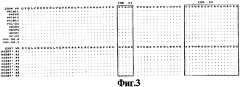
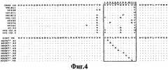


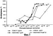
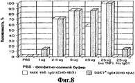

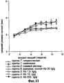
Classifications
- C—CHEMISTRY; METALLURGY
- C07—ORGANIC CHEMISTRY
- C07K—PEPTIDES
- C07K16/00—Immunoglobulins [IGs], e.g. monoclonal or polyclonal antibodies
- C07K16/18—Immunoglobulins [IGs], e.g. monoclonal or polyclonal antibodies against material from animals or humans
- C07K16/24—Immunoglobulins [IGs], e.g. monoclonal or polyclonal antibodies against material from animals or humans against cytokines, lymphokines or interferons
- C07K16/241—Tumor Necrosis Factors
- C—CHEMISTRY; METALLURGY
- C07—ORGANIC CHEMISTRY
- C07K—PEPTIDES
- C07K16/00—Immunoglobulins [IGs], e.g. monoclonal or polyclonal antibodies
- C07K16/18—Immunoglobulins [IGs], e.g. monoclonal or polyclonal antibodies against material from animals or humans
- C07K16/24—Immunoglobulins [IGs], e.g. monoclonal or polyclonal antibodies against material from animals or humans against cytokines, lymphokines or interferons
- A—HUMAN NECESSITIES
- A61—MEDICAL OR VETERINARY SCIENCE; HYGIENE
- A61K—PREPARATIONS FOR MEDICAL, DENTAL OR TOILETRY PURPOSES
- A61K39/00—Medicinal preparations containing antigens or antibodies
- A61K39/395—Antibodies; Immunoglobulins; Immune serum, e.g. antilymphocytic serum
- A—HUMAN NECESSITIES
- A61—MEDICAL OR VETERINARY SCIENCE; HYGIENE
- A61P—SPECIFIC THERAPEUTIC ACTIVITY OF CHEMICAL COMPOUNDS OR MEDICINAL PREPARATIONS
- A61P1/00—Drugs for disorders of the alimentary tract or the digestive system
- A—HUMAN NECESSITIES
- A61—MEDICAL OR VETERINARY SCIENCE; HYGIENE
- A61P—SPECIFIC THERAPEUTIC ACTIVITY OF CHEMICAL COMPOUNDS OR MEDICINAL PREPARATIONS
- A61P1/00—Drugs for disorders of the alimentary tract or the digestive system
- A61P1/02—Stomatological preparations, e.g. drugs for caries, aphtae, periodontitis
- A—HUMAN NECESSITIES
- A61—MEDICAL OR VETERINARY SCIENCE; HYGIENE
- A61P—SPECIFIC THERAPEUTIC ACTIVITY OF CHEMICAL COMPOUNDS OR MEDICINAL PREPARATIONS
- A61P1/00—Drugs for disorders of the alimentary tract or the digestive system
- A61P1/04—Drugs for disorders of the alimentary tract or the digestive system for ulcers, gastritis or reflux esophagitis, e.g. antacids, inhibitors of acid secretion, mucosal protectants
- A—HUMAN NECESSITIES
- A61—MEDICAL OR VETERINARY SCIENCE; HYGIENE
- A61P—SPECIFIC THERAPEUTIC ACTIVITY OF CHEMICAL COMPOUNDS OR MEDICINAL PREPARATIONS
- A61P1/00—Drugs for disorders of the alimentary tract or the digestive system
- A61P1/16—Drugs for disorders of the alimentary tract or the digestive system for liver or gallbladder disorders, e.g. hepatoprotective agents, cholagogues, litholytics
- A—HUMAN NECESSITIES
- A61—MEDICAL OR VETERINARY SCIENCE; HYGIENE
- A61P—SPECIFIC THERAPEUTIC ACTIVITY OF CHEMICAL COMPOUNDS OR MEDICINAL PREPARATIONS
- A61P11/00—Drugs for disorders of the respiratory system
- A—HUMAN NECESSITIES
- A61—MEDICAL OR VETERINARY SCIENCE; HYGIENE
- A61P—SPECIFIC THERAPEUTIC ACTIVITY OF CHEMICAL COMPOUNDS OR MEDICINAL PREPARATIONS
- A61P11/00—Drugs for disorders of the respiratory system
- A61P11/16—Central respiratory analeptics
- A—HUMAN NECESSITIES
- A61—MEDICAL OR VETERINARY SCIENCE; HYGIENE
- A61P—SPECIFIC THERAPEUTIC ACTIVITY OF CHEMICAL COMPOUNDS OR MEDICINAL PREPARATIONS
- A61P13/00—Drugs for disorders of the urinary system
- A61P13/12—Drugs for disorders of the urinary system of the kidneys
- A—HUMAN NECESSITIES
- A61—MEDICAL OR VETERINARY SCIENCE; HYGIENE
- A61P—SPECIFIC THERAPEUTIC ACTIVITY OF CHEMICAL COMPOUNDS OR MEDICINAL PREPARATIONS
- A61P17/00—Drugs for dermatological disorders
- A—HUMAN NECESSITIES
- A61—MEDICAL OR VETERINARY SCIENCE; HYGIENE
- A61P—SPECIFIC THERAPEUTIC ACTIVITY OF CHEMICAL COMPOUNDS OR MEDICINAL PREPARATIONS
- A61P17/00—Drugs for dermatological disorders
- A61P17/02—Drugs for dermatological disorders for treating wounds, ulcers, burns, scars, keloids, or the like
- A—HUMAN NECESSITIES
- A61—MEDICAL OR VETERINARY SCIENCE; HYGIENE
- A61P—SPECIFIC THERAPEUTIC ACTIVITY OF CHEMICAL COMPOUNDS OR MEDICINAL PREPARATIONS
- A61P19/00—Drugs for skeletal disorders
- A—HUMAN NECESSITIES
- A61—MEDICAL OR VETERINARY SCIENCE; HYGIENE
- A61P—SPECIFIC THERAPEUTIC ACTIVITY OF CHEMICAL COMPOUNDS OR MEDICINAL PREPARATIONS
- A61P19/00—Drugs for skeletal disorders
- A61P19/02—Drugs for skeletal disorders for joint disorders, e.g. arthritis, arthrosis
- A—HUMAN NECESSITIES
- A61—MEDICAL OR VETERINARY SCIENCE; HYGIENE
- A61P—SPECIFIC THERAPEUTIC ACTIVITY OF CHEMICAL COMPOUNDS OR MEDICINAL PREPARATIONS
- A61P19/00—Drugs for skeletal disorders
- A61P19/06—Antigout agents, e.g. antihyperuricemic or uricosuric agents
- A—HUMAN NECESSITIES
- A61—MEDICAL OR VETERINARY SCIENCE; HYGIENE
- A61P—SPECIFIC THERAPEUTIC ACTIVITY OF CHEMICAL COMPOUNDS OR MEDICINAL PREPARATIONS
- A61P19/00—Drugs for skeletal disorders
- A61P19/08—Drugs for skeletal disorders for bone diseases, e.g. rachitism, Paget's disease
- A—HUMAN NECESSITIES
- A61—MEDICAL OR VETERINARY SCIENCE; HYGIENE
- A61P—SPECIFIC THERAPEUTIC ACTIVITY OF CHEMICAL COMPOUNDS OR MEDICINAL PREPARATIONS
- A61P21/00—Drugs for disorders of the muscular or neuromuscular system
- A—HUMAN NECESSITIES
- A61—MEDICAL OR VETERINARY SCIENCE; HYGIENE
- A61P—SPECIFIC THERAPEUTIC ACTIVITY OF CHEMICAL COMPOUNDS OR MEDICINAL PREPARATIONS
- A61P25/00—Drugs for disorders of the nervous system
- A—HUMAN NECESSITIES
- A61—MEDICAL OR VETERINARY SCIENCE; HYGIENE
- A61P—SPECIFIC THERAPEUTIC ACTIVITY OF CHEMICAL COMPOUNDS OR MEDICINAL PREPARATIONS
- A61P27/00—Drugs for disorders of the senses
- A61P27/02—Ophthalmic agents
- A—HUMAN NECESSITIES
- A61—MEDICAL OR VETERINARY SCIENCE; HYGIENE
- A61P—SPECIFIC THERAPEUTIC ACTIVITY OF CHEMICAL COMPOUNDS OR MEDICINAL PREPARATIONS
- A61P29/00—Non-central analgesic, antipyretic or antiinflammatory agents, e.g. antirheumatic agents; Non-steroidal antiinflammatory drugs [NSAID]
- A—HUMAN NECESSITIES
- A61—MEDICAL OR VETERINARY SCIENCE; HYGIENE
- A61P—SPECIFIC THERAPEUTIC ACTIVITY OF CHEMICAL COMPOUNDS OR MEDICINAL PREPARATIONS
- A61P29/00—Non-central analgesic, antipyretic or antiinflammatory agents, e.g. antirheumatic agents; Non-steroidal antiinflammatory drugs [NSAID]
- A61P29/02—Non-central analgesic, antipyretic or antiinflammatory agents, e.g. antirheumatic agents; Non-steroidal antiinflammatory drugs [NSAID] without antiinflammatory effect
- A—HUMAN NECESSITIES
- A61—MEDICAL OR VETERINARY SCIENCE; HYGIENE
- A61P—SPECIFIC THERAPEUTIC ACTIVITY OF CHEMICAL COMPOUNDS OR MEDICINAL PREPARATIONS
- A61P3/00—Drugs for disorders of the metabolism
- A61P3/04—Anorexiants; Antiobesity agents
- A—HUMAN NECESSITIES
- A61—MEDICAL OR VETERINARY SCIENCE; HYGIENE
- A61P—SPECIFIC THERAPEUTIC ACTIVITY OF CHEMICAL COMPOUNDS OR MEDICINAL PREPARATIONS
- A61P3/00—Drugs for disorders of the metabolism
- A61P3/08—Drugs for disorders of the metabolism for glucose homeostasis
- A61P3/10—Drugs for disorders of the metabolism for glucose homeostasis for hyperglycaemia, e.g. antidiabetics
- A—HUMAN NECESSITIES
- A61—MEDICAL OR VETERINARY SCIENCE; HYGIENE
- A61P—SPECIFIC THERAPEUTIC ACTIVITY OF CHEMICAL COMPOUNDS OR MEDICINAL PREPARATIONS
- A61P31/00—Antiinfectives, i.e. antibiotics, antiseptics, chemotherapeutics
- A—HUMAN NECESSITIES
- A61—MEDICAL OR VETERINARY SCIENCE; HYGIENE
- A61P—SPECIFIC THERAPEUTIC ACTIVITY OF CHEMICAL COMPOUNDS OR MEDICINAL PREPARATIONS
- A61P31/00—Antiinfectives, i.e. antibiotics, antiseptics, chemotherapeutics
- A61P31/04—Antibacterial agents
- A—HUMAN NECESSITIES
- A61—MEDICAL OR VETERINARY SCIENCE; HYGIENE
- A61P—SPECIFIC THERAPEUTIC ACTIVITY OF CHEMICAL COMPOUNDS OR MEDICINAL PREPARATIONS
- A61P31/00—Antiinfectives, i.e. antibiotics, antiseptics, chemotherapeutics
- A61P31/12—Antivirals
- A—HUMAN NECESSITIES
- A61—MEDICAL OR VETERINARY SCIENCE; HYGIENE
- A61P—SPECIFIC THERAPEUTIC ACTIVITY OF CHEMICAL COMPOUNDS OR MEDICINAL PREPARATIONS
- A61P31/00—Antiinfectives, i.e. antibiotics, antiseptics, chemotherapeutics
- A61P31/12—Antivirals
- A61P31/14—Antivirals for RNA viruses
- A61P31/18—Antivirals for RNA viruses for HIV
- A—HUMAN NECESSITIES
- A61—MEDICAL OR VETERINARY SCIENCE; HYGIENE
- A61P—SPECIFIC THERAPEUTIC ACTIVITY OF CHEMICAL COMPOUNDS OR MEDICINAL PREPARATIONS
- A61P31/00—Antiinfectives, i.e. antibiotics, antiseptics, chemotherapeutics
- A61P31/12—Antivirals
- A61P31/20—Antivirals for DNA viruses
- A—HUMAN NECESSITIES
- A61—MEDICAL OR VETERINARY SCIENCE; HYGIENE
- A61P—SPECIFIC THERAPEUTIC ACTIVITY OF CHEMICAL COMPOUNDS OR MEDICINAL PREPARATIONS
- A61P31/00—Antiinfectives, i.e. antibiotics, antiseptics, chemotherapeutics
- A61P31/12—Antivirals
- A61P31/20—Antivirals for DNA viruses
- A61P31/22—Antivirals for DNA viruses for herpes viruses
- A—HUMAN NECESSITIES
- A61—MEDICAL OR VETERINARY SCIENCE; HYGIENE
- A61P—SPECIFIC THERAPEUTIC ACTIVITY OF CHEMICAL COMPOUNDS OR MEDICINAL PREPARATIONS
- A61P33/00—Antiparasitic agents
- A61P33/02—Antiprotozoals, e.g. for leishmaniasis, trichomoniasis, toxoplasmosis
- A61P33/06—Antimalarials
- A—HUMAN NECESSITIES
- A61—MEDICAL OR VETERINARY SCIENCE; HYGIENE
- A61P—SPECIFIC THERAPEUTIC ACTIVITY OF CHEMICAL COMPOUNDS OR MEDICINAL PREPARATIONS
- A61P35/00—Antineoplastic agents
- A—HUMAN NECESSITIES
- A61—MEDICAL OR VETERINARY SCIENCE; HYGIENE
- A61P—SPECIFIC THERAPEUTIC ACTIVITY OF CHEMICAL COMPOUNDS OR MEDICINAL PREPARATIONS
- A61P35/00—Antineoplastic agents
- A61P35/04—Antineoplastic agents specific for metastasis
- A—HUMAN NECESSITIES
- A61—MEDICAL OR VETERINARY SCIENCE; HYGIENE
- A61P—SPECIFIC THERAPEUTIC ACTIVITY OF CHEMICAL COMPOUNDS OR MEDICINAL PREPARATIONS
- A61P37/00—Drugs for immunological or allergic disorders
- A—HUMAN NECESSITIES
- A61—MEDICAL OR VETERINARY SCIENCE; HYGIENE
- A61P—SPECIFIC THERAPEUTIC ACTIVITY OF CHEMICAL COMPOUNDS OR MEDICINAL PREPARATIONS
- A61P37/00—Drugs for immunological or allergic disorders
- A61P37/02—Immunomodulators
- A—HUMAN NECESSITIES
- A61—MEDICAL OR VETERINARY SCIENCE; HYGIENE
- A61P—SPECIFIC THERAPEUTIC ACTIVITY OF CHEMICAL COMPOUNDS OR MEDICINAL PREPARATIONS
- A61P37/00—Drugs for immunological or allergic disorders
- A61P37/02—Immunomodulators
- A61P37/04—Immunostimulants
- A—HUMAN NECESSITIES
- A61—MEDICAL OR VETERINARY SCIENCE; HYGIENE
- A61P—SPECIFIC THERAPEUTIC ACTIVITY OF CHEMICAL COMPOUNDS OR MEDICINAL PREPARATIONS
- A61P37/00—Drugs for immunological or allergic disorders
- A61P37/02—Immunomodulators
- A61P37/06—Immunosuppressants, e.g. drugs for graft rejection
- A—HUMAN NECESSITIES
- A61—MEDICAL OR VETERINARY SCIENCE; HYGIENE
- A61P—SPECIFIC THERAPEUTIC ACTIVITY OF CHEMICAL COMPOUNDS OR MEDICINAL PREPARATIONS
- A61P37/00—Drugs for immunological or allergic disorders
- A61P37/08—Antiallergic agents
- A—HUMAN NECESSITIES
- A61—MEDICAL OR VETERINARY SCIENCE; HYGIENE
- A61P—SPECIFIC THERAPEUTIC ACTIVITY OF CHEMICAL COMPOUNDS OR MEDICINAL PREPARATIONS
- A61P39/00—General protective or antinoxious agents
- A61P39/02—Antidotes
- A—HUMAN NECESSITIES
- A61—MEDICAL OR VETERINARY SCIENCE; HYGIENE
- A61P—SPECIFIC THERAPEUTIC ACTIVITY OF CHEMICAL COMPOUNDS OR MEDICINAL PREPARATIONS
- A61P43/00—Drugs for specific purposes, not provided for in groups A61P1/00-A61P41/00
- A—HUMAN NECESSITIES
- A61—MEDICAL OR VETERINARY SCIENCE; HYGIENE
- A61P—SPECIFIC THERAPEUTIC ACTIVITY OF CHEMICAL COMPOUNDS OR MEDICINAL PREPARATIONS
- A61P7/00—Drugs for disorders of the blood or the extracellular fluid
- A—HUMAN NECESSITIES
- A61—MEDICAL OR VETERINARY SCIENCE; HYGIENE
- A61P—SPECIFIC THERAPEUTIC ACTIVITY OF CHEMICAL COMPOUNDS OR MEDICINAL PREPARATIONS
- A61P7/00—Drugs for disorders of the blood or the extracellular fluid
- A61P7/02—Antithrombotic agents; Anticoagulants; Platelet aggregation inhibitors
- A—HUMAN NECESSITIES
- A61—MEDICAL OR VETERINARY SCIENCE; HYGIENE
- A61P—SPECIFIC THERAPEUTIC ACTIVITY OF CHEMICAL COMPOUNDS OR MEDICINAL PREPARATIONS
- A61P7/00—Drugs for disorders of the blood or the extracellular fluid
- A61P7/04—Antihaemorrhagics; Procoagulants; Haemostatic agents; Antifibrinolytic agents
- A—HUMAN NECESSITIES
- A61—MEDICAL OR VETERINARY SCIENCE; HYGIENE
- A61P—SPECIFIC THERAPEUTIC ACTIVITY OF CHEMICAL COMPOUNDS OR MEDICINAL PREPARATIONS
- A61P9/00—Drugs for disorders of the cardiovascular system
- A—HUMAN NECESSITIES
- A61—MEDICAL OR VETERINARY SCIENCE; HYGIENE
- A61P—SPECIFIC THERAPEUTIC ACTIVITY OF CHEMICAL COMPOUNDS OR MEDICINAL PREPARATIONS
- A61P9/00—Drugs for disorders of the cardiovascular system
- A61P9/04—Inotropic agents, i.e. stimulants of cardiac contraction; Drugs for heart failure
- A—HUMAN NECESSITIES
- A61—MEDICAL OR VETERINARY SCIENCE; HYGIENE
- A61P—SPECIFIC THERAPEUTIC ACTIVITY OF CHEMICAL COMPOUNDS OR MEDICINAL PREPARATIONS
- A61P9/00—Drugs for disorders of the cardiovascular system
- A61P9/08—Vasodilators for multiple indications
- A—HUMAN NECESSITIES
- A61—MEDICAL OR VETERINARY SCIENCE; HYGIENE
- A61P—SPECIFIC THERAPEUTIC ACTIVITY OF CHEMICAL COMPOUNDS OR MEDICINAL PREPARATIONS
- A61P9/00—Drugs for disorders of the cardiovascular system
- A61P9/10—Drugs for disorders of the cardiovascular system for treating ischaemic or atherosclerotic diseases, e.g. antianginal drugs, coronary vasodilators, drugs for myocardial infarction, retinopathy, cerebrovascula insufficiency, renal arteriosclerosis
- C—CHEMISTRY; METALLURGY
- C12—BIOCHEMISTRY; BEER; SPIRITS; WINE; VINEGAR; MICROBIOLOGY; ENZYMOLOGY; MUTATION OR GENETIC ENGINEERING
- C12N—MICROORGANISMS OR ENZYMES; COMPOSITIONS THEREOF; PROPAGATING, PRESERVING, OR MAINTAINING MICROORGANISMS; MUTATION OR GENETIC ENGINEERING; CULTURE MEDIA
- C12N15/00—Mutation or genetic engineering; DNA or RNA concerning genetic engineering, vectors, e.g. plasmids, or their isolation, preparation or purification; Use of hosts therefor
- C12N15/09—Recombinant DNA-technology
- C12N15/11—DNA or RNA fragments; Modified forms thereof; Non-coding nucleic acids having a biological activity
- C—CHEMISTRY; METALLURGY
- C12—BIOCHEMISTRY; BEER; SPIRITS; WINE; VINEGAR; MICROBIOLOGY; ENZYMOLOGY; MUTATION OR GENETIC ENGINEERING
- C12N—MICROORGANISMS OR ENZYMES; COMPOSITIONS THEREOF; PROPAGATING, PRESERVING, OR MAINTAINING MICROORGANISMS; MUTATION OR GENETIC ENGINEERING; CULTURE MEDIA
- C12N15/00—Mutation or genetic engineering; DNA or RNA concerning genetic engineering, vectors, e.g. plasmids, or their isolation, preparation or purification; Use of hosts therefor
- C12N15/09—Recombinant DNA-technology
- C12N15/63—Introduction of foreign genetic material using vectors; Vectors; Use of hosts therefor; Regulation of expression
- C12N15/64—General methods for preparing the vector, for introducing it into the cell or for selecting the vector-containing host
- C—CHEMISTRY; METALLURGY
- C12—BIOCHEMISTRY; BEER; SPIRITS; WINE; VINEGAR; MICROBIOLOGY; ENZYMOLOGY; MUTATION OR GENETIC ENGINEERING
- C12N—MICROORGANISMS OR ENZYMES; COMPOSITIONS THEREOF; PROPAGATING, PRESERVING, OR MAINTAINING MICROORGANISMS; MUTATION OR GENETIC ENGINEERING; CULTURE MEDIA
- C12N5/00—Undifferentiated human, animal or plant cells, e.g. cell lines; Tissues; Cultivation or maintenance thereof; Culture media therefor
- C12N5/10—Cells modified by introduction of foreign genetic material
- A—HUMAN NECESSITIES
- A61—MEDICAL OR VETERINARY SCIENCE; HYGIENE
- A61K—PREPARATIONS FOR MEDICAL, DENTAL OR TOILETRY PURPOSES
- A61K39/00—Medicinal preparations containing antigens or antibodies
- A61K2039/505—Medicinal preparations containing antigens or antibodies comprising antibodies
- A—HUMAN NECESSITIES
- A61—MEDICAL OR VETERINARY SCIENCE; HYGIENE
- A61K—PREPARATIONS FOR MEDICAL, DENTAL OR TOILETRY PURPOSES
- A61K38/00—Medicinal preparations containing peptides
- C—CHEMISTRY; METALLURGY
- C07—ORGANIC CHEMISTRY
- C07K—PEPTIDES
- C07K2317/00—Immunoglobulins specific features
- C07K2317/20—Immunoglobulins specific features characterized by taxonomic origin
- C07K2317/21—Immunoglobulins specific features characterized by taxonomic origin from primates, e.g. man
- C—CHEMISTRY; METALLURGY
- C07—ORGANIC CHEMISTRY
- C07K—PEPTIDES
- C07K2317/00—Immunoglobulins specific features
- C07K2317/50—Immunoglobulins specific features characterized by immunoglobulin fragments
- C07K2317/56—Immunoglobulins specific features characterized by immunoglobulin fragments variable (Fv) region, i.e. VH and/or VL
- C07K2317/565—Complementarity determining region [CDR]
- Y—GENERAL TAGGING OF NEW TECHNOLOGICAL DEVELOPMENTS; GENERAL TAGGING OF CROSS-SECTIONAL TECHNOLOGIES SPANNING OVER SEVERAL SECTIONS OF THE IPC; TECHNICAL SUBJECTS COVERED BY FORMER USPC CROSS-REFERENCE ART COLLECTIONS [XRACs] AND DIGESTS
- Y02—TECHNOLOGIES OR APPLICATIONS FOR MITIGATION OR ADAPTATION AGAINST CLIMATE CHANGE
- Y02A—TECHNOLOGIES FOR ADAPTATION TO CLIMATE CHANGE
- Y02A50/00—TECHNOLOGIES FOR ADAPTATION TO CLIMATE CHANGE in human health protection, e.g. against extreme weather
- Y02A50/30—Against vector-borne diseases, e.g. mosquito-borne, fly-borne, tick-borne or waterborne diseases whose impact is exacerbated by climate change
- Y—GENERAL TAGGING OF NEW TECHNOLOGICAL DEVELOPMENTS; GENERAL TAGGING OF CROSS-SECTIONAL TECHNOLOGIES SPANNING OVER SEVERAL SECTIONS OF THE IPC; TECHNICAL SUBJECTS COVERED BY FORMER USPC CROSS-REFERENCE ART COLLECTIONS [XRACs] AND DIGESTS
- Y10—TECHNICAL SUBJECTS COVERED BY FORMER USPC
- Y10S—TECHNICAL SUBJECTS COVERED BY FORMER USPC CROSS-REFERENCE ART COLLECTIONS [XRACs] AND DIGESTS
- Y10S424/00—Drug, bio-affecting and body treating compositions
- Y10S424/81—Drug, bio-affecting and body treating compositions involving autoimmunity, allergy, immediate hypersensitivity, delayed hypersensitivity, immunosuppression, immunotolerance, or anergy
Landscapes
- Health & Medical Sciences (AREA)
- Life Sciences & Earth Sciences (AREA)
- Chemical & Material Sciences (AREA)
- Organic Chemistry (AREA)
- General Health & Medical Sciences (AREA)
- Medicinal Chemistry (AREA)
- Engineering & Computer Science (AREA)
- Veterinary Medicine (AREA)
- Public Health (AREA)
- Pharmacology & Pharmacy (AREA)
- Animal Behavior & Ethology (AREA)
- Nuclear Medicine, Radiotherapy & Molecular Imaging (AREA)
- General Chemical & Material Sciences (AREA)
- Chemical Kinetics & Catalysis (AREA)
- Bioinformatics & Cheminformatics (AREA)
- Immunology (AREA)
- Genetics & Genomics (AREA)
- Molecular Biology (AREA)
- Biotechnology (AREA)
- Virology (AREA)
- Biomedical Technology (AREA)
- Diabetes (AREA)
- Oncology (AREA)
- Biochemistry (AREA)
- Communicable Diseases (AREA)
- Physical Education & Sports Medicine (AREA)
- Wood Science & Technology (AREA)
- Zoology (AREA)
- Biophysics (AREA)
- Hematology (AREA)
- General Engineering & Computer Science (AREA)
- Cardiology (AREA)
- Rheumatology (AREA)
- Heart & Thoracic Surgery (AREA)
- Microbiology (AREA)
- Proteomics, Peptides & Aminoacids (AREA)
- Pulmonology (AREA)
- Tropical Medicine & Parasitology (AREA)
- Orthopedic Medicine & Surgery (AREA)
- Pain & Pain Management (AREA)
Abstract
Description
Translated fromRussianОбласть техники, к которой относится изобретениеFIELD OF THE INVENTION
Изобретение относится к медицинской фармакологии и касается, в частности, применения антител человека, которые специфически связывают или нейтрализуют hTNFα, при производстве лекарственных препаратов для лечения нарушений, при которых активность TNFα является вредной.The invention relates to medical pharmacology, and relates, in particular, to the use of human antibodies that specifically bind or neutralize hTNFα in the manufacture of drugs for the treatment of disorders in which TNFα activity is harmful.
Уровень техникиState of the art
Фактор некроза опухоли α(TNFα) является цитокином, образуемым множеством типов клеток, включая моноциты и макрофаги, который был исходно идентифицирован на основе своей способности индуцировать некроз некоторых мышиных опухолей (см., например, Old L. (1985) Science 230:630-632). Впоследствии было показано, что фактор, называемый кахектин, ассоциированный с кахексией, является той же молекулой, что TNFα. TNFα участвовал в генерации шока (см., например, Beutler В. and Cerami A. (1988) Annu. Rev. Biochem. 57:505-518; Beutler В. and Cerami A. (1989) Annu. Rev. Immunol. 7:625-655). Более того, TNFα участвовал в патофизиологии ряда других заболеваний и нарушений человека, включая сепсис, инфекции, аутоиммунные заболевания, отторжение трансплантата и болезнь трансплантат против хозяина (см..например, MoellerA. et al., (1990) Cytokine 2:162-169; Патент США No 5231024 Moeller et al.; Европейский Патент No 260610 B1 Moeller A. et al.; Vasilli P. (1992) Annu. Rev. Immunol 10:411-452; Tracey K.J. and Cerami A. (1994) Annu. Rev. Med. 45:491-503).Tumor Necrosis Factor α (TNFα) is a cytokine produced by many cell types, including monocytes and macrophages, which was originally identified based on its ability to induce necrosis of certain murine tumors (see, for example, Old L. (1985) Science 230: 630- 632). It was subsequently shown that a factor called cachectin associated with cachexia is the same molecule as TNFα. TNFα was involved in the generation of shock (see, for example, Beutler B. and Cerami A. (1988) Annu. Rev. Biochem. 57: 505-518; Beutler B. and Cerami A. (1989) Annu. Rev. Immunol. 7 : 625-655). Moreover, TNFα has been implicated in the pathophysiology of a number of other human diseases and disorders, including sepsis, infections, autoimmune diseases, graft rejection and graft versus host disease (see, for example, Moeller A. et al., (1990) Cytokine 2: 162-169 ; U.S. Patent No. 5231024 Moeller et al .; European Patent No. 260610 B1 Moeller A. et al .; Vasilli P. (1992) Annu. Rev. Immunol 10: 411-452; Tracey KJ and Cerami A. (1994) Annu. Rev. Med. 45: 491-503).
Вследствие вредной роли человеческого TNFα (hTNFα) в ряде нарушений у человека были разработаны терапевтические способы ингибирования или противодействия активности hTNFα. В частности, антитела, которые связывают или нейтрализуют hTNFα, рассматривались как средства ингибирования активности hTNFα. Одними из самых ранних таких антител были мышиные моноклональные антитела (mAbs), секретируемые гибридомами, полученными из лимфоцитов мышей, иммунизированных hTNFα (см., например, Hahn Т. et al. (1985) Proc. Natl. Acad. Sci. USA 82: 3814-3818; Liang C-M. et al. (1986) Biochem. Biophys. Res. Commun. 137: 847-854; Hirai M. et al.(1987) J. Immunol. Methods 96: 57-62; Fendly B.M. et al.(1987) Hybridoma 6:359-370; Moeller A. et al. (1990) Cytokine 2:162-169; Патент США No 5231024 Moeller et al.; Европейский Патент No 186833 B1 Wallach D.; Европейская Патентная Заявка No 218868 А1 Old et al.; Европейский Патент No 260610 B1 Moeller A. et al.). Поскольку эти мышиные антитела против hTNFα часто проявляли высокую аффинность к hTNFα (например, Kd≤10-9 M) и были способны нейтрализовать активность hTNFα, их применение in vivo могло ограничиваться проблемами, связанными с введением мышиных антител человеку, такими как короткий период полужизни в сывортке, неспособность включать определенные функции эффекторов человека и появление нежелательного иммунного ответа против мышиных антител у человека (реакция "человеческих антител против мыши" (НАМА)).Due to the harmful role of human TNFα (hTNFα) in a number of disorders in humans, therapeutic methods have been developed to inhibit or counteract hTNFα activity. In particular, antibodies that bind or neutralize hTNFα have been considered as a means of inhibiting hTNFα activity. One of the earliest such antibodies was murine monoclonal antibodies (mAbs) secreted by hybridomas obtained from lymphocytes of mice immunized with hTNFα (see, for example, Hahn, T. et al. (1985) Proc. Natl. Acad. Sci. USA 82: 3814-3818; Liang CM. Et al. (1986) Biochem. Biophys. Res. Commun. 137: 847-854; Hirai M. et al. (1987) J. Immunol. Methods 96: 57-62; Fendly BM et al. (1987) Hybridoma 6: 359-370; Moeller A. et al. (1990) Cytokine 2: 162-169; U.S. Patent No. 5231024 Moeller et al .; European Patent No. 186833 B1 Wallach D .; European Patent Application No. 218868 A1 Old et al .; European Patent No. 260610 B1 Moeller A. et al.). Because these murine anti-hTNFα antibodies often showed high affinity for hTNFα (e.g., Kd ≤ 10-9 M) and were able to neutralize hTNFα activity, their in vivo use could be limited to problems associated with the administration of murine antibodies to humans, such as a short half-life in serum, the inability to include certain functions of human effectors and the appearance of an undesirable immune response against murine antibodies in humans (the "human anti-mouse antibodies" reaction (NAMA)).
При попытке преодолеть проблемы, связанные с использованием полностью мышиных антител у человека, мышиные антитела против hTNFα были с помощью генетической инженерии сделаны "человекоподобными". Например, были получены химерные антитела, в которых вариабельные участки цепей антител имеют мышиную природу, а константные участки цепей антител - человеческую (Knight D.M. et al. (1993) Mol. Immunol. 30:1443-1453; Публикация РСТ No WO 92/16553 Daddona P.E. et al.). Кроме того, были также получены человекоподобные антитела, в которых гипервариабельные домены вариабельных участков антитела имеют мышиную природу, но остальные вариабельные участки и константные участки антител имеют человеческую природу (Публикация РСТ No WO 92/11383 Adair J.R. et al.). Однако поскольку эти химерные и гуманизированные антитела еще сохраняют некоторые мышиные последовательности, они еще могут вызвать нежелательную иммунную реакцию, реакцию человеческих антихимерных антител (НАСА), особенно при введении в течение длительных периодов, например при хронических показаниях, таких как ревматоидный артрит (см,, например, Elliott M.J. et al. (1994) Lancet 344:1125-1127; Elliott M.J. et al. (1994) Lancet 344: 1105-1110).In an attempt to overcome the problems associated with the use of fully murine antibodies in humans, murine antibodies against hTNFα were made "human-like" by genetic engineering. For example, chimeric antibodies were prepared in which the variable regions of the antibody chains are murine in nature and the constant regions of the antibody chains are human (Knight DM et al. (1993) Mol. Immunol. 30: 1443-1453; PCT Publication No. WO 92/16553 Daddona PE et al.). In addition, human-like antibodies were also prepared in which the hypervariable domains of the variable regions of the antibody are mouse in nature, but the remaining variable regions and constant regions of the antibodies are human in nature (PCT Publication No. WO 92/11383 Adair J.R. et al.). However, since these chimeric and humanized antibodies still retain some mouse sequences, they can still cause an unwanted immune reaction, the reaction of human antimer antibodies (NASA), especially when administered for long periods, for example, for chronic indications such as rheumatoid arthritis (see ,, e.g., Elliott MJ et al. (1994) Lancet 344: 1125-1127; Elliott MJ et al. (1994) Lancet 344: 1105-1110).
Предпочтительным ингибирующим hTNFα агентом по отношению к мышиным mAbs и их производным (например, химерным или человекоподобным антителам) было бы полностью человеческое антитело против hTNFα, поскольку такой агент не вызывал бы реакции НАМА, даже если бы применялся в течение длительных периодов. Человеческие моноклональные аутоантитела против hTNFα были получены способами с использованием человеческой гибридомы (Boyle P. Et al. (1993) Cell. Immunol. 152:556-568; Boyle P. et al. (1993) Cell. Immunol. 152:569-581; Европейская Патентная Заявка No 614984 А2 Boyle et al.). Однако, как сообщалось, эти моноклональные аутоантитела гибридомной природы обладали настолько низкой аффинностью в отношении hTNFα, что ее трудно было подсчитать известными способами, не были способны к связыванию растворимого hTNFα и не были способны нейтрализовать hhTNFα-индуцированную цитотоксичность (см. Boyle et al., supra). Более того, успех способа с использованием человеческой гибридомы зависит от естественного присутствия в периферической крови человека лимфоцитов, продуцирующих аутоантитела, специфичные для hTNFα. В ряде исследований у человека обнаружены сывороточные аутоантитела против hTNFα (Fomsgaard A. et al. (1989) Scand. J. Immunol. 30: 219-223; Bendtzen К. et al. (1990) Prog. Leukocyte Biol. 10B: 447-452), тогда как в других исследованиях они не были обнаружены (Leusch H-G. et al. (1991) J.Immunol. Methods 139: 145-147).The preferred hTNFα inhibitory agent with respect to murine mAbs and their derivatives (e.g. chimeric or human-like antibodies) would be a fully human anti-hTNFα antibody, since such an agent would not induce a NAMA reaction even if applied for long periods. Human anti-hTNFα monoclonal autoantibodies were obtained by methods using a human hybridoma (Boyle P. Et al. (1993) Cell. Immunol. 152: 556-568; Boyle P. et al. (1993) Cell. Immunol. 152: 569-581 ; European Patent Application No. 614984 A2 Boyle et al.). However, it was reported that these monoclonal autoantibodies of hybridoma nature had such a low affinity for hTNFα that it was difficult to calculate by known methods, were not able to bind soluble hTNFα and were not able to neutralize hhTNFα-induced cytotoxicity (see Boyle et al., supra). Moreover, the success of the method using human hybridomas depends on the natural presence in the peripheral blood of a person of lymphocytes producing autoantibodies specific for hTNFα. In a number of studies, human serum autoantibodies against hTNFα have been found (Fomsgaard A. et al. (1989) Scand. J. Immunol. 30: 219-223; Bendtzen K. et al. (1990) Prog. Leukocyte Biol. 10B: 447- 452), while in other studies they were not found (Leusch HG. Et al. (1991) J. Immunol. Methods 139: 145-147).
Альтернативой существующим в естественных условиях человеческим антителам против hTNFα были бы рекомбинантные антитела против hhTNFα. Описаны рекомбинантные человеческие антитела, которые связывают hTNFα с относительно низкой аффинностью (т.е. Кd ~ 10-7 М) и быстрой скоростью диссоциации (т.е. Кoff ~ 10-2 с-1). Однако ввиду относительно быстрой кинетики диссоциации эти антитела не могут быть применены для терапевтических нужд. Кроме того, были описаны рекомбинантные человеческие антитела против hTNFα, которые не нейтрализуют активность DTNFα, но значительно усиливают связывание hTNFα с поверхностью клеток и усиливают внедрение в цитоплазму hTNFα (Lidbury A. et al. (1994) Biotechnol. Ther. 5:27-45; Публикация РСТ No WO 92/03145 Aston R. et al.).An alternative to in vivo human anti-hTNFα antibodies would be recombinant anti-hhTNFα antibodies. Recombinant human antibodies are described that bind hTNFα with relatively low affinity (i.e., Kd ~ 10-7 M) and a fast dissociation rate (i.e., Koff ~ 10-2 s-1 ). However, due to the relatively fast kinetics of dissociation, these antibodies cannot be used for therapeutic purposes. In addition, recombinant human anti-hTNFα antibodies have been described that do not neutralize DTNFα activity, but significantly enhance the binding of hTNFα to the cell surface and enhance the incorporation of hTNFα into the cytoplasm (Lidbury A. et al. (1994) Biotechnol. Ther. 5: 27-45 ; PCT Publication No. WO 92/03145 Aston R. et al.).
Соответственно человеческие антитела, такие как рекомбинантные человеческие антитела, которые связывают растворимый hTNFα с высокой аффинностью и низкой кинетикой диссоциации и которые способны нейтрализовать активность hTNFα, включая hTNFα-индуцированную цитотоксичность (in vitro и in vivo) и hTNFα-индуцированную активацию клеток, все еще являются необходимыми.Accordingly, human antibodies, such as recombinant human antibodies that bind soluble hTNFα with high affinity and low dissociation kinetics and which are able to neutralize hTNFα activity, including hTNFα-induced cytotoxicity (in vitro and in vivo) and hTNFα-induced cell activation, are still necessary.
Сущность изобретенияSUMMARY OF THE INVENTION
Изобретение относится к применению человеческих антител, в частности, рекомбинантных человеческих антител, которые специфически связывают человеческий TNFα. Антитела, соответствующие изобретению, характеризуются связыванием hTNFα с высокой аффинностью и медленной кинетикой диссоциации, а также нейтрализацией активности hTNFα, включая hTNFα-индуцированную цитотоксичность (in vitro и in vivo) и hTNFα-индуцированную активацию клеток. Антитела, соответствующие изобретению, далее характеризуются связыванием hTNFα, но не hTNFα (лимфотоксина) и наличием способности связывать кроме человеческого TNFα также факторы некроза опухоли (TNFα) других приматов и TNFα неприматов.The invention relates to the use of human antibodies, in particular recombinant human antibodies that specifically bind human TNFα. Antibodies of the invention are characterized by binding of hTNFα with high affinity and slow dissociation kinetics, as well as neutralization of hTNFα activity, including hTNFα-induced cytotoxicity (in vitro and in vivo) and hTNFα-induced cell activation. The antibodies of the invention are further characterized by binding of hTNFα but not hTNFα (lymphotoxin) and the ability to bind, in addition to human TNFα, tumor necrosis factors (TNFα) of other primates and TNFα nonprimate.
Антитела, соответствующие изобретению, могут быть полной длины (например, антитело lgG1 или lgG4) или могут содержать только антиген-связывающий фрагмент (например, фрагмент Fab, F(ab')2 или scFv). Наиболее предпочтительное рекомбинантное антитело, соответствующее изобретению, обозначаемое D2E7, имеет домен CDR3 легкой цепи, содержащий аминокислотную последовательность SEQ ID No 3 и домен CDR3 тяжелой цепи, содержащий аминокислотную последовательность SEQ ID No 4. Предпочтительно антитело D2E7 имеет вариабельный участок легкой цепи (LCVR), содержащий аминокислотную последовательность SEQ ID No 1 и вариабельный участок тяжелой цепи (HCVR), содержащий аминокислотную последовательность SEQ ID No 2.Antibodies of the invention may be full length (e.g., an IgG1 or IgG4 antibody) or may contain only an antigen-binding fragment (e.g., Fab, F (ab ')2 or scFv fragment). The most preferred recombinant antibody of the invention, designated D2E7, has a light chain CDR3 domain containing the amino acid sequence of
В изобретении предлагаются способ ингибирования активности человеческого TNFα, в котором осуществляют контактирование человеческого TNFα с выделенным антителом человека или его антиген-связывающим фрагментом и при этом ингибируют активность человеческого TNFα, а также применение выделенного антитела человека или его антиген-связывающего фрагмента в качестве компонента для производства средства (лекарственного препарата) для лечения нарушений, при которых активность TNFα является вредной, посредством введения антитела субъекту. В одном из вариантов осуществления изобретения упомянутое выделенное человеческое антитело или его антиген-связывающий фрагмента диссоциирует из человеческого TNFα с Кd равной 1×10⁻⁸ М или менее и c константой скорости Кoff 1×10⁻³с-1 или менее (оба значения определены с помощью поверхностного плазменного резонанса) и нейтрализуют цитотоксичность человеческого hTNFα в стандартном анализе с использованием L929 in vitro с IC50 равной 1×10⁻⁷М или меньше. Более предпочтительно, если выделенное человеческое антитело или его антиген-связывающий фрагмент диссоциируют из ассоциации (комплекса) с человеческим TNFα с Кoff равной 5×10⁻⁴ с-1 или менее и даже более предпочтительно с Кoff 1×10⁻⁴ с-1 или менее. Более предпочтительно, если выделенное человеческое антитело или его антиген-связывающий фрагмент нейтрализуют цитотоксичность человеческого TNFα в стандартном анализе с использованием L929 in vitro с IC50 1×10⁻⁸ М или менее и еще более предпочтительно с IC50 5×10⁻¹⁰ М или менее.The invention provides a method of inhibiting human TNFα activity, in which human TNFα is contacted with an isolated human antibody or antigen-binding fragment thereof, and the human TNFα activity is inhibited, as well as the use of an isolated human antibody or antigen-binding fragment thereof as a component for production an agent (drug) for treating disorders in which TNFα activity is harmful by administering an antibody to a subject. In one embodiment of the invention, said isolated human antibody or antigen binding fragment thereof dissociates from human TNFα with a Kd of 1 × 10⁻⁸ M or less and a
В другом варианте осуществления изобретения используется человеческое антитело или его антиген-связывающий фрагмент со следующими характеристиками:In another embodiment, a human antibody or antigen binding fragment thereof is used with the following characteristics:
a) диссоциирует из hTNFα с Кoff 1×10⁻³ с-1 или менее, что определено с помощью поверхностного плазменного резонанса;a) dissociates from hTNFα with
b) имеет по меньшей мере домен CDR3 легкой цепи, содержащий аминокислотную последовательность SEQ ID No 3 или SEQ ID No 3, модифицированную посредством замены одним аланином в положении 1, 4, 5, 7 или 8 или заменой от одной до пяти консервативных аминокислот в положениях 1, 3, 4, 6, 7, 8 и/или 9;b) has at least a light chain CDR3 domain containing the amino acid sequence of
c) имеет по меньшей мере домен CDR3 тяжелой цепи, содержащий аминокислотную последовательность SEQ ID No 4 или SEQ ID No 4, модифицированную посредством замены одним аланином в положении 2, 3, 4, 5, 6, 8, 9, 10 или 11 или заменой от одной до пяти консервативных аминокислот в положениях 2, 3, 4, 5, 6, 8, 9, 10, 11 и/или 12.c) has at least a heavy chain CDR3 domain comprising the amino acid sequence of
Более предпочтительно, если антитело или его антиген-связывающий фрагмент диссоциируют из человеческого TNFα с Кoff 5×10⁻⁴ с-1 или менее. Еще более предпочтительно, если антитело или его антиген-связывающий фрагмент диссоциируют из человеческого TNFα с Кoff 1×10⁻⁴ с-1 или менее.More preferably, if the antibody or its antigen-binding fragment is dissociated from human TNFα with
В еще одном варианте осуществления изобретения используется выделенное человеческое антитело или его антиген-связывающий фрагмент с LCVR, содержащим аминокислотную последовательность SEQ ID No 1, и с HCVR, содержащим аминокислотную последовательность SEQ ID No 2. В некоторых вариантах осуществления антитело может иметь константный участок тяжелой цепи lgG1 или константный участок тяжелой цепи lgG4. В других вариантах осуществления антитело является фрагментом Fab, фрагментом F(ab')2 или фрагментом Fv одной цепи.In another embodiment of the invention, an isolated human antibody or antigen binding fragment thereof is used with an LCVR containing the amino acid sequence of
В следующем варианте осуществления изобретение в качестве компонента для производства упомянутого средства для лечения нарушений предлагается применение антитела D2E7.In a further embodiment, the invention provides the use of the D2E7 antibody as a component for the manufacture of said agent for treating disorders.
В других вариантах осуществления изобретение представляет антитела или его антиген-связывающие фрагменты с LCVR, имеющим домен CDR3, содержащий аминокислотную последовательность, выбранную из группы, состоящей из SEQ ID No3, SEQ ID No 11, SEQ ID No 12, SEQ ID No 13, SEQ ID No 14, SEQ ID No 15, SEQ ID No 16, SEQ ID No 17, SEQ ID No 18, SEQ ID No 19, SEQ ID No 20, SEQ ID No 21, SEQ ID No 22, SEQ ID No 23, SEQ ID No 24, SEQ ID No 25, SEQ ID No 26, или с HCVR, имеющим домен CDR3, содержащий аминокислотную последовательность, выбранную из группы, состоящей из SEQ ID No 4, SEQ ID No 27, SEQ ID No 28, SEQ ID No 29, SEQ ID No 30, SEQ ID No 31, SEQ ID No 32, SEQ ID No 33, SEQ ID No 34 и SEQ ID No 35.In other embodiments, the invention provides antibodies or antigen binding fragments thereof with an LCVR having a CDR3 domain comprising an amino acid sequence selected from the group consisting of
В еще одном варианте осуществления изобретение представляет выделенное человеческое антитело или его антиген-связывающий фрагмент, которые нейтрализуют активность человеческого TNFα, но не человеческого TNFα (лимфотоксина). В предпочтительном варианте осуществления человеческое антитело или его антиген-связывающий фрагмент нейтрализуют активность человеческого TNFα, TNFα шимпанзе и TNFα по меньшей мере еще одного примата, выбранного из группы, состоящей из hTNFα павиана, TNFα игрунки, hTNFα циномолгуса и TNFα резуса. Предпочтительно, если антитело также нейтрализует активность TNFα по меньшей мере одного непримата. Например, в одном варианте осуществления выделенное человеческое антитело или его антиген-связывающий фрагмент нейтрализуют также активность собачьего TNFα. А в другом варианте выделенное человеческое антитело или его антиген-связывающий фрагмент нейтрализуют также активность свиного TNFα. В еще одном варианте осуществления выделенное человеческое антитело или его антиген-связывающий фрагмент нейтрализуют также активность мышиного TNFα.In yet another embodiment, the invention provides an isolated human antibody or antigen binding fragment thereof that neutralizes the activity of human TNFα, but not human TNFα (lymphotoxin). In a preferred embodiment, the human antibody or antigen binding fragment thereof neutralizes the activity of human TNFα, TNFα chimpanzee and TNFα of at least one other primate selected from the group consisting of hTNFα baboon, TNFα marmoset, hTNFα cynomolgus and TNFα rhesus. Preferably, the antibody also neutralizes the activity of TNFα of at least one nonprimate. For example, in one embodiment, the isolated human antibody or antigen binding fragment thereof also neutralizes canine TNFα activity. In another embodiment, the isolated human antibody or antigen-binding fragment thereof also neutralizes the activity of porcine TNFα. In yet another embodiment, the isolated human antibody or antigen-binding fragment thereof also neutralizes mouse TNFα activity.
Другой аспект изобретения относится к молекулам нуклеиновых кислот, кодирующих соответствующие изобретению антитела или их антиген-связывающие фрагменты. Предпочтительная нуклеиновая кислота, соответствующая изобретению, кодирующая LCVR D2E7, имеет нуклеотидную последовательность, представленную на Фигуре 7, и SEQ ID No 36. Другая предпочтительная нуклеиновая кислота, соответствующая изобретению, кодирующая HCVR D2E7, имеет нуклеотидную последовательность, представленную на Фигуре 8, и SEQ ID No 37. Рекомбинантные экспрессирующие векторы, несущие кодирующие антитела нуклеиновые кислоты, соответствующие изобретению, и клетки-хозяева, в которые интродуцированы такие векторы, также охватываются изобретением, как и способы получения антител, соответствующих изобретению, посредством культивирования клеток-хозяев, соответствующих изобретению.Another aspect of the invention relates to nucleic acid molecules encoding antibodies of the invention or antigen binding fragments thereof. A preferred nucleic acid of the invention encoding LCVR D2E7 has the nucleotide sequence shown in Figure 7 and SEQ ID No 36. Another preferred nucleic acid of the invention encoding HCVR D2E7 has the nucleotide sequence shown in Figure 8 and SEQ ID No. 37. Recombinant expression vectors carrying antibody-encoding nucleic acids of the invention and host cells into which such vectors are introduced are also encompassed by the invention eat, as well as methods for producing antibodies corresponding to the invention, by culturing host cells corresponding to the invention.
Еще один аспект изобретения относится к способам ингибирования активности человеческого TNFα с использованием антитела или его антиген-связывающегофрагмента, соответствующего изобретению. В одном варианте осуществления способ предусматривает контактирование человеческого ТМРα с антителом, соответствующим изобретению, или его антиген-связывающим фрагментом, при этом ингибируется активность человеческого TNFα. В другом варианте осуществления способ предусматривает введение антитела, соответствующего изобретению, или его антиген-связывающего фрагмента человеку, страдающему от нарушения, при котором активность TNFα является вредной, при этом у человека ингибируется активность TNFα. Нарушением может быть, например, сепсис, аутоиммунное заболевание (например, ревматоидный артрит, аллергия, рассеянный склероз, аутоиммунный диабет, аутоиммунный увеит и почечный синдром), инфекционное заболевание, злокачественное новообразование, отторжение трансплантата или болезнь трансплантат против хозяина, легочное нарушение, костное нарушение, кишечное нарушение или сердечное нарушение.Another aspect of the invention relates to methods of inhibiting the activity of human TNFα using an antibody or its antigen-binding fragment, corresponding to the invention. In one embodiment, the method comprises contacting human TMPα with an antibody of the invention or an antigen binding fragment thereof, wherein the activity of human TNFα is inhibited. In another embodiment, the method comprises administering an antibody of the invention or an antigen binding fragment thereof to a person suffering from a disorder in which TNFα activity is detrimental, while TNFα activity is inhibited in humans. The violation may be, for example, sepsis, an autoimmune disease (e.g. rheumatoid arthritis, allergies, multiple sclerosis, autoimmune diabetes, autoimmune uveitis and renal syndrome), an infectious disease, malignant neoplasm, graft rejection or a graft-to-host transplant disorder, pulmonary disease, intestinal disorder or heart disorder.
Упомянутые выделенные антитела человека или его антиген-связывающего фрагмента согласно изобретению могут применяться для производства средств для лечения, в частности, следующих нарушений: аутоиммунные заболевания, инфекционные болезни, болезни, связанные с отторжением трансплантата (болезни трансплантат против хозяина), нарушения, связанные со злокачественным образованием, легочные нарушения, кишечные нарушения, сердечные нарушения, сепсис, ревматоидный артрит, ревматоидный спондилит, остеоартрит, подагрический артрит, аллергия, рассеянный склероз, аутоиммунный диабет, аутоиммунный увеит, почечный синдром, воспалительные костные заболевания, заболевания, связанные с резорбцией кости, алкогольный гепатит, вирусный гепатит, нарушения свертывания крови, ожоги, реперфузионные повреждения, нарушения, связанные с образованием келоида, нарушения, связанные с формированием рубцовой ткани, гипертермия, септический шок, эндотоксический шок, сепсис, вызываемый грамотрицательными бактериями, токсический шоковый синдром, малярия, менингит, кахексия, СПИД, бактериальный менингит, СПИД-ассоциированный комплекс (ARC), вторичная инфекция цитомегаловируса при трансплантации, нарушения, связанные с лихорадкой и миалгией, обусловленные инфекцией, и вторичная кахексии при инфекции, нарушения, связанные с отторжением аллотрансплантата, нарушения, связанные со стимуляцией роста опухоли, нарушения, связанные с усилением метастатического потенциала и появлением цитотоксичности при злокачественных образованиях, нарушения, связанные с ингибированием роста опухоли или метастазов, нарушения, связанные с респираторным дистресс-синдромом взрослых, легочный шок, хронические легочные воспалительные заболевания, легочный саркоидоз, легочный фиброз, силикоз, воспалительные кишечные нарушения, идиопатические воспалительные заболевания кишечника, болезнь Крона, язвенный колит, ишемия сердца, сердечная недостаточность, гепатит.Mentioned isolated antibodies of a person or an antigen-binding fragment thereof according to the invention can be used to produce agents for treating, in particular, the following disorders: autoimmune diseases, infectious diseases, diseases associated with transplant rejection (graft versus host disease), malignant disorders education, pulmonary disorders, intestinal disorders, heart disorders, sepsis, rheumatoid arthritis, rheumatoid spondylitis, osteoarthritis, gouty arthritis, allergies, multiple sclerosis, autoimmune diabetes, autoimmune uveitis, renal syndrome, inflammatory bone diseases, diseases related to bone resorption, alcoholic hepatitis, viral hepatitis, blood coagulation disorders, burns, reperfusion injuries, disorders associated with the formation of keloid, disorders associated with the formation scar tissue, hyperthermia, septic shock, endotoxic shock, sepsis caused by gram-negative bacteria, toxic shock syndrome, malaria, meningitis, cachexia, AIDS, bacterial ingit, AIDS-associated complex (ARC), secondary infection of cytomegalovirus during transplantation, disorders associated with fever and myalgia due to infection, and secondary cachexia during infection, disorders associated with rejection of allograft, disorders associated with stimulation of tumor growth, disorders, associated with increased metastatic potential and the appearance of cytotoxicity in malignant tumors, disorders associated with inhibition of tumor growth or metastases, disorders associated with respiratory distress syndrome adults, pulmonary shock, chronic pulmonary inflammatory disease, pulmonary sarcoidosis, pulmonary fibrosis, silicosis, inflammatory bowel disorders, idiopathic inflammatory bowel disease, Crohn's disease, ulcerative colitis, ischemic heart disease, heart failure, hepatitis.
Упомянутые выделенные антитела человека или его антиген-связывающего фрагмента согласно изобретению могут также применяться для введения антитела человеку вместе с цитокином интерлейкином-6 (IL-6) или введения человеку, у которого концентрация IL-6 в сыворотке или плазме превышает 500 пг/мл, а также для производства средства, в котором выделенное антитело используется в комбинации с фармацевтически приемлемым носителем, или для производства средства для введения выделенного антитела с по меньшей мере одним дополнительным терапевтическим агентом.Said isolated human antibodies or an antigen binding fragment thereof according to the invention can also be used to administer the antibody to a human together with the cytokine interleukin-6 (IL-6) or to administer to a human whose serum or plasma IL-6 concentration exceeds 500 pg / ml, and also for the manufacture of an agent in which an isolated antibody is used in combination with a pharmaceutically acceptable carrier, or for the manufacture of an agent for administration of an isolated antibody with at least one additional therapeutic a gent.
В предпочтительном варианте осуществления изобретение в качестве упомянутого субъекта выступает человек и применение упомянутых выделенных антител человека или его антиген-связывающего фрагмента согласно изобретению предназначается для производства лекарственного препарата для лечения нарушений, при которых активность TNFα является вредной, посредством введения антитела человеку для ингибирования активности человеческого TNFα.In a preferred embodiment, the subject is a human being and the use of said isolated human antibodies or an antigen binding fragment thereof according to the invention is intended for the manufacture of a medicament for treating disorders in which TNFα activity is harmful by administering an antibody to a human to inhibit human TNFα activity .
Перечень фигур чертежейList of drawings
На Фигурах 1 и 2 представлены последовательности нуклеиновых кислот вариабельного участка легкой цепи D2E7 (D2E7 VL; показано также в SEQ ID No 1), аланин-сканмутанты D2E7VL (LD2E7*.A1, LD2E7*.A3, LD2E7*.A4, LD2E7*.A5, LD2E7*.A7 и LD2E7*.A8), вариабельный участок легкой цепи подобного D2E7 антитела 2SD4 (2SD4 VL; показано также в SEQ ID No 9) и другие вариабельные участки легкой цепи, подобные D2E7 (ЕР В12, VL10E4, VL100А9, VL100D2. VL10F4, LOE5, VLLOF9, VLLOF10, VLLOG7, VLLOG9, VLLOH1, VLLOH10, VL1B7, VL1С1, VL1C7, VL0.1F4.VL0.1H8, LOE7, LOE7.A и LOE7.T). На Фигуре 1А представлены домены FR1, CDR1, FR2 и CDR2. На Фигуре 1В представлены домены FR3, CDR3 и FR4. Домены легкой цепи CDR1 ("CDR L1"), CDR2 ("CDR L2") и CDR3 ("CDR L3") находятся в прямоугольнике.Figures 1 and 2 show the nucleic acid sequences of the variable region of the light chain D2E7 (D2E7 VL; also shown in SEQ ID No. 1), alanine scansmantons D2E7VL (LD2E7* .A1, LD2E7* .A3, LD2E7* .A4, LD2E7* . A5, LD2E7* .A7 and LD2E7* .A8), the variable region of the light chain like D2E7 antibody 2SD4 (2SD4 VL; also shown in SEQ ID No 9) and other variable regions of the light chain like D2E7 (EP B12, VL10E4, VL100A9, VL100D2. VL10F4, LOE5, VLLOF9, VLLOF10, VLLOG7, VLLOG9, VLLOH1, VLLOH10, VL1B7, VL1C1, VL1C7, VL0.1F4.VL0.1H8, LOE7, LOE7.A and LOE7.T). Figure 1A shows the domains FR1, CDR1, FR2 and CDR2. Figure 1B shows the domains FR3, CDR3, and FR4. The light chain domains CDR1 ("CDR L1"), CDR2 ("CDR L2") and CDR3 ("CDR L3") are in the rectangle.
На Фигурах 3 и 4 представлены аминокислотные последовательности вариабельного участка D2E7 (D2E7 VH; показано также в SEQ ID No 2), аланин-сканмутанты D2E7VH (HD2E7*.A1, HD2E7*.A2, HD2E7*.A3, HD2E7*.A4, HD2E7*.A5, HD2E7*.A6, HD2E7*.A7, HD2E7*.A8 и HD2E7*.A9), вариабельный участок тяжелой цепи, подобного D2E7 антитела 2SD4 (2SD4 VH, показано также в SEQ ID No 10) и другие вариабельные участки тяжелой цепи, подобные D2E7 (VH1B11, VH1D8, VH1A11, VH1B12, VH1-D2, VH1E4, VH1F6, VH1G1, 3С-Н2, VH1-D2.N и VH1-D2.Y). На Фигуре 2А представлены домены FR1, CDR1, FR2 и CDR2. На Фигуре 2В представлены домены FR3, CDR3 и FR4. Домены тяжелой цепи CDR1 ("CDR H1"), CDR2 ("CDR H2") и CDR3 ("CDR Н3") находятся в прямоугольнике.In Figures 3 and 4, the amino acid sequences of the variable region D2E7 (D2E7 VH; shown also in SEQ ID No 2), alanine scans, D2E7VH (HD2E7* .A1, HD2E7* .A2, HD2E7* .A3, HD2E7* .A4, HD2E7* .A5, HD2E7* .A6, HD2E7* .A7, HD2E7* .A8 and HD2E7* .A9), the variable region of the heavy chain, like D2E7 antibody 2SD4 (2SD4 VH, also shown in SEQ ID No 10) and other variable regions heavy chain like D2E7 (VH1B11, VH1D8, VH1A11, VH1B12, VH1-D2, VH1E4, VH1F6, VH1G1, 3C-H2, VH1-D2.N and VH1-D2.Y). Figure 2A shows the domains FR1, CDR1, FR2 and CDR2. Figure 2B shows the domains FR3, CDR3, and FR4. The heavy chain domains of CDR1 ("CDR H1"), CDR2 ("CDR H2") and CDR3 ("CDR H3") are in the box.
На Фигуре 5 графически представлено ингибирование TNFα-индуцированной цитотоксичности L929 человеческим антителом D2E7 против hTNFα по сравнению с мышиным антителом МАК 195 против hTNFα.Figure 5 graphically depicts the inhibition of TNFα-induced cytotoxicity of L929 by the human anti-hTNFα antibody D2E7 compared to the murine
На Фигуре 6 графически представлено ингибирование связывания rhTNFα с рецепторами hTNFα на клетках U-937 с помощью человеческого антитела D2E7 против hTNFα по сравнению с мышиным антителом МАК 195 против hTNFα.Figure 6 graphically shows the inhibition of the binding of rhTNFα to hTNFα receptors on U-937 cells with the human anti-hTNFα antibody D2E7 compared to the murine
На Фигуре 7 графически представлено ингибирование индуцированной TNFα экспрессии ELAM-1 на HUVEC с помощью человеческого антитела D2E7 против hTNFα по сравнению с мышиным антителом МАК 195 против hTNFα.Figure 7 graphically depicts the inhibition of TNFα-induced ELAM-1 expression on HUVEC using the human anti-hTNFα antibody D2E7 compared to the murine
На Фигуре 8 представлен график в виде столбиков защиты от TNFα-индуцированной гибели сенсибилизированных D-галактозамином мышей посредством введения человеческого антитела против hTNFα D2E7 (черные столбики) по сравнению с мышиным антителом МАК 195 против hTNFα (заштрихованные столбики).Figure 8 is a bar graph of protection against TNFα-induced death of sensitized D-galactosamine mice by introducing a human anti-hTNFα D2E7 antibody (black bars) compared to the murine MAK anti-hTNFα antibody 195 (shaded bars).
На Фигуре 9 представлена нуклеотидная последовательность вариабельного участка легкой цепи D2E7c прогнозированной аминокислотной последовательностью, находящейся ниже нуклеотидной последовательности. Участки CDR L1, CDR L2 и CDR L3 подчеркнуты.Figure 9 shows the nucleotide sequence of the variable region of the light chain D2E7c predicted amino acid sequence below the nucleotide sequence. Plots of CDR L1, CDR L2, and CDR L3 are underlined.
На Фигуре 10 представлена нуклеотидная последовательность вариабельного участка тяжелой цепи D2E7 с прогнозированной аминокислотной последовательностью, находящейся ниже нуклеотидной последовательности. Участки CDR H1, CDR H2 и CDR H3 подчеркнуты.Figure 10 shows the nucleotide sequence of the variable region of the heavy chain D2E7 with the predicted amino acid sequence below the nucleotide sequence. Plots of CDR H1, CDR H2 and CDR H3 are underlined.
На Фигуре 10 приведен график, представляющий эффект лечения антителами D2E7 трансгенных мышей Тg197 со средним размером суставов в качестве модели полиартрита.Figure 10 is a graph showing the effect of antibody treatment with D2E7 transgenic Tg197 mice with average joint size as a model of polyarthritis.
Сведения, подтверждающие возможность осуществления изобретенияInformation confirming the possibility of carrying out the invention
Данное изобретение относится к выделенным человеческим антителам или их антиген-связывающим фрагментам, которые связывают TNFα с высокой аффинностью, низкой скоростью диссоциации и высокой нейтрализующей активностью, и применению этих антител для ингибирования активности человеческого TNFα и в качестве компонентов лекарственных средств для лечения различных нарушений, при которых активность TNFα является вредной. В частности в изобретении предлагаются способы применения антител, соответствующих изобретению, для детекции человеческого TNFα или для ингибирования активности человеческого TNFα либо in vitro, либо in vivo.This invention relates to isolated human antibodies or their antigen-binding fragments that bind TNFα with high affinity, low dissociation rate and high neutralizing activity, and the use of these antibodies to inhibit the activity of human TNFα and as drug components for the treatment of various disorders, which TNFα activity is harmful. In particular, the invention provides methods of using antibodies of the invention to detect human TNFα or to inhibit the activity of human TNFα either in vitro or in vivo.
С целью более легкого понимания изобретения сначала определяются некоторые термины.In order to more easily understand the invention, certain terms are first defined.
Термин "человеческий TNFα" (сокращаемый здесь как hTNFα или просто hTNF), используемый здесь, предназначен для определения человеческого цитокина, который существует в виде секретируемой формы с молекулярной массой 17 кД и ассоциированной с мембаной формы с молекулярной массой 26 кД, биологически активная форма которого содержит тример нековалентно связанных молекул с массой 17 кД. Структура hTNFα описана далее в, например, Pennica D. et al., (1984) Nature 312:724-729; Davis J.M. et al. (1987) Biochemistry 26:1322-1326; Jones E.Y. et al,(1989) Nature 338:225-228. Термин человеческий TNFα предполагает включение рекомбинантного человеческого TNFα (rhTNFα), который может быть получен стандартными способами рекомбинантной экспрессии или закуплен (R & D Systems, Catalog No. 210-TA, Minneapolis, MN).The term "human TNFα" (abbreviated here as hTNFα or simply hTNF), used here, is intended to define a human cytokine that exists in the form of a secreted form with a molecular weight of 17 kDa and associated with the membrane form with a molecular weight of 26 kD, the biologically active form of which contains a trimer of non-covalently bound molecules with a mass of 17 kD. The structure of hTNFα is described further in, for example, Pennica D. et al., (1984) Nature 312: 724-729; Davis J.M. et al. (1987) Biochemistry 26: 1322-1326; Jones E.Y. et al, (1989) Nature 338: 225-228. The term human TNFα implies the inclusion of recombinant human TNFα (rhTNFα), which can be obtained by standard methods of recombinant expression or purchased (R & D Systems, Catalog No. 210-TA, Minneapolis, MN).
Термин "антитело", используемый здесь, предназначен для определения молекул иммуноглобулина, состоящего из четырех полипептидных цепей (две тяжелые (Н) цепи и две легкие (L) цепи), связанных дисульфидными связями. Каждая тяжелая цепь содержит вариабельный участок тяжелой цепи (сокращенный здесь как HCVR или VH) и константный участок тяжелой цепи. Костантный участок тяжелой цепи содержит три домена СН1, СН2 и СН3. Каждая легкая цепь содержит вариабельный участок легкой цепи (сокращенный здесь как LCVR или VL) и константный участок легкой цепи. Константный участок легкой цепи содержит один домен CL Участки VH и VL могут далее подразделяться на участки гипервариабельности, называемые определяющими комплементарность участками (CDR), окруженные участками, которые являются более консервативными, называемыми скелетными участками (FR). Каждая из VH и VL состоит из трех CDR и четырех FR участков, расположенных от амино- до карбоксильного конца в следующем порядке: FR1, CDR1, FR2, CDR2, FR3, CDR3, FR4.The term "antibody", as used herein, is intended to define immunoglobulin molecules consisting of four polypeptide chains (two heavy (H) chains and two light (L) chains) linked by disulfide bonds. Each heavy chain contains a variable region of the heavy chain (abbreviated here as HCVR or VH) and a constant region of the heavy chain. The costant region of the heavy chain contains three domains of CH1, CH2 and CH3. Each light chain contains a variable region of the light chain (abbreviated here as LCVR or VL) and a constant region of the light chain. The light chain constant region contains one CL domain. The VH and VL regions can be further subdivided into hypervariability regions called complementarity determining regions (CDRs) surrounded by regions that are more conservative called skeletal regions (FR). Each of VH and VL consists of three CDRs and four FR sites located from the amino to the carboxyl end in the following order: FR1, CDR1, FR2, CDR2, FR3, CDR3, FR4.
Термин "антиген-связывающий фрагмент" антитела (или просто "фрагмент антитела"), используемый здесь, относится к одному или более фрагментам антитела, которые сохраняют способность специфически связывать антиген (например, hTNFα). Показано, что антиген-связывающая функция антитела может быть осуществлена фрагментами антитела полной длины. Примеры связывающих фрагментов, охватываемые термином "антиген-связывающий фрагмент" антитела, включают (i) фрагмент Fab, моновалентный фрагмент, состоящий из доменов VL, VH, CL и CHl; (ii) фрагмент Р(ab')2, бивалентный фрагмент, содержащий два фрагмента Fab, связанные дисульфидным мостиком в районе петли; (iii) фрагмент Fd, состоящий из доменов VH и СН1; (iv) фрагмент Fv, состоящий из доменов VL и VH одного плеча антитела; (v) фрагмент dAb (Ward et al., (1989) Nature 341:544-546), который состоит из домена VH, и (vi) выделенный участок (CDR), определяющий комплементарность. Более того, хотя два домена фрагмента Fv, VL и VH, кодируются отдельными генами, они могут быть рекомбинантными способами связаны с помощью синтетического линкера, который обеспечивает их получение в виде одной белковой цепи, в которой участки VL и VH спарены с образованием моновалентных молекул (известных как Fv одной цепи (scFv); см., например, Bird et al. (1988) Science 242:423-426; Huston et al.,(1988) Proc. Natl. Acad. Sci. USA 85:5879-5883). Предполагается, что такие антитела из одной цепи также охватываются термином "антиген-связывающий фрагмент" антитела. К ним относятся также другие формы антител из одной цепи, такие как диатела. Диатела являются бивалентными, биспецифическими антителами, в которых домены VH и VL экспрессируются на одной полипептидной цепи, но с использованием линкера, который является слишком коротким, чтобы позволять спаривание двух доменов на одной и той же цепи, что заставляет домены спариваться с комплементарными доменами другой цепи и создавать два антиген-связывающих сайта (см., например, Holliger Р. et al. (1993) Proc. Natl. Acad. Sci. USA 90:6444-6448; Poljak R.J. et al. (1994) Structure 2:1121-1123).The term “antigen-binding fragment” of an antibody (or simply “antibody fragment”), as used herein, refers to one or more antibody fragments that retain the ability to specifically bind an antigen (eg, hTNFα). It has been shown that the antigen-binding function of an antibody can be carried out by full-length antibody fragments. Examples of binding fragments encompassed by the term “antigen binding fragment” of an antibody include (i) a Fab fragment, a monovalent fragment consisting of the VL, VH, CL, and CHl domains; (ii) a fragment P (ab ')2 , a bivalent fragment containing two Fab fragments connected by a disulfide bridge in the loop region; (iii) a Fd fragment consisting of the VH and CH1 domains; (iv) an Fv fragment consisting of the VL and VH domains of one arm of an antibody; (v) a dAb fragment (Ward et al., (1989) Nature 341: 544-546), which consists of the VH domain, and (vi) an isolated complementarity determining region (CDR) that determines complementarity. Moreover, although the two domains of the Fv fragment, VL and VH, are encoded by separate genes, they can be recombinantly linked using a synthetic linker that provides them in the form of a single protein chain in which the VL and VH portions are paired to form monovalent molecules ( known as single chain Fv (scFv); see, for example, Bird et al. (1988) Science 242: 423-426; Huston et al., (1988) Proc. Natl. Acad. Sci. USA 85: 5879-5883 ) It is contemplated that such single chain antibodies are also encompassed by the term “antigen binding fragment” of an antibody. Other forms of single chain antibodies, such as diabodies, also apply. Diabodies are bivalent, bispecific antibodies in which the VH and VL domains are expressed on the same polypeptide chain, but using a linker that is too short to allow two domains to pair on the same chain, causing the domains to pair with complementary domains of the other chain and create two antigen-binding sites (see, for example, Holliger P. et al. (1993) Proc. Natl. Acad. Sci. USA 90: 6444-6448; Poljak RJ et al. (1994) Structure 2: 1121- 1123).
Далее, антитело или его антиген-связывающий фрагмент могут быть частью более крупных молекул иммуноадгезии, образованных ковалентной или нековалентной связью антитела или фрагмента антитела с одним или более белком или пептидом. Примеры таких молекул иммуноадгезии включают использование участка ядра стрептавидина для получения тетрамерной молекулы scFv (Kipriyanov S.M. et al (1995) Human Antibodies and Hybridomas 6:93-101) и использование остатка цистеина, маркерного пептида и С-концевой полигистидиновой метки для получения бивалентных и уменьшенных биомолекул scFv (Kipriyanov S.M. et al (1994) Mol. Immunol., 31:1047-1058). Фрагменты антител, такие как Fab F(ab')2, могут быть получены из целых антител с использованием принятых способов, таких как разложение папаином или пепсином соответственно целых антител. Более того, антитела, фрагменты антител и молекулы иммуноадгезии могут быть получены с использованием стандартных способов с применением рекомбинантной ДНК, как описано здесь.Further, the antibody or antigen-binding fragment thereof may be part of larger immunoadhesion molecules formed by covalent or non-covalent binding of an antibody or antibody fragment to one or more protein or peptide. Examples of such immunoadhesion molecules include the use of a streptavidin core portion to produce a tetrameric scFv molecule (Kipriyanov SM et al (1995) Human Antibodies and Hybridomas 6: 93-101) and the use of a cysteine residue, a marker peptide and a C-terminal polyhistidine tag to produce bivalent and reduced scFv biomolecules (Kipriyanov SM et al (1994) Mol. Immunol., 31: 1047-1058). Antibody fragments, such as Fab F (ab ')2 , can be obtained from whole antibodies using accepted methods, such as papain or pepsin degradation of whole antibodies, respectively. Moreover, antibodies, antibody fragments and immunoadhesion molecules can be obtained using standard methods using recombinant DNA, as described here.
Термин "человеческое антитело", используемый здесь, включает антитела, имеющие вариабельные и константные участки, выделенные из последовательностей зародышевого иммуноглобулина человека. Человеческие антитела, соответствующие изобретению, могут включать остатки аминокислот, не кодируемые последовательностями зародышевого иммуноглобулина человека (например, мутации, интродуцированные ненаправленным или сайт-специфическим мутагенезом in vitro или соматической мутацией in vivo), например, в CDR и особенно в CDR3. Однако термин "человеческое антитело", используемый здесь, не включает антитела, в которых последовательности CDR, выделенные из эмбрионов других видов млекопитающих, таких как мышь, были пересажены на человеческие скелетные последовательности.The term "human antibody" as used herein includes antibodies having variable and constant regions isolated from human germline immunoglobulin sequences. Human antibodies of the invention may include amino acid residues not encoded by human germline immunoglobulin sequences (e.g., mutations introduced by in vitro undirected or site-specific mutagenesis or in vivo somatic mutation), for example, in CDR and especially in CDR3. However, the term "human antibody" as used herein does not include antibodies in which CDR sequences isolated from embryos of other mammalian species, such as mice, were transplanted into human skeletal sequences.
Термин "рекомбинантное человеческое антитело", используемый здесь, включает все человеческие антитела, которые получены, экспрессированы, созданы или выделены рекомбинантными средствами, такие как антитела, экспрессированные с использованием рекомбинантного экспрессирующего вектора, введенного в клетку-хозяин (описано далее в Разделе II ниже), антитела, выделенные из набора известных рекомбинантных комбинаторных человеческих антител (описано далее в Разделе III ниже), антитела, выделенные из животного (например, мыши), которое является трансгенным в отношении генов человеческого иммуноглобулина (см., например, Taylor L.D. et al. (1992) Nucl. Acids Res. 20:6287-6295) или антитела, полученные, экспрессированные, созданные или выделенные любыми другими средствами, которые включают сплайсинг последовательности гена человеческого иммуноглобулина до других последовательностей ДНК. Такие рекомбинантные человеческие антитела имеют вариабельные и константные участки, выделенные из последовательностей человеческого зародышевого иммуноглобулина. В некоторых вариантах осуществления, однако, такие рекомбинантные человеческие антитела подвергают мутагенезу in vitro (или, если используют животное, трансгенное по последовательностям Ig человека, соматическому мутагенезу in vivo) и, таким образом, аминокислотные последовательности участков VH и VL рекомбинантных антител являются последовательностями, которые, поскольку они выделены из последовательностей зародышевых VH и VL человека и близки к ним, не могут в естественных условиях существовать в зародышевом наборе антител человека in vivo.The term "recombinant human antibody" as used herein includes all human antibodies that are obtained, expressed, created or isolated by recombinant means, such as antibodies expressed using a recombinant expression vector introduced into a host cell (described further in Section II below) , antibodies isolated from a set of known recombinant combinatorial human antibodies (described further in Section III below), antibodies isolated from an animal (e.g., mouse), which is t antigenic to human immunoglobulin genes (see, for example, Taylor LD et al. (1992) Nucl. Acids Res. 20: 6287-6295) or antibodies obtained, expressed, created or isolated by any other means that include splicing the gene sequence human immunoglobulin to other DNA sequences. Such recombinant human antibodies have variable and constant regions isolated from human germline immunoglobulin sequences. In some embodiments, however, such recombinant human antibodies are subjected to in vitro mutagenesis (or, if an animal transgenic to human Ig sequences is used, somatic mutagenesis in vivo) and thus the amino acid sequences of the recombinant antibody VH and VL regions are sequences that since they are isolated from and close to human VH and VL germline sequences, they cannot exist in vivo in the germinal set of human antibodies in vivo.
"Выделенное антитело", как используют здесь, предназначено для определения антитела, которое практически не содержит других антител, имеющих различные антигенные специфичности (например, выделенное антитело, которое специфически связывает hTNFα, практически не содержит антител, которые специфически связывают антигены, отличные от hTNFα). Выделенное антитело, которое специфически связывает hTNFα, может, однако, иметь перекрестную реактивность с другими антигенами, такими как молекулы TNFα из других видов (детально обсуждается ниже). Более того, выделенное антитело может практически не содержать иной клеточный материал и/или химические соединения.An “isolated antibody” as used herein is intended to define an antibody that is substantially free of other antibodies having different antigenic specificities (for example, an isolated antibody that specifically binds hTNFα is substantially free of antibodies that specifically bind antigens other than hTNFα) . An isolated antibody that specifically binds hTNFα may, however, cross-react with other antigens, such as TNFα molecules from other species (discussed in detail below). Moreover, the isolated antibody may practically contain no other cellular material and / or chemical compounds.
"Нейтрализующее антитело", как используют здесь, (или "антитело, которое нейтрализовало активность hTNFα) подразумевает антитело, связывание которого с hTNFα приводит к ингибированию биологической активности hTNFα. Это ингибирование биологической активности hTNFα может быть оценено измерением одного или более индикаторов биологической активности hTNFα, таких как hTNFα-индуцированной активации клетки и связывания hTNFα с рецепторами hTNFα. Эти индикаторы биологической активности hTNFα могут быть оценены одним или более методом различных стандартных анализов in vitro и in vivo, известных в уровне техники (см. Пример 4). Предпочтительно способность антитела нейтрализовать активность hTNFα оценивается по ингибированию hTNFα-индуцированной цитотоксичности клеток L929. В качестве дополнительного или альтернативного параметра активности hTNFα может быть оценена способность антитела ингибировать hTNFα-индуцированную экспрессию ELAM-1 на HUVEC как степень hTNFα-индуцированной активации клеток.A “neutralizing antibody” as used herein (or “an antibody that neutralizes hTNFα activity) refers to an antibody whose binding to hTNFα inhibits the biological activity of hTNFα. This inhibition of the biological activity of hTNFα can be evaluated by measuring one or more indicators of the biological activity of hTNFα, such as hTNFα-induced cell activation and hTNFα binding to hTNFα receptors. These indicators of the biological activity of hTNFα can be evaluated by one or more methods of various standard of in vitro and in vivo lyses known in the art (see Example 4). Preferably, the ability of an antibody to neutralize hTNFα activity is assessed by inhibition of hTNFα-induced cytotoxicity of L929 cells. As an additional or alternative parameter of hTNFα activity, the ability of an antibody to inhibit hTNFα- can be evaluated induced expression of ELAM-1 on HUVEC as a degree of hTNFα-induced cell activation.
Термин "поверхностный плазменный резонанс", используемый здесь, относится к оптическому явлению, положенному в основу проведения анализа протекающих в данный момент биоспецифических взаимодействий по детекции изменений концентраций белка в матриксе биосенсора, например, с использованием системы BIAcore (Pharmacia Biosensor AB, Uppsala, Sweden and Piscataway, NJ). Дальнейшее описание см. в Примере 1 и Jönsson U. et al. (1993) Ann. Biol. Clin. 51:19-26; Jönsson U. et al. (1991) Biotechniques 11:620-627; Johnson B. et al. (1995) J. Mol. Recognit. 8:125-131 и Johnson В. et al. (1991) Anal. Biochem. 198:268-277.The term “surface plasma resonance” as used here refers to the optical phenomenon underlying the analysis of the ongoing biospecific interactions to detect changes in protein concentrations in the biosensor matrix, for example, using the BIAcore system (Pharmacia Biosensor AB, Uppsala, Sweden and Piscataway, NJ). For further description see Example 1 and Jönsson U. et al. (1993) Ann. Biol. Clin. 51: 19-26; Jönsson U. et al. (1991) Biotechniques 11: 620-627; Johnson B. et al. (1995) J. Mol. Recognit. 8: 125-131 and Johnson B. et al. (1991) Anal. Biochem. 198: 268-277.
Термин "Koff", используемый здесь, предназначен для определения константы скорости диссоциации антитела из комплекса антитело/антиген.The term "Koff ", as used here, is intended to determine the dissociation rate constant of an antibody from an antibody / antigen complex.
Термин "Кd", используемый здесь, предназначен для обозначения константы диссоциации определенного взаимодействия антитело-антиген.The term “Kd ”, as used herein, is intended to mean the dissociation constant of a specific antibody-antigen interaction.
Термин "молекула нуклеиновой кислоты", используемый здесь, включает молекулы ДНК и молекулы РНК. Молекула нуклеиновой кислоты может быть однонитиевой или двунитевой, но предпочтительно двунитевой ДНК.The term "nucleic acid molecule" as used herein includes DNA molecules and RNA molecules. The nucleic acid molecule may be single-stranded or double-stranded, but preferably double-stranded DNA.
Термин "выделенная молекула нуклеиновой кислоты", используемый здесь в отношении нуклеиновых кислот, кодирующих антитела или фрагменты антител (например, VH, VL, CDR3), которые связывают hTNFα, предназначен для определения молекулы нуклеиновой кислоты, в которой нуклеотидные последовательности, кодирующие антитела или фрагменты антител, не содержат другие нуклеотидные последовательности, кодирующие антитела или фрагменты антител, которые связывают антигены, отличные от hTNFα, эти другие последовательности могут в естественных условиях примыкать к нуклеиновой кислоте в геномной ДНК человека. Таким образом, например, выделенная нуклеиновая кислота, соответствующая изобретению, кодирующая участок VH антитела против TNFα, не содержит другие последовательности, кодирующие другие участки VH, которые связывают антигены, отличные от TNFα.The term "isolated nucleic acid molecule" as used herein with reference to nucleic acids encoding antibodies or antibody fragments (eg, VH, VL, CDR3) that bind hTNFα, is intended to define a nucleic acid molecule in which nucleotide sequences encoding antibodies or fragments antibodies that do not contain other nucleotide sequences encoding antibodies or fragments of antibodies that bind antigens other than hTNFα, these other sequences may in vivo adjacent be a nucleic acid in human genomic DNA. Thus, for example, an isolated nucleic acid of the invention encoding a VH region of an anti-TNFα antibody does not contain other sequences encoding other VH regions that bind antigens other than TNFα.
Термин "вектор" в контексте описания обозначает молекулу нуклеиновой кислоты, способной транспортировать другую нуклеиновую кислоту, с которой он был связан. Одним типом вектора является "плазмида", которая означает кольцевую петлю двунитевой ДНК, в которую могут быть лигированы дополнительные сегменты ДНК. Другим типом вектора является вирусный вектор, в котором дополнительные сегменты ДНК могут быть лигированы в геном вируса. Определенные векторы способны к автономной репликации в клетке-хозяине, в которую они интродуцированы (например, бактериальные векторы, имеющие бактериальный ориджин (точку начала) репликации, и эписомные векторы млекопитающих). Другие векторы (например, неэписомные векторы млекопитающих) могут быть интегрированы в геном клетки-хозяина введением в клетку-хозяин и вследствие этого реплицируются вместе с геномом хозяина. Более того, определенные векторы могут направлять экспрессию генов, с которыми они оперативно связаны. Такие векторы называются в контексте заявки "рекомбинантные экспрессирующие векторы" (или просто "экспрессирующие векторы"). В основном экспрессирующие векторы для использования в способах с применением рекомбинантной ДНК часто находятся в форме плазмид. В настоящем описании "плазмида" и "вектор" могут быть использованы взаимозаменяемо, поскольку плазмида является наиболее часто применяемой формой вектора. Однако изобретение включает другие формы экспрессирующих векторов, такие как вирусные векторы (например, репликационно-дефектные ретровирусы, аденовирусы и аденоассоциированные вирусы), которые выполняют эквивалентные функции.The term "vector" as used herein refers to a nucleic acid molecule capable of transporting another nucleic acid to which it has been linked. One type of vector is a "plasmid", which means a circular loop of double-stranded DNA into which additional DNA segments can be ligated. Another type of vector is a viral vector in which additional segments of DNA can be ligated into the genome of the virus. Certain vectors are capable of autonomous replication in the host cell into which they are introduced (e.g., bacterial vectors having a bacterial origin (origin) of replication and episomal mammalian vectors). Other vectors (e.g., non-episomal mammalian vectors) can be integrated into the genome of the host cell by introduction into the host cell and, as a result, are replicated along with the host genome. Moreover, certain vectors can direct the expression of genes with which they are operatively linked. Such vectors are referred to in the context of the application as “recombinant expression vectors” (or simply “expression vectors”). In general, expression vectors for use in recombinant DNA methods are often in the form of plasmids. As used herein, “plasmid” and “vector” can be used interchangeably since the plasmid is the most commonly used form of the vector. However, the invention includes other forms of expression vectors, such as viral vectors (e.g., replication-defective retroviruses, adenoviruses and adeno-associated viruses) that perform equivalent functions.
Термин "рекомбинантная клетка-хозяин" (или просто "клетка-хозяин") в контексте заявки предназначен для определения клетки, в которую интродуцирован рекомбинантный экспрессионный вектор. Следует понимать, что такие термины обозначают только определенную данную клетку, но не потомство такой клетки. Поскольку определенные модификации в последующих поколениях могут происходить вследствие мутаций или воздействия окружающей среды, такое потомство может на самом деле не быть идентичными родительской клетке, но также подпадает под определение термина "клетка-хозяин" в контексте заявки.The term "recombinant host cell" (or simply "host cell") in the context of the application is intended to define a cell into which a recombinant expression vector has been introduced. It should be understood that such terms refer only to a particular given cell, but not the offspring of such a cell. Since certain modifications in subsequent generations may occur due to mutations or environmental influences, such offspring may not actually be identical to the parent cell, but also falls within the definition of the term “host cell” in the context of the application.
Различные аспекты изобретения подробно описываются в следующих подразделах.Various aspects of the invention are described in detail in the following subsections.
1. Человеческие антитела, которые связывают TNFα человека1. Human antibodies that bind human TNFα
В изобретении представлены выделенные человеческие антитела или их антиген-связывающие фрагменты, которые связывают человеческий TNFα с высокой аффинностью, низкой скоростью диссоциации и высокой нейтрализующей способностью. Предпочтительно человеческие антитела, соответствующие изобретению, являются рекомбинантными нейтрализующими человеческими антителами против hTNFα. Наиболее предпочтительным рекомбинантным нейтрализующим антителом согласно изобретению является D2E7, которое имеет последовательности VL и VH, как показано на Фигурах 1, 2 и Фигурах 3 4, соответственно (аминокислотная последовательность участка VL D1E7 также представлена как SEQ ID No 1; аминокислотная последовательность участка VH D1 Е7 также представлена как SEQ ID No 2). Связывающие свойства D2E7 по сравнению с мышиными mAB МАК 195 против hTNFα, которые обладают высокой аффинностью и низкой кинетикой диссоциации, и другими человеческими антителами против hTNFα 2SD4 с близкой D2E7 последовательностью сведены ниже в таблицу:The invention provides isolated human antibodies or their antigen binding fragments that bind human TNFα with high affinity, low dissociation rate and high neutralizing ability. Preferably, the human antibodies of the invention are recombinant neutralizing human antibodies against hTNFα. The most preferred recombinant neutralizing antibody of the invention is D2E7, which has the VL and VH sequences as shown in Figures 1, 2 and Figures 3 to 4, respectively (the amino acid sequence of the VL D1E7 region is also represented as
Антитела D2E7 и близкие антитела также обладают высокой способностью к нейтрализации активности hTNFα, что было определено в ряде анализов in vitro и in vivo (см. Пример 4). Например, эти антитела нейтрализуют hhTNFα-индуцированную цитотоксичность клеток L929 со значениями IC50 в интервале от приблизительно 10-7 М до приблизительно 10-10 М. D2E7 при экспрессии в виде антитела lgG1 полной длины нейтрализует hTNFα-индуцированную цитотоксичность клеток L929 с 1650 приблизительно 1,25х10-10 M. Более того, нейтрализующая активность D2E7 поддерживается, если антитело экспрессируется как Fab, F(ab')2 или фрагмент scFv. D2E7 также ингибирует hTNFα-индуцированную активацию клеток, что измеряют по hTNFα-индуцированной экспрессии ELAM-1 на HUVEC (IC50=приблизительно 1,85×10⁻¹⁰ М) и по связыванию hTNFα рецепторами hTNFα на клетках U-937 (IC50=приблизительно 1,56×10⁻¹⁰ М). Относительно последнего случая D2E7 ингибирует связывание hTNFα с обоими рецепторами hTNFα - р55 и р75. Более того, антитело ингибирует hTNFα-индуцированную гибель in vivo у мышей (ED50=1-2,5 мкг/мышь).Antibodies D2E7 and related antibodies also have a high ability to neutralize hTNFα activity, which was determined in a number of in vitro and in vivo assays (see Example 4). For example, these antibodies neutralize the hhTNFα-induced cytotoxicity of L929 cells with IC50 values in the range of from about 10-7 M to about 10-10 M. D2E7 when expressed as full-length IgG1 antibody neutralizes the hTNFα-induced cytotoxicity of L929 cells with 1650 of about 1 , 25x10-10 M. Moreover, the neutralizing activity of D2E7 is maintained if the antibody is expressed as Fab, F (ab ')2 or a scFv fragment. D2E7 also inhibits hTNFα-induced cell activation, as measured by hTNFα-induced expression of ELAM-1 on HUVEC (IC50 = approximately 1.85 × 10⁻¹⁰ M) and by binding of hTNFα hTNFα receptors on U-937 cells (IC50 = approximately 1.56 × 10⁻¹⁰ M). Relative to the latter case, D2E7 inhibits the binding of hTNFα to both hTNFα receptors - p55 and p75. Moreover, the antibody inhibits hTNFα-induced in vivo death in mice (ED50 = 1-2.5 μg / mouse).
Что касается специфичности связывания D2E7, то это антитело связывает человеческий TNFα в различных формах, включая растворимый hTNFα, трансмембранный hTNFα и hTNFα, связанный с клеточными рецепторами. D2E7 не связывается специфически с другими цитокинами, такими как лимфокины (hTNFβ), 1L(интерлейкин)-1α, IL-1β, IL-2, IL-4, IL-6, IL-8, IFNγ и TGFβ. Однако, D2E7 действительно обладает перекрестной реактивностью с факторами некроза опухоли из других видов. Например, антитело нейтрализует активность TNFα по меньшей мере пяти приматов (шимпанзе, павиана, игрунки, циномолгуса и резуса) с приблизительно такими же значениями IC50, что и для нейтрализации активности мышиного TNFα, хотя приблизительно в 1000 раз слабее, чем человеческого TNFα (см. Пример 4, подраздел Е). D2E7 связывает также собачий и свиной hTNFα.Regarding the specificity of D2E7 binding, this antibody binds human TNFα in various forms, including soluble hTNFα, transmembrane hTNFα, and hTNFα bound to cell receptors. D2E7 does not specifically bind to other cytokines such as lymphokines (hTNFβ), 1L (interleukin) -1α, IL-1β, IL-2, IL-4, IL-6, IL-8, IFNγ and TGFβ. However, D2E7 does have cross-reactivity with tumor necrosis factors from other species. For example, an antibody neutralizes TNFα activity of at least five primates (chimpanzees, baboons, marmosets, cynomolgus and rhesus) with approximately the same IC50 values as for neutralizing mouse TNFα activity, although approximately 1000 times weaker than human TNFα (see Example 4, subsection E). D2E7 also binds canine and porcine hTNFα.
В одном аспекте изобретение касается антител и фрагментов антител D2E7, антител и фрагментов антител, близких D2E7, и других человеческих антител и фрагментов антител с эквивалентными D2E7 свойствами, такими как высокая аффинность связывания hTNFα при низкой кинетике диссоциации и высокой нейтрализующей активности. В одном варианте осуществления изобретение представляет выделенное человеческое антитело или его антиген-связывающий фрагмент, которые диссоциируют из человеческого TNFα с Кd 1×10⁻⁸ М или менее и константе скорости диссоциации Кoff 1×10⁻³сек-1 М или менее, измеренными с помощью поверхностного плазменного резонанса, и нейтрализуют цитотоксичность человеческого TNFα в стандартном анализе с использованием L929 in vitro с IC50 1×10⁻⁷ М или менее. Более предпочтительно, если выделенное человеческое антитело или его антиген-связывающий фрагмент диссоциируют из человеческого TNFα с Koff 5×10⁻⁴ сек-1 М или менее или даже более предпочтительно с Кoff 1×10⁻⁴сек-1 М или менее. Более предпочтительно, если выделенное человеческое антитело или его антиген-связывающий фрагмент нейтрализуют цитотоксичность человеческого TNFα в стандартном анализе с использованием L929 in vitro с IC50 1×10⁻⁸ М или менее, даже более предпочтительно с IC50 1×10⁻⁹ М или менее и еще более предпочтительно - IC50 5×10⁻¹⁰ М или менее. В предпочтительном варианте осуществления антитело является выделенным человеческим рекомбинантным антителом или его антиген-связывающим фрагментом. В другом предпочтительном варианте осуществления антитело также нейтрализует hhTNFα-индуцированную активацию клеток, что оценивают стандартным анализом in vitro hTNFα-индуцированной экспрессии ELAM-1 на эндотелиальных клетках пупочной вены человека (HUVEC).In one aspect, the invention relates to antibodies and D2E7 antibody fragments, antibodies and antibody fragments close to D2E7, and other human antibodies and antibody fragments with equivalent D2E7 properties, such as high hTNFα binding affinity with low dissociation kinetics and high neutralizing activity. In one embodiment, the invention provides an isolated human antibody or antigen binding fragment thereof that dissociates from human TNFα with
Анализ с помощью поверхностного плазменного резонанса для определения Кd и Кoff может быть выполнен, как описано в Примере 1. Стандартный анализ in vitro с использованием L929 для определения значений IC50 описан в Примере 4, подразделе А. Стандартный анализ in vitro hTNFα-индуцированной экспрессии ELAM-1 на эндотелиальных клетках пупочной вены человека (HUVEC) описан в Примере 4, подразделе А. Примеры рекомбинантных человеческих антител, которые соответствуют или, как предполагают, соответствуют вышеописанным критериям кинетики и нейтрализации, включают антитела, имеющие следующие [VH/VL] пары, последовательности которых представлены на Фигурах 1, 2, 3 и 4 (см. также анализы кинетики и нейтрализации в Примерах 2, 3 и 4): [D2E7 VH/D2E7 VL]; [HD2E7*.A1/D2E7 VL]; [HD2E7*.A2/D2E7 VL]; [HD2E7*.A3/D2E7 VL]; [HD2E7*.A4/D2E7 VL]; [HD2E7*.A5/D2E7 VL]; [HD2E7*.A6/D2E7 VL]; [HD2E7*.A7/D2E7 VL]; [HD2E7*.A8/D2E7 VL]; [HD2E7*.A9/D2E7 VL]; [D2E7 VH/LD2E7*.A1]; [D2E7 VH/LD2E7*.A4]; [D2E7 VH/LD2E7*.A5]; [D2E7 VH/LD2E7*.A7]; [D2E7VH/LD2E7*.A8]; [HD2E7*.A9/LD2E7*.A1]; [VH1-D2/LOE7]; [VH1-D2.N/LOE7.T]; [VH1-D2.Y/LOE7.A]; [VH1-D2.N/LOE7.A]; [VH1-D2/EP B12] и [3C-H2/LOE7].Surface plasma resonance analysis to determine Kd and Koff can be performed as described in Example 1. A standard in vitro assay using L929 to determine IC50 values is described in Example 4, subsection A. Standard in vitro analysis of hTNFα-induced the expression of ELAM-1 on human umbilical vein endothelial cells (HUVEC) is described in Example 4, subsection A. Examples of recombinant human antibodies that meet or are expected to meet the above kinetics and neutralization criteria include titers having the following [VH / VL] pairs, the sequences of which are shown in Figures 1, 2, 3 and 4 (see also kinetics and neutralization analyzes in Examples 2, 3 and 4): [D2E7 VH / D2E7 VL]; [HD2E7* .A1 / D2E7 VL]; [HD2E7* .A2 / D2E7 VL]; [HD2E7* .A3 / D2E7 VL]; [HD2E7* .A4 / D2E7 VL]; [HD2E7* .A5 / D2E7 VL]; [HD2E7* .A6 / D2E7 VL]; [HD2E7* .A7 / D2E7 VL]; [HD2E7* .A8 / D2E7 VL]; [HD2E7* .A9 / D2E7 VL]; [D2E7 VH / LD2E7* .A1]; [D2E7 VH / LD2E7* .A4]; [D2E7 VH / LD2E7* .A5]; [D2E7 VH / LD2E7* .A7]; [D2E7VH / LD2E7* .A8]; [HD2E7* .A9 / LD2E7* .A1]; [VH1-D2 / LOE7]; [VH1-D2.N / LOE7.T]; [VH1-D2.Y / LOE7.A]; [VH1-D2.N / LOE7.A]; [VH1-D2 / EP B12] and [3C-H2 / LOE7].
Из уровня техники хорошо известно, что домены CDR3 тяжелой и легкой цепей антитела играют важную роль в специфичности/аффинности при связывании антитела с антигеном. Соответственно в другом аспекте изобретение касается человеческих антител, которые имеют низкую кинетику диссоциации для ассоциации с hTNFα и которые имеют домены CDR3 легкой и тяжелой цепей, являющиеся структурно идентичными или близкими таковым D2E7. Как показано в Примере 3, положение 9 D2E7 VL CDR3 может быть занято Ala или Thr без существенного влияния на Кoff. Соответственно консенсусный участок D2E7 VL CDR3 содержит аминокислотную последовательность Q-R-Y-N-R-A-P-Y-(T/A) (SEQ ID No 3). Кроме того, положение 12 D2E7 VH CDR3 может быть занято Tyr или Asn без существенного влияния на Кoff. Соответственно консенсусный участок D2E7 VHCDRS содержит аминокислотную последовательность V-S-Y-L-S-T-A-S-S-L-D-(Y/N) (SEQ ID No 4). Более того, как продемонстрировано в Примере 2, в домене CDR3 тяжелой и легкой цепей D2E7 легко провести замену одним остатком аланина (в положении 1, 4, 5, 7 или 8 в VL CDR3 или в положении 2, 3, 4, 5, 6, 8, 9, 10 или 11 в VH CDR3) без существенного влияния на Кoff. Более того, специалист оценит, что при возможности легкой замены аланином в доменах D2E7 VL и VH CDR3 возможны замены другими аминокислотами в доменах CDR3 при сохранении низкого значения константы скорости диссоциации антитела, в частности, замены консервативных аминокислот. Под "заменой консервативной аминокислоты" в контексте заявки подразумевается замена, при которой остаток аминокислоты замещается другим остатком аминокислоты, имеющим близкую боковую цепь. Семейства остатков аминокислот, имеющих близкие боковые цепи, были определены в уровне техники, в том числе основные боковые цепи (например, лизин, аргинин, гистидин), кислые боковые цепи (например, аспарагиновая кислота, глутаминовая кислота), незаряженные полярные боковые цепи (например, глицин, аспарагин, глутамин, серин, треонин, тирозин, цистеин), неполярные боковые цепи (например, аланин, валин, лейцин, изолейцин, пролин, фенилаланин, метионин, триптофан), β-разветвленные боковые цепи (например, треонин, валин, изолейцин) и ароматические боковые цепи (например, тирозин, фенилаланин, триптофан, гистидин). Предпочтительно в доменах D2E7 VL и/или VH CDR3 делают не более чем от одной до пяти замен консервативных аминокислот. Более предпочтительно в доменах D2E7 VL и/или VH CDR3 делают не более чем от одной до трех замен консервативных аминокислот. Кроме того, замены консервативных аминокислот не следует делать в положениях аминокислот, критических для связывания DTNFα. Как показано в Примере 3, положения 2 и 5 D2E7 VL CDR3 и положения 1 и 7 D2E7 VH CDR3 оказались критическими для взаимодействия с hhTNFα и, таким образом, предпочтительно не делать замен консервативных аминокислот в этих положениях (хотя замена аланином в положении 5 D2E7 VL CDR3 является приемлемой, как описано выше).It is well known in the art that the CDR3 domains of the antibody heavy and light chains play an important role in specificity / affinity for binding of an antibody to an antigen. Accordingly, in another aspect, the invention relates to human antibodies that have low dissociation kinetics for association with hTNFα and which have light and heavy chain CDR3 domains that are structurally identical or close to those of D2E7. As shown in Example 3,
Соответственно в другом варианте осуществления изобретения используется выделенное человеческое антитело или его антиген-связывающий фрагмент со следующими характеристиками:Accordingly, in another embodiment of the invention, an isolated human antibody or antigen-binding fragment thereof is used with the following characteristics:
a) диссоциирует из ассоциации (комплекса) с hTNFα с константой скорости Кoff 1×10⁻³ с-1 или менее, что определено с помощью поверхностного плазменного резонанса;a) dissociates from an association (complex) with hTNFα with a rate
b) имеет домен CDR3 легкой цепи, содержащий аминокислотную последовательность SEQ ID No 3 или SEQ ID No 3, модифицированную посредством замены одним аланином в положении 1, 4, 5, 7 или 8 или заменой от одной до пяти консервативных аминокислот в положениях 1,3,4, 6, 7, 8 и/или 9;b) has a light chain CDR3 domain containing the amino acid sequence of
c) имеет домен CDR3 тяжелой цепи, содержащий аминокислотную последовательность SEQ ID No 4 или SEQ ID No 4, модифицированную посредством замены одним аланином в положении 2, 3, 4, 5, 6, 8, 9, 10 или 11 или заменой от одной до пяти консервативных аминокислот в положениях 2, 3, 4, 5, 6, 8, 9, 10, 11 и/или 12.c) has a heavy chain CDR3 domain containing the amino acid sequence of SEQ ID No. 4 or SEQ ID No. 4, modified by substitution with one alanine at
Более предпочтительно, если антитело или его антиген-связывающий фрагмент диссоциируют из человеческого hhTNFα с Кoff 5×10⁻⁴ с-1 или менее.More preferably, the antibody or antigen binding fragment thereof is dissociated from human hhTNFα with
Еще более предпочтительно, если антитело или его антиген-связывающий фрагмент диссоциируют из человеческого TNFα с Кoff 1×10⁻⁴ с-1 или менее.Even more preferably, if the antibody or its antigen-binding fragment is dissociated from human TNFα with
В еще одном варианте осуществления изобретения используется выделенное человеческое антитело или его антиген-связывающий фрагмент с вариабельным участком легкой цепи (LCVR), имеющим домен CDR3, содержащий аминокислотную последовательность SEQ ID No 3 или SEQ ID No 3. модифицированную посредством замены одним аланином в положении 1, 4, 5, 7 или 8 или с вариабельным участком тяжелой цепи (HCVR), имеющим домен CDR3, содержащий аминокислотную последовательность SEQ ID No 4 или SEQ ID No 4, модифицированную посредством замены одним аланином в положении 2, 3, 4, 5, 6, 8, 9, 10 или 11. Предпочтительно, если LCVR далее имеет домен CDR2, содержащий аминокислотную последовательность SEQ ID No 5 (т.е. D2E7 VLCDR2), и HCVR далее имеет домен CDR2, содержащий аминокислотную последовательность SEQ ID No 6 (т.е. D2E7 VH CDR2). Еще более предпочтительно, если LCVR далее имеет домен CDR1, содержащий аминокислотную последовательность SEQ ID No 7 (т.е. D2E7 VL CDR1) и HCVR далее имеет домен CDR1, содержащий аминокислотную последовательность SEQ ID No 8 (т.е. D2E7 VH CDR1). Предпочтительны скелетные участки VL из семейства зародышевой линии человека VkI, более предпочтительны - из гена Vk зародышевой линии человека А20 и наиболее предпочтительны - из скелетных последовательностей D2E7 VL, представленных на Фигурах 1 и 2. Предпочтительны скелетные участки VH из семейства зародышевой линии человека VH3, более предпочтительны - из гена VH зародышевой линии человека DP-31 и наиболее предпочтительны - из скелетных последовательностей D2E7 VH, представленных на Фигурах 3 и 4.In another embodiment of the invention, an isolated human antibody or antigen binding fragment thereof is used with a light chain variable region (LCVR) having a CDR3 domain containing the amino acid sequence of
В еще одном варианте осуществления изобретения используется выделенное человеческое антитело или его антиген-связывающий фрагмент с вариабельным участком легкой цепи (LCVR), содержащим аминокислотную последовательность SEQ ID No 1 (т.е. D2E7 VL) и вариабельным участком тяжелой цепи (HCVR), содержащим аминокислотную последовательность SEQ ID No 2 (т.е. D2E7 VH). В некоторых вариантах осуществления антитело содержит константный участок тяжелой цепи, такой как константный участок lgG1, lgG2, lgG3, lgG4, IgA, IgE, IgM, IgD. Предпочтительно константным участком тяжелой цепи является константный участок тяжелой цепи lgG1 или константный участок тяжелой цепи lgG4. Более того, антитело может содержать как константный участок легкой цепи либо каппа-константный участок легкой цепи, либо лямбда-константный участок легкой цепи. Предпочтительно антитело содержит каппа-константный участок легкой цепи. С другой стороны, фрагментом антитела может быть, например, фрагмент Fab или фрагмент Fv из одной цепи.In yet another embodiment, an isolated human antibody or antigen binding fragment thereof is used with a light chain variable region (LCVR) containing the amino acid sequence of SEQ ID No 1 (i.e., D2E7 VL) and a heavy chain variable region (HCVR) containing amino acid sequence of SEQ ID No 2 (i.e., D2E7 VH). In some embodiments, the antibody comprises a constant region of a heavy chain, such as a constant region of IgG1, IgG2, IgG3, IgG4, IgA, IgE, IgM, IgD. Preferably, the constant region of the heavy chain is the constant region of the heavy chain of IgG1 or the constant region of the heavy chain of IgG4. Moreover, the antibody may comprise either a constant light chain region or a kappa constant light chain region or a lambda constant light chain region. Preferably, the antibody comprises a kappa constant region of the light chain. Alternatively, the antibody fragment may be, for example, a Fab fragment or a single chain Fv fragment.
Выделенное человеческое антитело или его антиген-связывающие фрагменты может иметь домены CDR3 VL и VH, близкие D2E7, например, антитела или их антиген-связывающие фрагменты с вариабельным участком легкой цепи (LCVR), имеющим домен CDR3, содержащий аминокислотную последовательность, выбранную из группы, состоящей из SEQ ID No 3, SEQ ID No 11, SEQ ID No 12, SEQ ID No 13, SEQ ID No 14, SEQ ID No 15, SEQ ID No 16, SEQ ID No 17, SEQ ID No 18, SEQ ID No 19, SEQ ID No 20, SEQ ID No 21, SEQ ID No 22, SEQ ID No 23, SEQ ID No 24, SEQ ID No 25, SEQ ID No 26 или с вариабельным участком тяжелой цепи (HCVR), имеющим домен CDR3, содержащий аминокислотную последовательность, выбранную из группы, состоящей из SEQ ID No 4, SEQ ID No 27, SEQ ID No 28, SEQ ID No 29, SEQ ID No 30, SEQ ID No 31, SEQ ID No 32, SEQ ID No 33, SEQ ID No 34 и SEQ ID No 35.An isolated human antibody or antigen-binding fragments thereof may have VL and VH CDR3 domains close to D2E7, for example, antibodies or their antigen-binding fragments with a light chain variable region (LCVR) having a CDR3 domain containing an amino acid sequence selected from the group consisting of
В качестве еще одного примера осуществления изобретения может быть рассмотрено рекомбинантное человеческое антитело или его антиген-связывающий фрагмент, которые нейтрализуют активность человеческого TNFα, но не человеческого TNFβ. Предпочтительно, если антитело или его антиген-связывающий фрагмент также нейтрализуют активность hTNFα шимпанзе и по меньшей мере одного дополнительного hTNFα приматов, выбранного из группы, состоящей из hTNFα павиана, TNFα игрунки, hTNFα циномолгуса и TNFα резуса. Предпочтительно, если антитело или его антиген-связывающий фрагмент нейтрализуют TNFα человека, шимпанзе и/или дополнительного примата в стандартном анализе in vitro с использованием L929 с IC50 1×10⁻⁸ М или менее, более предпочтительно 1×10⁻⁹ М, еще более предпочтительно 5×10⁻¹⁰ М или менее. В одном субварианте осуществления антитело нейтрализует также активность собачьего TNFα, предпочтительно в стандартном анализе in vitro с использованием L929 с IC50 1×10⁻⁷ М или менее, более предпочтительно 1×10⁻⁸ М или менее и даже более предпочтительно 5×10⁻⁹ М или менее. В другом субварианте осуществления антитело нейтрализует также активность свиного TNFα, предпочтительно с 1050 1×10⁻⁵ М или менее, более предпочтительно 1×10⁻⁶М или менее и даже более предпочтительно 5×10⁻⁷ М или менее. В еще одном субварианте осуществления антитело нейтрализует также активность мышиного TNFα предпочтительно с IC50 1×10⁻⁴ М или менее, более предпочтительно 1×10⁻⁵ М или менее и даже более предпочтительно 5×10⁻⁶М или менее.As another example embodiment of the invention, a recombinant human antibody or antigen binding fragment thereof which neutralizes the activity of human TNFα but not human TNFβ can be considered. Preferably, the antibody or antigen binding fragment thereof also neutralizes the activity of hTNFα chimpanzee and at least one additional hTNFα primate selected from the group consisting of hTNFα baboon, TNFα marmoset, hTNFα cynomolgus and TNFα rhesus. Preferably, the antibody or antigen binding fragment thereof neutralizes human, chimpanzee and / or additional primate TNFα in a standard in vitro assay using L929 with an
Антитело или фрагмент антитела, соответствующие изобретению, могут образовывать производные или быть связаны с другой активной молекулой (например, другим пептидом или белком). Соответственно антитела или фрагменты антител, соответствующие изобретению, предусматривают включение производных или иным образом модифицированных форм человеческих антител против hTNFα, описанных здесь, включая молекулы иммуноадгезии. Например, антитело или фрагмент антитела, соответствующие изобретению, могут быть функционально связаны (химическим соединением, генетическим слиянием, нековалентной ассоциацией или иным способом) с одной или более молекулярных структур, таких как другое антитело (например, биспецифическое антитело или диатело), обнаруживаемый агент (метка), цитотоксический агент, фармацевтический агент и/или белок или пептид, которые могут влиять на ассоциацию антитела или фрагмента антитела с другой молекулой (такой как участок ядра стрептавидина или полигистидиновой метки).An antibody or antibody fragment of the invention may be derivatized or linked to another active molecule (e.g., another peptide or protein). Accordingly, antibodies or antibody fragments of the invention comprise derivatives or otherwise modified forms of the human anti-hTNFα antibodies described herein, including immunoadhesion molecules. For example, an antibody or antibody fragment of the invention may be operably linked (by chemical compound, genetic fusion, non-covalent association, or otherwise) to one or more molecular structures, such as another antibody (e.g., a bispecific antibody or diabody), a detectable agent ( label), a cytotoxic agent, a pharmaceutical agent and / or a protein or peptide that may affect the association of an antibody or antibody fragment with another molecule (such as a portion of the streptavidin core or polyhistidine tag).
Один тип производного антитела получают перекрестным сшиванием двух или более антител (одного и того же типа или различных типов, например, для создания биспецифичных антител). Подходящие перекрестно сшивающие агенты включают агенты, которые являются гетеробифункциональными, имеющими две различные реактивные группы, разделенные подходящим спейсером (например, m-малеимидобензоил-N-гидроксисукцинимидный эфир) или гомобифункциональными (например, дисукцинимидил суберат). Такие линкеры предлагаются Pierce Chemical Company, Rockford, IL.One type of antibody derivative is obtained by cross-linking two or more antibodies (of the same type or of different types, for example, to create bispecific antibodies). Suitable cross-linking agents include those that are heterobifunctional, having two different reactive groups separated by a suitable spacer (e.g. m-maleimidobenzoyl-N-hydroxysuccinimide ester) or homobifunctional (e.g. disuccinimidyl suberate). Such linkers are available from Pierce Chemical Company, Rockford, IL.
Подходящие обнаруживаемыми агентами, с помощью которых могут быть образованы производные антитела или фрагмента антитела, соответствующие изобретению, включают флуоресцентные соединения. Примеры флуоресцентных меток включают флуоресцеин, изотиоцианат флуоресцеина, родамин, хлорид 5-диметиламин-1-нафталинсульфонила, фикоэритрин и т.п. Антитело также может быть дериватизировано обнаруживаемыми ферментами, такими как щелочная фосфатаза, пероксидаза хрена, глюкозооксидаза и т.п. Когда антитело образует производное с обнаруживаемым ферментом, его выявляют добавлением дополнительных реагентов, которые фермент использует с образованием выявляемого продукта реакции. Например, когда присутствует обнаруживаемый агент пероксидаза хрена, добавление пероксида водорода и диаминобензидина приводит к образованию окрашенного продукта реакции, который является определяемым. Антитело может также образовать производное с биотином и быть выявленным посредством непрямого измерения связывания авидина или стрептавидина.Suitable detectable agents by which derivatives of an antibody or antibody fragment of the invention can be formed include fluorescent compounds. Examples of fluorescent labels include fluorescein, fluorescein isothiocyanate, rhodamine, 5-dimethylamine-1-naphthalenesulfonyl chloride, phycoerythrin, and the like. The antibody can also be derivatized with detectable enzymes such as alkaline phosphatase, horseradish peroxidase, glucose oxidase, and the like. When an antibody forms a derivative with a detectable enzyme, it is detected by the addition of additional reagents that the enzyme uses to form a detectable reaction product. For example, when a detectable horseradish peroxidase agent is present, the addition of hydrogen peroxide and diaminobenzidine leads to the formation of a colored reaction product that is detectable. The antibody can also form a derivative with biotin and be detected by indirect measurement of avidin or streptavidin binding.
II. Экспрессия антителII. Antibody expression
Антитело или фрагмент антитела, соответствующие изобретению, могут быть получены рекомбинантной экспрессией генов легкой и тяжелой цепей иммуноглобулина в клетке-хозяине. Для рекомбинантной экспрессии антитела клетку-хозяин трансфицируют одним или более рекомбинантных экспрессионных векторов, несущих фрагменты ДНК, кодирующие легкую и тяжелую цепи иммуноглобулина антитела таким образом, что легкая и тяжелая цепи экспрессируются в клетке-хозяине, и предпочтительно выделяются в среду, в которой культивируют клетки-хозяина, из этой среды могут быть выделены антитела. Стандартные способы с применением рекомбинантной ДНК используются для получения генов тяжелой и легкой цепей, инкорпорации этих генов в рекомбинантные экспрессионные векторы и интродукции векторов в клетки-хозяина так, как описано Sambrook, Fritsch and Maniatis (eds) Molecular Cloning; A Laboratory Manual, Second Edition, Cold Spring Harbor, N.Y. (1989; Ausubel F.M. et al. (eds.) Current Protocols in Molecular Biology, Green Publishing Associates (1989) и Патент США No 4816397 Boss et al.An antibody or antibody fragment of the invention can be obtained by recombinant expression of immunoglobulin light and heavy chain genes in a host cell. For recombinant expression of the antibody, the host cell is transfected with one or more recombinant expression vectors carrying DNA fragments encoding the antibody immunoglobulin light and heavy chains in such a way that the light and heavy chains are expressed in the host cell, and are preferably secreted into the medium in which the cells are cultured -host, antibodies can be isolated from this medium. Recombinant DNA standard methods are used to produce heavy and light chain genes, incorporate these genes into recombinant expression vectors, and introduce vectors into host cells as described by Sambrook, Fritsch and Maniatis (eds) Molecular Cloning; A Laboratory Manual, Second Edition, Cold Spring Harbor, N.Y. (1989; Ausubel F.M. et al. (Eds.) Current Protocols in Molecular Biology, Green Publishing Associates (1989) and U.S. Patent No. 4816397 to Boss et al.
Для экспрессии D2E7 или родственного D2E7 антитела сначала получают фрагменты ДНК, кодирующие легкую и тяжелую цепи вариабельных участков. Эти ДНК могут быть получены амплификацией и модификацией последовательностей легкой и тяжелой цепей зародышевой линии с использованием полимеразной цепной реакции (ПЦР). Последовательности ДНК зародышевой линии генов вариабельных участков тяжелой и легкой цепей человека известны из уровня техники (см., например, "Vbase" - базу данных последовательностей зародышевой линии человека; см. также Kabat Е.А et al.(1991) Sequences of Proteins of Immunological Interest, Fifth Edition, U.S. Department of Health and Human Services, NIH Publication No 91-3242; Tomlinson I.M. et al. (1992) "The Repertoire of Human Germline VH Sequences Reveals about Fifty Groups of VH Segments with Different Hypervariable Loops" J. Mol. Biol. 227:776-798; Cox J.P.L et al.(1994) "A Directory of Human Germ-line VH Segments Reveals a Strong Bias in their Usage" Eur. J. Immunol. 24:827-836; содержание каждого из которых специально введено здесь в виде ссылки). Для получения фрагмента ДНК, кодирующего вариабельный участок тяжелой цепи D2E7 или близкого D2E7 антитела, член семейства VH3 генов VH зародышевой линии человека амплифицируют с помощью стандартной ПЦР. Наиболее предпочтительной является амплификация последовательности зародышевой линии VH DP-31. Для получения фрагмента ДНК, кодирующего вариабельный участок легкой цепи D2E7 или близкого D2E7 антитела, член семейства VkI генов VL зародышевой линии человека амплифицируют стандартной ПЦР. Наиболее предпочтительной является амплификация последовательности зародышевой линии VLA20. ПЦР-праймеры, пригодные для использования при амплификации последовательностей зародышевой линии VH DP-31 и зародышевой линии VL A20, могут быть определены на основе нуклеотидных последовательностей, приведенных в ссылках, цитированных supra, с использованием стандартных способов.To express D2E7 or a related D2E7 antibody, DNA fragments encoding the light and heavy chains of the variable regions are first obtained. These DNA can be obtained by amplification and modification of the sequences of the light and heavy chains of the germ line using polymerase chain reaction (PCR). The germline DNA sequences of the genes of the variable regions of the human heavy and light chains are known from the prior art (see, for example, "Vbase" - a database of human germline sequences; see also Kabat E.A. et al. (1991) Sequences of Proteins of Immunological Interest, Fifth Edition, US Department of Health and Human Services, NIH Publication No. 91-3242; Tomlinson IM et al. (1992) "The Repertoire of Human Germline VH Sequences Reveals about Fifty Groups of VH Segments with Different Hypervariable Loops "J. Mol. Biol. 227: 776-798; Cox JPL et al. (1994)" A Directory of Human Germ-line VH Segments Reveals a Strong Bias in their Usage "Eur. J. Immunol. 24: 827- 836; the contents of each of which are specifically introduced here as ss trees). To obtain a DNA fragment encoding the variable region of the heavy chain D2E7 or close D2E7 antibodies, a member of the family of
После получения фрагментов VH и VL зародышевой линии эти последовательности могут быть подвергнуты мутагенезу, чтобы они кодировали описанные здесь аминокислотные последовательности D2E7 или близкие D2E7. Аминокислотные последовательности, кодируемые последовательностями ДНК зародышевых линий VH и VL, сначала сравнивают с аминокислотными последовательностями VH и VL D2E7 или близкими D2E7 для идентификации остатков аминокислот в последовательности D2E7 или близкой D2E7, которые отличаются от зародышевой линии. Затем соответствующие нуклеотиды последовательностей ДНК зародышевой линии подвергают мутагенезу таким образом, что мутантная последовательность зародышевой линии кодирует последовательность аминокислот D2E7 или близкую D2E7, используя при этом генетический код для того, чтобы установить, какие изменения нуклеотидов следует произвести. Мутагенез последовательностей зародышевой линии проводят стандартными способами, такими как ПЦР-обусловленный мутагенез (в котором мутантные нуклеотиды вводятся в ПЦР-праймеры таким образом, что продукт ПЦР содержит мутации) или сайт-направленный мутагенез.Once germline VH and VL fragments are obtained, these sequences can be mutagenized to encode the D2E7 amino acid sequences or close to D2E7 described herein. The amino acid sequences encoded by the VH and VL germline DNA sequences are first compared with the VH and VL D2E7 amino acid sequences or close D2E7 to identify amino acid residues in the D2E7 or close D2E7 sequence that are different from the germ line. Then, the corresponding nucleotides of the germline DNA sequences are mutagenized so that the mutant germline sequence encodes the amino acid sequence D2E7 or close D2E7, using the genetic code to determine what changes in the nucleotides should be made. Mutagenesis of germline sequences is carried out by standard methods, such as PCR-driven mutagenesis (in which mutant nucleotides are introduced into PCR primers so that the PCR product contains mutations) or site-directed mutagenesis.
Более того, следует отметить, что, если последовательности "зародышевой линии", полученные ПЦР-амплификацией, кодируют аминокислотные отличия скелетных участков от правильной конфигурации зародышевой линии (т.е. отличия в амплифицированной последовательности по сравнению с правильной последовательностью зародышевой линии, например, как результат соматической мутации), то может быть желательным изменить эти отличия аминокислот в обратном направлении - к правильным последовательностям зародышевой линии (т.е. "обратная мутация" скелетных остатков в конфигурации зародышевой линии).Moreover, it should be noted that if the germline sequences obtained by PCR amplification encode the amino acid differences of the skeletal regions from the correct germline configuration (ie differences in the amplified sequence compared to the correct germline sequence, for example, result of somatic mutation), it may be desirable to reverse these differences in amino acids in the opposite direction - to the correct sequences of the germ line (ie, the “reverse mutation” is skeletal residues in the germline configuration).
После получения фрагментов ДНК, кодирующих сегменты VH и VL D2E7 или близких D2E7 (путем амплификации и мутагенеза генов VH и VL зародышевой линии, как описано выше), могут быть проведены дальнейшие манипуляции с этими фрагментами ДНК стандартными способами с использованием рекомбинантной ДНК, например превращение генов вариабельного участка в гены цепи антитела полной длины, в гены фрагмента Fab или в ген scFv. В этих манипуляциях фрагмент ДНК, кодирующий VL или VH, оперативно связан с другим фрагментом ДНК, кодирующим другой белок, такой как константный участок антитела или гибкий линкер. Термин "оперативно связан", используемый в данном контексте, предназначен для обозначения того, что два фрагмента ДНК связаны так, что аминокислотные последовательности, кодируемые двумя фрагментами ДНК, остаются в рамке считывания.After obtaining DNA fragments encoding the VH and VL segments of D2E7 or close to D2E7 (by amplification and mutagenesis of the germline VH and VL genes as described above), further manipulations of these DNA fragments can be carried out by standard methods using recombinant DNA, for example, gene transformation the variable region into the genes of the antibody chain of full length, into the genes of the Fab fragment, or into the scFv gene. In these manipulations, a DNA fragment encoding VL or VH is operatively linked to another DNA fragment encoding another protein, such as a constant region of an antibody or a flexible linker. The term “operably linked” as used in this context is intended to mean that two DNA fragments are linked so that the amino acid sequences encoded by the two DNA fragments remain in the reading frame.
Выделенная ДНК, кодирующая участок VH, может быть превращена в ген тяжелой цепи полной длины посредством оперативного связывания VH-кодирующей ДНК с другой молекулой ДНК, кодирующей константные участки тяжелой цепи (СН1, СН2 и СН3). Последовательности генов константных участков тяжелой цепи человека известны из уровня техники (см., например, Kabat E.A et al.(1991) Sequences of Proteins of Immunological Interest, Fifth Edition, U.S. Department of Health and Human Services, NIH Publication No 91-3242), а фрагменты ДНК, содержащие эти участки, могут быть получены стандартной ПЦР-амплификацией. Константный участок тяжелой цепи может быть константным участком lgG1, lgG2, lgG3, lgG4, IgA, IgE, IgM, IgD, но наиболее предпочтительным является константный участк lgG1 или lgG4. Для гена фрагмента Fab тяжелой цепи VH-кодирующая ДНК может быть оперативно связана с другой молекулой ДНК, кодирующей только константный участок СН1 тяжелой цепи.The isolated DNA encoding the VH region can be converted into a full-length heavy chain gene by operatively linking the VH-encoding DNA to another DNA molecule encoding the constant regions of the heavy chain (CH1, CH2 and CH3). Human heavy chain constant region gene sequences are known in the art (see, e.g., Kabat EA et al. (1991) Sequences of Proteins of Immunological Interest, Fifth Edition, US Department of Health and Human Services, NIH Publication No. 91-3242) and DNA fragments containing these regions can be obtained by standard PCR amplification. The heavy chain constant region may be the constant region of IgG1, IgG2, IgG3, IgG4, IgA, IgE, IgM, IgD, but the constant region of IgG1 or IgG4 is most preferred. For the heavy chain Fab fragment gene, the VH coding DNA can be operatively linked to another DNA molecule encoding only the constant region of the CH1 heavy chain.
Выделенная ДНК, кодирующая участок VL, может быть превращена в ген легкой цепи полной длины (так же, как ген легкой цепи Fab) посредством оперативного связывания VL-кодирующей ДНК с другой молекулой ДНК, кодирующей константный участок легкой цепи CL. Последовательности генов константных участков легкой цепи человека известны из уровня техники (см., например, Kabat E.A. et al.(1991) Sequences of Proteins of Immunological Interest, Fifth Edition, U.S. Department of Health and Human Services, NIH Publication No 91-3242) и фрагменты ДНК, содержащие эти участки, могут быть получены стандартной ПЦР-амплификацией. Константный участок легкой цепи может быть константным участком каппа- или лямбда-, но наиболее предпочтительно является константным участком каппа-.The isolated DNA encoding the VL region can be converted into a full-length light chain gene (same as the Fab light chain gene) by operatively linking the VL-encoding DNA to another DNA molecule encoding the constant region of the CL light chain. Human light chain constant region gene sequences are known in the art (see, for example, Kabat EA et al. (1991) Sequences of Proteins of Immunological Interest, Fifth Edition, US Department of Health and Human Services, NIH Publication No. 91-3242) and DNA fragments containing these regions can be obtained by standard PCR amplification. The constant region of the light chain may be a constant region of kappa or lambda, but most preferably is a constant region of kappa.
Для получения гена scFv VH- и VL-кодирующие фрагменты ДНК оперативно связывают с другим фрагментом, кодирующим гибкий линкер, например, кодирующим последовательность аминокислот (Gly4-Ser)3, таким образом, что последовательности VL и VH могут быть экспрессированы в виде прилегающего белка из одной нити, в котором участки VL и VH связаны гибким линкером (см., например, Bird et al. (1988) Science 242:423-426; Huston et al. (1988) Proc. Natl. Acad. Sci. USA 85:5879-5883; McCafferty et al., Nature (1990) 348:552-554).To generate the scFv gene, VH and VL coding DNA fragments are operatively linked to another fragment encoding a flexible linker, for example, encoding the amino acid sequence (Gly4 -Ser)3 , so that the VL and VH sequences can be expressed as an adjacent protein from a single strand in which the VL and VH portions are connected by a flexible linker (see, for example, Bird et al. (1988) Science 242: 423-426; Huston et al. (1988) Proc. Natl. Acad. Sci. USA 85 : 5879-5883; McCafferty et al., Nature (1990) 348: 552-554).
Для экспрессии антител или фрагментов антител, соответствующих изобретению, кодирующие легкие и тяжелые цепи полной длины или их фрагменты ДНК, полученные, как описано выше, вводят в экспрессионные векторы таким образом, что гены становятся оперативно связанными с последовательностями контроля транскрипции и трансляции. В данном контексте термин "оперативно связанные" предназначен для обозначения того, что ген антитела лигируют в вектор так, что последовательности контроля транскрипции и трансляции в векторе выполняют предназначенную им функцию регуляции транскрипции и трансляции гена антитела. Экспрессионный вектор и последовательности контроля экспрессии выбирают так, что они совместимы с используемой экспрессирующей клеткой-хозяином. Ген легкой цепи антитела и ген тяжелой цепи антитела могут быть введены в раздельные векторы или, что более типично, оба гена вводят в один и тот же экспрессионный вектор. Ген антитела вводят в экспрессионный вектор стандартными способами (например, лигированием комплементарных рестрикционных сайтов на фрагменте гена антитела и вектора или лигированием тупых концов, если рестрикционные сайты отсутствуют). Перед введением последовательностей легкой и тяжелой цепей D2E7 или близких D2E7 экспрессионный вектор может уже нести последовательности константных участков антитела. Например, одним из подходов к превращению последовательностей VH и VL D2E7 или близких D2E7 в гены антитела полной длины является их инсерция в экспрессионный вектор, уже кодирующий константный участок тяжелой цепи и константный участок легкой цепи соответственно таким образом, что сегмент VH оперативно связан с сегментом(ами) СН в векторе, а сегмент VL оперативно связан с сегментом CL в векторе. Дополнительно или альтернативно рекомбинантный экспрессионный вектор может кодировать сигнальный пептид, который облегчает выделение цепи антитела из клетки-хозяина. Ген цепи антитела может быть клонирован в вектор таким образом, что сигнальный пептид связан в рамке считывания с геном амино-конца цепи антитела. Сигнальный пептид может быть сигнальным пептидом иммуноглобулина или гетерологичным сигнальным пептидом (т.е. сигнальным пептидом из белка неиммуноглобулина).For the expression of antibodies or antibody fragments of the invention, full-length light and heavy chains encoding or their DNA fragments obtained as described above are introduced into expression vectors so that the genes become operatively linked to transcription and translation control sequences. In this context, the term "operatively linked" is intended to mean that the antibody gene is ligated into the vector so that the transcription and translation control sequences in the vector perform their intended function of regulating the transcription and translation of the antibody gene. The expression vector and expression control sequences are selected so that they are compatible with the expression host cell used. The antibody light chain gene and the antibody heavy chain gene can be introduced into separate vectors or, more typically, both genes are introduced into the same expression vector. An antibody gene is introduced into the expression vector by standard methods (for example, by ligation of complementary restriction sites on a fragment of an antibody and vector gene or by ligation of blunt ends if there are no restriction sites). Before the introduction of the sequences of light and heavy chains of D2E7 or close D2E7, the expression vector may already carry the sequence of constant regions of the antibody. For example, one approach to converting VH and VL sequences of D2E7 or close D2E7 into full-length antibody genes is to insert them into an expression vector that already encodes the constant region of the heavy chain and the constant region of the light chain, respectively, so that the VH segment is operatively linked to the segment ( ami) CH in the vector, and the VL segment is operatively linked to the CL segment in the vector. Additionally or alternatively, the recombinant expression vector may encode a signal peptide that facilitates the isolation of the antibody chain from the host cell. An antibody chain gene can be cloned into a vector such that the signal peptide is linked in reading frame to the amino end gene of the antibody chain. The signal peptide may be an immunoglobulin signal peptide or a heterologous signal peptide (i.e., a signal peptide from a non-immunoglobulin protein).
Дополнительно к генам цепи антитела рекомбинантные экспрессирующие векторы, соответствующие изобретению, несут регуляторные последовательности, которые контролируют экспрессию генов цепи антитела в клетке-хозяине. Термин "регуляторная последовательность" предусматривает включение промоторов, энхансеров и других элементов контроля экспрессии (например, сигналов полиаденилирования), которые контролируют транскрипцию или трансляцию генов цепи антитела. Такие регуляторные последовательности описаны, например, см. Goeddel Gene Expression Technology: Methods in Enzymology 185, Academic Press, San Diego, CA (1990). Любой специалист оценит, что конструирование экспрессионного вектора, включая выбор регуляторных последовательностей, может зависеть от таких факторов, как выбор клетки-хозяина для трансформации, желательный уровень экспрессии белка и т.д. Предпочтительные регуляторные последовательности для экспрессии в клетке-хозяине млекопитающих включают вирусные элементы, которые направляют экспрессию белка в клетках млекопитающих на высоком уровне, такие как промоторы и/или энхансеры, выделенные из цитомегаловируса (ЦМВ) (такие как промотор/энхансер ЦМВ), вирус Simian 40 (SV40) (такие как промотор/энхансер SV40), аденовирус (например, аденовирусный главный поздний промотор (AdMLP)) и полиома. Для дальнейшего описания вирусных регуляторных элементов и их последовательностей см., например, Патент США No 5168062 Stinski; Патент США No 4510245 Bell et al. и Патент США No 4968615 Schaffner et al.In addition to the antibody chain genes, the recombinant expression vectors of the invention carry regulatory sequences that control the expression of antibody chain genes in the host cell. The term "regulatory sequence" includes the inclusion of promoters, enhancers and other expression control elements (eg, polyadenylation signals) that control the transcription or translation of antibody chain genes. Such regulatory sequences are described, for example, see Goeddel Gene Expression Technology: Methods in Enzymology 185, Academic Press, San Diego, CA (1990). Any specialist will appreciate that the construction of an expression vector, including the choice of regulatory sequences, may depend on factors such as the choice of a host cell for transformation, the desired level of protein expression, etc. Preferred regulatory sequences for expression in a mammalian host cell include viral elements that direct protein expression in mammalian cells at a high level, such as promoters and / or enhancers isolated from cytomegalovirus (CMV) (such as the CMV promoter / enhancer), Simian virus 40 (SV40) (such as the SV40 promoter / enhancer), adenovirus (e.g., adenovirus major late promoter (AdMLP)), and polyoma. For further description of viral regulatory elements and their sequences, see, for example, U.S. Patent No. 5168062 Stinski; U.S. Patent No. 4,510,245 to Bell et al. and U.S. Patent No. 4,968,615 to Schaffner et al.
Дополнительно к генам цепи антитела и регуляторным последовательностям рекомбинантные экспрессирующие векторы, соответствующие изобретению, могут нести дополнительные последовательности, такие как последовательности, которые регулируют репликацию вектора в клетках хозяина (например, ориджины репликации) и гены селектируемых маркеров. Ген селектируемого маркера облегчает селекцию клеток-хозяев, в которые был интродуцирован вектор (см., например, Патенты США NoNo 4399216, 4634665 и 5179017 все Axel et al.). Например, в типичном случае ген селектируемого маркера обусловливает устойчивость к лекарственным веществам, таким как G418, гигромицин или метотрексат, клетки-хозяина, в которую был инродуцирован вектор. Предпочтительные гены селектируемых маркеров включают ген дигидрофолатредуктазы (DHFR) (для использования клеток-хозяев dhfr- с селекцией/амплификацией на фоне метотрексата) и ген neo (для селекции на фоне G418).In addition to antibody chain genes and regulatory sequences, the recombinant expression vectors of the invention can carry additional sequences, such as sequences that regulate vector replication in host cells (e.g., origin of replication) and selectable marker genes. The selectable marker gene facilitates the selection of host cells into which the vector has been introduced (see, for example, U.S. Patent Nos. 4,399,216, 4,634,665 and 5,179,017 to all Axel et al.). For example, in a typical case, a selectable marker gene determines resistance to drug substances such as G418, hygromycin or methotrexate, the host cell into which the vector was introduced. Preferred selectable marker genes include the dihydrofolate reductase gene (DHFR) (for use by the host cell dhfr- with selection / amplification on a background of methotrexate) and the gene neo (for selection on G418 background).
Для экспрессии легкой и тяжелой цепей экспрессионным вектором(ами), кодирующим тяжелую и легкую цепи, трансфицируют клетку-хозяин стандартными способами. Различные трактовки термина "трансфекция" преднаначены для включения широкого ряда способов, обычно используемых при интродукции экзогенной ДНК в прокариотную или эукариотную клетку-хозяин, например, электропорацию, преципитацию фосфатом кальция, трансфекцию с помощью DEAE-декстрана и др. Хотя теоретически является возможным экспрессировать антитела, соответствующие изобретению, либо в прокариотных, либо в эукариотных клетках-хозяевах, экспрессия антител в эукариотных клетках, и наиболее предпочтительно, в клетках-хозяевах млекопитающих, является наиболее предпочтительной, поскольку такие эукариотные клетки и особенно клетки млекопитающих, более вероятно, чем прокариотные клетки собирают и секретируют правильно образованное и иммунологически активное антитело. Прокариотная экспрессия генов антител, как было описано, является неэффективной для получения активных антител с высоким выходом (Boss М.А. and Wood C.R. (1985) Immunology Today 6:12-13).For expression of light and heavy chains, the expression vector (s) encoding the heavy and light chains is transfected into a host cell by standard methods. Various interpretations of the term "transfection" are intended to include a wide range of methods commonly used to introduce exogenous DNA into a prokaryotic or eukaryotic host cell, for example, electroporation, precipitation with calcium phosphate, transfection with DEAE-dextran, etc. Although it is theoretically possible to express antibodies corresponding to the invention, either in prokaryotic or eukaryotic host cells, expression of antibodies in eukaryotic cells, and most preferably, in mammalian host cells them, is the most preferred because such eukaryotic cells, and especially mammalian cells, are more likely than prokaryotic cells are harvested and secrete a properly formed and immunologically active antibody. Prokaryotic expression of antibody genes, as described, is ineffective for obtaining active antibodies in high yield (Boss, M.A. and Wood C.R. (1985) Immunology Today 6: 12-13).
Предпочтительные клетки-хозяева млекопитающих для экспрессии рекомбинантных антител, соответствующих изобретению, включают клетки яичников китайского хомячка (клетки СНО) (включая клетки СНО dhfr-, описанные Uriaub and Chasin (1980) Proc. Natl. Acad. Sci. USA 77: 4216-4220, используемые с селективным маркером DHFR, например, как описано R.J.Kaufman and P.A.Sharp (1982) Mol. Biol. 159: 601-621), клетки миеломы NSO, клетки COS и клетки SP2. Когда рекомбинантные экспрессирующие векторы, кодирующие гены антитела, интродуцируют в клетки-хозяева млекопитающих, антитела образуются при культивировании клеток-хозяев в течение периода времени, который достаточен для экспрессии антитела в клетке-хозяине, или (более предпочтительно) секреции антитела в культуральную среду, в которой выращивают клетки-хозяина. Антитела могут быть выделены из культуральной среды с использованием стандартных способов очистки белков.Preferred mammalian host cells for expressing the recombinant antibodies of the invention include Chinese hamster ovary cells (CHO cells) (including CHO dhfr- cells described by Uriaub and Chasin (1980) Proc. Natl. Acad. Sci. USA 77: 4216-4220 used with a DHFR selective marker, for example, as described by RJKaufman and PASharp (1982) Mol. Biol. 159: 601-621), NSO myeloma cells, COS cells and SP2 cells. When recombinant expression vectors encoding antibody genes are introduced into mammalian host cells, the antibodies are formed by culturing the host cells for a period of time sufficient to express the antibody in the host cell, or (more preferably) secreting the antibody into the culture medium, which host cells are grown. Antibodies can be isolated from the culture medium using standard protein purification methods.
Клетки-хозяина также могут быть использованы для получения фрагментов интактных антител, таких как фрагменты Fab и молекулы scFv. Следует понимать, что варианты вышеописанной процедуры входят в объем изобретения. Например, может быть желательно трансфицировать клетку-хозяин ДНК, кодирующей либо легкую цепь, либо тяжелую цепь (но не обе) антитела, соответствующего изобретению. Способ с использованием рекомбинатной ДНК может также быть использован для удаления некоторых или всех ДНК, кодирующих отдельно или обе легкую и тяжелую цепи, которые не являются необходимыми для связывания hTNFα. Молекулы, экспрессируемые с таких усеченных молекул ДНК, также охватываются антителами, соответствующими изобретению. Кроме того, перекрестным сшиванием антитела, соответствующего изобретению, со вторым антителом с помощью стандартных химических способов перекрестного сшивания также могут быть получены бифункциональные антитела, в которых одна тяжелая и одна легкая цепь являются антителом, соответствующими изобретению, а другая тяжелая и другая легкая цепь специфичны к антигену, отличному от hTNFα.The host cells can also be used to obtain fragments of intact antibodies, such as Fab fragments and scFv molecules. It should be understood that variations of the above procedure are included in the scope of the invention. For example, it may be desirable to transfect a host cell of DNA encoding either a light chain or a heavy chain (but not both) of an antibody of the invention. A recombinant DNA method can also be used to remove some or all of the DNA encoding separately or both light and heavy chains that are not necessary for hTNFα binding. Molecules expressed from such truncated DNA molecules are also encompassed by antibodies of the invention. In addition, by cross-linking the antibody of the invention with a second antibody using standard chemical cross-linking methods, bifunctional antibodies can also be obtained in which one heavy and one light chain are the antibodies of the invention and the other heavy and other light chains are specific for an antigen other than hTNFα.
В предпочтительной системе рекомбинантной экспрессии антитела или его антиген-связывающего фрагмента, соответствующих изобретению, рекомбинатный экспрессионный вектор, кодирующий обе тяжелую цепь антитела и легкую цепь антитела, интродуцируют в клетки СНО dhfr- посредством трансфекции с помощью фосфата кальция. В рекомбинатном экспрессионном векторе каждый из генов тяжелой и легкой цепей антитела оперативно связан с регуляторными элементами энхансера/промотора (например, выделенными из SV40, ЦМВ, аденовируса и т.п., такими как регуляторный элемент энхансера ЦМВ/промотора AdMLP или регуляторный элемент энхансера SV40/промотора AdMLP) для того, чтобы направлять транскрипцию генов на высоком уровне. Рекомбинантный экспрессионный вектор несет также ген DHFR, который обеспечивает селекцию клеток СНО, которые были трансфицированы вектором с использованием селекции/амплификации на фоне метотрексата. Отобранные клетки-хозяева-трансформанты являются культурой, обеспечивающей экспрессию тяжелой и легкой цепей антитела, и интактное антитело выделяют из культуральной среды. Для получения рекомбинантного экспрессионного вектора, трансфекции клеток-хозяев, селекции трансформантов, культивирования клеток-хозяев и выделения антитела из культуральной среды используют стандартные молекулярно-биологические способы.In a preferred recombinant expression system for an antibody or antigen binding fragment of the invention, a recombinant expression vector encoding both the antibody heavy chain and antibody light chain is introduced into CHO cells dhfr- by transfection with calcium phosphate. In a recombinant expression vector, each of the antibody heavy and light chain genes is operatively linked to enhancer / promoter regulatory elements (e.g., isolated from SV40, CMV, adenovirus and the like, such as the CMV / AdMLP enhancer regulatory element or SV40 enhancer regulatory element / AdMLP promoter) in order to direct high-level gene transcription. The recombinant expression vector also carries the DHFR gene, which allows the selection of CHO cells that have been transfected with the vector using selection / amplification against methotrexate. Selected transformant host cells are a culture that expresses the antibody heavy and light chains, and the intact antibody is isolated from the culture medium. To obtain a recombinant expression vector, transfection of host cells, selection of transformants, cultivation of host cells and isolation of antibodies from the culture medium, standard molecular biological methods are used.
В свете вышеописанного изобретение также касается композиций нуклеиновых кислот, векторов и клеток-хозяев, которые могут быть использованы для рекомбинантной экспрессии антител и фрагментов антител, соответствующих изобретению. Нуклеотидная последовательность, кодирующая вариабельный участоклегкой цепи D2E7, представлена на Фигуре 9 и SEQ ID No 36. Домен CDR1 LCVR охватывает нуклеотиды 70-102, домен CDR2 охватывает нуклеотиды 148-168 и домен CDR3 охватывает нуклеотиды 265-291. Нуклеотидная последовательность, кодирующая вариабельный участок тяжелой цепи D2E7, представлена на Фигуре 10 и SEQ ID No 37. Домен CDR1 HCVR охватывает нуклеотиды 91-105, домен CDR2 охватывает нуклеотиды 148-198 и домен CDR3 охватывает нуклеотиды 295-330. Специалист оценит, что нуклеотидные последовательности, кодирующие антитела, близкие D2E7, или их фрагменты (например, домен CDR, такой как домен CDR3) могут быть выделены из нуклеотидных последовательностей, кодирующих LCVR и HCVR D2E7 с использованием генетического кода и стандартных молекулярно-биологических способов.In light of the above, the invention also relates to compositions of nucleic acids, vectors and host cells that can be used for recombinant expression of antibodies and antibody fragments of the invention. The nucleotide sequence encoding the variable region of the light chain D2E7 is shown in Figure 9 and SEQ ID No 36. The CDV1 domain of LCVR spans nucleotides 70-102, the CDR2 domain spans nucleotides 148-168 and the CDR3 domain spans nucleotides 265-291. The nucleotide sequence encoding the variable region of the D2E7 heavy chain is shown in Figure 10 and SEQ ID No 37. The HCVR domain CDR1 covers nucleotides 91-105, the CDR2 domain covers nucleotides 148-198 and the CDR3 domain covers nucleotides 295-330. One skilled in the art will appreciate that nucleotide sequences encoding antibodies close to D2E7, or fragments thereof (e.g., a CDR domain such as a CDR3 domain) can be isolated from nucleotide sequences encoding LCVR and HCVR D2E7 using a genetic code and standard molecular biological methods.
В одном варианте осуществления изобретения используется выделенная нуклеиновая кислота, кодирующая домен CDR3 легкой цепи, с аминокислотной последовательностью SEQ ID No 3 (т.е. D2E7 VL CDR3) или SEQ ID No 3, модифицированной посредством замены одним остатком аланина в положении 1, 4, 5, 7 или 8 или посредством замены от одной до пяти консервативных аминокислот в положениях 1, 3, 4, 6, 7, 8 и/или 9. Эта нуклеиновая кислота может кодировать только участок CDR3 или, более предпочтительно, кодирует вариабельный участок легкой цепи целого антитела (LCVR). Например, нуклеиновая кислота может кодировать LCVR, имеющий домен CDR2, содержащий аминокислотную последовательность SEQ ID No 5 (т.е. D2E7 VL CDR2) и домен CDR1, содержащий аминокислотную последовательность SEQ ID No 7 (т.е. D2E7 VL CDR1).In one embodiment, an isolated nucleic acid encoding a light chain CDR3 domain is used with the amino acid sequence of SEQ ID No 3 (i.e., D2E7 VL CDR3) or
В еще одном варианте осуществления изобретении используется выделенная нуклеиновая кислота, кодирующая домен CDR3 тяжелой цепи, с аминокислотной последовательнсотью SEQ ID No 4 (т.е. D2E7 VH CDR3) или SEQ ID No 4, модифицированной посредством замены одного остатка аланина в положении 2, 3, 4, 5, 6, 8, 9, 10 или 11 или посредством замены от одной до пяти консервативных аминокислот в положениях 2, 3, 4, 5, 6, 8, 9, 10, 11 и/или 12. Эта нуклеиновая кислота может кодировать только участок CDR3 или, более предпочтительно, кодирует вариабельный участок тяжелой цепи целого антитела (HCVR). Например, нуклеиновая кислота может кодировать HCVR, имеющий домен CDR2, содержащий аминокислотную последовательность SEQ ID No 6 (т.е. D2E7 VH CDR2) и домен CDR1, содержащий аминокислотную последовательность SEQ ID No 8 (т.е. D2E7 VH CDR1).In yet another embodiment, the invention uses an isolated nucleic acid encoding a heavy chain CDR3 domain with the amino acid sequence of SEQ ID No 4 (i.e., D2E7 VH CDR3) or
В изобретении могут также использоваться нуклеиновые кислоты, кодирующие домен CDR3, близкий D2E7, например, содержащий аминокислотную последовательность, выбранную из группы, состоящей из SEQ ID No 3, SEQ ID No 4, SEQ ID No 11, SEQ ID No 12, SEQ ID No 13, SEQ ID No 14, SEQ ID No 15, SEQ ID No 16, SEQ ID No 17, SEQ ID No 18, SEQ ID No 19, SEQ ID No 20, SEQ ID No 21, SEQ ID No 22, SEQ ID No 23, SEQ ID No 24, SEQ ID No 25, SEQ ID No 26, SEQ ID No 27, SEQ ID No 28, SEQ ID No 29, SEQ ID No 30, SEQ ID No 31, SEQ ID No 32, SEQ ID No 33, SEQ ID No 34 и SEQ ID No 35.Nucleic acids encoding a CDR3 domain close to D2E7, for example, containing an amino acid sequence selected from the group consisting of
В еще одном варианте осуществления изобретения используется выделенная нуклеиновая кислота, кодирующая вариабельный участок легкой цепи антитела, содержащий аминокислотную последовательность SEQ ID No 1 (т.е. LCVR D2E7). Предпочтительно данная нуклеиновая кислота содержит нуклеотидную последовательность SEQ ID No 36, хотя специалист оценит, что вследствие дегенерации генетического кода другие нуклеотидные последовательности могут кодировать аминокислотную последовательность SEQ ID No 1. Нуклеиновая кислота может кодировать только LCVR или может также кодировать константный участок легкой цепи антитела, оперативно связанный с LCVR. В одном варианте осуществления данная нуклеиновая кислота находится в рекомбинантном экспрессионном векторе.In another embodiment, an isolated nucleic acid is used that encodes an antibody light chain variable region comprising the amino acid sequence of SEQ ID No 1 (i.e., LCVR D2E7). Preferably, the nucleic acid contains the nucleotide sequence of SEQ ID No. 36, although one skilled in the art will appreciate that, due to degeneration of the genetic code, other nucleotide sequences can encode the amino acid sequence of SEQ ID No. 1. The nucleic acid can encode only LCVR or can also encode the constant region of the antibody light chain, operatively associated with LCVR. In one embodiment, the nucleic acid is in a recombinant expression vector.
В еще одном варианте осущствления изобретения используется выделенная нуклеиновая кислота, кодирующая вариабельный участок тяжелой цепи антитела, содержащий аминокислотную последовательность SEQ ID No 2 (т.е. HCVR D2E7). Предпочтительно данная нуклеиновая кислота содержит нуклеотидную последовательность SEQ ID No 37, хотя специалист оценит, что вследствие дегенерации генетического кода другие нуклеотидные последовательности могут кодировать аминокислотную последовательность SEQ ID No 2. Нуклеиновая кислота может кодировать только HCVR или может также кодировать константный участок тяжелой цепи антитела, оперативно связанный с HCVR. Например, нуклеиновая кислота может содержать константный участок lgG1 или lgG4. В одном варианте осуществления данная нуклеиновая кислота находится в рекомбинантном экспрессирующем векторе.In yet another embodiment, an isolated nucleic acid is used that encodes an antibody heavy chain variable region comprising the amino acid sequence of SEQ ID No 2 (i.e., HCVR D2E7). Preferably, the nucleic acid contains the nucleotide sequence of SEQ ID No. 37, although one skilled in the art will appreciate that due to degeneration of the genetic code, other nucleotide sequences can encode the amino acid sequence of SEQ ID No. 2. The nucleic acid can encode only HCVR or can also encode the constant region of the antibody heavy chain, operatively associated with HCVR. For example, a nucleic acid may comprise a constant region of IgG1 or IgG4. In one embodiment, the nucleic acid is in a recombinant expression vector.
Также могут использоваться рекомбинантные экспрессирующие векторы, кодирующие обе тяжелую цепь антитела и легкую цепь антитела. Например, рекомбинантный экспрессионный вектор, кодирующий:Recombinant expression vectors encoding both the antibody heavy chain and antibody light chain can also be used. For example, a recombinant expression vector encoding:
a) легкую цепь антитела, имеющую вариабельный участок, с аминокислотной последовательностью SEQ ID No 1 (т.е. LCVR D2E7), иa) an antibody light chain having a variable region with the amino acid sequence of SEQ ID No 1 (i.e., LCVR D2E7), and
b) тяжелую цепь антитела, имеющую вариабельный участок, с аминокислотной последовательностью SEQ ID No 2 (т.е. HCVR D2E7).b) an antibody heavy chain having a variable region with the amino acid sequence of SEQ ID No 2 (i.e., HCVR D2E7).
Один или более рекомбинантынй экспрессионный вектор, соответствующий изобретению, может быть интродуцирован в клетки-хозяина. Предпочтительно клетка-хозяин является клеткой-хозяином млекопитающего, более предпочтительно клетка-хозяин является клеткой СНО, клеткой NSO или клеткой COS.One or more recombinant expression vectors of the invention may be introduced into host cells. Preferably, the host cell is a mammalian host cell, more preferably the host cell is a CHO cell, NSO cell, or COS cell.
Синтез рекомбинантного человеческого антитела, соответствующего изобретению, может быть произведен посредством культивирования клетки-хозяина, соответствующей изобретению, в подходящей среде культивирования до того времени, пока не синтезируется рекомбинантное человеческое антитело, соответствующее изобретению. Способ может далее предусматривать выделение рекомбинантного человеческого антитела из культуральной среды.The synthesis of a recombinant human antibody of the invention can be carried out by culturing a host cell of the invention in a suitable culture medium until a recombinant human antibody of the invention is synthesized. The method may further comprise isolating a recombinant human antibody from the culture medium.
III. Селекция рекомбинантных человеческих антителIII. Recombinant Human Antibody Selection
Рекомбинантные человеческие антитела, соответствующие изобретению, дополнительно к описанным здесь антителам D2E7 или близким D2E7 могут быть выбраны просмотром рекомбинантной комбинаторной библиотеки антител, предпочтительно библиотеки scFv отображающей фаги, полученных с использованием кДНК человеческих VL и VH, полученных с мРНК, выделенной из лимфоцитов человека. Методологии получения и скрининга таких библиотек известны из уровня техники. В дополнение к имеющимся в продаже наборам для получения библиотек с демонстрацией фагов (например, Pharmacia Recombinant Phage Antibody System, каталог No 27-9400-01; набор демонстрации фагов Stratagene SurfZAP™, каталог No 240612), примеры способов и реагентов, особенно пригодных для применения при создании и скрининге библиотек, отображающих антитела, могут быть обнаружены, например, Ladner et al. Патент США No 5223409; Kang et al. Публикация PCT No WO 92/18619; Dower et al. Публикация РСТ No WO 91/17271; Winter et al. Публикация PCT No WO 92/20791; Markland et al. Публикация PCT No WO 92/15679; Breitling et al. Публикация PCT No WO 93/01288; McCafferty et al. Публикация РСТ No WO 92/01047; Garrard et al. Публикация РСТ No WO 92/09690; Fuchs et al. (1991) Bio/Technology 9: 1370-1372; Hay et al. (1992) Hum. Antibod. Hybridomas 3: 81-85; Huse et al. (1989) Science 246: 1275-1281; McCafferty et al., Nature (1990) 348: 552-554; Griffiths et al. (1993) EMBO J. 12: 725-734; Hawkins et al. (1992) J. Mol. Biol. 226: 889-896; CClckson et al. (1991) Nature 352: 624-628; Gram et al. (1992) PNAS 89:3576-33580; Garrad et al. (1991) Bio/Technology 9: 1373-1377; Hoogenboom et al. ((1991) Nuc. Acid Res. 19: 4133-4137; Barbas et al. (1991) PNAS 88:7978-7982.Recombinant human antibodies of the invention, in addition to the D2E7 antibodies or close D2E7 described herein, can be selected by viewing a recombinant combinatorial antibody library, preferably a scFv library displaying phages obtained using human VL and VH cDNA obtained from mRNA isolated from human lymphocytes. Methodologies for obtaining and screening such libraries are known in the art. In addition to commercially available phage demonstration kits (e.g. Pharmacia Recombinant Phage Antibody System, catalog No. 27-9400-01; phage demonstration kit Stratagene SurfZAP ™, catalog No. 240612), examples of methods and reagents particularly suitable for applications for creating and screening libraries displaying antibodies can be found, for example, Ladner et al. U.S. Patent No. 5,223,409; Kang et al. PCT Publication No. WO 92/18619; Dower et al. PCT Publication No. WO 91/17271; Winter et al. PCT Publication No. WO 92/20791; Markland et al. PCT Publication No. WO 92/15679; Breitling et al. PCT Publication No. WO 93/01288; McCafferty et al. PCT Publication No. WO 92/01047; Garrard et al. PCT Publication No. WO 92/09690; Fuchs et al. (1991) Bio / Technology 9: 1370-1372; Hay et al. (1992) Hum. Antibod Hybridomas 3: 81-85; Huse et al. (1989) Science 246: 1275-1281; McCafferty et al., Nature (1990) 348: 552-554; Griffiths et al. (1993) EMBO J. 12: 725-734; Hawkins et al. (1992) J. Mol. Biol. 226: 889-896; CClckson et al. (1991) Nature 352: 624-628; Gram et al. (1992) PNAS 89: 3576-33580; Garrad et al. (1991) Bio / Technology 9: 1373-1377; Hoogenboom et al. ((1991) Nuc. Acid Res. 19: 4133-4137; Barbas et al. (1991) PNAS 88: 7978-7982.
В предпочтительном варианте для выделения человеческих антител с высокой аффинностью и низкой константой скорости диссоциации в отношении hTNFα мышиные антитела против hTNFα, имеющие высокую аффинность и низкую константу скорости диссоциации в отношении hTNFα (например, МАК 195, гибридома для получения которых имеет номер депонента ЕСАСС 87 050801), впервые используют для селекции последовательностей человеческих тяжелой и легкой цепей, имеющих близкую активность связывания в отношении hTNFα, с применением эпитопного импринтинга или направленной селекции, данные способы описаны Hoogenboom et al., Публикация РСТ No WO 93/06213. Библиотеками антител, использованными в данном способе, являются предпочтительно библиотеки scFv, полученные и исследованные, как описано McCafferty et al.; Публикация РСТ No WO 92/01047; McCafferty et al., Nature (1990)348:552-554; Griffiths et al. (1993) EMBO J. 12:725-734. Скрининг библиотек антител scFv предпочтительно проводят, используя в качестве антигена рекомбинантный человеческий hTNFα.In a preferred embodiment, for the isolation of human antibodies with high affinity and a low dissociation rate constant for hTNFα, mouse anti-hTNFα antibodies having high affinity and a low dissociation rate constant for hTNFα (for example,
После выбора исходных сегментов человеческих VL и VH проводят эксперименты "смешивания и определения соответствия", в которых проводят скрининг различных пар исходно выбранных сегментов VL и VH в отношении связывания hTNFα с целью селекции предпочтительных комбинаций пар VL/VH. Кроме того, для дальнейшего улучшения аффинности и/или снижения константы скорости диссоциации связывания hTNFα, сегменты VL и VH предпочтительной пары (пар) VL/VH могут быть подвергнуты ненаправленному мутагенезу, предпочтительно в области CDR3 VH и/или VL, способом, аналогичным способу получения соматической мутации in vivo, обусловливающим созревание аффинности антител в течение естественного иммунного ответа. Данное созревание аффинности in vitro может быть осуществлено амплификацией участков VH и VL с использованием ПЦР-праймеров, комплементарных VH CDR3 или VL CDR3 соответственно, данные праймеры "вбивают" со случайной смесью четырех нуклеотидных оснований в определенные положения, так что полученные продукты ПЦР кодируют сегменты VH и VL, в которых случайные мутации были интродуцированы в участки CDR3 VH и/или VL. Данные сегменты VH и VL со случайными мутациями могут быть снова подвергнуты скринингу в отношении связывания hTNFα, и могут быть отобраны последовательности, которые проявляют высокую аффинность и низкую константу скорости диссоциации в отношении hTNFα.After selecting the starting segments of human VL and VH, “mixing and matching” experiments are performed in which various pairs of the originally selected VL and VH segments are screened for hTNFα binding to select preferred VL / VH pair combinations. In addition, to further improve the affinity and / or decrease the hTNFα binding dissociation rate constant, the VL and VH segments of the preferred VL / VH pair (s) can be subjected to non-directional mutagenesis, preferably in the CDR3 region of VH and / or VL, in a manner analogous to the preparation method somatic mutation in vivo, causing maturation of the affinity of antibodies during the natural immune response. This in vitro affinity maturation can be accomplished by amplification of VH and VL regions using PCR primers complementary to VH CDR3 or VL CDR3, respectively, these primers are driven with a random mixture of four nucleotide bases at specific positions, so that the resulting PCR products encode VH segments and VL, in which random mutations were introduced into the CDR3 regions of VH and / or VL. These random mutated VH and VL segments can be screened again for hTNFα binding, and sequences that exhibit high affinity and low dissociation rate constant for hTNFα can be selected.
Аминокислотные последовательности отобранных тяжелой и легкой цепей антитела можно сравнить с аминокислотными последовательностями тяжелой и легкой цепей зародышевой линии. В случаях, когда определенные скелетные остатки отобранных цепей VL и/или VH отличаются от конфигурации зародышевой линии (например, в результате соматической мутации генов иммуноглобулина, использованной для получения фаговой библиотеки), может быть желательным "обратная мутация" измененных скелетных остатков отобранных антител с возвращением к конфигурации зародышевой линии (т.е. изменение скелетных аминокислотных последовательностей отобранных антител, чтобы они стали такими же, как скелетные аминокислотные последовательности зародышевой линии). Такая "обратная мутация" (или "приведение к зародышевой линии") скелетных остатков может быть выполнена стандартными молекулярно-биологическими способами интродукции специфических мутаций (например, сайт-направленным мутагенезом, ПЦР-обусловленным мутагенезом и др.).The amino acid sequences of the selected heavy and light chains of an antibody can be compared with the amino acid sequences of the heavy and light chains of the germline. In cases where certain skeletal residues of the selected VL and / or VH chains differ from the germline configuration (for example, as a result of somatic mutation of the immunoglobulin genes used to obtain the phage library), it may be desirable to "reverse mutate" the altered skeletal residues of the selected antibodies with the return to the germline configuration (i.e., changing the skeletal amino acid sequences of the selected antibodies to become the same as the skeletal amino acid sequences of the selected respiratory line). Such a "reverse mutation" (or "bringing to the germ line") of skeletal residues can be performed by standard molecular biological methods of introducing specific mutations (for example, site-directed mutagenesis, PCR-induced mutagenesis, etc.).
После скрининга и выделения антитела против hTNFα, соответствующего изобретению, из библиотеки рекомбинантных иммуноглобулинов нуклеиновая кислота, кодирующая отобранное антитело, может быть выделена из демонстрационной упаковки (например, из генома фага) и субклонирована в другие экспрессирующие векторы стандартными способами с использованием рекомбинантной ДНК. Если желательно, возможны дальнейшие манипуляции с нуклеиновой кислотой для создания других форм антител, соответствующих изобретению (например, связывание с нуклеиновой кислотой, кодирующей дополнительные иммуноглобулиновые домены, такие как дополнительные константные участки). Для экспрессии рекомбинантного человеческого антитела, выделенного при скрининге комбинаторной библиотеки, ДНК, кодирующую антитело, клонируют в рекомбинантный экспрессионный вектор и интродуцируют в клетки-хозяина млекопитающего, как подробно описано выше в Разделе II.After screening and isolating the anti-hTNFα antibody of the invention from the recombinant immunoglobulin library, the nucleic acid encoding the selected antibody can be isolated from the demonstration package (e.g., from the phage genome) and subcloned into other expression vectors by standard methods using recombinant DNA. If desired, further manipulations with the nucleic acid are possible to create other forms of antibodies of the invention (e.g., binding to a nucleic acid encoding additional immunoglobulin domains, such as additional constant regions). To express a recombinant human antibody isolated by screening a combinatorial library, the DNA encoding the antibody is cloned into a recombinant expression vector and introduced into mammalian host cells, as described in detail in Section II above.
IV. Фармацевтические композиции и фармацевтическое применениеIV. Pharmaceutical Compositions and Pharmaceutical Use
Антитела и фрагменты антител, соответствующие изобретению, предлагаются для использования в производстве средств для лечения нарушений, при которых активность TNFα является вредной, и могут быть введены в фармацевтические композиции, пригодные для введения субъекту. В типичном случае фармацевтическая композиция содержит антитело или фрагмент антитела, соответствующие изобретению, и фармацевтически приемлемый носитель. Используемый здесь термин "фармацевтически приемлемый носитель" включает любые возможные растворители, дисперсионные среды, оболочки, антибактериальные и противогрибковые агенты, изотонические и задерживающие абсорбцию агенты и др., которые являются физиологически совместимыми. Примеры фармацевтически приемлемых носителей включают один или более из перечисленных далее носителей: вода, солевой раствор, забуференный фосфатом солевой раствор, декстроза, глицерин, этанол и т.п., а также их комбинации. Во многих случаях будет предпочтительным включить в композицию изотонические агенты, например, сахара, многоатомные спирты, такие как маннит, сорбит или хлорид натрия. Фармацевтически приемлемые носители могут далее содержать минорные количества добавочных субстанций, таких как увлажняющие агенты или эмульгаторы, консерванты или буферы, которые увеличивают время хранения или эффективность антитела или фрагмента антитела.Antibodies and antibody fragments of the invention are provided for use in the manufacture of agents for the treatment of disorders in which TNFα activity is detrimental and can be incorporated into pharmaceutical compositions suitable for administration to a subject. Typically, a pharmaceutical composition comprises an antibody or antibody fragment of the invention and a pharmaceutically acceptable carrier. As used herein, the term “pharmaceutically acceptable carrier” includes any possible solvents, dispersion media, coatings, antibacterial and antifungal agents, isotonic and absorption delaying agents, and others that are physiologically compatible. Examples of pharmaceutically acceptable carriers include one or more of the following vehicles: water, saline, phosphate buffered saline, dextrose, glycerin, ethanol and the like, and combinations thereof. In many cases, it will be preferable to include isotonic agents, for example, sugars, polyols, such as mannitol, sorbitol or sodium chloride, in the composition. Pharmaceutically acceptable carriers may further contain minor amounts of additional substances, such as wetting agents or emulsifiers, preservatives or buffers, which increase the storage time or effectiveness of the antibody or antibody fragment.
Композиции, соответствующие данному изобретению, могут находиться в различных формах. Они включают, например, жидкие, полутвердые и твердые дозированные формы, такие как жидкие растворы (например, растворы для инъекций и вливаний), дисперсии или суспензии, таблетки, пилюли, порошки, липосомы и суппозитории. Предпочтительная форма зависит от предполагаемого способа введения и терапевтического применения. Типичные предпочтительные композиции представлены формой растворов для инъекций или вливаний, таких как композиции, близкие к композициями, применяемым для пассивной иммунизации человека другими антителами. Предпочтительным способом введения является парэнтеральный (например, внутривенный, подкожный, внутрибрюшинный, внутримышечный). В предпочтительном варианте осуществления антитело вводят внутривенным вливанием или инъекцией. В другом предпочтительном варианте осуществления антитело вводят внутримышечной или подкожной инъекцией.The compositions of this invention may be in various forms. These include, for example, liquid, semi-solid and solid dosage forms, such as liquid solutions (e.g., solutions for injection and infusion), dispersions or suspensions, tablets, pills, powders, liposomes and suppositories. The preferred form depends on the intended route of administration and therapeutic use. Typical preferred compositions are presented in the form of solutions for injection or infusion, such as compositions close to compositions used for passive immunization of a person with other antibodies. The preferred route of administration is parenteral (e.g., intravenous, subcutaneous, intraperitoneal, intramuscular). In a preferred embodiment, the antibody is administered by intravenous infusion or injection. In another preferred embodiment, the antibody is administered by intramuscular or subcutaneous injection.
Терапевтические композиции в типичном случае должны быть стерильными и стабильными в условиях производства и хранения. Композиции могут быть сделаны в виде растворов, микроэмульсий, дисперсий, липосом или других назначенных структур, подходящих для высокой концентрации лекарственного вещества. Стерильные инъекционные растворы могут быть приготовлены введением активного соединения (т.е. антитела или фрагмента антитела) в необходимом количестве в соответствующий растворитель с одним или комбинацией перечисленных выше ингредиентов, как это требуется, с последующей стерилизацией путем фильтрации. В основном дисперсии готовят введением активного соединения в стерильный носитель, который содержит базовую дисперсионную среду и требующиеся иные ингредиенты из перечисленных выше. В случае стерильных порошков для приготовления стерильных инъекционных растворов предпочтительными способами приготовления являются вакуумная сушка и лиофилизация, с помощью которых получают порошок активного ингредиента с любым дополнительным желательным ингредиентом из его предварительного стерилизованного фильтрацией раствора. Надлежащая текучесть раствора может поддерживаться, например, использованием покрытия, такого как лецитиновое, поддержанием требующегося размера частиц в случае дисперсии и использованием поверхностно-активных веществ. Пролонгированное всасывание инъекционных композиций может быть достигнуто включением в композицию агента, который задерживает поглощение, например, солей моностеарата и желатина.Therapeutic compositions typically should be sterile and stable under the conditions of manufacture and storage. Compositions can be made in the form of solutions, microemulsions, dispersions, liposomes or other prescribed structures suitable for a high concentration of a drug substance. Sterile injectable solutions can be prepared by administering the active compound (i.e., an antibody or antibody fragment) in the required amount into an appropriate solvent with one or a combination of the above ingredients, as required, followed by sterilization by filtration. Generally, dispersions are prepared by incorporating the active compound into a sterile vehicle that contains a basic dispersion medium and the required other ingredients from those listed above. In the case of sterile powders for the preparation of sterile injectable solutions, the preferred methods of preparation are vacuum drying and lyophilization, with which the powder of the active ingredient with any additional desired ingredient is obtained from its pre-sterilized filtration solution. Proper fluidity of the solution can be maintained, for example, by using a coating such as lecithin, maintaining the required particle size in the case of dispersion, and using surfactants. Prolonged absorption of the injectable compositions can be achieved by the inclusion in the composition of an agent that delays absorption, for example, salts of monostearate and gelatin.
Антитела и фрагменты антител, соответствующие изобретению, могут вводиться различными способами, известными в уровне техники, хотя из многих способов терапевтического применения предпочтительным способом/путем введения является внутривенная инъекция или вливание. Как будет оценено специалистом, способ и/или путь введения будет изменяться в зависимости от желаемых результатов. В некоторых вариантах осуществления активное соединение может быть приготовлено с носителем, который будет защищать соединение от быстрого выхода так, как в препарате с контролируемым выходом, включая имплантаты, чрескожные пластыри и микроинкапсулированные системы доставки. Могут быть использованы биодеградируемые биосовместимые полимеры, такие как этиленвинилацетат, полиангидриды, полигликолевая кислота, коллаген, полиортоэфиры и полимолочная кислота. Многие способы приготовления таких препаратов запатентованы и в основном известны специалистам. См., например, Sustained and Controlled Release Drug Delivery Systems. J.R.Robinson ed. Marcel Dekker, Inc., New York, 1978.Antibodies and antibody fragments of the invention may be administered by various methods known in the art, although of the many therapeutic methods, the preferred route / route of administration is intravenous injection or infusion. As will be appreciated by one skilled in the art, the method and / or route of administration will vary depending on the desired results. In some embodiments, the active compound may be formulated with a carrier that will protect the compound from rapid release, such as in a controlled-release preparation, including implants, transdermal patches, and microencapsulated delivery systems. Biodegradable biocompatible polymers such as ethylene vinyl acetate, polyanhydrides, polyglycolic acid, collagen, polyorthoesters and polylactic acid can be used. Many methods for preparing such preparations are patented and generally known to those skilled in the art. See, for example, Sustained and Controlled Release Drug Delivery Systems. J.R. Robinson ed. Marcel Dekker, Inc., New York, 1978.
В некоторых вариантах осуществления антитело или фрагмент антитела, соответствующие изобретению, могут быть введены перорально, например, с инертным разбавителем или усваиваемым съедобным носителем. Соединение (и другие ингредиенты, если желательно) могут быть также заключены в желатиновую капсулу с твердой или мягкой оболочкой, спрессованы в таблетки или введены непосредственно в пищу субъекта. Для перорального терапевтического введения соединения могут быть введены с наполнителями и использованы в форме таблеток для приема внутрь, защечных таблеток, лепешек, капсул, эликсиров, суспензий, сиропов, облаток и т.п. Для введения соединения, соответствующего изобретению, способом, отличным от парэнтерального, может быть необходимо покрытие соединения оболочкой из материала, препятствующего его инактивации, или совместное введение соединения с таким материалом.In some embodiments, an antibody or antibody fragment of the invention may be administered orally, for example, with an inert diluent or an assimilable edible carrier. The compound (and other ingredients, if desired) may also be enclosed in a hard or soft shell gelatin capsule, compressed into tablets, or introduced directly into a subject's food. For oral therapeutic administration, the compounds can be administered with excipients and used in the form of oral tablets, cheek tablets, lozenges, capsules, elixirs, suspensions, syrups, wafers, and the like. In order to introduce a compound according to the invention in a manner different from parenteral, it may be necessary to coat the compound with a shell of a material that prevents its inactivation, or co-administer the compound with such material.
Дополнительные активные соединения могут быть также введены в композиции. В некоторых вариантах осуществления антитело или фрагмент антитела, соответствующие изобретению, содержатся в препарате и/или вводятся совместно с одним или более из терапевтических агентов, которые используют для лечения нарушений, в которых активность hTNFα является вредной. Например, антитело или фрагмент антитела против hTNFα, соответствующие изобретению, могут входить в препарат и/или вводиться совместно с одним или более дополнительными антителами, которые связывают другие мишени (например, антитела, которые связывают другие цитокины или связывают поверхностные молекулы клетки), с одним или более цитокинами, с растворимым рецептором TNFα (см., например, Публикацию РСТ No WO 94/06476) и/или с одним или более химическим агентом, которые ингибируют продукцию или активность hTNFα (такими как производные циклогексанилидена, как описано в Публикации РСТ No WO 93/19751. Более того, одно или более антител, соответствующих изобретению, может быть использовано в комбинации с одним или более из вышеупомянутых терапевтических агентов. В таких способах комбинированной терапии могут удачно использоваться низкие дозы вводимых терапевтических агентов, избегая, таким образом, возможной токсичности или осложнений, ассоциированных с различными способами монотерапии.Additional active compounds may also be formulated. In some embodiments, an antibody or antibody fragment of the invention is contained in a preparation and / or is administered in conjunction with one or more of the therapeutic agents that are used to treat disorders in which hTNFα activity is harmful. For example, an antibody or anti-hTNFα antibody fragment of the invention may be formulated and / or administered with one or more additional antibodies that bind other targets (e.g., antibodies that bind other cytokines or bind cell surface molecules) to one or more cytokines with a soluble TNFα receptor (see, for example, PCT Publication No. WO 94/06476) and / or with one or more chemical agents that inhibit the production or activity of hTNFα (such as cyclohexanilide derivatives and, as described in PCT Publication No. WO 93/19751. Moreover, one or more antibodies of the invention can be used in combination with one or more of the aforementioned therapeutic agents. agents, thus avoiding possible toxicity or complications associated with various monotherapy methods.
Неограничивающие объем изобретения примеры терапевтических агентов от ревматоидного артрита, с которыми могут комбинироваться антитело или фрагмент антитела, соответствующие изобретению, включают следующие: нестероидное противовоспалительное лекарственное вещество(а) (NSAIDs); цитокин-супрессирующее противовоспалительное лекарственное вещество(а) (CSAIDs); CDP-571/BAY-10-3356 (гуманизированное антитело против hTNFα; Celltech/Bayer); cA2 (химерное антитело против hTNFα; Centocor); TNFR-IgG молекулярной массы 75 кД (слитый белок рецептор TNF-IgG мол. массы 75 кД; Immunex; см., например, Arthritis & Rheumatism (1994) Vol.37, 295; J. Invest. Med. (1996) Vol.44, 235A); TNFR-IgG молекулярной массы 55 кД (слитый белок рецептор TNF-IgG мол. массы 55 кД; Hoffmann-LaRoche); IDEC-CE9.1/SB 210396 (неистощающее приматизированное антитело против CD4; IDEC/SmithKline; см., например, Arthritis & Rheumatism (1995) Vol.38, 185; DAB 486-IL-2 и/или DAB 389-IL-2 (слитые белки IL-2; Seragen; см., например, Arthritis & Rheumatism (1993) Vol.36, 1223; Anti-Tac (гуманизированное антитело против IL-2Ror; Protein Design Labs/Roche); IL-4 (противовоспалительный цитокин; DNAX/Schering); IL-10 (SCH 52000; рекомбинантный IL-10, противовоспалительный цитокин; DNAX/Schering); IL-4; агонисты IL-10 и/или IL-4 (например, агонистические антитела); IL-1RA (антагонист рецептора IL-1; Synergen/Amgen); TNF-bp/s-TNFR (растворимый TNF-связывающий белок; см., например, Arthritis & Rheumatism (1996) Vol.39, No 9 (дополнительный), 284; Amer. J.Physiol. - Heart and Circulatory Physiology (1995) Vol.268, pp.37-42); R973401 (ингибитор фосфодиэстеразы типа IV; см., например, Arthritis & Rheumatism (1996) Vol.39, No 9 (дополнительный), 282); МК-966 (ингибитор СОХ-2 см., например, Arthritis & Rheumatism (1996) Vol.39, No 9 (дополнительный), 81); илопрост (см., например, Arthritis & Rheumatism (1996) Vol.39, No 9 (дополнительный), 82); метотрексат; талидомид см., например, Arthritis & Rheumatism (1996) Vol.39, No 9 (дополнительный), 282) и близкие талидомиду лекарственные вещества (например, Celgen); лефлуномид (противовоспалительное и ингибитор цитокинов, см., например, Arthritis & Rheumatism (1996) Vol.39, No 9 (дополнительный), 131; Inflammation Research (1996) Vol.45, pp.103-107); транексамовая кислота (ингибитор активации плазминогена; см., например, Arthritis & Rheumatism (1996) Vol.39, No 9 (дополнительный), 284); Т-614 (ингибитор цитокина см., например, Arthritis & Rheumatism (1996) Vol.39, No 9 (дополнительный), 282); простагландин Е1 (см., например, Arthritis & Rheumatism (1996) Vol.39, No 9 (дополнительный), 282); тенидап (нестероидный противовоспалительный лекарственный препарат; см., например, Arthritis & Rheumatism (1996) Vol.39, No 9 (дополнительный), 280); напроксен (нестероидный противовоспалительный лекарственный препарат; см., например, Neuro Report (1996) Vol.7, pp.1209-1213); мелоксикам (нестероидный противовоспалительныйлекарственный препарат); ибупрофен (нестероидный противовоспалительный лекарственный препарат); пироксикам (нестероидный противовоспалительный лекарственный препарат); диклофенак (нестероидный противовоспалительный лекарственный препарат); индометацин (нестероидный противовоспалительный лекарственный препарат); сульфасалазин (см., например, Arthritis & Rheumatism (1996) Vol.39, No 9 (дополнительный), 281); азатиоприн (см., например, Arthritis & Rheumatism (1996) Vol.39, No 9 (дополнительный), 281); ингибитор ICE (ингибитор фермента интерлейкина - 1β-конвертирующего фермента); ингибитор zap-70 и/или lck (ингибитор тирозинкиназы zap-70 или lck); ингибитор VEGF и/или ингибитор VEGF-R (ингибиторы ростового фактора клеток сосудистого эндотелия или рецептора ростового фактора клеток сосудистого эндотелия; ингибиторы ангиогенеза); кортикостероидные противовоспалительные лекарственные вещества (например, SB203580); ингибиторы TNF-конвертазы; антитела против IL-12; интерлейкин-11 (см., например, Arthritis & Rheumatism (1996) Vol.39, No 9 (дополнительный), 296); интерлейкин-13 (см., например, Arthritis & Rheumatism (1996) Vol.39, No 9 (дополнительны и), 308); ингибиторы интерлейкина-17 (см., например, Arthritis & Rheumatism (1996) Vol.39, No 9 (дополнительный), 120); золото, пеницилламин, хлорохин; гидроксихлорохин; хлорамбуцил; циклофосфамид; циклоспорин; общее лимфоидное облучение: глобулин против тимоцитов; антитела против CD4; токсины CD5; пептиды и коллаген для перорального применения; динатрий лобензарит; Цитокин-регулирующие агенты (CRA) HP228 и НР466 (Houghten Pharmaceuticals, Inc.); антисмысловые фосфоротиоатные олигодезоксинуклеотиды ICAM-1 (ISIS 2302; Isis Pharmaceuticals, Inc.); растворимый рецептор комплемента 1 (ТР10; Т Cell Sciences, Inc.); преднизон; орготеин; гликозаминогликана полисульфат; миноциклин; антитела против IL2R; морские и растительные липиды (жирные кислоты морских животных и растений; см., например DeLuca et al. (1995) Rheum. Dis. Clin. North Am. 21:759-777); ауранофин; фенилбутазон; меклофенамовая кислота; флуфенамовая кислота: иммунный глобулин для внутривенного введения; зилеутон; микофеноловая кислота (RS-61443); такролимус (FK-506); сиролимус (рапамицин); амиприлоза (терафектин); кладрибин (2-хлордезоксиаденозин) и азарибин.Non-limiting examples of therapeutic agents for rheumatoid arthritis with which an antibody or antibody fragment of the invention can be combined include the following: non-steroidal anti-inflammatory drug substance (a) (NSAIDs); cytokine suppressing anti-inflammatory drug substance (a) (CSAIDs); CDP-571 / BAY-10-3356 (humanized anti-hTNFα antibody; Celltech / Bayer); cA2 (chimeric anti-hTNFα antibody; Centocor); 75 kD TNFR-IgG molecular weight (TNF-IgG receptor fusion molecular weight 75 kD; Immunex; see, for example, Arthritis & Rheumatism (1994) Vol. 37, 295; J. Invest. Med. (1996) Vol. 44, 235A); 55 KD TNFR-IgG (TNF-IgG fusion protein, 55 KD; Hoffmann-LaRoche fusion protein); IDEC-CE9.1 / SB 210396 (non-depleting, primatized anti-CD4 antibody; IDEC / SmithKline; see, for example, Arthritis & Rheumatism (1995) Vol. 38, 185; DAB 486-IL-2 and / or DAB 389-IL- 2 (IL-2 fusion proteins; Seragen; see, for example, Arthritis & Rheumatism (1993) Vol. 36, 1223; Anti-Tac (humanized anti-IL-2Ror antibody; Protein Design Labs / Roche); IL-4 (anti-inflammatory cytokine; DNAX / Schering); IL-10 (SCH 52000; recombinant IL-10, anti-inflammatory cytokine; DNAX / Schering); IL-4; agonists of IL-10 and / or IL-4 (e.g., agonistic antibodies); IL- 1RA (IL-1 receptor antagonist; Synergen / Amgen); TNF-bp / s-TNFR (soluble TNF-binding protein; see, for example, Arthritis & Rheumatism (1996) Vol. 39, No. 9 (to optional), 284; Amer. J. Physiol. - Heart and Circulatory Physiology (1995) Vol. 268, pp. 37-42); R973401 (type IV phosphodiesterase inhibitor; see, for example, Arthritis & Rheumatism (1996) Vol. 39, No. 9 (additional), 282); MK-966 (COX-2 inhibitor, see, for example, Arthritis & Rheumatism (1996) Vol. 39, No. 9 (additional), 81); iloprost (see, for example, Arthritis & Rheumatism (1996) Vol. 39, No. 9 (additional), 82); methotrexate; thalidomide see, for example, Arthritis & Rheumatism (1996) Vol. 39, No. 9 (optional), 282) and drugs similar to thalidomide (e.g. Celgen); leflunomide (anti-inflammatory and cytokine inhibitor, see, for example, Arthritis & Rheumatism (1996) Vol. 39, No. 9 (optional), 131; Inflammation Research (1996) Vol. 45, pp. 103-107); tranexamic acid (plasminogen activation inhibitor; see, for example, Arthritis & Rheumatism (1996) Vol. 39, No. 9 (optional), 284); T-614 (cytokine inhibitor see, e.g., Arthritis & Rheumatism (1996) Vol. 39, No. 9 (optional), 282); prostaglandin E1 (see, for example, Arthritis & Rheumatism (1996) Vol. 39, No. 9 (optional), 282); tenadap (a non-steroidal anti-inflammatory drug; see, for example, Arthritis & Rheumatism (1996) Vol. 39, No. 9 (optional), 280); naproxen (non-steroidal anti-inflammatory drug; see, for example, Neuro Report (1996) Vol.7, pp. 1209-1213); meloxicam (non-steroidal anti-inflammatory drug); ibuprofen (a non-steroidal anti-inflammatory drug); piroxicam (non-steroidal anti-inflammatory drug); diclofenac (a non-steroidal anti-inflammatory drug); indomethacin (a non-steroidal anti-inflammatory drug); sulfasalazine (see, e.g., Arthritis & Rheumatism (1996) Vol. 39, No. 9 (optional), 281); azathioprine (see, for example, Arthritis & Rheumatism (1996) Vol. 39, No. 9 (additional), 281); ICE inhibitor (inhibitor of the enzyme interleukin - 1β-converting enzyme); zap-70 and / or lck inhibitor (zap-70 or lck tyrosine kinase inhibitor); VEGF inhibitor and / or VEGF-R inhibitor (vascular endothelial cell growth factor inhibitors or vascular endothelial cell growth factor receptor inhibitors; angiogenesis inhibitors); corticosteroid anti-inflammatory drug substances (e.g. SB203580); TNF-convertase inhibitors; antibodies against IL-12; interleukin-11 (see, for example, Arthritis & Rheumatism (1996) Vol. 39, No. 9 (optional), 296); interleukin-13 (see, for example, Arthritis & Rheumatism (1996) Vol. 39, No. 9 (additional and), 308); interleukin-17 inhibitors (see, for example, Arthritis & Rheumatism (1996) Vol. 39, No. 9 (optional), 120); gold, penicillamine, chloroquine; hydroxychloroquine; chlorambucil; cyclophosphamide; cyclosporine; total lymphoid irradiation: globulin against thymocytes; anti-CD4 antibodies; CD5 toxins; peptides and collagen for oral use; disodium lobenzarite; Cytokine regulatory agents (CRA) HP228 and HP466 (Houghten Pharmaceuticals, Inc.); antisense phosphorothioate oligodeoxynucleotides ICAM-1 (ISIS 2302; Isis Pharmaceuticals, Inc.); soluble complement receptor 1 (TP10; T Cell Sciences, Inc.); prednisone; orgotein; glycosaminoglycan polysulfate; minocycline; anti-IL2R antibodies; marine and plant lipids (fatty acids of marine animals and plants; see, for example, DeLuca et al. (1995) Rheum. Dis. Clin. North Am. 21: 759-777); auranofin; phenylbutazone; meclofenamic acid; flufenamic acid: an immune globulin for intravenous administration; zileuton; mycophenolic acid (RS-61443); tacrolimus (FK-506); sirolimus (rapamycin); amiprilose (terafectin); cladribine (2-chlorodeoxyadenosine) and azaribine.
Неограничивающие объем изобретения примеры терапевтических агентов от воспалительных заболеваний кишечника, с которыми можно комбинировать антитела или фрагменты антител, соответствующие изобретению, включают следующее: буденозид; эпидермальный ростовой фактор; кортикостероиды; циклоспорин; сульфасалазин; аминосалицилаты; 6-меркаптопурин; азатиоприн; метронидазол; ингибиторы липоксигеназы; месаламин; олсалазин; балсалазид; антиоксиданты; ингибиторы тромброксана; антагонисты рецептора IL-1; моноклональные антитела против IL-1β; моноклональные антитела против IL-6; ростовые факторы; ингибиторы эластазы; соединения пиридинил-имидазола; CDP-571/BAY-10-3356 (гуманизированное антитело против hhTNFα; Celltech/Bayer); cA2 (химерное антитело против hhTNFα; Centocor); TNFR-IgG молекулярной массы 75 кД (слитый белок рецептор TNF-IgG мол. массы 75 кД; Immunex; см., например, Arthritis & Rheumatism (1994) Vol.37, 295; J. Invest. Med. (1996) Vol.44, 235A); TNFR-IgG молекулярной массы 55 кД (слитый белок рецептор TNF-IgG мол. массы 55 кД; Hoffmann-LaRoche); интерлейкин-10 (SCH 52000; Schering Plough); IL-4; агонисты IL-10 и/или IL-4 (например, агонистические антитела); интерлейкин-11; глюкуронид- или декстран-конъюгированные пролекарственные формы преднизолона, дексаметазона или будезонида; антисмысловые фосфоротиоатные олигодезоксинуклеотиды ICAM-1 (ISIS 2302; Isis Pharmaceuticals, Inc.); растворимый рецептор комплемента 1 (ТР10; Т Cell Sciences, Inc.); медленно высвобождающийся месалазин; метотрексат; антагонисты фактора активации тромбоцитов (PAF); ципрофлоксацин и лигнокаин.Non-limiting examples of therapeutic agents for inflammatory bowel diseases with which antibodies or antibody fragments of the invention can be combined include the following: budenoside; epidermal growth factor; corticosteroids; cyclosporine; sulfasalazine; aminosalicylates; 6-mercaptopurine; azathioprine; metronidazole; lipoxygenase inhibitors; mesalamine; olsalazine; balsalazide; antioxidants; thrombroxan inhibitors; IL-1 receptor antagonists; monoclonal antibodies against IL-1β; monoclonal antibodies against IL-6; growth factors; elastase inhibitors; pyridinyl imidazole compounds; CDP-571 / BAY-10-3356 (humanized anti-hhTNFα antibody; Celltech / Bayer); cA2 (chimeric anti-hhTNFα antibody; Centocor); 75 kD TNFR-IgG molecular weight (TNF-IgG receptor fusion molecular weight 75 kD; Immunex; see, for example, Arthritis & Rheumatism (1994) Vol. 37, 295; J. Invest. Med. (1996) Vol. 44, 235A); 55 KD TNFR-IgG (TNF-IgG fusion protein, 55 KD; Hoffmann-LaRoche fusion protein); interleukin-10 (SCH 52000; Schering Plow); IL-4; agonists of IL-10 and / or IL-4 (e.g., agonistic antibodies); interleukin-11; glucuronide or dextran conjugated prodrugs of prednisone, dexamethasone or budesonide; antisense phosphorothioate oligodeoxynucleotides ICAM-1 (ISIS 2302; Isis Pharmaceuticals, Inc.); soluble complement receptor 1 (TP10; T Cell Sciences, Inc.); slow-release mesalazine; methotrexate; Platelet Activation Factor Antagonists (PAF); ciprofloxacin and lignocaine.
Неограничивающие объем изобретения примеры терапевтических агентов от рассеянного склероза, с которыми можно комбинировать антитела или фрагменты антител, соответствующие изобретению, включают следующее: кортикостероиды; преднизолон; метилпреднизолон; азатиоприн; циклофосфамид; циклоспорин; метотрексат; 4-аминопиридин; тизанидин; интерферон-β1а (Avonex™; Biogen); интерферон-β1a (Betaseron™; Chiron/Berlex); Сополимер 1 (Сор-1; Copaxone™; Teva Pharmaceutical Industries Inc.); гипербарический кислород; иммуноглобулин для внутривенного введения; клабридин; CDP-571/BAY-10-3356 (гуманизированное антитело против hTNFα; Celltech/Bayer); cA2 (химерное антитело против hTNFα; Centocor); TNFR-IgG молекулярной массы 75 кД (слитый белок рецептор TNF-IgG мол. массы 75 кД; Immunex; см., например, Arthritis & Rheumatism (1994) Vol.37, 295; J. Invest. Med. (1996) Vol.44, 235A); TNFR-IgG молекулярной массы 55 кД (слитый белок рецептор TNF-IgG мол. массы 55 кД; Hoffmann-LaRoche); IL-10; IL-4; агонисты IL-10 и/или IL-4 (например, агонистические антитела).Non-limiting examples of therapeutic agents for multiple sclerosis with which antibodies or antibody fragments of the invention can be combined include the following: corticosteroids; prednisone; methylprednisolone; azathioprine; cyclophosphamide; cyclosporine; methotrexate; 4-aminopyridine; tizanidine; interferon-β1a (Avonex ™; Biogen); interferon-β1a (Betaseron ™; Chiron / Berlex); Copolymer 1 (Cop-1; Copaxone ™; Teva Pharmaceutical Industries Inc.); hyperbaric oxygen; immunoglobulin for intravenous administration; clabridine; CDP-571 / BAY-10-3356 (humanized anti-hTNFα antibody; Celltech / Bayer); cA2 (chimeric anti-hTNFα antibody; Centocor); 75 kD TNFR-IgG molecular weight (TNF-IgG receptor fusion molecular weight 75 kD; Immunex; see, for example, Arthritis & Rheumatism (1994) Vol. 37, 295; J. Invest. Med. (1996) Vol. 44, 235A); 55 KD TNFR-IgG (TNF-IgG fusion protein, 55 KD; Hoffmann-LaRoche fusion protein); IL-10; IL-4; agonists of IL-10 and / or IL-4 (e.g., agonistic antibodies).
Неограничивающие примеры терапевтических агентов от сепсиса, с которыми можно комбинировать антитела или фрагменты антител, соответствующие изобретению, включают следующее: гипертонические солевые растворы; антибиотики; γ-глобулин; продолжительная гемофильтрация; карбапенемы (например, меропенем); антагонисты цитокинов, такие как TNFα, IL-1β, IL-6 и/или IL-8; CDP-571/BAY-10-3356 (гуманизированное антитело против hTNFα; Celltech/Bayer); cA2 (химерное антитело против hTNFα; Centocor); TNFR-IgG молекулярной массы 75 кД (слитый белок рецептор TNF-IgG мол. массы 75 кД; Immunex; см., например, Arthritis & Rheumatism (1994) Vol.37, 295; J. Invest. Med. (1996) Vol.44, 235A); TNFR-IgG молекулярной массы 55 кД (слитый белок рецептор TNF-IgG мол. массы 55 кД; Hoffmann-LaRoche); цитокин-регулирующие агенты (CRA) HP228 иНР466 (Houghten Pharmaceuticals, Inc.); SK & F 107647 (низкомолекулярный пептид; SmithKline Beecham); четырехвалентный гуанилгидразон CNI-1493 (Picower Institute); ингибитор пути биосинтеза тканевого фактора (TFPI; Chiron); PHP (химически модифицированный гемоглобин; APEX Bioscience); хелаторы и хелаты железа, включая комплекс диэтилентриаминпентауксусная кислота - железо (III) (DTPA iron (III); Molichem Medicines); лизофиллин (синтетическая маленькая молекула метилксантина; Cell Therapeutics, Inc.); PGG-глюкан (водорастворимый β-1,3-глюкан; Alpha-Beta Technology); аполипопротеин А-1, реконструированный липидами; хиральные гидроксамовые кислоты (синтетические антибактериальные агенты, которые ингибируют биосинтез липида А); антитела против эндотоксина; Е5531 (синтетический антагонист липида A; Eisai America, Inc.); rBPI21 (рекомбинантный N-концевой фрагмент человеческого бактерицидного/повышающего проницаемость белка) и синтетические пептиды против эндотоксина (SAEP; BiosYnth Reserch Laboratories).Non-limiting examples of therapeutic agents for sepsis with which antibodies or antibody fragments of the invention can be combined include the following: hypertonic saline solutions; antibiotics γ-globulin; prolonged hemofiltration; carbapenems (e.g., meropenem); cytokine antagonists such as TNFα, IL-1β, IL-6 and / or IL-8; CDP-571 / BAY-10-3356 (humanized anti-hTNFα antibody; Celltech / Bayer); cA2 (chimeric anti-hTNFα antibody; Centocor); 75 kD TNFR-IgG molecular weight (TNF-IgG receptor fusion molecular weight 75 kD; Immunex; see, for example, Arthritis & Rheumatism (1994) Vol. 37, 295; J. Invest. Med. (1996) Vol. 44, 235A); 55 KD TNFR-IgG (TNF-IgG fusion protein, 55 KD; Hoffmann-LaRoche fusion protein); cytokine regulatory agents (CRA) HP228 and HP466 (Houghten Pharmaceuticals, Inc.); SK & F 107647 (low molecular weight peptide; SmithKline Beecham); tetravalent guanylhydrazone CNI-1493 (Picower Institute); tissue factor biosynthesis pathway inhibitor (TFPI; Chiron); PHP (chemically modified hemoglobin; APEX Bioscience); iron chelators and chelates, including the diethylene triamine pentaacetic acid-iron (III) complex (DTPA iron (III); Molichem Medicines); lysophylline (a synthetic small molecule of methylxanthine; Cell Therapeutics, Inc.); PGG-glucan (water-soluble β-1,3-glucan; Alpha-Beta Technology); apolipoprotein A-1, reconstructed by lipids; chiral hydroxamic acids (synthetic antibacterial agents that inhibit lipid A biosynthesis); antibodies against endotoxin; E5531 (synthetic lipid A antagonist; Eisai America, Inc.); rBPI21 (recombinant N-terminal fragment of a human bactericidal / permeability enhancing protein) and synthetic anti-endotoxin peptides (SAEP; BiosYnth Reserch Laboratories).
Неограничивающие примеры терапевтических агентов от респираторного дистресс-синдрома взрослых (ARDS), с которыми можно комбинировать антитела или фрагменты антител, соответствующие изобретению, включают следующее: антитела против IL-8; терапия замещением поверхностно-активных веществ; С DP-571 /BAY-10-3356 (гуманизированное антитело против hTNFα; Celltech/Bayer); cA2 (химерное антитело против hTNFα; Centocor); TNFR-IgG молекулярной массы 75 кД (слитый белок рецептор TNF-IgG мол. массы 75 кД; Immunex; см., например, Arthritis & Rheumatism (1994) Vol.37, 295; J. Invest. Med. (1996) Vol.44, 235A); TNFR-IgG мол. массы 55 кД (слитый белок рецептор TNF-IgG мол. массы 55 кД; Hoffmann-LaRoche).Non-limiting examples of therapeutic agents for adult respiratory distress syndrome (ARDS) with which antibodies or antibody fragments of the invention can be combined include the following: anti-IL-8 antibodies; surfactant replacement therapy; C DP-571 / BAY-10-3356 (humanized anti-hTNFα antibody; Celltech / Bayer); cA2 (chimeric anti-hTNFα antibody; Centocor); 75 kD TNFR-IgG molecular weight (TNF-IgG receptor fusion molecular weight 75 kD; Immunex; see, for example, Arthritis & Rheumatism (1994) Vol. 37, 295; J. Invest. Med. (1996) Vol. 44, 235A); TNFR-IgG mol. mass 55 kD (fusion protein receptor TNF-IgG mol. mass 55 kD; Hoffmann-LaRoche).
Применение антител или фрагментов антител, соответствующих изобретению, в комбинации с другими терапевтическими агентами обсуждается ниже в подразделе IV.The use of antibodies or antibody fragments of the invention in combination with other therapeutic agents is discussed below in subsection IV.
Фармацевтические композиции, соответствующие изобретению, могут включать "терапевтически эффективное количество" или "профилактически эффективное количество" антитела или фрагмента антитела, соответствующего изобретению. "Терапевтически эффективное количество" означает количество, эффективное в дозах и в течение периодов времени, необходимых для достижения желаемого терапевтического результата. Терапевтически эффективное количество антитела или фрагмента антитела может изменяться в соответствии с такими факторами, как состояние болезни, возраст, пол и масса субъекта и способность антитела или фрагмента антитела вызывать желаемый ответ у субъекта. Терапевтически эффективное количество является также количеством, в котором какие-либо токсические или вредные эффекты антитела или фрагмента антитела перевешиваются терапевтически благоприятными эффектами. Профилактически эффективное количество означает количество, эффективное в дозах и в течение периодов времени, необходимых для достижения желаемого профилактического результата. В типичном случае, поскольку профилактическую дозу используют у субъектов до или на ранней стадии заболевания, то профилактически эффективное количество будет меньше, чем терапевтически эффективное количество.Pharmaceutical compositions of the invention may include a “therapeutically effective amount” or a “prophylactically effective amount” of an antibody or antibody fragment of the invention. “Therapeutically effective amount” means an amount effective in dosages and for periods of time necessary to achieve the desired therapeutic result. A therapeutically effective amount of an antibody or antibody fragment may vary according to factors such as the disease state, age, gender and weight of the subject and the ability of the antibody or antibody fragment to elicit the desired response in the subject. A therapeutically effective amount is also an amount in which any toxic or detrimental effects of an antibody or antibody fragment are outweighed by therapeutically beneficial effects. A prophylactically effective amount means an amount effective in dosages and for periods of time necessary to achieve the desired prophylactic result. In a typical case, since a prophylactic dose is used in subjects before or at an early stage of the disease, the prophylactically effective amount will be less than the therapeutically effective amount.
Схемы дозировки могут быть доведены до получения оптимального желаемого ответа (например, терапевтического или профилактического ответа). Например, может быть введена одна ударная доза, может быть введено несколько отдельных доз через период времени или доза может пропорционально снижаться или повышаться, что определяется нуждами терапевтической ситуации. Особенным преимуществом является приготовление парэнтеральных композиций в стандартной дозированной форме для простоты применения и одинаковой дозировки. Стандартная дозированная форма, как используют здесь, означает физически дискретные единицы, пригодные в качестве отдельных доз для субъектов-млекопитающих, которых лечат; каждая единица содержит определенное количество активного соединения, рассчитанного для получения желаемого терапевтического эффекта, в сочетании с необходимым фармацевтическим носителем. Определение стандартных дозированных форм, соответствующих изобретению, диктуется и прямо зависит от (а) уникальных характеристик активного соединения и определенного терапевтического или профилактического эффекта, который предусматривают получить, и (b) ограничений, присущих способу получения такого активного соединения для лечения чувствительности у субъектов.Dosage regimens may be adjusted to obtain the optimum desired response (e.g., therapeutic or prophylactic response). For example, a single loading dose may be administered, several separate doses may be administered over a period of time, or the dose may be proportionally reduced or increased, which is determined by the needs of the therapeutic situation. A particular advantage is the preparation of parenteral compositions in unit dosage form for ease of use and uniform dosage. A unit dosage form, as used herein, means physically discrete units suitable as separate doses for mammalian subjects to be treated; each unit contains a certain amount of active compound, calculated to obtain the desired therapeutic effect, in combination with the necessary pharmaceutical carrier. The definition of unit dosage forms corresponding to the invention is dictated and directly depends on (a) the unique characteristics of the active compound and the specific therapeutic or prophylactic effect that is intended to be obtained, and (b) the limitations inherent in the method of producing such an active compound for treating sensitivity in subjects.
Примерный неограничивающий интервал для терапевтически или профилактически эффективного количества антитела или фрагмента антитела, соответствующего изобретению, составляет 0,1-20 мг/кг, более предпочтительно 1-10 мг/кг. Следует отметить, что значения дозы могут изменяться в зависимости от типа и остроты облегчаемого состояния. Кроме того, следует понять, что для любого определенного субъекта должны быть через период времени подобраны специфические схемы дозировки в соответствии с нуждами субъекта и профессионализмом специалиста, который вводит или руководит введением композиций, и что представленные здесь интервалы доз являются только примерными и не предназначены для ограничения объема или применения заявленной композиции.An exemplary non-limiting range for a therapeutically or prophylactically effective amount of an antibody or antibody fragment of the invention is 0.1-20 mg / kg, more preferably 1-10 mg / kg. It should be noted that dose values may vary depending on the type and severity of the alleviated condition. In addition, it should be understood that for any particular subject, specific dosage regimens must be selected after a period of time in accordance with the needs of the subject and the professionalism of the specialist who introduces or directs the administration of the compositions, and that the dose ranges presented here are only exemplary and not intended to limit the volume or application of the claimed composition.
IV. Применение антител, соответствующих изобретениюIV. The use of antibodies of the invention
С учетом их способности связывать hTNFα антитела против hTNFα или их фрагменты, соответствующие изобретению, могут быть использованы для детекции hTNFα (например, в биологическом образце, таком как сыворотка или плазма), с помощью известного метода иммуноанализа, такого как твердофазный иммуноферментный анализ (ELISA), радиоиммуноанализ (RIA) или иммуногистохимия ткани. Изобретение представляет способ детекции hTNFα в биологическом образце, предусматривающий контактирование биологического образца с антителом или фрагментом антитела, соответствующим изобретению, и детекцию либо антитела (или фрагмента антитела), связанного с hTNFα, либо несвязанного антитела (или фрагмента антитела) для выявления таким образом hTNFα в биологическом образце. Антитело прямо или непрямо метят определяемой субстанцией для облегчения детекции связанного или несвязанного антитела. Подходящие определяемые субстанции включают различные ферменты, простетические группы, флуоресцентные материалы, люминесцентные материалы и радиоактивные материалы. Примеры подходящих ферментов включают пероксидазу хрена, щелочную фосфатазу, β-галактозидазу или ацетилхолинэстеразу; примеры подходящих комплексов простетических групп включают стрептавидин/биотин и авидин/биотин; умбеллиферон, флуоресцеин, изотиоцианат флуоресцеина, родамин, дихлортриазиниламинфлуоресцеин, хлорид дансила или фикоэритрин; пример люминесцентного материала включает люминол и примеры подходящих радиоактивных материалов включают125I,131I,35S или3HGiven their ability to bind hTNFα anti-hTNFα antibodies, or fragments thereof, of the invention, can be used to detect hTNFα (for example, in a biological sample such as serum or plasma) using a known immunoassay method such as enzyme-linked immunosorbent assay (ELISA) , radioimmunoassay (RIA) or tissue immunohistochemistry. The invention provides a method for detecting hTNFα in a biological sample, comprising contacting the biological sample with an antibody or antibody fragment of the invention and detecting either an antibody (or antibody fragment) associated with hTNFα or an unbound antibody (or antibody fragment) to thereby detect hTNFα in biological sample. An antibody is directly or indirectly labeled with a detectable substance to facilitate detection of a bound or unbound antibody. Suitable detectable substances include various enzymes, prosthetic groups, fluorescent materials, luminescent materials and radioactive materials. Examples of suitable enzymes include horseradish peroxidase, alkaline phosphatase, β-galactosidase or acetylcholinesterase; examples of suitable prosthetic group complexes include streptavidin / biotin and avidin / biotin; umbelliferone, fluorescein, fluorescein isothiocyanate, rhodamine, dichlorotriazinylamine fluorescein, dansil chloride or phycoerythrin; an example of a luminescent material includes luminol and examples of suitable radioactive materials include125 I,131 I,35 S or3 H
Альтернативно мечению антитела hTNFα может быть определен в биологических образцах конкурентным иммуноанализом с использованием стандартов rhTNFα, меченных определяемой субстанцией, и немеченых антител против hTNFα. В данном анализе смешивают биологический образец, стандарты меченого rhTNFα и антитело против hTNFα и определяют количество стандарта меченого rhTNFα, связанного с немеченым антителом. Количество hTNFα в биологическом образце обратно пропорционально количеству стандарта меченого rhTNFα, связанного с антителом против hTNFα.As an alternative to labeling, hTNFα antibodies can be determined in biological samples by competitive immunoassays using rhTNFα standards labeled with a specific substance and unlabeled antibodies against hTNFα. In this assay, a biological sample, labeled rhTNFα standards and an anti-hTNFα antibody are mixed and the amount of labeled rhTNFα standard bound to an unlabeled antibody is determined. The amount of hTNFα in the biological sample is inversely proportional to the amount of the standard labeled rhTNFα bound to the anti-hTNFα antibody.
Антитело D2E7, соответствующее изобретению, также может быть использовано для детекции TNFα из видов, отличных от человека, в частности hTNFα приматов (например, шимпанзе, павиана, игрунки, циномолгуса и резуса), свиньи и мыши, поскольку D2E7 может связывать каждое их этих hTNFα (обсуждается далее в Примере 4, подраздел Е).The D2E7 antibody of the invention can also be used to detect TNFα from species other than humans, in particular hTNFα primates (e.g. chimpanzees, baboons, marmosets, cynomolgus and rhesus), pigs and mice, since D2E7 can bind each of these hTNFα (discussed further in Example 4, subsection E).
Антитела или фрагменты антител, соответствующие изобретению, способны нейтрализовать активность hTNFα in vitro и in vivo (см. Пример 4). Более того, по меньшей мере некоторые антитела, соответствующие изобретению, такие как D2E7, могут нейтрализовать активность hTNFα, выделенного из другого вида. Соответственно антитела или фрагменты антител, соответствующие изобретению, могут быть использованы для ингибирования активности hTNFα, например, в культуре клеток, содержащих hTNFα, у человека или других млекопитающих, имеющих hTNFα, с которыми антитела, соответствующие изобретению, перекрестно реактивны (например, шимпанзе, павиана, игрукки, циномолгуса и резуса, свиньи и мыши). В одном варианте осуществления изобретение представляет способ ингибирования активности hTNFα, предусматривающий контактирование hTNFα c антителом или фрагментом антитела, соответствующим изобретению, при этом активность hTNFα ингибируется. Предпочтительно hTNFα является человеческим hTNFα. Например, антитело или фрагмент антитела, соответствующий изобретению, может быть добавлен в культуру клеток, содержащих или предположительно содержащих hTNFα, с целью ингибирования активности hTNFα в культуре.Antibodies or antibody fragments of the invention are capable of neutralizing the activity of hTNFα in vitro and in vivo (see Example 4). Moreover, at least some antibodies of the invention, such as D2E7, can neutralize the activity of hTNFα isolated from another species. Accordingly, antibodies or antibody fragments of the invention can be used to inhibit hTNFα activity, for example, in a culture of hTNFα-containing cells in humans or other mammals having hTNFα with which the antibodies of the invention are cross-reactive (eg, chimpanzee, baboon , toys, cynomolgus and rhesus, pigs and mice). In one embodiment, the invention provides a method of inhibiting hTNFα activity comprising contacting hTNFα with an antibody or antibody fragment of the invention, wherein hTNFα activity is inhibited. Preferably hTNFα is human hTNFα. For example, an antibody or antibody fragment of the invention may be added to a culture of cells containing or presumably containing hTNFα in order to inhibit hTNFα activity in the culture.
В другом варианте осуществления изобретение представляет способ ингибирования активности hTNFα у субъекта, страдающего нарушениями, при которых активность hTNFα является вредной. hTNFα участвовал в патофизиологии широкого ряда нарушений (см., например, Moeller A. et а1.(1990), Cytokine 2:162-169; Патент США No 5231024 Moeller et al.; European Patent Publication No 260610 B1 Moeller А.). Изобретение представляет способы, касающиеся активности hTNFα у субъекта с такими нарушениями, данный способ предусматривает введение субъекту антитела или фрагмента антитела, соответствующего изобретению, при этом ингибируется активность hTNFα у субъекта. Предпочтительно hTNFα является человеческим hTNFα и субъект является человеком. Альтернативно субъект может быть млекопитающим, экспрессирующим hTNFα, с которым антитела, соответствующие изобретению, перекрестно реактивны. Далее, субъект может быть млекопитающим, которому был интродуцирован hTNFα (например, посредством введения hTNFα или экспрессии трансгена hTNFα). Антитело, соответствующее изобретению, может быть введено человеку с терапевтическими целями (дальнейшее обсуждение ниже). Более того, антитело, соответствующее изобретению, может быть введено млекопитающему животному, экспрессирующему hTNFα, с которым антитело перекрестно реактивно (например, примату, свинье или мыши) с ветеринарной целью или как модели на животном болезни человека. Относительно последнего, такие модели на животных могут быть использованы для оценки терапевтической эффективности антител, соответствующих изобретению (например, тестирования доз и времени курса применения).In another embodiment, the invention provides a method of inhibiting hTNFα activity in a subject suffering from disorders in which hTNFα activity is harmful. hTNFα has been involved in the pathophysiology of a wide range of disorders (see, for example, Moeller A. et a. (1990), Cytokine 2: 162-169; U.S. Patent No. 5,231,024 Moeller et al .; European Patent Publication No. 260610 B1 Moeller A.). The invention provides methods for hTNFα activity in a subject with such disorders, the method comprises administering to the subject an antibody or antibody fragment of the invention, while hTNFα activity in the subject is inhibited. Preferably, hTNFα is human hTNFα and the subject is human. Alternatively, the subject may be a mammal expressing hTNFα with which the antibodies of the invention are cross-reactive. Further, the subject may be a mammal to which hTNFα has been introduced (for example, by administering hTNFα or expressing the hTNFα transgene). An antibody of the invention may be administered to a human for therapeutic purposes (further discussion below). Moreover, an antibody of the invention can be administered to a mammalian animal expressing hTNFα with which the antibody is cross-reactive (e.g., a primate, a pig or a mouse) for veterinary purposes or as a model for an animal human disease. Relative to the latter, such animal models can be used to evaluate the therapeutic efficacy of antibodies of the invention (e.g., testing doses and time of administration).
Как используют здесь, термин "нарушение, при котором активность hTNFα является вредной," предусматривает включение заболеваний и других нарушений, при которых присутствие hTNFα у субъекта, страдающего от нарушения, как было показано или предполагается, либо обусловливает патофизиологию нарушения, либо является фактором, который усугубляет нарушение. Соответственно нарушение, при котором активность hTNFα является вредной, представляет собой нарушение, при котором ингибирование активности hTNFα, как ожидается, уменьшает симптомы и/или развитие нарушения. Такие нарушения могут быть обнаружены, например, по повышению концентрации hTNFα в биологической жидкости субъекта, страдающего от нарушения (например, повышение концентрации hTNFα в сыворотке, плазме, синовиальной жидкости и т.п. субъекта), которая может быть определена, например, с помощью антитела против hTNFα, как описано выше. Существует множество примеров нарушений, при которых активность hTNFα является вредной. Применение антител и фрагментов антител, соответствующих нарушению, при лечении специфических нарушений далее обсуждается ниже.As used here, the term "violation in which the activity of hTNFα is harmful," includes the inclusion of diseases and other disorders in which the presence of hTNFα in a subject suffering from a violation, as shown or assumed, either determines the pathophysiology of the violation, or is a factor that exacerbates the violation. Accordingly, a disorder in which hTNFα activity is harmful is a disorder in which inhibition of hTNFα activity is expected to reduce the symptoms and / or development of the disorder. Such disorders can be detected, for example, by increasing the concentration of hTNFα in the biological fluid of a subject suffering from a violation (e.g., increasing the concentration of hTNFα in serum, plasma, synovial fluid, etc. of the subject), which can be determined, for example, using antibodies against hTNFα as described above. There are many examples of disorders in which hTNFα activity is harmful. The use of antibodies and antibody fragments corresponding to the disorder in the treatment of specific disorders is further discussed below.
А. СепсисA. Sepsis
Фактор некроза опухоли играет установленную роль в патофизиологии сепсиса с биологическими эффектами, включающими гипотензию, ослабление миокарда, синдром подтекания сосудов, некроз органов, стимуляцию выхода токсических вторичных медиаторов и активацию каскада свертывания крови (см., например, Moeller A.,et al. (1990), Cytokine 2:162-169; Патент США No 5231024 Moeller et al.; Европейский Патент No 260610 В1 Moeller A.; TraceyK.J. and Cerami A. (1994) Annu. Rev. Med. 45:491-503; Russel D. and Thompson R.C. (1993) Curr. Opin. Biotech. 4:714-721). Соответственно человеческие антитела и фрагменты антител, соответствующие изобретению, могут быть использованы для лечения сепсиса в любом из его клинических проявлений, включая септический шок, эндотоксический шок, сепсис, вызываемый грамотрицательными бактериями, и токсический шоковый синдром.Tumor necrosis factor plays an established role in the pathophysiology of sepsis with biological effects including hypotension, weakening of the myocardium, vascular leakage syndrome, organ necrosis, stimulation of toxic secondary neurotransmitter release and activation of the blood coagulation cascade (see, for example, Moeller A., et al. ( 1990), Cytokine 2: 162-169; U.S. Pat. ; Russel D. and Thompson RC (1993) Curr. Opin. Biotech. 4: 714-721). Accordingly, human antibodies and antibody fragments of the invention can be used to treat sepsis in any of its clinical manifestations, including septic shock, endotoxic shock, septicemia caused by gram-negative bacteria, and toxic shock syndrome.
Более того, для лечения сепсиса антитела против hTNFα и фрагменты антител, соответствующие изобретению, могут быть введены вместе с одним или более терапевтическим агентом, который может еще более ослабить сепсис, таким как ингибитор интерлейкина-1 (описанные в Публикациях РСТ No WO 92/16221 и WO 92/17583), цитокин интерлейкин-6 (см., например, Публикацию РСТ No WO 93/11793) или антагонист фактора активации тромбоцитов (см., например, Европейскую Патентную заявку No EP 374510). Другие способы комбинированной терапии для лечения сепсиса обсуждаются ниже в подразделе III.Moreover, for the treatment of sepsis, anti-hTNFα antibodies and antibody fragments of the invention can be administered together with one or more therapeutic agents that can further reduce sepsis, such as an interleukin-1 inhibitor (described in PCT Publication No. WO 92/16221 and WO 92/17583), the cytokine interleukin-6 (see, for example, PCT Publication No. WO 93/11793) or platelet activation factor antagonist (see, for example, European Patent Application No. EP 374510). Other combination therapy methods for treating sepsis are discussed below in subsection III.
Кроме того, в предпочтительном варианте осуществления антитело против hTNFα и фрагменты антитела, соответствующие изобретению, вводят человеку из подгруппы пациентов с сепсисом, имеющих концентрацию IL-6 в сыворотке или плазме более 500 пг/мл и, более предпочтительно, 1000 пг/мл во время лечения (см. Публикацию РСТ NoWO 95/20978 Daum L. et al.).In addition, in a preferred embodiment, the anti-hTNFα antibody and antibody fragments of the invention are administered to a person from a subset of sepsis patients having a serum or plasma concentration of IL-6 greater than 500 pg / ml, and more preferably 1000 pg / ml during treatment (see PCT Publication NoWO 95/20978 Daum L. et al.).
В. Аутоиммунные заболеванияB. Autoimmune diseases
Фактор некроза опухоли, как было установлено, играет роль в патофизиологии ряда аутоиммунных заболеваний. Например, TNFα участвовал в активации воспаления тканей и вызывал разрушение суставов при ревматоидном артрите (см., например, Moeller A. et al.(1990), Cytokine 2:162-169; Патент США No 5231024 Moeller et al.; Европейский Патент No 260610 В1 Moeller A.; Tracey K.J. and Cerami A., supra; Arend W.P. and Dayer J-M. (1995) Arth. Rheum. 38:151-160; Fava R.A. et al. (1993) Clin. Exp. Immunol. 94:261-266). TNFα участвовал также в стимуляции гибели островков клеток и в образовании инсулин-резистентности при диабете (см., например, Tracey and Cerami, supra; Публикация РСТ NoWO 94/08609). TNFα участвовал также в появлении цитотоксичности в отношении олигодендроцитов и индукции воспалительных бляшек при рассеянном склерозе (см., например, Tracey and Cerami, supra). Химерные и гуманизированные мышиные антитела против hTNFα прошли клиническую проверку для лечения ревматоидного артрита (см., например, Elliot M.J. et al. (1994); Lancet 344:1125-1127; Elliot M.J. et al. (1994) Lancet 344: 1105-1110; Rankin E.C. etal. (1995) Br. J.Rheumatol. 34: 334-342).Tumor necrosis factor has been found to play a role in the pathophysiology of a number of autoimmune diseases. For example, TNFα was involved in the activation of tissue inflammation and caused joint destruction in rheumatoid arthritis (see, e.g., Moeller A. et al. (1990), Cytokine 2: 162-169; U.S. Patent No. 5,231,024 Moeller et al .; European Patent No. 260610 B1 Moeller A .; Tracey KJ and Cerami A., supra; Arend WP and Dayer JM. (1995) Arth. Rheum. 38: 151-160; Fava RA et al. (1993) Clin. Exp. Immunol. 94: 261-266). TNFα was also involved in the stimulation of cell islet death and the formation of insulin resistance in diabetes (see, for example, Tracey and Cerami, supra; PCT Publication No. WO 94/08609). TNFα also participated in the appearance of cytotoxicity against oligodendrocytes and the induction of inflammatory plaques in multiple sclerosis (see, for example, Tracey and Cerami, supra). Chimeric and humanized murine antibodies against hTNFα have been clinically tested for the treatment of rheumatoid arthritis (see, for example, Elliot MJ et al. (1994); Lancet 344: 1125-1127; Elliot MJ et al. (1994) Lancet 344: 1105-1110 ; Rankin EC etal. (1995) Br. J. Rheumatol. 34: 334-342).
Человеческие антитела и фрагменты антител, соответствующие изобретению, могут быть использованы для лечения аутоиммунных заболеваний, в частности, ассоциированных с воспалением, включая ревматоидный артрит, ревматоидный спондилит, остеоартрит и подагрический артрит, аллергию, рассеянный склероз, аутоиммунный диабет, аутоиммунный увеит и почечный синдром. В типичном случае антитело или фрагмент антитела применяются системно, хотя для некоторых нарушений может быть успешным местное применение антитела или фрагмента антитела в области воспаления (например, местное введение в суставы при ревматоидном артрите или наружное применение при диабетических язвах, отдельно или в сочетании с производным циклогексанилидена, как описано в Публикации РСТ No WO 93/19751). Антитело и фрагмент антитела, соответствующие изобретению, также могут применяться с одним или более дополнительными терапевтическими агентами, используемыми при лечении аутоиммунных заболеваний, как описано далее в подразделе III.Human antibodies and antibody fragments of the invention can be used to treat autoimmune diseases, in particular those associated with inflammation, including rheumatoid arthritis, rheumatoid spondylitis, osteoarthritis and gouty arthritis, allergies, multiple sclerosis, autoimmune diabetes, autoimmune uveitis and others. Typically, an antibody or antibody fragment is administered systemically, although topical application of the antibody or antibody fragment in the area of inflammation (for example, local injection into joints in rheumatoid arthritis or external use in diabetic ulcers, alone or in combination with a cyclohexanilidene derivative, may be successful for some disorders). as described in PCT Publication No. WO 93/19751). The antibody and antibody fragment of the invention may also be used with one or more additional therapeutic agents used in the treatment of autoimmune diseases, as described further in subsection III.
С. Инфекционные болезниC. Infectious diseases
Фактор некроза опухоли участвовал в появлении биологических эффектов, наблюдаемых при ряде инфекционных болезней. Например, hTNFα участвовал в развитии воспаления головного мозга и тромбозе капилляров и инфаркте при малярии. TNFα также участвовал в развитии воспаления головного мозга, индукции разрушения гематоэнцефалического барьера, развитии синдрома септического шока и активации венозного инфаркта при менингите. TNFα также участвовал в индукции кахексии, стимуляции пролиферации вирусов и образовании повреждений центральной нервной системы при синдроме приобретенного иммунодефицита (СПИД). Соответственно антитела и фрагменты антител, соответствующие изобретению, могут быть использованы для лечения инфекционных болезней, включая бактериальный менингит (см., например, Европейскую Патентную заявку No ЕР 585705), церебральную малярию, СПИД и СПИД-ассоциированный комплекс (ARC) (см., например, Европейскую Патентную заявку No ЕР 230574), а также вторичную инфекцию цитомегаловируса при трансплантации (см., например, Fietze E., et al. (1994) Transplantation 58: 675-680). Антитела и фрагменты антител, соответствующие изобретению, могут быть использованы для облегчения симптомов, ассоциированных с инфекционными болезнями, включая лихорадку и миалгии, обусловленные инфекцией (такой как грипп), и вторичную кахексию при инфекции (например, вторичную при СПИД или ARC).Tumor necrosis factor was involved in the appearance of biological effects observed in a number of infectious diseases. For example, hTNFα was involved in the development of cerebral inflammation and capillary thrombosis and heart attack in malaria. TNFα has also been involved in the development of cerebral inflammation, the induction of the destruction of the blood-brain barrier, the development of septic shock syndrome and the activation of venous infarction in meningitis. TNFα was also involved in the induction of cachexia, stimulation of proliferation of viruses and the formation of damage to the central nervous system in acquired immunodeficiency syndrome (AIDS). Accordingly, antibodies and antibody fragments of the invention can be used to treat infectious diseases, including bacterial meningitis (see, for example, European Patent Application No. EP 585705), cerebral malaria, AIDS and AIDS-associated complex (ARC) (see, e.g. European Patent Application No. EP 230574), as well as secondary infection of cytomegalovirus during transplantation (see, e.g., Fietze E., et al. (1994) Transplantation 58: 675-680). Antibodies and antibody fragments of the invention can be used to alleviate the symptoms associated with infectious diseases, including fever and myalgia due to infection (such as influenza), and secondary cachexia during infection (e.g. secondary to AIDS or ARC).
D. ТрансплантацияD. Transplantation
Фактор некроза опухоли рассматривали как ключевой медиатор в отторжении трансплантата и болезни трансплантат против хозяина (GVHD) и в образовании побочной реакции, которую отмечали, когда крысиное антитело ОКТЗ, направленное против комплекса Т-клеточный рецептор - CDR, использовали для ингибирования отторжения почечных трансплантатов (см., например, Eason J.D. et al. (1995) Transplantation 59:300-305; Suthanthiran M. and Strom T.B. (1994) New Engl. J.Med. 331: 365-375). Соответственно антитела и фрагменты антител, соответствующие изобретению, могут быть использованы для ингибирования отторжения трансплантата, включая отторжение аллотрансплантатов и ксенотрансплантатов, и для ингибирования GVHD. Хотя антитело или фрагмент антитела могут быть использованы отдельно, более предпочтительно их использование в комбинации с одним или более агентов, которые ингибируют иммунный ответ против аллотрансплантата или ингибируют GVHD. Например, в одном варианте осуществления антитело или фрагмент антитела, соответствующего изобретению, используют в комбинации с одним или более антител, направленных на другие мишени, участвующие в регуляции иммунных ответов, такие как поверхностные молекулы клетки CD25 (рецептор-βинтерлейкина-2), CD-11 a (LFA-1), CD54 (ICAM-1), CD4, CD45, CD28/CTLA4, CD80 (В7-1) и/или CD86 (В7-2). В еще одном варианте осуществления антитело или фрагментантитела, соответствующие изобретению, используют в комбинации с одним или более основных иммуносупрессирующих агентов, таких как циклоспорин А или FK506.Tumor necrosis factor was considered as a key mediator in transplant rejection and graft versus host disease (GVHD) and in the formation of an adverse reaction, which was noted when a rat OKTZ antibody directed against the T-cell receptor-CDR complex was used to inhibit renal transplant rejection (see ., for example, Eason JD et al. (1995) Transplantation 59: 300-305; Suthanthiran M. and Strom TB (1994) New Engl. J. Med. 331: 365-375). Accordingly, antibodies and antibody fragments of the invention can be used to inhibit transplant rejection, including rejection of allografts and xenografts, and to inhibit GVHD. Although the antibody or antibody fragment may be used alone, it is more preferable to use them in combination with one or more agents that inhibit the immune response against an allograft or inhibit GVHD. For example, in one embodiment, an antibody or antibody fragment of the invention is used in combination with one or more antibodies directed to other targets involved in the regulation of immune responses, such as CD25 cell surface molecules (β-interleukin-2 receptor), CD- 11 a (LFA-1), CD54 (ICAM-1), CD4, CD45, CD28 / CTLA4, CD80 (B7-1) and / or CD86 (B7-2). In yet another embodiment, an antibody or antibody fragment of the invention is used in combination with one or more basic immunosuppressive agents, such as cyclosporin A or FK506.
Е. Злокачественные образованияE. Malignant tumors
Фактор некроза опухоли участвовал в индукции кахексии, стимуляции роста опухоли, усилении метастатического потенциала и появлении цитотоксичности при злокачественных новообразованиях. Соответственно антитела или фрагменты антител, соответствующих изобретению, используют при лечении злокачественных новообразований для ингибирования роста опухоли или метастазов и/или для облегчения вторичной кахексии при злокачественных новообразованиях. Антитело или фрагмент антитела могут вводиться системно или местно в область опухоли.Tumor necrosis factor was involved in the induction of cachexia, stimulation of tumor growth, increased metastatic potential and the appearance of cytotoxicity in malignant neoplasms. Accordingly, antibodies or antibody fragments of the invention are used in the treatment of malignant neoplasms to inhibit tumor growth or metastases and / or to facilitate secondary cachexia in malignant neoplasms. An antibody or antibody fragment may be administered systemically or topically to a tumor area.
F. Легочные нарушенияF. Pulmonary disorders
Фактор некроза опухоли участвовал в патофизиологии респираторного дистресс-синдрома взрослых (ARDS), включая стимуляцию активации эндотелиальных лейкоцитов, направленность цитотоксичности на пневмоциты и индукцию синдрома подтекания сосудов. Соответственно антитела или фрагменты антител, соответствующих изобретению, могут быть использованы при лечении различных легочных заболеваний, включая респираторный дистресс-синдром взрослых (см., например, Публикацию РСТ No WO 91/04054), легочный шок, хроническое легочное воспалительное заболевание, легочный саркоидоз, легочный фиброз и силикоз. Антитело или фрагмент антитела, соответствующие изобретению, могут вводиться с одним или более дополнительным терапевтическим агентом, используемым при лечении легочных нарушений, как описано ниже в подразделе III.Tumor necrosis factor was involved in the pathophysiology of adult respiratory distress syndrome (ARDS), including stimulation of activation of endothelial leukocytes, the orientation of cytotoxicity to pneumocytes and the induction of vascular leakage syndrome. Accordingly, antibodies or antibody fragments of the invention can be used in the treatment of various pulmonary diseases, including adult respiratory distress syndrome (see, for example, PCT Publication No. WO 91/04054), pulmonary shock, chronic pulmonary inflammatory disease, pulmonary sarcoidosis, pulmonary fibrosis and silicosis. An antibody or antibody fragment of the invention may be administered with one or more additional therapeutic agents used in the treatment of pulmonary disorders, as described below in subsection III.
G. Кишечные нарушенияG. Intestinal Disorders
Фактор некроза опухоли участвовал в патофизиологии воспалительных кишечных нарушений (см., например, Tracy K.J., et al. (1986) Science 234:470-474; SunX-M., etal. (1988) J. Clin. Invest. 81:1328-1331; MacDonald T.T., et al. (1990) Clin. Exp. Immunol. 81: 301-305). Химерные мышиные антитела против hTNFα прошли клиническую проверку для лечения болезни Крона (van Dullemen H.M. et al.(1995) Gastroenterology 109:129-135). Человеческое антитело или фрагмент антитела, соответствующие изобретению, могут быть также использованы для лечения кишечных нарушений, таких как идиопатическое воспалительное заболевание кишечника, которое включает два синдрома - болезнь Крона и язвенный колит. Антитело или фрагмент антитела, соответствующие изобретению, могут также вводится с одним или более дополнительным терапевтическим агентом, используемым при лечении кишечных нарушений, как описано ниже в подразделе III.Tumor necrosis factor was involved in the pathophysiology of inflammatory intestinal disorders (see, e.g., Tracy KJ, et al. (1986) Science 234: 470-474; SunX-M. Etal. (1988) J. Clin. Invest. 81: 1328 -1331; MacDonald TT, et al. (1990) Clin. Exp. Immunol. 81: 301-305). Chimeric mouse antibodies against hTNFα have been clinically tested for the treatment of Crohn's disease (van Dullemen H.M. et al. (1995) Gastroenterology 109: 129-135). A human antibody or antibody fragment of the invention can also be used to treat intestinal disorders, such as idiopathic inflammatory bowel disease, which includes two syndromes - Crohn's disease and ulcerative colitis. An antibody or antibody fragment of the invention may also be administered with one or more additional therapeutic agents used in the treatment of intestinal disorders, as described in subsection III below.
Н. Сердечные нарушенияH. Heart disorders
Антитела или фрагменты антител, соответствующие изобретению, могут также быть использованы для лечения различных сердечных нарушений, включая ишемию сердца (см., например, Европейскую Патентную заявку No ЕР 453898) и сердечную недостаточность (слабость сердечной мышцы) (см., например, Публикацию РСТ No WO 94/20139).Antibodies or antibody fragments of the invention can also be used to treat various heart disorders, including cardiac ischemia (see, for example, European Patent Application No. EP 453898) and heart failure (heart muscle weakness) (see, for example, PCT Publication No WO 94/20139).
I. ДругиеI. Others
Антитела или фрагменты антител, соответствующие изобретению, могут также быть использованы для лечения различных нарушений, при которых активность hTNFα является вредной. Примеры других заболеваний и нарушений, при которых активность hTNFα участвует в патофизиологии и которые, таким образом, могут лечиться с использованием антитела или фрагмента антитела, соответствующего изобретению, включают воспалительные костные нарушения и заболевания, связанные с резорбцией кости (см., например, Bertolini D.R., et al. (1986) Nature 319:516-518; Konig A., et al. (1988) J. Bone Miner. Res. 3:621-627; Lerner U.H. and Onlin A. (1993) J. Bone Miner. Res. 8:147-155 и Shankar G. and Stern P.H. (1993) Bone 14:871-876), гепатит, включая алкогольный гепатит (см., например, McClain C.J. and Cohen D.A. (1989) Hepatology 9:349-351; Felver M.E. et al. (1990) Alcohol Clin. Exp. Res. 14:255-259 и Hansen J. et al. (1994) Hepatology 20:461-474), вирусный гепатит (Sheron N. Et al. (1991) J. Hepatol. 12:241-245 и Hussain M.J. et al. (1994) J. Clin. Pathol. 47:1112-1115) и фульминантный гепатит; нарушения свертывания (см., например, van der Poll Т. et al. (1990) N. Engl. J.Med. 322:1622-1627; van der Poll T. et al. (1991) Prog. Clin. Biol. Res. 367:55-60; ожоги (см., например, Giroir B.P. et al. (1994) Am. J.Physiol. 267:H118-124; Liu X.S. et al. (1994) Burns 20:40-44), реперфузионные повреждения (см., например, Scales W.E. et al. (1994) Am. J.Physiol. 26:1122-1127; SerrickC. et al. (1994) Transplantation 58:1158-1162; Yao Y.M. et al. (1995) Resuscitation 29:157-168), формирование келоида (см., например, McCauleyR.L et al.(1992) J. Clin. Immunol. 12:300-308), формирование рубцовой ткани; гипертермию, заболевания периодонта, ожирение и токсичность облучения.Antibodies or antibody fragments of the invention can also be used to treat various disorders in which hTNFα activity is harmful. Examples of other diseases and disorders in which hTNFα activity is involved in pathophysiology and which can thus be treated using an antibody or antibody fragment of the invention include inflammatory bone disorders and diseases associated with bone resorption (see, for example, Bertolini DR , et al. (1986) Nature 319: 516-518; Konig A., et al. (1988) J. Bone Miner. Res. 3: 621-627; Lerner UH and Onlin A. (1993) J. Bone Miner Res. 8: 147-155 and Shankar G. and Stern PH (1993) Bone 14: 871-876), hepatitis, including alcoholic hepatitis (see, for example, McClain CJ and Cohen DA (1989) Hepatology 9: 349- 351; Felver ME et al. (1990) Alcohol Clin. E xp. Res. 14: 255-259 and Hansen J. et al. (1994) Hepatology 20: 461-474), viral hepatitis (Sheron N. Et al. (1991) J. Hepatol. 12: 241-245 and Hussain MJ et al. (1994) J. Clin. Pathol. 47: 1112-1115) and fulminant hepatitis; coagulation disorders (see, for example, van der Poll T. et al. (1990) N. Engl. J. Med. 322: 1622-1627; van der Poll T. et al. (1991) Prog. Clin. Biol. Res. 367: 55-60; burns (see, for example, Giroir BP et al. (1994) Am. J. Physiol. 267: H118-124; Liu XS et al. (1994) Burns 20: 40-44) reperfusion injury (see, for example, Scales WE et al. (1994) Am. J. Physiol. 26: 1122-1127; Serrick C. et al. (1994) Transplantation 58: 1158-1162; Yao YM et al. ( 1995) Resuscitation 29: 157-168), keloid formation (see, e.g., McCauley R. L. et al. (1992) J. Clin. Immunol. 12: 300-308), scar tissue formation; hyperthermia, periodontal disease, obesity and radiation toxicity.
Данное изобретение далее иллюстрируется следующими примерами, которые не следует истолковывать как ограничивающие. Содержание всех ссылок, патентов и опубликованных патентных заявок, цитированных в данной заявке, введено здесь в виде ссылки.The invention is further illustrated by the following examples, which should not be construed as limiting. The contents of all references, patents and published patent applications cited in this application are incorporated herein by reference.
Пример 1: Кинетический анализ связывания человеческих антител с hTNFαExample 1: Kinetic analysis of the binding of human antibodies to hTNFα
Происходящие в данный момент взаимодействия связывания лиганда (биотинилированного рекомбинантного человеческого hTNFα (rhTNFα), иммобилизованного на матриксе биосенсора) и аналита (антитела в растворе) измеряют с помощью поверхностного плазменного резонанса (SPR), используя систему BIAcore (Pharmacia Biosensor, Piscataway, NJ). В системе используют оптические свойства SPR для детекции изменений концентраций белка в декстрановом матриксе биосенсора. Белки ковалентно связаны с декстрановым матриксом при известных концентрациях. Антитела инжектируют в декстрановый метрике, и специфическое связывание инжектированных антител и иммобилизованных лигандов обусловливает повышенную концентрацию белков в матриксе и, как результат, изменение сигнала SPR. Эти изменения сигнала SPR регистрируют как единицы резонанса (RU) и выражают относительно времени по оси Y сенсограммы.The currently occurring ligand binding interactions (biotinylated recombinant human hTNFα (rhTNFα) immobilized on a biosensor matrix) and analyte (antibodies in solution) are measured by surface plasma resonance (SPR) using the BIAcore system (Pharmacia Biosensor, N. Piscata). The system uses the optical properties of SPR to detect changes in protein concentrations in the dextran biosensor matrix. Proteins are covalently linked to a dextran matrix at known concentrations. Antibodies are injected in a dextran metric, and the specific binding of injected antibodies and immobilized ligands leads to an increased concentration of proteins in the matrix and, as a result, a change in the SPR signal. These changes in the SPR signal are recorded as resonance units (RU) and are expressed relative to time along the Y axis of the sensogram.
Для облегчения иммобилизации биотинилированного rhTNFα на матриксе биосенсора стрептавидин ковалентно связывают посредством свободных аминогрупп с декстрановым матриксом с помощью карбоксильных групп на матриксе, предварительно активированных 100 мМ N-гидроксисукцинимида (NHS) и 400 мМ гидрохлорида N-этил-N'-(3-диэтиламинопропил)карбодиимида (EDC). Затем стрептавидин вводят в активированный матрикс. Тридцать пять микролитров стрептавидина (25 мкг/мл), разведенного в ацетате натрия, рН 4,5, вводят в активированный биосенсор и свободные амины на белке непосредственно связывают активированные карбоксильные группы. Непрореагировавшие EDC-сложные эфиры матрикса дезактивируют инъекцией 1 М этаноламина. Чипы связанного со стрептавидином биосенсора имеются также в продаже (Pharmacia BR-1000-16, Pharmacia Biosensor, Piscataway, NJ).To facilitate the immobilization of biotinylated rhTNFα on the biosensor matrix, streptavidin is covalently linked via free amino groups to the dextran matrix using carboxyl groups on the matrix, pre-activated with 100 mM N-hydroxysuccinimide (NHS) and 400 mM N-ethyl-di-ethyl-3'-3-hydrochloride (3) carbodiimide (EDC). Then streptavidin is introduced into the activated matrix. Thirty-five microliters of streptavidin (25 μg / ml), diluted in sodium acetate, pH 4.5, are introduced into the activated biosensor and the free amines on the protein directly bind the activated carboxyl groups. Unreacted matrix EDC esters are deactivated by injection of 1 M ethanolamine. Streptavidin-linked biosensor chips are also commercially available (Pharmacia BR-1000-16, Pharmacia Biosensor, Piscataway, NJ).
Для получения биотинилированного rhTNFα сначала растворяют 5,0 мг биотина (N-гидроксисукцинимидный сложный эфир D-биотинил-с-аминокапроновой кислоты; Boehringer Mannheim, Каталог No 1008 960) и 500 мкл диметилсульфоксида для получения раствора 10 мг/мл. Десять микролитров биотина добавляют к 1 мл rhTNFα (2,65 мг/мл) для получения молярного отношения биотина к rhTNFα 2:1. Реакционную смесь осторожно перемешивают и инкубируют в течение двух часов при комнатной температуре в темноте. Колонку PD-10, SephadexG-25M (Pharmacia, каталожный номер 17-0851-01) уравновешивают 25 мл холодного PBS (забуференный фосфатом физиологический раствор) и наносят 2 мл rhTNFα-биотина на колонку. Колонку элюируют 10×1 мл холодного PBS. Фракции собирают и определяют при OD280 (0,1 OD=1,25 мг/мл). Соответствующие фракции объединяют и хранят при -80°С до использования. Биотинилированный rhTNFα также имеется в продаже (R&D Systems, Каталог No FTAOO, Minneapolis, MN).To obtain biotinylated rhTNFα, 5.0 mg of biotin (D-biotinyl-c-aminocaproic acid N-hydroxysuccinimide ester; Boehringer Mannheim, Catalog No. 1008 960) and 500 μl of dimethyl sulfoxide are first dissolved in 10 mg / ml. Ten microliters of biotin is added to 1 ml of rhTNFα (2.65 mg / ml) to obtain a molar ratio of biotin to rhTNFα of 2: 1. The reaction mixture was gently stirred and incubated for two hours at room temperature in the dark. Column PD-10, SephadexG-25M (Pharmacia, catalog number 17-0851-01) was equilibrated with 25 ml of cold PBS (phosphate buffered saline) and 2 ml of rhTNFα-biotin was applied to the column. The column was eluted with 10 x 1 ml cold PBS. Fractions were collected and determined at OD280 (0.1 OD = 1.25 mg / ml). The appropriate fractions are combined and stored at -80 ° C until use. Biotinylated rhTNFα is also commercially available (R&D Systems, Catalog No. FTAOO, Minneapolis, MN).
Биотинилированный rhTNFα для иммобилизации на матриксе с помощью стрептавидина разводят в буфере для PBS (Gibco Кат. No 14190-144, Gibco BRL, Grand Island, NY) с добавлением 0,05% (BIAcore) поверхностно-активного вещества Р20 (Pharmacia BR-1000-54, Pharmacia Biosensor, Piscataway, NJ). Для определения способности rhTNFα-специфичных антител связывать иммобилизованный rhTNFα анализ связывания выполняют следующим образом. Аликвоты биотинилированного rhTNFα (25 нМ; аликвоты 10 мкл) инъецируют в стрептавидин-связанный декстрановый матрикс со скоростью потока 5 мкл/мин. Перед инъекцией белка и сразу после нее чистым PBS-буфером промывают каждую проточную камеру. Для того чтобы представить величину связывания данного образца (приблизительно 500 RU) берут разницу между базовым сигналом и сигналом, полученным через приблизительно 30 с после окончания инъекции антитела. Измеряют прямое связывание rhTNFα-специфического антитела с иммобилизованным биотинилированным rhTNFα. Антитела (20 мкг/мл) разводят в PBS-буфере и инъецируют аликвоты по 25 мкл в матриксы с иммобилизованным белком со скоростью потока 5 мкл/мин. Перед инъекцией белка и сразу после нее чистым PBS-буфером промывают каждую проточную камеру. Для того чтобы представить величину связывания данного образца берут разницу между базовым сигналом и сигналом, полученным после окончания инъекции антитела. Матриксы биосенсора регенерируют с использованием 100 мМ HCl перед инъекцией следующего образца. Для определения констант скорости (Кoff), скорости связывания (Kon) скорости ассоциации (Ka) и скорости диссоциации (Кd) используют программное обеспечение для оценки кинетики BIAcore (версия 2.1).Biotinylated rhTNFα for immobilization on a matrix using streptavidin was diluted in PBS buffer (Gibco Cat. No. 14190-144, Gibco BRL, Grand Island, NY) with the addition of 0.05% (BIAcore) surfactant P20 (Pharmacia BR-1000 -54, Pharmacia Biosensor, Piscataway, NJ). To determine the ability of rhTNFα-specific antibodies to bind immobilized rhTNFα binding analysis is performed as follows. Aliquots of biotinylated rhTNFα (25 nM; aliquots of 10 μl) are injected into streptavidin-linked dextran matrix at a flow rate of 5 μl / min. Before injecting the protein and immediately after it, clean each flow chamber with a clean PBS buffer. In order to represent the binding value of a given sample (approximately 500 RU), the difference between the base signal and the signal obtained approximately 30 seconds after completion of the antibody injection is taken. Direct binding of the rhTNFα-specific antibody to immobilized biotinylated rhTNFα is measured. Antibodies (20 μg / ml) are diluted in PBS buffer and 25 μl aliquots are injected into immobilized protein matrices at a flow rate of 5 μl / min. Before injecting the protein and immediately after it, clean each flow chamber with a clean PBS buffer. In order to represent the magnitude of the binding of a given sample, the difference between the base signal and the signal obtained after completion of the injection of the antibody is taken. The biosensor matrices are regenerated using 100 mM HCl before the injection of the next sample. To determine the rate constants (Koff ), the binding rate (Kon ), the association rate (Ka ) and the dissociation rate (Kd ), BIAcore kinetics estimation software (version 2.1) is used.
Полученнные результаты связывания D2E7 (антитела lgG4 полной длины) с биотинилированным rhTNFα по сравнению с мышиным mAb МАК 195 (фрагмент F(ab')2) представлены ниже в Таблице 1.The results of the binding of D2E7 (full-length IgG4 antibodies) to biotinylated rhTNFα compared to murine mAb MAK 195 (fragment F (ab ')2 ) are presented below in Table 1.
Во второй серии экспериментов молекулярно-кинетические взаимодействия между lgG1 формы D2E7 полной длины и биотинилированного rhTNFα анализируют количественно, используя способ BIAcore, как описано выше, и полученные константы кинетики скорости представлены ниже в Таблицах 2, 3 и 4.In a second series of experiments, the molecular-kinetic interactions between the full-length IgG1 form D2E7 and the biotinylated rhTNFα are quantified using the BIAcore method as described above, and the resulting kinetics constants are presented in Tables 2, 3, and 4 below.
Константы скорости диссоциации и ассоциации вычисляют, анализируя участки диссоциации и ассоциации сенсограмм с помощью программного обеспечения анализа BIA. Для взаимодействия D2E7 и молекулы биотинилированного rhTNFα принимают условные значения кинетики химических реакций: нулевой порядок кинетики диссоциации и первый порядок кинетики ассоциации. При выборе молекулярных моделей для анализа кинетических данных анализу подвергают взаимодействие только одного плеча бивалентного антитела D2E7 и одной единицы трехмерного биотинилированного rhTNFα. Проводят три независимых эксперимента и анализируют результаты отдельно. Средняя полученная константа скорости диссоциации (kd) взаимодействия D2E7 и биотинилированного rhTNFα составляет 8,81±1,06×10⁻⁵ сек-1 и средняя полученная константа скорости ассоциации (ka) составляет 1,91±1,26×10⁵ М-1 сек-1. Полученную константу внутренней диссоциации (Кd) затем рассчитывают по формуле: Кd=kd/ka. Таким образом, значение Кd антитела D2E7 для rhTNFα, выведенное из кинетических параметров, составляет 6,09±3,42×10⁻¹⁰ М. Незначительные различия в кинетических значениях для формы lgG1 антитела D2E7 (представлены в Таблицах 2, 3 и 4) и формы lgG4 D2E7 (представлены в Таблице 1 и Примерах 2 и 3) не могут считаться достоверными различиями, обусловленными присутствием константных участков либо lgG1, либо lgG4, а рассматриваются как связанные с более точными измерениями концентраций антител, используемыми при кинетическом анализе lgG1. Соответственно представленные здесь кинетические значения для формы lgG1 D2E7, как полагают, являются наиболее точными кинетическими параметрами для антитела D2E7.Dissociation rate and association constants are calculated by analyzing the dissociation and sensogram association plots using the BIA analysis software. For the interaction of D2E7 and biotinylated rhTNFα molecules, the conditional values of the kinetics of chemical reactions take on the order of zero dissociation kinetics and first order association kinetics. When choosing molecular models for kinetic data analysis, only one arm of the bivalent antibody D2E7 and one unit of the three-dimensional biotinylated rhTNFα are subjected to analysis. Conduct three independent experiments and analyze the results separately. The average obtained dissociation rate constant (kd ) of the interaction of D2E7 and biotinylated rhTNFα is 8.81 ± 1.06 × 10⁻⁵ sec-1 and the average obtained association rate constant (ka ) is 1.91 ± 1.26 × 10⁵ M-1 sec-1 . The resulting internal dissociation constant (Kd ) is then calculated by the formula: Kd = kd / ka . Thus, the Kd value of the D2E7 antibody for rhTNFα derived from the kinetic parameters is 6.09 ± 3.42 × 10⁻¹⁰ M. Slight differences in the kinetic values for the IgG1 form of the D2E7 antibody (presented in Tables 2, 3 and 4) and the forms of IgG4 D2E7 (presented in Table 1 and Examples 2 and 3) cannot be considered significant differences due to the presence of constant regions of either IgG1 or IgG4, but are considered to be associated with more accurate measurements of antibody concentrations used in kinetic analysis of IgG1. Accordingly, the kinetic values presented here for the IgG1 D2E7 form are believed to be the most accurate kinetic parameters for the D2E7 antibody.
Пример 2. Сканирующий мутагенез аланина в доменах CDR3 D2E7.Example 2. Scanning mutagenesis of alanine in the CDR3 D2E7 domains.
Серию мутаций по одному остатку аланина интродуцируют стандартными способами на протяжении домена CDR3 участков VL D2E7 и VH D2E7. Мутации легкой цепи иллюстрируют Фигурой 1 (LD2E7*.A1, LD2E7*.A3, LD2E7*.A4, LD2E7*.A5, LD2E7*.A7 и LD2E7*.A8, имеющие аланиновую мутацию в положении 1, 3, 4, 5, 7 или 8 соответственно, в домене CDR3 VL D2E7). Мутации тяжелой цепи иллюстрируют Фигурой 4 (HD2E7*.A1, HD2E7*.A2, HD2E7*.A3, HD2E7*.A4, HD2E7*.A5, HD2E7*.A6, HD2E7*.A7, HD2E7*.A8 и HD2E7*.A9, имеющие аланиновую мутацию в положении 2, 3, 4, 5, 6, 8, 9, 10 или 11 соответственно, в домене CDR3 VH D2E7). Кинетику взаимодействия rhTNFα с антителом, содержащим VL и VH D2E7 дикого типа, сравнивают с таковой кинетикой антител, содержащих 1) VL D2E7 дикого типа, связанную с аланин-замещенной VH D2E7; 2) VH D2E7 дикого типа, связанную с аланин-замещенной VL D2E7, или 3) аланин-замещенную VL D2E7, связанную с аланин-замещенной VH D2E7. Все антитела тестируют как молекулы lgG4 полной длины.A series of mutations in one alanine residue was introduced by standard methods throughout the CDR3 domain of the VL D2E7 and VH D2E7 regions. Light chain mutations are illustrated in Figure 1 (LD2E7* .A1, LD2E7* .A3, LD2E7* .A4, LD2E7* .A5, LD2E7* .A7 and LD2E7* .A8 having an alanine mutation at
Кинетику взаимодействия антител с rhTNFα определяют с помощью поверхностного плазменного резонанса, как описано в Примере 1. Константы скорости Кoff для различных пар VH/VL приведены ниже в Таблице 5:The kinetics of the interaction of antibodies with rhTNFα is determined using surface plasma resonance, as described in Example 1. The rate constant Koff for different pairs of VH / VL are shown below in Table 5:
Данные результаты демонстрируют, что большинство положений доменов CDR3 участка VL и участка VH D2E7 пригодны для замены одним остатком аланина. Замена одним аланином в положении 1, 4, 5 или 7 в домене CDR3 VL D2E7 или в положении 2, 5, 6, 8, 9 или 10 в домене CDR3 VH D2E7 не оказывает существенного влияния на скорость диссоциации связывания hTNFα по сравнению с исходным антителом D2E7 дикого типа. Замена аланином в положении 8 CDR3 VL D2E7 или в положении 3 CDR3 VH D2E7 приводит к повышению Кoff в 4 раза, а замена аланином в положении 4 или 11 CDR3 VH D2E7 приводит к повышению Кoff в 8 раз, указывая на то, что данные положения являются более критическими для связывания hTNFα. Однако замена одним аланином в положении 1, 4, 5, 7 или 8 в домене CDR3 VL D2E7 или в положении 2, 3, 4, 5, 6, 8, 9, 10 или 11 в домене CDR3 VH D2E7 приводит к тому, что антитело против hTNFα имеет Кoff 1×10⁻³ сек-1, или менее.These results demonstrate that most of the provisions of the CDR3 domains of the VL region and the VH D2E7 region are suitable for substitution with one alanine residue. Substitution with alanine alone at
Пример 3: Анализ связывания антител, близких D2E7Example 3: Analysis of the binding of antibodies close to D2E7
Серию антител с последовательностью, близкой D2E7, анализируют на связывание с rhTNFα по сравнению с D2E7 с помощью поверхностного плазменного резонанса, как описано в Примере 1. Аминокислотные последовательности тестированных участков VL приводят на Фигурах 1 и 2. Аминокислотные последовательности тестированных участков VH приводят на Фигурах 3 и 4. Константы скорости Кoff для различных пар VH/VL (в указанном формате, либо в виде антител IgG1 или lgG4 полной длины, либо в виде scFv) представлены ниже в Таблице 6:A series of antibodies with a sequence close to D2E7 is analyzed for binding to rhTNFα compared to D2E7 using surface plasma resonance as described in Example 1. The amino acid sequences of the tested VL regions are shown in Figures 1 and 2. The amino acid sequences of the tested VH regions are shown in Figures 3 and 4. The Koff rate constants for different VH / VL pairs (in the specified format, either as full length IgG1 or IgG4 antibodies, or as scFv) are presented below in Table 6:
Низкие скорости распада (т.е. Кoff (1×10⁻⁴ с-1) для антител полной длины (например, формы IgG), имеющих VL, выбранную из D2E7, LOE7, LOE7.T и LOE7.A, которые содержат либо треонин, либо аланин в положении 9, указывают, что положение 9 CDR3 VL D2E7 может быть занято любым из этих двух остатков без существенного влияния на Кoff. Соответственно консенсусный участок CDR3 VL D2E7 имеет аминокислотную последовательность Q-R-Y-N-R-A-P-Y-(T/A) (SEQ ID No 3). Более того, низкие скорости диссоциации (т.е. Кoff≤1×10⁻⁴ с-1) для антител, имеющих VH, выбранную из D2E7,VH1-D2.N и VH1-D2.Y, которые имеют либо тирозин, либо аспарагин в положении 12, указывают, что положение 12 CDR3 VH D2E7 может быть занято любым из этих двух остатков без существенного влияния на Кoff. Соответственно консенсусный участок для CDR3 VH D2E7 имеет аминокислотную последовательность V-S-Y-L-S-T-A-S-S-L-D-(Y/N) (SEQ ID No 4).Low decay rates (i.e., Koff (1 × 10⁻⁴ s−1 ) for full length antibodies (eg, IgG forms) having a VL selected from D2E7, LOE7, LOE7.T and LOE7.A that contain either threonine or alanine at
Результаты, приведенные в Таблице 6, демонстрируют, что в формате scFv антитела, содержащие участок CDR3 VL или VH 2SD4, имеют более высокую Кoff (т.е. Кoff (1×10⁻³ с-1) по сравнению с антителами, содержащими участок CDR3 VL или VH D2E7. По CDR3 VL 2SD4 отличается от D2E7 в положениях 2, 5 и 9. Однако, как указывалось выше, положение 9 может быть занято Ala (как в 2SD4) или Thr (как в D2E7) без существенного влияния на Кoff. Таким образом, при сравнении 2SD4 и D2E7 положения 2 и 5 CDR3 VL D2E7 (оба аргинины) могут быть идентифицированы как критические для ассоциации антитела с hhTNFα. Эти остатки могут непосредственно вовлекаться в процесс связывания антитела, как контактирующие остатки, или могут играть критическую роль в поддержании структуры складывания молекулы антитела в данном участке. В отношении важности положения 2 отмечают, что замена Arg (в LOE7, которое имеет такой же CDR3 VL, как D2E7) на Lys (в ЕР В12) увеличивает скорость диссоциации на коэффициент два. В отношении важности положения 5 отмечают, что замена Arg (в D2E7) на Ala (в LD2E7*.A5), как описано в Примере 5, также увеличивает скорость диссоциации в два раза. Более того, без любого из Arg в положениях 2 и 5 (в 2SD4) скорость диссоциации повышается в пять раз. Однако следует отметить, что хотя положение 5 является важным для улучшенного связывания hTNFα, изменение в этом положении может быть негативным в отношении изменений в других положениях, как видно у VLLOE4, VLLOH1 или VLO.1H8.The results shown in Table 6 demonstrate that, in scFv format, antibodies containing the CDR3 VL or VH 2SD4 region have a higher Koff (i.e., Koff (1 × 10⁻³ s−1 ) compared to antibodies, containing CDR3 VL or VH D2E7 region. According to CDR3, VL 2SD4 differs from D2E7 in
Среди CDR3 VH 2SD4 отличается от D2E7 в положениях 1, 7 и 12. Однако, как обсуждалось выше, положение 12 может быть занято Asn (как в 2SD4) или Tyr (как в D2E7) без существенного влияния на константу скорости Koff. Таким образом, при сравнении 2SD4 и D2E7 положения 1 и 7 CDR3 VH D2E7 могут быть идентифицированы как критические для связывания с hTNFα. Какописано выше, данные остатки могут непосредственно находиться в сайте связывания антитела, как контактирующие остатки, или могут играть критическую роль в поддержании структуры складывания молекулы антитела в данном участке. Оба положения являются важными для связывания hTNFα, поскольку, когда используют CDR3 VH 3C-H2 (который содержит замену валина аланином в положении 1 относительно CDR3 VH D2E7), то scFv имеет скорость диссоциации, которая в три раза выше, чем при использовании CDR3 VH D2E7, но эта скорость все же в четыре раза меньше, чем при использовании CDR3 VH D2E7 (который имеет изменений в обоих положениях 1 и 7 относительно CDR3 VH D2E7).Among CDR3, VH 2SD4 differs from D2E7 in
Пример 4: Функциональная активность D2E7Example 4: Functional Activity D2E7
Для исследования функциональной активности D2E7 это антитело используют в ряде анализов, в которых определяют способность антитела ингибировать активность hTNFα либо in vitro, либо in vivo.To study the functional activity of D2E7, this antibody is used in a number of assays that determine the ability of an antibody to inhibit hTNFα activity either in vitro or in vivo.
А. Нейтрализация hTNFα-индуцированной цитотоксичности в клетках L929A. Neutralization of hTNFα-Induced Cytotoxicity in L929 Cells
Человеческий рекомбинантный hTNFα (rhTNFα) вызывает клеточную цитотоксичность мышиных клеток L929 после инкубации в течение 18-24 час. Человеческие антитела против hTNFα оценивают в анализах с использованием L929 при совместном инкубировании антител с rhTNFα и клетками следующим образом. В 96-луночном планшете для микротитрования, содержащем 100 мкл Ab против hTNFα, делают серийные дублированные разведения на 1/3 глубины планшета, используя среду RPMI, содержащую 10% эмбриональной телячьей сыворотки (FBS). Добавляют 50 микролитров rhTNFα до конечной концентрации 500 пг/мл в каждую лунку с образцом. Затем планшеты инкубируют в течение 30 мин при комнатной температуре. Далее добавляют 50 мкл TNFα-чувствительных мышиных фибробластов L929 до конечной концентрации 5×10⁴ клеток на лунку, включая 1 мкг/мл актиномицина D. Контроль содержит среду с клетками и rhTNFα с клетками. Эти контрольные опыты и стандартную кривую hTNFα в интервале от 2 нг/мл до 8,2 пг/мл используют для определения качества анализа и получения "окна" нейтрализации. Затем лунки инкубируют в течение ночи (18-24 часа) при 37°С в 5% CO2.Human recombinant hTNFα (rhTNFα) induces cell cytotoxicity of murine L929 cells after incubation for 18-24 hours. Human anti-hTNFα antibodies are evaluated in assays using L929 by co-incubating antibodies with rhTNFα and cells as follows. In a 96-well microtiter plate containing 100 μl anti-hTNFα Ab, serial duplicate dilutions were made at 1/3 of the plate depth using RPMI medium containing 10% fetal calf serum (FBS). Add 50 microliters of rhTNFα to a final concentration of 500 pg / ml in each well with the sample. Then the plates are incubated for 30 min at room temperature. Then add 50 μl of TNFα-sensitive murine fibroblasts L929 to a final concentration of 5 × 10⁴ cells per well, including 1 μg / ml actinomycin D. The control contains cell medium and rhTNFα with cells. These control experiments and the standard hTNFα curve in the range from 2 ng / ml to 8.2 pg / ml are used to determine the quality of the analysis and obtain a neutralization window. Then the wells are incubated overnight (18-24 hours) at 37 ° C in 5% CO2 .
Из каждой лунки удаляют сто микролитров среды и добавляют 50 мкл бромида 3,(4,4-диметилтиазол-2-ил)2,5-дифенил-татразолия (ММТ; имеющийся в продаже у Sigma Chemical Co., St. Louis, МО) в PBS с концентрацией 5 мг/мл. Затем планшеты инкубируют в течение 4 часов при 37°С. Затем в каждую лунку добавляют по 50 мкл 20%-ного раствора додецилсульфата натрия (SDS) и планшет инкубируют при 37°С в течение ночи. Измеряют оптическую плотность при 570/630 нм, строят кривые для каждого образца и определяют IC50 стандартными способами.One hundred microliters of medium was removed from each well and 50 μl of 3, (4,4-dimethylthiazol-2-yl) 2,5-diphenyl-tatrazolium bromide (MMT; commercially available from Sigma Chemical Co., St. Louis, MO) was added. in PBS with a concentration of 5 mg / ml. Then the plates are incubated for 4 hours at 37 ° C. Then, 50 μl of a 20% sodium dodecyl sulfate (SDS) solution was added to each well and the plate was incubated at 37 ° C. overnight. The optical density is measured at 570/630 nm, curves are plotted for each sample, and IC50 is determined by standard methods.
Характерные результаты для человеческих антител, имеющих различные пары VL и VH по сравнению с мышиным mAb МАК 195, представлены ниже на Фигуре 5 и в Таблице 7.Typical results for human antibodies having different pairs of VL and VH compared to the
Результаты, представленные на Фигуре 5 и в Таблице 7, демонстрируют, что человеческие антитела D2E7 против hTNFα и различные близкие D2E7 антитела нейтрализуют TNFα-индуцированную цитотоксичность L929 с активностью, приблизительно равной активности мышиного mAb МАК 195 против hTNFα.The results presented in Figure 5 and Table 7 demonstrate that human anti-hTNFα D2E7 antibodies and various closely related D2E7 antibodies neutralize TNFα-induced cytotoxicity of L929 with activity approximately equal to that of
В другой серии экспериментов исследуют способность формы lgG1 D2E7 нейтрализовать hTNFα-индуцированную цитотоксичность L929, как описано выше. Результаты трех независимых экспериментов и их среднее значение представлены ниже в Таблице 8:In another series of experiments, the ability of the IgG1 D2E7 form to neutralize the hTNFα-induced cytotoxicity of L929, as described above, is examined. The results of three independent experiments and their average value are presented below in Table 8:
Эта серия экспериментов подтверждает, что D2E7 в форме lgG1 полной длины нейтрализует hTNFα-индуцированную цитотоксичность L929 со средним значением IC50 [М] 1,25±0,01×10⁻¹⁰.This series of experiments confirms that full length IgG1 D2E7 neutralizes the hTNFα-induced cytotoxicity of L929 with an average IC50 [M] of 1.25 ± 0.01 × 10⁻¹⁰ .
В. Ингибирование связывания hTNFα с рецепторами hTNFα на клетках U-937B. Inhibition of hTNFα binding to hTNFα receptors on U-937 cells
Способность человеческих антител против hTNFα ингибировать связывание hTNFα с рецепторами hTNFα на поверхности клеток исследуют, используя клеточную линию U-937 (ATCC No CRL 1593), линию гистиоцитов человека, которая экспрессирует рецепторы hTNFα. Клетки U-937 выращивают в среде RPMI с добавлением 10% эмбриональной телячьей сыворотки (Hyclone A-1111, Hyclone Laboratories, Logan, UT), L-глутамина (4 нМ), буферного раствора HEPES (10 мМ), пенициллина (100 мкг/мл) и стрептомицина (100 мкг/мл). Для исследования активности антител IgG полной длины клетки U-937 предварительно инкубируют с PBS с добавлением 1 мг/мл человеческого IgG (Sigma 1-4506, Sigma Chemical Co., St. Louis, МО) в течение 45 мин на льду и затем клетки три раза промывают связывающим буфером. Для анализа связывания рецептора клетки U-937 (5×10⁶клеток/лунку) инкубируют в связывающем буфере (PBS с добавлением 0,2% бычьего сывороточного альбумина) в 96-луночных планшетах для микротитрования (Costar Corp., Cambridge, MA) вместе с меченным125I rhTNFα (3×1-1-10 М; 25 мк Кюри/мл, получаемым от NEN Research Products, Wilmington, DE) в присутствии или без антител против hTNFα в общем объеме 0,2 мл. Планшеты инкубируют на льду в течение 1,5 час. Затем 75 мкл каждого образца переносят в тест-пробирки емкостью 1,0 мл (Sarstedt 72.700, Sarstedt Corp., Princeton, NJ), содержащие дибутилфталат (Sigma-D-2270, Sigma Chemical Co., St. Louis, МО) и динонилфталат (ICN 210733, ICN, Irvine, CA). Тест-пробирки содержат 300 мкл смеси дибутилфталата и динонилфталата в объемном соотношении 2:1 соответственно. Свободный (т.е., несвязанный) меченный125I rhTNFα удаляют с помощью микроцентрифугирования в течение пяти минут. Затем конец каждой тест-пробирки, содержащий осадок клеток, отрезают ножницами для микропробирок (Bel-Art 210180001, Be I-Art Products, Pequannock, NJ). Осадок клеток содержит меченный125I rhTNFα, связанный с рецептором hTNFα p60 или р80, тогда как водная фаза над масляной смесью содержит избыток свободного меченного125I rhTNFα. Все осадки клеток собирают в пробирке для определения (Falcon 2052, Becton Dickinson Labware, Lincoln Park, NJ) и проводят подсчет в сцинтиляционном счетчике.The ability of human anti-hTNFα antibodies to inhibit the binding of hTNFα to hTNFα receptors on the cell surface was tested using the U-937 cell line (ATCC No. CRL 1593), a human histiocyte line that expresses hTNFα receptors. U-937 cells were grown in RPMI medium supplemented with 10% fetal calf serum (Hyclone A-1111, Hyclone Laboratories, Logan, UT), L-glutamine (4 nM), HEPES buffer solution (10 mM), penicillin (100 μg / ml) and streptomycin (100 μg / ml). To study the activity of full-length IgG antibodies, U-937 cells were preincubated with PBS supplemented with 1 mg / ml human IgG (Sigma 1-4506, Sigma Chemical Co., St. Louis, MO) for 45 min on ice and then three cells washed with binding buffer. For receptor binding analysis, U-937 cells (5 × 10⁶ cells / well) are incubated in binding buffer (PBS supplemented with 0.2% bovine serum albumin) in 96-well microtiter plates (Costar Corp., Cambridge, MA) together with labeled125 I rhTNFα (3 × 1-1-10 M; 25 μ Curie / ml, obtained from NEN Research Products, Wilmington, DE) with or without anti-hTNFα antibodies in a total volume of 0.2 ml. The plates are incubated on ice for 1.5 hours. Then, 75 μl of each sample was transferred to 1.0 ml test tubes (Sarstedt 72.700, Sarstedt Corp., Princeton, NJ) containing dibutyl phthalate (Sigma-D-2270, Sigma Chemical Co., St. Louis, MO) and dinonyl phthalate (ICN 210733, ICN, Irvine, CA). Test tubes contain 300 μl of a mixture of dibutyl phthalate and dinonyl phthalate in a volume ratio of 2: 1, respectively. Free (i.e., unbound) labeled125 I rhTNFα was removed by microcentrifugation for five minutes. Then the end of each test tube containing the cell pellet is cut with microtube scissors (Bel-Art 210180001, Be I-Art Products, Pequannock, NJ). The cell pellet contains125 I labeled rhTNFα bound to the hTNFα p60 or p80 receptor, while the aqueous phase above the oil mixture contains an excess of free125 I rhTNFα labeled. All cell pellets were collected in a test tube (Falcon 2052, Becton Dickinson Labware, Lincoln Park, NJ) and counted in a scintillation counter.
Показательные результаты представлены на Фигуре 6. Величина IC50 для ингибирования с помощью D2E7 связывания hTNFα с рецепторами hTNFα на клетках U-937 составляет в данных экспериментах приблизительно 3×10⁻¹⁰М. Эти результаты показывают, что человеческое антитело D2E7 против hTNFα ингибирует связывание rhTNFα с рецепторами hTNFα на клетках U-937 в концентрациях, приблизительно равных концентрациям mAb МАК 195 против hTNFα.The representative results are shown in Figure 6. The IC50 value for the inhibition by D2E7 of the binding of hTNFα to hTNFα receptors on U-937 cells is approximately 3 × 10⁻¹⁰ M in these experiments. These results show that the human anti-hTNFα antibody D2E7 inhibits rhTNFα binding with hTNFα receptors on U-937 cells at concentrations approximately equal to
В другой серии экспериментов способность формы lgG1 D2E7 ингибировать связывание rhTNFα с рецепторами hTNFα на клетках U-937 исследуют, как описано выше. Результаты трех независимых экспериментов и их среднее значение приведены в Таблице 9.In another series of experiments, the ability of the lgG1 D2E7 form to inhibit the binding of rhTNFα to hTNFα receptors on U-937 cells was investigated as described above. The results of three independent experiments and their average value are shown in Table 9.
Данная серия экспериментов подтверждает, что D2E7 в форме lgG1 полной длины ингибирует связывание рецептора TNF на клетках U-937 со средним IC50 [М] 1,56±0,12×10⁻¹⁰.This series of experiments confirms that D2E7 in the form of full-length IgG1 inhibits the binding of TNF receptor on U-937 cells with an average IC50 [M] of 1.56 ± 0.12 × 10⁻¹⁰ .
Для исследования ингибирующей активности D2E7 при связывании125I-rhTNFα, связанного с отдельными рецепторами р55 и р75, проводят твердофазный радиоиммуноанализ. Для определения значений IC50 D2E7 в отношении выделенных рецепторов TNF различные концентрации антитела инкубируют с концентрацией 3×10-10125I-rhTNFα. Затем смесь тестируют на отдельных чашках, содержащих рецепторы TNF р55, либо р75 доза-зависимым способом. Результаты представлены ниже в Таблице 10:To investigate the inhibitory activity of D2E7 upon binding of125 I-rhTNFα bound to individual p55 and p75 receptors, a solid-phase radioimmunoassay is performed. To determine IC50 D2E7 values for isolated TNF receptors, different antibody concentrations are incubated at a concentration of 3 × 10−10125 I-rhTNFα. The mixture is then tested on separate plates containing TNF p55 receptors, or p75 in a dose-dependent manner. The results are presented below in Table 10:
Ингибирование связывания125I-rhTNFα с рецепторами TNF р55 и р75 на клетках U-937 с помощью D2E7 сопровождается простой сигмоидной кривой, показывающей близкие величины IC50 для каждого рецептора. В экспериментах с использованием твердофазного радиоиммуноанализа (RIA) с рекомбинантными рецепторами TNF значения IC50 для ингибирования связывания125I-rhhTNFα с рецепторами TNF р55 и р75 с помощью D2E7 вычисляют как 1,47×10⁻⁹ и 1,26×10⁻⁹ M соответственно. Снижение величин IC50 в твердой фазе, возможно, обусловлено высокой плотностью рецепторов в форме RIA, поскольку немеченый rhTNFα также ингибируется с близкими значениями IC50. Значения IC50 для ингибирования связывания125I-rhTNFα с рецепторами р55 и р75 немеченым rhTNFα составляют 2,31×10⁻⁹ и 2,70×10⁻⁹ М соответственно.Inhibition of the binding of125 I-rhTNFα to TNF p55 and p75 receptors on U-937 cells using D2E7 is accompanied by a simple sigmoid curve showing similar IC50 values for each receptor. In experiments using solid-state radioimmunoassay (RIA) with recombinant TNF receptors, IC50 values for inhibiting the binding of125 I-rhhTNFα to TNF receptors p55 and p75 using D2E7 are calculated as 1.47 × 10⁻⁹ and 1.26 × 10⁻⁹ M respectively. The decrease in IC50 values in the solid phase is probably due to the high density of receptors in the form of RIA, since unlabeled rhTNFα is also inhibited with close IC50 values. IC50 values for inhibiting the binding of125 I-rhTNFα to the p55 and p75 receptors of unlabeled rhTNFα are 2.31 × 10⁻⁹ and 2.70 × 10⁻⁹ M, respectively.
С. Ингибирование экспрессии ELAM-1 на HUVECC. Inhibition of ELAM-1 Expression on HUVEC
Можно индуцировать экспрессию человеческими эндотелиальными клетками пупочной вены (HUVEC) молекулы адгезии 1 эндотелиальных лейкоцитов (ELAM-1) на поверхности клетки обработкой rhTNFα, что может быть определено реакцией rhTNFα-обработанных HUVEC с мышиным антителом против ELAM-1 человека. Способность человеческих антител против hTNFα ингибировать данную hTNFα-индуцированную экспрессию ELAM-1 на HUVEC исследуют следующим образом: HUVEC (ATCC NO CRL 1730) помещают в 96-луночные планшеты (5×10⁴ клеток/лунку) и инкубируют в течение ночи при 37°С. На следующий день готовят серийные разведения человеческого антитела против hTNFα (1:10) в микролитровой чашке, начиная с 20-100 мкг/мл антитела. Маточный раствор rhTNFα готовят с концентрацией 4,5 нг/мл, аликвоты rhTNFα добавляют в каждую содержащую антитело лунку и хорошо перемешивают содержимое. Контрольные опыты включают только среду, среду плюс антитело против hTNFα и среду плюс rhTNFα. Прекращают инкубирование планшетов с HUVEC при 37°С и среду осторожно удаляют отсасыванием из каждой лунки. Двести микролитров смеси антитело-rhTNFα переносят в каждую лунку планшетов с HUVEC. Затем планшеты с HUVEC далее инкубируют при 37°С в течение 4 часов. Потом маточный раствор мышиного антитела против ELAM-1 разводят в соотношении 1:1000 в RPMI. Среду из каждой лунки планшета с HUVEC осторожно отсасывают, добавляют 50 мкл на лунку раствора антитела против ELAM-1 и планшеты с HUVEC инкубируют 60 минут при комнатной температуре. Раствор125I-меченного антитела Ig против мыши готовят в RPMI (приблизительно 50000 импульсов/мин в 50 мкл). Среду из каждой лунки планшетов с HUVEC осторожно удаляют отсасыванием, лунки дважды промывают RPMI и в каждую лунку добавляют 50 мкл раствора125I-меченного Ig против мыши. Планшеты инкубируют в течение одного часа при комнатной температуре, а затем каждую лунку три раза промывают RPMI. Сто восемьдесят микролитров 5% SDS (додецилсульфата натрия) добавляют в каждую лунку для лизирования клеток. Клеточный лизат из каждой лунки затем переносят в пробирку и подсчитывают в сцинтилляторе.Human endothelial cells of the umbilical vein (HUVEC) can induce the expression of endothelial leukocyte adhesion molecule 1 (ELAM-1) on the cell surface by rhTNFα treatment, which can be determined by the reaction of rhTNFα-treated HUVEC with mouse anti-human ELAM-1 antibody. The ability of human anti-hTNFα antibodies to inhibit this hTNFα-induced expression of ELAM-1 on HUVEC was tested as follows: HUVEC (ATCC NO CRL 1730) was placed in 96-well plates (5 × 10⁴ cells / well) and incubated overnight at 37 ° FROM. The next day, serial dilutions of the human anti-hTNFα antibody (1:10) are prepared in a microliter cup, starting with 20-100 μg / ml of antibody. A stock solution of rhTNFα is prepared at a concentration of 4.5 ng / ml, aliquots of rhTNFα are added to each well containing the antibody and the contents are mixed well. Control experiments include only medium, medium plus anti-hTNFα antibody and medium plus rhTNFα. Stop incubation of the HUVEC plates at 37 ° C and the medium is carefully removed by suction from each well. Two hundred microliters of the antibody-rhTNFα mixture was transferred to each well of HUVEC plates. The HUVEC plates are then further incubated at 37 ° C. for 4 hours. Then, the mother liquor of the mouse anti-ELAM-1 antibody is diluted at a ratio of 1: 1000 in RPMI. The medium from each well of the HUVEC plate is carefully aspirated, 50 μl per well of the anti-ELAM-1 antibody solution is added, and the HUVEC plates are incubated for 60 minutes at room temperature. A solution of125 I-labeled anti-mouse Ig antibody was prepared in RPMI (approximately 50,000 pulses / min in 50 μl). The medium from each well of HUVEC plates was carefully removed by suction, the wells were washed twice with RPMI, and 50 μl of a solution of125 I-labeled anti-mouse Ig was added to each well. The plates were incubated for one hour at room temperature, and then each well was washed three times with RPMI. One hundred eighty microliters of 5% SDS (sodium dodecyl sulfate) is added to each well to lyse cells. The cell lysate from each well is then transferred to a tube and counted in a scintillator.
Полученные результаты представлены на Фигуре 7. Значение IC50 для ингибирования с помощью D2E7 hTNFα-индуцированной экспрессии ELAM-1 на HUVEC составляет в данных экспериментах приблизительно 6×10-11 М. Данные результаты демонстрируют, что человеческое антитело против hTNFα D2E7 ингибирует hTNFα-индуцированную экспрессию ELAM-1 на HUVEC в концентрациях, приблизительно равных концентрациям мышиного mAb МАК 195 против hTNFα.The results are shown in Figure 7. The IC50 value for inhibition by D2E7 of hTNFα-induced expression of ELAM-1 on HUVEC is approximately 6 × 10-11 M in these experiments. These results demonstrate that the human anti-hTNFα D2E7 antibody inhibits hTNFα-induced expression of ELAM-1 on HUVEC at concentrations approximately equal to the concentrations of
В другой серии экспериментов исследуют способность формы lgG1 D2E7 ингибировать hTNFα-индуцированную экспрессию ELAM-1 на HUVEC, как описано выше. Результаты трех независимых экспериментов и среднее значение из них представлены ниже в Таблице 11:In another series of experiments, the ability of the lgG1 D2E7 form to inhibit hTNFα-induced expression of ELAM-1 on HUVEC, as described above, is examined. The results of three independent experiments and the average value of them are presented below in Table 11:
Данная серия экспериментов подтверждает, что D2E7 в форме lgG1 полной длины ингибирует hTNFα-индуцированную экспрессию ELAM-1 на HUVEC со средней IC50 [M] 1,85±0,14×10⁻¹⁰.This series of experiments confirms that full-length IgG1 D2E7 inhibits hTNFα-induced ELAM-1 expression on HUVEC with an average IC50 [M] of 1.85 ± 0.14 × 10⁻¹⁰ .
Нейтрализующую активность lgG1 D2E7 исследуют также в отношении rhTNFα-индуцированной экспрессии двух других молекул адгезии ICAM-1 и VCAM-1. Поскольку кривая титрования rhTNFα для экспрессии ICAM-1 через 16 часов очень близка к кривой экспрессии ELAM-1, используют такую же концентрацию rhTNFα в экспериментах по нейтрализации с помощью антител. HUVEC инкубируют с rhTNFα в присутствии различных концентраций D2E7 при 37°С в инкубаторе с СО2 в течение 16 часов и измеряют экспрессию ICAM-1 с помощью мышиного антитела против ICAM-1 и затем125I-меченного овечьего антитела против мыши. Проводят два независимых эксперимента и вычисляют значения IC50. Неродственное человеческое антитело lgG1 не ингибирует экспрессию ICAM-1.The neutralizing activity of lgG1 D2E7 is also investigated in relation to rhTNFα-induced expression of two other adhesion molecules ICAM-1 and VCAM-1. Since the rhTNFα titration curve for ICAM-1 expression after 16 hours is very close to the ELAM-1 expression curve, the same rhTNFα concentration is used in antibody neutralization experiments. HUVECs are incubated with rhTNFα in the presence of various concentrations of D2E7 at 37 ° C in a CO2 incubator for 16 hours and ICAM-1 expression is measured using a mouse anti-ICAM-1 antibody and then125 I-labeled sheep anti-mouse antibody. Two independent experiments are performed and IC50 values are calculated. The unrelated human IgG1 antibody does not inhibit the expression of ICAM-1.
Экспериментальный способ тестирования ингибирования экспрессии VCAM-1 является таким же, как способ экспрессии ELAM-1 за исключением того, что вместо Mab против ELAM-1 используют Mab против VCAM-1. Проводят три независимых эксперимента и вычисляют значения IC50. Неродственное человеческое антитело lgG1 не ингибируетэкспрессию VCAM-1. Результаты суммируют ниже в Таблице 12:The experimental method for testing inhibition of VCAM-1 expression is the same as the ELAM-1 expression method except that instead of Mab against ELAM-1, Mab against VCAM-1 is used. Three independent experiments are performed and IC50 values are calculated. The unrelated human IgG1 antibody does not inhibit the expression of VCAM-1. The results are summarized below in Table 12:
Данные эксперименты демонстрируют, что обработка первичных эндотелиальных клеток пупочной вены человека rhTNFα приводит к оптимальной экспрессии молекул адгезии: ELAM-1 и VCAM-1 через 4 часа и к максимуму регулируемой экспрессии ICAM-1 через 16 час. D2E7 способно ингибировать экспрессию трех молекул адгезии доза-зависимым образом.These experiments demonstrate that treatment of the primary endothelial cells of the human umbilical vein rhTNFα leads to optimal expression of adhesion molecules: ELAM-1 and VCAM-1 after 4 hours and to a maximum of regulated expression of ICAM-1 after 16 hours. D2E7 is able to inhibit the expression of three adhesion molecules in a dose-dependent manner.
Величины IC50 для ингибирования ELAM-1, ICAM-1 и VCAM-1 составляют 1,85×10⁻¹⁰, 2,17×10⁻¹⁰ и 1,01×10⁻¹⁰ М соответственно. Данные значения очень близки, что указывает на близкие требования к дозе сигнала активации rhTNFα, необходимой для индукции экспрессии ELAM-1, ICAM-1 и VCAM-1. Интересно, что D2E7 имеет близкую эффективность при анализе более продолжительного ингибирования экспрессии ICAM-1. Анализ ингибирования ICAM-1 требует 16-часового совместного инкубирования rhTNFα и D2E7 с HUVEC в противоположность 4 часам, требующимся для анализов ингибирования ELAM-1 и VCAM-1. Поскольку D2E7 имеет низкую скорость распада в отношении rhTNFα, то понятно, что во время 16-часового периода совместного инкубирования не происходит значительной конкуренции за рецепторы TNF на HUVEC.The IC50 values for inhibiting ELAM-1, ICAM-1 and VCAM-1 are 1.85 × 10⁻¹⁰ , 2.17 × 10⁻¹⁰ and 1.01 × 10⁻¹⁰ M, respectively. These values are very close, which indicates close requirements for the dose of the rhTNFα activation signal necessary for the induction of expression of ELAM-1, ICAM-1, and VCAM-1. Interestingly, D2E7 has similar efficacy in the analysis of longer inhibition of ICAM-1 expression. The ICAM-1 inhibition assay requires a 16-hour co-incubation of rhTNFα and D2E7 with HUVEC as opposed to the 4 hours required for ELAM-1 and VCAM-1 inhibition assays. Since D2E7 has a low decay rate with respect to rhTNFα, it is understood that no significant competition for TNF receptors on HUVEC occurs during the 16-hour co-incubation period.
D. Нейтрализация rhTNFα in vivoD. Neutralization of rhTNFα in vivo
Три различные системы in vivo используют для демонстрации того, что D2E7 является эффективным при ингибировании активности rhTNFα in vivo.Three different in vivo systems are used to demonstrate that D2E7 is effective in inhibiting rhTNFα activity in vivo.
I. Ингибирование TNF-индуцированной гибели у D-галактозамин-сенсибилизированных мышейI. Inhibition of TNF-induced death in D-galactosamine-sensitized mice
Инъекция рекомбинантного человеческого hTNFα (rhTNFα) D-галактозамин-сенсибилизированным мышам вызывает гибель в течение 24-часового периода времени. TNFα-нейтрализующие агенты, как показывают, предотвращают гибель на этой модели. Для исследования способности человеческих антител против hTNFα нейтрализовать rhTNFα in vivo на этой модели мышам С57В1/6 внутрибрюшинно (i.p.) инъецируют различные концентрации D2T7-lgG1 или контрольного белка в PBS. Через 30 минут мышам вводят 1 мкг rhTNFα и 20 мг D-галактозамина в PBS i.p. и проводят наблюдения через 24 часа. Данное количество rhTNFα и D-галактозамина, как ранее устанавливают, вызывает 80-90% летальность у этих мышей. Полученные результаты, представленные как график в виде столбиков выживаемости в % по отношению к концентрации антител, показаны на Фигуре 8. Черные столбики означают D2E7, тогда как заштрихованные столбики означают МАК 196. Инъекция 2,5-25 мкг антитела D2E7 каждой мыши защищает животных от TNFα-индуцированной гибели. Значение ED50 составляет приблизительно 1-2,5 мкг на мышь. Антитела положительного контроля МАК 195 близки по своей защищающей способности. Инъекция D2E7 в отсутствие rhTNFα не вызывает никакого вредного эффекта на мышей. Инъекция неспецифического человеческого антитела lgG1 не дает никакой защиты от hTNFα-индуцированной гибели.Injection of recombinant human hTNFα (rhTNFα) to D-galactosamine-sensitized mice causes death within a 24-hour period. TNFα-neutralizing agents are shown to prevent death in this model. To study the ability of human anti-hTNFα antibodies to neutralize rhTNFα in vivo in this model, C57B1 / 6 mice were injected ip with various concentrations of D2T7-IgG1 or a control protein in PBS. After 30 minutes, 1 μg of rhTNFα and 20 mg of D-galactosamine in PBS ip were administered to mice and observations were made after 24 hours. This amount of rhTNFα and D-galactosamine, as previously established, causes 80-90% mortality in these mice. The results, presented as a graph in the form of survival bars in% relative to the concentration of antibodies, are shown in Figure 8. Black bars indicate D2E7, while shaded bars indicate MAK 196. Injection of 2.5-25 μg of each mouse D2E7 antibody protects animals from TNFα-induced death. The ED50 value is approximately 1-2.5 μg per mouse. Antibodies
Во втором эксперименте сорок девять мышей делят на 7 равных групп. Каждая группа получает различные дозы D2E7 за тридцать минут до получения дозы LD50 смеси rhTNFα/D-галактозамин (10 мкг rhTNFα и 20 мг D-галактозамина на мышь). Контрольная группа 7 получает нормальное человеческое каппа-антитело lgG1 в дозе 25 мкг на мышь. Мышей исследуют через 24 часа. Выживаемость для каждой группы представлена ниже в Таблице 13.In the second experiment, forty-nine mice were divided into 7 equal groups. Each group receives different doses of D2E7 thirty minutes before receiving a dose of LD50 of the rhTNFα / D-galactosamine mixture (10 μg rhTNFα and 20 mg D-galactosamine per mouse).
II. Ингибирование TNF-индуцированной гипертермии у кроликовII. Inhibition of TNF-induced Hyperthermia in Rabbits
Исследуют эффективность D2E7 в ингибировании rhTNFα-индуцированной гипертермии (лихорадки) у кроликов. Группам из трех самок кроликов NZW с массой приблизительно 2,5 кг каждая внутривенно инъецируют D2E7, rhTNFα и иммунные комплексы D2E7 и rhTNFα. Ректальную температуру измеряют температурными зондами на термографе Кауе каждую минуту в течение приблизительно 4 часов. Рекомбинантный человеческий TNF в солевом растворе, инъецированный в дозе 5 мкг/кг, вызывает повышение температуры более чем на 0,4°С приблизительно через 45 минут после инъекции. Сам препарат антител в солевом растворе в дозе 138 мкг/кг не вызывает повышения температуры у кроликов даже через 140 минут после введения. Во всех дальнейших экспериментах D2E7 или контрольные реагенты (человеческий IgGq или солевой раствор-носитель) инъецируют кроликам внутривенно через 15 минут после внутривенной инъекции rhTNFα в солевом растворе в дозе 5 мкг/кг. Репрезентативные результаты некоторых экспериментов представлены ниже в Таблице 14:The efficacy of D2E7 in inhibiting rhTNFα-induced hyperthermia (fever) in rabbits is investigated. Groups of three female NZW rabbits weighing approximately 2.5 kg were each injected intravenously with D2E7, rhTNFα and the immune complexes D2E7 and rhTNFα. Rectal temperature is measured by temperature probes on a Kaue thermograph every minute for approximately 4 hours. Recombinant human TNF in saline, injected at a dose of 5 μg / kg, causes a temperature increase of more than 0.4 ° C about 45 minutes after injection. The antibody preparation itself in saline at a dose of 138 μg / kg does not cause a temperature increase in rabbits even 140 minutes after administration. In all further experiments, D2E7 or control reagents (human IgGq or carrier saline solution) were injected intravenously into rabbits 15 minutes after the intravenous injection of rhTNFα in saline at a dose of 5 μg / kg. Representative results of some experiments are presented below in Table 14:
** - % ингибирования - (1-{повышение температуры при rhTNF & D2E7/повышение температуры при одном rhTNF})×100.* - peak temperature
** -% inhibition - (1- {temperature increase with rhTNF & D2E7 / temperature increase with one rhTNF}) × 100.
Внутривенное предварительное введение D2E7 в дозе 14 мкг/кг частично ингибирует пирогенную реакцию по сравнению с кроликами, которым предварительно вводят только солевой раствор. D2E7, введенный в дозе 137 мкг/кг, полностью супрессирует пирогенную реакцию rhTNFα в данном эксперименте. Во втором эксперименте D2E7, введенный в дозе 24 мкг/кг, также частично супрессирует пирогенную реакцию по сравнению с кроликами, которым предварительно вводят только солевой раствор. Молярное отношение D2E7 к rhTNFα составляет в данном эксперименте 1/6:1. В третьем эксперименте D2E7, инъецируемый внутривенно в дозе 48 мкг/кг (молярное соотношение D2E7:rhTNFα=3.3:1), полностью супрессирует пирогенную реакцию по сравнению с кроликами, которым предварительно вводят человеческий lgG1 (контроль) в солевом растворе в дозе 30 мкг/кг. В последнем эксперименте у кроликов, которым предварительно вводят D2E7 (792 мкг/кг) при очень высоком молярном соотношении с rhTNFα (55:1), не появляется никакого повышения температуры в течение 4 часов наблюдения. Лечение кроликов иммунными комплексами, полученными из смеси D2E7 и rhTNFα, которую инкубируют при 37°С в течение 1 часа при молярном соотношении 55:1, без последующего введения rhhTNFα также не приводит к какому-либо повышению температуры в данном эксперименте.Intravenous pre-administration of D2E7 at a dose of 14 μg / kg partially inhibits the pyrogenic response compared to rabbits that are previously given only saline. D2E7, administered at a dose of 137 μg / kg, completely suppresses the pyrogenic reaction of rhTNFα in this experiment. In a second experiment, D2E7, administered at a dose of 24 μg / kg, also partially suppresses the pyrogenic reaction compared to rabbits that were previously given only saline. The molar ratio of D2E7 to rhTNFα is 1/6: 1 in this experiment. In the third experiment, D2E7, injected intravenously at a dose of 48 μg / kg (molar ratio of D2E7: rhTNFα = 3.3: 1), completely suppresses the pyrogenic reaction compared to rabbits pre-injected with human IgG1 (control) in saline at a dose of 30 μg / kg In a recent experiment, rabbits pre-injected with D2E7 (792 μg / kg) at a very high molar ratio with rhTNFα (55: 1) did not show any temperature increase within 4 hours of observation. Treatment of rabbits with immune complexes obtained from a mixture of D2E7 and rhTNFα, which is incubated at 37 ° C for 1 hour at a molar ratio of 55: 1, without subsequent administration of rhhTNFα also does not lead to any temperature increase in this experiment.
III.Профилактика полиартрита у трансгенных мышей Тg197III.Prevention of polyarthritis in transgenic Tg197 mice
Эффект D2E7 на развитие заболевания исследуют на модели артрита на трансгенных мышах. Получают трансгенных мышей Тg197, которые экспрессируют человеческий TNF дикого типа (модифицированный в 3'-участке после кодирующей последовательности), и у данных трансгенных мышей развивается хронический полиартрит со 100% вероятностью в возрасте 4-7 недель (см. EMBO J. (1991) 10:4025-4031 для дальнейшего описания модели полиартрита на Тg197).The effect of D2E7 on the development of the disease is investigated on a model of arthritis in transgenic mice. Tg197 transgenic mice are obtained that express wild-type human TNF (modified in the 3 ′ region after the coding sequence), and these transgenic mice develop chronic polyarthritis with a 100% probability at the age of 4-7 weeks (see EMBO J. (1991) 10: 4025-4031 for further description of the polyarthritis model on Tg197).
Трансгенных мышей идентифицируют с помощью ПЦР в возрасте трех дней. Потомство трансгенных мышей делят на шесть групп.Трансгенных мышей проверяют гибридизационным анализом типа слотблоттинг в возрасте 15 дней. Представляют следующие протоколы лечения для шести групп: Группа 1=без лечения; Группа 2=солевой раствор (носитель); Группа 3=D2E7, 1,5 мкг/г; Группа 4=D2E7, 15 мкг/г; Группа 5=D2E7, 30 мкг/г и Группа 6=контроль изотипа lgG1, 30 мкг/г. Потомство с нетрансгенными мышами также включают в исследования в качестве контроля (Группа 7 - нетрансгенные, без лечения). Каждая группа получает три внутрибрюшинные инъекции в неделю в соответствии с назначенным лечением. Инъекции продолжают 10 недель. Каждую неделю регистрируют макроскопические изменения морфологии суставов для каждого животного. На 10 неделе всех животных умерщвляют и ткани мышей хранят в формалине. Проводят микроскопическое исследование ткани.Transgenic mice are identified by PCR at the age of three days. The progeny of transgenic mice is divided into six groups. Transgenic mice are checked by hybridization analysis of slot blotting type at the age of 15 days. The following treatment protocols are presented for six groups:
Массу животных в граммах определяют для каждой мыши в начале каждой недели. В это же время определяют размер сустава (в мм) как показателя остроты заболевания. Размер сустава определяют как среднее из трех измерений задней правой лодыжки с использованием микрометра. Показатели артрита регистрируют еженедельно следующим образом: 0=отсутствие артрита (нормальный внешний вид и флексия); +=слабый артрит (деформация сустава); ++=средний артрит (распухание, деформация сустава) и +++=тяжелый артрит (анкилоз, определяемый по флексии и сильно затрудненному движению). Гистопатологические показатели на базе окрашивания срезов суставов гемотоксилином/эозином обозначают следующим образом: 0=отсутствие определяемого заболевания; 1=пролиферация синовиальной оболочки; 2=значительное синовиальное уплотнение; 3=деструкция хряща и эрозия кости.The weight of animals in grams is determined for each mouse at the beginning of each week. At the same time, the size of the joint (in mm) is determined as an indicator of the severity of the disease. Joint size is defined as the average of three measurements of the rear right ankle using a micrometer. Arthritis scores are recorded weekly as follows: 0 = no arthritis (normal appearance and flexion); + = mild arthritis (joint deformation); ++ = moderate arthritis (swelling, deformation of the joint) and +++ = severe arthritis (ankylosis, determined by flexion and severely obstructed movement). Histopathological indicators based on staining of sections of joints with hemotoxylin / eosin are indicated as follows: 0 = absence of a definable disease; 1 = synovial membrane proliferation; 2 = significant synovial compaction; 3 = cartilage destruction and bone erosion.
Эффект лечения с помощью D2E7 на средний размер сустава трансгенных мышей с артритом Тg197 представлен на графике Фигуры 11. Гистопатологические показатели и показатели артрита трансгенных мышей Тg197 в возрасте 11 недель суммированы ниже в Таблице 15:The effect of treatment with D2E7 on the average joint size of transgenic mice with arthritis Tg197 is presented in the graph of Figure 11. The histopathological parameters and arthritis indicators of transgenic mice Tg197 at the age of 11 weeks are summarized below in Table 15:
Данный эксперимент демонстрирует, что антитело D2E7 имеет определенный благоприятный эффект в отношении трансгенных мышей, экспрессирующих человеческий TNF (Тg197) дикого типа, выражающийся в отсутствии артрита после периода лечения.This experiment demonstrates that the D2E7 antibody has a certain beneficial effect against transgenic mice expressing wild-type human TNF (Tg197), expressed in the absence of arthritis after the treatment period.
Е. Нейтрализация rhTNFα других видов животных с помощью D2E7E. Neutralization of rhTNFα of Other Animal Species with D2E7
Специфичность связывания D2E7 исследуют посредством определения их способности нейтрализовать факторы некроза опухоли других видов приматов и мыши, используя анализ цитотоксичности L929 (как описано выше в Примере 4, подраздел А). Результаты представлены ниже в Таблице 16:The specificity of D2E7 binding is examined by determining their ability to neutralize tumor necrosis factors of other primates and mice using the L929 cytotoxicity assay (as described above in Example 4, subsection A). The results are presented below in Table 16:
Результаты в Таблице 16 показывают, что D2E7 может нейтрализовать активность hTNFα пяти приматов, приблизительно эквивалентно человеческому hTNFα и, более того, может нейтрализовать активность собачьего hTNFα (приблизительно в 10 раз слабее, чем человеческого hTNFα), а свиного и мышиного hTNFα (приблизительно в ~ 1000 раз слабее, чем человеческого hTNFα). Более того, связывание D2E7 с растворимой фазой rhTNFα не ингибируется другими цитокинами, такими как лимфокин (TNFβ), IL-1α, IL-1β, IL-2, IL-4, IL-6, IL-8, IFNγ и TGFβ, указывая на то, что D2E7 является очень специфичным в отношении своего лиганда hTNFα.The results in Table 16 show that D2E7 can neutralize the activity of five primates hTNFα, approximately equivalent to human hTNFα and, moreover, can neutralize canine hTNFα (approximately 10 times weaker than human hTNFα), and porcine and mouse hTNFα (approximately ~ 1000 times weaker than human hTNFα). Moreover, the binding of D2E7 to the soluble rhTNFα phase is not inhibited by other cytokines such as lymphokine (TNFβ), IL-1α, IL-1β, IL-2, IL-4, IL-6, IL-8, IFNγ and TGFβ, indicating that D2E7 is very specific for its hTNFα ligand.
F. Отсутствие выхода цитокина в цельной крови человека, инкубируемой с D2E7F. Lack of cytokine release in human whole blood incubated with D2E7
В данном примере исследуют способность D2E7 индуцировать секрецию цитокинов или выделять поверхностные клеточные молекулы у нормальных клеток крови человека. D2E7 инкубируют с разведенной цельной кровью от трех различных нормальных доноров в различных концентрациях в течение 24 час. LPS-положительный контроль проводят в это же время с использованием предварительно определенных концентраций, стимулирующих секрецию цитокинов у иммунокомпетентных клеток крови. Супернатанты собирают и тестируют на панели наборов для ELISA из десяти растворимых цитокинов, рецепторов и молекул адгезии: IL-1α, IL-1β, антагонист рецептора IL-1, IL-6, IL-8, TNFβ, растворимый рецептор I TNF, растворимый рецептор II TNF, растворимый ICAM-1 и растворимый Е-селектин. В результате совместного инкубирования антитела D2E7 при концентрациях до 343 мкг/мл не было определено каких-либо значительных количеств цитокинов или выделяемых поверхностных клеточных молекул. Контрольные культуры без добавления антитела также не образуют цитокины в определяемых количествах, тогда как контроль с совместной культурой LPS дает повышенные значения в интервале от многих пикограмм до нескольких нанограмм. Данные результаты указывают, что D2E7 не индуцирует секрецию цитокинов или выделение поверхностных клеточных белков сверх нормальных уровней в культурах ex vivo.In this example, the ability of D2E7 to induce the secretion of cytokines or to isolate surface cell molecules in normal human blood cells is examined. D2E7 is incubated with diluted whole blood from three different normal donors at various concentrations for 24 hours. LPS-positive control is carried out at the same time using predefined concentrations that stimulate the secretion of cytokines in immunocompetent blood cells. Supernatants are collected and tested on the panel of ELISA kits of ten soluble cytokines, receptors and adhesion molecules: IL-1α, IL-1β, receptor antagonist IL-1, IL-6, IL-8, TNFβ, soluble receptor I TNF, soluble receptor II TNF, soluble ICAM-1 and soluble E-selectin. As a result of co-incubation of the D2E7 antibody at concentrations up to 343 μg / ml, no significant amounts of cytokines or secreted surface cell molecules were detected. Control cultures without the addition of antibodies also do not form cytokines in detectable amounts, whereas control with the LPS co-culture gives elevated values in the range from many picograms to several nanograms. These results indicate that D2E7 does not induce cytokine secretion or the release of surface cell proteins in excess of normal levels in ex vivo cultures.
Составной частью данного описания является прилагаемый Список Последовательностей, содержание которого представлено в таблице ниже:An integral part of this description is the attached List of Sequences, the contents of which are presented in the table below:


Claims (15)
Translated fromRussianApplications Claiming Priority (4)
| Application Number | Priority Date | Filing Date | Title |
|---|---|---|---|
| US08/599,226 | 1996-02-09 | ||
| US08/599,226US6090382A (en) | 1996-02-09 | 1996-02-09 | Human antibodies that bind human TNFα |
| US3147696P | 1996-11-25 | 1996-11-25 | |
| US60/031,476 | 1996-11-25 |
Related Parent Applications (1)
| Application Number | Title | Priority Date | Filing Date |
|---|---|---|---|
| RU98117079/13ADivisionRU2268266C2 (en) | 1996-02-09 | 1997-02-10 | ISOLATED HUMAN ANTIBODY OR ITS ANTIGEN-BINDING FRAGMENT, ISOLATED NUCLEIC ACID (VARIANTS), RECOMBINANT EXPRESSION VECTOR, USING HOST-CELLS FOR EXPRESSION OF ANTIBODY AND METHOD FOR ITS SYNTHESIS, PHARMACEUTICAL COMPOSITION AND METHOD FOR INHIBITION OF ACTIVITY OF HUMAN TNFα |
Publications (2)
| Publication Number | Publication Date |
|---|---|
| RU2003120859A RU2003120859A (en) | 2005-01-10 |
| RU2270030C2true RU2270030C2 (en) | 2006-02-20 |
Family
ID=26707297
Family Applications (3)
| Application Number | Title | Priority Date | Filing Date |
|---|---|---|---|
| RU2003120859/15ARU2270030C2 (en) | 1996-02-09 | 1997-02-10 | METHOD AND OBTAINED HUMAN ANTIBODY OR ITS ANTIGEN-BINDING FRAGMENT FOR INHIBITING HUMAN TNFα ACTIVITY, APPLYING THE OBTAINED HUMAN ANTIBODY OR ITS ANTIGEN-BINDING FRAGMENT AS INGREDIENT FOR PRODUCING MEDICAMENT |
| RU2005113954/15ARU2458704C9 (en) | 1995-11-25 | 2005-05-11 | RECOVERED HUMAN ANTIBODY OR ITS ANTIGEN-BINDING FRAGMENT (VERSIONS), METHOD OF TREATING hTNFα-MEDIATED DISEASES WITH MEDIATION THEREOF (VERSIONS), PHARMACEUTICAL COMPOSITION, RECOMBINANT HUMAN ANTIBODY OR ITS ANTIGEN-BINDING FRAGMENT, RECOVERED NUCLEIC ACID (VERSIONS) |
| RU2012102323/10ARU2012102323A (en) | 1996-02-09 | 2012-01-24 | METHOD FOR TREATING RHEUMATOID ARTHRITIS THROUGH A MEDICINAL BASIS ON THE BASIS OF AN EMBEDDED HUMAN ANTIBODY OR ITS ANTIGEN-BINDING FRAGMENT |
Family Applications After (2)
| Application Number | Title | Priority Date | Filing Date |
|---|---|---|---|
| RU2005113954/15ARU2458704C9 (en) | 1995-11-25 | 2005-05-11 | RECOVERED HUMAN ANTIBODY OR ITS ANTIGEN-BINDING FRAGMENT (VERSIONS), METHOD OF TREATING hTNFα-MEDIATED DISEASES WITH MEDIATION THEREOF (VERSIONS), PHARMACEUTICAL COMPOSITION, RECOMBINANT HUMAN ANTIBODY OR ITS ANTIGEN-BINDING FRAGMENT, RECOVERED NUCLEIC ACID (VERSIONS) |
| RU2012102323/10ARU2012102323A (en) | 1996-02-09 | 2012-01-24 | METHOD FOR TREATING RHEUMATOID ARTHRITIS THROUGH A MEDICINAL BASIS ON THE BASIS OF AN EMBEDDED HUMAN ANTIBODY OR ITS ANTIGEN-BINDING FRAGMENT |
Country Status (32)
| Country | Link |
|---|---|
| US (9) | US6258562B1 (en) |
| EP (1) | EP0929578B1 (en) |
| JP (8) | JP3861118B2 (en) |
| KR (1) | KR100317188B1 (en) |
| CN (5) | CN103275221B (en) |
| AT (1) | ATE239041T1 (en) |
| AU (1) | AU722077B2 (en) |
| BG (7) | BG107537A (en) |
| BR (3) | BRPI9707379C8 (en) |
| CA (1) | CA2243459C (en) |
| CY (2) | CY2463B1 (en) |
| CZ (1) | CZ292465B6 (en) |
| DE (4) | DE69721548T2 (en) |
| DK (1) | DK0929578T3 (en) |
| ES (1) | ES2198552T3 (en) |
| HK (4) | HK1214609A1 (en) |
| HU (4) | HU228630B1 (en) |
| IL (4) | IL125697A (en) |
| LU (1) | LU91062I2 (en) |
| MX (1) | MX336813B (en) |
| NL (1) | NL300143I2 (en) |
| NO (6) | NO316711B1 (en) |
| NZ (5) | NZ562935A (en) |
| PL (2) | PL193499B1 (en) |
| PT (1) | PT929578E (en) |
| RO (2) | RO119831B1 (en) |
| RU (3) | RU2270030C2 (en) |
| SI (1) | SI9720020B (en) |
| SK (1) | SK284040B6 (en) |
| TR (1) | TR199801532T2 (en) |
| UA (2) | UA57726C2 (en) |
| WO (1) | WO1997029131A1 (en) |
Cited By (3)
| Publication number | Priority date | Publication date | Assignee | Title |
|---|---|---|---|---|
| RU2561672C2 (en)* | 2009-08-21 | 2015-08-27 | Джилид Байолоджикс, Инк. | Methods and compositions for treatment of pulmonary fibrous diseases |
| RU2595379C2 (en)* | 2009-04-16 | 2016-08-27 | АббВай Биотерапеутикс Инк. | ANTIBODIES AGAINST TNF-α AND USE THEREOF |
| RU2652886C2 (en)* | 2011-10-20 | 2018-05-03 | ИЭсБиЭйТЕК-Э НОВАРТИС КОМПАНИ ЭлЭлСи | Stable multiple antigen-binding antibodies |
Families Citing this family (473)
| Publication number | Priority date | Publication date | Assignee | Title |
|---|---|---|---|---|
| US20030225254A1 (en) | 1989-08-07 | 2003-12-04 | Rathjen Deborah Ann | Tumour necrosis factor binding ligands |
| DE69027121T3 (en) | 1989-08-07 | 2001-08-30 | Peptech Ltd., Dee Why | BINDING LANDING FOR TUMORNROCROSIS FACTOR |
| US5959087A (en)* | 1989-08-07 | 1999-09-28 | Peptide Technology, Ltd. | Tumour necrosis factor binding ligands |
| US7192584B2 (en) | 1991-03-18 | 2007-03-20 | Centocor, Inc. | Methods of treating psoriasis with anti-TNF antibodies |
| US6270766B1 (en) | 1992-10-08 | 2001-08-07 | The Kennedy Institute Of Rheumatology | Anti-TNF antibodies and methotrexate in the treatment of arthritis and crohn's disease |
| US6830751B1 (en)* | 1994-03-14 | 2004-12-14 | Genetics Institute, Llc | Use of IL-12 antagonists in the treatment of rheumatoid arthritis |
| ZA95960B (en)* | 1994-03-14 | 1995-10-10 | Genetics Inst | Use of interleukin-12 antagonists in the treatment of autoimmune diseases |
| US20010055581A1 (en) | 1994-03-18 | 2001-12-27 | Lawrence Tamarkin | Composition and method for delivery of biologically-active factors |
| US6090382A (en)* | 1996-02-09 | 2000-07-18 | Basf Aktiengesellschaft | Human antibodies that bind human TNFα |
| CN103275221B (en)* | 1996-02-09 | 2016-08-17 | 艾伯维生物技术有限公司 | People's antibody in conjunction with human TNF alpha |
| US7129061B1 (en)* | 1996-08-07 | 2006-10-31 | Biogen Idec Ma Inc. | Tumor necrosis factor related ligand |
| JP2002505574A (en)* | 1997-04-30 | 2002-02-19 | エンゾン,インコーポレイテッド | Polyalkylene oxide-modified single-chain polypeptides |
| US20040009166A1 (en)* | 1997-04-30 | 2004-01-15 | Filpula David R. | Single chain antigen-binding polypeptides for polymer conjugation |
| US7229841B2 (en) | 2001-04-30 | 2007-06-12 | Cytimmune Sciences, Inc. | Colloidal metal compositions and methods |
| US6407218B1 (en) | 1997-11-10 | 2002-06-18 | Cytimmune Sciences, Inc. | Method and compositions for enhancing immune response and for the production of in vitro mabs |
| US7067144B2 (en)* | 1998-10-20 | 2006-06-27 | Omeros Corporation | Compositions and methods for systemic inhibition of cartilage degradation |
| DE69933696T2 (en) | 1998-12-14 | 2007-08-23 | Genetics Institute, LLC, Cambridge | Cytokine receptor CHAIN |
| US7553487B2 (en) | 1998-12-14 | 2009-06-30 | Genetics Institute, Llc | Method and compositions for treating asthma |
| DK1159003T3 (en)* | 1999-03-02 | 2011-02-07 | Centocor Inc | Anti-tnfalfa antibodies for the treatment of steroid-resistant asthma |
| US6914128B1 (en) | 1999-03-25 | 2005-07-05 | Abbott Gmbh & Co. Kg | Human antibodies that bind human IL-12 and methods for producing |
| US20040220103A1 (en) | 1999-04-19 | 2004-11-04 | Immunex Corporation | Soluble tumor necrosis factor receptor treatment of medical disorders |
| EP1227847A2 (en)* | 1999-10-06 | 2002-08-07 | Basf Aktiengesellschaft | Inhibitors of the endothelin signalling pathway and alphavbeta3 integrin receptor antagonists for combination therapy |
| JP2004500086A (en) | 2000-02-10 | 2004-01-08 | アボット・ラボラトリーズ | Antibodies that bind to human interleukin 18, and methods for their preparation and use |
| GB0013810D0 (en) | 2000-06-06 | 2000-07-26 | Celltech Chiroscience Ltd | Biological products |
| SK1152003A3 (en) | 2000-06-29 | 2003-07-01 | Abbott Lab | Dual specificity antibodies and methods of making and using |
| UA81743C2 (en)* | 2000-08-07 | 2008-02-11 | Центокор, Инк. | HUMAN MONOCLONAL ANTIBODY WHICH SPECIFICALLY BINDS TUMOR NECROSIS FACTOR ALFA (TNFα), PHARMACEUTICAL MIXTURE CONTAINING THEREOF, AND METHOD FOR TREATING ARTHRITIS |
| US20050196755A1 (en)* | 2000-11-17 | 2005-09-08 | Maurice Zauderer | In vitro methods of producing and identifying immunoglobulin molecules in eukaryotic cells |
| US7981420B2 (en) | 2000-12-22 | 2011-07-19 | Max-Planck-Gesellschaft Zur Foederung Der Wissenschaften E.V. | Therapeutic use of antibodies directed against repulsive guidance molecule (RGM) |
| CA2434802C (en)* | 2001-01-16 | 2013-05-28 | Regeneron Pharmaceuticals, Inc. | Isolating cells expressing secreted proteins |
| US20090137416A1 (en) | 2001-01-16 | 2009-05-28 | Regeneron Pharmaceuticals, Inc. | Isolating Cells Expressing Secreted Proteins |
| EP1379125A4 (en) | 2001-03-22 | 2004-12-08 | Abbott Gmbh & Co Kg | Transgenic animals expressing antibodies specific for genes of interest and uses thereof |
| US6410955B1 (en)* | 2001-04-19 | 2002-06-25 | Micron Technology, Inc. | Comb-shaped capacitor for use in integrated circuits |
| CA2385745C (en)* | 2001-06-08 | 2015-02-17 | Abbott Laboratories (Bermuda) Ltd. | Methods of administering anti-tnf.alpha. antibodies |
| DE60235989D1 (en) | 2001-06-26 | 2010-05-27 | Amgen Fremont Inc | ANTIBODIES AGAINST OPGL |
| US6886964B2 (en)* | 2001-06-26 | 2005-05-03 | Allan Gardiner | Illuminator with filter array and bandwidth controller |
| US20050271663A1 (en)* | 2001-06-28 | 2005-12-08 | Domantis Limited | Compositions and methods for treating inflammatory disorders |
| TWI327597B (en) | 2001-08-01 | 2010-07-21 | Centocor Inc | Anti-tnf antibodies, compositions, methods and uses |
| WO2003024384A2 (en)* | 2001-09-20 | 2003-03-27 | Magnum Therapeutics | Improved methods for treatment with viral vectors |
| US20030065382A1 (en)* | 2001-10-02 | 2003-04-03 | Fischell Robert E. | Means and method for the treatment of coronary artery obstructions |
| EP1432431B1 (en) | 2001-10-04 | 2017-05-10 | Genetics Institute LLC | Methods and compositions for modulating interleukin-21 activity |
| EP1494710A4 (en)* | 2002-03-26 | 2007-03-21 | Centocor Inc | Diabetes-related immunoglobulin derived proteins, compostions, methods and uses |
| US20040009172A1 (en)* | 2002-04-26 | 2004-01-15 | Steven Fischkoff | Use of anti-TNFalpha antibodies and another drug |
| US20030206898A1 (en)* | 2002-04-26 | 2003-11-06 | Steven Fischkoff | Use of anti-TNFalpha antibodies and another drug |
| PT1944322E (en) | 2002-07-19 | 2015-07-01 | Abbvie Biotechnology Ltd | Treatment of tnf alpha related disorders |
| US20090280065A1 (en)* | 2006-04-10 | 2009-11-12 | Willian Mary K | Uses and Compositions for Treatment of Psoriasis |
| US20040033228A1 (en) | 2002-08-16 | 2004-02-19 | Hans-Juergen Krause | Formulation of human antibodies for treating TNF-alpha associated disorders |
| WO2004019861A2 (en)* | 2002-08-28 | 2004-03-11 | Pharmacia Corporation | Stable ph optimized formulation of a modified antibody |
| MY150740A (en)* | 2002-10-24 | 2014-02-28 | Abbvie Biotechnology Ltd | Low dose methods for treating disorders in which tnf? activity is detrimental |
| US9320792B2 (en) | 2002-11-08 | 2016-04-26 | Ablynx N.V. | Pulmonary administration of immunoglobulin single variable domains and constructs thereof |
| US20060034845A1 (en) | 2002-11-08 | 2006-02-16 | Karen Silence | Single domain antibodies directed against tumor necrosis factor alpha and uses therefor |
| JP2006519763A (en) | 2002-11-08 | 2006-08-31 | アブリンクス エン.ヴェー. | Method of administering therapeutic polypeptides and polypeptides therefor |
| US20040162414A1 (en)* | 2002-11-22 | 2004-08-19 | Santora Ling C. | Method for reducing or preventing modification of a polypeptide in solution |
| US20040101939A1 (en)* | 2002-11-22 | 2004-05-27 | Santora Ling C. | Method for reducing or preventing modification of a polypeptide in solution |
| ATE472556T1 (en) | 2002-12-02 | 2010-07-15 | Amgen Fremont Inc | ANTIBODIES DIRECTED AGAINST THE TUMOR NECROSIS FACTOR AND THEIR USES |
| JP2006517191A (en)* | 2002-12-30 | 2006-07-20 | アムジエン・インコーポレーテツド | Combination therapy using costimulatory factors |
| US7101978B2 (en)* | 2003-01-08 | 2006-09-05 | Applied Molecular Evolution | TNF-α binding molecules |
| WO2004063335A2 (en)* | 2003-01-08 | 2004-07-29 | Applied Molecular Evolution | TNF-α BINDING MOLECULES |
| BRPI0406694B8 (en)* | 2003-01-10 | 2021-05-25 | Ablynx Nv | therapeutic polypeptides, their homologues, their fragments, which are used in the modulations of platelet aggregation |
| DE10303974A1 (en) | 2003-01-31 | 2004-08-05 | Abbott Gmbh & Co. Kg | Amyloid β (1-42) oligomers, process for their preparation and their use |
| EP2184298A1 (en)* | 2003-03-14 | 2010-05-12 | Wyeth a Corporation of the State of Delaware | Antibodies against human IL-21 receptor and uses therefor |
| EP1460088A1 (en) | 2003-03-21 | 2004-09-22 | Biotest AG | Humanized anti-CD4 antibody with immunosuppressive properties |
| WO2004098578A2 (en)* | 2003-05-12 | 2004-11-18 | Altana Pharma Ag | Composition comprising a pde4 inhibitor and a tnf-alfa antagonist selected from infliximab, adalimumab, cdp870 and cdp517 |
| FR2856075B1 (en)* | 2003-06-16 | 2007-10-12 | Monoclonal Antibodies Therapeu | ESSENTIALLY AUTOMATED LARGE-SCALE SCREENING METHOD OF CELLS SECURING MONOCLONAL ANTIBODIES |
| FR2859725B1 (en)* | 2003-09-16 | 2006-03-10 | Neovacs | HIGH EFFICIENCY PROCESS FOR OBTAINING HUMAN ANTIBODIES THAT NEUTRALIZE THE BIOLOGICAL ACTIVITY OF A HUMAN CYTOKINE |
| US20050100965A1 (en) | 2003-11-12 | 2005-05-12 | Tariq Ghayur | IL-18 binding proteins |
| KR100772800B1 (en)* | 2003-11-17 | 2007-11-01 | 주식회사유한양행 | Variable regions of monoclonal antibodies that recognize human tumor necrosis factor α and genes encoding them |
| CN1925843A (en) | 2003-12-02 | 2007-03-07 | 细胞免疫科学公司 | Methods and compositions for the production of monoclonal antibodies |
| JP4851944B2 (en) | 2003-12-23 | 2012-01-11 | ジェネンテック, インコーポレイテッド | Novel anti-IL13 antibody and use thereof |
| US7435799B2 (en)* | 2004-01-08 | 2008-10-14 | Applied Molecular Evolution | TNF-α binding molecules |
| US7625549B2 (en)* | 2004-03-19 | 2009-12-01 | Amgen Fremont Inc. | Determining the risk of human anti-human antibodies in transgenic mice |
| US20050260679A1 (en)* | 2004-03-19 | 2005-11-24 | Sirid-Aimee Kellerman | Reducing the risk of human anti-human antibodies through V gene manipulation |
| TWI439284B (en)* | 2004-04-09 | 2014-06-01 | Abbvie Biotechnology Ltd | Multiple-variable dose regimen for treating tnfα-related disorders |
| CN100427504C (en)* | 2004-06-02 | 2008-10-22 | 北京天广实生物技术有限公司 | TNFα high-affinity chimeric antibody and use thereof |
| US7501121B2 (en) | 2004-06-17 | 2009-03-10 | Wyeth | IL-13 binding agents |
| AR049390A1 (en) | 2004-06-09 | 2006-07-26 | Wyeth Corp | ANTIBODIES AGAINST HUMAN INTERLEUQUINE-13 AND USES OF THE SAME |
| GB0414054D0 (en) | 2004-06-23 | 2004-07-28 | Owen Mumford Ltd | Improvements relating to automatic injection devices |
| JP2008508885A (en)* | 2004-08-05 | 2008-03-27 | ワイス | Neutralizing interleukin-21 receptor activity |
| WO2006041970A2 (en)* | 2004-10-08 | 2006-04-20 | Abbott Biotechnology Ltd. | Treatment of respiratory syncytial virus (rsv) infection |
| JP2008517914A (en)* | 2004-10-22 | 2008-05-29 | ジェネンコー・インターナショナル・インク | Method for separating human antibodies |
| WO2006055871A2 (en) | 2004-11-19 | 2006-05-26 | Biogen Idec Ma Inc. | Treatment for multiple sclerosis |
| CN104177497B (en) | 2005-02-08 | 2018-01-16 | 根茨美公司 | For TGF β antibody |
| AU2006214384A1 (en)* | 2005-02-14 | 2006-08-24 | University Of Pittsburgh Of The Commonwealth System Of Higher Education | Use of IL-17F in diagnosis and therapy of airway inflammation |
| WO2006088833A2 (en)* | 2005-02-14 | 2006-08-24 | Wyeth | Interleukin-17f antibodies and other il-17f signaling antagonists and uses therefor |
| CA2597945C (en) | 2005-02-17 | 2016-07-12 | Biogen Idec Ma Inc. | Treating neurological disorders |
| GT200600148A (en)* | 2005-04-14 | 2006-11-22 | METHODS FOR THE TREATMENT AND PREVENTION OF FIBROSIS | |
| JP5339901B2 (en)* | 2005-05-10 | 2013-11-13 | バイオジェン・アイデック・エムエイ・インコーポレイテッド | Treatment and evaluation of inflammatory injury |
| AU2012254978C1 (en)* | 2005-05-16 | 2017-06-01 | Abbvie Biotechnology Ltd | Use of TNF inhibitor for treatment of erosive polyarthritis |
| BRPI0610058A2 (en)* | 2005-05-16 | 2010-05-25 | Abbott Biotech Ltd | use of tnf inhibitor for treatment of erosive polyarthritis |
| EP1888789A2 (en)* | 2005-06-06 | 2008-02-20 | Wyeth a Corporation of the State of Delaware | Expression profiles of peripheral blood mononuclear cells for inflammatory bowel diseases |
| RU2415151C2 (en) | 2005-06-07 | 2011-03-27 | Эсбатек Аг | STABLE AND SOLUBLE ANTIBODIES, INHIBITING TNFα |
| US20080311078A1 (en) | 2005-06-14 | 2008-12-18 | Gokarn Yatin R | Self-Buffering Protein Formulations |
| US20060286108A1 (en)* | 2005-06-16 | 2006-12-21 | Bell Katherine A | Topical compositions for the treatment of chronic wounds |
| AU2006261357A1 (en)* | 2005-06-17 | 2006-12-28 | Zimmer Spine Austin, Inc. | Improved method of treating degenerative spinal disorders |
| EP1896072A4 (en)* | 2005-06-24 | 2013-01-09 | Univ Duke | DIRECT DRUG DELIVERY SYSTEM USING THERMAL RESPONSE BIOPOLYMERS |
| WO2007014162A2 (en) | 2005-07-21 | 2007-02-01 | Abbott Laboratories | Multiple gene expression including sorf constructs and methods with polyproteins, pro-proteins, and proteolysis |
| TW200726776A (en)* | 2005-07-29 | 2007-07-16 | Friedrich Alexander University Of Erlangen Nuremberg | CD33-specific single-chain immunotoxin and methods of use |
| US20070041905A1 (en) | 2005-08-19 | 2007-02-22 | Hoffman Rebecca S | Method of treating depression using a TNF-alpha antibody |
| US20090215992A1 (en)* | 2005-08-19 | 2009-08-27 | Chengbin Wu | Dual variable domain immunoglobulin and uses thereof |
| US7612181B2 (en)* | 2005-08-19 | 2009-11-03 | Abbott Laboratories | Dual variable domain immunoglobulin and uses thereof |
| EP2495257A3 (en) | 2005-08-19 | 2012-10-17 | Abbott Laboratories | Dual variable domain immunoglobulin and uses thereof |
| EP2500357A3 (en) | 2005-08-19 | 2012-10-24 | Abbott Laboratories | Dual variable domain immunoglobulin and uses thereof |
| ES2542501T3 (en) | 2005-09-30 | 2015-08-06 | Abbvie Deutschland Gmbh & Co Kg | Protein binding domains of the protein family of repulsive targeting molecules (RGM) and functional fragments thereof, as well as their use |
| WO2007049286A1 (en)* | 2005-10-27 | 2007-05-03 | Tata Memorial Centre | A system for ex-vivo separation of apoptotic chromatin particles from blood or plasma |
| DK1948235T3 (en)* | 2005-11-01 | 2013-11-25 | Abbvie Biotechnology Ltd | Methods to determine the effectiveness of adalimumab in patients with Bechterew's disease with CTX-II and MMP3 as biomarkers |
| TW200736277A (en) | 2005-11-14 | 2007-10-01 | Amgen Inc | RANKL antibody-PTH/PTHrP chimeric molecules |
| US8497072B2 (en) | 2005-11-30 | 2013-07-30 | Abbott Laboratories | Amyloid-beta globulomer antibodies |
| EP1954718B1 (en) | 2005-11-30 | 2014-09-03 | AbbVie Inc. | Anti-a globulomer antibodies, antigen-binding moieties thereof, corresponding hybridomas, nucleic acids, vectors, host cells, methods of producing said antibodies, compositions comprising said antibodies, uses of said antibodies and methods of using said antibodies |
| US8841255B2 (en) | 2005-12-20 | 2014-09-23 | Duke University | Therapeutic agents comprising fusions of vasoactive intestinal peptide and elastic peptides |
| EP1971355B1 (en) | 2005-12-20 | 2020-03-11 | Duke University | Methods and compositions for delivering active agents with enhanced pharmacological properties |
| US20130172274A1 (en) | 2005-12-20 | 2013-07-04 | Duke University | Methods and compositions for delivering active agents with enhanced pharmacological properties |
| CN101400703B (en) | 2006-02-01 | 2013-05-08 | 赛法隆澳大利亚控股有限公司 | domain antibody constructs |
| TWI417301B (en) | 2006-02-21 | 2013-12-01 | Wyeth Corp | Antibodies against human il-22 and uses therefor |
| TW200744634A (en) | 2006-02-21 | 2007-12-16 | Wyeth Corp | Methods of using antibodies against human IL-22 |
| KR20150064254A (en) | 2006-04-05 | 2015-06-10 | 애브비 바이오테크놀로지 리미티드 | Antibody purification |
| CA2564435A1 (en) | 2006-04-10 | 2007-10-10 | Abbott Biotechnology Ltd. | Methods for monitoring and treating intestinal disorders |
| EP2007426A4 (en) | 2006-04-10 | 2010-06-16 | Abbott Biotech Ltd | Uses and compositions for treatment of psoriatic arthritis |
| US20090317399A1 (en)* | 2006-04-10 | 2009-12-24 | Pollack Paul F | Uses and compositions for treatment of CROHN'S disease |
| WO2007120626A2 (en) | 2006-04-10 | 2007-10-25 | Abbott Biotechnology Ltd. | Uses and compositions for treatment of ankylosing spondylitis |
| US9605064B2 (en)* | 2006-04-10 | 2017-03-28 | Abbvie Biotechnology Ltd | Methods and compositions for treatment of skin disorders |
| US20080118496A1 (en)* | 2006-04-10 | 2008-05-22 | Medich John R | Uses and compositions for treatment of juvenile rheumatoid arthritis |
| EP2010214A4 (en) | 2006-04-10 | 2010-06-16 | Abbott Biotech Ltd | Uses and compositions for treatment of rheumatoid arthritis |
| EP2666479A3 (en) | 2006-04-10 | 2014-03-26 | Abbott Biotechnology Ltd | Uses and compositions for treatment of juvenile rheumatoid arthritis |
| US20080131374A1 (en)* | 2006-04-19 | 2008-06-05 | Medich John R | Uses and compositions for treatment of rheumatoid arthritis |
| US20080311043A1 (en)* | 2006-06-08 | 2008-12-18 | Hoffman Rebecca S | Uses and compositions for treatment of psoriatic arthritis |
| US20100021451A1 (en)* | 2006-06-08 | 2010-01-28 | Wong Robert L | Uses and compositions for treatment of ankylosing spondylitis |
| KR101396797B1 (en) | 2006-06-30 | 2014-05-26 | 애브비 바이오테크놀로지 리미티드 | Automatic injection device |
| BRPI0716438B1 (en) | 2006-09-08 | 2022-01-25 | Abbvie Bahamas Ltd | il-13 binding proteins, recombinant anti-il-13 antibody, antibody construct, pharmaceutical compositions and uses thereof to reduce il-13 biological activity and function and treat respiratory disorders |
| EP2500416A1 (en) | 2006-09-13 | 2012-09-19 | Abbott Laboratories | Cell culture improvements |
| MX346523B (en) | 2006-09-13 | 2017-03-23 | Abbvie Inc | Cell culture improvements. |
| US8911964B2 (en) | 2006-09-13 | 2014-12-16 | Abbvie Inc. | Fed-batch method of making human anti-TNF-alpha antibody |
| CN104072612A (en)* | 2006-10-27 | 2014-10-01 | 艾伯维生物技术有限公司 | Crystalline anti-hTNFalpha antibodies |
| MX2009005252A (en) | 2006-11-17 | 2009-05-28 | Abbott Lab | Aminopyrrolidines as chemokine receptor antagonists. |
| KR101517251B1 (en) | 2006-11-21 | 2015-05-07 | 칼로바이오스 파마슈티컬스, 아이엔씨. | Methods of treating chronic inflammatory diseases using a gm-csf antagonist |
| US8455626B2 (en) | 2006-11-30 | 2013-06-04 | Abbott Laboratories | Aβ conformer selective anti-aβ globulomer monoclonal antibodies |
| US8324350B2 (en) | 2006-12-29 | 2012-12-04 | Abbott Laboratories | Dual-specific IL-1α/IL-1β antibodies |
| AU2008205512B2 (en) | 2007-01-16 | 2014-06-12 | Abbvie Inc. | Methods for treating psoriasis |
| JP2008209378A (en)* | 2007-01-31 | 2008-09-11 | Fujifilm Corp | Biosensor substrate |
| DK2148691T3 (en) | 2007-02-05 | 2015-08-17 | Apellis Pharmaceuticals Inc | Compstatinanaloger for use in the treatment of inflammatory states of the respiratory system |
| US8895004B2 (en) | 2007-02-27 | 2014-11-25 | AbbVie Deutschland GmbH & Co. KG | Method for the treatment of amyloidoses |
| JP5622397B2 (en)* | 2007-03-12 | 2014-11-12 | エスバテック− ア ノバルティス カンパニー エルエルシー | Single-chain antibody sequence-based manipulation and optimization |
| TW200902064A (en)* | 2007-03-28 | 2009-01-16 | Wyeth Corp | Methods and compositions for modulating IL-17F/IL-17A biological activity |
| US7807168B2 (en)* | 2007-04-10 | 2010-10-05 | Vaccinex, Inc. | Selection of human TNFα specific antibodies |
| WO2008124858A2 (en)* | 2007-04-11 | 2008-10-23 | F-Star Biotechnologische Forschungs- Und Entwicklungsges. M.B.H. | Targeted receptor |
| WO2008150491A2 (en) | 2007-05-31 | 2008-12-11 | Abbott Laboratories | BIOMARKERS PREDICTIVE OF THE RESPONSIVENESS TO TNFα INHIBITORS IN AUTOIMMUNE DISORDERS |
| WO2008150490A2 (en)* | 2007-06-01 | 2008-12-11 | Abbott Biotechnology Ltd. | Uses and compositions for treatment of psoriasis and crohn's disease |
| CA2688433A1 (en)* | 2007-06-06 | 2008-12-11 | Domantis Limited | Methods for selecting protease resistant polypeptides |
| CA2683801A1 (en)* | 2007-06-06 | 2008-12-11 | Domantis Limited | Polypeptides, antibody variable domains and antagonists |
| US8999337B2 (en) | 2007-06-11 | 2015-04-07 | Abbvie Biotechnology Ltd. | Methods for treating juvenile idiopathic arthritis by inhibition of TNFα |
| EA020456B1 (en) | 2007-06-14 | 2014-11-28 | Байоджен Айдек Ма Инк. | Pharmaceutical composition comprising vla-4 binding antibody for subcutaneous or intramuscular administration |
| NZ581470A (en)* | 2007-06-25 | 2013-04-26 | Esbatech Alcon Biomed Res Unit | Sequence based engineering and optimization of single chain antibodies |
| BRPI0813645A2 (en)* | 2007-06-25 | 2014-12-30 | Esbatech Alcon Biomed Res Unit | METHODS FOR MODIFYING ANTIBODIES, AND MODIFIED ANTIBODIES WITH PERFECT FUNCTIONAL PROPERTIES |
| CN101848733A (en)* | 2007-07-13 | 2010-09-29 | 艾博特生物技术有限公司 | The method and composition that is used for pulmonary administration TNF alpha inhibitor |
| US8753839B2 (en) | 2007-08-08 | 2014-06-17 | Abbvie Inc. | Compositions and methods for crystallizing antibodies |
| US20090304719A1 (en) | 2007-08-22 | 2009-12-10 | Patrick Daugherty | Activatable binding polypeptides and methods of identification and use thereof |
| WO2009032128A1 (en)* | 2007-08-28 | 2009-03-12 | Abbott Biotechnology Ltd. | Compositions and methods comprising binding proteins for adalimumab |
| JP2009082033A (en)* | 2007-09-28 | 2009-04-23 | Kaneka Corp | Method for producing completely human antibody |
| EP2205249B1 (en) | 2007-09-28 | 2018-11-07 | Intrexon Corporation | Therapeutic gene-switch constructs and bioreactors for the expression of biotherapeutic molecules, and uses thereof |
| WO2009062151A1 (en) | 2007-11-08 | 2009-05-14 | Cytimmune Sciences, Inc. | Compositions and methods for generating antibodies |
| US8883146B2 (en) | 2007-11-30 | 2014-11-11 | Abbvie Inc. | Protein formulations and methods of making same |
| CA2707483A1 (en) | 2007-11-30 | 2009-06-11 | Wolfgang Fraunhofer | Protein formulations and methods of making same |
| US20090271164A1 (en)* | 2008-01-03 | 2009-10-29 | Peng Joanna Z | Predicting long-term efficacy of a compound in the treatment of psoriasis |
| RU2537139C2 (en) | 2008-01-15 | 2014-12-27 | Эбботт Гмбх Унд Ко.Кг | Protein powder compositions and methods for preparing them |
| US8962803B2 (en) | 2008-02-29 | 2015-02-24 | AbbVie Deutschland GmbH & Co. KG | Antibodies against the RGM A protein and uses thereof |
| ES2569217T3 (en) | 2008-03-13 | 2016-05-09 | Biotest Ag | Agent to treat diseases |
| BRPI0909048A2 (en) | 2008-03-13 | 2015-11-24 | Biotest Ag | pharmaceutical composition and method of treating an autoimmune disease |
| ES2610327T3 (en) | 2008-03-13 | 2017-04-27 | Biotest Ag | Dosing regimen of anti-CD4 antibodies for the treatment of autoimmune diseases |
| WO2009117289A2 (en) | 2008-03-18 | 2009-09-24 | Abbott Laboratories | Methods for treating psoriasis |
| CA2722466A1 (en) | 2008-04-29 | 2009-11-05 | Tariq Ghayur | Dual variable domain immunoglobulins and uses thereof |
| US20100260668A1 (en)* | 2008-04-29 | 2010-10-14 | Abbott Laboratories | Dual Variable Domain Immunoglobulins and Uses Thereof |
| EP2113568A1 (en) | 2008-04-30 | 2009-11-04 | Deutsches Rheuma-Forschungszentrum Berlin | Knock-in mouse for modelling blockade of human TNFalpha |
| CA2723219A1 (en) | 2008-05-09 | 2009-11-12 | Abbott Gmbh & Co. Kg | Antibodies to receptor of advanced glycation end products (rage) and uses thereof |
| EP2311493A4 (en)* | 2008-05-20 | 2012-09-26 | Kaneka Corp | Cytotoxic composition |
| RU2010153580A (en)* | 2008-06-03 | 2012-07-20 | Эбботт Лэборетриз (Us) | IMMUNOGLOBULINS WITH TWO VARIABLE DOMAINS AND THEIR APPLICATION |
| NZ589436A (en) | 2008-06-03 | 2012-12-21 | Abbott Lab | Dual variable domain immunoglobulins and uses thereof |
| EP2307457B2 (en) | 2008-06-25 | 2022-06-22 | Novartis AG | Stable and soluble antibodies inhibiting tnf |
| KR101790567B1 (en) | 2008-06-25 | 2017-10-26 | 에스바테크 - 어 노바티스 컴파니 엘엘씨 | Solubility optimization of immunobinders |
| HRP20170615T1 (en) | 2008-06-25 | 2017-07-28 | Esbatech, An Alcon Biomedical Research Unit Llc | Stable and soluble antibodies inhibiting vegf |
| EP2926825A1 (en) | 2008-06-27 | 2015-10-07 | Duke University | Therapeutic agents comprising elastin-like peptides |
| EP2310508A1 (en)* | 2008-07-02 | 2011-04-20 | Emergent Product Development Seattle, LLC | Tgf-b antagonist multi-target binding proteins |
| CN104829718A (en) | 2008-07-08 | 2015-08-12 | 艾伯维公司 | Prostaglandin E2 binding proteins and uses thereof |
| CN102149825B (en) | 2008-07-08 | 2015-07-22 | Abbvie公司 | Prostaglandin E2 dual variable domain immunoglobulins and uses thereof |
| US20110159011A1 (en)* | 2008-08-28 | 2011-06-30 | Wyeth Llc | Uses of il-22, il-17, and il-1 family cytokines in autoimmune diseases |
| JP5860699B2 (en)* | 2008-10-07 | 2016-02-16 | ナショナル チェン クン ユニバーシティ | Use of IL-20 antagonists for the treatment of osteoporosis |
| MX345226B (en) | 2008-10-29 | 2017-01-20 | Ablynx Nv | Formulations of single domain antigen binding molecules. |
| JP6113404B2 (en) | 2008-10-29 | 2017-04-12 | アブリンクス エン.ヴェー. | Purification method of single domain antigen binding molecule |
| US8415291B2 (en) | 2008-10-31 | 2013-04-09 | Centocor Ortho Biotech Inc. | Anti-TNF alpha fibronectin type III domain based scaffold compositions, methods and uses |
| CN101419224B (en)* | 2008-11-06 | 2012-08-22 | 复旦大学附属华山医院 | Method for simultaneously determining mycophenolic acid ester, mycophenolic acid and metabolite thereof in human blood plasma |
| JP2012510468A (en)* | 2008-11-28 | 2012-05-10 | アボット・ラボラトリーズ | Stable antibody composition and method for stabilizing the same |
| CN101766602B (en)* | 2008-12-30 | 2012-01-11 | 中国医学科学院血液病医院(血液学研究所) | Application of Substituted Arylhydrazone Compounds as Tumor Necrosis Factor Inhibitor Drugs |
| US20110251099A1 (en) | 2008-12-30 | 2011-10-13 | Sudha Visvanathan | SERUM MARKERS PREDICTING CLINICAL RESPONSE TO ANTI-TNFa ANTIBODIES IN PATIENTS WITH ANKYLOSING SPONDYLITIS |
| US20100189651A1 (en) | 2009-01-12 | 2010-07-29 | Cytomx Therapeutics, Llc | Modified antibody compositions, methods of making and using thereof |
| BRPI1011384A2 (en) | 2009-02-23 | 2016-03-15 | Cytomx Therapeutics Inc | proproteins and their methods of use |
| US8030026B2 (en) | 2009-02-24 | 2011-10-04 | Abbott Laboratories | Antibodies to troponin I and methods of use thereof |
| MX2011009362A (en) | 2009-03-05 | 2011-09-26 | Abbott Lab | Il-17 binding proteins. |
| CN101875694B (en)* | 2009-04-28 | 2014-04-02 | 中国医学科学院基础医学研究所 | Antibody of TNF (Tumor Necrosis Factor) alpha and application thereof |
| WO2010127146A1 (en) | 2009-04-29 | 2010-11-04 | Abbott Biotechnology Ltd | Automatic injection device |
| CN102458469B (en)* | 2009-05-04 | 2014-12-24 | 艾伯维生物技术有限公司 | Stable high protein concentration formulations of human anti-TNF-α antibodies |
| WO2010132872A1 (en)* | 2009-05-15 | 2010-11-18 | Novimmune S.A | Combination therapies and methods using anti-cd3 modulating agents and anti-tnf antagonists |
| TW201109438A (en)* | 2009-07-29 | 2011-03-16 | Abbott Lab | Dual variable domain immunoglobulins and uses thereof |
| CA2804755C (en)* | 2009-08-14 | 2018-06-05 | Phasebio Pharmaceuticals, Inc. | Modified vasoactive intestinal peptides |
| CN102741288B (en) | 2009-08-29 | 2015-08-19 | Abbvie公司 | Therapeutic DLL4-binding protein |
| CN102612524A (en) | 2009-09-01 | 2012-07-25 | 雅培制药有限公司 | Dual variable domain immunoglobulins and uses thereof |
| JP2013504598A (en)* | 2009-09-14 | 2013-02-07 | アボット・ラボラトリーズ | Methods for treating psoriasis |
| JP2013505938A (en)* | 2009-09-24 | 2013-02-21 | エックスバイオテク,インコーポレイテッド | Methods, compositions and kits for reducing anti-antibody responses |
| US9885711B2 (en) | 2009-09-25 | 2018-02-06 | Xoma Technology Ltd. | Screening methods |
| US8926976B2 (en) | 2009-09-25 | 2015-01-06 | Xoma Technology Ltd. | Modulators |
| WO2011047262A2 (en) | 2009-10-15 | 2011-04-21 | Abbott Laboratories | Dual variable domain immunoglobulins and uses thereof |
| ES2796530T3 (en)* | 2009-10-26 | 2020-11-27 | Prometheus Biosciences Inc | Assays for the detection of anti-TNF drugs and autoantibodies |
| UY32979A (en)* | 2009-10-28 | 2011-02-28 | Abbott Lab | IMMUNOGLOBULINS WITH DUAL VARIABLE DOMAIN AND USES OF THE SAME |
| CN102812040A (en) | 2009-10-30 | 2012-12-05 | 雅培制药有限公司 | SORF constructs and polygene expression |
| TW201121568A (en) | 2009-10-31 | 2011-07-01 | Abbott Lab | Antibodies to receptor for advanced glycation end products (RAGE) and uses thereof |
| US9244063B2 (en) | 2009-11-11 | 2016-01-26 | Gentian As | Immunoassay for assessing related analytes of different origin |
| GB0920944D0 (en) | 2009-11-30 | 2010-01-13 | Biotest Ag | Agents for treating disease |
| US8871208B2 (en)* | 2009-12-04 | 2014-10-28 | Abbvie Inc. | 11-β-hydroxysteroid dehydrogenase type 1 (11β-HSD1) inhibitors and uses thereof |
| DK2510001T3 (en) | 2009-12-08 | 2016-02-29 | Abbvie Deutschland | MONOCLONAL ANTIBODIES AGAINST RGM A PROTEIN USED TO TREAT DEGENERATION OF THE RETINAL NERVE FIBER LAYER |
| EP2512558A4 (en) | 2009-12-15 | 2014-08-13 | Abbvie Biotechnology Ltd | IMPROVED TRIP PUSHER FOR AUTOMATIC INJECTION DEVICE |
| CA2781725A1 (en) | 2009-12-16 | 2011-06-23 | Philip Bosch | Methods of treating interstitial cystitis |
| USRE49251E1 (en) | 2010-01-04 | 2022-10-18 | Mapi Pharma Ltd. | Depot systems comprising glatiramer or pharmacologically acceptable salt thereof |
| US9096655B2 (en) | 2010-01-27 | 2015-08-04 | Tata Memorial Centre | Method for in-vivo binding of chromatin fragments |
| WO2011097301A2 (en) | 2010-02-02 | 2011-08-11 | Abbott Biotechnology Ltd. | METHODS AND COMPOSITIONS FOR PREDICTING RESPONSIVENESS TO TREATMENT WITH TNF-α INHIBITOR |
| CN102167741B (en)* | 2010-02-25 | 2014-05-14 | 上海百迈博制药有限公司 | A fully human anti-TNF-α monoclonal antibody, its preparation method and use |
| CN105037543B (en) | 2010-03-02 | 2020-11-03 | Abbvie 公司 | Therapeutic DLL4 binding proteins |
| EP3372617B1 (en) | 2010-04-02 | 2024-07-24 | Amunix Pharmaceuticals, Inc. | Binding fusion proteins, binding fusion protein-drug conjugates, xten-drug conjugates and methods of making and using same |
| SG184473A1 (en) | 2010-04-07 | 2012-11-29 | Abbvie Inc | Tnf-alpha binding proteins |
| US8987419B2 (en) | 2010-04-15 | 2015-03-24 | AbbVie Deutschland GmbH & Co. KG | Amyloid-beta binding proteins |
| EP4032912B1 (en) | 2010-04-16 | 2025-05-28 | Biogen MA Inc. | Anti-vla-4 antibodies |
| NZ702172A (en) | 2010-04-21 | 2016-03-31 | Abbvie Biotechnology Ltd | Wearable automatic injection device for controlled delivery of therapeutic agents |
| PE20130205A1 (en) | 2010-05-14 | 2013-03-24 | Abbvie Inc | IL-1 BINDING PROTEINS |
| US20110287018A1 (en) | 2010-05-19 | 2011-11-24 | Philip Bosch | Methods of Treating Interstitial Cystitis |
| WO2011153477A2 (en) | 2010-06-03 | 2011-12-08 | Abbott Biotechnology Ltd. | Uses and compositions for treatment of hidradenitis suppurativa (hs) |
| US20120009196A1 (en) | 2010-07-08 | 2012-01-12 | Abbott Laboratories | Monoclonal antibodies against hepatitis c virus core protein |
| UY33492A (en) | 2010-07-09 | 2012-01-31 | Abbott Lab | IMMUNOGLOBULINS WITH DUAL VARIABLE DOMAIN AND USES OF THE SAME |
| US20120100166A1 (en) | 2010-07-15 | 2012-04-26 | Zyngenia, Inc. | Ang-2 Binding Complexes and Uses Thereof |
| KR20130100118A (en) | 2010-08-03 | 2013-09-09 | 아비에 인코포레이티드 | Dual variable domain immunoglobulins and uses therof |
| EP3533803B1 (en) | 2010-08-14 | 2021-10-27 | AbbVie Inc. | Anti-amyloid-beta antibodies |
| ES2910305T3 (en) | 2010-08-19 | 2022-05-12 | Zoetis Belgium S A | Anti-NGF antibodies and their use |
| RU2013113225A (en) | 2010-08-26 | 2014-10-10 | Эббви Инк. | IMMUNOGLOBULINS WITH TWO VARIABLE DOMAINS AND THEIR APPLICATION |
| UY33679A (en) | 2010-10-22 | 2012-03-30 | Esbatech | STABLE AND SOLUBLE ANTIBODIES |
| UA113500C2 (en) | 2010-10-29 | 2017-02-10 | MEL EXTRUSION SOLID DISPERSIONS CONTAINING AN APOPTOSIS-INDUCING AGENT | |
| JP6068352B2 (en) | 2010-10-29 | 2017-01-25 | アッヴィ・インコーポレイテッド | Solid dispersion containing apoptosis inducer |
| TW201241180A (en)* | 2010-11-02 | 2012-10-16 | Abbott Lab | Dual variable domain immunoglobulins and uses thereof |
| MX344727B (en) | 2010-11-11 | 2017-01-05 | Abbvie Biotechnology Ltd | IMPROVED HIGH CONCENTRATION ANTI-TNFa ANTIBODY LIQUID FORMULATIONS. |
| ES2701603T3 (en) | 2010-11-23 | 2019-02-25 | Abbvie Ireland Unlimited Co | Treatment methods that use selective Bcl-2 inhibitors |
| CN107266435A (en) | 2010-11-23 | 2017-10-20 | Abbvie 公司 | The salt and crystalline form of cell death inducer |
| AR084210A1 (en) | 2010-12-08 | 2013-05-02 | Abbott Lab | PROTEINS OF UNION TO TNF-a |
| US20120275996A1 (en) | 2010-12-21 | 2012-11-01 | Abbott Laboratories | IL-1 Binding Proteins |
| WO2012121775A2 (en) | 2010-12-21 | 2012-09-13 | Abbott Laboratories | Dual variable domain immunoglobulins and uses thereof |
| EP2490024A1 (en) | 2010-12-22 | 2012-08-22 | Proteomika, S.L. | Method to optimize the treatment of patients with biological drugs |
| BR112013018905B1 (en) | 2011-01-24 | 2021-07-13 | Abbvie Biotechnology Ltd | AUTOMATIC INJECTION DEVICES THAT HAVE OVERMOLDED HANDLE SURFACES. |
| KR101989342B1 (en) | 2011-01-24 | 2019-06-14 | 애브비 바이오테크놀로지 리미티드 | Removal of needle shields from syringes and automatic injection devices |
| ES2710905T3 (en) | 2011-01-24 | 2019-04-29 | E3D Agricultural Coop Association Ltd | Injector |
| RU2455025C1 (en)* | 2011-02-10 | 2012-07-10 | Федеральное государственное автономное образовательное учреждение высшего профессионального образования "Белгородский государственный национальный исследовательский университет" (НИУ "БелГУ") | Method of pharmacological correction of extremity ischemia by mixed solutions of homoeopathic dilutions |
| EP2683404A4 (en) | 2011-03-07 | 2014-09-10 | Univ Tokushima | PROCESS FOR TREATING AMYOTROPHIC LATERAL SCLEROSIS |
| MX2013010699A (en) | 2011-03-18 | 2014-03-27 | Abbvie Inc | Systems, devices and methods for assembling automatic injection devices and sub-assemblies thereof. |
| TWI803876B (en) | 2011-03-28 | 2023-06-01 | 法商賽諾菲公司 | Dual variable region antibody-like binding proteins having cross-over binding region orientation |
| MX2013011263A (en) | 2011-03-29 | 2014-03-27 | Abbvie Inc | Improved shroud deployment in automatic injection devices. |
| AU2012245211B2 (en) | 2011-04-21 | 2016-03-10 | Abbvie Inc. | Wearable automatic injection device |
| US9062106B2 (en) | 2011-04-27 | 2015-06-23 | Abbvie Inc. | Methods for controlling the galactosylation profile of recombinantly-expressed proteins |
| ES2704038T3 (en) | 2011-05-24 | 2019-03-13 | Zyngenia Inc | Multivalent and monovalent multispecific complexes and their uses |
| HUE043339T2 (en) | 2011-05-31 | 2019-08-28 | Biogen Ma Inc | A method to determine the risk of PML |
| EP2717902B1 (en) | 2011-06-06 | 2018-01-24 | Phasebio Pharmaceuticals, Inc. | Use of modified vasoactive intestinal peptides in the treatment of hypertension |
| HK1198328A1 (en) | 2011-07-13 | 2015-04-02 | Abbvie Inc. | Methods and compositions for treating asthma using anti-il-13 antibodies |
| GB201112429D0 (en)* | 2011-07-19 | 2011-08-31 | Glaxo Group Ltd | Antigen-binding proteins with increased FcRn binding |
| WO2013019889A2 (en)* | 2011-08-01 | 2013-02-07 | Avaxia Biologics, Inc. | Bovine polyclonal antibody specific for human tnf |
| SG11201401796SA (en) | 2011-10-24 | 2014-10-30 | Abbvie Inc | Bispecific immunobinders directed against tnf and il-17 |
| UY34409A (en) | 2011-10-24 | 2013-05-31 | Abbvie Inc | IMMUNO LINKERS AGAINST TNF |
| CA2853258A1 (en) | 2011-10-24 | 2013-05-02 | Abbvie Inc. | Immunobinders directed against sclerostin |
| HK1202628A1 (en) | 2011-11-30 | 2015-10-02 | Abbvie Deutschland Gmbh & Co Kg | Methods and compositions for determining responsiveness to treatment with a tnf-alpha inhibitor |
| CA2855840C (en) | 2011-12-14 | 2023-08-29 | AbbVie Deutschland GmbH & Co. KG | Composition and method for the diagnosis and treatment of iron-related disorders |
| WO2013090633A2 (en) | 2011-12-14 | 2013-06-20 | AbbVie Deutschland GmbH & Co. KG | Composition and method for the diagnosis and treatment of iron-related disorders |
| EP2791172B1 (en) | 2011-12-16 | 2017-07-19 | Synthon Biopharmaceuticals B.V. | Compounds and methods for treating inflammatory diseases |
| CN104159920A (en) | 2011-12-30 | 2014-11-19 | 艾伯维公司 | Dual specific binding proteins directed against il-13 and/or il-17 |
| EP2915818A3 (en) | 2011-12-30 | 2015-11-11 | AbbVie Inc. | Dual variable domain immunoglobulins and uses thereof |
| MX352772B (en) | 2012-01-27 | 2017-12-07 | Abbvie Deutschland | Composition and method for diagnosis and treatment of diseases associated with neurite degeneration. |
| CN104159614A (en) | 2012-03-07 | 2014-11-19 | 卡迪拉保健有限公司 | Pharmaceutical formulations of TNF-alpha antibodies |
| WO2013158273A1 (en) | 2012-04-20 | 2013-10-24 | Abbvie Inc. | Methods to modulate c-terminal lysine variant distribution |
| US9067990B2 (en) | 2013-03-14 | 2015-06-30 | Abbvie, Inc. | Protein purification using displacement chromatography |
| US9334319B2 (en) | 2012-04-20 | 2016-05-10 | Abbvie Inc. | Low acidic species compositions |
| EP2657334B1 (en) | 2012-04-26 | 2016-07-06 | GeneFrontier Corporation | Efficient method for displaying protein multimer |
| WO2013176754A1 (en) | 2012-05-24 | 2013-11-28 | Abbvie Inc. | Novel purification of antibodies using hydrophobic interaction chromatography |
| CN104364264B (en) | 2012-06-06 | 2018-07-24 | 硕腾服务有限责任公司 | Caninized anti-NGF antibodies and methods thereof |
| KR102172897B1 (en) | 2012-06-08 | 2020-11-02 | 서트로 바이오파마, 인크. | Antibodies comprising site-specific non-natural amino acid residues, methods of their preparation and methods of their use |
| TW201402608A (en) | 2012-07-12 | 2014-01-16 | Abbvie Inc | IL-1 binding proteins |
| DK3584255T3 (en) | 2012-08-31 | 2022-04-04 | Sutro Biopharma Inc | MODIFIED AMINO ACIDS INCLUDING AN AZIDO GROUP |
| HK1211981A1 (en) | 2012-09-02 | 2016-06-03 | Abbvie Inc. | Methods to control protein heterogeneity |
| US9512214B2 (en) | 2012-09-02 | 2016-12-06 | Abbvie, Inc. | Methods to control protein heterogeneity |
| EA029215B1 (en) | 2012-09-07 | 2018-02-28 | Кохерус Байосайенсис, Инк. | Stable aqueous formulations of adalimumab |
| CN104704000A (en)* | 2012-09-19 | 2015-06-10 | 艾伯维生物医疗股份有限公司 | Methods for identifying antibodies with reduced immunogenicity |
| WO2014071074A2 (en) | 2012-11-01 | 2014-05-08 | Abbvie Inc. | Anti-vegf/dll4 dual variable domain immunoglobulins and uses thereof |
| JP2016513069A (en) | 2012-11-01 | 2016-05-12 | アッヴィ・インコーポレイテッド | Stable dual variable domain immunoglobulin protein formulation |
| TWI864338B (en) | 2012-11-14 | 2024-12-01 | 美商再生元醫藥公司 | Recombinant cell surface capture proteins |
| WO2014100542A1 (en) | 2012-12-21 | 2014-06-26 | Abbvie, Inc. | High-throughput antibody humanization |
| US9856319B2 (en) | 2012-12-28 | 2018-01-02 | Abbvie Inc. | Monovalent binding proteins |
| WO2014106001A2 (en) | 2012-12-28 | 2014-07-03 | Abbvie, Inc. | Dual specific binding proteins having a receptor sequence |
| WO2014123696A1 (en) | 2013-01-25 | 2014-08-14 | Thymon, Llc | Compositions for selective reduction of circulating bioactive soluble tnf and methods for treating tnf-mediated disease |
| US10174110B2 (en)* | 2013-02-13 | 2019-01-08 | Laboratoire Français Du Fractionnement Et Des Biotechnologies | Highly galactosylated anti-TNF-α antibodies and uses thereof |
| WO2014143205A1 (en) | 2013-03-12 | 2014-09-18 | Abbvie Inc. | Human antibodies that bind human tnf-alpha and methods of preparing the same |
| US9017687B1 (en) | 2013-10-18 | 2015-04-28 | Abbvie, Inc. | Low acidic species compositions and methods for producing and using the same using displacement chromatography |
| EP3564384A1 (en) | 2013-03-14 | 2019-11-06 | Abbott Laboratories | Hcv core lipid binding domain monoclonal antibodies |
| SG11201504260UA (en) | 2013-03-14 | 2015-10-29 | Abbvie Inc | Low acidic species compositions and methods for producing and using the same |
| US20140275082A1 (en) | 2013-03-14 | 2014-09-18 | Abbvie Inc. | Apoptosis-inducing agents for the treatment of cancer and immune and autoimmune diseases |
| JP6505076B2 (en) | 2013-03-14 | 2019-04-24 | アボット・ラボラトリーズAbbott Laboratories | HCV antigen-antibody combination assay and methods and compositions for use therein |
| WO2014159579A1 (en)* | 2013-03-14 | 2014-10-02 | Abbvie Inc. | MUTATED ANTI-TNFα ANTIBODIES AND METHODS OF THEIR USE |
| CA2899449A1 (en) | 2013-03-14 | 2014-10-02 | Abbvie Inc. | Low acidic species compositions and methods for producing the same using displacement chromatography |
| WO2014151878A2 (en) | 2013-03-14 | 2014-09-25 | Abbvie Inc. | Methods for modulating protein glycosylation profiles of recombinant protein therapeutics using monosaccharides and oligosacharides |
| MX2015012824A (en) | 2013-03-14 | 2016-06-24 | Abbott Lab | Hcv ns3 recombinant antigens and mutants thereof for improved antibody detection. |
| US9469686B2 (en) | 2013-03-15 | 2016-10-18 | Abbott Laboratories | Anti-GP73 monoclonal antibodies and methods of obtaining the same |
| WO2014144299A2 (en) | 2013-03-15 | 2014-09-18 | Abbvie Inc. | DUAL SPECIFIC BINDING PROTEINS DIRECTED AGAINST TNFα |
| CN105451767B (en) | 2013-03-15 | 2019-10-18 | 泽恩格尼亚股份有限公司 | Multivalence and monovalent polyspecific compound and application thereof |
| TW201512219A (en) | 2013-03-15 | 2015-04-01 | Abbvie Inc | Dual specific binding proteins directed against IL-1[beta] and/or IL-17 |
| EP3000827B1 (en)* | 2013-05-22 | 2020-04-22 | Seoul National University Hospital | Anti-tnf-alpha/cxcl10 double-targeting antibody and use thereof |
| EP3003372B1 (en) | 2013-06-07 | 2019-10-09 | Duke University | Inhibitors of complement factor h |
| EP3019522B1 (en) | 2013-07-10 | 2017-12-13 | Sutro Biopharma, Inc. | Antibodies comprising multiple site-specific non-natural amino acid residues, methods of their preparation and methods of their use |
| WO2015017683A1 (en)* | 2013-07-31 | 2015-02-05 | Malast Mary | Antimicrobial compositions and methods of use |
| CA2920686C (en) | 2013-09-13 | 2022-12-06 | Genentech, Inc. | Compositions and methods for detecting and quantifying host cell protein in cell lines and recombinant polypeptide products |
| EP3043820A4 (en) | 2013-09-13 | 2017-07-12 | F. Hoffmann-La Roche AG | Methods and compositions comprising purified recombinant polypeptides |
| US9598667B2 (en) | 2013-10-04 | 2017-03-21 | Abbvie Inc. | Use of metal ions for modulation of protein glycosylation profiles of recombinant proteins |
| BR112016007592A2 (en) | 2013-10-06 | 2018-01-23 | Abbvie Inc | bispecific binding protein binding to at least two targets, bispecific binding protein conjugate, pharmaceutical composition, isolated nucleic acid, vector, host cell, method for producing a bispecific binding protein, method for determining a patient's reactivity to a therapeutic agent that is capable of modulating tlr activity, a method of activating or inhibiting a tlr9 responsive cell, a method of treating a patient in need of tlr9 activation or a tlr inhibition, and a method for identifying a tlr signaling inhibitor or enhancer. tlr |
| EP3055298B1 (en) | 2013-10-11 | 2020-04-29 | Sutro Biopharma, Inc. | Modified amino acids comprising tetrazine functional groups, methods of preparation, and methods of their use |
| CA2926588C (en) | 2013-10-16 | 2020-07-21 | Oncobiologics, Inc. | Buffer formulations for enhanced antibody stability |
| US9181337B2 (en) | 2013-10-18 | 2015-11-10 | Abbvie, Inc. | Modulated lysine variant species compositions and methods for producing and using the same |
| US9085618B2 (en) | 2013-10-18 | 2015-07-21 | Abbvie, Inc. | Low acidic species compositions and methods for producing and using the same |
| US8946395B1 (en) | 2013-10-18 | 2015-02-03 | Abbvie Inc. | Purification of proteins using hydrophobic interaction chromatography |
| US20150139988A1 (en) | 2013-11-15 | 2015-05-21 | Abbvie, Inc. | Glycoengineered binding protein compositions |
| CA2931978A1 (en) | 2013-12-02 | 2015-06-11 | Abbvie Inc. | Compositions and methods for treating osteoarthritis |
| CN103965357B (en) | 2013-12-31 | 2016-08-17 | 嘉和生物药业有限公司 | A kind of anti-human RANKL antibody |
| ES2818823T3 (en) | 2014-02-27 | 2021-04-14 | Biogen Ma Inc | PML risk assessment method |
| DK3116891T3 (en) | 2014-03-10 | 2020-05-04 | Richter Gedeon Nyrt | Immunoglobulin purification by purification steps |
| SG11201607746QA (en) | 2014-03-21 | 2016-10-28 | Abbvie Inc | Anti-egfr antibodies and antibody drug conjugates |
| AR099625A1 (en) | 2014-03-21 | 2016-08-03 | Lilly Co Eli | IL-21 ANTIBODIES |
| IN2014MU01248A (en)* | 2014-04-02 | 2015-10-09 | Intas Pharmaceuticals Ltd | |
| AU2015255752B2 (en) | 2014-05-08 | 2020-07-23 | Immunoforge Co., Ltd. | Methods and compositions for treating Cystic Fibrosis |
| EP2946767B1 (en) | 2014-05-23 | 2016-10-05 | Ares Trading S.A. | Liquid pharmaceutical composition |
| EP2946766B1 (en) | 2014-05-23 | 2016-03-02 | Ares Trading S.A. | Liquid pharmaceutical composition |
| SI2946765T1 (en) | 2014-05-23 | 2016-11-30 | Ares Trading S.A. | Liquid pharmaceutical composition |
| FR3022462B1 (en)* | 2014-06-18 | 2018-04-27 | Laboratoire Francais Du Fractionnement Et Des Biotechnologies | ORAL COMPOSITION OF ANTI-TNFALPHA ANTIBODY |
| US20170183376A1 (en) | 2014-06-24 | 2017-06-29 | Insight Biopharmaceuticals Ltd. | Methods of purifying antibodies |
| WO2016000813A1 (en)* | 2014-06-30 | 2016-01-07 | Merck Patent Gmbh | Anti-tnfa antibodies with ph-dependent antigen binding |
| WO2016004197A1 (en) | 2014-07-03 | 2016-01-07 | Abbvie Inc. | Methods for modulating protein glycosylation profiles of recombinant protein therapeutics using cobalt |
| US20160185848A1 (en) | 2014-07-09 | 2016-06-30 | Abbvie Inc. | Methods for modulating the glycosylation profile of recombinant proteins using sugars |
| US10755801B2 (en) | 2014-07-11 | 2020-08-25 | Iogenetics, Llc | Identifying peptides having T-cell-exposed motifs with known frequency of occurrence in a reference database |
| EP3189153B1 (en)* | 2014-09-03 | 2021-06-16 | Boehringer Ingelheim International GmbH | Compound targeting il-23a and tnf-alpha and uses thereof |
| US10435464B1 (en) | 2014-09-05 | 2019-10-08 | Coherus Biosciences, Inc. | Methods for making recombinant proteins |
| HUP1400510A1 (en) | 2014-10-28 | 2016-05-30 | Richter Gedeon Nyrt | Pharmaceutical anti-tnfalpha antibody formulation |
| US10093733B2 (en) | 2014-12-11 | 2018-10-09 | Abbvie Inc. | LRP-8 binding dual variable domain immunoglobulin proteins |
| US20170360929A1 (en) | 2014-12-23 | 2017-12-21 | Pfizer Inc. | Stable aqueous antibody formulation for anti tnf alpha antibodies |
| EP3085709B1 (en) | 2014-12-28 | 2019-08-21 | Genor Biopharma Co., Ltd | Humanized anti-human rankl antibody, pharmaceutical composition and use thereof |
| WO2016118707A1 (en) | 2015-01-21 | 2016-07-28 | Oncobiologics, Inc. | Modulation of charge variants in a monoclonal antibody composition |
| EP3053572A1 (en) | 2015-02-06 | 2016-08-10 | Ares Trading S.A. | Liquid pharmaceutical composition |
| AU2016219513B2 (en) | 2015-02-09 | 2021-09-30 | Immunoforge Co., Ltd. | Methods and compositions for treating muscle disease and disorders |
| CN105777905B (en)* | 2015-03-24 | 2019-06-25 | 广东东阳光药业有限公司 | Fully human anti-TNF- α monoclonal antibody and application thereof |
| EP3078675A1 (en) | 2015-04-10 | 2016-10-12 | Ares Trading S.A. | Induction dosing regimen for the treatment of tnf alpha mediated disorders |
| WO2016179469A1 (en)* | 2015-05-07 | 2016-11-10 | Abbvie Inc. | Methods and compositions for diagnosing and treating inflammatory bowel disease |
| DK3303395T3 (en) | 2015-05-29 | 2020-01-27 | Abbvie Inc | ANTI-CD40 ANTIBODIES AND APPLICATIONS THEREOF |
| TW201710286A (en) | 2015-06-15 | 2017-03-16 | 艾伯維有限公司 | Binding proteins against VEGF, PDGF, and/or their receptors |
| FR3038517B1 (en) | 2015-07-06 | 2020-02-28 | Laboratoire Francais Du Fractionnement Et Des Biotechnologies | USE OF MODIFIED FC FRAGMENTS IN IMMUNOTHERAPY |
| HU231463B1 (en) | 2015-08-04 | 2024-01-28 | Richter Gedeon Nyrt. | Method for increasing the galactose content of recombinant proteins |
| US11583584B1 (en) | 2015-10-28 | 2023-02-21 | Coherus Biosciences, Inc. | Stable protein compositions and methods of their use |
| US11229702B1 (en) | 2015-10-28 | 2022-01-25 | Coherus Biosciences, Inc. | High concentration formulations of adalimumab |
| GB201522394D0 (en) | 2015-12-18 | 2016-02-03 | Ucb Biopharma Sprl | Antibodies |
| US11285210B2 (en) | 2016-02-03 | 2022-03-29 | Outlook Therapeutics, Inc. | Buffer formulations for enhanced antibody stability |
| US10465003B2 (en) | 2016-02-05 | 2019-11-05 | Janssen Biotech, Inc. | Anti-TNF antibodies, compositions, methods and use for the treatment or prevention of type 1 diabetes |
| PT3219726T (en)* | 2016-03-17 | 2020-12-15 | Tillotts Pharma Ag | Anti-tnf alpha-antibodies and functional fragments thereof |
| US10774140B2 (en) | 2016-03-17 | 2020-09-15 | Numab Therapeutics AG | Anti-TNFα-antibodies and functional fragments thereof |
| WO2017177179A1 (en) | 2016-04-08 | 2017-10-12 | Gilead Sciences, Inc. | Compositions and methods for treating cancer, inflammatory diseases and autoimmune diseases |
| US11071782B2 (en) | 2016-04-20 | 2021-07-27 | Coherus Biosciences, Inc. | Method of filling a container with no headspace |
| JP6871948B2 (en) | 2016-04-27 | 2021-05-19 | アッヴィ・インコーポレイテッド | Treatment of Diseases with Harmful IL-13 Activity Using Anti-IL-13 Antibodies |
| HRP20210924T1 (en) | 2016-06-02 | 2021-09-03 | Abbvie Inc. | GLUCOCORTICOID RECEPTOR AGONIST AND ITS IMMUNOCONJUGATES |
| EP3468615A1 (en) | 2016-06-08 | 2019-04-17 | AbbVie Inc. | Anti-egfr antibody drug conjugates |
| MX2018015274A (en) | 2016-06-08 | 2019-10-07 | Abbvie Inc | Anti-cd98 antibodies and antibody drug conjugates. |
| CN109563167A (en) | 2016-06-08 | 2019-04-02 | 艾伯维公司 | Anti-B7-H3 Antibodies and Antibody Drug Conjugates |
| BR112018075649A2 (en) | 2016-06-08 | 2019-04-09 | Abbvie Inc. | anti-b7-h3 antibodies and antibody drug conjugates |
| EP3468616A1 (en) | 2016-06-08 | 2019-04-17 | AbbVie Inc. | Anti-egfr antibody drug conjugates |
| EP4364754A3 (en) | 2016-06-08 | 2025-02-12 | AbbVie Inc. | Anti-b7-h3 antibodies and antibody drug conjugates |
| EP3472197A1 (en) | 2016-06-15 | 2019-04-24 | Sutro Biopharma, Inc. | Antibodies with engineered ch2 domains, compositions thereof and methods of using the same |
| WO2018018613A1 (en) | 2016-07-29 | 2018-02-01 | 广东东阳光药业有限公司 | Cell culture medium and culture method for increasing purity of antibody |
| US12097292B2 (en) | 2016-08-28 | 2024-09-24 | Mapi Pharma Ltd. | Process for preparing microparticles containing glatiramer acetate |
| HUE062393T2 (en) | 2016-08-31 | 2023-11-28 | Mapi Pharma Ltd | Depot systems comprising glatiramer acetate |
| US10751324B2 (en) | 2016-09-02 | 2020-08-25 | The University Of Chicago | Treatment of TNF- alpha cytotoxicity |
| EP3512875A2 (en)* | 2016-09-15 | 2019-07-24 | Quadrucept Bio Limited | Multimers, tetramers&octamers |
| AU2017339809B2 (en) | 2016-10-03 | 2022-02-17 | Abbott Laboratories | Improved methods of assessing UCH-L1 status in patient samples |
| WO2018075408A1 (en) | 2016-10-17 | 2018-04-26 | Alexion Pharmaceuticals, Inc. | Methods of treating acute myeloid leukemia (aml) with combinations of anti-cd200 antibodies, cytarabine, and daunorubicin |
| JP6884858B2 (en) | 2016-10-21 | 2021-06-09 | アムジエン・インコーポレーテツド | Pharmaceutical product and its manufacturing method |
| WO2018102594A1 (en) | 2016-12-01 | 2018-06-07 | Alexion Pharmaceuticals, Inc. | Methods of treating solid tumors with anti-cd200 antibodies |
| AU2017370937A1 (en) | 2016-12-07 | 2019-05-30 | Biora Therapeutics, Inc. | Gastrointestinal tract detection methods, devices and systems |
| CN110430801B (en) | 2016-12-14 | 2024-04-30 | 比奥拉治疗股份有限公司 | Treatment of gastrointestinal disorders with TNF inhibitors |
| CN110234348B (en) | 2016-12-16 | 2024-06-25 | 蓝鳍生物医药公司 | Anti-CUB domain-containing protein 1 (CDCP 1) antibodies, antibody drug conjugates, and methods of use thereof |
| MA47106A (en) | 2016-12-21 | 2019-10-30 | Amgen Inc | ANTI-TNF ALPHA ANTIBODY FORMULATIONS |
| JOP20190162A1 (en) | 2016-12-30 | 2019-06-27 | Biocad Joint Stock Co | Aqueous Pharmaceutical Composition of a Recombinant Monoclonal Antibody to TNF? |
| KR102513828B1 (en) | 2017-01-11 | 2023-03-24 | (주)셀트리온 | Stable Liquid Formulation |
| AR110755A1 (en) | 2017-01-20 | 2019-05-02 | Genzyme Corp | BONE DIRECTED ANTIBODIES |
| TW202506185A (en) | 2017-01-20 | 2025-02-16 | 法商賽諾菲公司 | Anti-tgf-beta antibodies and their use |
| KR20190113858A (en) | 2017-01-30 | 2019-10-08 | 얀센 바이오테크 인코포레이티드 | Anti-TNF Antibodies, Compositions, and Methods for the Treatment of Active Psoriatic Arthritis |
| KR20240038148A (en) | 2017-02-07 | 2024-03-22 | 얀센 바이오테크 인코포레이티드 | Anti-tnf antibodies, compositions, and methods for the treatment of active ankylosing spondylitis |
| US11608357B2 (en) | 2018-08-28 | 2023-03-21 | Arecor Limited | Stabilized antibody protein solutions |
| EP3372242A1 (en) | 2017-03-06 | 2018-09-12 | Ares Trading S.A. | Liquid pharmaceutical composition |
| EP3372241A1 (en) | 2017-03-06 | 2018-09-12 | Ares Trading S.A. | Liquid pharmaceutical composition |
| JP7346300B2 (en) | 2017-03-23 | 2023-09-19 | アボット・ラボラトリーズ | Methods for aiding in the diagnosis and determination of the extent of traumatic brain injury in human subjects using the early biomarker ubiquitin carboxy-terminal hydrolase L1 |
| CA3050086A1 (en) | 2017-03-26 | 2018-10-04 | Mapi Pharma Ltd. | Glatiramer depot systems for treating progressive forms of multiple sclerosis |
| WO2018184692A1 (en) | 2017-04-07 | 2018-10-11 | Ares Trading S.A. | Liquid pharmaceutical composition |
| CN110546513A (en) | 2017-04-15 | 2019-12-06 | 雅培实验室 | Method for aiding hyperacute diagnosis and determination of traumatic brain injury in human subjects using early biomarkers |
| US10865238B1 (en) | 2017-05-05 | 2020-12-15 | Duke University | Complement factor H antibodies |
| CN110651190A (en) | 2017-05-25 | 2020-01-03 | 雅培实验室 | Method for using early biomarkers to help determine whether to perform imaging on a human subject who has suffered or may have suffered a head injury |
| AU2018275236B2 (en) | 2017-05-30 | 2024-11-07 | Abbott Laboratories | Methods for aiding in diagnosing and evaluating a mild traumatic brain injury in a human subject using cardiac troponin I |
| EP3649474A1 (en) | 2017-07-03 | 2020-05-13 | Abbott Laboratories | Improved methods for measuring ubiquitin carboxy-terminal hydrolase l1 levels in blood |
| US11761963B2 (en) | 2017-09-27 | 2023-09-19 | Alexion Pharmaceuticals, Inc. | Biomarker signature for predicting tumor response to anti-CD200 therapy |
| SI3658192T1 (en) | 2017-12-01 | 2021-08-31 | Abbvie Inc. | Glucocorticoid receptor agonist and immunoconjugates thereof |
| CN109879962B (en)* | 2017-12-06 | 2022-10-11 | 北京科立思维生物科技有限公司 | anti-TNF single-chain antibody, anti-IL-6 single-chain antibody, fusion protein thereof and application thereof |
| JP7379165B2 (en) | 2017-12-09 | 2023-11-14 | アボット・ラボラトリーズ | Methods for aiding in diagnosing and assessing traumatic brain injury in human subjects using a combination of GFAP and UCH-L1 |
| CN111094983A (en) | 2017-12-09 | 2020-05-01 | 雅培实验室 | Methods of using Glial Fibrillary Acidic Protein (GFAP) and/or ubiquitin carboxy-terminal hydrolase L1(UCH-L1) to aid in the diagnosis and evaluation of patients who have suffered orthopedic injury and who have suffered or may have suffered a head injury such as mild Traumatic Brain Injury (TBI) |
| US12139533B2 (en) | 2017-12-20 | 2024-11-12 | Alexion Pharmaceuticals, Inc. | Liquid formulations of anti-CD200 antibodies |
| US11802154B2 (en) | 2017-12-20 | 2023-10-31 | Alexion Pharmaceuticals, Inc. | Humanized anti-CD200 antibodies and uses thereof |
| MX2020009526A (en) | 2018-03-12 | 2020-10-28 | Zoetis Services Llc | Anti-ngf antibodies and methods thereof. |
| US11913951B2 (en) | 2018-03-26 | 2024-02-27 | Alexion Pharmaceuticals, Inc. | High throughput method for measuring the protease activity of complement C3 convertase |
| CN112867394B9 (en) | 2018-06-04 | 2024-12-06 | 马萨诸塞州渤健公司 | Anti-VLA-4 antibodies with reduced effector function |
| US20230312700A1 (en) | 2018-06-20 | 2023-10-05 | Progenity, Inc. | Treatment of a disease of the gastrointestinal tract with a tnf inhibitor |
| EP3817737A1 (en) | 2018-07-03 | 2021-05-12 | Novartis AG | Methods of treating or selecting a treatment for a subject resistant to tnf inhibitor using a nlrp3 antagonist |
| EP3863672A4 (en) | 2018-10-11 | 2022-06-29 | The Scripps Research Institute | Antibody compounds with reactive arginine and related antibody drug conjugates |
| CN113347970A (en) | 2018-10-24 | 2021-09-03 | 诺华股份有限公司 | Compounds and compositions for treating conditions associated with NLRP activity |
| TWI825214B (en)* | 2018-11-05 | 2023-12-11 | 中國大陸商北京韓美藥品有限公司 | Anti-tnfα/anti-il-17a natural antibody structure-like heterodimeric bispecific antibody, and preparation method thereof |
| HU231514B1 (en) | 2018-11-07 | 2024-07-28 | Richter Gedeon Nyrt. | Method for modifying the glycosylation profile of a recombinant glycoprotein produced in cell culture |
| ES2957692T3 (en) | 2018-11-13 | 2024-01-24 | Novartis Ag | Compounds and compositions for the treatment of conditions associated with NLRP activity |
| ES2974842T3 (en) | 2018-11-13 | 2024-07-01 | Novartis Ag | Compounds and compositions for the treatment of conditions associated with nlrp activity |
| US20240252795A1 (en) | 2018-11-19 | 2024-08-01 | Biora Therapeutics, Inc. | Ingestible device for delivery of therapeutic agent to the gastrointestinal tract |
| US12428484B2 (en) | 2018-12-06 | 2025-09-30 | Alexion Pharmaceuticals, Inc. | Anti-ALK2 antibodies and uses thereof |
| US20230272402A1 (en) | 2018-12-25 | 2023-08-31 | Institute Of Basic Medical Sciences Chinese Academy Of Medical Sciences | Small rna medicament for prevention and treatment of inflammation-related diseases and combination thereof |
| US20220227707A1 (en) | 2019-01-22 | 2022-07-21 | Novartis Ag | Compounds and compositions for treating conditions associated with nlrp activity |
| HU231498B1 (en) | 2019-04-04 | 2024-05-28 | Richter Gedeon Nyrt | Development of affinity chromatography of immunoglobulins using pre-binding flocculation |
| KR102323342B1 (en)* | 2019-04-26 | 2021-11-08 | 주식회사 와이바이오로직스 | Bispecific Antibody Against IL-17A and TNF-α |
| CN113874073A (en) | 2019-05-23 | 2021-12-31 | 詹森生物科技公司 | Methods of treating inflammatory bowel disease with combination therapy of antibodies to IL-23 and TNF α |
| WO2021002887A1 (en) | 2019-07-02 | 2021-01-07 | Novartis Inflammasome Research, Inc. | Gut-targeted nlrp3 antagonists and their use in therapy |
| CN114401988A (en)* | 2019-07-09 | 2022-04-26 | 国家生物技术研究所公司 | Antibodies with reduced immunogenicity |
| CN115666704B (en) | 2019-12-13 | 2025-09-26 | 比特比德科有限责任公司 | Ingestible device for delivering therapeutic agents to the gastrointestinal tract |
| FR3104582A1 (en) | 2019-12-17 | 2021-06-18 | Commissariat A L'energie Atomique Et Aux Energies Alternatives | Adalimumab variants with reduced immunogenic potential |
| AU2021215936A1 (en) | 2020-02-05 | 2022-08-25 | Larimar Therapeutics, Inc. | TAT peptide binding proteins and uses thereof |
| EP4114852A1 (en) | 2020-03-03 | 2023-01-11 | Sutro Biopharma, Inc. | Antibodies comprising site-specific glutamine tags, methods of their preparation and methods of their use |
| EP4136459A1 (en) | 2020-04-13 | 2023-02-22 | Abbott Laboratories | Methods, complexes and kits for detecting or determining an amount of a ss-coronavirus antibody in a sample |
| BR112022022986A2 (en) | 2020-05-13 | 2023-01-17 | Disc Medicine Inc | ANTI-HEMOJUVELIN (HJV) ANTIBODIES TO TREAT MYELOFIBROSIS |
| AU2021276930A1 (en) | 2020-05-21 | 2023-02-02 | Janssen Biotech, Inc. | Method of treating inflammatory bowel disease with a combination therapy of antibodies to IL-23 and TNF alpha |
| KR20230042301A (en) | 2020-08-04 | 2023-03-28 | 애벗트 라보라토리이즈 | Improved methods and kits for detecting SARS-COV-2 proteins in samples |
| CN111944052B (en)* | 2020-08-26 | 2022-02-11 | 中国药科大学 | Anti-TNF-α/PD-1 Bispecific Antibody and Its Application |
| CN112010970B (en)* | 2020-10-30 | 2021-01-12 | 迈威(上海)生物科技股份有限公司 | Method for removing recombinant expression antibody polymer and degradation product |
| US20220170948A1 (en) | 2020-12-01 | 2022-06-02 | Abbott Laboratories | Use of one or more biomarkers to determine traumatic brain injury (tbi) in a human subject having received a head computerized tomography scan that is negative for a tbi |
| WO2023102384A1 (en) | 2021-11-30 | 2023-06-08 | Abbott Laboratories | Use of one or more biomarkers to determine traumatic brain injury (tbi) in a subject having received a head computerized tomography scan that is negative for a tbi |
| US11672929B2 (en) | 2020-12-02 | 2023-06-13 | Breathe Restore, Inc. | Product delivery devices and methods |
| CA3201564A1 (en) | 2020-12-09 | 2022-06-16 | Jung Min Yoo | Anti-ox40l antibody, anti-ox40l/anti-tnfalpha bispecific antibody, and uses thereof |
| KR20230154300A (en)* | 2020-12-18 | 2023-11-07 | 킨드레드 바이오사이언시스, 인코포레이티드 | TNF alpha and NGF antibodies for veterinary use |
| WO2022147147A1 (en) | 2020-12-30 | 2022-07-07 | Abbott Laboratories | Methods for determining sars-cov-2 antigen and anti-sars-cov-2 antibody in a sample |
| UY39610A (en) | 2021-01-20 | 2022-08-31 | Abbvie Inc | ANTI-EGFR ANTIBODY-DRUG CONJUGATES |
| JP2024507220A (en) | 2021-02-19 | 2024-02-16 | セリピオン, インコーポレイテッド | Paraoxonase fusion polypeptides and related compositions and methods |
| US20240216535A1 (en) | 2021-04-27 | 2024-07-04 | Generation Bio Co. | Non-viral dna vectors expressing anti-coronavirus antibodies and uses thereof |
| AU2022264509A1 (en) | 2021-04-27 | 2023-12-14 | Generation Bio Co. | Non-viral dna vectors expressing therapeutic antibodies and uses thereof |
| JP2024519858A (en) | 2021-05-18 | 2024-05-21 | アボット・ラボラトリーズ | Methods for assessing brain injury in pediatric subjects |
| JP2024521476A (en) | 2021-06-14 | 2024-05-31 | アボット・ラボラトリーズ | Methods for diagnosing or aiding in the diagnosis of brain injury caused by acoustic energy, electromagnetic energy, overpressure waves and/or blast waves |
| EP4341298A4 (en)* | 2021-06-22 | 2025-04-16 | University of Virginia Patent Foundation | Compositions and methods for detecting and regulating fibronectin-integrin interactions and signaling |
| EP4393937A1 (en) | 2021-08-26 | 2024-07-03 | Duality Biologics (Suzhou) Co., Ltd. | Steroid compound and conjugate thereof |
| CA3230038A1 (en) | 2021-08-31 | 2023-03-09 | Hongwei Zhang | Methods and systems of diagnosing brain injury |
| CN118715440A (en) | 2021-08-31 | 2024-09-27 | 雅培实验室 | Method and system for diagnosing brain injury |
| WO2023056268A1 (en) | 2021-09-30 | 2023-04-06 | Abbott Laboratories | Methods and systems of diagnosing brain injury |
| CA3240822A1 (en) | 2021-12-17 | 2023-06-22 | Tony Lee | Systems and methods for determining uch-l1, gfap, and other biomarkers in blood samples |
| AU2023216317A1 (en) | 2022-02-04 | 2024-09-05 | Abbott Laboratories | Lateral flow methods, assays, and devices for detecting the presence or measuring the amount of ubiquitin carboxy-terminal hydrolase l1 and/or glial fibrillary acidic protein in a sample |
| KR20240161965A (en) | 2022-03-14 | 2024-11-13 | 제너레이션 바이오 컴퍼니 | Heterologous Prime Boost Vaccine Composition and Method of Use |
| JP2025526279A (en)* | 2022-06-28 | 2025-08-13 | アダフレ・バイオサイエンシーズ・リミテッド・ライアビリティ・カンパニー | Anti-TNFα antibodies and compositions |
| JP2025524496A (en) | 2022-06-29 | 2025-07-30 | アボット・ラボラトリーズ | Magnetic point-of-care system and assay for determining gfap in biological samples - Patent Application 20070122997 |
| CN116903738A (en)* | 2022-08-02 | 2023-10-20 | 北京绿竹生物技术股份有限公司 | Low mannose type anti-human tumor necrosis factor-alpha monoclonal antibody and application thereof |
| WO2024054934A1 (en) | 2022-09-07 | 2024-03-14 | Mdx Management Llc | Shp-1 inhibitors for treating cancer |
| EP4587842A1 (en) | 2022-09-15 | 2025-07-23 | Abbott Laboratories | Biomarkers and methods for differentiating between mild and supermild traumatic brain injury |
| EP4611752A1 (en) | 2022-11-02 | 2025-09-10 | MDX Management LLC | Combination of a tyrosine kinase inhibitor and a pro-inflammatory agent for treating cancer |
| TW202444339A (en) | 2023-03-17 | 2024-11-16 | 美商Mdx管理有限責任公司 | Compositions and methods for ameliorating adverse effects of therapies |
| WO2024211475A1 (en) | 2023-04-04 | 2024-10-10 | Abbott Laboratories | Use of biomarkers to determine whether a subject has sustained, may have sustained or is suspected of sustaining a subacute acquired brain injury (abi) |
| WO2024226969A1 (en) | 2023-04-28 | 2024-10-31 | Abbott Point Of Care Inc. | Improved assays, cartridges, and kits for detection of biomarkers, including brain injury biomarkers |
| WO2025038745A1 (en) | 2023-08-16 | 2025-02-20 | Mdx Management Llc | Compositions and methods for activating immune cells |
| WO2025188920A1 (en) | 2024-03-06 | 2025-09-12 | Mdx Management Llc | Local application of agents inhibiting tyrosine kinase or shp-1 signaling |
Family Cites Families (114)
| Publication number | Priority date | Publication date | Assignee | Title |
|---|---|---|---|---|
| US4680276A (en)* | 1977-05-25 | 1987-07-14 | Institut National De La Sante Et De La Recherche Medicale | Metal polypeptides |
| WO1983000930A1 (en) | 1981-09-08 | 1983-03-17 | Univ Rockefeller | Lipoprotein lipase suppression by endotoxin-induce mediator (shock assay) |
| US5672347A (en) | 1984-07-05 | 1997-09-30 | Genentech, Inc. | Tumor necrosis factor antagonists and their use |
| IL73883A (en) | 1984-12-20 | 1990-12-23 | Yeda Res & Dev | Monoclonal antibodies against tnf-alpha,hybridomas producing them and method for the purification of tnf-alpha |
| US4661016A (en)* | 1985-04-11 | 1987-04-28 | Mobil Oil Corporation | Subsea flowline connector |
| ATE114673T1 (en) | 1985-08-16 | 1994-12-15 | Univ Rockefeller | ANABOLIC ACTIVITY MODULATOR AND ITS USES. |
| DE3631229A1 (en) | 1986-09-13 | 1988-03-24 | Basf Ag | MONOCLONAL ANTIBODIES AGAINST HUMAN TUMORNESCROSE FACTOR (TNF) AND THEIR USE |
| GB8630273D0 (en)* | 1986-12-18 | 1987-01-28 | Til Medical Ltd | Pharmaceutical delivery systems |
| WO1990000902A1 (en) | 1988-07-18 | 1990-02-08 | Chiron Corporation | Monoclonal antibodies reactive with cachectin |
| GB8823869D0 (en) | 1988-10-12 | 1988-11-16 | Medical Res Council | Production of antibodies |
| US4940723A (en)* | 1988-10-20 | 1990-07-10 | University Of North Carolina, Chapel Hill | Use of bis-(5-amidino-2-benzimidazolyl) methane (BABIM) to treat arthritis |
| EP0366043B1 (en) | 1988-10-24 | 1994-03-30 | Otsuka Pharmaceutical Co., Ltd. | Monoclonal antibody |
| US5530101A (en) | 1988-12-28 | 1996-06-25 | Protein Design Labs, Inc. | Humanized immunoglobulins |
| ZA902949B (en)* | 1989-05-05 | 1992-02-26 | Res Dev Foundation | A novel antibody delivery system for biological response modifiers |
| DE3918082A1 (en)* | 1989-06-02 | 1991-01-24 | Max Planck Gesellschaft | AGENT FOR AUTOIMMUNE DISEASES |
| DE69027121T3 (en) | 1989-08-07 | 2001-08-30 | Peptech Ltd., Dee Why | BINDING LANDING FOR TUMORNROCROSIS FACTOR |
| US5959087A (en)* | 1989-08-07 | 1999-09-28 | Peptide Technology, Ltd. | Tumour necrosis factor binding ligands |
| US6498237B2 (en)* | 1989-08-07 | 2002-12-24 | Peptech Limited | Tumor necrosis factor antibodies |
| GB8928874D0 (en) | 1989-12-21 | 1990-02-28 | Celltech Ltd | Humanised antibodies |
| US5859205A (en) | 1989-12-21 | 1999-01-12 | Celltech Limited | Humanised antibodies |
| US6150584A (en) | 1990-01-12 | 2000-11-21 | Abgenix, Inc. | Human antibodies derived from immunized xenomice |
| US6673986B1 (en) | 1990-01-12 | 2004-01-06 | Abgenix, Inc. | Generation of xenogeneic antibodies |
| US6075181A (en) | 1990-01-12 | 2000-06-13 | Abgenix, Inc. | Human antibodies derived from immunized xenomice |
| GB9014932D0 (en) | 1990-07-05 | 1990-08-22 | Celltech Ltd | Recombinant dna product and method |
| US6255458B1 (en) | 1990-08-29 | 2001-07-03 | Genpharm International | High affinity human antibodies and human antibodies against digoxin |
| US5789650A (en) | 1990-08-29 | 1998-08-04 | Genpharm International, Inc. | Transgenic non-human animals for producing heterologous antibodies |
| US5633425A (en) | 1990-08-29 | 1997-05-27 | Genpharm International, Inc. | Transgenic non-human animals capable of producing heterologous antibodies |
| US5661016A (en) | 1990-08-29 | 1997-08-26 | Genpharm International Inc. | Transgenic non-human animals capable of producing heterologous antibodies of various isotypes |
| KR100272077B1 (en) | 1990-08-29 | 2000-11-15 | 젠팜인터내셔날,인코포레이티드 | Transgenic non-human animals capable of producing heterologous antibodies |
| US7084260B1 (en) | 1996-10-10 | 2006-08-01 | Genpharm International, Inc. | High affinity human antibodies and human antibodies against human antigens |
| US5770429A (en) | 1990-08-29 | 1998-06-23 | Genpharm International, Inc. | Transgenic non-human animals capable of producing heterologous antibodies |
| US5625126A (en) | 1990-08-29 | 1997-04-29 | Genpharm International, Inc. | Transgenic non-human animals for producing heterologous antibodies |
| US6300129B1 (en) | 1990-08-29 | 2001-10-09 | Genpharm International | Transgenic non-human animals for producing heterologous antibodies |
| US5545806A (en) | 1990-08-29 | 1996-08-13 | Genpharm International, Inc. | Ransgenic non-human animals for producing heterologous antibodies |
| GB9022547D0 (en) | 1990-10-17 | 1990-11-28 | Wellcome Found | Purified immunoglobulin |
| GB2279077B (en) | 1990-12-21 | 1995-06-14 | Celltech Ltd | Therapeutic compositions comprising recombinant antibodies specific for the TNFalpha |
| GB9109645D0 (en) | 1991-05-03 | 1991-06-26 | Celltech Ltd | Recombinant antibodies |
| US5994510A (en) | 1990-12-21 | 1999-11-30 | Celltech Therapeutics Limited | Recombinant antibodies specific for TNFα |
| GB9028123D0 (en) | 1990-12-28 | 1991-02-13 | Erba Carlo Spa | Monoclonal antibodies against human tumor necrosis factor alpha |
| US5698195A (en) | 1991-03-18 | 1997-12-16 | New York University Medical Center | Methods of treating rheumatoid arthritis using chimeric anti-TNF antibodies |
| US20070298040A1 (en)* | 1991-03-18 | 2007-12-27 | Centocor, Inc. | Methods of treating seronegative arthropathy with anti-TNF antibodies |
| US7192584B2 (en) | 1991-03-18 | 2007-03-20 | Centocor, Inc. | Methods of treating psoriasis with anti-TNF antibodies |
| US5656272A (en) | 1991-03-18 | 1997-08-12 | New York University Medical Center | Methods of treating TNF-α-mediated Crohn's disease using chimeric anti-TNF antibodies |
| US6277969B1 (en)* | 1991-03-18 | 2001-08-21 | New York University | Anti-TNF antibodies and peptides of human tumor necrosis factor |
| US20040120952A1 (en)* | 2000-08-07 | 2004-06-24 | Centocor, Inc | Anti-TNF antibodies and peptides of human tumor necrosis factor |
| US20060246073A1 (en)* | 1991-03-18 | 2006-11-02 | Knight David M | Anti-TNF antibodies and peptides of human tumor necrosis factor |
| EP0610201B2 (en)* | 1991-03-18 | 2007-09-26 | New York University | Monoclonal and chimeric antibodies specific for human tumor necrosis factor |
| US5328985A (en) | 1991-07-12 | 1994-07-12 | The Regents Of The University Of California | Recombinant streptavidin-protein chimeras useful for conjugation of molecules in the immune system |
| DK0605522T3 (en)* | 1991-09-23 | 2000-01-17 | Medical Res Council | Process for producing humanized antibodies |
| US5869619A (en)* | 1991-12-13 | 1999-02-09 | Xoma Corporation | Modified antibody variable domains |
| US6270766B1 (en)* | 1992-10-08 | 2001-08-07 | The Kennedy Institute Of Rheumatology | Anti-TNF antibodies and methotrexate in the treatment of arthritis and crohn's disease |
| DE69312077T2 (en)* | 1992-10-08 | 1997-10-30 | Kennedy Inst Of Rheumatology | TREATMENT OF AUTOIMMUNE AND INFLAMMATION DISEASES |
| ES2159529T5 (en)* | 1993-03-05 | 2011-03-09 | Bayer Corporation | ANTI-TNF ALFA HUMAN MONOCLONAL ANTIBODIES. |
| AU6912294A (en) | 1993-05-12 | 1994-12-12 | Xoma Corporation | Immunotoxins comprising ribosome-inactivating proteins |
| EP0916678A3 (en) | 1993-06-03 | 1999-05-26 | Therapeutic Antibodies Inc. | Method of clotting blood |
| EP0724651B1 (en) | 1993-10-19 | 2008-08-20 | The Scripps Research Institute | Synthetic human neutralizing monoclonal antibodies to human immunodeficiency virus |
| EP0659766A1 (en) | 1993-11-23 | 1995-06-28 | Schering-Plough | Human monoclonal antibodies against human cytokines and methods of making and using such antibodies |
| JPH09509835A (en) | 1994-03-04 | 1997-10-07 | メルク エンド カンパニー インコーポレーテッド | In vitro antibody affinity maturation by alanine scanning mutagenesis |
| JPH07289288A (en)* | 1994-04-27 | 1995-11-07 | L T T Kenkyusho:Kk | Method for evaluating effect of antirheumatic medicine |
| CA2192821A1 (en) | 1994-06-24 | 1996-01-04 | Wayne R. Gombotz | Controlled release polypeptide compositions and methods of treating inflammatory bowel disease |
| US5561053A (en) | 1994-08-05 | 1996-10-01 | Genentech, Inc. | Method for selecting high-expressing host cells |
| GB9416721D0 (en)* | 1994-08-18 | 1994-10-12 | Short Brothers Plc | A bias yarn assembly forming device |
| US6113898A (en) | 1995-06-07 | 2000-09-05 | Idec Pharmaceuticals Corporation | Human B7.1-specific primatized antibodies and transfectomas expressing said antibodies |
| US6090382A (en)* | 1996-02-09 | 2000-07-18 | Basf Aktiengesellschaft | Human antibodies that bind human TNFα |
| CN103275221B (en)* | 1996-02-09 | 2016-08-17 | 艾伯维生物技术有限公司 | People's antibody in conjunction with human TNF alpha |
| AU3633000A (en)* | 1999-03-26 | 2000-10-16 | Human Genome Sciences, Inc. | Neutrokine-alpha binding proteins and methods based thereon |
| US20050249735A1 (en)* | 2000-08-07 | 2005-11-10 | Centocor, Inc. | Methods of treating ankylosing spondylitis using anti-TNF antibodies and peptides of human tumor necrosis factor |
| UA81743C2 (en)* | 2000-08-07 | 2008-02-11 | Центокор, Инк. | HUMAN MONOCLONAL ANTIBODY WHICH SPECIFICALLY BINDS TUMOR NECROSIS FACTOR ALFA (TNFα), PHARMACEUTICAL MIXTURE CONTAINING THEREOF, AND METHOD FOR TREATING ARTHRITIS |
| US20060018907A1 (en)* | 2000-08-07 | 2006-01-26 | Centocor, Inc. | Anti-TNF antibodies and peptides of human tumor necrosis factor |
| BR0206160A (en)* | 2001-05-25 | 2004-10-26 | Abbott Gmbh & Co Kg | Use of anti-TNF antibodies as medicines in the treatment of septic disorders of anemic patients. |
| CA2385745C (en) | 2001-06-08 | 2015-02-17 | Abbott Laboratories (Bermuda) Ltd. | Methods of administering anti-tnf.alpha. antibodies |
| US20030161828A1 (en)* | 2002-02-19 | 2003-08-28 | Abbott Gmbh & Co. Kg | Use of TNF antagonists as drugs for the treatment of patients with an inflammatory reaction and without suffering from total organ failure |
| US20030206898A1 (en)* | 2002-04-26 | 2003-11-06 | Steven Fischkoff | Use of anti-TNFalpha antibodies and another drug |
| US20040009172A1 (en)* | 2002-04-26 | 2004-01-15 | Steven Fischkoff | Use of anti-TNFalpha antibodies and another drug |
| US20090280065A1 (en) | 2006-04-10 | 2009-11-12 | Willian Mary K | Uses and Compositions for Treatment of Psoriasis |
| PT1944322E (en)* | 2002-07-19 | 2015-07-01 | Abbvie Biotechnology Ltd | Treatment of tnf alpha related disorders |
| US20040033228A1 (en)* | 2002-08-16 | 2004-02-19 | Hans-Juergen Krause | Formulation of human antibodies for treating TNF-alpha associated disorders |
| US20040054414A1 (en)* | 2002-09-18 | 2004-03-18 | Trieu Hai H. | Collagen-based materials and methods for augmenting intervertebral discs |
| MY150740A (en)* | 2002-10-24 | 2014-02-28 | Abbvie Biotechnology Ltd | Low dose methods for treating disorders in which tnf? activity is detrimental |
| TWI439284B (en) | 2004-04-09 | 2014-06-01 | Abbvie Biotechnology Ltd | Multiple-variable dose regimen for treating tnfα-related disorders |
| WO2006041970A2 (en)* | 2004-10-08 | 2006-04-20 | Abbott Biotechnology Ltd. | Treatment of respiratory syncytial virus (rsv) infection |
| BRPI0610058A2 (en)* | 2005-05-16 | 2010-05-25 | Abbott Biotech Ltd | use of tnf inhibitor for treatment of erosive polyarthritis |
| US20070041905A1 (en)* | 2005-08-19 | 2007-02-22 | Hoffman Rebecca S | Method of treating depression using a TNF-alpha antibody |
| DK1948235T3 (en)* | 2005-11-01 | 2013-11-25 | Abbvie Biotechnology Ltd | Methods to determine the effectiveness of adalimumab in patients with Bechterew's disease with CTX-II and MMP3 as biomarkers |
| KR20150064254A (en) | 2006-04-05 | 2015-06-10 | 애브비 바이오테크놀로지 리미티드 | Antibody purification |
| EP2007426A4 (en) | 2006-04-10 | 2010-06-16 | Abbott Biotech Ltd | Uses and compositions for treatment of psoriatic arthritis |
| US20080118496A1 (en)* | 2006-04-10 | 2008-05-22 | Medich John R | Uses and compositions for treatment of juvenile rheumatoid arthritis |
| WO2007120626A2 (en) | 2006-04-10 | 2007-10-25 | Abbott Biotechnology Ltd. | Uses and compositions for treatment of ankylosing spondylitis |
| US20090317399A1 (en) | 2006-04-10 | 2009-12-24 | Pollack Paul F | Uses and compositions for treatment of CROHN'S disease |
| EP2010214A4 (en) | 2006-04-10 | 2010-06-16 | Abbott Biotech Ltd | Uses and compositions for treatment of rheumatoid arthritis |
| US9605064B2 (en) | 2006-04-10 | 2017-03-28 | Abbvie Biotechnology Ltd | Methods and compositions for treatment of skin disorders |
| US20080131374A1 (en) | 2006-04-19 | 2008-06-05 | Medich John R | Uses and compositions for treatment of rheumatoid arthritis |
| US20080311043A1 (en)* | 2006-06-08 | 2008-12-18 | Hoffman Rebecca S | Uses and compositions for treatment of psoriatic arthritis |
| US20100021451A1 (en) | 2006-06-08 | 2010-01-28 | Wong Robert L | Uses and compositions for treatment of ankylosing spondylitis |
| KR101396797B1 (en) | 2006-06-30 | 2014-05-26 | 애브비 바이오테크놀로지 리미티드 | Automatic injection device |
| MX346523B (en)* | 2006-09-13 | 2017-03-23 | Abbvie Inc | Cell culture improvements. |
| CN104072612A (en) | 2006-10-27 | 2014-10-01 | 艾伯维生物技术有限公司 | Crystalline anti-hTNFalpha antibodies |
| CN101679507A (en) | 2007-03-29 | 2010-03-24 | 艾博特公司 | crystalline anti-human il-12 antibodies |
| WO2008150491A2 (en)* | 2007-05-31 | 2008-12-11 | Abbott Laboratories | BIOMARKERS PREDICTIVE OF THE RESPONSIVENESS TO TNFα INHIBITORS IN AUTOIMMUNE DISORDERS |
| WO2008150490A2 (en) | 2007-06-01 | 2008-12-11 | Abbott Biotechnology Ltd. | Uses and compositions for treatment of psoriasis and crohn's disease |
| US8999337B2 (en) | 2007-06-11 | 2015-04-07 | Abbvie Biotechnology Ltd. | Methods for treating juvenile idiopathic arthritis by inhibition of TNFα |
| CN101848733A (en) | 2007-07-13 | 2010-09-29 | 艾博特生物技术有限公司 | The method and composition that is used for pulmonary administration TNF alpha inhibitor |
| US8753839B2 (en) | 2007-08-08 | 2014-06-17 | Abbvie Inc. | Compositions and methods for crystallizing antibodies |
| CA2707483A1 (en) | 2007-11-30 | 2009-06-11 | Wolfgang Fraunhofer | Protein formulations and methods of making same |
| US20090271164A1 (en) | 2008-01-03 | 2009-10-29 | Peng Joanna Z | Predicting long-term efficacy of a compound in the treatment of psoriasis |
| TWI478937B (en) | 2008-01-15 | 2015-04-01 | Abbvie Inc | Improved mammalian expression vectors and uses thereof |
| RU2537139C2 (en) | 2008-01-15 | 2014-12-27 | Эбботт Гмбх Унд Ко.Кг | Protein powder compositions and methods for preparing them |
| WO2009099545A1 (en) | 2008-01-30 | 2009-08-13 | Abbott Laboratories | Compositions and methods for crystallizing antibody fragments |
| EP2271671A2 (en) | 2008-03-24 | 2011-01-12 | Abbott Biotechnology Ltd. | Tnf-alpha inhibitors for treating bone loss |
| WO2010127146A1 (en) | 2009-04-29 | 2010-11-04 | Abbott Biotechnology Ltd | Automatic injection device |
| CN102458469B (en) | 2009-05-04 | 2014-12-24 | 艾伯维生物技术有限公司 | Stable high protein concentration formulations of human anti-TNF-α antibodies |
| WO2011097301A2 (en) | 2010-02-02 | 2011-08-11 | Abbott Biotechnology Ltd. | METHODS AND COMPOSITIONS FOR PREDICTING RESPONSIVENESS TO TREATMENT WITH TNF-α INHIBITOR |
| WO2011153477A2 (en) | 2010-06-03 | 2011-12-08 | Abbott Biotechnology Ltd. | Uses and compositions for treatment of hidradenitis suppurativa (hs) |
| US9085618B2 (en)* | 2013-10-18 | 2015-07-21 | Abbvie, Inc. | Low acidic species compositions and methods for producing and using the same |
- 1997
- 1997-02-10CNCN201310141436.8Apatent/CN103275221B/ennot_activeExpired - Lifetime
- 1997-02-10AUAU21229/97Apatent/AU722077B2/ennot_activeExpired
- 1997-02-10DEDE69721548Tpatent/DE69721548T2/ennot_activeExpired - Lifetime
- 1997-02-10NZNZ562935Apatent/NZ562935A/ennot_activeIP Right Cessation
- 1997-02-10WOPCT/US1997/002219patent/WO1997029131A1/enactiveApplication Filing
- 1997-02-10HUHU0204115Apatent/HU228630B1/enunknown
- 1997-02-10DEDE200412000003patent/DE122004000003I1/enactivePending
- 1997-02-10ATAT97906572Tpatent/ATE239041T1/enactive
- 1997-02-10SKSK1062-98Apatent/SK284040B6/ennot_activeIP Right Cessation
- 1997-02-10ILIL12569797Apatent/IL125697A/ennot_activeIP Right Cessation
- 1997-02-10CACA002243459Apatent/CA2243459C/ennot_activeExpired - Lifetime
- 1997-02-10CNCNB971936358Apatent/CN1300173C/ennot_activeExpired - Lifetime
- 1997-02-10SISI9720020Apatent/SI9720020B/enunknown
- 1997-02-10PLPL97365237Apatent/PL193499B1/enunknown
- 1997-02-10KRKR1019980706163Apatent/KR100317188B1/ennot_activeExpired - Lifetime
- 1997-02-10EPEP97906572Apatent/EP0929578B1/ennot_activeExpired - Lifetime
- 1997-02-10NZNZ331579Apatent/NZ331579A/ennot_activeIP Right Cessation
- 1997-02-10RORO98-01269Apatent/RO119831B1/enunknown
- 1997-02-10TRTR1998/01532Tpatent/TR199801532T2/enunknown
- 1997-02-10HUHU9901874Apatent/HU221984B1/enactiveIP Right Grant
- 1997-02-10PTPT97906572Tpatent/PT929578E/enunknown
- 1997-02-10PLPL97328411Apatent/PL188192B1/enunknown
- 1997-02-10JPJP52875597Apatent/JP3861118B2/ennot_activeExpired - Lifetime
- 1997-02-10ROROA200500050Apatent/RO123028B1/enunknown
- 1997-02-10BRBRPI9707379Apatent/BRPI9707379C8/enunknown
- 1997-02-10USUS09/125,098patent/US6258562B1/ennot_activeExpired - Lifetime
- 1997-02-10NZNZ512006Apatent/NZ512006A/ennot_activeIP Right Cessation
- 1997-02-10HUHU1300152Apatent/HU230048B1/enunknown
- 1997-02-10CZCZ19982476Apatent/CZ292465B6/ennot_activeIP Right Cessation
- 1997-02-10DKDK97906572Tpatent/DK0929578T3/enactive
- 1997-02-10CNCNB031009581Apatent/CN100429232C/ennot_activeExpired - Lifetime
- 1997-02-10BRBRPI9715219-6Apatent/BRPI9715219B8/enactiveIP Right Grant
- 1997-02-10RURU2003120859/15Apatent/RU2270030C2/enactive
- 1997-02-10CNCN2010105525099Apatent/CN102070715A/enactivePending
- 1997-02-10NZNZ536216Apatent/NZ536216A/ennot_activeIP Right Cessation
- 1997-02-10BRBRPI9707379-2Apatent/BRPI9707379B8/enactiveIP Right Grant
- 1997-02-10MXMX2012010598Apatent/MX336813B/enunknown
- 1997-02-10CNCN200910225399Apatent/CN101712720A/enactivePending
- 1997-02-10DEDE1997621548patent/DE122004000003I2/enactiveActive
- 1997-02-10HUHU1500179Apatent/HU230515B1/enunknown
- 1997-02-10ESES97906572Tpatent/ES2198552T3/ennot_activeExpired - Lifetime
- 1997-02-10DEDE200412000004patent/DE122004000004I1/enactivePending
- 1997-02-10NZNZ576716Apatent/NZ576716A/ennot_activeIP Right Cessation
- 1997-10-02UAUA98094737Apatent/UA57726C2/enunknown
- 1998
- 1998-08-07NONO19983627Apatent/NO316711B1/ennot_activeIP Right Cessation
- 1998-09-08BGBG107537/2003Apatent/BG107537A/enunknown
- 1998-09-08BGBG102755Apatent/BG64564B1/enunknown
- 1998-09-08BGBG107537Apatent/BG64776B1/enunknown
- 2000
- 2000-01-01BGBG109311Apatent/BG109311A/enunknown
- 2001
- 2001-03-07USUS09/801,185patent/US7223394B2/ennot_activeExpired - Fee Related
- 2002
- 2002-09-04JPJP2002259017Apatent/JP4404181B2/ennot_activeExpired - Lifetime
- 2002-09-05ILIL15164102Apatent/IL151641A0/enactiveProtection Beyond IP Right Term
- 2002-09-30UAUA2002097761Apatent/UA82823C2/enunknown
- 2002-12-23NONO20026202Apatent/NO319955B1/ennot_activeIP Right Cessation
- 2004
- 2004-01-07NONO20040052Apatent/NO322755B1/ennot_activeIP Right Cessation
- 2004-01-13NONO20040154Apatent/NO320657B1/ennot_activeIP Right Cessation
- 2004-02-25LULU91062Cpatent/LU91062I2/enunknown
- 2004-02-27NLNL300143Cpatent/NL300143I2/enunknown
- 2004-04-15NONO2004002Cpatent/NO2004002I2/enunknown
- 2004-06-24CYCY0400052Apatent/CY2463B1/enunknown
- 2005
- 2005-05-11RURU2005113954/15Apatent/RU2458704C9/ennot_activeApplication Discontinuation
- 2005-09-30BGBG109311Apatent/BG66195B1/enunknown
- 2005-11-24CYCY2005011Cpatent/CY2005011I1/enunknown
- 2006
- 2006-08-17JPJP2006222629Apatent/JP4890997B2/ennot_activeExpired - Lifetime
- 2007
- 2007-04-17USUS11/787,901patent/US7541031B2/ennot_activeExpired - Fee Related
- 2009
- 2009-02-11USUS12/369,451patent/US8206714B2/ennot_activeExpired - Fee Related
- 2009-10-13USUS12/578,487patent/US8372400B2/ennot_activeExpired - Fee Related
- 2010
- 2010-06-30JPJP2010149053Apatent/JP5422501B2/ennot_activeExpired - Lifetime
- 2010-07-12BGBG110703Apatent/BG66509B1/enunknown
- 2010-07-14ILIL206994Apatent/IL206994A0/enunknown
- 2012
- 2012-01-24RURU2012102323/10Apatent/RU2012102323A/ennot_activeApplication Discontinuation
- 2012-03-07ILIL218518Apatent/IL218518A0/enunknown
- 2012-06-15USUS13/524,525patent/US8753633B2/ennot_activeExpired - Fee Related
- 2013
- 2013-01-08USUS13/736,931patent/US20130115224A1/ennot_activeAbandoned
- 2013-02-06JPJP2013021012Apatent/JP5689902B2/ennot_activeExpired - Lifetime
- 2013-08-12USUS13/965,152patent/US20130330356A1/ennot_activeAbandoned
- 2013-08-12USUS13/965,155patent/US20130330357A1/ennot_activeAbandoned
- 2013-11-01JPJP2013228008Apatent/JP5759526B2/ennot_activeExpired - Lifetime
- 2015
- 2015-02-13JPJP2015026021Apatent/JP5951056B2/ennot_activeExpired - Lifetime
- 2015-06-24BGBG112042Apatent/BG112042A/enunknown
- 2016
- 2016-02-10JPJP2016023326Apatent/JP2016104798A/enactivePending
- 2016-03-04HKHK16102509.2Apatent/HK1214609A1/enunknown
- 2016-03-04HKHK16102499.4Apatent/HK1214607A1/enunknown
- 2016-03-04HKHK16102512.7Apatent/HK1214610A1/enunknown
- 2016-03-04HKHK16102500.1Apatent/HK1214608A1/enunknown
- 2017
- 2017-08-01NONO2017038Cpatent/NO2017038I2/enunknown
Cited By (3)
| Publication number | Priority date | Publication date | Assignee | Title |
|---|---|---|---|---|
| RU2595379C2 (en)* | 2009-04-16 | 2016-08-27 | АббВай Биотерапеутикс Инк. | ANTIBODIES AGAINST TNF-α AND USE THEREOF |
| RU2561672C2 (en)* | 2009-08-21 | 2015-08-27 | Джилид Байолоджикс, Инк. | Methods and compositions for treatment of pulmonary fibrous diseases |
| RU2652886C2 (en)* | 2011-10-20 | 2018-05-03 | ИЭсБиЭйТЕК-Э НОВАРТИС КОМПАНИ ЭлЭлСи | Stable multiple antigen-binding antibodies |
Also Published As
Similar Documents
| Publication | Publication Date | Title |
|---|---|---|
| RU2270030C2 (en) | METHOD AND OBTAINED HUMAN ANTIBODY OR ITS ANTIGEN-BINDING FRAGMENT FOR INHIBITING HUMAN TNFα ACTIVITY, APPLYING THE OBTAINED HUMAN ANTIBODY OR ITS ANTIGEN-BINDING FRAGMENT AS INGREDIENT FOR PRODUCING MEDICAMENT | |
| RU2268266C2 (en) | ISOLATED HUMAN ANTIBODY OR ITS ANTIGEN-BINDING FRAGMENT, ISOLATED NUCLEIC ACID (VARIANTS), RECOMBINANT EXPRESSION VECTOR, USING HOST-CELLS FOR EXPRESSION OF ANTIBODY AND METHOD FOR ITS SYNTHESIS, PHARMACEUTICAL COMPOSITION AND METHOD FOR INHIBITION OF ACTIVITY OF HUMAN TNFα | |
| US20120177596A1 (en) | METHODS OF ADMINISTERING ANTI-TNFalpha ANTIBODIES | |
| HK1053846A (en) | Human antibodies that bind human tnfalpha | |
| HK1156054A (en) | Human antibodies that bind human tnfalpha | |
| HK1165437A (en) | Human antibodies that bind human tnfalpha | |
| HK1156055A (en) | Human antibodies that bind human tnfalpha | |
| HK1161270A (en) | Human antibodies that bind human tnfalpha |
Legal Events
| Date | Code | Title | Description |
|---|---|---|---|
| PD4A | Correction of name of patent owner |