RU2208391C1 - Method for three-dimensional visualization of atheromatosis substrate at obliterating arterial lesions during one's life period - Google Patents
Method for three-dimensional visualization of atheromatosis substrate at obliterating arterial lesions during one's life periodDownload PDFInfo
- Publication number
- RU2208391C1 RU2208391C1RU2001129772ARU2001129772ARU2208391C1RU 2208391 C1RU2208391 C1RU 2208391C1RU 2001129772 ARU2001129772 ARU 2001129772ARU 2001129772 ARU2001129772 ARU 2001129772ARU 2208391 C1RU2208391 C1RU 2208391C1
- Authority
- RU
- Russia
- Prior art keywords
- substrate
- atheromatous
- image
- artery
- color
- Prior art date
Links
Images


Landscapes
- Ultra Sonic Daignosis Equipment (AREA)
Abstract
Description
Translated fromRussianИзобретение представляет собой способ прижизненной трехмерной визуализации атероматозного субстрата при облитерирующих поражениях артерий. Данный способ может быть использован в рентгенологии, интервенционной кардиологии и радиологии, ангиологии и ангиотопоморфологии для изучения прижизненной морфологии сосудов, пораженных окклюзионно-стенозирующим процессом. The invention is a method for intravital three-dimensional visualization of an atheromatous substrate in case of obliterating lesions of arteries. This method can be used in radiology, interventional cardiology and radiology, angiology and angiotopomorphology to study the intravital morphology of vessels affected by the occlusion-stenosing process.
Известно, что для успешного выполнения операций в сосудистой хирургии, кардиохирургии, интервенционной кардиологии и радиологии необходимо точное представление о степени выраженности, распространенности и плотности окклюзионно-стенозирующего (атероматозного) субстрата в просвете артериальных сосудов. Знание этих параметров необходимо для решения вопроса о выполнимости тромбэндартерэктомии, для вынесения суждения о прогнозе дальнейшего течения заболевания, для определения показаний к имплантации стентов после баллонной ангиопластики, для профилактики эмбологенных осложнений с поверхности бляшки. Таким образом, знание топографии и пространственной конфигурации атероматозного субстрата (атероматозной бляшки) в просвете сосуда может оказать существенное влияние на результаты лечения. It is known that for successful operations in vascular surgery, cardiac surgery, interventional cardiology and radiology, an accurate idea of the severity, prevalence and density of the occlusal-stenosing (atheromatous) substrate in the lumen of arterial vessels is necessary. The knowledge of these parameters is necessary for solving the question of the feasibility of thrombendarterectomy, for making a judgment on the prognosis of the further course of the disease, for determining indications for stent implantation after balloon angioplasty, and for preventing embologic complications from the plaque surface. Thus, knowledge of the topography and spatial configuration of the atheromatous substrate (atheromatous plaque) in the lumen of the vessel can have a significant impact on the results of treatment.
Существуют различные способы исследования стенозированных артерий: рентгенологические (ангиография, сверхбыстрая компьютерная томография, ядерно-магниторезонансная ангиография), морфологические (макро- и микроскопическое исследование удаленных интраоперационно бляшек и резецированных участков артерий и секционных материалов), прямая визуализация (ангиоскопия), ультразвуковые (дуплексное сканирование, внутрисосудистое ультразвуковое исследование). There are various ways to study stenosed arteries: radiological (angiography, ultrafast computed tomography, nuclear magnetic resonance angiography), morphological (macro- and microscopic examination of removed intraoperative plaques and resected sections of arteries and sectional materials), direct imaging (angioscopy), ultrasound (duplex scanning , intravascular ultrasound).
Контрастная ангиография (Sones F. M. et al.. Circulation, 1959; 20; 773-4) остается в течение длительного времени "золотым стандартом" для выявления клинически значимых поражений артериального русла. При этом способе контрастное вещество (урографин, ультравист, омнипак и др.) вводится в аорту или селективно в одну из артерий с использованием инвазивного доступа. Contrast angiography (Sones F. M. et al .. Circulation, 1959; 20; 773-4) remains for a long time the "gold standard" for the detection of clinically significant lesions of the arterial bed. With this method, a contrast agent (urographin, ultra-vist, omnipack, etc.) is injected into the aorta or selectively into one of the arteries using invasive access.
Недостатком этого способа является то, что он не позволяет обнаружить ранние стадии атеросклеротического поражения артерий, не позволяет выявить увеличение размера бляшки до 40-45% за счет эффекта ремоделирования артерий, не дает информацию относительно поперечной структуры просвета сосуда. Площадь стеноза не может быть определена непосредственно, а только косвенно, путем сравнения диаметров пораженного и референсного сегментов. Ограничения ангиографии связаны с тем, что данный способ позволяет визуализировать только контуры внутреннего просвета сосуда. The disadvantage of this method is that it does not allow to detect the early stages of atherosclerotic lesions of the arteries, does not allow to detect an increase in the size of the plaque up to 40-45% due to the effect of remodeling of the arteries, does not provide information on the transverse structure of the lumen of the vessel. The stenosis area cannot be determined directly, but only indirectly, by comparing the diameters of the affected and reference segments. Limitations of angiography are associated with the fact that this method allows you to visualize only the contours of the inner lumen of the vessel.
Сверхбыстрая компьютерная томография (Achenbach S. et al., Herz, 1996; 21; 1-13) и магнитно-резонансная визуализация (Van der Wall et al.. Circulation, 1995; 92; 2723-39, Scheidegger M.В. et al., Herz, 1996; 21; 1-7) являются неинвазивными способами и позволяют не только получить информацию относительно пораженных сегментов сосудов, но и в ряде случаев представить пространственную трехмерную конфигурацию. Ultrafast computed tomography (Achenbach S. et al., Herz, 1996; 21; 1-13) and magnetic resonance imaging (Van der Wall et al .. Circulation, 1995; 92; 2723-39, Scheidegger M.V. et al., Herz, 1996; 21; 1-7) are non-invasive methods and allow not only to obtain information on the affected segments of the vessels, but also in some cases to present a three-dimensional spatial configuration.
Вместе с тем, как и ангиография, эти способы не дают представления о состоянии стенки сосуда и атеросклеротической бляшки. However, like angiography, these methods do not give an idea of the state of the vessel wall and atherosclerotic plaque.
Ангиоскопия (Forrester J.S. et al., Circulation, 1987; 75; 505-13) позволяет достичь прямой визуализации внутреннего просвета сосуда и эндотелия, но также не дает информацию относительно строения стенки и характера атероматозной бляшки. Angioscopy (Forrester J.S. et al., Circulation, 1987; 75; 505-13) allows direct visualization of the internal lumen of the vessel and endothelium, but also does not provide information on the structure of the wall and the nature of the atheromatous plaque.
Морфологические исследования, в том числе макроскопическая оценка бляшки и микроскопическое изучение поперечных срезов артерии, позволяют получить наиболее полную информацию о характере атероматозной бляшки, однако не могут служить задачам дооперационной прижизненной диагностики. Morphological studies, including a macroscopic evaluation of a plaque and a microscopic study of transverse sections of an artery, provide the most complete information about the nature of an atheromatous plaque, but cannot serve the purpose of preoperative intravital diagnosis.
Ультразвуковые способы позволяют получить информацию и о стенке сосуда, и о внутрипросветных наложениях, но не дают представления о трехмерной конфигурации патологического субстрата. При этом чрескожное дуплексное сканирование выполнимо только в некоторых сосудистых бассейнах (Pignoli P. et al. , Circulation, 1986; 74; 1399-406, Blankenhorn D.H. et al. Ultrasound Med. Biol, 1988; 14; 583-7). Внутрисосудистое ультразвуковое исследование (Hodgson J. McB. et al., Int. J. Card. Imaging, 1989; 4; 187-93, Mallery J. A. et al.,. Circulation, 1987; 76; IV371) позволяет получить изображения поперечных срезов сосуда с наиболее полной информацией относительно его стенки и атероматозной бляшки, однако, в ряде случаев достаточно трудно составить представление об их пространственных взаимоотношениях и длине поражения. Ultrasonic methods provide information on the vessel wall and on intraluminal overlay, but do not give an idea of the three-dimensional configuration of the pathological substrate. Moreover, percutaneous duplex scanning is feasible only in some vascular pools (Pignoli P. et al., Circulation, 1986; 74; 1399-406, Blankenhorn D.H. et al. Ultrasound Med. Biol, 1988; 14; 583-7). Intravascular ultrasound (Hodgson J. McB. Et al., Int. J. Card. Imaging, 1989; 4; 187-93, Mallery JA et al.,. Circulation, 1987; 76; IV371) provides images of transverse sections of the vessel with the most complete information regarding its wall and atheromatous plaque, however, in some cases it is rather difficult to get an idea of their spatial relationships and the length of the lesion.
Наиболее близким аналогом настоящего изобретения является способ трехмерной прижизненной визуализации атероматозного субстрата при облитерирующих поражениях артерий, включающий ультразвуковое сканирование исследуемой артерии, получение серии двумерных внутрисосудистых изображений, выполнение синтеза объемных изображений с получением трехмерной реконструкции участка артерии в градации серого цвета (Митьков В.В. Клиническое руководство по ультразвуковой диагностике, М.: ВИДАР, 1997, т. 4, с. 198-214). The closest analogue of the present invention is a method for three-dimensional intravital imaging of an atheromatous substrate in case of obliterating lesions of arteries, including ultrasound scanning of the artery under investigation, obtaining a series of two-dimensional intravascular images, performing synthesis of three-dimensional images with obtaining three-dimensional reconstruction of an artery site in gray gradation (Mitkov V.V. Clinical guidance on ultrasound diagnostics, M .: VIDAR, 1997, v. 4, p. 198-214).
Недостатком данного способа визуализации является выбор способов представления трехмерного изображения, не позволяющий наглядно показать форму и распространенность атероматозного субстрата (атероматозной бляшки). Кроме того, объемные изображения представляются в градации серого, что затрудняет оценку характера и плотности атероматозного субстрата. В связи с этим возникла необходимость в разработке усовершенствованного способа прижизненной трехмерной визуализации атероматозного субстрата. The disadvantage of this method of visualization is the choice of methods for presenting a three-dimensional image, which does not allow to visually show the shape and prevalence of atheromatous substrate (atheromatous plaque). In addition, volumetric images are presented in grayscale, which makes it difficult to assess the nature and density of the atheromatous substrate. In this regard, it became necessary to develop an improved method for intravital three-dimensional visualization of an atheromatous substrate.
Задачей настоящего изобретения являлось создание способа прижизненной трехмерной визуализации атероматозного субстрата при облитерирующих поражениях артерий. В данном способе на основе данных внутрисосудистого ультразвукового исследования производят построение трехмерной реконструкции участка артерии с последующим вычленением объемного прижизненного изображения атероматозной бляшки с цветным картированием участков различной плотности, что позволяет прижизненно получить информацию о морфологии и пространственной конфигурации атероматозной бляшки, а по полученному цветному изображению судить о ее структуре и плотности. An object of the present invention was to provide a method for intravital three-dimensional visualization of an atheromatous substrate in case of obliterating lesions of arteries. In this method, based on the data of intravascular ultrasound, a three-dimensional reconstruction of the arterial site is performed, followed by isolation of the volume intravital image of the atheromatous plaque with color mapping of areas of different densities, which allows in vivo to obtain information about the morphology and spatial configuration of the atheromatous plaque, and to judge about the color image its structure and density.
Данная задача решается настоящим изобретением. This problem is solved by the present invention.
Способ прижизненной трехмерной визуализации атероматозного субстрата при облитерирующих поражениях артерий по настоящему изобретению включает в себя ультразвуковое сканирование исследуемой артерии, получение серии двумерных внутрисосудистых изображений, выполнение синтеза объемных изображений с получением трехмерной реконструкции участка артерии в градации серого цвета. От указанного ближайшего аналога способ по настоящему изобретению отличается тем, что полученное в градации серого цвета объемное изображение участка артерии подвергают цветному сегментированию с выделением местонахождения атероматозного субстрата, затем производят цветное сегментирование атероматозного субстрата с использованием гистограммы интенсивности яркости изображения с цветовым выделением участков различной плотности, выполняют синтез изометрического изображения участка артерии и путем отключения отображения сегментированных стенок и просвете участка артерии вычленяют объемное прижизненное изображение атероматозного субстрата с выделенными цветом участками различной плотности. The method of intravital three-dimensional imaging of the atheromatous substrate in case of obliterating lesions of the arteries of the present invention includes ultrasound scanning of the artery under investigation, obtaining a series of two-dimensional intravascular images, performing synthesis of volumetric images to obtain a three-dimensional reconstruction of the artery section in gray gradation. The method of the present invention differs from the indicated closest analogue in that the volumetric image of a section of an artery obtained in a gray gradation is subjected to color segmentation with highlighting the location of the atheromatous substrate, then color segmentation of the atheromatous substrate is performed using a histogram of the brightness of the image with color highlighting of areas of different densities, and synthesis of an isometric image of a section of an artery and by turning off the display segment Rowan wall portion and the lumen of the artery volume vivo image isolate atheromatous substrate highlight color portions of different densities.
Кроме того, цветное сегментирование полученного в градации серого цвета объемного изображения участка артерии с выделением местонахождения атероматозного субстрата предпочтительно производят на нескольких кадрах из серии, а затем, при помощи интерполяционных алгоритмов, применяют ко всему объемному изображению. In addition, color segmentation of a volumetric image of a section of an artery obtained in a gray gradation with the allocation of the location of the atheromatous substrate is preferably performed on several frames from a series, and then, using interpolation algorithms, they are applied to the entire volume image.
Дополнительно, построение гистограммы интенсивности яркости изображения с цветовым выделением участков различной плотности производят на одном кадре с выраженным стенозирующим процессом. Additionally, the construction of a histogram of the intensity of the brightness of the image with color highlighting of areas of different densities is performed on the same frame with a pronounced stenosing process.
Кроме того, цветное сегментирование атероматозного субстрата с использованием гистограммы интенсивности яркости изображения с цветовым выделением участков различной плотности предпочтительно проводят в автоматическом режиме. In addition, color segmentation of the atheromatous substrate using a histogram of the brightness of the image with color highlighting of areas of different densities is preferably carried out automatically.
Синтез изометрического изображения участка артерии предпочтительно выполняют при помощи программы для ЭВМ "мультимодальная рабочая станция MultiVox". The synthesis of an isometric image of an artery site is preferably performed using the computer program "multimodal workstation MultiVox".
На фиг.1 представлена серия снимков поперечных сечений исследуемого участка артерии. Figure 1 presents a series of images of cross sections of the investigated section of the artery.
На фиг.2 показана трехмерная реконструкция участка артерии, полученная в градациях серого цвета (проекция максимальной интенсивности). Figure 2 shows a three-dimensional reconstruction of a section of an artery obtained in gray gradations (maximum intensity projection).
На фиг.3 показано сегментирование кадра из серии, показанной на фиг.1, с выделением цветом стенки артерии. Figure 3 shows the segmentation of the frame from the series shown in figure 1, highlighting the color of the artery wall.
На фиг.4 показано сегментирование кадра из серии, показанной на фиг.1, с выделением цветом стенки и просвета артерии. Figure 4 shows the segmentation of the frame from the series shown in figure 1, with highlighting the color of the wall and lumen of the artery.
На фиг. 5 показано сегментирование атероматозного субстрата на основе гистограммы интенсивности яркости изображения, с цветовым выделением участков различной плотности. In FIG. Figure 5 shows the segmentation of an atheromatous substrate based on a histogram of the image brightness intensity, with color highlighting of areas of different densities.
На фиг.6 (а, б) показано изометрическое изображение участка артерии. Figure 6 (a, b) shows an isometric image of a section of an artery.
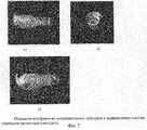
На фиг. 7 (а, б, в) показано объемное изображение атероматозного субстрата с выделенными цветом участками различной плотности. In FIG. 7 (a, b, c) shows a three-dimensional image of an atheromatous substrate with areas of different densities highlighted in color.
Предложенный способ осуществляется следующим образом. The proposed method is as follows.
На установках Oracle и Oracle In-Vision (фирма EndoSonics (Jomed)) или аналогичных им, получают двумерное внутрисосудистое ультразвуковое изображение, представляющее собой серию снимков поперечных сечений исследуемого участка артерии, посредством равномерного протягивания диагностического ультразвукового катетера внутри исследуемого сегмента сосуда с записью на S-VHS видеомагнитофон или CD-диск. Производится анализ полученной записи и выбирается участок для последующей обработки. Данный фрагмент записи вводится с помощью модуля оцифровки изображений в компьютер и обрабатывается при помощи программы для ЭВМ "мультимодальная рабочая станция MultiVox" (Свидетельство об официальной регистрации программы для ЭВМ 2000610789, дата регистрации 24 августа 2000 г.) Производится синтез объемного изображения с получением трехмерной реконструкции исследуемого участка сосуда в градациях серого цвета. Далее в интерактивном режиме на нескольких кадрах из серии производится сегментирование объемного изображения - выделение контуров и поверхностей исследуемых структур (стенок и просвета сосуда) с окраской их псевдоцветами. Такое сегментирование позволяет выделить местонахождение атероматозного субстрата. Данные сегментирования в автоматическом режиме с помощью интерполяционных алгоритмов применяются ко всей серии, то есть распространяются на весь 3D-массив. В серии выбирается кадр с выраженным стенозирующим процессом, предпочтительно с разной плотностью атероматозного субстрата. На данном кадре производится построение гистограммы интенсивности яркости изображения с цветным картированием атероматозных бляшек различной плотности ("мягкие", фиброзные, кальцинированные) с целью подбора оптимальных пороговых значений интенсивности для данного больного. Далее производится автоматическое сегментирование атероматозной бляшки на основе подобранных пороговых значений гистограммы интенсивности. Синтезированный 3D-массив и сегментированные объекты с помощью программы для ЭВМ "мультимодальная рабочая станция MwtiVox" отображаются на экран монитора в виде изометрической проекции сосуда. После этого производится отключение отображения сегментированных объектов, не представляющих диагностический интерес (стенок и просвета сосуда). В результате остается трехмерное изображение атероматозной бляшки с цветовым выделением участков различной плотности и возможностью продольного вращения во всех плоскостях для анализа строения и характера распространения бляшки. On Oracle and Oracle In-Vision installations (EndoSonics (Jomed) company or similar), a two-dimensional intravascular ultrasound image is obtained, which is a series of images of cross sections of the artery section under investigation, by uniformly pulling a diagnostic ultrasound catheter inside the vessel segment under investigation recorded on S- VHS VCR or CD. The analysis of the obtained record is carried out and a site is selected for subsequent processing. This fragment of the record is entered using the module for digitizing images into a computer and processed using the computer program "multimodal workstation MultiVox" (Certificate of official registration of the computer program 2000610789, registration date August 24, 2000) A volume image is synthesized to obtain a three-dimensional reconstruction the studied section of the vessel in gray gradations. Then, in an interactive mode, for several frames from the series, a three-dimensional image is segmented — the contours and surfaces of the structures under investigation (walls and lumen of the vessel) are highlighted with their pseudocolor colors. Such segmentation allows you to highlight the location of the atheromatous substrate. Automatic segmentation data using interpolation algorithms is applied to the entire series, that is, they apply to the entire 3D array. In the series, a frame is selected with a pronounced stenosing process, preferably with a different density of atheromatous substrate. A histogram of the image brightness intensity is plotted on this frame with color mapping of atheromatous plaques of various densities (“soft”, fibrous, calcified) in order to select the optimal threshold intensity values for this patient. Next, an atheromatous plaque is automatically segmented based on selected threshold values of the intensity histogram. The synthesized 3D array and segmented objects using the computer program "multimodal workstation MwtiVox" are displayed on the monitor screen in the form of an isometric projection of the vessel. After that, the display of segmented objects that are not of diagnostic interest (walls and lumen of the vessel) is turned off. As a result, there remains a three-dimensional image of an atheromatous plaque with color highlighting of areas of different densities and the possibility of longitudinal rotation in all planes to analyze the structure and nature of the plaque propagation.
Пример. Example.
Предложенный способ прижизненной трехмерной визуализации атероматозного субстрата был использован при изучении коронарных и периферических артерий во время диагностических исследований и рентгенохирургических операций, проводимых в связи с атеросклеротическими поражениями сосудов. The proposed method for intravital three-dimensional visualization of the atheromatous substrate was used in the study of coronary and peripheral arteries during diagnostic studies and x-ray surgery performed in connection with atherosclerotic vascular lesions.
Примером использования предложенного способа является описание полученного трехмерного компьютерного изображения атеросклеротической бляшки в правой коронарной артерии. Исходные внутрисосудистые ультразвуковые данные, полученные, как это было описано выше, представляющие собой серию снимков поперечных сечений исследуемого участка артерии (фиг.1), вводятся в компьютер для обработки. Выполняется синтез объемного изображения с получением трехмерной реконструкции участка артерии в градациях серого цвета (фиг.2). На нескольких кадрах из серии производится сегментирование объемного изображения с окраской различными цветами стенки артерии (красный, фиг.3), просвета сосуда (синий. фиг.4). Данные сегментирования в автоматическом режиме распространяются на весь 3D-массив. Сегментирование атероматозной бляшки выполняется в автоматическом режиме на основе подобранных по гистограмме интенсивности значений яркости изображения - "мягкая" бляшка помечается желтым цветом, фиброзная - оранжевым, кальцинированная - коричневым, (фиг.5)). Синтезированный 3D-массив и сегментированные объекты с помощью средств программы для ЭВМ "мультимодальная рабочая станция MultiVox" отображаются на экран монитора в виде изометрической проекции участка артерии (фиг.6 а, б). После этого производится отключение отображения сегментированных объектов, не представляющих диагностический интерес (красного, черного и синего цветов в данном примере). Получившееся объемное цветное изображение атероматозной бляшки (фиг. 7 а, 6, в) оператор может произвольно поворачивать в различных плоскостях на любой заданный угол для получения наиболее информативных проекций. An example of using the proposed method is a description of the obtained three-dimensional computer image of an atherosclerotic plaque in the right coronary artery. The initial intravascular ultrasound data obtained as described above, which is a series of images of cross-sections of the investigated section of the artery (figure 1), are entered into the computer for processing. The synthesis of the volumetric image is performed to obtain a three-dimensional reconstruction of the artery section in gray gradations (Fig. 2). On several frames from the series, volumetric images are segmented with different colors of the artery wall (red, Fig. 3), vessel lumen (blue. Fig. 4). The segmentation data automatically applies to the entire 3D array. Segmentation of atheromatous plaque is performed automatically based on the image brightness values selected according to the intensity histogram — a “soft” plaque is marked in yellow, fibrous in orange, calcined in brown (Fig. 5)). The synthesized 3D-array and segmented objects using the computer software "multimodal workstation MultiVox" are displayed on the monitor screen in the form of an isometric projection of a section of the artery (Fig.6 a, b). After that, the display of segmented objects that are not of diagnostic interest (red, black, and blue in this example) is turned off. The resulting volumetric color image of an atheromatous plaque (Fig. 7 a, 6, c), the operator can arbitrarily rotate in various planes at any given angle to obtain the most informative projections.
Способ позволяет прижизненно получить информацию о морфологии и пространственной конфигурации атероматозного субстрата, а по полученному цветному изображению судить о его структуре и плотности. The method allows in vivo to obtain information on the morphology and spatial configuration of the atheromatous substrate, and to judge by its color image its structure and density.
Claims (5)
Translated fromRussianPriority Applications (1)
| Application Number | Priority Date | Filing Date | Title |
|---|---|---|---|
| RU2001129772ARU2208391C1 (en) | 2001-11-05 | 2001-11-05 | Method for three-dimensional visualization of atheromatosis substrate at obliterating arterial lesions during one's life period |
Applications Claiming Priority (1)
| Application Number | Priority Date | Filing Date | Title |
|---|---|---|---|
| RU2001129772ARU2208391C1 (en) | 2001-11-05 | 2001-11-05 | Method for three-dimensional visualization of atheromatosis substrate at obliterating arterial lesions during one's life period |
Publications (1)
| Publication Number | Publication Date |
|---|---|
| RU2208391C1true RU2208391C1 (en) | 2003-07-20 |
Family
ID=29210799
Family Applications (1)
| Application Number | Title | Priority Date | Filing Date |
|---|---|---|---|
| RU2001129772ARU2208391C1 (en) | 2001-11-05 | 2001-11-05 | Method for three-dimensional visualization of atheromatosis substrate at obliterating arterial lesions during one's life period |
Country Status (1)
| Country | Link |
|---|---|
| RU (1) | RU2208391C1 (en) |
Cited By (5)
| Publication number | Priority date | Publication date | Assignee | Title |
|---|---|---|---|---|
| RU2302203C2 (en)* | 2003-09-02 | 2007-07-10 | Общество с ограниченной ответственностью Научно-производственное предприятие "Эксергия" | X-ray volumetric computer diagnostics of sponal column |
| RU2413995C2 (en)* | 2005-12-19 | 2011-03-10 | Конинклейке Филипс Электроникс, Н.В. | Method of improving image post processing using deformable grids |
| RU2469308C2 (en)* | 2007-06-04 | 2012-12-10 | Конинклейке Филипс Электроникс, Н.В. | X-ray instrument for three-dimensional ultrasonic analysis |
| RU2584135C1 (en)* | 2015-04-20 | 2016-05-20 | Федеральное государственное бюджетное учреждение "Российский кардиологический научно-производственный комплекс" Министерства здравоохранения России (ФГБУ "РКНПК" МЗ РФ) | Method for determining homogeneity of atherosclerotic plaque structure |
| RU2620758C1 (en)* | 2016-07-13 | 2017-05-29 | Федеральное государственное бюджетное научное учреждение "Научный центр неврологии" (ФГБНУ НЦН) | Method for quantitative determination of microvessels in atherosclerothic plaque of carotid arteries |
Citations (3)
| Publication number | Priority date | Publication date | Assignee | Title |
|---|---|---|---|---|
| RU2093077C1 (en)* | 1994-03-30 | 1997-10-20 | Виталий Анатольевич Петухов | Method for diagnosing atherosclerosis case |
| RU2128473C1 (en)* | 1997-07-30 | 1999-04-10 | Московский научно-исследовательский институт глазных болезней им.Гельмгольца | Method for carrying out ultrasonic examination of eyes |
| RU2154977C1 (en)* | 1999-09-13 | 2000-08-27 | Государственный научно-клинический центр охраны здоровья шахтеров | Method for diagnosing the cases of atherosclerotic vascular injuries |
- 2001
- 2001-11-05RURU2001129772Apatent/RU2208391C1/ennot_activeIP Right Cessation
Patent Citations (3)
| Publication number | Priority date | Publication date | Assignee | Title |
|---|---|---|---|---|
| RU2093077C1 (en)* | 1994-03-30 | 1997-10-20 | Виталий Анатольевич Петухов | Method for diagnosing atherosclerosis case |
| RU2128473C1 (en)* | 1997-07-30 | 1999-04-10 | Московский научно-исследовательский институт глазных болезней им.Гельмгольца | Method for carrying out ultrasonic examination of eyes |
| RU2154977C1 (en)* | 1999-09-13 | 2000-08-27 | Государственный научно-клинический центр охраны здоровья шахтеров | Method for diagnosing the cases of atherosclerotic vascular injuries |
Non-Patent Citations (1)
| Title |
|---|
| МИТЬКОВ В.В. Клиническое руководство по ультразвуковой диагностике. - М.: ВИДАР, 1997, т.4, с. 198-214.* |
Cited By (5)
| Publication number | Priority date | Publication date | Assignee | Title |
|---|---|---|---|---|
| RU2302203C2 (en)* | 2003-09-02 | 2007-07-10 | Общество с ограниченной ответственностью Научно-производственное предприятие "Эксергия" | X-ray volumetric computer diagnostics of sponal column |
| RU2413995C2 (en)* | 2005-12-19 | 2011-03-10 | Конинклейке Филипс Электроникс, Н.В. | Method of improving image post processing using deformable grids |
| RU2469308C2 (en)* | 2007-06-04 | 2012-12-10 | Конинклейке Филипс Электроникс, Н.В. | X-ray instrument for three-dimensional ultrasonic analysis |
| RU2584135C1 (en)* | 2015-04-20 | 2016-05-20 | Федеральное государственное бюджетное учреждение "Российский кардиологический научно-производственный комплекс" Министерства здравоохранения России (ФГБУ "РКНПК" МЗ РФ) | Method for determining homogeneity of atherosclerotic plaque structure |
| RU2620758C1 (en)* | 2016-07-13 | 2017-05-29 | Федеральное государственное бюджетное научное учреждение "Научный центр неврологии" (ФГБНУ НЦН) | Method for quantitative determination of microvessels in atherosclerothic plaque of carotid arteries |
Similar Documents
| Publication | Publication Date | Title |
|---|---|---|
| US20170367678A1 (en) | Ultrasound automated method for measuring the thickness of the walls of the left anterior descending, right and circumflex coronary arteries | |
| JP5039294B2 (en) | Apparatus and method for analyzing tissue type along a cylindrical structure | |
| Nakanishi et al. | How accurate is atherosclerosis imaging by coronary computed tomography angiography? | |
| US20070265521A1 (en) | Integrated MRI and OCT system and dedicated workflow for planning, online guiding and monitoring of interventions using MRI in combination with OCT | |
| US20220346756A1 (en) | Co-registration of intravascular and extravascular imaging for extravascular image with intravascular tissue morphology | |
| Taki et al. | Overview of different medical imaging techniques for the identification of coronary atherosclerotic plaques | |
| de Vries et al. | Current imaging modalities to visualize vulnerability within the atherosclerotic carotid plaque | |
| Klingensmith et al. | Automated three-dimensional assessment of coronary artery anatomy with intravascular ultrasound scanning | |
| Kips et al. | Identifying the vulnerable plaque: A review of invasive and non-invasive imaging modalities | |
| RU2208391C1 (en) | Method for three-dimensional visualization of atheromatosis substrate at obliterating arterial lesions during one's life period | |
| Song et al. | Morphologic assessment of the left atrial appendage in patients with atrial fibrillation by gray values–inverted volume-rendered imaging of three-dimensional transesophageal echocardiography: A comparative study with computed tomography | |
| Li et al. | Temporal averaging for quantification of lumen dimensions in intravascular ultrasound images | |
| Meissner et al. | High-resolution MR imaging of human atherosclerotic femoral arteries in vivo: validation with intravascular ultrasound | |
| Rice et al. | Pediatric echocardiography: current role and a review of technical advances | |
| US20040138567A1 (en) | Method of analyzing and displaying blood volume using myocardial blood volume map | |
| Keberle et al. | Three‐dimensional power Doppler sonography in screening for carotid artery disease | |
| Li et al. | Image segmentation and 3D reconstruction of intravascular ultrasound images | |
| Balocco et al. | Relation between plaque type, plaque thickness, blood shear stress, and plaque stress in coronary arteries assessed by X‐ray Angiography and Intravascular Ultrasound | |
| CA2627700A1 (en) | Systems and methods for detecting and presenting textural information from medical images | |
| White et al. | Vascular imaging before, during, and after endovascular repair | |
| Liu et al. | Feasibility of three‐dimensional intravascular ultrasonography: preliminary clinical studies. | |
| Yao et al. | Usefulness of three-dimensional transesophageal echocardiographic imaging for evaluating narrowing in the coronary arteries | |
| de Winter et al. | Computer assisted three-dimensional plaque characterization in intracoronary ultrasound studies | |
| van der Lugt et al. | Interobserver reproducibility of qualitative and quantitative analysis of intravascular ultrasound images before and after peripheral balloon angioplasty | |
| Peycheva et al. | Characteristics of unstable carotid plaques–new image modalities |
Legal Events
| Date | Code | Title | Description |
|---|---|---|---|
| MM4A | The patent is invalid due to non-payment of fees | Effective date:20061106 |