RU2184483C1 - Topic method for diagnosing visceral organ diseases of non-infectious nature - Google Patents
Topic method for diagnosing visceral organ diseases of non-infectious natureDownload PDFInfo
- Publication number
- RU2184483C1 RU2184483C1RU2001118607/14ARU2001118607ARU2184483C1RU 2184483 C1RU2184483 C1RU 2184483C1RU 2001118607/14 ARU2001118607/14 ARU 2001118607/14ARU 2001118607 ARU2001118607 ARU 2001118607ARU 2184483 C1RU2184483 C1RU 2184483C1
- Authority
- RU
- Russia
- Prior art keywords
- codogram
- lead
- leads
- carried out
- subject
- Prior art date
Links
- 201000010099diseaseDiseases0.000titleclaimsabstractdescription20
- 208000037265diseases, disorders, signs and symptomsDiseases0.000titleclaimsabstractdescription20
- 238000000034methodMethods0.000titleclaimsabstractdescription17
- 210000001835visceraAnatomy0.000titleclaimsdescription8
- 208000015181infectious diseaseDiseases0.000titleclaimsdescription6
- 230000002458infectious effectEffects0.000titleclaimsdescription6
- 230000000747cardiac effectEffects0.000claimsabstractdescription12
- 230000004807localizationEffects0.000claimsabstractdescription7
- 230000000241respiratory effectEffects0.000claimsabstractdescription4
- 238000000718qrs complexMethods0.000claimsdescription21
- 238000003745diagnosisMethods0.000claimsdescription15
- 230000000699topical effectEffects0.000claimsdescription10
- 230000007170pathologyEffects0.000claimsdescription9
- 238000010276constructionMethods0.000claimsdescription5
- 238000004364calculation methodMethods0.000claimsdescription3
- 230000014509gene expressionEffects0.000claimsdescription3
- 238000012545processingMethods0.000claimsdescription3
- 238000006073displacement reactionMethods0.000claimsdescription2
- 230000000694effectsEffects0.000abstractdescription4
- 239000003814drugSubstances0.000abstractdescription2
- 239000000126substanceSubstances0.000abstract1
- 238000004458analytical methodMethods0.000description10
- 210000000056organAnatomy0.000description9
- 238000002405diagnostic procedureMethods0.000description5
- 210000003414extremityAnatomy0.000description5
- 238000011161developmentMethods0.000description3
- 238000012774diagnostic algorithmMethods0.000description3
- 238000005516engineering processMethods0.000description3
- 231100000915pathological changeToxicity0.000description3
- 230000036285pathological changeEffects0.000description3
- 238000012360testing methodMethods0.000description3
- 230000005405multipoleEffects0.000description2
- 230000003068static effectEffects0.000description2
- 210000001685thyroid glandAnatomy0.000description2
- 206010007027Calculus urinaryDiseases0.000description1
- 206010020880HypertrophyDiseases0.000description1
- 208000009525MyocarditisDiseases0.000description1
- 230000003044adaptive effectEffects0.000description1
- 210000003152adnexa uteriAnatomy0.000description1
- 230000003683cardiac damageEffects0.000description1
- 208000028831congenital heart diseaseDiseases0.000description1
- 210000004907glandAnatomy0.000description1
- 210000003734kidneyAnatomy0.000description1
- 210000003141lower extremityAnatomy0.000description1
- 210000004072lungAnatomy0.000description1
- 210000005075mammary glandAnatomy0.000description1
- 239000000463materialSubstances0.000description1
- 238000005259measurementMethods0.000description1
- 208000008494pericarditisDiseases0.000description1
- 208000008281urolithiasisDiseases0.000description1
- 230000002861ventricularEffects0.000description1
Images

Landscapes
- Measurement And Recording Of Electrical Phenomena And Electrical Characteristics Of The Living Body (AREA)
Abstract
Description
Translated fromRussianИзобретение относится к медицине, а именно к функциональной диагностике, и может быть использовано для диагностики заболеваний внутренних органов на любом этапе их развития и определения наиболее вероятной локализации характерных патологических изменений в условиях поликлиники, медсанчасти, лечебно-диагностического центра при проведении диспансеризации населения, медицинского контроля здоровья работающих в условиях воздействия экстремальных факторов профессиональной деятельности или среды обитания, при профотборе, а также в отделениях (кабинетах) функциональной диагностики больниц и клиник. The invention relates to medicine, namely to functional diagnostics, and can be used to diagnose diseases of internal organs at any stage of their development and determine the most likely localization of characteristic pathological changes in the conditions of a clinic, medical unit, medical and diagnostic center during medical examination of a population, medical control the health of workers in conditions of exposure to extreme factors of professional activity or the environment, during professional selection, and also in the department lazy (cabinets) functional diagnostics of hospitals and clinics.
Предлагаемый способ диагностики является продолжением развития нового направления в диагностике заболеваний внутренних органов неинфекционной природы, в основе которого лежит технология информационного анализа кардиосигналов, реализованная в патентах на изобретения: 2157093 "Способ диагностики болезней неинфекционной этиологии" (2000 г); 2163088 "Способ диагностики заболеваний внутренних органов неинфекционной природы на любой стадии их развития (2001 г); 215974 "Устройство экспресс-диагностики заболеваний внутренних органов и онкопаталогии" (2000 г). The proposed diagnostic method is a continuation of the development of a new direction in the diagnosis of diseases of internal organs of a non-infectious nature, which is based on the technology of information analysis of cardiac signals implemented in patents for inventions: 2157093 "A method for the diagnosis of diseases of non-infectious etiology" (2000 g); 2163088 "Method for the diagnosis of diseases of internal organs of non-infectious nature at any stage of their development (2001); 215974" Device for the rapid diagnosis of diseases of internal organs and oncopathology "(2000 g).
Принципиальным отличием перечисленных способов диагностики от аналогичных других способов является то, что в основу их взят анализ амплитудной, частотной или фазовой модуляции кардиосигнала (зубцов R), кодирование динамики их пространственно-временных изменений, сравнение с эталонными кодограммами (стандартами) нормы и заболеваний и вынесение суждения о состоянии обследуемого по результатам сравнения. Эти способы диагностики базировались на анализе зубцов R сердечных комплексов, регистрируемых в одном из стандартных отведений, обычно во 2-м стандартном отведении, т.e. в отведении, в котором, как правило, амплитуда зубца R хорошо представлена. The fundamental difference between these diagnostic methods from similar other methods is that they are based on the analysis of the amplitude, frequency or phase modulation of the cardiosignal (R waves), coding the dynamics of their spatio-temporal changes, comparison with reference codograms (standards) of norm and disease, and judgments about the condition of the subject according to the results of comparison. These diagnostic methods were based on the analysis of R waves of cardiac complexes recorded in one of the standard leads, usually in the 2nd standard lead, i.e. in the lead, in which, as a rule, the amplitude of the R wave is well represented.
Способ диагностики, описанный в патенте RU 2163088, был выбран нами в качестве прототипа предлагаемого изобретения. Заявленный способ так же, как и известный, включает регистрацию 300-600 электрокардиосигналов, выделение серий, соответствующих дыхательным волнам, ранжирование их на периоды с учетом количества входящих в них кардиоимпульсов, определение амплитуд зубцов Rn(ф) и временных интервалов между ними Тn(ф) в каждой серии, вычисление средне-статической величины амплитуды каждого зубца Rn(cp) и временного интервала Тn(ср) путем математической обработки тех зубцов Rn(ф) и временных интервалов Tn(ф), порядковый номер которых в серии соответствует порядковому номеру в анализируемой серии, сравнение амплитуды каждого анализируемого зубца Rn(ф) и продолжительности каждого анализируемого временного интервала Тn(ф) с соответствующими среднестатическими величинами, кодирование получаемых выражений сравнения, при этом само кодирование производят с использованием одно-двух-трех и более членных кодовых комбинаций, построение кодограммы обследуемого в соответствии с вариантом кодирования и количеством членов кодовой комбинации, осуществляемое методом последовательного смещения на один кардиосигнал всей записи кардиоимпульсов от начала до конца с последующим структурированием кодограммы путем распределения кодовых комбинаций с учетом частоты их встречаемости, сравнение кодограммы обследуемого с ладонными кодограммами здоровых и больных лиц, полученными аналогичным образом и включающими только кодовые комбинации стопроцентной встречаемости и вынесение заключения о наличии нормы или заболевания в случае присутствия в кодограмме обследуемого полного набора комбинаций соответствующего эталона. The diagnostic method described in patent RU 2163088, was chosen by us as a prototype of the invention. The claimed method, as well as the known one, includes registration of 300-600 electrocardiosignals, highlighting series corresponding to respiratory waves, ranking them for periods taking into account the number of cardio pulses included in them, determining the tooth amplitudes Rn (f) and time intervals between them Tn (f ) in each series, the calculation of the average static value of the amplitude of each tooth Rn (cp) and the time interval Tn (cf) by mathematical processing of those teeth Rn (f) and time intervals Tn (f), the serial number of which in the series corresponds to the serial number in the analyzed series, comparing the amplitude of each analyzed tooth Rn (f) and the duration of each analyzed time interval Tn (f) with the corresponding average statistical values, coding the resulting comparison expressions, while the coding itself is performed using one, two, three or more member code combinations, the construction of the codogram of the subject in accordance with the coding option and the number of members of the code combination, carried out by the method of sequential displacement by one cardios the whole record of cardiac pulses from beginning to end, followed by structuring the codogram by distributing code combinations taking into account the frequency of their occurrence, comparing the codogram of the subject with the palm codograms of healthy and sick people, obtained in a similar way and including only code combinations of one hundred percent occurrence and concluding that there is a norm or diseases in the presence of a complete set of combinations of the corresponding standard in the codogram of the subject.
Всесторонняя апробация известного способа диагностики (более 15 тыс. исследований) показала, что одноканальный вариат использования технологии информационного анализа кардиосигналов не всегда обеспечивает полноту диагностики заболеваний. Нами обнаружено, что наиболее адекватным носителем информации о патологии того или иного органа является электрокардиосигнал такой конфигурации (комплекс QRS), которая регистрируется в зоне органа. Поэтому наиболее полная диагностика достигается при одновременном многополюсном многоканальном съеме и информационном анализе электрокардиосигналов. Клиническая апробация свидетельствует о том, что минимальным вариантом многополюсной топической диагностики может быть одновременный съем и информационный анализ электрокардиосигналов не менее чем в трех стандартных отведениях по Эйнтховену или в четырех униполярных усиленных отведениях от конечностей по Гольдбергеру. Оптимальный результат достигается при одновременном использовании всех семи перечисленных отведений. Comprehensive testing of the known diagnostic method (more than 15 thousand studies) showed that a single-channel version of using the technology of information analysis of cardiac signals does not always ensure the completeness of diagnosis of diseases. We found that the most adequate carrier of information about the pathology of a particular organ is an electrocardiogram of such a configuration (QRS complex), which is recorded in the area of the organ. Therefore, the most complete diagnosis is achieved with simultaneous multi-pole multi-channel acquisition and information analysis of cardiac signals. Clinical testing indicates that the minimum option for multi-pole topical diagnosis can be simultaneous acquisition and information analysis of electrocardiograms in at least three standard leads according to Einthoven or in four unipolar reinforced leads from the limbs according to Goldberger. The optimal result is achieved with the simultaneous use of all seven of the listed leads.
Техническим результатом заявленного способа топической диагностики является более точная и полная диагностика, т.e. выявление такой патологии, которая известным способом не могла быть установлена, и определение наиболее вероятной локализации характерных для каждого заболевания патоморфологических изменений, т.e. топическая диагностика, что особенно важно при выявлении патологии в парных органах. The technical result of the claimed method of topical diagnosis is a more accurate and complete diagnosis, i.e. the identification of such a pathology that could not be established in a known manner, and the determination of the most likely localization of pathomorphological changes characteristic of each disease, i.e. topical diagnosis, which is especially important when identifying pathologies in paired organs.
Поставленная задача достигается тем, что в известном способе топической диагностики заболеваний внутренних органов неинфекционной природы, включающем регистрацию 300-600 электрокардиосигналов, выделение серий, соответствующих дыхательным волнам, ранжирование их на периоды с учетом количества входящих в них кардиоимпульсов, определение амплитуд зубцов Rn(ф) и временных интервалов между ними Тn(ф) в каждой серии, вычисление среднестатической величины амплитуды каждого зубца Rn(cp) и временного интервала Тn(ср) путем математической обработки тех зубцов Rn(ф) и временных интервалов Тn(ф), порядковый номер которых в серии соответствует порядковому номеру в анализируемой серии, сравнение амплитуды каждого анализируемого зубца Rn(ф) и каждою анализируемого временного интервала Тn(ф) с соответствующими среднестатическими величинами, кодирование получаемых выражений сравнения, при этом само кодирование производят с использованием одно-двух-трех и более членных кодовых комбинаций, построение кодограммы обследуемого в соответствии с вариантом кодирования и количеством членов кодовой комбинации, осуществляемое методом последовательного смещения на один кардиосигнал всей записи кардиоимпульсов от начала до конца с последующим структурированием кодограммы путем распределения кодовых комбинаций с учетом частоты их встречаемости, сравнение кодограммы обследуемого с эталонными кодограммами здоровых и больных лиц, полученными аналогичным образом и включающими только кодовые комбинации стопроцентной встречаемости и вынесение заключения о наличии нормы или заболевания в случае присутствия в кодограмме обследуемого полного набора комбинаций соответствующего эталона, при этом регистрацию электрокардиосигналов как у обследуемого, так и при построении стандартов - эталонов проводят одновременно более чем в одном стандартном отведении, дополнительно проводят классификацию графических изображений QRS-комплексов электрокардиосигналов, проводят кодирование электрокардиосигналов в каждом отведении отдельно с учетом спецификации последних в соответствии с вариантом распределения символов используемого кодирования, сравнение кодограммы обследуемого со стандартами - эталонными кодограммами проводят для каждого отведения отдельно с учетом их классификации и спецификации, а вынесение суждения о наличии или отсутствии патологии, а также наиболее вероятной ее локализации, т. е. проведение топической диагностики осуществляют по анализу суммированных результатов, полученных для каждого отведения. The problem is achieved in that in the known method for the topical diagnosis of diseases of internal organs of non-infectious nature, including the registration of 300-600 electrocardiograms, the allocation of series corresponding to respiratory waves, ranking them for periods taking into account the number of cardio pulses included in them, determining the amplitudes of the teeth Rn (f) and the time intervals between them Tn (f) in each series, the calculation of the average static amplitude of each tooth Rn (cp) and the time interval Tn (cp) by mathematical processing t ex teeth Rn (f) and time intervals Tn (f), the serial number of which in the series corresponds to the serial number in the analyzed series, comparing the amplitude of each analyzed tooth Rn (f) and each analyzed time interval Tn (f) with the corresponding average values, coding obtained comparison expressions, while the encoding itself is performed using one, two, three or more member code combinations, the codogram of the subject is constructed in accordance with the coding option and the number of code members combinations performed by the method of sequentially shifting the entire record of cardio pulses from one beginning to the end by one cardiosignal followed by structuring the codogram by distributing the code combinations taking into account their frequency of occurrence, comparing the codogram of the subject with the reference codograms of healthy and sick persons, obtained in a similar way and including only code combinations of one hundred percent incidence and conclusion on the presence of a norm or disease in the case of the presence in the codogram of the subject about a complete set of combinations of the corresponding standard, while registering the electrocardiograms both in the subject and in building standards - the standards are carried out simultaneously in more than one standard lead, they additionally classify graphic images of QRS-complexes of cardiac signals, conduct encoding of cardiac signals in each lead separately, taking into account specifications of the latter in accordance with the distribution of characters used coding, comparison of the codogram is examined go with standards - reference codograms is carried out for each assignment separately, taking into account their classification and specification, and a judgment is made on the presence or absence of pathology, as well as its most likely localization, i.e., topical diagnostics are carried out by analyzing the summarized results obtained for each assignments.
Регистрацию электрокардиосигналов как у обследуемого, так и при построении стандартов-эталонов можно проводить одновременно в трех биполярных стандартных отведениях по Эйнтховену и четырех униполярных усиленных отведениях от конечностей по Гольдбергеру (всего в 7 отведениях). The registration of electrocardiograms both in the subject and in the construction of standard standards can be carried out simultaneously in three bipolar standard leads according to Einthoven and four unipolar reinforced leads from the limbs according to Goldberger (a total of 7 leads).
Способ осуществляют следующим образом
Проводят одновременную регистрацию электрокардиосигналов в трех биполярных стандартных отведениях по Эйнтховену и в четырех униполярных усиленных отведениях от конечностей по Гольдбергеру (всего семь отведений);
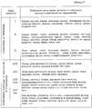
классифицируют каждое отведение согласно типу конфигурации QRS-комплекса, регистрируемого в нем электрокардиосигнала (основные типы QRS-комплексов представлены в табл 1).The method is as follows
Simultaneously register electrocardiograms in three bipolar standard leads according to Einthoven and in four unipolar reinforced leads from the limbs according to Goldberger (a total of seven leads);
each lead is classified according to the type of configuration of the QRS complex, the electrocardiogram recorded in it (the main types of QRS complexes are presented in Table 1).
Осуществляют измерение основных параметров QRS-комплекса:
амплитуд зубцов R1, R2, S, размаха OR1, R1S, SR2, а также временных интервалов Тn между основными зубцами (R1, S или R2), регистрируемых QRS-комплексов электрокардиосигналов (варианты измерений параметров QRS-комплексов приведены в таблице 1), кодируют динамику пространственно-временных изменений главного желудочкового вектора электрокардиосигнала по динамике основных параметров QRS-комплекса электрокардиограммы применительно к каждому отведению;
анализируют электрокардиосигналы по одному из способов, изложенных в патентах на изобретения (Успенский В.М. 2000г. 2157093; Успенский В.М. 2001 г. 2163088), и получают кодограммы отведений;
проводят спецификацию отведений в соответствии с вариантом распределения символов используемого кодирования с учетом частоты их встречаемости в масштабе всей кодограммы соответствующих отведений. Например, использовано кодирование, включающее символы А, В, С, Д и Е. После построения кодограммы отведения подсчитывают количество каждого символа в кодограмме и согласно частоте их встречаемости получают распределение символов (например, АСВЕД или СДЕВА), которые можно рассматривать в качестве специфической характеристики отведений с учетом варианта кодирования;
сравнивают кодограммы отведений обследуемого с эталонными кодограммами - стандартами нормы и заболеваний применительно к каждому отведению регистрации электрокардиограммы с учетом их классификации и спецификации;
дают суммарную оценку результатов диагностики на основании результатов диагностики отдельно в каждом отведении (топическая диагностика) и выносят заключение о здоровье обследуемого.Carry out the measurement of the main parameters of the QRS complex:
the amplitudes of the teeth R1 , R2 , S, the span OR1 , R1 S, SR2 , as well as the time intervals Tn between the main teeth (R1 , S or R2 ), registered QRS-complexes of electrocardiosignals (options for measuring parameters QRS- complexes are given in table 1), encode the dynamics of spatio-temporal changes in the main ventricular vector of the electrocardiogram according to the dynamics of the main parameters of the QRS-complex of the electrocardiogram as applied to each lead;
analyze electrocardiograms in one of the ways described in the patents for inventions (Uspensky V.M. 2000. 2157093; Uspensky V.M. 2001. 2163088), and receive lead codograms;
lead specification is carried out in accordance with the distribution of symbols used coding taking into account the frequency of their occurrence on the scale of the entire codogram of the corresponding leads. For example, coding is used, including the characters A, B, C, D, and E. After building the lead codogram, the number of each character in the codogram is calculated and according to the frequency of their occurrence, a distribution of characters is obtained (for example, ASVED or CEDAW), which can be considered as a specific characteristic Leads taking into account the encoding option
comparing the codogram of the leads of the subject with the reference codograms - standards of norms and diseases in relation to each lead of registration of the electrocardiogram, taking into account their classification and specification;
give a total assessment of the diagnostic results based on the diagnostic results separately in each lead (topical diagnosis) and make a conclusion about the health of the subject.
Для реализации изложенного алгоритма диагностики осуществлена стандартизация электрокардиосигналов по типу конфигурации QRS-комплекса во всех используемых отведениях. Конфигурация QRS-комплекса в стандартных биполярных отведениях по Эйнтховену и в униполярных усиленных отведениях от конечностей по Гольдбергеру зависит от ориентации в пространстве главного вектора и плоскости петли QRS относительно точек съема электрокардиосигнала. Полому в каждом отведении для съема электрокардиосигнала имеет место свой вариант конфигурации комплекса. To implement the diagnostic algorithm outlined, standardization of electrocardiosignals was performed according to the type of configuration of the QRS complex in all leads used. The configuration of the QRS complex in standard Einthoven bipolar leads and in unipolar reinforced leads from the limbs according to Goldberger depends on the orientation in space of the main vector and the plane of the QRS loop relative to the pick-up points of the cardiac signal. In each lead for picking up the electrocardiogram, there is a variant of the complex configuration.
На конфигурацию QRS-комплекса могут оказывать влияние различные адаптационные и патологические изменения отделов сердца и в первую очередь его желудочков: гипертрофия, диллятация, рубцовые изменения и кардиосклероз, миокардиты, перикардиты, блокады ножек пучка Гисса, пороки сердца и другие виды поражения сердца. Могут влиять также особенности грудной клетки, состояние диафрагмы и ряд других анатомо-физиологических факторов. The configuration of the QRS complex can be influenced by various adaptive and pathological changes in the departments of the heart and primarily its ventricles: hypertrophy, dilatation, cicatricial changes and cardiosclerosis, myocarditis, pericarditis, blockade of the bundle of the His branch, heart defects and other types of heart damage. Features of the chest, the state of the diaphragm, and a number of other anatomical and physiological factors can also influence.
Учитывая, что информация закладывается модулирующим механизмом в электрокардиосигнал (QRS) любой конфигурации, вполне очевидна важная роль классификации наиболее часто встречающихся конфигураций QRS комплекса. Основные наиболее часто встречающихся варианты QRS-комплексов электрокардиограммы представлены в таблице 1. Considering that the information is inserted by the modulating mechanism into the electrocardiosignal (QRS) of any configuration, the important role of the classification of the most common QRS complex configurations is quite obvious. The main most common variants of QRS-complexes of the electrocardiogram are presented in table 1.
Для спецификации отведений можно также использовать варианты распределения символов любого способа кодирования изменений основных пространственно-временных параметров регистрируемых QRS-комплексов с учетом частоты их встречаемости в масштабе всей кодограммы соответствующих отведений. На основе классификации отведений согласно конфигурации QRS-комплексов, регистрируемых в них, и спецификации отведений по характеру распределения символов соответствующих вариантов кодирования с учетом частоты их встречаемости в кодограмме отведений отрабатывается банк эталонов (кодограмм) нормы и различных заболеваний. To specify leads, you can also use character distribution options for any method of encoding changes in the main spatio-temporal parameters of registered QRS complexes, taking into account their frequency of occurrence on the scale of the entire codogram of the corresponding leads. Based on the classification of leads according to the configuration of the QRS complexes recorded in them, and the specification of leads according to the character distribution of the characters of the corresponding coding options, taking into account the frequency of their occurrence, a bank of standards (codograms) of norm and various diseases is worked out in the lead codogram.
Для осуществления диагностического алгоритма у обследуемого предварительно осуществляется аналогичная классификация отведений по конфигурации ORS-комплексов и спецификация каждого соответствующего отведения по вариантам распределения символов кодирования. Диагностический алгоритм включается только по результатам указанной классификации и спецификации отведений, составляющих исходную базу данных обследуемого. To implement the diagnostic algorithm, the subject is preliminarily carried out a similar classification of the leads according to the configuration of the ORS complexes and the specification of each corresponding lead according to the distribution of encoding symbols. The diagnostic algorithm is included only according to the results of the specified classification and specification of the leads that make up the initial database of the subject.
Апробация предлагаемой технологии одновременного многоканального информационного анализа электрокардиосигналов выявила дополнительные новые диагностические возможности. В частности, установленный факт о том, что максимальная информация о патологии того или иного органа заложена в тех электрокардиосигналах, конфигурация которых соответствует конфигурации электрокардиосигналов, регистрируемых в зоне соответствующего органа, позволяет определять локализацию и выраженность патоморфологических изменений, специфических для заболевания, что имеет особенно важное значение при определении патологии в парных органах. Testing of the proposed technology for simultaneous multichannel information analysis of cardiac signals revealed additional new diagnostic capabilities. In particular, the established fact that the maximum information about the pathology of a particular organ is embedded in those electrocardiograms whose configuration corresponds to the configuration of the electrocardiograms recorded in the area of the corresponding organ allows us to determine the localization and severity of pathological changes specific to the disease, which is especially important importance in determining the pathology in paired organs.
Следует отметить, что конфигурация QRS-комплекса электрокардиосигналов, регистрируемых в зоне того или иного органа, у разных людей разная и в каждом конкретном случае зависит от соотношения этого органа с главным вектором и плоскостью петли QRS комплекса электрокардиосигнала в пространстве. Последнее является относительно устойчивым признаком для одного и того же человека и может изменяться в течение жизни человека в процессе его роста, в зависимости от вида физического труда, спорта, профессиональной деятельности или заболеваний, влияющих на ориентацию в пространстве электрической оси сердца. Что касается соотношения патологии различных внутренних органов и отведений регистрации электрокардиосигналов, с последующим информационным анализом их, позволяющим выявить патологию, то она характеризуется большим постоянством для одного и того же человека. На основе анализа обширного материала нами представлены в таблице 2 возможности в топической диагностике каждого биполярного стандартного и униполярного усиленного от конечностей отведения для регистрации электрокардиограммы. It should be noted that the configuration of the QRS-complex of cardiac signals recorded in the area of a particular organ is different for different people and in each case depends on the ratio of this organ with the main vector and the plane of the QRS loop of the cardiac-signal complex in space. The latter is a relatively stable sign for the same person and can change during the course of a person’s life in the process of his growth, depending on the type of physical labor, sports, professional activity or diseases that affect the orientation in space of the electrical axis of the heart. As for the correlation of the pathology of various internal organs and the leads of registration of electrocardiosignals, followed by their information analysis, which allows to identify the pathology, it is characterized by great constancy for the same person. Based on an analysis of the vast material, we present in Table 2 the possibilities in the topical diagnosis of each bipolar standard and unipolar limb-reinforced lead for recording an electrocardiogram.
На этапе суммарного анализа результатов диагностики в биполярных стандартных отведениях и униполярных усиленных отведениях от конечностей открывается возможность топической диагностики основных патоморфологических изменений при заболевании. Наиболее ярко это находит свое выражение применительно к парным органам: щитовидная железа, легкие, почки, молочные железы, придатки матки. Например, сопоставляя степень активности мочекаменной болезни в отведениях по Гольдбергеру от правой и левой нижних конечностей, можно определить наиболее вероятную локализацию конкремента. Сопоставляя результаты диагностики в 1-м биполярном стандартном отведении по Эйнтховену и в униполярных усиленных отведениях от правой и левой рук по Гольдбергеру, можно определить диффузный или очаговый процесс в щитовидной железе, а если очаговый, то в какой доле железы преимущественно. At the stage of a summary analysis of the diagnostic results in bipolar standard leads and unipolar reinforced leads from the limbs, the possibility of a topical diagnosis of the main pathomorphological changes in the disease opens. This is most clearly expressed in relation to paired organs: the thyroid gland, lungs, kidneys, mammary glands, uterine appendages. For example, comparing the degree of activity of urolithiasis in Goldberger leads from the right and left lower extremities, you can determine the most likely localization of calculus. Comparing the diagnostic results in the 1st bipolar standard lead according to Einthoven and in unipolar reinforced leads from the right and left hands according to Goldberger, it is possible to determine the diffuse or focal process in the thyroid gland, and if focal, in which lobe of the gland is predominant.
Claims (2)
Translated fromRussianPriority Applications (2)
| Application Number | Priority Date | Filing Date | Title |
|---|---|---|---|
| RU2001118607/14ARU2184483C1 (en) | 2001-07-06 | 2001-07-06 | Topic method for diagnosing visceral organ diseases of non-infectious nature |
| PCT/RU2002/000325WO2003003919A1 (en) | 2001-07-06 | 2002-07-04 | Method for diagnosing non-infectious diseases of vincera (variants) |
Applications Claiming Priority (1)
| Application Number | Priority Date | Filing Date | Title |
|---|---|---|---|
| RU2001118607/14ARU2184483C1 (en) | 2001-07-06 | 2001-07-06 | Topic method for diagnosing visceral organ diseases of non-infectious nature |
Publications (1)
| Publication Number | Publication Date |
|---|---|
| RU2184483C1true RU2184483C1 (en) | 2002-07-10 |
Family
ID=20251437
Family Applications (1)
| Application Number | Title | Priority Date | Filing Date |
|---|---|---|---|
| RU2001118607/14ARU2184483C1 (en) | 2001-07-06 | 2001-07-06 | Topic method for diagnosing visceral organ diseases of non-infectious nature |
Country Status (1)
| Country | Link |
|---|---|
| RU (1) | RU2184483C1 (en) |
Cited By (3)
| Publication number | Priority date | Publication date | Assignee | Title |
|---|---|---|---|---|
| RU2240722C2 (en)* | 2003-01-04 | 2004-11-27 | Государственное учреждение Научно-исследовательский институт комплексных проблем гигиены и профессиональных заболеваний СО РАМН | Method for predicting pathological results in case of pregnancy |
| RU2258458C2 (en)* | 2003-10-14 | 2005-08-20 | Кузьмин Александр Геннадьевич | Method for predicting severity degree of autonomic dystonia syndrome |
| RU2270042C2 (en)* | 2003-12-02 | 2006-02-20 | Научно-исследовательский институт кардиологии им. В.А. Алмазова Минздрава Российской Федерации | Method for preventing atrial fibrillation in patients with sinus nodal weakness syndrome and paroxysms of atrial fibrillation |
Citations (3)
| Publication number | Priority date | Publication date | Assignee | Title |
|---|---|---|---|---|
| US6035233A (en)* | 1995-12-11 | 2000-03-07 | Intermedics Inc. | Implantable medical device responsive to heart rate variability analysis |
| US6148228A (en)* | 1998-03-05 | 2000-11-14 | Fang; Dan Oun | System and method for detecting and locating heart disease |
| RU2163088C1 (en)* | 2000-04-12 | 2001-02-20 | Успенский Вячеслав Максимилианович | Method for diagnosing visceral organ diseases of noninfectious nature at any stage of their development |
- 2001
- 2001-07-06RURU2001118607/14Apatent/RU2184483C1/ennot_activeIP Right Cessation
Patent Citations (3)
| Publication number | Priority date | Publication date | Assignee | Title |
|---|---|---|---|---|
| US6035233A (en)* | 1995-12-11 | 2000-03-07 | Intermedics Inc. | Implantable medical device responsive to heart rate variability analysis |
| US6148228A (en)* | 1998-03-05 | 2000-11-14 | Fang; Dan Oun | System and method for detecting and locating heart disease |
| RU2163088C1 (en)* | 2000-04-12 | 2001-02-20 | Успенский Вячеслав Максимилианович | Method for diagnosing visceral organ diseases of noninfectious nature at any stage of their development |
Non-Patent Citations (1)
| Title |
|---|
| ДЕ ЛУНА А.Б. Руководство по клинической кардиологии. - М.: Медицина, 1993, с.54-62, 215. МИРОНОВА Т.Ф., МИРОНОВ В.А. Клинический анализ волновой структуры синусового ритма сердца. - Челябинск, 1998. БЕРЕЗНЫЙ Е.А., РУБИН А.М. Практическая кардиоритмография. - НПП "Нео", 1997. MOSER M. Heart rate variability as a prognostic in cardiology. - Circulation, 1998, v. 90, pp.1978-1982.* |
Cited By (3)
| Publication number | Priority date | Publication date | Assignee | Title |
|---|---|---|---|---|
| RU2240722C2 (en)* | 2003-01-04 | 2004-11-27 | Государственное учреждение Научно-исследовательский институт комплексных проблем гигиены и профессиональных заболеваний СО РАМН | Method for predicting pathological results in case of pregnancy |
| RU2258458C2 (en)* | 2003-10-14 | 2005-08-20 | Кузьмин Александр Геннадьевич | Method for predicting severity degree of autonomic dystonia syndrome |
| RU2270042C2 (en)* | 2003-12-02 | 2006-02-20 | Научно-исследовательский институт кардиологии им. В.А. Алмазова Минздрава Российской Федерации | Method for preventing atrial fibrillation in patients with sinus nodal weakness syndrome and paroxysms of atrial fibrillation |
Similar Documents
| Publication | Publication Date | Title |
|---|---|---|
| CN1117331C (en) | Method and apparatus for non-invasive diagnosis of cardiovascular and related diseases | |
| US4974598A (en) | EKG system and method using statistical analysis of heartbeats and topographic mapping of body surface potentials | |
| RU2512794C2 (en) | Method and device for noise frequency ranging | |
| US8992435B2 (en) | System and method for classifying a heart sound | |
| US6607480B1 (en) | Evaluation system for obtaining diagnostic information from the signals and data of medical sensor systems | |
| US20230274832A1 (en) | Apparatus and method for generating electrocardiogram based on generative adversarial network algorithm | |
| AU2003231148A1 (en) | System and method for synthesizing leads of an electrocardiogram | |
| US20210204857A1 (en) | Method and device for cardiac monitoring | |
| RU2163088C1 (en) | Method for diagnosing visceral organ diseases of noninfectious nature at any stage of their development | |
| KR102802002B1 (en) | Ecg measurement service providing method and system using potable ecg device | |
| CN110277169A (en) | Shared intelligent physical examination system | |
| RU2346653C2 (en) | Cardiovascular sound analysis method and system | |
| JP2024532284A (en) | Method and system for designing photoplethysmographic waveform features from biophysical signals for use in characterizing physiological systems | |
| JP2005323821A (en) | Standard 12-lead ECG construction method and ECG inspection apparatus | |
| US8521263B2 (en) | Method and device for recording an electrocardiogram | |
| JP2024532280A (en) | Method and system for engineering visual features from biophysical signals for use in characterizing physiological systems - Patents.com | |
| JP2024534131A (en) | Method and system for engineering conduction deviation features from biophysical signals for use in characterizing physiological systems - Patents.com | |
| RU2184483C1 (en) | Topic method for diagnosing visceral organ diseases of non-infectious nature | |
| RU2118117C1 (en) | Method of creating biological feedback to correct heart activity and appropriate device | |
| RU2141247C1 (en) | Method for diagnosing cardiac system functional state | |
| CN118747325A (en) | A method for evaluating the quality of electrocardiogram lead data | |
| US9149201B2 (en) | TWA measuring apparatus and TWA measuring method | |
| RU2102000C1 (en) | Method for setting early stage diagnosis of hypertension disease | |
| JP2024532279A (en) | Method and system for non-invasively assessing elevated left ventricular end-diastolic pressure - Patents.com | |
| JP2024532285A (en) | Method and system for engineering cardiac waveform features from biophysical signals for use in characterizing physiological systems - Patents.com |
Legal Events
| Date | Code | Title | Description |
|---|---|---|---|
| MM4A | The patent is invalid due to non-payment of fees | Effective date:20090707 |