RU2161016C1 - Device for holding laser light guide end in the vicinity of the face and oral cavity - Google Patents
Device for holding laser light guide end in the vicinity of the face and oral cavityDownload PDFInfo
- Publication number
- RU2161016C1 RU2161016C1RU99125457ARU99125457ARU2161016C1RU 2161016 C1RU2161016 C1RU 2161016C1RU 99125457 ARU99125457 ARU 99125457ARU 99125457 ARU99125457 ARU 99125457ARU 2161016 C1RU2161016 C1RU 2161016C1
- Authority
- RU
- Russia
- Prior art keywords
- rod
- face
- light guide
- oral cavity
- laser light
- Prior art date
Links
Images


Landscapes
- Dental Tools And Instruments Or Auxiliary Dental Instruments (AREA)
- Investigating Or Analysing Materials By Optical Means (AREA)
Abstract
Description
Translated fromRussianПредполагаемое изобретение относится к медицине, а именно к терапевтической стоматологии, и может быть использовано при диагностике различных заболеваний пародонта, слизистой полости рта и области лица. The alleged invention relates to medicine, namely to therapeutic dentistry, and can be used in the diagnosis of various diseases of periodontal, oral mucosa and facial area.
По мнению большинства исследователей, ведущим звеном в патогенезе заболеваний пародонта являются нарушения микроциркуляции, от которых зависит течение капиллярно-трофических процессов, поэтому современная регистрация этих изменений является важной задачей пародонтологии. According to most researchers, the leading link in the pathogenesis of periodontal diseases are microcirculation disorders, which determine the course of capillary-trophic processes, therefore, the modern registration of these changes is an important task of periodontology.
Существующие на сегодняшний день способы оценки периферической гемодинамики пародонта не удовлетворяют требованиям ученых и клиницистов, прежде всего своей громоздкостью и инвазивностью. Поэтому поиск новых приспособлений, позволяющих улучшить качество диагностики заболеваний пародонта, слизистой полости рта и лица, является актуальной проблемой стоматологии (Козлов В.И., Корси Л.В., Соколов В.Г., Материалы Второго Всероссийского симпозиума. М., 1998). Currently existing methods for assessing the peripheral hemodynamics of periodontal disease do not meet the requirements of scientists and clinicians, primarily due to their bulkiness and invasiveness. Therefore, the search for new devices to improve the quality of diagnosis of periodontal disease, oral mucosa and face, is an urgent problem of dentistry (Kozlov V.I., Korsi L.V., Sokolov V.G., Materials of the Second All-Russian Symposium. M., 1998 )
За аналог изобретения можно принять руку врача, удерживающую световод в исследуемой области. For the analogue of the invention, you can take the hand of a doctor holding the fiber in the study area.
Недостатки:
1. Относительная невысокая воспроизводимость результатов тестирования состояния микроциркуляции в силу ее органоспецифической и индивидуальной изменчивости.Disadvantages:
1. The relative low reproducibility of the results of testing the state of microcirculation due to its organ-specific and individual variability.
2. Конечный результат измерения кровотока зависит от величины расстояния от торца лазерного световода до участка исследуемой поверхности. В связи с тем, что световод удерживается руками, данные микроциркуляции изменяются в зависимости от степени прижатия световода к исследуемому участку. 2. The final result of measuring blood flow depends on the distance from the end of the laser fiber to the area of the surface under study. Due to the fact that the fiber is held by hands, the microcirculation data changes depending on the degree of pressing of the fiber to the studied area.
3. Торец лазерного световода должен располагаться под углом 90o к исследуемой поверхности, что невозможно соблюдать при удержании световода вручную.3. The end of the laser fiber must be at an angle of 90o to the test surface, which cannot be observed when holding the fiber manually.
Вышеперечисленные недостатки побудили авторов к изобретению приспособления для удержания торца световода лазера в области лица и полости рта и выбора в качестве прототипа к этому приспособлению устройства по а.с. СССР N 1718897, 1992 г. The above disadvantages prompted the authors to invent devices for holding the end of the laser fiber in the face and oral cavity and choosing as a prototype for this device a. USSR N 1718897, 1992
Задачей данной работы явилось повышение качества диагностики заболеваний пародонта, слизистой полости рта и лица. The objective of this work was to improve the quality of diagnosis of periodontal disease, oral mucosa and face.
Сущность изобретения:
Устройство предназначено для подведения торца световода лазера к любой точке лица и полости рта и удержания его без помощи пациента и врача. Устройство отличается тем, что крепится на голове пациента ремнем, длина которого регулируется и фиксируется замком. В замке зажат стержень, второй стержень крепится под прямым углом относительно первого стержня через втулку-зажим. Торец световода устанавливается во втулку-зажим, закрепленный на конце второго стержня. Втулки установлены с возможностью перемещения по сторонам.The invention:
The device is designed to bring the end of the laser fiber to any point on the face and oral cavity and hold it without the help of the patient and the doctor. The device is characterized in that it is attached to the patient’s head with a belt, the length of which is adjustable and fixed by a lock. A rod is clamped in the lock, the second rod is mounted at right angles to the first rod through the clamp sleeve. The end of the fiber is installed in the sleeve-clamp, mounted on the end of the second rod. The bushings are mounted to move on the sides.
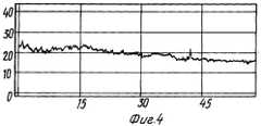
Сущность изобретения поясняется чертежом, где на фиг.1 изображен общий вид устройства; на фиг. 2 - вариант конструкции втулки-зажима; на фиг.3 - ЛДФ-грамма (больной Д), снятая без заявляемого устройства, и на фиг.4 - ЛДФ-грамма (больной Д), снятая с предложенным устройством. The invention is illustrated in the drawing, where figure 1 shows a General view of the device; in FIG. 2 - a design variant of the sleeve-clamp; figure 3 - LDF-gram (patient D), shot without the inventive device, and figure 4 - LDF-gram (patient D), shot with the proposed device.
Состояние микроциркуляции тканей пародонта регистрируют на аппарате ЛАКК-01 (НПП "Лазма", Россия), оснащенном 3- канальным световодным кабелем с диаметром поперечного сечения 0,3 см. Измерения проводят у пациентов в положении сидя (угол наклона спины 90 градусов), голова фиксирована на подголовнике при горизонтальном расположении трагоорбитальной линии. Устройство состоит из ремня (1) (фиг. 1), замка (2), двух стержней (3, 4), двух втулок-зажимов (5, 6). The state of microcirculation of periodontal tissues is recorded on a LAKK-01 apparatus (NPP Lazma, Russia) equipped with a 3-channel light guide cable with a cross-sectional diameter of 0.3 cm. Measurements are performed in patients in a sitting position (back angle 90 degrees), head fixed on the headrest with horizontal tragoorbital line. The device consists of a belt (1) (Fig. 1), a lock (2), two rods (3, 4), two clamping bushings (5, 6).
Один из вариантов конструкции втулки-зажима (фиг. 2) состоит из корпуса (8), двух упоров (9), двух пружин (10), двух гаек (11). One of the design options of the sleeve-clamp (Fig. 2) consists of a housing (8), two stops (9), two springs (10), two nuts (11).
В каждое из двух направляющих отверстий, расположенных под прямым углом, входят упоры (9), поджимаемые через пружины (10) гайками (11). Гайки (11) регулируют усилие поджатия упоров (9) к осям (3, 4) (фиг. 1) или их полное зажатие. In each of the two guide holes located at right angles, there are stops (9), pressed through the springs (10) with nuts (11). Nuts (11) regulate the force of compression of the stops (9) to the axes (3, 4) (Fig. 1) or their full clamping.
Устройство применяют следующим образом:
Устройство крепят на голове пациента ремнем (1), длину которого регулируют и фиксируют замком (2). Один стержень (3) зажат в замке (2), второй стержень (4) крепят под прямым углом через втулку-зажим (5). Торец световода (7) устанавливают во втулку-зажим (6), закрепленный на конце стержня (4).The device is used as follows:
The device is attached to the patient’s head with a belt (1), the length of which is regulated and fixed with a lock (2). One rod (3) is clamped in the lock (2), the second rod (4) is fixed at right angles through the clamp sleeve (5). The end of the fiber (7) is installed in the sleeve-clamp (6), mounted on the end of the rod (4).
Т. к. каждая пара соединения стержень (3, 4) - втулка-зажим (5, 6) имеет две степени свободы (перемещение и поворот), то приспособление с тремя парами соединений имеет все степени свободы, т.е. торец световода (7) может быть подведен к любой точке лица или полости рта и зафиксирован втулками-зажимами (5, 6). Since each pair of connection rod (3, 4) - sleeve-clamp (5, 6) has two degrees of freedom (movement and rotation), the device with three pairs of connections has all degrees of freedom, i.e. the end of the optical fiber (7) can be brought to any point on the face or oral cavity and fixed with bushings (5, 6).
Нами проведено обследование с помощью предложенного приспособления 35 больных (18 мужчин и 17 женщин в возрасте от 25 до 55 лет) хроническим генерализованным пародонтитом различной степени тяжести, красным плоским лишаем, микозом и другими заболеваниями слизистой оболочки полости рта. Контролем служила группа больных того же возраста с аналогичным диагнозом, обследованных без данного приспособления (световод удерживался руками с помощью ассистента). Оценку результатов обследования проводили визуально, сравнивая ЛДФ-граммы у основной и контрольной групп. We examined with the proposed device 35 patients (18 men and 17 women aged 25 to 55 years) with chronic generalized periodontitis of varying severity, lichen planus, mycosis and other diseases of the oral mucosa. The control was a group of patients of the same age with a similar diagnosis, examined without this device (the fiber was held by hands with the help of an assistant). The evaluation of the results of the examination was carried out visually, comparing LDF-grams in the main and control groups.
Результаты исследований. Research results.
Проведенное нами исследование состояния микроциркуляции при пародонтите с помощью данного приспособления показало, что в зависимости от степени тяжести заболевания наблюдается ухудшение кровоснабжения тканей пародонта, что выражается в различной степени расстройств микроциркуляции. Our study of the state of microcirculation with periodontitis using this device showed that, depending on the severity of the disease, a deterioration in the blood supply to periodontal tissues is observed, which is expressed in various degrees of microcirculation disorders.
При средней степени пародонтита данные флуометрии показывают снижение капиллярного кровотока в десне в среднем на 20%. При этом показатели различий микроциркуляции достаточно высоки, что говорит о сохраняющемся очаговом характере воспалительного процесса в пародонте (фиг. 3)
При тяжелой степени пародонтита отмечается снижение показателей микроциркуляции на 33%.With an average degree of periodontitis, the fluometry data show a decrease in capillary blood flow in the gums by an average of 20%. Moreover, the indicators of differences in microcirculation are quite high, which indicates a persistent focal nature of the inflammatory process in periodontal disease (Fig. 3)
In severe periodontitis, there is a decrease in microcirculation by 33%.
При исследовании состояния микроциркуляции без данного приспособления (торец световода фиксировался вручную) наблюдались искажения графиков ЛДФ-граммы. В различных участках графиков наблюдались артефакты (фиг.4). When studying the state of microcirculation without this device (the end of the fiber was fixed manually), distortions of the LDF-graphs were observed. In various sections of the graphs, artifacts were observed (Fig. 4).
Приспособление для удержания торца световода лазера в области лица и полости рта позволяет повысить качество диагностики заболеваний пародонта, слизистой полости рта и лица. Торец лазерного световода с помощью данного приспособления располагается строго под углом 90o к исследуемой поверхности, что невозможно соблюдать при удержании световода вручную. Т.к. конечный результат измерения кровотока зависит от величины расстояния, от торца лазерного световода до участка исследуемой поверхности, то применение предложенного приспособления дает возможность избежать неточностей на ЛДФ-граммах.A device for holding the end of the laser fiber in the face and oral cavity can improve the quality of diagnosis of periodontal disease, oral mucosa and face. The end of the laser fiber using this device is located strictly at an angle of 90o to the investigated surface, which cannot be observed when holding the fiber manually. Because the final result of measuring blood flow depends on the distance, from the end of the laser fiber to the area of the surface under study, then the use of the proposed device makes it possible to avoid inaccuracies on LDF-grams.
Устройство рекомендуется для использования в широкой стоматологической практике. The device is recommended for use in general dental practice.
Claims (1)
Translated fromRussianPriority Applications (1)
| Application Number | Priority Date | Filing Date | Title |
|---|---|---|---|
| RU99125457ARU2161016C1 (en) | 1999-11-30 | 1999-11-30 | Device for holding laser light guide end in the vicinity of the face and oral cavity |
Applications Claiming Priority (1)
| Application Number | Priority Date | Filing Date | Title |
|---|---|---|---|
| RU99125457ARU2161016C1 (en) | 1999-11-30 | 1999-11-30 | Device for holding laser light guide end in the vicinity of the face and oral cavity |
Publications (1)
| Publication Number | Publication Date |
|---|---|
| RU2161016C1true RU2161016C1 (en) | 2000-12-27 |
Family
ID=20227681
Family Applications (1)
| Application Number | Title | Priority Date | Filing Date |
|---|---|---|---|
| RU99125457ARU2161016C1 (en) | 1999-11-30 | 1999-11-30 | Device for holding laser light guide end in the vicinity of the face and oral cavity |
Country Status (1)
| Country | Link |
|---|---|
| RU (1) | RU2161016C1 (en) |
Citations (4)
| Publication number | Priority date | Publication date | Assignee | Title |
|---|---|---|---|---|
| DE3321763A1 (en)* | 1982-06-28 | 1983-12-29 | Colgate-Palmolive Co., 10022 New York, N.Y. | DEVICE FOR COMBINED THERAPEUTIC AND EXCITING TREATMENT OF GUM |
| US4700691A (en)* | 1985-03-18 | 1987-10-20 | Metripond Merleggyar | Head restraining device for operating procedures on the head |
| SU1718897A1 (en)* | 1990-01-31 | 1992-03-15 | Л.Я. Зазулевска , Р.А. Долгих, Г. С. Седов и В.П. Зайцев | Stomatologic physiotherapeutic device |
| US5724746A (en)* | 1994-11-25 | 1998-03-10 | Mack; Heinz | Dental recording apparatus |
- 1999
- 1999-11-30RURU99125457Apatent/RU2161016C1/enactive
Patent Citations (4)
| Publication number | Priority date | Publication date | Assignee | Title |
|---|---|---|---|---|
| DE3321763A1 (en)* | 1982-06-28 | 1983-12-29 | Colgate-Palmolive Co., 10022 New York, N.Y. | DEVICE FOR COMBINED THERAPEUTIC AND EXCITING TREATMENT OF GUM |
| US4700691A (en)* | 1985-03-18 | 1987-10-20 | Metripond Merleggyar | Head restraining device for operating procedures on the head |
| SU1718897A1 (en)* | 1990-01-31 | 1992-03-15 | Л.Я. Зазулевска , Р.А. Долгих, Г. С. Седов и В.П. Зайцев | Stomatologic physiotherapeutic device |
| US5724746A (en)* | 1994-11-25 | 1998-03-10 | Mack; Heinz | Dental recording apparatus |
Similar Documents
| Publication | Publication Date | Title |
|---|---|---|
| US4951683A (en) | Device for detecting keratoconjunctivitis sicca | |
| CN114786572B (en) | Parallel detection and/or hook type optical fiber transmission microcirculation monitoring device | |
| Millodot | Objective measurement of corneal sensitivity | |
| RU2161016C1 (en) | Device for holding laser light guide end in the vicinity of the face and oral cavity | |
| Grassi et al. | Labial capillary microscopy in systemic sclerosis. | |
| CN111261008A (en) | Mirror image training device for oral medical teaching | |
| Joos et al. | Reproducibility of laser Doppler flowmetry in the human optic nerve head | |
| CN113080838A (en) | Supporting device and method for strabismus inspection prism | |
| Haak et al. | A handheld OCT probe for intraoral diagnosis on teeth | |
| Garcia-Uribe et al. | Micromachined fiber optical sensor for in vivo measurement of optical properties of human skin | |
| RU185919U1 (en) | DEVICE FOR PHOTOMETRIC STUDY OF THE PROFILE OF MEDIAL AND DISTAL JAW PROVISIONS | |
| Zheng et al. | Ultrasound imaging of human teeth using a desktop scanning acoustic microscope | |
| CN221654671U (en) | Device suitable for mouse eye imaging operation | |
| RU2302194C1 (en) | Method and device for computerized-thermal vision diagnostics in dentistry | |
| CA3193417A1 (en) | Multi-modal system for fluorescence and reflectance imaging | |
| KR100756929B1 (en) | Probe of Optical Biopsy Device Using MEMS Technology | |
| RU2218127C1 (en) | Method and device for determining degree of sagittal and transversal occlusion curves specificity | |
| RU21343U1 (en) | DEVICE FOR RETAINING THE LASER GUIDE OF A LASER APPARATUS | |
| Tusscher et al. | Focus on focus: lack of coherence between systemic and microvascular indices of oedema formation | |
| CN219147540U (en) | A visual quality inspection device | |
| CN214147783U (en) | Oral lamp for oral medical treatment | |
| CN214761175U (en) | Inspection bed with adjustable ultrasonic department | |
| CN214073366U (en) | Medical ultrasonic probe rest stand | |
| Sinescu et al. | An optical coherence tomography investigation of materials defects in ceramic fixed partial dental prostheses | |
| UA21115U (en) | Method for laser doppler flowmetry for assessing vascularization of periodontal mucosa |