RU2157246C2 - Subcutaneous implant - Google Patents
Subcutaneous implantDownload PDFInfo
- Publication number
- RU2157246C2 RU2157246C2RU97100780/14ARU97100780ARU2157246C2RU 2157246 C2RU2157246 C2RU 2157246C2RU 97100780/14 ARU97100780/14 ARU 97100780/14ARU 97100780 ARU97100780 ARU 97100780ARU 2157246 C2RU2157246 C2RU 2157246C2
- Authority
- RU
- Russia
- Prior art keywords
- hydromorphone
- matrix
- coating
- drug
- polymer
- Prior art date
Links
- 238000007920subcutaneous administrationMethods0.000titleclaimsdescription18
- 239000007943implantSubstances0.000titleabstractdescription40
- 229960001410hydromorphoneDrugs0.000claimsabstractdescription86
- WVLOADHCBXTIJK-YNHQPCIGSA-NhydromorphoneChemical compoundO([C@H]1C(CC[C@H]23)=O)C4=C5[C@@]12CCN(C)[C@@H]3CC5=CC=C4OWVLOADHCBXTIJK-YNHQPCIGSA-N0.000claimsabstractdescription85
- 206010058019Cancer PainDiseases0.000claimsabstractdescription14
- 208000011117substance-related diseaseDiseases0.000claimsabstractdescription6
- 206010013663drug dependenceDiseases0.000claimsabstractdescription5
- 229920000642polymerPolymers0.000claimsdescription62
- 239000011159matrix materialSubstances0.000claimsdescription39
- 238000000576coating methodMethods0.000claimsdescription37
- 239000011248coating agentSubstances0.000claimsdescription36
- 208000002193PainDiseases0.000claimsdescription22
- 230000036407painEffects0.000claimsdescription19
- 229920003229poly(methyl methacrylate)Polymers0.000claimsdescription14
- 239000004926polymethyl methacrylateSubstances0.000claimsdescription14
- 238000000034methodMethods0.000claimsdescription13
- 241000124008MammaliaSpecies0.000claimsdescription8
- 230000007774longtermEffects0.000claimsdescription8
- -1polyethylene vinyl acetatePolymers0.000claimsdescription8
- 229940124636opioid drugDrugs0.000claimsdescription3
- 238000009738saturatingMethods0.000claims1
- 230000000694effectsEffects0.000abstractdescription9
- 230000002757inflammatory effectEffects0.000abstractdescription5
- 229940127240opiateDrugs0.000abstractdescription5
- 239000000126substanceSubstances0.000abstractdescription2
- 239000003814drugSubstances0.000description64
- 229940079593drugDrugs0.000description60
- 239000005038ethylene vinyl acetateSubstances0.000description32
- 229920001200poly(ethylene-vinyl acetate)Polymers0.000description31
- DQXBYHZEEUGOBF-UHFFFAOYSA-Nbut-3-enoic acid;etheneChemical compoundC=C.OC(=O)CC=CDQXBYHZEEUGOBF-UHFFFAOYSA-N0.000description30
- 241000283973Oryctolagus cuniculusSpecies0.000description18
- 239000000243solutionSubstances0.000description17
- 238000011282treatmentMethods0.000description17
- 210000002381plasmaAnatomy0.000description15
- 239000002245particleSubstances0.000description12
- BQJCRHHNABKAKU-KBQPJGBKSA-NmorphineChemical compoundO([C@H]1[C@H](C=C[C@H]23)O)C4=C5[C@@]12CCN(C)[C@@H]3CC5=CC=C4OBQJCRHHNABKAKU-KBQPJGBKSA-N0.000description10
- YMWUJEATGCHHMB-UHFFFAOYSA-NDichloromethaneChemical compoundClCClYMWUJEATGCHHMB-UHFFFAOYSA-N0.000description9
- 238000000338in vitroMethods0.000description9
- 239000002904solventSubstances0.000description9
- 206010028980NeoplasmDiseases0.000description7
- 238000002360preparation methodMethods0.000description7
- 239000003795chemical substances by applicationSubstances0.000description6
- 238000013270controlled releaseMethods0.000description6
- 238000002474experimental methodMethods0.000description6
- 230000003204osmotic effectEffects0.000description6
- XLYOFNOQVPJJNP-UHFFFAOYSA-NwaterSubstancesOXLYOFNOQVPJJNP-UHFFFAOYSA-N0.000description6
- USSIQXCVUWKGNF-UHFFFAOYSA-N6-(dimethylamino)-4,4-diphenylheptan-3-oneChemical compoundC=1C=CC=CC=1C(CC(C)N(C)C)(C(=O)CC)C1=CC=CC=C1USSIQXCVUWKGNF-UHFFFAOYSA-N0.000description5
- 206010012335DependenceDiseases0.000description5
- 201000011510cancerDiseases0.000description5
- 238000005266castingMethods0.000description5
- 238000002513implantationMethods0.000description5
- 239000000463materialSubstances0.000description5
- 229960001797methadoneDrugs0.000description5
- 229960005181morphineDrugs0.000description5
- 229940005483opioid analgesicsDrugs0.000description5
- 230000036470plasma concentrationEffects0.000description5
- CSCPPACGZOOCGX-UHFFFAOYSA-NAcetoneChemical compoundCC(C)=OCSCPPACGZOOCGX-UHFFFAOYSA-N0.000description4
- AOJJSUZBOXZQNB-TZSSRYMLSA-NDoxorubicinChemical compoundO([C@H]1C[C@@](O)(CC=2C(O)=C3C(=O)C=4C=CC=C(C=4C(=O)C3=C(O)C=21)OC)C(=O)CO)[C@H]1C[C@H](N)[C@H](O)[C@H](C)O1AOJJSUZBOXZQNB-TZSSRYMLSA-N0.000description4
- 208000026251Opioid-Related diseaseDiseases0.000description4
- 230000015572biosynthetic processEffects0.000description4
- 239000000872bufferSubstances0.000description4
- VYFYYTLLBUKUHU-UHFFFAOYSA-NdopamineChemical compoundNCCC1=CC=C(O)C(O)=C1VYFYYTLLBUKUHU-UHFFFAOYSA-N0.000description4
- 238000007654immersionMethods0.000description4
- NOESYZHRGYRDHS-UHFFFAOYSA-NinsulinChemical compoundN1C(=O)C(NC(=O)C(CCC(N)=O)NC(=O)C(CCC(O)=O)NC(=O)C(C(C)C)NC(=O)C(NC(=O)CN)C(C)CC)CSSCC(C(NC(CO)C(=O)NC(CC(C)C)C(=O)NC(CC=2C=CC(O)=CC=2)C(=O)NC(CCC(N)=O)C(=O)NC(CC(C)C)C(=O)NC(CCC(O)=O)C(=O)NC(CC(N)=O)C(=O)NC(CC=2C=CC(O)=CC=2)C(=O)NC(CSSCC(NC(=O)C(C(C)C)NC(=O)C(CC(C)C)NC(=O)C(CC=2C=CC(O)=CC=2)NC(=O)C(CC(C)C)NC(=O)C(C)NC(=O)C(CCC(O)=O)NC(=O)C(C(C)C)NC(=O)C(CC(C)C)NC(=O)C(CC=2NC=NC=2)NC(=O)C(CO)NC(=O)CNC2=O)C(=O)NCC(=O)NC(CCC(O)=O)C(=O)NC(CCCNC(N)=N)C(=O)NCC(=O)NC(CC=3C=CC=CC=3)C(=O)NC(CC=3C=CC=CC=3)C(=O)NC(CC=3C=CC(O)=CC=3)C(=O)NC(C(C)O)C(=O)N3C(CCC3)C(=O)NC(CCCCN)C(=O)NC(C)C(O)=O)C(=O)NC(CC(N)=O)C(O)=O)=O)NC(=O)C(C(C)CC)NC(=O)C(CO)NC(=O)C(C(C)O)NC(=O)C1CSSCC2NC(=O)C(CC(C)C)NC(=O)C(NC(=O)C(CCC(N)=O)NC(=O)C(CC(N)=O)NC(=O)C(NC(=O)C(N)CC=1C=CC=CC=1)C(C)C)CC1=CN=CN1NOESYZHRGYRDHS-UHFFFAOYSA-N0.000description4
- 238000012986modificationMethods0.000description4
- 230000004048modificationEffects0.000description4
- 230000003533narcotic effectEffects0.000description4
- 239000000014opioid analgesicSubstances0.000description4
- 230000002035prolonged effectEffects0.000description4
- 231100000419toxicityToxicity0.000description4
- 230000001988toxicityEffects0.000description4
- 102000009027AlbuminsHuman genes0.000description3
- 108010088751AlbuminsProteins0.000description3
- 241001465754MetazoaSpecies0.000description3
- 238000010521absorption reactionMethods0.000description3
- 239000000556agonistSubstances0.000description3
- 239000005557antagonistSubstances0.000description3
- 210000004369bloodAnatomy0.000description3
- 239000008280bloodSubstances0.000description3
- 230000007423decreaseEffects0.000description3
- 238000001704evaporationMethods0.000description3
- 230000008020evaporationEffects0.000description3
- 230000029142excretionEffects0.000description3
- 238000000605extractionMethods0.000description3
- 238000001727in vivoMethods0.000description3
- 238000002955isolationMethods0.000description3
- 239000000203mixtureSubstances0.000description3
- 239000003887narcotic antagonistSubstances0.000description3
- 201000005040opiate dependenceDiseases0.000description3
- 239000006187pillSubstances0.000description3
- 239000011148porous materialSubstances0.000description3
- 239000000725suspensionSubstances0.000description3
- 229940124597therapeutic agentDrugs0.000description3
- DYUTXEVRMPFGTH-UHFFFAOYSA-N4-(2,5-dimethylphenyl)-5-methyl-1,3-thiazol-2-amineChemical compoundS1C(N)=NC(C=2C(=CC=C(C)C=2)C)=C1CDYUTXEVRMPFGTH-UHFFFAOYSA-N0.000description2
- HEDRZPFGACZZDS-UHFFFAOYSA-NChloroformChemical compoundClC(Cl)ClHEDRZPFGACZZDS-UHFFFAOYSA-N0.000description2
- 208000000094Chronic PainDiseases0.000description2
- PEDCQBHIVMGVHV-UHFFFAOYSA-NGlycerineChemical compoundOCC(O)COPEDCQBHIVMGVHV-UHFFFAOYSA-N0.000description2
- 108060003951ImmunoglobulinProteins0.000description2
- 102000004877InsulinHuman genes0.000description2
- 108090001061InsulinProteins0.000description2
- 241000700159RattusSpecies0.000description2
- 229940009456adriamycinDrugs0.000description2
- VREFGVBLTWBCJP-UHFFFAOYSA-NalprazolamChemical compoundC12=CC(Cl)=CC=C2N2C(C)=NN=C2CN=C1C1=CC=CC=C1VREFGVBLTWBCJP-UHFFFAOYSA-N0.000description2
- 230000008859changeEffects0.000description2
- 230000001684chronic effectEffects0.000description2
- 238000013461designMethods0.000description2
- 206010012601diabetes mellitusDiseases0.000description2
- 229960003638dopamineDrugs0.000description2
- 230000037406food intakeEffects0.000description2
- 238000002695general anesthesiaMethods0.000description2
- 239000011521glassSubstances0.000description2
- 229960002738hydromorphone hydrochlorideDrugs0.000description2
- 102000018358immunoglobulinHuman genes0.000description2
- 238000002347injectionMethods0.000description2
- 239000007924injectionSubstances0.000description2
- 229940125396insulinDrugs0.000description2
- 238000010253intravenous injectionMethods0.000description2
- 238000004519manufacturing processMethods0.000description2
- 230000003389potentiating effectEffects0.000description2
- 239000000843powderSubstances0.000description2
- 229920002379silicone rubberPolymers0.000description2
- 238000012360testing methodMethods0.000description2
- 210000001519tissueAnatomy0.000description2
- IXPNQXFRVYWDDI-UHFFFAOYSA-N1-methyl-2,4-dioxo-1,3-diazinane-5-carboximidamideChemical compoundCN1CC(C(N)=N)C(=O)NC1=OIXPNQXFRVYWDDI-UHFFFAOYSA-N0.000description1
- CURLTUGMZLYLDI-UHFFFAOYSA-NCarbon dioxideChemical compoundO=C=OCURLTUGMZLYLDI-UHFFFAOYSA-N0.000description1
- 206010013654Drug abuseDiseases0.000description1
- 206010016275FearDiseases0.000description1
- WHUUTDBJXJRKMK-UHFFFAOYSA-NGlutamic acidNatural productsOC(=O)C(N)CCC(O)=OWHUUTDBJXJRKMK-UHFFFAOYSA-N0.000description1
- GVGLGOZIDCSQPN-PVHGPHFFSA-NHeroinChemical compoundO([C@H]1[C@H](C=C[C@H]23)OC(C)=O)C4=C5[C@@]12CCN(C)[C@@H]3CC5=CC=C4OC(C)=OGVGLGOZIDCSQPN-PVHGPHFFSA-N0.000description1
- WHUUTDBJXJRKMK-VKHMYHEASA-NL-glutamic acidChemical compoundOC(=O)[C@@H](N)CCC(O)=OWHUUTDBJXJRKMK-VKHMYHEASA-N0.000description1
- ROHFNLRQFUQHCH-YFKPBYRVSA-NL-leucineChemical compoundCC(C)C[C@H](N)C(O)=OROHFNLRQFUQHCH-YFKPBYRVSA-N0.000description1
- ROHFNLRQFUQHCH-UHFFFAOYSA-NLeucineNatural productsCC(C)CC(N)C(O)=OROHFNLRQFUQHCH-UHFFFAOYSA-N0.000description1
- 208000023178Musculoskeletal diseaseDiseases0.000description1
- 108010025020Nerve Growth FactorProteins0.000description1
- 102000015336Nerve Growth FactorHuman genes0.000description1
- 229910019142PO4Inorganic materials0.000description1
- 229920002732PolyanhydridePolymers0.000description1
- 208000004756Respiratory InsufficiencyDiseases0.000description1
- 229910000831SteelInorganic materials0.000description1
- XTXRWKRVRITETP-UHFFFAOYSA-NVinyl acetateChemical compoundCC(=O)OC=CXTXRWKRVRITETP-UHFFFAOYSA-N0.000description1
- 230000003187abdominal effectEffects0.000description1
- 239000002253acidSubstances0.000description1
- 230000009471actionEffects0.000description1
- 239000013543active substanceSubstances0.000description1
- 230000006978adaptationEffects0.000description1
- 230000002411adverseEffects0.000description1
- 238000004458analytical methodMethods0.000description1
- 239000003242anti bacterial agentSubstances0.000description1
- 229940088710antibiotic agentDrugs0.000description1
- 239000003963antioxidant agentSubstances0.000description1
- 230000036528appetiteEffects0.000description1
- 235000019789appetiteNutrition0.000description1
- 230000004596appetite lossEffects0.000description1
- 229920000249biocompatible polymerPolymers0.000description1
- 229920002988biodegradable polymerPolymers0.000description1
- 239000004621biodegradable polymerSubstances0.000description1
- 239000012620biological materialSubstances0.000description1
- 230000036765blood levelEffects0.000description1
- 238000010241blood samplingMethods0.000description1
- 238000009835boilingMethods0.000description1
- 239000002639bone cementSubstances0.000description1
- 244000309464bullSpecies0.000description1
- 229960001736buprenorphineDrugs0.000description1
- RMRJXGBAOAMLHD-IHFGGWKQSA-NbuprenorphineChemical compoundC([C@]12[C@H]3OC=4C(O)=CC=C(C2=4)C[C@@H]2[C@]11CC[C@]3([C@H](C1)[C@](C)(O)C(C)(C)C)OC)CN2CC1CC1RMRJXGBAOAMLHD-IHFGGWKQSA-N0.000description1
- 238000011088calibration curveMethods0.000description1
- 230000001914calming effectEffects0.000description1
- 208000035269cancer or benign tumorDiseases0.000description1
- 235000011089carbon dioxideNutrition0.000description1
- 210000004027cellAnatomy0.000description1
- 238000007385chemical modificationMethods0.000description1
- 238000011281clinical therapyMethods0.000description1
- 150000001875compoundsChemical class0.000description1
- 238000009833condensationMethods0.000description1
- 230000005494condensationEffects0.000description1
- 239000000356contaminantSubstances0.000description1
- 230000003412degenerative effectEffects0.000description1
- 229920006237degradable polymerPolymers0.000description1
- 230000003111delayed effectEffects0.000description1
- 229960002069diamorphineDrugs0.000description1
- 238000009792diffusion processMethods0.000description1
- 201000010099diseaseDiseases0.000description1
- 208000037265diseases, disorders, signs and symptomsDiseases0.000description1
- 239000012153distilled waterSubstances0.000description1
- 238000009826distributionMethods0.000description1
- 238000012377drug deliveryMethods0.000description1
- 238000001035dryingMethods0.000description1
- 238000005538encapsulationMethods0.000description1
- 238000005516engineering processMethods0.000description1
- 230000003203everyday effectEffects0.000description1
- 239000003925fatSubstances0.000description1
- 238000011049fillingMethods0.000description1
- 235000013305foodNutrition0.000description1
- 235000013922glutamic acidNutrition0.000description1
- 239000004220glutamic acidSubstances0.000description1
- 235000011187glycerolNutrition0.000description1
- 239000003102growth factorSubstances0.000description1
- 229940088597hormoneDrugs0.000description1
- 239000005556hormoneSubstances0.000description1
- 239000000017hydrogelSubstances0.000description1
- 230000002209hydrophobic effectEffects0.000description1
- 229940072221immunoglobulinsDrugs0.000description1
- 238000003780insertionMethods0.000description1
- 230000037431insertionEffects0.000description1
- 230000003914insulin secretionEffects0.000description1
- 238000001990intravenous administrationMethods0.000description1
- 231100000518lethalToxicity0.000description1
- 230000001665lethal effectEffects0.000description1
- 238000011068loading methodMethods0.000description1
- 230000004807localizationEffects0.000description1
- 235000021266loss of appetiteNutrition0.000description1
- 208000019017loss of appetiteDiseases0.000description1
- 238000007726management methodMethods0.000description1
- 238000005259measurementMethods0.000description1
- 230000007246mechanismEffects0.000description1
- 238000002844meltingMethods0.000description1
- 230000008018meltingEffects0.000description1
- 238000002156mixingMethods0.000description1
- DQCKKXVULJGBQN-XFWGSAIBSA-NnaltrexoneChemical compoundN1([C@@H]2CC3=CC=C(C=4O[C@@H]5[C@](C3=4)([C@]2(CCC5=O)O)CC1)O)CC1CC1DQCKKXVULJGBQN-XFWGSAIBSA-N0.000description1
- 229960003086naltrexoneDrugs0.000description1
- 229940053128nerve growth factorDrugs0.000description1
- 210000000653nervous systemAnatomy0.000description1
- 239000002858neurotransmitter agentSubstances0.000description1
- 239000003921oilSubstances0.000description1
- 239000003960organic solventSubstances0.000description1
- 239000012188paraffin waxSubstances0.000description1
- 239000006072pasteSubstances0.000description1
- 230000035515penetrationEffects0.000description1
- 230000002085persistent effectEffects0.000description1
- 230000000144pharmacologic effectEffects0.000description1
- NBIIXXVUZAFLBC-UHFFFAOYSA-KphosphateChemical compound[O-]P([O-])([O-])=ONBIIXXVUZAFLBC-UHFFFAOYSA-K0.000description1
- 239000010452phosphateSubstances0.000description1
- 239000008363phosphate bufferSubstances0.000description1
- 229920002338polyhydroxyethylmethacrylatePolymers0.000description1
- 229920001296polysiloxanePolymers0.000description1
- 229920002451polyvinyl alcoholPolymers0.000description1
- 229960005205prednisoloneDrugs0.000description1
- OIGNJSKKLXVSLS-VWUMJDOOSA-NprednisoloneChemical compoundO=C1C=C[C@]2(C)[C@H]3[C@@H](O)C[C@](C)([C@@](CC4)(O)C(=O)CO)[C@@H]4[C@@H]3CCC2=C1OIGNJSKKLXVSLS-VWUMJDOOSA-N0.000description1
- 229960004618prednisoneDrugs0.000description1
- XOFYZVNMUHMLCC-ZPOLXVRWSA-NprednisoneChemical compoundO=C1C=C[C@]2(C)[C@H]3C(=O)C[C@](C)([C@@](CC4)(O)C(=O)CO)[C@@H]4[C@@H]3CCC2=C1XOFYZVNMUHMLCC-ZPOLXVRWSA-N0.000description1
- 230000000750progressive effectEffects0.000description1
- 235000018102proteinsNutrition0.000description1
- 102000004169proteins and genesHuman genes0.000description1
- 108090000623proteins and genesProteins0.000description1
- 238000005086pumpingMethods0.000description1
- 238000011084recoveryMethods0.000description1
- 238000001953recrystallisationMethods0.000description1
- 238000005057refrigerationMethods0.000description1
- 230000001105regulatory effectEffects0.000description1
- 201000004193respiratory failureDiseases0.000description1
- 230000000717retained effectEffects0.000description1
- 229920006395saturated elastomerPolymers0.000description1
- 238000004062sedimentationMethods0.000description1
- 238000007493shaping processMethods0.000description1
- 239000004945silicone rubberSubstances0.000description1
- 235000010413sodium alginateNutrition0.000description1
- 239000000661sodium alginateSubstances0.000description1
- 229940005550sodium alginateDrugs0.000description1
- 239000001488sodium phosphateSubstances0.000description1
- 229910000162sodium phosphateInorganic materials0.000description1
- 239000007787solidSubstances0.000description1
- 125000006850spacer groupChemical group0.000description1
- 239000010959steelSubstances0.000description1
- 230000009747swallowingEffects0.000description1
- 208000024891symptomDiseases0.000description1
- 239000003826tabletSubstances0.000description1
- 238000011287therapeutic doseMethods0.000description1
- 230000001225therapeutic effectEffects0.000description1
- 231100000331toxicToxicity0.000description1
- 230000002588toxic effectEffects0.000description1
- 230000002110toxicologic effectEffects0.000description1
- 231100000027toxicologyToxicity0.000description1
- RYFMWSXOAZQYPI-UHFFFAOYSA-Ktrisodium phosphateChemical compound[Na+].[Na+].[Na+].[O-]P([O-])([O-])=ORYFMWSXOAZQYPI-UHFFFAOYSA-K0.000description1
- 238000009827uniform distributionMethods0.000description1
- 238000002562urinalysisMethods0.000description1
- 230000000007visual effectEffects0.000description1
- 230000004580weight lossEffects0.000description1
Images


Classifications
- A—HUMAN NECESSITIES
- A61—MEDICAL OR VETERINARY SCIENCE; HYGIENE
- A61K—PREPARATIONS FOR MEDICAL, DENTAL OR TOILETRY PURPOSES
- A61K9/00—Medicinal preparations characterised by special physical form
- A61K9/0012—Galenical forms characterised by the site of application
- A61K9/0019—Injectable compositions; Intramuscular, intravenous, arterial, subcutaneous administration; Compositions to be administered through the skin in an invasive manner
- A61K9/0024—Solid, semi-solid or solidifying implants, which are implanted or injected in body tissue
- A—HUMAN NECESSITIES
- A61—MEDICAL OR VETERINARY SCIENCE; HYGIENE
- A61K—PREPARATIONS FOR MEDICAL, DENTAL OR TOILETRY PURPOSES
- A61K31/00—Medicinal preparations containing organic active ingredients
- A61K31/33—Heterocyclic compounds
- A61K31/395—Heterocyclic compounds having nitrogen as a ring hetero atom, e.g. guanethidine or rifamycins
- A61K31/435—Heterocyclic compounds having nitrogen as a ring hetero atom, e.g. guanethidine or rifamycins having six-membered rings with one nitrogen as the only ring hetero atom
- A61K31/47—Quinolines; Isoquinolines
- A61K31/485—Morphinan derivatives, e.g. morphine, codeine
- A—HUMAN NECESSITIES
- A61—MEDICAL OR VETERINARY SCIENCE; HYGIENE
- A61P—SPECIFIC THERAPEUTIC ACTIVITY OF CHEMICAL COMPOUNDS OR MEDICINAL PREPARATIONS
- A61P23/00—Anaesthetics
- A—HUMAN NECESSITIES
- A61—MEDICAL OR VETERINARY SCIENCE; HYGIENE
- A61P—SPECIFIC THERAPEUTIC ACTIVITY OF CHEMICAL COMPOUNDS OR MEDICINAL PREPARATIONS
- A61P25/00—Drugs for disorders of the nervous system
- A61P25/04—Centrally acting analgesics, e.g. opioids
- A—HUMAN NECESSITIES
- A61—MEDICAL OR VETERINARY SCIENCE; HYGIENE
- A61P—SPECIFIC THERAPEUTIC ACTIVITY OF CHEMICAL COMPOUNDS OR MEDICINAL PREPARATIONS
- A61P25/00—Drugs for disorders of the nervous system
- A61P25/30—Drugs for disorders of the nervous system for treating abuse or dependence
- A—HUMAN NECESSITIES
- A61—MEDICAL OR VETERINARY SCIENCE; HYGIENE
- A61P—SPECIFIC THERAPEUTIC ACTIVITY OF CHEMICAL COMPOUNDS OR MEDICINAL PREPARATIONS
- A61P25/00—Drugs for disorders of the nervous system
- A61P25/30—Drugs for disorders of the nervous system for treating abuse or dependence
- A61P25/36—Opioid-abuse
- A—HUMAN NECESSITIES
- A61—MEDICAL OR VETERINARY SCIENCE; HYGIENE
- A61P—SPECIFIC THERAPEUTIC ACTIVITY OF CHEMICAL COMPOUNDS OR MEDICINAL PREPARATIONS
- A61P35/00—Antineoplastic agents
Landscapes
- Health & Medical Sciences (AREA)
- Animal Behavior & Ethology (AREA)
- Chemical & Material Sciences (AREA)
- Veterinary Medicine (AREA)
- Medicinal Chemistry (AREA)
- Public Health (AREA)
- General Health & Medical Sciences (AREA)
- Pharmacology & Pharmacy (AREA)
- Life Sciences & Earth Sciences (AREA)
- Nuclear Medicine, Radiotherapy & Molecular Imaging (AREA)
- Organic Chemistry (AREA)
- General Chemical & Material Sciences (AREA)
- Chemical Kinetics & Catalysis (AREA)
- Engineering & Computer Science (AREA)
- Biomedical Technology (AREA)
- Neurosurgery (AREA)
- Epidemiology (AREA)
- Addiction (AREA)
- Bioinformatics & Cheminformatics (AREA)
- Neurology (AREA)
- Psychiatry (AREA)
- Dermatology (AREA)
- Emergency Medicine (AREA)
- Pain & Pain Management (AREA)
- Anesthesiology (AREA)
- Pharmaceuticals Containing Other Organic And Inorganic Compounds (AREA)
- Medicinal Preparation (AREA)
- Prostheses (AREA)
- Materials For Medical Uses (AREA)
Abstract
Description
Translated fromRussian Область изобретения
Изобретение относится к уникальному устройству для постоянного подкожного введения сильнодействующего опиоида в форме, делающей затруднительной утечку препарата для нелегального использования и обеспечивающей продолжительное постоянное высвобождение действующего агента, тем самым обеспечивая долгосрочное обезболивание или лечение привыкания к опиоидным препаратам и предотвращая потенциально летальные последствия нестабильного высвобождения препарата из устройства.Field of Invention
The invention relates to a unique device for continuous subcutaneous administration of a potent opioid in a form that makes it difficult to leak the drug for illegal use and provides a continuous, continuous release of the active agent, thereby providing long-term pain relief or treatment for addiction to opioid drugs and preventing the potentially fatal consequences of the unstable release of the drug from the device .
Предпосылки изобретения
Примерно 70% раковых пациентов испытывают боли, связанные с появлением неоплазмы или с ее лечением. По мере увеличения средней продолжительности жизни в развитых и в развивающихся странах рак и раковые боли становятся важной социальной и медицинской проблемой. Доступность опиоидных анальгетиков, первого терапевтического средства для большинства вызываемых раком болей, существенно меняется от страны к стране. В 1991 г. на 20 развитых стран приходилось 86% потребляемого в мире морфина, в то время как оставшиеся 14% морфина потребляются в остальных странах с большинством населения мира (Джорансон Д.Е., Journal of Pain and Symptom Management, 8 (6): 353-360, 1993).BACKGROUND OF THE INVENTION
Approximately 70% of cancer patients experience pain associated with the onset of neoplasm or its treatment. As life expectancy in developed and developing countries increases, cancer and cancer pain become an important social and medical problem. The availability of opioid analgesics, the first therapeutic agent for most cancer-related pains, varies significantly from country to country. In 1991, 20 developed countries accounted for 86% of the world's morphine consumed, while the remaining 14% of morphine was consumed in the remaining countries with the majority of the world's population (D. Joranson, Journal of Pain and Symptom Management, 8 (6) : 353-360, 1993).
Скудность опиатов для смягчения раковых болей для большинства населения мира является результатом многих факторов, включающих опасения утечки препаратов для нелегального использования и наркомании. Далее, многим пациентам с болями ракового происхождения требуется долгосрочная непрерывная дозировка опиоидных анальгетиков, что часто вызывает необходимость глотания многочисленных пилюль или таблеток несколько раз в течение дня. Зачастую соблюдение схемы дозировки является неудовлетворительным. Далее, высвобождение препарата по приеме внутрь плохо переносится или даже запрещается многим пациентам с болями ракового происхождения, которым требуется продолжительное введение препаратов. Однако продолжительное введение опиоидных анальгетиков непосредственно внутрь организма дорого, тяжело и зависит от наличия рефрижераторного оборудования, катетеров, насосов и подготовленного персонала. The scarcity of opiates to alleviate cancer pain for most of the world's population is the result of many factors, including fears of drug diversion for illegal use and drug abuse. Further, many patients with cancer pain require a long-term continuous dosage of opioid analgesics, which often necessitates swallowing numerous pills or tablets several times during the day. Often compliance with the dosage schedule is unsatisfactory. Further, the release of the drug by ingestion is poorly tolerated or even prohibited for many patients with pain of cancer origin who require prolonged administration of the drug. However, prolonged administration of opioid analgesics directly into the body is expensive, difficult, and depends on the availability of refrigeration equipment, catheters, pumps, and trained personnel.
Привыкание к препаратам является важной социальной проблемой во всем мире. Только в Соединенных Штатах ежедневно в соответствующих лечебных учреждениях находятся сотни тысяч пациентов. Большинство из них находятся на "метадоновой поддержке" в качестве основы лечения. Drug addiction is an important social problem worldwide. In the United States alone, there are hundreds of thousands of patients in their respective hospitals every day. Most of them are on "methadone support" as the basis of treatment.
Стоимость лечения "метадоновой поддержкой" составляет несколько сотен долларов в месяц на пациента. Ее значительная часть ложится на частые посещения клиники и проведение анализов мочи для контроля должного соблюдения дозировки препарата, равно как и на оплату аптекарских счетов за источники метадона. The cost of treatment with methadone support is several hundred dollars per month per patient. A significant part of it falls on frequent visits to the clinic and urinalysis to monitor proper compliance with the dosage of the drug, as well as to pay for pharmacies' bills for methadone sources.
Системы и устройства для контролируемого выделения препаратов, т.е. для контролируемого выделения и замедленного или продолжительного выделения, хорошо известны. В литературе описаны различные способы, включая физиологические модификации абсорбции или выделения, модификации растворителей, химические модификации препаратов, абсорбцию препарата в нерастворимом носителе, использование суспензий и имплантируемых пилюль. Другие способы включают смешивание препарата с носителем, таким как пасты, масла, жиры и растворимые полимеры, которые постепенно разлагаются окружающей средой, приводя к выделению препарата. Значительное внимание направлялось на устройства резервуарного типа, т.е. на устройства, в которых препарат с растворителем или носителем или без них помещается в полимерный контейнер, позволяющий препарату выделяться из резервуара. Systems and devices for the controlled release of drugs, i.e. for controlled release and delayed or prolonged release, are well known. Various methods are described in the literature, including physiological modifications of the absorption or excretion, solvent modifications, chemical modifications of the preparations, absorption of the drug in an insoluble carrier, the use of suspensions and implantable pills. Other methods include mixing the drug with a carrier, such as pastes, oils, fats and soluble polymers, which gradually decompose by the environment, leading to the release of the drug. Considerable attention was directed to tank-type devices, i.e. on devices in which a drug with or without a solvent or carrier is placed in a polymer container, allowing the drug to stand out from the reservoir.
Другой тип устройств для выделения препарата - это монолитный тип, в котором препарат диспергирован в полимере, из которого он высвобождается по мере разложения полимера и/или по мере выхода препарата из полимера. Хорошо известным представителем непроницаемого полимера является этиленвинилацетатный сополимер (ЭВА) (Рин В.Д. и др. Journal of Pharmaceutical Sciences, 69: 265-270, 1980; Севтон М.В. и др. Journal of Pharmaceutical Sciences, 73: 1859-1861, 1984; Коэн Дж. и др. Journal of Pharmaceutical Sciences, 73: 1034-1037, 1973). Кинетика высвобождения препарата из полимерной системы выделения зависит от молекулярного веса агента, растворимости липоида, количества препарата, а также от характеристик полимера, процентного соотношения препарата и характеристик матричного покрытия. Важность этих факторов вместе с конкретными фармакологическими, токсикологическими и терапевтическими целями приводит к необходимости тщательного конструирования полимерного имплантанта для конкретного агента. Another type of drug isolation device is the monolithic type, in which the drug is dispersed in the polymer from which it is released as the polymer decomposes and / or as the drug leaves the polymer. A well-known representative of an impermeable polymer is an ethylene vinyl acetate copolymer (EVA) (Rin V.D. et al. Journal of Pharmaceutical Sciences, 69: 265-270, 1980; Sevton M.V. et al. Journal of Pharmaceutical Sciences, 73: 1859- 1861, 1984; Cohen, J. et al. Journal of Pharmaceutical Sciences, 73: 1034-1037, 1973). The kinetics of drug release from the polymer recovery system depends on the molecular weight of the agent, the solubility of the lipoid, the amount of the drug, as well as on the characteristics of the polymer, the percentage of the drug and the characteristics of the matrix coating. The importance of these factors, together with specific pharmacological, toxicological and therapeutic goals, necessitates the careful design of a polymer implant for a specific agent.
Ку и др. Journal of Pharmaceutical Sciences, 74, с. 926 (1985) описывает конструкцию с множеством отверстий для получения нулевого порядка высвобождения. Ku et al. Journal of Pharmaceutical Sciences, 74, p. 926 (1985) describes a multi-hole design for obtaining a zero release order.
Подкожная внутренняя подача опиоидных антагонистов или агонистов находится в области значительного внимания, поскольку, во-первых, она может открыть новые пути для лечения зависимости от опиоидных препаратов и, во-вторых, недостаточность лечения болей широко признана в мире. The subcutaneous internal delivery of opioid antagonists or agonists is in the area of considerable attention, because, firstly, it can open new ways for treating opioid dependence and, secondly, the failure to treat pain is widely recognized in the world.
А. Наркотический антагонист: нальтрексон
За последние два десятилетия был сделан ряд попыток использовать полимеры, содержащие наркотические антагонисты, в целях предотвращения наркотической зависимости. Характеристики высвобождения таких антагонистов менее критичны, чем характеристики чистых антагонистов, о чем свидетельствуют ссылки в литературе на кинетические кривые первого порядка.A. Narcotic antagonist: naltrexone
Over the past two decades, several attempts have been made to use polymers containing narcotic antagonists in order to prevent drug addiction. The release characteristics of such antagonists are less critical than the characteristics of pure antagonists, as indicated by references in the literature to first-order kinetic curves.
1. Глицериновые имплантанты
2. Холестерол-глицерилтриестерат в опытах на крысах показывал кинетическую кривую первого порядка.1. Glycerin implants
2. Cholesterol-glyceryl triesterate in experiments on rats showed a first-order kinetic curve.
3. Глютаминовая кислота и леуцин - биологически разлагаемые. 3. Glutamic acid and leucine are biodegradable.
4. Капли полилактиковой/глюколиковой кислоты (ПЛГК). 4. Drops of polylactic / glucolic acid (PLGK).
В. Смешанные наркотические агонисты/антагонисты:
Бупренорфин
Кинетические кривые высвобождения препарата первого порядка получаются при использовании агента, не являющегося предпочтительным для лечения хронических болей.B. Mixed narcotic agonists / antagonists:
Buprenorphine
Kinetic release curves of a first-order drug are obtained using an agent that is not preferred for the treatment of chronic pain.
1. Холестерол-глицерилтриестерат в опытах на крысах показывал кинетическую кривую первого порядка. 1. Cholesterol-glyceryl triesterate in experiments on rats showed a first-order kinetic curve.
С. Наркотический агонист: морфин
Морфин является превосходным агентом для лечения болей, однако он в семь раз менее сильный, чем гидроморфон, и поэтому он значительно менее удобен для долгосрочного подкожного имплантирования. Многие из таких имплантантов показывают кинетическую кривую высвобождения препарата первого порядка, что может угрожать жизни пациентов, получающих имплантанты, содержащие летальные количества опиоидов.C. Narcotic agonist: morphine
Morphine is an excellent agent for the treatment of pain, but it is seven times less powerful than hydromorphone, and therefore it is much less convenient for long-term subcutaneous implantation. Many of these implants show a kinetic curve of first-order drug release, which can be life-threatening for patients receiving implants containing lethal amounts of opioids.
1. Полимерный силиконовый эластомер. 1. Polymer silicone elastomer.
2. Силикон с альгинатом натрия (разбухает при контакте с водой, высвобождая препарат). 2. Silicone with sodium alginate (swells upon contact with water, releasing the drug).
3. Таблетки. 3. Pills.
4. Полиангидридные соединения. 4. Polyanhydride compounds.
D. ЭВА-имплантанты
ЭВА (этиленвинилацетатные) полимеры используются для выделения многих классов лекарств: гормонов (например, преднизолона, инсулина), антинеоплазменных агентов (например, 5FU, адриамицина), протеинов (например, альбумина, иммуноглобулинов), невротрансмиттеров (например, допамина) и антибиотиков. Выброс этих агентов несравним по последствиям выбросу сильных опиоидов.D. EVA implants
EVA (ethylene vinyl acetate) polymers are used to isolate many classes of drugs: hormones (e.g., prednisone, insulin), antineoplasma agents (e.g., 5FU, adriamycin), proteins (e.g., albumin, immunoglobulins), neurotransmitters (e.g., dopamine) and antibiotics. The release of these agents is not comparable in its effects to the release of strong opioids.
1. Преднизолон (Мийаэаки С. и др. , Chem. Pharm. Bull. (Токио), 29: 2714-2717, 1981). 1. Prednisolone (Miyaeaki S. et al., Chem. Pharm. Bull. (Tokyo), 29: 2714-2717, 1981).
2. 5FU (Визжински Р.Е. и др., J. Ocul. Pharmacol., 5:141-146, 1989). 2.5FU (Wizzinsky R.E. et al., J. Ocul. Pharmacol., 5: 141-146, 1989).
3. Адриамицин (Лин С.Ю. и др., Biomat Art Cells Art Org., 17:189-203, 1989). 3. Adriamycin (Lin S.Yu. et al., Biomat Art Cells Art Org., 17: 189-203, 1989).
4. Инсулин (Броун Л. и др. Diabetes, 35:692-697, 1986; Броун Л. и др. Diabetes, 35: 684-691, 1986) - покрытый ЭВА и с отверстием на одной стороне полимера, что давало почти постоянные скорости высвобождения. 4. Insulin (Brown L. et al. Diabetes, 35: 692-697, 1986; Brown L. et al. Diabetes 35: 684-691, 1986) - covered with EVA and with a hole on one side of the polymer, which gave almost constant release rates.
5. Фактор роста уверенности (Nerve Growth Factor) (Хофман Д. и др., Вхр. Neurol., 110:39-44, 1990). 5. The growth factor confidence (Nerve Growth Factor) (Hofman D. et al., Nehrol., 110: 39-44, 1990).
6. Иммуноглобулин (Радомский М. Л. и др., Biomaterials, 11:619-624, 1990). 6. Immunoglobulin (Radomsky M. L. et al., Biomaterials, 11: 619-624, 1990).
7. Альбумин (Ниеми С.М. и др. Lab. Anim. Sci., 35:609-612, 1985). 7. Albumin (Niemi S.M. et al. Lab. Anim. Sci., 35: 609-612, 1985).
8. Допамин/Леводропа (Дюринг М. Дж. и др., Ann. Neurol., 25:351-356, 1989; Сабел Б.А. и ДР., Ann. Neurol., 28:714-717, 1990). 8. Dopamine / Levodropa (Dühring M. J. et al., Ann. Neurol., 25: 351-356, 1989; Sabel B.A. and DR., Ann. Neurol., 28: 714-717, 1990) .
Механизмы получения ЭВА полимеров и тесты на их биосовместимость и отсутствие воспалительного действия описаны в литературе. Броун Л.Р. и др., J. Pharm. Sci. , 72:1181-1185, 1983; Лангер P. и др., J. Biomed. Mater. Res., 15: 267-277, 1981; и Ниеми С.М. и др. Lab. Anim. Sci., 35:609-612, 1985, все описывают невоспаляющий характер полимера и технику его изготовления. The mechanisms for obtaining EVA polymers and tests for their biocompatibility and the absence of inflammatory action are described in the literature. Brown L.R. et al., J. Pharm. Sci. 72: 1181-1185, 1983; Langer P. et al., J. Biomed. Mater. Res., 15: 267-277, 1981; and Niemi S.M. et al. Lab. Anim. Sci., 35: 609-612, 1985, all describe the non-inflammatory nature of the polymer and its manufacturing technique.
Критические факторы, влияющие на изменении характеристик высвобождения препаратов из ЭВА-полимера, также описаны в литературе (Брук И.М. и др., Br. Den. J., 157:11-15, 1984). Critical factors affecting changes in the release characteristics of preparations from an EVA polymer are also described in the literature (Brook I.M. et al., Br. Den. J., 157: 11-15, 1984).
Патент США US 5153002, включенный в данное описание посредством ссылки на него, описывает кубик, пять сторон которого покрыты непроницаемым слоем, и цилиндр, у которого покрыты все стороны, кроме одной торцевой. Полусфера с непроницаемым (парафиновым) покрытием, за исключением углубления на лицевой поверхности, обеспечивала нулевую кинетику высвобождения альбумина (Хсиех Д. С. и др. , L. Pharm. Sci., 72:17-22, 1982). ЭВА с непроницаемым покрытием полимера с отверстием в центре одной из поверхностей обеспечивала нулевую кинетику высвобождения инсулина. Размеры частичек препарата, нагрузка препарата, покрытие матрицы - все существенно влияет на кинетику высвобождения (Рин В.Д. и др., J. Pharm. Sci., 69:265-270, 1980). US Pat. No. 5,153,002, incorporated herein by reference, describes a cube with five sides covered by an impermeable layer and a cylinder with all sides covered except one end. A hemisphere with an impermeable (paraffin) coating, with the exception of a recess on the front surface, provided zero kinetics of albumin release (Xsieh D.S. et al., L. Pharm. Sci., 72: 17-22, 1982). EVA with an impermeable polymer coating with a hole in the center of one of the surfaces provided zero kinetics of insulin release. Particle sizes, drug loading, matrix coating — all significantly affect release kinetics (Rin V.D. et al., J. Pharm. Sci., 69: 265-270, 1980).
Гроссман и др. (Proceedings ASCO), т. 19, с.337, 1991) описывает систему выделения, в которой гидроморфон заключен в матрицу управляемого высвобождения из поли-[этиленвинилацетата]. Grossman et al. (Proceedings ASCO), v. 19, p. 377, 1991) describes a release system in which hydromorphone is enclosed in a controlled release matrix from poly [ethylene vinyl acetate].
Цели изобретения
Целью настоящего изобретения является создание имплантируемого, биосовместимого полимера, который подкожно непрерывно выделяет стабильную концентрацию опиоидного анальгетика в периоды от двух недель до свыше шести месяцев.OBJECTS OF THE INVENTION
An object of the present invention is to provide an implantable, biocompatible polymer that subcutaneously continuously releases a stable concentration of opioid analgesic for periods from two weeks to over six months.
Другой целью настоящего изобретения является создание средств приема снимающего боль гидроморфона в форме, затрудняющей утечку для нелегального использования. Another objective of the present invention is to provide means for receiving pain-relieving hydromorphone in a form that impedes leakage for illegal use.
Существо изобретения
Настоящее изобретения относится к системе подкожного выделения, включающей: i) полимерный матричный материал, ii) терапевтический агент, заключенный в упомянутую матрицу, и iii) покрытие, окружающее упомянутую матрицу, характеризующийся тем, что эта система выделения приспособлена обеспечивать практически постоянное выделение гидроморфона.SUMMARY OF THE INVENTION
The present invention relates to a subcutaneous release system, comprising: i) a polymer matrix material, ii) a therapeutic agent encapsulated in the matrix, and iii) a coating surrounding the matrix, characterized in that the release system is adapted to provide a substantially constant release of hydromorphone.
Изобретение включает также способ изготовления системы выделения, включающий i) помещение терапевтического агента в полиэтилен винил ацетатную матрицу, ii) придание упомянутой матрице формы цилиндра, iii) покрытие упомянутой матрицы поли(метил метакрилатом), и iv) образование цилиндрического отверстия вдоль оси цилиндрической матрицы, формирующего внутреннюю стенку в упомянутой матрице, характеризующийся тем, что эта внутренняя стенка не имеет покрытия. The invention also includes a method of manufacturing an extraction system, comprising i) placing the therapeutic agent in a polyethylene vinyl acetate matrix, ii) shaping the matrix into a cylinder, iii) coating the matrix with poly (methyl methacrylate), and iv) forming a cylindrical hole along the axis of the cylindrical matrix, forming an inner wall in said matrix, characterized in that this inner wall is not coated.
Изобретение относится также к способу обеспечения продолжительного смягчения болей у млекопитающих, страдающих болями, включающему подкожное введение млекопитающему по меньшей мере одного устройства (а когда вводятся два или несколько устройств, они либо соединяются вместе, либо вводятся раздельно), а также относится к способу лечения зависимости от опиоидных препаратов у млекопитающих, страдающих такой зависимостью, включающему подкожное введение млекопитающему по меньшей мере одного устройства. The invention also relates to a method for providing long-term pain relief in mammals suffering from pain, comprising subcutaneously administering to the mammal at least one device (and when two or more devices are introduced, they are either connected together or administered separately), and also relates to a method for treating addiction from opioid preparations in mammals suffering from such an addiction, including subcutaneous administration to the mammal of at least one device.
Краткое описание чертежей
На фиг.1 представлен общий вид устройства согласно настоящему изобретению.Brief Description of the Drawings
Figure 1 presents a General view of the device according to the present invention.
На фиг. 2 изображено поперечное сечение устройства выдачи по линии 2-2 на фиг.1. Толщина покрытия показана большей в иллюстративных целях. In FIG. 2 shows a cross section of the dispenser along line 2-2 of FIG. 1. Coating thickness is shown greater for illustrative purposes.
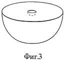
На фиг. 3 представлен общий вид устройства согласно настоящему изобретению, выполненного в форме полусферы. In FIG. 3 is a perspective view of a hemispherical device according to the present invention.
На фиг. 4 представлен общий вид устройства согласно настоящему изобретению в кубическом варианте. In FIG. 4 shows a general view of the device according to the present invention in a cubic version.
На фиг. 5 представлен общий вид устройства согласно настоящему изобретению в многосекционном (тандемном) варианте. In FIG. 5 shows a general view of the device according to the present invention in a multi-section (tandem) version.
На фиг. 6 показано влияние на выделение гидроморфона внутрь (in vitro) поли-(метилметакрилат) покрытия с центральным отверстием и без него в сравнении с выделением из непокрытого сплошного устройства. In FIG. Figure 6 shows the effect on the hydromorphone release in (in vitro) of a poly (methyl methacrylate) coating with and without a central hole in comparison with the release from an uncoated solid device.
На фиг. 7 показано влияние высоты цилиндрического устройства с поли-(метилметакрилат) покрытием и одинаковыми центральным отверстием и диаметром на выделение гидроморфона внутрь (in vitro). In FIG. Figure 7 shows the effect of the height of a cylindrical device with a poly (methyl methacrylate) coating and the same central bore and diameter on the release of hydromorphone inward (in vitro).
Фиг.8 иллюстрирует вплоть до четырех недель практически нулевую кинетику высвобождения гидроморфона внутрь (in vitro) из устройства выдачи согласно изобретению. Исключен начальный выброс высвобождения, осложняющий действие многих устройств контролируемой выдачи. Fig. 8 illustrates up to four weeks the practically zero kinetics of in-vitro release of hydromorphone from the dispenser of the invention. The initial release release, complicating the effect of many controlled delivery devices, is excluded.
Фиг. 9 показывает, что простая модификация устройств выдачи позволяет легко варьировать количество гидроморфона, высвобождаемого в час, и длительность высвобождения гидроморфона внутрь (in vitro). FIG. 9 shows that a simple modification of the dispensers makes it easy to vary the amount of hydromorphone released per hour and the duration of hydromorphone release inside (in vitro).
Фиг. 10 показывает, что два устройства выдачи, помещенные подкожно каждому из 5 кроликов, обеспечивают стабильно поддерживаемую концентрацию гидроморфона в плазме вплоть до четырех недель. Полученные концентрации в плазме находятся в интервале терапевтических доз для человека. FIG. 10 shows that two dispensers placed subcutaneously for each of the 5 rabbits provide a stably maintained plasma concentration of hydromorphone for up to four weeks. The resulting plasma concentrations are in the range of therapeutic doses for humans.
Фиг. 11 показывает прогнозируемое высвобождение гидроморфона из нескольких имплантированных устройств выдачи и наблюдаемую концентрацию гидроморфона в плазме кроликов во времени. Увеличение количества подкожно имплантированных устройств выдачи производит стабильное и прогнозированное увеличение концентрации гидроморфона в плазме кроликов. Общая токсичность 6 имплантантов (приблизительно 900 микрограмм гидроморфона в час) проявлялась в проходящих со временем депрессии и потере аппетита. FIG. 11 shows the predicted release of hydromorphone from several implanted delivery devices and the observed plasma concentration of hydromorphone in rabbits over time. An increase in the number of subcutaneously implanted delivery devices produces a stable and predicted increase in the concentration of hydromorphone in rabbit plasma. The total toxicity of 6 implants (approximately 900 micrograms of hydromorphone per hour) was manifested in depression and loss of appetite, which passed with time.
Фиг. 12 показывает, что подкожно имплантированные осмотические насосы, выделяющие приблизительно такое же количество гидроморфона в час, как и два устройства выдачи согласно изобретению, производят сравнимые концентрации гидроморфона в плазме кроликов. FIG. 12 shows that subcutaneously implanted osmotic pumps emitting approximately the same amount of hydromorphone per hour as the two dispensers of the invention produce comparable concentrations of hydromorphone in rabbit plasma.
Фиг. 13 показывает, что внутривенное введение 50% (600 мкг) и 100% (1200 мкг) суммарного количества гидроморфона, высвобождаемого двумя устройствами выдачи в течение 4 часов (типичный интервал дозировки для человека) приводит к пику концентрации в плазме кроликов в пределах одной минуты с последующим быстрым спадом до фона в течение нескольких часов. Пиковые концентрации гидроморфона четко проявлялись и были по крайней мере в 4 раза выше постоянных концентраций, получаемых с помощью устройства согласно изобретению. FIG. 13 shows that intravenous administration of 50% (600 μg) and 100% (1200 μg) of the total amount of hydromorphone released by two dispensers within 4 hours (typical dosage interval for humans) leads to a peak in plasma concentration of rabbits within one minute s followed by a rapid decline to the background within a few hours. Peak hydromorphone concentrations were clearly manifested and were at least 4 times higher than the constant concentrations obtained using the device according to the invention.
Подробное описание изобретения
Изобретение относится к биосовместимому невоспалительному и биологически не разлагаемому имплантируемому устройству 2, обеспечивающему контролируемое высвобождение сильнодействующего опиоида при подкожном имплантировании. Препарат, такой как гидроморфон, внедряют в матрицу 14 контролируемого выделения, например, полиэтиленвинилацетатную, а этот полимер покрывают биосовместимым и непроницаемым для гидроморфона полимером 12, например поли-(метилметакрилатом). Типичный имплантант имеет цилиндрическую геометрию с диаметром верхнего 4 и нижнего 6 сечений цилиндра, существенно превышающих высоту наружной стенки 8 цилиндра. Имплантант 2 перфорирован с образованием отверстия 10 в верхнем 4 и нижнем 6 сечении устройства 2 и непокрытой внутренней цилиндрической стенки 16. Такая структура обеспечивает постоянное высвобождение гидроморфона.DETAILED DESCRIPTION OF THE INVENTION
The invention relates to a biocompatible non-inflammatory and biodegradable
Устройство выдачи в соответствии с настоящим изобретением минимизирует возможность утечки наркотиков, улучшает соответствие предписанной дозировке и исключает дорогостоящую необходимость привлечения обслуживающего персонала и использования оборудования, а также потребность в дорогостоящих и не всегда доступных наружных катетерах и насосах для пациентов с раковыми болями, требующих продолжительного приема опиатов. Устройство выделяет постоянные количества гидроморфона in vitro и in vivo длительные периоды времени. Уровни гидроморфона в плазме, достигаемые с помощью настоящего изобретения, не являются заметно токсичными. Достигаемые стабильные уровни снижают токсичность и улучшают эффективность. Величина уровней гидроморфона в плазме и возможность их варьирования, достигаемые с помощью имплантантов, совершенно сходны с величинами, получаемыми с помощью осмотических насосов. The dispensing device in accordance with the present invention minimizes the possibility of drug leakage, improves compliance with the prescribed dosage and eliminates the costly need to attract attendants and use equipment, as well as the need for expensive and not always accessible external catheters and pumps for patients with cancer pain requiring long-term opiate use . The device releases constant amounts of hydromorphone in vitro and in vivo for long periods of time. Plasma hydromorphone levels achieved by the present invention are not markedly toxic. Achievable stable levels reduce toxicity and improve efficacy. The magnitude of the levels of hydromorphone in plasma and the possibility of their variation achieved with the help of implants are completely similar to the values obtained using osmotic pumps.
Покрытие 12 полимера ЭВА, содержащего препарат, эффективно исключает начальный пик выдачи препарата, наблюдаемый во многих других устройствах выдачи. В отличие от внутривенных инъекций уровни гидроморфона в плазме, получаемые после имплантации данного устройства, остаются стабильными и находятся в терапевтически допустимых пределах во всем интервале дозировки.
Изменением толщины (т. е. высоты стенки 8) и диаметра (т.е. диаметра верхнего 4 и нижнего 6 сечений) данных устройств, равно как и изменением количества имплантируемых устройств, достигается регулирование количества гидроморфона, выделяемого в час, и длительность выделения гидроморфона, а также значения получаемых уровней гидроморфона в плазме. By changing the thickness (i.e., wall height 8) and the diameter (i.e., the diameter of the upper 4 and lower 6 sections) of these devices, as well as by changing the number of implantable devices, the amount of hydromorphone released per hour and the duration of hydromorphone release are regulated , as well as the values of the resulting levels of hydromorphone in plasma.
Настоящее изобретение решает проблему "пикового эффекта" гидроморфона, исключает необходимость повторяющихся инъекций гидроморфона, обеспечивает долгосрочное купирование постоянных или хронических раковых болей, предоставляет средства лечения зависимости от опиоидных препаратов и препятствует наркотической опасности, т.е. утечке препарата, поскольку данная технология делает исключительно трудной получение гидроморфона из устройства согласно изобретению. The present invention solves the problem of the “peak effect” of hydromorphone, eliminates the need for repeated injections of hydromorphone, provides long-term relief of persistent or chronic cancer pains, provides treatments for opioid dependence and prevents narcotic danger, i.e. leakage of the drug, since this technology makes it extremely difficult to obtain hydromorphone from the device according to the invention.
Опиоид
Гидроморфон (включая гидрохлорид гидроморфона) представляет собой растворимый в воде сильный опиоид (в 6-7 раз сильнее морфина), разрешенный для подкожного использования и обычно прописываемый пациентам с раковыми болями. См. Валлнер и др., J. Clin. Pharmacol., 21:152-156, 1981; Бруера и др. , J. Natl. Cancer Inst., 80:1152-1154, 1988; Рейденберг и др., Clin. Pharmacol. Ther. , 44:376-382, 1988; Моулин и др. Can. Med. Assoc. J., 146: 891-897,1992; Моулин и др., Lancet, 337:465-468, 1991. Ограничения, накладываемые на размеры подкожных устройств выдачи, делают предпочтительным использование этого опиоида. Гидроморфон превосходно растворяется в воде и дает удовлетворительную картину высвобождения из гидрофобного полимера ЭВА.Opioid
Hydromorphone (including hydromorphone hydrochloride) is a water-soluble strong opioid (6-7 times stronger than morphine), allowed for subcutaneous use and usually prescribed for patients with cancer pain. See Wallner et al., J. Clin. Pharmacol., 21: 152-156, 1981; Bruer et al., J. Natl. Cancer Inst., 80: 1152-1154, 1988; Raidenberg et al., Clin. Pharmacol Ther. 44: 376-382, 1988; Moulin et al. Can. Med. Assoc. J., 146: 891-897.1992; Moulin et al., Lancet, 337: 465-468, 1991. Restrictions on the size of subcutaneous delivery devices make this opioid preferred. Hydromorphone is highly soluble in water and gives a satisfactory release pattern from the hydrophobic EVA polymer.
Матрица
Этиленвинилацетатный (ЭВА) сополимер является биосовместимым, невоспалительным и биологически неразлагаемым полимером. В настоящем изобретении используется биологически неразлагаемый полимер, чтобы обеспечивалась локализация. Далее, если какое-либо неблагоприятное обстоятельство заставит врача изъять имплантант 12 у пациента, он может быть вынут целым. Биологически разлагаемый имплантант со временем размягчается и теряет структурную целостность, делая возможность срочного удаления затруднительной, если не невозможной.Matrix
Ethylene vinyl acetate (EVA) copolymer is a biocompatible, non-inflammatory and biologically non-degradable polymer. The present invention uses a biodegradable polymer to provide localization. Further, if any adverse circumstance forces the doctor to remove the
Напитанные препаратом ЭВА матрицы успешно изготавливаются методом литья из раствора. Предварительно полимер растворяют в органическом растворителе, предпочтительно в растворителе с невысокой температурой кипения, например, в метиленхлориде или хлороформе, чтобы облегчит последующее удаление растворителя путем испарения. Концентрация полимера в растворе предпочтительно составляет от пяти до пятнадцати процентов по весу. Слишком слабый раствор приводит к образованию пузырьков при литье, а слишком концентрированный затрудняет диспергирование частиц препарата в растворе. Matrixes filled with EVA preparation are successfully made by casting from solution. The polymer is preliminarily dissolved in an organic solvent, preferably in a solvent with a low boiling point, for example, methylene chloride or chloroform, in order to facilitate subsequent removal of the solvent by evaporation. The concentration of polymer in the solution is preferably from five to fifteen percent by weight. Too weak a solution leads to the formation of bubbles during casting, and too concentrated makes it difficult to disperse the particles of the drug in the solution.
Препарат, вводимый в ЭВА-матрицу, растворяют, а в случае высокого уровня нагрузки препаратом диспергируют в растворе полимера. Препарат может высвобождаться путем проникания через полимерную фазу, а если препарат имеет слабую растворимость в полимере - путем диффузии через поры и каналы. Как только частицы препарата вблизи поверхности матрицы растворены и высвобождены, за ними остаются поры и каналы, через которые высвобождаются напитанные частицы препарата. Предпочтительно просеивать частицы препарата, поскольку их размер должен определяться размером пор и каналов. Однако это не абсолютно необходимо, если только частицы препарата не слиплись с образованием больших комков, например, размером в несколько сотен микрон. Разумно воспроизводимая кинетика выделения (т.е. почти постоянное выделение) достигается с микронизированными частицами имеющегося в продаже препарата. The drug introduced into the EVA matrix is dissolved, and in the case of a high level of load, the drug is dispersed in a polymer solution. The drug can be released by penetration through the polymer phase, and if the drug has poor solubility in the polymer, by diffusion through the pores and channels. As soon as the particles of the drug near the surface of the matrix are dissolved and released, behind them are the pores and channels through which the saturated particles of the drug are released. It is preferable to sift the particles of the drug, since their size should be determined by the size of the pores and channels. However, this is not absolutely necessary, unless the particles of the drug stick together to form large lumps, for example, several hundred microns in size. Reasonably reproducible kinetics of excretion (i.e., almost constant excretion) is achieved with micronized particles of a commercially available drug.
Раствор полимера с препаратом заливается в форму требуемого вида и размера. После медленного (чтобы предотвратить образование пузырьков) испарения растворителя молекулы или частицы препарата оказываются внедренными в полимерную матрицу. Литье обычно осуществляют при низкой температуре, чтобы предотвратить оседание частиц препарата во время испарения растворителя. Обычно полимерный раствор с препаратом наливают в форму, которую охлаждают до температуры, ниже температуры плавления растворителя. Раствор при этом быстро замерзает, обеспечивая однородное распределение частиц препарата в конечной матрице. The polymer solution with the drug is poured into the form of the desired type and size. After slow (to prevent the formation of bubbles) evaporation of the solvent, the molecules or particles of the drug are embedded in the polymer matrix. Casting is usually carried out at low temperature to prevent sedimentation of the particles of the drug during evaporation of the solvent. Typically, the polymer solution with the drug is poured into a mold, which is cooled to a temperature below the melting point of the solvent. The solution quickly freezes, ensuring a uniform distribution of the particles of the drug in the final matrix.
Другие материалы, которые могут быть использованы в качестве матрицы, включают силиконовый каучук, гидрогели, например, поли-(винилалкоголь) и поли-(гидроксиэтилметакрилат) с перекрестными связями. Other materials that can be used as the matrix include silicone rubber, hydrogels, for example, poly- (vinyl alcohol) and poly- (hydroxyethyl methacrylate) with cross-bonds.
Покрытие
Покрытие полимерной ЭВА-матрицы исключает потенциально опасный "пиковый эффект", обычно наблюдаемый в устройствах управляемой выдачи. Непокрытый полимер выделяет гидроморфон пиковым образом в первые два дня пользования препаратом, а затем снижает выдачу до минимума в последующие дни (см. фиг. 6). Это ключевая проблема в использовании подкожных имплантантов для выделения наркотических веществ, и настоящее изобретение решает эту проблему для гидроморфона. Для исключения пикового эффекта и получения более стабильной и постоянной кинетики выделения без манипулирования неоднородным распределением препарата в матрице ЭВА-полимер 14, содержащий препарат, весь покрыт, за исключением небольшого отверстия 10 в середине, покрытием 12, непроницаемым для препарата и биосовместимым с тканями организма, вызывающим лишь минимальную инкапсуляцию имплантанта 12 уплотненной тканью. Предпочтительно с гидроморфоном используется костный цемент поли-(метилметакрилат). Покрытие 12 должно иметь достаточную толщину с учетом предполагаемого времени службы имплантанта, как правило, около 100 микрон.Coating
Coating the polymer EVA matrix eliminates the potentially dangerous "peak effect" typically seen in controlled dispensing devices. An uncoated polymer excrets hydromorphone in a peak manner in the first two days of use of the drug, and then reduces delivery to a minimum in the following days (see Fig. 6). This is a key problem in using subcutaneous implants to isolate narcotic substances, and the present invention solves this problem for hydromorphone. To eliminate the peak effect and obtain a more stable and constant release kinetics without manipulating the inhomogeneous distribution of the drug in the
Покрытие матрицы осуществляют предпочтительно методом покрытия погружением. Например, покрытие осуществляют, надевая диск на иглу от шприца нужного диаметра и погружая его в раствор полимера. По нанесении нескольких слоев повторным погружением и высушиванием иглу удаляют, открывая непокрытое цилиндрическое отверстие. Напротив, отверстие по оси матрицы может быть выполнено и после покрытия матрицы. Препарат высвобождается только через это отверстие. При данной конфигурации получают почти постоянное высвобождение препарата при отсутствии градиента концентрации препарата в ЭВА-полимере. The coating of the matrix is preferably carried out by immersion coating. For example, the coating is carried out by putting a disk on a needle from a syringe of the desired diameter and immersing it in a polymer solution. After applying several layers by repeated immersion and drying, the needle is removed, opening an uncovered cylindrical hole. On the contrary, the hole along the axis of the matrix can be made after coating the matrix. The drug is released only through this hole. With this configuration, an almost constant release of the drug is obtained in the absence of a concentration gradient of the drug in the EVA polymer.
Материал покрытия непроницаем для гидроморфона и не разлагается биологически (пренебрежимые расщепление структуры полимера и потеря массы за двухмесячный период), он растворим в растворителе, который не является хорошим растворителем для выбранной матрицы. В противном случае матрица частично растворялась бы в процессе покрытия погружением и какая-то часть молекул или частичек препарата могла бы внедряться в покрытие. Параметр растворимости поли-(метилметакрилата) лежит в интервале 9-9,5 [кал/см3]0,5. Подходящим покрытием для полимера ЭВА-гидроморфон могут быть и другие полимеры, непроницаемые для гидроморфона и имеющие тот же параметр растворимости.The coating material is impervious to hydromorphone and does not biodegrade (negligible splitting of the polymer structure and weight loss over a two-month period), it is soluble in a solvent that is not a good solvent for the selected matrix. Otherwise, the matrix would partially dissolve during the coating process by immersion, and some part of the molecules or particles of the preparation could be incorporated into the coating. The solubility parameter of poly- (methyl methacrylate) is in the range of 9-9.5 [cal / cm3 ]0.5 . Other polymers impervious to hydromorphone and having the same solubility parameter may be a suitable coating for the EVA-hydromorphone polymer.
Нагрузка препаратом
Отношение гидроморфона к полимеру ЭВА может составлять от 10 до 90 процентов гидроморфона по весу, предпочтительно от 30 до 70 процентов. Смесь препарата с ЭВА однородна. Большие уровни концентрации гидроморфона используют обычно при маленьких отверстиях 10 (см. ниже).Drug load
The ratio of hydromorphone to EVA polymer may be from 10 to 90 percent hydromorphone by weight, preferably from 30 to 70 percent. The mixture of the drug with EVA is homogeneous. Large levels of hydromorphone are usually used with small openings of 10 (see below).
Геометрия имплантанта
Предпочтительно использовать имплантант цилиндрической формы (см. фиг.1 и 2). Могут использоваться и другие геометрии, в которых расстояние между непокрытой стенкой и покрытой стенкой (противоположной непокрытой стенке) является постоянным или существенно постоянным (+-20%) по имплантанту, например, полусфера с непокрытой стенкой также в форме полусферы (фиг.3) или куб с квадратным отверстием, простирающимся на всю высоту (толщину) куба (фиг. 4). Эти геометрии обеспечивают почти постоянную скорость выделения в течение срока службы имплантанта.Implant geometry
It is preferable to use a cylindrical implant (see figures 1 and 2). Other geometries can be used in which the distance between the uncoated wall and the coated wall (opposite the uncoated wall) is constant or substantially constant (+ -20%) over the implant, for example, a hemisphere with an uncoated wall also in the form of a hemisphere (Fig. 3) or cube with a square hole extending to the entire height (thickness) of the cube (Fig. 4). These geometries provide an almost constant release rate over the life of the implant.
Цилиндрический имплантант выполняют диаметром от 5 до 100 мм, предпочтительно от 10 до 25 мм и высотой (толщиной) от 1 до 20 мм, предпочтительно от 1 до 2 мм. A cylindrical implant is performed with a diameter of 5 to 100 mm, preferably 10 to 25 mm, and a height (thickness) of 1 to 20 mm, preferably 1 to 2 mm.
Одно круглое или практически круглое отверстие 10 диаметром от 50 микрон до 3 мм, предпочтительно от 0,5 до 1,5 мм образует внутреннюю цилиндрическую непокрытую область 16, через которую выделяется препарат. Площадь отверстия для устройств цилиндрической, кубической, полусферической или иной формы должна составлять менее 10%, а предпочтительно менее 1% от площади поверхности верха устройства. One round or almost
Многосекционное устройство согласно изобретению образуют наложением цилиндрических имплантантов (или имплантантов других форм), т.е. когда оси отдельных устройств перпендикулярны оси многосекционного устройства (см. фиг. 5). При наложении области контакта (т.е. которыми секции соединяются) отдельных компонент (т.е. отдельные цилиндры или кубики) могут быть покрытыми или непокрытыми. The multi-section device according to the invention is formed by the application of cylindrical implants (or other forms of implants), i.e. when the axes of the individual devices are perpendicular to the axis of the multi-section device (see Fig. 5). When the contact area (i.e., by which the sections are connected) is superimposed, the individual components (i.e., individual cylinders or cubes) can be coated or uncovered.
В другом варианте многосекционное устройство образуют так, что оси отдельных устройств (например, цилиндрических или кубических) совпадают с осью многосекционного устройства. Для соединения отдельных устройств таким образом и обеспечения выделения препарата, индивидуальные устройства отделены одно от другого разделителями. В одном из вариантов разделители представляют собой один или несколько элементов, тянущихся параллельно оси многосекционного устройства, причем каждый элемент скреплен с наружной стенкой каждого отдельного устройства. В другом варианте между отдельными устройствами размещают пористые элементы (обеспечивающие также соединение устройств). Еще в одном варианте для образования многосекционного устройства используют небольшие разделители, контактирующие (и соединяющие) низ одного отдельного устройства и верх соседнего устройства. Предпочтительно изготавливать разделители из материала покрытия, т.е. из поли-(метилметакрилата). В другом варианте в качестве разделителя может использоваться поволока, покрытая этим материалом. In another embodiment, the multisection device is formed so that the axes of the individual devices (for example, cylindrical or cubic) coincide with the axis of the multisection device. To connect the individual devices in this way and to ensure the release of the drug, the individual devices are separated from one another by separators. In one embodiment, the spacers are one or more elements extending parallel to the axis of the multi-section device, with each element bonded to the outer wall of each individual device. In another embodiment, porous elements (also providing connection of devices) are placed between the individual devices. In yet another embodiment, small dividers are used to form a multi-section device, contacting (and connecting) the bottom of one separate device and the top of an adjacent device. It is preferable to make the separators from the coating material, i.e. from poly- (methyl methacrylate). In another embodiment, a wire coated with this material may be used as a separator.
Введение имплантанта
Устройство согласно изобретению 12 имплантируется подкожно, предпочтительно в верхнюю часть руки или в брюшные области, с использованием приемов, известных специалистам в данной сфере. Дозировку выбирают безопасной и эффективной для конкретного пациента. Слишком низкая дозировка приводит к недостаточному успокоению болей, а слишком высокая может привести к состоянию депрессии и к респираторной недостаточности. Например, при лечении раковых болей, используя визуальную аналоговую шкалу для измерения болей и титрируя гидроморфон в соответствии с субъективным уровнем болей пациента, выбирают для него диск с подходящей дозировкой.Implant insertion
The device according to the
Для лечения раковых болей обычно используют имплантанты, выделяющие от 0,1 до 25 мг/ч, предпочтительно от 0,1 до 10 мг/ч (например, 0,25 мг/ч, 1 мг/ч и 4 мг/ч). Пациенту обычно вживляют от одного до трех имплантантов. Дозировку (мг/ч) предпочтительно регулируют высотой цилиндрического диска или диаметром отверстия, т. е. увеличение высоты или диаметра отверстия означает большие скорости выделения (большая площадь непокрытой поверхности 16 подвергается воздействию внешней среды). См. фиг. 7. Применение нескольких устройств, или раздельных, или наложенных один на другой, как описано выше, увеличивает дозировку. Кроме того, дозировку увеличивает повышенная нагрузка препаратом. For the treatment of cancer pain, implants are usually used that release from 0.1 to 25 mg / h, preferably from 0.1 to 10 mg / h (for example, 0.25 mg / h, 1 mg / h and 4 mg / h). Patients usually have one to three implants implanted. The dosage (mg / h) is preferably controlled by the height of the cylindrical disk or by the diameter of the hole, i.e., increasing the height or diameter of the hole means high release rates (a large area of the uncovered
Для лечения опиоидной зависимости обычно используют имплантанты, выделяющие подкожно от 0,1 до 0,5 мг/ч гидроморфона. Обсуждение метадоновой поддержки см. Страйн и др., Ann. Intern. Med., 119:23-27, 1993 и Герштейн и Левин, N. Engl. J. Med., 323, 844-848, 1990. For the treatment of opioid dependence, implants are usually used that emit subcutaneously from 0.1 to 0.5 mg / h of hydromorphone. For a discussion of methadone support, see Strain et al., Ann. Intern. Med., 119: 23-27, 1993; and Gerstein and Levin, N. Engl. J. Med. 323, 844-848, 1990.
Устройство согласно изобретению может контролируемо выделять гидроморфон для поддержания желательного уровня в крови от двух недель до свыше шести месяцев. Время действия имплантанта предпочтительно регулируется подбором его диаметра (чем больше диаметр, тем больше время действия). Нагрузка препаратом, размер отверстия и высота также влияют на время действия имплантанта. Для лечения раковых болей предпочтителен имплантант, рассчитанный на четыре недели, поскольку онкологи обычно проводят осмотр своих раковых пациентов по крайней мере раз в месяц. Болевые ощущения могут возрастать в результате прогрессирующей опухоли, что потребует увеличения дозировки. Более того, существует привыкание к гидроморфону, так что большая дозировка может потребоваться для купирования болей той же силы в последующий месяц. The device according to the invention can controlledly release hydromorphone to maintain the desired blood level from two weeks to over six months. The duration of the implant is preferably controlled by the selection of its diameter (the larger the diameter, the longer the duration of action). The drug load, hole size and height also affect the duration of the implant. For treatment of cancer pain, a four-week implant is preferred, as oncologists usually examine their cancer patients at least once a month. Pain can increase as a result of a progressive tumor, which will require an increase in dosage. Moreover, there is addiction to hydromorphone, so a large dosage may be required to relieve pain of the same strength in the next month.
Устройство согласно изобретению обеспечивает почти постоянное выделение гидроморфона. "Почти постоянное" здесь понимается как плюс-минус пятикратное (500%), предпочтительно плюс-минус двухкратное (200%) изменение, а наиболее предпочтительно плюс-минус однократное (100%) изменение скорости выделения (in vivo или in vitro). Более чем пятикратное изменение приводит к "пиковому эффекту", который мог бы вызвать угрозу или даже смерть реципиента. The device according to the invention provides an almost constant release of hydromorphone. “Almost constant” is understood here as plus or minus five times (500%), preferably plus or minus twice (200%), and most preferably plus or minus once (100%), the change in release rate (in vivo or in vitro). More than five-fold change leads to a “peak effect” that could cause a threat or even death to the recipient.
В одном из вариантов осуществления изобретения выделение препарата из имплантанта сопровождают дополнительным приемом (внутрь, ректальным), когда пациент испытывает усиление болей. In one of the embodiments of the invention, the release of the drug from the implant is accompanied by an additional intake (by mouth, rectal), when the patient experiences increased pain.
Лечение болей
Имплантант согласно изобретению может быть использован во всех случаях, когда показан внутренний прием опиатов. Внутренний прием опиатов бывает показан примерно для 85% пациентов с раковыми болями. Использование изобретения особенно предпочтительно, когда прием внутрь не подходит или недоступен или когда проявляется токсичность, связанная с прерывистым приемом. Имплантант согласно изобретению может применяться при лечении раковых болей у человека и у других животных. Имплантант может найти применение при лечении других типов хронических болей, как, например, хронических тяжелых болей, связанных дегенеративными мускулоскелетными заболеваниями или заболеваниями нервной системы.Pain treatment
The implant according to the invention can be used in all cases where the internal administration of opiates is indicated. Internal opiate administration is indicated for approximately 85% of patients with cancer pain. The use of the invention is particularly preferred when ingestion is not suitable or unavailable or when toxicity associated with intermittent administration is manifested. The implant according to the invention can be used in the treatment of cancer pain in humans and other animals. The implant may find application in the treatment of other types of chronic pain, such as chronic severe pain associated with degenerative musculoskeletal diseases or diseases of the nervous system.
Лечение опиоидной зависимости
Имплантант согласно изобретению может быть полезен при лечении зависимости от опиоидных препаратов (например, героина). Гидроморфон, даваемый в малых дозах, продолжительное время является эффективным средством лечения наркомании. Имплантант согласно изобретению может быть использован в любой ситуации, когда может быть использована программа метадоновой поддержки. Имплантант обеспечивает соблюдение низкой дозировки препарата в течение длительных периодов, не полагаясь на соблюдение пациентом графика дозировки при приеме внутрь. Далее, при использовании имплантанта препарат предоставляется в такой форме, которая практически исключает возможность нелегального использования.Opioid addiction treatment
An implant according to the invention may be useful in the treatment of addiction to opioid drugs (e.g. heroin). Hydromorphone, given in small doses, has long been an effective treatment for drug addiction. The implant according to the invention can be used in any situation where a methadone support program can be used. The implant ensures compliance with the low dosage of the drug for long periods, without relying on the patient's adherence to the dosage schedule for oral administration. Further, when using an implant, the drug is provided in a form that virtually eliminates the possibility of illegal use.
Следующие примеры иллюстрируют, но не ограничивают композиции и способы согласно настоящему изобретению. В рамках настоящего изобретения находятся и другие подходящие модификации и приспособления к различным условиям и параметрам, обычно встречающимся в клинической терапии, очевидные для специалистов в данной области. The following examples illustrate but do not limit the compositions and methods of the present invention. Other suitable modifications and adaptations to various conditions and parameters commonly found in clinical therapy, which are obvious to those skilled in the art, are within the scope of the present invention.
ПРИМЕРЫ
Пример 1
Формирование полимера
В этих экспериментах для формирования гидроморфонного имплантанта использовался метод низкотемпературного литья раствора. ЭВА (с молекулярным весом около 250.000)(Elvax-40W фирмы Дюпон) с 40% по весу винилацетата промывался в воде и растворялся в метиленхлориде (CH2CL2) до получения 10% раствора. Для удаления загрязнений и противоокислителей полимер мог очищаться экстрагированием с дистиллированной водой в течение трех суток в экстракторе Soxhlet, с последующим экстрагированием с ацетоном еще четверо суток. Матрицы с включением гидроморфона приготовлялись литьем раствора. 10% (веса на объем) раствор ЭВА получался при растворении 2 г чистого ЭВА в 20 мл метиленхлорида.EXAMPLES
Example 1
Polymer formation
In these experiments, a low-temperature solution casting method was used to form a hydromorphone implant. EVA (with a molecular weight of about 250,000) (Elvax-40W from DuPont) with 40% by weight vinyl acetate was washed in water and dissolved in methylene chloride (CH2 CL2 ) to obtain a 10% solution. To remove contaminants and antioxidants, the polymer could be purified by extraction with distilled water for three days in a Soxhlet extractor, followed by extraction with acetone for another four days. Matrices with the inclusion of hydromorphone were prepared by casting a solution. A 10% (weight per volume) EVA solution was obtained by dissolving 2 g of pure EVA in 20 ml of methylene chloride.
К 10% ЭВА при комнатной температуре добавлялся непросеянный порошок гидрохлорида гидроморфона (от Knoll Pharmaceuticals) до образования 50% по весу раствора, который затем подвергался магнитному перемешиванию примерно 10 минут. Порошок гидроморфона мог механически просеиваться до определенного размера частиц и образовывать суспензию в растворе ЭВА. Видимые макроскопические комочки, остающиеся в этом растворе, удалялись с помощью стеклянной мешалки. To a 10% EVA at room temperature, an unsintered powder of hydromorphone hydrochloride (from Knoll Pharmaceuticals) was added until a 50% by weight solution was formed, which was then magnetically mixed for about 10 minutes. Hydromorphone powder could be mechanically sieved to a certain particle size and form a suspension in an EVA solution. Visible macroscopic lumps remaining in this solution were removed using a glass stirrer.
Однородная суспензия быстро наносилась на рекристаллизационное блюдце 14 см в диаметре, предварительно охлажденное сухим льдом до -78oC. Блюдце покрывалось для уменьшения конденсации. После затвердевания, которое проходило в пределах 15 минут, образец высушивался в течение двух 48 часовых периодов: в первом при -20oC, а затем в вакууме при комнатной температуре.A homogeneous suspension was quickly applied to a
Из высушенной круглой пластины перовым сверлом N 7 вырезалось примерно 15 полимерных дисков с гидроморфоном 0,27 см толщиной и 1,05 см диаметром. Для экспериментов, показывающих возможность вариации количества гидромофона, высвобождаемого в час, изготавливались полимерные диски двойной и тройной толщины (соответственно 0,55-0,65 см и 0,83 см) путем удвоения или утроения количества смеси ЭВА с гидроморфоном, помещаемой в форму. Полимерные диски 2,13 см диаметром для демонстрации вариации длительности высвобождения препарата вырезались из высушенной пластины ЭВА с гидроморфоном перовым сверлом N 15. About 15 polymer disks with hydromorphone 0.27 cm thick and 1.05 cm in diameter were cut from a dried round plate with a N 7 drill. For experiments showing the possibility of varying the amount of hydrophone released per hour, polymeric disks of double and triple thickness (0.55-0.65 cm and 0.83 cm, respectively) were made by doubling or tripling the amount of a mixture of EVA with hydromorphone placed in the mold. 2.13 cm diameter polymer discs were cut out from a dried EVA plate with hydromorphone with a N15 drill bit to demonstrate variation in drug release duration.
Пример 2
Покрытие полимера
Перед покрытием каждый полимерный диск ЭВА с гидроморфоном приблизительно по центру его круговой стороны прокалывался полой стальной иглой 18 размера. Затем каждый полимерный диск покрывался путем погружения в 10% раствор поли-(метилметакрилата) с молекулярным весом около 996000 (фирмы Aldrich) в ацетоне на 10 секунд. Покрытие из поли-(метилметакрилата) высушивалось на воздухе при комнатной температуре в течение 24 часов, после чего игла удалялась, полимерный диск переворачивался и игла вставлялась с другой стороны. Процедура повторялась еще два раза для получения непроницаемого покрытия из поли- (метилметакрилата) тройной толщины, которое полностью окружало полимер за исключением цилиндрического отверстия 18 размера (примерно, 1,25 мм в диаметре), проходящего по центру каждого полимерного диска, подобно отверстию в пуговице. Толщина покрытия достигалась примерно 100-200 мкм. Параметр растворимости материала покрытия составлял 9-9,5 [кал/см3]0,5.Example 2
Polymer coating
Before coating, each EVA polymer disk with hydromorphone was punctured approximately 18 in the center of its circular side by a hollow steel needle. Then, each polymer disk was coated by immersion in a 10% solution of poly- (methyl methacrylate) with a molecular weight of about 996,000 (Aldrich) in acetone for 10 seconds. The poly (methyl methacrylate) coating was dried in air at room temperature for 24 hours, after which the needle was removed, the polymer disk was turned over and the needle was inserted from the other side. The procedure was repeated two more times to obtain an impermeable coating of poly- (methyl methacrylate) triple thickness, which completely surrounded the polymer with the exception of a cylindrical hole of size 18 (approximately 1.25 mm in diameter) passing through the center of each polymer disk, like a hole in a button . The coating thickness was reached about 100-200 microns. The solubility parameter of the coating material was 9-9.5 [cal / cm3 ]0.5 .
Пример 3
Тест на выделение in vitro
Выделение гидроморфона из покрытого полимера ЭВА с гидроморфоном испытывалось in vitro при помещении каждого образца полимера в стеклянный сцинцилляционный пузырек, содержащий в качество буфера 10 мл 0,1 моль фосфата натрия с pH 7,4, подогретого до 37oC. Все пузырьки помещались в водяную баню при 37oC. С соответствующими интервалами буфер из каждого пузырька отбирался и помещался в пробирку центрифуги, а в пузырек добавлялись 10 мл свежего буферного фосфата при 37oC. Образцы совместно периодически измерялись ультрафиолетовым спектрофотометром на длине волны 254 нм в параллельных кюветах с приведением к нулю по чистому 0,1 моль фосфатному буферу с pH 7,4. В каждый опыт включались измерения стандартных концентраций гидроморфона и при необходимости образцы разбавлялись буфером, так что их абсорбция могла измеряться на линейной части градуировочной кривой.Example 3
In Vitro Isolation Test
The isolation of hydromorphone from the coated EVA polymer with hydromorphone was tested in vitro by placing each polymer sample in a glass scintillation vial containing 10 ml of 0.1 mole sodium phosphate pH 7.4 heated to 37° C as a buffer. All bubbles were placed in water a bath at 37o C. At appropriate intervals, a buffer was taken from each vial and placed in a centrifuge tube, and 10 ml of fresh buffer phosphate was added to the vial at 37o C. The samples were periodically measured together with an ultraviolet spectrophotometer at a wavelength of 254 nm in parallel cuvettes, leading to zero in a pure 0.1 mol phosphate buffer with a pH of 7.4. Measurements of standard concentrations of hydromorphone were included in each experiment and, if necessary, the samples were diluted with a buffer, so that their absorption could be measured on the linear part of the calibration curve.
Покрытые полимеры ЭВА с гидроморфоном выделяли гидроморфон с постоянной средней скоростью 164 мкг/час начиная с четвертого дня после начала высвобождения in vitro более чем в течение 4 недель. Покрытие этих полимеров непроницаемым слоем поли-(метилметакрилата), за исключением центрального отверстия на всю толщину, исключает начальный "пик", который осложняет использование многих других устройств с регулируемым выделением. Вариация количества гидроморфона, выделяемого в час, и длительности выделения препарата легко достигается изменением толщины и диаметра покрытого полимера ЭВА с гидроморфоном. См. фиг. 9. Coated EVA polymers with hydromorphone released hydromorphone at a constant average rate of 164 μg / h starting on the fourth day after the start of in vitro release for more than 4 weeks. Coating these polymers with an impermeable layer of poly (methyl methacrylate), with the exception of the entire central hole, eliminates the initial "peak", which complicates the use of many other controlled release devices. Variation in the amount of hydromorphone released per hour and the duration of drug release is easily achieved by changing the thickness and diameter of the coated EVA polymer with hydromorphone. See FIG. nine.
Примеры in vivo
Пример 4A
Высвобождение из полимера
Два покрытых полимера ЭВА с гидроморфоном помещались под общим наркозом в подкожные мешки каждого из пяти взрослых кроликов. Каждый кролик содержался в клетке и получал кроличий корм и воду без ограничений. У каждого животного отбирались образцы крови до имплантации и с определенными интервалами вплоть до 6 недель после помещения. Образцы крови центрифугировались и плазма отбиралась и хранилась в холодильнике при -20oC. Образцы плазмы измерялись совместно с использованием имеющегося в продаже радиоиммунометра опиатов и эталонных гидроморфонных образцов. Для демонстрации соотношения количества имплантированного гидроморфона и результирующим уровнем препарата в плазме крови два дополнительных кролика получили три порции по два полимера, последовательно имплантированные с интервалами примерно в одну неделю. Ранее имплантированные покрытые полимеры при этом сохранялись, так что эти кролики имели сначала 2, затем 4 и затем 6 полимеров, одновременно выделяющих гидроморфон. Пробы крови отбирались и плазма проверялась, как описано выше.In vivo Examples
Example 4A
Polymer release
Two coated EVA polymers with hydromorphone were placed under general anesthesia in the subcutaneous sacs of each of five adult rabbits. Each rabbit was kept in a cage and received rabbit food and water without restrictions. Blood samples were taken from each animal prior to implantation and at specific intervals up to 6 weeks after placement. Blood samples were centrifuged and plasma was taken and stored in a refrigerator at -20o C. Plasma samples were measured using a commercially available radio opioid immunometer and reference hydromorphone samples. To demonstrate the ratio of the amount of hydromorphone implanted and the resulting plasma level of the drug, two additional rabbits received three servings of two polymers, successively implanted at approximately one week intervals. The previously implanted coated polymers were retained, so that these rabbits first had 2, then 4, and then 6 polymers that simultaneously emitted hydromorphone. Blood samples were taken and plasma was checked as described above.
Два устройства, одновременно имплантированные кроликам, производят стабильные и длительные уровни гидроморфона в плазме крови в 23-27 нг/мл до четырех недель (см. фиг.10). Увеличение количества подкожных имплантантов у отдельного кролика производит длительное и предсказуемое увеличение концентрации гидроморфона в плазме крови этого кролика (см. фиг. 11). В этих экспериментах на животных не наблюдалось заметной токсичности, за исключением слабой проходящей депрессии после имплантирования двух устройств и увеличения депрессии с уменьшением аппетита, также проходящих после имплантирования большего количества устройств. Two devices simultaneously implanted in rabbits produce stable and prolonged plasma hydromorphone levels of 23-27 ng / ml for up to four weeks (see FIG. 10). An increase in the number of subcutaneous implants in an individual rabbit produces a long-term and predictable increase in the concentration of hydromorphone in the blood plasma of this rabbit (see Fig. 11). No significant toxicity was observed in these animal experiments, with the exception of a mild, passing depression after implanting two devices and an increase in depression with a decrease in appetite, also passing after implanting more devices.
Пример 4B
Выдача осмотическим насосом
Для подтверждения уровней гидроморфона в плазме крови кроликов, измеренных после имплантирования, использовались осмотические насосы фирмы Alzet, выдающие примерно то же самое количество гидроморфона в час, что и два устройства согласно изобретению. Каждому из четырех кроликов под общим наркозом помещали подкожно осмотический насос Alzet (средняя скорость накачки равнялась 2,08 мкл/ч, средний объем заполнения - 2073 мкл); взятие образцов крови осуществлялось, как описано выше. Подготовка плазмы и ее анализ осуществлялся так же, как описано выше.Example 4B
Issuance by osmotic pump
To confirm the levels of hydromorphone in the blood plasma of rabbits, measured after implantation, Alzet osmotic pumps were used, which produced approximately the same amount of hydromorphone per hour as the two devices according to the invention. Alzet osmotic pump was placed subcutaneously for each of the four rabbits under general anesthesia (average pumping rate was 2.08 μl / h, average filling volume was 2073 μl); blood sampling was carried out as described above. Plasma preparation and analysis was carried out as described above.
Осмотические насосы Alzet, выделяющие 297 мкг/ч гидроморфона, обеспечивали средние уровни гидроморфона в плазме крови кроликов от 28 до 51 нг/мл в течение двух недель (см. фиг. 12). Alzet osmotic pumps, emitting 297 μg / h of hydromorphone, provided average rabbit plasma hydromorphone levels of 28 to 51 ng / ml for two weeks (see FIG. 12).
Пример 4C
Внутривенные инъекции гидроморфона
Еще двум кроликам внутривенно вводилось по одной дозе в 600 мкг и 1200 мкг, соответственно (что равнялось 1/2 и 1 дозе суммарного количества, выделяемого подкожно полимерами или насосами в течение 4 часов, типичному интервалу дозирования для человека); образцы отбирались с интервалами 0, 1, 2, 5, 10, 20, 30, 45, 60, 90, 120, 150, 180, 210 и 240 минут.Example 4C
Intravenous injection of hydromorphone
Two more rabbits were injected intravenously with a single dose of 600 μg and 1200 μg, respectively (which was 1/2 and 1 dose of the total amount excreted subcutaneously with polymers or pumps for 4 hours, a typical dosing interval for humans); samples were taken at intervals of 0, 1, 2, 5, 10, 20, 30, 45, 60, 90, 120, 150, 180, 210, and 240 minutes.
Шестисот и 1200 мкг инъекции гидроморфона производили пиковые уровни гидроморфона в плазме крови кроликов почти в 200 и свыше 250 нг/мл за одну минуту с быстрым спадом до фона в течение 4 часов (см. фиг. 13). Six hundred and 1200 μg hydromorphone injections produced peak hydromorphone levels in rabbit plasma at almost 200 and over 250 ng / ml in one minute with a rapid decline to the background within 4 hours (see Fig. 13).
Для сведующих в данной области очевидно, что оба настоящих изобретения, раскрытое здесь устройство и соответствующая система могут модифицироваться и дополняться без отхода от раскрытого изобретения. For those skilled in the art, it is obvious that both of the present invention, the apparatus disclosed herein and the corresponding system can be modified and supplemented without departing from the disclosed invention.
Claims (20)
Translated fromRussianApplications Claiming Priority (2)
| Application Number | Priority Date | Filing Date | Title |
|---|---|---|---|
| US08/264,689US5633000A (en) | 1994-06-23 | 1994-06-23 | Subcutaneous implant |
| US264689 | 1994-06-23 |
Publications (2)
| Publication Number | Publication Date |
|---|---|
| RU97100780A RU97100780A (en) | 1999-02-20 |
| RU2157246C2true RU2157246C2 (en) | 2000-10-10 |
Family
ID=23007180
Family Applications (1)
| Application Number | Title | Priority Date | Filing Date |
|---|---|---|---|
| RU97100780/14ARU2157246C2 (en) | 1994-06-23 | 1995-06-20 | Subcutaneous implant |
Country Status (16)
| Country | Link |
|---|---|
| US (3) | US5633000A (en) |
| EP (1) | EP0766538B1 (en) |
| JP (1) | JP3964933B2 (en) |
| CN (1) | CN1212809C (en) |
| AT (1) | ATE309761T1 (en) |
| AU (1) | AU711323B2 (en) |
| BR (1) | BR9508124A (en) |
| CA (1) | CA2193627C (en) |
| DE (1) | DE69534622T2 (en) |
| IL (1) | IL114244A (en) |
| MX (1) | MX9606582A (en) |
| NZ (1) | NZ288988A (en) |
| RU (1) | RU2157246C2 (en) |
| TW (1) | TW421597B (en) |
| WO (1) | WO1996000047A1 (en) |
| ZA (1) | ZA955168B (en) |
Cited By (3)
| Publication number | Priority date | Publication date | Assignee | Title |
|---|---|---|---|---|
| RU2279928C2 (en)* | 2000-12-04 | 2006-07-20 | Уве Эмиль Кристиан ГРЮССНЕР | Device and method (versions) of applying coat made from active substance on article |
| RU2322979C2 (en)* | 2002-09-29 | 2008-04-27 | Тианцзин Тейсли Груп Ко., Лтд. | Temosolomide-containing system for controlled release |
| US11400186B2 (en) | 2016-05-12 | 2022-08-02 | Merck Sharp & Dohme Llc | Drug delivery system for the delivery of antiviral agents |
Families Citing this family (127)
| Publication number | Priority date | Publication date | Assignee | Title |
|---|---|---|---|---|
| US5633000A (en)* | 1994-06-23 | 1997-05-27 | Axxia Technologies | Subcutaneous implant |
| US20040024006A1 (en)* | 1996-05-06 | 2004-02-05 | Simon David Lew | Opioid pharmaceutical compositions |
| US20030211157A1 (en)* | 1996-05-06 | 2003-11-13 | Simon David Lew | Semi-sol delivery blend for water soluble molecules |
| US20060052757A1 (en)* | 1996-06-04 | 2006-03-09 | Vance Products Incorporated, D/B/A Cook Urological Incorporated | Implantable medical device with analgesic or anesthetic |
| US20070161968A1 (en)* | 1996-06-04 | 2007-07-12 | Vance Products Inc., Dba Cood Urological Inc. | Implantable medical device with pharmacologically active ingredient |
| US20070161967A1 (en)* | 1996-06-04 | 2007-07-12 | Vance Products Inc., Dba Cook Urological Inc. | Implantable medical device with pharmacologically active ingredient |
| US5919473A (en)* | 1997-05-12 | 1999-07-06 | Elkhoury; George F. | Methods and devices for delivering opioid analgesics to wounds via a subdermal implant |
| NL1008929C2 (en) | 1998-04-20 | 1999-10-21 | Vhp Ugchelen Bv | Substrate made of paper provided with an integrated circuit. |
| NZ331201A (en)* | 1998-07-30 | 2001-02-23 | Bomac Lab Ltd | Slow release implants comprising an insoluble polymeric material in the form of a porous network containing vitamin B12 dispersed within |
| US6835194B2 (en)* | 1999-03-18 | 2004-12-28 | Durect Corporation | Implantable devices and methods for treatment of pain by delivery of fentanyl and fentanyl congeners |
| US6541021B1 (en)* | 1999-03-18 | 2003-04-01 | Durect Corporation | Devices and methods for pain management |
| JP2002542183A (en) | 1999-04-16 | 2002-12-10 | ノボ ノルディスク アクティーゼルスカブ | Moldable dry pharmaceutical formulation |
| CA2382577C (en)* | 1999-08-27 | 2008-01-22 | Southern Research Institute | Injectable buprenorphine microparticle compositions and their use |
| US7074803B2 (en)* | 2001-03-02 | 2006-07-11 | Durect Corporation | Opioid formulations |
| US6569152B2 (en)* | 2000-03-21 | 2003-05-27 | Farrington Pharmaceuticals, Llc | Sustained release delivery systems for solutes |
| AU2001281729A1 (en)* | 2000-07-14 | 2002-01-30 | Novo-Nordisk A/S | Method of moulding a pharmaceutical composition in a packaging material |
| DE10105592A1 (en) | 2001-02-06 | 2002-08-08 | Achim Goepferich | Placeholder for drug release in the frontal sinus |
| KR100954741B1 (en) | 2001-06-29 | 2010-04-27 | 메드 그래프트 마이크로텍 인코포레이션 | Biodegradable Injectable Implants and Related Methods and Uses |
| US20030129234A1 (en)* | 2001-07-06 | 2003-07-10 | Penwest Pharmaceuticals Company | Methods of making sustained release formulations of oxymorphone |
| AU2002318211B2 (en)* | 2001-07-06 | 2007-07-12 | Endo Pharmaceuticals, Inc. | Parenteral administration of 6-hydroxy-oxymorphone for use as an analgesic |
| US8329216B2 (en)* | 2001-07-06 | 2012-12-11 | Endo Pharmaceuticals Inc. | Oxymorphone controlled release formulations |
| JP2005523876A (en)* | 2001-09-26 | 2005-08-11 | ペンウェスト ファーマシューティカルズ カンパニー | Opioid formulations with reduced potential for abuse |
| US6753071B1 (en)* | 2001-09-27 | 2004-06-22 | Advanced Cardiovascular Systems, Inc. | Rate-reducing membrane for release of an agent |
| US8454997B2 (en) | 2001-12-18 | 2013-06-04 | Novo Nordisk A/S | Solid dose micro implant |
| US20030204161A1 (en)* | 2002-04-25 | 2003-10-30 | Bozidar Ferek-Petric | Implantable electroporation therapy device and method for using same |
| EP1509182A4 (en)* | 2002-05-31 | 2009-12-30 | Titan Pharmaceuticals Inc | Implantable polymeric device for sustained release of buprenorphine |
| US20040001889A1 (en) | 2002-06-25 | 2004-01-01 | Guohua Chen | Short duration depot formulations |
| US8317816B2 (en) | 2002-09-30 | 2012-11-27 | Acclarent, Inc. | Balloon catheters and methods for treating paranasal sinuses |
| US20050186526A1 (en)* | 2002-11-01 | 2005-08-25 | Bas Medical, Inc. | Methods and systems for enabling and stabilizing tooth movement |
| US6984128B2 (en)* | 2002-11-01 | 2006-01-10 | Bas Medical, Inc. | Methods for enabling and stabilizing tooth movement |
| US20040102476A1 (en)* | 2002-11-25 | 2004-05-27 | Chan Tai Wah | High concentration formulations of opioids and opioid derivatives |
| SI1575569T1 (en) | 2002-12-13 | 2010-12-31 | Durect Corp | Oral drug delivery system comprising high viscosity liquid carrier materials |
| US7063884B2 (en)* | 2003-02-26 | 2006-06-20 | Advanced Cardiovascular Systems, Inc. | Stent coating |
| MXPA05010450A (en)* | 2003-03-31 | 2005-11-04 | Titan Pharmaceuticals Inc | Implantable polymeric device for sustained release of dopamine agonist. |
| US20050031668A1 (en)* | 2003-05-30 | 2005-02-10 | Patel Rajesh A. | Implantable polymeric device for sustained release of nalmefene |
| US7654997B2 (en) | 2004-04-21 | 2010-02-02 | Acclarent, Inc. | Devices, systems and methods for diagnosing and treating sinusitus and other disorders of the ears, nose and/or throat |
| US7462175B2 (en) | 2004-04-21 | 2008-12-09 | Acclarent, Inc. | Devices, systems and methods for treating disorders of the ear, nose and throat |
| US9554691B2 (en) | 2004-04-21 | 2017-01-31 | Acclarent, Inc. | Endoscopic methods and devices for transnasal procedures |
| US20190314620A1 (en) | 2004-04-21 | 2019-10-17 | Acclarent, Inc. | Apparatus and methods for dilating and modifying ostia of paranasal sinuses and other intranasal or paranasal structures |
| US20060063973A1 (en) | 2004-04-21 | 2006-03-23 | Acclarent, Inc. | Methods and apparatus for treating disorders of the ear, nose and throat |
| US8932276B1 (en) | 2004-04-21 | 2015-01-13 | Acclarent, Inc. | Shapeable guide catheters and related methods |
| US9101384B2 (en) | 2004-04-21 | 2015-08-11 | Acclarent, Inc. | Devices, systems and methods for diagnosing and treating sinusitis and other disorders of the ears, Nose and/or throat |
| US20060004323A1 (en) | 2004-04-21 | 2006-01-05 | Exploramed Nc1, Inc. | Apparatus and methods for dilating and modifying ostia of paranasal sinuses and other intranasal or paranasal structures |
| US8747389B2 (en) | 2004-04-21 | 2014-06-10 | Acclarent, Inc. | Systems for treating disorders of the ear, nose and throat |
| US9089258B2 (en) | 2004-04-21 | 2015-07-28 | Acclarent, Inc. | Endoscopic methods and devices for transnasal procedures |
| US20070167682A1 (en) | 2004-04-21 | 2007-07-19 | Acclarent, Inc. | Endoscopic methods and devices for transnasal procedures |
| US7410480B2 (en) | 2004-04-21 | 2008-08-12 | Acclarent, Inc. | Devices and methods for delivering therapeutic substances for the treatment of sinusitis and other disorders |
| US8864787B2 (en) | 2004-04-21 | 2014-10-21 | Acclarent, Inc. | Ethmoidotomy system and implantable spacer devices having therapeutic substance delivery capability for treatment of paranasal sinusitis |
| US8764729B2 (en) | 2004-04-21 | 2014-07-01 | Acclarent, Inc. | Frontal sinus spacer |
| US7419497B2 (en) | 2004-04-21 | 2008-09-02 | Acclarent, Inc. | Methods for treating ethmoid disease |
| US8146400B2 (en) | 2004-04-21 | 2012-04-03 | Acclarent, Inc. | Endoscopic methods and devices for transnasal procedures |
| US9351750B2 (en) | 2004-04-21 | 2016-05-31 | Acclarent, Inc. | Devices and methods for treating maxillary sinus disease |
| US20070208252A1 (en) | 2004-04-21 | 2007-09-06 | Acclarent, Inc. | Systems and methods for performing image guided procedures within the ear, nose, throat and paranasal sinuses |
| US9399121B2 (en) | 2004-04-21 | 2016-07-26 | Acclarent, Inc. | Systems and methods for transnasal dilation of passageways in the ear, nose or throat |
| US8894614B2 (en) | 2004-04-21 | 2014-11-25 | Acclarent, Inc. | Devices, systems and methods useable for treating frontal sinusitis |
| US10188413B1 (en) | 2004-04-21 | 2019-01-29 | Acclarent, Inc. | Deflectable guide catheters and related methods |
| US7803150B2 (en) | 2004-04-21 | 2010-09-28 | Acclarent, Inc. | Devices, systems and methods useable for treating sinusitis |
| US7361168B2 (en) | 2004-04-21 | 2008-04-22 | Acclarent, Inc. | Implantable device and methods for delivering drugs and other substances to treat sinusitis and other disorders |
| US8702626B1 (en) | 2004-04-21 | 2014-04-22 | Acclarent, Inc. | Guidewires for performing image guided procedures |
| US7559925B2 (en) | 2006-09-15 | 2009-07-14 | Acclarent Inc. | Methods and devices for facilitating visualization in a surgical environment |
| ES2432556T3 (en)* | 2004-08-04 | 2013-12-04 | Evonik Corporation | Methods for manufacturing supply devices and their devices |
| LT2767292T (en)* | 2004-09-17 | 2016-12-12 | Durect Corporation | Sustained Local Anesthetic Composition Containing SAIB |
| WO2006085605A1 (en)* | 2005-02-10 | 2006-08-17 | National University Corporation Nagoya University | Δς modulator and δς analog/digital converting circuit |
| US8951225B2 (en) | 2005-06-10 | 2015-02-10 | Acclarent, Inc. | Catheters with non-removable guide members useable for treatment of sinusitis |
| US20070027105A1 (en) | 2005-07-26 | 2007-02-01 | Alza Corporation | Peroxide removal from drug delivery vehicle |
| US8114113B2 (en) | 2005-09-23 | 2012-02-14 | Acclarent, Inc. | Multi-conduit balloon catheter |
| US8682619B2 (en) | 2005-12-14 | 2014-03-25 | The Invention Science Fund I, Llc | Device including altered microorganisms, and methods and systems of use |
| US8734823B2 (en)* | 2005-12-14 | 2014-05-27 | The Invention Science Fund I, Llc | Device including altered microorganisms, and methods and systems of use |
| US8278094B2 (en) | 2005-12-14 | 2012-10-02 | The Invention Science Fund I, Llc | Bone semi-permeable device |
| US20110172826A1 (en)* | 2005-12-14 | 2011-07-14 | Amodei Dario G | Device including altered microorganisms, and methods and systems of use |
| US20070264130A1 (en)* | 2006-01-27 | 2007-11-15 | Phluid, Inc. | Infusion Pumps and Methods for Use |
| US20070212414A1 (en)* | 2006-03-08 | 2007-09-13 | Penwest Pharmaceuticals Co. | Ethanol-resistant sustained release formulations |
| US8190389B2 (en) | 2006-05-17 | 2012-05-29 | Acclarent, Inc. | Adapter for attaching electromagnetic image guidance components to a medical device |
| WO2007139744A2 (en)* | 2006-05-23 | 2007-12-06 | Titan Pharmaceuticals, Inc. | Implantable polymeric device for sustained release of buprenorphine with minimal initial burst |
| US9820688B2 (en) | 2006-09-15 | 2017-11-21 | Acclarent, Inc. | Sinus illumination lightwire device |
| PT2117521E (en) | 2006-11-03 | 2012-09-10 | Durect Corp | Transdermal delivery systems comprising bupivacaine |
| US8439687B1 (en) | 2006-12-29 | 2013-05-14 | Acclarent, Inc. | Apparatus and method for simulated insertion and positioning of guidewares and other interventional devices |
| US8118757B2 (en) | 2007-04-30 | 2012-02-21 | Acclarent, Inc. | Methods and devices for ostium measurement |
| US8485199B2 (en) | 2007-05-08 | 2013-07-16 | Acclarent, Inc. | Methods and devices for protecting nasal turbinate during surgery |
| US20090124650A1 (en)* | 2007-06-21 | 2009-05-14 | Endo Pharmaceuticals, Inc. | Method of Treating Pain Utilizing Controlled Release Oxymorphone Pharmaceutical Compositions and Instructions on Effects of Alcohol |
| WO2009051819A1 (en)* | 2007-10-17 | 2009-04-23 | Axxia Pharmaceuticals, Llc | Polymeric drug delivery systems and thermoplastic extrusion processes for producing such systems |
| US8415401B2 (en) | 2007-12-06 | 2013-04-09 | Durect Corporation | Oral pharmaceutical dosage forms |
| US10206821B2 (en) | 2007-12-20 | 2019-02-19 | Acclarent, Inc. | Eustachian tube dilation balloon with ventilation path |
| JP5502751B2 (en) | 2007-12-20 | 2014-05-28 | エボニック コーポレイション | Process for preparing microparticles with low residual solvent concentration |
| US8986253B2 (en)* | 2008-01-25 | 2015-03-24 | Tandem Diabetes Care, Inc. | Two chamber pumps and related methods |
| US8182432B2 (en) | 2008-03-10 | 2012-05-22 | Acclarent, Inc. | Corewire design and construction for medical devices |
| WO2009129459A1 (en)* | 2008-04-18 | 2009-10-22 | Combinent Biomedical Systems, Inc. | Devices that include ethylene-vinyl acetate copolymers and methods of making and using same |
| RU2500337C2 (en) | 2008-07-30 | 2013-12-10 | Аккларент, Инк. | Device and methods of identifying orifice of paranasal sinus |
| US8408421B2 (en) | 2008-09-16 | 2013-04-02 | Tandem Diabetes Care, Inc. | Flow regulating stopcocks and related methods |
| BRPI0919195A2 (en) | 2008-09-18 | 2019-09-24 | Acclarent Inc | Methods and Apparatus for the Treatment of Ear, Nose, and Throat Disorders |
| CA2737461A1 (en)* | 2008-09-19 | 2010-03-25 | Tandem Diabetes Care, Inc. | Solute concentration measurement device and related methods |
| US20100260844A1 (en) | 2008-11-03 | 2010-10-14 | Scicinski Jan J | Oral pharmaceutical dosage forms |
| CN102215901B (en)* | 2008-11-07 | 2014-12-03 | 联合生物医学系统有限公司 | Devices and methods for treating and/or preventing diseases |
| US20100241155A1 (en) | 2009-03-20 | 2010-09-23 | Acclarent, Inc. | Guide system with suction |
| US8435290B2 (en) | 2009-03-31 | 2013-05-07 | Acclarent, Inc. | System and method for treatment of non-ventilating middle ear by providing a gas pathway through the nasopharynx |
| US7978742B1 (en) | 2010-03-24 | 2011-07-12 | Corning Incorporated | Methods for operating diode lasers |
| US20100256175A1 (en)* | 2009-04-03 | 2010-10-07 | Bommannan D Bommi | Treatment for drug addiction |
| EP2419139A2 (en)* | 2009-04-17 | 2012-02-22 | Axxia Pharmaceuticals, Llc | Polymeric drug delivery systems and processes for producing such systems |
| US20100307542A1 (en)* | 2009-06-05 | 2010-12-09 | Kraft Foods Global Brands Llc | Method of Reducing Surface Oil on Encapsulated Material |
| EP2724739B1 (en) | 2009-07-30 | 2015-07-01 | Tandem Diabetes Care, Inc. | Portable infusion pump system |
| US11033624B2 (en)* | 2010-06-02 | 2021-06-15 | Novaflux Inc. | Medical item for prevention and treatment of ear infection |
| US9155492B2 (en) | 2010-09-24 | 2015-10-13 | Acclarent, Inc. | Sinus illumination lightwire device |
| US9351924B2 (en) | 2011-03-11 | 2016-05-31 | Snu R&Db Foundation | Drug delivery system including laminated structure |
| KR101314126B1 (en) | 2011-03-11 | 2013-10-04 | 서울대학교산학협력단 | Polyhedral drug delivery system |
| US20130236524A1 (en) | 2011-09-09 | 2013-09-12 | Axxia Pharmaceuticals, Llc | Silicone coated implant |
| US9180242B2 (en) | 2012-05-17 | 2015-11-10 | Tandem Diabetes Care, Inc. | Methods and devices for multiple fluid transfer |
| US20140099351A1 (en) | 2012-10-04 | 2014-04-10 | Axxia Pharmaceuticals, Llc | Process for making controlled release medical implant products |
| US20140105956A1 (en)* | 2012-10-11 | 2014-04-17 | Rupak BANERJEE | Biodegradable polymer based microimplant for ocular drug delivery |
| US9173998B2 (en) | 2013-03-14 | 2015-11-03 | Tandem Diabetes Care, Inc. | System and method for detecting occlusions in an infusion pump |
| US9555113B2 (en) | 2013-03-15 | 2017-01-31 | Durect Corporation | Compositions with a rheological modifier to reduce dissolution variability |
| US9629684B2 (en) | 2013-03-15 | 2017-04-25 | Acclarent, Inc. | Apparatus and method for treatment of ethmoid sinusitis |
| US9433437B2 (en) | 2013-03-15 | 2016-09-06 | Acclarent, Inc. | Apparatus and method for treatment of ethmoid sinusitis |
| WO2015168642A1 (en) | 2014-05-02 | 2015-11-05 | Labib Mohamed E | Drug-releasing device usable in mucosal body cavities |
| US20160022819A1 (en)* | 2014-07-25 | 2016-01-28 | Robert W. Adams | Medical implant |
| US20160022570A1 (en) | 2014-07-25 | 2016-01-28 | Robert W. Adams | Medical implant |
| US10596330B2 (en) | 2015-08-26 | 2020-03-24 | Medtronic Xomed, Inc. | Resorbable, drug-eluting submucosal turbinate implant device and method |
| CA3028450A1 (en) | 2016-07-06 | 2018-01-11 | Durect Corporation | Oral dosage form with drug composition, barrier layer and drug layer |
| US20210007973A1 (en)* | 2016-10-05 | 2021-01-14 | Titan Pharmaceuticals, Inc. | Implantable devices for drug delivery with reduced burst release |
| GB201708224D0 (en)* | 2017-05-23 | 2017-07-05 | Juniper Pharmaceuticals Uk Ltd | Device |
| US20190099364A1 (en)* | 2017-10-02 | 2019-04-04 | BioCorRx Inc. | Subcutaneous biodegradable naltrexone implant and accompanying behavioral program for weight loss in a patient |
| US11689849B2 (en) | 2018-05-24 | 2023-06-27 | Nureva, Inc. | Method, apparatus and computer-readable media to manage semi-constant (persistent) sound sources in microphone pickup/focus zones |
| CN119236191A (en)* | 2018-05-24 | 2025-01-03 | 塞拉尼斯伊娃高性能聚合物公司 | Implantable devices for sustained release of macromolecular drug compounds |
| SG11202005947RA (en) | 2018-05-24 | 2020-07-29 | Celanese Eva Performance Polymers Corp | Implantable device for sustained release of a macromolecular drug compound |
| CA3104513A1 (en)* | 2018-06-25 | 2020-01-02 | Titan Pharmaceuticals, Inc. | Implants for release of lipophilic or amphiphilic pharmaceutical substances |
| KR102114474B1 (en)* | 2018-06-26 | 2020-05-22 | 랩앤피플주식회사 | Hypodermatic capsule made of bioabsorbable material for vaccination |
| CN115666621A (en) | 2020-01-13 | 2023-01-31 | 度勒科特公司 | Sustained release drug delivery system with reduced impurities and related methods |
| JP2024503402A (en) | 2021-01-12 | 2024-01-25 | デュレクト コーポレーション | Sustained release drug delivery systems and related methods |
Citations (1)
| Publication number | Priority date | Publication date | Assignee | Title |
|---|---|---|---|---|
| WO1988001497A1 (en)* | 1986-09-03 | 1988-03-10 | Rutgers, The State University Of New Jersey | Transdermal absorption dosage unit for narcotic analgesics and antagonists and process for administration |
Family Cites Families (10)
| Publication number | Priority date | Publication date | Assignee | Title |
|---|---|---|---|---|
| US3923939A (en)* | 1974-06-07 | 1975-12-02 | Alza Corp | Process for improving release kinetics of a monolithic drug delivery device |
| CA1076480A (en)* | 1975-04-28 | 1980-04-29 | John S. Kent | Inert core implant pellet |
| GB2049469B (en)* | 1979-05-10 | 1983-02-16 | Alza Corp | Process for making an osmotically driven active agent dispenser |
| US4451260A (en)* | 1982-03-26 | 1984-05-29 | Minnesota Mining And Manufacturing Company | Sustained release oral medicinal delivery device |
| US4830860A (en)* | 1986-10-30 | 1989-05-16 | Pfizer Inc. | Stressed polymeric device for controlled release of a substance to an ambient environment |
| US4863736A (en)* | 1987-03-16 | 1989-09-05 | Monsanto Company | Somatotropin prolonged release |
| US5188836A (en)* | 1990-07-27 | 1993-02-23 | Warner-Lambert Company | Sustained release formulations |
| US5153002A (en)* | 1991-03-04 | 1992-10-06 | University Of Montreal | Biocompatible gradient controlled release implant |
| US5633000A (en)* | 1994-06-23 | 1997-05-27 | Axxia Technologies | Subcutaneous implant |
| US5601839A (en)* | 1995-04-26 | 1997-02-11 | Theratech, Inc. | Triacetin as a penetration enhancer for transdermal delivery of a basic drug |
- 1994
- 1994-06-23USUS08/264,689patent/US5633000A/ennot_activeExpired - Lifetime
- 1995
- 1995-06-20BRBR9508124Apatent/BR9508124A/ennot_activeApplication Discontinuation
- 1995-06-20AUAU28692/95Apatent/AU711323B2/ennot_activeCeased
- 1995-06-20ATAT95924024Tpatent/ATE309761T1/ennot_activeIP Right Cessation
- 1995-06-20CNCNB95194746XApatent/CN1212809C/ennot_activeExpired - Fee Related
- 1995-06-20MXMX9606582Apatent/MX9606582A/enunknown
- 1995-06-20JPJP50330396Apatent/JP3964933B2/ennot_activeExpired - Fee Related
- 1995-06-20ILIL11424495Apatent/IL114244A/ennot_activeIP Right Cessation
- 1995-06-20EPEP95924024Apatent/EP0766538B1/ennot_activeExpired - Lifetime
- 1995-06-20CACA002193627Apatent/CA2193627C/ennot_activeExpired - Fee Related
- 1995-06-20NZNZ288988Apatent/NZ288988A/ennot_activeIP Right Cessation
- 1995-06-20DEDE69534622Tpatent/DE69534622T2/ennot_activeExpired - Lifetime
- 1995-06-20WOPCT/US1995/007877patent/WO1996000047A1/enactiveIP Right Grant
- 1995-06-20RURU97100780/14Apatent/RU2157246C2/ennot_activeIP Right Cessation
- 1995-06-22ZAZA955168Apatent/ZA955168B/enunknown
- 1995-06-22TWTW084106435Apatent/TW421597B/ennot_activeIP Right Cessation
- 1997
- 1997-01-30USUS08/791,404patent/US5858388A/ennot_activeExpired - Lifetime
- 1998
- 1998-12-22USUS09/218,081patent/US6126956A/ennot_activeExpired - Lifetime
Patent Citations (1)
| Publication number | Priority date | Publication date | Assignee | Title |
|---|---|---|---|---|
| WO1988001497A1 (en)* | 1986-09-03 | 1988-03-10 | Rutgers, The State University Of New Jersey | Transdermal absorption dosage unit for narcotic analgesics and antagonists and process for administration |
Non-Patent Citations (1)
| Title |
|---|
| GROSMAH et al "Proceedings of Asco" vol.10, p. 337, 1991,"A non - Absuable Parenteral Narcotic Delivery system for Continuous Sub-cutaneus (SQ) Administation.* |
Cited By (3)
| Publication number | Priority date | Publication date | Assignee | Title |
|---|---|---|---|---|
| RU2279928C2 (en)* | 2000-12-04 | 2006-07-20 | Уве Эмиль Кристиан ГРЮССНЕР | Device and method (versions) of applying coat made from active substance on article |
| RU2322979C2 (en)* | 2002-09-29 | 2008-04-27 | Тианцзин Тейсли Груп Ко., Лтд. | Temosolomide-containing system for controlled release |
| US11400186B2 (en) | 2016-05-12 | 2022-08-02 | Merck Sharp & Dohme Llc | Drug delivery system for the delivery of antiviral agents |
Also Published As
| Publication number | Publication date |
|---|---|
| US6126956A (en) | 2000-10-03 |
| EP0766538B1 (en) | 2005-11-16 |
| NZ288988A (en) | 1999-05-28 |
| TW421597B (en) | 2001-02-11 |
| CA2193627A1 (en) | 1996-01-04 |
| EP0766538A1 (en) | 1997-04-09 |
| DE69534622D1 (en) | 2005-12-22 |
| AU2869295A (en) | 1996-01-19 |
| JPH10503757A (en) | 1998-04-07 |
| DE69534622T2 (en) | 2006-08-10 |
| WO1996000047A1 (en) | 1996-01-04 |
| MX9606582A (en) | 1997-05-31 |
| AU711323B2 (en) | 1999-10-14 |
| IL114244A (en) | 2004-02-19 |
| IL114244A0 (en) | 1995-10-31 |
| BR9508124A (en) | 1997-08-12 |
| CA2193627C (en) | 2009-03-31 |
| US5633000A (en) | 1997-05-27 |
| US5858388A (en) | 1999-01-12 |
| CN1212809C (en) | 2005-08-03 |
| EP0766538A4 (en) | 1997-12-03 |
| CN1156400A (en) | 1997-08-06 |
| JP3964933B2 (en) | 2007-08-22 |
| ATE309761T1 (en) | 2005-12-15 |
| ZA955168B (en) | 1996-01-24 |
Similar Documents
| Publication | Publication Date | Title |
|---|---|---|
| RU2157246C2 (en) | Subcutaneous implant | |
| US20220241186A1 (en) | Implantable polymeric device for sustained release of buprenorphine | |
| Danckwerts et al. | Implantable controlled release drug delivery systems: a review | |
| US8529936B2 (en) | Long term drug delivery devices with polyurethane based polymers and their manufacture | |
| JP3720386B2 (en) | Drug release controlled formulation | |
| Anderson et al. | The role of the fibrous capsule in the function of implanted drug‐polymer sustained release systems | |
| US20040013702A1 (en) | Implantable devices for the controlled release of cytotoxic agents | |
| CA2102507A1 (en) | A controlled, sustained release delivery system for smoking cessation | |
| CN113413537A (en) | Multi-unit drug delivery device and method | |
| EP0301856A2 (en) | Delivery system | |
| CN117959239A (en) | Bioerodible drug delivery device | |
| Lesser et al. | In vitro and in vivo studies of subcutaneous hydromorphone implants designed for the treatment of cancer pain | |
| CN109010255A (en) | opioid formulation | |
| KR100528639B1 (en) | Hypodermic Graft | |
| CN214158121U (en) | Long-acting intelligent implantable drug carrying device | |
| CN112107742B (en) | Long-acting intelligent implanted medicine carrying device and manufacturing method thereof | |
| US20180207328A1 (en) | Implantable Compositions for Pain Management | |
| Sendil-Keskin et al. | In vivo pain relief effectiveness of an analgesic-anesthetic carrying biodegradable controlled release rod systems | |
| HK40020691A (en) | Bioerodible drug delivery devices | |
| US20150174301A1 (en) | Long term drug delivery devices with polyurethane based polymers and their manufacture | |
| HK1231776A1 (en) | Sustained release drug delivery devices, methods of use, and methods of manufacturing thereof | |
| HK1136490A (en) | Sustained release drug delivery devices, methods of use, and methods of manufacturing thereof |
Legal Events
| Date | Code | Title | Description |
|---|---|---|---|
| MM4A | The patent is invalid due to non-payment of fees | Effective date:20130621 |