RU2152763C1 - Method for performing osteosynthesis using wires - Google Patents
Method for performing osteosynthesis using wiresDownload PDFInfo
- Publication number
- RU2152763C1 RU2152763C1RU99119114ARU99119114ARU2152763C1RU 2152763 C1RU2152763 C1RU 2152763C1RU 99119114 ARU99119114 ARU 99119114ARU 99119114 ARU99119114 ARU 99119114ARU 2152763 C1RU2152763 C1RU 2152763C1
- Authority
- RU
- Russia
- Prior art keywords
- wire
- knitting needles
- elastic element
- bone
- pusher
- Prior art date
Links
- 238000000034methodMethods0.000titleclaimsabstractdescription20
- 210000000988bone and boneAnatomy0.000claimsabstractdescription22
- 238000009940knittingMethods0.000claimsdescription31
- 239000003814drugSubstances0.000abstractdescription3
- 230000000694effectsEffects0.000abstractdescription2
- 230000006835compressionEffects0.000abstract1
- 238000007906compressionMethods0.000abstract1
- 239000000126substanceSubstances0.000abstract1
- 239000012634fragmentSubstances0.000description7
- 210000001519tissueAnatomy0.000description7
- 208000027418Wounds and injuryDiseases0.000description6
- 206010052428WoundDiseases0.000description5
- 238000001356surgical procedureMethods0.000description5
- 238000013508migrationMethods0.000description4
- 230000005012migrationEffects0.000description4
- 210000004872soft tissueAnatomy0.000description4
- 208000010392Bone FracturesDiseases0.000description3
- 238000006243chemical reactionMethods0.000description3
- 238000005553drillingMethods0.000description3
- 210000002436femur neckAnatomy0.000description3
- 238000009434installationMethods0.000description3
- 239000002184metalSubstances0.000description3
- 208000026137Soft tissue injuryDiseases0.000description2
- 208000002847Surgical WoundDiseases0.000description2
- 230000015572biosynthetic processEffects0.000description2
- 230000006378damageEffects0.000description2
- 239000000834fixativeSubstances0.000description2
- 208000014674injuryDiseases0.000description2
- 229920002994synthetic fiberPolymers0.000description2
- XTKDAFGWCDAMPY-UHFFFAOYSA-NazaperoneChemical compoundC1=CC(F)=CC=C1C(=O)CCCN1CCN(C=2N=CC=CC=2)CC1XTKDAFGWCDAMPY-UHFFFAOYSA-N0.000description1
- 239000008280bloodSubstances0.000description1
- 210000004369bloodAnatomy0.000description1
- 238000007596consolidation processMethods0.000description1
- 230000001054cortical effectEffects0.000description1
- 210000002758humerusAnatomy0.000description1
- 208000015181infectious diseaseDiseases0.000description1
- 230000002458infectious effectEffects0.000description1
- 208000037816tissue injuryDiseases0.000description1
- 230000008733traumaEffects0.000description1
- 210000000689upper legAnatomy0.000description1
Images

Landscapes
- Surgical Instruments (AREA)
Abstract
Description
Translated fromRussianИзобретение относится к медицине, а именно к травматологии, и предназначено для хирургического лечения при переломах костей, в частности шейки бедренной или плечевой кости. The invention relates to medicine, namely to traumatology, and is intended for surgical treatment of bone fractures, in particular the neck of the femur or humerus.
Известен способ остеосинтеза спицами, включающий веерообразное введение в костные отломки нескольких спиц с последующим загибанием и скреплением проксимальных их концов самотвердеющей пластмассой /Патент РФ N 2062060, МКИ A 61 B 17/56/. A known method of osteosynthesis with knitting needles, including a fan-shaped introduction of several knitting needles into bone fragments, followed by folding and bonding of their proximal ends with self-hardening plastic / RF Patent N 2062060, MKI A 61 B 17/56 /.
Недостатком этого способа является необходимость операционного разреза для обеспечения возможностей после проведения в кость спиц загибания их в глубине раны с формированием там скрепляющей шляпки из пластмассы. Недостатком способа является также необходимость иметь самотвердеющую пластмассу в операционной. Изготовление из нее шляпки для спиц удлиняет время проведения операции. Оставление ее в тканях повышает вероятность возникновения реакций организма на синтетический материал. Наличие же операционного разреза не исключает операционную кровопотерю и при значительном инородном теле в тканях увеличивает опасность возникновения инфекционных осложнений. The disadvantage of this method is the need for an operative incision to ensure that, after holding the spokes in the bone, they are bent in the depth of the wound with the formation of a plastic bonding cap there. The disadvantage of this method is the need to have self-hardening plastic in the operating room. Making of a hat for knitting needles lengthens the time of the operation. Leaving it in the tissues increases the likelihood of reactions of the body to synthetic material. The presence of an operative incision does not exclude operational blood loss and, with a significant foreign body in the tissues, increases the risk of infectious complications.
Известен также способ остеосинтеза спицами со скрепляющим их фиксатором, включающий последовательное проведение через операционную рану в шейку и головку бедренной кости спиц сквозь отверстия в пластине, удерживаемой у кортикальной поверхности кости. При этом, после введения одного конца спицы в кость второй ее конец загибают под углом в 90 градусов и прижимают к перфорированной пластине другой пластиной, прикрепляемой к первой винтом. Фиксирующее устройство оставляют в мягких тканях организма. Способ описан в "Фиксирующем устройстве при переломах шейки бедренной кости у детей" (Авторское свидетельство СССР N 1593644, МКИ A 61 B 17/56)
Недостатком этого способа является необходимость осуществления операционного разреза значительного размера для работы с металлической конструкцией в глубине операционной раны, что ведет к травме мягких тканей, а оставление в них большого металлического тела повышает опасность возникновения нежелательных реакций организма и осложнений. Кроме того, после консолидации кости для удаления фиксатора необходим операционный разрез, что повышает повторное травмирование тканей организма с опасностью возникновения осложнений и удлинению сроков лечения.There is also known a method of osteosynthesis with knitting needles with a fastener fixing them, including sequentially passing through the surgical wound into the neck and head of the femur of the spokes through holes in the plate held at the cortical surface of the bone. In this case, after introducing one end of the spoke into the bone, its second end is bent at an angle of 90 degrees and pressed to the perforated plate with another plate attached to the first screw. The locking device is left in the soft tissues of the body. The method is described in the "Fixing device for fractures of the femoral neck in children" (USSR Author's Certificate N 1593644, MKI A 61 B 17/56)
The disadvantage of this method is the need for a significant incision to be performed to work with a metal structure deep in the wound, which leads to soft tissue injury, and leaving a large metal body in them increases the risk of unwanted reactions of the body and complications. In addition, after bone consolidation, an surgical incision is required to remove the fixative, which increases repeated trauma to body tissues with the risk of complications and prolongation of treatment time.
Известен также способ остеосинтеза, включающий просверливание костных отломков и введение в них фиксатора через полый направитель, которым прокалывают мягкие ткани до кости (Патент РФ N1826873, МКИ A 61 B 17/56). A method of osteosynthesis is also known, including drilling bone fragments and introducing a fixative into them through a hollow guide, which soft tissue is pierced to the bone (RF Patent N1826873, MKI A 61 B 17/56).
Недостатком этого способа остеосинтеза является необходимость просверливания костных отломков дрелью со сверлом, что требует наличия в операционной дрели. При просверливании нескольких отверстий под несколько винтов увеличивается площадь разрушенной ткани и оставляемая масса металла в тканях организма, повышая опасность возникновения осложнений. При таком способе возможна миграция винтов после операции, что может привести к осложнениям и потребовать повторного вмешательства. The disadvantage of this method of osteosynthesis is the need to drill bone fragments with a drill with a drill, which requires an operating drill. When drilling several holes for several screws, the area of the destroyed tissue and the mass of metal left in the body tissues increase, increasing the risk of complications. With this method, migration of screws after surgery is possible, which can lead to complications and require repeated intervention.
Недостатком этого способа является необходимость иметь самотвердеющую пластмассу в операционной. Изготовление из нее шляпки для спиц удлиняет время проведения операции. Оставление ее в тканях увеличивает объем инородного тела и создает вероятность возникновения реакций организма на синтетический материал. The disadvantage of this method is the need to have self-hardening plastic in the operating room. Making of a hat for knitting needles lengthens the time of the operation. Leaving it in the tissues increases the volume of the foreign body and creates the likelihood of reactions of the body to synthetic material.
Наиболее близким по технической сущности и достигаемому результату к предлагаемому является способ остеосинтеза по патенту N 1826873, который принят за прототип, а недостатки его изложены выше. The closest in technical essence and the achieved result to the proposed is the method of osteosynthesis according to patent N 1826873, which is adopted as a prototype, and its disadvantages are described above.
Технический результат изобретения заключается в снижении осложнений путем уменьшения травмирования тканей организма, повышения качества установки фиксаторов с исключением миграции их и с ограничением размеров погружной конструкции. The technical result of the invention is to reduce complications by reducing injury to body tissues, improving the quality of installation of clamps with the exception of their migration and limiting the size of the submersible structure.
Указанный технический результат достигается тем, что способ остеосинтеза спицами включает введение через полый направитель толкателем расходящихся заостренных концов сдвоенной спицы в кость, последующее введение очередных спиц в других плоскостях, рентгенологический контроль и удаление направителя и толкателя из раны. При этом, между концами спицы размещают эластичный элемент, концы которого удерживают снаружи, после введения спицы, при необходимости, подтягивание или извлечение спицы производят тракцией эластичного элемента за оба его конца, после установки всех спиц концы эластичного элемента введенной первой спицы скрепляют между собой над последней спицей, свободные концы его над узлом отсекают и вместе с направителем и остальными эластичными элементами извлекают потягиванием за один конец каждого. The specified technical result is achieved by the fact that the method of osteosynthesis with knitting needles involves introducing the diverging pointed ends of the double needle into the bone through the hollow guide with the pusher, subsequent introducing the next knitting needles in other planes, x-ray control and removing the guide and pusher from the wound. At the same time, an elastic element is placed between the ends of the knitting needles, the ends of which are held outside, after the introduction of the knitting needles; if necessary, the knitting needles are pulled or removed by traction of the elastic element at both ends, after all the knitting needles are installed, the ends of the elastic element of the inserted first knit are fastened together over the last with a knitting needle, its free ends above the knot are cut off and, together with the guide and other elastic elements, are removed by pulling at one end of each.
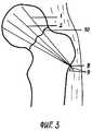
Сущность заявляемого способа поясняется чертежами. На фиг.1 изображено начало введения толкателем в кость первой сдвоенной спицы с острыми концами через установленный на кости под заданным углом полый направитель, при этом между концами спицы введен эластичный элемент, концы которого выведены наружу над направителем. На фиг. 2 - то же, но спица уже введена в костные отломки. На фиг. 3 изображено положение после введения всех спиц и формирования из эластичного элемента узла, скрепляющего все спицы между собой. На фиг. 1-3 обозначены: 1 - один заостренный конец спицы, 2 - второй заостренный конец спицы, 3 - проксимальный конец спицы, 4 - полый направитель, 5 - толкатель спицы, 6 - один конец эластичного элемента, 7 - второй конец эластичного элемента, 8 - узел скрепления спиц, сформированный из эластичного элемента, 9 - свободные концы эластичного элемента над узлом, 10 - линия перелома. The essence of the proposed method is illustrated by drawings. Figure 1 shows the beginning of the introduction of the pusher into the bone of the first double needle with sharp ends through the hollow guide mounted on the bone at a predetermined angle, while an elastic element is introduced between the ends of the spoke, the ends of which are brought out above the guide. In FIG. 2 - the same, but the spoke is already inserted into the bone fragments. In FIG. 3 shows the position after the introduction of all the spokes and the formation of the elastic element of the node, holding all the spokes together. In FIG. 1-3 are marked: 1 - one pointed end of the knitting needle, 2 - second pointed end of the knitting needle, 3 - proximal end of the knitting needle, 4 - hollow guide, 5 - pusher, 6 - one end of the elastic element, 7 - the second end of the elastic element, 8 - knot of knitting needles, formed from an elastic element, 9 - free ends of the elastic element above the node, 10 - line of fracture.
Конкретный пример осуществления способа. Например, у больного с переломом шейки бедренной кости полым заостренным направителем прокалывают мягкие ткани до кости и заостренным концом устанавливают его на ее поверхности. Между концами сдвоенной спицы проводят эластичный элемент, например нить высокой прочности на разрыв, и концы его оставляют снаружи над полым направителем. Острые расходящиеся концы спицы вводят в полость направителя и толкателем проводят до упора их в кость. Направитель устанавливают под должным углом к поверхности кости, направляя его проекцию в головку бедренной кости. Толкатель вводят в наружный конец направителя и забивают им спицу в костные отломки. Осуществляют рентгенологический контроль. В случае необходимости подтягивания назад спицы или полного извлечения ее, осуществляют тракцию одновременно за оба конца эластичного элемента. При правильной установке первой спицы аналогично проводят остальные две - четыре спицы. Потягиванием за один конец удаляют поочередно все эластичные элементы, кроме введенного с первой спицей. Концы эластичного элемента введенной первой спицы скрепляют между собой над последней спицей, например, на узел, над которым излишки эластичного элемента отсекают и вместе с направителем извлекают потягиванием за один конец каждого. A specific example of the method. For example, in a patient with a fracture of the femoral neck with a hollow pointed guide, soft tissues are pierced to the bone and a pointed end is placed on its surface. An elastic element, for example, a thread with high tensile strength, is carried out between the ends of the double knitting needle, and its ends are left outside over the hollow guide. The sharp diverging ends of the spokes are inserted into the cavity of the guide and the pusher is held all the way into the bone. The guide is set at the proper angle to the surface of the bone, directing its projection into the femoral head. The pusher is inserted into the outer end of the guide and the needle is hammered into the bone fragments. Carry out x-ray control. If it is necessary to pull the spokes back or completely remove it, traction is carried out simultaneously for both ends of the elastic element. With the correct installation of the first knitting needles, the remaining two to four spokes are likewise carried out. By pulling at one end, all the elastic elements, except for the one introduced with the first spoke, are removed alternately. The ends of the elastic element of the introduced first spoke are fastened together above the last spoke, for example, on a knot, over which excess elastic element is cut off and, together with the guide, removed by pulling one end of each.
Существенность отличий заявленного способа от прототипа заключается в следующем. Исключение необходимости просверливания костных отломков снижает объем повреждений костной ткани. Незначительные размеры фиксирующего эластичного элемента уменьшают размеры оставляемых в организме инородных тел. Расхождение обоих концов одной спицы со скреплением нескольких спиц между собой эластичным элементом исключает миграцию фиксаторов после оперативного вмешательства, предупреждая возникновение показаний к повторной операции. Размещение эластичного элемента между расходящимися заостренными концами спицы, вводимой толкателем через полый направитель, установленный на кости путем прокола мягких тканей, и выведение обоих концов эластичного элемента наружу позволяет при необходимости подтянуть или даже удалить неправильно введенную спицу через полость направителя, исключая дополнительную травму мягких тканей и не требуя расширения раны для поиска, захвата введенной спицы и извлечения ее. Это отличие позволяет добиться желаемых направления и глубины установки спиц в костные отломки. Отличительный признак: после установки всех спиц концы эластичного элемента введенной первой спицы скрепляют между собой над последней спицей, что позволяет скрепить одним элементом все спицы, фиксировать их в заданном положении, предупредить их миграцию и возникновение связанных с ней осложнений. Отсечение свободных концов эластичного элемента над узлом и удаление их, как и направителя, и остальных эластичных элементов других спиц, путем потягивания за один конец каждого из них позволяет без расширения раны и дополнительного травмирования тканей удалить простым приемом все излишние после операции детали, уменьшая опасность возникновения осложнений. The significance of the differences of the claimed method from the prototype is as follows. Eliminating the need for drilling bone fragments reduces the amount of damage to bone tissue. The small size of the fixing elastic element reduces the size of the foreign bodies left in the body. The divergence of both ends of the same knitting needle with the binding of several knitting needles to each other by an elastic element eliminates the migration of retainers after surgery, preventing the occurrence of indications for reoperation. Placing the elastic element between the divergent pointed ends of the knitting needle inserted by the pusher through the hollow guide mounted on the bone by puncture of soft tissues and pulling both ends of the elastic element out allows pulling or even removing the incorrectly inserted needle through the guide cavity, if necessary, eliminating additional soft tissue injury and without requiring an extension of the wound to search, capture the inserted needle and retrieve it. This difference allows you to achieve the desired direction and depth of installation of the spokes in the bone fragments. Distinctive feature: after installing all knitting needles, the ends of the elastic element of the inserted first knitting needle are fastened together above the last knitting needle, which allows you to fasten all the knitting needles with one element, fix them in a predetermined position, prevent their migration and the occurrence of complications associated with it. Cutting off the free ends of the elastic element over the knot and removing them, like the guide, and the remaining elastic elements of the other knitting needles, by pulling on one end of each of them, allows you to remove all unnecessary parts after the operation without expanding the wound and additional tissue injury, reducing the risk of occurrence complications.
Таким образом, в совокупности всех признаков заявленный способ обеспечивает достижение технического результата, улучшает качество хирургического лечения, сокращает сроки стационарного лечения больных и приводит к экономическому эффекту. Thus, in the aggregate of all the features of the claimed method ensures the achievement of a technical result, improves the quality of surgical treatment, reduces the time of inpatient treatment of patients and leads to an economic effect.
Применение способа возможно в детской и военно-полевой хирургии, а также в ветеринарии. The application of the method is possible in pediatric and field surgery, as well as in veterinary medicine.
Claims (1)
Translated fromRussianPriority Applications (1)
| Application Number | Priority Date | Filing Date | Title |
|---|---|---|---|
| RU99119114ARU2152763C1 (en) | 1999-09-01 | 1999-09-01 | Method for performing osteosynthesis using wires |
Applications Claiming Priority (1)
| Application Number | Priority Date | Filing Date | Title |
|---|---|---|---|
| RU99119114ARU2152763C1 (en) | 1999-09-01 | 1999-09-01 | Method for performing osteosynthesis using wires |
Publications (1)
| Publication Number | Publication Date |
|---|---|
| RU2152763C1true RU2152763C1 (en) | 2000-07-20 |
Family
ID=20224645
Family Applications (1)
| Application Number | Title | Priority Date | Filing Date |
|---|---|---|---|
| RU99119114ARU2152763C1 (en) | 1999-09-01 | 1999-09-01 | Method for performing osteosynthesis using wires |
Country Status (1)
| Country | Link |
|---|---|
| RU (1) | RU2152763C1 (en) |
Cited By (21)
| Publication number | Priority date | Publication date | Assignee | Title |
|---|---|---|---|---|
| RU2231987C2 (en)* | 2002-01-21 | 2004-07-10 | Учебно-научный центр Медицинского центра Управления делами Президента Российской Федерации | Method for performing tense closed osteosynthesis in fractures of surgical brachial cervix |
| RU2254089C2 (en)* | 2003-02-03 | 2005-06-20 | Кемеровская городская клиническая больница №3 им. М.А. Подгорбунского | Fixing member for performing osteosynthesis |
| RU2281714C2 (en)* | 2004-08-10 | 2006-08-20 | Джевдет Энвербекович Купкенов | Rod apparatus for through-bone osteosynthesis |
| RU2281715C2 (en)* | 2004-08-10 | 2006-08-20 | Джевдет Энвербекович Купкенов | Rod apparatus for through-bone osteosynthesis |
| RU2311148C1 (en)* | 2006-06-05 | 2007-11-27 | Федеральное государственное учреждение "Нижегородский научно-исследовательский институт травматологии и ортопедии Федерального агентства по здравоохранению и социальному развитию" | Method for operative treatment of fractures in proximal department of brachial bone |
| RU2311886C1 (en)* | 2006-04-28 | 2007-12-10 | Джевдет Энвербекович Купкенов | Rod apparatus for carrying out transosseous osteosynthesis |
| RU2317790C1 (en)* | 2006-10-09 | 2008-02-27 | Федеральное государственное учреждение высшего профессионального образования "Нижегородский научно-исследовательский институт травматологии и ортопедии Федерального агентства по здравоохранению и социальному развитию" | Surgical method for treating proximal humeral bone fracture malunion cases |
| RU2319467C2 (en)* | 2006-05-06 | 2008-03-20 | Джевдет Энвербекович Купкенов | Rod apparatus for intraosseous ostheosynthesis |
| RU2322953C1 (en)* | 2006-07-17 | 2008-04-27 | Джевдет Энвербекович Купкенов | Rod apparatus for carrying out transosseous osteosynthesis |
| RU2339331C1 (en)* | 2007-06-13 | 2008-11-27 | Государственное Образовательное Учреждение Высшего Профессионального Образования Амурская Государственная Медицинская Академия Росздрава | Repositioning device for treatment of intra-articular fractures of proximal part of shin bone |
| RU2350296C1 (en)* | 2007-09-18 | 2009-03-27 | Джевдет Энвербекович Купкенов | Rod apparatus for intraosteal osteosynthesis of shin bone |
| RU2350297C1 (en)* | 2007-09-18 | 2009-03-27 | Джевдет Энвербекович Купкенов | Rod apparatus for intraosteal osteosynthesis of shin bone |
| RU2353320C1 (en)* | 2007-08-17 | 2009-04-27 | Джевдет Энвербекович Купкенов | Osteosynthesis technique for segmental humerus fracture with using rod transosseous osteosynthesis apparatus |
| RU2354326C1 (en)* | 2007-09-03 | 2009-05-10 | Джевдет Энвербекович Купкенов | Rod apparatus for transosteal tibia osteosynthesis |
| RU2354325C1 (en)* | 2007-09-17 | 2009-05-10 | Джевдет Энвербекович Купкенов | Rod apparatus for transosteal tibia osteosynthesis |
| RU2354323C1 (en)* | 2007-09-14 | 2009-05-10 | Джевдет Энвербекович Купкенов | Rod apparatus for transosteal tibia osteosynthesis |
| RU2354324C1 (en)* | 2007-09-14 | 2009-05-10 | Джевдет Энвербекович Купкенов | Rod apparatus for transosteal tibia osteosynthesis |
| RU2368346C2 (en)* | 2007-09-10 | 2009-09-27 | Государственное образовательное учреждение высшего профессионального образования Кабардино-Балкарский государственный университет им. Х.М. Бербекова | Set of metal constructions for transosteal osteosynthesis of tubular bones |
| RU2373888C2 (en)* | 2007-09-14 | 2009-11-27 | Джевдет Энвербекович Купкенов | Rod apparatus for transosteal osteosynthesis of tibia |
| RU2523828C1 (en)* | 2013-03-14 | 2014-07-27 | Государственное бюджетное образовательное учреждение высшего профессионального образования "Самарский государственный медицинский университет" Министерства здравоохранения Российской Федерации | Method for nail osteosynthesis of facial bone fractures |
| RU2523830C1 (en)* | 2013-01-30 | 2014-07-27 | Государственное бюджетное образовательное учреждение высшего профессионального образования "Самарский государственный медицинский университет" Министерства здравоохранения Российской Федерации | Method for nail osteosynthesis |
Citations (4)
| Publication number | Priority date | Publication date | Assignee | Title |
|---|---|---|---|---|
| SU1452521A1 (en)* | 1986-06-26 | 1989-01-23 | Омский государственный медицинский институт им.М.И.Калинина | Arrangement for osteosynhtesis of the femur neck |
| US5429641A (en)* | 1993-03-28 | 1995-07-04 | Gotfried; Yechiel | Surgical device for connection of fractured bones |
| DE4440797A1 (en)* | 1994-11-17 | 1996-05-23 | Treu Instr Gmbh | Device for preparation of neck of femur for insertion of screw |
| RU2122368C1 (en)* | 1996-01-10 | 1998-11-27 | Старых Владимир Степанович | Osteosynthesis method used in case of intra-articular fracture of femur neck and device for its embodiment |
- 1999
- 1999-09-01RURU99119114Apatent/RU2152763C1/enactive
Patent Citations (4)
| Publication number | Priority date | Publication date | Assignee | Title |
|---|---|---|---|---|
| SU1452521A1 (en)* | 1986-06-26 | 1989-01-23 | Омский государственный медицинский институт им.М.И.Калинина | Arrangement for osteosynhtesis of the femur neck |
| US5429641A (en)* | 1993-03-28 | 1995-07-04 | Gotfried; Yechiel | Surgical device for connection of fractured bones |
| DE4440797A1 (en)* | 1994-11-17 | 1996-05-23 | Treu Instr Gmbh | Device for preparation of neck of femur for insertion of screw |
| RU2122368C1 (en)* | 1996-01-10 | 1998-11-27 | Старых Владимир Степанович | Osteosynthesis method used in case of intra-articular fracture of femur neck and device for its embodiment |
Cited By (23)
| Publication number | Priority date | Publication date | Assignee | Title |
|---|---|---|---|---|
| RU2231987C2 (en)* | 2002-01-21 | 2004-07-10 | Учебно-научный центр Медицинского центра Управления делами Президента Российской Федерации | Method for performing tense closed osteosynthesis in fractures of surgical brachial cervix |
| RU2254089C2 (en)* | 2003-02-03 | 2005-06-20 | Кемеровская городская клиническая больница №3 им. М.А. Подгорбунского | Fixing member for performing osteosynthesis |
| RU2281714C2 (en)* | 2004-08-10 | 2006-08-20 | Джевдет Энвербекович Купкенов | Rod apparatus for through-bone osteosynthesis |
| RU2281715C2 (en)* | 2004-08-10 | 2006-08-20 | Джевдет Энвербекович Купкенов | Rod apparatus for through-bone osteosynthesis |
| RU2281714C9 (en)* | 2004-08-10 | 2007-04-10 | Джевдет Энвербекович Купкенов | Rod apparatus for through-bone osteosynthesis |
| RU2281715C9 (en)* | 2004-08-10 | 2007-04-10 | Джевдет Энвербекович Купкенов | Rod apparatus for through-bone osteosynthesis |
| RU2311886C1 (en)* | 2006-04-28 | 2007-12-10 | Джевдет Энвербекович Купкенов | Rod apparatus for carrying out transosseous osteosynthesis |
| RU2319467C2 (en)* | 2006-05-06 | 2008-03-20 | Джевдет Энвербекович Купкенов | Rod apparatus for intraosseous ostheosynthesis |
| RU2311148C1 (en)* | 2006-06-05 | 2007-11-27 | Федеральное государственное учреждение "Нижегородский научно-исследовательский институт травматологии и ортопедии Федерального агентства по здравоохранению и социальному развитию" | Method for operative treatment of fractures in proximal department of brachial bone |
| RU2322953C1 (en)* | 2006-07-17 | 2008-04-27 | Джевдет Энвербекович Купкенов | Rod apparatus for carrying out transosseous osteosynthesis |
| RU2317790C1 (en)* | 2006-10-09 | 2008-02-27 | Федеральное государственное учреждение высшего профессионального образования "Нижегородский научно-исследовательский институт травматологии и ортопедии Федерального агентства по здравоохранению и социальному развитию" | Surgical method for treating proximal humeral bone fracture malunion cases |
| RU2339331C1 (en)* | 2007-06-13 | 2008-11-27 | Государственное Образовательное Учреждение Высшего Профессионального Образования Амурская Государственная Медицинская Академия Росздрава | Repositioning device for treatment of intra-articular fractures of proximal part of shin bone |
| RU2353320C1 (en)* | 2007-08-17 | 2009-04-27 | Джевдет Энвербекович Купкенов | Osteosynthesis technique for segmental humerus fracture with using rod transosseous osteosynthesis apparatus |
| RU2354326C1 (en)* | 2007-09-03 | 2009-05-10 | Джевдет Энвербекович Купкенов | Rod apparatus for transosteal tibia osteosynthesis |
| RU2368346C2 (en)* | 2007-09-10 | 2009-09-27 | Государственное образовательное учреждение высшего профессионального образования Кабардино-Балкарский государственный университет им. Х.М. Бербекова | Set of metal constructions for transosteal osteosynthesis of tubular bones |
| RU2354324C1 (en)* | 2007-09-14 | 2009-05-10 | Джевдет Энвербекович Купкенов | Rod apparatus for transosteal tibia osteosynthesis |
| RU2373888C2 (en)* | 2007-09-14 | 2009-11-27 | Джевдет Энвербекович Купкенов | Rod apparatus for transosteal osteosynthesis of tibia |
| RU2354323C1 (en)* | 2007-09-14 | 2009-05-10 | Джевдет Энвербекович Купкенов | Rod apparatus for transosteal tibia osteosynthesis |
| RU2354325C1 (en)* | 2007-09-17 | 2009-05-10 | Джевдет Энвербекович Купкенов | Rod apparatus for transosteal tibia osteosynthesis |
| RU2350297C1 (en)* | 2007-09-18 | 2009-03-27 | Джевдет Энвербекович Купкенов | Rod apparatus for intraosteal osteosynthesis of shin bone |
| RU2350296C1 (en)* | 2007-09-18 | 2009-03-27 | Джевдет Энвербекович Купкенов | Rod apparatus for intraosteal osteosynthesis of shin bone |
| RU2523830C1 (en)* | 2013-01-30 | 2014-07-27 | Государственное бюджетное образовательное учреждение высшего профессионального образования "Самарский государственный медицинский университет" Министерства здравоохранения Российской Федерации | Method for nail osteosynthesis |
| RU2523828C1 (en)* | 2013-03-14 | 2014-07-27 | Государственное бюджетное образовательное учреждение высшего профессионального образования "Самарский государственный медицинский университет" Министерства здравоохранения Российской Федерации | Method for nail osteosynthesis of facial bone fractures |
Similar Documents
| Publication | Publication Date | Title |
|---|---|---|
| RU2152763C1 (en) | Method for performing osteosynthesis using wires | |
| EP1726317B1 (en) | Surgical means for cosmetic surgery | |
| US9326807B2 (en) | Peri-prosthetic fixation implant and method | |
| JP5323815B2 (en) | Loader and threader for knot elements | |
| KR101327244B1 (en) | Method and apparatus for bone fracture fixation | |
| KR101237769B1 (en) | Sternal reconstruction system | |
| US20150039029A1 (en) | Implant device and system for stabilized fixation of bone and soft tissue | |
| RU2210331C2 (en) | Method for surgical treatment of brachial collum fractures | |
| EP0517435A1 (en) | Intramedullary osteosynthetic device | |
| RU2272592C1 (en) | Surgical intervention method for in the cases of humerus cervix fracture | |
| RU2254091C2 (en) | Guide member for operating with extension wires usable with cannulated drill and method for making femoral neck osteosynthesis using the guiding device | |
| RU2122368C1 (en) | Osteosynthesis method used in case of intra-articular fracture of femur neck and device for its embodiment | |
| RU2159591C1 (en) | Method for carrying out osteosynthesis in the cases of lateral fracture of the femur neck | |
| RU2214192C2 (en) | Wire locking device for osteosynthesis | |
| RU2609058C1 (en) | Method of surgical treatment of fractures of proximal part of shoulder bone in children and teenagers | |
| RU2152188C1 (en) | Method and device for performing osteosynthesis in the cases of medial fractures of femur neck | |
| RU2110230C1 (en) | Fixing device for performing osteosynthesis of neck of the femur | |
| RU2269964C2 (en) | Device for introducing two-rod holders into bone | |
| RU2164785C1 (en) | Device for bending wires | |
| RU2123308C1 (en) | Method of paracentetic osteosynthesis and device for its realization | |
| RU2088168C1 (en) | Method for performing combined osteosynthesis in mandibular fractures | |
| RU2790756C1 (en) | Method of surgical treatment of repeated fractures of long bones in children | |
| RU2155012C1 (en) | Device for guiding introduction of fixing members into bone | |
| RU2367373C1 (en) | Osteosynthesis technique | |
| RU2705234C1 (en) | Method for operative treatment of multi-fragment fractures of surgical neck of humerus |