RU211923U1 - EXOPROSTHESIS-FRAME FOR STRENGTHENING THE WALL OF THE DILATED AORTIC - Google Patents
EXOPROSTHESIS-FRAME FOR STRENGTHENING THE WALL OF THE DILATED AORTICDownload PDFInfo
- Publication number
- RU211923U1 RU211923U1RU2021138431URU2021138431URU211923U1RU 211923 U1RU211923 U1RU 211923U1RU 2021138431 URU2021138431 URU 2021138431URU 2021138431 URU2021138431 URU 2021138431URU 211923 U1RU211923 U1RU 211923U1
- Authority
- RU
- Russia
- Prior art keywords
- aorta
- frame
- exoprosthesis
- diameter
- diamond
- Prior art date
Links
- 238000005728strengtheningMethods0.000titleclaimsabstractdescription6
- 210000000709aortaAnatomy0.000claimsabstractdescription38
- 230000015572biosynthetic processEffects0.000claimsabstractdescription3
- 230000002093peripheral effectEffects0.000claimsdescription5
- 238000005476solderingMethods0.000claimsdescription2
- 238000003466weldingMethods0.000claimsdescription2
- HLXZNVUGXRDIFK-UHFFFAOYSA-Nnickel titaniumChemical compound[Ti].[Ti].[Ti].[Ti].[Ti].[Ti].[Ti].[Ti].[Ti].[Ti].[Ti].[Ni].[Ni].[Ni].[Ni].[Ni].[Ni].[Ni].[Ni].[Ni].[Ni].[Ni].[Ni].[Ni].[Ni]HLXZNVUGXRDIFK-UHFFFAOYSA-N0.000abstractdescription7
- 229910001000nickel titaniumInorganic materials0.000abstractdescription7
- 238000000034methodMethods0.000abstractdescription6
- 238000013130cardiovascular surgeryMethods0.000abstractdescription4
- 238000001356surgical procedureMethods0.000abstractdescription4
- 206010057453Aortic dilatationDiseases0.000abstractdescription2
- 210000002376aorta thoracicAnatomy0.000description8
- 230000002792vascularEffects0.000description7
- 210000001519tissueAnatomy0.000description4
- 239000000463materialSubstances0.000description3
- 230000002980postoperative effectEffects0.000description3
- 206010002329AneurysmDiseases0.000description2
- 230000001174ascending effectEffects0.000description2
- 230000006378damageEffects0.000description2
- 201000010099diseaseDiseases0.000description2
- 208000037265diseases, disorders, signs and symptomsDiseases0.000description2
- 206010058314DysplasiaDiseases0.000description1
- 208000005189EmbolismDiseases0.000description1
- 208000010496Heart ArrestDiseases0.000description1
- 208000037273Pathologic ProcessesDiseases0.000description1
- 230000006978adaptationEffects0.000description1
- 230000003872anastomosisEffects0.000description1
- 210000002168brachiocephalic trunkAnatomy0.000description1
- 210000004556brainAnatomy0.000description1
- 238000007675cardiac surgeryMethods0.000description1
- 230000006835compressionEffects0.000description1
- 238000007906compressionMethods0.000description1
- 238000005520cutting processMethods0.000description1
- 230000001066destructive effectEffects0.000description1
- 206010012601diabetes mellitusDiseases0.000description1
- 238000003745diagnosisMethods0.000description1
- 239000003814drugSubstances0.000description1
- 238000005516engineering processMethods0.000description1
- 230000003601intercostal effectEffects0.000description1
- 230000007774longtermEffects0.000description1
- 238000004519manufacturing processMethods0.000description1
- 230000017074necrotic cell deathEffects0.000description1
- RVTZCBVAJQQJTK-UHFFFAOYSA-Noxygen(2-);zirconium(4+)Chemical compound[O-2].[O-2].[Zr+4]RVTZCBVAJQQJTK-UHFFFAOYSA-N0.000description1
- 230000009054pathological processEffects0.000description1
- 230000002265preventionEffects0.000description1
- 230000002035prolonged effectEffects0.000description1
- 239000007787solidSubstances0.000description1
- 238000010561standard procedureMethods0.000description1
- 210000001562sternumAnatomy0.000description1
- 210000003270subclavian arteryAnatomy0.000description1
- 230000003144traumatizing effectEffects0.000description1
Images
Abstract
Translated fromRussian
Description
Translated fromRussianОбласть техникиTechnical field
Полезная модель относится к области сердечно-сосудистой хирургии, в частности к способу хирургического лечения дилатации аорты. Может быть использовано в специализированных отделениях сердечно-сосудистой хирургии.The utility model relates to the field of cardiovascular surgery, in particular to a method for the surgical treatment of aortic dilatation. It can be used in specialized departments of cardiovascular surgery.
Уровень техникиState of the art
Расширение аорты у больных при дисплазиях встречается довольно часто. При этом диаметр дуги не всегда достигает необходимой величины для ее протезирования. В случае пограничного диаметра аорты в диапазоне от 40 мм до 50 мм, решение о протезировании данного сегмента является спорным, однако известно, что при постепенном прогрессировании заболевания в отдаленном периоде увеличение пограничного диаметра может приводить к увеличению риска разрыва и других аорто-ассоциированных осложнений.Expansion of the aorta in patients with dysplasia is quite common. At the same time, the diameter of the arc does not always reach the required value for its prosthetics. In the case of aortic marginal diameter in the range of 40 mm to 50 mm, the decision to replace this segment is controversial, however, it is known that with the gradual progression of the disease in the long term, an increase in the marginal diameter may lead to an increased risk of rupture and other aorto-associated complications.
Экзопротезирование расширенных сегментов аорты отдельной частью сосудистого плетеного протеза является вариантом хирургического вмешательства, не требующим применения циркуляторного ареста, технологий защиты головного мозга и предотвращающим дальнейшее расширение дистальных от анастомоза сегментов аорты.Exoprosthesis of dilated aortic segments with a separate part of a vascular braided prosthesis is a surgical option that does not require the use of circulatory arrest, brain protection technologies and prevents further expansion of the aortic segments distal from the anastomosis.
Имеющийся способ экзопротезирования заключается в позиционировании рассеченного вдоль сосудистого плетеного протеза методом проведения вокруг вышележащей расширенной аорты и последующим сшиванием ткани экзопротеза линейным швом между ветвями аорты с выкраиванием ткани протеза у их устьев и фиксацией одиночными узловыми швами к протезу аорты и вовлеченной в патологический процесс стенки аорты [1, 2].The existing method of exoprosthesis consists in positioning a vascular braided prosthesis dissected along the vascular wicker by passing it around the overlying dilated aorta and then suturing the exoprosthesis tissue with a linear suture between the branches of the aorta with cutting out the prosthesis tissue at their mouths and fixing with single interrupted sutures to the aorta prosthesis and the aorta wall involved in the pathological process [ 12].
Недостатками данного способа являются возможность некроза стенки аорты в связи с продолжительным плотным сдавлением последней сплошной тканью экзопротеза. При укреплении стенки аорты снаружи сосудистым протезом, представленным плотным материалом, который не может изменять размер диаметра и длины соответственно пульсовой волне, во время систолы ткань протеза врезается в аортальную стенку, постоянно травмируя ее, приводя к деструкции.The disadvantages of this method are the possibility of necrosis of the aortic wall due to prolonged tight compression of the last solid tissue of the exoprosthesis. When strengthening the aortic wall from the outside with a vascular prosthesis, which is a dense material that cannot change the size of the diameter and length according to the pulse wave, during systole, the prosthesis tissue cuts into the aortic wall, constantly injuring it, leading to destruction.
Раскрытие полезной моделиUtility Model Disclosure
Сущность полезной модели состоит в создании экзопротеза-каркаса в виде проволочного каркаса цилиндрической формы, который не препятствует пульсирующим движениям стенки аорты, минимально травмируя, укрепляет стенку аорты, препятствуя ее дальнейшей дилатации и деструкции с возможностью моделирования диаметра.The essence of the utility model is to create an exoprosthesis-framework in the form of a cylindrical wire frame, which does not interfere with the pulsating movements of the aortic wall, minimally traumatizing, strengthens the aortic wall, preventing its further dilatation and destruction with the possibility of modeling the diameter.
Краткое описание чертежейBrief description of the drawings
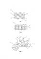
На фиг. 1 представлен вариант реализации экзопротеза-каркаса для укрепления стенки дилатированной аорты. Он представляет собой гибкий проволочный каркас 1, способный при сворачивании принимать форму цилиндра.In FIG. Figure 1 shows an embodiment of an exoprosthesis-frame for strengthening the wall of a dilated aorta. It is a flexible wire frame 1 capable of taking the form of a cylinder when folded.
На фиг. 1 каркас находится в развернутом состоянии в виде плоскости. Каркас выполнен из изогнутой проволоки 2. Соседние участки изогнутой проволоки 2 сцеплены между собой. Сцепление может осуществляться как постоянное фиксированное 3, с помощью пайки или сварки, обеспечивая постоянное сцепление между соседними участками проволоки, так и посредством их подвижного взаимного сцепления 4, аналогично сцеплению соседних участков в применяемого сетке рабица. Постоянное фиксированное сцепление 3 и подвижное взаимное сцепление 4 могут осуществлять между любыми соседними участками изогнутой проволоки 2, в зависимости от конкретных размеров аорты и условий операции. Как один из вариантов, периферийные части 5 могут быть не сцеплены между собой для облегчения формирования окон на цилиндрическом каркасе для расположения в них ветвей аорты, при установке протеза на аорту.In FIG. 1 the frame is in the expanded state in the form of a plane. The frame is made of curved wire 2. Neighboring sections of the curved wire 2 are interlocked. The adhesion can be carried out as a permanent fixed 3, by soldering or welding, providing a constant adhesion between adjacent sections of the wire, and through their movable
На фиг. 2 изображен экзопротез, свернутый в форме цилиндра 6.In FIG. 2 shows an exoprosthesis rolled up in the form of a
На фиг 3 изображен экзопротез, установленный на дуге аорты.Figure 3 shows an exoprosthesis mounted on the aortic arch.
Подвижные соединения 4, сцепляющие периферийные части 5, обеспечивают при фиксации цилиндра 6 на аорте 7 оптимальное их расположение относительно ветвей аорты 8. То есть, сцепляя посредством лигатур 9 любые периферийные части 5 цилиндра 6 противоположных сторон цилиндра 6, при установке протеза на аорте, можно достичь нужной конфигурации окон между периферийными частями 5, для оптимального расположения протеза на аорте, равномерно сдавливающего стенки аорты 7 и не пережимающие ветви аорты 8. Количество образующихся ячеек проволочного каркаса, их размер, а также длина и диаметр образующегося при сворачивании сетки цилиндра могут быть любыми в зависимости от конкретных размеров аорты, на которую будет установлен протез. Один из вариантов реализации, уже прошедших проверку в клинических условиях, возможно, применение цилиндра длиной 108 мм, диаметр 40 мм, размер образующихся ромбовидных ячеек 12 мм на 22 мм. Для изготовления экзопротеза-каркаса возможно применение любых материалов, применяемых в медицине, не вызывающих отторжение, - как один из вариантов, прошедший успешное клиническое испытание, возможно применение нитиноловой проволоки диаметром 0,3 мм. При установке экзопротеза-каркаса на аорту свободные концы могут сдвигаться относительно друг друга для образования лучшего окна для ветвей аорты. Предложенный экзопротез-каркас для укрепления стенки дилатированной аорты отличается от плетеного сосудистого протеза при использовании для экзопротезирования отсутствием необходимости формирования окон для пропускания ветвей аорты, сплошного давящего воздействия на сосудистую стенку, имеет больше возможностей по адаптации диаметра.
Использование экзопротеза-каркаса для укрепления стенки дилатированной аорты позволит значительно снизить опасность возникновения деструктивных изменений в сосудистой стенке в отдаленном послеоперационном периоде. Применение экзопротеза-каркаса для укрепления стенки дилатированной аорты обеспечит хорошую адаптацию, удобство и быстроту при его использовании.The use of an exoprosthesis-framework to strengthen the wall of the dilated aorta will significantly reduce the risk of destructive changes in the vascular wall in the late postoperative period. The use of an exoprosthesis-framework to strengthen the wall of the dilated aorta will provide good adaptation, convenience and speed in its use.
Осуществление полезной моделиImplementation of the utility model
Клинический примерClinical example
Больной Д., 62 годаPatient D., 62 years old
Диагноз: Аневризма восходящей аорты. Расширение дуги аорты. Гипертоническая болезнь. Сахарный диабет.Diagnosis: Aneurysm of the ascending aorta. Expansion of the aortic arch. Hypertonic disease. Diabetes.
По данным МСКТ аорты с контрастированием диаметр восходящей аорты - 65 мм, диаметр дуги - 41 мм.According to MSCT of the aorta with contrast, the diameter of the ascending aorta was 65 mm, the diameter of the arch was 41 mm.
Поступил для хирургического лечения в отдел инновационной кардиоаортальной хирургии (кардиохирургии 1) ФГБНУ РНЦХ им. акад. Петровского 06.07.2020 г.Admitted for surgical treatment in the department of innovative cardio-aortic surgery (cardiac surgery 1) acad. Petrovsky July 6, 2020
09.07.2020 г. Выполнено протезирование восходящей аорты протезом Polythese IC 30 mm и экзопротезирование дуги нитиноловым экзопротезом-каркасом из J-образной министернотомии.On July 9, 2020, the ascending aorta was replaced with a Polythese IC 30 mm prosthesis and the arch was exoprosthesized with a nitinol exoprosthesis-frame from a J-shaped ministernotomy.
Ход операции: J-образная министернотомия в 4-м межреберье с канюляцией бедренных сосудов для проведения периферического ИК. Мобилизация восходящей, дуги аорты и брахиоцефальных ветвей. Восходящая аорта аневризматически расширена максимально до 65 мм в диаметре. Дуга аорты дилатирована до 41 мм. В данном случае имелись абсолютные показания к протезированию восходящей аорты. В условиях ПК выполнено протезирование восходящей аорты протезом Polythese IC 30 mm. После профилактики воздушной и материальной эмболии снят зажим с аорты. Для укрепления стенки дилатированной дуги аорты был выбран способ экзопротезирования нитиноловым экзопротезом-каркасом. Нитиноловый экзопротез-каркас проведен под внутренней кривизной дуги аорты вверх и вокруг вышележащей расширенной дуги аорты, пропуская устья брахиоцефального ствола, левой общей сонной и левой подключичной артерий между свободными концами циркулярно расположенных сегментов нитинолового экзопротеза-каркаса. Непрерывность нитинолового экзопротеза-каркаса восстановлена в проекции наружной кривизны дуги аорты за счет соединения свободных ромбовидных фигур лигатурами. Оптимальное моделирование экзопротеза-каркаса в соответствии с анатомическими особенностями аорты и ее ветвей достигнуто за счет соединения ромбовидных фигур дополнительными лигатурами.Procedure: J-shaped ministernotomy in the 4th intercostal space with cannulation of the femoral vessels for peripheral EC. Mobilization of the ascending, aortic arch and brachiocephalic branches. The ascending aorta is aneurysmically dilated to a maximum of 65 mm in diameter. The aortic arch was dilated to 41 mm. In this case, there were absolute indications for ascending aortic replacement. The ascending aorta was replaced with a Polythese IC 30 mm prosthesis under PC conditions. After the prevention of air and material embolism, the clamp was removed from the aorta. To strengthen the wall of the dilated aortic arch, the method of exoprosthesis replacement with a nitinol exoprosthesis-framework was chosen. The nitinol exoprosthesis-frame is passed under the internal curvature of the aortic arch up and around the overlying dilated aortic arch, passing the orifices of the brachiocephalic trunk, the left common carotid and left subclavian arteries between the free ends of the circularly located segments of the nitinol exoprosthesis-frame. The continuity of the nitinol exoprosthesis-framework was restored in the projection of the external curvature of the aortic arch by connecting free diamond-shaped figures with ligatures. Optimal modeling of the exoprosthesis-frame in accordance with the anatomical features of the aorta and its branches was achieved by connecting diamond-shaped figures with additional ligatures.
Далее проведена стандартная процедура деканюляции, остеосинтеза грудины и наложения швов. У пациента гладкий послеоперационный период и короткий срок послеоперационной реабилитации. При контрольной КТ аорты с контрастированием через 2 месяца после операции увеличения диаметра аорты не отмечалось.Next, a standard procedure of decannulation, osteosynthesis of the sternum and suturing was performed. The patient has a smooth postoperative period and a short period of postoperative rehabilitation. At the control CT of the aorta with contrast 2 months after the operation, there was no increase in the diameter of the aorta.
Применение нитинолового экзопротеза-каркаса для укрепления стенки дилатированной аорты позволило успешно выполнить адекватную реконструкцию при аневризме восходящей аорты и пограничном расширении ее дуги.The use of a nitinol exoprosthesis-framework to strengthen the wall of the dilated aorta made it possible to successfully perform an adequate reconstruction for an aneurysm of the ascending aorta and borderline expansion of its arch.
Источники информации:Sources of information:
1. Tagarakis GI, Karangelis D, Baddour AJ, et al. An alternate solution for the treatment of ascending aortic aneurysms: the wrapping technique. J Cardiothorac Surg 2010; 5:100.1. Tagarakis GI, Karangelis D, Baddour AJ, et al. An alternate solution for the treatment of ascending aortic aneurysms: the wrapping technique. J Cardiothorac Surg 2010; 5:100.
2. Bauer M, Grauhan O, Hetzer R. Dislocated wrap after previous reduction aortoplasty causes erosion of the ascending aorta. Ann Thorac Surg 2003; 75:583).2. Bauer M, Grauhan O, Hetzer R. Dislocated wrap after previous reduction aortoplasty causes erosion of the ascending aorta. Ann Thorac Surg 2003; 75:583).
Claims (2)
Translated fromRussianPublications (1)
| Publication Number | Publication Date |
|---|---|
| RU211923U1true RU211923U1 (en) | 2022-06-28 |
Family
ID=
Citations (6)
| Publication number | Priority date | Publication date | Assignee | Title |
|---|---|---|---|---|
| RU2102016C1 (en)* | 1995-07-07 | 1998-01-20 | Московский государственный институт стали и сплавов (технологический университет) | Device for extravasal correction of function of main vein valves |
| RU2128966C1 (en)* | 1995-06-01 | 1999-04-20 | Чадаев Алексей Павлович | Method and device for carrying out extravasal correction of great veins function |
| US6648911B1 (en)* | 2000-11-20 | 2003-11-18 | Avantec Vascular Corporation | Method and device for the treatment of vulnerable tissue site |
| US20060281966A1 (en)* | 2003-10-14 | 2006-12-14 | Peacock James C Iii | Aneurysm treatment system and method |
| RU2705910C2 (en)* | 2018-03-06 | 2019-11-12 | Андрей Николаевич Шведов | Method for prosthetic repair of main arterial vessels |
| RU2728702C1 (en)* | 2019-05-08 | 2020-07-30 | Федеральное государственное бюджетное научное учреждение "Российский научный центр хирургии имени академика Б.В. Петровского" | Method for simultaneous ascending aorta prosthetics and arch exoprosthesis |
Patent Citations (6)
| Publication number | Priority date | Publication date | Assignee | Title |
|---|---|---|---|---|
| RU2128966C1 (en)* | 1995-06-01 | 1999-04-20 | Чадаев Алексей Павлович | Method and device for carrying out extravasal correction of great veins function |
| RU2102016C1 (en)* | 1995-07-07 | 1998-01-20 | Московский государственный институт стали и сплавов (технологический университет) | Device for extravasal correction of function of main vein valves |
| US6648911B1 (en)* | 2000-11-20 | 2003-11-18 | Avantec Vascular Corporation | Method and device for the treatment of vulnerable tissue site |
| US20060281966A1 (en)* | 2003-10-14 | 2006-12-14 | Peacock James C Iii | Aneurysm treatment system and method |
| RU2705910C2 (en)* | 2018-03-06 | 2019-11-12 | Андрей Николаевич Шведов | Method for prosthetic repair of main arterial vessels |
| RU2728702C1 (en)* | 2019-05-08 | 2020-07-30 | Федеральное государственное бюджетное научное учреждение "Российский научный центр хирургии имени академика Б.В. Петровского" | Method for simultaneous ascending aorta prosthetics and arch exoprosthesis |
Non-Patent Citations (2)
| Title |
|---|
| А.П. ЗАРЕЦКИЙ, А.В. БОГОМОЛОВ, "Биомеханическое моделирование персонифицированного коронарного стента", ТРУДЫ МФТИ. — 2015. — Том 7, N 3, стр.82-90: стр.84 последний абзац, рисунки.* |
| Д.А. БОЖКО, Ю.М. ЧЕСНОВ, С.В. СПИРИДОНОВ, С.А. КУРГАНОВИЧ, "Экзопротезирование расширенной восходящей аорты при патологии аортального клапана", Неотложная кардиология и кардиоваскулярные риски, 2019, Т. 3, N 2, С. 672-676.* |
Similar Documents
| Publication | Publication Date | Title |
|---|---|---|
| US11969171B2 (en) | Systems and methods for treating aneurysms | |
| JP6725598B2 (en) | Occluder and anastomosis device | |
| US20220125431A1 (en) | Anastomosis devices | |
| US9539088B2 (en) | Fixation band for affixing a prosthetic heart valve to tissue | |
| JP2017515631A5 (en) | ||
| JP2022516603A (en) | Stent graft and how to use it | |
| JP2006507053A (en) | Anastomosis device | |
| CN106821545A (en) | Blocking-up type branch stent blood vessel on demand in a kind of combined type art | |
| RU211923U1 (en) | EXOPROSTHESIS-FRAME FOR STRENGTHENING THE WALL OF THE DILATED AORTIC | |
| RU2728702C1 (en) | Method for simultaneous ascending aorta prosthetics and arch exoprosthesis | |
| JP2022167849A (en) | Asymmetric external supports for stabilizing vein grafts used in coronary artery bypass graft (CABG) procedures and their applications | |
| RU2551627C1 (en) | Method for simulating stent-graft fixation area in distal portion of ascending aorta in hybrid interventions | |
| RU2401076C1 (en) | Oesophagogastric anastomosis technique | |
| RU215471U1 (en) | Composite linear prosthesis for seamless anastomosis of blood vessels | |
| RU2701901C2 (en) | Method for haemorrhage arrest from intercostals and lumbar arteries from inside of aorta | |
| JP2018534997A (en) | Occluder and suture device | |
| CN110464522A (en) | Bracket in art |