KR930004055B1 - Peptide and its use - Google Patents
Peptide and its useDownload PDFInfo
- Publication number
- KR930004055B1 KR930004055B1KR1019910003768AKR910003768AKR930004055B1KR 930004055 B1KR930004055 B1KR 930004055B1KR 1019910003768 AKR1019910003768 AKR 1019910003768AKR 910003768 AKR910003768 AKR 910003768AKR 930004055 B1KR930004055 B1KR 930004055B1
- Authority
- KR
- South Korea
- Prior art keywords
- lys
- peptide
- arg
- glu
- seq
- Prior art date
- Legal status (The legal status is an assumption and is not a legal conclusion. Google has not performed a legal analysis and makes no representation as to the accuracy of the status listed.)
- Expired - Fee Related
Links
Images


Classifications
- C—CHEMISTRY; METALLURGY
- C07—ORGANIC CHEMISTRY
- C07K—PEPTIDES
- C07K7/00—Peptides having 5 to 20 amino acids in a fully defined sequence; Derivatives thereof
- C07K7/04—Linear peptides containing only normal peptide links
- C07K7/06—Linear peptides containing only normal peptide links having 5 to 11 amino acids
- C—CHEMISTRY; METALLURGY
- C07—ORGANIC CHEMISTRY
- C07K—PEPTIDES
- C07K14/00—Peptides having more than 20 amino acids; Gastrins; Somatostatins; Melanotropins; Derivatives thereof
- C07K14/005—Peptides having more than 20 amino acids; Gastrins; Somatostatins; Melanotropins; Derivatives thereof from viruses
- C—CHEMISTRY; METALLURGY
- C12—BIOCHEMISTRY; BEER; SPIRITS; WINE; VINEGAR; MICROBIOLOGY; ENZYMOLOGY; MUTATION OR GENETIC ENGINEERING
- C12N—MICROORGANISMS OR ENZYMES; COMPOSITIONS THEREOF; PROPAGATING, PRESERVING, OR MAINTAINING MICROORGANISMS; MUTATION OR GENETIC ENGINEERING; CULTURE MEDIA
- C12N2770/00—MICROORGANISMS OR ENZYMES; COMPOSITIONS THEREOF; PROPAGATING, PRESERVING, OR MAINTAINING MICROORGANISMS; MUTATION OR GENETIC ENGINEERING; CULTURE MEDIA ssRNA viruses positive-sense
- C12N2770/00011—Details
- C12N2770/24011—Flaviviridae
- C12N2770/24211—Hepacivirus, e.g. hepatitis C virus, hepatitis G virus
- C12N2770/24222—New viral proteins or individual genes, new structural or functional aspects of known viral proteins or genes
- Y—GENERAL TAGGING OF NEW TECHNOLOGICAL DEVELOPMENTS; GENERAL TAGGING OF CROSS-SECTIONAL TECHNOLOGIES SPANNING OVER SEVERAL SECTIONS OF THE IPC; TECHNICAL SUBJECTS COVERED BY FORMER USPC CROSS-REFERENCE ART COLLECTIONS [XRACs] AND DIGESTS
- Y10—TECHNICAL SUBJECTS COVERED BY FORMER USPC
- Y10S—TECHNICAL SUBJECTS COVERED BY FORMER USPC CROSS-REFERENCE ART COLLECTIONS [XRACs] AND DIGESTS
- Y10S530/00—Chemistry: natural resins or derivatives; peptides or proteins; lignins or reaction products thereof
- Y10S530/806—Antigenic peptides or proteins
Landscapes
- Chemical & Material Sciences (AREA)
- Organic Chemistry (AREA)
- Health & Medical Sciences (AREA)
- Life Sciences & Earth Sciences (AREA)
- Medicinal Chemistry (AREA)
- Biochemistry (AREA)
- Biophysics (AREA)
- General Health & Medical Sciences (AREA)
- Genetics & Genomics (AREA)
- Molecular Biology (AREA)
- Proteomics, Peptides & Aminoacids (AREA)
- Gastroenterology & Hepatology (AREA)
- Virology (AREA)
- Peptides Or Proteins (AREA)
Abstract
Translated fromKoreanDescription
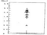
Translated fromKorean제1도는 실시예 1에서 얻어진 펩티드를 사용해서 실시예 9에 기재된 방법에 의해 측정한 각 혈청검체의 OD492값 분포도이다.1 is an OD492 value distribution chart of each serum sample measured by the method described in Example 9 using the peptide obtained in Example 1. FIG.
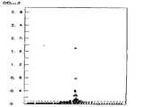
제2도는 실시예 2에서 얻어진 펩티드를 사용해서 실시예 9에 기재된 방법에 의해 측정한 각 혈청검체의 OD492값 분포도이다.FIG. 2 is a distribution diagram of the OD492 value of each serum sample measured by the method described in Example 9 using the peptide obtained in Example 2. FIG.
제3도는 참고예 1에서 얻어진 펩티드를 사용해서 실시예 9에 기재된 방법에 의해 측정한 각 혈청검체의 OD492값 분포도이다.3 is a distribution chart of OD492 values of each serum sample measured by the method described in Example 9 using the peptide obtained in Reference Example 1. FIG.
제4도는 참고예 2에서 얻어진 펩티드를 사용해서 실시예 9에 기재된 방법에 의해 측정한 각 혈청검체의 OD492값 분포도이다.4 is an OD492 value distribution chart of each serum sample measured by the method described in Example 9 using the peptide obtained in Reference Example 2. FIG.
제5도는 참고예 3에서 얻어진 펩티드를 사용해서 실시예 9에 기재된 방법에 의해 측정한 각 혈청검체의 OD492값 분포도이다.FIG. 5 is a distribution diagram of the OD492 value of each serum sample measured by the method described in Example 9 using the peptide obtained in Reference Example 3.
제6도는 실시예 3에서 얻어진 펩티드를 사용해서 실시예 9에 기재된 방법에 의해 측정한 각 혈청검체의 OD492값 분포도이다.6 is a distribution diagram of the OD492 value of each serum sample measured by the method described in Example 9 using the peptide obtained in Example 3. FIG.
제7도는 실시예 4에서 얻어진 펩티드를 사용해서 실시예 9에 기재된 방법에 의해 측정한 각 혈청검체의 OD492값 분포도이다.7 is an OD492 value distribution diagram of each serum sample measured by the method described in Example 9 using the peptide obtained in Example 4. FIG.
제8도는 실시예 5에서 얻어진 펩티드를 사용해서 실시예 9에 기재된 방법에 의해 측정한 각 혈청검체의 OD492값 분포도이다.8 is a distribution diagram of the OD492 value of each serum sample measured by the method described in Example 9 using the peptide obtained in Example 5. FIG.
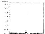
제9도는 참고예 4에서 얻어진 펩티드를 사용해서 실시예 9에 기재된 방법에 의해 측정한 각 혈청검체의 OD492값 분포도이다.9 is an OD492 value distribution diagram of each serum sample measured by the method described in Example 9 using the peptide obtained in Reference Example 4. FIG.
제10도는 참고예 5에서 얻어진 펩티드를 사용해서 실시예 9에 기재된 방법에 의해 측정한 각 혈청검체의 OD492값 분포도이다.FIG. 10 is a distribution diagram of the OD492 value of each serum sample measured by the method described in Example 9 using the peptide obtained in Reference Example 5. FIG.
제11도는 참고예 6에서 얻어진 펩티드를 사용해서 실시예 9에 기재된 방법에 의해 측정한 각 혈청검체의 OD492값 분포도이다.FIG. 11 is a distribution diagram of the OD492 value of each serum sample measured by the method described in Example 9 using the peptide obtained in Reference Example 6. FIG.
제12도는 실시예 6에서 얻어진 펩티드를 사용해서 실시예 9에 기재된 방법에 의해 측정한 각 혈청검체의 OD492값 분포도이다.FIG. 12 is a distribution diagram of the OD492 value of each serum sample measured by the method described in Example 9 using the peptide obtained in Example 6. FIG.
제13도는 실시예 7에서 얻어진 펩티드를 사용해서 실시예 9에 기재된 방법에 의해 측정한 각 혈청검체의 OD492값 분포도이다.FIG. 13 is a distribution diagram of the OD492 value of each serum sample measured by the method described in Example 9 using the peptide obtained in Example 7. FIG.
제14도는 실시예 8에서 얻어진 펩티드를 사용해서 실시예 9에 기재된 방법에 의해 측정한 각 혈청검체의 OD492값 분포도이다.FIG. 14 is a distribution diagram of the OD492 value of each serum sample measured by the method described in Example 9 using the peptide obtained in Example 8. FIG.
제15도는 참고예 7에서 얻어진 펩티드를 사용해서 실시예 9에 기재된 방법에 의해 측정한 각 혈청검체의 OD492값 분포도이다.FIG. 15 is a distribution diagram of the OD492 value of each serum sample measured by the method described in Example 9 using the peptide obtained in Reference Example 7.
그리고, 이들 도면에 있어서, 각 기호는 다음과 같다.In these drawings, each symbol is as follows.
●:GPT〉200 IU ; HBsAg ; (-) 혈청 A의 OD492값●: GPT> 200 IU; HBsAg; (-) OD492 value of serum A
○:GPT〉200 IU ; HBsAg ; (-) 혈청 B의 OD492값○: GPT> 200 IU; HBsAg; (-) OD492 value of serum B
×:GPT〉200 IU ; HBsAg ; (-) 혈청 C의 OD492값×: GPT> 200 IU; HBsAg; (-) OD492 value of serum C
본 발명은 펩티드 및 그의 용도에 관한 것이다.The present invention relates to peptides and their use.
본 발명에 의해서 제공되는 펩티드는 비 A 비 B형 간염 관련 항원(이하, 이것을 HCV 관련 항원이라 약칭함)에 특이성을 갖는 항체(이하, 이것을 항 HCV 항체라 약칭한다)와 고도로 특이적으로 결합하는 능력을 가지므로, 항 HCV 항체의 측정에 이용할 수 있다.Peptides provided by the present invention bind highly specifically to antibodies having specificity for non-A non-B hepatitis B antigens (hereinafter abbreviated as HCV related antigens) (hereinafter abbreviated as anti HCV antibodies). Since it has the ability, it can use for the measurement of anti HCV antibody.
본 발명에 의해서 제공되는 항 HCV 항체의 측정시약은 혈청 또는 혈장중에 항 HCV 항체를 고감도로 검출하는 능력을 가지고 있으며, 항 HCV 항체의 측정에 유용하다.The reagent for measuring anti-HCV antibodies provided by the present invention has the ability to detect anti-HCV antibodies in serum or plasma with high sensitivity and is useful for measuring anti-HCV antibodies.
간질환의 대부분을 차지하는 바이러스성 간염의 병인 바이러스로서는 현재 5종류가 알려지고 있으며, 각각 A형, B형, C형, D형, E형 간염 바이러스라 불리우고 있다. 바이러스성 간염중에서도 A형 간염 및 E형 간염은 경구 감염이며, 일과성 감염만으로 만성화하는 일은 없지만, B형 간염 및 C형 간염은 지속 감염에 의해 만성화하고, 높은 확률로 간경변 또는 간암으로 이향하기 때문에, 커다란 문제로 되어 있다. A형 간염, B형 간염 및 D형 간염에 대해서는 그들의 원인 바이러스가 발견되었으며, 현재로서는 면역학적인 진단이 가능해지고 있다. E형 간염 바이러스에 대해서도 최근 유전자가 단리되었다는 보고가 있다. 수혈 후 비 A비 B형 간염(이하, 이것을 PTNANBH라 약칭한다)의 병인 바이러스에 대해서는 많은 연구자에 의한 연구에도 불구하고 오랫동안 밝혀지지 않았으나, 1988년에 미국의 카이론사(Chiron Corporation)의 연구그룹에 의해서, PTNANBH에 감염시킨 침팬지의 혈장에서 PTNANBH 바이러스의 유전자의 단리, 동정이 이루어져[Science 제244권 제359페이지(1988) 및 Science 제244권 제362페이지(1988년)참조], C형 간염 바이러스(이하, 이것을 HCV라 약칭함)이라 명명되었다. 그 유전자의 염기 배열이라 추정되는 것의 일부는 이미 명확해지고 있으며[유럽 특허 제 0318216호 명세서 참조], 항 HCV 항체의 검출이 가능하게 되었고, HCV 감염에 대해서는 혈청학적 진단이 가능하게 되었다.Five types of viruses are known as viral hepatitis, which accounts for most of liver diseases, and are called hepatitis A, B, C, D, and E viruses, respectively. Among viral hepatitis, hepatitis A and hepatitis E are oral infections, and they are not chronicized by transient infections alone, but hepatitis B and hepatitis C are chronicized by persistent infection and have a high probability of turning to cirrhosis or liver cancer. It is a big problem. The causative viruses have been found for hepatitis A, hepatitis B, and hepatitis D, and immunological diagnosis is now possible. Genes have recently been isolated for hepatitis E virus. The pathogenesis virus of non-A non-A hepatitis B (hereinafter abbreviated as PTNANBH) after transfusion has not been known for a long time despite many researchers' studies, but in 1988, it was reported to a research group of the Iron Corporation of the United States. Isolation and identification of the gene of PTNANBH virus in the plasma of chimpanzees infected with PTNANBH were performed (see Science Vol. 244, pp. 359 (1988) and Science Vol. 244, pp. 362 (1988)). (Hereinafter abbreviated as HCV). Part of what is assumed to be the nucleotide sequence of the gene has already been clarified (refer to European Patent No. 0318216 specification), which enables the detection of anti-HCV antibodies and the serological diagnosis of HCV infection.
또, 본 발명자들중 한 사람을 포함한 몇 사람에 의해서 PTNANBH의 원인이 되는 바이러스의 유전자로 추정되는 리보헥산이 PTNANBH 환자의 혈청에서 단리되어, 동정되었다는 것이 보고되고 있다[Gastroenterologia Japonica 제24권, 제5호 제540페이지(1989년):Gastroenterologia Japonica 제24권, 제5호 제545페이지(1989년): 및 내과 제64권 제6호 제1022페이지(1989) 참조].In addition, it has been reported that ribohexane, which is presumed to be the gene of the virus causing PTNANBH, has been isolated and identified in the serum of PTNANBH patients by several persons including one of the present inventors. [Gastroenterologia Japonica Vol. 24, Vol. No. 5, page 540 (1989): Gastroenterologia Japonica, Vol. 24, No. 5, page 545 (1989): and Internal Medicine, Vol. 64, No. 6, page 1022 (1989).
지금까지 cDNA 라이브러리로부터의 필요한 cDNA의 스크리닝의 수단으로서, λ파아지를 이용한 항원항체 반응에 의해서 항 HCV 항체의 양성 또는 음성의 판정을 행하는 것이 일반적으로 행하여져 왔으나, 상기한 면역 스크리닝법은 정량성이 없고, 대장균의 발현산물중의 비특이한 항원성분과의 반응이 일어나는 경우가 있으며, 현재 HCV이 유전자를 클로닝하고, 파아지에 조입해서 효모를 숙주로하여 발현시킨 항원단백을 사용하여 행하는 효소면역 측정법에 의한 항 HCV 항체의 검사약의 개발이 검토되고 있다[내과 제64권, 제6호 제1027페이지 1989년) 참조]. 또한, 바이러스 또는 그의 항원성분에 의해서 감작된 젤라틴 입자가 항 바이러스 항체 존재하에서 응집하는 성질을 이용한 입자 응집법 또는 효소면역측정을 바이러스 또는 그의 항원성분으로 코팅된 비이드를 사용해서 행하는 비이드법의 개발이 진행되고 있다.Until now, as a means of screening the necessary cDNA from the cDNA library, it has been generally performed to determine the positive or negative anti-HCV antibody by antigen antibody reaction using lambda phage, but the above-described immune screening method is not quantitative In some cases, reaction with non-specific antigen components in expression products of Escherichia coli occurs, and the enzyme immunoassay is carried out using an antigen protein expressed by HCV cloning the gene, incorporating phage into yeast as a host. Development of test drugs for anti-HCV antibodies is under consideration (see Internal Medicine Vol. 64, No. 6, page 1027, 1989). In addition, the development of the bead method in which particle agglomeration or enzymatic immunoassay is carried out using a virus or its antigen component coated with gelatin particles sensitized by a virus or an antigen component thereof in the presence of an antiviral antibody. This is going on.
HCV 관련 항원을 이용한 지금까지의 효소면역측정법에서는, 측정대상이 임상적으로 PTNANBH라 진단된 경우에 있어서도 항 HCV 항체양성률은 약 75%이며, 항 HCV 항체에 음성인 PTNANBH가 약 25%의 비율로 함유되어 있다. 또 상기한 효소면역측정법에서 측정대상이 건강하고 정상적인 사람인 경우에는 양성률이 약 1%이며, 통계학적인 HCV 만연률이 약 3%라는데서 약 2%의 항 HCV 항체양성검체를 보고도 놓치고 있다. 이로 인해서 헌혈자로서의 HCV 캐리어 스크리닝에 있어서는, 캐리어를 보고도 놓치는 것을 피할 수 없으며, 비 A 비 B형 간염 바이러스 감염 혈액의 수혈을 저지하는 비율이 반드시 높지는 않다. 한편, HCV의 유전자를 클로닝하고, 파아지에 조입해서 효모를 숙주로 하여 발현시킨 항원 단백은 비특이한 여러가지의 항원 성분을 함유하고 있으므로, 그의 항원 단백은 비특이한 여러가지의 항원 성분을 함유하고 있으므로, 그의 항원 단백을 시약으로서 사용하여 항 HCV 항체의 측정을 행할 경우에는, 그 시약이 시료중의 항 HCV 항체 이외의 다른 비특이한 항체 성분마저도 인식하는 것으로 되며, 측정 결과는 반드시 항 HCV 항체의 존재를 정확하게 나타내고 있다고 말할 수 없다. 이와 같이 HCV 관련 항원을 이용한 지금까지의 효소 면역측정법에 의하면, 항 HCV 항체의 존재를 정확하게 알 수 없는 것이 현재의 상황이다.In the previous enzyme immunoassay using HCV-related antigens, even when the subject was clinically diagnosed as PTNANBH, the anti-HCV antibody positive rate was about 75%, and PTNANBH negative for anti-HCV antibody was about 25%. It is contained. In addition, in the enzyme immunoassay, if the subject is healthy and normal, the positive rate is about 1%, and the statistical HCV prevalence rate is about 3%, and about 2% of the anti-HCV antibody positive samples are missed. For this reason, in HCV carrier screening as a blood donor, a missed carrier cannot be missed, and the ratio which prevents transfusion of non-A non-Hepatitis B virus infected blood is not necessarily high. On the other hand, since the antigenic protein cloned into the phage, injected into phage and expressed in yeast as a host contains a variety of non-specific antigen components, since the antigenic protein contains a variety of non-specific antigen components, When the anti-HCV antibody is measured using an antigen protein as a reagent, the reagent also recognizes non-specific antibody components other than the anti-HCV antibody in the sample, and the measurement result must accurately detect the presence of the anti-HCV antibody. I cannot say that it is. As described above, according to the enzyme immunoassay method using the HCV-associated antigen, the present situation is that the presence of the anti-HCV antibody cannot be accurately known.
본 발명의 하나의 목적은 항 HCV 항체와 특이적으로 결합하는 능력을 갖는 펩티드를 제공하는데 있다.One object of the present invention is to provide a peptide having the ability to specifically bind an anti HCV antibody.
본 발명의 다른 하나의 목적은 항 HCV 항체의 측정시약을 제공하는 것에 있다.Another object of the present invention is to provide a reagent for measuring an anti-HCV antibody.
본 발명자들은, PTNANBH 관련 유전자에 코팅되는 폴리펩티드에서 선택된 단편 중에서, 비 A 비 B형 간염 관련 항원에 대한 특이성을 갖는 항체와 특이적으로 결합하는 능력을 갖는 특정의 펩티드를 발견하고, 본 발명을 완성하기에 이르렀다.The inventors have found, among fragments selected from polypeptides coated with PTNANBH related genes, specific peptides having the ability to specifically bind to antibodies having specificity for non-A non-B hepatitis B related antigens and completed the present invention. It came to the following.
본 발명에 의하면, 상기한 목적은 LyS Arg Ser Thr Asn(배열번호:2), Arg Arg Tyr Lys Glu Lys Glu Lys(배열번호:4), 또는 Ala Ile Ile Pro Asp Arg Glu Val Leu Tyr(배열번호:6)의 아미노산 배열을 함유한, HCV 관련 항원에 대해서 특이성을 갖는 항체와 특이적으로 결합하는 능력을 갖는 펩티드를 제공함으로써 달성된다.According to the present invention, the above object is LyS Arg Ser Thr Asn (array number: 2), Arg Arg Tyr Lys Glu Lys Glu Lys (array number: 4), or Ala Ile Ile Pro Asp Arg Glu Val Leu Tyr (array number) It is achieved by providing a peptide having the ability to specifically bind an antibody having specificity for an HCV related antigen, containing the amino acid sequence of: 6).
구체적으로는, (Ⅰ) 식(1) : Lys Asp Arg Thr Gln Gln Arg Lys Thr Lys Arg Ser Thr Asn Arg Arg Arg Ser Lys Asn Glu Lys Lys Lys Lys(배열번호:1)로서 나타내는 아미노산 배열을 갖는 펩티드 또는 그의 단편으로된 펩티드로서, 그 펩티드가 Lys Arg Ser Thr Asn(배열번호:2)의 아미노산 배열을 가지며, HCV 관련항원에 대해서 특이성을 갖는 항체와 특이적으로 결합하는 능력을 갖는 펩티드, (Ⅱ) 식 (2):Glu Lys Lys Gly Glu Ala Ser Asn Gly Glu Ala Glu Asn Asp Thr His Lys Lys Gln Arg Arg Tyr Lys Glu Lys Glu Lys Thr Ala Thr Asn Asn Pro Gly Lys Asn Lys Lys Pro Arg(배열번호:3)으로 나타내는 아미노산 배열을 갖는 펩티드 또는 그의 단편으로 된 펩티드이며, 그 펩티드가 Arg Arg Thr Lys Glu Lys Glu Lys(배열번호:4)의 아미노산 배열을 가지며, HCV 관련 항원에 대한 특이성을 갖는 항체와 특이적으로 결합하는 능력을 갖는 펩티드, (Ⅲ) 식 (3):Arg Val Val Leu Ser Gly Lys Pro Ala Ile Ile Pro Asp Arg Glu Val Leu Tyr Arg Glu Phe Asp Glu Met Glu Glu Cys Ser Gln His Leu Pro Tyr Ile Glu Gln Gly Met Met(비열번호:5)로 나타내는 아미노산 배열을 갖는 펩티드 또는 그의 단편으로된 펩티드로서 그 펩티드가 Ala Ile Ile Pro Asp Arg Glu Val Leu Tyr(배열번호:6)의 아미노산 배열을 가지며, HCV 관련 항원에 대한 특이성을 갖는 항체와 특이적으로 결합하는 능력을 갖는 펩티드이다.Specifically, (I) Formula (1): A peptide having an amino acid sequence represented as Lys Asp Arg Thr Gln Gln Arg Lys Thr Lys Arg Ser Thr Asn Arg Arg Arg Ser Lys Asn Glu Lys Lys Lys Lys (SEQ ID NO: 1). Or a peptide thereof, the peptide having the amino acid sequence of Lys Arg Ser Thr Asn (SEQ ID NO: 2) and having the ability to specifically bind to an antibody having specificity for HCV related antigen, (II Formula (2): Glu Lys Lys Gly Glu Ala Ser Asn Gly Glu Ala Glu Asn Asp Thr His Lys Lys Gln Arg Arg Tyr Lys Glu Lys Glu Lys Thr Ala Thr Asn Asn Pro Gly Lys Asn Lys Lys Pro Arg A peptide having an amino acid sequence represented by 3) or a fragment thereof, wherein the peptide has an amino acid sequence of Arg Arg Thr Lys Glu Lys Glu Lys (SEQ ID NO: 4), and has an antibody specific for HCV-associated antigen; Peptides having the ability to specifically bind, (III) Formula (3): Arg Val Peptide with an amino acid sequence represented by Val Leu Ser Gly Lys Pro Ala Ile Pro Asp Arg Glu Val Leu Tyr Arg Glu Phe Asp Glu Met Glu Glu Cys Ser Gln His Leu Pro Tyr Ile Glu Gln Gly Met Met (SEQ ID NO: 5) Or a fragment thereof, the peptide having the amino acid sequence of Ala Ile Ile Pro Asp Arg Glu Val Leu Tyr (SEQ ID NO: 6) and having the ability to specifically bind to an antibody having specificity for HCV related antigens. Peptides.
또, 상기한 펩티드로서 된 항 HCV 항체의 측정시약을 제공함으로써 달성된다.It is also achieved by providing a reagent for measuring anti-HCV antibody as the peptide described above.
본 명세서에 있어서는 각종 아미노산 잔기를 다음의 약호로서 기술한다.In this specification, various amino acid residues are described as the following abbreviations.
Ala:L-알라닌 잔기 Arg:L-아르기닌 잔기Ala: L-alanine residues Arg: L-arginine residues
Asn:L-아스파라긴 잔기 Asp:L-아스파르트산 잔기Asn: L-asparagine residues Asp: L-aspartic acid residues
Cys:L-시스틴 잔기 Gln:L-글루타민 잔기Cys: L-cystine residues Gln: L-glutamine residues
Glu:L-글루탐산 잔기 Gly:글리신 잔기Glu: L-glutamic acid residues Gly: glycine residues
His:L-히스티딘 잔기 Ile:L-이소로이신 잔기His: L-histidine residue Ile: L-isoleucine residue
Leu:L-로이신 잔기 Lys:L-리진 잔기Leu: L-leucine residues Lys: L-lysine residues
Met:L-메티오닌 잔기 Phe:L-페닐알라닌 잔기Met: L-methionine residue Phe: L-phenylalanine residue
Pro:L-프롤린 잔기 Ser:L-세린 잔기Pro: L-proline residues Ser: L-serine residues
Thr:L-트레오닌 잔기 Trp::-트립토판 잔기Thr: L-threonine residues Trp ::-tryptophan residues
Tyr:L-티로신 잔기 Val:L-발린 잔기Tyr: L-tyrosine residues Val: L-valine residues
본 명세서에 있어서는, 통상의 방법에 따라서 아미노산 배열을 N 말단의 아미노산 잔기가 왼쪽에 위치하고, C말단의 아미노산 잔기가 오른쪽에 위치하도록 기술한다.In this specification, the amino acid sequence is described so that the N-terminal amino acid residue is located on the left side and the C-terminal amino acid residue is located on the right side according to a conventional method.
본 발명의 펩티드는 HCV 관련 항원에 대해서 특이성을 갖는 항체와 특이적으로 결합하는 능력을 갖는 펩티드이며, 펩티드중에 적어도 1Ys Arg Ser Thr Asn(배열번호:2), Arg Arg Tyr Lys Glu Lys Glu Lys(배열번호:4), 또는 Ala Ile Ile Pro Asp Arg Glu Val Leu Tyr(배열번호:6)의 아미노산 배열을 가지고 있는 것이다.Peptides of the present invention are peptides that have the ability to specifically bind to antibodies having specificity for HCV related antigens, among which at least 1 Ys Arg Ser Thr Asn (SEQ ID NO: 2), Arg Arg Tyr Lys Glu Lys Glu Lys ( SEQ ID NO: 4) or Ala Ile Ile Pro Asp Arg Glu Val Leu Tyr (SEQ ID NO: 6).
이들의 부분적 아미노산 배열중, 특히 Lys Arg Ser Thr Asn의 배열(배열 번호:2)를 갖는 펩티드는 항원성이 높고, HCV 관련 항원에 특이성을 갖는 항체의 측정에 있어서 바람직하게 사용된다.Among these partial amino acid sequences, in particular, peptides having an arrangement of Lys Arg Ser Thr Asn (SEQ ID NO: 2) are preferably used for the determination of antibodies having high antigenicity and specificity to HCV related antigens.
본 발명의 펩티드는 전기한 부분적 아미노산 배열을 갖는 것이라면, 특히 제한되는 것은 아니지만, 통상 10~40개의 아미노산으로 된 펩티드이다.The peptide of the present invention is not particularly limited as long as the peptide has the partial amino acid sequence described above, but is usually a peptide of 10 to 40 amino acids.
구체적으로는 다음과 같은 펩티드 또는 그의 단편으로 된 펩티드를 들 수 있다. 즉, 본 발명의 펩티드로서는, 전기한 식(Ⅰ)로 나타내는 아미노산 배열을 갖는 펩티드 또는 그의 단편으로 된 펩티드로서, 그 펩티드가 Lys Arg Ser Thr Asn(배열번호:2)의 아미노산 배열을 가지며, HCV 관련 항원에 대해서 특이성을 갖는 항체와 특이적으로 결합하는 능력을 갖는 펩티드를 들 수 있다. 여기서 단편으로 된 펩티드로서는, 예를들면 식(1-a)을 들 수 있으나, 이에 한정되는 것은 아니다.Specifically, the peptide of the following peptide or its fragment is mentioned. In other words, the peptide of the present invention is a peptide having the amino acid sequence represented by the above formula (I) or a peptide thereof, wherein the peptide has an amino acid sequence of Lys Arg Ser Thr Asn (SEQ ID NO: 2), and HCV. Peptides having the ability to specifically bind to antibodies having specificity for the relevant antigens are mentioned. Herein, the peptide as a fragment may be, for example, formula (1-a), but is not limited thereto.
식 (1-a):Formula (1-a):
Thr Lys Arg Ser Thr Asn Arg Arg Arg Ser(배열번호:7)Thr Lys Arg Ser Thr Asn Arg Arg Arg Ser (array no.7)
또, 본 발명의 펩티드로서는 전기한 식(2)로 나타내는 아미노산 배열을 갖는 펩티드 또는 그의 단편으로된 펩티드로서, 그 펩티드가 Arg Arg Tyr Lys Glu Lys Glu Lys(배열번호:4)의 아미노산 배열을 가지며, HCV 관련 항원에 대해서 특이성을 갖는 항체와 특이적으로 결합하는 능력을 갖는 펩티드를 들 수 있다. 여기서 단편으로 된 펩티드로서는 예를 들면 식(2-a), 식(2-b) 및 식(2-c)가 예시되지만, 이들에 한정되는 것은 아니다.In addition, the peptide of the present invention is a peptide having the amino acid sequence represented by the above formula (2) or a peptide thereof, wherein the peptide has an amino acid sequence of Arg Arg Tyr Lys Glu Lys Glu Lys (SEQ ID NO: 4). And peptides having the ability to specifically bind to antibodies having specificity for HCV related antigens. Examples of the peptides in the form of fragments include, but are not limited to, formulas (2-a), (2-b) and (2-c).
식 (2-a):Formula (2-a):
Arg Arg Tyr Lys Glu Lys Glu Lys Thr Ala Thr Asn Asn Pro Gly Lys Asn Lys Lys Pro Arg(배열번호:8)Arg Arg Tyr Lys Glu Lys Glu Lys Thr Ala Thr Asn Asn Pro Gly Lys Asn Lys Lys Pro Arg (SEQ ID NO: 8)
식 (2-b):Formula (2-b):
Thr His Lys Lys Gln Arg Arg Tyr Lys Glu Lys Glu Lys(배열번호:9)Thr His Lys Lys Gln Arg Arg Tyr Lys Glu Lys Glu Lys (SEQ ID NO: 9)
식 (2-c):Formula (2-c):
Arg Arg Tyr Lys Glu Lys Glu Lys Thr Ala(배열번호:10)Arg Arg Tyr Lys Glu Lys Glu Lys Thr Ala (SEQ ID NO: 10)
또, 본 발명의 펩티드로서는 전기한 식(3)에서 나타내는 아미노산 배열을 갖는 펩티드 또는 그의 단편으로된 펩티드로서, 그 펩티드가 Ala Ile Ile Pro Asp Arg Glu Val Leu Tyr(배열번호:6)의 아미노산 배열을 가지며, HCV 관련 항원에 대해서 특이성을 갖는 항체와 특이적으로 결합하는 능력을 갖는 펩티드를 들 수 있다. 따라서 단편으로 된 펩티드로서는, 예를들면 식(3-a), 식(3-b) 및 식(3-c)가 예시되지만, 이들에 한정되는 것은 아니다.In addition, the peptide of the present invention is a peptide having the amino acid sequence represented by the above formula (3) or a peptide thereof, wherein the peptide is an amino acid sequence of Ala Ile Pro Asp Arg Glu Val Leu Tyr (SEQ ID NO: 6). And peptides having the ability to specifically bind to antibodies having specificity for HCV related antigens. Therefore, examples of the peptide as the fragment include, but are not limited to, the formulas (3-a), (3-b) and (3-c).
식 (3-a):Formula (3-a):
Ala Ile Ile Pro Asp Arg Glu Val Leu Tyr Arg Glu Phe Asp Glu Met Glu Glu Cys Ser Gln His Leu Pro Tyr Ile Glu Gln Gly Met Met (배열번호:11)Ala Ile Ile Pro Asp Arg Glu Val Leu Tyr Arg Glu Phe Asp Glu Met Glu Glu Cys Ser Gln His Leu Pro Tyr Ile Glu Gln Gly Met Met (SEQ ID NO: 11)
식 (3-b)Formula (3-b)
Arg Val Val Leu Ser Gly Lys Pro Ala Ile Ile Pro Asp Arg Glu Val Leu Tyr(배열번호:12)Arg Val Val Leu Ser Gly Lys Pro Ala Ile Ile Pro Asp Arg Glu Val Leu Tyr (SEQ ID NO: 12)
식 (3-c)Formula (3-c)
Ala Ile Ile Pro Asp Arg Glu Val Leu Tyr(배열번호:13)Ala Ile Ile Pro Asp Arg Glu Val Leu Tyr (SEQ ID NO: 13)
본 발명의 펩티드로는 전기한 바와같이 여러가지의 것을 예시할 수 있지만, 바람직하게는 반응의 특이성의 점에서 식(1-a)의 펩티드를 들 수 있다. 본 발명의 펩티드는 이와같은 HCV 관련 항원에 대해서 특이성을 갖는 항체와 특이적으로 결합하는 능력을 갖는 것이다.As mentioned above, although the thing of the above-mentioned can mention various things, the peptide of Formula (1-a) can be mentioned from the point of specificity of reaction. The peptide of the present invention has the ability to specifically bind to an antibody having specificity for such HCV related antigen.
본 발명의 펩티드의 합성은 펩티드의 합성에 있어서 통상 사용되는 방법, 예를들면 고상 합성법, 또는 단계적 신장법, 프래그먼트 축합법과 같은 액상 합성법에 의해 행하여지지만, 고상 합성법에 의해 행하는 것이 조작상 간편하다[예를 들면, Journal of the American Chemical Society, 제85권, 제2149~2154페이지:일본 생화학회편「생화학 실험 강좌 1 단백질의 화학 Ⅳ 화학수식과 펩티드 합성」(소화 52년 11월 15일 가부시끼가이샤 도오꾜 화학 동인 발행), 제207~495페이지; 일본 생화학회편「속 생화학 실험 강좌 2단백질의 화학(하)」(소화 62년 5월 20일 가부시끼가이샤 도오꾜 화학 동인 발행), 제641~694페이지 참조].Synthesis of the peptide of the present invention is carried out by methods commonly used in the synthesis of peptides, for example, solid phase synthesis, or liquid phase synthesis such as stepwise stretching and fragment condensation, but it is easy to operate by solid phase synthesis. For example, Journal of the American Chemical Society, Vol. 85, pp. 2149-2154: Japanese Biochemistry Segment ``
본 발명의 펩티드의 고상 합성법에 의한 제조는, 예를들면 스티렌-디비닐벤젠 공중합체 등의 반응 용매에 불용성인 중합체에, 목적으로 하는 펩티드의 C말단에 대응하는 아미노산 또는 그의 아미드를 그들이 갖는 α-COOH기 또는 α-CONH2기에서 각각 수소원자를 제외하고 얻어지는 α-COO-기 또는 α-CONH-기를 개재시켜 결합시키고, 이어서 그 아미노산 또는 그 아미드에 목적으로 하는 펩티드의 N말단의 방향을 향해서, 대응하는 아미노산 또는 펩티드 그 아미노산 또는 단편이 갖는 α-COOH기 이외의 α-아미노기 등의 관능기를 보호한 다음 축합시켜 결합시키는 조작과, 그 결합한 아미노산 또는 펩티드 단편에 있어서의 α-아미노기 등의 펩티드 결합을 형성하는 아미노기가 갖는 보호기를 제거하는 조작을 순차 반복함으로써, 펩티드 사슬을 신장시키고, 목적을 하는 펩티드에 대응하는 펩티드 사슬을 형성하고, 이어서 그 펩티드 사슬을 중합체에서 탈리시키고, 또한 보호되고 있는 관능기에서 보호기를 제거함으로써 목적으로 하는 펩티드를 얻고, 이어서 이것을 정제함으로써 실시된다. 여기서, 펩티드 사슬의 중합체로부터의 탈리 및 보호기의 제거는 불화 수소를 사용해서 동시에 행하는 것이 부반응을 억제하는 관점에서 바람직하다. 또, 얻어진 펩티드의 정제는 역상 액체 크로마토그래피로 행하는 것이 효과적이다.Preparation by the solid-phase synthesis method of the peptide of the present invention, for example, α having an amino acid or an amide thereof corresponding to the C terminus of the target peptide in a polymer insoluble in a reaction solvent such as styrene-divinylbenzene copolymer The -COOH group or the α-CONH2 group is bonded via an α-COO- group or an α-CONH- group obtained without the hydrogen atom, respectively, and then the amino acid or its amide is directed to the N-terminal direction of the target peptide. To protect the functional groups, such as α-amino groups other than the α-COOH group possessed by the corresponding amino acid or peptide amino acid or fragment thereof, to condense and bind, the α-amino group in the bound amino acid or peptide fragment By repeating the procedure of removing the protecting group which the amino group which forms a peptide bond, the peptide chain is extended and the objective is To form a peptide chain corresponding to the peptide, then obtain the peptide of interest was eliminated by the peptide chain from the polymer, and removing the protective group in the functional group being protected, which is carried out, followed by purification. Here, desorption from the polymer of the peptide chain and removal of the protecting group are preferably performed simultaneously using hydrogen fluoride from the viewpoint of suppressing side reactions. Moreover, it is effective to perform the purification of the obtained peptide by reverse phase liquid chromatography.
본 발명의 펩티드는 특이적으로 항 HCV 항체를 결합하는 능력을 가지므로, HCV 감염에 의해 출현하는 항 HCV 항체의 검출을 위한 측정시약으로서 유효하다.Since the peptide of the present invention has the ability to specifically bind anti-HCV antibodies, it is effective as a measurement reagent for detection of anti-HCV antibodies manifested by HCV infection.
즉, 본 발명의 측정시약은 본 발명의 펩티드를 함유하여 된 것이며, 전기한 바와같이 본 발명의 펩티드를 단독으로 또는 2종 이상을 혼합해서 사용된다.That is, the measurement reagent of the present invention contains the peptide of the present invention, and as described above, the peptide of the present invention is used alone or in combination of two or more thereof.
본 발명의 펩티드를 이용한 항 HCV 항체의 측정은 형광면역측정법, 수신혈구 응집법, 방사면역측정법, 효소면역측정법의 어느것을 이용함으로써 행하여진다. 이들의 방법은 어느 것이나 공지이지만, 예를들어 효소면적측정법을 이용하는 경우에 대해서 이하에 설명하겠다.The measurement of anti HCV antibody using the peptide of the present invention is carried out by using any one of fluorescence immunoassay, received hemagglutination, radioimmunoassay, and enzyme immunoassay. Although these methods are all well-known, the case where enzyme area measurement method is used, for example is demonstrated below.
측정계 전체의 구성요소는 담체, 측정시약으로서의 본 발명의 펩티드, 블록킹제, 피검시료, 표시용 항체, 효소 및 발색제로서 된다. 담체에 본 발명의 펩티드를 코팅하고, 이어서 펩티드 코팅 담체에 블록킹제를 작용시켜서 담체상의 비특이적인 담백결합 부위를 블록하고, 펩티드 코팅 담체에 피검시료를 가하여 인큐베이트하고, 계속해서 효소 표시 항체를 접촉시켜 인큐베이트하고, 다음에 이와같이 처리한 담체에 발색제를 가하여 인큐베이트하고, 효소와 발색제와의 반응의 반응 산물의 생성량을 흡광도계를 사용하여 측정한다. 그리고, 코팅에 사용되는 본 발명의 펩티드는 1종류이거나 2종류 이상이더라도 좋다. 담체로서는 효소면역검정용 컵, 또는 유리 또는 수지제의 비이드를 사용하는 것이 바람직하다. 측정에 앞서서, 본 발명의 펩티드를 0.01M 탄산 완충액에 용해하고, 그 용액을 예를들면 폴리스티렌계 효소면역검정용 컵에 가한 다음, 4℃로 하룻밤 또는 실온에서 3시간 정치함으로서, 담체 표면은 본 발명의 펩티드에 의해서 코팅된다. 담체상의 비특이적인 단백 결합 부위를 블록하기 위한 블록킹제로서는, 예를들면 소혈청 알부민, 카제인, 탈비분유, 항사람 IgG 항체 또는 항 사람 IgM 항체를 얻기 위한 면역원 동물의 혈청, 젤라틴 등이 사용된다. 표시용 항체로서는, 예를들면 항사람 IgG 항체, 항사람 IgM항체등의 사용된다. 또 효소로서는, 예를들면 알칼리 포스파타아제, 글루코오스옥시다아제, 퍼옥시다아제, 베타갈락토시다아제 등을 들 수 있다. 측정에 앞서서, 글루타르알데히드 등의 2개 이상의 관능기를 갖는 화학물을 사용해서, 표시용항체에 효소를 결합시켜서 콘쥬게이트하고, 측정계 전체의 구성요소의 일부로서 이미 준비해 두는 것이 바람직하다. 발색제는 선택한 효소에 따라서 적당히 사용하면 좋다. 예를들면, 효소로서 퍼옥시다아제를 선택한 경우에는 o-페닐렌디아민 등을 사용하는 것이 바람직하다.The components of the whole measurement system are the carrier, the peptide of the present invention as the measurement reagent, the blocking agent, the test sample, the display antibody, the enzyme, and the coloring agent. The peptide is coated with a peptide of the present invention, and then a blocking agent is applied to the peptide-coated carrier to block a nonspecific white binding site on the carrier, incubated by adding a test sample to the peptide-coated carrier, and then contacting the enzyme-labeled antibody. Incubate was carried out by adding a coloring agent to the carrier thus treated, and the production of the reaction product of the reaction between the enzyme and the coloring agent was measured using an absorbance meter. The peptide of the present invention used for coating may be one kind or two or more kinds. It is preferable to use a cup for enzyme immunoassay or glass or resin beads as a carrier. Prior to the measurement, the carrier surface was dissolved by dissolving the peptide of the present invention in 0.01 M carbonate buffer, adding the solution to, for example, a polystyrene-based enzyme immunoassay cup, and then standing at 4 ° C. overnight or at room temperature for 3 hours. Coated by the peptide of the invention. As a blocking agent for blocking the nonspecific protein binding site on the carrier, for example, bovine serum albumin, casein, desecreted milk, serum of human immunogen to obtain anti-human IgG antibody or anti-human IgM antibody, gelatin and the like are used. As the antibody for display, anti-human IgG antibody, anti-human IgM antibody, etc. are used, for example. Moreover, as an enzyme, alkali phosphatase, glucose oxidase, peroxidase, beta galactosidase, etc. are mentioned, for example. Prior to the measurement, it is preferable to use a chemical having two or more functional groups, such as glutaraldehyde, to conjugate the enzyme to a display antibody to conjugate it, and to prepare it as a part of the components of the whole measurement system. A coloring agent may be suitably used depending on the enzyme selected. For example, when peroxidase is selected as an enzyme, it is preferable to use o-phenylenediamine or the like.
이와같이 본 발명에 의하면, 항 HCV 항체와 특이적으로 결합하는 능력을 갖는 펩티드가 제공된다. 이 펩티드에 의해 기존의 항 HCV 항체 측정 시약을 상회하는 감도 및 특이성을 갖는 항 HCV 항체 측정시약을 제공하는 것이 가능해졌다.Thus, according to this invention, the peptide which has the ability to specifically bind with anti HCV antibody is provided. This peptide has made it possible to provide an anti-HCV antibody measuring reagent having sensitivity and specificity that exceeds that of existing anti-HCV antibody measuring reagents.
이하, 실시예에 의해 본 발명을 구체적으로 설명하지만, 본 발명은 이들의 실시예에 의해 한정되는 것은 아니다.Hereinafter, although an Example demonstrates this invention concretely, this invention is not limited by these Examples.
[실시예 1]Example 1
식(1):Lys Asp Arg Thr Gln Gln Arg Lys Thr Lys Arg Ser Thr Asn Arg Arg Ar Ser Lys Asn Glu Lys Lys Lys Lys (배열번호:1)로 나타내는 펩티드를 자동합성 장치[미국 Applid Bio Systems 사제, Model 431A]를 사용해서 고상 합성법에 의해 합성하였다.Formula (1): Lys Asp Arg Thr Gln Gln Arg Lys Thr Lys Arg Ser Thr Asn Arg Arg Ar Ser Lys Asn Glu Lys Lys Lys Lys (SEQ ID NO: 1). Model 431A] was synthesized by the solid phase synthesis method.
즉, 4-[Nα-(t-부톡시카르보닐)-Nε-(2-클로로벤질옥시카르보닐)-L-리딜옥시메틸]페닐아세틸아미드메틸기:Namely, 4- [Nα- (t-butoxycarbonyl) -Nε- (2-chlorobenzyloxycarbonyl) -L-ridyloxymethyl] phenylacetylamidemethyl group:

를 0.65밀리몰/g(수지)의 비율로 갖는 스티렌-디비닐벤젠 공중합체[스티렌과 디비닐벤젠과 구성몰비:99:1]로서 된 입상 수지[미국 Applied Bio Systems사제, PAM 리진(Lysine), t-Boc-L-Lys(Cl-Z)]를 760mg 사용하고 이것에 표 1에 나타낸 일련의 조작에 따라서 목적하는 펩티드의 N말단의 방향을 향해서 대응하는 L-아르기닌, L-아스파라긴, L-아스파르트산, L-글루타민, L-글루탐산, L-리진, L-세린, L-트레오닌을 순차 결합시켰다. 축합반응에 있어서, 상기한 아미노산은 각각 Nα-(t-부톡시카르보닐)-Nγ-(메시틸렌-2-술포닐)-L-아르기닌, N-(t-부톡시카르보닐)-L-아스파라긴, N-(t-부톡시카르보닐)L-아스파르트산-β-벤질에스테르, M-(t-부톡시카르보닐)-L-글루타민, N-(t-부톡시카르보닐)-L-글루탐산-γ-벤질에스테르, Nα-(t-부톡시카르보닐)-Nε-(2-클로로벤질옥시카르보닐)-L-리진, N-(t-부톡시카르보닐)-o-벤질-L-세린, N-(t-부톡시카르보닐)-O-벤질-L-트레오닌으로서 사용하고, 그들의 사용량은 기질에 대해서 약 4배 몰량으로 하였다. 축합반응은 실온하에서 행하였다. 아미노산 1잔기를 결합시키기 위하여 소요된 전공정을 포함한 반응시간은 100~110분간의 범위내였다. 모든 아미노산에 대해서는의 반응조작이 종료한 다음, 얻어진 수지를 글라스필터상에서 디클로로메탄 및 메탄올을 사용하여 순차 세척하고, 이어서 진공건조함으로써 2.58g의 건조수지를 얻었다. 다음에 폴리트리플루오로모노클로로에틸렌제의 반응용기(가부시끼가이샤 펩티드 연구소제, HF-반응장치 Ⅰ형)중에서, 얻어진 건조수지 0.7g을 아니솔 1.05ml 및 에틸메틸술피드 0.175ml과 혼합하고, 이 혼합물에 -20℃ 온도로 불화수소 7.0ml을 가하여, 같은 온도에서 30분간, 이어서 0℃의 온도에서 30분간 교반하였다. 얻어진 반응 혼합물에서 불화수소, 아니솔 및 에틸메틸술피드를 감압하에 제거하고, 잔류물을 글라스필터상에서 디에틸에테르 및 디클로로메탄을 사용하여 충분히 세척하였다. 얻어진 그 잔류물을 2N초산 수용액으로 추출하고, 추출액을 동결건조함으로써 조 펩티드 200mg을 얻었다. 얻어진 조 생성물을 분취용 역상 고속 액체크로마토그래피[컬럼:옥타데실화 실리카겔(입경:15μm) 충전 컬럼(내경:50mm, 길이:300mm), 닛뽕워터즈사제, μBONDASPHERE 15μ C18-100; 이동상:트리플루오로초산을 0.05% 함유하는 아세토니트릴과 물과의 혼합용매(아세토니트릴의 농도는 30분간에 걸쳐 10용량%에서 20용량%가 되도록 점차 변화시켰다); 유속 5ml분; 검출법:파장 210nm에 있어서의 흡광도]로 정제함으로써, 목적으로 하는 펩티드 정제물을 80mg 얻었다. 얻어진 정제물을 분석용 역상 고속 액체 크로마토그래피[컬럼:옥타데실화 실리카겔(입경:5μm) 충전 컬럼(내경:4mm, 길이:150mm), 도오요소오다 가부시끼가이샤제, TSK-gel ODS-8TM; 이동상:트리플루오로초산을 0.05용량% 함유하는 아세토니트릴과 물과의 혼합용매(아세토니트릴의 농도는 30분간에 걸쳐서 5용량%에서 50용량%가 되도록 점차 변화시켰다): 유속:1m: /분; 검출법; 파장 210nm에 있어서의 흡광도]한 결과, 15.0분에 단일의 날카로운 피이크가 나타났다. 고속원자충격법(이하, 이것을 FAB법이라 약칭함) 매스 스펙트럼에 의해 구하여진 정제물의 분자량은 3188였다(이론치:3187.53).Granular resin [Styrene-divinylbenzene copolymer having a ratio of styrene and divinylbenzene and a molar ratio of 99: 1] having a ratio of 0.65 mmol / g (resin) [manufactured by Applied Bio Systems, PAM Lysine, t-Boc-L-Lys (Cl-Z)] is used and the corresponding L-arginine, L-asparagine, and L- are directed toward the N-terminal direction of the desired peptide according to a series of manipulations shown in Table 1. Aspartic acid, L-glutamine, L-glutamic acid, L-lysine, L-serine, L-threonine were sequentially bound. In the condensation reaction, the amino acids described above are each Nα- (t-butoxycarbonyl) -Nγ- (mesitylene-2-sulfonyl) -L-arginine, N- (t-butoxycarbonyl)- L-asparagine, N- (t-butoxycarbonyl) L-aspartic acid-β-benzylester, M- (t-butoxycarbonyl) -L-glutamine, N- (t-butoxycarbonyl)- L-glutamic acid-γ-benzyl ester, Nα- (t-butoxycarbonyl) -Nε − (2-chlorobenzyloxycarbonyl) -L-lysine, N- (t-butoxycarbonyl) -o It was used as -benzyl-L-serine, N- (t-butoxycarbonyl) -O-benzyl-L-threonine, and their usage amount was about 4 times molar amount with respect to the substrate. The condensation reaction was performed at room temperature. The reaction time including the previous step required to bind 1 residue of amino acid was in the range of 100 to 110 minutes. After completion of the reaction operation for all amino acids, the obtained resin was washed sequentially with dichloromethane and methanol on a glass filter, followed by vacuum drying to obtain 2.58 g of a dry resin. Next, 0.7 g of the dried resin obtained was mixed with 1.05 ml of anisole and 0.175 ml of ethyl methyl sulfide in a reaction vessel made of polytrifluoromonochloroethylene (manufactured by KK Pept. Co., Ltd., HF-reactor Type I). 7.0 ml of hydrogen fluoride was added to this mixture at the temperature of -20 degreeC, and it stirred for 30 minutes at the same temperature, and then at the temperature of 0 degreeC for 30 minutes. Hydrogen fluoride, anisole and ethylmethyl sulfide were removed from the obtained reaction mixture under reduced pressure, and the residue was sufficiently washed with diethyl ether and dichloromethane on a glass filter. The obtained residue was extracted with a 2N acetic acid aqueous solution, and the extract was lyophilized to obtain 200 mg of crude peptide. The resulting crude product was subjected to preparative reverse phase high performance liquid chromatography [column: octadecylated silica gel (particle diameter: 15 μm) packed column (inner diameter: 50 mm, length: 300 mm), manufactured by Nippon Waters, μBONDASPHERE 15 μC18-100; Mobile phase: a mixed solvent of acetonitrile and water containing 0.05% trifluoroacetic acid (the concentration of acetonitrile was gradually changed from 10% to 20% by volume over 30 minutes); Flow rate 5 ml min; Detection method: absorbance at wavelength 210 nm] to obtain 80 mg of the target peptide purified product. The resulting purified product was analyzed for reverse phase high performance liquid chromatography [column: octadecylated silica gel (particle diameter: 5 m) packed column (inner diameter: 4 mm, length: 150 mm), manufactured by Toya Oda Co., Ltd., TSK-gel ODS-8TM; Mobile phase: Mixed solvent of acetonitrile and water containing 0.05% by volume of trifluoroacetic acid (the concentration of acetonitrile was gradually changed from 5% to 50% by volume over 30 minutes): Flow rate: 1m: / min ; Detection method; Absorbance at a wavelength of 210 nm] showed a single sharp peak at 15.0 minutes. The molecular weight of the purified product obtained by the high-speed atomic shock method (hereinafter abbreviated as FAB method) mass spectrum was 3188 (theoretical value: 3187.53).
[표 1]TABLE 1

[실시예 2]Example 2
실시예 1에 있어서와 마찬가지인 방법으로 펩티드의 고상 합성 및 정제를 행함으로써, 식 (1-a):Thr Lys Arg Ser Thr Asn Arg Arg Arg Ser(배열번호:7)로 나타낸 펩티드를 얻었다. 얻어진 펩티드를 분석용 역상 고속액체 크로마토그래피(전기와 같음)한 결과, 14.2분에 단일의 날카로운 피이크가 나타났다. FAB법 매스 스펙트럼에 의해 구하여진 펩티드의 분자량은 1243였다(이론치:1243.33).Solid phase synthesis and purification of the peptide were carried out in the same manner as in Example 1 to obtain a peptide represented by Formula (1-a): Thr Lys Arg Ser Thr Asn Arg Arg Arg Ser (SEQ ID NO: 7). The resulting peptide was subjected to analytical reversed phase high performance liquid chromatography (same as before), and a single sharp peak appeared at 14.2 minutes. The molecular weight of the peptide determined by FAB method mass spectrum was 1243 (theory: 1243.33).
[실시예 3]Example 3
실시예 1에 있어서와 마찬가지인 방법으로 펩티드의 고상합성 및 정제를 행함으로써, 식 (2-a); Arg Arg Tyr Lys Glu Lys Glu Lys Thr Ala Thr Asn Asn Pro Gly Lys Asn Lys Lys Pro Arg(배열번호:8)로 나타내는 펩티드를 얻었다. 얻어진 펩티드를 분석용 역상 고속 액체 크로마토그래피(전기와 같음)한 결과, 22.0분에 단일의 날카로운 피이크가 나타났다. FAB법 매스 스펙트럼에 의해 구하여진 펩티드의 분자량은 2542였다(이론치:2541.85).By solid phase synthesis and purification of the peptides in the same manner as in Example 1, formula (2-a); Peptides represented by Arg Arg Tyr Lys Glu Lys Glu Lys Thr Ala Thr Asn Asn Pro Gly Lys Asn Lys Lys Pro Arg (SEQ ID NO: 8). The resulting peptide was subjected to analytical reverse phase high performance liquid chromatography (same as before), and a single sharp peak appeared at 22.0 minutes. The molecular weight of the peptide determined by FAB method mass spectrum was 2542 (theoretical value: 2451.85).
[실시예 4]Example 4
실시예 1에 있어서와 마찬가지인 방법으로 펩티드의 고상 합성 및 정제를 행함으로써, 식 (2-b); Thr His Lys Lys Gln Arg Arg Tyr Lys Glu Lys Glu Lys(배열번호:9)로 나타낸 펩티드를 얻었다. 얻어진 펩티드를 분석용 역상 액체 크로마토그래피(전기와 같음)한 결과, 14.3분에 단일의 날카로운 피이크가 나타났다. FAB법 매스 스펙트럼에 의해 구하여진 펩티드의 분자량은 1758이었다(이론치:1758.01).By solid-phase synthesis and purification of the peptide in the same manner as in Example 1, formula (2-b); Peptides represented as Thr His Lys Lys Gln Arg Arg Tyr Lys Glu Lys Glu Lys (SEQ ID NO: 9). The resulting peptide was subjected to analytical reverse phase liquid chromatography (same as before), and a single sharp peak appeared at 14.3 minutes. The molecular weight of the peptide determined by FAB method mass spectrum was 1758 (theoretical value: 1758.01).
[실시예 5]Example 5
실시예 1에 있어서와 마찬가지인 방법으로 펩티드의 고상 합성 및 정제를 행함으로써, 식 (2-c):Arg Arg Tyr Lys Glu Lys Glu Lys Thr Ala(배열번호:10)로 나타낸 펩티드를 얻었다. 얻어진 펩티드를 분석용 역상 고속 액체 크로마토그래피(전기와 같음)한 결과, 14.5분에 단일의 날카로운 피이크가 나타났다. FAB법 매스 스펙트럼에 의해 구하여진 펩티드의 분자량은 1306이었다(이론치:1306.49).Solid phase synthesis and purification of the peptide were carried out in the same manner as in Example 1 to obtain a peptide represented by the formula (2-c): Arg Arg Tyr Lys Glu Lys Glu Lys Thr Ala (SEQ ID NO: 10). The resulting peptide was subjected to analytical reversed phase high performance liquid chromatography (same as the former) and showed a single sharp peak at 14.5 minutes. The molecular weight of the peptide determined by FAB method mass spectrum was 1306 (Theory: 1306.49).
[실시예 6]Example 6
실시예 1에 있어서와 마찬가지인 방법으로 펩티드의 고상 합성 및 정제를 행함으로써, 식 (3-a):Ala Ile Ile Pro Asp Arg Glu Val Leu Tyr Arg Glu Phe Asp Glu Met Glu Glu Cys Ser gLn His Leu Pro Tyr Ile Glu Glu Gly Met Met(배열번호:11)으로 나타낸 펩티드를 얻었다. 얻어진 펩티드를 분석용 역상 고속 액체 크로마토그래피(전기와 같음)한 결과, 29.8분에 단일의 날카로운 피이크가 나타났다. FAB법 매스 스펙트럼에 의해 구하여진 펩티드의 분자량은 3785였다(이론치:3785.19).Solid phase synthesis and purification of the peptide in the same manner as in Example 1 formula (3-a): Ala Ile Ile Pro Asp Arg Glu Val Leu Tyr Arg Glu Phe Asp Glu Met Glu Glu Cys Ser gLn His Leu Pro Peptides represented as Tyr Ile Glu Glu Gly Met Met (SEQ ID NO: 11) were obtained. The resulting peptide was subjected to analytical reverse phase high performance liquid chromatography (same as the former), and a single sharp peak appeared at 29.8 minutes. The molecular weight of the peptide determined by FAB method mass spectrum was 3785 (Theoretical value: 3785.19).
[실시예 7]Example 7
실시예 1에 있어서와 마찬가지인 방법으로 펩티드의 고상 합성 및 정제를 행함으로써, 식 (3-b):Arg Val Val Leu Ser Gly Lys Pro Ala Ile Ile Pro Asp Arg Glu Val Leu Tyr(배열번호:12)로 나타낸 펩티드를 얻었다. 얻어진 펩티드를 분석용 역상 고속 액체 크로마토그래피(전기와 같음)한 결과, 28.7분에 단일의 날카로운 피이크가 나타났다. FAB법 매스 스펙트럼에 의해 구하여진 펩티드의 분자량은 1953이었다(이론치:1953.15).Solid phase synthesis and purification of the peptide were carried out in the same manner as in Example 1, whereby Formula (3-b): Arg Val Val Leu Ser Gly Lys Pro Ala Ile Pro Asp Arg Glu Val Leu Tyr (SEQ ID NO: 12) The peptide shown is obtained. The resulting peptide was subjected to analytical reverse phase high performance liquid chromatography (same as the former), and a single sharp peak appeared at 28.7 minutes. The molecular weight of the peptide determined by FAB method mass spectrum was 1953 (theoretical value: 1953.15).
[실시예 8]Example 8
실시예 1에 있어서와 마찬가지인 방법으로 펩티드의 고상 합성 및 정제를 행함으로써, 식 (3-c):Ala Ile Ile Pro Asp Arg Glu Val Leu Tyr(배열번호:13)으로 나타내는 펩티드를 얻었다. 얻어진 펩티드를 분석용 역상 고속 액체 크로마토그래피(전기와 같음)한 결과, 26.9분에 단일의 날카로운 피이크가 나타났다. FAB법 매스 스펙트럼에 의해 구하여진 펩티드의 분자량은 1203이었다(이론치:1203.34).Solid phase synthesis and purification of the peptide were carried out in the same manner as in Example 1 to obtain a peptide represented by Formula (3-c): Ala Ile Pro Asp Arg Glu Val Leu Tyr (SEQ ID NO: 13). The resulting peptide was subjected to analytical reversed phase high performance liquid chromatography (same as before), and a single sharp peak appeared at 26.9 minutes. The molecular weight of the peptide determined by FAB method mass spectrum was 1203 (Theory: 1203.34).
[참고예 1]Reference Example 1
실시예 1에 있어서와 마찬가지인 방법으로 펩티드의 고상 합성 및 정제를 행함으로써, 식 (1)에서 나타낸 아미노산 배열을 갖는 펩티드중, Lys Arg Ser Thr Asn (배열번호:2)로 나타낸 아미노산 배열을 갖지 않는 식:Lys Asp Arg Thr Gln Gln Arg Lys Thr Lys(배열번호:14)로 나타낸 펩티드를 얻었다. 얻어진 펩티드를 분석용 역상 고속 액체 크로마토그래피(전기와 같음)한 결과, 11.9분에 단일의 날카로운 피이크가 나타났다. FAB법 매스 스펙트럼에 의해 구하여진 펩티드의 분자량은 1290이었다(이론치:1290.39).Solid phase synthesis and purification of the peptide were carried out in the same manner as in Example 1, so that the peptide having the amino acid sequence shown in Formula (1) did not have the amino acid sequence represented by Lys Arg Ser Thr Asn (SEQ ID NO: 2). A peptide represented by the formula: Lys Asp Arg Thr Gln Gln Arg Lys Thr Lys (SEQ ID NO: 14) was obtained. The resulting peptide was subjected to analytical reversed phase high performance liquid chromatography (same as the former) and showed a single sharp peak at 11.9 minutes. The molecular weight of the peptide determined by FAB method mass spectrum was 1290 (Theory: 1290.39).
[참고예 2]Reference Example 2
실시예 1에 있어서와 마찬가지인 방법으로 펩티드의 고상합성 및 정제를 행함으로써, 식(1)에서 나타낸 아미노산 배열을 갖는 펩티드중, Lys Arg Ser Thr Asn(배열번호:2)로 나타낸 아미노산 배열을 갖지 않는 식:Arg Ser Thr Asn Arg Arg Ser Lys Asn Glu Lys Lys Lys Lys(배열번호:15)로 나타낸 펩티드를 얻었다. 얻어진 펩티드를 분석용 역상 고속 액체 크로마토그래피(전기와 같음)한 결과, 13.6분에 단일의 날카로운 피이크가 나타났다. FAB법 매스 스펙트럼에 의해 구하여진 펩티드의 분자량은 1915이었다(이론치:1915.16).By solid-phase synthesis and purification of the peptides in the same manner as in Example 1, among the peptides having the amino acid sequence shown in Formula (1), the peptides having no amino acid sequence represented by Lys Arg Ser Thr Asn (SEQ ID NO: 2) A peptide represented by the formula: Arg Ser Thr Asn Arg Arg Ser Lys Asn Glu Lys Lys Lys Lys (SEQ ID NO: 15) was obtained. The resulting peptide was subjected to analytical reversed phase high performance liquid chromatography (same as the former) and showed a single sharp peak at 13.6 minutes. The molecular weight of the peptide determined by FAB method mass spectrum was 1915 (Theory: 1915.16).
[참고예 3]Reference Example 3
실시예 1에 있어서와 마찬가지인 방법으로 펩티드의 고상 합성 및 정제를 행함으로써, 식:Glu Gln Asp Gln Ile Lys Thr Lys Asp Arg Thr Gln Gln Arg Lys Thr Lys Arg Ser Thr Asn Arg Arg Arg Ser Lys Asn Glu Lys Lys Lys Lys(배열번호:16)로 나타낸 펩티드를 얻었다. 펩티드를 분석용 역상 고체 액체 크로마토그래피(전기와 같음)한 결과, 24.6분에 단일의 날카로운 피이크가 나타났다. FAB법 매스 스펙트럼에 의해 구하여진 펩티드의 분자량은 4031이었다(이론치:4031.38).Solid phase synthesis and purification of the peptides were carried out in the same manner as in Example 1, whereby Glu Gln Asp Gln Ile Lys Thr Lys Asp Arg Thr Gln Gln Arg Lys Thr Lys Arg Ser Thr Asn Arg Arg Arg Ser Lys Asn Glu Lys The peptide represented by Lys Lys Lys (SEQ ID NO: 16) was obtained. Analytical reversed phase solid liquid chromatography (same as before) showed a single sharp peak at 24.6 minutes. The molecular weight of the peptide determined by FAB method mass spectrum was 4031 (theoretical value: 4031.38).
[참고예 4]Reference Example 4
실시예 1에 있어서와 마찬가지인 방법으로 펩티드의 고상 합성 및 정제를 행함으로써, 식 (2)에서 나타낸 아미노산 배열을 갖는 펩티드중, Arg Arg Tyr Lys Glu Lys Glu Lys(배열번호:4)로 나타낸 아미노산 배열을 갖지 않는 식:Glu Lys Lys Gly Glu Ala Ser Asn Gly Ala Glu Asn Asp(배열번호:17)로 나타낸 펩티드를 얻었다. 얻어진 펩티드를 분석용 역상 고체 액체 크로마토그래피(전기와 같음)한 결과, 14.9분에 단일의 날카로운 피이크가 나타났다. FAB법 매스 스펙트럼에 의해 구하여진 펩티드의 분자량은 1473이었다(이론치:1473.47).Solid phase synthesis and purification of the peptide in the same manner as in Example 1 resulted in the amino acid sequence represented by Arg Arg Tyr Lys Glu Lys Glu Lys (SEQ ID NO: 4) in the peptide having the amino acid sequence shown in Formula (2). A peptide represented by the formula: Glu Lys Lys Gly Glu Ala Ser Asn Gly Ala Glu Asn Asp (SEQ ID NO: 17) was obtained. The resulting peptide was subjected to analytical reversed phase solid liquid chromatography (same as before) and showed a single sharp peak at 14.9 minutes. The molecular weight of the peptide determined by FAB method mass spectrum was 1473 (theory: 1473.47).
[참고에 5][5 for reference]
실시예 1에 있어서와 마찬가지인 방법으로 펩티드의 고상 합성 및 정제를 행함으로써, 식 (2)에서 나타낸 아미노산 배열을 갖는 펩티드중, Arg Arg Tyr Lys Glu Lys Glu Lys(배열번호:4)로 나타낸 아미노산 배열을 갖지 않는 식:Thr Asn Asn Pro Gly Lys Asn Lys Lys Pro Arg(배열번호:18)로 나타낸 펩티드를 얻었다. 얻어진 펩티드를 분석용 역상 고속 액체 크로마토그래피(전기와 같음)한 결과, 12.6분에 단일의 날카로운 피이크가 나타났다. FAB법 매스 스펙트럼에 의해 구하여진 펩티드의 분자량은 1253이었다(이론치:1253.38).Solid phase synthesis and purification of the peptide in the same manner as in Example 1 resulted in the amino acid sequence represented by Arg Arg Tyr Lys Glu Lys Glu Lys (SEQ ID NO: 4) in the peptide having the amino acid sequence shown in Formula (2). Peptide represented by formula: Thhr Asn Asn Pro Gly Lys Asn Lys Lys Pro Arg (SEQ ID NO: 18) was obtained. The resulting peptide was subjected to analytical reversed phase high performance liquid chromatography (same as the former) and showed a single sharp peak at 12.6 minutes. The molecular weight of the peptide determined by FAB method mass spectrum was 1253 (theoretical value: 1253.38).
[참고예 6]Reference Example 6
실시예 1에 있어서와 마찬가지인 방법으로 펩티드의 고상 합성 및 정제를 행함으로서, 식(2)에서 나타낸 아미노산 배열을 갖는 펩티드중, Arg Arg Tyr Lys Glu Lys glu Lys(배열번호:4)로 나타낸 아미노산 배열을 갖지 않은 식:Val Gly Arg Ile Lys Asn Trp Asn Arg Glu Gly Arg Lys Asp Ala Tyr Gln Ile Arg Lys Arg(배열번호:19)로 나타낸 펩티드를 얻었다. 얻어진 펩티드를 분석용 역상 고속 액체 크로마토그래피(전기와 같음)한 결과, 19.4분에 단일의 날카로운 피이크가 나타났다. FAB법 매스 스펙트럼에 의해 구하여진 펩티드의 분자량은 2644이였다(이론치:2643.92).By carrying out solid phase synthesis and purification of the peptide in the same manner as in Example 1, the amino acid sequence represented by Arg Arg Tyr Lys Glu Lys glu Lys (SEQ ID NO: 4) in the peptide having the amino acid sequence shown in Formula (2) A peptide represented by Val Gly Arg Ile Lys Asn Trp Asn Arg Glu Gly Arg Lys Asp Ala Tyr Gln Ile Arg Lys Arg (SEQ ID NO: 19) was obtained. The resulting peptide was subjected to analytical reversed phase high performance liquid chromatography (same as the former) and showed a single sharp peak at 19.4 minutes. The molecular weight of the peptide determined by FAB method mass spectrum was 2644 (theory: 2643.92).
[참고예 7]Reference Example 7
실시예 1에 있어서와 마찬가지인 방법으로 펩티드의 고상 합성 및 정제를 행함으로써, 식(3)에서 나타낸 아미노산 배열을 갖는 펩티드중, Ala Ile Ile Pro Asp Arg Glu Val Leu Tyr(배열번호:6)으로 나타낸 아미노산 배열을 갖지 않은 식:Arg Val Val Leu Ser Gly Lys Pro Ala Ile Ile(배열번호:20)로 나타낸 펩티드를 얻었다. 얻어진 펩티드를 분석용 역상 고속 액체 크로마토그래피(전기와 같음)한 결과, 26.7분에 단일의 날카로운 피이크가 나타났다. FAB법 매스 스펙트럼에 의해 구하여진 펩티드의 분자량은 1081이었다(이론치:1081.20).Solid phase synthesis and purification of the peptide in the same manner as in Example 1 resulted in Ala Ile Ile Pro Asp Arg Glu Val Leu Tyr (SEQ ID NO: 6) among the peptides having the amino acid sequence shown in Formula (3). Peptide represented by the formula: Arg Val Val Leu Ser Gly Lys Pro Ala Ile Ile (SEQ ID NO: 20) without amino acid sequence was obtained. The resulting peptide was subjected to analytical reversed phase high performance liquid chromatography (same as before), and a single sharp peak appeared at 26.7 minutes. The molecular weight of the peptide determined by FAB method mass spectrum was 1081 (theory: 1081.20).
[참고예 8]Reference Example 8
피검시료Test Sample
GPT〉200 IU; HBsHg(-)혈청:65검체GPT> 200 IU; HBsHg (-) serum: 65 samples
정상사람 혈청:10검체Normal human serum: 10 samples
효소면역 측정법에 의한 검정Assay by Enzyme Immunoassay
각 혈청검체에 대해서, 하기의 효소면역 측정법에 의해 항 HCV 항체의 유무를 검정하였다.For each serum sample, the presence or absence of anti HCV antibody was assayed by the following enzyme immunoassay.
즉, 본 발명자들읜 한 사람을 포함한 몇 사람에 의해서 단리된 리보 헥산에서 클론화된 #8클론, #14클론 및 #18클론을 갖는 파아지 λ gtll를 함유한 용액 100μl에 대장균 Y 1090주(Escherichia Coli Y 1090)을 지지균으로서 혼합한 다음, 37℃로 15분간 인큐베이트하고, 파아지를 대장균에 감염시켰다. 계속해서, 50μg/ml의 암피실린을 함유하는 한천배지에 상기한 혼합액을 식균하고, 43℃에서 3시간 배양하였다. 다음에, 10mM의 IPTG수용액에 2시간 침지한 다음, 바람으로 건조하여 얻어진 니트로셀룰로우스막을 상기한 한천배지위에 놓고, 37℃에서 3시간 인큐베이트하였다. 이렇게 얻어진 니트로셀룰로우스막을 150mM NaCl을 함유하는 10mM 트리스 염산(pH7.5)(이하, 이것을 TS Buffer라 약칭함)으로 3회 세척하고, 이어서, 500mM NaCl 및 3% 젤라틴을 함유하는 20mM 트리스염산(pH 7.5)중에서, 실온으로 하룻밤 진탕하여 막위의 비특이적인 단백결합 부위를 블록하였다. 계속해서, TS 버퍼중에서 2분간 진탕하여 막을 세척하였다. 혈청검체를 1% 젤라틴을 함유하는 TS 버퍼로 희석하여 얻어진 용액중에, 상기에서 얻어진 니트로셀룰로우스막을 침지하고, 실온에서 3시간 진탕하였다. 이렇게 얻어진 니트로셀룰로우스막을 0.05% 트윈 20을 함유하는 TS 버퍼(이하, 이것을 TS-T Buffer라 약칭함)중에서, 실온으로 5분간 진탕하고, 이 조작을 5회 반복하였다. 계속해서 염소항 사람 IgG항체-퍼옥시다아제 콘쥬게이트(1% 젤라틴을 함유하는 TS 버퍼로 매우 적합한 농도로 희석한것)중에 니트로셀룰로우스막을 침지하고, 실온에서 1.5시간 진탕하였다. 이렇게 얻어진 니트로셀룰로우스막을 TS-T 버퍼중에서 실온에서 5분간 진탕하고, 이 조작을 5회 반복하였다. 다음에 0.05% HRP-Color(Bio-Rad Laboratories 제품), 0.05% H2O2, 17% 메탄올을 함유하는 TS 버퍼로 니트로셀룰로우스막을 진탕하고, 발색 반응을 행한 다음, 증류수중에서 실온에서 5분간 진탕하고, 이 조작을 5회 반복하였다. 바람에 건조한 후, 발색의 유무에 의해 사용한 혈청중의 항 HCV 항체의 유무를 검정하였다.In other words, the inventors of the present invention have identified E. coli Y 1090 strains in 100 μl of a solution containing phage λ gtll cloned from ribohexane isolated by several persons including one person, having # 8 clones, # 14 clones, and # 18 clones. Coli Y 1090) was mixed as support bacteria and then incubated at 37 ° C. for 15 minutes and phages infected with E. coli. Subsequently, the above-mentioned mixed solution was inoculated into an agar medium containing 50 μg / ml of ampicillin and incubated at 43 ° C. for 3 hours. Subsequently, the nitrocellulose membrane obtained by immersing in 10 mM IPTG aqueous solution for 2 hours, and then dried by wind was put on the agar medium mentioned above, and incubated at 37 degreeC for 3 hours. The nitrocellulose membrane thus obtained was washed three times with 10 mM Tris hydrochloric acid (pH7.5) containing 150 mM NaCl (hereinafter abbreviated as TS Buffer), followed by 20 mM Tris hydrochloric acid containing 500 mM NaCl and 3% gelatin. In pH 7.5, shaking at room temperature overnight blocked the nonspecific protein binding site on the membrane. The membrane was then washed by shaking for 2 minutes in TS buffer. The nitrocellulose membrane obtained above was immersed in the solution obtained by diluting the serum sample with TS buffer containing 1% gelatin, and shaken for 3 hours at room temperature. The nitrocellulose membrane thus obtained was shaken at room temperature for 5 minutes in a TS buffer containing 0.05% Tween 20 (hereinafter, abbreviated as TS-T Buffer) for 5 minutes, and this operation was repeated five times. Subsequently, the nitrocellulose membrane was immersed in a goat anti human IgG antibody-peroxidase conjugate (diluted to a very suitable concentration with TS buffer containing 1% gelatin) and shaken at room temperature for 1.5 hours. The nitrocellulose membrane thus obtained was shaken for 5 minutes at room temperature in a TS-T buffer, and this operation was repeated five times. Next, the nitrocellulose membrane was shaken with TS buffer containing 0.05% HRP-Color (manufactured by Bio-Rad Laboratories), 0.05% H2 O2 , and 17% methanol, subjected to a color reaction, followed by 5 at room temperature in distilled water. Shaking was performed for 5 minutes, and this operation was repeated five times. After drying in the wind, the presence or absence of anti-HCV antibody in serum used for color development was assayed.
그리고, #8클론의 염기배열(배열번호:21)은 다음과 같다.The base sequence (array number 21) of

#14 클론의 염기배열(배열번호:22)는 다음과 같다.The base sequence of the clone # 14 (SEQ ID NO: 22) is as follows.

#18 클론의 염기배열(배열번호:23)는 다음과 같다.The base sequence of the # 18 clone (SEQ ID NO: 23) is as follows.

결과result
검정결과를 표 2에 나타낸다. 그 결과에서 피검시료인 GPT〉200 IU; HBsHg(-)혈청 65검체는 3군으로 분류될 수 있다는 것을 알았다. 즉, #14클론 및 #18클론의 2개에 있어서 양성으로 판정된 A군, #8클론, #14클론 및 #18클론의 3개에 있어서 양성으로 판정된 군 B 및 #8클론, #14클론 및 #18클론의 어느것에 있어서도 음성으로 판정된 군 C의 군이다.The test results are shown in Table 2. As a result, the test sample GPT> 200 IU; It was found that 65 samples of HBsHg (-) serum could be classified into three groups. That is, groups B and # 8 clones and # 14, which were determined to be positive in three groups A, # 8, # 14 and # 18, which were positive in two of # 14 and # 18 clones. All of the clones and # 18 clones are groups C, which were determined to be negative.
[표 2]TABLE 2

[실시예 9]Example 9
피검시료Test Sample
참고예 8의 결과로 분류된 각 혈청을 사용하였다.Each serum classified as a result of Reference Example 8 was used.
GPT〉200IU; HBsAg(-):혈청 A:30검체GPT> 200 IU; HBsAg (-): Serum A: 30 Specimen
GPT〉200IU; HBsAg(-):혈청 B:15검체GPT> 200 IU; HBsAg (-): Serum B: 15 Specimen
GPT〉200IU; HBsAg(-):혈청 C:20검체GPT> 200 IU; HBsAg (-): Serum C: 20 Specimen
정상사람 혈청 D:10검체Normal human serum D: 10 sample
효소면역측정법에 의한 검정Assay by Enzyme Immunoassay
각 혈청검체에 대해서, 하기의 효소면역측정법에 의해 흡광도를 측정하고, 항 HCV 항체의 유무를 검정하였다.For each serum sample, the absorbance was measured by the following enzyme immunoassay, and the presence or absence of anti HCV antibody was assayed.
즉, 항원물질로서 실시예 1, 실시예 2, 참고예 1, 참고예 2 및 참고예 3에서 얻어진 펩티드를 각 0.01M 탄산완충액(pH 9.5)에 용해하고, 얻어진 펩티드 용액을 폴르스티렌제 효소면역검정용 컵(Dynatech Laboratories Incorporation 제품)에 각 100μl씩 가한 다음, 4℃에서 12시간 정치함으로써, 펩티드에 의한 코팅을 행하였다. 이어서, 얻어진 검정용 컵에 함유된 펩티드 용액을 제거한 다음, 각각의 컵에 20용량%의 정상 염소 혈청을 함유하는 0.01M인산 완충 생리식염수(이하, 이를 PBS라 약칭함) 150μl을 가하여 실온에서 3시간 정치하고, 비특이적인 단백질결합 부위를 블록하였다. 이어서, 볼록킹에 사용한 20용량%의 정상 염소 혈청을 함유하는 PBS를 제거한 다음, 각 검정용 컵을 건조시켰다.That is, the peptide obtained in Example 1, Example 2, Reference Example 1, Reference Example 2, and Reference Example 3 as an antigenic substance was dissolved in each 0.01 M carbonic acid buffer solution (pH 9.5), and the obtained peptide solution was synthesized as a polystyrene enzyme. 100 μl of each was added to the assay cup (Dynatech Laboratories Incorporation), followed by standing at 4 ° C. for 12 hours to perform coating with the peptide. Subsequently, the peptide solution contained in the obtained assay cup was removed, and then 150 µl of 0.01 M phosphate buffered saline (hereinafter, abbreviated as PBS) containing 20 volume% of normal goat serum was added to each cup. Time was left to block nonspecific protein binding sites. Subsequently, PBS containing 20% by volume of normal goat serum used for convexation was removed, and then each assay cup was dried.
혈청 희석용 용액으로서 10용량%의 정상염소 혈청을 함유하는 PBS를 상기한 각 검정용 컵에 100μl씩 가한 다음, 각 피검혈청(GPT〉200IU; HBsAG(-) 혈청 A 30, 검체, GPT〉200IU; HBsAg(-) 혈청 B 15검체, GPT〉200IU; HBSAg(-) 혈청 C 20 검체 및 정상 사람혈청 D 10 검체)를 혈청 희석용 용액과 피검혈청의 비율이 20대1(용량비)로 되도록 가하였다. 37℃에서 1시간 인큐베이트한 후, 각각의 컵을 0.05용량%의 트윈 20을 함유하는 PBS로 3회 세척하였다.100 μl of PBS containing 10% by volume of normal goat serum as a solution for serum dilution was added to each assay cup, and then each serum tested (GPT> 200 IU; HBsAG (-) serum A 30, specimen, GPT> 200 IU). HBsAg (-) serum B 15 sample, GPT> 200IU; HBSAg (-) serum C 20 sample and normal human serum D 10 sample) are added so that the ratio of serum dilution solution and test serum is 20: 1 (dose ratio). It was. After 1 hour incubation at 37 ° C., each cup was washed three times with PBS containing 0.05 volume% Tween 20.
얻어진 각 검정용 컵에 염소항사람 IgG 항체-퍼옥시다아제 콘쥬게이트(10용량%의 정상 염소혈청을 함유하는 PBS로 제일 적당한 농도로 희석한 것) 100μl을 가하였다. 37℃에서 30분간 인큐베이트한 후, 각각의 컵을 0.05용량%의 트윈 20을 함유하는 PBS로 3회 세척하였다. 계속해서, 얻어진 각 검정용 컵에 발색제(o-페닐렌디아민을 0.3중량%로 되도록 0.02용량%의 과산화수소를 함유하는 0.1M 시트르산-인산 완충액 pH 5.6에 용해한 것) 100μl를 가하였다. 실온에서 15분간 정치한 다음, 2N 황산 100μl을 가하여 반응을 정지하고, 반응액의 492nm의 흡광도 OD492값을 측정하였다.To each of the obtained assay cups, 100 µl of goat antihuman IgG antibody-peroxidase conjugate (diluted to the most suitable concentration with PBS containing 10% by volume of normal goat serum) was added. After incubation at 37 ° C. for 30 minutes, each cup was washed three times with PBS containing 0.05 volume% Tween 20. Subsequently, 100 µl of a color developer (dissolved in 0.1 M citric acid-phosphate buffer pH 5.6 containing 0.02% by volume of hydrogen peroxide to 0.3% by weight of o-phenylenediamine) was added to each obtained assay cup. After standing at room temperature for 15 minutes, 100 µl of 2N sulfuric acid was added to stop the reaction, and the absorbance OD492 value of492 nm of the reaction solution was measured.
[결과][result]
측정결과를 표 3, 표 4, 표 5 및 표 6에 나타내었다. 표 3, 표 4, 표 5 및 표 6은 실시예 1~2 및 참고예 1~3에서 얻어진 펩티드를 사용한 효소면역측정법으로 각각 혈청 A, 혈청 B, 혈청 C 및 혈청 D를 측정한 경우의 제각기의 측정결과를 나타낸다. 또한, 정상 사람혈청 D 10검체의 OD492값에서 컷트 오프값을 설정하고, 항 HCV 항체 양성 음성의 판정을 행하였다. 컷트오프값은,The measurement results are shown in Table 3, Table 4, Table 5 and Table 6. Table 3, Table 4, Table 5 and Table 6 are the respective cases where serum A, serum B, serum C and serum D were measured by enzyme immunoassay using the peptides obtained in Examples 1-2 and Reference Examples 1-3. Indicates the measurement result. In addition, a cut-off value was set from the OD492 value of the normal human serum D 10 sample, and anti HCV antibody positive negative was determined. The cutoff value is
((정상사람혈청의 OD492값의 평균치)+2SD)((Average of OD492 value of normal human serum) + 2SD)
의 계산식에 의해 구하였다. 표 3, 표 4, 표 5, 표 6에 의거하여 계산한 컷트오프값에 의해, 실시예 1~2 및 참고예 1~3의 펩티드의 제각기의 OD492값의 분포를 제1~제5도에 나타내었다. 표 3, 표 4, 표 5 및 표 6과 상기 한 컷트오프값에 의해 산출한 양성률을 표 7에 나타내었다. 표 7에서, 실시예 1에서 얻어진 펩티드에 의한 효소면역측정법을 실시한 경우, 혈청 A, 혈청 B, 혈청 C 및 정상사람 혈청 D에서는 각각 93.3%, 93.3%, 10.0%, 0%의 양성률을 나타내엇다. 실시예 2에서 얻어진 펩티드에 의한 효소면역측정법을 실시한 경우, 혈청 A, 혈청 B, 혈청 C 및 정상사람 혈청 D에서는 각각 96.7%, 93.3%, 0%, 0%의 양성률을 나타내고, 이들의 펩티드를 사용한 효소면역측정법은 참고예 8에 나타낸 면역스크리닝법과 높은 상관이 있다는 것을 알았다. 참고예 1 및 참고예 2에서 얻어진 펩티드에 의한 효소면역측정법을 실시한 경우, 혈청 A, 혈청 B, 혈청 C, 또한 정상사람 혈청 D의 어느것에 있어서도 약 10.0%의 양성률을 나타내고, 이들의 펩티드를 사용한 효소면역 측정법은 감도 및 특이성이 불량하다는 것을 알았다. 또 본 발명자들중 한사람을 포함한 몇사람에 의해서 단리, 클로닝된 클론중에서 선택된 하나의 클론을 아미노산으로 번역한 펩티드의 전아미노산 배열로서 된 펩티드인, 참고예 3에서 얻어진 펩티드에 의한 효소면역측정법을 실시한 경우, 혈청 A, 혈청 B, 및 혈청 C에서는 각각 93.3%, 93.3%, 0%의 양성률을 나타냈으나, 정상사람 혈청 D에서는 10.0%의 양성률을 나타내고, 거짓 양성이 생긴다는 것을 알았다. 실시예 1 또는 실시예 2에서 얻어진 펩티드를 사용한 효소면역 측정법과 비교하면 특이성 및 강도가 보다 불량하다는 것을 알았다. 이상과 같은 결과에서, 실시예 1 및 실시예 2에서 얻어진 펩티드를 사용함으로써, 유요한 항 HCV 항체의 유무의 판정이 이루어진다는 것이 나타났다.It calculated | required by the formula of. The cut-off value calculated based on Table 3, Table 4, Table 5, and Table 6 shows distribution of the OD492 value of each of the peptides of Examples 1-2 and Reference Examples 1-3. Shown in Table 3, Table 4, Table 5 and Table 6 and the positive rate calculated by the above cutoff value are shown in Table 7. In Table 7, the enzyme immunoassay using the peptides obtained in Example 1 showed positive rates of 93.3%, 93.3%, 10.0%, and 0% in serum A, serum B, serum C, and normal human serum D, respectively. . When the enzyme immunoassay method using the peptide obtained in Example 2 was performed, the positive rates of 96.7%, 93.3%, 0%, and 0% were shown in serum A, serum B, serum C and normal human serum D, respectively. It was found that the enzyme immunoassay used was highly correlated with the immunoscreening method shown in Reference Example 8. When the enzyme immunoassay method using the peptides obtained in Reference Example 1 and Reference Example 2 was performed, a positive rate of about 10.0% was observed in any of serum A, serum B, serum C, and normal human serum D, and the peptides were used. Enzyme immunoassay was found to be poor in sensitivity and specificity. In addition, an enzyme immunoassay was performed using a peptide obtained in Reference Example 3, which is a peptide consisting of a whole amino acid sequence of a peptide in which one clone selected from clones isolated and cloned by amino acids of one of the present inventors was translated into an amino acid. In the case of serum A, serum B, and serum C, the positive rates were 93.3%, 93.3%, and 0%, respectively, but in normal human serum D, positive rates were 10.0% and false positives occurred. It was found that the specificity and the intensity were worse when compared with the enzyme immunoassay method using the peptide obtained in Example 1 or Example 2. From the above results, it was shown that the presence or absence of a valid anti HCV antibody can be determined by using the peptides obtained in Examples 1 and 2.
[표 3]TABLE 3

[표 4]TABLE 4

[표 5]TABLE 5

[표 6]TABLE 6

[표 7]TABLE 7

[실시예 10]Example 10
실시예 9에 있어서와 마찬가지인 방법으로, 항원물질로서 실시예 3, 실시예 4, 실시예 5, 참고예 4, 참고예 5 및 참고예 6에서 얻어진 펩티드를 사용하여 효소면역 측정법에 의한 검정을 행하였다.In the same manner as in Example 9, assays by enzyme-immunoassay were performed using the peptides obtained in Examples 3, 4, 5,
측정결과를 표 8, 표 9, 표 10 및 표 11에 나타내었다. 또, 양성률을 표 12에 나타내었다. 또, 제각기의 OD492값을 제6도~제11도에 나타내었다. 표 12에서, 실시예 1 또는 실시예 5에서 얻어진 펩티드에 의한 효소면역측정법을 실시한 경우, 혈청 A 및 혈청 B에서는 양성률은 각각 96.7% 또는 100%를 나타내고, 혈청 C 및 정상사람 혈청 D에서는 0% 또는 대단히 낮은 양성률을 나타내고 이들의 펩티드를 사용한 효소면역 측정법은 높은 강도와 특이성을 갖는다는 것을 알았다. 또, 실시예 4에서 얻어진 펩티드에 의한 효소면역 측정법을 실시한 경우, 혈청 A, 혈청 B, 혈청 C 및 혈청 D에서는 각각 36.7%, 53.3%, 5.0%, 0.0%의 양성률을 나타내고, 사용된 펩티드는 실시예 3 및 실시예 5에서 얻어진 펩티드가 갖는 항원성과는 다른 항원성을 갖는다는 것이 명확하게 되었다. 또한, 참고예 4~6에서 얻어진 펩티드에 의한 효소면역측정법을 실시한 경우, 혈청 A, 혈청 B, 혈청 C 및 혈청 D에서는 0% 또는 대단히 낮은 양성률을 나타내고, 이들의 펩티드를 사용한 효소면역 측정법은 감도 및 특이성이 불량하다는 것을 알았다. 이상과 같은 결과에서, 실시예 3, 실시예 4 및 실시예 5에서 얻어진 펩티드를 사용함으로써 유효한 항 HCV항체의 유무의 판정이 이루어진다는 것이 나타났다.The measurement results are shown in Table 8, Table 9, Table 10 and Table 11. Moreover, the positive rate is shown in Table 12. Each OD492 value is shown in FIGS. 6 to 11. In Table 12, when the enzyme immunoassay method using the peptide obtained in Example 1 or Example 5 was performed, the positive rate was 96.7% or 100% in serum A and serum B, respectively, and 0% in serum C and normal human serum D. Or very low positivity and enzyme immunoassay using these peptides was found to have high strength and specificity. In addition, when the enzyme immunoassay method using the peptide obtained in Example 4 was performed, the positive rate of 36.7%, 53.3%, 5.0%, and 0.0% in serum A, serum B, serum C, and serum D, respectively, was used. It became clear that the peptides obtained in Example 3 and Example 5 had antigenicity different from that of the peptides. In addition, when the enzyme immunoassay method using the peptides obtained in Reference Examples 4 to 6 was performed, serum A, serum B, serum C, and serum D showed 0% or very low positive rate, and the enzyme immunoassay method using these peptides showed sensitivity. And poor specificity. From the above results, it was shown that the presence or absence of an effective anti-HCV antibody was determined by using the peptides obtained in Examples 3, 4 and 5.
[표 8]TABLE 8

[표 9]TABLE 9

[표 10]TABLE 10

[표 11]TABLE 11

[표 12]TABLE 12

[실시예 11]Example 11
실시예 9에 있어서와 마찬가지인 방법으로, 항원물질로서 실시예 6, 실시예 7, 실시예 8 및 참고예 7에서 얻어진 펩티드를 사용하여 효소면역 측정법에 의한 결정을 행하였다.In the same manner as in Example 9, crystals were determined by enzyme immunoassay using the peptides obtained in Examples 6, 7, 7, and Reference 7 as antigenic substances.
측정결과를 표 13, 표 14, 표 15 및 표 16에 나타내었다. 또 양성률을 표 17에 나타내었다. 또, 제각기의 OD492값을 제12조~제15도에 나타내었다. 표 17에서, 실시예 6에서 얻어진 펩티드에 의한 효소면역측정법을 실시한 경우, 혈청 A, 혈청 B, 혈청 C 및 정상사람 혈청 D에서는 각각 33.3%, 40.0%, 0%, 0%의 양성률을 나타내었다. 실시예 7에서 얻어진 펩티드에 의한 효소면역측정법을 실시한 경우, 혈청 A, 혈청 B, 혈청 C 및 정상사람 혈청 D에서는 각각 50.0%, 66.7%, 5.0%, 0%의 양성률을 나타내었다.The measurement results are shown in Table 13, Table 14, Table 15 and Table 16. Also, the positive rate is shown in Table 17. Each OD492 value is shown in Articles 12 to 15, respectively. In Table 17, when the enzyme immunoassay method using the peptide obtained in Example 6 was performed, the positive rates of 33.3%, 40.0%, 0%, and 0% in serum A, serum B, serum C and normal human serum D, respectively, were shown. . When the enzyme immunoassay using the peptide obtained in Example 7 was performed, the positive rates of 50.0%, 66.7%, 5.0%, and 0% in serum A, serum B, serum C, and normal human serum D, respectively.
또한, 실시예 8에서 얻어진 펩티드에 의한 효소면역측정법을 실시한 경우, 혈청 A, 혈청 B, 혈청 C 및 정상사람 혈청 D에서는 각각 23.3%, 93.3%, 5.0%, 10.0%의 양성률을 나타내었다. 이들의 펩티드를 사용한 효소면역측정법은, 본 발명자들중 한사람을 포함한 몇 사람에 의해서 단리된 리보헥산에서 클론화된 #8 클론 #14 클론 및 #18클론을 사용한 참고예 8에서 얻어진 펩티드에 의한 효소면역측정법을 실시한 경우, 혈청 A, 혈청 B, 혈청 C 및 혈청 D에서는 각각 13.3%, 13.3%, 5.0%, 0.0%의 향성률을 나타내고, 사용된 펩티드는 실시예 6, 실시예 7 및 실시예 8에서 얻어진 펩티드가 갖는 항원성과는 다른 항원성을 갖는 것이 명확해졌다. 이상과 같은 결과에서, 실시예 6, 실시예 7 및 실시예 8에서 얻어진 펩티드를 사용함으로써, 유효한 항 HCV항체의 유무의 판정이 이루어진다는 것이 나타났다.In addition, when the enzyme immunoassay method using the peptide obtained in Example 8 was performed, the positive rates of serum A, serum B, serum C and normal human serum D were 23.3%, 93.3%, 5.0%, and 10.0%, respectively. Enzyme immunoassay using these peptides was performed by the peptides obtained in Reference Example 8 using # 8 clones # 14 clones and # 18 clones cloned from ribohexane isolated by several persons including one of the present inventors. When immunoassay was performed, serum A, serum B, serum C, and serum D had a flavor ratio of 13.3%, 13.3%, 5.0%, and 0.0%, respectively, and the peptides used were Example 6, Example 7, and Example. It became clear that the peptide obtained in 8 had antigenicity different from that of the peptide. From the above results, it was shown that the presence or absence of an effective anti-HCV antibody can be determined by using the peptides obtained in Examples 6, 7 and 8.
[표 13]TABLE 13

[표 14]TABLE 14

[표 15]TABLE 15

[표 16]TABLE 16

[표 17]TABLE 17

[실시예 12]Example 12
[피검시료][Test Sample]
표 18에는 각각의 질환에 대하여, 사용된 혈청검체가 유래하는 질환명과 검체수를 나타내었다.Table 18 shows the disease name and number of samples from which the serum samples were used for each disease.

[효소면역측정법에 의한 측정][Measurement by Enzyme Immunoassay]
표 18에 나타낸 각 혈청검체에 대해서, 하기 효소면역측정법에 의해 흡광도를 측정하고, 항 HCV항체의 유무를 검정하였다.About each serum sample shown in Table 18, the absorbance was measured by the following enzyme immunoassay method, and the presence or absence of anti HCV antibody was assayed.
즉, 실시예 9에 있어서와 마찬가지인 방법으로 항원물질로서 실시예 1, 실시예 2, 참고예 1, 참고예 2, 참고예 3에서 얻어진 펩티드를 검정용 마이크로컵에 코팅하고, 표 18에 나타낸 각 혈청검체와의 반응을 행하고, 발색제에서 생성한 색소를 함유하는 반응액의 492nm에서의 흡광도 OD492값을 측정하였다.That is, the peptide obtained in Example 1, Example 2, Reference Example 1, Reference Example 2, Reference Example 3 as an antigen substance was coated on the assay microcups in the same manner as in Example 9, The reaction with the serum sample was performed, and the absorbance OD492 value at492 nm of the reaction liquid containing the pigment | dye produced by the coloring agent was measured.
[결과][result]
측정결과를 표 19 및 표 20에 나타내었다. 표 19는 실시예 1~2, 표 20은 참고예 1~3에서 얻어진 펩티드를 사용한 효소면역측정법에 의해 표 18에 나타낸 각혈청검체를 검정한 경우의 제각기의 측정결과를 나타낸다.The measurement results are shown in Table 19 and Table 20. Table 19 shows Examples 1 and 2, and Table 20 shows the measurement results of the respective cases when the respective serum samples shown in Table 18 were assayed by the enzyme immunoassay method using the peptides obtained in Reference Examples 1-3.
다시 정상자 혈청 10검체의 OD492값에서 컷트오프값을 설정하고, 항 HCV항체 양성, 음성의 판정을 행하였다. 컷트오파값은,The cut-off value was set again from the OD492 value of 10 normal serum samples, and anti-HCV antibody positive and negative were judged. The cutoff value is
((정상자 혈청의 OD492값의 평균값)+2SD((Average of OD492 value of true serum) + 2SD
의 계산식에 의해 구하였다. 표 19 및 표 20의 정상자 혈청검체의 측정치에 의거해서 계산한 컷트오프값에 의해 양성 또는 음성의 구별을 판정한 결과를 표 19 및 표 20 중에 나타내었다. 표 19에서, 실시예 1에서 얻어진 펩티드에 의한 효소면역측정법을 실시한 경우, 산방성 비 A 비 B형의 급성간염, 회복기 및 만성간염 및 수혈후 비 A 비 B형 급성간염, 극기 및 만성간염의 환자혈청은 항 HCV항체가 양성으로 판정되며, 알코올성 간염 환자혈청, B형간염환자혈청 및 정상자 혈청에서는 항 HCV항체는 음성으로 판정된다. 실시예 2에서 얻어진 펩티드에 의한 효소면역 측정법을 실시한 경우, 비 A 비 B형 만성간염 환자혈청에서는 항 HCV항체가 양성으로 판정되었으며, 알코올성간염환자혈청, B형 간염환자혈청 및 정상자 혈청에서는 항 HCV 항체는 음성으로 판정되었다. 이상과 같음으로서, 실시예 1 및 실시에 2에서 얻어진 펩티드를 사용한 효소면역측정법은 비 A 비 B형 간염의 조기 진단에 유용하다는 것을 알았다. 또, 표 20에서 참고예 1, 참고예 2 및 참고예 3에서 얻어진 펩티드에 의한 효소면측측정법을 실시한 경우, 비 A 비 B형 간염의 환자혈청으로 항 HCV항체가 음성으로 판정되는 경우가 있으며, 또, B형 간염환자 혈청 및 정상자 혈청에서 항 HCV 항체가 양성으로 판정되는 경우가 있으며, 특이성 및 감도가 불량이었다. 즉, 참고예 1, 참고예 2 및 참고예 3에서 얻어진 펩티드를 사용한 효소면역측정법에 의한 판정결과는 거짓음성 및 거짓양성을 포함하며, 이 펩티드를 사용한 비 A 비 B형 간염의 진단효율을 불량이라는 것을 알았다. 이상과 같은 결과에서, 실시예 1 및 실시예 2에서 얻어진 펩티드를 사용함으로서 유요한 항 HCV 항체의 유무의 판정이 되어진다는 것을 나타내었다.It calculated | required by the formula of. In Table 19 and Table 20, the result of having judged the discrimination of positive or negative by the cutoff value calculated based on the measured value of the normal human serum sample of Table 19 and Table 20 is shown. In Table 19, when the enzyme immunoassay method using the peptide obtained in Example 1 was carried out, the acute hepatitis, recovery period and chronic hepatitis of non-acidic ratio A non-B type, and the non-A non-B type acute hepatitis, extreme and chronic hepatitis after transfusion Patient serum is positive for anti-HCV antibodies and anti-HCV antibodies are negative for alcoholic hepatitis patient serum, hepatitis B patient serum and normal serum. When the enzyme immunoassay method using the peptide obtained in Example 2 was performed, anti-HCV antibody was positive in non-A non-B chronic hepatitis B serum and anti-HCV antibody in hepatitis B serum, hepatitis B serum and normal serum. HCV antibodies were negative. As described above, it was found that the enzyme immunoassay method using the peptides obtained in Examples 1 and 2 was useful for early diagnosis of non-A non-B hepatitis. In addition, when the enzyme side measurement method using the peptides obtained in Reference Example 1, Reference Example 2 and Reference Example 3 in Table 20, anti-HCV antibodies may be negatively determined by patient serum of non-A non-B hepatitis. In addition, anti-HCV antibodies were sometimes found to be positive in hepatitis B serum and normal serum, and the specificity and sensitivity were poor. That is, the results of the enzyme immunoassay using the peptides obtained in Reference Examples 1, 2 and 3 include false negatives and false positives, and the diagnostic efficiency of non-A non-B hepatitis using these peptides was poor. I knew that. From the above results, it was shown that the presence or absence of a valid anti HCV antibody can be determined by using the peptides obtained in Examples 1 and 2.
[표 19]TABLE 19

[표 20]TABLE 20

[실시예 13]Example 13
실시예 12에 있어서와 마찬가지인 방법으로 표 18에 나타낸 각혈청 검체에 대해서, 항원물질로서 실시예 3, 실시예 4, 실시예 5, 참고예 4, 참고예 5 및 참고예 6에서 얻어진 펩티드를 사용해서 효소면역측정법에 의해 흡광도를 측정하고, 항 HCV항체의 유무를 검정하였다.In the same manner as in Example 12, the peptides obtained in Examples 3, 4, 5,
측정결과를 표 21 및 표 22에 나타내었다. 표 21은 실시예 3-5, 표 22은 참고예 4~6에서 얻어진 펩티드를 사용한 효소면역측정법에 의해 표 18에 나타낸 각 혈청검체를 검정한 경우의 제각기의 측정결과를 나타낸다.The measurement results are shown in Table 21 and Table 22. Table 21 shows Examples 3-5, and Table 22 shows the respective measurement results when the respective serum samples shown in Table 18 were assayed by the enzyme immunoassay method using the peptides obtained in Reference Examples 4-6.
또한, 정상자 혈청 10검체의 OD492값에서 컷트오프값을 설정하고, 항 HCV항체 양성, 음성의 판정을 행하였다. 컷트오프값은,In addition, a cutoff value was set in the OD492 value of 10 normal serum samples, and anti-HCV antibody positive and negative were judged. The cutoff value is
((정상자 혈청의 OD492값의 평균치)+2SD)((Average of OD492 value of true serum) + 2SD)
의 계산식에 의해 구하였다. 표 21 및 표 22의 정상자 혈청검체의 측정치에 의거하여 계산한 컷트오프값에 의해, 양성 또는 음성의 구별을 판졍한 결과를 표 21 및 표 22안에 나타내었다. 표 21에서, 실시예 3 및 실시예 5에서 얻어진 펩티드에 의한 효소면역측정법을 실시한 경우, 비 A 비 B형 간염의 급성, 회복기 환자의 혈청 및 비 A 비 B형 만성 간염환자의 혈청에서는 항 HCV 항체는 양성으로 판정되었다. 또한 알코올성 간염환자혈청, B형 간염환자혈청 및 정상자 혈청에서는 항 HCV 항체는 음성으로 판정되며, 이들의 펩티드를 사용한 효소면역 측정법은 상기한 환자의 진단에 유용하다는 것을 알았다. 또 실시예 4에서 얻어진 펩티드에 의한 효소면역측정법을 실시했을 경우, 비 A 비 B형 만성간염 환자의 혈청에서만이 특이적으로 항 HCV항체가 양성으로 판정되었으며, 상기한 환자의 진단에 유용하다는 것을 알았다. 또한, 표 22에서 참고예 4~6에서 얻어진 펩티드에 의한 효소면역측정법을 실시한 경우, 표 18의 질환을 갖는 환자혈청에 있어서도 항 HCV항체가 음성으로 판정되는 경우가 있어, 또한 정상자 혈청에 있어서도 항 HCV항체가 양성으로 판정되는 경우가 있기 때문에, 이들의 펩티드를 사용한 효소면역측정법에 의한 판정결과는 거짓양성 및 거짓음성을 포함하며, 이들의 펩티드를 사용한 비 A 비 B형 간염의 진단효율은 불량하다는 것을 알았다. 이상과 같은 결과에서, 실시예 3, 실시예 4, 실시예 5에서 얻어진 펩티드를 사용함으로써 유효한 항 HCV항체의 유무의 판정이 되어진다는 것이 나타났다.It calculated | required by the formula of. Table 21 and Table 22 show the results of judging the distinction of positive or negative, based on the cutoff values calculated based on the measured values of the normal human serum samples in Table 21 and Table 22. In Table 21, when the enzyme immunoassay using the peptides obtained in Examples 3 and 5 was performed, anti-HCV levels were observed in the sera of patients with acute and recovering non-A hepatitis B and the serum of non-A non-B chronic hepatitis patients. The antibody was determined to be positive. It was also found that anti-HCV antibodies were negative in alcoholic hepatitis serum, hepatitis B serum and normal serum, and enzyme immunoassay using these peptides was useful for the diagnosis of the above patients. In addition, when the enzyme immunoassay method using the peptide obtained in Example 4 was performed, only antiserum HCV antibodies were specifically identified as positive in the sera of patients with non-A non-B chronic hepatitis, which is useful for the diagnosis of the above patients. okay. In addition, when the enzyme immunoassay method using the peptides obtained in Reference Examples 4 to 6 in Table 22 is performed, anti-HCV antibodies may be negatively determined even in the serum of patients with the diseases of Table 18, and also in normal serum. Since anti-HCV antibodies may be determined to be positive, the results of enzyme immunoassay using these peptides include false positives and false negatives, and the diagnostic efficiency of non-A non-B hepatitis using these peptides is I knew it was bad. From the above results, it was shown that the presence or absence of an effective anti-HCV antibody can be determined by using the peptides obtained in Examples 3, 4 and 5.
[표 21]TABLE 21

[표 22]Table 22

[실시예 14]Example 14
실시예 12에 있어서와 마찬가지인 방법으로, 표 18에 나타낸 각혈청 검체에 대해서, 항원 물질로서 실시예 6, 실시예 7, 실시예 8 및 참고예 7에서 얻어진 펩티드를 사용해서 효소면역측정법에 의해 흡광도를 측정하고, 항 HCV항체의 유무를 검정하였다.In the same manner as in Example 12, the absorbance was measured by enzyme-immunoassay using the peptides obtained in Examples 6, 7, 7, and 8 as antigenic substances with respect to the serum samples shown in Table 18. Was measured and assayed for the presence of anti HCV antibody.
측정결과를 표 23에 나타내었다. 표 23은 실시예 6~8 및 참고예 7에서 얻어진 펩티드를 사용한 효소면역 측정법에 의해 표 18에 나타낸 각 혈청검체를 검정한 경우의 제각기의 측정결과를 나타낸다. 또한 정상자 혈청 10검체의 OD492값에서 컷트오프값을 설정하고, 항 HCV항체 양성.음성의 판정을 행하였다. 컷트오프값은The measurement results are shown in Table 23. Table 23 shows the respective measurement results when the respective serum samples shown in Table 18 were assayed by the enzyme immunoassay method using the peptides obtained in Examples 6 to 8 and Reference Example 7. In addition, a cutoff value was set from the OD492 value of 10 normal serum samples, and anti-HCV antibody positive and negative were judged. The cutoff value
((정상자혈청의 OD492값의 평균치)+2SD((Average of OD492 value of sperm serum) + 2SD
의 계산식에 의해 구하였다. 표 23의 정상자 혈청검체의 측정치에 의거해서 계산한 컷트오프값에서, 양성 또는 음성의 구별을 판정한 결과를 표 23 중에 나타내었다. 표 23에서, 실시예 6에서 얻어진 펩티드에 의한 효소면역측정법을 실시한 경우, 산발성 비 A 비 B형 만성간엽 환자의 혈청에서 항 HCV항체가 양성으로 판정되었으며, PANANBH 급성기, 회복기 및 만성간염은 모두 양성으로 판정되었다. 또한, 알코올성 간염환자혈청, B형 간염환자혈청 및 정상자혈청에서는 항 HCV 항체는 음성으로 판정되었다. 이상과 같음으로해서, 실시예 6에서 얻어진 펩티드를 사용한 효소면역측정법은 비 A 비 B형 간염의 조기진단에 유용하다는 것을 알았다. 또, 실시예 7 및 실시예 8에서 얻어진 펩티드에 의한 효소면역측정법을 실시한 경우, 비 A 비 B형 만성간염 회복기의 환자 및 만성간염 환자의 혈청에서 특이적으로 항 HCV 항체가 양성으로 판정되며, 상기한 환자의 진단에 유용하다는 것을 알았다. 참고예 7에서 얻어진 펩티드를 사용한 효소면역측정법을 실시한 경우, 비 A 비 B형 간염환자 혈청은 어느것이나 음성으로 판정되었으며, 또, 알코올성 간염 및 B형 간염 환자혈청에서 양성으로 판정된 경우가 있었다. 즉, 참고예 7에서 얻어진 펩티드를 사용한 효소면역측정법에 의한 판정결과는 거짓음성 및 거짓양성을 포함하며, 이 펩티드를 사용한 비 A 비 B형 간염의 진단효율은 불량하다는 것을 알았다. 이상과 같은 경과에서, 실시예 6, 실시예 7 및 실시예 8에서 얻어진 펩티드를 사용함으로써, 유효한 항 HCV 항체의 유무의 판정이 되어진다는 것이 나타났다.It calculated | required by the formula of. Table 23 shows the results of determining the positive or negative distinction from the cutoff values calculated based on the measured values of the normal human serum samples shown in Table 23. In Table 23, when the enzyme immunoassay using the peptide obtained in Example 6 was performed, the anti-HCV antibody was positive in the serum of sporadic non-A non-B chronic mesenchymal patients, and both PANANBH acute phase, recovery phase and chronic hepatitis were positive. Was determined. In addition, anti-HCV antibody was negative in alcoholic hepatitis serum, hepatitis B serum and normal serum. As described above, it was found that the enzyme immunoassay method using the peptide obtained in Example 6 is useful for the early diagnosis of non-A non-B hepatitis. In addition, when the enzyme immunoassay method using the peptides obtained in Examples 7 and 8 was performed, the anti-HCV antibody was specifically confirmed as positive in the sera of patients with non-A non-B chronic hepatitis recovery period and chronic hepatitis patients. It was found to be useful for the diagnosis of the above patients. When the enzyme immunoassay using the peptide obtained in Reference Example 7 was performed, all non-A non-B hepatitis B sera were negative, and in some cases, it was positive in alcoholic hepatitis and hepatitis B patient serum. That is, it was found that the result of the determination by the enzyme immunoassay method using the peptide obtained in Reference Example 7 included false negative and false positive, and the diagnostic efficiency of non-A non-B hepatitis using the peptide was poor. As mentioned above, it turned out that the presence or absence of an effective anti HCV antibody is judged by using the peptide obtained in Example 6, Example 7, and Example 8.
[표 23]TABLE 23

[실시예 15]Example 15
피검시료Test Sample
헌혈자혈청:2476검체Blood donor serum: 2476
효소면역측정법에 의한 검정Assay by Enzyme Immunoassay
각, 혈청검체에 대해서, 하기의 효소면역측정법에 의해 흡광도를 측정하고, 항 HCV항체의 유무를 검체하였다.For each serum sample, the absorbance was measured by the following enzyme immunoassay method, and the presence or absence of anti HCV antibody was sampled.
즉, 항원물질로서 실시예 1에서 얻어진 펩티드를 각각 0.01M 탄산완충액(pH 9.5)에 용해하고, 얻어진 펩티드를 용액을 폴리스티렌체 효소면역검정용 컵(Dynatech Laboratories Incorporation 제품)에 각 100μl씩 가한 다음, 4℃에서 12시간 정치함으로써, 펩티드에 의한 코팅을 행하였다. 이어서, 각각의 컵을 0.05용량%의 트윈 20을 함유하는 PBS로 3회 세척하였다. 계속해서, 각각의 컵에 20용량%의 정상 염소혈청을 함유하는 PBS 150μl을 가하여 실온에서 3시간 정치하고, 비특이적인 단백결합부위를 블록하였다. 이어서, 블록킹에 사용한 20용량%의 정상 염소혈청을 함유하는 PBS를 제외한 다음, 각 검정용 컵을 건조시켰다.That is, the peptide obtained in Example 1 was dissolved in 0.01 M carbonate buffer (pH 9.5) as an antigen substance, and the obtained peptide was added to the polystyrene enzyme immunoassay cup (Dynatech Laboratories Incorporation) each 100 μl, Coating with the peptide was performed by standing at 4 degreeC for 12 hours. Each cup was then washed three times with PBS containing 0.05 volume% Tween 20. Subsequently, 150 µl of PBS containing 20% by volume of normal goat serum was added to each cup, and left standing at room temperature for 3 hours to block nonspecific protein binding sites. Then, each assay cup was dried after excluding PBS containing 20% by volume of normal goat serum used for blocking.
혈청회석용 용액으로서 10용량%의 정상 염소혈청을 함유하는 PBS를 상기한 각각의 검정용 컵에 100μl씩 가한 다음, 각 피검혈청을 혈청회석용 용액과 피검혈청의 비율이 20대 1(용량비)로 되도록 가하였다.100 μl of PBS containing 10% by volume of normal goat serum as the serum lime solution was added to each assay cup, and then each test serum was added at a ratio of 20: 1 (serum ratio) to serum serum solution. To be added.
37℃에서 1시간 인큐베이트한 후, 각각의 컵을 0.05용량%의 트윈 20을 함유하는 PBS로 3회 세척하였다.After 1 hour incubation at 37 ° C., each cup was washed three times with PBS containing 0.05 volume% Tween 20.
얻어진 각각의 검정용 컵에, 염소항사람 IgG항체-퍼옥시다아제 콘쥬게이트(10용량%의 정상 염소혈청을 함유하는 PBS로 제일 적당한 농도로 희석한 것) 100μl을 가하였다. 37℃에서 30분간 인큐베이트한 후, 각각의 컵을 0.05용량%의 트윈 20을 함유하는 PBS로 3회 세척하였다. 계속해서, 얻어진 각각의 검정용 컵에 발색제(o-페닐렌디아민을 0.3중량%로 되도록 0.02용량%의 과산화수를 함유하는 0.1M시트르산-인산완충액 pH 5.6에 용해한 것) 100μl을 가하였다. 실온에서 15분간 정치한 다음, 2N황산 100μl을 가하여, 반응을 정지하고, 반응액의 492nm에서의 흡광도 OD492값을 측정하였다.To each assay cup obtained, 100 µl of goat antihuman IgG antibody-peroxidase conjugate (diluted to the most suitable concentration with PBS containing 10% by volume of normal goat serum) was added. After incubation at 37 ° C. for 30 minutes, each cup was washed three times with PBS containing 0.05 volume% Tween 20. Subsequently, 100 µl of a color developer (dissolved in 0.1 M citric acid-phosphate buffer pH 5.6 containing 0.02% by volume of water peroxide to 0.3% by weight) was added to each of the obtained assay cups. After standing at room temperature for 15 minutes, 100 µl of 2N sulfuric acid was added to stop the reaction, and the absorbance OD492 value at492 nm of the reaction solution was measured.
[결과][result]
실시예 1에서 얻어진 펩티드에 의한 항 HCV항체의 저해시험의 결과, 컷트오프값을 설정하였다. 상기한 측정결과에 의거해서, OD492값이 0.5 이상인 것을 항 HCV항체 양성으로하고, 0.5 미만인 것을 음성으로 판정한 결과를 표 24에 나타내었다. 그리고 표 24에 상기한 헌혈자 혈청 2476검체에 대해서 시판중인 항 HCV항체 측정시약(Ortho Diagnostic System Inc. 제품)를 사용해서 EIA법 및 RIBA법에 의한 판정을 행한 결과를 함께 나타내었다.The cutoff value was set as a result of the inhibition test of the anti HCV antibody by the peptide obtained in Example 1. Based on the above measurement results, it was shown in Table 24 that the OD492 value was 0.5 or more to be anti-HCV antibody positive and negative was determined to be less than 0.5. Table 24 also shows the results of the above-described donor serum 2476 samples obtained by the EIA method and the RIBA method using a commercially available anti-HCV antibody measuring reagent (product of Ortho Diagnostic System Inc.).
[표 24]TABLE 24

표 24에서 명백한 바와 같이, 시판중인 항 HCV항체 측정시약에 의해서 음성으로 판정된 2462검체중 35검체는 실시예 1에서 얻어진 펩티드로 측정한 항 HCV항체의 판정에서 양성으로 판정되었다. 또, 시판중인 항 HCV항체 측정시약에 의해서 양성으로 판정된 14검체는 실시예 1에서 얻어진 펩티드로 측정한 항 HCV 항체의 판정에서 어느것이나 양성로 판정되었다.As is apparent from Table 24, 35 of 2462 samples which were determined to be negative by a commercially available anti HCV antibody measuring reagent were found to be positive in the determination of the anti HCV antibody measured by the peptide obtained in Example 1. In addition, 14 specimens which were determined to be positive by the commercially available anti HCV antibody measurement reagent were all positive in the determination of the anti HCV antibody measured by the peptide obtained in Example 1.
[배열표][Array table]
배열번호:1Array number: 1
배열길이:25Array length: 25
배열형태:아미노산Array Form: Amino Acid
토폴로지:직쇄상Topology: straight
배열의 종류:펩티드Type of Array: Peptide
배열Arrangement

비열번호:2Specific heat number: 2
배열길이:5Array length: 5
배열형태:아미노산Array Form: Amino Acid
토폴로지:직쇄상Topology: straight
배열의 종류:펩티드Type of Array: Peptide
배열Arrangement

배열번호:3Array number: 3
배열길이:40Array length: 40
배열형태:아미노산Array Form: Amino Acid
토폴로지:직쇄상Topology: straight
배열의 종류:펩티드Type of Array: Peptide
배열Arrangement

배열번호:4Array number: 4
배열길이:8Array length: 8
배열형태:아미노산Array Form: Amino Acid
토폴로지:직쇄상Topology: straight
배열의 종류:펩티드Type of Array: Peptide
배열Arrangement

배열번호:5Array number: 5
배열길이:39Array length: 39
배열형태:아미노산Array: Amino Acid
토폴로지:직쇄상Topology: straight
배열의 종류:펩티드Type of Array: Peptide
배열Arrangement

배열번호:6Array number: 6
배열길이:10Array length: 10
배열형태:아미노산Array Form: Amino Acid
토폴로지:직쇄상Topology: straight
배열의 종류:펩티드Type of Array: Peptide
배열Arrangement

배열번호:7Array number: 7
배열길이:10Array length: 10
배열형태:아미노산Array Form: Amino Acid
토폴로지:직쇄상Topology: straight
배열의 종류:펩티드Type of Array: Peptide
배열Arrangement

배열번호:8Array number: 8
배열길이:21Array length: 21
배열형태:아미노산Array Form: Amino Acid
토폴로지:직쇄상Topology: straight
배열의 종류:펩티드Type of Array: Peptide
배열Arrangement

배열번호:9Array number: 9
배열길이:13Array length: 13
배열형태:아미노산Array Form: Amino Acid
토폴로지:직쇄상Topology: straight
배열의 종류:펩티드Type of Array: Peptide
배열Arrangement

배열번호:10Array number: 10
배열길이:10Array length: 10
배열형태:아미노산Array Form: Amino Acid
토폴로지:직쇄상Topology: straight
배열의 종류:펩티드Type of Array: Peptide
배열Arrangement

배열번호:11Array number: 11
배열길이:31Array length: 31
배열형태:아미노산Array Form: Amino Acid
토폴로지:직쇄상Topology: straight
배열의 종류:펩티드Type of Array: Peptide
배열Arrangement

배열번호:12Array number: 12
배열길이:18Array length: 18
배열형태:아미노산Array Form: Amino Acid
토폴로지:직쇄상Topology: straight
배열의 종류:펩티드Type of Array: Peptide
배열Arrangement

배열번호:13Array number: 13
배열길이:10Array length: 10
배열형태:아미노산Array Form: Amino Acid
토폴로지:직쇄상Topology: straight
배열의 종류:펩티드Type of Array: Peptide
배열Arrangement

배열번호:14Array number: 14
배열길이:10Array length: 10
배열형태:아미노산Array Form: Amino Acid
토폴로지:직쇄상Topology: straight
배열의 종류:펩티드Type of Array: Peptide
배열Arrangement

배열번호:15Array number: 15
배열길이:15Array length: 15
배열형태:아미노산Array Form: Amino Acid
토폴로지:직쇄상Topology: straight
배열의 종류:펩티드Type of Array: Peptide
배열Arrangement

배열번호:16Array number: 16
배열길이:32Array length: 32
배열형태:아미노산Array Form: Amino Acid
토폴로지:직쇄상Topology: straight
배열의 종류:펩티드Type of Array: Peptide
배열Arrangement

배열번호:17Array number: 17
배열길이:14Array length: 14
배열형태:아미노산Array Form: Amino Acid
토폴로지:직쇄상Topology: straight
배열의 종류:펩티드Type of Array: Peptide
배열Arrangement

배열번호:18Array number: 18
배열길이:11Array length: 11
배열형태:아미노산Array Form: Amino Acid
토폴로지:직쇄상Topology: straight
배열의 종류:펩티드Type of Array: Peptide
배열Arrangement

배열번호:19Array number: 19
배열길이:21Array length: 21
배열형태:아미노산Array Form: Amino Acid
토폴로지:직쇄상Topology: straight
배열의 종류:펩티드Type of Array: Peptide
배열Arrangement

배열번호:20Array number: 20
배열길이:11Array length: 11
배열형태:아미노산Array Form: Amino Acid
토폴로지:직쇄상Topology: straight
배열의 종류:펩티드Type of Array: Peptide
배열Arrangement

배열번호:21Array number: 21
배열길이:120Array length: 120
배열형태:헥산Array Type: Hexane
사슬갯수:불명Number of Chains: Unknown
토폴로지:불명Topology: Unknown
배열의 종류:게놈 RNA에 대한 cDNAType of array: cDNA for genomic RNA
프래그먼트 형태:중간부 프래그먼트Fragment form: Middle part fragment
기원origin
고체·단리 클론명:#8클론Solid, isolated clone name: # 8 clone
조직의 종류:사람 혈청Type of Tissue: Human Serum
배열의 특징Array features
특징을 나타내는 기호:CDSCharacteristic symbol: CDS
존재위치:1..120Presence Location: 1..120
특징을 결정한 방법:EHow the feature was determined: E
배열Arrangement

배열번호:22Array number: 22
배열길이:114Array length: 114
배열형태:헥산Array Type: Hexane
사슬갯수:불명Number of Chains: Unknown
토폴로지:불명Topology: Unknown
배열의 종류:게놈 RNA에 대한 cDNAType of array: cDNA for genomic RNA
프리그먼트 형태:중간부 프래그먼트Fragment form: Middle part fragment
기원origin
고체·단리 클론명:#14클론Solid, isolated clone name: # 14 clone
조직의 종류:사람 혈청Type of Tissue: Human Serum
배열의 특징Array features
특징을 나타내는 기호:CDSCharacteristic symbol: CDS
존재위치:1..114Presence Location: 1..114
특징을 결정한 방법:EHow the feature was determined: E
배열Arrangement

배열번호:23Array number: 23
배열길이:201Array length: 201
배열형태:헥산Array Type: Hexane
사슬갯수:불명Number of Chains: Unknown
토폴로지:불명Topology: Unknown
배열의 종류:게놈 RNA에 대한 cDNAType of array: cDNA for genomic RNA
프래그먼트 형태:중간부 프래그먼트Fragment form: Middle part fragment
기원origin
고체·단리 클론명:#18클론Solid, isolated clone name: # 18 clone
조직의 종류:사람 혈청Type of Tissue: Human Serum
배열의 특징Array features
특징을 나타내는 기호:CDSCharacteristic symbol: CDS
존재위치:1..201Presence Location: 1..201
특징을 결정한 방법:EHow the feature was determined: E
배열Arrangement

Claims (13)
Translated fromKoreanApplications Claiming Priority (12)
| Application Number | Priority Date | Filing Date | Title |
|---|---|---|---|
| JP58700 | 1990-03-08 | ||
| JP5870090 | 1990-03-08 | ||
| JP58700/90 | 1990-03-08 | ||
| JP6743990 | 1990-03-16 | ||
| JP67439/90 | 1990-03-16 | ||
| JP67439 | 1990-03-16 | ||
| JP8010090 | 1990-03-27 | ||
| JP80100 | 1990-03-27 | ||
| JP80100/90 | 1990-03-27 | ||
| JP296899/90 | 1990-10-31 | ||
| JP296899 | 1990-10-31 | ||
| JP29689990 | 1990-10-31 |
Publications (2)
| Publication Number | Publication Date |
|---|---|
| KR910016769A KR910016769A (en) | 1991-11-05 |
| KR930004055B1true KR930004055B1 (en) | 1993-05-19 |
Family
ID=27463675
Family Applications (1)
| Application Number | Title | Priority Date | Filing Date |
|---|---|---|---|
| KR1019910003768AExpired - Fee RelatedKR930004055B1 (en) | 1990-03-08 | 1991-03-08 | Peptide and its use |
Country Status (3)
| Country | Link |
|---|---|
| US (1) | US5247067A (en) |
| EP (1) | EP0445801A3 (en) |
| KR (1) | KR930004055B1 (en) |
Families Citing this family (19)
| Publication number | Priority date | Publication date | Assignee | Title |
|---|---|---|---|---|
| ZA912040B (en)* | 1990-03-30 | 1991-12-24 | Akzo Nv | Peptides immunochemically reactive with antibodies directed against hepatitis non-a,non-b virus |
| US5910404A (en)* | 1990-12-14 | 1999-06-08 | Innogenetics N.V. | Synthetic antigens for the detection of antibodies to hepatitis C virus |
| US6007982A (en)* | 1990-12-14 | 1999-12-28 | Innogenetics N.V. | Synthetic antigens for the detection of antibodies to hepatitis C virus |
| DK0489968T3 (en)* | 1990-12-14 | 1997-03-24 | Innogenetics Nv | Synthetic antigens for detection of antibodies against hepatitis C virus |
| ES2098511T3 (en) | 1991-03-26 | 1997-05-01 | Dade Int Inc | DIAGNOSIS PROCEDURE FOR HEPATITIS NO-A NO-B. |
| US5574132A (en)* | 1991-04-05 | 1996-11-12 | Biochem Immunosystems Inc. | Peptides and mixtures thereof for detecting antibodies to hepatitis C virus (HCV) |
| DE4209215A1 (en)* | 1991-07-04 | 1993-01-07 | Boehringer Mannheim Gmbh | HCV PEPTIDE ANTIGEN AND METHOD FOR DETERMINING HCV |
| ZA927837B (en)* | 1991-10-15 | 1994-03-11 | Akzo Nv | Monoclonal antibodiesto hepatitis C virus |
| US6709828B1 (en) | 1992-03-06 | 2004-03-23 | N.V. Innogenetics S.A. | Process for the determination of peptides corresponding to immunologically important epitopes and their use in a process for determination of antibodies or biotinylated peptides corresponding to immunologically important epitopes, a process for preparing them and compositions containing them |
| ES2287969T3 (en)* | 1992-03-06 | 2007-12-16 | Innogenetics N.V. | HIV PEPTIDES |
| US6667387B1 (en) | 1996-09-30 | 2003-12-23 | N.V. Innogenetics S.A. | HCV core peptides |
| DE69334323D1 (en)* | 1992-07-16 | 2010-05-06 | Advanced Life Science Inst Inc | Antigenetic peptides for the hepatitis C virus grouping and reagents and methods therefor |
| DE4240980A1 (en)* | 1992-08-07 | 1994-02-10 | Boehringer Mannheim Gmbh | HCV peptide antigens and method for the determination of HCV |
| WO1995011918A1 (en)* | 1993-10-29 | 1995-05-04 | Srl, Inc. | Antigen peptide compound and immunoassay method |
| WO2005118626A2 (en)* | 2004-06-01 | 2005-12-15 | Innogenetics N.V. | Peptides for inducing a ctl and/or htl response to hepatitis c virus |
| CN103620409B (en)* | 2011-05-20 | 2017-04-12 | 阿波特日本有限公司 | Immunoassay methods and reagents for reducing non-specific binding |
| EP2976325B1 (en) | 2013-03-21 | 2017-03-01 | Sanofi-Aventis Deutschland GmbH | Synthesis of cyclic imide containing peptide products |
| HK1219477A1 (en) | 2013-03-21 | 2017-04-07 | Sanofi-Aventis Deutschland Gmbh | Synthesis of hydantoin containing peptide products |
| US9850278B2 (en) | 2013-04-25 | 2017-12-26 | Carmel-Haifa University Economic Corp. | Synthetic anti-inflammatory peptides and use thereof |
Family Cites Families (3)
| Publication number | Priority date | Publication date | Assignee | Title |
|---|---|---|---|---|
| NZ207394A (en)* | 1983-03-08 | 1987-03-06 | Commw Serum Lab Commission | Detecting or determining sequence of amino acids |
| CN1074422C (en)* | 1987-11-18 | 2001-11-07 | 希龙股份有限公司 | Method for preparing isolated polypeptides containing HCV epitopes |
| KR940000755B1 (en)* | 1990-02-16 | 1994-01-29 | 유나이티드 바이오메디칼 인코오포레이티드 | Synthetic peptides specific for the detection of antibodies to hcv |
- 1991
- 1991-03-07EPEP19910103471patent/EP0445801A3/ennot_activeWithdrawn
- 1991-03-08USUS07/666,719patent/US5247067A/ennot_activeExpired - Fee Related
- 1991-03-08KRKR1019910003768Apatent/KR930004055B1/ennot_activeExpired - Fee Related
Also Published As
| Publication number | Publication date |
|---|---|
| EP0445801A3 (en) | 1992-07-01 |
| EP0445801A2 (en) | 1991-09-11 |
| KR910016769A (en) | 1991-11-05 |
| US5247067A (en) | 1993-09-21 |
Similar Documents
| Publication | Publication Date | Title |
|---|---|---|
| KR930004055B1 (en) | Peptide and its use | |
| KR0181344B1 (en) | Synthetic antigen for antibody detection against hepatitis C virus | |
| US6596476B1 (en) | Hepatitis C assay | |
| US6416946B1 (en) | Methods of typing hepatitis C virus and reagents for use therein | |
| JPS63225398A (en) | Synthetic polypeptide and antibody relating to epstein-barr virus initial antigen (diffusion) | |
| JPH04253998A (en) | C-type hepatitis assay | |
| CS256960B1 (en) | Peptides with properties of antigenic determinants and method of their production | |
| JPH06153960A (en) | Core antigen protein of hepatitis c virus and diagnostic method and kit using said protein | |
| EP1328811A2 (en) | Hcv mosaic antigen composition | |
| JPH0499799A (en) | Peptide antigen for detection of hepatitis c virus antibody and use thereof | |
| JP2666903B2 (en) | HCV peptide antigen and method for measuring HCV | |
| JP4173204B2 (en) | HCMV-specific peptides, their drugs and uses | |
| WO2006113522A2 (en) | Methods of detecting hepatitis c virus | |
| JP3241057B2 (en) | Peptides and their uses | |
| JPH05301889A (en) | Non-a, non-b peptide | |
| US5436127A (en) | Epitope-related peptides of human parvovirus | |
| CA2250076A1 (en) | Method for predicting the outcome of hepatitis b infection | |
| JP2003064098A (en) | Peptide and its application | |
| JPH05320192A (en) | Peptides showing immunochemical reactivity with antibodies against non-A non-B hepatitis viruses | |
| AU643838B2 (en) | Peptide and its use | |
| JPH05230097A (en) | Synthetic peptide derived from hbc antigen of hepatitis b virus and use of said peptide for detection of infection with such virus and for antihepatitis b vaccine | |
| JPH03287069A (en) | Measurement reagent | |
| JPH05271277A (en) | Peptide immunochemically reactive with antibodies directed against hepatitis non-a, non-b virus | |
| JPH03284696A (en) | Peptides and their uses | |
| CA2172305A1 (en) | Multiple antigenic peptide comprising at least two hepatitis c virus-associated peptides |
Legal Events
| Date | Code | Title | Description |
|---|---|---|---|
| PA0109 | Patent application | St.27 status event code:A-0-1-A10-A12-nap-PA0109 | |
| R17-X000 | Change to representative recorded | St.27 status event code:A-3-3-R10-R17-oth-X000 | |
| A201 | Request for examination | ||
| PA0201 | Request for examination | St.27 status event code:A-1-2-D10-D11-exm-PA0201 | |
| PG1501 | Laying open of application | St.27 status event code:A-1-1-Q10-Q12-nap-PG1501 | |
| PG1605 | Publication of application before grant of patent | St.27 status event code:A-2-2-Q10-Q13-nap-PG1605 | |
| E701 | Decision to grant or registration of patent right | ||
| PE0701 | Decision of registration | St.27 status event code:A-1-2-D10-D22-exm-PE0701 | |
| GRNT | Written decision to grant | ||
| PR0701 | Registration of establishment | St.27 status event code:A-2-4-F10-F11-exm-PR0701 | |
| PR1002 | Payment of registration fee | St.27 status event code:A-2-2-U10-U11-oth-PR1002 Fee payment year number:1 | |
| FPAY | Annual fee payment | Payment date:19960416 Year of fee payment:4 | |
| PR1001 | Payment of annual fee | St.27 status event code:A-4-4-U10-U11-oth-PR1001 Fee payment year number:4 | |
| LAPS | Lapse due to unpaid annual fee | ||
| PC1903 | Unpaid annual fee | St.27 status event code:A-4-4-U10-U13-oth-PC1903 Not in force date:19970520 Payment event data comment text:Termination Category : DEFAULT_OF_REGISTRATION_FEE | |
| R18-X000 | Changes to party contact information recorded | St.27 status event code:A-5-5-R10-R18-oth-X000 | |
| PN2301 | Change of applicant | St.27 status event code:A-5-5-R10-R13-asn-PN2301 St.27 status event code:A-5-5-R10-R11-asn-PN2301 | |
| PC1903 | Unpaid annual fee | St.27 status event code:N-4-6-H10-H13-oth-PC1903 Ip right cessation event data comment text:Termination Category : DEFAULT_OF_REGISTRATION_FEE Not in force date:19970520 | |
| PN2301 | Change of applicant | St.27 status event code:A-5-5-R10-R13-asn-PN2301 St.27 status event code:A-5-5-R10-R11-asn-PN2301 | |
| P22-X000 | Classification modified | St.27 status event code:A-4-4-P10-P22-nap-X000 |