KR20240082397A - Antibodies that bind to CD30 and CD3 - Google Patents
Antibodies that bind to CD30 and CD3Download PDFInfo
- Publication number
- KR20240082397A KR20240082397AKR1020247014743AKR20247014743AKR20240082397AKR 20240082397 AKR20240082397 AKR 20240082397AKR 1020247014743 AKR1020247014743 AKR 1020247014743AKR 20247014743 AKR20247014743 AKR 20247014743AKR 20240082397 AKR20240082397 AKR 20240082397A
- Authority
- KR
- South Korea
- Prior art keywords
- antibody
- cells
- region
- seq
- heavy chain
- Prior art date
- Legal status (The legal status is an assumption and is not a legal conclusion. Google has not performed a legal analysis and makes no representation as to the accuracy of the status listed.)
- Pending
Links
- 102100036857Tumor necrosis factor receptor superfamily member 8Human genes0.000titleclaimsabstractdescription197
- 101000851376Homo sapiens Tumor necrosis factor receptor superfamily member 8Proteins0.000titleclaimsabstractdescription194
- 150000007523nucleic acidsChemical class0.000claimsabstractdescription70
- 102000039446nucleic acidsHuman genes0.000claimsabstractdescription65
- 108020004707nucleic acidsProteins0.000claimsabstractdescription65
- 238000000034methodMethods0.000claimsabstractdescription57
- 239000008194pharmaceutical compositionSubstances0.000claimsabstractdescription25
- 210000004027cellAnatomy0.000claimsdescription394
- 230000027455bindingEffects0.000claimsdescription247
- 235000001014amino acidNutrition0.000claimsdescription216
- 241000282414Homo sapiensSpecies0.000claimsdescription190
- 150000001413amino acidsChemical class0.000claimsdescription167
- 108090000765processed proteins & peptidesProteins0.000claimsdescription126
- 229920001184polypeptidePolymers0.000claimsdescription124
- 102000004196processed proteins & peptidesHuman genes0.000claimsdescription124
- 238000006467substitution reactionMethods0.000claimsdescription116
- 102000017420CD3 protein, epsilon/gamma/delta subunitHuman genes0.000claimsdescription83
- 108050005493CD3 protein, epsilon/gamma/delta subunitProteins0.000claimsdescription83
- 125000003275alpha amino acid groupChemical group0.000claimsdescription57
- 101100112922Candida albicans CDR3 geneProteins0.000claimsdescription55
- 208000017604Hodgkin diseaseDiseases0.000claimsdescription53
- 208000021519Hodgkin lymphomaDiseases0.000claimsdescription53
- 208000010747Hodgkins lymphomaDiseases0.000claimsdescription53
- 206010073478Anaplastic large-cell lymphomaDiseases0.000claimsdescription41
- 208000032004Large-Cell Anaplastic LymphomaDiseases0.000claimsdescription41
- 208000015914Non-Hodgkin lymphomasDiseases0.000claimsdescription27
- 230000003993interactionEffects0.000claimsdescription24
- 206010028980NeoplasmDiseases0.000claimsdescription20
- 238000004519manufacturing processMethods0.000claimsdescription20
- 239000003981vehicleSubstances0.000claimsdescription20
- 239000000203mixtureSubstances0.000claimsdescription18
- 102000006496Immunoglobulin Heavy ChainsHuman genes0.000claimsdescription17
- 108010019476Immunoglobulin Heavy ChainsProteins0.000claimsdescription17
- 239000013604expression vectorSubstances0.000claimsdescription16
- 208000027585T-cell non-Hodgkin lymphomaDiseases0.000claimsdescription14
- 201000011510cancerDiseases0.000claimsdescription14
- 102000013463Immunoglobulin Light ChainsHuman genes0.000claimsdescription12
- 108010065825Immunoglobulin Light ChainsProteins0.000claimsdescription12
- 150000002632lipidsChemical class0.000claimsdescription11
- 235000018417cysteineNutrition0.000claimsdescription10
- XUJNEKJLAYXESH-UHFFFAOYSA-NcysteineNatural productsSCC(N)C(O)=OXUJNEKJLAYXESH-UHFFFAOYSA-N0.000claimsdescription10
- 208000028564B-cell non-Hodgkin lymphomaDiseases0.000claimsdescription8
- 208000027190Peripheral T-cell lymphomasDiseases0.000claimsdescription8
- 208000031672T-Cell Peripheral LymphomaDiseases0.000claimsdescription8
- 208000020968mature T-cell and NK-cell non-Hodgkin lymphomaDiseases0.000claimsdescription8
- 208000031673T-Cell Cutaneous LymphomaDiseases0.000claimsdescription7
- 201000007241cutaneous T cell lymphomaDiseases0.000claimsdescription7
- 239000003814drugSubstances0.000claimsdescription7
- 238000006317isomerization reactionMethods0.000claimsdescription7
- 201000005962mycosis fungoidesDiseases0.000claimsdescription7
- 208000025638primary cutaneous T-cell non-Hodgkin lymphomaDiseases0.000claimsdescription7
- 230000009471actionEffects0.000claimsdescription6
- 210000004978chinese hamster ovary cellAnatomy0.000claimsdescription6
- HVYWMOMLDIMFJA-DPAQBDIFSA-NcholesterolChemical compoundC1C=C2C[C@@H](O)CC[C@]2(C)[C@@H]2[C@@H]1[C@@H]1CC[C@H]([C@H](C)CCCC(C)C)[C@@]1(C)CC2HVYWMOMLDIMFJA-DPAQBDIFSA-N0.000claimsdescription4
- 239000002245particleSubstances0.000claimsdescription4
- 239000003937drug carrierSubstances0.000claimsdescription3
- 239000002105nanoparticleSubstances0.000claimsdescription3
- 235000012000cholesterolNutrition0.000claimsdescription2
- OAICVXFJPJFONN-UHFFFAOYSA-NPhosphorusChemical compound[P]OAICVXFJPJFONN-UHFFFAOYSA-N0.000claims3
- 229910052698phosphorusInorganic materials0.000claims3
- 239000011574phosphorusSubstances0.000claims3
- 238000011275oncology therapyMethods0.000abstractdescription3
- 210000001744T-lymphocyteAnatomy0.000description149
- 229940024606amino acidDrugs0.000description121
- 210000004881tumor cellAnatomy0.000description82
- 239000000427antigenSubstances0.000description71
- 102000036639antigensHuman genes0.000description71
- 108091007433antigensProteins0.000description71
- 230000014509gene expressionEffects0.000description67
- 230000010782T cell mediated cytotoxicityEffects0.000description64
- 230000000875corresponding effectEffects0.000description58
- 238000000338in vitroMethods0.000description57
- 239000000872bufferSubstances0.000description50
- 239000012636effectorSubstances0.000description46
- 238000010186stainingMethods0.000description43
- 230000006044T cell activationEffects0.000description39
- 238000000684flow cytometryMethods0.000description39
- 102100034922T-cell surface glycoprotein CD8 alpha chainHuman genes0.000description34
- 231100000673dose–response relationshipToxicity0.000description34
- 238000002474experimental methodMethods0.000description34
- 230000001404mediated effectEffects0.000description30
- 230000006052T cell proliferationEffects0.000description29
- 108090000623proteins and genesProteins0.000description27
- 101000716102Homo sapiens T-cell surface glycoprotein CD4Proteins0.000description26
- 102100036011T-cell surface glycoprotein CD4Human genes0.000description26
- 101001057504Homo sapiens Interferon-stimulated gene 20 kDa proteinProteins0.000description24
- 101001055144Homo sapiens Interleukin-2 receptor subunit alphaProteins0.000description24
- 102100026878Interleukin-2 receptor subunit alphaHuman genes0.000description24
- 241000282567Macaca fascicularisSpecies0.000description24
- 102100040678Programmed cell death protein 1Human genes0.000description22
- 235000018102proteinsNutrition0.000description22
- 102000004169proteins and genesHuman genes0.000description22
- 238000010790dilutionMethods0.000description20
- 239000012895dilutionSubstances0.000description20
- 238000001943fluorescence-activated cell sortingMethods0.000description20
- 239000013598vectorSubstances0.000description20
- 102100025137Early activation antigen CD69Human genes0.000description19
- 101000934374Homo sapiens Early activation antigen CD69Proteins0.000description19
- 238000002784cytotoxicity assayMethods0.000description19
- 231100000263cytotoxicity testToxicity0.000description19
- 239000002953phosphate buffered salineSubstances0.000description19
- 108060003951ImmunoglobulinProteins0.000description17
- 102000018358immunoglobulinHuman genes0.000description17
- 238000011534incubationMethods0.000description16
- 230000035772mutationEffects0.000description16
- KDXKERNSBIXSRK-UHFFFAOYSA-NLysineNatural productsNCCCCC(N)C(O)=OKDXKERNSBIXSRK-UHFFFAOYSA-N0.000description15
- 239000004472LysineSubstances0.000description15
- 230000003013cytotoxicityEffects0.000description14
- 231100000135cytotoxicityToxicity0.000description14
- 210000003819peripheral blood mononuclear cellAnatomy0.000description14
- 108010047041Complementarity Determining RegionsProteins0.000description13
- 238000005516engineering processMethods0.000description13
- 230000002147killing effectEffects0.000description13
- 239000003550markerSubstances0.000description13
- 108091003079Bovine Serum AlbuminProteins0.000description12
- 241000282560Macaca mulattaSpecies0.000description12
- 101710089372Programmed cell death protein 1Proteins0.000description12
- 239000011324beadSubstances0.000description12
- 229940098773bovine serum albuminDrugs0.000description12
- 230000009977dual effectEffects0.000description12
- 239000012634fragmentSubstances0.000description12
- 230000006870functionEffects0.000description12
- 230000004927fusionEffects0.000description12
- 125000003588lysine groupChemical group[H]N([H])C([H])([H])C([H])([H])C([H])([H])C([H])([H])C([H])(N([H])[H])C(*)=O0.000description12
- 208000025205Mantle-Cell LymphomaDiseases0.000description11
- 125000000539amino acid groupChemical group0.000description11
- 230000006698inductionEffects0.000description11
- 239000002609mediumSubstances0.000description11
- 230000002829reductive effectEffects0.000description11
- 101000611936Homo sapiens Programmed cell death protein 1Proteins0.000description10
- 230000010056antibody-dependent cellular cytotoxicityEffects0.000description10
- 210000004899c-terminal regionAnatomy0.000description10
- 230000022534cell killingEffects0.000description10
- 238000013207serial dilutionMethods0.000description10
- 108010054477Immunoglobulin Fab FragmentsProteins0.000description9
- 102000001706Immunoglobulin Fab FragmentsHuman genes0.000description9
- XUJNEKJLAYXESH-REOHCLBHSA-NL-CysteineChemical compoundSC[C@H](N)C(O)=OXUJNEKJLAYXESH-REOHCLBHSA-N0.000description9
- 208000013056classic Hodgkin lymphomaDiseases0.000description9
- MHMNJMPURVTYEJ-UHFFFAOYSA-Nfluorescein-5-isothiocyanateChemical compoundO1C(=O)C2=CC(N=C=S)=CC=C2C21C1=CC=C(O)C=C1OC1=CC(O)=CC=C21MHMNJMPURVTYEJ-UHFFFAOYSA-N0.000description9
- 230000001939inductive effectEffects0.000description9
- 238000012417linear regressionMethods0.000description9
- 230000004048modificationEffects0.000description9
- 238000012986modificationMethods0.000description9
- 239000013642negative controlSubstances0.000description9
- 239000000047productSubstances0.000description9
- 102220084711rs863225115Human genes0.000description9
- 230000035899viabilityEffects0.000description9
- 102000004127CytokinesHuman genes0.000description8
- 108090000695CytokinesProteins0.000description8
- 238000004458analytical methodMethods0.000description8
- 230000015572biosynthetic processEffects0.000description8
- 238000004132cross linkingMethods0.000description8
- 230000001419dependent effectEffects0.000description8
- 239000000126substanceSubstances0.000description8
- 239000006228supernatantSubstances0.000description8
- 238000005406washingMethods0.000description8
- 101000946860Homo sapiens T-cell surface glycoprotein CD3 epsilon chainProteins0.000description7
- 102100035794T-cell surface glycoprotein CD3 epsilon chainHuman genes0.000description7
- 238000003556assayMethods0.000description7
- 238000004113cell cultureMethods0.000description7
- UFULAYFCSOUIOV-UHFFFAOYSA-NcysteamineChemical compoundNCCSUFULAYFCSOUIOV-UHFFFAOYSA-N0.000description7
- 238000001727in vivoMethods0.000description7
- 230000001965increasing effectEffects0.000description7
- 239000012528membraneSubstances0.000description7
- 229960003151mercaptamineDrugs0.000description7
- 230000003389potentiating effectEffects0.000description7
- 230000008685targetingEffects0.000description7
- 108020004414DNAProteins0.000description6
- 238000002965ELISAMethods0.000description6
- 102000001398GranzymeHuman genes0.000description6
- 108060005986GranzymeProteins0.000description6
- 239000004793PolystyreneSubstances0.000description6
- 230000004913activationEffects0.000description6
- 230000000295complement effectEffects0.000description6
- 210000004408hybridomaAnatomy0.000description6
- 229920002223polystyrenePolymers0.000description6
- 238000002360preparation methodMethods0.000description6
- UCSJYZPVAKXKNQ-HZYVHMACSA-NstreptomycinChemical compoundCN[C@H]1[C@H](O)[C@@H](O)[C@H](CO)O[C@H]1O[C@@H]1[C@](C=O)(O)[C@H](C)O[C@H]1O[C@@H]1[C@@H](NC(N)=N)[C@H](O)[C@@H](NC(N)=N)[C@H](O)[C@H]1OUCSJYZPVAKXKNQ-HZYVHMACSA-N0.000description6
- -1IFNγProteins0.000description5
- 108010073807IgG ReceptorsProteins0.000description5
- 102000009490IgG ReceptorsHuman genes0.000description5
- 241000699670Mus sp.Species0.000description5
- 238000011579SCID mouse modelMethods0.000description5
- 230000003213activating effectEffects0.000description5
- 210000003719b-lymphocyteAnatomy0.000description5
- 210000004369bloodAnatomy0.000description5
- 239000008280bloodSubstances0.000description5
- 230000004186co-expressionEffects0.000description5
- 208000037265diseases, disorders, signs and symptomsDiseases0.000description5
- 238000002844meltingMethods0.000description5
- 230000008018meltingEffects0.000description5
- 210000000130stem cellAnatomy0.000description5
- YXHLJMWYDTXDHS-IRFLANFNSA-N7-aminoactinomycin DChemical compoundC[C@H]1OC(=O)[C@H](C(C)C)N(C)C(=O)CN(C)C(=O)[C@@H]2CCCN2C(=O)[C@@H](C(C)C)NC(=O)[C@H]1NC(=O)C1=C(N)C(=O)C(C)=C2OC(C(C)=C(N)C=C3C(=O)N[C@@H]4C(=O)N[C@@H](C(N5CCC[C@H]5C(=O)N(C)CC(=O)N(C)[C@@H](C(C)C)C(=O)O[C@@H]4C)=O)C(C)C)=C3N=C21YXHLJMWYDTXDHS-IRFLANFNSA-N0.000description4
- 1087000128137-aminoactinomycin DProteins0.000description4
- KCXVZYZYPLLWCC-UHFFFAOYSA-NEDTAChemical compoundOC(=O)CN(CC(O)=O)CCN(CC(O)=O)CC(O)=OKCXVZYZYPLLWCC-UHFFFAOYSA-N0.000description4
- XEEYBQQBJWHFJM-UHFFFAOYSA-NIronChemical compound[Fe]XEEYBQQBJWHFJM-UHFFFAOYSA-N0.000description4
- ZDXPYRJPNDTMRX-VKHMYHEASA-NL-glutamineChemical compoundOC(=O)[C@@H](N)CCC(N)=OZDXPYRJPNDTMRX-VKHMYHEASA-N0.000description4
- COLNVLDHVKWLRT-QMMMGPOBSA-NL-phenylalanineChemical compoundOC(=O)[C@@H](N)CC1=CC=CC=C1COLNVLDHVKWLRT-QMMMGPOBSA-N0.000description4
- 241000699666Mus <mouse, genus>Species0.000description4
- 240000004808Saccharomyces cerevisiaeSpecies0.000description4
- 108010003723Single-Domain AntibodiesProteins0.000description4
- 239000012911assay mediumSubstances0.000description4
- 150000001540azidesChemical class0.000description4
- 239000003153chemical reaction reagentSubstances0.000description4
- 239000003638chemical reducing agentSubstances0.000description4
- 238000004163cytometryMethods0.000description4
- 238000010494dissociation reactionMethods0.000description4
- 230000005593dissociationsEffects0.000description4
- 238000011156evaluationMethods0.000description4
- 210000004602germ cellAnatomy0.000description4
- RWSXRVCMGQZWBV-WDSKDSINSA-NglutathioneChemical compoundOC(=O)[C@@H](N)CCC(=O)N[C@@H](CS)C(=O)NCC(O)=ORWSXRVCMGQZWBV-WDSKDSINSA-N0.000description4
- 230000036541healthEffects0.000description4
- 229940072221immunoglobulinsDrugs0.000description4
- 230000002779inactivationEffects0.000description4
- VYNDHICBIRRPFP-UHFFFAOYSA-Npacific blueChemical compoundFC1=C(O)C(F)=C2OC(=O)C(C(=O)O)=CC2=C1VYNDHICBIRRPFP-UHFFFAOYSA-N0.000description4
- 239000013612plasmidSubstances0.000description4
- 239000013641positive controlSubstances0.000description4
- 210000002966serumAnatomy0.000description4
- 241000894007speciesSpecies0.000description4
- 229960005322streptomycinDrugs0.000description4
- 238000012360testing methodMethods0.000description4
- 210000001519tissueAnatomy0.000description4
- NFGXHKASABOEEW-UHFFFAOYSA-N1-methylethyl 11-methoxy-3,7,11-trimethyl-2,4-dodecadienoateChemical compoundCOC(C)(C)CCCC(C)CC=CC(C)=CC(=O)OC(C)CNFGXHKASABOEEW-UHFFFAOYSA-N0.000description3
- JKMHFZQWWAIEOD-UHFFFAOYSA-N2-[4-(2-hydroxyethyl)piperazin-1-yl]ethanesulfonic acidChemical compoundOCC[NH+]1CCN(CCS([O-])(=O)=O)CC1JKMHFZQWWAIEOD-UHFFFAOYSA-N0.000description3
- FWBHETKCLVMNFS-UHFFFAOYSA-N4',6-Diamino-2-phenylindolChemical compoundC1=CC(C(=N)N)=CC=C1C1=CC2=CC=C(C(N)=N)C=C2N1FWBHETKCLVMNFS-UHFFFAOYSA-N0.000description3
- 208000031261Acute myeloid leukaemiaDiseases0.000description3
- 239000004475ArginineSubstances0.000description3
- 102100021943C-C motif chemokine 2Human genes0.000description3
- 101710155857C-C motif chemokine 2Proteins0.000description3
- 101710098275C-X-C motif chemokine 10Proteins0.000description3
- 101150013553CD40 geneProteins0.000description3
- 206010057250Cell-mediated cytotoxicityDiseases0.000description3
- 102000000989Complement System ProteinsHuman genes0.000description3
- 108010069112Complement System ProteinsProteins0.000description3
- IAZDPXIOMUYVGZ-UHFFFAOYSA-NDimethylsulphoxideChemical compoundCS(C)=OIAZDPXIOMUYVGZ-UHFFFAOYSA-N0.000description3
- 239000007995HEPES bufferSubstances0.000description3
- 101000914514Homo sapiens T-cell-specific surface glycoprotein CD28Proteins0.000description3
- 108010067060Immunoglobulin Variable RegionProteins0.000description3
- 102000017727Immunoglobulin Variable RegionHuman genes0.000description3
- 108010002350Interleukin-2Proteins0.000description3
- 108090000978Interleukin-4Proteins0.000description3
- 108090001005Interleukin-6Proteins0.000description3
- 108090001007Interleukin-8Proteins0.000description3
- 102000004890Interleukin-8Human genes0.000description3
- WHUUTDBJXJRKMK-VKHMYHEASA-NL-glutamic acidChemical compoundOC(=O)[C@@H](N)CCC(O)=OWHUUTDBJXJRKMK-VKHMYHEASA-N0.000description3
- 229930182816L-glutamineNatural products0.000description3
- 108091028043Nucleic acid sequenceProteins0.000description3
- 241000283973Oryctolagus cuniculusSpecies0.000description3
- MUBZPKHOEPUJKR-UHFFFAOYSA-NOxalic acidChemical compoundOC(=O)C(O)=OMUBZPKHOEPUJKR-UHFFFAOYSA-N0.000description3
- 229930182555PenicillinNatural products0.000description3
- JGSARLDLIJGVTE-MBNYWOFBSA-NPenicillin GChemical compoundN([C@H]1[C@H]2SC([C@@H](N2C1=O)C(O)=O)(C)C)C(=O)CC1=CC=CC=C1JGSARLDLIJGVTE-MBNYWOFBSA-N0.000description3
- 239000012980RPMI-1640 mediumSubstances0.000description3
- 102100027213T-cell-specific surface glycoprotein CD28Human genes0.000description3
- PZBFGYYEXUXCOF-UHFFFAOYSA-NTCEPChemical compoundOC(=O)CCP(CCC(O)=O)CCC(O)=OPZBFGYYEXUXCOF-UHFFFAOYSA-N0.000description3
- 108060008682Tumor Necrosis FactorProteins0.000description3
- 102000000852Tumor Necrosis Factor-alphaHuman genes0.000description3
- 102100040245Tumor necrosis factor receptor superfamily member 5Human genes0.000description3
- 101710165436Tumor necrosis factor receptor superfamily member 8Proteins0.000description3
- 230000000259anti-tumor effectEffects0.000description3
- 230000009830antibody antigen interactionEffects0.000description3
- 230000005888antibody-dependent cellular phagocytosisEffects0.000description3
- ODKSFYDXXFIFQN-UHFFFAOYSA-NarginineNatural productsOC(=O)C(N)CCCNC(N)=NODKSFYDXXFIFQN-UHFFFAOYSA-N0.000description3
- 238000012575bio-layer interferometryMethods0.000description3
- 239000006285cell suspensionSubstances0.000description3
- 238000012512characterization methodMethods0.000description3
- 238000010367cloningMethods0.000description3
- 230000009827complement-dependent cellular cytotoxicityEffects0.000description3
- 230000004540complement-dependent cytotoxicityEffects0.000description3
- 230000008094contradictory effectEffects0.000description3
- 239000012228culture supernatantSubstances0.000description3
- 230000016396cytokine productionEffects0.000description3
- 208000035475disorderDiseases0.000description3
- VHJLVAABSRFDPM-QWWZWVQMSA-NdithiothreitolChemical compoundSC[C@@H](O)[C@H](O)CSVHJLVAABSRFDPM-QWWZWVQMSA-N0.000description3
- 229940079593drugDrugs0.000description3
- 239000003623enhancerSubstances0.000description3
- HKSZLNNOFSGOKW-UHFFFAOYSA-Nent-staurosporineNatural productsC12=C3N4C5=CC=CC=C5C3=C3CNC(=O)C3=C2C2=CC=CC=C2N1C1CC(NC)C(OC)C4(C)O1HKSZLNNOFSGOKW-UHFFFAOYSA-N0.000description3
- 238000009472formulationMethods0.000description3
- 238000005734heterodimerization reactionMethods0.000description3
- 210000000987immune systemAnatomy0.000description3
- 238000002372labellingMethods0.000description3
- 229940049954penicillinDrugs0.000description3
- 230000036470plasma concentrationEffects0.000description3
- 230000035755proliferationEffects0.000description3
- 230000006798recombinationEffects0.000description3
- 238000005215recombinationMethods0.000description3
- 239000000243solutionSubstances0.000description3
- 210000001082somatic cellAnatomy0.000description3
- HKSZLNNOFSGOKW-FYTWVXJKSA-NstaurosporineChemical compoundC12=C3N4C5=CC=CC=C5C3=C3CNC(=O)C3=C2C2=CC=CC=C2N1[C@H]1C[C@@H](NC)[C@@H](OC)[C@]4(C)O1HKSZLNNOFSGOKW-FYTWVXJKSA-N0.000description3
- CGPUWJWCVCFERF-UHFFFAOYSA-NstaurosporineNatural productsC12=C3N4C5=CC=CC=C5C3=C3CNC(=O)C3=C2C2=CC=CC=C2N1C1CC(NC)C(OC)C4(OC)O1CGPUWJWCVCFERF-UHFFFAOYSA-N0.000description3
- 238000013518transcriptionMethods0.000description3
- 230000035897transcriptionEffects0.000description3
- 238000013519translationMethods0.000description3
- 230000003827upregulationEffects0.000description3
- 241000894006BacteriaSpecies0.000description2
- 241000282465CanisSpecies0.000description2
- 231100000023Cell-mediated cytotoxicityToxicity0.000description2
- 206010009900Colitis ulcerativeDiseases0.000description2
- 102100031111Disintegrin and metalloproteinase domain-containing protein 17Human genes0.000description2
- 241000196324EmbryophytaSpecies0.000description2
- 241000588724Escherichia coliSpecies0.000description2
- 238000012413Fluorescence activated cell sorting analysisMethods0.000description2
- 241000233866FungiSpecies0.000description2
- WHUUTDBJXJRKMK-UHFFFAOYSA-NGlutamic acidNatural productsOC(=O)C(N)CCC(O)=OWHUUTDBJXJRKMK-UHFFFAOYSA-N0.000description2
- DHMQDGOQFOQNFH-UHFFFAOYSA-NGlycineChemical compoundNCC(O)=ODHMQDGOQFOQNFH-UHFFFAOYSA-N0.000description2
- 241000282412HomoSpecies0.000description2
- 241000713772Human immunodeficiency virus 1Species0.000description2
- 102000008394Immunoglobulin FragmentsHuman genes0.000description2
- 108010021625Immunoglobulin FragmentsProteins0.000description2
- QNAYBMKLOCPYGJ-REOHCLBHSA-NL-alanineChemical compoundC[C@H](N)C(O)=OQNAYBMKLOCPYGJ-REOHCLBHSA-N0.000description2
- 125000003412L-alanyl groupChemical group[H]N([H])[C@@](C([H])([H])[H])(C(=O)[*])[H]0.000description2
- ODKSFYDXXFIFQN-BYPYZUCNSA-PL-argininium(2+)Chemical compoundNC(=[NH2+])NCCC[C@H]([NH3+])C(O)=OODKSFYDXXFIFQN-BYPYZUCNSA-P0.000description2
- AGPKZVBTJJNPAG-WHFBIAKZSA-NL-isoleucineChemical compoundCC[C@H](C)[C@H](N)C(O)=OAGPKZVBTJJNPAG-WHFBIAKZSA-N0.000description2
- ROHFNLRQFUQHCH-UHFFFAOYSA-NLeucineNatural productsCC(C)CC(N)C(O)=OROHFNLRQFUQHCH-UHFFFAOYSA-N0.000description2
- 206010025323LymphomasDiseases0.000description2
- 241001436793MeruSpecies0.000description2
- 108090000143Mouse ProteinsProteins0.000description2
- 229920001213Polysorbate 20Polymers0.000description2
- 108010076504Protein Sorting SignalsProteins0.000description2
- 239000006146Roswell Park Memorial Institute mediumSubstances0.000description2
- 241000239226ScorpionesSpecies0.000description2
- PXIPVTKHYLBLMZ-UHFFFAOYSA-NSodium azideChemical compound[Na+].[N-]=[N+]=[N-]PXIPVTKHYLBLMZ-UHFFFAOYSA-N0.000description2
- 108091008874T cell receptorsProteins0.000description2
- 102000016266T-Cell Antigen ReceptorsHuman genes0.000description2
- QHNORJFCVHUPNH-UHFFFAOYSA-LTo-Pro-3Chemical compound[I-].[I-].S1C2=CC=CC=C2[N+](C)=C1C=CC=C1C2=CC=CC=C2N(CCC[N+](C)(C)C)C=C1QHNORJFCVHUPNH-UHFFFAOYSA-L0.000description2
- 108060008683Tumor Necrosis Factor ReceptorProteins0.000description2
- 201000006704Ulcerative ColitisDiseases0.000description2
- 238000002835absorbanceMethods0.000description2
- 235000004279alanineNutrition0.000description2
- 239000000611antibody drug conjugateSubstances0.000description2
- 229940049595antibody-drug conjugateDrugs0.000description2
- 235000003704aspartic acidNutrition0.000description2
- CKLJMWTZIZZHCS-REOHCLBHSA-Naspartic acid groupChemical classN[C@@H](CC(=O)O)C(=O)OCKLJMWTZIZZHCS-REOHCLBHSA-N0.000description2
- 230000001580bacterial effectEffects0.000description2
- OQFSQFPPLPISGP-UHFFFAOYSA-Nbeta-carboxyaspartic acidNatural productsOC(=O)C(N)C(C(O)=O)C(O)=OOQFSQFPPLPISGP-UHFFFAOYSA-N0.000description2
- 239000012888bovine serumSubstances0.000description2
- 229960000455brentuximab vedotinDrugs0.000description2
- 238000011088calibration curveMethods0.000description2
- 239000002771cell markerSubstances0.000description2
- 210000000170cell membraneAnatomy0.000description2
- 230000005890cell-mediated cytotoxicityEffects0.000description2
- 230000001413cellular effectEffects0.000description2
- 230000002759chromosomal effectEffects0.000description2
- 238000003776cleavage reactionMethods0.000description2
- 230000024203complement activationEffects0.000description2
- 230000004154complement systemEffects0.000description2
- 230000002596correlated effectEffects0.000description2
- 230000009089cytolysisEffects0.000description2
- 238000001514detection methodMethods0.000description2
- 239000003599detergentSubstances0.000description2
- 238000003745diagnosisMethods0.000description2
- 230000004069differentiationEffects0.000description2
- 238000000375direct analysis in real timeMethods0.000description2
- 201000010099diseaseDiseases0.000description2
- VHJLVAABSRFDPM-ZXZARUISSA-NdithioerythritolChemical compoundSC[C@H](O)[C@H](O)CSVHJLVAABSRFDPM-ZXZARUISSA-N0.000description2
- 238000012063dual-affinity re-targetingMethods0.000description2
- 238000002330electrospray ionisation mass spectrometryMethods0.000description2
- 238000006911enzymatic reactionMethods0.000description2
- 239000000284extractSubstances0.000description2
- 238000005558fluorometryMethods0.000description2
- 238000002825functional assayMethods0.000description2
- 230000030279gene silencingEffects0.000description2
- 235000013922glutamic acidNutrition0.000description2
- 239000004220glutamic acidSubstances0.000description2
- 229960003180glutathioneDrugs0.000description2
- 230000006867granzyme B productionEffects0.000description2
- 239000000833heterodimerSubstances0.000description2
- 239000000710homodimerSubstances0.000description2
- 238000009396hybridizationMethods0.000description2
- XMBWDFGMSWQBCA-UHFFFAOYSA-Nhydrogen iodideChemical compoundIXMBWDFGMSWQBCA-UHFFFAOYSA-N0.000description2
- 230000002209hydrophobic effectEffects0.000description2
- 230000001900immune effectEffects0.000description2
- 238000012744immunostainingMethods0.000description2
- 239000000543intermediateSubstances0.000description2
- 238000010253intravenous injectionMethods0.000description2
- 229910052742ironInorganic materials0.000description2
- 229960000310isoleucineDrugs0.000description2
- AGPKZVBTJJNPAG-UHFFFAOYSA-NisoleucineNatural productsCCC(C)C(N)C(O)=OAGPKZVBTJJNPAG-UHFFFAOYSA-N0.000description2
- 230000000670limiting effectEffects0.000description2
- 210000002540macrophageAnatomy0.000description2
- 210000004962mammalian cellAnatomy0.000description2
- 210000001616monocyteAnatomy0.000description2
- 210000000822natural killer cellAnatomy0.000description2
- 230000014207opsonizationEffects0.000description2
- 210000005259peripheral bloodAnatomy0.000description2
- 239000011886peripheral bloodSubstances0.000description2
- COLNVLDHVKWLRT-UHFFFAOYSA-NphenylalanineNatural productsOC(=O)C(N)CC1=CC=CC=C1COLNVLDHVKWLRT-UHFFFAOYSA-N0.000description2
- 235000010486polyoxyethylene sorbitan monolaurateNutrition0.000description2
- 239000000256polyoxyethylene sorbitan monolaurateSubstances0.000description2
- 238000011002quantificationMethods0.000description2
- 230000009467reductionEffects0.000description2
- 230000007017scissionEffects0.000description2
- 239000002904solventSubstances0.000description2
- 235000000346sugarNutrition0.000description2
- 230000004083survival effectEffects0.000description2
- 230000001225therapeutic effectEffects0.000description2
- 230000009261transgenic effectEffects0.000description2
- 102000003298tumor necrosis factor receptorHuman genes0.000description2
- 230000003612virological effectEffects0.000description2
- MTCFGRXMJLQNBG-REOHCLBHSA-N(2S)-2-Amino-3-hydroxypropansäureChemical compoundOC[C@H](N)C(O)=OMTCFGRXMJLQNBG-REOHCLBHSA-N0.000description1
- OGMADIBCHLQMIP-UHFFFAOYSA-N2-aminoethanethiol;hydron;chlorideChemical compoundCl.NCCSOGMADIBCHLQMIP-UHFFFAOYSA-N0.000description1
- ZTOJFFHGPLIVKC-UHFFFAOYSA-N3-ethyl-2-[(3-ethyl-6-sulfo-1,3-benzothiazol-2-ylidene)hydrazinylidene]-1,3-benzothiazole-6-sulfonic acidChemical compoundS1C2=CC(S(O)(=O)=O)=CC=C2N(CC)C1=NN=C1SC2=CC(S(O)(=O)=O)=CC=C2N1CCZTOJFFHGPLIVKC-UHFFFAOYSA-N0.000description1
- VDABVNMGKGUPEY-UHFFFAOYSA-N6-carboxyfluorescein succinimidyl esterChemical compoundC=1C(O)=CC=C2C=1OC1=CC(O)=CC=C1C2(C1=C2)OC(=O)C1=CC=C2C(=O)ON1C(=O)CCC1=OVDABVNMGKGUPEY-UHFFFAOYSA-N0.000description1
- 229940126670AB-836Drugs0.000description1
- 108091007504ADAM10Proteins0.000description1
- 108091007505ADAM17Proteins0.000description1
- 206010069754Acquired gene mutationDiseases0.000description1
- 108010025188Alcohol oxidaseProteins0.000description1
- 235000007652ArbutusNutrition0.000description1
- 240000008327Arbutus unedoSpecies0.000description1
- 241000945470ArcturusSpecies0.000description1
- DCXYFEDJOCDNAF-UHFFFAOYSA-NAsparagineNatural productsOC(=O)C(N)CC(N)=ODCXYFEDJOCDNAF-UHFFFAOYSA-N0.000description1
- BSYNRYMUTXBXSQ-UHFFFAOYSA-NAspirinChemical compoundCC(=O)OC1=CC=CC=C1C(O)=OBSYNRYMUTXBXSQ-UHFFFAOYSA-N0.000description1
- 102100024222B-lymphocyte antigen CD19Human genes0.000description1
- 210000003771C cellAnatomy0.000description1
- 102100024217CAMPATH-1 antigenHuman genes0.000description1
- 238000011357CAR T-cell therapyMethods0.000description1
- 229940124293CD30 monoclonal antibodyDrugs0.000description1
- 108010065524CD52 AntigenProteins0.000description1
- 101100463133Caenorhabditis elegans pdl-1 geneProteins0.000description1
- 101100476210Caenorhabditis elegans rnt-1 geneProteins0.000description1
- 241000283707CapraSpecies0.000description1
- 102000005367CarboxypeptidasesHuman genes0.000description1
- 108010006303CarboxypeptidasesProteins0.000description1
- 102100025470Carcinoembryonic antigen-related cell adhesion molecule 8Human genes0.000description1
- 241000557626Corvus coraxSpecies0.000description1
- 102000008130Cyclic AMP-Dependent Protein KinasesHuman genes0.000description1
- 108010049894Cyclic AMP-Dependent Protein KinasesProteins0.000description1
- YZCKVEUIGOORGS-OUBTZVSYSA-NDeuteriumChemical group[2H]YZCKVEUIGOORGS-OUBTZVSYSA-N0.000description1
- 102100039673Disintegrin and metalloproteinase domain-containing protein 10Human genes0.000description1
- 238000012286ELISA AssayMethods0.000description1
- 238000008157ELISA kitMethods0.000description1
- 102000004190EnzymesHuman genes0.000description1
- 108090000790EnzymesProteins0.000description1
- 102000000579EpigenHuman genes0.000description1
- 108010016906EpigenProteins0.000description1
- OTMSDBZUPAUEDD-UHFFFAOYSA-NEthaneChemical compoundCCOTMSDBZUPAUEDD-UHFFFAOYSA-N0.000description1
- 102000010834Extracellular Matrix ProteinsHuman genes0.000description1
- 108010037362Extracellular Matrix ProteinsProteins0.000description1
- 108010087819Fc receptorsProteins0.000description1
- 102000009109Fc receptorsHuman genes0.000description1
- 108010024636GlutathioneProteins0.000description1
- 239000004471GlycineSubstances0.000description1
- 102000003886GlycoproteinsHuman genes0.000description1
- 108090000288GlycoproteinsProteins0.000description1
- 208000002250Hematologic NeoplasmsDiseases0.000description1
- 101000690301Homo sapiens Aldo-keto reductase family 1 member C4Proteins0.000description1
- 101000980825Homo sapiens B-lymphocyte antigen CD19Proteins0.000description1
- 101000914320Homo sapiens Carcinoembryonic antigen-related cell adhesion molecule 8Proteins0.000description1
- 101000917858Homo sapiens Low affinity immunoglobulin gamma Fc region receptor III-AProteins0.000description1
- 101000917839Homo sapiens Low affinity immunoglobulin gamma Fc region receptor III-BProteins0.000description1
- 101000946889Homo sapiens Monocyte differentiation antigen CD14Proteins0.000description1
- 101000581981Homo sapiens Neural cell adhesion molecule 1Proteins0.000description1
- 101001117317Homo sapiens Programmed cell death 1 ligand 1Proteins0.000description1
- 101001116548Homo sapiens Protein CBFA2T1Proteins0.000description1
- 101000946843Homo sapiens T-cell surface glycoprotein CD8 alpha chainProteins0.000description1
- 108091006905Human Serum AlbuminProteins0.000description1
- 102000008100Human Serum AlbuminHuman genes0.000description1
- 241000725303Human immunodeficiency virusSpecies0.000description1
- 102100026120IgG receptor FcRn large subunit p51Human genes0.000description1
- 101710177940IgG receptor FcRn large subunit p51Proteins0.000description1
- ONIBWKKTOPOVIA-BYPYZUCNSA-NL-ProlineChemical compoundOC(=O)[C@@H]1CCCN1ONIBWKKTOPOVIA-BYPYZUCNSA-N0.000description1
- DCXYFEDJOCDNAF-REOHCLBHSA-NL-asparagineChemical compoundOC(=O)[C@@H](N)CC(N)=ODCXYFEDJOCDNAF-REOHCLBHSA-N0.000description1
- 239000004201L-cysteineSubstances0.000description1
- 235000013878L-cysteineNutrition0.000description1
- HNDVDQJCIGZPNO-YFKPBYRVSA-NL-histidineChemical compoundOC(=O)[C@@H](N)CC1=CN=CN1HNDVDQJCIGZPNO-YFKPBYRVSA-N0.000description1
- ROHFNLRQFUQHCH-YFKPBYRVSA-NL-leucineChemical compoundCC(C)C[C@H](N)C(O)=OROHFNLRQFUQHCH-YFKPBYRVSA-N0.000description1
- KDXKERNSBIXSRK-YFKPBYRVSA-NL-lysineChemical compoundNCCCC[C@H](N)C(O)=OKDXKERNSBIXSRK-YFKPBYRVSA-N0.000description1
- FFEARJCKVFRZRR-BYPYZUCNSA-NL-methionineChemical compoundCSCC[C@H](N)C(O)=OFFEARJCKVFRZRR-BYPYZUCNSA-N0.000description1
- AYFVYJQAPQTCCC-GBXIJSLDSA-NL-threonineChemical compoundC[C@@H](O)[C@H](N)C(O)=OAYFVYJQAPQTCCC-GBXIJSLDSA-N0.000description1
- QIVBCDIJIAJPQS-VIFPVBQESA-NL-tryptophaneChemical compoundC1=CC=C2C(C[C@H](N)C(O)=O)=CNC2=C1QIVBCDIJIAJPQS-VIFPVBQESA-N0.000description1
- OUYCCCASQSFEME-QMMMGPOBSA-NL-tyrosineChemical compoundOC(=O)[C@@H](N)CC1=CC=C(O)C=C1OUYCCCASQSFEME-QMMMGPOBSA-N0.000description1
- KZSNJWFQEVHDMF-BYPYZUCNSA-NL-valineChemical compoundCC(C)[C@H](N)C(O)=OKZSNJWFQEVHDMF-BYPYZUCNSA-N0.000description1
- 102100029185Low affinity immunoglobulin gamma Fc region receptor III-BHuman genes0.000description1
- 241000124008MammaliaSpecies0.000description1
- 102000018697Membrane ProteinsHuman genes0.000description1
- 108010052285Membrane ProteinsProteins0.000description1
- 102000005741MetalloproteasesHuman genes0.000description1
- 108010006035MetalloproteasesProteins0.000description1
- 241001465754MetazoaSpecies0.000description1
- 102100035877Monocyte differentiation antigen CD14Human genes0.000description1
- 241000713333Mouse mammary tumor virusSpecies0.000description1
- 241000699660Mus musculusSpecies0.000description1
- 208000033776Myeloid Acute LeukemiaDiseases0.000description1
- 102100027347Neural cell adhesion molecule 1Human genes0.000description1
- 108700020796OncogeneProteins0.000description1
- 108700026244Open Reading FramesProteins0.000description1
- 241000276498Pollachius virensSpecies0.000description1
- 102100024216Programmed cell death 1 ligand 1Human genes0.000description1
- ONIBWKKTOPOVIA-UHFFFAOYSA-NProlineNatural productsOC(=O)C1CCCN1ONIBWKKTOPOVIA-UHFFFAOYSA-N0.000description1
- 108091034057RNA (poly(A))Proteins0.000description1
- 108020004511Recombinant DNAProteins0.000description1
- 102000007056Recombinant Fusion ProteinsHuman genes0.000description1
- 108010008281Recombinant Fusion ProteinsProteins0.000description1
- MTCFGRXMJLQNBG-UHFFFAOYSA-NSerineNatural productsOCC(N)C(O)=OMTCFGRXMJLQNBG-UHFFFAOYSA-N0.000description1
- AYFVYJQAPQTCCC-UHFFFAOYSA-NThreonineNatural productsCC(O)C(N)C(O)=OAYFVYJQAPQTCCC-UHFFFAOYSA-N0.000description1
- 239000004473ThreonineSubstances0.000description1
- 108700019146TransgenesProteins0.000description1
- 102100023935Transmembrane glycoprotein NMBHuman genes0.000description1
- QIVBCDIJIAJPQS-UHFFFAOYSA-NTryptophanNatural productsC1=CC=C2C(CC(N)C(O)=O)=CNC2=C1QIVBCDIJIAJPQS-UHFFFAOYSA-N0.000description1
- KZSNJWFQEVHDMF-UHFFFAOYSA-NValineNatural productsCC(C)C(N)C(O)=OKZSNJWFQEVHDMF-UHFFFAOYSA-N0.000description1
- 108700005077Viral GenesProteins0.000description1
- IXKSXJFAGXLQOQ-XISFHERQSA-NWHWLQLKPGQPMYChemical compoundC([C@@H](C(=O)N[C@@H](CC=1C2=CC=CC=C2NC=1)C(=O)N[C@@H](CC(C)C)C(=O)N[C@@H](CCC(N)=O)C(=O)N[C@@H](CC(C)C)C(=O)N1CCC[C@H]1C(=O)NCC(=O)N[C@@H](CCC(N)=O)C(=O)N[C@@H](CC(O)=O)C(=O)N1CCC[C@H]1C(=O)N[C@@H](CCSC)C(=O)N[C@@H](CC=1C=CC(O)=CC=1)C(O)=O)NC(=O)[C@@H](N)CC=1C2=CC=CC=C2NC=1)C1=CNC=N1IXKSXJFAGXLQOQ-XISFHERQSA-N0.000description1
- 239000012190activatorSubstances0.000description1
- 238000001042affinity chromatographyMethods0.000description1
- 230000009824affinity maturationEffects0.000description1
- 238000012867alanine scanningMethods0.000description1
- 125000001931aliphatic groupChemical group0.000description1
- 108010004469allophycocyaninProteins0.000description1
- 150000001412aminesChemical class0.000description1
- 239000012491analyteSubstances0.000description1
- 239000003242anti bacterial agentSubstances0.000description1
- 230000000890antigenic effectEffects0.000description1
- 238000013459approachMethods0.000description1
- 125000000637arginyl groupChemical groupN[C@@H](CCCNC(N)=N)C(=O)*0.000description1
- 235000009582asparagineNutrition0.000description1
- 229960001230asparagineDrugs0.000description1
- OHDRQQURAXLVGJ-HLVWOLMTSA-Nazane;(2e)-3-ethyl-2-[(e)-(3-ethyl-6-sulfo-1,3-benzothiazol-2-ylidene)hydrazinylidene]-1,3-benzothiazole-6-sulfonic acidChemical compound[NH4+].[NH4+].S/1C2=CC(S([O-])(=O)=O)=CC=C2N(CC)C\1=N/N=C1/SC2=CC(S([O-])(=O)=O)=CC=C2N1CCOHDRQQURAXLVGJ-HLVWOLMTSA-N0.000description1
- 230000008901benefitEffects0.000description1
- 102000023732binding proteinsHuman genes0.000description1
- 108091008324binding proteinsProteins0.000description1
- 230000003115biocidal effectEffects0.000description1
- 230000004071biological effectEffects0.000description1
- 238000010241blood samplingMethods0.000description1
- 238000004364calculation methodMethods0.000description1
- 125000003178carboxy groupChemical group[H]OC(*)=O0.000description1
- 239000005018caseinSubstances0.000description1
- BECPQYXYKAMYBN-UHFFFAOYSA-Ncasein, tech.Chemical compoundNCCCCC(C(O)=O)N=C(O)C(CC(O)=O)N=C(O)C(CCC(O)=N)N=C(O)C(CC(C)C)N=C(O)C(CCC(O)=O)N=C(O)C(CC(O)=O)N=C(O)C(CCC(O)=O)N=C(O)C(C(C)O)N=C(O)C(CCC(O)=N)N=C(O)C(CCC(O)=N)N=C(O)C(CCC(O)=N)N=C(O)C(CCC(O)=O)N=C(O)C(CCC(O)=O)N=C(O)C(COP(O)(O)=O)N=C(O)C(CCC(O)=N)N=C(O)C(N)CC1=CC=CC=C1BECPQYXYKAMYBN-UHFFFAOYSA-N0.000description1
- 235000021240caseinsNutrition0.000description1
- 230000020411cell activationEffects0.000description1
- 239000006143cell culture mediumSubstances0.000description1
- 230000004663cell proliferationEffects0.000description1
- 230000005754cellular signalingEffects0.000description1
- 238000006243chemical reactionMethods0.000description1
- 239000003795chemical substances by applicationSubstances0.000description1
- BFPSDSIWYFKGBC-UHFFFAOYSA-NchlorotrianiseneChemical compoundC1=CC(OC)=CC=C1C(Cl)=C(C=1C=CC(OC)=CC=1)C1=CC=C(OC)C=C1BFPSDSIWYFKGBC-UHFFFAOYSA-N0.000description1
- 238000003501co-cultureMethods0.000description1
- 230000000052comparative effectEffects0.000description1
- 230000001447compensatory effectEffects0.000description1
- 108010047295complement receptorsProteins0.000description1
- 102000006834complement receptorsHuman genes0.000description1
- 150000001875compoundsChemical class0.000description1
- 239000012141concentrateSubstances0.000description1
- 230000021615conjugationEffects0.000description1
- 238000007796conventional methodMethods0.000description1
- 238000005520cutting processMethods0.000description1
- 125000000151cysteine groupChemical groupN[C@@H](CS)C(=O)*0.000description1
- 230000006240deamidationEffects0.000description1
- 238000004925denaturationMethods0.000description1
- 230000036425denaturationEffects0.000description1
- 238000011033desaltingMethods0.000description1
- 229910052805deuteriumInorganic materials0.000description1
- 238000011161developmentMethods0.000description1
- 239000000032diagnostic agentSubstances0.000description1
- 229940039227diagnostic agentDrugs0.000description1
- 238000000502dialysisMethods0.000description1
- CEJLBZWIKQJOAT-UHFFFAOYSA-Ndichloroisocyanuric acidChemical compoundClN1C(=O)NC(=O)N(Cl)C1=OCEJLBZWIKQJOAT-UHFFFAOYSA-N0.000description1
- 238000002050diffraction methodMethods0.000description1
- 239000003085diluting agentSubstances0.000description1
- 238000006471dimerization reactionMethods0.000description1
- LOKCTEFSRHRXRJ-UHFFFAOYSA-Idipotassium trisodium dihydrogen phosphate hydrogen phosphate dichlorideChemical compoundP(=O)(O)(O)[O-].[K+].P(=O)(O)([O-])[O-].[Na+].[Na+].[Cl-].[K+].[Cl-].[Na+]LOKCTEFSRHRXRJ-UHFFFAOYSA-I0.000description1
- 238000004090dissolutionMethods0.000description1
- 238000009826distributionMethods0.000description1
- 231100000276dose-dependent cytotoxicityToxicity0.000description1
- 230000000694effectsEffects0.000description1
- 238000004520electroporationMethods0.000description1
- 230000007613environmental effectEffects0.000description1
- 230000002255enzymatic effectEffects0.000description1
- 230000007717exclusionEffects0.000description1
- 210000002744extracellular matrixAnatomy0.000description1
- 239000000945fillerSubstances0.000description1
- 239000000834fixativeSubstances0.000description1
- 239000012737fresh mediumSubstances0.000description1
- 125000000524functional groupChemical group0.000description1
- 229930195712glutamateNatural products0.000description1
- ZDXPYRJPNDTMRX-UHFFFAOYSA-NglutamineNatural productsOC(=O)C(N)CCC(N)=OZDXPYRJPNDTMRX-UHFFFAOYSA-N0.000description1
- 150000004676glycansChemical class0.000description1
- 235000019410glycyrrhizinNutrition0.000description1
- 210000003714granulocyteAnatomy0.000description1
- 201000005787hematologic cancerDiseases0.000description1
- 208000019691hematopoietic and lymphoid cell neoplasmDiseases0.000description1
- HNDVDQJCIGZPNO-UHFFFAOYSA-NhistidineNatural productsOC(=O)C(N)CC1=CN=CN1HNDVDQJCIGZPNO-UHFFFAOYSA-N0.000description1
- 102000054751human RUNX1T1Human genes0.000description1
- 229910052739hydrogenInorganic materials0.000description1
- 239000001257hydrogenSubstances0.000description1
- 125000004435hydrogen atomChemical group[H]*0.000description1
- 238000009169immunotherapyMethods0.000description1
- 230000000415inactivating effectEffects0.000description1
- 208000027866inflammatory diseaseDiseases0.000description1
- 238000001802infusionMethods0.000description1
- 238000002347injectionMethods0.000description1
- 239000007924injectionSubstances0.000description1
- 210000003734kidneyAnatomy0.000description1
- 125000001909leucine groupChemical class[H]N(*)C(C(*)=O)C([H])([H])C(C([H])([H])[H])C([H])([H])[H]0.000description1
- 239000003446ligandSubstances0.000description1
- 230000004807localizationEffects0.000description1
- 210000004698lymphocyteAnatomy0.000description1
- 208000019420lymphoid neoplasmDiseases0.000description1
- 230000014759maintenance of locationEffects0.000description1
- 238000004949mass spectrometryMethods0.000description1
- 239000000463materialSubstances0.000description1
- 239000011159matrix materialSubstances0.000description1
- 229930182817methionineNatural products0.000description1
- 238000003032molecular dockingMethods0.000description1
- 230000000877morphologic effectEffects0.000description1
- 238000011512multiplexed immunoassayMethods0.000description1
- 231100001083no cytotoxicityToxicity0.000description1
- 238000010899nucleationMethods0.000description1
- 150000002894organic compoundsChemical class0.000description1
- 229940029358orthoclone okt3Drugs0.000description1
- 229940043515other immunoglobulins in atcDrugs0.000description1
- 235000006408oxalic acidNutrition0.000description1
- 238000007254oxidation reactionMethods0.000description1
- 230000037361pathwayEffects0.000description1
- 238000010647peptide synthesis reactionMethods0.000description1
- 210000001322periplasmAnatomy0.000description1
- 239000000825pharmaceutical preparationSubstances0.000description1
- 229940127557pharmaceutical productDrugs0.000description1
- 230000006461physiological responseEffects0.000description1
- 239000013600plasmid vectorSubstances0.000description1
- 210000004180plasmocyteAnatomy0.000description1
- 235000010482polyoxyethylene sorbitan monooleateNutrition0.000description1
- 229920001282polysaccharidePolymers0.000description1
- 239000005017polysaccharideSubstances0.000description1
- 229920000053polysorbate 80Polymers0.000description1
- 239000003755preservative agentSubstances0.000description1
- 238000012545processingMethods0.000description1
- 210000001236prokaryotic cellAnatomy0.000description1
- 230000002062proliferating effectEffects0.000description1
- 238000002708random mutagenesisMethods0.000description1
- 239000011541reaction mixtureSubstances0.000description1
- 238000003753real-time PCRMethods0.000description1
- 102000005962receptorsHuman genes0.000description1
- 108020003175receptorsProteins0.000description1
- 238000010188recombinant methodMethods0.000description1
- 230000010076replicationEffects0.000description1
- 239000011435rockSubstances0.000description1
- 150000003839saltsChemical class0.000description1
- 210000003752saphenous veinAnatomy0.000description1
- 230000003248secreting effectEffects0.000description1
- 230000028327secretionEffects0.000description1
- 238000002741site-directed mutagenesisMethods0.000description1
- 230000037439somatic mutationEffects0.000description1
- 239000003381stabilizerSubstances0.000description1
- 230000000638stimulationEffects0.000description1
- 238000010254subcutaneous injectionMethods0.000description1
- 239000007929subcutaneous injectionSubstances0.000description1
- 150000008163sugarsChemical class0.000description1
- 230000036962time dependentEffects0.000description1
- 230000009466transformationEffects0.000description1
- 238000011830transgenic mouse modelMethods0.000description1
- 230000010474transient expressionEffects0.000description1
- 108091007466transmembrane glycoproteinsProteins0.000description1
- OUYCCCASQSFEME-UHFFFAOYSA-NtyrosineNatural productsOC(=O)C(N)CC1=CC=C(O)C=C1OUYCCCASQSFEME-UHFFFAOYSA-N0.000description1
- 241000701447unidentified baculovirusSpecies0.000description1
- 239000004474valineSubstances0.000description1
- 210000003462veinAnatomy0.000description1
- 210000002845virionAnatomy0.000description1
- 210000005253yeast cellAnatomy0.000description1
- DGVVWUTYPXICAM-UHFFFAOYSA-Nβ‐MercaptoethanolChemical compoundOCCSDGVVWUTYPXICAM-UHFFFAOYSA-N0.000description1
Images


Classifications
- C—CHEMISTRY; METALLURGY
- C07—ORGANIC CHEMISTRY
- C07K—PEPTIDES
- C07K16/00—Immunoglobulins [IGs], e.g. monoclonal or polyclonal antibodies
- C07K16/18—Immunoglobulins [IGs], e.g. monoclonal or polyclonal antibodies against material from animals or humans
- C07K16/28—Immunoglobulins [IGs], e.g. monoclonal or polyclonal antibodies against material from animals or humans against receptors, cell surface antigens or cell surface determinants
- C07K16/2878—Immunoglobulins [IGs], e.g. monoclonal or polyclonal antibodies against material from animals or humans against receptors, cell surface antigens or cell surface determinants against the NGF-receptor/TNF-receptor superfamily, e.g. CD27, CD30, CD40, CD95
- A—HUMAN NECESSITIES
- A61—MEDICAL OR VETERINARY SCIENCE; HYGIENE
- A61K—PREPARATIONS FOR MEDICAL, DENTAL OR TOILETRY PURPOSES
- A61K39/00—Medicinal preparations containing antigens or antibodies
- A61K39/395—Antibodies; Immunoglobulins; Immune serum, e.g. antilymphocytic serum
- A—HUMAN NECESSITIES
- A61—MEDICAL OR VETERINARY SCIENCE; HYGIENE
- A61K—PREPARATIONS FOR MEDICAL, DENTAL OR TOILETRY PURPOSES
- A61K39/00—Medicinal preparations containing antigens or antibodies
- A61K39/395—Antibodies; Immunoglobulins; Immune serum, e.g. antilymphocytic serum
- A61K39/39533—Antibodies; Immunoglobulins; Immune serum, e.g. antilymphocytic serum against materials from animals
- A61K39/3955—Antibodies; Immunoglobulins; Immune serum, e.g. antilymphocytic serum against materials from animals against proteinaceous materials, e.g. enzymes, hormones, lymphokines
- A—HUMAN NECESSITIES
- A61—MEDICAL OR VETERINARY SCIENCE; HYGIENE
- A61P—SPECIFIC THERAPEUTIC ACTIVITY OF CHEMICAL COMPOUNDS OR MEDICINAL PREPARATIONS
- A61P35/00—Antineoplastic agents
- C—CHEMISTRY; METALLURGY
- C07—ORGANIC CHEMISTRY
- C07K—PEPTIDES
- C07K16/00—Immunoglobulins [IGs], e.g. monoclonal or polyclonal antibodies
- C07K16/18—Immunoglobulins [IGs], e.g. monoclonal or polyclonal antibodies against material from animals or humans
- C07K16/28—Immunoglobulins [IGs], e.g. monoclonal or polyclonal antibodies against material from animals or humans against receptors, cell surface antigens or cell surface determinants
- C07K16/2803—Immunoglobulins [IGs], e.g. monoclonal or polyclonal antibodies against material from animals or humans against receptors, cell surface antigens or cell surface determinants against the immunoglobulin superfamily
- C07K16/2809—Immunoglobulins [IGs], e.g. monoclonal or polyclonal antibodies against material from animals or humans against receptors, cell surface antigens or cell surface determinants against the immunoglobulin superfamily against the T-cell receptor (TcR)-CD3 complex
- C—CHEMISTRY; METALLURGY
- C12—BIOCHEMISTRY; BEER; SPIRITS; WINE; VINEGAR; MICROBIOLOGY; ENZYMOLOGY; MUTATION OR GENETIC ENGINEERING
- C12N—MICROORGANISMS OR ENZYMES; COMPOSITIONS THEREOF; PROPAGATING, PRESERVING, OR MAINTAINING MICROORGANISMS; MUTATION OR GENETIC ENGINEERING; CULTURE MEDIA
- C12N15/00—Mutation or genetic engineering; DNA or RNA concerning genetic engineering, vectors, e.g. plasmids, or their isolation, preparation or purification; Use of hosts therefor
- C12N15/09—Recombinant DNA-technology
- C12N15/63—Introduction of foreign genetic material using vectors; Vectors; Use of hosts therefor; Regulation of expression
- C12N15/79—Vectors or expression systems specially adapted for eukaryotic hosts
- C12N15/85—Vectors or expression systems specially adapted for eukaryotic hosts for animal cells
- A—HUMAN NECESSITIES
- A61—MEDICAL OR VETERINARY SCIENCE; HYGIENE
- A61K—PREPARATIONS FOR MEDICAL, DENTAL OR TOILETRY PURPOSES
- A61K39/00—Medicinal preparations containing antigens or antibodies
- A61K2039/505—Medicinal preparations containing antigens or antibodies comprising antibodies
- C—CHEMISTRY; METALLURGY
- C07—ORGANIC CHEMISTRY
- C07K—PEPTIDES
- C07K2317/00—Immunoglobulins specific features
- C07K2317/20—Immunoglobulins specific features characterized by taxonomic origin
- C07K2317/24—Immunoglobulins specific features characterized by taxonomic origin containing regions, domains or residues from different species, e.g. chimeric, humanized or veneered
- C—CHEMISTRY; METALLURGY
- C07—ORGANIC CHEMISTRY
- C07K—PEPTIDES
- C07K2317/00—Immunoglobulins specific features
- C07K2317/30—Immunoglobulins specific features characterized by aspects of specificity or valency
- C07K2317/31—Immunoglobulins specific features characterized by aspects of specificity or valency multispecific
- C—CHEMISTRY; METALLURGY
- C07—ORGANIC CHEMISTRY
- C07K—PEPTIDES
- C07K2317/00—Immunoglobulins specific features
- C07K2317/50—Immunoglobulins specific features characterized by immunoglobulin fragments
- C07K2317/52—Constant or Fc region; Isotype
- C—CHEMISTRY; METALLURGY
- C07—ORGANIC CHEMISTRY
- C07K—PEPTIDES
- C07K2317/00—Immunoglobulins specific features
- C07K2317/50—Immunoglobulins specific features characterized by immunoglobulin fragments
- C07K2317/52—Constant or Fc region; Isotype
- C07K2317/526—CH3 domain
- C—CHEMISTRY; METALLURGY
- C07—ORGANIC CHEMISTRY
- C07K—PEPTIDES
- C07K2317/00—Immunoglobulins specific features
- C07K2317/50—Immunoglobulins specific features characterized by immunoglobulin fragments
- C07K2317/55—Fab or Fab'
- C—CHEMISTRY; METALLURGY
- C07—ORGANIC CHEMISTRY
- C07K—PEPTIDES
- C07K2317/00—Immunoglobulins specific features
- C07K2317/50—Immunoglobulins specific features characterized by immunoglobulin fragments
- C07K2317/56—Immunoglobulins specific features characterized by immunoglobulin fragments variable (Fv) region, i.e. VH and/or VL
- C07K2317/565—Complementarity determining region [CDR]
- C—CHEMISTRY; METALLURGY
- C07—ORGANIC CHEMISTRY
- C07K—PEPTIDES
- C07K2317/00—Immunoglobulins specific features
- C07K2317/60—Immunoglobulins specific features characterized by non-natural combinations of immunoglobulin fragments
- C07K2317/62—Immunoglobulins specific features characterized by non-natural combinations of immunoglobulin fragments comprising only variable region components
- C07K2317/622—Single chain antibody (scFv)
- C—CHEMISTRY; METALLURGY
- C07—ORGANIC CHEMISTRY
- C07K—PEPTIDES
- C07K2317/00—Immunoglobulins specific features
- C07K2317/70—Immunoglobulins specific features characterized by effect upon binding to a cell or to an antigen
- C07K2317/71—Decreased effector function due to an Fc-modification
- C—CHEMISTRY; METALLURGY
- C07—ORGANIC CHEMISTRY
- C07K—PEPTIDES
- C07K2317/00—Immunoglobulins specific features
- C07K2317/70—Immunoglobulins specific features characterized by effect upon binding to a cell or to an antigen
- C07K2317/73—Inducing cell death, e.g. apoptosis, necrosis or inhibition of cell proliferation
- C07K2317/732—Antibody-dependent cellular cytotoxicity [ADCC]
- C—CHEMISTRY; METALLURGY
- C07—ORGANIC CHEMISTRY
- C07K—PEPTIDES
- C07K2317/00—Immunoglobulins specific features
- C07K2317/90—Immunoglobulins specific features characterized by (pharmaco)kinetic aspects or by stability of the immunoglobulin
- C07K2317/94—Stability, e.g. half-life, pH, temperature or enzyme-resistance
Landscapes
- Health & Medical Sciences (AREA)
- Chemical & Material Sciences (AREA)
- Life Sciences & Earth Sciences (AREA)
- Immunology (AREA)
- Organic Chemistry (AREA)
- Genetics & Genomics (AREA)
- Medicinal Chemistry (AREA)
- General Health & Medical Sciences (AREA)
- Engineering & Computer Science (AREA)
- Molecular Biology (AREA)
- Biochemistry (AREA)
- Biophysics (AREA)
- Bioinformatics & Cheminformatics (AREA)
- Proteomics, Peptides & Aminoacids (AREA)
- Veterinary Medicine (AREA)
- Pharmacology & Pharmacy (AREA)
- Animal Behavior & Ethology (AREA)
- Public Health (AREA)
- Microbiology (AREA)
- Wood Science & Technology (AREA)
- Biomedical Technology (AREA)
- Biotechnology (AREA)
- General Engineering & Computer Science (AREA)
- Zoology (AREA)
- Nuclear Medicine, Radiotherapy & Molecular Imaging (AREA)
- General Chemical & Material Sciences (AREA)
- Chemical Kinetics & Catalysis (AREA)
- Mycology (AREA)
- Epidemiology (AREA)
- Physics & Mathematics (AREA)
- Plant Pathology (AREA)
- Endocrinology (AREA)
- Peptides Or Proteins (AREA)
- Micro-Organisms Or Cultivation Processes Thereof (AREA)
- Preparation Of Compounds By Using Micro-Organisms (AREA)
- Medicinal Preparation (AREA)
- Medicines Containing Antibodies Or Antigens For Use As Internal Diagnostic Agents (AREA)
- Medicines That Contain Protein Lipid Enzymes And Other Medicines (AREA)
Abstract
Translated fromKorean
Description
Translated fromKorean본 발명은 CD30 및 CD3에 결합하는 다중특이적 항체에 관한 것이다. 본 발명은 항체를 포함하는 제약 조성물, 항체를 코딩하는 핵산, 항체를 생산하는 숙주 세포, 항체를 생산하는 방법, 및 특히 암 요법을 위한 항체의 용도를 추가로 제공한다.The present invention relates to multispecific antibodies that bind CD30 and CD3. The invention further provides pharmaceutical compositions comprising antibodies, nucleic acids encoding the antibodies, host cells producing the antibodies, methods of producing the antibodies, and particularly uses of the antibodies for cancer therapy.
CD30, 일명 Ki-1 또는 TNFRSF8은 120 kD 막횡단 당단백질 수용체이고, 종양 괴사 인자 수용체 (TNFR) 슈퍼패밀리의 구성원이다 (Smith et al. (1994) Cell 76: 959-962). CD30은 그의 세포외 도메인에 6개의 시스테인-풍부 반복부를 갖는 단일-통과 유형 I 막 단백질이다 (Durkop et al. (1992) Cell 68: 421-427). 또한, CD30의 가용성 형태 (sCD30)는 궤양성 결장염 (UC)을 포함하는 염증성 질환 (Giacomelli et al. Clin Exp Immunol. 1998;111:532-5), 및 CD30-양성 혈액 악성종양 (Josimovic-Alasevic et al. (1989) Eur J Immunol)을 갖는 환자의 혈청에서 검출되었다. sCD30은 세포 막-고정된 메탈로프로테이나제, 예컨대 TACE/ADAM17 및 ADAM10에 의한 CD30의 세포외 부분의 절단 산물을 나타낸다 (Nagata et al., PNAS 2005; Hansen et al., FASEB, 2004).CD30, also known as Ki-1 or TNFRSF8, is a 120 kD transmembrane glycoprotein receptor and is a member of the tumor necrosis factor receptor (TNFR) superfamily (Smith et al. (1994) Cell 76: 959-962). CD30 is a single-pass type I membrane protein with six cysteine-rich repeats in its extracellular domain (Durkop et al. (1992) Cell 68: 421-427). Additionally, the soluble form of CD30 (sCD30) is used in inflammatory diseases, including ulcerative colitis (UC) (Giacomelli et al. Clin Exp Immunol. 1998;111:532-5), and in CD30-positive hematological malignancies (Josimovic-Alasevic et al. (1989) Eur J Immunol). sCD30 represents the cleavage product of the extracellular portion of CD30 by cell membrane-anchored metalloproteinases such as TACE/ADAM17 and ADAM10 (Nagata et al., PNAS 2005; Hansen et al., FASEB, 2004).
정상 조직에서, CD30 발현은 주로 활성화된 T 및 B 림프구의 하위세트에 제한된다 (Bowen et al. (1996) J Immunol. 156:442-9; Shanebeck et al. (1995) Eur J Immunol. 25:2147-53). CD30 발현은 다양한 림프구성 신생물에서 검출되었다. 고전적 호지킨 림프종 (cHL) 및 역형성 대세포 림프종 (ALCL)은 높은 CD30 발현 수준을 나타낸다. 특히, cHL에서 전형적으로 발견되는 대다수의 리드-스턴버그(Reed-Sternberg) (RS) 세포는 CD30에 대해 양성이다 (Frizzera et al. (1992) Semin. Diagn. Pathol. 9: 291-296).In normal tissues, CD30 expression is primarily restricted to subsets of activated T and B lymphocytes (Bowen et al. (1996) J Immunol. 156:442-9; Shanebeck et al. (1995) Eur J Immunol. 25: 2147-53). CD30 expression has been detected in a variety of lymphocytic neoplasms. Classical Hodgkin's lymphoma (cHL) and anaplastic large cell lymphoma (ALCL) show high CD30 expression levels. In particular, the majority of Reed-Sternberg (RS) cells typically found in cHL are positive for CD30 (Frizzera et al. (1992) Semin. Diagn. Pathol. 9: 291-296).
예를 들어, CD30-표적화 항체-약물 접합체 브렌툭시맙 베도틴 (BV; SGN-35)은 암, 예컨대 cHL, ALCL 및 CTCL을 치료하는데 사용되었거나 사용하기 위해 제안되었다 (Younes et al. J Clin Oncol. 2012 Jun 20;30(18):2183-9; Pro et al. Blood. 2017 Dec 21;130(25):2709-2717; Shea et al. Curr Hematol Malig Rep. 2020 Feb;15(1):9-19). 4가 이중특이적 CD30xCD16A 항체 AFM13은 cHL 및 CD30-양성 림프종에 대한 NK 세포-매개 면역요법으로서 개발되고 있다 (Rothe et al. Blood. 2015 Jun 25;125(26):4024-31).For example, the CD30-targeting antibody-drug conjugate brentuximab vedotin (BV; SGN-35) has been used or proposed for use in treating cancers such as cHL, ALCL, and CTCL (Younes et al. J Clin Oncol 20;30(18):2183-9; Pro Blood 21;130(25):2709-2717; Curr Hematol Malig Rep. ):9-19). The tetravalent bispecific CD30xCD16A antibody AFM13 is being developed as an NK cell-mediated immunotherapy for cHL and CD30-positive lymphoma (Rothe et al. Blood. 2015 Jun 25;125(26):4024-31).
더욱이, CD30-표적화 CAR-T 세포 요법은 cHL 및 CD30-양성 림프종에 대해 개발되고 있다. 다른 CD30 항체는 WO2003059282 (메다렉스(Medarex)), US8257706 (시애틀 제네틱스(Seattle genetics)), US20100239571 (시애틀 제네틱스) WO2007040653 (유에스 가번먼트 앤드 헬스(US government & Health)), 및 WO20160177846 (아피메드(Affimed))에 기재되었다.Moreover, CD30-targeted CAR-T cell therapy is being developed for cHL and CD30-positive lymphoma. Other CD30 antibodies include WO2003059282 (Medarex), US8257706 (Seattle genetics), US20100239571 (Seattle Genetics) WO2007040653 (US government & Health), and WO20160177846 (Apimed) (Affimed )).
문헌 [Pohl et al. (1993 Int. J. Cancer, 54: 820-827)]은 CD30 모노클로날 항체 HRS-3-생산 하이브리도마 세포와 CD3 모노클로날 항체 OKT-3-생산 하이브리도마 세포의 융합 (하이브리드 하이브리도마 기술)에 의해 생성된 CD3xCD30 이중특이적 항체 OKT-3/HRS-3을 기재하고 있다.See Pohl et al. (1993 Int. J. Cancer, 54: 820-827)] is a fusion of CD30 monoclonal antibody HRS-3-producing hybridoma cells with CD3 monoclonal antibody OKT-3-producing hybridoma cells (hybrid high The CD3xCD30 bispecific antibody OKT-3/HRS-3 produced by Bridoma Technology is described.
WO2008119567은 CD30 및 CD3 교차-종 특이적 이중특이적 단일 쇄 분자의 생성 및 특징규명을 기재하고 있다.WO2008119567 describes the generation and characterization of CD30 and CD3 cross-species specific bispecific single chain molecules.
US20200095330은 항-CD3 (오르토클론(Orthoclone) OKT-3)에의 2개의 항-CD30 클론 (명칭 8D10 및 10C2)의 화학적 이종접합으로부터 초래된 CD3xCD30 이중특이적 항체를 기재하고 있다.US20200095330 describes a CD3xCD30 bispecific antibody resulting from chemical heterozygosity of two anti-CD30 clones (designated 8D10 and 10C2) to anti-CD3 (Orthoclone OKT-3).
그러나, 이들 CD3xCD30 이중특이적 항체 중 어느 것도 임상 환경에서 시험되지 않았다.However, none of these CD3xCD30 bispecific antibodies have been tested in a clinical setting.
따라서, 개선된 CD30-표적화 암 요법에 대한 필요가 존재한다. 효과적이고/거나, 안전하고/거나, 우수한 제조가능성을 갖고/거나, 긴 저장 수명을 갖는 CD30 표적화 화합물에 대한 필요가 존재한다.Therefore, a need exists for improved CD30-targeted cancer therapy. There is a need for CD30 targeting compounds that are effective, safe, have good manufacturability, and/or have a long shelf life.
본 발명은 인간 및 시노몰구스 원숭이 CD30 및 CD3에 결합하는 T-세포 결착 항체에 관한 것이다. 본 발명자들은 일부 CD30 결합 항체가 2가 (모노클로날) 포맷에서는 CD30-발현 세포에 잘 결합하지만, 1가 CD30 결합을 갖는 이중특이적 포맷에서는 강하게 감소된 결합 및 세포독성을 나타냄을 발견하였다. 강한 1가 CD30 결합 뿐만 아니라 탁월한 종양 세포 살해를 갖는 이중특이적 항체가 확인되었다. 더욱이, 탁월한 안정성 및 가용성 때문에 제약 생성물로 개발하는데 적합한 기능적으로 불활성인 Fc 백본을 갖는 이중특이적 항체가 확인되었다.The present invention relates to T-cell binding antibodies that bind human and cynomolgus monkey CD30 and CD3. We have found that some CD30 binding antibodies bind well to CD30-expressing cells in a bivalent (monoclonal) format, but show strongly reduced binding and cytotoxicity in a bispecific format with monovalent CD30 binding. A bispecific antibody with strong monovalent CD30 binding as well as excellent tumor cell killing was identified. Moreover, bispecific antibodies with functionally inactive Fc backbones have been identified that are suitable for development as pharmaceutical products due to their excellent stability and solubility.
한 측면에서, 본 발명은 하기를 포함하는 다중특이적 항체에 관한 것이다:In one aspect, the invention relates to a multispecific antibody comprising:
(i) 서열식별번호(SEQ ID NO): 1, 2 및 3에 각각 제시된 CDR1, CDR2 및 CDR3 서열을 포함하는 제1 중쇄 가변 영역, 및 서열식별번호: 4, 5 및 6에 각각 제시된 CDR1, CDR2 및 CDR3 서열을 포함하는 제1 경쇄 가변 영역을 포함하는 CD30 결합 영역, 및(i) a first heavy chain variable region comprising the CDR1, CDR2 and CDR3 sequences shown in SEQ ID NOs: 1, 2 and 3, respectively, and CDR1 shown in SEQ ID NOs: 4, 5 and 6, respectively, a CD30 binding region comprising a first light chain variable region comprising CDR2 and CDR3 sequences, and
(ii) 서열식별번호: 7, 8 및 9에 각각 제시된 CDR1, CDR2 및 CDR3 서열을 포함하는 제2 중쇄 가변 영역, 및 서열식별번호: 10, 11 및 12에 각각 제시된 CDR1, CDR2 및 CDR3 서열을 포함하는 제2 경쇄 가변 영역을 포함하는 CD3 결합 영역.(ii) a second heavy chain variable region comprising the CDR1, CDR2 and CDR3 sequences set forth in SEQ ID NOs: 7, 8 and 9, respectively, and the CDR1, CDR2 and CDR3 sequences set forth in SEQ ID NOs: 10, 11 and 12, respectively. A CD3 binding region comprising a second light chain variable region comprising.
추가의 측면에서, 본 발명은 본 발명에 따른 다중특이적 항체를 코딩하는 핵산 구축물, 또는 핵산 구축물의 조합물에 관한 것이다.In a further aspect, the invention relates to a nucleic acid construct, or combination of nucleic acid constructs, encoding a multispecific antibody according to the invention.
추가의 측면에서, 본 발명은 본 발명에 따른 핵산 구축물(들)을 포함하는 발현 벡터 또는 전달 비히클에 관한 것이다.In a further aspect, the invention relates to an expression vector or delivery vehicle comprising the nucleic acid construct(s) according to the invention.
추가의 측면에서, 본 발명은 본 발명에 따른 다중특이적 항체를 생산할 수 있는 재조합 숙주 세포로서, 여기서 숙주 세포는 본 발명에 따른 다중특이적 항체를 코딩하는 1종 이상의 핵산 구축물을 포함하는 것인 재조합 숙주 세포에 관한 것이다.In a further aspect, the invention relates to a recombinant host cell capable of producing a multispecific antibody according to the invention, wherein the host cell comprises one or more nucleic acid constructs encoding a multispecific antibody according to the invention. Relates to recombinant host cells.
추가의 측면에서, 본 발명은 본 발명에 따른 다중특이적 항체 및 제약상 허용되는 담체를 포함하는 제약 조성물에 관한 것이다.In a further aspect, the invention relates to a pharmaceutical composition comprising a multispecific antibody according to the invention and a pharmaceutically acceptable carrier.
추가의 측면에서, 본 발명은 예를 들어 암의 치료에서 의약으로서 사용하기 위한 본 발명에 따른 다중특이적 항체, 핵산 구축물, 전달 비히클 또는 제약 조성물에 관한 것이다.In a further aspect, the invention relates to a multispecific antibody, nucleic acid construct, delivery vehicle or pharmaceutical composition according to the invention for use as a medicine, for example in the treatment of cancer.
또한 추가의 측면에서, 본 발명은 본 발명에 따른 다중특이적 항체를 생산하는 방법에 관한 것이다.In yet a further aspect, the invention relates to a method for producing multispecific antibodies according to the invention.
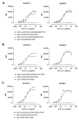
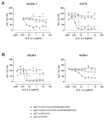
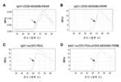
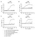
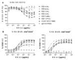
도 1: SU-DHL-1 또는 HDML-2 세포에의 이중특이적 CD3xCD30 항체 및 그들의 단일특이적, 2가 CD3 및 CD30 대응물의 결합. SU-DHL-1 세포 (좌측 패널) 또는 HDLM-2 세포 (우측 패널)에의 (A) bsG1-huCD3-FEALxCD30-MDX060-FEAR, bsG1-huCD3-FEALxb12-FEAR, bsG1-b12-FEALxCD30-MDX060-FEAR, 및 IgG1-CD30-MDX060-FEAR, (B) bsG1-huCD3-FEALxCD30-hAC10-FEAR, bsG1-huCD3-FEALxb12-FEAR, 및 IgG1-CD30-hAC10-FEAR, (C) bsG1-huCD3-FEALxCD30-HRS-3-FEAR, bsG1-huCD3-FEALxb12-FEAR, 및 IgG1-CD30-HRS-3-FEAR, (D) BsIgG1-huCD3-FEALxCD30-HeFi-I-FEAR, bsG1-huCD3-FEALxb12-FEAR, 및 IgG1-CD30-HeFi-I-FEAR, (E) bsIgG1-huCD3-FEALxCD30-T405-FEAR, bsG1-huCD3-FEALxb12-FEAR, 및 IgG1-CD30-T405-FEAR, (F) bsIgG1-huCD3-FEALxCD30-T105-FEAR, bsG1-huCD3-FEALxb12-FEAR, 및 IgG1-CD30-T105-FEAR, (G) bsIgG1-huCD3-FEALxCD30-T408-FEAR, bsG1-huCD3-FEALxb12-FEAR, 및 IgG1-CD30-T408-FEAR, 및 (H) bsIgG1-huCD3-FEALxCD30-T215-FEAR, bsG1-huCD3-FEALxb12-FEAR, 및 IgG1-CD30-HRS-3-FEAR의 용량-의존적 결합. (I) 1.11 μg/mL의 농도에서 SU-DHL-1 (좌측 패널) 또는 HDLM-2 세포 (우측 패널)에의 CD3xCD30 이중특이적 항체 및 CD30 단일특이적 항체의 결합. CD30 아암에 대해 사용된 항체 클론은 x-축 상에 지시된다. 제시된 데이터는 한 대표적인 실험에 대한, 유동 세포계측법에 의해 결정된 바와 같은 평균 형광 강도 (MFI) 값이다.
도 2: HL 및 ALCL 세포주에의 bsG1-huCD3xCD30-MDX060의 결합. (A) HDLM-2 (HL), (B) L-428 (HL), (C) DEL (ALCL), 또는 (D) KI-JK (ALCL) 세포에의 bsG1-huCD3xCD30-MDX060의 결합을 유동 세포계측법에 의해 평가하였다. 이중특이적 항체 bsG1-huCD3xb12 및 bsG1-b12xCD30-MDX060 및 단일특이적 항체 IgG1-CD30-MDX060, IgG1-huCD3 및 IgG1-12는 대조군으로서 포함되었다. 모든 항체는 지시된 바와 같이 그들의 Fc 도메인에 FEAL 및/또는 FERR Fc 침묵화 및 듀오바디(DuoBody)® 기술 돌연변이를 함유하였다. 제시된 데이터는 한 대표적인 실험에 대한, 유동 세포계측법에 의해 결정된 바와 같은 평균 형광 강도 (MFI) 값이다.
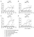
도 3: SU-DHL-1 또는 HDML-2 세포에서의 CD3xCD30 이중특이적 항체에 의한 시험관내에서의 세포독성의 유도. CD3xCD30 이중특이적 항체를 표적 세포로서 CD30-양성 종양 세포주 SU-DHL-1 세포 (좌측 패널) 또는 HDLM-2 세포 (우측 패널) 및 이펙터 세포로서 T 세포 (CD3-양성 ADCC 이펙터 세포 유형 IV 세포; 클린 셀스(Clean Cells), 프랑스 몽테규)를 사용하여 시험관내 세포독성 검정에서 시험하였다. 하기 항체를 시험하였다: (A) bsG1-huCD3-FEALxCD30-MDX060-FEAR, bsG1-b12-FEALxCD30-MDX060-FEAR, 및 IgG1-CD30-MDX060-FEAR, (B) bsG1-huCD3-FEALxCD30-hAC10-FEAR 및 IgG1-CD30-hAC10-FEAR, (C) bsG1-huCD3-FEALxCD30-HRS-3-FEAR 및 IgG1-CD30-HRS-3-FEAR, (D) BsIgG1-huCD3-FEALxCD30-HeFi-I-FEAR 및 IgG1-CD30-HeFi-I-FEAR, (E) bsIgG1-huCD3-FEALxCD30-T405-FEAR 및 IgG1-CD30-T405-FEAR, (F) bsIgG1-huCD3-FEALxCD30-T105-FEAR 및 IgG1-CD30-T105-FEAR, (G) bsIgG1-huCD3-FEALxCD30-T408-FEAR 및 IgG1-CD30-T408-FEAR, 또는 (H) bsIgG1-huCD3-FEALxCD30-T215-FEAR 및 IgG1-CD30-HRS-3-FEAR. 항체 bsG1-huCD3-FEALxb12-FEAR은 모든 실험에서 대조군으로서 포함되었다. 제시된 데이터는 백분율 생존 세포이며; 각각의 그래프에 대한 데이터는 한 대표적인 실험으로부터 얻어졌다.
도 4: 여러 ALCL 및 HL 세포주에서의 CD3xCD30 이중특이적 항체에 의한 시험관내에서의 T-세포 매개 세포독성 및 T-세포 증식의 유도. (A-C) CD3xCD30 이중특이적 항체를 표적 세포로서 상이한 ALCL 및 HL 세포주 및 이펙터 세포로서 정제된 T 세포 (A, B) 또는 ADCC 이펙터 세포 유형 IV 세포 (C)를 사용하여 시험관내 세포독성 검정에서 시험하였다. CD3xCD30 이중특이적 항체는 CD3에 대한 더 낮은 친화도를 갖는 huCD3-FEAL Fab 아암 또는 huCD3-H101G-FEAL 변이체, 및 CD30-특이적 MDX060-FEAR Fab 아암을 함유하였다. IgG1-huCD3 및 IgG1-b12 (A, B) 또는 bsG1-b12-FEALxCD30-MDX060-FEAR 및 IgG1-CD30-MDX060-FEAR (C)은 대조군으로서 포함되었다. 제시된 데이터는 백분율 생존 세포이며; 각각의 그래프에 대한 데이터는 한 대표적인 실험으로부터 얻어졌다. (D) 표적 세포로서 HDLM-2 세포 (좌측 패널) 또는 NCEB-1 세포 (우측 패널)를 사용하여 세포독성 검정에서 CFSE-양성 세포의 수를 절대 T-세포 카운트의 척도로서 평가하였다.
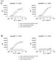
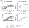
도 5: Expi293F 세포 내로 형질감염된 전장 인간 및 시노몰구스 원숭이 CD30에의 CD3xCD30 이중특이적 항체의 결합. (A-C) 야생형 Expi293F 세포 (A) 또는 전장 인간 CD30으로 일시적으로 형질감염된 Expi293F 세포 (B) 또는 시노몰구스 원숭이 CD30 (C)에의 1가 및 2가 CD30 항체의 결합. 세포를 증가하는 농도의 하기 항체와 인큐베이션하였다: IgG1-CD30-MDX060-FEAR, bsG1-huCD3-FEALxCD30-MDX060-FEAR, bsG1-huCD3-H101G-FEALxCD30-MDX060-FEAR, bsG1-b12-FEALxCD30-MDX060-FEAR, 및 bsG1-huCD3-FEALxb12-FEAR. 데이터는 2회의 기술적 반복실험의 유동 세포계측법에 의해 결정된 바와 같은 평균 형광 강도 (MFI) 값으로서 제시된다. (D) 인간 T 세포 또는 시노몰구스 원숭이 T 세포에의 항체 IgG1-CD30-MDX060-FEAR, bsG1-huCD3-FEALxCD30-MDX060-FEAR, 및 bsG1-huCD3-FEALxb12-FEAR의 결합. 데이터는 한 대표적인 실험의 유동 세포계측법에 의해 결정된 바와 같은 평균 형광 강도 (MFI) 값으로서 제시된다.
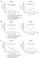
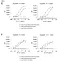
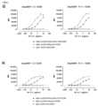
도 6: Expi293F 세포 내로 형질감염된 전장 인간 및 레서스 원숭이 CD30에의 CD3xCD30 이중특이적 항체의 결합. 전장 인간 CD30 (좌측 패널) 또는 레서스 원숭이 CD30 (우측 패널)으로 일시적으로 형질감염된 Expi293F 세포에의 1가 및 2가 CD30 항체의 결합을 유동 세포계측법에 의해 평가하였다. 하기 항체를 평가하였다: (A) bsG1-huCD3-FEALxCD30-hAC10-FEAR 및 IgG1-CD30-hAC10-FEAR, (C) bsG1-huCD3-FEALxCD30-HRS-3-FEAR 및 IgG1-CD30-HRS-3-FEAR, (D) bsIgG1-huCD3-FEALxCD30-HeFi-I-FEAR 및 IgG1-CD30-HeFi-I-FEAR, (E) bsIgG1-huCD3-FEALxCD30-T405-FEAR 및 IgG1-CD30-T405-FEAR, (F) bsIgG1-huCD3-FEALxCD30-T105-FEAR 및 IgG1-CD30-T105-FEAR, (G) bsIgG1-huCD3-FEALxCD30-T408-FEAR 및 IgG1-CD30-T408-FEAR, 또는 (H) bsIgG1-huCD3-FEALxCD30-T215-FEAR 및 IgG1-CD30-HRS-3-FEAR. 항체 bsG1-huCD3-FEALxb12-FEAR은 모든 실험에서 음성 대조군으로서 포함되었다. 제시된 데이터는 한 대표적인 실험에 대한, 유동 세포계측법에 의해 결정된 바와 같은 평균 형광 강도 (MFI) 값이다.
도 7: 시차 주사 형광계측법 (DSF)에 의해 결정된 바와 같은 상이한 비-활성화 돌연변이를 갖는 항체의 열안정성. 증가하는 온도에서의 형태적 단백질 안정성을 DSF에 의해 중복으로 평가하였다. 하기 항체의 용융 곡선이 제시된다: (A) pH 7.4에서의 IgG1-CD30-MDX060-FEAR, (B) pH 7.4에서의 IgG1-CD30-MDX060-FERR, (C) pH 7.4에서의 IgG1-huCD3-FEAL, 및 (D) pH 7.4에서의 BsG1-huCD3-FEALxCD30-MDX060-FERR.
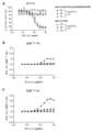
도 8: HL 세포주에의 bsG1-huCD3-FEALxCD30-MDX060-FERR의 결합.
HL 세포주 L-540 (A), KM-H2 (B), 및 L-1236 (C)에의 bsG1-huCD3-FEALxCD30-MDX060-FERR의 결합을 유동 세포계측법에 의해 평가하였다. 이중특이적 항체 bsG1-huCD3-FEALxb12-FERR 및 bsG1-b12-FEALxCD30-MDX060-FERR 및 단일특이적 항체 IgG1-CD30-MDX060-FERR, IgG1-huCD3-FEAL 및 IgG1-12-FEAL은 대조군으로서 포함되었다. 제시된 데이터는 한 대표적인 실험에 대한, 유동 세포계측법에 의해 결정된 바와 같은 평균 형광 강도 (MFI) 값이다.
도 9: ALCL 세포주에의 bsG1-huCD3-FEALxCD30-MDX060-FERR의 결합.
ALCL 세포주 SUP-M2 (A), DL-40 (B), KARPAS-299 (C), L-82 (D), 및 SR-786 (E)에의 bsG1-huCD3-FEALxCD30-MDX060-FERR의 결합을 유동 세포계측법에 의해 평가하였다. 이중특이적 항체 bsG1-huCD3-FEALxb12-FERR 및 bsG1-b12-FEALxCD30-MDX060-FERR 및 단일특이적 항체 IgG1-CD30-MDX060-FERR, IgG1-huCD3-FEAL 및 IgG1-12-FEAL은 대조군으로서 포함되었다. 제시된 데이터는 한 대표적인 실험에 대한, 유동 세포계측법에 의해 결정된 바와 같은 평균 형광 강도 (MFI) 값이다.
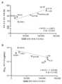
도 10: NHL 세포주에의 bsG1-huCD3-FEALxCD30-MDX060-FERR의 결합.
(A) SUP-T1 (TLL), (B) JVM-2 (MCL), (C) HH (CTCL), 및 (D) NCEB-1 (MCL) 세포주에의 bsG1-huCD3-FEALxCD30-MDX060-FERR의 결합을 유동 세포계측법에 의해 평가하였다. 이중특이적 항체 bsG1-huCD3-FEALxb12-FERR 및 bsG1-b12-FEALxCD30-MDX060-FERR 및 단일특이적 항체 IgG1-CD30-MDX060-FERR, IgG1-huCD3-FEAL 및 IgG1-12-FEAL은 대조군으로서 포함되었다. 제시된 데이터는 한 대표적인 실험에 대한, 유동 세포계측법에 의해 결정된 바와 같은 평균 형광 강도 (MFI) 값이다.
도 11: HEK293 세포 내로 형질감염된 전장 인간 및 레서스 원숭이 CD30에의 bsG1-huCD3-FEALxCD30-MDX060-FERR의 결합. 전장 인간 CD30 (A) 또는 레서스 원숭이 CD30 (B)으로 일시적으로 형질감염된 HEK293 세포에의 bsG1-huCD3-FEALxCD30-MDX060-FERR의 결합을 유동 세포계측법에 의해 평가하였다. bsG1-huCD3-FEALxb12-FERR은 음성 대조군으로서 포함되었다. 제시된 데이터는 한 대표적인 실험에 대한, 유동 세포계측법에 의해 결정된 바와 같은 평균 형광 강도 (MFI) 값이다.
도 12: T 세포 및 종양 세포에의 bsG1-huCD3-FEALxCD30-MDX060-FERR의 동시 결합. 형광 표지된 종양 세포 및 나이브 T 세포에의 bsG1-huCD3-FEALxCD30-MDX060-FERR의 동시 결합을 유동 세포계측법에 의해 연구하였다. (A) 이중-양성 사건은 6x10⁻⁵ 내지 10 μg/mL bsG1huCD3FEALxCD30-MDX060-FERR 또는 대조군 항체 bsG1-huCD3-FEALxb12-FEAR, bsG1b12FEALxCD30MDX060-FERR, 또는 IgG1-b12-FEAL의 존재 하에서의 총 생존 세포의 백분율로서 제시된다. 항체 없이 인큐베이션된 샘플에서의 이중-양성 사건의 백분율은 점선으로 지시된다. (B) 0.12 μg/mL bsG1huCD3FEALxCD30-MDX060-FERR과 인큐베이션된 샘플에서의 이중 양성 세포의 게이팅 전략의 예.
도 13: CD3xCD30 이중특이적 항체에 의한 시험관내에서의 T-세포 매개 세포독성 및 T-세포 활성화의 유도.
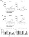
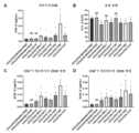
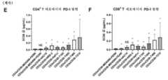
CD3xCD30 이중특이적 항체의 패널을 표적 세포로서 CD30-양성 종양 세포주 Karpas-299 및 이펙터 세포로서 건강한 인간 공여자 연막으로부터 정제된 T 세포를 사용하여 시험관내 세포독성 검정에서 시험하였다. 이들 검정에서, CD25 발현을 T-세포 활성화에 대한 척도로서 CD4+ 및 CD8+ 세포에서 평가하였다. 하기 항체를 시험하였다: bsG1-huCD3-FEALxCD30-MDX060-FERR, bsG1-huCD3-FEALxCD30-MDX060-FEAR, bsG1-huCD3-FEALxCD30-hAC10-FEAR, bsG1-huCD3-FEALxCD30-HRS-3-FEAR, BsIgG1-huCD3-FEALxCD30-HeFi-I-FEAR, bsIgG1-huCD3-FEALxCD30-T105-FEAR, bsIgG1-huCD3-FEALxCD30-T405-FEAR, bsIgG1-huCD3-FEALxCD30-T408-FEAR, 및 bsIgG1-huCD3-FEALxCD30-T215-FEAR. (A) CD3xCD30 항체에 의한 Karpas-299 세포의 세포독성에 대한 IC50 값. (B) 시험 항체에 의한 Karpas-299 세포의 최대 세포독성의 백분율. (C-F) CD3xCD30 항체에 의한 CD4+ (C, E) 또는 CD8+ (D, F) T 세포에서의 CD25 (C-D) 또는 PD-1 (E-F) 발현의 유도에 대한 EC50 값. 데이터는 6명의 상이한 공여자로부터 수득된 T 세포로 수행된 2회의 독립적인 실험으로부터 얻어졌다. 통계적 값은 지시된 클론 및 bsG1-huCD3-FEALxCD30-MDX060-FERR 사이의 윌콕슨(Wilcoxon) 매칭된 쌍 부호 순위 검정의 결과를 나타낸다. NS: 유의하지 않음, *: p <0.05.
도 14: bsG1-huCD3-FEALxCD30-MDX060-FERR에 의한 시험관내에서의 세포주의 T-세포 매개 세포독성. bsG1-huCD3-FEALxCD30-MDX060-FERR에 의한 용량-의존적 T-세포 매개 세포독성을 표적 세포로서 L-428 (A), KM-H2 (B), SUP-M2 (C), 또는 KI-JK (D) 종양 세포주 및 이펙터 세포로서 정제된 T 세포를 사용하여 시험관내에서 시험하였다. IgG1-huCD3-FEAL, bsG1-huCD3-FEALxb12-FERR, IgG1-CD30-MDX060-FERR, bsG1-b12-FEALxCD30-MDX060-FERR, 및 IgG1-b12-FEAL은 대조군으로서 포함되었다. 제시된 데이터는 백분율 생존 세포이며, 각각의 그래프에 대한 데이터는 한 대표적인 실험에 대해 얻어졌다.
도 15: L-428 및 KI-JK 세포주에서의 bsG1-huCD3-FEALxCD30-MDX060-FERR에 의한 시험관내에서의 T-세포 증식. T-세포 증식을 표적 세포로서 L-428 (A, B) 또는 KI-JK (C, D)를 사용하여 T-세포 매개 세포독성 검정에서 평가하였다. 희석된 셀트레이스 바이올렛(Celltrace Violet) 염색을 갖는 CD4+ (A, C) 또는 CD8+ (B, D) T 세포를 게이팅하고, T-세포 증식에 대한 척도로서 확장 지수를 플로우조(FlowJo)로부터의 증식 모델링 도구를 사용하여 계산하였다.
도 16: L-428 및 KI-JK 세포주에서의 bsG1-huCD3-FEALxCD30-MDX060-FERR에 의한 시험관내에서의 T-세포 활성화 마커 CD69의 발현. T-세포 활성화를 표적 세포로서 L-428 (A, B) 또는 KI-JK (C, D)를 사용하여 T-세포 매개 세포독성 검정에서 평가하였다. T-세포 활성화 마커 CD69의 발현을 CD4+ (A, C) 또는 CD8+ (B, D) T 세포에서 평가하였다.
도 17: L-428 및 KI-JK 세포주에서의 bsG1-huCD3-FEALxCD30-MDX060-FERR에 의한 시험관내에서의 T-세포 활성화 마커 CD25의 발현. T-세포 활성화를 표적 세포로서 L-428 (A, B) 또는 KI-JK (C, D)를 사용하여 T-세포 매개 세포독성 검정에서 평가하였다. T-세포 활성화 마커 CD25의 발현을 CD4+ (A, C) 또는 CD8+ (B, D) T 세포에서 평가하였다.
도 18: L-428 및 KI-JK 세포주에서의 bsG1-huCD3-FEALxCD30-MDX060-FERR에 의한 시험관내에서의 T-세포 활성화 마커 PD-1의 발현. T-세포 활성화를 표적 세포로서 L-428 (A, B) 또는 KI-JK (C, D)를 사용하여 T-세포 매개 세포독성 검정에서 평가하였다. T-세포 활성화 마커 PD-1의 발현을 CD4+ (A, C) 또는 CD8+ (B, D) T 세포에서 평가하였다.
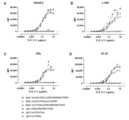
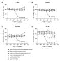
도 19: bsG1-huCD3-FEALxCD30-MDX060-FERR에 의한 시험관내에서의 시토카인 및 그랜자임 B 생산. 14가지 상이한 시토카인 (CD40, IFNγ, IL-10, IL-12, IL-13, IL-1b, IL-2, IL-4, IL-6, IL-8, IP-10, MCP-1, PDL-1, TNFα) 및 그랜자임 B의 농도를 표적 세포로서 L-428을 사용하여 시험관내 T 세포-매개 세포독성 실험 동안 수집된 상청액에서 평가하였다. 시토카인 및 그랜자임 B 농도는 상이한 농도의 bsG1-huCD3-FEALxCD30-MDX060-FERR 또는 대조군 항체 IgG1-b12-FEAL로 처리된 샘플에 대해 제시된다.
도 20: 다양한 이펙터 대 표적 비에서의 bsG1-huCD3-FEALxCD30-MDX060-FERR에 의한 시험관내에서의 T-세포 매개 세포독성 및 T-세포 증식. (A) bsG1-huCD3-FEALxCD30-MDX060-FERR에 의한 용량-의존적 T-세포 매개 세포독성을 1:1, 2:1, 4:1 또는 8:1의 E:T 비에서 표적 세포로서 L-428 종양 세포 및 이펙터 세포로서 정제된 T 세포를 사용하여 시험관내에서 시험하였다. bsG1-huCD3-FEALxb12-FERR은 대조군 항체로서 포함되었다. 제시된 데이터는 한 대표적인 실험으로부터 얻어진 백분율 생존 세포이다. (B, C) 희석된 셀트레이스 바이올렛 염색을 갖는 CD4+ (B) 또는 CD8+ (C) T 세포의 백분율은 증식하는 T 세포의 척도로서 제시된다.
도 21: bsG1-huCD3-FEALxCD30-MDX060-FERR에 의한 시험관내에서의 T-세포 매개 세포독성 및 T-세포 증식의 동역학. (A) bsG1-huCD3-FEALxCD30-MDX060-FERR에 의한 T-세포 매개 세포독성의 동역학을 4:1의 E:T 비에서 표적 세포로서 L-428 종양 세포 및 이펙터 세포로서 정제된 T 세포를 사용하여 시험관내에서 시험하였다. 세포독성을 24시간, 48시간, 및 72시간 후에 평가하였다. bsG1-huCD3-FEALxb12-FERR은 대조군 항체로서 포함되었다. 제시된 데이터는 한 대표적인 실험으로부터 얻어진 백분율 생존 세포이다. (B, C) 희석된 셀트레이스 바이올렛 염색을 갖는 CD4+ (B) 또는 CD8+ (C) T 세포를 게이팅하고, 세포독성 검정에서 T-세포 증식에 대한 척도로서 확장 지수를 플로우조로부터의 증식 모델링 도구를 사용하여 계산하였다.
도 22: bsG1-huCD3-FEALxCD30-MDX060-FERR에 의한 시험관내에서의 T-세포 매개 세포독성 및 CD30 발현 수준과의 상관관계. CD30 발현 수준 대비 bsG1-huCD3-FEALxCD30-MDX060-FERR에 의한 최대 T-세포 매개 살해 (A) 또는 T-세포 매개 세포독성의 IC50 농도 (B) 사이의 상관관계를 8가지 종양 세포주에서 평가하였다. 스피어만(Spearman) 순위 상관관계 검정 (그래프패드 프리즘(GraphPad Prism) 소프트웨어)을 사용하여 T-세포 매개 세포독성 (최대 살해 또는 IC50) 및 CD30 발현 사이의 상관관계의 정도의 통계적 유의성을 평가하였다.
도 23: bsG1-huCD3-FEALxCD30-MDX060-FERR에 의한 활성화된 T 세포의 동족살해. 단리된 건강한 공여자 T 세포를 1 μg/mL 항-CD3 (OKT-3), 1 μg/mL 항-CD28 및 0.025 μg/mL IL-15로 4일 동안 자극하였다. 활성화 (CD25 상향조절)를 확인하고, T 세포를 증가하는 농도의 bsG1-huCD3-FEALxCD30-MDX060-FERR, bsIgG1-ctrlxCD30-MDX-060-FERR, bsIgG1-CD3xb12, 또는 IgG1-b12-FEAL과 48시간 동안 인큐베이션하였다. 활성화된-CD30+ T-세포의 BsG1-huCD3-FEALxCD30-MDX060-FERR-유도된 T-세포 동족살해를 임의의 항체를 첨가하지 않은 조건에서의 생존 T 세포의 수에 비한 각각의 조건에서의 생존 T 세포의 백분율로서 측정하였다. (A-B) 항-CD3, 항-CD28 및 IL-15로의 자극 후 72시간 및 96시간에, CD4+ 또는 CD8+ T-세포에서의 CD25+ (A) 또는 CD30+ (B) 세포의 백분율을 유동 세포계측법에 의해 결정하였다. (C) T-세포 동족살해를 임의의 항체를 첨가하지 않은 조건에 비한 백분율 T-세포 생존으로서 제시하였다. 데이터는 한 대표적인 T-세포 공여자에 대해 제시된다.
도 24: CD30+ 세포 배양물의 상청액에서의 가용성 CD30 및 BsG1-huCD3-FEALxCD30-MDX060-FERR의 항-종양 활성과의 그의 간섭. (A) ELISA에 의해 측정된 바와 같은 상이한 혈액 종양 세포주의 상청액 중의 가용성 CD30 (sCD30) 농도가 제시된다. (B) 상이한 혈액 종양 세포주에서의, 정량적 유동 세포계측법 (인간 IgG 교정 키트-바이오사이텍스(Biocytex))을 사용하여 결정된 바와 같은, sCD30 농도 및 세포 표면 상의 CD30 분자의 수 사이의 상관관계를 스피어만 순위 상관관계 검정 (그래프패드 프리즘 소프트웨어)에 의해 평가하였다. (C) BsG1-huCD3-FEALxCD30-MDX060-FERR을 표적 세포로서 CD30-양성 ALCL 종양 세포주 DEL 및 이펙터 세포로서 건강한 공여자 단리된 T 세포를 사용하여 시험관내 세포독성 검정에서 시험하였다. 하기 대조군 항체가 포함되었다: BsG1-b12-FEALxCD30-MDX060-FERR, IgG1-CD30-MDX060-FERR, BsG1-huCD3-FEALxb12-FERR, IgG1-huCD30-FEAL 및 IgG1-b12-FEAL. 제시된 데이터는 한 대표적인 실험으로부터 얻어진 백분율 생존 세포이다.
도 25. L-428 종양 세포에서의 bsG1-huCD3-FEALxCD30-MDX060-FERR에 의한 생체외에서의 T-세포 매개 세포독성 및 T-세포 증식의 유도. (A) bsG1-huCD3-FEALxCD30-MDX060-FERR을 표적 세포로서 L-428 종양 세포 및 이펙터 세포로서 1차 환자-유래 T 세포를 사용하여 생체외 세포독성 검정에서 시험하였다. 호지킨 림프종 (HL), 급성 골수성 백혈병 (AML) 및 말초 T-세포 림프종 (PTCL) 환자-유래 말초 혈액 단핵 세포 (PBMC)를 T 세포의 공급원으로서 사용하여 CD3-의존적 종양 세포 살해를 평가하였다. IgG1-b12-FEAL은 대조군으로서 포함되었다. 제시된 데이터는 백분율 생존 표적 세포이다. (B-D) T-세포 활성화를 백분율 양성 세포로서 지시된, PBMC 하위세트 내의 CD4+/CD8+ T 세포 상의 CD69 (B), CD25 (C) 및 PD-1 (D) 마커의 상향조절에 의해 평가하였다.
도 26. SCID 마우스에서의 정맥내 주사 후의 BsG1-huCD3-FEALxCD30-MDX060-FERR의 혈장 농도. SCID 마우스를 10 μg (0.5 mg/kg) 또는 100 μg (5 mg/kg)의 BsG1-huCD3-FEALxCD30-MDX060-FERR의 단일 IV 용량으로 주사하였다. (A) 총 인간 IgG를 ELISA에 의해 결정하고, 평균 인간 IgG1 농도를 시간 경과에 따라 플롯팅하였다. (B) 평균 클리어런스율을 0.5 또는 5 mg/kg 용량 수준에 대해 플롯팅하였다. 점선은 마우스에서의 비-결합 보통의 인간 IgG1의 표준 분포 부피에 기반한 추정된 클리어런스율을 지시한다.
도 27: 막-결합된 bsG1-huCD3-FEALxCD30-MDX060-FERR에의 C1q 결합. bsG1-huCD3-FEALxCD30-MDX060-FERR-옵소닌화된, 활성화된 인간 CD8+ T 세포, 또는 CD30+ NCEB-1 세포에의 C1q의 결합을 FITC 표지된 토끼 항-C1q 항체를 사용하여 유동 세포계측법에 의해 평가하였다. (A) C1q의 공급원으로서 정상 인간 혈청의 존재 하에서 항-CD3/CD28 비드로 자극된 인간 CD8+ T 세포에의 bsG1-huCD3-FEALxCD30-MDX060-FERR, IgG1-huCD3-FEAL 또는 양성 대조군 항체 IgG1-CD52-E430G의 결합. 제시된 데이터는 한 대표적인 실험으로부터의 반복실험 웰로부터의 기하평균 형광 강도 (gMFI) ± SD이다. (B) C1q의 공급원으로서 정상 인간 혈청의 존재 하에서의, CD30+ NCEB-1 세포에의 bsG1-huCD3-FEALxCD30-MDX060-FERR, IgG1-CD30-MDX060-FERR 또는 양성 대조군 항체 IgG1-7D8-E430G의 결합. 제시된 데이터는 한 대표적인 실험으로부터의 gMFI이다.Figure 1: Binding of bispecific CD3xCD30 antibodies and their monospecific, bivalent CD3 and CD30 counterparts to SU-DHL-1 or HDML-2 cells. (A) bsG1-huCD3-FEALxCD30-MDX060-FEAR, bsG1-huCD3-FEALxb12-FEAR, bsG1-b12-FEALxCD30-MDX060-FEAR on SU-DHL-1 cells (left panel) or HDLM-2 cells (right panel). , and IgG1-CD30-MDX060-FEAR, (B) bsG1-huCD3-FEALxCD30-hAC10-FEAR, bsG1-huCD3-FEALxb12-FEAR, and IgG1-CD30-hAC10-FEAR, (C) bsG1-huCD3-FEALxCD30-HRS -3-FEAR, bsG1-huCD3-FEALxb12-FEAR, and IgG1-CD30-HRS-3-FEAR, (D) BsIgG1-huCD3-FEALxCD30-HeFi-I-FEAR, bsG1-huCD3-FEALxb12-FEAR, and IgG1- CD30-HeFi-I-FEAR, (E) bsIgG1-huCD3-FEALxCD30-T405-FEAR, bsG1-huCD3-FEALxb12-FEAR, and IgG1-CD30-T405-FEAR, (F) bsIgG1-huCD3-FEALxCD30-T105-FEAR , bsG1-huCD3-FEALxb12-FEAR, and IgG1-CD30-T105-FEAR, (G) bsIgG1-huCD3-FEALxCD30-T408-FEAR, bsG1-huCD3-FEALxb12-FEAR, and IgG1-CD30-T408-FEAR, and ( H) Dose-dependent binding of bsIgG1-huCD3-FEALxCD30-T215-FEAR, bsG1-huCD3-FEALxb12-FEAR, and IgG1-CD30-HRS-3-FEAR. (I) Binding of CD3xCD30 bispecific antibody and CD30 monospecific antibody to SU-DHL-1 (left panel) or HDLM-2 cells (right panel) at a concentration of 1.11 μg/mL. Antibody clones used against the CD30 arm are indicated on the x-axis. Data presented are mean fluorescence intensity (MFI) values as determined by flow cytometry for one representative experiment.
Figure 2: Binding of bsG1-huCD3xCD30-MDX060 to HL and ALCL cell lines. Flow binding of bsG1-huCD3xCD30-MDX060 to (A) HDLM-2 (HL), (B) L-428 (HL), (C) DEL (ALCL), or (D) KI-JK (ALCL) cells. Evaluated by cytometry. Bispecific antibodies bsG1-huCD3xb12 and bsG1-b12xCD30-MDX060 and monospecific antibodies IgG1-CD30-MDX060, IgG1-huCD3 and IgG1-12 were included as controls. All antibodies contained FEAL and/or FERR Fc silencing and DuoBody® technology mutations in their Fc domains as indicated. Data presented are mean fluorescence intensity (MFI) values as determined by flow cytometry for one representative experiment.
Figure 3: Induction of cytotoxicity in vitro by CD3xCD30 bispecific antibody in SU-DHL-1 or HDML-2 cells. The CD3xCD30 bispecific antibody was applied to CD30-positive tumor cell lines SU-DHL-1 cells (left panel) or HDLM-2 cells (right panel) as target cells and T cells (CD3-positive ADCC effector cell type IV cells) as effector cells. Tested in an in vitro cytotoxicity assay using Clean Cells (Montague, France). The following antibodies were tested: (A) bsG1-huCD3-FEALxCD30-MDX060-FEAR, bsG1-b12-FEALxCD30-MDX060-FEAR, and IgG1-CD30-MDX060-FEAR, (B) bsG1-huCD3-FEALxCD30-hAC10-FEAR and IgG1-CD30-hAC10-FEAR, (C) bsG1-huCD3-FEALxCD30-HRS-3-FEAR and IgG1-CD30-HRS-3-FEAR, (D) BsIgG1-huCD3-FEALxCD30-HeFi-I-FEAR and IgG1 -CD30-HeFi-I-FEAR, (E) bsIgG1-huCD3-FEALxCD30-T405-FEAR and IgG1-CD30-T405-FEAR, (F) bsIgG1-huCD3-FEALxCD30-T105-FEAR and IgG1-CD30-T105-FEAR , (G) bsIgG1-huCD3-FEALxCD30-T408-FEAR and IgG1-CD30-T408-FEAR, or (H) bsIgG1-huCD3-FEALxCD30-T215-FEAR and IgG1-CD30-HRS-3-FEAR. Antibody bsG1-huCD3-FEALxb12-FEAR was included as a control in all experiments. Data presented are percent viable cells; Data for each graph were obtained from one representative experiment.
Figure 4: Induction of T-cell mediated cytotoxicity and T-cell proliferation in vitro by CD3xCD30 bispecific antibody in several ALCL and HL cell lines. (AC) CD3xCD30 bispecific antibody tested in in vitro cytotoxicity assay using different ALCL and HL cell lines as target cells and purified T cells (A, B) or ADCC effector cell type IV cells (C) as effector cells. did. The CD3xCD30 bispecific antibody contained the huCD3-FEAL Fab arm or the huCD3-H101G-FEAL variant, which has lower affinity for CD3, and the CD30-specific MDX060-FEAR Fab arm. IgG1-huCD3 and IgG1-b12 (A, B) or bsG1-b12-FEALxCD30-MDX060-FEAR and IgG1-CD30-MDX060-FEAR (C) were included as controls. Data presented are percent viable cells; Data for each graph were obtained from one representative experiment. (D) The number of CFSE-positive cells was assessed as a measure of absolute T-cell counts in cytotoxicity assays using HDLM-2 cells (left panel) or NCEB-1 cells (right panel) as target cells.
Figure 5: Binding of CD3xCD30 bispecific antibody to full-length human and cynomolgus monkey CD30 transfected into Expi293F cells. (AC) Binding of monovalent and bivalent CD30 antibodies to wild-type Expi293F cells (A) or Expi293F cells transiently transfected with full-length human CD30 (B) or cynomolgus monkey CD30 (C). Cells were incubated with increasing concentrations of the following antibodies: IgG1-CD30-MDX060-FEAR, bsG1-huCD3-FEALxCD30-MDX060-FEAR, bsG1-huCD3-H101G-FEALxCD30-MDX060-FEAR, bsG1-b12-FEALxCD30-MDX060- FEAR, and bsG1-huCD3-FEALxb12-FEAR. Data are presented as mean fluorescence intensity (MFI) values as determined by flow cytometry of two technical replicates. (D) Binding of antibodies IgG1-CD30-MDX060-FEAR, bsG1-huCD3-FEALxCD30-MDX060-FEAR, and bsG1-huCD3-FEALxb12-FEAR to human T cells or cynomolgus monkey T cells. Data are presented as mean fluorescence intensity (MFI) values as determined by flow cytometry of one representative experiment.
Figure 6: Binding of CD3xCD30 bispecific antibody to full-length human and rhesus monkey CD30 transfected into Expi293F cells. Binding of monovalent and bivalent CD30 antibodies to Expi293F cells transiently transfected with full-length human CD30 (left panel) or rhesus monkey CD30 (right panel) was assessed by flow cytometry. The following antibodies were evaluated: (A) bsG1-huCD3-FEALxCD30-hAC10-FEAR and IgG1-CD30-hAC10-FEAR, (C) bsG1-huCD3-FEALxCD30-HRS-3-FEAR and IgG1-CD30-HRS-3- FEAR, (D) bsIgG1-huCD3-FEALxCD30-HeFi-I-FEAR and IgG1-CD30-HeFi-I-FEAR, (E) bsIgG1-huCD3-FEALxCD30-T405-FEAR and IgG1-CD30-T405-FEAR, (F ) bsIgG1-huCD3-FEALxCD30-T105-FEAR and IgG1-CD30-T105-FEAR, (G) bsIgG1-huCD3-FEALxCD30-T408-FEAR and IgG1-CD30-T408-FEAR, or (H) bsIgG1-huCD3-FEALxCD30- T215-FEAR and IgG1-CD30-HRS-3-FEAR. Antibody bsG1-huCD3-FEALxb12-FEAR was included as a negative control in all experiments. Data presented are mean fluorescence intensity (MFI) values as determined by flow cytometry for one representative experiment.
Figure 7: Thermostability of antibodies with different non-activating mutations as determined by differential scanning fluorometry (DSF). Conformational protein stability at increasing temperatures was assessed in duplicate by DSF. The melting curves of the following antibodies are shown: (A) IgG1-CD30-MDX060-FEAR at pH 7.4, (B) IgG1-CD30-MDX060-FERR at pH 7.4, (C) IgG1-huCD3- at pH 7.4. FEAL, and (D) BsG1-huCD3-FEALxCD30-MDX060-FERR at pH 7.4.
Figure 8: Binding of bsG1-huCD3-FEALxCD30-MDX060-FERR to HL cell lines.
Binding of bsG1-huCD3-FEALxCD30-MDX060-FERR to HL cell lines L-540 (A), KM-H2 (B), and L-1236 (C) was assessed by flow cytometry. Bispecific antibodies bsG1-huCD3-FEALxb12-FERR and bsG1-b12-FEALxCD30-MDX060-FERR and monospecific antibodies IgG1-CD30-MDX060-FERR, IgG1-huCD3-FEAL and IgG1-12-FEAL were included as controls. . Data presented are mean fluorescence intensity (MFI) values as determined by flow cytometry for one representative experiment.
Figure 9: Binding of bsG1-huCD3-FEALxCD30-MDX060-FERR to ALCL cell lines.
Binding of bsG1-huCD3-FEALxCD30-MDX060-FERR to ALCL cell lines SUP-M2 (A), DL-40 (B), KARPAS-299 (C), L-82 (D), and SR-786 (E). Evaluated by flow cytometry. Bispecific antibodies bsG1-huCD3-FEALxb12-FERR and bsG1-b12-FEALxCD30-MDX060-FERR and monospecific antibodies IgG1-CD30-MDX060-FERR, IgG1-huCD3-FEAL and IgG1-12-FEAL were included as controls. . Data presented are mean fluorescence intensity (MFI) values as determined by flow cytometry for one representative experiment.
Figure 10: Binding of bsG1-huCD3-FEALxCD30-MDX060-FERR to NHL cell lines.
bsG1-huCD3-FEALxCD30-MDX060-FERR on (A) SUP-T1 (TLL), (B) JVM-2 (MCL), (C) HH (CTCL), and (D) NCEB-1 (MCL) cell lines. Binding was assessed by flow cytometry. Bispecific antibodies bsG1-huCD3-FEALxb12-FERR and bsG1-b12-FEALxCD30-MDX060-FERR and monospecific antibodies IgG1-CD30-MDX060-FERR, IgG1-huCD3-FEAL and IgG1-12-FEAL were included as controls. . Data presented are mean fluorescence intensity (MFI) values as determined by flow cytometry for one representative experiment.
Figure 11: Binding of bsG1-huCD3-FEALxCD30-MDX060-FERR to full-length human and rhesus monkey CD30 transfected into HEK293 cells. Binding of bsG1-huCD3-FEALxCD30-MDX060-FERR to HEK293 cells transiently transfected with full-length human CD30 (A) or rhesus monkey CD30 (B) was assessed by flow cytometry. bsG1-huCD3-FEALxb12-FERR was included as a negative control. Data presented are mean fluorescence intensity (MFI) values as determined by flow cytometry for one representative experiment.
Figure 12: Simultaneous binding of bsG1-huCD3-FEALxCD30-MDX060-FERR to T cells and tumor cells. Simultaneous binding of bsG1-huCD3-FEALxCD30-MDX060-FERR to fluorescently labeled tumor cells and naive T cells was studied by flow cytometry. (A) Double-positive events are the percentage of total viable cells in the presence of 6x10⁻⁵ to 10 μg/mL bsG1huCD3FEALxCD30-MDX060-FERR or control antibodies bsG1-huCD3-FEALxb12-FEAR, bsG1b12FEALxCD30MDX060-FERR, or IgG1-b12-FEAL It is presented as. The percentage of double-positive events in samples incubated without antibody is indicated by the dotted line. (B) Example of gating strategy of double positive cells in samples incubated with 0.12 μg/mL bsG1huCD3FEALxCD30-MDX060-FERR.
Figure 13: Induction of T-cell mediated cytotoxicity and T-cell activation in vitro by CD3xCD30 bispecific antibody.
A panel of CD3xCD30 bispecific antibodies was tested in an in vitro cytotoxicity assay using the CD30-positive tumor cell line Karpas-299 as target cells and T cells purified from healthy human donor buffy coats as effector cells. In these assays, CD25 expression was assessed on CD4+ and CD8+ cells as a measure of T-cell activation. The following antibodies were tested: bsG1-huCD3-FEALxCD30-MDX060-FERR, bsG1-huCD3-FEALxCD30-MDX060-FEAR, bsG1-huCD3-FEALxCD30-hAC10-FEAR, bsG1-huCD3-FEALxCD30-HRS-3-FEAR, BsIgG1- huCD3-FEALxCD30-HeFi-I-FEAR, bsIgG1-huCD3-FEALxCD30-T105-FEAR, bsIgG1-huCD3-FEALxCD30-T405-FEAR, bsIgG1-huCD3-FEALxCD30-T408-FEAR, and bsIgG1-huCD3-FEALxCD30-T215-F EAR . (A) IC50 values for cytotoxicity of Karpas-299 cells by CD3xCD30 antibody. (B) Percentage of maximum cytotoxicity of Karpas-299 cells by test antibodies. (C-F) EC50 values for induction of CD25 (CD) or PD-1 (EF) expression on CD4+ (C, E) or CD8+ (D, F) T cells by CD3xCD30 antibody. Data were obtained from two independent experiments performed with T cells obtained from six different donors. Statistical values represent the results of Wilcoxon matched pairs signed rank test between the indicated clones and bsG1-huCD3-FEALxCD30-MDX060-FERR. NS: not significant, *: p <0.05.
Figure 14: T-cell mediated cytotoxicity of cell lines in vitro by bsG1-huCD3-FEALxCD30-MDX060-FERR. Dose-dependent T-cell mediated cytotoxicity by bsG1-huCD3-FEALxCD30-MDX060-FERR using L-428 (A), KM-H2 (B), SUP-M2 (C), or KI-JK ( D) Tested in vitro using purified T cells as tumor cell lines and effector cells. IgG1-huCD3-FEAL, bsG1-huCD3-FEALxb12-FERR, IgG1-CD30-MDX060-FERR, bsG1-b12-FEALxCD30-MDX060-FERR, and IgG1-b12-FEAL were included as controls. Data presented are percentage surviving cells, and data for each graph were obtained for one representative experiment.
Figure 15: T-cell proliferation in vitro by bsG1-huCD3-FEALxCD30-MDX060-FERR in L-428 and KI-JK cell lines. T-cell proliferation was assessed in T-cell mediated cytotoxicity assays using L-428 (A, B) or KI-JK (C, D) as target cells. Gating on CD4+ (A, C) or CD8+ (B, D) T cells with diluted Celltrace Violet staining and expansion index as a measure of T-cell proliferation from FlowJo. It was calculated using the proliferation modeling tool.
Figure 16: Expression of the T-cell activation marker CD69 in vitro by bsG1-huCD3-FEALxCD30-MDX060-FERR in L-428 and KI-JK cell lines. T-cell activation was assessed in T-cell mediated cytotoxicity assays using L-428 (A, B) or KI-JK (C, D) as target cells. Expression of the T-cell activation marker CD69 was assessed on CD4+ (A, C) or CD8+ (B, D) T cells.
Figure 17: Expression of the T-cell activation marker CD25 in vitro by bsG1-huCD3-FEALxCD30-MDX060-FERR in L-428 and KI-JK cell lines. T-cell activation was assessed in T-cell mediated cytotoxicity assays using L-428 (A, B) or KI-JK (C, D) as target cells. Expression of the T-cell activation marker CD25 was assessed on CD4+ (A, C) or CD8+ (B, D) T cells.
Figure 18: Expression of the T-cell activation marker PD-1 in vitro by bsG1-huCD3-FEALxCD30-MDX060-FERR in L-428 and KI-JK cell lines. T-cell activation was assessed in T-cell mediated cytotoxicity assays using L-428 (A, B) or KI-JK (C, D) as target cells. Expression of the T-cell activation marker PD-1 was assessed on CD4+ (A, C) or CD8+ (B, D) T cells.
Figure 19: Cytokine and granzyme B production in vitro by bsG1-huCD3-FEALxCD30-MDX060-FERR. 14 different cytokines (CD40, IFNγ, IL-10, IL-12, IL-13, IL-1b, IL-2, IL-4, IL-6, IL-8, IP-10, MCP-1, PDL Concentrations of -1, TNFα) and granzyme B were assessed in supernatants collected during in vitro T cell-mediated cytotoxicity experiments using L-428 as target cells. Cytokine and granzyme B concentrations are shown for samples treated with different concentrations of bsG1-huCD3-FEALxCD30-MDX060-FERR or control antibody IgG1-b12-FEAL.
Figure 20: T-cell mediated cytotoxicity and T-cell proliferation in vitro by bsG1-huCD3-FEALxCD30-MDX060-FERR at various effector to target ratios. (A) Dose-dependent T-cell mediated cytotoxicity by bsG1-huCD3-FEALxCD30-MDX060-FERR as target cells at E:T ratios of 1:1, 2:1, 4:1, or 8:1. Tested in vitro using 428 tumor cells and purified T cells as effector cells. bsG1-huCD3-FEALxb12-FERR was included as a control antibody. Data presented are percentage viable cells from one representative experiment. (B, C) Percentage of CD4+ (B) or CD8+ (C) T cells with diluted Celltrace Violet staining is presented as a measure of proliferating T cells.
Figure 21: Kinetics of T-cell mediated cytotoxicity and T-cell proliferation in vitro by bsG1-huCD3-FEALxCD30-MDX060-FERR. (A) Kinetics of T-cell mediated cytotoxicity by bsG1-huCD3-FEALxCD30-MDX060-FERR using L-428 tumor cells as target cells and purified T cells as effector cells at an E:T ratio of 4:1. This was tested in vitro. Cytotoxicity was assessed after 24, 48, and 72 hours. bsG1-huCD3-FEALxb12-FERR was included as a control antibody. Data presented are percentage viable cells from one representative experiment. (B, C) Gating on CD4+ (B) or CD8+ (C) T cells with diluted Celltrace Violet staining and expansion index as a measure for T-cell proliferation in cytotoxicity assays. Calculations were made using modeling tools.
Figure 22: T-cell mediated cytotoxicity in vitro by bsG1-huCD3-FEALxCD30-MDX060-FERR and correlation with CD30 expression levels. The correlation between maximum T-cell mediated killing (A) or IC50 concentration of T-cell mediated cytotoxicity (B) by bsG1-huCD3-FEALxCD30-MDX060-FERR versus CD30 expression level was assessed in eight tumor cell lines. The statistical significance of the degree of correlation between T-cell mediated cytotoxicity (maximum killing or IC50 ) and CD30 expression was assessed using the Spearman rank correlation test (GraphPad Prism software). .
Figure 23: Canine killing of activated T cells by bsG1-huCD3-FEALxCD30-MDX060-FERR. Isolated healthy donor T cells were stimulated with 1 μg/mL anti-CD3 (OKT-3), 1 μg/mL anti-CD28, and 0.025 μg/mL IL-15 for 4 days. To confirm activation (CD25 upregulation), T cells were incubated with increasing concentrations of bsG1-huCD3-FEALxCD30-MDX060-FERR, bsIgG1-ctrlxCD30-MDX-060-FERR, bsIgG1-CD3xb12, or IgG1-b12-FEAL for 48 hours. It was incubated for a while. BsG1-huCD3-FEALxCD30-MDX060-FERR-induced T-cell fratricidal killing of activated-CD30+ T-cells Survival in each condition relative to the number of T cells in the condition without addition of any antibody Measured as percentage of T cells. (AB) Flow percentage of CD25+ (A) or CD30+ (B) cells in CD4+ or CD8+ T-cells at 72 and 96 hours after stimulation with anti-CD3, anti-CD28, and IL-15. Determined by cytometry. (C) T-cell fratricide is presented as percent T-cell survival compared to conditions without the addition of any antibody. Data are presented for one representative T-cell donor.
Figure 24:Soluble CD30 and its interference with the anti-tumor activity of BsG1-huCD3-FEALxCD30-MDX060-FERR in supernatants of CD30+ cell cultures. (A) Soluble CD30 (sCD30) concentrations in supernatants of different hematological tumor cell lines as measured by ELISA are shown. (B) Sphere correlation between sCD30 concentration and number of CD30 molecules on the cell surface, as determined using quantitative flow cytometry (Human IgG Calibration Kit-Biocytex) in different hematologic tumor cell lines. Only rank correlation test was assessed by (GraphPad Prism software). (C) BsG1-huCD3-FEALxCD30-MDX060-FERR was tested in an in vitro cytotoxicity assay using the CD30-positive ALCL tumor cell line DEL as target cells and healthy donor isolated T cells as effector cells. The following control antibodies were included: BsG1-b12-FEALxCD30-MDX060-FERR, IgG1-CD30-MDX060-FERR, BsG1-huCD3-FEALxb12-FERR, IgG1-huCD30-FEAL, and IgG1-b12-FEAL. Data presented are percentage viable cells from one representative experiment.
Figure 25. Induction of T-cell mediated cytotoxicity and T-cell proliferation in vitro by bsG1-huCD3-FEALxCD30-MDX060-FERR in L-428 tumor cells. (A) bsG1-huCD3-FEALxCD30-MDX060-FERR was tested in an in vitro cytotoxicity assay using L-428 tumor cells as target cells and primary patient-derived T cells as effector cells. CD3-dependent tumor cell killing was assessed using Hodgkin's lymphoma (HL), acute myeloid leukemia (AML), and peripheral T-cell lymphoma (PTCL) patient-derived peripheral blood mononuclear cells (PBMCs) as a source of T cells. IgG1-b12-FEAL was included as a control. Data presented are percentage surviving target cells. (BD) T-cell activation assessed by upregulation of CD69 (B), CD25 (C) and PD-1 (D) markers on CD4+ /CD8+ T cells in PBMC subsets, indicated as percentage positive cells. did.
Figure 26. Plasma concentration of BsG1-huCD3-FEALxCD30-MDX060-FERR after intravenous injection in SCID mice. SCID mice were injected with a single IV dose of 10 μg (0.5 mg/kg) or 100 μg (5 mg/kg) of BsG1-huCD3-FEALxCD30-MDX060-FERR. (A) Total human IgG was determined by ELISA and average human IgG1 concentration was plotted over time. (B) Average clearance rates were plotted for 0.5 or 5 mg/kg dose levels. The dotted line indicates the estimated clearance rate based on the standard distribution volume of non-bound normal human IgG1 in mice.
Figure 27: C1q binding to membrane-bound bsG1-huCD3-FEALxCD30-MDX060-FERR. Binding of C1q to bsG1-huCD3-FEALxCD30-MDX060-FERR-opsonized, activated human CD8+ T cells, or CD30+ NCEB-1 cells was measured by flow cytometry using FITC-labeled rabbit anti-C1q antibody. It was evaluated by . (A) bsG1-huCD3-FEALxCD30-MDX060-FERR, IgG1-huCD3-FEAL, or positive control antibody IgG1- on human CD8+ T cells stimulated with anti-CD3/CD28 beads in the presence of normal human serum as a source of C1q. Binding of CD52-E430G. Data presented are geometric mean fluorescence intensity (gMFI) ± SD from replicate wells from one representative experiment. (B) Binding of bsG1-huCD3-FEALxCD30-MDX060-FERR, IgG1-CD30-MDX060-FERR, or positive control antibody IgG1-7D8-E430G to CD30+ NCEB-1 cells in the presence of normal human serum as a source of C1q. . Data presented are gMFI from one representative experiment.
정의Justice
본원에 사용된 용어 "항체"는 유의한 기간, 예컨대 적어도 약 30분, 적어도 약 45분, 적어도 약 1시간, 적어도 약 2시간, 적어도 약 4시간, 적어도 약 8시간, 적어도 약 12시간, 적어도 약 24시간 이상, 적어도 약 48시간 이상, 적어도 약 3, 4, 5, 6, 7일 또는 그 초과 등의 반감기, 또는 임의의 다른 관련된 기능적으로-정의된 기간 (예컨대 항원에의 항체 결합과 연관된 생리학적 반응을 유도하고/거나, 촉진시키고/거나, 증진시키고/거나, 조정하는데 충분한 시간 및/또는 항체가 내재화되는데 충분한 시간)을 갖는 전형적인 생리학적 및/또는 종양-특이적 조건 하에서 항원에 특이적으로 결합하는 능력을 갖는 이뮤노글로불린 분자, 이뮤노글로불린 분자의 단편, 또는 이 중 어느 하나의 유도체를 지칭하는 것으로 의도된다. 항체는 항원과 상호작용할 수 있는 결합 영역 (또는 본원에 사용될 수 있는 결합 도메인, 둘 다는 동일한 의미를 가짐)을 포함하며, 결합 영역은 이뮤노글로불린 분자의 중쇄 및 경쇄 둘 다의 가변 영역 등을 포함한다. 항체는 면역계의 다양한 세포 (예컨대 이펙터 세포) 및 보체계의 성분, 예컨대 보체 활성화의 고전적 경로에서의 제1 성분인 C1q를 포함하는, 숙주 조직 또는 인자에의 이뮤노글로불린의 결합을 매개할 수 있는 항체 (Ab)의 불변 영역을 포함할 수 있다.As used herein, the term “antibody” refers to an antibody that is present for a significant period of time, such as at least about 30 minutes, at least about 45 minutes, at least about 1 hour, at least about 2 hours, at least about 4 hours, at least about 8 hours, at least about 12 hours, at least a half-life of at least about 24 hours, at least about 48 hours, at least about 3, 4, 5, 6, 7 days or more, or any other relevant functionally-defined period of time (e.g., a half-life associated with antibody binding to an antigen) specific for the antigen under typical physiological and/or tumor-specific conditions (sufficient time to induce, promote, enhance, and/or modulate a physiological response and/or sufficient time for the antibody to be internalized). It is intended to refer to an immunoglobulin molecule, a fragment of an immunoglobulin molecule, or a derivative of any of these, having the ability to bind to a target. An antibody comprises a binding region (or binding domain, as used herein, both have the same meaning) capable of interacting with an antigen, the binding region comprising the variable regions of both the heavy and light chains of an immunoglobulin molecule, etc. do. Antibodies are antibodies that can mediate the binding of immunoglobulins to host tissues or factors, including various cells of the immune system (such as effector cells) and components of the complement system, such as C1q, the first component in the classical pathway of complement activation. It may include a constant region of (Ab).
본 발명의 맥락에서, 용어 "항체"는 모노클로날 항체 (mAb), 항체-유사 폴리펩티드, 키메라 항체, 인간 항체, 인간화 항체, 뿐만 아니라 임의의 공지된 기술, 예컨대 효소적 절단, 펩티드 합성, 및 재조합 DNA 기술에 의해 제공된 항원 (항원-결합 단편)에 특이적으로 결합하는 능력을 보유하는 '항체 단편' 또는 '그의 단편'을 포함한다. 용어 "항체"는 추가의 변형을 갖는 이중, 삼중, 또는 다중특이적 항체 및/또는 항체, 예를 들어, 항체-약물 접합체 및/또는 IgG Fc 도메인에 변형을 갖는 항체를 포함한다. 본 발명에 따라 정의된 바와 같은 항체는, 본원의 개시내용이 달리 제한되지 않는 한, 임의의 이소타입을 가질 수 있거나, 이소타입을 갖지 않을 수 있다 (예를 들어, scFv 항체).In the context of the present invention, the term "antibody" includes monoclonal antibodies (mAb), antibody-like polypeptides, chimeric antibodies, human antibodies, humanized antibodies, as well as any known technique such as enzymatic cleavage, peptide synthesis, and Includes 'antibody fragments' or 'fragments thereof' that retain the ability to specifically bind to an antigen (antigen-binding fragment) provided by recombinant DNA technology. The term “antibody” includes dual, triple, or multispecific antibodies and/or antibodies with additional modifications, such as antibody-drug conjugates and/or antibodies with modifications in the IgG Fc domain. Antibodies, as defined according to the present invention, may have any isotype or no isotype (e.g., scFv antibodies), unless the disclosure herein is otherwise limited.
항체의 항원-결합 기능은 전장 항체의 단편에 의해 수행될 수 있음이 제시되었다. 용어 "항체" 내에 포함되는 결합 단편의 예는 (i) Fab' 또는 Fab 단편, 즉, 경쇄 가변 도메인 (VL), 중쇄 가변 도메인 (VH), 경쇄 불변 영역 (CL) 및 중쇄 불변 영역 도메인 1 (CH1) 도메인으로 이루어진 1가 단편, 또는 WO 2007/059782에 기재된 바와 같은 1가 항체; (ii) F(ab')2 단편, 즉, 힌지 영역에서 디술피드 가교에 의해 연결된 2개의 Fab 단편을 포함하는 2가 단편; (iii) VH 및 CH1 도메인으로 본질적으로 이루어진 Fd 단편; (iv) 항체의 단일 아암의 VL 및 VH 도메인으로 본질적으로 이루어진 Fv 단편, (v) VH 도메인으로 본질적으로 이루어지고 또한 도메인 항체 [Holt et al.; Trends Biotechnol-. 2003 Nov;21(11):484-90]로 지칭되는 dAb 단편 [Ward et al., Nature341, 544546 (1989)]; (vi) 카멜리드 또는 나노바디 [Revets et al.; Expert Opin Biol Ther. 2005 Jan;5(1):111-24] 및 (vii) 단리된 상보성 결정 영역 (CDR)을 포함한다. 더욱이, Fv 단편의 2개의 도메인, 즉, VL 및 VH는 별개의 유전자에 의해 코딩되지만, 이들은 이들이 VL 및 VH 영역이 쌍형성하여 1가 분자를 형성하는 단일 단백질 쇄로서 만들어지는 것을 가능하게 하는 합성 링커에 의해, 재조합 방법을 사용하여 연결될 수 있다 (단일 쇄 항체 또는 단일 쇄 Fv (scFv)로 공지됨, 예를 들어 문헌 [Revets et al.; Expert Opin Biol Ther. 2005 Jan;5(1):111-24] 및 [Bird et al., Science242, 423426 (1988)] 참조. 이러한 단일 쇄 항체는 달리 언급되거나 맥락에 의해 명백하게 지시되지 않는 한, 용어 항체 내에 포함된다. 이러한 단편은 일반적으로 항체의 의미 내에 포함되지만, 이들은 집합적으로 및 각각 독립적으로 상이한 생물학적 특성 및 유용성을 나타내는 본 발명의 고유한 특색이다. 본 발명의 맥락에서 이들 및 다른 유용한 항체 단편은 본원에서 추가로 논의된다.It has been suggested that the antigen-binding function of an antibody can be performed by fragments of the full-length antibody. Examples of binding fragments encompassed within the term “antibody” include (i) Fab' or Fab fragments, i.e., light chain variable domain (VL), heavy chain variable domain (VH), light chain constant region (CL) and heavy chain constant region domain 1 ( a monovalent fragment consisting of a CH1) domain, or a monovalent antibody as described in WO 2007/059782; (ii) F(ab')2 fragment, i.e. a bivalent fragment comprising two Fab fragments linked by a disulfide bridge in the hinge region; (iii) Fd fragment consisting essentially of VH and CH1 domains; (iv) an Fv fragment consisting essentially of the VL and VH domains of a single arm of the antibody, (v) an Fv fragment consisting essentially of the VH domain and also a domain antibody [Holt et al.; Trends Biotechnol-. 2003 Nov;21 (11):484-90] (Ward et al., Nature341 , 544546 (1989)); (vi) camelids or nanobodies [Revets et al.; Expert Opin Biol Ther. Jan 2005;5 (1):111-24] and (vii) an isolated complementarity determining region (CDR). Moreover, although the two domains of the Fv fragment, VL and VH, are encoded by separate genes, they are synthesized, allowing them to be made as a single protein chain with the VL and VH regions pairing to form a monovalent molecule. By linkers, they can be linked using recombinant methods (known as single chain antibodies or single chain Fv (scFv), e.g. Revets et al.; Expert Opin Biol Ther. 2005 Jan;5 (1): 111-24 and Bird et al., Science242 , 423426 (1988). These fragments are generally encompassed within the term antibody, unless otherwise stated or clearly indicated by the context. Although included within the meaning of, these and other useful antibody fragments are further discussed herein in the context of the present invention, which collectively and each independently exhibit different biological properties and utility.
제어된 Fab-아암 교환 (cFEA)을 통해 예를 들어, 이중특이적 항체를 생성하는 최종 산물로서의 또는 중간체로서의 항체는 상이한 시험관내 또는 생체외 발현 또는 생산 시스템에서 생산되고, 그로부터, 예를 들어 재조합적으로 변형된 숙주 세포로부터, 항체를 코딩하는 핵산 서열의 시험관내 전사 및/또는 번역을 지지하는 세포성 추출물을 사용하는 하이브리도마 또는 시스템으로부터 수집될 수 있다.Antibodies, either as final products or as intermediates to generate bispecific antibodies, for example through controlled Fab-arm exchange (cFEA), are produced in different in vitro or ex vivo expression or production systems, from which, for example, recombinant It can be collected from a hybridoma or system using cellular extracts that support in vitro transcription and/or translation of the nucleic acid sequence encoding the antibody.
본원에 사용된 용어 "이뮤노글로불린 중쇄" 또는 "이뮤노글로불린의 중쇄"는 이뮤노글로불린의 중쇄 중 하나를 지칭하는 것으로 의도된다. 중쇄는 전형적으로 이뮤노글로불린의 이소타입을 정의하는 중쇄 가변 영역 (본원에서 VH로 약칭됨) 및 중쇄 불변 영역 (본원에서 CH로 약칭됨)으로 구성된다. IgG의 중쇄 불변 영역은 전형적으로 3개의 도메인, 즉, CH1, CH2, 및 CH3으로 구성된다. 본원에 사용된 용어 "이뮤노글로불린"은 폴리펩티드 쇄의 2개의 쌍, 즉, 경 (L) 저분자량 쇄의 한 쌍 및 중 (H) 쇄의 한 쌍 (모든 4개는 잠재적으로 디술피드 결합에 의해 상호-연결됨)으로 전형적으로 이루어진 구조적으로 관련된 당단백질의 부류를 지칭하는 것으로 의도된다. 이뮤노글로불린의 구조는 널리 특징규명되었다 (예를 들어 문헌 [Fundamental Immunology Ch. 7 (Paul, W., ed., 2nd ed. Raven Press, N.Y. (1989)] 참조). 이뮤노글로불린의 구조 내에서, 2개의 중쇄는 소위 "힌지 영역"에서 디술피드 결합을 통해 상호-연결된다. 중쇄와 동등하게, 각각의 경쇄는 전형적으로 여러 영역으로 구성된다; 경쇄 가변 영역 (본원에서 VL로 약칭됨) 및 경쇄 불변 영역. 경쇄 불변 영역은 전형적으로 1개의 도메인, 즉, CL로 구성된다. 더욱이, VH 및 VL 영역은 프레임워크 영역 (FR)으로 용어화되는 보다 보존된 영역 사이에 배치된, 또한 상보성 결정 영역 (CDR)으로 용어화되는 초가변성의 영역 (또는 구조적으로 한정된 루프의 서열 및/또는 형태에 있어서 초가변성일 수 있는 초가변 영역)으로 추가로 세분될 수 있다. 각각의 VH 및 VL은 전형적으로 아미노-말단에서 카르복시-말단으로 하기 순서로 배열된 3개의 CDR 및 4개의 FR로 구성된다: FR1, CDR1, FR2, CDR2, FR3, CDR3, FR4.As used herein, the term “immunoglobulin heavy chain” or “heavy chain of an immunoglobulin” is intended to refer to one of the heavy chains of an immunoglobulin. The heavy chain typically consists of a heavy chain variable region (abbreviated herein as VH) and a heavy chain constant region (abbreviated herein as CH) that define the isotype of the immunoglobulin. The heavy chain constant region of IgG typically consists of three domains: CH1, CH2, and CH3. As used herein, the term “immunoglobulin” refers to two pairs of polypeptide chains, one pair of light (L) low molecular weight chains and one pair of heavy (H) chains (all four potentially containing disulfide bonds). It is intended to refer to a class of structurally related glycoproteins that typically consist of (inter-connected by). The structure of immunoglobulins has been widely characterized (see, e.g., Fundamental Immunology Ch. 7 (Paul, W., ed., 2nd ed. Raven Press, N.Y. (1989)). Within the structure of immunoglobulins In the so-called “hinge region”, the two heavy chains are interconnected via disulfide bonds, and each light chain typically consists of several regions (abbreviated herein as VL). and the light chain constant region typically consists of one domain, CL. Moreover, the VH and VL regions are also complementary, interspersed with more conserved regions termed framework regions (FR). Each VH and VL can be further subdivided into regions of hypervariability (or hypervariable regions that may be hypervariable in the sequence and/or conformation of structurally defined loops), termed deterministic regions (CDRs). It typically consists of three CDRs and four FRs arranged from amino-terminus to carboxy-terminus in the following order: FR1, CDR1, FR2, CDR2, FR3, CDR3, FR4.
본원에 사용되는 경우, 용어 "절반 분자", "Fab-아암" 및 "아암"은 하나의 중쇄-경쇄 쌍을 지칭한다. 이중특이적 항체가 제1 항체"로부터 유래된" 절반-분자 항체, 및 제2 항체"로부터 유래된" 절반-분자 항체를 포함하는 것으로 기재되는 경우, 용어 "로부터 유래된"은 이중특이적 항체가 임의의 공지된 방법에 의해, 상기 제1 및 제2 항체의 각각으로부터의 상기 절반-분자를 생성된 이중특이적 항체로 재조합함으로써 생성되었음을 지시한다. 이 맥락에서, "재조합하는"은 재조합의 임의의 특정한 방법에 의해 제한되는 것으로 의도되지 않으며, 따라서 예를 들어 절반-분자 교환에 의한 재조합, 뿐만 아니라 핵산 수준에서 및/또는 동일한 세포에서의 2개의 절반-분자의 공동-발현을 통한 재조합을 포함하는, 본원에 기재된 이중특이적 항체를 생산하는 모든 방법을 포함한다.As used herein, the terms “half molecule,” “Fab-arm” and “arm” refer to one heavy chain-light chain pair. When a bispecific antibody is described as comprising a half-molecule antibody “derived from” a first antibody and a half-molecule antibody “derived from” a second antibody, the term “derived from” refers to the bispecific antibody. indicates that the antibody was produced by recombining the half-molecules from each of the first and second antibodies into the resulting bispecific antibody, by any known method. In this context, “recombining” is not intended to be limited by any particular method of recombination, and thus, for example, recombination by half-molecular exchange, as well as at the nucleic acid level and/or in the same cell. Includes all methods of producing bispecific antibodies described herein, including recombination through co-expression of half-molecules.
항체 또는 그의 도메인 또는 영역의 맥락에서 사용되는 경우 용어 "제1" 및 "제2"는 단지 참조의 용이성을 위해 의도되며, 특정한 상대적 위치 등을 지시하는 것으로 의도되지 않는다.The terms “first” and “second” when used in the context of an antibody or domain or region thereof are intended for ease of reference only and are not intended to indicate a specific relative position or the like.
본원에 사용된 용어 "항원-결합 영역" 또는 "결합 영역"은 항원에 결합할 수 있는 항체의 영역을 지칭한다. 항원은 임의의 분자, 예컨대 폴리펩티드, 단백질, 폴리사카라이드 또는 이들의 조합일 수 있다. 항원은 예를 들어 세포, 박테리아, 또는 비리온 상에 제시될 수 있다. 용어 "항원" 및 "표적"은, 맥락에 의해 모순되지 않는 한, 본 발명의 맥락에서 상호교환가능하게 사용될 수 있다. 용어 "항원-결합 영역" 및 "항원-결합 부위"는, 맥락에 의해 모순되지 않는 한, 본 발명의 맥락에서 상호교환가능하게 사용될 수 있다.As used herein, the term “antigen-binding region” or “binding region” refers to the region of an antibody that is capable of binding antigen. An antigen may be any molecule, such as a polypeptide, protein, polysaccharide, or combinations thereof. Antigens can be presented, for example, on cells, bacteria, or virions. The terms “antigen” and “target” may be used interchangeably in the context of the present invention, unless contradictory by context. The terms “antigen-binding region” and “antigen-binding site” may be used interchangeably in the context of the present invention, unless contradictory by context.
본원에 사용된 용어 "KD" (M)는 특정한 항체-항원 상호작용의 평형 해리 상수를 지칭하며,kd를ka로 나눔으로써 얻어진다.KD는 또한 "결합 친화도"로 지칭될 수 있다.As used herein, the term “KD ” (M) refers to the equilibrium dissociation constant of a particular antibody-antigen interaction and is obtained by dividingkd byka .KD may also be referred to as “binding affinity.”
본원에 사용된 용어 "kd" (sec-1)는 특정한 항체-항원 상호작용의 해리 속도 상수를 지칭한다. 상기 값은 또한 koff 값 또는 오프-레이트로 지칭된다.As used herein, the term “kd ” (sec-1 ) refers to the dissociation rate constant of a particular antibody-antigen interaction. This value is also referred to as the koff value or off-rate.
본원에 사용된 용어 "ka" (M-1 x sec-1)는 특정한 항체-항원 상호작용의 회합 속도 상수를 지칭한다. 상기 값은 또한 kon 값 또는 온-레이트로 지칭된다.As used herein, the term "ka " (M-1 x sec-1 ) refers to the association rate constant of a particular antibody-antigen interaction. This value is also referred to as kon value or on-rate.
본원에 사용된 용어 "결합"은 항체를 리간드로서 및 항원을 분석물로서 사용하여 생물층 간섭측정법에 의해 결정되는 경우, 전형적으로 1E-6 M 이하, 예를 들어 5E-7 M 이하, 1E-7 M 이하, 예컨대 5E-8 M 이하, 예컨대 1E-8 M 이하, 예컨대 5E-9 M 이하, 또는 예컨대 1E-9 M 이하, 예컨대 1E-10 M 이하 또는 예컨대 1E-11 M 이하의KD에 상응하는 결합 친화도로, 미리 결정된 항원 또는 표적에의 항체의 결합을 지칭하며, 미리 결정된 항원 또는 가깝게-관련된 항원 이외의 비-특이적 항원 (예를 들어, BSA, 카세인)에의 결합에 대한 그의 친화도보다 적어도 10배 더 낮은, 예컨대 적어도 100배 더 낮은, 예를 들어 적어도 1,000배 더 낮은, 예컨대 적어도 10,000배 더 낮은, 예를 들어 적어도 100,000배 더 낮은KD에 상응하는 친화도로 미리 결정된 항원에 결합한다.As used herein, the term "binding", when determined by biolayer interferometry using an antibody as a ligand and an antigen as an analyte, is typically 1E-6 M or less, for example 5E-7 M or less, 1E- At aKD of7 M or less, such as 5E-8 M or less, such as 1E-8 M or less, such as 5E-9 M or less, or such as 1E-9 M or less, such as 1E-10 M or less or such as 1E-11 M or less. Corresponding binding affinity refers to the binding of an antibody to a predetermined antigen or target, and its affinity for binding to a non-specific antigen (e.g., BSA, casein) other than the predetermined antigen or a closely-related antigen. to a predetermined antigen with an affinity corresponding to aKD that is at least 10 times lower, such as at least 100 times lower, such as at least 1,000 times lower, such as at least 10,000 times lower, such as at least 100,000 times lower. Combine.
본원에 사용된 용어 "CD30"은 인간 분화의 클러스터 30 단백질, 일명 TNFRSF8 (종양 괴사 인자 수용체 슈퍼패밀리 구성원 8)을 지칭한다. CD30은 다양한 종에서 발견되며, 따라서, 용어 "CD30"은 맥락에 의해 모순되지 않는 한, 인간 CD30에 제한되지 않을 수 있다. 인간 CD30의 서열은 서열식별번호: 39에 제시된다.As used herein, the term “CD30” refers to the
본원에 사용된 용어 "CD3"은 T-세포 공동-수용체 단백질 복합체의 일부이고 4개의 별개의 쇄로 구성된 인간 분화의 클러스터 3 단백질을 지칭한다. CD3은 다양한 종에서 발견되며, 따라서, 용어 "CD3"은 맥락에 의해 모순되지 않는 한, 인간 CD3에 제한되지 않을 수 있다. 포유동물에서, 복합체는 CD3γ (감마) 쇄 (인간 CD3γ 쇄 유니프롯(UniProt)KB/스위스-프롯(Swiss-Prot) 번호 P09693, 또는 시노몰구스 원숭이 CD3γ 유니프롯KB/스위스-프롯 번호 Q95LI7), CD3δ (델타) 쇄 (인간 CD3δ 유니프롯KB/스위스-프롯 번호 P04234, 또는 시노몰구스 원숭이 CD3δ 유니프롯KB/스위스-프롯 번호 Q95LI8), 2개의 CD3ε (엡실론) 쇄 (인간 CD3ε: 유니프롯KB/스위스-프롯 번호 P07766 (본원에서 그의 서열은 서열식별번호: 42로서 포함됨); 시노몰구스 원숭이 CD3ε 유니프롯KB/스위스-프롯 번호 Q95LI5; 또는 레서스 원숭이 CD3ε 유니프롯KB/스위스-프롯 번호 G7NCB9), 및 CD3ζ-쇄 (제타) 쇄 (인간 CD3ζ 유니프롯KB/스위스-프롯 번호 P20963, 시노몰구스 원숭이 CD3ζ 유니프롯KB/스위스-프롯 번호 Q09TK0)를 함유한다. 이들 쇄는 T 세포 수용체 (TCR)로 공지된 분자와 회합하며, T 림프구에서 활성화 신호를 생성한다. TCR 및 CD3 분자는 함께 TCR 복합체를 포함한다.As used herein, the term “CD3” refers to the
용어 "항체 결합 영역"은 항체가 결합하는 에피토프를 포함하는 항원의 영역을 지칭한다. 항체 결합 영역은 생물층 간섭측정법을 사용한 에피토프 비닝에 의해, 알라닌 스캔에 의해, 또는 도메인 셔플 검정에 의해 (항원의 영역이 또 다른 종의 그것으로 교환된 항원 구축물을 사용하고, 항체가 여전히 항원에 결합하는지 그렇지 않은지 여부를 결정하여) 결정될 수 있다. 항체와의 상호작용에 관여하는 항체 결합 영역 내의 아미노산은 수소/중수소 교환 질량 분광법에 의해 및/또는 그의 항원에 결합된 항체의 결정학에 의해 결정될 수 있다.The term “antibody binding region” refers to the region of the antigen that contains the epitope to which the antibody binds. Antibody binding regions are determined by epitope binning using biolayer interferometry, by alanine scanning, or by domain shuffle assays (using an antigen construct in which a region of the antigen is exchanged for that of another species, and the antibody still binds to the antigen). can be determined by determining whether or not it combines. The amino acids within the antibody binding region that are involved in the interaction with the antibody can be determined by hydrogen/deuterium exchange mass spectrometry and/or by crystallography of the antibody bound to its antigen.
용어 "에피토프"는 항체에 의해 특이적으로 결합되는 항원 결정자를 의미한다. 에피토프는 통상적으로 분자, 예컨대 아미노산, 당 측쇄 또는 이들의 조합의 표면 그룹핑으로 이루어지며, 통상적으로 특이적 3차원 구조적 특징, 뿐만 아니라 특이적 전하 특징을 갖는다. 형태적 및 비-형태적 에피토프는 전자에의 결합은 변성 용매 또는 단백질 또는 그의 다량체의 3차원 구조를 파괴하는 다른 작용제의 존재 하에서 소실되지만, 후자는 그렇지 않다는 점에서 구별된다. 에피토프는 결합에 직접적으로 관여하는 아미노산 잔기, 및 결합에 직접적으로 관여하지 않는 다른 아미노산 잔기, 예컨대 그것이 항원에 결합되는 경우 항체에 의해 효과적으로 차단되거나 커버되는 아미노산 잔기를 포함할 수 있다.The term “epitope” refers to an antigenic determinant that is specifically bound by an antibody. Epitopes typically consist of surface groupings of molecules such as amino acids, sugar side chains, or combinations thereof, and usually have specific three-dimensional structural characteristics, as well as specific charge characteristics. Conformational and non-conformational epitopes are distinguished in that binding to the former is lost in the presence of denaturing solvents or other agents that destroy the three-dimensional structure of the protein or its multimer, while the latter is not. Epitopes may include amino acid residues directly involved in binding and other amino acid residues not directly involved in binding, such as amino acid residues that are effectively blocked or covered by an antibody when it binds to an antigen.
본원에 사용된 용어 "모노클로날 항체", "모노클로날 Ab", "모노클로날 항체 조성물", "mAb" 등은 단일 분자 조성물의 항체 분자의 제제를 지칭하며, 전형적으로 특정한 에피토프에 대해 단일 결합 특이성 및 친화도를 나타낸다. 모노클로날 항체는 전형적으로 고유한 모 세포의 모든 클론인 동일한 세포, 예컨대 예를 들어 하이브리도마, 안정한 세포주 등에 의해 제조될 수 있다. 따라서, 용어 "인간 모노클로날 항체"는 인간 배선 이뮤노글로불린 서열로부터 유래된 가변 및 불변 영역을 갖는 단일 결합 특이성을 나타내는 항체를 지칭한다. 인간 모노클로날 항체는 불멸화 세포에 융합된 인간 중쇄 트랜스진 및 경쇄 트랜스진을 포함하는 게놈을 갖는 트랜스제닉 또는 트랜스-크로모조말 비인간 동물, 예컨대 트랜스제닉 마우스로부터 수득된 B 세포를 포함하는 하이브리도마에 의해 생산될 수 있다. 인간 모노클로날 항체는 인간 B 세포 또는 형질 세포로부터 유래될 수 있다. 모노클로날 항체는 또한 재조합적으로 변형된 숙주 세포, 또는 항체를 코딩하는 핵산 서열의 시험관내 전사 및/또는 번역을 지지하는 세포성 추출물을 사용하는 시스템으로부터 생산될 수 있다.As used herein, the terms “monoclonal antibody,” “monoclonal Ab,” “monoclonal antibody composition,” “mAb,” etc. refer to a preparation of antibody molecules in a single molecule composition, typically directed against a specific epitope. Shows single binding specificity and affinity. Monoclonal antibodies can typically be produced by identical cells that are all clones of a unique parent cell, such as, for example, hybridomas, stable cell lines, etc. Accordingly, the term “human monoclonal antibody” refers to an antibody that exhibits a single binding specificity with variable and constant regions derived from human germline immunoglobulin sequences. Human monoclonal antibodies are hybrids comprising B cells obtained from transgenic or trans-chromosomal non-human animals, such as transgenic mice, with genomes comprising human heavy and light chain transgenes fused to immortalized cells. Can be produced by cutting board. Human monoclonal antibodies can be derived from human B cells or plasma cells. Monoclonal antibodies can also be produced from recombinantly modified host cells, or systems using cellular extracts that support in vitro transcription and/or translation of the nucleic acid sequence encoding the antibody.
본원에 사용된 용어 "이소타입"은 중쇄 불변 영역 유전자에 의해 코딩되는 이뮤노글로불린 부류 (예를 들어 IgG1, IgG2, IgG3, IgG4, IgD, IgA, IgE, 또는 IgM) 또는 그의 임의의 알로타입, 예컨대 IgG1m(za) 및 IgG1m(f)를 지칭한다. 추가로, 각각의 중쇄 이소타입은 카파 (κ) 또는 람다 (λ) 경쇄 중 어느 하나와 조합될 수 있다.As used herein, the term “isotype” refers to the immunoglobulin class (e.g., IgG1, IgG2, IgG3, IgG4, IgD, IgA, IgE, or IgM) encoded by the heavy chain constant region genes, or any allotype thereof, For example, it refers to IgG1m(za) and IgG1m(f). Additionally, each heavy chain isotype can be combined with either a kappa (κ) or lambda (λ) light chain.
본원에 사용되는 경우 용어 "전장 항체"는 중쇄 및 경쇄의 한 쌍 또는 중쇄 및 경쇄의 2개의 쌍을 포함하는 항체 (예를 들어, 모 또는 변이체 항체)를 지칭하며, 각각의 쌍은 예컨대 그 이소타입의 야생형 항체의 중쇄-경쇄 쌍에서 통상적으로 발견되는 중쇄 및 경쇄 불변 및 가변 도메인을 함유한다. 따라서, 예를 들어, 전장 IgG1 항체는 VH, CH1, CH2, CH3, 힌지, VL 및 CL 도메인을 함유한다. 전장 변이체 항체에서, 중쇄 및 경쇄 불변 및 가변 도메인은 특히 전장 모 또는 야생형 항체와 비교할 경우 항체의 기능적 특성을 변형시키고/거나 개선시키는 아미노산 치환을 함유할 수 있다. 본 발명에 따른 전장 항체는 (i) CDR 서열을 완전한 중쇄 및 경쇄 서열을 포함하는 1종 이상의 적합한 벡터 내로 클로닝하는 단계, 및 (ii) 중쇄 및 경쇄 서열을 갖는 수득된 적합한 벡터를 적합한 발현 시스템에서 발현하는 단계를 포함하는 방법에 의해 생산될 수 있다. CDR 서열 또는 전체 가변 영역 서열 중 어느 하나로부터 시작하는 경우 전장 항체를 생산하는 것은 통상의 기술자의 지식 내에 있다. 따라서, 통상의 기술자는 본 발명에 따른 전장 항체를 생성하는 방법을 알고 있다.As used herein, the term “full-length antibody” refers to an antibody (e.g., a parent or variant antibody) comprising a pair of heavy and light chains or two pairs of heavy and light chains, each pair e.g. It contains the heavy and light chain constant and variable domains normally found in the heavy chain-light chain pairs of types of wild-type antibodies. Thus, for example, a full-length IgG1 antibody contains VH, CH1, CH2, CH3, hinge, VL and CL domains. In full-length variant antibodies, the heavy and light chain constant and variable domains may contain amino acid substitutions that modify and/or improve the functional properties of the antibody, especially when compared to the full-length parent or wild-type antibody. The full-length antibody according to the invention can be prepared by (i) cloning the CDR sequences into one or more suitable vectors containing complete heavy and light chain sequences, and (ii) expressing the obtained suitable vector with heavy and light chain sequences in a suitable expression system. It can be produced by a method including the step of expressing. It is within the knowledge of those skilled in the art to produce full-length antibodies when starting from either the CDR sequences or the entire variable region sequence. Accordingly, the person skilled in the art knows how to produce full-length antibodies according to the invention.
본원에 사용된 용어 "인간화 항체"는 인간 항체 불변 도메인, 및 인간 가변 도메인과 높은 수준의 서열 상동성을 함유하도록 변형된 비-인간 가변 도메인을 함유하는 유전자 조작된 비-인간 항체를 지칭한다. 이는 상동 인간 수용자 프레임워크 영역 (FR) 상으로의, 함께 항원 결합 부위를 형성하는 비-인간 항체 상보성-결정 영역 (CDR)의 그라프팅에 의해 달성될 수 있다 (그 중에서도 WO92/22653 및 EP0629240 참조). 모 항체의 결합 친화도 및 특이성을 완전히 재구성하기 위해, 인간 프레임워크 잔기의 일부의 모 항체 (즉, 비-인간 항체)로부터의 프레임워크 잔기의 일부로의 치환 (역-돌연변이)이 요구될 수 있다. 구조적 상동성 모델링은 항체의 결합 특성에 중요한 프레임워크 영역에서의 아미노산 잔기를 확인하는 것을 도울 수 있다. 따라서, 인간화 항체는 비-인간 CDR 서열, 주로 임의로 비-인간 아미노산 서열에 대한 1개 이상의 아미노산 역-돌연변이를 포함하는 인간 프레임워크 영역, 및 완전히 인간 불변 영역을 포함할 수 있다. 임의로, 반드시 역-돌연변이는 아닌 추가의 아미노산 변형이 적용되어 예를 들어 탈아미드화를 회피하고/거나, 제조를 개선시키는 변형을 포함하도록, 바람직한 특징, 예컨대 특정한 유용한 친화도 및 생화학적 특성을 갖는 인간화 항체를 수득할 수 있다. 더욱이, CDR 및/또는 프레임워크 영역은 예를 들어 친화성 성숙 절차를 통해, 항원에 대한 친화도를 개선시키도록 변형될 수 있다.As used herein, the term “humanized antibody” refers to a genetically engineered non-human antibody containing human antibody constant domains and non-human variable domains modified to contain a high level of sequence homology to human variable domains. This can be achieved by grafting non-human antibody complementarity-determining regions (CDRs) onto homologous human acceptor framework regions (FRs), which together form the antigen binding site (see inter alia WO92/22653 and EP0629240 ). To completely reconstitute the binding affinity and specificity of the parent antibody, substitution (back-mutation) of some of the human framework residues with some of the framework residues from the parent antibody (i.e., non-human antibody) may be required. . Structural homology modeling can help identify amino acid residues in framework regions that are important for the binding properties of an antibody. Accordingly, a humanized antibody may comprise non-human CDR sequences, primarily human framework regions optionally including one or more amino acid back-mutations to non-human amino acid sequences, and fully human constant regions. Optionally, additional amino acid modifications, not necessarily back-mutations, may be applied to, for example, avoid deamidation and/or include modifications that improve manufacturing, to possess desirable characteristics, such as certain useful affinities and biochemical properties. Humanized antibodies can be obtained. Moreover, CDR and/or framework regions can be modified to improve affinity for the antigen, for example, through affinity maturation procedures.
본원에 사용된 용어 "인간 항체"는 인간 배선 이뮤노글로불린 서열로부터 유래된 가변 및 불변 영역을 갖는 항체를 지칭한다. 인간 항체는 인간 배선 이뮤노글로불린 서열에 의해 코딩되지 않는 아미노산 잔기 (예를 들어, 시험관내에서 무작위 또는 부위-특이적 돌연변이유발에 의해 또는 생체내에서 체세포 돌연변이에 의해 도입된 돌연변이)를 포함할 수 있다. 그러나, 본원에 사용된 용어 "인간 항체"는 또 다른 포유동물 종, 예컨대 마우스의 배선으로부터 유래된 CDR 서열이 인간 프레임워크 서열 상으로 그라프팅된 항체를 포함하는 것으로 의도되지 않는다. 본 발명의 인간 모노클로날 항체는 통상적인 모노클로날 항체 방법론, 예를 들어, 문헌 [Kohler and Milstein,Nature 256: 495 (1975)]의 표준 체세포 혼성화 기술을 포함하는 다양한 기술에 의해 생산될 수 있다. 체세포 혼성화 절차가 원칙적으로 바람직하지만, 모노클로날 항체를 생산하기 위한 다른 기술, 예를 들어, B-림프구의 바이러스 또는 종양유전자 형질전환, 또는 인간 항체 유전자의 라이브러리를 사용한 파지 제시 기술도 이용될 수 있다. 인간 모노클로날 항체는 마우스 시스템이라기 보다는 인간 면역계의 부분을 보유하는 트랜스제닉 또는 트랜스크로모조말 마우스, 예컨대 HCo12 마우스 (예를 들어 WO 03/059282 참조) 등을 사용하여 생성될 수 있다.As used herein, the term “human antibody” refers to an antibody having variable and constant regions derived from human germline immunoglobulin sequences. Human antibodies may contain amino acid residues that are not encoded by human germline immunoglobulin sequences (e.g., mutations introduced by random or site-directed mutagenesis in vitro or by somatic mutation in vivo). there is. However, the term “human antibody” as used herein is not intended to include antibodies in which CDR sequences derived from the germline of another mammalian species, such as the mouse, have been grafted onto human framework sequences. Human monoclonal antibodies of the invention can be produced by a variety of techniques, including conventional monoclonal antibody methodology, e.g., the standard somatic cell hybridization technique of Kohler and Milstein,Nature 256: 495 (1975). there is. Although somatic cell hybridization procedures are in principle preferred, other techniques for producing monoclonal antibodies can also be used, such as viral or oncogene transformation of B-lymphocytes, or phage presentation techniques using libraries of human antibody genes. there is. Human monoclonal antibodies can be produced using transgenic or transchromosomal mice, which carry parts of the human immune system rather than the mouse system, such as HCo12 mice (see for example WO 03/059282).
본원에 사용된 용어 "Fc 영역"은 항체의 2개의 중쇄 폴리펩티드의 N-에서 C-말단으로의 방향으로, 적어도 힌지 영역, CH2 영역 및 CH3 영역을 포함하는 영역을 지칭한다. Fc 폴리펩티드는 전형적으로 글리코실화된다. 항체의 Fc 영역은 면역계의 다양한 세포 (예컨대 이펙터 세포) 및 보체계의 성분을 포함하는, 숙주 조직 또는 인자에의 이뮤노글로불린의 결합을 매개할 수 있다. Fc 영역은 전형적으로 또한 FcRn 및 단백질 A에 결합한다.As used herein, the term “Fc region” refers to the region comprising at least the hinge region, CH2 region and CH3 region in the N- to C-terminal direction of the two heavy chain polypeptides of an antibody. Fc polypeptides are typically glycosylated. The Fc region of an antibody can mediate the binding of immunoglobulins to host tissues or factors, including various cells of the immune system (such as effector cells) and components of the complement system. The Fc region typically also binds FcRn and protein A.
본원에 사용된 어구 "위치...에서의 아미노산에 상응하는 아미노산" 등은 인간 IgG1 중쇄에서의 아미노산 위치 번호를 지칭한다. 다른 이뮤노글로불린에서의 상응하는 아미노산 위치는 인간 IgG1과의 정렬에 의해 발견될 수 있다. 달리 언급되거나 맥락에 의해 모순되지 않는 한, 불변 영역 서열의 아미노산은 본원에서 넘버링의 EU-지수에 따라 넘버링된다 (문헌 [Kabat, E.A. et al., 1991, Sequences of proteins of immunological interest. 5th Edition - US Department of Health and Human Services, NIH publication No. 91-3242, pp 662, 680, 689]에 기재됨). 따라서, 또 다른 서열에서의 아미노산 또는 절편"에 상응하는" 한 서열에서의 아미노산 또는 절편은 전형적으로 디폴트 설정에서의 표준 서열 정렬 프로그램, 예컨대 ALIGN, 클러스탈(Clustal)W 또는 유사한 것을 사용하여 다른 아미노산 또는 절편과 정렬되고, 인간 IgG1 중쇄와 적어도 50%, 적어도 80%, 적어도 90%, 또는 적어도 95% 동일성을 갖는 것이다. 서열 또는 서열 내의 절편을 정렬하는 방법 및 이로써 본 발명에 따른 아미노산 위치에 대한 서열 내의 상응하는 위치를 결정하는 방법은 관련 기술분야에 널리 공지된 것으로 간주된다.As used herein, the phrase "amino acid corresponding to the amino acid at position..." and the like refers to the amino acid position number in a human IgG1 heavy chain. Corresponding amino acid positions in other immunoglobulins can be found by alignment with human IgG1. Unless otherwise stated or contradictory by context, amino acids of the constant region sequences are numbered herein according to the EU-index of numbering (Kabat, E.A. et al., 1991, Sequences of proteins of immunological interest. 5th Edition - US Department of Health and Human Services, NIH publication No. 91-3242, pp 662, 680, 689). Thus, an amino acid or segment in one sequence that "corresponds to" an amino acid or segment in another sequence is typically aligned with the other amino acid using a standard sequence alignment program, such as ALIGN, ClustalW, or similar, at default settings. or is aligned with a fragment and has at least 50%, at least 80%, at least 90%, or at least 95% identity to a human IgG1 heavy chain. Methods for aligning sequences or segments within a sequence and thereby determining corresponding positions within the sequence for amino acid positions according to the invention are considered well known in the art.
본원에 사용된 용어 "힌지 영역"은 이뮤노글로불린 중쇄의 힌지 영역을 지칭한다. 따라서, 예를 들어 인간 IgG1 항체의 힌지 영역은 문헌 [Kabat, E.A. et al., Sequences of proteins of immunological interest. 5th Edition - US Department of Health and Human Services, NIH publication No. 91-3242, pp 662,680,689 (1991)]에 제시된 바와 같은 Eu 넘버링에 따른 아미노산 216-230에 상응한다. 그러나, 힌지 영역은 또한 본원에 기재된 바와 같은 임의의 다른 하위유형일 수 있다.As used herein, the term “hinge region” refers to the hinge region of an immunoglobulin heavy chain. Thus, for example, the hinge region of a human IgG1 antibody is described in Kabat, E.A. et al., Sequences of proteins of immunological interest. 5th Edition - US Department of Health and Human Services, NIH publication No. 91-3242, pp 662,680,689 (1991). However, the hinge region may also be of any other subtype as described herein.
본원에 사용된 용어 "CH1 영역" 또는 "CH1 도메인"은 이뮤노글로불린 중쇄의 CH1 영역을 지칭한다. 따라서, 예를 들어 인간 IgG1 항체의 CH1 영역은 카바트 (동일 문헌)에 제시된 바와 같은 Eu 넘버링에 따른 아미노산 118-215에 상응한다. 그러나, CH1 영역은 또한 본원에 기재된 바와 같은 임의의 다른 하위유형일 수 있다.As used herein, the term “CH1 region” or “CH1 domain” refers to the CH1 region of an immunoglobulin heavy chain. Thus, for example, the CH1 region of a human IgG1 antibody corresponds to amino acids 118-215 according to Eu numbering as given by Kabat (ibid.). However, the CH1 region may also be of any other subtype as described herein.
본원에 사용된 용어 "CH2 영역" 또는 "CH2 도메인"은 이뮤노글로불린 중쇄의 CH2 영역을 지칭한다. 따라서, 예를 들어 인간 IgG1 항체의 CH2 영역은 카바트 (동일 문헌)에 제시된 바와 같은 Eu 넘버링에 따른 아미노산 231-340에 상응한다. 그러나, CH2 영역은 또한 본원에 기재된 바와 같은 임의의 다른 하위유형일 수 있다.As used herein, the term “CH2 region” or “CH2 domain” refers to the CH2 region of an immunoglobulin heavy chain. Thus, for example, the CH2 region of a human IgG1 antibody corresponds to amino acids 231-340 according to Eu numbering as given by Kabat (ibid.). However, the CH2 region may also be of any other subtype as described herein.
본원에 사용된 용어 "CH3 영역" 또는 "CH3 도메인"은 이뮤노글로불린 중쇄의 CH3 영역을 지칭한다. 따라서, 예를 들어 인간 IgG1 항체의 CH3 영역은 카바트 (동일 문헌)에 제시된 바와 같은 Eu 넘버링에 따른 아미노산 341-447에 상응한다. 그러나, CH3 영역은 또한 본원에 기재된 바와 같은 임의의 다른 하위유형일 수 있다.As used herein, the term “CH3 region” or “CH3 domain” refers to the CH3 region of an immunoglobulin heavy chain. Thus, for example, the CH3 region of a human IgG1 antibody corresponds to amino acids 341-447 according to Eu numbering as given by Kabat (ibid.). However, the CH3 region may also be of any other subtype as described herein.
본원에 사용된 용어 "Fc-매개 이펙터 기능"은 폴리펩티드 또는 항체를 세포막 상의 그의 표적 또는 항원에 결합시키는 결과인 기능을 지칭하는 것으로 의도되며, 여기서 Fc-매개 이펙터 기능은 폴리펩티드 또는 항체의 Fc 영역에 기인한다. Fc-매개 이펙터 기능의 예는 (i) C1q 결합, (ii) 보체 활성화, (iii) 보체-의존성 세포독성 (CDC), (iv) 항체-의존성 세포-매개 세포독성 (ADCC), (v) Fc-감마 수용체 (FcgR)-결합, (vi) 항체-의존성, FcγR-매개 항원 가교, (vii) 항체-의존성 세포성 포식작용 (ADCP), (viii) 보체-의존성 세포성 세포독성 (CDCC), (ix) 보체-증진된 세포독성, (x) 항체에 의해 매개된 옵소닌화 항체의 보체 수용체에의 결합, (xi) 옵소닌화, 및 (xii) 임의의 (i) 내지 (xi)의 조합을 포함한다.As used herein, the term "Fc-mediated effector function" is intended to refer to a function that results in binding a polypeptide or antibody to its target or antigen on a cell membrane, wherein the Fc-mediated effector function is associated with the Fc region of the polypeptide or antibody. It is caused by Examples of Fc-mediated effector functions include (i) C1q binding, (ii) complement activation, (iii) complement-dependent cytotoxicity (CDC), (iv) antibody-dependent cell-mediated cytotoxicity (ADCC), (v) Fc-gamma receptor (FcgR)-binding, (vi) antibody-dependent, FcγR-mediated antigen cross-linking, (vii) antibody-dependent cellular phagocytosis (ADCP), (viii) complement-dependent cellular cytotoxicity (CDCC) , (ix) complement-enhanced cytotoxicity, (x) antibody-mediated opsonization, binding of the antibody to the complement receptor, (xi) opsonization, and (xii) any of (i) to (xi). Includes a combination of
본원에 사용된 용어 "불활성화", "불활성" 또는 "비-활성화"는 임의의 FcγR에 결합하고, FcγR의 Fc-매개의 가교를 유도하고, FcγR-매개 이펙터 기능, 예컨대 ADCC 및 ADCP를 유도하고, 개별적인 항체의 2개의 Fc 영역을 통한 표적 항원의 FcγR-매개 가교를 유도할 수 없거나 또는 그러한 최소 능력을 갖는, 및/또는 C1q에 결합하고, 보체-매개 이펙터 기능, 예컨대 CDC 및 CDCC를 유도할 수 없는 Fc 영역을 지칭한다. 항체의 Fc 영역의 불활성화는 단일특이적 또는 이중특이적 포맷의 항체를 사용하여 시험될 수 있다.As used herein, the term "inactivation", "inactivation" or "non-activation" means binding to any FcγR, inducing Fc-mediated cross-linking of the FcγR, and inducing FcγR-mediated effector functions such as ADCC and ADCP. and are unable or have minimal ability to induce FcγR-mediated cross-linking of the target antigen through the two Fc regions of the individual antibodies, and/or bind to C1q and induce complement-mediated effector functions such as CDC and CDCC. It refers to the Fc region that cannot be used. Inactivation of the Fc region of an antibody can be tested using antibodies in monospecific or bispecific format.
본 발명의 맥락에서 용어 "1가 항체"는 단지 1개의 항원-결합 도메인 (예를 들어 1개의 Fab 아암)을 갖는, 항원과 상호작용할 수 있는 항체 분자를 지칭한다. 다중특이적, 예컨대 이중특이적 항체의 맥락에서, "1가 항체 결합"은 단지 1개의 항원-결합 도메인 (예를 들어 1개의 Fab 아암)을 갖는 1개의 항원에의 다중특이적 항체의 결합을 지칭한다.The term “monovalent antibody” in the context of the present invention refers to an antibody molecule capable of interacting with an antigen, having only one antigen-binding domain (e.g. one Fab arm). In the context of multispecific, such as bispecific antibodies, "monovalent antibody binding" refers to the binding of a multispecific antibody to one antigen with only one antigen-binding domain (e.g., one Fab arm). refers to
본 발명의 맥락에서 용어 "단일특이적 항체"는 단지 1개의 항원, 1개의 에피토프에 대한 결합 특이성을 갖는 항체를 지칭한다. 항체는 단일특이적, 1가 항체 (즉, 단지 1개의 항원-결합 영역을 운반함), 단일특이적, 2가 항체 (예를 들어 2개의 동일한 항원-결합 영역을 갖는 항체) 또는 단일특이적, 다가 항체 (예를 들어 2개 초과의 동일한 항원-결합 영역을 갖는 항체)일 수 있다.The term “monospecific antibody” in the context of the present invention refers to an antibody that has binding specificity for only one antigen, one epitope. Antibodies may be monospecific, monovalent (i.e., carrying only one antigen-binding region), monospecific, bivalent (e.g., antibodies with two identical antigen-binding regions), or monospecific. , may be a multivalent antibody (e.g., an antibody having more than two identical antigen-binding regions).
용어 "다중특이적 항체"는 2개 이상의 상이한 에피토프에 결합하는 2개 이상의 항원-결합 도메인을 갖는 항체를 지칭한다. 용어 "이중특이적 항체"는 상이한 에피토프에 결합하는 2개의 항원-결합 도메인, 예를 들어 VH 및 VL 영역의 2개의 비-동일한 쌍, 2개의 비-동일한 Fab-아암 또는 비-동일한 CDR 영역을 갖는 2개의 Fab-아암을 갖는 항체를 지칭한다. 본 발명의 맥락에서, 이중특이적 및 다중특이적 항체는 각각 2개 이상의 상이한 에피토프에 대한 특이성을 갖는다. 이러한 에피토프는 동일한 또는 상이한 항원 또는 표적 상에 있을 수 있다. 에피토프가 상이한 항원 상에 있는 경우, 이러한 항원은 동일한 세포 또는 상이한 세포, 세포 유형 또는 구조, 예컨대 세포외 매트릭스 또는 베지클 및 가용성 단백질 상에 있을 수 있다. 따라서 다중특이적 및 이중특이적 항체는 다수의 항원, 예를 들어 2개의 상이한 세포를 가교할 수 있다.The term “multispecific antibody” refers to an antibody that has two or more antigen-binding domains that bind two or more different epitopes. The term “bispecific antibody” refers to an antibody having two antigen-binding domains that bind different epitopes, such as two non-identical pairs of VH and VL regions, two non-identical Fab-arms, or non-identical CDR regions. refers to an antibody having two Fab-arms. In the context of the present invention, bispecific and multispecific antibodies each have specificity for two or more different epitopes. These epitopes may be on the same or different antigens or targets. When the epitopes are on different antigens, these antigens may be on the same cell or on different cells, cell types or structures, such as extracellular matrix or vesicles and soluble proteins. Therefore, multispecific and bispecific antibodies can cross multiple antigens, for example, two different cells.
용어 "2가 항체"는 2개의 항원-결합 영역을 갖는 항체를 지칭하며, 여기서 상기 항원-결합 영역은 동일하고, 동일한 에피토프에 결합할 수 있거나, 또는 이들은 비-동일하고, 동일한 또는 상이한 항원(들) 상에 있을 수 있는 상이한 에피토프에 결합할 수 있다. 따라서, 2가 항체는 단일특이적 항체 또는 이중특이적 항체일 수 있다.The term “bivalent antibody” refers to an antibody that has two antigen-binding regions, wherein the antigen-binding regions are identical and capable of binding the same epitope, or they are non-identical and bind the same or different antigens ( s) can bind to different epitopes that may be present on the epitope. Accordingly, a bivalent antibody may be a monospecific or bispecific antibody.
용어 "아미노산" 및 "아미노산 잔기"는 본원에서 상호교환가능하게 사용될 수 있으며, 제한적인 것으로 이해되지 않아야 한다. 아미노산은 각각의 아미노산에 특이적인 측쇄 (R 기)와 함께, 아민 (-NH2) 및 카르복실 (-COOH) 관능기를 함유하는 유기 화합물이다. 본 발명의 맥락에서, 아미노산은 구조 및 화학적 특징에 기반하여 분류될 수 있다. 따라서, 아미노산의 부류는 하기 표 중 하나 또는 둘 다에 반영될 수 있다:The terms “amino acid” and “amino acid residue” may be used interchangeably herein and should not be understood as limiting. Amino acids are organic compounds containing amine (-NH2 ) and carboxyl (-COOH) functional groups, along with side chains (R groups) specific to each amino acid. In the context of the present invention, amino acids can be classified based on structural and chemical characteristics. Accordingly, the class of amino acids may be reflected in one or both of the tables below:
표 1Table 1
R 기의 구조 및 일반적 화학적 특징규명에 기반한 주요 분류The main classifications are based on the structure and general chemical characterization of the R group.

표 2Table 2
아미노산 잔기의 대안적인 물리적 및 기능적 분류Alternative physical and functional classifications of amino acid residues

하나의 아미노산의 또 다른 것에 대한 치환은 보존적 또는 비-보존적 치환으로서 분류될 수 있다. 본 발명의 맥락에서, "보존적 치환"은 하나의 아미노산의 유사한 구조적 및/또는 화학적 특징을 갖는 또 다른 아미노산으로의 치환, 하나의 아미노산 잔기의 상기 임의의 2개의 표에 정의된 바와 같은 동일한 부류의 또 다른 아미노산 잔기에 대한 이러한 치환이다: 예를 들어, 보존적 치환은 류신의 이소류신으로의 치환일 수 있는데, 이는 이들이 둘 다 지방족, 분지형 소수성물질이기 때문이다. 유사하게, 보존적 치환의 예는 아스파르트산의 글루탐산으로의 치환인데, 이는 이들이 둘 다 작은 음으로 하전된 잔기이기 때문이다.Substitutions of one amino acid for another can be classified as conservative or non-conservative substitutions. In the context of the present invention, “conservative substitution” means the replacement of one amino acid by another amino acid having similar structural and/or chemical characteristics, of the same class as defined in any two tables above of one amino acid residue. This substitution is for another amino acid residue: For example, a conservative substitution could be the substitution of leucine for isoleucine, since these are both aliphatic, branched hydrophobic substances. Similarly, an example of a conservative substitution is the substitution of aspartic acid for glutamic acid, since these are both small negatively charged residues.
본 발명의 맥락에서, 항체에서의 치환은 하기로서 지시된다: 원래 아미노산 - 위치의 번호 - 치환된 아미노산.In the context of the present invention, substitutions in antibodies are indicated as follows: original amino acid - number of position - replaced amino acid.
아미노산에 대한 널리 인식된 명명법에 관하여, 임의의 아미노산 잔기를 지시하는 부호 "Xaa" 또는 "X"를 포함하는 3문자 부호, 또는 1문자 부호가 사용된다. 따라서, Xaa 또는 X는 전형적으로 임의의 20개의 천연 발생 아미노산을 나타낼 수 있다. 본원에 사용된 용어 "천연 발생"은 하기 아미노산 잔기 중 어느 하나를 지칭한다; 글리신, 알라닌, 발린, 류신, 이소류신, 세린, 트레오닌, 리신, 아르기닌, 히스티딘, 아스파르트산, 아스파라긴, 글루탐산, 글루타민, 프롤린, 트립토판, 페닐알라닌, 티로신, 메티오닌, 및 시스테인.With regard to the widely recognized nomenclature for amino acids, a three-letter designation containing the symbols "Xaa" or "X", or a single-letter designation, is used to designate any amino acid residue. Thus, Xaa or X can typically represent any of the 20 naturally occurring amino acids. As used herein, the term “naturally occurring” refers to any one of the following amino acid residues; Glycine, alanine, valine, leucine, isoleucine, serine, threonine, lysine, arginine, histidine, aspartic acid, asparagine, glutamic acid, glutamine, proline, tryptophan, phenylalanine, tyrosine, methionine, and cysteine.
따라서, 표기 "K409R" 또는 "Lys409Arg"는 항체가 아미노산 위치 409에 리신의 아르기닌으로의 치환을 포함함을 의미한다. 임의의 다른 아미노산에 대한 주어진 위치에서의 아미노산의 치환은 원래 아미노산 - 위치; 또는 예를 들어 "K409"로 지칭된다. 원래 아미노산(들) 및/또는 치환된 아미노산(들)이 하나 초과의 아미노산(들)을 포함할 수 있지만, 모든 아미노산(들)을 포함하지 않는 변형에 대해, 하나 초과의 아미노산은 "," 또는 "/"에 의해 분리될 수 있다. 예를 들어 위치 409에서 리신의 아르기닌, 알라닌, 또는 페닐알라닌으로의 치환은 "Lys409Arg,Ala,Phe" 또는 "Lys409Arg/Ala/Phe" 또는 "K409R,A,F" 또는 "K409R/A/F" 또는 "K409 내지 R, A, 또는 F"이다. 이러한 명칭은 본 발명의 맥락에서 상호교환가능하게 사용될 수 있지만, 동일한 의미 및 목적을 갖는다.Accordingly, the designation “K409R” or “Lys409Arg” means that the antibody contains a lysine to arginine substitution at amino acid position 409. Substitution of an amino acid at a given position for any other amino acid is at the original amino acid - position; or for example referred to as “K409”. The original amino acid(s) and/or substituted amino acid(s) may include more than one amino acid(s), but for variations that do not include all amino acid(s), more than one amino acid may be "," or Can be separated by "/". For example, substitution of lysine at position 409 with arginine, alanine, or phenylalanine would result in "Lys409Arg,Ala,Phe" or "Lys409Arg/Ala/Phe" or "K409R,A,F" or "K409R/A/F" or “K409 to R, A, or F”. Although these names may be used interchangeably in the context of the present invention, they have the same meaning and purpose.
더욱이, 용어 "치환"은 어느 하나 또는 다른 19개의 천연 아미노산으로의, 또는 다른 아미노산, 예컨대 비-천연 아미노산으로의 치환을 포함한다. 예를 들어, 위치 409에서 아미노산 K의 치환은 하기 치환의 각각을 포함한다: 409A, 409C, 409D, 409E, 409F, 409G, 409H, 409I, 409L, 409M, 409N, 409Q, 409R, 409S, 409T, 409V, 409W, 409P, 및 409Y. 이들 치환은 또한 K409A, K409C, 등 또는 K409A,C, 등 또는 K409A/C/등으로 지칭될 수 있다. 동일한 것은 본원에서 이러한 치환 중 어느 하나를 구체적으로 포함하기 위해, 본원에 언급된 각각의 및 모든 위치에 대해 유사하게 적용된다.Moreover, the term “substitution” includes substitution with any one or the other of the 19 natural amino acids, or with other amino acids, such as non-natural amino acids. For example, a substitution of amino acid K at position 409 includes each of the following substitutions: 409A, 409C, 409D, 409E, 409F, 409G, 409H, 409I, 409L, 409M, 409N, 409Q, 409R, 409S, 409T, 409V, 409W, 409P, and 409Y. These substitutions may also be referred to as K409A, K409C, etc. or K409A,C, etc. or K409A/C/etc. The same applies analogously to each and every position mentioned herein to specifically include any one of these substitutions.
본원에 사용된 용어 "숙주 세포"는 핵산, 예컨대 발현 벡터가 도입된 세포를 지칭하는 것으로 의도된다. 이러한 용어는 특정한 대상 세포를 지칭할 뿐만 아니라, 이러한 세포의 자손을 포함할 수 있는 것으로 의도됨이 이해되어야 한다. 돌연변이 또는 환경적 영향 중 어느 하나로 인해 특정 변형이 후속 세대에서 발생할 수 있기 때문에, 이러한 자손은 사실 모 세포와 동일하지 않을 수 있지만, 여전히 본원에 사용된 용어 "숙주 세포"의 범위 내에 포함된다. 재조합 숙주 세포 (즉, 재조합 단백질의 생산에 사용되는 숙주 세포)는 예를 들어, 트랜스펙토마, 예컨대 CHO 세포, HEK-293 세포, Expi293F 세포, PER.C6 세포, NS0 세포, 및 림프구성 세포, 및 원핵생물 세포, 예컨대 이. 콜라이(E. coli) 및 다른 진핵생물 숙주, 예컨대 식물 세포 및 진균을 포함한다.As used herein, the term “host cell” is intended to refer to a cell into which a nucleic acid, such as an expression vector, has been introduced. It should be understood that these terms are intended to refer not only to specific cells of interest, but may also include descendants of such cells. Because certain modifications may occur in subsequent generations, either due to mutations or environmental influences, such progeny may not in fact be identical to the parent cell, but are still included within the scope of the term “host cell” as used herein. Recombinant host cells (i.e., host cells used for production of recombinant proteins) include, for example, transfectomas such as CHO cells, HEK-293 cells, Expi293F cells, PER.C6 cells, NS0 cells, and lymphoid cells, and prokaryotic cells such as E.E. coli and other eukaryotic hosts such as plant cells and fungi.
본원에 사용된 용어 "트랜스펙토마"는 항체 또는 표적 항원을 발현하는 재조합 진핵생물 숙주 세포, 예컨대 CHO 세포, PER.C6 세포, NS0 세포, HEK-293 세포, Expi293F 세포, 식물 세포, 또는 효모 세포를 포함하는 진균을 포함한다.As used herein, the term “transfectoma” refers to a recombinant eukaryotic host cell that expresses an antibody or target antigen, such as CHO cells, PER.C6 cells, NS0 cells, HEK-293 cells, Expi293F cells, plant cells, or yeast cells. Contains fungi containing.
본 발명의 목적상, 2개의 아미노산 서열 사이의 서열 동일성은 EMBOSS 패키지 (EMBOSS: The European Molecular Biology Open Software Suite, Rice et al., 2000, Trends Genet. 16: 276-277), 바람직하게는 버전 5.0.0 또는 그 이후의 니들(Needle) 프로그램에서 실행된 바와 같은 니들만-운쉬(Needleman-Wunsch) 알고리즘 (Needleman and Wunsch, 1970, J. Mol. Biol. 48: 443-453)을 사용하여 참조된 서열의 길이에 걸쳐 결정된다. 사용되는 파라미터는 10의 갭 오픈 페널티, 0.5의 갭 연장 페널티, 및 EBLOSUM62 (BLOSUM62의 EMBOSS 버전) 치환 매트릭스이다. 니들 표지된 "최장 동일성" (-노브리프 옵션을 사용하여 수득됨)의 산출물은 퍼센트 동일성으로서 사용되며, 하기와 같이 계산된다:For the purposes of the present invention, sequence identity between two amino acid sequences is determined using the EMBOSS package (EMBOSS: The European Molecular Biology Open Software Suite, Rice et al., 2000, Trends Genet. 16: 276-277), preferably version 5.0. Referenced using the Needleman-Wunsch algorithm (Needleman and Wunsch, 1970, J. Mol. Biol. 48: 443-453) as implemented in the Needle program .0 or later. determined over the length of the sequence. The parameters used are a gap open penalty of 10, a gap extension penalty of 0.5, and an EBLOSUM62 (EMBOSS version of BLOSUM62) substitution matrix. The product of needle labeled "longest identity" (obtained using the -nobrief option) is used as percent identity and is calculated as follows:
(동일한 잔기 x 100)/(정렬의 길이 - 정렬에서의 갭의 총 수).(same residues x 100)/(length of alignment - total number of gaps in the alignment).
유사한 잔기의 보유는 또한 또는 대안적으로 BLAST 프로그램 (예를 들어, 표준 설정 BLOSUM62, 오픈 갭=11 및 연장된 갭=1을 사용하여 NCBI를 통해 이용가능한 BLAST 2.2.8)을 사용하여 결정된 바와 같은 유사성 점수에 의해 측정될 수 있다. 적합한 변이체는 전형적으로 모 또는 참조된 서열과 적어도 약 45%, 예컨대 적어도 약 55%, 적어도 약 65%, 적어도 약 75%, 적어도 약 85%, 적어도 약 90%, 적어도 약 95%, 또는 그 초과 (예를 들어, 약 99%)의 유사성을 나타낸다.Retention of similar residues may also or alternatively be as determined using the BLAST program (e.g., BLAST 2.2.8, available through NCBI using standard settings BLOSUM62, open gap=11 and extended gap=1). It can be measured by similarity score. Suitable variants typically differ from the parent or reference sequence by at least about 45%, such as at least about 55%, at least about 65%, at least about 75%, at least about 85%, at least about 90%, at least about 95%, or more. (e.g., about 99%).
표 3 아미노산 서열Table 3 Amino acid sequence


본 발명의 추가의 측면 및 실시양태Additional Aspects and Embodiments of the Invention
상기 기재된 바와 같이, 제1 측면에서, 본 발명은 하기를 포함하는 다중특이적 항체에 관한 것이다:As described above, in a first aspect, the invention relates to a multispecific antibody comprising:
(i) 서열식별번호: 1, 2 및 3에 각각 제시된 CDR1, CDR2 및 CDR3 서열을 포함하는 제1 중쇄 가변 영역, 및 서열식별번호: 4, 5 및 6에 각각 제시된 CDR1, CDR2 및 CDR3 서열을 포함하는 제1 경쇄 가변 영역을 포함하는 CD30 결합 영역, 및(i) a first heavy chain variable region comprising the CDR1, CDR2 and CDR3 sequences shown in SEQ ID NOs: 1, 2 and 3, respectively, and the CDR1, CDR2 and CDR3 sequences shown in SEQ ID NOs: 4, 5 and 6, respectively. a CD30 binding region comprising a first light chain variable region comprising, and
(ii) 서열식별번호: 7, 8 및 9에 각각 제시된 CDR1, CDR2 및 CDR3 서열을 포함하는 제2 중쇄 가변 영역, 및 서열식별번호: 10, 11 및 12에 각각 제시된 CDR1, CDR2 및 CDR3 서열을 포함하는 제2 경쇄 가변 영역을 포함하는 CD3 결합 영역.(ii) a second heavy chain variable region comprising the CDR1, CDR2 and CDR3 sequences set forth in SEQ ID NOs: 7, 8 and 9, respectively, and the CDR1, CDR2 and CDR3 sequences set forth in SEQ ID NOs: 10, 11 and 12, respectively. A CD3 binding region comprising a second light chain variable region comprising.
한 실시양태에서, 서열식별번호: 9에서 X는 H이다. 또 다른 실시양태에서, 서열식별번호: 9에서 X는 G이다.In one embodiment, in SEQ ID NO:9, X is H. In another embodiment, in SEQ ID NO:9, X is G.
한 실시양태에서, 제1 중쇄 가변 영역은 인간 또는 인간화이다. 또 다른 실시양태에서, 제1 경쇄 가변 영역은 인간 또는 인간화이다. 한 실시양태에서, 제2 중쇄 가변 영역은 인간 또는 인간화이다. 또 다른 실시양태에서, 제2 경쇄 가변 영역은 인간 또는 인간화이다. 또 다른 실시양태에서, 제1 중쇄 가변 영역 및 제1 경쇄 가변 영역은 인간이다. 또 다른 실시양태에서, 제1 중쇄 가변 영역 및 제1 경쇄 가변 영역은 인간화이다. 또 다른 실시양태에서, 제2 중쇄 가변 영역 및 제2 경쇄 가변 영역은 인간이다. 또 다른 실시양태에서, 제2 중쇄 가변 영역 및 제2 경쇄 가변 영역은 인간화이다. 또 다른 실시양태에서, 제1 중쇄 가변 영역 및 제1 경쇄 가변 영역은 인간이고, 제2 중쇄 가변 영역 및 제2 경쇄 가변 영역은 인간이다. 또 다른 실시양태에서, 제1 중쇄 가변 영역 및 제1 경쇄 가변 영역은 인간이고, 제2 중쇄 가변 영역 및 제2 경쇄 가변 영역은 인간화이다.In one embodiment, the first heavy chain variable region is human or humanized. In another embodiment, the first light chain variable region is human or humanized. In one embodiment, the second heavy chain variable region is human or humanized. In another embodiment, the second light chain variable region is human or humanized. In another embodiment, the first heavy chain variable region and the first light chain variable region are human. In another embodiment, the first heavy chain variable region and the first light chain variable region are humanized. In another embodiment, the second heavy chain variable region and the second light chain variable region are human. In another embodiment, the second heavy chain variable region and the second light chain variable region are humanized. In another embodiment, the first heavy chain variable region and the first light chain variable region are human and the second heavy chain variable region and the second light chain variable region are human. In another embodiment, the first heavy chain variable region and the first light chain variable region are human and the second heavy chain variable region and the second light chain variable region are humanized.
통상의 기술자에게 널리 공지되어 있을 것인 바와 같이, 항체의 각각의 항원-결합 영역은 일반적으로 중쇄 가변 영역 (VH) 및 경쇄 가변 영역 (VL)을 포함하고, 가변 영역의 각각은 각각 3개의 CDR 서열, 즉, CDR1, CDR2 및 CDR3을 포함하고, 각각 4개의 프레임워크 서열, 즉, FR1, FR2, FR3 및 FR4를 포함할 수 있다. 이 구조는 바람직하게는 또한 본 발명에 따른 항체에서 발견된다. 한 실시양태에서, 상기 4개의 프레임워크 서열 중 1개, 2개, 3개 또는 모두는 인간 프레임워크 서열이다.As will be well known to those skilled in the art, each antigen-binding region of an antibody generally comprises a heavy chain variable region (VH) and a light chain variable region (VL), each of the variable regions having three CDRs each. sequences, namely CDR1, CDR2 and CDR3, and may each contain four framework sequences, namely FR1, FR2, FR3 and FR4. This structure is preferably also found in the antibodies according to the invention. In one embodiment, 1, 2, 3 or all of the four framework sequences are human framework sequences.
상기 기재된 바와 같이, 본 발명에 따른 다중특이적 항체는 그의 서열이 서열식별번호: 39에 제시된 인간 CD30에 결합할 수 있는 항원-결합 영역을 포함한다. 특히, 본 발명에 따른 항체는 인간 CD30에 결합할 수 있는 상기 항원-결합 영역이 인간 CD30의 세포외 도메인, 예컨대 세포, 보다 바람직하게는 종양 세포 상에 발현된 CD30 분자에 결합할 수 있는 항체이다.As described above, the multispecific antibody according to the invention comprises an antigen-binding region capable of binding human CD30, the sequence of which is set forth in SEQ ID NO:39. In particular, the antibody according to the invention is an antibody in which said antigen-binding region capable of binding human CD30 is capable of binding the extracellular domain of human CD30, such as a CD30 molecule expressed on a cell, more preferably a tumor cell. .
기재된 바와 같이, 항체는 서열식별번호: 1, 2 및 3에 각각 제시된 CDR1, CDR2 및 CDR3 서열을 포함하는 제1 중쇄 가변 영역, 및 서열식별번호: 4, 5 및 6에 각각 제시된 CDR1, CDR2 및 CDR3 서열을 포함하는 제1 경쇄 가변 영역을 포함하는 CD30 결합 영역을 포함한다.As described, the antibody has a first heavy chain variable region comprising the CDR1, CDR2 and CDR3 sequences set forth in SEQ ID NOs: 1, 2 and 3, respectively, and the CDR1, CDR2 and CDR3 sequences set forth in SEQ ID NOs: 4, 5 and 6, respectively. and a CD30 binding region comprising a first light chain variable region comprising CDR3 sequences.
CDR1, CDR2 및 CDR3 영역은 관련 기술분야에 공지된 방법을 사용하여 가변 중쇄 및 경쇄 영역으로부터 확인될 수 있다. 상기 가변 중쇄 및 경쇄 영역으로부터의 CDR 영역은 IMGT에 따라 주석되었다 (문헌 [Lefranc, M.-P., The Immunologist, 7, 132-136 (1999)]; [Lefranc, Developmental and Comparative Immunology, 27(1), 55-77 (2003)] 참조).CDR1, CDR2 and CDR3 regions can be identified from the variable heavy and light chain regions using methods known in the art. CDR regions from the variable heavy and light chain regions were annotated according to IMGT (Lefranc, M.-P., The Immunologist, 7, 132-136 (1999); Lefranc, Developmental and Comparative Immunology, 27( 1), 55-77 (2003)].
한 실시양태에서, CD30에 결합하는 항원-결합 영역은 하기를 포함한다:In one embodiment, the antigen-binding region that binds CD30 comprises:


추가의 실시양태에서, 본 발명에 따른 항체는 본원에 정의된 바와 같은 CD30에 결합하는 항원-결합 영역의 중쇄 가변 (VH) 영역을 포함하고, 여기서 서열은 서열식별번호: 13과 비교할 경우, 총합하여, 최대 1, 2, 3, 4 또는 5개의 아미노산 치환을 포함한다.In a further embodiment, the antibody according to the invention comprises the heavy chain variable (VH) region of the antigen-binding region as defined herein, wherein the sequence totals when compared to SEQ ID NO: 13 Thus, it contains a maximum of 1, 2, 3, 4 or 5 amino acid substitutions.
추가의 실시양태에서, 본 발명에 따른 항체는 본원에 정의된 바와 같은 CD30에 결합하는 항원-결합 영역의 경쇄 가변 (VL) 영역을 포함하고, 여기서 서열은 서열식별번호: 14와 비교할 경우, 총합하여, 최대 1, 2, 3, 4 또는 5개의 아미노산 치환을 포함한다.In a further embodiment, the antibody according to the invention comprises a light chain variable (VL) region of the antigen-binding region that binds CD30 as defined herein, wherein the sequence totals when compared to SEQ ID NO: 14 Thus, it contains a maximum of 1, 2, 3, 4 or 5 amino acid substitutions.
추가의 실시양태에서, 제1 중쇄 가변 영역은 서열식별번호: 13에 제시된 서열을 포함하고, 제1 경쇄 가변 영역은 서열식별번호: 14에 제시된 서열을 포함한다.In a further embodiment, the first heavy chain variable region comprises the sequence set forth in SEQ ID NO: 13 and the first light chain variable region comprises the sequence set forth in SEQ ID NO: 14.
인간 CD30에 결합할 수 있는 이러한 항원-결합 영역은 그 중에서도 본원에 참조로 포함되는 WO03059282 (메다렉스)에 기재되었다.Such antigen-binding regions capable of binding human CD30 have been described inter alia in WO03059282 (Medarex), which is incorporated herein by reference.
본 발명에 따른 상기 항체는 인간 CD30에 결합하는 항원-결합 영역 사이의 평형 해리 상수 KD로 결합할 수 있으며, 인간 CD30은 0.1 내지 20 nM의 범위 내, 예를 들어 0.5-5 nM의 범위 내, 예컨대 1.5-2 nM의 범위 내 (1가 결합)이다. 상기 결합 친화도는 생물층 간섭측정법에 의해 결정될 수 있다.The antibody according to the invention is capable of binding with an equilibrium dissociation constant KD between the antigen-binding regions that binds to human CD30, wherein human CD30 is in the range of 0.1 to 20 nM, for example in the range of 0.5-5 nM, For example, in the range of 1.5-2 nM (monovalent bond). The binding affinity can be determined by biolayer interferometry.
한 실시양태에서, 본 발명의 항체는 또한 시노몰구스 CD30 (서열식별번호: 40)에 결합할 수 있다.In one embodiment, the antibodies of the invention are also capable of binding Cynomolgus CD30 (SEQ ID NO: 40).
상기 기재된 바와 같이, 본 발명에 따른 다중특이적 항체는 인간 CD3에 결합할 수 있는 항원-결합 영역을 포함한다. 더욱이, 본 발명은 인간 CD3ε (엡실론), 예컨대 서열식별번호: 22에 명시된 바와 같은 인간 CD3ε (엡실론)에 결합할 수 있는 본 발명에 따른 항체를 제공한다. 이러한 항원-결합 영역은 T 세포, 예컨대 1차 인간 T 세포 상에 제시된 바와 같은 인간 CD3ε (엡실론)에 결합할 수 있다.As described above, the multispecific antibody according to the invention comprises an antigen-binding region capable of binding human CD3. Moreover, the invention provides antibodies according to the invention capable of binding human CD3ε (Epsilon), such as human CD3ε (Epsilon) as specified in SEQ ID NO:22. This antigen-binding region can bind human CD3ε (epsilon) as presented on T cells, such as primary human T cells.
기재된 바와 같이, 항체는 서열식별번호: 7, 8 및 9에 각각 제시된 CDR1, CDR2 및 CDR3 서열을 포함하는 제2 중쇄 가변 영역, 및 서열식별번호: 10, 11 및 12에 각각 제시된 CDR1, CDR2 및 CDR3 서열을 포함하는 제2 경쇄 가변 영역을 포함하는 CD3 결합 영역을 포함한다.As described, the antibody has a second heavy chain variable region comprising the CDR1, CDR2 and CDR3 sequences set forth in SEQ ID NOs: 7, 8 and 9, respectively, and the CDR1, CDR2 and CDR3 sequences set forth in SEQ ID NOs: 10, 11 and 12, respectively. and a CD3 binding region comprising a second light chain variable region comprising CDR3 sequences.
한 실시양태에서, CD3에 결합하는 항원-결합 영역은 하기를 포함한다:In one embodiment, the antigen-binding region that binds CD3 comprises:


추가의 실시양태에서, 본 발명에 따른 항체는 본원에 정의된 바와 같은 CD3에 결합하는 항원-결합 영역의 중쇄 가변 (VH) 영역을 포함하고, 여기서 서열은 서열식별번호: 15와 비교할 경우, 총합하여, 최대 1, 2, 3, 4 또는 5개의 아미노산 치환을 포함한다.In a further embodiment, the antibody according to the invention comprises the heavy chain variable (VH) region of the antigen-binding region as defined herein, wherein the sequence totals when compared to SEQ ID NO: 15 Thus, it contains a maximum of 1, 2, 3, 4 or 5 amino acid substitutions.
추가의 실시양태에서, 본 발명에 따른 항체는 본원에 정의된 바와 같은 CD3에 결합하는 항원-결합 영역의 경쇄 가변 (VL) 영역을 포함하고, 여기서 서열은 서열식별번호: 16과 비교할 경우, 총합하여, 최대 1, 2, 3, 4 또는 5개의 아미노산 치환을 포함한다.In a further embodiment, the antibody according to the invention comprises a light chain variable (VL) region of the antigen-binding region as defined herein that binds CD3, wherein the sequence totals when compared to SEQ ID NO: 16 Thus, it contains a maximum of 1, 2, 3, 4 or 5 amino acid substitutions.
추가의 실시양태에서, 제2 중쇄 가변 영역은 서열식별번호: 15에 제시된 서열을 포함하고, 제2 경쇄 가변 영역은 서열식별번호: 16에 제시된 서열을 포함한다. 한 실시양태에서, 서열식별번호: 15에서 X는 H이다. 또 다른 실시양태에서, 서열식별번호: 15에서 X는 G이다.In a further embodiment, the second heavy chain variable region comprises the sequence set forth in SEQ ID NO: 15 and the second light chain variable region comprises the sequence set forth in SEQ ID NO: 16. In one embodiment, in SEQ ID NO: 15, X is H. In another embodiment, in SEQ ID NO: 15, X is G.
인간 CD3에 결합할 수 있는 이러한 항원-결합 영역은 그 중에서도 본원에 참조로 포함되는 WO2015/001085 (젠맵(Genmab))에 기재되었다. 그의 변이체, 예컨대 X가 H인 모 항체보다 인간 CD3 결합에 대한 더 낮은 친화도를 갖는 X가 G인 서열식별번호: 9에 제시된 바와 같은 VH CDR3 영역을 포함하는 변이체는 본원에 참조로 포함되는 WO2017/009442 (젠맵)의 실시예 2에 기재되었다.Such antigen-binding regions capable of binding human CD3 have been described inter alia in WO2015/001085 (Genmab), which is incorporated herein by reference. Variants thereof, such as variants comprising the VH CDR3 region as set forth in SEQ ID NO: 9 where X is G have a lower affinity for human CD3 binding than the parent antibody where /009442 (Genmap), Example 2.
본 발명에 따른 상기 항체는 인간 CD3에 결합하는 항원-결합 영역 사이의 평형 해리 상수 KD로 결합할 수 있으며, 인간 CD3은 5 내지 30 nM, 예컨대 10 내지 20 nM의 범위 내 (1가 결합에 대해)이다.The antibody according to the invention is capable of binding to human CD3 with an equilibrium dissociation constant K between the antigen-binding regions, which binds to human CD3 in the range of 5 to 30 nM, such as 10 to 20 nM (for monovalent binding) )am.
한 실시양태에서, 본 발명의 항체는 또한 시노몰구스 원숭이 CD3 (서열식별번호: 43)에 결합할 수 있다.In one embodiment, the antibodies of the invention are also capable of binding cynomolgus monkey CD3 (SEQ ID NO: 43).
추가의 실시양태에서, 다중특이적 항체는 하기를 포함한다:In a further embodiment, the multispecific antibody comprises:
(i) 서열식별번호: 13에 제시된 서열을 포함하는 제1 중쇄 가변 영역 및 서열식별번호: 14에 제시된 서열을 포함하는 제1 경쇄 가변 영역을 포함하는 CD30 결합 영역, 및(i) a CD30 binding region comprising a first heavy chain variable region comprising the sequence set forth in SEQ ID NO: 13 and a first light chain variable region comprising the sequence set forth in SEQ ID NO: 14, and
(ii) 서열식별번호: 15에 제시된 서열을 포함하는 제2 중쇄 가변 영역 및 서열식별번호: 16에 제시된 서열을 포함하는 제2 경쇄 가변 영역을 포함하는 CD3 결합 영역.(ii) a CD3 binding region comprising a second heavy chain variable region comprising the sequence set forth in SEQ ID NO: 15 and a second light chain variable region comprising the sequence set forth in SEQ ID NO: 16.
항체 포맷antibody format
본 발명의 다중특이적 항체는 2개 이상의 특이성, 예컨대 2개 또는 3개 이상의 특이성을 가질 수 있다. 더욱이, 다중특이적 항체는 CD3 및/또는 CD30에 대한 항원-결합 영역의 1개 초과의 카피를 가질 수 있다. 예를 들어, 한 실시양태에서, 항체는 CD3에 결합할 수 있는 2개의 항원-결합 영역, 예컨대 CD3에 결합하는 2개의 동일한 결합 영역을 갖는다. 예를 들어, 또 다른 실시양태에서, 항체는 CD30에 결합하는 2개의 항원-결합 영역, 예컨대 CD30에 결합하는 2개의 동일한 결합 영역을 갖는다. 추가의 항원-결합 영역은 예를 들어 불변 영역에 공유 연결된 scFv의 형태로 존재할 수 있다.Multispecific antibodies of the invention may have two or more specificities, such as two or three or more specificities. Moreover, multispecific antibodies may have more than one copy of the antigen-binding region for CD3 and/or CD30. For example, in one embodiment, the antibody has two antigen-binding regions capable of binding CD3, such as two identical binding regions that bind CD3. For example, in another embodiment, the antibody has two antigen-binding regions that bind CD30, such as two identical binding regions that bind CD30. Additional antigen-binding regions may be present, for example in the form of scFvs covalently linked to the constant region.
바람직한 실시양태에서, 본 발명의 다중특이적 항체는 이중특이적 항체이다. 많은 상이한 포맷 및 이중특이적 항체의 용도는 관련 기술분야에 공지되어 있으며, 문헌 [Kontermann; Drug Discov Today, 2015 Jul;20(7):838-47] 및 [MAbs, 2012 Mar-Apr;4(2):182-97] 및 [Labrijn et al. 2019 Nat Rev Drug Discov 18(8) 585-608]에 의해 검토되었다. 본 발명에 따른 이중특이적 항체는 임의의 특정한 이중특이적 포맷 또는 이를 생산하는 방법에 제한되지 않을 수 있다.In a preferred embodiment, the multispecific antibodies of the invention are bispecific antibodies. Many different formats and uses of bispecific antibodies are known in the art, see Kontermann; Drug Discov Today, 2015 Jul;20(7):838-47] and [MAbs, 2012 Mar-Apr;4(2):182-97] and [Labrijn et al. 2019 Nat Rev Drug Discov 18(8) 585-608]. Bispecific antibodies according to the invention may not be limited to any particular bispecific format or method of producing them.
본 발명에 사용될 수 있는 이중특이적 항체 분자의 예는 (i) 상이한 항원-결합 영역을 포함하는 2개의 아암을 갖는 단일 항체; (ii) 예를 들어, 가외의 펩티드 링커에 의해 탠덤으로 연결된 2개의 scFv를 통해, 2개의 상이한 표적에 대한 특이성을 갖는 단일 쇄 항체; (iii) 각각의 경쇄 및 중쇄가 짧은 펩티드 연결을 통해 탠덤으로 2개의 가변 도메인을 함유하는 이중-가변-도메인 항체 (DVD-Ig) (Wu et al., Generation and Characterization of a Dual Variable Domain Immunoglobulin (DVD-Ig™) Molecule, In: Antibody Engineering, Springer Berlin Heidelberg (2010)); (iv) 화학적으로-연결된 이중특이적 (Fab')2 단편; (v) 표적 항원의 각각에 대한 2개의 결합 부위를 갖는 4가 이중특이적 항체를 발생시키는 2개의 단일 쇄 디아바디의 융합물인 탄답(Tandab); (vi) 다가 분자를 발생시키는 디아바디를 갖는 scFv의 조합인 플렉시바디; (vii) Fab에 적용되는 경우, 상이한 Fab 단편에 연결된 2개의 동일한 Fab 단편으로 이루어진 3가 이중특이적 결합 단백질을 생성할 수 있는 단백질 키나제 A에서 "이량체화 및 도킹 도메인"에 기반한 소위 "독 앤드 록" 분자; (viii) 예를 들어, 인간 Fab-아암의 둘 다의 말단에 융합된 2개의 scFv를 포함하는 소위 스콜피온(Scorpion) 분자; 및 (ix) 디아바디를 포함한다.Examples of bispecific antibody molecules that can be used in the present invention include (i) a single antibody with two arms comprising different antigen-binding regions; (ii) a single chain antibody with specificity for two different targets, for example via two scFvs linked in tandem by an extra peptide linker; (iii) a dual-variable-domain antibody (DVD-Ig) in which each light and heavy chain contains two variable domains in tandem via a short peptide connection (Wu et al., Generation and Characterization of a Dual Variable Domain Immunoglobulin ( DVD-Ig™) Molecule, In: Antibody Engineering, Springer Berlin Heidelberg (2010)); (iv) chemically-linked bispecific (Fab')2 fragment; (v) Tandab, which is a fusion of two single chain diabodies resulting in a tetravalent bispecific antibody with two binding sites for each of the target antigens; (vi) flexibodies, which are combinations of scFvs with diabodies to generate multivalent molecules; (vii) When applied to Fab, the so-called "dog-end" based on the "dimerization and docking domain" in protein kinase A can generate a trivalent bispecific binding protein consisting of two identical Fab fragments linked to different Fab fragments. “rock” molecule; (viii) the so-called Scorpion molecule comprising, for example, two scFvs fused to both ends of a human Fab-arm; and (ix) diabodies.
이중특이적 항체의 상이한 부류의 추가의 예는 (i) 이종이량체화를 강제하는 상보적 CH3 도메인을 갖는 IgG-유사 분자; (ii) 분자의 2개의 측이 각각 적어도 2개의 상이한 항체의 Fab 단편 또는 Fab 단편의 부분을 함유하는 재조합 IgG-유사 이중 표적화 분자; (iii) 전장 IgG 항체가 가외의 Fab 단편 또는 Fab 단편의 부분에 융합된 IgG 융합 분자; (iv) 단일 쇄 Fv 분자 또는 안정화된 디아바디가 중쇄 불변-도메인, Fc-영역 또는 그의 부분에 융합된 Fc 융합 분자; (v) 상이한 Fab-단편이 함께 융합되고, 중쇄 불변-도메인, Fc-영역 또는 그의 부분에 융합된 Fab 융합 분자; 및 (vi) 상이한 단일 쇄 Fv 분자 또는 상이한 디아바디 또는 상이한 중쇄 항체 (예를 들어 도메인 항체, 나노바디)가 서로에 또는 중쇄 불변-도메인, Fc-영역 또는 그의 부분에 융합된 또 다른 단백질 또는 운반체 분자에 융합된 scFv- 및 디아바디-기반 및 중쇄 항체 (예를 들어, 도메인 항체, 나노바디)를 포함하나 이에 제한되지는 않는다.Additional examples of different classes of bispecific antibodies include (i) IgG-like molecules with complementary CH3 domains that force heterodimerization; (ii) a recombinant IgG-like dual targeting molecule, wherein each of the two sides of the molecule contains Fab fragments or portions of Fab fragments from at least two different antibodies; (iii) IgG fusion molecules in which a full-length IgG antibody is fused to an additional Fab fragment or portion of a Fab fragment; (iv) single chain Fv molecules or Fc fusion molecules in which a stabilized diabody is fused to a heavy chain constant-domain, Fc-region or portion thereof; (v) Fab fusion molecules in which different Fab-fragments are fused together and fused to a heavy chain constant-domain, Fc-region or portion thereof; and (vi) another protein or carrier in which different single chain Fv molecules or different diabodies or different heavy chain antibodies (e.g. domain antibodies, nanobodies) are fused to each other or to a heavy chain constant-domain, Fc-region or portion thereof. Includes, but is not limited to, scFv- and diabody-based and heavy chain antibodies fused to molecules (e.g., domain antibodies, nanobodies).
상보적 CH3 도메인 분자를 갖는 IgG-유사 분자의 예는 트리오맙(Triomab)/콰드로마(Quadroma) 분자 (트리온 파마(Trion Pharma)/프레세니우스 바이오테크(Fresenius Biotech); 로슈(Roche), WO2011069104), 소위 놉-인투-홀(Knobs-into-Holes) 분자 (제넨테크(Genentech), WO9850431), 크로스맙(CrossMAbs) (로슈, WO2011117329) 및 정전기적으로-매칭된 분자 (암젠(Amgen), EP1870459 및 WO2009089004; 추가이(Chugai), US201000155133; 온코메드(Oncomed), WO2010129304), LUZ-Y 분자 (제넨테크, Wranik et al. J. Biol. Chem. 2012, 287(52): 43331-9, doi: 10.1074/jbc.M112.397869. Epub 2012 Nov 1), DIG-바디 및 PIG-바디 분자 (파맙신(Pharmabcine), WO2010134666, WO2014081202), 가닥 교환 조작된 도메인 바디 (SEED바디) 분자 (이엠디 세로노(EMD Serono), WO2007110205), 비클로닉스(Biclonics) 분자 (메루스(Merus), WO2013157953), FcΔAdp 분자 (리제네론(Regeneron), WO201015792), 이중특이적 IgG1 및 IgG2 분자 (화이자(Pfizer)/리나트(Rinat), WO11143545), 아지메트릭(Azymetric) 스캐폴드 분자 (짐웍스(Zymeworks)/머크(Merck), WO2012058768), mAb-Fv 분자 (젠코어(Xencor), WO2011028952), 2가 이중특이적 항체 (WO2009080254) 및 듀오바디® 분자 (젠맵 에이/에스, WO2011131746)를 포함하나 이에 제한되지는 않는다.Examples of IgG-like molecules with complementary CH3 domain molecules include the Triomab/Quadroma molecule (Trion Pharma/Fresenius Biotech; Roche, WO2011069104), so-called Knobs-into-Holes molecules (Genentech, WO9850431), CrossMAbs (Roche, WO2011117329) and electrostatically-matched molecules (Amgen) , EP1870459 and WO2009089004; Chugai, US201000155133; Oncomed, WO2010129304), LUZ-Y molecule (Genentech, Wranik et al. J. Biol. Chem. 2012, 287(52): 43331-9, doi: 10.1074/jbc.M112.397869. Epub 2012 Nov 1), DIG-body and PIG-body molecules (Pharmabcine, WO2010134666, WO2014081202), strand exchange engineered domain body (SEED body) molecule (EMD) Serono (EMD Serono, WO2007110205), Biclonics molecule (Merus, WO2013157953), FcΔAdp molecule (Regeneron, WO201015792), bispecific IgG1 and IgG2 molecule (Pfizer) /Rinat, WO11143545), Azymetric scaffold molecule (Zymeworks/Merck, WO2012058768), mAb-Fv molecule (Xencor, WO2011028952), divalent double Including, but not limited to, specific antibodies (WO2009080254) andDuobody® molecules (Genmap A/S, WO2011131746).
재조합 IgG-유사 이중 표적화 분자의 예는 이중 표적화 (DT)-Ig 분자 (WO2009058383), 투-인-원(Two-in-one) 항체 (제넨테크; Bostrom, et al. 2009. Science 323, 1610-1614.), 가교된 Mab (카르마노스 캔서 센터(Karmanos Cancer Center)), mAb2 (F-스타(F-Star), WO2008003116), 자이바디(Zybody) 분자 (진게니아(Zyngenia); LaFleur et al. MAbs. 2013 Mar-Apr;5(2):208-18), 공통 경쇄를 갖는 접근법 (크루셀(Crucell)/메루스, US7,262,028), 카파/람다 바디™ 분자 (노비뮨(NovImmune), WO2012023053) 및 CovX-바디 (코브엑스(CovX)/화이자; Doppalapudi, V.R., et al. 2007. Bioorg. Med. Chem. Lett. 17,501-506.)를 포함하나 이에 제한되지는 않는다.Examples of recombinant IgG-like dual targeting molecules include dual targeting (DT)-Ig molecules (WO2009058383), two-in-one antibodies (Genentech; Bostrom, et al. 2009. Science 323, 1610) -1614.), cross-linked Mab (Karmanos Cancer Center), mAb2 (F-Star, WO2008003116), Zybody molecule (Zyngenia; LaFleur et al. MAbs. 5(2):208-18), approach with common light chain (Crucell/Merus, US7,262,028), Kappa/Lambda Body™ molecule (NovImmune). , WO2012023053) and CovX-body (CovX/Pfizer; Doppalapudi, V.R., et al. 2007. Bioorg. Med. Chem. Lett. 17,501-506.).
IgG 융합 분자의 예는 이중 가변 도메인 (DVD)-Ig 분자 (애보트(Abbott), US7,612,181), 이중 도메인 이중 헤드 항체 (유니레버(Unilever); 사노피 아벤티스(Sanofi Aventis), WO20100226923), IgG-유사 이중특이적 분자 (임클론(ImClone)/일라이 릴리(Eli Lilly), Lewis et al. Nat Biotechnol. 2014 Feb;32(2):191-8), Ts2Ab (메드이뮨(MedImmune)/AZ; Dimasi et al. J Mol Biol. 2009 Oct 30;393(3):672-92) 및 BsAb 분자 (자이모제네틱스(Zymogenetics), WO2010111625), 허큘레스(HERCULES) 분자 (바이오젠 아이덱(Biogen Idec), US007951918), scFv 융합 분자 (노바티스(Novartis)), scFv 융합 분자 (창저우 아담 바이오테크 인크(Changzhou Adam Biotech Inc), CN 102250246) 및 TvAb 분자 (로슈, WO2012025525, WO2012025530)를 포함하나 이에 제한되지는 않는다.Examples of IgG fusion molecules include dual variable domain (DVD)-Ig molecules (Abbott, US7,612,181), dual domain dual head antibodies (Unilever; Sanofi Aventis, WO20100226923), IgG-like. Bispecific molecules (ImClone/Eli Lilly, Lewis et al. Nat Biotechnol. 2014 Feb;32(2):191-8), Ts2Ab (MedImmune/AZ; Dimasi et al. J Mol Biol. 2009
Fc 융합 분자의 예는 scFv/Fc 융합물 (Pearce et al., Biochem Mol Biol Int. 1997 Sep;42(6):1179-88), 스콜피온 분자 (이머전트 바이오솔루션스(Emergent BioSolutions)/트루비온(Trubion), Blankenship JW, et al. AACR 100th Annual meeting 2009 (초록 # 5465); 자이모제네틱스/BMS, WO2010111625), 이중 친화도 재표적화 기술 (Fc-DART) 분자 (마크로제닉스(MacroGenics), WO2008157379, WO2010080538) 및 이중(scFv)2-Fab 분자 (내셔널 리서치 센터 포 안티바디 메디슨(National Research Center for Antibody Medicine) - 중국)를 포함하나 이에 제한되지는 않는다.Examples of Fc fusion molecules include the scFv/Fc fusion (Pearce et al., Biochem Mol Biol Int. 1997 Sep;42(6):1179-88), the Scorpion molecule (Emergent BioSolutions/Truvion ( Trubion), Blankenship JW, et al. AACR 100th Annual meeting 2009 (Abstract # 5465); Dual affinity retargeting technology (Fc-DART) molecule (MacroGenics, WO2008157379) WO2010080538) and double (scFv)2-Fab molecules (National Research Center for Antibody Medicine - China).
Fab 융합 이중특이적 항체의 예는 F(ab)2 분자 (메다렉스/암젠; Deo et al. J Immunol. 1998 Feb 15;160(4):1677-86.), 듀얼-액션(Dual-Action) 또는 비스-Fab 분자 (제넨테크, Bostrom, et al. 2009. Science 323, 1610-1614.), 독-앤드-록(Dock-and-Lock) (DNL) 분자 (이뮤노메딕스(ImmunoMedics), WO2003074569, WO2005004809), 2가 이중특이적 분자 (바이오테크놀(Biotecnol), Schoonjans, J Immunol. 2000 Dec 15;165(12):7050-7.) 및 Fab-Fv 분자 (UCB-셀테크(UCB-Celltech), WO 2009040562 A1)를 포함하나 이에 제한되지는 않는다.Examples of Fab fused bispecific antibodies include the F(ab)2 molecule (Medarex/Amgen; Deo et al. J Immunol. 1998
scFv-, 디아바디-기반 및 도메인 항체의 예는 이중특이적 T 세포 인게이저 (BiTE) 분자 (미크로메트(Micromet), WO2005061547), 탠덤 디아바디 분자 (탄답(TandAb)) (아피메드) Le Gall et al., Protein Eng Des Sel. 2004 Apr;17(4):357-66.), DART 분자 (마크로제닉스, WO2008157379, WO2010080538), 단일-쇄 디아바디 분자 (Lawrence, FEBS Lett. 1998 Apr 3;425(3):479-84), TCR-유사 항체 (AIT, 리셉터로직스(ReceptorLogics)), 인간 혈청 알부민 scFv 융합물 (메리마크(Merrimack), WO2010059315) 및 콤바디(COMBODY) 분자 (에피겐 바이오테크(Epigen Biotech), Zhu et al. Immunol Cell Biol. 2010 Aug;88(6):667-75.), 이중 표적화 나노바디 (압링크스(Ablynx), Hmila et al., FASEB J. 2010) 및 이중 표적화 중쇄 단독 도메인 항체를 포함하나 이에 제한되지는 않는다.Examples of scFv-, diabody-based and domain antibodies include bispecific T cell engager (BiTE) molecules (Micromet, WO2005061547), tandem diabody molecules (TandAb) (Apimed) Le Gall et al., Protein Eng Des Sel. 2004 Apr;17(4):357-66.), DART molecule (Macrogenics, WO2008157379, WO2010080538), single-chain diabody molecule (Lawrence, FEBS Lett. 1998
한 실시양태에서, 본 발명의 이중특이적 항체는 디아바디, 크로스-바디, 또는 제어된 Fab-아암 교환을 통해 수득된 이중특이적 항체 (예컨대 WO2011131746 (젠맵)에 기재됨)이다.In one embodiment, the bispecific antibody of the invention is a diabody, cross-body, or bispecific antibody obtained via controlled Fab-arm exchange (such as described in WO2011131746 (Genmap)).
한 실시양태에서, 본 발명의 항체는 이중특이적 듀오바디® 분자 (젠맵 에이/에스, WO2011131746)이다.In one embodiment, the antibody of the invention is a bispecific Duobody® molecule (Genmap A/S, WO2011131746).
본 발명의 다중특이적, 예컨대 이중특이적 항체는 임의의 이소타입의 것일 수 있다. 예시적인 이소타입은 인간 IgG1, IgG2, IgG3, 및 IgG4 이소타입 중 어느 하나를 포함하나 이에 제한되지는 않는다. 바람직하게는, 항체는 실시예에 제시된 바와 같이, 인간 IgG1 이소타입의 것이도록 선택될 수 있다. 인간 경쇄 불변 영역, 카파 또는 람다 중 어느 하나, 또는 둘 다, 예를 들어 서열식별번호: 53 및 54에 제시된 서열이 사용될 수 있다. 예를 들어, 한 실시양태에서, CD30 결합에 관여하는 경쇄는 카파 불변 영역을 포함하고, CD3 결합에 관여하는 경쇄는 람다 불변 영역을 포함한다. 한 실시양태에서, 본 발명의 항체의 둘 다의 중쇄는 IgG1 이소타입의 것이다. 한 실시양태에서, 이중특이적 항체의 2개의 중쇄는 각각 IgG1 및 IgG4 이소타입의 것이다. 바람직하게는, 이중특이적 항체는 실시예에 제시된 바와 같이, 인간 IgG1 이소타입의 것이도록 선택될 수 있다. 임의로, 및 바람직하게는, 선택된 이소타입의 중쇄 및 그의 Fc 영역 서열은 이중특이적 항체의 생성을 가능하게 하고 불활성화를 도입하도록 바람직하게는 힌지, CH2 및/또는 CH3 영역에서 변형될 수 있다.Multispecific, e.g., bispecific, antibodies of the invention may be of any isotype. Exemplary isotypes include, but are not limited to, any of the human IgG1, IgG2, IgG3, and IgG4 isotypes. Preferably, the antibody may be selected to be of the human IgG1 isotype, as shown in the examples. Either the human light chain constant region, kappa or lambda, or both, may be used, for example the sequences set forth in SEQ ID NOs: 53 and 54. For example, in one embodiment, the light chain involved in CD30 binding comprises a kappa constant region and the light chain involved in CD3 binding comprises a lambda constant region. In one embodiment, both heavy chains of the antibodies of the invention are of the IgG1 isotype. In one embodiment, the two heavy chains of the bispecific antibody are of the IgG1 and IgG4 isotypes, respectively. Preferably, the bispecific antibody may be selected to be of the human IgG1 isotype, as shown in the Examples. Optionally, and preferably, the heavy chain of the selected isotype and its Fc region sequence may be modified, preferably in the hinge, CH2 and/or CH3 regions, to enable production of bispecific antibodies and to introduce inactivation.
한 실시양태에서, 본 발명의 다중특이적 항체는 제1 및 제2 Fc 폴리펩티드로 이루어진 Fc 영역을 포함한다.In one embodiment, the multispecific antibody of the invention comprises an Fc region consisting of first and second Fc polypeptides.
한 실시양태에서, 제1 Fc 폴리펩티드 및 제1 중쇄 가변 영역은 동일한 폴리펩티드 쇄 내에 포함되고, 여기서 제2 Fc 폴리펩티드 및 제2 중쇄 가변 영역은 동일한 폴리펩티드 쇄 내에 포함된다.In one embodiment, the first Fc polypeptide and the first heavy chain variable region are comprised within the same polypeptide chain, and wherein the second Fc polypeptide and the second heavy chain variable region are comprised within the same polypeptide chain.
제1 및 제2 Fc 폴리펩티드는 각각 임의의 인간 이소타입, 예컨대 IgG1, IgG2, IgG3, IgG4, IgE, IgD, IgM, 또는 IgA 이소타입 또는 혼합된 이소타입을 포함하는 임의의 이소타입의 것일 수 있다. 바람직하게는, Fc 영역은 인간 IgG1, IgG2, IgG3, IgG4 이소타입 또는 혼합된 이소타입이다. 한 실시양태에서, 상기 Fc 영역은 인간 IgG1 Fc 영역이다.The first and second Fc polypeptides may each be of any human isotype, including IgG1, IgG2, IgG3, IgG4, IgE, IgD, IgM, or IgA isotypes or mixed isotypes. . Preferably, the Fc region is of human IgG1, IgG2, IgG3, IgG4 isotype or mixed isotype. In one embodiment, the Fc region is a human IgG1 Fc region.
추가의 실시양태에서, 다중특이적 항체는 본원에 정의된 바와 같은 전장 항체이다.In a further embodiment, the multispecific antibody is a full length antibody as defined herein.
본 발명에 따른 항체는 항체를 불활성, 또는 비-활성화 항체로 만드는 Fc 영역 내의 변형을 포함할 수 있다. 따라서, 본원에 개시된 항체에서, 하나 또는 둘 다의 중쇄는 항체가 상기 변형을 포함하지 않는 것을 제외하고는 동일한 항체에 비해 더 적은 정도로 Fc-매개 이펙터 기능을 유도하도록 변형될 수 있다. Fc-매개 이펙터 기능은 T 세포 상의 Fc-매개 CD69 발현 (즉, 예를 들어 WO2015001085에 기재된 바와 같이 CD3 항체-매개, Fcγ 수용체-의존적 CD3 가교의 결과로서의 CD69 발현)을 결정함으로써, Fcγ 수용체에의 결합에 의해, C1q에의 결합에 의해, 또는 FcγR의 Fc-매개 가교의 유도에 의해 측정될 수 있다. 특히, 중쇄 불변 서열은 Fc-매개 CD69 발현이 야생형 (비변형된) 항체와 비교할 경우 적어도 50%, 적어도 60%, 적어도 70%, 적어도 80%, 적어도 90%, 적어도 99% 또는 100% 감소되도록 변형될 수 있고, 여기서 상기 Fc-매개 CD69 발현은 PBMC-기반 기능적 검정에서, 예를 들어 WO2015001085의 실시예 3에 기재된 바와 같이 유동 세포계측법에 의해 결정된다. 중쇄 및 경쇄 불변 서열의 변형은 또한 상기 항체에의 C1q의 감소된 결합을 발생시킬 수 있다. 비변형된 항체와 비교하여, 감소는 적어도 70%, 적어도 80%, 적어도 90%, 적어도 95%, 적어도 97%, 또는 100%일 수 있고, C1q 결합은 ELISA에 의해 결정될 수 있다. 또한, Fc 영역은, 예를 들어 곡선의 선형 부분에서, 상기 항체가 비변형된 항체와 비교하여 적어도 50%, 적어도 60%, 적어도 70%, 적어도 80%, 적어도 90%, 적어도 99% 또는 100% 감소된 Fc-매개 T-세포 증식을 매개하도록 변형될 수 있고, 여기서 상기 T-세포 증식은 PBMC-기반 기능적 검정에서 측정된다.Antibodies according to the invention may contain modifications in the Fc region that render the antibody an inactive, or non-activating antibody. Accordingly, in the antibodies disclosed herein, one or both heavy chains may be modified to induce Fc-mediated effector functions to a lesser extent compared to the same antibody except that the antibody does not include such modifications. Fc-mediated effector function is determined by determining Fc-mediated CD69 expression on T cells (i.e. CD69 expression as a result of CD3 antibody-mediated, Fcγ receptor-dependent CD3 cross-linking, as described for example in WO2015001085) on Fcγ receptors. It can be measured by binding, by binding to C1q, or by induction of Fc-mediated cross-linking of FcγRs. In particular, the heavy chain constant sequence is such that Fc-mediated CD69 expression is reduced by at least 50%, at least 60%, at least 70%, at least 80%, at least 90%, at least 99% or 100% compared to the wild-type (unmodified) antibody. Modifications may be made, wherein the Fc-mediated CD69 expression is determined by flow cytometry in a PBMC-based functional assay, for example as described in Example 3 of WO2015001085. Modifications of the heavy and light chain constant sequences can also result in reduced binding of C1q to the antibody. Compared to an unmodified antibody, the reduction can be at least 70%, at least 80%, at least 90%, at least 95%, at least 97%, or 100%, and C1q binding can be determined by ELISA. Additionally, the Fc region may be at least 50%, at least 60%, at least 70%, at least 80%, at least 90%, at least 99% or 100%, e.g., in the linear portion of the curve, compared to an unmodified antibody. % reduced Fc-mediated T-cell proliferation, wherein the T-cell proliferation is measured in a PBMC-based functional assay.
Fc-매개 이펙터 기능을 제거하기 위해 아미노산 치환, 및 이들의 조합이 IgG1 이소타입 항체의 불변 중쇄 영역에 도입된 폭넓은 범위의 상이한 비-활성화 항체 포맷이 개발되었다 (예를 들어 Chiu et al., Antibodies 2019 Dec; 8(4): 55; Liu et al., Antibodies, 2020 Nov 17;9(4):64; 29(10):457-66; Shields et al., J Biol Chem. 2001 Mar 2;276(9):6591-604).A wide range of different non-activating antibody formats have been developed in which amino acid substitutions, and combinations thereof, have been introduced into the constant heavy chain region of IgG1 isotype antibodies to eliminate Fc-mediated effector functions (e.g. Chiu et al., Antibodies 2019 Dec; 55; Liu et al., 2020 Nov 17;9(4):64; Shields et al., J Biol Chem. ;276(9):6591-604).
예를 들어 IgG1 이소타입 항체에서 변형될 수 있는 아미노산 위치의 예는 위치 L234 및 L235를 포함한다. 한 실시양태에서, 제1 및/또는 제2 Fc 폴리펩티드는 인간 IgG1 중쇄에서 위치 L234 및/또는 L235에서의 아미노산에 상응하는 아미노산의 치환을 포함하며, 여기서 치환은 바람직하게는 각각 F 및 E로의 치환이고, 여기서 아미노산 위치는 Eu 넘버링에 의해 정의된 바와 같다.Examples of amino acid positions that can be modified, for example in an IgG1 isotype antibody, include positions L234 and L235. In one embodiment, the first and/or second Fc polypeptide comprises a substitution of an amino acid corresponding to an amino acid at positions L234 and/or L235 in a human IgG1 heavy chain, wherein the substitution is preferably a substitution with F and E, respectively. , where the amino acid positions are as defined by Eu numbering.
아미노산 위치 L234 및 L235의 변형에 추가로, 추가의 위치가 변형될 수 있음이 이해된다. 따라서, 추가의 실시양태에서, 제1 및 제2 Fc 폴리펩티드는 위치 L234 및 L235에서의 아미노산에 상응하는 아미노산의 각각 F 및 E로의 치환을 포함하고, 제1 및/또는 제2 Fc 폴리펩티드는 인간 IgG1 중쇄에서 위치 G236에서의 아미노산에 상응하는 아미노산의 치환을 추가로 포함하며, 여기서 치환은 바람직하게는 R로의 치환이다.It is understood that in addition to modifying amino acid positions L234 and L235, additional positions may be modified. Accordingly, in a further embodiment, the first and second Fc polypeptides comprise substitutions of amino acids corresponding to amino acids at positions L234 and L235 with F and E, respectively, and wherein the first and/or second Fc polypeptides comprise a human IgG1 It further comprises a substitution of an amino acid corresponding to the amino acid at position G236 in the heavy chain, wherein the substitution is preferably with R.
또 다른 실시양태에서, 제1 및 제2 Fc 폴리펩티드는 위치 L234 및 L235에서의 아미노산에 상응하는 아미노산의 각각 F 및 E로의 치환을 포함하고, 제1 및 제2 Fc 폴리펩티드는 인간 IgG1 중쇄에서 위치 G236에서의 아미노산에 상응하는 아미노산의 치환을 추가로 포함하며, 여기서 치환은 바람직하게는 R로의 치환이다.In another embodiment, the first and second Fc polypeptides comprise substitutions of amino acids corresponding to amino acids at positions L234 and L235 with F and E, respectively, and the first and second Fc polypeptides comprise substitutions at positions G236 in a human IgG1 heavy chain. It further includes substitution of an amino acid corresponding to an amino acid in, wherein the substitution is preferably a substitution with R.
또 다른 실시양태에서, 제1 및 제2 Fc 폴리펩티드는 위치 L234 및 L235에서의 아미노산에 상응하는 아미노산의 각각 F 및 E로의 치환을 포함하고, 제1 및/또는 제2 Fc 폴리펩티드는 인간 IgG1 중쇄에서 위치 D265에서의 아미노산에 상응하는 아미노산의 치환을 추가로 포함하며, 여기서 치환은 바람직하게는 A로의 치환이다.In another embodiment, the first and second Fc polypeptides comprise substitutions of amino acids corresponding to amino acids at positions L234 and L235 with F and E, respectively, and wherein the first and/or second Fc polypeptides are in a human IgG1 heavy chain. It further comprises a substitution of the amino acid corresponding to the amino acid at position D265, wherein the substitution is preferably a substitution with A.
또 다른 실시양태에서, 제1 및 제2 Fc 폴리펩티드는 위치 L234 및 L235에서의 아미노산에 상응하는 아미노산의 각각 F 및 E의 치환을 포함하고, 제1 및 제2 Fc 폴리펩티드는 인간 IgG1 중쇄에서 위치 D265에서의 아미노산에 상응하는 아미노산의 치환을 추가로 포함하며, 여기서 치환은 바람직하게는 A로의 치환이다.In another embodiment, the first and second Fc polypeptides comprise substitutions of F and E, respectively, of amino acids corresponding to amino acids at positions L234 and L235, and wherein the first and second Fc polypeptides comprise substitutions at position D265 in a human IgG1 heavy chain. It further includes substitution of an amino acid corresponding to an amino acid in, wherein the substitution is preferably a substitution with A.
또 다른 실시양태에서, 제1 및 제2 Fc 폴리펩티드 중 하나는 위치 L234, L235 및 G236에서의 아미노산에 상응하는 아미노산의 각각 F, E 및 R로의 치환을 포함하고, 다른 Fc 폴리펩티드는 위치 L234, L235E 및 D265에서의 아미노산에 상응하는 아미노산의 각각 F, E 및 A로의 치환을 포함한다.In another embodiment, one of the first and second Fc polypeptides comprises substitutions of amino acids corresponding to amino acids at positions L234, L235, and G236 with F, E, and R, respectively, and the other Fc polypeptide comprises substitutions of amino acids at positions L234, L235E. and substitution of the amino acid corresponding to the amino acid at D265 with F, E and A, respectively.
추가의 실시양태에서, 제1 Fc 폴리펩티드는 위치 L234, L235 및 G236에서의 아미노산에 상응하는 아미노산의 각각 F, E 및 R로의 치환을 포함하고, 제2 Fc 폴리펩티드는 위치 L234, L235E 및 D265에서의 아미노산에 상응하는 아미노산의 각각 F, E 및 A로의 치환을 포함하며, 여기서 아미노산 위치는 Eu 넘버링에 의해 정의된 바와 같다.In a further embodiment, the first Fc polypeptide comprises substitutions of amino acids corresponding to the amino acids at positions L234, L235 and G236 with F, E and R, respectively, and the second Fc polypeptide comprises substitutions of the amino acids at positions L234, L235E and D265. and substitution of the corresponding amino acid with F, E and A, respectively, where the amino acid positions are as defined by Eu numbering.
예를 들어, 이러한 Fc 영역 치환을 갖는 불변 영역은 그 중에서도 서열식별번호: 45, 46, 49, 50, 51 및 52에 제공되며, 이는 이러한 치환(들)을 갖지 않는 서열식별번호: 44와 비교될 수 있다. 한 실시양태에서, 본 발명의 항체는 서열식별번호: 45, 46, 49, 50, 51 및 52로 이루어진 군으로부터 선택되는 서열을 포함한다.For example, constant regions with such Fc region substitutions are provided in SEQ ID NO: 45, 46, 49, 50, 51 and 52, among others, compared to SEQ ID NO: 44 without such substitution(s). It can be. In one embodiment, the antibody of the invention comprises a sequence selected from the group consisting of SEQ ID NOs: 45, 46, 49, 50, 51, and 52.
한 실시양태에서, 본 발명의 다중특이적 또는 이중특이적 항체는 제1 및 제2 CH3 영역을 포함하는 Fc 영역을 포함하며, 상기 제1 및 제2 CH3 영역은 상이하고, 상기 제1 및 제2 CH3 영역 사이의 이종이량체성 상호작용이 상기 제1 및 제2 CH3 영역의 동종이량체성 상호작용의 각각보다 더 강하도록 하는 것이다. 이들 상호작용 및 이들이 달성될 수 있는 방법에 대한 보다 상세한 사항은 본원에 참조로 포함되는 WO2011131746 및 WO2013060867 (젠맵)에서 제공된다. 안정한 이종이량체성 항체는 CH3 영역에 단지 소수의 비대칭적 돌연변이를 함유하는 2개의 동종이량체성 출발 항체에 기반하여, 예를 들어 WO 2008/119353 및 WO 2011/131746에 제공된 바와 같은 소위 Fab-아암 교환에 의해 높은 수율로 수득될 수 있다.In one embodiment, the multispecific or bispecific antibody of the invention comprises an Fc region comprising first and second CH3 regions, wherein the first and second CH3 regions are different, and wherein the first and second CH3 regions are different. The aim is to ensure that the heterodimeric interaction between the two CH3 regions is stronger than each of the homodimeric interactions of the first and second CH3 regions. More details on these interactions and how they can be achieved are provided in WO2011131746 and WO2013060867 (Genmap), which are incorporated herein by reference. Stable heterodimeric antibodies are based on two homodimeric starting antibodies containing only a few asymmetric mutations in the CH3 region, the so-called Fab- as provided for example in WO 2008/119353 and WO 2011/131746. It can be obtained in high yield by arm exchange.
특정한 실시양태에서, 본 발명은 인간 IgG1 중쇄에서 K409에 상응하는 위치에서의 아미노산이 상기 제1 Fc 폴리펩티드에서 R이고, 인간 IgG1 중쇄에서 F405에 상응하는 위치에서의 아미노산이 상기 제2 Fc 폴리펩티드에서 L이거나, 또는 그 반대의 경우인 항체를 제공한다.In certain embodiments, the invention provides that the amino acid at a position corresponding to K409 in a human IgG1 heavy chain is R in said first Fc polypeptide, and the amino acid at a position corresponding to F405 in a human IgG1 heavy chain is L in said second Fc polypeptide. An antibody that is this or vice versa is provided.
추가의 실시양태에서, K409에 상응하는 위치에서의 아미노산은 제1 Fc 폴리펩티드에서 R이고, 여기서 F405에 상응하는 위치에서의 아미노산은 제2 Fc 폴리펩티드에서 L이다.In a further embodiment, the amino acid at the position corresponding to K409 is R in the first Fc polypeptide, and wherein the amino acid at the position corresponding to F405 is L in the second Fc polypeptide.
따라서, 한 실시양태에서, 제1 Fc 폴리펩티드에서, 인간 IgG1 중쇄에서 T366, L368, K370, D399, F405, Y407 및 K409로 이루어진 군으로부터 선택되는 위치에 상응하는 위치에서의 아미노산 중 적어도 하나는 치환되어 있고, 제2 Fc 폴리펩티드에서, 인간 IgG1 중쇄에서 T366, L368, K370, D399, F405, Y407 및 K409로 이루어진 군으로부터 선택되는 위치에 상응하는 위치에서의 아미노산 중 적어도 하나는 치환되어 있으며, 여기서 제1 및 제2 Fc 폴리펩티드에서의 상기 치환은 동일한 위치에 있지 않고, 여기서 아미노산 위치는 Eu 넘버링에 의해 정의된 바와 같다. 예를 들어, 이러한 Fc 영역 치환을 갖는 불변 영역은 그 중에서도 서열식별번호: 47, 48, 49, 50, 51 및 52에 제공되며, 이는 이러한 치환을 갖지 않는 서열식별번호: 44와 비교될 수 있다. 한 실시양태에서, 본 발명의 항체는 서열식별번호: 47, 48, 49, 50, 51 및 52로 이루어진 군으로부터 선택되는 서열을 포함한다.Accordingly, in one embodiment, in the first Fc polypeptide, at least one of the amino acids at a position corresponding to a position selected from the group consisting of T366, L368, K370, D399, F405, Y407 and K409 in a human IgG1 heavy chain is substituted. and, in the second Fc polypeptide, at least one of the amino acids at a position corresponding to a position selected from the group consisting of T366, L368, K370, D399, F405, Y407 and K409 in a human IgG1 heavy chain is substituted, wherein the first and the substitutions in the second Fc polypeptide are not at the same position, where the amino acid positions are as defined by Eu numbering. For example, constant regions with these Fc region substitutions are provided in SEQ ID NOs: 47, 48, 49, 50, 51 and 52, among others, which can be compared to SEQ ID NO: 44 without these substitutions. . In one embodiment, the antibody of the invention comprises a sequence selected from the group consisting of SEQ ID NOs: 47, 48, 49, 50, 51, and 52.
바람직하게는, F405에 상응하는 위치에서의 아미노산은 제1 Fc 폴리펩티드에서 L이고, K409에 상응하는 위치에서의 아미노산은 제2 Fc 폴리펩티드에서 R이거나, 또는 그 반대의 경우이다.Preferably, the amino acid at the position corresponding to F405 is L in the first Fc polypeptide and the amino acid at the position corresponding to K409 is R in the second Fc polypeptide, or vice versa.
따라서, 한 실시양태에서, 제1 및 제2 Fc 폴리펩티드 중 하나는 위치 L234, L235, G236 및 F405에서의 아미노산에 상응하는 아미노산의 각각 F, E, R 및 L로의 치환을 포함하고, 다른 Fc 폴리펩티드는 위치 L234, L235E, D265 및 K409에서의 아미노산에 상응하는 아미노산의 각각 F, E, A 및 R로의 치환을 포함한다.Accordingly, in one embodiment, one of the first and second Fc polypeptides comprises substitutions of amino acids corresponding to amino acids at positions L234, L235, G236 and F405 with F, E, R and L, respectively, and the other Fc polypeptide includes substitution of the corresponding amino acids at positions L234, L235E, D265 and K409 with F, E, A and R, respectively.
또 다른 실시양태에서, 제1 및 제2 Fc 폴리펩티드 중 하나는 위치 L234, L235, G236 및 K409에서의 아미노산에 상응하는 아미노산의 각각 F, E, R 및 R로의 치환을 포함하고, 다른 Fc 폴리펩티드는 위치 L234, L235E, D265 및 F405에서의 아미노산에 상응하는 아미노산의 각각 F, E, A 및 L로의 치환을 포함한다.In another embodiment, one of the first and second Fc polypeptides comprises substitutions of amino acids corresponding to amino acids at positions L234, L235, G236 and K409 with F, E, R and R, respectively, and the other Fc polypeptide and substitution of the corresponding amino acids at positions L234, L235E, D265 and F405 with F, E, A and L, respectively.
추가의 실시양태에서, 본 발명은 하기를 포함하는 다중특이적 항체로서:In a further embodiment, the invention provides a multispecific antibody comprising:
(i) 서열식별번호: 1, 2 및 3에 각각 제시된 CDR1, CDR2 및 CDR3 서열을 포함하는 제1 중쇄 가변 영역, 및 서열식별번호: 4, 5 및 6에 각각 제시된 CDR1, CDR2 및 CDR3 서열을 포함하는 제1 경쇄 가변 영역을 포함하는 CD30 결합 영역, 및(i) a first heavy chain variable region comprising the CDR1, CDR2 and CDR3 sequences shown in SEQ ID NOs: 1, 2 and 3, respectively, and the CDR1, CDR2 and CDR3 sequences shown in SEQ ID NOs: 4, 5 and 6, respectively. a CD30 binding region comprising a first light chain variable region comprising, and
(ii) 서열식별번호: 7, 8 및 9에 각각 제시된 CDR1, CDR2 및 CDR3 서열을 포함하는 제2 중쇄 가변 영역, 및 서열식별번호: 10, 11 및 12에 각각 제시된 CDR1, CDR2 및 CDR3 서열을 포함하는 제2 경쇄 가변 영역을 포함하는 CD3 결합 영역;(ii) a second heavy chain variable region comprising the CDR1, CDR2 and CDR3 sequences set forth in SEQ ID NOs: 7, 8 and 9, respectively, and the CDR1, CDR2 and CDR3 sequences set forth in SEQ ID NOs: 10, 11 and 12, respectively. a CD3 binding region comprising a second light chain variable region comprising;
여기서 다중특이적 항체는 이중특이적 항체이고, 제1 및 제2 Fc 폴리펩티드로 이루어진 Fc 영역을 포함하고;wherein the multispecific antibody is a bispecific antibody and comprises an Fc region consisting of a first and a second Fc polypeptide;
여기서 제1 Fc 폴리펩티드 및 제1 중쇄 가변 영역은 동일한 폴리펩티드 쇄 내에 포함되고, 여기서 제2 Fc 폴리펩티드 및 제2 중쇄 가변 영역은 동일한 폴리펩티드 쇄 내에 포함되고;wherein the first Fc polypeptide and the first heavy chain variable region are comprised within the same polypeptide chain, and wherein the second Fc polypeptide and the second heavy chain variable region are comprised within the same polypeptide chain;
여기서 제1 Fc 폴리펩티드는 위치 L234, L235 및 G236에서의 아미노산에 상응하는 아미노산의 각각 F, E 및 R로의 치환을 포함하고, 제2 Fc 폴리펩티드는 위치 L234, L235 및 D265에서의 아미노산에 상응하는 아미노산의 각각 F, E 및 A로의 치환을 포함하며, 여기서 아미노산 위치는 Eu 넘버링에 의해 정의된 바와 같고;wherein the first Fc polypeptide comprises substitutions of amino acids corresponding to the amino acids at positions L234, L235 and G236 with F, E and R, respectively, and wherein the second Fc polypeptide comprises amino acids corresponding to the amino acids at positions L234, L235 and D265. with F, E and A, respectively, wherein the amino acid positions are as defined by Eu numbering;
여기서 K409에 상응하는 위치에서의 아미노산은 제1 Fc 폴리펩티드에서 R이고, 여기서 F405에 상응하는 위치에서의 아미노산은 제2 Fc 폴리펩티드에서 L인wherein the amino acid at the position corresponding to K409 is R in the first Fc polypeptide, and wherein the amino acid at the position corresponding to F405 is L in the second Fc polypeptide.
다중특이적 항체에 관한 것이다.It relates to multispecific antibodies.
추가의 실시양태에서, 본 발명은 하기를 포함하는 다중특이적 항체로서:In a further embodiment, the invention provides a multispecific antibody comprising:
(i) 서열식별번호: 1, 2 및 3에 각각 제시된 CDR1, CDR2 및 CDR3 서열을 포함하는 제1 중쇄 가변 영역, 및 서열식별번호: 4, 5 및 6에 각각 제시된 CDR1, CDR2 및 CDR3 서열을 포함하는 제1 경쇄 가변 영역을 포함하는 CD30 결합 영역, 및(i) a first heavy chain variable region comprising the CDR1, CDR2 and CDR3 sequences shown in SEQ ID NOs: 1, 2 and 3, respectively, and the CDR1, CDR2 and CDR3 sequences shown in SEQ ID NOs: 4, 5 and 6, respectively. a CD30 binding region comprising a first light chain variable region comprising, and
(ii) 서열식별번호: 7, 8 및 9에 각각 제시된 CDR1, CDR2 및 CDR3 서열을 포함하는 제2 중쇄 가변 영역, 및 서열식별번호: 10, 11 및 12에 각각 제시된 CDR1, CDR2 및 CDR3 서열을 포함하는 제2 경쇄 가변 영역을 포함하는 CD3 결합 영역;(ii) a second heavy chain variable region comprising the CDR1, CDR2 and CDR3 sequences set forth in SEQ ID NOs: 7, 8 and 9, respectively, and the CDR1, CDR2 and CDR3 sequences set forth in SEQ ID NOs: 10, 11 and 12, respectively. a CD3 binding region comprising a second light chain variable region comprising;
여기서 다중특이적 항체는 이중특이적 항체이고, 제1 및 제2 Fc 폴리펩티드로 이루어진 Fc 영역을 포함하고;wherein the multispecific antibody is a bispecific antibody and comprises an Fc region consisting of a first and a second Fc polypeptide;
여기서 제1 Fc 폴리펩티드 및 제1 중쇄 가변 영역은 동일한 폴리펩티드 쇄 내에 포함되고, 여기서 제2 Fc 폴리펩티드 및 제2 중쇄 가변 영역은 동일한 폴리펩티드 쇄 내에 포함되고;wherein the first Fc polypeptide and the first heavy chain variable region are comprised within the same polypeptide chain, and wherein the second Fc polypeptide and the second heavy chain variable region are comprised within the same polypeptide chain;
여기서 제1 Fc 폴리펩티드는 위치 L234, L235 및 D265에서의 아미노산에 상응하는 아미노산의 각각 F, E 및 A로의 치환을 포함하고, 제2 Fc 폴리펩티드는 위치 L234, L235 및 D265에서의 아미노산에 상응하는 아미노산의 각각 F, E 및 A로의 치환을 포함하며, 여기서 아미노산 위치는 Eu 넘버링에 의해 정의된 바와 같고;wherein the first Fc polypeptide comprises substitutions of amino acids corresponding to the amino acids at positions L234, L235 and D265 with F, E and A, respectively, and wherein the second Fc polypeptide comprises amino acids corresponding to the amino acids at positions L234, L235 and D265. with F, E and A, respectively, wherein the amino acid positions are as defined by Eu numbering;
여기서 K409에 상응하는 위치에서의 아미노산은 제1 Fc 폴리펩티드에서 R이고, 여기서 F405에 상응하는 위치에서의 아미노산은 제2 Fc 폴리펩티드에서 L인wherein the amino acid at the position corresponding to K409 is R in the first Fc polypeptide, and wherein the amino acid at the position corresponding to F405 is L in the second Fc polypeptide.
다중특이적 항체에 관한 것이다.It relates to multispecific antibodies.
한 실시양태에서, 본 발명의 항체는 서열식별번호: 17 및 19에 제시된 중쇄 서열 및 서열식별번호: 18 및 20에 제시된 경쇄 서열을 포함하거나 또는 그로 이루어진다.In one embodiment, the antibody of the invention comprises or consists of the heavy chain sequences set forth in SEQ ID NOs: 17 and 19 and the light chain sequences set forth in SEQ ID NOs: 18 and 20.
추가의 실시양태에서, 본 발명의 항체는 서열식별번호: 17 및 19에 제시된 중쇄 서열 및 서열식별번호: 18 및 20에 제시된 경쇄 서열을 포함하거나 또는 그로 이루어지고, 상기 항체는 이중특이적 항체이다.In a further embodiment, the antibody of the invention comprises or consists of the heavy chain sequences set forth in SEQ ID NOs: 17 and 19 and the light chain sequences set forth in SEQ ID NOs: 18 and 20, wherein the antibody is a bispecific antibody. .
한 실시양태에서, 본 발명의 항체는 서열식별번호: 17 및 35에 제시된 중쇄 서열 및 서열식별번호: 18 및 20에 제시된 경쇄 서열을 포함하거나 또는 그로 이루어진다.In one embodiment, the antibody of the invention comprises or consists of the heavy chain sequences set forth in SEQ ID NOs: 17 and 35 and the light chain sequences set forth in SEQ ID NOs: 18 and 20.
한 실시양태에서, 본 발명의 항체는 서열식별번호: 55 및 19에 제시된 중쇄 서열 및 서열식별번호: 18 및 20에 제시된 경쇄 서열을 포함하거나 또는 그로 이루어진다.In one embodiment, the antibody of the invention comprises or consists of the heavy chain sequences set forth in SEQ ID NOs: 55 and 19 and the light chain sequences set forth in SEQ ID NOs: 18 and 20.
한 실시양태에서, 본 발명의 항체는 서열식별번호: 55 및 35에 제시된 중쇄 서열 및 서열식별번호: 18 및 20에 제시된 경쇄 서열을 포함하거나 또는 그로 이루어진다.In one embodiment, the antibody of the invention comprises or consists of the heavy chain sequences set forth in SEQ ID NOs: 55 and 35 and the light chain sequences set forth in SEQ ID NOs: 18 and 20.
또한 추가의 실시양태에서, 본 발명의 항체는 bsG1-huCD3-FEALxCD30-MDX060-FEAR 또는 bsG1-huCD3-FEALxCD30-MDX060-FERR이다. 또한 추가의 실시양태에서, 본 발명의 항체는 bsG1-huCD3-FEALxCD30-MDX060-FERR이다.In still a further embodiment, the antibody of the invention is bsG1-huCD3-FEALxCD30-MDX060-FEAR or bsG1-huCD3-FEALxCD30-MDX060-FERR. In yet a further embodiment, the antibody of the invention is bsG1-huCD3-FEALxCD30-MDX060-FERR.
서열식별번호: 44-52 및 55에 열거된 불변 영역 서열은 C-말단 리신 (K)을 포함하지 않는다. 그러나, 이들 Fc 영역이 유래되는 인간에서 발견되는 천연 발생 서열에서, 이러한 C-말단 리신은 오픈 리딩 프레임의 일부로서 존재할 수 있다. 재조합 항체의 세포 배양 생산 동안, 이 말단 리신은 내인성 카르복시펩티다제(들)에 의한 단백질분해에 의해 절단되어, 동일한 서열을 갖지만 C-말단 리신을 결여하는 불변 영역을 발생시킬 수 있다. 항체의 제조 목적을 위해, 이 말단 리신을 코딩하는 DNA는 항체가 리신 없이 생산되도록 서열로부터 생략될 수 있다. 항체를 코딩하는 서열로부터의 C-말단 리신의 생략은 C-말단 리신의 존재에 관하여 항체의 동질성을 증가시킬 수 있다. 말단 리신을 코딩하거나, 또는 코딩하지 않는 핵산 서열로부터 생산된 항체는 서열에 있어서 및 기능에 있어서 실질적으로 동일한데, 이는 예를 들어 CHO-기반 생산 시스템에서 생산된 항체를 사용하는 경우 C-말단 리신의 프로세싱의 정도가 전형적으로 높기 때문이다 (Dick, L.W. et al. Biotechnol. Bioeng. 2008;100: 1132-1143). 따라서, 본 발명에 따른 항체는 예컨대 본원에 열거된 C-말단 리신을 코딩하거나 갖지 않고 생성될 수 있음이 이해된다. 제조 목적을 위해, 항체는 따라서 C-말단 리신을 갖지 않고 생성될 수 있다.The constant region sequences listed in SEQ ID NOs: 44-52 and 55 do not include the C-terminal lysine (K). However, in the naturally occurring sequences found in humans from which these Fc regions are derived, this C-terminal lysine may be present as part of the open reading frame. During cell culture production of recombinant antibodies, this terminal lysine can be proteolytically cleaved by endogenous carboxypeptidase(s), generating a constant region with the same sequence but lacking the C-terminal lysine. For purposes of making antibodies, the DNA encoding this terminal lysine can be omitted from the sequence so that the antibody is produced without the lysine. Omission of the C-terminal lysine from the sequence encoding the antibody can increase the identity of the antibody with respect to the presence of the C-terminal lysine. Antibodies produced from nucleic acid sequences that may or may not encode a terminal lysine are substantially identical in sequence and function, for example, when using antibodies produced in a CHO-based production system, including the C-terminal lysine. This is because the degree of processing is typically high (Dick, L.W. et al. Biotechnol. Bioeng. 2008;100: 1132-1143). Accordingly, it is understood that antibodies according to the invention may be produced with or without encoding a C-terminal lysine, such as those listed herein. For manufacturing purposes, antibodies can therefore be produced without a C-terminal lysine.
대안적인 실시양태에서, 본 발명에 따른 다중특이적 항체는 Fc 영역을 포함하는 고전적 전장 항체가 아니다. 예를 들어, 한 실시양태에서,In an alternative embodiment, the multispecific antibody according to the invention is not a classical full-length antibody comprising an Fc region. For example, in one embodiment,
(i) CD30 결합 영역 및/또는 CD3 결합 영역은 Fab이거나,(i) the CD30 binding region and/or the CD3 binding region is Fab,
(ii) CD30 결합 영역 및/또는 CD3 결합 영역은 scFv이거나,(ii) the CD30 binding region and/or the CD3 binding region is an scFv;
(iii) CD30 결합 영역은 Fab이고, CD3 결합 영역은 scFv이거나, 또는(iii) the CD30 binding region is Fab and the CD3 binding region is scFv, or
(iv) CD30 결합 영역은 scFv이고, CD3 결합 영역은 Fab이다.(iv) The CD30 binding region is scFv and the CD3 binding region is Fab.
결합, 세포독성 및 T-세포 활성화Binding, cytotoxicity and T-cell activation
인간 CD3 및 인간 CD30에 결합할 수 있는 본원에 기재된 바와 같은 항체, 예컨대 이중특이적 항체는 유리하게는 T 세포를 인간 CD30 발현 암 세포에 표적화하며, 이로써 상기 암 세포의 T-세포 매개 살해를 유도할 수 있다.Antibodies as described herein capable of binding human CD3 and human CD30, such as bispecific antibodies, advantageously target T cells to human CD30 expressing cancer cells, thereby inducing T-cell mediated killing of said cancer cells. can do.
상기와 같이, 바람직하게는, 본 발명에 따른 항체는 불활성이고, 더욱이, 항체는 하기 특징 중 하나 이상을 포함한다:As above, preferably the antibody according to the invention is inert and, furthermore, the antibody comprises one or more of the following features:
a) 예를 들어 본원의 실시예에 기재된 바와 같이, 유동 세포계측법을 사용하여 시험되는 경우, CD30-발현 인간 종양 세포, 예컨대 SU-DHL-1 세포, SUP-M2 세포, DL-40 세포, KARPAS-299 세포, L-82 세포, SR-786 세포, L-540 세포, KM-H2 세포, L-1236 세포, JVM-2 세포, HH 세포, NCEB-1 세포 및/또는 HDLM-2 세포에 결합할 수 있음,a) CD30-expressing human tumor cells, such as SU-DHL-1 cells, SUP-M2 cells, DL-40 cells, KARPAS, when tested using flow cytometry, for example as described in the examples herein. Binds to -299 cells, L-82 cells, SR-786 cells, L-540 cells, KM-H2 cells, L-1236 cells, JVM-2 cells, HH cells, NCEB-1 cells and/or HDLM-2 cells. can do it,
b) 본원의 실시예에 기재된 바와 같이 검정되는 경우, 이펙터 세포로서 예를 들어, 정제된 PBMC, ADCC 이펙터 세포 또는 T 세포를 사용하는 경우, CD30-발현 인간 종양 세포의 농도-의존적 세포독성을 매개할 수 있음,b) Mediates concentration-dependent cytotoxicity of CD30-expressing human tumor cells, when assayed as described in the Examples herein, when using, for example, purified PBMCs, ADCC effector cells or T cells as effector cells. can do it,
c) 본원의 실시예에 기재된 바와 같이 검정되는 경우, 이펙터 세포로서 예를 들어, 정제된 PBMC 또는 T 세포를 사용하는 경우, SU-DHL-1 세포, L-428 세포, KM-H2 세포, SUP-M2 세포, KI-JK 세포 및 HDLM-2 세포로 이루어진 군으로부터 선택되는 1종 이상의 인간 CD30-발현 종양 세포주의 농도-의존적 세포독성을 매개할 수 있음,c) SU-DHL-1 cells, L-428 cells, KM-H2 cells, SUP, when assayed as described in the examples herein, when using purified PBMCs or T cells as effector cells, for example - capable of mediating concentration-dependent cytotoxicity of one or more human CD30-expressing tumor cell lines selected from the group consisting of M2 cells, KI-JK cells and HDLM-2 cells,
d) 예를 들어 본원의 실시예에 기재된 바와 같이 검정되는 경우, CD30-발현 인간 종양 세포의 존재 하에서 시험관내에서 T 세포의 증식을 유도할 수 있음,d) capable of inducing proliferation of T cells in vitro in the presence of CD30-expressing human tumor cells, for example when assayed as described in the Examples herein,
e) 본원의 실시예에 기재된 바와 같이 검정되는 경우, SU-DHL-1 세포, L-428 세포, KI-JK 세포, 및 HDLM-2 세포로 이루어진 군으로부터 선택되는 1종 이상의 CD30-발현 인간 종양 세포주의 존재 하에서 시험관내에서 T 세포를 활성화시킬 수 있음,e) one or more CD30-expressing human tumors selected from the group consisting of SU-DHL-1 cells, L-428 cells, KI-JK cells, and HDLM-2 cells, when assayed as described in the Examples herein. Capable of activating T cells in vitro in the presence of cell lines;
f) 본원의 실시예에 기재된 바와 같이 검정되는 경우, 시토카인 및 그랜자임 B의 T 세포에 의해 시험관내에서 용량-의존적 생산을 유도할 수 있음,f) can induce dose-dependent production in vitro by T cells of cytokines and granzyme B when assayed as described in the examples herein,
g) 본원의 실시예에 기재된 바와 같이 검정되는 경우, CD30이 활성화된 T 세포의 하위집단 상에 발현되더라도 T 세포의 동족살해를 발생시키지 않을 수 있음, 및/또는g) when assayed as described in the Examples herein, CD30 may not result in fratricide of T cells even if expressed on a subset of activated T cells, and/or
h) 본원의 실시예에 기재된 바와 같이 검정되는 경우, 심지어 sCD30의 존재 하에서도 T 세포 매개 세포독성을 유도할 수 있음.h) When assayed as described in the examples herein, can induce T cell mediated cytotoxicity even in the presence of sCD30.
한 실시양태에서, 본원에 기재된 바와 같은 항체는 본원의 실시예에 기재된 바와 같이, 예컨대 L-428 종양 세포를 사용하고 유동 세포계측법에 의해 측정하여 검정되는 경우, 0.050 μg/ml 미만, 예컨대 0.045 μg/ml 미만, 예컨대 0.040 μg/ml 미만, 예컨대 0.035 μg/ml 미만, 예컨대 0.030 μg/ml 미만, 예컨대 0.025 μg/ml 미만, 예컨대 0.020 μg/ml 미만의 EC50 농도로 T-세포 매개 세포독성을 유도한다.In one embodiment, the antibody as described herein is less than 0.050 μg/ml, such as 0.045 μg, when assayed using L-428 tumor cells and measured by flow cytometry, as described in the Examples herein. T-cell mediated cytotoxicity at an EC50 concentration of less than /ml, such as less than 0.040 μg/ml, such as less than 0.035 μg/ml, such as less than 0.030 μg/ml, such as less than 0.025 μg/ml, such as less than 0.020 μg/ml. induce.
추가의 실시양태에서, 본원에 기재된 바와 같은 항체는 본원의 실시예에 기재된 바와 같이, 예컨대 L-428 종양 세포를 사용하고 유동 세포계측법에 의해 측정하여 검정되는 경우, T-세포 매개 세포독성을 유도하는 경우 80% 초과, 예컨대 85% 초과, 예컨대 90% 초과의 최대 용해를 갖는다.In a further embodiment, an antibody as described herein induces T-cell mediated cytotoxicity when assayed, e.g., using L-428 tumor cells and measured by flow cytometry, as described in the Examples herein. has a maximum dissolution of greater than 80%, such as greater than 85%, such as greater than 90%.
한 실시양태에서, 본원에 기재된 바와 같은 항체는 본원의 실시예에 기재된 바와 같이, 예컨대 L-428 종양 세포를 사용하고 유동 세포계측법에 의해 측정하여 검정되는 경우, 0.05 μg/ml 미만, 예컨대 0.045 μg/ml 미만, 예컨대 0.04 μg/ml 미만, 예컨대 0.035 μg/ml 미만, 예컨대 0.03 μg/ml 미만의 CD25 발현의 EC50 농도를 갖는 CD4+ T 세포 활성화를 유도한다. 추가의 실시양태에서, 본원에 기재된 바와 같은 항체는 본원의 실시예에 기재된 바와 같이, 예컨대 L-428 종양 세포를 사용하고 유동 세포계측법에 의해 측정하여 검정되는 경우, 0.005 μg/ml 미만, 예컨대 0.001 μg/ml 미만, 예컨대 0.0009 μg/ml 미만, 예컨대 0.0008 μg/ml 미만, 예컨대 0.0007 μg/ml 미만의 CD25 발현의 EC50 농도를 갖는 CD8+ T 세포 활성화를 유도한다.In one embodiment, the antibody as described herein has a concentration of less than 0.05 μg/ml, such as 0.045 μg, when assayed using L-428 tumor cells and measured by flow cytometry, as described in the Examples herein. induces CD4+ T cell activation with an EC50 concentration of CD25 expression of less than /ml, such as less than 0.04 μg/ml, such as less than 0.035 μg/ml, such as less than 0.03 μg/ml. In a further embodiment, the antibody as described herein has a concentration of less than 0.005 μg/ml, such as 0.001, when assayed using L-428 tumor cells and measured by flow cytometry, as described in the Examples herein. Induces CD8+ T cell activation with an EC50 concentration of CD25 expression of less than μg/ml, such as less than 0.0009 μg/ml, such as less than 0.0008 μg/ml, such as less than 0.0007 μg/ml.
한 실시양태에서, 본원에 기재된 바와 같은 항체는 본원의 실시예에 기재된 바와 같이, 예컨대 L-428 종양 세포를 사용하고 유동 세포계측법에 의해 측정하여 검정되는 경우, 0.004 μg/ml 미만, 예컨대 0.003 μg/ml 미만, 예컨대 0.002 μg/ml 미만, 예컨대 0.001 μg/ml 미만, 예컨대 0.0009 μg/ml 미만의 CD69 발현의 EC50 농도를 갖는 CD4+ T 세포 활성화를 유도한다. 추가의 실시양태에서, 본원에 기재된 바와 같은 항체는 본원의 실시예에 기재된 바와 같이, 예컨대 L-428 종양 세포를 사용하고 유동 세포계측법에 의해 측정하여 검정되는 경우, 0.0035 μg/ml 미만, 예컨대 0.0030 μg/ml 미만, 예컨대 0.0025 μg/ml 미만, 예컨대 0.0020 μg/ml 미만, 예컨대 0.0015 μg/ml 미만의 CD69 발현의 EC50 농도를 갖는 CD8+ T 세포 활성화를 유도한다.In one embodiment, the antibody as described herein has a concentration of less than 0.004 μg/ml, such as 0.003 μg, when assayed using L-428 tumor cells and measured by flow cytometry, as described in the Examples herein. induces CD4+ T cell activation with an EC50 concentration of CD69 expression of less than /ml, such as less than 0.002 μg/ml, such as less than 0.001 μg/ml, such as less than 0.0009 μg/ml. In a further embodiment, the antibody as described herein, when assayed as described in the Examples herein, e.g., using L-428 tumor cells and measured by flow cytometry, has a molecular weight of less than 0.0035 μg/ml, such as 0.0030. Induces CD8+ T cell activation with an EC50 concentration of CD69 expression of less than μg/ml, such as less than 0.0025 μg/ml, such as less than 0.0020 μg/ml, such as less than 0.0015 μg/ml.
한 실시양태에서, 본원에 기재된 바와 같은 항체는 본원의 실시예에 기재된 바와 같이, 예컨대 L-428 종양 세포를 사용하고 유동 세포계측법에 의해 측정하여 검정되는 경우, 0.01 μg/ml 미만, 예컨대 0.008 μg/ml 미만, 예컨대 0.007 μg/ml 미만, 예컨대 0.006 μg/ml 미만, 예컨대 0.005 μg/ml 미만의 PD-1 발현의 EC50 농도를 갖는 CD4+ T 세포 활성화를 유도한다. 추가의 실시양태에서, 본원에 기재된 바와 같은 항체는 본원의 실시예에 기재된 바와 같이, 예컨대 L-428 종양 세포를 사용하고 유동 세포계측법에 의해 측정하여 검정되는 경우, 0.007 μg/ml 미만, 예컨대 0.006 μg/ml 미만, 예컨대 0.005 μg/ml 미만, 예컨대 0.004 μg/ml 미만, 예컨대 0.003 μg/ml 미만의 PD-1 발현의 EC50 농도를 갖는 CD8+ T 세포 활성화를 유도한다.In one embodiment, the antibody as described herein has a concentration of less than 0.01 μg/ml, such as 0.008 μg, when assayed using L-428 tumor cells and measured by flow cytometry, as described in the Examples herein. induces CD4+ T cell activation with an EC50 concentration of PD-1 expression of less than /ml, such as less than 0.007 μg/ml, such as less than 0.006 μg/ml, such as less than 0.005 μg/ml. In a further embodiment, the antibody as described herein has a concentration of less than 0.007 μg/ml, such as 0.006, when assayed using L-428 tumor cells and measured by flow cytometry, as described in the Examples herein. Induces CD8+ T cell activation with an EC50 concentration of PD-1 expression of less than μg/ml, such as less than 0.005 μg/ml, such as less than 0.004 μg/ml, such as less than 0.003 μg/ml.
검정은 통상의 기술자에게 통상적으로 공지된 바와 같이 수행될 수 있으며, 임의로, 4:1의 이펙터 세포 대 표적 세포 (E:T)의 비 및/또는 임의로, 72시간의 인큐베이션 기간을 포함할 수 있다. 검정은 예를 들어 종양 세포, 바람직하게는 표지된 종양 세포, 예컨대 L-428 종양 세포를 제공하고; 본원에 기재된 바와 같은 항체를 바람직하게는 시리즈 희석으로 첨가하고; T 세포, 바람직하게는 표지된 T 세포를 4:1의 E:T인 이펙터 대 표적의 비로 제공하고, 72시간 동안 인큐베이션하고; 임의로, 관련된 파라미터, 예컨대 CD4, CD8, CD25, CD69 및/또는 PD-1에 대해 염색하고, 마지막으로 세포를 FACS와 같은 유동 세포계측법을 사용하여 분석함으로써 수행될 수 있다. % 살아있는 세포 및/또는 T 세포 증식/활성화는 관련 기술분야의 통상의 기술자에게 통상적으로 공지된 바와 같은 분석에 의해 얻어진 데이터로부터 계산될 수 있다.The assay may be performed as commonly known to those skilled in the art and may optionally include a ratio of effector cells to target cells (E:T) of 4:1 and/or optionally an incubation period of 72 hours. . The assay provides for example tumor cells, preferably labeled tumor cells such as L-428 tumor cells; Antibodies as described herein are added, preferably in serial dilutions; T cells, preferably labeled T cells, are provided at an effector to target ratio of 4:1 E:T and incubated for 72 hours; Optionally, this can be done by staining for relevant parameters such as CD4, CD8, CD25, CD69 and/or PD-1 and finally analyzing the cells using flow cytometry such as FACS. Percent viable cells and/or T cell proliferation/activation can be calculated from data obtained by assays as commonly known to those skilled in the art.
본 발명의 항체의 생산Production of antibodies of the invention
전통적인 방법, 예컨대 하이브리드 하이브리도마 및 화학적 접합 방법 (Marvin and Zhu (2005) Acta Pharmacol Sin 26:649)은 본 발명의 다중특이적, 예컨대 이중특이적 항체의 제조에 사용될 수 있다. 상이한 중쇄 및 경쇄로 이루어진 2개의 항체의 숙주 세포에서의 공동-발현은 목적하는 이중특이적 항체에 추가로 가능한 항체 산물의 혼합물을 초래하며, 이는 이어서 예를 들어, 친화도 크로마토그래피 또는 유사한 방법에 의해 단리될 수 있다.Traditional methods, such as hybrid hybridoma and chemical conjugation methods (Marvin and Zhu (2005) Acta Pharmacol Sin 26:649), can be used for the preparation of multispecific, such as bispecific, antibodies of the invention. Co-expression in a host cell of two antibodies consisting of different heavy and light chains results in a mixture of possible antibody products in addition to the bispecific antibody of interest, which can then be subjected to, for example, affinity chromatography or similar methods. It can be isolated by
언급된 바와 같이, 상이한 항체 구축물의 공동-발현 시, 기능적 이중특이적 산물의 형성을 선호하는 전략, 예를 들어 문헌 [Lindhofer et al. (1995 J Immunol 155:219)]에 의해 기재된 방법은 또한 사용될 수 있다. 상이한 항체를 생산하는 래트 및 마우스 하이브리도마의 융합은 우선적인 종-제한된 중쇄/경쇄 쌍형성 때문에 제한된 수의 이종이량체성 단백질을 초래한다. 동종이량체에 비해 이종이량체의 형성을 촉진시키는 또 다른 전략은 이종이량체 형성을 촉진시키고 동종이량체 형성을 방해하도록 돌기가 이들 2개의 중쇄의 계면에서의 공동에 위치될 수 있도록, 돌기가 제1 중쇄 폴리펩티드 상에 도입되고, 상응하는 공동이 제2 중쇄 폴리펩티드에 도입되는 "놉-인투-홀" 전략이다. "돌기"는 제1 폴리펩티드의 계면으로부터의 작은 아미노산 측쇄를 더 큰 측쇄로 대체함으로써 구축된다. 돌기와 동일하거나 유사한 크기의 보상적 "공동"은 큰 아미노산 측쇄를 보다 작은 것으로 대체함으로써 제2 폴리펩티드의 계면에서 생성된다 (미국 특허 5,731,168). EP1870459 (추가이) 및 WO2009089004 (암젠)는 숙주 세포에서의 상이한 항체 도메인의 공동-발현 시 이종이량체 형성을 선호하는 다른 전략을 기재한다. 이들 방법에서, 둘 다의 CH3 도메인에서 CH3-CH3 계면을 구성하는 1개 이상의 잔기는 동종이량체 형성이 정전기적으로 비선호되고, 이종이량체화가 정전기적으로 선호되도록 하전된 아미노산으로 대체된다. WO2007110205 (머크)는 IgA 및 IgG CH3 도메인 사이의 차이가 이용되어 이종이량체화를 촉진시키는 또한 또 다른 전략을 기재한다.As mentioned, upon co-expression of different antibody constructs, strategies that favor the formation of functional bispecific products are employed, e.g. Lindhofer et al. (1995 J Immunol 155:219) can also be used. Fusion of rat and mouse hybridomas producing different antibodies results in a limited number of heterodimeric proteins due to preferential species-restricted heavy/light chain pairing. Another strategy to promote the formation of heterodimers over homodimers is to allow protrusions to be positioned in the cavity at the interface of these two heavy chains to promote heterodimer formation and hinder homodimer formation. It is a “knob-into-hole” strategy in which a cavity is introduced on a first heavy chain polypeptide and a corresponding cavity is introduced on a second heavy chain polypeptide. “Protrusions” are constructed by replacing small amino acid side chains from the interface of the first polypeptide with larger side chains. Compensatory “cavities” of the same or similar size as the protuberances are created at the interface of the second polypeptide by replacing large amino acid side chains with smaller ones (U.S. Patent 5,731,168). EP1870459 (Chu Gai) and WO2009089004 (Amgen) describe different strategies that favor heterodimer formation upon co-expression of different antibody domains in host cells. In these methods, one or more residues constituting the CH3-CH3 interface in both CH3 domains are replaced with charged amino acids such that homodimer formation is electrostatically disfavored and heterodimerization is electrostatically favored. WO2007110205 (Merck) describes another strategy in which differences between IgA and IgG CH3 domains are exploited to promote heterodimerization.
이중특이적 항체를 생산하는 또 다른 시험관내 방법은 WO2008119353 (젠맵)에 기재되었고, 여기서 이중특이적 항체는 환원 조건 하에서 인큐베이션 시 2개의 단일특이적 IgG4- 또는 IgG4-유사 항체 사이의 "Fab-아암" 또는 "절반-분자" 교환 (중쇄 및 부착된 경쇄의 스와핑)에 의해 형성된다. 생성된 산물은 상이한 서열을 포함할 수 있는 2개의 Fab 아암을 갖는 이중특이적 항체이다.Another in vitro method for producing bispecific antibodies is described in WO2008119353 (Genmap), where the bispecific antibody is formed by forming the "Fab-arm" between two monospecific IgG4- or IgG4-like antibodies upon incubation under reducing conditions. " or "half-molecule" exchange (swapping of a heavy chain and an attached light chain). The resulting product is a bispecific antibody with two Fab arms that may contain different sequences.
본 발명의 이중특이적 CD3xCD30 항체를 제조하는 바람직한 방법은 하기 단계를 포함하는 WO2011131746 및 WO13060867 (젠맵)에 기재된 방법을 포함한다:Preferred methods for preparing the bispecific CD3xCD30 antibody of the present invention include the methods described in WO2011131746 and WO13060867 (Genmap) comprising the following steps:
a) Fc 영역을 포함하는 제1 항체를 제공하며, 상기 Fc 영역은 제1 CH3 영역을 포함하는 것인 단계;a) providing a first antibody comprising an Fc region, wherein the Fc region comprises a first CH3 region;
b) 제2 Fc 영역을 포함하는 제2 항체를 제공하며, 상기 Fc 영역은 제2 CH3 영역을 포함하는 것인 단계,b) providing a second antibody comprising a second Fc region, wherein the Fc region comprises a second CH3 region,
여기서 제1 항체는 CD30 항체이고, 제2 항체는 CD3 항체이거나, 또는 그 반대의 경우이고;wherein the first antibody is a CD30 antibody and the second antibody is a CD3 antibody, or vice versa;
여기서 상기 제1 및 제2 CH3 영역의 서열은 상이하고, 상기 제1 및 제2 CH3 영역 사이의 이종이량체성 상호작용이 상기 제1 및 제2 CH3 영역의 동종이량체성 상호작용의 각각보다 더 강하도록 하는 것임;wherein the sequences of the first and second CH3 regions are different, and the heterodimeric interaction between the first and second CH3 regions is greater than the homodimeric interaction of the first and second CH3 regions, respectively. To make it stronger;
c) 상기 제1 항체를 환원 조건 하에서 상기 제2 항체와 함께 인큐베이션하는 단계; 및c) incubating the first antibody with the second antibody under reducing conditions; and
d) 상기 이중특이적 CD3xCD30 항체를 수득하는 단계.d) Obtaining the bispecific CD3xCD30 antibody.
유사하게, 본 발명은 하기 단계를 포함하는, 본 발명에 따른 다중특이적, 예컨대 이중특이적 항체를 생산하는 방법에 관한 것이다:Similarly, the invention relates to a method for producing multispecific, such as bispecific, antibodies according to the invention, comprising the following steps:
a) 본원에 기재된 바와 같은 CD30 결합 영역을 포함하는 제1 동종이량체성 항체, 및 본원에 기재된 바와 같은 CD3 결합 영역을 포함하는 제2 동종이량체성 항체를 제공하며, 여기서 상기 항체는 Fc 영역을 포함하고, 임의로 본원에 기재된 추가의 특색을 함유하고,a) providing a first homodimeric antibody comprising a CD30 binding region as described herein, and a second homodimeric antibody comprising a CD3 binding region as described herein, wherein the antibody comprises an Fc region and optionally contains additional features described herein,
여기서 제1 및 제2 항체의 제1 및 제2 CH3 영역의 서열은 상이하고, 제1 및 제2 CH3 영역 사이의 이종이량체성 상호작용이 제1 및 제2 CH3 영역의 동종이량체성 상호작용의 각각보다 더 강하도록 하는 것인wherein the sequences of the first and second CH3 regions of the first and second antibodies are different, and the heterodimeric interaction between the first and second CH3 regions is a homodimeric interaction between the first and second CH3 regions. To make it stronger than each of its actions.
단계;step;
b) 힌지 영역에서의 시스테인이 디술피드-결합 이성질체화를 겪는 것을 허용하는데 충분한 환원 조건 하에서 제1 항체를 제2 항체와 함께 인큐베이션하는 단계; 및b) incubating the first antibody with the second antibody under reducing conditions sufficient to allow the cysteine in the hinge region to undergo disulfide-bond isomerization; and
c) 제1 항체의 제1 이뮤노글로불린 중쇄 및 제1 이뮤노글로불린 경쇄 및 제2 항체의 제2 이뮤노글로불린 중쇄 및 제2 이뮤노글로불린 경쇄를 포함하는, 본원에 기재된 바와 같은 본 발명의 이종이량체성 다중특이적 항체를 수득하는 단계.c) a heterologous variant of the invention as described herein, comprising a first immunoglobulin heavy chain and a first immunoglobulin light chain of a first antibody and a second immunoglobulin heavy chain and a second immunoglobulin light chain of a second antibody. Obtaining dimeric multispecific antibodies.
한 실시양태에서, 상기 제1 항체는 상기 제2 항체와 함께 힌지 영역에서의 시스테인이 디술피드-결합 이성질체화를 겪는 것을 허용하는데 충분한 환원 조건 하에서 인큐베이션되고, 여기서 생성된 이종이량체성 항체에서의 상기 제1 및 제2 항체 사이의 이종이량체성 상호작용은 37℃에서 24시간 후 0.5 mM GSH에서 Fab-아암 교환이 일어나지 않도록 하는 것이다.In one embodiment, the first antibody is incubated with the second antibody under reducing conditions sufficient to allow the cysteine in the hinge region to undergo disulfide-bond isomerization, wherein the resulting heterodimeric antibody The heterodimeric interaction between the first and second antibodies is such that Fab-arm exchange does not occur in 0.5 mM GSH after 24 hours at 37°C.
이론에 제한되지는 않지만, 단계 c)에서, 모 항체의 힌지 영역에서의 중쇄 디술피드 결합은 환원되고, 생성된 시스테인은 이어서 또 다른 모 항체 분자 (원래 상이한 특이성을 가짐)의 시스테인 잔기를 갖는 중쇄간 디술피드 결합을 형성할 수 있다. 이 방법의 한 실시양태에서, 단계 c)에서의 환원 조건은 환원제, 예를 들어 2-메르캅토에틸아민 (2-MEA), 디티오트레이톨 (DTT), 디티오에리트리톨 (DTE), 글루타티온, 트리스(2-카르복시에틸)포스핀 (TCEP), L-시스테인 및 베타-메르캅토-에탄올로 이루어진 군으로부터 선택되는 환원제, 바람직하게는 2-메르캅토에틸아민, 디티오트레이톨 및 트리스(2-카르복시에틸)포스핀으로 이루어진 군으로부터 선택되는 환원제의 첨가를 포함한다. 바람직한 실시양태에서, 환원제는 2-메르캅토에틸아민이다. 추가의 실시양태에서, 단계 c)는 예를 들어 환원제의 제거에 의해, 예를 들어 탈염에 의해, 조건을 비-환원 또는 덜 환원이 되도록 복원하는 것을 포함한다.Without being bound by theory, in step c), the heavy chain disulfide bond in the hinge region of the parent antibody is reduced and the resulting cysteine is then converted to a heavy chain with a cysteine residue from another parent antibody molecule (originally with a different specificity). Interstitial disulfide bonds can be formed. In one embodiment of this method, the reducing conditions in step c) include a reducing agent, such as 2-mercaptoethylamine (2-MEA), dithiothreitol (DTT), dithioerythritol (DTE), glutathione. , a reducing agent selected from the group consisting of tris(2-carboxyethyl)phosphine (TCEP), L-cysteine and beta-mercapto-ethanol, preferably 2-mercaptoethylamine, dithiothreitol and tris(2 -carboxyethyl)phosphine. In a preferred embodiment, the reducing agent is 2-mercaptoethylamine. In a further embodiment, step c) comprises restoring the conditions to non-reducing or less reducing, for example by removal of the reducing agent, for example by desalting.
추가의 측면에서, 본 발명은 하기를 포함하는 단일특이적 항체에 관한 것이다:In a further aspect, the invention relates to monospecific antibodies comprising:
(i) 서열식별번호: 1, 2 및 3에 각각 제시된 CDR1, CDR2 및 CDR3 서열을 포함하는 제1 중쇄 가변 영역, 및 서열식별번호: 4, 5 및 6에 각각 제시된 CDR1, CDR2 및 CDR3 서열을 포함하는 제1 경쇄 가변 영역을 포함하는 CD30 결합 영역, 및(i) a first heavy chain variable region comprising the CDR1, CDR2 and CDR3 sequences shown in SEQ ID NOs: 1, 2 and 3, respectively, and the CDR1, CDR2 and CDR3 sequences shown in SEQ ID NOs: 4, 5 and 6, respectively. a CD30 binding region comprising a first light chain variable region comprising, and
(ii) 제1 및 제2 Fc 폴리펩티드로 이루어진 Fc 영역으로서, 여기서 제1 및 제2 Fc 폴리펩티드는 위치 L234 및 L235에서의 아미노산에 상응하는 아미노산의 각각 F 및 E로의 치환을 포함하고, 여기서 제1 및 제2 Fc 폴리펩티드는 인간 IgG1 중쇄에서 위치 G236에서의 아미노산에 상응하는 아미노산의 치환을 추가로 포함하며, 여기서 치환은 바람직하게는 R로의 치환이고, 여기서 아미노산 위치는 Eu 넘버링에 의해 정의된 바와 같은 것인 Fc 영역.(ii) an Fc region consisting of first and second Fc polypeptides, wherein the first and second Fc polypeptides comprise substitutions of amino acids corresponding to amino acids at positions L234 and L235 with F and E, respectively; And the second Fc polypeptide further comprises a substitution of an amino acid corresponding to the amino acid at position G236 in a human IgG1 heavy chain, wherein the substitution is preferably a substitution with R, wherein the amino acid position is as defined by Eu numbering. Fc region.
한 실시양태에서, 제1 중쇄 가변 영역은 서열식별번호: 13에 제시된 서열을 포함하고, 제1 경쇄 가변 영역은 서열식별번호: 14에 제시된 서열을 포함한다.In one embodiment, the first heavy chain variable region comprises the sequence set forth in SEQ ID NO: 13 and the first light chain variable region comprises the sequence set forth in SEQ ID NO: 14.
추가의 측면에서, 본 발명은 하기를 포함하는 단일특이적 항체에 관한 것이다:In a further aspect, the invention relates to monospecific antibodies comprising:
(i) 서열식별번호: 7, 8 및 9에 각각 제시된 CDR1, CDR2 및 CDR3 서열을 포함하는 제2 중쇄 가변 영역, 및 서열식별번호: 10, 11 및 12에 각각 제시된 CDR1, CDR2 및 CDR3 서열을 포함하는 제2 경쇄 가변 영역을 포함하는 CD3 결합 영역, 및(i) a second heavy chain variable region comprising the CDR1, CDR2 and CDR3 sequences shown in SEQ ID NOs: 7, 8 and 9, respectively, and the CDR1, CDR2 and CDR3 sequences shown in SEQ ID NOs: 10, 11 and 12, respectively. a CD3 binding region comprising a second light chain variable region comprising, and
(ii) 제1 및 제2 Fc 폴리펩티드로 이루어진 Fc 영역으로서, 여기서 제1 및 제2 Fc 폴리펩티드는 위치 L234 및 L235에서의 아미노산에 상응하는 아미노산의 각각 F 및 E로의 치환을 포함하고, 여기서 제1 및 제2 Fc 폴리펩티드는 인간 IgG1 중쇄에서 위치 G236에서의 아미노산에 상응하는 아미노산의 치환을 추가로 포함하며, 여기서 치환은 바람직하게는 R로의 치환이고, 여기서 아미노산 위치는 Eu 넘버링에 의해 정의된 바와 같은 것인 Fc 영역.(ii) an Fc region consisting of first and second Fc polypeptides, wherein the first and second Fc polypeptides comprise substitutions of amino acids corresponding to amino acids at positions L234 and L235 with F and E, respectively; And the second Fc polypeptide further comprises a substitution of an amino acid corresponding to the amino acid at position G236 in a human IgG1 heavy chain, wherein the substitution is preferably a substitution with R, wherein the amino acid position is as defined by Eu numbering. Fc region.
한 실시양태에서, 서열식별번호: 9에서 X는 H이다. 또 다른 실시양태에서, 서열식별번호: 9에서 X는 G이다. 한 실시양태에서, 제2 중쇄 가변 영역은 서열식별번호: 15에 제시된 서열을 포함하고, 제2 경쇄 가변 영역은 서열식별번호: 16에 제시된 서열을 포함한다.In one embodiment, in SEQ ID NO:9, X is H. In another embodiment, in SEQ ID NO:9, X is G. In one embodiment, the second heavy chain variable region comprises the sequence set forth in SEQ ID NO: 15 and the second light chain variable region comprises the sequence set forth in SEQ ID NO: 16.
한 실시양태에서, 상기 언급된 단일특이적 항체는 전장 항체이다. 바람직하게는, 인간 IgG1 중쇄에서 T366, L368, K370, D399, F405, Y407 및 K409로 이루어진 군으로부터 선택되는 위치에 상응하는 위치에서의 아미노산 중 적어도 하나는 치환되어 있으며, 여기서 바람직하게는, F405에 상응하는 위치에서의 아미노산은 L이거나 또는 K409에 상응하는 위치에서의 아미노산은 R이다.In one embodiment, the above-mentioned monospecific antibody is a full-length antibody. Preferably, at least one of the amino acids at a position corresponding to a position selected from the group consisting of T366, L368, K370, D399, F405, Y407 and K409 in a human IgG1 heavy chain is substituted, preferably at F405. The amino acid at the corresponding position is L or the amino acid at the position corresponding to K409 is R.
추가의 측면에서, 본 발명은 하기 단계를 포함하는, 다중특이적 항체를 생산하는 방법에 관한 것이다:In a further aspect, the invention relates to a method for producing multispecific antibodies, comprising the following steps:
a) 상기 본원에 기재된 바와 같은 제1 단일특이적 CD30 항체 및 하기를 포함하는 제2 항체를 제공하거나:a) providing a first monospecific CD30 antibody as described herein above and a second antibody comprising:
(i) 서열식별번호: 7, 8 및 9에 각각 제시된 CDR1, CDR2 및 CDR3 서열을 포함하는 제2 중쇄 가변 영역, 및 서열식별번호: 10, 11 및 12에 각각 제시된 CDR1, CDR2 및 CDR3 서열을 포함하는 제2 경쇄 가변 영역을 포함하는 CD3 결합 영역, 및(i) a second heavy chain variable region comprising the CDR1, CDR2 and CDR3 sequences shown in SEQ ID NOs: 7, 8 and 9, respectively, and the CDR1, CDR2 and CDR3 sequences shown in SEQ ID NOs: 10, 11 and 12, respectively. a CD3 binding region comprising a second light chain variable region comprising, and
(ii) 제1 및 제2 Fc 폴리펩티드로 이루어진 Fc 영역으로서, 여기서 제1 및 제2 Fc 폴리펩티드는 위치 L234 및 L235에서의 아미노산에 상응하는 아미노산의 각각 F 및 E로의 치환, 및 인간 IgG1 중쇄에서 위치 D265에서의 아미노산에 상응하는 아미노산의 A로의 치환을 포함하는 것인 Fc 영역,(ii) an Fc region consisting of a first and a second Fc polypeptide, wherein the first and second Fc polypeptides substitute F and E, respectively, for amino acids corresponding to amino acids at positions L234 and L235, and positions in a human IgG1 heavy chain. An Fc region comprising a substitution of the amino acid corresponding to the amino acid at D265 with A,
또는or
상기 본원에 기재된 바와 같은 제2 단일특이적 CD3 항체 및 하기를 포함하는 제1 항체를 제공하는 단계:Providing a second monospecific CD3 antibody as described herein above and a first antibody comprising:
(i) 서열식별번호: 1, 2 및 3에 각각 제시된 CDR1, CDR2 및 CDR3 서열을 포함하는 제2 중쇄 가변 영역, 및 서열식별번호: 4, 5 및 6에 각각 제시된 CDR1, CDR2 및 CDR3 서열을 포함하는 제2 경쇄 가변 영역을 포함하는 CD30 결합 영역, 및(i) a second heavy chain variable region comprising the CDR1, CDR2 and CDR3 sequences shown in SEQ ID NOs: 1, 2 and 3, respectively, and the CDR1, CDR2 and CDR3 sequences shown in SEQ ID NOs: 4, 5 and 6, respectively. a CD30 binding region comprising a second light chain variable region comprising, and
(ii) 제1 및 제2 Fc 폴리펩티드로 이루어진 Fc 영역으로서, 여기서 제1 및 제2 Fc 폴리펩티드는 위치 L234 및 L235에서의 아미노산에 상응하는 아미노산의 각각 F 및 E로의 치환, 및 인간 IgG1 중쇄에서 위치 D265에서의 아미노산에 상응하는 아미노산의 A로의 치환을 포함하는 것인 Fc 영역,(ii) an Fc region consisting of a first and a second Fc polypeptide, wherein the first and second Fc polypeptides substitute F and E, respectively, for amino acids corresponding to amino acids at positions L234 and L235, and positions in a human IgG1 heavy chain. An Fc region comprising a substitution of the amino acid corresponding to the amino acid at D265 with A,
여기서 제1 및 제2 항체의 제1 및 제2 CH3 영역의 서열은 상이하고, 제1 및 제2 CH3 영역 사이의 이종이량체성 상호작용이 제1 및 제2 CH3 영역의 동종이량체성 상호작용의 각각보다 더 강하도록 하는 것이고; 여기서 바람직하게는 F405에 상응하는 위치에서의 아미노산은 제1 CH3 영역에서 L이고, K409에 상응하는 위치에서의 아미노산은 제2 CH3 영역에서 R이거나, 또는 그 반대의 경우임;wherein the sequences of the first and second CH3 regions of the first and second antibodies are different, and the heterodimeric interaction between the first and second CH3 regions is a homodimeric interaction between the first and second CH3 regions. to be stronger than each of its actions; wherein preferably the amino acid at the position corresponding to F405 is L in the first CH3 region and the amino acid at the position corresponding to K409 is R in the second CH3 region, or vice versa;
b) 힌지 영역에서의 시스테인이 디술피드-결합 이성질체화를 겪는 것을 허용하는데 충분한 환원 조건 하에서 제1 항체를 제2 항체와 함께 인큐베이션하는 단계; 및b) incubating the first antibody with the second antibody under reducing conditions sufficient to allow the cysteine in the hinge region to undergo disulfide-bond isomerization; and
c) 제1 항체의 제1 이뮤노글로불린 중쇄 및 제1 이뮤노글로불린 경쇄 및 제2 항체의 제2 이뮤노글로불린 중쇄 및 제2 이뮤노글로불린 경쇄를 포함하는 다중특이적 항체를 수득하는 단계.c) obtaining a multispecific antibody comprising a first immunoglobulin heavy chain and a first immunoglobulin light chain of a first antibody and a second immunoglobulin heavy chain and a second immunoglobulin light chain of a second antibody.
한 실시양태에서, 본 발명은 하기 단계를 포함하는, 본원에 기재된 바와 같은 다중특이적 항체를 생산하는 방법에 관한 것이다:In one embodiment, the invention relates to a method of producing a multispecific antibody as described herein, comprising the following steps:
a) 본원에 기재된 바와 같은 제1 단일특이적 CD30 항체 및 하기를 포함하는 제2 항체를 제공하는 단계:a) providing a first monospecific CD30 antibody as described herein and a second antibody comprising:
(i) 서열식별번호: 7, 8 및 9에 각각 제시된 CDR1, CDR2 및 CDR3 서열을 포함하는 제2 중쇄 가변 영역, 및 서열식별번호: 10, 11 및 12에 각각 제시된 CDR1, CDR2 및 CDR3 서열을 포함하는 제2 경쇄 가변 영역을 포함하는 CD3 결합 영역, 및(i) a second heavy chain variable region comprising the CDR1, CDR2 and CDR3 sequences shown in SEQ ID NOs: 7, 8 and 9, respectively, and the CDR1, CDR2 and CDR3 sequences shown in SEQ ID NOs: 10, 11 and 12, respectively. a CD3 binding region comprising a second light chain variable region comprising, and
(ii) 제1 및 제2 Fc 폴리펩티드로 이루어진 Fc 영역으로서, 여기서 제1 및 제2 Fc 폴리펩티드는 위치 L234 및 L235에서의 아미노산에 상응하는 아미노산의 각각 F 및 E로의 치환, 및 인간 IgG1 중쇄에서 위치 D265에서의 아미노산에 상응하는 아미노산의 A로의 치환을 포함하는 것인 Fc 영역,(ii) an Fc region consisting of a first and a second Fc polypeptide, wherein the first and second Fc polypeptides substitute F and E, respectively, for amino acids corresponding to amino acids at positions L234 and L235, and positions in a human IgG1 heavy chain. An Fc region comprising a substitution of the amino acid corresponding to the amino acid at D265 with A,
여기서 제1 및 제2 항체의 제1 및 제2 CH3 영역의 서열은 상이하고, 제1 및 제2 CH3 영역 사이의 이종이량체성 상호작용이 제1 및 제2 CH3 영역의 동종이량체성 상호작용의 각각보다 더 강하도록 하는 것이고; 여기서 바람직하게는 K409에 상응하는 위치에서의 아미노산은 제1 CH3 영역에서 R이고, F405에 상응하는 위치에서의 아미노산은 제2 CH3 영역에서 L임;wherein the sequences of the first and second CH3 regions of the first and second antibodies are different, and the heterodimeric interaction between the first and second CH3 regions is a homodimeric interaction between the first and second CH3 regions. to be stronger than each of its actions; wherein preferably the amino acid at the position corresponding to K409 is R in the first CH3 region and the amino acid at the position corresponding to F405 is L in the second CH3 region;
b) 힌지 영역에서의 시스테인이 디술피드-결합 이성질체화를 겪는 것을 허용하는데 충분한 환원 조건 하에서 제1 항체를 제2 항체와 함께 인큐베이션하는 단계; 및b) incubating the first antibody with the second antibody under reducing conditions sufficient to allow the cysteine in the hinge region to undergo disulfide-bond isomerization; and
c) 제1 항체의 제1 이뮤노글로불린 중쇄 및 제1 이뮤노글로불린 경쇄 및 제2 항체의 제2 이뮤노글로불린 중쇄 및 제2 이뮤노글로불린 경쇄를 포함하는 다중특이적 항체를 수득하는 단계.c) obtaining a multispecific antibody comprising a first immunoglobulin heavy chain and a first immunoglobulin light chain of a first antibody and a second immunoglobulin heavy chain and a second immunoglobulin light chain of a second antibody.
본 발명은 추가로 본원에 기재된 바와 같은 방법에 의해 수득된 다중특이적 항체에 관한 것이다.The invention further relates to multispecific antibodies obtained by the method as described herein.
상기 방법에서, CD30 및/또는 CD3에 결합할 수 있는 제1 또는 제2 동종이량체성 항체를 제공하는 단계는 하기 단계를 포함할 수 있다:In the method, providing a first or second homodimeric antibody capable of binding CD30 and/or CD3 may include the following steps:
- 상기 항체 또는 상기 항체들을 생산하기 위한 발현 벡터를 함유하는 세포를 제공하는 단계; 및- providing cells containing said antibody or expression vector for producing said antibodies; and
- 세포가 상기 항체 또는 상기 항체들을 생산하는 것을 허용하는 단계 및 후속적으로,- allowing the cell to produce said antibody or said antibodies and subsequently,
- 상기 항체 또는 상기 항체들을 수득하고, 이로써 상기 항체 또는 상기 항체들을 제공하는 단계.- Obtaining said antibody or said antibodies, thereby providing said antibody or said antibodies.
이 방법의 한 실시양태에서, 상기 제1 및/또는 제2 동종이량체성 항체는 전장 항체이다.In one embodiment of this method, the first and/or second homodimeric antibody is a full-length antibody.
제1 및 제2 항체 동종이량체성 항체의 Fc 영역은 IgG1, IgG2, IgG3 또는 IgG4를 포함하나 이에 제한되지는 않는 임의의 이소타입의 것일 수 있다. 이 방법의 한 실시양태에서, 상기 제1 및 상기 제2 동종이량체성 항체 둘 다의 Fc 영역은 IgG1 이소타입의 것이다. 또 다른 실시양태에서, 상기 동종이량체성 항체의 Fc 영역 중 하나는 IgG1 이소타입의 것이고, 다른 것은 IgG4 이소타입의 것이다. 후자의 실시양태에서, 생성된 이중특이적 항체는 IgG1의 Fc 영역 및 IgG4의 Fc 영역을 포함하고, 따라서 이펙터 기능의 활성화에 관하여 흥미로운 중간 특성을 가질 수 있다.The Fc regions of the first and second homodimeric antibodies may be of any isotype, including but not limited to IgG1, IgG2, IgG3, or IgG4. In one embodiment of this method, the Fc regions of both the first and second homodimeric antibodies are of the IgG1 isotype. In another embodiment, one of the Fc regions of the homodimeric antibody is of the IgG1 isotype and the other is of the IgG4 isotype. In the latter embodiment, the resulting bispecific antibody comprises the Fc region of IgG1 and the Fc region of IgG4 and may therefore have interesting intermediate properties with respect to activation of effector functions.
추가의 실시양태에서, 동종이량체성 출발 항체 중 하나는 단백질 A에 결합하지 않도록 조작되었고, 따라서 산물을 단백질 A 칼럼 상으로 통과시킴으로써 동종이량체성 출발 항체로부터 이종이량체성 항체를 분리하고, 유통액을 제거하고, 단백질 A 칼럼으로부터 이종이량체성 항체를 용리하여, 정제된 이종이량체성 항체 조성물을 수득하는 것을 허용한다.In a further embodiment, one of the homodimeric starting antibodies has been engineered not to bind Protein A, thus separating the heterodimeric antibody from the homodimeric starting antibody by passing the product over a Protein A column; The flow-through is removed and the heterodimeric antibody is allowed to elute from the Protein A column to obtain the purified heterodimeric antibody composition.
상기 기재된 바와 같이, 동종이량체성 출발 항체의 제1 및 제2 CH3 영역의 서열은 상이하고, 상기 제1 및 제2 CH3 영역 사이의 이종이량체성 상호작용이 상기 제1 및 제2 CH3 영역의 동종이량체성 상호작용의 각각보다 더 강하도록 하는 것일 수 있다. 이들 상호작용 및 이들이 달성될 수 있는 방법에 대한 보다 상세한 사항은 그 전문이 본원에 참조로 포함되는 WO2011131746 및 WO2013060867 (젠맵)에 제공된다.As described above, the sequences of the first and second CH3 regions of the homodimeric starting antibody are different, and the heterodimeric interaction between the first and second CH3 regions It may be that the homodimeric interactions are stronger than each other. More details on these interactions and how they can be achieved are provided in WO2011131746 and WO2013060867 (Genmap), which are incorporated herein by reference in their entirety.
특히, 안정한 이중특이적 CD3xCD30 항체는 각각 CD30 및 CD3에 결합하고, CH3 영역에 단지 소수의 상당히 보존적인 비대칭적 돌연변이를 함유하는 2개의 동종이량체성 출발 항체에 기반하여 본 발명의 상기 방법을 사용하여 높은 수율로 수득될 수 있다. 비대칭적 돌연변이는 상기 제1 및 제2 CH3 영역의 서열이 비-동일한 위치에 아미노산 치환을 함유함을 의미한다.In particular, a stable bispecific CD3xCD30 antibody binds CD30 and CD3, respectively, using the method of the invention based on two homodimeric starting antibodies containing only a few highly conservative asymmetric mutations in the CH3 region. Thus, it can be obtained in high yield. An asymmetric mutation means that the sequences of the first and second CH3 regions contain amino acid substitutions at non-identical positions.
본 발명의 다중특이적, 예컨대 이중특이적 항체는 또한 단일 세포에서 제1 및 제2 폴리펩티드를 코딩하는 구축물의 공동-발현에 의해 수득될 수 있다.Multispecific, such as bispecific, antibodies of the invention can also be obtained by co-expression of constructs encoding the first and second polypeptides in single cells.
따라서, 추가의 측면에서, 본 발명은 본 발명에 따른 다중특이적 항체를 코딩하는 핵산 구축물, 또는 핵산 구축물의 조합물, 및 이러한 핵산 구축물(들)을 포함하는 발현 벡터, 또는 발현 벡터의 조합물에 관한 것이다.Accordingly, in a further aspect, the invention provides a nucleic acid construct, or combination of nucleic acid constructs, encoding a multispecific antibody according to the invention, and an expression vector comprising such nucleic acid construct(s), or a combination of expression vectors. It's about.
더욱이, 본 발명은 본 발명에 따른 다중특이적 항체를 생산할 수 있는 재조합 숙주 세포로서, 여기서 숙주 세포는 본 발명에 따른 다중특이적 항체를 코딩하는 1종 이상의 핵산 구축물을 포함하는 것인 재조합 숙주 세포에 관한 것이다.Moreover, the invention relates to a recombinant host cell capable of producing a multispecific antibody according to the invention, wherein the host cell comprises one or more nucleic acid constructs encoding the multispecific antibody according to the invention. It's about.
따라서, 본 발명은 또한 하기 단계를 포함하는, 본 발명에 따른 다중특이적 항체를 생산하는 방법에 관한 것이다:Accordingly, the invention also relates to a method for producing a multispecific antibody according to the invention comprising the following steps:
(i) 본 발명의 재조합 숙주 세포를 항체가 생산되는 조건 하에서 배양하는 단계, 및(i) cultivating the recombinant host cell of the present invention under conditions in which antibodies are produced, and
(ii) 생산된 다중특이적 항체를 배양물로부터 단리하는 단계.(ii) isolating the produced multispecific antibody from the culture.
한 실시양태에서, 상기 방법은 하기 단계를 포함한다:In one embodiment, the method includes the following steps:
a) 제1 항체 중쇄의 제1 Fc 영역 및 제1 항원-결합 영역을 포함하는 제1 폴리펩티드를 코딩하는 제1 핵산 구축물을 제공하며, 상기 제1 Fc 영역은 제1 CH3 영역을 포함하는 것인 단계,a) providing a first nucleic acid construct encoding a first polypeptide comprising a first Fc region and a first antigen-binding region of a first antibody heavy chain, wherein the first Fc region comprises a first CH3 region. step,
b) 제2 항체 중쇄의 제2 Fc 영역 및 제2 항원-결합 영역을 포함하는 제2 폴리펩티드를 코딩하는 제2 핵산 구축물을 제공하며, 상기 제2 Fc 영역은 제2 CH3 영역을 포함하는 것인 단계,b) providing a second nucleic acid construct encoding a second polypeptide comprising a second Fc region and a second antigen-binding region of a second antibody heavy chain, wherein the second Fc region comprises a second CH3 region. step,
여기서 상기 제1 및 제2 CH3 영역의 서열은 상이하고, 상기 제1 및 제2 CH3 영역 사이의 이종이량체성 상호작용이 상기 제1 및 제2 CH3 영역의 동종이량성 상호작용의 각각보다 더 강하도록 하는 것이고, 임의로 여기서 상기 제1 및 제2 핵산 구축물은 상기 제1 및 제2 항체의 경쇄 서열을 코딩함,wherein the sequences of the first and second CH3 regions are different, and the heterodimeric interaction between the first and second CH3 regions is greater than each of the homodimeric interactions of the first and second CH3 regions. strong, optionally wherein the first and second nucleic acid constructs encode light chain sequences of the first and second antibodies,
c) 숙주 세포에서 상기 제1 및 제2 핵산 구축물을 공동-발현하는 단계, 및c) co-expressing said first and second nucleic acid constructs in a host cell, and
d) 세포 배양물로부터 상기 이종이량체성 단백질을 수득하는 단계.d) Obtaining the heterodimeric protein from cell culture.
바람직하게는 F405에 상응하는 위치에서의 코딩된 아미노산은 제1 CH3 영역에서 L이고, K409에 상응하는 위치에서의 코딩된 아미노산은 제2 CH3 영역에서 R이거나, 또는 그 반대의 경우이다.Preferably the encoded amino acid at the position corresponding to F405 is L in the first CH3 region and the encoded amino acid at the position corresponding to K409 is R in the second CH3 region, or vice versa.
프로모터, 인핸서 등을 포함하는 적합한 발현 벡터, 및 항체의 생산을 위한 적합한 숙주 세포는 관련 기술분야에 널리 공지되어 있다. 숙주 세포의 예는 효모, 박테리아 및 포유동물 세포, 예컨대 중국 햄스터 난소 세포 (CHO) 또는 인간, 예컨대 인간 배아 신장 (HEK) 세포를 포함한다.Suitable expression vectors containing promoters, enhancers, etc., and suitable host cells for production of antibodies are well known in the art. Examples of host cells include yeast, bacteria and mammalian cells such as Chinese hamster ovary cells (CHO) or humans such as human embryonic kidney (HEK) cells.
본원에 정의된 바와 같은 핵산 또는 1종 이상의 핵산은 RNA 또는 DNA일 수 있다. 본원에 정의된 바와 같은 핵산 또는 1종 이상의 핵산은 포유동물 세포에서의 발현에 사용하기 위한 것일 수 있다. 따라서, 더욱이 본 발명은 본원에 정의된 바와 같은 핵산을 포함하거나, 또는 1종 이상의 핵산을 포함하는 세포 또는 세포들을 제공한다.The nucleic acid or one or more nucleic acids as defined herein may be RNA or DNA. The nucleic acid or one or more nucleic acids as defined herein may be for use for expression in mammalian cells. Accordingly, furthermore, the present invention provides a cell or cells comprising a nucleic acid as defined herein, or comprising one or more nucleic acids.
본 발명의 맥락에서 핵산은 염색체, 비-염색체, 및 합성 핵산 벡터 (발현 제어 요소의 적합한 세트를 포함하는 핵산 서열)를 포함하는 임의의 적합한 벡터일 수 있는 발현 벡터일 수 있다. 이러한 벡터의 예는 SV40의 유도체, 박테리아 플라스미드, 파지 DNA, 바쿨로바이러스, 효모 플라스미드, 플라스미드 및 파지 DNA의 조합으로부터 유래된 벡터, 및 바이러스 핵산 (RNA 또는 DNA) 벡터를 포함한다. 한 실시양태에서, CD30 또는 CD3 항체-코딩 핵산은 예를 들어, 선형 발현 요소 (예를 들어 문헌 [Sykes and Johnston, Nat Biotech 17, 355 59 (1997)]에 기재된 바와 같음), 콤팩트화 핵산 벡터 (예를 들어 US 6,077, 835 및/또는 WO 00/70087에 기재된 바와 같음), 플라스미드 벡터, 예컨대 pBR322, pUC 19/18, 또는 pUC 118/119, "각다귀" 최소-크기 핵산 벡터 (예를 들어 문헌 [Schakowski et al., Mol Ther 3, 793 800 (2001)]에 기재된 바와 같음), 또는 침전된 핵산 벡터 구축물, 예컨대 CaP04-침전된 구축물로서 (예를 들어 WO200046147, 문헌 [Benvenisty and Reshef, PNAS USA 83, 9551 55 (1986)], [Wigler et al., Cell 14, 725 (1978)], 및 [Coraro and Pearson, Somatic Cell Genetics 7, 603 (1981)]에 기재된 바와 같음)를 포함하는 네이키드 DNA 또는 RNA 벡터에 포함된다. 이러한 핵산 벡터 및 그의 용법은 관련 기술분야에 널리 공지되어 있다 (예를 들어 US 5,589,466 및 US 5,973,972 참조).Nucleic acids in the context of the present invention may be expression vectors, which may be any suitable vector, including chromosomal, non-chromosomal, and synthetic nucleic acid vectors (nucleic acid sequences containing a suitable set of expression control elements). Examples of such vectors include derivatives of SV40, bacterial plasmids, phage DNA, baculoviruses, yeast plasmids, vectors derived from a combination of plasmids and phage DNA, and viral nucleic acid (RNA or DNA) vectors. In one embodiment, the CD30 or CD3 antibody-encoding nucleic acid is a compacted nucleic acid vector, e.g., as a linear expression element (e.g., as described in Sykes and Johnston, Nat Biotech 17, 355 59 (1997)). (e.g. as described in US 6,077, 835 and/or WO 00/70087), plasmid vectors such as pBR322, pUC 19/18, or pUC 118/119, “gnat” minimum-size nucleic acid vectors (e.g. as described in Schakowski et al.,
한 실시양태에서, 벡터는 박테리아 세포에서의 CD30 항체 및/또는 CD3 항체의 발현에 적합하다. 이러한 벡터의 예는 발현 벡터, 예컨대 블루스크립트(BlueScript) (스트라타진(Stratagene)), pIN 벡터 (Van Heeke & Schuster, J Biol Chem 264, 5503 5509 (1989), pET 벡터 (노바젠(Novagen), 미국 위스콘신주 매디슨) 등)를 포함한다.In one embodiment, the vector is suitable for expression of CD30 antibody and/or CD3 antibody in bacterial cells. Examples of such vectors include expression vectors such as BlueScript (Stratagene), pIN vector (Van Heeke & Schuster, J Biol Chem 264, 5503 5509 (1989), pET vector (Novagen), Madison, Wisconsin, USA), etc.)
발현 벡터는 또한 또는 대안적으로 효모 시스템에서의 발현에 적합한 벡터일 수 있다. 효모 시스템에서의 발현에 적합한 임의의 벡터가 채용될 수 있다. 적합한 벡터는 예를 들어, 구성적 또는 유도성 프로모터, 예컨대 알파 인자, 알콜 옥시다제 및 PGH를 포함하는 벡터 (문헌 [F. Ausubel et al., ed. Current Protocols in Molecular Biology, Greene Publishing and Wiley InterScience New York (1987)], 및 [Grant et al., Methods in Enzymol 153, 516 544 (1987)]에서 검토됨)를 포함한다.The expression vector may also or alternatively be a vector suitable for expression in yeast systems. Any vector suitable for expression in yeast systems can be employed. Suitable vectors include, for example, those containing constitutive or inducible promoters such as alpha factor, alcohol oxidase and PGH (F. Ausubel et al., ed. Current Protocols in Molecular Biology, Greene Publishing and Wiley InterScience New York (1987)], and [reviewed in Grant et al., Methods in Enzymol 153, 516 544 (1987)].
핵산 및/또는 발현 벡터는 또한 폴리펩티드, 예컨대 발생기 폴리펩티드 쇄를 주변세포질 공간에 또는 세포 배양 배지 내로 표적화할 수 있는 분비/국재화 서열을 코딩하는 핵산 서열을 포함할 수 있다. 이러한 서열은 관련 기술분야에 공지되어 있으며, 분비 리더 또는 신호 펩티드를 포함한다. 핵산 및/또는 발현 벡터는 (이중특이적) 항체의 성분이 발현되도록 발현, 즉, 핵산의 전사 및/또는 번역을 용이하게 하는 임의의 적합한 요소를 포함할 수 있다. 핵산 및/또는 벡터는 임의의 적합한 프로모터, 인핸서, 및 다른 발현-용이화 요소와 회합될 수 있다. 이러한 요소의 예는 강한 발현 프로모터 (예를 들어, 인간 CMV IE 프로모터/인핸서 뿐만 아니라 RSV, SV40, SL3 3, MMTV, 및 HIV LTR 프로모터), 유효한 폴리 (A) 종결 서열, 이. 콜라이에서의 플라스미드 산물에 대한 복제 기점, 선택가능한 마커로서 항생제 저항성 유전자, 및/또는 편리한 클로닝 부위 (예를 들어, 폴리링커)를 포함한다. 핵산은 또한 구성적 프로모터, 예컨대 CMV IE와 반대로 유도성 프로모터를 포함할 수 있다.The nucleic acid and/or expression vector may also include a nucleic acid sequence encoding a polypeptide, such as a secretion/localization sequence capable of targeting the nascent polypeptide chain to the periplasmic space or into the cell culture medium. Such sequences are known in the art and include secretory leader or signal peptides. Nucleic acids and/or expression vectors may contain any suitable elements that facilitate expression, i.e. transcription and/or translation of the nucleic acids such that the components of the (bispecific) antibody are expressed. Nucleic acids and/or vectors may be associated with any suitable promoters, enhancers, and other expression-facilitating elements. Examples of such elements include strong expression promoters (e.g., the human CMV IE promoter/enhancer as well as the RSV, SV40,
조성물 및 (의학적) 용도Compositions and (medical) uses
더욱이, 본 발명은 본원에 정의된 바와 같은 항체를 포함하는 조성물을 제공한다. 바람직하게는, 이러한 조성물은 제약 조성물이며, 즉, 항체는 제약상 허용되는 담체에 포함된다.Moreover, the present invention provides compositions comprising an antibody as defined herein. Preferably, such compositions are pharmaceutical compositions, i.e., the antibody is comprised in a pharmaceutically acceptable carrier.
제약 조성물은 통상적인 기술, 예컨대 문헌 [Remington: The Science and Practice of Pharmacy, 19th Edition, Gennaro, Ed., Mack Publishing Co., Easton, PA, 1995]에 개시된 것들에 따라 제제화될 수 있다. 본 발명의 제약 조성물은 예를 들어 희석제, 충전제, 염, 완충제, 세정제 (예를 들어, 비이온성 세정제, 예컨대 트윈(Tween)-20 또는 트윈-80), 안정화제 (예를 들어, 당 또는 단백질-무함유 아미노산), 보존제, 조직 고정제, 가용화제, 및/또는 제약 조성물에 포함시키는데 적합한 다른 물질을 포함할 수 있다.Pharmaceutical compositions can be formulated according to conventional techniques, such as those disclosed in Remington: The Science and Practice of Pharmacy, 19th Edition, Gennaro, Ed., Mack Publishing Co., Easton, PA, 1995. Pharmaceutical compositions of the invention may contain, for example, diluents, fillers, salts, buffers, detergents (e.g., nonionic detergents such as Tween-20 or Tween-80), stabilizers (e.g., sugars or proteins). -free amino acids), preservatives, tissue fixatives, solubilizers, and/or other substances suitable for inclusion in pharmaceutical compositions.
제약 조성물은 임의의 적합한 경로 및 방식에 의해 투여될 수 있다. 한 실시양태에서, 그 제약 조성물은 정맥내 또는 피하 주사 또는 주입에 의해 투여된다.Pharmaceutical compositions may be administered by any suitable route and manner. In one embodiment, the pharmaceutical composition is administered by intravenous or subcutaneous injection or infusion.
본 발명에 따른 항체, 조성물, 또는 제약 조성물은 바람직하게는 의약으로서 사용하기 위한 것이다.The antibody, composition, or pharmaceutical composition according to the invention is preferably for use as a medicine.
본 발명에 따른 항체, 조성물, 또는 제약 조성물은 바람직하게는 질환의 치료에 사용하기 위한 것이다.The antibody, composition, or pharmaceutical composition according to the invention is preferably for use in the treatment of disease.
특히, 본 발명의 이중특이적 항체는 다양한 형태의 암의 치료에 사용될 수 있다.In particular, the bispecific antibody of the present invention can be used for the treatment of various types of cancer.
한 측면에서, 본 발명은 대상체에서 암을 치료하는 방법으로서, 본 발명의 다중특이적 항체의 치료 유효량의 투여를 포함하는 방법을 제공한다. 추가의 실시양태에서, 본 발명은 대상체에서 CD30을 발현하는 세포를 수반하는 장애를 치료하는 방법으로서, 본 발명의 다중특이적 항체의 치료 유효량의 투여를 포함하는 방법을 제공한다.In one aspect, the invention provides a method of treating cancer in a subject, comprising administering a therapeutically effective amount of a multispecific antibody of the invention. In a further embodiment, the invention provides a method of treating a disorder involving cells expressing CD30 in a subject, comprising administering a therapeutically effective amount of a multispecific antibody of the invention.
상기와 같이, 본 발명에 따른 방법 및 용도에서 고려될 수 있는 적합한 질환은 암이다. 상기 암은 가장 바람직하게는 CD30의 발현을 특징으로 한다. 암에서의 CD30의 발현은 관련 기술분야에 공지된 방법, 예컨대 PCR, 면역염색, 또는 FACS 분석, 즉, CD30 전사체 및/또는 단백질의 발현을 검출하는 것을 사용하여 용이하게 결정될 수 있다. 인간 CD30에 결합할 수 있는 본원에 기재된 바와 같은 항체는 예를 들어 면역염색 및/또는 FACS 분석 등에 사용될 수 있다.As above, a suitable disease that can be considered in the methods and uses according to the present invention is cancer. The cancer is most preferably characterized by expression of CD30. Expression of CD30 in cancer can be readily determined using methods known in the art, such as PCR, immunostaining, or FACS analysis, i.e., detecting the expression of CD30 transcripts and/or proteins. Antibodies as described herein that can bind human CD30 can be used, for example, in immunostaining and/or FACS analysis.
한 측면에서, 본 발명은 호지킨 림프종 또는 역형성 대세포 림프종의 치료에 사용하기 위한 본 발명에 따른 다중특이적 항체, 조성물 또는 제약 조성물에 관한 것이다.In one aspect, the invention relates to a multispecific antibody, composition or pharmaceutical composition according to the invention for use in the treatment of Hodgkin's lymphoma or anaplastic large cell lymphoma.
추가의 측면에서, 본 발명은 호지킨 림프종 (HL) 또는 비-호지킨 림프종 (NHL)의 치료에 사용하기 위한 본 발명에 따른 다중특이적 항체, 조성물 또는 제약 조성물에 관한 것이다.In a further aspect, the invention relates to a multispecific antibody, composition or pharmaceutical composition according to the invention for use in the treatment of Hodgkin's lymphoma (HL) or non-Hodgkin's lymphoma (NHL).
한 실시양태에서, 호지킨 림프종은 고전적 호지킨 림프종 (cHL)이다.In one embodiment, the Hodgkin's lymphoma is classic Hodgkin's lymphoma (cHL).
한 실시양태에서, 비-호지킨 림프종은 T 세포 비-호지킨 림프종 (T-NHL) 또는 B 세포 비-호지킨 림프종 (B-NHL)이다. 추가의 실시양태에서, 비-호지킨 림프종은 T 세포 비-호지킨 림프종 (T-NHL)이다.In one embodiment, the non-Hodgkin's lymphoma is T cell non-Hodgkin's lymphoma (T-NHL) or B cell non-Hodgkin's lymphoma (B-NHL). In a further embodiment, the non-Hodgkin's lymphoma is T cell non-Hodgkin's lymphoma (T-NHL).
추가의 실시양태에서, T 세포 비-호지킨 림프종은 말초 T-세포 림프종 (PTCL) 또는 피부 T-세포 림프종 (CTCL)이다. 또한 추가의 실시양태에서, T 세포 비-호지킨 림프종 (T-NHL)은 역형성 대세포 림프종 (ALCL)이다. 또한 추가의 실시양태에서, 말초 T-세포 림프종 (PTCL)은 역형성 대세포 림프종 (ALCL)이다.In a further embodiment, the T cell non-Hodgkin's lymphoma is peripheral T-cell lymphoma (PTCL) or cutaneous T-cell lymphoma (CTCL). In yet a further embodiment, the T cell non-Hodgkin lymphoma (T-NHL) is anaplastic large cell lymphoma (ALCL). In still a further embodiment, the peripheral T-cell lymphoma (PTCL) is anaplastic large cell lymphoma (ALCL).
한 실시양태에서, B 세포 비-호지킨 림프종 (B-NHL)은 외투 세포 림프종 (MCL)이다.In one embodiment, the B cell non-Hodgkin lymphoma (B-NHL) is mantle cell lymphoma (MCL).
또한 추가의 실시양태에서, 호지킨 림프종 (HL)은 재발성 및 난치성 호지킨 림프종이다. 추가의 실시양태에서, 호지킨 림프종 (HL)은 CD30+ 호지킨 림프종이다. 또한 추가의 실시양태에서, 호지킨 림프종 (HL)은 재발성 및 난치성 CD30+ 호지킨 림프종이다. 또한 추가의 실시양태에서, 호지킨 림프종 (HL)은 재발성 및 난치성 CD30+ 고전적 호지킨 림프종이다.In still a further embodiment, the Hodgkin's lymphoma (HL) is relapsed and refractory Hodgkin's lymphoma. In a further embodiment, the Hodgkin's lymphoma (HL) is CD30+ Hodgkin's lymphoma. In yet a further embodiment, the Hodgkin's lymphoma (HL) is a relapsed and refractory CD30+ Hodgkin's lymphoma. In yet a further embodiment, the Hodgkin's lymphoma (HL) is a relapsed and refractory CD30+ classic Hodgkin's lymphoma.
또한 추가의 실시양태에서, 비-호지킨 림프종 (NHL)은 재발성 및 난치성 비-호지킨 림프종이다. 추가의 실시양태에서, 비-호지킨 림프종 (NHL)은 CD30+ 비-호지킨 림프종이다. 또한 추가의 실시양태에서, 비-호지킨 림프종 (NHL)은 재발성 및 난치성 CD30+ 비-호지킨 림프종이다.In still a further embodiment, the non-Hodgkin's lymphoma (NHL) is a relapsed and refractory non-Hodgkin's lymphoma. In a further embodiment, the non-Hodgkin's lymphoma (NHL) is CD30+ non-Hodgkin's lymphoma. In yet a further embodiment, the non-Hodgkin's lymphoma (NHL) is a relapsed and refractory CD30+ non-Hodgkin's lymphoma.
추가의 실시양태에서, 호지킨 림프종 (HL) 또는 비-호지킨 림프종 (NHL)의 치료에 사용하기 위한 본 발명에 따른 다중특이적 항체, 조성물 또는 제약 조성물은 정맥내로 및/또는 피하로, 바람직하게는 피하로 투여되거나 또는 투여되었다.In a further embodiment, the multispecific antibody, composition or pharmaceutical composition according to the invention for use in the treatment of Hodgkin's lymphoma (HL) or non-Hodgkin's lymphoma (NHL) is administered intravenously and/or subcutaneously, preferably Typically, it is administered or administered subcutaneously.
추가의 실시양태에서, 암으로 진단된 환자는 암 세포에서 CD30 발현이 평가될 수 있으며, 낮은 내지 높은 범위일 수 있는 CD30이 검출되는 경우, 이러한 환자는 본 발명에 따른 항체로의 치료를 위해 선택될 수 있다. 그러나, 치료를 위한 환자를 선택하는데 있어서 이러한 평가를 포함하는 것이 반드시 요건이지 않을 수 있다.In a further embodiment, patients diagnosed with cancer can be assessed for CD30 expression on cancer cells, and if CD30, which can range from low to high, is detected, such patients are selected for treatment with an antibody according to the invention. It can be. However, including this assessment may not be a requirement when selecting patients for treatment.
본 발명의 다중특이적 항체는 CD30을 발현하는 세포를 수반하는 장애의 진단 및 치료를 수반하는 다수의 시험관내 및 생체내 진단적 및 치료적 유용성을 갖는다. 예를 들어, 항체는 다양한 장애를 치료하고/거나, 예방하고/거나, 진단하기 위해, 배양에서, 예를 들어, 시험관내에서 또는 생체외에서 세포에, 또는 예를 들어, 생체내에서 대상체에게 투여될 수 있다. 본원에 사용된 용어 "대상체"는 인간 및 비-인간 개체를 포함하는 것으로 의도된다.The multispecific antibodies of the invention have numerous in vitro and in vivo diagnostic and therapeutic utility involving the diagnosis and treatment of disorders involving cells expressing CD30. For example, antibodies may be administered to cells in culture, e.g., in vitro or ex vivo, or to a subject, e.g., in vivo, to treat, prevent, and/or diagnose various disorders. It can be. As used herein, the term “subject” is intended to include human and non-human individuals.
한 측면에서, 본 발명은 본원에 개시된 바와 같은 실시양태 중 어느 하나에 따른 다중특이적 항체를 포함하는 진단 조성물에 관한 것이다.In one aspect, the invention relates to a diagnostic composition comprising a multispecific antibody according to any of the embodiments as disclosed herein.
한 실시양태에서, 진단 조성물은 다중특이적 항체로의 치료로부터 이익을 얻을 환자를 스크리닝하고 선택하는데 사용되는 동반 진단제이다.In one embodiment, the diagnostic composition is a companion diagnostic agent used to screen and select patients who will benefit from treatment with a multispecific antibody.
키트kit
본 발명은 상기 개시된 바와 같은 항체를 포함하는 파트들의 키트, 예컨대 동반 진단제로서 사용하기 위한/환자의 집단 내에서 상기 본원에 정의된 바와 같은 항체로의 치료에 반응하는 경향을 갖는 환자를 확인하기 위한, 또는 환자의 치료에 사용되는 경우 상기 항체의 효능 또는 항-종양 활성을 예측하기 위한 키트로서, 상기 정의된 바와 같은 항체; 및 상기 키트의 사용을 위한 지시서를 포함하는 것인 파트들의 키트를 추가로 제공한다.The present invention provides a kit of parts comprising an antibody as described above, for example for use as a companion diagnostic/to identify patients within a population of patients who are likely to respond to treatment with an antibody as defined herein above. A kit for, or for predicting the efficacy or anti-tumor activity of said antibody when used in the treatment of a patient, comprising: an antibody as defined above; and instructions for use of the kit.
한 실시양태에서, 본 발명은 다중특이적 CD3xCD30 항체, 및 CD30 발현 세포 및 CD3 발현 세포의 가교를 검출하기 위한 1종 이상의 시약을 포함하는 용기를 포함하는 암의 진단을 위한 키트를 제공한다. 시약은 예를 들어, 형광 태그, 효소 태그, 또는 다른 검출가능한 태그를 포함할 수 있다. 시약은 또한 2차 또는 3차 항체 또는 효소 반응을 위한 시약을 포함할 수 있고, 여기서 효소 반응은 시각화될 수 있는 산물을 생산한다.In one embodiment, the invention provides a kit for the diagnosis of cancer comprising a container containing a multispecific CD3xCD30 antibody and one or more reagents for detecting cross-linking of CD30-expressing cells and CD3-expressing cells. Reagents may include, for example, fluorescent tags, enzyme tags, or other detectable tags. Reagents may also include secondary or tertiary antibodies or reagents for enzymatic reactions, where the enzymatic reactions produce products that can be visualized.
추가의 측면에서, 본 발명은 하기 단계를 포함하는, 본원에 개시된 바와 같은 실시양태 중 어느 하나에 따른 다중특이적 항체의 투여 시, CD30- 및 CD3-발현 세포 사이의 가교가 환자로부터 유래된 샘플에서 발생하는지 여부를 검출하는 방법에 관한 것이다:In a further aspect, the invention provides that upon administration of a multispecific antibody according to any one of the embodiments as disclosed herein, comprising the steps of: This is about how to detect whether something happens in:
(i) 샘플을 본원에 개시된 바와 같은 실시양태 중 어느 하나에 따른 다중특이적 항체와, 상기 이중특이적 항체 및 CD30-발현 세포 및 CD3-발현 세포 사이의 복합체의 형성을 허용하는 조건 하에서 접촉시키는 단계; 및(i) contacting the sample with a multispecific antibody according to any of the embodiments as disclosed herein under conditions that allow for the formation of a complex between the bispecific antibody and the CD30-expressing cell and the CD3-expressing cell. step; and
(ii) 복합체가 형성되었는지 여부를 분석하는 단계.(ii) Analyzing whether a complex has been formed.
핵산 구축물의 전달 및 전달 비히클Delivery of nucleic acid constructs and delivery vehicles
추가의 측면에서, 본 발명은 생체내 발현을 위한 본 발명의 항체를 코딩하는 핵산 구축물의 투여에 관한 것이다. 항체를 코딩하는 핵산의 생체내 발현을 위해, 상기 핵산은 전형적으로 핵산이 대상체의 세포에 들어가는데 적합한 형태로 투여된다. 생체내 발현을 위한 핵산을 전달하는 상이한 방법이 존재하며, 기계적 및 화학적 수단을 수반하는 방법 둘 다를 포함한다. 예를 들어, 이러한 방법은 전기천공 또는 핵산을 피부 상으로 타투화하는 것을 수반할 수 있다 (Patel et al., 2018, Cell Reports 25, 1982-1993). 대상체에의 핵산의 투여에 적합한 다른 방법은 적합한 제제에서의 핵산의 투여를 수반한다.In a further aspect, the invention relates to the administration of nucleic acid constructs encoding antibodies of the invention for in vivo expression. For in vivo expression of a nucleic acid encoding an antibody, the nucleic acid is typically administered in a form suitable for entering the subject's cells. Different methods exist for delivering nucleic acids for in vivo expression, including both methods involving mechanical and chemical means. For example, these methods may involve electroporation or tattooing nucleic acids onto the skin (Patel et al., 2018,
따라서, 본 발명은 또한 본 발명에 따른 핵산 또는 핵산의 조합물을 포함하는 전달 비히클에 관한 것이다. 일부 실시양태에서, 상기 전달 비히클은 지질 제제일 수 있다. 제제의 지질은 입자(들), 예컨대 지질 나노입자(들) (LNP)일 수 있다. 본 발명의 핵산 또는 핵산의 조합물은 상기 입자 내에, 예를 들어 상기 LNP 내에 캡슐화될 수 있다. 생체내 발현을 위한 대상체에의 핵산의 투여에 적합한 상이한 지질 제제는 관련 기술분야의 통상의 기술자에게 널리 공지되어 있다. 예를 들어, 상기 지질 제제는 전형적으로 지질, 이온화가능한 아미노지질, PEG-지질, 콜레스테롤 또는 이들의 임의의 조합을 포함할 수 있다.Accordingly, the invention also relates to a delivery vehicle comprising a nucleic acid or combination of nucleic acids according to the invention. In some embodiments, the delivery vehicle can be a lipid formulation. The lipid in the formulation may be particle(s), such as lipid nanoparticle(s) (LNP). A nucleic acid or combination of nucleic acids of the invention may be encapsulated within the particle, for example within the LNP. Different lipid preparations suitable for administration of nucleic acids to subjects for in vivo expression are well known to those skilled in the art. For example, the lipid preparation may typically include lipids, ionizable aminolipids, PEG-lipids, cholesterol, or any combination thereof.
치료 항체의 발현을 위한 대상체에의 핵산의 투여에 적합한 지질 제제의 제조를 위한 다양한 형태 및 방법은 관련 기술분야에 널리 공지되어 있다. 이러한 지질 제제의 예는 US20180170866 (아크투루스(Arcturus)), EP 2391343 (아부투스(Arbutus)), WO 2018/006052 (프로티바(Protiva)), WO2014152774 (샤이어 휴면 제네틱스(Shire Human Genetics)), EP 2 972 360 (트랜슬레이트 바이오(Translate Bio)), US10195156 (모더나(Moderna)) 및 US20190022247 (아퀴타스(Acuitas))에 기재된 것들을 포함하나 이에 제한되지는 않는다.Various forms and methods for preparing lipid preparations suitable for administration of nucleic acids to a subject for expression of therapeutic antibodies are well known in the art. Examples of such lipid preparations include US20180170866 (Arcturus), EP 2391343 (Arbutus), WO 2018/006052 (Protiva), WO2014152774 (Shire Human Genetics),
따라서, 추가의 측면에서, 본 발명은 의약으로서 사용하기 위한, 바람직하게는 암의 치료에, 예컨대 호지킨 림프종 또는 역형성 대세포 림프종의 치료에 사용하기 위한 본 발명에 따른 핵산 구축물(들) 또는 본 발명에 따른 전달 비히클에 관한 것이다.Therefore, in a further aspect, the invention provides nucleic acid construct(s) according to the invention for use as a medicine, preferably for use in the treatment of cancer, such as Hodgkin's lymphoma or anaplastic large cell lymphoma. It relates to a delivery vehicle according to the invention.
본 발명은 하기 실시예에 의해 추가로 예시되며, 이는 본 발명의 범위를 제한하는 것으로 해석되지 않아야 한다.The invention is further illustrated by the following examples, which should not be construed as limiting the scope of the invention.
실시예Example
실시예 1 - 2-MEA-유도된 Fab-아암 교환에 의한 CD3xCD30 이중특이적 항체의 생성Example 1 - Generation of CD3xCD30 Bispecific Antibodies by 2-MEA-Directed Fab-Arm Exchange
하기 항체가 실시예에서 사용되었다:The following antibodies were used in the examples:
인간화 CD3 항체Humanized CD3 Antibody
WO2015/001085 (젠맵)의 실시예 1에 기재된 바와 같은 IgG1-huCD3-H1L1. IgG1-huCD3-H1L1은 본원에서 'IgG1-huCD3'으로 지칭된다.IgG1-huCD3-H1L1 as described in Example 1 of WO2015/001085 (Genmap). IgG1-huCD3-H1L1 is referred to herein as 'IgG1-huCD3'.
WO2017/009442 (젠맵)의 실시예 2에 기재된 바와 같은 IgG1-huCD3-H1L1-H101G. IgG1-huCD3-H1L1-H101G는 본원에서 'IgG1-huCD3-H101G'로 지칭된다.IgG1-huCD3-H1L1-H101G as described in Example 2 of WO2017/009442 (Genmap). IgG1-huCD3-H1L1-H101G is referred to herein as 'IgG1-huCD3-H101G'.
CD30 항체CD30 antibody
HuMab 5F11로도 지칭되는 MDX-060은 WO2003/059282 (메다렉스)에 개시되었다. hAC10 (또는 SGN-30)은 US 8257706 및 US20100239571 (시애틀 제네틱스)에 개시되었다. HRS-3은 WO2016/0177846 (아피메드)에 개시되었다. HeFi-I, T405, T105, T408, 및 T215는 WO 2007/040653 (유에스 가번먼트 앤드 헬스)에 개시되었다.MDX-060, also referred to as HuMab 5F11, was disclosed in WO2003/059282 (Medarex). hAC10 (or SGN-30) was disclosed in US 8257706 and US20100239571 (Seattle Genetics). HRS-3 was disclosed in WO2016/0177846 (Apimed). HeFi-I, T405, T105, T408, and T215 were disclosed in WO 2007/040653 (US Government and Health).
항체 발현antibody expression
항체 서열을 pcDNA3.3 발현 벡터 (인비트로젠(Invitrogen), 미국) 내로 클로닝하고, Fc 도메인에서 Fc-침묵화 및/또는 듀오바디® 기술 아미노산 치환과 함께 또는 없이, IgG1, κ 또는 IgG1, λ로서 발현하였다 (하기 참조). 모든 항체를 혈청-무함유 조건 하에서, 특히 제조업체에 의해 기재된 바와 같이, 관련된 중쇄 및 경쇄 발현 벡터를 Expi293F™ 세포 (써모 피셔 사이언티픽(Thermo Fisher Scientific), 미국; 카탈로그 번호 A14527)에서 엑스피펙타민(ExpiFectamine)™ 293 (써모 피셔 사이언티픽; 카탈로그 번호 A14525)을 사용하여 공동-형질감염시킴으로써 생산하였다.The antibody sequence was cloned into the pcDNA3.3 expression vector (Invitrogen, USA) and either IgG1, κ or IgG1, λ, with or without Fc-silencing and/or Duobody® technology amino acid substitutions in the Fc domain. It was expressed as (see below). All antibodies were incubated in Expi293F™ cells (Thermo Fisher Scientific, USA; Cat. No. A14527) under serum-free conditions, particularly the relevant heavy and light chain expression vectors, as described by the manufacturer. ExpiFectamine)™ 293 (Thermo Fisher Scientific; Cat. No. A14525).
이중특이적 항체의 생성Generation of bispecific antibodies
이중특이적 항체를 듀오바디® 플랫폼 기술, 즉, WO2011147986, WO2011131746 및 WO2013060867 (젠맵) 및 문헌 [Labrijn et al. (Labrijn et al., PNAS 2013, 110: 5145-50]; [Gramer et al., MAbs 2013, 5: 962- 973)]에 기재된 바와 같은 2-MEA-유도된 제어된 Fab-아암 교환 (cFAE)을 사용하여 시험관내에서 생성하였다. 이 방법에 의한 이중특이적 항체의 생성을 가능하게 하기 위해, CH3 도메인에 단일 돌연변이를 운반하는 IgG1 분자를 생성하였다: 하나의 모 IgG1 항체에서 F405L 돌연변이 (즉, CD3 항체 또는 대조군, HIV-1 gp120-특이적 항체), 다른 모 IgG1 항체에서 K409R 돌연변이 (즉, CD30 또는 대조군 항체). 이들 돌연변이에 추가로, 모 IgG1 항체는 IgG Fc 수용체 (Fc 감마 수용체)와 상호작용할 수 없는 Fc 도메인 및/또는 보체 인자, 예컨대 C1q: L234F, L235E, D265A (FEA; US 2015/0337049) 또는 L234F, L235E, G236R (FER)을 발생시키는 치환을 포함하였다.Bispecific antibodies were prepared using the Duobody® platform technology, namely WO2011147986, WO2011131746 and WO2013060867 (Genmap) and Labrijn et al. (Labrijn et al., PNAS 2013, 110: 5145-50); 2-MEA-induced controlled Fab-arm exchange (cFAE) as described in (Gramer et al., MAbs 2013, 5: 962-973) ) was used to produce it in vitro. To enable the generation of bispecific antibodies by this method, IgG1 molecules were generated carrying single mutations in the CH3 domain: the F405L mutation in one parental IgG1 antibody (i.e., the CD3 antibody or the control, HIV-1 gp120 -specific antibodies), K409R mutation in other parent IgG1 antibodies (i.e. CD30 or control antibodies). In addition to these mutations, the parental IgG1 antibody has an Fc domain and/or complement factors that are unable to interact with the IgG Fc receptor (Fc gamma receptor), such as C1q: L234F, L235E, D265A (FEA; US 2015/0337049) or L234F, Substitutions resulting in L235E, G236R (FER) were included.
Fc 침묵화 및 듀오바디® 기술 돌연변이의 조합은 하기와 같이 표시되었다:Combinations of Fc silencing and Duobody® technology mutations were indicated as follows:
L234F, L235E, D265A, 및 F405L: FEALL234F, L235E, D265A, and F405L: FEAL
L234F, L235E, D265A, 및 K409R: FEARL234F, L235E, D265A, and K409R: FEAR
L234F, L235E, G236R, 및 K409R: FERRL234F, L235E, G236R, and K409R: FERR
모 항체의 중쇄 (HC) 및 경쇄 (LC) 서열은 하기 서열식별번호에 제시된다:The heavy chain (HC) and light chain (LC) sequences of the parent antibody are shown in the SEQ ID NOs:
IgG1-huCD3-FEAL: 서열식별번호: 19 (HC) 및 서열식별번호: 20 (LC).IgG1-huCD3-FEAL: SEQ ID NO: 19 (HC) and SEQ ID NO: 20 (LC).
IgG1-huCD3-H101G-FEAL: 서열식별번호: 35 (HC) 및 서열식별번호: 20 (LC).IgG1-huCD3-H101G-FEAL: SEQ ID NO: 35 (HC) and SEQ ID NO: 20 (LC).
IgG1-CD30-MDX060-FERR: 서열식별번호: 17 (HC) 및 서열식별번호: 18 (LC).IgG1-CD30-MDX060-FERR: SEQ ID NO: 17 (HC) and SEQ ID NO: 18 (LC).
IgG1-CD30-MDX060-FEAR: 서열식별번호: 55 (HC) 및 서열식별번호: 18 (LC).IgG1-CD30-MDX060-FEAR: SEQ ID NO: 55 (HC) and SEQ ID NO: 18 (LC).
IgG1-CD30-hAC10-FEAR: 서열식별번호: 21 (HC) 및 서열식별번호: 22 (LC).IgG1-CD30-hAC10-FEAR: SEQ ID NO: 21 (HC) and SEQ ID NO: 22 (LC).
IgG1-CD30-HRS-3-FEAR: 서열식별번호: 23 (HC) 및 서열식별번호: 24 (LC).IgG1-CD30-HRS-3-FEAR: SEQ ID NO: 23 (HC) and SEQ ID NO: 24 (LC).
IgG1-CD30-HeFi-I-FEAR: 서열식별번호: 25 (HC) 및 서열식별번호: 26 (LC).IgG1-CD30-HeFi-I-FEAR: SEQ ID NO: 25 (HC) and SEQ ID NO: 26 (LC).
IgG1-CD30-T405-FEAR: 서열식별번호: 27 (HC) 및 서열식별번호: 28 (LC).IgG1-CD30-T405-FEAR: SEQ ID NO: 27 (HC) and SEQ ID NO: 28 (LC).
IgG1-CD30-T105-FEAR: 서열식별번호: 29 (HC) 및 서열식별번호: 30 (LC).IgG1-CD30-T105-FEAR: SEQ ID NO: 29 (HC) and SEQ ID NO: 30 (LC).
IgG1-CD30-T408-FEAR: 서열식별번호: 31 (HC) 및 서열식별번호: 32 (LC).IgG1-CD30-T408-FEAR: SEQ ID NO: 31 (HC) and SEQ ID NO: 32 (LC).
IgG1-CD30-T215-FEAR: 서열식별번호: 33 (HC) 및 서열식별번호: 34 (LC).IgG1-CD30-T215-FEAR: SEQ ID NO: 33 (HC) and SEQ ID NO: 34 (LC).
이중특이적 항체를 생성하기 위해, 2가지 모 항체를 동등한 몰비로 PBS 완충제 (포스페이트 완충 염수; 8.7 mM HPO42-, 1.8 mM H2PO4-, 163.9 mM Na+, 140.3 mM Cl-, pH 7.4)에서 혼합하였다. 2-메르캅토에틸아민-HCl (2-MEA)을 75 mM의 최종 농도로 첨가하고, 반응 혼합물을 31℃에서 5시간 동안 인큐베이션하였다. 2-MEA를 10 kDa 분자량 컷오프 슬라이드-A-레이저(Slide-A-Lyzer) 캐리지 (써모 피셔 사이언티픽)를 사용하여 제조업체의 프로토콜에 따라 PBS 완충제 내로의 투석에 의해 제거하였다. 샘플을 4 ℃에서 밤새 저장하여 디술피드 결합의 재-산화 및 무손상 이중특이적 항체의 형성을 허용하였다. cFAE의 효능은 문헌 [Gramer et al. (MAbs. 2013 Nov 1; 5(6): 962-973.)]에 기재된 바와 같은 전자분무 이온화 질량 분광법 (ESI-MS)에 의해 평가된 바와 같이 >95 %였다.To generate bispecific antibodies, the two parent antibodies were incubated in equal molar ratios in PBS buffer (phosphate buffered saline; 8.7 mM HPO42 - , 1.8 mM H2 PO4- , 163.9 mM Na+ , 140.3 mM Cl- , pH 7.4) and mixed. 2-Mercaptoethylamine-HCl (2-MEA) was added to a final concentration of 75mM and the reaction mixture was incubated at 31°C for 5 hours. 2-MEA was removed by dialysis into PBS buffer using a 10 kDa molecular weight cutoff Slide-A-Lyzer carriage (Thermo Fisher Scientific) according to the manufacturer's protocol. Samples were stored overnight at 4°C to allow re-oxidation of disulfide bonds and formation of intact bispecific antibodies. The efficacy of cFAE was evaluated as described in Gramer et al. (MAbs. 2013
비-결합 대조군 항체 b12Non-binding control antibody b12
IgG1-b12는 음성, 비-결합 대조군 항체로서 실시예의 일부에서 사용된 HIV-1 gp120 특이적 항체 (Barbas, CF. J Mol Biol. 1993 Apr 5; 230(3):812-23)이다. 중쇄의 서열 및 경쇄의 서열은 본원에서 각각 서열식별번호: 36 및 37 (FEAL) 또는 각각 서열식별번호: 38 및 37 (FERR)로서 포함된다.IgG1-b12 is an HIV-1 gp120 specific antibody (Barbas, CF. J Mol Biol. 1993
실시예 2 - 인간 호지킨 림프종 (HL), 역형성 대세포 림프종 (ALCL) 및 TLL 세포주에서의 CD30 발현Example 2 - CD30 expression in human Hodgkin's lymphoma (HL), anaplastic large cell lymphoma (ALCL) and TLL cell lines
CD30 표면 발현 수준을 정량적 유동 세포계측법 (인간 IgG 교정 키트, 바이오사이텍스, 카탈로그 번호 CP010)을 사용하여 HL, ALCL 및 TLL 세포주의 패널에서 평가하였다 (표 4). 세포 (5x10⁴개 세포/웰)를 폴리스티렌 96-웰 둥근 바닥 플레이트 (그라이너 바이오-원(Greiner bio-one), 카탈로그 번호 650180)에서 0.1% 소 혈청 알부민 [BSA, 분획 V, 로슈, 카탈로그 번호 10735086001] 및 0.02% NaN3 [시그마 알드리치(Sigma Aldrich), 카탈로그 번호 13412])으로 보충된 50 μL 염색 완충제 (PBS [론자(Lonza), 카탈로그 번호 BE17-517Q] 중 10 μg/mL IgG1-CD30-MDX060-FERR과 4℃에서 30분 동안 인큐베이션하였다. 병행하여, 표준 곡선을 인간 IgG 교정 키트 (바이오사이텍스, 카탈로그 번호 CP010)를 사용하여, 본질적으로 제조업체의 지시서에 따라 생성하였다. 비드당 인간 IgG 모노클로날 항체의 널리 정의된 수를 함유하는 교정 비드를 동일한 R-PE-접합된 2차 항체 (잭슨 이뮤노리서치(Jackson ImmunoResearch), 영국; 카탈로그 번호 109-116-098; 1:500 희석)와 광으로부터 보호된 4℃에서 30분 동안 인큐베이션하였다. 세포 및 비드를 FACS 완충제에서 세척하고, FACS셀레스타(FACSCelesta) 유동 세포계측기 (비디 바이오사이언시스(BD Biosciences), 미국) 상에서 유동 세포계측법에 의해 분석하였다. 인간 IgG 교정 키트를 사용하여 얻어진 표준 곡선을 사용하여 세포당 결합된 IgG1-CD30-MDX060-FERR 항체의 수 (ABC)를 그래프패드 프리즘 소프트웨어를 사용하여 내삽하였으며, 이는 세포 표면 상에 발현된 CD30 분자의 수의 추정치를 나타낸다.CD30 surface expression levels were assessed in a panel of HL, ALCL and TLL cell lines using quantitative flow cytometry (human IgG calibration kit, Biositex, catalog number CP010) (Table 4). Cells (5x10⁴ cells/well) were grown in polystyrene 96-well round bottom plates (Greiner bio-one, catalog no. 650180) in 0.1% bovine serum albumin [BSA, fraction V, Roche, catalog no. 10735086001] and 10 μg/mL IgG1-CD30-MDX060 in 50 μL staining buffer (PBS (Lonza, catalog number BE17-517Q) supplemented with 0.02% NaN3 (Sigma Aldrich, catalog number 13412) In parallel with incubation with -FERR for 30 minutes at 4°C, a standard curve was generated using the human IgG calibration kit (Biocitex, catalog number CP010) essentially according to the manufacturer's instructions. Calibration beads containing well-defined numbers of ronal antibodies were incubated with identical R-PE-conjugated secondary antibodies (Jackson ImmunoResearch, UK; cat. no. 109-116-098; 1:500 dilution). Cells and beads were washed in FACS buffer for 30 minutes protected from light and analyzed by flow cytometry on a FACSCelesta flow cytometer (BD Biosciences, USA). The number of bound IgG1-CD30-MDX060-FERR antibodies per cell (ABC) was analyzed using GraphPad Prism software using a standard curve obtained using a human IgG calibration kit and expressed on the cell surface. Represents an estimate of the number of CD30 molecules.
표 4 및 5는 SUP-T1을 제외한 모든 세포주에서 정량화의 하한 (LLOQ) 초과의 CD30 발현이 관찰되었음을 제시한다.Tables 4 and 5 show that CD30 expression above the lower limit of quantification (LLOQ) was observed in all cell lines except SUP-T1.
표 4: 세포주 및 CD30의 발현의 개관Table 4: Overview of cell lines and expression of CD30

표 5: 세포주 및 CD30의 평균 발현의 개관Table 5: Overview of cell lines and average expression of CD30

a <LLOQ, 정량화의 하한 (3300개의 ABC) 미만a <LLOQ, below the lower limit of quantification (3300 ABCs)
실시예 3 - 인간 호지킨 림프종 (HL) 및 비-호지킨 림프종 (NHL), 예컨대 역형성 대세포 림프종 (ALCL) 세포에의 CD3xCD30 이중특이적 항체의 결합Example 3 - Binding of CD3xCD30 Bispecific Antibodies to Human Hodgkin Lymphoma (HL) and Non-Hodgkin Lymphoma (NHL), Such as Anaplastic Large Cell Lymphoma (ALCL) Cells
2가지 CD30-발현 인간 종양 세포주 SU-DHL-1 (ALCL; ATCC, 카탈로그 번호 ACC 356) 및 HDLM-2 (HL; ATCC, 카탈로그 번호 CRL-2965)에의 CD3xCD30 이중특이적 항체의 결합을 유동 세포계측법에 의해 분석하였다.Binding of the CD3xCD30 bispecific antibody to two CD30-expressing human tumor cell lines, SU-DHL-1 (ALCL; ATCC, catalog number ACC 356) and HDLM-2 (HL; ATCC, catalog number CRL-2965) was assayed by flow cytometry. It was analyzed by .
세포 (3x10⁴개 세포/웰)를 폴리스티렌 96-웰 둥근 바닥 플레이트 (그라이너 바이오-원, 카탈로그 번호 650180)에서 50 μL 염색 완충제 중 항체의 시리즈 희석물 (3배 희석 단계에서 범위 0.0046 내지 10 μg/mL)과 4℃에서 30분 동안 인큐베이션하였다. 염색 완충제에서 2회 세척한 후, 세포를 50 μL 2차 항체와 4℃에서 30분 동안 인큐베이션하였다. 2차 항체로서, 염색 완충제에 1:400 희석된 R-PE-접합된 염소-항-인간 IgG (잭슨 이뮤노리서치, 영국; 카탈로그 번호 109-116-098)를 사용하였다. 다음으로, 세포를 염색 완충제에서 2회 세척하고, TO-PRO-3 아이오다이드 (써모 피셔 사이언티픽; 카탈로그 번호 T3605; 1:8000 희석)로 보충된 100 μL 염색 완충제에 재현탁시키고, FACS셀레스타 유동 세포계측기 (비디 바이오사이언시스, 미국) 상에서 분석하였다. 살아있는 세포를 FSC/SSC, 및 TOPRO-3 염색의 부재에 기반하여 게이팅하였다. 결합 곡선을 그래프패드 프리즘 V7.02 소프트웨어 (그래프패드 소프트웨어(GraphPad Software), 미국 캘리포니아주 샌 디에고)를 사용하여 로그-변환된 데이터 (가변 기울기를 갖는 S자형 용량-반응, 4개의 파라미터)의 비-선형 회귀에 의해 분석하였다.Cells (3x10 cells/well) were grown in polystyrene 96-well round bottom plates (Greiner Bio-One, Cat. No. 650180) with serial dilutions of antibody (range 0.0046 to 10 μg in 3-fold dilution steps) in 50 μL staining buffer. /mL) and incubated at 4°C for 30 minutes. After washing twice in staining buffer, cells were incubated with 50 μL secondary antibody for 30 min at 4°C. As secondary antibody, R-PE-conjugated goat-anti-human IgG (Jackson ImmunoResearch, UK; cat. no. 109-116-098) diluted 1:400 in staining buffer was used. Next, cells were washed twice in staining buffer, resuspended in 100 μL staining buffer supplemented with TO-PRO-3 iodide (Thermo Fisher Scientific; catalog number T3605; 1:8000 dilution), and FACS cells. Analyzed on a Star flow cytometer (BD Biosciences, USA). Live cells were gated based on FSC/SSC, and absence of TOPRO-3 staining. Binding curves were plotted as a ratio of log-transformed data (sigmoidal dose-response with variable slope, four parameters) using GraphPad Prism V7.02 software (GraphPad Software, San Diego, CA, USA). -Analyzed by linear regression.
결과result
도 1은 SU-DHL-1 (좌측 패널) 및 HDLM-2 (우측 패널) 종양 세포에의 CD3xCD30 이중특이적 항체 bsG1-huCD3-FEALxCD30-MDX060-FEAR (A), bsG1-huCD3-FEALxCD30-hAC10-FEAR (B), bsG1-huCD3-FEALxCD30-HRS-3-FEAR (C), BsG1-huCD3-FEALxCD30-HeFi-I-FEAR (D), bsG1-huCD3-FEALxCD30-T405-FEAR (E), bsG1-huCD3-FEALxCD30-T105-FEAR (F), BisIgG1-huCD3-FEALxCD30-T408-FEAR (G) 및 bsG1-huCD3-FEALxCD30-T215-FEAR (H)의 용량-반응 결합 곡선을 제시한다.Figure 1 shows CD3xCD30 bispecific antibodies bsG1-huCD3-FEALxCD30-MDX060-FEAR (A), bsG1-huCD3-FEALxCD30-hAC10- on SU-DHL-1 (left panel) and HDLM-2 (right panel) tumor cells. FEAR (B), bsG1-huCD3-FEALxCD30-HRS-3-FEAR (C), BsG1-huCD3-FEALxCD30-HeFi-I-FEAR (D), bsG1-huCD3-FEALxCD30-T405-FEAR (E), bsG1- Dose-response binding curves of huCD3-FEALxCD30-T105-FEAR (F), BisIgG1-huCD3-FEALxCD30-T408-FEAR (G) and bsG1-huCD3-FEALxCD30-T215-FEAR (H) are shown.
1.11 μg/mL의 농도에서, bsG1-huCD3-FEALxCD30-MDX060-FEAR, bsG1-huCD3-FEALxCD30-hAC10-FEAR, bsG1-huCD3-FEALxCD30-HRS-3-FEAR, 및 bsG1-huCD3-FEALxCD30-T105-FEAR은 단일특이적, 2가 CD30 모 항체 IgG1-CD30-MDX060-FEAR, IgG1-CD30-hAC10-FEAR, IgG1-CD30-HRS-3-FEAR 및 IgG1-CD30-T105-FEAR의 결합과 비교할 경우 유사한 결합을 나타내었다 (도 1I).At a concentration of 1.11 μg/mL, bsG1-huCD3-FEALxCD30-MDX060-FEAR, bsG1-huCD3-FEALxCD30-hAC10-FEAR, bsG1-huCD3-FEALxCD30-HRS-3-FEAR, and bsG1-huCD3-FEALxCD30-T105-FEAR Similar binding compared to that of the monospecific, bivalent CD30 parent antibodies IgG1-CD30-MDX060-FEAR, IgG1-CD30-hAC10-FEAR, IgG1-CD30-HRS-3-FEAR, and IgG1-CD30-T105-FEAR. was shown (Figure 1I).
대조적으로, 1.11 μg/mL의 농도에서, bsG1-huCD3-FEALxCD30-T405-FEAR, bsG1-huCD3-FEALxCD30-T408-FEAR 및 bsG1-huCD3-FEALxCD30-T215-FEAR의 결합은 SU-DHL-1 및 HDLM-2 세포에의 단일특이적, 2가 CD30 모 항체 IgG1-CD30-T405-FEAR, IgG1-CD30-T408-FEAR 및 IgG1-CD30-T215-FEAR의 결합보다 더 낮았다 (도 1I).In contrast, at a concentration of 1.11 μg/mL, the binding of bsG1-huCD3-FEALxCD30-T405-FEAR, bsG1-huCD3-FEALxCD30-T408-FEAR, and bsG1-huCD3-FEALxCD30-T215-FEAR was enhanced by SU-DHL-1 and HDLM. -2 was lower than the binding of the monospecific, bivalent CD30 parent antibodies IgG1-CD30-T405-FEAR, IgG1-CD30-T408-FEAR, and IgG1-CD30-T215-FEAR (Figure 1I).
전체적으로, 1.11 μg/mL의 농도에서 bsG1-huCD3-FEALxCD30-MDX060-FEAR, bsG1-huCD3-FEALxCD30-hAC10-FEAR, bsG1-huCD3-FEALxCD30-HRS-3-FEAR, BsG1-huCD3-FEALxCD30-HeFi-I-FEAR 및 bsG1-huCD3-FEALxCD30-T105-FEAR의 결합은 동일한 농도에서 bsG1-huCD3-FEALxCD30-T405-FEAR, bsG1-huCD3-FEALxCD30-T408-FEAR 및 bsG1-huCD3-FEALxCD30-T215-FEAR의 결합보다 더 높았다 (도 1I).Overall, bsG1-huCD3-FEALxCD30-MDX060-FEAR, bsG1-huCD3-FEALxCD30-hAC10-FEAR, bsG1-huCD3-FEALxCD30-HRS-3-FEAR, BsG1-huCD3-FEALxCD30-HeFi-I at a concentration of 1.11 μg/mL. The binding of -FEAR and bsG1-huCD3-FEALxCD30-T105-FEAR is better than the binding of bsG1-huCD3-FEALxCD30-T405-FEAR, bsG1-huCD3-FEALxCD30-T408-FEAR, and bsG1-huCD3-FEALxCD30-T215-FEAR at the same concentration. was higher (Figure 1I).
이들 실험에 포함된 음성 대조군 항체 BsG1-huCD3-FEALxb12-FEAR은 SU-DHL-1 및 HDLM-2 세포에의 결합을 나타내지 않았으며, 이는 SU-DHL-1 세포도 HDLM-2 세포도 CD3을 발현하지 않음을 지시한다.The negative control antibody BsG1-huCD3-FEALxb12-FEAR included in these experiments showed no binding to SU-DHL-1 and HDLM-2 cells, indicating that neither SU-DHL-1 nor HDLM-2 cells express CD3. Instruct not to do so.
결론적으로, CD30 항체 클론 T405, T408 및 T215는 2가 포맷과 비교하여 1가에서 감소된 결합을 나타낸 반면, 클론 MDX060, hAC10, HRS-3 및 T105는 1가 및 2가 포맷 둘 다에서 CD30-발현 종양 세포에의 효율적인 결합을 나타내었다.In conclusion, CD30 antibody clones T405, T408, and T215 showed reduced binding in monovalent compared to bivalent format, whereas clones MDX060, hAC10, HRS-3, and T105 showed reduced binding to CD30-binding in both monovalent and bivalent formats. Demonstrated efficient binding to expressing tumor cells.
도 2는 (A) HDLM-2 (HL), (B) L-428 (HL), (C) DEL (ALCL), 및 (D) KI-JK (ALCL) 세포에의 bsG1-huCD3-FEALxCD30-MDX060-FERR의 용량-반응 결합 곡선을 제시한다. BsG1-huCD3-FEALxCD30-MDX060-FERR은 단일특이적, 2가 CD30 모 항체 IgG1-CD30-MDX060-FERR과 비교할 경우 유사한 최대 결합을 나타내었다. 음성 대조군 항체 bsG1-huCD3-FEALxb12-FEAR, IgG1-huCD3-FEAL, 및 IgG1-b12-FEAL은 임의의 이들 세포주에의 결합을 나타내지 않았으며, 이는 이들 세포가 세포 표면 상에 CD3을 발현하지 않음을 지시한다. 이들 데이터는 HL 및 ALCL 세포주에의 bsG1-huCD3-FEALxCD30-MDX060-FEER의 효율적인 결합을 확인시켜 주었다.Figure 2 shows bsG1-huCD3-FEALxCD30- on (A) HDLM-2 (HL), (B) L-428 (HL), (C) DEL (ALCL), and (D) KI-JK (ALCL) cells. The dose-response binding curve of MDX060-FERR is presented. BsG1-huCD3-FEALxCD30-MDX060-FERR showed similar maximum binding when compared to the monospecific, bivalent CD30 parent antibody IgG1-CD30-MDX060-FERR. Negative control antibodies bsG1-huCD3-FEALxb12-FEAR, IgG1-huCD3-FEAL, and IgG1-b12-FEAL did not show binding to any of these cell lines, indicating that these cells do not express CD3 on the cell surface. Instruct. These data confirmed efficient binding of bsG1-huCD3-FEALxCD30-MDX060-FEER to HL and ALCL cell lines.
표 6은 2회의 독립적인 실험에서 평가된 바와 같은 HDLM-2, L-428, DEL, 및 KI-JK 세포에의 bsG1-huCD3-FEALxCD30-MDX060-FEAR의 결합에 대한 EC50 값을 제시한다. EC50 값은 0.05 내지 0.30 μg/mL의 범위였다.Table 6 presents EC50 values for the binding of bsG1-huCD3-FEALxCD30-MDX060-FEAR to HDLM-2, L-428, DEL, and KI-JK cells as assessed in two independent experiments. EC50 values ranged from 0.05 to 0.30 μg/mL.
표 6: bsG1-huCD3xCD30-MDX060의 결합에 대한 ECTable 6: EC for binding of bsG1-huCD3xCD30-MDX0605050 값 value

도 8, 9, 및 10은 CD30-발현 HL 세포주 (도 8), ALCL 세포주 (도 9), 및 NHL 세포주 (도 10)에의 bsG1-huCD3-FEALxCD30-MDX060-FERR의 용량-반응 결합 곡선을 제시한다. 모든 세포주에서, BsG1-huCD3-FEALxCD30-MDX060-FERR은 1가 대조군 항체 BsG1-b12-FEALxCD30-MDX060-FERR과 비교하여 유사한 최대 결합을 나타내었다. 단일특이적, 2가 CD30 모 항체 IgG1-CD30-MDX060-FERR은 모든 세포주에의 용량-의존적 결합을 나타내었지만, BsG1-huCD3-FEALxCD30-MDX060-FERR과 비교하여 더 낮은 최대 결합을 나타내었다. 음성 대조군 항체 bsG1-huCD3-FEALxb12-FEAR, IgG1-huCD3-FEAL, 및 IgG1-b12-FEAL은 임의의 이들 세포주에의 결합을 나타내지 않았으며, 이는 이들 세포가 세포 표면 상에 CD3을 발현하지 않음을 지시한다. 이들 데이터는 HL 세포주, T-NHL, 예컨대 ALCL 및 CTCL 세포주; 및 B-NHL, 예컨대 MCL 세포주에의 bsG1-huCD3-FEALxCD30-MDX060-FERR의 효율적인 결합을 확인시켜 주었다.Figures 8, 9, and 10 present dose-response binding curves of bsG1-huCD3-FEALxCD30-MDX060-FERR to CD30-expressing HL cell lines (Figure 8 ), ALCL cell lines (Figure 9 ), and NHL cell lines (Figure 10 ). do. In all cell lines, BsG1-huCD3-FEALxCD30-MDX060-FERR showed similar maximal binding compared to the monovalent control antibody BsG1-b12-FEALxCD30-MDX060-FERR. The monospecific, bivalent CD30 parent antibody IgG1-CD30-MDX060-FERR showed dose-dependent binding to all cell lines, but showed lower maximum binding compared to BsG1-huCD3-FEALxCD30-MDX060-FERR. Negative control antibodies bsG1-huCD3-FEALxb12-FEAR, IgG1-huCD3-FEAL, and IgG1-b12-FEAL did not show binding to any of these cell lines, indicating that these cells do not express CD3 on the cell surface. Instruct. These data include HL cell lines, T-NHL, such as ALCL and CTCL cell lines; and B-NHL, such as MCL cell lines.
표 7은 2 또는 3회의 독립적인 실험에서 평가된 바와 같은 HL, T-NHL, 및 B-NHL 세포주에의 bsG1-huCD3-FEALxCD30-MDX060-FERR의 결합에 대한 EC50 값을 제시한다. EC50 값은 0.12 내지 0.35 μg/mL의 범위였다.Table 7 presents EC50 values for binding of bsG1-huCD3-FEALxCD30-MDX060-FERR to HL, T-NHL, and B-NHL cell lines as assessed in 2 or 3 independent experiments. EC50 values ranged from 0.12 to 0.35 μg/mL.
표 7: bsG1-huCD3-FEALxCD30-MDX060-FERR의 결합에 대한 ECTable 7: EC for binding of bsG1-huCD3-FEALxCD30-MDX060-FERR5050 값 value

실시예 4 - CD3xCD30 이중특이적 항체에 의한 시험관내에서의 T-세포 매개 세포독성 및 T-세포 증식의 유도Example 4 - Induction of T-cell mediated cytotoxicity and T-cell proliferation in vitro by CD3xCD30 bispecific antibody
CD3xCD30 이중특이적 항체를 표적 세포로서 CD30-양성 종양 세포주 및 이펙터 세포로서 T 세포를 사용하여 시험관내 세포독성 검정에서 시험하였다. T 세포의 공급원으로서, CD3-양성 ADCC 이펙터 세포 유형 IV (클린 셀스, 프랑스 몽테규) 또는 정제된 T 세포 (실시예 5에 기재된 바와 같음)를 사용하여 CD3-의존적 종양 세포 살해를 평가하였다.The CD3xCD30 bispecific antibody was tested in an in vitro cytotoxicity assay using CD30-positive tumor cell lines as target cells and T cells as effector cells. CD3-dependent tumor cell killing was assessed using either CD3-positive ADCC effector cell type IV (Clean Cells, Montague, France) or purified T cells (as described in Example 5) as a source of T cells.
SU-DHL-1 (ALCL), HuT78 (ALCL), HDLM-2 (HL), NCEB-1 (MCL), 또는 L540 (HL) 세포를 폴리스티렌 96-웰 둥근 바닥 플레이트 (그라이너 바이오-원, 카탈로그 번호 650180) 내로 10,000개 세포/웰의 밀도로 시딩하였다. 이펙터 세포를 0.5 μM CFSE (카르복시플루오레세인 숙신이미딜 에스테르; 셀 시그널링 테크놀로지(Cell Signalling Technology), 미국 매사추세츠주 댄버스; 카탈로그 번호 C34554)로 37℃에서 20분 동안 표지하고, 종양 세포에 E:T 비 = 10:1 (ADCC 이펙터 세포) 또는 7:1 (정제된 T 세포)로 첨가하였다. 이중특이적 CD3xCD30, b12xCD30 또는 CD3xb12 항체, 또는 단일특이적, 2가 CD30 항체의 시리즈 희석물을 첨가하고 (10 내지 0.041 μg/mL의 범위의 최종 농도; 3배 희석), 세포를 37℃에서 72시간 동안 인큐베이션하였다. 일부 실험에서, CD3에 대한 감소된 친화도를 갖는, H101G 돌연변이를 함유하는 CD3 결합 아암을 갖는 bsG1-huCD3-FEALxCD30-MDX060-FEAR의 변이체 (WO2017/009442, 젠맵)를 사용하였다. 100 μL 염색 완충제에서 2회 세척한 후, 세포를 TO-PRO-3 아이오다이드를 함유하는 염색 완충제 (써모 피셔 사이언티픽; 카탈로그 번호 T3605; 1:4000 희석)에 재현탁시키고, FACS셀레스타 유동 세포계측기 (비디 바이오사이언시스, 미국) 상에서 분석하였다.SU-DHL-1 (ALCL), HuT78 (ALCL), HDLM-2 (HL), NCEB-1 (MCL), or L540 (HL) cells were plated in polystyrene 96-well round bottom plates (Greiner Bio-One, Catalog No. 650180) were seeded at a density of 10,000 cells/well. Effector cells were labeled with 0.5 μM CFSE (carboxyfluorescein succinimidyl ester; Cell Signaling Technology, Danvers, MA, USA; catalog no. C34554) for 20 min at 37°C, and tumor cells were labeled with E: T ratio = 10:1 (ADCC effector cells) or 7:1 (purified T cells). Serial dilutions of bispecific CD3xCD30, b12xCD30, or CD3xb12 antibodies, or monospecific, bivalent CD30 antibodies were added (final concentrations ranging from 10 to 0.041 μg/mL; 3-fold dilutions) and cells were incubated at 37°C for 72 days. Incubated for an hour. In some experiments, a variant of bsG1-huCD3-FEALxCD30-MDX060-FEAR (WO2017/009442, Genmap) with the CD3 binding arm containing the H101G mutation, with reduced affinity for CD3, was used. After washing twice in 100 μL staining buffer, cells were resuspended in staining buffer containing TO-PRO-3 iodide (Thermo Fisher Scientific; catalog number T3605; 1:4000 dilution) and FACSCelesta Flow. Analysis was performed on a cytometer (BD Biosciences, USA).
5 μM 스타우로스포린 (시그마-알드리치, 미국, 카탈로그 번호 S6942)으로 처리된 종양 세포 샘플의 생존력은 0%에서 설정되었고, 비처리된 종양 세포 샘플의 생존력은 100%에서 설정되었다.The viability of tumor cell samples treated with 5 μM staurosporine (Sigma-Aldrich, USA, catalog number S6942) was set at 0%, and the viability of untreated tumor cell samples was set at 100%.
'백분율 생존 세포'를 하기와 같이 계산하였다:'Percent viable cells' was calculated as follows:
% 생존 세포 = ([샘플의 세포 수 - 스타우로스포린 처리된 표적 세포의 세포 수]/[비처리된 표적 세포의 세포 수 - 스타우로스포린 처리된 표적 세포의 세포 수]) x 100.% surviving cells = ([cell number in sample - cell number in staurosporine-treated target cells]/[cell number in untreated target cells - cell number in staurosporine-treated target cells]) x 100.
CFSE-양성 세포를 T 세포의 절대 수의 척도로서 카운팅하여 T-세포 증식을 평가하였다.T-cell proliferation was assessed by counting CFSE-positive cells as a measure of the absolute number of T cells.
용량-반응 곡선을 그래프패드 프리즘 V8 소프트웨어 (그래프패드 소프트웨어, 미국 캘리포니아주 샌 디에고)를 사용하여 비-선형 회귀 (가변 기울기를 갖는 S자형 용량-반응)에 의해 분석하였다.Dose-response curves were analyzed by non-linear regression (sigmoidal dose-response with variable slope) using GraphPad Prism V8 software (GraphPad Software, San Diego, CA, USA).
결과result
CD3xCD30 이중특이적 항체를 표적 세포로서 CD30-양성 종양 세포주 SU-DHL-1 세포 또는 HDLM-2 세포 및 이펙터 세포로서 ADCC 이펙터 세포 유형 IV 세포 (클린 셀스, 프랑스 몽테규)를 사용하여 시험관내 세포독성 검정에서 시험하였다.In vitro cytotoxicity using the CD3xCD30 bispecific antibody with CD30-positive tumor cell lines SU-DHL-1 cells or HDLM-2 cells as target cells and ADCC effector cell type IV cells (Clean Cells, Montague, France) as effector cells. Tested in assay.
도 3은 bsG1-huCD3-FEALxCD30-MDX060-FEAR (A), bsG1-huCD3-FEALxCD30-hAC10-FEAR (B), bsG1-huCD3-FEALxCD30-HRS-3-FEAR (C), BsG1-huCD3-FEALxCD30-HeFi-I-FEAR (D), bsG1-huCD3-FEALxCD30-T405-FEAR (E), bsG1-huCD3-FEALxCD30-T105-FEAR (F), bsG1-huCD3-FEALxCD30-T408-FEAR (G), 및 bsG1-huCD3-FEALxCD30-T215-FEAR (H)이 SU-DHL-1 (좌측 패널) 또는 HDLM-2 (우측 패널) 세포의 용량-의존적 T-세포 매개 세포독성 (% 생존 세포의 감소로서 제시됨)을 유도하였음을 제시한다.Figure 3 shows bsG1-huCD3-FEALxCD30-MDX060-FEAR (A), bsG1-huCD3-FEALxCD30-hAC10-FEAR (B), bsG1-huCD3-FEALxCD30-HRS-3-FEAR (C), BsG1-huCD3-FEALxCD30- HeFi-I-FEAR (D), bsG1-huCD3-FEALxCD30-T405-FEAR (E), bsG1-huCD3-FEALxCD30-T105-FEAR (F), bsG1-huCD3-FEALxCD30-T408-FEAR (G), and bsG1 -huCD3-FEALxCD30-T215-FEAR (H) induced dose-dependent T-cell mediated cytotoxicity (presented as reduction in % viable cells) of SU-DHL-1 (left panel) or HDLM-2 (right panel) cells. It is suggested that it was induced.
단일특이적, 2가 CD30 항체 IgG1-MDX060-FEAR (A), IgG1-CD30-hAC10-FEAR (B), IgG1-CD30-HRS-3-FEAR (C), IgG1-CD30-HeFi-I-FEAR (D), IgG1-CD30-T405-FEAR (E), IgG1-CD30-T105-FEAR (F), IgG1-CD30-T408-FEAR (G), 및 IgG1-CD30-T215-FEAR (H)은 T-세포 매개 세포독성을 유도하지 않았다. 대조군 항체 bsG1-huCD3-FEALxb12-FEAR은 또한 SU-DHL-1 또는 HDLM-2 세포의 T-세포 매개 세포독성을 유도하지 않았다.Monospecific, bivalent CD30 antibodies IgG1-MDX060-FEAR (A), IgG1-CD30-hAC10-FEAR (B), IgG1-CD30-HRS-3-FEAR (C), IgG1-CD30-HeFi-I-FEAR (D), IgG1-CD30-T405-FEAR (E), IgG1-CD30-T105-FEAR (F), IgG1-CD30-T408-FEAR (G), and IgG1-CD30-T215-FEAR (H). -Does not induce cell-mediated cytotoxicity. The control antibody bsG1-huCD3-FEALxb12-FEAR also did not induce T-cell mediated cytotoxicity of SU-DHL-1 or HDLM-2 cells.
또한, CD3xCD30 이중특이적 항체를 표적 세포로서 상이한 MCL, ALCL 및 HL 세포주 및 이펙터 세포로서 정제된 T 세포 또는 ADCC 이펙터 세포 유형 IV 세포를 사용하여 시험관내 세포독성 검정에서 시험하였다.도 4A-B는 bsG1-huCD3-FEALxCD30-MDX060-FEAR에 의해 유도된 SU-DHL-1, HuT78, 또는 NCEB-1 세포의 T-세포 매개 세포독성이 감소된 친화도를 갖는 CD3 결합 아암을 갖는 이 항체의 변이체 (bsG1-huCD3-H101G-FEALxCD30-MDX060-FEAR)와 비교하여 더 강력함을 제시한다. HDLM-2 세포의 유사한 최대 T-세포 매개 세포독성이 bsG1-huCD3-FEALxCD30-MDX060-FEAR 및 bsG1-huCD3-H101G-FEALxCD30-MDX060-FEAR에 의해 유도되었다 (도 4B). 대조군으로서 포함된 2가, 단일특이적 항체 IgG1-huCD3-FEAL 또는 IgG1-b12-FEAR과의 인큐베이션은 이들 세포주에서 세포독성을 유도하지 않았다. L540 세포의 강력하고 유사한 T-세포 매개 세포독성은 시험된 가장 낮은 농도 (0.014 μg/mL;도 4C)에서 bsG1-huCD3-FEALxCD30-MDX060-FEAR 및 bsG1-huCD3-H101G-FEALxCD30-MDX060-FEAR에 의해 유도되었다. 대조군 항체 bsG1-b12-FEALxCD30-MDX060-FEAR 또는 IgG1-MDX060-FEAR은 L540 세포의 T-세포 매개 세포독성을 유도하지 않았다.Additionally, the CD3xCD30 bispecific antibody was tested in an in vitro cytotoxicity assay using different MCL, ALCL and HL cell lines as target cells and purified T cells or ADCC effector cell type IV cells as effector cells.Figures 4A-B show T-cell mediated cytotoxicity of SU-DHL-1, HuT78, or NCEB-1 cells induced by bsG1-huCD3-FEALxCD30-MDX060-FEAR with CD3 binding arms with reduced affinity. Compared to the variant of this antibody (bsG1-huCD3-H101G-FEALxCD30-MDX060-FEAR), it is shown to be more potent. Similar maximal T-cell mediated cytotoxicity of HDLM-2 cells was induced by bsG1-huCD3-FEALxCD30-MDX060-FEAR and bsG1-huCD3-H101G-FEALxCD30-MDX060-FEAR (Figure 4B ). Incubation with the bivalent, monospecific antibodies IgG1-huCD3-FEAL or IgG1-b12-FEAR, included as controls, did not induce cytotoxicity in these cell lines. Potent and similar T-cell mediated cytotoxicity of L540 cells was observed for bsG1-huCD3-FEALxCD30-MDX060-FEAR and bsG1-huCD3-H101G-FEALxCD30-MDX060-FEAR at the lowest concentration tested (0.014 μg/mL;Figure 4C ). was induced by Control antibodies bsG1-b12-FEALxCD30-MDX060-FEAR or IgG1-MDX060-FEAR did not induce T-cell mediated cytotoxicity of L540 cells.
결론적으로, bsG1-huCD3-FEALxCD30-MDX060-FEAR은 다양한 CD30-발현 MCL, ALCL 및 HL 종양 세포주의 강력한 살해를 유도하였다. CD3 아암의 VH CDR3의 위치 101에 H를 함유하는 BsG1-huCD3-FEALxCD30-MDX060-FEAR은 위치 101에 G를 함유하는 더 낮은-친화도 CD3 아암을 갖는 변이체와 비교하여 CD30-발현 MCL 및 ALCL 종양 세포를 살해하는데 있어서 더 강력하다.In conclusion, bsG1-huCD3-FEALxCD30-MDX060-FEAR induced potent killing of various CD30-expressing MCL, ALCL and HL tumor cell lines. BsG1-huCD3-FEALxCD30-MDX060-FEAR, which contains an H at
표적 세포로서 HDLM-2 세포 (좌측 패널) 또는 NCEB-1 세포 (우측 패널)를 사용한 세포독성 검정에서, CFSE-양성 세포의 수를 절대 T-세포 카운트의 척도로서 평가하였다.도 4D는 bsG1-huCD3-FEALxCD30-MDX060-FEAR 또는 bsG1-huCD3-H101G-FEALxCD30-MDX060-FEAR에 의해 유도된 HDLM-2 및 NCEB-1 세포의 T-세포 매개 세포독성 (도 4B 참조)이 T-세포 카운트의 용량-의존적 증가와 연관되었음을 제시한다. 일반적으로, 이 검정에서 bsG1-huCD3-FEALxCD30-MDX060-FEAR 또는 bsG1-huCD3-H101G-FEALxCD30-MDX060-FEAR과의 인큐베이션 후 유사한 T-세포 수가 카운팅되었다. 1 μg/mL보다 더 높은 농도에서 bsG1-huCD3-FEALxCD30-MDX060-FEAR과 인큐베이션된 NCEB-1 세포와의 공동-배양물에서, 감소된 T-세포 수가 주목되었다. 대조군 항체 IgG1-b12-FEAL은 이들 실험에서 T-세포 카운트에 영향을 미치지 않았다.In cytotoxicity assays using HDLM-2 cells (left panel) or NCEB-1 cells (right panel) as target cells, the number of CFSE-positive cells was assessed as a measure of absolute T-cell counts.Figure 4D shows T-cell mediated cytotoxicity of HDLM-2 and NCEB-1 cells (seeFigure 4B ) induced by bsG1-huCD3-FEALxCD30-MDX060-FEAR or bsG1-huCD3-H101G-FEALxCD30-MDX060-FEAR. -Suggested to be associated with a dose-dependent increase in cell counts. Generally, in this assay similar T-cell numbers were counted after incubation with bsG1-huCD3-FEALxCD30-MDX060-FEAR or bsG1-huCD3-H101G-FEALxCD30-MDX060-FEAR. In co-cultures with NCEB-1 cells incubated with bsG1-huCD3-FEALxCD30-MDX060-FEAR at concentrations higher than 1 μg/mL, reduced T-cell numbers were noted. The control antibody IgG1-b12-FEAL had no effect on T-cell counts in these experiments.
결론적으로, BsG1-huCD3-FEALxCD30-MDX060-FEAR은 다양한 CD30-발현 ALCL, HL 및 MCL 종양 세포주의 존재 하에서 T-세포 증식을 유도하였다.In conclusion, BsG1-huCD3-FEALxCD30-MDX060-FEAR induced T-cell proliferation in the presence of various CD30-expressing ALCL, HL and MCL tumor cell lines.
실시예 5 - Expi293F 세포에서 발현된 인간, 시노몰구스, 또는 레서스 원숭이 CD30에의 CD3xCD30 이중특이적 항체의 결합Example 5 - Binding of CD3xCD30 Bispecific Antibodies to Human, Cynomolgus, or Rhesus Monkey CD30 Expressed in Expi293F Cells
인간 CD30 또는 시노몰구스 원숭이 CD30으로 일시적으로 형질감염된 Expi293 세포의 형질막에의 이중특이적 CD3xCD30 항체 및 단일특이적, 2가 CD30 항체의 결합을 유동 세포계측법에 의해 분석하였다.Binding of the bispecific CD3xCD30 antibody and the monospecific, bivalent CD30 antibody to the plasma membrane of Expi293 cells transiently transfected with human CD30 or cynomolgus monkey CD30 was analyzed by flow cytometry.
HEK-293F 또는 HEK-293 세포에서의 인간, 시노몰구스 또는 레서스 원숭이 CD30의 일시적 발현Transient expression of human, cynomolgus or rhesus monkey CD30 in HEK-293F or HEK-293 cells
다양한 전장 CD30 변이체의 발현을 위한 하기 코돈-최적화된 구축물을 생성하였다: 인간 (호모 사피엔스(Homo sapiens)) CD30 (huCD30; 유니프롯 수탁 번호 P28908), 시노몰구스 원숭이 (마카카 파시쿨라리스(Macaca fascicularis)) CD30 (mfCD30; 유니프롯 수탁 번호 A0A2K5VW07) (서열식별번호: 40) 및 레서스 원숭이 (마카카 물라타(Macaca mulatta)) CD30 (mmCD30; 유니프롯 수탁 번호 A0A1D5RK03) (서열식별번호: 41). 구축물은 클로닝을 위한 적합한 제한 부위 및 최적의 코작(Kozak) (GCCGCCACC) 서열 (Kozak, M., Gene 1999;234(2):187-208)을 함유하였다. 전장 인간 CD30, 시노몰구스 및 레서스 원숭이 CD30 코돈-최적화된 구축물을 포유동물 발현 벡터 pcDNA3.3 (인비트로젠)에서 클로닝하고, Expi293F 발현 플랫폼 (써모 피셔 사이언티픽, 미국 매사추세츠주 월섬, 카탈로그 번호 A14527)을 사용하여 본질적으로 제조업체에 의해 기재된 바와 같이 발현하였다. 실험의 또 다른 세트에서, 전장 인간 CD30 또는 시노몰구스 원숭이 CD30 구축물을 HEK-293 세포에서 발현하였다.The following codon-optimized constructs were generated for expression of various full-length CD30 variants: human (Homo sapiens) CD30 (huCD30; Uniprot accession number P28908), cynomolgus monkey (Macaca fascicularis) fascicularis)) CD30 (mfCD30; Uniprot accession number A0A2K5VW07) (SEQ ID NO: 40) and rhesus monkey (Macaca mulatta) CD30 (mmCD30; Uniprot accession number A0A1D5RK03) (SEQ ID NO: 41 ). The construct contained suitable restriction sites for cloning and an optimal Kozak (GCCGCCACC) sequence (Kozak, M., Gene 1999;234(2):187-208). Full-length human CD30, cynomolgus and rhesus monkey CD30 codon-optimized constructs were cloned in the mammalian expression vector pcDNA3.3 (Invitrogen) and grown on the Expi293F expression platform (Thermo Fisher Scientific, Waltham, MA, USA, catalog no. A14527) was used for expression essentially as described by the manufacturer. In another set of experiments, full-length human CD30 or cynomolgus monkey CD30 constructs were expressed in HEK-293 cells.
Expi293 세포에서 발현된 인간, 시노몰구스 원숭이 또는 레서스 원숭이 CD30에의 CD3xCD30 이중특이적 항체의 결합Binding of CD3xCD30 Bispecific Antibody to Human, Cynomolgus Monkey, or Rhesus Monkey CD30 Expressed in Expi293 Cells
세포 (3x10⁴개 세포/웰)를 폴리스티렌 96-웰 둥근 바닥 플레이트 (그라이너 바이오-원, 카탈로그 번호 650180)에서 100 μL 염색 완충제 중 항체의 시리즈 희석물 (3배 희석 단계에서 0.005 내지 10 μg/mL의 범위)과 4℃에서 30분 동안 인큐베이션하였다. 실험을 기술적 중복실험으로 수행하였다. 염색 완충제에서 2회 세척한 후, 세포를 50 μL 2차 항체에서 4℃에서 30분 동안 인큐베이션하였다. 2차 항체로서, 염색 완충제에 1:400 희석된 R-PE-접합된 염소-항-인간 IgG (잭슨 이뮤노리서치, 영국; 카탈로그 번호 109-116-098)를 사용하였다. 세포를 염색 완충제에서 2회 세척하고, 0.4% EDTA를 함유하는 30 μL 염색 완충제에 재현탁시키고, 아이큐 스크리너(iQue Screener) (인텔리시트 코포레이션(Intellicyt Corporation), 미국) 상에서 분석하였다. 결합 곡선을 그래프패드 프리즘 V9.0.0 소프트웨어 (그래프패드 소프트웨어, 미국 캘리포니아주 샌 디에고)를 사용하여 비-선형 회귀 (가변 기울기를 갖는 S자형 용량-반응)를 사용하여 분석하였다.Cells (3x10 cells/well) were grown in polystyrene 96-well round bottom plates (Greiner Bio-One, Cat. No. 650180) with serial dilutions of antibody (0.005 to 10 μg/well in 3-fold dilution steps) in 100 μL staining buffer. mL range) and incubated at 4°C for 30 minutes. The experiment was performed as a technical duplicate experiment. After washing twice in staining buffer, cells were incubated in 50 μL secondary antibody for 30 min at 4°C. As secondary antibody, R-PE-conjugated goat-anti-human IgG (Jackson ImmunoResearch, UK; cat. no. 109-116-098) diluted 1:400 in staining buffer was used. Cells were washed twice in staining buffer, resuspended in 30 μL staining buffer containing 0.4% EDTA, and analyzed on an iQue Screener (Intellicyt Corporation, USA). Binding curves were analyzed using non-linear regression (sigmoidal dose-response with variable slope) using GraphPad Prism V9.0.0 software (GraphPad Software, San Diego, CA, USA).
HEK293 세포에서 발현된 인간, 시노몰구스 원숭이 또는 레서스 원숭이 CD30에의 CD3xCD30 이중특이적 항체의 결합Binding of CD3xCD30 Bispecific Antibody to Human, Cynomolgus Monkey, or Rhesus Monkey CD30 Expressed in HEK293 Cells
세포 (3x10⁴개 세포/웰)를 폴리스티렌 96-웰 둥근 바닥 플레이트 (써모 사이언티픽, 카탈로그 번호 163320)에서 50 μL 염색 완충제 중 항체의 시리즈 희석물 (4배 희석 단계에서 0.0002 내지 50 μg/mL의 범위)과 4℃에서 30분 동안 인큐베이션하였다. 실험을 기술적 중복실험으로 수행하였다. 염색 완충제에서 2회 세척한 후, 세포를 50 μL 2차 항체에서 4℃에서 30분 동안 인큐베이션하였다. 2차 항체로서, 염색 완충제에 1:200 희석된 R-PE-접합된 염소-항-인간 IgG (잭슨 이뮤노리서치, 영국; 카탈로그 번호 109-116-098)를 사용하였다. 세포를 염색 완충제에서 2회 세척하고, 0.4% EDTA 및 1:10,000 희석된 ToPro-3 생존력 마커 (인비트로젠, 카탈로그 번호 T3605)를 함유하는 30 μL 염색 완충제에 재현탁시켰다. 세포를 아이큐 스크리너 (인텔리시트 코포레이션, 미국) 상에서 분석하였다. 결합 곡선을 그래프패드 프리즘 V9.0.0 소프트웨어 (그래프패드 소프트웨어, 미국 캘리포니아주 샌 디에고)를 사용하여 비-선형 회귀 (가변 기울기를 갖는 S자형 용량-반응)를 사용하여 분석하였다.Cells (3x10⁴ cells/well) were grown in polystyrene 96-well round bottom plates (Thermo Scientific, Cat. No. 163320) with serial dilutions of antibody (0.0002 to 50 μg/mL in 4-fold dilution steps) in 50 μL staining buffer. range) and incubated at 4°C for 30 minutes. The experiment was performed as a technical duplicate experiment. After washing twice in staining buffer, cells were incubated in 50 μL secondary antibody for 30 min at 4°C. As secondary antibody, R-PE-conjugated goat-anti-human IgG (Jackson ImmunoResearch, UK; cat. no. 109-116-098) diluted 1:200 in staining buffer was used. Cells were washed twice in staining buffer and resuspended in 30 μL staining buffer containing 0.4% EDTA and 1:10,000 diluted ToPro-3 viability marker (Invitrogen, catalog number T3605). Cells were analyzed on an IQ Screener (Intellisheet Corporation, USA). Binding curves were analyzed using non-linear regression (sigmoidal dose-response with variable slope) using GraphPad Prism V9.0.0 software (GraphPad Software, San Diego, CA, USA).
인간 T 세포 또는 시노몰구스 원숭이 PBMC에의 CD3xCD30 이중특이적 항체의 결합Binding of CD3xCD30 Bispecific Antibodies to Human T Cells or Cynomolgus Monkey PBMCs
시노몰구스 원숭이 PBMC (테뷰-바이오(Tebu-Bio), 네덜란드; 카탈로그 번호 PBMCMFA-10) 또는 정제된 인간 T 세포를 폴리스티렌 96-웰 둥근 바닥 플레이트에 플레이팅하였다. T 세포는 인간 공여자 연막 (산퀸(Sanquin), 네덜란드 암스테르담)으로부터 유래되었으며, 로세트셉(RosetteSep) 인간 T 세포 풍부화 칵테일 (스템셀 테크놀로지스(Stemcell Technologies), 프랑스, 카탈로그 번호 15061)을 사용하여 제조업체의 지시서에 따라 단리하였다. 세포 (3x10⁴개 세포/웰)를 50 μL 염색 완충제 중 항체 IgG1-CD30-MDX060-FEAR, bsG1-huCD3-FEALxCD30-MDX060-FEAR, 및 bsG1-huCD3-FEALxb12-FEAR의 시리즈 희석물 (3배 희석 단계에서 0.0001 내지 10 μg/mL의 범위)과 4℃에서 30분 동안 인큐베이션하였다. 염색 완충제에서 2회 세척한 후, 세포를 50 μL 2차 R-PE-접합된 염소-항-인간 IgG 항체에서 4℃에서 30분 동안 인큐베이션하였다 (1:400 희석). 염색 완충제에서 2회 세척한 후, T 세포를 T-세포 마커 CD3 (1:100; 밀테니 비오텍(Miltenyi biotec), 클론 10D12, APC에 접합됨), CD4 (1:50; 이바이오사이언스(eBioscience), 클론 OKT4, APC-Cy7에 접합됨), CD8 (1:100; 바이오레전드(Biolegend), 클론 RPA-T8, AF700에 접합됨) 및 T-세포 활성화 마커 CD69 (1:50; 비디 바이오사이언시스, 클론 AB2439, FITC에 접합됨), CD25 (1:50; 이바이오사이언스, 클론 BC96, PE-Cy7에 접합됨) 및 CD279/PD1 (1:50; 비디 바이오사이언시스, 클론 AEH12.2H7, BV605에 접합됨)에 대해 염색하였다. 울트라콤프(Ultracomp) 비드 (5 μL; 인비트로젠, 카탈로그 번호 01-2222-42)를 갖는 단일 염색된 샘플을 유동 세포계측기의 보상 조정을 위해 사용하였다. 4℃에서 30분의 인큐베이션 후, 세포를 염색 완충제로 2회 세척하고, 100 μL 염색 완충제에 재현탁시키고, FACS 포르테사(Fortessa) (비디 바이오사이언시스)를 사용하여 분석하였다. 데이터를 플로우조 (비디 바이오사이언시스)를 사용하여 프로세싱하였다.Cynomolgus monkey PBMC (Tebu-Bio, Netherlands; catalog number PBMCMFA-10) or purified human T cells were plated in polystyrene 96-well round bottom plates. T cells were derived from human donor buffy coats (Sanquin, Amsterdam, Netherlands) and were purified using RosetteSep human T cell enrichment cocktail (Stemcell Technologies, France, cat. no. 15061) from the manufacturer. It was isolated according to the instructions. Cells (3x10 cells/well) were incubated with serial dilutions (3-fold dilutions) of antibodies IgG1-CD30-MDX060-FEAR, bsG1-huCD3-FEALxCD30-MDX060-FEAR, and bsG1-huCD3-FEALxb12-FEAR in 50 μL staining buffer. in the range of 0.0001 to 10 μg/mL) and incubated for 30 minutes at 4°C. After washing twice in staining buffer, cells were incubated in 50 μL secondary R-PE-conjugated goat-anti-human IgG antibody (1:400 dilution) for 30 min at 4°C. After two washes in staining buffer, T cells were incubated with T-cell markers CD3 (1:100; Miltenyi biotec, clone 10D12, conjugated to APC), CD4 (1:50; eBioscience ( eBioscience), clone OKT4, conjugated to APC-Cy7), CD8 (1:100; Biolegend, clone RPA-T8, conjugated to AF700) and the T-cell activation marker CD69 (1:50; BD Bio) Sciences, clone AB2439, conjugated to FITC), CD25 (1:50; eBiosciences, clone BC96, conjugated to PE-Cy7), and CD279/PD1 (1:50; BD Biosciences, clone AEH12.2H7) , conjugated to BV605). A single stained sample with Ultracomp beads (5 μL; Invitrogen, catalog number 01-2222-42) was used for compensation adjustment of the flow cytometer. After 30 minutes of incubation at 4°C, cells were washed twice with staining buffer, resuspended in 100 μL staining buffer, and analyzed using FACS Fortessa (BD Biosciences). Data were processed using Flowjo (BD Biosciences).
결과result
CD30-표적화 이중특이적 항체 bsG1-huCD3-FEALxCD30-MDX060-FEAR, bsG1-huCD3-H101G-FEALxCD30-MDX060-FEAR 및 bsG1-b12-FEALxCD30-MDX060-FEAR은 야생형 Expi293F 세포에의 결합을 나타내지 않았지만 (도 5A), huCD30 (도 5B) 또는 mfCD30 (도 5C)로 형질감염된 Expi293F 세포에의 용량-의존적 결합을 나타내었다. 이들 이중특이적 항체의 결합은 단일특이적, 2가 CD30 항체 IgG1-CD30-MDX060-FEAR의 결합과 대등하였다. 예상된 바와 같이, 음성 대조군 항체 bsG1-huCD3-FEALxb12-FEAR은 야생형 또는 huCD30- 또는 mfCD30-형질감염된 Expi293F 세포에의 결합을 나타내지 않았다. 유사하게, bsG1-huCD3-FEALxCD30-MDX060-FERR은 huCD30 (도 11A) 또는 mfCD30 (도 11B)으로 형질감염된 HEK293 세포에의 용량-의존적 결합을 나타내었고, 음성 대조군 항체 bsG1-huCD3-FEALxb12-FERR은 huCD30- 또는 mfCD30-형질감염된 HEK293 세포에의 결합을 나타내지 않았다. 이는 CD30-표적화 항체 MDX060이 2가 뿐만 아니라 1가 포맷에서 HEK 세포에서 발현된 인간 및 시노몰구스 원숭이 CD30 둘 다에 효율적으로 결합함을 지시한다.The CD30-targeted bispecific antibodies bsG1-huCD3-FEALxCD30-MDX060-FEAR, bsG1-huCD3-H101G-FEALxCD30-MDX060-FEAR, and bsG1-b12-FEALxCD30-MDX060-FEAR did not show binding to wild-type Expi293F cells (Figure 5A ), dose-dependent binding to Expi293F cells transfected with huCD30 (Figure 5B ) or mfCD30 (Figure 5C ) was shown. The binding of these bispecific antibodies was comparable to that of the monospecific, bivalent CD30 antibody IgG1-CD30-MDX060-FEAR. As expected, the negative control antibody bsG1-huCD3-FEALxb12-FEAR showed no binding to wild-type or huCD30- or mfCD30-transfected Expi293F cells. Similarly, bsG1-huCD3-FEALxCD30-MDX060-FERR showed dose-dependent binding to HEK293 cells transfected with huCD30 (Figure 11A ) or mfCD30 (Figure 11B ), and the negative control antibody bsG1-huCD3-FEALxb12-FERR No binding to huCD30- or mfCD30-transfected HEK293 cells was observed. This indicates that the CD30-targeting antibody MDX060 binds efficiently to both human and cynomolgus monkey CD30 expressed on HEK cells in bivalent as well as monovalent formats.
도 5D는 CD3xCD30 이중특이적 항체 bsG1-huCD3-FEALxCD30-MDX060-FEAR 및 대조군 이중특이적 항체 bsG1-huCD3-FEALxb12-FEAR이 1차 인간 및 시노몰구스 원숭이 T 세포에 효율적으로 결합하였음을 제시한다. 이는 이들 CD3-표적화 이중특이적 항체가 T 세포 상에 내인적으로 발현된 인간 및 시노몰구스 원숭이 CD3 둘 다에 효율적으로 결합함을 제시한다. IgG1-CD30-MDX060 2가, 모 항체는 인간 또는 시노몰구스 원숭이 T 세포에 결합하지 않았으며, 이는 CD30이 이들 세포 상에 발현되지 않았음을 지시한다.Figure 5D shows that CD3xCD30 bispecific antibody bsG1-huCD3-FEALxCD30-MDX060-FEAR and control bispecific antibody bsG1-huCD3-FEALxb12-FEAR bound efficiently to primary human and cynomolgus monkey T cells. This suggests that these CD3-targeted bispecific antibodies bind efficiently to both human and cynomolgus monkey CD3 expressed endogenously on T cells. The IgG1-CD30-MDX060 bivalent, parent antibody did not bind to human or cynomolgus monkey T cells, indicating that CD30 was not expressed on these cells.
huCD30 또는 mmCD30으로 형질감염된 Expi293F 세포에의 CD3xCD30 이중특이적 항체 및 단일특이적 모 CD30 항체의 패널의 결합을 유동 세포계측법에 의해 평가하였다. CD3xCD30 이중특이적 항체 bsG1-huCD3-FEALxCD30-MDX060-FEAR (도 6A), bsG1-huCD3-FEALxCD30-hAC10-FEAR (도 6B), bsG1-huCD3-FEALxCD30-HRS-3-FEAR (도 6C), bsG1-huCD3-FEALxCD30-T405-FEAR (도 6E), bsG1-huCD3-FEALxCD30-T105-FEAR (도 6F), bsG1-huCD3-FEALxCD30-T408-FEAR (도 6G), 및 bsG1-huCD3-FEALxCD30-T215-FEAR (도 6H)은 huCD30 또는 mmCD30을 발현하는 세포에의 동등한 결합을 나타내었다. 유사하게, 모, 단일특이적 CD30 항체 클론은 huCD30 또는 mmCD30을 발현하는 세포에의 동등한 결합을 나타내었다. 대조적으로, bsG1-huCD3-FEALxCD30-HeFi-I-FEAR 및 모, 단일특이적 CD30 항체 IgG1-CD30-HeFi-I_FEAR은 huCD30에의 결합을 나타내었지만, mmCD30에의 결합은 나타내지 않았다 (도 6D).Binding of a panel of CD3xCD30 bispecific antibodies and monospecific parental CD30 antibodies to Expi293F cells transfected with huCD30 or mmCD30 was assessed by flow cytometry. CD3xCD30 bispecific antibodies bsG1-huCD3-FEALxCD30-MDX060-FEAR (Figure 6A ), bsG1-huCD3-FEALxCD30-hAC10-FEAR (Figure 6B ), bsG1-huCD3-FEALxCD30-HRS-3-FEAR (Figure 6C ), bsG1 -huCD3-FEALxCD30-T405-FEAR (Figure 6E ), bsG1-huCD3-FEALxCD30-T105-FEAR (Figure 6F ), bsG1-huCD3-FEALxCD30-T408-FEAR (Figure 6G ), and bsG1-huCD3-FEALxCD30-T215- FEAR (Figure 6H ) showed equivalent binding to cells expressing huCD30 or mmCD30. Similarly, the parental, monospecific CD30 antibody clone showed equivalent binding to cells expressing huCD30 or mmCD30. In contrast, bsG1-huCD3-FEALxCD30-HeFi-I-FEAR and the parent, monospecific CD30 antibody IgG1-CD30-HeFi-I_FEAR showed binding to huCD30, but not mmCD30 (Figure 6D ).
실시예 6 - DSF 분석에 의한 단일특이적 및 이중특이적 비-활성화 항체 변이체의 형태적 안정성의 평가Example 6 - Evaluation of conformational stability of monospecific and bispecific non-activated antibody variants by DSF analysis
불변 중쇄 영역에 비-활성화 돌연변이를 보유하는 2가 단일특이적 CD30, CD3, 및 이중특이적 CD3xCD30 IgG1 항체 변이체의 단백질 안정성 특징을 시차 주사 형광계측법 (DSF)을 사용하여 평가하였다.The protein stability characteristics of bivalent monospecific CD30, CD3, and bispecific CD3xCD30 IgG1 antibody variants carrying non-activating mutations in the constant heavy chain region were assessed using differential scanning fluorography (DSF).
IgG1-CD30-MDX060-FEAR, IgG1-CD30-MDX060-FERR, IgG1-huCD3-FEAL 및 BsG1-huCD3-FEALxCD30-MDX060-FERR의 샘플을 PBS pH 7.4에서 대략 1 mg/mL의 농도로 제제화하였다.Samples of IgG1-CD30-MDX060-FEAR, IgG1-CD30-MDX060-FERR, IgG1-huCD3-FEAL and BsG1-huCD3-FEALxCD30-MDX060-FERR were formulated in PBS pH 7.4 at a concentration of approximately 1 mg/mL.
형태적 안정성을 평가하기 위해, DSF를 IgG의 언폴딩 시 노출되는 소수성 영역에의 외부 염료 시프로-오렌지(Sypro-Orange) (DMSO 중 5000x 농축물, 카탈로그 번호 S5692, 시그마-알드리치)의 결합에 의해 유발된 형광 강도의 변화를 검출할 수 있는 iQ5 멀티컬러(Multicolor) 실시간 PCR 검출 시스템 (바이오-래드(Bio-Rad))에서 수행하였다. 시프로-오렌지를 PBS pH 7.4 (하이클론 지이 헬스케어(Hyclone GE Healthcare))에 320배 희석하였다. 열 용융 곡선은 분석되는 IgG의 제어된 단계적 열 변성 동안 증가하는 형광을 측정하는 것으로부터 유래될 수 있다. 따라서, 5 μL의 항체 용액 (PBS 중 1 mg/mL)의 중복 샘플을 iQ 96-웰 PCR 플레이트에서 PBS pH 7.4 중 20 μL의 희석된 시프로-오렌지에 첨가하였다. 형광을 25 ℃ 내지 95 ℃의 범위의 증가하는 농도에서, 증분당 0.5 ℃의 단계적 증분 및 15초 지속기간 플러스 모든 웰의 형광을 기록하는데 필요한 시간에서 기록하였다. 데이터를 바이오-래드 CFX 매니저(Manager) 소프트웨어 3.0을 사용하여 분석하고, 융점을 소프트웨어에 의해 형광 대 온도 그래프로부터 결정하였다.To assess conformational stability, DSF was subjected to binding of the external dye Sypro-Orange (5000x concentrate in DMSO, catalog no. S5692, Sigma-Aldrich) to the hydrophobic regions exposed upon unfolding of the IgG. It was performed on an iQ5 Multicolor real-time PCR detection system (Bio-Rad), which can detect changes in fluorescence intensity caused by . Cipro-Orange was diluted 320-fold in PBS pH 7.4 (Hyclone GE Healthcare). Thermal melt curves can be derived from measuring the increasing fluorescence during controlled stepwise thermal denaturation of the IgG being analyzed. Therefore, duplicate samples of 5 μL of antibody solution (1 mg/mL in PBS) were added to 20 μL of diluted Cipro-Orange in PBS pH 7.4 in iQ 96-well PCR plates. Fluorescence was recorded at increasing concentrations ranging from 25°C to 95°C, in step increments of 0.5°C per increment and with a duration of 15 seconds plus the time required to record the fluorescence of all wells. Data were analyzed using Bio-Rad CFX Manager software 3.0 and melting points were determined from fluorescence versus temperature graphs by the software.
결과result
도 7 및표 8은 IgG1-CD30-MDX060-FERR의 용융 온도 (Tm)가 pH 7.4 (64.5℃)에서의 IgG1-CD30-MDX060-FEAR의 Tm보다 더 높은 69.0℃임을 제시한다. 이는 IgG1-CD30-MDX060-FERR이 IgG1-CD30-MDX060-FEAR보다 더 높은 형태적 안정성을 가짐을 지시하며, 이는 FER 백본을 함유하는 IgG1-CD30-MDX060이 FEA 백본을 함유하는 IgG1-CD30-MDX060보다 더 높은 형태적 안정성을 가짐을 시사한다. BsG1-huCD3-FEALxCD30-MDX060-FERR의 용융 온도는 2가지 모 항체 IgG1-huCD3-FEAL (62.5℃) 및 IgG1-CD30-MDX060-FERR (69.0℃)에 대해 결정된 Tm 사이인 64.5℃인 것으로 결정되었다.Figure 7 andTable 8 show that the melting temperature (Tm ) of IgG1-CD30-MDX060-FERR is 69.0°C, which is higher than the Tm of IgG1-CD30-MDX060-FEAR at pH 7.4 (64.5°C). This indicates that IgG1-CD30-MDX060-FERR has higher conformational stability than IgG1-CD30-MDX060-FEAR, which means that IgG1-CD30-MDX060 containing the FER backbone is different from IgG1-CD30-MDX060 containing the FEA backbone. This suggests that it has higher morphological stability. The melting temperature of BsG1-huCD3-FEALxCD30-MDX060-FERR was determined to be 64.5°C, which is between the Tm determined for the two parent antibodies, IgG1-huCD3-FEAL (62.5°C) and IgG1-CD30-MDX060-FERR (69.0°C). It has been done.
표 8: 시차 주사 형광계측법 (DSF)에 의해 결정된 바와 같은 지시된 항체의 용융 온도 (TTable 8: Melting temperature (T) of the indicated antibodies as determined by differential scanning fluorometry (DSF)mm))

실시예 7 - T 세포 및 종양 세포에의 bsG1-huCD3-FEALxCD30-MDX060-FERR의 동시 결합Example 7 - Simultaneous binding of bsG1-huCD3-FEALxCD30-MDX060-FERR to T cells and tumor cells
종양 세포 및 나이브 T 세포에의 bsG1-huCD3-FEALxCD30-MDX060-FERR의 동시 결합을 연구하였다.Simultaneous binding of bsG1-huCD3-FEALxCD30-MDX060-FERR to tumor cells and naive T cells was studied.
건강한 공여자로부터 단리된 동결된 T 세포를 해동시키고, 0.25 mM 셀트레이스 바이올렛 (퍼시픽 블루(Pacific Blue); 인비트로젠, 카탈로그 번호 C34557A)으로 37℃에서 15분 동안 표지하였다. L-428 종양 세포를 셀트레이스 파레드(FarRed) (APC; 인비트로젠 카탈로그 번호 C34564A)로 37℃에서 15분 동안 표지하고, T 세포에 1:1의 E:T 비로 첨가하였다. bsG1-huCD3-FEALxCD30-MDX060-FERR 또는 대조군 항체 bsG1-huCD3-FEALxb12-FEAR, bsG1b12FEALxCD30MDX060-FERR, 또는 IgG1-b12-FEAL의 시리즈 희석물을 첨가하고 (6x10⁻⁵ 내지 10 μg/mL의 범위의 최종 농도; 3배 희석), 세포를 4℃에서 2시간 동안 인큐베이션하였다. 인큐베이션 후, 생존력 마커 7-AAD (비디 바이오사이언스, 카탈로그 번호 559925)를 첨가하고 (100x 최종 희석), 세포를 FACS 셀레스타 유동 세포계측기 (비디 바이오사이언시스) 상에서 분석하였다.Frozen T cells isolated from healthy donors were thawed and labeled with 0.25 mM Celltrace Violet (Pacific Blue; Invitrogen, Cat. No. C34557A) for 15 minutes at 37°C. L-428 tumor cells were labeled with CellTrace FarRed (APC; Invitrogen catalog number C34564A) for 15 minutes at 37°C and added to T cells at an E:T ratio of 1:1. Serial dilutions of bsG1-huCD3-FEALxCD30-MDX060-FERR or control antibodies bsG1-huCD3-FEALxb12-FEAR, bsG1b12FEALxCD30MDX060-FERR, or IgG1-b12-FEAL were added (final doses ranging from 6x10⁻⁵ to 10 μg/mL) concentration; 3-fold dilution), and cells were incubated at 4°C for 2 h. After incubation, the viability marker 7-AAD (BD Biosciences, catalog number 559925) was added (100x final dilution) and cells were analyzed on a FACS Celesta flow cytometer (BD Biosciences).
결과result
도 12는 bsG1-huCD3-FEALxCD30-MDX060-FERR이 T 세포에의 종양 세포의 bsG1-huCD3-FEALxCD30-MDX060-FERR-매개 가교 (동시 결합)에 대한 척도로서 CD3+CD30+ 이중-양성 사건 (유동 세포계측법 염색에서 셀트레이스 파 레드 및 셀트레이스 바이올렛 둘 다를 발현하는 세포)의 형성을 유도함을 제시한다. 이중-양성 사건의 증가는 항체 농도-의존적이었으며, 벨-형상 곡선을 나타내었다. 대조군 항체 bsG1-huCD3-FEALxb12-FEAR, bsG1b12FEALxCD30MDX060-FERR, 또는 IgG1-b12-FEAL와 인큐베이션된 샘플에서 또는 항체 없이 인큐베이션된 샘플에서, 종양 세포 및 T 세포의 가교의 증가는 관찰되지 않았다 (A).도 12B는 셀트레이스 파 레드 및 셀트레이스 바이올렛 둘 다를 발현하는 세포의 백분율에 의해 검출된 바와 같은, 종양 세포 및 나이브 T 세포에의 bsG1-huCD3-FEALxCD30-MDX060-FERR의 동시 결합을 제시한다 (B).Figure 12 shows that bsG1-huCD3-FEALxCD30-MDX060-FERR is a measure of bsG1-huCD3-FEALxCD30-MDX060-FERR-mediated cross-linking (simultaneous binding) of tumor cells to T cells. CD3+ CD30+ double-positive events (flow) Cytometry staining suggests that cells expressing both Celltrace Far Red and Celltrace Violet) are induced. The increase in double-positive events was antibody concentration-dependent and exhibited a bell-shaped curve. No increase in cross-linking of tumor cells and T cells was observed in samples incubated with control antibodies bsG1-huCD3-FEALxb12-FEAR, bsG1b12FEALxCD30MDX060-FERR, or IgG1-b12-FEAL or in samples incubated without antibody (A).Figure 12B shows simultaneous binding of bsG1-huCD3-FEALxCD30-MDX060-FERR to tumor cells and naive T cells, as detected by the percentage of cells expressing both Celltrace Far Red and Celltrace Violet (B ).
이들 데이터는 bsG1huCD3FEALxCD30-MDX060-FERR이 CD30+ 종양 세포 및 CD3+ T 세포에 동시에 결합하고 이를 가교시킬 수 있음을 지시한다.These data indicate that bsG1huCD3FEALxCD30-MDX060-FERR can simultaneously bind to and crosslink CD30+ tumor cells and CD3+ T cells.
실시예 8 - CD3xCD30 이중특이적 항체에 의한 시험관내에서의 T-세포 매개 세포독성 및 T-세포 활성화의 유도Example 8 - Induction of T-cell mediated cytotoxicity and T-cell activation in vitro by CD3xCD30 bispecific antibody
CD3xCD30 이중특이적 항체의 패널에 의한 Karpas-299 종양 세포의 T-세포 매개 세포독성 및 연관된 T-세포 활성화를 평가하였다. 하기 항체를 평가하였다: bsG1-huCD3-FEALxCD30-MDX060-FERR, bsG1-huCD3-FEALxCD30-MDX060-FEAR, bsG1-huCD3-FEALxCD30-hAC10-FEAR, bsG1-huCD3-FEALxCD30-HRS-3-FEAR, bsG1-huCD3-FEALx CD30-HeFi-I-FEAR, bsG1-huCD3-FEALxCD30-T105-FEAR, bsG1-huCD3-FEALxCD30-T405-FEAR, bsG1-huCD3-FEALx CD30-T408-FEAR, 및 bsG1-huCD3-FEALx CD30-T215-FEAR.T-cell mediated cytotoxicity and associated T-cell activation of Karpas-299 tumor cells were assessed by a panel of CD3xCD30 bispecific antibodies. The following antibodies were evaluated: bsG1-huCD3-FEALxCD30-MDX060-FERR, bsG1-huCD3-FEALxCD30-MDX060-FEAR, bsG1-huCD3-FEALxCD30-hAC10-FEAR, bsG1-huCD3-FEALxCD30-HRS-3-FEAR, bsG1- huCD3-FEALx CD30-HeFi-I-FEAR, bsG1-huCD3-FEALxCD30-T105-FEAR, bsG1-huCD3-FEALxCD30-T405-FEAR, bsG1-huCD3-FEALx CD30-T408-FEAR, and bsG1-huCD3-FEALx CD30- T215-FEAR.
T 세포를 건강한 인간 공여자 연막 (산퀸, 네덜란드 암스테르담)으로부터 수득하고, 로세트셉™ 인간 T-세포 풍부화 칵테일 (스템셀 테크놀로지스, 프랑스, 카탈로그 번호 15061)을 사용하여 제조업체의 지시서에 따라 단리하였다. T 세포를 셀트레이스 바이올렛 (인비트로젠, 카탈로그 번호 C34557A; 최종 농도 5 μM)으로 37℃에서 15분 동안 표지하였다. 병행하여, Karpas-299 종양 세포를 셀트레이스 파레드 (인비트로젠, 카탈로그 번호 C34564A; 최종 농도 2 μM)로 37℃에서 15분 동안 표지하였다. 표지 후, 빙냉 DBSI의 부피의 5x를 첨가하고, RT에서 5분 동안 인큐베이션하였다. 세포를 펠릿화하고, 배지에 재현탁시키고, 종양 세포를 96-웰 플레이트 (그라니너-바이오-원, 네덜란드, 카탈로그 번호 655180) 내로 50,000개 세포/웰의 밀도로 시딩하였다. 이중특이적 CD3xCD30 항체의 시리즈 희석물을 첨가하고 (1,000 내지 0.051 ng/mL의 범위의 최종 농도; 3배 희석), 플레이트를 RT에서 15분 동안 인큐베이션하였다. T 세포를 종양 세포에 4:1의 이펙터 대 표적 (E:T) 비로 첨가하고, 플레이트를 37℃에서 72시간 동안 인큐베이션하였다. PBS/0.1% BSA/0.02% 아지드 (염색 완충제)로 2회 세척한 후, 세포를 T-세포 마커 CD4 (1:50; 바이오레전드, 카탈로그 번호 300521, 퍼시픽 블루에 접합됨), CD8 (1:100; 비디 바이오사이언시스, FITC에 접합됨) 및 T 세포 활성화 마커 CD69 (1:50; 바이오레전드, 카탈로그 번호 310934, BV650에 접합됨), CD25 (1:100; 인비트로젠, 카탈로그 번호 25-0259-42, PE-Cy7에 접합됨) 및 CD279/PD-1 (1:50; 바이오레전드, 카탈로그 번호 329930, BV605에 접합됨)에 대해 염색하였다. 울트라콤프 비드 (5 μL; 인비트로젠, 카탈로그 번호 01-2222-42)를 갖는 단일 염색된 샘플이 포함되었고, 유동 세포계측기의 보상 조정을 위해 사용되었다. 4℃에서 30분의 인큐베이션 후, 플레이트를 염색 완충제로 2회 세척하고, 세포를 7-AAD (염색 완충제에서 1:100 희석됨)로 4℃에서 10분 동안 염색하였다. 세포를 FACS 셀레스타 (비디 바이오사이언시스)를 사용하여 분석하였다. 데이터를 플로우조 (비디 바이오사이언시스)를 사용하여 프로세싱하였다.T cells were obtained from healthy human donor buffy coats (Sanquin, Amsterdam, Netherlands) and isolated using RossetSep™ Human T-Cell Enrichment Cocktail (StemCell Technologies, France, Cat. No. 15061) according to the manufacturer's instructions. T cells were labeled with Celltrace Violet (Invitrogen, catalog number C34557A;
용량-반응 곡선을 그래프패드 프리즘 V7.02 소프트웨어 (그래프패드 소프트웨어, 미국 캘리포니아주 샌 디에고)를 사용하여 비-선형 회귀 분석 (가변 기울기를 갖는 S자형 용량-반응)을 사용하여 생성하였다.Dose-response curves were generated using non-linear regression analysis (sigmoidal dose-response with variable slope) using GraphPad Prism V7.02 software (GraphPad Software, San Diego, CA, USA).
결과result
도 13A 및 B는 모든 CD3xCD30 항체가 Karpas-299 세포의 T-세포 매개 세포독성을 유도함을 제시한다. CD30 클론 MDX060을 사용하여 생성된 CD3xCD30 이중특이적 항체 (bsG1-huCD3-FEALxCD30-MDX060-FERR 및 bsG1-huCD3-FEALxCD30-MDX060-FEAR)는 시험된 모든 다른 클론과 비교하여 Karpas-299 세포를 살해하는데 있어서 더 효과적이었다. 사실, MDX060-기반 CD3xCD30 이중특이적 항체는 CD30 클론 HRS-3, HeFi-I, T105, T405, T408, 또는 T215를 사용하여 생성된 CD3xCD30 이중특이적 항체 (bsG1-huCD3-FEALxCD30-HRS-3-FEAR, bsG1-huCD3-FEALxCD30-HeFi-I-FEAR, bsG1-huCD3-FEALxCD30-T105-FEAR, bsG1-huCD3-FEALxCD30-T405-FEAR, bsG1-huCD3-FEALxCD30-T408-FEAR, 및 bsG1-huCD3-FEALxCD30-T215-FEAR;도 13A)와 비교하여 유의하게 더 낮은 IC50 값을 나타내었다. 더욱이, bsG1-huCD3-FEALxCD30-MDX060-FERR 및 bsG1-huCD3-FEALxCD30-MDX060-FEAR은 CD30 클론 hAC10, HeFi-I, T405, T408, 또는 T215를 사용하여 생성된 CD3xCD30 이중특이적 항체 (bsG1-huCD3-FEALxCD30-hAC10-FEAR, bsG1-huCD3-FEALxCD30-HeFi-I-FEAR, bsG1-huCD3-FEALxCD30-T405-FEAR, bsG1-huCD3-FEALxCD30-T408-FEAR, 및 bsG1-huCD3-FEALxCD30-T215-FEAR;도 13B)와 비교하여 더 높은 최대 살해를 유도하였다.도 13C 및 D는 bsG1-huCD3-FEALxCD30-MDX060-FERR 및 bsG1-huCD3-FEALxCD30-MDX060-FEAR이 CD4+ T 세포 (도 13C) 또는 CD8+ T 세포 (도 13 D)에서의 T-세포 활성화의 척도로서, CD25의 발현을 유도하는데 있어서 임의의 다른 CD3xCD30 이중특이적 항체보다 더 효과적이었음 (더 낮은 EC50 값)을 제시한다. PD-1 (도 13E 및 F) 및 CD69 (데이터는 제시되지 않음)의 발현에 대해 유사한 결과가 관찰되었다. FEAR 또는 FERR 돌연변이를 함유하는 2가지 MDX060-기반 CD3xCD30 이중특이적 항체 사이에는 T-세포-매개 살해 또는 T-세포 활성화의 차이가 관찰되지 않았다.Figures 13A and B show that all CD3xCD30 antibodies induce T-cell mediated cytotoxicity of Karpas-299 cells. CD3xCD30 bispecific antibodies (bsG1-huCD3-FEALxCD30-MDX060-FERR and bsG1-huCD3-FEALxCD30-MDX060-FEAR) generated using CD30 clone MDX060 killed Karpas-299 cells compared to all other clones tested. It was more effective. In fact, the MDX060-based CD3xCD30 bispecific antibody (bsG1-huCD3-FEALxCD30-HRS-3- FEAR, bsG1-huCD3-FEALxCD30-HeFi-I-FEAR, bsG1-huCD3-FEALxCD30-T105-FEAR, bsG1-huCD3-FEALxCD30-T405-FEAR, bsG1-huCD3-FEALxCD30-T408-FEAR, and bsG1-huCD3-FEALxCD30 -T215-FEAR;Figure 13A ) showed a significantly lower IC50 value. Moreover, bsG1-huCD3-FEALxCD30-MDX060-FERR and bsG1-huCD3-FEALxCD30-MDX060-FEAR were CD3xCD30 bispecific antibodies generated using CD30 clones hAC10, HeFi-I, T405, T408, or T215 (bsG1-huCD3 -FEALxCD30-hAC10-FEAR, bsG1-huCD3-FEALxCD30-HeFi-I-FEAR, bsG1-huCD3-FEALxCD30-T405-FEAR, bsG1-huCD3-FEALxCD30-T408-FEAR, and bsG1-huCD3-FEALxCD30-T215-FEAR; resulted in higher maximum killing compared toFigure 13B ).Figures 13C and D show that bsG1-huCD3-FEALxCD30-MDX060-FERR and bsG1-huCD3-FEALxCD30-MDX060-FEAR affect T-cell activation in CD4+ T cells (Figure 13C ) or CD8+ T cells (Figure 13D ). As a measure, it is shown to be more effective (lower EC50 value) than any other CD3xCD30 bispecific antibody in inducing expression of CD25. Similar results were observed for the expression of PD-1 (Figure 13E and F ) and CD69 (data not shown). No differences in T-cell-mediated killing or T-cell activation were observed between the two MDX060-based CD3xCD30 bispecific antibodies containing FEAR or FERR mutations.
이중특이적 항체 bsG1-huCD3xCD30-MX060은 또한 시험된 다른 패널 CD3xCD30 이중특이적 항체와 비교하여 L-428 세포의 T-세포 매개 세포독성을 유도하는데 있어서 더 효과적이었다 (데이터는 제시되지 않음).The bispecific antibody bsG1-huCD3xCD30-MX060 was also more effective in inducing T-cell mediated cytotoxicity of L-428 cells compared to the other panel CD3xCD30 bispecific antibodies tested (data not shown).
이들 실험에 사용된 CD3xCD30 이중특이적 항체의 패널에 의해 유도된 T-세포 매개 세포독성의 평균 IC50 농도 및 최대 백분율 용해 및 T-세포 활성화 (CD25 발현)의 EC50 농도는표 9에 요약된다.The mean IC50 concentrations of T-cell mediated cytotoxicity and EC50 concentrations of maximum percent lysis and T-cell activation (CD25 expression) induced by the panel of CD3xCD30 bispecific antibodies used in these experiments are summarized inTable 9. .
결론적으로, 이들 데이터는 bsG1huCD3xCD30-MDX060이 T-세포 매개 세포독성을 유도하는데 있어서 평가된 임의의 다른 CD3xCD30 이중특이적 항체보다 더 효과적이었음을 입증하였다.In conclusion, these data demonstrated that bsG1huCD3xCD30-MDX060 was more effective than any other CD3xCD30 bispecific antibody evaluated in inducing T-cell mediated cytotoxicity.
표 9: CD3xCD30 이중특이적 항체의 패널에 의해 유도된 Karpas-299 세포의 T-세포 매개 세포독성의 ICTable 9: IC of T-cell mediated cytotoxicity of Karpas-299 cells induced by a panel of CD3xCD30 bispecific antibodies.5050 농도 및 최대 백분율 용해 및 T-세포 활성화 (CD25 발현)의 EC EC concentration and maximum percentage lysis and T-cell activation (CD25 expression)5050 농도. density.

실시예 9 - bsG1-huCD3-FEALxCD30-MDX060-FERR에 의한 시험관내에서의 T-세포 매개 세포독성, T-세포 증식, 및 T-세포 활성화의 유도Example 9 - Induction of T-cell mediated cytotoxicity, T-cell proliferation, and T-cell activation in vitro by bsG1-huCD3-FEALxCD30-MDX060-FERR
bsG1-huCD3-FEALxCD30-MDX060-FERR에 의한 종양 세포의 T-세포 매개 세포독성 및 연관된 T-세포 증식 및 활성화를 HL 및 ALCL 세포주에서 평가하였다.T-cell mediated cytotoxicity of tumor cells and associated T-cell proliferation and activation by bsG1-huCD3-FEALxCD30-MDX060-FERR were assessed in HL and ALCL cell lines.
T 세포를 건강한 인간 공여자 연막 (산퀸, 네덜란드 암스테르담)으로부터 수득하고, 로세트셉™ 인간 T-세포 풍부화 칵테일 (스템셀 테크놀로지스, 프랑스, 카탈로그 번호 15061)을 사용하여 제조업체의 지시서에 따라 단리하였다. T 세포를 셀트레이스 바이올렛 (인비트로젠, 카탈로그 번호 C34557A; 최종 농도 5 μM)으로 37℃에서 15분 동안 표지하였다. 병행하여, 종양 세포 L-428, KI-JK, KM-H2, 또는 SUP-M2를 셀트레이스 파레드 (인비트로젠, 카탈로그 번호 C34564A; 최종 농도 2 μM)로 37℃에서 15분 동안 표지하였다. 표지 후, 빙냉 DBSI의 부피의 5x를 첨가하고, RT에서 5분 동안 인큐베이션하였다. 세포를 펠릿화하고, 배지에 재현탁시키고, 종양 세포를 96-웰 플레이트 (그라이너-바이오-원, 네덜란드, 카탈로그 번호 655180) 내로 50,000개 세포/웰의 밀도로 시딩하였다. bsG1-huCD3-FEALxCD30-MDX060-FERR 또는 대조군 항체 IgG1-huCD3-FEAL, bsG1-huCD3-FEALxb12-FERR, IgG1-CD30-MDX060-FERR, bsG1-b12-FEALxCD30-MDX060-FERR, IgG1-b12-FEAL의 시리즈 희석물을 첨가하고 (1,000 내지 0.051 ng/mL의 범위의 최종 농도; 3배 희석), 플레이트를 RT에서 15분 동안 인큐베이션하였다. T 세포를 종양 세포에 4:1의 이펙터 대 표적 (E:T) 비로 첨가하고, 플레이트를 37℃에서 72시간 동안 인큐베이션하였다. PBS/0.1% BSA/0.02% 아지드 (염색 완충제)로 2회 세척한 후, 세포를 T-세포 마커 CD4 (1:50; 바이오레전드, 카탈로그 번호 300521, 퍼시픽 블루에 접합됨), CD8 (1:100; 비디 바이오사이언시스, 카탈로그 번호 345772, FITC에 접합됨) 및 T 세포 활성화 마커 CD69 (1:50; 바이오레전드, 카탈로그 번호 310934, BV650에 접합됨), CD25 (1:100; 인비트로젠, 카탈로그 번호 25-0259-42, PE-Cy7에 접합됨) 및 CD279/PD-1 (1:50; 바이오레전드, 카탈로그 번호 329930, BV605에 접합됨)에 대해 염색하였다. 울트라콤프 비드 (5 μL; 인비트로젠, 카탈로그 번호 01-2222-42)를 갖는 단일 염색된 샘플이 포함되었고, 유동 세포계측기의 보상 조정을 위해 사용되었다. 4℃에서 30분의 인큐베이션 후, 플레이트를 염색 완충제로 2회 세척하고, 세포를 7-AAD (염색 완충제에서 1:100 희석됨)로 4℃에서 10분 동안 염색하였다. 세포를 FACS 셀레스타 (비디 바이오사이언시스)를 사용하여 분석하고, 데이터를 플로우조 (비디 바이오사이언시스)를 사용하여 프로세싱하였다.T cells were obtained from healthy human donor buffy coats (Sanquin, Amsterdam, Netherlands) and isolated using RossetSep™ Human T-Cell Enrichment Cocktail (StemCell Technologies, France, Cat. No. 15061) according to the manufacturer's instructions. T cells were labeled with Celltrace Violet (Invitrogen, catalog number C34557A;
살아있는 표적 세포의 백분율을 하기 식을 사용하여 계산하였다:The percentage of live target cells was calculated using the formula:
% 살아있는 표적 세포 = (각각의 조건에서의 살아있는, 단일 셀트레이스 파레드-표지된 세포의 절대 수 / 임의의 항체를 첨가하지 않은 표적 세포 및 T 세포만을 함유하는 조건에서의 살아있는, 단일 셀트레이스 파레드 표지된 세포의 절대 수) x 100.% live target cells = (absolute number of live, single CellTrace Pared-labeled cells in each condition/live, single CellTrace Pared in conditions containing only target cells and T cells without the addition of any antibodies Absolute number of red labeled cells) x 100.
T-세포 증식을 희석된 셀트레이스 바이올렛 염색을 갖는 CD4+ 또는 CD8+ T 세포를 게이팅함으로써 평가하였다. 확장 지수를 플로우조로부터 증식 모델링 도구를 사용하여 계산하였다. 생성 피크를 자동으로 적합시키고, 확장 지수 값을 하기 식에 따라 계산하였다:T-cell proliferation was assessed by gating on CD4+ or CD8+ T cells with diluted Celltrace Violet staining. The expansion exponent was calculated using the proliferation modeling tool from Flowjo. The resulting peaks were automatically fitted and the extension index values were calculated according to the formula:
확장 지수 = 세포의 총량/배양의 시작에서의 세포의 양 = (G0 + G1 + G2 + G3 + G4 + G5 + G6)/(G0 + G1:2 + G2:4 + G3:8 + G4:16 + G5:32 + G6:64).Expansion index = total amount of cells/amount of cells at start of culture = (G0 + G1 + G2 + G3 + G4 + G5 + G6)/(G0 + G1:2 + G2:4 + G3:8 + G4:16 + G5:32 + G6:64).
Gn = 생성 n 피크에서의 세포의 수 (n = 0 내지 6).Gn = number of cells at the peak n produced (n = 0 to 6).
용량-반응 곡선을 그래프패드 프리즘 V7.02 소프트웨어 (그래프패드 소프트웨어, 미국 캘리포니아주 샌 디에고)를 사용하여 비-선형 회귀 분석 (가변 기울기를 갖는 S자형 용량-반응)을 사용하여 생성하였다.Dose-response curves were generated using non-linear regression analysis (sigmoidal dose-response with variable slope) using GraphPad Prism V7.02 software (GraphPad Software, San Diego, CA, USA).
결과result
도 14는 bsG1-huCD3-FEALxCD30-MDX060-FERR이 시험관내에서 L-428 (HL), KM-H2 (HL), SUP-M2 (ALCL), 및 KI-JK (ALCL) 세포주에서 용량-의존적 T-세포 매개 세포독성을 유도하였음을 제시한다. L-428 및 KI-JK 세포에서 bsG1-huCD3-FEALxCD30-MDX060-FERR에 의해 유도된 T-세포 매개 세포독성의 평균 IC50 농도는표 10에 요약된다. 대조군 항체 IgG1-huCD3-FEAL, bsG1-huCD3-FEALxb12-FERR, IgG1-CD30-MDX060-FERR, bsG1-b12-FEALxCD30-MDX060-FERR, IgG1-b12-FEAL과 인큐베이션된 세포에서 또는 항체 없이 인큐베이션된 샘플에서 세포독성은 관찰되지 않았다.Figure 14 shows that bsG1-huCD3-FEALxCD30-MDX060-FERR produces dose-dependent T in L-428 (HL), KM-H2 (HL), SUP-M2 (ALCL), and KI-JK (ALCL) cell lines in vitro. -Suggests that cell-mediated cytotoxicity was induced. The average IC50 concentrations of T-cell mediated cytotoxicity induced by bsG1-huCD3-FEALxCD30-MDX060-FERR in L-428 and KI-JK cells are summarized inTable 10 . Samples incubated with or without antibodies from cells incubated with control antibodies IgG1-huCD3-FEAL, bsG1-huCD3-FEALxb12-FERR, IgG1-CD30-MDX060-FERR, bsG1-b12-FEALxCD30-MDX060-FERR, IgG1-b12-FEAL No cytotoxicity was observed.
도 15, 16, 17, 및 18은 bsG1-huCD3-FEALxCD30-MDX060-FERR에 의해 유도된 L-428 및 KI-JK 세포의 T-세포 매개 세포독성이 CD4+ 및 CD8+ T 세포 증식 (도 15), 및 T-세포 활성화 마커 CD69 (도 16), CD25 (도 17) 및 PD-1 (도 18)의 발현과 연관되었음을 제시한다. 이들 실험에서 bsG1-huCD3-FEALxCD30-MDX060-FERR에 의해 유도된 T-세포 증식 및 활성화의 평균 EC50 농도는표 10에 요약된다.Figures 15, 16, 17, and 18 show that T-cell mediated cytotoxicity of L-428 and KI-JK cells induced by bsG1-huCD3-FEALxCD30-MDX060-FERR was influenced by CD4+ and CD8+ T cell proliferation (Figure 15 ), and the expression of T-cell activation markers CD69 (Figure 16 ), CD25 (Figure 17 ), and PD-1 (Figure 18 ). The mean EC50 concentrations of T-cell proliferation and activation induced by bsG1-huCD3-FEALxCD30-MDX060-FERR in these experiments are summarized inTable 10 .
따라서, bsG1-huCD3-FEALxCD30-MDX060-FERR은 시험관내에서 HL 및 ALCL 세포주에서 용량-의존적 T-세포 매개 세포독성을 유도하였으며, 이는 T-세포 증식 및 활성화와 연관되었다.Accordingly, bsG1-huCD3-FEALxCD30-MDX060-FERR induced dose-dependent T-cell mediated cytotoxicity in HL and ALCL cell lines in vitro, which was associated with T-cell proliferation and activation.
표 10: L-428 및 KI-JK 세포에서 bsG1-huCD3-FEALxCD30-MDX060-FERR에 의해 유도된 T-세포 매개 세포독성 및 CD4Table 10: T-cell mediated cytotoxicity and CD4 induced by bsG1-huCD3-FEALxCD30-MDX060-FERR in L-428 and KI-JK cells++ 및 CD8 and CD8++ 세포의 연관된 T-세포 증식 및 활성화의 EC EC associated T-cell proliferation and activation of cells5050 농도. density.

실시예 10 - bsG1-huCD3-FEALxCD30-MDX060-FERR에 의한 시험관내에서의 시토카인 생산의 유도Example 10 - Induction of cytokine production in vitro by bsG1-huCD3-FEALxCD30-MDX060-FERR
bsG1-huCD3-FEALxCD30-MDX060-FERR에 의해 유도된 시토카인 및 그랜자임 B 생산을 실시예 9에 기재된 바와 같이, L-428 표적 세포 및 건강한 공여자 T 세포를 사용하여 시험관내 T 세포-매개 세포독성 실험 동안 수집된 상청액에서 평가하였다. 상청액을 -20℃에서 저장하고, 분석을 위해 해동시켰다. 14가지 상이한 시토카인 (CD40, IFNγ, IL-10, IL-12, IL-13, IL-1b, IL-2, IL-4, IL-6, IL-8, IP-10, MCP-1, PDL-1, TNFα) 및 그랜자임 B의 농도를 알앤디 시스템스(R&D systems)에 의해 맞춤 제조된 비드-기반 다중화 면역-검정 (루미넥스)을 사용하여 측정하였다.Cytokine and granzyme B production induced by bsG1-huCD3-FEALxCD30-MDX060-FERR was assayed in vitro T cell-mediated cytotoxicity experiments using L-428 target cells and healthy donor T cells, as described in Example 9. Supernatants collected during the evaluation were evaluated. Supernatants were stored at -20°C and thawed for analysis. 14 different cytokines (CD40, IFNγ, IL-10, IL-12, IL-13, IL-1b, IL-2, IL-4, IL-6, IL-8, IP-10, MCP-1, PDL -1, TNFα) and granzyme B were measured using a bead-based multiplexed immuno-assay (Luminex) custom manufactured by R&D systems.
결과result
증가된 농도는 주로 bsG1-huCD3-FEALxCD30-MDX060-FERR의 존재 하에서 L-428 세포 및 T 세포의 공동-배양물로부터의 상청액 중의 그랜자임 B 및 시토카인 IFNγ, IL-13 및 TNFα (>2000 pg/mL)에 대해 관찰되었다. 대조군 항체 IgG1-b12-FEAL과 비교할 경우, CD40, IL-10, IL-12, IL-1β, IL-2, IL-4, IL6, 및 IP-10 시토카인 농도에 대해 보통의 증가가 관찰되었다. 대조군 항체 IgG1-b12-FEAL과 비교하여 IL-8, MCP-1, 및 PDL1의 수준은 조정되지 않았다 (도 19).Increased concentrations were mainly observed in granzyme B and cytokines IFNγ, IL-13 and TNFα (>2000 pg/ mL) was observed. When compared to the control antibody IgG1-b12-FEAL, moderate increases were observed for CD40, IL-10, IL-12, IL-1β, IL-2, IL-4, IL6, and IP-10 cytokine concentrations. The levels of IL-8, MCP-1, and PDL1 were not adjusted compared to the control antibody IgG1-b12-FEAL (Figure 19 ).
따라서, bsG1-huCD3-FEALxCD30-MDX060-FERR에 의한 T-세포 매개 세포독성 및 T-세포 활성화는 시토카인 및 그랜자임 B의 용량-의존적 생산과 연관되었다.Accordingly, T-cell mediated cytotoxicity and T-cell activation by bsG1-huCD3-FEALxCD30-MDX060-FERR were associated with dose-dependent production of cytokines and granzyme B.
실시예 11 - 다양한 이펙터 대 표적 비에서 이펙터 세포로서 정제된 T 세포를 사용한 bsG1-huCD3-FEALxCD30-MDX060-FERR에 의한 시험관내에서의 T-세포 매개 세포독성의 유도Example 11 - Induction of T-cell mediated cytotoxicity in vitro by bsG1-huCD3-FEALxCD30-MDX060-FERR using purified T cells as effector cells at various effector to target ratios
이중특이적 항체 bsG1-huCD3-FEALxCD30-MDX060-FERR의 존재 하에서 T 세포-매개 종양 세포 살해의 최적의 이펙터 대 표적 세포 비를 결정하기 위해, 시험관내 세포독성 검정을 다양한 이펙터 대 표적 세포 (E:T) 비에서 표적 세포로서 CD30-양성 종양 세포주 L-428 및 이펙터 세포로서 정제된 T 세포를 사용하여 수행하였다.To determine the optimal effector to target cell ratio for T cell-mediated tumor cell killing in the presence of the bispecific antibody bsG1-huCD3-FEALxCD30-MDX060-FERR, in vitro cytotoxicity assays were performed on various effector to target cells (E: T) ratio was performed using the CD30-positive tumor cell line L-428 as target cells and purified T cells as effector cells.
T 세포를 종양 세포에 1:1, 2:1, 4:1 또는 8:1의 다양한 이펙터 대 표적 (E:T) 세포 비로 첨가한 것을 제외하고는, 본질적으로 실시예 9에 기재된 바와 같이 T-세포 매개 세포독성을 평가하였다.T cells were grown essentially as described in Example 9, except that T cells were added to tumor cells at various effector to target (E:T) cell ratios of 1:1, 2:1, 4:1, or 8:1. -Cell-mediated cytotoxicity was evaluated.
결과result
도 20A는 용량-의존적 T-세포 매개 세포독성이 모든 E:T 비에서 bsG1-huCD3-FEALxCD30-MDX060-FERR에 의해 유도되었으며, 최대 종양 세포 살해 (20% 미만의 생존 종양 세포)는 4:1 및 8:1의 E:T 비에서 관찰되었음을 제시한다. 이와 일치하게, CD4+ 및 CD8+ T 세포 증식은 모든 E:T 세포 비에서, 가장 현저하게는 4:1 및 8:1의 E:T 비에서 관찰되었다 (도 20B-C). 시험된 임의의 E:T 비에서 대조군 항체 bsG1-huCD3-FEALxb12-FERR에 의해 특이적 T-세포 매개 세포독성 또는 T-세포 증식은 유도되지 않았다.Figure 20A shows that dose-dependent T-cell mediated cytotoxicity was induced by bsG1-huCD3-FEALxCD30-MDX060-FERR at all E:T ratios, with maximum tumor cell killing (less than 20% viable tumor cells) at 4:1. and an E:T ratio of 8:1. Consistent with this, CD4+ and CD8+ T cell proliferation was observed at all E:T cell ratios, most notably at E:T ratios of 4:1 and 8:1 (Figure 20B-C ). No specific T-cell mediated cytotoxicity or T-cell proliferation was induced by the control antibody bsG1-huCD3-FEALxb12-FERR at any of the E:T ratios tested.
함께, 이들 데이터는 시험관내에서 L-428 종양 세포의 bsG1-huCD3-FEALxCD30-MDX060-FERR-유도된 T-세포 매개 세포독성이 4:1 및 8:1의 E:T 비에서 가장 효과적이었음을 지시한다.Together, these data demonstrate that bsG1-huCD3-FEALxCD30-MDX060-FERR-induced T-cell mediated cytotoxicity of L-428 tumor cells in vitro was most effective at E:T ratios of 4:1 and 8:1. Instruct.
실시예 12 - bsG1-huCD3-FEALxCD30-MDX060-FERR에 의한 시험관내에서의 T-세포 매개 세포독성 및 T-세포 증식의 동역학Example 12 - Kinetics of T-cell mediated cytotoxicity and T-cell proliferation in vitro by bsG1-huCD3-FEALxCD30-MDX060-FERR
bsG1-huCD3-FEALxCD30-MDX060-FERR의 존재 하에서의 T 세포-매개 종양 세포 살해의 동역학을 평가하기 위해, 시험관내 세포독성 검정을 다양한 인큐베이션 기간에서 표적 세포로서 CD30-양성 종양 세포주 L-428 및 이펙터 세포로서 정제된 T 세포를 사용하여 수행하였다.To evaluate the kinetics of T cell-mediated tumor cell killing in the presence of bsG1-huCD3-FEALxCD30-MDX060-FERR, in vitro cytotoxicity assays were performed using the CD30-positive tumor cell line L-428 and effector cells as target cells at various incubation periods. This was performed using purified T cells.
종양 세포 세포독성 및 T-세포 증식을 24시간, 48시간 및 72시간 후에 평가한 것을 제외하고는, 본질적으로 실시예 9에 기재된 바와 같이 T-세포 매개 세포독성을 평가하였다.T-cell mediated cytotoxicity was assessed essentially as described in Example 9, except that tumor cell cytotoxicity and T-cell proliferation were assessed after 24, 48, and 72 hours.
결과result
도 21은 bsG1-huCD3-FEALxCD30-MDX060-FERR이 48시간 및 72시간 후에 용량-의존적 T-세포 매개 세포독성을 유도한 반면, 24시간 후에는 유의한 T-세포 매개 세포독성이 관찰되지 않았음을 제시한다. 용량-의존적 CD4+ 및 CD8+ T 세포 증식은 72시간 후에 bsG1-huCD3-FEALxCD30-MDX060-FERR에 의해 유도된 반면, 24시간 또는 48시간 후에는 T-세포 증식이 관찰되지 않았다 (도 21B 및 C).Figure 21 shows that bsG1-huCD3-FEALxCD30-MDX060-FERR induced dose-dependent T-cell mediated cytotoxicity after 48 and 72 hours, while no significant T-cell mediated cytotoxicity was observed after 24 hours. presents. Dose-dependent CD4+ and CD8+ T cell proliferation was induced by bsG1-huCD3-FEALxCD30-MDX060-FERR after 72 hours, whereas no T-cell proliferation was observed after 24 or 48 hours (Figure 21B and C ).
함께, 이들 데이터는 bsG1-huCD3-FEALxCD30-MDX060-FERR이 종양 세포의 T-세포 매개 세포독성 및 T-세포 증식을 시간-의존적 방식으로 유도하였음을 제시한다.Together, these data suggest that bsG1-huCD3-FEALxCD30-MDX060-FERR induced T-cell mediated cytotoxicity and T-cell proliferation of tumor cells in a time-dependent manner.
실시예 13 - 시험관내에서의 CD30 발현 수준과 bsG1-huCD3-FEALxCD30-MDX060-FERR-유도된 T-세포 매개 세포독성과의 상관관계Example 13 - Correlation of CD30 expression levels with bsG1-huCD3-FEALxCD30-MDX060-FERR-induced T-cell mediated cytotoxicity in vitro
8가지 CD30 발현 종양 세포주의 bsG1-huCD3-FEALxCD30-MDX060-FERR에 의한 T-세포 매개 살해를 4:1의 E:T 비를 사용하여, 실시예 9에 기재된 바와 같이 시험관내 세포독성 검정에서 결정하였다. 하기 세포주를 사용하였다: L-428, KM-H2, DEL, KI-JK, KARPAS-299, SUP-M2, NCEB-1, 및 JVM-2. 이들 종양 세포주에 대해, CD30 발현 수준을 실시예 2에 상세화된 바와 같이 정량적 유동 세포계측법에 의해 평가하였다.T-cell mediated killing by bsG1-huCD3-FEALxCD30-MDX060-FERR of eight CD30 expressing tumor cell lines was determined in an in vitro cytotoxicity assay as described in Example 9, using an E:T ratio of 4:1. did. The following cell lines were used: L-428, KM-H2, DEL, KI-JK, KARPAS-299, SUP-M2, NCEB-1, and JVM-2. For these tumor cell lines, CD30 expression levels were assessed by quantitative flow cytometry as detailed in Example 2.
결과result
도 22는 bsG1-huCD3-FEALxCD30-MDX060-FERR이 시험관내에서 모든 세포주에서 T-세포 매개 세포독성을 68% 내지 98%의 최대 표적 세포 살해로 유도하였음을 제시한다. bsG1-huCD3-FEALxCD30-MDX060-FERR에 의한 최대 T-세포 매개 종양 세포 살해는 CD30 발현의 수준과 유의하게 상관되었다 (도 22A).Figure 22 shows that bsG1-huCD3-FEALxCD30-MDX060-FERR induced T-cell mediated cytotoxicity in all cell lines in vitro with 68% to 98% maximal target cell killing. Maximal T-cell mediated tumor cell killing by bsG1-huCD3-FEALxCD30-MDX060-FERR was significantly correlated with the level of CD30 expression (Figure 22A ).
도 22B에서, 각각의 세포주에 대한 bsG1-huCD3-FEALxCD30-MDX060-FERR의 존재 하에서의 T 세포-매개 살해의 EC50을 CD30 발현 수준에 대해 플롯팅하며, 이는 음성이지만 유의하지 않은 경향을 나타낸다.InFigure 22B , the EC50 of T cell-mediated killing in the presence of bsG1-huCD3-FEALxCD30-MDX060-FERR for each cell line is plotted against CD30 expression levels, showing a negative but non-significant trend.
따라서, 이들 데이터는 시험관내에서 CD30 발현 수준 및 bsG1-huCD3-FEALxCD30-MDX060-FERR-유도된 최대 T-세포 매개 세포독성 사이의 양성 상관관계를 제시한다.Accordingly, these data suggest a positive correlation between CD30 expression levels and bsG1-huCD3-FEALxCD30-MDX060-FERR-induced maximal T-cell mediated cytotoxicity in vitro.
실시예 14 - BsG1-huCD3-FEALxCD30-MDX060-FERR에 의한 활성화된 T 세포의 동족살해Example 14 - Canine killing of activated T cells by BsG1-huCD3-FEALxCD30-MDX060-FERR
활성화된-CD30+ T-세포의 BsG1-huCD3-FEALxCD30-MDX060-FERR-유도된 T-세포 동족살해를 시험관내에서 평가하였다.BsG1-huCD3-FEALxCD30-MDX060-FERR-induced T-cell killing of activated-CD30+ T-cells was assessed in vitro.
96-웰 플레이트 (그라이너-바이오-원, 네덜란드, 카탈로그 번호 655180)를 PBS 중 1 μg/mL 항-인간 CD3 (클론 OKT3, 인비트로젠, 카탈로그 번호 16-0037-85)의 용액 100 μL로 코팅하였다. 플레이트를 37℃에서 4시간 동안 인큐베이션하였다. 항체 용액을 제거한 후, 웰을 100 μL의 PBS로 세척하였다. T 세포를 건강한 인간 공여자 연막 (산퀸, 네덜란드 암스테르담)으로부터 수득하고, 로세트셉™ 인간 T 세포 풍부화 칵테일 (스템셀 테크놀로지스, 프랑스, 카탈로그 번호 15061)을 사용하여 제조업체의 지시서에 따라 단리하였다. 정제된 T 세포를 2x10⁶개 세포/mL의 농도로 철을 갖는 10% 열-불활성화된 공여자 소 혈청 (DBSI; 깁코(Gibco), 카탈로그 번호 20731-030) 및 페니실린/스트렙토마이신 (pen/strep; 론자, 카탈로그 번호 DE17-603E)으로 보충된 T-세포 배지 (25 mM HEPES 및 L-글루타민을 갖는 로스웰 파크 메모리얼 인스티튜트(Roswell Park Memorial Institute) [RPMI]-1640 배지 (론자, 카탈로그 번호 BE12-115F)에 재현탁시키고, 100 μL의 T-세포 현탁액 (200,000개 T-세포 함유)을 항-CD3 코팅된 플레이트의 각각의 웰에 첨가하였다. 더욱이, 2 μg/mL 항-CD28 (클론 CD28.2, 인비트로젠, 카탈로그 번호 16-0289-85) 및 0.05 μg/mL IL-15 (써모피셔, 카탈로그 번호 PHC9151)로 보충된 100 μL의 T 세포 배양 배지를 각각의 웰에 첨가하였다. 이어서 T-세포를 37℃에서 96시간 동안 인큐베이션하였다.96-well plates (Greiner-Bio-One, The Netherlands, cat. no. 655180) were inoculated with 100 μL of a 1 μg/mL solution of anti-human CD3 (clone OKT3, Invitrogen, cat. no. 16-0037-85) in PBS. Coated. Plates were incubated at 37°C for 4 hours. After removing the antibody solution, the wells were washed with 100 μL of PBS. T cells were obtained from healthy human donor buffy coats (Sanquin, Amsterdam, Netherlands) and isolated using RossetSep™ Human T Cell Enrichment Cocktail (StemCell Technologies, France, Cat. No. 15061) according to the manufacturer's instructions. Purified T cells were incubated with 10% heat-inactivated donor bovine serum with iron (DBSI; Gibco, catalog number 20731-030) and penicillin/streptomycin (pen/strep) at a concentration of2x10 cells/mL. T-cell medium (Roswell Park Memorial Institute [RPMI]-1640 medium with 25 mM HEPES and L-glutamine (Lonza, catalog number BE12-115F) supplemented with Lonza, catalog number DE17-603E) ), and 100 μL of T-cell suspension (containing 200,000 T-cells) was added to each well of the anti-CD3 coated plate, in addition, 2 μg/mL anti-CD28 (clone CD28.2). , Invitrogen, catalog number 16-0289-85) and 0.05 μg/mL IL-15 (Thermo Fisher, catalog number PHC9151) were then added to each well. Cells were incubated at 37°C for 96 hours.
96시간 후, T 세포를 수집하고, T-세포 배지에 2x10⁶개 세포/mL의 농도로 재현탁시켰다. 유동 세포계측법 분석을 수행하여 CD30 및 T-세포 활성화 마커의 의 발현을 측정하였다. 간략하게, 세포의 분취물을 PBS/0.1% BSA/0.02% 아지드 (염색 완충제)로 세척하고, 50 μl의 1000x 희석된 FVS510 생존력 염료 (비디 바이오사이언시스, 카탈로그 번호 564406)로 염색하고, 실온에서 15분 동안 인큐베이션하였다. 이어서 세포를 염색 완충제로 세척하고, CD30 (1:50; 바이오레전드, 카탈로그 번호 333906, PE에 접합됨), T-세포 마커 CD4 (1:50; 바이오레전드, 카탈로그 번호 300506, FITC에 접합됨), CD8 (1:100; 바이오레전드, 카탈로그 번호 301028, AF700에 접합됨) 및 T-세포 활성화 마커 CD69 (1:50; 바이오레전드, 카탈로그 번호 310910, APC에 접합됨), CD25 (1:100; 인비트로젠, 카탈로그 번호 25-0259-42, PE-Cy7에 접합됨) 및 CD279/PD1 (1:50; 바이오레전드, 카탈로그 번호 329924, BV605에 접합됨)에 대해 염색하였다. 울트라콤프 비드 (5 μL; 인비트로젠, 카탈로그 번호 01-2222-42)를 갖는 단일 염색된 샘플이 포함되었고, 유동 세포계측기의 보상 조정을 위해 사용되었다. 4℃에서 30분의 인큐베이션 후, 플레이트를 염색 완충제로 2회 세척하였다. 세포를 FACS 셀레스타 (비디 바이오사이언시스)를 사용하여 분석하고, 데이터를 플로우조 (비디 바이오사이언시스)를 사용하여 프로세싱하였다.After 96 hours, T cells were collected and resuspended in T-cell medium at a concentration of 2x10⁶ cells/mL. Flow cytometry analysis was performed to measure the expression of CD30 and T-cell activation markers. Briefly, aliquots of cells were washed with PBS/0.1% BSA/0.02% azide (staining buffer), stained with 50 μl of 1000x diluted FVS510 viability dye (BD Biosciences, catalog no. 564406), and incubated at room temperature. It was incubated for 15 minutes. Cells were then washed with staining buffer and CD30 (1:50; BioLegend, catalog number 333906, conjugated to PE), T-cell marker CD4 (1:50; BioLegend, catalog number 300506, conjugated to FITC). , CD8 (1:100; BioLegend, catalog number 301028, conjugated to AF700) and the T-cell activation marker CD69 (1:50; BioLegend, catalog number 310910, conjugated to APC), CD25 (1:100; Invitrogen, catalog number 25-0259-42, conjugated to PE-Cy7) and CD279/PD1 (1:50; BioLegend, catalog number 329924, conjugated to BV605). A single stained sample with Ultracomp beads (5 μL; Invitrogen, catalog number 01-2222-42) was included and used for compensation adjustment of the flow cytometer. After 30 minutes of incubation at 4°C, the plates were washed twice with staining buffer. Cells were analyzed using FACS Celesta (BD Biosciences) and data were processed using Flowzo (BD Biosciences).
BsG1-huCD3-FEALxCD30-MDX060-FERR이 활성화된 T 세포의 동족살해를 유도할 수 있는지 여부를 평가하기 위해, 자극된 T 세포를 96-웰 플레이트에 200,000개 세포/웰의 밀도로 시딩하였다. 후속적으로, 50 μL의 BsG1-huCD3-FEALxCD30-MDX060-FERR, 또는 대조군 항체, 즉, bsG1-b12-FEALxCD30-MDX060-FERR, bsG1-huCD3-FEALxb12-MDX060-FERR, 또는 IgG1-b12를 각각의 웰에 첨가하였다 (T-세포 배지에서 3배 희석 단계에서 0.003 내지 3.3 μg/mL의 범위의 최종 농도). 플레이트를 37℃에서 48시간 동안 인큐베이션하였다.To assess whether BsG1-huCD3-FEALxCD30-MDX060-FERR can induce fratricide of activated T cells, stimulated T cells were seeded in 96-well plates at a density of 200,000 cells/well. Subsequently, 50 μL of BsG1-huCD3-FEALxCD30-MDX060-FERR, or control antibodies, i.e., bsG1-b12-FEALxCD30-MDX060-FERR, bsG1-huCD3-FEALxb12-MDX060-FERR, or IgG1-b12, respectively. were added to the wells (final concentrations ranging from 0.003 to 3.3 μg/mL in 3-fold dilution steps in T-cell medium). Plates were incubated at 37°C for 48 hours.
염색 완충제로 2회 세척한 후, 세포를 FVS510 생존력 염료로 염색하고, 결과적으로 T-세포 마커 CD4 및 CD8 T 세포 활성화 마커 CD69, CD25 및 CD279/PD1에 대해 염색하였다. 유동 세포계측 분석을 FACS 셀레스타 (비디 바이오사이언시스)를 사용하여 수행하고, 데이터를 플로우조 (비디 바이오사이언시스)를 사용하여 프로세싱하였다.After two washes with staining buffer, cells were stained with FVS510 viability dye and consequently for the T-cell marker CD4 and CD8 T cell activation markers CD69, CD25 and CD279/PD1. Flow cytometric analysis was performed using FACS Celesta (BD Biosciences) and data were processed using Flowzo (BD Biosciences).
용량-반응 곡선을 그래프패드 프리즘 V7.02 소프트웨어 (그래프패드 소프트웨어, 미국 캘리포니아주 샌 디에고)를 사용하여 생성하였다.Dose-response curves were generated using GraphPad Prism V7.02 software (GraphPad Software, San Diego, CA, USA).
결과result
도 23은 세포 마커 CD25 (T-세포 활성화)(A) 및 CD30(B)이 72시간의 인큐베이션 후 각각 T 세포의 54-63% 및 21-27%에서 발현되었음을 제시한다. CD25 및 CD30의 발현은 96시간의 인큐베이션 후 추가로 유도되었다 (CD25: 80-83% 및 CD30: 27-33%).도 23C는 증가하는 용량의 BsG1-huCD3-FEALxCD30-MDX060-FERR, bsG1-b12-FEALxCD30-MDX060-FERR, bsG1-huCD3-FEALxb12-MDX060-FERR, 또는 IgG1-b12가 활성화된 T 세포의 감소된 생존력과 연관되지 않았음을 제시한다.Figure 23 shows that cell markers CD25 (T-cell activation)(A) and CD30(B) were expressed on 54-63% and 21-27% of T cells, respectively, after 72 hours of incubation. Expression of CD25 and CD30 was further induced after 96 hours of incubation (CD25: 80-83% and CD30: 27-33%).Figure 23C shows reduced viability of T cells activated with increasing doses of BsG1-huCD3-FEALxCD30-MDX060-FERR, bsG1-b12-FEALxCD30-MDX060-FERR, bsG1-huCD3-FEALxb12-MDX060-FERR, or IgG1-b12. suggests that it is not related to .
따라서, 활성화된 T 세포의 하위집단 상의 CD30 발현은 BsG1-huCD3-FEALxCD30-MDX060-FERR과의 인큐베이션 시 T 세포의 동족살해를 발생시키지 않았다.Accordingly, CD30 expression on a subset of activated T cells did not result in fratricide of T cells upon incubation with BsG1-huCD3-FEALxCD30-MDX060-FERR.
실시예 15 - BsG1-huCD3-FEALxCD30-MDX060-FERR의 항-종양 활성과의 sCD30의 간섭Example 15 - Interference of sCD30 with the anti-tumor activity of BsG1-huCD3-FEALxCD30-MDX060-FERR
세포 표면 CD30의 쉐딩 및 가용성 CD30 (sCD30)의 생성을 17가지 상이한 혈액 CD30+ 종양 세포주에서 평가하였다.Shedding of cell surface CD30 and production of soluble CD30 (sCD30) were assessed in 17 different blood CD30+ tumor cell lines.
신선한 배지에서 세포를 시딩한 후 3일에 세포 배양물로부터 수집된 25μL의 비희석된 상청액 중의 sCD30의 농도를 "인간 sCD30 ELISA 키트" (인비트로젠, 카탈로그 번호 BMS240)를 사용하여 제조업체의 지시서에 따라 인간 CD30의 정량적 검출을 위한 ELISA 검정에 의해 측정하였다.The concentration of sCD30 in 25 μL of undiluted supernatant collected from
결과result
도 24A는 세포 배양 상청액 중의 sCD30의 농도를 제시한다. 제시된 바와 같이, 다양한 농도의 sCD30이 상이한 세포주로부터의 세포 배양 상청액에서 검출되었다.도 24B는 세포 배양 상청액 중의 sCD30의 농도가 실시예 2에 이전에 기재된 바와 같이 정량적 유동 세포계측법에 의해 측정된 바와 같이 (인간 IgG 교정 키트, 바이오사이텍스, 카탈로그 번호 CP010), CD30 막 발현 수준과 유의하게 상관되었음을 제시한다.Figure 24A presents the concentration of sCD30 in cell culture supernatants. As shown, various concentrations of sCD30 were detected in cell culture supernatants from different cell lines.Figure 24B shows the concentration of sCD30 in cell culture supernatant compared to CD30 membrane expression levels, as measured by quantitative flow cytometry (Human IgG Calibration Kit, Biocytex, Cat. No. CP010) as previously described in Example 2. It suggests that there is a significant correlation.
sCD30이 강력한 BsG1-huCD3-FEALxCD30-MDX060-FERR-유도된 T-세포 매개 세포독성을 차단할 수 있는지 여부를 평가하기 위해, BsG1-huCD3-FEALxCD30-MDX060-FERR에 의한 T-세포 매개 세포독성을 상청액 (129 ng/mL) 중의 sCD30의 높은 수준을 나타낸 DEL 종양 세포 (ALCL)에서 평가하였다. T-세포 매개 세포독성 검정을 이전에 기재된 바와 같이 수행하였다 (실시예 8).도 24C는 BsG1-huCD3-FEALxCD30-MDX060-FERR이 86%의 최대 종양 세포 살해로, 이 세포주에서 강력한 T-세포 매개 세포독성을 유도하였음을 제시하며, 이는 BsG1-huCD3-FEALxCD30-MDX060-FERR이 sCD30의 존재 하에서 시험관내에서 여전히 강력한 T-세포-매개 세포독성을 유도할 수 있었음을 지시한다.To assess whether sCD30 can block potent BsG1-huCD3-FEALxCD30-MDX060-FERR-induced T-cell-mediated cytotoxicity, T-cell-mediated cytotoxicity by BsG1-huCD3-FEALxCD30-MDX060-FERR was detected in the supernatant. DEL tumor cells (ALCL) showed high levels of sCD30 (129 ng/mL). T-cell mediated cytotoxicity assays were performed as previously described (Example 8).Figure 24C shows that BsG1-huCD3-FEALxCD30-MDX060-FERR induced potent T-cell mediated cytotoxicity in this cell line, with a maximum tumor cell killing of 86%, indicating that BsG1-huCD3-FEALxCD30-MDX060-FERR This indicates that it was still possible to induce potent T-cell-mediated cytotoxicity in vitro in the presence of sCD30.
따라서, sCD30 농도는 세포 유형 사이에 다양하였으며, 세포 표면 상의 CD30 발현의 수준과 상관되었다. 더욱이, BsG1-huCD3-FEALxCD30-MDX060-FERR은 sCD30의 존재 하에서 시험관내에서 여전히 강력한 T-세포 매개 세포독성을 유도할 수 있었다.Therefore, sCD30 concentration varied between cell types and correlated with the level of CD30 expression on the cell surface. Moreover, BsG1-huCD3-FEALxCD30-MDX060-FERR was still able to induce potent T-cell mediated cytotoxicity in vitro in the presence of sCD30.
실시예 16 - 이펙터 세포로서 환자-유래 말초 혈액 단핵 T 세포를 사용한 bsG1-huCD3-FEALxCD30-MDX060-FERR의 생체외 세포독성Example 16 - In vitro cytotoxicity of bsG1-huCD3-FEALxCD30-MDX060-FERR using patient-derived peripheral blood mononuclear T cells as effector cells
CD3xCD30 이중특이적 항체를 표적 세포로서 CD30-양성 종양 세포주 및 이펙터 세포로서 1차 환자-유래 T 세포를 사용하여 생체외 세포독성 검정에서 시험하였다. T 세포의 공급원으로서, 호지킨 림프종 (HL), 비-호지킨 림프종 (NHL) 및 급성 골수성 백혈병 (AML) 환자-유래 말초 혈액 단핵 세포 (PBMC, 디스커버리 라이프 사이언시스(Discovery Life Sciences), 표 11)를 사용하여 CD3-의존적 종양 세포 살해를 평가하였다.The CD3xCD30 bispecific antibody was tested in an in vitro cytotoxicity assay using CD30-positive tumor cell lines as target cells and primary patient-derived T cells as effector cells. As a source of T cells, Hodgkin's lymphoma (HL), non-Hodgkin's lymphoma (NHL), and acute myeloid leukemia (AML) patient-derived peripheral blood mononuclear cells (PBMC, Discovery Life Sciences, Table 11 ) was used to evaluate CD3-dependent tumor cell killing.
L-428 종양 세포를 셀트레이스 파레드 (인비트로젠, 카탈로그 번호 C34564A; 최종 농도 2 μM)로 37℃에서 15분 동안 표지하였다. 표지 후, 빙냉 DBSI의 부피의 5x를 첨가하고, RT에서 5분 동안 인큐베이션하였다. 세포를 펠릿화하고, 배지에 재현탁시키고, 종양 세포를 96-웰 플레이트 (그라이너-바이오-원, 네덜란드, 카탈로그 번호 655180) 내로 50,000개 세포/웰의 밀도로 시딩하였다. bsG1-huCD3-FEALxCD30-MDX060-FERR 및 대조군 항체 IgG1-b12-FEAL의 시리즈 희석물을 첨가하고 (1,000 내지 0.051 ng/mL의 범위의 최종 농도; 3배 희석), 플레이트를 RT에서 15분 동안 인큐베이션하였다. PBMC를 해동시키고, 카운팅하고, 배지 (RPMI1640/10% FBS/1% 페니실린-스트렙토마이신/1% 글루타메이트)에 재현탁시킨 후, 종양 세포에 8:1의 이펙터 대 표적 (E:T) 비로 첨가하고, 플레이트를 37℃에서 72시간 동안 인큐베이션하였다. PBS/0.1% BSA/0.02% 아지드 (염색 완충제)로 2회 세척한 후, 세포를 염색하여 CD3 (T 세포; 인비트로젠, 카탈로그 번호 48-0037), CD14 (단핵구/대식세포; 바이오레전드; 카탈로그 번호 301834), CD19 (B 세포; 바이오레전드; 카탈로그 번호 302246), CD16 (단핵구/대식세포; 비디 바이오사이언시스; 카탈로그 번호 556618), CD56 (NK 세포; 비디 바이오사이언시스; 카탈로그 번호 564849) 및 CD66b (과립구; 바이오레전드; 카탈로그 번호 305116)를 사용하여 상이한 세포 집단을 구별하였다. 세포를 추가로 T 세포 마커 CD4 (1:50; 바이오레전드, 카탈로그 번호 300521, 퍼시픽 블루에 접합됨), CD8 (1:100; 비디 바이오사이언시스, 카탈로그 번호 345772, FITC에 접합됨) 및 T 세포 활성화 마커 CD69 (1:50; 바이오레전드, 카탈로그 번호 310934, BV650에 접합됨), CD25 (1:100; 인비트로젠, 카탈로그 번호 25-0259-42, PE-Cy7에 접합됨) 및 CD279/PD1 (1:50; 바이오레전드, 카탈로그 번호 329930, BV605에 접합됨)에 대해 염색하였다. 울트라콤프 비드 (5 μL; 인비트로젠, 카탈로그 번호 01-2222-42)를 갖는 단일 염색된 샘플이 포함되었고, 유동 세포계측기의 보상 조정을 위해 사용되었다. 4℃에서 30분의 인큐베이션 후, 플레이트를 염색 완충제로 2회 세척하고, 세포를 7-AAD (염색 완충제에서 1:100 희석)로 4℃에서 10분 동안 염색하였다. 세포를 FACS 셀레스타 (비디 바이오사이언시스)를 사용하여 분석하고, 데이터를 플로우조 (비디 바이오사이언시스)를 사용하여 프로세싱하였다. 용량-반응 곡선을 그래프패드 프리즘 V7.02 소프트웨어 (그래프패드 소프트웨어, 미국 캘리포니아주 샌 디에고)를 사용하여 비-선형 회귀 분석 (가변 기울기를 갖는 S자형 용량-반응)을 사용하여 생성하였다.L-428 tumor cells were labeled with Celltrace Pared (Invitrogen, catalog number C34564A;
살아있는 표적 세포의 백분율을 하기 식을 사용하여 계산하였다:The percentage of live target cells was calculated using the formula:
% 살아있는 표적 세포 = (각각의 조건에서의 살아있는, 단일 셀트레이스 파레드-표지된 세포의 절대 수/ 임의의 항체를 첨가하지 않은 표적 세포 및 T 세포만을 함유하는 조건에서의 살아있는, 단일 셀트레이스 파레드 표지된 세포의 절대 수) x 100.% live target cells = (absolute number of live, single CellTrace Pared-labeled cells in each condition/live, single CellTrace Pared in conditions containing only target cells and T cells without the addition of any antibodies Absolute number of red labeled cells) x 100.
결과result
도 25A는 bsG1-huCD3-FEALxCD30-MDX060-FERR이 72시간 후에, 건강한 대조군 공여자 및 상이한 HL 및 NHL 환자 공여자 둘 다로부터 유래된 T 세포에 의해 매개된 L-428 종양 세포의 용량-의존적 세포독성을 유도하였음을 제시한다. 세포독성은 CD69, CD25 및 PD-1의 상향조절에 의해 예시된 T-세포 활성화 및 증식과 연관되었다 (도 25 B-D). 대조군 항체 IgG1-b12-FEAL에 대해서는 T-세포 매개 세포독성이 관찰되지 않았다. 공여자 E (AML)에 대해서는 L-428 종양 세포의 T-세포 매개 세포독성이 관찰되지 않았으며, 이는 PBMC 샘플 내의 T 세포의 낮은 빈도에 기인할 수 있었다 (표 11).Figure 25A shows that bsG1-huCD3-FEALxCD30-MDX060-FERR dose-dependent cytotoxicity of L-428 tumor cells mediated by T cells derived from both healthy control donors and different HL and NHL patient donors after 72 hours. It is suggested that it was induced. Cytotoxicity was associated with T-cell activation and proliferation exemplified by upregulation of CD69, CD25, and PD-1 (Figure 25 BD ). No T-cell mediated cytotoxicity was observed for the control antibody IgG1-b12-FEAL. No T-cell mediated cytotoxicity of L-428 tumor cells was observed for donor E (AML), which could be attributed to the low frequency of T cells in the PBMC samples (Table 11 ).
함께, 이들 데이터는 HL 및 NHL 환자로부터의 말초 혈액 T 세포가 bsG1-huCD3-FEALxCD30-MDX060-FERR의 존재 하에서 종양 세포주의 T-세포 매개 세포독성을 유도할 수 있음을 예시한다.Together, these data illustrate that peripheral blood T cells from HL and NHL patients can induce T-cell mediated cytotoxicity of tumor cell lines in the presence of bsG1-huCD3-FEALxCD30-MDX060-FERR.
표 11. 1차 환자 PBMC 샘플 특징.Table 11. Primary patient PBMC sample characteristics.

실시예 17 - SCID 마우스에서의 BsG1-huCD3-FEALxCD30-MDX060-FERR의 약동학 특성의 평가Example 17 - Evaluation of the pharmacokinetic properties of BsG1-huCD3-FEALxCD30-MDX060-FERR in SCID mice
11-12주령, 암컷 종양-무함유 SCID 마우스 (C.B-17/IcrHan®Hsd-Prkdcscid 마우스, 엔비고(Envigo)) (그룹당 3마리의 마우스)를 1 μg (0.05 mg/kg), 10 μg (0.5 mg/kg) 또는 100 μg (5 mg/kg)의 BsG1-huCD3-FEALxCD30-MDX060-FERR의 단일 용량으로 정맥내로 (IV) 주사하였다. BsG1-huCD3-FEALxCD30-MDX060-FERR이 마우스 단백질과 교차-반응하지 않기 때문에, 실험을 표적-매개 클리어런스의 부재 하에서 항체 클리어런스를 연구하기 위해 설정하였다.11-12 week old, female tumor-free SCID mice (C.B-17/IcrHan®Hsd-Prkdcscid mice, Envigo) (3 mice per group) were administered 1 μg (0.05 mg/kg), 10 μg ( A single dose of BsG1-huCD3-FEALxCD30-MDX060-FERR at 0.5 mg/kg) or 100 μg (5 mg/kg) was injected intravenously (IV). Because BsG1-huCD3-FEALxCD30-MDX060-FERR does not cross-react with mouse proteins, experiments were set up to study antibody clearance in the absence of target-mediated clearance.
40 μL 혈액 샘플을 항체 투여 후 10분, 4-6시간, 24시간, 2일, 7일, 14일 및 21일에 협부 경유 정맥 천공 또는 정맥 복재정맥 천공으로부터 수집하였다. 혈액을 K2-EDTA 함유 바이알 (사스테드(Sarstedt), 마이크로베트(Microvette) CB300, 카탈로그 번호 16.444.100) 내로 수집하고, 10,000 g에서 10분 동안 원심분리하였다. 혈장 상청액을 표지된 에펜도르프(Eppendorf) 바이알에 옮기고, 혈장 IgG 농도 결정까지 -80℃에서 저장하였다.40 μL blood samples were collected from transisthmic vein puncture or saphenous vein puncture at 10 minutes, 4-6 hours, 24 hours, 2 days, 7 days, 14 days, and 21 days after antibody administration. Blood was collected into vials containing K2-EDTA (Sarstedt, Microvette CB300, catalog number 16.444.100) and centrifuged at 10,000 g for 10 minutes. Plasma supernatants were transferred to labeled Eppendorf vials and stored at -80°C until determination of plasma IgG concentrations.
인간 IgG 농도를 총 인간 IgG 효소-연결 면역흡착 검정 (ELISA)을 사용하여 결정하였다. 100 μL PBS (바이오트레이딩(BioTrading), 카탈로그 번호 K654F500PP)에서 2 μg/mL의 농도로 96-웰 마이크로론(Microlon) ELISA 플레이트 (그라이너, 독일)에 4℃에서 밤새 코팅된 마우스 항-인간 IgG-카파 클론 MH16 (CLB 산퀸, 네덜란드; 카탈로그 번호 M1268)를 포획 항체로서 사용하였다. 플레이트를 PBSA (0.2% 소 혈청 알부민 [BSA]을 갖는 PBS)로 실온 (RT)에서 1시간 동안 차단한 후, 샘플을 첨가하고, PBSA에서 시리즈 희석하고, 플레이트 진탕기 상에서 RT에서 1시간 동안 인큐베이션하였다. 플레이트를 300 μL PBST (0.05% 트윈 20으로 보충된 PBS)로 3회 세척하고, 후속적으로 RT에서 1시간 동안 염소 항-인간 IgG 이뮤노글로불린 (잭슨, 미국 펜실베니아주 웨스트 그레이스; 카탈로그 번호 109-035-098; 0.2% BSA로 보충된 PBST에 1:10.000)과 인큐베이션하였다. 플레이트를 300 μL PBST로 3회 세척한 후, 광으로부터 보호된 2,2'-아지노-비스 (3-에틸벤즈티아졸린-6-술폰산) (ABTS; 로슈, 카탈로그 번호 11112422001 및 11112597001)과 인큐베이션하였다. 100 μL 2% 옥살산 (시그마-알드리치, 카탈로그 번호 33506)을 첨가함으로써 반응을 정지시키고, RT에서 10분 동안 인큐베이션하였다. 흡광도를 ELx808 흡광도 마이크로플레이트 판독기 (비오텍, 미국 버몬트주 위누스키) 상에서 405 nm에서 측정하였다.Human IgG concentrations were determined using total human IgG enzyme-linked immunosorbent assay (ELISA). Mouse anti-human IgG coated overnight at 4°C in 96-well Microlon ELISA plates (Greiner, Germany) at a concentration of 2 μg/mL in 100 μL PBS (BioTrading, catalog number K654F500PP). -Kappa clone MH16 (CLB Sanquin, Netherlands; catalog number M1268) was used as capture antibody. Plates were blocked with PBSA (PBS with 0.2% bovine serum albumin [BSA]) for 1 hour at room temperature (RT), then samples were added, serially diluted in PBSA, and incubated for 1 hour at RT on a plate shaker. did. Plates were washed three times with 300 μL PBST (PBS supplemented with 0.05% Tween 20) and subsequently incubated with goat anti-human IgG immunoglobulin (Jackson, West Grace, PA, USA; Cat. No. 109-109) for 1 h at RT. 035-098; 1:10.000 in PBST supplemented with 0.2% BSA. Plates were washed three times with 300 μL PBST and then incubated with 2,2'-azino-bis(3-ethylbenzthiazoline-6-sulfonic acid) (ABTS; Roche, catalog numbers 11112422001 and 11112597001) protected from light. . The reaction was stopped by adding 100
기준 항체 인간 IgG1λ (순수한 단백질 30C, 카탈로그 번호 BP078)로부터, 표준 곡선을 PBSTA 중 3배 희석물에서 추가로 희석되도록 생성하였다 (농도 범위: 1 mg/mL [3 μL]). 주사된 물질을 사용하여 제2 표준 곡선을 생성하고, PBSTA 중 3배 희석물에서 추가로 희석되도록 1 mg/mL (3.6 μL의 항체)의 농도로 5 mg/kg 용량으로부터 제조하였다.A standard curve was generated from the reference antibody human IgG1λ (pure protein 30C, catalog number BP078), further diluted in 3-fold dilutions in PBSTA (concentration range: 1 mg/mL [3 μL]). A second standard curve was generated using the injected material and prepared from a 5 mg/kg dose at a concentration of 1 mg/mL (3.6 μL of antibody) to be further diluted in 3-fold dilutions in PBSTA.
결과result
기준 표준으로부터, 교정 곡선을 마이크로소프트 엑셀(Microsoft Excel)에서 4-파라미터 로지스틱 적합 곡선을 사용하여 미지물의 내삽에 의해 계산하였다. 혈장 샘플 중의 인간 IgG1 농도를 교정 곡선의 식으로부터 계산하고, 플롯팅하고 (도 26A), 곡선하 면적 (AUC)을 그래프패드 프리즘 소프트웨어를 사용하여 계산하였다. 혈액 샘플링의 마지막 날 (제21일)까지의 IgG 클리어런스를 식 D*1.000/AUC에 의해 결정하였으며, 여기서 D는 주사의 용량 (1 mg/kg)이다 (도 26B).From reference standards, calibration curves were calculated by interpolation of unknowns using a 4-parameter logistic fit curve in Microsoft Excel. Human IgG1 concentrations in plasma samples were calculated from the equation of the calibration curve and plotted (Figure 26A ) and the area under the curve (AUC) was calculated using GraphPad Prism software. IgG clearance until the last day of blood sampling (day 21) was determined by the formula D*1.000/AUC, where D is the dose of injection (1 mg/kg) (Figure 26B ).
BsG1-huCD3-FEALxCD30-MDX060-FERR은 마우스 단백질과 교차-반응하지 않으며, 따라서 약동학 특성은 다른 비-결합 야생형 인간 IgG1 분자와 대등할 것으로 예상될 것이다. 모든 용량 그룹에 대한 예상된 인간 IgG 혈장 농도는 대략 100 μg/mL (~5 mg/kg), 10 μg/mL (~0.5 mg/kg) 또는 1 μg/mL (~0.05 mg/kg)인 것으로 계산되었다. 모든 시점에 대해, 0.05 mg/kg으로 처리된 동물로부터의 샘플은 측정가능하지 않았다.BsG1-huCD3-FEALxCD30-MDX060-FERR does not cross-react with mouse proteins, and therefore its pharmacokinetic properties would be expected to be comparable to other non-binding wild-type human IgG1 molecules. Expected human IgG plasma concentrations for all dose groups were approximately 100 μg/mL (~5 mg/kg), 10 μg/mL (~0.5 mg/kg), or 1 μg/mL (~0.05 mg/kg). It was calculated. For all time points, samples from animals treated with 0.05 mg/kg were not measurable.
BsG1-huCD3-FEALxCD30-MDX060-FERR의 혈장 클리어런스율은 보통의 인간 IgG1의 예측된 혈장 클리어런스율과 대등하였다. 평균 최대 인간 IgG 혈장 농도 (Cmax)는 보통의 인간 IgG1의 예측된 Cmax와 대등하였다.The plasma clearance rate of BsG1-huCD3-FEALxCD30-MDX060-FERR was comparable to the predicted plasma clearance rate of normal human IgG1. The mean maximum human IgG plasma concentration (Cmax) was comparable to the predicted Cmax of normal human IgG1.
따라서, BsG1-huCD3-FEALxCD30-MDX060-FERR의 약동학 프로파일은 표적 결합의 부재 하에서 비-종양 보유 SCID 마우스에서의 보통의 인간 IgG1에 대해 예측된 것과 대등하다.Accordingly, the pharmacokinetic profile of BsG1-huCD3-FEALxCD30-MDX060-FERR is comparable to that predicted for normal human IgG1 in non-tumor bearing SCID mice in the absence of target binding.
실시예 18 - BsG1-huCD3-FEALxCD30-MDX060-FERR에의 C1q 결합의 평가Example 18 - Evaluation of C1q binding to BsG1-huCD3-FEALxCD30-MDX060-FERR
막-결합된 BsG1-huCD3-FEALxCD30-MDX060-FERR, 또는 BsG1-huCD3-FEALxCD30-MDX060-FERR이 생성된 모 항체, 즉, IgG1-huCD3-FEAL 및 IgG1-CD30-MDX060-FERR에의 보체 단백질 C1q의 결합을 CD3- 또는 CD30-발현 세포 중 어느 하나를 사용하여 평가하였다.Membrane-bound BsG1-huCD3-FEALxCD30-MDX060-FERR, or BsG1-huCD3-FEALxCD30-MDX060-FERR, of the complement protein C1q to the parent antibodies generated, i.e., IgG1-huCD3-FEAL and IgG1-CD30-MDX060-FERR. Binding was assessed using either CD3- or CD30-expressing cells.
A.CD3-발현 세포에 결합된 BsG1-huCD3-FEALxCD30-MDX060-FERR에의 C1q 결합A.C1q binding to BsG1-huCD3-FEALxCD30-MDX060-FERR bound to CD3-expressing cells.
CD3-결합된 BsG1-huCD3-FEALxCD30-MDX060-FERR 및 IgG1-huCD3-FEAL에의 보체 단백질 C1q의 결합을 자극된 인간 CD8+ T-세포를 사용하여 시험하였다. IgG1-CD52-E430G는 양성 대조군으로서 포함되었으며, 이는 CD52 항체 CAMPATH-1H에 기반한 VH 및 VL 도메인을 갖고, 세포 표면에 결합되는 경우 C1q에 효율적으로 결합하는 것으로 공지된 Fc-증진된 백본을 갖는다. 비-결합 음성 대조군 항체로서, IgG1-b12-FERR 및 IgG1-b12가 포함되었다.Binding of complement protein C1q to CD3-bound BsG1-huCD3-FEALxCD30-MDX060-FERR and IgG1-huCD3-FEAL was tested using stimulated human CD8+ T-cells. IgG1-CD52-E430G was included as a positive control, which has VH and VL domains based on the CD52 antibody CAMPATH-1H and an Fc-enhanced backbone known to bind Clq efficiently when bound to the cell surface. As non-binding negative control antibodies, IgG1-b12-FERR and IgG1-b12 were included.
인간 CD8+ T 세포를 로세트셉™ 인간 CD8+ T 세포 풍부화 칵테일 (스템셀 테크놀로지스, 카탈로그 번호 15023C.2)을 사용하여 제조업체의 지시서에 따라 음성 선택에 의해 건강한 자원자로부터 수득된 연막 (산퀸)으로부터 정제하였다 (풍부화하였다). 정제된 T 세포를 철을 갖는 10% 열-불활성화된 공여자 소 혈청 (DBSI; 깁코, 카탈로그 번호 20731-030) 및 페니실린/스트렙토마이신 (pen/strep; 론자, 카탈로그 번호 DE17-603E)으로 보충된 25 mM HEPES 및 L-글루타민 (론자, 카탈로그 번호 BE12-115F)을 갖는 T-세포 배지 (로스웰 파크 메모리얼 인스티튜트 [RPMI]-1640 배지에 재현탁시켰다.Human CD8+ T cells were obtained from buffy coats (Sanquin) obtained from healthy volunteers by negative selection using Rosetsep™ Human CD8+ T Cell Enrichment Cocktail (StemCell Technologies, Cat. No. 15023C.2) according to the manufacturer's instructions. Purified (enriched). Purified T cells were incubated with 10% heat-inactivated donor bovine serum with iron (DBSI; Gibco, catalog number 20731-030) and penicillin/streptomycin (pen/strep; Lonza, catalog number DE17-603E). Resuspend in T-cell medium (Roswell Park Memorial Institute [RPMI]-1640 medium) with 25 mM HEPES and L-glutamine (Lonza, catalog number BE12-115F).
항-CD3/CD28 비드 (디나비즈(Dynabeads)™ 인간 T-활성화제 CD3/CD28; 써모피셔 사이언티픽, 카탈로그 번호 11132D)를 PBS로 세척하고, T-세포 배지에 재현탁시켰다. 비드를 풍부화된 인간 CD8+ T 세포에 1:1 비로 첨가하고, 37℃, 5% CO2에서 48시간 동안 인큐베이션하였다. 다음으로, 비드를 자석을 사용하여 제거하고, 세포를 PBS에서 2회 세척하고, 다시 카운팅하였다.Anti-CD3/CD28 beads (Dynabeads™ Human T-Activator CD3/CD28; Thermo Fisher Scientific, Cat. No. 11132D) were washed with PBS and resuspended in T-cell medium. Beads were added to the enriched human CD8+ T cells at a 1:1 ratio and incubated at 37°C, 5% CO2 for 48 hours. Next, the beads were removed using a magnet, and the cells were washed twice in PBS and counted again.
활성화된 CD8+ T 세포에의 BsG1-huCD3-FEALxCD30-MDX060-FERR 및 IgG1-huCD3-FEAL의 결합을 BsG1-huCD3-FEALxCD30-MDX060-FERR 및 IgG1-huCD3-FEAL (30 μg/mL), 및 R-피코에리트린 (PE)-접합된 염소-항-인간 IgG F(ab')2 (GMB FACS 완충제에서 1:200 희석됨; 잭슨 이뮤노리서치, 카탈로그 번호 109-116-098)를 사용하여 유동 세포계측법에 의해 확인하였다.Binding of BsG1-huCD3-FEALxCD30-MDX060-FERR and IgG1-huCD3-FEAL to activated CD8+ T cells was measured using BsG1-huCD3-FEALxCD30-MDX060-FERR and IgG1-huCD3-FEAL (30 μg/mL), and R Flow using -phycoerythrin (PE)-conjugated goat-anti-human IgG F(ab')2 (diluted 1:200 in GMB FACS buffer; Jackson ImmunoResearch, catalog number 109-116-098) Confirmed by cytometry.
활성화된 CD8+ T 세포를 둥근 바닥 96-웰 플레이트에 시딩하고 (30,000개 세포/웰), 펠릿화하고, 30 μL 검정 배지 (0.1% [w/v] 소 혈청 알부민 분획 V (BSA; 로슈, 카탈로그 번호 10735086001) 및 페니실린/스트렙토마이신으로 보충된 25 mM HEPES 및 L-글루타민을 갖는 RPMI-1640)에 재현탁시켰다. 후속적으로, 50 μL의 BsG1-huCD3-FEALxCD30-MDX060-FERR, IgG1-huCD3-FEAL, IgG1-b12-FERR, IgG1-CD52-E430G, 또는 IgG1-b12 (검정 배지에서 3배 희석 단계에서 1.7 x 10⁻⁴ - 30 μg/mL의 최종 농도)를 각각의 웰에 첨가하고, 37℃에서 15분 동안 인큐베이션하여 항체가 세포에 결합하는 것을 허용하였다.Activated CD8+ T cells were seeded in round-bottom 96-well plates (30,000 cells/well), pelleted, and incubated in 30 μL assay medium (0.1% [w/v] bovine serum albumin fraction V (BSA; Roche, Cat. No. 10735086001) and RPMI-1640 with 25 mM HEPES and L-glutamine supplemented with penicillin/streptomycin. Subsequently, 50 μL of BsG1-huCD3-FEALxCD30-MDX060-FERR, IgG1-huCD3-FEAL, IgG1-b12-FERR, IgG1-CD52-E430G, or IgG1-b12 (1.7x in 3-fold dilution steps in assay medium) (final concentration of 10-4 - 30 μg/mL) was added to each well and incubated at 37°C for 15 minutes to allow the antibody to bind to the cells.
C1q의 공급원으로서 인간 혈청 (20 μL/웰; 산퀸, 로트 20L15-02)을 20%의 최종 농도로 첨가하였다. 세포를 얼음 상에서 45분 동안 인큐베이션하고, 이어서 차가운 GMB FACS 완충제로 2회 세척하고, 알로피코시아닌-접합된 마우스-항-CD8 (비디 바이오사이언시스, 카탈로그 번호 555369; GMB FACS 완충제에서 1:50 희석됨)의 존재 또는 부재 하에서 어두운 곳에서 4℃에서 30분 동안 50 μL 플루오레세인 이소티오시아네이트 (FITC)-접합된 토끼 항-인간 C1q (20 μg/mL의 최종 농도 (DAKO, 카탈로그 번호 F0254); GMB FACS 완충제에서 1:75 희석됨)와 인큐베이션하였다. 세포를 차가운 GMB FACS 완충제로 2회 세척하고, 2 mM 에틸렌디아민테트라아세트산 (EDTA; 시그마-알드리치, 카탈로그 번호 03690) 및 4',6-디아미디노-2-페닐인돌 (DAPI) 생존력 염료 (1:5,000; 비디 파밍겐(BD Pharmingen), 카탈로그 번호 564907)로 보충된 20 μL의 GMB FACS 완충제에 재현탁시켰다. 생존 세포에의 C1q 결합 (DAPI 배제에 의해 확인된 바와 같음)을 아이큐3 스크리너 (인텔리시트 코포레이션) 상에서 유동 세포계측법에 의해 분석하였다. 데이터를 아이큐 소프트웨어 (인텔리시트 코포레이션, 포어시트(ForeCyt)® 엔터프라이즈 클라이언트 에디션(Enterprise Client Edition) 6.2 [R3], 버전 6.2.652)를 사용하여 분석하였다. 결합 곡선을 그래프패드 프리즘 소프트웨어를 사용하여 비-선형 회귀 분석 (가변 기울기를 갖는 S자형 용량-반응)을 사용하여 분석하였다.Human serum (20 μL/well; Sanquin, lot 20L15-02) was added as a source of C1q to a final concentration of 20%. Cells were incubated on ice for 45 minutes, then washed twice with cold GMB FACS buffer and incubated with allophycocyanin-conjugated mouse-anti-CD8 (BD Biosciences, cat. no. 555369; 1:50 in GMB FACS buffer). 50 μL fluorescein isothiocyanate (FITC)-conjugated rabbit anti-human C1q (final concentration of 20 μg/mL (DAKO, catalog no. F0254); diluted 1:75 in GMB FACS buffer. Cells were washed twice with cold GMB FACS buffer and incubated with 2 mM ethylenediaminetetraacetic acid (EDTA; Sigma-Aldrich, catalog no. 03690) and 4',6-diamidino-2-phenylindole (DAPI) viability dye (1). :5,000; BD Pharmingen, catalog number 564907). C1q binding to viable cells (as confirmed by DAPI exclusion) was analyzed by flow cytometry on an IQ3 Screener (Intellisheet Corporation). Data were analyzed using IQ software (Intellicit Corporation, ForeCyt® Enterprise Client Edition 6.2 [R3], version 6.2.652). Binding curves were analyzed using non-linear regression analysis (sigmoidal dose-response with variable slope) using GraphPad Prism software.
결과result
도 27A는 막-결합된 IgG1-CD52-E430G에의 용량-의존적 C1q 결합이 관찰된 반면, 막-결합된 BsG1-huCD3-FEALxCD30-MDX060-FERR 또는 IgG1-huCD3-FEAL에의, 또는 비-결합 대조군 항체에의 C1q 결합은 관찰되지 않았음을 제시한다.Figure 27A shows that dose-dependent C1q binding to membrane-bound IgG1-CD52-E430G was observed, whereas to membrane-bound BsG1-huCD3-FEALxCD30-MDX060-FERR or IgG1-huCD3-FEAL, or to non-binding control antibodies. suggests that C1q binding to was not observed.
B.CD30-발현 세포에 결합된 BsG1-huCD3-FEALxCD30-MDX060-FERR에의 C1q 결합B.C1q binding to BsG1-huCD3-FEALxCD30-MDX060-FERR bound to CD30-expressing cells.
CD30-결합된 BsG1-huCD3-FEALxCD30-MDX060-FERR 및 IgG1-huCD30-MDX060-FERR에의 보체 단백질 C1q의 결합을 NCEB-1 외투 세포 림프종 세포를 사용하여 시험하였다. 양성 대조군으로서, IgG1-7D8-E430G (항-CD20)가 포함되었으며, 이는 불활성화 돌연변이를 함유하지 않고, C1q에 결합하는 능력을 보유한다. 비-결합 음성 대조군 항체로서, IgG1-b12-FERR이 포함되었다.Binding of complement protein C1q to CD30-bound BsG1-huCD3-FEALxCD30-MDX060-FERR and IgG1-huCD30-MDX060-FERR was tested using NCEB-1 mantle cell lymphoma cells. As a positive control, IgG1-7D8-E430G (anti-CD20) was included, which does not contain inactivating mutations and retains the ability to bind C1q. As a non-binding negative control antibody, IgG1-b12-FERR was included.
NCEB-1 세포를 0.1% 소 혈청 알부민 (BSA, 분획 V, 로슈, 카탈로그 번호 10735086001) 및 1% 페니실린/스트렙토마이신 (깁코, 카탈로그 번호 15140-122)을 함유하는 검정 배지 (RPMI-1640 (론자, 스위스, 카탈로그 번호 BE12-115F)에 2x10⁶개 세포/mL의 농도로 현탁시켰다. 종양 세포 (50 μL 중 100,000개 세포)를 96-웰 둥근 바닥 플레이트 (그라이너 바이오, 카탈로그 번호 650180)에 첨가하였다. 다음으로, 30 μL의 BsG1-huCD3-FEALxCD30-MDX060-FERR, IgG1-CD30-MDX060-FERR, 또는 대조군 항체 (검정 배지에서 희석된 3배 희석 단계에서 5.6 x10⁻⁵ - 10 μg/mL의 최종 농도)를 각각의 웰에 첨가하고, 37℃에서 15분 동안 인큐베이션하여 항체가 세포에 결합하는 것을 허용하였다.NCEB-1 cells were grown in assay medium (RPMI-1640 (Lonza, Lonza, Tumor cells (100,000 cells in 50 μL) were suspended at a concentration of 2×10 cells/mL (Greiner Bio, catalog number 650180, Switzerland). Next, 30 μL of BsG1-huCD3-FEALxCD30-MDX060-FERR, IgG1-CD30-MDX060-FERR, or control antibody (diluted in 3-fold dilution steps in assay medium to a final concentration of 5.6 x10⁻⁵ - 10 μg/mL). concentration) was added to each well and incubated at 37°C for 15 minutes to allow the antibody to bind to the cells.
세포의 절반 (40 μL의 세포 현탁액, 항체 함유)을 항체 결합을 체크하기 위해 상이한 플레이트에 플레이팅하였다. NCEB-1 세포에의 BsG1-huCD3-FEALxCD30-MDX060-FERR 및 IgG1-CD30-MDX060-FERR의 결합을 R-피코에리트린 (PE)-접합된 염소-항-인간 IgG F(ab')2 (FACS 완충제에서 1:500 희석됨; 잭슨 이뮤노리서치, 카탈로그 번호 109-116-098)를 사용하여 유동 세포계측법에 의해 확인하였다.Half of the cells (40 μL of cell suspension, containing antibodies) were plated on different plates to check antibody binding. Binding of BsG1-huCD3-FEALxCD30-MDX060-FERR and IgG1-CD30-MDX060-FERR to NCEB-1 cells was measured using R-phycoerythrin (PE)-conjugated goat-anti-human IgG F(ab')2 ( Confirmed by flow cytometry using Jackson ImmunoResearch, catalog number 109-116-098, diluted 1:500 in FACS buffer.
인간 혈청 (10 μL/웰; 산퀸, 로트 21K04-01)을 나머지 40μL의 세포 현탁액 에 첨가하고 (20%의 최종 농도), 얼음 상에서 45분 동안 인큐베이션하고, 이어서 FACS 완충제로 세척하고, 어두운 곳에서 4℃에서 30분 동안 25 μL FITC-접합된 토끼 항-C1q 항체 (20 μg/mL의 최종 농도 (다코(DAKO), 카탈로그 번호 F0254); FACS 완충제에서 희석됨)와 인큐베이션하였다. 세포를 차가운 FACS 완충제로 세척하고, TO-PRO™-3 생존력 염색 (1:5000; 써모피셔, 카탈로그 번호 T3605)으로 보충된 30 μL의 FACS 완충제에 재현탁시키고, 아이큐3 스크리너 (인텔리시트 코포레이션) 상에서 측정하였다. 데이터를 아이큐 소프트웨어 (인텔리시트 코포레이션, 포어시트® 엔터프라이즈 클라이언트 에디션 6.2 [R3], 버전 6.2.652)를 사용하여 분석하였다. 결합 곡선을 그래프패드 프리즘 소프트웨어를 사용하여 비-선형 회귀 분석 (가변 기울기를 갖는 S자형 용량-반응)을 사용하여 분석하였다.Human serum (10 μL/well; Sanquin, lot 21K04-01) was added to the remaining 40 μL of cell suspension (final concentration of 20%), incubated on ice for 45 min, then washed with FACS buffer, and stored in the dark. Incubation was performed with 25 μL FITC-conjugated rabbit anti-C1q antibody (final concentration of 20 μg/mL (DAKO, catalog number F0254); diluted in FACS buffer) for 30 minutes at 4°C. Cells were washed with cold FACS buffer and resuspended in 30 μL of FACS buffer supplemented with TO-PRO™-3 viability stain (1:5000; ThermoFisher, catalog number T3605) and IQ3 Screener (Intellisheet Corporation). It was measured on the Data were analyzed using IQ software (Intelliseat Corporation, Foreseat® Enterprise Client Edition 6.2 [R3], version 6.2.652). Binding curves were analyzed using non-linear regression analysis (sigmoidal dose-response with variable slope) using GraphPad Prism software.
결과result
도 27B는 막-결합된 IgG1-CD20-E430G에의 용량-의존적 C1q 결합이 관찰된 반면, 막-결합된 BsG1-huCD3-FEALxCD30-MDX060-FERR 또는 IgG1-CD30-MDX060-FERR에의, 또는 비-결합 대조군 항체에의 C1q 결합은 관찰되지 않았음을 제시한다.Figure 27B shows that dose-dependent C1q binding to membrane-bound IgG1-CD20-E430G was observed, whereas binding to membrane-bound BsG1-huCD3-FEALxCD30-MDX060-FERR or IgG1-CD30-MDX060-FERR, or not. This suggests that no C1q binding to the control antibody was observed.
따라서, 이들 결과는 bsG1-huCD3-FEALxCD30-MDX060-FERR이 C1q에 결합하지 않음을 입증하였으며, 이는 기능적으로 불활성인 백본을 확인시켜 주었다.Therefore, these results demonstrated that bsG1-huCD3-FEALxCD30-MDX060-FERR does not bind to C1q, confirming a functionally inactive backbone.
서열목록 전자파일 첨부Sequence list electronic file attached
Claims (60)
Translated fromKorean(i) 서열식별번호: 1, 2 및 3에 각각 제시된 CDR1, CDR2 및 CDR3 서열을 포함하는 제1 중쇄 가변 영역, 및 서열식별번호: 4, 5 및 6에 각각 제시된 CDR1, CDR2 및 CDR3 서열을 포함하는 제1 경쇄 가변 영역을 포함하는 CD30 결합 영역, 및
(ii) 서열식별번호: 7, 8 및 9에 각각 제시된 CDR1, CDR2 및 CDR3 서열을 포함하는 제2 중쇄 가변 영역, 및 서열식별번호: 10, 11 및 12에 각각 제시된 CDR1, CDR2 및 CDR3 서열을 포함하는 제2 경쇄 가변 영역을 포함하는 CD3 결합 영역.Multispecific antibodies comprising:
(i) a first heavy chain variable region comprising the CDR1, CDR2 and CDR3 sequences shown in SEQ ID NOs: 1, 2 and 3, respectively, and the CDR1, CDR2 and CDR3 sequences shown in SEQ ID NOs: 4, 5 and 6, respectively. a CD30 binding region comprising a first light chain variable region comprising, and
(ii) a second heavy chain variable region comprising the CDR1, CDR2 and CDR3 sequences set forth in SEQ ID NOs: 7, 8 and 9, respectively, and the CDR1, CDR2 and CDR3 sequences set forth in SEQ ID NOs: 10, 11 and 12, respectively. A CD3 binding region comprising a second light chain variable region comprising.
(i) 서열식별번호: 1, 2 및 3에 각각 제시된 CDR1, CDR2 및 CDR3 서열을 포함하는 제1 중쇄 가변 영역, 및 서열식별번호: 4, 5 및 6에 각각 제시된 CDR1, CDR2 및 CDR3 서열을 포함하는 제1 경쇄 가변 영역을 포함하는 CD30 결합 영역, 및
(ii) 서열식별번호: 7, 8 및 9에 각각 제시된 CDR1, CDR2 및 CDR3 서열을 포함하는 제2 중쇄 가변 영역, 및 서열식별번호: 10, 11 및 12에 각각 제시된 CDR1, CDR2 및 CDR3 서열을 포함하는 제2 경쇄 가변 영역을 포함하는 CD3 결합 영역;
여기서 다중특이적 항체는 이중특이적 항체이고, 제1 및 제2 Fc 폴리펩티드로 이루어진 Fc 영역을 포함하고;
여기서 제1 Fc 폴리펩티드 및 제1 중쇄 가변 영역은 동일한 폴리펩티드 쇄 내에 포함되고, 여기서 제2 Fc 폴리펩티드 및 제2 중쇄 가변 영역은 동일한 폴리펩티드 쇄 내에 포함되고;
여기서 제1 Fc 폴리펩티드는 위치 L234, L235 및 G236에서의 아미노산에 상응하는 아미노산의 각각 F, E 및 R로의 치환을 포함하고, 제2 Fc 폴리펩티드는 위치 L234, L235 및 D265에서의 아미노산에 상응하는 아미노산의 각각 F, E 및 A로의 치환을 포함하며, 여기서 아미노산 위치는 Eu 넘버링에 의해 정의된 바와 같고;
여기서 K409에 상응하는 위치에서의 아미노산은 제1 Fc 폴리펩티드에서 R이고, 여기서 F405에 상응하는 위치에서의 아미노산은 제2 Fc 폴리펩티드에서 L인
다중특이적 항체.24. The method of any one of claims 1 to 23, comprising:
(i) a first heavy chain variable region comprising the CDR1, CDR2 and CDR3 sequences shown in SEQ ID NOs: 1, 2 and 3, respectively, and the CDR1, CDR2 and CDR3 sequences shown in SEQ ID NOs: 4, 5 and 6, respectively. a CD30 binding region comprising a first light chain variable region comprising, and
(ii) a second heavy chain variable region comprising the CDR1, CDR2 and CDR3 sequences set forth in SEQ ID NOs: 7, 8 and 9, respectively, and the CDR1, CDR2 and CDR3 sequences set forth in SEQ ID NOs: 10, 11 and 12, respectively. a CD3 binding region comprising a second light chain variable region comprising;
wherein the multispecific antibody is a bispecific antibody and comprises an Fc region consisting of a first and a second Fc polypeptide;
wherein the first Fc polypeptide and the first heavy chain variable region are comprised within the same polypeptide chain, and wherein the second Fc polypeptide and the second heavy chain variable region are comprised within the same polypeptide chain;
wherein the first Fc polypeptide comprises substitutions of amino acids corresponding to the amino acids at positions L234, L235 and G236 with F, E and R, respectively, and wherein the second Fc polypeptide comprises amino acids corresponding to the amino acids at positions L234, L235 and D265. with F, E and A, respectively, wherein the amino acid positions are as defined by Eu numbering;
wherein the amino acid at the position corresponding to K409 is R in the first Fc polypeptide, and wherein the amino acid at the position corresponding to F405 is L in the second Fc polypeptide.
Multispecific antibodies.
(i) CD30 결합 영역 및/또는 CD3 결합 영역이 Fab이거나,
(ii) CD30 결합 영역 및/또는 CD3 결합 영역이 scFv이거나,
(iii) CD30 결합 영역이 Fab이고 CD3 결합 영역이 scFv이거나, 또는
(iv) CD30 결합 영역이 scFv이고 CD3 결합 영역이 Fab인
다중특이적 항체.According to any one of claims 1 to 8,
(i) the CD30 binding region and/or the CD3 binding region is Fab,
(ii) the CD30 binding region and/or the CD3 binding region is an scFv;
(iii) the CD30 binding region is Fab and the CD3 binding region is scFv, or
(iv) the CD30 binding region is scFv and the CD3 binding region is Fab.
Multispecific antibodies.
(i) 제34항 또는 제35항의 재조합 숙주 세포를 항체가 생산되는 조건 하에서 배양하는 단계, 및
(ii) 생산된 다중특이적 항체를 배양물로부터 단리하는 단계.28. A method for producing a multispecific antibody according to any one of claims 1 to 27, comprising the following steps:
(i) cultivating the recombinant host cell of paragraph 34 or 35 under conditions in which antibodies are produced, and
(ii) isolating the produced multispecific antibody from the culture.
a) 제1항 (i)에 기재된 바와 같은 CD30 결합 영역을 포함하는 제1 항체, 및 제1항 (ii)에 기재된 바와 같은 CD3 결합 영역을 포함하는 제2 항체를 제공하며, 여기서 상기 항체는 임의로 제2항 내지 제26항 중 어느 한 항에 기재된 추가의 특색을 함유하고,
여기서 제1 및 제2 항체는 Fc 영역을 포함하고,
여기서 제1 및 제2 항체의 제1 및 제2 CH3 영역의 서열은 상이하고, 제1 및 제2 CH3 영역 사이의 이종이량체성 상호작용이 제1 및 제2 CH3 영역의 동종이량체성 상호작용의 각각보다 더 강하도록 하는 것인
단계;
b) 힌지 영역에서의 시스테인이 디술피드-결합 이성질체화를 겪는 것을 허용하는데 충분한 환원 조건 하에서 제1 항체를 제2 항체와 함께 인큐베이션하는 단계; 및
c) 제1 항체의 제1 이뮤노글로불린 중쇄 및 제1 이뮤노글로불린 경쇄 및 제2 항체의 제2 이뮤노글로불린 중쇄 및 제2 이뮤노글로불린 경쇄를 포함하는 다중특이적 항체를 수득하는 단계.27. A method for producing a multispecific antibody according to any one of claims 9 to 26, comprising the following steps:
a) providing a first antibody comprising a CD30 binding region as described in paragraph 1 (i), and a second antibody comprising a CD3 binding region as described in paragraph 1 (ii), wherein said antibody optionally containing further features as set out in any one of claims 2 to 26,
wherein the first and second antibodies comprise an Fc region,
wherein the sequences of the first and second CH3 regions of the first and second antibodies are different, and the heterodimeric interaction between the first and second CH3 regions is a homodimeric interaction between the first and second CH3 regions. To make it stronger than each of its actions.
step;
b) incubating the first antibody with the second antibody under reducing conditions sufficient to allow the cysteine in the hinge region to undergo disulfide-bond isomerization; and
c) obtaining a multispecific antibody comprising a first immunoglobulin heavy chain and a first immunoglobulin light chain of a first antibody and a second immunoglobulin heavy chain and a second immunoglobulin light chain of a second antibody.
(i) 서열식별번호: 1, 2 및 3에 각각 제시된 CDR1, CDR2 및 CDR3 서열을 포함하는 제1 중쇄 가변 영역, 및 서열식별번호: 4, 5 및 6에 각각 제시된 CDR1, CDR2 및 CDR3 서열을 포함하는 제1 경쇄 가변 영역을 포함하는 CD30 결합 영역, 및
(ii) 제1 및 제2 Fc 폴리펩티드로 이루어진 Fc 영역으로서, 여기서 제1 및 제2 Fc 폴리펩티드는 위치 L234 및 L235에서의 아미노산에 상응하는 아미노산의 각각 F 및 E로의 치환을 포함하고, 여기서 제1 및 제2 Fc 폴리펩티드는 인간 IgG1 중쇄에서 위치 G236에서의 아미노산에 상응하는 아미노산의 치환을 추가로 포함하며, 여기서 치환은 바람직하게는 R로의 치환이고, 여기서 아미노산 위치는 Eu 넘버링에 의해 정의된 바와 같은 것인 Fc 영역.Antibodies comprising:
(i) a first heavy chain variable region comprising the CDR1, CDR2 and CDR3 sequences shown in SEQ ID NOs: 1, 2 and 3, respectively, and the CDR1, CDR2 and CDR3 sequences shown in SEQ ID NOs: 4, 5 and 6, respectively. a CD30 binding region comprising a first light chain variable region comprising, and
(ii) an Fc region consisting of first and second Fc polypeptides, wherein the first and second Fc polypeptides comprise substitutions of amino acids corresponding to amino acids at positions L234 and L235 with F and E, respectively; And the second Fc polypeptide further comprises a substitution of an amino acid corresponding to the amino acid at position G236 in a human IgG1 heavy chain, wherein the substitution is preferably a substitution with R, wherein the amino acid position is as defined by Eu numbering. Fc region.
(i) 서열식별번호: 7, 8 및 9에 각각 제시된 CDR1, CDR2 및 CDR3 서열을 포함하는 제2 중쇄 가변 영역, 및 서열식별번호: 10, 11 및 12에 각각 제시된 CDR1, CDR2 및 CDR3 서열을 포함하는 제2 경쇄 가변 영역을 포함하는 CD3 결합 영역, 및
(ii) 제1 및 제2 Fc 폴리펩티드로 이루어진 Fc 영역으로서, 여기서 제1 및 제2 Fc 폴리펩티드는 위치 L234 및 L235에서의 아미노산에 상응하는 아미노산의 각각 F 및 E로의 치환을 포함하고, 여기서 제1 및 제2 Fc 폴리펩티드는 인간 IgG1 중쇄에서 위치 G236에서의 아미노산에 상응하는 아미노산의 치환을 추가로 포함하며, 여기서 치환은 바람직하게는 R로의 치환이고, 여기서 아미노산 위치는 Eu 넘버링에 의해 정의된 바와 같은 것인 Fc 영역.Antibodies comprising:
(i) a second heavy chain variable region comprising the CDR1, CDR2 and CDR3 sequences shown in SEQ ID NOs: 7, 8 and 9, respectively, and the CDR1, CDR2 and CDR3 sequences shown in SEQ ID NOs: 10, 11 and 12, respectively. a CD3 binding region comprising a second light chain variable region comprising, and
(ii) an Fc region consisting of first and second Fc polypeptides, wherein the first and second Fc polypeptides comprise substitutions of amino acids corresponding to amino acids at positions L234 and L235 with F and E, respectively; And the second Fc polypeptide further comprises a substitution of an amino acid corresponding to the amino acid at position G236 in a human IgG1 heavy chain, wherein the substitution is preferably a substitution with R, wherein the amino acid position is as defined by Eu numbering. Fc region.
a) 제49항 내지 제51항 및 제55항 내지 제57항 중 어느 한 항에 기재된 바와 같은 제1 항체 및 하기를 포함하는 제2 항체를 제공하거나:
(i) 서열식별번호: 7, 8 및 9에 각각 제시된 CDR1, CDR2 및 CDR3 서열을 포함하는 제2 중쇄 가변 영역, 및 서열식별번호: 10, 11 및 12에 각각 제시된 CDR1, CDR2 및 CDR3 서열을 포함하는 제2 경쇄 가변 영역을 포함하는 CD3 결합 영역, 및
(ii) 제1 및 제2 Fc 폴리펩티드로 이루어진 Fc 영역으로서, 여기서 제1 및 제2 Fc 폴리펩티드는 위치 L234 및 L235에서의 아미노산에 상응하는 아미노산의 각각 F 및 E로의 치환, 및 인간 IgG1 중쇄에서 위치 D265에서의 아미노산에 상응하는 아미노산의 A로의 치환을 포함하는 것인 Fc 영역,
또는
제52항 내지 제57항 중 어느 한 항에 기재된 바와 같은 제2 항체 및 하기를 포함하는 제1 항체를 제공하는 단계:
(i) 서열식별번호: 1, 2 및 3에 각각 제시된 CDR1, CDR2 및 CDR3 서열을 포함하는 제2 중쇄 가변 영역, 및 서열식별번호: 4, 5 및 6에 각각 제시된 CDR1, CDR2 및 CDR3 서열을 포함하는 제2 경쇄 가변 영역을 포함하는 CD30 결합 영역, 및
(ii) 제1 및 제2 Fc 폴리펩티드로 이루어진 Fc 영역으로서, 여기서 제1 및 제2 Fc 폴리펩티드는 위치 L234 및 L235에서의 아미노산에 상응하는 아미노산의 각각 F 및 E로의 치환, 및 인간 IgG1 중쇄에서 위치 D265에서의 아미노산에 상응하는 아미노산의 A로의 치환을 포함하는 것인 Fc 영역,
여기서 제1 및 제2 항체의 제1 및 제2 CH3 영역의 서열은 상이하고, 제1 및 제2 CH3 영역 사이의 이종이량체성 상호작용이 제1 및 제2 CH3 영역의 동종이량체성 상호작용의 각각보다 더 강하도록 하는 것이고; 여기서 바람직하게는 F405에 상응하는 위치에서의 아미노산은 제1 CH3 영역에서 L이고, K409에 상응하는 위치에서의 아미노산은 제2 CH3 영역에서 R이거나, 또는 그 반대의 경우임;
b) 힌지 영역에서의 시스테인이 디술피드-결합 이성질체화를 겪는 것을 허용하는데 충분한 환원 조건 하에서 제1 항체를 제2 항체와 함께 인큐베이션하는 단계; 및
c) 제1 항체의 제1 이뮤노글로불린 중쇄 및 제1 이뮤노글로불린 경쇄 및 제2 항체의 제2 이뮤노글로불린 중쇄 및 제2 이뮤노글로불린 경쇄를 포함하는 다중특이적 항체를 수득하는 단계.A method for producing a multispecific antibody, comprising the following steps:
a) providing a first antibody as described in any one of claims 49-51 and 55-57 and a second antibody comprising:
(i) a second heavy chain variable region comprising the CDR1, CDR2 and CDR3 sequences shown in SEQ ID NOs: 7, 8 and 9, respectively, and the CDR1, CDR2 and CDR3 sequences shown in SEQ ID NOs: 10, 11 and 12, respectively. a CD3 binding region comprising a second light chain variable region comprising, and
(ii) an Fc region consisting of a first and a second Fc polypeptide, wherein the first and second Fc polypeptides substitute F and E, respectively, for amino acids corresponding to amino acids at positions L234 and L235, and positions in a human IgG1 heavy chain. An Fc region comprising a substitution of the amino acid corresponding to the amino acid at D265 with A,
or
Providing a second antibody as described in any one of claims 52 to 57 and a first antibody comprising:
(i) a second heavy chain variable region comprising the CDR1, CDR2 and CDR3 sequences shown in SEQ ID NOs: 1, 2 and 3, respectively, and the CDR1, CDR2 and CDR3 sequences shown in SEQ ID NOs: 4, 5 and 6, respectively. a CD30 binding region comprising a second light chain variable region comprising, and
(ii) an Fc region consisting of a first and a second Fc polypeptide, wherein the first and second Fc polypeptides substitute F and E, respectively, for amino acids corresponding to amino acids at positions L234 and L235, and positions in a human IgG1 heavy chain. An Fc region comprising a substitution of the amino acid corresponding to the amino acid at D265 with A,
wherein the sequences of the first and second CH3 regions of the first and second antibodies are different, and the heterodimeric interaction between the first and second CH3 regions is a homodimeric interaction between the first and second CH3 regions. to be stronger than each of its actions; wherein preferably the amino acid at the position corresponding to F405 is L in the first CH3 region and the amino acid at the position corresponding to K409 is R in the second CH3 region, or vice versa;
b) incubating the first antibody with the second antibody under reducing conditions sufficient to allow the cysteine in the hinge region to undergo disulfide-bond isomerization; and
c) obtaining a multispecific antibody comprising a first immunoglobulin heavy chain and a first immunoglobulin light chain of a first antibody and a second immunoglobulin heavy chain and a second immunoglobulin light chain of a second antibody.
a) 제49항 내지 제51항 및 제55항 내지 제57항 중 어느 한 항에 기재된 바와 같은 제1 항체 및 하기를 포함하는 제2 항체를 제공하는 단계:
(i) 서열식별번호: 7, 8 및 9에 각각 제시된 CDR1, CDR2 및 CDR3 서열을 포함하는 제2 중쇄 가변 영역, 및 서열식별번호: 10, 11 및 12에 각각 제시된 CDR1, CDR2 및 CDR3 서열을 포함하는 제2 경쇄 가변 영역을 포함하는 CD3 결합 영역, 및
(ii) 제1 및 제2 Fc 폴리펩티드로 이루어진 Fc 영역으로서, 여기서 제1 및 제2 Fc 폴리펩티드는 위치 L234 및 L235에서의 아미노산에 상응하는 아미노산의 각각 F 및 E로의 치환, 및 인간 IgG1 중쇄에서 위치 D265에서의 아미노산에 상응하는 아미노산의 A로의 치환을 포함하는 것인 Fc 영역,
여기서 제1 및 제2 항체의 제1 및 제2 CH3 영역의 서열은 상이하고, 제1 및 제2 CH3 영역 사이의 이종이량체성 상호작용이 제1 및 제2 CH3 영역의 동종이량체성 상호작용의 각각보다 더 강하도록 하는 것이고; 여기서 바람직하게는 K409에 상응하는 위치에서의 아미노산은 제1 CH3 영역에서 R이고, F405에 상응하는 위치에서의 아미노산은 제2 CH3 영역에서 L임;
b) 힌지 영역에서의 시스테인이 디술피드-결합 이성질체화를 겪는 것을 허용하는데 충분한 환원 조건 하에서 제1 항체를 제2 항체와 함께 인큐베이션하는 단계; 및
c) 제1 항체의 제1 이뮤노글로불린 중쇄 및 제1 이뮤노글로불린 경쇄 및 제2 항체의 제2 이뮤노글로불린 중쇄 및 제2 이뮤노글로불린 경쇄를 포함하는 다중특이적 항체를 수득하는 단계.59. The method of claim 58, comprising the following steps:
a) providing a first antibody as described in any one of claims 49-51 and 55-57 and a second antibody comprising:
(i) a second heavy chain variable region comprising the CDR1, CDR2 and CDR3 sequences shown in SEQ ID NOs: 7, 8 and 9, respectively, and the CDR1, CDR2 and CDR3 sequences shown in SEQ ID NOs: 10, 11 and 12, respectively. a CD3 binding region comprising a second light chain variable region comprising, and
(ii) an Fc region consisting of a first and a second Fc polypeptide, wherein the first and second Fc polypeptides substitute F and E, respectively, for amino acids corresponding to amino acids at positions L234 and L235, and positions in a human IgG1 heavy chain. An Fc region comprising a substitution of the amino acid corresponding to the amino acid at D265 with A,
wherein the sequences of the first and second CH3 regions of the first and second antibodies are different, and the heterodimeric interaction between the first and second CH3 regions is a homodimeric interaction between the first and second CH3 regions. to be stronger than each of its actions; wherein preferably the amino acid at the position corresponding to K409 is R in the first CH3 region and the amino acid at the position corresponding to F405 is L in the second CH3 region;
b) incubating the first antibody with the second antibody under reducing conditions sufficient to allow the cysteine in the hinge region to undergo disulfide-bond isomerization; and
c) obtaining a multispecific antibody comprising a first immunoglobulin heavy chain and a first immunoglobulin light chain of a first antibody and a second immunoglobulin heavy chain and a second immunoglobulin light chain of a second antibody.
Applications Claiming Priority (3)
| Application Number | Priority Date | Filing Date | Title |
|---|---|---|---|
| EP21201712.3 | 2021-10-08 | ||
| EP21201712 | 2021-10-08 | ||
| PCT/EP2022/077817WO2023057571A1 (en) | 2021-10-08 | 2022-10-06 | Antibodies binding to cd30 and cd3 |
Publications (1)
| Publication Number | Publication Date |
|---|---|
| KR20240082397Atrue KR20240082397A (en) | 2024-06-10 |
Family
ID=78087069
Family Applications (1)
| Application Number | Title | Priority Date | Filing Date |
|---|---|---|---|
| KR1020247014743APendingKR20240082397A (en) | 2021-10-08 | 2022-10-06 | Antibodies that bind to CD30 and CD3 |
Country Status (15)
| Country | Link |
|---|---|
| US (2) | US11814437B2 (en) |
| EP (1) | EP4413039A1 (en) |
| JP (2) | JP7503215B2 (en) |
| KR (1) | KR20240082397A (en) |
| CN (1) | CN118414353A (en) |
| AR (1) | AR127298A1 (en) |
| AU (1) | AU2022361691A1 (en) |
| CA (1) | CA3234153A1 (en) |
| CL (1) | CL2024001009A1 (en) |
| CO (1) | CO2024005918A2 (en) |
| IL (1) | IL311805A (en) |
| MX (1) | MX2024003615A (en) |
| PE (1) | PE20241175A1 (en) |
| TW (1) | TW202330610A (en) |
| WO (1) | WO2023057571A1 (en) |
Families Citing this family (2)
| Publication number | Priority date | Publication date | Assignee | Title |
|---|---|---|---|---|
| TW202506178A (en)* | 2023-04-05 | 2025-02-16 | 丹麥商珍美寶股份有限公司 | Pharmaceutical compositions comprising antibodies binding to cd30 and cd3 |
| CN119331093B (en)* | 2024-11-04 | 2025-07-11 | 武汉伊莱瑞特生物科技股份有限公司 | Anti-CD 30 rabbit-derived Fab antibody and preparation method and application thereof |
Family Cites Families (73)
| Publication number | Priority date | Publication date | Assignee | Title |
|---|---|---|---|---|
| US6077A (en) | 1849-01-30 | Improved hinged claw-wrench | ||
| US835A (en) | 1838-07-12 | X i i i x | ||
| US5703055A (en) | 1989-03-21 | 1997-12-30 | Wisconsin Alumni Research Foundation | Generation of antibodies through lipid mediated DNA delivery |
| US6407213B1 (en) | 1991-06-14 | 2002-06-18 | Genentech, Inc. | Method for making humanized antibodies |
| GB9203459D0 (en) | 1992-02-19 | 1992-04-08 | Scotgen Ltd | Antibodies with germ-line variable regions |
| US5731168A (en) | 1995-03-01 | 1998-03-24 | Genentech, Inc. | Method for making heteromultimeric polypeptides |
| KR970029803A (en) | 1995-11-03 | 1997-06-26 | 김광호 | Precharge Circuit of Semiconductor Memory Device |
| DK0979281T3 (en) | 1997-05-02 | 2005-11-21 | Genentech Inc | Process for the preparation of multispecific antibodies with heteromultimers and common components |
| ES2228467T3 (en) | 1999-02-03 | 2005-04-16 | Biosante Pharmaceuticals, Inc. | THERAPEUTIC PARTICLES OF CALCIUM PHOSPHATE AND METHODS OF MANUFACTURE AND ITS USE. |
| US6281005B1 (en) | 1999-05-14 | 2001-08-28 | Copernicus Therapeutics, Inc. | Automated nucleic acid compaction device |
| US20100081792A1 (en) | 2001-06-28 | 2010-04-01 | Smithkline Beecham Corporation | Ligand |
| US7241304B2 (en) | 2001-12-21 | 2007-07-10 | Advanced Cardiovascular Systems, Inc. | Flexible and conformable embolic filtering devices |
| KR100668538B1 (en) | 2002-01-09 | 2007-01-16 | 메다렉스, 인코포레이티드 | Human monoclonal antibodies against CD300 |
| US20040018557A1 (en) | 2002-03-01 | 2004-01-29 | Immunomedics, Inc. | Bispecific antibody point mutations for enhancing rate of clearance |
| CA2872136C (en) | 2002-07-18 | 2017-06-20 | Merus B.V. | Recombinant production of mixtures of antibodies |
| JP5026072B2 (en) | 2003-07-01 | 2012-09-12 | イミューノメディクス、インコーポレイテッド | Multispecific carrier of bispecific antibody |
| US7288638B2 (en) | 2003-10-10 | 2007-10-30 | Bristol-Myers Squibb Company | Fully human antibodies against human 4-1BB |
| US7235641B2 (en) | 2003-12-22 | 2007-06-26 | Micromet Ag | Bispecific antibodies |
| US7741568B2 (en) | 2005-01-13 | 2010-06-22 | The Wiremold Company | Downward facing receptacle assembly for cable raceway |
| US20110123440A1 (en)* | 2005-03-29 | 2011-05-26 | Genevieve Hansen | Altered Antibody FC Regions and Uses Thereof |
| WO2006106905A1 (en) | 2005-03-31 | 2006-10-12 | Chugai Seiyaku Kabushiki Kaisha | Process for production of polypeptide by regulation of assembly |
| WO2007040653A2 (en) | 2005-05-16 | 2007-04-12 | The Government Of The United States Of America As Represented By The Secretary Of Health And Human Services National Institutes Of Health | Anti-cd30 antibodies that bind to intact cd30 but not soluble cd30 |
| US7612181B2 (en) | 2005-08-19 | 2009-11-03 | Abbott Laboratories | Dual variable domain immunoglobulin and uses thereof |
| SG10201600950TA (en) | 2005-11-28 | 2016-03-30 | Genmab As | Recombinant monovalent antibodies and methods for production thereof |
| AU2007227292B2 (en) | 2006-03-17 | 2012-04-12 | Biogen Ma Inc. | Stabilized polypeptide compositions |
| EP1999154B1 (en) | 2006-03-24 | 2012-10-24 | Merck Patent GmbH | Engineered heterodimeric protein domains |
| AT503902B1 (en) | 2006-07-05 | 2008-06-15 | F Star Biotech Forsch & Entw | METHOD FOR MANIPULATING IMMUNE LOBULINS |
| WO2008025020A2 (en) | 2006-08-25 | 2008-02-28 | Seattle Genetics, Inc. | Cd30 binding agents and uses thereof |
| ES2667863T3 (en) | 2007-03-29 | 2018-05-14 | Genmab A/S | Bispecific antibodies and their production methods |
| KR101940944B1 (en) | 2007-04-03 | 2019-01-22 | 암젠 리서치 (뮌헨) 게엠베하 | Cross-species-specific cd3-epsilon binding domain |
| CN101821288A (en) | 2007-06-21 | 2010-09-01 | 宏观基因有限公司 | Covalent diabodies and their uses |
| EP2535349A1 (en) | 2007-09-26 | 2012-12-19 | UCB Pharma S.A. | Dual specificity antibody fusions |
| US8227577B2 (en) | 2007-12-21 | 2012-07-24 | Hoffman-La Roche Inc. | Bivalent, bispecific antibodies |
| PL2235064T3 (en) | 2008-01-07 | 2016-06-30 | Amgen Inc | Method for making antibody fc-heterodimeric molecules using electrostatic steering effects |
| WO2010015792A1 (en) | 2008-08-06 | 2010-02-11 | Argenta Discovery Limited | Nitrogen containing heterocyclic compounds useful as bifunctional modulators of m3 receptors and beta-2 receptors |
| JP5397668B2 (en) | 2008-09-02 | 2014-01-22 | ソニー株式会社 | Storage element and storage device |
| WO2010059315A1 (en) | 2008-11-18 | 2010-05-27 | Merrimack Pharmaceuticals, Inc. | Human serum albumin linkers and conjugates thereof |
| CN106432503B (en) | 2008-12-19 | 2020-03-06 | 宏观基因有限公司 | Covalent diabodies and uses thereof |
| AU2010208035B2 (en) | 2009-01-29 | 2016-06-23 | Arbutus Biopharma Corporation | Improved lipid formulation for the delivery of nucleic acids |
| KR20110134494A (en) | 2009-03-27 | 2011-12-14 | 자이모제네틱스, 인코포레이티드 | Compositions and methods for using multispecific-binding proteins comprising antibody-receptor combinations |
| US9067986B2 (en) | 2009-04-27 | 2015-06-30 | Oncomed Pharmaceuticals, Inc. | Method for making heteromultimeric molecules |
| KR101224468B1 (en) | 2009-05-20 | 2013-01-23 | 주식회사 파멥신 | Bispecific antibody having a novel form and use thereof |
| US9493578B2 (en) | 2009-09-02 | 2016-11-15 | Xencor, Inc. | Compositions and methods for simultaneous bivalent and monovalent co-engagement of antigens |
| KR20120123299A (en) | 2009-12-04 | 2012-11-08 | 제넨테크, 인크. | Multispecific antibodies, antibody analogs, compositions, and methods |
| TWI426920B (en) | 2010-03-26 | 2014-02-21 | Hoffmann La Roche | Bispecific, bivalent anti-VEGF/anti-ANG-2 antibody |
| KR101930964B1 (en) | 2010-04-20 | 2018-12-19 | 젠맵 에이/에스 | Heterodimeric antibody fc-containing proteins and methods for production thereof |
| CA2797981C (en) | 2010-05-14 | 2019-04-23 | Rinat Neuroscience Corporation | Heterodimeric proteins and methods for producing and purifying them |
| CA3051311A1 (en) | 2010-05-27 | 2011-12-01 | Genmab A/S | Monoclonal antibodies against her2 |
| DK2606064T3 (en) | 2010-08-16 | 2015-04-20 | Novimmune Sa | Methods for generating multispecific and multivalent antibodies |
| CN103068847B (en) | 2010-08-24 | 2019-05-07 | 罗切格利卡特公司 | Activatable Bispecific Antibodies |
| CA2807278A1 (en) | 2010-08-24 | 2012-03-01 | F. Hoffmann - La Roche Ag | Bispecific antibodies comprising a disulfide stabilized - fv fragment |
| KR101973930B1 (en) | 2010-11-05 | 2019-04-29 | 자임워크스 인코포레이티드 | Stable heterodimeric antibody design with mutations in the fc domain |
| JP6141834B2 (en)* | 2011-06-06 | 2017-06-07 | ノヴォ ノルディスク アー/エス | Therapeutic antibody |
| CN102250246A (en) | 2011-06-10 | 2011-11-23 | 常州亚当生物技术有限公司 | Bispecific antibody to VEGF/PDGFR beta and application thereof |
| JP6475017B2 (en) | 2011-10-27 | 2019-02-27 | ゲンマブ エー/エス | Production of heterodimeric protein |
| CN114163530B (en) | 2012-04-20 | 2025-04-29 | 美勒斯公司 | Methods and means for producing immunoglobulin-like molecules |
| EP2869845B1 (en) | 2012-07-06 | 2019-08-28 | Genmab B.V. | Dimeric protein with triple mutations |
| KR101569083B1 (en) | 2012-11-21 | 2015-11-16 | 주식회사 파멥신 | Bispecific Antibody Targeting VEGFR-2 and DLL4, and Pharmaceutical Composition Comprising Thereof |
| MX2015011947A (en) | 2013-03-14 | 2015-12-01 | Shire Human Genetic Therapies | Methods and compositions for delivering mrna coded antibodies. |
| HUE071526T2 (en) | 2013-03-15 | 2025-09-28 | Translate Bio Inc | Synergistic enhancement of the delivery of nucleic acids via blended formulations |
| RS60131B1 (en) | 2013-07-05 | 2020-05-29 | Genmab As | Humanized or chimeric cd3 antibodies |
| SI3292153T1 (en) | 2015-05-04 | 2020-01-31 | Affimed Gmbh | Combination of a cd30xcd16a antibody with an anti-pd-1 antagonistic antibody for therapy |
| AU2016293073B2 (en)* | 2015-07-15 | 2022-12-22 | Genmab A/S | Humanized or chimeric CD3 antibodies |
| HRP20220268T1 (en) | 2015-12-22 | 2022-05-13 | Modernatx, Inc. | Compounds and compositions for intracellular delivery of agents |
| WO2017117528A1 (en) | 2015-12-30 | 2017-07-06 | Acuitas Therapeutics, Inc. | Lipids and lipid nanoparticle formulations for delivery of nucleic acids |
| JP7086870B2 (en) | 2016-06-30 | 2022-06-20 | アルブータス・バイオファーマー・コーポレイション | Compositions and Methods for Delivering Messenger RNA |
| US10526284B2 (en) | 2016-12-21 | 2020-01-07 | Arcturus Therapeutics, Inc. | Ionizable cationic lipid for RNA delivery |
| EP4527854A3 (en)* | 2017-03-09 | 2025-06-25 | Genmab A/S | Antibodies against pd-l1 |
| AU2018290228B2 (en)* | 2017-06-21 | 2021-07-01 | Gilead Sciences, Inc. | Multispecific antibodies that target HIV gp120 and CD3 |
| KR102758346B1 (en)* | 2017-08-04 | 2025-01-24 | 젠맵 에이/에스 | Binding agent that binds to PD-L1 and CD137 and uses thereof |
| WO2019204522A1 (en)* | 2018-04-17 | 2019-10-24 | Invenra Inc. | Binding molecules |
| EP3856778A1 (en)* | 2018-09-24 | 2021-08-04 | The Medical College of Wisconsin, Inc. | System and method for the development of cd30 bispecific antibodies for immunotherapy of cd30+ malignancies |
| CR20210687A (en)* | 2019-06-25 | 2022-03-03 | Gilead Sciences Inc | FLT3L-Fc FUSION PROTEINS AND METHODS OF USE |
- 2022
- 2022-10-06KRKR1020247014743Apatent/KR20240082397A/enactivePending
- 2022-10-06MXMX2024003615Apatent/MX2024003615A/enunknown
- 2022-10-06ARARP220102726Apatent/AR127298A1/enunknown
- 2022-10-06CNCN202280081095.7Apatent/CN118414353A/enactivePending
- 2022-10-06JPJP2023538860Apatent/JP7503215B2/enactiveActive
- 2022-10-06ILIL311805Apatent/IL311805A/enunknown
- 2022-10-06USUS17/938,523patent/US11814437B2/enactiveActive
- 2022-10-06TWTW111138087Apatent/TW202330610A/enunknown
- 2022-10-06WOPCT/EP2022/077817patent/WO2023057571A1/ennot_activeCeased
- 2022-10-06CACA3234153Apatent/CA3234153A1/enactivePending
- 2022-10-06AUAU2022361691Apatent/AU2022361691A1/enactivePending
- 2022-10-06EPEP22801741.4Apatent/EP4413039A1/enactivePending
- 2022-10-06PEPE2024000691Apatent/PE20241175A1/enunknown
- 2023
- 2023-09-29USUS18/478,711patent/US20240228641A9/enactivePending
- 2024
- 2024-04-04CLCL2024001009Apatent/CL2024001009A1/enunknown
- 2024-05-07COCONC2024/0005918Apatent/CO2024005918A2/enunknown
- 2024-06-07JPJP2024093206Apatent/JP2024116279A/enactivePending
Also Published As
| Publication number | Publication date |
|---|---|
| JP2024116279A (en) | 2024-08-27 |
| CL2024001009A1 (en) | 2024-09-13 |
| EP4413039A1 (en) | 2024-08-14 |
| IL311805A (en) | 2024-05-01 |
| CA3234153A1 (en) | 2023-04-13 |
| PE20241175A1 (en) | 2024-05-28 |
| JP7503215B2 (en) | 2024-06-19 |
| US20240132607A1 (en) | 2024-04-25 |
| TW202330610A (en) | 2023-08-01 |
| US20240228641A9 (en) | 2024-07-11 |
| JP2023551744A (en) | 2023-12-12 |
| MX2024003615A (en) | 2024-04-09 |
| AU2022361691A1 (en) | 2024-03-28 |
| WO2023057571A1 (en) | 2023-04-13 |
| US11814437B2 (en) | 2023-11-14 |
| AR127298A1 (en) | 2024-01-10 |
| US20230132049A1 (en) | 2023-04-27 |
| CO2024005918A2 (en) | 2024-06-27 |
| CN118414353A (en) | 2024-07-30 |
Similar Documents
| Publication | Publication Date | Title |
|---|---|---|
| JP7750808B2 (en) | Multispecific antibodies against CD40 and CD137 | |
| JP7640614B2 (en) | Antibody to PD-L1 | |
| KR102758346B1 (en) | Binding agent that binds to PD-L1 and CD137 and uses thereof | |
| JP2019513777A (en) | Multispecific antigen binding constructs targeting immunotherapeutic agents | |
| US20240228641A9 (en) | Antibodies binding to cd30 and cd3 | |
| WO2024208898A1 (en) | Pharmaceutical compositions comprising antibodies binding to cd30 and cd3 |
Legal Events
| Date | Code | Title | Description |
|---|---|---|---|
| PA0105 | International application | Patent event date:20240502 Patent event code:PA01051R01D Comment text:International Patent Application | |
| PG1501 | Laying open of application |