KR20240011868A - The in vivo use of chondroitinase and/or hyaluronidase to enhance delivery of an agent - Google Patents
The in vivo use of chondroitinase and/or hyaluronidase to enhance delivery of an agentDownload PDFInfo
- Publication number
- KR20240011868A KR20240011868AKR1020247000963AKR20247000963AKR20240011868AKR 20240011868 AKR20240011868 AKR 20240011868AKR 1020247000963 AKR1020247000963 AKR 1020247000963AKR 20247000963 AKR20247000963 AKR 20247000963AKR 20240011868 AKR20240011868 AKR 20240011868A
- Authority
- KR
- South Korea
- Prior art keywords
- polypeptide
- chondroitinase
- hyaluronidase
- agent
- subject
- Prior art date
- Legal status (The legal status is an assumption and is not a legal conclusion. Google has not performed a legal analysis and makes no representation as to the accuracy of the status listed.)
- Pending
Links
Images


Classifications
- A—HUMAN NECESSITIES
- A61—MEDICAL OR VETERINARY SCIENCE; HYGIENE
- A61K—PREPARATIONS FOR MEDICAL, DENTAL OR TOILETRY PURPOSES
- A61K38/00—Medicinal preparations containing peptides
- A61K38/16—Peptides having more than 20 amino acids; Gastrins; Somatostatins; Melanotropins; Derivatives thereof
- A61K38/43—Enzymes; Proenzymes; Derivatives thereof
- A61K38/51—Lyases (4)
- A—HUMAN NECESSITIES
- A61—MEDICAL OR VETERINARY SCIENCE; HYGIENE
- A61K—PREPARATIONS FOR MEDICAL, DENTAL OR TOILETRY PURPOSES
- A61K35/00—Medicinal preparations containing materials or reaction products thereof with undetermined constitution
- A61K35/12—Materials from mammals; Compositions comprising non-specified tissues or cells; Compositions comprising non-embryonic stem cells; Genetically modified cells
- A61K35/30—Nerves; Brain; Eyes; Corneal cells; Cerebrospinal fluid; Neuronal stem cells; Neuronal precursor cells; Glial cells; Oligodendrocytes; Schwann cells; Astroglia; Astrocytes; Choroid plexus; Spinal cord tissue
- A—HUMAN NECESSITIES
- A61—MEDICAL OR VETERINARY SCIENCE; HYGIENE
- A61K—PREPARATIONS FOR MEDICAL, DENTAL OR TOILETRY PURPOSES
- A61K38/00—Medicinal preparations containing peptides
- A61K38/16—Peptides having more than 20 amino acids; Gastrins; Somatostatins; Melanotropins; Derivatives thereof
- A61K38/43—Enzymes; Proenzymes; Derivatives thereof
- A61K38/46—Hydrolases (3)
- A61K38/47—Hydrolases (3) acting on glycosyl compounds (3.2), e.g. cellulases, lactases
- A—HUMAN NECESSITIES
- A61—MEDICAL OR VETERINARY SCIENCE; HYGIENE
- A61K—PREPARATIONS FOR MEDICAL, DENTAL OR TOILETRY PURPOSES
- A61K39/00—Medicinal preparations containing antigens or antibodies
- A61K39/395—Antibodies; Immunoglobulins; Immune serum, e.g. antilymphocytic serum
- A—HUMAN NECESSITIES
- A61—MEDICAL OR VETERINARY SCIENCE; HYGIENE
- A61K—PREPARATIONS FOR MEDICAL, DENTAL OR TOILETRY PURPOSES
- A61K39/00—Medicinal preparations containing antigens or antibodies
- A61K39/395—Antibodies; Immunoglobulins; Immune serum, e.g. antilymphocytic serum
- A61K39/40—Antibodies; Immunoglobulins; Immune serum, e.g. antilymphocytic serum bacterial
- A—HUMAN NECESSITIES
- A61—MEDICAL OR VETERINARY SCIENCE; HYGIENE
- A61K—PREPARATIONS FOR MEDICAL, DENTAL OR TOILETRY PURPOSES
- A61K48/00—Medicinal preparations containing genetic material which is inserted into cells of the living body to treat genetic diseases; Gene therapy
- A61K48/0008—Medicinal preparations containing genetic material which is inserted into cells of the living body to treat genetic diseases; Gene therapy characterised by an aspect of the 'non-active' part of the composition delivered, e.g. wherein such 'non-active' part is not delivered simultaneously with the 'active' part of the composition
- A61K48/0016—Medicinal preparations containing genetic material which is inserted into cells of the living body to treat genetic diseases; Gene therapy characterised by an aspect of the 'non-active' part of the composition delivered, e.g. wherein such 'non-active' part is not delivered simultaneously with the 'active' part of the composition wherein the nucleic acid is delivered as a 'naked' nucleic acid, i.e. not combined with an entity such as a cationic lipid
- A—HUMAN NECESSITIES
- A61—MEDICAL OR VETERINARY SCIENCE; HYGIENE
- A61K—PREPARATIONS FOR MEDICAL, DENTAL OR TOILETRY PURPOSES
- A61K9/00—Medicinal preparations characterised by special physical form
- A61K9/0002—Galenical forms characterised by the drug release technique; Application systems commanded by energy
- A61K9/0009—Galenical forms characterised by the drug release technique; Application systems commanded by energy involving or responsive to electricity, magnetism or acoustic waves; Galenical aspects of sonophoresis, iontophoresis, electroporation or electroosmosis
- A—HUMAN NECESSITIES
- A61—MEDICAL OR VETERINARY SCIENCE; HYGIENE
- A61P—SPECIFIC THERAPEUTIC ACTIVITY OF CHEMICAL COMPOUNDS OR MEDICINAL PREPARATIONS
- A61P31/00—Antiinfectives, i.e. antibiotics, antiseptics, chemotherapeutics
- A61P31/04—Antibacterial agents
- C—CHEMISTRY; METALLURGY
- C07—ORGANIC CHEMISTRY
- C07K—PEPTIDES
- C07K16/00—Immunoglobulins [IGs], e.g. monoclonal or polyclonal antibodies
- C07K16/08—Immunoglobulins [IGs], e.g. monoclonal or polyclonal antibodies against material from viruses
- C07K16/10—Immunoglobulins [IGs], e.g. monoclonal or polyclonal antibodies against material from viruses from RNA viruses
- C—CHEMISTRY; METALLURGY
- C07—ORGANIC CHEMISTRY
- C07K—PEPTIDES
- C07K16/00—Immunoglobulins [IGs], e.g. monoclonal or polyclonal antibodies
- C07K16/12—Immunoglobulins [IGs], e.g. monoclonal or polyclonal antibodies against material from bacteria
- C07K16/1203—Immunoglobulins [IGs], e.g. monoclonal or polyclonal antibodies against material from bacteria from Gram-negative bacteria
- C07K16/1214—Immunoglobulins [IGs], e.g. monoclonal or polyclonal antibodies against material from bacteria from Gram-negative bacteria from Pseudomonadaceae (F)
- C—CHEMISTRY; METALLURGY
- C07—ORGANIC CHEMISTRY
- C07K—PEPTIDES
- C07K16/00—Immunoglobulins [IGs], e.g. monoclonal or polyclonal antibodies
- C07K16/18—Immunoglobulins [IGs], e.g. monoclonal or polyclonal antibodies against material from animals or humans
- C—CHEMISTRY; METALLURGY
- C07—ORGANIC CHEMISTRY
- C07K—PEPTIDES
- C07K16/00—Immunoglobulins [IGs], e.g. monoclonal or polyclonal antibodies
- C07K16/18—Immunoglobulins [IGs], e.g. monoclonal or polyclonal antibodies against material from animals or humans
- C07K16/28—Immunoglobulins [IGs], e.g. monoclonal or polyclonal antibodies against material from animals or humans against receptors, cell surface antigens or cell surface determinants
- C07K16/30—Immunoglobulins [IGs], e.g. monoclonal or polyclonal antibodies against material from animals or humans against receptors, cell surface antigens or cell surface determinants from tumour cells
- C—CHEMISTRY; METALLURGY
- C12—BIOCHEMISTRY; BEER; SPIRITS; WINE; VINEGAR; MICROBIOLOGY; ENZYMOLOGY; MUTATION OR GENETIC ENGINEERING
- C12N—MICROORGANISMS OR ENZYMES; COMPOSITIONS THEREOF; PROPAGATING, PRESERVING, OR MAINTAINING MICROORGANISMS; MUTATION OR GENETIC ENGINEERING; CULTURE MEDIA
- C12N15/00—Mutation or genetic engineering; DNA or RNA concerning genetic engineering, vectors, e.g. plasmids, or their isolation, preparation or purification; Use of hosts therefor
- C12N15/09—Recombinant DNA-technology
- C12N15/63—Introduction of foreign genetic material using vectors; Vectors; Use of hosts therefor; Regulation of expression
- C12N15/79—Vectors or expression systems specially adapted for eukaryotic hosts
- C12N15/85—Vectors or expression systems specially adapted for eukaryotic hosts for animal cells
- C—CHEMISTRY; METALLURGY
- C12—BIOCHEMISTRY; BEER; SPIRITS; WINE; VINEGAR; MICROBIOLOGY; ENZYMOLOGY; MUTATION OR GENETIC ENGINEERING
- C12N—MICROORGANISMS OR ENZYMES; COMPOSITIONS THEREOF; PROPAGATING, PRESERVING, OR MAINTAINING MICROORGANISMS; MUTATION OR GENETIC ENGINEERING; CULTURE MEDIA
- C12N9/00—Enzymes; Proenzymes; Compositions thereof; Processes for preparing, activating, inhibiting, separating or purifying enzymes
- C12N9/88—Lyases (4.)
- C—CHEMISTRY; METALLURGY
- C12—BIOCHEMISTRY; BEER; SPIRITS; WINE; VINEGAR; MICROBIOLOGY; ENZYMOLOGY; MUTATION OR GENETIC ENGINEERING
- C12Y—ENZYMES
- C12Y302/00—Hydrolases acting on glycosyl compounds, i.e. glycosylases (3.2)
- C12Y302/01—Glycosidases, i.e. enzymes hydrolysing O- and S-glycosyl compounds (3.2.1)
- C12Y302/01035—Hyaluronoglucosaminidase (3.2.1.35), i.e. hyaluronidase
- C—CHEMISTRY; METALLURGY
- C12—BIOCHEMISTRY; BEER; SPIRITS; WINE; VINEGAR; MICROBIOLOGY; ENZYMOLOGY; MUTATION OR GENETIC ENGINEERING
- C12Y—ENZYMES
- C12Y402/00—Carbon-oxygen lyases (4.2)
- C12Y402/02—Carbon-oxygen lyases (4.2) acting on polysaccharides (4.2.2)
- C12Y402/02004—Chondroitin ABC lyase (4.2.2.4), i.e. chondroitinase
- C—CHEMISTRY; METALLURGY
- C12—BIOCHEMISTRY; BEER; SPIRITS; WINE; VINEGAR; MICROBIOLOGY; ENZYMOLOGY; MUTATION OR GENETIC ENGINEERING
- C12Y—ENZYMES
- C12Y402/00—Carbon-oxygen lyases (4.2)
- C12Y402/02—Carbon-oxygen lyases (4.2) acting on polysaccharides (4.2.2)
- C12Y402/02005—Chondroitin AC lyase (4.2.2.5)
- A—HUMAN NECESSITIES
- A61—MEDICAL OR VETERINARY SCIENCE; HYGIENE
- A61K—PREPARATIONS FOR MEDICAL, DENTAL OR TOILETRY PURPOSES
- A61K39/00—Medicinal preparations containing antigens or antibodies
- A61K2039/505—Medicinal preparations containing antigens or antibodies comprising antibodies
- A—HUMAN NECESSITIES
- A61—MEDICAL OR VETERINARY SCIENCE; HYGIENE
- A61K—PREPARATIONS FOR MEDICAL, DENTAL OR TOILETRY PURPOSES
- A61K39/00—Medicinal preparations containing antigens or antibodies
- A61K2039/51—Medicinal preparations containing antigens or antibodies comprising whole cells, viruses or DNA/RNA
- A61K2039/53—DNA (RNA) vaccination
- A—HUMAN NECESSITIES
- A61—MEDICAL OR VETERINARY SCIENCE; HYGIENE
- A61K—PREPARATIONS FOR MEDICAL, DENTAL OR TOILETRY PURPOSES
- A61K2300/00—Mixtures or combinations of active ingredients, wherein at least one active ingredient is fully defined in groups A61K31/00 - A61K41/00
- C—CHEMISTRY; METALLURGY
- C07—ORGANIC CHEMISTRY
- C07K—PEPTIDES
- C07K2317/00—Immunoglobulins specific features
- C07K2317/20—Immunoglobulins specific features characterized by taxonomic origin
- C07K2317/21—Immunoglobulins specific features characterized by taxonomic origin from primates, e.g. man
- C—CHEMISTRY; METALLURGY
- C07—ORGANIC CHEMISTRY
- C07K—PEPTIDES
- C07K2317/00—Immunoglobulins specific features
- C07K2317/90—Immunoglobulins specific features characterized by (pharmaco)kinetic aspects or by stability of the immunoglobulin
- C—CHEMISTRY; METALLURGY
- C12—BIOCHEMISTRY; BEER; SPIRITS; WINE; VINEGAR; MICROBIOLOGY; ENZYMOLOGY; MUTATION OR GENETIC ENGINEERING
- C12N—MICROORGANISMS OR ENZYMES; COMPOSITIONS THEREOF; PROPAGATING, PRESERVING, OR MAINTAINING MICROORGANISMS; MUTATION OR GENETIC ENGINEERING; CULTURE MEDIA
- C12N15/00—Mutation or genetic engineering; DNA or RNA concerning genetic engineering, vectors, e.g. plasmids, or their isolation, preparation or purification; Use of hosts therefor
- C12N15/09—Recombinant DNA-technology
- C12N15/63—Introduction of foreign genetic material using vectors; Vectors; Use of hosts therefor; Regulation of expression
- C12N15/79—Vectors or expression systems specially adapted for eukaryotic hosts
- C12N15/85—Vectors or expression systems specially adapted for eukaryotic hosts for animal cells
- C12N15/8509—Vectors or expression systems specially adapted for eukaryotic hosts for animal cells for producing genetically modified animals, e.g. transgenic
- C12N2015/8518—Vectors or expression systems specially adapted for eukaryotic hosts for animal cells for producing genetically modified animals, e.g. transgenic expressing industrially exogenous proteins, e.g. for pharmaceutical use, human insulin, blood factors, immunoglobulins, pseudoparticles
Landscapes
- Health & Medical Sciences (AREA)
- Chemical & Material Sciences (AREA)
- Life Sciences & Earth Sciences (AREA)
- Organic Chemistry (AREA)
- General Health & Medical Sciences (AREA)
- Engineering & Computer Science (AREA)
- Genetics & Genomics (AREA)
- Medicinal Chemistry (AREA)
- Bioinformatics & Cheminformatics (AREA)
- Biochemistry (AREA)
- Immunology (AREA)
- Zoology (AREA)
- Wood Science & Technology (AREA)
- Pharmacology & Pharmacy (AREA)
- Animal Behavior & Ethology (AREA)
- Public Health (AREA)
- Veterinary Medicine (AREA)
- Epidemiology (AREA)
- Molecular Biology (AREA)
- General Engineering & Computer Science (AREA)
- Proteomics, Peptides & Aminoacids (AREA)
- Biotechnology (AREA)
- Biophysics (AREA)
- Biomedical Technology (AREA)
- Microbiology (AREA)
- Virology (AREA)
- Gastroenterology & Hepatology (AREA)
- Cell Biology (AREA)
- Mycology (AREA)
- Developmental Biology & Embryology (AREA)
- Neurosurgery (AREA)
- Ophthalmology & Optometry (AREA)
- Neurology (AREA)
- Communicable Diseases (AREA)
- Physics & Mathematics (AREA)
- Plant Pathology (AREA)
- Oncology (AREA)
- Chemical Kinetics & Catalysis (AREA)
- General Chemical & Material Sciences (AREA)
- Nuclear Medicine, Radiotherapy & Molecular Imaging (AREA)
Abstract
Translated fromKorean
Description
Translated fromKorean관련 출원에 대한 교차 참조Cross-reference to related applications
본원은 2016년 4월 29일 출원된 미국 가출원 번호 62/329,593, 2016년 5월 13일 출원된 미국 가출원 번호 62/336,501, 및 2017년 4월 21일 출원된 미국 가출원 번호 62/488,605에 우선권을 주장하고, 이들 각각은 그 전체가 참고로 통합된다.This application claims priority to U.S. Provisional Application No. 62/329,593, filed April 29, 2016, U.S. Provisional Application No. 62/336,501, filed May 13, 2016, and U.S. Provisional Application No. 62/488,605, filed April 21, 2017. claim, each of which is incorporated by reference in its entirety.
분야Field
본 개시내용은 제제의 전달에 관한 것이다. 본 개시내용은 대상체에서 질환의 예방 및/또는 치료에 관한 것이다. 방법은 콘드로이티나제 폴리펩타이드 또는 콘드로이티나제 폴리펩타이드를 인코딩하는 폴리뉴클레오타이드 및 제제를 투여하는 것을 포함할 수 있다. 방법은 히알루로니다제 폴리펩타이드 또는 히알루로니다제 폴리펩타이드를 인코딩하는 폴리뉴클레오타이드 및 제제를 투여하는 것을 포함할 수 있다. 방법은 콘드로이티나제 폴리펩타이드 또는 콘드로이티나제 폴리펩타이드를 인코딩하는 폴리뉴클레오타이드, 히알루로니다제 폴리펩타이드 또는 히알루로니다제 폴리펩타이드를 인코딩하는 폴리뉴클레오타이드, 및 제제를 투여하는 것을 포함할 수 있다.This disclosure relates to delivery of agents. The present disclosure relates to the prevention and/or treatment of disease in a subject. The method may include administering a chondroitinase polypeptide or a polynucleotide encoding a chondroitinase polypeptide and an agent. The method may include administering a hyaluronidase polypeptide or a polynucleotide encoding a hyaluronidase polypeptide and an agent. The method may include administering a chondroitinase polypeptide or a polynucleotide encoding a chondroitinase polypeptide, a hyaluronidase polypeptide or a polynucleotide encoding a hyaluronidase polypeptide, and an agent. .
도입introduction
글리코사미노글리칸 (GAGs)는 세포외 매트릭스 (ECM)의 복합 선형 다당류이다. GAGs는 N-치환된 헥소사민 그리고 (예를 들면, 히알루로난 (HA), 콘드로이틴 설페이트 (CS), 콘드로이틴 (C), 데르마탄 설페이트 (DS), 헤파란 설페이트 (HS), 헤파린 (H)에서) 우론산 또는 (예를 들면, 케라탄 설페이트 (KS)에서) 갈락토스의 반복 2당류 구조를 특징으로 한다. 히알루로난을 제외하고, 모두는 코어 단백질에 공유결합되어 실재한다. 그것의 코어 단백질을 가진 GAGs는 구조적으로 프로테오글리칸 (PGs)로 지칭된다.Glycosaminoglycans (GAGs) are complex linear polysaccharides of the extracellular matrix (ECM). GAGs include N-substituted hexosamines (e.g., hyaluronan (HA), chondroitin sulfate (CS), chondroitin (C), dermatan sulfate (DS), heparan sulfate (HS), and heparin (H ) is characterized by a repeating disaccharide structure of uronic acid (e.g., in keratan sulfate (KS)) or galactose (e.g., in keratan sulfate (KS)). Except for hyaluronan, all exist covalently bound to the core protein. GAGs with their core proteins are structurally referred to as proteoglycans (PGs).
콘드로이틴 설페이트 프로테오글리칸 (CSPGs)은 세포외 매트릭스의 주요 성분이고 다양한 기능적 역할을 갖는다. 예를 들어, CSPGs는 세포로부터 일반적으로 분비되고, 연골을 포함하는, 여러 가지의 인간 조직의 구조 성분이고, 특정 세포 공정, 예컨대 세포 접착, 세포 성장, 수용체 결합, 세포 이동, 및 다른 세포외 매트릭스 구성성분과 상호작용에서 관연된다고 공지된다. 이들 다른 세포외 매트릭스 구성성분은 라미닌, 파이브로넥틴, 테나스신, 및/또는 콜라겐일 수 있다. 히알루로난은 또한, 특히 연질 결합 조직에서, 세포외 매트릭스의 주요 성분 중 하나이다. 결합 조직에서, 히알루로난과 관련된 수화의 물은 조직 사이 공간을 창출하고, 따라서 세포 운동 및 증식에 공헌하는 환경을 창출한다. 히알루로난은 신속 발달, 재생, 회복, 배아발생, 발생학적 발달, 상처 치유, 혈관신생, 세포 조절, 세포 발달, 세포성 분화, 및 세포 이동을 포함하는 세포 운동성과 관련된 생물학적 현상에서 핵심 역할을 한다. 히알루로난 생산은 세포 증식에서 증가하고 종양형성에서 역할을 가질 수 있다.Chondroitin sulfate proteoglycans (CSPGs) are major components of the extracellular matrix and have diverse functional roles. For example, CSPGs are commonly secreted from cells, are structural components of several human tissues, including cartilage, and are involved in certain cellular processes such as cell adhesion, cell growth, receptor binding, cell migration, and other extracellular matrices. It is known to be involved in the composition and interaction. These other extracellular matrix components may be laminin, fibronectin, tenascin, and/or collagen. Hyaluronan is also one of the main components of the extracellular matrix, especially in soft connective tissue. In connective tissue, the water of hydration associated with hyaluronan creates spaces between tissues, thus creating an environment conducive to cell movement and proliferation. Hyaluronan plays a key role in biological phenomena related to cell motility, including rapid development, regeneration, repair, embryogenesis, embryological development, wound healing, angiogenesis, cell regulation, cell development, cellular differentiation, and cell migration. do. Hyaluronan production increases in cell proliferation and may have a role in tumorigenesis.
세포외 매트릭스는, 대부분의 조직의 세포외 공간에서 치밀한, 조직화된 네트워크내 조립된, 단백질 및 다당류 분자를 포함한다. 세포외 매트릭스의 주요 성분 중 하나로서, CSPGs 및 히알루로난은 그것의 점성 용액 형성 특성에 의해 세포외 매트릭스의 특징에 영향을 발휘한다. 안전한, 비용 효과적인, 및 효율적인 방식으로 대상체에 제제를 전달하기 위해 조직 및 세포외 매트릭스를 횡단하는 수단에 대하여 기술 분야에서 요구가 남아 있다.The extracellular matrix contains protein and polysaccharide molecules assembled in a dense, organized network in the extracellular space of most tissues. As one of the main components of the extracellular matrix, CSPGs and hyaluronan exert influence on the characteristics of the extracellular matrix by their viscous solution-forming properties. There remains a need in the art for means of traversing tissues and extracellular matrices to deliver agents to a subject in a safe, cost-effective, and efficient manner.
요약summary
본 발명의 양태는 대상체에 제제의 전달 방법을 포함하고, 여기서 상기 방법은 콘드로이틴 설페이트 프로테오글리칸 (CSPG)를 분해시키는데 충분한 양으로 콘드로이티나제 폴리펩타이드 또는 콘드로이티나제 폴리펩타이드를 인코딩하는 폴리뉴클레오타이드를 대상체에 투여하는 단계, 및 제제를 대상체에 투여하는 단계를 포함한다. CSPG는, 예를 들어, 아그레칸 (CSPG1), 베르시칸 (CSPG2), 뉴로칸 (CSPG3), CSPG4 (흑색종-관련 콘드로이틴 설페이트 프로테오글리칸, NG2), CSPG5, SMC3 (CSPG6, 염색체 3의 구조 유지), 브레비칸 (CSPG7), CD44 (CSPG8, 분화 44의 클러스터), 포스파칸, 또는 이의 조합일 수 있다. 제제는 대상체에서 면역 반응을 불법일 수 있거나 면역 반응을 향상시킬 수 있다.Aspects of the invention include a method of delivering an agent to a subject, wherein the method comprises a chondroitinase polypeptide or a polynucleotide encoding a chondroitinase polypeptide in an amount sufficient to degrade chondroitin sulfate proteoglycan (CSPG). administering to the subject, and administering the agent to the subject. CSPGs include, for example, aggrecan (CSPG1), versican (CSPG2), neurocan (CSPG3), CSPG4 (melanoma-related chondroitin sulfate proteoglycan, NG2), CSPG5, SMC3 (CSPG6, structure of chromosome 3) maintenance), brevican (CSPG7), CD44 (CSPG8, cluster of differentiation 44), phosphacan, or a combination thereof. The agent may quench the immune response in the subject or may enhance the immune response.
본 발명의 다른 양태는 대상체에서 질환 또는 장애의 치료 방법을 포함하고, 여기서 상기 방법은 대상체에 콘드로이티나제 폴리펩타이드 또는 콘드로이티나제 폴리펩타이드를 인코딩하는 폴리뉴클레오타이드를 투여하는 단계; 및 대상체에 제제를 투여하는 단계를 포함한다.Another aspect of the invention includes a method of treating a disease or disorder in a subject, wherein the method comprises administering to the subject a chondroitinase polypeptide or a polynucleotide encoding a chondroitinase polypeptide; and administering the agent to the subject.
본 명세서에서 기재된 임의의 방법에서, 제제는, 예를 들어, 폴리뉴클레오타이드, 폴리펩타이드, 소 분자, 또는 이의 조합일 수 있다. 제제는 폴리뉴클레오타이드를 포함할 수 있다. 폴리뉴클레오타이드는 단클론성 항체를 인코딩할 수 있다. 제제는 폴리펩타이드를 포함할 수 있다. 폴리펩타이드는 단클론성 항체를 포함할 수 있다. 단클론성 항체는 생체내 발현될 수 있다. 제제는 전기천공을 통해 대상체에 투여될 수 있다.In any of the methods described herein, the agent can be, for example, a polynucleotide, polypeptide, small molecule, or combinations thereof. The agent may include polynucleotides. Polynucleotides may encode monoclonal antibodies. The agent may include a polypeptide. Polypeptides may include monoclonal antibodies. Monoclonal antibodies can be expressed in vivo. The agent may be administered to the subject via electroporation.
콘드로이티나제 폴리펩타이드 및 단클론성 항체는 동일한 폴리뉴클레오타이드 또는 별개의 폴리뉴클레오타이드에 의해 인코딩될 수 있다. 콘드로이티나제 폴리펩타이드를 인코딩하는 폴리뉴클레오타이드 및 단클론성 항체를 인코딩하는 폴리뉴클레오타이드는 동일한 벡터 또는 별개의 벡터 내에 포함될 수 있다.The chondroitinase polypeptide and monoclonal antibody may be encoded by the same polynucleotide or separate polynucleotides. The polynucleotide encoding the chondroitinase polypeptide and the polynucleotide encoding the monoclonal antibody may be included in the same vector or in separate vectors.
임의의 본 명세서에서 기재된 방법에서, 콘드로이티나제 폴리펩타이드 또는 콘드로이티나제 폴리펩타이드를 인코딩하는 폴리뉴클레오타이드는 제제의 투여에 앞서 대상체에 투여될 수 있다. 콘드로이티나제 폴리펩타이드 또는 콘드로이티나제 폴리펩타이드를 인코딩하는 폴리뉴클레오타이드는 제제의 투여에 적어도 약 15 분 내지 약 24 시간 앞서 대상체에 투여될 수 있다. 콘드로이티나제 폴리펩타이드 또는 콘드로이티나제 폴리펩타이드를 인코딩하는 폴리뉴클레오타이드, 및 제제는 동시에 대상체에 투여될 수 있다. 콘드로이티나제 폴리펩타이드 또는 콘드로이티나제 폴리펩타이드를 인코딩하는 폴리뉴클레오타이드, 및 제제는 피하로 또는 근육내로 대상체에 투여될 수 있다. 콘드로이티나제 폴리펩타이드 또는 폴리뉴클레오타이드에 의해 인코딩된 콘드로이티나제 폴리펩타이드는 CSPG를 가수분해시키고 대상체의 세포외 매트릭스의 해체를 유발시킨다.In any of the methods described herein, the chondroitinase polypeptide or a polynucleotide encoding a chondroitinase polypeptide can be administered to the subject prior to administration of the agent. The chondroitinase polypeptide or polynucleotide encoding a chondroitinase polypeptide may be administered to the subject at least about 15 minutes to about 24 hours prior to administration of the agent. The chondroitinase polypeptide or polynucleotide encoding the chondroitinase polypeptide, and agent may be administered to the subject simultaneously. Chondroitinase polypeptides or polynucleotides encoding chondroitinase polypeptides, and agents can be administered to a subject subcutaneously or intramuscularly. Chondroitinase polypeptides or chondroitinase polypeptides encoded by polynucleotides hydrolyze CSPG and cause disassembly of the subject's extracellular matrix.
본 발명의 다른 양태는 글리코사미노글리칸을 분해시키는데 충분한 양으로 임의의 본 명세서에서 기재된 방법에서 히알루로니다제 폴리펩타이드 또는 히알루로니다제 폴리펩타이드를 인코딩하는 폴리뉴클레오타이드를 투여하는 단계를 또한 포함한다. 글리코사미노글리칸은 히알루로난을 포함할 수 있다. 히알루로니다제 폴리펩타이드 또는 히알루로니다제 폴리펩타이드를 인코딩하는 폴리뉴클레오타이드는 콘드로이티나제와 동시에 투여될 수 있다. 히알루로니다제 폴리펩타이드 또는 히알루로니다제 폴리펩타이드를 인코딩하는 폴리뉴클레오타이드는 제제의 투여에 앞서 대상체에 투여될 수 있다. 히알루로니다제 폴리펩타이드 또는 히알루로니다제 폴리펩타이드를 인코딩하는 폴리뉴클레오타이드는 제제의 투여에 적어도 약 15 분 내지 약 24 시간 앞서 대상체에 투여될 수 있다. 히알루로니다제 폴리펩타이드 또는 히알루로니다제 폴리펩타이드를 인코딩하는 폴리뉴클레오타이드, 및 제제는 동시에 대상체에 투여될 수 있다. 히알루로니다제 폴리펩타이드 또는 히알루로니다제 폴리펩타이드를 인코딩하는 폴리뉴클레오타이드, 및 제제는 피하로 또는 근육내로 대상체에 투여될 수 있다.Another aspect of the invention also includes administering a hyaluronidase polypeptide or a polynucleotide encoding a hyaluronidase polypeptide in any of the methods described herein in an amount sufficient to degrade glycosaminoglycans. do. Glycosaminoglycans may include hyaluronan. Hyaluronidase polypeptides or polynucleotides encoding hyaluronidase polypeptides can be administered simultaneously with chondroitinase. A hyaluronidase polypeptide or a polynucleotide encoding a hyaluronidase polypeptide may be administered to the subject prior to administration of the agent. The hyaluronidase polypeptide or polynucleotide encoding the hyaluronidase polypeptide may be administered to the subject at least about 15 minutes to about 24 hours prior to administration of the agent. The hyaluronidase polypeptide or polynucleotide encoding the hyaluronidase polypeptide, and agent may be administered to the subject simultaneously. Hyaluronidase polypeptides or polynucleotides encoding hyaluronidase polypeptides, and agents can be administered to a subject subcutaneously or intramuscularly.
콘드로이티나제 폴리펩타이드 또는 콘드로이티나제 폴리펩타이드를 인코딩하는 폴리뉴클레오타이드, 및 제제는 투여에 앞서 공-제형화될 수 있다. 콘드로이티나제 폴리펩타이드 또는 콘드로이티나제 폴리펩타이드를 인코딩하는 폴리뉴클레오타이드, 히알루로니다제 폴리펩타이드 또는 히알루로니다제 폴리펩타이드를 인코딩하는 폴리뉴클레오타이드, 및 제제는 투여에 앞서 공-제형화될 수 있다.Chondroitinase polypeptides or polynucleotides encoding chondroitinase polypeptides, and agents may be co-formulated prior to administration. A chondroitinase polypeptide or a polynucleotide encoding a chondroitinase polypeptide, a hyaluronidase polypeptide or a polynucleotide encoding a hyaluronidase polypeptide, and the formulation may be co-formulated prior to administration. there is.
본 개시내용은 하기 상세한 설명 및 수반하는 도면의 면에서 분명할 다른 양태 및 구현예를 제공한다.The present disclosure provides other aspects and implementations that will become apparent in light of the following detailed description and accompanying drawings.
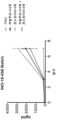
도 1은 그룹 1 내지 4 (Balb/c 마우스, 6-7 주)에서 일 0-7 사이 ELISA에 의해 측정된 hIgG의 수준 (ng/ml)을 보여준다. 그룹 1 (회색) PBS 및 pGX9214, 그룹 2 히알루로니다제 전처리 및 pGX9214 (청색), 그룹 3 콘드로이티나제 및 pGX9214 (녹색), 그룹 4 히알루로니다제/ 콘드로이티나제 전처리 및 pGX9214 (갈색).
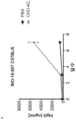
도 2는 그룹 1 내지 4 (C57BL/6, 6-7 주)에서 일 0-7 사이 ELISA에 의해 측정된 hIgG의 수준 (ng/ml)를 보여준다. 그룹 1 (회색) PBS 및 pGX9214, 그룹 2 히알루로니다제 전처리 및 pGX9214 (청색), 그룹 3 콘드로이티나제 및 pGX9214 (녹색), 그룹 4 히알루로니다제/ 콘드로이티나제 전처리 및 pGX9214 (갈색).
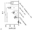
도 3은 그룹 1 내지 4에서 일 0-21 사이 ELISA에 의해 측정된 hIgG의 수준 (ng/ml)를 보여준다. 그룹 1 (회색) PBS 및 pGX9214, 그룹 2 히알루로니다제 전처리 및 pGX9214 (암적색), 그룹 3 콘드로이티나제 및 pGX9214 (밝은 적색), 그룹 4 히알루로니다제/ 콘드로이티나제 전처리 및 pGX9214 (담홍색 - 하틀리 기니 피그).
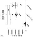
도 4는 그룹 1 내지 4에서 개별 기니 피그내 일 0-21 사이 ELISA에 의해 측정된 hIgG의 수준 (ng/ml)를 보여준다. 도 4A: 그룹 1 PBS. 도 4B: 400U/ml로 그룹 2 히알루로니다제 전처리. 도 4C: 그룹 3 콘드로이티나제 (0.5U/ml). 도 4D: 그룹 4 히알루로니다제 400U/ml 및 콘드로이티나제 0.5U/ml 전처리.
도 5는 콘드로이티나제가 Balb/c 마우스에서 플라스미드-인코딩된 hlgG 발현을 향상시키는 것을 보여준다. (a) 그룹 1 및 2 (Balb/c 마우스, 6-7 주)에서 일 0-7 사이 ELISA에 의해 측정된 hIgG의 수준 [ng/ml]. 그룹 1 (회색) PBS 및 pGX9214, 그룹 2 콘드로이티나제 전처리 및 pGX9214 (흑색). (b) 일 7에서 콘드로이티나제에 의한 hIgG의 유의미한 향상. 만 휘트니 시험에 의해 수행된 통계, P = 0.0079.
도 6은 콘드로이티나제가 C57BL/6 마우스에서 플라스미드-인코딩된 hIgG 발현을 향상시킨다는 것을 보여준다. (a) 그룹 1 및 2 (Balb/c 마우스, 6-7 주)에서 일 0-7 사이 ELISA에 의해 측정된 hIgG의 수준 [ng/ml]. 그룹 1 (회색) PBS 및 pGX9214, 그룹 2 콘드로이티나제 전처리 및 pGX9214 (흑색). (b) 일 7에서 콘드로이티나제에 의한 hIgG의 유의미한 향상. 만 휘트니 시험에 의해 수행된 통계, P = 0.0079.
도 7은 DMAb 발현을 향상시키기 위한 콘드로이티나제의 상이한 버전의 능력의 조사를 보여준다. 그래프는 그룹 1-4 (Balb/c)에서 일 7에 ELISA에 의해 측정된 hIgG 수준 (ng/ ml)를 나타낸다. 마우스는 어느 한쪽 PBS (대조군 그룹 1), 콘드로이티나제 AC, 임상 등급 콘드로이티나제 ABC 또는 재조합 단백질 GALNS로 처리되었다. 모든 그룹은 pGX9203의 주사를 받았고 이어서 전기천공되었다. 크루스칼-왈리스 시험에 의해 수행된 통계, **P = 0.0026.
도 8은 증가하는 콘드로이티나제 ABC 용량이 DNA-기반 단백질 발현을 추가로 향상시키는 것을 보여준다. Balb/c 마우스는 pDNA (pGX9207) 주사 및 전기천공 30 분 앞서 콘드로이티나제 ABC의 증가 용량으로 처리되었다. hIgG의 수준은 ELISA에 의해 측정되었다. 크루스칼-왈리스 시험에 의해 수행된 통계, *P = 0.0357.
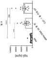
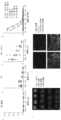
도 9는 향상된 형광성 단백질 발현에서 콘드로이티나제 투여 결과를 보여준다. (a) 골격근에 어느 한쪽 콘드로이티나제 또는 PBS로 처리된 Balb/c 마우스의 좌측 및 우측 뒷다리. 리포터 단백질 발현의 가시화는 형광 이미저 (Protein Simple)에 의해 수행되었다. (b) 임의의 유닛 형광 강도는 리포터 유전자 발현과 평행하고 AlphaView SA 소프트웨어 사용에 의해 정량화되었다. 만 휘트니 시험에 의해 수행된 통계, P = 0.0022.
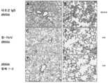
도 10은 H&E 염색에 의해 수행된 쥣과 뒷다리 골격근의 대표적인 조직병리를 보여준다. 최상부 패널은 어느 한쪽 PBS 단독 (대조군) 또는 콘드로이티나제 ABC 단독으로 처리된 조직을 보여준다. pDNA 전달 전 어느 한쪽 콘드로이티나제 또는 PBS (대조군)으로 전처리된 (30 분) 근육은 최하부 도에서 제시된다. 결과는 슬라이드 스캐너 및 CaseViewer 소프트웨어 (3DHISTECH)에 의해 분석되었다. 척도 = 200 μm.
도 11은 콘드로이티나제 ABC가 뉴질랜드 래빗 (9 주)에서 플라스미드-인코딩된 hIgG 발현을 향상시키는 것을 보여준다. (a) 그룹 2 (콘드로이티나제-전처리된, 회색)에서 그리고 그룹 1 (PBS 대조군, 흑색)에서 일 0과 6 사이 ELISA에 의해 측정된 hIgG의 수준 [ng/ml]이 제시된다. (b) 그래프는 ELISA에 의해 측정된 일 6으로부터 hIgG 수준을 보여준다. 만 휘트니 시험에 의해 수행된 통계, P = 0.0043.
도 12는 마우스 (Balb/c; 14 마우스 / 그룹)에서 pDNA (pGX9207)을 가진 콘드로이티나제의 공-제형화를 보여준다. 그래프는 ELISA에 의해 측정된 일 6으로부터 hIgG 수준 [ng/ml]를 보여준다. 만 휘트니 시험에 의해 수행된 통계, ****P < 0.0001.
도 13은 향상된 형광성 단백질 발현에서 콘드로이티나제 투여 결과를 보여준다. (a) 골격근에 어느 한쪽 콘드로이티나제 또는 PBS로 처리된 Balb/c 마우스의 좌측 및 우측 뒷다리. 리포터 단백질 발현의 가시화는 형광 이미지형성 시스템에 의해 수행되었다. (b) 임의의 단위 형광 강도는 리포터 유전자 발현과 평행하고 AlphaView SA 소프트웨어 사용에 의해 정량화되었다. 만 휘트니 시험에 의해 수행된 통계, **P = 0.0004.
도 14는 래빗 (뉴질랜드 래빗; 6마리 래빗 / 그룹)에서 pDNA (pGX9207)을 가진 콘드로이티나제의 공-제형화를 보여준다. (a) 그래프는 ELISA에 의해 측정된 일 0부터 일 5까지 혈청 hIgG 수준 [ng/ml]을 보여준다. 그룹 1, 2 및 4의 비교. (b) 일 5에서 측정된 모든 그룹의 래빗 혈청 hIgG 수준. 만 휘트니 시험에 의해 수행된 통계, ****P < 0.0001.
도 15는 콘드로이티나제 (2.5 U/ ml)/ pDNA (pGX9207, 250 ng / 웰) 공제형화된 샘플의 아가로스 겔 전기영동을 보여준다. (a) 짝수가 있는 레인은 콘드로이티나제 ABC를 함유하는 pDNA 샘플을 나타내고, 홀수가 있는 레인은 PBS 음성 대조군을 지칭한다. 레인 1-2: 겔 전기영동 전 샘플의 인큐베이션 없음, 레인 3-4: 10 분 동안 RT (21℃)에서 인큐베이션, 레인 5-6: 6℃, 120 분, 레인 7-8: RT, 120 분, 레인 9-10: 6℃, 24 시간, 레인 11-12: RT, 24 시간, 레인 13-14: 6℃, 10 분. M1 = 마커. (b) 초나선 DNA용 사다리, 2-10 kb (New England Biolabs).
도 16은 24 시간 동안 콘드로이티나제/ pDNA 공제형의 저장이 Balb/c 마우스에서 향상된 유전자 발현에 영향을 주지 않는 것을 보여준다. 그래프는 일 6에서 ELISA에 의해 측정된 혈청 hIgG 수준 (ng/ ml)를 나타낸다. 마우스는 좌측 뒷다리 골격근에 공제형화로 처리되었고 전기천공은 1 분의 약물 주사 후 수행되었다. 처리에 앞서, 공제형화 샘플은 4 ℃에서 10 분, 120 분 및 24 시간에서 인큐베이션되었다. 대조군: 마우스는 pDNA 투여 및 전기천공 30 분 전 어느 한쪽 콘드로이티나제 또는 PBS로 전처리되었다. 크루스칼-왈리스 시험에 의해 수행된 통계, ****P = 0.0001.
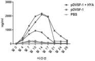
도 17은 그룹 1, 2 및 3에서 일 0-28 사이 ELISA에 의해 측정된 hIgG의 수준 (ng/ml를 보여준다. 그룹 1은 pDVSF-1 및 히알루로니다제 전처리로 처리되었고, 그룹 2는 pDVSF 단독으로 처리되었고, 그룹 3은 PBS 단독으로 처리되었다.
도 18은 그룹 1 및 2의 항-hIgG 결합 역가를 보여준다.
도 19는 실시예 2에서 인플루엔자 NP 펩타이드 풀에 대한 그룹 1-3의 PBMCs내 평균 (+/- SEM) IFN-γ (스팟 / 백만) 반응을 보여준다.
도 20은 실시예 2에서 그룹 1-3에 대하여 항-인플루엔자 NP IgG 결합 역가를 보여준다.
도 21은녹농균급성 폐렴 모델을 보여준다. 항-PcrV 또는 DMAb-항체 1-2 플라스미드 DNA의 전기천공은 마우스에서 활성 IgG의 발현을 산출한다. 강력한 보호성 활성은 양쪽 항-녹농균 DMAbs 및 DMAb-항체 1-2 IgG에 대하여 관측되었다.
도 22는 혈청에서 DMAb 발현의 IgG 정량화를 보여준다. 혈청은 녹농균 감염에 앞서 DMAb 발현에 대하여 평가되었다. 유사한 생존 프로파일에도 불구하고, 항-PcrV DMAb 발현은 DMAb-항체 1-2보다 5-배 더 컸다.
도 23은 항-녹농균 DMAbs에 의한 장기 부담의 감소를 보여준다. DMAb-항체 1-2 및 IgG는 폐에서 부담을 감소시킨다. 항-PcrV 및 DMAb-항체 1-2는 박테리아의 전신 확산을 상당히 감소시킨다. 조직은 감염후 24 시간 수집되었다. LOD = 검출의 한계; * = 크루스칼-왈리스 및 던의 다중 비교 시험 대 대조군 IgG DMAb에 의한 P<0.05.
도 24는 감염후 48 시간에서 급성 폐렴의 조직학을 보여준다 (H&E). A. 대조군 IgG 폐는 중성구 및 대식세포로 구성된 중증 폐포 침윤물 그리고 출혈의 면적을 나타낸다 (10x). C. 온화한 폐렴 및 가끔의 세기관지 잔해 (10x). E. 온화한 폐포염을 가진 DMAb-항체 1-2 DNA 그룹 (10x). B, D, F. A. C. D., 각각으로부터 40x로 삽입물.
도 25는 DMAb-항체 1-2가 농도 의존적 보호성 활성을 나타내는 것을 보여준다. A. 동물은 EP에 앞서 1, 2 또는 3 부위에 DNA를 받았다. B. 혈청 IgG의 정량화. *는 로그-순위 시험을 통해 P<0.05를 나타낸다.
도 26은 DMAb-항체 1-2 및 메로페넴 (MEM)의 치료이하 투약량이 녹농균 폐렴에 대한 향상된 활성을 나타내는 것을 보여준다. 동물은 EP에 앞서 1, 2 또는 3 부위에 DNA를 받았다. B. 혈청 IgG의 정량화. *는 로그-순위 시험을 통해 P<0.05를 나타낸다.
도 27은 pGX2013 면역화 14 일 후 NP55 및 NP147 펩타이드 에피토프에 대한 비장세포내 평균 (+/- SEM) IFN-γ (백만당 점) 반응을 보여준다.
도 28은 pGX2013으로 면역화 7 및 14 일 후 그룹에 대하여 항-NP IgG 결합 역가를 보여준다.
도 29는 항-MERS-CoV 인간 IgG의 시험관내 발현을 보여준다. (a) 항-MERS-CoV 항원 DMAb 플라스미드 DNA pMERS의 도식적 실례. 푸린 및 2A 절단 부위에 의해 분리된 항체 중쇄 및 경쇄 서열의 업스트림 자리잡은 CMV 프로모터. (b & c) 293T 세포 배양물은 1 μg/ml의 pVax 또는 pMERS로 형질감염되었고 배양물 상청액은 48 시간 후 수확되었다. (b) 상청액에서 인간 IgG 수준은 ELISA에 의해 분석되었다. (c) MERS CoV 항원에 대한 IgG 결합은 ELISA에 의해 측정되었다.
도 30은 BALB/c 마우스내 DMAb의 향상된 생체내 발현을 보여준다
6.25 내지 100 μg의 pMERS는, HYA-처리된 근육 (EP + HYA)에 (a) 주사 단독으로 (EP 없음), (b) EP가 있는 주사로 (EP) 그리고 (c) EP가 있는 주사로, BALB/c 마우스 (4-8 마우스 / 그룹)의 TA 근육에 투여되었다. (a-c) 혈청 인간 IgG 수준은 pMERS 전달 후 일 6에서 ELISA에 의해 정량화되었다 (데이터 포인트는 평균+/-SEM을 표시한다). (d) pMERS 전달 후 일 0 및 일 6에서 ELISA에 의해 측정된 상호 혈청 희석액의 MERS CoV 항원. (e) BALB/c 마우스 TA 근육은 (a), (b) 또는 (c)에서 이용된 프로토콜로 pRFP (25 μg) 리포터 유전자 투여 후 또는 삽입물 1-4, 각각에서 처리 없이 72 시간 수확되었다. 이미지는 리포터 유전자 발현을 설명한다. TA 근육의 섹션의 면역형광 이미지는 EP + HYA로 전달된 pMERS 또는 pVax (100 μg)으로 처리되었고, 72 시간 후 수확되었다 (f). hIgG는 항-인간 IgG 이어서 FITC-표지된 2차 항체 (녹색)으로 검출되었다. 청색으로 DAPI 얼룩. 패널 1. 처리 없음. 패널 2. pVax. 패널 3 & 4. pMERS. 패널 1-3은 근육 섬유에 수직인 단면 이미지를 나타내고, 패널 4에서 이미지는 근육 섬유 사이이다.
도 31은 Crl:Nu-Foxn1nu 마우스에서 증가된 및 지속된 DMAb 발현을 보여준다. 6.25 내지 100 μg의 pMERS는 Crl:Nu-Foxn1nu 마우스 (8 마우스 / 그룹)의 HYA 전처리된 TA 근육에 EP로 투여되었다. 혈청 hIgG는 일 0 내지 160에서 ELISA에 의해 정량화되었다 (a). (b) 100 μg의 pMERS는 (a)에서처럼 1 (좌측 TA), 2 (우측 및 좌측 TA), 3 (우측 및 좌측 TA, 및 좌측 사두) 또는 4 (우측 및 좌측 TA, 및 좌측 및 우측 사두) 근육에 투여되었다. 혈청 hIgG는 일 21에서 ELISA에 의해 정량화되었다.
도 32는 뉴질랜드 화이트 래빗에서 생체내 DMAb 발현을 보여준다. (a) PBS- (최상부 패널) 또는 HYA- (최하부 패널) 처리된 근육에서 EP로 pGFP (0.2 mg)의 전달 72 시간 후 래빗 TA 근육 섹션에서 리포터 유전자 발현. (b&c) 2 mg의 pMERS는 래빗 (6 / 그룹)의 (PBS 또는 HYA로 전-처리된) 사두 근육에 EP로 투여되었다. 혈청 hIgG는 일 3, 5 및 6에서 정량화되었고 (a), 전달 후 일 6에서 ELISA에 의해 MERS CoV 항원 결합 측정되었다 (b). (c) 2 mg의 pMERS는 래빗 (6 / 그룹)의 (HYA로 처리된) 사두 근육에 20 내지 65 V 의 전압 셋팅에서 EP로 투여되었고, 혈청 hIgG는 전달 후 일 5에서 정량화되었다. (c & d) 값은 평균 +/- SEM (n = 6/그룹)으로서 묘사된다. ****p < 0.0001, ***p < 0.001, **p < 0.01, 및 ns = 비-유의미. P 값은 짝짓기되지 않은, 2-테일드 만-휘트니 시험으로부터이다.
도 33은 히말라야 원숭이에서 생체내 DMAb 발현을 보여준다. 13.5 mg의 pMERS는 히말라야 원숭이 (5 / 그룹)의 (HYA로 전-처리된) 사두 근육에 EP로 투여되었다. 혈청 hIgG는 정량화되었고 (a), 일 0 내지 35에서 ELISA에 의해 MERS CoV 항원 결합 측정되었고 (b), 각각의 히말라야 원숭이에 대하여 일 17 상호 혈청 희석되었다 (c). (d) 인간 IgG (ADA)에 대한 항체 반응은 히말라야 원숭이의 혈청에서 직접 ELISA에 의해 측정되었다. (e) 혈청에서 DMAb 수준 및 항-인간 IgG 항체 결합 수준의 상관관계는 묘사된다. 피크 hIgG (μg/ml) 값이 각각의 히말라야 원숭이에서 도달된 후 그리고 포함하여 데이터 포인트 (상응하는 ADA (OD450nm)를 가진 hIgG (μg/ml))는 플롯팅된다. P 값 및 스피어만 상관 계수는 GraphPad Prism 6 소프트웨어를 사용하여 계산되었다.
도 34는 BALB/c 마우스에서 DMAb의 생체내 발현 동력학을 보여준다. (a-c) 6.25 내지 100 μg의 pMERS는, HYA-처리된 근육 (EP + HYA)에 (a) 주사 단독으로 (EP 없음), (b) EP가 있는 주사로 (EP) 및 (c) EP가 있는 주사로, BALB/c 마우스 (4-8 마우스 / 그룹)의 TA 근육에 투여되었다. (a-c) 혈청 인간 IgG 수준은 pMERS 전달 후 일 0 내지 14에서 ELISA에 의해 정량화되었다 (데이터 포인트는 평균+/-SEM을 표시한다).
도 35는 pMERS 처리된 BALB/c 마우스에서 인간 IgG에 대한 항-항체 반응을 보여준다. 인간 IgG (ADA)에 대한 항체 반응은 EP로 pMERS 전달 후 일 0 내지 14에서 BALB/c 마우스의 혈청에서 직접 ELISA에 의해 측정되었다.
도 36은 유전자 발현을 향상시킨다고 보고된 pDNA 전달 시약의 스크린을 보여준다. 100 μg의 pMERS는 BALB/c 마우스 (5 마우스 / 그룹)의 TA 근육에 투여되었다. 상기 표적 TA 근육은 그룹 1, 2, 5, 7, 8, 및 9 각각에서 PBS, HYA, 7% 수크로스, 콜라겐분해효소 D, 엘라스타제 또는 MMP7로 pMERS 전달 30 분 전 전처리되었다. pMERS는 Poly-L-글루탐산 (그룹 3) 또는 Tempol (그룹 4) 또는 (OH)3D3 (그룹 6)으로 공제형화되었고, 전처리는 없었다. 혈청 인간 IgG 수준은 pMERS 전달 후 일 6에서 ELISA에 의해 정량화되었다 (데이터 포인트는 평균+/-SEM을 표시한다).
도 37은 래빗에서 DMAb 발현을 보여준다. (a 및 b) 2 mg의 pGX9207은 뉴질랜드 화이트 래빗 (6 / 그룹)의 (HYA로 전-처리된) 사두 근육에 EP로 투여되었다. 혈청 hIgG는 (a)이었고 항-인간 IgG (ADA) 결합 (b)는 일 0 내지 10에서 ELISA에 의해 분석되었다.
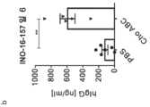
도 38은 래빗에서 pDNA를 가진 HYA의 공-제형화 연구를 보여준다. 6 뉴질랜드 화이트 래빗 / 그룹. 좌측 사두 근육에서 2 mg pGX9207. 2 부위. HYA (Intropharma) 200U / 부위. 투여 5 분 전 공-제형화된 CELLECTRA®-5P pDNA/HYA. 혈액은 일 5에서 샘플링되었다. 실험 번호 INO-16-158b.
도 39는 EP 지연을 가진 래빗에서 pDNA를 가진 HYA의 공-제형화를 보여준다. 6 뉴질랜드 화이트 래빗 / 그룹. 좌측 사두 근육에서 2 mg pGX9207. 2 부위. HYA (Intropharma) 200 U / 부위. 투여 5 분 전 공-제형화된 CELLECTRA®-5P pDNA/HYA. 혈액은 일 5에서 샘플링되었다. 실험 번호 INO-16-158a.
도 40은 히말라야 원숭이에서 pDNA를 가진 HYA의 공-제형화를 보여준다. CELLECTRA®-5P로 사두 근육에 전달된 pGX9207 (pMERS). 4 부위. 1 mg pDNA / 부위. Intropharma (소 고환 정제된 HYA). 투여 5 분 전 공-제형화된 pDNA /HYA. 실험 번호 INO-16-194.
도 41은 Hylenex (인간 재조합 히알루로니다제)를 가진 EP 지연의 최적화를 보여준다. pGX9207 HYA 공-제형화. 좌측 사두에서 2개의 Tx. 일 5 hlgG 혈청 수준은 묘사된다. 실험 번호 INO-16-279.
도 42는 HYA 공-제형화의 DNA 백신 용량 절약 효과를 보여준다. BALB/c 마우스. 일 0 인플루엔자 pNP IM CELLECTRA-3P, 일 7 및 14 ELISA. 실험 번호 INO-16-218.
도 43은 DNA 백신 HYA 제형화로 종양 항원에 대한 면역 반응의 확대를 보여준다. B6 마우스. 원상태 마우스 TERT 펩타이드 풀에 대한 일 0 및 14 pmTERT IM CELLECTRA®-3P, 일 21 IFN-감마 ELISpot. 실험 번호 INO-17-018.Figure 1 shows the levels of hIgG (ng/ml) measured by ELISA between days 0-7 in groups 1-4 (Balb/c mice, 6-7 weeks). Group 1 (grey) PBS and pGX9214,
Figure 2 shows the levels of hIgG (ng/ml) measured by ELISA between days 0-7 in groups 1-4 (C57BL/6, 6-7 weeks). Group 1 (grey) PBS and pGX9214,
Figure 3 shows levels of hIgG (ng/ml) measured by ELISA between days 0-21 in groups 1-4. Group 1 (grey) PBS and pGX9214,
Figure 4 shows the levels of hIgG (ng/ml) measured by ELISA between days 0-21 in individual guinea pigs in groups 1-4. Figure 4A:
Figure 5 shows that chondroitinase enhances plasmid-encoded hlgG expression in Balb/c mice. (a) Levels of hIgG [ng/ml] measured by ELISA between
Figure 6 shows that chondroitinase enhances plasmid-encoded hIgG expression in C57BL/6 mice. (a) Levels of hIgG [ng/ml] measured by ELISA between
Figure 7 shows an investigation of the ability of different versions of chondroitinase to enhance DMAb expression. The graph represents hIgG levels (ng/ml) measured by ELISA on
Figure 8 shows that increasing doses of chondroitinase ABC further enhance DNA-based protein expression. Balb/c mice were treated with increasing doses of
Figure 9 shows the results of chondroitinase administration in enhanced fluorescent protein expression. (a) Left and right hindlimbs of Balb/c mice whose skeletal muscles were treated with either chondroitinase or PBS. Visualization of reporter protein expression was performed by a fluorescence imager (Protein Simple). (b) Arbitrary unit fluorescence intensity was paralleled with reporter gene expression and quantified using AlphaView SA software. Statistics performed by Mann-Whitney test, P = 0.0022.
Figure 10 shows representative histopathology of murine hind limb skeletal muscle performed by H&E staining. The top panel shows tissue treated with either PBS alone (control) or chondroitinase ABC alone. Muscles pretreated (30 min) with either chondroitinase or PBS (control) prior to pDNA delivery are shown in the bottom figure. Results were analyzed by slide scanner and CaseViewer software (3DHISTECH). Scale = 200 μm.
Figure 11 shows that chondroitinase ABC enhances plasmid-encoded hIgG expression in New Zealand rabbits (9 weeks). (A) Levels of hIgG [ng/ml] measured by ELISA between
Figure 12 shows co-formulation of chondroitinase with pDNA (pGX9207) in mice (Balb/c; 14 mice/group). The graph shows hIgG levels [ng/ml] from
Figure 13 shows the results of chondroitinase administration in enhanced fluorescent protein expression. (a) Left and right hindlimbs of Balb/c mice whose skeletal muscles were treated with either chondroitinase or PBS. Visualization of reporter protein expression was performed by a fluorescence imaging system. (b) Arbitrary unit fluorescence intensity was paralleled with reporter gene expression and quantified using AlphaView SA software. Statistics performed by Mann-Whitney test, **P = 0.0004.
Figure 14 shows co-formulation of chondroitinase with pDNA (pGX9207) in rabbits (New Zealand rabbits; 6 rabbits/group). (a) Graph shows serum hIgG levels [ng/ml] from
Figure 15 shows agarose gel electrophoresis of chondroitinase (2.5 U/ml)/pDNA (pGX9207, 250 ng/well) co-incubated samples. (a) Lanes with even numbers represent pDNA samples containing chondroitinase ABC, and lanes with odd numbers refer to the PBS negative control. Lanes 1-2: no incubation of the sample before gel electrophoresis, lanes 3-4: incubation at RT (21°C) for 10 min, lanes 5-6: 6°C, 120 min, lanes 7-8: RT, 120 min. , lanes 9-10: 6°C, 24 hours, lanes 11-12: RT, 24 hours, lanes 13-14: 6°C, 10 min. M1 = marker. (b) Ladder for supercoiled DNA, 2-10 kb (New England Biolabs).
Figure 16 shows that storage of chondroitinase/pDNA co-format for 24 hours does not affect improved gene expression in Balb/c mice. The graph represents serum hIgG levels (ng/ml) measured by ELISA at
Figure 17 shows the levels of hIgG (ng/ml) measured by ELISA between days 0-28 in
Figure 18 shows anti-hIgG binding titers for
Figure 19 shows the mean (+/- SEM) IFN-γ (spots/million) response in PBMCs of Groups 1-3 to the influenza NP peptide pool in Example 2.
Figure 20 shows anti-influenza NP IgG binding titers for groups 1-3 in Example 2.
Figure 21 showsa Pseudomonas aeruginosa acute pneumonia model. Electroporation of anti-PcrV or DMAb-antibody 1-2 plasmid DNA results in expression of active IgG in mice. Strong protective activity was observed against both anti-Pseudomonas aeruginosa DMAbs and DMAb-antibody 1-2 IgG.
Figure 22 shows IgG quantification of DMAb expression in serum. Serum was assessed for DMAb expression prior to Pseudomonas aeruginosa infection. Despite similar survival profiles, anti-PcrV DMAb expression was 5-fold greater than DMAb-antibody 1-2.
Figure 23 shows reduction of organ burden by anti-Pseudomonas aeruginosa DMAbs. DMAb-antibodies 1-2 and IgG reduce the burden on the lungs. Anti-PcrV and DMAb-antibodies 1-2 significantly reduce systemic spread of bacteria. Tissues were collected 24 hours after infection. LOD = limit of detection; * = P<0.05 by Kruskal-Wallis and Dunn's multiple comparison test versus control IgG DMAb.
Figure 24 shows the histology of acute pneumonia at 48 hours post infection (H&E). A. Control IgG lung shows severe alveolar infiltrate composed of neutrophils and macrophages and area of hemorrhage (10x). C. Mild pneumonia and occasional bronchiolar debris (10x). E. DMAb-antibody 1-2 DNA group (10x) with mild alveolitis. B, D, FACD, inserts at 40x from each.
Figure 25 shows that DMAb-antibody 1-2 exhibits concentration-dependent protective activity. A. Animals received DNA at
Figure 26 shows that subtherapeutic doses of DMAb-antibody 1-2 and meropenem (MEM) show improved activity against P. aeruginosa pneumonia. Animals received DNA at 1, 2, or 3 sites prior to EP. B. Quantification of serum IgG. * indicates P<0.05 via log-rank test.
Figure 27 shows mean (+/- SEM) IFN-γ (points per million) responses in splenocytes to NP55 and
Figure 28 shows anti-NP IgG binding titers for
Figure 29 shows in vitro expression of anti-MERS-CoV human IgG. (a) Schematic illustration of anti-MERS-CoV antigen DMAb plasmid DNA pMERS. The CMV promoter lies upstream of the antibody heavy and light chain sequences, separated by furin and 2A cleavage sites. (b & c) 293T cell cultures were transfected with 1 μg/ml of pVax or pMERS and culture supernatants were harvested 48 hours later. (b) Human IgG levels in supernatants were analyzed by ELISA. (c) IgG binding to MERS CoV antigen was measured by ELISA.
Figure 30 shows enhanced in vivo expression of DMAb in BALB/c mice.
6.25 to 100 μg of pMERS was administered to HYA-treated muscles (EP+HYA) (a) by injection alone (no EP), (b) by injection with EP (EP), and (c) by injection with EP. , were administered to the TA muscle of BALB/c mice (4-8 mice/group). (ac) Serum human IgG levels were quantified by ELISA at
Figure 31 shows increased and sustained DMAb expression in Crl:Nu-Foxn1nu mice. 6.25 to 100 μg of pMERS was administered by EP into the HYA-pretreated TA muscle of Crl:Nu-Foxn1nu mice (8 mice/group). Serum hIgG was quantified by ELISA from
Figure 32 shows DMAb expression in vivo in New Zealand White rabbits. (a) Reporter gene expression in rabbit TA muscle sections 72 h after delivery of pGFP (0.2 mg) with EP in PBS- (top panel) or HYA- (bottom panel) treated muscle. (b&c) 2 mg of pMERS was administered by EP to the quadriceps muscle (pre-treated with PBS or HYA) of rabbits (6/group). Serum hIgG was quantified on
Figure 33 shows DMAb expression in vivo in rhesus macaques. 13.5 mg of pMERS was administered by EP to the quadriceps muscle (pre-treated with HYA) of rhesus macaques (5/group). Serum hIgG was quantified (a), MERS CoV antigen binding was measured by ELISA on
Figure 34 shows in vivo expression kinetics of DMAb in BALB/c mice. (ac) 6.25 to 100 μg of pMERS was administered to HYA-treated muscles (EP + HYA) with (a) injection alone (no EP), (b) injection with EP (EP), and (c) EP. was administered by injection into the TA muscle of BALB/c mice (4-8 mice/group). (ac) Serum human IgG levels were quantified by ELISA on
Figure 35 shows anti-antibody responses to human IgG in pMERS treated BALB/c mice. Antibody responses to human IgG (ADA) were measured by ELISA directly in the serum of BALB/c mice on
Figure 36 shows a screen of pDNA delivery reagents reported to enhance gene expression. 100 μg of pMERS was administered into the TA muscle of BALB/c mice (5 mice/group). The target TA muscles were pretreated 30 minutes prior to pMERS delivery with PBS, HYA, 7% sucrose, collagenase D, elastase, or MMP7 in
Figure 37 shows DMAb expression in rabbits. (a and b) 2 mg of pGX9207 was administered by EP to the quadriceps muscle (pre-treated with HYA) of New Zealand White rabbits (6/group). Serum hIgG was (a) and anti-human IgG (ADA) binding (b) was analyzed by ELISA on
Figure 38 shows co-formulation studies of HYA with pDNA in rabbits. 6 New Zealand White Rabbits / Group. 2 mg pGX9207 in the left quadriceps muscle. 2 parts. HYA (Intropharma) 200U/site. Co-formulated CELLECTRA®-5P pDNA/
Figure 39 shows co-formulation of HYA with pDNA in rabbits with EP delay. 6 New Zealand White Rabbits / Group. 2 mg pGX9207 in the left quadriceps muscle. 2 parts. HYA (Intropharma) 200 U/site. Co-formulated CELLECTRA®-5P pDNA/
Figure 40 shows co-formulation of HYA with pDNA in rhesus macaques. pGX9207 (pMERS) delivered to quadriceps muscle with CELLECTRA®-5P. 4 parts. 1 mg pDNA/site. Intropharma (purified bovine testes HYA). Co-formulated pDNA/
Figure 41 shows optimization of EP delay with Hylenex (human recombinant hyaluronidase). pGX9207 HYA co-formulation. 2 Tx in left quadriceps.
Figure 42 shows the DNA vaccine dose saving effect of HYA co-formulation. BALB/c mice.
Figure 43 shows expansion of immune response to tumor antigens with DNA vaccine HYA formulation. B6 mouse.
상세한 설명details
본 발명은 대상체에 제제의 전달용 조성물 및 전달 방법에 관한 것이다. 상기 방법은 CSPGs의 설페이트 기를 가수분해시키는데 충분한 양으로 제제, 및 콘드로이티나제 폴리펩타이드 또는 콘드로이티나제 폴리펩타이드를 인코딩하는 폴리뉴클레오타이드를 대상체에 투여하는 단계를 포함할 수 있다. 콘드로이티나제는 대상체에서 CSPGs의 설페이트 기를 가수분해시킬 수 있다. 이러한 해체는 세포외 매트릭스의 해체로 이어질 수 있고 그렇게 함으로써 제제의 전달을 용이하게 할 수 있다. 방법은 추가로 히알루로니다제 폴리펩타이드 또는 히알루로니다제 폴리펩타이드를 인코딩하는 폴리뉴클레오타이드를 대상체에 투여하는 단계를 포함할 수 있다.The present invention relates to compositions and methods for delivering agents to a subject. The method may include administering to the subject an agent and a chondroitinase polypeptide or a polynucleotide encoding a chondroitinase polypeptide in an amount sufficient to hydrolyze the sulfate group of CSPGs. Chondroitinase can hydrolyze the sulfate groups of CSPGs in a subject. This disorganization can lead to disorganization of the extracellular matrix and thereby facilitate delivery of the agent. The method may further include administering to the subject a hyaluronidase polypeptide or a polynucleotide encoding a hyaluronidase polypeptide.
본 발명은 또한 대상체에 제제와 함께 히알루로니다제의 투여에 관한 것이다. 히알루로니다제는 폴리펩타이드로서 또는 핵산 인코딩 히알루로니다제 또는 이의 단편 또는 변이체로서 투여될 수 있다. 히알루로니다제는 대상체에 제제의 전달을 용이하게 할 수 있다. 그 결과, 히알루로니다제는 대상체에서 면역 반응을 향상시킬 수 있고/있거나 대상체에서 제제의 발현을 향상시킬 수 있다.The present invention also relates to the administration of hyaluronidase with a formulation to a subject. Hyaluronidase can be administered as a polypeptide or as a nucleic acid encoding hyaluronidase or fragments or variants thereof. Hyaluronidase can facilitate delivery of an agent to a subject. As a result, hyaluronidase may enhance the immune response in the subject and/or enhance expression of the agent in the subject.
1) 정의1) Definition
달리 정의되지 않는 한, 본 명세서에서 사용된 모든 기술 및 과학적 용어들은 당해 분야의 숙련가에 의해 통상적으로 이해되는 바와 동일한 의미를 갖는다. 이해상충의 경우에, 정의를 포함하는, 본 문서가 제어할 것이다. 본 명세서에서 기재된 것과 유사한 또는 동등한 방법 및 물질이 본 발명의 실시 또는 시험에서 사용될 수 있어도, 바람직한 방법 및 물질은 아래 기재된다. 본 명세서에서 언급된 모든 공보, 특허 출원, 특허 및 다른 참조문헌은 그 전문이 참고로 편입된다. 본 명세서에서 개시된 물질, 방법, 및 예는 단지 실례가 되고 제한되도록 의도되지 않는다.Unless otherwise defined, all technical and scientific terms used herein have the same meaning as commonly understood by a person skilled in the art. In case of conflict of interest, this document, including definitions, will control. Although methods and materials similar or equivalent to those described herein can be used in the practice or testing of the present invention, the preferred methods and materials are described below. All publications, patent applications, patents and other references mentioned herein are incorporated by reference in their entirety. The materials, methods, and examples disclosed herein are illustrative only and are not intended to be limiting.
용어 "포함한다", "포함하다", "갖는", "갖는다", "할 수 있다", "함유한다", 및 이의 변이체는, 본 명세서에서 사용된 바와 같이, 추가의 행위 또는 구조의 가능성을 배제하지 않는 개방형 과도기적 어구, 용어들, 또는 단어인 것으로 의도된다. 단수 형태 "한", "하나", 및 "그"는 문맥이 분명히 달리 지시하지 않는 한 복수의 참조를 포함한다. 본 개시내용은 또한, 명백하게 제시되든 아니든, 본 명세서에서 제시된 구현예 또는 요소를 "포함하는", "상기로 구성되는", 및 "상기로 본질적으로 구성되는" 다른 구현예를 고려한다.The terms “comprise,” “comprise,” “having,” “have,” “may,” “contain,” and variants thereof, as used herein, refer to the possibility of additional acts or structures. It is intended to be an open-ended transitional phrase, term, or word that does not exclude. The singular forms “one,” “one,” and “the” include plural references unless the context clearly dictates otherwise. The disclosure also contemplates other embodiments, “comprising,” “consisting of,” and “consisting essentially of” the embodiments or elements set forth herein, whether or not explicitly stated.
관심의 하나 이상의 값에 적용된 경우 본 명세서에서 사용된 바와 같이 용어 "약"은 언급된 참조 값과 유사한 값을 지칭한다. 특정 양태에서, 용어 "약"은 (그와 같은 수가 가능한 값의 100%를 초과하지 않을 경우를 제외하고) 문맥으로부터 달리 언급되지 않는 한 또는 달리 분명하지 않는 한 언급된 참조 값의 어느 한쪽 방향 (초과 또는 미만)으로 20%, 19%, 18%, 17%, 16%, 15%, 14%, 13%, 12%, 11%, 10%, 9%, 8%, 7%, 6%, 5%, 4%, 3%, 2%, 1%, 또는 보다 적게 해당하는 값의 범위를 지칭한다.As used herein, the term “about” when applied to one or more values of interest refers to a value similar to the stated reference value. In certain embodiments, the term “about” means (except where such number does not exceed 100% of the possible values) a reference value in either direction of the stated reference value (unless otherwise indicated or otherwise clear from the context). greater than or equal to 20%, 19%, 18%, 17%, 16%, 15%, 14%, 13%, 12%, 11%, 10%, 9%, 8%, 7%, 6%, Refers to a range of values such as 5%, 4%, 3%, 2%, 1%, or less.
"항체"는, Fab, F(ab')2, Fd, 및 단일 사슬 항체를 포함하는, 부류 IgG, IgM, IgA, IgD, 또는 IgE의 항체, 또는 단편, 이의 단편 또는 유도체, 및 이의 유도체를 의미할 수 있다. 항체는 포유동물의 혈청 샘플로부터 단리된 항체, 다클론성 항체, 단클론성 항체, 친화성 정제된 항체, 또는 원하는 에피토프 또는 그로부터 유래된 서열에 충분한 결합 특이성을 나타내는 이의 혼합물일 수 있다. 항체는 본 명세서에서 기재된 바와 같은 합성 항체일 수 있다.“Antibody” refers to an antibody, or fragment, or derivative thereof, of the class IgG, IgM, IgA, IgD, or IgE, including Fab, F(ab')2, Fd, and single chain antibodies. It can mean. The antibody may be an antibody isolated from a mammalian serum sample, a polyclonal antibody, a monoclonal antibody, an affinity purified antibody, or a mixture thereof that exhibits sufficient binding specificity for the desired epitope or sequences derived therefrom. The antibody may be a synthetic antibody as described herein.
본 명세서에서 교환가능하게 사용된 바와 같이 "항체 단편" 또는 "항체의 단편"은 항원-결합 부위 또는 가변 영역을 포함하는 온전한 항체의 한 부분을 지칭한다. 그 부분은 온전한 항체의 Fc 영역의 불변 중쇄 도메인 (즉, 항체 아이소타입에 의존하여, CH2, CH3, 또는 CH4)를 포함하지 않는다. 항체 단편의 예는, 비제한적으로, Fab 단편, Fab' 단편, Fab'-SH 단편, F(ab')2 단편, Fd 단편, Fv 단편, 디아바디, 단일-쇄 Fv (scFv) 분자, 단 하나의 경쇄 가변 도메인을 함유하는 단일-쇄 폴리펩타이드, 경-쇄 가변 도메인의 3 CDRs를 함유하는 단일-쇄 폴리펩타이드, 단 하나의 중쇄 가변 영역을 함유하는 단일-쇄 폴리펩타이드, 및 중쇄 가변 영역의 3 CDRs를 함유하는 단일-쇄 폴리펩타이드를 포함한다.As used interchangeably herein, “antibody fragment” or “fragment of an antibody” refers to a portion of an intact antibody comprising the antigen-binding site or variable region. That portion does not include the constant heavy chain domain (i.e., CH2, CH3, or CH4, depending on the antibody isotype) of the Fc region of the intact antibody. Examples of antibody fragments include, but are not limited to, Fab fragments, Fab' fragments, Fab'-SH fragments, F(ab')2 fragments, Fd fragments, Fv fragments, diabodies, single-chain Fv (scFv) molecules, A single-chain polypeptide containing one light chain variable domain, a single-chain polypeptide containing the 3 CDRs of the light-chain variable domain, a single-chain polypeptide containing only one heavy chain variable region, and a heavy chain variable region. It contains a single-chain polypeptide containing 3 CDRs.
본 명세서에서 사용된 바와 같이 "단편"은 포유동물에서 면역 반응을 유도할 수 있는 폴리펩타이드를 인코딩하는 핵산 서열 또는 이의 한 부분을 의미한다. 상기 단편은 아래 제시된 단백질 단편을 인코딩하는 다양한 뉴클레오타이드 서열의 적어도 하나로부터 선택된 DNA 단편일 수 있다. "단편"은 포유동물에서 면역 반응을 유도할 수 있는 폴리펩타이드 서열 또는 이의 한 부분을 또한 지칭할 수 있다.As used herein, “fragment” refers to a nucleic acid sequence or portion thereof encoding a polypeptide capable of inducing an immune response in a mammal. The fragment may be a DNA fragment selected from at least one of the various nucleotide sequences encoding the protein fragments set forth below. “Fragment” may also refer to a polypeptide sequence or portion thereof that is capable of eliciting an immune response in a mammal.
본 명세서에서 사용된 바와 같이 "면역 반응"은, 항원의 도입에 반응하여, 숙주의 면역계의 활성화, 예를 들면, 포유동물의 것을 의미한다. 면역 반응은 세포성 또는 체액성 반응, 또는 양쪽의 형태일 수 있다.As used herein, “immune response” refers to the activation of the immune system of a host, e.g., that of a mammal, in response to the introduction of an antigen. The immune response may be cellular or humoral, or both.
본 명세서에서 사용된 바와 같이 "작동가능하게 연결된"은 유전자의 발현이 공간적으로 연결되는 프로모터의 제어 하에 있는 것을 의미한다. 프로모터는 그것의 제어 하에서 유전자의 5' (업스트림) 또는 3' (다운스트림) 배치될 수 있다. 프로모터와 유전자 사이 거리는 프로모터가 유래되는 유전자에서 제어하는 프로모터와 유전자 사이 거리와 대략 동일할 수 있다. 기술 분야에서 공지된 바와 같이, 이 거리에서 변화는 프로모터 기능의 손실 없이 수용될 수 있다.As used herein, “operably linked” means that the expression of a gene is under the control of a spatially linked promoter. A promoter can be placed 5' (upstream) or 3' (downstream) of the gene under its control. The distance between the promoter and the gene may be approximately equal to the distance between the promoter and the gene controlled by the gene from which the promoter is derived. As is known in the art, changes in this distance can be accommodated without loss of promoter function.
본 명세서에서 사용된 바와 같이 "펩타이드", "단백질", 또는 "폴리펩타이드"는 아미노산의 연결된 서열을 의미할 수 있고 천연, 합성, 또는 천연 및 합성의 변형 또는 조합일 수 있다.As used herein, “peptide,” “protein,” or “polypeptide” may refer to a linked sequence of amino acids and may be natural, synthetic, or a modification or combination of natural and synthetic.
본 명세서에서 사용된 바와 같이 "폴리뉴클레오타이드" 또는 "올리고뉴클레오타이드" 또는 "핵산"은 함께 공유적으로 연결된 적어도 2 뉴클레오타이드를 의미한다. 폴리뉴클레오타이드는 단일 가닥 또는 이중 가닥될 수 있거나, 양쪽 이중 가닥된 및 단일 가닥된 서열의 부분을 함유할 수 있다. 폴리뉴클레오타이드는 DNA, 양쪽 게놈 및 cDNA, RNA, 또는 하이브리드일 수 있다. 폴리뉴클레오타이드는 데옥시리보- 및 리보-뉴클레오타이드의 조합, 그리고 우라실, 아데닌, 티민, 시토신, 구아닌, 이노신, 잔틴 하이포잔틴, 이소시토신, 이소구아닌, 및 합성 또는 비-자연 발생 뉴클레오타이드 및 뉴클레오사이드를 포함하는 염기의 조합을 함유할 수 있다. 폴리뉴클레오타이드는 화학적 합성 방법에 의해 또는 재조합 방법에 의해 수득될 수 있다.As used herein, “polynucleotide” or “oligonucleotide” or “nucleic acid” means at least two nucleotides covalently linked together. A polynucleotide may be single-stranded or double-stranded, or may contain portions of both double-stranded and single-stranded sequences. Polynucleotides can be DNA, bigenomic and cDNA, RNA, or a hybrid. Polynucleotides are combinations of deoxyribo- and ribo-nucleotides, and uracil, adenine, thymine, cytosine, guanine, inosine, xanthine hypoxanthine, isocytosine, isoguanine, and synthetic or non-naturally occurring nucleotides and nucleosides. It may contain a combination of bases including: Polynucleotides can be obtained by chemical synthesis methods or by recombinant methods.
본 명세서에서 사용된 바와 같이 "프로모터"는 세포에서 핵산의 발현을 부여, 활성화, 또는 향상시킬 수 있는 합성 또는 자연적으로-유래된 분자를 의미한다. 프로모터는 동일한 것의 공간적 발현 및/또는 일시적 발현을 변경시키기 위해 및/또는 발현을 추가로 향상시키기 위해 하나 이상의 특이적 전사 조절 서열을 포함할 수 있다. 프로모터는, 전사의 개시 부위로부터 수천 염기쌍만큼 많이 위치할 수 있는, 원위 인핸서 또는 억제인자 요소를 또한 포함할 수 있다. 프로모터는 바이러스성, 박테리아성, 진균, 식물, 곤충, 및 동물을 포함하는 공급원으로부터 유래될 수 있다. 프로모터는 발현이 발생하는 세포, 조직, 또는 장기에 관하여, 또는 발현이 발생하는, 또는 외부 자극 예컨대 생리적 스트레스, 병원체, 금속 이온, 또는 유도 제제에 반응으로 발달성 시기에 관하여 구성적으로 또는 차별적으로 유전자 성분의 발현을 조절할 수 있다. 프로모터의 대표적인 예는 박테리오파아지 T7 프로모터, 박테리오파아지 T3 프로모터, SP6 프로모터, lac 오퍼레이터-프로모터, tac 프로모터, SV40 후기 프로모터, SV40 초기 프로모터, RSV-LTR 프로모터, CMV IE 프로모터, SV40 초기 프로모터 또는 SV40 후기 프로모터, 및 CMV IE 프로모터를 포함한다.As used herein, “promoter” means a synthetic or naturally-derived molecule that can confer, activate, or enhance the expression of a nucleic acid in a cell. A promoter may contain one or more specific transcriptional regulatory sequences to alter the spatial and/or temporal expression of the same and/or to further enhance expression. Promoters may also contain distal enhancer or repressor elements, which may be located as many as several thousand base pairs from the start site of transcription. Promoters can be derived from sources including viral, bacterial, fungal, plant, insect, and animal. Promoters can be expressed constitutively or differentially with respect to the cell, tissue, or organ in which expression occurs, or with respect to the developmental period in which expression occurs, or in response to external stimuli such as physiological stress, pathogens, metal ions, or inducing agents. The expression of genetic components can be regulated. Representative examples of promoters include the bacteriophage T7 promoter, bacteriophage T3 promoter, SP6 promoter, lac operator-promoter, tac promoter, SV40 late promoter, SV40 early promoter, RSV-LTR promoter, CMV IE promoter, SV40 early promoter, or SV40 late promoter. , and CMV IE promoter.
본 명세서에서 사용된 바와 같이 "대상체"는 포유동물을 의미할 수 있다. 포유동물은 인간, 침팬지, 개, 고양이, 말, 소, 마우스, 또는 랫트일 수 있다.As used herein, “subject” can mean a mammal. The mammal may be a human, chimpanzee, dog, cat, horse, cow, mouse, or rat.
"치료" 또는 "치료하는"은, 본 명세서에서 사용된 바와 같이, 질환의 억제, 억압, 진압, 또는 완전히 제거 수단을 통해 질환으로부터 동물의 보호를 의미할 수 있다. 질환의 예방은 질환의 개시에 앞서 동물에 본 발명의 조성물 투여를 포함할 수 있다. 질환 억압은 질환의 유도 후, 그러나 그것의 임상 외관 전 동물에 본 발명의 조성물 투여를 포함한다. 질환 진압은 질환의 임상 외관 후 동물에 본 발명의 조성물 투여를 포함한다.“Treatment” or “treating”, as used herein, can mean protecting an animal from a disease by means of inhibiting, suppressing, suppressing, or completely eliminating the disease. Prevention of disease may include administering a composition of the invention to the animal prior to the onset of disease. Disease suppression involves administration of a composition of the invention to an animal after induction of the disease, but prior to its clinical appearance. Suppressing disease involves administering a composition of the invention to the animal after the clinical appearance of the disease.
핵산에 관하여 본 명세서에서 사용된 바와 같이 "변이체"는 (i) 언급된 뉴클레오타이드 서열의 한 부분 또는 단편; (ii) 언급된 뉴클레오타이드 서열의 보체 또는 이의 부분; (iii) 언급된 핵산과 실질적으로 동일한 핵산 또는 이의 보체; 또는 (iv) 언급된 핵산, 이의 보체, 또는 거기에 실질적으로 동일한 서열에 엄격한 조건 하에서 하이브리드화하는 핵산을 의미한다.As used herein with respect to nucleic acids, “variant” means (i) a portion or fragment of a referenced nucleotide sequence; (ii) the complement of the referenced nucleotide sequence or portion thereof; (iii) a nucleic acid substantially identical to the referenced nucleic acid or its complement; or (iv) a nucleic acid that hybridizes under stringent conditions to the referenced nucleic acid, its complement, or a sequence substantially identical thereto.
"변이체"는 아미노산의 삽입, 결실, 또는 보존적 치환에 의해 아미노산 서열에서 상이하지만, 적어도 하나의 생물학적 활성을 보유하는 펩타이드 또는 폴리펩타이드로서 추가로 정의될 수 있다. "생물학적 활성"의 대표적인 예는 특이적 항체에 의해 결합되는 또는 면역 반응을 촉진시키는 능력을 포함한다. 변이체는 또한 적어도 하나의 생물학적 활성을 보유하는 아미노산 서열을 가진 언급된 단백질과 실질적으로 동일한 아미노산 서열을 가진 단백질을 의미할 수 있다. 아미노산의 보존적 치환, 즉, 아미노산의 유사한 특성 (예를 들면, 충전된 영역의 친수성, 정도 및 분포)의 상이한 아미노산으로의 대체는 소수의 변화를 전형적으로 포함하는 것으로서 기술 분야에서 인식된다. 이들 소수의 변화는, 기술 분야 (Kyte 등,J. Mol. Biol.1982,157, 105-132)에서 이해되는 바와 같이, 부분적으로, 아미노산의 소수성 지수 고려에 의해 확인될 수 있다. 아미노산의 소수성 지수는 그것의 소수성 및 전하의 고려에 기반된다. 유사한 소수성 지수의 아미노산이 치환될 수 있고 여전히 단백질 기능을 보유하는 것이 기술 분야에서 공지된다. 일 양태에서, ± 2의 소수성 지수를 갖는 아미노산은 치환된다. 아미노산의 친수성은 또한 생물학적 기능을 보유하는 단백질을 초래할 치환을 드러내는데 사용될 수 있다. 펩타이드의 문맥에서 아미노산의 친수성의 고려는 항원성 및 면역원성과 상관관계가 있는 것으로 보고되고 있는 유용한 측정인, 그 펩타이드의 가장 큰 국부 평균 친수성의 계산을 허용한다. 유사한 친수성 값을 갖는 아미노산의 치환은, 기술 분야에서 이해되는 바와 같이, 생물학적 활성, 예를 들어 면역원성을 보유하는 펩타이드를 초래할 수 있다. 치환은 서로의 ±2 이내 친수성 값을 갖는 아미노산으로 수행될 수 있다. 아미노산의 양쪽 소수성 지수 및 친수성 값은 그 아미노산의 특정한 측쇄에 의해 영향받는다. 그 관찰과 일치하는, 생물학적 기능과 양립가능한 아미노산 치환은, 소수성, 친수성, 전하, 크기, 및 다른 특성에 의해 드러난 바와 같이, 아미노산, 및 특히, 그들 아미노산의 측쇄의 상대 유사성에 의존한다고 이해된다.A “variant” may be further defined as a peptide or polypeptide that differs in amino acid sequence by insertion, deletion, or conservative substitution of amino acids, but retains at least one biological activity. Representative examples of “biological activity” include the ability to bind to specific antibodies or to promote an immune response. Variant may also refer to a protein having an amino acid sequence that is substantially identical to a referenced protein having an amino acid sequence that retains at least one biological activity. Conservative substitution of an amino acid, i.e., replacement of an amino acid with a different amino acid of similar properties (e.g., hydrophilicity, extent and distribution of charged regions), is recognized in the art as typically involving a small number of changes. These minor changes can be identified, in part, by considering the hydrophobicity index of the amino acid, as understood in the art (Kyte et al.,J. Mol. Biol.1982 ,157 , 105-132). The hydrophobicity index of an amino acid is based on consideration of its hydrophobicity and charge. It is known in the art that amino acids of similar hydrophobicity index can be substituted and still retain protein function. In one aspect, an amino acid with a hydrophobicity index of ±2 is substituted. The hydrophilic nature of amino acids can also be used to reveal substitutions that will result in proteins retaining biological function. Consideration of the hydrophilicity of an amino acid in the context of a peptide allows calculation of the greatest local average hydrophilicity of that peptide, a useful measure that has been reported to correlate with antigenicity and immunogenicity. Substitution of amino acids with similar hydrophilicity values can result in peptides that retain biological activity, such as immunogenicity, as is understood in the art. Substitutions can be made with amino acids having hydrophilicity values within ±2 of each other. Both the hydrophobicity index and the hydrophilicity value of an amino acid are influenced by the specific side chain of that amino acid. Consistent with that observation, amino acid substitutions compatible with biological function are understood to depend on the relative similarity of the amino acids, and especially the side chains of those amino acids, as revealed by hydrophobicity, hydrophilicity, charge, size, and other properties.
변이체는 전체 유전자 서열 또는 이의 단편의 전장에 대해 실질적으로 동일한 핵산 서열일 수 있다. 핵산 서열은 유전자 서열 또는 이의 단편의 전장에 대해 80%, 81%, 82%, 83%, 84%, 85%, 86%, 87%, 88%, 89%, 90%, 91%, 92%, 93%, 94%, 95%, 96%, 97%, 98%, 99%, 또는 100% 동일할 수 있다. 변이체는 아미노산 서열 또는 이의 단편의 전장에 대해 실질적으로 동일한 아미노산 서열일 수 있다. 아미노산 서열은 아미노산 서열 또는 이의 단편의 전장에 대해 80%, 81%, 82%, 83%, 84%, 85%, 86%, 87%, 88%, 89%, 90%, 91%, 92%, 93%, 94%, 95%, 96%, 97%, 98%, 99%, 또는 100% 동일할 수 있다.A variant may be a nucleic acid sequence that is substantially identical over the full length of the entire gene sequence or a fragment thereof. The nucleic acid sequence is 80%, 81%, 82%, 83%, 84%, 85%, 86%, 87%, 88%, 89%, 90%, 91%, 92% of the full length of the gene sequence or fragment thereof. , may be 93%, 94%, 95%, 96%, 97%, 98%, 99%, or 100% identical. A variant may be an amino acid sequence that is substantially identical over the full length of the amino acid sequence or fragment thereof. The amino acid sequence is 80%, 81%, 82%, 83%, 84%, 85%, 86%, 87%, 88%, 89%, 90%, 91%, 92% of the full length of the amino acid sequence or fragment thereof. , may be 93%, 94%, 95%, 96%, 97%, 98%, 99%, or 100% identical.
본 명세서에서 사용된 바와 같이 "벡터"는 복제의 기원을 함유하는 핵산 서열을 의미한다. 벡터는 바이러스성 벡터, 박테리오파아지, 박테리아성 인공 염색체, 또는 효모 인공 염색체일 수 있다. 벡터는 DNA 또는 RNA 벡터일 수 있다. 벡터는 자가-복제 염색체외 벡터일 수 있고, 바람직하게는, DNA 플라스미드이다.As used herein, “vector” means a nucleic acid sequence containing an origin of replication. The vector may be a viral vector, bacteriophage, bacterial artificial chromosome, or yeast artificial chromosome. The vector may be a DNA or RNA vector. The vector may be a self-replicating extrachromosomal vector and is preferably a DNA plasmid.
본 명세서에서 수치 범위의 인용을 위하여, 정확성의 동일한 정도로 그 사이 각각의 개입 수는 명백하게 고려된다. 예를 들어, 6-9의 범위에 대하여, 수 7 및 8은 6 및 9에 더하여 고려되고, 6.0-7.0 범위에 대하여, 수 6.0, 6.1, 6.2, 6.3, 6.4, 6.5, 6.6, 6.7, 6.8, 6.9, 및 7.0은 명백하게 고려된다.For the purposes of reciting numerical ranges herein, the respective intervening numbers therebetween are expressly contemplated to the same degree of precision. For example, for the range 6-9, the
2) 제제 전달을 향상시키기 위한 콘드로이티나제의 용도2) Use of chondroitinase to improve agent delivery
본 발명은 대상체에 제제와 함께 콘드로이티나제의 투여에 관한 것이다. 콘드로이티나제는 폴리펩타이드로서 또는 핵산 인코딩 콘드로이티나제 또는 이의 단편 또는 변이체로서 투여될 수 있다. 콘드로이티나제는 대상체에 제제의 전달을 용이하게 할 수 있다. 그 결과, 콘드로이티나제는 대상체에서 면역 반응을 향상시킬 수 있고/있거나 대상체에서 제제의 발현을 향상시킬 수 있다.The present invention relates to the administration of chondroitinase with an agent to a subject. Chondroitinase can be administered as a polypeptide or as a nucleic acid encoding chondroitinase or fragments or variants thereof. Chondroitinase can facilitate delivery of an agent to a subject. As a result, the chondroitinase can enhance the immune response in the subject and/or enhance the expression of the agent in the subject.
a) 콘드로이티나제 a) Chondroitinase
콘드로이티나제 또는 이의 단편은 대상체에 투여될 수 있다. 콘드로이티나제는 임의의 콘드로이티나제일 수 있다. 예를 들어, 콘드로이티나제는 N-아세틸갈락토사민-4-설파타제, N-아세틸갈락토사민-6-설파타제, 또는 콘드로이틴 ABC 분해효소일 수 있다. 콘드로이티나제는 콘드로이티나제 AC일 수 있다. 콘드로이티나제는 재조합 콘드로이티나제일 수 있다. 콘드로이티나제는 콘드로이틴 설페이트 프로테오글리칸 (CSPG)의 가수분해를 촉매화시킬 수 있다. 콘드로이티나제는 콘드로이틴 설페이트 및/또는 데르마탄 설페이트의 N-아세틸-D-갈락토사민 4-설페이트 유닛의 4-설페이트 기를 가수분해시킬 수 있다. 콘드로이티나제는 N-아세틸 글루코사민 4-설페이트의 4-설페이트 기를 가수분해시킬 수 있다. 콘드로이티나제는 콘드로이틴 설페이트의 N-아세틸-D-갈락토사민 6-설페이트 유닛 및 케라틴 설페이트의 D-갈락토스 6-설페이트 유닛의 6-설페이트 기를 가수분해시킬 수 있다. 콘드로이틴 ABC 분해효소는 4-데옥시-베타-D-글루크-4-에누로노실 기를 함유하는 2당류에 1,4-베타-D-헥소스아미닐 및 1,3-베타-D-글루쿠로노실 또는 1,3-알파-L-이두로노실 연결기를 함유하는 다당류의 분해를 촉매화시킬 수 있다. 콘드로이틴 ABC 분해효소는 콘드로이틴 4-설페이트, 콘드로이틴 6-설페이트, 및 데르마탄 설페이트에서 작용할 수 있다.Chondroitinase or fragments thereof can be administered to a subject. The chondroitinase can be any chondroitinase. For example, chondroitinase may be N-acetylgalactosamine-4-sulfatase, N-acetylgalactosamine-6-sulfatase, or chondroitin ABC lyase. The chondroitinase may be chondroitinase AC. The chondroitinase may be a recombinant chondroitinase. Chondroitinase can catalyze the hydrolysis of chondroitin sulfate proteoglycan (CSPG). Chondroitinase can hydrolyze the 4-sulfate group of the N-acetyl-D-galactosamine 4-sulfate unit of chondroitin sulfate and/or dermatan sulfate. Chondroitinase can hydrolyze the 4-sulfate group of N-acetyl glucosamine 4-sulfate. Chondroitinase can hydrolyze the 6-sulfate group of the N-acetyl-D-galactosamine 6-sulfate unit of chondroitin sulfate and the D-galactose 6-sulfate unit of keratin sulfate. Chondroitin ABC lyase is a disaccharide containing 4-deoxy-beta-D-gluco-4-enuronosyl group containing 1,4-beta-D-hexosaminyl and 1,3-beta-D-glucose. It can catalyze the degradation of polysaccharides containing ronosyl or 1,3-alpha-L-iduronosyl linkages. Chondroitin ABC lyase can act on chondroitin 4-sulfate, chondroitin 6-sulfate, and dermatan sulfate.
CSPG는 아그레칸 (CSPG1), 베르시칸 (CSPG2), 뉴로칸 (CSPG3), CSPG4 (흑색종-관련 콘드로이틴 설페이트 프로테오글리칸, NG2), CSPG5, SMC3 (CSPG6, 염색체 3의 구조 유지), 브레비칸 (CSPG7), CD44 (CSPG8, 분화 44의 클러스터), 포스파칸, 및 이의 조합일 수 있다.CSPGs include aggrecan (CSPG1), versican (CSPG2), neurocan (CSPG3), CSPG4 (melanoma-related chondroitin sulfate proteoglycan, NG2), CSPG5, SMC3 (CSPG6, structural maintenance of chromosome 3), and brevican. (CSPG7), CD44 (CSPG8, cluster of differentiation 44), phosphacan, and combinations thereof.
세포외 매트릭스 (ECM)의 구성성분인, CSPGs의 가수분해 촉매화에 의해, 콘드로이티나제는 CSPGs 및 세포외 매트릭스의 점도를 저하시키고, 그렇게 함으로써 조직 투과도를 증가시킨다. 콘드로이티나제, 또는 콘드로이티나제를 인코딩하는 폴리뉴클레오타이드의 투여는 CSPGs의 가수분해로 이어질 수 있고, 그렇게 함으로써 대상체의 세포외 매트릭스의 해체로 이어질 수 있다. 대상체의 세포외 매트릭스의 해체는 그렇게 함으로써 제제의 전달 또는 투여를 용이하게 할 수 있다.By catalyzing the hydrolysis of CSPGs, a component of the extracellular matrix (ECM), chondroitinase reduces the viscosity of CSPGs and the extracellular matrix, thereby increasing tissue permeability. Administration of chondroitinase, or polynucleotides encoding chondroitinase, can lead to hydrolysis of CSPGs, thereby leading to disassembly of the subject's extracellular matrix. Disorganization of the subject's extracellular matrix may thereby facilitate delivery or administration of the agent.
콘드로이티나제는 박테리아로부터 유래된 콘드로이티나제일 수 있다. 박테리아는, 예를 들어,플라보박테리움 헤파리눔일 수 있다. 콘드로이티나제는 박테리아에서 재조합으로 생산될 수 있다.The chondroitinase may be a chondroitinase derived from bacteria. The bacteria may be, for example,Flavobacterium heparinum . Chondroitinase can be produced recombinantly in bacteria.
일부 구현예에서, 콘드로이티나제 폴리펩타이드 또는 이의 단편은 투여될 수 있다.In some embodiments, a chondroitinase polypeptide or fragment thereof may be administered.
일부 구현예에서, 콘드로이티나제 폴리펩타이드를 인코딩하는 폴리뉴클레오타이드 또는 이의 단편은 투여될 수 있다. 콘드로이티나제 폴리펩타이드는생체내 콘드로이티나제 폴리펩타이드를 인코딩하는 폴리뉴클레오타이드로부터 발현될 수 있다.In some embodiments, a polynucleotide encoding a chondroitinase polypeptide or fragment thereof may be administered. Chondroitinase polypeptides can be expressedin vivo from polynucleotides encoding chondroitinase polypeptides.
3) 히알루로니다제의 용도3) Uses of hyaluronidase
방법은 또한 글리코사미노글리칸, 예컨대 히알루로난을 분해시키기 위한 양으로 히알루로니다제 폴리펩타이드 또는 히알루로니다제 폴리펩타이드를 인코딩하는 폴리뉴클레오타이드를 대상체에 추가로 투여하는 단계를 포함할 수 있다. 콘드로이티나제 및 히알루로니다제의 투여는, 예를 들어, 대상체의 혈청에서 제제 발현, 면역 반응, 또는 제제의 농도에서 부가적 또는 상승작용 효과로 이어질 수 있다. 콘드로이티나제 및 히알루로니다제 조합 처리에 노출된 대상체의 혈청에서 검출된 제제 농도는, 예를 들어, 단일 콘드로이티나제 또는 히알루로니다제 효소로 처리된 대상체에서 제제의 혈청 수준에 비교하여 더 높을 수 있다.The method may also include further administering to the subject a hyaluronidase polypeptide or a polynucleotide encoding a hyaluronidase polypeptide in an amount to degrade glycosaminoglycans, such as hyaluronan. . Administration of chondroitinase and hyaluronidase can lead to additive or synergistic effects, for example, on agent expression, immune response, or concentration of the agents in the subject's serum. Concentrations of the agent detected in the serum of subjects exposed to combined chondroitinase and hyaluronidase treatment can be compared, for example, to serum levels of the agent in subjects treated with single chondroitinase or hyaluronidase enzymes. So it can be higher.
본 발명은 또한 대상체에 제제와 함께 히알루로니다제의 투여에 관한 것이다. 히알루로니다제는 폴리펩타이드로서 또는 핵산 인코딩 히알루로니다제 또는 이의 단편 또는 변이체로서 투여될 수 있다. 히알루로니다제는 대상체에 제제의 전달을 용이하게 할 수 있다. 그 결과, 히알루로니다제는 대상체에서 면역 반응을 향상시킬 수 있고/있거나 대상체에서 제제의 발현을 향상시킬 수 있다. 대상체에 히알루로니다제의 투여 방법은 콘드로이티나제의 투여를 포함할 수 있거나 아닐 수 있다. 제제는 본 명세서에서 기재된 바와 같이 임의의 제제일 수 있다. 히알루로니다제의 용도는, 본 명세서에서 기재된 바와 같이, 임의의 제제, 투여의 기재된 타이밍, 투여의 방식, 처리의 방법, 또는 전달의 방법과 함께일 수 있다.The present invention also relates to the administration of hyaluronidase with a formulation to a subject. Hyaluronidase can be administered as a polypeptide or as a nucleic acid encoding hyaluronidase or fragments or variants thereof. Hyaluronidase can facilitate delivery of an agent to a subject. As a result, hyaluronidase may enhance the immune response in the subject and/or enhance expression of the agent in the subject. The method of administering hyaluronidase to a subject may or may not include administration of chondroitinase. The agent may be any agent as described herein. Use of hyaluronidase can be with any of the formulations, described timings of administration, mode of administration, methods of treatment, or methods of delivery, as described herein.
히알루로니다제 폴리펩타이드 또는 히알루로니다제 폴리펩타이드를 인코딩하는 폴리뉴클레오타이드 및 제제는 동시에 대상체에 투여될 수 있다. 히알루로니다제 폴리펩타이드 또는 히알루로니다제 폴리펩타이드를 인코딩하는 폴리뉴클레오타이드는 콘드로이티나제 폴리펩타이드, 또는 콘드로이티나제 폴리펩타이드를 인코딩하는 폴리뉴클레오타이드, 및 제제와 동시에 투여될 수 있다. 전달의 방법 및 투여의 시간은, 본 명세서에서 기재된 것과, 동일할 수 있거나, 유사할 수 있다. 참고 섹션 4 b), 예를 들어.The hyaluronidase polypeptide or the polynucleotide encoding the hyaluronidase polypeptide and the agent may be administered to the subject simultaneously. The hyaluronidase polypeptide or polynucleotide encoding a hyaluronidase polypeptide can be administered simultaneously with the chondroitinase polypeptide, or polynucleotide encoding a chondroitinase polypeptide, and agent. The method of delivery and time of administration may be the same or similar as described herein. See section 4 b), for example.
히알루로니다제 폴리펩타이드 또는 히알루로니다제 폴리펩타이드를 인코딩하는 폴리뉴클레오타이드 및 제제는 대상체에 연속적으로 투여될 수 있다. 히알루로니다제 폴리펩타이드 또는 히알루로니다제 폴리펩타이드를 인코딩하는 폴리뉴클레오타이드 및 콘드로이티나제 폴리펩타이드, 또는 콘드로이티나제 폴리펩타이드를 인코딩하는 폴리뉴클레오타이드, 및 제제는 대상체에 연속적으로 투여될 수 있다. 전달의 방법 및 투여의 시간은, 본 명세서에서 기재된 것과, 동일할 수 있거나, 유사할 수 있다. 참고 섹션 4 b), 예를 들어. 콘드로이티나제 폴리펩타이드 또는 콘드로이티나제 폴리펩타이드를 인코딩하는 폴리뉴클레오타이드, 히알루로니다제 폴리펩타이드 또는 히알루로니다제 폴리펩타이드를 인코딩하는 폴리뉴클레오타이드, 및 제제는 투여에 앞서 공-제형화될 수 있다. 콘드로이티나제 폴리펩타이드 또는 콘드로이티나제 폴리펩타이드를 인코딩하는 폴리뉴클레오타이드, 및 히알루로니다제 폴리펩타이드 또는 히알루로니다제 폴리펩타이드를 인코딩하는 폴리뉴클레오타이드는 투여에 앞서 공-제형화될 수 있다. 콘드로이티나제 폴리펩타이드 또는 콘드로이티나제 폴리펩타이드를 인코딩하는 폴리뉴클레오타이드, 히알루로니다제 폴리펩타이드 또는 히알루로니다제 폴리펩타이드를 인코딩하는 폴리뉴클레오타이드, 및 아그코넨트의 임의의 공-제형화는, 예를 들어, 투여에 1 분, 5 분, 10 분, 15 분, 20 분, 45 분, 1 시간, 5 시간, 10 시간, 24 시간, 5 일, 7 일, 20 일, 30 일, 40 일, 50 일, 100 일, 200 일, 300 일, 1 년, 400 일, 1.5 년, 또는 2 년 앞서 발생할 수 있다. 콘드로이티나제 폴리펩타이드, 또는 콘드로이티나제 폴리펩타이드를 인코딩하는 폴리뉴클레오타이드, 및 제제의 임의의 공-제형화는, 예를 들어, 투여에 1 분, 5 분, 10 분, 15 분, 20 분, 45 분, 1 시간, 5 시간, 10 시간, 24 시간, 5 일, 7 일, 20 일, 30 일, 40 일, 50 일, 100 일, 200 일, 300 일, 1 년, 400 일, 1.5 년, 또는 2 년 앞서 발생할 수 있다. 히알루로니다제 폴리펩타이드, 또는 히알루로니다제 폴리펩타이드를 인코딩하는 폴리뉴클레오타이드, 및 제제의 임의의 공-제형화는, 예를 들어, 투여에 1 분, 5 분, 10 분, 15 분, 20 분, 45 분, 1 시간, 5 시간, 10 시간, 24 시간, 5 일, 7 일, 20 일, 30 일, 40 일, 50 일, 100 일, 200 일, 300 일, 1 년, 400 일, 1.5 년, 또는 2 년 앞서 발생할 수 있다. 콘드로이티나제 폴리펩타이드 또는 콘드로이티나제 폴리펩타이드를 인코딩하는 폴리뉴클레오타이드, 및 히알루로니다제 폴리펩타이드 또는 히알루로니다제 폴리펩타이드를 인코딩하는 폴리뉴클레오타이드의 임의의 공-제형화는, 예를 들어, 투여에 앞서 투여 일에 1 분, 5 분, 10 분, 15 분, 20 분, 45 분, 1 시간, 5 시간, 10 시간, 24 시간, 5 일, 7 일, 20 일, 30 일, 40 일, 50 일, 100 일, 200 일, 300 일, 1 년, 400 일, 1.5 년, 또는 2 년 앞서 발생할 수 있다.Hyaluronidase polypeptides or polynucleotides and agents encoding hyaluronidase polypeptides can be administered sequentially to a subject. The hyaluronidase polypeptide or polynucleotide encoding a hyaluronidase polypeptide and the chondroitinase polypeptide, or polynucleotide encoding a chondroitinase polypeptide, and the formulation may be administered sequentially to the subject. . The method of delivery and time of administration may be the same or similar as described herein. See section 4 b), for example. A chondroitinase polypeptide or a polynucleotide encoding a chondroitinase polypeptide, a hyaluronidase polypeptide or a polynucleotide encoding a hyaluronidase polypeptide, and the formulation may be co-formulated prior to administration. there is. The chondroitinase polypeptide or polynucleotide encoding a chondroitinase polypeptide, and the hyaluronidase polypeptide or polynucleotide encoding a hyaluronidase polypeptide may be co-formulated prior to administration. Any co-formulation of a chondroitinase polypeptide or a polynucleotide encoding a chondroitinase polypeptide, a hyaluronidase polypeptide or a polynucleotide encoding a hyaluronidase polypeptide, and Agconent can be , e.g., administered at 1 minute, 5 minutes, 10 minutes, 15 minutes, 20 minutes, 45 minutes, 1 hour, 5 hours, 10 hours, 24 hours, 5 days, 7 days, 20 days, 30 days, 40 days. It can occur 1, 50 days, 100 days, 200 days, 300 days, 1 year, 400 days, 1.5 years, or 2 years ahead of time. Chondroitinase polypeptides, or polynucleotides encoding chondroitinase polypeptides, and any co-formulation of the agent can be administered, for example, at 1 minute, 5 minutes, 10 minutes, 15 minutes, 20 minutes, minutes, 45 minutes, 1 hour, 5 hours, 10 hours, 24 hours, 5 days, 7 days, 20 days, 30 days, 40 days, 50 days, 100 days, 200 days, 300 days, 1 year, 400 days, It can happen 1.5 years, or 2 years ahead. Any co-formulation of the hyaluronidase polypeptide, or polynucleotide encoding the hyaluronidase polypeptide, and the agent can be administered, for example, at 1 minute, 5 minutes, 10 minutes, 15 minutes, 20 minutes, minutes, 45 minutes, 1 hour, 5 hours, 10 hours, 24 hours, 5 days, 7 days, 20 days, 30 days, 40 days, 50 days, 100 days, 200 days, 300 days, 1 year, 400 days, It can happen 1.5 years, or 2 years ahead. Any co-formulation of a chondroitinase polypeptide or a polynucleotide encoding a chondroitinase polypeptide and a hyaluronidase polypeptide or a polynucleotide encoding a hyaluronidase polypeptide can be used, for example , 1 minute, 5 minutes, 10 minutes, 15 minutes, 20 minutes, 45 minutes, 1 hour, 5 hours, 10 hours, 24 hours, 5 days, 7 days, 20 days, 30 days, 40 days prior to administration. It can occur 1, 50 days, 100 days, 200 days, 300 days, 1 year, 400 days, 1.5 years, or 2 years ahead of time.
콘드로이티나제 및/또는 히알루로니다제 및/또는 제제는 전기천공을 통해 (EP), 예컨대, 이들의 내용은 본 명세서에서 참고로 편입된, 미국 특허 번호 7,664,545에서 기재된 방법에 의해 투여될 수 있다. 전기천공은 에서 기재된 방법 및/또는 장치에 의한 것일 수 있다: 미국 특허 번호 6,302,874; 5,676,646; 6,241,701; 6,233,482; 6,216,034; 6,208,893; 6,192,270; 6,181,964; 6,150,148; 6,120,493; 6,096,020; 6,068,650; 및 5,702,359, 이들의 내용은 본 명세서에서 참고로 그 전문이 편입됨. 전기천공은 최소로 침습성 디바이스를 통해 수행될 수 있다. 전기천공은, 예를 들어, 콘드로이티나제 및/또는 히알루로니다제 및/또는 제제의 투여 전 또는 후 발생할 수 있다. 전기천공은, 예를 들어, 콘드로이티나제 및/또는 히알루로니다제 및/또는 제제의 투여와 부수적으로 발생할 수 있다. 콘드로이티나제, 히알루로니다제, 제제, 또는 이의 임의의 공-제형, 및 EP의 투여 사이 지연이 있을 수 있다. 예를 들어, EP는 콘드로이티나제, 히알루로니다제, 제제, 항원, 또는 이의 임의의 공-제형의 투여 5 초, 10 초, 20 초, 30 초, 45 초, 1 분, 2 분, 3 분, 4 분, 또는 5 분 후 투여될 수 있다.Chondroitinase and/or hyaluronidase and/or agents can be administered via electroporation (EP), such as by the methods described in U.S. Pat. No. 7,664,545, the contents of which are incorporated herein by reference. there is. Electroporation may be by the methods and/or devices described in U.S. Pat. No. 6,302,874; 5,676,646; 6,241,701; 6,233,482; 6,216,034; 6,208,893; 6,192,270; 6,181,964; 6,150,148; 6,120,493; 6,096,020; 6,068,650; and 5,702,359, the contents of which are hereby incorporated by reference in their entirety. Electroporation can be performed through a minimally invasive device. Electroporation may occur, for example, before or after administration of the chondroitinase and/or hyaluronidase and/or agent. Electroporation may occur, for example, concomitantly with the administration of chondroitinase and/or hyaluronidase and/or agent. There may be a delay between administration of the chondroitinase, hyaluronidase, formulation, or any co-formulation thereof, and EP. For example, EP may be administered at 5 seconds, 10 seconds, 20 seconds, 30 seconds, 45 seconds, 1 minute, 2 minutes, It may be administered after 3, 4, or 5 minutes.
히알루로니다제는 히알루론산을 분해하는 폴리펩타이드를 지칭할 수 있다. "히알루론산" 및 "히알루로난"은 본 명세서에서 교환가능하게 사용된다. 히알루로난은 음이온성, 설페이트화되지 않은 글리코사미노글리칸이다. 히알루로난은 2당류의 폴리머이고, 각각의 2당류는, 교대 β-1,4 및 β-1,3 글리코시드 결합을 통해 연결된, D-글루쿠론산 및 D-N-아세틸글루코사민을 포함한다. 히알루로난은 길이 수 천의 2당류 반복부를 포함할 수 있다. 히알루로난은 약 1 kDa 내지 약 5,000 kDa 또는 초과의 분자량을 가질 수 있다.Hyaluronidase may refer to a polypeptide that degrades hyaluronic acid. “Hyaluronic acid” and “hyaluronan” are used interchangeably herein. Hyaluronan is an anionic, non-sulfated glycosaminoglycan. Hyaluronan is a polymer of disaccharides, each disaccharide comprising D-glucuronic acid and D-N-acetylglucosamine, linked through alternating β-1,4 and β-1,3 glycosidic bonds. Hyaluronan can contain disaccharide repeats that are several thousand long. Hyaluronan can have a molecular weight of about 1 kDa to about 5,000 kDa or more.
히알루로니다제는 글리코사미노글리칸 엔도글루코사미니다제의 계열이고, 여기서 히알루로니다제에서 글루타메이트 잔기는 산-염기 촉매적 기전을 통해 콘드로이틴 설페이트 및 히알루로난의 β-1,4개의 연결기를 가수분해시킨다.Hyaluronidases are a family of glycosaminoglycan endoglucosaminidase, in which the glutamate moiety is converted to a β-1,4 linker of chondroitin sulfate and hyaluronan through an acid-base catalytic mechanism. is hydrolyzed.
세포외 매트릭스 (ECM)의 구성성분인, 히알루로난의 가수분해 촉매화에 의해, 히알루로니다제는 히알루로난 및 세포외 매트릭스의 점도를 저하시키고, 그렇게 함으로써 조직 투과도를 증가시킨다. 히알루로니다제, 또는 히알루로니다제를 인코딩하는 폴리뉴클레오타이드의 투여는 히알루로난의 가수분해로 이어질 수 있고, 그렇게 함으로써 대상체의 세포외 매트릭스의 해체로 이어질 수 있다. 대상체의 세포외 매트릭스의 해체는 그렇게 함으로써 제제의 전달 또는 투여를 용이하게 할 수 있다.By catalyzing the hydrolysis of hyaluronan, a component of the extracellular matrix (ECM), hyaluronidase lowers the viscosity of hyaluronan and the extracellular matrix, thereby increasing tissue permeability. Administration of hyaluronidase, or polynucleotides encoding hyaluronidase, can lead to hydrolysis of hyaluronan, thereby leading to disassembly of the subject's extracellular matrix. Disorganization of the subject's extracellular matrix may thereby facilitate delivery or administration of the agent.
히알루로니다제는 포유류 기원, 파충류 또는 벌목 하이알루로네이트 글리카노가수분해효소, 거머리의 타액샘으로부터 하이알루로네이트 글리카노가수분해효소, 또는 박테리아성 기원으로부터 유래된 히알루로니다제일 수 있다. 박테리아성 히알루로니다제는, 예를 들어, 연쇄상구균, 폐렴구균, 및 클로스트리듐성 히알루로니다제를 포함할 수 있다.The hyaluronidase may be of mammalian origin, a reptilian or Hymenoptera hyaluronate glycanohydrase, a hyaluronate glycanohydrase from the salivary glands of leeches, or a hyaluronidase from bacterial origin. Bacterial hyaluronidases may include, for example, streptococcal, pneumococcal, and clostridial hyaluronidases.
4) 제제4) Formulation
제제는 대상체에 투여될 수 있다. 제제는 폴리펩타이드, 폴리뉴클레오타이드, 소 분자, 항원, 또는 임의의 이의 조합을 포함할 수 있다. 제제는, 예를 들어, 본 명세서에서 참고로 편입되는, PCT/US2014/070188에서 상세한 바와 같이, 항체, 이의 단편, 이의 변이체, 또는 이의 조합을 인코딩하는 재조합 핵산 서열을 포함할 수 있다. 제제는 DNA-인코딩 단클론성 항체 (DMAb)일 수 있다.The agent can be administered to the subject. The agent may include a polypeptide, polynucleotide, small molecule, antigen, or any combination thereof. The agent may comprise a recombinant nucleic acid sequence encoding an antibody, a fragment thereof, a variant thereof, or a combination thereof, e.g., as detailed in PCT/US2014/070188, which is incorporated herein by reference. The agent may be a DNA-encoded monoclonal antibody (DMAb).
i) 폴리펩타이드i) polypeptide
일부 구현예에서, 제제는 폴리펩타이드를 포함한다. 폴리펩타이드는 항체, 항원, 효소, 또는 다른 단백질, 또는 임의의 이의 조합을 포함할 수 있다. 폴리펩타이드는 포유류, 동물, 박테리아성, 또는 바이러스성 기원으로부터 유래될 수 있다. 폴리펩타이드는 이종성 폴리펩타이드, 즉, 상이한 공급원 또는 유기체로부터 유래될 수 있다. 일부 구현예에서, 폴리펩타이드는 항체를 포함한다. 일부 구현예에서, 항체는 다클론성 항체이다. 일부 구현예에서, 항체는 단클론성 항체이다.In some embodiments, the agent comprises a polypeptide. A polypeptide may include an antibody, antigen, enzyme, or other protein, or any combination thereof. The polypeptide may be derived from mammalian, animal, bacterial, or viral origin. The polypeptides may be heterologous, that is, derived from different sources or organisms. In some embodiments, polypeptides include antibodies. In some embodiments, the antibody is a polyclonal antibody. In some embodiments, the antibody is a monoclonal antibody.
(1) 항체(1) Antibodies
상기 기재된 바와 같이, 폴리펩타이드는 항체, 이의 단편, 이의 변이체, 또는 이의 조합을 포함할 수 있다. 항체는, 아래에 더 상세히 기재되는, 항원일 수 있는, 원하는 표적 분자, 수용체용 리간드를 포함하는, 리간드, 수용체에서 리간드-결합 부위를 포함하는, 수용체, 리간드-수용체 복합체, 및 암 마커를 포함하는, 마커와 결합 또는 반응할 수 있다.As described above, a polypeptide may comprise an antibody, a fragment thereof, a variant thereof, or a combination thereof. Antibodies include a desired target molecule, which may be an antigen, a ligand, including a ligand for the receptor, a receptor, including a ligand-binding site on the receptor, a ligand-receptor complex, and a cancer marker, as described in more detail below. may bind or react with the marker.
항체는, CDRs에 지지를 제공하는 그리고 서로에 비해 CDRs의 공간적 관계를 한정하는 중쇄 및 경쇄 프레임워크 ("FR") 세트 사이 각각 개재된, 중쇄 및 경쇄 상보성 결정 영역 ("CDR") 세트를 포함할 수 있다. CDR 세트는 중쇄 또는 경쇄 V 영역의 3 초가변 영역을 함유할 수 있다. 중쇄 또는 경쇄의 N-말단부터 진행하여, 이들 영역은 "CDR1", "CDR2", 및 "CDR3", 각각으로 표시된다. 항원-결합 부위는, 따라서, 각각의 중쇄 및 경쇄 V 영역으로부터 CDR 세트를 포함하는, 6 CDRs를 포함할 수 있다.Antibodies comprise a set of heavy and light chain complementarity determining regions (“CDRs”), respectively, sandwiched between a set of heavy and light chain frameworks (“FRs”) that provide support to the CDRs and define the spatial relationship of the CDRs relative to each other. can do. The CDR set may contain 3 hypervariable regions of the heavy or light chain V region. Proceeding from the N-terminus of the heavy or light chain, these regions are designated “CDR1”, “CDR2”, and “CDR3”, respectively. The antigen-binding site may therefore comprise 6 CDRs, comprising a set of CDRs from each heavy and light chain V region.
단백분해 효소 파파인은 몇 개의 단편을 산출하기 위해 IgG 분자를 우선적으로 절단시키고, 이들 (F(ab) 단편) 각각 중 2개는 온전한 항원-결합 부위를 포함하는 공유 이종이량체를 포함한다. 효소 펩신은, 양쪽 항원-결합 부위를 포함하는, F(ab')2 단편을 포함하는, 몇 개의 단편을 제공하기 위해 IgG 분자를 절단시킬 수 있다. 따라서, 항체는 Fab 또는 F(ab')2일 수 있다. Fab는 중쇄 폴리펩타이드 및 경쇄 폴리펩타이드를 포함할 수 있다. Fab의 중쇄 폴리펩타이드는 VH 영역 및 CH1 영역을 포함할 수 있다. Fab의 경쇄는 VL 영역 및 CL 영역을 포함할 수 있다.The proteolytic enzyme papain preferentially cleaves the IgG molecule to yield several fragments, two of which each (F(ab) fragments) comprise a covalent heterodimer containing an intact antigen-binding site. The enzyme pepsin can cleave an IgG molecule to provide several fragments, including the F(ab')2 fragment, which contains both antigen-binding sites. Accordingly, the antibody may be Fab or F(ab')2 . Fab can include heavy chain polypeptides and light chain polypeptides. The heavy chain polypeptide of Fab may include a VH region and a CH1 region. The light chain of Fab may include a VL region and a CL region.
항체는 면역글로불린 (Ig)일 수 있다. Ig는, 예를 들어, IgA, IgM, IgD, IgE, 및 IgG일 수 있다. 면역글로불린은 중쇄 폴리펩타이드 및 경쇄 폴리펩타이드를 포함할 수 있다. 면역글로불린의 중쇄 폴리펩타이드는 VH 영역, CH1 영역, 힌지 영역, CH2 영역, 및 CH3 영역을 포함할 수 있다. 면역글로불린의 경쇄 폴리펩타이드는 VL 영역 및 CL 영역을 포함할 수 있다.The antibody may be an immunoglobulin (Ig). Igs can be, for example, IgA, IgM, IgD, IgE, and IgG. Immunoglobulins can include heavy chain polypeptides and light chain polypeptides. The heavy chain polypeptide of an immunoglobulin may include a VH region, a CH1 region, a hinge region, a CH2 region, and a CH3 region. The light chain polypeptide of an immunoglobulin may include a VL region and a CL region.
항체는 다클론성 또는 단클론성 항체일 수 있다. 항체는 키메라성 항체, 단일 사슬 항체, 친화성 성숙된 항체, 인간 항체, 인간화된 항체, 또는 완전하게 인간 항체일 수 있다. 인간화된 항체는 비-인간 종으로부터 하나 이상의 상보성 결정 영역 (CDRs) 및 인간 면역글로불린 분자로부터 프레임워크 영역을 갖는 원하는 항원을 결합시키는 비-인간 종으로부터 항체일 수 있다.Antibodies may be polyclonal or monoclonal. The antibody may be a chimeric antibody, single chain antibody, affinity matured antibody, human antibody, humanized antibody, or fully human antibody. A humanized antibody can be an antibody from a non-human species that binds the desired antigen having one or more complementarity determining regions (CDRs) from the non-human species and framework regions from a human immunoglobulin molecule.
항체는 이중특이적 항체, 이의 단편, 이의 변이체, 또는 이의 조합일 수 있다. 이중특이적 항체는 2 항원, 예를 들어, 아래 더 상세히 기재된 항원 중 2개와 결합 또는 반응할 수 있다. 이중특이적 항체는 본 명세서에서 기재된 항체 중 2개의 단편으로 구성될 수 있고, 그렇게 함으로써 아래에 더 상세히 기재되는, 항원일 수 있는, 원하는 표적 분자, 수용체용 리간드를 포함하는, 리간드, 수용체에서 리간드-결합 부위를 포함하는, 수용체, 리간드-수용체 복합체, 및 암 마커를 포함하는, 마커와 이중특이적 항체가 결합 또는 반응하게 할 수 있다.The antibody may be a bispecific antibody, a fragment thereof, a variant thereof, or a combination thereof. A bispecific antibody can bind or react with two antigens, for example, two of the antigens described in more detail below. A bispecific antibody may be composed of fragments of two of the antibodies described herein, and thereby may be an antigen, a desired target molecule, including a ligand for a receptor, a ligand at the receptor, as described in more detail below. -Can cause the bispecific antibody to bind or react with a marker, including a receptor, a ligand-receptor complex, and a cancer marker, including a binding site.
항체는 이중특이적 항체, 이의 단편, 이의 변이체, 또는 이의 조합일 수 있다. 이중특이적 항체는 아래에 기재된 항원과 결합 또는 반응할 수 있다. 이중특이적 항체는 또한 항원의 인식 및 항원에 결합을 넘어서 항체에 추가의 기능성을 부여하도록 변형될 수 있다. 그와 같은 변형은, 비제한적으로, 인자 H 또는 이의 단편에 커플링을 포함할 수 있다. 인자 H는 보체 활성화의 가용성 조절자이고 따라서, 보체-매개된 용해 (CML)을 통해 면역 반응에 기여할 수 있다.The antibody may be a bispecific antibody, a fragment thereof, a variant thereof, or a combination thereof. Bispecific antibodies can bind or react with the antigens listed below. Bispecific antibodies can also be modified to give the antibody additional functionality beyond recognition of and binding to antigen. Such modifications may include, but are not limited to, coupling to Factor H or fragments thereof. Factor H is a soluble regulator of complement activation and therefore may contribute to the immune response through complement-mediated lysis (CML).
상기 기재된 바와 같이, 항체는 대상체에 조성물의 투여시 대상체에서 생성될 수 있다. 항체는 대상체 내에 반감기를 가질 수 있다. 일부 구현예에서, 항체는 대상체 대상체 내에 그것의 반감기를 연장 또는 단축하도록 변형될 수 있다. 그와 같은 변형은 아래에 더 상세히 기재된다.As described above, antibodies may be produced in a subject upon administration of the composition to the subject. Antibodies can have a half-life within a subject. In some embodiments, an antibody can be modified to extend or shorten its half-life in a subject. Such variations are described in more detail below.
ii) 폴리뉴클레오타이드 ii) polynucleotide
일부 구현예에서, 제제는 폴리뉴클레오타이드를 포함한다. 일부 구현예에서, 제제는, 상기에 상세한 바와 같이, 폴리뉴클레오타이드에 의해 인코딩된 폴리펩타이드이다. 폴리뉴클레오타이드는 항체를 인코딩할 수 있다. 일부 구현예에서, 항체는 단클론성 항체이다. 단클론성 항체를 인코딩하는 폴리뉴클레오타이드는 단클론성 항체의 생체내 발현 및 형성을 용이하게 할 수 있다.In some embodiments, the agent comprises a polynucleotide. In some embodiments, the agent is a polypeptide encoded by a polynucleotide, as detailed above. Polynucleotides may encode antibodies. In some embodiments, the antibody is a monoclonal antibody. Polynucleotides encoding monoclonal antibodies can facilitate the in vivo expression and formation of monoclonal antibodies.
일부 구현예에서, 콘드로이티나제 폴리펩타이드 및 단클론성 항체는 동일한 폴리뉴클레오타이드에 의해 인코딩된다. 일부 구현예에서, 콘드로이티나제 폴리펩타이드 및 단클론성 항체는 별개의 폴리뉴클레오타이드에 의해 인코딩된다.In some embodiments, the chondroitinase polypeptide and monoclonal antibody are encoded by the same polynucleotide. In some embodiments, the chondroitinase polypeptide and monoclonal antibody are encoded by separate polynucleotides.
(1) 벡터(1) Vector
하나 이상의 벡터는 폴리뉴클레오타이드를 포함할 수 있다. 일부 구현예에서, 콘드로이티나제 폴리펩타이드를 인코딩하는 폴리뉴클레오타이드 및 제제를 인코딩하는 폴리뉴클레오타이드는 동일한 벡터 내에 포함된다. 일부 구현예에서, 콘드로이티나제 폴리펩타이드를 인코딩하는 폴리뉴클레오타이드 및 제제를 인코딩하는 폴리뉴클레오타이드는 별개의 벡터 내에 포함된다. 하나 이상의 벡터는 제제를 발현시킬 수 있다. 하나 이상의 벡터는, 표적 세포에 특이적 유전자를 도입시키는데 사용되는 일반적으로 플라스미드인, 발현 작제물일 수 있다. 발현 벡터가 세포 내부이면, 유전자에 의해 인코딩되는 폴리펩타이드는 세포성-전사 및 번역 기계장치 리보솜 복합체에 의해 생산된다. 플라스미드는 인핸서 및 프로모터 영역으로서 작동하는 그리고 발현 벡터에서 운반되는 유전자의 효율적인 전사로 이어지는 조절 서열을 함유하도록 빈번하게 조작된다. 구현예에서, 본 발명의 벡터는 다량의 안정한 메신저 RNA, 및 따라서 폴리펩타이드를 발현시킬 수 있다.One or more vectors may contain polynucleotides. In some embodiments, the polynucleotide encoding the chondroitinase polypeptide and the polynucleotide encoding the agent are comprised within the same vector. In some embodiments, the polynucleotide encoding the chondroitinase polypeptide and the polynucleotide encoding the agent are comprised in separate vectors. One or more vectors can express the agent. One or more vectors may be expression constructs, usually plasmids, used to introduce specific genes into target cells. If the expression vector is intracellular, the polypeptide encoded by the gene is produced by the cellular-transcription and translation machinery ribosomal complex. Plasmids are frequently engineered to contain regulatory sequences that act as enhancer and promoter regions and lead to efficient transcription of the genes carried in the expression vector. In embodiments, the vectors of the invention are capable of expressing large amounts of stable messenger RNA, and therefore polypeptides.
(a) 발현 벡터 (a) Expression vector
벡터는 원형 플라스미드 또는 선형 핵산일 수 있다. 원형 플라스미드 및 선형 핵산은 적절한 대상체 세포에서 특정 뉴클레오타이드 서열의 발현을 유도할 수 있다. 벡터는, 종결 신호에 작동가능하게 연결될 수 있는, 항원-인코딩 뉴클레오타이드 서열, 또는 아쥬반트-인코딩 뉴클레오타이드 서열에 작동가능하게 연결된 프로모터를 가질 수 있다. 벡터는 또한 뉴클레오타이드 서열의 적절한 번역을 위하여 요구된 서열을 함유할 수 있다. 관심의 뉴클레오타이드 서열을 포함하는 벡터는, 그것의 성분 중 적어도 하나가 그것의 다른 성분 중 적어도 하나에 관하여 이종성인 것을 의미하는, 키메라성일 수 있다. 발현 카셋트에서 뉴클레오타이드 서열의 발현은, 숙주 세포가 일부 특정한 외부 자극에 노출되는 경우에만 전사를 개시하는, 구성적 프로모터 또는 유도성 프로모터의 제어하일 수 있다. 다중세포 유기체의 경우에, 프로모터는 또한 특정 조직 또는 장기 또는 발달의 시기에 특이적일 수 있다.Vectors can be circular plasmids or linear nucleic acids. Circular plasmids and linear nucleic acids can induce expression of specific nucleotide sequences in appropriate subject cells. The vector can have a promoter operably linked to an antigen-encoding nucleotide sequence, which can be operably linked to a termination signal, or to an adjuvant-encoding nucleotide sequence. Vectors may also contain sequences required for proper translation of the nucleotide sequence. A vector containing a nucleotide sequence of interest may be chimeric, meaning that at least one of its components is heterologous with respect to at least one of its other components. Expression of the nucleotide sequences in the expression cassette may be under the control of a constitutive or inducible promoter, which initiates transcription only when the host cell is exposed to some specific external stimulus. In the case of multicellular organisms, promoters may also be specific for particular tissues or organs or periods of development.
(b) 원형 및 선형 벡터 (b) Circular and linear vectors
벡터는, 세포성 게놈에 통합에 의해 표적 세포를 형질전환시킬 수 있거나 염색체외로 실재시킬 수 있는, 원형 플라스미드 (예를 들면, 복제의 기원을 가진 자율적 복제 플라스미드)일 수 있다.The vector may be a circular plasmid (e.g., an autonomously replicating plasmid with an origin of replication) that can transform target cells by integration into the cellular genome or reside extrachromosomally.
벡터는 pVAX, pcDNA3.0, 또는 provax, 또는 제제를 인코딩하는 DNA를 발현시킬 수 있고, 제제에 서열을 세포가 번역시키게 할 수 있는 임의의 다른 발현 벡터일 수 있다.The vector may be pVAX, pcDNA3.0, or provax, or any other expression vector capable of expressing the DNA encoding the agent and causing the cell to translate the sequence in the agent.
전기천공을 통해 대상체에 효율적으로 전달될 수 있는 그리고 하나 이상의 원하는 제제를 발현시킬 수 있는, 선형 핵산, 또는 선형 발현 카셋트 ("LEC")가 본 명세서에서 또한 제공된다. LEC는 임의의 포스페이트 백본이 결여된 임의의 선형 DNA일 수 있다. DNA는 하나 이상의 제제를 인코딩할 수 있다. LEC는 프로모터, 인트론, 정지 코돈, 및/또는 폴리아데닐화 신호를 함유할 수 있다. 제제의 발현은 프로모터에 의해 제어될 수 있다. LEC는 임의의 항생제 내성 유전자 및/또는 포스페이트 백본을 함유하지 않을 수 있다. LEC는 원하는 제제 유전자 발현에 관련없는 다른 핵산 서열을 함유하지 않을 수 있다.Also provided herein are linear nucleic acids, or linear expression cassettes (“LECs”), that can be efficiently delivered to a subject via electroporation and that can express one or more desired agents. LEC can be any linear DNA lacking any phosphate backbone. DNA may encode one or more agents. LECs may contain promoters, introns, stop codons, and/or polyadenylation signals. Expression of the agent may be controlled by a promoter. LEC may not contain any antibiotic resistance genes and/or phosphate backbone. LECs may not contain other nucleic acid sequences that are not relevant to gene expression in the desired agent.
LEC는 선형화될 수 있는 임의의 플라스미드로부터 유래될 수 있다. 플라스미드는 제제를 발현시킬 수 있다. 플라스미드는 pNP (Puerto Rico/34) 또는 pM2 (New Caledonia/99)일 수 있다. 플라스미드는 WLV009, pVAX, pcDNA3.0, 또는 provax, 또는 제제를 인코딩하는 DNA를 발현시킬 수 있고, 제제에 서열을 세포가 번역시키게 할 수 있는 임의의 다른 발현 벡터일 수 있다.LEC can be derived from any plasmid that can be linearized. Plasmids can express agents. The plasmid may be pNP (Puerto Rico/34) or pM2 (New Caledonia/99). The plasmid may be WLV009, pVAX, pcDNA3.0, or provax, or any other expression vector capable of expressing DNA encoding the agent and causing cells to translate the sequence in the agent.
LEC는 pcrM2일 수 있다. LEC는 pcrNP일 수 있다. pcrNP 및 pcrMR은 pNP (Puerto Rico/34) 및 pM2 (New Caledonia/99), 각각으로부터 유래될 수 있다.LEC may be pcrM2. LEC may be pcrNP. pcrNP and pcrMR can be derived from pNP (Puerto Rico/34) and pM2 (New Caledonia/99), respectively.
(c) 프로모터, 인트론, 정지 코돈, 및 폴리아데닐화 신호 (c) Promoter, intron, stop codon, and polyadenylation signal.
벡터는 프로모터를 가질 수 있다. 프로모터는 유전자 발현을 구동시킬 수 있는 그리고 단리된 핵산의 발현을 조절시킬 수 있는 임의의 프로모터일 수 있다. 그와 같은 프로모터는, 본 명세서에서 기재된 제제 서열을 전사하는, DNA 의존적 RNA 폴리머라제를 통해 전사를 위하여 요구된 시스-작용 서열 요소이다. 이종성 핵산의 직접 발현에 사용된 프로모터의 선택은 특정한 응용에 좌우된다. 프로모터는 그것의 천연 셋팅에서 전사 개시 부위로부터임에 따라 벡터에서 전사 개시로부터 약 동일한 거리 배치될 수 있다. 그러나, 이 거리에서 변화는 프로모터 기능의 손실 없이 수용될 수 있다.Vectors may have promoters. The promoter can be any promoter capable of driving gene expression and regulating the expression of an isolated nucleic acid. Such promoters are cis-acting sequence elements required for transcription via DNA dependent RNA polymerase, which transcribes the agent sequences described herein. The choice of promoter used for direct expression of heterologous nucleic acids depends on the specific application. The promoter may be placed approximately the same distance from the initiation of transcription in the vector as it is from the transcription initiation site in its native setting. However, changes in this distance can be accommodated without loss of promoter function.
프로모터는 전사체의 효율적인 폴리아데닐화, 리보솜 결합 부위, 및 번역 종결을 위하여 요구된 신호 및 제제를 인코딩하는 핵산 서열에 작동가능하게 연결될 수 있다. 프로모터는 전사체의 효율적인 폴리아데닐화, 리보솜 결합 부위, 및 번역 종결을 위하여 요구된 신호 및 제제를 인코딩하는 핵산 서열에 작동가능하게 연결될 수 있다.Promoters can be operably linked to nucleic acid sequences encoding signals and agents required for efficient polyadenylation of the transcript, ribosome binding sites, and translation termination. Promoters can be operably linked to nucleic acid sequences encoding signals and agents required for efficient polyadenylation of the transcript, ribosome binding sites, and translation termination.
프로모터는 CMV 프로모터, SV40 초기 프로모터, SV40 후기 프로모터, 메탈로티오네인 프로모터, 쥣과 유선 종양 바이러스 프로모터, 루 육종 바이러스 프로모터, 폴리헤드린 프로모터, 또는 진핵 세포에서 발현에 대하여 효과적으로 보여진 또 다른 프로모터일 수 있다.The promoter may be the CMV promoter, SV40 early promoter, SV40 late promoter, metallothionein promoter, murine mammary tumor virus promoter, Roux sarcoma virus promoter, polyhedrin promoter, or another promoter that has been shown to be effective for expression in eukaryotic cells. .
벡터는 기능적 스플라이스 공여체 및 수용체 부위를 가진 인트론 및 인핸서를 포함할 수 있다. 벡터는 효율적인 종결을 제공하기 위해 구조적 유전자의 전사 종결 영역 다운스트림을 함유할 수 있다. 종결 영역은 프로모터 서열과 동일한 유전자로부터 수득될 수 있거나 상이한 유전자로부터 수득될 수 있다.Vectors may contain introns and enhancers with functional splice donor and acceptor sites. The vector may contain a transcription termination region downstream of the structural gene to provide efficient termination. The termination region may be obtained from the same gene as the promoter sequence or may be obtained from a different gene.
iii) 소 분자 iii) small molecules
제제는 소 분자를 포함할 수 있다. 소 분자는, 예를 들어, 의약품 또는 약물, 유기 화합물, 유기금속 화합물, 항원, 호르몬, 비타민, 항생제, 보조인자, 사이토카인, 스테로이드, 탄수화물, 당, 알코올, 폴리엔, 알칼로이드, 글리코사이드, 플라보노이드, 카복실레이트, 피롤, 펜아진, 지방산, 아민, 핵염기 및 그것의 유도체 (예를 들면, 뉴클레오타이드 및 뉴클레오사이드), 아미노산, 및 세포성 대사물을 포함할 수 있다.The agent may include small molecules. Small molecules include, for example, pharmaceuticals or drugs, organic compounds, organometallic compounds, antigens, hormones, vitamins, antibiotics, cofactors, cytokines, steroids, carbohydrates, sugars, alcohols, polyenes, alkaloids, glycosides, flavonoids. , carboxylates, pyrroles, phenazines, fatty acids, amines, nucleobases and their derivatives (e.g., nucleotides and nucleosides), amino acids, and cellular metabolites.
iv) 항원 iv) antigen
항원은 대상체에 투여될 수 있다. "항원"은 대상체에서 면역 반응을 생성시키는 능력을 갖는 임의의 것일 수 있다. 항원은 폴리뉴클레오타이드, 폴리펩타이드, 또는 이의 조합일 수 있다. 폴리뉴클레오타이드는 링커를 인코딩하는 추가의 서열 또는 펩타이드 결합에 의해 항원에 연결되는 태그 서열을 또한 포함할 수 있다. 항원은, 상기에 상세한 바와 같이, 소 분자를 포함할 수 있다.Antigens can be administered to a subject. An “antigen” can be anything that has the ability to generate an immune response in a subject. Antigens may be polynucleotides, polypeptides, or combinations thereof. The polynucleotide may also include additional sequences encoding a linker or tag sequence that is linked to the antigen by a peptide bond. Antigens may include small molecules, as detailed above.
항원은 임의의 수의 유기체, 예를 들어, 바이러스, 기생충, 박테리아, 진균, 또는 포유동물로부터 폴리뉴클레오타이드, 폴리펩타이드, 또는 이의 단편, 또는 이의 변이체, 또는 이의 조합에서 함유될 수 있다. 항원은 자가면역 질환, 알러지, 또는 천식과 관련될 수 있다. 다른 구현예에서, 항원은 암, 헤르페스, 인플루엔자, B형 간염, C형 간염, 인간 유두종 바이러스 (HPV), 또는 인간 면역결핍 바이러스 (HIV)와 관련될 수 있다.Antigens may be contained in polynucleotides, polypeptides, or fragments thereof, or variants thereof, or combinations thereof, from any number of organisms, such as viruses, parasites, bacteria, fungi, or mammals. The antigen may be associated with autoimmune disease, allergies, or asthma. In other embodiments, the antigen may be associated with cancer, herpes, influenza, hepatitis B, hepatitis C, human papilloma virus (HPV), or human immunodeficiency virus (HIV).
항원은 항체에 의해 인식 및 결합될 수 있다. 일부 항원은 강한 면역 반응을 유도할 수 있다. 다른 항원은 약한 면역 반응을 유도할 수 있다. 항원은 바디 내로부터 또는 외부 환경으로부터 기원할 수 있다. 항원은 외래 항원 또는 자기-항원일 수 있다.Antigens can be recognized and bound by antibodies. Some antigens can induce a strong immune response. Other antigens may induce a weaker immune response. Antigens may originate within the body or from the external environment. Antigens may be foreign antigens or self-antigens.
일부 구현예에서, 항체는, 상기 기재된 바와 같이, 항원과 결합 또는 반응할 수 있다.In some embodiments, an antibody is capable of binding or reacting with an antigen, as described above.
일부 구현예에서, 콘드로이티나제 폴리펩타이드 및 폴리뉴클레오타이드를 포함하는 제제는 동일한 폴리뉴클레오타이드 또는 별개의 폴리뉴클레오타이드에 의해 인코딩된다. 일부 구현예에서, 콘드로이티나제 폴리펩타이드, 폴리뉴클레오타이드를 포함하는 제제, 및 항원은 동일한 벡터 또는 별개의 벡터 내에 포함된다.In some embodiments, agents comprising a chondroitinase polypeptide and a polynucleotide are encoded by the same polynucleotide or separate polynucleotides. In some embodiments, the chondroitinase polypeptide, the agent comprising the polynucleotide, and the antigen are comprised within the same vector or separate vectors.
항원은 대상체에서 면역 반응을 유도하는 임의의 것일 수 있다. 정제된 항원은 스스로 일반적으로 강하게 면역원성이 아니고 따라서 상기 기재된 바와 같이 아쥬반트와 조합된다. 항원에 의해 유도된 면역 반응은 아쥬반트와 조합된 경우 부스팅 또는 증가될 수 있다. 그와 같은 면역 반응은 체액성 면역 반응 및/또는 세포성 면역 반응일 수 있다. 일부 구현예에서, 아쥬반트 및 항원의 조합은 대상체에서 세포성 면역 반응을 부스팅 또는 증가시킬 수 있다.The antigen can be anything that induces an immune response in the subject. The purified antigen is generally not strongly immunogenic on its own and is therefore combined with an adjuvant as described above. The immune response induced by an antigen can be boosted or increased when combined with an adjuvant. Such immune responses may be humoral and/or cellular immune responses. In some embodiments, the combination of an adjuvant and an antigen can boost or increase a cellular immune response in a subject.
항원은 핵산 서열, 아미노산 서열, 또는 이의 조합일 수 있다. 핵산 서열은 DNA, RNA, cDNA, 이의 변이체, 이의 단편, 또는 이의 조합일 수 있다. 핵산 서열은 링커를 인코딩하는 추가의 서열 또는 펩타이드 결합에 의해 항원에 연결되는 태그 서열을 또한 포함할 수 있다. 아미노산 서열은 단백질, 펩타이드, 이의 변이체, 이의 단편, 또는 이의 조합일 수 있다.An antigen may be a nucleic acid sequence, an amino acid sequence, or a combination thereof. The nucleic acid sequence may be DNA, RNA, cDNA, a variant thereof, a fragment thereof, or a combination thereof. The nucleic acid sequence may also include additional sequences encoding a linker or tag sequence that is linked to the antigen by a peptide bond. The amino acid sequence may be a protein, a peptide, a variant thereof, a fragment thereof, or a combination thereof.
항원은 임의의 수의 유기체, 예를 들어, 바이러스, 기생충, 박테리아, 진균, 또는 포유동물로부터 단백질, 핵산, 또는 이의 단편, 또는 이의 변이체, 또는 이의 조합에서 함유될 수 있다. 항원은 자가면역 질환, 알러지, 또는 천식과 관련될 수 있다. 다른 구현예에서, 항원은 암, 헤르페스, 인플루엔자, B형 간염, C형 간염, 인간 유두종 바이러스 (HPV), 또는 인간 면역결핍 바이러스 (HIV)와 관련될 수 있다. 바람직하게는, 항원은 인플루엔자 또는 HIV와 관련될 수 있다.Antigens may be contained in proteins, nucleic acids, or fragments thereof, or variants thereof, or combinations thereof, from any number of organisms, such as viruses, parasites, bacteria, fungi, or mammals. The antigen may be associated with autoimmune disease, allergies, or asthma. In other embodiments, the antigen may be associated with cancer, herpes, influenza, hepatitis B, hepatitis C, human papilloma virus (HPV), or human immunodeficiency virus (HIV). Preferably, the antigen may be related to influenza or HIV.
일부 항원은 강한 면역 반응을 유도할 수 있다. 다른 항원은 약한 면역 반응을 유도할 수 있다. 항원은 아쥬반트와 조합된 경우 더 큰 면역 반응을 유도할 수 있다.Some antigens can induce a strong immune response. Other antigens may induce a weaker immune response. Antigens can induce a greater immune response when combined with an adjuvant.
(1) 바이러스성 항원(1) Viral antigen
항원은 바이러스성 항원, 또는 이의 단편, 또는 이의 변이체일 수 있다. 바이러스성 항원은 하기 계열 중 하나로부터 바이러스로부터일 수 있다:아데노비리다에, 아레나비리다에, 분야비리다에, 칼리시비리다에, 코로나비리다에, 필로비리다에, 헤파드나비리다에, 헤르레스비리다에, 오토믹소비리다에, 파포바비리다에, 파라믹소비리다에, 파보비리다에, 피코르나비리다에, 폭스비리다에, 레오비리다에, 레트로비리다에, 라브도비리다에,또는 토가비리다에. 바이러스성 항원은 하기로부터일 수 있다: 유두종 바이러스, 예를 들어, 인간 유두종 바이러스 (HPV), 인간 면역결핍 바이러스 (HIV), 폴리오 바이러스, B형 간염 바이러스, C형 간염 바이러스, 천연두 바이러스 (대두창 및 소두창), 백시니아 바이러스, 인플루엔자 바이러스, 리노바이러스, 뎅기열 바이러스, 말 뇌염 바이러스, 풍진 바이러스, 황열병 바이러스, 노워크 바이러스, A형 간염 바이러스, 인간 T-세포 백혈병 바이러스 (HTLV-I), 털이 많은 세포 백혈병 바이러스 (HTLV-II), 캘리포니아 뇌염 바이러스, 한타 바이러스 (출혈열), 광견병 바이러스, 에볼라 열 바이러스, 마버그 바이러스, 홍역 바이러스, 볼거리 바이러스, 호흡기 세포융합 바이러스 (RSV), 단순 포진 1 (구순 포진), 단순 포진 2 (성기 포진), 대상포진 (수두-대상포진, 수두라고로 알려짐), 사이토메갈로바이러스 (CMV), 예를 들어 인간 CMV, 엡슈타인-바르 바이러스 (EBV), 플라비바이러스, 구제역 질환 바이러스, 치쿤군야 바이러스, 라싸 바이러스, 아레나바이러스, 림프구성 맥락수막염 바이러스 (LCMV), 또는 암 유발 바이러스.The antigen may be a viral antigen, or fragment thereof, or variant thereof. The viral antigen may be from a virus from one of the following families:Adenoviridae, Arenaviridae, Bunyaviridae, Caliciviridae, Coronaviridae, Filoviridae, Hepadnaviridae, Herresviridae, Automyxoviridae, Papovaviridae, Paramyxoviridae, Parvoviridae, Picornaviridae, Poxviridae, Reoviridae, Retroviridae, Rhabdoviridae, orTogaviridae . Viral antigens may be from: papilloma viruses, e.g., human papilloma virus (HPV), human immunodeficiency virus (HIV), polio virus, hepatitis B virus, hepatitis C virus, smallpox virus (variola virus) and smallpox), vaccinia virus, influenza virus, rhinovirus, dengue virus, equine encephalitis virus, rubella virus, yellow fever virus, Norwalk virus, hepatitis A virus, human T-cell leukemia virus (HTLV-I), hairy Many cell leukemia virus (HTLV-II), California encephalitis virus, hantavirus (hemorrhagic fever), rabies virus, Ebola fever virus, Marburg virus, measles virus, mumps virus, respiratory syncytial virus (RSV), herpes simplex 1 (cleft lip) herpes), herpes simplex 2 (genital herpes), herpes zoster (also known as varicella-zoster, varicella), cytomegalovirus (CMV), such as human CMV, Epstein-Barr virus (EBV), flaviviruses , foot-and-mouth disease virus, chikungunya virus, Lassa virus, arenavirus, lymphocytic choriomeningitis virus (LCMV), or cancer-causing virus.
(a) 간염 항원 (a) Hepatitis antigen
항원은 간염 바이러스 항원 (즉, 간염 항원), 또는 이의 단편, 또는 이의 변이체일 수 있다. 간염 항원은 A형 간염 바이러스 (HAV), B형 간염 바이러스 (HBV), C형 간염 바이러스 (HCV), D형 간염 바이러스 (HDV), 및/또는 E형 간염 바이러스 (HEV)로부터 항원 또는 면역원일 수 있다. 일부 구현예에서, 간염 항원은, HAV, HBV, HCV, HDV, 및 HEV로부터 항원의 하나 이상을 인코딩하는, 이종성 핵산 분자(들), 예컨대 플라스미드(들)일 수 있다. 간염 항원은 전장 단백질의 전장 또는 면역원성 단편일 수 있다.The antigen may be a hepatitis virus antigen (i.e., hepatitis antigen), or a fragment thereof, or a variant thereof. Hepatitis antigens are antigens or immunogens from hepatitis A virus (HAV), hepatitis B virus (HBV), hepatitis C virus (HCV), hepatitis D virus (HDV), and/or hepatitis E virus (HEV). You can. In some embodiments, the hepatitis antigen may be a heterologous nucleic acid molecule(s), such as plasmid(s), encoding one or more antigens from HAV, HBV, HCV, HDV, and HEV. Hepatitis antigens can be full-length or immunogenic fragments of full-length proteins.
간염 항원은 개선된 발현을 위하여 공통 서열 및/또는 하나 이상의 변형을 포함할 수 있다. 코돈 최적화, RNA 최적화, 및 작제물의 면역원성을 증가시키기 위한 고도로 효율적인 면역글로불린 리더 서열의 부가를 포함하는, 유전적 변형은 변형된 공통 서열에서 포함될 수 있다. 공통 간염 항원은 신호 펩타이드 예컨대 면역글로불린 신호 펩타이드 예컨대 IgE 또는 IgG 신호 펩타이드를 포함할 수 있고, 일부 구현예에서, HA 태그를 포함할 수 있다. 면역원은 상응하는 코돈 최적화된 면역원보다 더 강한 및 더 넓은 세포성 면역 반응을 유도하도록 설계될 수 있다.Hepatitis antigens may contain consensus sequences and/or one or more modifications for improved expression. Genetic modifications may be included in the modified consensus sequence, including codon optimization, RNA optimization, and addition of highly efficient immunoglobulin leader sequences to increase the immunogenicity of the construct. The common hepatitis antigen may include a signal peptide such as an immunoglobulin signal peptide such as an IgE or IgG signal peptide and, in some embodiments, may include an HA tag. Immunogens can be designed to induce stronger and broader cellular immune responses than corresponding codon optimized immunogens.
간염 항원은 HAV로부터 항원일 수 있다. 간염 항원은 HAV 캡시드 단백질, HAV 비-구조 단백질, 이의 단편, 이의 변이체, 또는 이의 조합일 수 있다.The hepatitis antigen may be an antigen from HAV. The hepatitis antigen may be a HAV capsid protein, a HAV non-structural protein, a fragment thereof, a variant thereof, or a combination thereof.
간염 항원은 HCV로부터 항원일 수 있다. 간염 항원은 HCV 뉴클레오캡시드 단백질 (즉, 코어 단백질), HCV 외피 단백질 (예를 들면, E1 및 E2), HCV 비-구조 단백질 (예를 들면, NS1, NS2, NS3, NS4a, NS4b, NS5a, 및 NS5b), 이의 단편, 이의 변이체, 또는 이의 조합일 수 있다.The hepatitis antigen may be an antigen from HCV. Hepatitis antigens include HCV nucleocapsid proteins (i.e., core proteins), HCV envelope proteins (e.g., E1 and E2), HCV non-structural proteins (e.g., NS1, NS2, NS3, NS4a, NS4b, NS5a, and NS5b), fragments thereof, variants thereof, or combinations thereof.
간염 항원은 HDV로부터 항원일 수 있다. 간염 항원은 HDV 델타 항원, 이의 단편, 또는 이의 변이체일 수 있다.The hepatitis antigen may be an antigen from HDV. The hepatitis antigen may be HDV delta antigen, a fragment thereof, or a variant thereof.
간염 항원은 HEV로부터 항원일 수 있다. 간염 항원은 HEV 캡시드 단백질, 이의 단편, 또는 이의 변이체일 수 있다.The hepatitis antigen may be an antigen from HEV. The hepatitis antigen may be the HEV capsid protein, a fragment thereof, or a variant thereof.
간염 항원은 HBV로부터 항원일 수 있다. 간염 항원은 HBV 코어 단백질, HBV 표면 단백질, HBV DNA 폴리머라제, 유전자 X에 의해 인코딩된 HBV 단백질, 이의 단편, 이의 변이체, 또는 이의 조합일 수 있다. 간염 항원은 HBV 유전자형 A 코어 단백질, HBV 유전자형 B 코어 단백질, HBV 유전자형 C 코어 단백질, HBV 유전자형 D 코어 단백질, HBV 유전자형 E 코어 단백질, HBV 유전자형 F 코어 단백질, HBV 유전자형 G 코어 단백질, HBV 유전자형 H 코어 단백질, HBV 유전자형 A 표면 단백질, HBV 유전자형 B 표면 단백질, HBV 유전자형 C 표면 단백질, HBV 유전자형 D 표면 단백질, HBV 유전자형 E 표면 단백질, HBV 유전자형 F 표면 단백질, HBV 유전자형 G 표면 단백질, HBV 유전자형 H 표면 단백질, 이의 단편, 이의 변이체, 또는 이의 조합일 수 있다. 간염 항원은 공통 HBV 코어 단백질, 또는 공통 HBV 표면 단백질일 수 있다.The hepatitis antigen may be an antigen from HBV. The hepatitis antigen may be HBV core protein, HBV surface protein, HBV DNA polymerase, HBV protein encoded by gene X, fragments thereof, variants thereof, or combinations thereof. Hepatitis antigens include HBV genotype A core protein, HBV genotype B core protein, HBV genotype C core protein, HBV genotype D core protein, HBV genotype E core protein, HBV genotype F core protein, HBV genotype G core protein, and HBV genotype H core protein. , HBV genotype A surface protein, HBV genotype B surface protein, HBV genotype C surface protein, HBV genotype D surface protein, HBV genotype E surface protein, HBV genotype F surface protein, HBV genotype G surface protein, HBV genotype H surface protein, It may be a fragment, a variant thereof, or a combination thereof. The hepatitis antigen may be the common HBV core protein, or the common HBV surface protein.
일부 구현예에서, 간염 항원은 HBV 유전자형 A 공통 코어 DNA 서열 작제물, HBV 유전자형 A 코어 단백질용 공통 서열에 연결된 IgE 리더 서열, 또는 HBV 유전자형 A 공통 코어 단백질 서열일 수 있다.In some embodiments, the hepatitis antigen can be an HBV genotype A consensus core DNA sequence construct, an IgE leader sequence linked to a consensus sequence for the HBV genotype A core protein, or an HBV genotype A consensus core protein sequence.
다른 구현예에서, 간염 항원은 HBV 유전자형 B 공통 코어 DNA 서열 작제물, HBV 유전자형 B 코어 단백질용 공통 서열에 연결된 IgE 리더 서열, 또는 HBV 유전자형 B 공통 코어 단백질 서열일 수 있다.In other embodiments, the hepatitis antigen may be an HBV genotype B consensus core DNA sequence construct, an IgE leader sequence linked to the consensus sequence for the HBV genotype B core protein, or an HBV genotype B consensus core protein sequence.
더욱 다른 구현예에서, 간염 항원은 HBV 유전자형 C 공통 코어 DNA 서열 작제물, HBV 유전자형 C 코어 단백질용 공통 서열에 연결된 IgE 리더 서열, 또는 HBV 유전자형 C 공통 코어 단백질 서열일 수 있다.In still other embodiments, the hepatitis antigen may be an HBV genotype C consensus core DNA sequence construct, an IgE leader sequence linked to the consensus sequence for the HBV genotype C core protein, or an HBV genotype C consensus core protein sequence.
일부 구현예에서, 간염 항원은 HBV 유전자형 D 공통 코어 DNA 서열 작제물, HBV 유전자형 D 코어 단백질용 공통 서열에 연결된 IgE 리더 서열, 또는 HBV 유전자형 D 공통 코어 단백질 서열일 수 있다.In some embodiments, the hepatitis antigen can be an HBV genotype D consensus core DNA sequence construct, an IgE leader sequence linked to the consensus sequence for the HBV genotype D core protein, or an HBV genotype D consensus core protein sequence.
다른 구현예에서, 간염 항원은 HBV 유전자형 E 공통 코어 DNA 서열 작제물, HBV 유전자형 E 코어 단백질용 공통 서열에 연결된 IgE 리더 서열, 또는 HBV 유전자형 E 공통 코어 단백질 서열일 수 있다.In other embodiments, the hepatitis antigen may be an HBV genotype E consensus core DNA sequence construct, an IgE leader sequence linked to the consensus sequence for the HBV genotype E core protein, or an HBV genotype E consensus core protein sequence.
일부 구현예에서, 간염 항원은 HBV 유전자형 F 공통 코어 DNA 서열 작제물, HBV 유전자형 F 코어 단백질용 공통 서열에 연결된 IgE 리더 서열, 또는 HBV 유전자형 F 공통 코어 단백질 서열일 수 있다.In some embodiments, the hepatitis antigen can be an HBV genotype F consensus core DNA sequence construct, an IgE leader sequence linked to a consensus sequence for the HBV genotype F core protein, or an HBV genotype F consensus core protein sequence.
다른 구현예에서, 간염 항원은 HBV 유전자형 G 공통 코어 DNA 서열 작제물, HBV 유전자형 G 코어 단백질용 공통 서열에 연결된 IgE 리더 서열, 또는 HBV 유전자형 G 공통 코어 단백질 서열일 수 있다.In other embodiments, the hepatitis antigen may be an HBV genotype G consensus core DNA sequence construct, an IgE leader sequence linked to the consensus sequence for the HBV genotype G core protein, or an HBV genotype G consensus core protein sequence.
일부 구현예에서, 간염 항원은 HBV 유전자형 H 공통 코어 DNA 서열 작제물, HBV 유전자형 H 코어 단백질용 공통 서열에 연결된 IgE 리더 서열, 또는 HBV 유전자형 H 공통 코어 단백질 서열일 수 있다.In some embodiments, the hepatitis antigen can be an HBV genotype H consensus core DNA sequence construct, an IgE leader sequence linked to a consensus sequence for the HBV genotype H core protein, or an HBV genotype H consensus core protein sequence.
더욱 다른 구현예에서, 간염 항원은 HBV 유전자형 A 공통 표면 DNA 서열 작제물, HBV 유전자형 A 표면 단백질용 공통 서열에 연결된 IgE 리더 서열, 또는 HBV 유전자형 A 공통 표면 단백질 서열일 수 있다.In still other embodiments, the hepatitis antigen may be an HBV genotype A consensus surface DNA sequence construct, an IgE leader sequence linked to a consensus sequence for the HBV genotype A surface protein, or an HBV genotype A consensus surface protein sequence.
일부 구현예에서, 간염 항원은 HBV 유전자형 B 공통 표면 DNA 서열 작제물, HBV 유전자형 B 표면 단백질용 공통 서열에 연결된 IgE 리더 서열, 또는 HBV 유전자형 B 공통 표면 단백질 서열일 수 있다.In some embodiments, the hepatitis antigen can be an HBV genotype B consensus surface DNA sequence construct, an IgE leader sequence linked to a consensus sequence for the HBV genotype B surface protein, or an HBV genotype B consensus surface protein sequence.
다른 구현예에서, 간염 항원은 HBV 유전자형 C 공통 표면 DNA 서열 작제물, HBV 유전자형 C 표면 단백질용 공통 서열에 연결된 IgE 리더 서열, 또는 HBV 유전자형 C 공통 표면 단백질 서열일 수 있다.In other embodiments, the hepatitis antigen may be an HBV genotype C consensus surface DNA sequence construct, an IgE leader sequence linked to a consensus sequence for the HBV genotype C surface protein, or an HBV genotype C consensus surface protein sequence.
더욱 다른 구현예에서, 간염 항원은 HBV 유전자형 D 공통 표면 DNA 서열 작제물, HBV 유전자형 D 표면 단백질용 공통 서열에 연결된 IgE 리더 서열, 또는 HBV 유전자형 D 공통 표면 단백질 서열일 수 있다.In still other embodiments, the hepatitis antigen may be an HBV genotype D consensus surface DNA sequence construct, an IgE leader sequence linked to a consensus sequence for the HBV genotype D surface protein, or an HBV genotype D consensus surface protein sequence.
일부 구현예에서, 간염 항원은 HBV 유전자형 E 공통 표면 DNA 서열 작제물, HBV 유전자형 E 표면 단백질용 공통 서열에 연결된 IgE 리더 서열, 또는 HBV 유전자형 E 공통 표면 단백질 서열일 수 있다.In some embodiments, the hepatitis antigen can be an HBV genotype E consensus surface DNA sequence construct, an IgE leader sequence linked to a consensus sequence for the HBV genotype E surface protein, or an HBV genotype E consensus surface protein sequence.
다른 구현예에서, 간염 항원은 HBV 유전자형 F 공통 표면 DNA 서열 작제물, HBV 유전자형 F 표면 단백질용 공통 서열에 연결된 IgE 리더 서열, 또는 HBV 유전자형 F 공통 표면 단백질 서열일 수 있다.In other embodiments, the hepatitis antigen may be an HBV genotype F consensus surface DNA sequence construct, an IgE leader sequence linked to a consensus sequence for the HBV genotype F surface protein, or an HBV genotype F consensus surface protein sequence.
더욱 다른 구현예에서, 간염 항원은 HBV 유전자형 G 공통 표면 DNA 서열 작제물, HBV 유전자형 G 표면 단백질용 공통 서열에 연결된 IgE 리더 서열, 또는 HBV 유전자형 G 공통 표면 단백질 서열일 수 있다.In still other embodiments, the hepatitis antigen may be an HBV genotype G consensus surface DNA sequence construct, an IgE leader sequence linked to a consensus sequence for the HBV genotype G surface protein, or an HBV genotype G consensus surface protein sequence.
다른 구현예에서, 간염 항원은 HBV 유전자형 H 공통 표면 DNA 서열 작제물, HBV 유전자형 H 표면 단백질용 공통 서열에 연결된 IgE 리더 서열, 또는 HBV 유전자형 H 공통 표면 단백질 서열일 수 있다.In other embodiments, the hepatitis antigen may be an HBV genotype H consensus surface DNA sequence construct, an IgE leader sequence linked to a consensus sequence for the HBV genotype H surface protein, or an HBV genotype H consensus surface protein sequence.
(b) 인간 유두종 바이러스 (HPV) 항원 (b) human papillomavirus (HPV) antigen
항원은 인간 유두종 바이러스 (HPV) 항원, 또는 이의 단편, 또는 이의 변이체일 수 있다. HPV 항원은 자궁경부 암, 직장 암, 및/또는 다른 암을 유발시키는 HPV 유형 16, 18, 31, 33, 35, 45, 52, 및 58로부터일 수 있다. HPV 항원은, 생식기 혹을 유발시키는, 그리고 두경부 암의 원인인 것으로 공지되는, HPV 유형 6 및 11로부터일 수 있다.The antigen may be a human papillomavirus (HPV) antigen, or fragment thereof, or variant thereof. The HPV antigens may be from
HPV 항원은 각각의 HPV 유형으로부터 HPV E6 또는 E7 도메인일 수 있다. 예를 들어, HPV 유형 16 (HPV16)에 대하여, HPV16 항원은 HPV16 E6 항원, HPV16 E7 항원, 단편, 변이체, 또는 이의 조합을 포함할 수 있다. 유사하게, HPV 항원은 HPV 6 E6 및/또는 E7, HPV 11 E6 및/또는 E7, HPV 18 E6 및/또는 E7, HPV 31 E6 및/또는 E7, HPV 33 E6 및/또는 E7, HPV 52 E6 및/또는 E7, 또는 HPV 58 E6 및/또는 E7, 단편, 변이체, 또는 이의 조합일 수 있다.The HPV antigen can be the HPV E6 or E7 domain from each HPV type. For example, for HPV type 16 (HPV16), the HPV16 antigen may include HPV16 E6 antigen, HPV16 E7 antigen, fragment, variant, or combinations thereof. Similarly, HPV antigens include
(c) RSV 항원 (c) RSV antigen
항원은 RSV 항원 또는 이의 단편, 또는 이의 변이체일 수 있다. RSV 항원은 인간 RSV 융합 단백질 (본 명세서에서 "RSV F", "RSV F 단백질" 및 "F 단백질"로서 또한 지칭됨), 또는 이의 단편 또는 변이체일 수 있다. 인간 RSV 융합 단백질은 RSV 하위유형 A 및 B 사이 보존될 수 있다. RSV 항원은 RSV 긴 균주 (유전자은행 AAX23994.1)로부터 RSV F 단백질, 또는 이의 단편 또는 변이체일 수 있다. RSV 항원은 RSV A2 균주 (유전자은행 AAB59858.1)로부터 RSV F 단백질, 또는 이의 단편 또는 변이체일 수 있다. RSV 항원은 RSV F 단백질의 단량체, 이량체 또는 삼량체, 또는 이의 단편 또는 변이체일 수 있다. RSV 항원은 최적화된 아미노산 RSV F 아미노산 서열, 또는 이의 단편 또는 변이체일 수 있다.The antigen may be an RSV antigen or a fragment thereof, or a variant thereof. The RSV antigen may be a human RSV fusion protein (also referred to herein as “RSV F,” “RSV F protein,” and “F protein”), or a fragment or variant thereof. Human RSV fusion proteins can be conserved between RSV subtypes A and B. The RSV antigen may be the RSV F protein from the RSV long strain (Genbank AAX23994.1), or a fragment or variant thereof. The RSV antigen may be the RSV F protein from the RSV A2 strain (Genbank AAB59858.1), or a fragment or variant thereof. The RSV antigen may be a monomer, dimer or trimer of the RSV F protein, or a fragment or variant thereof. The RSV antigen may be an optimized amino acid RSV F amino acid sequence, or a fragment or variant thereof.
RSV F의 후융합 형태는 면역화된 동물에서 고 역가 중화 항체를 유발시키고 동물을 RSV 공격으로부터 보호한다. 본 발명은 요구된 백신에서 이러한 면역반응을 이용한다. 본 발명에 따르면, RSV F 단백질은 전융합 형태 또는 후융합 형태일 수 있다.The post-fusion form of RSV F elicits high titers of neutralizing antibodies in immunized animals and protects the animals from RSV challenge. The present invention exploits this immune response in the desired vaccine. According to the present invention, the RSV F protein may be in pre-fusion or post-fusion form.
RSV 항원은 또한 인간 RSV 부착 당단백질 (본 명세서에서 "RSV G", "RSV G 단백질" 및 "G 단백질"로서 또한 지칭됨), 또는 이의 단편 또는 변이체일 수 있다. 인간 RSV G 단백질은 RSV 하위유형 A 및 B 사이 상이하다. 항원은 RSV 긴 균주 (유전자은행 AAX23993)으로부터 RSV G 단백질, 또는 이의 단편 또는 변이체일 수 있다. RSV 항원은 하기로부터 RSV G 단백질일 수 있다: RSV 하위유형 B 분리 H5601, RSV 하위유형 B 분리 H1068, RSV 하위유형 B 분리 H5598, RSV 하위유형 B 분리 H1123, 또는 이의 단편 또는 변이체. RSV 항원은 최적화된 아미노산 RSV G 아미노산 서열, 또는 이의 단편 또는 변이체일 수 있다.The RSV antigen may also be the human RSV attachment glycoprotein (also referred to herein as “RSV G,” “RSV G protein,” and “G protein”), or a fragment or variant thereof. Human RSV G protein differs between RSV subtypes A and B. The antigen may be the RSV G protein from the RSV long strain (Genbank AAX23993), or a fragment or variant thereof. The RSV antigen may be an RSV G protein from: RSV subtype B isolate H5601, RSV subtype B isolate H1068, RSV subtype B isolate H5598, RSV subtype B isolate H1123, or a fragment or variant thereof. The RSV antigen may be an optimized amino acid RSV G amino acid sequence, or a fragment or variant thereof.
다른 구현예에서, RSV 항원은 인간 RSV 비-구조 단백질 1 ("NS1 단백질"), 또는 이의 단편 또는 변이체일 수 있다. 예를 들어, RSV 항원은 RSV 긴 균주 (유전자은행 AAX23987.1)로부터 RSV NS1 단백질, 또는 이의 단편 또는 변이체일 수 있다. RSV 항원 인간은 또한 RSV 비-구조 단백질 2 ("NS2 단백질"), 또는 이의 단편 또는 변이체일 수 있다. 예를 들어, RSV 항원은 RSV 긴 균주 (유전자은행 AAX23988.1)로부터 RSV NS2 단백질, 또는 이의 단편 또는 변이체일 수 있다. RSV 항원은 추가로 인간 RSV 뉴클레오캡시드 ("N") 단백질, 또는 이의 단편 또는 변이체일 수 있다. 예를 들어, RSV 항원은 RSV 긴 균주 (유전자은행 AAX23989.1)로부터 RSV N 단백질, 또는 이의 단편 또는 변이체일 수 있다. RSV 항원은 인간 RSV 인단백질 ("P") 단백질, 또는 이의 단편 또는 변이체일 수 있다. 예를 들어, RSV 항원은 RSV 긴 균주 (유전자은행 AAX23990.1)로부터 RSV P 단백질, 또는 이의 단편 또는 변이체일 수 있다. RSV 항원은 또한 인간 RSV 매트릭스 단백질 ("M") 단백질, 또는 이의 단편 또는 변이체일 수 있다. 예를 들어, RSV 항원은 RSV 긴 균주 (유전자은행 AAX23991.1)로부터 RSV M 단백질, 또는 이의 단편 또는 변이체일 수 있다.In another embodiment, the RSV antigen may be human RSV non-structural protein 1 (“NS1 protein”), or a fragment or variant thereof. For example, the RSV antigen can be the RSV NS1 protein from the RSV long strain (Genbank AAX23987.1), or a fragment or variant thereof. The RSV antigen human may also be RSV non-structural protein 2 (“NS2 protein”), or a fragment or variant thereof. For example, the RSV antigen can be the RSV NS2 protein from the RSV long strain (Genbank AAX23988.1), or a fragment or variant thereof. The RSV antigen may further be the human RSV nucleocapsid (“N”) protein, or a fragment or variant thereof. For example, the RSV antigen can be the RSV N protein from the RSV long strain (Genbank AAX23989.1), or a fragment or variant thereof. The RSV antigen may be the human RSV phosphoprotein (“P”) protein, or a fragment or variant thereof. For example, the RSV antigen can be the RSV P protein from the RSV long strain (Genbank AAX23990.1), or a fragment or variant thereof. The RSV antigen may also be the human RSV matrix protein (“M”) protein, or a fragment or variant thereof. For example, the RSV antigen can be the RSV M protein from the RSV long strain (Genbank AAX23991.1), or a fragment or variant thereof.
더욱 다른 구현예에서, RSV 항원은 인간 RSV 작은 소수성 ("SH") 단백질, 또는 이의 단편 또는 변이체일 수 있다. 예를 들어, RSV 항원은 RSV 긴 균주 (유전자은행 AAX23992.1)로부터 RSV SH 단백질, 또는 이의 단편 또는 변이체일 수 있다. RSV 항원은 또한 인간 RSV 매트릭스 단백질 2-1 ("M2-1") 단백질, 또는 이의 단편 또는 변이체일 수 있다. 예를 들어, RSV 항원은 RSV 긴 균주 (유전자은행 AAX23995.1)로부터 RSV M2-1 단백질, 또는 이의 단편 또는 변이체일 수 있다. RSV 항원은 추가로 인간 RSV 매트릭스 단백질 2-2 ("M2-2") 단백질, 또는 이의 단편 또는 변이체일 수 있다. 예를 들어, RSV 항원은 RSV 긴 균주 (유전자은행 AAX23997.1)로부터 RSV M2-2 단백질, 또는 이의 단편 또는 변이체일 수 있다. RSV 항원 인간은 RSV 폴리머라제 L ("L") 단백질, 또는 이의 단편 또는 변이체일 수 있다. 예를 들어, RSV 항원은 RSV 긴 균주 (유전자은행 AAX23996.1)로부터 RSV L 단백질, 또는 이의 단편 또는 변이체일 수 있다.In yet another embodiment, the RSV antigen may be human RSV small hydrophobic (“SH”) protein, or a fragment or variant thereof. For example, the RSV antigen can be the RSV SH protein from the RSV long strain (Genbank AAX23992.1), or a fragment or variant thereof. The RSV antigen may also be the human RSV matrix protein 2-1 (“M2-1”) protein, or a fragment or variant thereof. For example, the RSV antigen can be the RSV M2-1 protein from the RSV long strain (Genbank AAX23995.1), or a fragment or variant thereof. The RSV antigen may further be the human RSV matrix protein 2-2 (“M2-2”) protein, or a fragment or variant thereof. For example, the RSV antigen can be the RSV M2-2 protein from the RSV long strain (Genbank AAX23997.1), or a fragment or variant thereof. The RSV antigen human may be the RSV polymerase L (“L”) protein, or a fragment or variant thereof. For example, the RSV antigen can be the RSV L protein from the RSV long strain (Genbank AAX23996.1), or a fragment or variant thereof.
추가 구현예에서, RSV 항원은 NS1, NS2, N, P, M, SH, M2-1, M2-2, 또는 L 단백질의 최적화된 아미노산 서열을 가질 수 있다. RSV 항원은 인간 RSV 단백질 또는 재조합 항원, 예컨대 인간 RSV 게놈에 의해 인코딩된 단백질의 어느 하나일 수 있다.In further embodiments, the RSV antigen may have an optimized amino acid sequence of the NS1, NS2, N, P, M, SH, M2-1, M2-2, or L protein. The RSV antigen may be either a human RSV protein or a recombinant antigen, such as a protein encoded by the human RSV genome.
다른 구현예에서, RSV 항원은, 비제한적으로, 하기일 수 있다: RSV 긴 균주로부터 RSV F 단백질, RSV 긴 균주로부터 RSV G 단백질, 최적화된 아미노산 RSV G 아미노산 서열, RSV 긴 균주의 인간 RSV 게놈, 최적화된 아미노산 RSV F 아미노산 서열, RSV 긴 균주로부터 RSV NS1 단백질, RSV 긴 균주로부터 RSV NS2 단백질, RSV 긴 균주로부터 RSV N 단백질, RSV 긴 균주로부터 RSV P 단백질, RSV 긴 균주로부터 RSV M 단백질, RSV 긴 균주로부터 RSV SH 단백질, RSV 긴 균주로부터 RSV M2-1 단백질, RSV 긴 균주로부터 RSV M2-2 단백질, RSV 긴 균주로부터 RSV L 단백질, RSV 하위유형 B 분리 H5601로부터 RSV G 단백질, RSV 하위유형 B 분리 H1068로부터 RSV G 단백질, RSV 하위유형 B 분리 H5598로부터 RSV G 단백질, RSV 하위유형 B 분리 H1123으로부터 RSV G 단백질, 또는 이의 단편, 또는 이의 변이체.In other embodiments, the RSV antigen may be, but is not limited to: the RSV F protein from the RSV long strain, the RSV G protein from the RSV long strain, the optimized amino acid RSV G amino acid sequence, the human RSV genome from the RSV long strain, Optimized amino acid RSV F amino acid sequence, RSV NS1 protein from RSV long strain, RSV NS2 protein from RSV long strain, RSV N protein from RSV long strain, RSV P protein from RSV long strain, RSV M protein from RSV long strain, RSV long RSV SH protein from RSV long strain, RSV M2-1 protein from RSV long strain, RSV M2-2 protein from RSV long strain, RSV L protein from RSV long strain, RSV subtype B isolate, RSV G protein, RSV subtype B isolate from H5601 RSV G protein from H1068, RSV subtype B isolate H5598, RSV G protein from RSV subtype B isolate H1123, or a fragment thereof, or a variant thereof.
(d) 인플루엔자 항원 (d) influenza antigen
항원은 인플루엔자 항원 또는 이의 단편, 또는 이의 변이체일 수 있다. 인플루엔자 항원은 하나 이상의 인플루엔자 혈청형에 대해 포유동물에서 면역 반응을 유발시킬 수 있는 것이다. 항원은 전장 번역 생성물 HA0, 서브유닛 HA1, 서브유닛 HA2, 이의 변이체, 이의 단편 또는 이의 조합을 포함할 수 있다. 인플루엔자 혈구응집소 항원은 인플루엔자 A 혈청형 H1의 다중 균주로부터 유래된 공통 서열, 인플루엔자 A 혈청형 H2의 다중 균주로부터 유래된 공통 서열, 인플루엔자 A 혈청형 H1의 다중 균주의 상이한 세트로부터 유래된 2 상이한 공통 서열의 부분을 함유하는 하이브리드 서열 또는 인플루엔자 B의 다중 균주로부터 유래된 공통 서열일 수 있다. 인플루엔자 혈구응집소 항원은 인플루엔자 B로부터일 수 있다.The antigen may be an influenza antigen or fragment thereof, or variant thereof. Influenza antigens are those that can induce an immune response in mammals against one or more influenza serotypes. The antigen may comprise the full-length translation product HA0, subunit HA1, subunit HA2, variants thereof, fragments thereof, or combinations thereof. The influenza hemagglutinin antigen has a consensus sequence derived from multiple strains of influenza A serotype H1, a consensus sequence derived from multiple strains of influenza A serotype H2, and a consensus sequence derived from two different sets of multiple strains of influenza A serotype H1. It may be a hybrid sequence containing portions of the sequence or a consensus sequence derived from multiple strains of influenza B. The influenza hemagglutinin antigen may be from influenza B.
인플루엔자 항원은 또한 면역 반응이 유도될 수 있는 특정 인플루엔자 면역원에 대해 효과적일 수 있는 적어도 하나의 항원성 에피토프를 함유할 수 있다. 항원은 온전한 인플루엔자 바이러스에서 존재하는 면역원성 부위 및 에피토프의 전체 레퍼토리를 제공할 수 있다. 항원은 1 혈청형의 복수의 인플루엔자 A 바이러스 균주 예컨대 혈청형 H1 또는 혈청형 H2의 복수의 인플루엔자 A 바이러스 균주로부터 혈구응집소 항원 서열로부터 유래될 수 있는 공통 혈구응집소 항원 서열일 수 있다. 항원은 조합하는 2 상이한 공통 혈구응집소 항원 서열 또는 이의 부분으로부터 유래될 수 있는 하이브리드 공통 혈구응집소 항원 서열일 수 있다. 각각의 2 상이한 공통 혈구응집소 항원 서열은 1 혈청형의 복수의 인플루엔자 A 바이러스 균주 예컨대 혈청형 H1의 복수의 인플루엔자 A 바이러스 균주의 상이한 세트로부터 유래될 수 있다. 항원은 복수의 인플루엔자 B 바이러스 균주로부터 혈구응집소 항원 서열로부터 유래될 수 있는 공통 혈구응집소 항원 서열일 수 있다.Influenza antigens may also contain at least one antigenic epitope that may be effective against a particular influenza immunogen against which an immune response may be elicited. Antigens can provide the full repertoire of immunogenic sites and epitopes present in an intact influenza virus. The antigen may be a common hemagglutinin antigen sequence, which may be derived from a hemagglutinin antigen sequence from a plurality of influenza A virus strains of one serotype, such as a plurality of influenza A virus strains of serotype H1 or serotype H2. The antigen may be a hybrid common hemagglutinin antigen sequence that may be derived from two different common hemagglutinin antigen sequences or portions thereof in combination. Each of the two different consensus hemagglutinin antigen sequences may be derived from a different set of multiple influenza A virus strains of
일부 구현예에서, 인플루엔자 항원은 H1 HA, H2 HA, H3 HA, H5 HA, 또는 BHA 항원일 수 있다. 대안적으로, 인플루엔자 항원은 공통 H1 아미노산 서열 또는 공통 H2 아미노산 서열을 포함하는 공통 혈구응집소 항원일 수 있다. 공통 혈구응집소 항원은 2 상이한 공통 H1 서열의 부분을 포함하는 합성 하이브리드 공통 H1 서열일 수 있고, 이들 각각은 서로로부터 서열의 상이한 세트로부터 유래된다. 합성 하이브리드 공통 H1 단백질인 공통 HA 항원의 예는 U2 아미노산 서열을 포함하는 단백질이다. 공통 혈구응집소 항원은 인플루엔자 B 균주로부터 혈구응집소 서열로부터 유래된 공통 혈구응집소 단백질, 예컨대 공통 BHA 아미노산 서열을 포함하는 단백질일 수 있다.In some embodiments, the influenza antigen can be an H1 HA, H2 HA, H3 HA, H5 HA, or BHA antigen. Alternatively, the influenza antigen may be a consensus hemagglutinin antigen comprising a consensus H1 amino acid sequence or a consensus H2 amino acid sequence. The consensus hemagglutinin antigen may be a synthetic hybrid consensus H1 sequence comprising portions of two different consensus H1 sequences, each of which is derived from a different set of sequences from the other. An example of a consensus HA antigen that is a synthetic hybrid consensus H1 protein is the protein containing the U2 amino acid sequence. The consensus hemagglutinin antigen may be a consensus hemagglutinin protein derived from the hemagglutinin sequence from an influenza B strain, such as a protein comprising a consensus BHA amino acid sequence.
공통 혈구응집소 항원은 하나 이상의 추가의 아미노산 서열 요소를 추가로 포함할 수 있다. 공통 혈구응집소 항원은 그것의 N-말단에서 IgE 또는 IgG 리더 아미노산 서열을 추가로 포함할 수 있다. 공통 혈구응집소 항원은 쉽게 이용가능한 항체에 의해 검출될 수 있는 특유의 면역원성 에피토프인 면역원성 태그를 추가로 포함할 수 있다. 그와 같은 면역원성 태그의 예는 공통 혈구응집소 C 말단에서 연결될 수 있는 9 아미노산 인플루엔자 HA 태그이다. 일부 구현예에서, 공통 혈구응집소 항원은 그것의 N-말단에서 IgE 또는 IgG 리더 아미노산 서열 그리고 그것의 C 말단에서 HA 태그를 추가로 포함할 수 있다.The common hemagglutinin antigen may further comprise one or more additional amino acid sequence elements. The common hemagglutinin antigen may further comprise an IgE or IgG leader amino acid sequence at its N-terminus. The common hemagglutinin antigen may further include an immunogenic tag, which is a unique immunogenic epitope that can be detected by readily available antibodies. An example of such an immunogenic tag is the 9 amino acid influenza HA tag, which can be linked at the common hemagglutinin C terminus. In some embodiments, the common hemagglutinin antigen may further comprise an IgE or IgG leader amino acid sequence at its N-terminus and an HA tag at its C-terminus.
공통 혈구응집소 항원은 공통 인플루엔자 아미노산 서열 또는 이의 단편 및 변이체로 구성되는 공통 혈구응집소 단백질일 수 있다. 공통 혈구응집소 항원은 비-인플루엔자 단백질 서열 및 인플루엔자 단백질 서열 또는 이의 단편 및 변이체를 포함하는 공통 혈구응집소 단백질일 수 있다.The consensus hemagglutinin antigen may be a consensus hemagglutinin protein consisting of a common influenza amino acid sequence or fragments and variants thereof. The common hemagglutinin antigen may be a common hemagglutinin protein comprising non-influenza protein sequences and influenza protein sequences or fragments and variants thereof.
공통 H1 단백질의 예는 공통 H1 아미노산 서열로 구성될 수 있는 것 또는 추가의 요소 예컨대 IgE 리더 서열, 또는 HA 태그 또는 양쪽 IgE 리더 서열 및 HA 태그를 추가로 포함하는 것을 포함한다.Examples of consensus H1 proteins include those that may consist of a consensus H1 amino acid sequence or that further comprise additional elements such as an IgE leader sequence, or an HA tag, or both an IgE leader sequence and an HA tag.
공통 H2 단백질의 예는 공통 H2 아미노산 서열로 구성될 수 있는 것 또는 IgE 리더 서열, 또는 HA 태그, 또는 양쪽 IgE 리더 서열 및 HA 태그를 추가로 포함하는 것을 포함한다.Examples of consensus H2 proteins include those that may consist of a consensus H2 amino acid sequence or an IgE leader sequence, or an HA tag, or both an IgE leader sequence and an HA tag.
하이브리드 공통 H1 단백질의 예는 공통 U2 아미노산 서열로 구성될 수 있는 것 또는 IgE 리더 서열, 또는 HA 태그, 또는 양쪽 IgE 리더 서열 및 HA 태그를 추가로 포함하는 것을 포함한다.Examples of hybrid consensus H1 proteins include those that may consist of a consensus U2 amino acid sequence or further comprising an IgE leader sequence, or an HA tag, or both an IgE leader sequence and an HA tag.
하이브리드 공통 인플루엔자 B 혈구응집소 단백질의 예는 공통 BHA 아미노산 서열로 구성될 수 있는 것을 포함하거나 IgE 리더 서열, 또는 HA 태그, 또는 양쪽 IgE 리더 서열 및 HA 태그를 포함할 수 있다.Examples of hybrid common influenza B hemagglutinin proteins include those that may consist of a common BHA amino acid sequence, or may include an IgE leader sequence, or an HA tag, or both an IgE leader sequence and an HA tag.
공통 혈구응집소 단백질은 공통 혈구응집소 핵산, 이의 변이체 또는 이의 단편에 의해 인코딩될 수 있다. 상이한 균주 및 변이체로부터 복수의 상이한 혈구응집소 서열로부터 유래된 공통 서열일 수 있는 공통 혈구응집소 단백질과 달리, 공통 혈구응집소 핵산은 공통 단백질 서열을 인코딩하는 핵산 서열을 지칭하고 사용된 코딩 서열은 공통 혈구응집소 단백질 서열이 유래되는 복수의 상이한 혈구응집소 서열에서 특정한 아미노산 서열을 인코딩하는데 사용된 것과 상이할 수 있다. 공통 핵산 서열은 코돈 최적화 및/또는 RNA 최적화될 수 있다. 공통 혈구응집소 핵산 서열은 5' 미번역된 영역에서 코작 서열을 포함할 수 있다. 공통 혈구응집소 핵산 서열은 리더 서열을 인코딩하는 핵산 서열을 포함할 수 있다. N 말단 리더 서열의 코딩 서열은 혈구응집소 코딩 서열의 5'이다. N-말단 리더는 분비를 용이하게 할 수 있다. N-말단 리더는 IgE 리더 또는 IgG 리더일 수 있다. 공통 혈구응집소 핵산 서열은 면역원성 태그를 인코딩하는 핵산 서열을 포함할 수 있다. 면역원성 태그는 단백질의 C 말단에서일 수 있고 그것을 인코딩하는 서열은 HA 코딩 서열의 3'이다. 면역원성 태그는 그와 같은 항체가 단백질의 발현을 검출 및 확인하기 위한 검정에서 사용될 수 있도록 쉽게 이용가능한 항체가 있는 특유의 에피토프를 제공한다. 면역원성 태그는 단백질의 C-말단에서 HA 태그일 수 있다.A common hemagglutinin protein may be encoded by a common hemagglutinin nucleic acid, a variant thereof, or a fragment thereof. Unlike the common hemagglutinin protein, which may be a consensus sequence derived from a plurality of different hemagglutinin sequences from different strains and variants, a common hemagglutinin nucleic acid refers to a nucleic acid sequence that encodes a common protein sequence and the coding sequence used is a common hemagglutinin nucleic acid. The protein sequence may differ from that used to encode a particular amino acid sequence in a plurality of different hemagglutinin sequences from which it is derived. The consensus nucleic acid sequence can be codon optimized and/or RNA optimized. The consensus hemagglutinin nucleic acid sequence may include a Kozak sequence in the 5' untranslated region. The consensus hemagglutinin nucleic acid sequence may include a nucleic acid sequence encoding a leader sequence. The coding sequence of the N-terminal leader sequence is 5' of the hemagglutinin coding sequence. The N-terminal leader may facilitate secretion. The N-terminal leader may be an IgE leader or an IgG leader. The common hemagglutinin nucleic acid sequence may include a nucleic acid sequence encoding an immunogenic tag. The immunogenic tag may be at the C terminus of the protein and the sequence encoding it is 3' of the HA coding sequence. Immunogenic tags provide a unique epitope for which antibodies are readily available so that such antibodies can be used in assays to detect and confirm expression of the protein. The immunogenic tag may be an HA tag at the C-terminus of the protein.
(e) 인간 면역결핍 바이러스 (HIV) 항원 (e) human immunodeficiency virus (HIV) antigen
항원은 HIV 항원 또는 이의 단편, 또는 이의 변이체일 수 있다. HIV 항원은 면역원용 변형된 공통 서열을 포함할 수 있다. 작제물의 면역원성을 증가시키기 위해 코돈 최적화, RNA 최적화, 및 고 효율 면역글로빈 리더 서열의 부가를 포함하는 유전적 변형은 변형된 공통 서열에서 포함될 수 있다. 신규한 면역원은 상응하는 코돈 최적화된 면역원보다 더 강한 및 더 넓은 세포성 면역 반응을 유도하도록 설계될 수 있다.The antigen may be an HIV antigen or fragment thereof, or variant thereof. HIV antigens may contain modified consensus sequences for immunogens. Genetic modifications, including codon optimization, RNA optimization, and addition of high efficiency immunoglobin leader sequences, can be included in the modified consensus sequence to increase the immunogenicity of the construct. Novel immunogens can be designed to induce stronger and broader cellular immune responses than corresponding codon-optimized immunogens.
일부 구현예에서, HIV 항원은 하위유형 A 공통 외피 DNA 서열 작제물, 하위유형 A 외피 단백질용 공통 서열에 연결된 IgE 리더 서열, 또는 하위유형 A 공통 외피 단백질 서열일 수 있다.In some embodiments, the HIV antigen may be a subtype A consensus envelope DNA sequence construct, an IgE leader sequence linked to a consensus sequence for subtype A envelope protein, or a subtype A consensus envelope protein sequence.
다른 구현예에서, HIV 항원은 하위유형 B 공통 외피 DNA 서열 작제물, 하위유형 B 외피 단백질용 공통 서열에 연결된 IgE 리더 서열, 또는 하위유형 B 공통 외피 단백질 서열일 수 있다.In other embodiments, the HIV antigen may be a subtype B consensus envelope DNA sequence construct, an IgE leader sequence linked to a consensus sequence for subtype B envelope protein, or a subtype B consensus envelope protein sequence.
더욱 다른 구현예에서, HIV 항원은 하위유형 C 공통 외피 DNA 서열 작제물, 하위유형 C 외피 단백질용 공통 서열에 연결된 IgE 리더 서열, 또는 하위유형 C 공통 외피 단백질 서열일 수 있다.In still other embodiments, the HIV antigen may be a subtype C consensus envelope DNA sequence construct, an IgE leader sequence linked to a consensus sequence for subtype C envelope protein, or a subtype C consensus envelope protein sequence.
추가 구현예에서, HIV 항원은 하위유형 D 공통 외피 DNA 서열 작제물, 하위유형 D 외피 단백질용 공통 서열에 연결된 IgE 리더 서열, 또는 하위유형 D 공통 외피 단백질 서열일 수 있다.In further embodiments, the HIV antigen may be a subtype D consensus envelope DNA sequence construct, an IgE leader sequence linked to a consensus sequence for subtype D envelope protein, or a subtype D consensus envelope protein sequence.
일부 구현예에서, HIV 항원은 하위유형 B Nef-Rev 공통 외피 DNA 서열 작제물, 하위유형 B Nef-Rev 단백질용 공통 서열에 연결된 IgE 리더 서열, 또는 하위유형 B Nef-Rev 공통 단백질 서열일 수 있다.In some embodiments, the HIV antigen may be a subtype B Nef-Rev consensus envelope DNA sequence construct, an IgE leader sequence linked to a consensus sequence for subtype B Nef-Rev protein, or a subtype B Nef-Rev consensus protein sequence. .
다른 구현예에서, HIV 항원은 하위유형 A, B, C 및 D DNA 서열 작제물의 Gag 공통 DNA 서열, Gag 공통 하위유형 A, B, C 및 D 단백질용 공통 서열에 연결된 IgE 리더 서열, 또는 공통 Gag 하위유형 A, B, C 및 D 단백질 서열일 수 있다.In other embodiments, the HIV antigen is an IgE leader sequence linked to the Gag consensus DNA sequence of subtype A, B, C and D DNA sequence constructs, the consensus sequence for Gag consensus subtype A, B, C and D proteins, or the consensus These may be Gag subtype A, B, C and D protein sequences.
더욱 다른 구현예에서, HIV 항원은 MPol DNA 서열 또는 MPol 단백질 서열일 수 있다. HIV 항원은 Env A, Env B, Env C, Env D, B Nef-Rev, Gag, 또는 임의의 이의 조합의 핵산 또는 아미노산 서열일 수 있다.In yet another embodiment, the HIV antigen may be an MPol DNA sequence or an MPol protein sequence. The HIV antigen may be a nucleic acid or amino acid sequence of Env A, Env B, Env C, Env D, B Nef-Rev, Gag, or any combination thereof.
(f) 림프구성 맥락수막염 바이러스 (LCMV) 항원 (f) Lymphocytic choriomeningitis virus (LCMV) antigen
항원은 LCMV 항원 또는 이의 단편, 또는 이의 변이체일 수 있다. LCMV 항원은 개선된 발현을 위하여 공통 서열 및/또는 하나 이상의 변형을 포함할 수 있다. 작제물의 면역원성을 증가시키기 위해 코돈 최적화, RNA 최적화, 및 고도로 효율적인 면역글로불린 리더 서열의 부가를 포함하는, 유전적 변형은 변형된 서열에서 포함될 수 있다. LCMV 항원은 신호 펩타이드 예컨대 면역글로불린 신호 펩타이드 (예를 들면, IgE 또는 IgG 신호 펩타이드)를 포함할 수 있고, 일부 구현예에서, HA 태그를 포함할 수 있다. 면역원은 상응하는 코돈 최적화된 면역원보다 더 강한 및 더 넓은 세포성 면역 반응을 유도하도록 설계될 수 있다.The antigen may be an LCMV antigen or a fragment thereof, or a variant thereof. LCMV antigens may contain consensus sequences and/or one or more modifications for improved expression. Genetic modifications may be included in the modified sequence, including codon optimization, RNA optimization, and addition of highly efficient immunoglobulin leader sequences to increase the immunogenicity of the construct. The LCMV antigen may include a signal peptide such as an immunoglobulin signal peptide (e.g., an IgE or IgG signal peptide) and, in some embodiments, may include an HA tag. Immunogens can be designed to induce stronger and broader cellular immune responses than corresponding codon optimized immunogens.
LCMV 항원은 LCMV Armstrong으로부터 항원일 수 있다. LCMV 항원은 LCMV 클론 13으로부터 항원일 수 있다. LCMV 항원은 LCMV로부터 핵단백질 (NP), LCMV로부터 당단백질 (GP; 예를 들면, GP-1, GP-2, 및 GP-C), LCMV로부터 L 단백질, LCMV로부터 Z 폴리펩타이드, 이의 단편, 이의 변이체, 또는 이의 조합일 수 있다.The LCMV antigen may be an antigen from LCMV Armstrong. The LCMV antigen may be an antigen from LCMV clone 13. LCMV antigens include nucleoprotein (NP) from LCMV, glycoprotein (GP; e.g., GP-1, GP-2, and GP-C) from LCMV, L protein from LCMV, Z polypeptide from LCMV, fragments thereof, It may be a variant thereof, or a combination thereof.
(2) 기생충 항원(2) Parasite antigen
항원은 기생충 항원 또는 이의 단편 또는 변이체일 수 있다. 기생충은 원생동물, 연충, 또는 체외기생충일 수 있다. 연충 (즉, 벌레)는 편형충 (예를 들면, 흡충 및 촌충), 구두충, 또는 회충 (예를 들면, 요충)일 수 있다. 체외기생충은 이, 벼룩, 진드기, 및 진드기일 수 있다.The antigen may be a parasite antigen or a fragment or variant thereof. Parasites may be protozoa, helminths, or ectoparasites. Helminths (i.e., worms) may be flatworms (e.g., flukes and tapeworms), shoeworms, or roundworms (e.g., pinworms). Ectoparasites can be lice, fleas, mites, and mites.
기생충은 하기 질환을 야기시키는 임의의 기생충일 수 있다: 가시아메바 각막염, 아메바증, 회충증, 바베스열원충증, 대장섬모충증, 너구리회충증, 샤가스 질환, 간흡충증, 코클리오마이아, 작은와포자충, 열두조충증, 드라쿤쿨루스증, 포충증, 코끼리피부병, 요충증, 간질증, 비대흡충증, 사상충증, 람블편모충증, 악구충증, 막양조충증, 이소스포라증, 카타야마열, 라이쉬마니증, 라임병, 말라리아, 흡충증, 구더기증, 사상충증, 이감염증, 옴, 주혈협충병, 수면병, 간충증, 조충증, 톡소카라증, 톡소플라스마증, 선모충증, 및 편충증.The parasite may be any parasite that causes the following diseases: Acanthamoeba keratitis, amebiasis, ascariasis, Babes falciparum, ciliate coli, raccoon ascariasis, Chagas disease, liver flukes, Cochlioma, Cryptosporidium micros, Taeniacephaly, dracunculosis, echinococcosis, elephant skin disease, pinechiasis, hepatosis, syringomycosis, onchocerciasis, giardiasis Lamb, onchocerciasis, membranous tapeworm, isosporiasis, Katayama fever, leishmaniosis , Lyme disease, malaria, trematodes, maggotosis, onchocerciasis, ear infections, scabies, schistosomiasis, sleeping sickness, hepatiasis, tapeworm, toxocariasis, toxoplasmosis, trichinellosis, and trichinosis.
기생충은 하기일 수 있다: 가시아메바, 아니사키스, 아스카리스 룸브리코이데스, 쇠파리, 발란티듐 콜리, 빈대, 조충 (촌충), 양충, 코클리오마이아 호미니보락스, 이질아메바, 파스시올라 헤파티카, 람플편모충, 구충, 라이쉬마니아, 린구아툴라 세라타, 간 흡충, 로아사상충, 파라고니무스 - 폐 흡충, 요충, 플라스모디엄 팔시파럼, 주혈흡충, 분선충속 스테르코랄리스, 진드기, 촌충, 톡소플라스마 곤디이, 트라이파노소마, 편충, 또는 반크롭트 사상충.Parasites may be: Acanthamoeba, Anisakis, Ascaris lumbricoides, Stablefly, Balanthidium coli, Bed bugs, Tapeworms (tapeworms), Pinworms, Cochliomaia hominivorax, Dysentery amoeba, Pasciola hepa Tyca, Giardia Rampella, Hookworm, Leishmania, Linguatula serrata, Liver fluke, Loa filariasis, Paragonimus - Lung fluke, Pinworm, Plasmodium falciparum, Schistosoma, Stercoralis, Ticks, tapeworms, Toxoplasma gondii, Trypanosoma, whipworms, or hemicrop filarial worms.
(a) 말라리아 항원 (a) Malaria antigen
항원은 말라리아 항원 (즉, PF 항원 또는 PF 면역원), 또는 이의 단편, 또는 이의 변이체일 수 있다. 항원은 말라리아를 야기시키는 기생충으로부터일 수 있다. 말라리아 야기 기생충은 플라스모디엄 팔시파럼일 수 있다. 플라스모디엄 팔시파럼 항원은 시르쿰스포로조이테 (CS) 항원을 포함할 수 있다.The antigen may be a malaria antigen (i.e., PF antigen or PF immunogen), or a fragment thereof, or a variant thereof. The antigen may be from the parasite that causes malaria. The parasite causing malaria may be Plasmodium falciparum. Plasmodium falciparum antigens may include Circumsporozoite (CS) antigens.
일부 구현예에서, 말라리아 항원은P. 팔시파룸 면역원 CS, LSA1, TRAP, CelTOS, 및 Ama1의 하나 이상을 인코딩하는 핵산 분자 예컨대 플라스미드일 수 있다. 면역원은 전장 단백질의 전장 또는 면역원성 단편일 수 있다. 면역원은 개선된 발현을 위하여 공통 서열 및/또는 변형을 포함한다.In some embodiments, the malaria antigen may be a nucleic acid molecule such as a plasmid encoding one or more ofthe P. falciparum immunogens CS, LSA1, TRAP, CelTOS, and Ama1. The immunogen may be full-length or an immunogenic fragment of a full-length protein. Immunogens contain consensus sequences and/or modifications for improved expression.
다른 구현예에서, 말라리아 항원은, 유전자은행 데이터베이스 (총 28 서열)에서 모든 전장플라스모디엄 팔시파럼 TRAP/SSP2 서열의 편집으로부터 설계된, SSP2로서 또한 지칭되는, TRAP의 공통 서열일 수 있다. 공통 TRAP 면역원 (즉, ConTRAP 면역원)은 신호 펩타이드 예컨대 면역글로불린 신호 펩타이드 예컨대 IgE 또는 IgG 신호 펩타이드를 포함할 수 있고 일부 구현예에서, HA 태그를 포함할 수 있다.In another embodiment, the malaria antigen may be the consensus sequence of TRAP, also referred to as SSP2, designed from a compilation of all full-lengthPlasmodium falciparum TRAP/SSP2 sequences in the Genebank database (total 28 sequences). A common TRAP immunogen (i.e., ConTRAP immunogen) may include a signal peptide such as an immunoglobulin signal peptide such as an IgE or IgG signal peptide and, in some embodiments, may include an HA tag.
더욱 다른 구현예에서, 말라리아 항원은, Ag2로서 또한 지칭되고 고도로 보존된열원충 항원인, CelTOS일 수 있다. 공통 CelTOS 항원 (즉, ConCelTOS 면역원)은 신호 펩타이드 예컨대 면역글로불린 신호 펩타이드 예컨대 IgE 또는 IgG 신호 펩타이드를 포함할 수 있고 일부 구현예에서, HA 태그를 포함할 수 있다.In yet another embodiment, the malaria antigen may be CelTOS, also referred to as Ag2 and a highly conservedPlasmodium falciparum antigen. The common CelTOS antigen (i.e., ConCelTOS immunogen) may include a signal peptide such as an immunoglobulin signal peptide such as an IgE or IgG signal peptide and, in some embodiments, may include an HA tag.
추가 구현예에서, 말라리아 항원은, 고도로 보존된열원충 항원인, Ama1일 수 있다. 말라리아 항원은 또한 일부 경우에서, 신호 펩타이드 예컨대 면역글로불린 신호 펩타이드 예컨대 IgE 또는 IgG 신호 펩타이드를 포함하는 Ama1 (즉, ConAmaI 면역원)의 공통 서열일 수 있고 일부 구현예에서, HA 태그를 포함할 수 있다.In a further embodiment, the malaria antigen may be Ama1, a highly conservedPlasmodium falciparum antigen. The malaria antigen may also, in some cases, be the consensus sequence of Ama1 (i.e., ConAmaI immunogen), including a signal peptide such as an immunoglobulin signal peptide such as an IgE or IgG signal peptide and, in some embodiments, may include an HA tag.
일부 구현예에서, 말라리아 항원은 일부 경우에서, 신호 펩타이드 예컨대 면역글로불린 신호 펩타이드 예컨대 IgE 또는 IgG 신호 펩타이드를 포함하는 공통 CS 항원 (즉, 공통 CS 면역원)일 수 있고 일부 구현예에서, HA 태그를 포함할 수 있다.In some embodiments, the malaria antigen may be a common CS antigen (i.e., a common CS immunogen), which in some cases includes a signal peptide such as an immunoglobulin signal peptide such as an IgE or IgG signal peptide and, in some embodiments, includes an HA tag. can do.
다른 구현예에서, 말라리아 항원은 본 명세서에서 제시된 PF 단백질의 2 이상의 조합을 포함하는 융합 단백질일 수 있다. 예를 들어, 융합 단백질은 서로에 직접적으로 인접하여 연결된 또는 그 사이 스페이서 또는 하나 이상의 아미노산으로 연결된 공통 CS 면역원, ConLSA1 면역원, ConTRAP 면역원, ConCelTOS 면역원 및 ConAma1 면역원의 2 이상을 포함할 수 있다. 일부 구현예에서, 융합 단백질은 2 PF 면역원을 포함하고; 일부 구현예에서 융합 단백질은 3 PF 면역원을 포함하고; 일부 구현예에서 융합 단백질은 4 PF 면역원을 포함하고; 일부 구현예에서 융합 단백질은 5 PF 면역원을 포함한다. 2 공통 PF 면역원을 가진 융합 단백질은 하기를 포함할 수 있다: CS 및 LSA1; CS 및 TRAP; CS 및 CelTOS; CS 및 Ama1; LSA1 및 TRAP; LSA1 및 CelTOS; LSA1 및 Ama1; TRAP 및 CelTOS; TRAP 및 Ama1; 또는 CelTOS 및 Ama1. 3 공통 PF 면역원을 가진 융합 단백질은 하기를 포함할 수 있다: CS, LSA1 및 TRAP; CS, LSA1 및 CelTOS; CS, LSA1 및 Ama1; LSA1, TRAP 및 CelTOS; LSA1, TRAP 및 Ama1; 또는 TRAP, CelTOS 및 Ama1. 4 공통 PF 면역원을 가진 융합 단백질은 하기를 포함할 수 있다: CS, LSA1, TRAP 및 CelTOS; CS, LSA1, TRAP 및 Ama1; CS, LSA1, CelTOS 및 Ama1; CS, TRAP, CelTOS 및 Ama1; 또는 LSA1, TRAP, CelTOS 및 Ama1. 5 공통 PF 면역원을 가진 융합 단백질은 CS 또는 CS-alt, LSA1, TRAP, CelTOS 및 Ama1을 포함할 수 있다.In another embodiment, the malaria antigen may be a fusion protein comprising a combination of two or more of the PF proteins set forth herein. For example, the fusion protein may comprise two or more of the common CS immunogen, ConLSA1 immunogen, ConTRAP immunogen, ConCelTOS immunogen, and ConAma1 immunogen, linked directly adjacent to each other or linked by a spacer or one or more amino acids therebetween. In some embodiments, the fusion protein comprises 2 PF immunogens; In some embodiments the fusion protein comprises a 3 PF immunogen; In some embodiments the fusion protein comprises a 4 PF immunogen; In some embodiments the fusion protein comprises a 5 PF immunogen. Fusion proteins with two common PF immunogens may include: CS and LSA1; CS and TRAP; CS and CelTOS; CS and Ama1; LSA1 and TRAP; LSA1 and CelTOS; LSA1 and Ama1; TRAP and CelTOS; TRAP and Ama1; or CelTOS and Ama1. Fusion proteins with three common PF immunogens may include: CS, LSA1 and TRAP; CS, LSA1, and CelTOS; CS, LSA1 and Ama1; LSA1, TRAP and CelTOS; LSA1, TRAP and Ama1; or TRAP, CelTOS, and Ama1. Fusion proteins with four common PF immunogens may include: CS, LSA1, TRAP and CelTOS; CS, LSA1, TRAP and Ama1; CS, LSA1, CelTOS and Ama1; CS, TRAP, CelTOS and Ama1; or LSA1, TRAP, CelTOS, and Ama1. 5 Fusion proteins with a common PF immunogen may include CS or CS-alt, LSA1, TRAP, CelTOS, and Ama1.
일부 구현예에서, 융합 단백질은 N 말단에 연결된 신호 펩타이드를 포함한다. 일부 구현예에서, 융합 단백질은 각각의 공통 PF 면역원의 N 말단에 연결된 다중 신호 펩타이드를 포함한다. 일부 구현예에서, 스페이서는 융합 단백질의 PF 면역원 사이 포함될 수 있다. 일부 구현예에서, 융합 단백질의 PF 면역원 사이 스페이서는 단백분해성 절단 부위일 수 있다. 일부 구현예에서, 스페이서는 백신이 투여되고/되거나 흡수되는 세포에서 발견된 프로테아제에 의해 인식된 단백분해성 절단 부위일 수 있다. 일부 구현예에서, 스페이서는 융합 단백질의 PF 면역원 사이 포함될 수 있고, 여기서 상기 스페이서는 백신이 투여되고/되거나 흡수되는 세포에서 발견된 프로테아제에 의해 인식된 단백분해성 절단 부위이고 융합 단백질은 각각의 공통 PF 면역원의 N 말단에 연결된 다중 신호 펩타이드를 포함하여 이로써 절단시, 각각의 공통 PF 면역원의 신호 펩타이드는 세포 외부에 공통 PF 면역원을 전위시킨다.In some embodiments, the fusion protein comprises a signal peptide linked to the N terminus. In some embodiments, the fusion protein comprises multiple signal peptides linked to the N terminus of each common PF immunogen. In some embodiments, a spacer may be included between the PF immunogens of the fusion protein. In some embodiments, the spacer between the PF immunogens of the fusion protein may be a proteolytic cleavage site. In some embodiments, the spacer may be a proteolytic cleavage site recognized by a protease found in the cell into which the vaccine is administered and/or absorbed. In some embodiments, a spacer may be included between the PF immunogens of the fusion protein, wherein the spacer is a proteolytic cleavage site recognized by a protease found in the cell into which the vaccine is administered and/or absorbed and the fusion protein is a proteolytic cleavage site recognized by a protease found in the cell into which the vaccine is administered and/or absorbed. It contains multiple signal peptides linked to the N terminus of the immunogen so that upon cleavage, the signal peptide of each common PF immunogen translocates the common PF immunogen outside the cell.
(3) 박테리아성 항원(3) bacterial antigen
항원은 박테리아성 항원 또는 이의 단편 또는 변이체일 수 있다. 박테리아는 하기 종족의 어느 하나로부터일 수 있다: 아시도박테리아, 악티노박테리아, 아퀴피카애, 박테로이데테스, 칼디세리카, 클라마이디애, 클로로비, 클로로플렉시, 크리시오게네테스, 시아노박테리아, 데페리박테레스, 데이노코쿠스-테르무스, 딕티오글로미, 엘루시미크로비아, 파이브로박터레스, 피르미쿠테스, 푸소박테리아, 겜마티모나데테스, 렌티스파에라애, 니트로스피라, 플랑토미세테스, 프로테오박테리아, 스피로카에테스, 시네르기스테테스, 테네리쿠테스, 서모데설포박테리아, 써모토가애, 및 베루코미크로비아.The antigen may be a bacterial antigen or a fragment or variant thereof. The bacteria may be from any of the following species: Acidobacteria, Actinobacteria, Aquipicae, Bacteroidetes, Caldycerica, Chlamydiae, Chlorobi, Chloroplexi, Chrysiogenetes, and Cyanobacteria. , Deferibacteres, Deinococcus-thermus, Dictyoglomi, Eleusimicrobia, Fibrobacteres, Firmicutes, Fusobacteria, Gemmatimonadetes, Lentisphaerae, Nitrospira, Plasma Langtomycetes, Proteobacteria, Spirochaetes, Synergistetes, Tenericutes, Thermodesulfobacteria, Thermotogae, and Verucomicrobia.
박테리아는 그람 양성 박테리아 또는 그람 음성 박테리아일 수 있다. 박테리아는 호기성 박테리아 또는 혐기성 박테리아일 수 있다. 박테리아는 자가영양 박테리아 또는 종속영양 박테리아일 수 있다. 박테리아는 호중온균, 호중구, 극한미생물, 호산성균, 호알칼리성균, 호고온균, 호냉균, 호염균, 또는 호삼투압균일 수 있다.The bacteria may be gram positive or gram negative. The bacteria may be aerobic or anaerobic. The bacteria may be autotrophic or heterotrophic. The bacteria may be neutrophils, neutrophils, extremophiles, acidophiles, alkalophiles, thermophiles, psychrophiles, halophiles, or osmophiles.
박테리아는 탄저병 박테리아, 항생제 저항성 박테리아, 질환 야기 박테리아, 식중독 박테리아, 감염성 박테리아, 살모넬라 박테리아, 스타필로코쿠스 박테리아, 연쇄상구균 박테리아, 또는 테타누스독소증 박테리아일 수 있다. 박테리아는 마이코박테리아,클로스트리듐 테타니,예르시니아 페스티스,바실러스 안트라시스, 메티실린-저항성스타필로코쿠스 아우레스 (MRSA), 또는클로스트리듐 디피실레일 수 있다. 박테리아는마이코박테리움 투베르쿨로시스일 수 있다.The bacteria may be anthrax bacteria, antibiotic-resistant bacteria, disease-causing bacteria, food poisoning bacteria, infectious bacteria, salmonella bacteria, staphylococcus bacteria, streptococcus bacteria, or tetanotoxemia bacteria. The bacteria may be Mycobacteria,Clostridium tetani ,Yersinia pestis ,Bacillus anthracis , Methicillin-resistantStaphylococcus aures (MRSA), orClostridium difficile . The bacteria may beMycobacterium tuberculosis .
(a) 마이코박테리움 투베르쿨로시스 (a) Mycobacterium tuberculosis 항원 antigen
항원은마이코박테리움 투베르쿨로시스항원 (즉, TB 항원 또는 TB 면역원), 또는 이의 단편, 또는 이의 변이체일 수 있다. TB 항원은 TB 항원의 Ag85 계열, 예를 들어, Ag85A 및 Ag85B로부터일 수 있다. TB 항원은 TB 항원의 Esx 계열, 예를 들어, EsxA, EsxB, EsxC, EsxD, EsxE, EsxF, EsxH, EsxO, EsxQ, EsxR, EsxS, EsxT, EsxU, EsxV, 및 EsxW로부터일 수 있다.The antigen may be aMycobacterium tuberculosis antigen (i.e., TB antigen or TB immunogen), or a fragment thereof, or a variant thereof. The TB antigen may be from the Ag85 family of TB antigens, such as Ag85A and Ag85B. The TB antigen may be from the Esx family of TB antigens, e.g., EsxA, EsxB, EsxC, EsxD, EsxE, EsxF, EsxH, EsxO, EsxQ, EsxR, EsxS, EsxT, EsxU, EsxV, and EsxW.
일부 구현예에서, TB 항원은 Ag85 계열 및 Esx 계열로부터마이코박테리움 투베르쿨로시스면역원의 하나 이상을 인코딩하는 핵산 분자 예컨대 플라스미드일 수 있다. 면역원은 전장 단백질의 전장 또는 면역원성 단편일 수 있다. 면역원은 개선된 발현을 위하여 공통 서열 및/또는 변형을 포함할 수 있다. 공통 면역원은 신호 펩타이드 예컨대 면역글로불린 신호 펩타이드 예컨대 IgE 또는 IgG 신호 펩타이드를 포함할 수 있고 일부 구현예에서, HA 태그를 포함할 수 있다.In some embodiments, the TB antigen can be a nucleic acid molecule such as a plasmid that encodes one or more ofthe Mycobacterium tuberculosis immunogens from the Ag85 family and the Esx family. The immunogen may be full-length or an immunogenic fragment of a full-length protein. Immunogens may contain consensus sequences and/or modifications for improved expression. The common immunogen may include a signal peptide such as an immunoglobulin signal peptide such as an IgE or IgG signal peptide and, in some embodiments, may include an HA tag.
(4) 진균 항원(4) Fungal antigens
항원은 진균 항원 또는 이의 단편 또는 변이체일 수 있다. 진균은 아스페르길루스 종, 블라스토마이세스 더마티티디스,칸디다 효모 (예를 들면,칸디다 알비칸스), 콕시디오이데스, 크립토코쿠스 네오포르만스, 크립토코쿠스 가티이, 피부사상균, 푸사리움 종, 히스토플라즈마 캅술라툼, 뮤코로마이코티나, 뉴모사이스티스 지로베시, 스포로트릭스 쉔크키, 엑세로힐룸, 또는 클라도스포리움일 수 있다.The antigen may be a fungal antigen or a fragment or variant thereof. Fungi include Aspergillus species, Blastomyces dermatitidis,Candida yeasts (e.g.Candida albicans), Coccidioides, Cryptococcus neoformans, Cryptococcus gathii, dermatophytes, and Fusa. It maybe Leeum spp., Histoplasma capsulatum, Mucoromycotina, Pneumocystis girobesi, Sporothrix Schencki, Exerohilum, or Cladosporium .
b) 투여 b) Administration
콘드로이티나제 및 제제는 약제학적 기술에서 숙련된 이들에 잘 알려진 표준 기술을 따라 제형화될 수 있다. 콘드로이티나제 및 제제는 동일한 또는 별개의 조성물에서 포함될 수 있다. 히알루로니다제 및 제제는 약제학적 기술에서 숙련된 이들에 잘 알려진 표준 기술을 따라 제형화될 수 있다. 히알루로니다제 및 제제는 동일한 또는 별개의 조성물에서 포함될 수 있다. 그와 같은 조성물은 연령, 성별, 중량, 및 특정한 대상체의 상태, 그리고 투여의 경로를 고려하여 의술에서 숙련된 이들에 잘 알려진 기술로 그리고 투약량으로 투여될 수 있다.Chondroitinases and preparations can be formulated following standard techniques well known to those skilled in the pharmaceutical arts. The chondroitinase and agent may be included in the same or separate compositions. Hyaluronidase and preparations can be formulated following standard techniques well known to those skilled in the pharmaceutical arts. The hyaluronidase and agent may be included in the same or separate compositions. Such compositions can be administered by techniques and dosages well known to those skilled in the medical arts, taking into account the age, sex, weight, and condition of the particular subject, and the route of administration.
콘드로이티나제 및 제제는 예방적으로 또는 치료적으로 투여될 수 있다. 예방적 투여에서, 콘드로이티나제 및 제제는 면역 반응을 유도하는데 충분한 양으로 투여될 수 있다. 치료 적용에서, 콘드로이티나제 및 제제는 치료 효과를 유도하는데 충분한 양으로 필요로 하는 대상체에 투여된다. 히알루로니다제 및 제제는 예방적으로 또는 치료적으로 투여될 수 있다. 예방적 투여에서, 히알루로니다제 및 제제는 면역 반응을 유도하는데 충분한 양으로 투여될 수 있다. 치료 적용에서, 히알루로니다제 및 제제는 치료 효과를 유도하는데 충분한 양으로 필요로 하는 대상체에 투여된다. 이것을 달성하는데 적절한 양은 "치료적으로 유효 용량"으로서 정의된다. 이 용도에 효과적인 양은, 예를 들면, 투여된 백신 레지멘의 특정한 조성물, 투여의 방식, 질환의 병기 및 중증도, 환자의 건강의 일반적인 상태, 및 처방의의 판단에 좌우될 것이다.Chondroitinase and preparations can be administered prophylactically or therapeutically. In prophylactic administration, the chondroitinase and agent may be administered in an amount sufficient to induce an immune response. In therapeutic applications, the chondroitinase and agent are administered to a subject in need in an amount sufficient to induce a therapeutic effect. Hyaluronidase and preparations can be administered prophylactically or therapeutically. In prophylactic administration, the hyaluronidase and agent may be administered in an amount sufficient to induce an immune response. In therapeutic applications, the hyaluronidase and agent are administered to a subject in need in an amount sufficient to induce a therapeutic effect. The amount appropriate to achieve this is defined as the “therapeutically effective dose”. Amounts effective for this use will depend, for example, on the particular composition of the vaccine regimen administered, the mode of administration, the stage and severity of the disease, the general state of health of the patient, and the judgment of the prescribing physician.
콘드로이티나제 및 제제, 또는 히알루로니다제 및 제제는, 예를 들어, 하기에서 기재된 바와 같이 당해 분야에서 잘 알려진 방법에 의해 투여될 수 있다: Donnelly 등 (Ann. Rev. Immunol.1997,15, 617-648); 미국 특허 번호 5,580,859; 미국 특허 번호 5,703,055; 및 미국 특허 번호 5,679,647, 이들 모두의 내용은 본 명세서에서 참고로 그 전문이 편입된다. 폴리뉴클레오타이드는, 예를 들어, 백신 건을 사용하여, 개체에 투여될 수 있는 입자 또는 비드로 복합체화될 수 있다. 당해 분야의 숙련가는, 생리적으로 허용가능한 화합물을 포함하는, 약제학적으로 허용가능한 캐리어의 선택이, 예를 들어, 발현 벡터의 투여의 경로에 좌우되는 것을 알 것이다.Chondroitinases and preparations, or hyaluronidase and preparations, can be administered by methods well known in the art, for example, as described below: Donnelly et al. (Ann. Rev. Immunol.1997 ,15 , 617-648); US Patent No. 5,580,859; US Patent No. 5,703,055; and U.S. Patent No. 5,679,647, the contents of all of which are hereby incorporated by reference in their entirety. Polynucleotides can be complexed into particles or beads that can be administered to an individual, for example, using a vaccine gun. Those skilled in the art will recognize that the choice of a pharmaceutically acceptable carrier containing a physiologically acceptable compound will depend, for example, on the route of administration of the expression vector.
콘드로이티나제 및 제제는 여러 가지의 경로를 통해 전달될 수 있다. 히알루로니다제 및 제제는 여러 가지의 경로를 통해 전달될 수 있다. 전형적인 전달 경로는 비경구 투여, 예를 들면, 진피내, 근육내, 지방질 조직 전달, 또는 피하 전달을 포함한다. 다른 경로는 경구 투여, 비강내, 및 질내 경로를 포함한다. 특히 폴리뉴클레오타이드에 대하여, 콘드로이티나제 및 제제, 또는 히알루로니다제 및 제제는 개체의 조직의 틈새 공간에 전달될 수 있다 (미국 특허 번호 5,580,859 및 5,703,055, 이들 모두의 내용은 본 명세서에서 참고로 그 전문이 편입된다). 콘드로이티나제 및 제제, 또는 히알루로니다제 및 제제는 또한 근육에 투여될 수 있거나, 진피내 또는 피하 주사를 통해, 또는 경피로, 예컨대 이온침투요법에 의해 투여될 수 있다. 표피 투여는 또한 이용될 수 있다. 표피 투여는 자극제에 면역 반응을 자극시키기 위해 표피의 최외층을 기계적으로 또는 화학적으로 성나게 하는 것을 포함할 수 있다 (미국 특허 번호 5,679,647, 이들의 내용은 본 명세서에서 참고로 그 전문이 편입된다).Chondroitinase and agents can be delivered through several routes. Hyaluronidase and agents can be delivered via several routes. Typical routes of delivery include parenteral administration, such as intradermal, intramuscular, adipose tissue delivery, or subcutaneous delivery. Other routes include oral administration, intranasal, and intravaginal routes. Particularly with respect to polynucleotides, chondroitinases and agents, or hyaluronidase and agents can be delivered to the interstitial space of tissues of an individual (U.S. Pat. Nos. 5,580,859 and 5,703,055, the contents of which are both incorporated herein by reference) The full text is incorporated). Chondroitinases and preparations, or hyaluronidase and preparations, can also be administered intramuscularly, via intradermal or subcutaneous injection, or transdermally, such as by iontophoresis. Epidermal administration can also be used. Epidermal administration may involve mechanically or chemically agitating the outermost layer of the epidermis to stimulate an immune response to the irritant (see U.S. Patent No. 5,679,647, the contents of which are incorporated herein by reference in their entirety).
콘드로이티나제 및 제제는 액상 제제 예컨대 현탁액, 시럽 또는 엘릭시르일 수 있다. 콘드로이티나제 및 제제는 또한 비경구, 피하, 지방질 조직, 진피내, 근육내 또는 정맥내 투여 (예를 들면, 주사가능 투여)용 제제, 예컨대 멸균된 현탁액 또는 에멀젼일 수 있다. 히알루로니다제 및 제제는 액상 제제 예컨대 현탁액, 시럽 또는 엘릭시르일 수 있다. 히알루로니다제 및 제제는 또한 비경구, 피하, 지방질 조직, 진피내, 근육내 또는 정맥내 투여 (예를 들면, 주사가능 투여)용 제제, 예컨대 멸균된 현탁액 또는 에멀젼일 수 있다.Chondroitinase and preparations may be liquid preparations such as suspensions, syrups or elixirs. Chondroitinases and preparations may also be preparations for parenteral, subcutaneous, adipose tissue, intradermal, intramuscular or intravenous administration (e.g., injectable administration), such as sterile suspensions or emulsions. Hyaluronidase and preparations may be liquid preparations such as suspensions, syrups or elixirs. Hyaluronidases and preparations may also be preparations for parenteral, subcutaneous, adipose tissue, intradermal, intramuscular or intravenous administration (e.g., injectable administration), such as sterile suspensions or emulsions.
콘드로이티나제 및 제제, 또는 히알루로니다제 및 제제는 리포좀, 마이크로구형체 또는 다른 폴리머 매트릭스에 편입될 수 있다 (미국 특허 번호 5,703,055; Gregoriadis,Liposome Technology, 제2판, I 내지 III권, 1993), 이들의 내용은 본 명세서에서 참고로 그 전문이 편입된다). 리포좀은 인지질 또는 다른 지질로 구성될 수 있고, 제조 및 투여하기 위해 상대적으로 단순한 비독성, 생리적으로 허용가능한 및 대사가능 캐리어일 수 있다.Chondroitinases and agents, or hyaluronidase and agents can be incorporated into liposomes, microspheres, or other polymer matrices (U.S. Pat. No. 5,703,055; Gregoriadis,Liposome Technology , 2nd ed., Volumes I-III, 1993 ), the contents of which are incorporated in their entirety into this specification by reference). Liposomes may be composed of phospholipids or other lipids and can be relatively simple, non-toxic, physiologically acceptable and metabolizable carriers for manufacture and administration.
콘드로이티나제 및 제제, 또는 히알루로니다제 및 제제는 전기천공을 통해, 예컨대, 하기에서 기재된 방법에 의해 투여될 수 있다: 미국 특허 번호 7,664,545, 이들의 내용은 본 명세서에서 참고로 편입된다. 전기천공은 하기에서 기재된 방법 및/또는 장치에 의한 것일 수 있다: 미국 특허 번호 6,302,874; 5,676,646; 6,241,701; 6,233,482; 6,216,034; 6,208,893; 6,192,270; 6,181,964; 6,150,148; 6,120,493; 6,096,020; 6,068,650; 및 5,702,359, 이들의 내용은 본 명세서에서 참고로 그 전문이 편입된다. 전기천공은 최소로 침습성 디바이스를 통해 수행될 수 있다.Chondroitinases and agents, or hyaluronidase and agents, can be administered via electroporation, for example, by methods described in U.S. Pat. No. 7,664,545, the contents of which are incorporated herein by reference. Electroporation may be by methods and/or devices described in U.S. Pat. No. 6,302,874; 5,676,646; 6,241,701; 6,233,482; 6,216,034; 6,208,893; 6,192,270; 6,181,964; 6,150,148; 6,120,493; 6,096,020; 6,068,650; and 5,702,359, the contents of which are hereby incorporated by reference in their entirety. Electroporation can be performed through a minimally invasive device.
최소로 침습성 전기천공 디바이스 ("MID")는 바디 조직에 상기 기재된 콘드로이티나제 및 제제 그리고 관련된 유체 주사용 장치일 수 있다. 디바이스는 중공 바늘, DNA 카셋트, 및 유체 전달 수단을 포함할 수 있고, 여기서 상기 디바이스는 상기 바디 조직에 바늘의 삽입 동안 바디 조직에 동시에 (예를 들어, 자동으로) DNA를 주사하기 위해 사용시 유체 전달 수단을 작동시키도록 적응된다. 이것은 바늘이 삽입되는 동안 서서히 DNA 및 관련된 유체를 주사하는 능력이 바디 조직을 통해 유체의 더욱 고른 분배를 유발시킨다는 장점을 갖는다. 주사 동안 경험하였던 통증은 더 큰 영역에 걸쳐 주사되는 DNA의 분포로 인해 감소될 수 있다.A minimally invasive electroporation device (“MID”) may be a device for injecting the chondroitinase and agents described above and associated fluids into body tissue. The device may include a hollow needle, a DNA cassette, and a fluid delivery means, wherein the device delivers fluid when used to simultaneously (e.g., automatically) inject DNA into body tissue during insertion of the needle into the body tissue. Adapted to operate the means. This has the advantage that the ability to slowly inject DNA and associated fluids while the needle is inserted results in a more even distribution of fluids through the body tissues. Pain experienced during injection may be reduced due to the distribution of the injected DNA over a larger area.
MID는 바늘의 사용 없이 조직에 콘드로이티나제 및 제제를 주사할 수 있다. MID는 백신이 조직의 표면을 관통하는 그리고 내부 조직 및/또는 근육을 진입하는 그와 같은 힘으로 작은 스트림 또는 젯트로서 백신을 주사할 수 있다. 작은 스트림 또는 젯트 배후의 힘은 몇분의 1초 내에 마이크로-오리피스를 통해 압축된 가스, 예컨대 이산화탄소의 팽창에 의해 제공될 수 있다. 최소로 침습성 전기천공 디바이스, 및 이들의 사용 방법의 예는 하기에서 기재된다: 공개된 미국 특허 출원 번호 20080234655; 미국 특허 번호 6,520,950; 미국 특허 번호 7,171,264; 미국 특허 번호 6,208,893; 미국 특허 번호 6,009,347; 미국 특허 번호 6,120,493; 미국 특허 번호 7,245,963; 미국 특허 번호 7,328,064; 및 미국 특허 번호 6,763,264, 이들 각각의 내용은 본 명세서에서 참고로 편입된다.MID can inject chondroitinase and agents into tissue without the use of a needle. MID can inject the vaccine as a small stream or jet with such force that the vaccine penetrates the surface of the tissue and enters the internal tissues and/or muscles. The force behind the small stream or jet can be provided by the expansion of a compressed gas, such as carbon dioxide, through a micro-orifice in a fraction of a second. Examples of minimally invasive electroporation devices, and methods of their use, are described in: Published U.S. Patent Application No. 20080234655; US Patent No. 6,520,950; US Patent No. 7,171,264; US Patent No. 6,208,893; US Patent No. 6,009,347; US Patent No. 6,120,493; U.S. Patent No. 7,245,963; US Patent No. 7,328,064; and U.S. Patent No. 6,763,264, the contents of each of which are incorporated herein by reference.
MID는 무통증으로 조직을 관통하는 액체의 고속 젯트를 창출하는 주사기를 포함할 수 있다. 그와 같은 무바늘 주사기는 상업적으로 입수가능하다. 본 명세서에서 이용될 수 있는 무바늘 주사기의 예는 하기에서 기재된 것을 포함한다: 미국 특허 번호 3,805,783; 4,447,223; 5,505,697; 및 4,342,310, 이들 각각의 내용은 본 명세서에서 참고로 편입된다.The MID may include a syringe that creates a high-velocity jet of liquid that painlessly penetrates tissue. Such needle-free syringes are commercially available. Examples of needle-free syringes that can be used herein include those described in U.S. Pat. No. 3,805,783; 4,447,223; 5,505,697; and 4,342,310, the contents of each of which are incorporated herein by reference.
직접적인 또는 간접적인 전자수송에 적합한 형태로 원하는 콘드로이티나제 및 제제, 히알루로니다제 및 제제는, 조직에 침투를 야기시키기 위한 충분한 힘으로, 제제의 젯트의 전달을 작동시키기 위해 주사기와 조직 표면 접촉에 의해 일반적으로, 처리되어야 하는 조직에 무바늘 주사기를 사용하여 도입될 수 있다 (예를 들면, 주사될 수 있다). 예를 들어, 처리되어야 하는 조직이 점막, 피부 또는 근육이면, 제제는 각질층을 통해 그리고 진피 층에, 또는 내부 조직 및 근육, 각각에 제제가 침투하도록 야기시키기 위해 충분한 힘으로 점막 또는 피부 표면을 향해 사출된다.The desired chondroitinase and agent, hyaluronidase and agent, in a form suitable for direct or indirect electron transport, are applied to the tissue surface with a syringe to effectuate delivery of a jet of the agent with sufficient force to cause penetration into the tissue. It may be introduced (eg, injected) by contact, generally using a needle-free syringe, into the tissue to be treated. For example, if the tissue to be treated is mucosa, skin, or muscle, the agent is directed toward the mucosa or skin surface with sufficient force to cause the agent to penetrate through the stratum corneum and into the dermal layer, or into the internal tissues and muscles, respectively. It is ejected.
무바늘 주사기는 조직의 모든 유형에, 특히 피부 및 점막에 전달하도록 양호하게 맞춤화된다. 일부 구현예에서, 무바늘 주사기는 콘드로이티나제 및 제제를 함유하는 액체를 표면에 그리고 대상체의 피부 또는 점막 속에 추진하는데 사용될 수 있다. 본 발명 방법을 사용하여 처리될 수 있는 조직의 다양한 유형의 대표적인 예는 췌장, 후두, 비인두, 하인두, 구인두, 입술, 목, 폐, 심장, 신장, 근육, 유방, 결장, 전립선, 가슴샘, 고환, 피부, 점막 조직, 난소, 혈관, 또는 임의의 이의 조합을 포함한다.Needle-free syringes are well tailored for delivery to all types of tissue, especially skin and mucous membranes. In some embodiments, a needle-free syringe can be used to propel a liquid containing chondroitinase and agent to a surface and into the skin or mucous membranes of a subject. Representative examples of the various types of tissue that can be processed using the methods of the invention include the pancreas, larynx, nasopharynx, hypopharynx, oropharynx, lips, neck, lungs, heart, kidneys, muscles, breast, colon, prostate, thymus, and testes. , skin, mucosal tissue, ovaries, blood vessels, or any combination thereof.
MID는 조직을 전기천공하는 바늘 전극을 가질 수 있다. 예를 들어 직사각형 또는 정사각형 패턴으로 설정된, 다중 전극 어레이에서 전극의 다중 쌍 사이 펄스화에 의해, 한 쌍의 전극 사이 펄스화의 것보다 개선된 결과를 제공한다. 예를 들어, 미국 특허 번호 5,702,359 명칭 "Needle Electrodes for Mediated Delivery of Drugs and Genes"에서 개시된 것은 복수의 쌍의 바늘이 치료적 처치 동안 펄스화될 수 있는 다수의 바늘이다. 완전하게 제시된 바와 같이 본 명세서에서 참고로 편입되는, 그 출원에서, 바늘은 원형 어레이로 배치되었지만, 바늘 전극의 대향 쌍 사이 펄스화를 가능하게 하는 스위칭 장치 및 커넥터를 갖는다. 세포에 재조합 발현 벡터 전달용 한 쌍의 바늘 전극은 사용될 수 있다. 그와 같은 디바이스 및 시스템은 하기에서 기재된다: 미국 특허 번호 6,763,264, 이들의 내용은 본 명세서에서 참고로 편입된다. 대안적으로, 정상 주사 바늘을 닮은 단일 바늘로 DNA의 주사 및 전기천공을 허용하고 현재 사용된 디바이스에 의해 전달된 것보다 더 낮은 전압의 펄스를 인가하는, 따라서 환자에 의해 경험하였던 전기 감각을 감소시키는 단일 바늘 디바이스는 사용될 수 있다.The MID may have a needle electrode that electroporates tissue. Pulsing between multiple pairs of electrodes in a multi-electrode array, for example set up in a rectangular or square pattern, provides improved results over pulsing between a single pair of electrodes. For example, disclosed in U.S. Pat. No. 5,702,359, titled “Needle Electrodes for Mediated Delivery of Drugs and Genes,” is a plurality of needles in which multiple pairs of needles can be pulsed during therapeutic procedures. In that application, which is incorporated herein by reference as if fully set forth, the needles are arranged in a circular array, but with a connector and switching device that allows pulsing between opposing pairs of needle electrodes. A pair of needle electrodes can be used to deliver recombinant expression vectors to cells. Such devices and systems are described in U.S. Pat. No. 6,763,264, the contents of which are incorporated herein by reference. Alternatively, it allows injection and electroporation of DNA with a single needle that resembles a normal injection needle and applies pulses of lower voltage than that delivered by currently used devices, thus reducing the electrical sensation experienced by the patient. A single needle device may be used.
MID는 하나 이상의 전극 어레이를 포함할 수 있다. 어레이는 동일한 직경 또는 상이한 직경의 2 이상의 바늘을 포함할 수 있다. 바늘은 고르게 또는 고르지 않게 떨어져서 이격될 수 있다. 바늘은 0.005 인치 내지 0.03 인치, 0.01 인치 내지 0.025 인치; 또는 0.015 인치 내지 0.020 인치일 수 있다. 바늘은 직경 0.0175 인치일 수 있다. 바늘은 0.5 mm, 1.0 mm, 1.5 mm, 2.0 mm, 2.5 mm, 3.0 mm, 3.5 mm, 4.0 mm, 또는 초과 떨어져서 이격될 수 있다.The MID may include one or more electrode arrays. The array may include two or more needles of the same or different diameters. The needles may be spaced apart evenly or unevenly. Needles are 0.005 inch to 0.03 inch, 0.01 inch to 0.025 inch; Or it may be 0.015 inches to 0.020 inches. The needle may be 0.0175 inches in diameter. The needles may be spaced 0.5 mm, 1.0 mm, 1.5 mm, 2.0 mm, 2.5 mm, 3.0 mm, 3.5 mm, 4.0 mm, or more apart.
MID는 펄스 발생제 그리고 단일 단계에서 백신 및 전기천공 펄스를 전달하는 2 이상-바늘 백신 주사기로 구성될 수 있다. 펄스 발생제는 플래시 카드 작동된 퍼스널 컴퓨터를 통해 펄스 및 주사 파라미터의 가요성 프로그래밍, 뿐만 아니라 전기천공 및 환자 데이터의 포괄적인 기록 및 저장을 허용할 수 있다. 펄스 발생제는 짧은 기간 동안 여러 가지의 볼트 펄스를 전달할 수 있다. 예를 들어, 펄스 발생제는 지속기간으로 100 ms의 15 볼트 펄스 3개를 전달할 수 있다. 그와 같은 MID의 예는 Inovio Biomedical Corporation에 의한 Elgen 1000 시스템이고, 이는 하기에서 기재된다: 미국 특허 번호 7,328,064, 이들의 내용은 본 명세서에서 참고로 편입된다.The MID may consist of a pulse generator and a two- or more-needle vaccine syringe that delivers vaccine and electroporation pulses in a single step. The pulse generator can allow flexible programming of pulse and injection parameters via a flash card operated personal computer, as well as comprehensive recording and storage of electroporation and patient data. A pulse generator can deliver multiple volt pulses over a short period of time. For example, a pulse generator can deliver three 15 volt pulses of 100 ms in duration. An example of such a MID is the
MID는, 바디 또는 식물에서 선택된 조직의 세포에, 거대분자, 예컨대 DNA의 도입을 용이하게 하는, 모듈러 전극 시스템인, CELLECTRA (Inovio Pharmaceuticals, Plymouth Meeting, PA) 디바이스 및 시스템일 수 있다. 모듈러 전극 시스템은 복수의 바늘 전극; 피하 바늘; 프로그래밍가능한 정전류 펄스 컨트롤러부터 복수의 바늘 전극까지 전도성 연결을 제공하는 전기 커넥터; 및 전원을 포함할 수 있다. 오퍼레이터는 지지 구조에서 실장되는 복수의 바늘 전극을 꼭 잡을 수 있고 이들을 바디 또는 식물에서 선택된 조직 속에 확실하게 삽입시킬 수 있다. 거대분자는 그 다음 피하 바늘을 통해 선택된 조직에 전달된다. 프로그래밍가능한 정전류 펄스 컨트롤러는 활성화되고 정전류 전기 펄스는 복수의 바늘 전극에 인가된다. 인가된 정전류 전기 펄스는 복수의 전극 사이 세포 속에 거대분자의 도입을 용이하게 한다. 세포의 과열로 인한 세포 사망은 정전류 펄스의 덕택에 조직에서 전력 손실 제한에 의해 최소화된다. CELLECTRA 디바이스 및 시스템은 미국 특허 번호 7,245,963에서 기재되고, 이들의 내용은 본 명세서에서 참고로 편입된다.The MID may be a CELLECTRA (Inovio Pharmaceuticals, Plymouth Meeting, PA) device and system, a modular electrode system that facilitates the introduction of macromolecules, such as DNA, into cells of selected tissues in the body or plant. The modular electrode system includes a plurality of needle electrodes; hypodermic needle; an electrical connector providing a conductive connection from the programmable constant current pulse controller to the plurality of needle electrodes; and power. An operator can hold a plurality of needle electrodes mounted on a support structure and securely insert them into selected tissue in the body or plant. The macromolecule is then delivered to the selected tissue via a hypodermic needle. A programmable constant current pulse controller is activated and constant current electrical pulses are applied to the plurality of needle electrodes. The applied constant current electric pulse facilitates the introduction of macromolecules into cells between the plurality of electrodes. Cell death due to overheating of cells is minimized by limiting power loss in the tissue thanks to constant current pulses. CELLECTRA devices and systems are described in U.S. Patent No. 7,245,963, the contents of which are incorporated herein by reference.
MID는 Elgen 1000 시스템 (Inovio Pharmaceuticals)일 수 있다. Elgen 1000 시스템은 중공 바늘을 제공하는 디바이스; 및 유체 전달 수단을 포함할 수 있고, 여기서 장치는, 상기 바디 조직에 바늘의 삽입 동안 바디 조직에, 본 명세서에서 기재된 백신인, 유체를 동시에 (예를 들어 자동으로) 주사하기 위해 사용시 유체 전달 수단을 작동시키도록 적응된다. 장점은 바늘이 삽입되는 동안 바디 조직을 통해 유체의 더욱 고른 분배로 이어지는 서서히 유체를 주사하는 능력이다. 주사 동안 경험하였던 통증이 더 큰 영역에 걸쳐 주사되는 유체의 용적의 분포로 인해 감소되는 것이 또한 믿어진다.The MID may be the
또한, 유체의 자동 주사는 주사된 유체의 실제의 용량의 등록 및 자동 모니터링을 용이하게 한다. 이러한 데이터는 원한다면 문서화 목적으로 제어 장치에 의해 저장될 수 있다.Additionally, automatic injection of fluid facilitates registration and automatic monitoring of the actual volume of fluid injected. This data may, if desired, be stored by the control device for documentation purposes.
주사의 속도가 어느 한쪽 선형 또는 비-선형일 수 있다는 것 그리고 바늘이 처리되어야 하는 대상체의 피부를 통해 삽입된 후 및 이들이 바디 조직에 추가로 삽입되는 동안 주사가 수행될 수 있음이 인정될 것이다.It will be appreciated that the speed of injection may be either linear or non-linear and that injection may be performed after the needles have been inserted through the skin of the subject to be treated and while they are further inserted into body tissue.
유체가 주사될 수 있는 적합한 조직은 종양 조직, 피부 또는 간 조직을 포함하지만 근육 조직일 수 있다.Suitable tissues into which fluid may be injected include tumor tissue, skin, or liver tissue, but may be muscle tissue.
투여용 장치는 바디 조직에 바늘의 가이딩 삽입용 바늘 삽입 수단을 추가로 포함할 수 있다. 유체 주사의 속도는 바늘 삽입의 속도에 의해 제어된다. 이것은 유체의 양쪽 바늘 삽입 및 주사가 제어될 수 있어서 이로써 삽입의 속도가 원하는 대로 주사의 속도에 일치될 수 있다는 장점을 갖는다. 또한 사용자가 조작하기 더욱 쉽게 장치를 만든다. 원한다면 바디 조직에 바늘을 자동으로 삽입하기 위한 수단은 제공될 수 있다.The administration device may further include needle insertion means for guiding insertion of the needle into body tissue. The speed of fluid injection is controlled by the speed of needle insertion. This has the advantage that both needle insertion and injection of fluid can be controlled so that the speed of insertion can be matched to the speed of injection as desired. It also makes the device easier for users to operate. If desired, means may be provided for automatically inserting the needle into body tissue.
사용자는 유체의 주사를 언제 착수할지를 선택할 수 있다. 이상적으로 그러나, 주사는 바늘의 끝이 근육 조직에 도달한 경우 착수되고 장치는 유체의 주사가 착수하기에 충분한 깊이로 바늘이 삽입된 경우 감지용 수단을 포함할 수 있다. 이것은 유체의 주사가 (근육 조직이 시작하는 깊이가 정상적으로 될) 원하는 깊이에 바늘이 도달한 경우 자동으로 착수하도록 유발될 수 있다는 것을 의미한다. 근육 조직이 시작하는 깊이는 예를 들어 사전설정 바늘 삽입 깊이 예컨대 바늘이 피부층을 침투하기에 충분한 것으로 간주될 4 mm의 값으로 여겨질 수 있다.The user can choose when to initiate injection of fluid. Ideally, however, the injection is initiated when the tip of the needle has reached muscle tissue and the device may include means for detecting when the needle has been inserted to a sufficient depth for injection of fluid to occur. This means that the injection of fluid can be triggered to commence automatically when the needle has reached the desired depth (which would normally be the depth at which muscle tissue begins). The depth at which the muscle tissue starts can for example be taken as a preset needle insertion depth, for example a value of 4 mm, which would be considered sufficient for the needle to penetrate the skin layer.
감지 수단은 초음파 탐침을 포함할 수 있다. 감지 수단은 임피던스 또는 저항에서 변화 감지용 수단을 포함할 수 있다. 이 경우에, 수단은 자체로 바디 조직에서 바늘의 깊이를 기록할 수 없지만 바늘이 바디 조직의 상이한 유형부터 근육 속으로 움직임에 따라 임피던스 또는 저항에서 변화를 감지하도록 다소 적응될 것이다. 이들 대안의 어느 한쪽은 주사가 착수할 수 있는 감지의 수단을 작동시키도록 상대적으로 정확한 및 단순한 것을 제공한다. 바늘의 삽입의 깊이는 원한다면 추가로 기록될 수 있고 바늘 삽입의 깊이가 기록됨에 따라 주사되기 위한 유체의 용적이 결정되도록 유체의 주사를 제어하는데 사용될 수 있다.The sensing means may include an ultrasonic probe. The sensing means may include means for detecting changes in impedance or resistance. In this case, the means will not be able to record the depth of the needle in the body tissue by itself, but will be somewhat adapted to detect changes in impedance or resistance as the needle moves into the muscle from different types of body tissue. Either of these alternatives provides a relatively accurate and simple to operate means of detection that can be scanned. The depth of insertion of the needle can be further recorded if desired and used to control the injection of fluid such that the volume of fluid to be injected is determined as the depth of needle insertion is recorded.
투여 장치는 추가로 하기를 포함할 수 있고: 바늘 지지용 베이스; 및 거기에서 베이스를 받기 위한 하우징, 여기서 상기 베이스는 하우징에 비해 이동성이어서 이로써 베이스가 하우징에 비해 제1 뒷쪽 위치인 경우 바늘은 하우징 내에 수축하고 베이스가 하우징 내에 제2 앞쪽 위치인 경우 바늘은 하우징 밖으로 확장한다. 이것은 하우징이 환자의 피부에 라이닝될 수 있음에 따라 사용자에 유리하고, 바늘은 그 다음 베이스에 비해 하우징을 움직임으로써 환자의 피부에 삽입될 수 있다.The administration device may further include: a base for supporting the needle; and a housing there for receiving a base, wherein the base is movable relative to the housing so that when the base is in a first rearward position relative to the housing the needle is retracted within the housing and when the base is in a second forward position within the housing the needle is retracted out of the housing. expand This is advantageous to the user as the housing can be lined to the patient's skin, and the needle can then be inserted into the patient's skin by moving the housing relative to the base.
상기에서 언급된 바와 같이, 피부에 삽입됨에 따라 유체가 바늘의 길이에 대해 고르게 분포되도록 유체 주사의 제어된 속도를 달성하는 것이 바람직하다. 유체 전달 수단은 제어된 속도에서 유체를 주사하도록 적응된 피스톤 구동 수단을 포함할 수 있다. 피스톤 구동 수단은 예를 들어 서보 모터에 의해 활성화돨 수 있다. 그러나, 피스톤 구동 수단은 하우징에 비해 축상 방향에서 움직이고 있는 베이스에 의해 작동될 수 있다. 유체 전달용 대안적인 수단이 제공될 수 있음이 인정될 것이다. 따라서, 예를 들어, 제어된 또는 비-제어된 속도로 유체 전달을 위하여 압박될 수 있는 폐쇄된 컨테이너는 주사기 및 피스톤 시스템 대신에 제공될 수 있다.As mentioned above, it is desirable to achieve a controlled rate of fluid injection such that the fluid is evenly distributed over the length of the needle as it is inserted into the skin. The fluid delivery means may include piston drive means adapted to inject fluid at a controlled rate. The piston driving means can be activated by, for example, a servo motor. However, the piston drive means can be actuated by the base moving in an axial direction relative to the housing. It will be appreciated that alternative means for fluid transfer may be provided. Thus, for example, a closed container that can be pressed for fluid delivery at a controlled or uncontrolled rate may be provided instead of a syringe and piston system.
주사의 임의의 유형은 사용될 수 있다. 그러나 전기천공의 분야에서 특히 유용한 것이 예상되고 그래서 바늘에 전압 인가용 수단을 추가로 포함할 수 있다. 이것은 주사용으로 뿐만 아니라 전기천공 동안 전극으로서 바늘이 사용되는 것을 허용한다. 이것은 전기장이 주사된 유체와 동일한 면적에 인가되는 것을 의미함에 따라 특히 유리하다. 이전에 주사된 유체와 전극을 정확하게 정렬하는 것이 매우 어렵다는 점에서 전기천공을 가진 문제가 종래에 있어 왔고 그래서 사용자는 더 큰 면적에 대하여 요구되는 것보다 유체의 더 큰 용적을 주사하는 경향을 가졌고 주사된 서브스턴스와 전기장 사이 중첩을 보장하기 위한 시도로 더 많은 면적에 대해 전기장을 인가하는 경향을 가졌다. 본 발명을 사용하여, 양쪽 주사된 유체의 용적 및 인가된 전기장의 크기는 전기장과 유체 사이 양호한 적합을 달성하는 동안 감소될 수 있다.Any type of injection may be used. However, it is expected to be particularly useful in the field of electroporation and thus may additionally include means for applying voltage to the needle. This allows the needle to be used for injections as well as as an electrode during electroporation. This is particularly advantageous as it means that the electric field is applied over the same area as the injected fluid. There has traditionally been a problem with electroporation in that it is very difficult to accurately align the electrodes with the previously injected fluid and so users have tended to inject larger volumes of fluid than required for larger areas. There was a tendency to apply the electric field over a larger area in an attempt to ensure overlap between the applied substance and the electric field. Using the present invention, both the volume of injected fluid and the magnitude of the applied electric field can be reduced while achieving a good fit between the electric field and the fluid.
콘드로이티나제 폴리펩타이드 또는 콘드로이티나제 폴리펩타이드를 인코딩하는 폴리뉴클레오타이드 및 제제의 투여는 동시 또는 연속일 수 있다.Administration of the chondroitinase polypeptide or polynucleotide encoding a chondroitinase polypeptide and agent may be simultaneous or sequential.
i) 동시 투여 i) Simultaneous administration
콘드로이티나제 폴리펩타이드 또는 콘드로이티나제 폴리펩타이드를 인코딩하는 폴리뉴클레오타이드 및 제제는 동시에 대상체에 투여될 수 있다. 본 명세서에서 사용된 바와 같이, "동시에(concurrently)" 및 "동시에(simultaneously)"는 교환가능하게 사용된다. 예를 들어, 콘드로이티나제 폴리펩타이드 또는 콘드로이티나제 폴리펩타이드를 인코딩하는 폴리뉴클레오타이드 및 제제는 공-제형화될 수 있다. 공-제형화된 경우, 콘드로이티나제 폴리펩타이드 또는 콘드로이티나제 폴리펩타이드를 인코딩하는 폴리뉴클레오타이드 및 제제는 단일 단계에서 대상체에 투여될 수 있다. 공-제형화된 경우, 콘드로이티나제 폴리펩타이드 또는 콘드로이티나제 폴리펩타이드를 인코딩하는 폴리뉴클레오타이드 및 제제는, 예를 들어, 단일 주사 단계에서 대상체에 투여될 수 있다.The chondroitinase polypeptide or the polynucleotide encoding the chondroitinase polypeptide and the agent may be administered to the subject simultaneously. As used herein, “concurrently” and “simultaneously” are used interchangeably. For example, chondroitinase polypeptides or polynucleotides encoding chondroitinase polypeptides and agents can be co-formulated. When co-formulated, the chondroitinase polypeptide or polynucleotide encoding a chondroitinase polypeptide and agent can be administered to a subject in a single step. When co-formulated, the chondroitinase polypeptide or polynucleotide and agent encoding a chondroitinase polypeptide can be administered to a subject, for example, in a single injection step.
히알루로니다제 폴리펩타이드 또는 히알루로니다제 폴리펩타이드를 인코딩하는 폴리뉴클레오타이드 및 제제는 동시에 대상체에 투여될 수 있다. 본 명세서에서 사용된 바와 같이, "동시에" 및 "동시에"는 교환가능하게 사용된다. 예를 들어, 히알루로니다제 폴리펩타이드 또는 히알루로니다제 폴리펩타이드를 인코딩하는 폴리뉴클레오타이드 및 제제는 공-제형화될 수 있다. 공-제형화된 경우, 히알루로니다제 폴리펩타이드 또는 히알루로니다제 폴리펩타이드를 인코딩하는 폴리뉴클레오타이드 및 제제는 단일 단계에서 대상체에 투여될 수 있다. 공-제형화된 경우, 히알루로니다제 폴리펩타이드 또는 히알루로니다제 폴리펩타이드를 인코딩하는 폴리뉴클레오타이드 및 제제는, 예를 들어, 단일 주사 단계에서 대상체에 투여될 수 있다.The hyaluronidase polypeptide or the polynucleotide encoding the hyaluronidase polypeptide and the agent may be administered to the subject simultaneously. As used herein, “simultaneously” and “simultaneously” are used interchangeably. For example, hyaluronidase polypeptides or polynucleotides encoding hyaluronidase polypeptides and agents can be co-formulated. When co-formulated, the hyaluronidase polypeptide or polynucleotide encoding the hyaluronidase polypeptide and agent may be administered to the subject in a single step. When co-formulated, the hyaluronidase polypeptide or polynucleotide and agent encoding a hyaluronidase polypeptide can be administered to a subject, for example, in a single injection step.
ii) 연속 투여 ii) Continuous administration
콘드로이티나제 폴리펩타이드 또는 콘드로이티나제 폴리펩타이드를 인코딩하는 폴리뉴클레오타이드 및 제제는 연속적으로 대상체에 투여될 수 있다. 본 명세서에서 사용된 바와 같이, "연속적으로" 및 "경시적으로 엇갈려서"는 교환가능하게 사용된다. 일부 구현예에서, 콘드로이티나제 폴리펩타이드 또는 콘드로이티나제 폴리펩타이드를 인코딩하는 폴리뉴클레오타이드는 제제의 투여에 앞서 대상체에 투여된다. 히알루로니다제 폴리펩타이드 또는 히알루로니다제 폴리펩타이드를 인코딩하는 폴리뉴클레오타이드 및 제제는 연속적으로 대상체에 투여될 수 있다. 본 명세서에서 사용된 바와 같이, "연속적으로" 및 "경시적으로 엇갈려서"는 교환가능하게 사용된다. 일부 구현예에서, 히알루로니다제 폴리펩타이드 또는 히알루로니다제 폴리펩타이드를 인코딩하는 폴리뉴클레오타이드는 제제의 투여에 앞서 대상체에 투여된다. 히알루로니다제 폴리펩타이드 또는 히알루로니다제 폴리펩타이드를 인코딩하는 폴리뉴클레오타이드 및/또는 콘드로이티나제 폴리펩타이드 또는 콘드로이티나제 폴리펩타이드를 인코딩하는 폴리뉴클레오타이드는 제제의 투여에 적어도 약 3 분, 5 분, 15 분, 적어도 약 20 분, 적어도 약 25 분, 적어도 약 30 분, 적어도 약 35 분, 적어도 약 40 분, 적어도 약 45 분, 적어도 약 50 분, 적어도 약 55 분, 적어도 약 1 시간, 적어도 약 1.5 시간, 적어도 약 2 시간, 적어도 약 2.5 시간, 적어도 약 3 시간, 적어도 약 3.5 시간, 적어도 약 4 시간, 적어도 약 4.5 시간, 적어도 약 5 시간, 적어도 약 5.5 시간, 적어도 약 6 시간, 적어도 약 7 시간, 적어도 약 8 시간, 적어도 약 9 시간, 적어도 약 10 시간, 적어도 약 11 시간, 적어도 약 12 시간, 적어도 약 15 시간, 적어도 약 18 시간, 적어도 약 21 시간, 또는 적어도 약 24 시간 앞서 대상체에 투여될 수 있다. 히알루로니다제 폴리펩타이드 또는 히알루로니다제 폴리펩타이드를 인코딩하는 폴리뉴클레오타이드 및/또는 콘드로이티나제 폴리펩타이드 또는 콘드로이티나제 폴리펩타이드를 인코딩하는 폴리뉴클레오타이드는 제제의 투여에 약 24 시간 미만, 약 21 시간 미만, 약 18 시간 미만, 약 15 시간 미만, 약 12 시간 미만, 약 11 시간 미만, 약 10 시간 미만, 약 9 시간 미만, 약 8 시간 미만, 약 7 시간 미만, 약 6 시간 미만, 약 5.5 시간 미만, 약 5 시간 미만, 약 4.5 시간 미만, 약 4 시간 미만, 약 3.5 시간 미만, 약 3 시간 미만, 약 2.5 시간 미만, 약 2 시간 미만, 약 1.5 시간 미만, 약 1 시간 미만, 약 55 분 미만, 약 50 분 미만, 약 45 분 미만, 약 40 분 미만, 약 35 분 미만, 약 30 분 미만, 약 25 분 미만, 약 20 분 미만, 약 10 분 미만, 약 5 분 미만, 또는 약 15 분 미만 앞서 대상체에 투여될 수 있다. 히알루로니다제 폴리펩타이드 또는 히알루로니다제 폴리펩타이드를 인코딩하는 폴리뉴클레오타이드 및/또는 콘드로이티나제 폴리펩타이드 또는 콘드로이티나제 폴리펩타이드를 인코딩하는 폴리뉴클레오타이드는 제제의 투여에 적어도 약 5 분 내지 약 24 시간 앞서 대상체에 투여될 수 있다.Chondroitinase polypeptides or polynucleotides and agents encoding chondroitinase polypeptides can be administered sequentially to a subject. As used herein, “continuously” and “staggered over time” are used interchangeably. In some embodiments, a chondroitinase polypeptide or a polynucleotide encoding a chondroitinase polypeptide is administered to the subject prior to administration of the agent. Hyaluronidase polypeptides or polynucleotides and agents encoding hyaluronidase polypeptides can be administered to a subject sequentially. As used herein, “continuously” and “staggered over time” are used interchangeably. In some embodiments, a hyaluronidase polypeptide or a polynucleotide encoding a hyaluronidase polypeptide is administered to the subject prior to administration of the agent. The hyaluronidase polypeptide or polynucleotide encoding a hyaluronidase polypeptide and/or the chondroitinase polypeptide or polynucleotide encoding a chondroitinase polypeptide may be administered at least about 3 minutes, 5 minutes prior to administration of the agent. minutes, 15 minutes, at least about 20 minutes, at least about 25 minutes, at least about 30 minutes, at least about 35 minutes, at least about 40 minutes, at least about 45 minutes, at least about 50 minutes, at least about 55 minutes, at least about 1 hour, at least about 1.5 hours, at least about 2 hours, at least about 2.5 hours, at least about 3 hours, at least about 3.5 hours, at least about 4 hours, at least about 4.5 hours, at least about 5 hours, at least about 5.5 hours, at least about 6 hours, At least about 7 hours, at least about 8 hours, at least about 9 hours, at least about 10 hours, at least about 11 hours, at least about 12 hours, at least about 15 hours, at least about 18 hours, at least about 21 hours, or at least about 24 hours It may be administered to the subject previously. The hyaluronidase polypeptide or polynucleotide encoding a hyaluronidase polypeptide and/or the chondroitinase polypeptide or polynucleotide encoding a chondroitinase polypeptide is administered less than about 24 hours prior to administration of the agent, at approximately Less than 21 hours, less than about 18 hours, less than about 15 hours, less than about 12 hours, less than about 11 hours, less than about 10 hours, less than about 9 hours, less than about 8 hours, less than about 7 hours, less than about 6 hours, about Less than 5.5 hours, less than about 5 hours, less than about 4.5 hours, less than about 4 hours, less than about 3.5 hours, less than about 3 hours, less than about 2.5 hours, less than about 2 hours, less than about 1.5 hours, less than about 1 hour, about Less than 55 minutes, less than about 50 minutes, less than about 45 minutes, less than about 40 minutes, less than about 35 minutes, less than about 30 minutes, less than about 25 minutes, less than about 20 minutes, less than about 10 minutes, less than about 5 minutes, or It can be administered to the subject less than about 15 minutes in advance. The hyaluronidase polypeptide or polynucleotide encoding a hyaluronidase polypeptide and/or the chondroitinase polypeptide or polynucleotide encoding a chondroitinase polypeptide may be administered within at least about 5 minutes to about 5 minutes prior to administration of the agent. It may be administered to the subject 24 hours in advance.
c) 방법 c) method
i) 제제의 전달 방법i) Method of delivery of the formulation
본 발명은 또한 대상체에 제제의 전달 방법에 관한 것이다. 상기 방법은 글리코사미노글리칸을 분해시키는데 충분한 양으로 콘드로이티나제 폴리펩타이드 또는 콘드로이티나제 폴리펩타이드를 인코딩하는 폴리뉴클레오타이드를 대상체에 투여하는 단계, 및 대상체에 제제를 투여하는 단계를 포함할 수 있다. 상기 방법은 대상체에 항원을 인코딩하는 폴리뉴클레오타이드를 투여하는 단계를 추가로 포함할 수 있다.The invention also relates to methods of delivering agents to a subject. The method may include administering to the subject a chondroitinase polypeptide or a polynucleotide encoding a chondroitinase polypeptide in an amount sufficient to degrade glycosaminoglycans, and administering the agent to the subject. You can. The method may further include administering a polynucleotide encoding the antigen to the subject.
본 발명은 또한 대상체에 제제의 전달 방법에 관한 것이다. 상기 방법은 글리코사미노글리칸을 분해시키는데 충분한 양으로 히알루로니다제 폴리펩타이드 또는 히알루로니다제 폴리펩타이드를 인코딩하는 폴리뉴클레오타이드를 대상체에 투여하는 단계, 및 대상체에 제제를 투여하는 단계를 포함할 수 있다. 상기 방법은 대상체에 항원을 인코딩하는 폴리뉴클레오타이드를 투여하는 단계를 추가로 포함할 수 있다.The invention also relates to methods of delivering agents to a subject. The method may include administering to the subject a hyaluronidase polypeptide or a polynucleotide encoding the hyaluronidase polypeptide in an amount sufficient to degrade glycosaminoglycans, and administering the agent to the subject. You can. The method may further include administering a polynucleotide encoding the antigen to the subject.
ii) 질환 또는 장애의 치료 방법ii) Methods of treating disease or disorder
본 발명은 또한 대상체에서 질환 또는 장애의 치료 방법에 관한 것이다. 상기 방법은 콘드로이티나제 폴리펩타이드 또는 콘드로이티나제 폴리펩타이드를 인코딩하는 폴리뉴클레오타이드를 대상체에 투여하는 단계, 및 대상체에 제제를 투여하는 단계를 포함할 수 있다. 상기 방법은 히알루로니다제 폴리펩타이드 또는 히알루로니다제 폴리펩타이드를 인코딩하는 폴리뉴클레오타이드를 대상체에 투여하는 단계, 및 대상체에 제제를 투여하는 단계를 포함할 수 있다.The invention also relates to methods of treating a disease or disorder in a subject. The method may include administering a chondroitinase polypeptide or a polynucleotide encoding a chondroitinase polypeptide to the subject, and administering an agent to the subject. The method may include administering a hyaluronidase polypeptide or a polynucleotide encoding a hyaluronidase polypeptide to the subject, and administering an agent to the subject.
질환은, 비제한적으로, 하기를 포함할 수 있다: 심혈관 질환 예컨대 관상동맥 심장병, 죽상경화증, 고혈압, 심장 비대증, 심근경색증, 심실 또는 심방세동, 및 심근병증; 신경적 질환 예컨대 신경병증 또는 신경퇴행성 질환; 대사성 질환 예컨대 당뇨병; 염증성 장 질환 예컨대 크론병 및 궤양성 대장염; 피부과 장애를 포함하는 염증성 장애; 자가면역 질환 예컨대 알츠하이머병, 다중 경화증, 건선, 전신 피부근염, 항원에 대한 면역 반응의 악화, 죽상경화증, 류마티스성 관절염, 및 홍반성 낭창; 골다공증; 골관절염; 바이러스성, 박테리아성, 기생충, 또는 진균 감염에 의해 야기된 질환; 및 암. 바이러스성 질환은, 예를 들어, 중동 호흡기 증후군일 수 있다. 제제 및 콘드로이티나제 투여된 대상체는 제제 투여된 대상체와 비교하여 증가된 또는 부스팅된 면역 반응을 가질 수 있다. 증가된 면역 반응은 대상체에서 질환을 치료 및/또는 예방하는데 사용될 수 있다.Diseases may include, but are not limited to: cardiovascular diseases such as coronary heart disease, atherosclerosis, hypertension, cardiac hypertrophy, myocardial infarction, ventricular or atrial fibrillation, and cardiomyopathy; neurological diseases such as neuropathy or neurodegenerative diseases; metabolic diseases such as diabetes; Inflammatory bowel diseases such as Crohn's disease and ulcerative colitis; Inflammatory disorders, including dermatological disorders; Autoimmune diseases such as Alzheimer's disease, multiple sclerosis, psoriasis, systemic dermatomyositis, worsening of the immune response to antigens, atherosclerosis, rheumatoid arthritis, and lupus erythematosus; osteoporosis; osteoarthritis; Diseases caused by viral, bacterial, parasitic, or fungal infections; and cancer. The viral disease may be, for example, Middle East Respiratory Syndrome. Subjects administered the agent and chondroitinase may have an increased or boosted immune response compared to subjects administered the agent. The increased immune response can be used to treat and/or prevent disease in a subject.
질환은 암, 예를 들어, HPV-관련된 암, HBV-관련된 암, 난소 암, 전립선 암, 유방 암, 뇌암, 두경부 암, 목 암, 폐 암, 간 암, 췌장의 암, 신장 암, 골 암, 흑색종, 전이성 암, hTERT-관련된 암, FAP-항원 관련된 암, 비-소 세포 폐 암, 혈액 암, 식도 편평상피 세포 암종, 자궁경부 암, 방광 암, 결장직장 암, 위 암, 항문 암, 활막 암종, 고환 암, 재발성 호흡 유두종증, 피부 암, 교모세포종, 간암종, 위 암, 급성 골수 백혈병, 삼중-음성 유방 암, 및 1차 피부 T 세포 림프종일 수 있다.The disease may include cancer, for example, HPV-related cancer, HBV-related cancer, ovarian cancer, prostate cancer, breast cancer, brain cancer, head and neck cancer, neck cancer, lung cancer, liver cancer, cancer of the pancreas, kidney cancer, bone cancer , melanoma, metastatic cancer, hTERT-related cancer, FAP-antigen associated cancer, non-small cell lung cancer, hematological cancer, esophageal squamous cell carcinoma, cervical cancer, bladder cancer, colorectal cancer, stomach cancer, anal cancer , synovial carcinoma, testicular cancer, recurrent respiratory papillomatosis, skin cancer, glioblastoma, liver carcinoma, gastric cancer, acute myeloid leukemia, triple-negative breast cancer, and primary cutaneous T-cell lymphoma.
방법은 대상체에서 확립된 종양 또는 병변의 크기 감소를 추가로 포함할 수 있다. 종양은, 예를 들어, 콘드로이티나제 및/또는 히알루로니다제 없이 제제의 투여와 비교하여, 약 50% 내지 약 100%, 약 60% 내지 약 100%, 약 70% 내지 약 100%, 약 80% 내지 약 100%, 약 90% 내지 약 100%, 약 50% 내지 약 95%, 약 60% 내지 약 95%, 약 70% 내지 약 95%, 약 80% 내지 약 95%, 약 90% 내지 약 95%, 약 50% 내지 약 90%, 약 60% 내지 약 90%, 약 70% 내지 약 90%, 또는 약 80% 내지 약 90%만큼 크기가 감소될 수 있다. 종양은, 예를 들어, 콘드로이티나제 및/또는 히알루로니다제 없이 제제의 투여와 비교하여, 약 80%만큼, 약 81%만큼, 약 82%만큼, 약 83%만큼, 약 84%만큼, 약 85%만큼, 약 86%만큼, 약 87%만큼, 약 88%만큼, 약 89%만큼, 약 90%만큼, 약 91%만큼, 약 92%만큼, 약 93%만큼, 약 94%만큼, 약 95%만큼, 약 96%만큼, 약 97%, 약 98%만큼, 약 99%만큼, 또는 약 100%만큼 크기가 감소될 수 있다.The method may further include reducing the size of an established tumor or lesion in the subject. Tumors may, for example, shrink from about 50% to about 100%, from about 60% to about 100%, from about 70% to about 100%, compared to administration of the formulation without chondroitinase and/or hyaluronidase. About 80% to about 100%, about 90% to about 100%, about 50% to about 95%, about 60% to about 95%, about 70% to about 95%, about 80% to about 95%, about 90% The size may be reduced by % to about 95%, about 50% to about 90%, about 60% to about 90%, about 70% to about 90%, or about 80% to about 90%. Tumors, for example, shrink by about 80%, by about 81%, by about 82%, by about 83%, by about 84% compared to administration of the agent without chondroitinase and/or hyaluronidase. , by about 85%, by about 86%, by about 87%, by about 88%, by about 89%, by about 90%, by about 91%, by about 92%, by about 93%, by about 94% , the size may be reduced by about 95%, by about 96%, by about 97%, by about 98%, by about 99%, or by about 100%.
방법은 콘드로이티나제 및/또는 히알루로니다제 없이 제제 투여된 대상체와 비교하여 대상체에서 종양 퇴행 증가를 추가로 포함할 수 있다. 콘드로이티나제 및/또는 히알루로니다제를 가진 제제의 투여는, 예를 들어, 콘드로이티나제 및/또는 히알루로니다제 없이 제제의 투여와 비교하여, 약 40% 내지 약 60%, 약 45% 내지 약 55%, 또는 약 50%만큼 종양 퇴행을 증가시킬 수 있다. 콘드로이티나제 및/또는 히알루리다제를 가진 제제의 투여는 종양 퇴행의 속도를 또한 증가시킬 수 있다. 콘드로이티나제 및/또는 히알루로니다제를 가진 제제의 투여는, 콘드로이티나제 및/또는 히알루로니다제 없이 제제의 투여와 비교하여, 약 80% 내지 약 100%, 약 85% 내지 약 100%, 약 90% 내지 약 100%, 약 95% 내지 약 100%, 약 80% 내지 약 95%, 약 85% 내지 약 95%, 약 90% 내지 약 95%, 약 80% 내지 약 90%, 또는 약 85% 내지 약 90%의 대상체에서 종양 퇴행을 추가로 달성할 수 있다. 종양 퇴행은, 제제 단독의 투여와 비교하여, 콘드로이티나제 및/또는 히알루로니다제를 가진 제제 투여된 대상체에서 약 80%, 약 81%, 약 82%, 약 83%, 약 84%, 약 85%, 약 86%, 약 87%, 약 88%, 약 89%, 약 90%, 약 91%, 약 92%, 약 93%, 약 94%, 약 95%, 약 96%, 약 97%, 약 98%, 약 99%, 또는 약 100%일 수 있다. 콘드로이티나제 및/또는 히알루로니다제를 가진 제제 투여된 대상체에서 종양 퇴행은 추가로 약 90% 또는 약 100%일 수 있다.The method may further include increasing tumor regression in the subject compared to a subject administered the agent without chondroitinase and/or hyaluronidase. Administration of a formulation with chondroitinase and/or hyaluronidase, for example, compared to administration of a formulation without chondroitinase and/or hyaluronidase, about 40% to about 60%, about It can increase tumor regression by 45% to about 55%, or by about 50%. Administration of agents with chondroitinase and/or hyaluridase can also increase the rate of tumor regression. Administration of a formulation with chondroitinase and/or hyaluronidase results in about 80% to about 100%, about 85% to about 85%, to about 100%, about 85% to about 100%, compared to administration of a formulation without chondroitinase and/or hyaluronidase. 100%, about 90% to about 100%, about 95% to about 100%, about 80% to about 95%, about 85% to about 95%, about 90% to about 95%, about 80% to about 90% , or tumor regression can be further achieved in about 85% to about 90% of subjects. Tumor regression was about 80%, about 81%, about 82%, about 83%, about 84%, in subjects administered an agent with chondroitinase and/or hyaluronidase compared to administration of the agent alone. About 85%, about 86%, about 87%, about 88%, about 89%, about 90%, about 91%, about 92%, about 93%, about 94%, about 95%, about 96%, about 97 %, about 98%, about 99%, or about 100%. Tumor regression in subjects administered the agent with chondroitinase and/or hyaluronidase may further be about 90% or about 100%.
방법은 콘드로이티나제를 가진 제제 투여된 대상체 투여된 대상체에서 암 또는 종양 성장의 예방을 추가로 포함할 수 있다. 이러한 예방은 콘드로이티나제를 가진 제제 투여된 대상체가 미래의 암을 극복하게 할 수 있다. 환언하면, 콘드로이티나제를 가진 제제는 콘드로이티나제를 가진 제제 투여된 대상체에 암에 대한 보호를 제공한다. 콘드로이티나제를 가진 제제 투여된 대상체는, 제제 단독의 투여와 비교하여, 암의 약 90% 내지 약 100% 생존을 가질 수 있다. 콘드로이티나제를 가진 제제 투여된 대상체는, 제제 단독의 투여와 비교하여, 암의 약 90%, 약 91%, 약 92%, 약 93%, 약 94%, 약 95%, 약 96%, 약 97%, 약 98%, 약 99%, 또는 약 100% 생존을 가질 수 있다.The method may further include preventing cancer or tumor growth in the subject administered the agent having chondroitinase. Such prevention may enable subjects administered an agent with chondroitinase to overcome future cancer. In other words, the agent with chondroitinase provides protection against cancer to the subject administered the agent with chondroitinase. Subjects administered an agent with chondroitinase can have about 90% to about 100% survival of their cancer compared to administration of the agent alone. Subjects administered an agent with chondroitinase had about 90%, about 91%, about 92%, about 93%, about 94%, about 95%, about 96%, or about 96% of cancers compared to administration of the agent alone. Survival may be about 97%, about 98%, about 99%, or about 100%.
본 발명은, 하기 비-제한 실시예에 의해 설명된, 다중 양태를 갖는다.The invention has multiple aspects, illustrated by the following non-limiting examples.
5) 실시예5) Example
실시예 1Example 1
실시예 2 및 3에 대한 방법Methods for Examples 2 and 3
어느 한쪽 연구 INO-16-056 (Balb/c) 또는 INO-16-057 (C57BL/6)에 대하여 총 20 마우스는 5 암컷 마우스 (6-7 주령)의 4 그룹으로 분할되었고, 녹농균에 DNA 플라스미드 인코딩 인간 항체 (DMAb) 반응성인, pGX9214로 면역화되었다.For either study INO-16-056 (Balb/c) or INO-16-057 (C57BL/6) a total of 20 mice were divided into 4 groups of 5 female mice (6-7 weeks old) and incubated with a Pseudomonas aeruginosa DNA plasmid. were immunized with pGX9214, encoding a reactive human antibody (DMAb).
실험 그룹 2 (참고 표 1)에서 각각의 마우스의 좌측 TA 근육은 pGX9214 전달 30 분 전 30 μl 히알루로니다제 (400 유닛/ml; 소 고환으로부터 정제됨)으로 전처리되었다. 그룹 3의 마우스는 플라비박테리움 헤파리눔으로부터 30 μl 콘드로이티나제 AC로 전처리를 받았고 그룹 4의 마우스는 그것의 좌측 TA 근육에 동일한 농도에서 히알루로니다제 및 콘드로이티나제의 조합을 함유하는 전처리가 주어졌고 이어서 30 분 후 pGX9214 플러스 EP 되었다. 대조군 그룹 (그룹 1)의 마우스는 DNA 전달 및 EP 전 PBS로 주사되었다.The left TA muscle of each mouse in experimental group 2 (see Table 1) was pretreated with 30 μl hyaluronidase (400 units/ml; purified from bovine testis) 30 min prior to pGX9214 delivery. Mice in
플라스미드 DNA는 효소 전처리를 받았던 부위 (그룹 2, 3 및 4) 또는 비-전처리된 부위 (그룹 1)에서 근육내로 30 μl SSC내 0.1 mg의 농도에서 투여되었고, 전기천공은 주사 직후 수행되었다. 전기천공은 CELLECTRA®-3P (2 mm 전극) 디바이스 (Inovio Pharmaceuticals, Inc.)를 사용하여 주사의 부위에 적용되었다. 파라미터는 하기이었다:Plasmid DNA was administered at a concentration of 0.1 mg in 30 μl SSC intramuscularly at sites that had received enzyme pretreatment (
·펄스의 수 = 2 세트의 2 펄스 (2x2),Number ofpulses = 2 sets of 2 pulses (2x2),
·전류 세기 = 0.1 Amp,· Current intensity = 0.1 Amp,
·최대 전압 = 200V,· Maximum voltage = 200V,
·전기천공 펄스 지속기간 = 52 밀리초,· Electroporation pulse duration = 52 msec;
·간격 분리 펄스 = 펄스 사이 0.2 초, 및 각각의 세트의 펄스 사이 3 초.· Pulse interval separation = 0.2 seconds between pulses, and 3 seconds between each set of pulses.
일 0, 3, 7에서 마우스는 채혈되었고 (50 μl) 인간 IgG 카파 (DMAb PseudoV2L2MD)의 수준은 ELISA에 의해 혈청에서 측정되었다.On
hIgG에 대한 체액성 반응의 분석을 위하여, hIgG 카파 표준 단백질에 대한 ELISA는 수행되었다.For analysis of humoral responses to hIgG, ELISA against hIgG kappa standard protein was performed.
실시예 2Example 2
pMAb 백신 전달에 앞서 근육의 콘드로이티나제 치료 - Balb/cChondroitinase treatment of muscles prior to pMAb vaccine delivery - Balb/c
pGX9214 (Pseudo V2L2MD)로 처리된 Balb/c 마우스의 혈청에서 DNA-인코딩된 단클론성 항체의 발현의 수준은 결정되었다. 소 고환으로부터 유래된 히알루로니다제와 유사한, 플라보박테리움 헤파리눔으로부터 콘드로이티나제 AC로 근육 전달 부위의 전처리는 검출된 hIgG의 전신 혈청 수준을 상당히 향상시킨다.The level of expression of DNA-encoded monoclonal antibodies was determined in the serum of Balb/c mice treated with pGX9214 (Pseudo V2L2MD). Pretreatment of the muscle delivery site with Chondroitinase AC from Flavobacterium heparinum, similar to hyaluronidase derived from bovine testes, significantly improves the systemic serum levels of hIgG detected.
효소의 처리는 DMAb 전달 30 분 전 수행되었다.Treatment of enzymes was performed 30 min prior to DMAb delivery.
실시예 3Example 3
pDNA 백신 전달에 앞서 근육의 콘드로이티나제 치료 - C57BL/6Chondroitinase treatment of muscle prior to pDNA vaccine delivery - C57BL/6
Balb/c 마우스와 유사한, 콘드로이티나제 및 pGX9214로 처리된 C57BL/6 마우스는 pGX9214 플러스 PBS만을 받았던 마우스와 비교하여 그것의 혈청에서 DNA-인코딩된 단클론성 항체의 발현의 증가된 수준을 보여준다. hIgG 수준은 DNA 주사 및 EP 전 히알루로니다제로 전처리된 마우스와 비교할만하였고, 따라서 콘드로이티나제로 마우스 TA 근육 전달 부위의 전처리가 hIgG의 전신 혈청 수준을 상당히 향상시킨다는 것을 보여주었다. 참고 도 2.Similar to Balb/c mice, C57BL/6 mice treated with chondroitinase and pGX9214 show increased levels of expression of DNA-encoded monoclonal antibodies in their serum compared to mice that received only pGX9214 plus PBS. hIgG levels were comparable to mice pretreated with hyaluronidase prior to DNA injection and EP, thus showing that pretreatment of the mouse TA muscle delivery site with chondroitinase significantly improves systemic serum levels of hIgG. See Figure 2.
효소의 처리는 DMAb 전달 30 분 전 수행되었다.Treatment of enzymes was performed 30 min prior to DMAb delivery.
실시예 2 및 3의 요약 - 이들 연구는, DMAb 플러스 EP의 전달에 앞서, Balb/c 마우스, 뿐만 아니라 C57BL/6 마우스의 TA 근육에 콘드로이티나제의 투여가 혈청에서 hIgG의 발현을 향상시킨다는 것을 입증한다. 도 1은 그룹 1 (214.86 ng/ ml의 평균)에 비교하여 콘드로이티나제 (2904.53 ng/ml의 평균)으로 전처리된 그룹 3에서 피크 시점 (일 7)에 Balb/c 마우스내 hIgG 혈청 발현에서 13.5 배 증가가 있었음을 보여준다. 동일한 처리 (연구 INO-16-057)를 받았던 C57BL/6은 대조군 그룹 1 (618.21 ng/ml의 평균)과 비교하여 콘드로이티나제 (5428.49 ng/ml의 평균)으로 전처리된 그룹 3에서 일 7에 hIgG 혈청 발현에서 8.8 배 증가를 보여주었다. 콘드로이티나제로 전처리된 그룹의 hIgG 혈청 발현 수준은 양쪽 쥣과 균주에서 히알루로니다제로 전처리된 그룹에서 보여진 hIgG 혈청 발현 수준에 비교할만하였다.Summary of Examples 2 and 3 - These studies demonstrate that administration of chondroitinase to the TA muscle of Balb/c mice, as well as C57BL/6 mice, prior to delivery of DMAb plus EP, enhances the expression of hIgG in serum. prove that Figure 1: hIgG serum expression in Balb/c mice at peak time (day 7) in
근육의 콘드로이티나제 전처리는 마우스 혈청에서 DMAb 발현을 향상시키는데 사용될 수 있다.Chondroitinase pretreatment of muscle can be used to enhance DMAb expression in mouse serum.
실시예 4Example 4
실시예 5에 대한 방법Method for Example 5
5 동물 각각을 함유하는 암컷 기니 피그의 4 그룹은 중동 호흡기 증후군 코로나 바이러스 (MERS-coV)에 DNA 플라스미드 인코딩 인간 항체 (DMAb) 반응성인, pGX9207이 투여되었다. DNA 주사 이어서 전기천공은 모든 그룹에서 양쪽 사두근 근육에서 수행되었다.Four groups of female guinea pigs, containing 5 animals each, were administered pGX9207, a DNA plasmid-encoded human antibody (DMAb) reactive to Middle East respiratory syndrome coronavirus (MERS-coV). DNA injection followed by electroporation was performed in both quadriceps muscles in all groups.
그룹 3의 기니 피그는 DMAb 전달 90 분 전 각각의 근육에 200 μl 콘드로이티나제 AC (0.5 유닛/ml)로 처리되었다. 대조군 그룹은 PBS (그룹 1) 또는 히알루로니다제 (그룹 2; 400 유닛/ml; 소 고환으로부터 정제됨)로 어느 한쪽 전처리되었다. 콘드로이티나제가 히알루로니다제로 전달시 사두근 근육의 기니피그 세포외 매트릭스 분해에 부가적 효과를 보여주는지를 시험하기 위해, 동물은 그룹 4에서 DMAb 전달 및 EP 90 분 전 양쪽 효소, 히알루로니다제 및 콘드로이티나제로 전처리되었다.Guinea pigs in
PGX 9207은 효소 전처리를 받았던 부위 (그룹 2, 3 및 4) 또는 비-전처리된 부위 (그룹 1)에서 근육내로 200 μl SSC내 0.45 mg의 농도에서 주사되었고, EP는 주사 직후 수행되었다. 전기천공은 CELLECTRA®-3P (8 mm 전극) 디바이스 (Inovio Pharmaceuticals, Inc.)를 사용하여 주사의 부위에 적용되었다.PGX 9207 was injected at a concentration of 0.45 mg in 200 μl SSC intramuscularly at sites that had received enzyme pretreatment (
파라미터는 하기이었다:The parameters were:
펄스의 수: 2 세트의 2 펄스 (2x2);Number of pulses: 2 pulses in 2 sets (2x2);
전류 세기: 0.2 Amp;Current intensity: 0.2 Amp;
최대 전압: 200V;Maximum voltage: 200V;
전기천공 펄스 지속기간: 52 밀리초;Electroporation pulse duration: 52 msec;
간격 분리 펄스: 펄스 사이 0.2 초, 및 각각의 세트의 펄스 사이 3 초.Pulse interval separation: 0.2 seconds between pulses, and 3 seconds between pulses of each set.
일 0, 3, 7, 10, 14, 17, 및 21에서, 기니 피그는 채혈되었고 (200 μl) 인간 IgG 카파 (DMAb MERS-hIgG)의 수준은 ELISA에 의해 혈청에서 측정되었다.On
효소의 처리는 DMAb 전달 90 분 전 수행되었다.Treatment of enzymes was performed 90 min prior to DMAb delivery.
실시예 5Example 5
콘드로이티나제로 처리는 전신 DNA-기반 항체 발현을 향상시킨다Treatment with chondroitinase enhances systemic DNA-based antibody expression
박테리아플라보박테리움 헤파리눔으로부터 유래된 콘드로이티나제 AC의 작용은 주사 및 전기천공 (EP)에 의한 pGX9207의 투여 후 기니 피그내 hIgG의 전신 혈청 수준을 향상시킨다.The action of chondroitinase AC, derived from the bacteriumFlavobacterium heparinum, enhances systemic serum levels of hIgG in guinea pigs following administration of pGX9207 by injection and electroporation (EP).
하틀리 기니 피그내 혈청 hIgG 수준의 동력학은 도 3에서 표시된다. 각각의 그룹에서, 피크 발현은 일 7에서 관측된다. DMAb 전달 및 EP 90 분 전 양쪽 사두근 근육에 200 μl내 0.5 유닛/ml 콘드로이티나제 AC로 처리된 하틀리 기니 피그는 대조군 시약 PBS (98.79 ng/ml의 일 7 평균; 참고 도 3)으로 전처리된 기니 피그보다 그것의 혈청 (485.39 ng/ml의 일 7 평균)에서 hIgG의 8.6-배 더 높은 수준을 가졌다. 콘드로이티나제로 전처리는 양성 대조군으로서 사용된 히알루로니다제 (278.41 ng/ml의 일 7 평균)으로 전처리보다 hIgG의 1.74-배 더 높은 수준을 유발시켰다. DMAb 전달 및 EP 90 분 전 양쪽 효소 (849.57 ng/ml의 평균)의 동시 적용은 부가적 효과 (히알루로니다제에 비교하여 3-배 증가 및 콘드로이티나제에 비교하여 1.75-배 증가)를 시사한다.Kinetics of serum hIgG levels in Hartley guinea pigs are shown in Figure 3. In each group, peak expression is observed at
히알루로니다제 및 콘드로이티나제 효소의 조합에 노출된 동물의 기니 피그 혈청에서 검출된 IgG 농도는 단일 효소로 전처리된 기니 피그에서 수준에 비교된 경우 더 높았다. pGX9207의 예 발현 동력학은 도 4에서 묘사된다.IgG concentrations detected in guinea pig serum from animals exposed to a combination of hyaluronidase and chondroitinase enzymes were higher when compared to levels in guinea pigs pretreated with a single enzyme. Example expression kinetics of pGX9207 are depicted in Figure 4.
실시예 6Example 6
콘드로이티나제는 마우스 (INO-16-056 및 INO-16-057)에서 플라스미드-인코딩된 단백질 발현을 향상시킨다Chondroitinase enhances plasmid-encoded protein expression in mice (INO-16-056 and INO-16-057)
어느 한쪽 INO-16-056 (Balb/c) 또는 INO-16-057 (C57BL/6)에 대하여 총 10 마우스는 5 암컷 마우스 (6-7 주령)의 2 그룹으로 분할되었고, 녹농균에 DNA 플라스미드 인코딩 인간 항체 (DMAb) 반응성인, pGX9214 (PseudoV2L2MD (Lot # D150827))의 주사를 받았다.A total of 10 mice for either INO-16-056 (Balb/c) or INO-16-057 (C57BL/6) were divided into 2 groups of 5 female mice (6-7 weeks old), each containing a DNA plasmid encoding Pseudomonas aeruginosa. Received an injection of pGX9214 (PseudoV2L2MD (Lot # D150827)), which is human antibody (DMAb) reactive.
실험 그룹 2 (표 6)에서 각각의 마우스의 좌측 골격근은 pGX9214 전달 30 분 전 (플라비박테리움 헤파리눔으로부터 정제된; Sigma Aldrich) 30 μl 콘드로이티나제 AC로 전처리되었다. 대조군 그룹 (그룹 1)의 마우스는 DNA 전달 및 EP 전 PBS로 주사되었다.The left skeletal muscle of each mouse in experimental group 2 (Table 6) was pretreated with 30 μl chondroitinase AC (purified from Flavibacterium heparinum; Sigma Aldrich) 30 min prior to pGX9214 delivery. Mice in the control group (Group 1) were injected with PBS before DNA transfer and EP.
플라스미드 DNA는 효소 전처리를 받았던 부위 (그룹 2) 또는 비-전처리된 부위 (그룹 1)에서 근육내로 30 μl SSC내 0.1 mg의 농도에서 투여되었고, 전기천공은 주사 직후 수행되었다. 전기천공은 CELLECTRA®-3P (2 mm 전극) 디바이스 (Inovio Pharmaceuticals, Inc.)를 사용하여 주사의 부위에 적용되었다.Plasmid DNA was administered at a concentration of 0.1 mg in 30 μl SSC intramuscularly at sites that had received enzyme pretreatment (Group 2) or non-pretreatment (Group 1), and electroporation was performed immediately after injection. Electroporation was applied at the site of injection using a CELLECTRA®-3P (2 mm electrode) device (Inovio Pharmaceuticals, Inc.).
이 보고서에서 모든 실험에 대하여 마우스내 파라미터는 하기이었다:The in-mouse parameters for all experiments in this report were:
펄스의 수 = 2 세트의 2 펄스 (2x2),Number of pulses = 2 pulses in 2 sets (2x2),
전류 세기 = 0.1 Amp,Current intensity = 0.1 Amp,
최대 전압 = 200V,Maximum voltage = 200V,
전기천공 펄스 지속기간 = 52 밀리초,Electroporation pulse duration = 52 msec;
간격 분리 펄스 = 펄스 사이 0.2 초, 및 각각의 세트의 펄스 사이 3 초.Pulse interval separation = 0.2 seconds between pulses, and 3 seconds between pulses of each set.
일 0, 3, 7에서 마우스는 채혈되었고 (50 μl) 인간 IgG 카파 (DMAb PseudoV2L2MD)의 수준은 ELISA에 의해 혈청에서 측정되었다.On
결과 INO-16-056:Result INO-16-056:
**pGX9214 (PseudoV2L2MD)로 처리된 Balb/c 마우스의 혈청에서 DNA-인코딩된 단클론성 항체의 발현의 수준은 여기에서 보고된다. 플라보박테리움 헤파리눔으로부터 콘드로이티나제 AC로 근육 전달 부위의 전처리는 검출된 hIgG의 전신 혈청 수준을 상당히 향상시킨다 (도 5).**The level of expression of DNA-encoded monoclonal antibodies in the serum of Balb/c mice treated with pGX9214 (PseudoV2L2MD) is reported here. Pretreatment of the muscle delivery site with Chondroitinase AC from Flavobacterium heparinum significantly improves the systemic serum levels of hIgG detected (Figure 5).
결과 INO-16-057:Result INO-16-057:
Balb/c 마우스에 유사한, 콘드로이티나제 및 pGX9214로 처리된 C57BL/6 마우스는 pGX9214 플러스 PBS만을 받았던 마우스에 비교하여 그것의 혈청에서 DNA-인코딩된 단클론성 항체의 발현의 증가된 수준을 보여준다 (도 6).Similar to Balb/c mice, C57BL/6 mice treated with chondroitinase and pGX9214 show increased levels of expression of DNA-encoded monoclonal antibodies in their serum compared to mice that received only pGX9214 plus PBS ( Figure 6).
연구 INO-16-056 및 INO-16-057에 대한 결론:Conclusions for Studies INO-16-056 and INO-16-057:
이들 연구는 DMAb 플러스 EP의 전달에 앞서 Balb/c 마우스 뿐만 아니라 C57BL/6 마우스의 골격근에 콘드로이티나제의 투여가 혈청에서 hIgG의 발현을 향상시킨다는 것을 입증한다. 도 5는 그룹 1 (214.86 ng/ ml의 평균)에 비교된 콘드로이티나제로 전처리된 그룹 2 (2904.53 ng/ml의 평균)에서 피크 시점 (일 7)에 Balb/c 마우스내 hIgG 혈청 발현에서 13.5 배 증가가 있었다는 것을 보여준다. 동일한 처리를 받았던 C57BL/6 (연구 INO-16-057)은 대조군 그룹 1 (618.21 ng/ml의 평균)에 비교된 콘드로이티나제로 전처리된 그룹 2 (5428.49 ng/ml의 평균)에서 일 7에 hIgG 혈청 발현에서 8.8 배 증가를 보여주었다.These studies demonstrate that administration of chondroitinase to the skeletal muscle of Balb/c mice as well as C57BL/6 mice prior to delivery of DMAb plus EP enhances the expression of hIgG in serum. Figure 5 shows hIgG serum expression in Balb/c mice at peak time (day 7) in group 2 (mean of 2904.53 ng/ml) pretreated with chondroitinase compared to group 1 (mean of 214.86 ng/ml). It shows that there has been a doubling increase. C57BL/6 (study INO-16-057) that received the same treatment on
전술의 관점에서, 근육의 콘드로이티나제 전처리는 pDNA 발현을 향상시키는데 사용될 수 있다.In terms of tactics, chondroitinase pretreatment of muscle can be used to enhance pDNA expression.
실시예 7Example 7
임상 등급 Cho-ABC는 플라스미드-인코딩된 단백질 (INO-16-083B 및 INO-16-097)의 증가된 수준을 유발시킨다Clinical grade Cho-ABC causes increased levels of plasmid-encoded proteins (INO-16-083B and INO-16-097)
콘드로이티나제의 기본적 전임상 연구를 임상으로 결국 이동시키기 위해, 임상 등급 콘드로이티나제 ABC는 시험되었다. 제조자에 따르면, 이러한 생성물은 양이온 교환 크로마토그래피에 의해프로테우스 불가리스로부터 정제되고, 낮은 내독소 수준을 갖고 오염물질 예컨대 콘드로설파타제, 프로테아제, 헤파리나제 및 헤파리티나제를 보여주지 않는다. 효소는 Seikagaku로부터 콘드로이티나제 ABC (콘돌리아제)에 유사한 활성을 보여준다. 게다가, 우리는 우리의 검정에 인간 재조합 콘드로이티나제 (R&D 시스템)인, GALNS를 첨가하였다. 리소좀 N-아세틸갈락토사민-6-설파타제 (GalNAc6S)으로서 또한 공지된, 이러한 효소는 콘드로이틴 설페이트의 N-아세틸-D-갈락토사민 6-설페이트 유닛 및 케라탄 설페이트의 D-갈락토스 6-설페이트 유닛의 6-설페이트 기를 가수분해시킨다. 임상에서, 이러한 효소는 리소좀 축적 장애인, 모르키오 A 증후군 또는 점액다당류증 4A의 치료에 적용된다. 상기 질환은 GalNAc6S의 부족으로 인해 케라탄 설페이트 및 콘드로이틴-6-설페이트의 세포내 축적을 특징으로 한다. 이러한 단백질의 공급원은 N-말단 6-His 태그를 가진, 스포도프테라 프루지페르다, Sf 21 (배큘로바이러스)-유래된 Ala27-His522이다.To eventually move basic preclinical research of chondroitinases to the clinic, clinical grade chondroitinase ABC was tested. According to the manufacturer, this product is purified fromProteus vulgaris by cation exchange chromatography, has low endotoxin levels and does not show contaminants such as chondrosulfatase, protease, heparinase and heparitinase. The enzyme shows similar activity to chondroitinase ABC (chondroitinase) from Seikagaku. Additionally, we added GALNS, a human recombinant chondroitinase (R&D Systems), to our assay. Also known as lysosomal N-acetylgalactosamine-6-sulfatase (GalNAc6S), this enzyme combines the N-acetyl-D-galactosamine 6-sulfate unit of chondroitin sulfate and the D-galactose 6-sulfate unit of keratan sulfate. The 6-sulfate group of the unit is hydrolyzed. In clinical practice, these enzymes are applied in the treatment of lysosomal storage disorders, Morchio A syndrome or mucopolysaccharidosis 4A. The disease is characterized by intracellular accumulation of keratan sulfate and chondroitin-6-sulfate due to lack of GalNAc6S. The source of this protein is Spodoptera frugiperda, Sf 21 (baculovirus)-derived Ala27-His522, with an N-terminal 6-His tag.
목표는 pDNA 발현을 향상시키기 위해 콘드로이티나제 AC (F. 헤파리눔, Sigma Aldrich) 임상 등급 콘드로이티나제 ABC (P. 불가리스, Amsbio) 및 GALNS (인간 재조합 콘드로이티나제, R&D 시스템)의 능력을 결정하는 것이었다. 후속적으로, 콘드로이티나제 ABC의 다양한 용량의 효과는 검사되었다.The goal was to enhance pDNA expression by using chondroitinase AC (F. heparinum, Sigma Aldrich) clinical grade chondroitinase ABC (P. vulgaris, Amsbio) and GALNS (human recombinant chondroitinase, R&D Systems). was to determine the capabilities of Subsequently, the effect of different doses of chondroitinase ABC was examined.
INO-16-083B: 콘드로이티나제의 3 상이한 유형의 유효성을 평가하기 위해, Balb/c 마우스는 5 마우스 / 그룹으로 4 그룹으로 분할되었다. 각각의 마우스는 좌측 골격근에 뎅기열 바이러스-특이적 단클론성 항체 hIgG-DVSF-1 (Lot # D160222A)에 대하여 인코딩하는 0.1 mg의 플라스미드 DNA pGX9203의 주사 이어서 전기천공을 받았다. 절차의 파라미터는 연구 1에 대하여 기재된 바와 같이 수행되었다. 마우스는 표 2.1에서 나타낸 바와 같이 DNA 투여 30 분 전 대조군으로서 각각의 효소 또는 PBS로 처리되었다. 혈액 샘플은 일 0, 3 및 7에서 수집되었고 혈청 인간 IgG 람다 수준은 ELISA에 의해 측정되었다.INO-16-083B: To evaluate the effectiveness of 3 different types of chondroitinase, Balb/c mice were divided into 4 groups with 5 mice/group. Each mouse received an injection into the left skeletal muscle of 0.1 mg of plasmid DNA pGX9203 encoding for the dengue virus-specific monoclonal antibody hIgG-DVSF-1 (Lot # D160222A) followed by electroporation. The parameters of the procedure were performed as described for
데이터는, 양쪽 콘드로이티나제 AC 및 콘드로이티나제 ABC 효소가 유전자 발현을 향상시킨다는 것을 시사한다 (도 7). 그러나, 재조합 단백질 GALNS는 그렇지 않다.The data suggest that both chondroitinase AC and chondroitinase ABC enzymes enhance gene expression (Figure 7). However, the recombinant protein GALNS does not.
INO-16-097: 하기 실험에 대하여 최적의 농도에 관해 결정하기 위해, 임상 등급 콘드로이티나제 ABC의 다양한 용량의 효과는 시험되었다. 상세한 실험적 전략은 표 2.3에서 기재된다. 0.1 U/ml 내지 5 U/ml 범위의 콘드로이티나제 ABC의 4 상이한 용량은 좌측 골격근에 pGX9207의 유전자 전달에 앞서 Balb/c 마우스 (그룹 2-5)에 적용되었고 PBS 대조군 그룹 1에 비교되었다.INO-16-097: The effect of various doses of clinical grade chondroitinase ABC was tested to determine the optimal concentration for the following experiments. The detailed experimental strategy is described in Table 2.3. Four different doses of chondroitinase ABC ranging from 0.1 U/ml to 5 U/ml were applied to Balb/c mice (groups 2-5) prior to gene transfer of pGX9207 to the left skeletal muscle and compared to the
증가하는 콘드로이티나제 ABC 농도는 우리의 동물 모델에 대하여 최적의 용량으로서 2.5 U/ml로 이식유전자의 발현을 추가로 향상시켰다 (도 8).Increasing chondroitinase ABC concentrations further enhanced expression of the transgene, with 2.5 U/ml being the optimal dose for our animal model (Figure 8).
연구 INO-16-083B 및 INO-16-097에 대하여: 동물이 각각의 콘드로이티나제로 전처리된 경우, 임상 등급 효소는 이전의 실험에서 성공적으로 보여졌던 콘드로이티나제 AC로서 유전자 발현의 유사한 증가를 초래하였다. pGX9207의 주사 전 2.5 U/ml의 농도 적용에 의해, 우리는 PBS 대조군 그룹에 비교하여 인간 혈청 IgG 수준에서 4-배 증가를 알아내었다. 이러한 관찰은 콘드로이티나제 ABC가 플라스미드-인코딩된 단백질 수준의 상당한 상승을 유발시킨다는 것을 확인하였고 추가 시험에 대하여 탁월한 개시점을 제공하였다.For studies INO-16-083B and INO-16-097: When animals were pretreated with each chondroitinase, the clinical grade enzyme produced a similar increase in gene expression as chondroitinase AC, which has been successfully shown in previous experiments. caused. By applying a concentration of 2.5 U/ml prior to injection of pGX9207, we found a 4-fold increase in human serum IgG levels compared to the PBS control group. These observations confirmed that chondroitinase ABC caused significant elevations in plasmid-encoded protein levels and provided an excellent starting point for further testing.
프로토콜 INO-16-083B. *콘드로이티나제 AC (F. 헤파리눔); Sigma Aldrich; 카탈로그 # C2780; 0.5U/ml; **콘드로이티나제 ABC (P. 불가리스); Amsbio, 카탈로그 # AMS.E1028-10; 0.5U/ml, 재조합 인간 GALNS 단백질, R&D 시스템, 카탈로그 #8269-SU-050; 20μg/ml. 효소 처리는 pDNA 전달 30분 전 수행되었다; 마우스 균주: Balb/c.Protocol INO-16-083B. *Chondroitinase AC (F. heparinum); Sigma Aldrich; Catalog #C2780; 0.5U/ml; **Chondroitinase ABC (P. vulgaris); Amsbio, catalog #AMS.E1028-10; 0.5U/ml, recombinant human GALNS protein, R&D Systems, catalog #8269-SU-050; 20μg/ml. Enzyme treatment was performed 30 min prior to pDNA delivery; Mouse strain: Balb/c.
프로토콜 INO-16-097 콘드로이티나제 ABC (P. 불가리스); Amsbio, 카탈로그 # AMS.E1028-10; 효소 처리는 pDNA 전달 30분 전 수행되었다; 마우스 균주: Balb/c. pGX9207 (MERS IgG) - Lot # 63682.Protocol INO-16-097 Chondroitinase ABC (P. vulgaris ); Amsbio, catalog #AMS.E1028-10; Enzyme treatment was performed 30 min prior to pDNA delivery; Mouse strain: Balb/c. pGX9207 (MERS IgG) - Lot #63682.
실시예 8Example 8
Cho-ABC 투여는 향상된 단백질 발현 (INO-16-188)을 초래한다Cho-ABC administration results in enhanced protein expression (INO-16-188)
이전의 실험은 임상 등급 콘드로이티나제 ABC가 마우스에서 플라스미드-의존적 유전자 발현 향상에 적용가능하다는 것을 나타냈다. 이러한 발견을 추가로 확인하기 위해, 다음 단계는 콘드로이티나제-처리된 골격 마우스 근육에서 이러한 향상된 유전자 발현의 시각적 이미지를 제공하는 것이었다. 따라서, 리포터 유전자 및 형광에 기반하여 측정된 단백질 발현.Previous experiments indicated that clinical grade chondroitinase ABC is applicable for plasmid-dependent gene expression enhancement in mice. To further confirm these findings, the next step was to provide visual images of this enhanced gene expression in chondroitinase-treated skeletal mouse muscle. Therefore, protein expression measured based on reporter genes and fluorescence.
리포터 유전자 발현 효소 (2.5 U/ ml; 30 분 표준 전처리)의 정량화를 위하여, PBS (대조군) 및 pDNA (; pGX9902, 50 μg 용량 / 다리)는 전기천공에 의해 좌측 및 우측 쥣과 골격근에 전달되었다 (표 10). 72 시간 후 쥣과 뒷다리는 해부되었고 피부는 제거되었다. 처리된 마우스 근육에서 형광 강도는 형광 이미지형성 시스템 (ProteinSimple)에 의해 측정되었고 AlphaView SA에 의해 정량화되었다.For quantification of reporter gene expression enzymes (2.5 U/ml; 30 min standard pretreatment), PBS (control) and pDNA (; pGX9902, 50 μg dose/leg) were delivered to left and right murine skeletal muscles by electroporation. (Table 10). After 72 hours, murine hindlimbs were dissected and skin removed. Fluorescence intensity in treated mouse muscles was measured by a fluorescence imaging system (ProteinSimple) and quantified by AlphaView SA.
데이터는 콘드로이티나제 ABC로 처리시 (30 분) 마우스 근육에서 리포터 유전자 발현이 향상되었다는 것을 드러낸다 (도 9).The data reveal that treatment with chondroitinase ABC (30 min) enhanced reporter gene expression in mouse muscles (Figure 9).
처리의 72 시간 후 단백질의 가시화에 의해 콘드로이티나제가 유전자 발현을 상당히 향상시켰다는 것을 보여주는 입증은 생성되었다. 우리는 우리의 대조군 그룹에 비교하여 리포터 pDNA로 처리 전 효소 적용에 의해 6.7-배 개선을 달성하였다. 전임상 마우스 실험의 이러한 발견은 콘드로이티나제 ABC가 DNA-기반 면역요법을 잠재적으로 진전시킬 수 있다는 것을 추가로 확신시켰다.Demonstration was generated showing that chondroitinase significantly enhanced gene expression by visualization of proteins after 72 hours of treatment. We achieved a 6.7-fold improvement by enzyme application prior to treatment with reporter pDNA compared to our control group. These findings from preclinical mouse experiments further convinced us that chondroitinase ABC could potentially advance DNA-based immunotherapy.
프로토콜 INO-16-188. 마우스는 pDNA (pGX9902) 주사 30 분 전 좌측 및 우측에서 골격근에 콘드로이티나제 ABC (2.5 U/ ml) 또는 PBS (대조군)으로 전처리되었다. 마우스 균주: Balb/c.Protocol INO-16-188. Mice were pretreated with chondroitinase ABC (2.5 U/ml) or PBS (control) in skeletal muscle on the left and
실시예 9Example 9
콘드로이티나제 ABC 처리된 근육 (INO-16-195)의 조직학적 분석Histological analysis of chondroitinase ABC treated muscle (INO-16-195)
콘드로이티나제 ABC가 쥣과 근육 조직에서 형태적 변화를 유발시키는지를 시험하기 위해, 주사의 부위에서 처리된 뒷다리의 조직병리는 검사되었다.To test whether chondroitinase ABC causes morphological changes in murine muscle tissue, histopathology of treated hind limbs at the site of injection was examined.
마우스의 4 그룹은 뒷다리의 그것의 좌측 및 우측 골격근에 콘드로이티나제로 (그룹 2 및 그룹 3) 또는 PBS로 (그룹 1 및 그룹 4) 어느 한쪽 처리를 받았다 (표 11). 플라스미드 DNA (pGX9207 - MERS IgG (Lot# 163682))는 그룹 1 및 2에 속하는 마우스의 근육에 전기천공에 의해 전달되었다. 72 시간 후 쥣과 뒷다리는 해부되었고 골격근은 Haemotoxylin and Eosin (H&E) 염색을 위하여 단리되었다. 슬라이드의 H&E 염색 뿐만 아니라 스캐닝은 계약 연구 조직화 (Reveal Biosciences)에 의해 수행되었다. 전체의 근육의 이미지 수득 후, 근육 조직병리의 대표적인 예는 소프트웨어 CaseViewer를 사용함으로써 선택되었다.Four groups of mice received treatment with either chondroitinase (
실험적 동물은 그것의 거동에서 변화의 임의의 징후를 보여주지 못했고 실험적 처리 절차 동안 및 후 건강해 보였다. 분석 완료 후, 자발적인 염증의 입증은 관측되지 않았고 조직 손상 또는 다른 분명한 조직학적 변화는 마우스 근육이 PBS 대조군에 비교된 콘드로이티나제만으로 처리된 경우 관측될 수 없었다 (도 10). pDNA가 양쪽에서 침윤 면역 세포 전기천공에 의해 추가로 전달된 경우, 콘드로이티나제 및 PBS 전처리된 마우스는 관측되었다.The experimental animal failed to show any signs of change in its behavior and appeared healthy during and after the experimental handling procedures. After completion of the analysis, no evidence of spontaneous inflammation was observed and no tissue damage or other obvious histological changes could be observed when mouse muscles were treated with chondroitinase alone compared to the PBS control group (Figure 10). When pDNA was further delivered by electroporation of infiltrating immune cells on both sides, chondroitinase- and PBS-pretreated mice were observed.
이들 데이터는, 효소가 아닌, 전기천공이 조직 형태에서 가역적으로 변화를 야기시키는 것을 보여주어, 효소의 투여가 심각한 유해 사례 (SAE)가 매개되지 않는다는 것을 나타낸다.These data show that electroporation, but not enzymes, cause reversible changes in tissue morphology, indicating that administration of enzymes does not mediate serious adverse events (SAEs).
콘드로이티나제 플러스 pDNA/EP 그룹에서 침윤 세포의 약간 더 많은 수가 더 높은 pDNA 형질감염 효율로 인해 될 것 같다.A slightly higher number of infiltrating cells in the chondroitinase plus pDNA/EP group is likely due to higher pDNA transfection efficiency.
실시예 10Example 10
Cho-ABC는 뉴질랜드 래빗 (INO-16-157)에서 플라스미드-인코딩된 단백질 발현을 향상시킨다Cho-ABC enhances plasmid-encoded protein expression in New Zealand rabbits (INO-16-157)
DNA-기반 면역요법에서 콘드로이티나제의 효과를 추가로 연구하기 위해, 조사는 콘드로이티나제 ABC로 근육내 전처리가 래빗에서 단백질 발현 향상에 강력하였는지에 대해 수행되었다.To further study the effect of chondroitinase in DNA-based immunotherapy, an investigation was conducted to determine whether intramuscular pretreatment with chondroitinase ABC was potent in enhancing protein expression in rabbits.
2 mg의 pGX9207 (MERS IgG (Lot# 63682))는 12 뉴질랜드 래빗 (그룹 1 및 그룹 2, 동물 연령: 9 주, 표 12)에 투여되었다. 전기천공은 CELLECTRA®-디바이스를 사용하여 골격근에서 주사의 부위에 적용되었다.2 mg of pGX9207 (MERS IgG (Lot# 63682)) was administered to 12 New Zealand rabbits (
래빗내 파라미터는 이 보고서에서 모든 실험에 대한 것이었다:The in-rabbit parameters were for all experiments in this report:
· 펄스의 수 = 3 펄스· Number of pulses = 3 pulses
· 전류 세기 = 0.5 Amp,· Current intensity = 0.5 Amp,
· 전기천공 펄스 지속기간 = 52 밀리초,· Electroporation pulse duration = 52 msec;
· 간격 분리 펄스 = 펄스 사이 1 초Interval Separated Pulses = 1 second between pulses
그룹 2의 동물은 pDNA 전달과 동일한 부위에 콘드로이티나제로 전처리되었고 그룹 1의 동물은 PBS 대조군으로서 기능되었다. 채혈은 일 0, 3, 5 및 6에서 수행되었다. 인간 IgG 수준은 ELISA에 의해 측정되었다.Animals in
데이터는 근육이 PBS 처리에 비교하여 효소로 전처리된 그룹에서 hIgG 혈청 수준의 유의미한 향상을 실증하였다.The data demonstrated a significant improvement in hIgG serum levels in the group whose muscles were pretreated with enzyme compared to PBS treatment.
매트릭스-분해 효소 주사에 의해 래빗에서 DNA-기반 유전자 발현의 상당한 개선 (대조군에 비교하여 4.9-배)는 마우스 데이터와 일치하고 플라스미드 DNA-기반 유전자 요법에서 콘드로이티나제 ABC의 사용의 효력을 추가로 통합시킨다.Significant improvement in DNA-based gene expression (4.9-fold compared to controls) in rabbits by matrix-degrading enzyme injection is consistent with mouse data and adds to the potency of the use of chondroitinase ABC in plasmid DNA-based gene therapy. Integrate with
실시예 11Example 11
마우스 (INO-16-99B, INO-16-201, INO-16-188)내 pDNA 와 Cho ABC의 공-제형화Co-formulation of pDNA and Cho ABC in mouse (INO-16-99B, INO-16-201, INO-16-188)
이전의 실험은 콘드로이티나제의 정확한 전처리 타이밍이 최적의 유전자 발현에 비필수적임을 보여주었다. 우리가 이들을 pDNA 전달 및 EP 5 분, 15 분, 30 분, 2 시간, 24 시간 또는 48 시간 전 전처리하였던 경우 마우스에서 유의차는 보여지지 않았다. 이것은 유전자 발현의 부정적으로 영향화 없이 pDNA와 콘드로이티나제 ABC를 공-제형화하는 것이 가능한지 여부의 질문을 제기하였다.Previous experiments have shown that precise timing of pretreatment with chondroitinase is not essential for optimal gene expression. No significant differences were seen in mice when we pretreated them 5, 15, 30, 2, 24 or 48 hours prior to pDNA delivery and EP. This raised the question of whether it is possible to co-formulate chondroitinase ABC with pDNA without negatively affecting gene expression.
이러한 목적을 위하여 14 마우스는 단 하나의 주사에서 효소 (2.5 U/ ml) 및 pDNA (pGX9702, MERS IgG (Lot #s: 63682 및 72883))으로 처리되었다 (그룹 3). EP는 콘드로이티나제의 필요한 반응 시간으로 인해 주사 1 분 후 수행되었다. 2 대조군 그룹은 이 실험에 포함되었다: 그룹 1의 마우스는 PBS로 전처리되었고 그룹의 마우스는 효소의 주사를 받았다 (그룹 2). 전기천공에 의한 pDNA의 전달은 30 분 후 양쪽 그룹에서 수행되었다.For this purpose, 14 mice were treated with enzyme (2.5 U/ml) and pDNA (pGX9702, MERS IgG (Lot #s: 63682 and 72883)) in a single injection (group 3). EP was performed 1 min after injection due to the required reaction time of chondroitinase. Two control groups were included in this experiment: mice in
이 보고서에서 모든 공-제형화 실험에 대하여 마우스내 파라미터는 하기이었다:For all co-formulation experiments in this report the in-mouse parameters were:
· 펄스의 수 = 2 세트의 2 펄스 (2x2),Number of pulses = 2 sets of 2 pulses (2x2),
· 전류 세기 = 0.1 Amp,· Current intensity = 0.1 Amp,
· 최대 전압 = 200V,· Maximum voltage = 200V,
· 전기천공 펄스 지속기간 = 52 밀리초,· Electroporation pulse duration = 52 msec;
· 간격 분리 펄스 = 펄스 사이 0.2 초, 및 각각의 세트의 펄스 사이 3 초.· Pulse interval separation = 0.2 seconds between pulses, and 3 seconds between each set of pulses.
일 0, 3, 6에서 마우스는 채혈되었고 (50 μl) 인간 IgG 카파 (DMAb PseudoV2L2MD)의 수준은 ELISA에 의해 혈청에서 측정되었다.On
공-제형화된 약물을 받았던 동물은 PBS 대조군 그룹 (그룹 1)에 비교하여 그것의 혈청에서 hIgG의 높은 유의미한 증가를 보여주었다. 현저히, 표준 절차를 거친 동물 (30 분 전처리; 그룹 2)와 신규한 유전자 전달 절차를 받았던 동물 (그룹 3) 사이 단백질 생산에서 유의차는 없었다.Animals that received co-formulated drugs showed a highly significant increase in hIgG in their serum compared to the PBS control group (Group 1). Remarkably, there were no significant differences in protein production between animals that underwent the standard procedure (30 min pretreatment; group 2) and those that received the novel gene transfer procedure (group 3).
결과는 pDNA와 콘드로이티나제의 공-제형화가 실행가능하다는 것을 보여준다. 단일 주사 (효소/ pDNA)를 받았던 동물내 유전자 발현은 PBS 대조군 그룹에서 발현 수준보다 5.6-배 더 높았다. 향상된 단백질 발현 수준은 최초 조직 전처리 절차와 동등하였다. 공-제형화 기술 및 방법은 덜 복잡할 수 있고, 덜 기술적으로 도전적일 수 있고 시간-절감형일 수 있다.The results show that co-formulation of pDNA and chondroitinase is feasible. Gene expression in animals that received a single injection (enzyme/pDNA) was 5.6-fold higher than expression levels in the PBS control group. The improved protein expression level was equivalent to the original tissue preparation procedure. Co-formulation techniques and methods can be less complex, less technically challenging and time-saving.
실시예 12Example 12
pDNA와 콘드로이티나제의 공-제형화는 향상된 형광성 단백질 발현 (INO-16-188)을 초래한다Co-formulation of pDNA and chondroitinase results in enhanced fluorescent protein expression (INO-16-188)
다음 후속적인 단계는 형광 리포터 플라스미드로 관찰을 인증하는 것이었다.The next subsequent step was to validate the observations with a fluorescent reporter plasmid.
리포터 유전자 발현을 시각화하기 위해, 콘드로이티나제 (2.5 U/ml), PBS (대조군) 및 pDNA (pGX9902)는 전기천공에 의해 좌측 및 우측 쥣과 골격근에 단일 주사로서 전달되었다 (표 14). 뒷다리의 절개는 처리 72 시간 후 수행되었다. 처리된 마우스 근육에서 형광 강도는 이미저 (FluorChem M 시스템, ProteinSimple)에 의해 측정되었고 AlphaView SA에 의해 정량화되었다.To visualize reporter gene expression, chondroitinase (2.5 U/ml), PBS (control) and pDNA (pGX9902) were delivered as a single injection into left and right murine skeletal muscle by electroporation (Table 14). Dissection of the hindlimb was performed 72 hours after treatment. Fluorescence intensity in treated mouse muscles was measured by an imager (FluorChem M system, ProteinSimple) and quantified by AlphaView SA.
이전의 데이터와 일관되게, 콘드로이티나제 ABC 및 pDNA의 공제형화는 PBS 대조군에 비교하여 향상된 유전자 발현을 유발시킨다. 유전자 발현에서 유의차는 그룹 3 (공제형화)와 그룹 2 (30 분 표준 전처리) 사이 보여질 수 없다.Consistent with previous data, cotyping of chondroitinase ABC and pDNA results in enhanced gene expression compared to the PBS control. No significant differences in gene expression can be seen between Group 3 (cotyping) and Group 2 (30 min standard pretreatment).
리포터 플라스미드 사용에 의해 유전자 발현의 가시화는 단일 주사에서 효소 및 pDNA의 공제형화가 마우스 모델에서 유익하다는 것을 확인한다.Visualization of gene expression by the use of reporter plasmids confirms that co-transfection of enzyme and pDNA in a single injection is beneficial in the mouse model.
프로토콜 INO-16-188. 그룹 3의 마우스는 EP 1 분 전 좌측 및 우측에서 골격근에 콘드로이티나제 ABC (2.5 U/ml) 및 pDNA (pGX9902)의 단일 주사를 받았다. 대조군 그룹의 마우스는 표준 프로토콜 후 처리되었다: 이들 어느 한쪽은 pDNA 전달 및 EP 30 분 전 콘드로이티나제 또는 PBS로 전처리되었다. 마우스 균주: Balb/c. CoF = 공제형화.Protocol INO-16-188. Mice in
실시예 13Example 13
래빗 (INO-16-180)에서 pDNA와 Cho ABC의 공-제형화Co-formulation of Cho ABC with pDNA in rabbit (INO-16-180)
큰 근육의 유전자 발현에서 공제형화된 콘드로이티나제 ABC/ pDNA의 효과를 평가하기 위해, 실험은 래빗 골격근에서 수행되었다. 또한, 전기천공의 지연이 이러한 모델에서 유리한지 여부는 시험되었다. 마지막으로, pDNA로 효소의 1 시간 인큐베이션이 생체내 유전자 발현에서 영향을 갖는지 여부는 연구되었다.To evaluate the effect of cotyped chondroitinase ABC/pDNA on gene expression in large muscles, experiments were performed in rabbit skeletal muscle. Additionally, it was tested whether delay in electroporation would be advantageous in this model. Finally, it was studied whether 1 hour incubation of enzymes with pDNA had an effect on gene expression in vivo.
이 목적을 위하여 래빗 (뉴질랜드 래빗, 6 동물 / 그룹, 12 주령)의 3 그룹은 좌측 골격근에 효소/ pDNA (pGX9207) 공제형으로 주사되었다 (2 주사; 표 15). EP는 그룹 3에서 효소/ pDNA 전달 후 즉시 수행되었다. 그룹 4 및 5에서 전기천공은 지연되었고 효소/ pDNA 투여 1 분 후 수행되었다. 그룹 5의 동물은 처리에 1 시간 앞서 얼음에서 인큐베이션된 콘드로이티나제 및 pDNA의 혼합물을 받았다. 그룹 1의 동물은 PBS 대조군으로서 기능하였고 그룹 2의 동물은 우리의 30 분 표준 프로토콜에 따라 전처리되었다. 전기천공은 CELLECTRA®-디바이스를 사용하여 골격근에서 주사의 부위에 적용되었다. 채혈은 일 0, 4 및 5에서 수행되었다. 인간 IgG 수준은 ELISA에 의해 측정되었다.For this purpose, three groups of rabbits (New Zealand rabbits, 6 animals/group, 12 weeks of age) were injected with enzyme/pDNA (pGX9207) in combination into the left skeletal muscle (2 injections; Table 15). EP was performed immediately after enzyme/pDNA delivery in
래빗 혈청에서 인간 IgG 수준 측정에 의해, 단백질 생산은 대조군에 비교하여 공제형으로 투여된 래빗에서 상당히 증가되었다 (도 14). 게다가, 1 분 EP 지연은 향상된 인간 IgG 혈청 수준과 관련되었고, 전달 수 분 전 공제형화와 전달 1 시간 전 공제형화시 동등한 발현은 보여졌다.As measured by human IgG levels in rabbit serum, protein production was significantly increased in rabbits administered subcutaneously compared to controls (Figure 14). Furthermore, a 1-minute EP delay was associated with improved human IgG serum levels, and equivalent expression was seen when co-incubating a few minutes before delivery and
결과는 유전자 발현을 향상시키기 위해 pDNA와 콘드로이티나제를 공-제형화하는 능력을 입증한다. 1 분의 EP 지연 첨가에 의해 이러한 신규한 프로토콜의 추가 진전은 대조군에 비교하여 발현의 6.41-배 증가를 유발시켰다. 1 시간 동안 효소/ pDNA 공제형의 인큐베이션 후, 제형화 후 즉각적인 주사에 비교하여 동등한 유전자 발현 수준은 달성되었다.Results demonstrate the ability to co-formulate pDNA and chondroitinase to enhance gene expression. Further advancement of this novel protocol by delayed addition of EP of 1 minute resulted in a 6.41-fold increase in expression compared to the control. After incubation of enzyme/pDNA coformulation for 1 hour, equivalent gene expression levels were achieved compared to injection immediately after formulation.
효소 및 pDNA 공제형화는 이러한 전임상 연구에서 단백질 생산을 개선하였다.Enzyme and pDNA coformation improved protein production in these preclinical studies.
실시예 14Example 14
pDNA와 Cho ABC의 공-제형화 - 시험관내 pDNA 안정성 시험 (INO-16-252A)Co-formulation of pDNA and Cho ABC - In vitro pDNA stability test (INO-16-252A)
한번에 콘드로이티나제 및 pDNA 조합의 가능성을 추가로 시험하기 위해, 상이한 조건 하에서 공제형화시 pDNA 안정성은 평가되었다. 안정성 시험의 목적은 생성물의 품질이 환경 인자, 예컨대 온도의 영향 하에서 경시적으로 가변하는 방법에에 관한 입증을 제공하는 것, 그리고 미래의 약물 생성물 및 권고된 보관 조건에 대하여 유통 기한을 확립하는 것이다.To further test the feasibility of combining chondroitinase and pDNA in one step, pDNA stability upon cotyping under different conditions was assessed. The purpose of stability testing is to provide evidence of how the quality of the product varies over time under the influence of environmental factors such as temperature, and to establish a shelf life for future drug products and recommended storage conditions. .
콘드로이티나제로 인큐베이션된 경우 pDNA의 안정성 및 품질을 시험하기 위해, pDNA의 분자량 및 형태는 아가로스 겔 전기영동에 의해 검사되었다. 7 샘플은 pDNA (pGX9207, 250 ng)를 콘드로이티나제 ABC (2.5 U/ ml, 표 9.1)와 함유하였다. 7 추가 샘플은 대조군으로서 기능하였고 pDNA 및 PBS로 구성되었다. 그룹 1-2의 샘플은 제형화 후 겔 전기영동 (TAE, 1 % 아가로스, EmbiTec)에 의해 시험되었고, 그룹 3-4는 10 분 동안 실온 (RT, 21℃로서 정의됨)에서 인큐베이션되었다. RT에서 인큐베이션은 PCR 사이클에서 수행되었다. 추가 인큐베이션은 하기와 같이 수행되었다: 5-6: 6℃, 120 분, 7-8: RT, 120 분, 9-10: 6℃, 24 시간, 11-12: RT, 24 시간, 및 13-14: 6℃, 10 분. 겔 전기영동 후 겔은 Gene Sys 소프트웨어 (결합 없음, EDR, 120 ms 노출)에 의해 분석되었다.To test the stability and quality of pDNA when incubated with chondroitinase, the molecular weight and morphology of pDNA were examined by agarose gel electrophoresis. 7 samples contained pDNA (pGX9207, 250 ng) with chondroitinase ABC (2.5 U/ml, Table 9.1). 7 Additional samples served as controls and consisted of pDNA and PBS. Samples from groups 1-2 were tested by gel electrophoresis (TAE, 1% agarose, EmbiTec) after formulation, and groups 3-4 were incubated at room temperature (RT, defined as 21°C) for 10 minutes. Incubation at RT was performed in a PCR cycle. Additional incubations were performed as follows: 5-6: 6°C, 120 min, 7-8: RT, 120 min, 9-10: 6°C, 24 h, 11-12: RT, 24 h, and 13- 14: 6℃, 10 minutes. After gel electrophoresis, the gel was analyzed by Gene Sys software (no binding, EDR, 120 ms exposure).
작제물 pGX9207은 초나선 형태 그리고 5171 bp의 분자량을 갖는다. 최초 중량을 가진 최초 형태에서 모든 DNA 샘플 (효소-함유 및 대조군 샘플). 차이는 밴드와 샘플 사이, 각각 보여질 수 없었다 (도 15).Construct pGX9207 has a supercoiled conformation and a molecular weight of 5171 bp. All DNA samples (enzyme-containing and control samples) in their original form with initial weight. No differences could be seen between bands and samples, respectively (Figure 15).
모든 샘플용 밴드가 최초 분자량을 가진 초나선 DNA를 나타냄에 따라 이러한 초기 pDNA 안정성 접근법은 콘드로이티나제가 플라스미드 구조 및 약물 품질 각각에서 영향이 없다는 것을 나타낸다.As the bands for all samples represent supercoiled DNA with the initial molecular weight, this initial pDNA stability approach indicates that chondroitinase has no effect on plasmid structure and drug quality, respectively.
실시예 15Example 15
pDNA와 Cho ABC의 공-제형 - 생체내 pDNA 안정성 연구 (INO-16-237)Co-formulation of pDNA and Cho ABC - In vivo pDNA stability study (INO-16-237)
pDNA와 콘드로이티나제 공제형화가 생체내 전달에 최대 24 시간 앞서 제조될 수 있는지 여부는 조사되었다. 따라서, 전달이 마우스에서 유전자 발현에서 가졌던 10, 120 분 또는 24 시간 전 제조된 공-제형의 효과는 시험되었다.It was investigated whether pDNA and chondroitinase coformation could be prepared up to 24 hours prior to in vivo delivery. Therefore, the effect of co-formulations prepared 10, 120 minutes or 24 hours prior to delivery had on gene expression in mice was tested.
효소 (2.5 U/ ml) 및 pDNA (pGX9207, 0.1 mg)의 공제형은 약물 주사 (근육내) 및 전기천공 (1 분 지연된, 표 10.1) 10 분, 120 분, 및 24 시간 앞서 제조되었다. 대조군으로서 마우스의 2 그룹은 어느 한쪽 콘드로이티나제 (그룹 2) 또는 PBS (그룹 1) 30 분 전처리를 받았다. 혈액은 일 0, 3, 6에서 수집되었고 혈청 hIgG 수준은 ELISA에 의해 결정되었다.Subtypes of enzyme (2.5 U/ml) and pDNA (pGX9207, 0.1 mg) were prepared 10, 120, and 24 h prior to drug injection (intramuscular) and electroporation (1 min delayed, Table 10.1). As controls, two groups of mice received either chondroitinase (group 2) or PBS (group 1) pretreatment for 30 min. Blood was collected on
마우스에서 유전자 발현의 쇠퇴는 효소/ pDNA 공제형이 24 시간 동안 저장된 경우 발생하지 않았다.Decay of gene expression in mice did not occur when the enzyme/pDNA knockdown form was stored for 24 hours.
이러한 데이터는 양쪽, 콘드로이티나제 ABC 및 pDNA를 함유하는 우리의 시약이 적어도 24 시간 동안 저장될 수 있다는 것을 입증한다. 참고 도 16.These data demonstrate that our reagents containing both, chondroitinase ABC and pDNA, can be stored for at least 24 hours. See Figure 16.
실시예 16Example 16
실시예 17 및 18에 대한 방법Methods for Examples 17 and 18
전신 단클론성 항체 발현에서 히알루로니다제 처리Hyaluronidase treatment in systemic monoclonal antibody expression.
9 기니 피그는 3 암컷 하틀리 기니 피그 (6 주령)의 2 그룹으로 분할되었고, 뎅기열 바이러스에 DNA 플라스미드 인코딩 인간 항체 ((DMAb) 반응성인, pGX9203으로 면역화되었고, 한편 대조군 그룹은 PBS만을 받았다.Nine guinea pigs were divided into two groups of three female Hartley guinea pigs (6 weeks old) and immunized with pGX9203, a DNA plasmid-encoded human antibody ((DMAb) reactive to dengue virus), while the control group received only PBS.
실험 그룹 I (참고 아래 표)의 다리 근육은 pGX9203 전달 3 시간 전 6 별개의 부위에서 0.2 ml 히알루로니다제 ((0.4 유나이트/ml) 소 고환으로부터 정제됨)으로 전처리되었다. pGX 9203 전달은 히알루로니다제 전처리를 받았던 부위 (그룹 1) 또는 비-전처리된 부위 (그룹 2)에서 근육내로 200 μl SSC내 0.33 mg의 플라스미드의 주사에 의해 수행되었고, 전기천공은 주사 직후 수행되었다. 전기천공은 CELLECTRA®-3P (5 mm 전극) 디바이스 (Inovio Pharmaceuticals, Inc.)를 사용하여 주사의 부위화된에 적용되었다. 파라미터는 하기이었다:Leg muscles of experimental group I (see table below) were pretreated with 0.2 ml hyaluronidase (purified from bovine testes (0.4 units/ml)) at 6
·펄스의 수 = 2 세트의 2 펄스 (2x2),Number ofpulses = 2 sets of 2 pulses (2x2),
·전류 세기 = 0.1 Amp,· Current intensity = 0.1 Amp,
·최대 전압 = 200V,· Maximum voltage = 200V,
·전기천공 펄스 지속기간 = 52 밀리초, 및· Electroporation pulse duration = 52 milliseconds, and
·간격 분리 펄스 = 펄스 사이 0.2 초, 및 각각의 세트의 펄스 사이 3 초.· Pulse interval separation = 0.2 seconds between pulses, and 3 seconds between each set of pulses.
일 0, 7, 10, 14, 17, 21, 및 24에서, 기니 피그는 채혈되었고 (250 μl) 인간 IgG 람다 (DMAb DVSF-1)의 수준은 ELISA에 의해 혈청에서 측정되었다.On
hIgG에 대한 체액성 반응의 분석을 위하여, hIgG 카파 표준 단백질에 대한 ELISA는 수행되었다.For analysis of humoral responses to hIgG, ELISA against hIgG kappa standard protein was performed.
pGX9203 (pDVSF-1)로 처리된 기니 피그의 혈청에서 DNA-인코딩된 단클론성 항체의 발현의 수준은 보고된다. 히알루로니다제로 근육 전달 부위의 전처리는 검출된 hIgG의 전신 혈청 수준을 상당히 향상시킨다. 참고 도 17 및 18. dNAb 플러스 EP 면역화 레지멘에 기니 피그의 다리 근육의 히알루로니다제 (HYA) 전처리의 첨가는 혈청에서 hIgG의 발현을 향상시킨다. 도 17은 그룹 2 (92 ng/ml의 평균)에 비교하여 HYA로 전처리된 그룹 1 (1.700 ng/ml의 평균)에서 피크 시점 (일 14)에 hIgG 혈청 발현에서 18 배 증가가 있었다는 것을 보여준다. hIgG 람다에 대해 상승된 숙주 체액성 반응은 일 28에서 검출된 양쪽 그룹 1 (16.66 결합 역가) 및 2 (216.66 결합 역가에서 최소이었다. 근육의 히알루로니다제 전처리는 혈청에서 DMAb 발현을 향상시키는데 이용될 수 있다.The level of expression of DNA-encoded monoclonal antibodies in the serum of guinea pigs treated with pGX9203 (pDVSF-1) is reported. Pretreatment of the muscle delivery site with hyaluronidase significantly improves the systemic serum levels of hIgG detected. See Figures 17 and 18. Addition of hyaluronidase (HYA) pretreatment of guinea pig leg muscles to a dNAb plus EP immunization regimen enhances the expression of hIgG in serum. Figure 17 shows that there was an 18-fold increase in hIgG serum expression at peak time (day 14) in
실시예 17Example 17
pDNA 백신 전달에 앞서 근육의 히알루로니다제 처리Hyaluronidase treatment of muscle prior to pDNA vaccine delivery.
4 암컷 하틀리 기니 피그 (10 주령)의 2 그룹은, DNA 플라스미드 인코딩 인플루엔자 NP PR8인, pGX2013으로 면역화되었고, 한편 대조군 그룹은 PBS만을 받았다. 실험 그룹 1 (참고 표 3 아래)의 좌측 TA 근육은 백신 전달 약 2 시간 전 대략 0.2 ml 히알루로니다제 ((약 0.4 유닛/ml) 소 고환으로부터 정제됨)으로 전처리되었다. 면역화는 일 1 및 15에서 수행되었다. 백신 전달은 TA에서 근육내로 대략 200 μl SSC내 플라스미드의 약 20 μg의 주사에 의해 수행되었고, 전기천공은 주사 직후 수행되었다. 전기천공은 CELLECTRA®-3P (5 mm 전극) 디바이스 (Inovio Pharmaceuticals, Inc.)를 사용하여 주사의 부위에 적용되었다. 파라미터는 하기이었다:Two groups of 4 female Hartley guinea pigs (10 weeks old) were immunized with pGX2013, a DNA plasmid encoding influenza NP PR8, while a control group received PBS only. The left TA muscle of experimental group 1 (see Table 3 below) was pretreated with approximately 0.2 ml hyaluronidase (purified from bovine testes (approximately 0.4 units/ml)) approximately 2 hours prior to vaccine delivery. Immunizations were performed on
- 펄스의 수 = 2 세트의 2 펄스 (2x2),- Number of pulses = 2 pulses in 2 sets (2x2),
- 전류 세기 = 0.1 Amp,- Current intensity = 0.1 Amp,
- 최대 전압 = 200 V,- maximum voltage = 200 V,
- 전기천공 펄스 지속기간 = 52 밀리초, 및- electroporation pulse duration = 52 milliseconds, and
- 간격 분리 펄스 = 펄스 사이 0.2 초, 및 각각의 세트의 펄스 사이 3 초.- Pulse interval separation = 0.2 seconds between pulses, and 3 seconds between each set of pulses.
제1 면역화 14 일 후 그리고 제2 면역화 7 일 후 기니 피그는 채혈되었고 (약 3 mls), 항원 특이적 세포성 반응은, 인플루엔자 NP PR8 항원에 걸치는 중첩 펩타이드를 사용하였던, IFNγ ELISpot에 의해 수확된 PBMC 모집단에서 측정되었다. 검정은 변형된 ELISpot 프로토콜에 따라 수행되었다.Guinea pigs were bled (approximately 3 mls) 14 days after the first immunization and 7 days after the second immunization, and antigen-specific cellular responses were harvested by an IFNγ ELISpot, which used overlapping peptides spanning the influenza NP PR8 antigen. Measured in PBMC population. The assay was performed according to a modified ELISpot protocol.
간단히, 사용된 ELISpot 프로토콜은 아래와 같다: 3 mls의 주변 혈액은 인출되었고 얼음에서 EDTA+ (라벤다 캡) 튜브에 즉시 전달되었다. 혈액은 평형 염액으로 1:1 희석되었다. 혈액은 15 ml 튜브에서 4.5 ml 피콜 밀도 구배로 느리게 층상화되었다. 세포는 2000 rpm, 30 분, 실온에서, 쉼 없이 회전되었다. 버피 코트는 수확되었고 R10에서 15 mls로 희석되었다. 세포는 그 다음 4 ℃에서 5 분 동안 1500 rpm으로 원심분리를 통해 펠릿화되었다. 세포는 2회 세정되었고 또 다른 70 μm 스크린을 거쳐 통과되었고, 카운트되었고 10% (v/v) FBS 및 2×항생제-항진균제를 가진 RPMI 배지내 1 × 10⁶생존가능한 세포 / mL로 희석되었다. 생존력은 트리판 블루 염색에 의해 결정되었다. 2 × 96-웰 밀리포어 IP 플레이트 (밀리포어, Billerica, MA)의 웰들은 4 ℃에서 24 시간 동안 100 μl 1차 항-IFN-γ 항체 용액 (PBS내 5 μg/ml, pH 7.4, X-D11 (1.78 mg/ml에서 스톡)으로 코팅되었다. 비-특이적 결합은 실온에서 2 시간 200 μl의 차단 완충액으로 차단되었다. 차단 및 세정 후, 100 μl의 RPMI내 1 × 10⁵ 비장세포는 50 μl 자극제와 3중으로 혼합되었다. 18 시간 동안 37 ℃에 가습된 5% CO2에서 인큐베이션 후, 세포는 세정에 의해 제거되었고 차단 완충액내 100 μl의 바이오티닐화된 2차 항-IFN-γ 항체 (2 μg/ml, N-G3)은 각 웰에 첨가되었다. 2 시간 인큐베이션 및 세정 이후, 알칼리성 포스파타제-접합된 스트렙타비딘 (SEL002, R&D Systems Inc., Minneapolis, MN)은 차단 완충액에서 1:100 희석되었고 웰들은 실온에서 1 시간 동안 100 μl로 인큐베이션되었다. 세정 이후, 웰들은 100 μl의 BCIP/NBT 검출 시약 (SEL002, R & D Systems)으로 실온에서 20 분 동안 인큐베이션되었다. 사용된 NP 펩타이드 풀은, 대략 40 15-량체의 풀로 분할된, 인플루엔자 뉴클레오캡시드 단백질 (인플루엔자 A 바이러스 (A/Puerto Rico/8.34(H1N1))이다. 각각의 NP 펩타이드 풀 (1-3)은 기니 피그 R10내 66, 2 ml에서 30 ul로 1 희석되었다. 스팟-형성 단위 (SFU's)는 카운트되었고 ImmunoSpot Analyzers (Cellular Technology Ltd. (Shaker Heights, OH 소재))에 의해 분석되었다.Briefly, the ELISpot protocol used is as follows: 3 mls of peripheral blood was withdrawn and immediately transferred into EDTA+ (lavender cap) tubes on ice. Blood was diluted 1:1 with balanced salt solution. Blood was slowly stratified with a 4.5 ml Ficoll density gradient in 15 ml tubes. Cells were rotated continuously at 2000 rpm for 30 minutes at room temperature. Buffy coats were harvested and diluted to 15 mls in R10. Cells were then pelleted via centrifugation at 1500 rpm for 5 minutes at 4°C. Cells were washed twice and passed through another 70 μm screen, counted and diluted to 1 × 10⁶ viable cells/mL in RPMI medium with 10% (v/v) FBS and 2×antibiotic-antimycotic. Viability was determined by trypan blue staining. Wells of a 2 × 96-well Millipore IP plate (Millipore, Billerica, MA) were incubated with 100 μl primary anti-IFN-γ antibody solution (5 μg/ml in PBS, pH 7.4, Coated with D11 (stock at 1.78 mg/ml). Non-specific binding was blocked with 200 μl of blocking buffer for 2 hours at room temperature. After blocking and washing, 1 × 10⁵ splenocytes were incubated in 100 μl of RPMI. μl stimulants were mixed in triplicate. After incubation for 18 h at 37°C in humidified 5% CO2 , cells were removed by washing and incubated with 100 μl of biotinylated secondary anti-IFN-γ antibody in blocking buffer ( 2 μg/ml, N-G3) was added to each well. After 2 h incubation and washing, alkaline phosphatase-conjugated streptavidin (SEL002, R&D Systems Inc., Minneapolis, MN) was added at 1:100 in blocking buffer. Dilutions were made and the wells were incubated with 100 μl for 1 h at room temperature. After washing, the wells were incubated with 100 μl of BCIP/NBT detection reagent (SEL002, R&D Systems) for 20 min at room temperature. NP peptide pools used is the influenza nucleocapsid protein (influenza A virus (A/Puerto Rico/8.34(H1N1)), partitioned into pools of approximately 40 15-mers. Each NP peptide pool (1-3) has 66 peptides in guinea pig R10. , diluted from 2 ml to 30 ul at 1. Spot-forming units (SFU's) were counted and analyzed by ImmunoSpot Analyzers (Cellular Technology Ltd., Shaker Heights, OH).
각각의 개별 기니 피그에 대하여 SFU's / 백만 비장세포 및 마우스의 각각의 그룹의 평균 +/- SEM은 계산되었다. 한 쌍의 면역화된 그룹 사이 통계 차이는 특이적 P-값을 생성하였던 2-테일드 페어링되지 않은 t 시험을 사용하여 평가되었다. P-값 <0.05는 통계적으로 상이하고, 따라서 유의미한 것으로 간주되었다.For each individual guinea pig the mean +/- SEM of SFU's/million splenocytes and each group of mice was calculated. Statistical differences between paired immunized groups were assessed using a 2-tailed unpaired t test, which yielded specific P-values. P-values <0.05 were considered statistically different and therefore significant.
체액성 반응의 분석을 위하여 혈장 분획은 전체의 혈액 제제로부터 수집되었다. 인플루엔자 NP 항원에 대한 ELISA는 수행되었고 결합 역가는 결정되었다.For analysis of humoral responses, plasma fractions were collected from whole blood products. ELISA against influenza NP antigen was performed and binding titers were determined.
도 19는, 제1 면역화 (프라임) 14 일 후 또는 제2 면역화 (부스트) 7 일 후 ELISpot 분석에 의해 검출된 바와 같이, 인플루엔자 NP PR8 항원의 길이에 걸쳐 중첩하는 펩타이드 풀로 자극 후 PBMCs에서 IFN-γ 반응을 보여준다. 인플루엔자 NP (IMR-274) 항원에 대한 혈장 IgG 결합 역가는 ELISA에 의해 결정되었고 도 20에서 보여진다.Figure 19: IFN- expression in PBMCs after stimulation with peptide pools overlapping over the length of the influenza NP PR8 antigen, as detected by
이 실시예는 pDNA 플러스 EP 면역화 레지멘에 TA 근육의 히알루로니다제 (HYA) 전처리의 첨가가 유발된 숙주 면역 반응을 향상시킨다는 것을 입증한다. 도 19는 전처리되지 않은 pNP 백신 그룹 (약 258 SFU/백만)에 비교하여 HYA로 전처리된 pNP 백신 그룹 (약 922 SFU/백만)에서 IFN-γ 스팟 / 백만에서 약 3.5-배 증가가 있었고; 이들 반응이 프라임 면역화 14 일 후 측정되었다는 것을 보여준다. 이러한 증가는 부스트 7 일 후 대략 3-배 (약 3012 대 약 1032 SFU/백만)이었다. 도 20은 체액성 반응이 HYA로 전처리되지 않은 pNP 백신 그룹에 비교하여 pNP 백신 HYA 전처리된 그룹에서 또한 증가되었다는 것을 입증한다.This example demonstrates that addition of hyaluronidase (HYA) pretreatment of TA muscle to a pDNA plus EP immunization regimen enhances the evoked host immune response. Figure 19 shows that there was an approximately 3.5-fold increase in IFN-γ spots/million in the pNP vaccine group pretreated with HYA (approximately 922 SFU/million) compared to the unpretreated pNP vaccine group (approximately 258 SFU/million); It is shown that these responses were measured 14 days after prime immunization. This increase was approximately 3-fold (approximately 3012 vs. approximately 1032 SFU/million) after 7 days of boost. Figure 20 demonstrates that humoral responses were also increased in the pNP vaccine HYA pretreated group compared to the pNP vaccine group not pretreated with HYA.
실시예 18Example 18
녹농균Pseudomonas aeruginosa을 표적하는 단일특이적 및 이중특이적 단클론성 항체의 DNA-전달은 치명적인 폐렴으로부터 마우스를 보호한다DNA-delivery of monospecific and bispecific monoclonal antibodies targeting protects mice against fatal pneumonia
기회감염성 박테리아성 병원체 녹농균은 종종 다중-약물 저항성이고 열악한 임상 결과와 관련된다. 성장하는 약물 저항 및 개발 중인 신규한 기전 항생제의 부족은 병원체-특이적 단클론성 항체 (mAbs)를 포함하는 대안적인 항미생물 전략을 요구한다. 전임상 감염 모델에서 강력한 개별 및 상승작용 조합된 보호성 활성을 각각 부여하는,녹농균 유형 III 분비 단백질 PcrV (V2L2-MD) 및 Psl 세포외다당류 (EPS)를 표적하는 MAbs는 이중특이적 임상 후보 항체 1-1의 성분이다. 그와 같은 mAbs의 DNA 전달은 임상 적용에서 상당한 이점을 가질 수 있다. 이들 mAb DNA 서열의 용도는, 생체내 전장 인간 IgG1의 양쪽 항체 중쇄 및 경쇄 발현을 위하여 조작된, 전기천공을 통해 플라스미드 DNA 전달 (DMAb)에 의해 대안적인 mAb 전달 전략의 실행성을 결정하기 위해 탐구되었다.The opportunistic bacterial pathogen Pseudomonas aeruginosa is often multi-drug resistant and associated with poor clinical outcomes. Growing drug resistance and a dearth of novel mechanistic antibiotics in development require alternative antimicrobial strategies, including pathogen-specific monoclonal antibodies (mAbs). MAbs targetingPseudomonas aeruginosa type III secreted proteins PcrV (V2L2-MD) and Psl exopolysaccharide (EPS), which each confer potent individual and synergistic combined protective activity in preclinical infection models, are bispecific
근육내 주사 (IM) 부위는 양쪽 전경골근 및 우측 이두근 근육으로 구성되었다. 주사는 근육조직에 나란히 투여되었다. 전기천공에 1 시간 앞서, 히알루로니다제는, 30μL / 부위내 8U에서, IM 주사에 의해 전달되었다. 30μL의 각각의 DMAb내 100 μg은 히알루로니다제로 이전에 주사된 모두 3 정확한 부위에 IM 주사에 의해 전달되었고, 그 다음 즉시 전기천공되었다. 마우스는 전기천공 절차 5 일 후녹농균으로 비강내로 공격받았다. DMAb 생체내 IgG 발현 수준은 감염에 앞서 평가되었다.The intramuscular injection (IM) site consisted of the tibialis anterior and right biceps muscles bilaterally. The injections were administered side by side into the muscle tissue. One hour prior to electroporation, hyaluronidase was delivered by IM injection, at 30 μL/8 U intrasite. 100 μg in 30 μL of each DMAb was delivered by IM injection to all 3 precise sites previously injected with hyaluronidase and then immediately electroporated. Mice were challenged intranasally with
항-PcrV 단일특이적 V2L2-MD 및 이중특이적 항체 1-1 중쇄 및 경쇄 서열은 플라스미드 pGX001에 클로닝되어, DMAb-V2L2-MD 및 DMAb-항체 1-2를 초래하였다. 각각의 후보는 BALB/c 마우스에서 근육내 주사 이어서 전기천공 (IM-EP) 전 HEK293T 세포내 발현에 대하여 확인되었다.생체내 항체 발현은 발현된 mAbs의 기능적 활성 (항-PcrV 항-세포독성 활성)이 그랬듯이 IM-EP 최대 7 일 이후 모니터링되었다. DMAb-V2L2MD 및 DMAb-항체 1-2는녹농균 급성 폐렴 모델에서 또한 평가되었다.Anti-PcrV monospecific V2L2-MD and bispecific antibody 1-1 heavy and light chain sequences were cloned into plasmid pGX001, resulting in DMAb-V2L2-MD and DMAb-antibody 1-2. Each candidate was confirmed for expression in HEK293T cells before intramuscular injection followed by electroporation (IM-EP) in BALB/c mice.In vivo antibody expression was monitored up to 7 days after IM-EP, as was the functional activity (anti-PcrV anti-cytotoxic activity) of the expressed mAbs. DMAb-V2L2MD and DMAb-antibody 1-2 were also evaluated ina Pseudomonas aeruginosa acute pneumonia model.
IM-EP 이후 일 7에서 양쪽 DMAb-V2L2-MD 및 DMAb-항체 1-2 처리된 동물로부터 혈청 항체 농도는 측정된 생체외 항-세포독성 활성과 상관관계가 있어서, 양쪽 V2L2-MD 및 항체 1-1이 발현되었고 기능적이었음을 나타냈다. 또한, 급성 쥣과 녹농균 폐 감염 모델에서, 양쪽 DMAb-V2L2MD 및 DMAb-항체 1-2는 대조군 IgG DMAb에 비교하여 상당한 생체내 보호성 활성을 나타냈다 (90% 및 100% 생존 대 0%, 각각; P<0.0001).Serum antibody concentrations from animals treated with both DMAb-V2L2-MD and DMAb-Antibody 1-2 at
DMAb-V2L2-MD 및 DMAb-항체 1-2는 쥣과 폐 감염 모델에서 치사를 예방하기 위해생체내 보여진다. 또한, 우리의 결과는 전장 IgG mAbs의 DNA 전달이 심각한 박테리아성 감염 예방용, 그리고 가능하게는 적응할 수 있게 고도로 강력한 mAbs가 확인되었고 특성규명된 다른 감염성 제제에 대한 예방용 실행가능한 플랫폼 전략일 수 있다는 것을 시사한다.DMAb-V2L2-MD and DMAb-antibody 1-2 were shownin vivo to prevent mortality in a murine lung infection model. Additionally, our results demonstrate that DNA delivery of full-length IgG mAbs may be a viable platform strategy for the prevention of serious bacterial infections, and possibly against other infectious agents for which adaptively highly potent mAbs have been identified and characterized. suggests that
실시예 19Example 19
백신 제형에서 히알루로니다제의 편입과 관련된 용량 절약 효과Dose-saving effects associated with the incorporation of hyaluronidase in vaccine formulations
히알루로니다제와 pDNA 백신의 공-제형화가 유발된 숙주 면역 반응에서 용량 절약 효과를 갖는지 여부를 결정하기 위해, BALB/c 마우스의 총 12 그룹 (6 / 그룹)은 pGX2013 (인플루엔자 핵단백질 (NP)에 대하여 인코딩하는 pDNA)의 10과 0.125 ug 사이 용량으로 처리되었다. 그룹 1-6은 히알루로니다제 (200U/ml)로 공-제형화된 pGX2013을 받았고, 그룹 7-12는 SSC 완충액만으로 받았다. 그룹 13은 SSC 만을 받았다 (무 백신). 세부사항은 표 1에서 제시된다. 면역화는 일 0에서 수행되었다. 백신 전달은 근육내로 (TA 다리 근육) 30 μl 제형의 주사에 의해 수행되었고, 전기천공은 HYA 없는 및 HYA 있는 그룹, 각각에서 주사 직후 또는 60 초 후 수행되었다. 전기천공은 CELLECTRA®-3P (3 mm 전극) 디바이스 (Inovio Pharmaceuticals, Inc.)를 사용하여 주사의 부위에 적용되었다. 파라미터는 하기이었다:To determine whether co-formulation of hyaluronidase and pDNA vaccines has a dose-sparing effect on the evoked host immune response, a total of 12 groups (6/group) of BALB/c mice were incubated with pGX2013 (influenza nucleoprotein (NP) ) were treated with doses between 10 and 0.125 ug of pDNA encoding for ). Groups 1-6 received pGX2013 co-formulated with hyaluronidase (200U/ml), and groups 7-12 received SSC buffer only. Group 13 received SSC only (no vaccine). Details are presented in Table 1. Immunization was performed on
펄스의 수 = 2 세트의 2 펄스 (2x2),Number of pulses = 2 pulses in 2 sets (2x2),
전류 세기 = 0.1 Amp,Current intensity = 0.1 Amp,
최대 전압 = 200V,Maximum voltage = 200V,
전기천공 펄스 지속기간 = 52 밀리초,Electroporation pulse duration = 52 msec;
간격 분리 펄스 = 펄스 사이 0.2 초, 및 각각의 세트의 펄스 사이 3 초.Pulse interval separation = 0.2 seconds between pulses, and 3 seconds between pulses of each set.
일 7 및 14에서 마우스는 채혈되었고 혈청은 수확되었다. 체액성 반응은 A/PR8/34 (H1N1) 인플루엔자 바이러스 핵단백질 재조합 항원에 대해 ELISA를 사용하여 검출되었다. 종점 결합 역가는 플롯팅되었다.On
일 14에서 마우스는 희생되었고 비장은 수확되었다. A/PR8/34 (H1N1) 인플루엔자 바이러스 핵단백질 H2d-제한된 에피토프 NP55-69 (부류 II) 및 NP147-155 (부류 I)에 항원 특이적 세포성 반응은 IFNγ ELISpot에 의해 측정되었다. 스팟-형성 단위 (SFU's)는 카운트되었고 ImmunoSpot Analyzers (Cellular Technology Ltd. in Shaker Heights, OH)에 의해 분석되었다. 각각의 개별 마우스에 대하여 SFU's / 백만 비장세포 및 마우스의 각각의 그룹의 평균 +/- SEM은 계산되었고 그래프화되었다. 한 쌍의 면역화된 그룹 사이 통계 차이는 특이적 P-값을 생성하였던 2-테일드 미페어링된 t 시험을 사용하여 평가되었다. P-값 <0.05는 통계적으로 상이하고, 따라서 유의미한 것으로 간주되었다.On
비장세포 모집단에서 ELISpot IFN-γ 반응은 PR8 핵단백질 NP55 (CD4+ T 세포) 및 NP147 (CD8+ T 세포) 펩타이드 에피토프로 자극 후 열거되었다 (도 27). PR8 핵단백질 재조합 항원에 대한 혈청 IgG 결합 역가는 ELISA에 의해 결정되었다 (도 28).ELISpot IFN-γ responses in splenocyte populations were enumerated after stimulation with PR8 nucleoprotein NP55 (CD4+ T cells) and NP147 (CD8+ T cells) peptide epitopes (Figure 27). Serum IgG binding titers to PR8 nucleoprotein recombinant antigen were determined by ELISA (Figure 28).
히알루로니다제와 pDNA 백신의 공-제형화에 의해 BALB/c 마우스에서 유발된 숙주 면역 반응에서 부가적 효과는 보여진다. 이러한 전달 전략의 용량 절약 효과는 여기에서 실증된다. BALB/c 마우스의 그룹은 히알루로니다제와 함께 또는 상기 없이 전달된 0.125 내지 10 μg pNP 백신으로 면역화되었다. 도 27은 0.5 μg 초과 pNP 용량으로 면역화된 그룹에서 동등한 세포성 면역 반응을 보여주지만, 더 낮은 용량에서 0.25 및 0.125 ug 용량으로 NP147에 CD8+ T 세포 반응에서 그리고 히알루로니다제 없이 면역화된 그룹에서 0.125 ug로 NP55에 CD4+ T 세포 반응에서 상당한 감소가 있었다. 히알루로니다제 공-제형화의 용량 절약 효과는 일 14에서 pNP 백신에 체액성 반응에 대하여 또한 관측되었다 (도 28). 게다가 면역화 후 일 7에서 우리는, 히알루로니다제 없는 마우스가 아닌, 히알루로니다제 있는 pNP의 더 높은 용량으로 처리된 마우스의 혈청에서 상당한 항-NP 항원 결합 역가를 검출할 수 있었다.An additive effect on the host immune response induced in BALB/c mice by co-formulation of hyaluronidase and pDNA vaccine is shown. The capacity savings of this delivery strategy are demonstrated here. Groups of BALB/c mice were immunized with 0.125 to 10 μg pNP vaccine delivered with or without hyaluronidase. Figure 27 shows equivalent cellular immune responses in groups immunized with pNP doses above 0.5 μg, but at lower doses of 0.25 and 0.125 ug in CD8+ T cell responses to NP147 at doses and 0.125 in groups immunized without hyaluronidase. There was a significant decrease in CD4+ T cell responses to NP55 per ug. The dose-saving effect of hyaluronidase co-formulation was also observed on humoral responses to pNP vaccine at day 14 (Figure 28). Furthermore, at
용량 절약 효과는 pDNA 백신 제형에서 히알루로니다제의 편입과 관련된다. 게다가, 이러한 공-제형화는 더 높은 용량에서 가속화된 면역 반응과 관련될 수 있다. 참고 또한 도 42.The dose-saving effect is associated with the incorporation of hyaluronidase in the pDNA vaccine formulation. Furthermore, this co-formulation may be associated with accelerated immune responses at higher doses. See also Figure 42.
실시예 20Example 20
실시예 21 - 24용 물질 및 방법Materials and Methods for Examples 21-24
DNA-인코딩된 단클론성 항체 작제 및 시험관내 발현.플라스미드 DNA-인코딩된 단클론성 항체 (DMAb) 작제물은 이전에 기재된 바와 같이 조작되었다14, 15. pMERS는 전장 항-MERS 외피 당단백질 단클론성 항체용 경 (VL) 및 중 (VH) 쇄를 인코딩하는 몇 개의 변형을 가진 합성 올리고뉴클레오타이드의 사용에 의해 생성되었고, 최종 서열은 인간 CMV 유도된 프로모터 발현 시스템에 클로닝되었다. 수득한 변형된 및 향상된 면역원은 코돈 및 RNA 최적화되었고, 이어서 이들 작제물의 후속적인 대규모 생산과 GenScript (Picastaway, NJ)에 의해 pVax1 발현 벡터에 클로닝되었다. VH 및 VL 유전자는 BamH1과 Xho1 제한 부위 사이 삽입되었다. 시험관내 DMAb 발현을 확인하기 위해, 인간 배아 신장 293T 세포 (ATCC)는 Lipofectamine® 3000 형질감염 시약 (Invitrogen, Carlsbad, CA)를 사용하여 pMERS의 1 x 10⁶ 세포당 3 μg으로 형질감염되었다. 48 시간 후 배양 상청액은 수확되었고 MERS-CoV 결합 항체 및 hIgG 수준은 (아래 기재된) ELISA에 의해 측정되었다.DNA-encoded monoclonal antibody construction and in vitro expression.Plasmid DNA-encoded monoclonal antibody (DMAb) constructs were engineered as previously described14,15. pMERS encodes light (VL) and heavy (VH) chains for full-length anti-MERS envelope glycoprotein monoclonal antibodies. was generated by the use of synthetic oligonucleotides with several modifications, and the final sequence was cloned into the human CMV-induced promoter expression system. The resulting modified and improved immunogens were codon and RNA optimized and then cloned into the pVax1 expression vector by GenScript (Picastaway, NJ) with subsequent large-scale production of these constructs. The VH and VL genes were inserted between BamH1 and Xho1 restriction sites. To confirm DMAb expression in vitro, human embryonic kidney 293T cells (ATCC) were transfected with 3 μg per 1 × 10⁶ cells of pMERS using
동물. 암컷 BALB/c 및 Crl:Nu-Foxn1nu 마우스 (7-8 주령)는 Charles River Laboratories (Wilmington, MA)로부터 구매되었다. 암컷 뉴질랜드 화이트 래빗 (10-12 주령, 2 내지 2.5 kg)은 Charles River Laboratories로부터 구매되었다. 히말라야 원숭이 2.35 내지 4.20 kg은 WWP, Inc. (Miami, FL)로부터 구매되었고, 연구 시작 전 33 일 동안 격리하여 배치되었다. 마우스는 그룹 하우징되었고, 래빗 및 히말라야 원숭이는 음식 및 물에 선택적으로 접근하면서 개별적으로 하우징되었다. 모든 동물은 하우징되었고 실험동물 운영위원회 (IACUC)의 표준에 따라 BTS 연구 (San Diego, CA)에서 취급되었다.animal. Female BALB/c and Crl:Nu-Foxn1nu mice (7-8 weeks old) were purchased from Charles River Laboratories (Wilmington, MA). Female New Zealand White Rabbits (10–12 weeks old, 2–2.5 kg) were purchased from Charles River Laboratories. Rhesus macaques 2.35 to 4.20 kg were purchased from WWP, Inc. (Miami, FL) and placed in quarantine for 33 days prior to the start of the study. Mice were group housed, and rabbits and rhesus macaques were housed individually with selective access to food and water. All animals were housed and handled at BTS Research (San Diego, CA) according to the standards of the Institutional Animal Care and Use Committee (IACUC).
근육내 pDNA 전달. 정제된 플라스미드 DNA는 동물에 후속적인 투여를 위하여 염수-시트르산나트륨 (SSC)에서 제형화되었다. 전처리를 위하여, 동물은 1xDPBS (Thermofisher, MA)내 소 고환 (Sigma)로부터 정제된 200U/ml 히알루로니다제의 근육내 사전-주사를 받았다. 마우스는 TA 근육에 30 μl를 받았고, 래빗 및 히말라야 원숭이는 사두 근육에 1 ml를 받았다. 30 분 후 플라스미드 DNA는 동일한 부위에서 주사되었고 이어서 즉각적인 IM 전기천공 처리되었다. 마우스 TA 근육에 pDNA 전달은 CELLECTRA® 3P로 일조되었고, CELLECTRA® 5P는 래빗 및 히말라야 원숭이의 처리에 사용되었다.Intramuscular pDNA delivery. Purified plasmid DNA was formulated in saline-sodium citrate (SSC) for subsequent administration to animals. For pretreatment, animals received an intramuscular pre-injection of 200 U/ml hyaluronidase purified from bovine testes (Sigma) in 1xDPBS (Thermofisher, MA). Mice received 30 μl into the TA muscle, and rabbits and rhesus macaques received 1 ml into the quadriceps muscle. Thirty minutes later plasmid DNA was injected at the same site followed by immediate IM electroporation. pDNA delivery to mouse TA muscle was assisted with CELLECTRA® 3P, and CELLECTRA® 5P was used for treatment of rabbits and rhesus macaques.
인간 IgG 정량화 ELISA. 96-웰 검정 플레이트 (Thermo Scientific™ Nunc™)은 4℃에서 밤새 1x DPBS (Thermofischer, MA)내 100 μl/웰 10 μg/ml 염소 항-huIgG Fc 단편 항체 (Bethyl, TX)로 코팅되었다. 다음 날 플레이트는 1xPBS 세정 완충액내 0.2% (v/v)TWEEN으로 세정되었고 실온에서 1시간 동안 1xDPBS내 10%(v/v)FBS로 차단되었다. 혈청 샘플은 0.2% (v/v) TWEEN-1xPBS내 1% (v/v) FBS에서 희석되었고 이 믹스의 100 μl는 또 다른 세정 단계 후 검정 플레이트에 첨가되었다. 추가로 정제된 인간 카파 경쇄 (Bethyl, TX)의 표준 희석액은 희석 완충액내 500 ng/ml에서 시작하는 1:2 연속 희석액이 제조됨에 따라 제조되었고 각각의 검정 플레이트에 반복하여 첨가되었다. 샘플 및 표준은 실온에서 1시간 동안 인큐베이션되었다. 세정 후, 플레이트는 실온에서 1시간 동안 염소 항-인간 IgG 카파 경쇄 HRP (Bethyl, TX)의 1:10,000 희석액으로 인큐베이션되었다. 검출을 위하여 100 μl/웰 SureBlue Substrate 용액 (KPL, MD)는 세정된 플레이트에 첨가되었다. 반응은 검정 플레이트에 6분 후 TMB 정지 용액 (KPL, MD)의 100 μl/웰 첨가에 의해 중단되었다. O.D.는 450nm에서 판독되었다. 혈청-수준 발현은 농도의 로그에 대하여 에스자형 4 파라미터 로지스틱 곡선 적합을 사용하여 표준 곡선으로부터 보간되었다.Human IgG quantification ELISA. 96-well assay plates (Thermo Scientific™ Nunc™) were coated with 100 μl/well 10 μg/ml goat anti-huIgG Fc fragment antibody (Bethyl, TX) in 1x DPBS (Thermofischer, MA) overnight at 4°C. The next day, plates were washed with 0.2% (v/v) TWEEN in 1xPBS wash buffer and blocked with 10% (v/v) FBS in 1xDPBS for 1 hour at room temperature. Serum samples were diluted in 1% (v/v) FBS in 0.2% (v/v) TWEEN-1xPBS and 100 μl of this mix was added to the assay plate after another washing step. Additionally, standard dilutions of purified human kappa light chain (Bethyl, TX) were prepared as 1:2 serial dilutions starting at 500 ng/ml in dilution buffer were prepared and added to each assay plate in replicate. Samples and standards were incubated for 1 hour at room temperature. After washing, the plates were incubated with a 1:10,000 dilution of goat anti-human IgG kappa light chain HRP (Bethyl, TX) for 1 hour at room temperature. For detection, 100 μl/well SureBlue Substrate solution (KPL, MD) was added to the cleaned plate. The reaction was stopped after 6 min by addition of 100 μl/well of TMB stop solution (KPL, MD) to the assay plate. O.D. was read at 450 nm. Serum-level expression was interpolated from the standard curve using a sigmoidal four-parameter logistic curve fit against the logarithm of concentration.
항원 결합 ELISA. 검정 플레이트는 4℃에서 밤새 1xDPBS (Thermofisher, MA)내 100 μl/웰 1 μg/ml MERS-CoV Spike 단백질 S1 (SinoBiological, China)로 코팅되었다. 플레이트는 0.05% TWEEN을 가진 1xPBS 완충액으로 세정되었다. 0.05% TWEEN을 가진 1xPBS내 3% (w/v) BSA의 250ul/웰은 첨가되었고 실온에서 1시간 동안 인큐베이션되었다. 혈청 샘플은 0.05% TWEEN을 가진 1xPBS내 1% (w/v) BSA에서 희석되었다. 세정 후 검정 플레이트는 100 μl/웰 1%BSA PBS/TWEEN 완충액으로 채워졌다. 상호 혈청 희석액의 항원 결합을 위하여 1:3 50 μl 연속 희석은 검정 플레이트에서 사전-희석된 혈청 샘플로 수행되었다. 플레이트는 실온에서 1시간 인큐베이션되었다. 세정 후 100 μl의 1:10000 희석된 염소 항-인간 IgG 중쇄 및 경쇄 원숭이-흡착된 항체 (bethyl, TX)는 첨가되었고 실온에서 1시간 동안 인큐베이션되었다. 현상을 위하여 SureBlue/TMB 정지 용액 (KPL, MD)는 사용되었고 O.D.는 450nm에서 기록되었다.Antigen binding ELISA. Assay plates were coated with 100 μl/well 1 μg/ml MERS-CoV Spike protein S1 (SinoBiological, China) in 1xDPBS (Thermofisher, MA) overnight at 4°C. Plates were washed with 1xPBS buffer with 0.05% TWEEN. 250ul/well of 3% (w/v) BSA in 1xPBS with 0.05% TWEEN was added and incubated for 1 hour at room temperature. Serum samples were diluted in 1% (w/v) BSA in 1xPBS with 0.05% TWEEN. After washing, the assay plate was filled with 100 μl/well 1%BSA PBS/TWEEN buffer. For antigen binding of reciprocal serum dilutions, 1:3 50 μl serial dilutions were performed with pre-diluted serum samples in assay plates. Plates were incubated for 1 hour at room temperature. After washing, 100 μl of 1:10000 diluted goat anti-human IgG heavy and light chain monkey-adsorbed antibodies (bethyl, TX) were added and incubated for 1 hour at room temperature. For development, SureBlue/TMB stop solution (KPL, MD) was used and O.D. was recorded at 450 nm.
ADA ELISA. 검정 플레이트는 시험관내 형질감염된 세포 배양 상청액으로부터 정제된 100 μl/웰 0.3 μg/ml MERS DMAb로 4℃에서 밤새 코팅되었다. 플레이트는 1xPBS 0.05% TWEEN 세정 완충액으로 세정되었고 250μl/웰 3%BSA/PBS/TWEEN 차단 완충액은 첨가되었고 실온에서 1시간 동안 인큐베이션되었다. 혈청 샘플은 1%BSA/PBS/TWEEN 희석 완충액에서 1:25 사전-희석되었다. 검정 플레이트 세정 후, 100ul/웰의 사전-희석된 샘플은 첨가되었고 실온에서 2시간 동안 인큐베이션되었다. 히말라야 원숭이에서 ADA 반응의 검출을 위하여 100 μl/웰 1:10000 염소-항 인간 람다 경쇄 HRP 항체 (Bethyl, TX)는 첨가되었고 실온에서 1시간 동안 인큐베이션되었다. 히말라야 원숭이 IgG에 대한 이러한 검출 항체의 충분한 교차-반응성 및 코팅 단백질 pMERS에 대한 낮은 교차-반응성은 전에 확인되었다 (데이터 도시되지 않음). 마우스 샘플 염소 항- 마우스 IgG 페록시다아제 (Sigma)에서 ADA 검출을 위하여, 래빗에 대해 염소 항-래빗 IgG (Sigma)는 1:10000 희석되었다. SureBlue/TMB 정지 용액은 플레이트 현상에 사용되었고 O.D. 값은 450nm에서 기록되었다.ADA ELISA. Assay plates were coated overnight at 4°C with 100 μl/well 0.3 μg/ml MERS DMAb purified from in vitro transfected cell culture supernatants. Plates were washed with 1xPBS 0.05% TWEEN washing buffer and 250 μl/well 3% BSA/PBS/TWEEN blocking buffer was added and incubated for 1 hour at room temperature. Serum samples were pre-diluted 1:25 in 1%BSA/PBS/TWEEN dilution buffer. After washing the assay plate, 100ul/well of pre-diluted sample was added and incubated for 2 hours at room temperature. For detection of ADA responses in rhesus macaques, 100 μl/well 1:10000 goat-anti-human lambda light chain HRP antibody (Bethyl, TX) was added and incubated for 1 hour at room temperature. The sufficient cross-reactivity of this detection antibody to rhesus macaque IgG and low cross-reactivity to the coating protein pMERS has been previously confirmed (data not shown). For detection of ADA in mouse samples goat anti-mouse IgG peroxidase (Sigma), for rabbits goat anti-rabbit IgG (Sigma) was diluted 1:10000. SureBlue/TMB stop solution was used for plate development and O.D. Values were recorded at 450 nm.
실시예 21Example 21
DMAbs의 설계, 시험관내, 및 생체내 특성규명 그리고 전달 향상 전략Design, in vitro and in vivo characterization of DMAbs and delivery enhancement strategies
DMAbs는 단클론성 항체의 경 및 중 면역글로불린 (Ig) 쇄를 인코딩하는 DNA 플라스미드로서 정의된다. 구체적으로, DNA 카셋트는 발현을 위하여 최적화된 그리고 pVax1 포유류 발현 벡터에 클로닝된 전장 mAbs의 가변 경 (VL) 및 중 (VH) Ig 쇄의 코딩 서열용 cDNAs를 함유한다. 단일 오픈-리딩 프레임으로부터 전장 항체의 형성을 허용하기 위해 중쇄 및 경쇄의 효율적인 분리를 위하여, 푸린 절단 부위 및 2A 자기-가공 펩타이드는 설계에서 포함되었다 (도 29a).DMAbs are defined as DNA plasmids encoding the light and heavy immunoglobulin (Ig) chains of monoclonal antibodies. Specifically, the DNA cassette contains cDNAs for the coding sequences of the variable light (VL) and heavy (VH) Ig chains of full-length mAbs optimized for expression and cloned into the pVax1 mammalian expression vector. For efficient separation of heavy and light chains to allow formation of full-length antibodies from a single open-reading frame, a furin cleavage site and a 2A self-processing peptide were included in the design (Figure 29A).
이들 전달 최적화 연구를 위하여 인간 항-중동 호흡 증후군 (MERS)-코로나바이러스 (CoV) mAb를 인코딩하는 플라스미드 DNA 작제물 (pMERS)는 선택되었다. pMERS는 전장 항-MERS 외피 당단백질 단클론성 항체에 대하여 경 (VL) 및 중 (VH) 쇄를 인코딩하는 몇 개의 변형을 가진 합성 올리고뉴클레오타이드의 사용에 의해 생성되었고, 최종 서열은 인간 CMV 유도된 프로모터 발현 시스템에 클로닝되었다. 임뮤노글로우불린 중쇄 및 경쇄 리더 서열은 발현을 개선하기 위해 편입되었다. 수득한 변형된 및 향상된 면역원은 코돈- 및 RNA 최적화되었고, pVax1 발현 벡터에 클로닝되었다 (도 29a).A plasmid DNA construct encoding human anti-Middle East respiratory syndrome (MERS)-coronavirus (CoV) mAb (pMERS) was chosen for these delivery optimization studies. pMERS was generated by the use of synthetic oligonucleotides with several modifications encoding the light (VL) and heavy (VH) chains for the full-length anti-MERS envelope glycoprotein monoclonal antibody, the final sequence of which is a human CMV-derived promoter. Cloned into an expression system. Immunoglobulin heavy and light chain leader sequences were incorporated to improve expression. The resulting modified and improved immunogen was codon- and RNA optimized and cloned into the pVax1 expression vector (Figure 29a).
pMERS 형질감염된 세포로부터 mAbs의 생산은 시험관내 초기에 확인되었다. 인간 293T 세포는 pMERS 또는 비어있는 pVax 플라스미드로 형질감염되었다. 분비된 항체의 수준은 효소-연결된 면역흡착 검정 (ELISA)에 의해 형질감염 48 시간 후 상청액에서 정량화되었다, 도 29b. 기능적 항체의 생산을 확인하기 위해, 항-MERS 항체 결합 수준은 수확된 상청액에서 ELISA에 의해 MERS-CoV 항원에 대해 측정되었다 (도 29c). 요약하면, 데이터는 기능적 MERS-CoV 항원 결합 항체가 시험관내 형질감염된 세포로부터 분비되었음을 실증하였다.Production of mAbs from pMERS transfected cells was initially confirmed in vitro. Human 293T cells were transfected with pMERS or empty pVax plasmid. The level of secreted antibodies was quantified in the supernatant 48 hours after transfection by enzyme-linked immunosorbent assay (ELISA), Figure 29B. To confirm the production of functional antibodies, the level of anti-MERS antibody binding was measured to MERS-CoV antigen by ELISA in harvested supernatants (Figure 29C). In summary, the data demonstrated that functional MERS-CoV antigen binding antibodies were secreted from transfected cells in vitro.
BALB/c 마우스에서 근육내 전달 이후 항-MERS DMAb의 생체내 발현은 조사되었다. 전경골 (TA) 근육에 6.25와 100 μg 사이 네이키드 pDNA 용량의 전달은 바탕을 넘어 혈청 인간 IgG (hIgG) 수준을 생산하는데 실패하였다 (도 30a). 혈청에서 전신 hIgG 수준을 증가시키기 위한 목표로, 생체내 유전자 전달을 향상시키기 위해 실증된 유전자 전달 전략은 연구되었다. 이전의 DMAb 연구는 전달 보좌관으로서 생체내 EP의 이용으로 향상된 발현을 실증하였다. 전기천공 (EP)는, 네이키드 pDNA 전달 단독에 100 배 넘게 유전자 발현을 증가시키는, pDNA에 대하여 확립된 생체내 전달 보좌관이다. EP 밑에서 이론은 세포에 큰 분자 (예를 들면 pDNA)의 효율적인 통로를 허용하기 위해 세포 막의 투과도를 증가시킨다는 것이다. 이용된 저 전기 파라미터에서, 세포에서 EP 효과는 일과성이고, 에 대한 세포는 완전하게 기능적으로 남는다. 여기에서, CELLECTRA®-3P EP는 BALB/c 마우스의 TA 근육을 표적하는데 사용되었다. DMAb 주사 직후 TA 근육 전달 부위에서 EP의 적용은 증가된 혈청 hIgG 수준을 유발시켰다 (전달 후 일 6에서 50 μg pMERS 용량에 대하여 평균 670 ng/ml, 도 30b). 혈청 hIgG 수준의 약동학적 분석은 처리후 일 6에서 피크 발현을 드러냈다 (도 34). hIgG 수준은 일 6 후 빠르게 감소하였고, 이는 외래 인간 IgG 단백질에 대해 지향된 숙주 면역 반응의 유발과 일치하였다 (도 35).The in vivo expression of anti-MERS DMAb following intramuscular delivery in BALB/c mice was investigated. Delivery of naked pDNA doses between 6.25 and 100 μg to the tibialis anterior (TA) muscle failed to produce serum human IgG (hIgG) levels above baseline (Figure 30A). With the goal of increasing systemic hIgG levels in serum, validated gene transfer strategies have been studied to improve gene transfer in vivo. Previous DMAb studies have demonstrated improved expression with the use of EP in vivo as a delivery aid. Electroporation (EP) is an established in vivo delivery aid for pDNA, increasing gene expression over 100-fold over naked pDNA delivery alone. The theory behind EP is that it increases the permeability of the cell membrane to allow efficient passage of large molecules (e.g. pDNA) into the cell. At the low electrical parameters used, the EP effect in the cell is transient and the cell remains fully functional. Here, CELLECTRA®-3P EP was used to target the TA muscle in BALB/c mice. Application of EP at the TA muscle delivery site immediately after DMAb injection resulted in increased serum hIgG levels (average 670 ng/ml for a 50 μg pMERS dose at
생체내 pDNA + EP 전달 프로토콜에서 히알루로니다제 (HYA)의 용도는 유전자 발현을 향상시키기 위해 이전에 실증되었다. HYA는 ECM에서 히알루로난의 가수분해를 촉매화시켜, 조직 투과도에서 일과성 증가를 허용한다. 이것은 근육 조직을 거쳐 주사된 pDNA의 더 큰 분산, 그리고 형질감염에 접근가능한 근세포의 수에서 증가를 허용한다. HYA는 따라서 hIgG 혈청 농도의 추가 증가의 목표로 전달 프로토콜에 첨가되었다. 전기천공과 유전자 전달 30 분 전 200U/ml의 HYA로 TA 근육의 전처리는 혈청 hIgG의 상당히 더 높은 수준 (평균 2160 ng/ml (전달 후 일 6에서 100 μg 용량의 pMERS))를 수득하였다 (도 30c). EP의 부재 하에서 HYA 전처리를 사용하는 pMERS의 전달은 향상된 혈청 hIgG 농도와 관련되지 않았다 (데이터 도시되지 않음). ELISA에 의한 기능적 항체 (MERS CoV 항원에 결합하는 혈청)의 생체내 생산은 수행되었다. 상호 혈청 희석 결합은 pMERS 전달 후 일 6에서 플롯팅되었다 (도 30d). 추가로, 다른 시약은 이들이 세포내 유전자 전달 예컨대 비타민 D, 수크로스 및 폴리머를 향상시켰는지 여부, 또는 ECM 구조 변형 능력 예컨대 엘라스타제 및 콜라겐분해효소를 소유하는지를 결정하기 위해 연구되었고, DMAb 전달 프로토콜에서 그것의 포함이 hIgG 수준을 상당히 향상시킬 수 있는지를 결정하였다. EP + pMERS 전달 프로토콜에 이들 시약의 첨가는 혈청 hIgG 수준을 상당히 향상시키는데 실패하였다 (도 36). 게다가, EP + HYA pMERS 전달 프로토콜에 임의의 이들 시약의 첨가시 혈청 hIgG 수준에서 증가는 없었다 (데이터 도시되지 않음).The use of hyaluronidase (HYA) in an in vivo pDNA+EP delivery protocol has been previously demonstrated to enhance gene expression. HYA catalyzes the hydrolysis of hyaluronan in the ECM, allowing a transient increase in tissue permeability. This allows greater dispersion of the injected pDNA throughout the muscle tissue, and an increase in the number of myocytes accessible for transfection. HYA was therefore added to the delivery protocol with the goal of further increasing hIgG serum concentrations. Pretreatment of TA muscles with 200 U/
유전자 발현에서 전달 근육의 HYA 전처리의 놀라운 효과는 마우스 TA 근육에 리포터 유전자 (pRFP)의 전달시 시각적으로 관측되었다 (도 30e). 추가로, 처리의 부위에서 근세포내 hIgG 발현용 검정시 강한 면역형광 신호 (도 30f)는 관측되어, pMERS 전달의 부위에서 근육 세포에 의해 항체의 생산을 확인하였다. 요약하면, 상기 제시된 데이터는 BALB/c 마우스 모델에서 기능적 hIgG의 발현을 향상시키기 위해 채택될 수 있는 EP 및 HYA를 포함하는 DMAb 전달 프로토콜을 기술한다.The striking effect of HYA pretreatment of the transferred muscle on gene expression was visually observed upon delivery of the reporter gene (pRFP) to the mouse TA muscle (Figure 30e). Additionally, a strong immunofluorescence signal was observed when assaying for hIgG expression in muscle cells at the site of treatment (Figure 30F), confirming production of antibody by muscle cells at the site of pMERS delivery. In summary, the data presented above describe a DMAb delivery protocol comprising EP and HYA that can be adopted to enhance the expression of functional hIgG in the BALB/c mouse model.
실시예 22Example 22
면역결핍된 마우스에서 지속된 DMAb 발현Sustained DMAb expression in immunodeficient mice
BALB/c 또는 B6 마우스에서 연구는 조직 형질감염 및 결과적으로 전신 hIgG 수준 향상에서 전달 보좌관의 중요성 및 DMAb의 생체내 발현을 성공적으로 실증하였다. 그러나, 외래 hIgG 단백질에 대한 숙주 면역계의 반응은 발현 동력학에 부정적으로 영향을 미쳤다 (도 34 및 35). DMAb 발현에서 숙주 면역 반응의 부정적 효과를 회피하기 위해, 우리는 면역결핍된 BALB/c 누드 마우스에서 혈청 hIgG 수준의 PK를 조사하였다. 기능적 적응성 면역계의 부재 하에서 hIgG 혈청 수준은 일 6 후 폭락하지 않았지만, 계속 상승하고 일 21 및 35 사이 안정되었다 (도 31a). 이것은 BALB/c 마우스보다 누드 마우스에서 관측된 혈청 hIgG의 더 높은 농도를 초래하였다 (100 μg pMERS 용량에서 12.5 μg/ml 대 2.2 μg/ml 평균 피크). DMAb 발현은 또한 지속되었다. 발현 분석의 지속기간은 전달 후 160 일에서 0.77 μg/ml (12.5 μg 용량 그룹) 및 3.84 μg/ml (100 μg 용량 그룹) 사이의 hIgG 혈청 수준을 드러냈다 (도 31a).Studies in BALB/c or B6 mice successfully demonstrated the importance of delivery aides and in vivo expression of DMAb in tissue transfection and consequent enhancement of systemic hIgG levels. However, the response of the host immune system to foreign hIgG proteins negatively affected expression kinetics (Figures 34 and 35). To circumvent the negative effect of the host immune response on DMAb expression, we examined the PK of serum hIgG levels in immunodeficient BALB/c nude mice. In the absence of a functional adaptive immune system, hIgG serum levels did not plummet after
형질감염된 근세포의 수 증가가 더 높은 전신 hIgG 수준을 초래할 것이라는 가설을 제기했다. 따라서 다중 근육 부위 표적화는 유리할 것이다. pMERS의 부위당 100 μg은 1, 2, 3 또는 4 근육에 전달되었다. 도 31b는 표적화된 근육 조직 부위의 수 증가가 유리함을 입증한다. 좌측 및 우측 TA 및 사두 근육 (총 4 부위)에 pMERS 전달은 일 21에 혈청에서 검출된 hIgG의 28.8 μg/ml를 초래하였다. 단일 TA 근육에 100 μg 초과 용량 전달은 혈청 hIgG 수준을 추가로 증가시키는데 실패하였다. 요약으로 이 섹션에서 데이터는 항-hIgG 반응의 부재 하를 나타내고, 전신 수준 DNA-인코딩된 단클론성 항체는 증가되고 지속된다. 게다가, 다중 전달 부위 표적화는 국부 용량 증가에 비해 유리하다.We hypothesized that increasing the number of transfected myocytes would result in higher systemic hIgG levels. Therefore, targeting multiple muscle regions would be advantageous. 100 μg per site of pMERS was delivered to 1, 2, 3 or 4 muscles. Figure 31B demonstrates that increasing the number of muscle tissue areas targeted is advantageous. pMERS delivery to the left and right TA and quadriceps muscles (4 sites total) resulted in 28.8 μg/ml of hIgG detected in serum at day 21. Delivery of doses exceeding 100 μg to a single TA muscle failed to further increase serum hIgG levels. In summary, the data in this section show the absence of anti-hIgG responses, increased and sustained systemic levels of DNA-encoded monoclonal antibodies. Moreover, targeting multiple delivery sites is advantageous over increasing local dose.
실시예 23Example 23
면역결핍된 마우스에서 지속된 DMAb 발현Sustained DMAb expression in immunodeficient mice
규모확대 공정에서 제1 단계는, 설치류보다 영장류에 계통발생적으로 더 가까운 종인, 마우스 (2 ml의 주변 혈액 총 용적과 중량 대략 20 g)부터 래빗 (140 ml의 주변 혈액 총 용적과 중량 대략 2.5 kg)까지이었다. 게다가 래빗 사두근 근육의 큰 크기는 CELLECTRA®-5P와 양립가능하고, 인간 근육내 EP 디바이스는 현재 임상시험에서 사용중이다.The first step in the scale-up process is to select species that are phylogenetically closer to primates than to rodents, from the mouse (2 ml total peripheral blood volume and weight approximately 20 g) to the rabbit (140 ml total peripheral blood volume and weight approximately 2.5 kg). ) was up to that point. Additionally, the large size of the rabbit quadriceps muscle is compatible with CELLECTRA®-5P, a human intramuscular EP device currently in clinical trials.
pMERS 발현 동력학은 뉴질랜드 화이트 래빗에서 혈청 인간 IgG에 관하여 결정되었다. 래빗은 HYA-전처리된 사두근 근육에 CELLECTRA®-5P EP와 함께 전달된 총 2 mg pMERS로 처리되었다. 강력한 혈청 hIgG 수준은 일 6에서 정점이었고 (도 37a), 그 다음 일 7에서 바탕으로 빠르게 떨어졌다. 혈청 hIgG의 이러한 신속한 손실은 일 6 및 7 사이 발달하는 강한 항-약물 항체 (ADA)와 일치하였다 (도 37b). 그러나, CELLECTRA® 5P 전달 시스템을 사용하는 이러한 모델에서 이렇게 빠른 시점 (일 6)에 강력한 및 일관된 전신 발현은 수득되었다. hIgG 발현에서 근육 전달 부위의 히알루로니다제 전처리의 효과는 그 다음 실증되었다. 혈청 hIgG의 향상된 수준은 PBS 전처리에 비교하여 근육의 HYA 전처리와 관련되었다 (도 32a), 일 6에서 770 대 122 ng/ml (p < 0.0001), 각각 (도 32b). 게다가, 수득한 hIgG는 MERS CoV 항원 결합에 의해 결정된 바와 같이 기능적이었다 (도 32c).pMERS expression kinetics were determined relative to serum human IgG in New Zealand White rabbits. Rabbits were treated with a total of 2 mg pMERS delivered with CELLECTRA®-5P EP to HYA-pretreated quadriceps muscles. Robust serum hIgG levels peaked at day 6 (Figure 37A) and then rapidly fell to baseline at
CELLECTRA® 5P IM EP 디바이스에 대하여 전기 설정은, DMAb 플라스미드가 아닌, DNA 백신의 전달 및 면역원성을 위하여 이전에 최적화되었다. HYA 전처리의 문맥에서 DMAb 전달용 전압에 관하여 최적의 EP 전기 파라미터는 조사되었다. 20, 35, 50 및 65 볼트에서 pMERS IM 전달과 관련된 hIgG 혈청 수준은 분석되었다. 이러한 전압 과정을 수행하기 위해 5P 어레이 및 펄스 패턴 설정은 BTS-12 박스 - 광범위한 전압을 전달할 수 있는 디바이스에 전달되었다. 65 볼트 셋팅은 래빗 사두 근육에서 CELLECTRA®-5P 디바이스에 의해 전달된 전압에 비교할만하였다. 결과는 전압에서 감소와 관련된 hIgG 혈청 수준에서 상당한 손실을 분명히 실증하였다 (도 32d). 조직 손상에서 증가 및 처리 내성에서 감소에 대하여 잠재력으로 인해, 더 높은 전압은 조사되지 않았다.Electrical settings for the CELLECTRA® 5P IM EP device were previously optimized for delivery and immunogenicity of DNA vaccines, but not DMAb plasmids. The optimal EP electrical parameters with respect to voltage for DMAb delivery in the context of HYA pretreatment were investigated. hIgG serum levels associated with pMERS IM delivery at 20, 35, 50 and 65 volts were analyzed. To perform this voltage process, the 5P array and pulse pattern setup were transferred to a BTS-12 box - a device capable of delivering a wide range of voltages. The 65 volt setting was comparable to the voltage delivered by the CELLECTRA®-5P device in the rabbit quadriceps muscle. Results clearly demonstrated a significant loss in hIgG serum levels associated with a decrease in voltage (Figure 32D). Due to the potential for increased tissue damage and decreased treatment tolerance, higher voltages were not investigated.
결론적으로, 우리의 최적화된 전달 프로토콜로 pMERS 전달 후 이른 시점 (일 6)에서 기능적 hIgG의 강력한 혈청 수준의 존재는 더 큰 동물에 DMAb 플랫폼의 진전을 지지하였다.In conclusion, the presence of robust serum levels of functional hIgG at an early time point (day 6) after pMERS delivery with our optimized delivery protocol supported the advancement of the DMAb platform to larger animals.
실시예 24Example 24
비인간 영장류에 DMAb 전달 최적화의 적용Application of DMAb delivery optimization to non-human primates
인간에 비교할만한 생리학 및 면역계로, 비인간 영장류는 임상에 항체 약물 후보의 번역 잠재력을 연구하기 위해 예외적인 모델을 제공한다. DMAb 전달 최적화 적용은 더 작은 동물 모델에서 기술하였고, 히말라야 원숭이 (Maccaca mulatto)의 혈청에서 hIgG의 수준은 평가되었다. pMERS는 5 히말라야 원숭이의 HYA-전처리된 사두 근육에 CELLECTRA® 5P EP로 전달되었다. hIgG의 전신 수준은 모든 NHPs의 혈청에서 검출되었다 (도 33a). 혈청 hIgG 수준은 1.30 내지 4.97 μg/ml의 범위로 일 11 및 21 사이 정점이었다. 기능적 항체의 생산을 확인하기 위해, MERS CoV 항원에 혈청 항체 결합은 ELISA에 의해 확인되었다. MERS 항원 결합 값은 혈청에서 hIgG 수준에 비교할만한 동력학 프로파일을 표시하였고 (도 33b), 각각의 히말라야 원숭이에 대하여 상호 혈청 희석 결합은 일 17에서 플롯팅되었다 (도 33c). 함께 이들 결과는 NHPs에서 인간 항-MERS-CoV 항체의 강력한 수준의 생산을 확인하였다.With physiology and immune systems comparable to humans, non-human primates provide an exceptional model to study the translational potential of antibody drug candidates to the clinic. The application of DMAb delivery optimization was described in a smaller animal model, and the levels of hIgG were assessed in the serum of rhesus macaques (Maccaca mulatto). pMERS was delivered with CELLECTRA® 5P EP to HYA-pretreated quadriceps muscles of 5 rhesus macaques. Systemic levels of hIgG were detected in the serum of all NHPs (Figure 33a). Serum hIgG levels peaked between days 11 and 21, ranging from 1.30 to 4.97 μg/ml. To confirm the production of functional antibodies, serum antibody binding to MERS CoV antigens was confirmed by ELISA. MERS antigen binding values displayed a kinetic profile comparable to hIgG levels in serum (Figure 33B), and reciprocal serum dilution binding was plotted at day 17 for each rhesus macaque (Figure 33C). Together, these results confirmed the production of robust levels of human anti-MERS-CoV antibodies in NHPs.
외래 hIgG에 ADA 반응의 유발이 NHPs에서 DMAb 발현의 약동학에 영향을 미쳤는지 여부를 결정하기 위해, ELISA에 의해 pMERS 인코딩된 정제된 단백질 hIgG에 혈청 항체 결합은 분석되었다. 다른 실험 동물 모델에서 hIgG에 ADA 반응은 검출되었다 (도 35 및 37b). 모든 NHPs에서 우리는 DMAb에 대해 항체의 유발을 검출하였지만 (도 33d), 5 NHPs 중 4개에서 이들 ADA는 다른 동물에서 이전의 관찰에 비교하여 지연되었다. ADA의 규모는 hIgG 혈청 수준과 역으로 상관관계가 있었다 (스피어만 r = -0.5811 (p = 0.0072), 도 33e). 이것은 "비-자가" 인간 IgG에 대한 숙주 면역 반응이 히말라야 원숭이에서 DMAb 발현 수준에 부정적으로 영향을 미쳤음을 강하게 제안하였다.To determine whether induction of ADA responses to exogenous hIgG affected the pharmacokinetics of DMAb expression in NHPs, serum antibody binding to the purified protein hIgG encoded by pMERS was analyzed by ELISA. ADA responses to hIgG were detected in other experimental animal models (Figures 35 and 37b). In all NHPs we detected the induction of antibodies against DMAb (Figure 33D), but in 4 out of 5 NHPs these ADAs were delayed compared to previous observations in other animals. The magnitude of ADA was inversely correlated with hIgG serum levels (Spearman r = -0.5811 (p = 0.0072), Figure 33E). This strongly suggested that the host immune response to “non-self” human IgG negatively affected DMAb expression levels in rhesus macaques.
요약하면, 작은 및 큰 전임상 동물 모델의 근육에 전달된 DNA-기반 단클론성 항체의 전신 발현을 상당히 향상시키기 위해 적용될 수 있는 전달 프로토콜의 개발은 기술된다. 여기에서 보여진 바와 같이, 전달의 부위에서 EP 및 히알루로니다제의 조합된 용도는 비인간 영장류에서 μg/ml 범위에서 전신 혈청 hIgG 수준을 달성하게 한다.In summary, the development of a delivery protocol that can be applied to significantly enhance systemic expression of DNA-based monoclonal antibodies delivered to the muscles of small and large preclinical animal models is described. As shown herein, the combined use of EP and hyaluronidase at the site of delivery achieves systemic serum hIgG levels in the μg/ml range in non-human primates.
실시예 25Example 25
래빗 및 히말라야 원숭이에서 pDNA와 히알루로니다제의 공-제형Co-formulation of pDNA and hyaluronidase in rabbits and rhesus monkeys
데이터는 pDNA/히알루로니다제 (HYA) 공-제형이 표준 HYA 전처리 프로토콜에 비교하여 발현의 손실 없이 래빗 근육에 전달될 수 있음을 보여준다. 참고 도 38. pDNA/HYA의 주사 후 60 초의 EP 지연은 증가된 DMAb 발현과 관련된다. 참고 도 39.Data show that pDNA/hyaluronidase (HYA) co-formulation can be delivered to rabbit muscle without loss of expression compared to standard HYA pretreatment protocols. See Figure 38. EP delay of 60 seconds after injection of pDNA/HYA is associated with increased DMAb expression. See Figure 39.
데이터는 또한 pDNA /HYA 공-제형이 표준 HYA 전처리 프로토콜에 비교하여 발현의 손실 없이 NHP 근육에 전달될 수 있음을 보여준다. pDNA/HYA의 주사 후 60 초의 EP 지연은 증가된 DMAb 발현과 관련된다. 참고 도 40.The data also show that the pDNA/HYA co-formulation can be delivered to NHP muscle without loss of expression compared to standard HYA pretreatment protocols. EP delay of 60 seconds after injection of pDNA/HYA is associated with increased DMAb expression. See Figure 40.
실시예 26Example 26
EP 지연의 최적화Optimization of EP delay
실험은 공-제형화 후 EP 지연을 최적화하기 위해 수행되었다. 데이터는 공-제형 주사 후 20-초 EP 지연이 최적일 수 있음을 보여준다. 참고 도 41. 따라서, HYA 프로토콜로 조직 전처리에 비교하여 약물 전달과 EP 사이 시간 지연의 편입시 상당히 향상된 유전자 발현. 유전자 발현의 동등한 수준은 HYA 프로토콜과 조직 전처리에 비교된 경우 시간 지연 없이 관측되었다.Experiments were conducted to optimize EP delay after co-formulation. The data show that a 20-second EP delay after co-formulation injection may be optimal. See Figure 41. Thus, significantly enhanced gene expression upon incorporation of a time delay between drug delivery and EP compared to tissue pretreatment with the HYA protocol. Equivalent levels of gene expression were observed without time delay when comparing tissue pretreatment with the HYA protocol.
실시예 27Example 27
DNA 백신 HYA 제형으로 종양 항원에 면역 반응의 확대Expansion of immune response to tumor antigens with DNA vaccine HYA formulation
백신 제형에 HYA의 포함이 종양 관련된 자기-항원을 인코딩하는 백신에 유발된 숙주 면역 반응을 향상시킬 것이라는 가설을 제기했다. 실험 결과는 도 43에서 보여진다.We hypothesized that the inclusion of HYA in the vaccine formulation would enhance the host immune response elicited by vaccines encoding tumor-associated self-antigens. The experimental results are shown in Figure 43.
개시된 구현예에 다양한 변화 및 변형은 당해 분야의 숙련가에 분명할 것이다. 본 발명의 화학적 구조, 치환체, 유도체, 중간체, 합성, 조성물, 제형, 또는 사용 방법에 관한 것을 비제한적으로 포함하는, 그와 같은 변화 및 변형은 이의 사상 및 범위로부터 이탈 없이 실시될 수 있다.Various changes and modifications to the disclosed embodiments will be apparent to those skilled in the art. Such changes and modifications, including but not limited to those relating to the chemical structure, substituents, derivatives, intermediates, synthesis, compositions, formulations, or methods of use of the present invention, may be made without departing from the spirit and scope thereof.
완성도의 이유로, 본 발명의 다양한 양태는 하기 넘버링된 조항에서 설명된다:For reasons of completeness, various aspects of the invention are described in the numbered sections below:
조항 1: 하기 단계를 포함하는, 대상체에 제제의 전달 방법:Article 1: A method of delivering an agent to a subject comprising the following steps:
콘드로이틴 설페이트 프로테오글리칸 (CSPG)을 분해시키는데 충분한 양으로 콘드로이티나제 폴리펩타이드 또는 콘드로이티나제 폴리펩타이드를 인코딩하는 폴리뉴클레오타이드를 상기 대상체에 투여하는 단계; 및administering to the subject a chondroitinase polypeptide or a polynucleotide encoding a chondroitinase polypeptide in an amount sufficient to degrade chondroitin sulfate proteoglycan (CSPG); and
상기 제제를 상기 대상체에 투여하는 단계.Administering the agent to the subject.
조항 2: 조항 1에 있어서, CSPG가 아그레칸 (CSPG1), 베르시칸 (CSPG2), 뉴로칸 (CSPG3), CSPG4 (흑색종-관련 콘드로이틴 설페이트 프로테오글리칸, NG2), CSPG5, SMC3 (CSPG6, 염색체 3의 구조 유지), 브레비칸 (CSPG7), CD44 (CSPG8, 분화 44의 클러스터), 포스파칸, 및 이의 조합으로 구성되는 군으로부터 선택되는, 방법.Clause 2: The method of
조항 3: 하기 단계를 포함하는, 대상체에서 질환 또는 장애의 치료 방법:Clause 3: A method of treating a disease or disorder in a subject comprising the following steps:
콘드로이티나제 폴리펩타이드 또는 콘드로이티나제 폴리펩타이드를 인코딩하는 폴리뉴클레오타이드를 상기 대상체에 투여하는 단계; 및administering to the subject a chondroitinase polypeptide or a polynucleotide encoding a chondroitinase polypeptide; and
제제를 상기 대상체에 투여하는 단계.Administering an agent to the subject.
조항 4: 조항 1 내지 3 중 어느 하나에 있어서, 제제가 폴리뉴클레오타이드, 폴리펩타이드, 및 소 분자로 구성되는 군으로부터 선택되는, 방법.Clause 4: The method of any of
조항 5: 조항 4에 있어서, 제제가 폴리뉴클레오타이드를 포함하는, 방법.Clause 5: The method of
조항 6: 조항 5에 있어서, 폴리뉴클레오타이드가 단클론성 항체를 인코딩하는, 방법.Clause 6: The method of
조항 7: 조항 4에 있어서, 제제가 폴리펩타이드를 포함하는, 방법.Clause 7: The method of
조항 8: 조항 7에 있어서, 폴리펩타이드가 단클론성 항체를 포함하는, 방법.Clause 8: The method of
조항 9: 조항 6에 있어서, 단클론성 항체가 생체내 발현되는, 방법.Clause 9: The method of
조항 10: 조항 6에 있어서, 콘드로이티나제 폴리펩타이드 및 단클론성 항체가 동일한 폴리뉴클레오타이드 또는 별개의 폴리뉴클레오타이드에 의해 인코딩되는, 방법.Clause 10: The method of
조항 11: 조항 6에 있어서, 콘드로이티나제 폴리펩타이드를 인코딩하는 폴리뉴클레오타이드 및 단클론성 항체를 인코딩하는 폴리뉴클레오타이드가 동일한 벡터 또는 별개의 벡터 내에 포함되는, 방법.Clause 11: The method of
조항 12: 조항 1 내지 11 중 어느 하나에 있어서, 콘드로이티나제 폴리펩타이드 또는 콘드로이티나제 폴리펩타이드를 인코딩하는 폴리뉴클레오타이드가 제제의 투여에 앞서 대상체에 투여되는, 방법.Clause 12: The method of any of clauses 1-11, wherein the chondroitinase polypeptide or a polynucleotide encoding a chondroitinase polypeptide is administered to the subject prior to administration of the agent.
조항 13: 조항 12에 있어서, 콘드로이티나제 폴리펩타이드 또는 콘드로이티나제 폴리펩타이드를 인코딩하는 폴리뉴클레오타이드가 제제의 투여에 적어도 약 15 분 내지 약 24 시간 앞서 대상체에 투여되는, 방법.Clause 13: The method of clause 12, wherein the chondroitinase polypeptide or a polynucleotide encoding a chondroitinase polypeptide is administered to the subject at least about 15 minutes to about 24 hours prior to administration of the agent.
조항 14: 조항 1 내지 11 중 어느 하나에 있어서, 콘드로이티나제 폴리펩타이드 또는 콘드로이티나제 폴리펩타이드를 인코딩하는 폴리뉴클레오타이드, 및 제제가 동시에 대상체에 투여되는, 방법.Clause 14: The method of any one of
조항 15: 조항 1 내지 14 중 어느 하나에 있어서, 콘드로이티나제 폴리펩타이드 또는 콘드로이티나제 폴리펩타이드를 인코딩하는 폴리뉴클레오타이드, 및 제제가 피하로 또는 근육내로 대상체에 투여되는, 방법.Clause 15: The method of any one of
조항 16: 조항 1 내지 15 중 어느 하나에 있어서, 콘드로이티나제 폴리펩타이드 또는 폴리뉴클레오타이드에 의해 인코딩된 콘드로이티나제 폴리펩타이드가 CSPG를 가수분해시키고 대상체의 세포외 매트릭스의 해체를 초래하는, 방법.Clause 16: The method of any of
원인 17: 조항 1 내지 16 중 어느 하나에 있어서, 글리코사미노글리칸을 분해시키는데 충분한 양으로 히알루로니다제 폴리펩타이드 또는 히알루로니다제 폴리펩타이드를 인코딩하는 폴리뉴클레오타이드를 추가로 포함하는, 방법.Cause 17: The method of any of
조항 18: 조항 17에 있어서, 글리코사미노글리칸이 히알루로난을 포함하는, 방법.Clause 18: The method of clause 17, wherein the glycosaminoglycan comprises hyaluronan.
조항 19: 조항 17에 있어서, 히알루로니다제 폴리펩타이드 또는 히알루로니다제 폴리펩타이드를 인코딩하는 폴리뉴클레오타이드가 제제의 투여에 앞서 대상체에 투여되는, 방법.Clause 19: The method of clause 17, wherein the hyaluronidase polypeptide or a polynucleotide encoding a hyaluronidase polypeptide is administered to the subject prior to administration of the agent.
조항 20: 조항 19에 있어서, 히알루로니다제 폴리펩타이드 또는 히알루로니다제 폴리펩타이드를 인코딩하는 폴리뉴클레오타이드가 제제의 투여에 적어도 약 15 분 내지 약 24 시간 앞서 대상체에 투여되는, 방법.Clause 20: The method of
조항 21: 조항 17에 있어서, 히알루로니다제 폴리펩타이드 또는 히알루로니다제 폴리펩타이드를 인코딩하는 폴리뉴클레오타이드, 및 제제가 동시에 대상체에 투여되는, 방법.Clause 21: The method of clause 17, wherein the hyaluronidase polypeptide or a polynucleotide encoding a hyaluronidase polypeptide, and the agent are administered to the subject simultaneously.
조항 22: 조항 17에 있어서, 히알루로니다제 폴리펩타이드 또는 히알루로니다제 폴리펩타이드를 인코딩하는 폴리뉴클레오타이드, 및 제제가 피하로 또는 근육내로 대상체에 투여되는, 방법.Clause 22: The method of clause 17, wherein the hyaluronidase polypeptide or a polynucleotide encoding a hyaluronidase polypeptide, and the agent are administered subcutaneously or intramuscularly to the subject.
조항 23: 조항 17에 있어서, 히알루로니다제가 콘드로이티나제와 동시에 투여되는, 방법.Clause 23: The method of clause 17, wherein the hyaluronidase is administered simultaneously with the chondroitinase.
조항 24: 조항 1 내지 23 중 어느 하나에 있어서, 제제가 전기천공을 통해 대상체에 투여되는, 방법.Clause 24: The method of any of clauses 1-23, wherein the agent is administered to the subject via electroporation.
조항 25: 조항 14에 있어서, 콘드로이티나제 폴리펩타이드 또는 콘드로이티나제 폴리펩타이드를 인코딩하는 폴리뉴클레오타이드, 및 제제가 투여에 앞서 공-제형화되는, 방법.Clause 25: The method of
조항 26: 조항 17에 있어서, 콘드로이티나제 폴리펩타이드 또는 콘드로이티나제 폴리펩타이드를 인코딩하는 폴리뉴클레오타이드, 히알루로니다제 폴리펩타이드 또는 히알루로니다제 폴리펩타이드를 인코딩하는 폴리뉴클레오타이드, 및 제제가 투여에 앞서 공-제형화되는, 방법.Clause 26: The method of clause 17, wherein the chondroitinase polypeptide or a polynucleotide encoding a chondroitinase polypeptide, the hyaluronidase polypeptide or the polynucleotide encoding a hyaluronidase polypeptide, and the agent are administered. Co-formulated prior to the method.
조항 27: 조항 17에 있어서, 히알루로니다제 폴리펩타이드 또는 히알루로니다제 폴리펩타이드를 인코딩하는 폴리뉴클레오타이드, 및 콘드로이티나제 폴리펩타이드 또는 콘드로이티나제 폴리펩타이드를 인코딩하는 폴리뉴클레오타이드가 투여에 앞서 공-제형화되는, 방법.Clause 27: The clause 17, wherein the hyaluronidase polypeptide or polynucleotide encoding a hyaluronidase polypeptide, and the chondroitinase polypeptide or polynucleotide encoding a chondroitinase polypeptide are administered prior to administration. Co-formulated, method.
조항 28: 하기 단계를 포함하는, 대상체에 제제의 전달 방법:Clause 28: A method of delivering an agent to a subject comprising the following steps:
글리코사미노글리칸을 분해시키는데 충분한 양으로 히알루로니다제 폴리펩타이드 또는 히알루로니다제 폴리펩타이드를 인코딩하는 폴리뉴클레오타이드를 상기 대상체에 투여하는 단계; 및administering to the subject a hyaluronidase polypeptide or a polynucleotide encoding a hyaluronidase polypeptide in an amount sufficient to degrade glycosaminoglycans; and
상기 제제를 상기 대상체에 투여하는 단계.Administering the agent to the subject.
조항 29: 조항 29에 있어서, 글리코사미노글리칸이 히알루로난을 포함하는, 방법.Clause 29: The method of clause 29, wherein the glycosaminoglycan comprises hyaluronan.
조항 30: 하기 단계를 포함하는, 대상체에서 질환 또는 장애의 치료 방법:Clause 30: A method of treating a disease or disorder in a subject comprising the following steps:
히알루로니다제 폴리펩타이드 또는 히알루로니다제 폴리펩타이드를 인코딩하는 폴리뉴클레오타이드를 상기 대상체에 투여하는 단계; 및administering to the subject a hyaluronidase polypeptide or a polynucleotide encoding a hyaluronidase polypeptide; and
제제를 상기 대상체에 투여하는 단계.Administering an agent to the subject.
조항 31: 조항 28 내지 30 중 어느 하나에 있어서, 제제가 폴리뉴클레오타이드, 폴리펩타이드, 및 소 분자로 구성되는 군으로부터 선택되는, 방법.Clause 31: The method of any of clauses 28 to 30, wherein the agent is selected from the group consisting of polynucleotides, polypeptides, and small molecules.
조항 32: 조항 31에 있어서, 제제가 폴리뉴클레오타이드를 포함하는, 방법.Clause 32: The method of clause 31, wherein the agent comprises a polynucleotide.
조항 33: 조항 32에 있어서, 폴리뉴클레오타이드가 단클론성 항체를 포함하는, 방법.Clause 33: The method of clause 32, wherein the polynucleotide comprises a monoclonal antibody.
조항 34: 조항 31에 있어서, 제제가 폴리펩타이드를 포함하는, 방법.Clause 34: The method of clause 31, wherein the agent comprises a polypeptide.
조항 35: 조항 36에 있어서, 폴리펩타이드가 단클론성 항체를 포함하는, 방법.Clause 35: The method of clause 36, wherein the polypeptide comprises a monoclonal antibody.
조항 36: 조항 33에 있어서, 단클론성 항체가 생체내 발현되는, 방법.Clause 36: The method of clause 33, wherein the monoclonal antibody is expressed in vivo.
조항 37: 조항 33에 있어서, 히알루로니다제 폴리펩타이드 및 단클론성 항체가 동일한 폴리뉴클레오타이드 또는 별개의 폴리뉴클레오타이드에 의해 인코딩되는, 방법.Clause 37: The method of clause 33, wherein the hyaluronidase polypeptide and the monoclonal antibody are encoded by the same polynucleotide or separate polynucleotides.
조항 38: 조항 33에 있어서, 히알루로니다제 폴리펩타이드를 인코딩하는 폴리뉴클레오타이드 및 단클론성 항체를 인코딩하는 폴리뉴클레오타이드가 동일한 벡터 또는 별개의 벡터 내에 포함되는, 방법.Clause 38: The method of clause 33, wherein the polynucleotide encoding the hyaluronidase polypeptide and the polynucleotide encoding the monoclonal antibody are comprised within the same vector or separate vectors.
조항 39: 조항 1 내지 38 중 어느 하나에 있어서, 히알루로니다제 폴리펩타이드 또는 히알루로니다제 폴리펩타이드를 인코딩하는 폴리뉴클레오타이드가 제제의 투여에 앞서 대상체에 투여되는, 방법.Clause 39: The method of any one of
조항 40: 조항 39에 있어서, 히알루로니다제 폴리펩타이드 또는 히알루로니다제 폴리펩타이드를 인코딩하는 폴리뉴클레오타이드가 제제의 투여에 적어도 약 15 분 내지 약 24 시간 앞서 대상체에 투여되는, 방법.Clause 40: The method of clause 39, wherein the hyaluronidase polypeptide or a polynucleotide encoding a hyaluronidase polypeptide is administered to the subject at least about 15 minutes to about 24 hours prior to administration of the agent.
조항 41:조항 39에 있어서, 히알루로니다제 폴리펩타이드 또는 히알루로니다제 폴리펩타이드를 인코딩하는 폴리뉴클레오타이드가 제제의 투여에 1 시간 앞서 대상체에 투여되는, 방법.Article 41:The method of clause 39, wherein the hyaluronidase polypeptide or a polynucleotide encoding a hyaluronidase polypeptide is administered to the subject 1 hour prior to administration of the agent.
조항 42: 조항 28 내지 38 중 어느 하나에 있어서, 히알루로니다제 폴리펩타이드 또는 히알루로니다제 폴리펩타이드를 인코딩하는 폴리뉴클레오타이드, 및 제제가 동시에 대상체에 투여되는, 방법.Clause 42: The method of any of clauses 28 to 38, wherein the hyaluronidase polypeptide or a polynucleotide encoding a hyaluronidase polypeptide, and the agent are administered to the subject simultaneously.
조항 43: 조항 1 내지 42 중 어느 하나에 있어서, 히알루로니다제 폴리펩타이드 또는 히알루로니다제 폴리펩타이드를 인코딩하는 폴리뉴클레오타이드가 피하로 또는 근육내로 대상체에 투여되는, 방법.Clause 43: The method of any one of
조항 44: 조항 1 내지 43 중 어느 하나에 있어서, 히알루로니다제 폴리펩타이드 또는 폴리뉴클레오타이드에 의해 인코딩된 히알루로니다제 폴리펩타이드가 히알루로난을 가수분해시키고 대상체의 세포외 매트릭스의 해체를 초래하는, 방법.Clause 44: The method of any of
조항 45: 조항 1 내지 44 중 어느 하나에 있어서, 제제가 전기천공을 통해 대상체에 투여되는, 방법.Clause 45: The method of any one of
조항 46: 조항 28 또는 조항 30에 있어서, 히알루로니다제 폴리펩타이드 또는 히알루로니다제 폴리펩타이드를 인코딩하는 폴리뉴클레오타이드, 및 제제가 투여에 앞서 공-제형화되는, 방법.Clause 46: The method of clause 28 or
조항 47: 조항 1 내지 46 중 어느 하나에 있어서, 전기천공을 추가로 포함하는, 방법.Clause 47: The method of any one of
상기 상세한 설명 및 동반하는 예가 단지 예시적이고 본 발명의 범위에서 제한으로서 취급되지 않고, 첨부된 청구항 및 이들의 등가물에 의해 유일하게 한정되는 것이 이해된다.It is to be understood that the foregoing detailed description and accompanying examples are illustrative only and are not to be treated as limiting in scope of the invention, which is to be defined solely by the appended claims and their equivalents.
Claims (47)
Translated fromKorean콘드로이틴 설페이트 프로테오글리칸 (CSPG)을 분해시키는데 충분한 양으로 콘드로이티나제 폴리펩타이드 또는 콘드로이티나제 폴리펩타이드를 인코딩하는 폴리뉴클레오타이드를 상기 대상체에 투여하는 단계; 및
상기 제제를 상기 대상체에 투여하는 단계.A method of delivering an agent to a subject comprising the following steps:
administering to the subject a chondroitinase polypeptide or a polynucleotide encoding a chondroitinase polypeptide in an amount sufficient to degrade chondroitin sulfate proteoglycan (CSPG); and
Administering the agent to the subject.
콘드로이티나제 폴리펩타이드 또는 콘드로이티나제 폴리펩타이드를 인코딩하는 폴리뉴클레오타이드를 상기 대상체에 투여하는 단계; 및
제제를 상기 대상체에 투여하는 단계.A method of treating a disease or disorder in a subject comprising the following steps:
administering to the subject a chondroitinase polypeptide or a polynucleotide encoding a chondroitinase polypeptide; and
Administering an agent to the subject.
글리코사미노글리칸을 분해시키는데 충분한 양으로 히알루로니다제 폴리펩타이드 또는 히알루로니다제 폴리펩타이드를 인코딩하는 폴리뉴클레오타이드를 상기 대상체에 투여하는 단계; 및
상기 제제를 상기 대상체에 투여하는 단계.A method of delivering an agent to a subject comprising the following steps:
administering to the subject a hyaluronidase polypeptide or a polynucleotide encoding a hyaluronidase polypeptide in an amount sufficient to degrade glycosaminoglycans; and
Administering the agent to the subject.
히알루로니다제 폴리펩타이드 또는 히알루로니다제 폴리펩타이드를 인코딩하는 폴리뉴클레오타이드를 상기 대상체에 투여하는 단계; 및
제제를 상기 대상체에 투여하는 단계.A method of treating a disease or affection in a subject comprising the following steps:
administering to the subject a hyaluronidase polypeptide or a polynucleotide encoding a hyaluronidase polypeptide; and
Administering an agent to the subject.
Applications Claiming Priority (8)
| Application Number | Priority Date | Filing Date | Title |
|---|---|---|---|
| US201662329593P | 2016-04-29 | 2016-04-29 | |
| US62/329,593 | 2016-04-29 | ||
| US201662336501P | 2016-05-13 | 2016-05-13 | |
| US62/336,501 | 2016-05-13 | ||
| US201762488605P | 2017-04-21 | 2017-04-21 | |
| US62/488,605 | 2017-04-21 | ||
| KR1020227031129AKR102624967B1 (en) | 2016-04-29 | 2017-05-01 | The in vivo use of chondroitinase and/or hyaluronidase to enhance delivery of an agent |
| PCT/US2017/030447WO2017190147A1 (en) | 2016-04-29 | 2017-05-01 | The in vivo use of chondroitinase and/or hyaluronidase to enhance delivery of an agent |
Related Parent Applications (1)
| Application Number | Title | Priority Date | Filing Date |
|---|---|---|---|
| KR1020227031129ADivisionKR102624967B1 (en) | 2016-04-29 | 2017-05-01 | The in vivo use of chondroitinase and/or hyaluronidase to enhance delivery of an agent |
Publications (1)
| Publication Number | Publication Date |
|---|---|
| KR20240011868Atrue KR20240011868A (en) | 2024-01-26 |
Family
ID=60161168
Family Applications (3)
| Application Number | Title | Priority Date | Filing Date |
|---|---|---|---|
| KR1020247000963APendingKR20240011868A (en) | 2016-04-29 | 2017-05-01 | The in vivo use of chondroitinase and/or hyaluronidase to enhance delivery of an agent |
| KR1020187034693AActiveKR102443064B1 (en) | 2016-04-29 | 2017-05-01 | In vivo use of chondroitinase and/or hyaluronidase to enhance delivery of agents |
| KR1020227031129AActiveKR102624967B1 (en) | 2016-04-29 | 2017-05-01 | The in vivo use of chondroitinase and/or hyaluronidase to enhance delivery of an agent |
Family Applications After (2)
| Application Number | Title | Priority Date | Filing Date |
|---|---|---|---|
| KR1020187034693AActiveKR102443064B1 (en) | 2016-04-29 | 2017-05-01 | In vivo use of chondroitinase and/or hyaluronidase to enhance delivery of agents |
| KR1020227031129AActiveKR102624967B1 (en) | 2016-04-29 | 2017-05-01 | The in vivo use of chondroitinase and/or hyaluronidase to enhance delivery of an agent |
Country Status (8)
| Country | Link |
|---|---|
| US (1) | US20190284263A1 (en) |
| EP (2) | EP4570825A3 (en) |
| KR (3) | KR20240011868A (en) |
| CN (1) | CN109415714A (en) |
| AU (2) | AU2017257191B2 (en) |
| CA (1) | CA3025146A1 (en) |
| ES (1) | ES3032333T3 (en) |
| WO (1) | WO2017190147A1 (en) |
Families Citing this family (5)
| Publication number | Priority date | Publication date | Assignee | Title |
|---|---|---|---|---|
| CN108828224A (en)* | 2018-04-24 | 2018-11-16 | 上海市第十人民医院 | Liver cancer serum tumor markers ACAN |
| EP3793689A1 (en)* | 2018-05-15 | 2021-03-24 | Translate Bio, Inc. | Subcutaneous delivery of messenger rna |
| WO2021065768A1 (en)* | 2019-09-30 | 2021-04-08 | テルモ株式会社 | Space-forming agent, subcutaneous injection preparation, and space-forming method |
| WO2024138188A1 (en) | 2022-12-22 | 2024-06-27 | Halozyme, Inc. | Hyaluronidase enzyme formulations for high volume administration |
| CN120712091A (en)* | 2022-12-29 | 2025-09-26 | 诺沃库勒有限责任公司 | Cancer-associated fibroblast (CAF) inhibitors for use in methods of treating diseases such as cancer with alternating electric fields |
Family Cites Families (31)
| Publication number | Priority date | Publication date | Assignee | Title |
|---|---|---|---|---|
| US3805783A (en) | 1971-02-12 | 1974-04-23 | A Ismach | Hand powered hypodermic jet injector gun |
| US4342310A (en) | 1980-07-08 | 1982-08-03 | Istvan Lindmayer | Hydro-pneumatic jet injector |
| US4447223A (en) | 1982-04-16 | 1984-05-08 | Cct Associates | Medicament implant applicator |
| US5703055A (en) | 1989-03-21 | 1997-12-30 | Wisconsin Alumni Research Foundation | Generation of antibodies through lipid mediated DNA delivery |
| US5545130A (en) | 1992-04-08 | 1996-08-13 | Genetronics, Inc. | Flow through electroporation method |
| US5702359A (en) | 1995-06-06 | 1997-12-30 | Genetronics, Inc. | Needle electrodes for mediated delivery of drugs and genes |
| US5993434A (en) | 1993-04-01 | 1999-11-30 | Genetronics, Inc. | Method of treatment using electroporation mediated delivery of drugs and genes |
| US5679647A (en) | 1993-08-26 | 1997-10-21 | The Regents Of The University Of California | Methods and devices for immunizing a host against tumor-associated antigens through administration of naked polynucleotides which encode tumor-associated antigenic peptides |
| US5505697A (en) | 1994-01-14 | 1996-04-09 | Mckinnon, Jr.; Charles N. | Electrically powered jet injector |
| US5869326A (en) | 1996-09-09 | 1999-02-09 | Genetronics, Inc. | Electroporation employing user-configured pulsing scheme |
| US6241701B1 (en) | 1997-08-01 | 2001-06-05 | Genetronics, Inc. | Apparatus for electroporation mediated delivery of drugs and genes |
| US6055453A (en) | 1997-08-01 | 2000-04-25 | Genetronics, Inc. | Apparatus for addressing needle array electrodes for electroporation therapy |
| US6216034B1 (en) | 1997-08-01 | 2001-04-10 | Genetronics, Inc. | Method of programming an array of needle electrodes for electroporation therapy of tissue |
| US6120493A (en) | 1998-01-27 | 2000-09-19 | Genetronics, Inc. | Method for the introduction of therapeutic agents utilizing an electroporation apparatus |
| US6009347A (en) | 1998-01-27 | 1999-12-28 | Genetronics, Inc. | Electroporation apparatus with connective electrode template |
| US6208893B1 (en) | 1998-01-27 | 2001-03-27 | Genetronics, Inc. | Electroporation apparatus with connective electrode template |
| WO2000002620A1 (en) | 1998-07-13 | 2000-01-20 | Genetronics, Inc. | Method and apparatus for electrically assisted topical delivery of agents for cosmetic applications |
| US6192270B1 (en) | 1998-08-14 | 2001-02-20 | Genetronics, Inc. | Apparatus and method for the delivery of drugs and genes into tissue |
| US6150148A (en) | 1998-10-21 | 2000-11-21 | Genetronics, Inc. | Electroporation apparatus for control of temperature during the process |
| US6520950B1 (en) | 1999-05-10 | 2003-02-18 | Genetronics, Inc. | Method of electroporation-enhanced delivery of active agents |
| US7171264B1 (en) | 1999-05-10 | 2007-01-30 | Genetronics, Inc. | Intradermal delivery of active agents by needle-free injection and electroporation |
| US7245963B2 (en) | 2002-03-07 | 2007-07-17 | Advisys, Inc. | Electrode assembly for constant-current electroporation and use |
| US7328064B2 (en) | 2002-07-04 | 2008-02-05 | Inovio As | Electroporation device and injection apparatus |
| PL375446A1 (en)* | 2002-08-16 | 2005-11-28 | Schering Aktiengesellschaft | Gene therapy for critical limb ischemia with wild type or mutant enos |
| EP2218779A1 (en)* | 2002-12-16 | 2010-08-18 | Halozyme, Inc. | Human chondroitinase glycoprotein (chasegp), process for preparing the same, and pharmaceutical compositions comprising thereof |
| US20060104968A1 (en)* | 2003-03-05 | 2006-05-18 | Halozyme, Inc. | Soluble glycosaminoglycanases and methods of preparing and using soluble glycosaminogly ycanases |
| SG187427A1 (en)* | 2008-04-14 | 2013-02-28 | Halozyme Inc | Modified hyaluronidases and uses in treating hyaluronan-associated diseases and conditions |
| ES2385251B1 (en)* | 2009-05-06 | 2013-05-06 | Fundació Privada Institut D'investigació Biomèdica De Bellvitge | ONCOLYTIC ADENOVIRUSES FOR THE TREATMENT OF CANCER. |
| CA2774053C (en)* | 2009-09-17 | 2015-04-28 | Baxter Healthcare, S.A. | Stable co-formulation of hyaluronidase and immunoglobulin, and methods of use thereof |
| US20110142949A1 (en)* | 2009-10-16 | 2011-06-16 | The Research Foundation Of State University Of New York | Chondroitinase treatment method for demyelination-related conditions and diseases |
| DK2653168T3 (en)* | 2010-12-13 | 2015-04-20 | Seikagaku Kogyo Co Ltd | Chondroitinase abc to treat disc prolapse |
- 2017
- 2017-05-01KRKR1020247000963Apatent/KR20240011868A/enactivePending
- 2017-05-01CACA3025146Apatent/CA3025146A1/enactivePending
- 2017-05-01CNCN201780039929.7Apatent/CN109415714A/enactivePending
- 2017-05-01USUS16/302,567patent/US20190284263A1/ennot_activeAbandoned
- 2017-05-01KRKR1020187034693Apatent/KR102443064B1/enactiveActive
- 2017-05-01WOPCT/US2017/030447patent/WO2017190147A1/ennot_activeCeased
- 2017-05-01EPEP25173047.9Apatent/EP4570825A3/enactivePending
- 2017-05-01AUAU2017257191Apatent/AU2017257191B2/enactiveActive
- 2017-05-01KRKR1020227031129Apatent/KR102624967B1/enactiveActive
- 2017-05-01ESES17790648Tpatent/ES3032333T3/enactiveActive
- 2017-05-01EPEP17790648.4Apatent/EP3448993B1/enactiveActive
- 2023
- 2023-09-08AUAU2023226760Apatent/AU2023226760A1/enactivePending
Also Published As
| Publication number | Publication date |
|---|---|
| CA3025146A1 (en) | 2017-11-02 |
| AU2023226760A1 (en) | 2023-09-28 |
| WO2017190147A1 (en) | 2017-11-02 |
| KR20190013794A (en) | 2019-02-11 |
| EP3448993A1 (en) | 2019-03-06 |
| AU2017257191A9 (en) | 2023-08-03 |
| ES3032333T3 (en) | 2025-07-17 |
| EP3448993B1 (en) | 2025-04-30 |
| EP4570825A2 (en) | 2025-06-18 |
| AU2017257191A1 (en) | 2018-12-13 |
| AU2017257191B2 (en) | 2023-08-10 |
| KR20220127943A (en) | 2022-09-20 |
| EP3448993A4 (en) | 2020-04-22 |
| CN109415714A (en) | 2019-03-01 |
| EP4570825A3 (en) | 2025-09-03 |
| US20190284263A1 (en) | 2019-09-19 |
| KR102624967B1 (en) | 2024-01-16 |
| KR102443064B1 (en) | 2022-09-14 |
Similar Documents
| Publication | Publication Date | Title |
|---|---|---|
| KR102624967B1 (en) | The in vivo use of chondroitinase and/or hyaluronidase to enhance delivery of an agent | |
| ES2926280T3 (en) | Compositions and methods for internalizing enzymes | |
| CN103747797A (en) | Liposomal formulations | |
| AU2017204110A1 (en) | Vaccines with biomolecular adjuvants | |
| KR20230154048A (en) | Compositions containing SBI adjuvants and methods of using them | |
| KR20220097928A (en) | Combination therapy to treat brain cancer | |
| US20230092556A1 (en) | In vivo use of chondroitinase and/or hyaluronidase to enhance delivery of an agent | |
| JP2019521702A (en) | DNA monoclonal antibody targeting influenza virus | |
| JP2025061151A (en) | Compositions and methods for in vivo post-translational modification | |
| HK40000373A (en) | The in vivo use of chondroitinase and/or hyaluronidase to enhance delivery of an agent | |
| EA049768B1 (en) | IN VIVO USE OF CHONDROITINASE AND/OR HYALURONIDASE TO IMPROVE AGENT DELIVERY | |
| EA043111B1 (en) | IN VIVO USE OF CHONDROITINASE AND/OR HYALURONIDASE TO IMPROVE AGENT DELIVERY | |
| Liu et al. | Design and preparation of chimeric hyaluronidase as a chaperone for the subcutaneous administration of biopharmaceuticals | |
| IL312520A (en) | Modification of engineered influenza hemagglutinin polypeptides | |
| US12350318B2 (en) | Combination therapy to treat brain cancer | |
| US20230210981A1 (en) | Coronavirus disease 2019 (covid-19) combination vaccine | |
| US20190290750A1 (en) | Dna antibody constructs for use against ebola virus | |
| EA049799B1 (en) | COMBINATION THERAPY FOR THE TREATMENT OF BRAIN CANCER | |
| WO2019152602A1 (en) | Structurally modified flavivirus dmabs |
Legal Events
| Date | Code | Title | Description |
|---|---|---|---|
| A107 | Divisional application of patent | ||
| PA0104 | Divisional application for international application | St.27 status event code:A-0-1-A10-A18-div-PA0104 St.27 status event code:A-0-1-A10-A16-div-PA0104 | |
| PG1501 | Laying open of application | St.27 status event code:A-1-1-Q10-Q12-nap-PG1501 | |
| E13-X000 | Pre-grant limitation requested | St.27 status event code:A-2-3-E10-E13-lim-X000 | |
| P11-X000 | Amendment of application requested | St.27 status event code:A-2-2-P10-P11-nap-X000 | |
| P13-X000 | Application amended | St.27 status event code:A-2-2-P10-P13-nap-X000 | |
| PA0201 | Request for examination | St.27 status event code:A-1-2-D10-D11-exm-PA0201 | |
| E902 | Notification of reason for refusal | ||
| PE0902 | Notice of grounds for rejection | St.27 status event code:A-1-2-D10-D21-exm-PE0902 | |
| T11-X000 | Administrative time limit extension requested | St.27 status event code:U-3-3-T10-T11-oth-X000 | |
| E13-X000 | Pre-grant limitation requested | St.27 status event code:A-2-3-E10-E13-lim-X000 | |
| P11-X000 | Amendment of application requested | St.27 status event code:A-2-2-P10-P11-nap-X000 | |
| PE0902 | Notice of grounds for rejection | St.27 status event code:A-1-2-D10-D21-exm-PE0902 |