KR20230163211A - Medical image displaying method and apparatus - Google Patents
Medical image displaying method and apparatusDownload PDFInfo
- Publication number
- KR20230163211A KR20230163211AKR1020220063048AKR20220063048AKR20230163211AKR 20230163211 AKR20230163211 AKR 20230163211AKR 1020220063048 AKR1020220063048 AKR 1020220063048AKR 20220063048 AKR20220063048 AKR 20220063048AKR 20230163211 AKR20230163211 AKR 20230163211A
- Authority
- KR
- South Korea
- Prior art keywords
- medical image
- medical
- movement
- patient
- image
- Prior art date
- Legal status (The legal status is an assumption and is not a legal conclusion. Google has not performed a legal analysis and makes no representation as to the accuracy of the status listed.)
- Ceased
Links
Images


Classifications
- A—HUMAN NECESSITIES
- A61—MEDICAL OR VETERINARY SCIENCE; HYGIENE
- A61B—DIAGNOSIS; SURGERY; IDENTIFICATION
- A61B90/00—Instruments, implements or accessories specially adapted for surgery or diagnosis and not covered by any of the groups A61B1/00 - A61B50/00, e.g. for luxation treatment or for protecting wound edges
- A61B90/36—Image-producing devices or illumination devices not otherwise provided for
- A61B90/37—Surgical systems with images on a monitor during operation
- A—HUMAN NECESSITIES
- A61—MEDICAL OR VETERINARY SCIENCE; HYGIENE
- A61B—DIAGNOSIS; SURGERY; IDENTIFICATION
- A61B17/00—Surgical instruments, devices or methods
- A61B2017/00681—Aspects not otherwise provided for
- A61B2017/00694—Aspects not otherwise provided for with means correcting for movement of or for synchronisation with the body
- A—HUMAN NECESSITIES
- A61—MEDICAL OR VETERINARY SCIENCE; HYGIENE
- A61B—DIAGNOSIS; SURGERY; IDENTIFICATION
- A61B90/00—Instruments, implements or accessories specially adapted for surgery or diagnosis and not covered by any of the groups A61B1/00 - A61B50/00, e.g. for luxation treatment or for protecting wound edges
- A61B90/36—Image-producing devices or illumination devices not otherwise provided for
- A61B2090/364—Correlation of different images or relation of image positions in respect to the body
- A61B2090/365—Correlation of different images or relation of image positions in respect to the body augmented reality, i.e. correlating a live optical image with another image
- A—HUMAN NECESSITIES
- A61—MEDICAL OR VETERINARY SCIENCE; HYGIENE
- A61B—DIAGNOSIS; SURGERY; IDENTIFICATION
- A61B90/00—Instruments, implements or accessories specially adapted for surgery or diagnosis and not covered by any of the groups A61B1/00 - A61B50/00, e.g. for luxation treatment or for protecting wound edges
- A61B90/36—Image-producing devices or illumination devices not otherwise provided for
- A61B90/37—Surgical systems with images on a monitor during operation
- A61B2090/374—NMR or MRI
- A—HUMAN NECESSITIES
- A61—MEDICAL OR VETERINARY SCIENCE; HYGIENE
- A61B—DIAGNOSIS; SURGERY; IDENTIFICATION
- A61B90/00—Instruments, implements or accessories specially adapted for surgery or diagnosis and not covered by any of the groups A61B1/00 - A61B50/00, e.g. for luxation treatment or for protecting wound edges
- A61B90/36—Image-producing devices or illumination devices not otherwise provided for
- A61B90/37—Surgical systems with images on a monitor during operation
- A61B2090/376—Surgical systems with images on a monitor during operation using X-rays, e.g. fluoroscopy
- A61B2090/3762—Surgical systems with images on a monitor during operation using X-rays, e.g. fluoroscopy using computed tomography systems [CT]
Landscapes
- Health & Medical Sciences (AREA)
- Surgery (AREA)
- Nuclear Medicine, Radiotherapy & Molecular Imaging (AREA)
- Life Sciences & Earth Sciences (AREA)
- Engineering & Computer Science (AREA)
- Medical Informatics (AREA)
- Oral & Maxillofacial Surgery (AREA)
- Radiology & Medical Imaging (AREA)
- Gynecology & Obstetrics (AREA)
- Biomedical Technology (AREA)
- Heart & Thoracic Surgery (AREA)
- Pathology (AREA)
- Molecular Biology (AREA)
- Animal Behavior & Ethology (AREA)
- General Health & Medical Sciences (AREA)
- Public Health (AREA)
- Veterinary Medicine (AREA)
- Magnetic Resonance Imaging Apparatus (AREA)
Abstract
Description
Translated fromKorean본 발명의 실시 예는 의료영상을 표시하는 방법 및 그 장치에 관한 것으로, 보다 상세하게는 의료영상을 환자의 실시간 움직임과 동기화하여 표시하는 방법 및 그 장치에 관한 것이다.Embodiments of the present invention relate to a method and device for displaying medical images, and more specifically, to a method and device for displaying medical images in synchronization with real-time movements of a patient.
CT(Computed Tomography) 또는 MRI(Magnetic Resonance Imaging) 영상은 인체 내부의 단면 이미지를 제공하므로 검사나 진단 등에 유용하게 사용된다. 환자의 호흡이나 심장박동 등의 움직임에 따라 CT 또는 MRI의 단면 이미지 내 인체조직(예를 들어, 각종 장기나 병변 등)의 위치나 모양이 달라지므로, 수술이나 치료시에 인체조직의 정확한 위치나 모양이 실시간 필요한 경우에는 이전에 촬영한 CT 또는 MRI 영상을 그대로 이용할 수 없고 실시간으로 CT 또는 MRI 촬영을 수행하여야 한다.CT (Computed Tomography) or MRI (Magnetic Resonance Imaging) images provide cross-sectional images of the inside of the human body, so they are useful for examination or diagnosis. Because the location and shape of human tissue (e.g., various organs or lesions, etc.) in the cross-sectional image of a CT or MRI varies depending on the patient's breathing or heartbeat, the exact location of the human tissue is determined during surgery or treatment. If the shape is needed in real time, previously captured CT or MRI images cannot be used as is, and CT or MRI imaging must be performed in real time.
본 발명의 실시 예가 이루고자 하는 기술적 과제는, 의료영상을 환자의 실시간 움직임에 동기화하여 표시할 수 있는 의료영상 표시방법 및 그 장치를 제공하는 데 있다.The technical problem to be achieved by embodiments of the present invention is to provide a medical image display method and device that can display medical images in synchronization with the patient's real-time movements.
상기의 기술적 과제를 달성하기 위한, 본 발명의 실시 예에 따른 의료영상 표시방법의 일 예는, CT 또는 MRI 촬영시 획득한 환자의 제1 움직임정보와 맵핑되는 복수의 의료영상을 저장하는 단계; 상기 환자로부터 실시간 제2 움직임정보를 파악하는 단계; 및 상기 복수의 의료영상을 상기 제2 움직임정보에 동기화하여 표시하는 단계;를 포함한다.In order to achieve the above technical problem, an example of a medical image display method according to an embodiment of the present invention includes the steps of: storing a plurality of medical images mapped with first movement information of a patient obtained during CT or MRI scanning; Recognizing real-time second movement information from the patient; and displaying the plurality of medical images in synchronization with the second motion information.
상기의 기술적 과제를 달성하기 위한, 본 발명의 실시 예에 따른 의료영상 표시장치의 일 예는, CT 또는 MRI 촬영시 획득한 환자의 제1 움직임정보와 맵핑되는 복수의 의료영상을 저장하는 영상저장부; 상기 환자로부터 실시간 제2 움직임정보를 파악하는 움직임파악부; 및 상기 복수의 의료영상을 상기 제2 움직임정보에 동기화하여 표시하는 표시부;를 포함한다.In order to achieve the above technical problem, an example of a medical image display device according to an embodiment of the present invention is an image storage device that stores a plurality of medical images mapped with the patient's first movement information obtained during CT or MRI scanning. wealth; a movement detection unit that detects real-time second movement information from the patient; and a display unit that displays the plurality of medical images in synchronization with the second motion information.
본 발명의 실시 예에 따르면, 환자의 호흡이나 심장박동 등 움직임과 실시간 동기화하여 의료영상을 표시할 수 있다. 수술이나 치료시에 CT 또는 MRI 촬영을 실시간 수행할 필요없이 이전에 촬영한 CT 또는 MRI 영상을 기반으로 환자의 움직임에 따른 인체조직의 위치나 모양의 변화를 실시간 표시할 수 있다. 또한, CT 또는 MRI 촬영의 횟수를 줄일 수 있어 환자나 의료진의 방사선 노출을 최소화할 수 있다.According to an embodiment of the present invention, medical images can be displayed in real-time synchronization with the patient's movements, such as breathing or heartbeat. Without the need to perform CT or MRI imaging in real time during surgery or treatment, changes in the position or shape of human tissue according to the patient's movement can be displayed in real time based on previously captured CT or MRI images. Additionally, the number of CT or MRI scans can be reduced, minimizing radiation exposure to patients and medical staff.
도 1은 본 발명의 실시 예에 따른 의료영상 표시장치의 일 예를 도시한 도면,
도 2는 본 발명의 실시 예에 따른 의료영상을 저장하는 방법의 일 예를 도시한 도면,
도 3은 본 발명의 실시 예에 따른 3차원 의료영상을 생성하는 방법의 일 예를 도시한 도면,
도 4 및 도 5는 본 발명의 실시 예에 따른 의료영상을 움직임상태로 구분하여 저장하는 방법의 일 예를 도시한 도면,
도 6은 본 발명의 실시 예에 따른 의료영상 표시방법의 일 예를 도시한 흐름도,
도 7은 본 발명의 실시 예에 따른 의료영상을 환자와 중첩하여 가상현실 또는 증가현실에 표시하는 방법의 일 예를 도시한 도면, 그리고,
도 8는 본 발명의 실시 예에 따른 의료영상표시장치의 일 예의 구성을 도시한 도면이다.1 is a diagram showing an example of a medical image display device according to an embodiment of the present invention;
2 is a diagram illustrating an example of a method for storing medical images according to an embodiment of the present invention;
3 is a diagram illustrating an example of a method for generating a 3D medical image according to an embodiment of the present invention;
4 and 5 are diagrams illustrating an example of a method for classifying and storing medical images by movement state according to an embodiment of the present invention;
6 is a flowchart showing an example of a medical image display method according to an embodiment of the present invention;
Figure 7 is a diagram illustrating an example of a method of displaying a medical image in virtual reality or augmented reality by overlapping it with a patient according to an embodiment of the present invention;
Figure 8 is a diagram showing the configuration of an example of a medical image display device according to an embodiment of the present invention.
이하에서, 첨부된 도면들을 참조하여 본 발명의 실시 예에 따른 의료영상 표시방법 및 그 장치에 대하여 상세히 살펴본다.Hereinafter, a medical image display method and device according to an embodiment of the present invention will be described in detail with reference to the attached drawings.
도 1은 본 발명의 실시 예에 따른 의료영상 표시장치의 일 예를 도시한 도면이다.1 is a diagram illustrating an example of a medical image display device according to an embodiment of the present invention.
도 1을 참조하면, 의료영상표시장치(100)는 CT 영상 또는 MRI 영상(110)을 입력받으면 환자의 실시간 움직임에 동기화하여 의료영상을 표시(120)한다. 환자의 호흡이나 맥박 등에 따라 인체의 내부 조직(예를 들어, 폐, 간, 심장 등의 장기나 병변 등)의 위치나 모양이 변화하므로, 의료영상표시장치(100)는 호흡이나 맥박 등에 의한 환자의 실시간 움직임에 따라 변화하는 의료영상을 표시한다.Referring to FIG. 1, when the medical
일 실시 예로, 의료영상표시장치(100)는 증강현실(AR) 또는 가상현실(VR)에서 의료영상을 환자와 중첩하여 표시할 수 있다. 이에 대한 일 예가 도 7에 도시되어 있다. 다른 실시 예로, 의료영상표시장치(100)는 의료영상을 환자와 중첩없이 일반 화면에 환자의 실시간 움직임과 동기화하여 표시할 수 있다.In one embodiment, the medical
의료영상표시장치(100)가 표시하는 의료영상은 CT 영상 또는 MRI 영상의 단면 이미지이거나, CT 영상 또는 MRI 영상으로부터 분할(segmentation)한 특정 인체조직의 단면 이미지이거나, 또는 CT 영상 또는 MRI 영상에서 분할한 인체조직을 3차원으로 모델링한 3차원 영상일 수 있다. CT 영상 또는 MRI 영상에서 특정 인체조직을 분할하여 3차원으로 모델링하는 일 예가 도 3에 도시되어 있다.The medical image displayed by the medical
도 2는 본 발명의 실시 예에 따른 의료영상을 저장하는 방법의 일 예를 도시한 도면이다.Figure 2 is a diagram illustrating an example of a method for storing medical images according to an embodiment of the present invention.
도 2를 참조하면, 의료영상촬영장치(210)(예를 들어, CT 또는 MRI 기기 등)를 이용하여 환자(200)를 촬영할 때 센서(220)를 통해 환자(200)의 움직임을 함께 파악한다.Referring to FIG. 2, when imaging a
의료영상촬영장치(210)가 환자(200)를 촬영하여 출력하는 영상은 복수의 단면 이미지로 구성되는 CT 영상 또는 MRI 영상일 수 있다. 의료영상은 도 1에서 설명한 바와 같이 의료영상촬영장치(210)가 출력한 CT 영상 또는 MRI 영상 그 자체이거나 이를 가공하여 얻은 2차원 또는 3차원의 인체의 특정 영역의 영상일 수 있다.The image that the
센서(220)는 환자의 움직임을 파악하는 다양한 종류의 센서로 구현될 수 있다. 예를 들어, 센서(220)는 환자의 호흡에 따른 움직임을 파악하는 움직임센서이거나(도 4 참조) 환자의 맥박을 파악하는 센서일 수 있다(도 5 참조). 이 외에도 환자의 움직임을 파악하기 위한 다양한 종류의 센서가 본 실시 예에 적용될 수 있으며 특정한 종류의 센서로 한정되는 것은 아니다.The
의료영상촬영장치(210)는 일정 시간 간격마다 환자를 촬영하고, 센서(220)는 연속 또는 일정 시간 간격마다 환자의 움직임을 측정할 수 있다. 의료영상촬영장치(210)의 촬영주기와 센서(220)의 측정주기가 일치하지 않을 수 있으므로, 의료영상표시장치(100)는 센서가 측정한 환자의 움직임을 복수의 움직임상태의 구간으로 분할하고, 의료영상과 움직임상태의 구간정보를 맵핑하여 저장할 수 있다. 움직임상태별 의료영상을 구분하여 저장하는 예에 대해서는 도 4 및 도 5에서 다시 살펴본다.The
도 3은 본 발명의 실시 예에 따른 3차원 의료영상을 생성하는 방법의 일 예를 도시한 도면이다.Figure 3 is a diagram illustrating an example of a method for generating a 3D medical image according to an embodiment of the present invention.
도 3을 참조하면, CT 또는 MRI 영상(300)은 복수의 단면 이미지로 구성된다. 의료영상표시장치(100)는 각 단면 이미지에서 원하는 인체 조직(310)(예를 들어, 장기나 병변)을 분할하고, 각 단면 이미지에서 분할된 인체조직의 영역을 3차원 모델링하여 3차원 의료영상(320)을 생성할 수 있다. CT 또는 MRI 영상(300)에서 특정 인체 조직을 분할하는 종래의 다양한 방법이 본 실시 예에 적용될 수 있다. 또한, 복수의 단면 이미지에서 분할된 영역 이미지를 3차원으로 모델링하는 종래의 다양한 방법이 본 실시 예에 적용될 수 있다.Referring to FIG. 3, a CT or
예를 들어, 의료영상표시장치(100)는 CT 또는 MRI 영상(300)에 포함된 복수의 단면 이미지에서 폐의 종양(310)을 분할하고, 각 단면 이미지의 폐 종양 영역의 이미지를 3차원 모델링하여 3차원 볼륨으로 구성되는 의료영상(320)을 생성할 수 있다. 폐 종양의 3차원 모델은 가상현실 또는 증강현실의 디지털트윈 영상으로 표시될 수 있다.For example, the medical
환자의 호흡이나 맥박 등의 움직임에 의해 폐 종양의 의료영상의 위치나 모양이 달라질 수 있다. 의료영상표시장치(100)는 CT 영상 또는 MRI 영상을 촬영할 때 도 2에서 살핀 센서(220)를 통해 환자의 제1 움직임정보를 함께 파악한 후 제1 움직임정보와 맵핑하여 의료영상을 저장한다. 의료영상표시장치(100)는 환자의 수술이나 치료시에 실시간 환자의 움직임에 동기화하여 기 저장된 의료영상을 표시할 수 있다.The location or shape of the medical image of a lung tumor may change depending on the patient's breathing or pulse. When capturing a CT image or MRI image, the medical
도 4 및 도 5는 본 발명의 실시 예에 따른 의료영상을 움직임상태로 구분하여 저장하는 방법의 일 예를 도시한 도면이다.Figures 4 and 5 are diagrams illustrating an example of a method for classifying and storing medical images by movement state according to an embodiment of the present invention.
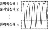
도 4를 참조하면, 환자의 호흡에 의한 높이 방향의 변화를 센서를 통해 파악한 그래프의 일부가 도시되어 있다. 그래프의 가로방향은 시간축이고, 세로축은 흉부의 높낮이 변화를 나타낸다.Referring to FIG. 4, a portion of a graph is shown in which the change in height direction due to the patient's breathing is detected through a sensor. The horizontal direction of the graph is the time axis, and the vertical axis represents changes in chest height.
의료영상표시장치(100)는 세로축의 높낮이 변화를 복수의 움직임상태 구간으로 분할할 수 있다. 움직임상태 구간의 개수(n)는 10개 또는 20개 등 실시 예에 따라 다양하게 변형 가능하다.The medical
의료영상표시장치(100)는 의료영상촬영장치가 촬영할 때 센서가 파악한 움직임정보를 기초로 CT 영상 또는 MRI 영상에 포함된 복수의 단면 이미지가 복수의 움직임상태의 구간 중 어디에 해당하는지 파악할 수 있다. 의료영상표시장치(100)는 제1 움직임상태의 구간에 속한 적어도 하나 이상의 단면 이미지를 이용하여 제1 움직임상태에 대한 제1 의료영상을 생성하고, 제2 움직임상태의 구간에 속한 적어도 하나 이상의 단면 이미지를 이용하여 제2 움직임상태에 대한 제2 의료영상을 생성하고, 제n 움직임상태의 구간에 속한 적어도 하나 이상의 단면 이미지를 이용하여 제n 움직임상태에 대한 제n 의료영상을 생성할 수 있다. 예를 들어, 도 3의 복수의 단면 이미지가 제1 움직임상태의 구간에 속한 복수의 단면 이미지이면, 의료영상표시장치(100)는 제1 움직임상태에 대한 폐 종양의 의료영상을 생성할 수 있다. 의료영상표시장치는 움직임상태별 의료영상을 저장 관리한다.The medical
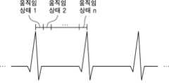
도 5를 참조하면, 맥박 그래프가 도시되어 있다. 의료영상촬영장치가 환자를 촬영할 때 센서는 실시간으로 맥박을 함께 측정한다. 의료영상표시장치(100)는 맥박을 기준으로 그래프의 시간축을 복수의 움직임상태의 구간으로 구분할 수 있다.Referring to Figure 5, a pulse graph is shown. When a medical imaging device records a patient, the sensor measures the pulse in real time. The medical
의료영상표시장치(100)는 의료영상촬영장치의 촬영시점에 센서가 파악한 맥박을 기준으로 의료영상이 복수의 움직임상태의 구간 중 어느 구간에 해당하는지 파악할 수 있다. 예를 들어, 의료영상표시장치(100)는 CT 영상 또는 MRI 영상에 포함된 복수의 단면 이미지의 촬영시점이 맥박의 어느 시점에 해당하는지 파악할 수 있다. 의료영상표시장치(100)는 각 움직임상태의 구간에 속한 적어도 하나 이상의 단면이미지를 이용하여 각 움직임상태에 대한 의료영상을 생성하여 저장할 수 있다.The medical
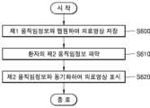
도 6은 본 발명의 실시 예에 따른 의료영상 표시방법의 일 예를 도시한 흐름도이다.Figure 6 is a flowchart showing an example of a medical image display method according to an embodiment of the present invention.
도 6을 참조하면, 의료영상표시장치(100)는 CT 또는 MRI 촬영시 획득한 환자의 제1 움직임정보와 맵핑되는 복수의 의료영상을 저장한다(S600). 예를 들어, 도 4 및 도 5를 참조하면, 환자의 움직임을 복수의 움직임상태 구간으로 정의하고, 각 움직임상태 구간에 대한 의료영상을 생성하여 데이터베이스 등에 저장한다. 다른 실시 예로, 의료영상이 움직임상태 구간별로 미리 저장되어 있다면, 의료영상표시장치는 해당 단계(S600)를 생략할 수 있다.Referring to FIG. 6, the medical
의료영상표시장치(100)는 수술이나 치료 등을 위하여 환자의 호흡이나 맥박 등의 움직임에 따라 변화하는 의료영상을 표시하고자 할 경우에 환자의 제2 움직임정보를 파악하고(S610), 제2 움직임정보와 동기화하여 의료영상을 표시한다(S620). 의료영상표시장치(100)는 이동단말(예를 들어, 태블릿PC 등)을 이용하여 실시간 촬영되는 환자의 이미지에 의료영상을 중첩하여 증강현실로 표시하거나, 환자의 실시간 움직임이 표시되는 아바타에 의료영상을 중첩하여 가상현실로 표시할 수 있다.When the medical
도 7은 본 발명의 실시 예에 따른 의료영상을 환자와 중첩하여 가상현실 또는 증가현실에 표시하는 방법의 일 예를 도시한 도면이다.Figure 7 is a diagram illustrating an example of a method of displaying a medical image in virtual reality or augmented reality by overlapping it with a patient according to an embodiment of the present invention.
도 7을 참조하면, 의료영상표시장치(100)는 센서(710)를 통해 환자(700)의 실시간 움직임을 파악한다. 예를 들어, 센서(710)는 환자의 호흡(도 4 참조) 또는 맥박(도 5 참조) 등을 파악할 수 있다.Referring to FIG. 7, the medical
의료영상표시장치(100)는 환자(700)의 실시간 움직임정보(즉, 제2 움직임정보(720)를 기초로 데이터베이스(730)에서 해당하는 의료영상을 파악한다. 도 2에서 살핀 바와 같이, 의료영상은 CT 또는 MRI 촬영시에 획득한 환자의 제1 움직임정보와 맵핑하여 저장되므로, 의료영상표시장치(100)는 환자(700)로부터 실시간 획득되는 제2 움직임정보(720)을 기초로 데이터베이스(730)를 검색하여 제2 움직임정보(720)에 해당하는 의료영상을 파악할 수 있다. 예를 들어, 도 4 및 도 5와 같이 복수의 움직임상태의 구간이 정의되고, 의료영상이 각 움직임상태의 구간별로 존재한다면, 의료영상표시장치(100)는 환자(700)의 실시간 움직임을 나타내는 제2 움직임정보(720)가 복수의 움직임상태의 구간 중 어느 구간에 해당하는지 파악한 후 해당 움직임상태 구간의 의료영상을 데이터베이스(730)에서 파악한다.The medical
의료영상표시장치(100)는 카메라 등을 통해 환자(700)를 촬영하여 얻은 환자 이미지(750) 또는 환자의 실시간 움직임을 가상공간에 구현한 아바타(750)에 제2 움직임정보에 해당하는 의료영상(즉, 디지털트윈(760))을 중첩하여 가상현실 또는 증강현실(740)에 표시한다.The medical
도 8은 본 발명의 실시 예에 따른 의료영상표시장치의 일 예의 구성을 도시한 도면이다.Figure 8 is a diagram showing the configuration of an example of a medical image display device according to an embodiment of the present invention.
도 8을 참조하면, 의료영상표시장치(100)는 영상저장부(800), 움직임파악부(810) 및 표시부(820)를 포함한다. 일 예로 의료영상표시장치(100)는 메모리, 프로세서 및 입출력장치를 포함하는 컴퓨팅 장치로 구현될 수 있다. 이 경우 각 구성은 소프트웨어로 구현되어 메모리에 탑재된 후 프로세서에 의해 수행될 수 있다.Referring to FIG. 8, the medical
영상저장부(800)는 의료영상 촬영시 획득한 환자의 제1 움직임정보와 맵핑되는 복수의 의료영상을 저장한다. 영상저장부(800)는 도 2와 같이 의료영상촬영장치와 센서를 통해 각각 얻은 의료영상과 제1 움직임정보를 맵핑하여 저장할 수 있다. 호흡이나 맥박의 움직임과 맵핑하여 의료영상을 저장하는 예가 도 4 및 도 5에 도시되어 있다. 다른 실시 예로, 제1 움직임정보와 의료영상이 미리 맵핑되어 데이터베이스에 저장되어 있다면 영상저장부(800)는 생략될 수 있다.The
움직임파악부(810)는 환자로부터 실시간 제2 움직임정보를 파악한다. 예를 들어, 수술이나 치료를 위하여 환자의 움직임에 따른 실시간 의료영상이 필요한 경우에, 움직임파악부(810)는 센서를 이용하여 환자로부터 호흡이나 맥박 등을 실시간 파악한다.The
표시부(820)는 데이터베이스에 제1 움직임정보와 맵핑되어 저장된 복수의 의료영상을 제2 움직임정보에 동기화하여 표시한다. 예를 들어, 움직임파악부(810)가 센서를 통해 측정한 호흡이 도 4의 제1 움직임상태에 해당하면, 표시부(820)는 데이터베이스에서 제1 움직임상태와 맵핑되어 저장된 의료영상을 표시한다. 움직임파악부(810)는 센서를 통해 실시간 환자의 움직임을 파악하고, 표시부(820)는 실시간 파악된 환자의 제2 움직임정보에 따라 데이터베이스를 실시간 검색하여 찾은 의료영상을 표시한다. 다른 실시 예로, 표시부(820)는 가상현실 또는 증강현실에서 데이터베이스에 저장된 의료영상을 제2 움직임정보와 동기화하여 표시할 수 있다. 이에 대한 예가 도 7에 도시되어 있다.The
본 발명은 또한 컴퓨터로 읽을 수 있는 기록매체에 컴퓨터가 읽을 수 있는 프로그램 코드로서 구현하는 것이 가능하다. 컴퓨터가 읽을 수 있는 기록매체는 컴퓨터 시스템에 의하여 읽혀질 수 있는 데이터가 저장되는 모든 종류의 기록장치를 포함한다. 컴퓨터가 읽을 수 있는 기록매체의 예로는 ROM, RAM, CD-ROM, 자기 테이프, 플로피디스크, 광데이터 저장장치 등이 있다. 또한 컴퓨터가 읽을 수 있는 기록매체는 네트워크로 연결된 컴퓨터 시스템에 분산되어 분산방식으로 컴퓨터가 읽을 수 있는 코드가 저장되고 실행될 수 있다.The present invention can also be implemented as computer-readable program code on a computer-readable recording medium. Computer-readable recording media include all types of recording devices that store data that can be read by a computer system. Examples of computer-readable recording media include ROM, RAM, CD-ROM, magnetic tape, floppy disk, and optical data storage devices. Additionally, computer-readable recording media can be distributed across networked computer systems so that computer-readable code can be stored and executed in a distributed manner.
이제까지 본 발명에 대하여 그 바람직한 실시 예들을 중심으로 살펴보았다. 본 발명이 속하는 기술 분야에서 통상의 지식을 가진 자는 본 발명이 본 발명의 본질적인 특성에서 벗어나지 않는 범위에서 변형된 형태로 구현될 수 있음을 이해할 수 있을 것이다. 그러므로 개시된 실시 예들은 한정적인 관점이 아니라 설명적인 관점에서 고려되어야 한다. 본 발명의 범위는 전술한 설명이 아니라 특허청구범위에 나타나 있으며, 그와 동등한 범위 내에 있는 모든 차이점은 본 발명에 포함된 것으로 해석되어야 할 것이다.So far, the present invention has been examined focusing on its preferred embodiments. A person skilled in the art to which the present invention pertains will understand that the present invention may be implemented in a modified form without departing from the essential characteristics of the present invention. Therefore, the disclosed embodiments should be considered from an illustrative rather than a restrictive perspective. The scope of the present invention is indicated in the claims rather than the foregoing description, and all differences within the equivalent scope should be construed as being included in the present invention.
Claims (11)
Translated fromKorean상기 환자로부터 실시간 제2 움직임정보를 파악하는 단계; 및
상기 복수의 의료영상을 상기 제2 움직임정보에 동기화하여 표시하는 단계;를 포함하는 것을 특징으로 하는 의료영상 표시방법.Storing a plurality of medical images mapped with the patient's first movement information obtained during CT or MRI scanning;
Recognizing real-time second movement information from the patient; and
A medical image display method comprising: synchronizing and displaying the plurality of medical images with the second motion information.
호흡 또는 심장박동에 따른 움직임을 측정하는 센서를 통해 환자의 제2 움직임정보를 획득하는 단계;를 포함하는 것을 특징으로 하는 의료영상 표시방법.The method of claim 1, wherein determining the second motion information comprises:
A medical image display method comprising: acquiring second movement information of the patient through a sensor that measures movement according to breathing or heartbeat.
증강현실에서 상기 환자의 현실이미지에 상기 의료영상을 중첩하여 표시하거나, 가상현실에서 상기 환자의 아바타에 상기 의료영상을 중첩하여 표시하는 단계;를 포함하는 것을 특징으로 하는 의료영상 표시방법.The method of claim 1, wherein the displaying step includes:
A medical image display method comprising: displaying the medical image by overlaying it on a real image of the patient in augmented reality, or displaying the medical image by overlapping it on the patient's avatar in virtual reality.
CT 또는 MRI 영상에 포함된 복수의 단면 이미지를 상기 제1 움직임정보에 따라 복수의 움직임상태로 분할하는 단계; 및
각 움직임상태에 속한 단면 이미지를 이용하여 움직임상태별 의료영상을 생성하는 단계;를 포함하는 것을 특징으로 하는 의료영상 표시방법.The method of claim 1, wherein storing the medical image comprises:
dividing a plurality of cross-sectional images included in a CT or MRI image into a plurality of motion states according to the first motion information; and
A medical image display method comprising: generating medical images for each movement state using cross-sectional images belonging to each movement state.
상기 제2 움직임정보에 해당하는 움직임상태의 의료영상을 표시하는 단계;를 포함하는 것을 특징으로 하는 의료영상 표시방법.The method of claim 4, wherein the displaying step includes:
A medical image display method comprising: displaying a medical image in a motion state corresponding to the second motion information.
상기 의료영상은, CT 또는 MRI 영상에서 분할한 인체조직의 영역을 3차원 모델링하여 생성되는 디지털트윈 영상인 것을 특징으로 하는 의료영상 표시방법.According to clause 1,
The medical image is a digital twin image generated by three-dimensional modeling of a human tissue area divided from a CT or MRI image.
상기 환자로부터 실시간 제2 움직임정보를 파악하는 움직임파악부; 및
상기 복수의 의료영상을 상기 제2 움직임정보에 동기화하여 표시하는 표시부;를 포함하는 것을 특징으로 하는 의료영상 표시장치.an image storage unit that stores a plurality of medical images mapped to the patient's first movement information obtained during CT or MRI scanning;
a movement detection unit that detects real-time second movement information from the patient; and
A display unit that displays the plurality of medical images in synchronization with the second motion information.
호흡 또는 심장박동에 따른 움직임을 측정하는 센서를 통해 환자의 제2 움직임정보를 획득하는 것을 특징으로 하는 의료영상 표시장치.The method of claim 7, wherein the movement detection unit,
A medical image display device characterized by acquiring the patient's second movement information through a sensor that measures movement according to breathing or heartbeat.
CT 또는 MRI 영상에 포함된 복수의 단면 이미지를 상기 제1 움직임정보에 따라 복수의 움직임상태로 분할하고, 각 움직임상태에 속한 단면 이미지를 이용하여 움직임상태별 의료영상을 생성하는 것을 특징으로 하는 의료영상 표시장치.The method of claim 7, wherein the image storage unit,
Medical treatment characterized by dividing a plurality of cross-sectional images included in a CT or MRI image into a plurality of movement states according to the first motion information, and generating medical images for each movement state using the cross-sectional images belonging to each movement state. Video display device.
상기 제2 움직임정보에 해당하는 움직임상태의 의료영상을 표시하는 것을 특징으로 하는 의료영상 표시장치.The method of claim 9, wherein the display unit,
A medical image display device characterized in that it displays a medical image in a movement state corresponding to the second movement information.
Priority Applications (1)
| Application Number | Priority Date | Filing Date | Title |
|---|---|---|---|
| KR1020220063048AKR20230163211A (en) | 2022-05-23 | 2022-05-23 | Medical image displaying method and apparatus |
Applications Claiming Priority (1)
| Application Number | Priority Date | Filing Date | Title |
|---|---|---|---|
| KR1020220063048AKR20230163211A (en) | 2022-05-23 | 2022-05-23 | Medical image displaying method and apparatus |
Publications (1)
| Publication Number | Publication Date |
|---|---|
| KR20230163211Atrue KR20230163211A (en) | 2023-11-30 |
Family
ID=88968655
Family Applications (1)
| Application Number | Title | Priority Date | Filing Date |
|---|---|---|---|
| KR1020220063048ACeasedKR20230163211A (en) | 2022-05-23 | 2022-05-23 | Medical image displaying method and apparatus |
Country Status (1)
| Country | Link |
|---|---|
| KR (1) | KR20230163211A (en) |
- 2022
- 2022-05-23KRKR1020220063048Apatent/KR20230163211A/ennot_activeCeased
Similar Documents
| Publication | Publication Date | Title |
|---|---|---|
| CN105796052B (en) | Joint visualization of 3D reconstructed photographs and internal medical scans | |
| EP2637593B1 (en) | Visualization of anatomical data by augmented reality | |
| KR101982149B1 (en) | Method and apparatus for creating medical image using partial medical image | |
| KR20210020990A (en) | System and method for lung-volume-gated X-ray imaging | |
| KR20120111871A (en) | Method and apparatus for creating medical image using 3d deformable model | |
| CN111656395A (en) | Affordable electrocardiographic hardware and imaging based non-invasive electrophysiology mapping | |
| JP2012205899A (en) | Image generating method and system of body organ using three-dimensional model and computer readable recording medium | |
| US10248756B2 (en) | Anatomically specific movie driven medical image review | |
| JP2010279539A (en) | Diagnosis support apparatus and method, and program. | |
| JP2014161734A (en) | Method and apparatus for matching medical images | |
| JP2012511707A (en) | Method and apparatus for monitoring an object | |
| JP6833533B2 (en) | Ultrasonic diagnostic equipment and ultrasonic diagnostic support program | |
| KR101993384B1 (en) | Method, Apparatus and system for correcting medical image by patient's pose variation | |
| CN100548223C (en) | Ultrasonic diagnostic apparatus | |
| JP2014530348A (en) | Radiation imaging system and method for updating an original radiation image | |
| KR102254365B1 (en) | Medical image processing method | |
| JP2021192746A (en) | Program, information processing method, and information processing device | |
| KR20150145106A (en) | Method and appartus for registering medical images | |
| US12272066B2 (en) | Apparatus, method and computer program for monitoring a subject during a medical imaging procedure | |
| KR20230163211A (en) | Medical image displaying method and apparatus | |
| CN110546684B (en) | Quantitative evaluation of time-varying data | |
| US11056149B2 (en) | Medical image storage and reproduction apparatus, method, and program | |
| JP7714431B2 (en) | Program, information processing method, information processing device and diagnostic support system | |
| KR20180131211A (en) | Apparatus and method of producing three dimensional image of orbital prosthesis | |
| CN113614785A (en) | Interventional device tracking |
Legal Events
| Date | Code | Title | Description |
|---|---|---|---|
| PA0109 | Patent application | St.27 status event code:A-0-1-A10-A12-nap-PA0109 | |
| PA0201 | Request for examination | St.27 status event code:A-1-2-D10-D11-exm-PA0201 | |
| PN2301 | Change of applicant | St.27 status event code:A-3-3-R10-R13-asn-PN2301 St.27 status event code:A-3-3-R10-R11-asn-PN2301 | |
| PG1501 | Laying open of application | St.27 status event code:A-1-1-Q10-Q12-nap-PG1501 | |
| E902 | Notification of reason for refusal | ||
| PE0902 | Notice of grounds for rejection | St.27 status event code:A-1-2-D10-D21-exm-PE0902 | |
| E13-X000 | Pre-grant limitation requested | St.27 status event code:A-2-3-E10-E13-lim-X000 | |
| P11-X000 | Amendment of application requested | St.27 status event code:A-2-2-P10-P11-nap-X000 | |
| P13-X000 | Application amended | St.27 status event code:A-2-2-P10-P13-nap-X000 | |
| R18-X000 | Changes to party contact information recorded | St.27 status event code:A-3-3-R10-R18-oth-X000 | |
| PE0601 | Decision on rejection of patent | St.27 status event code:N-2-6-B10-B15-exm-PE0601 | |
| P22-X000 | Classification modified | St.27 status event code:A-2-2-P10-P22-nap-X000 |