KR20230078644A - System and method for determining deep brain stimulation parameters - Google Patents
System and method for determining deep brain stimulation parametersDownload PDFInfo
- Publication number
- KR20230078644A KR20230078644AKR1020237008918AKR20237008918AKR20230078644AKR 20230078644 AKR20230078644 AKR 20230078644AKR 1020237008918 AKR1020237008918 AKR 1020237008918AKR 20237008918 AKR20237008918 AKR 20237008918AKR 20230078644 AKR20230078644 AKR 20230078644A
- Authority
- KR
- South Korea
- Prior art keywords
- stimulation
- frequency
- target structure
- hfo
- eca
- Prior art date
- Legal status (The legal status is an assumption and is not a legal conclusion. Google has not performed a legal analysis and makes no representation as to the accuracy of the status listed.)
- Granted
Links
Images


Classifications
- A—HUMAN NECESSITIES
- A61—MEDICAL OR VETERINARY SCIENCE; HYGIENE
- A61N—ELECTROTHERAPY; MAGNETOTHERAPY; RADIATION THERAPY; ULTRASOUND THERAPY
- A61N1/00—Electrotherapy; Circuits therefor
- A61N1/18—Applying electric currents by contact electrodes
- A61N1/32—Applying electric currents by contact electrodes alternating or intermittent currents
- A61N1/36—Applying electric currents by contact electrodes alternating or intermittent currents for stimulation
- A61N1/3605—Implantable neurostimulators for stimulating central or peripheral nerve system
- A61N1/36128—Control systems
- A61N1/36146—Control systems specified by the stimulation parameters
- A61N1/36167—Timing, e.g. stimulation onset
- A61N1/36171—Frequency
- A—HUMAN NECESSITIES
- A61—MEDICAL OR VETERINARY SCIENCE; HYGIENE
- A61B—DIAGNOSIS; SURGERY; IDENTIFICATION
- A61B5/00—Measuring for diagnostic purposes; Identification of persons
- A61B5/24—Detecting, measuring or recording bioelectric or biomagnetic signals of the body or parts thereof
- A61B5/25—Bioelectric electrodes therefor
- A61B5/279—Bioelectric electrodes therefor specially adapted for particular uses
- A61B5/291—Bioelectric electrodes therefor specially adapted for particular uses for electroencephalography [EEG]
- A61B5/293—Invasive
- A—HUMAN NECESSITIES
- A61—MEDICAL OR VETERINARY SCIENCE; HYGIENE
- A61B—DIAGNOSIS; SURGERY; IDENTIFICATION
- A61B5/00—Measuring for diagnostic purposes; Identification of persons
- A61B5/24—Detecting, measuring or recording bioelectric or biomagnetic signals of the body or parts thereof
- A61B5/316—Modalities, i.e. specific diagnostic methods
- A61B5/369—Electroencephalography [EEG]
- A61B5/377—Electroencephalography [EEG] using evoked responses
- A—HUMAN NECESSITIES
- A61—MEDICAL OR VETERINARY SCIENCE; HYGIENE
- A61B—DIAGNOSIS; SURGERY; IDENTIFICATION
- A61B5/00—Measuring for diagnostic purposes; Identification of persons
- A61B5/40—Detecting, measuring or recording for evaluating the nervous system
- A61B5/4058—Detecting, measuring or recording for evaluating the nervous system for evaluating the central nervous system
- A61B5/4064—Evaluating the brain
- A—HUMAN NECESSITIES
- A61—MEDICAL OR VETERINARY SCIENCE; HYGIENE
- A61B—DIAGNOSIS; SURGERY; IDENTIFICATION
- A61B5/00—Measuring for diagnostic purposes; Identification of persons
- A61B5/40—Detecting, measuring or recording for evaluating the nervous system
- A61B5/4076—Diagnosing or monitoring particular conditions of the nervous system
- A61B5/4082—Diagnosing or monitoring movement diseases, e.g. Parkinson, Huntington or Tourette
- A—HUMAN NECESSITIES
- A61—MEDICAL OR VETERINARY SCIENCE; HYGIENE
- A61B—DIAGNOSIS; SURGERY; IDENTIFICATION
- A61B5/00—Measuring for diagnostic purposes; Identification of persons
- A61B5/48—Other medical applications
- A61B5/4848—Monitoring or testing the effects of treatment, e.g. of medication
- A—HUMAN NECESSITIES
- A61—MEDICAL OR VETERINARY SCIENCE; HYGIENE
- A61B—DIAGNOSIS; SURGERY; IDENTIFICATION
- A61B5/00—Measuring for diagnostic purposes; Identification of persons
- A61B5/48—Other medical applications
- A61B5/4887—Locating particular structures in or on the body
- A—HUMAN NECESSITIES
- A61—MEDICAL OR VETERINARY SCIENCE; HYGIENE
- A61N—ELECTROTHERAPY; MAGNETOTHERAPY; RADIATION THERAPY; ULTRASOUND THERAPY
- A61N1/00—Electrotherapy; Circuits therefor
- A61N1/02—Details
- A61N1/04—Electrodes
- A61N1/05—Electrodes for implantation or insertion into the body, e.g. heart electrode
- A61N1/0526—Head electrodes
- A61N1/0529—Electrodes for brain stimulation
- A61N1/0534—Electrodes for deep brain stimulation
- A—HUMAN NECESSITIES
- A61—MEDICAL OR VETERINARY SCIENCE; HYGIENE
- A61N—ELECTROTHERAPY; MAGNETOTHERAPY; RADIATION THERAPY; ULTRASOUND THERAPY
- A61N1/00—Electrotherapy; Circuits therefor
- A61N1/18—Applying electric currents by contact electrodes
- A61N1/32—Applying electric currents by contact electrodes alternating or intermittent currents
- A61N1/36—Applying electric currents by contact electrodes alternating or intermittent currents for stimulation
- A61N1/3605—Implantable neurostimulators for stimulating central or peripheral nerve system
- A61N1/3606—Implantable neurostimulators for stimulating central or peripheral nerve system adapted for a particular treatment
- A61N1/36067—Movement disorders, e.g. tremor or Parkinson disease
- A—HUMAN NECESSITIES
- A61—MEDICAL OR VETERINARY SCIENCE; HYGIENE
- A61N—ELECTROTHERAPY; MAGNETOTHERAPY; RADIATION THERAPY; ULTRASOUND THERAPY
- A61N1/00—Electrotherapy; Circuits therefor
- A61N1/18—Applying electric currents by contact electrodes
- A61N1/32—Applying electric currents by contact electrodes alternating or intermittent currents
- A61N1/36—Applying electric currents by contact electrodes alternating or intermittent currents for stimulation
- A61N1/3605—Implantable neurostimulators for stimulating central or peripheral nerve system
- A61N1/3606—Implantable neurostimulators for stimulating central or peripheral nerve system adapted for a particular treatment
- A61N1/36082—Cognitive or psychiatric applications, e.g. dementia or Alzheimer's disease
- A61N1/36096—Mood disorders, e.g. depression, anxiety or panic disorder
- A—HUMAN NECESSITIES
- A61—MEDICAL OR VETERINARY SCIENCE; HYGIENE
- A61B—DIAGNOSIS; SURGERY; IDENTIFICATION
- A61B2505/00—Evaluating, monitoring or diagnosing in the context of a particular type of medical care
- A61B2505/05—Surgical care
Landscapes
- Health & Medical Sciences (AREA)
- Life Sciences & Earth Sciences (AREA)
- Neurology (AREA)
- General Health & Medical Sciences (AREA)
- Animal Behavior & Ethology (AREA)
- Veterinary Medicine (AREA)
- Engineering & Computer Science (AREA)
- Biomedical Technology (AREA)
- Public Health (AREA)
- Neurosurgery (AREA)
- Heart & Thoracic Surgery (AREA)
- Medical Informatics (AREA)
- Biophysics (AREA)
- Molecular Biology (AREA)
- Physics & Mathematics (AREA)
- Pathology (AREA)
- Surgery (AREA)
- Psychology (AREA)
- Radiology & Medical Imaging (AREA)
- Nuclear Medicine, Radiotherapy & Molecular Imaging (AREA)
- Hospice & Palliative Care (AREA)
- Psychiatry (AREA)
- Physiology (AREA)
- Developmental Disabilities (AREA)
- Child & Adolescent Psychology (AREA)
- Cardiology (AREA)
- Electrotherapy Devices (AREA)
- Measuring And Recording Apparatus For Diagnosis (AREA)
- Measurement And Recording Of Electrical Phenomena And Electrical Characteristics Of The Living Body (AREA)
Abstract
Translated fromKorean
Description
Translated fromKorean관련 출원 교차 참조Cross reference to related applications
본 출원은 2021년 8월 12일자로 출원된 미국 특허출원 제17/400,312호의 우선권을 주장하며, 이는 2020년 8월 14일자로 출원된 미국 가 특허출원 제63/066,141호 및 2020년 8월 20일자로 출원된 미국 가 특허출원 제63/068,155호의 우선권을 주장한다.This application claims priority from U.S. Patent Application Serial No. 17/400,312, filed on August 12, 2021, which claims priority to U.S. Provisional Patent Application No. 63/066,141, filed on August 14, 2020, and on August 20, 2020. Priority is claimed to U.S. Provisional Patent Application No. 63/068,155, filed dated.
기술분야technology field
본 발명은 심부 뇌 자극(deep brain stimulation)에 관한 것으로, 보다 구체적으로는 심부 뇌 자극 파라미터들을 결정하기 위한 시스템들 및 방법들에 관한 것이다.The present invention relates to deep brain stimulation, and more particularly to systems and methods for determining deep brain stimulation parameters.
현재의 뇌 자극기는 시행착오(trial and error)에 의해 프로그래밍된다. 이는 매우 시간 소모적일 수 있다. 예를 들어, 프로그래밍은 다수의 세션들에 걸쳐 세션들마다 시간이 걸릴 수 있고, 후속 프로그래밍 세션이 요구될 수 있다. 이에 따라, 자동화되고 객관적인 방식으로 뇌의 반응에 기초하여 프로그래밍 파라미터들을 최적화할 수 있는 시스템이 필요하다.Current brain stimulators are programmed by trial and error. This can be very time consuming. For example, programming may take time from session to session, over multiple sessions, and a subsequent programming session may be required. Accordingly, there is a need for a system capable of optimizing programming parameters based on the response of the brain in an automated and objective manner.
본 개시의 양태들에 따르면, 복수의 다접점 전극들을 개체의 뇌에서의 타겟 구조의 영역 내로 삽입하는 단계를 포함하는 뇌에의 식립 부위의 위치를 찾는 방법이 제공된다. 복수의 다접점 전극들 중의 다접점 전극의 접점에 고주파수 자극(High Frequency Stimulation, HFS)이 가해진다. HFS에 의해 타겟 구조의 영역에서 유도되는 고주파수 발진(High Frequency Oscillations, HFO)이 측정된다. HFS에 의해 타겟 구조의 영역에서 유발되는 유발 화합물 활성(Evoked Compound Activity, ECA)이 측정된다. HFO 및 ECA 중 적어도 하나가 미리 결정된 임계치를 초과하는지 결정된다. HFO 및 ECA 중 적어도 하나가 미리 결정된 임계치를 초과한다면, 다접점 전극의 접점의 위치가 개체의 뇌에의 전극 식립을 위한 부위로서 식별된다.According to aspects of the present disclosure, there is provided a method of locating an implantation site in the brain comprising inserting a plurality of multi-contact electrodes into a region of a target structure in the brain of a subject. High frequency stimulation (HFS) is applied to a contact point of a multi-contact electrode among a plurality of multi-contact electrodes. High Frequency Oscillations (HFO) induced in the region of the target structure by HFS are measured. Evoked compound activity (ECA) evoked in the region of the target structure by HFS is measured. It is determined whether at least one of HFO and ECA exceeds a predetermined threshold. If at least one of HFO and ECA exceeds a predetermined threshold, the location of the contact point of the multi-contact electrode is identified as a site for implantation of the electrode in the subject's brain.
본 개시의 양태에서, HFO 및 ECA가 미리 결정된 임계치 미만이라면, 복수의 다접점 전극들 중의 다접점 전극의 제2 접점에 제2 고주파수 자극이 가해진다. 개체의 뇌에의 전극 식립을 위한 부위가 색별되지 않는다면, 복수의 다접점 전극들은 개체의 뇌에서의 제2 영역으로 이동된다.In an aspect of the present disclosure, if HFO and ECA are below a predetermined threshold, a second high-frequency stimulation is applied to a second contact of a multi-contact electrode of the plurality of multi-contact electrodes. If the site for electrode implantation in the subject's brain is not identified, the plurality of multi-contact electrodes are moved to the second area in the subject's brain.
본 개시의 양태에서, 적어도 하나의 전극은 개체의 뇌에의 전극 식립을 위한 부위에 대한 심부 뇌 자극(Deep Brain Stimulation, DBS)을 위해 구성된다.In an aspect of the present disclosure, at least one electrode is configured for Deep Brain Stimulation (DBS) at a site for implantation of the electrode in the brain of a subject.
본 개시의 양태에서, 타겟 구조는 시상하핵(Subthalamic nucleus, STN)이다.In an aspect of the present disclosure, the target structure is the Subthalamic nucleus (STN).
본 개시의 양태에서, HFO 및 ECA를 측정하는 것은 수술 중에 수행된다.In an aspect of the present disclosure, measuring HFO and ECA is performed intraoperatively.
본 개시의 양태에서, HFS는 100Hz보다 크다.In an aspect of the present disclosure, HFS is greater than 100 Hz.
본 개시의 양태에서, HFO는 300H보다 큰 발진 패턴을 포함한다.In an aspect of this disclosure, the HFO includes an oscillation pattern greater than 300H.
본 개시의 양태에서, ECA는 200-450Hz 사이의 공명 패턴을 포함한다.In an aspect of the present disclosure, the ECA includes a resonance pattern between 200-450 Hz.
본 개시의 양태에서, 복수의 다접점 전극들은 파킨슨병(PD)을 앓는 개체의 뇌 내로 삽입된다.In an aspect of the present disclosure, a plurality of multi-contact electrodes are implanted into the brain of an individual suffering from Parkinson's disease (PD).
본 개시의 양태들에 따르면, 복수의 다접점 전극들을 포함하는 뇌에의 전극 식립을 위한 부위의 위치를 찾기 위한 시스템이 제공된다. 복수의 다접점 전극들은 개체의 뇌에서의 타겟 구조의 영역 내로의 삽입을 위해 구성된다. 자극 디바이스는 복수의 다접점 전극들 각각과 전기 통신한다. 자극 디바이스는 복수의 다접점 전극들 각각에 고주파수 자극(HFS)을 가한다. 자극 디바이스는 복수의 다접점 전극들 중 하나의 다접점 전극의 접점들의 서브세트에 HFS를 선택적으로 가하도록 구성된다. 기록 디바이스는 HFS에 의해 타겟 구조의 영역에서 유도되는 국소장 전위(Local Field Potentials)의 고주파수 발진(HFO)을 측정하도록 구성된다. 기록 디바이스는 HFS에 의해 타겟 구조의 영역에서 유발되는 유발 화합물 활성(ECA)을 측정하도록 구성된다. 신호 처리 유닛은 기록 디바이스와 통신한다. 신호 처리 유닛은 HFO 및 ECA 중 적어도 하나가 미리 결정된 임계치를 초과하는지 결정하여, 다접점 전극의 하나의 접점의 위치를 개체의 뇌에의 전극 식립을 위한 부위로서 식별한다.According to aspects of the present disclosure, a system for locating a site for electrode implantation in the brain comprising a plurality of multi-contact electrodes is provided. A plurality of multi-contact electrodes are configured for insertion into a region of a target structure in a subject's brain. The stimulation device is in electrical communication with each of the plurality of multi-contact electrodes. The stimulation device applies high-frequency stimulation (HFS) to each of the plurality of multi-contact electrodes. The stimulation device is configured to selectively apply HFS to a subset of contacts of one of the plurality of multi-contact electrodes. The recording device is configured to measure high frequency oscillations (HFO) of Local Field Potentials induced by the HFS in the region of the target structure. The recording device is configured to measure the evoked compound activity (ECA) evoked by HFS in the region of the target structure. The signal processing unit communicates with the recording device. The signal processing unit determines whether at least one of HFO and ECA exceeds a predetermined threshold, and identifies a location of one contact point of the multi-contact electrode as a site for implantation of the electrode in the subject's brain.
본 개시의 양태에서, 시각화 유닛은 측정된 HFO 및 ECA를 시각적으로 디스플레이한다.In an aspect of the present disclosure, the visualization unit visually displays the measured HFO and ECA.
본 개시의 양태에서, 스위칭 유닛이 자극 디바이스에 의해 가해지는 HFS를 제어한다. HFO 및 ECA 중 적어도 하나가 미리 결정된 임계치 미만이라면, 스위칭 유닛은 HFS를 제어하여 복수의 다접점 전극들 중의 다접점 전극의 제2 접점에 제2 고주파수 자극을 가하도록 구성된다.In an aspect of the present disclosure, a switching unit controls the HFS applied by the stimulation device. If at least one of HFO and ECA is less than a predetermined threshold, the switching unit is configured to control HFS to apply a second high-frequency stimulation to a second contact of a multi-contact electrode of the plurality of multi-contact electrodes.
본 개시의 양태에서, 기록 디바이스는 수술 중에 국소장 전위로 유도되는 HFO 및 ECA를 측정하도록 구성된다.In an aspect of the present disclosure, the recording device is configured to measure HFO and ECA induced into local field potentials during surgery.
본 개시의 양태들에 따르면, 개체의 뇌에서의 타겟 구조에 식립된 다접점 전극의 접점들의 서브세트에 저주파 자극(LFS)을 가하는 단계를 포함하는 뇌 자극 파라미터들을 결정하는 방법이 제공된다. LFS에 의해 타겟 구조에서 유발되는 유발 화합물 활성(Evoked Compound Activity, ECA)이 측정된다. ECA로부터 추출되는 위상 공간에 기초하여 미리 결정된 범위 내의 뇌 자극을 전달하기 위한 주파수 범위가 결정된다. 결정된 주파수 범위 내에서 다접점 전극의 접점에 자극 주파수들이 가해진다. 결정된 범위 내에서 가해진 자극 주파수들에 의해 타겟 구조에서 유발되는 고주파수 발진(High Frequency Oscillations, HFO)이 측정된다. 미리 결정된 임계치를 초과하는 HFO를 유발하는 주파수가 결정된다. 결정된 주파수는 타겟 구조에 대한 처치 주파수로서 선택된다.According to aspects of the present disclosure, a method for determining brain stimulation parameters is provided comprising applying low frequency stimulation (LFS) to a subset of contacts of a multi-contact electrode implanted in a target structure in the brain of a subject. Evoked Compound Activity (ECA) evoked in the target structure by LFS is measured. A frequency range for delivering brain stimulation within a predetermined range is determined based on the phase space extracted from the ECA. Stimulation frequencies are applied to the contacts of the multi-contact electrode within the determined frequency range. High frequency oscillations (HFO) induced in the target structure by the applied stimulation frequencies within the determined range are measured. A frequency that causes HFO to exceed a predetermined threshold is determined. The determined frequency is selected as the treatment frequency for the target structure.
본 개시의 양태에서, 타겟 구조는 시상하핵(Subthalamic nucleus, STN)이다.In an aspect of the present disclosure, the target structure is the Subthalamic nucleus (STN).
본 개시의 양태에서, HFO 및 ECA를 측정하는 것은 장기간 수행된다.In an aspect of the present disclosure, measuring HFO and ECA is performed over an extended period of time.
본 개시의 양태에서, HFS는 100Hz보다 크다.In an aspect of the present disclosure, HFS is greater than 100 Hz.
본 개시의 양태에서, LFS는 100Hz보다 적다.In an aspect of the present disclosure, LFS is less than 100 Hz.
본 개시의 양태에서, 선택된 주파수는 100Hz보다 크다.In an aspect of the present disclosure, the selected frequency is greater than 100 Hz.
본 개시의 양태에서, 선택된 주파수는 100-200Hz 범위 내의 임의의 주파수일 수 있다.In aspects of this disclosure, the selected frequency may be any frequency within the range of 100-200 Hz.
본 개시의 양태에서, 타겟 구조에서 유발되는 측정된 HFO는 200-450Hz 사이이다.In an aspect of the present disclosure, the measured HFO induced in the target structure is between 200-450 Hz.
본 개시의 양태에서, ECA는 미리 결정된 임계치를 초과하는 공명 반응을 포함한다.In an aspect of the present disclosure, ECA includes a resonance response above a predetermined threshold.
본 개시의 양태에서, ECA가 미리 결정된 임계치 미만이라면, 다접점 전극의 접점과 상이한 타겟 구조 내의 위치에서 다접점 전극의 접점들의 제2 서브세트에 제2 LFS가 가해진다.In an aspect of the present disclosure, a second LFS is applied to a second subset of the contacts of the multi-contact electrode at a different location within the target structure than the contacts of the multi-contact electrode if the ECA is below the predetermined threshold.
본 개시의 양태에서, 다접점 전극은 타겟 구조의 심부 뇌 자극(DBS)을 위해 구성된다.In an aspect of the present disclosure, a multi-contact electrode is configured for deep brain stimulation (DBS) of a target structure.
본 개시의 양태에서, 타겟 구조는 시상하핵(STN)이다.In an aspect of the present disclosure, the target structure is the subthalamic nucleus (STN).
본 개시의 양태에서, 개체는 파킨슨병(PD)을 앓는다.In an aspect of the present disclosure, the individual suffers from Parkinson's disease (PD).
본 개시의 양태들에 따르면, 개체의 뇌에서의 타겟 구조에의 식립을 위해 구성된 적어도 하나의 다접점 전극을 포함하는 심부 뇌 자극 파라미터들을 결정하기 위한 시스템이 제공된다. 자극 디바이스는 적어도 하나의 다접점 전극과 전기 통신한다. 자극 디바이스는 적어도 하나의 다접점 전극에 저주파수 자극(LFS) 또는 고주파수 자극(HFS)을 가하도록 구성된다. 기록 디바이스는 LFS에 의해 타겟 구조에서 유발되는 유발 화합물 활성(ECA) 및 HFS에 의해 타겟 구조에서 유발되는 고주파수 발진(HFO) 중 적어도 하나를 기록하도록 구성된다. 신호 처리 유닛은 기록 디바이스와 통신한다. 신호 처리 유닛은 ECA로부터 추출되는 위상 공간에 기초하여 미리 결정된 범위 내의 뇌 자극을 전달하기 위한 주파수 범위를 결정한다. 신호 처리 유닛은 결정된 범위 내에서 가해진 자극 주파수들에 의해 타겟 구조에서 유발되는 고주파수 발진(HFO)을 분석한다. 파라미터 최적화 유닛은 미리 결정된 임계치를 초과하는 HFO를 유발하는 주파수를 결정하고, 주파수를 타겟 구조에 대한 처치 주파수로서 선택한다.According to aspects of the present disclosure, a system for determining deep brain stimulation parameters is provided that includes at least one multi-contact electrode configured for implantation into a target structure in the brain of a subject. The stimulation device is in electrical communication with the at least one multi-contact electrode. The stimulation device is configured to apply low frequency stimulation (LFS) or high frequency stimulation (HFS) to the at least one multi-contact electrode. The recording device is configured to record at least one of induced compound activity (ECA) induced in the target structure by LFS and high frequency oscillation (HFO) induced in the target structure by HFS. The signal processing unit communicates with the recording device. The signal processing unit determines a frequency range for delivering brain stimulation within a predetermined range based on the phase space extracted from the ECA. The signal processing unit analyzes the high frequency oscillation (HFO) induced in the target structure by the applied stimulation frequencies within the determined range. The parameter optimization unit determines the frequency that causes the HFO to exceed a predetermined threshold and selects the frequency as the treatment frequency for the target structure.
본 개시의 양태에서, 자극 디바이스는 복수의 다접점 전극들 중 하나의 다접점 전극의 접점들의 서브세트에 LFS 또는 HFS를 선택적으로 가하도록 구성된다.In an aspect of the present disclosure, the stimulation device is configured to selectively apply LFS or HFS to a subset of the contacts of one of the plurality of multi-contact electrodes.
본 개시의 양태에서, 자극 디바이스, 기록 디바이스, 신호 처리 유닛, 및 파라미터 최적화 유닛 중 적어도 하나는 개체의 가슴에 식립된다.In an aspect of the present disclosure, at least one of the stimulation device, the recording device, the signal processing unit, and the parameter optimization unit is implanted on the chest of the subject.
본 개시의 양태에서, 입력/출력(I/O) 유닛이 자극 디바이스, 기록 디바이스, 신호 처리 유닛, 및 파라미터 최적화 유닛과 전기 통신한다.In an aspect of the present disclosure, an input/output (I/O) unit is in electrical communication with a stimulation device, a recording device, a signal processing unit, and a parameter optimization unit.
본 개시의 다양한 양태들 및 특징들이 도면들을 참조하여 이하에서 설명되며, 여기서:
도 1은 본 개시의 양태들에 따른 뇌에의 식립 부위의 위치를 찾기 위한 시스템의 블록도이다;
도 2는 본 개시의 양태들에 따라 뇌에의 식립 부위의 위치를 찾기 위한 방법의 블록도이다;
도 3은 본 개시의 양태들에 따라 뇌에의 식립 부위의 위치를 찾기 위한 다른 방법의 블록도이다;
도 4는 본 개시의 양태들에 따른 신호 처리 유닛의 예시적인 컴퓨터의 블록도이다;
도 5는 본 개시의 양태들에 따른 DBS 파라미터들을 결정하기 위한 식립가능 시스템의 블록도이다;
도 6은 본 개시의 양태들에 따른 DBS 파라미터들을 결정하기 위한 방법의 블록도이다;
도 7은 본 개시의 양태들에 따라 DBS 파라미터들을 결정하기 위한 다른 방법의 블록도이다;
도 8은 다접점 전극이 STN에 위치될 때 STN에서 유발되는 기록된 HFO를 디스플레이하고, 다접점 전극이 STN에 위치되지 않을 때 HFO의 부재를 디스플레이한다;
도 9는 다접점 전극이 STN에 위치될 때 다양한 HFS 주파수들에서 기록된 HFO 및 ECA를 디스플레이한다;
도 10은 다양한 HFS 주파수들과 비교하여 LFS 주파수에 대한 기록된 ECA를 디스플레이한다; 그리고
도 11은 ECA 파형의 위상에 기초한 다양한 HFS 주파수들의 DBS 조정을 도시한다.Various aspects and features of the present disclosure are described below with reference to the drawings, where:
1 is a block diagram of a system for locating an implantation site in the brain according to aspects of the present disclosure;
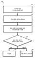
2 is a block diagram of a method for locating an implantation site in the brain according to aspects of the present disclosure;
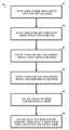
3 is a block diagram of another method for locating an implantation site in the brain according to aspects of the present disclosure;
4 is a block diagram of an exemplary computer of a signal processing unit in accordance with aspects of the present disclosure;
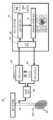
5 is a block diagram of an implantable system for determining DBS parameters in accordance with aspects of the present disclosure;
6 is a block diagram of a method for determining DBS parameters in accordance with aspects of the present disclosure;
7 is a block diagram of another method for determining DBS parameters in accordance with aspects of the present disclosure;
8 displays the recorded HFO evoked at the STN when the multi-contact electrode is placed on the STN, and displays the absence of HFO when the multi-contact electrode is not placed on the STN;
Figure 9 displays HFO and ECA recorded at various HFS frequencies when a multi-contact electrode is placed on the STN;
Figure 10 displays the recorded ECA for the LFS frequency compared to various HFS frequencies; and
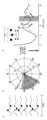
Figure 11 shows the DBS adjustment of various HFS frequencies based on the phase of the ECA waveform.
본원에서 사용될 때, "원위"라는 용어는 (인간 의사이든 수술용 로봇이든) 조작자로부터 더 멀리 있는 것으로 설명되고 있는 부분을 지칭하는 한편, "근위"라는 용어는 조작자에 더 가까운 것으로 설명되고 있는 부분을 지칭한다. "약", "실질적으로" 등의 용어는 본원에서 이용될 때, 제조, 재료, 환경, 사용, 및/또는 측정 공차 및 편차를 설명하는 것으로 여겨지고, 어느 경우든 10% 이하의 차이를 망라할 수 있다. 또한, 일관되는 한, 본원에서 설명되는 양태들의 임의의 양태는 본원에서 설명되는 다른 양태들의 임의의 양태 또는 모든 양태와 함께 사용될 수 있다.As used herein, the term "distal" refers to a portion described as being further from the operator (whether a human physician or a surgical robot), while the term "proximal" refers to a portion described as closer to the operator. refers to The terms “about,” “substantially,” and the like, when used herein, are intended to describe manufacturing, material, environmental, use, and/or measurement tolerances and variations, which in any case will encompass differences of 10% or less. can Also, as far as is consistent, any aspect of the aspects described herein can be used with any or all aspects of the other aspects described herein.
본 개시의 예시적인 구성의 기술적 특징들 또는 양태들에 대한 설명은 통상적으로 본 개시의 다른 예시적인 구성에서의 다른 유사한 특징들 또는 양태들에 이용가능하고 적용가능한 것으로서 고려되어야 한다. 이에 따라, 본 개시의 하나의 예시적인 구성에 따라 본원에서 설명되는 기술적 특징들은 본 개시의 다른 예시적인 구성들에 적용가능할 수 있고, 이에 따라 여기서 중복 설명은 생략될 수 있다.Descriptions of technical features or aspects of an exemplary configuration of the present disclosure should generally be considered as available and applicable to other similar features or aspects in other exemplary configurations of the present disclosure. Accordingly, technical features described herein according to one exemplary configuration of the present disclosure may be applicable to other exemplary configurations of the present disclosure, and thus redundant description may be omitted herein.
본 개시의 예시적인 구성들은 (예를 들어, 첨부 도면들을 참조하여) 아래에서 더 충분하게 설명될 것이다. 같은 참조 번호들은 본 명세서 및 도면 전체에 걸쳐 같은 요소들을 지칭할 수 있다.Exemplary configurations of the present disclosure will be more fully described below (eg, with reference to the accompanying drawings). Like reference numbers may refer to like elements throughout this specification and drawings.
본 개시의 양태들에서, 이를테면 파킨슨병(PD) 환자에서의 시상하핵(STN)이 심부 뇌 자극(DBS)을 겪는 전기 자극에 의해 유도되는 국소장 전위(LFP)의 변조가 채용된다. 본원에서 설명되는 시스템들 및 방법들은 이상적인 식립 부위들의 위치를 찾고, 처치 파라미터들을 개별적으로 교정함으로써 DBS의 유효성을 증가시키기 위해 주파수 및 다른 파라미터들을 최적화한다. 본원에서 설명되는 시스템들 및 방법들은 뇌에 전기 자극을 전달하고, 자극 전, 동안 및 후에 뇌의 반응을 기록한다. 자극에 반응하여 측정된 전기생리학적 마커들에 기초하여, 본 방법 및 시스템은 그 파라미터들을 미세 조정한다. 운동 및 다른 정신 장애는 네트워크 질환이므로, 본원에서 설명되는 시스템들 및 방법들의 하나의 이점은 이들이 자극으로 네트워크의 상태를 탐색한 다음, 폐루프 방식으로 반응 신호에 자극 파라미터들을 적응시킨다는 것이다.In aspects of the present disclosure, modulation of local field potential (LFP) induced by electrical stimulation such as in a Parkinson's disease (PD) patient where the subthalamic nucleus (STN) undergoes deep brain stimulation (DBS) is employed. The systems and methods described herein locate ideal implantation sites and optimize frequency and other parameters to increase the effectiveness of the DBS by individually calibrating treatment parameters. The systems and methods described herein deliver electrical stimulation to the brain and record the brain's response before, during, and after stimulation. Based on electrophysiological markers measured in response to stimulation, the method and system fine-tune its parameters. Since motor and other mental disorders are network disorders, one advantage of the systems and methods described herein is that they probe the state of the network with stimuli and then adapt the stimuli parameters to the response signal in a closed loop manner.
STN에서, 치료 고주파수 자극(130-180Hz)은 약리학적 처치로 관찰된 것과 유사한 고주파수 발진(~300Hz, HFO)을 유도한다. HFO와 함께, 각 자극 펄스 후의 유발 화합물 활성(ECA)이 식별되었다. ECA는 치료 및 비치료(20Hz) 자극 둘 모두에서 관찰되었지만, HFO는 단지 치료 주파수들로만 유도되었고, 연관된 ECA는 상당히 더 공명하였다. HFO 파워의 상대적 향상 정도는 자극 펄스와 ECA의 위상의 상호작용과 관련되었다.In STN, therapeutic high-frequency stimulation (130-180 Hz) induces high-frequency oscillations (~300 Hz, HFO) similar to those observed with pharmacological treatments. With HFO, the evoked compound activity (ECA) after each stimulation pulse was identified. ECA was observed with both treatment and non-treatment (20 Hz) stimulation, but HFO was induced only at treatment frequencies, and the associated ECA was significantly more resonant. The relative enhancement of HFO power was related to the interaction of stimulation pulse and ECA phase.
고주파수 STN-DBS는 신경 발진을 약리학적 처치와 유사하게, 그 건강한/처치된 상태로 조정하고, 이 발진을 최대화하기 위한 자극 주파수는 개별 대상체의 ECA 파형의 위상으로부터 추론될 수 있다. 이에 따라, 유도된 HFO는 PD 증상을 발생시키는 기능장애 회로의 성공적인 재교정의 마커로서 이용될 수 있다.High-frequency STN-DBS tunes neural oscillations to their healthy/treated state, similar to pharmacological treatment, and the stimulation frequency to maximize this oscillation can be inferred from the phase of an individual subject's ECA waveform. Thus, induced HFO can be used as a marker of successful recalibration of the dysfunctional circuits that give rise to PD symptoms.
아래에서 더 상세하게 설명되는 바와 같이, 고주파수 자극(HFS)은 약제학적 처치의 효과와 유사하게, STN에서의 발진 활성을 변조함으로써 그 치료 효과를 발휘한다.As described in more detail below, high frequency stimulation (HFS) exerts its therapeutic effect by modulating the oscillation activity in the STN, similar to the effect of pharmaceutical treatment.
본원에서 설명되는 시스템들 및 방법들은 운동 장애, 이를테면 파킨슨병, 수전증, 투렛 증후군, 간질, 근육긴장이상, 정신의학적/인지 장애, 이를테면 강박 장애, 중증 우울증, 알츠하이머병 치매 및 양극성 장애에 적용될 수 있다는 것이예상된다.The systems and methods described herein may be applied to movement disorders such as Parkinson's disease, tremor, Tourette's syndrome, epilepsy, dystonia, psychiatric/cognitive disorders such as obsessive-compulsive disorder, severe depression, Alzheimer's disease dementia and bipolar disorder. it is expected
본원에서 설명되는 시스템들 및 방법들은 STN, 담창구(내부 및 외부), 시상, 피질, 흑질(그물부 및 치밀부), 및 뇌각뇌교핵과 같은 타겟 뇌 구조에 적용될 수 있다는 것이 예상된다.It is anticipated that the systems and methods described herein may be applied to target brain structures such as the STN, globus (inner and outer), thalamus, cortex, substantia nigra (reticular and dense), and pontine nuclei.
"장기간 전극(chronic electrode)"이라는 어구는 개체의 뇌에 외과적으로 식립된, DBS 전극과 같은 다접점 전극을 지칭한다. 아래에서 더 상세히 설명되는 장기간 전극들은 각각 본원에서 설명되는 다접점 전극들과 유사하게 상이한 위치들에서 개별적으로 활성화가능한 접점들을 가질 수 있다. 각 장기간 전극/다접점 전극의 각 접점은 개체의 맞춤화된 뇌 반응을 설명하기 위해 특별히 조정되는 다양한 파라미터들에 기초하여 DBS를 전달하도록 제어될 수 있다.The phrase "chronic electrode" refers to a multi-contact electrode, such as a DBS electrode, that is surgically implanted into the brain of a subject. The long-term electrodes described in more detail below may each have individually activatable contacts at different locations, similar to the multi-contact electrodes described herein. Each junction of each long-term electrode/multi-contact electrode can be controlled to deliver DBS based on a variety of parameters that are specifically tuned to account for a subject's customized brain response.
도 1을 참조하면, 뇌에의 전극 식립을 위한 부위의 위치를 찾기 위한 시스템(100)은 복수의 다접점 전극들(101)을 포함한다. 복수의 다접점 전극들(101)은 개체의 뇌에서의 타겟 구조(102)의 영역 내로의 삽입을 위해 구성된다. 각 다접점 전극(101)은 별개의 해부학적 트랙을 따라 (예를 들어, 개체의 두개골에 대해 상이한 깊이들에서) 상이한 지리적 영역으로 연장한다. 복수의 접점들(103)은 각 다접점 전극(101)의 길이를 따라(예를 들어, 근위에서 원위 길이를 따라) 서로 이격된다.Referring to FIG. 1 , a
자극 디바이스(104)는 복수의 다접점 전극들(101) 각각과 전기 통신한다. 자극 디바이스(104)는 복수의 다접점 전극들(101) 각각에 고주파수 자극(HFS)을 가한다. HFS는 100Hz보다 클 수 있다(예를 들어, 100Hz 내지 200Hz).The
자극 디바이스(104)는 복수의 다접점 전극들 중 하나의 다접점 전극(101)의 접점들의 서브세트(103)에 HFS를 선택적으로 가하도록 구성된다. 기록 디바이스(105)는 HFS에 의해 타겟 구조(102)의 영역에서 유발되는 유발 화합물 활성(HFO)을 측정하도록 구성된다. 기록 디바이스(105)는 HFS에 의해 타겟 구조(102)의 영역에서 유발되는 유발 화합물 활성(ECA)을 측정하도록 구성된다.The
신호 처리 유닛(106)은 기록 디바이스(105)와 통신한다. 신호 처리 유닛(106)은 HFO 및 ECA 중 적어도 하나가 미리 결정된 임계치를 초과하는지 결정하여, 다접점 전극(101)의 하나의 접점(103)의 위치를 개체의 뇌에의 전극 식립을 위한 부위로서 식별한다.The
시각화 유닛(107)은 측정된 HFO 및 ECA를 시각적으로 디스플레이한다. 시각화 유닛(107)은 각 다접점 전극(101)의 각 접점(103)에 의해 유발되는 HFO 또는 ECA를 개별적으로 디스플레이할 수 있다. 이에 따라, 신경 외과의 또는 진료 전문가(108)가 각 개별 접점(103)에 의해 유발되는 HFO/ECA를 시각적으로 식별할 수 있다.The
본 개시의 양태에 따르면, 스위칭 유닛(109)이 자극 디바이스(104)에 의해 가해지는 HFS를 제어한다. HFO 및 ECA 중 적어도 하나가 미리 결정된 임계치 미만이라면, 스위칭 유닛(109)은 자극 디바이스(104)를 제어하여 복수의 다접점 전극들 중의 다접점 전극(101)의 접점들의 제2 서브세트(103)에 제2 HFS를 가하도록 구성된다.According to aspects of the present disclosure, a
도 1을 참조하여 설명된 시스템(100)은 수술 중에 HFO 및 ECA를 측정하도록 구성된다. 개체의 뇌에의 전극 식립을 위한 하나 이상의 부위가 식별된 후, 장기 DBS를 위해 하나 이상의 부위 각각에 전극(즉, 장기간 전극)이외과적으로 식립된다.The
도 2는 시스템(100)에 의해 채용될 수 있는 뇌에의 식립 부위의 위치를 찾기 위한 방법(200)의 블록도이다.2 is a block diagram of a
도 2를 참조하면, 방법(200)은 HFS를 가하는 단계(단계 201), 유발 반응 진폭을 측정하는 단계(단계 202) 및 특정 고주파수 범위에 대한 대역 전력을 추정하는 단계(단계 203)를 포함한다. 본 방법은 최대 신호 세기가 수신되는지 결정하는 단계(단계 204)를 포함한다. 최대 신호 세기가 수신된다면, 식립물이 식립된다(단계 205). 최대 신호 세기가 수신되지 않는다면, 다른 위치로 이동하기로 하는 결정이 행해진다(단계 206).Referring to FIG. 2 ,
도 1 내지 도 3을 참조하면, 시스템(100)에 의해 채용될 수 있는 뇌에의 식립 부위의 위치를 찾는 다른 방법(300)이 설명된다. 방법(300)은 복수의 다접점 전극들을 개체의 뇌에서의 타겟 구조의 영역 내로 삽입하는 단계를 포함한다(단계 301). 다접점 전극들은 개체의 두개골에 형성된 버 홀을 통해 연장될 수 있다.Referring to FIGS. 1-3 , another
복수의 다접점 전극들 중의 다접점 전극의 접점에 HFS가 가해진다(단계 302). HFS에 의해 타겟 구조의 영역에서 유발되는 고주파수 발진(HFO)이 측정된다(단계 303). HFS에 의해 타겟 구조의 영역에서 유발되는 유발 화합물 활성(ECA)이 측정된다(단계 304). HFO 및 ECA 중 적어도 하나가 미리 결정된 임계치를 (305) 초과하는지 결정된다(단계 305). HFO 및 ECA 중 적어도 하나가 미리 결정된 임계치를 초과한다면, 다접점 전극의 접점의 위치가 개체의 뇌에의 전극 식립을 위한 (306) 부위로서 식별된다(단계 306).HFS is applied to a contact point of a multi-contact electrode among a plurality of multi-contact electrodes (step 302). A high frequency oscillation (HFO) induced in the region of the target structure by the HFS is measured (step 303). The evoked compound activity (ECA) evoked in the region of the target structure by HFS is measured (step 304). A determination is made whether at least one of HFO and ECA exceeds 305 a predetermined threshold (step 305). If at least one of HFO and ECA exceeds a predetermined threshold, the location of the contact point of the multi-contact electrode is identified as a 306 site for electrode implantation in the subject's brain (step 306).
각 다접점 전극(101)은 별개의 해부학적 트랙을 따라 연장될 수 있고, 이의 원위 단부는들은 개체의 뇌 내의 다양한 깊이들에서 끝날 수 있다. 각 다접점 전극(101)은 그 길이를 따라 위치된 많은 접점들(103)을 포함할 수 있다. 각 접점(103)은 사용자의 뇌의 다양한 트랙들을 따라 다양한 깊이들을 테스트하기 위해 전기 자극(LFS 또는 HFS)을 선택적으로 그리고 개별적으로 수신할 수 있다. 이에 따라, 수술 중에 전기 자극을 개별적으로 가함으로써, 이상적인 트랙 및 이상적인 깊이가 식별될 수 있다. 예를 들어, 도 1을 참조하면, 트랙 3은 타겟 구조를 완전히 놓칠 수 있고, 트랙 2는 타겟 구조의 주변과 정렬될 수 있는 한편, 트랙 3은 타겟 구조의 중앙 영역과 정렬된다. 또한, 트랙 1의 최원위 접점(즉, 접점 1)은 최대 ECA 및 HFO를 유발할 수 있다. 이에 따라, 트랙 1 다접점 전극에 따른 접점 1의 위치는 개체의 뇌에의 전극 식립을 위한 바람직한 부위로서 식별될 것이다. 이어서, 이하에서 보다 상세하게 설명되는 바와 같이, 식립된 전극은 개별적으로 DBS의 유효성을 최대화하기 위해, 그 특정 파라미터들을 조정함으로써 교정될 수 있다. 아래에서 더 상세하게 설명되는 바와 같이, 파라미터 조정은 개체의 맞춤화된 뇌 반응 및 기저의 구조적 또는 전기화학적 편차를 고려하여 이루어질 수 있다. 이상화된 전극 배치와 이상화된 파라미터 설정의 조합은 DBS의 처치 효과를 최대화한다. 또한, 파라미터 조정은 개체의 기저 질환 병태 진행, 시간이 지나면서 일어나는 해부학적 변화, 또는 시간이 지나면서 일어나는 전기화학적 변화를 고려하여 주기적으로 조정될 수 있다.Each
HFO 또는 ECA가 미리 결정된 임계치 미만이라면, 복수의 다접점 전극들 중의 다접점 전극의 접점들의 제2 세트에 제2 고주파수 자극이 가해진다. 개체의 뇌에의 전극 식립을 위한 부위가 색별되지 않는다면, 복수의 다접점 전극들은 개체의 뇌에서의 제2 영역으로 이동된다. 예를 들어, 다접점 전극들 중 어느 것도 초임계 HFO 또는 ECA를 유발하는 것으로 밝혀지지 않는다면, 목적하는 HFO/ECA가 적어도 하나의 접점에 의해 유발될 때까지 다접점 전극들은 밀리미터 단위로 개체의 뇌 내로 추가로 증진될 수 있다.If the HFO or ECA is below a predetermined threshold, a second high frequency stimulation is applied to a second set of contacts of a multi-contact electrode of the plurality of multi-contact electrodes. If the site for electrode implantation in the subject's brain is not identified, the plurality of multi-contact electrodes are moved to the second area in the subject's brain. For example, if none of the multi-contact electrodes are found to cause supercritical HFO or ECA, then the multi-contact electrodes are millimeter-by-millimetre-wise in the subject's brain until the desired HFO/ECA is induced by at least one of the contacts. can be further enhanced.
본 개시의 양태에서, HFS는 100Hz보다 크다.In an aspect of the present disclosure, HFS is greater than 100 Hz.
본 개시의 양태에서, HFO는 200-450Hz 사이의 발진 패턴을 포함한다.In aspects of the present disclosure, the HFO includes an oscillation pattern between 200-450 Hz.
본 개시의 양태에서, ECA는 미리 결정된 임계치를 초과하는 공명 반응을 포함한다(예를 들어, 아래에서 더 상세하게 설명되는 도 11 참조).In an aspect of the present disclosure, the ECA includes a resonance response that exceeds a predetermined threshold (eg, see FIG. 11 described in more detail below).
도 4는 본 개시의 양태들에 따른 도 1의 신호 처리 유닛(106)의 예시적인 컴퓨터(400)의 블록도이다.4 is a block diagram of an
도 4를 참조하면, 신호 처리 유닛(106)은 컴퓨터 판독가능 저장 매체에 연결된 프로세서(401), 또는 휘발성 타입 메모리, 예를 들어, RAM, 또는 비휘발성 타입 메모리(예를 들어, 플래시 매체, 디스크 매체 등)일 수 있는 메모리(402)를 포함할 수 있다. 프로세서(401)는 디지털 신호 프로세서, 마이크로프로세서, ASIC, GPU(graphics processing unit), FPGA(field-programmable gate array), 또는 CPU(central processing unit)와 같은 다른 유형의 프로세서일 수 있지만, 이에 제한되는 것은 아니다.Referring to Figure 4, the
본 개시의 일부 양태들에서, 메모리(402)는 랜덤 액세스 메모리, 판독 전용 메모리, 자기 디스크 메모리, 고체 상태 메모리, 광 디스크 메모리 및/또는 다른 유형의 메모리일 수 있다. 메모리(402)는 회로 기판의 통신 버스(403)를 통해 그리고/또는 직렬 ATA 케이블 또는 다른 유형의 케이블과 같은 통신 케이블을 통해 프로세서(401)와 통신할 수 있다. 메모리(402)는 신호 처리 유닛(106)을 동작시키기 위해 프로세서(401)에 의해 실행가능한 컴퓨터 판독가능 명령어들을 포함한다. 신호 처리 유닛(106)은 다른 컴퓨터들 또는 서버와 통신하기 위한 네트워크 인터페이스(404)를 포함할 수 있다. 저장 디바이스(405)는 데이터를 저장하는 데 사용될 수 있다. 신호 처리 유닛(106)은 하나 이상의 FPGA(406)를 포함할 수 있다. FPGA(406)는 다양한 기계 학습 알고리즘들을 실행하는 데 사용될 수 있다. 디스플레이(407)는 신호 처리 유닛(106)에 의해 처리되는 데이터를 디스플레이하기 위해 채용될 수 있다.In some aspects of this disclosure,
도 1 및 도 4를 참조하여 설명된 신호 처리 유닛(106)은 달리 나타내어지지 않는 한, 아래에서 도 5를 참조하여 설명되는 신호 처리 유닛들(506)과 실질적으로 동일하고, 이에 따라 여기서 중복 설명은 생략될 수 있다. 예를 들어, 도 5에 도시된 신호 처리 유닛(506)은 도 1에 도시된 신호 처리 유닛(106)과 실질적으로 동일한 하드웨어 구성을 가질 수 있다. 또한, 아래에서 도 5를 참조하여 설명되는 스위칭 유닛(509), 기록 디바이스(505), 및 자극 디바이스(504)는 달리 나타내어지지 않는 한, 도 1 내지 도 3을 참조하여 설명된 스위칭 유닛(109), 기록 디바이스(105), 및 자극 디바이스(104)와 실질적으로 동일하고, 이에 따라 여기서 중복 설명은 생략될 수 있다.The
도 5를 참조하면, 심부 뇌 자극 파라미터들을 결정하기 위한 시스템(500)은 개체의 뇌에서의 타겟 구조에의 식립을 위해 구성된 적어도 하나의 다접점 전극(501)을 포함한다. 다접점 전극(501)은 아래에서 더 상세하게 설명되는 바와 같이, 장기간 식립된 전극일 수 있다. 자극 디바이스(504)는 적어도 하나의 다접점 전극(501)과 전기 통신한다. 자극 디바이스(504)는 적어도 하나의 다접점 전극(501)에 (예를 들어, 105Hz보다 작은) 저주파수 자극(LFS) 또는 (예를 들어, 100Hz 내지 200Hz의) 고주파수 자극(HFS)을 가하도록 구성된다.Referring to FIG. 5 , a
본 개시의 양태에 따르면, 스위칭 유닛(509)은 자극 디바이스(504)에 의해 가해지는 HFS를 제어한다. HFO 및 ECA 중 적어도 하나가 미리 결정된 임계치 미만이라면, 스위칭 유닛(509)은 자극 디바이스(504)를 제어하여 복수의 다접점 전극들 중의 다접점 전극(501)의 접점들의 제2 서브세트(503)에 제2 HFS를 가하도록 구성된다.According to aspects of the present disclosure, the
기록 디바이스(505)는 LFS에 의해 타겟 구조(502)에서 유발되는 유발 화합물 활성(ECA) 및 HFS에 의해 타겟 구조(502)에서 유발되는 고주파수 발진(HFO) 중 적어도 하나를 기록하도록 구성된다. 신호 처리 유닛(506)은 기록 디바이스(505)와 통신한다. 신호 처리 유닛(506)은 ECA로부터 추출되는 위상 공간에 기초하여 미리 결정된 범위 내의 뇌 자극을 전달하기 위한 주파수 범위를 결정한다(예를 들어, 도 11 참조). 신호 처리 유닛(506)은 결정된 범위 내에서 가해진 자극 주파수들에 의해 타겟 구조(502)에서 유발되는 고주파수 발진(HFO)을 분석한다. 파라미터 최적화 유닛(511)은 미리 결정된 임계치를 초과하는 HFO를 유발하는 주파수를 결정하고, 주파수를 타겟 구조에 대한 처치 주파수로서 선택한다.The
자극 디바이스(504)는 하나의 다접점 전극(501)의 하나의 접점(503)에 LFS 또는 HFS를 선택적으로 가하도록 구성된다.The
본 개시의 양태에서, 입력/출력(I/O) 유닛(512)은 자극 디바이스(504), 기록 디바이스(505), 신호 처리 유닛(506), 및 파라미터 최적화 유닛(511)과 전기 통신한다.In an aspect of the present disclosure, an input/output (I/O)
자극 디바이스(504), 기록 디바이스(505), 신호 처리 유닛(506), 및 파라미터 최적화 유닛(511) 중 적어도 하나는 개체의 가슴에 식립된다. 예를 들어, 신호 처리 유닛(506), 파라미터 최적화 유닛(511) 및 I/O 유닛(512)을 포함하는 처리 서브시스템(512)이 환자의 가슴에 식립될 수 있다.At least one of the
처리 서브시스템(512)은 DBS을 위해 식립된 다접점 전극들(501)을 제어할 수 있다.The
도 5 내지 도 7을 참조하면, 개뇌 자극 파라미터들을 결정하는 방법은 체의 뇌에서의 타겟 구조에 식립된 다접점 전극의 접점에 (예를 들어, 105Hz보다 작은) 저주파 자극(LFS)을 가하는 단계를 포함한다. LFS에 의해 타겟 구조에서 유발되는 유발 화합물 활성(Evoked Compound Activity, ECA)이 측정된다.5 to 7, a method for determining brain stimulation parameters includes applying low-frequency stimulation (LFS) (eg, less than 105 Hz) to the contact points of multi-contact electrodes implanted in target structures in the brain of the body. includes Evoked Compound Activity (ECA) evoked in the target structure by LFS is measured.
ECA로부터 추출되는 위상 공간에 기초하여 미리 결정된 범위 내의 뇌 자극을 전달하기 위한 주파수 범위가 결정된다. 결정된 주파수 범위 내에서 다접점 전극의 접점에 자극 주파수들이 가해진다. 결정된 범위 내에서 가해진 자극 주파수들에 의해 타겟 구조에서 유발되는 국소장 전위의 고주파수 발진(High Frequency Oscillations, HFO)이 측정된다. 미리 결정된 임계치를 초과하는 HFO를 유발하는 주파수가 결정된다. 결정된 주파수는 타겟 구조에 대한 처치 주파수로서 선택된다.A frequency range for delivering brain stimulation within a predetermined range is determined based on the phase space extracted from the ECA. Stimulation frequencies are applied to the contacts of the multi-contact electrode within the determined frequency range. High frequency oscillations (HFO) of the local field potential induced in the target structure by the applied stimulation frequencies within the determined range are measured. A frequency that causes HFO to exceed a predetermined threshold is determined. The determined frequency is selected as the treatment frequency for the target structure.
본 개시의 양태에서, 선택된 주파수는 130Hz보다 크다.In an aspect of this disclosure, the selected frequency is greater than 130 Hz.
본 개시의 양태에서, 선택된 주파수는 약 130Hz, 약 160Hz, 또는 약 180Hz이고, 100-200Hz 사이의 임의의 주파수일 수 있다.In aspects of the present disclosure, the selected frequency is about 130 Hz, about 160 Hz, or about 180 Hz, and may be any frequency between 100-200 Hz.
본 개시의 양태에서, 타겟 구조에서 유발되는 측정된 HFO는 200-450Hz 사이이다.In an aspect of the present disclosure, the measured HFO induced in the target structure is between 200-450 Hz.
본 개시의 양태에서, ECA는 미리 결정된 임계치를 초과하는 공명 반응을 포함한다.In an aspect of the present disclosure, ECA includes a resonance response above a predetermined threshold.
ECA가 미리 결정된 임계치 미만이라면, 다접점 전극의 접점과 상이한 타겟 구조 내의 위치에서 다접점 전극의 접점들의 제2 서브세트에 제2 LFS가 가해진다.If the ECA is below the predetermined threshold, a second LFS is applied to a second subset of the contacts of the multi-contact electrode at a different location within the target structure than the contacts of the multi-contact electrode.
예로서, 다수의 접점들을 갖는 하나 이상의 장기간 전극은 수술 중의 절차 동안 식립된다. 하나 이상의 접점을 통해 저주파수(<105Hz) 자극이 가해진다. 국소장 전위의 ECA 파형이 기록되고, 그 특성(예를 들어, 진폭, 위상, 공명 지속기간)이 결정된다. 이어서, 최적의 반응에 대한 자극 주파수 범위가 ECA로부터 추출되는 위상 공간에 기초하여 계산된다. 이 범위 내의 주파수들에서 자극이 전달되고, 대응하는 HFO 주파수 및/또는 전력이 계산된다. 최대 HFO 파워와 연관된 주파수가 처치를 위해 선택된다. 이에 따라, 본원에서 설명된 시스템 및 방법들은 HFO 파워 및 ECA 위상 공간을 처리함으로써 자극 주파수를 미세 조정하기 위해 채용될 수 있다.As an example, one or more long-term electrodes with multiple contacts are placed during an intraoperative procedure. A low-frequency (<105 Hz) stimulus is applied through one or more contacts. The ECA waveform of the local field potential is recorded and its properties (eg, amplitude, phase, resonance duration) determined. Stimulus frequency ranges for optimal responses are then calculated based on the phase space extracted from the ECA. Stimuli are delivered at frequencies within this range, and the corresponding HFO frequency and/or power is calculated. The frequency associated with the maximum HFO power is selected for treatment. Accordingly, the systems and methods described herein may be employed to fine-tune stimulation frequency by processing HFO power and ECA phase space.
도 5 내지 도 7을 참조하여 설명된 시스템들 및 방법들은 미리 결정된 스케줄(예를 들어, 매 24시간마다 1회, 매주 1회 등)로 뇌 자극 파라미터들을 재교정하기 위해 주기적으로 채용될 수 있다. 처치 파라미터들을 주기적으로 재교정하는 것 외에도, 재교정은 또한 신경 외과의 또는 처치 전문가가 직접 개입할 필요 없이, 시간이 지나면서 일어나는 맞춤화된 개체의 생리학적 변화에 기초하여 처치 파라미터들을 동적으로 최적화하기 위해 자율적으로 수행될 수 있다.The systems and methods described with reference to FIGS. 5-7 may be employed periodically to recalibrate brain stimulation parameters on a pre-determined schedule (eg, once every 24 hours, once a week, etc.) . In addition to periodic recalibration of treatment parameters, recalibration is also intended to dynamically optimize treatment parameters based on personalized individual physiological changes over time, without the need for direct intervention by a neurosurgeon or treatment specialist. can be performed autonomously.
특히 도 6을 참조하면, DBS 파라미터들(600)을 결정하기 위한 방법은 LFS를 가하는 단계(단계 601), 유발 반응을 측정하는 단계(단계 602), 광학 자극 주파수에 대한 위상 공간을 결정하는 단계(단계 603), 제안된 범위 내의 여러 주파수들로 자극들을 전달하는 단계(단계 604), 특정 고주파수 범위에 대한 대역 전력을 추정하는 단계(단계 605), 및 최대 대역 전력을 제공하는 최적화된 주파수를 선택하는 단계(단계 606)를 포함한다.Referring specifically to FIG. 6 , a method for determining
도 7을 참조하면, DBS 파라미터들(700)을 결정하기 위한 다른 방법은 개체의 뇌에서의 타겟 구조에 식립된 다접점 전극의 접점에 LFS를 가하는 단계(단계 701)를 포함한다. 방법(700)은 LFS에 의해 타겟 구조에서 유발되는 ECA를 측정하는 단계(단계 702) 및 ECA로부터 추출되는 위상 공간에 기초하여 미리 결정된 범위 내에서 뇌 자극을 전달하기 위한 주파수 범위를 결정하는 단계(단계 703)를 포함한다. 방법(700)은 결정된 주파수 범위 내에서 다접점 전극의 접점에 자극 주파수들을 가하는 단계(단계 704) 및 결정된 범위 내에서 가해진 자극 주파수들에 의해 타겟 구조에서 유발되는 HFO를 측정하는 단계(단계 705)를 포함한다. 방법(700)은 미리 결정된 임계치를 초과하는 HFO를 유발하는 주파수를 결정하는 단계(단계 706) 및 주파수를 타겟 구조에 대한 처치 주파수로서 선택하는 단계(단계 707)를 포함한다.Referring to FIG. 7 , another method for determining
도 8은 다접점 전극이 STN에 위치될 때 STN에서 유발되는 기록된 HFO를 디스플레이하고, 다접점 전극이 STN에 위치되지 않을 때 HFO의 부재를디스플레이한다. 도 8을 참조하면, HFO 및 공명 유발 화합물 활성(ECA)은 단지 STN에서만 고주파수 DBS 동안 관찰된다. 10개의 반구들에서, 자극 또는 기록 하드웨어에 의해 야기되었을 수 있는 가능한 아티팩트를 식별하고 배제하기 위해 130Hz 자극을 STN 안팎에서 수행하였다.8 displays the recorded HFO evoked at the STN when the multi-contact electrode is placed at the STN, and displays the absence of HFO when the multi-contact electrode is not placed at the STN. Referring to FIG. 8 , HFO and resonance-inducing compound activity (ECA) were observed during high-frequency DBS only in STN. In 10 hemispheres, 130 Hz stimulation was performed in and around the STN to identify and rule out possible artifacts that might have been caused by stimulation or recording hardware.
도 9는 다접점 전극이 STN에 위치될 때 다양한 HFS 주파수들에서 기록된 HFO 및 ECA를 디스플레이한다. 도 9를 참조하면, 고주파수 자극(예를 들어, 130Hz, 160Hz, 및 180Hz)은 HFO 및 ECA를 상이한 진폭들로 변조한다.9 displays HFO and ECA recorded at various HFS frequencies when a multi-contact electrode is placed on the STN. Referring to FIG. 9 , high frequency stimulation (eg, 130 Hz, 160 Hz, and 180 Hz) modulates HFO and ECA at different amplitudes.
도 10은 다양한 HFS 주파수들과 비교하여 LFS 주파수에 대한 기록된 ECA를 디스플레이한다. 도 10을 참조하면, 펄스간 유발 활성은 저주파수 자극(예를 들어, 20Hz)이 아닌, 단지 고주파수 자극(예를 들어, 130Hz, 160Hz, 및 180Hz)과의 적응을 제시한다.10 displays the recorded ECA for the LFS frequency compared to various HFS frequencies. Referring to FIG. 10 , inter-pulse evoked activity suggests adaptation only with high-frequency stimuli (eg, 130 Hz, 160 Hz, and 180 Hz) and not low-frequency stimulation (eg, 20 Hz).
도 11은 ECA 파형의 위상에 기초한 다양한 HFS 주파수들의 DBS 조정을 도시한다. 도 11을 참조하면, DBS는 ECA 파형의 위상에 기초하여 최대 변조 효과를 제공하도록 조정될 수 있다.Figure 11 shows the DBS adjustment of various HFS frequencies based on the phase of the ECA waveform. Referring to Figure 11, the DBS can be adjusted to provide the maximum modulation effect based on the phase of the ECA waveform.
다음 참고문헌 각각은 전문이 본원에 원용된다.Each of the following references is incorporated herein in its entirety.
Agnesi, F., Connolly, A. T., Baker, K. B., Vitek, J. L., 및 Johnson, M. D. (2013). Deep Brain Stimulation Imposes Complex Informational Lesions(심부 뇌 자극은 복잡한 정보 병변을 부과한다). PLoS One 8, 1-11. doi:10.1371/journal.pone.0074462.Agnesi, F., Connolly, A. T., Baker, K. B., Vitek, J. L., and Johnson, M. D. (2013). Deep Brain Stimulation Imposes Complex Informational Lesions. PLoS One 8, 1-11. doi:10.1371/journal.pone.0074462.
Ashby, P., Paradiso, G., Saint-Cyr, J. A., Chen, R., Lang, A. E., 및 Lozano, A. M. (2001). Potentials recorded at the scalp by stimulation near the human subthalamic nucleus(인간의 시상하핵 근처에서 자극에 의해 두피에 기록된 전위). Clin Neurophysiol 112, 431-437. doi:10.1016/S1388-2457(00)00532-0.Ashby, P., Paradiso, G., Saint-Cyr, J. A., Chen, R., Lang, A. E., and Lozano, A. M. (2001). Potentials recorded at the scalp by stimulation near the human subthalamic nucleus. Clin Neurophysiol 112, 431-437. doi:10.1016/S1388-2457(00)00532-0.
Benabid, A. L., Chabardes, S., Mitrofanis, J., 및 Pollak, P. (2009). Deep brain stimulation of the subthalamic nucleus for the treatment of Parkinson's disease(파킨슨병의 치료를 위한 시상하핵의 심부 뇌 자극). Lancet Neurol, 67-81.Benabid, A. L., Chabardes, S., Mitrofanis, J., and Pollak, P. (2009). Deep brain stimulation of the subthalamic nucleus for the treatment of Parkinson's disease. Lancet Neurol, 67-81.
Benazzouz, A., Gao, D. ., Ni, Z. ., Piallat, B., Bouali-Benazzouz, R., 및 Benabid, A. . (2000). Effect of high-frequency stimulation of the subthalamic nucleus on the neuronal activities of the substantia nigra pars reticulata and ventrolateral nucleus of the thalamus in the rat(래트에서 흑질 그물부 및 시상의 복외측 핵의 뉴런 활성에 대한 시상하핵의 고주파수 자극의 효과). Neuroscience 99, 289-295. doi:10.1016/S0306-4522(00)00199-8.Benazzouz, A., Gao, D. ., Ni, Z. ., Piallat, B., Bouali-Benazzouz, R., and Benabid, A. . (2000). Effect of high-frequency stimulation of the subthalamic nucleus on the neuronal activities of the substantia nigra pars reticulata and ventrolateral nucleus of the thalamus in the rat effect of stimulation). Neuroscience 99, 289-295. doi:10.1016/S0306-4522(00)00199-8.
Berens, P. (2009). CircStat: A MATLAB Toolbox for Circular Statistics(순환 통계를 위한 MATLAB 툴박스). J Stat Softw 31, 293-295. doi:10.18637/jss.v031.i10.Berens, P. (2009). CircStat: A MATLAB Toolbox for Circular Statistics. J Stat Softw 31, 293-295. doi:10.18637/jss.v031.i10.
Bergman, H., Wichmann, T., Karmon, B., 및 DeLong, M. R. (1994). The primate subthalamic nucleus(영장류 시상하 핵). II. Neuronal activity in the MPTP model of parkinsonism(파킨슨증의 MPTP 모델에서의 신경 활성). J Neurophysiol 72, 507-20. doi:10.1152/jn.1994.72.2.507.Bergman, H., Wichmann, T., Karmon, B., and DeLong, M. R. (1994). The primate subthalamic nucleus. II. Neuronal activity in the MPTP model of parkinsonism. J Neurophysiol 72, 507-20. doi:10.1152/jn.1994.72.2.507.
Bevan, M. D., Magill, P. J., Terman, D., Bolam, J. P., 및 Wilson, C. J. (2002). Move to the rhythm: oscillations in the subthalamic nucleus-external globus pallidus network(리듬: 시상하핵 외측 담창구 네트워크에서의 발진). Trends Neurosci 25, 525-31. doi:10.1016/s0166-2236(02)02235-x.Bevan, M. D., Magill, P. J., Terman, D., Bolam, J. P., and Wilson, C. J. (2002). Move to the rhythm: oscillations in the subthalamic nucleus-external globus pallidus network.
Brittain, J.-S., 및 Brown, P. (2014). Oscillations and the basal ganglia: Motor control and beyond(발진 및 기저핵: 운동 성능 제어 및 그 이상). Neuroimage 85, 637-647. doi:10.1016/j.neuroimage.2013.05.084.Brittain, J.-S., and Brown, P. (2014). Oscillations and the basal ganglia: Motor control and beyond. Neuroimage 85, 637-647. doi:10.1016/j.neuroimage.2013.05.084.
Brown, P., Mazzone, P., Oliviero, A., Altibr및i, M. G., Pilato, F., Tonali, P. A. 외 (2004). Effects of stimulation of the subthalamic area on oscillatory pallidal activity in Parkinson's disease(파킨슨병에서 발진 창백핵 활성에 대한 시상하부 영역의 자극의 효과). Exp Neurol 188, 480-490. doi:10.1016/j.expneurol.2004.05.009.Brown, P., Mazzone, P., Oliviero, A., Altibr andi, M. G., Pilato, F., Tonali, P. A. et al. (2004). Effects of stimulation of the subthalamic area on oscillatory pallidal activity in Parkinson's disease. Exp Neurol 188, 480-490. doi:10.1016/j.expneurol.2004.05.009.
Chu, H.-Y., McIver, E. L., Kovaleski, R. F., Atherton, J. F., 및 Bevan, M. D. (2017). Loss of Hyperdirect Pathway Cortico-Subthalamic Inputs Following Degeneration of Midbrain Dopamine Neurons(중뇌 도파민 뉴론의 퇴행 후 하이퍼다이렉트 경로 피질-시상하 입력의 손실). Neuron 95, 1306-1318.e5. doi:10.1016/j.neuron.2017.08.038.Chu, H.-Y., McIver, E. L., Kovaleski, R. F., Atherton, J. F., and Bevan, M. D. (2017). Loss of Hyperdirect Pathway Cortico-Subthalamic Inputs Following Degeneration of Midbrain Dopamine Neurons. Neuron 95, 1306-1318. e5. doi:10.1016/j.neuron.2017.08.038.
Cleary, D. R., Raslan, A. M., Rubin, J. E., Bahgat, D., Viswanathan, A., Heinricher, M. M. 외 (2013). Deep brain stimulation entrains local neuronal firing in human globus pallidus internus(심부 뇌 자극은 인간 내측 담창구에서 국소 신경세포 점호를 동반한다). J Neurophysiol 109, 978-987. doi:10.1152/jn.00420.2012.Cleary, D. R., Raslan, A. M., Rubin, J. E., Bahgat, D., Viswanathan, A., Heinricher, M. M. et al (2013). Deep brain stimulation entrains local neuronal firing in human globus pallidus internus.
Dostrovsky, J. O., Levy, R., Wu, J. P., Hutchison, W. D., Tasker, R. R., 및 Lozano, A. M. (2000). Microstimulation-Induced Inhibition of Neuronal Firing in Human Globus Pallidus(인간 내측 담창구에서 신경세포 점호의 미세자극 유도 저해). J Neurophysiol 84, 570-574. doi:10.1152/jn.2000.84.1.570.Dostrovsky, J. O., Levy, R., Wu, J. P., Hutchison, W. D., Tasker, R. R., and Lozano, A. M. (2000). Microstimulation-Induced Inhibition of Neuronal Firing in Human Globus Pallidus. J Neurophysiol 84, 570-574. doi:10.1152/jn.2000.84.1.570.
Erwin, B., Jr, M. M., 및 Baker, K. K. (2000). Mechanisms of deep brain stimulation and future technical developments(심부 뇌 자극의 메커니즘 및 미래의 기술적 개발). Neurol Res 22, 259-266. doi:10.1080/01616412.2000.11740668.Erwin, B., Jr, M. M., and Baker, K. K. (2000). Mechanisms of deep brain stimulation and future technical developments. Neurol Res 22, 259-266. doi:10.1080/01616412.2000.11740668.
Escobar, D., Johnson, L. A., Nebeck, S. D., Zhang, J., Johnson, M. D., Baker, K. B. 외 (2017). Parkinsonism and Vigilance: Alteration in neural oscillatory activity and phase-amplitude coupling in the basal ganglia and motor cortex(파킨슨증 및 경계: 기저핵 및 운동 피질에서의 신경 발진 활성 및 위상-진폭 결합의 변화). J Neurophysiol 118, jn.00388.2017. doi:10.1152/jn.00388.2017.Escobar, D., Johnson, L. A., Nebeck, S. D., Zhang, J., Johnson, M. D., Baker, K. B. et al (2017). Parkinsonism and Vigilance: Alteration in neural oscillatory activity and phase-amplitude coupling in the basal ganglia and motor cortex. J Neurophysiol 118, jn.00388.2017. doi:10.1152/jn.00388.2017.
Eusebio, A., Chen, C. C., Lu, C. S., Lee, S. T., Tsai, C. H., Limousin, P. 외 (2008). Effects of low-frequency stimulation of the subthalamic nucleus on movement in Parkinson's disease(파킨슨병에서 운동에 대한 시상하핵의 저주파수 자극의 효과). Exp Neurol 209, 125-130. doi:10.1016/j.expneurol.2007.09.007.Eusebio, A., Chen, C. C., Lu, C. S., Lee, S. T., Tsai, C. H., Limousin, P. et al (2008). Effects of low-frequency stimulation of the subthalamic nucleus on movement in Parkinson's disease. Exp Neurol 209, 125-130. doi:10.1016/j.expneurol.2007.09.007.
Eusebio, A., Thevathasan, W., Doyle Gaynor, L., Pogosyan, A., Bye, E., Foltynie, T. 외 (2011). Deep brain stimulation can suppress pathological synchronisation in parkinsonian patients(심부 뇌 자극은 파킨슨 환자에서 병리학적 동기화를 억제할 수 있다). J Neurol Neurosurg Psychiatry 82, 569-573. doi:10.1136/jnnp.2010.217489.Eusebio, A., Thevathasan, W., Doyle Gaynor, L., Pogosyan, A., Bye, E., Foltynie, T. et al. (2011). Deep brain stimulation can suppress pathological synchronisation in parkinsonian patients. J Neurol Neurosurg Psychiatry 82, 569-573. doi:10.1136/jnnp.2010.217489.
Filali, M., Hutchison, W. D., Palter, V. N., Lozano, A. M., 및 Dostrovsky, J. O. (2004). Stimulation-induced inhibition of neuronal firing in human subthalamic nucleus(인간 시상하핵에서 신경 점호의 자극 유도 저해). Exp Brain Res 156, 274-281. doi:10.1007/s00221-003-1784-y.Filali, M., Hutchison, W. D., Palter, V. N., Lozano, A. M., and Dostrovsky, J. O. (2004). Stimulation-induced inhibition of neuronal firing in human subthalamic nucleus. Exp Brain Res 156, 274-281. doi:10.1007/s00221-003-1784-y.
Foffani, G., Ardolino, G., Egidi, M., Caputo, E., Bossi, B., 및 Priori, A. (2006). Subthalamic oscillatory activities at beta or higher frequency do not change after high-frequency DBS in Parkinson's disease(베타 또는 더 높은 주파수에서의 시상하부 발진 활성은 파킨슨병에서 고주파수 DBS 후에 변하지 않는다). Brain Res Bull 69, 123-130. doi:10.1016/j.brainresbull.2005.11.012.Foffani, G., Ardolino, G., Egidi, M., Caputo, E., Bossi, B., and Priori, A. (2006). Subthalamic oscillatory activities at beta or higher frequency do not change after high-frequency DBS in Parkinson's disease. Brain Res Bull 69, 123-130. doi:10.1016/j.brainresbull.2005.11.012.
Foffani, G., Priori, A., Egidi, M., Rampini, P., Tamma, F., Caputo, E. 외 (2003). 300-Hz subthalamic oscillations in Parkinson's disease(파킨슨병에서의 300-Hz 시상하부 발진). Brain 126, 2153-2163. doi:10.1093/brain/awg229.Foffani, G., Priori, A., Egidi, M., Rampini, P., Tamma, F., Caputo, E. et al (2003). 300-Hz subthalamic oscillations in Parkinson's disease. Brain 126, 2153-2163. doi:10.1093/brain/awg229.
Fogelson, N., K
Garcia, L., D'Alessandro, G., Bioulac, B., 및 Hammond, C. (2005a). High-frequency stimulation in Parkinson's disease: more or less?(파킨슨병에서의 고주파수 자극: 많은가 적은가) Trends Neurosci 28, 209-216. doi:10.1016/j.tins.2005.02.005.Garcia, L., D'Alessandro, G., Bioulac, B., and Hammond, C. (2005a). High-frequency stimulation in Parkinson's disease: more or less? Trends Neurosci 28, 209-216. doi:10.1016/j.tins.2005.02.005.
Garcia, L., D'Alessandro, G., Fernagut, P., Bioulac, B., 및 Hammond, C. (2005b). Impact of High-Frequency Stimulation Parameters on the Pattern of Discharge of Subthalamic Neurons(시상하 뉴런의 방출 패턴에 대한 고주파수 자극 파라미터의 영향). J Neurophysiol 94, 3662-3669. doi:10.1152/jn.00496.2005.Garcia, L., D'Alessandro, G., Fernagut, P., Bioulac, B., and Hammond, C. (2005b). Impact of High-Frequency Stimulation Parameters on the Pattern of Discharge of Subthalamic Neurons. J Neurophysiol 94, 3662-3669. doi:10.1152/jn.00496.2005.
Gmel, G. E., Obradovic, M., Gorman, R. B., Single, P. S., Parker, J. L., Hamilton, T. J. 외 (2015). A new biomarker for subthalamic deep brain stimulation for patients with advanced Parkinson's disease - A pilot study(진행성 파킨슨병을 앓는 환자에 대한 시상하 심부 뇌 자극에 대한 새로운 바이오마커-A 예비 연구). J Neural Eng 12. doi:10.1088/1741-2560/12/6/066013.Gmel, G. E., Obradovic, M., Gorman, R. B., Single, P. S., Parker, J. L., Hamilton, T. J. et al (2015). A new biomarker for subthalamic deep brain stimulation for patients with advanced Parkinson's disease - A pilot study. J Neural Eng 12. doi:10.1088/1741-2560/12/6/066013.
Grill, W. M., Snyder, A. N., 및 Miocinovic, S. (2004). Deep brain stimulation creates an informational lesion of the stimulated nucleus(심부 뇌 자극은 자극된 핵의 정보 병변을 생성한다). Neuroreport 15, 1137-1140. doi:10.1097/00001756-200405190-00011.Grill, W. M., Snyder, A. N., and Miocinovic, S. (2004). Deep brain stimulation creates an informational lesion of the stimulated nucleus.
Gross, R. E., Krack, P., Rodriguez-Oroz, M. C., Rezai, A. R., 및 Benabid, A.-L. (2006). Electrophysiological mapping for the implantation of deep brain stimulators for Parkinson's disease and tremor(파킨슨병 및 진전에 대한 심부 뇌 자극제의 식립을 위한 전기생리학적 맵핑). Mov Disord 21, S259-S283. doi:10.1002/mds.20960.Gross, R. E., Krack, P., Rodriguez-Oroz, M. C., Rezai, A. R., and Benabid, A.-L. (2006). Electrophysiological mapping for the implantation of deep brain stimulators for Parkinson's disease and tremor. Mov Disord 21, S259-S283. doi:10.1002/mds.20960.
Guo, Y., Rubin, J. E., McIntyre, C. C., Vitek, J. L., 및 Terman, D. (2008). Thalamocortical Relay Fidelity Varies Across Subthalamic Nucleus Deep Brain Stimulation Protocols in a Data-Driven Computational Model(데이터 구동 계산 모델에서 시상하핵 심부 뇌 자극 프로토콜에 따라 시상피질 릴레이 충실도가 달라진다). J Neurophysiol 99, 1477-1492. doi:10.1152/jn.01080.2007.Guo, Y., Rubin, J. E., McIntyre, C. C., Vitek, J. L., and Terman, D. (2008). Thalamocortical Relay Fidelity Varies Across Subthalamic Nucleus Deep Brain Stimulation Protocols in a Data-Driven Computational Model. J Neurophysiol 99, 1477-1492. doi:10.1152/jn.01080.2007.
Hahn, P. J., Russo, G. S., Hashimoto, T., Miocinovic, S., Xu, W., McIntyre, C. C. 외 (2008). Pallidal burst activity during therapeutic deep brain stimulation(치료 심부 뇌 자극 동안의 창백핵 활성). Exp Neurol 211, 243-251. doi:10.1016/j.expneurol.2008.01.032.Hahn, P. J., Russo, G. S., Hashimoto, T., Miocinovic, S., Xu, W., McIntyre, C. C. et al (2008). Pallidal burst activity during therapeutic deep brain stimulation. Exp Neurol 211, 243-251. doi:10.1016/j.expneurol.2008.01.032.
Hashimoto, T., Elder, C. M., Okun, M. S., Patrick, S. K., 및 Vitek, J. L. (2003). Stimulation of the Subthalamic Nucleus Changes the Firing Pattern of Pallidal Neurons(시상하핵의 자극은 창백핵 뉴런의 점호 패턴을 변화시킨다). J Neurosci 23, 1916-1923. doi:10.1523/JNEUROSCI.23-05-01916.2003.Hashimoto, T., Elder, C. M., Okun, M. S., Patrick, S. K., and Vitek, J. L. (2003). Stimulation of the Subthalamic Nucleus Changes the Firing Pattern of Pallidal Neurons. J Neurosci 23, 1916-1923. doi:10.1523/JNEUROSCI.23-05-01916.2003.
Herrington, T. M., Cheng, J. J., 및 Eskandar, E. N. (2016). Mechanisms of deep brain stimulation(심부 뇌 자극의 메커니즘). J Neurophysiol 115, 19-38. doi:10.1152/jn.00281.2015.Herrington, T. M., Cheng, J. J., and Eskandar, E. N. (2016). Mechanisms of deep brain stimulation. J Neurophysiol 115, 19-38. doi:10.1152/jn.00281.2015.
Hoang, K. B., 및 Turner, D. A. (2019). The Emerging Role of Biomarkers in Adaptive Modulation of Clinical Brain Stimulation(임상 뇌 자극의 적응 변조에 있어서의 바이오마커의 새로운 역할). Neurosurgery 85, E430-E439. doi:10.1093/neuros/nyz096.Hoang, K. B., and Turner, D. A. (2019). The Emerging Role of Biomarkers in Adaptive Modulation of Clinical Brain Stimulation. Neurosurgery 85, E430-E439. doi:10.1093/neuros/nyz096.
Huang, H., Watts, R. L., 및 Montgomery, E. B. (2014). Effects of deep brain stimulation frequency on bradykinesia of Parkinson's disease(파킨슨병의 운동완만에 대한 심부 뇌 자극 주파수의 효과). Mov Disord 29, 203-206. doi:10.1002/mds.25773.Huang, H., Watts, R. L., and Montgomery, E. B. (2014). Effects of deep brain stimulation frequency on bradykinesia of Parkinson's disease. Mov Disord 29, 203-206. doi:10.1002/mds.25773.
Johnson, M. D., Miocinovic, S., McIntyre, C. C., 및 Vitek, J. L. (2008). Mechanisms and targets of deep brain stimulation in movement disorders(운동 장애에서 심부 뇌 자극의 메커니즘 및 타겟). Neurotherapeutics 5, 294-308. doi:10.1016/j.nurt.2008.01.010.Johnson, M. D., Miocinovic, S., McIntyre, C. C., and Vitek, J. L. (2008). Mechanisms and targets of deep brain stimulation in movement disorders.
Kaku, H., Ozturk, M., Viswanathan, A., Shahed, J., Sheth, S. A., Kumar, S. 외 (2020). Unsupervised clustering reveals spatially varying single neuronal firing patterns in the subthalamic nucleus of patients with Parkinson's disease(비지도형 클러스터링은 파킨슨병 환자의 시상하핵에서 공간적으로 변하는 단일 뉴런 점호 패턴들을 보인다). Clin Park Relat Disord 3, 100032. doi:10.1016/j.prdoa.2019.100032.Kaku, H., Ozturk, M., Viswanathan, A., Shahed, J., Sheth, S. A., Kumar, S. et al (2020). Unsupervised clustering reveals spatially varying single neuronal firing patterns in the subthalamic nucleus of patients with Parkinson's disease. Clin
Kane, A., Hutchison, W. D., Hodaie, M., Lozano, A. M., 및 Dostrovsky, J. O. (2009). Dopamine-dependent high-frequency oscillatory activity in thalamus and subthalamic nucleus of patients with Parkinson's disease(파킨슨병 환자의 시상 및 시상하핵에서의 도파민 의존적 고주파수 발진 활성). Neuroreport 20, 1549-1553. doi:10.1097/WNR.0b013e32833282c8.Kane, A., Hutchison, W. D., Hodaie, M., Lozano, A. M., and Dostrovsky, J. O. (2009). Dopamine-dependent high-frequency oscillatory activity in thalamus and subthalamic nucleus of patients with Parkinson's disease.
Kent, A. R., Swan, B. D., Brocker, D. T., Turner, D. A., Gross, R. E., 및 Grill, W. M. (2015). Measurement of Evoked Potentials During Thalamic Deep Brain Stimulation(시상 심부 뇌 자극 동안의 유발 전위의 측정). Brain Stimul 8, 42-56. doi:10.1016/j.brs.2014.09.017.Kent, A. R., Swan, B. D., Brocker, D. T., Turner, D. A., Gross, R. E., and Grill, W. M. (2015). Measurement of Evoked Potentials During Thalamic Deep Brain Stimulation.
Kita, H., 및 Kitai, S. T. (1991). Intracellular study of rat globus pallidus neurons: membrane properties and responses to neostriatal, subthalamic and nigral stimulation(래트 담창구 뉴런의 세포내 연구: 막 속성 및 신상피, 시상하 및 흑질 자극에 대한 반응). Brain Res 564, 296-305. doi:10.1016/0006-8993(91)91466-E.Kita, H., and Kitai, S. T. (1991). Intracellular study of rat globus pallidus neurons: membrane properties and responses to neostriatal, subthalamic and nigral stimulation. Brain Res 564, 296-305. doi:10.1016/0006-8993(91)91466-E.
K
K
Kuncel, A. M., Cooper, S. E., Wolgamuth, B. R., 및 Grill, W. M. (2007). Amplitude- and Frequency-Dependent Changes in Neuronal Regularity Parallel Changes in Tremor With Thalamic Deep Brain Stimulation(시상 심부 뇌 자극에 따른 전진의 뉴런 규칙성 병렬 변화의 진폭 및 주파수 의존적 변화). IEEE Trans Neural Syst Rehabil Eng 15, 190-197. doi:10.1109/TNSRE.2007.897004.Kuncel, A. M., Cooper, S. E., Wolgamuth, B. R., and Grill, W. M. (2007). Amplitude- and Frequency-Dependent Changes in Neuronal Regularity Parallel Changes in Tremor With Thalamic Deep Brain Stimulation. IEEE Trans Neural
Leventhal, D. K., Gage, G. J., Schmidt, R., Pettibone, J. R., Case, A. C., 및 Berke, J. D. (2012). Basal ganglia beta oscillations accompany cue utilization(기저핵 베타 발진은 큐 활용을 동반한다). Neuron 73, 523-536. doi:10.1016/j.neuron.2011.11.032.Leventhal, D. K., Gage, G. J., Schmidt, R., Pettibone, J. R., Case, A. C., and Berke, J. D. (2012). Basal ganglia beta oscillations accompany cue utilization. Neuron 73, 523-536. doi:10.1016/j.neuron.2011.11.032.
Li, Q., Ke, Y., Chan, D. C. W., Qian, Z. M., Yung, K. K. L., Ko, H., 외 (2012). Therapeutic Deep Brain Stimulation in Parkinsonian Rats Directly Influences Motor Cortex(파킨슨병 래트의 치료 심층 뇌 자극은 운동 피질에 직접적인 영향을 미친다). Neuron 76, 1030-1041. doi:10.1016/j.neuron.2012.09.032.Li, Q., Ke, Y., Chan, D. C. W., Qian, Z. M., Yung, K. K. L., Ko, H., et al (2012). Therapeutic Deep Brain Stimulation in Parkinsonian Rats Directly Influences Motor Cortex. Neuron 76, 1030-1041. doi:10.1016/j.neuron.2012.09.032.
Li, S., Arbuthnott, G. W., Jutras, M. J., Goldberg, J. A., 및 Jaeger, D. (2007). Resonant Antidromic Cortical Circuit Activation as a Consequence of High-Frequency Subthalamic Deep-Brain Stimulation(고주파수 시상하 심부 뇌 자극에 의한 공명 항염증 피질 회로 활성화). J Neurophysiol 98, 3525-3537. doi:10.1152/jn.00808.2007.Li, S., Arbuthnott, G. W., Jutras, M. J., Goldberg, J. A., and Jaeger, D. (2007). Resonant Antidromic Cortical Circuit Activation as a Consequence of High-Frequency Subthalamic Deep-Brain Stimulation. J Neurophysiol 98, 3525-3537. doi:10.1152/jn.00808.2007.
Litvak, V., Eusebio, A., Jha, A., Oostenveld, R., Barnes, G., Foltynie, T., 외 (2012). Movement-related changes in local and long-range synchronization in parkinson's disease revealed by simultaneous magnetoencephalography and intracranial recordings(동시 자기 뇌조영술과 두개내 기록에 의해 밝혀진 파킨슨병의 국소 및 장거리 동기화의 움직임 관련 변화). J Neurosci 32, 10541-10553. doi:10.1523/JNEUROSCI.0767-12.2012.Litvak, V., Eusebio, A., Jha, A., Oostenveld, R., Barnes, G., Foltynie, T., et al (2012). Movement-related changes in local and long-range synchronization in parkinson's disease revealed by simultaneous magnetoencephalography and intracranial recordings. J Neurosci 32, 10541-10553. doi:10.1523/JNEUROSCI.0767-12.2012.
Lopez-Azcarate, J., Tainta, M., Rodriguez-Oroz, M. C., Valencia, M., Gonzalez, R., Guridi, J. 외 (2010). Coupling between beta and high-frequency activity in the human subthalamic nucleus may be a pathophysiological mechanism in Parkinson's disease(인간의 시상하핵에서 베타와 고주파수 활성 간의 결합은 파킨슨병의 병태 생리학적 메커니즘일 수 있다). J Neurosci 30, 6667-6677. doi:10.1523/JNEUROSCI.5459-09.2010.Lopez-Azcarate, J., Tainta, M., Rodriguez-Oroz, M. C., Valencia, M., Gonzalez, R., Guridi, J. et al (2010). Coupling between beta and high-frequency activity in the human subthalamic nucleus may be a pathophysiological mechanism in Parkinson's disease.
Mastro, K. J., 및 Gittis, A. H. (2015). Striking the right balance: Cortical modulation of the subthalamic nucleus-globus pallidus circuit(시상하핵-담창구 회로의 피질 변조). Neuron 85, 233-235. doi:10.1016/j.neuron.2014.12.062.Mastro, K. J., and Gittis, A. H. (2015). Striking the right balance: Cortical modulation of the subthalamic nucleus-globus pallidus circuit. Neuron 85, 233-235. doi:10.1016/j.neuron.2014.12.062.
McConnell, G. C., So, R. Q., Hilliard, J. D., Lopomo, P., 및 Grill, W. M. (2012). Effective Deep Brain Stimulation Suppresses Low-Frequency Network Oscillations in the Basal Ganglia by Regularizing Neural Firing Patterns(신경 점포 패턴을 정규화하여 기저핵의 저주파수 네트워크 발진을 억제하는 효과적인 심부 뇌 자극). J Neurosci 32, 15657-15668. doi:10.1523/JNEUROSCI.2824-12.2012.McConnell, G. C., So, R. Q., Hilliard, J. D., Lopomo, P., and Grill, W. M. (2012). Effective Deep Brain Stimulation Suppresses Low-Frequency Network Oscillations in the Basal Ganglia by Regularizing Neural Firing Patterns. J Neurosci 32, 15657-15668. doi:10.1523/JNEUROSCI.2824-12.2012.
McIntyre, C. C., 및 Hahn, P. J. (2010). Network perspectives on the mechanisms of deep brain stimulation(심부 뇌 자극 메커니즘에 대한 네트워크 관점). Neurobiol Dis 38, 329-337. doi:10.1016/j.nbd.2009.09.022.McIntyre, C. C., and Hahn, P. J. (2010). Network perspectives on the mechanisms of deep brain stimulation. Neurobiol Dis 38, 329-337. doi:10.1016/j.nbd.2009.09.022.
Meissner, W., Leblois, A., Hansel, D., Bioulac, B., Gross, C. E., Benazzouz, A. 외 (2005). Subthalamic high frequency stimulation resets subthalamic firing and reduces abnormal oscillations(시상하 고주파수 자극은 시상하 점호를 리셋하고 비정상적인 발진을 감소시킨다). Brain 128, 2372-2382. doi:10.1093/brain/awh616.Meissner, W., Leblois, A., Hansel, D., Bioulac, B., Gross, C. E., Benazzouz, A. et al (2005). Subthalamic high frequency stimulation resets subthalamic firing and reduces abnormal oscillations. Brain 128, 2372-2382. doi:10.1093/brain/awh616.
Miocinovic, S., de Hemptinne, C., Chen, W., Isbaine, F., Willie, J. T., Ostrem, J. L. 외 (2018). Cortical Potentials Evoked by Subthalamic Stimulation Demonstrate a Short Latency Hyperdirect Pathway in Humans(인간에서 짧은 지연 하이퍼다이렉트 경로를 보여주는 시상하 자극에 의해 유발되는 피질 전위). J Neurosci 38, 9129-9141. doi:10.1523/JNEUROSCI.1327-18.2018.Miocinovic, S., de Hemptinne, C., Chen, W., Isbaine, F., Willie, J. T., Ostrem, J. L. et al (2018). Cortical Potentials Evoked by Subthalamic Stimulation Demonstrate a Short Latency Hyperdirect Pathway in Humans. J Neurosci 38, 9129-9141. doi:10.1523/JNEUROSCI.1327-18.2018.
Miocinovic, S., Somayajula, S., Chitnis, S., 및 Vitek, J. L. (2013). History, Applications, and Mechanisms of Deep Brain Stimulation. JAMA Neurol 70, 163. doi:10.1001/2013.jamaneurol.45.Miocinovic, S., Somayajula, S., Chitnis, S., and Vitek, J. L. (2013). History, Applications, and Mechanisms of Deep Brain Stimulation. JAMA Neurol 70, 163. doi:10.1001/2013.jamaneurol.45.
Montgomery, Jr, E. B., 및 Gale, J. T. (2007). "Neurophysiology and Neurocircuitry(신경생리학 및 신경회로학)," in Handbook of Parkinson's Disease Neurological Disease and Therapy., eds. R. Pahwa 및 K. E. Lyons (New York and London: CRC Press), 223-238. https://books.google.com/books?id=5ObndbjDuRcC에서 이용가능.Montgomery, Jr, E. B., and Gale, J. T. (2007). "Neurophysiology and Neurocircuitry," in Handbook of Parkinson's Disease Neurological Disease and Therapy., eds. R. Pahwa and K. E. Lyons (New York and London: CRC Press), 223-238. Available at https://books.google.com/books?id=5ObndbjDuRcC.
Montgomery, E. (2004). Dynamically Coupled, High-Frequency Reentrant, Non-linear Oscillators Embedded in Scale-Free Basal Ganglia-Thalamic-Cortical Networks Mediating Function and Deep Brain Stimulation Efiects(스케일 프리 기저핵-심부-피질 네트워크 매개 기능 및 심부 뇌 자극 효과에 내장된 동적 결합, 고주파 재진입, 비선형 발진기). Nonlinear Stud 11.Montgomery, E. (2004). Dynamically Coupled, High-Frequency Reentrant, Non-linear Oscillators Embedded in Scale-Free Basal Ganglia-Thalamic-Cortical Networks Mediating Function and Deep Brain Stimulation Efiects dynamic coupled, high frequency reentrant, nonlinear oscillators).
Montgomery, E. B. (2013). "Deep Brain Stimulation: Mechanisms of Action(심부 뇌 자극: 작용 메커니즘)," in Neurostimulation (Oxford, UK: John Wiley & Sons, Ltd), 1-19. doi:10.1002/9781118346396.ch1.Montgomery, E. B. (2013). "Deep Brain Stimulation: Mechanisms of Action," in Neurostimulation (Oxford, UK: John Wiley & Sons, Ltd), 1-19. doi:10.1002/9781118346396.ch1.
Montgomery, E. B., 및 Gale, J. T. (2008). Mechanisms of action of deep brain stimulation (DBS)(심부 뇌 자극의 작용 메커니즘). Neurosci Biobehav Rev 32, 388-407. doi:10.1016/j.neubiorev.2007.06.003.Montgomery, E. B., and Gale, J. T. (2008). Mechanisms of action of deep brain stimulation (DBS). Neurosci Biobehav Rev 32, 388-407. doi:10.1016/j.neubiorev.2007.06.003.
Montgomery, E. B., Gale, J. T., 및 Huang, H. (2005). Methods for isolating extracellular action potentials and removing stimulus artifacts from microelectrode recordings of neurons requiring minimal operator intervention(최소한의 조작자 개입이 필요한 뉴런의 미세 전극 기록에서 세포 외 활동 전위를 분리하고 자극 아티팩트를 제거하는 방법). J Neurosci Methods 144, 107-125. doi:10.1016/j.jneumeth.2004.10.017.Montgomery, E. B., Gale, J. T., and Huang, H. (2005). Methods for isolating extracellular action potentials and removing stimulus artifacts from microelectrode recordings of neurons requiring minimal operator intervention. J Neurosci Methods 144, 107-125. doi:10.1016/j.jneumeth.2004.10.017.
Moran, A., Stein, E., Tischler, H., Belelovsky, K., 및 Bar-Gad, I. (2011). Dynamic Stereotypic Responses of Basal Ganglia Neurons to Subthalamic Nucleus High-Frequency Stimulation in the Parkinsonian Primate(파킨슨병 영장류의 시상하핵 고주파 자극에 대한 기저핵 뉴런의 동적 정형 반응). Front Syst Neurosci 5, 1-11. doi:10.3389/fnsys.2011.00021.Moran, A., Stein, E., Tischler, H., Belelovsky, K., and Bar-Gad, I. (2011). Dynamic Stereotypic Responses of Basal Ganglia Neurons to Subthalamic Nucleus High-Frequency Stimulation in the Parkinsonian Primate.
Moro, E., Esselink, R. J. A., Xie, J., Hommel, M., Benabid, A. L., 및 Pollak, P. (2002). The impact on Parkinson's disease of electrical parameter settings in STN stimulation(STN 자극에서 전기 파라미터 설정의 파킨슨병에 미치는 영향). Neurology 59, 706-713. doi:10.1212/WNL.59.5.706.Moro, E., Esselink, R. J. A., Xie, J., Hommel, M., Benabid, A. L., and Pollak, P. (2002). The impact on Parkinson's disease of electrical parameter settings in STN stimulation. Neurology 59, 706-713. doi:10.1212/WNL.59.5.706.
Orfanidis, S. J. (1995). Introduction to signal processing(신호 처리 도입). Prentice-Hall, Inc.Orfanidis, S. J. (1995). Introduction to signal processing. Prentice-Hall, Inc.
Oswal, A., Brown, P., 및 Litvak, V. (2013). Synchronized neural oscillations and the pathophysiology of Parkinson's disease(동기화된 신경 발진과 파킨슨병의 병태 생리학). Curr Opin Neurol 26, 662-670. doi:10.1097/WCO.0000000000000034.Oswal, A., Brown, P., and Litvak, V. (2013). Synchronized neural oscillations and the pathophysiology of Parkinson's disease. Curr Opin Neurol 26, 662-670. doi:10.1097/WCO.0000000000000034.

Ozturk, M., Abosch, A., Francis, D., Wu, J., JimenezShahed, J., 및 Ince, N. F. (2019). Distinct subthalamic coupling in the ON state describes motor performance in Parkinson's disease(ON 상태에서의 개별 시상하 결합은 파킨슨병의 운동 성능을 설명한다). Mov Disord. doi:10.1002/mds.27800.Ozturk, M., Abosch, A., Francis, D., Wu, J., JimenezShahed, J., and Ince, N. F. (2019). Distinct subthalamic coupling in the ON state describes motor performance in Parkinson's disease. Mov Disord. doi:10.1002/mds.27800.
Ozturk, M., Kaku, H., Jimenez-Shahed, J., Viswanathan, A., Sheth, S. A., Kumar, S. 외 (2020a). Subthalamic Single Cell and Oscillatory Neural Dynamics of a Dyskinetic Medicated Patient With Parkinson's Disease(파킨슨병 약물치료 환자의 시상하부 단세포 및 발진신경역학). Front Neurosci 14, 1-8. doi:10.3389/fnins.2020.00391.Ozturk, M., Kaku, H., Jimenez-Shahed, J., Viswanathan, A., Sheth, S. A., Kumar, S. et al (2020a). Subthalamic Single Cell and Oscillatory Neural Dynamics of a Dyskinetic Medidicated Patient With Parkinson's Disease. Front Neurosci 14, 1-8. doi:10.3389/fnins.2020.00391.
Ozturk, M., Telkes, I., Viswanathan, A., Jimenez-shahed, J., Tarakad, A., Kumar, S. 외 (2020b). Randomized, double-blind assessment of LFP versus SUA guidance in STN-DBS lead implantation: A Pilot Study(STN-DBS 리드 식립에서 LFP의 무작위 이중 블라인드 평가 대 SUA 지침: 예비 연구). Front Neurosci [Accepted]. doi:10.3389/fnins.2020.00611.Ozturk, M., Telkes, I., Viswanathan, A., Jimenez-shahed, J., Tarakad, A., Kumar, S. et al. (2020b). Randomized, double-blind assessment of LFP versus SUA guidance in STN-DBS lead implantation: A Pilot Study. Front Neurosci [Accepted]. doi:10.3389/fnins.2020.00611.
Parker, J. L., Obradovic, M., Hesam Shariati, N., Gorman, R. B., Karantonis, D. M., Single, P. S. 외 (2020). Evoked Compound Action Potentials Reveal Spinal Cord Dorsal Column Neuroanatomy(척수 등주 신경 해부학을 보여주는 유발 화합물 작용 전위). Neuromodulation Technol Neural Interface 23, 82-95. doi:10.1111/ner.12968.Parker, J. L., Obradovic, M., Hesam Shariati, N., Gorman, R. B., Karantonis, D. M., Single, P. S. et al (2020). Evoked Compound Action Potentials Reveal Spinal Cord Dorsal Column Neuroanatomy. Neuromodulation Technol Neural Interface 23, 82-95. doi:10.1111/ner.12968.
Priori, A., Foffani, G., Pesenti, A., Tamma, F., Bianchi, A. M., Pellegrini, M. 외 (2004). Rhythm-specific pharmacological modulation of subthalamic activity in Parkinson's disease(파킨슨병에서 시상하부 활성의 리듬 특이적 약리학적 조절). Exp Neurol 189, 369-379. doi:10.1016/j.expneurol.2004.06.001.Priori, A., Foffani, G., Pesenti, A., Tamma, F., Bianchi, A. M., Pellegrini, M. et al (2004). Rhythm-specific pharmacological modulation of subthalamic activity in Parkinson's disease. Exp Neurol 189, 369-379. doi:10.1016/j.expneurol.2004.06.001.
Reese, R., Leblois, A., Steigerwald, F., P
Rizzone, M., Lanotte, M., Bergamasco, B., Tavella, A., Torre, E., Faccani, G. 외 (2001). Deep brain stimulation of the subthalamic nucleus in Parkinson's disease: effects of variation in stimulation parameters(파킨슨병에서 시상하핵의 심부 뇌 자극: 자극 파라미터의 변화 효과). J Neurol Neurosurg Psychiatry 71, 215-9. doi:10.1136/jnnp.71.2.215.Rizzone, M., Lanotte, M., Bergamasco, B., Tavella, A., Torre, E., Faccani, G. et al. (2001). Deep brain stimulation of the subthalamic nucleus in Parkinson's disease: effects of variation in stimulation parameters. J Neurol Neurosurg Psychiatry 71, 215-9. doi:10.1136/jnnp.71.2.215.
Rubin, J. E., 및 Terman, D. (2004). High Frequency Stimulation of the Subthalamic Nucleus Eliminates Pathological Thalamic Rhythmicity in a Computational Model(계산 모델에서 병리학적 시상하부 리듬을 제거하는 시상하부 핵의 고주파 자극). J Comput Neurosci 16, 211-235. doi:10.1023/B:JCNS.0000025686.47117.67.Rubin, J. E., and Terman, D. (2004). High Frequency Stimulation of the Subthalamic Nucleus Eliminates Pathological Thalamic Rhythmicity in a Computational Model. J Comput Neurosci 16, 211-235. doi:10.1023/B:JCNS.0000025686.47117.67.
Saenger, V. M., Kahan, J., Foltynie, T., Friston, K., Aziz, T. Z., Green, A. L. 외 (2017). Uncovering the underlying mechanisms and whole-brain dynamics of deep brain stimulation for Parkinson's disease(파킨슨병에 대한 심부 뇌 자극의 근본적인 메커니즘과 전뇌 역학 발견). Sci Rep 7, 9882. doi:10.1038/s41598-017-10003-y.Saenger, V. M., Kahan, J., Foltynie, T., Friston, K., Aziz, T. Z., Green, A. L. et al (2017). Uncovering the underlying mechanisms and whole-brain dynamics of deep brain stimulation for Parkinson's disease. Sci Rep 7, 9882. doi:10.1038/s41598-017-10003-y.
Santaniello, S., McCarthy, M. M., Montgomery, E. B., Gale, J. T., Kopell, N., 및 Sarma, S. V. (2015). Therapeutic mechanisms of high-frequency stimulation in Parkinson's disease and neural restoration via loop-based reinforcement(파킨슨병의 고주파 자극 및 루프 기반 강화를 통한 신경 회복의 치료 메커니즘). Proc Natl Acad Sci 112, E586-E595. doi:10.1073/pnas.1406549111.Santaniello, S., McCarthy, M. M., Montgomery, E. B., Gale, J. T., Kopell, N., and Sarma, S. V. (2015). Therapeutic mechanisms of high-frequency stimulation in Parkinson's disease and neural restoration via loop-based reinforcement. Proc Natl Acad Sci 112, E586-E595. doi:10.1073/pnas.1406549111.
Sharott, A., Gulberti, A., Zittel, S., Tudor Jones, A. A., Fickel, U., Munchau, A. 외 (2014). Activity Parameters of Subthalamic Nucleus Neurons Selectively Predict Motor Symptom Severity in Parkinson's Disease(파킨슨병의 운동 증상 중증도를 선택적으로 예측하는 시상하핵 뉴런의 활동 파라미터). J Neurosci 34, 6273-6285. doi:10.1523/jneurosci.1803-13.2014.Sharott, A., Gulberti, A., Zittel, S., Tudor Jones, A. A., Fickel, U., Munchau, A. et al (2014). Activity Parameters of Subthalamic Nucleus Neurons Selectively Predict Motor Symptom Severity in Parkinson's Disease. J Neurosci 34, 6273-6285. doi:10.1523/jneurosci.1803-13.2014.
Sinclair, N. C., McDermott, H. J., Bulluss, K. J., Fallon, J. B., Perera, T., Xu, S. S. 외 (2018). Subthalamic nucleus deep brain stimulation evokes resonant neural activity(시상하핵 심부 뇌 자극은 공명 신경 활동을 유발한다). Ann Neurol 83, 1027-1031. doi:10.1002/ana.25234.Sinclair, N. C., McDermott, H. J., Bulluss, K. J., Fallon, J. B., Perera, T., Xu, S. S. et al (2018). Subthalamic nucleus deep brain stimulation evokes resonant neural activity. Ann Neurol 83, 1027-1031. doi:10.1002/ana.25234.
Sinclair, N. C., McDermott, H. J., Fallon, J. B., Perera, T., Brown, P., Bulluss, K. J. 외 (2019). Deep brain stimulation for Parkinson's disease modulates high-frequency evoked and spontaneous neural activity(파킨슨병에 대한 심부 뇌 자극은 고주파수로 유발되고 자발적인 신경 활동을 조절한다). Neurobiol Dis 130, 104522. doi:10.1016/j.nbd.2019.104522.Sinclair, N. C., McDermott, H. J., Fallon, J. B., Perera, T., Brown, P., Bulluss, K. J. et al (2019). Deep brain stimulation for Parkinson's disease modulates high-frequency evoked and spontaneous neural activity.
Soares, J., Kliem, M. A., Betarbet, R., Greenamyre, J. T., Yamamoto, B., 및 Wichmann, T. (2004). Role of External Pallidal Segment in Primate Parkinsonism: Comparison of the Effects of 1-Methyl-4-Phenyl-1,2,3,6-Tetrahydropyridine-Induced Parkinsonism and Lesions of the External Pallidal Segment(영장류 파킨슨병에서 외측 창백핵 부분의 역할: 1-메틸-4-페닐-1,2,3,6-테트라히드로피리딘 유도 파킨슨병과 외측 창백핵 부분의 병변의 영향 비교). J Neurosci 24, 6417-6426. doi:10.1523/jneurosci.0836-04.2004.Soares, J., Kliem, M. A., Betarbet, R., Greenamyre, J. T., Yamamoto, B., and Wichmann, T. (2004). Role of External Pallidal Segment in Primate Parkinsonism: Comparison of the Effects of 1-Methyl-4-Phenyl-1,2,3,6-Tetrahydropyridine-Induced Parkinsonism and Lessons of the External Pallidal Segment Role of: Comparison of the effects of 1-methyl-4-phenyl-1,2,3,6-tetrahydropyridine-induced Parkinson's disease and lesions in the lateral nucleus accumbens). J Neurosci 24, 6417-6426. doi:10.1523/jneurosci.0836-04.2004.
Stefani, A., Fedele, E., Vitek, J., Pierantozzi, M., Galati, S., Marzetti, S. 외 (2011). The clinical efficacy of L-DOPA and STN-DBS share a common marker: Reduced GABA content in the motor thalamus(L-DOPA와 STN-DBS의 임상적 효능의 공통 마커 공유: 운동 시상의 GABA 함량 감소). Cell Death Dis 2, 1-9. doi:10.1038/cddis.2011.35.Stefani, A., Fedele, E., Vitek, J., Pierantozzi, M., Galati, S., Marzetti, S. et al. (2011). The clinical efficacy of L-DOPA and STN-DBS share a common marker: Reduced GABA content in the motor thalamus.
Stypulkowski, P. H., Giftakis, J. E., 및 Billstrom, T. M. (2011). Development of a Large Animal Model for Investigation of Deep Brain Stimulation for Epilepsy(뇌전증에 대한 심부 뇌 자극 조사를 위한 대형 동물 모델 개발). Stereotact Funct Neurosurg 89, 111-122. doi:10.1159/000323343.Stypulkowski, P. H., Giftakis, J. E., and Billstrom, T. M. (2011). Development of a Large Animal Model for Investigation of Deep Brain Stimulation for Epilepsy. Stereotact Funct Neurosurg 89, 111-122. doi:10.1159/000323343.
Swann, N. C., de Hemptinne, C., Miocinovic, S., Qasim, S., Wang, S. S., Ziman, N. 외 (2016). Gamma Oscillations in the Hyperkinetic State Detected with Chronic Human Brain Recordings in Parkinson's Disease(파킨슨병에서 장기간 인간 뇌 기록으로 감지된 고운동 상태의 감마 발진). J Neurosci 36, 6445-6458. doi:10.1523/JNEUROSCI.1128-16.2016.Swann, N. C., de Hemptinne, C., Miocinovic, S., Qasim, S., Wang, S. S., Ziman, N. et al (2016). Gamma Oscillations in the Hyperkinetic State Detected with Chronic Human Brain Recordings in Parkinson's Disease. J Neurosci 36, 6445-6458. doi:10.1523/JNEUROSCI.1128-16.2016.
Telkes, I., Jimenez-Shahed, J., Viswanathan, A., Abosch, A., 및 Ince, N. F. (2016). Prediction of STN-DBS electrode implantation track in Parkinson's disease by using local field potentials(국소 자기장 전위를 이용한 파킨슨병의 STN-DBS 전극 삽입 경로 예측). Front Neurosci 10, 1-16. doi:10.3389/fnins.2016.00198.Telkes, I., Jimenez-Shahed, J., Viswanathan, A., Abosch, A., and Ince, N. F. (2016). Prediction of STN-DBS electrode implantation track in Parkinson's disease by using local field potentials.
Thompson, J. A., Tekriwal, A., Felsen, G., Ozturk, M., Telkes, I., Wu, J. 외 (2018). Sleep patterns in Parkinson's disease: direct recordings from the subthalamic nucleus(파킨슨병의 수면 패턴: 시상하핵으로부터의 직접 기록). J Neurol Neurosurg Psychiatry 89, 95-104. doi:10.1136/jnnp-2017-316115.Thompson, J. A., Tekriwal, A., Felsen, G., Ozturk, M., Telkes, I., Wu, J. et al (2018). Sleep patterns in Parkinson's disease: direct recordings from the subthalamic nucleus. J Neurol Neurosurg Psychiatry 89, 95-104. doi:10.1136/jnnp-2017-316115.
Urrestarazu, E., Iriarte, J., Alegre, M., Clavero, P., Rodr
van Wijk, B. C. M., Beudel, M., Jha, A., Oswal, A., Foltynie, T., Hariz, M. I. 외 (2016). Subthalamic nucleus phase-amplitude coupling correlates with motor impairment in Parkinson's disease(파킨슨병에서 시상하핵 위상-진폭 결합은 운동 장애와 관련이 있다). Clin Neurophysiol 127, 2010-2019. doi:10.1016/j.clinph.2016.01.015.van Wijk, B. C. M., Beudel, M., Jha, A., Oswal, A., Foltynie, T., Hariz, M. I. et al (2016). Subthalamic nucleus phase-amplitude coupling correlates with motor impairment in Parkinson's disease. Clin Neurophysiol 127, 2010-2019. doi:10.1016/j.clinph.2016.01.015.
Weinberger, M., Mahant, N., Hutchison, W. D., Lozano, A. M., Moro, E., Hodaie, M. 외 (2006). Beta Oscillatory Activity in the Subthalamic Nucleus and Its Relation to Dopaminergic Response in Parkinson's Disease(파킨슨병에서 시상하핵에서의 베타 발진 활성과 도파민 반응과의 관계). J Neurophysiol 96, 3248-3256. doi:10.1152/jn.00697.2006.Weinberger, M., Mahant, N., Hutchison, W. D., Lozano, A. M., Moro, E., Hodaie, M. et al (2006). Beta Oscillatory Activity in the Subthalamic Nucleus and Its Relation to Dopaminergic Response in Parkinson's Disease. J Neurophysiol 96, 3248-3256. doi:10.1152/jn.00697.2006.
Welter, M.-L., Houeto, J.-L., Bonnet, A.-M., Bejjani, P.-B., Mesnage, V., Dormont, D. 외 (2004). Effects of High-Frequency Stimulation on Subthalamic Neuronal Activity in Parkinsonian Patients(파킨슨병 환자의 고주파수 자극이 시상하부 신경 활성에 미치는 영향.). Arch Neurol 61, 89. doi:10.1001/archneur.61.1.89.Welter, M.-L., Houeto, J.-L., Bonnet, A.-M., Bejjani, P.-B., Mesnage, V., Dormont, D. et al (2004). Effects of High-Frequency Stimulation on Subthalamic Neuronal Activity in Parkinsonian Patients. Arch Neurol 61, 89. doi:10.1001/archneur.61.1.89.
Wichmann, T., 및 DeLong, M. R. (2006). "Basal ganglia discharge abnormalities in Parkinson's disease(파킨슨병의 기저핵 방출 이상)," in Parkinson's Disease and Related Disorders (Vienna: Springer Vienna), 21-25. doi:10.1007/978-3-211-45295-0_5.Wichmann, T., and DeLong, M. R. (2006). "Basal ganglia discharge abnormalities in Parkinson's disease," in Parkinson's Disease and Related Disorders (Vienna: Springer Vienna), 21-25. doi:10.1007/978-3-211-45295-0_5.
Xu, W., Russo, G. S., Hashimoto, T., Zhang, J., 및 Vitek, J. L. (2008). Subthalamic Nucleus Stimulation Modulates Thalamic Neuronal Activity(시상하핵 자극은 시상 신경 활동을 변조한다). J Neurosci 28, 11916-11924. doi:10.1523/JNEUROSCI.2027-08.2008.Xu, W., Russo, G. S., Hashimoto, T., Zhang, J., and Vitek, J. L. (2008). Subthalamic Nucleus Stimulation Modulates Thalamic Neuronal Activity. J Neurosci 28, 11916-11924. doi:10.1523/JNEUROSCI.2027-08.2008.
Youngerman, B. E., Chan, A. K., Mikell, C. B., McKhann, G. M., 및 Sheth, S. A. (2016). A decade of emerging indications: deep brain stimulation in the United States(미국에서 최근 10년간 나타난 징후: 심부 뇌 자극). J Neurosurg 125, 461-471. doi:10.3171/2015.7.JNS142599.Youngerman, B. E., Chan, A. K., Mikell, C. B., McKhann, G. M., and Sheth, S. A. (2016). A decade of emerging indications: deep brain stimulation in the United States. J Neurosurg 125, 461-471. doi:10.3171/2015.7.JNS142599.
Zhou, A., Johnson, B. C., 및 Muller, R. (2018). Toward true closed-loop neuromodulation: artifact-free recording during stimulation(진정한 폐루프 신경 변조: 자극 중 아티팩트가 없는 기록). Curr Opin Neurobiol 50, 119-127. doi:10.1016/j.conb.2018.01.012.Zhou, A., Johnson, B. C., and Muller, R. (2018). Toward true closed-loop neuromodulation: artifact-free recording during stimulation. Curr Opin Neurobiol 50, 119-127. doi:10.1016/j.conb.2018.01.012.
Zhuang, Q. X., Li, G. Y., Li, B., Zhang, C. Z., Zhang, X. Y., Xi, K. 외 (2018). Regularizing firing patterns of rat subthalamic neurons ameliorates parkinsonian motor deficits(래트의 시상하 뉴런의 점호 패턴을 규칙화하면 파킨슨병 운동 결손을 개선할 수 있다). J Clin Invest 128, 5413-5427. doi:10.1172/JCI99986.Zhuang, Q. X., Li, G. Y., Li, B., Zhang, C. Z., Zhang, X. Y., Xi, K. et al (2018). Regularizing firing patterns of rat subthalamic neurons ameliorates parkinsonian motor deficits. J Clin Invest 128, 5413-5427. doi:10.1172/JCI99986.
본원에서 개시된 양태들 및 특징들에 대해 다양한 변형이 이루어질 수 있다는 것이 이해될 것이다. 따라서, 상기한 설명은 제한적인 것으로 해석되어서는 안 되며, 단지 다양한 양태들 및 특징들의 예증인 것으로서 해석되어야 한다. 당업자들은 첨부된 청구항 범위 및 사상 내에서 다른 변형을 구상할 것이다.It will be appreciated that various modifications may be made to the aspects and features disclosed herein. Accordingly, the above description should not be construed as limiting, but merely as illustrative of various aspects and features. Those skilled in the art will envision other modifications within the scope and spirit of the appended claims.
Claims (20)
Translated fromKorean개체의 뇌에서의 타겟 구조에 식립된 다접점 전극의 접점들의 서브세트에 저주파 자극(Low Frequency Stimulation, LFS)을 가하는 단계;
상기 LFS에 의해 상기 타겟 구조에서 유발되는 유발 화합물 활성(Evoked Compound Activity, ECA)을 측정하는 단계;
상기 ECA로부터 추출되는 위상 공간에 기초하여 미리 결정된 범위 내의 뇌 자극을 전달하기 위한 주파수 범위를 결정하는 단계;
상기 결정된 주파수 범위 내에서 상기 다접점 전극의 상기 접점들의 서브세트에 자극 주파수들을 가하는 단계;
상기 결정된 범위 내에서 가해진 자극 주파수들에 의해 상기 타겟 구조에서 유발되는 고주파수 발진(High Frequency Oscillations, HFO)을 측정하는 단계;
미리 결정된 임계치를 초과하는 HFO를 유발하는 주파수를 결정하는 단계; 및
상기 주파수를 상기 타겟 구조에 대한 처치 주파수로서 선택하는 단계를 포함하는, 방법.As a method for determining brain stimulation parameters,
applying low frequency stimulation (LFS) to a subset of contacts of a multi-contact electrode implanted in a target structure in the subject's brain;
Measuring Evoked Compound Activity (ECA) induced in the target structure by the LFS;
determining a frequency range for transmitting brain stimulation within a predetermined range based on the phase space extracted from the ECA;
applying stimulation frequencies to a subset of the contacts of the multi-contact electrode within the determined frequency range;
measuring high frequency oscillations (HFO) induced in the target structure by stimulation frequencies applied within the determined range;
determining a frequency that causes an HFO that exceeds a predetermined threshold; and
selecting the frequency as the treatment frequency for the target structure.
개체의 뇌의 타겟 구조에 식립하도록 구성된 적어도 하나의 다접점 전극;
상기 적어도 하나의 다접점 전극과 전기적으로 통신하는 자극 디바이스 ― 상기 적어도 하나의 다접점 전극에 저주파수 자극(LFS) 또는 고주파수 자극(High Frequency Stimulation, HFS)을 가하도록 구성됨 ―;
상기 LFS에 의해 상기 타겟 구조에서 유발되는 유발 화합물 활성(ECA) 및 상기 HFS에 의해 상기 타겟 구조에서 유발되는 고주파수 발진(HFO) 중 적어도 하나를 기록하도록 구성된 기록 디바이스;
상기 기록 디바이스와 통신하는 신호 처리 유닛 ― 상기 신호 처리 유닛은 상기 ECA로부터 추출되는 위상 공간에 기초하여 미리 결정된 범위 내의 뇌 자극을 전달하기 위한 주파수 범위를 결정하도록 구성되며, 상기 신호 처리 유닛은 상기 결정된 범위 내에서 가해진 자극 주파수들에 의해 상기 타겟 구조에서 유발되는 고주파수 발진(HFO)을 분석하도록 구성됨 ―; 및
미리 결정된 임계치를 초과하는 HFO를 유발하는 주파수를 결정하고, 상기 주파수를 상기 타겟 구조에 대한 처치 주파수로서 선택하도록 구성된 파라미터 최적화 유닛을 포함하는, 시스템.A system for determining deep brain stimulation parameters, comprising:
at least one multi-contact electrode configured to be implanted in a target structure of a subject's brain;
a stimulation device in electrical communication with the at least one multi-contact electrode, configured to apply low frequency stimulation (LFS) or high frequency stimulation (HFS) to the at least one multi-contact electrode;
a recording device configured to record at least one of an induced compound activity (ECA) induced in the target structure by the LFS and a high frequency oscillation (HFO) induced in the target structure by the HFS;
a signal processing unit in communication with the recording device, the signal processing unit being configured to determine a frequency range for delivering brain stimulation within a predetermined range based on a phase space extracted from the ECA, the signal processing unit configured to: configured to analyze a high frequency oscillation (HFO) induced in the target structure by applied stimulation frequencies within a range; and
and a parameter optimization unit configured to determine a frequency that causes an HFO exceeding a predetermined threshold and select the frequency as a treatment frequency for the target structure.
Applications Claiming Priority (7)
| Application Number | Priority Date | Filing Date | Title |
|---|---|---|---|
| US202063066141P | 2020-08-14 | 2020-08-14 | |
| US63/066,141 | 2020-08-14 | ||
| US202063068155P | 2020-08-20 | 2020-08-20 | |
| US63/068,155 | 2020-08-20 | ||
| US17/400,312 | 2021-08-12 | ||
| US17/400,312US12370367B2 (en) | 2020-08-14 | 2021-08-12 | System and method for determining deep brain stimulation parameters |
| PCT/US2021/045921WO2022036202A1 (en) | 2020-08-14 | 2021-08-13 | System and method for determining deep brain stimulation parameters |
Publications (2)
| Publication Number | Publication Date |
|---|---|
| KR20230078644Atrue KR20230078644A (en) | 2023-06-02 |
| KR102602704B1 KR102602704B1 (en) | 2023-11-14 |
Family
ID=80224740
Family Applications (1)
| Application Number | Title | Priority Date | Filing Date |
|---|---|---|---|
| KR1020237008918AActiveKR102602704B1 (en) | 2020-08-14 | 2021-08-13 | Deep brain stimulation parameter determination system and method |
Country Status (6)
| Country | Link |
|---|---|
| US (1) | US12370367B2 (en) |
| EP (1) | EP4196003B1 (en) |
| JP (1) | JP2023536763A (en) |
| KR (1) | KR102602704B1 (en) |
| CA (1) | CA3189309A1 (en) |
| WO (1) | WO2022036202A1 (en) |
Families Citing this family (1)
| Publication number | Priority date | Publication date | Assignee | Title |
|---|---|---|---|---|
| WO2023215176A1 (en)* | 2022-05-02 | 2023-11-09 | Medtronic, Inc. | Closed-loop deep brain stimulation (dbs) programming based on evoked signals and local field potential (lfp) signals |
Citations (6)
| Publication number | Priority date | Publication date | Assignee | Title |
|---|---|---|---|---|
| JP2012504458A (en)* | 2008-10-03 | 2012-02-23 | デューク ユニバーシティ | Unscheduled electrical stimulation patterns for treating neurological disorders |
| KR20120043694A (en)* | 2009-07-30 | 2012-05-04 | 사피엔스 스티어링 브레인 스티뮬레이션 비.브이. | System and method for deep brain stimulation |
| US20130085361A1 (en)* | 2010-04-01 | 2013-04-04 | Ecole Polytechnique Federale De Lausanne | Device for interacting with neurological tissue and methods of making and using the same |
| US20140277282A1 (en)* | 2013-03-15 | 2014-09-18 | Boston Scientific Neuromodulation Corporation | Use of compound action potentials to automatically adjust neurostimulation therapy in response to postural changes of patient |
| US20160121124A1 (en)* | 2014-10-31 | 2016-05-05 | Medtronic, Inc. | Paired stimulation pulses based on sensed compound action potential |
| US20200138324A1 (en)* | 2017-05-22 | 2020-05-07 | The Bionics Institute Of Australia | Systems and methods for monitoring neural activity |
Family Cites Families (14)
| Publication number | Priority date | Publication date | Assignee | Title |
|---|---|---|---|---|
| US6480743B1 (en) | 2000-04-05 | 2002-11-12 | Neuropace, Inc. | System and method for adaptive brain stimulation |
| US8000795B2 (en) | 2004-12-17 | 2011-08-16 | Lozano Andres M | Cognitive function within a human brain |
| ITMI20052061A1 (en) | 2005-10-28 | 2007-04-29 | Fondazione Irccs | EQUIPMENT FOR THE TREATMENT OF NEUROLOGICAL DISORDERS THROUGH CHRONIC ADAPTIVE CEREBRAL STIMULATION RETROACTIVATED BY LOCAL BIOPOTENTIALS |
| US8280514B2 (en) | 2006-10-31 | 2012-10-02 | Advanced Neuromodulation Systems, Inc. | Identifying areas of the brain by examining the neuronal signals |
| US20090099474A1 (en)* | 2007-10-01 | 2009-04-16 | Pineda Jaime A | System and method for combined bioelectric sensing and biosensory feedback based adaptive therapy for medical disorders |
| US8600513B2 (en) | 2010-12-09 | 2013-12-03 | The Board Of Trustees Of The Leland Stanford Junior University | Seizure prediction and neurological disorder treatment |
| EP2699309B1 (en)* | 2011-04-20 | 2017-08-09 | Medtronic, Inc. | Electrical therapy parameter determination based on a bioelectrical resonance response |
| US9295838B2 (en) | 2012-10-31 | 2016-03-29 | The Regents Of The University Of California | Methods and systems for treating neurological movement disorders |
| EP2789364A1 (en)* | 2013-04-08 | 2014-10-15 | Sapiens Steering Brain Stimulation B.V. | A lead, especially a lead for neural applications |
| AU2014268697A1 (en) | 2013-05-21 | 2016-01-21 | Duke University | Devices, systems and methods for deep brain stimulation parameters |
| CN109689156B (en) | 2016-08-08 | 2023-08-01 | 深部脑刺激技术有限公司 | Systems and methods for monitoring neural activity |
| US10786676B2 (en) | 2016-10-21 | 2020-09-29 | Regents Of The University Of Minnesota | Deep brain stimulation system and method with multi-modal, multi-symptom neuromodulation |
| WO2018112164A1 (en) | 2016-12-14 | 2018-06-21 | Functional Neurosciences Inc. | Patient stimulation system |
| US10912941B2 (en)* | 2017-01-05 | 2021-02-09 | Regents Of The University Of Minnesota | System and method for feedback-driven neuromodulation |
- 2021
- 2021-08-12USUS17/400,312patent/US12370367B2/enactiveActive
- 2021-08-13CACA3189309Apatent/CA3189309A1/enactivePending
- 2021-08-13WOPCT/US2021/045921patent/WO2022036202A1/ennot_activeCeased
- 2021-08-13KRKR1020237008918Apatent/KR102602704B1/enactiveActive
- 2021-08-13JPJP2023510450Apatent/JP2023536763A/enactivePending
- 2021-08-13EPEP21856776.6Apatent/EP4196003B1/enactiveActive
Patent Citations (6)
| Publication number | Priority date | Publication date | Assignee | Title |
|---|---|---|---|---|
| JP2012504458A (en)* | 2008-10-03 | 2012-02-23 | デューク ユニバーシティ | Unscheduled electrical stimulation patterns for treating neurological disorders |
| KR20120043694A (en)* | 2009-07-30 | 2012-05-04 | 사피엔스 스티어링 브레인 스티뮬레이션 비.브이. | System and method for deep brain stimulation |
| US20130085361A1 (en)* | 2010-04-01 | 2013-04-04 | Ecole Polytechnique Federale De Lausanne | Device for interacting with neurological tissue and methods of making and using the same |
| US20140277282A1 (en)* | 2013-03-15 | 2014-09-18 | Boston Scientific Neuromodulation Corporation | Use of compound action potentials to automatically adjust neurostimulation therapy in response to postural changes of patient |
| US20160121124A1 (en)* | 2014-10-31 | 2016-05-05 | Medtronic, Inc. | Paired stimulation pulses based on sensed compound action potential |
| US20200138324A1 (en)* | 2017-05-22 | 2020-05-07 | The Bionics Institute Of Australia | Systems and methods for monitoring neural activity |
Also Published As
| Publication number | Publication date |
|---|---|
| EP4196003B1 (en) | 2025-10-15 |
| WO2022036202A9 (en) | 2022-03-17 |
| CA3189309A1 (en) | 2022-02-17 |
| US20220047873A1 (en) | 2022-02-17 |
| US12370367B2 (en) | 2025-07-29 |
| WO2022036202A1 (en) | 2022-02-17 |
| EP4196003A4 (en) | 2024-09-11 |
| KR102602704B1 (en) | 2023-11-14 |
| JP2023536763A (en) | 2023-08-29 |
| EP4196003A1 (en) | 2023-06-21 |
Similar Documents
| Publication | Publication Date | Title |
|---|---|---|
| US10820819B2 (en) | Movement disorder symptom control | |
| Aum et al. | Deep brain stimulation: foundations and future trends | |
| US10849525B2 (en) | Monitoring brain neural activity | |
| US20140081348A1 (en) | Low-frequency stimulation systems and methods | |
| Bergfeld et al. | Invasive and non-invasive neurostimulation for OCD | |
| US20130261706A1 (en) | Systems and methods for applying rapid sequential electrode stimulation | |
| JP2020521544A (en) | System and method for monitoring neural activity | |
| EP3768371A1 (en) | Non-continuous electrical stimulation therapy | |
| KR102602704B1 (en) | Deep brain stimulation parameter determination system and method | |
| US12168134B2 (en) | System and method for locating an implantation site in the brain | |
| Gasson et al. | Towards a demand driven deep-brain stimulator for the treatment of movement disorders | |
| Campbell et al. | The impact of pulse timing on cortical and subthalamic nucleus deep brain stimulation evoked potentials | |
| EP4395881A1 (en) | Detection of neural potential evoked in response to electrical stimulation | |
| Slopsema et al. | Deep brain stimulation | |
| US11745013B2 (en) | Method and system for treating movement disorders | |
| Brinda et al. | Mechanisms and Targeting of Deep-Brain Stimulation Therapies | |
| WO2025046353A1 (en) | Sensing electrodes for monitoring events | |
| Jerde | Medical device implants for neuromodulation | |
| CN119947780A (en) | Neural stimulation systems and methods |
Legal Events
| Date | Code | Title | Description |
|---|---|---|---|
| PA0105 | International application | Patent event date:20230314 Patent event code:PA01051R01D Comment text:International Patent Application | |
| PA0201 | Request for examination | ||
| A302 | Request for accelerated examination | ||
| PA0302 | Request for accelerated examination | Patent event date:20230525 Patent event code:PA03022R01D Comment text:Request for Accelerated Examination | |
| PG1501 | Laying open of application | ||
| E902 | Notification of reason for refusal | ||
| PE0902 | Notice of grounds for rejection | Comment text:Notification of reason for refusal Patent event date:20230620 Patent event code:PE09021S01D | |
| E701 | Decision to grant or registration of patent right | ||
| PE0701 | Decision of registration | Patent event code:PE07011S01D Comment text:Decision to Grant Registration Patent event date:20231030 | |
| GRNT | Written decision to grant | ||
| PR0701 | Registration of establishment | Comment text:Registration of Establishment Patent event date:20231110 Patent event code:PR07011E01D | |
| PR1002 | Payment of registration fee | Payment date:20231110 End annual number:3 Start annual number:1 | |
| PG1601 | Publication of registration |