KR20230070054A - Biomarkers and methods of treating pd-1 and pd-l1 related conditions - Google Patents
Biomarkers and methods of treating pd-1 and pd-l1 related conditionsDownload PDFInfo
- Publication number
- KR20230070054A KR20230070054AKR1020237015323AKR20237015323AKR20230070054AKR 20230070054 AKR20230070054 AKR 20230070054AKR 1020237015323 AKR1020237015323 AKR 1020237015323AKR 20237015323 AKR20237015323 AKR 20237015323AKR 20230070054 AKR20230070054 AKR 20230070054A
- Authority
- KR
- South Korea
- Prior art keywords
- antibody
- cancer
- sample
- biomarker
- cells
- Prior art date
- Legal status (The legal status is an assumption and is not a legal conclusion. Google has not performed a legal analysis and makes no representation as to the accuracy of the status listed.)
- Ceased
Links
Images


Classifications
- C—CHEMISTRY; METALLURGY
- C07—ORGANIC CHEMISTRY
- C07K—PEPTIDES
- C07K16/00—Immunoglobulins [IGs], e.g. monoclonal or polyclonal antibodies
- C07K16/18—Immunoglobulins [IGs], e.g. monoclonal or polyclonal antibodies against material from animals or humans
- C07K16/28—Immunoglobulins [IGs], e.g. monoclonal or polyclonal antibodies against material from animals or humans against receptors, cell surface antigens or cell surface determinants
- C07K16/2803—Immunoglobulins [IGs], e.g. monoclonal or polyclonal antibodies against material from animals or humans against receptors, cell surface antigens or cell surface determinants against the immunoglobulin superfamily
- C07K16/2827—Immunoglobulins [IGs], e.g. monoclonal or polyclonal antibodies against material from animals or humans against receptors, cell surface antigens or cell surface determinants against the immunoglobulin superfamily against B7 molecules, e.g. CD80, CD86
- C—CHEMISTRY; METALLURGY
- C12—BIOCHEMISTRY; BEER; SPIRITS; WINE; VINEGAR; MICROBIOLOGY; ENZYMOLOGY; MUTATION OR GENETIC ENGINEERING
- C12Q—MEASURING OR TESTING PROCESSES INVOLVING ENZYMES, NUCLEIC ACIDS OR MICROORGANISMS; COMPOSITIONS OR TEST PAPERS THEREFOR; PROCESSES OF PREPARING SUCH COMPOSITIONS; CONDITION-RESPONSIVE CONTROL IN MICROBIOLOGICAL OR ENZYMOLOGICAL PROCESSES
- C12Q1/00—Measuring or testing processes involving enzymes, nucleic acids or microorganisms; Compositions therefor; Processes of preparing such compositions
- C12Q1/68—Measuring or testing processes involving enzymes, nucleic acids or microorganisms; Compositions therefor; Processes of preparing such compositions involving nucleic acids
- C—CHEMISTRY; METALLURGY
- C07—ORGANIC CHEMISTRY
- C07K—PEPTIDES
- C07K16/00—Immunoglobulins [IGs], e.g. monoclonal or polyclonal antibodies
- C07K16/18—Immunoglobulins [IGs], e.g. monoclonal or polyclonal antibodies against material from animals or humans
- C07K16/28—Immunoglobulins [IGs], e.g. monoclonal or polyclonal antibodies against material from animals or humans against receptors, cell surface antigens or cell surface determinants
- C07K16/2803—Immunoglobulins [IGs], e.g. monoclonal or polyclonal antibodies against material from animals or humans against receptors, cell surface antigens or cell surface determinants against the immunoglobulin superfamily
- C07K16/2818—Immunoglobulins [IGs], e.g. monoclonal or polyclonal antibodies against material from animals or humans against receptors, cell surface antigens or cell surface determinants against the immunoglobulin superfamily against CD28 or CD152
- A—HUMAN NECESSITIES
- A61—MEDICAL OR VETERINARY SCIENCE; HYGIENE
- A61P—SPECIFIC THERAPEUTIC ACTIVITY OF CHEMICAL COMPOUNDS OR MEDICINAL PREPARATIONS
- A61P1/00—Drugs for disorders of the alimentary tract or the digestive system
- A—HUMAN NECESSITIES
- A61—MEDICAL OR VETERINARY SCIENCE; HYGIENE
- A61P—SPECIFIC THERAPEUTIC ACTIVITY OF CHEMICAL COMPOUNDS OR MEDICINAL PREPARATIONS
- A61P1/00—Drugs for disorders of the alimentary tract or the digestive system
- A61P1/04—Drugs for disorders of the alimentary tract or the digestive system for ulcers, gastritis or reflux esophagitis, e.g. antacids, inhibitors of acid secretion, mucosal protectants
- A—HUMAN NECESSITIES
- A61—MEDICAL OR VETERINARY SCIENCE; HYGIENE
- A61P—SPECIFIC THERAPEUTIC ACTIVITY OF CHEMICAL COMPOUNDS OR MEDICINAL PREPARATIONS
- A61P1/00—Drugs for disorders of the alimentary tract or the digestive system
- A61P1/18—Drugs for disorders of the alimentary tract or the digestive system for pancreatic disorders, e.g. pancreatic enzymes
- A—HUMAN NECESSITIES
- A61—MEDICAL OR VETERINARY SCIENCE; HYGIENE
- A61P—SPECIFIC THERAPEUTIC ACTIVITY OF CHEMICAL COMPOUNDS OR MEDICINAL PREPARATIONS
- A61P11/00—Drugs for disorders of the respiratory system
- A—HUMAN NECESSITIES
- A61—MEDICAL OR VETERINARY SCIENCE; HYGIENE
- A61P—SPECIFIC THERAPEUTIC ACTIVITY OF CHEMICAL COMPOUNDS OR MEDICINAL PREPARATIONS
- A61P13/00—Drugs for disorders of the urinary system
- A61P13/08—Drugs for disorders of the urinary system of the prostate
- A—HUMAN NECESSITIES
- A61—MEDICAL OR VETERINARY SCIENCE; HYGIENE
- A61P—SPECIFIC THERAPEUTIC ACTIVITY OF CHEMICAL COMPOUNDS OR MEDICINAL PREPARATIONS
- A61P13/00—Drugs for disorders of the urinary system
- A61P13/10—Drugs for disorders of the urinary system of the bladder
- A—HUMAN NECESSITIES
- A61—MEDICAL OR VETERINARY SCIENCE; HYGIENE
- A61P—SPECIFIC THERAPEUTIC ACTIVITY OF CHEMICAL COMPOUNDS OR MEDICINAL PREPARATIONS
- A61P13/00—Drugs for disorders of the urinary system
- A61P13/12—Drugs for disorders of the urinary system of the kidneys
- A—HUMAN NECESSITIES
- A61—MEDICAL OR VETERINARY SCIENCE; HYGIENE
- A61P—SPECIFIC THERAPEUTIC ACTIVITY OF CHEMICAL COMPOUNDS OR MEDICINAL PREPARATIONS
- A61P15/00—Drugs for genital or sexual disorders; Contraceptives
- A—HUMAN NECESSITIES
- A61—MEDICAL OR VETERINARY SCIENCE; HYGIENE
- A61P—SPECIFIC THERAPEUTIC ACTIVITY OF CHEMICAL COMPOUNDS OR MEDICINAL PREPARATIONS
- A61P17/00—Drugs for dermatological disorders
- A—HUMAN NECESSITIES
- A61—MEDICAL OR VETERINARY SCIENCE; HYGIENE
- A61P—SPECIFIC THERAPEUTIC ACTIVITY OF CHEMICAL COMPOUNDS OR MEDICINAL PREPARATIONS
- A61P21/00—Drugs for disorders of the muscular or neuromuscular system
- A—HUMAN NECESSITIES
- A61—MEDICAL OR VETERINARY SCIENCE; HYGIENE
- A61P—SPECIFIC THERAPEUTIC ACTIVITY OF CHEMICAL COMPOUNDS OR MEDICINAL PREPARATIONS
- A61P25/00—Drugs for disorders of the nervous system
- A—HUMAN NECESSITIES
- A61—MEDICAL OR VETERINARY SCIENCE; HYGIENE
- A61P—SPECIFIC THERAPEUTIC ACTIVITY OF CHEMICAL COMPOUNDS OR MEDICINAL PREPARATIONS
- A61P35/00—Antineoplastic agents
- A—HUMAN NECESSITIES
- A61—MEDICAL OR VETERINARY SCIENCE; HYGIENE
- A61P—SPECIFIC THERAPEUTIC ACTIVITY OF CHEMICAL COMPOUNDS OR MEDICINAL PREPARATIONS
- A61P35/00—Antineoplastic agents
- A61P35/02—Antineoplastic agents specific for leukemia
- A—HUMAN NECESSITIES
- A61—MEDICAL OR VETERINARY SCIENCE; HYGIENE
- A61P—SPECIFIC THERAPEUTIC ACTIVITY OF CHEMICAL COMPOUNDS OR MEDICINAL PREPARATIONS
- A61P37/00—Drugs for immunological or allergic disorders
- A61P37/02—Immunomodulators
- A—HUMAN NECESSITIES
- A61—MEDICAL OR VETERINARY SCIENCE; HYGIENE
- A61P—SPECIFIC THERAPEUTIC ACTIVITY OF CHEMICAL COMPOUNDS OR MEDICINAL PREPARATIONS
- A61P43/00—Drugs for specific purposes, not provided for in groups A61P1/00-A61P41/00
- A—HUMAN NECESSITIES
- A61—MEDICAL OR VETERINARY SCIENCE; HYGIENE
- A61P—SPECIFIC THERAPEUTIC ACTIVITY OF CHEMICAL COMPOUNDS OR MEDICINAL PREPARATIONS
- A61P7/00—Drugs for disorders of the blood or the extracellular fluid
- C—CHEMISTRY; METALLURGY
- C12—BIOCHEMISTRY; BEER; SPIRITS; WINE; VINEGAR; MICROBIOLOGY; ENZYMOLOGY; MUTATION OR GENETIC ENGINEERING
- C12Q—MEASURING OR TESTING PROCESSES INVOLVING ENZYMES, NUCLEIC ACIDS OR MICROORGANISMS; COMPOSITIONS OR TEST PAPERS THEREFOR; PROCESSES OF PREPARING SUCH COMPOSITIONS; CONDITION-RESPONSIVE CONTROL IN MICROBIOLOGICAL OR ENZYMOLOGICAL PROCESSES
- C12Q1/00—Measuring or testing processes involving enzymes, nucleic acids or microorganisms; Compositions therefor; Processes of preparing such compositions
- C12Q1/68—Measuring or testing processes involving enzymes, nucleic acids or microorganisms; Compositions therefor; Processes of preparing such compositions involving nucleic acids
- C12Q1/6876—Nucleic acid products used in the analysis of nucleic acids, e.g. primers or probes
- C12Q1/6883—Nucleic acid products used in the analysis of nucleic acids, e.g. primers or probes for diseases caused by alterations of genetic material
- C12Q1/6886—Nucleic acid products used in the analysis of nucleic acids, e.g. primers or probes for diseases caused by alterations of genetic material for cancer
- G—PHYSICS
- G01—MEASURING; TESTING
- G01N—INVESTIGATING OR ANALYSING MATERIALS BY DETERMINING THEIR CHEMICAL OR PHYSICAL PROPERTIES
- G01N33/00—Investigating or analysing materials by specific methods not covered by groups G01N1/00 - G01N31/00
- G01N33/48—Biological material, e.g. blood, urine; Haemocytometers
- G01N33/50—Chemical analysis of biological material, e.g. blood, urine; Testing involving biospecific ligand binding methods; Immunological testing
- G01N33/53—Immunoassay; Biospecific binding assay; Materials therefor
- G01N33/574—Immunoassay; Biospecific binding assay; Materials therefor for cancer
- G01N33/57484—Immunoassay; Biospecific binding assay; Materials therefor for cancer involving compounds serving as markers for tumor, cancer, neoplasia, e.g. cellular determinants, receptors, heat shock/stress proteins, A-protein, oligosaccharides, metabolites
- G01N33/57492—Immunoassay; Biospecific binding assay; Materials therefor for cancer involving compounds serving as markers for tumor, cancer, neoplasia, e.g. cellular determinants, receptors, heat shock/stress proteins, A-protein, oligosaccharides, metabolites involving compounds localized on the membrane of tumor or cancer cells
- G—PHYSICS
- G01—MEASURING; TESTING
- G01N—INVESTIGATING OR ANALYSING MATERIALS BY DETERMINING THEIR CHEMICAL OR PHYSICAL PROPERTIES
- G01N33/00—Investigating or analysing materials by specific methods not covered by groups G01N1/00 - G01N31/00
- G01N33/48—Biological material, e.g. blood, urine; Haemocytometers
- G01N33/50—Chemical analysis of biological material, e.g. blood, urine; Testing involving biospecific ligand binding methods; Immunological testing
- G01N33/68—Chemical analysis of biological material, e.g. blood, urine; Testing involving biospecific ligand binding methods; Immunological testing involving proteins, peptides or amino acids
- G01N33/6854—Immunoglobulins
- G—PHYSICS
- G01—MEASURING; TESTING
- G01N—INVESTIGATING OR ANALYSING MATERIALS BY DETERMINING THEIR CHEMICAL OR PHYSICAL PROPERTIES
- G01N33/00—Investigating or analysing materials by specific methods not covered by groups G01N1/00 - G01N31/00
- G01N33/48—Biological material, e.g. blood, urine; Haemocytometers
- G01N33/50—Chemical analysis of biological material, e.g. blood, urine; Testing involving biospecific ligand binding methods; Immunological testing
- G01N33/68—Chemical analysis of biological material, e.g. blood, urine; Testing involving biospecific ligand binding methods; Immunological testing involving proteins, peptides or amino acids
- G01N33/6863—Cytokines, i.e. immune system proteins modifying a biological response such as cell growth proliferation or differentiation, e.g. TNF, CNF, GM-CSF, lymphotoxin, MIF or their receptors
- G01N33/6869—Interleukin
- G—PHYSICS
- G06—COMPUTING OR CALCULATING; COUNTING
- G06Q—INFORMATION AND COMMUNICATION TECHNOLOGY [ICT] SPECIALLY ADAPTED FOR ADMINISTRATIVE, COMMERCIAL, FINANCIAL, MANAGERIAL OR SUPERVISORY PURPOSES; SYSTEMS OR METHODS SPECIALLY ADAPTED FOR ADMINISTRATIVE, COMMERCIAL, FINANCIAL, MANAGERIAL OR SUPERVISORY PURPOSES, NOT OTHERWISE PROVIDED FOR
- G06Q30/00—Commerce
- G06Q30/02—Marketing; Price estimation or determination; Fundraising
- G06Q30/0241—Advertisements
- G06Q30/0251—Targeted advertisements
- A—HUMAN NECESSITIES
- A61—MEDICAL OR VETERINARY SCIENCE; HYGIENE
- A61K—PREPARATIONS FOR MEDICAL, DENTAL OR TOILETRY PURPOSES
- A61K39/00—Medicinal preparations containing antigens or antibodies
- A61K2039/505—Medicinal preparations containing antigens or antibodies comprising antibodies
- A—HUMAN NECESSITIES
- A61—MEDICAL OR VETERINARY SCIENCE; HYGIENE
- A61K—PREPARATIONS FOR MEDICAL, DENTAL OR TOILETRY PURPOSES
- A61K39/00—Medicinal preparations containing antigens or antibodies
- A61K2039/545—Medicinal preparations containing antigens or antibodies characterised by the dose, timing or administration schedule
- C—CHEMISTRY; METALLURGY
- C07—ORGANIC CHEMISTRY
- C07K—PEPTIDES
- C07K2317/00—Immunoglobulins specific features
- C07K2317/20—Immunoglobulins specific features characterized by taxonomic origin
- C07K2317/24—Immunoglobulins specific features characterized by taxonomic origin containing regions, domains or residues from different species, e.g. chimeric, humanized or veneered
- C—CHEMISTRY; METALLURGY
- C07—ORGANIC CHEMISTRY
- C07K—PEPTIDES
- C07K2317/00—Immunoglobulins specific features
- C07K2317/30—Immunoglobulins specific features characterized by aspects of specificity or valency
- C07K2317/31—Immunoglobulins specific features characterized by aspects of specificity or valency multispecific
- C—CHEMISTRY; METALLURGY
- C07—ORGANIC CHEMISTRY
- C07K—PEPTIDES
- C07K2317/00—Immunoglobulins specific features
- C07K2317/70—Immunoglobulins specific features characterized by effect upon binding to a cell or to an antigen
- C07K2317/73—Inducing cell death, e.g. apoptosis, necrosis or inhibition of cell proliferation
- C—CHEMISTRY; METALLURGY
- C07—ORGANIC CHEMISTRY
- C07K—PEPTIDES
- C07K2317/00—Immunoglobulins specific features
- C07K2317/70—Immunoglobulins specific features characterized by effect upon binding to a cell or to an antigen
- C07K2317/76—Antagonist effect on antigen, e.g. neutralization or inhibition of binding
- G—PHYSICS
- G01—MEASURING; TESTING
- G01N—INVESTIGATING OR ANALYSING MATERIALS BY DETERMINING THEIR CHEMICAL OR PHYSICAL PROPERTIES
- G01N2333/00—Assays involving biological materials from specific organisms or of a specific nature
- G01N2333/435—Assays involving biological materials from specific organisms or of a specific nature from animals; from humans
- G01N2333/52—Assays involving cytokines
- G01N2333/54—Interleukins [IL]
- G01N2333/5412—IL-6
- G—PHYSICS
- G01—MEASURING; TESTING
- G01N—INVESTIGATING OR ANALYSING MATERIALS BY DETERMINING THEIR CHEMICAL OR PHYSICAL PROPERTIES
- G01N2333/00—Assays involving biological materials from specific organisms or of a specific nature
- G01N2333/435—Assays involving biological materials from specific organisms or of a specific nature from animals; from humans
- G01N2333/705—Assays involving receptors, cell surface antigens or cell surface determinants
- G01N2333/70503—Immunoglobulin superfamily, e.g. VCAMs, PECAM, LFA-3
- G01N2333/70532—B7 molecules, e.g. CD80, CD86
- G—PHYSICS
- G01—MEASURING; TESTING
- G01N—INVESTIGATING OR ANALYSING MATERIALS BY DETERMINING THEIR CHEMICAL OR PHYSICAL PROPERTIES
- G01N2800/00—Detection or diagnosis of diseases
- G01N2800/52—Predicting or monitoring the response to treatment, e.g. for selection of therapy based on assay results in personalised medicine; Prognosis
Landscapes
- Health & Medical Sciences (AREA)
- Life Sciences & Earth Sciences (AREA)
- Chemical & Material Sciences (AREA)
- Engineering & Computer Science (AREA)
- Immunology (AREA)
- Organic Chemistry (AREA)
- General Health & Medical Sciences (AREA)
- Medicinal Chemistry (AREA)
- Chemical Kinetics & Catalysis (AREA)
- General Chemical & Material Sciences (AREA)
- Pharmacology & Pharmacy (AREA)
- Animal Behavior & Ethology (AREA)
- Public Health (AREA)
- Veterinary Medicine (AREA)
- Nuclear Medicine, Radiotherapy & Molecular Imaging (AREA)
- Bioinformatics & Cheminformatics (AREA)
- Molecular Biology (AREA)
- Urology & Nephrology (AREA)
- Hematology (AREA)
- Biomedical Technology (AREA)
- Cell Biology (AREA)
- Proteomics, Peptides & Aminoacids (AREA)
- Biochemistry (AREA)
- Physics & Mathematics (AREA)
- Analytical Chemistry (AREA)
- Pathology (AREA)
- Oncology (AREA)
- Biotechnology (AREA)
- Microbiology (AREA)
- General Physics & Mathematics (AREA)
- Genetics & Genomics (AREA)
- Food Science & Technology (AREA)
- Hospice & Palliative Care (AREA)
- Biophysics (AREA)
- Business, Economics & Management (AREA)
- Zoology (AREA)
- Wood Science & Technology (AREA)
- Strategic Management (AREA)
- Finance (AREA)
- Development Economics (AREA)
Abstract
Translated fromKorean
Description
Translated fromKorean관련 출원에 대한 상호 참조CROSS REFERENCES TO RELATED APPLICATIONS
본원은 미국 가출원 번호 61/802,296 (2013년 3월 15일 출원); 61/812,678 (2013년 4월 16일 출원); 61/829,236 (2013년 5월 30일 출원) 및 61/883,186 (2013년 9월 26일 출원) (이들은 그의 전문이 본원에 참조로 포함됨)을 우선권 주장한다.This application claims US Provisional Application No. 61/802,296 (filed March 15, 2013); 61/812,678 (filed April 16, 2013); 61/829,236 (filed May 30, 2013) and 61/883,186 (filed September 26, 2013), which are incorporated herein by reference in their entirety.
기술분야technology field
병리학적 상태, 예컨대 암의 치료를 위한 바이오마커, 및 PD-L1/PD-1 경로 길항제를 사용하는 방법이 본원에서 제공된다. 특히, 암에서의 환자 선택 및 예후를 위한 바이오마커 뿐만 아니라, 치유적 치료 방법, 제조품 및 그의 제조 방법, 진단 키트, 검출 방법, 및 이와 관련된 광고 방법이 제공된다.Provided herein are biomarkers for the treatment of pathological conditions, such as cancer, and methods of using PD-L1/PD-1 pathway antagonists. In particular, biomarkers for patient selection and prognosis in cancer are provided, as well as therapeutic treatment methods, articles of manufacture and methods for making them, diagnostic kits, detection methods, and advertising methods related thereto.
암은 인간 건강에 가장 치명적인 위협 중 하나로 남아있다. 미국에서 암은 매년 거의 130만명의 새로운 환자를 발생시키고, 이는 심장 질환 다음으로 두번째 주요 사망 원인이며, 사망자 4명 중 대략 1명을 차지한다. 예를 들어, 폐암은 가장 흔한 형태의 암이며, 미국 여성 사이에서 주요 암 사망 원인이다. 또한, 암은 5년내 사망의 1위 원인으로서 심혈관 질환을 능가할 수도 있을 것으로 예상된다. 고형 종양은 대부분의 이러한 사망의 원인이 된다. 특정 암의 의학적 치료에 있어 상당한 진전이 있었으나, 모든 암에 대한 전반적 5년 생존율은 지난 20년 동안 단지 약 10%만 개선되었다. 암 또는 악성 종양은 비제어된 방식으로 신속하게 전이되어 성장하기 때문에, 적시의 검출 및 치료가 극히 어려워진다.Cancer remains one of the most devastating threats to human health. In the United States, cancer causes nearly 1.3 million new cases each year, making it the second leading cause of death after heart disease, accounting for approximately 1 in 4 deaths. For example, lung cancer is the most common form of cancer and the leading cause of cancer death among American women. It is also predicted that cancer may surpass cardiovascular disease as the number one cause of death within 5 years. Solid tumors are responsible for most of these deaths. Although significant progress has been made in the medical treatment of certain cancers, overall 5-year survival rates for all cancers have only improved by about 10% over the past 20 years. Cancer or malignant tumors metastasize and grow rapidly in an uncontrolled manner, making timely detection and treatment extremely difficult.
암 치료에서의 상당한 진전에도 불구하고, 개선된 요법이 여전히 요망되고 있다.Despite significant advances in cancer treatment, improved therapies are still desired.
특허 출원 및 공개를 포함하여 본원에 인용된 모든 참고문헌은 그의 전문이 참조로 포함된다.All references cited herein, including patent applications and publications, are incorporated by reference in their entirety.
질환 또는 장애를 갖는 개체로부터의 샘플에서 PD-L1 바이오마커의 존재를 결정하며, 여기서 샘플 중의 PD-L1 바이오마커의 존재는 개체가 PD-L1 축 결합 길항제를 사용하는 치료에 반응할 가능성이 더 크다는 것을 나타내는 것인 단계, 및 개체가 PD-L1 축 결합 길항제를 사용하는 치료에 반응할 가능성이 더 클 것이라는 제안을 제공하는 단계를 포함하는, PD-L1 축 결합 길항제를 사용하는 치료에 반응할 가능성이 더 큰 질환 또는 장애를 갖는 개체를 확인하는 방법이 본원에 제공된다.Determining the presence of a PD-L1 biomarker in a sample from an individual having a disease or disorder, wherein the presence of the PD-L1 biomarker in the sample makes the individual more likely to respond to treatment with a PD-L1 axis binding antagonist will respond to treatment with the PD-L1 axis binding antagonist, comprising providing a suggestion that the individual will be more likely to respond to treatment with the PD-L1 axis binding antagonist. Methods for identifying individuals with a higher likelihood of a disease or disorder are provided herein.
질환 또는 장애를 갖는 개체로부터의 샘플에서 PD-L1 바이오마커의 존재를 결정하며, 여기서 샘플 중의 PD-L1 바이오마커의 존재는 개체가 PD-L1 축 결합 길항제를 사용하는 치료에 반응성일 가능성이 더 크다는 것을 나타내는 것인 단계, 및 개체가 PD-L1 축 결합 길항제를 사용하는 치료에 반응성일 가능성이 증가할 것이라는 제안을 제공하는 단계를 포함하는, 질환 또는 장애를 갖는 개체의 PD-L1 축 결합 길항제를 사용하는 치료에 대한 반응성을 예측하는 방법이 본원에 제공된다.Determining the presence of a PD-L1 biomarker in a sample from an individual having a disease or disorder, wherein the presence of the PD-L1 biomarker in the sample makes the individual more likely to be responsive to treatment with a PD-L1 axis binding antagonist. a PD-L1 axis binding antagonist in an individual having a disease or disorder, comprising providing a suggestion that the individual will be more likely to be responsive to treatment with the PD-L1 axis binding antagonist. Provided herein are methods for predicting responsiveness to treatment using
질환 또는 장애를 갖는 개체로부터의 샘플에서 PD-L1 바이오마커의 존재를 결정하며, 여기서 샘플 중의 PD-L1 바이오마커의 존재는 개체가 PD-L1 축 결합 길항제를 사용하는 치료로부터 이익을 나타낼 가능성이 증가한다는 것을 나타내는 것인 단계, 및 개체가 PD-L1 축 결합 길항제를 사용하는 치료로부터 이익을 나타낼 가능성이 증가할 것이라는 제안을 제공하는 단계를 포함하는, 질환 또는 장애를 갖는 개체가 PD-L1 축 결합 길항제를 사용하는 치료로부터 이익을 나타낼 가능성을 결정하는 방법이 본원에 제공된다.Determining the presence of a PD-L1 biomarker in a sample from an individual having a disease or disorder, wherein the presence of the PD-L1 biomarker in the sample is likely to indicate that the individual will benefit from treatment with a PD-L1 axis binding antagonist. an increase in the PD-L1 axis binding antagonist; Methods for determining the likelihood of benefit from treatment with a binding antagonist are provided herein.
질환 또는 장애를 갖는 개체로부터의 샘플에서 PD-L1 바이오마커의 존재를 결정하는 단계, 및 개체를 위해 선택된 요법이 샘플 중의 PD-L1 바이오마커의 존재에 기초하여 PD-L1 축 결합 길항제를 사용하는 치료를 포함한다는 제안을 제공하는 단계를 포함하는, 질환 또는 장애를 갖는 개체를 위한 요법을 선택하는 방법이 본원에 제공된다.determining the presence of a PD-L1 biomarker in a sample from an individual having a disease or disorder, and a therapy selected for the individual using a PD-L1 axis binding antagonist based on the presence of the PD-L1 biomarker in the sample. Provided herein is a method of selecting a therapy for an individual having a disease or disorder comprising providing a suggestion comprising the treatment.
일부 실시양태에서, 방법은 유효량의 PD-L1 축 결합 길항제를 개체에게 투여하는 것을 추가로 포함한다.In some embodiments, the method further comprises administering to the individual an effective amount of a PD-L1 axis binding antagonist.
개체로부터의 샘플에서 PD-L1 바이오마커의 존재를 결정하는 단계, 및 유효량의 PD-L1 축 결합 길항제를 개체에게 투여하는 단계를 포함하는, 개체에서 질환 또는 장애를 치료하는 방법이 본원에 제공된다.Provided herein is a method of treating a disease or disorder in a subject comprising determining the presence of a PD-L1 biomarker in a sample from the subject, and administering to the subject an effective amount of a PD-L1 axis binding antagonist. .
개체에게 유효량의 PD-L1 축 결합 길항제를 투여하는 것을 포함하는, 개체에서 질환 또는 장애를 치료하는 방법이며, 여기서 치료는 개체로부터의 샘플 중의 PD-L1 바이오마커의 존재에 기초하는 것인 방법이 본원에 제공된다.A method of treating a disease or disorder in an individual comprising administering to the individual an effective amount of a PD-L1 axis binding antagonist, wherein the treatment is based on the presence of a PD-L1 biomarker in a sample from the individual. provided herein.
표적 청중에게, PD-L1 바이오마커의 존재에 기초하여 질환 또는 장애를 갖는 개체를 치료하기 위해 PD-L1 축 결합 길항제를 사용할 것을 프로모션하는 것을 포함하는, PD-L1 축 결합 길항제를 광고하는 방법이 본원에 제공된다.A method of advertising a PD-L1 axis binding antagonist to a target audience comprising promoting use of the PD-L1 axis binding antagonist to treat an individual having a disease or disorder based on the presence of a PD-L1 biomarker. provided herein.
질환 또는 장애를 갖는 개체로부터의 샘플에서 PD-L1 바이오마커의 존재를 결정하는 단계, 및 PD-L1 바이오마커의 존재에 기초하여 PD-L1 축 결합 길항제를 권장하는 단계를 포함하는, PD-L1 축 결합 길항제를 제공받을 질환 또는 장애를 갖는 개체를 확인하기 위한 검정이 본원에 제공된다.PD-L1 comprising determining the presence of a PD-L1 biomarker in a sample from an individual having a disease or disorder, and recommending a PD-L1 axis binding antagonist based on the presence of the PD-L1 biomarker. Provided herein are assays for identifying individuals with a disease or disorder who will be given an axis binding antagonist.
질환 또는 장애를 갖는 개체로부터의 샘플에서 PD-L1 바이오마커의 존재를 결정하기 위한 하나 이상의 시약을 포함하며, 여기서 PD-L1 바이오마커의 존재는 개체가 PD-L1 축 결합 길항제를 사용하여 치료받는 경우에 효능이 나타날 가능성이 더 크다는 것을 의미하고, PD-L1 바이오마커의 부재는 질환을 갖는 개체가 PD-L1 축 결합 길항제를 사용하여 치료받는 경우에 효능이 나타날 가능성이 더 작다는 것을 의미하는 것인, 진단 키트가 본원에 제공된다.one or more reagents for determining the presence of a PD-L1 biomarker in a sample from an individual having a disease or disorder, wherein the presence of the PD-L1 biomarker is determined by the individual being treated with a PD-L1 axis binding antagonist The absence of the PD-L1 biomarker means that efficacy is less likely to occur when individuals with the disease are treated with a PD-L1 axis binding antagonist. A diagnostic kit, which is provided herein.
또한, PD-L1 축 결합 길항제를, 제약상 허용되는 담체 및 PD-L1 바이오마커의 발현에 기초하여 PD-L1 축 결합 길항제가 질환 또는 장애를 갖는 환자를 치료하기 위한 것임을 나타내는 포장 삽입물과 함께 포장하여 포함하는 제조품이 본원에 제공된다. 치료 방법은 본원에 개시된 임의의 치료 방법을 포함한다. 또한, PD-L1 축 결합 길항제를 포함하는 제약 조성물, 및 PD-L1 바이오마커의 발현에 기초하여 이 제약 조성물이 질환 또는 장애를 갖는 환자를 치료하기 위한 것임을 나타내는 포장 삽입물을 포장 내에 조합하는 것을 포함하는, 제조품을 제조하는 방법이 제공된다.In addition, the PD-L1 axis binding antagonist is packaged with a pharmaceutically acceptable carrier and a package insert indicating that the PD-L1 axis binding antagonist is for treating a patient with a disease or disorder based on expression of the PD-L1 biomarker. An article of manufacture comprising the is provided herein. The treatment method includes any treatment method disclosed herein. Also comprising combining within the package a pharmaceutical composition comprising a PD-L1 axis binding antagonist and a package insert indicating that the pharmaceutical composition is for treating a patient having a disease or disorder based on the expression of a PD-L1 biomarker. A method for manufacturing an article of manufacture is provided.
임의의 방법, 검정 및/또는 키트의 일부 실시양태에서, PD-L1 바이오마커는 PD-L1, PD-1, PD-L2 및 그의 임의의 조합으로 이루어진 군으로부터 선택된다.In some embodiments of any of the methods, assays and/or kits, the PD-L1 biomarker is selected from the group consisting of PD-L1, PD-1, PD-L2, and any combination thereof.
임의의 방법, 검정 및/또는 키트의 일부 실시양태에서, PD-L1 바이오마커는 면역-관련 마커이다. 일부 실시양태에서, 면역-관련 마커는 T-세포 관련 마커이다. 일부 실시양태에서, T-세포 관련 마커는 CD8A, IFN-g, EOMES, 그랜자임-A, CXCL9 및 그의 임의의 조합으로 이루어진 군으로부터 선택된다. 일부 실시양태에서, 면역-관련 마커는 CX3CL1, CD45RO, IDO1, 갈렉틴 9, MIC-A, MIC-B, CTLA-4 및 그의 임의의 조합으로 이루어진 군으로부터 선택된다.In some embodiments of any of the methods, assays and/or kits, the PD-L1 biomarker is an immune-related marker. In some embodiments, the immune-related marker is a T-cell related marker. In some embodiments, the T-cell associated marker is selected from the group consisting of CD8A, IFN-g, EOMES, granzyme-A, CXCL9, and any combination thereof. In some embodiments, the immune-related marker is selected from the group consisting of CX3CL1, CD45RO, IDO1,
임의의 방법, 검정 및/또는 키트의 일부 실시양태에서, 질환 또는 장애는 증식성 질환 또는 장애이다. 임의의 방법, 검정 및/또는 키트의 일부 실시양태에서, 질환 또는 장애는 면역-관련 질환 또는 장애이다. 임의의 방법, 검정 및/또는 키트의 일부 실시양태에서, 질환 또는 장애는 암이다. 일부 실시양태에서, 암은 비소세포 폐암, 소세포 폐암, 신세포암, 결장직장암, 난소암, 유방암, 췌장암, 위 암종, 방광암, 식도암, 중피종, 흑색종, 두경부암, 갑상선암, 육종, 전립선암, 교모세포종, 자궁경부암, 흉선 암종, 백혈병, 림프종, 골수종, 균상식육종, 메르켈 세포 암 및 다른 혈액 악성종양으로 이루어진 군으로부터 선택된다.In some embodiments of any of the methods, assays and/or kits, the disease or disorder is a proliferative disease or disorder. In some embodiments of any of the methods, assays and/or kits, the disease or disorder is an immune-related disease or disorder. In some embodiments of any of the methods, assays and/or kits, the disease or disorder is cancer. In some embodiments, the cancer is non-small cell lung cancer, small cell lung cancer, renal cell cancer, colorectal cancer, ovarian cancer, breast cancer, pancreatic cancer, gastric carcinoma, bladder cancer, esophageal cancer, mesothelioma, melanoma, head and neck cancer, thyroid cancer, sarcoma, prostate cancer, glioblastoma, cervical cancer, thymic carcinoma, leukemia, lymphoma, myeloma, mycosis fungoides, Merkel cell cancer and other hematological malignancies.
임의의 방법, 검정 및/또는 키트의 일부 실시양태에서, 개체로부터 수득한 샘플은 조직, 전혈, 혈장, 혈청 및 그의 조합으로 이루어진 군으로부터 선택된다. 일부 실시양태에서, 조직 샘플은 종양 조직 샘플이다. 일부 실시양태에서, 종양 조직 샘플은 종양 세포, 종양 침윤 면역 세포, 기질 세포 및 그의 임의의 조합을 포함한다. 일부 실시양태에서, 조직 샘플은 포르말린 고정 및 파라핀 포매된 것이거나, 보존된 것이거나, 새로운 것이거나 또는 동결된 것이다. 일부 실시양태에서, 샘플은 전혈이다. 일부 실시양태에서, 전혈은 면역 세포, 순환 종양 세포 및 그의 임의의 조합을 포함한다.In some embodiments of any of the methods, assays and/or kits, the sample obtained from the individual is selected from the group consisting of tissue, whole blood, plasma, serum, and combinations thereof. In some embodiments, the tissue sample is a tumor tissue sample. In some embodiments, a tumor tissue sample comprises tumor cells, tumor infiltrating immune cells, stromal cells, and any combination thereof. In some embodiments, the tissue sample is formalin fixed and paraffin embedded, preserved, fresh, or frozen. In some embodiments, the sample is whole blood. In some embodiments, whole blood comprises immune cells, circulating tumor cells, and any combination thereof.
임의의 방법, 검정 및/또는 키트의 일부 실시양태에서, 샘플은 PD-L1 축 결합 길항제를 사용하는 치료 전에 수득된다.In some embodiments of any of the methods, assays and/or kits, the sample is obtained prior to treatment with the PD-L1 axis binding antagonist.
임의의 방법, 검정 및/또는 키트의 일부 실시양태에서, PD-L1 바이오마커의 존재는 개체가 PD-L1 축 결합 길항제를 사용하여 치료받는 경우에 개체에서 임상 이익의 증가가 나타날 가능성이 있다는 것을 나타낸다. 일부 실시양태에서, 임상 이익의 증가는 하기: 전체 생존 (OS), 무진행 생존 (PFS), 완전 반응 (CR), 부분 반응 (PR) 및 그의 조합 중 하나 이상의 상대적 증가를 포함한다.In some embodiments of any of the methods, assays and/or kits, the presence of a PD-L1 biomarker indicates that the individual is likely to see an increase in clinical benefit if the individual is treated with a PD-L1 axis binding antagonist. indicate In some embodiments, the increase in clinical benefit comprises a relative increase in one or more of the following: overall survival (OS), progression free survival (PFS), complete response (CR), partial response (PR), and combinations thereof.
임의의 방법, 검정 및/또는 키트의 일부 실시양태에서, PD-L1 바이오마커는 샘플 중 0% 포함되는 경우에 샘플에 존재하지 않는다.In some embodiments of any of the methods, assays and/or kits, the PD-L1 biomarker is absent from the sample when it comprises 0% of the sample.
임의의 방법, 검정 및/또는 키트의 일부 실시양태에서, PD-L1 바이오마커는 샘플 중 0% 초과 포함되는 경우에 샘플에 존재한다. 일부 실시양태에서, PD-L1 바이오마커는 샘플의 적어도 1%에 존재한다. 일부 실시양태에서, PD-L1 바이오마커는 샘플의 적어도 5%에 존재한다. 일부 실시양태에서, PD-L1 바이오마커는 샘플의 적어도 10%에 존재한다.In some embodiments of any of the methods, assays and/or kits, the PD-L1 biomarker is present in the sample if it comprises greater than 0% of the sample. In some embodiments, the PD-L1 biomarker is present in at least 1% of samples. In some embodiments, the PD-L1 biomarker is present in at least 5% of samples. In some embodiments, the PD-L1 biomarker is present in at least 10% of samples.
임의의 방법, 검정 및/또는 키트의 일부 실시양태에서, PD-L1 바이오마커는 FACS, 웨스턴 블롯, ELISA, 면역침전, 면역조직화학, 면역형광, 방사성면역검정, 도트 블롯팅, 면역검출 방법, HPLC, 표면 플라즈몬 공명, 광학 분광분석법, 질량 분광측정법, HPLC, qPCR, RT-qPCR, 멀티플렉스 qPCR 또는 RT-qPCR, RNA-seq, 마이크로어레이 분석, SAGE, 매스어레이(MassARRAY) 기술, 및 FISH, 및 그의 조합으로 이루어진 군으로부터 선택된 방법을 이용하여 샘플에서 검출된다.In some embodiments of any of the methods, assays and/or kits, the PD-L1 biomarker is detected by FACS, Western blot, ELISA, immunoprecipitation, immunohistochemistry, immunofluorescence, radioimmunoassay, dot blotting, immunodetection method, HPLC, surface plasmon resonance, optical spectroscopy, mass spectrometry, HPLC, qPCR, RT-qPCR, multiplex qPCR or RT-qPCR, RNA-seq, microarray analysis, SAGE, MassARRAY technology, and FISH; and combinations thereof in the sample.
임의의 방법, 검정 및/또는 키트의 일부 실시양태에서, PD-L1 바이오마커는 단백질 발현에 의해 샘플에서 검출된다. 일부 실시양태에서, 단백질 발현은 면역조직화학 (IHC)에 의해 결정된다. 일부 실시양태에서, PD-L1 바이오마커는 항-PD-L1 항체를 사용하여 검출된다. 일부 실시양태에서, PD-L1 바이오마커는 IHC에 의해 약한 염색 강도로 검출된다. 일부 실시양태에서, PD-L1 바이오마커는 IHC에 의해 중간 염색 강도로 검출된다. 일부 실시양태에서, PD-L1 바이오마커는 IHC에 의해 강한 염색 강도로 검출된다. 일부 실시양태에서, PD-L1 바이오마커는 종양 세포, 종양 침윤 면역 세포, 기질 세포 및 그의 임의의 조합 상에서 검출된다. 일부 실시양태에서, 염색은 막 염색, 세포질 염색 또는 그의 조합이다.In some embodiments of any of the methods, assays and/or kits, the PD-L1 biomarker is detected in a sample by protein expression. In some embodiments, protein expression is determined by immunohistochemistry (IHC). In some embodiments, a PD-L1 biomarker is detected using an anti-PD-L1 antibody. In some embodiments, the PD-L1 biomarker is detected with weak staining intensity by IHC. In some embodiments, the PD-L1 biomarker is detected with moderate staining intensity by IHC. In some embodiments, the PD-L1 biomarker is detected with strong staining intensity by IHC. In some embodiments, the PD-L1 biomarker is detected on tumor cells, tumor infiltrating immune cells, stromal cells, and any combination thereof. In some embodiments, the staining is membrane staining, cytoplasmic staining, or a combination thereof.
임의의 방법, 검정 및/또는 키트의 일부 실시양태에서, PD-L1 바이오마커의 부재는 샘플에서 존재하지 않거나 또는 염색되지 않는 것으로 검출된다. 임의의 방법, 검정 및/또는 키트의 일부 실시양태에서, PD-L1 바이오마커의 존재는 샘플에서의 임의의 염색으로 검출된다.In some embodiments of any of the methods, assays and/or kits, the absence of the PD-L1 biomarker is detected as not present or no staining in the sample. In some embodiments of any of the methods, assays and/or kits, the presence of the PD-L1 biomarker is detected by any staining in the sample.
임의의 방법, 검정 및/또는 키트의 일부 실시양태에서, PD-L1 바이오마커는 핵산 발현에 의해 샘플에서 검출된다. 일부 실시양태에서, 핵산 발현은 qPCR, RT-qPCR, 멀티플렉스 qPCR 또는 RT-qPCR, RNA-seq, 마이크로어레이 분석, SAGE, 매스어레이 기술, 또는 FISH를 이용하여 결정된다. 일부 실시양태에서, PD-L1 바이오마커는 종양 세포, 종양 침윤 면역 세포, 기질 세포 및 그의 임의의 조합 상에서 검출된다.In some embodiments of any of the methods, assays and/or kits, the PD-L1 biomarker is detected in a sample by nucleic acid expression. In some embodiments, nucleic acid expression is determined using qPCR, RT-qPCR, multiplex qPCR or RT-qPCR, RNA-seq, microarray analysis, SAGE, massarray technology, or FISH. In some embodiments, the PD-L1 biomarker is detected on tumor cells, tumor infiltrating immune cells, stromal cells, and any combination thereof.
임의의 방법, 검정 및/또는 키트의 일부 실시양태에서, PD-L1 축 결합 길항제는 PD-L1 결합 길항제 및 PD-1 결합 길항제로 이루어진 군으로부터 선택된다.In some embodiments of any of the methods, assays and/or kits, the PD-L1 axis binding antagonist is selected from the group consisting of a PD-L1 binding antagonist and a PD-1 binding antagonist.
임의의 방법, 검정 및/또는 키트의 일부 실시양태에서, PD-L1 축 결합 길항제는 PD-L1 결합 길항제이다. 임의의 방법, 검정 및/또는 키트의 일부 실시양태에서, PD-L1 결합 길항제는 PD-L1의 그의 리간드 결합 파트너에 대한 결합을 억제한다. 임의의 방법, 검정 및/또는 키트의 일부 실시양태에서, PD-L1 결합 길항제는 PD-L1의 PD-1에 대한 결합을 억제한다. 임의의 방법, 검정 및/또는 키트의 일부 실시양태에서, PD-L1 결합 길항제는 PD-L1의 B7-1에 대한 결합을 억제한다. 임의의 방법, 검정 및/또는 키트의 일부 실시양태에서, PD-L1 결합 길항제는 PD-L1의 PD-1 및 B7-1 둘 다에 대한 결합을 억제한다.In some embodiments of any of the methods, assays and/or kits, the PD-L1 axis binding antagonist is a PD-L1 binding antagonist. In some embodiments of any of the methods, assays and/or kits, the PD-L1 binding antagonist inhibits binding of PD-L1 to its ligand binding partner. In some embodiments of any of the methods, assays and/or kits, the PD-L1 binding antagonist inhibits binding of PD-L1 to PD-1. In some embodiments of any of the methods, assays and/or kits, the PD-L1 binding antagonist inhibits binding of PD-L1 to B7-1. In some embodiments of any of the methods, assays and/or kits, the PD-L1 binding antagonist inhibits binding of PD-L1 to both PD-1 and B7-1.
임의의 방법, 검정 및/또는 키트의 일부 실시양태에서, PD-L1 결합 길항제는 항체이다. 임의의 방법, 검정 및/또는 키트의 일부 실시양태에서, 항체는 모노클로날 항체이다. 임의의 방법, 검정 및/또는 키트의 일부 실시양태에서, 항체는 인간, 인간화 또는 키메라 항체이다.In some embodiments of any of the methods, assays and/or kits, the PD-L1 binding antagonist is an antibody. In some embodiments of any of the methods, assays and/or kits, the antibody is a monoclonal antibody. In some embodiments of any of the methods, assays and/or kits, the antibody is a human, humanized or chimeric antibody.
임의의 방법, 검정 및/또는 키트의 일부 실시양태에서, PD-L1 축 결합 길항제는 PD-1 결합 길항제이다. 임의의 방법, 검정 및/또는 키트의 일부 실시양태에서, PD-1 결합 길항제는 PD-1의 그의 리간드 결합 파트너에 대한 결합을 억제한다. 임의의 방법, 검정 및/또는 키트의 일부 실시양태에서, PD-1 결합 길항제는 PD-1의 PD-L1에 대한 결합을 억제한다. 임의의 방법, 검정 및/또는 키트의 일부 실시양태에서, PD-1 결합 길항제는 PD-1의 PD-L2에 대한 결합을 억제한다. 임의의 방법, 검정 및/또는 키트의 일부 실시양태에서, PD-1 결합 길항제는 PD-1의 PD-L1 및 PD-L2 둘 다에 대한 결합을 억제한다.In some embodiments of any of the methods, assays and/or kits, the PD-L1 axis binding antagonist is a PD-1 binding antagonist. In some embodiments of any of the methods, assays and/or kits, the PD-1 binding antagonist inhibits binding of PD-1 to its ligand binding partner. In some embodiments of any of the methods, assays and/or kits, the PD-1 binding antagonist inhibits binding of PD-1 to PD-L1. In some embodiments of any of the methods, assays and/or kits, the PD-1 binding antagonist inhibits binding of PD-1 to PD-L2. In some embodiments of any of the methods, assays and/or kits, the PD-1 binding antagonist inhibits binding of PD-1 to both PD-L1 and PD-L2.
임의의 방법, 검정 및/또는 키트의 일부 실시양태에서, PD-1 결합 길항제는 항체이다. 임의의 방법, 검정 및/또는 키트의 일부 실시양태에서, 항체는 모노클로날 항체이다. 임의의 방법, 검정 및/또는 키트의 일부 실시양태에서, 항체는 인간, 인간화 또는 키메라 항체이다.In some embodiments of any of the methods, assays and/or kits, the PD-1 binding antagonist is an antibody. In some embodiments of any of the methods, assays and/or kits, the antibody is a monoclonal antibody. In some embodiments of any of the methods, assays and/or kits, the antibody is a human, humanized or chimeric antibody.
임의의 방법, 검정 및/또는 키트의 일부 실시양태에서, 세포독성제, 화학요법제, 성장 억제제, 방사선 요법 작용제 및 항혈관신생제 및 그의 조합으로 이루어진 군으로부터 선택된 제2 요법제의 유효량을 추가로 포함한다.In some embodiments of any of the methods, assays and/or kits, an effective amount of a second therapy selected from the group consisting of cytotoxic agents, chemotherapeutic agents, growth inhibitory agents, radiation therapy agents, and anti-angiogenic agents and combinations thereof is added. to include
(a) PD-L1 축 결합 길항제를 투여하는 동안 또는 그 후의 시점에서 개체로부터 유래된 생물학적 샘플에서의 하나 이상의 바이오마커의 수준(들)을 결정하는 단계; 및 (b) 생물학적 샘플에서의 하나 이상의 바이오마커의 수준(들)을 참조 수준과 비교한 결과에 기초하여 개체의 치료를 유지하거나, 조정하거나 또는 중단하는 단계를 포함하며, 여기서 참조 수준과 비교시에 생물학적 샘플에서의 하나 이상의 바이오마커의 수준(들)의 변화는 PD-L1 축 결합 길항제를 사용하는 치료에 반응한다는 것을 나타내는 것인, PD-L1 축 결합 길항제를 사용한 치료에 대한 개체의 반응을 평가하는 방법이 본원에 제공된다.(a) determining the level(s) of one or more biomarkers in a biological sample from the subject during or after administration of the PD-L1 axis binding antagonist; and (b) maintaining, adjusting, or discontinuing treatment of the subject based on a result of comparing the level(s) of the one or more biomarkers in the biological sample to the reference level, wherein when compared to the reference level A change in the level(s) of one or more biomarkers in a biological sample is indicative of a response to treatment with the PD-L1 axis binding antagonist. Methods for evaluating are provided herein.
(a) PD-L1 축 결합 길항제를 투여하는 동안 또는 그 후의 시점에서 개체로부터 유래된 생물학적 샘플에서의 하나 이상의 바이오마커의 수준(들)을 결정하는 단계; 및 (b) PD-L1 축 결합 길항제를 사용하는 치료를 받고 있는 개체에서의 반응을 모니터링하기 위해 생물학적 샘플에서의 하나 이상의 바이오마커의 수준(들)을 참조 수준과 비교하는 단계를 포함하는, PD-L1 축 결합 길항제를 사용하여 치료받은 개체의 반응을 모니터링하는 방법이 본원에 제공된다.(a) determining the level(s) of one or more biomarkers in a biological sample from the subject during or after administration of the PD-L1 axis binding antagonist; and (b) comparing the level(s) of one or more biomarkers in the biological sample to a reference level to monitor a response in an individual undergoing treatment with a PD-L1 axis binding antagonist. - Provided herein are methods of monitoring the response of an individual treated with an L1 axis binding antagonist.
일부 실시양태에서, 하나 이상의 바이오마커의 참조 수준은 (1) PD-L1 축 결합 길항제를 투여하기 전의 개체로부터의 하나 이상의 바이오마커의 수준; (2) 참조 집단으로부터의 하나 이상의 바이오마커의 수준; (3) 하나 이상의 바이오마커에 대해 미리-할당된 수준; 및 (4) 제1 시점 이전의 제2 시점에서의 개체로부터의 하나 이상의 바이오마커의 수준으로 이루어진 군으로부터 선택된다.In some embodiments, the reference level of one or more biomarkers is (1) the level of one or more biomarkers from the individual prior to administration of the PD-L1 axis binding antagonist; (2) the level of one or more biomarkers from a reference population; (3) pre-assigned levels for one or more biomarkers; and (4) the level of the one or more biomarkers from the subject at the second time point prior to the first time point.
일부 실시양태에서, 참조 수준과 비교시에 생물학적 샘플에서의 하나 이상의 바이오마커의 수준(들)의 변화는 수준의 증가이다.In some embodiments, a change in the level(s) of one or more biomarkers in a biological sample compared to a reference level is an increase in level.
일부 실시양태에서, 참조 수준과 비교시에 생물학적 샘플에서의 하나 이상의 바이오마커의 수준(들)의 변화는 수준의 감소이다.In some embodiments, a change in the level(s) of one or more biomarkers in a biological sample compared to a reference level is a decrease in level.
일부 실시양태에서, 하나 이상의 바이오마커는 PD-L1, PD-1, PD-L2 및 그의 임의의 조합으로 이루어진 군으로부터 선택된다.In some embodiments, the one or more biomarkers are selected from the group consisting of PD-L1, PD-1, PD-L2, and any combination thereof.
일부 실시양태에서, PD-L1, PD-1, PD-L2 및 그의 임의의 조합으로 이루어진 군으로부터 선택된 하나 이상의 바이오마커는 참조 수준과 비교하여 생물학적 샘플에서 증가한다. 일부 실시양태에서, 참조 수준과 비교시에 생물학적 샘플에서의 PD-L1, PD-1, PD-L2 및 그의 임의의 조합으로 이루어진 군으로부터 선택된 하나 이상의 바이오마커의 증가는 치료에 대한 양성 반응을 나타낸다.In some embodiments, one or more biomarkers selected from the group consisting of PD-L1, PD-1, PD-L2, and any combination thereof are increased in the biological sample compared to a reference level. In some embodiments, an increase in one or more biomarkers selected from the group consisting of PD-L1, PD-1, PD-L2, and any combination thereof in the biological sample compared to a reference level indicates a positive response to treatment. .
일부 실시양태에서, 하나 이상의 바이오마커는 면역 관련 마커이다. 일부 실시양태에서, 하나 이상의 바이오마커는 T-세포 관련 마커이다.In some embodiments, the one or more biomarkers are immune related markers. In some embodiments, the one or more biomarkers are T-cell associated markers.
일부 실시양태에서, 하나 이상의 바이오마커는 T-세포 활성화 마커이다.In some embodiments, the one or more biomarkers are T-cell activation markers.
일부 실시양태에서, T-세포 활성화 마커는 참조 수준과 비교하여 생물학적 샘플에서 증가한다.In some embodiments, the T-cell activation marker is increased in the biological sample compared to a reference level.
일부 실시양태에서, T-세포 활성화 마커는 CD8, IFN-g, 그랜자임-A, TNF-a, 퍼포린 및 그의 임의의 조합으로 이루어진 군으로부터 선택된다. 일부 실시양태에서, 참조 수준과 비교시에 생물학적 샘플에서의 CD8, IFN-g, 그랜자임-A, TNF-a, 퍼포린 및 그의 임의의 조합으로 이루어진 군으로부터 선택된 T-세포 활성화 마커의 증가는 치료에 대한 양성 반응을 나타낸다.In some embodiments, the T-cell activation marker is selected from the group consisting of CD8, IFN-g, granzyme-A, TNF-a, perforin, and any combination thereof. In some embodiments, an increase in a T-cell activation marker selected from the group consisting of CD8, IFN-g, granzyme-A, TNF-a, perforin, and any combination thereof in the biological sample compared to a reference level is show a positive response to treatment.
일부 실시양태에서, 하나 이상의 바이오마커는 활성화된 증식 T 세포다.In some embodiments, the one or more biomarkers are activated proliferative T cells.
일부 실시양태에서, 활성화된 증식 T 세포는 참조 수준과 비교하여 생물학적 샘플에서 증가한다.In some embodiments, activated proliferative T cells are increased in a biological sample compared to a reference level.
일부 실시양태에서, 활성화된 증식 T 세포는 CD8+/Ki67+ 세포, CD8+/HLA-DR+/Ki67+ 세포 및 그의 임의의 조합이다.In some embodiments, the activated proliferative T cells are CD8+/Ki67+ cells, CD8+/HLA-DR+/Ki67+ cells, and any combination thereof.
일부 실시양태에서, 하나 이상의 바이오마커는 IL-6이다. 일부 실시양태에서, IL-6 수준은 참조 수준과 비교하여 생물학적 샘플에서 감소한다. 일부 실시양태에서, 참조 수준과 비교시에 생물학적 샘플에서의 IL-6 수준의 감소는 치료에 대한 양성 반응을 나타낸다. 일부 실시양태에서, IL-6 수준은 참조 수준과 비교하여 생물학적 샘플에서 증가한다. 일부 실시양태에서, 참조 수준과 비교시에 생물학적 샘플에서의 IL-6 수준의 증가는 치료에 대한 음성 반응을 나타낸다.In some embodiments, the one or more biomarkers are IL-6. In some embodiments, the IL-6 level is decreased in the biological sample compared to a reference level. In some embodiments, a decrease in the level of IL-6 in a biological sample compared to a reference level indicates a positive response to treatment. In some embodiments, the IL-6 level is increased in a biological sample compared to a reference level. In some embodiments, an increase in the level of IL-6 in a biological sample compared to a reference level indicates a negative response to treatment.
일부 실시양태에서, 개체로부터 유래된 생물학적 샘플은 세포, 조직, 조직 배양물, 종양, 생물학적 유체 및 그의 조합으로 이루어진 군으로부터 선택된다.In some embodiments, a biological sample derived from an individual is selected from the group consisting of cells, tissues, tissue cultures, tumors, biological fluids, and combinations thereof.
일부 실시양태에서, 생물학적 유체는 혈장, 혈청, 전혈, PBMC 및 그의 조합으로 이루어진 군으로부터 선택된다.In some embodiments, the biological fluid is selected from the group consisting of plasma, serum, whole blood, PBMC, and combinations thereof.
일부 실시양태에서, 조직은 종양 조직이다. 일부 실시양태에서, 종양 조직은 종양 세포, 종양 침윤 세포, 기질 세포 및 그의 임의의 조합으로 이루어진 군으로부터 선택된다.In some embodiments, the tissue is tumor tissue. In some embodiments, the tumor tissue is selected from the group consisting of tumor cells, tumor infiltrating cells, stromal cells, and any combination thereof.
일부 실시양태에서, 세포는 순환 종양 세포 (CTC)이다.In some embodiments, the cell is a circulating tumor cell (CTC).
일부 실시양태에서, 개체는 증식성 질환 또는 장애를 앓고 있다.In some embodiments, the individual suffers from a proliferative disease or disorder.
일부 실시양태에서, 개체는 암 또는 악성종양을 앓고 있다. 일부 실시양태에서, 암 또는 악성종양은 비소세포 폐암, 소세포 폐암, 신세포암, 결장직장암, 난소암, 유방암, 췌장암, 위 암종, 방광암, 식도암, 중피종, 흑색종, 두경부암, 갑상선암, 육종, 전립선암, 교모세포종, 자궁경부암, 흉선 암종, 백혈병, 림프종, 골수종, 균상식육종, 메르켈 세포 암 및 다른 혈액 악성종양으로부터 선택된다.In some embodiments, the individual suffers from cancer or malignancy. In some embodiments, the cancer or malignancy is non-small cell lung cancer, small cell lung cancer, renal cell cancer, colorectal cancer, ovarian cancer, breast cancer, pancreatic cancer, gastric carcinoma, bladder cancer, esophageal cancer, mesothelioma, melanoma, head and neck cancer, thyroid cancer, sarcoma, prostate cancer, glioblastoma, cervical cancer, thymic carcinoma, leukemia, lymphoma, myeloma, mycosis fungoides, Merkel cell cancer and other hematological malignancies.
일부 실시양태에서, 개체는 면역-관련 질환 또는 장애를 앓고 있다.In some embodiments, the individual suffers from an immune-related disease or disorder.
일부 실시양태에서, PD-L1 축 결합 길항제는 PD-L1 결합 길항제이다.In some embodiments, the PD-L1 axis binding antagonist is a PD-L1 binding antagonist.
일부 실시양태에서, PD-L1 결합 길항제는 PD-L1의 그의 리간드 결합 파트너에 대한 결합을 억제한다.In some embodiments, a PD-L1 binding antagonist inhibits binding of PD-L1 to its ligand binding partner.
일부 실시양태에서, PD-L1 결합 길항제는 PD-L1의 PD-1에 대한 결합을 억제한다. 일부 실시양태에서, PD-L1 결합 길항제는 PD-L1의 B7-1에 대한 결합을 억제한다. 일부 실시양태에서, PD-L1 결합 길항제는 PD-L1의 PD-1 및 B7-1 둘 다에 대한 결합을 억제한다.In some embodiments, the PD-L1 binding antagonist inhibits binding of PD-L1 to PD-1. In some embodiments, the PD-L1 binding antagonist inhibits binding of PD-L1 to B7-1. In some embodiments, the PD-L1 binding antagonist inhibits binding of PD-L1 to both PD-1 and B7-1.
일부 실시양태에서, PD-L1 결합 길항제는 항체이다. 일부 실시양태에서, 항체는 모노클로날 항체이다. 일부 실시양태에서, 항체는 인간, 인간화 또는 키메라 항체이다.In some embodiments, the PD-L1 binding antagonist is an antibody. In some embodiments, the antibody is a monoclonal antibody. In some embodiments, the antibody is a human, humanized or chimeric antibody.
일부 실시양태에서, PD-L1 축 결합 길항제는 PD-1 결합 길항제이다.In some embodiments, the PD-L1 axis binding antagonist is a PD-1 binding antagonist.
일부 실시양태에서, PD-1 결합 길항제는 PD-1의 그의 리간드 결합 파트너에 대한 결합을 억제한다.In some embodiments, a PD-1 binding antagonist inhibits binding of PD-1 to its ligand binding partner.
일부 실시양태에서, PD-1 결합 길항제는 PD-1의 PD-L1에 대한 결합을 억제한다. 일부 실시양태에서, PD-1 결합 길항제는 PD-1의 PD-L2에 대한 결합을 억제한다. 일부 실시양태에서, PD-1 결합 길항제는 PD-1의 PD-L1 및 PD-L2 둘 다에 대한 결합을 억제한다.In some embodiments, the PD-1 binding antagonist inhibits binding of PD-1 to PD-L1. In some embodiments, the PD-1 binding antagonist inhibits binding of PD-1 to PD-L2. In some embodiments, the PD-1 binding antagonist inhibits binding of PD-1 to both PD-L1 and PD-L2.
일부 실시양태에서, PD-1 결합 길항제는 항체이다. 일부 실시양태에서, 항체는 모노클로날 항체이다. 일부 실시양태에서, 항체는 인간, 인간화 또는 키메라 항체이다.In some embodiments, the PD-1 binding antagonist is an antibody. In some embodiments, the antibody is a monoclonal antibody. In some embodiments, the antibody is a human, humanized or chimeric antibody.
특허 또는 출원 파일은 컬러로 제작된 하나 이상의 도면을 함유한다. 컬러 도면(들)을 갖는 본 특허 또는 특허 출원 공개의 사본은 필요한 요금의 청구 및 납부 시 해당 관청에 의해 제공될 것이다.
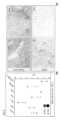
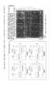
도 1은 대조 세포 샘플의 예시적인 IHC 분석을 보여준다. (A) 부모 HEK-293 세포의 음성 대조군 IHC 염색; (B) 약한 염색 강도를 갖는, 재조합 인간 PD-L1로 형질감염된 HEK-293 세포의 IHC 염색; (C) 중간 염색 강도를 갖는, 재조합 인간 PD-L1로 형질감염된 HEK-293 세포의 IHC 염색; (D) 강한 염색 강도를 갖는, 재조합 인간 PD-L1로 형질감염된 HEK-293 세포의 IHC 염색; (E) 태반 조직 샘플의 양성 조직 대조군 IHC 염색; (F) 편도 조직 샘플의 양성 조직 대조군 IHC 염색. 모든 IHC 염색은 등록된 항-PD-L1 항체를 사용하여 수행하였다.
도 2는 (A) 삼중-음성 유방암; (B) 악성 흑색종; (C) NSCLC, 선암종으로부터의 종양 샘플의 예시적인 PD-L1 양성 IHC 염색을 보여준다.
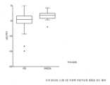
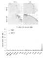
도 3은 종양 침윤 면역 세포에서의 PD-L1 발현과 암 환자에서의 항-PD-L1 치료에 대한 PD 또는 PR/CR 반응의 상관관계를 보여준다. PD = 진행성 질환; PR = 부분 반응; CR = 완전 반응. (A) PD-L1 IHC 분석을 이용한, 종양 샘플 구역 내의 PD-L1 + 종양 침윤 면역 세포의 %. (B) PD-L1 IHC 분석을 이용한, 종양 샘플 내의 전체 면역 침윤물 내의 PD-L1+ IC의 %.
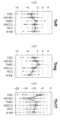
도 4는 PD-L1 qPCR 분석을 이용하여, 종양 세포에서의 PD-L1 유전자 발현과 암 환자에서의 항-PD-L1 치료에 대한 PD 또는 PR/CR 반응의 상관관계를 보여준다. PD = 진행성 질환; PR = 부분 반응; CR = 완전 반응.
도 5는 종양 샘플에서의 PD-1 유전자 발현과 암 환자에서의 항-PD-L1 치료에 대한 PD 또는 PR/CR 반응의 상관관계를 보여준다. PD = 진행성 질환; PR = 부분 반응; CR = 완전 반응.
도 6은 종양 샘플에서의 다양한 면역 유전자 발현과 암 환자에서의 항-PD-L1 치료에 대한 PD 또는 PR 반응의 상관관계를 보여준다. PD = 진행성 질환; PR = 부분 반응.
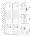
도 7은 항-PD-L1 항체를 사용하여 치료받은 환자로부터의 일련의 치료전/치료중 종양 생검의 개략도를 보여준다. 쌍을 이룬 기준선 (치료전 또는 보존된 종양 조직 포함), 및 흑색종, 신세포암종 (RCC), 비소세포 폐암 (NSCLC), 두경부암 (H&N), 결장직장암 (CRC), 위암, 및 유방암을 포함한 다양한 적응증을 앓고 있는, 항-PD-L1 항체를 사용하여 치료받은 환자 (n=26)로부터의 치료중 종양 생검을 평가하였다.
도 8은 (a) CD8+ T 세포 침윤의 증가가 항-PD-L1 항체를 사용하는 치료에 반응하는 환자로부터의 종양 샘플에서의 PD-L1 발현의 증가와 연관된다는 것; 및 (b) 항-PD-L1 항체를 사용하는 치료에 반응하는 환자에서, 항-PD-L1 항체를 사용하는 치료 후에 그랜자임 A, 퍼포린, IFN-g, TNFa 및 CD8을 포함하는 T 세포 활성화 마커가 증가한 것을 보여준다.
도 9는 항-PD-L1 항체 치료를 받고 있는 환자에서의 PD-L1 발현의 변화를 요약한다.
도 10은 항-PD-L1 항체를 사용하는 치료를 받고 있는 환자에서의, (a) 혈액 중의 증식 T-세포 (CD8+/Ki67+ 세포인 것으로 확인됨)의 빈도수 증가; 및 (b) 활성화된 증식 T-세포 (CD8+/HLA-DR+/Ki67+ 세포인 것으로 확인됨)의 빈도수 증가를 보여준다.
도 11은 혈장에서의 IL-6 수준의 감소가 항-PD-L1 항체 치료에 반응하는 환자와 연관되고, 혈장에서의 IL-6 수준의 증가가 항-PD-L1 항체 치료시에 질환이 진행된 환자와 연관된다는 것을 보여준다.
도 12는 종양 샘플에서의 다양한 면역 유전자 발현과 암 환자에서의 항-PD-L1 치료에 대한 PD 또는 PR/CR 반응의 상관관계를 보여준다. PD = 진행성 질환; PR = 부분 반응; CR = 완전 반응.
도 13은 흑색종 또는 NSCLC로부터의 종양 샘플에서의 IDO1 유전자 발현과 암 환자에서의 항-PD-L1 치료에 대한 PD 또는 PR/CR 반응의 상관관계를 보여준다. PD = 진행성 질환; PR = 부분 반응; CR = 완전 반응.
도 14는 항-PD-L1 항체를 사용하는 치료에 반응하는 환자로부터 수집된 혈액에서의, 순환 T 세포 상에서의 PD-L1 발현의 증가를 보여준다. PD = 진행성 질환; PR = 부분 반응; CR = 완전 반응.
도 15는 종양 샘플에서의 세포독성 Th1 세포, IFN-g 및 T-세포 트래픽킹 마커의 유전자 발현과 암 환자에서의 항-PD-L1 치료에 대한 PD 또는 PR/CR 반응의 상관관계를 보여준다. PD = 진행성 질환; PR = 부분 반응; CR = 완전 반응.
도 16은 항-PD-L1 항체를 사용하는 치료에 반응하는 흑색종 환자에서의 T 세포 활성화 마커의 증가를 보여준다.
도 17은 항-PD-L1 항체를 사용하는 치료에 반응하지 않는 흑색종 환자에서의, 종양내 T 세포의 낮은 빈도수 및 T 세포 활성화 마커에서의 T 세포 활성화의 결여를 보여준다.
도 18은 항-PD-L1 항체를 사용하는 치료에 반응하는 환자의 혈액에서의 CD8+/HLA-DR+/Ki-67+ 활성화된 T 세포의 빈도수의 일시적 증가를 보여준다.
도 19는 반응과 상관관계가 있는 CD4+/ICOS+ T 세포 집단에서 증가가 지연되고, 질환 진행 (사이클 3 이후에 발생)에 따라 감소되는, CD4+/ICOS+ T 세포의 변동을 보여준다.
도 20은 PD-L1 발현의 적절한 증가가 항-PD-L1 항체를 사용하는 치료에 반응하는 환자에서 현저하게 나타난다는 것을 보여준다.
도 21은 프랙탈카인/CX3CL1의 발현은 질환 진행과 상관관계가 있는 반면 CTLA4 발현은 항-PD-L1 항체를 사용하는 치료에 대한 반응과 상관관계가 있다는 것을 보여준다.
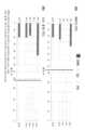
도 22는 6가지 암 적응증 사이에서의 Teff (T-이펙터) 세포, Treg (T-조절) 세포, 및 Th17 세포와 연관된 유전자 서명의 상관관계를 보여준다.
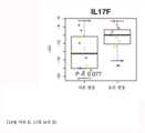
도 23은 항-PD-L1 치료에 반응하지 않는 환자에서의 IL17F의 보다 높은 종양 유전자 발현에 대한 경향을 보여준다. R = 반응자; nR = 비-반응자.
도 24는 IL-17F의 종양 유전자 발현이 항-PD-L1 치료에 대해 늦은 반응을 나타낸 환자에서 더 높다는 것을 보여준다.
도 25는 항-PD-L1 항체를 사용하는 치료를 받고 있는 환자에서의 순환 CD8+/HLA-DR+/Ki67+ 세포의 일시적 증가를 보여준다. (a) UBC 환자, (b) 모든 환자.
도 26은 항-PD-L1 항체를 사용하는 치료를 받고 있는 환자에서의 혈장 IL-18의 일시적 증가를 보여준다. 또한, 기준선 혈장 MCP-1은 항-PD-L1 치료에 대해 부분 반응/완전 반응 (PR/CR)을 나타내는 환자에서 더 낮았다. IL-18 및 MCP-1은 또한 단핵구에서 우세하게 발현되었다.
도 27은 항-PD-L1 항체를 사용하는 치료 후에 질환이 진행된 환자로부터의 치료전 종양이 골수 세포 (예를 들어, 단핵구, 수지상 세포)에서 우세하게 발현되는, 비교적 높은 골수 유전자 서명 (IL-8, CCL2, 및 IL1B)을 나타내었다는 것을 보여준다.
도 28은 항-PD-L1 항체를 사용하는 치료에 반응하는 환자의 혈액에서의 가용성 PD-L1의 상관관계를 보여준다.
도 29는 종양 침윤 면역 세포 (IC)에서의 PD-L1 발현과 항-PD-L1 치료에 대한 반응 사이의 연관성을 보여준다. (a) NSCLC, (b) 모든 종양.
도 30은 종양 세포에서의 PD-L1 발현과 항-PD-L1 치료에 대한 반응 사이의 연관성을 보여준다. (a) NSCLC, (b) 모든 종양.A patent or application file contains one or more drawings made in color. Copies of this patent or patent application publication with color drawing(s) will be provided by the Office upon request and payment of the necessary fee.
1 shows an exemplary IHC analysis of control cell samples. (A) Negative control IHC staining of parental HEK-293 cells; (B) IHC staining of HEK-293 cells transfected with recombinant human PD-L1 with weak staining intensity; (C) IHC staining of HEK-293 cells transfected with recombinant human PD-L1 with moderate staining intensity; (D) IHC staining of HEK-293 cells transfected with recombinant human PD-L1 with strong staining intensity; (E) Positive tissue control IHC staining of placental tissue samples; (F) Positive tissue control IHC staining of tonsil tissue samples. All IHC staining was performed using a registered anti-PD-L1 antibody.
2 shows (A) triple-negative breast cancer; (B) malignant melanoma; (C) Shows exemplary PD-L1 positive IHC staining of a tumor sample from NSCLC, adenocarcinoma.
Figure 3 shows the correlation between PD-L1 expression in tumor infiltrating immune cells and PD or PR/CR response to anti-PD-L1 treatment in cancer patients. PD = progressive disease; PR = partial response; CR = complete response. (A) Percentage of PD-L1 + tumor infiltrating immune cells in the tumor sample area using PD-L1 IHC assay. (B) % of PD-L1+ IC in total immune infiltrates in tumor samples using PD-L1 IHC assay.
Figure 4 shows the correlation of PD-L1 gene expression in tumor cells with PD or PR/CR response to anti-PD-L1 treatment in cancer patients, using PD-L1 qPCR assay. PD = progressive disease; PR = partial response; CR = complete response.
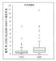
5 shows the correlation of PD-1 gene expression in tumor samples with PD or PR/CR response to anti-PD-L1 treatment in cancer patients. PD = progressive disease; PR = partial response; CR = complete response.
Figure 6 shows the correlation of various immune gene expression in tumor samples with PD or PR response to anti-PD-L1 treatment in cancer patients. PD = progressive disease; PR = partial response.
7 shows a schematic of a series of pre/on-treatment tumor biopsies from patients treated with anti-PD-L1 antibodies. Paired baselines (including pretreatment or preserved tumor tissue), and melanoma, renal cell carcinoma (RCC), non-small cell lung cancer (NSCLC), head and neck cancer (H&N), colorectal cancer (CRC), gastric cancer, and breast cancer were analyzed. On-treatment tumor biopsies from patients (n=26) treated with anti-PD-L1 antibodies were evaluated for a variety of indications including.
Figure 8 shows (a) increased CD8+ T cell infiltration is associated with increased PD-L1 expression in tumor samples from patients responding to treatment with an anti-PD-L1 antibody; and (b) in patients responding to treatment with an anti-PD-L1 antibody, T cells comprising granzyme A, perforin, IFN-g, TNFa and CD8 after treatment with an anti-PD-L1 antibody. It shows an increase in activation markers.
9 summarizes changes in PD-L1 expression in patients receiving anti-PD-L1 antibody treatment.
Figure 10 shows (a) increased frequency of proliferating T-cells (identified as CD8+/Ki67+ cells) in the blood in patients receiving treatment with an anti-PD-L1 antibody; and (b) an increase in the frequency of activated proliferative T-cells (confirmed to be CD8+/HLA-DR+/Ki67+ cells).
11 shows that a decrease in plasma IL-6 levels is associated with patients responding to anti-PD-L1 antibody treatment, and an increase in plasma IL-6 levels is associated with disease progression upon anti-PD-L1 antibody treatment. show that it is related to the patient.
12 shows the correlation of various immune gene expression in tumor samples with PD or PR/CR response to anti-PD-L1 treatment in cancer patients. PD = progressive disease; PR = partial response; CR = complete response.
13 shows the correlation of IDO1 gene expression in tumor samples from melanoma or NSCLC with PD or PR/CR response to anti-PD-L1 treatment in cancer patients. PD = progressive disease; PR = partial response; CR = complete response.
14 shows an increase in PD-L1 expression on circulating T cells in blood collected from patients responding to treatment with anti-PD-L1 antibodies. PD = progressive disease; PR = partial response; CR = complete response.
15 shows the correlation of gene expression of cytotoxic Th1 cells, IFN-g and T-cell trafficking markers in tumor samples with PD or PR/CR response to anti-PD-L1 treatment in cancer patients. PD = progressive disease; PR = partial response; CR = complete response.
16 shows an increase in T cell activation markers in melanoma patients responding to treatment with anti-PD-L1 antibodies.
17 shows low frequency of intratumoral T cells and lack of T cell activation markers in melanoma patients who do not respond to treatment with anti-PD-L1 antibodies.
18 shows a transient increase in the frequency of CD8+/HLA-DR+/Ki-67+ activated T cells in the blood of patients in response to treatment with anti-PD-L1 antibodies.
Figure 19 shows the variability of CD4+/ICOS+ T cells, with delayed increases in CD4+/ICOS+ T cell populations that correlated with response, and decreased with disease progression (occurring after Cycle 3).
20 shows that moderate increases in PD-L1 expression are markedly seen in patients responding to treatment with anti-PD-L1 antibodies.
21 shows that expression of fractalkine/CX3CL1 correlates with disease progression while CTLA4 expression correlates with response to treatment with an anti-PD-L1 antibody.
22 shows the correlation of gene signatures associated with Teff (T-effector) cells, Treg (T-regulatory) cells, and Th17 cells among six cancer indications.
23 shows a trend toward higher tumor gene expression of IL17F in patients who do not respond to anti-PD-L1 treatment. R = responder; nR = non-responder.
24 shows that oncogene expression of IL- 17F is higher in patients with a late response to anti-PD-L1 treatment.
25 shows a transient increase in circulating CD8+/HLA-DR+/Ki67+ cells in patients receiving treatment with anti-PD-L1 antibodies. (a) UBC patients, (b) all patients.
26 shows a transient increase in plasma IL-18 in patients receiving treatment with an anti-PD-L1 antibody. In addition, baseline plasma MCP-1 was lower in patients with partial response/complete response (PR/CR) to anti-PD-L1 treatment. IL-18 and MCP-1 were also predominantly expressed in monocytes.
27 shows a relatively high myeloid gene signature (IL- 8, CCL2, and IL1B).
28 shows the correlation of soluble PD-L1 in the blood of patients responding to treatment with anti-PD-L1 antibodies.
29 shows the association between PD-L1 expression in tumor infiltrating immune cells (IC) and response to anti-PD-L1 treatment. (a) NSCLC, (b) all tumors.
30 shows the association between PD-L1 expression in tumor cells and response to anti-PD-L1 treatment. (a) NSCLC, (b) all tumors.
정의Justice
용어 "PD-L1 축 결합 길항제"는 PD-L1 신호전달 축 상의 신호전달로부터 발생한 T-세포 기능이상을 제거하여 T-세포 기능을 복원 또는 증진시키는 결과를 나타내도록, PD-L1 축 결합 파트너와 하나 이상의 그의 결합 파트너의 상호작용을 억제하는 분자이다. 본원에 사용된 PD-L1 축 결합 길항제는 PD-L1 결합 길항제 및 PD-1 결합 길항제 뿐만 아니라 PD-L1 및 PD-1 사이의 상호작용 (예를 들어, PD-L2-Fc 융합)을 방해하는 분자를 포함한다.The term “PD-L1 axis binding antagonist” refers to a combination of a PD-L1 axis binding partner and a PD-L1 axis binding antagonist to denote a result of restoring or enhancing T-cell function by eliminating T-cell dysfunction resulting from signaling on the PD-L1 signaling axis. A molecule that inhibits the interaction of one or more of its binding partners. As used herein, a PD-L1 axis binding antagonist is one that interferes with a PD-L1 binding antagonist and a PD-1 binding antagonist as well as an interaction between PD-L1 and PD-1 (eg, a PD-L2-Fc fusion). contains molecules.
용어 "PD-L1 결합 길항제"는 PD-L1과 그의 결합 파트너, 예컨대 PD-1, B7-1 중 하나 이상의 상호작용으로부터 발생한 신호 전달을 감소, 차단, 억제, 제거 또는 방해하는 분자이다. 일부 실시양태에서, PD-L1 결합 길항제는 PD-L1의 그의 결합 파트너에 대한 결합을 억제하는 분자이다. 구체적 측면에서, PD-L1 결합 길항제는 PD-L1의 PD-1 및/또는 B7-1에 대한 결합을 억제한다. 일부 실시양태에서, PD-L1 결합 길항제는 항-PD-L1 항체, 그의 항원 결합 단편, 이뮤노어드헤신, 융합 단백질, 올리고펩티드, 및 PD-L1과 그의 결합 파트너, 예컨대 PD-1, B7-1 중 하나 이상의 상호작용으로부터 발생한 신호 전달을 감소, 차단, 억제, 제거 또는 방해하는 다른 분자를 포함한다. 한 실시양태에서, PD-L1 결합 길항제는 기능이상 T-세포가 보다 덜 비-기능이상적이 되도록, PD-L1 또는 PD-1을 통한 T 림프구 - 및 다른 세포 - 매개된 신호전달시에 발현되는 세포 표면 단백질에 의해 매개되거나 또는 이를 통한 음성 신호를 감소시킨다.The term “PD-L1 binding antagonist” is a molecule that reduces, blocks, inhibits, eliminates or interferes with signal transduction resulting from the interaction of PD-L1 with one or more of its binding partners, such as PD-1, B7-1. In some embodiments, a PD-L1 binding antagonist is a molecule that inhibits the binding of PD-L1 to its binding partner. In a specific aspect, the PD-L1 binding antagonist inhibits the binding of PD-L1 to PD-1 and/or B7-1. In some embodiments, the PD-L1 binding antagonist is an anti-PD-L1 antibody, antigen-binding fragment thereof, immunoadhesin, fusion protein, oligopeptide, and PD-L1 and its binding partners, such as PD-1, B7- and other molecules that reduce, block, inhibit, eliminate or interfere with signal transduction resulting from the interaction of one or more of 1. In one embodiment, the PD-L1 binding antagonist is expressed upon PD-L1 or T lymphocyte- and other cell-mediated signaling through PD-1 such that the dysfunctional T-cell becomes less non-dysfunctional. Reduce negative signals mediated by or through cell surface proteins.
용어 "PD-1 결합 길항제"는 PD-1과 그의 결합 파트너, 예컨대 PD-L1, PD-L2 중 하나 이상의 상호작용으로부터 발생한 신호 전달을 감소, 차단, 억제, 제거 또는 방해하는 분자이다. 일부 실시양태에서, PD-1 결합 길항제는 PD-1의 그의 결합 파트너에 대한 결합을 억제하는 분자이다. 구체적 측면에서, PD-1 결합 길항제는 PD-1의 PD-L1 및/또는 PD-L2에 대한 결합을 억제한다. 예를 들어, PD-1 결합 길항제는 항-PD-1 항체, 그의 항원 결합 단편, 이뮤노어드헤신, 융합 단백질, 올리고펩티드, 및 PD-1과 PD-L1 및/또는 PD-L2의 상호작용으로부터 발생한 신호 전달을 감소, 차단, 억제, 제거 또는 방해하는 다른 분자를 포함한다. 한 실시양태에서, PD-1 결합 길항제는 기능이상 T-세포가 보다 덜 비-기능이상적이 되도록, PD-1 또는 PD-L1을 통한 T 림프구 - 및 다른 세포 - 매개된 신호전달시에 발현되는 세포 표면 단백질에 의해 매개되거나 또는 이를 통한 음성 신호를 감소시킨다.The term “PD-1 binding antagonist” is a molecule that reduces, blocks, inhibits, eliminates or interferes with signal transduction resulting from the interaction of PD-1 with one or more of its binding partners, such as PD-L1, PD-L2. In some embodiments, a PD-1 binding antagonist is a molecule that inhibits the binding of PD-1 to its binding partner. In a specific aspect, a PD-1 binding antagonist inhibits the binding of PD-1 to PD-L1 and/or PD-L2. For example, PD-1 binding antagonists include anti-PD-1 antibodies, antigen-binding fragments thereof, immunoadhesins, fusion proteins, oligopeptides, and interactions of PD-1 with PD-L1 and/or PD-L2. and other molecules that reduce, block, inhibit, eliminate or interfere with signal transduction resulting from In one embodiment, the PD-1 binding antagonist is expressed upon T lymphocyte- and other cell-mediated signaling through PD-1 or PD-L1 such that the dysfunctional T-cell becomes less non-dysfunctional. Reduce negative signals mediated by or through cell surface proteins.
용어 "예정된 사멸 리간드 1" 및 "PD-L1"은 본원에서 천연 서열 PD-L1 폴리펩티드, 폴리펩티드 변이체 및 천연 서열 폴리펩티드 및 폴리펩티드 변이체의 단편 (본원에서 추가로 정의됨)을 지칭한다. 본원에 기재된 PD-L1 폴리펩티드는 다양한 공급원으로부터, 예컨대 인간 조직 유형으로부터 또는 또 다른 공급원으로부터 단리되거나, 또는 재조합 또는 합성 방법에 의해 제조된 것일 수 있다.The terms “programmed
"천연 서열 PD-L1 폴리펩티드"는 자연으로부터 유래된 상응하는 PD-L1 폴리펩티드와 동일한 아미노산 서열을 갖는 폴리펩티드를 포함한다.A "native sequence PD-L1 polypeptide" includes a polypeptide having the same amino acid sequence as a corresponding PD-L1 polypeptide derived from nature.
"PD-L1 폴리펩티드 변이체" 또는 그의 변이체는 본원에 개시된 바와 같은 임의의 천연 서열 PD-L1 폴리펩티드 서열과 적어도 약 80%의 아미노산 서열 동일성을 갖는 본원에 정의된 바와 같은 PD-L1 폴리펩티드, 일반적으로 활성 PD-L1 폴리펩티드를 의미한다. 이러한 PD-L1 폴리펩티드 변이체는 예를 들어 1개 이상의 아미노산 잔기가 천연 아미노산 서열의 N- 또는 C-말단에서 첨가 또는 결실된 PD-L1 폴리펩티드를 포함한다. 통상적으로, PD-L1 폴리펩티드 변이체는 본원에 개시된 바와 같은 천연 서열 PD-L1 폴리펩티드 서열에 대해 적어도 약 80% 아미노산 서열 동일성, 대안적으로 적어도 약 81%, 82%, 83%, 84%, 85%, 86%, 87%, 88%, 89%, 90%, 91%, 92%, 93%, 94%, 95%, 96%, 97%, 98% 또는 99% 아미노산 서열 동일성을 가질 것이다. 통상적으로, PD-L1 변이체 폴리펩티드는 길이가 적어도 약 10개 아미노산, 대안적으로 길이가 적어도 약 20, 30, 40, 50, 60, 70, 80, 90, 100, 110, 120, 130, 140, 150, 160, 170, 180, 190, 200, 210, 220, 230, 240, 250, 260, 270, 280, 281, 282, 283, 284, 285, 286, 287, 288, 289개 아미노산 또는 그 초과일 것이다. 임의로, PD-L1 변이체 폴리펩티드는 천연 PD-L1 폴리펩티드 서열에 비해 1개 이하의 보존적 아미노산 치환, 대안적으로 천연 PD-L1 폴리펩티드 서열에 비해 2, 3, 4, 5, 6, 7, 8, 9 또는 10개 이하의 보존적 아미노산 치환을 가질 것이다.A “PD-L1 polypeptide variant” or variant thereof is a PD-L1 polypeptide as defined herein having at least about 80% amino acid sequence identity to any native sequence PD-L1 polypeptide sequence as disclosed herein, generally active PD-L1 polypeptide. Such PD-L1 polypeptide variants include, for example, PD-L1 polypeptides in which one or more amino acid residues are added or deleted at the N- or C-terminus of the native amino acid sequence. Typically, PD-L1 polypeptide variants have at least about 80% amino acid sequence identity, alternatively at least about 81%, 82%, 83%, 84%, 85% amino acid sequence identity to a native sequence PD-L1 polypeptide sequence as disclosed herein. , 86%, 87%, 88%, 89%, 90%, 91%, 92%, 93%, 94%, 95%, 96%, 97%, 98% or 99% amino acid sequence identity. Typically, the PD-L1 variant polypeptide is at least about 10 amino acids in length, alternatively at least about 20, 30, 40, 50, 60, 70, 80, 90, 100, 110, 120, 130, 140, 150, 160, 170, 180, 190, 200, 210, 220, 230, 240, 250, 260, 270, 280, 281, 282, 283, 284, 285, 286, 287, 288, 289 amino acids or more would. Optionally, the PD-L1 variant polypeptide has no more than one conservative amino acid substitution relative to the native PD-L1 polypeptide sequence, alternatively no more than 2, 3, 4, 5, 6, 7, 8, It will have no more than 9 or 10 conservative amino acid substitutions.
본원에 정의된 용어 "PD-L1 길항제"는 천연 서열 PD-L1에 의해 매개되는 생물학적 활성 및/또는 기능을 부분적으로 또는 완전히 차단, 억제 또는 중화하는 임의의 분자이다. 특정 실시양태에서 이러한 길항제는 PD-L1에 결합한다. 한 실시양태에 따르면, 길항제는 폴리펩티드이다. 또 다른 실시양태에 따르면, 길항제는 항-PD-L1 항체이다. 또 다른 실시양태에 따르면, 길항제는 소분자 길항제이다. 또 다른 실시양태에 따르면, 길항제는 폴리뉴클레오티드 길항제이다.As defined herein, the term “PD-L1 antagonist” is any molecule that partially or completely blocks, inhibits or neutralizes a biological activity and/or function mediated by native sequence PD-L1. In certain embodiments, such antagonists bind PD-L1. According to one embodiment, the antagonist is a polypeptide. According to another embodiment, the antagonist is an anti-PD-L1 antibody. According to another embodiment, the antagonist is a small molecule antagonist. According to another embodiment, the antagonist is a polynucleotide antagonist.
"폴리뉴클레오티드" 또는 "핵산"은 본원에서 교환가능하게 사용되며, 임의의 길이의 뉴클레오티드의 중합체를 지칭하고, DNA 및 RNA를 포함한다. 뉴클레오티드는 데옥시리보뉴클레오티드, 리보뉴클레오티드, 변형 뉴클레오티드 또는 염기 및/또는 그의 유사체, 또는 DNA 또는 RNA 폴리머라제에 의해, 또는 합성 반응에 의해 중합체 내로 혼입될 수 있는 임의의 기질일 수 있다. 폴리뉴클레오티드는 변형된 뉴클레오티드, 예컨대 메틸화 뉴클레오티드 및 그의 유사체를 포함할 수 있다. 뉴클레오티드 구조에 대한 변형은 존재하는 경우에 중합체의 어셈블리 이전 또는 이후에 부여될 수 있다. 뉴클레오티드의 서열에 비-뉴클레오티드 성분이 개재될 수 있다. 폴리뉴클레오티드는 합성 후에 예컨대 표지와의 접합에 의해 추가로 변형될 수 있다. 다른 유형의 변형은 예를 들어 하나 이상의 자연 발생 뉴클레오티드의 유사체로의 "캡" 치환, 뉴클레오티드간 변형, 예컨대 예를 들어 비하전된 연결부 (예를 들어, 메틸 포스포네이트, 포스포트리에스테르, 포스포아미데이트, 카르바메이트 등) 및 하전된 연결부 (예를 들어, 포스포로티오에이트, 포스포로디티오에이트 등)를 갖는 것, 펜던트 모이어티, 예컨대 예를 들어 단백질 (예를 들어, 뉴클레아제, 독소, 항체, 신호 펩티드, 폴리-L-리신 등)을 함유하는 것, 삽입제 (예를 들어, 아크리딘, 프소랄렌 등)을 갖는 것, 킬레이트화제 (예를 들어, 금속, 방사성 금속, 붕소, 산화 금속 등)을 함유하는 것, 알킬화제를 함유하는 것, 변형 연결부 (예를 들어, 알파 아노머 핵산 등)를 갖는 것 뿐만 아니라, 폴리뉴클레오티드(들)의 비변형된 형태를 포함한다. 추가로, 당 내에 통상적으로 존재하는 임의의 히드록실 기가 예를 들어 포스포네이트 기, 포스페이트 기로 대체되거나, 표준 보호 기로 보호되거나, 추가의 뉴클레오티드에 대한 추가의 연결이 만들어지도록 활성화되거나, 또는 고체 또는 반고체 지지체에 접합될 수 있다. 5' 및 3' 말단 OH가 인산화되거나 또는 아민 또는 1 내지 20개의 탄소 원자의 유기 캡핑 기 모이어티로 치환될 수 있다. 다른 히드록실이 또한 표준 보호기로 유도체화될 수 있다. 폴리뉴클레오티드는 또한 일반적으로 관련 기술분야에 공지된 리보스 또는 데옥시리보스 당의 유사형, 예를 들어 2'-O-메틸-, 2'-O-알릴, 2'-플루오로- 또는 2'-아지도-리보스, 카르보시클릭 당 유사체, α-아노머 당, 에피머 당, 예컨대 아라비노스, 크실로스 또는 릭소스, 피라노스 당, 푸라노스 당, 세도헵툴로스, 비-시클릭 유사체 및 무염기 뉴클레오시드 유사체, 예컨대 메틸 리보시드를 함유할 수 있다. 1개 이상의 포스포디에스테르 연결이 대안적인 연결 기로 대체될 수 있다. 이들 대안적인 연결 기는 포스페이트가 P(O)S ("티오에이트"), P(S)S ("디티오에이트"), "(O)NR2 ("아미데이트"), P(O)R, P(O)OR', CO 또는 CH2 ("포름아세탈") (여기서, 각각의 R 또는 R'는 독립적으로 H이거나 또는 임의로 에테르 (-O-) 연결부를 함유하는 치환 또는 비치환 알킬 (1-20 C), 아릴, 알케닐, 시클로알킬, 시클로알케닐 또는 아랄딜임)로 대체되는 실시양태를 포함하지만 이에 제한되지는 않는다. 폴리뉴클레오티드 내의 모든 연결이 동일할 필요는 없다. 상기 기재는 RNA 및 DNA를 비롯하여 본원에서 언급되는 모든 폴리뉴클레오티드에 적용된다.“Polynucleotide” or “nucleic acid” are used interchangeably herein and refer to a polymer of nucleotides of any length, and includes DNA and RNA. Nucleotides can be deoxyribonucleotides, ribonucleotides, modified nucleotides or bases and/or their analogues, or any substrate that can be incorporated into a polymer by DNA or RNA polymerase or by a synthetic reaction. Polynucleotides can include modified nucleotides, such as methylated nucleotides and their analogs. Modifications to the nucleotide structure, if present, may be imparted prior to or after assembly of the polymer. The sequence of nucleotides may be interrupted by non-nucleotide components. A polynucleotide may be further modified after synthesis, such as by conjugation with a label. Other types of modifications include, for example, “cap” substitutions of one or more naturally occurring nucleotides with analogs, internucleotide modifications such as, for example, uncharged linkages (e.g., methyl phosphonates, phosphotriesters, phosphotriesters, amidates, carbamates, etc.) and those with charged linkages (e.g., phosphorothioates, phosphorodithioates, etc.), pendant moieties such as, for example, proteins (e.g., nucleases). , toxins, antibodies, signal peptides, poly-L-lysine, etc.), those with intercalating agents (eg, acridine, psoralen, etc.), chelating agents (eg, metals, radioactive metals, etc.) , boron, metal oxides, etc.), those containing alkylating agents, those with modified linkages (eg, alpha anomeric nucleic acids, etc.), as well as unmodified forms of the polynucleotide(s). . Additionally, any hydroxyl groups normally present in sugars are replaced, for example, with phosphonate groups, phosphate groups, protected with standard protecting groups, activated to make additional linkages to additional nucleotides, or solid or It can be bonded to a semi-solid support. The 5' and 3' terminal OH may be phosphorylated or substituted with amines or organic capping group moieties of 1 to 20 carbon atoms. Other hydroxyls can also be derivatized with standard protecting groups. Polynucleotides also generally contain analogs of ribose or deoxyribose sugars known in the art, such as 2'-O-methyl-, 2'-O-allyl, 2'-fluoro- or 2'-azi do-ribose, analogs of carbocyclic sugars, α-anomeric sugars, epimeric sugars such as arabinose, xylose or lyxose, pyranose sugars, furanose sugars, sedoheptulose, non-cyclic analogs and bases It may contain nucleoside analogs such as methyl riboside. One or more phosphodiester linkages may be replaced with alternative linking groups. These alternative linking groups are phosphates P(O)S ("thioate"), P(S)S ("dithioate"), "(O)NR2 ("amidate"), P(O)R , P(O)OR', CO or CH2 ("formacetal"), wherein each R or R' is independently H or a substituted or unsubstituted alkyl, optionally containing an ether (-O-) linkage ( 1-20 C), aryl, alkenyl, cycloalkyl, cycloalkenyl or araldyl). Not all linkages in a polynucleotide need to be identical. This applies to all polynucleotides referred to herein, including RNA and DNA.
본원에 사용된 "올리고뉴클레오티드"는 일반적으로 (반드시는 아니지만) 길이가 약 250개 미만의 뉴클레오티드인 짧은 단일-가닥 폴리뉴클레오티드를 지칭한다. 올리고뉴클레오티드는 합성될 수 있다. 용어 "올리고뉴클레오티드" 및 "폴리뉴클레오티드"는 상호 배타적이지 않다. 폴리뉴클레오티드에 대한 상기 기재는 올리고뉴클레오티드에 동일하고 완전하게 적용가능하다.As used herein, “oligonucleotide” refers to short single-stranded polynucleotides that are generally (but not necessarily) less than about 250 nucleotides in length. Oligonucleotides can be synthesized. The terms "oligonucleotide" and "polynucleotide" are not mutually exclusive. The above description of polynucleotides is equally and fully applicable to oligonucleotides.
용어 "프라이머"는 핵산에 혼성화하고, 일반적으로 유리 3'-OH 기를 제공함으로써 상보성 핵산의 중합을 허용할 수 있는 단일 가닥 폴리뉴클레오티드를 의미한다.The term "primer" refers to a single-stranded polynucleotide capable of hybridizing to a nucleic acid and allowing polymerization of complementary nucleic acids, usually by providing a free 3'-OH group.
용어 "소분자"는 약 2000 달톤 이하, 바람직하게는 약 500 달톤 이하의 분자량을 갖는 임의의 분자를 지칭한다.The term “small molecule” refers to any molecule having a molecular weight of about 2000 Daltons or less, preferably about 500 Daltons or less.
용어 "숙주 세포", "숙주 세포주" 및 "숙주 세포 배양물"은 교환가능하게 사용되고, 외인성 핵산이 도입된 세포 (이러한 세포의 자손 포함)를 지칭한다. 숙주 세포는 "형질전환체" 및 "형질전환된 세포"를 포함하며, 이는 1차 형질전환된 세포 및 계대배양 횟수와 관계없이 그로부터 유래된 자손을 포함한다. 자손은 모 세포와 핵산 함량이 완전히 동일하지 않을 수 있으나, 돌연변이를 함유할 수 있다. 본래 형질전환된 세포에 대해 스크리닝 또는 선택되는 동일한 기능 또는 생물학적 활성을 갖는 돌연변이체 자손이 본원에 포함된다.The terms "host cell", "host cell line" and "host cell culture" are used interchangeably and refer to cells (including progeny of such cells) into which an exogenous nucleic acid has been introduced. Host cells include "transformants" and "transformed cells," which include the primary transformed cell and progeny derived therefrom without regard to the number of passages. The progeny may not be completely identical in nucleic acid content to the parent cell, but may contain mutations. Mutant progeny that have the same function or biological activity that was screened for or selected for in the originally transformed cell are included herein.
본원에 사용된 용어 "벡터"는 그 벡터가 연결된 또 다른 핵산을 증식시킬 수 있는 핵산 분자를 지칭한다. 상기 용어는 자기-복제 핵산 구조로서의 벡터 뿐만 아니라 벡터가 그 내부로 도입된 숙주 세포의 게놈 내로 통합되는 벡터를 포함한다. 특정 벡터는 그 벡터가 작동가능하게 연결된 핵산의 발현을 지시할 수 있다. 이러한 벡터는 본원에서 "발현 벡터"로 지칭된다.As used herein, the term “vector” refers to a nucleic acid molecule capable of propagating another nucleic acid to which it has been linked. The term includes the vector as a self-replicating nucleic acid structure as well as the vector integrated into the genome of a host cell into which it has been introduced. Certain vectors are capable of directing the expression of nucleic acids to which they are operably linked. Such vectors are referred to herein as "expression vectors".
"단리된" 항체는 그의 자연 환경의 성분에서 분리된 것이다. 일부 실시양태에서, 항체는 예를 들어 전기영동 (예를 들어, SDS-PAGE, 등전 포커싱 (IEF), 모세관 전기영동) 또는 크로마토그래피 (예를 들어, 이온 교환 또는 역상 HPLC)에 의해 결정된 바와 같이 95% 또는 99% 초과의 순도로 정제된다. 항체 순도의 평가 방법의 검토를 위해, 예를 들어 문헌 [Flatman et al., J. Chromatogr. B 848:79-87 (2007)]을 참조한다.An "isolated" antibody is one that has been separated from a component of its natural environment. In some embodiments, an antibody is determined by electrophoresis (eg, SDS-PAGE, isoelectric focusing (IEF), capillary electrophoresis) or chromatography (eg, ion exchange or reverse phase HPLC). It is purified to greater than 95% or 99% purity. For a review of methods for assessing antibody purity, see, eg, Flatman et al., J. Chromatogr. B 848:79-87 (2007).
"단리된" 핵산은 그의 자연 환경의 성분으로부터 분리된 핵산 분자를 지칭한다. 단리된 핵산은 핵산 분자를 통상적으로 함유하는 세포에 함유되는 핵산 분자를 포함하지만, 핵산 분자는 염색체 외에 또는 그의 천연 염색체 위치와 상이한 염색체 위치에 존재한다.An "isolated" nucleic acid refers to a nucleic acid molecule that has been separated from a component of its natural environment. An isolated nucleic acid includes a nucleic acid molecule contained in cells that ordinarily contain the nucleic acid molecule, but the nucleic acid molecule is present extrachromosomally or at a chromosomal location different from its natural chromosomal location.
본원에서 용어 "항체"는 가장 넓은 의미로 사용되고, 모노클로날 항체, 폴리클로날 항체, 다중특이적 항체 (예를 들어, 이중특이적 항체), 및 원하는 항원-결합 활성을 나타내는 한 항체 단편을 포함하나 이에 제한되지는 않는 다양한 항체 구조를 포함한다.As used herein, the term "antibody" is used in the broadest sense and includes monoclonal antibodies, polyclonal antibodies, multispecific antibodies (eg, bispecific antibodies), and antibody fragments so long as they exhibit the desired antigen-binding activity. A variety of antibody structures, including but not limited to, are included.
용어 "항-PD-L1 항체" 및 "PD-L1에 결합하는 항체"는 항체가 PD-L1의 표적화에서 진단제 및/또는 치료제로서 유용하도록 충분한 친화도로 PD-L1에 결합할 수 있는 항체를 지칭한다. 한 실시양태에서, 항-PD-L1 항체의 비관련, 비-PD-L1 단백질에 대한 결합의 정도는 예를 들어 방사성면역검정 (RIA)에 의해 측정시에 항체의 PD-L1에 대한 결합의 약 10% 미만이다. 특정 실시양태에서, 항-PD-L1 항체는 상이한 종으로부터의 PD-L1 사이에서 보존된 PD-L1의 에피토프에 결합한다.The terms “anti-PD-L1 antibody” and “antibody that binds PD-L1” refer to an antibody capable of binding PD-L1 with sufficient affinity such that the antibody is useful as a diagnostic and/or therapeutic agent in targeting PD-L1. refers to In one embodiment, the extent of binding of an anti-PD-L1 antibody to an unrelated, non-PD-L1 protein is the degree of binding of the antibody to PD-L1, e.g., as measured by a radioimmunoassay (RIA). less than about 10%. In certain embodiments, the anti-PD-L1 antibody binds to an epitope of PD-L1 that is conserved among PD-L1 from different species.
"차단" 항체 또는 "길항제" 항체는 그가 결합하는 항원의 생물학적 활성을 억제하거나 또는 감소시키는 항체이다. 바람직한 차단 항체 또는 길항제 항체는 항원의 생물학적 활성을 실질적으로 또는 완전히 억제한다.A “blocker” or “antagonist” antibody is an antibody that inhibits or reduces the biological activity of the antigen to which it binds. A preferred blocking antibody or antagonist antibody substantially or completely inhibits the biological activity of the antigen.
"친화도"는 분자 (예를 들어, 항체)의 단일 결합 부위와 그의 결합 파트너 (예를 들어, 항원) 사이의 비공유 상호작용의 총 합계의 강도를 지칭한다. 달리 나타내지 않는 한, 본원에 사용된 "결합 친화도"는 결합 쌍의 구성원들 (예를 들어, 항체 및 항원) 사이의 1:1 상호작용을 반영하는 내인성 결합 친화도를 지칭한다. 분자 X의 그의 파트너 Y에 대한 친화도는 일반적으로 해리 상수 (Kd)로 표시될 수 있다. 친화도는 본원에 기재된 것들을 비롯하여 관련 기술분야에 공지된 통상의 방법에 의해 측정될 수 있다. 결합 친화도 측정을 위한 구체적인 설명적 및 예시적 실시양태가 하기 기재된다."Affinity" refers to the strength of the total sum of non-covalent interactions between a single binding site of a molecule (eg antibody) and its binding partner (eg antigen). Unless otherwise indicated, “binding affinity” as used herein refers to endogenous binding affinity that reflects a 1:1 interaction between members of a binding pair (eg, antibody and antigen). The affinity of a molecule X for its partner Y can generally be expressed as the dissociation constant (Kd). Affinity can be measured by common methods known in the art, including those described herein. Specific illustrative and exemplary embodiments for measuring binding affinity are described below.
"친화도 성숙" 항체는 항원에 대한 항체의 친화도를 개선시키는 변경을 갖지 않는 모 항체와 비교하여 하나 이상의 초가변 영역 (HVR)에서 하나 이상의 변경을 갖는 항체를 지칭한다.An “affinity matured” antibody refers to an antibody that has one or more alterations in one or more hypervariable regions (HVRs) compared to a parent antibody that has no alterations that improve the affinity of the antibody for an antigen.
"항체 단편"은 무손상 항체가 결합하는 항원에 결합하는 무손상 항체의 일부를 포함하는, 무손상 항체 이외의 분자를 지칭한다. 항체 단편의 예는 Fv, Fab, Fab', Fab'-SH, F(ab')2; 디아바디; 선형 항체; 단일-쇄 항체 분자 (예를 들어, scFv); 및 항체 단편들로부터 형성된 다중특이적 항체를 포함하나 이에 제한되지는 않는다."Antibody fragment" refers to a molecule other than an intact antibody comprising a portion of an intact antibody that binds an antigen to which the intact antibody binds. Examples of antibody fragments include Fv, Fab, Fab', Fab'-SH, F(ab')2 ; diabodies; linear antibodies; single-chain antibody molecules (eg scFv); and multispecific antibodies formed from antibody fragments.
참조 항체와 "동일한 에피토프에 결합하는 항체"는 경쟁 검정에서 참조 항체의 그의 항원에 대한 결합을 50% 이상 차단하고, 반대로 경쟁 검정에서 참조 항체가 항체의 그의 항원에 대한 결합을 50% 이상 차단하는 항체를 지칭한다. 예시적인 경쟁 검정이 본원에 제공된다.An "antibody that binds to the same epitope" as a reference antibody blocks binding of the reference antibody to its antigen in a competition assay by 50% or more, and conversely, in a competition assay, the reference antibody blocks binding of the antibody to its antigen by 50% or more. refers to antibodies. Exemplary competition assays are provided herein.
용어 "키메라" 항체는 중쇄 및/또는 경쇄의 일부가 특정한 공급원 또는 종으로부터 유래된 반면, 중쇄 및/또는 경쇄의 나머지가 다른 공급원 또는 종으로부터 유래된 항체를 지칭한다.The term "chimeric" antibody refers to an antibody in which portions of the heavy and/or light chains are derived from a particular source or species, while the remainder of the heavy and/or light chains are from a different source or species.
항체의 "부류"는 그의 중쇄가 보유하는 불변 도메인 또는 불변 영역의 유형을 지칭한다. 5종의 주요 부류의 항체: IgA, IgD, IgE, IgG, 및 IgM이 존재하고, 이들 중 몇몇은 하위부류 (이소형), 예를 들어 IgG1, IgG2, IgG3, IgG4, IgA1, 및 IgA2로 추가로 분류될 수 있다. 상이한 부류의 이뮤노글로불린에 상응하는 중쇄 불변 도메인은 각각 α, δ, ε, γ 및 μ로 불린다.The "class" of an antibody refers to the type of constant domain or constant region possessed by its heavy chain. There are five major classes of antibodies: IgA, IgD, IgE, IgG, and IgM, several of which are subclasses (isotypes), e.g., IgG1 , IgG2 , IgG3 , IgG4 , IgA1 , and IgA2 . The heavy chain constant domains corresponding to the different classes of immunoglobulins are called α, δ, ε, γ and μ, respectively.
용어 "전장 항체", "무손상 항체" 및 "전체 항체"는 본원에서 교환가능하게 사용되며, 천연 항체 구조와 실질적으로 유사한 구조를 갖거나 또는 본원에 정의된 바와 같은 Fc 영역을 함유하는 중쇄를 갖는 항체를 지칭한다.The terms “full-length antibody,” “intact antibody” and “whole antibody” are used interchangeably herein and refer to heavy chains having a structure substantially similar to that of a native antibody or containing an Fc region as defined herein. refers to antibodies with
본원에 사용된 용어 "모노클로날 항체"는 실질적으로 동종인 항체 집단으로부터 수득된 항체를 지칭하도록 사용되고, 즉 이러한 집단을 구성하는 개별 항체는 일반적으로 소량으로 존재할 수도 있는, 예를 들어 자연 발생 돌연변이를 포함하거나 모노클로날 항체 제제의 생산 동안 생성되는 가능한 변이체 항체를 제외하고는 동일하고/하거나, 동일한 에피토프에 결합한다. 전형적으로 상이한 결정기 (에피토프)에 대해 지시된 상이한 항체를 포함하는 폴리클로날 항체 제제와는 달리, 모노클로날 항체 제제의 각각의 모노클로날 항체는 항원 상의 단일 결정기에 대해 지시된다. 따라서, 수식어 "모노클로날"은 항체의 실질적으로 동종인 집단으로부터 수득된 항체의 특성을 나타내고, 임의의 특정한 방법에 의한 항체 생산을 필요로 하는 것으로 간주되지 않아야 한다. 예를 들어, 본 발명에 따라 사용되는 모노클로날 항체는 하이브리도마 방법, 재조합 DNA 방법, 파지-디스플레이 방법, 및 인간 이뮤노글로불린 로커스의 전부 또는 일부를 함유하는 트랜스제닉 동물을 이용하는 방법을 포함하나 이에 제한되지는 않는 다양한 기술에 의해 제조할 수 있고, 상기 방법 및 모노클로날 항체를 제조하기 위한 다른 예시적인 방법이 본원에 기재된다.As used herein, the term "monoclonal antibody" is used to refer to an antibody obtained from a population of substantially homogeneous antibodies, i.e., the individual antibodies comprising such population generally may be present in minor amounts, e.g., naturally occurring mutations. are identical and/or bind to the same epitope, except for possibly variant antibodies that contain or are generated during production of the monoclonal antibody preparation. Unlike polyclonal antibody preparations, which typically include different antibodies directed against different determinants (epitopes), each monoclonal antibody of a monoclonal antibody preparation is directed against a single determinant on the antigen. Thus, the modifier “monoclonal” indicates the character of the antibody as being obtained from a substantially homogeneous population of antibodies, and is not to be construed as requiring production of the antibody by any particular method. For example, monoclonal antibodies used in accordance with the present invention include hybridoma methods, recombinant DNA methods, phage-display methods, and methods using transgenic animals that contain all or part of the human immunoglobulin locus. Can be made by a variety of techniques, including but not limited to, these methods and other exemplary methods for making monoclonal antibodies are described herein.
"인간 항체"는 인간 또는 인간 세포에 의해 생산된 항체의 아미노산 서열에 상응하는 아미노산 서열을 보유하거나, 또는 인간 항체 레퍼토리 또는 다른 인간 항체-코딩 서열을 이용하여 비-인간 공급원으로부터 유래된 항체이다. 인간 항체의 이러한 정의에서 비-인간 항원-결합 잔기를 포함하는 인간화 항체는 명확하게 배제된다.A “human antibody” is an antibody that possesses an amino acid sequence that corresponds to that of an antibody produced by a human or a human cell, or is derived from a non-human source using a human antibody repertoire or other human antibody-coding sequence. Humanized antibodies comprising non-human antigen-binding moieties are specifically excluded from this definition of a human antibody.
"인간화" 항체는 비-인간 HVR로부터의 아미노산 잔기 및 인간 FR로부터의 아미노산 잔기를 포함하는 키메라 항체를 지칭한다. 특정 실시양태에서, 인간화 항체는 실질적으로 1개 이상, 전형적으로 2개의 가변 도메인을 모두 포함할 것이며, 여기서 모든 또는 실질적으로 모든 HVR (예를 들어, CDR)은 비-인간 항체의 것에 상응하고, 모든 또는 실질적으로 모든 FR은 인간 항체의 것에 상응한다. 인간화 항체는 임의로 인간 항체로부터 유래된 항체 불변 영역의 적어도 일부를 포함할 수 있다. 항체, 예를 들어 비-인간 항체의 "인간화 형태"는 인간화를 거친 항체를 지칭한다.A “humanized” antibody refers to a chimeric antibody comprising amino acid residues from non-human HVRs and amino acid residues from human FRs. In certain embodiments, a humanized antibody will comprise substantially all of at least one, and typically both, variable domains, wherein all or substantially all of the HVRs (e.g., CDRs) correspond to those of a non-human antibody; All or substantially all FRs correspond to those of a human antibody. A humanized antibody may optionally comprise at least a portion of an antibody constant region derived from a human antibody. A “humanized form” of an antibody, eg, a non-human antibody, refers to an antibody that has undergone humanization.
"면역접합체"는 세포독성제를 포함하나 이에 제한되지는 않는 하나 이상의 이종 분자(들)에 접합된 항체이다.An "immunoconjugate" is an antibody conjugated to one or more heterologous molecule(s), including but not limited to a cytotoxic agent.
참조 폴리펩티드 서열에 대한 "아미노산 서열 동일성 퍼센트(%)"는 서열을 정렬시키고 필요한 경우에는 최대 서열 동일성 퍼센트 달성을 위해 갭을 도입한 후 임의의 보존적 치환을 서열 동일성의 일부로 간주하지 않으면서 참조 폴리펩티드 서열 내의 아미노산 잔기와 동일한 후보 서열 내의 아미노산 잔기의 백분율로서 정의된다. 아미노산 서열 동일성 퍼센트를 결정하기 위한 정렬은 관련 기술분야 기술 범위 내의 다양한 방법, 예를 들어 공개적으로 이용가능한 컴퓨터 소프트웨어, 예컨대 BLAST, BLAST-2, ALIGN 또는 메갈린(Megalign) (DNASTAR) 소프트웨어를 이용하여 달성할 수 있다. 통상의 기술자는 비교할 전장 서열에 대한 최대 정렬을 달성하는데 필요한 임의의 알고리즘을 포함하여 서열 정렬에 적절한 파라미터를 정할 수 있다. 그러나, 본원의 목적상, 아미노산 서열 동일성 % 값은 서열 비교 컴퓨터 프로그램 ALIGN-2를 이용하여 생성된다. ALIGN-2 서열 비교 컴퓨터 프로그램은 제넨테크, 인크.(Genentech, Inc.) 소유로서, 소스 코드는 미국 저작권청 (20559 워싱턴 디.씨.)에 사용자 문서로 제출되어 있고, 미국 저작권 등록 번호 TXU510087로 등록되어 있다. ALIGN-2 프로그램은 제넨테크, 인크. (캘리포니아주 사우스 샌프란시스코)를 통해 공개적으로 이용가능하거나, 소스 코드로부터 컴파일링될 수 있다. ALIGN-2 프로그램은 디지털 UNIX V4.0D를 비롯한 UNIX 운영 시스템에서 사용되도록 컴파일링되어야 한다. 모든 서열 비교 파라미터는 ALIGN-2 프로그램에 의해 설정되어 있으며 변하지 않는다."Percent (%) amino acid sequence identity" to a reference polypeptide sequence is the reference polypeptide without considering any conservative substitutions as part of the sequence identity after aligning the sequences and introducing gaps, if necessary, to achieve the maximum percent sequence identity. It is defined as the percentage of amino acid residues in a candidate sequence that are identical to amino acid residues in the sequence. Alignment to determine percent amino acid sequence identity can be performed by various methods within the skill in the art, for example using publicly available computer software such as BLAST, BLAST-2, ALIGN or Megalign (DNASTAR) software. can be achieved One skilled in the art can determine appropriate parameters for aligning sequences, including any algorithms necessary to achieve maximal alignment over the full-length sequences being compared. However, for purposes herein, % amino acid sequence identity values are generated using the sequence comparison computer program ALIGN-2. The ALIGN-2 sequence comparison computer program is owned by Genentech, Inc., the source code of which has been submitted as User Documentation to the United States Copyright Office (Washington, D.C. 20559), and is registered under United States Copyright Registration No. TXU510087. are registered The ALIGN-2 program was developed by Genentech, Inc. (South San Francisco, CA) or compiled from source code. The ALIGN-2 program must be compiled for use with UNIX operating systems, including Digital UNIX V4.0D. All sequence comparison parameters are set by the ALIGN-2 program and do not change.
ALIGN-2가 아미노산 서열 비교를 위해 사용되는 상황에서, 주어진 아미노산 서열 B에, 주어진 아미노산 서열 B와, 또는 주어진 아미노산 서열 B에 대한 주어진 아미노산 서열 A의 아미노산 서열 동일성 % (대안적으로, 주어진 아미노산 서열 B에, 주어진 아미노산 서열 B와, 또는 주어진 아미노산 서열 B에 대해 특정 아미노산 서열 동일성 %를 갖거나 또는 이를 포함하는 주어진 아미노산 서열 A라는 어구로 기재될 수 있음)는 하기와 같이 계산된다:In situations where ALIGN-2 is used for amino acid sequence comparison, % amino acid sequence identity of a given amino acid sequence A to a given amino acid sequence B, to a given amino acid sequence B, or to a given amino acid sequence B (alternatively, to a given amino acid sequence B). B, which may be phrased as a given amino acid sequence A that has or comprises a certain % amino acid sequence identity to, or to, a given amino acid sequence B) is calculated as follows:
X/Y의 분율 x 100Fraction of X/
여기서, X는 서열 정렬 프로그램 ALIGN-2에 의한 A 및 B의 프로그램 정렬시에 상기 프로그램에 의해 동일한 매치로 스코어링된 아미노산 잔기의 수이고, Y는 B의 아미노산 잔기의 전체 수이다. 아미노산 서열 A의 길이가 아미노산 서열 B의 길이와 동일하지 않은 경우에는 B에 대한 A의 아미노산 서열 동일성 %가 A에 대한 B의 아미노산 서열 동일성 %와 동일하지 않을 것임을 이해할 것이다. 달리 구체적으로 언급되지 않는 한, 본원에 사용된 모든 아미노산 서열 동일성 % 값은 ALIGN-2 컴퓨터 프로그램을 이용하여 상기 단락에 기재한 바와 같이 수득한다.Here, X is the number of amino acid residues scored as identical matches by the program alignment of A and B by the sequence alignment program ALIGN-2, and Y is the total number of amino acid residues of B. It will be appreciated that the % amino acid sequence identity of A to B will not equal the % amino acid sequence identity of B to A if the length of amino acid sequence A is not equal to the length of amino acid sequence B. Unless specifically stated otherwise, all % amino acid sequence identity values used herein are obtained as described in the preceding paragraph using the ALIGN-2 computer program.
용어 "검출"은 직접 및 간접 검출을 비롯한 임의의 검출 수단을 포함한다.The term “detection” includes any means of detection including direct and indirect detection.
본원에 사용된 용어 "바이오마커"은 샘플에서 검출될 수 있는 표지자, 예를 들어 예측, 진단 및/또는 예후를 지칭한다. 바이오마커는 특정의 분자, 병리학적적, 조직학적 및/또는 임상적 특징에 의해 특성화된 특정한 하위유형의 질환 또는 장애 (예를 들어, 암)의 표지자로서 역할을 할 수 있다. 일부 실시양태에서, 바이오마커는 유전자이다. 바이오마커는 폴리뉴클레오티드 (예를 들어, DNA 및/또는 RNA), 폴리뉴클레오티드 카피수 변경 (예를 들어, DNA 카피수), 폴리펩티드, 폴리펩티드 및 폴리뉴클레오티드 변형 (예를 들어, 번역후 변형), 탄수화물 및/또는 당지질-기재 분자 마커를 포함하나 이에 제한되지는 않는다.As used herein, the term "biomarker" refers to a marker that can be detected in a sample, eg, predictive, diagnostic, and/or prognostic. A biomarker can serve as a marker for a particular subtype of disease or disorder (eg, cancer) characterized by particular molecular, pathological, histological and/or clinical features. In some embodiments, a biomarker is a gene. Biomarkers include polynucleotides (eg, DNA and/or RNA), polynucleotide copy number alterations (eg, DNA copy number), polypeptides, polypeptide and polynucleotide modifications (eg, post-translational modifications), carbohydrates and/or glycolipid-based molecular markers.
용어 "바이오마커 서명", "서명", "바이오마커 발현 서명" 또는 "발현 서명"은 본원에서 교환가능하게 사용되고, 그의 발현이 표지자, 예를 들어 예측, 진단 및/또는 예후인 바이오마커 중 하나 또는 그의 조합을 지칭한다. 바이오마커 서명은 특정의 분자, 병리학적, 조직학적 및/또는 임상적 특징에 의해 특성화된 특정한 하위유형의 질환 또는 장애 (예를 들어, 암)의 표지자로서 역할을 할 수 있다. 일부 실시양태에서, 바이오마커 서명은 "유전자 서명"이다. 용어 "유전자 서명"은 "유전자 발현 서명"과 교환가능하게 사용되고, 그의 발현이 표지자, 예를 들어 예측, 진단 및/또는 예후인 폴리뉴클레오티드 중 하나 또는 그의 조합을 지칭한다. 일부 실시양태에서, 바이오마커 서명은 "단백질 서명"이다. 용어 "단백질 서명"은 "단백질 발현 서명"과 교환가능하게 사용되고, 그의 발현이 표지자, 예를 들어 예측, 진단 및/또는 예후인 폴리펩티드 중 하나 또는 그의 조합을 지칭한다.The terms "biomarker signature", "signature", "biomarker expression signature" or "expression signature" are used interchangeably herein and are one of a biomarker whose expression is a marker, e.g., predictive, diagnostic and/or prognostic. or a combination thereof. A biomarker signature can serve as a marker for a particular subtype of disease or disorder (eg, cancer) characterized by particular molecular, pathological, histological and/or clinical features. In some embodiments, a biomarker signature is a “genetic signature”. The term "gene signature" is used interchangeably with "gene expression signature" and refers to one or a combination of polynucleotides whose expression is a marker, eg, predictive, diagnostic and/or prognostic. In some embodiments, a biomarker signature is a “protein signature”. The term “protein signature” is used interchangeably with “protein expression signature” and refers to one or a combination of polypeptides whose expression is a marker, eg, predictive, diagnostic and/or prognostic.
개체에 대한 증가된 임상적 이익과 연관된 바이오마커의 "양" 또는 "수준"은 생물학적 샘플 중의 검출가능한 수준이다. 이것은 통상의 기술자에게 공지되고 또한 본 발명에 의해 개시된 방법에 의해 측정될 수 있다. 평가되는 바이오마커의 발현 수준 또는 양은 치료에 대한 반응을 결정하기 위해 사용될 수 있다.An "amount" or "level" of a biomarker associated with increased clinical benefit to an individual is a detectable level in a biological sample. This can be determined by methods known to those skilled in the art and also disclosed by the present invention. The level or amount of expression of the biomarker being evaluated can be used to determine response to treatment.
용어 "발현의 수준" 또는 "발현 수준"은 일반적으로 교환가능하게 사용되고, 일반적으로 생물학적 샘플에서 바이오마커의 양을 지칭한다. "발현"은 일반적으로 정보 (예를 들어, 유전자-코딩 및/또는 후성학)가 세포 내에 존재하고 작동하는 구조로 전환되는 과정을 지칭다. 따라서, 본원에 사용된 "발현"은 폴리뉴클레오티드로의 전사, 폴리펩티드로의 번역 또는 심지어 폴리뉴클레오티드 및/또는 풀리펩티드 변형 (예를 들어, 폴리펩티드의 번역후 변형)을 지칭할 수 있다. 전사된 폴리뉴클레오티드, 번역된 폴리펩티드 또는 폴리뉴클레오티드 및/또는 풀리펩티드 변형 (예를 들어, 폴리펩티드의 번역후 변형)은 또한 이들이 대안적 스플라이싱에 의해 생성된 전사체 또는 분해된 전사체로부터 유래하든지 또는 예를 들어 단백질분해에 의한 폴리펩티드의 번역후 프로세싱으로부터 유래하든지 발현되는 것으로 여겨질 것이다. "발현된 유전자"는 mRNA로서 폴리뉴클레오티드로 전사되고, 이어서 폴리펩티드로 번역되는 것, 또한 RNA로 전사되지만 폴리펩티드로 번역되지 않는 것 (예를 들어, 운반 및 리보솜 RNA)을 포함한다.The terms "level of expression" or "level of expression" are generally used interchangeably and generally refer to the amount of a biomarker in a biological sample. “Expression” generally refers to the process by which information (eg, gene-coding and/or epigenetics) is converted into structures that exist and operate within a cell. Thus, "expression" as used herein may refer to transcription into a polynucleotide, translation into a polypeptide or even polynucleotide and/or polypeptide modification (eg, post-translational modification of a polypeptide). Transcribed polynucleotides, translated polypeptides, or polynucleotide and/or peptide modifications (e.g., post-translational modifications of a polypeptide) may also be used whether they are derived from transcripts produced by alternative splicing or degraded transcripts. or derived from or expressed from post-translational processing of the polypeptide, for example by proteolysis. An “expressed gene” includes those that are transcribed into polynucleotides as mRNAs, which are then translated into polypeptides, and those that are also transcribed into RNA but not translated into polypeptides (eg, transport and ribosomal RNAs).
"상승된 발현", "상승된 발현 수준" 또는 "상승된 수준"은 대조군, 예컨대 질환 또는 장애 (예를 들어, 암)를 앓고 있지 않은 개체 또는 개체들 또는 내부 대조군 (예를 들어, 하우스키핑 바이오마커)에 비해 개체에서 바이오마커의 증가된 발현 또는 증가된 수준을 지칭한다."Elevated expression", "elevated expression level" or "elevated level" refers to a control, such as an individual or individuals not suffering from a disease or disorder (eg, cancer) or an internal control (eg, housekeeping Biomarker) refers to an increased expression or increased level of a biomarker in an individual relative to the biomarker.
"감소된 발현", "감소된 발현 수준" 또는 "감소된 수준"은 대조군, 예컨대 질환 또는 장애 (예를 들어, 암)를 앓고 있지 않은 개체 또는 개체들 또는 내부 대조군 (예를 들어, 하우스키핑 바이오마커)에 비해 개체에서 바이오마커의 감소된 발현 또는 감소된 수준을 지칭한다. 일부 실시양태에서, 감소된 발현은 거의 없거나 또는 전혀 없는 발현이다."Reduced expression", "reduced expression level" or "reduced level" refers to a control, such as an individual or individuals not suffering from a disease or disorder (eg, cancer) or an internal control (eg, housekeeping Biomarker) refers to a reduced expression or reduced level of a biomarker in an individual relative to the biomarker). In some embodiments, reduced expression is little or no expression.
용어 "하우스키핑 바이오마커"는 전형적으로 모든 세포 유형에서 유사하게 존재한 바이오마커 또는 바이오마커의 군 (예를 들어, 폴리뉴클레오티드 및/또는 폴리펩티드)을 지칭한다. 일부 실시양태에서, 하우스키핑 바이오마커는 "하우스키핑 유전자"이다. "하우스키핑 유전자"는 본원에서 그의 활성이 세포 기능의 유지에 필수적이고 전형적으로 모든 세포 유형에서 유사하게 존재하는 단백질을 코딩하는 유전자 또는 유전자의 군을 지칭한다.The term “housekeeping biomarker” refers to a biomarker or group of biomarkers (eg, polynucleotides and/or polypeptides) that are typically similarly present in all cell types. In some embodiments, a housekeeping biomarker is a “housekeeping gene”. "Housekeeping gene" refers herein to a gene or group of genes that encode proteins whose activity is essential for the maintenance of cellular function and which are typically similarly present in all cell types.
본원에 사용된 "증폭"은 일반적으로 원하는 서열의 다중 카피를 생산하는 과정을 지칭한다. "다중 카피"는 적어도 2개의 카피를 의미한다. "카피"가 반드시 주형 서열에 대한 완전한 서열 상보성 또는 동일성을 의미하는 것이 아니다. 예를 들어, 카피는 뉴클레오티드 유사체, 예를 들어 데옥시이노신, 의도적인 서열 변경 (예컨대, 주형에 혼성화가능하지만 상보성이지는 않은 서열을 포함하는 프라이머를 통해 도입되는 서열 변경), 및/또는 증폭 동안 발생하는 서열 오류를 포함할 수 있다.As used herein, “amplification” generally refers to the process of producing multiple copies of a desired sequence. “Multiple copies” means at least two copies. A "copy" does not necessarily mean complete sequence complementarity or identity to a template sequence. For example, the copy is a nucleotide analog such as deoxyinosine, an intentional sequence alteration (eg, a sequence alteration introduced via a primer comprising a sequence hybridizable to the template but not complementary), and/or during amplification. may contain sequence errors that occur.
용어 "멀티플렉스-PCR"은 단일 반응에서 2개 이상의 DNA 서열을 증폭시키는 목적을 위해 하나 초과의 프라이머 세트를 사용하여 단일 공급원 (예를 들어, 개체)으로부터 수득한 핵산에 대해 수행되는 단일 PCR 반응을 지칭한다.The term “multiplex-PCR” refers to a single PCR reaction performed on nucleic acids obtained from a single source (eg, individual) using more than one primer set for the purpose of amplifying two or more DNA sequences in a single reaction. refers to
혼성화 반응의 "엄격도"는 통상의 기술자에 의해 용이하게 결정될 수 있고, 일반적으로 프로브 길이, 세척 온도 및 염 농도에 따라 실험적으로 계산된다. 일반적으로, 프로브가 길수록 적절한 어닐링을 위해 더 높은 온도가 필요하고, 프로브가 짧으면 보다 낮은 온도가 필요하다. 혼성화는 일반적으로 상보성 가닥이 그의 융점 미만의 환경에 존재할 때 변성 DNA가 재어닐링되는 능력에 따라 결정된다. 프로브와 혼성화 가능 서열 사이의 바람직한 상동성 정도가 클수록 사용될 수 있는 상대적 온도가 더 높다. 그 결과, 보다 높은 상대 온도는 반응 조건을 보다 엄격하게 만들고, 보다 낮은 온도는 덜 엄격하게 만드는 경향이 있다. 혼성화 반응의 엄격도에 대한 추가의 상세한 내용 및 설명에 대해 문헌 [Ausubel et al., Current Protocols in Molecular Biology, Wiley Interscience Publishers, (1995)]을 참조한다.The "stringency" of a hybridization reaction can be readily determined by the skilled artisan and is generally calculated empirically as a function of probe length, wash temperature and salt concentration. Generally, longer probes require higher temperatures for proper annealing and shorter probes require lower temperatures. Hybridization is generally determined by the ability of denatured DNA to reanneal when complementary strands are present in an environment below their melting point. The greater the degree of homology desired between the probe and the hybridizable sequence, the higher the relative temperature that can be used. As a result, higher relative temperatures tend to make the reaction conditions more stringent and lower temperatures less stringent. See Ausubel et al., Current Protocols in Molecular Biology, Wiley Interscience Publishers, (1995) for further details and discussion of the stringency of hybridization reactions.
본원에 정의된 바와 "엄격한 조건" 또는 "고 엄격도 조건"은 다음에 의해 확인될 수 있다: (1) 세척시 낮은 이온 강도 및 높은 온도, 예를 들어 50℃에서 0.015 M 염화나트륨/0.0015 M 시트르산나트륨/0.1% 소듐 도데실 술페이트를 사용하거나, (2) 혼성화 동안에 42℃에서 포름아미드, 예를 들어 0.1% 소 혈청 알부민/0.1% 피콜/0.1% 폴리비닐피롤리돈/750 mM 염화나트륨, 75 mM 시트르산나트륨을 함유하는 50 mM 인산나트륨 완충제 (pH 6.5)를 함유하는 50% (v/v) 포름아미드와 같은 변성제를 사용하거나, 또는 (3) 42℃에서 50% 포름아미드, 5 x SSC (0.75 M NaCl, 0.075 M 시트르산나트륨), 50 mM 인산나트륨 (pH 6.8), 0.1% 피로인산나트륨, 5 x 덴하르트(Denhardt) 용액, 음파처리된 연어 정자 DNA (50 μg/ml), 0.1% SDS 및 10% 덱스트란 술페이트를 사용하고 42℃에서 0.2 x SSC (염화나트륨/시트르산나트륨) 중에 10분 세척한 후에, 55℃에서 EDTA가 함유된 0.1 x SSC로 이루어진 고엄격도 세척을 10분 수행하여 용액 중에서 밤샘 혼성화한다."Stringent conditions" or "high stringency conditions" as defined herein may be identified by: (1) low ionic strength and high temperature in the wash, e.g., 0.015 M sodium chloride/0.0015 M citric acid at 50°C. sodium/0.1% sodium dodecyl sulfate, or (2) formamide, eg 0.1% bovine serum albumin/0.1% Ficoll/0.1% polyvinylpyrrolidone/750 mM sodium chloride at 42° C. during hybridization, 75 using a denaturant such as 50% (v/v) formamide in 50 mM sodium phosphate buffer (pH 6.5) containing mM sodium citrate, or (3) 50% formamide at 42 °C, 5 x SSC ( 0.75 M NaCl, 0.075 M sodium citrate), 50 mM sodium phosphate (pH 6.8), 0.1% sodium pyrophosphate, 5 x Denhardt's solution, sonicated salmon sperm DNA (50 μg/ml), 0.1% SDS and 10% dextran sulfate followed by a 10-minute wash in 0.2 x SSC (sodium chloride/sodium citrate) at 42°C followed by a 10-minute high-stringency wash consisting of 0.1 x SSC containing EDTA at 55°C. Hybridize overnight in solution.
"중간 정도 엄격한 조건"은 문헌 [Sambrook et al., Molecular Cloning: A Laboratory Manual, New York: Cold Spring Harbor Press, 1989]에 기재된 바와 같이 확인할 수 있고, 상기한 것보다 엄격성이 낮은 세척 용액 및 혼성화 조건 (예를 들어, 온도, 이온 강도 및 %SDS)의 사용을 포함한다. 중간 정도 엄격한 조건의 예는 20% 포름아미드, 5 x SSC (150 mM NaCl, 15 mM 시트르산삼나트륨), 50 mM 인산나트륨 (pH 7.6), 5 x 덴하르트 용액, 10% 덱스트란 술페이트 및 20 mg/ml의 변성되고 전단된 연어 정자 DNA를 포함하는 용액 중에서 37℃에서 밤새 인큐베이션한 후, 필터를 약 37-50℃에서 1 x SSC로 세척하는 것이다. 통상의 기술자는 프로브 길이 등과 같은 인자를 조정하기 위해 필요한 온도, 이온 강도 등의 조정 방법을 알 것이다."Medium stringency conditions" can be identified as described in Sambrook et al., Molecular Cloning: A Laboratory Manual, New York: Cold Spring Harbor Press, 1989, and include wash solutions less stringent than those described above and including the use of hybridization conditions (eg, temperature, ionic strength, and %SDS). An example of moderately stringent conditions is 20% formamide, 5 x SSC (150 mM NaCl, 15 mM trisodium citrate), 50 mM sodium phosphate (pH 7.6), 5 x Denhardt's solution, 10% dextran sulfate and 20 After overnight incubation at 37°C in a solution containing mg/ml of denatured and sheared salmon sperm DNA, the filters are washed with 1 x SSC at approximately 37-50°C. A person of ordinary skill in the art will know how to adjust temperature, ionic strength, etc. necessary to adjust factors such as probe length and the like.
본원에 사용된 "폴리머라제 연쇄 반응" 또는 "PCR"의 기술은 일반적으로 미량의 핵산, RNA 및/또는 DNA의 특정 조각이 1987년 7월 28일에 허여된 미국 특허 번호 4,683,195에 기재된 바와 같이 증폭되는 절차를 지칭한다. 일반적으로, 올리고뉴클레오티드 프라이머가 설계될 수 있도록, 관심 영역 말단 또는 그 너머로부터의 서열 정보는 활용될 필요가 있으며; 이러한 프라이머는 증폭시키려는 주형의 대향하는 가닥에 대한 서열과 동일하거나 유사할 것이다. 2개 프라이머의 5' 말단 뉴클레오티드는 증폭되는 물질의 말단과 일치할 수 있다. PCR은 특정한 RNA 서열, 전체 게놈 DNA로부터의 특정한 DNA 서열, 및 전체 세포 RNA로부터 전사되는 cDNA, 박테리오파지 또는 플라스미드 서열 등을 증폭하는데 사용될 수 있다. 일반적으로 문헌 [Mulliset al., Cold Spring Harbor Symp. Quant. Biol., 51: 263 (1987); Erlich, ed., PCR Technology, (Stockton Press, NY, 1989)]을 참조한다. 본원에 사용된 PCR은 프라이머로서 공지의 핵산 (DNA 또는 RNA)을 사용하는 것을 포함하는 핵산 시험 샘플을 증폭시키는 핵산 폴리머라제 반응 방법의 하나의 예이지만 유일한 예는 아닌 것으로 간주되고, 핵산 폴리머라제를 활용하여 특정 조각의 핵산을 증폭 또는 생성시키거나 또는 특정한 핵산에 상보성인 특정 조각의 핵산을 증폭 또는 생성시킨다.As used herein, the technique of "polymerase chain reaction" or "PCR" generally refers to the amplification of trace amounts of nucleic acids, RNA and/or certain fragments of DNA, as described in U.S. Patent No. 4,683,195 issued July 28, 1987. refers to the process of In general, sequence information from or beyond the ends of the region of interest needs to be utilized so that oligonucleotide primers can be designed; These primers will be identical or similar in sequence to the opposite strand of the template to be amplified. The 5' terminal nucleotides of the two primers may coincide with the ends of the material to be amplified. PCR can be used to amplify specific RNA sequences, specific DNA sequences from total genomic DNA, and cDNA transcribed from total cellular RNA, bacteriophage or plasmid sequences, and the like. See generally Mulliset al., Cold Spring Harbor Symp. Quant. Biol ., 51: 263 (1987); Erlich, ed., PCR Technology, (Stockton Press, NY, 1989). As used herein, PCR is considered to be one example, but not the only example, of a nucleic acid polymerase reaction method for amplifying a nucleic acid test sample comprising using a known nucleic acid (DNA or RNA) as a primer, wherein the nucleic acid polymerase It is utilized to amplify or generate a specific piece of nucleic acid or to amplify or generate a specific piece of nucleic acid complementary to a specific nucleic acid.
"정량적 실시간 폴리머라제 연쇄 반응" 또는 "qRT-PCR"은 PCR 생성물의 양이 PCR 반응에서 각각의 단계에서 측정되는 PCR의 형태를 지칭한다. 상기 기술은 문헌 [Croninet al., Am. J. Pathol. 164(1):35-42 (2004); 및 Maet al., Cancer Cell5:607-616 (2004)]을 포함한 다양한 공개 문헌에 기재되어 있다."Quantitative real-time polymerase chain reaction" or "qRT-PCR" refers to a form of PCR in which the amount of PCR product is measured at each step in the PCR reaction. This technique is described in Croninet al., Am. J. Pathol . 164(1):35-42 (2004); and Maet al., Cancer Cell 5:607-616 (2004).
용어 "마이크로어레이"는 기판 상의 혼성화가능한 어레이 요소, 바람직하게는 폴리뉴클레오티드 프로브의 정렬된 배열을 지칭한다.The term “microarray” refers to an ordered array of hybridizable array elements, preferably polynucleotide probes, on a substrate.
용어 "폴리뉴클레오티드"는 단수 또는 복수로 사용될 때 일반적으로 비변형된 RNA 또는 DNA 또는 변형된 RNA 또는 DNA일 수 있는 임의의 폴리리보뉴클레오티드 또는 폴리데옥시리보뉴클레오티드를 지칭한다. 따라서, 예를 들어 본원에 정의된 바와 같은 폴리뉴클레오티드는 비제한적으로 단일- 및 이중-가닥 DNA, 단일- 및 이중-가닥 영역을 포함하는 DNA, 단일- 및 이중-가닥 RNA, 및 단일- 및 이중-가닥 영역을 포함하는 RNA, 단일-가닥 또는 보다 전형적으로는 이중-가닥일 수도 있거나 또는 단일- 및 이중-가닥 영역을 포함할 수 있는 DNA 및 RNA를 포함하는 하이브리드 분자를 포함한다. 또한, 본원에 사용된 용어 "폴리뉴클레오티드"는 RNA 또는 DNA, 또는 RNA와 DNA 둘 다를 포함하는 삼중-가닥 영역을 지칭한다. 이러한 영역 내 가닥들은 동일한 분자 또는 상이한 분자로부터의 것일 수 있다. 상기 영역은 모두 1개 이상의 분자를 포함할 수 있지만, 보다 전형적으로는 오직 일부 분자의 영역을 포함한다. 삼중-나선 영역의 분자 중 1개는 종종 올리고뉴클레오티드이다. 용어 "폴리뉴클레오티드"는 구체적으로 cDNA를 포함한다. 용어는 1개 이상의 변형된 염기를 함유하는 DNA (cDNA 포함) 및 RNA를 포함한다. 따라서, 안정성 또는 다른 이유로 인해 변형된 백본을 갖는 DNA 또는 RNA는 본원에서 의도된 용어와 같은 "폴리뉴클레오티드"이다. 또한, 이노신과 같은 비통상적 염기 또는 삼중수소화 염기와 같은 변형된 염기를 포함하는 DNA 또는 RNA가 본원에 정의된 바와 같은 용어 "폴리뉴클레오티드"에 포함된다. 일반적으로, 용어 "폴리뉴클레오티드"는 비변형된 폴리뉴클레오티드의 모든 화학적으로, 효소적으로 및/또는 대사적으로 변형된 형태 뿐만 아니라 바이러스 및 세포 (단순 및 복합 세포 포함)의 특징적인 DNA 및 RNA의 화학적 형태를 포함한다.The term "polynucleotide" when used in the singular or plural generally refers to any polyribonucleotide or polydeoxyribonucleotide, which may be unmodified RNA or DNA or modified RNA or DNA. Thus, for example, polynucleotides as defined herein include, but are not limited to, single- and double-stranded DNA, DNA comprising single- and double-stranded regions, single- and double-stranded RNA, and single- and double-stranded DNA. - RNA comprising a stranded region, hybrid molecules comprising DNA and RNA which may be single-stranded or more typically double-stranded or may comprise single- and double-stranded regions. Also, as used herein, the term "polynucleotide" refers to a triple-stranded region comprising RNA or DNA, or both RNA and DNA. Strands within these regions may be from the same molecule or different molecules. All of these domains can contain one or more molecules, but more typically contain domains of only some molecules. One of the molecules of the triple-helical region is often an oligonucleotide. The term “polynucleotide” specifically includes cDNA. The term includes DNA (including cDNA) and RNA containing one or more modified bases. Thus, DNA or RNA having a backbone that has been modified for stability or other reasons is a “polynucleotide” as that term is intended herein. Also included within the term "polynucleotide" as defined herein are DNA or RNA comprising unconventional bases such as inosine or modified bases such as tritiated bases. In general, the term “polynucleotide” refers to all chemically, enzymatically and/or metabolically modified forms of unmodified polynucleotides as well as DNA and RNA characteristic of viruses and cells (including simple and complex cells). contains chemical forms.
용어 "올리고뉴클레오티드"는 단일-가닥 데옥시리보뉴클레오티드, 단일-가닥 또는 이중-가닥 리보뉴클레오티드, RNA:DNA 하이브리드 및 이중-가닥 DNA를 포함하나 이에 제한되지 않는 비교적 짧은 폴리뉴클레오티드를 지칭한다. 올리고뉴클레오티드, 예컨대 단일-가닥 DNA 프로브 올리고뉴클레오티드는 종종 예를 들어 상업적으로 입수가능한 자동화 올리고뉴클레오티드 합성기를 이용하여 화학적 방법에 의해 합성된다. 그러나, 올리고뉴클레오티드는 시험관내 재조합 DNA-매개 기술을 비롯한 다양한 다른 방법에 의해 및 세포 및 유기체에서 DNA의 발현에 의해 제조될 수 있다.The term “oligonucleotide” refers to relatively short polynucleotides, including but not limited to single-stranded deoxyribonucleotides, single-stranded or double-stranded ribonucleotides, RNA:DNA hybrids, and double-stranded DNA. Oligonucleotides, such as single-stranded DNA probe oligonucleotides, are often synthesized by chemical methods, for example using commercially available automated oligonucleotide synthesizers. However, oligonucleotides can be prepared by a variety of other methods including in vitro recombinant DNA-mediated techniques and by expression of DNA in cells and organisms.
용어 "진단"은 분자 또는 병리학적 상태, 질환 또는 상태 (예를 들어, 암)의 확인 또는 분류를 지칭하기 위해 본원에 사용된다. 예를 들어, "진단"은 특정한 유형의 암의 확인을 지칭할 수 있다. "진단"은 또한 예를 들어 조직병리학적 기준, 또는 분자 특징 (예를 들어, 바이오마커 (예를 들어, 특정한 유전자 또는 상기 유전자에 의해 코딩된 단백질) 중 하나 또는 그의 조합의 발현에 의해 특성화된 하위유형)에 의한 특정한 유형의 암의 분류를 지칭할 수 있다.The term “diagnosis” is used herein to refer to the identification or classification of a molecular or pathological condition, disease or condition (eg, cancer). For example, “diagnosis” may refer to identification of a particular type of cancer. "Diagnosis" also includes, for example, histopathological criteria, or molecular characteristics (e.g., characterized by expression of one or a combination of biomarkers (e.g., a particular gene or protein encoded by said gene)). subtype) of a particular type of cancer.
용어 "진단을 돕는 것"은 질환 또는 장애 (예를 들어, 암)의 특정한 유형의 증상 또는 상태의 존재 또는 특성에 대한 임상적 결정을 돕는 방법을 지칭하기 위해 본원에 사용된다. 예를 들어, 질환 또는 상태 (예를 들어, 암)의 진단을 돕는 방법은 개체로부터 생물학적 샘플에서 특정 바이오마커를 측정하는 것을 포함할 수 있다.The term "aiding in diagnosis" is used herein to refer to a method of assisting in a clinical determination of the presence or nature of a particular type of symptom or condition of a disease or disorder (eg, cancer). For example, methods to help diagnose a disease or condition (eg, cancer) can include measuring certain biomarkers in a biological sample from a subject.
본원에 사용된 용어 "샘플"은 예를 들어 물리적, 생화학적, 화학적 및/또는 생리학적 특징에 기초하여 특성화 및/또는 확인되는 세포성 및/또는 다른 분자 엔티티를 함유하는 관심 대상체 및/또는 개체로부터 얻거나 이들체로부터 유래된 조성물을 지칭한다. 예를 들어, 어구 "질환 샘플" 및 그의 변형 표현은 특성화되는 세포성 및/또는 분자 엔티티를 함유하는 것으로 예상되거나 공지된 관심 대상으로부터 얻은 임의의 샘플을 의미한다. 샘플은 1차 또는 배양된 세포 또는 세포주, 세포 상청액, 세포 용해물, 혈소판, 혈청, 혈장, 유리체액, 림프액, 활액, 여포액, 정액, 양수, 유액, 전혈, 혈액-유래 세포, 소변, 뇌척수액, 타액, 객담, 누액, 땀, 점액, 종양 용해물, 및 조직 배양 배지, 조직 추출물, 예컨대 균질화된 조직, 종양 조직 및 세포 추출물, 및 그의 조합을 포함하나 이에 제한되지는 않는다.As used herein, the term “sample” refers to a subject and/or individual of interest containing cellular and/or other molecular entities that are characterized and/or identified based, for example, on the basis of physical, biochemical, chemical and/or physiological characteristics. refers to compositions obtained from or derived from these bodies. For example, the phrase “disease sample” and variations thereof refer to any sample obtained from a subject of interest that is expected or known to contain the cellular and/or molecular entity being characterized. Samples may include primary or cultured cells or cell lines, cell supernatants, cell lysates, platelets, serum, plasma, vitreous humor, lymph fluid, synovial fluid, follicular fluid, semen, amniotic fluid, latex, whole blood, blood-derived cells, urine, cerebrospinal fluid. , saliva, sputum, tears, sweat, mucus, tumor lysates, and tissue culture media, tissue extracts such as homogenized tissue, tumor tissue and cell extracts, and combinations thereof.
"조직 샘플" 또는 "세포 샘플"은 대상체 또는 개체의 조직으로부터 얻은 유사한 세포의 집합을 의미한다. 조직 또는 세포 샘플의 공급원은 신선하고/하거나 냉동되고/되거나 보존된 기관, 조직 샘플, 생검 및/또는 흡인물로부터의 고체 조직, 혈액 또는 임의의 혈액 구성성분, 예를 들어 뇌 척수액, 양수, 복수액 또는 간질액과 같은 체액 및 대상체의 임신 또는 발생 중 임의의 시점으로부터의 세포 또는 혈장일 수 있다. 또한, 조직 샘플은 1차 또는 배양된 세포 또는 세포주일 수 있다. 임의로, 조직 또는 세포 샘플은 질환 조직/기관으로부터 수득한다. 조직 샘플은 자연에서 조직과 자연적으로 서로 혼합되지 않는 화합물, 예컨대 보존제, 항응고제, 완충제, 고정제, 영양소, 항생제 등을 함유할 수 있다.A “tissue sample” or “cell sample” means a subject or collection of similar cells obtained from a tissue of an individual. The source of the tissue or cell sample may be solid tissue, blood or any blood component from fresh, frozen and/or preserved organs, tissue samples, biopsies and/or aspirates, such as cerebral spinal fluid, amniotic fluid, ascites. bodily fluids, such as fluid or interstitial fluid, and cells or plasma from any point in the subject's pregnancy or development. Additionally, the tissue sample may be primary or cultured cells or cell lines. Optionally, a tissue or cell sample is obtained from a diseased tissue/organ. Tissue samples may contain compounds that do not naturally mix with tissue in nature, such as preservatives, anticoagulants, buffers, fixatives, nutrients, antibiotics, and the like.
본원에 사용된 "참조 샘플", "참조 세포", "참조 조직", "대조 샘플", "대조 세포" 또는 "대조 조직"은 비교 목적을 위해 사용되는 샘플, 세포, 조직, 표준 또는 수준을 지칭한다. 한 실시양태에서, 참조 샘플, 참조 세포, 참조 조직, 대조 샘플, 대조 세포 또는 대조 조직은 동일한 대상체 또는 개체의 건강한 및/또는 비-이환된 신체 일부 (예를 들어, 조직 또는 세포)로부터 수득한다. 예를 들어, 건강한 및/또는 비-이환된 세포 또는 조직은 이환된 세포 또는 조직 (예를 들어, 종양에 인접한 세포 또는 조직)에 인접하여 있다. 또 다른 실시양태에서, 참조 샘플은 동일한 대상체 또는 개체의 신체의 비처리 조직 및/또는 세포로부터 수득한다. 또 다른 실시양태에서, 참조 샘플, 참조 세포, 참조 조직, 대조 샘플, 대조 세포 또는 대조 조직은 대상체 또는 개체가 아닌 개체의 건강한 및/또는 비-이환된 신체의 일부 (예를 들어, 조직 또는 세포)로부터 수득한다. 또 다른 실시양태에서, 참조 샘플, 참조 세포, 참조 조직, 대조 샘플, 대조 세포 또는 대조 조직은 대상체 또는 개체가 아닌 개체의 신체의 비처리 조직 및/또는 세포로부터 수득한다.As used herein, "reference sample", "reference cell", "reference tissue", "control sample", "control cell" or "control tissue" refers to a sample, cell, tissue, standard or level used for comparison purposes. refers to In one embodiment, a reference sample, reference cell, reference tissue, control sample, control cell or control tissue is obtained from a healthy and/or non-diseased body part (eg, tissue or cell) of the same subject or individual. . For example, healthy and/or non-diseased cells or tissues are adjacent to diseased cells or tissues (eg, cells or tissues adjacent to a tumor). In another embodiment, a reference sample is obtained from untreated tissues and/or cells of the body of the same subject or individual. In another embodiment, a reference sample, reference cell, reference tissue, control sample, control cell or control tissue is a part of a healthy and/or non-diseased body (e.g., a tissue or cell of an individual who is not the subject or individual). ) is obtained from In another embodiment, a reference sample, reference cell, reference tissue, control sample, control cell or control tissue is obtained from untreated tissues and/or cells of the body of the subject or an individual who is not the individual.
본원에서의 목적을 위해, 조직 샘플의 "절편"은 조직 샘플의 단일 부분 또는 조각, 예를 들어 조직 샘플로부터 절단한 조직의 박편 또는 세포를 의미한다. 조직 샘플의 동일한 절편이 형태 및 분자 수준 둘 다에서 분석되거나, 또는 폴리펩티드 및 폴리뉴클레오티드 둘 다에 대해 분석될 수 있음을 이해한다면, 조직 샘플의 다수의 절편을 채취하여 분석에 적용할 수 있음이 이해된다.For purposes herein, a “section” of a tissue sample means a single part or piece of a tissue sample, eg, a slice or cell of tissue cut from a tissue sample. It is understood that multiple sections of a tissue sample may be taken and subjected to analysis, provided it is understood that the same section of a tissue sample may be analyzed at both the morphological and molecular level, or may be analyzed for both polypeptides and polynucleotides. do.
"상관시키다" 또는 "상관시키는"은 임의의 방식으로든 제1 분석 또는 프로토콜의 성과 및/또는 결과를 제2 분석 또는 프로토콜의 성과 및/또는 결과와 비교하는 것을 의미한다. 예를 들어, 제2 프로토콜의 수행시에 제1 분석 또는 프로토콜의 결과를 이용할 수 있고/있거나 제1 분석 또는 프로토콜의 결과를 이용하여 제2 분석 또는 프로토콜을 수행해야 하는지의 여부를 결정할 수 있다. 폴리뉴클레오티드 분석 또는 프로토콜의 실시양태와 관련하여, 폴리뉴클레오티드 발현 분석 또는 프로토콜의 결과를 이용하여 특정 치료 요법을 수행해야 하는지의 여부를 결정할 수 있다. 폴리뉴클레오티드 분석 또는 프로토콜의 실시양태와 관련하여, 폴리뉴클레오티드 발현 분석 또는 프로토콜의 결과를 이용하여 특정 치료 요법을 수행해야 하는지의 여부를 결정할 수 있다.“Correlate” or “correlating” means comparing in any way the performance and/or results of a first analysis or protocol with the performance and/or results of a second analysis or protocol. For example, the results of the first analysis or protocol may be used in performing the second protocol and/or the results of the first analysis or protocol may be used to determine whether a second analysis or protocol should be performed. With respect to embodiments of a polynucleotide assay or protocol, the results of the polynucleotide expression assay or protocol may be used to determine whether a particular treatment regimen should be performed. With respect to embodiments of a polynucleotide assay or protocol, the results of the polynucleotide expression assay or protocol may be used to determine whether a particular treatment regimen should be performed.
"개체 반응" 또는 "반응"은 (1) 질환 진행 (예를 들어, 암 진행)의 어느 정도까지의 억제, 예를 들어 지연 및 완전한 정지, (2) 종양 크기의 감소, (3) 인접한 말초 기관 및/또는 조직으로의 암 세포 침윤의 억제 (즉, 감소, 지연 또는 완전한 정지), (4) 전이의 억제 (즉, 감소, 지연 또는 완전한 정지), (5) 질환 또는 장애 (예를 들어, 암)와 연관된 하나 이상의 증상의 어느 정도까지의 완화, (6) 생존 (전체 생존 및 무진행 생존 포함)의 기간의 증가 또는 연장, 및/또는 (9) 치료 후 주어진 시점에서의 감소된 사망률을 포함하나 이에 제한되지는 않는, 개체에게 이익을 나타내는 임의의 종점을 이용하여 평가할 수 있다.An "individual response" or "response" refers to (1) inhibition to some extent, eg, delay or complete arrest, of disease progression (eg, cancer progression), (2) reduction in tumor size, (3) adjacent peripheral Inhibition (i.e., reduction, delay, or complete arrest) of cancer cell infiltration into organs and/or tissues, (4) inhibition (i.e., reduction, delay, or complete arrest) of metastasis, (5) disease or disorder (e.g., , cancer), (6) increase or prolong the duration of survival (including overall survival and progression-free survival), and/or (9) decrease mortality at a given time point after treatment It can be evaluated using any endpoint that indicates a benefit to the individual, including but not limited to.
의약을 사용하는 치료에 대한 환자의 "유효 반응" 또는 환자의 "반응성" 및 유사한 용어는 질환 또는 장애, 예컨대 암에 걸릴 위험이 있거나 또는 이를 앓고 있는 환자에게 부여되는 임상적 또는 치료적 이익을 지칭한다. 한 실시양태에서, 이러한 이익은 생존 (전체 생존 및 무진행 생존 포함) 연장; 객관적 반응 (완전 반응 또는 부분 반응 포함)의 발생; 또는 암의 증상 또는 징후의 개선 중 어느 하나 이상을 포함한다. 한 실시양태에서, 바이오마커 (예를 들어, PD-L1 발현 (예를 들어, IHC를 이용하여 결정됨))는 바이오마커를 발현하지 않는 환자에 비해 의약 (예를 들어, 항-PD-L1 항체)을 사용하는 치료에 반응할 가능성이 증가할 것으로 예측되는 환자를 확인하기 위해 사용된다. 한 실시양태에서, 바이오마커 (예를 들어, PD-L1 발현 (예를 들어, IHC를 이용하여 결정됨))는 바이오마커를 동일한 수준으로 발현하지 않는 환자에 비해, 의약 (예를 들어, 항-PD-L1 항체)을 사용하는 치료에 반응할 가능성이 증가할 것으로 예측되는 환자를 확인하기 위해 사용된다. 한 실시양태에서, 바이오마커의 존재는 바이오마커가 존재하지 않는 환자에 비해, 의약을 사용하는 치료에 대해 반응할 가능성이 더 큰 환자를 확인하기 위해 사용된다. 또 다른 실시양태에서, 바이오마커의 존재는 바이오마커가 존재하지 않는 환자에 비해, 의약을 사용하는 치료로부터 이익을 나타낼 가능성이 증가할 환자를 결정하기 위해 사용된다.A patient's "effective response" or a patient's "responsiveness" to treatment with a medicament and similar terms refer to the clinical or therapeutic benefit conferred on a patient at risk of suffering from or suffering from a disease or disorder, such as cancer. do. In one embodiment, these benefits include prolonging survival (including overall survival and progression-free survival); Occurrence of an objective response (including complete or partial response); or improvement of a symptom or sign of cancer. In one embodiment, a biomarker (eg, PD-L1 expression (eg, determined using IHC)) is a drug (eg, an anti-PD-L1 antibody) compared to a patient who does not express the biomarker. ) to identify patients who are predicted to be more likely to respond to treatment with In one embodiment, a biomarker (e.g., PD-L1 expression (e.g., determined using IHC)) is a drug (e.g., anti- PD-L1 antibody) to identify patients who are predicted to have an increased likelihood of responding to treatment. In one embodiment, the presence of a biomarker is used to identify patients who are more likely to respond to treatment with the medicament than patients in whom the biomarker is not present. In another embodiment, the presence of the biomarker is used to determine which patients will be more likely to benefit from treatment with the medicament compared to patients without the presence of the biomarker.
생존"은 환자가 계속 살아있는 것을 지칭하고, 전체 생존 뿐만 아니라 무진행 생존을 포함한다."Survival" refers to the patient continuing to live, and includes overall survival as well as progression-free survival.
전체 생존은 진단 또는 치료 시점로부터 소정의 기간, 예컨대 1년, 5년 등의 기간 동안 환자가 계속 살아있는 것을 지칭한다.Overall survival refers to the patient remaining alive for a predetermined period of time, such as 1 year, 5 years, etc., from the time of diagnosis or treatment.
무진행 생존은 암이 진행되거나 악화되지 않으면서 환자가 계속 살아있는 것을 지칭한다.Progression-free survival refers to the patient continuing to live without the cancer progressing or worsening.
"생존 연장"은 치료받지 않은 환자에 비해 (즉, 의약을 사용하여 치료받지 않은 환자에 비해) 또는 바이오마커를 지정된 수준으로 발현하지 않는 환자에 비해, 및/또는 승인된 항종양제를 사용하여 치료받은 환자에 비해, 치료받은 환자에서의 전체 생존 또는 무진행 생존이 증가하는 것을 의미한다. 객관적 반응은 완전 반응 (CR) 또는 부분 반응 (PR)을 포함하는 측정가능한 반응을 지칭한다.“Survival prolongation” is defined as a decrease in survival compared to untreated patients (i.e., compared to patients not treated with a medication) or compared to patients who do not express a biomarker at a specified level, and/or using an approved anti-tumor agent. An increase in overall survival or progression-free survival in a treated patient compared to a treated patient. Objective response refers to a measurable response including complete response (CR) or partial response (PR).
완전 반응 또는 "CR"은 치료에 반응하여 암의 모든 증상이 사라짐을 의도한다. 이것은 항상 암이 치유되었음을 의미하지는 않는다.A complete response or “CR” is intended to be the disappearance of all symptoms of the cancer in response to treatment. This does not always mean that the cancer is cured.
부분 반응 또는 "PR"은 치료에 대한 반응에서 하나 이상의 종양 또는 병변 크기의 감소, 또는 신체 내 암 정도의 감소를 지칭한다.A partial response or “PR” refers to a decrease in the size of one or more tumors or lesions, or a decrease in the extent of cancer in the body, in response to treatment.
본원에 사용된 용어 "실질적으로 동일한"은 통상의 기술자가 2개의 수치값 (예를 들어, Kd 값 또는 발현)에 의해 측정된 생물학적 특징과 관련하여 이들 2개 값 사이의 차이를 생물학적 및/또는 통계적 유의성이 거의 없거나 또는 전혀 없는 것으로 간주하도록, 상기 값 사이의 충분히 높은 정도의 유사성을 나타낸다. 상기 2개의 값 사이의 차이는 참조/비교 값의 함수로서, 예를 들어 약 50% 미만, 약 40% 미만, 약 30% 미만, 약 20% 미만 및/또는 약 10% 미만이다.As used herein, the term “substantially identical” means that the skilled person can describe a biological characteristic measured by two numerical values (e.g., a Kd value or expression) as a difference between these two values, biologically and/or indicates a sufficiently high degree of similarity between the values to be considered of little or no statistical significance. The difference between the two values is, for example, less than about 50%, less than about 40%, less than about 30%, less than about 20% and/or less than about 10% as a function of the reference/comparison value.
본원에 사용된 어구 "실질적으로 상이한"은 통상의 기술자가 2개의 수치 값 (예를 들어, Kd 값)에 의해 측정된 생물학적 특징과 관련하여 이들 2개 값 사이의 차이를 통계적 유의성이 있는 것으로 간주할 정도의, 상기 값 사이의 충분히 높은 정도의 차이를 나타낸다. 상기 2개의 값 사이의 차이는 참조/비교 분자에 대한 값의 함수로서 예를 들어 약 10% 초과, 약 20% 초과, 약 30% 초과, 약 40% 초과 및/또는 약 50% 초과이다.As used herein, the phrase "substantially different" means that one skilled in the art would consider a difference between two numerical values (e.g., a Kd value) to be statistically significant with respect to a biological characteristic measured by these two values. represents a sufficiently high degree of difference between the above values, such that The difference between the two values is, for example, greater than about 10%, greater than about 20%, greater than about 30%, greater than about 40% and/or greater than about 50% as a function of the value for the reference/comparator molecule.
단어 "표지"는 본원에 사용된 경우에 검출가능한 화합물 또는 조성물을 지칭한다. 표지는 전형적으로 시약, 예컨대 폴리뉴클레오티드 프로브 또는 항체에 직접 또는 간접적으로 접합되거나 융합되고, 그와 접합되거나 융합된 시약의 검출을 용이하게 한다. 표지는 그 자체로 검출가능할 수도 있고 (예를 들어, 방사선동위원소 표지 또는 형광 표지), 또는 효소 표지의 경우에는 검출가능한 생성물을 생성하는 기질 화합물 또는 조성물의 화학적 변경을 촉매할 수도 있다.The word "label" when used herein refers to a detectable compound or composition. A label is typically conjugated or fused, directly or indirectly, to a reagent, such as a polynucleotide probe or antibody, and facilitates detection of the reagent conjugated or fused thereto. A label may itself be detectable (eg, a radioisotope label or a fluorescent label) or, in the case of an enzymatic label, may catalyze chemical alteration of a substrate compound or composition that results in a detectable product.
"유효량"은 목적하는 치료 또는 예방 결과 달성에 필요한 투여량에서 이러한 기간 동안 유효한 양을 지칭한다."Effective amount" refers to an amount effective for such period of time at dosages necessary to achieve the desired therapeutic or prophylactic result.
"치료 유효량"은 포유동물에서 질환 또는 장애를 치료 또는 예방하기 위한 치료제의 양을 지칭한다. 암의 경우에, 치료제의 치료 유효량은 암 세포의 수를 감소시킬 수 있고/거나; 원발성 종양 크기를 감소시킬 수 있고/거나; 말초 기관 내로의 암 세포 침윤을 억제할 수 있고/거나 (즉, 어느 정도까지 느리게 하고, 바람직하게는 정지시킴); 종양 전이를 억제할 수 있고/거나 (즉, 어느 정도까지 느리게 하고, 바람직하게는 정지시킴); 종양 성장을 어느 정도까지 억제할 수 있고/거나; 장애와 연관된 하나 이상의 증상을 어느 정도까지 완화할 수 있다. 약물은, 존재하는 암 세포의 성장을 방지하고/거나 존재하는 암 세포를 사멸시킬 수 있는 정도까지 세포증식억제성 및/또는 세포독성일 수 있다. 암 요법의 경우에, 생체내 효능은 예를 들어 생존 기간, 질환 진행까지의 시간 (TTP), 반응률 (RR), 반응 지속기간 및/또는 삶의 질을 평가하여 측정될 수 있다.A “therapeutically effective amount” refers to an amount of a therapeutic agent for treating or preventing a disease or disorder in a mammal. In the case of cancer, a therapeutically effective amount of a therapeutic agent can reduce the number of cancer cells; reduce primary tumor size; can inhibit (ie, slow to some extent and preferably stop) cancer cell infiltration into peripheral organs; can inhibit (ie, slow to some extent and preferably stop) tumor metastasis; inhibit to some extent tumor growth; may alleviate to some extent one or more of the symptoms associated with the disorder. A drug can be cytostatic and/or cytotoxic to the extent that it can prevent the growth of existing cancer cells and/or kill existing cancer cells. In the case of cancer therapy, efficacy in vivo can be measured, for example, by assessing duration of survival, time to disease progression (TTP), response rate (RR), duration of response and/or quality of life.
용어 "암" 및 "암성"은 전형적으로 비조절된 세포 성장을 특징으로 하는 포유동물에서의 생리학적 상태를 지칭하거나 기재한다. 이 정의는 양성 및 악성 암을 포함한다. "초기 병기 암" 또는 "초기 병기 종양"은 침습성 또는 전이성이 아닌 암을 의미하거나, 또는 0, I 또는 II기의 암으로 분류된다. 암의 예는 암종, 림프종, 모세포종 (수모세포종 및 망막모세포종 포함), 육종 (지방육종 및 활막 세포 육종 포함), 신경내분비 종양 (카르시노이드 종양, 가스트린종 및 도세포암 포함), 중피종, 슈반세포종 (청신경종 포함), 수막종, 선암종, 흑색종, 및 백혈병 또는 림프성 악성종양을 포함하나 이에 제한되지는 않는다. 이러한 암의 보다 특정한 예는 편평세포암 (예를 들어, 상피 편평세포암), 폐암, 예를 들어 소세포 폐암 (SCLC), 비소세포 폐암 (NSCLC), 폐의 선암종 및 폐의 편평세포 암종, 복막암, 간세포성암, 위장암을 비롯한 위의 암 또는 위암, 췌장암, 교모세포종, 자궁경부암, 난소암, 간암, 방광암, 간세포암, 유방암 (전이성 유방암 포함), 결장암, 직장암, 결장직장암, 자궁내막 또는 자궁 암종, 타액선 암종, 신장암 또는 신암, 전립선암, 외음부암, 갑상선암, 간 암종, 항문 암종, 음경 암종, 메르켈 세포 암, 균상식육종, 고환암, 식도암, 담도의 종양 뿐만 아니라 두경부암 및 혈액 악성종양을 포함한다. 일부 실시양태에서, 암은 국부 재발성 또는 전이성 질환 (여기서 국부 재발성 질환은 치유 의도를 갖는 절제로 다룰 수 없음)을 갖는 유방의 임의의 조직학적으로 확인된 삼중-음성 (ER-, PR-, HER2-) 선암종을 비롯한 삼중-음성 전이성 유방암이다.The terms “cancer” and “cancerous” refer to or describe a physiological condition in mammals that is typically characterized by unregulated cell growth. This definition includes benign and malignant cancers. “Early stage cancer” or “early stage tumor” means a cancer that is not invasive or metastatic, or is classified as a
용어 "제약 제제"는 그 안에 함유된 활성 성분의 생물학적 활성이 효과적이도록 하는 형태로 존재하며, 제제가 투여될 대상체에게 허용되지 않는 독성인 추가의 성분을 함유하지 않는 제제를 지칭한다.The term “pharmaceutical formulation” refers to a formulation that is in a form that allows the biological activity of the active ingredients contained therein to be effective and does not contain additional ingredients that are unacceptably toxic to the subject to whom the formulation is administered.
"제약상 허용되는 담체"는 대상체에게 비독성인, 활성 성분 이외의 다른 제약 제제 내의 성분을 지칭한다. 제약상 허용되는 담체는 완충제, 부형제, 안정화제 또는 보존제를 포함하나 이에 제한되지는 않는다."Pharmaceutically acceptable carrier" refers to an ingredient in a pharmaceutical formulation other than the active ingredient that is non-toxic to a subject. Pharmaceutically acceptable carriers include, but are not limited to, buffers, excipients, stabilizers or preservatives.
본원에 사용된 "치료" (및 "치료하다" 또는 "치료하는"과 같은 그의 문법적 변형)는 치료되는 개체의 자연적 과정을 변경시키려는 임상적 개입을 지칭하고, 임상 병리상태의 예방을 위해 또는 그 과정 동안 수행될 수 있다. 바람직한 치료 효과는 질환의 발생 또는 재발 예방, 증상의 완화, 질환의 임의의 직접적 또는 간접적 병리학적 결과의 축소, 전이의 예방, 질환 질행 속도의 감소, 질환 상태의 개선 또는 완화, 및 완화 또는 개선된 예후를 포함하나 이에 제한되지는 않는다. 일부 실시양태에서, 항체는 질환의 발생을 지연시키거나 또는 질환의 진행을 느리게 하기 위해 사용된다.“Treatment” (and grammatical variations thereof such as “treat” or “treating”) as used herein refers to clinical intervention that seeks to alter the natural course of the individual being treated, and is for or against the prevention of a clinical pathology. can be performed during the course. Desirable therapeutic effects include preventing occurrence or recurrence of disease, alleviation of symptoms, reduction of any direct or indirect pathological consequences of disease, prevention of metastasis, reduction in rate of disease progression, amelioration or alleviation of disease state, and alleviation or amelioration of the disease. Including but not limited to prognosis. In some embodiments, antibodies are used to delay development of a disease or to slow the progression of a disease.
용어 "항암 요법"은 암을 치료하는데 유용한 요법을 지칭한다. 항암 치료제의 예는, 예를 들어 화학요법제, 성장 억제제, 세포독성제, 방사선 요법에 사용되는 작용제, 항혈관신생제, 아폽토시스성 작용제, 항-튜불린 작용제, 및 암을 치료하기 위한 다른 작용제, 항-CD20 항체, 혈소판 유래 성장 인자 억제제 (예를 들어, 글리벡(Gleevec)™ (이마티닙 메실레이트)), COX-2 억제제 (예를 들어, 셀레콕시브), 인터페론, 시토카인, 하기 표적 PDGFR-베타, BlyS, APRIL, BCMA 수용체(들) 중 하나 이상에 결합하는 길항제 (예를 들어, 중화 항체), TRAIL/Apo2, 및 다른 생물활성제 및 유기 화학적 작용제 등을 포함하나 이에 제한되지는 않는다. 그의 조합이 또한 본 발명에 포함된다.The term “anti-cancer therapy” refers to therapy useful for treating cancer. Examples of anti-cancer therapeutics include, for example, chemotherapeutic agents, growth inhibitory agents, cytotoxic agents, agents used in radiation therapy, anti-angiogenic agents, apoptotic agents, anti-tubulin agents, and other agents for treating cancer. , anti-CD20 antibodies, platelet-derived growth factor inhibitors (eg Gleevec™ (imatinib mesylate)), COX-2 inhibitors (eg celecoxib), interferons, cytokines, the following target PDGFR- beta, BlyS, APRIL, antagonists (eg, neutralizing antibodies) that bind to one or more of the BCMA receptor(s), TRAIL/Apo2, and other bioactive and organic chemical agents, and the like. Combinations thereof are also included in the present invention.
본원에 사용된 용어 "세포독성제"는 세포의 기능을 억제 또는 방해하고/거나 세포의 파괴를 유발하는 물질을 지칭한다. 이 용어는 방사성 동위원소 (예를 들어, At211, I131, I125, Y90, Re186, Re188, Sm153, Bi212, P32, 및 Lu의 방사성 동위원소), 화학요법제, 예를 들어, 메토트렉세이트, 아드리아미신, 빈카 알칼로이드 (빈크리스틴, 빈블라스틴, 에토포시드), 독소루비신, 멜팔란, 미토마이신 C, 클로람부실, 다우노루비신 또는 다른 삽입제, 효소 및 그의 단편, 예컨대 핵산분해 효소, 항생제, 및 독소, 예컨대 소분자 독소 또는 박테리아, 진균, 식물 또는 동물 기원의 효소 활성 독소 (그의 단편 및/또는 변이체 포함), 및 하기 개시되는 다양한 항종양제 또는 항암제를 포함하도록 의도된다. 다른 세포독성제는 하기 기재되어 있다. 종양사멸제는 종양 세포의 파괴를 유발한다.As used herein, the term “cytotoxic agent” refers to a substance that inhibits or interferes with the function of cells and/or causes destruction of cells. The term includes radioactive isotopes (e.g., At211 , I131 , I125 , Y90 , Re186 , Re188 , Sm153 , Bi212 , P32 , and radioactive isotopes of Lu), chemotherapeutic agents, for example methotrexate, adriamycin, vinca alkaloids (vincristine, vinblastine, etoposide), doxorubicin, melphalan, mitomycin C, chlorambucil, daunorubicin or other intercalants, enzymes and fragments thereof; It is intended to include, for example, nucleolytic enzymes, antibiotics, and toxins, such as small molecule toxins or enzymatically active toxins of bacterial, fungal, plant or animal origin, including fragments and/or variants thereof, and various anti-tumor or anti-cancer agents described below. do. Other cytotoxic agents are described below. Oncolytic agents cause destruction of tumor cells.
"화학요법제"는 암의 치료에 유용한 화학적 화합물을 지칭한다. 화학요법제의 예는 알킬화제, 예컨대 티오테파 및 시클로포스파미드 (시톡산(CYTOXAN)®); 알킬 술포네이트, 예컨대 부술판, 임프로술판 및 피포술판; 아지리딘, 예컨대 벤조도파, 카르보쿠온, 메투레도파 및 우레도파; 에틸렌이민 및 메틸라멜라민 (알트레타민, 트리에틸렌멜라민, 트리에틸렌포스포르아미드, 트리에틸렌티오포스포르아미드 및 트리메틸올로멜라민 포함); 아세토게닌 (특히 불라타신 및 불라타시논); 델타-9-테트라히드로칸나비놀 (드로나비놀, 마리놀(MARINOL)®); 베타-라파콘; 라파콜; 콜키신; 베툴린산; 캄프토테신 (합성 유사체 토포테칸 (하이캄틴(HYCAMTIN)®), CPT-11 (이리노테칸, 캄프토사르(CAMPTOSAR)®), 아세틸캄프토테신, 스코폴렉틴 및 9-아미노캄프토테신 포함); 브리오스타틴; 칼리스타틴; CC-1065 (그의 아도젤레신, 카르젤레신 및 비젤레신 합성 유사체 포함); 포도필로톡신; 포도필린산; 테니포시드; 크립토피신 (특히 크립토피신 1 및 크립토피신 8); 돌라스타틴; 두오카르마이신 (합성 유사체, KW-2189 및 CB1-TM1 포함); 엘레우테로빈; 판크라티스타틴; 사르코딕티인; 스폰지스타틴; 질소 머스타드, 예컨대 클로람부실, 클로르나파진, 클로로포스파미드, 에스트라무스틴, 이포스파미드, 메클로레타민, 메클로레타민 옥시드 히드로클로라이드, 멜팔란, 노벰비킨, 페네스테린, 프레드니무스틴, 트로포스파미드, 우라실 머스타드; 니트로스우레아, 예컨대 카르무스틴, 클로로조토신, 포테무스틴, 로무스틴, 니무스틴 및 라니무스틴; 항생제, 예컨대 에네디인 항생제 (예를 들어, 칼리케아미신, 특히 칼리케아미신 감마1I 및 칼리케아미신 오메가I1 (예를 들어, 문헌 [Nicolaou et al., Angew. Chem Intl. Ed. Engl., 33: 183-186 (1994)] 참조); CDP323 (경구 알파-4 인테그린 억제제); 다이네미신 (다이네미신 A 포함); 에스페라미신; 뿐만 아니라 네오카르지노스타틴 발색단 및 관련 색소단백질 에네디인 항생 발색단), 아클라시노마이신, 악티노마이신, 아우트라마이신, 아자세린, 블레오마이신, 칵티노마이신, 카라비신, 카르미노마이신, 카르지노필린, 크로모마이신, 닥티노마이신, 다우노루비신, 데토루비신, 6-디아조-5-옥소-L-노르류신, 독소루비신 (아드리아마이신(ADRIAMYCIN)®, 모르폴리노-독소루비신, 시아노모르폴리노-독소루비신, 2-피롤리노-독소루비신, 독소루비신 HCl 리포솜 주사 (독실(DOXIL)®), 리포솜 독소루비신 TLC D-99 (마이오세트(MYOCET)®), PEG화 리포솜 독소루비신 (케릭스(CAELYX)®) 및 데옥시독소루비신 포함), 에피루비신, 에소루비신, 이다루비신, 마르셀로마이신, 미토마이신, 예컨대 미토마이신 C, 미코페놀산, 노갈라마이신, 올리보마이신, 페플로마이신, 포트피로마이신, 퓨로마이신, 쿠엘라마이신, 로도루비신, 스트렙토니그린, 스트렙토조신, 투베르시딘, 우베니멕스, 지노스타틴, 조루비신; 항대사물, 예컨대 메토트렉세이트, 겜시타빈 (겜자르(GEMZAR)®), 테가푸르 (유프토랄(UFTORAL)®), 카페시타빈 (젤로다(XELODA)®), 에포틸론 및 5-플루오로우라실 (5-FU); 폴산 유사체, 예컨대 데노프테린, 메토트렉세이트, 프테로프테린, 트리메트렉세이트; 퓨린 유사체, 예컨대 플루다라빈, 6-메르캅토퓨린, 티아미프린, 티오구아닌; 피리미딘 유사체, 예컨대 안시타빈, 아자시티딘, 6-아자우리딘, 카르모푸르, 시타라빈, 디데옥시우리딘, 독시플루리딘, 에노시타빈, 플록수리딘; 안드로겐, 예컨대 칼루스테론, 드로모스타놀론, 프로피오네이트, 에피티오스타놀, 메피티오스탄, 테스토락톤; 항-아드레날, 예컨대 아미노글루테티미드, 미토탄, 트릴로스탄; 폴산 보충물, 예컨대 프롤린산; 아세글라톤; 알도포스파미드 글리코시드; 아미노레불린산; 에닐우라실; 암사크린; 베스트라부실; 비산트렌; 에다트락세이트; 데포파민; 데메콜신; 디아지쿠온; 엘포르니틴; 엘리프티늄 아세테이트; 에포틸론; 에토글루시드; 갈륨 니트레이트; 히드록시우레아; 렌티난; 로니다이닌; 메이탄시노이드, 예컨대 메이탄신 및 안사미토신; 미토구아존; 미톡산트론; 모피단몰; 니트라에린; 펜토스타틴; 페나메트; 피라루비신; 로속산트론; 2-에틸히드라지드; 프로카르바진; PSK® 폴리사카라이드 복합체 (JHS 내츄럴 프로덕츠(JHS Natural Products), 오레곤주 유진); 라족산; 리족신; 시조피란; 스피로게르마늄; 테누아존산; 트리아지쿠온; 2,2',2'-트리클로로트리에틸아민; 트리코테센 (특히 T-2 독소, 베라쿠린 A, 로리딘 A 및 안구이딘); 우레탄; 빈데신 (엘디신(ELDISINE)®, 필데신(FILDESIN)®); 다카르바진; 만노무스틴; 미토브로니톨; 미토락톨; 피포브로만; 가시토신; 아라비노시드 ("Ara-C"); 티오테파; 탁소이드, 예를 들어, 파클리탁셀 (탁솔(TAXOL)®), 파클리탁셀의 알부민-조작된 나노입자 제제 (아브락산(ABRAXANE)™), 및 도세탁셀 (탁소테레(TAXOTERE)®); 클로란부실; 6-티오구아닌; 메르캅토퓨린; 메토트렉세이트; 백금 작용제, 예컨대 시스플라틴, 옥살리플라틴 (예를 들어, 엘록사틴(ELOXATIN)®) 및 카르보플라틴; 미세소관을 형성하는 튜불린 중합을 억제하는 빈카 (빈블라스틴 (벨반(VELBAN)®), 빈크리스틴 (온코빈(ONCOVIN)®), 빈데신 (엘디신®, 필데신®) 및 비노렐빈 (나벨빈(NAVELBINE)®) 포함); 에토포시드 (VP-16); 이포스파미드; 미톡산트론; 류코보빈; 노반트론; 에다트렉세이트; 다우노마이신; 아미노프테린; 이반드로네이트; 토포이소머라제 억제제 RFS 2000; 디플루오로메틸오르니틴 (DMFO); 레티노이드, 예컨대 레티노산 (벡사로텐 (탈그레틴(TARGRETIN)®) 포함); 비스포스포네이트, 예컨대 클로드로네이트 (예를 들어, 보네포스(BONEFOS)® 또는 오스탁(OSTAC)®), 에티드로네이트 (디드로칼(DIDROCAL)®), NE-58095, 졸레드론산/졸레드로네이트 (조메타(ZOMETA)®), 알렌드로네이트 (포사맥스(FOSAMAX)®), 파미드로네이트 (아레디아(AREDIA)®), 틸루드로네이트 (스켈리드(SKELID)®) 또는 리세드로네이트 (악토넬(ACTONEL)®); 트록사시타빈 (1,3-디옥솔란 뉴클레오시드 시토신 유사체); 안티센스 올리고뉴클레오티드, 특히 비정상적인 세포 증식과 연관된 신호전달 경로에서 유전자의 발현을 억제하는 것, 예컨대 예를 들어, PKC-알파, Raf, H-Ras 및 표피 성장 인자 수용체 (EGF-R); 백신, 예컨대 테라토프(THERATOPE)® 백신 및 유전자 요법 백신, 예를 들어, 알로벡틴(ALLOVECTIN)® 백신, 류벡틴(LEUVECTIN)® 백신 및 박시드(VAXID)® 백신; 토포이소머라제 1 억제제 (예를 들어, 루르토테칸(LURTOTECAN)®); rmRH (예를 들어, 아바렐릭스(ABARELIX)®); BAY439006 (소라페닙; 바이엘(Bayer)); SU-11248 (수니티닙, 수텐트(SUTENT)®, 화이자(Pfizer)); 페리포신, COX-2 억제제 (예를 들어, 셀레콕시브 또는 에토리콕시브), 프로테오솜 억제제 (예를 들어, PS341); 보르테조밉 (벨케이드(VELCADE)®); CCI-779; 티피파르닙 (R11577); 오라페닙, ABT510; Bcl-2 억제제, 예컨대 오블리메르센 나트륨 (게나센스(GENASENSE)®); 픽산트론; EGFR 억제제 (하기의 정의 참조); 티로신 키나제 억제제 (하기의 정의 참조); 세린-트레오닌 키나제 억제제, 예컨대 라파마이신 (시롤리무스, 라파뮨(RAPAMUNE)®); 파르네실트랜스퍼라제 억제제, 예컨대 로나파르닙 (SCH 6636, 사라사르(SARASAR)™); 및 상기 중 임의의 것의 제약상 허용되는 염, 산 또는 유도체; 뿐만 아니라 상기 중 2종 이상의 조합물, 예컨대 CHOP (시클로포스파미드, 독소루비신, 빈크리스틴 및 플레드니솔론의 조합 요법의 약어) 및 FOLFOX (5-FU 및 류코보빈과 조합된 옥살리플라틴 (엘록사틴(ELOXATIN)™)을 사용한 치료 요법의 약어)를 포함한다."Chemotherapeutic agent" refers to a chemical compound useful in the treatment of cancer. Examples of chemotherapeutic agents include alkylating agents such as thiotepa and cyclophosphamide (CYTOXAN®); alkyl sulfonates such as busulfan, improsulfan and piposulfan; aziridines such as benzodopa, carboquone, meturedopa and uredopa; ethylenimine and methylamelamine (including altretamine, triethylenemelamine, triethylenephosphoramide, triethylenethiophosphoramide and trimethylolomelamin); acetogenins (particularly bullatacin and bullatacinone); delta-9-tetrahydrocannabinol (dronabinol, MARINOL®); beta-lapachone; laphacol; colchicine; betulinic acid; camptothecins (including the synthetic analogue topotecan (HYCAMTIN®), CPT-11 (irinotecan, CAMPTOSAR®), acetylcamptothecin, scopolectin and 9-aminocamptothecin); bryostatin; callistatin; CC-1065 (including its adozelesin, carzelesin and bizelesin synthetic analogues); podophyllotoxin; pophyllic acid; teniposide; cryptophycins (particularly cryptophycin 1 and cryptophycin 8); dolastatin; Duocarmycin (including synthetic analogues, KW-2189 and CB1-TM1); eleuterobin; pancratistatin; sarcodictyin; spongistatin; nitrogen mustards such as chlorambucil, chlornaphazine, chlorophosphamide, estramustine, ifosfamide, mechlorethamine, mechlorethamine oxide hydrochloride, melphalan, nobembikin, penesterine, fred nimustine, trophosphamide, uracil mustard; nitrosureas such as carmustine, chlorozotocin, fotemustine, lomustine, nimustine and ranimustine; Antibiotics such as enediin antibiotics (e.g., calicheamicins, especially calicheamicin gamma1I and calicheamicin omegaI1 (see, for example, Nicolaou et al., Angew. Chem Intl. Ed. Engl., 33: 183-186 (1994)); CDP323 (oral alpha-4 integrin inhibitor); dynemycins (including dynemycin A); esperamicin; as well as neocarzinostatin chromophore and related chromoprotein enedi phosphorus antibiotic chromophore), aclacinomycin, actinomycin, authramycin, azaserine, bleomycin, cactinomycin, carabicin, carminomycin, carzinophylline, chromomycin, dactinomycin, daunorubicin , detorubicin, 6-diazo-5-oxo-L-norleucine, doxorubicin (ADRIAMYCIN®, morpholino-doxorubicin, cyanomorpholino-doxorubicin, 2-pyrrolino-doxorubicin, Doxorubicin HCl Liposomal Injection (DOXIL®), Liposomal Doxorubicin TLC D-99 (MYOCET®), PEGylated Liposomal Doxorubicin (including CAELYX® and Deoxydoxorubicin), Epirubicin , esorubicin, idarubicin, marcellomycin, mitomycins such as mitomycin C, mycophenolic acid, nogalamycin, olibomycin, peplomycin, potpyromycin, puromycin, quelamycin, rodorubicin, streptonigrin, streptozocin, tubersidin, ubenimex, ginostatin, zorubicin; Antimetabolites such as methotrexate, gemcitabine (GEMZAR®), tegafur (UFTORAL®), capecitabine (XELODA®), epothilone and 5-fluoro uracil (5-FU); folic acid analogs such as denopterin, methotrexate, pteropterin, trimetrexate; purine analogs such as fludarabine, 6-mercaptopurine, thiamiprine, thioguanine; pyrimidine analogs such as ancitabine, azacitidine, 6-azauridine, carmopur, cytarabine, dideoxyuridine, doxifuridine, enocitabine, floxuridine; androgens such as calosterone, dromostanolone, propionate, epithiostanol, mepitiostane, testolactone; anti-adrenergics such as aminoglutethimide, mitotane, trilostane; folic acid supplements such as prolinic acid; aceglatone; aldophosphamide glycoside; aminolevulinic acid; eniluracil; amsacrine; bestrabucil; bisantrene; edatraxate; depopamine; demecolsine; diaziquone; elformitine; eliptinium acetate; epothilone; Etogluside; gallium nitrate; hydroxyurea; lentinan; Lonidainin; maytansinoids such as maytansine and ansamitosine; mitoguazone; mitoxantrone; fur monomol; nitraerin; pentostatin; phenamet; pirarubicin; rosoxantrone; 2-ethylhydrazide; procarbazine; PSK® polysaccharide complex (JHS Natural Products, Eugene, OR); Razoxic acid; lyzoxin; sizopyran; spirogermanium; tenuazonic acid; triaziquone; 2,2',2'-trichlorotriethylamine; trichothecenes (particularly T-2 toxin, veraculin A, loridin A and anguidin); urethane; Vindesine (ELDISINE®, FILDESIN®); dacarbazine; mannomustine; Mitobronitol; mitolactol; piphobroman; gonitocin; arabinoside (“Ara-C”); thiotepa; taxoids such as paclitaxel (TAXOL®), albumin-engineered nanoparticle formulations of paclitaxel (ABRAXANE™), and docetaxel (TAXOTERE®); chloranebucil; 6-thioguanine; mercaptopurine; methotrexate; platinum agonists such as cisplatin, oxaliplatin (eg, ELOXATIN®) and carboplatin; Vinca (vinblastine (VELBAN®), vincristine (ONCOVIN®), vindesine (Eldicine®, Fildesyn®) and vinorelbine (which inhibit tubulin polymerization to form microtubules) including NAVELBINE®); etoposide (VP-16); ifosfamide; mitoxantrone; leucovobin; Novantrone; edatrexate; daunomycin; aminopterin; ibandronate; topoisomerase inhibitor RFS 2000; difluoromethylornithine (DMFO); retinoids such as retinoic acid (including bexarotene (TARGRETIN®)); Bisphosphonates such as clodronate (e.g., BONEFOS® or OSTAC®), etidronate (DIDROCAL®), NE-58095, zoledronic acid/zoledro nate (ZOMETA®), alendronate (FOSAMAX®), pamidronate (AREDIA®), tiludronate (SKELID®) or risedronate (Actonel ( ACTONEL)®); troxacitabine (a 1,3-dioxolane nucleoside cytosine analogue); antisense oligonucleotides, particularly those that inhibit expression of genes in signaling pathways associated with abnormal cell proliferation, such as, for example, PKC-alpha, Raf, H-Ras and epidermal growth factor receptor (EGF-R); vaccines such as THERATOPE® vaccine and gene therapy vaccines such as ALLOVECTIN® vaccine, LEUVECTIN® vaccine and VAXID® vaccine; topoisomerase 1 inhibitors (eg, LURTOTECAN®); rmRH (eg, ABARELIX®); BAY439006 (sorafenib; Bayer); SU-11248 (sunitinib, SUTENT®, Pfizer); perifosine, COX-2 inhibitors (eg celecoxib or etoricoxib), proteosome inhibitors (eg PS341); bortezomib (VELCADE®); CCI-779; tipiparnib (R11577); orafenib, ABT510; Bcl-2 inhibitors such as oblimersen sodium (GENASENSE®); Pixantrone; EGFR inhibitors (see definition below); tyrosine kinase inhibitors (see definition below); serine-threonine kinase inhibitors such as rapamycin (sirolimus, Rapamune®); farnesyltransferase inhibitors such as lonafarnib (SCH 6636, SARASAR™); and pharmaceutically acceptable salts, acids or derivatives of any of the above; as well as combinations of two or more of the above, such as CHOP (an abbreviation for combination therapy of cyclophosphamide, doxorubicin, vincristine and prednisolone) and FOLFOX (oxaliplatin in combination with 5-FU and leucovobin (ELOXATIN Abbreviation of treatment regimen using )™)).
본원에 정의된 바와 같은 화학요법제는 암의 성장을 촉진할 수 있는 호르몬의 효과를 조절, 감소, 차단 또는 억제하는 작용을 하는 "항호르몬제" 또는 "내분비 치료제"를 포함한다. 이들은 혼합된 효능제/길항제 프로파일을 갖는 항에스트로겐, 예를 들어 타목시펜 (놀바덱스(NOLVADEX)®), 4-히드록시타목시펜, 토레미펜 (파레스톤(FARESTON)®), 이독시펜, 드롤록시펜, 랄록시펜 (에비스타(EVISTA)®), 트리옥시펜, 케옥시펜, 및 선택적 에스트로겐 수용체 조절제 (SERM), 예컨대 SERM3; 효능제 특성을 갖지 않는 순수한 항에스트로겐, 예컨대 풀베스트란트 (파슬로덱스(FASLODEX)®) 및 EM800 (이러한 작용제는 에스트로겐 수용체 (ER) 이량체화를 차단하고/거나, DNA 결합을 억제하고/거나, ER 턴오버를 증가시키고/거나 ER 수준을 억제할 수 있음); 아로마타제 억제제, 예를 들어 스테로이드성 아로마타제 억제제, 예컨대 포르메스탄 및 엑세메스탄 (아로마신(AROMASIN)®), 및 비스테로이드성 아로마타제 억제제, 예컨대 아나스트라졸 (아리미덱스(ARIMIDEX)®), 레트로졸 (페마라(FEMARA)®) 및 아미노글루테티미드, 및 다른 아로마타제 억제제, 예를 들어 보로졸 (리비소르(RIVISOR)®), 메게스트롤 아세테이트 (메가세(MEGASE)®), 파드로졸 및 4(5)-이미다졸; 황체화 호르몬-방출 호르몬 효능제, 예를 들어 류프롤리드 (루프론(LUPRON)® 및 엘리가드(ELIGARD)®), 고세렐린, 부세렐린 및 트립테렐린; 성 스테로이드, 예를 들어 프로게스틴, 예컨대 메게스트롤 아세테이트 및 메드록시프로게스테론 아세테이트, 에스트로겐, 예컨대 디에틸스틸베스트롤 및 프레마린, 및 안드로겐/레티노이드, 예컨대 플루옥시메스테론, 모든 트랜스레티온산 및 펜레티니드; 오나프리스톤; 항프로게스테론; 에스트로겐 수용체 하향 조절제 (ERD); 항안드로겐, 예컨대 플루타미드, 닐루타미드 및 비칼루타미드; 및 상기 중 임의의 것의 제약상 허용되는 염, 산 또는 유도체; 뿐만 아니라 상기 중 2종 이상의 조합물을 포함하나 이에 제한되지는 않는 호르몬 그 자체일 수 있다.Chemotherapeutic agents, as defined herein, include “anti-hormonal agents” or “endocrine therapeutics” that act to modulate, reduce, block or inhibit the effects of hormones that may promote cancer growth. These are antiestrogens with a mixed agonist/antagonist profile, such as tamoxifen (NOLVADEX®), 4-hydroxytamoxifen, toremifene (FARESTON®), idoxifene, droloxifene. , raloxifene (EVISTA®), trioxifene, keoxifene, and selective estrogen receptor modulators (SERMs) such as SERM3; Pure antiestrogens without agonist properties, such as fulvestrant (FASLODEX®) and EM800 (these agonists block estrogen receptor (ER) dimerization, inhibit DNA binding, and/or , may increase ER turnover and/or inhibit ER levels); aromatase inhibitors, for example, steroidal aromatase inhibitors such as formestane and exemestane (AROMASIN®), and non-steroidal aromatase inhibitors such as anastrazole (ARIMIDEX®); letrozole (FEMARA®) and aminoglutethimide, and other aromatase inhibitors such as vorozole (RIVISOR®), megestrol acetate (MEGASE®), phadro sol and 4(5)-imidazole; luteinizing hormone-releasing hormone agonists such as leuprolide (LUPRON® and ELIGARD®), goserelin, buserelin and tripterelin; sex steroids such as progestins such as megestrol acetate and medroxyprogesterone acetate, estrogens such as diethylstilbestrol and premarin, and androgens/retinoids such as fluoxymesterone, all transretionic acids and phenretinide ; onapristone; antiprogesterone; estrogen receptor down-regulators (ERDs); anti-androgens such as flutamide, nilutamide and bicalutamide; and pharmaceutically acceptable salts, acids or derivatives of any of the above; as well as the hormone itself, including but not limited to combinations of two or more of the above.
본원에 사용된 용어 "전구약물"은 모 약물에 비해 종양 세포에 덜 세포독성이고, 보다 활성의 모 형태로 효소에 의해 활성화되거나 전환될 수 있는, 제약 활성 물질의 전구체 또는 유도체 형태를 지칭한다. 예를 들어, 문헌 [Wilman, "Prodrugs in Cancer Chemotherapy" Biochemical Society Transactions, 14, pp. 375-382, 615th Meeting Belfast (1986) 및 Stella et al., "Prodrugs: A Chemical Approach to Targeted Drug Delivery," Directed Drug Delivery, Borchardt et al., (ed.), pp. 247-267, Humana Press (1985)]을 참조한다. 본 발명의 전구약물은, 보다 활성의 세포독성 유리 약물로 전환될 수 있는, 포스페이트-함유 전구약물, 티오포스페이트-함유 전구약물, 술페이트-함유 전구약물, 펩티드-함유 전구약물, D-아미노산-개질된 전구약물, 글리코실화 전구약물, β-락탐-함유 전구약물, 임의로 치환된 페녹시아세트아미드-함유 전구약물 또는 임의로 치환된 페닐아세트아미드-함유 전구약물, 5-플루오로시토신 및 다른 5-플루오로우리딘 전구약물을 포함하나 이에 제한되지는 않는다. 본 발명에 사용하기 위한 전구약물 형태로 유도체화될 수 있는 세포독성 약물의 예는 상기 기재된 화학요법제를 포함하나 이에 제한되지는 않는다.As used herein, the term “prodrug” refers to a precursor or derivative form of a pharmaceutically active substance that is less cytotoxic to tumor cells than the parent drug and can be enzymatically activated or converted to the more active parent form. See, eg, Wilman, “Prodrugs in Cancer Chemotherapy” Biochemical Society Transactions, 14, pp. 375-382, 615th Meeting Belfast (1986) and Stella et al., "Prodrugs: A Chemical Approach to Targeted Drug Delivery," Directed Drug Delivery, Borchardt et al., (ed.), pp. 247-267, Humana Press (1985). The prodrugs of the present invention include phosphate-containing prodrugs, thiophosphate-containing prodrugs, sulfate-containing prodrugs, peptide-containing prodrugs, D-amino acid-containing prodrugs, which can be converted into more active cytotoxic free drugs. modified prodrugs, glycosylated prodrugs, β-lactam-containing prodrugs, optionally substituted phenoxyacetamide-containing prodrugs or optionally substituted phenylacetamide-containing prodrugs, 5-fluorocytosine and other 5- fluorouridine prodrugs, but are not limited thereto. Examples of cytotoxic drugs that can be derivatized into prodrug forms for use in the present invention include, but are not limited to, the chemotherapeutic agents described above.
본원에 사용된 경우에 "성장 억제제"는 세포 (예를 들어, 시험관내 또는 생체내에서 그의 성장이 PD-L1 발현에 의존하는 세포)의 성장을 억제하는 화합물 또는 조성물을 지칭한다. 성장 억제제의 예는 세포 주기 진행을 (S기 이외의 다른 단계에서) 차단하는 작용제, 예컨대 G1 정지 및 M-기 정지를 유도하는 작용제를 포함한다. 전통적인 M-기 차단제는 빈카 (빈크리스틴 및 빈블라스틴), 탁산, 및 토포이소머라제 II 억제제, 예컨대 독소루비신, 에피루비신, 다우노루비신, 에토포시드 및 블레오마이신을 포함한다. G1을 정지시키는 작용제, 예를 들어 DNA 알킬화제, 예컨대 타목시펜, 프레드니손, 다카르바진, 메클로레타민, 시스플라틴, 메토트렉세이트, 5-플루오로우라실 및 ara-C는 또한 S-기 정지로까지 이어진다. 추가의 정보는 문헌 [The Molecular Basis of Cancer, Mendelsohn and Israel, eds., Chapter 1, entitled "Cell cycle regulation, oncogenes, and antineoplastic drugs" by Murakami et al. (WB Saunders: Philadelphia, 1995)] (특히, p. 13)에서 찾아볼 수 있다. 탁산 (파클리탁셀 및 도세탁셀)은 둘 다 주목으로부터 유래된 항암 약물이다. 유럽 주목으로부터 유래된 도세탁셀 (탁소테레®, 롱-프랑 로러(Rhone-Poulenc Rorer))은 파클리탁셀 (탁솔®, 브리스톨-마이어스 스큅(Bristol-Myers Squibb))의 반합성 유사체이다. 파클리탁셀 및 도세탁셀은 튜불린 이량체로부터의 미세관 어셈블리를 촉진하고, 탈중합을 방지함으로써 미세관을 안정화시켜서 세포에서의 유사분열 억제를 유발한다.“Growth inhibitory agent” as used herein refers to a compound or composition that inhibits the growth of a cell (eg, a cell whose growth is dependent on PD-L1 expression, in vitro or in vivo). Examples of growth inhibitors include agents that block cell cycle progression (at stages other than S phase), such as those that induce G1 arrest and M-phase arrest. Classical M-phase blockers include the vincas (vincristine and vinblastine), taxanes, and topoisomerase II inhibitors such as doxorubicin, epirubicin, daunorubicin, etoposide, and bleomycin. Agents that arrest G1, for example DNA alkylating agents such as tamoxifen, prednisone, dacarbazine, mechlorethamine, cisplatin, methotrexate, 5-fluorouracil and ara-C also lead to S-phase arrest. Further information may be found in The Molecular Basis of Cancer, Mendelsohn and Israel, eds.,
"방사선 요법"은 정상적으로 기능하거나 세포를 파괴하는 능력이 완전히 제한되도록 세포에 대한 충분한 손상을 유도하기 위해 지정된 감마선 또는 베타선을 사용하는 것을 의미한다. 투여량 및 치료의 지속시간을 결정하기 위한 관련 기술분야에 공지된 수많은 방법이 있음을 이해할 것이다. 전형적인 치료는 1회 투여로 제공되며, 전형적인 투여량은 1일에 10 내지 200 유닛 (Gray)의 범위이다.“Radiation therapy” means the use of directed gamma or beta rays to induce sufficient damage to cells such that their ability to function normally or destroy cells is completely limited. It will be appreciated that there are numerous methods known in the art for determining the dosage and duration of treatment. A typical treatment is given as a single administration, with typical dosages ranging from 10 to 200 units (Gray) per day.
"개체" 또는 "대상체"는 포유동물이다. 포유동물은 가축 (예를 들어, 소, 양, 고양이, 개 및 말), 영장류 (예를 들어, 인간 및 비-인간 영장류, 예컨대 원숭이), 토끼, 및 설치류 (예를 들어, 마우스 및 래트)를 포함하나 이에 제한되지는 않는다. 특정 실시양태에서, 개체 또는 대상체는 인간이다.An “individual” or “subject” is a mammal. Mammals include livestock (e.g., cows, sheep, cats, dogs, and horses), primates (e.g., humans and non-human primates such as monkeys), rabbits, and rodents (e.g., mice and rats). Including, but not limited to. In certain embodiments, the individual or subject is a human.
용어 "공동으로"는 2종 이상의 치료제의 투여가 적어도 일부의 투여 시간이 중첩되는 경우를 지칭하도록 본원에 사용된다. 따라서, 공동 투여는 하나 이상의 작용제(들)의 투여를 하나 이상의 다른 작용제(들)의 투여를 중단한 후에 계속하는 경우의 투여 요법을 포함한다.The term “concurrently” is used herein to refer to instances where the administration of two or more therapeutic agents overlaps in time for at least some of the administrations. Thus, co-administration includes dosing regimens where administration of one or more agent(s) continues after administration of one or more other agent(s) is discontinued.
"감소시키거나 또는 억제하는"은 20%, 30%, 40%, 50%, 60%, 70%, 75%, 80%, 85%, 90%, 95% 또는 그 초과의 전반적 감소를 발생시키는 능력을 의미한다. 감소 또는 억제는 치료할 장애의 증상, 전이물의 존재 또는 크기, 또는 원발성 종양의 크기를 지칭할 수 있다."Reduces or inhibits" means an overall reduction of 20%, 30%, 40%, 50%, 60%, 70%, 75%, 80%, 85%, 90%, 95% or more. means ability. Reduction or inhibition may refer to the symptoms of the disorder being treated, the presence or size of metastases, or the size of the primary tumor.
용어 "포장 삽입물"은 치료 제품의 상업용 포장 내에 통상적으로 포함되어 있으며 적응증, 용법, 투여량, 투여, 조합 요법, 금기 사항, 및/또는 이러한 치료 제품의 사용에 관한 경고에 대한 정보를 함유하는 설명서를 지칭하는데 사용된다.The term “package insert” refers to a statement that is commonly included within commercial packaging of therapeutic products and contains information about indications, usage, dosage, administration, combination therapy, contraindications, and/or warnings regarding the use of such therapeutic products. is used to refer to
"제조품"은 하나 이상의 시약, 예를 들어 질환 또는 장애 (예를 들어, 암)를 치료하기 위한 의약 또는 본원에 기재된 바이오마커를 특이적으로 검출하기 위한 프로브를 포함하는 임의의 제조품 (예를 들어, 포장 또는 용기) 또는 키트이다. 특정 실시양태에서, 제조품 또는 키트는 본원에 기재된 방법을 수행하기 위한 유닛으로서 판촉되거나, 배급되거나 또는 판매된다."Article of manufacture" is any article of manufacture (e.g., that includes one or more reagents, eg, a medicament for treating a disease or disorder (eg, cancer) or a probe for specifically detecting a biomarker described herein. , packaging or container) or a kit. In certain embodiments, an article of manufacture or kit is promoted, distributed, or sold as a unit for performing the methods described herein.
"표적 청중"은, 마케팅 또는 광고에 의해서와 같이, 특히 특정한 용도, 치료 또는 적응증을 위한 특정한 의약이 프로모션되고 있거나 프로모션되도록 의도되는 일군의 사람들 또는 기관, 예컨대 개체, 집단, 신문, 의학 문헌 및 잡지의 구독자, 텔레비젼 또는 인터넷 시청자, 라디오 또는 인터넷 청취자, 의사, 제약 회사 등이다.A "target audience" is a group of people or institutions, such as entities, groups, newspapers, medical literature, and magazines, such as by marketing or advertising, in particular for which a particular medicament for a particular use, treatment, or indication is being promoted or intended to be promoted. subscribers, television or Internet viewers, radio or Internet listeners, doctors, pharmaceutical companies, etc.
본원에 사용되는 경우에 어구 "에 기초하여"는 하나 이상의 바이오마커에 대한 정보가 치료 결정, 포장 삽입물 상에 제공되는 정보, 또는 마케팅/프로모션 안내 등을 알리는데 이용된다는 것을 의미한다.The phrase "based on" when used herein means that information about one or more biomarkers is used to inform treatment decisions, information provided on a package insert, or marketing/promotional guidance, or the like.
통상의 기술자에 의해 이해되는 바와 같이, 본원에서 "약" 값 또는 파라미터에 대한 언급은 그 값 또는 파라미터 자체에 대한 실시양태를 포함(하고 이를 기재)한다. 예를 들어, "약 X"를 언급하는 기재는 "X"의 기재를 포함한다.As will be appreciated by those skilled in the art, references herein to “about” a value or parameter include (and describe) embodiments for that value or parameter per se. For example, description referring to "about X" includes description of "X".
본원에 기재된 측면 및 실시양태는 측면 및 실시양태로 "로 이루어지는" 및/또는 "로 본질적으로 이루어지는" 것을 포함하는 것으로 이해된다. 본원에 사용된 단수 형태는 달리 나타내지 않는 한 복수 언급대상을 포함한다.It is understood that aspects and embodiments described herein include “consisting of” and/or “consisting essentially of” aspects and embodiments. As used herein, the singular forms "a" and "an" include plural references unless indicated otherwise.
I. 방법 및 용도I. Methods and uses
PD-L1 바이오마커를 사용하는 방법이 본원에 제공된다. 특히, PD-L1 축 결합 길항제 및 PD-L1 바이오마커를 사용하는 방법이 제공된다.Methods of using the PD-L1 biomarker are provided herein. In particular, methods of using PD-L1 axis binding antagonists and PD-L1 biomarkers are provided.
진단 방법Diagnosis method
질환 또는 장애를 갖는 개체로부터의 샘플에서 PD-L1 바이오마커의 존재를 결정하며, 여기서 샘플 중의 PD-L1 바이오마커의 존재는 개체가 PD-L1 축 결합 길항제를 사용하는 치료에 반응할 가능성이 더 크다는 것을 나타내는 것인 단계, 및 개체가 PD-L1 축 결합 길항제를 사용하는 치료에 반응할 가능성이 더 클 것이라는 제안을 제공하는 단계를 포함하는, PD-L1 축 결합 길항제를 사용하는 치료에 반응할 가능성이 더 큰 질환 또는 장애를 갖는 개체를 확인하는 방법이 본원에 제공된다.Determining the presence of a PD-L1 biomarker in a sample from an individual having a disease or disorder, wherein the presence of the PD-L1 biomarker in the sample makes the individual more likely to respond to treatment with a PD-L1 axis binding antagonist will respond to treatment with the PD-L1 axis binding antagonist, comprising providing a suggestion that the individual will be more likely to respond to treatment with the PD-L1 axis binding antagonist. Methods for identifying individuals with a higher likelihood of a disease or disorder are provided herein.
질환 또는 장애를 갖는 개체로부터의 샘플에서 PD-L1 바이오마커의 존재를 결정하며, 여기서 샘플 중의 PD-L1 바이오마커의 존재는 개체가 PD-L1 축 결합 길항제를 사용하는 치료에 반응성일 가능성이 더 크다는 것을 나타내는 것인 단계, 및 개체가 PD-L1 축 결합 길항제를 사용하는 치료에 반응성일 가능성이 증가할 것이라는 제안을 제공하는 단계를 포함하는, 질환 또는 장애를 갖는 개체의 PD-L1 축 결합 길항제를 사용하는 치료에 대한 반응성을 예측하는 방법이 본원에 추가로 제공된다.Determining the presence of a PD-L1 biomarker in a sample from an individual having a disease or disorder, wherein the presence of the PD-L1 biomarker in the sample makes the individual more likely to be responsive to treatment with a PD-L1 axis binding antagonist. a PD-L1 axis binding antagonist in an individual having a disease or disorder, comprising providing a suggestion that the individual will be more likely to be responsive to treatment with the PD-L1 axis binding antagonist. Further provided herein are methods of predicting responsiveness to treatment using
질환 또는 장애를 갖는 개체로부터의 샘플에서 PD-L1 바이오마커의 존재를 결정하며, 여기서 샘플 중의 PD-L1 바이오마커의 존재는 개체가 PD-L1 축 결합 길항제를 사용하는 치료로부터 이익을 나타낼 가능성이 증가한다는 것을 나타내는 것인 단계, 및 개체가 PD-L1 축 결합 길항제를 사용하는 치료로부터 이익을 나타낼 가능성이 증가할 것이라는 제안을 제공하는 단계를 포함하는, 질환 또는 장애를 갖는 개체가 PD-L1 축 결합 길항제를 사용하는 치료로부터 이익을 나타낼 가능성을 결정하는 방법이 본원에 추가로 제공된다.Determining the presence of a PD-L1 biomarker in a sample from an individual having a disease or disorder, wherein the presence of the PD-L1 biomarker in the sample is likely to indicate that the individual will benefit from treatment with a PD-L1 axis binding antagonist. an increase in the PD-L1 axis binding antagonist; Further provided herein are methods for determining the likelihood of benefit from treatment with a binding antagonist.
질환 또는 장애를 갖는 개체로부터의 샘플에서 PD-L1 바이오마커의 존재를 결정하는 단계, 및 개체를 위해 선택된 요법이 샘플 중의 PD-L1 바이오마커의 존재에 기초하여 PD-L1 축 결합 길항제를 사용하는 치료를 포함한다는 제안을 제공하는 단계를 포함하는, 질환 또는 장애를 갖는 개체를 위한 요법을 선택하는 방법이 추가로 제공된다.determining the presence of a PD-L1 biomarker in a sample from an individual having a disease or disorder, and a therapy selected for the individual using a PD-L1 axis binding antagonist based on the presence of the PD-L1 biomarker in the sample. Further provided is a method of selecting a therapy for an individual having a disease or disorder comprising providing a suggestion comprising the treatment.
일부 실시양태에서, 방법은 유효량의 PD-L1 축 결합 길항제를 개체에게 투여하는 것을 추가로 포함한다.In some embodiments, the method further comprises administering to the individual an effective amount of a PD-L1 axis binding antagonist.
일부 실시양태에서, PD-L1 바이오마커는 PD-L1, PD-1, PD-L2 및 그의 임의의 조합으로 이루어진 군으로부터 선택된다.In some embodiments, the PD-L1 biomarker is selected from the group consisting of PD-L1, PD-1, PD-L2, and any combination thereof.
일부 실시양태에서, PD-L1 바이오마커는 면역-관련 마커이다. 면역-관련 마커는 면역 세포에 의해, 또는 다른 세포 (예를 들어 종양 세포, 내피 세포, 섬유모세포 또는 다른 기질 세포)에 의해 발현되는 마커를 지칭한다. 면역 세포 이외의 다른 세포에 의해 발현되면, 마커는 면역 세포 생물학 및 기능, 예컨대 활성화, 프라이밍, 항원 인식 및 제시, 시토카인 및 케모카인 생산, 증식, 이동, 생존, 항체 생산 및 기타의 조절에 관여할 수 있다. 일부 실시양태에서, 면역-관련 마커는 T-세포 관련 마커이다. 일부 실시양태에서, T-세포 관련 마커는 CD8A, IFN-g, EOMES, 그랜자임-A, CXCL9 및 그의 임의의 조합으로 이루어진 군으로부터 선택된다. 일부 실시양태에서, 면역-관련 마커는 CX3CL1, CD45RO, IDO1, 갈렉틴 9, MIC-A, MIC-B, CTLA-4 및 그의 임의의 조합으로 이루어진 군으로부터 선택된다.In some embodiments, the PD-L1 biomarker is an immune-related marker. An immune-related marker refers to a marker expressed by immune cells or by other cells (eg tumor cells, endothelial cells, fibroblasts or other stromal cells). When expressed by cells other than immune cells, markers can be involved in the regulation of immune cell biology and function, such as activation, priming, antigen recognition and presentation, cytokine and chemokine production, proliferation, migration, survival, antibody production, and others. there is. In some embodiments, the immune-related marker is a T-cell related marker. In some embodiments, the T-cell associated marker is selected from the group consisting of CD8A, IFN-g, EOMES, granzyme-A, CXCL9, and any combination thereof. In some embodiments, the immune-related marker is selected from the group consisting of CX3CL1, CD45RO, IDO1,
일부 실시양태에서, PD-L1 바이오마커의 존재는 개체가 PD-L1 축 결합 길항제를 사용하여 치료받는 경우에 개체에서 임상 이익의 증가가 나타날 가능성이 있다는 것을 나타낸다. 일부 실시양태에서, 임상 이익의 증가는 하기: 전체 생존 (OS), 무진행 생존 (PFS), 완전 반응 (CR), 부분 반응 (PR) 및 그의 조합 중 하나 이상의 상대적 증가를 포함한다.In some embodiments, the presence of a PD-L1 biomarker indicates that the individual is likely to see an increase in clinical benefit if the individual is treated with a PD-L1 axis binding antagonist. In some embodiments, the increase in clinical benefit comprises a relative increase in one or more of the following: overall survival (OS), progression free survival (PFS), complete response (CR), partial response (PR), and combinations thereof.
일부 실시양태에서, PD-L1 바이오마커는 샘플 중 0% 포함되는 경우에 샘플에 존재하지 않는다. 일부 실시양태에서, PD-L1 바이오마커는 샘플 중 0% 초과 포함되는 경우에 샘플에 존재한다. 일부 실시양태에서, PD-L1 바이오마커는 샘플의 적어도 1%에 존재한다. 일부 실시양태에서, PD-L1 바이오마커는 샘플의 적어도 5%에 존재한다. 일부 실시양태에서, PD-L1 바이오마커는 샘플의 적어도 10%에 존재한다.In some embodiments, the PD-L1 biomarker is absent from the sample when included 0% of the sample. In some embodiments, the PD-L1 biomarker is present in a sample when included in greater than 0% of the sample. In some embodiments, the PD-L1 biomarker is present in at least 1% of samples. In some embodiments, the PD-L1 biomarker is present in at least 5% of samples. In some embodiments, the PD-L1 biomarker is present in at least 10% of samples.
일부 실시양태에서, PD-L1 바이오마커는 FACS, 웨스턴 블롯, ELISA, 면역침전, 면역조직화학, 면역형광, 방사성면역검정, 도트 블롯팅, 면역검출 방법, HPLC, 표면 플라즈몬 공명, 광학 분광분석법, 질량 분광측정법, HPLC, qPCR, RT-qPCR, 멀티플렉스 qPCR 또는 RT-qPCR, RNA-seq, 마이크로어레이 분석, SAGE, 매스어레이 기술, 및 FISH, 및 그의 조합으로 이루어진 군으로부터 선택된 방법을 이용하여 샘플에서 검출된다. 일부 실시양태에서, PD-L1 바이오마커는 단백질 발현에 의해 샘플에서 검출된다. 일부 실시양태에서, 단백질 발현은 면역조직화학 (IHC)에 의해 결정된다. 일부 실시양태에서, PD-L1 바이오마커는 항-PD-L1 항체를 사용하여 검출된다.In some embodiments, the PD-L1 biomarker is analyzed by FACS, Western blot, ELISA, immunoprecipitation, immunohistochemistry, immunofluorescence, radioimmunoassay, dot blotting, immunodetection method, HPLC, surface plasmon resonance, optical spectroscopy, sample using a method selected from the group consisting of mass spectrometry, HPLC, qPCR, RT-qPCR, multiplex qPCR or RT-qPCR, RNA-seq, microarray analysis, SAGE, massarray technology, and FISH, and combinations thereof is detected in In some embodiments, a PD-L1 biomarker is detected in a sample by protein expression. In some embodiments, protein expression is determined by immunohistochemistry (IHC). In some embodiments, a PD-L1 biomarker is detected using an anti-PD-L1 antibody.
일부 실시양태에서, PD-L1 바이오마커는 IHC에 의해 약한 염색 강도로 검출된다. 일부 실시양태에서, PD-L1 바이오마커는 IHC에 의해 중간 염색 강도로 검출된다. 일부 실시양태에서, PD-L1 바이오마커는 IHC에 의해 강한 염색 강도로 검출된다.In some embodiments, the PD-L1 biomarker is detected with weak staining intensity by IHC. In some embodiments, the PD-L1 biomarker is detected with moderate staining intensity by IHC. In some embodiments, the PD-L1 biomarker is detected with strong staining intensity by IHC.
일부 실시양태에서, PD-L1 바이오마커는 단백질 발현 분석, 예컨대 IHC 분석을 이용하여 종양 세포, 종양 침윤 면역 세포 또는 그의 조합 상에서 검출된다. 종양 침윤 면역 세포는 종양내 면역 세포, 종양주위 면역 세포 또는 그의 임의의 조합, 다른 종양 기질 세포 (예를 들어, 섬유모세포)를 포함하나 이에 제한되지는 않는다. 이러한 종양 침윤 면역 세포는 T 림프구 (예컨대, CD8+ T 림프구 및/또는 CD4+ T 림프구), B 림프구, 또는 다른 골수-계통 세포, 예를 들어 과립구 (호중구, 호산구, 호염기구), 단핵구, 대식세포, 수지상 세포 (즉, 지상감입 수지상 세포), 조직구, 및 자연 킬러 세포일 수 있다.In some embodiments, the PD-L1 biomarker is detected on tumor cells, tumor infiltrating immune cells, or a combination thereof using a protein expression assay, such as an IHC assay. Tumor infiltrating immune cells include, but are not limited to, intratumoral immune cells, peritumoral immune cells or any combination thereof, and other tumor stromal cells (eg, fibroblasts). Such tumor infiltrating immune cells may be T lymphocytes (eg CD8+ T lymphocytes and/or CD4+ T lymphocytes), B lymphocytes, or other myeloid-lineage cells such as granulocytes (neutrophils, eosinophils, basophils), monocytes, macrophages, dendritic cells (ie, dendritic cells), histiocytes, and natural killer cells.
일부 실시양태에서, PD-L1 바이오마커에 대한 염색은 막 염색, 세포질 염색 및 그의 조합으로 검출된다. 다른 실시양태에서, PD-L1 바이오마커의 부재는 샘플에서 존재하지 않거나 또는 염색되지 않는 것으로 검출된다.In some embodiments, staining for the PD-L1 biomarker is detected with membrane staining, cytoplasmic staining, and combinations thereof. In other embodiments, the absence of the PD-L1 biomarker is detected as absent or no staining in the sample.
일부 실시양태에서, PD-L1 바이오마커는 핵산 발현에 의해 샘플에서 검출된다. 일부 실시양태에서, 핵산 발현은 qPCR, rtPCR, RNA-seq, 멀티플렉스 qPCR 또는 RT-qPCR, 마이크로어레이 분석, SAGE, 매스어레이 기술, 또는 FISH를 이용하여 결정된다.In some embodiments, a PD-L1 biomarker is detected in a sample by nucleic acid expression. In some embodiments, nucleic acid expression is determined using qPCR, rtPCR, RNA-seq, multiplex qPCR or RT-qPCR, microarray analysis, SAGE, massarray technology, or FISH.
일부 실시양태에서, PD-L1 바이오마커는 qPCR 분석과 같이 핵산 발현을 이용하여, 종양 세포, 종양 침윤 면역 세포, 기질 세포 및 그의 조합 상에서 검출된다.In some embodiments, the PD-L1 biomarker is detected on tumor cells, tumor infiltrating immune cells, stromal cells, and combinations thereof using nucleic acid expression, such as qPCR analysis.
임의의 방법, 검정 및/또는 키트의 일부 실시양태에서, PD-L1 축 결합 길항제는 PD-L1 결합 길항제 및 PD-1 결합 길항제로 이루어진 군으로부터 선택된다.In some embodiments of any of the methods, assays and/or kits, the PD-L1 axis binding antagonist is selected from the group consisting of a PD-L1 binding antagonist and a PD-1 binding antagonist.
임의의 방법, 검정 및/또는 키트의 일부 실시양태에서, PD-L1 축 결합 길항제는 PD-L1 결합 길항제이다. 임의의 방법, 검정 및/또는 키트의 일부 실시양태에서, PD-L1 결합 길항제는 PD-L1의 그의 리간드 결합 파트너에 대한 결합을 억제한다. 임의의 방법, 검정 및/또는 키트의 일부 실시양태에서, PD-L1 결합 길항제는 PD-L1의 PD-1에 대한 결합을 억제한다. 임의의 방법, 검정 및/또는 키트의 일부 실시양태에서, PD-L1 결합 길항제는 PD-L1의 B7-1에 대한 결합을 억제한다. 임의의 방법, 검정 및/또는 키트의 일부 실시양태에서, PD-L1 결합 길항제는 PD-L1의 PD-1 및 B7-1 둘 다에 대한 결합을 억제한다.In some embodiments of any of the methods, assays and/or kits, the PD-L1 axis binding antagonist is a PD-L1 binding antagonist. In some embodiments of any of the methods, assays and/or kits, the PD-L1 binding antagonist inhibits binding of PD-L1 to its ligand binding partner. In some embodiments of any of the methods, assays and/or kits, the PD-L1 binding antagonist inhibits binding of PD-L1 to PD-1. In some embodiments of any of the methods, assays and/or kits, the PD-L1 binding antagonist inhibits binding of PD-L1 to B7-1. In some embodiments of any of the methods, assays and/or kits, the PD-L1 binding antagonist inhibits binding of PD-L1 to both PD-1 and B7-1.
임의의 방법, 검정 및/또는 키트의 일부 실시양태에서, PD-L1 결합 길항제는 항체이다. 임의의 방법, 검정 및/또는 키트의 일부 실시양태에서, 항체는 모노클로날 항체이다. 임의의 방법, 검정 및/또는 키트의 일부 실시양태에서, 항체는 인간, 인간화 또는 키메라 항체이다.In some embodiments of any of the methods, assays and/or kits, the PD-L1 binding antagonist is an antibody. In some embodiments of any of the methods, assays and/or kits, the antibody is a monoclonal antibody. In some embodiments of any of the methods, assays and/or kits, the antibody is a human, humanized or chimeric antibody.
임의의 방법, 검정 및/또는 키트의 일부 실시양태에서, PD-L1 축 결합 길항제는 PD-1 결합 길항제이다. 임의의 방법, 검정 및/또는 키트의 일부 실시양태에서, PD-1 결합 길항제는 PD-1의 그의 리간드 결합 파트너에 대한 결합을 억제한다. 임의의 방법, 검정 및/또는 키트의 일부 실시양태에서, PD-1 결합 길항제는 PD-1의 PD-L1에 대한 결합을 억제한다. 임의의 방법, 검정 및/또는 키트의 일부 실시양태에서, PD-1 결합 길항제는 PD-1의 PD-L2에 대한 결합을 억제한다. 임의의 방법, 검정 및/또는 키트의 일부 실시양태에서, PD-1 결합 길항제는 PD-1의 PD-L1 및 PD-L2 둘 다에 대한 결합을 억제한다.In some embodiments of any of the methods, assays and/or kits, the PD-L1 axis binding antagonist is a PD-1 binding antagonist. In some embodiments of any of the methods, assays and/or kits, the PD-1 binding antagonist inhibits binding of PD-1 to its ligand binding partner. In some embodiments of any of the methods, assays and/or kits, the PD-1 binding antagonist inhibits binding of PD-1 to PD-L1. In some embodiments of any of the methods, assays and/or kits, the PD-1 binding antagonist inhibits binding of PD-1 to PD-L2. In some embodiments of any of the methods, assays and/or kits, the PD-1 binding antagonist inhibits binding of PD-1 to both PD-L1 and PD-L2.
일부 실시양태에서, 개체로부터 수득한 샘플은 조직, 전혈, 혈장, 혈청 및 그의 조합으로 이루어진 군으로부터 선택된다. 일부 실시양태에서, 샘플은 조직 샘플이다. 일부 실시양태에서, 샘플은 종양 조직 샘플이다. 일부 실시양태에서, 종양 조직 샘플은 종양 세포, 종양 침윤 면역 세포, 기질 세포 또는 그의 임의의 조합을 포함한다.In some embodiments, a sample obtained from an individual is selected from the group consisting of tissue, whole blood, plasma, serum, and combinations thereof. In some embodiments, the sample is a tissue sample. In some embodiments, the sample is a tumor tissue sample. In some embodiments, a tumor tissue sample comprises tumor cells, tumor infiltrating immune cells, stromal cells, or any combination thereof.
일부 실시양태에서, 샘플은 PD-L1 축 결합 길항제를 사용하는 치료 전에 수득된다. 일부 실시양태에서, 조직 샘플은 포르말린 고정 및 파라핀 포매된 것이거나, 보존된 것이거나, 새로운 것이거나 또는 동결된 것이다.In some embodiments, the sample is obtained prior to treatment with the PD-L1 axis binding antagonist. In some embodiments, the tissue sample is formalin fixed and paraffin embedded, preserved, fresh, or frozen.
일부 실시양태에서, 샘플은 전혈이다. 일부 실시양태에서, 전혈은 면역 세포, 순환 종양 세포 및 그의 임의의 조합을 포함한다.In some embodiments, the sample is whole blood. In some embodiments, whole blood comprises immune cells, circulating tumor cells, and any combination thereof.
일부 실시양태에서, 질환 또는 장애는 증식성 질환 또는 장애이다. 일부 실시양태에서, 질환 또는 장애는 면역-관련 질환 또는 장애이다. 일부 실시양태에서, 질환 또는 장애는 암이다. 일부 실시양태에서, 암은 비소세포 폐암, 소세포 폐암, 신세포암, 결장직장암, 난소암, 유방암, 췌장암, 위 암종, 방광암, 식도암, 중피종, 흑색종, 두경부암, 갑상선암, 육종, 전립선암, 교모세포종, 자궁경부암, 흉선 암종, 백혈병, 림프종, 골수종, 균상식육종, 메르켈 세포 암 및 다른 혈액 악성종양이다.In some embodiments, the disease or disorder is a proliferative disease or disorder. In some embodiments, the disease or disorder is an immune-related disease or disorder. In some embodiments, the disease or disorder is cancer. In some embodiments, the cancer is non-small cell lung cancer, small cell lung cancer, renal cell cancer, colorectal cancer, ovarian cancer, breast cancer, pancreatic cancer, gastric carcinoma, bladder cancer, esophageal cancer, mesothelioma, melanoma, head and neck cancer, thyroid cancer, sarcoma, prostate cancer, glioblastoma, cervical cancer, thymic carcinoma, leukemia, lymphoma, myeloma, mycosis fungoides, Merkel cell carcinoma and other hematological malignancies.
바이오마커 (예를 들어, PD-L1)의 존재 및/또는 발현 수준/양은 대사물, DNA, mRNA, cDNA, 단백질, 단백질 단편 및/또는 유전자 카피수를 포함하나 이에 제한되지는 않는 관련 기술분야에 공지된 임의의 적합한 기준에 기초하여 정성적으로 및/또는 정량적으로 결정될 수 있다. 특정 실시양태에서, 제1 샘플에서 바이오마커의 존재 및/또는 발현 수준/양은 제2 샘플에서의 존재/부재 및/또는 발현 수준/양에 비해 증가 또는 상승된다. 특정 실시양태에서, 제1 샘플에서 바이오마커의 존재/부재 및/또는 발현 수준/양은 제2 샘플에서의 존재 및/또는 발현 수준/양에 비해 감소 또는 저하된다. 특정 실시양태에서, 제2 샘플은 참조 샘플, 참조 세포, 참조 조직, 대조 샘플, 대조 세포 또는 대조 조직이다. 유전자의 존재/부재 및/또는 발현 수준/양을 결정하기 위한 추가의 개시내용이 본원에 기재된다.The presence and/or expression level/amount of a biomarker (eg, PD-L1) includes, but is not limited to, a metabolite, DNA, mRNA, cDNA, protein, protein fragment, and/or gene copy number. can be determined qualitatively and/or quantitatively based on any suitable criterion known in . In certain embodiments, the presence and/or expression level/amount of the biomarker in the first sample is increased or elevated relative to the presence/absence and/or expression level/amount in the second sample. In certain embodiments, the presence/absence and/or expression level/amount of the biomarker in the first sample is reduced or decreased compared to the presence and/or expression level/amount in the second sample. In certain embodiments, the second sample is a reference sample, reference cell, reference tissue, control sample, control cell, or control tissue. Additional disclosures for determining the presence/absence and/or expression level/amount of a gene are described herein.
일부 실시양태에서, 상승된 발현은 참조 샘플, 참조 세포, 참조 조직, 대조 샘플, 대조 세포, 또는 대조 조직과 비교하여 표준 관련 기술분야 공지의 방법, 예컨대 본원에 기재된 것에 의해 검출된 바이오마커 (예를 들어, 단백질 또는 핵산 (예를 들어, 유전자 또는 mRNA))의 수준의 약 10%, 20%, 30%, 40%, 50%, 60%, 70%, 80%, 90%, 95%, 96%, 97%, 98%, 99% 또는 그 초과의 전반적 증가를 지칭한다. 특정 실시양태에서, 상승된 발현은 샘플에서 바이오마커의 발현 수준/양의 증가를 지칭하며, 여기서 증가는 참조 샘플, 참조 세포, 참조 조직, 대조 샘플, 대조 세포 또는 대조 조직에서 각각의 바이오마커의 발현 수준/양의 적어도 약 1.5X, 1.75X, 2X, 3X, 4X, 5X, 6X, 7X, 8X, 9X, 10X, 25X, 50X, 75X 또는 100X이다. 일부 실시양태에서, 상승된 수준은 참조 샘플, 참조 세포, 참조 조직, 대조 샘플, 대조 세포, 대조 조직 또는 내부 대조군 (예를 들어, 하우스키핑 유전자)과 비교하여 약 1.5배, 약 1.75배, 약 2배, 약 2.25배, 약 2.5배, 약 2.75배, 약 3.0배 또는 약 3.25배 초과의 전반적 증가를 지칭한다.In some embodiments, the elevated expression is a biomarker (e.g., detected by standard art-known methods, such as those described herein) compared to a reference sample, reference cell, reference tissue, control sample, control cell, or control tissue. For example, about 10%, 20%, 30%, 40%, 50%, 60%, 70%, 80%, 90%, 95% of the level of a protein or nucleic acid (eg, gene or mRNA), An overall increase of 96%, 97%, 98%, 99% or more. In certain embodiments, elevated expression refers to an increase in the level/amount of expression of a biomarker in a sample, wherein an increase in the amount of each biomarker in a reference sample, reference cell, reference tissue, control sample, control cell, or control tissue. at least about 1.5X, 1.75X, 2X, 3X, 4X, 5X, 6X, 7X, 8X, 9X, 10X, 25X, 50X, 75X or 100X of the expression level/amount. In some embodiments, the elevated level is about 1.5-fold, about 1.75-fold, about 1.75-fold, compared to a reference sample, reference cell, reference tissue, control sample, control cell, control tissue, or internal control (eg, housekeeping gene). An overall increase of more than 2-fold, about 2.25-fold, about 2.5-fold, about 2.75-fold, about 3.0-fold, or about 3.25-fold.
일부 실시양태에서, 감소된 발현은 참조 샘플, 참조 세포, 참조 조직, 대조 샘플, 대조 세포, 또는 대조 조직과 비교하여 표준 관련 기술분야 공지의 방법, 예컨대 본원에 기재된 것에 의해 검출된 바이오마커 (예를 들어, 단백질 또는 핵산 (예를 들어, 유전자 또는 mRNA))의 수준의 약 10%, 20%, 30%, 40%, 50%, 60%, 70%, 80%, 90%, 95%, 96%, 97%, 98%, 99% 또는 그 초과의 전반적 감소를 지칭한다. 특정 실시양태에서, 감소된 발현은 샘플에서 바이오마커의 발현 수준/양의 감소를 지칭하며, 여기서 감소는 참조 샘플, 참조 세포, 참조 조직, 대조 샘플, 대조 세포, 또는 대조 조직에서 각각의 바이오마커의 발현 수준/양의 적어도 약 0.9X, 0.8X, 0.7X, 0.6X, 0.5X, 0.4X, 0.3X, 0.2X, 0.1X, 0.05X, 또는 0.01X이다.In some embodiments, the reduced expression is compared to a reference sample, reference cell, reference tissue, control sample, control cell, or control tissue compared to a biomarker detected by standard art-known methods, such as those described herein (e.g. For example, about 10%, 20%, 30%, 40%, 50%, 60%, 70%, 80%, 90%, 95% of the level of a protein or nucleic acid (eg, gene or mRNA), An overall reduction of 96%, 97%, 98%, 99% or greater. In certain embodiments, reduced expression refers to a decrease in the level/amount of expression of a biomarker in a sample, wherein the decrease refers to a decrease in the respective biomarker in a reference sample, reference cell, reference tissue, control sample, control cell, or control tissue. at least about 0.9X, 0.8X, 0.7X, 0.6X, 0.5X, 0.4X, 0.3X, 0.2X, 0.1X, 0.05X, or 0.01X of the expression level/amount of
샘플 중 다양한 바이오마커의 존재 및/또는 발현 수준/양은 면역조직화학 ("IHC"), 웨스턴 블롯 분석, 면역침전, 분자 결합 검정, ELISA, ELIFA, 형광 활성화 세포 분류 ("FACS"), 매스어레이, 단백질체학, 정량적 혈액 기반 검정 (예를 들어, 혈청 ELISA), 생화학적 효소적 활성 검정, 계내 혼성화, 서던 분석, 노던 분석, 전체 게놈 서열분석, 폴리머라제 연쇄 반응 ("PCR") (정량적 실시간 PCR ("qRT-PCR") 포함) 및 다른 증폭 유형 검출 방법, 예를 들어 분지형 DNA, SISBA, TMA 등), RNA-Seq, FISH, 마이크로어레이 분석, 유전자 발현 프로파일링 및/또는 유전자 발현의 일련의 분석 ("SAGE") 뿐만 아니라 단백질, 유전자 및/또는 조직 어레이 분석에 의해 수행될 수 있는 매우 다양한 검정 중 어느 하나를 포함하나 이에 제한되지는 않는 다수의 방법론에 의해 분석될 수 있으며, 이들 중 다수는 관련 기술분야에 공지되어 있고 통상의 기술자에 의해 이해된다. 유전자 및 유전자 산물의 상태를 평가하기 위한 전형적인 프로토콜은 예를 들어 문헌 [Ausubel et al., eds., 1995, Current Protocols In Molecular Biology] (유닛 2 (노던 블롯팅), 4 (서던 블롯팅), 15 (이뮤노블롯팅) 및 18 (PCR 분석))에서 찾아볼 수 있다. 또한, 멀티플렉스화 면역검정, 예컨대 룰즈 베이스드 메디슨(Rules Based Medicine) 또는 메조 스케일 디스커버리(Meso Scale Discovery) ("MSD")로부터 입수가능한 것이 사용될 수 있다.The presence and/or expression level/amount of various biomarkers in a sample can be assessed by immunohistochemistry ("IHC"), western blot analysis, immunoprecipitation, molecular binding assay, ELISA, ELIFA, fluorescence activated cell sorting ("FACS"), mass array , proteomics, quantitative blood-based assays (eg, serum ELISA), biochemical enzymatic activity assays, in situ hybridization, Southern analysis, Northern analysis, whole genome sequencing, polymerase chain reaction ("PCR") (quantitative real-time PCR (including "qRT-PCR") and other methods for detecting amplification types, e.g., branched DNA, SISBA, TMA, etc.), RNA-Seq, FISH, microarray analysis, gene expression profiling, and/or detection of gene expression. can be analyzed by a number of methodologies, including but not limited to any of a wide variety of assays that can be performed by serial assays ("SAGE") as well as protein, gene and/or tissue array analysis; Many of these are known in the art and understood by those skilled in the art. Exemplary protocols for assessing the status of genes and gene products are described, for example, in Ausubel et al., eds., 1995, Current Protocols In Molecular Biology (Units 2 (Northern blotting), 4 (Southern blotting), 15 (immunoblotting) and 18 (PCR analysis)). In addition, multiplexed immunoassays such as those available from Rules Based Medicine or Meso Scale Discovery ("MSD") may be used.
일부 실시양태에서, 바이오마커의 존재 및/또는 발현 수준/양은 (a) 샘플 (예컨대, 대상체 암 샘플)에 대해 유전자 발현 프로파일링, PCR (예컨대, rtPCR 또는 qRT-PCR), RNA-seq, 마이크로어레이 분석, SAGE, 매스어레이 기술, 또는 FISH를 수행하는 것; 및 b) 샘플에서 바이오마커의 존재 및/또는 발현 수준/양을 결정하는 것을 포함하는 방법을 이용하여 결정된다. 일부 실시양태에서, 마이크로어레이 방법은 엄격한 조건 하에 상기 언급된 유전자를 코딩하는 핵산 분자에 혼성화할 수 있는 하나 이상의 핵산 분자를 갖거나 또는 상기 언급된 유전자에 의해 코딩되는 단백질 중 하나 이상에 결합할 수 있는 하나 이상의 폴리펩티드 (예컨대, 펩티드 또는 항체)를 갖는 마이크로어레이 칩의 사용을 포함한다. 한 실시양태에서, PCR 방법은 qRT-PCR이다. 한 실시양태에서, PCR 방법은 멀티플렉스-PCR이다. 일부 실시양태에서, 유전자 발현은 마이크로어레이에 의해 측정된다. 일부 실시양태에서, 유전자 발현은 qRT-PCR에 의해 측정된다. 일부 실시양태에서, 발현은 멀티플렉스-PCR에 의해 측정된다.In some embodiments, the presence and/or expression level/amount of a biomarker is determined by (a) gene expression profiling for a sample (eg, a subject cancer sample), PCR (eg, rtPCR or qRT-PCR), RNA-seq, micro performing array analysis, SAGE, mass array technology, or FISH; and b) determining the presence and/or expression level/amount of the biomarker in the sample. In some embodiments, the microarray method has one or more nucleic acid molecules capable of hybridizing under stringent conditions to nucleic acid molecules encoding the aforementioned genes or capable of binding to one or more of the proteins encoded by the aforementioned genes. This includes the use of microarray chips with one or more polypeptides (eg, peptides or antibodies) on the substrate. In one embodiment, the PCR method is qRT-PCR. In one embodiment, the PCR method is multiplex-PCR. In some embodiments, gene expression is measured by microarray. In some embodiments, gene expression is measured by qRT-PCR. In some embodiments, expression is measured by multiplex-PCR.
세포에서 mRNA의 평가 방법은 널리 공지되어 있고, 예를 들어 상보적 DNA 프로브를 사용하는 혼성화 검정 (예컨대, 하나 이상의 유전자에 특이적인 표지된 리보프로브를 사용하는 계내 혼성화, 노던 블롯 및 관련 기술) 및 다양한 핵산 증폭 검정 (예컨대, 하나 이상의 유전자에 특이적인 상보적 프라이머를 사용하는 RT-PCR, 및 다른 증폭 유형의 검출 방법, 예를 들어 분지형 DNA, SISBA, TMA 등)을 포함한다.Methods for assessing mRNA in cells are well known and include, for example, hybridization assays using complementary DNA probes (e.g., in situ hybridization using labeled riboprobes specific for one or more genes, Northern blots and related techniques) and various nucleic acid amplification assays (such as RT-PCR using complementary primers specific for one or more genes, and methods for detecting other types of amplification, such as branched DNA, SISBA, TMA, etc.).
포유동물로부터의 샘플은 노던, 도트 블롯 또는 PCR 분석을 이용하여 mRNA에 대해 편리하게 검정될 수 있다. 또한, 이러한 방법은 생물학적 샘플에서 표적 mRNA의 수준의 결정 (예를 들어, "하우스키핑" 유전자, 예컨대 액틴 패밀리 구성원의 비교 대조 mRNA 서열 수준을 동시에 조사함으로써 결정됨)을 허용하는 하나 이상의 단계를 포함할 수 있다. 임의로, 증폭된 표적 cDNA의 서열을 결정할 수 있다.Samples from mammals can be conveniently assayed for mRNA using Northern, dot blot or PCR analysis. In addition, such methods may include one or more steps that allow for determination of the level of a target mRNA in a biological sample (eg, determined by simultaneously examining the level of a comparative control mRNA sequence of a "housekeeping" gene, such as an actin family member). can Optionally, the sequence of the amplified target cDNA can be determined.
임의의 방법은 마이크로어레이 기술에 의해 조직 또는 세포 샘플 중의 mRNA, 예컨대 표적 mRNA를 검사 또는 검출하는 프로토콜을 포함한다. 핵산 마이크로어레이를 이용하여, 시험 및 대조 조직 샘플로부터의 시험 및 대조 mRNA 샘플을 역전사시키고, 표지하여 cDNA 프로브를 생성한다. 이어서, 프로브를 고체 지지체 상에 고정된 핵산의 어레이에 혼성화시킨다. 어레이는 어레이의 각각의 구성원의 서열 및 위치를 알 수 있도록 구성된다. 예를 들어, 그의 발현이 항혈관신생 요법의 증가된 또는 감소된 임상적 이익과 상관되는 유전자의 선택물을 고체 지지체 상에 배열할 수 있다. 특정한 어레이 구성원을 갖는 표지된 프로브의 혼성화는 프로브가 유래된 샘플이 그 유전자를 발현한다는 것을 나타낸다.Optional methods include protocols for examining or detecting an mRNA, such as a target mRNA, in a tissue or cell sample by microarray technology. Using nucleic acid microarrays, test and control mRNA samples from test and control tissue samples are reverse transcribed and labeled to generate cDNA probes. The probes are then hybridized to an array of nucleic acids immobilized on a solid support. The array is constructed such that the sequence and position of each member of the array is known. For example, a selection of genes whose expression correlates with increased or decreased clinical benefit of anti-angiogenic therapy can be arrayed on a solid support. Hybridization of a labeled probe with a particular array member indicates that the sample from which the probe was derived expresses that gene.
일부 실시양태에 따르면, 존재 및/또는 발현 수준/양은 상기 언급된 유전자의 단백질 발현 수준을 관찰함으로써 측정된다. 특정 실시양태에서, 방법은 본원에 기재된 바이오마커 (예를 들어, 항-PD-L1 항체)의 결합을 허용하는 조건 하에 상기 바이오마커에 대한 항체와 생물학적 샘플을 접촉시키는 것, 및 항체와 바이오마커 사이에 복합체가 형성되는지 여부를 검출하는 것을 포함한다. 이러한 방법은 시험관내 또는 생체내 방법일 수 있다. 한 실시양태에서, 항체는 PD-L1 축 결합 길항제, 예를 들어 개체의 선택을 위한 바이오마커를 사용하는 요법에 적격인 대상체를 선택하는데 사용된다.According to some embodiments, the presence and/or expression level/amount is determined by observing the protein expression level of the aforementioned genes. In certain embodiments, the method comprises contacting a biological sample with an antibody to a biomarker described herein (eg, an anti-PD-L1 antibody) under conditions permissive for binding of the biomarker, and the antibody and the biomarker and detecting whether a complex is formed between the Such methods may be in vitro or in vivo methods. In one embodiment, the antibody is used to select a subject eligible for therapy using a PD-L1 axis binding antagonist, eg, a biomarker for selection in an individual.
특정 실시양태에서, 샘플 내의 바이오마커 단백질의 존재 및/또는 발현 수준/양은 IHC 및 염색 프로토콜을 이용하여 조사된다. 조직 절편의 IHC 염색은 샘플 내의 단백질의 존재를 결정 또는 검출하는 신뢰할만한 방법인 것으로 밝혀졌다. 임의의 방법, 검정 및/또는 키트의 일부 실시양태에서, PD-L1 바이오마커는 PD-L1이다. 일부 실시양태에서, PD-L1은 면역조직화학에 의해 검출된다. 일부 실시양태에서, 개체로부터의 샘플에서의 PD-L1 바이오마커의 상승된 발현은 상승된 단백질 발현이고, 추가 실시양태에서 IHC를 이용하여 결정된다. 한 실시양태에서, 바이오마커의 발현 수준은 (a) 항체를 사용하여 샘플 (예컨대, 대상체 암 샘플)의 IHC 분석을 수행하는 것; 및 b) 샘플에서 바이오마커의 발현 수준을 결정하는 것을 포함하는 방법을 이용하여 결정된다. 일부 실시양태에서, IHC 염색 강도는 참조물과 비교하여 결정된다. 일부 실시양태에서, 참조물은 참조 값이다. 일부 실시양태에서, 참조물은 참조 샘플 (예를 들어, 대조군 세포주 염색 샘플 또는 비-암성 환자로부터의 조직 샘플)이다.In certain embodiments, the presence and/or expression level/amount of a biomarker protein in a sample is investigated using IHC and staining protocols. IHC staining of tissue sections has been shown to be a reliable method for determining or detecting the presence of proteins in a sample. In some embodiments of any of the methods, assays and/or kits, the PD-L1 biomarker is PD-L1. In some embodiments, PD-L1 is detected by immunohistochemistry. In some embodiments, elevated expression of a PD-L1 biomarker in a sample from an individual is elevated protein expression, and in further embodiments is determined using IHC. In one embodiment, the expression level of a biomarker is determined by (a) performing IHC analysis of a sample (eg, a subject cancer sample) using an antibody; and b) determining the level of expression of the biomarker in the sample. In some embodiments, IHC staining intensity is determined by comparison to a reference. In some embodiments, a reference is a reference value. In some embodiments, a reference is a reference sample (eg, a control cell line staining sample or a tissue sample from a non-cancerous patient).
IHC는 형태 염색 및/또는 형광 계내 혼성화와 같은 추가의 기술과 함께 수행할 수 있다. 2가지 일반적 IHC 방법; 즉 직접 및 간접 검정이 이용가능하다. 첫번째 검정에 따르면, 표적 항원에 대한 항체의 결합은 직접 결정된다. 이러한 직접 검정은 추가의 항체 상호작용 없이도 시각화될 수 있는 표지된 시약, 예컨대 형광 태그 또는 효소-표지된 1차 항체를 사용한다. 전형적인 간접 검정에서, 미접합 1차 항체가 항원에 결합한 후에, 표지된 2차 항체가 1차 항체에 결합한다. 2차 항체가 효소 표지에 접합된 경우에, 발색 또는 형광 기질을 첨가하여 항원의 시각화를 제공한다. 여러 2차 항체가 1차 항체 상의 상이한 에피토프와 반응할 수 있기 때문에 신호 증폭이 발생한다.IHC can be performed with additional techniques such as morphological staining and/or fluorescence in situ hybridization. 2 general IHC methods; That is, direct and indirect assays are available. According to the first assay, binding of the antibody to the target antigen is directly determined. These direct assays use labeled reagents such as fluorescent tags or enzyme-labeled primary antibodies that can be visualized without further antibody interaction. In a typical indirect assay, after unconjugated primary antibody binds to the antigen, a labeled secondary antibody binds to the primary antibody. When the secondary antibody is conjugated to an enzymatic label, a chromogenic or fluorescent substrate is added to provide visualization of the antigen. Signal amplification occurs because several secondary antibodies can react with different epitopes on the primary antibody.
IHC에 사용되는 1차 및/또는 2차 항체는 전형적으로 검출가능한 모이어티로 표지될 것이다. 다수의 표지가 이용가능하고, 이들은 일반적으로 하기 카테고리로 분류될 수 있다: (a) 방사성동위원소, 예컨대35S,14C,125I,3H 및131I; (b) 콜로이드성 금 입자; (c) 희토류 킬레이트 (유로퓸 킬레이트), 텍사스 레드, 로다민, 플루오레세인, 단실, 리사민, 움벨리페론, 피코크리테린, 피코시아닌, 또는 상업적으로 입수가능한 형광단, 예컨대 스펙트럼 오렌지7(SPECTRUM ORANGE7)과 스펙트럼 그린7(SPECTRUM GREEN7) 및/또는 상기 중 임의의 하나 이상의 유도체를 포함하나 이에 제한되지는 않는 형광 표지; (d) 다양한 효소-기질 표지가 이용가능하고, 미국 특허 번호 4,275,149는 이들 중 일부의 개관을 제공한다. 효소 표지의 예는 루시페라제 (예를 들어, 반딧불이 루시페라제 및 박테리아 루시페라제; 미국 특허 번호 4,737,456), 루시페린, 2,3-디히드로프탈라진디온, 말레이트 데히드로게나제, 우레아제, 퍼옥시다제, 예컨대 양고추냉이 퍼옥시다제 (HRPO), 알칼리성 포스파타제, β-갈락토시다제, 글루코아밀라제, 리소자임, 사카라이드 옥시다제 (예를 들어, 글루코스 옥시다제, 갈락토스 옥시다제 및 글루코스-6-포스페이트 데히드로게나제), 헤테로시클릭 옥시다제 (예컨대, 우리카제 및 크산틴 옥시타제), 락토퍼옥시다제, 마이크로퍼옥시다제 등을 포함한다.Primary and/or secondary antibodies used for IHC will typically be labeled with a detectable moiety. A number of labels are available, and these can generally be classified into the following categories: (a) radioisotopes, such as35 S,14 C,125 I,3 H and131 I; (b) colloidal gold particles; (c) a rare earth chelate (europium chelate), Texas red, rhodamine, fluorescein, dansyl, lisamine, umbelliferon, phyccryterin, phycocyanin, or a commercially available fluorophore such as Spectrum Orange7 ( fluorescent labels including but not limited to SPECTRUM ORANGE7) and SPECTRUM GREEN7 and/or derivatives of any one or more of the foregoing; (d) A variety of enzyme-substrate labels are available, and US Pat. No. 4,275,149 provides an overview of some of these. Examples of enzyme labels include luciferase (e.g., firefly luciferase and bacterial luciferase; US Pat. No. 4,737,456), luciferin, 2,3-dihydrophthalazindione, malate dehydrogenase, urease. , peroxidases such as horseradish peroxidase (HRPO), alkaline phosphatase, β-galactosidase, glucoamylase, lysozyme, saccharide oxidase (e.g., glucose oxidase, galactose oxidase and glucose- 6-phosphate dehydrogenase), heterocyclic oxidases (eg, uricase and xanthine oxidase), lactoperoxidase, microperoxidase, and the like.
효소-기질 조합물의 예는 예를 들어 기질로서 수소 퍼옥시다제와의 양고추냉이 퍼옥시다제 (HRPO); 발색 기질로서 파라-니트로페닐 포스페이트와의 알칼리성 포스파타제 (AP); 및 발색 기질 (예를 들어, p-니트로페닐-β-D-갈락토시다제) 또는 형광 기질 (예를 들어, 4-메틸움벨리페릴-β-D-갈락토시다제)과의 β-D-갈락토시다제 (β-D-Gal)를 포함한다. 이들의 일반적 검토를 위해, 미국 특허 번호 4,275,149 및 4,318,980을 참조한다.Examples of enzyme-substrate combinations include, for example, horseradish peroxidase (HRPO) with hydrogen peroxidase as substrate; alkaline phosphatase (AP) with para-nitrophenyl phosphate as a chromogenic substrate; and β- with a chromogenic substrate (eg, p-nitrophenyl-β-D-galactosidase) or a fluorescent substrate (eg, 4-methylumbelliferyl-β-D-galactosidase). D-galactosidase (β-D-Gal). For a general review of these, see U.S. Patent Nos. 4,275,149 and 4,318,980.
임의의 방법의 일부 실시양태에서, PD-L1은 항-PD-L1 진단 항체 (즉, 1차 항체)를 사용 하는 면역조직화학에 의해 검출된다. 일부 실시양태에서, PD-L1 진단 항체는 인간 PD-L1에 특이적으로 결합한다. 일부 실시양태에서, PD-L1 진단 항체는 비인간 항체이다. 일부 실시양태에서, PD-L1 진단 항체는 래트, 마우스 또는 토끼 항체이다. 일부 실시양태에서, PD-L1 진단 항체는 모노클로날 항체이다. 일부 실시양태에서, PD-L1 진단 항체는 직접 표지된다.In some embodiments of any of the methods, PD-L1 is detected by immunohistochemistry using an anti-PD-L1 diagnostic antibody (ie, primary antibody). In some embodiments, the PD-L1 diagnostic antibody specifically binds human PD-L1. In some embodiments, the PD-L1 diagnostic antibody is a non-human antibody. In some embodiments, the PD-L1 diagnostic antibody is a rat, mouse or rabbit antibody. In some embodiments, the PD-L1 diagnostic antibody is a monoclonal antibody. In some embodiments, the PD-L1 diagnostic antibody is directly labeled.
이에 따라 제조된 시료를 탑재하고 이에 커버슬립을 덮을 수 있다. 이어서, 예를 들어 현미경을 사용하여 슬라이드 평가를 수행하고, 관련 기술분야에 통상적으로 사용되는 염색 강도 기준을 이용할 수 있다. 한 실시양태에서, 종양으로부터의 세포 및/또는 조직을 IHC를 이용하여 조사하는 경우, 염색은 일반적으로 (샘플에 존재할 수 있는 기질 또는 주위 조직에 반대되는 것으로서) 종양 세포 및/또는 조직에서 결정 또는 평가되는 것으로 이해된다. 일부 실시양태에서, 종양으로부터의 세포 및/또는 조직을 IHC를 이용하여 조사하는 경우, 염색은 종양 침윤 면역 세포 (종양내 또는 종양주위 면역 세포 포함)에서의 결정 또는 평가를 포함하는 것으로 이해된다. 일부 실시양태에서, PD-L1 바이오마커의 존재는 IHC에 의해 샘플 중 0%, 적어도 1%의 샘플, 적어도 5%의 샘플, 적어도 10%의 샘플에서 검출된다.Accordingly, the prepared sample may be mounted and covered with a coverslip. Slide evaluation is then performed, for example using a microscope, and staining intensity criteria commonly used in the art can be used. In one embodiment, when cells and/or tissue from a tumor are examined using IHC, staining is generally determined in the tumor cells and/or tissue (as opposed to surrounding tissue or stroma that may be present in the sample); It is understood to be evaluated. In some embodiments, when cells and/or tissues from a tumor are examined using IHC, staining is understood to include the determination or evaluation of tumor infiltrating immune cells (including intratumoral or peritumoral immune cells). In some embodiments, the presence of the PD-L1 biomarker is detected in 0%, at least 1% of samples, at least 5% of samples, at least 10% of samples by IHC.
임의의 방법, 검정 및/또는 키트의 일부 실시양태에서, PD-L1 바이오마커의 존재는 IHC에 의해 임의의 강도의 PD-L1 염색으로 검출된다. 일부 실시양태에서, PD-L1 바이오마커는 IHC에 의해 약한 염색 강도로 검출된다. 일부 실시양태에서, PD-L1 바이오마커는 IHC에 의해 중간 염색 강도로 검출된다. 일부 실시양태에서, PD-L1 바이오마커는 IHC에 의해 강한 염색 강도로 검출된다.In some embodiments of any of the methods, assays and/or kits, the presence of the PD-L1 biomarker is detected by IHC as PD-L1 staining of any intensity. In some embodiments, the PD-L1 biomarker is detected with weak staining intensity by IHC. In some embodiments, the PD-L1 biomarker is detected with moderate staining intensity by IHC. In some embodiments, the PD-L1 biomarker is detected with strong staining intensity by IHC.
일부 실시양태에서, PD-L1 바이오마커는 종양 세포, 종양 침윤 면역 세포 및 그의 조합에서 IHC에 의해 검출된다.In some embodiments, the PD-L1 biomarker is detected by IHC in tumor cells, tumor infiltrating immune cells, and combinations thereof.
IHC에 사용하기에 적합한 항-PD-L1 항체는 관련 기술분야에 널리 공지되어 있다. 통상의 기술자는 추가의 적합한 항-PD-L1 항체가 예를 들어 본원에 개시된 IHC 프로토콜을 이용하여 항-PD-L1 항체와 비교함으로써 확인 및 특성화될 수 있음을 이해한다.Anti-PD-L1 antibodies suitable for use in IHC are well known in the art. One skilled in the art understands that additional suitable anti-PD-L1 antibodies may be identified and characterized by comparison to anti-PD-L1 antibodies using, for example, the IHC protocol disclosed herein.
양성 조직 대조군은 태반 및 편도 조직 (강한 PD-L1 염색 강도); 재조합 인간 PD-L1로 형질감염된 HEK-293 세포 (약한, 중간 및 강한 강도의 다양한 정도의 PD-L1 염색 강도)를 사용하여 예시한다. 다음이 예시적인 PD-L1 IHC 기준을 위해 언급될 수 있다.Positive tissue controls included placenta and tonsil tissue (strong PD-L1 staining intensity); Illustrated using HEK-293 cells transfected with recombinant human PD-L1 (with varying degrees of PD-L1 staining intensity of weak, moderate and strong intensity). The following may be mentioned for exemplary PD-L1 IHC criteria.

일부 실시양태에서, PD-L1 IHC 진단 평가를 위한 기준은 다음과 같이 제공된다:In some embodiments, criteria for evaluating a PD-L1 IHC diagnosis are provided as follows:

대안적 방법에서, 샘플을 항체-바이오마커 복합체 형성에 충분한 조건하에서 상기 바이오마커에 특이적인 항체와 접촉시킨 후에 상기 복합체를 검출할 수 있다. 바이오마커의 존재는 다수의 방법, 예를 들어 혈장 및 혈청을 포함하는 다양한 조직 및 샘플을 검정하기 위한 웨스턴 블롯팅 및 ELISA 절차에 의해 검출될 수 있다. 이러한 검정 포맷을 이용한 광범위한 면역검정 기술이 이용가능하고, 예를 들어 미국 특허 번호 4,016,043, 4,424,279 및 4,018,653을 참조한다. 이들은 비-경쟁적 유형의 단일-부위 및 2-부위 또는 "샌드위치" 검정 뿐만 아니라, 통상적인 경쟁적 결합 검정을 둘 다 포함한다. 또한, 이러한 검정은 표적 바이오마커에 대한 표지된 항체의 직접 결합을 포함한다.In an alternative method, the sample can be contacted with an antibody specific for the biomarker under conditions sufficient to form the antibody-biomarker complex, and then the complex can be detected. The presence of biomarkers can be detected by a number of methods, including Western blotting and ELISA procedures for assaying various tissues and samples, including plasma and serum. A wide range of immunoassay techniques using this assay format are available, see eg US Pat. Nos. 4,016,043, 4,424,279 and 4,018,653. These include both single-site and two-site or "sandwich" assays of the non-competitive type, as well as conventional competitive binding assays. In addition, such assays include direct binding of labeled antibodies to target biomarkers.
조직 또는 세포 샘플에서 선택된 바이오마커의 존재 및/또는 발현 수준/양은 또한 기능 또는 활성-기반 검정에 의해 조사될 수 있다. 예를 들어, 바이오마커가 효소인 경우에, 관련 기술분야에 공지된 검정을 수행함으로써 조직 또는 세포 샘플에서 주어진 효소 활성의 존재를 결정 또는 검출할 수 있다.The presence and/or expression level/amount of a selected biomarker in a tissue or cell sample can also be investigated by functional or activity-based assays. For example, where the biomarker is an enzyme, the presence of a given enzyme activity in a tissue or cell sample can be determined or detected by performing an assay known in the art.
특정 실시양태에서, 샘플은 검정된 바이오마커의 양의 차이 및 사용된 샘플의 품질의 가변성 둘 다, 및 검정 수행 사이의 가변성에 대해 정규화된다. 널리 공지되어 있는 하우스키핑 유전자를 비롯한, 특정의 표준화 바이오마커의 발현을 검출하고 그를 도입함으로써 상기와 같은 정규화를 달성할 수 있다. 대안적으로, 정규화는 검정된 유전자 또는 그의 큰 하위세트 모두의 평균 또는 중간 신호에 기초할 수 있다 (전반적 정규화 접근법). 유전자마다, 대상체 종양 mRNA 또는 단백질의 측정된 정규화 양을 참조 세트에서 확인된 양과 비교한다. 각 대상체마다 시험된 종양당 각 mRNA 또는 단백질에 대해 정규화된 발현 수준은 참조 세트에서 측정된 발현 수준의 백분율로서 표현될 수 있다. 분석할 특정한 대상체 샘플에서 측정된 존재 및/또는 발현 수준/양은 이러한 범위 내의 일부 백분위수에 포함될 것이며, 이는 관련 기술분야에 널리 공지된 방법에 의해 결정될 수 있다.In certain embodiments, samples are normalized for both differences in the amount of biomarkers assayed and variability in the quality of samples used, and variability between assay runs. Such normalization can be achieved by detecting and incorporating the expression of certain normalizing biomarkers, including well-known housekeeping genes. Alternatively, normalization can be based on the average or median signal of all of the genes tested or a large subset thereof (global normalization approach). For each gene, the measured normalized amount of the subject tumor mRNA or protein is compared to the amount found in the reference set. Normalized expression levels for each mRNA or protein per tumor tested for each subject can be expressed as a percentage of the expression level measured in the reference set. The presence and/or expression level/amount measured in a particular subject sample to be analyzed will fall within some percentile within this range, which can be determined by methods well known in the art.
한 실시양태에서, 샘플은 임상 샘플이다. 또 다른 실시양태에서, 샘플은 진단 검정에 사용된다. 일부 실시양태에서, 샘플은 원발성 또는 전이성 종양으로부터 수득된다. 조직 생검은 종양 조직의 대표적인 조각을 얻기 위해 종종 사용된다. 대안적으로, 종양 세포는 관심 종양 세포를 함유하는 것으로 공지되어 있거나 그렇게 여겨지는 조직 또는 유체의 형태로 간접적으로 수득될 수 있다. 예를 들어, 폐암 병변의 샘플은 절제, 기관지경검사, 미세 바늘 흡인, 기관지 찰과술에 의해, 또는 객담, 흉막액 또는 혈액으로부터 수득될 수 있다. 유전자 또는 유전자 산물은 암 또는 종양 조직으로부터, 또는 다른 신체 샘플, 예를 들어 소변, 객담, 혈청 또는 혈장으로부터 검출될 수 있다. 암성 샘플에서 표적 유전자 또는 유전자 산물의 검출에 대해 상기 논의된 것과 동일한 기술이 다른 신체 샘플에도 적용될 수 있다. 암 세포는 암 병변으로부터 벗겨져서 이러한 신체 샘플 중에 나타날 수 있다. 이러한 신체 샘플을 스크리닝함으로써 이들 암에 관한 간단한 조기 진단을 달성할 수 있다. 또한, 요법의 진행은 이러한 신체 샘플을 표적 유전자 또는 유전자 산물에 대해 시험함으로써 보다 용이하게 모니터링될 수 있다.In one embodiment, the sample is a clinical sample. In another embodiment, the sample is used in a diagnostic assay. In some embodiments, a sample is obtained from a primary or metastatic tumor. A tissue biopsy is often used to obtain a representative piece of tumor tissue. Alternatively, tumor cells may be obtained indirectly in the form of a tissue or fluid known or believed to contain the tumor cells of interest. For example, samples of lung cancer lesions may be obtained by resection, bronchoscopy, fine needle aspiration, bronchial abrasion, or from sputum, pleural fluid or blood. The gene or gene product can be detected from cancer or tumor tissue, or from other body samples such as urine, sputum, serum or plasma. The same techniques discussed above for detection of target genes or gene products in cancerous samples can be applied to other body samples as well. Cancer cells can peel off from cancerous lesions and appear in these body samples. Simple early diagnosis of these cancers can be achieved by screening these body samples. In addition, the progress of therapy can be monitored more easily by testing such body samples for target genes or gene products.
특정 실시양태에서, 참조 샘플, 참조 세포, 참조 조직, 대조 샘플, 대조 세포 또는 대조 조직은 단일 샘플 또는 시험 샘플이 수득되는 때보다 하나 이상의 다른 시점에 수득된 동일한 대상체 또는 개체로부터의 조합된 다중 샘플이다. 예를 들어, 참조 샘플, 참조 세포, 참조 조직, 대조 샘플, 대조 세포 또는 대조 조직은 시험 샘플이 수득된 때보다 이른 시점에 동일한 대상체 또는 개체로부터 수득된다. 이러한 참조 샘플, 참조 세포, 참조 조직, 대조 샘플, 대조 세포 또는 대조 조직은 참조 샘플이 암의 초기 진단 동안 수득되고 시험 샘플이 암이 전이성이 되었을 때 이후에 수득되는 경우에 유용해질 수 있다.In certain embodiments, a reference sample, reference cell, reference tissue, control sample, control cell or control tissue is a combined multiple samples from the same subject or individual obtained at one or more different time points than when the single sample or test sample was obtained. am. For example, a reference sample, reference cell, reference tissue, control sample, control cell or control tissue is obtained from the same subject or individual at an earlier time point than when the test sample is obtained. Such a reference sample, reference cell, reference tissue, control sample, control cell or control tissue may be useful where the reference sample is obtained during initial diagnosis of cancer and the test sample is obtained later when the cancer has become metastatic.
특정 실시양태에서, 참조 샘플, 참조 세포, 참조 조직, 대조 샘플, 대조 세포 또는 대조 조직은 대상체 또는 개체가 아닌 하나 이상의 건강한 개체로부터의 조합된 다중 샘플이다. 특정 실시양태에서, 참조 샘플, 참조 세포, 참조 조직, 대조 샘플, 대조 세포 또는 대조 조직은 대상체 또는 개체가 아닌 질환 또는 장애 (예를 들어, 암)를 갖는 하나 이상의 개체로부터의 조합된 다중 샘플이다. 특정 실시양태에서, 참조 샘플, 참조 세포, 참조 조직, 대조 샘플, 대조 세포 또는 대조 조직은 정상 조직으로부터 모은 RNA 샘플 또는 대상체 또는 개체가 아닌 하나 이상의 개체로부터 모은 혈장 또는 혈청 샘플이다. 특정 실시양태에서, 참조 샘플, 참조 세포, 참조 조직, 대조 샘플, 대조 세포 또는 대조 조직은 종양 조직으로부터 모은 RNA 샘플 또는 대상체 또는 개체가 아닌 질환 또는 장애 (예를 들어, 암)를 갖는 하나 이상의 개체로부터 모은 혈장 또는 혈청 샘플이다.In certain embodiments, a reference sample, reference cell, reference tissue, control sample, control cell or control tissue is a combined multiple samples from one or more healthy individuals who are not the subject or individual. In certain embodiments, a reference sample, reference cell, reference tissue, control sample, control cell, or control tissue is a combined multiple samples from one or more individuals having a disease or disorder (eg, cancer) that is not the subject or individual. . In certain embodiments, a reference sample, reference cell, reference tissue, control sample, control cell or control tissue is a pooled RNA sample from normal tissue or a pooled plasma or serum sample from one or more individuals who are not the subject or individual. In certain embodiments, a reference sample, reference cell, reference tissue, control sample, control cell, or control tissue is a pooled RNA sample from tumor tissue or one or more individuals having a disease or disorder (eg, cancer) that is not the subject or individual. Plasma or serum samples collected from
일부 실시양태에서, 샘플은 개체로부터 조직 샘플이다. 일부 실시양태에서, 조직 샘플은 종양 조직 샘플 (예를 들어, 생검 조직)이다. 일부 실시양태에서, 조직 샘플은 폐 조직이다. 일부 실시양태에서, 조직 샘플은 신장 조직이다. 일부 실시양태에서, 조직 샘플은 피부 조직이다. 일부 실시양태에서, 조직 샘플은 췌장 조직이다. 일부 실시양태에서, 조직 샘플은 위 조직이다. 일부 실시양태에서, 조직 샘플은 방광 조직이다. 일부 실시양태에서, 조직 샘플은 식도 조직이다. 일부 실시양태에서, 조직 샘플은 중피 조직이다. 일부 실시양태에서, 조직 샘플은 유방 조직이다. 일부 실시양태에서, 조직 샘플은 갑상선 조직이다. 일부 실시양태에서, 조직 샘플은 결장직장 조직이다. 일부 실시양태에서, 조직 샘플은 두경부 조직이다. 일부 실시양태에서, 조직 샘플은 골육종 조직이다. 일부 실시양태에서, 조직 샘플은 전립선 조직이다. 일부 실시양태에서, 조직 샘플은 난소 조직, HCC (간), 혈액 세포, 림프절, 골/골수이다.In some embodiments, the sample is a tissue sample from an individual. In some embodiments, the tissue sample is a tumor tissue sample (eg, biopsy tissue). In some embodiments, the tissue sample is lung tissue. In some embodiments, the tissue sample is kidney tissue. In some embodiments, a tissue sample is skin tissue. In some embodiments, the tissue sample is pancreatic tissue. In some embodiments, the tissue sample is gastric tissue. In some embodiments, the tissue sample is bladder tissue. In some embodiments, the tissue sample is esophageal tissue. In some embodiments, the tissue sample is mesothelial tissue. In some embodiments, the tissue sample is breast tissue. In some embodiments, the tissue sample is thyroid tissue. In some embodiments, the tissue sample is colorectal tissue. In some embodiments, the tissue sample is head and neck tissue. In some embodiments, the tissue sample is osteosarcoma tissue. In some embodiments, the tissue sample is prostate tissue. In some embodiments, the tissue sample is ovarian tissue, HCC (liver), blood cells, lymph nodes, bone/bone marrow.
임의의 방법의 일부 실시양태에서, 질환 또는 장애는 종양이다. 일부 실시양태에서, 종양은 악성 암성 종양 (즉, 암)이다. 일부 실시양태에서, 종양 및/또는 암은 고형 종양 또는 비-고형 또는 연부 조직 종양이다. 연부 조직 종양의 예는 백혈병 (예를 들어, 만성 골수 백혈병, 급성 골수 백혈병, 성인 급성 림프모구성 백혈병, 급성 골수 백혈병, 성숙 B-세포 급성 림프모구성 백혈병, 만성 림프구성 백혈병, 전림프구성 백혈병 또는 모발상 세포 백혈병) 또는 림프종 (예를 들어, 비-호지킨 림프종, 피부 T-세포 림프종 또는 호지킨병)을 포함한다. 고형 종양은 혈액, 골수 또는 림프계 이외의 신체 조직의 임의의 암을 포함한다. 고형 종양은 상피 세포 기원의 종양 및 비-상피 세포 기원의 종양으로 추가로 나누어질 수 있다. 상피 세포 고형 종양의 예는 위장관, 결장, 결장직장 (예를 들어, 기저양 결장직장 암종), 유방, 전립선, 폐, 신장, 간, 췌장, 난소 (예를 들어, 자궁내막양 난소 암종), 두경부, 구강, 위, 십이지장, 소장, 대장, 항문, 담낭, 음순, 비인두, 피부, 자궁, 남성 생식 기관, 비뇨 기관 (예를 들어, 요로상피 암종, 이형성 요로상피 암종, 이행 세포 암종), 방광 및 피부의 종양을 포함한다. 비-상피 기원의 고형 종양은 육종, 뇌 종양 및 골 종양을 포함한다. 일부 실시양태에서, 폐암은 비-소세포 폐암 (NSCLC)이다. 일부 실시양태에서, 암은 2차 또는 3차 국부 진행성 또는 전이성 비소세포 폐암이다. 일부 실시양태에서, 암은 선암종이다. 일부 실시양태에서, 암은 편평 세포 암종이다.In some embodiments of any of the methods, the disease or disorder is a tumor. In some embodiments, the tumor is a malignant cancerous tumor (ie, cancer). In some embodiments, the tumor and/or cancer is a solid tumor or a non-solid or soft tissue tumor. Examples of soft tissue tumors include leukemia (e.g., chronic myeloid leukemia, acute myelogenous leukemia, adult acute lymphoblastic leukemia, acute myelogenous leukemia, mature B-cell acute lymphoblastic leukemia, chronic lymphocytic leukemia, prolymphocytic leukemia or hairy cell leukemia) or lymphoma (eg, non-Hodgkin's lymphoma, cutaneous T-cell lymphoma, or Hodgkin's disease). A solid tumor includes any cancer of body tissue other than the blood, bone marrow or lymphatic system. Solid tumors can be further divided into tumors of epithelial cell origin and tumors of non-epithelial cell origin. Examples of epithelial cell solid tumors include gastrointestinal, colon, colorectal (eg, basal-like colorectal carcinoma), breast, prostate, lung, kidney, liver, pancreas, ovary (eg, endometrioid ovarian carcinoma), Head and neck, oral cavity, stomach, duodenum, small intestine, large intestine, anus, gallbladder, labia, nasopharynx, skin, uterus, male reproductive tract, urinary tract (e.g., urothelial carcinoma, dysplastic urothelial carcinoma, transitional cell carcinoma), including tumors of the bladder and skin. Solid tumors of non-epithelial origin include sarcomas, brain tumors and bone tumors. In some embodiments, the lung cancer is non-small cell lung cancer (NSCLC). In some embodiments, the cancer is secondary or tertiary locally advanced or metastatic non-small cell lung cancer. In some embodiments, the cancer is adenocarcinoma. In some embodiments, the cancer is squamous cell carcinoma.
일부 실시양태에서, PD-L1 바이오마커는 FACS, 웨스턴 블롯, ELISA, 면역침전, 면역조직화학, 면역형광, 방사성면역검정, 도트 블롯팅, 면역검출 방법, HPLC, 표면 플라즈몬 공명, 광학 분광분석법, 질량 분광측정법, HPLC, qPCR, RT-qPCR, 멀티플렉스 qPCR 또는 RT-qPCR, RNA-seq, 마이크로어레이 분석, SAGE, 매스어레이 기술, 및 FISH, 및 그의 조합으로 이루어진 군으로부터 선택된 방법을 이용하여 샘플에서 검출된다. 일부 실시양태에서, PD-L1 바이오마커는 FACS 분석을 이용하여 검출된다. 일부 실시양태에서, PD-L1 바이오마커는 PD-L1이다. 일부 실시양태에서, PD-L1 발현은 혈액 샘플에서 검출된다. 일부 실시양태에서, PD-L1 발현은 혈액 샘플에서 순환 면역 세포 상에서 검출된다. 일부 실시양태에서, 순환 면역 세포는 CD3+/CD8+ T 세포이다. 일부 실시양태에서, 분석 전에, 면역 세포는 혈액 샘플로부터 단리된다. 세포 분류를 포함하나 이에 제한되지는 않는, 이러한 세포 집단을 단리/풍부화하는데 적합한 임의의 방법이 이용될 수 있다. 일부 실시양태에서, PD-L1 발현은 PD-L1/PD-1 축 경로의 억제제, 예컨대 항-PD-L1 항체를 사용하는 치료에 반응하는 개체로부터의 샘플에서 상승된다. 일부 실시양태에서, PD-L1 발현은 혈액 샘플에서 순환 면역 세포, 예컨대 CD3+/CD8+ T 세포 상에서 상승된다.In some embodiments, the PD-L1 biomarker is analyzed by FACS, Western blot, ELISA, immunoprecipitation, immunohistochemistry, immunofluorescence, radioimmunoassay, dot blotting, immunodetection method, HPLC, surface plasmon resonance, optical spectroscopy, sample using a method selected from the group consisting of mass spectrometry, HPLC, qPCR, RT-qPCR, multiplex qPCR or RT-qPCR, RNA-seq, microarray analysis, SAGE, massarray technology, and FISH, and combinations thereof is detected in In some embodiments, the PD-L1 biomarker is detected using FACS analysis. In some embodiments, the PD-L1 biomarker is PD-L1. In some embodiments, PD-L1 expression is detected in a blood sample. In some embodiments, PD-L1 expression is detected on circulating immune cells in a blood sample. In some embodiments, the circulating immune cells are CD3+/CD8+ T cells. In some embodiments, prior to analysis, immune cells are isolated from a blood sample. Any method suitable for isolating/enriching such cell populations may be used, including but not limited to cell sorting. In some embodiments, PD-L1 expression is elevated in a sample from an individual responding to treatment with an inhibitor of the PD-L1/PD-1 axis pathway, such as an anti-PD-L1 antibody. In some embodiments, PD-L1 expression is elevated on circulating immune cells, such as CD3+/CD8+ T cells, in a blood sample.
치료 방법treatment method
개체로부터의 샘플에서 PD-L1 바이오마커의 존재를 결정하는 단계, 및 유효량의 PD-L1 축 결합 길항제를 개체에게 투여하는 단계를 포함하는, 개체에서 질환 또는 장애를 치료하는 방법이 제공된다.A method of treating a disease or disorder in a subject is provided comprising determining the presence of a PD-L1 biomarker in a sample from the subject, and administering to the subject an effective amount of a PD-L1 axis binding antagonist.
또한, 개체에게 유효량의 PD-L1 축 결합 길항제를 투여하는 것을 포함하는, 개체에서 질환 또는 장애를 치료하는 방법이 본원에 제공되며, 여기서 치료는 개체로부터의 샘플에서의 PD-L1 바이오마커의 존재에 기초한다.Also provided herein is a method of treating a disease or disorder in an individual comprising administering to the individual an effective amount of a PD-L1 axis binding antagonist, wherein the treatment is the presence of a PD-L1 biomarker in a sample from the individual. is based on
일부 실시양태에서, PD-L1 바이오마커는 PD-L1, PD-1, PD-L2 및 그의 임의의 조합으로 이루어진 군으로부터 선택된다.In some embodiments, the PD-L1 biomarker is selected from the group consisting of PD-L1, PD-1, PD-L2, and any combination thereof.
일부 실시양태에서, PD-L1 바이오마커는 면역-관련 마커이다. 면역-관련 마커는 면역 세포에 의해, 또는 다른 세포 (예를 들어, 종양 세포, 내피 세포, 섬유모세포 또는 다른 기질 세포)에 의해 발현되는 마커를 지칭한다. 면역 세포 이외의 다른 세포에 의해 발현되면, 마커는 면역 세포 생물학 및 기능, 예컨대 활성화, 프라이밍, 항원 인식 및 제시, 시토카인 및 케모카인 생산, 증식, 이동, 생존, 항체 생산 및 기타의 조절에 관여할 수 있다. 일부 실시양태에서, 면역-관련 마커는 T-세포 관련 마커이다. 일부 실시양태에서, T-세포 관련 마커는 CD8A, IFN-g, EOMES, 그랜자임-A, CXCL9 및 그의 임의의 조합으로 이루어진 군으로부터 선택된다. 일부 실시양태에서, 면역-관련 마커는 CX3CL1, CD45RO, IDO1, 갈렉틴 9, MIC-A, MIC-B, CTLA-4 및 그의 임의의 조합으로 이루어진 군으로부터 선택된다.In some embodiments, the PD-L1 biomarker is an immune-related marker. An immune-related marker refers to a marker expressed by immune cells or by other cells (eg, tumor cells, endothelial cells, fibroblasts or other stromal cells). When expressed by cells other than immune cells, markers can be involved in the regulation of immune cell biology and function, such as activation, priming, antigen recognition and presentation, cytokine and chemokine production, proliferation, migration, survival, antibody production, and others. there is. In some embodiments, the immune-related marker is a T-cell related marker. In some embodiments, the T-cell associated marker is selected from the group consisting of CD8A, IFN-g, EOMES, granzyme-A, CXCL9, and any combination thereof. In some embodiments, the immune-related marker is selected from the group consisting of CX3CL1, CD45RO, IDO1,
일부 실시양태에서, PD-L1 바이오마커의 존재는 개체가 PD-L1 축 결합 길항제를 사용하여 치료되는 경우에 개체가 임상 이익의 증가를 나타낼 가능성이 있다는 것을 나타낸다. 일부 실시양태에서, 임상 이익의 증가는 하기: 전체 생존 (OS), 무진행 생존 (PFS), 완전 반응 (CR), 부분 반응 (PR) 및 그의 조합 중 하나 이상의 상대적 증가를 포함한다.In some embodiments, the presence of a PD-L1 biomarker indicates that the individual is likely to show an increase in clinical benefit when the individual is treated with a PD-L1 axis binding antagonist. In some embodiments, the increase in clinical benefit comprises a relative increase in one or more of the following: overall survival (OS), progression free survival (PFS), complete response (CR), partial response (PR), and combinations thereof.
일부 실시양태에서, PD-L1 바이오마커는 샘플 중 0% 포함되는 경우에 샘플에 존재하지 않는다. 일부 실시양태에서, PD-L1 바이오마커는 샘플 중 0% 초과 포함되는 경우에 샘플에 존재한다. 일부 실시양태에서, PD-L1 바이오마커는 샘플의 적어도 1%에 존재한다. 일부 실시양태에서, PD-L1 바이오마커는 샘플의 적어도 5%에 존재한다. 일부 실시양태에서, PD-L1 바이오마커는 샘플의 적어도 10%에 존재한다.In some embodiments, the PD-L1 biomarker is absent from the sample when included 0% of the sample. In some embodiments, the PD-L1 biomarker is present in a sample when included in greater than 0% of the sample. In some embodiments, the PD-L1 biomarker is present in at least 1% of samples. In some embodiments, the PD-L1 biomarker is present in at least 5% of samples. In some embodiments, the PD-L1 biomarker is present in at least 10% of samples.
일부 실시양태에서, PD-L1 바이오마커는 FACS, 웨스턴 블롯, ELISA, 면역침전, 면역조직화학, 면역형광, 방사성면역검정, 도트 블롯팅, 면역검출 방법, HPLC, 표면 플라즈몬 공명, 광학 분광분석법, 질량 분광측정법, HPLC, qPCR, RT-qPCR, 멀티플렉스 qPCR 또는 RT-qPCR, RNA-seq, 마이크로어레이 분석, SAGE, 매스어레이 기술, 및 FISH, 및 그의 조합으로 이루어진 군으로부터 선택된 방법을 이용하여 샘플에서 검출된다.In some embodiments, the PD-L1 biomarker is analyzed by FACS, Western blot, ELISA, immunoprecipitation, immunohistochemistry, immunofluorescence, radioimmunoassay, dot blotting, immunodetection method, HPLC, surface plasmon resonance, optical spectroscopy, sample using a method selected from the group consisting of mass spectrometry, HPLC, qPCR, RT-qPCR, multiplex qPCR or RT-qPCR, RNA-seq, microarray analysis, SAGE, massarray technology, and FISH, and combinations thereof is detected in
일부 실시양태에서, PD-L1 바이오마커는 단백질 발현에 의해 샘플에서 검출된다. 일부 실시양태에서, 단백질 발현은 면역조직화학 (IHC)에 의해 결정된다. 일부 실시양태에서, PD-L1 바이오마커는 항-PD-L1 항체를 사용하여 검출된다.In some embodiments, a PD-L1 biomarker is detected in a sample by protein expression. In some embodiments, protein expression is determined by immunohistochemistry (IHC). In some embodiments, a PD-L1 biomarker is detected using an anti-PD-L1 antibody.
일부 실시양태에서, PD-L1 바이오마커는 IHC에 의해 약한 염색 강도로 검출된다. 일부 실시양태에서, PD-L1 바이오마커는 IHC에 의해 중간 염색 강도로 검출된다. 일부 실시양태에서, PD-L1 바이오마커는 IHC에 의해 강한 염색 강도로 검출된다.In some embodiments, the PD-L1 biomarker is detected with weak staining intensity by IHC. In some embodiments, the PD-L1 biomarker is detected with moderate staining intensity by IHC. In some embodiments, the PD-L1 biomarker is detected with strong staining intensity by IHC.
일부 실시양태에서, PD-L1 바이오마커는 단백질 발현 분석, 예컨대 IHC 분석을 이용하여 종양 세포, 종양 침윤 면역 세포 또는 그의 조합 상에서 검출된다. 종양 침윤 면역 세포는 종양내 면역 세포, 종양주위 면역 세포 또는 그의 임의의 조합, 다른 종양 기질 세포 (예를 들어, 섬유모세포)를 포함하나 이에 제한되지는 않는다. 이러한 종양 침윤 면역 세포는 T 림프구 (예컨대, CD8+ T 림프구 및/또는 CD4+ T 림프구), B 림프구, 또는 다른 골수-계통 세포, 예를 들어 과립구 (호중구, 호산구, 호염기구), 단핵구, 대식세포, 수지상 세포 (즉, 지상감입 수지상 세포), 조직구, 및 자연 킬러 세포일 수 있다.In some embodiments, the PD-L1 biomarker is detected on tumor cells, tumor infiltrating immune cells, or a combination thereof using a protein expression assay, such as an IHC assay. Tumor infiltrating immune cells include, but are not limited to, intratumoral immune cells, peritumoral immune cells or any combination thereof, and other tumor stromal cells (eg, fibroblasts). Such tumor infiltrating immune cells may be T lymphocytes (eg CD8+ T lymphocytes and/or CD4+ T lymphocytes), B lymphocytes, or other myeloid-lineage cells such as granulocytes (neutrophils, eosinophils, basophils), monocytes, macrophages, dendritic cells (ie, dendritic cells), histiocytes, and natural killer cells.
일부 실시양태에서, PD-L1 바이오마커에 대한 염색은 막 염색, 세포질 염색 및 그의 조합으로 검출된다. 다른 실시양태에서, PD-L1 바이오마커의 부재는 샘플에서 존재하지 않거나 또는 염색되지 않는 것으로 검출된다.In some embodiments, staining for the PD-L1 biomarker is detected with membrane staining, cytoplasmic staining, and combinations thereof. In other embodiments, the absence of the PD-L1 biomarker is detected as absent or no staining in the sample.
일부 실시양태에서, PD-L1 바이오마커는 핵산 발현에 의해 샘플에서 검출된다. 일부 실시양태에서, 핵산 발현은 qPCR, rtPCR, RNA-seq, 멀티플렉스 qPCR 또는 RT-qPCR, 마이크로어레이 분석, SAGE, 매스어레이 기술, 또는 FISH를 이용하여 결정된다.In some embodiments, a PD-L1 biomarker is detected in a sample by nucleic acid expression. In some embodiments, nucleic acid expression is determined using qPCR, rtPCR, RNA-seq, multiplex qPCR or RT-qPCR, microarray analysis, SAGE, massarray technology, or FISH.
일부 실시양태에서, PD-L1 바이오마커는 qPCR 분석과 같이 핵산 발현을 이용하여 종양 세포, 종양 침윤 면역 세포, 기질 세포 및 그의 조합 상에서 검출된다.In some embodiments, the PD-L1 biomarker is detected on tumor cells, tumor infiltrating immune cells, stromal cells, and combinations thereof using nucleic acid expression, such as qPCR analysis.
임의의 방법, 검정 및/또는 키트의 일부 실시양태에서, PD-L1 축 결합 길항제는 PD-L1 결합 길항제 및 PD-1 결합 길항제로 이루어진 군으로부터 선택된다.In some embodiments of any of the methods, assays and/or kits, the PD-L1 axis binding antagonist is selected from the group consisting of a PD-L1 binding antagonist and a PD-1 binding antagonist.
임의의 방법, 검정 및/또는 키트의 일부 실시양태에서, PD-L1 축 결합 길항제는 PD-L1 결합 길항제이다. 임의의 방법, 검정 및/또는 키트의 일부 실시양태에서, PD-L1 결합 길항제는 PD-L1의 그의 리간드 결합 파트너에 대한 결합을 억제한다. 임의의 방법, 검정 및/또는 키트의 일부 실시양태에서, PD-L1 결합 길항제는 PD-L1의 PD-1에 대한 결합을 억제한다. 임의의 방법, 검정 및/또는 키트의 일부 실시양태에서, PD-L1 결합 길항제는 PD-L1의 B7-1에 대한 결합을 억제한다. 임의의 방법, 검정 및/또는 키트의 일부 실시양태에서, PD-L1 결합 길항제는 PD-L1의 PD-1 및 B7-1 둘 다에 대한 결합을 억제한다.In some embodiments of any of the methods, assays and/or kits, the PD-L1 axis binding antagonist is a PD-L1 binding antagonist. In some embodiments of any of the methods, assays and/or kits, the PD-L1 binding antagonist inhibits binding of PD-L1 to its ligand binding partner. In some embodiments of any of the methods, assays and/or kits, the PD-L1 binding antagonist inhibits binding of PD-L1 to PD-1. In some embodiments of any of the methods, assays and/or kits, the PD-L1 binding antagonist inhibits binding of PD-L1 to B7-1. In some embodiments of any of the methods, assays and/or kits, the PD-L1 binding antagonist inhibits binding of PD-L1 to both PD-1 and B7-1.
임의의 방법, 검정 및/또는 키트의 일부 실시양태에서, PD-L1 결합 길항제는 항체이다. 임의의 방법, 검정 및/또는 키트의 일부 실시양태에서, 항체는 모노클로날 항체이다. 임의의 방법, 검정 및/또는 키트의 일부 실시양태에서, 항체는 인간, 인간화 또는 키메라 항체이다.In some embodiments of any of the methods, assays and/or kits, the PD-L1 binding antagonist is an antibody. In some embodiments of any of the methods, assays and/or kits, the antibody is a monoclonal antibody. In some embodiments of any of the methods, assays and/or kits, the antibody is a human, humanized or chimeric antibody.
임의의 방법, 검정 및/또는 키트의 일부 실시양태에서, PD-L1 축 결합 길항제는 PD-1 결합 길항제이다. 임의의 방법, 검정 및/또는 키트의 일부 실시양태에서, PD-1 결합 길항제는 PD-1의 그의 리간드 결합 파트너에 대한 결합을 억제한다. 임의의 방법, 검정 및/또는 키트의 일부 실시양태에서, PD-1 결합 길항제는 PD-1의 PD-L1에 대한 결합을 억제한다. 임의의 방법, 검정 및/또는 키트의 일부 실시양태에서, PD-1 결합 길항제는 PD-1의 PD-L2에 대한 결합을 억제한다. 임의의 방법, 검정 및/또는 키트의 일부 실시양태에서, PD-1 결합 길항제는 PD-1의 PD-L1 및 PD-L2 둘 다에 대한 결합을 억제한다.In some embodiments of any of the methods, assays and/or kits, the PD-L1 axis binding antagonist is a PD-1 binding antagonist. In some embodiments of any of the methods, assays and/or kits, the PD-1 binding antagonist inhibits binding of PD-1 to its ligand binding partner. In some embodiments of any of the methods, assays and/or kits, the PD-1 binding antagonist inhibits binding of PD-1 to PD-L1. In some embodiments of any of the methods, assays and/or kits, the PD-1 binding antagonist inhibits binding of PD-1 to PD-L2. In some embodiments of any of the methods, assays and/or kits, the PD-1 binding antagonist inhibits binding of PD-1 to both PD-L1 and PD-L2.
본 발명의 방법에 유용한 항-PD-L1 항체, 및 그의 제조 방법의 예는 PCT 특허 출원 WO 2010/077634 A1에 기재되어 있으며, 이는 본원에 참조로 포함된다.Examples of anti-PD-L1 antibodies useful in the methods of the present invention, and methods of making them, are described in PCT Patent Application WO 2010/077634 A1, incorporated herein by reference.
일부 실시양태에서, 항-PD-L1 항체는 PD-L1과 PD-1 사이 및/또는 PD-L1과 B7-1 사이의 결합을 억제할 수 있다. 일부 실시양태에서, 항-PD-L1 항체는 모노클로날 항체이다. 일부 실시양태에서, 항-PD-L1 항체는 Fab, Fab'-SH, Fv, scFv, 및 (Fab')2 단편으로 이루어진 군으로부터 선택된 항체 단편이다. 일부 실시양태에서, 항-PD-L1 항체는 인간화 항체이다. 일부 실시양태에서, 항-PD-L1 항체는 인간 항체이다.In some embodiments, the anti-PD-L1 antibody is capable of inhibiting binding between PD-L1 and PD-1 and/or between PD-L1 and B7-1. In some embodiments, an anti-PD-L1 antibody is a monoclonal antibody. In some embodiments, the anti-PD-L1 antibody is an antibody fragment selected from the group consisting of Fab, Fab'-SH, Fv, scFv, and (Fab')2 fragments. In some embodiments, an anti-PD-L1 antibody is a humanized antibody. In some embodiments, an anti-PD-L1 antibody is a human antibody.
한 실시양태에서, 항-PD-L1 항체는 HVR-H1, HVR-H2 및 HVR-H3 서열을 포함하는 중쇄 가변 영역 폴리펩티드를 함유하며, 여기서In one embodiment, an anti-PD-L1 antibody contains a heavy chain variable region polypeptide comprising the HVR-H1, HVR-H2 and HVR-H3 sequences, wherein
(a) HVR-H1 서열은 GFTFSX1SWIH (서열 1)이고;(a) the HVR-H1 sequence is GFTFSX1SWIH (SEQ ID NO: 1);
(b) HVR-H2 서열은 AWIX2PYGGSX3YYADSVKG (서열 2)이고;(b) the HVR-H2 sequence is AWIX2PYGGSX3YYADSVKG (SEQ ID NO: 2);
(c) HVR-H3 서열은 RHWPGGFDY (서열 3)이고;(c) the HVR-H3 sequence is RHWPGGFDY (SEQ ID NO: 3);
추가로 여기서 X1은 D 또는 G이고; X2는 S 또는 L이고; X3은 T 또는 S이다.further wherein X1 is D or G; X2 is S or L; X3 is T or S;
한 구체적 측면에서, X1은 D이고; X2는 S이고, X3은 T이다. 또 다른 측면에서, 폴리펩티드는 식: (HC-FR1)-(HVR-H1)-(HC-FR2)-(HVR-H2)-(HC-FR3)-(HVR-H3)-(HC-FR4)에 따라 HVR 사이에 병렬된 가변 영역 중쇄 프레임워크 서열을 추가로 포함한다. 또 다른 측면에서, 프레임워크 서열은 인간 컨센서스 프레임워크 서열로부터 유래된다. 추가 측면에서, 프레임워크 서열은 VH 하위군 III 컨센서스 프레임워크이다. 추가 측면에서, 프레임워크 서열 중 적어도 하나는 하기와 같다:In one specific aspect, X1 is D; X2 is S and X3 is T. In another aspect, the polypeptide is of the formula: (HC-FR1)-(HVR-H1)-(HC-FR2)-(HVR-H2)-(HC-FR3)-(HVR-H3)-(HC-FR4) It further comprises a variable region heavy chain framework sequence juxtaposed between the HVRs according to. In another aspect, the framework sequences are derived from human consensus framework sequences. In a further aspect, the framework sequence is a VH subgroup III consensus framework. In a further aspect, at least one of the framework sequences is:
HC-FR1은 EVQLVESGGGLVQPGGSLRLSCAAS (서열 4)이고,HC-FR1 is EVQLVESGGGLVQPGGSLRLSCAAS (SEQ ID NO: 4);
HC-FR2는 WVRQAPGKGLEWV (서열 5)이고,HC-FR2 is WVRQAPGKGLEWV (SEQ ID NO: 5);
HC-FR3은 RFTISADTSKNTAYLQMNSLRAEDTAVYYCAR (서열 6)이고,HC-FR3 is RFTISADTSKNTAYLQMNSLRAEDTAVYYCAR (SEQ ID NO: 6);
HC-FR4는 WGQGTLVTVSA (서열 7)이다.HC-FR4 is WGQGTLVTVSA (SEQ ID NO: 7).
추가 측면에서, 중쇄 폴리펩티드는 HVR-L1, HVR-L2 및 HVR-L3을 포함하는 가변 영역 경쇄와 추가로 조합되며, 여기서In a further aspect, the heavy chain polypeptide is further combined with a variable region light chain comprising HVR-L1, HVR-L2 and HVR-L3, wherein
(a) HVR-L1 서열은 RASQX4X5X6TX7X8A (서열 8)이고;(a) the HVR-L1 sequence is RASQX4X5X6TX7X8A (SEQ ID NO: 8);
(b) HVR-L2 서열은 SASX9LX10S (서열 9)이고;(b) the HVR-L2 sequence is SASX9LX10S (SEQ ID NO: 9);
(c) HVR-L3 서열은 QQX11X12X13X14PX15T (서열 10)이고;(c) the HVR-L3 sequence is QQX11X12X13X14PX15T (SEQ ID NO: 10);
추가로, 여기서 X4는 D 또는 V이고; X5는 V 또는 I이고; X6은 S 또는 N이고; X7은 A 또는 F이고; X8은 V 또는 L이고; X9는 F 또는 T이고; X10은 Y 또는 A이고; X11은 Y, G, F, 또는 S이고; X12는 L, Y, F 또는 W이고; X13은 Y, N, A, T, G, F 또는 I이고; X14는 H, V, P, T 또는 I이고; X15는 A, W, R, P 또는 T이다.Additionally, wherein X4 is D or V; X5 is V or I; X6 is S or N; X7 is A or F; X8 is V or L; X9 is F or T; X10 is Y or A; X11 is Y, G, F, or S; X12 is L, Y, F or W; X13 is Y, N, A, T, G, F or I; X14 is H, V, P, T or I; X15 is A, W, R, P or T.
추가 측면에서, X4는 D이고; X5는 V이고; X6은 S이고; X7은 A이고; X8은 V이고; X9는 F이고; X10은 Y이고; X11은 Y이고; X12는 L이고; X13은 Y이고; X14는 H이고; X15는 A이다. 추가 측면에서, 경쇄는 식: (LC-FR1)-(HVR-L1)-(LC-FR2)-(HVR-L2)-(LC-FR3)-(HVR-L3)-(LC-FR4)에 따라 HVR 사이에 병렬된 가변 영역 경쇄 프레임워크 서열을 추가로 포함한다. 추가 측면에서, 프레임워크 서열은 인간 컨센서스 프레임워크 서열로부터 유래된다. 추가 측면에서, 프레임워크 서열은 VL 카파 I 컨센서스 프레임워크이다. 추가 측면에서, 프레임워크 서열 중 적어도 하나는 하기와 같다:In a further aspect, X4 is D; X5 is V; X6 is S; X7 is A; X8 is V; X9 is F; X10 is Y; X11 is Y; X12 is L; X13 is Y; X14 is H; X15 is A. In a further aspect, the light chain has the formula: (LC-FR1)-(HVR-L1)-(LC-FR2)-(HVR-L2)-(LC-FR3)-(HVR-L3)-(LC-FR4) It further comprises a variable region light chain framework sequence juxtaposed between the HVRs according to. In a further aspect, the framework sequences are derived from human consensus framework sequences. In a further aspect, the framework sequence is a VL kappa I consensus framework. In a further aspect, at least one of the framework sequences is:
LC-FR1은 DIQMTQSPSSLSASVGDRVTITC (서열 11)이고,LC-FR1 is DIQMTQSPSSLSASVGDRVTITC (SEQ ID NO: 11);
LC-FR2는 WYQQKPGKAPKLLIY (서열 12)이고,LC-FR2 is WYQQKPGKAPKLLIY (SEQ ID NO: 12);
LC-FR3은 GVPSRFSGSGSGTDFTLTISSLQPEDFATYYC (서열 13)이고,LC-FR3 is GVPSRFSGSGSGTDFTLTISSLQPEDFATYYC (SEQ ID NO: 13);
LC-FR4는 FGQGTKVEIKR (서열 14)이다.LC-FR4 is FGQGTKVEIKR (SEQ ID NO: 14).
또 다른 실시양태에서, 중쇄 및 경쇄 가변 영역 서열을 포함하는 단리된 항-PD-L1 항체 또는 항원 결합 단편이 제공되며, 여기서In another embodiment, an isolated anti-PD-L1 antibody or antigen-binding fragment comprising heavy and light chain variable region sequences is provided, wherein
(a) 중쇄는 HVR-H1, HVR-H2 및 HVR-H3을 포함하고, 여기서 또한(a) the heavy chain comprises HVR-H1, HVR-H2 and HVR-H3, wherein also
(i) HVR-H1 서열은 GFTFSX1SWIH (서열 1)이고,(i) the HVR-H1 sequence is GFTFSX1SWIH (SEQ ID NO: 1);
(ii) HVR-H2 서열은 AWIX2PYGGSX3YYADSVKG (서열 2)이고,(ii) the HVR-H2 sequence is AWIX2PYGGSX3YYADSVKG (SEQ ID NO: 2);
(iii) HVR-H3 서열은 RHWPGGFDY (서열 3)이고,(iii) the HVR-H3 sequence is RHWPGGFDY (SEQ ID NO: 3);
(b) 경쇄는 HVR-L1, HVR-L2 및 HVR-L3을 포함하고, 여기서 또한(b) the light chain comprises HVR-L1, HVR-L2 and HVR-L3, wherein also
(i) HVR-L1 서열은 RASQX4X5X6TX7X8A (서열 8)이고,(i) the HVR-L1 sequence is RASQX4X5X6TX7X8A (SEQ ID NO: 8);
(ii) HVR-L2 서열은 SASX9LX10S (서열 9)이고,(ii) the HVR-L2 sequence is SASX9LX10S (SEQ ID NO: 9);
(iii) HVR-L3 서열은 QQX11X12X13X14PX15T (서열 10)이다.(iii) the HVR-L3 sequence is QQX11X12X13X14PX15T (SEQ ID NO: 10).
또한, 여기서, X1은 D 또는 G이고; X2는 S 또는 L이고; X3은 T 또는 S이고; X4는 D 또는 V이고; X5는 V 또는 I이고; X6은 S 또는 N이고; X7은 A 또는 F이고; X8은 V 또는 L이고; X9는 F 또는 T이고; X10은 Y 또는 A이고; X11은 Y, G, F, 또는 S이고; X12는 L, Y, F 또는 W이고; X13은 Y, N, A, T, G, F 또는 I이고; X14는 H, V, P, T 또는 I이고; X15는 A, W, R, P 또는 T이다.Also, herein, X1 is D or G; X2 is S or L; X3 is T or S; X4 is D or V; X5 is V or I; X6 is S or N; X7 is A or F; X8 is V or L; X9 is F or T; X10 is Y or A; X11 is Y, G, F, or S; X12 is L, Y, F or W; X13 is Y, N, A, T, G, F or I; X14 is H, V, P, T or I; X15 is A, W, R, P or T.
구체적 측면에서, X1은 D이고; X2는 S이고, X3은 T이다. 또 다른 측면에서, X4는 D이고; X5는 V이고; X6은 S이고; X7은 A이고; X8은 V이고; X9는 F이고; X10은 Y이고; X11은 Y이고; X12는 L이고; X13은 Y이고; X14는 H이고; X15는 A이다. 또 다른 측면에서, X1은 D이고; X2는 S이고, X3은 T이고, X4는 D이고; X5는 V이고; X6은 S이고; X7은 A이고; X8은 V이고; X9는 F이고; X10은 Y이고; X11은 Y이고; X12는 L이고; X13은 Y이고; X14는 H이고, X15는 A이다.In a specific aspect, X1 is D; X2 is S and X3 is T. In another aspect, X4 is D; X5 is V; X6 is S; X7 is A; X8 is V; X9 is F; X10 is Y; X11 is Y; X12 is L; X13 is Y; X14 is H; X15 is A. In another aspect, X1 is D; X2 is S, X3 is T and X4 is D; X5 is V; X6 is S; X7 is A; X8 is V; X9 is F; X10 is Y; X11 is Y; X12 is L; X13 is Y; X14 is H and X15 is A.
추가 측면에서, 중쇄 가변 영역은 (HC-FR1)-(HVR-H1)-(HC-FR2)-(HVR-H2)-(HC-FR3)-(HVR-H3)-(HC-FR4)와 같이, HVR 사이에 병렬된 하나 이상의 프레임워크 서열을 포함하고, 경쇄 가변 영역은 (LC-FR1)-(HVR-L1)-(LC-FR2)-(HVR-L2)-(LC-FR3)-(HVR-L3)-(LC-FR4)와 같이, HVR 사이에 병렬된 하나 이상의 프레임워크 서열을 포함한다. 추가 측면에서, 프레임워크 서열은 인간 컨센서스 프레임워크 서열로부터 유래된다. 추가 측면에서, 중쇄 프레임워크 서열은 카바트 하위군 I, II 또는 III 서열로부터 유래된다. 추가 측면에서, 중쇄 프레임워크 서열은 VH 하위군 III 컨센서스 프레임워크이다. 추가 측면에서, 중쇄 프레임워크 서열 중 하나 이상은 하기와 같다:In a further aspect, the heavy chain variable region comprises (HC-FR1)-(HVR-H1)-(HC-FR2)-(HVR-H2)-(HC-FR3)-(HVR-H3)-(HC-FR4) Likewise, the light chain variable region comprises one or more framework sequences juxtaposed between the HVRs, and the light chain variable region is (LC-FR1)-(HVR-L1)-(LC-FR2)-(HVR-L2)-(LC-FR3)- and one or more framework sequences juxtaposed between the HVRs, such as (HVR-L3)-(LC-FR4). In a further aspect, the framework sequences are derived from human consensus framework sequences. In a further aspect, the heavy chain framework sequences are derived from Kabat subgroup I, II or III sequences. In a further aspect, the heavy chain framework sequence is a VH subgroup III consensus framework. In a further aspect, one or more of the heavy chain framework sequences are:
HC-FR1 EVQLVESGGGLVQPGGSLRLSCAAS (서열 4)HC-FR1 EVQLVESGGGLVQPGGSLRLSCAAS (SEQ ID NO: 4)
HC-FR2 WVRQAPGKGLEWV (서열 5)HC-FR2 WVRQAPGKGLEWV (SEQ ID NO: 5)
HC-FR3 RFTISADTSKNTAYLQMNSLRAEDTAVYYCAR (서열 6)HC-FR3 RFTISADTSKNTAYLQMNSLRAEDTAVYYCAR (SEQ ID NO: 6)
HC-FR4 WGQGTLVTVSA (서열 7).HC-FR4 WGQGTLVTVSA (SEQ ID NO: 7).
추가 측면에서, 경쇄 프레임워크 서열은 카바트 카파 I, II, II 또는 IV 하위군 서열로부터 유래된다. 추가 측면에서, 경쇄 프레임워크 서열은 VL 카파 I 컨센서스 프레임워크이다. 추가 측면에서, 경쇄 프레임워크 서열 중 하나 이상은 하기와 같다:In a further aspect, the light chain framework sequences are derived from Kabat kappa I, II, II or IV subgroup sequences. In a further aspect, the light chain framework sequence is a VL kappa I consensus framework. In a further aspect, one or more of the light chain framework sequences are:
LC-FR1 DIQMTQSPSSLSASVGDRVTITC (서열 11)LC-FR1 DIQMTQSPSSLSASVGDRVTITC (SEQ ID NO: 11)
LC-FR2 WYQQKPGKAPKLLIY (서열 12)LC-FR2 WYQQKPGKAPKLLIY (SEQ ID NO: 12)
LC-FR3 GVPSRFSGSGSGTDFTLTISSLQPEDFATYYC (서열 13)LC-FR3 GVPSRFSGSGSGTDFTLTISSLQPEDFATYYC (SEQ ID NO: 13)
LC-FR4 FGQGTKVEIKR (서열 14).LC-FR4 FGQGTKVEIKR (SEQ ID NO: 14).
추가의 구체적 측면에서, 항체는 인간 또는 뮤린 불변 영역을 추가로 포함한다. 추가 측면에서, 인간 불변 영역은 IgG1, IgG2, IgG2, IgG3, IgG4로 이루어진 군으로부터 선택된다. 추가의 구체적 측면에서, 인간 불변 영역은 IgG1이다. 추가 측면에서, 뮤린 불변 영역은 IgG1, IgG2A, IgG2B, IgG3으로 이루어진 군으로부터 선택된다. 추가 측면에서, 뮤린 불변 영역은 IgG2A이다. 추가의 구체적 측면에서, 항체는 감소되거나 또는 최소 이펙터 기능을 갖는다. 추가의 구체적 측면에서 최소 이펙터 기능은 "이펙터-무함유 Fc 돌연변이" 또는 비-글리코실화로부터 발생한다. 추가 실시양태에서, 이펙터-무함유 Fc 돌연변이는 불변 영역에서의 N297A 또는 D265A/N297A 치환이다.In a further specific aspect, the antibody further comprises a human or murine constant region. In a further aspect, the human constant region is selected from the group consisting of IgG1, IgG2, IgG2, IgG3, IgG4. In a further specific aspect, the human constant region is an IgG1. In a further aspect, the murine constant region is selected from the group consisting of IgG1, IgG2A, IgG2B, IgG3. In a further aspect, the murine constant region is an IgG2A. In a further specific aspect, the antibody has reduced or minimal effector function. In a further specific aspect the minimal effector function results from an “effector-free Fc mutation” or non-glycosylation. In a further embodiment, the effector-free Fc mutation is a N297A or D265A/N297A substitution in the constant region.
또 다른 실시양태에서, 중쇄 및 경쇄 가변 영역 서열을 포함하는 항-PD-L1 항체가 제공되며, 여기서In another embodiment is an anti-PD-L1 antibody comprising heavy and light chain variable region sequences, wherein
(a) 중쇄는 각각 GFTFSDSWIH (서열 15), AWISPYGGSTYYADSVKG (서열 16) 및 RHWPGGFDY (서열 3)에 대해 적어도 85%의 서열 동일성을 갖는 HVR-H1, HVR-H2 및 HVR-H3 서열을 추가로 포함하거나, 또는(a) the heavy chain further comprises HVR-H1, HVR-H2 and HVR-H3 sequences having at least 85% sequence identity to GFTFSDSWIH (SEQ ID NO: 15), AWISPYGGSTYYADSVKG (SEQ ID NO: 16) and RHWPGGFDY (SEQ ID NO: 3), respectively; , or
(b) 경쇄는 각각 RASQDVSTAVA (서열 17), SASFLYS (서열 18) 및 QQYLYHPAT (서열 19)에 대해 적어도 85%의 서열 동일성을 갖는 HVR-L1, HVR-L2 및 HVR-L3 서열을 추가로 포함한다.(b) the light chain further comprises HVR-L1, HVR-L2 and HVR-L3 sequences having at least 85% sequence identity to RASQDVSTAVA (SEQ ID NO: 17), SASFLYS (SEQ ID NO: 18) and QQYLYHPAT (SEQ ID NO: 19), respectively. .
구체적 측면에서, 서열 동일성은 86%, 87%, 88%, 89%, 90%, 91%, 92%, 93%, 94%, 95%, 96%, 97%, 98%, 99% 또는 100%이다. 또 다른 측면에서, 중쇄 가변 영역은 (HC-FR1)-(HVR-H1)-(HC-FR2)-(HVR-H2)-(HC-FR3)-(HVR-H3)-(HC-FR4)와 같이, HVR 사이에 병렬된 하나 이상의 프레임워크 서열을 포함하고, 경쇄 가변 영역은 (LC-FR1)-(HVR-L1)-(LC-FR2)-(HVR-L2)-(LC-FR3)-(HVR-L3)-(LC-FR4)와 같이, HVR 사이에 병렬된 하나 이상의 프레임워크 서열을 포함한다. 또 다른 측면에서, 프레임워크 서열은 인간 컨센서스 프레임워크 서열로부터 유래된다. 추가 측면에서, 중쇄 프레임워크 서열은 카바트 하위군 I, II 또는 III 서열로부터 유래된다. 추가 측면에서, 중쇄 프레임워크 서열은 VH 하위군 III 컨센서스 프레임워크이다. 추가 측면에서, 중쇄 프레임워크 서열 중 하나 이상은 하기와 같다:In a specific aspect, the sequence identity is 86%, 87%, 88%, 89%, 90%, 91%, 92%, 93%, 94%, 95%, 96%, 97%, 98%, 99% or 100%. %am. In another aspect, the heavy chain variable region is (HC-FR1)-(HVR-H1)-(HC-FR2)-(HVR-H2)-(HC-FR3)-(HVR-H3)-(HC-FR4) As such, the light chain variable region comprises one or more framework sequences juxtaposed between the HVRs, and the light chain variable region is (LC-FR1)-(HVR-L1)-(LC-FR2)-(HVR-L2)-(LC-FR3) -(HVR-L3)-(LC-FR4), with one or more framework sequences juxtaposed between the HVRs. In another aspect, the framework sequences are derived from human consensus framework sequences. In a further aspect, the heavy chain framework sequences are derived from Kabat subgroup I, II or III sequences. In a further aspect, the heavy chain framework sequence is a VH subgroup III consensus framework. In a further aspect, one or more of the heavy chain framework sequences are:
HC-FR1 EVQLVESGGGLVQPGGSLRLSCAAS (서열 4)HC-FR1 EVQLVESGGGLVQPGGSLRLSCAAS (SEQ ID NO: 4)
HC-FR2 WVRQAPGKGLEWV (서열 5)HC-FR2 WVRQAPGKGLEWV (SEQ ID NO: 5)
HC-FR3 RFTISADTSKNTAYLQMNSLRAEDTAVYYCAR (서열 6)HC-FR3 RFTISADTSKNTAYLQMNSLRAEDTAVYYCAR (SEQ ID NO: 6)
HC-FR4 WGQGTLVTVSA (서열 7).HC-FR4 WGQGTLVTVSA (SEQ ID NO: 7).
추가 측면에서, 경쇄 프레임워크 서열은 카바트 카파 I, II, II 또는 IV 하위군 서열로부터 유래된다. 추가 측면에서, 경쇄 프레임워크 서열은 VL 카파 I 컨센서스 프레임워크이다. 추가 측면에서, 경쇄 프레임워크 서열 중 하나 이상은 하기와 같다:In a further aspect, the light chain framework sequences are derived from Kabat kappa I, II, II or IV subgroup sequences. In a further aspect, the light chain framework sequence is a VL kappa I consensus framework. In a further aspect, one or more of the light chain framework sequences are:
LC-FR1 DIQMTQSPSSLSASVGDRVTITC (서열 11)LC-FR1 DIQMTQSPSSLSASVGDRVTITC (SEQ ID NO: 11)
LC-FR2 WYQQKPGKAPKLLIY (서열 12)LC-FR2 WYQQKPGKAPKLLIY (SEQ ID NO: 12)
LC-FR3 GVPSRFSGSGSGTDFTLTISSLQPEDFATYYC (서열 13)LC-FR3 GVPSRFSGSGSGTDFTLTISSLQPEDFATYYC (SEQ ID NO: 13)
LC-FR4 FGQGTKVEIKR (서열 14).LC-FR4 FGQGTKVEIKR (SEQ ID NO: 14).
추가의 구체적 측면에서, 항체는 인간 또는 뮤린 불변 영역을 추가로 포함한다. 추가 측면에서, 인간 불변 영역은 IgG1, IgG2, IgG2, IgG3, IgG4로 이루어진 군으로부터 선택된다. 추가의 구체적 측면에서, 인간 불변 영역은 IgG1이다. 추가 측면에서, 뮤린 불변 영역은 IgG1, IgG2A, IgG2B, IgG3으로 이루어진 군으로부터 선택된다. 추가 측면에서, 뮤린 불변 영역은 IgG2A이다. 추가의 구체적 측면에서, 항체는 감소되거나 또는 최소 이펙터 기능을 갖는다. 추가의 구체적 측면에서 최소 이펙터 기능은 "이펙터-무함유 Fc 돌연변이" 또는 비-글리코실화로부터 발생한다. 추가 실시양태에서, 이펙터-무함유 Fc 돌연변이는 불변 영역에서의 N297A 또는 D265A/N297A 치환이다.In a further specific aspect, the antibody further comprises a human or murine constant region. In a further aspect, the human constant region is selected from the group consisting of IgG1, IgG2, IgG2, IgG3, IgG4. In a further specific aspect, the human constant region is an IgG1. In a further aspect, the murine constant region is selected from the group consisting of IgG1, IgG2A, IgG2B, IgG3. In a further aspect, the murine constant region is an IgG2A. In a further specific aspect, the antibody has reduced or minimal effector function. In a further specific aspect the minimal effector function results from an “effector-free Fc mutation” or non-glycosylation. In a further embodiment, the effector-free Fc mutation is a N297A or D265A/N297A substitution in the constant region.
추가 실시양태에서, 중쇄 및 경쇄 가변 영역 서열을 포함하는 단리된 항-PD-L1 항체가 제공되며, 여기서In a further embodiment, an isolated anti-PD-L1 antibody comprising heavy and light chain variable region sequences is provided, wherein
(a) 중쇄 서열은 중쇄 서열: EVQLVESGGGLVQPGGSLRLSCAASGFTFSDSWIHWVRQAPGKGLEWVAWIS PYGGSTYYADSVKGRFTISADTSKNTAYLQMNSLRAEDTAVYYCARRHWPGGFDYWGQGTLVTVSA (서열 20)에 대해 적어도 85% 서열 동일성을 갖거나,(a) the heavy chain sequence has at least 85% sequence identity to the heavy chain sequence: EVQLVESGGGLVQPGGSLRLSCAASGFTFSDSWIHWVRQAPGKGLEWVAWIS PYGGSTYYADSVKGRFTISADTSKNTAYLQMNSLRAEDTAVYYCARRHWPGGFDYWGQGTLVTVSA (SEQ ID NO: 20);
(b) 경쇄 서열은 경쇄 서열: DIQMTQSPSSLSASVGDRVTITCRASQDVSTAVAWYQQKPGKAPKLLIY SASF LYSGVPSRFSGSGSGTDFTLTISSLQPEDFATYYCQQYLYHPATFGQGTKVEIKR (서열 21)에 대해 적어도 85% 서열 동일성을 갖는다.(b) the light chain sequence has at least 85% sequence identity to the light chain sequence: DIQMTQSPSSLSASVGDRVTITCRASQDVSTAVAWYQQKPGKAPKLLIY SASF LYSGVPSRFSGSGSGTDFTLTISSLQPEDFATYYCQQYLYHPATFGQGTKVEIKR (SEQ ID NO: 21).
구체적 측면에서, 서열 동일성은 86%, 87%, 88%, 89%, 90%, 91%, 92%, 93%, 94%, 95%, 96%, 97%, 98%, 99% 또는 100%이다. 또 다른 측면에서, 중쇄 가변 영역은 (HC-FR1)-(HVR-H1)-(HC-FR2)-(HVR-H2)-(HC-FR3)-(HVR-H3)-(HC-FR4)와 같이, HVR 사이에 병렬된 하나 이상의 프레임워크 서열을 포함하고, 경쇄 가변 영역은 (LC-FR1)-(HVR-L1)-(LC-FR2)-(HVR-L2)-(LC-FR3)-(HVR-L3)-(LC-FR4)와 같이, HVR 사이에 병렬된 하나 이상의 프레임워크 서열을 포함한다. 또 다른 측면에서, 프레임워크 서열은 인간 컨센서스 프레임워크 서열로부터 유래된다. 추가 측면에서, 중쇄 프레임워크 서열은 카바트 하위군 I, II 또는 III 서열로부터 유래된다. 추가 측면에서, 중쇄 프레임워크 서열은 VH 하위군 III 컨센서스 프레임워크이다. 추가 측면에서, 중쇄 프레임워크 서열 중 하나 이상은 하기와 같다:In a specific aspect, the sequence identity is 86%, 87%, 88%, 89%, 90%, 91%, 92%, 93%, 94%, 95%, 96%, 97%, 98%, 99% or 100%. %am. In another aspect, the heavy chain variable region is (HC-FR1)-(HVR-H1)-(HC-FR2)-(HVR-H2)-(HC-FR3)-(HVR-H3)-(HC-FR4) As such, the light chain variable region comprises one or more framework sequences juxtaposed between the HVRs, and the light chain variable region is (LC-FR1)-(HVR-L1)-(LC-FR2)-(HVR-L2)-(LC-FR3) -(HVR-L3)-(LC-FR4), with one or more framework sequences juxtaposed between the HVRs. In another aspect, the framework sequences are derived from human consensus framework sequences. In a further aspect, the heavy chain framework sequences are derived from Kabat subgroup I, II or III sequences. In a further aspect, the heavy chain framework sequence is a VH subgroup III consensus framework. In a further aspect, one or more of the heavy chain framework sequences are:
HC-FR1 EVQLVESGGGLVQPGGSLRLSCAAS (서열 4)HC-FR1 EVQLVESGGGLVQPGGSLRLSCAAS (SEQ ID NO: 4)
HC-FR2 WVRQAPGKGLEWV (서열 5)HC-FR2 WVRQAPGKGLEWV (SEQ ID NO: 5)
HC-FR3 RFTISADTSKNTAYLQMNSLRAEDTAVYYCAR (서열 6)HC-FR3 RFTISADTSKNTAYLQMNSLRAEDTAVYYCAR (SEQ ID NO: 6)
HC-FR4 WGQGTLVTVSA (서열 7).HC-FR4 WGQGTLVTVSA (SEQ ID NO: 7).
추가 측면에서, 경쇄 프레임워크 서열은 카바트 카파 I, II, II 또는 IV 하위군 서열로부터 유래된다. 추가 측면에서, 경쇄 프레임워크 서열은 VL 카파 I 컨센서스 프레임워크이다. 추가 측면에서, 경쇄 프레임워크 서열 중 하나 이상은 하기와 같다:In a further aspect, the light chain framework sequences are derived from Kabat kappa I, II, II or IV subgroup sequences. In a further aspect, the light chain framework sequence is a VL kappa I consensus framework. In a further aspect, one or more of the light chain framework sequences are:
LC-FR1 DIQMTQSPSSLSASVGDRVTITC (서열 11)LC-FR1 DIQMTQSPSSLSASVGDRVTITC (SEQ ID NO: 11)
LC-FR2 WYQQKPGKAPKLLIY (서열 12)LC-FR2 WYQQKPGKAPKLLIY (SEQ ID NO: 12)
LC-FR3 GVPSRFSGSGSGTDFTLTISSLQPEDFATYYC (서열 13)LC-FR3 GVPSRFSGSGSGTDFTLTISSLQPEDFATYYC (SEQ ID NO: 13)
LC-FR4 FGQGTKVEIKR (서열 14).LC-FR4 FGQGTKVEIKR (SEQ ID NO: 14).
추가의 구체적 측면에서, 항체는 인간 또는 뮤린 불변 영역을 추가로 포함한다. 추가 측면에서, 인간 불변 영역은 IgG1, IgG2, IgG2, IgG3, IgG4로 이루어진 군으로부터 선택된다. 추가의 구체적 측면에서, 인간 불변 영역은 IgG1이다. 추가 측면에서, 뮤린 불변 영역은 IgG1, IgG2A, IgG2B, IgG3으로 이루어진 군으로부터 선택된다. 추가 측면에서, 뮤린 불변 영역은 IgG2A이다. 추가의 구체적 측면에서, 항체는 감소되거나 또는 최소 이펙터 기능을 갖는다. 추가의 구체적 측면에서, 최소 이펙터 기능은 원핵 세포에서의 생산으로부터 발생한다. 추가의 구체적 측면에서 최소 이펙터 기능은 "이펙터-무함유 Fc 돌연변이" 또는 비-글리코실화로부터 발생한다. 추가 실시양태에서, 이펙터-무함유 Fc 돌연변이는 불변 영역에서의 N297A 또는 D265A/N297A 치환이다.In a further specific aspect, the antibody further comprises a human or murine constant region. In a further aspect, the human constant region is selected from the group consisting of IgG1, IgG2, IgG2, IgG3, IgG4. In a further specific aspect, the human constant region is an IgG1. In a further aspect, the murine constant region is selected from the group consisting of IgG1, IgG2A, IgG2B, IgG3. In a further aspect, the murine constant region is an IgG2A. In a further specific aspect, the antibody has reduced or minimal effector function. In a further specific aspect, the minimal effector function arises from production in a prokaryotic cell. In a further specific aspect the minimal effector function results from an “effector-free Fc mutation” or non-glycosylation. In a further embodiment, the effector-free Fc mutation is a N297A or D265A/N297A substitution in the constant region.
추가 실시양태에서, 본 발명은 임의의 상기 기재된 항-PD-L1 항체를 하나 이상의 제약상 허용되는 담체와 조합하여 포함하는 조성물을 제공한다.In a further embodiment, the present invention provides a composition comprising any of the above described anti-PD-L1 antibodies in combination with one or more pharmaceutically acceptable carriers.
추가 실시양태에서, 항-PD-L1 항체의 경쇄 또는 중쇄 가변 영역 서열을 코딩하는 단리된 핵산이 제공되며, 여기서In a further embodiment, an isolated nucleic acid encoding a light or heavy chain variable region sequence of an anti-PD-L1 antibody is provided, wherein
(a) 중쇄는 각각 GFTFSDSWIH (서열 15), AWISPYGGSTYYADSVKG (서열 16) 및 RHWPGGFDY (서열 3)에 대해 적어도 85%의 서열 동일성을 갖는 HVR-H1, HVR-H2 및 HVR-H3 서열을 추가로 포함하고,(a) the heavy chain further comprises HVR-H1, HVR-H2 and HVR-H3 sequences having at least 85% sequence identity to GFTFSDSWIH (SEQ ID NO: 15), AWISPYGGSTYYADSVKG (SEQ ID NO: 16) and RHWPGGFDY (SEQ ID NO: 3), respectively; ,
(b) 경쇄는 각각 RASQDVSTAVA (서열 17), SASFLYS (서열 18) 및 QQYLYHPAT (서열 19)에 대해 적어도 85%의 서열 동일성을 갖는 HVR-L1, HVR-L2 및 HVR-L3 서열을 추가로 포함한다.(b) the light chain further comprises HVR-L1, HVR-L2 and HVR-L3 sequences having at least 85% sequence identity to RASQDVSTAVA (SEQ ID NO: 17), SASFLYS (SEQ ID NO: 18) and QQYLYHPAT (SEQ ID NO: 19), respectively. .
구체적 측면에서, 서열 동일성은 86%, 87%, 88%, 89%, 90%, 91%, 92%, 93%, 94%, 95%, 96%, 97%, 98%, 99% 또는 100%이다. 측면에서, 중쇄 가변 영역은 (HC-FR1)-(HVR-H1)-(HC-FR2)-(HVR-H2)-(HC-FR3)-(HVR-H3)-(HC-FR4)와 같이, HVR 사이에 병렬된 하나 이상의 프레임워크 서열을 포함하고, 경쇄 가변 영역은 (LC-FR1)-(HVR-L1)-(LC-FR2)-(HVR-L2)-(LC-FR3)-(HVR-L3)-(LC-FR4)와 같이, HVR 사이에 병렬된 하나 이상의 프레임워크 서열을 포함한다. 또 다른 측면에서, 프레임워크 서열은 인간 컨센서스 프레임워크 서열로부터 유래된다. 추가 측면에서, 중쇄 프레임워크 서열은 카바트 하위군 I, II 또는 III 서열로부터 유래된다. 추가 측면에서, 중쇄 프레임워크 서열은 VH 하위군 III 컨센서스 프레임워크이다. 추가 측면에서, 중쇄 프레임워크 서열 중 하나 이상은 하기와 같다:In a specific aspect, the sequence identity is 86%, 87%, 88%, 89%, 90%, 91%, 92%, 93%, 94%, 95%, 96%, 97%, 98%, 99% or 100%. %am. In aspects, the heavy chain variable region is (HC-FR1)-(HVR-H1)-(HC-FR2)-(HVR-H2)-(HC-FR3)-(HVR-H3)-(HC-FR4) , one or more framework sequences juxtaposed between the HVRs, and the light chain variable region is (LC-FR1)-(HVR-L1)-(LC-FR2)-(HVR-L2)-(LC-FR3)-( HVR-L3)-(LC-FR4), with one or more framework sequences juxtaposed between the HVRs. In another aspect, the framework sequences are derived from human consensus framework sequences. In a further aspect, the heavy chain framework sequences are derived from Kabat subgroup I, II or III sequences. In a further aspect, the heavy chain framework sequence is a VH subgroup III consensus framework. In a further aspect, one or more of the heavy chain framework sequences are:
HC-FR1 EVQLVESGGGLVQPGGSLRLSCAAS (서열 4)HC-FR1 EVQLVESGGGLVQPGGSLRLSCAAS (SEQ ID NO: 4)
HC-FR2 WVRQAPGKGLEWV (서열 5)HC-FR2 WVRQAPGKGLEWV (SEQ ID NO: 5)
HC-FR3 RFTISADTSKNTAYLQMNSLRAEDTAVYYCAR (서열 6)HC-FR3 RFTISADTSKNTAYLQMNSLRAEDTAVYYCAR (SEQ ID NO: 6)
HC-FR4 WGQGTLVTVSA (서열 7).HC-FR4 WGQGTLVTVSA (SEQ ID NO: 7).
추가 측면에서, 경쇄 프레임워크 서열은 카바트 카파 I, II, II 또는 IV 하위군 서열로부터 유래된다. 추가 측면에서, 경쇄 프레임워크 서열은 VL 카파 I 컨센서스 프레임워크이다. 추가 측면에서, 경쇄 프레임워크 서열 중 하나 이상은 하기와 같다:In a further aspect, the light chain framework sequences are derived from Kabat kappa I, II, II or IV subgroup sequences. In a further aspect, the light chain framework sequence is a VL kappa I consensus framework. In a further aspect, one or more of the light chain framework sequences are:
LC-FR1 DIQMTQSPSSLSASVGDRVTITC (서열 11)LC-FR1 DIQMTQSPSSLSASVGDRVTITC (SEQ ID NO: 11)
LC-FR2 WYQQKPGKAPKLLIY (서열 12)LC-FR2 WYQQKPGKAPKLLIY (SEQ ID NO: 12)
LC-FR3 GVPSRFSGSGSGTDFTLTISSLQPEDFATYYC (서열 13)LC-FR3 GVPSRFSGSGSGTDFTLTISSLQPEDFATYYC (SEQ ID NO: 13)
LC-FR4 FGQGTKVEIKR (서열 14).LC-FR4 FGQGTKVEIKR (SEQ ID NO: 14).
추가의 구체적 측면에서, 항체는 인간 또는 뮤린 불변 영역을 추가로 포함한다. 추가 측면에서, 인간 불변 영역은 IgG1, IgG2, IgG2, IgG3, IgG4로 이루어진 군으로부터 선택된다. 추가의 구체적 측면에서, 인간 불변 영역은 IgG1이다. 추가 측면에서, 뮤린 불변 영역은 IgG1, IgG2A, IgG2B, IgG3으로 이루어진 군으로부터 선택된다. 추가 측면에서, 뮤린 불변 영역은 IgG2A이다. 추가의 구체적 측면에서, 항체는 감소되거나 또는 최소 이펙터 기능을 갖는다. 추가의 구체적 측면에서, 최소 이펙터 기능은 원핵 세포에서의 생산으로부터 발생한다. 추가의 구체적 측면에서 최소 이펙터 기능은 "이펙터-무함유 Fc 돌연변이" 또는 비-글리코실화로부터 발생한다. 추가 측면에서, 이펙터-무함유 Fc 돌연변이는 불변 영역에서의 N297A 또는 D265A/N297A 치환이다.In a further specific aspect, the antibody further comprises a human or murine constant region. In a further aspect, the human constant region is selected from the group consisting of IgG1, IgG2, IgG2, IgG3, IgG4. In a further specific aspect, the human constant region is an IgG1. In a further aspect, the murine constant region is selected from the group consisting of IgG1, IgG2A, IgG2B, IgG3. In a further aspect, the murine constant region is an IgG2A. In a further specific aspect, the antibody has reduced or minimal effector function. In a further specific aspect, the minimal effector function arises from production in a prokaryotic cell. In a further specific aspect the minimal effector function results from an “effector-free Fc mutation” or non-glycosylation. In a further aspect, the effector-free Fc mutation is a N297A or D265A/N297A substitution in the constant region.
추가 측면에서, 핵산은 상기 기재된 임의의 항-PD-L1 항체를 코딩하는 핵산의 발현에 적합한 벡터를 추가로 포함한다. 추가의 구체적 측면에서, 벡터는 핵산의 발현에 적합한 숙주 세포를 추가로 포함한다. 추가의 구체적 측면에서, 숙주 세포는 진핵 세포 또는 원핵 세포이다. 추가의 구체적 측면에서, 진핵 세포는 포유동물 세포, 예컨대 차이니즈 햄스터 난소 (CHO)이다.In a further aspect, the nucleic acid further comprises a vector suitable for expression of a nucleic acid encoding any of the anti-PD-L1 antibodies described above. In a further specific aspect, the vector further comprises a host cell suitable for expression of the nucleic acid. In a further specific aspect, the host cell is a eukaryotic or prokaryotic cell. In a further specific aspect, the eukaryotic cell is a mammalian cell, such as Chinese Hamster Ovary (CHO).
항-PD-L1 항체 또는 그의 항원 결합 단편은 관련 기술분야에 공지된 방법을 사용하여, 예를 들어 발현에 적합한 형태의 상기 기재된 임의의 항-PD-L1 항체 또는 항원-결합 단편을 코딩하는 핵산을 함유하는 숙주 세포를 이러한 항체 또는 단편을 생산하는데 적합한 조건 하에서 배양하는 단계, 및 항체 또는 단편을 회수하는 단계를 포함하는 방법에 의해 제조될 수 있다. 추가 실시양태에서, 본 발명은 본원에 제공된 바와 같은 항-PD-L1 항체 또는 그의 항원 결합 단편 및 하나 이상의 제약상 허용되는 담체를 포함하는 조성물을 제공한다.An anti-PD-L1 antibody or antigen-binding fragment thereof can be prepared using methods known in the art, e.g., a nucleic acid encoding any of the anti-PD-L1 antibodies or antigen-binding fragments described above in a suitable form for expression. It can be produced by a method comprising culturing a host cell containing the antibody or fragment under conditions suitable for producing such an antibody or fragment, and recovering the antibody or fragment. In a further embodiment, the invention provides a composition comprising an anti-PD-L1 antibody or antigen-binding fragment thereof as provided herein and one or more pharmaceutically acceptable carriers.
A. 항체A. Antibodies
1. 항체 친화도1. Antibody Affinity
특정 실시양태에서, 본원에 제공된 항체는 ≤ 1 μM의 해리 상수 (Kd)를 갖는다. 한 실시양태에서, Kd는 하기 검정에 기재된 바와 같이 관심 항체의 Fab 버전 및 그의 항원을 사용하여 수행된 방사성표지된 항원 결합 검정 (RIA)에 의해 측정된다. 항원에 대한 Fab의 용액 결합 친화도는 비표지된 항원의 적정 시리즈의 존재 하에 Fab를 최소 농도의 (125I)-표지된 항원과 평형화시킨 다음, 결합된 항원을 항-Fab 항체-코팅된 플레이트로 포획함으로써 측정된다 (예를 들어, 문헌 [Chen et al., J. Mol. Biol. 293:865-881(1999)] 참조). 검정 조건을 확립하기 위해, 마이크로타이터(MICROTITER)® 멀티-웰 플레이트 (써모 사이언티픽(Thermo Scientific))를 50 mM 탄산나트륨 (pH 9.6) 중의 5 μg/ml의 포획 항-Fab 항체 (카펠 랩스(Cappel Labs))로 밤새 코팅한 후, PBS 중의 2% (w/v) 소 혈청 알부민으로 2 내지 5시간 동안 실온 (대략 23℃)에서 차단하였다. 비-흡착 플레이트 (눈크(Nunc) #269620)에서는 100 pM 또는 26 pM [125I]-항원을 관심 Fab의 연속 희석물과 혼합한다 (예를 들어, 문헌 [Presta et al., Cancer Res. 57:4593-4599 (1997)]의 항-VEGF 항체, Fab-12의 평가와 일치함). 이어서, 관심 Fab를 밤새 인큐베이션하지만; 평형에 도달하는 것을 확실하게 하기 위해 더 오랜 시간 (예를 들어, 약 65시간) 동안 계속 인큐베이션할 수 있다. 이후에, 혼합물을 포획 플레이트로 옮겨 실온에서 (예를 들어, 1시간 동안) 인큐베이션한다. 이어서, 용액을 제거하고, 플레이트를 PBS 중의 0.1% 폴리소르베이트 20 (트윈(TWEEN)-20®)으로 8회 세척하였다. 플레이트를 건조시킬 때, 150 μl/웰의 섬광제 (마이크로신트(MICROSCINT)-20™; 팩커드(Packard))를 첨가하고, 플레이트를 탑카운트(TOPCOUNT)™ 감마 계수기 (팩커드)로 10분 동안 계수한다. 최대 결합의 20% 이하를 제공하는 각 Fab의 농도를 선택하여 경쟁 결합 검정에 사용한다.In certain embodiments, an antibody provided herein has a dissociation constant (Kd) of ≤ 1 μM. In one embodiment, the Kd is determined by a radiolabeled antigen binding assay (RIA) performed using a Fab version of the antibody of interest and its antigen as described in the assay below. Solution binding affinity of the Fab to the antigen was determined by equilibrating the Fab with a minimum concentration of (125 I)-labeled antigen in the presence of a titration series of unlabeled antigen, followed by incubation of the bound antigen on an anti-Fab antibody-coated plate. (see, eg, Chen et al., J. Mol. Biol. 293:865-881 (1999)). To establish assay conditions, MICROTITER® multi-well plates (Thermo Scientific) were plated with 5 μg/ml of capture anti-Fab antibody (Capel Labs ( Cappel Labs)) overnight, then blocked with 2% (w/v) bovine serum albumin in PBS for 2-5 hours at room temperature (approximately 23° C.). In non-adsorbent plates (Nunc #269620), 100 pM or 26 pM [125 I]-antigen is mixed with serial dilutions of the Fab of interest (eg, Presta et al., Cancer Res. 57 : 4593-4599 (1997)], consistent with the evaluation of the anti-VEGF antibody, Fab-12). The Fab of interest is then incubated overnight; Incubation may continue for a longer period of time (eg, about 65 hours) to ensure that equilibrium is reached. Thereafter, the mixture is transferred to the capture plate and incubated at room temperature (eg, for 1 hour). The solution was then removed and the plate was washed 8 times with 0.1% polysorbate 20 (TWEEN-20®) in PBS. When the plate is dry, 150 μl/well of scintillation agent (MICROSCINT-20™; Packard) is added and the plate is counted in a TOPCOUNT™ Gamma Counter (Packard) for 10 minutes. do. Concentrations of each Fab that give 20% or less of maximal binding are selected and used in competitive binding assays.
또 다른 실시양태에 따르면, Kd는 예를 들어 ~10 반응 단위 (RU)로 고정화된 항원 CM5 칩을 사용하여 25℃에서 비아코어(BIACORE)®-2000 또는 비아코어®-3000 (비아코어, 인크.(BIAcore, Inc.), 뉴저지주 피스카타웨이)을 사용하는 표면 플라즈몬 공명 검정을 사용하여 측정된다. 간략하게, 카르복시메틸화 덱스트란 바이오센서 칩 (CM5, 비아코어, 인크.)을 공급업체의 지침에 따라 N-에틸-N'-(3-디메틸아미노프로필)-카르보디이미드 히드로클로라이드 (EDC) 및 N-히드록시숙신이미드 (NHS)로 활성화시킨다. 항원을 10 mM 아세트산나트륨 (pH 4.8)을 사용하여 5 μg/ml (~0.2 μM)로 희석한 후에 커플링된 단백질 대략 10 반응 단위 (RU)가 달성되도록 5 μl/분의 유량으로 주사한다. 항원의 주입 후, 1 M 에탄올아민을 주입하여 미반응기를 차단한다. 동역학적 측정을 위해, Fab의 2배 연속 희석물 (0.78 nM 내지 500 nM)을 대략 25 μl/분의 유량으로 25℃에서 0.05% 폴리소르베이트 20 (트윈-20™) 계면활성제를 갖는 PBS (PBST) 내에 주사한다. 간단한 일-대-일 랭뮤어(Langmuir) 결합 모델 (비아코어® 평가 소프트웨어 버전 3.2)을 이용하여 회합 및 해리 센서그램을 동시에 피팅시켜 회합률 (kon) 및 해리율 (koff)을 계산한다. 평형 해리 상수 (Kd)는 koff/kon의 비로 계산한다. 예를 들어, 문헌 [Chen et al., J. Mol. Biol. 293:865-881 (1999)]을 참조한다. 상기 표면-플라즈몬 공명 검정에 의한 온-레이트가 106 M-1 s-1을 초과하는 경우, 온-레이트는 분광측정계, 예컨대 정지-유동 설치 분광광도계 (아비브 인스트루먼츠(Aviv Instruments)) 또는 교반 큐벳이 장착된 8000-시리즈 SLM-아민코(SLM-AMINCO)™ 분광광도계 (써모스펙트로닉(ThermoSpectronic))에서 측정할 때 증가하는 농도의 항원의 존재 하에 PBS (pH 7.2) 중 20 nM의 항-항원 항체 (Fab 형태)의 25℃에서의 형광 방출 강도 (여기 = 295nm, 방출 = 340nm, 16nm 대역-통과)의 증가 또는 감소를 측정하는 형광 켄칭 기술을 이용하여 결정할 수 있다.According to another embodiment, the Kd is eg -10 response units (RU) at 25°C using an immobilized antigen CM5 chip with BIACORE®-2000 or BIACORE®-3000 (BIACORE, Inc. (BIAcore, Inc., Piscataway, NJ) using a surface plasmon resonance assay. Briefly, a carboxymethylated dextran biosensor chip (CM5, Biacore, Inc.) was reacted with N-ethyl-N'-(3-dimethylaminopropyl)-carbodiimide hydrochloride (EDC) and Activate with N-hydroxysuccinimide (NHS). Antigen is diluted with 10 mM sodium acetate, pH 4.8, to 5 μg/ml (~0.2 μM) before injection at a flow rate of 5 μl/min to achieve approximately 10 response units (RU) of coupled protein. After injection of antigen, 1 M ethanolamine is injected to block unreacted groups. For kinetic measurements, 2-fold serial dilutions (0.78 nM to 500 nM) of Fab were mixed in PBS with 0.05% polysorbate 20 (Tween-20™) surfactant at 25° C. PBST). Calculate association rates (kon ) and dissociation rates (koff ) by simultaneously fitting association and dissociation sensorgrams using a simple one-to-one Langmuir binding model (BIACORE® Evaluation Software version 3.2) . The equilibrium dissociation constant (Kd) is calculated as the ratio koff /kon . See, eg, Chen et al., J. Mol. Biol. 293:865-881 (1999). If the on-rate by the above surface-plasmon resonance assay exceeds 106 M−1 s−1 , the on-rate is determined by a spectrometer, such as a stationary-flow setup spectrophotometer (Aviv Instruments) or a stirred cuvette. 20 nM of anti-antigen in PBS (pH 7.2) in the presence of increasing concentrations of antigen as measured on an 8000-series SLM-AMINCO™ spectrophotometer (ThermoSpectronic) equipped with It can be determined using a fluorescence quenching technique that measures the increase or decrease in fluorescence emission intensity (excitation = 295 nm, emission = 340 nm, 16 nm band-pass) at 25°C of the antibody (Fab form).
2. 항체 단편2. Antibody Fragments
특정 실시양태에서, 본원에 제공된 항체는 항체 단편이다. 항체 단편은 Fab, Fab', Fab'-SH, F(ab')2, Fv 및 scFv 단편, 및 하기 기재된 다른 단편을 포함하나 이에 제한되지는 않는다. 특정 항체 단편의 검토를 위해, 문헌 [Hudson et al. Nat. Med. 9:129-134 (2003)]을 참조한다. scFv 단편의 검토를 위해, 예를 들어, 문헌 [Pluckthuen, in The Pharmacology of Monoclonal Antibodies, vol. 113, Rosenburg and Moore eds., (Springer-Verlag, New York), pp. 269-315 (1994)]을 참조하고; 또한 WO 93/16185; 및 미국 특허 번호 5,571,894 및 5,587,458을 참조한다. 샐비지 수용체 결합 에피토프 잔기를 포함하고 증가된 생체내 반감기를 갖는 Fab 및 F(ab')2 단편의 논의에 대해, 미국 특허 번호 5,869,046을 참조한다.In certain embodiments, an antibody provided herein is an antibody fragment. Antibody fragments include, but are not limited to, Fab, Fab', Fab'-SH, F(ab')2 , Fv and scFv fragments, and other fragments described below. For a review of specific antibody fragments, see Hudson et al. Nat. Med. 9:129-134 (2003)]. For a review of scFv fragments, see, eg, Pluckthuen, in The Pharmacology of Monoclonal Antibodies, vol. 113, Rosenburg and Moore eds., (Springer-Verlag, New York), pp. 269-315 (1994); See also WO 93/16185; and US Patent Nos. 5,571,894 and 5,587,458. For a discussion of Fab and F(ab')2 fragments comprising salvage receptor binding epitope residues and having increased in vivo half-lives, see US Pat. No. 5,869,046.
디아바디는 2가 또는 이중특이적일 수 있는 2개의 항원-결합 부위를 갖는 항체 단편이다. 예를 들어, EP 404,097; WO 1993/01161; 문헌 [Hudson et al., Nat. Med. 9:129-134 (2003); 및 Hollinger et al., Proc. Natl. Acad. Sci. USA 90: 6444-6448 (1993)]을 참조한다. 트리아바디 및 테트라바디는 또한 문헌 [Hudson et al., Nat. Med. 9:129-134 (2003)]에 기재되어 있다.Diabodies are antibody fragments with two antigen-binding sites, which may be bivalent or bispecific. See, for example, EP 404,097; WO 1993/01161; See Hudson et al., Nat. Med. 9:129-134 (2003); and Hollinger et al., Proc. Natl. Acad. Sci. USA 90: 6444-6448 (1993). Triabodies and tetrabodies are also described in Hudson et al., Nat. Med. 9:129-134 (2003).
단일-도메인 항체는 항체의 중쇄 가변 도메인의 전부 또는 일부 또는 경쇄 가변 도메인의 전부 또는 일부를 포함하는 항체 단편이다. 특정 실시양태에서, 단일-도메인 항체는 인간 단일-도메인 항체이다 (도만티스, 인크.(Domantis, Inc.), 매사추세츠주 월섬; 예를 들어 미국 특허 번호 6,248,516 B1 참조).Single-domain antibodies are antibody fragments comprising all or part of the heavy chain variable domain or all or part of the light chain variable domain of the antibody. In certain embodiments, the single-domain antibody is a human single-domain antibody (Domantis, Inc., Waltham, MA; see, eg, US Pat. No. 6,248,516 B1).
항체 단편은 본원에 기재된 바와 같은 무손상 항체의 단백질분해적 소화 뿐만 아니라 재조합 숙주 세포 (예를 들어, 이. 콜라이(E. coli) 또는 파지)에 의한 생산을 포함하나 이에 제한되지는 않는 다양한 기술에 의해 제조될 수 있다.Antibody fragments can be prepared by a variety of techniques including, but not limited to, proteolytic digestion of intact antibodies as described herein as well as production by recombinant host cells (eg,E. coli or phage). can be produced by
3. 키메라 및 인간화 항체3. Chimeric and Humanized Antibodies
특정 실시양태에서, 본원에 제공된 항체는 키메라 항체이다. 특정 키메라 항체는 예를 들어 미국 특허 번호 4,816,567; 및 문헌 [Morrison et al., Proc. Natl. Acad. Sci. USA, 81:6851-6855 (1984)]에 기재되어 있다. 한 예에서, 키메라 항체는 비-인간 가변 영역 (예를 들어, 마우스, 래트, 햄스터, 토끼 또는 비-인간 영장류, 예컨대 원숭이로부터 유래된 가변 영역) 및 인간 불변 영역을 포함한다. 추가의 예에서, 키메라 항체는 부류 또는 하위부류가 모 항체의 것으로부터 변화된 "부류 스위칭" 항체이다. 키메라 항체는 그의 항원-결합 단편을 포함한다.In certain embodiments, an antibody provided herein is a chimeric antibody. Certain chimeric antibodies are described in, for example, U.S. Patent Nos. 4,816,567; and Morrison et al., Proc. Natl. Acad. Sci. USA, 81:6851-6855 (1984). In one example, a chimeric antibody comprises a non-human variable region (eg, a variable region derived from a mouse, rat, hamster, rabbit, or non-human primate, such as a monkey) and a human constant region. In a further example, a chimeric antibody is a "class switched" antibody in which the class or subclass has been changed from that of the parent antibody. Chimeric antibodies include antigen-binding fragments thereof.
특정 실시양태에서, 키메라 항체는 인간화 항체이다. 전형적으로, 비-인간 항체는 모 비-인간 항체의 특이성 및 친화도를 유지하면서 인간에 대한 면역원성이 감소하도록 인간화된다. 일반적으로, 인간화 항체는 HVR, 예를 들어 CDR (또는 그의 부분)이 비-인간 항체로부터 유래되고, FR (또는 그의 부분)이 인간 항체 서열로부터 유래된 1개 이상의 가변 도메인을 포함한다. 인간화 항체는 또한 임의로 인간 불변 영역의 적어도 일부를 포함할 것이다. 일부 실시양태에서, 인간화 항체의 일부 FR 잔기는, 예를 들어 항체 특이성 또는 친화도를 복원하거나 또는 개선하기 위해, 비-인간 항체 (예를 들어, HVR 잔기가 유래된 항체)로부터의 상응하는 잔기로 치환된다.In certain embodiments, a chimeric antibody is a humanized antibody. Typically, a non-human antibody is humanized to reduce immunogenicity to humans while retaining the specificity and affinity of the parental non-human antibody. Generally, a humanized antibody comprises one or more variable domains in which the HVRs, eg CDRs (or portions thereof) are derived from a non-human antibody and the FRs (or portions thereof) are derived from human antibody sequences. A humanized antibody optionally will also comprise at least a portion of a human constant region. In some embodiments, some FR residues of a humanized antibody are corresponding residues from a non-human antibody (e.g., an antibody from which the HVR residues are derived), e.g., to restore or improve antibody specificity or affinity. is replaced by
인간화 항체 및 그의 제조 방법은 예를 들어 문헌 [Almagro and Fransson, Front. Biosci. 13:1619-1633 (2008)]에서 검토되었고, 예를 들어 문헌 [Riechmann et al., Nature 332:323-329 (1988); Queen et al., Proc. Nat'l Acad. Sci. USA 86:10029-10033 (1989)]; 미국 특허 번호 5,821,337, 7,527,791, 6,982,321 및 7,087,409; [Kashmiri et al., Methods 36:25-34 (2005)] (SDR (a-CDR) 그라프팅 기재); [Padlan, Mol. Immunol. 28:489-498 (1991)] ("리서페이싱" 기재); [Dall'Acqua et al., Methods 36:43-60 (2005)] ("FR 셔플링" 기재); 및 [Osbourn et al., Methods 36:61-68 (2005) 및 Klimka et al., Br. J. Cancer, 83:252-260 (2000)] (FR 셔플링에 대한 "가이드 선택" 접근법 기재)에 추가로 기재되어 있다.Humanized antibodies and methods for their preparation are described, eg, in Almagro and Fransson, Front. Biosci. 13:1619-1633 (2008); see, eg, Riechmann et al., Nature 332:323-329 (1988); Queen et al., Proc. Nat'l Acad. Sci. USA 86:10029-10033 (1989)]; U.S. Patent Nos. 5,821,337, 7,527,791, 6,982,321 and 7,087,409; Kashmiri et al., Methods 36:25-34 (2005) (describing SDR (a-CDR) grafting); [Padlan, Mol. Immunol. 28:489-498 (1991)] (describing “resurfacing”); Dall'Acqua et al., Methods 36:43-60 (2005) (describing "FR shuffling"); and Osbourn et al., Methods 36:61-68 (2005) and Klimka et al., Br. J. Cancer, 83:252-260 (2000) (describing a “guided selection” approach to FR shuffling).
인간화에 사용될 수 있는 인간 프레임워크 영역은 "최적-적합" 방법을 사용하여 선택된 프레임워크 영역 (예를 들어, 문헌 [Sims et al. J. Immunol. 151:2296 (1993)] 참조); 경쇄 또는 중쇄 가변 영역의 특정한 하위군의 인간 항체의 컨센서스 서열로부터 유래된 프레임워크 영역 (예를 들어, 문헌 [Carter et al. Proc. Natl. Acad. Sci. USA, 89:4285 (1992); 및 Presta et al. J. Immunol., 151:2623 (1993)] 참조); 인간 성숙 (체세포 성숙) 프레임워크 영역 또는 인간 배선 프레임워크 영역 (예를 들어, 문헌 [Almagro and Fransson, Front. Biosci. 13:1619-1633 (2008)] 참조); 및 FR 라이브러리 스크리닝으로부터 유래된 프레임워크 영역 (예를 들어, 문헌 [Baca et al., J. Biol. Chem. 272:10678-10684 (1997) 및 Rosok et al., J. Biol. Chem. 271:22611-22618 (1996)] 참조)을 포함하나 이에 제한되지는 않는다.Human framework regions that can be used for humanization include framework regions selected using the “best-fit” method (see, eg, Sims et al. J. Immunol. 151:2296 (1993)); Framework regions derived from consensus sequences of human antibodies of specific subgroups of light or heavy chain variable regions (see, eg, Carter et al. Proc. Natl. Acad. Sci. USA, 89:4285 (1992); and Presta et al. J. Immunol., 151:2623 (1993)); human maturation (somatic maturation) framework regions or human germline framework regions (see, eg, Almagro and Fransson, Front. Biosci. 13:1619-1633 (2008)); and framework regions derived from FR library screening (see, e.g., Baca et al., J. Biol. Chem. 272:10678-10684 (1997) and Rosok et al., J. Biol. Chem. 271: 22611-22618 (1996)), but is not limited thereto.
4. 인간 항체4. Human Antibodies
특정 실시양태에서, 본원에 제공된 항체는 인간 항체이다. 인간 항체는 다양한 관련 기술분야의 공지된 기술을 이용하여 생산될 수 있다. 인간 항체는 일반적으로 문헌 [van Dijk and van de Winkel, Curr. Opin. Pharmacol. 5: 368-74 (2001) 및 Lonberg, Curr. Opin. Immunol. 20:450-459 (2008)]에 기재되어 있다.In certain embodiments, an antibody provided herein is a human antibody. Human antibodies can be produced using a variety of techniques known in the art. Human antibodies are generally described in van Dijk and van de Winkel, Curr. Opin. Pharmacol. 5: 368-74 (2001) and Lonberg, Curr. Opin. Immunol. 20:450-459 (2008).
인간 항체는 항원 접종에 반응하여 인간 가변 영역을 갖는 무손상 인간 항체 또는 무손상 항체를 생산하도록 변형된 트랜스제닉 동물에게 면역원을 투여하여 제조할 수 있다. 이러한 동물은 전형적으로 내인성 이뮤노글로불린 로커스를 대체하거나 또는 염색체외에 존재하거나 동물의 염색체로 무작위적으로 통합된 인간 이뮤노글로불린 로커스의 전부 또는 일부를 함유한다. 이러한 트랜스제닉 마우스에서, 내인성 이뮤노글로불린 로커스는 일반적으로 불활성화된다. 트랜스제닉 동물로부터 인간 항체를 수득하는 방법의 검토를 위해, 문헌 [Lonberg, Nat. Biotech. 23:1117-1125 (2005)]을 참조한다. 또한, 예를 들어 미국 특허 번호 6,075,181 및 6,150,584 (제노마우스(XENOMOUSE)™ 기술 기재); 미국 특허 번호 5,770,429 (HuMab® 기술 기재); 미국 특허 번호 7,041,870 (K-M 마우스(K-M MOUSE)® 기술 기재), 및 미국 특허 출원 공개 번호 US 2007/0061900 (벨로시마우스(VelociMouse)® 기술 기재)을 참조한다. 이러한 동물에 의해 생성된 무손상 항체로부터의 인간 가변 영역은 예를 들어 상이한 인간 불변 영역과 조합시켜 추가로 변형될 수 있다.Human antibodies can be prepared by administering an immunogen to a transgenic animal that has been modified to produce intact human antibodies or intact antibodies with human variable regions in response to challenge with an antigen. Such animals typically replace endogenous immunoglobulin loci or contain all or part of human immunoglobulin loci present extrachromosomally or integrated randomly into the animal's chromosomes. In these transgenic mice, the endogenous immunoglobulin locus is usually inactivated. For a review of methods for obtaining human antibodies from transgenic animals, see Lonberg, Nat. Biotech. 23:1117-1125 (2005). See also, for example, US Pat. Nos. 6,075,181 and 6,150,584 (describing XENOMOUSE™ technology); U.S. Patent No. 5,770,429 (describing HuMab® technology); See US Patent No. 7,041,870 (describing K-M MOUSE® technology), and US Patent Application Publication No. US 2007/0061900 (describing VelociMouse® technology). Human variable regions from intact antibodies produced by such animals may be further modified, for example by combining with different human constant regions.
인간 항체는 또한 하이브리도마-기반 방법에 의해 제조될 수 있다. 인간 모노클로날 항체의 생산을 위한 인간 골수종 및 마우스-인간 이종골수종 세포주가 기재되어 있다. (예를 들어, 문헌 [Kozbor J. Immunol., 133: 3001 (1984); Brodeur et al., Monoclonal Antibody Production Techniques and Applications, pp. 51-63 (Marcel Dekker, Inc., New York, 1987); and Boerner et al., J. Immunol., 147: 86 (1991)] 참조.) 인간 B-세포 하이브리도마 기술을 통해 생성된 인간 항체는 또한 문헌 [Li et al., Proc. Natl. Acad. Sci. USA, 103:3557-3562 (2006)]에 기재되어 있다. 추가의 방법은, 예를 들어 미국 특허 번호 7,189,826 (하이브리도마 세포주로부터의 모노클로날 인간 IgM 항체 생산 기재) 및 문헌 [Ni, Xiandai Mianyixue, 26(4):265-268 (2006)] (인간-인간 하이브리도마 기재)에 기재된 것을 포함한다. 인간 하이브리도마 기술 (트리오마(Trioma) 기술)은 또한 문헌 [Vollmers and Brandlein, Histology and Histopathology, 20(3):927-937 (2005) 및 Vollmers and Brandlein, Methods and Findings in Experimental and Clinical Pharmacology, 27(3):185-91 (2005)]에 기재되어 있다.Human antibodies can also be made by hybridoma-based methods. Human myeloma and mouse-human heteromyeloma cell lines for the production of human monoclonal antibodies have been described. (See, eg, Kozbor J. Immunol., 133: 3001 (1984); Brodeur et al., Monoclonal Antibody Production Techniques and Applications, pp. 51-63 (Marcel Dekker, Inc., New York, 1987); and Boerner et al., J. Immunol., 147: 86 (1991).) Human antibodies generated through human B-cell hybridoma technology are also described in Li et al., Proc. Natl. Acad. Sci. USA, 103:3557-3562 (2006). Additional methods are described, for example, in US Pat. No. 7,189,826 (describing production of monoclonal human IgM antibodies from hybridoma cell lines) and Ni, Xiandai Mianyixue, 26(4):265-268 (2006) (human -Human hybridoma description). Human hybridoma technology (Trioma technology) is also described in Vollmers and Brandlein, Histology and Histopathology, 20(3):927-937 (2005) and Vollmers and Brandlein, Methods and Findings in Experimental and Clinical Pharmacology, 27(3):185-91 (2005).
인간 항체는 또한 인간-유래 파지 디스플레이 라이브러리로부터 선택된 Fv 클론 가변 도메인 서열을 단리하여 생성될 수 있다. 이어서, 이러한 가변 도메인 서열은 바람직한 인간 불변 도메인과 조합될 수 있다. 항체 라이브러리로부터 인간 항체를 선택하기 위한 기술은 하기 기재된다.Human antibodies can also be generated by isolating Fv clone variable domain sequences selected from human-derived phage display libraries. These variable domain sequences can then be combined with the desired human constant domains. Techniques for selecting human antibodies from antibody libraries are described below.
5. 라이브러리-유래 항체5. Library-derived antibodies
항체는 원하는 활성 활성들을 갖는 항체에 대해 조합 라이브러리를 스크리닝하여 단리될 수 있다. 예를 들어, 파지 디스플레이 라이브러리를 생성하고, 원하는 결합 특성을 갖는 항체에 대하여 이러한 라이브러리를 스크리닝하는 다양한 방법이 관련 기술분야에 공지되어 있다. 이러한 방법은 예를 들어 문헌 [Hoogenboom et al. in Methods in Molecular Biology 178:1-37 (O'Brien et al., ed., Human Press, Totowa, NJ, 2001)]에서 검토되고, 예를 들어 문헌 [McCafferty et al., Nature 348:552-554; Clackson et al., Nature 352: 624-628 (1991); Marks et al., J. Mol. Biol. 222: 581-597 (1992); Marks and Bradbury, in Methods in Molecular Biology 248:161-175 (Lo, ed., Human Press, Totowa, NJ, 2003); Sidhu et al., J. Mol. Biol. 338(2): 299-310 (2004); Lee et al., J. Mol. Biol. 340(5): 1073-1093 (2004); Fellouse, Proc. Natl. Acad. Sci. USA 101(34): 12467-12472 (2004); 및 Lee et al., J. Immunol. Methods 284(1-2): 119-132(2004)]에 추가로 기재되어 있다.Antibodies can be isolated by screening combinatorial libraries for antibodies with the desired active activities. For example, a variety of methods are known in the art for generating phage display libraries and screening such libraries for antibodies possessing the desired binding characteristics. Such methods are described, for example, in Hoogenboom et al. in Methods in Molecular Biology 178:1-37 (O'Brien et al., ed., Human Press, Totowa, NJ, 2001); see, eg, McCafferty et al., Nature 348:552- 554; Clackson et al., Nature 352: 624-628 (1991); Marks et al., J. Mol. Biol. 222: 581-597 (1992); Marks and Bradbury, in Methods in Molecular Biology 248:161-175 (Lo, ed., Human Press, Totowa, NJ, 2003); Sidhu et al., J. Mol. Biol. 338(2): 299-310 (2004); Lee et al., J. Mol. Biol. 340(5): 1073-1093 (2004); Fellouse, Proc. Natl. Acad. Sci. USA 101(34): 12467-12472 (2004); and Lee et al., J. Immunol. Methods 284(1-2): 119-132 (2004).
특정 파지 디스플레이 방법에서, VH 및 VL 유전자의 레퍼토리는 개별적으로 폴리머라제 연쇄 반응 (PCR)에 의해 클로닝되고, 파지 라이브러리에 무작위적으로 재조합되며, 이는 이어서 문헌 [Winter et al., Ann. Rev. Immunol., 12: 433-455 (1994)]에 기재된 바와 같이 항원-결합 파지에 대해 스크리닝될 수 있다. 파지는 전형적으로 항체 단편을 단일-쇄 Fv (scFv) 단편 또는 Fab 단편으로 디스플레이한다. 면역화된 공급원으로부터의 라이브러리는 하이브리도마를 구축할 필요 없이 면역원에 대한 고-친화도 항체를 제공한다. 대안적으로, 나이브 레퍼토리를 클로닝 (예를 들어, 인간으로부터)하여, 문헌 [Griffiths et al., EMBO J, 12: 725-734 (1993)]에 기재된 바와 같이 어떠한 면역화도 없이 광범위한 비-자기 및 또한 자기 항원에 대한 항체의 단일 공급원을 제공할 수 있다. 최종적으로, 문헌 [Hoogenboom and Winter, J. Mol. Biol., 227: 381-388 (1992)]에 기재된 바와 같이, 줄기 세포로부터의 재배열되지 않은 V-유전자 절편을 클로닝하고, 고도로 가변성인 CDR3 영역을 코딩하고 시험관내 재배열이 달성되도록 무작위 서열을 함유하는 PCR 프라이머를 사용함으로써, 나이브 라이브러리를 또한 합성적으로 제조할 수 있다. 인간 항체 파지 라이브러리를 기재하고 있는 특허 공개는 예를 들어 미국 특허 번호 5,750,373, 및 미국 특허 공개 번호 2005/0079574, 2005/0119455, 2005/0266000, 2007/0117126, 2007/0160598, 2007/0237764, 2007/0292936 및 2009/0002360을 포함한다.In certain phage display methods, repertoires of VH and VL genes are separately cloned by polymerase chain reaction (PCR) and randomly recombined into a phage library, which is subsequently described in Winter et al., Ann. Rev. Immunol., 12: 433-455 (1994). Phage typically display antibody fragments as single-chain Fv (scFv) fragments or Fab fragments. Libraries from immunized sources provide high-affinity antibodies to the immunogen without the need to construct hybridomas. Alternatively, the naïve repertoire can be cloned (eg, from humans) and broadly non-self and without any immunization as described by Griffiths et al., EMBO J, 12: 725-734 (1993). It can also provide a single source of antibodies to self antigens. Finally, Hoogenboom and Winter, J. Mol. Biol., 227: 381-388 (1992), unrearranged V-gene segments from stem cells were cloned and randomized sequences encoding highly variable CDR3 regions and such that in vitro rearrangements were achieved. Naive libraries can also be prepared synthetically by using PCR primers containing Patent publications describing human antibody phage libraries include, for example, US Patent No. 5,750,373, and US Patent Publication Nos. 2005/0079574, 2005/0119455, 2005/0266000, 2007/0117126, 2007/0160598, 2007/0237764, 200 7/ 0292936 and 2009/0002360.
인간 항체 라이브러리로부터 단리된 항체 또는 항체 단편은 본원에서 인간 항체 또는 인간 항체 단편으로 여겨진다.Antibodies or antibody fragments isolated from human antibody libraries are referred to herein as human antibodies or human antibody fragments.
6. 다중특이적 항체6. Multispecific antibodies
특정 실시양태에서, 본원에 제공된 항체는 다중특이적 항체, 예를 들어 이중특이적 항체이다. 다중특이적 항체는 2개 이상의 상이한 부위에 대해 결합 특이성을 갖는 모노클로날 항체이다. 특정 실시양태에서, 결합 특이성 중 하나는 PD-L1에 대한 것이고, 다른 하나는 임의의 다른 항원에 대한 것이다. 특정 실시양태에서, 이중특이적 항체는 PD-L1의 2개의 상이한 에피토프에 결합할 수 있다. 이중특이적 항체는 또한 PD-L1과 같은 폴리펩티드를 발현하는 세포에 세포독성제를 국재화시키는데 사용될 수 있다. 이중특이적 항체는 전장 항체 또는 항체 단편으로서 제조될 수 있다.In certain embodiments, an antibody provided herein is a multispecific antibody, eg a bispecific antibody. Multispecific antibodies are monoclonal antibodies that have binding specificities for two or more different sites. In certain embodiments, one of the binding specificities is to PD-L1 and the other is to any other antigen. In certain embodiments, bispecific antibodies are capable of binding to two different epitopes of PD-L1. Bispecific antibodies can also be used to localize cytotoxic agents to cells expressing a polypeptide such as PD-L1. Bispecific antibodies can be prepared as full-length antibodies or antibody fragments.
다중특이적 항체를 제조하기 위한 기술은 상이한 특이성을 갖는 2개의 이뮤노글로불린 중쇄-경쇄 쌍의 재조합 공-발현 (문헌 [Milstein and Cuello, Nature 305: 537 (1983))], WO 93/08829 및 [Traunecker et al., EMBO J. 10: 3655 (1991)] 참조), 및 "노브-인-홀(knob-in-hole)" 조작 (예를 들어, 미국 특허 번호 5,731,168 참조)을 포함하나 이에 제한되지는 않는다. 다중특이적 항체는 또한 항체 Fc-이종이량체 분자를 제조하기 위한 정전기 스티어링 효과의 조작 (WO 2009/089004A1); 2개 이상의 항체 또는 단편의 가교 (예를 들어, 미국 특허 번호 4,676,980, 및 문헌 [Brennan et al., Science, 229: 81 (1985)] 참조); 이중특이적 항체를 생산하기 위한 류신 지퍼의 사용 (예를 들어, 문헌 [Kostelny et al., J. Immunol., 148(5):1547-1553 (1992)] 참조); 이중특이적 항체 단편의 제조를 위한 "디아바디" 기술의 이용 (예를 들어, 문헌 [Hollinger et al., Proc. Natl. Acad. Sci. USA, 90:6444-6448 (1993)] 참조); 및 단일-쇄 Fv (sFv) 이량체의 사용 (예를 들어, 문헌 [Gruber et al., J. Immunol., 152:5368 (1994)] 참조); 및 예를 들어 문헌 [Tutt et al. J. Immunol. 147: 60 (1991)]에 기재된 바와 같은 삼중특이적 항체의 제조에 의해 제조될 수 있다.Techniques for making multispecific antibodies are described in recombinant co-expression of two immunoglobulin heavy-light chain pairs with different specificities (Milstein and Cuello, Nature 305: 537 (1983)), WO 93/08829 and See Traunecker et al., EMBO J. 10: 3655 (1991)), and “knob-in-hole” manipulations (see, eg, US Pat. No. 5,731,168), but Not limited. Multispecific antibodies may also be prepared by engineering electrostatic steering effects to produce antibody Fc-heterodimeric molecules (WO 2009/089004A1); cross-linking of two or more antibodies or fragments (see, eg, US Pat. No. 4,676,980, and Brennan et al., Science, 229: 81 (1985)); use of leucine zippers to produce bispecific antibodies (see, eg, Kostelny et al., J. Immunol., 148(5):1547-1553 (1992)); the use of “diabody” technology for the preparation of bispecific antibody fragments (see, eg, Hollinger et al., Proc. Natl. Acad. Sci. USA, 90:6444-6448 (1993)); and the use of single-chain Fv (sFv) dimers (see, eg, Gruber et al., J. Immunol., 152:5368 (1994)); and see, for example, Tutt et al. J. Immunol. 147: 60 (1991).
"옥토퍼스 항체"를 포함하여, 3개 이상의 기능적 항원 결합 부위를 갖는 조작된 항체가 또한 본원에 포함된다 (예를 들어, US 2006/0025576A1 참조).Engineered antibodies with three or more functional antigen binding sites, including “Octopus antibodies,” are also included herein (see, eg, US 2006/0025576A1).
본원의 항체 또는 단편은 또한 PD-L1 뿐만 아니라 또 다른 상이한 항원에 결합하는 항원 결합 부위를 포함하는 "이중 작용 FAb" 또는 "DAF"를 포함한다.Antibodies or fragments herein also include “dual acting FAbs” or “DAFs” that contain an antigen binding site that binds to PD-L1 as well as another, different antigen.
7. 항체 변이체7. Antibody variants
a) 글리코실화 변이체a) glycosylation variants
특정 실시양태에서, 본원에 제공된 항체는 항체가 글리코실화되는 정도를 증가시키거나 감소시키도록 변경된다. 항체에 대한 글리코실화 부위의 부가 또는 결실은 1개 이상의 글리코실화 부위가 생성되거나 제거되도록 아미노산 서열을 변경함으로써 편리하게 달성될 수 있다.In certain embodiments, an antibody provided herein is altered to increase or decrease the extent to which the antibody is glycosylated. Addition or deletion of glycosylation sites to an antibody may conveniently be accomplished by altering the amino acid sequence such that one or more glycosylation sites are created or removed.
항체가 Fc 영역을 포함하는 경우, 이에 부착된 탄수화물이 변경될 수 있다. 포유동물 세포에 의해 생산된 천연 항체는 전형적으로 Fc 영역의 CH2 도메인의 Asn297에의 N-연결에 의해 일반적으로 부착되는 분지형 이중안테나 올리고사카라이드를 포함한다. 예를 들어, 문헌 [Wright et al. TIBTECH 15:26-32 (1997)]을 참조한다. 올리고사카라이드는 다양한 탄수화물, 예를 들어 만노스, N-아세틸 글루코사민 (GlcNAc), 갈락토스 및 시알산 뿐만 아니라 이중안테나 올리고사카라이드 구조의 "줄기" 내의 GlcNAc에 부착된 푸코스를 포함할 수 있다. 일부 실시양태에서, 항체 내의 올리고사카라이드의 변형은 특정 개선된 특성을 갖는 항체 변이체를 제조하기 위해 이루어질 수 있다.Where the antibody comprises an Fc region, the carbohydrate attached to it may be altered. Native antibodies produced by mammalian cells typically contain branched biantennary oligosaccharides usually attached by an N-linkage of the CH2 domain of the Fc region to Asn297. See, eg, Wright et al. TIBTECH 15:26-32 (1997). Oligosaccharides can include various carbohydrates such as mannose, N-acetyl glucosamine (GlcNAc), galactose and sialic acid, as well as fucose attached to GlcNAc in the "stem" of the double-antenna oligosaccharide structure. In some embodiments, modifications of oligosaccharides within an antibody may be made to create antibody variants with certain improved properties.
한 실시양태에서, Fc 영역에 (직접 또는 간접적으로) 부착된 푸코스가 결핍된 탄수화물 구조를 갖는 항체 변이체가 제공된다. 예를 들어, 이러한 항체에서 푸코스의 양은 1% 내지 80%, 1% 내지 65%, 5% 내지 65% 또는 20% 내지 40%일 수 있다. 푸코스의 양은 예를 들어 WO 2008/077546에 기재된 바와 같이 MALDI-TOF 질량 분광측정법에 의해 측정된 Asn 297에 부착된 모든 당구조물 (예를 들어, 복합체, 하이브리드 및 고만노스 구조물)의 합에 비해 Asn297에서 당 쇄 내의 푸코스의 평균적인 양을 계산하여 결정된다. Asn297은 Fc 영역의 약 위치 297 (Fc 영역 잔기의 Eu 넘버링)에 위치한 아스파라긴 잔기를 나타내지만; Asn297은 또한 항체의 부가적 서열 변이에 의해 위치 297의 약 ±3 아미노산 상류 또는 하류, 즉 위치 294 및 300 사이에 위치할 수 있다. 이러한 푸코실화 변이체는 개선된 ADCC 기능을 가질 수 있다. 예를 들어, 미국 특허 공개 번호 US 2003/0157108 (Presta, L.); US 2004/0093621 (교와 핫코 고교 캄파니, 리미티드(Kyowa Hakko Kogyo Co., Ltd))을 참조한다. "탈푸코실화" 또는 "푸코스-결손" 항체 변이체에 대한 문헌의 예는 하기를 포함한다: US 2003/0157108; WO 2000/61739; WO 2001/29246; US 2003/0115614; US 2002/0164328; US 2004/0093621; US 2004/0132140; US 2004/0110704; US 2004/0110282; US 2004/0109865; WO 2003/085119; WO 2003/084570; WO 2005/035586; WO 2005/035778; WO2005/053742; WO2002/031140; 문헌 [Okazaki et al. J. Mol. Biol. 336:1239-1249 (2004); Yamane-Ohnuki et al., Biotech. Bioeng. 87: 614 (2004)]. 탈푸코실화 항체를 생산할 수 있는 세포주의 예는 단백질 푸코실화가 결핍된 Lec13 CHO 세포 (문헌 [Ripka et al. Arch. Biochem. Biophys. 249:533-545 (1986)]; 미국 특허 출원 번호 US 2003/0157108 A1 (Presta, L); 및 WO 2004/056312 A1 (Adams et al., 특히 실시예 11)), 및 녹아웃 세포주, 예컨대 알파-1,6-푸코실트랜스퍼라제 유전자, FUT8, 녹아웃 CHO 세포 (예를 들어, 문헌 [Yamane-Ohnuki et al. Biotech. Bioeng. 87: 614 (2004); Kanda, Y. et al., Biotechnol. Bioeng., 94(4):680-688 (2006)]; 및 WO2003/085107 참조)를 포함한다.In one embodiment, antibody variants are provided having a carbohydrate structure lacking fucose attached (directly or indirectly) to an Fc region. For example, the amount of fucose in such antibodies may be 1% to 80%, 1% to 65%, 5% to 65% or 20% to 40%. The amount of fucose compared to the sum of all glycostructures (eg complex, hybrid and high mannose structures) attached to Asn 297 measured by MALDI-TOF mass spectrometry as described for example in WO 2008/077546. It is determined by calculating the average amount of fucose in the sugar chain at Asn297. Asn297 refers to an asparagine residue located at about position 297 of the Fc region (Eu numbering of Fc region residues); Asn297 may also be located about ±3 amino acids upstream or downstream of position 297, between
이등분된 올리고사카라이드를 갖는 항체 변이체가 추가로 제공되는데, 예를 들어 항체의 Fc 영역에 부착된 이중안테나 올리고사카라이드가 GlcNAc에 의해 이등분된다. 이러한 항체 변이체는 푸코실화가 감소될 수 있고/거나 ADCC 기능이 개선될 수 있다. 이러한 항체 변이체의 예가, 예를 들어 WO 2003/011878 (Jean-Mairet et al.); 미국 특허 번호 6,602,684 (Umana et al.); 및 US 2005/0123546 (Umana et al.)에 기재되어 있다. Fc 영역에 부착된 올리고사카라이드 내에 적어도 1개의 갈락토스 잔기를 갖는 항체 변이체가 또한 제공된다. 이러한 항체 변이체는 개선된 CDC 기능을 가질 수 있다. 이러한 항체 변이체는 예를 들어 WO 1997/30087 (Patel et al.); WO 1998/58964 (Raju, S.); 및 WO 1999/22764 (Raju, S.)에 기재되어 있다.Antibody variants are further provided with bisected oligosaccharides, eg, a biantennary oligosaccharide attached to the Fc region of the antibody is bisected by GlcNAc. Such antibody variants may have reduced fucosylation and/or improved ADCC function. Examples of such antibody variants are described in, for example, WO 2003/011878 (Jean-Mairet et al.); U.S. Patent No. 6,602,684 (Umana et al.); and US 2005/0123546 (Umana et al.). Antibody variants having at least one galactose residue in the oligosaccharide attached to the Fc region are also provided. Such antibody variants may have improved CDC function. Such antibody variants are described, for example, in WO 1997/30087 (Patel et al.); WO 1998/58964 (Raju, S.); and WO 1999/22764 (Raju, S.).
b) Fc 영역 변이체b) Fc region variants
특정 실시양태에서, 하나 이상의 아미노산 변형이 본원에 제공된 항체의 Fc 영역에 도입되어 Fc 영역 변이체가 생성될 수 있다. Fc 영역 변이체는 하나 이상의 아미노산 위치에서의 아미노산 변형 (예를 들어, 치환)을 포함하는 인간 Fc 영역 서열 (예를 들어, 인간 IgG1, IgG2, IgG3 또는 IgG4 Fc 영역)을 포함할 수 있다.In certain embodiments, one or more amino acid modifications may be introduced into the Fc region of an antibody provided herein to create Fc region variants. An Fc region variant may comprise a human Fc region sequence (eg a human IgG1, IgG2, IgG3 or IgG4 Fc region) comprising an amino acid modification (eg substitution) at one or more amino acid positions.
특정 실시양태에서, 본 발명은 모든 이펙터 기능은 아니지만, 일부 이펙터 기능을 보유하고 있으므로, 생체내에서의 항체의 반감기가 중요하긴 하지만 특정의 이펙터 기능 (예컨대, 보체 및 ADCC)이 불필요하거나 해로운 적용에 대한 바람직한 후보가 되는 항체 변이체를 고려한다. CDC 및/또는 ADCC 활성의 감소/고갈을 확인하기 위해 시험관내 및/또는 생체내 세포독성 검정을 수행할 수 있다. 예를 들어, 항체에 FcγR 결합이 결손되어 있지만 (따라서, 아마도 ADCC 활성이 결손될 것임), FcRn 결합 능력은 보유하고 있는 것을 확인하기 위해서 Fc 수용체 (FcR) 결합 검정을 수행할 수 있다. ADCC를 매개하는 1차 세포인 NK 세포는 단지 FcγRIII만을 발현하고, 반면에 단핵구는 FcγRI, FcγRII 및 FcγRIII를 발현한다. 조혈 세포 상의 FcR 발현은 문헌 [Ravetch and Kinet, Annu. Rev. Immunol. 9:457-492 (1991)]의 페이지 464, 표 3에 요약되어 있다. 관심 분자의 ADCC 활성을 평가하기 위한 시험관내 검정의 비제한적 예가 미국 특허 번호 5,500,362 (예를 들어, 문헌 [Hellstrom, I. et al. Proc. Nat'l Acad. Sci. USA 83:7059-7063 (1986) 및 Hellstrom, I et al., Proc. Nat'l Acad. Sci. USA 82:1499-1502 (1985)] 참조); 5,821,337 (문헌 [Bruggemann, M. et al., J. Exp. Med. 166:1351-1361 (1987)] 참조)에 기재되어 있다. 대안적으로, 비-방사성 검정 방법을 이용할 수 있다 (예를 들어, 유동 세포측정법을 위한 악티(ACTI)™ 비-방사성 세포독성 검정 (셀테크놀로지, 인크.(CellTechnology, Inc.), 캘리포니아주 마운틴 뷰); 및 사이토톡스(CytoTox) 96® 비-방사성 세포독성 검정 (프로메가, 위스콘신주 매디슨) 참조). 이러한 검정에 유용한 이펙터 세포는 말초 혈액 단핵 세포 (PBMC) 및 자연 킬러 (NK) 세포를 포함한다. 대안적으로 또는 추가로, 관심 분자의 ADCC 활성은 생체내에서, 예를 들어 문헌 [Clynes et al. Proc. Nat'l Acad. Sci. USA 95:652-656 (1998)]에 기재된 바와 같은 동물 모델에서 평가할 수 있다. 또한, C1q 결합 검정을 수행하여, 항체가 C1q에 결합할 수 없고, 따라서 CDC 활성이 결핍되었는지 확인할 수 있다. 예를 들어, WO 2006/029879 및 WO 2005/100402의 C1q 및 C3c 결합 ELISA를 참조한다. 보체 활성화를 평가하기 위해 CDC 검정을 수행할 수 있다 (예를 들어, 문헌 [Gazzano-Santoro et al., J. Immunol. Methods 202:163 (1996); Cragg, M.S. et al., Blood 101:1045-1052 (2003); 및 Cragg, M.S. and M.J. Glennie, Blood 103:2738-2743 (2004)] 참조). FcRn 결합 및 생체내 제거율/반감기 결정은 또한 관련 기술분야에 공지된 방법을 이용하여 수행할 수 있다 (예를 들어, 문헌 [Petkova, S.B. et al., Int'l. Immunol. 18(12):1759-1769 (2006)] 참조).In certain embodiments, the present invention retains some, but not all, effector functions, such that while the half-life of the antibody in vivo is important, certain effector functions (eg, complement and ADCC) are unnecessary or detrimental for applications. Antibody variants that are preferred candidates for In vitro and/or in vivo cytotoxicity assays can be performed to confirm reduction/depletion of CDC and/or ADCC activity. For example, an Fc receptor (FcR) binding assay can be performed to confirm that the antibody lacks FcγR binding (and thus probably lacks ADCC activity), but retains FcRn binding ability. NK cells, the primary cells that mediate ADCC, express only FcγRIII, whereas monocytes express FcγRI, FcγRII and FcγRIII. FcR expression on hematopoietic cells is described by Ravetch and Kinet, Annu. Rev. Immunol. 9:457-492 (1991), page 464, Table 3. A non-limiting example of an in vitro assay for assessing the ADCC activity of a molecule of interest is US Pat. No. 5,500,362 (see, eg, Hellstrom, I. et al. Proc. Nat'l Acad. Sci. USA 83:7059-7063 ( 1986) and Hellstrom, I et al., Proc. Nat'l Acad. Sci. USA 82:1499-1502 (1985)); 5,821,337 (see Bruggemann, M. et al., J. Exp. Med. 166:1351-1361 (1987)). Alternatively, non-radioactive assay methods can be used (e.g., ACTI™ non-radioactive cytotoxicity assay for flow cytometry (CellTechnology, Inc.), Mountain, Calif. View); and CytoTox 96® non-radioactive cytotoxicity assay (Promega, Madison, Wis.)). Effector cells useful in this assay include peripheral blood mononuclear cells (PBMC) and natural killer (NK) cells. Alternatively or additionally, ADCC activity of the molecule of interest can be measured in vivo, eg as described by Clynes et al. Proc. Nat'l Acad. Sci. USA 95:652-656 (1998)]. In addition, a C1q binding assay can be performed to confirm that the antibody is unable to bind C1q and thus lacks CDC activity. See, eg, the C1q and C3c binding ELISAs of WO 2006/029879 and WO 2005/100402. A CDC assay can be performed to assess complement activation (see, eg, Gazzano-Santoro et al., J. Immunol. Methods 202:163 (1996); Cragg, M.S. et al., Blood 101:1045 -1052 (2003); and Cragg, M.S. and M.J. Glennie, Blood 103:2738-2743 (2004)). FcRn binding and in vivo clearance/half-life determinations can also be performed using methods known in the art (see, e.g., Petkova, S.B. et al., Int'l. Immunol. 18(12): 1759-1769 (2006)).
감소된 이펙터 기능을 갖는 항체는 Fc 영역 잔기 238, 265, 269, 270, 297, 327 및 329 중 1개 이상의 치환을 갖는 것을 포함한다 (미국 특허 번호 6,737,056). 이러한 Fc 돌연변이체는 아미노산 위치 265, 269, 270, 297 및 327 중 2개 이상에서 치환을 갖는 Fc 돌연변이체 (잔기 265 및 297의 알라닌으로의 치환을 갖는, 소위 "DANA" Fc 돌연변이체 포함)를 포함한다 (미국 특허 번호 7,332,581).Antibodies with reduced effector function include those with substitution of one or more of Fc region residues 238, 265, 269, 270, 297, 327 and 329 (US Pat. No. 6,737,056). Such Fc mutants include Fc mutants with substitutions at two or more of amino acid positions 265, 269, 270, 297 and 327 (including the so-called "DANA" Fc mutant with substitutions of residues 265 and 297 to alanine). (U.S. Patent No. 7,332,581).
FcR에 대해 개선되거나 또는 감소된 결합을 갖는 특정 항체 변이체가 기재된다. (예를 들어, 미국 특허 번호 6,737,056; WO 2004/056312, 및 문헌 [Shields et al., J. Biol. Chem. 9(2): 6591-6604 (2001)] 참조.) 특정 실시양태에서, 항체 변이체는 ADCC를 개선하는 하나 이상의 아미노산 치환, 예를 들어 Fc 영역의 위치 298, 333 및/또는 334 (잔기의 EU 넘버링)에서의 치환을 갖는 Fc 영역을 포함한다. 일부 실시양태에서, 예를 들어 미국 특허 번호 6,194,551, WO 99/51642, 및 문헌 [Idusogie et al. J. Immunol. 164: 4178-4184 (2000)]에 기재된 바와 같이, 변경된 (즉, 개선된 또는 감소된) C1q 결합 및/또는 보체 의존성 세포독성 (CDC)을 발생시키는 변경이 Fc 영역에서 만들어진다.Certain antibody variants with improved or reduced binding to FcRs are described. (See, eg, US Patent No. 6,737,056; WO 2004/056312, and Shields et al., J. Biol. Chem. 9(2): 6591-6604 (2001).) In certain embodiments, an antibody Variants include an Fc region with one or more amino acid substitutions that improve ADCC, for example substitutions at positions 298, 333 and/or 334 of the Fc region (EU numbering of residues). In some embodiments, see, for example, US Pat. No. 6,194,551, WO 99/51642, and Idusogie et al. J. Immunol. 164: 4178-4184 (2000), alterations are made in the Fc region that result in altered (ie, improved or reduced) C1q binding and/or complement dependent cytotoxicity (CDC).
증가된 반감기, 및 모체 IgG를 태아에게 전달하는 것을 담당하는 신생아 Fc 수용체 (FcRn) (문헌 [Guyer et al., J. Immunol. 117:587 (1976) 및 Kim et al., J. Immunol. 24:249 (1994)])에 대한 개선된 결합을 갖는 항체가 US2005/0014934A1 (Hinton et al.)에 기재되어 있다. 이들 항체는 FcRn에 대한 Fc 영역의 결합을 개선하는 1개 이상의 치환을 갖는 Fc 영역을 포함한다. 이러한 Fc 변이체는 Fc 영역 잔기: 238, 256, 265, 272, 286, 303, 305, 307, 311, 312, 317, 340, 356, 360, 362, 376, 378, 380, 382, 413, 424 또는 434 중 1개 이상의 치환, 예를 들어 Fc 영역 잔기 434의 치환을 갖는 것을 포함한다 (미국 특허 번호 7,371,826). Fc 영역 변이체의 다른 예에 관하여 또한 문헌 [Duncan & Winter, Nature 322:738-40 (1988)]; 미국 특허 번호 5,648,260; 미국 특허 번호 5,624,821; 및 WO 94/29351을 참조한다.increased half-life, and the neonatal Fc receptor (FcRn) responsible for transfer of maternal IgG to the fetus (Guyer et al., J. Immunol. 117:587 (1976) and Kim et al., J. Immunol. 24 :249 (1994)] are described in US2005/0014934A1 (Hinton et al.). These antibodies comprise an Fc region with one or more substitutions that improve binding of the Fc region to FcRn. Such Fc variants are Fc region residues: 238, 256, 265, 272, 286, 303, 305, 307, 311, 312, 317, 340, 356, 360, 362, 376, 378, 380, 382, 413, 424 or 434, for example, Fc region residue 434 (US Pat. No. 7,371,826). For other examples of Fc region variants see also Duncan & Winter, Nature 322:738-40 (1988); U.S. Patent No. 5,648,260; U.S. Patent No. 5,624,821; and WO 94/29351.
c) 시스테인 조작된 항체 변이체c) cysteine engineered antibody variants
특정 실시양태에서, 항체의 1개 이상의 잔기를 시스테인 잔기로 치환시킨 시스테인 조작된 항체, 예를 들어 "티오MAb"를 제조하는 것이 바람직할 수 있다. 특정한 실시양태에서, 이와 같이 치환된 잔기는 항체의 접근가능한 부위에서 일어난다. 이들 잔기를 시스테인으로 치환함으로써 반응성 티올 기가 항체의 접근가능한 부위에 배치되게 되고, 이것을 이용하여 항체를 본원에 추가로 기재한 바와 같이 다른 모이어티, 예컨대 약물 모이어티 또는 링커-약물 모이어티에 접합시켜 면역접합체를 생성할 수 있다. 특정 실시양태에서, 하기 잔기 중 임의의 1개 이상은 시스테인으로 치환될 수 있다: 경쇄의 V205 (카바트 넘버링); 중쇄의 A118 (EU 넘버링); 및 중쇄 Fc 영역의 S400 (EU 넘버링). 시스테인 조작된 항체는, 예를 들어 미국 특허 번호 7,521,541에 기재된 바와 같이 생성될 수 있다.In certain embodiments, it may be desirable to create cysteine engineered antibodies, eg "thioMAbs", in which one or more residues of the antibody are substituted with cysteine residues. In certain embodiments, such substituted residues occur at accessible sites of the antibody. Substitution of these residues with cysteine places reactive thiol groups in accessible sites of the antibody, which can be used to conjugate the antibody to other moieties, such as drug moieties or linker-drug moieties, as described further herein, for immunological zygotes can be created. In certain embodiments, any one or more of the following residues may be substituted with cysteine: V205 of the light chain (Kabat numbering); A118 of the heavy chain (EU numbering); and S400 of the heavy chain Fc region (EU numbering). Cysteine engineered antibodies can be generated as described, for example, in U.S. Patent No. 7,521,541.
d) 면역접합체d) immunoconjugates
또한 하나 이상의 세포독성제, 예컨대 화학요법제 또는 약물, 성장 억제제, 독소 (예를 들어, 단백질 독소, 박테리아, 진균, 식물 또는 동물 기원의 효소 활성 독소 또는 그의 단편) 또는 방사성 동위원소에 접합된 본원의 항-PD-L1 항체를 포함하는 면역접합체가 본원에 제공된다.Also herein conjugated to one or more cytotoxic agents, such as chemotherapeutic agents or drugs, growth inhibitors, toxins (eg, protein toxins, enzymatically active toxins or fragments thereof of bacterial, fungal, plant or animal origin) or radioactive isotopes. Provided herein are immunoconjugates comprising an anti-PD-L1 antibody of
한 실시양태에서, 면역접합체는 항체-약물 접합체 (ADC)이며, 여기서 항체는 메이탄시노이드 (미국 특허 번호 5,208,020, 5,416,064 및 유럽 특허 EP 0 425 235 B1 참조); 아우리스타틴, 예컨대 모노메틸아우리스타틴 약물 모이어티 DE 및 DF (MMAE 및 MMAF) (미국 특허 번호 5,635,483 및 5,780,588, 및 7,498,298 참조); 돌라스타틴; 칼리케아미신 또는 그의 유도체 (미국 특허 번호 5,712,374, 5,714,586, 5,739,116, 5,767,285, 5,770,701, 5,770,710, 5,773,001, 및 5,877,296; 문헌 [Hinman et al., Cancer Res. 53:3336-3342 (1993); 및 Lode et al., Cancer Res. 58:2925-2928 (1998)] 참조); 안트라시클린, 예컨대 다우노마이신 또는 독소루비신 (문헌 [Kratz et al., Current Med. Chem. 13:477-523 (2006); Jeffrey et al., Bioorganic & Med. Chem. Letters 16:358-362 (2006); Torgov et al., Bioconj. Chem. 16:717-721 (2005); Nagy et al., Proc. Natl. Acad. Sci. USA 97:829-834 (2000); Dubowchik et al., Bioorg. & Med. Chem. Letters 12:1529-1532 (2002); King et al., J. Med. Chem. 45:4336-4343 (2002)]; 및 미국 특허 번호 6,630,579 참조); 메토트렉세이트; 빈데신; 탁산, 예컨대 도세탁셀, 파클리탁셀, 라로탁셀, 테세탁셀 및 오르타탁셀; 트리코테센; 및 CC1065를 포함하나 이에 제한되지는 않는 하나 이상의 약물에 접합된다.In one embodiment, the immunoconjugate is an antibody-drug conjugate (ADC), wherein the antibody is a maytansinoid (see US Pat. Nos. 5,208,020, 5,416,064 and
또 다른 실시양태에서, 면역접합체는 효소 활성 독소 또는 그의 단편 (디프테리아 A 쇄, 디프테리아 독소의 비결합 활성 단편, 외독소 A 쇄 (슈도모나스 아에루기노사(Pseudomonas aeruginosa)로부터 유래됨), 리신 A 쇄, 아브린 A 쇄, 모데신 A 쇄, 알파-사르신, 알레우리테스 포르디이(Aleurites fordii) 단백질, 디안틴 단백질, 피토라카 아메리카나(Phytolaca americana) 단백질 (PAPI, PAPII 및 PAP-S), 모모르디카 카란티아(momordica charantia) 억제제, 쿠르신, 크로틴, 사파오나리아 오피시날리스(sapaonaria officinalis) 억제제, 겔로닌, 미토겔린, 레스트릭토신, 페노마이신, 에노마이신 및 트리코테센을 포함하나 이에 제한되지는 않음)에 접합된 본원에 기재된 항체를 포함한다.In another embodiment, the immunoconjugate comprises an enzymatically active toxin or fragment thereof (diphtheria A chain, unbound active fragment of diphtheria toxin, exotoxin A chain (derived from Pseudomonas aeruginosa), ricin A chain, abrin A chain, modesin A chain, alpha-sarsine, Aleurites fordii protein, diantin protein, Phytolaca americana proteins (PAPI, PAPII and PAP-S), momor Including momordica charantia inhibitor, cursin, crotin, sapaonaria officinalis inhibitor, gelonin, mitogenin, retrictocin, phenomycin, enomycin and trichothecen but not limited to) the antibodies described herein conjugated to.
또 다른 실시양태에서, 면역접합체는 방사성접합체를 형성하기 위해 방사성 원자에 접합된 본원에 기재된 바와 같은 항체를 포함한다. 다양한 방사성 동위원소가 방사성접합체의 생산을 위해 이용가능하다. 그 예는 At211, I131, I125, Y90, Re186, Re188, Sm153, Bi212, P32, Pb212, 및 Lu의 방사성 동위원소를 포함한다. 방사성접합체는 검출용으로 사용되는 경우에 신티그래피 연구를 위한 방사성 원자, 예를 들어 tc99 또는 I123, 또는 핵 자기 공명 (NMR) 영상화 (또한 자기 공명 영상화, mri로 공지됨)용 스핀 표지, 예컨대 아이오딘-123, 아이오딘-131, 인듐-111, 플루오린-19, 탄소-13, 질소-15, 산소-17, 가돌리늄, 망가니즈 또는 철을 포함할 수 있다.In another embodiment, an immunoconjugate comprises an antibody as described herein conjugated to a radioactive atom to form a radioconjugate. A variety of radioactive isotopes are available for the production of radioconjugates. Examples include At211 , I131 , I125 , Y90 , Re186 , Re188 , Sm153 , Bi212 , P32 , Pb212 , and radioactive isotopes of Lu. Radioconjugates, when used for detection, are radioactive atoms for scintigraphy studies, eg tc99 or I123 , or spin labels for nuclear magnetic resonance (NMR) imaging (also known as magnetic resonance imaging, mri); For example, iodine-123, iodine-131, indium-111, fluorine-19, carbon-13, nitrogen-15, oxygen-17, gadolinium, manganese or iron may be included.
항체 및 세포독성제의 접합체는 다양한 이관능성 단백질 커플링제, 예컨대 N-숙신이미딜-3-(2-피리딜디티오) 프로피오네이트 (SPDP), 숙신이미딜-4-(N-말레이미도메틸) 시클로헥산-1-카르복실레이트 (SMCC), 이미노티올란 (IT), 이미도에스테르의 이관능성 유도체 (예컨대, 디메틸 아디프이미데이트 HCl), 활성 에스테르 (예컨대, 디숙신이미딜 수베레이트), 알데히드 (예컨대, 글루타르알데히드), 비스-아지도 화합물 (예컨대, 비스 (p-아지도벤조일) 헥산디아민), 비스-디아조늄 유도체 (예컨대, 비스-(p-디아조늄벤조일)-에틸렌디아민), 디이소시아네이트 (예컨대, 톨루엔 2,6-디이소시아네이트) 및 비스-활성 플루오린 화합물 (예컨대, 1,5-디플루오로-2,4-디니트로벤젠)을 사용하여 제조할 수 있다. 예를 들어, 리신 면역독소는 문헌 [Vitetta et al., Science 238:1098 (1987)]에 기재된 바와 같이 제조될 수 있다. 탄소-14-표지된 1-이소티오시아네이토벤질-3-메틸디에틸렌 트리아민펜타아세트산 (MX-DTPA)은 항체에 방사성뉴클레오티드를 접합시키기 위한 예시적인 킬레이트화제이다. WO94/11026을 참조한다. 링커는 세포에서 세포독성 약물의 방출을 용이하게 하는 "절단가능한 링커"일 수 있다. 예를 들어, 산-불안정성 링커, 펩티다제-감수성 링커, 광불안정성 링커, 디메틸 링커 또는 디술피드-함유 링커 (문헌 [Chari et al., Cancer Res. 52:127-131 (1992)]; 미국 특허 번호 5,208,020)가 사용될 수 있다.Conjugates of the antibody and the cytotoxic agent may be formed using a variety of bifunctional protein coupling agents such as N-succinimidyl-3-(2-pyridyldithio) propionate (SPDP), succinimidyl-4-(N-maleimido) methyl) cyclohexane-1-carboxylate (SMCC), iminothiolane (IT), bifunctional derivatives of imidoesters (eg dimethyl adipimidate HCl), active esters (eg disuccinimidyl suberate) , aldehydes (eg glutaraldehyde), bis-azido compounds (eg bis (p-azidobenzoyl) hexanediamine), bis-diazonium derivatives (eg bis-(p-diazoniumbenzoyl)-ethylenediamine ), diisocyanates (eg
본원의 면역접합체 또는 ADC는 상업적으로 입수가능한 (예를 들어, 피어스 바이오테크놀로지, 인크.(Pierce Biotechnology, Inc.), 미국 일리노이주 록포드)로부터의) 가교-링커 시약 (BMPS, EMCS, GMBS, HBVS, LC-SMCC, MBS, MPBH, SBAP, SIA, SIAB, SMCC, SMPB, SMPH, 술포-EMCS, 술포-GMBS, 술포-KMUS, 술포-MBS, 술포-SIAB, 술포-SMCC 및 술포-SMPB, 및 SVSB (숙신이미딜-(4-비닐술폰)벤조에이트)를 포함하나 이에 제한되지는 않음)으로 제조된 이러한 접합체를 명백하게 고려하나 이에 제한되지는 않는다.The immunoconjugates or ADCs herein are commercially available (eg, from Pierce Biotechnology, Inc., Rockford, IL) cross-linker reagents (BMPS, EMCS, GMBS, HBVS, LC-SMCC, MBS, MPBH, SBAP, SIA, SIAB, SMCC, SMPB, SMPH, sulfo-EMCS, sulfo-GMBS, sulfo-KMUS, sulfo-MBS, sulfo-SIAB, sulfo-SMCC and sulfo-SMPB, and SVSB (including, but not limited to, succinimidyl-(4-vinylsulfone)benzoate).
C. 결합 폴리펩티드C. Binding Polypeptide
결합 폴리펩티드는 본원에 기재된 바와 같은 PD-L1에 결합하는, 바람직하게는 특이적으로 결합하는 폴리펩티드이다. 일부 실시양태에서, 결합 폴리펩티드는 PD-L1 축 결합 길항제이다. 결합 폴리펩티드는 공지된 폴리펩티드 합성 방법을 이용하여 화학적으로 합성될 수 있거나 또는 재조합 기술을 이용하여 제조 및 정제될 수 있다. 결합 올리고펩티드는 통상적으로는 적어도 약 5개 아미노산 길이, 대안적으로는 적어도 약 6, 7, 8, 9, 10, 11, 12, 13, 14, 15, 16, 17, 18, 19, 20, 21, 22, 23, 24, 25, 26, 27, 28, 29, 30, 31, 32, 33, 34, 35, 36, 37, 38, 39, 40, 41, 42, 43, 44, 45, 46, 47, 48, 49, 50, 51, 52, 53, 54, 55, 56, 57, 58, 59, 60, 61, 62, 63, 64, 65, 66, 67, 68, 69, 70, 71, 72, 73, 74, 75, 76, 77, 78, 79, 80, 81, 82, 83, 84, 85, 86, 87, 88, 89, 90, 91, 92, 93, 94, 95, 96, 97, 98, 99 또는 100개 또는 그 초과의 아미노산 길이이고, 여기서 이러한 결합 폴리펩티드는 본원에 기재된 바와 같은 표적, PD-L1에 바람직하게는 특이적으로 결합할 수 있다. 결합 폴리펩티드는 널리 공지된 기술을 이용하여 과도한 실험 없이 확인될 수 있다. 이와 관련하여, 폴리펩티드 표적에 특이적으로 결합할 수 있는 결합 폴리펩티드에 대해 폴리펩티드 라이브러리를 스크리닝하는 기술이 관련 기술분야에 널리 공지되어 있는 것으로 주지된다 (예를 들어, 미국 특허 번호 5,556,762, 5,750,373, 4,708,871, 4,833,092, 5,223,409, 5,403,484, 5,571,689, 5,663,143; PCT 공개 번호 WO 84/03506 및 WO84/03564; 문헌 [Geysen et al., Proc. Natl. Acad. Sci. U.S.A., 81:3998-4002 (1984); Geysen et al., Proc. Natl. Acad. Sci. U.S.A., 82:178-182 (1985); Geysen et al., in Synthetic Peptides as Antigens, 130-149 (1986); Geysen et al., J. Immunol. Meth., 102:259-274 (1987); Schoofs et al., J. Immunol., 140:611-616 (1988), Cwirla, S. E. et al. (1990) Proc. Natl. Acad. Sci. USA, 87:6378; Lowman, H.B. et al. (1991) Biochemistry, 30:10832; Clackson, T. et al. (1991) Nature, 352: 624; Marks, J. D. et al. (1991), J. Mol. Biol., 222:581; Kang, A.S. et al. (1991) Proc. Natl. Acad. Sci. USA, 88:8363, 및 Smith, G. P. (1991) Current Opin. Biotechnol., 2:668] 참조).A binding polypeptide is a polypeptide that binds, preferably specifically binds to PD-L1 as described herein. In some embodiments, the binding polypeptide is a PD-L1 axis binding antagonist. Binding polypeptides can be chemically synthesized using known polypeptide synthesis methods or can be prepared and purified using recombinant techniques. The binding oligopeptide is typically at least about 5 amino acids in length, alternatively at least about 6, 7, 8, 9, 10, 11, 12, 13, 14, 15, 16, 17, 18, 19, 20, 21, 22, 23, 24, 25, 26, 27, 28, 29, 30, 31, 32, 33, 34, 35, 36, 37, 38, 39, 40, 41, 42, 43, 44, 45, 46, 47, 48, 49, 50, 51, 52, 53, 54, 55, 56, 57, 58, 59, 60, 61, 62, 63, 64, 65, 66, 67, 68, 69, 70, 71, 72, 73, 74, 75, 76, 77, 78, 79, 80, 81, 82, 83, 84, 85, 86, 87, 88, 89, 90, 91, 92, 93, 94, 95, 96, 97, 98, 99 or 100 or more amino acids in length, wherein such binding polypeptide is capable of binding, preferably specifically, to a target, PD-L1, as described herein. Binding polypeptides can be identified without undue experimentation using well-known techniques. In this regard, it is noted that techniques for screening polypeptide libraries for binding polypeptides capable of specific binding to a polypeptide target are well known in the art (see, e.g., U.S. Pat. Nos. 5,556,762, 5,750,373, 4,708,871; 4,833,092, 5,223,409, 5,403,484, 5,571,689, 5,663,143 PCT Publication Nos. WO 84/03506 and WO84/03564 Geysen et al., Proc. Natl. Acad. 1984); al., Proc. Natl. Acad. Sci. U.S.A., 82:178-182 (1985) Geysen et al., in Synthetic Peptides as Antigens, 130-149 (1986) Geysen et al., J. Immunol. Meth ., 102:259-274 (1987) Schoofs et al., J. Immunol., 140:611-616 (1988), Cwirla, S. E. et al. (1990) Proc. Natl. Acad. Sci. USA, 87 :6378;Lowman, H.B. et al.(1991) Biochemistry, 30:10832;Clackson, T. et al.(1991) Nature, 352:624;Marks, J. D. et al.(1991), J. Mol. Biol. , 222:581; Kang, A.S. et al. (1991) Proc. Natl. Acad. Sci. USA, 88:8363, and Smith, G. P. (1991) Current Opin. Biotechnol., 2:668).
이와 관련하여, 박테리오파지 (파지) 디스플레이는 널리 공지된 기술이며, 이는 큰 폴리펩티드 라이브러리를 스크리닝하여 표적 폴리펩티드, 예를 들어 PD-L1에 특이적으로 결합할 수 있는 라이브러리의 구성원(들)을 확인하는 것을 허용한다. 파지 디스플레이는 변이체 폴리펩티드가 박테리오파지 입자의 표면 상의 코트 단백질에 대한 융합 단백질로서 디스플레이되는 기술이다 (문헌 [Scott, J.K. and Smith, G. P. (1990) Science, 249: 386]). 파지 디스플레이의 유용성은, 선택적으로 무작위화된 단백질 변이체 (또는 무작위 클로닝된 cDNA)의 대규모 라이브러리가 표적 분자에 고친화도로 결합하는 서열에 대하여 빠르고 효율적으로 분류될 수 있다는 사실에 있다. 파지 상에서 펩티드 (문헌 [Cwirla, S. E. et al. (1990) Proc. Natl. Acad. Sci. USA, 87:6378]) 또는 단백질 (문헌 [Lowman, H.B. et al. (1991) Biochemistry, 30:10832; Clackson, T. et al. (1991) Nature, 352: 624; Marks, J. D. et al. (1991), J. Mol. Biol., 222:581; Kang, A.S. et al. (1991) Proc. Natl. Acad. Sci. USA, 88:8363]) 라이브러리의 디스플레이가 수백만가지의 폴리펩티드 또는 올리고펩티드를 특이적 결합 특성을 갖는 것에 대해 스크리닝하는데 사용되고 있다 (문헌 [Smith, G. P. (1991) Current Opin. Biotechnol., 2:668]). 무작위 돌연변이체의 파지 라이브러리 분류는 다수의 변이체를 구축하여 증식시키는 전략, 표적 수용체를 사용하여 친화도 정제하는 절차, 및 결합 풍부화의 결과를 평가하는 수단을 필요로 한다. 미국 특허 번호 5,223,409, 5,403,484, 5,571,689, 및 5,663,143.In this regard, bacteriophage (phage) display is a well-known technique, which involves screening large polypeptide libraries to identify member(s) of the library that can specifically bind to a target polypeptide, such as PD-L1. allow Phage display is a technique in which variant polypeptides are displayed as fusion proteins to coat proteins on the surface of bacteriophage particles (Scott, J.K. and Smith, G. P. (1990) Science, 249: 386). The utility of phage display lies in the fact that large libraries of selectively randomized protein variants (or randomly cloned cDNAs) can be rapidly and efficiently sorted for sequences that bind with high affinity to a target molecule. peptides (Cwirla, S. E. et al. (1990) Proc. Natl. Acad. Sci. USA, 87:6378) or proteins (Lowman, H.B. et al. (1991) Biochemistry, 30:10832; Clackson, T. et al. (1991) Nature, 352: 624; Marks, J. D. et al. (1991), J. Mol. Acad. Sci. USA, 88:8363]) and displays of libraries have been used to screen millions of polypeptides or oligopeptides for having specific binding properties (Smith, G. P. (1991) Current Opin. Biotechnol., 2:668]). Assortment of phage libraries of random mutants requires strategies to construct and propagate large numbers of variants, procedures for affinity purification using target receptors, and means to evaluate the results of binding enrichment. U.S. Patent Nos. 5,223,409, 5,403,484, 5,571,689, and 5,663,143.
대부분의 파지 디스플레이 방법에서 필라멘트형 파지가 사용되어 왔지만, 람다형 파지 디스플레이 시스템 (WO 95/34683; U.S. 5,627,024), T4 파지 디스플레이 시스템 (문헌 [Ren et al., Gene, 215: 439 (1998); Zhu et al., Cancer Research, 58(15): 3209-3214 (1998); Jiang et al., Infection & Immunity, 65(11): 4770-4777 (1997); Ren et al., Gene, 195(2):303-311 (1997); Ren, Protein Sci., 5: 1833 (1996); Efimov et al., Virus Genes, 10: 173 (1995)]) 및 T7 파지 디스플레이 시스템 (문헌 [Smith and Scott, Methods in Enzymology, 217: 228-257 (1993)]; U.S. 5,766,905)이 또한 공지되어 있다.Although filamentous phages have been used in most phage display methods, the lambda phage display system (WO 95/34683; U.S. 5,627,024), the T4 phage display system (Ren et al., Gene, 215: 439 (1998); Zhu et al., Cancer Research, 58(15): 3209-3214 (1998) Jiang et al., Infection & Immunity, 65(11): 4770-4777 (1997) Ren et al., Gene, 195( 2):303-311 (1997); Ren, Protein Sci., 5: 1833 (1996); Efimov et al., Virus Genes, 10: 173 (1995)) and the T7 phage display system (Smith and Scott , Methods in Enzymology, 217: 228-257 (1993); U.S. 5,766,905) are also known.
추가의 개선은 선택된 표적 분자와의 결합에 대해 펩티드 라이브러리를 스크리닝하고 이러한 단백질을 바람직한 특성에 대해 스크리닝하는 잠재성을 이용하여 기능적 단백질을 디스플레이하는 디스플레이 시스템의 능력을 증진시킨다. 파지 디스플레이 반응에 대한 조합 반응 장치가 개발되었으며 (WO 98/14277), 파지 디스플레이 라이브러리를 이용하여 이분자 상호작용 (WO 98/20169; WO 98/20159) 및 억제된 나선형 펩티드의 특성 (WO 98/20036)을 분석 및 제어하였다. WO 97/35196은 파지 디스플레이 라이브러리를, 리간드가 표적 분자에 결합할 하나의 용액 및 친화성 리간드가 표적 분자에 결합하지 않을 제2 용액과 접촉시켜 친화성 리간드를 단리함으로써 결합 리간드를 선택적으로 단리하는 방법을 기재한다. WO 97/46251은 친화도 정제된 항체를 사용하여 무작위 파지 디스플레이 라이브러리를 바이오패닝한 후에 결합 파지를 단리하고, 이후에 마이크로플레이트 웰을 사용하는 마이크로패닝 과정에 의해 고친화도 결합 파지를 단리하는 방법을 기재한다. 친화성 태그로서의 스타필로코쿠스 아우레우스(Staphlylococcus aureus) 단백질 A의 용도가 또한 보고되었다 (문헌 [Li et al. (1998) Mol Biotech., 9:187]). WO 97/47314는 파지 디스플레이 라이브러리일 수 있는 조합 라이브러리를 사용하여 효소 특이성을 구별하는 기질 제외 라이브러리의 사용을 기재한다. 파지 디스플레이를 이용하여 세제 중에 사용하기에 적합한 효소를 선택하는 방법은 WO 97/09446에 기재되어 있다. 특이적 결합 단백질을 선택하는 추가의 방법은 미국 특허 번호 5,498,538, 5,432,018, 및 WO 98/15833에 기재되어 있다.Further improvements enhance the ability of display systems to display functional proteins by screening libraries of peptides for binding to selected target molecules and exploiting the potential to screen these proteins for desirable properties. A combinatorial reaction device for phage display reactions has been developed (WO 98/14277), and using phage display libraries, bimolecular interactions (WO 98/20169; WO 98/20159) and characterization of inhibited helical peptides (WO 98/20036 ) was analyzed and controlled. WO 97/35196 discloses selective isolation of binding ligands by contacting a phage display library with one solution in which the ligand will bind to the target molecule and a second solution in which the affinity ligand will not bind to the target molecule to isolate the affinity ligand. describe the method WO 97/46251 describes a method for biopanning a random phage display library using affinity purified antibodies followed by isolation of binding phage, followed by isolation of high affinity binding phage by a micropanning process using microplate wells. write The use ofStaphlylococcus aureus protein A as an affinity tag has also been reported (Li et al. (1998) Mol Biotech., 9:187). WO 97/47314 describes the use of substrate exclusion libraries to differentiate enzyme specificities using combinatorial libraries, which can be phage display libraries. A method for selecting suitable enzymes for use in detergents using phage display is described in WO 97/09446. Additional methods for selecting specific binding proteins are described in US Pat. Nos. 5,498,538, 5,432,018, and WO 98/15833.
펩티드 라이브러리를 생성하고 이러한 라이브러리를 스크리닝하는 방법이 또한 미국 특허 번호 5,723,286, 5,432,018, 5,580,717, 5,427,908, 5,498,530, 5,770,434, 5,734,018, 5,698,426, 5,763,192, 및 5,723,323에 개시되어 있다.Methods for generating peptide libraries and screening such libraries are also described in US Pat. 23,323.
D. 결합 소분자D. Binding Small Molecules
PD-L1 소분자 길항제로서 사용하기 위한 결합 소분자가 본원에 제공된다.Binding small molecules for use as PD-L1 small molecule antagonists are provided herein.
결합 소분자는 바람직하게는 본원에 기재된 바와 같은 PD-L1에 바람직하게는 특이적으로 결합하는 본원에 정의된 바와 같은 결합 폴리펩티드 또는 항체 이외의 유기 분자이다. 결합 유기 소분자는 공지된 방법론을 이용하여 확인되고 화학적으로 합성될 수 있다 (예를 들어, PCT 공개 번호 WO00/00823 및 WO00/39585 참조). 결합 유기 소분자는 통상적으로 크기가 약 2000 달톤 미만이고, 대안적으로 크기가 약 1500, 750, 500, 250 또는 200 달톤 미만이며, 여기서 본원에 기재된 바와 같은 폴리펩티드에 결합, 바람직하게는 특이적으로 결합할 수 있는 이러한 유기 소분자는 널리 공지된 기술을 이용하여 과도한 실험 없이 확인될 수 있다. 이와 관련하여, 폴리펩티드 표적에 결합할 수 있는 분자에 대해 유기 소분자 라이브러리를 스크리닝하는 기술은 관련 기술분야에 널리 공지되어 있는 것으로 주지된다 (예를 들어, PCT 공개 번호 WO00/00823 및 WO00/39585 참조). 결합 유기 소분자는 예를 들어 알데히드, 케톤, 옥심, 히드라존, 세미카르바존, 카르바지드, 1급 아민, 2급 아민, 3급 아민, N-치환된 히드라진, 히드라지드, 알콜, 에테르, 티올, 티오에테르, 디술피드, 카르복실산, 에스테르, 아미드, 우레아, 카르바메이트, 카르보네이트, 케탈, 티오케탈, 아세탈, 티오아세탈, 아릴 할라이드, 아릴 술포네이트, 알킬 할라이드, 알킬 술포네이트, 방향족 화합물, 헤테로시클릭 화합물, 아닐린, 알켄, 알킨, 디올, 아미노 알콜, 옥사졸리딘, 옥사졸린, 티아졸리딘, 티아졸린, 엔아민, 술폰아미드, 에폭시드, 아지리딘, 이소시아네이트, 술포닐 클로라이드, 디아조 화합물, 산 클로라이드 등일 수 있다.A binding small molecule is preferably an organic molecule other than a binding polypeptide or antibody as defined herein that binds preferably specifically to PD-L1 as described herein. Binding organic small molecules can be identified and chemically synthesized using known methodologies (see, eg, PCT Publication Nos. WO00/00823 and WO00/39585). Binding organic small molecules are typically less than about 2000 Daltons in size, alternatively less than about 1500, 750, 500, 250 or 200 Daltons in size, wherein they bind, preferably specifically bind, to a polypeptide as described herein. Such organic small molecules that can do this can be identified without undue experimentation using well-known techniques. In this regard, it is noted that techniques for screening organic small molecule libraries for molecules capable of binding a polypeptide target are well known in the art (see, eg, PCT Publication Nos. WO00/00823 and WO00/39585). . Linked organic small molecules are, for example, aldehydes, ketones, oximes, hydrazones, semicarbazones, carbazides, primary amines, secondary amines, tertiary amines, N-substituted hydrazines, hydrazides, alcohols, ethers, thiols , thioethers, disulfides, carboxylic acids, esters, amides, ureas, carbamates, carbonates, ketals, thioketals, acetals, thioacetals, aryl halides, aryl sulfonates, alkyl halides, alkyl sulfonates, Aromatic compounds, heterocyclic compounds, anilines, alkenes, alkynes, diols, amino alcohols, oxazolidines, oxazolines, thiazolidines, thiazolines, enamines, sulfonamides, epoxides, aziridines, isocyanates, sulfonyl chlorides , diazo compounds, acid chlorides, and the like.
E. 길항제 폴리뉴클레오티드E. Antagonist Polynucleotides
폴리뉴클레오티드 길항제가 본원에 제공된다. 폴리뉴클레오티드는 안티센스 핵산 및/또는 리보자임일 수 있다. 안티센스 핵산은 PD-L1 유전자의 RNA 전사체의 적어도 일부에 상보적인 서열을 포함한다. 그러나, 절대 상보성은 바람직하기는 하지만 요구되지는 않는다.Polynucleotide antagonists are provided herein. A polynucleotide can be an antisense nucleic acid and/or a ribozyme. The antisense nucleic acid comprises a sequence complementary to at least a portion of an RNA transcript of the PD-L1 gene. However, absolute complementarity, although desirable, is not required.
본원에 지칭된 "RNA의 적어도 일부에 상보적인" 서열은 RNA와 혼성화하여 안정한 듀플렉스를 형성할 수 있도록 충분한 상보성을 갖는 서열을 의미하고; 따라서 이중 가닥 PD-L1 안티센스 핵산의 경우에, 듀플렉스 DNA의 단일 가닥을 시험할 수 있거나 또는 트리플렉스 형성을 검정할 수 있다. 혼성화 능력은 안티센스 핵산의 상보성 정도 및 길이 둘 다에 따라 달라질 것이다. 일반적으로, 혼성화 핵산이 길수록 PD-L1 RNA와의 염기 미스매치가 더 많을 수 있고, 안정한 듀플렉스 (또는 경우에 따라 트리플렉스)를 함유하고 계속 형성할 수 있다. 통상의 기술자는 혼성화된 복합체의 융점을 결정하기 위해 표준 절차를 이용하여 허용가능한 미스매치의 정도를 확인할 수 있다.As referred to herein, a sequence “complementary to at least a portion of an RNA” means a sequence that has sufficient complementarity to hybridize with the RNA to form a stable duplex; Thus, in the case of a double-stranded PD-L1 antisense nucleic acid, a single strand of duplex DNA can be tested or triplex formation can be assayed. The ability to hybridize will depend on both the degree of complementarity and the length of the antisense nucleic acids. In general, the longer the hybridizing nucleic acid, the more base mismatches it can have with the PD-L1 RNA, and which can contain and continue to form stable duplexes (or triplexes as the case may be). One skilled in the art can ascertain the degree of acceptable mismatch using standard procedures to determine the melting point of a hybridized complex.
메시지의 5' 말단, 예를 들어 AUG 개시 코돈까지 및 이를 포함하는 5' 비번역 서열에 상보적인 폴리뉴클레오티드는 번역을 억제하는데 가장 효율적으로 작동해야 한다. 그러나, mRNA의 3' 비번역 서열에 상보적인 서열이 또한 mRNA의 번역을 억제하는데 효과적인 것으로 밝혀졌다. 일반적으로, 문헌 [Wagner, R., 1994, Nature 372:333-335]을 참조한다. 따라서, PD-L1 유전자의 5'- 또는 3'-비-번역, 비-코딩 영역에 상보적인 올리고뉴클레오티드를 안티센스 접근법에 사용하여 내인성 PD-L1 mRNA의 번역을 억제할 수 있다. mRNA의 5' 비번역 영역에 상보적인 폴리뉴클레오티드는 AUG 개시 코돈의 상보체를 포함해야 한다. mRNA 코딩 영역에 상보적인 안티센스 폴리뉴클레오티드는 번역의 보다 덜 효율적인 억제제이지만, 본 발명에 따라 사용될 수 있다. PD-L1 mRNA의 5'-, 3'- 또는 코딩 영역에 혼성화되도록 설계되는지 여부에 관계없이, 안티센스 핵산은 적어도 6개의 뉴클레오티드 길이를 가져야 하며, 바람직하게는 6 내지 약 50개 뉴클레오티드 범위의 길이를 갖는 올리고뉴클레오티드이다. 구체적 실시양태에서, 올리고뉴클레오티드는 적어도 10개의 뉴클레오티드, 적어도 17개의 뉴클레오티드, 적어도 25개의 뉴클레오티드 또는 적어도 50개의 뉴클레오티드이다.Polynucleotides complementary to the 5' untranslated sequence up to and including the 5' end of the message, eg the AUG initiation codon, should work most effectively to inhibit translation. However, sequences complementary to the 3' untranslated sequences of mRNAs have also been shown to be effective in inhibiting translation of mRNAs. See generally, Wagner, R., 1994, Nature 372:333-335. Thus, oligonucleotides complementary to the 5'- or 3'-non-translated, non-coding regions of the PD-L1 gene can be used in an antisense approach to inhibit translation of endogenous PD-L1 mRNA. A polynucleotide complementary to the 5' untranslated region of the mRNA must contain the complement of the AUG initiation codon. Antisense polynucleotides complementary to the mRNA coding region are less efficient inhibitors of translation, but can be used according to the present invention. Antisense nucleic acids, whether designed to hybridize to the 5'-, 3'- or coding regions of PD-L1 mRNA, should be at least 6 nucleotides in length, preferably ranging in length from 6 to about 50 nucleotides. It is an oligonucleotide with In specific embodiments, an oligonucleotide is at least 10 nucleotides, at least 17 nucleotides, at least 25 nucleotides or at least 50 nucleotides.
한 실시양태에서, PD-L1 안티센스 핵산은 외인성 서열로부터의 전사에 의해 세포내에서 생산된다. 예를 들어, 벡터 또는 그의 일부가 전사되어, PD-L1 유전자의 안티센스 핵산 (RNA)을 생산한다. 이러한 벡터는 PD-L1 안티센스 핵산을 코딩하는 서열을 함유할 것이다. 이러한 벡터는 원하는 안티센스 RNA를 생산하도록 전사될 수 있는 한 에피솜을 보유하거나 또는 염색체에 통합될 수 있다. 이러한 벡터는 관련 기술분야 표준의 재조합 DNA 기술 방법에 의해 구축될 수 있다. 벡터는 척추동물 세포에서 복제 및 발현을 위해 사용되는, 플라스미드, 바이러스 또는 관련 기술분야에 공지된 다른 것일 수 있다. PD-L1, 또는 그의 단편을 코딩하는 서열의 발현은 척추동물, 바람직하게는 인간 세포에서 작용하는 것으로 관련 기술분야에 공지된 임의의 프로모터에 의한 것일 수 있다. 이러한 프로모터는 유도성 또는 구성적일 수 있다. 이러한 프로모터는 SV40 초기 프로모터 영역 (문헌 [Bernoist and Chambon, Nature 29:304-310 (1981)]), 라우스(Rous) 육종 바이러스의 3' 긴 말단 반복부에 함유된 프로모터 (문헌 [Yamamoto et al., Cell 22:787-797 (1980)]), 헤르페스 티미딘 프로모터 (문헌 [Wagner et al., Proc. Natl. Acad. Sci. U.S.A. 78:1441-1445 (1981)]), 메탈로티오네인 유전자의 조절 서열 (문헌 [Brinster, et al., Nature 296:39-42 (1982)]) 등을 포함하나 이에 제한되지는 않는다.In one embodiment, the PD-L1 antisense nucleic acid is produced intracellularly by transcription from an exogenous sequence. For example, the vector or portion thereof is transcribed to produce antisense nucleic acid (RNA) of the PD-L1 gene. Such vectors will contain sequences encoding PD-L1 antisense nucleic acids. Such vectors may be episomal or chromosomally integrated so long as they can be transcribed to produce the desired antisense RNA. Such vectors can be constructed by standard methods of recombinant DNA technology in the art. Vectors can be plasmids, viruses, or others known in the art, used for replication and expression in vertebrate cells. Expression of a sequence encoding PD-L1, or a fragment thereof, may be by any promoter known in the art to function in vertebrates, preferably human cells. Such promoters may be inducible or constitutive. Such promoters include the SV40 early promoter region (Bernoist and Chambon, Nature 29:304-310 (1981)), the promoter contained in the 3' long terminal repeat of Rous sarcoma virus (Yamamoto et al. , Cell 22:787-797 (1980)), the herpes thymidine promoter (Wagner et al., Proc. Natl. Acad. Sci. U.S.A. 78:1441-1445 (1981)), the metallothionein gene regulatory sequences of (Brinster, et al., Nature 296:39-42 (1982)); and the like.
F. 항체 및 결합 폴리펩티드 변이체F. Antibody and Binding Polypeptide Variants
특정 실시양태에서, 본원에 제공된 항체 및/또는 결합 폴리펩티드의 아미노산 서열 변이체가 고려된다. 예를 들어, 항체 및/또는 결합 폴리펩티드의 결합 친화도 및/또는 다른 생물학적 특성을 개선하는 것이 바람직할 수 있다. 항체 및/또는 결합 폴리펩티드의 아미노산 서열 변이체는 항체 및/또는 결합 폴리펩티드를 코딩하는 뉴클레오티드 서열에 적절한 변경을 도입하거나 펩티드 합성에 의해 제조할 수 있다. 이러한 변형은 예를 들어 항체 및/또는 결합 폴리펩티드의 아미노산 서열 내 잔기의 결실 및/또는 삽입 및/또는 치환을 포함한다. 최종 구축물이 원하는 특성, 예를 들어 표적-결합을 보유하도록, 최종 구축물에 도달하기 위해 결실, 삽입 및 치환의 임의의 조합이 이루어질 수 있다.In certain embodiments, amino acid sequence variants of the antibodies and/or binding polypeptides provided herein are contemplated. For example, it may be desirable to improve the binding affinity and/or other biological properties of the antibody and/or binding polypeptide. Amino acid sequence variants of antibodies and/or binding polypeptides may be prepared by introducing appropriate alterations to the nucleotide sequences encoding the antibodies and/or binding polypeptides or by peptide synthesis. Such modifications include, for example, deletions and/or insertions and/or substitutions of residues in the amino acid sequence of the antibody and/or binding polypeptide. Any combination of deletions, insertions and substitutions can be made to arrive at the final construct such that the final construct possesses the desired properties, eg, target-binding.
특정 실시양태에서, 1개 이상의 아미노산 치환을 갖는 항체 변이체 및/또는 결합 폴리펩티드 변이체가 제공된다. 치환 돌연변이유발을 위한 관심 부위는 HVR 및 FR을 포함한다. 보존적 치환은 "보존적 치환"의 표제 하에 표 1에 제시된다. 보다 더 실질적인 변화는 "예시적인 치환"의 표제 하의 표 1에 아미노산 측쇄 부류에 관하여 하기 추가로 기재된 바와 같이 제공된다. 아미노산 치환은 관심 항체 및/또는 결합 폴리펩티드에 도입되고, 생성물은 원하는 활성, 예를 들어 유지/개선된 항원 결합, 감소된 면역원성 또는 개선된 ADCC 또는 CDC에 대해 스크리닝될 수 있다.In certain embodiments, antibody variants and/or binding polypeptide variants with one or more amino acid substitutions are provided. Sites of interest for substitutional mutagenesis include the HVRs and FRs. Conservative substitutions are shown in Table 1 under the heading of "conservative substitutions". More substantial changes are provided as further described below with respect to amino acid side chain classes in Table 1 under the heading of "Exemplary Substitutions". Amino acid substitutions may be introduced into the antibody and/or binding polypeptide of interest, and the product screened for the desired activity, eg, maintenance/improved antigen binding, reduced immunogenicity, or improved ADCC or CDC.
<표 1><Table 1>

아미노산은 공통적인 측쇄 특성에 따라 분류될 수 있다:Amino acids can be classified according to common side chain properties:
(1) 소수성: 노르류신, Met, Ala, Val, Leu, Ile;(1) Hydrophobicity: Norleucine, Met, Ala, Val, Leu, Ile;
(2) 중성 친수성: Cys, Ser, Thr, Asn, Gln;(2) neutral hydrophilic: Cys, Ser, Thr, Asn, Gln;
(3) 산성: Asp, Glu;(3) acidic: Asp, Glu;
(4) 염기성: His, Lys, Arg;(4) basicity: His, Lys, Arg;
(5) 쇄 배향에 영향을 미치는 잔기: Gly, Pro;(5) residues affecting chain orientation: Gly, Pro;
(6) 방향족: Trp, Tyr, Phe.(6) Aromatic: Trp, Tyr, Phe.
비-보존적 치환은 이들 부류 중의 하나의 구성원을 또 다른 부류로 교환하는 것을 수반할 것이다.Non-conservative substitutions will entail exchanging a member of one of these classes for another class.
치환 변이체의 한 유형은 모 항체 (예를 들어, 인간화 또는 인간 항체)의 하나 이상의 초가변 영역 잔기가 치환되는 것을 수반한다. 일반적으로, 추가 연구를 위해 선택된 생성된 변이체(들)는 모 항체에 비해 특정 생물학적 특성 (예를 들어, 상승된 친화도, 감소된 면역원성)의 변형 (예를 들어, 개선)을 가질 것이고/거나 모 항체의 특정 생물학적 특성을 실질적으로 유지할 것이다. 예시적인 치환 변이체는 예를 들어 본원에 기재된 것과 같은 파지 디스플레이-기반 친화도 성숙 기술을 이용하여 편리하게 생성될 수 있는 친화도 성숙 항체이다. 간략하게, 1개 이상의 HVR 잔기가 돌연변이화되고, 변이체 항체가 파지 상에 디스플레이되고, 특정한 생물학적 활성 (예를 들어, 결합 친화도)에 대해 스크리닝된다.One type of substitutional variant involves substitution of one or more hypervariable region residues of a parent antibody (eg, a humanized or human antibody). In general, the resulting variant(s) selected for further study will have a modification (e.g., improvement) in a particular biological property (e.g., increased affinity, reduced immunogenicity) relative to the parent antibody/ or will substantially retain the specific biological properties of the parent antibody. Exemplary substitutional variants are affinity matured antibodies that may conveniently be generated using phage display-based affinity maturation techniques, eg, as described herein. Briefly, one or more HVR residues are mutated and the variant antibodies displayed on phage and screened for a particular biological activity (eg binding affinity).
변경 (예를 들어, 치환)은 예를 들어 항체 친화도를 개선하기 위해 HVR에서 이루어질 수 있다. 이러한 변경은 HVR "핫스팟", 즉 체세포 성숙 과정 (예를 들어, 문헌 [Chowdhury, Methods Mol. Biol. 207:179-196 (2008)] 참조), 및/또는 SDR (a-CDR) 동안 결합 친화도에 대해 시험된 생성된 변이체 VH 또는 VL로 높은 빈도에서 돌연변이화를 수행하는 코돈에 의해 코딩되는 잔기에서 이루어질 수 있다. 2차 라이브러리로부터의 구축 및 재선택에 의한 친화도 성숙은 예를 들어 문헌 [Hoogenboom et al. in Methods in Molecular Biology 178:1-37 (O'Brien et al., ed., Human Press, Totowa, NJ, (2001)]에 기재되어 있다. 친화도 성숙의 일부 실시양태에서에서, 다양성은 임의의 다양한 방법 (예를 들어, 오류-유발 PCR, 쇄 셔플링, 또는 올리고뉴클레오티드-지정된 돌연변이유발)에 의한 성숙을 위해 선택된 가변 유전자로 도입된다. 이어서, 2차 라이브러리를 생성한다. 이어서, 원하는 친화도를 갖는 임의의 항체 변이체를 확인하게 위해 라이브러리를 스크리닝한다. 다양성을 도입하는 다른 방법은 HVR-지정된 접근법과 연관되며, 여기서 여러 HVR 잔기 (예를 들어, 한 번에 4-6개의 잔기)가 무작위화된다. 항원 결합과 연관된 HVR 잔기는 예를 들어 알라닌 스캐닝 돌연변이유발 또는 모델링을 이용하여 구체적으로 확인될 수 있다. 특히, CDR-H3 및 CDR-L3이 종종 표적화된다.Alterations (eg, substitutions) may be made in the HVRs, for example to improve antibody affinity. These alterations occur in HVR “hotspots,” i.e. during somatic maturation (see, eg, Chowdhury, Methods Mol. Biol. 207:179-196 (2008)), and/or binding affinity during SDRs (a-CDRs). Mutations can be made at residues encoded by codons that undergo mutations at high frequency with the resulting variants VH or VL tested for degrees. Construction from secondary libraries and affinity maturation by reselection are described, eg, in Hoogenboom et al. in Methods in Molecular Biology 178:1-37 (O'Brien et al., ed., Human Press, Totowa, NJ, (2001). In some embodiments of affinity maturation, the diversity is The variable genes selected for maturation by various methods (eg, error-prone PCR, chain shuffling, or oligonucleotide-directed mutagenesis) are introduced into the selected variable genes Next, a secondary library is created. The desired affinity is then The library is screened to identify any antibody variants with a degree of integrity Another method of introducing diversity involves an HVR-directed approach, wherein several HVR residues (eg, 4-6 residues at a time) are The HVR residues associated with antigen binding can be specifically identified, for example, using alanine scanning mutagenesis or modeling In particular, CDR-H3 and CDR-L3 are often targeted.
특정 실시양태에서, 치환, 삽입 또는 결실은 이러한 변경이 항원에 결합하는 항체의 능력을 실질적으로 감소시키지 않는 한, 1개 이상의 HVR 내에서 일어날 수 있다. 예를 들어, 결합 친화도를 실질적으로 감소시키지 않는 보존적 변경 (예를 들어, 본원에 제공된 바와 같은 보존적 치환)이 HVR에서 이루어질 수 있다. 이러한 변경은 HVR "핫스팟" 또는 SDR의 외부일 수 있다. 상기에서 제공된 변이체 VH 및 VL 서열의 특정 실시양태에서, 각각의 HVR은 변경되지 않거나, 또는 1, 2 또는 3개 이하의 아미노산 치환을 함유한다.In certain embodiments, substitutions, insertions, or deletions may occur within one or more HVRs, so long as such alterations do not substantially reduce the ability of the antibody to bind antigen. For example, conservative alterations (eg, conservative substitutions as provided herein) that do not substantially reduce binding affinity can be made in the HVRs. These changes may be outside of the HVR "hotspots" or SDRs. In certain embodiments of the variant VH and VL sequences provided above, each HVR is unaltered or contains no more than 1, 2 or 3 amino acid substitutions.
문헌 [Cunningham and Wells (1989) Science, 244:1081-1085]에 기재된 바와 같이, 돌연변이유발을 위해 표적화될 수 있는 항체 및/또는 결합 폴리펩티드의 잔기 또는 영역의 확인에 유용한 방법은 "알라닌 스캐닝 돌연변이유발"이라고 지칭된다. 이 방법에서, 잔기 또는 표적 잔기의 군이 확인되고 (예를 들어, 하전된 잔기, 예컨대 arg, asp, his, lys 및 glu), 중성 또는 음으로 하전된 아미노산 (예를 들어, 알라닌 또는 폴리알라닌)으로 대체되어 항체의 항원과의 상호작용에 영향을 미치는지의 여부를 결정한다. 추가의 치환은 초기 치환에 대한 기능적 감수성을 입증하는 아미노산 위치에 도입될 수 있다. 대안적으로 또는 추가로, 항체와 항원 사이의 접촉점을 확인하기 위해 항원-항체 복합체의 결정 구조를 분석하는 것이 유익할 수 있다. 이러한 접촉 잔기 및 인접한 잔기는 치환을 위한 후보로 표적화되거나 또는 제거될 수 있다. 변이체는 그들이 원하는 특성을 함유하는지의 여부를 결정하기 위해 스크리닝될 수 있다.As described by Cunningham and Wells (1989) Science, 244:1081-1085, a useful method for identifying residues or regions of an antibody and/or binding polypeptide that can be targeted for mutagenesis is "alanine scanning mutagenesis". " is referred to as In this method, a residue or group of target residues is identified (e.g., charged residues such as arg, asp, his, lys, and glu) and neutral or negatively charged amino acids (e.g., alanine or polyalanine). ) to determine whether it affects the interaction of the antibody with the antigen. Additional substitutions may be introduced at amino acid positions demonstrating functional sensitivity to the initial substitution. Alternatively or additionally, it may be beneficial to analyze the crystal structure of the antigen-antibody complex to identify contact points between the antibody and the antigen. Such contact residues and adjacent residues can be targeted or eliminated as candidates for substitution. Variants can be screened to determine whether they contain the desired properties.
아미노산 서열 삽입은 길이 범위가 1개의 잔기 내지 100개 이상의 잔기를 함유하는 폴리펩티드에 이르는 아미노- 및/또는 카르복실-말단 융합, 뿐만 아니라 단일 또는 다중 아미노산 잔기의 서열내 삽입을 포함한다. 말단 삽입의 예는 N-말단 메티오닐 잔기를 갖는 항체를 포함한다. 항체 분자의 다른 삽입 변이체는 효소 (예를 들어, ADEPT의 경우) 또는 항체의 혈청 반감기를 증가시키는 폴리펩티드가 상기 항체의 N- 또는 C-말단에 융합된 것을 포함한다.Amino acid sequence insertions include amino- and/or carboxyl-terminal fusions ranging in length from 1 residue to polypeptides containing 100 or more residues, as well as intrasequence insertions of single or multiple amino acid residues. Examples of terminal insertions include antibodies with an N-terminal methionyl residue. Other insertional variants of the antibody molecule include the fusion to the N- or C-terminus of the antibody of an enzyme (eg for ADEPT) or a polypeptide which increases the serum half-life of the antibody.
G. 항체 및 결합 폴리펩티드 유도체G. Antibodies and Binding Polypeptide Derivatives
특정 실시양태에서, 본원에 제공된 항체 및/또는 결합 폴리펩티드는 관련 기술분야에 공지되고 용이하게 입수가능한 추가의 비단백질성 모이어티를 함유하도록 추가로 변형될 수 있다. 항체 및/또는 결합 폴리펩티드의 유도체화에 적합한 모이어티는 수용성 중합체를 포함하나 이에 제한되지는 않는다. 수용성 중합체의 비제한적 예는 폴리에틸렌 글리콜 (PEG), 에틸렌 글리콜/프로필렌 글리콜의 공중합체, 카르복시메틸셀룰로스, 덱스트란, 폴리비닐 알콜, 폴리비닐 피롤리돈, 폴리-1,3-디옥솔란, 폴리-1,3,6-트리옥산, 에틸렌/말레산 무수물 공중합체, 폴리아미노산 (단독중합체 또는 랜덤 공중합체), 및 덱스트란 또는 폴리(n-비닐 피롤리돈)폴리에틸렌 글리콜, 프로프로필렌 글리콜 단독중합체, 폴리프로필렌 옥시드/에틸렌 옥시드 공중합체, 폴리옥시에틸화 폴리올 (예를 들어, 글리세롤), 폴리비닐 알콜, 및 그의 혼합물을 포함하나 이에 제한되지는 않는다. 폴리에틸렌 글리콜 프로피온알데히드가 물에서의 안정성으로 인해 제조에 이점을 가질 수 있다. 중합체는 임의의 분자량을 가질 수 있고, 분지형 또는 비분지형일 수 있다. 항체 및/또는 결합 폴리펩티드에 부착되어 있는 중합체의 수는 달라질 수 있고, 하나 초과의 중합체가 부착되어 있는 경우에, 이들은 동일하거나 상이한 분자일 수 있다. 일반적으로, 유도체화에 사용되는 중합체의 수 및/또는 유형은 개선될 항체 및/또는 결합 폴리펩티드의 특정한 특성 또는 기능, 항체 유도체 및/또는 결합 폴리펩티드 유도체가 규정된 조건 하에 요법에서 사용될 것인지의 여부 등을 포함하나 이에 제한되지는 않는 고려사항을 기반으로 결정될 수 있다.In certain embodiments, antibodies and/or binding polypeptides provided herein may be further modified to contain additional nonproteinaceous moieties known in the art and readily available. Moieties suitable for derivatization of antibodies and/or binding polypeptides include, but are not limited to, water soluble polymers. Non-limiting examples of water soluble polymers include polyethylene glycol (PEG), copolymers of ethylene glycol/propylene glycol, carboxymethylcellulose, dextran, polyvinyl alcohol, polyvinyl pyrrolidone, poly-1,3-dioxolane, poly- 1,3,6-trioxane, ethylene/maleic anhydride copolymer, polyamino acid (homopolymer or random copolymer), and dextran or poly(n-vinyl pyrrolidone) polyethylene glycol, propylene glycol homopolymer, polypropylene oxide/ethylene oxide copolymers, polyoxyethylated polyols (eg, glycerol), polyvinyl alcohol, and mixtures thereof. Polyethylene glycol propionaldehyde may have advantages in manufacturing due to its stability in water. Polymers can be of any molecular weight and can be branched or unbranched. The number of polymers attached to the antibody and/or binding polypeptide can vary, and if more than one polymer is attached, they can be the same or different molecules. In general, the number and/or type of polymers used for derivatization depends on the particular property or function of the antibody and/or binding polypeptide to be improved, whether the antibody derivative and/or binding polypeptide derivative will be used in therapy under defined conditions, etc. It may be determined based on considerations including, but not limited to.
또 다른 실시양태에서, 방사선 노출에 의해 선택적으로 가열될 수 있는 비단백질성 모이어티에 대한 항체 및/또는 결합 폴리펩티드의 접합체가 제공된다. 한 실시양태에서, 비단백질성 모이어티는 탄소 나노튜브이다 (문헌 [Kam et al., Proc. Natl. Acad. Sci. USA 102: 11600-11605 (2005)]). 방사선은 임의의 파장일 수 있고, 통상적인 세포에는 해를 끼치지 않지만 항체 및/또는 결합 폴리펩티드-비단백질성 모이어티에 근접한 세포는 사멸시키는 온도로 비단백질성 모이어티를 가열하는 파장을 포함하나 이에 제한되지는 않는다.In another embodiment, conjugates of antibodies and/or binding polypeptides to nonproteinaceous moieties that can be selectively heated by exposure to radiation are provided. In one embodiment, the nonproteinaceous moiety is a carbon nanotube (Kam et al., Proc. Natl. Acad. Sci. USA 102: 11600-11605 (2005)). The radiation can be of any wavelength, including but not limited to wavelengths that do not harm normal cells but heat the nonproteinaceous moiety to a temperature that kills cells in proximity to the antibody and/or binding polypeptide-nonproteinaceous moiety. Not limited.
일부 실시양태에서, 샘플은 조직 샘플이다. 일부 실시양태에서, 샘플은 종양 조직 샘플이다. 일부 실시양태에서, 종양 조직 샘플은 종양 세포, 종양 침윤 면역 세포, 종양내 면역 세포, 종양주위 면역 세포 또는 그의 임의의 조합, 종양 기질 세포 (예를 들어, 섬유모세포)를 포함한다. 일부 실시양태에서, 샘플은 환자의 암으로부터의 것이다. 일부 실시양태에서, 샘플은 PD-L1 축 결합 길항제를 사용하는 치료 전에 수득된다. 일부 실시양태에서, 샘플은 포르말린 고정 및 파라핀 포매된 것이다.In some embodiments, the sample is a tissue sample. In some embodiments, the sample is a tumor tissue sample. In some embodiments, the tumor tissue sample comprises tumor cells, tumor infiltrating immune cells, intratumoral immune cells, peritumoral immune cells or any combination thereof, tumor stromal cells (eg, fibroblasts). In some embodiments, the sample is from a patient's cancer. In some embodiments, the sample is obtained prior to treatment with the PD-L1 axis binding antagonist. In some embodiments, the sample is formalin fixed and paraffin embedded.
일부 실시양태에서, 질환 또는 장애는 증식성 질환 또는 장애이다. 일부 실시양태에서, 질환 또는 장애는 면역-관련 질환 또는 장애이다. 일부 실시양태에서, 질환 또는 장애는 암이다. 일부 실시양태에서, 암은 비소세포 폐암, 신세포암, 난소암, 췌장암, 위 암종, 방광암, 식도암, 중피종, 흑색종, 유방암, 갑상선암, 결장직장암, 두경부암, 골육종, 전립선암 또는 교모세포종이다. 일부 실시양태에서, 암은 비소세포 폐암 (NSCLC)이다. 일부 실시양태에서, NSCLC는 2차 또는 3차 국부 진행성 또는 전이성 NSCLC이다. 일부 실시양태에서, NSCLC는 선암종이다. 일부 실시양태에서, NSCLC는 편평 세포 암종이다.In some embodiments, the disease or disorder is a proliferative disease or disorder. In some embodiments, the disease or disorder is an immune-related disease or disorder. In some embodiments, the disease or disorder is cancer. In some embodiments, the cancer is non-small cell lung cancer, renal cell cancer, ovarian cancer, pancreatic cancer, gastric carcinoma, bladder cancer, esophageal cancer, mesothelioma, melanoma, breast cancer, thyroid cancer, colorectal cancer, head and neck cancer, osteosarcoma, prostate cancer, or glioblastoma. . In some embodiments, the cancer is non-small cell lung cancer (NSCLC). In some embodiments, the NSCLC is secondary or tertiary locally advanced or metastatic NSCLC. In some embodiments, the NSCLC is adenocarcinoma. In some embodiments, the NSCLC is squamous cell carcinoma.
임의의 방법의 일부 실시양태에서, 임의의 상기 실시양태에 따른 개체는 인간일 수 있다.In some embodiments of any of the methods, an individual according to any of the above embodiments may be a human.
추가 실시양태에서, 암을 치료하기 위한 방법이 본원에 제공된다. 한 실시양태에서, 방법은 이러한 암을 갖는 개체에게 유효량의 PD-L1 축 결합 길항제를 투여하는 것을 포함한다. 한 이러한 실시양태에서, 방법은 개체에게 유효량의 하나 이상의 추가의 치료제를 투여하는 것을 추가로 포함한다. 일부 실시양태에서, 개체는 인간일 수 있다.In a further embodiment, methods for treating cancer are provided herein. In one embodiment, the method comprises administering to an individual having such a cancer an effective amount of a PD-L1 axis binding antagonist. In one such embodiment, the method further comprises administering to the individual an effective amount of one or more additional therapeutic agents. In some embodiments, an individual can be a human.
본원에 기재된 PD-L1 축 결합 길항제는 요법에서 단독으로 또는 다른 작용제와 조합하여 사용될 수 있다. 예를 들어, 본원에 기재된 PD-L1 축 결합 길항제는 하나 이상의 추가의 치료제와 공동-투여될 수 있다. 특정 실시양태에서, 추가의 치료제는 화학요법제이다.The PD-L1 axis binding antagonists described herein may be used alone or in combination with other agents in a therapy. For example, a PD-L1 axis binding antagonist described herein may be co-administered with one or more additional therapeutic agents. In certain embodiments, the additional therapeutic agent is a chemotherapeutic agent.
상기 언급된 이러한 조합 요법은 조합 투여 (여기서 2종 이상의 치료제가 동일한 또는 개별 제제에 포함됨) 및 개별 투여를 포괄하고, 이 경우에 길항제의 투여는 추가의 치료제 및/또는 아주반트의 투여 전에, 그와 동시에 및/또는 그 후에 일어날 수 있다. 본원에 기재된 PD-L1 축 결합 길항제는 또한 방사선 요법와 조합하여 사용될 수 있다.Such combination therapy as mentioned above encompasses combined administration (where two or more therapeutic agents are included in the same or separate formulations) and separate administration, in which case the administration of the antagonist is prior to administration of the additional therapeutic agent and/or adjuvant, its can occur concurrently with and/or after. The PD-L1 axis binding antagonists described herein may also be used in combination with radiation therapy.
본원에 기재된 PD-L1 축 결합 길항제 (예를 들어, 항체, 결합 폴리펩티드 및/또는 소분자) (및 임의의 추가의 치료제)는 비경구, 폐내 및 비강내를 포함하여 임의의 적합한 수단에 의해 투여될 수 있고, 원하는 경우에 국부 치료를 위해 병변내 투여될 수 있다. 비경구 주사는 근육내, 정맥내, 동맥내, 복강내 또는 피하 투여를 포함한다. 부분적으로는 투여가 단기간인지 또는 장기간인지의 여부에 따라, 임의의 적합한 경로, 예를 들어 주사, 예컨대 정맥내 또는 피하 주사에 의해 투여될 수 있다. 단일 투여 또는 다양한 시점에 걸친 다중 투여, 볼루스 투여 및 펄스 주입을 포함하나 이에 제한되지는 않는 다양한 투여 스케줄이 본원에서 고려된다.The PD-L1 axis binding antagonist (eg, antibody, binding polypeptide and/or small molecule) described herein (and any additional therapeutic agent) may be administered by any suitable means, including parenteral, intrapulmonary and intranasal. and, if desired, intralesional administration for local treatment. Parenteral injection includes intramuscular, intravenous, intraarterial, intraperitoneal or subcutaneous administration. Administration may be by any suitable route, for example by injection, such as intravenous or subcutaneous injection, depending in part on whether the administration is short-term or long-term. Various dosing schedules are contemplated herein, including but not limited to single dosing or multiple dosing over various time points, bolus dosing and pulsed infusions.
본원에 기재된 PD-L1 축 결합 길항제 (예를 들어, 항체, 결합 폴리펩티드 및/또는 소분자)는 우수한 의료 행위와 일치하는 방식으로 제제화, 투약 및 투여될 수 있다. 이와 관련하여 고려할 요인은 치료할 특정한 장애, 치료할 특정한 포유동물, 개별 환자의 임상 상태, 장애의 원인, 작용제의 전달 부위, 투여 방법, 투여 스케줄링, 및 진료의에게 공지된 다른 요인을 포함한다. PD-L1 축 길항제는 반드시 그러할 필요는 없지만, 임의로 해당 장애를 예방 또는 치료하는데 현재 사용되는 하나 이상의 작용제와 함께 제제화된다. 이러한 다른 작용제의 유효량은 제제에 존재하는 PD-L1 축 결합 길항제의 양, 장애 또는 치료의 유형, 및 상기 논의된 다른 요인에 따라 달라진다. 이는 일반적으로 상기 기재된 바와 동일한 투여량 및 투여 경로로, 또는 본원에 기재된 투여량의 약 1 내지 99%로, 또는 실험적으로/임상적으로 적절한 것으로 결정된 임의의 투여량 및 임의의 경로에 의해 사용된다.The PD-L1 axis binding antagonists (eg, antibodies, binding polypeptides and/or small molecules) described herein may be formulated, dosed and administered in a manner consistent with good medical practice. Factors to consider in this regard include the particular disorder being treated, the particular mammal being treated, the clinical condition of the individual patient, the cause of the disorder, the site of delivery of the agent, the method of administration, the scheduling of administration, and other factors known to the practitioner. The PD-L1 axis antagonist is optionally, but need not be, formulated with one or more agents currently used to prevent or treat the disorder in question. The effective amount of these other agents will depend on the amount of PD-L1 axis binding antagonist present in the formulation, the type of disorder or treatment, and other factors discussed above. It is generally used at the same dosages and routes of administration as described above, or at about 1 to 99% of the dosages described herein, or at any dosage and any route determined to be empirically/clinically appropriate. .
질환의 예방 또는 치료를 위해, 본원에 기재된 PD-L1 축 결합 길항제의 적절한 투여량 (단독으로, 또는 하나 이상의 다른 추가의 치료제와 조합되어 사용되는 경우)은 치료할 질환의 유형, 질환의 중증도 및 경과, PD-L1 축 결합 길항제가 예방 또는 치료 목적으로 투여되는지의 여부, 선행 요법, 환자의 임상 병력 및 PD-L1 축 결합 길항제에 대한 반응, 및 담당의의 판단에 따라 달라질 것이다. PD-L1 축 결합 길항제는 환자에게 1회 또는 일련의 치료에 걸쳐 적합하게 투여된다. 한 가지 전형적인 1일 투여량은 상기 언급된 인자에 따라 약 1 μg/kg 내지 100 mg/kg 또는 그 초과의 범위일 수 있다. 수일 이상에 걸친 반복 투여의 경우에, 치료는 일반적으로 상태에 따라 질환 증상의 목적하는 억제가 일어날 때까지 지속될 것이다. 이러한 용량은 간헐적으로, 예를 들어 매주 또는 3주마다 투여될 수 있다 (예를 들어, 약 2회 내지 약 20회, 또는 예를 들어 약 6회 용량의 PD-L1 축 결합 길항제가 환자에게 제공됨). 보다 높은 초기 부하 용량을 투여한 후, 1회 이상의 보다 낮은 용량을 투여할 수 있다. 예시적인 투여 요법은 투여를 포함한다. 그러나, 다른 투여 요법이 유용할 수 있다. 이러한 요법의 진행은 통상의 기술 및 검정에 의해 용이하게 모니터링된다.For the prophylaxis or treatment of a disease, an appropriate dosage of a PD-L1 axis binding antagonist described herein (either alone or in combination with one or more other additional therapeutic agents) depends on the type of disease being treated, the severity and course of the disease. , whether the PD-L1 axis binding antagonist is administered for prophylactic or therapeutic purposes, prior therapy, the patient's clinical history and response to the PD-L1 axis binding antagonist, and the judgment of the attending physician. The PD-L1 axis binding antagonist is suitably administered to the patient at one time or over a series of treatments. One typical daily dosage might range from about 1 μg/kg to 100 mg/kg or more, depending on the factors mentioned above. For repeated administrations over several days or longer, treatment will generally be sustained until a desired suppression of disease symptoms has occurred, depending on the condition. Such doses may be administered intermittently, eg, every week or 3 weeks (eg, about 2 to about 20, or, for example, about 6 doses of the PD-L1 axis binding antagonist given to the patient). ). A higher initial loading dose may be administered followed by one or more lower doses. Exemplary dosage regimens include administration. However, other dosing regimens may be useful. The progress of this therapy is easily monitored by conventional techniques and assays.
임의의 방법의 일부 실시양태에서, PD-L1 축 결합 길항제 (예를 들어, 항-PD-L1 항체)는 약 0.3-30 mg/kg의 투여량으로 투여된다. 일부 실시양태에서, PD-L1 축 결합 길항제 (예를 들어, 항-PD-L1 항체)는 약 0.3 mg/kg, 0.5 mg/kg, 1 mg/kg, 2 mg/kg, 4 mg/kg, 8 mg/kg, 15 mg/kg, 20 mg/kg 또는 30 mg/kg 중 어느 것의 투여량으로 투여된다. 일부 실시양태에서, PD-L1 축 결합 길항제 (예를 들어, 항-PD-L1 항체)는 21일 사이클에서 약 2 mg/kg, 4 mg/kg, 8 mg/kg, 15 mg/kg 또는 30 mg/kg 중 어느 것의 투여량으로 투여된다. 임의의 상기 제제 또는 치료 방법이 PD-L1 축 결합 길항제 대신에 또는 이에 더하여 면역접합체를 사용하여 수행될 수 있을 것으로 이해된다.In some embodiments of any of the methods, the PD-L1 axis binding antagonist (eg, anti-PD-L1 antibody) is administered at a dosage of about 0.3-30 mg/kg. In some embodiments, the PD-L1 axis binding antagonist (eg, anti-PD-L1 antibody) is about 0.3 mg/kg, 0.5 mg/kg, 1 mg/kg, 2 mg/kg, 4 mg/kg, It is administered at a dosage of any of 8 mg/kg, 15 mg/kg, 20 mg/kg or 30 mg/kg. In some embodiments, the PD-L1 axis binding antagonist (eg, anti-PD-L1 antibody) is about 2 mg/kg, 4 mg/kg, 8 mg/kg, 15 mg/kg or 30 mg/kg in a 21 day cycle. administered at a dose of either mg/kg. It is understood that any of the above formulations or methods of treatment may be performed using an immunoconjugate in place of or in addition to a PD-L1 axis binding antagonist.
본원에 기재된 바와 같은 PD-L1 축 결합 길항제의 제약 제제는 바람직한 정도의 순도를 갖는 이러한 항체를 하나 이상의 임의적인 제약상 허용되는 담체 (문헌 [Remington's Pharmaceutical Sciences 16th edition, Osol, A. Ed. (1980)])와 혼합하여 동결건조 제제 또는 수용액의 형태로 제조한다. 일부 실시양태에서, PD-L1 축 결합 길항제는 결합 소분자, 항체, 결합 폴리펩티드 및/또는 폴리뉴클레오티드이다. 제약상 허용되는 담체는 일반적으로 사용된 투여량 및 농도에서 수용자에게 비독성이며, 이는 완충제, 예컨대 포스페이트, 시트레이트 및 다른 유기 산; 항산화제, 예컨대 아스코르브산 및 메티오닌; 보존제 (예컨대, 옥타데실디메틸벤질 암모늄 클로라이드; 헥사메토늄 클로라이드; 벤즈알코늄 클로라이드, 벤제토늄 클로라이드; 페놀, 부틸 또는 벤질 알콜; 알킬 파라벤, 예컨대 메틸 또는 프로필 파라벤; 카테콜; 레조르시놀; 시클로헥산올; 3-펜탄올; 및 m-크레졸); 저분자량 (약 10개 미만의 잔기) 폴리펩티드; 단백질, 예컨대 혈청 알부민, 젤라틴 또는 이뮤노글로불린; 친수성 중합체, 예컨대 폴리비닐피롤리돈; 아미노산, 예컨대 글리신, 글루타민, 아스파라긴, 히스티딘, 아르기닌 또는 리신; 모노사카라이드, 디사카라이드 및 다른 탄수화물, 예컨대 글루코스, 만노스 또는 덱스트린; 킬레이트화제, 예컨대 EDTA; 당, 예컨대 수크로스, 만니톨, 트레할로스 또는 소르비톨; 염-형성 반대-이온, 예컨대 나트륨; 금속 착체 (예를 들어, Zn-단백질 착체); 및/또는 비-이온성 계면활성제, 예컨대 폴리에틸렌 글리콜 (PEG)을 포함하나 이에 제한되지 않다. 본원에서 예시적인 제약상 허용되는 담체는 간질성 약물 분산액 작용제, 예컨대 가용성 중성-활성 히알루로니다제 당단백질 (sHASEGP), 예를 들어 인간 가용성 PH-20 히알루로니다제 당단백질, 예컨대 rHuPH20 (힐레넥스(HYLENEX)®, 백스터 인터내셔널, 인크.(Baxter International, Inc.))을 추가로 포함한다. rHuPH20을 포함하는, 특정의 예시적인 sHASEGP 및 사용 방법은 미국 특허 공개 번호 2005/0260186 및 2006/0104968에 기재되어 있다. 한 실시양태에서, sHASEGP는 하나 이상의 추가의 글리코사미노글리카나제, 예컨대 콘드로이티나제와 조합된다.A pharmaceutical formulation of a PD-L1 axis binding antagonist as described herein may contain such antibody having a desired degree of purity in one or more optional pharmaceutically acceptable carriers (Remington's Pharmaceutical Sciences 16th edition, Osol, A. Ed. (1980 )]) and prepared in the form of a lyophilized formulation or aqueous solution. In some embodiments, the PD-L1 axis binding antagonist is a binding small molecule, antibody, binding polypeptide and/or polynucleotide. Pharmaceutically acceptable carriers are generally nontoxic to recipients at the dosages and concentrations employed, and include buffers such as phosphate, citrate and other organic acids; antioxidants such as ascorbic acid and methionine; Preservatives (such as octadecyldimethylbenzyl ammonium chloride; hexamethonium chloride; benzalkonium chloride, benzethonium chloride; phenol, butyl or benzyl alcohol; alkyl parabens such as methyl or propyl paraben; catechol; resorcinol; cyclohexane ol; 3-pentanol; and m-cresol); low molecular weight (less than about 10 residues) polypeptides; proteins such as serum albumin, gelatin or immunoglobulins; hydrophilic polymers such as polyvinylpyrrolidone; amino acids such as glycine, glutamine, asparagine, histidine, arginine or lysine; monosaccharides, disaccharides and other carbohydrates such as glucose, mannose or dextrins; chelating agents such as EDTA; sugars such as sucrose, mannitol, trehalose or sorbitol; salt-forming counter-ions such as sodium; metal complexes (eg, Zn-protein complexes); and/or non-ionic surfactants such as polyethylene glycol (PEG). Exemplary pharmaceutically acceptable carriers herein include interstitial drug dispersion agents such as soluble neutral-active hyaluronidase glycoproteins (sHASEGP), e.g. human soluble PH-20 hyaluronidase glycoproteins such as rHuPH20 (Hile HYLENEX®, Baxter International, Inc.). Certain exemplary sHASEGPs and methods of use, including rHuPH20, are described in US Patent Publication Nos. 2005/0260186 and 2006/0104968. In one embodiment, a sHASEGP is combined with one or more additional glycosaminoglycanases, such as chondroitinases.
예시적인 동결건조된 제제는 미국 특허 번호 6,267,958에 기재되어 있다. 수성 항체 제제는 미국 특허 번호 6,171,586 및 WO2006/044908에 기재된 것을 포함하고, 후자의 제제는 히스티딘-아세테이트 완충제를 포함한다.An exemplary lyophilized formulation is described in US Patent No. 6,267,958. Aqueous antibody formulations include those described in US Pat. No. 6,171,586 and WO2006/044908, the latter formulation comprising histidine-acetate buffer.
또한, 본원에서 제제는 치료되는 특정한 적응증에 필요한 하나 초과의 활성 성분, 바람직하게는 서로 유해한 영향을 주지 않는 보완적 활성을 갖는 것을 함유할 수 있다. 이러한 활성 성분은 의도된 목적에 유효한 양으로 조합되어 적합하게 존재한다.In addition, the formulations herein may contain more than one active ingredient as required for the particular indication being treated, preferably having complementary activities that do not adversely affect each other. These active ingredients are suitably present in combination in an amount effective for the intended purpose.
활성 성분은 예를 들어 콜로이드성 약물 전달 시스템 (예를 들어, 리포솜, 알부민 마이크로구체, 마이크로에멀젼, 나노입자 및 나노캡슐)에서 또는 마크로에멀젼에서 코아세르베이션 기술 또는 계면 중합에 의해 제조되는 마이크로캡슐, 예를 들어 각각 히드록시메틸셀룰로스 또는 젤라틴-마이크로캡슐 및 폴리-(메틸메타크릴레이트) 마이크로캡슐 내에 봉입될 수 있다. 이러한 기술은 문헌 [Remington's Pharmaceutical Sciences 16th edition, Osol, A. Ed. (1980)]에 개시되어 있다.The active ingredient may be prepared, for example, in colloidal drug delivery systems (e.g., liposomes, albumin microspheres, microemulsions, nanoparticles and nanocapsules) or in macroemulsions by coacervation techniques or microcapsules prepared by interfacial polymerization; For example, it may be encapsulated in hydroxymethylcellulose or gelatin-microcapsules and poly-(methylmethacrylate) microcapsules, respectively. Such techniques are described in Remington's Pharmaceutical Sciences 16th edition, Osol, A. Ed. (1980).
지속-방출 제제를 제조할 수 있다. 지속-방출 제제의 적합한 예는 PD-L1 축 결합 길항제를 함유하는 고체 소수성 중합체의 반투과성 매트릭스를 포함하고, 이러한 매트릭스는 성형품, 예를 들어 필름 또는 마이크로캡슐의 형태이다.Sustained-release formulations can be prepared. Suitable examples of sustained-release preparations include semipermeable matrices of solid hydrophobic polymers containing a PD-L1 axial binding antagonist, and such matrices are in the form of shaped articles, eg films, or microcapsules.
생체내 투여에 사용되는 제제는 일반적으로 멸균된다. 멸균은 예를 들어 멸균 여과 막을 통한 여과에 의해 용이하게 달성할 수 있다.Formulations used for in vivo administration are generally sterile. Sterilization can be readily achieved, for example, by filtration through sterile filtration membranes.
임의의 상기 제조품이 PD-L1 길항제 대신에 또는 이에 더하여 본원에 기재된 면역접합체를 포함할 수 있는 것으로 이해된다.It is understood that any of the above articles of manufacture may include an immunoconjugate described herein in place of or in addition to a PD-L1 antagonist.
바이오마커의 확인 및 사용 방법Identification and use of biomarkers
치료-반응성 바이오마커를 확인하기 위한 방법이 본원에 제공된다.Methods for identifying treatment-responsive biomarkers are provided herein.
일부 실시양태에서, 약동학적 바이오마커는 분석물 발현 수준과, 하나 이상의 치료전 기준선 반응과 비교한 대상체의 반응의 양성 또는 음성 변화 사이의 상관관계 또는 규정된 관계에 기초하여 확인될 수 있다. 일부 실시양태에서, 분석물 발현 수준은 치료 또는 치료적 개입 전에, 그 동안 및 그 후에 대상체로부터 수집된 샘플에서 측정될 수 있다.In some embodiments, a pharmacokinetic biomarker can be identified based on a correlation or defined relationship between the level of analyte expression and a positive or negative change in a subject's response compared to one or more pre-treatment baseline responses. In some embodiments, the level of analyte expression can be measured in a sample collected from a subject before, during, and after treatment or therapeutic intervention.
약동학적 바이오마커는, 비제한적으로, 치료 모니터링 및 치료 유효성 평가에 사용될 수 있다. 예를 들어, 약동학적 바이오마커 수준은 대상체를 위한 치료 과정을 확립 또는 변경하는데 사용하기 위해 임상의에게 제공될 수 있다. 치료가 선택되어 치료를 시작한 경우에, 대상체는 2가지 이상의 간격으로 생물학적 샘플을 수집하고, 주어진 시간 간격 치료 전, 치료 동안 및 치료 후에 해당하는 임상 반응을 결정하고, 시간 경과에 따라 임상 반응을 비교함으로써 주기적으로 모니터링될 수 있다. 이러한 반응, 및 임상 반응 또는 약동학적 바이오마커 수준의 변화의 증가, 감소 또는 안정화와 관련하여 관찰되는 임의의 경향에 기초하여, 임상의, 치료사, 또는 다른 건강-관리 전문가는 치료를 계속할지, 치료를 중단할지, 또는 시간 경과에 따른 개선을 목적으로 치료 계획을 조정할지 선택할 수 있다.Pharmacokinetic biomarkers can be used, without limitation, for treatment monitoring and evaluation of treatment effectiveness. For example, pharmacokinetic biomarker levels can be provided to a clinician for use in establishing or altering a course of treatment for a subject. When a treatment is selected and treatment is initiated, the subject collects biological samples at two or more intervals, determines the corresponding clinical response before, during, and after treatment at a given time interval, and compares the clinical response over time. This can be monitored periodically. Based on this response, and any trends observed with respect to increases, decreases, or stabilization of changes in clinical response or pharmacokinetic biomarker levels, a clinician, therapist, or other health-care professional may decide whether to continue treatment, or whether treatment You can choose to discontinue treatment or to adjust your treatment plan with the goal of improving over time.
따라서, (a) PD-L1 축 결합 길항제를 투여하는 동안 또는 그 후의 시점에서 개체로부터 유래된 생물학적 샘플에서의 하나 이상의 바이오마커의 수준(들)을 결정하는 단계; 및 (b) 생물학적 샘플에서의 하나 이상의 바이오마커의 수준(들)을 참조 수준과 비교한 결과에 기초하여 개체의 치료를 유지하거나, 조정하거나, 또는 중단하는 단계를 포함하며, 여기서 참조 수준과 비교시에 생물학적 샘플에서의 하나 이상의 바이오마커의 수준(들)의 변화는 PD-L1 축 결합 길항제를 사용하는 치료에 대해 반응한다는 것을 나타내는 것인, PD-L1 축 결합 길항제를 사용하는 치료에 대한 개체의 반응을 평가하는 방법이 본원에 제공된다.Accordingly, (a) determining the level(s) of one or more biomarkers in a biological sample from the subject during or after administration of the PD-L1 axis binding antagonist; and (b) maintaining, adjusting, or discontinuing treatment of the individual based on a result of comparing the level(s) of the one or more biomarkers in the biological sample to a reference level, wherein the comparison with the reference level wherein a change in the level(s) of the one or more biomarkers in the biological sample at time of year is indicative of a response to treatment with the PD-L1 axis binding antagonist. A method for evaluating the response of is provided herein.
또한, (a) PD-L1 축 결합 길항제를 투여하는 동안 또는 그 후의 시점에서 개체로부터 유래된 생물학적 샘플에서의 하나 이상의 바이오마커의 수준(들)을 결정하는 단계; 및 (b) PD-L1 축 결합 길항제를 사용하는 치료를 받고 있는 개체에서 반응을 모니터링하기 위해 생물학적 샘플에서의 하나 이상의 바이오마커의 수준(들)을 참조 수준과 비교하는 단계를 포함하는, PD-L1 축 결합 길항제를 사용하여 치료한 개체의 반응을 모니터링하는 방법이 본원에 제공된다.(a) determining the level(s) of one or more biomarkers in a biological sample from the subject during or after administration of the PD-L1 axis binding antagonist; and (b) comparing the level(s) of one or more biomarkers in a biological sample to a reference level to monitor response in an individual undergoing treatment with a PD-L1 axis binding antagonist. Provided herein are methods of monitoring the response of an individual treated with an L1 axis binding antagonist.
일부 실시양태에서, 하나 이상의 바이오마커의 참조 수준은 (1) PD-L1 축 결합 길항제를 투여하기 전의 대상체로부터의 하나 이상의 바이오마커의 수준; (2) 참조 집단으로부터의 하나 이상의 바이오마커의 수준; (3) 하나 이상의 바이오마커에 대해 미리-할당된 수준; 및 (4) 제1 시점 이전의 제2 시점에서의 개체로부터의 하나 이상의 바이오마커의 수준으로 이루어진 군으로부터 선택된다.In some embodiments, the reference level of one or more biomarkers is (1) the level of one or more biomarkers from the subject prior to administration of the PD-L1 axis binding antagonist; (2) the level of one or more biomarkers from a reference population; (3) pre-assigned levels for one or more biomarkers; and (4) the level of the one or more biomarkers from the subject at the second time point prior to the first time point.
개체의 생물학적 샘플을 참조 집단과 상관시키고 비교하기 위해서는, PD-L1 축 결합 길항제를 사용하는 치료 전에 및/또는 후에 치료를 받는 개체의 집단, 즉 임상 집단에서 나타난 임상 반응에 대한 데이터를 수득해야 한다. 이러한 임상 데이터는 임상 시험(들) 결과의 후향적 분석에 의해 얻을 수 있다. 대안적으로, 임상 데이터는 하나 이상의 새로운 임상 시험을 설계 및 수행함으로써 얻을 수 있다. 임상 집단 데이터의 분석은 표준 참조 집단을 규정하는데 유용하고, 이는 다시 치유적 치료의 선택을 위한 대상체를 분류하고/거나 PD-L1 축 결합 길항제를 사용하는 치료에 대한 양성 반응을 나타내는 대상체를 분류하는데 유용하다.In order to correlate and compare a biological sample of an individual with a reference population, data on the clinical response seen in a population of individuals receiving treatment before and/or after treatment with a PD-L1 axis binding antagonist, i.e., a clinical population, should be obtained. . These clinical data can be obtained by retrospective analysis of the results of the clinical trial(s). Alternatively, clinical data may be obtained by designing and conducting one or more new clinical trials. Analysis of clinical population data is useful in defining a standard reference population, which in turn can be used to classify subjects for selection of a therapeutic treatment and/or to classify subjects who test positive for treatment with a PD-L1 axis binding antagonist. useful.
일부 실시양태에서, 참조 수준과 비교시에 생물학적 샘플에서의 하나 이상의 바이오마커의 수준(들)의 변화는 수준의 증가이다.In some embodiments, a change in the level(s) of one or more biomarkers in a biological sample compared to a reference level is an increase in level.
일부 실시양태에서, 참조 수준과 비교시에 생물학적 샘플에서의 하나 이상의 바이오마커의 수준(들)의 변화는 수준의 감소이다.In some embodiments, a change in the level(s) of one or more biomarkers in a biological sample compared to a reference level is a decrease in level.
일부 실시양태에서, 하나 이상의 바이오마커는 PD-L1, PD-1, PD-L2 및 그의 임의의 조합으로 이루어진 군으로부터 선택된다. 일부 실시양태에서, 참조 수준과 비교시에 생물학적 샘플에서의 PD-L1, PD-1, PD-L2 및 그의 임의의 조합으로 이루어진 군으로부터 선택된 하나 이상의 바이오마커의 증가는 치료에 대한 양성 반응을 나타낸다.In some embodiments, the one or more biomarkers are selected from the group consisting of PD-L1, PD-1, PD-L2, and any combination thereof. In some embodiments, an increase in one or more biomarkers selected from the group consisting of PD-L1, PD-1, PD-L2, and any combination thereof in the biological sample compared to a reference level indicates a positive response to treatment. .
일부 실시양태에서, 하나 이상의 바이오마커는 면역 관련 마커이다.In some embodiments, the one or more biomarkers are immune related markers.
일부 실시양태에서, 하나 이상의 바이오마커는 T-세포 관련 마커이다.In some embodiments, the one or more biomarkers are T-cell associated markers.
일부 실시양태에서, 하나 이상의 바이오마커는 T-세포 활성화 마커이다.In some embodiments, the one or more biomarkers are T-cell activation markers.
일부 실시양태에서, T-세포 활성화 마커는 참조 수준과 비교하여 생물학적 샘플에서 증가한다.In some embodiments, the T-cell activation marker is increased in the biological sample compared to a reference level.
일부 실시양태에서, T-세포 활성화 마커는 CD8, IFN-g, 그랜자임-A, TNF-a, 퍼포린 및 그의 임의의 조합으로 이루어진 군으로부터 선택된다. 일부 실시양태에서, 참조 수준과 비교시에 생물학적 샘플에서의 CD8, IFN-g, 그랜자임-A, TNF-a, 퍼포린 및 그의 임의의 조합으로 이루어진 군으로부터 선택된 T-세포 활성화 마커의 증가는 치료에 대한 양성 반응을 나타낸다.In some embodiments, the T-cell activation marker is selected from the group consisting of CD8, IFN-g, granzyme-A, TNF-a, perforin, and any combination thereof. In some embodiments, an increase in a T-cell activation marker selected from the group consisting of CD8, IFN-g, granzyme-A, TNF-a, perforin, and any combination thereof in the biological sample compared to a reference level is show a positive response to treatment.
일부 실시양태에서, 하나 이상의 바이오마커는 활성화된 증식 T 세포이다.In some embodiments, the one or more biomarkers are activated proliferative T cells.
일부 실시양태에서, 활성화된 증식 T 세포는 참조 수준과 비교하여 생물학적 샘플에서 증가한다.In some embodiments, activated proliferative T cells are increased in a biological sample compared to a reference level.
일부 실시양태에서, 활성화된 증식 T 세포는 CD8+/Ki67+ 세포, CD8+/HLA-DR+/Ki67+ 세포 및 그의 임의의 조합이다.In some embodiments, the activated proliferative T cells are CD8+/Ki67+ cells, CD8+/HLA-DR+/Ki67+ cells, and any combination thereof.
일부 실시양태에서, 하나 이상의 바이오마커는 IL-6이다.In some embodiments, the one or more biomarkers are IL-6.
일부 실시양태에서, IL-6 수준은 참조 수준과 비교하여 생물학적 샘플에서 감소한다. 일부 실시양태에서, 참조 수준과 비교시에 생물학적 샘플에서의 IL-6 수준의 감소는 치료에 대한 양성 반응을 나타낸다. 일부 실시양태에서, IL-6 수준은 참조 수준과 비교하여 생물학적 샘플에서 증가한다. 일부 실시양태에서, 참조 수준과 비교시에 생물학적 샘플에서의 IL-6 수준의 증가는 치료에 대해 반응하지 않음을 나타낸다.In some embodiments, the IL-6 level is decreased in the biological sample compared to a reference level. In some embodiments, a decrease in the level of IL-6 in a biological sample compared to a reference level indicates a positive response to treatment. In some embodiments, the IL-6 level is increased in a biological sample compared to a reference level. In some embodiments, an increase in the level of IL-6 in the biological sample compared to a reference level is indicative of non-response to treatment.
일부 실시양태에서, 개체로부터 유래된 생물학적 샘플은 세포, 조직, 조직 배양물, 종양, 생물학적 유체 및 그의 조합으로 이루어진 군으로부터 선택된다.In some embodiments, a biological sample derived from an individual is selected from the group consisting of cells, tissues, tissue cultures, tumors, biological fluids, and combinations thereof.
일부 실시양태에서, 생물학적 유체는 혈장, 혈청, 전혈, PBMC 및 그의 조합으로 이루어진 군으로부터 선택된다.In some embodiments, the biological fluid is selected from the group consisting of plasma, serum, whole blood, PBMC, and combinations thereof.
일부 실시양태에서, 조직은 종양 조직이다.In some embodiments, the tissue is tumor tissue.
일부 실시양태에서, 종양 조직은 종양 세포, 종양 침윤 세포, 기질 세포 및 그의 임의의 조합으로 이루어진 군으로부터 선택된다.In some embodiments, the tumor tissue is selected from the group consisting of tumor cells, tumor infiltrating cells, stromal cells, and any combination thereof.
일부 실시양태에서, 세포는 순환 종양 세포 (CTC)이다.In some embodiments, the cell is a circulating tumor cell (CTC).
일부 실시양태에서, 개체는 증식성 질환 또는 장애를 앓고 있다.In some embodiments, the individual suffers from a proliferative disease or disorder.
일부 실시양태에서, 개체는 암 또는 악성종양을 앓고 있다.In some embodiments, the individual suffers from cancer or malignancy.
일부 실시양태에서, 암 또는 악성종양은 비소세포 폐암, 소세포 폐암, 신세포 암, 결장직장암, 난소암, 유방암, 췌장암, 위 암종, 방광암, 식도암, 중피종, 흑색종, 두경부암, 갑상선암, 육종, 전립선암, 교모세포종, 자궁경부암, 흉선 암종, 백혈병, 림프종, 골수종, 균상식육종, 메르켈 세포 암 및 다른 혈액 악성종양으로부터 선택된다.In some embodiments, the cancer or malignancy is non-small cell lung cancer, small cell lung cancer, renal cell cancer, colorectal cancer, ovarian cancer, breast cancer, pancreatic cancer, gastric carcinoma, bladder cancer, esophageal cancer, mesothelioma, melanoma, head and neck cancer, thyroid cancer, sarcoma, prostate cancer, glioblastoma, cervical cancer, thymic carcinoma, leukemia, lymphoma, myeloma, mycosis fungoides, Merkel cell cancer and other hematological malignancies.
일부 실시양태에서, 개체는 면역-관련 질환 또는 장애를 앓고 있다.In some embodiments, the individual suffers from an immune-related disease or disorder.
일부 실시양태에서, PD-L1 축 결합 길항제는 PD-L1 결합 길항제이다.In some embodiments, the PD-L1 axis binding antagonist is a PD-L1 binding antagonist.
일부 실시양태에서, PD-L1 결합 길항제는 PD-L1의 그의 리간드 결합 파트너에 대한 결합을 억제한다.In some embodiments, a PD-L1 binding antagonist inhibits binding of PD-L1 to its ligand binding partner.
일부 실시양태에서, PD-L1 결합 길항제는 PD-L1의 PD-1에 대한 결합을 억제한다.In some embodiments, the PD-L1 binding antagonist inhibits binding of PD-L1 to PD-1.
일부 실시양태에서, PD-L1 결합 길항제는 PD-L1의 B7-1에 대한 결합을 억제한다.In some embodiments, the PD-L1 binding antagonist inhibits binding of PD-L1 to B7-1.
일부 실시양태에서, PD-L1 결합 길항제는 PD-L1의 PD-1 및 B7-1 둘 다에 대한 결합을 억제한다.In some embodiments, the PD-L1 binding antagonist inhibits binding of PD-L1 to both PD-1 and B7-1.
일부 실시양태에서, PD-L1 결합 길항제는 항체이다.In some embodiments, the PD-L1 binding antagonist is an antibody.
일부 실시양태에서, 항체는 모노클로날 항체이다.In some embodiments, the antibody is a monoclonal antibody.
일부 실시양태에서, 항체는 인간, 인간화 또는 키메라 항체이다.In some embodiments, the antibody is a human, humanized or chimeric antibody.
일부 실시양태에서, PD-L1 축 결합 길항제는 PD-1 결합 길항제이다.In some embodiments, the PD-L1 axis binding antagonist is a PD-1 binding antagonist.
일부 실시양태에서, PD-1 결합 길항제는 PD-1의 그의 리간드 결합 파트너에 대한 결합을 억제한다.In some embodiments, a PD-1 binding antagonist inhibits binding of PD-1 to its ligand binding partner.
일부 실시양태에서, PD-1 결합 길항제는 PD-1의 PD-L1에 대한 결합을 억제한다.In some embodiments, the PD-1 binding antagonist inhibits binding of PD-1 to PD-L1.
일부 실시양태에서, PD-1 결합 길항제는 PD-1의 PD-L2에 대한 결합을 억제한다.In some embodiments, the PD-1 binding antagonist inhibits binding of PD-1 to PD-L2.
일부 실시양태에서, PD-1 결합 길항제는 PD-1의 PD-L1 및 PD-L2 둘 다에 대한 결합을 억제한다.In some embodiments, the PD-1 binding antagonist inhibits binding of PD-1 to both PD-L1 and PD-L2.
일부 실시양태에서, PD-1 결합 길항제는 항체이다.In some embodiments, the PD-1 binding antagonist is an antibody.
일부 실시양태에서, 항체는 모노클로날 항체이다.In some embodiments, the antibody is a monoclonal antibody.
일부 실시양태에서, 항체는 인간, 인간화 또는 키메라 항체이다.In some embodiments, the antibody is a human, humanized or chimeric antibody.
광고 방법Advertising method
또한, 표적 청중에게, PD-L1 바이오마커의 존재 및/또는 수준에 기초하여 질환 또는 장애를 갖는 환자를 치료하기 위해 PD-L1 축 결합 길항제를 사용할 것을 프로모션하는 것을 포함하는, PD-L1 축 결합 길항제를 광고하는 방법이 본원에 제공된다. 일부 실시양태에서, PD-L1 축 결합 길항제의 사용은 PD-L1 바이오마커의 상승된 수준에 기초한다.Also, to a target audience, PD-L1 axis binding, including promoting use of the PD-L1 axis binding antagonist to treat a patient with a disease or disorder based on the presence and/or level of the PD-L1 biomarker. Methods of advertising an antagonist are provided herein. In some embodiments, the use of a PD-L1 axis binding antagonist is based on elevated levels of a PD-L1 biomarker.
광고는 일반적으로 광고주가 확인되고 메시지가 제어된, 비-개인적인 매체를 통한 유료 커뮤니케이션이다. 본원의 목적을 위한 광고는 선전, 홍보, 간접 광고, 후원, 인수 및 판촉을 포함한다. 이러한 용어는 또한 본원에서 본 발명을 구입하거나 지지하거나 승인하는 유리한 패턴으로 설득하거나 정보를 제공하거나 프로모션하거나 동기부여하거나 또는 다르게는 행동을 변형시키기 위해 대중에게 호소하도록 고안된 인쇄 커뮤니케이션 매체들 중 임의의 것으로 나타나는, 후원받은 정보성 공시를 또한 포함한다. Advertising is generally paid communication through a non-personal medium in which the advertiser is identified and the message is controlled. Advertising for purposes herein includes publicity, promotion, product placement, sponsorship, underwriting and promotion. These terms are also used herein to refer to any of the printed communication media designed to appeal to the public in order to persuade, inform, promote, motivate, or otherwise transform behavior in an advantageous pattern to buy, support, or endorse the invention. Also includes sponsored informational disclosures that appear.
본원에서의 진단 방법의 광고 프로모션은 임의의 수단에 의해 달성될 수 있다. 이러한 메시지를 전달하기 위해 사용되는 광고 매체의 예는 방송 매체에서 메시지를 나타내는, 텔레비젼, 라디오, 영화, 잡지, 신문, 인터넷 및 광고판 (광고방송 포함)를 포함한다. 광고는 또한 식품점 카트의 시트, 공항 보도의 벽 및 버스 측면 상의 것들, 통화중 대기 메시지 또는 점포내 PA 시스템에서 들리는 것들, 시각적 또는 청각적 커뮤니케이션이 존재할 수 있는 어디에서든지 존재할 수 있는 것들을 포함한다. Advertising promotion of the diagnostic method herein may be achieved by any means. Examples of advertising media used to convey such messages include television, radio, movies, magazines, newspapers, the Internet, and billboards (including commercials), which present messages in broadcast media. Advertising also includes things that can be present on the seats of grocery store carts, on the walls of airport sidewalks and on the side of buses, things heard on busy messages or in-store PA systems, anywhere visual or auditory communication can exist.
프로모션 또는 광고 수단의 보다 구체적 예는 텔레비전, 라디오, 영화, 인터넷, 예컨대 웹 캐스트 및 웨비나, 동시 사용자들에게 도달하도록 의도된 쌍방향 컴퓨터 네트워크, 고정형 또는 전자식 광고판 및 기타 공공 간판, 포스터, 전통적 또는 전자식 인쇄물, 예컨대 잡지 및 신문, 기타 매스컴, 프레젠테이션 또는 개인적인 접촉 (예를 들어, 이메일, 전화, 인스턴트 메시지, 우편, 택배, 대중, 또는 배달 우편, 개인 방문 등에 의함)을 포함한다.More specific examples of promotional or advertising means are television, radio, film, the Internet, such as webcasts and webinars, interactive computer networks intended to reach concurrent users, fixed or electronic billboards and other public signage, posters, traditional or electronic printed materials. , such as magazines and newspapers, other media outlets, presentations or personal contacts (eg, by e-mail, telephone, instant message, mail, courier, public or delivery mail, personal visits, etc.).
사용되는 광고의 유형은 많은 요인, 예를 들어 도달하려는 표적 청중의 특성, 예를 들어 병원, 보험 회사, 진료소, 의사, 간호사 및 환자, 뿐만 아니라 비용 고려사항, 및 의약 및 진단 광고를 규제하는 관련 관할 법률 및 규칙에 따라 결정될 것이다. 광고는 서비스 상호작용에 의해 정의되는 사용자 특성화 및/또는 기타 데이터, 예컨대 사용자 통계 및 지리적 위치를 기초로 하여 개별화 또는 주문제작될 수 있다. The type of advertising used depends on many factors, such as the characteristics of the target audience to be reached, such as hospitals, insurance companies, clinics, doctors, nurses, and patients, as well as cost considerations, and the relevance of regulating pharmaceutical and diagnostic advertising. This will be determined in accordance with the applicable laws and regulations. Advertisements may be individualized or customized based on user characterization and/or other data defined by service interactions, such as user statistics and geographic location.
진단 키트, 검정 및 제조품Diagnostic Kits, Assays and Manufactures
질환 또는 장애를 갖는 개체로부터의 샘플에서 PD-L1 바이오마커의 존재를 결정하기 위한 하나 이상의 시약을 포함하며, 여기서 PD-L1 바이오마커의 존재는 개체가 PD-L1 축 결합 길항제를 사용하여 치료받는 경우에 효능이 나타날 가능성이 더 크다는 것을 의미하고, PD-L1 바이오마커의 부재는 질환을 갖는 개체가 PD-L1 축 결합 길항제를 사용하여 치료받는 경우에 효능이 나타날 가능성이 더 작다는 것을 의미하는 것인 진단 키트가 본원에 제공된다. 임의로, 키트는 개체가 PD-L1 바이오마커를 발현하는 경우에 질환 또는 장애를 치료하는데 사용되는 의약 (예를 들어, PD-L1 축 결합 길항제, 예컨대 항-PD-L1 항체)을 선택하기 위한 키트의 사용에 대한 지침을 추가로 포함한다. 또 다른 실시양태에서, 지침은 개체가 PD-L1 바이오마커를 발현하지 않는 경우에 PD-L1 축 결합 길항제가 아닌 다른 의약을 선택하기 위한 키트의 사용에 대한 것이다.one or more reagents for determining the presence of a PD-L1 biomarker in a sample from an individual having a disease or disorder, wherein the presence of the PD-L1 biomarker is determined by the individual being treated with a PD-L1 axis binding antagonist The absence of the PD-L1 biomarker means that efficacy is less likely to occur when individuals with the disease are treated with a PD-L1 axis binding antagonist. A diagnostic kit is provided herein. Optionally, the kit is a kit for selecting a medicament (eg, a PD-L1 axis binding antagonist, such as an anti-PD-L1 antibody) for use in treating a disease or disorder when the individual expresses a PD-L1 biomarker. Includes additional instructions on the use of In another embodiment, the instructions are for use of the kit to select a medicament other than a PD-L1 axis binding antagonist if the individual does not express a PD-L1 biomarker.
또한, 질환 또는 장애를 갖는 개체로부터의 샘플에서 PD-L1 바이오마커의 존재를 결정하는 단계, 및 PD-L1 바이오마커의 존재에 기초하여 PD-L1 축 결합 길항제를 권장하는 단계를 포함하는, PD-L1 축 결합 길항제를 제공받을 질환 또는 장애를 갖는 개체를 확인하기 위한 검정이 본원에 제공된다.PD, also comprising determining the presence of a PD-L1 biomarker in a sample from an individual having the disease or disorder, and recommending a PD-L1 axis binding antagonist based on the presence of the PD-L1 biomarker. - Provided herein are assays to identify individuals with a disease or disorder who will be given an L1 axis binding antagonist.
또한, PD-L1 축 결합 길항제 (예를 들어, 항-PD-L1 항체)를, 제약상 허용되는 담체 및 PD-L1 바이오마커의 발현에 기초하여 PD-L1 축 결합 길항제 (예를 들어, 항-PD-L1 항체)가 질환 또는 장애를 갖는 환자를 치료하기 위한 것임을 나타내는 포장 삽입물을 함께 포장하여 포함하는 제조품이 본원에 제공된다. 치료 방법은 본원에 개시된 임의의 치료 방법을 포함한다. 또한, 본 발명은 PD-L1 축 결합 길항제 (예를 들어, 항-PD-L1 항체)를 포함하는 제약 조성물, 및 PD-L1 바이오마커의 발현에 기초하여 이 제약 조성물이 질환 또는 장애를 갖는 환자를 치료하기 위한 것임을 나타내는 포장 삽입물을 포장 내에 조합하는 것을 포함하는, 제조품을 제조하는 방법에 관한 것이다.In addition, a PD-L1 axis binding antagonist (eg, an anti-PD-L1 antibody) may be determined based on a pharmaceutically acceptable carrier and PD-L1 biomarker expression. -PD-L1 antibody) is provided herein an article of manufacture comprising a package insert together packaged with a package insert indicating that it is for treating a patient having a disease or disorder. The treatment method includes any treatment method disclosed herein. The present invention also provides a pharmaceutical composition comprising a PD-L1 axis binding antagonist (eg, an anti-PD-L1 antibody), and a patient with a disease or disorder based on the expression of a PD-L1 biomarker. A method of making an article of manufacture comprising assembling into a package a package insert indicating that it is intended for treatment.
제조품은 용기, 및 용기 상에 있거나 용기와 회합된 라벨 또는 포장 삽입물을 포함한다. 적합한 용기는, 예를 들어 병, 바이알, 시린지 등을 포함한다. 용기는 다양한 물질, 예컨대 유리, 또는 플라스틱으로부터 형성될 수 있다. 용기는 활성제로서의 암 의약을 포함하는 조성물을 보유 또는 함유하고, 멸균 접근 포트를 가질 수 있다 (예를 들어, 용기는 피하 주사 바늘로 관통가능한 스토퍼를 갖는 정맥내 용액 백 또는 바이알일 수 있음). An article of manufacture includes a container and a label or package insert on or associated with the container. Suitable containers include, for example, bottles, vials, syringes, and the like. The container may be formed from a variety of materials, such as glass or plastic. The container holds or contains a composition comprising a cancer medicament as an active agent, and may have a sterile access port (eg, the container may be an intravenous solution bag or vial having a stopper pierceable by a hypodermic needle).
제조품은 제약상 허용되는 희석제, 완충제, 예컨대 정박테리아 주사용수 (BWFI), 포스페이트-완충 염수, 링거액 및 덱스트로스 용액을 포함하는 제2 용기를 추가로 포함할 수 있다. 제조품은 상업적 및 사용자 관점에서 바람직한 다른 물질, 예를 들어 다른 완충제, 희석제, 필터, 바늘 및 시린지를 추가로 포함할 수 있다.The article of manufacture may further comprise a second container comprising a pharmaceutically acceptable diluent, buffer such as Bacterial Water for Injection (BWFI), phosphate-buffered saline, Ringer's solution and dextrose solution. The article of manufacture may further include other materials desirable from a commercial and user standpoint, such as other buffers, diluents, filters, needles, and syringes.
본 발명의 제조품은 또한 조성물이 본원의 바이오마커(들)의 발현 수준에 기초하여 암을 치료하기 위해 사용되는 것임을 나타내는 정보를, 예를 들어 포장 삽입물 형태로 포함한다. 삽입물 또는 라벨은 임의의 형태, 예컨대 종이 또는 전자 매체, 예컨대 자기 기록 매체 (예를 들어, 플로피 디스크) 또는 CD-ROM을 취할 할 수 있다. 라벨 또는 삽입물은 또한 키트 또는 제조품 내의 제약 조성물 및 투여 형태에 관한 다른 정보를 포함할 수도 있다. The article of manufacture of the present invention also includes information, eg, in the form of a package insert, indicating that the composition is to be used to treat cancer based on the level of expression of the biomarker(s) herein. The insert or label may take any form, such as paper or electronic media, such as magnetic recording media (eg, floppy disks) or CD-ROMs. The label or insert may also include other information regarding the pharmaceutical composition and dosage form in the kit or article of manufacture.
본 발명은 또한 PD-L1 축 결합 길항제 (예를 들어, 항-PD-L1 항체)를 포함하는 제약 조성물, 및 PD-L1 바이오마커의 발현에 기초하여 이 제약 조성물이 암 (예컨대, NSCLC)을 갖는 환자를 치료하기 위한 것임을 나타내는 포장 삽입물을 포장 내에 조합하는 것을 포함하는, 제조품을 제조하는 방법에 관한 것이다.The present invention also relates to a pharmaceutical composition comprising a PD-L1 axis binding antagonist (eg, an anti-PD-L1 antibody), and the pharmaceutical composition to treat cancer (eg, NSCLC) based on the expression of a PD-L1 biomarker. A method of making an article of manufacture comprising assembling into a package a package insert indicating that it is for treating a patient having a condition.
제조품은 제약상 허용되는 희석제, 완충제, 예컨대 정박테리아 주사용수 (BWFI), 포스페이트-완충 염수, 링거액 및/또는 덱스트로스 용액을 포함하는 추가의 용기를 추가로 포함할 수 있다. 제조품은 상업적 및 사용자 관점에서 바람직한 다른 물질, 예를 들어 다른 완충제, 희석제, 필터, 바늘 및 시린지를 추가로 포함할 수 있다.The article of manufacture may further comprise an additional container comprising a pharmaceutically acceptable diluent, buffer, such as Bacterial Water for Injection (BWFI), phosphate-buffered saline, Ringer's solution and/or dextrose solution. The article of manufacture may further include other materials desirable from a commercial and user standpoint, such as other buffers, diluents, filters, needles, and syringes.
실시예Example
다음은 방법 및 조성물의 예이다. 상기 제공된 일반적 설명을 고려하여 다양한 다른 실시양태가 실시될 수 있는 것으로 이해된다.The following are examples of methods and compositions. It is understood that various other embodiments may be practiced, given the general description provided above.
실시예를 위한 물질 및 방법Materials and Methods for Examples
샘플: 종양 샘플 또는 암 세포주의 포르말린-고정된 파라핀-포매 (FFPE) 부분을 분석하였다.Samples: Formalin-fixed, paraffin-embedded (FFPE) sections of tumor samples or cancer cell lines were analyzed.
면역조직화학 (IHC): 포르말린-고정된, 파라핀-포매 조직 절편을 탈파라핀화시킨 후에 항원 검색, 차단, 및 1차 항-PD-L1 항체와의 인큐베이션을 수행하였다. 2차 항체와 인큐베이션하고, 효소 색을 발현시킨 후에, 절편을 대조염색시키고 일련의 알콜 및 크실렌에서 탈수시킨 다음 커버슬립을 덮었다.Immunohistochemistry (IHC): Deparaffinization of formalin-fixed, paraffin-embedded tissue sections was followed by antigen retrieval, blocking, and incubation with a primary anti-PD-L1 antibody. After incubation with the secondary antibody and expression of the enzyme color, sections were counterstained, dehydrated in a series of alcohols and xylenes and covered with coverslips.
하기 프로토콜을 IHC에 이용하였다. 벤타나 벤치마크(Ventana Benchmark) XT 또는 벤치마크 울트라(Benchmark Ultra) 시스템을 이용하여 하기 시약 및 물질을 사용하는 PD-L1 IHC 염색을 수행하였다:The following protocol was used for IHC. PD-L1 IHC staining was performed using a Ventana Benchmark XT or Benchmark Ultra system using the following reagents and materials:
1차 항체: 항-PD-L1 토끼 모노클로날 1차 항체Primary antibody: anti-PD-L1 rabbit monoclonal primary antibody
시편 유형: 조직 샘플의 포르말린-고정된 파라핀 포매 (FFPE) 부분 및 변화하는 염색 강도의 대조 세포 펠릿Specimen type: formalin-fixed, paraffin-embedded (FFPE) sections of tissue samples and control cell pellets of varying staining intensities.
절차 종: 인간Procedure Species: Human
기기 : 벤치마크 XT 또는 벤치마크 울트라Device: Benchmark XT or Benchmark Ultra
에피토프 회수 조건: 세포 컨디셔닝, 표준 1 (CC1, 벤타나, 카탈로그 # 950-124)Epitope Retrieval Conditions: Cell Conditioning, Standard 1 (CC1, Ventana, Catalog # 950-124)
1차 항체 조건: 1/100, 6.5 μg/ml/16분 (36℃)Primary antibody conditions: 1/100, 6.5 μg/ml/16 min (36° C.)
희석제: 항체 희석 완충제 (운반체 단백질 및 Brig-35를 함유하는 트리스-완충 염수)Diluent: antibody dilution buffer (Tris-buffered saline containing carrier protein and Brig-35)
음성 대조군: 6.5 μg/ml의 나이브 토끼 IgG (셀 시그널링(Cell Signaling)) 또는 희석제 단독Negative control: naive rabbit IgG at 6.5 μg/ml (Cell Signaling) or diluent alone
검출: 옵티뷰(Optiview) 또는 울트라뷰(Ultraview) 유니버셜 DAB 검출 키트 (벤타나), 및 증폭 키트 (적용 가능한 경우)를 제조업체의 지침 (벤타나)에 따라 이용하였다.Detection: An Optiview or Ultraview universal DAB detection kit (Ventana), and an amplification kit (if applicable) were used according to the manufacturer's instructions (Ventana).
대조염색: 벤타나 헤마톡실린 II (카탈로그 # 790-2208)/청색화 시약 (카탈로그 # 760-2037) 포함 (각각 4분 및 4분)Counterstaining: with Ventana Hematoxylin II (Catalog # 790-2208)/cyanizing reagent (Catalog # 760-2037) (4 min and 4 min respectively)
벤치마크 프로토콜은 다음과 같았다:The benchmark protocol was as follows:
1. 파라핀 (선택됨)1. Paraffin (optional)
2. 탈파라핀화 (선택됨)2. Deparaffinization (selected)
3. 세포 컨디셔닝 (선택됨)3. Cell conditioning (selected)
4. 컨디셔너 #1 (선택됨)4. Conditioner #1 (selected)
5. 표준 CC1 (선택됨)5. Standard CC1 (selected)
6. Ab 인큐베이션 온도 (선택됨)6. Ab incubation temperature (selected)
7. 36C Ab 인큐베이션 (선택됨)7. 36C Ab Incubation (selected)
8. 적정 (선택됨)8. Titration (selected)
9. 자동-분배 (1차 항체), 및 (16분) 동안 인큐베이션9. Auto-distribution (primary antibody), and incubation for (16 minutes)
10. 대조염색 (선택됨)10. Counterstaining (selected)
11. (헤마톡실린 II) 한 방울 적용 (대조염색), 커버슬립 적용, 및 (4분) 동안 인큐베이션11. Apply a drop of (hematoxylin II) (counterstain), apply a coverslip, and incubate for (4 minutes)
12. 포스트 대조염색 (선택됨)12. Post counterstaining (selected)
13. (청색화 시약) 한 방울 적용 (포스트 대조염색), 커버슬립 적용, 및 (4분) 동안 인큐베이션13. Apply a drop of (bluening reagent) (post counterstain), apply a coverslip, and incubate for (4 min)
14. 슬라이드를 비누 물에서 세척하여 오일 제거14. Wash slides in soapy water to remove oil
15. 슬라이드를 물로 세정15. Rinse the slide with water
16. 95% 에탄올, 100% 에탄올에서 크실렌 (라이카(Leica) 자동염색기 프로그램 #9)을 통한 슬라이드의 탈수16. Dehydration of slides in 95% ethanol, xylene in 100% ethanol (Leica autostainer program #9)
17. 커버슬립 덮기.17. Cover slip.
실시예 1 - IHC에 의한 PD-L1 발현 스코어링Example 1 - PD-L1 expression scoring by IHC
종양 시편에서의 PD-L1 발현의 존재 또는 부재를 IHC에 의해 인간 포르말린-고정된, 파라핀-포매 (FFPE) 조직에서 PD-L1을 검출할 수 있는 항-PD-L1-특이적 항체를 이용하여 평가하였다. 종양 샘플에서 PD-L1의 상대 발현을 측정 및 정량화하기 위해, PD-L1 IHC 스코어링 시스템을 개발하여 종양 세포 및 종양 침윤 면역 세포에서 PD-L1 특이적 신호를 측정하였다. 면역 세포는 림프양 및/또는 대식세포/조직구 형태를 갖는 세포로 규정된다.The presence or absence of PD-L1 expression in tumor specimens was determined by IHC using an anti-PD-L1-specific antibody capable of detecting PD-L1 in human formalin-fixed, paraffin-embedded (FFPE) tissue. evaluated. To measure and quantify the relative expression of PD-L1 in tumor samples, a PD-L1 IHC scoring system was developed to measure PD-L1 specific signals in tumor cells and tumor infiltrating immune cells. Immune cells are defined as cells having a lymphoid and/or macrophage/histiocytic morphology.
종양 세포 염색은 임의의 강도의 막 염색을 보여주는 모든 종양 세포의 퍼센트로 표현된다. 침윤 면역 세포 염색은 임의의 강도의 염색을 보여주는 면역 세포가 차지하는 전체 종양 구역의 퍼센트로 규정된다. 전체 종양 구역은 주요 종양 덩어리에 바로 인접한 및 이와 근접한 면역 침윤물의 구역을 포함하여, 악성 세포 뿐만 아니라 종양-연관 기질을 포괄한다. 또한, 침윤 면역 세포 염색은 모든 종양 침윤 면역 세포의 퍼센트로 규정된다.Tumor cell staining is expressed as the percentage of all tumor cells showing membrane staining of any intensity. Infiltrating immune cell staining is defined as the percentage of the total tumor area occupied by immune cells showing staining of any intensity. The entire tumor zone encompasses malignant cells as well as the tumor-associated stroma, including zones of immune infiltrates immediately adjacent to and proximal to the main tumor mass. Infiltrating immune cell staining was also defined as the percentage of all tumor infiltrating immune cells.
종양 조직에 매우 광범위한 PD-L1 염색 강도가 존재하였다. 세포내 국재화에 관계없이, 신호는 또한 강함, 중간 정도, 약함 또는 음성 염색으로 분류되었다.A very wide range of PD-L1 staining intensities were present in the tumor tissue. Regardless of intracellular localization, signals were also classified as strong, moderate, weak or negative staining.
도 1에 나타난 바와 같이, 음성 신호 강도는 HEK-293 세포를 사용하여 예시된 바와 같이 임의의 검출가능한 신호의 부재를 특징으로 한다. 대조적으로, 양성 신호 강도는 재조합 인간 PD-L1로 형질감염된 HEK-293 세포를 사용하여 예시된 바와 같이, 금색 내지 암갈색의 막 염색을 특징으로 한다. 마지막으로, 양성 신호 강도는 또한 태반 영양막의 염색 및 편도 음와 영역의 강한 염색 및 종종 금색 내지 암갈색 염색을 특징으로 하는 막 패턴에 의해 설명된다. 종양 조직에서, PD-L1 음성 샘플은 20x 배율을 사용하여 평가시에 검출가능한 신호가 없거나 또는 오직 약한 세포질 배경 염색을 나타내는 것으로 정성화된다. 대조적으로, PD-L1 양성 샘플은 주로 종양 세포 및/또는 침윤 면역 세포에서의 막 염색을 입증한다. PD-L1 염색은 흐린, 밝은-갈색 막으로 나타나는 약한 강도에서부터 낮은 배율에서도 용이하게 인식되는 진한 암갈색 막으로 나타나는 강한 강도까지 다양한 강도로 관찰된다. 도 2에 도시된 바와 같이, 3가지 대표적인 PD-L1 양성 종양 샘플이 제시된다: (A) 삼중-음성 유방암 (여기서, 대부분의 종양 세포는 막 및 세포질 염색의 조합을 보여주며 PD-L1에 대해 강하게 양성임 (100x 배율)); (B) 악성 흑색종 (여기서, 면역 세포의 클러스터 (이들 중 일부는 PD-L1에 대해 막 염색됨)가 나타남; PD-L1에 대해 막 염색된 종양 세포 (화살표)가 드물게 나타남 (400x 배율)); (C) NSCLC, 선암종 (여기서, PD-L1에 대해 강하게 염색된 면역 세포의 클러스터가 나타남; PD-L1에 대해 막 및/또는 세포질 염색된 종양 세포 (화살표)가 여러 개 존재함 (400x 배율)).As shown in Figure 1, negative signal strength is characterized by the absence of any detectable signal as exemplified using HEK-293 cells. In contrast, positive signal intensity is characterized by gold to dark brown membrane staining, as exemplified using HEK-293 cells transfected with recombinant human PD-L1. Finally, the positive signal intensity is also explained by staining of the placental trophoblast and a membranous pattern characterized by strong staining and often gold to dark brown staining of the amygdala crypt region. In tumor tissue, PD-L1 negative samples are qualified as showing no detectable signal or only weak cytoplasmic background staining when evaluated using 20x magnification. In contrast, PD-L1 positive samples demonstrate membrane staining primarily in tumor cells and/or infiltrating immune cells. PD-L1 staining is observed at varying intensities, ranging from weak intensity as a dull, light-brown membrane to strong intensity as a dark-dark brown membrane that is easily recognized even at low magnification. As shown in Figure 2, three representative PD-L1 positive tumor samples are shown: (A) Triple-negative breast cancer, where most tumor cells show a combination of membrane and cytoplasmic staining and for PD-L1 strongly positive (100x magnification); (B) Malignant melanoma, in which clusters of immune cells, some of which just stained for PD-L1, are seen; tumor cells that just stained for PD-L1 (arrows) are sparse (400x magnification) ); (C) NSCLC, adenocarcinoma, where clusters of immune cells staining strongly for PD-L1 are seen; there are several tumor cells (arrows) with membrane and/or cytoplasmic staining for PD-L1 (400x magnification) ).
양성의 경우에서의 염색은 공간 분포 및 강도와 관련하여 집중되는 경향이 있다. 임의의 강도의 염색을 나타내는 종양 또는 면역 세포의 백분율을 시각적으로 추정하고, 이를 이용하여 PD-L1 상태를 결정하였다. 이소형 음성 대조군을 사용하여 시험 샘플에서의 배경의 존재를 평가하였다.Staining in benign cases tends to be concentrated with respect to spatial distribution and intensity. The percentage of tumor or immune cells exhibiting staining of any intensity was visually estimated and used to determine PD-L1 status. An isotype negative control was used to assess the presence of background in the test samples.
염색은 H&E의 경우에 하나의 연속 조직 절편, 항-PD-L1의 경우에 제2의 연속 조직 절편, 및 이소형 음성 대조군 항체의 경우에 제3의 연속 조직 절편을 필요로 하였다. PD-L1-형질감염된 HEK-293 세포주 대조군 또는 편도 슬라이드를 수행 대조군 및 검정 특이성을 위한 참조로 사용하였다.Staining required one serial tissue section for H&E, a second serial tissue section for anti-PD-L1, and a third serial tissue section for isotype negative control antibody. PD-L1-transfected HEK-293 cell line controls or tonsil slides were used as performance controls and references for assay specificity.
PD-L1 상태 기준Based on PD-L1 status

일부 경우에, PD-L1 양성 상태는 종양 세포, 연관된 종양내, 및 인접한 종양주위 결합조직형성 기질에 의해 점유되는 종양 구역의 최대 50%에서의 종양 세포 또는 종양 침윤 면역 세포에서 임의의 강도의 식별가능한 PD-L1 염색의 존재를 포함할 수 있다. 따라서, PD-L1 양성 염색은 임의의 강도의 염색을 나타내는 종양 세포 또는 종양 침윤 면역 세포를 50%만큼 많이 포함한다.In some cases, a PD-L1 positive status is identified of any intensity in tumor cells or tumor infiltrating immune cells in up to 50% of the tumor area occupied by tumor cells, associated intratumoral, and adjacent peritumoral connective tissue stroma. possible presence of PD-L1 staining. Thus, PD-L1 positive staining contains as much as 50% of tumor cells or tumor infiltrating immune cells exhibiting staining of any intensity.
항-PD-L1로 염색된 평가가능한 슬라이드를 상기 기재된 바와 같이 평가하였다. 음성 염색 강도는 임의의 검출가능한 신호의 부재 또는 (갈색 또는 암갈색보다는 오히려) 연회색에서 청색을 특징으로 하는 신호 및 막 증진의 부재를 특징으로 하였다. 막 염색이 없는 (예를 들어, 부재하는) 경우가 음성이었다.Evaluable slides stained with anti-PD-L1 were evaluated as described above. Negative staining intensity was characterized by the absence of any detectable signal or signal characterized by light gray to blue (rather than brown or dark brown) and absence of membrane enhancement. Absence (eg, absence) of membrane staining was negative.
실시예 2 - 항-PD-L1 항체를 사용하는 치료Example 2 - Treatment with anti-PD-L1 antibodies
상 I 연구 설계는 구체적으로 (a) 항-PD-L1 IHC 시약 (b) PD-L1 qPCR 시약에 의해 측정된 바와 같은 PD-L1 유전자 발현 (c) 멀티플렉스 qPCR "이뮤노칩"에 의해 측정된 바와 같은 면역 유전자 서명에 의해 평가된 바와 같은 PD-L1 종양 상태 및 (i) RECIST 1.1 기반 반응 (ii) 면역-관련 반응 기준 (iii) PFS (iv) OS (v) 완전 반응률 (vi) 반응의 지속성 (vii) 6주에서의 PD에 의해 측정된 바와 같은 PD-L1/PD-1 경로의 단독요법 억제의 임상 이익 사이의 상관관계를 평가하였다. 확장 코호트에서의 환자는 PD-L1 종양 상태의 평가를 위한 종양 조직을 제공할 필요가 있었으며, PD-L1 종양 상태에 관계없이 확장 코호트에 등록하거나, 또는 PD-L1에 대한 IHC 검정에 의해 측정된 바와 같은 PD-L1 종양 상태에 기초하여 환자를 예측적으로 선택한 확장 코호트에 등록하였다. 등록된 종양 유형은 구체적으로 NSCLC (편평 및 비-편평 조직학), 흑색종, RCC, CRC, 위암, 유방암, SCCHN, 췌장암, 방광암 및 혈액 악성종양을 포함하였다. 추가로, 림프종, 골수종, 육종, 난소암, 전립선암, 식도암, 소세포 폐암, 균상식육종, 메르켈 세포 암, 자궁경부암, HPV 또는 EBV+ SCCHN, 및 흉선 암종을 갖는 환자가 또한 등록된다/등록되었다.The Phase I study design specifically specified (a) anti-PD-L1 IHC reagent (b) PD-L1 gene expression as measured by a PD-L1 qPCR reagent (c) multiplex qPCR as measured by an "immunochip". of (i) RECIST 1.1 based response (ii) immune-related response criteria (iii) PFS (iv) OS (v) complete response rate (vi) response The correlation between the clinical benefit of monotherapy inhibition of the PD-L1/PD-1 pathway as measured by sustained (vii) PD at 6 weeks was evaluated. Patients in the expansion cohort were required to provide tumor tissue for assessment of PD-L1 tumor status and either enrolled in the expansion cohort regardless of PD-L1 tumor status, or were required to provide tumor tissue for PD-L1 tumor status as measured by an IHC assay for PD-L1. Patients were enrolled into an expansion cohort that was prospectively selected based on PD-L1 tumor status as described above. Tumor types enrolled specifically included NSCLC (squamous and non-squamous histology), melanoma, RCC, CRC, gastric cancer, breast cancer, SCCHN, pancreatic cancer, bladder cancer and hematological malignancies. Additionally, patients with lymphoma, myeloma, sarcoma, ovarian cancer, prostate cancer, esophageal cancer, small cell lung cancer, mycosis fungoides, Merkel cell cancer, cervical cancer, HPV or EBV+ SCCHN, and thymic carcinoma are also enrolled/enrolled.
기준선 PD-L1 종양 상태와 PD-L1/PD-1 경로의 단독요법 억제로부터의 임상 이익의 상관관계를 평가하는 것 뿐만 아니라, 연구는 또한 (a) 보존된 종양 샘플 대 새로운 또는 최근의 종양 생검 샘플에서의 PD-L1 상태 측정 (b) 항-CD8 IHC 시약을 사용하는, 종양에서의 CD8+ T 세포 침윤 평가 (c) 상이한 세포 유형 %, 구획 또는 염색 강도에서의 PD-L1 염색 평가 (d) PD-L1 또는 CD8의 종양주위 대 종양내 염색의 영향 (e) PD-L1 염색 증폭의 영향 (f) qPCR 또는 이뮤노칩 평가 이전의 종양의 매크로해부의 영향 (g) PD-L1 상태 평가 및 이익과의 상관관계에 대한 조직 샘플 연령 및 고정화의 영향 (h) 상기 기재된 종양 특성화 방법을 이용하여 임상 이익 또는 독성을 평가하는데 있어서의 치료중 종양 생검의 유용성 (i) 치료중 이익의 평가 또는 환자 선택에 있어서의 FDG PET 영상화 및 CT 조영 증강의 유용성 (j) 상기 치료에 대한 이익을 예측하는데 있어서 종양 돌연변이/종양유전자 상태 (예를 들어, KRAS, bRAF, PI3K 경로 돌연변이 상태, Met 상태, Her2neu 상태, PTEN 상태)의 유용성 (k) CTC 수 및 PD-L1 특성화 (l) 순환 세포 유형, 하위세트 및 수 (m) 순환 혈장/혈청 바이오마커 (n) 인종 차이 (o) 흡연 상태 (p) FcgRIII 다형성 상태 (q) 면역-관련 다형성 상태의 이익을 평가하였다.In addition to evaluating the correlation of baseline PD-L1 tumor status with clinical benefit from monotherapy inhibition of the PD-L1/PD-1 pathway, the study also investigated (a) preserved tumor samples versus new or recent tumor biopsies. Determination of PD-L1 status in samples (b) Assessment of CD8+ T cell infiltration in tumors using anti-CD8 IHC reagents (c) Assessment of PD-L1 staining in different cell type %, compartments or staining intensities (d) Effects of peritumoral versus intratumoral staining of PD-L1 or CD8 (e) Effects of amplification of PD-L1 staining (f) Effects of macrodissection of tumors prior to qPCR or immunochip evaluation (g) PD-L1 status assessment and benefits Effect of tissue sample age and immobilization on correlation with (h) usefulness of on-treatment tumor biopsies in assessing clinical benefit or toxicity using the tumor characterization methods described above (i) assessment of on-treatment benefit or patient selection (j) Tumor mutation/oncogene status (e.g., KRAS, bRAF, PI3K pathway mutation status, Met status, Her2neu status, PTEN status) utility (k) CTC counts and PD-L1 characterization (l) circulating cell types, subsets and counts (m) circulating plasma/serum biomarkers (n) race differences (o) smoking status (p) FcgRIII polymorphisms Status (q) The benefit of the immune-related polymorphic status was evaluated.
연구 설계. 본 연구는 고체 또는 액체 종양에서 항-PD-L1 항체 (MPDL3280A)를 사용하는 PD-L1/PD-1 경로 억제를 수반한 치료의 예비 활성 및 안전성을 평가하기 위해 설계된 상 I 다기관 시험이었다. 250명 초과의 환자가 17개 초과의 다국적 지역에서 등록하였다. MPDL3280A를 사용하는 치료는 질환 진행이 일어날 때까지, 환자의 임상 상태에 따라 허용되지 않는 독성이 발생할 때까지 계속하였다 (즉, 질환 진행의 증거가 나타난 환자는 이들이 ECOG PS를 유지하고, 연구자가 평가하였을 때 임상 이익을 나타낼 잠재력이 있는 경우에 계속 연구 치료를 받게 된다). 본 연구로부터의 데이터의 중간 분석을, 2012년 7월 1일 이전에 등록한 환자 (n=122)가 포함된 연구에 등록된 환자에 대해 2013년 1월 10일을 비롯한 연구 개시일 이후에 여러 번 수행하였다. 이러한 데이터는 종양이 낮은 수준의 PD-L1을 발현하는 환자가 PD-L1/PD-1 경로 억제로부터 최소의 이익을 도출하였으나, 특히 종양 면역 침윤 세포 상에서 측정시에 그의 종양에 높은 수준의 PD-L1이 존재하는 환자는 지속적인 반응에 의해 측정시에 연구 상에서 대부분의 이익을 도출하였다는 것을 시사한다.study design. This study was a phase I multicenter trial designed to evaluate the preliminary activity and safety of treatment involving inhibition of the PD-L1/PD-1 pathway using an anti-PD-L1 antibody (MPDL3280A) in solid or liquid tumors. More than 250 patients were enrolled from more than 17 multinational locations. Treatment with MPDL3280A was continued until disease progression occurred, until toxicity unacceptable for the clinical condition of the patient occurred (i.e., patients with evidence of disease progression, they maintained ECOG PS and were evaluated by the investigator will continue to receive study treatment if it has the potential to demonstrate clinical benefit when administered). Interim analyzes of data from this study were performed several times after the study start date, including 10 January 2013, for patients enrolled in the study that included patients enrolled before 1 July 2012 (n=122) did These data show that patients whose tumors express low levels of PD-L1 derive minimal benefit from PD-L1/PD-1 pathway inhibition, but their tumors have high levels of PD-L1, particularly as measured on tumor immune infiltrating cells. Patients with L1, as measured by sustained response, suggest that they elicited the most benefit on the study.
연구 동안, 종양 측정 및 생존 상태에 대한 데이터를 PFS, 전체 생존 (OS) 및 전체 반응률 (ORR) 및 상기 언급된 바와 같은 다른 척도의 평가를 위해 수집하였다. CT 스캔을 기준선 및 대략 6주마다 수득하였다. 일부 환자에서의 영상화는 FDG-PET 영상화를 포함하였다. 혈액 바이오마커를 기준선 및 혈액 기반 및 세포 하위세트 기반 바이오마커에 대한 연구에서 평가하였다. 이들 및 다른 종양 바이오마커를 임상 결과와 상관시키는 것은 예측성 바이오마커, 예를 들어 약물 활성 및 요법에 대한 반응을 반영할 수 있는 순환 마커를 확인하는 것을 도울 것이다. 혈청 및 혈장을 위한 혈액을 미리 지정된 시간에 동의한 환자에서 채혈하였고, 이들 탐색적 마커의 수준을 평가하였다.During the study, data on tumor measurements and survival status were collected for evaluation of PFS, overall survival (OS) and overall response rate (ORR) and other measures as noted above. CT scans were obtained at baseline and approximately every 6 weeks. Imaging in some patients included FDG-PET imaging. Blood biomarkers were evaluated at baseline and in the study for blood-based and cell subset-based biomarkers. Correlating these and other tumor biomarkers with clinical outcome will help identify predictive biomarkers, such as circulating markers that may reflect drug activity and response to therapy. Blood for serum and plasma was drawn from consenting patients at pre-specified times, and the levels of these exploratory markers were evaluated.
실시예 3 - 항-PD-L1 항체를 사용하여 치료받은 개체로부터의 샘플의 IHC에 의한 스코어링은 PD-L1 발현과 치료에 대한 반응 사이의 상관관계를 보여준다Example 3 - Scoring by IHC of samples from individuals treated with anti-PD-L1 antibodies shows a correlation between PD-L1 expression and response to treatment
도 3A에 도시된 바와 같이, 항-PD-L1 항체 MPDL3280A를 사용하여 치료받은 상 I 환자로부터의 종양 샘플을 PD-L1 발현에 대해 분석하였다. 데이터 세트는 2012년 7월 1일 이전에 등록한 환자를 포함한다. 종양 샘플에서 PD-L1 상태에 대한 염색을 상기 기재된 IHC 프로토콜을 이용하여 수행하였다.As shown in Figure 3A, tumor samples from Phase I patients treated with the anti-PD-L1 antibody MPDL3280A were analyzed for PD-L1 expression. The data set includes patients enrolled before July 1, 2012. Staining for PD-L1 status in tumor samples was performed using the IHC protocol described above.
예비 결과는 종양 침윤 세포 (IC)에서의 PD-L1 발현 및 항-PD-L1 치료에 대한 환자의 임상 반응 사이에 상관관계가 존재한다는 것을 보여준다. 특히, 항-PD-L1 치료에 대해 부분 반응 (PR) 또는 완전 반응 (CR)을 나타내는 환자는 IHC에 의해 검출시에, 종양 샘플 구역 내의 PD-L1 발현 종양 침윤 세포의 염색과 상관관계가 있었다. 종양 샘플 구역은 주요 종양 덩어리에 바로 근접한 또는 이와 인접한 면역 침윤물의 영역을 포함하여, 악성 세포 뿐만 아니라 종양-연관 기질을 포괄한다. 대조적으로, 항-PD-L1 치료에 대해 임상 반응을 나타내지 않는 (예를 들어, 진행성 질환 (PD)을 나타내는) 환자의 종양은 종양 샘플 구역 내의 종양 침윤 면역 세포에서 보다 낮은 PD-L1 발현을 나타내었다. p < 0.0001.Preliminary results show that a correlation exists between PD-L1 expression on tumor infiltrating cells (IC) and the clinical response of patients to anti-PD-L1 treatment. In particular, patients with a partial response (PR) or complete response (CR) to anti-PD-L1 treatment correlated with staining of PD-L1 expressing tumor infiltrating cells within the tumor sample area, as detected by IHC. . The tumor sample area encompasses malignant cells as well as tumor-associated stroma, including areas of immune infiltrates immediately adjacent to or adjacent to the main tumor mass. In contrast, tumors of patients who do not exhibit a clinical response to anti-PD-L1 treatment (e.g., exhibit progressive disease (PD)) show lower PD-L1 expression in tumor infiltrating immune cells within the tumor sample area. was p < 0.0001.
항-PD-L1 치료에 대한 환자의 임상 반응과 전체 면역 세포 내의 PD-L1 발현 종양 침윤 면역 세포 (IC)의 염색 사이의 상관관계가 또한 관찰되었다. 도 3B에 나타난 바와 같이, 항-PD-L1 치료를 이용하는 치료에 대한 반응성을 나타내는 환자는 종양 샘플내 전체 면역 침윤물 내에서의 PD-L1 발현 종양 침윤 세포의 염색과 상관관계가 있었다. 종양 샘플 내의 면역 침윤물의 전체 수는 H&E 염색에 의해 결정되었다. 항-PD-L1 치료에 대해 부분 반응 (PR) 또는 완전 반응 (CR)을 나타내는 환자는 전체 면역 침윤물 내의 PD-L1 발현 종양 침윤 세포의 염색과 상관관계가 있었다. 대조적으로, 항-PD-L1 치료에 대해 임상 반응을 나타내지 않는 (예를 들어, 진행성 질환 (PD)을 나타내는) 환자의 종양은 종양 샘플 영역 내의 종양 침윤 면역 세포에서 보다 낮은 PD-L1 발현을 나타내었다. p < 0.0005.A correlation was also observed between a patient's clinical response to anti-PD-L1 treatment and staining of PD-L1 expressing tumor infiltrating immune cells (IC) within total immune cells. As shown in Figure 3B, patients who showed responsiveness to treatment with anti-PD-L1 therapy correlated with staining of PD-L1 expressing tumor infiltrating cells within total immune infiltrates in tumor samples. The total number of immune infiltrates in tumor samples was determined by H&E staining. Patients with a partial response (PR) or complete response (CR) to anti-PD-L1 treatment correlated with staining of PD-L1 expressing tumor infiltrating cells within the total immune infiltrate. In contrast, tumors of patients who do not exhibit a clinical response to anti-PD-L1 treatment (eg, exhibit progressive disease (PD)) show lower PD-L1 expression in tumor infiltrating immune cells within the tumor sample area. was p < 0.0005.
예비 데이터는 PD-L1 종양 상태가 항-PD-L1 항체 치료를 이용하는 PD-L1/PD-1 경로의 억제를 수반한 암 요법에 대해 반응할 가능성이 더 큰 환자를 확인하기 위한 예측성 마커일 수 있음을 시사한다. 지금까지 관찰된 초기 임상 이익은 PR 및/또는 CR을 포함하나, 지속적으로 모니터링하여 반응의 지속성, PFS 평가, 전체 생존 (OS) 및 전체 반응률 (ORR)을 포함하는 추가의 이익을 반영할 수 있다. 이러한 예비 데이터는 종양 샘플에서의 PD-L1 발현 (종양 침윤 면역 세포 (IC) 상에서의 발현 포함)이 항-PD-L1 항체 치료를 이용하는 PD-L1/PD-1 경로의 억제를 수반한 암 요법에 대한 환자의 반응성을 예측할 수 있다는 근거를 제공한다. 데이터는 또한 PD-L1 종양 상태가 환자가 항-PD-L1 항체를 사용하는 치료로부터 이익을 나타낼 가능성을 결정할 수 있다는 것을 뒷받침한다.Preliminary data suggest that PD-L1 tumor status may be a predictive marker to identify patients who are more likely to respond to cancer therapy involving inhibition of the PD-L1/PD-1 pathway using anti-PD-L1 antibody treatment. suggests that it can Initial clinical benefits observed to date include PR and/or CR, but ongoing monitoring may reflect additional benefits including persistence of response, assessment of PFS, overall survival (OS) and overall response rate (ORR) . These preliminary data suggest that PD-L1 expression in tumor samples (including expression on tumor infiltrating immune cells (IC)) is reduced by cancer therapy involving inhibition of the PD-L1/PD-1 pathway using anti-PD-L1 antibody treatment. It provides a basis for predicting the patient's responsiveness to The data also support that PD-L1 tumor status can determine the likelihood that a patient will benefit from treatment with an anti-PD-L1 antibody.
실시예 4 - 항-PD-L1 항체를 사용하여 치료받은 개체로부터의 샘플의 qPCR에 의한 스코어링은 PD-L1 발현과 치료에 대한 반응 사이의 상관관계를 보여준다Example 4 - Scoring by qPCR of samples from individuals treated with anti-PD-L1 antibodies shows a correlation between PD-L1 expression and response to treatment
PD-L1 유전자 발현 상태가 항-PD-L1 치료에 대한 환자 반응과 상관관계가 있는지 여부를 평가하기 위해, 종양 샘플에서의 PD-L1의 유전자 발현 수준을 qPCR에 의해 결정하였다. 상 1 환자로부터의 조직을 매크로-해부하여 종양 함량을 풍부하게 하였다. RNA를 FFPE 절편으로부터 단리하고, PD-L1 유전자 발현을 PCR-기반 방법 (플루이다임(Fluidigm))을 이용하여 측정하였다. PD-L1 발현을 하우스키핑 유전자 (GusB)에 대해 정규화하였다.To assess whether PD-L1 gene expression status correlates with patient response to anti-PD-L1 treatment, the gene expression level of PD-L1 in tumor samples was determined by qPCR. Tissues from
FFPE RNA 단리FFPE RNA isolation
FFPE 종양 시편으로부터의 H&E 슬라이드는 조직 진단 및 종양 함량 평가를 위해 병리학자에 의해 검증되었다. 전체 종양 함량이 70-75% 미만인 경우, RNA를 매크로-해부된 조직으로부터 단리하여 종양 함량을 풍부하게 하였다.H&E slides from FFPE tumor specimens were validated by pathologists for tissue diagnosis and tumor content assessment. When the total tumor content was less than 70-75%, RNA was isolated from the macro-dissected tissue to enrich the tumor content.
FFPE 조직 절편을 엔비렌(Envirene) 시약 (하디 디아그노스틱스(Hardy Diagnostics), 미국 캘리포니아주 산타 마리아)을 사용하여 탈파라핀화시킨 후에 조직 용해물을 준비하였다. RNA 단리는 LC 페르투주맙 FFPET RNA 키트 (로슈 다이아그노스틱(Roche Diagnostic) 파트 #06474 969 001)를 사용하여 수행하였다. RNA 농도 및 260/280 비를 나노드롭(NanoDrop)® ND-2000 /8000 UV-Vis 분광광도계로 결정하였다. 각 샘플에 대해, 20ng-200ng RNA (2 μL 부피)를 바이오마크(BioMark) 실시간 PCR 플랫폼 (이뮨 플루이다임(Immune Fluidigm) 패널)을 이용하는 유전자 발현 분석에 사용하였다. 110ng - 115ng RNA를 PDL1 qPCR 검정에 사용하였다.Tissue lysates were prepared after deparaffinization of FFPE tissue sections using Envirene reagent (Hardy Diagnostics, Santa Maria, CA, USA). RNA isolation was performed using the LC Pertuzumab FFPET RNA kit (Roche Diagnostic part #06474 969 001). RNA concentration and 260/280 ratio were determined with a NanoDrop® ND-2000/8000 UV-Vis spectrophotometer. For each sample, 20ng-200ng RNA (2 μL volume) was used for gene expression analysis using the BioMark real-time PCR platform (Immune Fluidigm panel). 110ng - 115ng RNA was used for the PDL1 qPCR assay.
PD-L1 qPCR 검정PD-L1 qPCR assay
PD-L1 qPCR을 로슈 몰레큘라 사이언스 (RMS)에서 개발한 PDL1 mRNA qRT-PCR 검정을 이용하여 수행하였다. PDL1 및 참조 유전자 (GusB 또는 TMEM55B) mRNA를, RMS에 의해 제공된 반응 믹스 및 올리고 믹스를 제조업체의 지침에 따라 사용하여 역전사시키고, 증폭시키고, 검출하였다. 열 순환 조건은 다음과 같았다: 50℃에서 5분 동안 1 사이클, 95℃에서 1분 동안 1 사이클, 61℃에서 30분 동안 1 사이클, 이어서 95℃에서 15초 동안 및 61℃에서 30초 동안 2 사이클, 이어서 92℃에서 15초 동안 및 61℃에서 30초 동안 53 사이클, 이어서 40℃에서 30초 동안 및 25℃에서 10초 동안 1 사이클. 반응을 코바즈(Cobas) z480 분석기 (로슈)에서 수행하였다. PD-L1 발현 수준은 다음과 같이 델타 Ct (dCt) 방법을 이용하여 결정하였다: Ct(PD-L1) - Ct(참조 유전자). 데이터 세트는 2012년 11월 1일에 이전에 이용가능한 샘플을 갖는 환자를 포함한다.PD-L1 qPCR was performed using the PDL1 mRNA qRT-PCR assay developed by Roche Molecular Sciences (RMS). PDL1 and reference gene (GusB or TMEM55B) mRNA were reverse transcribed, amplified and detected using the reaction mix and oligo mix provided by RMS according to the manufacturer's instructions. Thermal cycling conditions were as follows: 1 cycle at 50°C for 5 min, 1 cycle at 95°C for 1 min, 1 cycle at 61°C for 30 min, followed by 2 cycles at 95°C for 15 sec and 61°C for 30 sec. cycle, followed by 53 cycles of 92°C for 15 seconds and 61°C for 30 seconds, followed by 1 cycle of 40°C for 30 seconds and 25°C for 10 seconds. Reactions were performed on a Cobas z480 analyzer (Roche). PD-L1 expression levels were determined using the delta Ct (dCt) method as follows: Ct(PD-L1) - Ct (reference gene). The data set includes patients with samples previously available on November 1, 2012.
도 4에 도시된 바와 같이, 예비 결과는 종양 샘플에서의 상승된 PD-L1 유전자 발현과 항-PD-L1 치료에 대한 환자의 임상 반응 사이에 상관관계가 존재한다는 것을 보여준다. 항-PD-L1 치료에 대해 부분 반응 (PR) 또는 완전 반응 (CR)을 나타내는 환자는 종양 샘플 내의 PD-L1 유전자 발현과 상관관계가 있었다. 대조적으로, 항-PD-L1 치료에 대해 임상 반응을 나타내지 않는 (예를 들어, 진행성 질환 (PD)을 나타내는) 환자의 종양은 종양 샘플에서 보다 낮은 PD-L1 유전자 발현을 나타내었다. p = 0.0037.As shown in FIG. 4 , preliminary results show that a correlation exists between elevated PD-L1 gene expression in tumor samples and patients' clinical response to anti-PD-L1 treatment. Patients with partial response (PR) or complete response (CR) to anti-PD-L1 treatment correlated with PD-L1 gene expression in tumor samples. In contrast, tumors from patients who did not exhibit a clinical response to anti-PD-L1 treatment (eg, exhibiting progressive disease (PD)) showed lower PD-L1 gene expression in the tumor samples. p = 0.0037.
이러한 예비 데이터는 PD-L1 종양 유전자 발현 상태가 항-PD-L1 항체를 사용하는 PD-L1/PD-1 경로의 억제를 수반한 암 요법에 대한 환자의 반응성을 예측하는데 유용한 바이오마커일 수 있음을 시사한다. PD-L1 종양 유전자 발현 프로파일은 종양 세포, 종양 침윤 세포 또는 둘 다의 조합으로부터 유래될 수 있다.These preliminary data suggest that PD-L1 oncogene expression status may be a useful biomarker for predicting patient responsiveness to cancer therapies involving inhibition of the PD-L1/PD-1 pathway using anti-PD-L1 antibodies. suggests The PD-L1 tumor gene expression profile can be derived from tumor cells, tumor infiltrating cells, or a combination of both.
실시예 5 - 항-PD-L1 항체를 사용하여 치료받은 개체로부터의 샘플의 qPCR에 의한 스코어링은 PD-1 발현과 치료에 대한 반응 사이의 상관관계를 보여준다Example 5 - Scoring by qPCR of samples from individuals treated with anti-PD-L1 antibodies shows a correlation between PD-1 expression and response to treatment
종양 샘플에서의 PD-L1 유전자 발현과 환자 임상 반응 사이에서 관찰되는 상관관계 뿐만 아니라, PD-1 유전자 발현 상태가 또한 임상 반응과 상관관계가 있는 것으로 밝혀졌다. 도 5에 나타난 바와 같이, 종양 샘플에서의 PD-1 유전자 발현과 항-PD-L1 치료에 대한 환자의 임상 반응 사이에 상관관계가 관찰되었다. 항-PD-L1 치료에 대해 부분 반응 (PR)을 나타내는 환자는 종양 샘플 내에서의 PD-1 유전자 발현과 상관관계가 있었다. 대조적으로, PD-1 유전자 발현 상태와 항-PD-L1 치료에 대해 임상 반응을 나타내지 않는 (예를 들어, PD) 환자 사이에는 보다 낮은 상관관계가 존재하였다. p = 0.0206. 데이터 세트는 2012년 11월 1일 전에 이용가능한 샘플을 갖는 환자를 포함한다.In addition to the correlation observed between PD-L1 gene expression in tumor samples and patient clinical response, PD-1 gene expression status has also been found to correlate with clinical response. As shown in Figure 5, a correlation was observed between PD-1 gene expression in tumor samples and the patient's clinical response to anti-PD-L1 treatment. Patients with a partial response (PR) to anti-PD-L1 treatment correlated with PD-1 gene expression in tumor samples. In contrast, there was a lower correlation between PD-1 gene expression status and patients with no clinical response to anti-PD-L1 treatment (eg, PD). p = 0.0206. The data set includes patients with samples available before November 1, 2012.
이러한 예비 데이터는 PD-1 종양 상태가 항-PD-L1 항체 치료를 이용하는 PD-L1/PD-1 경로의 억제를 수반한 암 요법에 반응할 가능성이 더 큰 환자를 확인하기 위한 또 다른 예측성 마커일 수 있음을 시사한다. 종양 샘플에서의 PD-1 유전자 발현 (종양 침윤 면역 세포 (IC), 종양 세포 또는 둘 다의 조합에서의 발현 포함)은 항-PD-L1 항체 치료를 이용하는 PD-L1/PD-1 경로의 억제를 수반한 암 요법에 대한 환자의 반응성을 예측할 수 있다.These preliminary data provide another predictor for identifying patients whose PD-1 tumor status is more likely to respond to cancer therapy involving inhibition of the PD-L1/PD-1 pathway using anti-PD-L1 antibody treatment. suggests that it may be a marker. PD-1 gene expression in tumor samples (including expression in tumor infiltrating immune cells (IC), tumor cells, or a combination of both) inhibits the PD-L1/PD-1 pathway using anti-PD-L1 antibody treatment It can predict the patient's responsiveness to cancer therapy involving.
이러한 예비 데이터는 PD-1 종양 상태가 항-PD-L1 항체 치료를 이용하는 PD-L1/PD-1 경로의 억제를 수반한 암 요법에 반응할 가능성이 더 큰 환자를 확인하기 위한 또 다른 예측성 마커일 수 있음을 시사한다. 종양 샘플에서의 PD-1 유전자 발현 (종양 침윤 면역 세포 (IC), 종양 세포 또는 이들 둘의 조합에서의 발현 포함)은 항-PD-L1 항체 치료를 이용하는 PD-L1/PD-1 경로의 억제를 수반한 암 요법에 대한 환자의 반응성을 예측할 수 있다These preliminary data provide another predictor for identifying patients whose PD-1 tumor status is more likely to respond to cancer therapy involving inhibition of the PD-L1/PD-1 pathway using anti-PD-L1 antibody treatment. suggests that it may be a marker. PD-1 gene expression in tumor samples (including expression in tumor infiltrating immune cells (IC), tumor cells, or a combination of the two) inhibits the PD-L1/PD-1 pathway using anti-PD-L1 antibody treatment. Can predict patient's responsiveness to cancer therapy involving
실시예 6 - 항-PD-L1 항체를 사용하여 치료받은 개체로부터의 샘플의 종양 면역 유전자 서명은 치료에 대한 반응과 상관관계를 보여준다Example 6 - Tumor Immune Gene Signatures of Samples from Subjects Treated with Anti-PD-L1 Antibodies Correlate with Response to Treatment
특정 면역 유전자 서명과 항-PD-L1 항체를 사용하는 치료에 대한 환자의 반응 사이에 상관관계가 존재하는지 여부를 결정하기 위해, 하기 프로토콜을 수행하였다.To determine whether a correlation exists between a particular immune gene signature and a patient's response to treatment with an anti-PD-L1 antibody, the following protocol was performed.
플루이다임 유전자 발현 분석Fluidigm gene expression analysis
유전자 발현 분석을 바이오마크 실시간 PCR 플랫폼 (이뮨 플루이다임)을 이용하여 수행하였다. 2 μl의 전체 RNA를 cDNA로 역전사시키고, 슈퍼스크립트(Superscript) III/백금 Taq 및 2X 반응 믹스 (인비트로젠(Invitrogen))를 사용하여 단일 반응물에서 예비-증폭시켰다. 96개의 택맨(Taqman) 프라이머/프로브 세트를 0.2X 택맨 검정 농도 (어플라이드 바이오시스템즈(Applied Biosystems))의 최종 희석 농도로 예비-증폭 반응물에 포함시켰다. 열 순환 조건은 다음과 같았다: 50℃에서 15분 동안 1 사이클, 70℃에서 2분 동안 1 사이클, 이어서 95℃에서 15초 동안 및 60℃에서 4분 동안 18 사이클.Gene expression analysis was performed using the Biomark real-time PCR platform (Immune Fluidigm). 2 μl of total RNA was reverse transcribed into cDNA and pre-amplified in a single reaction using Superscript III/Platinum Taq and 2X Reaction Mix (Invitrogen). 96 Taqman primer/probe sets were included in the pre-amplification reaction at a final dilution of 0.2X Taqman assay concentration (Applied Biosystems). Thermal cycling conditions were as follows: 1 cycle at 50°C for 15 minutes, 1 cycle at 70°C for 2 minutes, followed by 18 cycles at 95°C for 15 seconds and 60°C for 4 minutes.
예비-증폭된 cDNA를 1.94배로 희석한 후에, 제조업체의 지침에 따라 바이오마크 BMK-M-96.96 플랫폼 (플루이다임) 상에서 택맨 유니버설 PCR 마스터믹스 (어플라이드 바이오시스템즈)를 사용하여 증폭시켰다. 모든 샘플을 삼중으로 검정하였다. 5가지 참조 유전자, GusB, SDHA, SP2, TMEM55B 및 VPS-33B를 포함하는 발현 패널에서의 모든 택맨 검정은 FAM-MGB였고, 라이프 테크놀로지스(Life Technologies) 통해 주문제작 또는 맞춤-설계되도록 주문하였다. 참조 유전자에 대한 Ct 값의 중앙값을 각 샘플에 대해 계산하고, 발현 수준을 다음과 같이 델타 Ct (dCt) 방법을 이용하여 결정하였다: Ct(표적 유전자) - 중앙 Ct(참조 유전자). 대안적으로, 지시될 때마다, 발현 수준은 각 표적 유전자의 Ct 값을 모든 유전자의 중앙 Ct 값에 대해 정규화시킨 후에 결정하였다.After diluting the pre-amplified cDNA 1.94-fold, it was amplified using the TaqMan Universal PCR Mastermix (Applied Biosystems) on the Biomark BMK-M-96.96 platform (Fluidigm) according to the manufacturer's instructions. All samples were assayed in triplicate. All TaqMan assays in the expression panel, including the five reference genes, GusB, SDHA, SP2, TMEM55B and VPS-33B, were FAM-MGB and were ordered to be custom-made or custom-designed through Life Technologies. Median Ct values for the reference gene were calculated for each sample, and expression levels were determined using the delta Ct (dCt) method as follows: Ct (target gene) - median Ct (reference gene). Alternatively, whenever indicated, expression levels were determined after normalizing the Ct value of each target gene to the median Ct value of all genes.
도 6에 도시된 바와 같이, 특정 면역 유전자 서명과 항-PD-L1 항체를 사용하는 치료에 대한 환자의 반응 사이에 상관관계가 존재한다. 이 결과는 특정 면역 유전자의 발현이 항-PD-L1 항체를 사용하는 치료에 대한 환자 반응과 상관관계가 있다는 것을 보여준다. 예를 들어, IFN-g, CD8A, EOMES, 그랜자임 A 및 CXCL9를 포함하는 T 세포 활성화 면역 유전자는 항-PD-L1을 사용하는 치료에 대한 환자 부분 반응과 상관관계가 있는 것으로 밝혀졌다. 데이터 세트는 2012년 11월 1일 전에 이용가능한 샘플을 갖는 환자를 포함한다.As shown in FIG. 6 , a correlation exists between a particular immune gene signature and a patient's response to treatment with an anti-PD-L1 antibody. These results show that expression of specific immune genes correlates with patient response to treatment with anti-PD-L1 antibodies. For example, T cell activating immune genes including IFN-g, CD8A, EOMES, granzyme A and CXCL9 have been shown to correlate with partial patient response to treatment with anti-PD-L1. The data set includes patients with samples available before November 1, 2012.
이러한 예비 데이터는 항-PD-L1 항체 치료를 이용하는 PD-L1/PD-1 경로의 억제를 수반한 암 요법에 대해 반응할 가능성이 더 큰 환자의 확인을 도울 수 있는 추가의 예측성 바이오마커가 확인되었음을 시사한다. 면역 유전자 서명는 IFN-g, CD8A, EOMES, 그랜자임 A 및 CXCL9를 포함하나 이에 제한되지는 않고, 면역 세포 활성화와 연관된다.These preliminary data suggest additional predictive biomarkers that may help identify patients who are more likely to respond to cancer therapies involving inhibition of the PD-L1/PD-1 pathway using anti-PD-L1 antibody treatment. indicates that it has been confirmed. Immune gene signatures include, but are not limited to, IFN-g, CD8A, EOMES, granzyme A, and CXCL9, and are associated with immune cell activation.
실시예 7 - PD-L1 및 PD-L2 발현과 항-PD-L1 항체 치료에 대한 반응의 상관관계Example 7 - Correlation of PD-L1 and PD-L2 Expression with Response to Anti-PD-L1 Antibody Treatment
PD-L1 축 결합 길항제, 예컨대 항-PD-L1 항체를 사용하여 치료받은, 국부 진행성 또는 전이성 종양을 갖는 환자의 임상 활성, 안전성 및 바이오마커.Clinical activity, safety and biomarkers of patients with locally advanced or metastatic tumors treated with a PD-L1 axis binding antagonist such as an anti-PD-L1 antibody.
PD-L1 및 PD-L2는 Th1 및 Th2 면역 반응을 조절하는 것으로 보고되었다. 종양-발현된 PD-L1은 활성화된 T 세포 상에서 PD-1 또는 B7.1에 결합되었을 때 암 면역 회피를 조정할 수 있다. PD-L1의 그의 수용체에 대한 결합의 억제는 종양-특이적 T-세포 면역을 복원하기 위한 유망한 전략을 제시한다. 그러나, 종양 미세환경에서 발현된 PD-L2는 또한 PD-1-발현 T 세포에 결합하여, 그의 기능을 약화시킬 수 있다. Th1-구동된 반응을 촉진하여 효능 및 안전성이 최적화되도록 설계된 조작된 Fc-도메인을 함유하는 인간 모노클로날 항체인 MPDL3280A (항-PD-L1 항체)가 상 I 결과와 함께 여기에 기재된다.PD-L1 and PD-L2 have been reported to modulate Th1 and Th2 immune responses. Tumor-expressed PD-L1 can modulate cancer immune evasion when bound to PD-1 or B7.1 on activated T cells. Inhibition of binding of PD-L1 to its receptor presents a promising strategy for restoring tumor-specific T-cell immunity. However, PD-L2 expressed in the tumor microenvironment can also bind to PD-1-expressing T cells and impair their function. Described herein is MPDL3280A (anti-PD-L1 antibody), a human monoclonal antibody containing an engineered Fc-domain designed to promote Th1-driven responses to optimize efficacy and safety, along with Phase I results.
물질 및 방법: 연구는 3+3 용량-상승 및 확장 코호트를 포함하여, 국부 진행성 또는 전이성 고형 종양을 갖는 환자에게 투여 (IV q3w)된 MPDL3280A를 사용하여 수행하였다. ORR을 RECIST v1.1에 의해 평가하였으며, 이는 u/cCR 및 u/cPR을 포함한다. 보존된 종양 시편에서 PD-L1을 IHC (양성 대 음성)에 의해 측정하고, PD-L2를 qPCR (높은 대 낮음)에 의해 측정하였다.Materials and Methods: Studies were conducted using MPDL3280A administered (IV q3w) to patients with locally advanced or metastatic solid tumors, including 3+3 dose-escalation and expansion cohorts. ORR was evaluated by RECIST v1.1, which includes u/cCR and u/cPR. In preserved tumor specimens, PD-L1 was measured by IHC (positive versus negative) and PD-L2 was measured by qPCR (high versus low).
결과: 2013년 2월 1일에, 171명 환자를 안전성에 대해 평가할 수 있었다. 투여된 용량은 ≤1 (n=9), 3 (n=3), 10 (n=35), 15 (n=57) 및 20 mg/kg (n=67)을 포함한다. 용량-상승 코호트의 환자는 용량 제한 독성 (DLT)을 경험하지 않았다. 최대 허용 용량 (MTD)이 확인되지 않았다. 환자는 147일 (1-450일 범위)의 중앙 지속기간 동안 MPDL3280A를 제공받았다. 환자의 41%에서 원인에 관계없이 G3/4 AE가 보고되었다. 급성 폐장염은 관찰되지 않았다. 2012년 7월 1일 이전에 등록한 122명의 환자를 효능에 대해 평가할 수 있었다. RECIST 반응이 NSCLC (9/37), RCC (5/39), 흑색종 (9/35), CRC (1/4) 및 위암 (1/1)을 포함하는 여러 종양 유형에서 관찰되었다. 21% (25/122)의 ORR이 비-선택된 고형 종양 (반응 지속기간 범위 1+ 내지 253+ 일)에서 관찰되었다. 다른 환자는 명백한 방사선 진행 후에 지연된 반응을 나타내었다 (ORR에 포함되지 않음). 24주 PFS는 42%였다. 94명의 환자가 PD-L1 상태에 대해 평가가능한 종양을 갖고 있었으며, 81명의 환자가 PD-L2에 대해 평가가능한 종양을 갖고 있었다. 중앙 PD-L2 발현은 PD-L1-음성 종양에 비해 PD-L1-양성 종양에서
MPDL3280A는 내약성이 우수하였고, 폐장염-관련 사망이 발생하지 않았다. 지속적인 반응이 다양한 종양에서 관찰되었다. PD-L1 및 PD-L2 종양 상태는 MPDL3280A에 대한 반응과 상관관계가 있는 것으로 보인다. 이러한 예비 데이터는 종양 세포, 종양 침윤 면역 세포 (IC) 상에서의, 및/또는 종양 미세환경, 예컨대 기질 세포 내에서의, 및 그의 임의의 조합에서의 발현을 포함하는, 종양 샘플에서의 PD-L1 발현 뿐만 아니라 PD-L2 발현이 PD-L1/PD-1 경로의 억제를 수반한 암 요법에 대한 환자의 반응성을 예측할 수 있다는 것에 대한 추가의 근거를 제공한다. 이 데이터는 또한 PD-L1 및 PD-L2 종양 상태가, 환자가 항-PD-L1 항체와 같은 PD-L1 축 결합 길항제를 사용하는 치료로부터 이익을 나타낼 가능성을 결정할 수 있다는 것을 뒷받침한다.MPDL3280A was well tolerated and no pneumonitis-related deaths occurred. Sustainable responses have been observed in a variety of tumors. PD-L1 and PD-L2 tumor status appear to correlate with response to MPDL3280A. These preliminary data include PD-L1 in tumor samples, including expression on tumor cells, tumor infiltrating immune cells (IC), and/or within the tumor microenvironment, such as stromal cells, and any combination thereof. Expression as well as PD-L2 expression provide additional evidence that can predict a patient's responsiveness to cancer therapies involving inhibition of the PD-L1/PD-1 pathway. These data also support that PD-L1 and PD-L2 tumor status may determine the likelihood that a patient will benefit from treatment with a PD-L1 axis binding antagonist such as an anti-PD-L1 antibody.
실시예 8 - 항-PD-L1 항체 치료는 치료에 반응하는 PD-L1+ 환자에서 T-세포 활성화를 증가시킨다Example 8 - Anti-PD-L1 antibody treatment increases T-cell activation in PD-L1+ patients responding to treatment
항-PD-L1 항체에 반응하는 환자에 대한 임상 이익을 평가하는데 있어서의 치료중 종양 생검의 유용성 및 치료 유효성 및 연관된 약역학적 (PD) 바이오마커의 확인에 대한 평가를 수행하였다.An evaluation was conducted of the usefulness of on-treatment tumor biopsies in assessing clinical benefit for patients responding to anti-PD-L1 antibodies and the identification of therapeutic efficacy and associated pharmacodynamic (PD) biomarkers.
도 7에 도시된 바와 같이, 진행 중인 상 I 연구로부터 항-PD-L1 항체를 사용하여 치료받은 환자로부터의 일련의 치료전/치료중 종양 생검을 평가하였다. 쌍을 이룬 기준선 (치료전 또는 보존된 종양 조직 포함), 및 흑색종, RCC, NSCLC, H&N, CRC, 위암, 및 유방암을 포함하여 다양한 적응증을 앓고 있는, 항-PD-L1 항체를 사용하여 치료받은 환자 (n=26)로부터의 치료중 종양 생검을 유전자 발현 분석을 통해 항-CD8 IHC 시약 뿐만 아니라 약역학적 바이오마커를 사용하여 CD8+ T 세포 침윤에 대해 분석하였다.As shown in FIG. 7 , a series of pre-/on-treatment tumor biopsies from patients treated with anti-PD-L1 antibodies from an ongoing Phase I study were evaluated. Paired baseline (including pre-treatment or preserved tumor tissue), and treatment with anti-PD-L1 antibodies suffering from various indications, including melanoma, RCC, NSCLC, H&N, CRC, gastric cancer, and breast cancer On-treatment tumor biopsies from recipient patients (n=26) were analyzed for CD8+ T cell infiltration using anti-CD8 IHC reagents as well as pharmacodynamic biomarkers via gene expression analysis.
도 8(a)에 도시된 바와 같이, CD8+ T 세포 침윤의 증가는 항-PD-L1 항체를 사용하는 치료에 반응하는 환자로부터의 종양 샘플에서의 PD-L1 발현 증가와 연관되었다. 기준선 조건 하에, T-세포 및 PD-L1+ 종양 세포는 공동-국재화될 수 있고, 집중된 PD-L1 발현이 암 세포와 면역 세포 사이의 인터페이스에서 나타날 수 있다 (항-종양 T-세포 공격이 종양 또는 면역 세포 PD-L1 발현에 의해 제어될 수 있다). 항-PD-L1 항체를 사용하는 사이클 1 제1일 (C1D1) 치료 4주 후에, 종양 샘플 내에서의 PD-L1 발현의 증가가 밀집한 림프구 침윤, 특히 CD8+ T 세포 침윤과 함께 검출되었다. 이러한 CD8+ T 세포의 증가는 종양 세포의 T-세포 매개된 사멸로 이어질 수 있고, 이어서 T-세포 증식 및 활성화로 이어질 수 있다. 이러한 활성화된 T-세포는 IFN-g를 방출할 수 있고, 또한 이웃하는 종양 세포 및/또는 면역 세포에서 PD-L1 발현을 유도할 수 있다.As shown in Figure 8(a), increased CD8+ T cell infiltration was associated with increased PD-L1 expression in tumor samples from patients responding to treatment with an anti-PD-L1 antibody. Under baseline conditions, T-cells and PD-L1+ tumor cells can co-localize, and concentrated PD-L1 expression can be seen at the interface between cancer cells and immune cells (anti-tumor T-cells attack the tumor). or by immune cell PD-L1 expression). After 4 weeks of
도 8(b)에 도시된 바와 같이, 다수의 T-세포 활성화 마커가 항-PD-L1 항체 치료에 반응하는 환자에서 증가하는 것으로 밝혀졌다. 그랜자임 A, 퍼포린, IFN-g, TNFa 및 CD8을 포함하는 T 세포 활성화 마커의 유전자 발현 수준은 치료전 기준선 수준과 비교하여 항-PD-L1 항체를 사용하는 치료에 반응하는 환자에서 항-PD-L1 항체를 사용하여 치료받은 후에 증가하는 것으로 밝혀졌다.As shown in FIG. 8( b ), a number of T-cell activation markers were found to be increased in patients responding to anti-PD-L1 antibody treatment. Gene expression levels of T cell activation markers, including granzyme A, perforin, IFN-g, TNFa, and CD8, compared to pre-treatment baseline levels, in patients responding to treatment with anti-PD-L1 antibodies found to increase after treatment with the PD-L1 antibody.
이러한 데이터는 CD8+ T 세포 침윤의 증가가 항-PD-L1 항체 치료에 반응하는 환자로부터의 종양 샘플에서의 PDL-1 발현의 증가와 상관관계가 있음을 시사한다. 또한, 그랜자임 A, 퍼포린, IFN-g, TNFa 및 CD8을 포함하나 이에 제한되지는 않는 다수의 T 세포 활성화 마커의 발현이 항-PD-L1 항체 치료에 반응하는 환자로부터의 종양 샘플에서 증가하는 것으로 밝혀졌다. 이들 마커는 항-PD-L1 항체를 사용하는 것을 포함하는 PD-L1/PD-1 경로의 억제를 수반한 요법의 임상 이익 및 효능을 평가하는데 유용한 약역학적 바이오마커일 수 있다.These data suggest that increased CD8+ T cell infiltration correlates with increased PDL-1 expression in tumor samples from patients responding to anti-PD-L1 antibody treatment. In addition, expression of a number of T cell activation markers including, but not limited to, granzyme A, perforin, IFN-g, TNFa and CD8 is increased in tumor samples from patients responding to anti-PD-L1 antibody treatment. It turned out to be These markers can be useful pharmacodynamic biomarkers for evaluating the clinical benefit and efficacy of therapies involving inhibition of the PD-L1/PD-1 pathway, including the use of anti-PD-L1 antibodies.
실시예 9 - PD-L1 발현의 적절한 증가는 치료에 반응하는 환자에서 현저하게 나타난다Example 9 - Moderate increases in PD-L1 expression are marked in patients responding to treatment
항-PD-L1 항체 치료에 반응하는 환자로부터의 종양 샘플에서의 CD8+ T 세포 침윤 및 T 세포 활성화 마커의 발현의 증가 뿐만 아니라, 종양 PD-L1 발현의 증가가 또한 항-PD-L1 항체 치료에 반응하는 환자에서 관찰되었다.Increases in CD8+ T cell infiltration and expression of T cell activation markers in tumor samples from patients responding to anti-PD-L1 antibody treatment, as well as increases in tumor PD-L1 expression were also found in anti-PD-L1 antibody treatment observed in responding patients.
도 9에 도시된 바와 같이, 쌍을 이룬 종양 생검에서의 항-PD-L1 항체에 대한 반응의 개요가 제시된다. 항-PD-L1 항체 치료에 반응하는 환자에서 표적 병변의 최장 직경의 합 (SLD)이 >30% 감소한 모든 경우 (4명/4명 환자; 100%)에, PD-L1 IHC에 의해 측정된 바와 같이 종양 PD-L1 발현도 증가하였다. 항-PD-L1 항체 치료에 반응하는 환자에서 SLD가 0-30% 감소할지라도, 환자의 33% (2명/6명 환자)는 치료 후에 종양 PD-L1 발현의 증가를 나타내었다.As shown in FIG. 9 , an overview of responses to anti-PD-L1 antibodies in paired tumor biopsies is presented. In all cases (4/4 patients; 100%) with a >30% reduction in the sum of the longest diameters (SLD) of target lesions in patients responding to anti-PD-L1 antibody treatment, as measured by PD-L1 IHC As shown, tumor PD-L1 expression was also increased. Although SLD decreased 0-30% in patients responding to anti-PD-L1 antibody treatment, 33% of patients (2/6 patients) showed an increase in tumor PD-L1 expression after treatment.
대조적으로, 항-PD-L1 항체 치료에 반응하지 않고, SLD의 0-20% 증가를 나타내는 환자에서는, 1명/10명 환자만이 종양 PD-L1 발현의 증가를 나타내었다. 또한, SLD의 >20% 증가를 나타내는 환자의 경우, 종양 PD-L1 발현에 있어서 임의의 측정가능한 증가를 나타내는 환자가 존재하지 않았다 (0명/4명 환자).In contrast, in patients who did not respond to anti-PD-L1 antibody treatment and exhibited a 0-20% increase in SLD, only 1/10 patients showed an increase in tumor PD-L1 expression. Additionally, for patients exhibiting >20% increase in SLD, there were no patients demonstrating any measurable increase in tumor PD-L1 expression (0/4 patients).
이러한 예비 데이터는 종양에서의 PD-L1 발현이 항-PD-L1 항체를 사용하는 치료에 반응하는 환자에서 증가할 수 있으며, 이러한 증가가 종양을 공격하는 국부 종양 침윤 백혈구 (TIL)의 표지자인 약역학적 바이오마커로서, 또한 항-PD-L1 항체를 사용하는 것을 포함하는 PD-L1/PD-1 경로의 억제를 수반한 요법의 임상 이익 및 효능을 평가하기 위한 마커로서의 역할을 수행할 수 있는 적절한 증가일 수 있음을 시사한다.These preliminary data suggest that PD-L1 expression in tumors may be increased in patients who respond to treatment with anti-PD-L1 antibodies, and that this increase is a marker for local tumor-infiltrating leukocytes (TIL) that attack the tumor. As an epidemiological biomarker, it is also suitable to serve as a marker for assessing the clinical benefit and efficacy of therapies involving inhibition of the PD-L1/PD-1 pathway, including the use of anti-PD-L1 antibodies. suggests that it may be increased.
실시예 10 - 항-PD-L1 항체 치료는 혈액에서 활성화된 T-세포의 빈도수를 증가시킨다Example 10 - Anti-PD-L1 antibody treatment increases the frequency of activated T-cells in the blood
혈액에서 항-PD-L1 항체 치료의 약역학적 (PD) 바이오마커 확인에 대한 평가를 또한 수행하였다.An evaluation of the identification of pharmacodynamic (PD) biomarkers of anti-PD-L1 antibody treatment in blood was also performed.
유동 세포측정법 (FACS) 분석:Flow cytometry (FACS) analysis:
전혈을 나트륨 헤파린 (NaHep) 혈액 수집 튜브에 수집하였다. 수집 튜브를 천천히 전도시켜 혈액을 혼합하였다. 세포를 적절한 항체 조합으로 염색하고, 실온에서 30분 동안 어두운 곳에서 인큐베이션하였다. 인큐베이션 후에, FACSLyse를 모든 튜브에 첨가하였다. 튜브를 볼텍싱하고, 실온에서 10분 동안 어두운 곳에서 인큐베이션하였다. 세포를 1%BSA를 포함하는 PBS로 세척하였다. 세척 후에, FACSPerm2를 모든 튜브에 첨가하고, 실온에서 10분 동안 어두운 곳에서 인큐베이션하였다. 인큐베이션 후에, 세포를 1%BSA를 포함하는 PBS로 세척하고, 적용가능한 경우에 항체와 인큐베이션하였다. 인큐베이션 후에, 세포를 1%BSA를 포함하는 PBS로 세척하고, 1% 파라포름알데히드에 재현탁시켰다. 튜브를 팩스칸토(FACSCanto)II 유동 세포측정기 상에서 획득될 때까지 2-8℃에 저장하였다.Whole blood was collected into sodium heparin (NaHep) blood collection tubes. The collection tube was slowly inverted to mix the blood. Cells were stained with appropriate antibody combinations and incubated at room temperature for 30 minutes in the dark. After incubation, FACSLyse was added to all tubes. The tube was vortexed and incubated at room temperature for 10 minutes in the dark. Cells were washed with PBS containing 1% BSA. After washing, FACSPerm2 was added to all tubes and incubated at room temperature for 10 minutes in the dark. After incubation, cells were washed with PBS containing 1% BSA and incubated with antibodies where applicable. After incubation, cells were washed with PBS containing 1% BSA and resuspended in 1% paraformaldehyde. Tubes were stored at 2-8° C. until acquired on a FACSCanto II flow cytometer.
도 10(a)에 도시된 바와 같이, 혈액 중의 증식 T-세포 (CD8+/Ki67+인 것으로 확인됨)는, 항-PD-L1 항체 치료 과정 동안 C1D1 (사이클 1 제1일)과 비교하여 C2D1 (사이클 2 제1일)에 ~2배 증가로 증가한다. 또한, 도 10(b)에 도시된 바와 같이, 또한 혈액에서 활성화된 증식 T-세포 (CD8+/HLA-DR+/Ki67+인 것으로 확인됨)는 항-PD-L1 치료 과정 동안 증가하고, C2D1에서 빈도수가 더 높았다 (~2배 증가).As shown in FIG. 10(a), proliferating T-cells (confirmed to be CD8+/Ki67+) in the blood increased during the course of anti-PD-L1 antibody treatment in C2D1 (
이러한 예비 데이터는 혈액에서의 활성화된 T-세포 증식이 항-PD-L1 항체 치료 과정 동안 증가할 수 있고, 항-PD-L1 항체를 사용하는 것을 포함하는 PD-L1/PD-1 경로의 억제를 수반한 요법의 약역학적 바이오마커로서의 역할을 수행할 수 있다는 것을 시사한다.These preliminary data suggest that activated T-cell proliferation in the blood can increase during the course of anti-PD-L1 antibody treatment, and inhibition of the PD-L1/PD-1 pathway involving the use of anti-PD-L1 antibodies. It suggests that it can serve as a pharmacodynamic biomarker for therapy involving.
실시예 11 - 혈장에서의 IL-6 발현의 감소는 치료에 반응하는 환자와 연관될 수 있다Example 11 - Reduction of IL-6 expression in plasma can be associated with patients responding to treatment
혈장에서의 항-PD-L1 항체 치료의 약역학적 (PD) 바이오마커 확인에 대한 평가를 또한 수행하였다.An evaluation of the pharmacodynamic (PD) biomarker identification of anti-PD-L1 antibody treatment in plasma was also performed.
혈장 분석plasma analysis
혈액을 나트륨 헤파린 혈액 수집 튜브에 수집하였다. 수집 튜브를 천천히 전도시켜 튜브를 완전히 혼합하였다. 이어서, 수집 튜브를 냉각된 원심분리기에서 15분 동안 최소 1500-2000 x g로 원심분리하였다. 혈장을 폴리프로필렌 동결바이알로 옮기고, 분석시까지 계속 냉동시켰다. 변형된 ELISA를 제조업체의 권장에 따라 이용하여 혈장을 IL-6 및 다른 시토카인에 대해 분석하였다.Blood was collected into sodium heparin blood collection tubes. The collection tube was slowly inverted to mix the tube thoroughly. The collection tube was then centrifuged at a minimum of 1500-2000 x g for 15 minutes in a chilled centrifuge. Plasma was transferred to polypropylene cryovials and kept frozen until analysis. Plasma was assayed for IL-6 and other cytokines using a modified ELISA according to manufacturer's recommendations.
도 11에 도시된 바와 같이, 혈장에서의 IL-6 수준의 감소는 항-PD-L1 항체 치료에 반응하는 환자와 연관되었다. 특히, 치료 과정 동안 유익한 PR/CR 반응 (부분 반응/완전 반응)을 나타내는 환자는 또한 IL-6 수준의 측정가능한 감소를 나타내었다.As shown in FIG. 11 , decreased levels of IL-6 in plasma were associated with patients responding to anti-PD-L1 antibody treatment. In particular, patients who exhibited a beneficial PR/CR response (partial response/complete response) during the course of treatment also showed measurable reductions in IL-6 levels.
대조적으로, 항-PD-L1 항체를 사용하는 치료로부터 이익을 나타내지 못한 환자는 치료 과정 동안 혈장에서의 IL-6 수준의 증가와 연관되었다. 도 11에 도시된 바와 같이, 치료 과정 동안 PD 반응 (진행성 질환)을 나타내는 환자는 IL-6 수준의 측정가능한 증가를 나타내었다.In contrast, patients who did not show benefit from treatment with anti-PD-L1 antibodies were associated with increased IL-6 levels in plasma during the course of treatment. As shown in FIG. 11 , patients exhibiting a PD response (progressive disease) over the course of treatment showed a measurable increase in IL-6 levels.
이러한 예비 데이터는 혈장에서의 IL-6 수준이 항-PD-L1 항체를 사용하는 것을 포함하는 PD-L1/PD-1 경로의 억제를 수반한 요법의 임상 이익 및 효능을 평가하기 위해 약역학적 바이오마커로서의 역할을 수행할 수 있다는 것을 시사한다.These preliminary data suggest that IL-6 levels in plasma may be elevated in pharmacodynamic biologics to assess the clinical benefit and efficacy of therapies involving inhibition of the PD-L1/PD-1 pathway, including the use of anti-PD-L1 antibodies. It suggests that it can serve as a marker.
실시예 12 - 항-PD-L1 항체를 사용하여 치료한 개체로부터의 샘플에서의 종양 면역 유전자 발현은 치료에 대한 반응과 상관관계를 보여준다Example 12 - Tumor Immune Gene Expression in Samples from Subjects Treated with Anti-PD-L1 Antibodies Correlate with Response to Treatment
추가의 면역 유전자 서명 및 프로토콜을 사용하는 치료에 대한 환자의 반응성을 추가로 평가하기 위해, 유전자 발현 분석을 플루이다임 유전자 발현 분석을 이용하여 이전에 실시예 6에 기재된 바와 같이 수행하였다.To further evaluate the patient's responsiveness to treatment using additional immune gene signatures and protocols, gene expression analysis was performed as previously described in Example 6 using Fluidigm Gene Expression Assay.
도 12에 도시된 바와 같이, 다수의 추가의 면역 관련 유전자와 항-PD-L1 항체를 사용하는 치료에 대한 환자의 반응 사이에 상관관계가 존재한다. 특히, CTLA4 및 CD45RO의 유전자 발현 수준이 진행성 질환 (PD)을 갖는 환자와 비교하여 항-PD-L1 항체를 사용하는 치료 후에 부분 반응 (PR) 또는 완전 반응 (CR)을 나타내는 환자에서 더 높은 것으로 관찰되었다. 도 12는 또한 CX3CL1 (케모카인), LGLS9 (갈렉틴-9), MIC-A 및 MIC-B의 유전자 발현 수준이 PD를 갖는 환자와 비교하여 항-PD-L1 항체를 사용하는 치료에 반응하는 (PR/CR) 환자에서 더 낮은 것으로 관찰되었다는 것을 보여준다. 이러한 데이터는 하기 암 적응증: 흑색종, RCC, NSCLC, CRC, 위암, 방광암, 난소암, 유방암, 두경부암, 췌장암, 육종, 식도암, SCLC, 다발성 골수종, NHL, 및 자궁내막암을 앓고 있는 환자로부터 수집된 샘플로서의 합산된 유전자 발현 수준을 나타낸다.As shown in FIG. 12 , a correlation exists between a number of additional immune related genes and a patient's response to treatment with an anti-PD-L1 antibody. In particular, gene expression levels of CTLA4 and CD45RO were found to be higher in patients with partial response (PR) or complete response (CR) after treatment with anti-PD-L1 antibody compared to patients with progressive disease (PD). Observed. 12 also shows that the gene expression levels of CX3CL1 (chemokine), LGLS9 (galectin-9), MIC-A and MIC-B respond to treatment with an anti-PD-L1 antibody compared to patients with PD ( PR/CR) was observed to be lower in patients. These data are from patients suffering from the following cancer indications: melanoma, RCC, NSCLC, CRC, gastric cancer, bladder cancer, ovarian cancer, breast cancer, head and neck cancer, pancreatic cancer, sarcoma, esophageal cancer, SCLC, multiple myeloma, NHL, and endometrial cancer. Summed gene expression levels as collected samples are shown.
흥미롭게도, 특정 면역 관련 유전자는 질환 적응증에 따라 항-PD-L1 항체를 사용하는 치료에 대한 환자의 반응과 상이한 상관관계 패턴을 나타낸다. 도 13에 도시된 바와 같이, IDO1 (인돌아민-피롤 2,3-디옥시게나제)의 유전자 발현 수준은 진행성 질환 (PD)을 갖는 환자와 비교하여 항-PD-L1 항체를 사용하는 치료 후에 부분 반응 (PR) 또는 완전 반응 (CR)을 나타내는 흑색종 환자에서 더 높았다. 그러나, NSCLC 환자에서는, IDO1의 유전자 발현 수준이 PD를 갖는 환자와 비교하여 항-PD-L1 항체를 사용하는 치료 후에 PR 또는 CR을 나타내는 환자에서 더 낮았다. 따라서, 이들 바이오마커가 질환 적응증에 따라 상이한 상관관계 프로파일을 나타낼 수 있다는 것이 가능하다.Interestingly, certain immune-related genes show different correlation patterns with patients' response to treatment with anti-PD-L1 antibodies depending on the disease indication. As shown in FIG. 13 , the gene expression level of IDO1 (indolamine-
이러한 결과는 항-PD-L1 항체를 사용하는 것과 같은 PD-L1/PD-1 경로의 억제를 수반한 암 요법에 반응할 가능성이 더 큰 환자의 확인을 도울 수 있는 추가의 예측성 바이오마커가 확인되었음을 시사한다.These results suggest that additional predictive biomarkers that may help identify patients who are more likely to respond to cancer therapies involving inhibition of the PD-L1/PD-1 pathway, such as with anti-PD-L1 antibodies indicates that it has been confirmed.
실시예 13 - 혈액에서 순환 T 세포 상에서의 PD-L1 발현은 항-PD-L1 항체를 사용하는 치료에 대한 반응과 상관관계가 있다Example 13 - PD-L1 expression on circulating T cells in blood correlates with response to treatment with anti-PD-L1 antibodies
순환 T 세포 상에서의 PD-L1 발현이 항-PD-L1 항체를 사용하는 치료에 대한 환자 반응과 상관관계가 있는지 여부를 평가하기 위해, 혈액 샘플을 치료전 60일 내에 수집하고, FACs 분석을 수행하여 샘플에서 T 세포 상에서의 PD-L1 발현 수준을 결정하였다.To assess whether PD-L1 expression on circulating T cells correlates with patient response to treatment with an anti-PD-L1 antibody, blood samples are collected within 60 days prior to treatment and FACs analysis is performed to determine the level of PD-L1 expression on T cells in the samples.
간략하게, 치료전 환자로부터의 혈액 샘플을 항응고제 (예를 들어, 나트륨 헤파린, EDTA, 또는 시트레이트)를 함유하는 튜브에 수집하였다. 수집 튜브를 적어도 3회 천천히 전도시켜 수집된 혈액을 수집 튜브에서 완전히 혼합하였다. 대략100μL의 항응고된 전혈을 적절하게 라벨링된 시험 튜브에 피펫팅하였다. 혈액을 하기 1차 항체: 항-CD3 항체, 항-CD8 항체 및 항-PD-L1 항체로 염색하고, 실온에서 30분 동안 어두운 곳에서 인큐베이션하였다. 이어서, 1차 항체 염색 후에, 적혈구 세포를 예를 들어 염화암모늄 용해 용액을 사용하여 용해시킨 다음, 세포를 1%BSA를 포함하는 2mL PBS로 세척하였다. 이어서, 혈액 세포를 2차 항체 또는 적절한 양의 스트렙타비딘-(염료) (비오티닐화된 1차 항체가 사용된 경우)로 실온에서 20분 동안 어두운 곳에서 염색하였다. 이어서, 세포를 1%BSA를 함유하는 PBS로 다시 세척하고, 1% 파라포름알데히드에 재현탁시키고, 이들이 유동 세포측정기 상에서 획득될 때까지 2-8℃에 저장하였다.Briefly, blood samples from patients before treatment were collected in tubes containing an anticoagulant (eg, sodium heparin, EDTA, or citrate). Collected blood was thoroughly mixed in the collection tube by slowly inverting the collection tube at least 3 times. Approximately 100 μL of anticoagulated whole blood was pipetted into appropriately labeled test tubes. Blood was stained with the following primary antibodies: anti-CD3 antibody, anti-CD8 antibody and anti-PD-L1 antibody, and incubated at room temperature for 30 minutes in the dark. Then, after primary antibody staining, red blood cells were lysed using, for example, an ammonium chloride lysis solution, and then the cells were washed with 2 mL PBS containing 1% BSA. Blood cells were then stained with secondary antibody or appropriate amount of streptavidin-(dye) (if biotinylated primary antibody was used) for 20 minutes at room temperature in the dark. Cells were then washed again with PBS containing 1%BSA, resuspended in 1% paraformaldehyde and stored at 2-8°C until they were acquired on a flow cytometer.
도 14에 나타난 바와 같이, 순환 T 세포 (CD3+/CD8+) 상에서의 상승된 PD-L1 발현과 항-PD-L1 치료에 대한 환자의 임상 반응 사이에 상관관계가 존재한다. 항-PD-L1 치료 후에 부분 반응 (PR) 또는 완전 반응 (CR)을 나타내는 환자가 순환 T 세포 상에서의 PD-L1 발현의 상승된 수준과 상관관계가 있었다. 대조적으로, 항-PD-L1 치료에 대해 임상 반응을 나타내지 않는 (예를 들어, 진행성 질환 (PD)을 갖는) 환자는 순환 T 세포 상에서 보다 낮은 PD-L1 발현을 나타내었다.As shown in FIG. 14 , a correlation exists between elevated PD-L1 expression on circulating T cells (CD3+/CD8+) and patients' clinical response to anti-PD-L1 treatment. Patients with a partial response (PR) or complete response (CR) after anti-PD-L1 treatment correlated with elevated levels of PD-L1 expression on circulating T cells. In contrast, patients who did not show a clinical response to anti-PD-L1 treatment (eg, with progressive disease (PD)) showed lower PD-L1 expression on circulating T cells.
이러한 결과는 순환 T 세포 상에서의 PD-L1 발현이 예를 들어 항-PD-L1 항체를 사용하는 PD-L1/PD-1 경로의 억제를 수반한 암 요법에 대한 환자의 반응성을 예측하는데 있어서 가치있고 유용한 바이오마커일 수 있음을 시사한다.These results show that PD-L1 expression on circulating T cells has value in predicting patient responsiveness to cancer therapies involving inhibition of the PD-L1/PD-1 pathway using, for example, anti-PD-L1 antibodies. and may be useful biomarkers.
실시예 14 - 항-PD-L1 항체를 사용하여 치료받은 개체의 상 1a 연구 및 진단상 선택된 개체에서의 치료에 대한 반응과의 상관관계Example 14 - Correlation with Response to Treatment in Phase 1a Study and Diagnosis Selected Subjects Treated with Anti-PD-L1 Antibodies
연구 PCD4989g는 3주마다 정맥내 주입에 의해 투여된 항-PD-L1 항체 (MPDL3280A)의 안전성 및 내약성을 평가하기 위해 진행성 고형 종양 및 혈액 악성종양을 갖는 환자에서 진행 중인 상 Ia 시험이다. 연구는 NSCLC 환자의 대규모 코호트 (n = 79, 최소 6개월의 추적 관찰을 받은 53명 포함)를 함유한다.Study PCD4989g is an ongoing Phase Ia trial in patients with advanced solid tumors and hematological malignancies to evaluate the safety and tolerability of an anti-PD-L1 antibody (MPDL3280A) administered by intravenous infusion every 3 weeks. The study contains a large cohort of NSCLC patients (n = 79, including 53 patients with a follow-up of at least 6 months).
연구 PCD4989g로부터의 이러한 예비 결과는 종양-침윤 면역 세포에서의 PD-L1 발현이 MPDL3280A에 대한 반응과 연관된다는 것을 시사한다. NSCLC에서의 PD-L1 양성은 종양 세포, 연관된 종양내, 및 인접한 종양주위 결합조직형성 기질에 의해 점유되는 종양 구역의 ≥ 5%를 차지하는 종양-침윤 면역 세포에서의 임의의 강도의 식별가능한 PD-L1 염색으로 정의된다. NSCLC에서의 PD-L1 진단 평가를 위해 제안된 기준이 하기에 제공된다:These preliminary results from study PCD4989g suggest that PD-L1 expression in tumor-infiltrating immune cells is associated with the response to MPDL3280A. PD-L1 positivity in NSCLC is any intensity of discernible PD-L1 in tumor-infiltrating immune cells that occupy > 5% of the tumor area occupied by tumor cells, associated intratumoral, and adjacent peritumoral connective tissue stroma. Defined as L1 staining. Suggested criteria for PD-L1 diagnostic evaluation in NSCLC are provided below:

2013년 4월 30일의 데이터 컷오프시에, 2012년 10월 1일 이전에 투여되고 최소 6개월의 추적 관찰을 받은 국부 진행성 또는 전이성 NSCLC 환자 53명이 있었다. 이러한 군의 중앙 연령은 62세 (24-84세 범위)였으며, 이 군은 수차례 선행 치료받은 환자 집단을 나타내었다: 84.9%가 ≥ 2 선행 전신 요법을 받았고, 52.8%가 ≥ 4 선행 전신 요법을 받았다. 여러 전신 요법이 실패하였고/거나 치료 시작 전에 증상을 보였던 환자를 포함하여, NSCLC에서 현저한 반응이 관찰되었다. 반응까지의 중앙 시간은 11.7주이며, 반응의 대략 90%가 24주 (6개월)까지 관찰되었다. 최소 6개월의 추적 관찰을 받은 모든 NSCLC 환자에서의 객관적 반응률 (ORR)은 22.6% (95% CI: 12.3%-35.1%)이다.At the data cutoff of April 30, 2013, there were 53 patients with locally advanced or metastatic NSCLC who were administered prior to October 1, 2012 and had a follow-up of at least 6 months. The median age of this group was 62 years (range 24-84 years), and this group represented a cohort of patients with multiple prior treatments: 84.9% received ≥ 2 prior systemic therapies and 52.8% received ≥ 4 prior systemic therapies. received Significant responses have been observed in NSCLC, including patients who have failed several systemic therapies and/or who were symptomatic prior to initiation of treatment. The median time to response was 11.7 weeks, with approximately 90% of responses observed by 24 weeks (6 months). The objective response rate (ORR) in all NSCLC patients with at least 6 months of follow-up was 22.6% (95% CI: 12.3%-35.1%).
종양-침윤 면역 세포의 PD-L1 양성은 MPDL3280A를 사용하여 치료받은 NSCLC 환자에서 보다 높은 반응을 예측하는 것으로 나타났다. ≥ 5% PD-L1-양성 종양-침윤 면역 세포 (IHC 2/3)를 갖는 NSCLC 환자는 IHC 0/1의 진단 프로파일을 갖는 환자에서의 18.2% (95% CI: 8.2%-33.8%)와 비교하여 46.2% (95% CI: 22.4%-74.0%)의 ORR을 나타내었다. ≥ 10% PD-L1-양성 종양-침윤 면역 세포 (IHC 3)의 보다 높은 진단 역치가 사용된 경우, PD-L1-양성 환자는 IHC 0/1의 진단 프로파일을 갖는 환자에서의 17.5% (95% CI: 7.8%-31.5%)와 비교하여 83.3% (95% CI: 40.2%-99.1%)의 ORR을 나타내었다. 사전 경험은 IHC 2/3의 진단 컷오프가 MPDL3280A를 사용하여 치료받은 NSCLC 환자에 대한 유의한 임상 이익과 연관된다는 것을 보여준다. 반응한 환자는 발전된 지속적인 항-종양 면역성을 갖는 것으로 나타났으며, 데이터 컷오프의 시점에서의 연구에서 모든 NSCLC 반응이 170 내지 534일 지속되었다.PD-L1 positivity of tumor-infiltrating immune cells has been shown to predict a higher response in NSCLC patients treated with MPDL3280A. NSCLC patients with ≥ 5% PD-L1-positive tumor-infiltrating immune cells (
실시예 15 - 항-PD-L1 항체를 사용하여 치료받은 개체로부터의 샘플의 종양 면역 유전자 서명은 치료에 대한 반응과의 상관관계를 보여준다Example 15 - Tumor Immune Gene Signatures of Samples from Subjects Treated with Anti-PD-L1 Antibodies Show Correlation with Response to Treatment
면역학적 약역학 효과를 MPDL3280A를 사용하여 치료받은 환자로부터의 종양 및 혈액에서 평가하였다. 치료시에, 반응하는 종양은 종양 세포의 발현 및 종양 침윤 면역 세포 PD-L1 발현, CD8+ T-세포의 침윤 및 Th1-우성 면역 침윤물의 증가를 보여주며, 이는 적응 PD-L1 상향조절의 증거를 제공한다. 비-반응자는 최소의 종양 CD8+ T-세포 침윤 및 T-세포 활성화 (그랜자임, 퍼포린 및 EOMES 발현에 의해 측정됨)의 부재를 보여주었다.Immunological pharmacodynamic effects were evaluated in tumors and blood from patients treated with MPDL3280A. Upon treatment, responding tumors show an increase in the expression of tumor cells and tumor infiltrating immune cell PD-L1 expression, infiltration of CD8+ T-cells and Th1-dominant immune infiltrates, providing evidence of adaptive PD-L1 upregulation. to provide. Non-responders showed minimal tumor CD8+ T-cell infiltration and absence of T-cell activation (as measured by granzyme, perforin and EOMES expression).
도 15에 도시된 바와 같이, MPDL3280A에 대한 항-종양 반응은 T-세포 생물학에 관련된 마커와 연관되었다. 특히, 세포독성 T h1 세포, IFN-g 및 T-세포 트래픽킹 마커의 보다 높은 유전자 발현이 기준선에서의 종양 조직에서 검출되었으며, 이는 MPDL3280A 활성과 연관되었다. 예를 들어, IFN-g, CD8A, 그랜자임 B 및 CXCL9를 포함하는 T 세포 활성화 면역 유전자가 MPDL3280A를 사용하는 치료에 대한 환자 부분 반응/완전 반응과 상관관계가 있는 것으로 밝혀졌다. 데이터 세트는 2013년 6월 1일자로 이용가능한 샘플을 갖는 환자를 포함한다.As shown in Figure 15, the anti-tumor response to MPDL3280A was associated with markers related to T-cell biology. In particular, higher gene expression of cytotoxic T h1 cells, IFN-g and T-cell trafficking markers was detected in tumor tissue at baseline, which was associated with MPDL3280A activity. For example, T cell activating immune genes including IFN-g, CD8A, granzyme B and CXCL9 have been found to correlate with patients partial/complete response to treatment with MPDL3280A. The data set includes patients with samples available as of June 1, 2013.
도 16에 도시된 바와 같이, MPDL3280A는 치료에 반응하는 흑색종 환자에서 T 세포 활성화를 증가시킨다. 특히, 그랜자임 A, 그랜자임 B, 퍼포린, EOMES, IFN-g, TNF, CXCL9, CXCL10, CD8A 및 ICOS를 포함하는 다수의 T-세포 활성화 마커가 MPDL3280A에 반응하는 환자에서 증가하는 것으로 밝혀졌다.As shown in FIG. 16 , MPDL3280A increases T cell activation in melanoma patients responding to treatment. In particular, multiple T-cell activation markers have been found to be increased in patients responding to MPDL3280A, including granzyme A, granzyme B, perforin, EOMES, IFN-g, TNF, CXCL9, CXCL10, CD8A and ICOS .
대조적으로, 도 17에 도시된 바와 같이, MPDL3280A에 반응하지 않는 흑색종 환자에서 낮은 빈도의 종양내 T 세포가 나타났으며, T 세포 활성화가 결여되었다.In contrast, as shown in FIG. 17 , melanoma patients who did not respond to MPDL3280A showed low frequencies of intratumoral T cells and lacked T cell activation.
순환 바이오마커를 또한 임상 결과와의 그의 연관성에 대해 평가하였다. 혈액에서의 CD8+HLA-DR+Ki67+ T-세포의 빈도수는 모든 환자에서 평가하였을 때, MPDL3280A의 첫번째 투여 후에 바로 증가하였고, 사이클 2의 종점에서 기준선 수준으로 돌아왔으며, 이는 PD-L1 억제의 일시적인 약역학적 측정 결과를 보여준다. 도 18에 도시된 바와 같이, MPDL3280A는 혈액에서 활성화된 T 세포의 빈도수를 일시적으로 증가시키고, 이는 CD8+HLA-DR+Ki67+ T-세포가 MPDL3280A 치료의 잠재적 약역학적 바이오마커일 수 있음을 시사한다.Circulating biomarkers were also evaluated for their association with clinical outcomes. The frequency of CD8+HLA-DR+Ki67+ T-cells in the blood, assessed in all patients, increased immediately after the first dose of MPDL3280A and returned to baseline levels at the end of
도 19에 도시된 바와 같이, 반응과 상관관계가 있는 CD4+/ICOS+ T 세포 집단에서 증가가 지연되고, 질환 진행 (사이클 3 이후에 발생함)에 따라 감소하는, 이러한 T 세포에서의 유의한 변동이 관찰되었다. CD4+/ICOS+ T 세포의 증가는 MPDL3280A를 사용하는 치료 후에 강한 CD8+ 항-종양 T 세포 반응을 보이는 환자에서 T 헬퍼 세포 반응의 보조적 활성화를 반영할 수 있다.As shown in Figure 19, there is a delayed increase in the CD4+/ICOS+ T cell population that correlates with response, and significant fluctuations in these T cells, which decrease with disease progression (occurring after cycle 3), Observed. The increase in CD4+/ICOS+ T cells may reflect adjuvant activation of T helper cell responses in patients with strong CD8+ anti-tumor T cell responses following treatment with MPDL3280A.
또한, PD-L1 발현의 적응 증가는 MPDL3280A에 반응하는 환자에서 우세하였다. 도 20에 도시된 바와 같이, 쌍을 이룬 종양 생검에서의 MPDL3280A에 대한 반응의 개요가 제시된다. MPDL3280A에 반응하는 환자에서, PD-L1 IHC 검정에 의해 측정시에, 종양 세포 PD-L1 발현 뿐만 아니라 종양 침윤 면역 세포 PDL-1 발현이 둘 다 증가하였다.In addition, adaptive increases in PD-L1 expression were predominant in patients responding to MPDL3280A. As shown in FIG. 20 , an overview of the response to MPDL3280A in paired tumor biopsies is presented. In patients responding to MPDL3280A, both tumor cell PD-L1 expression as well as tumor infiltrating immune cell PDL-1 expression were increased, as measured by the PD-L1 IHC assay.
실시예 16 - CTLA4 및 프랙탈카인 발현과 항-PD-L1 항체 치료 후의 반응 또는 진행의 상관관계Example 16 - Correlation of CTLA4 and Fractalkine Expression with Response or Progression Following Anti-PD-L1 Antibody Treatment
다양한 암 유형 사이에서, Th1-관련 유전자 발현, CTLA4의 존재, 및 프랙탈카인/CX3CL1의 부재를 나타내는 치료전 종양은 활성과 연관되었다. 특히, 도 21에 도시된 바와 같이, CTLA4의 발현은 MPDL3280A에 대한 반응과 강한 상관관계가 있는 반면, 치료전 종양에서의 프랙탈카인/CX3CL1의 발현은 MPDL3280A에 대한 진행과 강한 상관관계가 있었다. T 세포 활성화를 추가로 억제할 수 있는 CTLA4의 역할은 T 세포에 의해 발현되는 인자로서 널리 확립되어 있다. 상이한 종양 유형 사이에서 MPDL3280A에 반응하는 환자에서의 보다 높은 치료전 CTLA4 발현의 상관관계는 CTLA4가 항암 면역 반응에서 중요한 피드백 메카니즘으로서의 역할을 수행하고, 활성 T 세포 면역 및 염증의 마커를 나타낸다는 것을 시사한다. 그러나, 주변부에서, 음성 조절제로서의 CTLA4의 기능적 역할은 PD-L1보다 덜 중요한 것으로 보인다.Among the various cancer types, pre-treatment tumors exhibiting Th1-related gene expression, presence of CTLA4, and absence of fractalkine/CX3CL1 were associated with activity. In particular, as shown in FIG. 21 , the expression of CTLA4 was strongly correlated with the response to MPDL3280A, whereas the expression of fractalkine/CX3CL1 in pre-treatment tumors was strongly correlated with progression to MPDL3280A. The role of CTLA4 to further inhibit T cell activation is well established as a factor expressed by T cells. The correlation of higher pre-treatment CTLA4 expression in patients responding to MPDL3280A among different tumor types suggests that CTLA4 plays a role as an important feedback mechanism in the anti-cancer immune response and represents a marker of active T-cell immunity and inflammation. do. However, at the periphery, the functional role of CTLA4 as a negative regulator appears to be less important than that of PD-L1.
또한 MPDL3280A에 대해 질환 진행을 경험한 환자에서의 보다 높은 치료전 프랙탈카인 (CX3CL1) 발현의 상관관계는, 이 케모카인이 일반적으로는 T 세포 침윤 유도와 연관되기 때문에 예상하지 못한 것이었다. 그러나, 프랙탈카인은 그의 비절단된 형태에서 내피 세포에 대한 림프구 부착을 유도하고, 이에 따라 종양 층으로의 T 세포 진입을 실제로 제한할 수 있다. 종양에서의 프랙탈카인 발현 및 MPDL3280A를 사용하여 치료받은 환자에서의 진행성 질환의 연관성은 프랙탈카인 발현이 또한 활성 면역 반응이 결여된 종양에서의 피드백 메카니즘을 나타낼 수 있거나 또는 활성 종양 면역 반응 억제 인자를 나타낼 수 있음을 시사한다.The correlation of higher pre-treatment fractalkine (CX3CL1) expression in patients who also experienced disease progression for MPDL3280A was unexpected as this chemokine is commonly associated with induction of T cell infiltration. However, fractalkine, in its uncleaved form, induces lymphocyte adhesion to endothelial cells and can thus actually limit T cell entry into the tumor bed. The association of fractalkine expression in tumors with progressive disease in patients treated with MPDL3280A suggests that fractalkine expression may also represent a feedback mechanism in tumors lacking an active immune response or may represent an active tumor immune response suppressor. suggests that it can
이러한 결과는 CTLA4 및 프랙탈카인이 항-PD-L1 항체를 사용하는 것과 같이 PD-L1/PD-1 경로의 억제를 수반한 암 요법에 반응할 가능성이 더 큰 환자의 확인을 돕는데 유용한 예측성 바이오마커일 수 있음을 시사한다.These results suggest that CTLA4 and fractalkine are useful predictive biomarkers to help identify patients who are more likely to respond to cancer therapies involving inhibition of the PD-L1/PD-1 pathway, such as with anti-PD-L1 antibodies. suggests that it may be a marker.
실시예 17 - 6가지 암 유형에서의 종양 침윤 림프구 서명 및 그의 질환 예후 인자와의 연관성Example 17 - Tumor infiltrating lymphocyte signature in 6 cancer types and its association with disease prognostic factors
항-종양 면역성을 조정하거나 또는 억제할 수 있어 면역 조절 요법에 대한 반응 또는 저항성에 기여할 수 있는 복잡한 인자를 추가로 평가하고 이해하기 위해, 플루이다임 바이오마커 플랫폼을 사용하는 다수의 고도로 민감한 면역 유전자 발현 검정 (iCHIP)을 이용하여, CRC (n=48), BC (n=126), NSCLC (n=51), 흑색종 (n=35), RCC (n=48), 및 방광암 (n=42)을 포함하는 6가지 암 적응증에서 면역 반응의 품질을 조사하였다. iCHIP 플랫폼은 IFNg 경로와 연관된 서명, 세포독성 T-세포, Th2 세포, T-이펙터 세포, T-이펙터 세포, T-조절 세포, Th17 세포, 골수 세포, 수지상 세포, NK 세포, B-세포 및 면역 체크포인트 마커를 대표하는 96개의 유전자로 이루어진다.Numerous highly sensitive immune genes using the Fluidigm biomarker platform to further evaluate and understand complex factors that can modulate or suppress anti-tumor immunity and thus contribute to response or resistance to immunomodulatory therapies Using an expression assay (iCHIP), CRC (n=48), BC (n=126), NSCLC (n=51), melanoma (n=35), RCC (n=48), and bladder cancer (n= 42) were investigated for the quality of immune responses in six cancer indications. The iCHIP platform provides signatures associated with the IFNg pathway, cytotoxic T-cells, Th2 cells, T-effector cells, T-effector cells, T-regulatory cells, Th17 cells, myeloid cells, dendritic cells, NK cells, B-cells and immune cells. It consists of 96 genes representing checkpoint markers.
RNA를 임상 수집물로부터 유래되거나 또는 MPDL3280A (항-PD-L1 항체)의 진행 중인 상 I 연구에서 수집한 포르말린-고정된 파라핀 포매 보존된 조직으로부터 추출하였다. 적절한 환자 사전 동의서를 바이오마커의 탐색적 평가를 위한 기관 감사 위원회로부터 입수하였다.RNA was extracted from formalin-fixed, paraffin-embedded preserved tissues derived from clinical collections or collected from an ongoing Phase I study of MPDL3280A (anti-PD-L1 antibody). Appropriate patient informed consent was obtained from the Institutional Review Board for Exploratory Evaluation of Biomarkers.
도 22에 나타난 바와 같이, Teff (T-이펙터 세포), Treg (T-조절 세포), 및 Th17과 연관된 유전자 서명이 제시된다. Teff 세포는 유전자 클러스터: CD8A, GZMB, IFNg, EOMES, GZMA, 퍼포린에 의해 규정되고; Treg 세포는 유전자 클러스터: FOXP3에 의해 규정되고; Th17 세포는 유전자 클러스터: RORC, IL17F, IL17A에 의해 규정된다.As shown in Figure 22, gene signatures associated with Teff (T-effector cells), Tregs (T-regulatory cells), and Th17 are presented. Teff cells are defined by the gene clusters: CD8A, GZMB, IFNg, EOMES, GZMA, Perforin; Treg cells are defined by the gene cluster: FOXP3; Th17 cells are defined by the gene cluster: RORC, IL17F, IL17A.
면역 유전자 발현 분석은 적응증에서의 면역억제 및 면역반응 인자 및 세포 유형의 특유한 패턴을 보여주었다. 삼중-음성 유방암 (TNBC), NSCLC 및 방광암을 포함하는 적응증에서 IFN-g 서명이 최고의 출현률을 나타낸 반면, CRC 및 호르몬 수용체-양성 유방암은 가장 낮은 발현을 갖는 질환을 구성한다. TNBC에서의 높은 IFN-g 서명 뿐만 아니라, 이러한 유방암의 하위유형에서 흑생종과 비교하였을 때 높은 Treg 서명이 또한 나타나며, 이는 최고 비의 IFN-g:Treg 유전자 발현을 나타낸다. Th17 유전자 서명은 다른 모든 적응증에 비해 CRC에서 가장 많이 출현한다. 이들 유전자 서명을 질환 단계, 결과 (이용가능한 경우) 및 다른 질환 특이적인 공지된 예후 인자 (KRAS, BRAF, PIK3CA 및 EGFR의 분자 하위유형 및 돌연변이 포함)와 연관시키는 것이 현재 진행 중이다.Immune gene expression analysis revealed distinct patterns of immunosuppressive and immune response factors and cell types in indications. In indications including triple-negative breast cancer (TNBC), NSCLC and bladder cancer, the IFN-g signature has the highest prevalence, whereas CRC and hormone receptor-positive breast cancer constitute the diseases with the lowest expression. In addition to the high IFN-g signature in TNBC, there is also a high Treg signature compared to melanoma in this subtype of breast cancer, indicating the highest ratio of IFN-g:Treg gene expression. The Th17 gene signature is most prevalent in CRC compared to all other indications. Linking these genetic signatures to disease stage, outcome (when available) and other disease-specific known prognostic factors (including molecular subtypes and mutations in KRAS, BRAF, PIK3CA and EGFR) is currently underway.
특히, RCC 환자의 코호트에서, PD-L1 (CD274)의 종양 유전자 발현이 항-PD-L1 치료에 대한 반응자에서 보다 높음에도 불구하고, 도 23에 도시된 바와 같이 항-PD-L1 치료에 반응하지 않는 환자에서 IL17F의 종양 유전자 발현이 보다 높은 경향이 관찰되었다.Specifically, in a cohort of patients with RCC, the tumor gene expression of PD-L1 (CD274) was higher in responders to anti-PD-L1 treatment, as shown in Figure 23, but responded to anti-PD-L1 treatment. A trend toward higher oncogene expression of IL17F was observed in patients who did not.
또한, IL-17F의 종양 유전자 발현은 도 24에 도시된 바와 같이 적응증 (흑색종, 폐암, RCC) 사이에서 항-PD-L1에 대해 늦은 반응 (요법 6개월 후에 반응함)을 나타내는 환자에서 더 높다.In addition, tumor gene expression of IL- 17F was higher in patients with late response to anti-PD-L1 (response after 6 months of therapy) among indications (melanoma, lung cancer, RCC) as shown in FIG. 24 . high.
따라서, 특정 바이오마커가 질환 단계 및 PD-L1/PD-1 경로의 억제를 수반한 요법의 타이밍에 따라 상이한 상관관계 프로파일을 보여줄 수 있는 것이 가능하다.Thus, it is possible that certain biomarkers may show different correlation profiles depending on disease stage and timing of therapy involving inhibition of the PD-L1/PD-1 pathway.
실시예 18 - MPDL3280A에 의한 PD-L1의 억제는 전이성 요로상피 방광암 (UBC) 환자에서 임상 활성을 나타낸다Example 18 - Inhibition of PD-L1 by MPDL3280A shows clinical activity in patients with metastatic urothelial bladder cancer (UBC)
전이성 UBC는 불량한 예후 및 제한된 치료 옵션과 연관된다. PD-L1 발현이 이 질환에서 흔하게 나타나며, 이는 그의 수용체 PD-1 및 B7.1에 결합함으로써 암 세포가 면역-매개 파괴되지 않도록 할 수 있다.Metastatic UBC is associated with poor prognosis and limited treatment options. PD-L1 expression is common in this disease and can protect cancer cells from immune-mediated destruction by binding to its receptors PD-1 and B7.1.
상 I 연구에서, UBC 환자에게 최대 1년 동안 MPDL3280A 15 mg/kg (IV q3w)를 제공하였다. 객관적 반응률 (ORR; 확인되지 않은 반응 포함)을 RECIST v1.1에 의해 평가하였다. 이와 함께, 종양 및 순환 바이오마커를 평가하여 MPDL3280A 면역 상관성을 연구하였다.In a phase I study, UBC patients were given MPDL3280A 15 mg/kg (IV q3w) for up to 1 year. Objective response rates (ORR; including unconfirmed responses) were assessed by RECIST v1.1. In parallel, tumor and circulating biomarkers were evaluated to study MPDL3280A immune correlates.
2013년 9월 19일에, 31명의 PD-L1+ UBC 환자를 MPDL3280A를 사용하여 치료하였다. 환자는 84%가 남성이고, 중앙 연령이 66세 (42-86세)이고, 57%가 ECOG PS 1이고, 68%가 내장 전이되었다. 71%가 ≥ 2 선행 요법을 받았고; 97%가 선행 백금-기반 화학요법을 받았다. 환자에게 43일 (1-153일) (데이터 컷오프)의 중앙 지속기간 동안 MPDL3280A를 제공하였다. ≥ 2 환자에서 발생한 G1-4 치료-관련 AE는 발열, 빈혈, 식욕 저하, 피로 및 오심이었다. 관련 G3-4 AE는 환자의 3.2%에서 발생하였다. 면역-관련 AE는 나타나지 않았다. 2.8개월 (1.4-5개월)의 중앙 기간의 추적 관찰을 받은 20명의 환자는 분석 시점에 효능에 대해 평가할 수 있었다. ORR은 50% (1명 CR 및 9명 PR) (43일 (39-82일)의 반응까지의 중앙 시간을 가짐)였으며, 이는 첫번째 방사선 평가에 해당하고, 기준선에서 CNS, 폐 및 골 전이를 갖는 환자를 포함한다. 모든 반응자는 임상 컷오프의 시점에서 여전히 반응하고 있었다.On September 19, 2013, 31 PD-L1+ UBC patients were treated with MPDL3280A. Patients were 84% male,
종양 침윤 면역 세포 상에서의 PD-L1 상태와 항-PD-L1 치료에 대한 반응 사이에 연관이 관찰되었으며, 종양 세포 상에서의 PD-L1 상태 및 항-PD-L1 치료에 대한 반응의 연관성을 결정하기 위한 추가의 평가가 진행 중이다.An association was observed between PD-L1 status on tumor infiltrating immune cells and response to anti-PD-L1 treatment, and determining the association between PD-L1 status on tumor cells and response to anti-PD-L1 treatment Further evaluation for this is in progress.
치료는 순환 Ki-67+CD8+ T 세포를 일시적으로 증가시켰으며, 이는 UBC 환자에서 PD-1/PD-L1 경로의 억제제를 사용하는 요법에 대한 활성의 잠재적 약역학적 (PD) 바이오마커를 나타낸다. 도 25에 도시된 바와 같이, 순환 Ki67+CD8+ T 세포는 MPDL3280A를 사용하는 치료 동안 일시적 상승을 입증하였다.Treatment transiently increased circulating Ki-67+CD8+ T cells, representing a potential pharmacodynamic (PD) biomarker of activity for therapy with inhibitors of the PD-1/PD-L1 pathway in UBC patients. As shown in Figure 25, circulating Ki67+CD8+ T cells demonstrated a transient elevation during treatment with MPDL3280A.
도 26에 도시된 바와 같이, 치료는 또한 IFN-g 신호전달의 상류에 있는 혈장 단백질, 예컨대 IL-18을 일시적으로 증가시켰으며, 이는 활성의 또 다른 PD 바이오마커를 나타낸다. 또한, 기준선 혈장 MCP-1은 부분 반응/완전 반응 (PR/CR)을 나타내는 환자에서 더 낮았다. IL-18 및 MCP-1은 둘 다 골수 세포의 성분인 단핵구에서 우세하게 발현되었다 (도 26 참조).As shown in Figure 26, treatment also transiently increased plasma proteins upstream of IFN-g signaling, such as IL-18, representing another PD biomarker of activity. In addition, baseline plasma MCP-1 was lower in patients with partial response/complete response (PR/CR). Both IL-18 and MCP-1 were expressed predominantly on monocytes, which are components of myeloid cells (see Figure 26).
치료전 종양으로부터의 유전자 발현 데이터는 질환이 진행된 환자가 비교적 높은 골수 유전자 서명을 갖는다는 것을 보여주었다. 도 27에 도시된 바와 같이, 진행성 질환 (PD)을 갖는 환자는 IL-8, CCL2, 및 IL1B의 상승된 수준을 나타내었으며, 이들은 골수 유형 세포 (예를 들어, 단핵구, 수지상 세포)에 우세하게 존재하는 것과 연관이 있었다.Gene expression data from pre-treatment tumors showed that patients with advanced disease had a relatively high bone marrow gene signature. As shown in FIG. 27 , patients with progressive disease (PD) exhibited elevated levels of IL-8, CCL2, and IL1B, which are predominantly in myeloid type cells (eg, monocytes, dendritic cells). It had to do with existing.
MPDL3280A는 이러한 치료전 PD-L1+ UBC 집단에서 내약성이 우수하였다. 치료된 환자의 50%가 치료에 반응하였다. 반응은 신속하였으며, 계속 진행된다. 바이오마커 분석은 PD 마커 뿐만 아니라 요법에 대한 내성의 잠재적 메카니즘의 마커를 밝혀내었다.MPDL3280A was well tolerated in the PD-L1+ UBC population before this treatment. 50% of treated patients responded to treatment. The reaction was rapid and continues. Biomarker analysis revealed PD markers as well as markers of potential mechanisms of resistance to therapy.
실시예 19 - 가용성 PD-L1의 상승된 수준은 치료에 반응하는 환자에서 현저하게 나타난다Example 19 - Elevated levels of soluble PD-L1 are marked in patients responding to treatment
가용성 PD-L1의 상승된 기준선 혈장 수준은 또한 진행 중인 상 1 연구에서 항-PD-L1 항체 치료에 반응하는 환자로부터의 혈액 샘플에서 관찰되었다.Elevated baseline plasma levels of soluble PD-L1 were also observed in blood samples from patients responding to anti-PD-L1 antibody treatment in an
혈액을 나트륨 헤파린 수집 튜브에 수집하였다. 수집 튜브를 천천히 전도시켜 튜브를 완전하게 혼합하였다. 이어서, 수집 튜브를 냉각된 원심분리기에서 최소 1500-2000 x g로 15분 동안 원심분리하였다. 혈장을 폴리프로필렌 동결바이알로 옮기고, 분석까지 계속 냉동시켰다. 변형된 ELISA를 제조업체의 권장에 따라 이용하여 혈장을 IL-6 및 다른 시토카인에 대해 분석하였다.Blood was collected in sodium heparin collection tubes. The collection tube was slowly inverted to mix the tube thoroughly. The collection tube was then centrifuged for 15 minutes at a minimum of 1500-2000 x g in a chilled centrifuge. Plasma was transferred to polypropylene cryovials and kept frozen until analysis. Plasma was assayed for IL-6 and other cytokines using a modified ELISA according to manufacturer's recommendations.
도 28에 도시된 바와 같이, 표적 병변의 최장 직경의 합 (SLD)이 >=30% 감소하면서 항-PD-L1 항체 치료에 반응한 RCC 환자는 SLD이 단지 >=20%만 감소한 환자에서보다 높은 수준의 혈장 샘플 중 가용성 PD-L1 (sPDL1)과 상관관계가 있는 것으로 밝혀졌다.As shown in FIG. 28 , RCC patients who responded to anti-PD-L1 antibody treatment with a >=30% decrease in the sum of the longest diameters of target lesions (SLD) were better than patients with only >=20% decrease in SLD. It has been found to correlate with high levels of soluble PD-L1 (sPDL1) in plasma samples.
이러한 예비 데이터는 혈장에서의 가용성 PD-L1 발현이 PD-L1/PD-1 경로의 억제를 수반한 암 요법에 대한 환자의 반응성을 예측하기 위한 가치있고 유용한 바이오마커일 수 있음을 시사한다.These preliminary data suggest that soluble PD-L1 expression in plasma may be a valuable and useful biomarker for predicting patients' responsiveness to cancer therapies involving inhibition of the PD-L1/PD-1 pathway.
실시예 20 - 종양 침윤 면역 세포 및 종양 세포 상에서의 PD-L1 발현과 항-PD-L1 치료에 대한 반응의 연관성Example 20 - Association of PD-L1 Expression on Tumor Infiltrating Immune and Tumor Cells with Response to Anti-PD-L1 Treatment
진행 중인 상 1 연구에서, 항-PD-L1 치료에 대한 반응과 PD-L1 발현의 명확한 연관성이 종양 침윤 면역 세포 (IC) 및 종양 세포 둘 다에서 관찰되었다. 도 29에 도시된 바와 같이, 종양 침윤 면역 세포 (IC)에서의 PD-L1 발현과 항-PD-L1 치료에 대한 반응 사이의 연관성이 NSCLC 환자 (도 29(a)) 뿐만 아니라 모든 환자 (도 29(b))에서 관찰되었다. 유사하게, 종양 세포 (TC)에서의 PD-L1 발현과 항-PD-L1 치료에 대한 반응 사이의 연관성이 NSCLC 환자 (도 30(a)) 뿐만 아니라 모든 환자 (도 30(b))에서 관찰되었다.In an
상기 본 발명은 이해를 명확하게 하고자 하는 목적으로 예시 및 실시예로서 어느 정도 상세하게 기재되었지만, 상기 설명 및 실시예가 본 발명의 범위를 제한하는 것으로 해석되어서는 안 된다. 본원에 인용된 모든 특허 및 과학 문헌의 개시내용은 그 전문이 명백하게 참조로 포함된다.Although the above invention has been described in some detail as examples and examples for the purpose of clarifying the understanding, the above description and examples should not be construed as limiting the scope of the invention. The disclosures of all patent and scientific literature cited herein are expressly incorporated by reference in their entirety.
SEQUENCE LISTING<110> GENENTECH, INC. ET AL. <120> BIOMARKERS AND METHODS OF TREATING PD-1 AND PD-L1 RELATED CONDITIONS<130> P5574R1-WO<140><141><150> 61/883,186<151> 2013-09-26<150> 61/829,236<151> 2013-05-30<150> 61/812,678<151> 2013-04-16<150> 61/802,296<151> 2013-03-15<160> 21 <170> PatentIn version 3.5<210> 1<211> 10<212> PRT<213> Artificial Sequence<220><221> source<223> /note="Description of Artificial Sequence: Synthetic peptide"<220><221> VARIANT<222> (6)..(6)<223> /replace="Gly"<220><221> misc_feature<222> (1)..(10)<223> /note="Variant residues given in the sequence have no preference with respect to those in the annotations for variant positions"<400> 1Gly Phe Thr Phe Ser Asp Ser Trp Ile His 1 5 10 <210> 2<211> 18<212> PRT<213> Artificial Sequence<220><221> source<223> /note="Description of Artificial Sequence: Synthetic peptide"<220><221> VARIANT<222> (4)..(4)<223> /replace="Leu"<220><221> VARIANT<222> (10)..(10)<223> /replace="Ser"<220><221> misc_feature<222> (1)..(18)<223> /note="Variant residues given in the sequence have no preference with respect to those in the annotations for variant positions"<400> 2Ala Trp Ile Ser Pro Tyr Gly Gly Ser Thr Tyr Tyr Ala Asp Ser Val 1 5 10 15 Lys Gly <210> 3<211> 9<212> PRT<213> Artificial Sequence<220><221> source<223> /note="Description of Artificial Sequence: Synthetic peptide"<400> 3Arg His Trp Pro Gly Gly Phe Asp Tyr 1 5 <210> 4<211> 25<212> PRT<213> Artificial Sequence<220><221> source<223> /note="Description of Artificial Sequence: Synthetic peptide"<400> 4Glu Val Gln Leu Val Glu Ser Gly Gly Gly Leu Val Gln Pro Gly Gly 1 5 10 15 Ser Leu Arg Leu Ser Cys Ala Ala Ser 20 25 <210> 5<211> 13<212> PRT<213> Artificial Sequence<220><221> source<223> /note="Description of Artificial Sequence: Synthetic peptide"<400> 5Trp Val Arg Gln Ala Pro Gly Lys Gly Leu Glu Trp Val 1 5 10 <210> 6<211> 32<212> PRT<213> Artificial Sequence<220><221> source<223> /note="Description of Artificial Sequence: Synthetic polypeptide"<400> 6Arg Phe Thr Ile Ser Ala Asp Thr Ser Lys Asn Thr Ala Tyr Leu Gln 1 5 10 15 Met Asn Ser Leu Arg Ala Glu Asp Thr Ala Val Tyr Tyr Cys Ala Arg 20 25 30 <210> 7<211> 11<212> PRT<213> Artificial Sequence<220><221> source<223> /note="Description of Artificial Sequence: Synthetic peptide"<400> 7Trp Gly Gln Gly Thr Leu Val Thr Val Ser Ala 1 5 10 <210> 8<211> 11<212> PRT<213> Artificial Sequence<220><221> source<223> /note="Description of Artificial Sequence: Synthetic peptide"<220><221> VARIANT<222> (5)..(5)<223> /replace="Val"<220><221> VARIANT<222> (6)..(6)<223> /replace="Ile"<220><221> VARIANT<222> (7)..(7)<223> /replace="Asn"<220><221> VARIANT<222> (9)..(9)<223> /replace="Phe"<220><221> VARIANT<222> (10)..(10)<223> /replace="Leu"<220><221> misc_feature<222> (1)..(11)<223> /note="Variant residues given in the sequence have no preference with respect to those in the annotations for variant positions"<400> 8Arg Ala Ser Gln Asp Val Ser Thr Ala Val Ala 1 5 10 <210> 9<211> 7<212> PRT<213> Artificial Sequence<220><221> source<223> /note="Description of Artificial Sequence: Synthetic peptide"<220><221> VARIANT<222> (4)..(4)<223> /replace="Thr"<220><221> VARIANT<222> (6)..(6)<223> /replace="Ala"<220><221> misc_feature<222> (1)..(7)<223> /note="Variant residues given in the sequence have no preference with respect to those in the annotations for variant positions"<400> 9Ser Ala Ser Phe Leu Tyr Ser 1 5 <210> 10<211> 9<212> PRT<213> Artificial Sequence<220><221> source<223> /note="Description of Artificial Sequence: Synthetic peptide"<220><221> VARIANT<222> (3)..(3)<223> /replace="Gly" or "Phe" or "Ser"<220><221> VARIANT<222> (4)..(4)<223> /replace="Tyr" or "Phe" or "Trp"<220><221> VARIANT<222> (5)..(5)<223> /replace="Asn" or "Ala" or "Thr" or "Gly" or "Phe" or "Ile"<220><221> VARIANT<222> (6)..(6)<223> /replace="Val" or "Pro" or "Thr" or "Ile"<220><221> VARIANT<222> (8)..(8)<223> /replace="Trp" or "Arg" or "Pro" or "Thr"<220><221> misc_feature<222> (1)..(9)<223> /note="Variant residues given in the sequence have no preference with respect to those in the annotations for variant positions"<400> 10Gln Gln Tyr Leu Tyr His Pro Ala Thr 1 5 <210> 11<211> 23<212> PRT<213> Artificial Sequence<220><221> source<223> /note="Description of Artificial Sequence: Synthetic peptide"<400> 11Asp Ile Gln Met Thr Gln Ser Pro Ser Ser Leu Ser Ala Ser Val Gly 1 5 10 15 Asp Arg Val Thr Ile Thr Cys 20 <210> 12<211> 15<212> PRT<213> Artificial Sequence<220><221> source<223> /note="Description of Artificial Sequence: Synthetic peptide"<400> 12Trp Tyr Gln Gln Lys Pro Gly Lys Ala Pro Lys Leu Leu Ile Tyr 1 5 10 15 <210> 13<211> 32<212> PRT<213> Artificial Sequence<220><221> source<223> /note="Description of Artificial Sequence: Synthetic polypeptide"<400> 13Gly Val Pro Ser Arg Phe Ser Gly Ser Gly Ser Gly Thr Asp Phe Thr 1 5 10 15 Leu Thr Ile Ser Ser Leu Gln Pro Glu Asp Phe Ala Thr Tyr Tyr Cys 20 25 30 <210> 14<211> 11<212> PRT<213> Artificial Sequence<220><221> source<223> /note="Description of Artificial Sequence: Synthetic peptide"<400> 14Phe Gly Gln Gly Thr Lys Val Glu Ile Lys Arg 1 5 10 <210> 15<211> 10<212> PRT<213> Artificial Sequence<220><221> source<223> /note="Description of Artificial Sequence: Synthetic peptide"<400> 15Gly Phe Thr Phe Ser Asp Ser Trp Ile His 1 5 10 <210> 16<211> 18<212> PRT<213> Artificial Sequence<220><221> source<223> /note="Description of Artificial Sequence: Synthetic peptide"<400> 16Ala Trp Ile Ser Pro Tyr Gly Gly Ser Thr Tyr Tyr Ala Asp Ser Val 1 5 10 15 Lys Gly <210> 17<211> 11<212> PRT<213> Artificial Sequence<220><221> source<223> /note="Description of Artificial Sequence: Synthetic peptide"<400> 17Arg Ala Ser Gln Asp Val Ser Thr Ala Val Ala 1 5 10 <210> 18<211> 7<212> PRT<213> Artificial Sequence<220><221> source<223> /note="Description of Artificial Sequence: Synthetic peptide"<400> 18Ser Ala Ser Phe Leu Tyr Ser 1 5 <210> 19<211> 9<212> PRT<213> Artificial Sequence<220><221> source<223> /note="Description of Artificial Sequence: Synthetic peptide"<400> 19Gln Gln Tyr Leu Tyr His Pro Ala Thr 1 5 <210> 20<211> 118<212> PRT<213> Artificial Sequence<220><221> source<223> /note="Description of Artificial Sequence: Synthetic polypeptide"<400> 20Glu Val Gln Leu Val Glu Ser Gly Gly Gly Leu Val Gln Pro Gly Gly 1 5 10 15 Ser Leu Arg Leu Ser Cys Ala Ala Ser Gly Phe Thr Phe Ser Asp Ser 20 25 30 Trp Ile His Trp Val Arg Gln Ala Pro Gly Lys Gly Leu Glu Trp Val 35 40 45 Ala Trp Ile Ser Pro Tyr Gly Gly Ser Thr Tyr Tyr Ala Asp Ser Val 50 55 60 Lys Gly Arg Phe Thr Ile Ser Ala Asp Thr Ser Lys Asn Thr Ala Tyr 65 70 75 80 Leu Gln Met Asn Ser Leu Arg Ala Glu Asp Thr Ala Val Tyr Tyr Cys 85 90 95 Ala Arg Arg His Trp Pro Gly Gly Phe Asp Tyr Trp Gly Gln Gly Thr 100 105 110 Leu Val Thr Val Ser Ala 115 <210> 21<211> 108<212> PRT<213> Artificial Sequence<220><221> source<223> /note="Description of Artificial Sequence: Synthetic polypeptide"<400> 21Asp Ile Gln Met Thr Gln Ser Pro Ser Ser Leu Ser Ala Ser Val Gly 1 5 10 15 Asp Arg Val Thr Ile Thr Cys Arg Ala Ser Gln Asp Val Ser Thr Ala 20 25 30 Val Ala Trp Tyr Gln Gln Lys Pro Gly Lys Ala Pro Lys Leu Leu Ile 35 40 45 Tyr Ser Ala Ser Phe Leu Tyr Ser Gly Val Pro Ser Arg Phe Ser Gly 50 55 60 Ser Gly Ser Gly Thr Asp Phe Thr Leu Thr Ile Ser Ser Leu Gln Pro 65 70 75 80 Glu Asp Phe Ala Thr Tyr Tyr Cys Gln Gln Tyr Leu Tyr His Pro Ala 85 90 95 Thr Phe Gly Gln Gly Thr Lys Val Glu Ile Lys Arg 100 105SEQUENCE LISTING<110> GENENTECH, INC. ET AL. <120> BIOMARKERS AND METHODS OF TREATING PD-1 AND PD-L1 RELATED CONDITIONS<130> P5574R1-WO<140><141><150> 61/883,186<151> 2013-09-26<150> 61/829,236<151> 2013-05-30<150> 61/812,678<151> 2013-04-16<150> 61/802,296<151> 2013-03-15<160> 21<170> PatentIn version 3.5<210> 1<211> 10<212> PRT<213> artificial sequence<220><221> source<223> /note="Description of Artificial Sequence: Synthetic peptide"<220><221> VARIANT<222> (6)..(6)<223> /replace="Gly"<220><221> misc_feature<222> (1)..(10)<223> /note="Variant residues given in the sequence have no preference with respect to those in the annotations for variant positions"<400> 1Gly Phe Thr Phe Ser Asp Ser Trp Ile His1 5 10<210> 2<211> 18<212> PRT<213> artificial sequence<220><221> source<223> /note="Description of Artificial Sequence: Synthetic peptide"<220><221> VARIANT<222> (4)..(4)<223> /replace="Leu"<220><221> VARIANT<222> (10)..(10)<223> /replace="Ser"<220><221> misc_feature<222> (1)..(18)<223> /note="Variant residues given in the sequence have no preference with respect to those in the annotations for variant positions"<400> 2Ala Trp Ile Ser Pro Tyr Gly Gly Ser Thr Tyr Tyr Ala Asp Ser Val1 5 10 15Lys Gly <210> 3<211> 9<212> PRT<213> artificial sequence<220><221> source<223> /note="Description of Artificial Sequence: Synthetic peptide"<400> 3Arg His Trp Pro Gly Gly Phe Asp Tyr1 5<210> 4<211> 25<212> PRT<213> artificial sequence<220><221> source<223> /note="Description of Artificial Sequence: Synthetic peptide"<400> 4Glu Val Gln Leu Val Glu Ser Gly Gly Gly Leu Val Gln Pro Gly Gly1 5 10 15Ser Leu Arg Leu Ser Cys Ala Ala Ser 20 25<210> 5<211> 13<212> PRT<213> artificial sequence<220><221> source<223> /note="Description of Artificial Sequence: Synthetic peptide"<400> 5Trp Val Arg Gln Ala Pro Gly Lys Gly Leu Glu Trp Val1 5 10<210> 6<211> 32<212> PRT<213> artificial sequence<220><221> source<223> /note="Description of Artificial Sequence: Synthetic polypeptide"<400> 6Arg Phe Thr Ile Ser Ala Asp Thr Ser Lys Asn Thr Ala Tyr Leu Gln1 5 10 15Met Asn Ser Leu Arg Ala Glu Asp Thr Ala Val Tyr Tyr Cys Ala Arg 20 25 30<210> 7<211> 11<212> PRT<213> artificial sequence<220><221> source<223> /note="Description of Artificial Sequence: Synthetic peptide"<400> 7Trp Gly Gln Gly Thr Leu Val Thr Val Ser Ala1 5 10<210> 8<211> 11<212> PRT<213> artificial sequence<220><221> source<223> /note="Description of Artificial Sequence: Synthetic peptide"<220><221> VARIANT<222> (5)..(5)<223> /replace="Val"<220><221> VARIANT<222> (6)..(6)<223> /replace="Ile"<220><221> VARIANT<222> (7)..(7)<223> /replace="Asn"<220><221> VARIANT<222> (9)..(9)<223> /replace="Phe"<220><221> VARIANT<222> (10)..(10)<223> /replace="Leu"<220><221> misc_feature<222> (1)..(11)<223> /note="Variant residues given in the sequence have no preference with respect to those in the annotations for variant positions"<400> 8Arg Ala Ser Gln Asp Val Ser Thr Ala Val Ala1 5 10<210> 9<211> 7<212> PRT<213> artificial sequence<220><221> source<223> /note="Description of Artificial Sequence: Synthetic peptide"<220><221> VARIANT<222> (4)..(4)<223> /replace="Thr"<220><221> VARIANT<222> (6)..(6)<223> /replace="Ala"<220><221> misc_feature<222> (1)..(7)<223> /note="Variant residues given in the sequence have no preference with respect to those in the annotations for variant positions"<400> 9Ser Ala Ser Phe Leu Tyr Ser1 5<210> 10<211> 9<212> PRT<213> artificial sequence<220><221> source<223> /note="Description of Artificial Sequence: Synthetic peptide"<220><221> VARIANT<222> (3)..(3)<223> /replace="Gly" or "Phe" or "Ser"<220><221> VARIANT<222> (4)..(4)<223> /replace="Tyr" or "Phe" or "Trp"<220><221> VARIANT<222> (5)..(5)<223> /replace="Asn" or "Ala" or "Thr" or "Gly" or "Phe" or "Ile"<220><221> VARIANT<222> (6)..(6)<223> /replace="Val" or "Pro" or "Thr" or "Ile"<220><221> VARIANT<222> (8)..(8)<223> /replace="Trp" or "Arg" or "Pro" or "Thr"<220><221> misc_feature<222> (1)..(9)<223> /note="Variant residues given in the sequence have no preference with respect to those in the annotations for variant positions"<400> 10Gln Gln Tyr Leu Tyr His Pro Ala Thr1 5<210> 11<211> 23<212> PRT<213> artificial sequence<220><221> source<223> /note="Description of Artificial Sequence: Synthetic peptide"<400> 11Asp Ile Gln Met Thr Gln Ser Pro Ser Ser Leu Ser Ala Ser Val Gly1 5 10 15Asp Arg Val Thr Ile Thr Cys 20<210> 12<211> 15<212> PRT<213> artificial sequence<220><221> source<223> /note="Description of Artificial Sequence: Synthetic peptide"<400> 12Trp Tyr Gln Gln Lys Pro Gly Lys Ala Pro Lys Leu Leu Ile Tyr1 5 10 15<210> 13<211> 32<212> PRT<213> artificial sequence<220><221> source<223> /note="Description of Artificial Sequence: Synthetic polypeptide"<400> 13Gly Val Pro Ser Arg Phe Ser Gly Ser Gly Ser Gly Thr Asp Phe Thr1 5 10 15Leu Thr Ile Ser Ser Leu Gln Pro Glu Asp Phe Ala Thr Tyr Tyr Cys 20 25 30<210> 14<211> 11<212> PRT<213> artificial sequence<220><221> source<223> /note="Description of Artificial Sequence: Synthetic peptide"<400> 14Phe Gly Gln Gly Thr Lys Val Glu Ile Lys Arg1 5 10<210> 15<211> 10<212> PRT<213> artificial sequence<220><221> source<223> /note="Description of Artificial Sequence: Synthetic peptide"<400> 15Gly Phe Thr Phe Ser Asp Ser Trp Ile His1 5 10<210> 16<211> 18<212> PRT<213> artificial sequence<220><221> source<223> /note="Description of Artificial Sequence: Synthetic peptide"<400> 16Ala Trp Ile Ser Pro Tyr Gly Gly Ser Thr Tyr Tyr Ala Asp Ser Val1 5 10 15Lys Gly <210> 17<211> 11<212> PRT<213> artificial sequence<220><221> source<223> /note="Description of Artificial Sequence: Synthetic peptide"<400> 17Arg Ala Ser Gln Asp Val Ser Thr Ala Val Ala1 5 10<210> 18<211> 7<212> PRT<213> artificial sequence<220><221> source<223> /note="Description of Artificial Sequence: Synthetic peptide"<400> 18Ser Ala Ser Phe Leu Tyr Ser1 5<210> 19<211> 9<212> PRT<213> artificial sequence<220><221> source<223> /note="Description of Artificial Sequence: Synthetic peptide"<400> 19Gln Gln Tyr Leu Tyr His Pro Ala Thr1 5<210> 20<211> 118<212> PRT<213> artificial sequence<220><221> source<223> /note="Description of Artificial Sequence: Synthetic polypeptide"<400> 20Glu Val Gln Leu Val Glu Ser Gly Gly Gly Leu Val Gln Pro Gly Gly1 5 10 15Ser Leu Arg Leu Ser Cys Ala Ala Ser Gly Phe Thr Phe Ser Asp Ser 20 25 30Trp Ile His Trp Val Arg Gln Ala Pro Gly Lys Gly Leu Glu Trp Val 35 40 45Ala Trp Ile Ser Pro Tyr Gly Gly Ser Thr Tyr Tyr Ala Asp Ser Val 50 55 60Lys Gly Arg Phe Thr Ile Ser Ala Asp Thr Ser Lys Asn Thr Ala Tyr65 70 75 80Leu Gln Met Asn Ser Leu Arg Ala Glu Asp Thr Ala Val Tyr Tyr Cys 85 90 95Ala Arg Arg His Trp Pro Gly Gly Phe Asp Tyr Trp Gly Gln Gly Thr 100 105 110Leu Val Thr Val Ser Ala 115<210> 21<211> 108<212> PRT<213> artificial sequence<220><221> source<223> /note="Description of Artificial Sequence: Synthetic polypeptide"<400> 21Asp Ile Gln Met Thr Gln Ser Pro Ser Ser Leu Ser Ala Ser Val Gly1 5 10 15Asp Arg Val Thr Ile Thr Cys Arg Ala Ser Gln Asp Val Ser Thr Ala 20 25 30Val Ala Trp Tyr Gln Gln Lys Pro Gly Lys Ala Pro Lys Leu Leu Ile 35 40 45Tyr Ser Ala Ser Phe Leu Tyr Ser Gly Val Pro Ser Arg Phe Ser Gly 50 55 60Ser Gly Ser Gly Thr Asp Phe Thr Leu Thr Ile Ser Ser Leu Gln Pro65 70 75 80Glu Asp Phe Ala Thr Tyr Tyr Cys Gln Gln Tyr Leu Tyr His Pro Ala 85 90 95Thr Phe Gly Gln Gly Thr Lys Val Glu Ile Lys Arg 100 105
Claims (1)
Translated fromKoreanPriority Applications (1)
| Application Number | Priority Date | Filing Date | Title |
|---|---|---|---|
| KR1020247016699AKR20240093812A (en) | 2013-03-15 | 2014-03-12 | Biomarkers and methods of treating pd-1 and pd-l1 related conditions |
Applications Claiming Priority (10)
| Application Number | Priority Date | Filing Date | Title |
|---|---|---|---|
| US201361802296P | 2013-03-15 | 2013-03-15 | |
| US61/802,296 | 2013-03-15 | ||
| US201361812678P | 2013-04-16 | 2013-04-16 | |
| US61/812,678 | 2013-04-16 | ||
| US201361829236P | 2013-05-30 | 2013-05-30 | |
| US61/829,236 | 2013-05-30 | ||
| US201361883186P | 2013-09-26 | 2013-09-26 | |
| US61/883,186 | 2013-09-26 | ||
| PCT/US2014/024746WO2014151006A2 (en) | 2013-03-15 | 2014-03-12 | Biomarkers and methods of treating pd-1 and pd-l1 related conditions |
| KR1020227012872AKR20220053691A (en) | 2013-03-15 | 2014-03-12 | Biomarkers and methods of treating pd-1 and pd-l1 related conditions |
Related Parent Applications (1)
| Application Number | Title | Priority Date | Filing Date |
|---|---|---|---|
| KR1020227012872ADivisionKR20220053691A (en) | 2013-03-15 | 2014-03-12 | Biomarkers and methods of treating pd-1 and pd-l1 related conditions |
Related Child Applications (1)
| Application Number | Title | Priority Date | Filing Date |
|---|---|---|---|
| KR1020247016699ADivisionKR20240093812A (en) | 2013-03-15 | 2014-03-12 | Biomarkers and methods of treating pd-1 and pd-l1 related conditions |
Publications (1)
| Publication Number | Publication Date |
|---|---|
| KR20230070054Atrue KR20230070054A (en) | 2023-05-19 |
Family
ID=50391526
Family Applications (7)
| Application Number | Title | Priority Date | Filing Date |
|---|---|---|---|
| KR1020237015323ACeasedKR20230070054A (en) | 2013-03-15 | 2014-03-12 | Biomarkers and methods of treating pd-1 and pd-l1 related conditions |
| KR1020247016699ACeasedKR20240093812A (en) | 2013-03-15 | 2014-03-12 | Biomarkers and methods of treating pd-1 and pd-l1 related conditions |
| KR1020207026646AActiveKR102389677B1 (en) | 2013-03-15 | 2014-03-12 | Biomarkers and methods of treating pd-1 and pd-l1 related conditions |
| KR1020157029278ACeasedKR20150131269A (en) | 2013-03-15 | 2014-03-12 | Biomarkers and methods of treating pd-1 and pd-l1 related conditions |
| KR1020187026186AActiveKR102158924B1 (en) | 2013-03-15 | 2014-03-12 | Biomarkers and methods of treating pd-1 and pd-l1 related conditions |
| KR1020257018800APendingKR20250091304A (en) | 2013-03-15 | 2014-03-12 | Biomarkers and methods of treating pd-1 and pd-l1 related conditions |
| KR1020227012872ACeasedKR20220053691A (en) | 2013-03-15 | 2014-03-12 | Biomarkers and methods of treating pd-1 and pd-l1 related conditions |
Family Applications After (6)
| Application Number | Title | Priority Date | Filing Date |
|---|---|---|---|
| KR1020247016699ACeasedKR20240093812A (en) | 2013-03-15 | 2014-03-12 | Biomarkers and methods of treating pd-1 and pd-l1 related conditions |
| KR1020207026646AActiveKR102389677B1 (en) | 2013-03-15 | 2014-03-12 | Biomarkers and methods of treating pd-1 and pd-l1 related conditions |
| KR1020157029278ACeasedKR20150131269A (en) | 2013-03-15 | 2014-03-12 | Biomarkers and methods of treating pd-1 and pd-l1 related conditions |
| KR1020187026186AActiveKR102158924B1 (en) | 2013-03-15 | 2014-03-12 | Biomarkers and methods of treating pd-1 and pd-l1 related conditions |
| KR1020257018800APendingKR20250091304A (en) | 2013-03-15 | 2014-03-12 | Biomarkers and methods of treating pd-1 and pd-l1 related conditions |
| KR1020227012872ACeasedKR20220053691A (en) | 2013-03-15 | 2014-03-12 | Biomarkers and methods of treating pd-1 and pd-l1 related conditions |
Country Status (17)
| Country | Link |
|---|---|
| US (2) | US11299544B2 (en) |
| EP (3) | EP2972373B1 (en) |
| JP (2) | JP6483082B2 (en) |
| KR (7) | KR20230070054A (en) |
| CN (3) | CN105209919B (en) |
| AU (5) | AU2014235453A1 (en) |
| BR (1) | BR112015023120A2 (en) |
| CA (2) | CA2905798C (en) |
| ES (1) | ES2761260T3 (en) |
| IL (3) | IL318611A (en) |
| MX (2) | MX374488B (en) |
| MY (2) | MY176706A (en) |
| NZ (3) | NZ751264A (en) |
| PL (1) | PL2972373T3 (en) |
| RU (1) | RU2701378C2 (en) |
| SG (2) | SG10201701380TA (en) |
| WO (1) | WO2014151006A2 (en) |
Families Citing this family (151)
| Publication number | Priority date | Publication date | Assignee | Title |
|---|---|---|---|---|
| CN110082533B (en) | 2008-04-09 | 2023-01-10 | 健泰科生物技术公司 | Novel compositions and methods for the treatment of immune-related diseases |
| PT4209510T (en) | 2008-12-09 | 2024-04-02 | Hoffmann La Roche | Anti-pd-l1 antibodies and their use to enhance t-cell function |
| CN115060882A (en) | 2009-10-21 | 2022-09-16 | 斯克里普斯研究所 | Method for detecting rare cells by using non-rare cells |
| JP6038128B2 (en) | 2011-06-03 | 2016-12-07 | エーザイ・アール・アンド・ディー・マネジメント株式会社 | A biomarker for predicting and evaluating the reactivity of thyroid and renal cancer subjects to lenvatinib compounds |
| KR20230070054A (en) | 2013-03-15 | 2023-05-19 | 제넨테크, 인크. | Biomarkers and methods of treating pd-1 and pd-l1 related conditions |
| US9308236B2 (en) | 2013-03-15 | 2016-04-12 | Bristol-Myers Squibb Company | Macrocyclic inhibitors of the PD-1/PD-L1 and CD80(B7-1)/PD-L1 protein/protein interactions |
| HRP20201404T1 (en) | 2013-07-16 | 2020-11-27 | F. Hoffmann - La Roche Ag | PROCEDURES FOR THE TREATMENT OF CANCER USING PD-1 BASIC BINDING ANTAGONIST AND TIGIT INHIBITOR |
| AU2014364601A1 (en) | 2013-12-17 | 2016-07-07 | Genentech, Inc. | Methods of treating HER2-positive cancers using PD-1 axis binding antagonists and anti-HER2 antibodies |
| EP3084005A4 (en)* | 2013-12-17 | 2017-08-02 | Merck Sharp & Dohme Corp. | Pd-l1 gene signature biomarkers of tumor response to pd-1 antagonists |
| US20160312295A1 (en)* | 2013-12-17 | 2016-10-27 | Merck Sharp & Dohme Corp. | Gene signature biomarkers of tumor response to pd-1 antagonists |
| TWI681969B (en) | 2014-01-23 | 2020-01-11 | 美商再生元醫藥公司 | Human antibodies to pd-1 |
| TWI680138B (en) | 2014-01-23 | 2019-12-21 | 美商再生元醫藥公司 | Human antibodies to pd-l1 |
| EP3100047B1 (en) | 2014-01-27 | 2021-08-11 | Epic Sciences, Inc. | Circulating tumor cell diagnostics for prostate cancer biomarkers |
| WO2015127008A1 (en) | 2014-02-21 | 2015-08-27 | Epic Sciences, Inc. | Methods for analyzing rare circulating cells |
| KR102360693B1 (en) | 2014-07-11 | 2022-02-08 | 벤타나 메디컬 시스템즈, 인코포레이티드 | Anti-pd-l1 antibodies and diagnostic uses thereof |
| EP3189080A1 (en)* | 2014-09-05 | 2017-07-12 | Medimmune Limited | Methods for identifying patients responsive to anti-pd-l1 antibody therapy using markers (cxcl9, krt8.trim29, and ifngamma) |
| MA40132B1 (en) | 2014-09-11 | 2019-04-30 | Bristol Myers Squibb Co | Macrocyclic Inhibitors of Protein / protein Interactions pd-1 / pd-11 and cd80 (b7-1) / pd-li |
| US9732119B2 (en) | 2014-10-10 | 2017-08-15 | Bristol-Myers Squibb Company | Immunomodulators |
| US20170242016A1 (en)* | 2014-10-15 | 2017-08-24 | Epic Sciences, Inc. | Circulating tumor cell diagnostics for therapy targeting pd-l1 |
| JP2017538404A (en)* | 2014-11-14 | 2017-12-28 | リキッド ジェノミクス,インコーポレイティド | Use of circulating cell-free RNA for diagnosing and / or observing cancer |
| US9856292B2 (en) | 2014-11-14 | 2018-01-02 | Bristol-Myers Squibb Company | Immunomodulators |
| EP3229025B1 (en)* | 2014-12-02 | 2019-07-17 | The University of Tokyo | Method for assessing therapeutic effect of anti-cancer agent having anti-cd4 antibody as active ingredient |
| JP2018505658A (en)* | 2014-12-09 | 2018-03-01 | メルク・シャープ・アンド・ドーム・コーポレーションMerck Sharp & Dohme Corp. | Systems and methods for obtaining genetic signature biomarkers of response to PD-1 antagonists |
| US9861680B2 (en) | 2014-12-18 | 2018-01-09 | Bristol-Myers Squibb Company | Immunomodulators |
| US9944678B2 (en) | 2014-12-19 | 2018-04-17 | Bristol-Myers Squibb Company | Immunomodulators |
| WO2016121574A1 (en)* | 2015-01-29 | 2016-08-04 | コニカミノルタ株式会社 | Method for simultaneous detection of blood cells having interacting molecules |
| US20160222060A1 (en) | 2015-02-04 | 2016-08-04 | Bristol-Myers Squibb Company | Immunomodulators |
| EP3262073A1 (en) | 2015-02-26 | 2018-01-03 | Merck Patent GmbH | Pd-1 / pd-l1 inhibitors for the treatment of cancer |
| WO2016140717A1 (en) | 2015-03-04 | 2016-09-09 | Merck Sharp & Dohme Corp. | Combination of a pd-1 antagonist and a vegfr/fgfr/ret tyrosine kinase inhibitor for treating cancer |
| KR20170135860A (en) | 2015-03-13 | 2017-12-08 | 싸이톰스 테라퓨틱스, 인크. | Anti-PD1 antibodies, activatable anti-PD1 antibodies, and methods of using them |
| US9809625B2 (en) | 2015-03-18 | 2017-11-07 | Bristol-Myers Squibb Company | Immunomodulators |
| PT3273992T (en) | 2015-03-23 | 2020-08-21 | Jounce Therapeutics Inc | Antibodies to icos |
| WO2016168133A1 (en) | 2015-04-17 | 2016-10-20 | Merck Sharp & Dohme Corp. | Blood-based biomarkers of tumor sensitivity to pd-1 antagonists |
| CA2983282A1 (en)* | 2015-05-12 | 2016-11-17 | Genentech, Inc. | Therapeutic and diagnostic methods for cancer |
| EP4335931A3 (en) | 2015-05-29 | 2024-06-19 | F. Hoffmann-La Roche AG | Therapeutic and diagnostic methods for cancer |
| WO2016196381A1 (en)* | 2015-05-29 | 2016-12-08 | Genentech, Inc. | Pd-l1 promoter methylation in cancer |
| AU2016274584A1 (en)* | 2015-06-08 | 2018-01-04 | Genentech, Inc. | Methods of treating cancer using anti-OX40 antibodies and PD-1 axis binding antagonists |
| KR102705821B1 (en) | 2015-06-16 | 2024-09-12 | 가부시키가이샤 프리즘 바이오랩 | Anticancer drug |
| AU2016280003B2 (en) | 2015-06-16 | 2021-09-16 | Merck Patent Gmbh | PD-L1 antagonist combination treatments |
| CN116327953A (en) | 2015-06-17 | 2023-06-27 | 豪夫迈·罗氏有限公司 | Methods of treating locally advanced or metastatic breast cancer using PD-1 axis binding antagonists and taxanes |
| WO2017024465A1 (en) | 2015-08-10 | 2017-02-16 | Innovent Biologics (Suzhou) Co., Ltd. | Pd-1 antibodies |
| SG11201801083UA (en) | 2015-08-20 | 2018-03-28 | Eisai R&D Man Co Ltd | Tumor therapeutic agent |
| CA2995502A1 (en) | 2015-09-21 | 2017-03-30 | Merck Sharp & Dohme Corp. | Antibody that binds to human programmed death ligand 2 (pd-l2) and uses thereof |
| WO2017065000A1 (en)* | 2015-10-15 | 2017-04-20 | 国立大学法人京都大学 | Biomarker for predicting remarkable response of pd-1 pathway inhibitor |
| CN108350505A (en) | 2015-10-22 | 2018-07-31 | 震动疗法股份有限公司 | Gene markers for determining ICOS expression |
| SMT202000159T1 (en)* | 2015-10-27 | 2020-05-08 | Pharmassist Ltd | Method for the quantification of pd-l1 |
| KR20180069903A (en) | 2015-11-02 | 2018-06-25 | 파이브 프라임 테라퓨틱스, 인크. | CD80 extracellular domain polypeptides and their use in the treatment of cancer |
| US10815304B2 (en)* | 2015-11-17 | 2020-10-27 | Suzhou Suncadia Biopharmaceuticals Co., Ltd. | PD-L1 antibody, antigen-binding fragment thereof and medical application thereof |
| EP3387155A4 (en)* | 2015-12-10 | 2019-06-12 | Definiens AG | METHODS OF TREATING AND SELECTING PATIENTS SENSITIVE TO ANTICANCER IMMUNOTHERAPY |
| SMT202300304T1 (en) | 2015-12-22 | 2023-11-13 | Regeneron Pharma | Combination of anti-pd-1 antibodies and bispecific anti-cd20/anti-cd3 antibodies to treat cancer |
| CN106939047B (en)* | 2016-01-04 | 2021-08-31 | 江苏怀瑜药业有限公司 | PD-L1 antibody and preparation method thereof |
| BR112018011029A2 (en) | 2016-01-08 | 2018-11-21 | Hoffmann La Roche | methods for treating or delaying cancer progression and improving immune function in a cancer individual, uses of a binding antagonist and a bispecific antibody, compositions and kits |
| CN105717298A (en)* | 2016-01-29 | 2016-06-29 | 浙江数问生物技术有限公司 | PD-L1 immunohistochemical kit |
| WO2017139755A1 (en)* | 2016-02-12 | 2017-08-17 | Rekoske Brian T | Cancer therapy |
| AU2017225854B2 (en) | 2016-02-29 | 2020-11-19 | Foundation Medicine, Inc. | Therapeutic and diagnostic methods for cancer |
| WO2017151517A1 (en) | 2016-02-29 | 2017-09-08 | Foundation Medicine, Inc. | Methods of treating cancer |
| WO2017148424A1 (en)* | 2016-03-04 | 2017-09-08 | 四川科伦博泰生物医药股份有限公司 | Pdl-1 antibody, pharmaceutical composition thereof, and uses thereof |
| US10143746B2 (en) | 2016-03-04 | 2018-12-04 | Bristol-Myers Squibb Company | Immunomodulators |
| KR20180127971A (en) | 2016-03-23 | 2018-11-30 | 맵스페이스 바이오사이언시즈 (쑤저우) 컴퍼니 리미티드 | New anti-PD-L1 antibodies |
| US10613092B2 (en)* | 2016-04-01 | 2020-04-07 | Agilent Technologies, Inc. | Scoring methods for anti-PD therapy eligibility and compositions for performing same |
| US10358463B2 (en) | 2016-04-05 | 2019-07-23 | Bristol-Myers Squibb Company | Immunomodulators |
| ES2850428T3 (en)* | 2016-04-15 | 2021-08-30 | Hoffmann La Roche | Cancer monitoring and treatment procedures |
| TWI822521B (en) | 2016-05-13 | 2023-11-11 | 美商再生元醫藥公司 | Methods of treating skin cancer by administering a pd-1 inhibitor |
| DE102016005947B3 (en)* | 2016-05-16 | 2017-06-08 | Dimo Dietrich | A method for estimating the prognosis and predicting the response to immunotherapy of patients with malignant diseases |
| CN109312408B (en)* | 2016-05-17 | 2022-12-23 | 豪夫迈·罗氏有限公司 | Stromal gene signatures for diagnosis and for use in immunotherapy |
| BR112018073642A2 (en) | 2016-05-19 | 2019-02-26 | Bristol-Myers Squibb Company | toe imaging immunomodulators |
| WO2017213117A1 (en)* | 2016-06-10 | 2017-12-14 | 公益財団法人がん研究会 | Multiple staining method and staining kit |
| MX2019000022A (en) | 2016-06-27 | 2019-09-18 | Yeda Res & Dev | VETO CELLS GENERATED FROM MEMORY T LYMPHOCYTES. |
| KR20190045213A (en)* | 2016-08-11 | 2019-05-02 | 더 카운실 오브 더 퀸즐랜드 인스티튜트 오브 메디컬 리서치 | Immune-modulating compounds |
| CN106198984B (en)* | 2016-08-22 | 2019-07-05 | 上海立闻生物科技有限公司 | The detection method of Peripheral Blood of Patients with Non-small Cell Lung circulating tumor cell PDL1 gene |
| EP3515456A4 (en)* | 2016-09-20 | 2020-06-17 | Medimmune, LLC | Compositions and methods for characterizing solid tumors responsiveness to anti-pd-l1 antibody monotherapy |
| AU2017339517B2 (en) | 2016-10-06 | 2024-03-14 | Foundation Medicine, Inc. | Therapeutic and diagnostic methods for cancer |
| BR112019006504A2 (en) | 2016-10-06 | 2019-06-25 | Merck Patent Gmbh | Avelumab Dosage Regimen For Cancer Treatment |
| CA3039402A1 (en)* | 2016-10-07 | 2018-04-12 | Omniseq, Inc. | Methods and systems for determining personalized therapies |
| WO2018085358A1 (en) | 2016-11-02 | 2018-05-11 | Jounce Therapeutics, Inc. | Antibodies to pd-1 and uses thereof |
| JP7085545B2 (en) | 2016-11-07 | 2022-06-16 | ブリストル-マイヤーズ スクイブ カンパニー | Immune modifier |
| MX2019006331A (en)* | 2016-12-12 | 2019-07-12 | Genentech Inc | Methods of treating cancer using anti-pd-l1 antibodies and antiandrogens. |
| JP7042753B2 (en)* | 2016-12-28 | 2022-03-28 | 学校法人近畿大学 | How to select individuals to receive immune checkpoint inhibitors |
| KR20190104411A (en) | 2017-01-18 | 2019-09-09 | 제넨테크, 인크. | Idiotype Antibodies to Anti-PD-L1 Antibodies and Uses thereof |
| WO2018145023A1 (en)* | 2017-02-06 | 2018-08-09 | Novartis Ag | Method of predicting response to immunotherapy |
| BR112019014127A2 (en) | 2017-02-08 | 2020-02-11 | Eisai R&D Management Co., Ltd. | PHARMACEUTICAL COMPOSITION FOR TUMOR TREATMENT |
| WO2018160841A1 (en) | 2017-03-01 | 2018-09-07 | Genentech, Inc. | Diagnostic and therapeutic methods for cancer |
| AU2018243754B2 (en)* | 2017-03-31 | 2025-04-10 | Bristol-Myers Squibb Company | Methods of treating tumor |
| US11603407B2 (en) | 2017-04-06 | 2023-03-14 | Regeneron Pharmaceuticals, Inc. | Stable antibody formulation |
| MX2019012192A (en)* | 2017-04-14 | 2020-01-21 | Genentech Inc | Diagnostic and therapeutic methods for cancer. |
| ES2899616T3 (en) | 2017-04-28 | 2022-03-14 | Five Prime Therapeutics Inc | CD80 extracellular domain polypeptides for use in increasing central memory T cells |
| WO2018212202A1 (en) | 2017-05-16 | 2018-11-22 | Eisai R&D Management Co., Ltd. | Treatment of hepatocellular carcinoma |
| JP2020522486A (en) | 2017-06-01 | 2020-07-30 | サイトメックス セラピューティクス インコーポレイテッド | Activatable anti-PDL1 antibody and method of using the same |
| US12016900B2 (en) | 2017-06-04 | 2024-06-25 | Rappaport Family Institute For Research In The Medical Sciences | Method of treating cancer with an immune checkpoint inhibitor in combination with another therapeutic agent |
| WO2018225063A1 (en)* | 2017-06-04 | 2018-12-13 | Rappaport Family Institute For Research In The Medical Sciences | Method of predicting personalized response to cancer treatment with immune checkpoint inhibitors and kits therefor |
| US12070489B2 (en) | 2018-12-12 | 2024-08-27 | Rappaport Family Institute For Research In The Medical Sciences | Method of treating cancer with a cancer therapy in combination with another therapeutic agent |
| WO2018231772A1 (en)* | 2017-06-13 | 2018-12-20 | Bostongene Corporation | Systems and methods for identifying responders and non-responders to immune checkpoint blockade therapy |
| WO2018237153A1 (en) | 2017-06-23 | 2018-12-27 | Bristol-Myers Squibb Company | IMMUNOMODULATORS ACTING AS PD-1 ANTAGONISTS |
| US10636512B2 (en) | 2017-07-14 | 2020-04-28 | Cofactor Genomics, Inc. | Immuno-oncology applications using next generation sequencing |
| MX2020000604A (en)* | 2017-07-21 | 2020-09-10 | Genentech Inc | Therapeutic and diagnostic methods for cancer. |
| KR20200058506A (en) | 2017-10-03 | 2020-05-27 | 브리스톨-마이어스 스큅 컴퍼니 | Immunomodulators |
| KR101982001B1 (en)* | 2017-10-13 | 2019-05-27 | 한국과학기술원 | Method for selecting of anti-pd-1 or anti-pd-l1 antibodies using alloimmunity or xenoimmunity |
| WO2019075468A1 (en) | 2017-10-15 | 2019-04-18 | Bristol-Myers Squibb Company | Methods of treating tumor |
| JP2021503077A (en) | 2017-11-09 | 2021-02-04 | ザ トラスティーズ オブ ザ ユニバーシティ オブ ペンシルバニア | Extracellular vesicular proteins, and their use for cancer diagnosis, prediction of response to therapy, and treatment |
| US12276664B2 (en) | 2017-11-30 | 2025-04-15 | Singapore Health Services Pte. Ltd. | Method for classifying cancer patients into appropriate hepatocellular carcinoma treatment groups and compounds for treating the patient |
| CN108179134B (en)* | 2017-12-27 | 2020-11-24 | 武汉大学 | Functionalized microfluidic chip based on EpCAM/PSMA double antibody and its preparation method and application |
| BR112020013475A2 (en)* | 2018-01-10 | 2020-12-08 | Jiangsu Hengrui Medicine Co., Ltd. | PD-L1 ANTIBODY, ANTIGEN BINDING FRAGMENT OF THE SAME, AND PHARMACEUTICAL USE OF THE SAME |
| US20200348280A1 (en)* | 2018-01-18 | 2020-11-05 | Eutropics Pharmaceuticals, Inc. | Methods for predicting cancer drug responsiveness |
| KR102089241B1 (en)* | 2018-02-05 | 2020-03-16 | 한국과학기술원 | A method for prediction of the immunotherapy effects to cancer patients |
| WO2019159825A1 (en)* | 2018-02-14 | 2019-08-22 | 国立大学法人名古屋大学 | Biomarker for predicting effects of anti-pd-1 antibody/anti-pd-l1 antibody therapy |
| US20200399376A1 (en)* | 2018-02-26 | 2020-12-24 | Genentech, Inc. | Dosing for treatment with anti-tigit and anti-pd-l1 antagonist antibodies |
| US20210401931A1 (en)* | 2018-02-28 | 2021-12-30 | Mayo Foundation For Medical Education And Research | Methods and materials for treating cancer |
| CA3094241A1 (en)* | 2018-03-27 | 2019-10-03 | Laboratory Corporation Of America Holdings | Compositions and methods for identifying subjects who may benefit from treatment with therapeutic agents |
| US20210123919A1 (en)* | 2018-05-14 | 2021-04-29 | Merck Sharp & Dohme Corp. | Biomarkers for a combination therapy comprising lenvatinib and a pd-1 antagonist |
| SG11202012446UA (en)* | 2018-06-23 | 2021-01-28 | Genentech Inc | Methods of treating lung cancer with a pd-1 axis binding antagonist, a platinum agent, and a topoisomerase ii inhibitor |
| KR102091637B1 (en)* | 2018-07-10 | 2020-03-20 | 연세대학교 산학협력단 | Methods for poviding information about responses to cancer immunotherapy and kits using the same |
| CN109116032B (en)* | 2018-08-01 | 2021-09-03 | 广州市第一人民医院(广州消化疾病中心、广州医科大学附属市一人民医院、华南理工大学附属第二医院) | Kit for detecting PD-L1 antibody immunotherapy and prognosis of prostate cancer patient |
| KR102084196B1 (en)* | 2018-08-29 | 2020-03-03 | 연세대학교 산학협력단 | Methods for providing information about responses to cancer immunotherapy and kits using the same |
| EP3853611A1 (en) | 2018-09-19 | 2021-07-28 | F. Hoffmann-La Roche AG | Therapeutic and diagnostic methods for bladder cancer |
| MX2021004348A (en)* | 2018-10-18 | 2021-05-28 | Genentech Inc | DIAGNOSTIC AND THERAPEUTIC PROCEDURES FOR SARCOMATOID KIDNEY CANCER. |
| KR102195135B1 (en)* | 2018-12-07 | 2020-12-24 | 한국과학기술원 | A screening method of immunomodulators |
| WO2020132363A1 (en)* | 2018-12-19 | 2020-06-25 | Strata Oncology, Inc. | Methods of detecting and treating subjects with checkpoint inhibitor-responsive cancer |
| KR102159538B1 (en)* | 2019-01-11 | 2020-09-24 | 충남대학교 산학협력단 | IL-6 marker for predicting treatment response of a patient with lung cancer to cancer immunotherapy and uses thereof |
| CN109596831B (en)* | 2019-01-14 | 2020-05-29 | 臻悦生物科技江苏有限公司 | Multiple immunohistochemical analysis kit for lung cancer and use method and application thereof |
| US20220096552A1 (en)* | 2019-02-20 | 2022-03-31 | Saitama Medical University | Method and composition for predicting long-term survival in cancer immunotherapy |
| WO2020168541A1 (en)* | 2019-02-22 | 2020-08-27 | 深圳华大生命科学研究院 | Use of intestinal metagenome in screening therapeutic effect of pd-1 antibody blockers |
| WO2020172592A1 (en)* | 2019-02-22 | 2020-08-27 | Mitra Rxdx, Inc. | A method to predict a patient's response to an anti-cancer drug from a ratio of expression levels of two sets of genes |
| JP7509131B2 (en) | 2019-03-27 | 2024-07-02 | 住友金属鉱山株式会社 | Positive electrode active material for lithium ion secondary battery, manufacturing method thereof, and lithium ion secondary battery |
| CN109884303B (en)* | 2019-03-29 | 2020-05-29 | 臻悦生物科技江苏有限公司 | Multiple immunohistochemical analysis kit for liver cancer and use method and application thereof |
| CA3080612C (en) | 2019-05-15 | 2023-12-05 | Micareo Taiwan Co., Ltd. | Methods for predicting immunotherapy response of subject having cancer |
| CN110687281B (en)* | 2019-08-26 | 2023-05-23 | 中国医学科学院肿瘤医院 | Application of PD-L1 autoantibody in tumor prognosis evaluation |
| US20210088523A1 (en)* | 2019-09-23 | 2021-03-25 | Sri International | Methods for detecting circulating tumor cells in non-small cell lung cancer |
| JP2022548978A (en)* | 2019-09-27 | 2022-11-22 | ジェネンテック, インコーポレイテッド | Dosing for Treatment with Drugs Anti-TIGIT and Anti-PD-L1 Antagonist Antibodies |
| EP4048668A4 (en)* | 2019-10-21 | 2024-03-13 | Celgene Corporation | Methods for treating a hematological cancer and the use of companion biomarkers for 2-(2,6-dioxopiperidin-3-yl)-4-((2-fluoro-4-((3-morpholinoazetidin-1-yl)methyl)benzyl)amino)isoindoline-1,3-dione |
| CN110927389B (en)* | 2019-11-29 | 2021-07-16 | 中国科学院苏州生物医学工程技术研究所 | A cancer biomarker, use |
| KR102371903B1 (en)* | 2019-12-24 | 2022-03-08 | 주식회사 테라젠바이오 | Methods for poviding information about responses to cancer immunotherapy and devices using the same |
| JP2023510737A (en) | 2020-01-06 | 2023-03-15 | ブリストル-マイヤーズ スクイブ カンパニー | Immunomodulator |
| CN115362166A (en) | 2020-03-30 | 2022-11-18 | 百时美施贵宝公司 | Immunomodulator |
| WO2021202959A1 (en)* | 2020-04-03 | 2021-10-07 | Genentech, Inc. | Therapeutic and diagnostic methods for cancer |
| CN111521796A (en)* | 2020-04-21 | 2020-08-11 | 山东第一医科大学(山东省医学科学院) | Immunofluorescence kit and detection method for detecting expression of peripheral blood circulating tumor cells PD-L1 of renal cancer patient |
| CN111521792A (en)* | 2020-04-21 | 2020-08-11 | 山东第一医科大学(山东省医学科学院) | Immunofluorescence kit and method for detecting PD-L1 gene mutation in peripheral blood circulating tumor cells of patients with non-small cell lung cancer |
| EP4143345A1 (en) | 2020-04-28 | 2023-03-08 | Genentech, Inc. | Methods and compositions for non-small cell lung cancer immunotherapy |
| WO2021228888A1 (en) | 2020-05-12 | 2021-11-18 | Asylia Diagnostics | Biomarkers for hyperprogressive disease and therapy response after immunotherapy |
| CN111863126B (en)* | 2020-05-28 | 2024-03-26 | 上海市生物医药技术研究院 | Method for constructing colorectal tumor state evaluation model and application |
| JP2023529026A (en)* | 2020-06-01 | 2023-07-06 | デイナ ファーバー キャンサー インスティチュート,インコーポレイテッド | Methods for modulating MHC-I expression and immunotherapeutic uses thereof - Patents.com |
| WO2022135402A1 (en)* | 2020-12-24 | 2022-06-30 | 信达生物制药(苏州)有限公司 | Use of pd-l1 and/or mhc-2 biomarker in prediction of therapeutic efficacy in lung cancer patients |
| CN113109565B (en)* | 2021-04-12 | 2024-02-02 | 中国人民解放军空军军医大学 | Detection box for evaluating sensitivity of patient to PD-1 monoclonal antibody product and application thereof |
| RU2766739C1 (en)* | 2021-04-12 | 2022-03-15 | Федеральное государственное бюджетное учреждение "Национальный медицинский исследовательский центр онкологии имени Н.Н. Блохина" Министерства здравоохранения Российской Федерации (ФГБУ "НМИЦ онкологии им. Н.Н. Блохина" Минздрава России) | Method for prediction of clinical effectiveness in melanoma |
| CN113358872B (en)* | 2021-06-03 | 2022-10-21 | 中山大学肿瘤防治中心(中山大学附属肿瘤医院、中山大学肿瘤研究所) | Marker panel and system for evaluating the efficacy of tumor immunotherapy |
| AU2022315526A1 (en) | 2021-07-22 | 2024-02-29 | Asylia Diagnostics Bv | Biomarkers for therapy response after immunotherapy |
| CA3227993A1 (en) | 2021-08-11 | 2023-02-16 | OncoHost Ltd. | Predicting patient response |
| CN114022472B (en)* | 2021-11-17 | 2022-05-20 | 深圳裕策生物科技有限公司 | Tumor infiltrating lymphocyte analysis method, device and storage medium |
| AU2023305322A1 (en)* | 2022-07-14 | 2025-02-27 | Central Adelaide Local Health Network Inc | Methods and products for determining responsiveness to anti-pd1 immune checkpoint inhibitor immunotherapy |
| CN116381235A (en)* | 2022-11-28 | 2023-07-04 | 中国人民解放军海军军医大学第三附属医院 | Biomarker SOD3 and application thereof in liver cancer treatment |
| CN117701719B (en)* | 2024-02-04 | 2024-05-31 | 北京肿瘤医院(北京大学肿瘤医院) | Tumor targeting CLDN18.2 therapeutic drug-resistant marker and application thereof |
| CN117741148B (en)* | 2024-02-19 | 2024-06-14 | 苏州颐坤生物科技有限公司 | Marker combination for predicting curative effect of immunotherapy and model construction method and application |
Family Cites Families (183)
| Publication number | Priority date | Publication date | Assignee | Title |
|---|---|---|---|---|
| US4018653A (en) | 1971-10-29 | 1977-04-19 | U.S. Packaging Corporation | Instrument for the detection of Neisseria gonorrhoeae without culture |
| US4016043A (en) | 1975-09-04 | 1977-04-05 | Akzona Incorporated | Enzymatic immunological method for the determination of antigens and antibodies |
| US4275149A (en) | 1978-11-24 | 1981-06-23 | Syva Company | Macromolecular environment control in specific receptor assays |
| US4318980A (en) | 1978-04-10 | 1982-03-09 | Miles Laboratories, Inc. | Heterogenous specific binding assay employing a cycling reactant as label |
| US4424279A (en) | 1982-08-12 | 1984-01-03 | Quidel | Rapid plunger immunoassay method and apparatus |
| JPS60500673A (en) | 1983-03-08 | 1985-05-09 | コモンウエルス セラム ラボラトリ−ズ コミツシヨン | Amino acid sequence with antigenic activity |
| NZ207394A (en) | 1983-03-08 | 1987-03-06 | Commw Serum Lab Commission | Detecting or determining sequence of amino acids |
| WO1984003506A1 (en) | 1983-03-08 | 1984-09-13 | Commw Serum Lab Commission | Antigenically active amino acid sequences |
| US4816567A (en) | 1983-04-08 | 1989-03-28 | Genentech, Inc. | Recombinant immunoglobin preparations |
| US4683195A (en) | 1986-01-30 | 1987-07-28 | Cetus Corporation | Process for amplifying, detecting, and/or-cloning nucleic acid sequences |
| US6492107B1 (en) | 1986-11-20 | 2002-12-10 | Stuart Kauffman | Process for obtaining DNA, RNA, peptides, polypeptides, or protein, by recombinant DNA technique |
| CH0229046H1 (en) | 1985-03-30 | 1998-07-15 | Stuart Alan Kauffman | METHOD FOR OBTAINING DNA, RNA, PEPTIDES, POLYPEPTINIQUE. DES OR PROTEINS BY MEANS OF A DNA RECOMBINANT TECH |
| NZ215865A (en) | 1985-04-22 | 1988-10-28 | Commw Serum Lab Commission | Method of determining the active site of a receptor-binding analogue |
| US4737456A (en) | 1985-05-09 | 1988-04-12 | Syntex (U.S.A.) Inc. | Reducing interference in ligand-receptor binding assays |
| US4676980A (en) | 1985-09-23 | 1987-06-30 | The United States Of America As Represented By The Secretary Of The Department Of Health And Human Services | Target specific cross-linked heteroantibodies |
| US6548640B1 (en) | 1986-03-27 | 2003-04-15 | Btg International Limited | Altered antibodies |
| IL85035A0 (en) | 1987-01-08 | 1988-06-30 | Int Genetic Eng | Polynucleotide molecule,a chimeric antibody with specificity for human b cell surface antigen,a process for the preparation and methods utilizing the same |
| EP0307434B2 (en) | 1987-03-18 | 1998-07-29 | Scotgen Biopharmaceuticals, Inc. | Altered antibodies |
| US5770701A (en) | 1987-10-30 | 1998-06-23 | American Cyanamid Company | Process for preparing targeted forms of methyltrithio antitumor agents |
| US5606040A (en) | 1987-10-30 | 1997-02-25 | American Cyanamid Company | Antitumor and antibacterial substituted disulfide derivatives prepared from compounds possessing a methyl-trithio group |
| US5266684A (en) | 1988-05-02 | 1993-11-30 | The Reagents Of The University Of California | Peptide mixtures |
| US5571689A (en) | 1988-06-16 | 1996-11-05 | Washington University | Method of N-acylating peptide and proteins with diheteroatom substituted analogs of myristic acid |
| US5223409A (en) | 1988-09-02 | 1993-06-29 | Protein Engineering Corp. | Directed evolution of novel binding proteins |
| US5663143A (en) | 1988-09-02 | 1997-09-02 | Dyax Corp. | Engineered human-derived kunitz domains that inhibit human neutrophil elastase |
| FI903489A7 (en) | 1988-11-11 | 1990-07-10 | Medical Res Council | Ligands containing one moiety, receptors containing these ligands, methods for their preparation and uses of the ligands and receptors |
| DE3920358A1 (en) | 1989-06-22 | 1991-01-17 | Behringwerke Ag | BISPECIFIC AND OLIGO-SPECIFIC, MONO- AND OLIGOVALENT ANTI-BODY CONSTRUCTS, THEIR PRODUCTION AND USE |
| CA2026147C (en) | 1989-10-25 | 2006-02-07 | Ravi J. Chari | Cytotoxic agents comprising maytansinoids and their therapeutic use |
| US5208020A (en) | 1989-10-25 | 1993-05-04 | Immunogen Inc. | Cytotoxic agents comprising maytansinoids and their therapeutic use |
| US6075181A (en) | 1990-01-12 | 2000-06-13 | Abgenix, Inc. | Human antibodies derived from immunized xenomice |
| US6150584A (en) | 1990-01-12 | 2000-11-21 | Abgenix, Inc. | Human antibodies derived from immunized xenomice |
| US5498538A (en) | 1990-02-15 | 1996-03-12 | The University Of North Carolina At Chapel Hill | Totally synthetic affinity reagents |
| US5427908A (en) | 1990-05-01 | 1995-06-27 | Affymax Technologies N.V. | Recombinant library screening methods |
| US5723286A (en) | 1990-06-20 | 1998-03-03 | Affymax Technologies N.V. | Peptide library and screening systems |
| US5770429A (en) | 1990-08-29 | 1998-06-23 | Genpharm International, Inc. | Transgenic non-human animals capable of producing heterologous antibodies |
| US5770434A (en) | 1990-09-28 | 1998-06-23 | Ixsys Incorporated | Soluble peptides having constrained, secondary conformation in solution and method of making same |
| US5698426A (en) | 1990-09-28 | 1997-12-16 | Ixsys, Incorporated | Surface expression libraries of heteromeric receptors |
| ATE176239T1 (en) | 1990-11-21 | 1999-02-15 | Iterex Pharma Lp | SYNTHESIS OF EQUIMOLAR MIXTURES OF VARIOUS OLIGOMERS, SPECIFICALLY OLIGOPEPTIDE MIXTURES |
| ES2113940T3 (en) | 1990-12-03 | 1998-05-16 | Genentech Inc | ENRICHMENT METHOD FOR PROTEIN VARIANTS WITH ALTERED UNION PROPERTIES. |
| US5571894A (en) | 1991-02-05 | 1996-11-05 | Ciba-Geigy Corporation | Recombinant antibodies specific for a growth factor receptor |
| US6407213B1 (en) | 1991-06-14 | 2002-06-18 | Genentech, Inc. | Method for making humanized antibodies |
| GB9114948D0 (en) | 1991-07-11 | 1991-08-28 | Pfizer Ltd | Process for preparing sertraline intermediates |
| FI941572L (en) | 1991-10-07 | 1994-05-27 | Oncologix Inc | Combination and method of use of anti-erbB-2 monoclonal antibodies |
| US5270170A (en) | 1991-10-16 | 1993-12-14 | Affymax Technologies N.V. | Peptide library and screening method |
| WO1993008829A1 (en) | 1991-11-04 | 1993-05-13 | The Regents Of The University Of California | Compositions that mediate killing of hiv-infected cells |
| EP0625200B1 (en) | 1992-02-06 | 2005-05-11 | Chiron Corporation | Biosynthetic binding protein for cancer marker |
| EP0752248B1 (en) | 1992-11-13 | 2000-09-27 | Idec Pharmaceuticals Corporation | Therapeutic application of chimeric and radiolabeled antibodies to human B lymphocyte restricted differentiation antigen for treatment of B cell lymphoma |
| US5635483A (en) | 1992-12-03 | 1997-06-03 | Arizona Board Of Regents Acting On Behalf Of Arizona State University | Tumor inhibiting tetrapeptide bearing modified phenethyl amides |
| US5780588A (en) | 1993-01-26 | 1998-07-14 | Arizona Board Of Regents | Elucidation and synthesis of selected pentapeptides |
| JPH08511420A (en) | 1993-06-16 | 1996-12-03 | セルテック・セラピューテイクス・リミテッド | Body |
| US5922855A (en) | 1993-12-17 | 1999-07-13 | Oregon Health Sciences University | Mammalian DNA mismatch repair genes MLH1 and PMS1 |
| US5773001A (en) | 1994-06-03 | 1998-06-30 | American Cyanamid Company | Conjugates of methyltrithio antitumor agents and intermediates for their synthesis |
| AU7104294A (en) | 1994-06-10 | 1996-01-05 | Symbiotech, Inc. | Method of detecting compounds utilizing genetically modified lambdoid bacteriophage |
| US5627024A (en) | 1994-08-05 | 1997-05-06 | The Scripps Research Institute | Lambdoid bacteriophage vectors for expression and display of foreign proteins |
| US5731168A (en) | 1995-03-01 | 1998-03-24 | Genentech, Inc. | Method for making heteromultimeric polypeptides |
| US5869046A (en) | 1995-04-14 | 1999-02-09 | Genentech, Inc. | Altered polypeptides with increased half-life |
| US5714586A (en) | 1995-06-07 | 1998-02-03 | American Cyanamid Company | Methods for the preparation of monomeric calicheamicin derivative/carrier conjugates |
| US5712374A (en) | 1995-06-07 | 1998-01-27 | American Cyanamid Company | Method for the preparation of substantiallly monomeric calicheamicin derivative/carrier conjugates |
| US6267958B1 (en) | 1995-07-27 | 2001-07-31 | Genentech, Inc. | Protein formulation |
| CN1196094A (en) | 1995-09-07 | 1998-10-14 | 诺沃挪第克公司 | Phage display of detergent enzyme activity |
| GB9603256D0 (en) | 1996-02-16 | 1996-04-17 | Wellcome Found | Antibodies |
| EP1944610A1 (en) | 1996-03-20 | 2008-07-16 | Dyax Corporation | Engineering affinity ligands for macromolecules |
| WO1997046251A1 (en) | 1996-06-06 | 1997-12-11 | Lajolla Pharmaceutical Company | aPL IMMUNOREACTIVE PEPTIDES, CONJUGATES THEREOF AND METHODS OF TREATMENT FOR aPL ANTIBODY-MEDIATED PATHOLOGIES |
| CA2257873A1 (en) | 1996-06-10 | 1997-12-18 | The Scripps Research Institute | Use of substrate subtraction libraries to distinguish enzyme specificities |
| US5766905A (en) | 1996-06-14 | 1998-06-16 | Associated Universities Inc. | Cytoplasmic bacteriophage display system |
| WO1998014277A1 (en) | 1996-10-04 | 1998-04-09 | Whatman, Inc. | Device and method for simultaneous multiple chemical syntheses |
| JP2001512560A (en) | 1996-10-08 | 2001-08-21 | ユー―ビスイス ベスローテン フェンノートシャップ | Methods and means for the selection of peptides and proteins with specific affinity for a target |
| IL129772A0 (en) | 1996-11-06 | 2000-02-29 | Genentech Inc | Constrained helical peptides and methods of making same |
| IL119587A (en) | 1996-11-07 | 2000-12-06 | Univ Ramot | Method of preparing and for obtaining bimolecular interactions |
| IL119586A (en) | 1996-11-07 | 2001-09-13 | Univ Ramot | Discontinuous library of a single biological unit and a method for its preparation |
| US6171586B1 (en) | 1997-06-13 | 2001-01-09 | Genentech, Inc. | Antibody formulation |
| WO1998058964A1 (en) | 1997-06-24 | 1998-12-30 | Genentech, Inc. | Methods and compositions for galactosylated glycoproteins |
| CA2307330A1 (en) | 1997-10-21 | 1999-04-29 | The University Court Of The University Of Glasgow | Jmy, a co-activator for p300/cbp, nucleic acid encoding jmy and uses thereof |
| DE69840412D1 (en) | 1997-10-31 | 2009-02-12 | Genentech Inc | METHODS AND COMPOSITIONS CONTAINING GLYCOPROTEIN GLYCOR FORMS |
| US6610833B1 (en) | 1997-11-24 | 2003-08-26 | The Institute For Human Genetics And Biochemistry | Monoclonal human natural antibodies |
| DK1034298T3 (en) | 1997-12-05 | 2012-01-30 | Scripps Research Inst | Humanization of murine antibody |
| US6194551B1 (en) | 1998-04-02 | 2001-02-27 | Genentech, Inc. | Polypeptide variants |
| ES2292236T3 (en) | 1998-04-02 | 2008-03-01 | Genentech, Inc. | VARIATIONS OF ANTIBODIES AND THEIR FRAGMENTS. |
| WO1999054342A1 (en) | 1998-04-20 | 1999-10-28 | Pablo Umana | Glycosylation engineering of antibodies for improving antibody-dependent cellular cytotoxicity |
| US6335155B1 (en) | 1998-06-26 | 2002-01-01 | Sunesis Pharmaceuticals, Inc. | Methods for rapidly identifying small organic molecule ligands for binding to biological target molecules |
| EP1141708A1 (en) | 1998-12-28 | 2001-10-10 | Sunesis Pharmaceuticals Inc. | Identifying small organic molecule ligands for binding |
| HUP0104865A3 (en) | 1999-01-15 | 2004-07-28 | Genentech Inc | Polypeptide variants with altered effector function |
| US6737056B1 (en) | 1999-01-15 | 2004-05-18 | Genentech, Inc. | Polypeptide variants with altered effector function |
| EP3031917A1 (en) | 1999-04-09 | 2016-06-15 | Kyowa Hakko Kirin Co., Ltd. | Method for controlling the activity of immunologically functional molecule |
| ATE353365T1 (en) | 1999-08-23 | 2007-02-15 | Dana Farber Cancer Inst Inc | NEW B7-4 MOLECULES AND THEIR USES |
| EP1229125A4 (en) | 1999-10-19 | 2005-06-01 | Kyowa Hakko Kogyo Kk | PROCESS FOR PRODUCING A POLYPEPTIDE |
| EP1234031B2 (en) | 1999-11-30 | 2021-11-24 | Mayo Foundation For Medical Education And Research | B7-h1, a novel immunoregulatory molecule |
| US6803192B1 (en) | 1999-11-30 | 2004-10-12 | Mayo Foundation For Medical Education And Research | B7-H1, a novel immunoregulatory molecule |
| JP2003516755A (en) | 1999-12-15 | 2003-05-20 | ジェネンテック・インコーポレーテッド | Shotgun scanning, a combined method for mapping functional protein epitopes |
| NZ518764A (en) | 1999-12-29 | 2004-02-27 | Immunogen Inc | Cytotoxic agents comprising modified doxorubicins and daunorubicins and their therapeutic use |
| KR20020093029A (en) | 2000-04-11 | 2002-12-12 | 제넨테크, 인크. | Multivalent Antibodies And Uses Therefor |
| US6946292B2 (en) | 2000-10-06 | 2005-09-20 | Kyowa Hakko Kogyo Co., Ltd. | Cells producing antibody compositions with increased antibody dependent cytotoxic activity |
| EA013563B1 (en) | 2000-10-06 | 2010-06-30 | Киова Хакко Кирин Ко., Лтд. | A transgenic non-human animal, producing antibodies with modified sugar chains, a process for producing antibodies composition and a medicament comprising the antibodies |
| US7064191B2 (en) | 2000-10-06 | 2006-06-20 | Kyowa Hakko Kogyo Co., Ltd. | Process for purifying antibody |
| US6596541B2 (en) | 2000-10-31 | 2003-07-22 | Regeneron Pharmaceuticals, Inc. | Methods of modifying eukaryotic cells |
| ES2405944T3 (en) | 2000-11-30 | 2013-06-04 | Medarex, Inc. | Nucleic acids encoding reorganized human immunoglobulin sequences from transgenic transchromosomal micezadas |
| US7754208B2 (en) | 2001-01-17 | 2010-07-13 | Trubion Pharmaceuticals, Inc. | Binding domain-immunoglobulin fusion proteins |
| NZ592087A (en) | 2001-08-03 | 2012-11-30 | Roche Glycart Ag | Antibody glycosylation variants having increased antibody-dependent cellular cytotoxicity |
| PL213948B1 (en) | 2001-10-25 | 2013-05-31 | Genentech Inc | Glycoprotein compositions |
| US20040093621A1 (en) | 2001-12-25 | 2004-05-13 | Kyowa Hakko Kogyo Co., Ltd | Antibody composition which specifically binds to CD20 |
| JPWO2003085119A1 (en) | 2002-04-09 | 2005-08-11 | 協和醗酵工業株式会社 | Method for enhancing binding activity of antibody composition to Fcγ receptor IIIa |
| CA2481920A1 (en) | 2002-04-09 | 2003-10-16 | Kyowa Hakko Kogyo Co., Ltd. | Antibody composition-containing medicament |
| ES2362419T3 (en) | 2002-04-09 | 2011-07-05 | Kyowa Hakko Kirin Co., Ltd. | CELLS WITH DEPRESSION OR DELETION OF THE ACTIVITY OF THE PROTEIN THAT PARTICIPATES IN THE TRANSPORT OF GDP-FUCOSA. |
| AU2003236015A1 (en) | 2002-04-09 | 2003-10-20 | Kyowa Hakko Kirin Co., Ltd. | Process for producing antibody composition |
| EP1502603A4 (en) | 2002-04-09 | 2006-12-13 | Kyowa Hakko Kogyo Kk | MEDICAMENT CONTAINING ANTIBODY COMPOSITION APPROPRIATE TO PATIENT SUFFERING FROM POLYMORPHISM FC gammma RIIIA |
| PL373256A1 (en) | 2002-04-09 | 2005-08-22 | Kyowa Hakko Kogyo Co, Ltd. | Cells with modified genome |
| CA2488441C (en) | 2002-06-03 | 2015-01-27 | Genentech, Inc. | Synthetic antibody phage libraries |
| ATE261126T1 (en) | 2002-08-01 | 2004-03-15 | Mtm Lab Ag | PROCEDURE FOR SOLUTION-BASED DIAGNOSIS |
| US7361740B2 (en) | 2002-10-15 | 2008-04-22 | Pdl Biopharma, Inc. | Alteration of FcRn binding affinities or serum half-lives of antibodies by mutagenesis |
| EP2301966A1 (en) | 2002-12-16 | 2011-03-30 | Genentech, Inc. | Immunoglobulin variants and uses thereof |
| EP1585767A2 (en) | 2003-01-16 | 2005-10-19 | Genentech, Inc. | Synthetic antibody phage libraries |
| US20060104968A1 (en) | 2003-03-05 | 2006-05-18 | Halozyme, Inc. | Soluble glycosaminoglycanases and methods of preparing and using soluble glycosaminogly ycanases |
| US7871607B2 (en) | 2003-03-05 | 2011-01-18 | Halozyme, Inc. | Soluble glycosaminoglycanases and methods of preparing and using soluble glycosaminoglycanases |
| US20080241884A1 (en) | 2003-10-08 | 2008-10-02 | Kenya Shitara | Fused Protein Composition |
| US20070134759A1 (en) | 2003-10-09 | 2007-06-14 | Harue Nishiya | Process for producing antibody composition by using rna inhibiting the function of alpha1,6-fucosyltransferase |
| DE602004026470D1 (en) | 2003-11-05 | 2010-05-20 | Roche Glycart Ag | FC RECEPTOR AND EFFECTOR FUNCTION |
| EP3858387A1 (en) | 2003-11-06 | 2021-08-04 | Seagen Inc. | Monomethylvaline compounds capable of conjugation to ligands |
| JPWO2005053742A1 (en) | 2003-12-04 | 2007-06-28 | 協和醗酵工業株式会社 | Medicament containing antibody composition |
| CN1961003B (en) | 2004-03-31 | 2013-03-27 | 健泰科生物技术公司 | Humanized anti-TGF-beta antibodies |
| US7785903B2 (en) | 2004-04-09 | 2010-08-31 | Genentech, Inc. | Variable domain library and uses |
| PL1737891T3 (en) | 2004-04-13 | 2013-08-30 | Hoffmann La Roche | Anti-p-selectin antibodies |
| AU2012211347B9 (en)* | 2004-07-20 | 2015-06-18 | Alexion Pharmaceuticals, Inc. | Polypeptides and antibodies derived from chronic lymphocytic leukemia cells and uses thereof |
| TWI309240B (en) | 2004-09-17 | 2009-05-01 | Hoffmann La Roche | Anti-ox40l antibodies |
| WO2006034488A2 (en) | 2004-09-23 | 2006-03-30 | Genentech, Inc. | Cysteine engineered antibodies and conjugates |
| ZA200703482B (en)* | 2004-10-06 | 2008-09-25 | Mayo Foundation | B7-H1 and methods of diagnosis, prognosis, and treatment of cancer |
| EP1810026B1 (en) | 2004-10-06 | 2018-04-18 | Mayo Foundation For Medical Education And Research | B7-h1 and pd-1 in treatment of renal cell carcinoma |
| JO3000B1 (en) | 2004-10-20 | 2016-09-05 | Genentech Inc | Antibody Formulations. |
| CA3151350A1 (en) | 2005-05-09 | 2006-11-16 | E. R. Squibb & Sons, L.L.C. | Human monoclonal antibodies to programmed death 1 (pd-1) and methods for treating cancer using anti-pd-1 antibodies alone or in combination with other immunotherapeutics |
| CA2611861C (en)* | 2005-06-08 | 2017-11-28 | The Brigham And Women's Hospital, Inc. | Methods and compositions for the treatment of persistent infections |
| CA3201163A1 (en) | 2005-07-01 | 2007-01-11 | E. R. Squibb & Sons, L.L.C. | Human monoclonal antibodies to programmed death ligand 1 (pd-l1) |
| EP1777523A1 (en)* | 2005-10-19 | 2007-04-25 | INSERM (Institut National de la Santé et de la Recherche Médicale) | An in vitro method for the prognosis of progression of a cancer and of the outcome in a patient and means for performing said method |
| US20090221010A1 (en) | 2005-10-21 | 2009-09-03 | Elting James J | Methods for Prediction and Prognosis of Cancer, and Monitoring Cancer Therapy |
| ES2577292T3 (en) | 2005-11-07 | 2016-07-14 | Genentech, Inc. | Binding polypeptides with diversified VH / VL hypervariable sequences and consensus |
| US20070237764A1 (en) | 2005-12-02 | 2007-10-11 | Genentech, Inc. | Binding polypeptides with restricted diversity sequences |
| US20090215084A1 (en) | 2006-01-05 | 2009-08-27 | Mayo Foundation For Medical Education And Research | B7-h1 and b7-h4 in cancer |
| WO2007082144A2 (en)* | 2006-01-05 | 2007-07-19 | Mayo Foundation For Medical Education And Research | B7-h1 and survivin in cancer |
| EP2468765B1 (en) | 2006-03-03 | 2015-04-22 | ONO Pharmaceutical Co., Ltd. | Tetramer of extracellular domain of PD-L1 |
| JP2009536527A (en) | 2006-05-09 | 2009-10-15 | ジェネンテック・インコーポレーテッド | Binding polypeptide with optimized scaffold |
| CN101104640A (en) | 2006-07-10 | 2008-01-16 | 苏州大学 | Preparation and application of anti-human PD-L1 monoclonal antibody |
| US7895540B2 (en) | 2006-08-02 | 2011-02-22 | Georgia Tech Research Corporation | Multilayer finite difference methods for electrical modeling of packages and printed circuit boards |
| US20080226635A1 (en) | 2006-12-22 | 2008-09-18 | Hans Koll | Antibodies against insulin-like growth factor I receptor and uses thereof |
| RU2009129185A (en)* | 2007-02-28 | 2011-04-10 | Дзе Проктер Энд Гэмбл Компани (US) | METHODS AND TARGETS FOR IDENTIFICATION OF COMPOUNDS FOR REGULATION OF RINOVIRAL INFECTION |
| CN100592373C (en) | 2007-05-25 | 2010-02-24 | 群康科技(深圳)有限公司 | Liquid crystal display panel driving device and driving method thereof |
| PL2235064T3 (en) | 2008-01-07 | 2016-06-30 | Amgen Inc | Method for making antibody fc-heterodimeric molecules using electrostatic steering effects |
| AU2009296392B2 (en) | 2008-09-26 | 2016-06-02 | Dana-Farber Cancer Institute, Inc. | Human anti-PD-1, PD-L1, and PD-L2 antibodies and uses therefor |
| WO2010056735A1 (en)* | 2008-11-11 | 2010-05-20 | The Trustees Of The University Of Pennsylvania | Compositions and methods for inhibiting an oncogenic protein to enhance immunogenicity |
| PT4209510T (en) | 2008-12-09 | 2024-04-02 | Hoffmann La Roche | Anti-pd-l1 antibodies and their use to enhance t-cell function |
| EP3192811A1 (en) | 2009-02-09 | 2017-07-19 | Université d'Aix-Marseille | Pd-1 antibodies and pd-l1 antibodies and uses thereof |
| JPWO2010101249A1 (en) | 2009-03-06 | 2012-09-10 | 国立大学法人三重大学 | Method for enhancing T cell function |
| JP5960597B2 (en) | 2009-09-30 | 2016-08-02 | メモリアル スローン−ケタリング キャンサー センター | Combined immunotherapy for cancer treatment |
| ES2646863T3 (en) | 2009-11-24 | 2017-12-18 | Medimmune Limited | B7-H1 specific binding agents |
| EP2588443A2 (en) | 2010-07-02 | 2013-05-08 | Ventana Medical Systems, Inc. | Hapten conjugates for target detection |
| US20120010230A1 (en)* | 2010-07-08 | 2012-01-12 | Macdougall John R | Methods and compositions for identification, assessment and treatment of cancers associated with hedgehog signaling |
| CA2808236A1 (en)* | 2010-08-31 | 2012-03-08 | Genentech, Inc. | Biomarkers and methods of treatment |
| WO2012037378A2 (en)* | 2010-09-15 | 2012-03-22 | Almac Diagnostics Limited | Molecular diagnostic test for cancer |
| KR101970025B1 (en)* | 2011-04-20 | 2019-04-17 | 메디뮨 엘엘씨 | Antibodies and other molecules that bind b7-h1 and pd-1 |
| CN102250911A (en)* | 2011-05-31 | 2011-11-23 | 苏州大学 | Soluble B7-H1 quantitative detection kit |
| TWI835048B (en)* | 2011-08-01 | 2024-03-11 | 美商建南德克公司 | Methods of treating cancer using pd-1 axis binding antagonists and mek inhibitors |
| AU2012344260B2 (en) | 2011-11-28 | 2017-09-07 | Merck Patent Gmbh | Anti-PD-L1 antibodies and uses thereof |
| WO2013172926A1 (en) | 2012-05-14 | 2013-11-21 | Yale University | Immune biomarkers and assays predictive of clinical response to immunotherapy for cancer |
| JP6448533B2 (en) | 2012-05-15 | 2019-01-09 | ブリストル−マイヤーズ スクイブ カンパニーBristol−Myers Squibb Company | Cancer immunotherapy by disrupting PD-1 / PD-L1 signaling |
| WO2013173233A1 (en) | 2012-05-17 | 2013-11-21 | Baker Hughes Incorporated | Dendritic surfactants and extended surfactants for drilling fluid formulations |
| CN111499755A (en) | 2012-08-03 | 2020-08-07 | 丹娜法伯癌症研究院 | Anti-PD-L1 and PD-L2 double binding antibody single reagent and method of use |
| CN104507498A (en) | 2012-08-07 | 2015-04-08 | 霍夫曼-拉罗奇有限公司 | Combination therapy for the treatment of glioblastoma |
| BR112015012644A2 (en) | 2012-11-30 | 2017-12-19 | Hoffmann La Roche | method for determining a cancer patient's need, cancer treatment method, pharmaceutical composition, use of a nucleic acid or antibody and kit; |
| AR093984A1 (en) | 2012-12-21 | 2015-07-01 | Merck Sharp & Dohme | ANTIBODIES THAT JOIN LEGEND 1 OF SCHEDULED DEATH (PD-L1) HUMAN |
| WO2014165082A2 (en) | 2013-03-13 | 2014-10-09 | Medimmune, Llc | Antibodies and methods of detection |
| US20150071910A1 (en) | 2013-03-15 | 2015-03-12 | Genentech, Inc. | Biomarkers and methods of treating pd-1 and pd-l1 related conditions |
| KR20230070054A (en) | 2013-03-15 | 2023-05-19 | 제넨테크, 인크. | Biomarkers and methods of treating pd-1 and pd-l1 related conditions |
| US20160084839A1 (en) | 2013-04-02 | 2016-03-24 | Marisa Dolled-Filhart | Immunohistochemical assay for detecting expression of programmed death ligand 1 (pd-l1) in tumor tissue |
| WO2014194293A1 (en) | 2013-05-30 | 2014-12-04 | Amplimmune, Inc. | Improved methods for the selection of patients for pd-1 or b7-h4 targeted therapies, and combination therapies thereof |
| CA2919473A1 (en) | 2013-07-24 | 2015-01-29 | Dana-Farber Cancer Institute, Inc. | Anti-galectin-1 monoclonal antibodies and fragments thereof |
| GB201316024D0 (en) | 2013-09-09 | 2013-10-23 | Almac Diagnostics Ltd | Molecular diagnostic test for lung cancer |
| GB201316027D0 (en) | 2013-09-09 | 2013-10-23 | Almac Diagnostics Ltd | Molecular diagnostic test for oesophageal cancer |
| WO2015038538A1 (en) | 2013-09-10 | 2015-03-19 | Medimmune, Llc | Compositions and methods for treating sepsis |
| KR20240056664A (en) | 2013-09-11 | 2024-04-30 | 메디뮨 리미티드 | Anti-b7-h1 antibodies for treating tumors |
| CA2926856A1 (en) | 2013-10-25 | 2015-04-30 | Dana-Farber Cancer Institute, Inc. | Anti-pd-l1 monoclonal antibodies and fragments thereof |
| WO2015088930A1 (en) | 2013-12-10 | 2015-06-18 | Merck Sharp & Dohme Corp. | Immunohistochemical proximity assay for pd-1 positive cells and pd-ligand positive cells in tumor tissue |
| EP3492919A3 (en) | 2014-02-24 | 2019-07-10 | Ventana Medical Systems, Inc. | Quinone methide analog signal amplification |
| US20150322184A1 (en) | 2014-05-07 | 2015-11-12 | Chevron Phillips Chemical Company Lp | High Performance Moisture Barrier Films at Lower Densities |
| EP3194554A4 (en) | 2014-05-12 | 2018-04-25 | The Procter and Gamble Company | Anti-microbial cleaning composition |
| DK3149042T3 (en) | 2014-05-29 | 2019-11-04 | Spring Bioscience Corp | PD-L1 antibodies and uses thereof |
| ES2715018T3 (en) | 2014-05-30 | 2019-05-31 | Ventana Med Syst Inc | Multiple assay to better score stained tumor tissues for PD-L1 |
| KR102360693B1 (en) | 2014-07-11 | 2022-02-08 | 벤타나 메디컬 시스템즈, 인코포레이티드 | Anti-pd-l1 antibodies and diagnostic uses thereof |
- 2014
- 2014-03-12KRKR1020237015323Apatent/KR20230070054A/ennot_activeCeased
- 2014-03-12MXMX2015011774Apatent/MX374488B/enactiveIP Right Grant
- 2014-03-12RURU2015139224Apatent/RU2701378C2/enactive
- 2014-03-12KRKR1020247016699Apatent/KR20240093812A/ennot_activeCeased
- 2014-03-12SGSG10201701380TApatent/SG10201701380TA/enunknown
- 2014-03-12ESES14714119Tpatent/ES2761260T3/enactiveActive
- 2014-03-12WOPCT/US2014/024746patent/WO2014151006A2/enactiveApplication Filing
- 2014-03-12CACA2905798Apatent/CA2905798C/enactiveActive
- 2014-03-12NZNZ751264Apatent/NZ751264A/enunknown
- 2014-03-12CNCN201480027406.7Apatent/CN105209919B/enactiveActive
- 2014-03-12EPEP14714119.6Apatent/EP2972373B1/enactiveActive
- 2014-03-12BRBR112015023120Apatent/BR112015023120A2/enactiveSearch and Examination
- 2014-03-12KRKR1020207026646Apatent/KR102389677B1/enactiveActive
- 2014-03-12KRKR1020157029278Apatent/KR20150131269A/ennot_activeCeased
- 2014-03-12NZNZ712314Apatent/NZ712314A/enunknown
- 2014-03-12CNCN201910113951.2Apatent/CN110079599B/enactiveActive
- 2014-03-12NZNZ751260Apatent/NZ751260A/enunknown
- 2014-03-12KRKR1020187026186Apatent/KR102158924B1/enactiveActive
- 2014-03-12EPEP19191154.4Apatent/EP3633377A1/ennot_activeWithdrawn
- 2014-03-12SGSG11201507333XApatent/SG11201507333XA/enunknown
- 2014-03-12KRKR1020257018800Apatent/KR20250091304A/enactivePending
- 2014-03-12KRKR1020227012872Apatent/KR20220053691A/ennot_activeCeased
- 2014-03-12AUAU2014235453Apatent/AU2014235453A1/ennot_activeAbandoned
- 2014-03-12EPEP23217779.0Apatent/EP4356960A3/enactivePending
- 2014-03-12MYMYPI2015002278Apatent/MY176706A/enunknown
- 2014-03-12ILIL318611Apatent/IL318611A/enunknown
- 2014-03-12JPJP2016501626Apatent/JP6483082B2/enactiveActive
- 2014-03-12MYMYPI2018000698Apatent/MY197809A/enunknown
- 2014-03-12CNCN202410595099.8Apatent/CN118547067A/enactivePending
- 2014-03-12PLPL14714119Tpatent/PL2972373T3/enunknown
- 2014-03-12CACA3180286Apatent/CA3180286A1/enactivePending
- 2015
- 2015-09-07MXMX2020008976Apatent/MX2020008976A/enunknown
- 2015-09-08ILIL241309Apatent/IL241309B/enactiveIP Right Grant
- 2015-09-10USUS14/850,462patent/US11299544B2/enactiveActive
- 2017
- 2017-09-07AUAU2017225061Apatent/AU2017225061B2/enactiveActive
- 2018
- 2018-11-19JPJP2018216412Apatent/JP6845840B2/enactiveActive
- 2019
- 2019-11-26AUAU2019271922Apatent/AU2019271922C1/enactiveActive
- 2019-11-26ILIL270943Apatent/IL270943A/enunknown
- 2022
- 2022-03-04USUS17/686,628patent/US20220363764A1/enactivePending
- 2022-05-30AUAU2022203671Apatent/AU2022203671B2/enactiveActive
- 2025
- 2025-02-17AUAU2025201101Apatent/AU2025201101A1/enactivePending
Also Published As
Similar Documents
| Publication | Publication Date | Title |
|---|---|---|
| AU2022203671B2 (en) | Biomarkers and methods of treating PD-1 and PD-L1 related conditions | |
| US20240248092A1 (en) | Biomarkers and methods of treating pd-1 and pd-l1 related conditions | |
| RU2820869C1 (en) | Biomarkers and methods of treating pd-1 and pd-l1 related conditions | |
| HK1212769B (en) | Biomarkers and methods of treating pd-1 and pd-l1 related conditions |
Legal Events
| Date | Code | Title | Description |
|---|---|---|---|
| A107 | Divisional application of patent | ||
| PA0104 | Divisional application for international application | Comment text:Divisional Application for International Patent Patent event code:PA01041R01D Patent event date:20230504 Application number text:1020227012872 Filing date:20220418 | |
| PG1501 | Laying open of application | ||
| E902 | Notification of reason for refusal | ||
| PE0902 | Notice of grounds for rejection | Comment text:Notification of reason for refusal Patent event date:20230612 Patent event code:PE09021S01D | |
| E601 | Decision to refuse application | ||
| PE0601 | Decision on rejection of patent | Patent event date:20231220 Comment text:Decision to Refuse Application Patent event code:PE06012S01D Patent event date:20230612 Comment text:Notification of reason for refusal Patent event code:PE06011S01I |