KR20230038322A - Differentiation of pluripotent stem cells to intestinal midgut endoderm cells - Google Patents
Differentiation of pluripotent stem cells to intestinal midgut endoderm cellsDownload PDFInfo
- Publication number
- KR20230038322A KR20230038322AKR1020237008352AKR20237008352AKR20230038322AKR 20230038322 AKR20230038322 AKR 20230038322AKR 1020237008352 AKR1020237008352 AKR 1020237008352AKR 20237008352 AKR20237008352 AKR 20237008352AKR 20230038322 AKR20230038322 AKR 20230038322A
- Authority
- KR
- South Korea
- Prior art keywords
- cells
- endoderm
- intestinal
- intestine
- cell
- Prior art date
- Legal status (The legal status is an assumption and is not a legal conclusion. Google has not performed a legal analysis and makes no representation as to the accuracy of the status listed.)
- Granted
Links
Images


Classifications
- C—CHEMISTRY; METALLURGY
- C12—BIOCHEMISTRY; BEER; SPIRITS; WINE; VINEGAR; MICROBIOLOGY; ENZYMOLOGY; MUTATION OR GENETIC ENGINEERING
- C12N—MICROORGANISMS OR ENZYMES; COMPOSITIONS THEREOF; PROPAGATING, PRESERVING, OR MAINTAINING MICROORGANISMS; MUTATION OR GENETIC ENGINEERING; CULTURE MEDIA
- C12N5/00—Undifferentiated human, animal or plant cells, e.g. cell lines; Tissues; Cultivation or maintenance thereof; Culture media therefor
- C12N5/06—Animal cells or tissues; Human cells or tissues
- C12N5/0602—Vertebrate cells
- C12N5/0603—Embryonic cells ; Embryoid bodies
- C—CHEMISTRY; METALLURGY
- C12—BIOCHEMISTRY; BEER; SPIRITS; WINE; VINEGAR; MICROBIOLOGY; ENZYMOLOGY; MUTATION OR GENETIC ENGINEERING
- C12N—MICROORGANISMS OR ENZYMES; COMPOSITIONS THEREOF; PROPAGATING, PRESERVING, OR MAINTAINING MICROORGANISMS; MUTATION OR GENETIC ENGINEERING; CULTURE MEDIA
- C12N5/00—Undifferentiated human, animal or plant cells, e.g. cell lines; Tissues; Cultivation or maintenance thereof; Culture media therefor
- A—HUMAN NECESSITIES
- A61—MEDICAL OR VETERINARY SCIENCE; HYGIENE
- A61K—PREPARATIONS FOR MEDICAL, DENTAL OR TOILETRY PURPOSES
- A61K35/00—Medicinal preparations containing materials or reaction products thereof with undetermined constitution
- A61K35/12—Materials from mammals; Compositions comprising non-specified tissues or cells; Compositions comprising non-embryonic stem cells; Genetically modified cells
- A61K35/37—Digestive system
- A61K35/38—Stomach; Intestine; Goblet cells; Oral mucosa; Saliva
- A—HUMAN NECESSITIES
- A61—MEDICAL OR VETERINARY SCIENCE; HYGIENE
- A61K—PREPARATIONS FOR MEDICAL, DENTAL OR TOILETRY PURPOSES
- A61K35/00—Medicinal preparations containing materials or reaction products thereof with undetermined constitution
- A61K35/12—Materials from mammals; Compositions comprising non-specified tissues or cells; Compositions comprising non-embryonic stem cells; Genetically modified cells
- A61K35/48—Reproductive organs
- A61K35/54—Ovaries; Ova; Ovules; Embryos; Foetal cells; Germ cells
- A61K35/545—Embryonic stem cells; Pluripotent stem cells; Induced pluripotent stem cells; Uncharacterised stem cells
- A—HUMAN NECESSITIES
- A61—MEDICAL OR VETERINARY SCIENCE; HYGIENE
- A61P—SPECIFIC THERAPEUTIC ACTIVITY OF CHEMICAL COMPOUNDS OR MEDICINAL PREPARATIONS
- A61P3/00—Drugs for disorders of the metabolism
- A61P3/08—Drugs for disorders of the metabolism for glucose homeostasis
- A61P3/10—Drugs for disorders of the metabolism for glucose homeostasis for hyperglycaemia, e.g. antidiabetics
- C—CHEMISTRY; METALLURGY
- C12—BIOCHEMISTRY; BEER; SPIRITS; WINE; VINEGAR; MICROBIOLOGY; ENZYMOLOGY; MUTATION OR GENETIC ENGINEERING
- C12N—MICROORGANISMS OR ENZYMES; COMPOSITIONS THEREOF; PROPAGATING, PRESERVING, OR MAINTAINING MICROORGANISMS; MUTATION OR GENETIC ENGINEERING; CULTURE MEDIA
- C12N5/00—Undifferentiated human, animal or plant cells, e.g. cell lines; Tissues; Cultivation or maintenance thereof; Culture media therefor
- C12N5/06—Animal cells or tissues; Human cells or tissues
- C12N5/0602—Vertebrate cells
- C12N5/0679—Cells of the gastro-intestinal tract
- C—CHEMISTRY; METALLURGY
- C12—BIOCHEMISTRY; BEER; SPIRITS; WINE; VINEGAR; MICROBIOLOGY; ENZYMOLOGY; MUTATION OR GENETIC ENGINEERING
- C12N—MICROORGANISMS OR ENZYMES; COMPOSITIONS THEREOF; PROPAGATING, PRESERVING, OR MAINTAINING MICROORGANISMS; MUTATION OR GENETIC ENGINEERING; CULTURE MEDIA
- C12N2500/00—Specific components of cell culture medium
- C12N2500/30—Organic components
- C12N2500/38—Vitamins
- C—CHEMISTRY; METALLURGY
- C12—BIOCHEMISTRY; BEER; SPIRITS; WINE; VINEGAR; MICROBIOLOGY; ENZYMOLOGY; MUTATION OR GENETIC ENGINEERING
- C12N—MICROORGANISMS OR ENZYMES; COMPOSITIONS THEREOF; PROPAGATING, PRESERVING, OR MAINTAINING MICROORGANISMS; MUTATION OR GENETIC ENGINEERING; CULTURE MEDIA
- C12N2500/00—Specific components of cell culture medium
- C12N2500/60—Buffer, e.g. pH regulation, osmotic pressure
- C—CHEMISTRY; METALLURGY
- C12—BIOCHEMISTRY; BEER; SPIRITS; WINE; VINEGAR; MICROBIOLOGY; ENZYMOLOGY; MUTATION OR GENETIC ENGINEERING
- C12N—MICROORGANISMS OR ENZYMES; COMPOSITIONS THEREOF; PROPAGATING, PRESERVING, OR MAINTAINING MICROORGANISMS; MUTATION OR GENETIC ENGINEERING; CULTURE MEDIA
- C12N2501/00—Active agents used in cell culture processes, e.g. differentation
- C12N2501/10—Growth factors
- C12N2501/117—Keratinocyte growth factors (KGF-1, i.e. FGF-7; KGF-2, i.e. FGF-12)
- C—CHEMISTRY; METALLURGY
- C12—BIOCHEMISTRY; BEER; SPIRITS; WINE; VINEGAR; MICROBIOLOGY; ENZYMOLOGY; MUTATION OR GENETIC ENGINEERING
- C12N—MICROORGANISMS OR ENZYMES; COMPOSITIONS THEREOF; PROPAGATING, PRESERVING, OR MAINTAINING MICROORGANISMS; MUTATION OR GENETIC ENGINEERING; CULTURE MEDIA
- C12N2501/00—Active agents used in cell culture processes, e.g. differentation
- C12N2501/10—Growth factors
- C12N2501/119—Other fibroblast growth factors, e.g. FGF-4, FGF-8, FGF-10
- C—CHEMISTRY; METALLURGY
- C12—BIOCHEMISTRY; BEER; SPIRITS; WINE; VINEGAR; MICROBIOLOGY; ENZYMOLOGY; MUTATION OR GENETIC ENGINEERING
- C12N—MICROORGANISMS OR ENZYMES; COMPOSITIONS THEREOF; PROPAGATING, PRESERVING, OR MAINTAINING MICROORGANISMS; MUTATION OR GENETIC ENGINEERING; CULTURE MEDIA
- C12N2501/00—Active agents used in cell culture processes, e.g. differentation
- C12N2501/10—Growth factors
- C12N2501/155—Bone morphogenic proteins [BMP]; Osteogenins; Osteogenic factor; Bone inducing factor
- C—CHEMISTRY; METALLURGY
- C12—BIOCHEMISTRY; BEER; SPIRITS; WINE; VINEGAR; MICROBIOLOGY; ENZYMOLOGY; MUTATION OR GENETIC ENGINEERING
- C12N—MICROORGANISMS OR ENZYMES; COMPOSITIONS THEREOF; PROPAGATING, PRESERVING, OR MAINTAINING MICROORGANISMS; MUTATION OR GENETIC ENGINEERING; CULTURE MEDIA
- C12N2501/00—Active agents used in cell culture processes, e.g. differentation
- C12N2501/10—Growth factors
- C12N2501/16—Activin; Inhibin; Mullerian inhibiting substance
- C—CHEMISTRY; METALLURGY
- C12—BIOCHEMISTRY; BEER; SPIRITS; WINE; VINEGAR; MICROBIOLOGY; ENZYMOLOGY; MUTATION OR GENETIC ENGINEERING
- C12N—MICROORGANISMS OR ENZYMES; COMPOSITIONS THEREOF; PROPAGATING, PRESERVING, OR MAINTAINING MICROORGANISMS; MUTATION OR GENETIC ENGINEERING; CULTURE MEDIA
- C12N2501/00—Active agents used in cell culture processes, e.g. differentation
- C12N2501/10—Growth factors
- C12N2501/19—Growth and differentiation factors [GDF]
- C—CHEMISTRY; METALLURGY
- C12—BIOCHEMISTRY; BEER; SPIRITS; WINE; VINEGAR; MICROBIOLOGY; ENZYMOLOGY; MUTATION OR GENETIC ENGINEERING
- C12N—MICROORGANISMS OR ENZYMES; COMPOSITIONS THEREOF; PROPAGATING, PRESERVING, OR MAINTAINING MICROORGANISMS; MUTATION OR GENETIC ENGINEERING; CULTURE MEDIA
- C12N2501/00—Active agents used in cell culture processes, e.g. differentation
- C12N2501/30—Hormones
- C12N2501/38—Hormones with nuclear receptors
- C12N2501/385—Hormones with nuclear receptors of the family of the retinoic acid recptor, e.g. RAR, RXR; Peroxisome proliferator-activated receptor [PPAR]
- C—CHEMISTRY; METALLURGY
- C12—BIOCHEMISTRY; BEER; SPIRITS; WINE; VINEGAR; MICROBIOLOGY; ENZYMOLOGY; MUTATION OR GENETIC ENGINEERING
- C12N—MICROORGANISMS OR ENZYMES; COMPOSITIONS THEREOF; PROPAGATING, PRESERVING, OR MAINTAINING MICROORGANISMS; MUTATION OR GENETIC ENGINEERING; CULTURE MEDIA
- C12N2501/00—Active agents used in cell culture processes, e.g. differentation
- C12N2501/40—Regulators of development
- C12N2501/415—Wnt; Frizzeled
- C—CHEMISTRY; METALLURGY
- C12—BIOCHEMISTRY; BEER; SPIRITS; WINE; VINEGAR; MICROBIOLOGY; ENZYMOLOGY; MUTATION OR GENETIC ENGINEERING
- C12N—MICROORGANISMS OR ENZYMES; COMPOSITIONS THEREOF; PROPAGATING, PRESERVING, OR MAINTAINING MICROORGANISMS; MUTATION OR GENETIC ENGINEERING; CULTURE MEDIA
- C12N2501/00—Active agents used in cell culture processes, e.g. differentation
- C12N2501/60—Transcription factors
- C12N2501/602—Sox-2
- C—CHEMISTRY; METALLURGY
- C12—BIOCHEMISTRY; BEER; SPIRITS; WINE; VINEGAR; MICROBIOLOGY; ENZYMOLOGY; MUTATION OR GENETIC ENGINEERING
- C12N—MICROORGANISMS OR ENZYMES; COMPOSITIONS THEREOF; PROPAGATING, PRESERVING, OR MAINTAINING MICROORGANISMS; MUTATION OR GENETIC ENGINEERING; CULTURE MEDIA
- C12N2501/00—Active agents used in cell culture processes, e.g. differentation
- C12N2501/70—Enzymes
- C12N2501/72—Transferases [EC 2.]
- C12N2501/727—Kinases (EC 2.7.)
- C—CHEMISTRY; METALLURGY
- C12—BIOCHEMISTRY; BEER; SPIRITS; WINE; VINEGAR; MICROBIOLOGY; ENZYMOLOGY; MUTATION OR GENETIC ENGINEERING
- C12N—MICROORGANISMS OR ENZYMES; COMPOSITIONS THEREOF; PROPAGATING, PRESERVING, OR MAINTAINING MICROORGANISMS; MUTATION OR GENETIC ENGINEERING; CULTURE MEDIA
- C12N2506/00—Differentiation of animal cells from one lineage to another; Differentiation of pluripotent cells
- C12N2506/02—Differentiation of animal cells from one lineage to another; Differentiation of pluripotent cells from embryonic cells
- C—CHEMISTRY; METALLURGY
- C12—BIOCHEMISTRY; BEER; SPIRITS; WINE; VINEGAR; MICROBIOLOGY; ENZYMOLOGY; MUTATION OR GENETIC ENGINEERING
- C12N—MICROORGANISMS OR ENZYMES; COMPOSITIONS THEREOF; PROPAGATING, PRESERVING, OR MAINTAINING MICROORGANISMS; MUTATION OR GENETIC ENGINEERING; CULTURE MEDIA
- C12N2506/00—Differentiation of animal cells from one lineage to another; Differentiation of pluripotent cells
- C12N2506/45—Differentiation of animal cells from one lineage to another; Differentiation of pluripotent cells from artificially induced pluripotent stem cells
Landscapes
- Health & Medical Sciences (AREA)
- Life Sciences & Earth Sciences (AREA)
- Engineering & Computer Science (AREA)
- Biomedical Technology (AREA)
- Cell Biology (AREA)
- Chemical & Material Sciences (AREA)
- Zoology (AREA)
- Biotechnology (AREA)
- General Health & Medical Sciences (AREA)
- Bioinformatics & Cheminformatics (AREA)
- Organic Chemistry (AREA)
- Genetics & Genomics (AREA)
- Wood Science & Technology (AREA)
- Developmental Biology & Embryology (AREA)
- Pharmacology & Pharmacy (AREA)
- Animal Behavior & Ethology (AREA)
- Public Health (AREA)
- Medicinal Chemistry (AREA)
- Veterinary Medicine (AREA)
- Immunology (AREA)
- Virology (AREA)
- Epidemiology (AREA)
- Biochemistry (AREA)
- Microbiology (AREA)
- General Engineering & Computer Science (AREA)
- Diabetes (AREA)
- Reproductive Health (AREA)
- Gastroenterology & Hepatology (AREA)
- Nutrition Science (AREA)
- Physiology (AREA)
- Gynecology & Obstetrics (AREA)
- Endocrinology (AREA)
- Emergency Medicine (AREA)
- Hematology (AREA)
- Obesity (AREA)
- Chemical Kinetics & Catalysis (AREA)
- General Chemical & Material Sciences (AREA)
- Nuclear Medicine, Radiotherapy & Molecular Imaging (AREA)
- Micro-Organisms Or Cultivation Processes Thereof (AREA)
- Medicines Containing Material From Animals Or Micro-Organisms (AREA)
Abstract
Translated fromKoreanDescription
Translated fromKorean본 발명은 당뇨병과 같은 질환에 대한 세포-기반 요법의 분야에 관한 것이다. 상세하게는, 본 발명은 인간 만능성(pluripotent) 줄기 세포의 분화를 유도하여 장의 중장 내배엽 세포의 집단을 생성하는 것을 포함하는 세포 분화에 관한 것이다. 본 발명은 장의 중장 내배엽의 특징적인 마커를 발현하는 세포 또는 세포 집단 및 그러한 세포를 생성하는 방법을 제공한다.The present invention relates to the field of cell-based therapy for diseases such as diabetes. Specifically, the present invention relates to cell differentiation comprising inducing differentiation of human pluripotent stem cells to generate a population of intestinal mesenchymal endoderm cells. The present invention provides cells or populations of cells that express markers characteristic of the midgut endoderm of the intestine and methods for generating such cells.
줄기 세포 및 내분비 세포 단계 둘 모두에서, 장 분화의 이해에 있어서의 진전과 결합되어 인크레틴 호르몬 작용 기전의 지식의 진보는 생착(engraftment)에 적절한, 인크레틴 호르몬 생성 세포의 공급원을 개발하는 데로 관심을 갖게 하였다. 한 가지 접근법은 인간 배아 줄기 세포 ("hESC") 또는 유도 만능성 줄기 세포 ("iPS")와 같은 만능성 줄기 세포로부터 기능성 장내분비 L- 또는 K-세포의 생성이다.Advances in the knowledge of the mechanism of action of incretin hormones, combined with advances in the understanding of intestinal differentiation, at both the stem cell and endocrine cell levels, are of interest in developing sources of incretin hormone-producing cells suitable for engraftment. made to have One approach is the generation of functional enteroendocrine L- or K-cells from pluripotent stem cells, such as human embryonic stem cells ("hESC") or induced pluripotent stem cells ("iPS").
장 L-세포로부터의 글루카곤-유사 펩티드 1 (GLP-1)의 생성/분비 또는 장 K-세포로부터의 글루코스 의존성 인슐린분비촉진 폴리펩티드 (GIP)의 생성/분비는 진성 당뇨병의 치료에 유익한 효과를 갖는다. 인크레틴 호르몬은 진성 당뇨병 (제1형 및 제2형)의 치료에 유익한 전신 효과를 갖는다 (문헌[Unger, J.,Curr Diab Rep., 2013; 13(5):663-668]). 이득은 베타 (β) 세포 기능 및 수의 많은 양상의 증대, 글루카곤 분비의 억제, 말초 대사 조직의 인슐린 감수성의 증가, 간 당신생의 감소, 및 식욕의 감소를 포함할 수 있다. 2가지 부류의 인크레틴-기반 치료제가 진성 당뇨병의 치료에 대해 확인되어 있다 (GLP-1 수용체 효능제 및 다이펩티딜 펩티다제 4 (DPP-4) 억제제). 그러나, 개선되고 효율적인 GLP1-기반 당뇨병 치료를 위한 내인성 및 세포 바로미터(endogenous and cellular barometer)를 포괄하는 인크레틴-기반 세포 요법 선택지가 현재는 존재하지 않는다. 더욱이, 현재의 인크레틴-기반 요법은 혈당 수준을 순환시킴으로써 조절되지 않으며, 따라서 비생리학적으로 조절된 GLP 생성을 제공한다.Production/secretion of glucagon-like peptide 1 (GLP-1) from intestinal L-cells or glucose dependent insulinotropic polypeptide (GIP) from intestinal K-cells has beneficial effects in the treatment of diabetes mellitus. . Incretin hormones have beneficial systemic effects in the treatment of diabetes mellitus (
척추동물 배아 발생에서, 만능성 세포는 낭배형성(gastrulation)으로 알려진 과정에서 삼배엽층 (외배엽, 중배엽, 및 내배엽)을 포함하는 세포군을 생성한다. 중간엽 조직은 중배엽으로부터 유래되며, 특히 유전자인 심장 및 신경 능선 유도체 발현 1 (HAND1), 및 포크헤드 박스 F1 (FOXF1)에 의해 표시된다. 갑상선, 흉선, 췌장, 장 및 간과 같은 조직은 중간 단계를 통해 내배엽으로부터 발생할 것이다. 이 과정에서 중간 단계는 완성 내배엽(definitive endoderm)의 형성이다. 낭배형성의 종료 시점까지, 내배엽은 내배엽의 전방 전장, 후방 전장, 중장, 및 후장 영역을 독특하게 표시하는 인자들의 패널의 발현에 의해 인식될 수 있는 전방-후방 도메인들로 나누어진다.In vertebrate embryogenesis, pluripotent cells give rise to cell populations comprising three germ layers (ectoderm, mesoderm, and endoderm) in a process known as gastrulation. Mesenchymal tissue is derived from the mesoderm and is specifically represented by the genes cardiac and neural crest derivative expression 1 (HAND1), and forkhead box F1 (FOXF1). Tissues such as thyroid, thymus, pancreas, intestine and liver will arise from endoderm through intermediate stages. An intermediate step in this process is the formation of the definitive endoderm. By the end of gastrulation, the endoderm is divided into anterior-posterior domains that can be recognized by the expression of a panel of factors that uniquely mark the anterior foregum, posterior foregin, midgut, and hindgut regions of the endoderm.
특정 전사 인자 ("TF")의 발현 수준은 문헌[Grapin-Bottenet al., Trends Genet, 2000; 16(3):124-130]에 기재된 바와 같이, 조직의 정체(identity)를 지정하는 데 사용될 수 있다. FOXA2는 전방-후방 축을 따라 전체 내배엽을 표시한다. 완성 내배엽이 원시 장관(primitive gut tube)으로 형질전환하는 동안, 장관은 제한된 유전자 발현 패턴에 의해 분자 수준에서 관찰될 수 있는 폭넓은 도메인들로 국지화되게 된다. 전방 전장은 SOX2의 높은 발현에 의해 광범위하게 표시되며, 갑상선, 폐, 및 식도와 같은 기관 도메인을 포함한다. 중장 (십이지장, 회장, 공장을 포함함) 및 후장 (결장을 포함함)은 미측형 호메오박스(caudal type homeobox) 2 (CDX2)의 높은 발현에 의해 표시된다. SOX2-CDX2 경계는 후방 전장 내에서 일어나며, 그 안에서 추가의 TF는 특정 기관 도메인을 표시한다. 후방 전장 내의 국지화된 췌장 도메인은 PDX1의 매우 높은 발현 및 CDX2 및 SOX2의 매우 낮은 발현을 나타낸다. PTF1A는 췌장 조직에서 고도로 발현된다. 높은 CDX2 발현과 함께 낮은 PDX1 발현은 십이지장 도메인을 표시한다. 장 내배엽은 특정 호메오박스 (HOX) 유전자에 의해 패턴화된다. 예를 들어, HOXC5는 중장 내배엽 세포에서 선택적으로 발현된다. 게다가, HOXA13 및 HOXD13의 발현은 후장 내배엽 세포로 제한된다. ALB 유전자, 또는 알부민 1 단백질은 후방 전장 내배엽에서 가장 이른 간 전구체를 표시한다 (문헌[Zaretet al., Curr Top Dev Biol, 2016; 117:647-669).Expression levels of specific transcription factors (“TFs”) are determined by Grapin-Bottenet al., Trends Genet , 2000; 16(3):124-130], can be used to designate the identity of an organization. FOXA2 marks the entire endoderm along the anterior-posterior axis. During transformation of the definitive endoderm into the primitive gut tube, the gut is localized into broad domains that can be observed at the molecular level by constrained gene expression patterns. The anterior foregut is marked extensively by high expression of SOX2 and includes organ domains such as thyroid, lung, and esophagus. The midgut (including the duodenum, ileum, and jejunum) and hindgut (including the colon) are marked by high expression of caudal type homeobox 2 (CDX2). The SOX2-CDX2 boundary occurs within the posterior foregut, within which additional TFs mark specific organ domains. Localized pancreatic domains within the posterior foregut show very high expression of PDX1 and very low expression of CDX2 and SOX2. PTF1A is highly expressed in pancreatic tissue. Low PDX1 expression along with high CDX2 expression marks the duodenal domain. Intestinal endoderm is patterned by specific homeobox (HOX) genes. For example, HOXC5 is selectively expressed in midgut endoderm cells. Moreover, the expression of HOXA13 and HOXD13 is restricted to dorsal intestinal endoderm cells. The ALB gene, or
인간 만능성 줄기 세포로부터 장 내배엽 세포를 생성하기 위한 프로토콜을 개선하는 데 있어서 진전이 이루어져 왔다. 예를 들어, 하기의 간행물 (문헌[Spenceet al.,Nature, 2011; 470(7332):105-109]; 문헌[Watsonet al.,Nature Medicine, 2014; 20(11):1310-1314]; 및 문헌[Kauffmanet al.,Front Pharmacol, 2013; 4(79):1-18])은 CDX2+/FOXA2+ 내배엽 집단뿐만 아니라 상당한 중간엽 CDX2+ 세포 집단을 함유하는, 중장/후장 구상체(spheroid)를 생성하는, 완성 내배엽 단계에서 출발하여 섬유아세포 성장 인자 (FGF)-4, 윙리스형(Wingless-type) MMTV 통합 부위 패밀리, 구성원 3A (WNT3A), Chiron 99021, 또는 레틴산 (RA) 및 FGF7 중 어느 하나를 사용하는 분화 프로토콜을 개략적으로 설명한다. 이들 hESC-유래 중장/후장 구상체로부터의 장내분비 세포의 분화 과정은 매우 비효율적이어서, 장시간을 필요로 하며, 융모간 및 융모 영역의 모든 장 세포 유형의 생성을 향해 비구별적으로 유도된다. 세포 요법을 위한 장 장내분비 세포를 고효율로 생성할 수 있도록 하기 위하여, 중간엽을 실질적으로 오염시키지 않고서, 장의 중장 내배엽 세포를 생성하는 기술에 대한 필요성이 여전히 존재한다.Progress has been made in improving protocols for generating intestinal endoderm cells from human pluripotent stem cells. See, eg, the following publications (Spenceet al .,Nature, 2011; 470(7332):105-109; Watsonet al .,Nature Medicine , 2014; 20(11):1310-1314). and Kauffmanet al. (Front Pharmacol, 2013; 4(79):1-18) describe midgut/hindgut spheroids, which contain a CDX2+ /FOXA2+ endoderm population as well as a significant mesenchymal CDX2+ cell population. Fibroblast growth factor (FGF)-4, Wingless-type MMTV integration site family, member 3A (WNT3A), Chiron 99021, or retinoic acid (RA ) and a differentiation protocol using either FGF7 is outlined. The process of differentiation of enteroendocrine cells from these hESC-derived midgut/hindgut spheroids is very inefficient, requiring a long time, and directed non-differentially towards the generation of all intestinal cell types of the intervillous and villous regions. There is still a need for techniques to generate intestinal midgut endoderm cells without substantially contaminating the mesenchyme, in order to be able to produce intestinal enteroendocrine cells for cell therapy with high efficiency.
구체화되고 완전히 기재된 바와 같이, 본 발명은 인간 만능성 줄기 세포를 분화시킴으로써 생성되는 세포, 세포 집단 및 인간 만능성 줄기 세포를 분화시킴으로써 상기 세포를 생성하는 방법을 제공한다. 상세하게는, 본 발명은 장의 중장 내배엽 세포, 더 특히 장의 중장 내배엽 세포의 내배엽 단층을 생성하기 위한, 인간 만능성 줄기 세포의 유도 분화 방법을 특징으로 한다.As embodied and fully described, the present invention provides cells produced by differentiating human pluripotent stem cells, cell populations and methods of producing human pluripotent stem cells by differentiating said cells. Specifically, the present invention features a method for directed differentiation of human pluripotent stem cells to generate an endoderm monolayer of intestinal midgut endoderm cells, more particularly intestinal mesodermal endoderm cells.
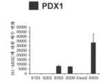
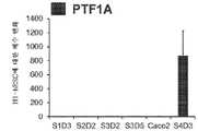
본 발명의 일 태양은 장의 중장 내배엽 세포의 집단을 생성하는 방법으로서, 상기 방법은 인간 만능성 줄기 세포를 배양 배지 중에서 배양하는 단계를 포함한다. 실시 형태에서, 본 방법은 인간 만능성 줄기 세포의 장의 중장 내배엽 세포로의 분화를 유도하는 단계를 포함한다. 일부 실시 형태에서, 장의 중장 내배엽 세포의 집단이 생성된다. 일부 실시 형태에서, 실질적인 장의 중장 내배엽 세포의 집단이 생성된다. 본 발명의 실시 형태에서, 장의 중장 내배엽 세포는 배양 중에 단층으로서 형성되고 안정하다. 실시 형태에서, 분화된 세포의 50% 초과가 장의 중장 내배엽의 특징적인 마커를 발현하며, 바람직하게는 분화된 세포의 60% 초과가 장의 중장 내배엽의 특징적인 마커를 발현하며, 더 바람직하게는 70% 초과, 80% 초과, 및 90% 초과가 장의 중장 내배엽의 특징적인 마커를 발현한다. 실시 형태에서, 분화된 세포는 장의 중장 내배엽 세포의 특징적인 마커를 발현한다. 실시 형태에서, 장의 중장 내배엽 세포는 CDX2 및 FOXA2를 발현한다. 모든 실시 형태에서, 장의 중장 내배엽 세포는 SOX9, PDX1, KLF5 및 HOXC5로부터 선택되는 전사 인자를 발현한다. 실시 형태에서, 장의 중장 내배엽 세포는 SOX2, ALB, PTF1A, HOXA13 및 LGR5로부터 선택되는 전사 인자를 발현하지 않는다.One aspect of the present invention is a method of generating a population of intestinal mesenchymal endoderm cells, the method comprising culturing human pluripotent stem cells in a culture medium. In an embodiment, the method comprises inducing differentiation of human pluripotent stem cells into midgut endoderm cells of the intestine. In some embodiments, a population of mesenchymal endoderm cells of the intestine is generated. In some embodiments, a population of mesenchymal endoderm cells of a substantial intestine is generated. In an embodiment of the present invention, the mesenchymal endoderm cells of the intestine are formed as a monolayer and are stable during culture. In an embodiment, greater than 50% of the differentiated cells express markers characteristic of intestinal midgut endoderm, preferably greater than 60% of differentiated cells express markers characteristic of intestinal midgut endoderm, more preferably 70% or more. Greater than %, greater than 80%, and greater than 90% express markers characteristic of the midgut endoderm of the intestine. In an embodiment, the differentiated cells express markers characteristic of midgut endoderm cells of the intestine. In an embodiment, the midgut endoderm cells of the intestine express CDX2 and FOXA2. In all embodiments, the midgut endoderm cells of the intestine express a transcription factor selected from SOX9, PDX1, KLF5 and HOXC5. In an embodiment, the midgut endoderm cells of the intestine do not express a transcription factor selected from SOX2, ALB, PTF1A, HOXA13 and LGR5.
본 발명의 실시 형태에서, 인간 만능성 줄기 세포는 하기를 포함하는 단계들에 의해 장의 중장 내배엽 세포로 분화된다: a) 인간 만능성 줄기 세포를 GDF-8 및 GSK3β 억제제, 예컨대 MCX 화합물을 함유하는 제1 배양 배지 중에서 배양하여 완성 내배엽 세포로의 분화를 유도하는 단계; b) 완성 내배엽 세포를 아스코르브산 및 FGF7을 함유하는 제2 배양 배지 중에서 배양하여 원시 장관 세포로의 분화를 유도하는 단계; 및 c) 원시 장관 세포를 산성 조건에서 레틴산 및 BMP2 또는 BMP4를 함유하는 제3 배양 배지 중에서 배양하여 장의 중장 내배엽 세포로의 분화를 유도하는 단계. 특정 실시 형태에서, 산성 조건은 BLAR 배지를 사용한 배양이다. 산성 배양의 pH는 6.8 내지 7.2의 범위일 수 있다. 본 발명의 실시 형태에서, 장의 중장 내배엽 세포는 배양 중에 단층을 형성한다. 실시 형태에서, 장의 중장 내배엽 세포의 단층은 배양 중에 유지된다.In an embodiment of the present invention, human pluripotent stem cells are differentiated into intestinal midgut endoderm cells by steps comprising: a) human pluripotent stem cells are treated with GDF-8 and a GSK3β inhibitor, such as an MCX compound. culturing in a first culture medium to induce differentiation into definitive endoderm cells; b) culturing the definitive endoderm cells in a second culture medium containing ascorbic acid and FGF7 to induce differentiation into primitive intestinal cells; and c) culturing the primitive intestinal cells in a third culture medium containing retinoic acid and BMP2 or BMP4 under acidic conditions to induce differentiation into intestinal mesenchymal endoderm cells. In a specific embodiment, the acidic conditions are cultured with BLAR medium. The pH of the acidic culture may range from 6.8 to 7.2. In an embodiment of the invention, the mesenteric endoderm cells of the intestine form a monolayer in culture. In an embodiment, a monolayer of mesenchymal endoderm cells of the intestine is maintained in culture.
본 발명의 다른 실시 형태는 당뇨병을 앓고 있거나 이것이 발병될 위험이 있는 환자를 치료하는 방법으로서, 상기 방법은 인간 만능성 줄기 세포를 장의 중장 내배엽 세포로 분화시키는 단계 및 분화된 장의 중장 내배엽 세포를 당뇨병을 갖는 환자 내에 투여하는 단계를 포함한다. 실시 형태에서, 당뇨병은 제1형 또는 제2형이다. 실시 형태에서, 세포를 투여하는 단계는 치료 부위에 직접적으로 또는 간접적으로 이식, 주입 또는 다른 방식으로 투여하는 것을 통해 행해질 수 있다. 일부 실시 형태에서, 장의 중장 내배엽 세포는 피하 공간, 장막, 간, 신장 등과 같은 신체 내에 이식된다. 추가의 실시 형태는 매크로- 또는 마이크로-캡슐화 디바이스의 캡슐화를 포함한 세포의 캡슐화된 전달을 포함한다.Another embodiment of the present invention is a method of treating a patient suffering from or at risk of developing diabetes, the method comprising differentiating human pluripotent stem cells into intestinal midgut endoderm cells and transforming the differentiated intestinal mesoenteric endoderm cells into diabetes mellitus. Administering into a patient with In an embodiment, the diabetes is
본 발명의 추가의 실시 형태는 장의 중장 내배엽 세포를 생성하는 방법으로서, 상기 방법은 배양 중인 완성 내배엽 세포의 원시 장관 세포로의 분화를 유도하는 단계를 포함한다. 실시 형태에서, 완성 내배엽 세포는 아스코르브산 및 FGF7을 함유하는 배양 배지 중에서 배양된다. 추가의 실시 형태에서, 원시 장관 세포는 레틴산과 BMP2 또는 BMP4를 함유하는 배양 배지 중에서 배양된다. 원시 장관 세포는 장의 중장 내배엽 세포로 분화된다. 일부 실시 형태에서, 원시 장관 세포는 산성 조건 (산성 배양 배지)에서 장의 중장 내배엽 세포로 분화된다. 특정 실시 형태에서, 산성 조건은 BLAR 배지를 사용한 배양이다. 산성 배양의 pH는 6.8 내지 7.2의 범위일 수 있다. 실시 형태에서, 장의 중장 내배엽 세포는 배양 중에 단층을 형성하고 유지한다.A further embodiment of the present invention is a method of generating intestinal mesodermal endoderm cells, the method comprising inducing differentiation of definitive endoderm cells in culture into primitive intestinal intestinal cells. In an embodiment, the definitive endoderm cells are cultured in a culture medium containing ascorbic acid and FGF7. In a further embodiment, the primitive intestinal cells are cultured in a culture medium containing retinoic acid and BMP2 or BMP4. Primordial intestinal cells differentiate into intestinal midgut endoderm cells. In some embodiments, the primitive intestinal cells differentiate into mesenchymal endoderm cells of the intestine in acidic conditions (acidic culture medium). In a specific embodiment, the acidic conditions are cultured with BLAR medium. The pH of the acidic culture may range from 6.8 to 7.2. In an embodiment, the mesenchymal endoderm cells of the intestine form and maintain a monolayer during culture.
상기 논의된 실시 형태들 각각에서, 인간 만능성 줄기 세포는 인간 배아 줄기 세포 또는 유도 만능성 줄기 세포이다. 상기 실시 형태들 각각에서, 장의 중장 내배엽 세포는 CDX2 및 FOXA2를 발현한다. 모든 실시 형태에서, 장의 중장 내배엽 세포는 SOX9, PDX1, KLF5 및 HOXC5로부터 선택되는 전사 인자를 발현한다. 실시 형태에서, 장의 중장 내배엽 세포는 SOX2, ALB, PTF1A, HOXA13 및 LGR5로부터 선택되는 전사 인자를 발현하지 않는다. 상기 실시 형태에서, 장의 중장 내배엽 세포는 CDX2, FOXA2, SOX9, PDX1, KLF5 및 HOXC5를 발현한다. 상기 실시 형태에서, 장의 중장 내배엽 세포는 SOX2, ALB, PTF1A, HOXA13 및 LGR5를 발현하지 않는다. 실시 형태에서, 분화된 세포의 50% 초과가 장의 중장 내배엽의 특징적인 마커를 발현하며, 바람직하게는 분화된 세포의 60% 초과가 장의 중장 내배엽의 특징적인 마커를 발현하며, 더 바람직하게는 70% 초과, 80% 초과, 및 90% 초과가 장의 중장 내배엽의 특징적인 마커를 발현한다. 실시 형태에서, 분화된 세포는 장의 중장 내배엽 세포의 특징적인 마커를 발현한다. 실시 형태에서, 장의 중장 내배엽 세포는 CDX2 및 FOXA2를 발현한다. 실시 형태에서, 장의 중장 내배엽 세포는 SOX9, PDX1, KLF5 및 HOXC5로부터 선택되는 전사 인자를 발현한다. 실시 형태에서, 장의 중장 내배엽 세포는 SOX2, ALB, PTF1A, HOXA13 및 LGR5로부터 선택되는 전사 인자를 발현하지 않는다. 실시 형태에서, 장의 중장 내배엽 세포는 HAND1을 발현하지 않는다.In each of the embodiments discussed above, the human pluripotent stem cells are human embryonic stem cells or induced pluripotent stem cells. In each of the above embodiments, the midgut endoderm cells of the intestine express CDX2 and FOXA2. In all embodiments, the midgut endoderm cells of the intestine express a transcription factor selected from SOX9, PDX1, KLF5 and HOXC5. In an embodiment, the midgut endoderm cells of the intestine do not express a transcription factor selected from SOX2, ALB, PTF1A, HOXA13 and LGR5. In this embodiment, the midgut endoderm cells of the intestine express CDX2, FOXA2, SOX9, PDX1, KLF5 and HOXC5. In this embodiment, the midgut endoderm cells of the intestine do not express SOX2, ALB, PTF1A, HOXA13 and LGR5. In an embodiment, greater than 50% of the differentiated cells express markers characteristic of intestinal midgut endoderm, preferably greater than 60% of differentiated cells express markers characteristic of intestinal midgut endoderm, more preferably 70% or more. Greater than %, greater than 80%, and greater than 90% express markers characteristic of the midgut endoderm of the intestine. In an embodiment, the differentiated cells express markers characteristic of midgut endoderm cells of the intestine. In an embodiment, the midgut endoderm cells of the intestine express CDX2 and FOXA2. In an embodiment, the midgut endoderm cells of the intestine express a transcription factor selected from SOX9, PDX1, KLF5 and HOXC5. In an embodiment, the midgut endoderm cells of the intestine do not express a transcription factor selected from SOX2, ALB, PTF1A, HOXA13 and LGR5. In an embodiment, the midgut endoderm cells of the intestine do not express HAND1.
상기 논의된 실시 형태에서, 장의 중장 내배엽 세포의 집단은 실질적인 장의 중장 내배엽 세포이다. 일부 실시 형태에서, 장의 중장 내배엽 세포의 집단은 70% 초과의 장의 중장 내배엽 세포, 바람직하게는 80% 초과, 90% 초과, 그리고 95% 초과의 장의 중장 내배엽 세포를 포함한다. 일부 실시 형태에서, 장의 중장 내배엽 세포의 집단은 20% 미만의 중간엽 세포, 바람직하게는 15% 미만, 더 바람직하게는 10% 미만, 5% 미만, 2% 미만, 1% 미만, 0.5% 미만을 포함한다. 실시 형태에서, 장의 중장 내배엽 세포는 HAND1의 발현이 결여되어 있다.In the embodiments discussed above, the population of intestinal mesenteric endoderm cells is substantially intestinal mesenteric endoderm cells. In some embodiments, the population of intestinal midgut endoderm cells comprises greater than 70% intestinal midgut endoderm cells, preferably greater than 80%, greater than 90%, and greater than 95% intestinal midgut endoderm cells. In some embodiments, the population of mesenchymal endoderm cells of the intestine is less than 20% mesenchymal cells, preferably less than 15%, more preferably less than 10%, less than 5%, less than 2%, less than 1%, less than 0.5%. includes In an embodiment, the midgut endoderm cells of the intestine lack expression of HAND1.
상기 기재된 본 발명의 일부 실시 형태에서, 분화는 시험관내(in vitro)에서 유도된다. 다른 실시 형태에서, 장의 중장 내배엽 세포는 생체내(in vivo)에서 추가로 분화된다. 다른 실시 형태는 생체내에서 장내분비 세포로 추가로 분화되는 장의 중장 내배엽 세포에 관한 것이다. 장내분비 세포는 인크레틴 호르몬을 발현하거나 분비한다. 실시 형태에서, 인크레틴 호르몬은 GLP1 및 GIP이다.In some embodiments of the invention described above, differentiation is inducedin vitro . In another embodiment, the mesenchymal endoderm cells of the intestine are further differentiatedin vivo . Another embodiment relates to midgut endoderm cells of the intestine that further differentiate in vivo into enteroendocrine cells. Enteroendocrine cells express or secrete incretin hormones. In an embodiment, the incretin hormones are GLP1 and GIP.
추가의 실시 형태에서, 장의 중장 내배엽 세포는 장의 중장 내배엽 세포를 먼저 장내분비 선구체로, 그리고 최종적으로 인크레틴 발현 또는 분비 장내분비 세포로 고효율로 시험관내 분화를 촉진하는 소분자의 확인을 위한 출발 물질로서의 역할을 한다.In a further embodiment, intestinal midgut endoderm cells are used as a starting material for the identification of small molecules that promote in vitro differentiation with high efficiency of intestinal midgut endoderm cells first into enteroendocrine progenitors and finally into incretin expressing or secreting enteroendocrine cells. play a role
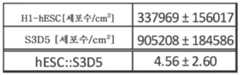
도 1a 내지 도 1d는 장의 중장 내배엽 세포의 분화 방법을 도시한다. 도 1a는 각각의 단계에 첨가된 배지 성분, 성장 인자 및 소분자, 그리고 분화 중인 장의 중장 내배엽 세포의 단계-특이적 핵심 마커 (FOXA2, 포크헤드 박스 A2; CDX2, 미측형 호메오박스 2; KLF5, 크루펠-유사 인자 5; SOX9, SRY (성별 결정 영역 Y)-박스 9; PDX1, 췌장 및 십이지장 호메오박스 1; LO, 낮은 발현 및 단백질 존재)를 포함하여 분화 방법에 대한 요약이다. S2D2에서 언급된 중성 pH (7.35±0.04)와 대비할 때, 세포를 3기 동안 BLAR 배지 ("BLAR 산성 배지"와 상호교환가능하게 사용됨) 중에서 약산성인 조건에 노출시켰으며 (pH; S3D1, 6.98±0.05; S3D2, 7.02±0.04; S3D5, 7.18±0.03) (도 1b), 이는 BLAR 배지 중 더 낮은 중탄산나트륨 수준의 결과로서 그러하다. 도 1c는 S3D5 단층 (좌측), 및 인간 상피 결장 선암종 세포주 ("Caco-2") (우측) - 이는 분화 특성화를 위한 벤치마크로서 사용됨 - 의 대표적인 위상차 이미지를 보여준다. S3D5에서 균일한 형태가 일관되게 관찰되었다. Nucleocounter® NC-100 (덴마크 알레뢰드 소재의 Chemometec, 카탈로그 번호 900-004)를 사용한 세포수의 특성화는 1개의 hESC가 4.56±2.60개의 S3D5 장의 중장 내배엽 세포로 분화되었음을 보여준다 (도 1d).
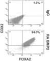
도 2a 내지 도 2d는 골 형성 단백질-4 (BMP4)를 이용하는 분화 방법이 전사체 및 단백질 수준에서 CDX2 및 FOXA2 둘 모두를 포함하는 장의 중장 내배엽 세포들을 단층으로 생성함을 보여준다. 도 2a (하단)는 CDX2 및 FOXA2 단백질 둘 모두에 대해 90.0±5.85%의 S3D5 세포가 공존하였음을 보여주는데, 이는 Caco-2 세포에서 관찰된 백분율 (86.0±6.67)과 유사하다. 대조적으로, 완성 내배엽 (DE ― S1D3) 세포에는 CDX2 및 FOXA2 공존이 없었다 (2.3±1.2). 유전자 발현 분석은 3기 동안 CDX2가 유도되었고 (도 2b), FOXA2가 유지되었음 (도 2c)을 보여준다. 도 2d는 FOXA2-양성 원시 장 내배엽 단계, S2D2 (도 2d-i)의 확립 후에 CDX2 단백질 수준 및 CDX2/FOXA2 단백질 공존이 유도되었고, S3D2 (도 2d-ii)까지 점진적으로 증가하였으며, S3D5 (도 2d-iii)에서 Caco-2 세포 (도 2d-iv)에서 보여지는 바와 유사한 수준에 도달하였음을 보여준다. CDX2 단백질은 하단 행에 도시되어 있고, FOXA2 단백질은 상단 행에 도시되어 있다. 정량적 분석을 가능하게 하도록 동일한 파라미터를 사용하여 각각의 이미지를 촬영하였다. 단백질 발현은 FACS에 의해 평가하였으며; 유전자 발현은 qPCR에 의해 평가하였다.
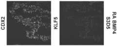
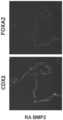
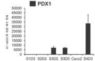
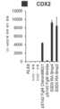
도 3a 내지 도 3q는 확실한 장의 중장 내배엽 유도를 구성하는 추가의 전사 인자 (TF)의 전사체 및 단백질 수준의 S3D5에 의한 유도를 보여주며; 적절한 장의 중장 내배엽이 달성되었다. CDX2 및 FOXA2 동시발현에 더하여, S3D5 세포는 또한 SOX9, PDX1, KLF5, HOXC5 (호메오박스 C5)의 동시발현을 나타내었지만, SOX2 (SRY-박스 2), ALB (알부민), PTF1A (췌장 특이적 전사 인자, 1a), 및 LGR5 (류신 풍부 반복 함유 G 단백질 결합된 수용체 5)의 동시발현은 나타내지 않았다. 모든 TF의 단백질 존재는 별개의 단일 채널 이미지로 도시되어 있다. 도 3a (하단)는 S3D5에서 CDX2 및 SOX9 둘 모두에 대해 98.7±0.25%의 세포가 공존하였음을 보여준다. SOX9 유전자 발현의 강한 유도는 Caco-2 세포에서 관찰된 수준과 비견되었으며 (도 3b), 면역형광 (IF)-분석에 의해 평가된 바와 같이 단백질 존재가 관찰되었다 (도 3c). 69.4±14.2%의 세포는 CDX2 및 PDX1 둘 모두에 대해 동시-양성이었다 (도 3d - 하단). 췌장-편향된 S4D3 세포와 대비할 때 (예를 들어, 미국 특허 출원 공개 제2014/0242693호 참조), PDX1 유전자 발현은 낮은 수준으로 유도되었으며 (도 3e), IF-분석에서 낮은 내지 부재하는 단백질 수준이 반영되었다 (도 3f). 전방 내배엽 TF SOX2는 1.45±0.15의 S3D5 세포가 SOX2 및 CDX2 공존을 나타낸 바와 같이 S3D5 세포에서 관찰되지 않았으며 (도 3g ― 하단; 도 3i), 유전자 발현은 hESC 및 Caco-2 세포에서 관찰된 수준 미만이었다 (도 3h). 장 중장/후장 내배엽의 적절한 발생에 필수적인, KLF5의 유전자 발현이 S3D5에서 상향조절되었다 (도 3j). S3D5에서 CDX2-양성 세포 내의 KLF5의 단백질 공존이 관찰되었다 (도 3k). ALB 유전자 발현 (도 3l), 및 단백질 존재 (도 3m)는 S3D5 세포에서 관찰되지 않았다. 췌장 계통 할당 TF인 PTF1A의 유전자 발현은 췌장-편향된 S4D3 세포와 달리 S3D5 세포에서 유도되지 않았다 (도 3n). 배아 중장 내배엽에 존재하는 호메오박스 유전자, HOXC5는 S3D5 세포에서 유도되었다 (도 3o). 도 3p는 마우스에서 임신 중기에 시작하는 배아 장 내배엽의 마커인 LGR5가 S3D5 세포에서 유도되지 않았음을 보여준다. 도 3q는 장 후장 내배엽의 마커인 HOXA13이 S3D5 세포에서 유도되지 않았음을 보여준다 (도 3p). 유전자 발현은 qPCR로 평가하였다.
도 4a 및 도 4b는 분화 중인 S3D5 세포의 증식 프로파일을 특성화한다. 도 4a는 Caco-2 세포의 경우 대부분의 CDX2-단백질 양성 세포가 (KI67 단백질과의 동시발현에 의해 나타난 바와 같이) 활성 세포 주기에 있으며 (좌측), 3기 동안의 H1-hESC-유래 세포의 증식 지수는 시간 경과에 따라 감소되었음 (S3D2 ― 중간; S3D5 ― 우측)을 보여준다. CDX2 (상단 행) 및 KI67 (하단 행) 단백질 수준이 단일 채널 이미지로서 도시되어 있다. FACS에 의해 평가된, 총 S3D5 세포 (총 세포는 90% 초과의 CDX2-양성 세포임)의 KI67-단백질 양성 세포의 백분율은 16.8±3.12였는데, 이는 S1D3에서 관찰된 백분율 (97.3±1.3), 및 Caco-2 세포에서 관찰된 백분율 (99.2±0.2)과는 대조적이었다 (도 4b).
도 5a 내지 도 5c는 BMP2를 3기 동안 BMP4에 대한 대안으로서 사용하여, CDX2 및 FOXA2 단백질 공존을 갖는 장의 중장 내배엽 세포의 단층을 달성함을 보여준다. 도 5a는 각각의 단계에 첨가된 배지 성분, 성장 인자 및 소분자, 그리고 분화 중인 장의 중장 내배엽 세포의 단계-특이적 마커 (FOXA2, CDX2, KLF5, SOX9, 및 PDX1LO)를 포함하여 분화 방법을 요약한다. S2D2에서 언급된 중성 pH (7.35±0.04)와 대비할 때, 세포를 3기의 전체 동안 BLAR 배지 중에서 약산성인 조건에 노출시켰으며 (pH; S3D1, 6.92; S3D2, 7.01; S3D5, 7.22) (도 5b), 이는 BLAR 배지 중 더 낮은 중탄산나트륨 수준의 결과로서 그러하다. 도 5c는 S3D5 단층 (좌측), 및 Caco-2 세포 (우측)의 대표적인 위상차 이미지를 도시하고; S3D5에서 균일한 형태가 관찰되었다.
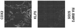
도 6a 내지 도 6u는 단층으로의 적절한 장의 중장 내배엽 세포들의 생성을 보여주며, 각각은 전사체 및 단백질 수준에서 CDX2, FOXA2, KLF5, SOX9, PDX1LO 및 HOXC5를 포함한다. IF 이미지의 경우, 모든 TF 단백질 수준은 단일 채널 이미지로서 도시되어 있다. 도 6a (하단)는 CDX2 및 FOXA2 단백질 둘 모두에 대해 94%의 S3D5 세포가 공존하였음을 보여주는데, 이는 Caco-2 세포에서 관찰된 백분율 (86.0±6.67)과 유사하거나 더 크다. 유전자 발현 분석은 3기 동안 CDX2가 유도되었고 (도 6b), FOXA2가 유지되었음 (도 6c)을 보여준다. 도 6d는 CDX2 단백질 수준 및 완전한 CDX2/FOXA2 단백질 공존이 S3D5에서 유도되었음을 보여주는데, 이는 Caco-2 세포에서 관찰된 수준 (도 2d-iv)과 유사한 수준에 도달한다. 도 6e (하단)는 S3D5에서 CDX2 및 SOX9 둘 모두에 대해 99.8%의 세포가 공존하였음을 보여준다. SOX9 유전자 발현의 강한 유도는 Caco-2 세포에서 관찰된 수준과 비견되었으며 (도 6f), IF-분석에 의해 평가된 바와 같이 단백질 존재가 관찰되었다 (도 6g). 45.5%의 세포는 CDX2 및 PDX1 둘 모두에 대해 동시-양성이었다 (도 6h - 하단). 췌장-편향된 S4D3 세포와 대비할 때, PDX1 유전자 발현은 낮은 수준으로 유도되었으며 (도 6i); IF-분석에서 낮은 내지 부재하는 단백질 수준이 반영되었다 (도 6j). 전방 내배엽 TF SOX2는 0.8%의 S3D5 세포가 SOX2 및 CDX2 공존을 나타낸 바와 같이 S3D5 세포에서 관찰되지 않았으며 (도 6k ― 하단; 도 6m), 유전자 발현은 hESC 및 Caco-2 세포에서 관찰된 수준 미만이었다 (도 6l). 장 중장/후장 내배엽의 적절한 발생에 필수적인, KLF5의 유전자 발현이 S3D5에서 상향조절되었다 (도 6n). CDX2-양성 세포 내의 KLF5의 단백질 공존이 S3D5에서 관찰되었다 (도 6o). ALB 유전자 발현 (도 6p), 및 단백질 존재 (도 6q)는 S3D5 세포에서 관찰되지 않았다. 췌장 계통 할당 TF인 PTF1A의 유전자 발현은 췌장-편향된 S4D3 세포와 달리 S3D5 세포에서 유도되지 않았다 (도 6r). 배아 장의 중장 내배엽에 존재하는 호메오박스 유전자, HOXC5는 S3D5 세포에서 유도되었다 (도 6s). 도 6t는 임신 중기에 시작하는 배아 장 내배엽의 마커인 LGR5가 S3D5 세포에서 유도되지 않았음을 보여준다. 도 6u는 장 후장 내배엽의 마커인 HOXA13이 S3D5 세포에서 유도되지 않았음을 보여준다 (도 6u).
도 7은 분화 중인 S3D5 세포의 증식 프로파일을 특성화한다. Caco-2 세포의 경우 대부분의 CDX2-단백질 양성 세포가 (KI67 단백질과의 동시발현에 의해 나타난 바와 같이) 활성 세포 주기에 있는 것과 대비하여 (좌측), 3기 동안의 H1-hESC-유래 세포의 증식 지수는 더 낮았다 (S3D5 ― 우측). CDX2 (상단 행) 및 KI67 (하단 행) 단백질 수준이 단일 채널 이미지로서 도시되어 있다. FACS에 의해 평가된, 총 S3D5 세포 (총 세포는 90% 초과의 CDX2-양성 세포임)의 KI67-단백질 양성 세포의 백분율은 14.1%였는데, 이는 S1D3에서 관찰된 백분율 (97.3±1.3), 및 Caco-2 세포에서 관찰된 백분율 (99.2±0.2)과는 대조적이었다.
도 8a 내지 도 8f는 CDX2+ 세포의 불균질한 집단의 유도를 보여준다. 도 8a는 각각의 단계에 첨가된 배지 성분, 성장 인자 및 소분자, 그리고 분화 중인 장의 중장/후장 내배엽 세포의 단계-특이적 핵심 마커 (HAND1)를 포함하여 분화 방법에 대한 요약이다. 도 8b는 H1-hESC 세포 (상단 행, 좌측), 500 ng/ml의 FGF4 및 3 μM Chiron99021로 2일간 컨디셔닝된 1기 후 세포 (상단 행, 중간), 500 ng/ml의 FGF4 및 500 ng/ml의 WNT3A로 2일간 컨디셔닝된 1기 후 세포 (상단 행, 우측), RA/BMP4에 의해 컨디셔닝된 S3D5 단층 (하단 행, 좌측), 및 RA/BMP2에 의해 컨디셔닝된 S3D5 단층 (하단 행, 우측)의 위상차 이미지를 보여준다. 2일의 컨디셔닝 후 유전자 발현의 유도가 낮은 수준으로 CDX2에 대해 나타나 있으며 (도 8c), 내배엽 마커 FOXA2에 대해 유지되고 있으며 (도 8d), 중배엽/중간엽 마커 HAND1에 대해 유도되어 있다 (도 8f). KLF5는 유도되지 않았다 (도 8e).1A-1D show a method for differentiating intestinal mesenchymal endoderm cells. Figure 1a shows the media components, growth factors and small molecules added at each step, and the stage-specific key markers (FOXA2, forkhead box A2; CDX2,
2A-2D show that a differentiation method using bone morphogenetic protein-4 (BMP4) results in a monolayer of intestinal mesenchymal endoderm cells that contain both CDX2 and FOXA2 at the transcript and protein levels. 2A (bottom) shows that 90.0±5.85% of S3D5 cells co-expressed for both CDX2 and FOXA2 proteins, similar to the percentage observed in Caco-2 cells (86.0±6.67). In contrast, there was no CDX2 and FOXA2 coexistence in definitive endoderm (DE - S1D3) cells (2.3±1.2). Gene expression analysis showed that CDX2 was induced during phase 3 (FIG. 2B) and FOXA2 was maintained (FIG. 2C). Fig. 2d shows that CDX2 protein levels and CDX2/FOXA2 protein coexistence were induced after the establishment of the FOXA2-positive primitive intestinal endoderm stage, S2D2 (Fig. 2d-i), and gradually increased until S3D2 (Fig. 2d-ii), followed by S3D5 (Fig. 2d-i). 2d-iii) reached levels similar to those seen in Caco-2 cells (Fig. 2d-iv). CDX2 protein is shown in the bottom row and FOXA2 protein is shown in the top row. Each image was taken using the same parameters to enable quantitative analysis. Protein expression was assessed by FACS; Gene expression was assessed by qPCR.
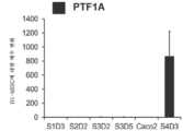
Figures 3A-3Q show induction by S3D5 of transcript and protein levels of additional transcription factors (TFs) constituting definitive intestinal midgut endoderm induction; Adequate intestinal midgut endoderm was achieved. In addition to co-expression of CDX2 and FOXA2, S3D5 cells also showed co-expression of SOX9, PDX1, KLF5, HOXC5 (homeobox C5), but not SOX2 (SRY-box 2), ALB (albumin), PTF1A (pancreas-specific Coexpression of the transcription factor, 1a), and LGR5 (G protein coupled receptor 5 containing leucine rich repeats) is not shown. Protein presence of all TFs is shown in separate single channel images. 3A (bottom) shows that 98.7±0.25% of cells co-localized for both CDX2 and SOX9 at S3D5. Strong induction of SOX9 gene expression was comparable to levels observed in Caco-2 cells (FIG. 3B), and protein presence was observed as assessed by immunofluorescence (IF)-analysis (FIG. 3C). 69.4±14.2% of cells were co-positive for both CDX2 and PDX1 ( FIG. 3D - bottom). When compared to pancreas-biased S4D3 cells (see, eg, US Patent Application Publication No. 2014/0242693), PDX1 gene expression was induced at low levels ( FIG. 3E ), with low to absent protein levels in the IF-assay. was reflected (Fig. 3f). Anterior endoderm TF SOX2 was not observed in S3D5 cells as 1.45±0.15 of S3D5 cells showed SOX2 and CDX2 coexistence (Fig. 3g-bottom; Fig. 3i), and gene expression was at the level observed in hESC and Caco-2 cells. was less than (Fig. 3h). Gene expression of KLF5, essential for the proper development of intestinal midgut/hindgut endoderm, was upregulated at S3D5 (Fig. 3j). Protein co-localization of KLF5 in CDX2-positive cells was observed at S3D5 (Fig. 3k). ALB gene expression (Fig. 3l), and protein presence (Fig. 3m) were not observed in S3D5 cells. Gene expression of PTF1A, a pancreatic lineage-assigned TF, was not induced in S3D5 cells, unlike pancreatic-biased S4D3 cells (Fig. 3n). HOXC5, a homeobox gene present in embryonic midgut endoderm, was induced in S3D5 cells (Fig. 3o). Figure 3p shows that LGR5, a marker of embryonic intestinal endoderm starting at mid-gestation in mice, was not induced in S3D5 cells. Figure 3q shows that HOXA13, a marker of intestinal hindgut endoderm, was not induced in S3D5 cells (Figure 3p). Gene expression was assessed by qPCR.
4A and 4B characterize the proliferation profile of differentiating S3D5 cells. Figure 4a shows that for Caco-2 cells, most CDX2-protein positive cells (as shown by co-expression with KI67 protein) are in an active cell cycle (left), and H1-hESC-derived cells during phase 3 It shows that the proliferation index decreased over time (S3D2 - middle; S3D5 - right). CDX2 (top row) and KI67 (bottom row) protein levels are shown as single channel images. The percentage of KI67-protein positive cells in total S3D5 cells (total cells being greater than 90% CDX2-positive cells), assessed by FACS, was 16.8±3.12, which was comparable to the percentage observed in S1D3 (97.3±1.3), and This was in contrast to the percentage observed in Caco-2 cells (99.2±0.2) ( FIG. 4B ).
5A-5C show that BMP2 was used as an alternative to BMP4 during phase 3 to achieve a monolayer of intestinal midgut endoderm cells with coexistence of CDX2 and FOXA2 proteins. FIG. 5A summarizes the differentiation method, including media components, growth factors and small molecules added at each stage, and stage-specific markers of the midgut endoderm cells of the differentiating intestine (FOXA2, CDX2, KLF5, SOX9, and PDX1LO ). do. Cells were exposed to slightly acidic conditions in BLAR medium for the entirety of phase 3 (pH; S3D1, 6.92; S3D2, 7.01; S3D5, 7.22), as compared to the neutral pH (7.35±0.04) noted in S2D2 (Fig. 5b). ), as a result of lower sodium bicarbonate levels in BLAR medium. Figure 5C shows representative phase-contrast images of S3D5 monolayers (left), and Caco-2 cells (right); A uniform morphology was observed in S3D5.
6A-6U show the generation of suitable intestinal midgut endoderm cells in monolayers, each containing CDX2, FOXA2, KLF5, SOX9, PDX1LO and HOXC5 at the transcript and protein level. For IF images, all TF protein levels are shown as single channel images. 6A (bottom) shows that 94% of S3D5 cells co-expressed for both CDX2 and FOXA2 proteins, similar to or greater than the percentage observed in Caco-2 cells (86.0±6.67). Gene expression analysis showed that CDX2 was induced during phase 3 (FIG. 6B) and FOXA2 was maintained (FIG. 6C). Figure 6d shows that CDX2 protein levels and complete CDX2/FOXA2 protein co-localization were induced at S3D5, reaching levels similar to those observed in Caco-2 cells (Figures 2d-iv). 6E (bottom) shows that 99.8% of cells co-localized for both CDX2 and SOX9 at S3D5. A strong induction of SOX9 gene expression was comparable to levels observed in Caco-2 cells (FIG. 6F) and protein presence was observed as assessed by IF-assay (FIG. 6G). 45.5% of cells were co-positive for both CDX2 and PDX1 (FIG. 6H - bottom). Compared to pancreas-biased S4D3 cells, PDX1 gene expression was induced at a low level (Fig. 6i); Low to absent protein levels were reflected in the IF-assay (Fig. 6j). Anterior endoderm TF SOX2 was not observed in S3D5 cells as 0.8% of S3D5 cells showed coexistence of SOX2 and CDX2 (FIG. 6K-Bottom; FIG. 6M), gene expression below levels observed in hESC and Caco-2 cells. was (FIG. 6l). Gene expression of KLF5, which is essential for the proper development of intestinal midgut/hindgut endoderm, was upregulated at S3D5 (FIG. 6n). Protein co-localization of KLF5 in CDX2-positive cells was observed at S3D5 (Fig. 6o). ALB gene expression (Fig. 6p), and protein presence (Fig. 6q) were not observed in S3D5 cells. Gene expression of PTF1A, a pancreatic lineage-assigned TF, was not induced in S3D5 cells unlike pancreatic-biased S4D3 cells (Fig. 6r). HOXC5, a homeobox gene present in the midgut endoderm of the embryonic intestine, was induced in S3D5 cells (Fig. 6s). 6T shows that LGR5, a marker of embryonic intestinal endoderm starting in mid-gestation, was not induced in S3D5 cells. Figure 6u shows that HOXA13, a marker of intestinal hindgut endoderm, was not induced in S3D5 cells (Figure 6u).
7 characterizes the proliferation profile of differentiating S3D5 cells. For Caco-2 cells, the majority of CDX2-protein positive cells are in an active cell cycle (as shown by co-expression with KI67 protein) (left), compared to H1-hESC-derived cells during phase 3. Proliferation index was lower (S3D5 - right). CDX2 (top row) and KI67 (bottom row) protein levels are shown as single channel images. The percentage of KI67-protein positive cells in total S3D5 cells (total cells were >90% CDX2-positive cells), assessed by FACS, was 14.1%, which was the percentage observed in S1D3 (97.3±1.3), and Caco In contrast to the percentage observed in -2 cells (99.2±0.2).
8A-8F show the induction of a heterogeneous population of CDX2+ cells. FIG. 8A is a summary of the differentiation method, including medium components, growth factors and small molecules added at each stage, and a stage-specific key marker (HAND1) of differentiating intestinal midgut/hindgut endoderm cells. FIG. 8B shows H1-hESC cells (top row, left), cells after
본 발명은 특정 방법, 시약, 화합물, 조성물 또는 생물학적 시스템에 한정되지 않으며, 이는 물론 다양할 수 있음을 이해해야 한다. 또한, 본 명세서에서 사용되는 용어는 단지 특정 실시 형태들을 설명하기 위한 것이며, 한정하는 것으로 의도되지 않음을 이해해야 한다.It is to be understood that the present invention is not limited to a particular method, reagent, compound, composition or biological system, which of course may vary. Also, it should be understood that terminology used herein is merely for describing specific embodiments and is not intended to be limiting.
본 발명은 장의 중장 내배엽 세포의 생성에 관한 것이다. 특정 배양 서열을 사용하여 세포를 생성하였다. 따라서, 본 발명은 만능성 줄기 세포로부터 유래된 세포를 CDX2 및 FOXA2의 발현과 같은 장의 중장 내배엽 세포 계통의 특징적인 마커를 발현하는 세포로 분화시키기 위한 시험관내 세포 배양을 제공한다. 본 발명은 시험관내 세포 배양을 통해 단층으로 그러한 세포를 획득하고 유지하는 방법을 추가로 제공한다. 소정 실시 형태에서, 본 발명은 레틴산과 BMP4 또는 BMP2 또는 이들의 유사체의 포함이 분화 중인 세포에서 CDX2를 유도하고 FOXA2 단백질 발현을 유지하여 장의 중장 내배엽 세포로의 분화를 촉진시킨다는 발견에 기초한다. CDX2는 완성 내배엽 (1기) 또는 원시 장관 (2기)에서의 단백질 수준에서는 발현되지 않는다. 따라서, 본 발명은 만능성 줄기 세포를 분화시켜 CDX2 및 FOXA2를 발현하는 장의 중장 내배엽 세포를 생성하는 방법을 제공한다.The present invention relates to the production of midgut endoderm cells of the intestine. Cells were generated using specific culture sequences. Accordingly, the present invention provides in vitro cell culture for differentiating cells derived from pluripotent stem cells into cells expressing markers characteristic of the midgut endoderm cell lineage of the intestine, such as the expression of CDX2 and FOXA2. The present invention further provides methods for obtaining and maintaining such cells in monolayers via in vitro cell culture. In certain embodiments, the present invention is based on the discovery that inclusion of retinoic acid with BMP4 or BMP2 or analogs thereof induces CDX2 and maintains FOXA2 protein expression in differentiating cells to promote differentiation into mesenchymal endoderm cells of the intestine. CDX2 is not expressed at the protein level in definitive endoderm (stage 1) or primitive intestinal tract (stage 2). Accordingly, the present invention provides methods for differentiating pluripotent stem cells to generate enteric mesenchymal endoderm cells expressing CDX2 and FOXA2.
정의Justice
달리 정의되지 않는 한, 본 명세서에서 사용되는 모든 기술 용어 및 과학 용어는 당업자가 일반적으로 이해하는 것과 동일한 의미를 갖는다. 본 명세서에 기재된 것과 유사하거나 또는 동등한 임의의 방법 및 재료가 본 발명의 시험 실시에 사용될 수 있지만, 바람직한 방법 및 재료가 본 명세서에 기재되어 있다. 본 발명을 설명하고 청구함에 있어서, 하기 용어가 사용될 것이다.Unless defined otherwise, all technical and scientific terms used herein have the same meaning as commonly understood by one of skill in the art. Although any methods and materials similar or equivalent to those described herein can be used in the practice of testing the present invention, the preferred methods and materials are described herein. In describing and claiming the present invention, the following terminology will be used.
줄기 세포는 자가 재생할 뿐만 아니라, 분화되어, 자가 재생 전구체, 비재생 전구체, 및 최종 분화된 세포를 비롯한 자손 세포(progeny cell)를 생성하는 단일 세포의 능력에 의해 정의되는 미분화 세포이다. 줄기 세포는 또한 다수의 배엽층 (내배엽, 중배엽 및 외배엽)으로부터 다양한 세포 계통의 기능성 세포로 시험관내에서 분화되는 이의 능력뿐만 아니라 이식 후 다수의 배엽층의 조직을 생성시키며 배반포 내로의 주입 후 전부는 아니라 하더라도 대부분의 조직에 실질적으로 기여하는 이의 능력을 특징으로 한다.Stem cells are undifferentiated cells defined by the ability of a single cell to self-renew as well as to differentiate and generate progeny cells, including self-renewing precursors, non-renewing precursors, and terminally differentiated cells. Stem cells also have their ability to differentiate in vitro from multiple germ layers (endoderm, mesoderm and ectoderm) into functional cells of various cell lineages, as well as generate tissues of multiple germ layers after transplantation, all after injection into blastocysts. It is characterized by one's ability to make a substantial, if not most, contribution to an organization.
줄기 세포는 그의 발생능(developmental potential)에 따라 하기와 같이 분류된다: (1) 전능성(totipotent); (2) 만능성; (3) 다능성; (4) 소기능성(oligopotent); 및 (5) 단일기능성(unipotent). 전능성 세포는 모든 배아 및 배외 세포 유형을 생성시킬 수 있다. 만능성 세포는 모든 배아 세포 유형을 생성시킬 수 있다. 다능성 세포는, 세포 계통의 하위세트(subset)이지만 모두 특정 조직, 기관 또는 생리학적 시스템 내에 있는 하위세트를 생성시킬 수 있는 세포 (예를 들어, 조혈 줄기 세포(hematopoietic stem cell, HSC)는 HSC (자가 재생), 혈구 세포 제한 소기능성 전구체 및 혈액의 정상 성분인 모든 세포 유형 및 요소 (예를 들어, 혈소판)를 포함하는 자손을 생성할 수 있음)를 포함한다. 소기능성인 세포는 다능성 줄기 세포보다 더 제한된 세포 계통의 하위세트를 생성시킬 수 있고; 단일기능성인 세포는 단일 세포 계통 (예를 들어, 정자형성 줄기 세포)을 생성시킬 수 있다.Stem cells are classified according to their developmental potential: (1) totipotent; (2) pluripotency; (3) pluripotency; (4) oligopotent; and (5) unipotent. Totipotent cells can give rise to all embryonic and extraembryonic cell types. Pluripotent cells can give rise to all embryonic cell types. Pluripotent cells are cells capable of giving rise to a subset of cell lineages, but all within a particular tissue, organ, or physiological system (e.g., hematopoietic stem cells (HSCs) are HSCs). (self-renewal), blood cell-restricted microfunctional precursors and capable of producing progeny that include all cell types and elements that are normal components of blood (eg, platelets). Cells that are microfunctional can give rise to a more restricted subset of cell lineages than pluripotent stem cells; Cells that are monofunctional are capable of giving rise to a single cell lineage (eg, spermatogenic stem cells).
줄기 세포는 또한 이것이 얻어질 수 있는 공급원에 기초하여 분류된다. 성체 줄기 세포는 대체로 다수의 분화된 세포 유형을 포함하는 조직에서 발견되는 다능성 미분화 세포이다. 성체 줄기 세포는 스스로 재생할 수 있다. 정상 환경 하에서, 이는 또한 분화되어, 그의 기원이 되는 조직의 특수화된 세포 유형, 및 가능하게는 다른 조직 유형을 제공할 수 있다. 유도 만능성 줄기 세포 (iPS 세포)는 만능성 줄기 세포로 변환되는 성체 세포이다. (문헌[Takahashiet al.,Cell, 2006; 126(4):663-676]; 문헌[Takahashiet al., Cell, 2007; 131:1-12]). 배아 줄기 세포는 배반포기 배아의 내세포집단 유래의 만능성 세포이다. 태아 줄기 세포는 태아 조직 또는 막으로부터 기원된 것이다.Stem cells are also classified based on the source from which they are obtained. Adult stem cells are pluripotent undifferentiated cells usually found in tissues that contain multiple differentiated cell types. Adult stem cells can regenerate themselves. Under normal circumstances, it can also differentiate to give specialized cell types of the tissue of origin, and possibly other tissue types. Induced pluripotent stem cells (iPS cells) are adult cells that transform into pluripotent stem cells. (Takahashiet al.,Cell , 2006; 126(4):663-676; Takahashiet al., Cell , 2007; 131:1-12). Embryonic stem cells are pluripotent cells derived from the inner cell population of blastocyst stage embryos. Fetal stem cells are those derived from fetal tissue or membranes.
배아 조직은 전형적으로 배아 (인간의 경우 수정에서 발생 약 6주까지의 기간을 지칭함)로부터 기원되는 조직으로 정의된다. 태아 조직은, 인간의 경우 발생 약 6주에서 분만까지의 기간을 지칭하는, 태아로부터 기원되는 조직을 지칭한다. 배외 조직은 배아 또는 태아와 관련되지만 이로부터 기원되지 않는 조직이다. 배외 조직은 배외 막 (융모막, 양막, 난황낭 및 요막), 제대 및 태반 (그 자체가 융모막 및 모체 기저탈락막으로부터 형성됨)을 포함한다.Embryonic tissue is typically defined as tissue that originates from an embryo (refers to the period from fertilization to about 6 weeks of development in humans). Fetal tissue refers to tissue originating from the fetus, which in humans refers to the period from about 6 weeks of development to delivery. Extraembryonic tissue is tissue associated with, but not derived from, an embryo or fetus. Extraembryonic tissue includes the extraembryonic membrane (chorion, amnion, yolk sac, and allantoic membrane), umbilical cord and placenta (which itself is formed from the chorion and maternal basolateral decidua).
분화는 특수화되지 않은 ("미수임된(uncommitted)") 또는 덜 특수화된 세포가, 예를 들어 장 세포 또는 췌장 세포와 같은 특수화된 세포의 특징을 획득하는 과정이다. 분화된 세포는 세포의 계통 내에서 보다 특수화된 ("수임된(committed)") 위치를 차지한 것이다. 분화 과정에 적용될 때, '수임된'이란 용어는 분화 경로에서, 정상적인 환경 하에서 특정 세포 유형 또는 세포 유형의 하위세트로 계속 분화될 것이며, 정상적인 환경 하에서 다른 세포 유형으로 분화될 수 없거나 덜 분화된 세포 유형으로 되돌아갈 수 없는 시점까지 진행한 세포를 지칭한다. 탈분화(de-differentiation)는 세포가 세포의 계통 내의 덜 특수화된 (또는 수임된) 위치로 되돌아가는 과정을 지칭한다. 본 명세서에 사용된 바와 같이, 세포의 계통은 세포의 유전, 즉, 어느 세포로부터 왔는지 그리고 어떤 세포를 생성시킬 수 있는지를 규정한다. 세포의 계통은 세포를 발생과 분화의 유전적 체계 내에 둔다.Differentiation is the process by which unspecialized ("uncommitted") or less specialized cells acquire the characteristics of specialized cells, such as, for example, intestinal cells or pancreatic cells. A differentiated cell is one that has occupied a more specialized ("committed") position within the lineage of cells. When applied to the process of differentiation, the term 'committed' refers to a cell that, in a differentiation pathway, will continue to differentiate, under normal circumstances, into a particular cell type or subset of cell types, and cannot, or is less differentiated, into other cell types under normal circumstances. Refers to cells that have progressed to the point where they cannot return to their original form. De-differentiation refers to the process by which a cell reverts to a less specialized (or committed) position within the cell's lineage. As used herein, a cell's lineage defines a cell's inheritance, i.e., which cells it came from and which cells it can produce. A cell's lineage places it within a genetic system of development and differentiation.
넓은 의미에서, 전구 세포는 그 자신보다 더 분화되지만 전구체의 풀(pool)을 보충하는 능력을 유지하는 자손을 생성하는 능력을 갖는 세포이다. 이 정의에 의하면, 줄기 세포 그 자체는 또한, 최종 분화된 세포에 대한 더 가까운 선구체(precursor)가 그러하듯이, 전구 세포이다. 좁은 의미에서, 전구 세포는 종종 분화 경로에서의 중간체인 세포로 정의되며, 다시 말하면, 이는 줄기 세포로부터 발생하고, 성숙 세포 유형 또는 세포 유형의 하위세트의 생성에서의 중간체이다. 이러한 유형의 전구 세포는 대체로 자가 재생할 수 없다. 따라서, 이러한 유형의 세포가 본 명세서에 지칭되는 경우, 이는 비재생 전구 세포 또는 중간 전구 세포 또는 중간 선구 세포로 지칭될 것이다.In a broad sense, a progenitor cell is a cell that has the ability to produce progeny that are more differentiated than themselves but retain the ability to replenish the pool of progenitors. By this definition, stem cells themselves are also progenitor cells, as are more likely precursors to terminally differentiated cells. In a narrow sense, a progenitor cell is often defined as a cell that is an intermediate in the differentiation pathway, in other words, it arises from a stem cell and is an intermediate in the production of a mature cell type or subset of cell types. Progenitor cells of this type are usually unable to self-renew. Thus, when this type of cell is referred to herein, it will be referred to as a non-renewing progenitor cell or an intermediate progenitor cell or an intermediate progenitor cell.
본 명세서에서 사용되는 바와 같이, "마커"는 관심 세포에서 차별적으로 발현되는 핵산 또는 폴리펩티드 분자이다. 이와 관련하여, 차별적 발현(differential expression)은 미분화된 세포에 비하여 양성 마커에 대한 증가된 수준 및 음성 마커에 대한 감소된 수준을 의미한다. 마커 핵산 또는 폴리펩티드의 검출가능한 수준은 다른 세포에 비하여 관심 세포에서 충분히 더 높거나 더 낮아서, 관심 세포가 당업계에 알려진 다양한 방법 중 임의의 것을 이용하여 다른 세포로부터 확인되어 구별될 수 있다.As used herein, a “marker” is a nucleic acid or polypeptide molecule that is differentially expressed in a cell of interest. In this context, differential expression refers to increased levels of positive markers and decreased levels of negative markers compared to undifferentiated cells. The detectable level of the marker nucleic acid or polypeptide is sufficiently higher or lower in the cell of interest compared to other cells such that the cell of interest can be identified and distinguished from other cells using any of a variety of methods known in the art.
본 명세서에 사용되는 바와 같이, 특정 마커가 세포에서 충분히 검출될 때, 그 세포는 그 특정 마커에 "대하여 양성"이거나 그 세포는 "양성"이다. 유사하게, 특정 마커가 세포에서 충분히 검출되지 않을 때, 그 세포는 그 특정 마커에 "대하여 음성"이거나 그 세포는 "음성"이다. 특히, 형광 활성화 유세포측정 (FACS)에 의한 양성은 보통 2% 초과인 반면, FACS에 의한 음성 역치는 보통 1% 미만이다.As used herein, a cell is “positive for” or a cell is “positive” when a particular marker is sufficiently detected in a cell. Similarly, when a particular marker is not sufficiently detectable in a cell, the cell is “negative for” that particular marker or the cell is “negative”. In particular, positivity by fluorescence activated flow cytometry (FACS) is usually greater than 2%, whereas the negative threshold by FACS is usually less than 1%.
본 명세서에 사용되는 바와 같이, 실시간 PCR (RT-PCR)에 의한 양성은 28회 사이클 (Ct) 미만을 가졌고, Taqman® 저밀도 어레이(Low Density Array) (TLDA)를 사용하는 경우에는 33 Ct 미만을 가졌으며; 한편 Open Array®에 의한 음성은 28.5회 사이클 초과이고 TLDA에 의한 음성은 33.5 Ct 초과이다.As used herein, positivity by real-time PCR (RT-PCR) had less than 28 cycles (Ct) and less than 33 Ct when using Taqman® Low Density Array (TLDA). have; On the other hand, the voice by Open Array® exceeds 28.5 cycles and the voice by TLDA exceeds 33.5 Ct.
정치(static) 시험관내 세포 배양에서 만능성 줄기 세포를 기능성 장의 중장 내분비 세포로 분화시키는 데 있어서, 분화 과정은 종종 연속적인 단계들을 통해 진행되는 것으로 여겨진다. 여기서, 장의 중장 내배엽으로의 분화 과정은 3개의 단계를 통해 일어난다. 이러한 단계적 진행에서, "1기"는 분화 과정에서의 제1 단계, 즉, 만능성 줄기 세포의 완성 내배엽 세포 (이하, 대안적으로 "1기 세포"로 지칭됨)의 특징적인 마커를 발현하는 세포로의 분화를 지칭한다. "2기"는 제2 단계, 즉, 완성 내배엽 세포의 특징적인 마커를 발현하는 세포의 원시 장관 세포 (이하, 대안적으로 "2기 세포"로 지칭됨)의 특징적인 마커를 발현하는 세포로의 분화를 지칭한다. "3기"는 제3 단계, 즉, 장관 세포의 특징적인 마커를 발현하는 세포의 장의 중장 내배엽 세포 (이하, 대안적으로 "3기 세포"로 지칭됨)의 특징적인 마커를 발현하는 세포로의 분화를 지칭한다.In the differentiation of pluripotent stem cells into functional intestinal midgut endocrine cells in static in vitro cell culture, the differentiation process is often considered to proceed through successive steps. Here, the process of differentiation of the intestine into the midgut endoderm occurs through three stages. In this stepwise progression, “
그러나, 특정 집단에서의 모든 세포가 동일한 속도로 이러한 단계들을 통해 진행되는 것은 아니라는 것에 주목해야 한다. 결과적으로, 시험관내 세포 배양에서, 특히 후기 분화 단계에서 집단에 존재하는 다수의 세포들보다 더 적거나 더 많은 분화 경로를 진행한 세포의 존재를 검출하는 것은 드문 일이 아니다. 본 발명을 설명하기 위하여, 상기 확인된 단계들과 관련된 다양한 세포 유형들의 특징들이 본 명세서에 기재된다.However, it should be noted that not all cells in a given population progress through these stages at the same rate. Consequently, it is not uncommon to detect in in vitro cell cultures the presence of cells that have progressed through a differentiation pathway that are fewer or more than the majority of cells present in the population, particularly at later stages of differentiation. To illustrate the present invention, characteristics of various cell types related to the above identified steps are described herein.
본 명세서에 사용되는 바와 같이, "완성 내배엽 세포"는, 낭배형성 동안 상배엽(epiblast)으로부터 생기는 세포의 특징을 보유하고 위장관 및 그의 파생물을 형성하는 세포를 지칭한다. 완성 내배엽 세포는 하기 마커들 중 적어도 하나를 발현한다: FOXA2 (간세포 핵 인자 3-β (HNF3β)로도 알려짐), GATA4, SOX17, CXCR4, 단미증(Brachyury), 세르베루스(Cerberus), OTX2, 구스코이드(goosecoid), C-Kit, CD99, 및 MIXL1. 완성 내배엽 세포의 특징적인 마커는 CXCR4, FOXA2 및 SOX17을 포함한다. 따라서, 완성 내배엽 세포는 그의 CXCR4, FOXA2, 및 SOX17 발현을 특징으로 할 수 있다. 게다가, 세포가 1기에 남아 있도록 허용되는 시간의 길이에 따라, HNF4α의 증가가 관찰될 수 있다.As used herein, “definitive endoderm cells” refer to cells that retain characteristics of cells that arise from the epiblast during gastrulation and form the gastrointestinal tract and its derivatives. Definitive endoderm cells express at least one of the following markers: FOXA2 (also known as hepatocyte nuclear factor 3-β (HNF3β)), GATA4, SOX17, CXCR4, Brachyury, Cerberus, OTX2, orgu goosecoid, C-Kit, CD99, and MIXL1. Characteristic markers of definitive endoderm cells include CXCR4, FOXA2 and SOX17. Thus, definitive endoderm cells can be characterized for their CXCR4, FOXA2, and SOX17 expression. Moreover, depending on the length of time cells are allowed to remain in
본 명세서에서 사용되는 바와 같이, "원시 장관 세포"는, 모든 내배엽성 기관, 예컨대 폐, 간, 췌장, 위, 및 장이 생기게 할 수 있는 완성 내배엽으로부터 유래된 내배엽 세포를 지칭한다. 원시 장관 세포는 완성 내배엽 세포에 의해 발현되는 것에 비해 실질적으로 증가된 그의 HNF4α 발현을 특징으로 할 수 있다.As used herein, "primitive intestinal cells" refer to endoderm cells derived from the definitive endoderm capable of giving rise to all endodermal organs, such as lung, liver, pancreas, stomach, and intestine. Primitive intestinal cells can be characterized by their HNF4α expression that is substantially increased compared to that expressed by definitive endoderm cells.
본 명세서에 사용되는 바와 같이, "전장 내배엽 세포"는 식도, 폐, 위, 간, 췌장, 담낭, 및 십이지장의 최전방 부분이 생기게 하는 내배엽 세포를 지칭한다. 전장 내배엽 세포는 특히, 그의 SOX2, PDX1, ALB, SOX17 및 FOXA2 발현을 특징으로 할 수 있다.As used herein, "full length endoderm cells" refer to endoderm cells that give rise to the anteriormost portion of the esophagus, lungs, stomach, liver, pancreas, gallbladder, and duodenum. Full length endoderm cells can be characterized, inter alia, for their SOX2, PDX1, ALB, SOX17 and FOXA2 expression.
본 명세서에 사용되는 바와 같이, "장의 중장 내배엽 세포"는 소장이 생기게 하는 내배엽 세포를 지칭한다. 장의 중장 내배엽 세포는 그의 CDX2, FOXA2의 발현, 및 PDX1의 낮은 발현 (PDX1LO)을 특징으로 할 수 있다. 소정의 HOX 유전자의 발현은 중장 내배엽과 후장 내배엽을 구별할 수 있다. 예를 들어, HOXC5는 중장 내배엽 세포에서 선택적으로 발현된다.As used herein, “intestinal midgut endoderm cells” refers to the endoderm cells that give rise to the small intestine. Intestinal midgut endoderm cells can be characterized by their expression of CDX2, FOXA2, and low expression of PDX1 (PDX1LO ). Expression of certain HOX genes can differentiate between midgut endoderm and hindgut endoderm. For example, HOXC5 is selectively expressed in midgut endoderm cells.
본 명세서에 사용되는 바와 같이, "후장 내배엽 세포"는 대장이 생기게 하는 내배엽 세포를 지칭한다. 후장 내배엽 세포는 그의 CDX2, FOXA2, HOXA13 및 HOXD13 발현을 특징으로 할 수 있다.As used herein, “hindtestinal endoderm cells” refer to the endoderm cells that give rise to the large intestine. Dorsal intestinal endoderm cells can be characterized for their CDX2, FOXA2, HOXA13 and HOXD13 expression.
본 명세서에 사용되는 바와 같이, "중간엽 세포"는 결합 조직, 예컨대 뼈, 연골, 림프계, 및 순환계를 생기게 하는 중배엽 세포를 지칭한다. HAND1 및 FOXF1의 발현은 중간엽 세포를 한정한다.As used herein, "mesenchymal cell" refers to the mesoderm cells that give rise to connective tissue such as bone, cartilage, lymphatic system, and circulatory system. Expression of HAND1 and FOXF1 confine mesenchymal cells.
"환자" 또는 "대상체" 또는 "숙주"라는 용어는 조성물 또는 약제학적 조성물로 치료되거나 본 명세서에 기술된 방법에 따라 치료되는 포유류를 포함하는 동물, 바람직하게는 인간을 지칭한다.The term "patient" or "subject" or "host" refers to an animal, preferably a human, including mammals, treated with a composition or pharmaceutical composition or treated according to the methods described herein.
용어 "유효량" 또는 이와 동등한 표현은 인간 만능성 줄기 세포를 분화된 세포 집단으로, 예를 들어 완성 내배엽, 전장 내배엽, 장의 중장 내배엽, 후장 내배엽, 췌장 내배엽 등으로 진행 및 분화시키기에 충분한 성장 인자를 포함하지만 이로 한정되지 않는 소정량의 작용제 또는 화합물을 지칭한다.The term “effective amount” or an equivalent expression means a growth factor sufficient to cause development and differentiation of human pluripotent stem cells into a differentiated cell population, such as definitive endoderm, foregut endoderm, intestinal midgut endoderm, hindgut endoderm, pancreatic endoderm, etc. refers to an amount of an agent or compound, including but not limited to.
용어 "투여하는" 및 "투여"는 본 명세서에서 상호교환가능하게 사용되며, 세포가 치료 부위에 직접적으로 또는 간접적으로 삽입, 주입, 이식 또는 달리 투여될 수 있음을 의미한다. 세포가 반고체 또는 고체 디바이스로 투여될 때, 이식은 적합한 전달 수단이며, 특히 신체 내의 정확한 위치 내로의, 예컨대 피하 공간, 장막, 간, 신장 (신장 피막) 내로의 외과적 이식이 그러하다. 액체 또는 유체 약제학적 조성물은 더 일반적인 위치에 투여될 수 있다.The terms "administering" and "administration" are used interchangeably herein and mean that cells may be implanted, infused, transplanted or otherwise administered directly or indirectly to a treatment site. When the cells are administered in a semi-solid or solid device, implantation is a suitable means of delivery, particularly surgical implantation into a precise location within the body, such as into the subcutaneous space, serous membrane, liver, kidney (renal capsule). A liquid or fluid pharmaceutical composition may be administered to a more general location.
본 명세서 및 첨부된 청구범위에서 사용되는 바와 같이, 단수형 ("a", "an" 및 "the")은 그 내용이 명확하게 달리 지시하지 않으면 복수형 지시 대상을 포함한다. 따라서, 예를 들어, "세포"에 대한 언급은 2개 이상의 세포의 조합 등을 포함한다.As used in this specification and the appended claims, the singular forms "a", "an" and "the" include plural referents unless the content clearly dictates otherwise. Thus, for example, reference to “a cell” includes combinations of two or more cells, and the like.
본 명세서에 사용된 바와 같이, 측정가능한 값, 예를 들어 양, 시간적 길이 등을 지칭할 때 용어 "약"은 명시된 값의 ±20% 내지 ±0.1%, 바람직하게는 ±20% 또는 ±10%, 더 바람직하게는 ±5%, 더욱 더 바람직하게는 ±1%, 그리고 훨씬 더 바람직하게는 ±0.5%, ±0.1%, 0.05% 또는 0.01%의 변동을 포함하는 것으로 의미되는데, 그러한 변동은 개시된 방법을 수행하기에 적절하기 때문이다.As used herein, the term "about" when referring to a measurable value, e.g., amount, length of time, etc., is ±20% to ±0.1%, preferably ±20% or ±10% of the specified value. , more preferably ±5%, even more preferably ±1%, and still more preferably ±0.5%, ±0.1%, 0.05% or 0.01%, such variations being disclosed herein. Because it is appropriate to carry out the method.
하기 약어는 본 명세서 및 청구범위 전체에 걸쳐 나타날 수 있다:The following abbreviations may appear throughout this specification and claims:
ABCG2 - ATP-결합 카세트, 서브-패밀리 G, 구성원 2;ABCG2 - ATP-binding cassette, sub-family G,
ALB ― 알부민;ALB - albumin;
BMP - 골 형성 단백질;BMP - bone morphogenetic protein;
CDX2 - 미측형 호메오박스 2;CDX2 -
CXCR4 - C-X-C 케모카인 4형 수용체;CXCR4—
FAF-BSA - 지방산 무함유 소혈청 알부민;FAF-BSA - fatty acid free bovine serum albumin;
FGF - 섬유아세포 성장 인자;FGF - Fibroblast Growth Factor;
FOXA2 - 포크헤드 박스 A2;FOXA2 - Forkhead Box A2;
GATA4 - GATA 결합 단백질 4;GATA4 - GATA
GDF - 성장 분화 인자;GDF—growth differentiation factor;
GIP - 글루코스-의존성 인슐린분비촉진 폴리펩티드;GIP - glucose-dependent insulinotropic polypeptide;
GLP-1 - 글루카곤-유사 펩티드 1;GLP-1 - glucagon-
GSK3B - 글리코겐 신타제 키나제 3 베타;GSK3B - glycogen synthase kinase 3 beta;
HAND1 - 심장 및 신경 능선 유도체 발현 1;HAND1 - cardiac and neural crest
HOX ― 호메오박스;HOX—homeobox;
hTERT ― 인간 텔로머라제 역전사효소;hTERT—human telomerase reverse transcriptase;
KLF ― 크루펠-유사 인자;KLF—Krupel-like factor;
LGR5 - 류신 풍부 반복 함유 G 단백질 결합된 수용체 5;LGR5 - leucine rich repeat containing G protein coupled receptor 5;
MIXL1 - 믹스 쌍형-유사 호메오박스-1(Mix Paired-Like Homeobox-1);MIXL1 - Mix Paired-Like Homeobox-1;
OCT4 - 옥타머-결합 전사 인자 4;OCT4—octamer-binding
OTX2 - 오르토덴티클 호메오박스 2;OTX2 -
PDX1 - 췌장 및 십이지장 호메오박스 1;PDX1 - pancreatic and
PTF1A - 췌장 특이적 전사 인자, 1a;PTF1A - pancreatic specific transcription factor, 1a;
SOX - 성별 결정 영역 Y (SRY)-박스;SOX - sex determining area Y (SRY) - box;
TRA1-60 - T 세포 수용체 알파-1-60;TRA1-60 - T cell receptor alpha-1-60;
UTF1 - 미분화된 배아 세포 전사 인자 1;UTF1 - undifferentiated embryonic
WNT3A - 윙리스형 MMTV 통합 부위 패밀리, 구성원 3A; 및WNT3A - wingless MMTV integration site family, member 3A; and
ZFP42 - 징크 핑거 단백질 42.ZFP42 - zinc finger protein 42.
상세한 설명details
만능성 줄기 세포는 3개의 모든 배엽층, 즉 내배엽, 중배엽, 및 외배엽의 조직의 세포로 분화될 잠재력을 갖는다. 사용될 수 있는 만능성 줄기 세포의 예시적인 유형은 확립된 만능성 세포주를 포함하고, 이에는 전-배아 조직 (예컨대, 배반포), 배아 조직, 또는 임신 동안 임의의 시점에, 전형적으로는 그러나 반드시는 아니고, 대략 10 내지 12주의 임신 전에 취해진 태아 조직이 포함된다. 비제한적인 예는, 예를 들어 인간 배아 줄기 세포주 H1, H7, 및 H9 (미국 위스콘신주 매디슨 소재의 WiCell Research Institute)와 같은, 확립된 인간 배아 줄기 세포주 또는 인간 배아생식 세포주이다. 지지 세포(feeder cell)의 부재 하에서 이미 배양된 만능성 줄기 세포 집단으로부터 취해진 세포가 또한 적합하다. 다수의 만능성 관련 전사 인자들, 예컨대 OCT4, NANOG, SOX2, KLF4, 및 ZFP42의 강제 발현을 사용하여 성체 체세포로부터 유래되는, iPS, 또는 재프로그래밍된 만능성 세포 (문헌[Annu Rev Genomics Hum Genet 2011, 12:165-185]; 또한 iPS는 문헌[Cell, 126(4): 663-676]을 참조함)가 또한 사용될 수 있다. 본 발명의 방법에 사용되는 인간 배아 줄기 세포는 또한 문헌[Thomsonet al.]에 의해 기재된 바와 같이 제조될 수 있다(미국 특허 제5,843,780호; 문헌[Science, 1998, 282:1145-1147]; 문헌[Curr Top Dev Biol 1998, 38:133-165]; 문헌[Proc Natl Acad Sci U.S.A. 1995, 92:7844-7848]). BG01v (미국 조지아주 애선스 소재의 BresaGen), 또는 성체 인간 체세포로부터 유래된 세포, 예를 들어 문헌[Takahashiet al., Cell 131:1-12 (2007)]에 개시된 세포와 같은 돌연변이 인간 배아 줄기 세포주가 또한 사용될 수 있다. 소정 실시 형태에서, 본 발명에 사용하기에 적합한 만능성 줄기 세포는 문헌[Liet al. (Cell Stem Cell 4: 16-19, 2009)]; 문헌[Maheraliet al. (Cell Stem Cell 1: 55-70, 2007)]; 문헌[Stadtfeldet al. (Cell Stem Cell 2: 230-240)]; 문헌[Nakagawaet al. (Nature Biotechnol 26: 101-106, 2008)]; 문헌[Takahashiet al. (Cell 131: 861-872, 2007)]; 및 미국 특허 출원 공개 제2011/0104805호에 기재된 방법에 따라 유래될 수 있다. 소정 실시 형태에서, 만능성 줄기 세포는 비-배아 기원의 것일 수 있다. 이들 참고문헌, 특허, 및 특허 출원 모두는, 특히, 만능성 세포의 단리, 배양, 확장 및 분화에 관련되므로, 전체적으로 본 명세서에 참고로 포함된다.Pluripotent stem cells have the potential to differentiate into cells of tissues of all three germ layers: endoderm, mesoderm, and ectoderm. Exemplary types of pluripotent stem cells that can be used include established pluripotent cell lines, including pre-embryonic tissue (eg, blastocyst), embryonic tissue, or at any time during pregnancy, typically but not necessarily but not fetal tissue taken before approximately 10 to 12 weeks of gestation. Non-limiting examples are established human embryonic stem cell lines or human embryonic germ cell lines, such as, for example, human embryonic stem cell lines H1, H7, and H9 (WiCell Research Institute, Madison, WI). Cells taken from a population of pluripotent stem cells already cultured in the absence of feeder cells are also suitable. iPS, or reprogrammed pluripotent cells derived from adult somatic cells using forced expression of a number of pluripotency-related transcription factors, such as OCT4, NANOG, SOX2, KLF4, and ZFP42 (Annu Rev Genomics Hum Genet 2011 , 12:165-185]; iPS also seeCell , 126(4): 663-676) can also be used. Human embryonic stem cells used in the methods of the present invention are also described in Thomsonet al. (U.S. Patent No. 5,843,780; Science, 1998, 282:1145-1147;Curr Top Dev Biol 1998, 38:133-165;Proc Natl Acad Sci USA 1995, 92:7844-7848]). BG01v (BresaGen, Athens, GA), or cells derived from adult human somatic cells, such as cells disclosed in Takahashiet al ., Cell 131:1-12 (2007), mutant human embryonic stem Cell lines may also be used. In certain embodiments, pluripotent stem cells suitable for use in the present invention are described in Liet al. (Cell Stem Cell 4: 16-19, 2009)]; See Maheraliet al. (Cell Stem Cell 1: 55-70, 2007)]; See Stadtfeldet al. (Cell Stem Cell 2: 230-240)]; See Nakagawaet al. (Nature Biotechnol 26: 101-106, 2008)]; See Takahashiet al. (Cell 131: 861-872, 2007)]; and US Patent Application Publication No. 2011/0104805. In certain embodiments, pluripotent stem cells may be of non-embryonic origin. All of these references, patents, and patent applications are incorporated herein by reference in their entirety as they relate, inter alia, to the isolation, culture, expansion, and differentiation of pluripotent cells.
만능성 줄기 세포는 다양한 단계들을 거쳐 분화되며, 각각의 단계는 특정 마커의 존재 또는 부재를 특징으로 할 수 있다. 이들 단계로의 세포의 분화는 배양 배지에 첨가되는 소정의 인자들의 존재 또는 결여를 포함하는 구체적인 배양 조건들로 달성된다. 일반적으로, 이러한 분화는 만능성 줄기 세포의 완성 내배엽 세포 - 본 명세서에서 1기로 지칭됨 - 로의 분화를 포함할 수 있다. 이어서, 이들 완성 내배엽 세포는 본 명세서에서 2기로 지칭되는 원시 장관 세포로 추가로 분화될 수 있다. 이어서, 원시 장관 세포는 다시 본 명세서에서 3기로 지칭되는 장의 중장 내배엽 세포로 분화될 수 있다.Pluripotent stem cells differentiate through various stages, each stage can be characterized by the presence or absence of certain markers. Differentiation of cells to these stages is achieved with specific culture conditions including the presence or absence of certain factors added to the culture medium. Generally, this differentiation may include the differentiation of pluripotent stem cells into definitive endoderm cells, referred to herein as
장의 중장 내배엽 세포의 특징적인 마커를 발현하는 세포로의 만능성 줄기 세포의 분화Differentiation of pluripotent stem cells into cells expressing markers characteristic of intestinal midgut endoderm cells
만능성 줄기 세포의 특징은 당업자에게 잘 알려져 있으며, 만능성 줄기 세포의 추가의 특징은 계속 확인되고 있다. 만능성 줄기 세포 마커는, 예를 들어, 하기 중 하나 이상의 발현을 포함한다: ABCG2; 크립토(Cripto); FOXD3; CONNEXIN43; CONNEXIN45; OCT4; SOX2; NANOG; hTERT; UTF1; ZFP42; SSEA-3; SSEA-4; TRA-1-60; 및 TRA-1-81.The characteristics of pluripotent stem cells are well known to those skilled in the art, and additional characteristics of pluripotent stem cells continue to be identified. Pluripotent stem cell markers include, for example, expression of one or more of: ABCG2; Crypto; FOXD3; CONNEXIN43; CONNEXIN45; OCT4; SOX2; NANOG; hTERT; UTF1; ZFP42; SSEA-3; SSEA-4; TRA-1-60; and TRA-1-81.
예시적인 만능성 줄기 세포는 인간 배아 줄기 세포주 H1 (NIH 코드: WA01), 인간 배아 줄기 세포주 H9 (NIH 코드: WA09), 인간 배아 줄기 세포주 H7 (NIH 코드: WA07) 및 인간 배아 줄기 세포주 SA002 (스웨덴 소재의 Cellartis)를 포함한다. 만능성 세포의 특징적인 하기의 마커들 중 적어도 하나를 발현하는 세포가 또한 적합하다: ABCG2, 크립토, CD9, FOXD3, CONNEXIN43, CONNEXIN45, OCT4, SOX2, NANOG, hTERT, UTF1, ZFP42, SSEA-3, SSEA-4, TRA-1-60, 및 TRA-1-81.Exemplary pluripotent stem cells include human embryonic stem cell line H1 (NIH code: WA01), human embryonic stem cell line H9 (NIH code: WA09), human embryonic stem cell line H7 (NIH code: WA07) and human embryonic stem cell line SA002 (Sweden Cellartis of the material). Also suitable are cells expressing at least one of the following markers characteristic of pluripotent cells: ABCG2, crypto, CD9, FOXD3, CONNEXIN43, CONNEXIN45, OCT4, SOX2, NANOG, hTERT, UTF1, ZFP42, SSEA-3, SSEA-4, TRA-1-60, and TRA-1-81.
완성 내배엽 계통의 특징적인 마커들 중 적어도 하나를 발현하는 세포가 또한 본 발명에 사용하기에 적합하다. 본 발명의 일 실시 형태에서, 완성 내배엽 계통의 특징적인 마커를 발현하는 세포는 원시선 선구 세포이다. 대안적인 실시 형태에서, 완성 내배엽 계통의 특징적인 마커를 발현하는 세포는 중내배엽 세포이다. 대안적인 실시 형태에서, 완성 내배엽 계통의 특징적인 마커를 발현하는 세포는 완성 내배엽 세포이다.Cells expressing at least one of the markers characteristic of the definitive endoderm lineage are also suitable for use in the present invention. In one embodiment of the invention, the cells expressing markers characteristic of the definitive endoderm lineage are primitive glandular progenitor cells. In an alternative embodiment, the cells expressing markers characteristic of the definitive endoderm lineage are mesendoderm cells. In an alternative embodiment, cells expressing markers characteristic of definitive endoderm lineage are definitive endoderm cells.
장의 중장 내배엽 계통의 특징적인 마커들 중 적어도 하나를 발현하는 세포가 또한 본 발명에 사용하기에 적합하다. 본 발명의 일 실시 형태에서, 장 내배엽 계통의 특징적인 마커를 발현하는 세포는 장의 중장 내배엽 세포이며, 여기서 상기 세포는 FOXA2 및 CDX2를 발현한다. 일부 실시 형태에서, 세포는 SOX2, ALB, PTF1A, HOXA13 또는 LGR5를 발현하지 않는다. 실시 형태에서, 장 내배엽 계통의 특징적인 마커를 발현하는 세포는 장의 중장 내배엽 세포이며, 여기서 상기 세포는 FOXA2, CDX2, SOX9, PDX1, KLF5 및 HOXC5 각각을 발현한다. 실시 형태에서, 장 내배엽 계통의 특징적인 마커를 발현하는 세포는 장의 중장 내배엽 세포이며, 여기서 상기 세포는 SOX2, ALB, PTF1A, HOXA13 및 LGR5 중 어느 것도 발현하지 않는다.Cells expressing at least one of the markers characteristic of the midgut endoderm lineage of the intestine are also suitable for use in the present invention. In one embodiment of the invention, the cells expressing markers characteristic of the intestinal endoderm lineage are midgut endoderm cells of the intestine, wherein the cells express FOXA2 and CDX2. In some embodiments, the cell does not express SOX2, ALB, PTF1A, HOXA13 or LGR5. In an embodiment, the cell expressing a marker characteristic of the intestinal endoderm lineage is an intestinal mesenchymal endoderm cell, wherein the cell expresses FOXA2, CDX2, SOX9, PDX1, KLF5 and HOXC5, respectively. In an embodiment, the cells expressing markers characteristic of the intestinal endoderm lineage are intestinal mesenteric endoderm cells, wherein the cells express none of SOX2, ALB, PTF1A, HOXA13 and LGR5.
본 발명은 세포 배양 조건 및 배지를 사용하여 만능성 줄기 세포의 장의 중장 내배엽 세포로의 단계적 유도 분화를 제공한다. 본 발명의 실시 형태에서, 장의 중장 내배엽 세포의 특징적인 마커를 발현하는 세포에 도달하기 위하여, 만능성 줄기 세포, 예컨대 배아 줄기 세포 및 유도 만능성 세포에서 출발하는 프로토콜이 사용된다. 이러한 프로토콜은 하기 단계들을 포함한다.The present invention provides for the directed differentiation of pluripotent stem cells into mesenchymal endoderm cells of the intestine using cell culture conditions and media. In embodiments of the present invention, protocols starting from pluripotent stem cells, such as embryonic stem cells and induced pluripotent cells, are used to arrive at cells that express markers characteristic of midgut endoderm cells of the intestine. This protocol includes the following steps.
1기:세포 배양 라인에서 얻어진 만능성 줄기 세포, 예컨대 배아 줄기 세포를 적절한 인자로 처리하여 완성 내배엽 세포의 특징적인 마커를 발현하는 세포로의 발현을 유도한다.1st period:Pluripotent stem cells obtained from cell culture lines, such as embryonic stem cells, are treated with appropriate factors to induce their expression into cells expressing markers characteristic of definitive endoderm cells.
2기:1기에서 생성된 세포를 적절한 인자로 처리하여 원시 장관 세포의 특징적인 마커를 발현하는 세포로의 추가 분화를 유도한다.2nd period:Cells generated in
3기:2기에서 생성된 세포를 적절한 인자로 처리하여 장의 중장 내배엽 세포의 특징적인 마커를 발현하는 세포로의 추가 분화를 유도한다.3rd period:Cells generated in
배양되거나 단리된 세포에서 단백질 및 핵산 마커의 발현을 평가하는 방법은 당업계에서의 표준이다. 이러한 방법은 RT-PCR, 노던 블롯(Northern blot), 동소(in situ) 혼성화 (예를 들어, 문헌[Current Protocols in Molecular Biology (Ausubelet al., eds. 2001 supplement)] 참조), 및 면역검정법 (예컨대, 절편화된 재료의 면역조직화학적 분석), 웨스턴 블롯팅(Western blotting), 및 온전한 세포에서 접근가능한 마커의 경우, FACS (예를 들어 문헌[Harlow and Lane, Using Antibodies: A Laboratory Manual, New York: Cold Spring Harbor Laboratory Press (1998)] 참조)를 포함한다. 분화의 효율은 관심 세포 유형의 특징적인 마커를 발현하는 세포에 의해 발현되는 단백질 마커를 특이적으로 인식하는 작용제 (예컨대, 항체)에 처리된 세포 집단을 노출시킴으로써 결정될 수 있다.Methods for assessing the expression of protein and nucleic acid markers in cultured or isolated cells are standard in the art. Such methods include RT-PCR, Northern blot,in situ hybridization (see, eg, Current Protocols in Molecular Biology (Ausubelet al ., eds. 2001 supplement)), and immunoassays. (e.g., immunohistochemical analysis of sectioned material), Western blotting, and, for markers accessible in intact cells, FACS (e.g., Harlow and Lane, Using Antibodies: A Laboratory Manual, New York: Cold Spring Harbor Laboratory Press (1998)). Efficiency of differentiation can be determined by exposing the treated cell population to an agent (eg, antibody) that specifically recognizes a protein marker expressed by cells expressing a marker characteristic of the cell type of interest.
1.One.완성 내배엽 세포의 특징적인 마커를 발현하는 세포로의 만능성 줄기 세포의 분화Differentiation of pluripotent stem cells into cells expressing markers characteristic of definitive endoderm cells
만능성 줄기 세포는 당업계에 공지된 임의의 적합한 방법에 의해, 또는 본 발명에서 제안된 임의의 방법에 의해 완성 내배엽 세포의 특징적인 마커를 발현하는 세포로 분화될 수 있다. 본 발명의 일 실시 형태에서, 만능성 줄기 세포는 GDF8 및 GSK3β 억제제 (예컨대, 미국 특허 출원 공개 제2010/0015711호에 개시된 환형 아닐린-피리디노트라이아진 화합물; 이는 전체적으로 본 명세서에 참고로 포함됨)를 포함한 인자가 보충된 배지, 예컨대 MCDB-131 배지 (미국 캘리포니아주 칼스배드 소재의 Life Technologies)로 처리되어 완성 내배엽 세포의 특징적인 마커를 발현하는 세포로의 분화를 유도한다. 넓은 범위의 GSK3β 억제제, 예컨대 스타우로스포린이 있으며, 바람직한 GSK3β 억제제 14-프로프-2-엔-1-일-3,5,7,14,17,23,27-헵타아자테트라사이클로 [19.3.1.1~2,6~.1~8,12~]헵타코사-1(25),2(27),3,5,8(26),9,11,21,23-노나엔-16-온이 있으며, 이는 본 명세서에서 "MCX 화합물"로 지칭된다. 처리는 만능성 줄기 세포를 약 10 ng/ml 내지 1000 ng/ml, 바람직하게는 50 ng/ml 내지 약 150 ng/ml, 대안적으로 약 75 ng/ml 내지 약 125 ng/ml, 대안적으로 약 100 ng/ml의 GDF8이 보충된 배지와 접촉시키는 것을 포함할 수 있다. 처리는 또한 세포를 약 0.1 내지 10 μM, 바람직하게는 약 0.1 내지 5 μM, 대안적으로 약 0.5 내지 약 2.5 μM, 바람직하게는 약 1.5 μM 또는 약 1.0 μM의 MCX 화합물과 접촉시키는 것을 포함할 수 있다. 배지의 다른 성분은 약 2.7 g/1000 ml 내지 3.6 g/1000 ml, 바람직하게는 2.7 g/1000 ml의 중탄산나트륨; 약 0.1% 내지 2.0%, 바람직하게는 약 0.5%의 FAF-BSA; 1:100 희석 ("1X 농도")의 GlutaMAX™ (미국 캘리포니아주 칼스배드 소재의 Life Technologies Corporation); 및 10 mM D-글루코스의 농도를 얻기 위한 약 2 mM 내지 20 mM의 농도 범위, 바람직하게는 4.5 mM의 D-글루코스를 포함할 수 있다.Pluripotent stem cells can be differentiated into cells expressing markers characteristic of definitive endoderm cells by any suitable method known in the art, or by any method proposed herein. In one embodiment of the present invention, pluripotent stem cells are administered with GDF8 and GSK3β inhibitors (eg, the cyclic aniline-pyridinotriazine compounds disclosed in US Patent Application Publication No. 2010/0015711; which is incorporated herein by reference in its entirety). treatment with a medium supplemented with factors including, for example, MCDB-131 medium (Life Technologies, Carlsbad, Calif.) to induce differentiation into cells expressing markers characteristic of definitive endoderm cells. There are broad spectrum GSK3β inhibitors such as staurosporine, with the preferred GSK3β inhibitor 14-prop-2-en-1-yl-3,5,7,14,17,23,27-heptaazatetracyclo [19.3. 1.1~2,6~.1~8,12~] Heptacosa-1(25),2(27),3,5,8(26),9,11,21,23-nonaen-16- ions, which are referred to herein as "MCX compounds". The treatment may include pluripotent stem cells at about 10 ng/ml to 1000 ng/ml, preferably 50 ng/ml to about 150 ng/ml, alternatively about 75 ng/ml to about 125 ng/ml, alternatively and contacting with medium supplemented with about 100 ng/ml of GDF8. The treatment may also include contacting the cells with about 0.1 to 10 μM, preferably about 0.1 to 5 μM, alternatively about 0.5 to about 2.5 μM, preferably about 1.5 μM or about 1.0 μM of the MCX compound. there is. Other components of the medium include sodium bicarbonate at about 2.7 g/1000 ml to 3.6 g/1000 ml, preferably 2.7 g/1000 ml; FAF-BSA from about 0.1% to 2.0%, preferably about 0.5%; GlutaMAX™ (Life Technologies Corporation, Carlsbad, CA) at a 1:100 dilution ("1X concentration"); and a concentration range of about 2 mM to 20 mM to obtain a concentration of 10 mM D-glucose, preferably 4.5 mM D-glucose.
만능성 세포는 완성 내배엽 세포로의 그의 분화를 촉진시키기 위하여 대략 2 내지 5일, 바람직하게는 약 3 내지 4일 동안 배양될 수 있다. 일 실시 형태에서, 만능성 세포는 유효량의 TGFβ 신호전달 분자 및/또는 GSK3β 억제제, 예를 들어 유효량의 GDF8 및 MCX 화합물의 존재 하에서 1일 동안 배양된 후, GDF8 및 더 낮은 농도의 MCX 화합물의 존재 하에서 1일 동안 배양된 후, MCX 화합물의 부재 하에서의 GDF8의 존재 하에서 1일 동안 배양된다. 상세하게는, 세포는 GDF8 및 약 1.5 μM의 MCX 화합물의 존재 하에서 1일 동안 배양된 후, GDF8 및 약 0.1 μM의 MCX 화합물의 존재 하에서 1일 동안 배양된 후, MCX 화합물의 부재 하에서의 GDF8의 존재 하에서 1일 동안 배양될 수 있다. 대안적인 실시 형태에서, 세포는 GDF8 및 약 1.5 μM의 MCX 화합물의 존재 하에서 1일 동안 배양된 후, GDF8 및 약 0.1 μM의 MCX 화합물의 존재 하에서 1일 동안 배양될 수 있다.Pluripotent cells can be cultured for about 2 to 5 days, preferably about 3 to 4 days, to promote their differentiation into definitive endoderm cells. In one embodiment, the pluripotent cells are cultured for 1 day in the presence of an effective amount of a TGFβ signaling molecule and/or a GSK3β inhibitor, such as an effective amount of GDF8 and an MCX compound, followed by the presence of GDF8 and a lower concentration of an MCX compound. and then cultured for 1 day in the presence of GDF8 in the absence of MCX compound. Specifically, cells were cultured in the presence of GDF8 and about 1.5 μM of an MCX compound for 1 day, then cultured in the presence of GDF8 and about 0.1 μM of an MCX compound for 1 day, and then in the presence of GDF8 in the absence of an MCX compound. It can be cultured for 1 day under In an alternative embodiment, the cells can be cultured in the presence of GDF8 and about 1.5 μM of the MCX compound for 1 day, followed by culture in the presence of GDF8 and about 0.1 μM of the MCX compound for 1 day.
완성 내배엽 세포의 특징적인 마커를 발현하는 세포의 생성은 특정 프로토콜을 이행하기 전과 후에 마커의 존재에 대해 시험함으로써 결정될 수 있다. 만능성 줄기 세포는 전형적으로 그러한 마커를 발현하지 않는다. 따라서, 만능성 세포의 분화는 세포가 완성 내배엽 세포의 특징적인 마커, 예컨대 CXCR4, FOXA2 및 SOX17을 발현하기 시작할 때 검출될 수 있다. 실시 형태에서, 완성 내배엽 세포의 특징적인 마커를 발현하는 세포는 완성 내배엽 세포이다.Generation of cells expressing markers characteristic of definitive endoderm cells can be determined by testing for the presence of the markers before and after implementing a particular protocol. Pluripotent stem cells typically do not express such markers. Thus, differentiation of pluripotent cells can be detected when the cells begin to express markers characteristic of definitive endoderm cells, such as CXCR4, FOXA2 and SOX17. In an embodiment, a cell that expresses a marker characteristic of a definitive endoderm cell is a definitive endoderm cell.
2.2.완성 내배엽 세포의 특징적인 마커를 발현하는 세포의 원시 장관 세포의 특징적인 마커를 발현하는 세포로의 분화Differentiation of cells expressing markers characteristic of definitive endoderm cells into cells expressing markers characteristic of primitive intestinal intestinal cells
완성 내배엽 세포의 특징적인 마커를 발현하는 세포는 원시 장관 세포의 특징적인 마커를 발현하는 세포로 추가로 분화될 수 있다. 일 실시 형태에서, 원시 장관 세포의 특징적인 마커를 발현하는 세포의 형성은 FGF7을 함유하는 배지, 예컨대 MCDB-131로 완성 내배엽 세포의 특징적인 마커를 발현하는 세포를 배양하여 이들 세포를 분화시키는 것을 포함한다. 예를 들어, 배양 배지는 약 10 ng/ml 내지 100 ng/ml, 바람직하게는 약 25 ng/ml 내지 약 75 ng/ml, 대안적으로 약 30 ng/ml 내지 약 60 ng/ml, 대안적으로 약 50 ng/ml의 FGF7을 포함할 수 있다. 세포는 이러한 조건 하에서 약 2 내지 3일, 바람직하게는 약 2일 동안 배양될 수 있다.Cells expressing markers characteristic of definitive endoderm cells can be further differentiated into cells expressing markers characteristic of primitive intestinal intestinal cells. In one embodiment, formation of cells expressing markers characteristic of primitive intestinal intestinal cells comprises culturing cells expressing markers characteristic of definitive endoderm cells with a medium containing FGF7, such as MCDB-131, to differentiate the cells. include For example, the culture medium contains between about 10 ng/ml and 100 ng/ml, preferably between about 25 ng/ml and about 75 ng/ml, alternatively between about 30 ng/ml and about 60 ng/ml, alternatively between about 25 ng/ml and about 60 ng/ml. and about 50 ng/ml of FGF7. Cells can be cultured under these conditions for about 2 to 3 days, preferably about 2 days.
다른 실시 형태에서, 원시 장관 세포의 특징적인 마커를 발현하는 세포로의 분화는 완성 내배엽 세포의 특징적인 마커를 발현하는 세포를 FGF7 및 아스코르브산 (비타민 C)과 함께 배양하는 것을 포함한다. 배양 배지, 예컨대 MCDB-131은 약 0.1 mM 내지 약 1.0 mM의 아스코르브산, 바람직하게는 약 0.1 mM 내지 약 1.0 mM, 대안적으로 약 0.2 mM 내지 약 0.4 mM, 대안적으로 약 0.25 mM의 아스코르브산을 포함할 수 있다. 배양 배지는 또한 약 10 ng/ml 내지 100 ng/ml, 바람직하게는 약 10 ng/ml 내지 약 50 ng/ml, 대안적으로 약 15 ng/ml 내지 약 30 ng/ml, 대안적으로 약 50 ng/ml 또는 약 25 ng/ml의 FGF7을 포함할 수 있다. 예를 들어, 배양 배지는 약 0.25 mM의 아스코르브산 및 약 50 ng/ml의 FGF7을 포함할 수 있다. 배지의 다른 성분은 약 2.7 g/1000 ml 내지 3.6 g/1000 ml, 바람직하게는 2.7 g/1000 ml의 중탄산나트륨; 약 0.1% 내지 2.0%, 바람직하게는 약 0.5%의 FAF-BSA; 1:100 희석 ("1X 농도")의 GlutaMAX™; 및 10 mM D-글루코스의 농도를 얻기 위한 약 2 mM 내지 20 mM의 농도 범위, 바람직하게는 4.5 mM의 D-글루코스를 포함할 수 있다. 일 실시 형태에서, 완성 내배엽 세포의 특징적인 마커를 발현하는 세포는 FGF7 및 아스코르브산으로 2일 동안 처리된다. 완성 내배엽 세포의 분화는 세포가 FOXA2의 발현 및 HNF4α의 증가된 발현과 같은 원시 장관 세포의 특징적인 마커를 발현하기 시작할 때 검출될 수 있다. 실시 형태에서, 원시 장관 세포의 특징적인 마커를 발현하는 세포는 원시 장관 세포이다.In another embodiment, differentiation into cells expressing markers characteristic of primitive intestinal intestinal cells comprises culturing cells expressing markers characteristic of definitive endoderm cells with FGF7 and ascorbic acid (vitamin C). The culture medium, such as MCDB-131, contains about 0.1 mM to about 1.0 mM ascorbic acid, preferably about 0.1 mM to about 1.0 mM, alternatively about 0.2 mM to about 0.4 mM, alternatively about 0.25 mM ascorbic acid. can include The culture medium also contains between about 10 ng/ml and 100 ng/ml, preferably between about 10 ng/ml and about 50 ng/ml, alternatively between about 15 ng/ml and about 30 ng/ml, alternatively between about 50 ng/ml and about 50 ng/ml. ng/ml or about 25 ng/ml of FGF7. For example, the culture medium can include about 0.25 mM ascorbic acid and about 50 ng/ml FGF7. Other components of the medium include sodium bicarbonate at about 2.7 g/1000 ml to 3.6 g/1000 ml, preferably 2.7 g/1000 ml; FAF-BSA from about 0.1% to 2.0%, preferably about 0.5%; GlutaMAX™ at a 1:100 dilution ("1X concentration"); and a concentration range of about 2 mM to 20 mM to obtain a concentration of 10 mM D-glucose, preferably 4.5 mM D-glucose. In one embodiment, cells expressing markers characteristic of definitive endoderm cells are treated with FGF7 and ascorbic acid for 2 days. Differentiation of definitive endoderm cells can be detected when the cells begin to express markers characteristic of primitive intestinal cells, such as expression of FOXA2 and increased expression of HNF4α. In an embodiment, the cells expressing a marker characteristic of primitive intestinal cells are primitive intestinal cells.
3.3.원시 장관 세포의 특징적인 마커를 발현하는 세포의 장의 중장 내배엽 세포의 특징적인 마커를 발현하는 세포로의 분화Differentiation of cells expressing markers characteristic of primitive intestinal cells into cells expressing markers characteristic of intestinal midgut endoderm cells
원시 장관 세포의 특징적인 마커를 발현하는 세포는 장의 중장 내배엽 세포의 특징적인 마커를 발현하는 세포로 추가로 분화될 수 있다. 일 실시 형태에서, 원시 장관 세포는 레틴산 및 BMP4 또는 BMP2가 보충된 배양 배지, 예컨대 BLAR 배지 (미국 캘리포니아주 칼스배드 소재의 Life Technologies, Corporation) 중에서 원시 장관 세포를 배양함으로써 장의 중장 내배엽 세포로 추가로 분화된다. 바람직한 실시 형태에서, 배지에는 약 0.5 μM 내지 약 5 μM의 레틴산, 바람직하게는 약 1 μM, 및 약 10 ng/ml 내지 약 100 ng/ml의 BMP4 또는 BMP2, 바람직하게는 약 50 ng/ml의 BMP4 또는 BMP2가 보충된다. 배지에 대한 다른 보충물은 약 0.1% 내지 2.0%, 바람직하게는 약 0.5%의 FAF-BSA; GlutaMAX™; 및 10 mM D-글루코스의 농도를 얻기 위한 약 2 mM 내지 20 mM의 농도 범위, 바람직하게는 4.5 mM의 D-글루코스를 포함할 수 있다. 일 실시 형태에서, 원시 장 세포의 특징적인 마커를 발현하는 세포는 3 내지 5일 동안, 바람직하게는 5일 동안 BMP4 또는 BMP2 및 레틴산으로 처리된다. 배양의 pH는 (S2D2에서의 정상 pH가 7.3 이상인 것과 대비하여) 5일 3기 컨디셔닝 기간 동안 6.8 내지 7.2의 범위일 수 있다.Cells expressing markers characteristic of primitive intestinal cells can be further differentiated into cells expressing markers characteristic of midgut endoderm cells of the intestine. In one embodiment, the primitive intestinal cells are added to the intestinal midgut endoderm cells by culturing the primitive intestinal cells in a culture medium such as BLAR medium (Life Technologies, Corporation, Carlsbad, CA) supplemented with retinoic acid and BMP4 or BMP2 differentiated into In a preferred embodiment, the medium contains about 0.5 μM to about 5 μM retinoic acid, preferably about 1 μM, and about 10 ng/ml to about 100 ng/ml of BMP4 or BMP2, preferably about 50 ng/ml. of BMP4 or BMP2 is supplemented. Other supplements to the medium include FAF-BSA at about 0.1% to 2.0%, preferably about 0.5%; GlutaMAX™; and a concentration range of about 2 mM to 20 mM to obtain a concentration of 10 mM D-glucose, preferably 4.5 mM D-glucose. In one embodiment, cells expressing markers characteristic of primitive intestinal cells are treated with BMP4 or BMP2 and retinoic acid for 3 to 5 days, preferably 5 days. The pH of the culture can range from 6.8 to 7.2 during the 5-day three-phase conditioning period (as opposed to the normal pH at S2D2 of 7.3 or greater).
본 발명은 장의 중장 내배엽 세포를 생성하기 위하여 선택되는 배양 배지 중에서 인간 만능성 줄기 세포를 배양함으로써 장의 중장 내배엽 세포의 집단을 생성하는 방법에 관한 것이다. 실시 형태에서, 본 방법은 단계적 과정에서 인간 만능성 줄기 세포의 장의 중장 내배엽 세포로의 분화를 유도한다. 실시 형태에서, 장의 중장 내배엽 세포의 집단이 생성된다. 일부 실시 형태에서, 실질적인 장의 중장 내배엽 세포의 집단이 생성된다. 실시 형태에서, 장의 중장 내배엽 세포는 평면 배양물 상에 단층을 형성하고 유지한다. 실시 형태에서, 장의 중장 내배엽 세포는 배양 중에 단층으로서 안정하다. 단층으로서 안정하거나 단층으로서 안정하게 유지되는 세포는 배양 중에 구상체를 형성하지 않는 세포 단층을 지칭한다.The present invention relates to a method for generating a population of intestinal midgut endoderm cells by culturing human pluripotent stem cells in a culture medium selected to produce intestinal midgut endoderm cells. In an embodiment, the method induces differentiation of human pluripotent stem cells into mesenchymal endoderm cells of the intestine in a stepwise process. In an embodiment, a population of mesenchymal endoderm cells of the intestine is generated. In some embodiments, a population of mesenchymal endoderm cells of a substantial intestine is generated. In an embodiment, the midgut endoderm cells of the intestine form and maintain a monolayer on a planar culture. In an embodiment, the mesenchymal endoderm cells of the intestine are stable as a monolayer in culture. Cells that are stable as a monolayer or remain stable as a monolayer refer to a monolayer of cells that do not form spheroids during culture.
실시 형태에서, 분화된 세포의 50% 초과는 장의 중장 내배엽의 특징적인 마커를 발현한다. 실시 형태에서, 분화된 세포의 60% 초과, 70% 초과, 80% 초과, 90% 초과 또는 95% 초과가 장의 중장 내배엽의 특징적인 마커를 발현한다. 실시 형태에서, 분화된 세포는 장의 중장 내배엽 세포의 특징적인 마커를 발현한다. 실시 형태에서, 장의 중장 내배엽 세포는 FACS 분석 및 qPCR에 의해 결정되는 바와 같이 CDX2 및 FOXA2를 발현한다. 일부 실시 형태에서, 장의 중장 내배엽 세포는 IF 분석 및 qPCR에 의해 결정되는 바와 같이 SOX9, PDX1, KLF5 및 HOXC5로부터 선택되는 전사 인자를 발현한다. 실시 형태에서, 장의 중장 내배엽 세포는 IF 분석 및 qPCR에 의해 결정되는 바와 같이 SOX2, ALB, PTF1A로부터 선택되는, 그리고 qPCR에 의해 결정되는 바와 같이 HOXA13 및 LGR5로부터 선택되는 전사 인자를 발현하지 않는다.In an embodiment, greater than 50% of the differentiated cells express markers characteristic of the midgut endoderm of the intestine. In an embodiment, greater than 60%, greater than 70%, greater than 80%, greater than 90% or greater than 95% of the differentiated cells express markers characteristic of the midgut endoderm of the intestine. In an embodiment, the differentiated cells express markers characteristic of midgut endoderm cells of the intestine. In an embodiment, the midgut endoderm cells of the intestine express CDX2 and FOXA2 as determined by FACS analysis and qPCR. In some embodiments, the midgut endoderm cells of the intestine express a transcription factor selected from SOX9, PDX1, KLF5 and HOXC5 as determined by IF assay and qPCR. In an embodiment, the midgut endoderm cells of the intestine do not express a transcription factor selected from SOX2, ALB, PTF1A as determined by IF assay and qPCR, and selected from HOXA13 and LGR5 as determined by qPCR.
본 발명의 추가의 실시 형태는 장의 중장 내배엽 세포를 생성하는 방법으로서, 상기 방법은 배양 중인 완성 내배엽 세포의 원시 장관 세포로의 분화를 유도하는 단계를 포함한다. 실시 형태에서, 완성 내배엽 세포는 아스코르브산 및 FGF7을 함유하는 배양 배지 중에서 배양된다. 추가의 실시 형태에서, 원시 장관 세포는 레틴산과 BMP2 또는 BMP4를 함유하는 배양 배지 중에서 배양된다. 원시 장관 세포는 장의 중장 내배엽 세포로 분화된다. 일부 실시 형태에서, 원시 장관 세포는 산성 조건 (산성 배양 배지)에서 장의 중장 내배엽 세포로 분화된다. 특정 실시 형태에서, 산성 조건은 BLAR 배지 중에서의 배양이다. 산성 배양의 pH는 (S2D2에서의 정상 pH가 7.3 이상인 것과 대비하여) 원시 장관 세포로부터 장의 중장 내배엽 세포로의 5일 분화 컨디셔닝 기간 동안 6.8 내지 7.2의 범위일 수 있다. 실시 형태에서, 장의 중장 내배엽 세포는 배양 중에 단층을 형성하고 유지한다.A further embodiment of the present invention is a method of producing intestinal mesodermal endoderm cells, the method comprising inducing differentiation of definitive endoderm cells in culture into primitive intestinal intestinal cells. In an embodiment, the definitive endoderm cells are cultured in a culture medium containing ascorbic acid and FGF7. In a further embodiment, the primitive intestinal cells are cultured in a culture medium containing retinoic acid and BMP2 or BMP4. Primordial intestinal cells differentiate into intestinal midgut endoderm cells. In some embodiments, primitive intestinal cells differentiate into intestinal mesenchymal endoderm cells in acidic conditions (acidic culture medium). In a specific embodiment, the acidic condition is culture in BLAR medium. The pH of the acidic culture may range from 6.8 to 7.2 during the 5-day differentiation conditioning period from primitive intestinal cells to intestinal midgut endoderm cells (as opposed to normal pH at S2D2 of 7.3 or greater). In an embodiment, the mesenchymal endoderm cells of the intestine form and maintain a monolayer during culture.
본 명세서에 논의된 실시 형태들 각각에서, 인간 만능성 줄기 세포는 인간 hESC 또는 iPS 세포이다. 상기의 그리고 본 명세서의 실시 형태들 각각에서, 장의 중장 내배엽 세포는 FACS 분석 및 qPCR에 의해 결정되는 바와 같이 CDX2 및 FOXA2를 발현한다. 모든 실시 형태에서, 장의 중장 내배엽 세포는 IF 분석 및 qPCR에 의해 결정되는 바와 같이 SOX9, PDX1, KLF5 및 HOXC5로부터 선택되는 전사 인자를 발현한다. 상기의 그리고 본 명세서의 실시 형태에서, 장의 중장 내배엽 세포는 IF 분석 및 qPCR에 의해 결정되는 바와 같이 SOX2, ALB, PTF1A로부터 선택되는, 그리고 qPCR에 의해 결정되는 바와 같이 HOXA13 및 LGR5로부터 선택되는 전사 인자를 발현하지 않는다. 상기의 그리고 본 명세서의 실시 형태에서, 장의 중장 내배엽 세포는 IF 분석 및 qPCR에 의해 CDX2, FOXA2, SOX9, PDX1, KLF5 및 HOXC5를 발현한다. 실시 형태들 각각에서, 장의 중장 내배엽 세포는 IF 분석 및 qPCR에 의해 결정되는 바와 같이 SOX2, ALB 및 PTF1A를, 그리고 qPCR에 의해 결정되는 바와 같이 HOXA13 및 LGR5를 발현하지 않는다.In each of the embodiments discussed herein, the human pluripotent stem cells are human hESCs or iPS cells. In each of the embodiments above and herein, the mesenteric endoderm cells of the intestine express CDX2 and FOXA2 as determined by FACS analysis and qPCR. In all embodiments, the midgut endoderm cells of the intestine express a transcription factor selected from SOX9, PDX1, KLF5 and HOXC5 as determined by IF assay and qPCR. In embodiments above and herein, the midgut endoderm cells of the intestine have a transcription factor selected from SOX2, ALB, PTF1A as determined by IF assay and qPCR, and selected from HOXA13 and LGR5 as determined by qPCR. does not manifest In embodiments above and herein, the midgut endoderm cells of the intestine express CDX2, FOXA2, SOX9, PDX1, KLF5 and HOXC5 by IF assay and qPCR. In each of the embodiments, the midgut endoderm cells of the intestine express SOX2, ALB and PTF1A as determined by IF assay and qPCR, and do not express HOXA13 and LGR5 as determined by qPCR.
특정 배양 성분 및 배양 조건, 특히 산성 배양 조건, 예컨대 BLAR 배지 중에서의 배양을 사용하는, 상기에 그리고 본 명세서에 기재된 분화 프로토콜의 결과로서, 장의 중장 내배엽 세포에 대한 마커를 발현하는 세포의 배양물이 생성되며; 상기 세포는 qPCR에 의해 결정되는 바와 같이 중배엽/중간엽 계통의 마커인 HAND1의 발현이 결여되어 있다. 만능성 줄기 세포를 중장/후장 내배엽 계통으로 유도하기 위하여 분화 프로토콜을 변화시킴으로써, 예컨대 원시 장관 세포 2기보다는 오히려 완성 내배엽 1기에서 줄기 세포를 유도함으로써, qPCR에 의해 결정되는 바와 같이 HAND1을 발현하는 내배엽-중간엽 CDX2+ 중장/후장 세포의 혼합 집단이 생성된다.As a result of the differentiation protocol described above and herein, using specific culture components and culture conditions, particularly acidic culture conditions, such as culture in BLAR medium, a culture of cells expressing markers for mesenchymal endoderm cells of the intestine created; These cells lack expression of HAND1, a marker of mesoderm/mesenchymal lineage, as determined by qPCR. Expressing HAND1 as determined by qPCR, by changing the differentiation protocol to direct pluripotent stem cells to the midgut/hindgut endoderm lineage, e.g., by inducing stem cells in
소정 실시 형태에서, 장의 중장 내배엽 세포의 집단은 실질적인 장의 중장 내배엽 세포이다. 일부 실시 형태에서, 장의 중장 내배엽 세포의 집단은 70% 초과의 장의 중장 내배엽 세포, 바람직하게는 80% 초과, 90% 초과, 그리고 95% 초과의 장의 중장 내배엽 세포를 포함한다. 일부 실시 형태에서, 장의 중장 내배엽 세포의 집단은 20% 미만의 중간엽 세포, 바람직하게는 15% 미만, 더 바람직하게는 10% 미만, 5% 미만, 2% 미만, 1% 미만, 0.5% 미만을 포함한다. 실시 형태에서, 장의 중장 내배엽 세포는 HAND1의 발현이 결여되어 있다.In certain embodiments, the population of intestinal mesenteric endoderm cells is substantially intestinal mesenteric endoderm cells. In some embodiments, the population of intestinal midgut endoderm cells comprises greater than 70% intestinal midgut endoderm cells, preferably greater than 80%, greater than 90%, and greater than 95% intestinal midgut endoderm cells. In some embodiments, the population of mesenchymal endoderm cells of the intestine is less than 20% mesenchymal cells, preferably less than 15%, more preferably less than 10%, less than 5%, less than 2%, less than 1%, less than 0.5%. includes In an embodiment, the midgut endoderm cells of the intestine lack expression of HAND1.
분화된 장의 중장 내배엽 세포의 용도Use of Differentiated Intestinal Midgut Endoderm Cells
본 발명의 다른 실시 형태에서, 분화된 장의 중장 내배엽 세포는 단독으로 또는 분화된 또는 성숙한 내분비 세포, 예를 들어 장내분비 세포와 조합되어 당뇨병을 앓고 있거나 이것이 발병될 위험이 있는 환자를 치료하는 데 사용될 수 있다. 그러한 실시 형태에서, 분화된 장의 중장 내배엽 세포, 또는 이들의 혼합물은 당뇨병을 갖는 환자, 예를 들어 제1형 또는 제2형 당뇨병을 갖는 환자에게 투여될 수 있다. 실시 형태에서, 장의 중장 내배엽 세포는 장내분비 세포로 분화되고 성숙한다. 실시 형태에서, 장의 중장 내배엽 세포는 장내분비 세포로 분화되고 성숙하며, 장내분비 세포는 인크레틴 유형 호르몬을 발현하거나 분비한다. 실시 형태에서, 인크레틴 호르몬은 GLP1 및 GIP를 포함한다. 세포의 투여는 신체 내 이식 또는 주입, 특히 피하 공간, 장막, 간, 신장 등 내로의 이식을 통해 행해질 수 있다.In another embodiment of the present invention, differentiated intestinal endoderm cells alone or in combination with differentiated or mature endocrine cells, e.g., enteroendocrine cells, are used to treat a patient suffering from or at risk of developing diabetes. can In such an embodiment, the differentiated intestinal endoderm cells, or mixtures thereof, may be administered to a patient with diabetes, eg,
상기에 기재된 본 발명의 일부 실시 형태에서, 장의 중장 내배엽 세포의 분화는 시험관내에서 유도된다. 다른 실시 형태에서, 장의 중장 내배엽 세포는 추가로 생체내에서 분화되고 성숙한다. 다른 실시 형태는 생체내에서, 또는 생체내에서 장내분비 세포와의 혼합물 중에서, 장내분비 세포로 추가로 분화되는 장의 중장 내배엽 세포에 관한 것이다. 그러한 장내분비 세포는 인크레틴 호르몬을 발현하거나 분비한다. 실시 형태에서, 장내분비 세포-분비 인크레틴 호르몬은 GLP1 및 GIP를 포함한다.In some embodiments of the invention described above, differentiation of the mesenchymal endoderm cells of the intestine is induced in vitro. In another embodiment, the intestinal mesenchymal endoderm cells are further differentiated and matured in vivo. Another embodiment relates to the mesenchymal endoderm cells of the intestine that further differentiate into enteroendocrine cells in vivo or in a mixture with enteroendocrine cells in vivo. Such enteroendocrine cells express or secrete incretin hormones. In an embodiment, the enteroendocrine cell-secreted incretin hormones include GLP1 and GIP.
추가의 실시 형태에서, 장의 중장 내배엽 세포는 장의 중장 내배엽 세포 유형을 먼저 장내분비 선구체로, 그리고 이어서 인크레틴 발현 또는 분비 장내분비 세포로 고효율로 시험관내 분화를 촉진하는 소분자의 확인을 위한 출발 물질로서의 역할을 한다.In a further embodiment, the intestinal midgut endoderm cells are used as a starting material for the identification of small molecules that promote the in vitro differentiation of intestinal midgut endoderm cell types first into enteroendocrine progenitors and then into incretin expressing or secreting enteroendocrine cells with high efficiency. play a role
본 명세서에 기재된 것들과 같은 세포 및 세포 집단 및 혼합물은 마이크로- 또는 매크로-캡슐화되고, 후속적으로 포유류 숙주 내로 이식될 수 있다. 캡슐화된 세포 또는 세포 단독은 피하로 또는 신체 내의 다른 어디에든 이식 (투여)될 수 있으며, 이에 의해 세포는 혈관화되고, 생체내에서 분화되고 성숙할 수 있다.Cells and cell populations and mixtures, such as those described herein, can be micro- or macro-encapsulated and subsequently transplanted into a mammalian host. The encapsulated cells or cells alone can be implanted (administered) subcutaneously or elsewhere in the body, whereby the cells become vascularized and capable of differentiating and maturing in vivo.
실시예Example
본 발명은 하기의 비제한적인 실시예를 고려하여 더 이해될 수 있다.The present invention can be further understood in consideration of the following non-limiting examples.
실시예 1Example 1
CDX2 및 FOXA2 공존/공동발현을 갖는 장의 중장 내배엽 세포 집단을 생성하는 방법Method for Generating an Intestinal Midgut Endoderm Cell Population with CDX2 and FOXA2 Co-Expression/Co-Expression
하기 실시예는 인간 배아 줄기 세포 ("hESC")로부터 장의 중장 내배엽 세포를 생성하기 위한 유도-기반 방법을 기술한다. "장의 중장 내배엽"은 마우스 발생 동안 대략 배아 일수 8.5 ("E8.5")에, 또는 인간 배아 발생 동안 약 3 내지 4주 시점에 존재하는, CDX2-양성 및 FOXA2-양성 내배엽 세포인 상응하는 생체내 또는 원위치(in situ) 세포 유형을 지칭한다.The following example describes an induction-based method for generating intestinal mesenchymal endoderm cells from human embryonic stem cells ("hESCs"). "Intestinal midgut endoderm" refers to the corresponding organism, CDX2-positive and FOXA2-positive endoderm cells present at approximately embryonic day 8.5 ("E8.5") during mouse development, or at about 3-4 weeks during human embryogenesis. Refers to the cell type in orin situ .
재료 및 방법Materials and Methods
세포 배양:계대 28의 EZ8 배지 (카탈로그 번호 A1516901 Gibco, Thermo Fisher Scientific)와 함께 배양된 인간 배아 줄기 세포주 H1 ("H1-hESC") (WA01 세포, 미국 위스콘신주 매디슨 소재의 WiCell Research Institute)의 세포를 하기를 함유하는 둘베코 변형 이글 배지 영양소 혼합물 F-12 ("DMEM-F12") (미국 캘리포니아주 칼스배드 소재의 Life Technologies Corporation, 카탈로그 번호 11330-032)의 배지 중에서 1:30 희석의 MATRIGEL™ (미국 뉴욕주 코닝 소재의 Corning Incorporated, 카탈로그 번호 356231) 코팅된 접시 상에 단일 세포로서 0.094 x 10⁶개의 세포/㎠로 시딩(seeding)하였다:Cell Culture: Cells of the human embryonic stem cell line H1 (“H1-hESC”) (WA01 cells, WiCell Research Institute, Madison, WI, USA) cultured with EZ8 medium (catalog number A1516901 Gibco, Thermo Fisher Scientific) at passage 28. at a 1:30 dilution in medium of Dulbecco's Modified Eagle's Medium Nutrient Mixture F-12 ("DMEM-F12") (Life Technologies Corporation, Carlsbad, CA, catalog number 11330-032) containing (Corning Incorporated, Corning, NY, USA, catalog number 356231) Seeded as single cells on coated dishes at 0.094 x 10⁶ cells/cm 2 :

시딩 후 약 48시간째에, 배양물을 불완전 PBS (마그네슘 또는 칼슘을 함유하지 않는 인산염 완충 식염수) (미국 캘리포니아주 칼스배드 소재의 Life Technologies, 카탈로그 번호 14190) 중에서 세척하였다. Rock 억제제 Y-27632 (Y 화합물)는 단지 처음 24시간의 배양 동안에만 사용하였다.About 48 hours after seeding, cultures were washed in incomplete PBS (phosphate buffered saline without magnesium or calcium) (Life Technologies, Carlsbad, CA, catalog number 14190). Rock inhibitor Y-27632 (Y compound) was used only during the first 24 hours of incubation.
분화: 배양물을 하기 프로토콜을 사용하여 분화시켰다. 프로토콜의 1기 내지 3기 동안, 배양물을 평면 부착성 배양물 상에 유지하였다. 그러나, 전체적으로 참고로 포함되는 미국 특허 출원 공개 제2014/0242693호를 포함한 다른 것들은 현탁 배양물을 사용한 분화를 기재해 왔으며; 본 명세서에 기재된 프로토콜은 현탁액 중에서 변형 및 수행될 수 있으며, 이는 제조의 확장성을 제공한다. 하기의 명명법, S#D#은 1기 내지 3기 동안의 정확한 시간을 명시한다. 예를 들어, S1D3은 1기 3일이다. 간략하게 말하면, 각각의 단계는 완성 내배엽(1기), 원시 장관 (2기), 및 장의 중장 내배엽 (3기)으로의 분화를 규정한다.Differentiation: Cultures were differentiated using the following protocol. During phases 1-3 of the protocol, cultures were maintained on planar adherent cultures. However, others, including US Patent Application Publication No. 2014/0242693, which is incorporated by reference in its entirety, have described differentiation using suspension cultures; The protocols described herein can be modified and performed in suspension, providing scalability of the preparation. The nomenclature below, S#D#, specifies the exact time during periods 1-3. For example, S1D3 is
a.1기 (3일): 세포를 하기 1기 배지 중에서 1일 동안 배양하였다:a.Stage 1 (3 days) : Cells were cultured for 1 day in the following

이어서, 세포를 하기 배지 중에서 추가 1일 동안 배양하였다:The cells were then cultured for an additional 1 day in the following medium:

이어서, 세포를 MCX 화합물이 없는 것을 제외하고는 상기 일수 2와 동일한 배지 중에서 추가 1일 동안 배양하였다.Cells were then cultured for an additional day in the same medium as for
b.2기 (2일): 세포를 하기 배지로 2일 동안 처리하였다:b.Phase 2 (Day 2) : Cells were treated for 2 days with the following media:

c.3기 (5일): 세포를 BLAR 001 주문제작 배지로 5일 동안 처리하였다:c.Phase 3 (Day 5) : Cells were treated with BLAR 001 Custom Medium for 5 days:

도 1a 내지 도 4b (BMP4-기반), 및 도 5a 내지 도 7 (BMP2-기반)에 예시된 바와 같이, CDX2 및 FOXA2 단백질 공존을 가지면서 단층으로 장의 중장 내배엽 세포를 달성하기 위해 이 방법에서 3기 동안 BMP4 또는 BMP2가 사용될 수 있다.3 in this method to achieve intestinal midgut endoderm cells in a monolayer with CDX2 and FOXA2 protein coexistence, as illustrated in FIGS. 1A-4B (BMP4-based) and FIGS. 5A-7 (BMP2-based). During this period, BMP4 or BMP2 may be used.
[표 I][Table I]


분화된 세포의 정량화:단백질 존재 공동-국재화의 정량화를 위하여, S3D5 세포를 수집하고 FACS에 의해 분석하였다. 전체적으로 본 명세서에 참고로 포함되는 문헌[Nature Biotechnology, 2014 (32) 11, 1121-1133]에 기재된 바와 같이, 그리고 표 II에 열거된 항체를 사용하여 FACS 염색을 수행하였다. 간략하게 말하면, 분화된 세포를 TrypLE™ Express (미국 캘리포니아주 칼스배드 소재의 Life Technologies, 카탈로그 번호 12604) 중에서 37℃에서 5 내지 10분 동안 인큐베이션하고, 단일-세포 현탁액으로 방출시키고, 이후에 이것을 0.2% BSA를 함유하는 PBS의 염색 완충액 (미국 캘리포니아주 새너제이 소재의 BD Biosciences, 카탈로그 번호 554657)으로 2회 세척하였다. 4℃에서 30분 동안 LIVE/DEAD 바이올렛 형광 반응성 염료 (미국 캘리포니아주 칼스배드 소재의 Life Technologies, 카탈로그 번호 L34955)를 이용한 후, 차가운 PBS 중에서 단회 세척함으로써 세포내 항체 염색을 달성하였다. 300 μl의 Cytofix/Cytoperm 완충액 (미국 캘리포니아주 새너제이 소재의 BD Biosciences, 카탈로그 번호 554723) 중에서 세포를 고정시킨 후, Perm/Wash 완충액 (미국 캘리포니아주 새너제이 소재의 BD Biosciences, 카탈로그 번호 554722) 중에서 2회 세척하였다. 이어서, 세포를 4℃에서 30분 (비접합된 항체의 경우) 또는 1시간 (접합된 항체의 경우) 동안 적절한 항체와 함께 인큐베이션하고, 이어서 2회 세척한 후, BD FACS Diva 소프트웨어를 사용하여 BD FACS Canto II 상에서 분석하였는데, 이때 적어도 30,000회 사건을 획득하였다. 생존 불가능한 세포를 FACS 분석 동안 배제시키고, 동종형 항체 ("IgG")를 사용함으로써 게이팅(gating)을 결정하였다. 제시된 각각의 FACS 실험에 대하여 IgG FACS 데이터가 상단 패널로서 나타나 있다. 양성 대조군, 예컨대 Caco-2 세포, 또는 음성 대조군, 예컨대 S1D3 완성 내배엽 ("DE") 세포를 사용하여 특이성에 대해 항체를 시험하였다.Quantification of Differentiated Cells: For quantification of protein presence co-localization, S3D5 cells were collected and analyzed by FACS. FACS staining was performed as described inNature Biotechnology , 2014 (32) 11, 1121-1133, which is incorporated herein by reference in its entirety, and using the antibodies listed in Table II. Briefly, differentiated cells were incubated in TrypLE™ Express (Life Technologies, Carlsbad, CA, catalog number 12604) at 37°C for 5-10 minutes and released into a single-cell suspension, which was then 0.2 Washed twice with staining buffer in PBS containing % BSA (BD Biosciences, San Jose, CA, catalog number 554657). Intracellular antibody staining was achieved using LIVE/DEAD violet fluorescent reactive dye (Life Technologies, Carlsbad, CA, catalog number L34955) for 30 minutes at 4° C., followed by a single wash in cold PBS. Cells were fixed in 300 μl of Cytofix/Cytoperm buffer (BD Biosciences, San Jose, CA, catalog # 554723) followed by washing twice in Perm/Wash buffer (BD Biosciences, San Jose, CA, catalog # 554722) did The cells were then incubated with the appropriate antibody for 30 minutes (for unconjugated antibodies) or 1 hour (for conjugated antibodies) at 4°C, followed by two washes, followed by BD FACS Diva software using BD FACS Diva software. Analysis was performed on a FACS Canto II, with at least 30,000 events acquired. Non-viable cells were excluded during FACS analysis and gating was determined by using an allotype antibody (“IgG”). IgG FACS data are shown as top panels for each FACS experiment presented. Antibodies were tested for specificity using positive controls, such as Caco-2 cells, or negative controls, such as S1D3 definitive endoderm ("DE") cells.
[표 II][Table II]

다양한 단계에서의 단백질 공동-국재화의 정량화를 위하여, Caco-2, S2D2, S3D2 및 S3D5 세포를 단층으로 수집하고, 면역형광 ("IF")에 의해 분석하였다. IF 이미지에서 관찰된 형태는 부착성 배양물의 단층으로부터 세포 스크레이핑(cell scraping)의 방법에 의해 야기된 것임에 유의한다. 문헌[Nature Biotechnology, 2014 (32) 11, 1121-1133]에 기재된 바와 같이, 그리고 표 III에 열거된 항체를 사용하여, H1-hESC-유래 세포를 제조하고 염색하였다. 동결절단(cryosectioning)을 위하여, 세포를 PBS로 헹군 후, 4% PFA (미국 미주리주 세인트 루이스 소재의 Sigma Aldrich, 카탈로그 번호 158127) 중에서 4℃에서 하룻밤 고정시켰다. 고정 후, 4% PFA를 제거하고, 세포를 PBS로 2 회 헹구고, 30% 수크로스 용액 (미국 오하이오주 솔론 소재의 Amresco, 카탈로그 번호 0335)에서 4℃에서 하룻밤 인큐베이션하였다. 샘플을 OCT 용액 (미국 캘리포니아주 토랜스 소재의 Sakura Finetek USA Inc., 카탈로그 번호 4583) 중에서 동결보존시키고, 5 μm 절편을 Superfrost 플러스 슬라이드 (미국 펜실베이니아주 래드너 소재의 VWR International, LLC, 카탈로그 번호 48311-703) 상에 놓았다. IF-염색을 위하여, 1차 항체를 4℃에서 하룻밤 적절한 희석물로 첨가하고, 한편 2차 항체를 실온에서 30분 동안 첨가한 후, PBS로 헹구고, Vectastain 마운팅 시약을 DAPI (미국 캘리포니아주 벌링게임 소재의 Vector Laboratories Inc., 카탈로그 번호 H-1200)와 함께 첨가하였다. Nikon Ti 형광 현미경 (미국 뉴욕주 멜빌 소재의 Nikon Instruments, Inc.)을 사용하여 절편을 시각화하였다.For quantification of protein co-localization at various stages, Caco-2, S2D2, S3D2 and S3D5 cells were collected as monolayers and analyzed by immunofluorescence ("IF"). Note that the morphology observed in the IF images is caused by the method of cell scraping from monolayers of adherent cultures. H1-hESC-derived cells were prepared and stained as described inNature Biotechnology , 2014 (32) 11, 1121-1133 and using the antibodies listed in Table III. For cryosectioning, cells were rinsed with PBS and then fixed overnight at 4° C. in 4% PFA (Sigma Aldrich, St. Louis, MO, catalog number 158127). After fixation, 4% PFA was removed, cells were rinsed twice with PBS, and incubated overnight at 4° C. in 30% sucrose solution (Amresco, Solon, OH, catalog number 0335). Samples were cryopreserved in OCT solution (Sakura Finetek USA Inc., Torrance, CA, catalog number 4583), and 5 μm sections were cut onto Superfrost Plus slides (VWR International, LLC, Radnor, PA, USA, catalog number 48311- 703). For IF-staining, primary antibodies were added at appropriate dilutions overnight at 4°C, while secondary antibodies were added at room temperature for 30 minutes, then rinsed with PBS and Vectastain mounting reagent was added with DAPI (Burlingame, CA, USA). material Vector Laboratories Inc., catalog number H-1200). Sections were visualized using a Nikon Ti fluorescence microscope (Nikon Instruments, Inc., Melville, NY).
[표 III][Table III]

다양한 단계에서의 유전자 발현의 정량화를 위하여, 문헌[Nature Biotechnology, 2014 (32) 11, 1121-1133]에 기재된 바와 같이, Caco-2, H1-hESC, S1D3, S2D2, S3D2 및 S3D5 세포를 수집하였다. 간략하게 말하면, 주문제작 Taqman® 어레이 (미국 캘리포니아주 포스터 시티 소재의 Applied Biosystems)를 사용하여 세포에서 유전자 발현을 평가하였으며; Open Array® (OA)를 CDX2, FOXA2, SOX2, SOX9, PDX1, ALB, PTF1A에 대해 사용하였으며, Taqman® 저밀도 어레이 (TLDA)를 KLF5, HOXC5, 및 LGR5에 대해 사용하였으며, 하우스키핑(housekeeping) 유전자 GAPDH를 두 시험 모두에 대해 사용하였다. 데이터를 Sequence Detection 소프트웨어 (미국 캘리포니아주 포스터 시티 소재의 Applied Biosystems)를 사용하여 분석하고, 하우스키핑 유전자로서 GAPDH를 사용하여 ΔΔCt 방법을 사용하여 미분화된 H1-hESC에 대해 정규화하였다. 프라이머 상세사항은 표 IV에 개략적으로 설명되어 있다.For quantification of gene expression at various stages, Caco-2, H1-hESC, S1D3, S2D2, S3D2 and S3D5 cells were collected as described inNature Biotechnology , 2014 (32) 11, 1121-1133 . Briefly, gene expression was assessed in cells using custom Taqman® arrays (Applied Biosystems, Foster City, CA); Open Array® (OA) was used for CDX2, FOXA2, SOX2, SOX9, PDX1, ALB, PTF1A, Taqman® Low Density Array (TLDA) was used for KLF5, HOXC5, and LGR5, and housekeeping genes GAPDH was used for both tests. Data were analyzed using Sequence Detection software (Applied Biosystems, Foster City, CA) and normalized to undifferentiated H1-hESCs using the ΔΔCt method using GAPDH as the housekeeping gene. Primer details are outlined in Table IV.
[표 IV][Table IV]

결과result
각각의 단계에 첨가된 중요한 배지 성분, 성장 인자 및 소분자, 그리고 분화 중인 장의 중장 내배엽 세포의 단계-특이적 핵심 마커 (FOXA2; CDX2; KLF5; SOX9; PDX1LO)를 포함하여 분화 방법의 요약이 도 1a에 도시되어 있다. S2D2에 대해 언급된 중성 pH (7.35±0.04)와 대비할 때, 세포를 3기의 전체 동안 BLAR 배지 중에서 약산성인 조건에 노출시켰으며 (pH; S3D1, 6.98±0.05; S3D2, 7.02±0.04; S3D5, 7.18±0.03) (도 1b), 이는 BLAR 배지 중 더 낮은 중탄산나트륨 수준의 결과로서 그러하다. 배양물의 pH는 3기의 5일 동안 약 6.8 내지 7.2의 범위일 수 있다. 도 1c는 S3D5 단층 (좌측), 및 인간 상피 결장 선암종 세포주 ("Caco-2") (우측) - 이는 분화 특성화를 위한 벤치마크로서 사용됨 - 의 대표적인 위상차 이미지를 보여준다. S3D5에서 균일한 형태가 관찰되었다. Nucleocounter® NC-100 (덴마크 알레뢰드 소재의 Chemometec, 카탈로그 번호 900-004)를 사용한 세포수의 특성화는 1개의 hESC가 4.56±2.60개의 S3D5 후장 내배엽 세포로 분화되었음을 보여준다 (도 1d).A summary of the differentiation method is shown, including key media components, growth factors and small molecules added at each stage, and stage-specific key markers of differentiating intestinal mesenchymal endoderm cells (FOXA2; CDX2; KLF5; SOX9; PDX1LO ). 1a. Cells were exposed to slightly acidic conditions in BLAR medium for the entirety of phase 3, as compared to the neutral pH (7.35±0.04) noted for S2D2 (pH; S3D1, 6.98±0.05; S3D2, 7.02±0.04; S3D5, 7.18±0.03) ( FIG. 1B ), as a result of lower sodium bicarbonate levels in BLAR media. The pH of the culture may range from about 6.8 to 7.2 for 5 days of phase 3. 1C shows representative phase-contrast images of an S3D5 monolayer (left), and a human epithelial colon adenocarcinoma cell line (“Caco-2”) (right), which was used as a benchmark for differentiation characterization. A uniform morphology was observed in S3D5. Characterization of cell counts using the Nucleocounter® NC-100 (Chemometec, Allerød, Denmark, catalog number 900-004) showed that 1 hESC differentiated into 4.56±2.60 S3D5 dorsal intestinal endoderm cells ( FIG. 1D ).
BMP4를 이용한 분화 방법은 장의 중장 내배엽 세포들을 단층으로 효율적으로 생성 및 유지하며, 각각은 전사체 및 단백질 수준에서 CDX2 및 FOXA2 둘 모두를 포함한다. 도 2a (하단)는 CDX2 및 FOXA2 단백질 둘 모두에 대해 90.0±5.85%의 S3D5 세포가 공존하였음을 보여주는데, 이는 Caco-2 세포에서 관찰된 백분율 (86.0±6.67)과 유사하다. 대조적으로, 완성 내배엽 (DE ― S1D3) 세포에는 CDX2 및 FOXA2 공존이 없었다 (2.3±1.2). 유전자 발현 분석은 3기 동안 CDX2가 유도되었고 (도 2b), FOXA2가 유지되었음 (도 2c)을 보여준다. 도 2d는 FOXA2-양성 원시 장 내배엽 단계, S2D2 (도 2d-i)의 확립 후에 CDX2 단백질 수준 및 CDX2/FOXA2 단백질 공존이 유도되었고, S3D2 (도 2d-ii)까지 점진적으로 증가하였으며, S3D5 (도 2d-iii)에서 Caco-2 세포 (도 2d-iv)에서 보여지는 바와 유사한 수준에 도달하였음을 보여준다. CDX2 단백질은 하단 행에 도시되어 있고, FOXA2 단백질은 상단 행에 도시되어 있다.The differentiation method using BMP4 efficiently generates and maintains intestinal midgut endoderm cells as monolayers, each containing both CDX2 and FOXA2 at the transcript and protein level. 2A (bottom) shows that 90.0±5.85% of S3D5 cells co-expressed for both CDX2 and FOXA2 proteins, similar to the percentage observed in Caco-2 cells (86.0±6.67). In contrast, there was no CDX2 and FOXA2 coexistence in definitive endoderm (DE - S1D3) cells (2.3±1.2). Gene expression analysis showed that CDX2 was induced during phase 3 (FIG. 2B) and FOXA2 was maintained (FIG. 2C). Fig. 2d shows that CDX2 protein levels and CDX2/FOXA2 protein coexistence were induced after the establishment of the FOXA2-positive primitive intestinal endoderm stage, S2D2 (Fig. 2d-i), and gradually increased until S3D2 (Fig. 2d-ii), followed by S3D5 (Fig. 2d-i). 2d-iii) reached levels similar to those seen in Caco-2 cells (Fig. 2d-iv). CDX2 protein is shown in the bottom row and FOXA2 protein is shown in the top row.
추가의 TF의 전사체 및 단백질 수준이 S3D5에서 발견되는데, 이들은 확실한 장의 중장 내배엽 유도를 구성한다. 도 3a 내지 도 3p는 적절한 장의 중장 내배엽이 달성되었음을 보여준다. CDX2 및 FOXA2 공존에 더하여, S3D5 세포는 또한 SOX9, PDX1, KLF5, HOXC5의 공존을 나타내었지만, SOX2, ALB, PTF1A, 및 LGR5는 발현하지 않았다. 모든 TF의 단백질 존재는 별개의 단일 채널 이미지로 도시되어 있다. 도 3a (하단)는 S3D5에서 CDX2 및 SOX9 둘 모두에 대해 98.7±0.25%의 세포가 공존하였음을 보여준다. SOX9 유전자 발현의 강한 유도는 Caco-2 세포에서 관찰된 수준과 비견되었으며 (도 3b), IF-분석에 의해 평가된 바와 같이 단백질 존재가 관찰되었다 (도 3c). 69.4±14.2%의 세포는 CDX2 및 PDX1 둘 모두에 대해 동시-양성이었다 (도 3d - 하단). 췌장-편향된 S4D3 세포 (예를 들어, 미국 특허 출원 공개 제2014/0242693호 참조)와 대비할 때, PDX1 유전자 발현은 낮은 수준으로 유도되었으며 (도 3e), 이는 IF-분석에서 낮은 내지 부재하는 단백질 수준으로 반영되었다 (도 3f).Transcript and protein levels of additional TFs are found in S3D5, which constitute the definitive intestinal midgut endoderm induction. 3A-3P show that adequate intestinal midgut endoderm was achieved. In addition to CDX2 and FOXA2 coexistence, S3D5 cells also showed colocalization of SOX9, PDX1, KLF5, HOXC5, but not SOX2, ALB, PTF1A, and LGR5. Protein presence of all TFs is shown in separate single channel images. 3A (bottom) shows that 98.7±0.25% of cells co-localized for both CDX2 and SOX9 at S3D5. Strong induction of SOX9 gene expression was comparable to levels observed in Caco-2 cells (FIG. 3B), and protein presence was observed as assessed by IF-assay (FIG. 3C). 69.4±14.2% of cells were co-positive for both CDX2 and PDX1 ( FIG. 3D - bottom). When compared to pancreas-biased S4D3 cells (see, eg, US Patent Application Publication No. 2014/0242693), PDX1 gene expression was induced at low levels ( FIG. 3E ), indicating low to absent protein levels in the IF-assay. was reflected as (Fig. 3f).
S3D5 세포는 전방 내배엽 TF SOX2를 발현하지 않았고, S3D5 세포의 단지 1.45±0.15만이 SOX2 및 CDX2 공존을 나타내었으며 (도 3g ― 하단; 도 3i), 유전자 발현은 hESC 및 Caco-2 세포에서 관찰된 수준 미만이었다 (도 3h). 후장 내배엽의 적절한 발생에 필수적인, KLF5의 유전자 발현이 S3D5에서 상향조절되었다 (도 3j). S3D5에서 CDX2-양성 세포 내의 KLF5의 단백질 공존이 관찰되었다 (도 3k). ALB 유전자 발현 (도 3l), 및 단백질 존재 (도 3m)는 S3D5 세포에서 관찰되지 않았다. 유사하게, 췌장 계통 마커인 PTF1A의 유전자 발현은 췌장-편향된 S4D3 세포와 대비할 때 S3D5 세포에서 유도되지 않았다 (도 3n). 배아 장의 중장 내배엽에 존재하는 호메오박스 유전자, HOXC5는 S3D5 세포에서 강하게 유도되었다 (도 3o). 도 3p는 마우스에서 임신 중기에 시작하는 배아 장 내배엽의 마커인 LGR5가 S3D5 세포에서 유도되지 않았음을 보여준다 (도 3p). 도 3q는 장 후장 내배엽의 마커인 HOXA13이 S3D5 세포에서 유도되지 않았음을 보여준다 (도 3p).S3D5 cells did not express the anterior endoderm TF SOX2, and only 1.45±0.15 of S3D5 cells showed coexistence of SOX2 and CDX2 (Fig. 3g-bottom; Fig. 3i), gene expression at levels observed in hESC and Caco-2 cells. was less than (Fig. 3h). Gene expression of KLF5, essential for the proper development of hindgut endoderm, was upregulated at S3D5 (Fig. 3j). Protein co-localization of KLF5 in CDX2-positive cells was observed at S3D5 (Fig. 3k). ALB gene expression (Fig. 3l), and protein presence (Fig. 3m) were not observed in S3D5 cells. Similarly, gene expression of the pancreatic lineage marker, PTF1A, was not induced in S3D5 cells when compared to pancreas-biased S4D3 cells (Fig. 3n). HOXC5, a homeobox gene present in the midgut endoderm of the embryonic intestine, was strongly induced in S3D5 cells (Fig. 3o). Fig. 3p shows that LGR5, a marker of embryonic intestinal endoderm starting at mid-gestation in mice, was not induced in S3D5 cells (Fig. 3p). Figure 3q shows that HOXA13, a marker of intestinal hindgut endoderm, was not induced in S3D5 cells (Figure 3p).
도 4a 및 도 4b는 분화 중인 S3D5 세포의 증식 프로파일을 예시한다. 도 4a는 Caco-2 세포의 경우 대부분의 CDX2-단백질 양성 세포가 (KI67 단백질과의 동시발현에 의해 나타난 바와 같이) 활성 세포 주기에 있으며 (좌측), 3기 동안의 H1-hESC-유래 세포의 증식 지수는 시간 경과에 따라 감소되었음 (S3D2 ― 중간; S3D5 ― 우측)을 보여준다. CDX2 (상단 행) 및 KI67 (하단 행) 단백질 수준이 단일 채널 이미지로서 도시되어 있다. FACS에 의해 평가된, 총 S3D5 세포 (총 세포는 90% 초과의 CDX2-양성 세포임)의 KI67-단백질 양성 세포의 백분율은 16.8±3.12였는데, 이는 S1D3에서 관찰된 백분율 (97.3±1.3), 및 Caco-2 세포에서 관찰된 백분율 (99.2±0.2)과는 대조적이었다 (도 4b).4A and 4B illustrate the proliferation profile of differentiating S3D5 cells. Figure 4a shows that for Caco-2 cells, most CDX2-protein positive cells (as shown by co-expression with KI67 protein) are in an active cell cycle (left), and H1-hESC-derived cells during phase 3 It shows that the proliferation index decreased over time (S3D2 - middle; S3D5 - right). CDX2 (top row) and KI67 (bottom row) protein levels are shown as single channel images. The percentage of KI67-protein positive cells in total S3D5 cells (total cells being greater than 90% CDX2-positive cells), assessed by FACS, was 16.8±3.12, which was comparable to the percentage observed in S1D3 (97.3±1.3), and This was in contrast to the percentage observed in Caco-2 cells (99.2±0.2) ( FIG. 4B ).
BMP2는 이 방법에서 3기 동안 BMP4에 대한 대안으로서 사용되어, CDX2 및 FOXA2 단백질 공존을 갖는 장의 중장 내배엽 세포의 단층을 생성할 수 있다. 도 5a는 각각의 단계에 첨가된 배지 성분, 성장 인자 및 소분자, 그리고 분화 중인 장의 중장 내배엽 세포의 단계-특이적 핵심 마커 (FOXA2, CDX2, KLF5, SOX9, 및 PDX1LO)를 포함하여 분화 방법의 요약을 도시한다. S2D2에서 언급된 중성 pH (7.35±0.04)와 대비할 때, 세포를 3기의 전체 동안 BLAR 배지 중에서 약산성인 조건에 노출시켰으며 (pH; S3D1, 6.92; S3D2, 7.01; S3D5, 7.22) (도 5b), 이는 BLAR 산성 배지 중 더 낮은 중탄산나트륨 수준의 결과로서 그러하다. 도 5c는 S3D5 단층 (좌측), 및 Caco-2 세포 (우측)의 대표적인 위상차 이미지를 도시한다. S3D5에서 균일한 형태가 관찰되었다.BMP2 can be used as an alternative to BMP4 during phase 3 in this method to generate a monolayer of intestinal midgut endoderm cells with coexistence of CDX2 and FOXA2 proteins. FIG. 5A shows a method of differentiation, including media components, growth factors and small molecules added at each step, and stage-specific key markers (FOXA2, CDX2, KLF5, SOX9, and PDX1LO ) of differentiating intestinal midgut endoderm cells. Show summary. Cells were exposed to slightly acidic conditions in BLAR medium for the entirety of phase 3 (pH; S3D1, 6.92; S3D2, 7.01; S3D5, 7.22), as compared to the neutral pH (7.35±0.04) noted in S2D2 (Fig. 5b). ), as a result of lower sodium bicarbonate levels in the BLAR acid medium. 5C shows representative phase-contrast images of S3D5 monolayers (left), and Caco-2 cells (right). A uniform morphology was observed in S3D5.
분화 방법은 단층으로 적절한 장의 중장 내배엽 세포들을 생성 및 유지하며, 각각은 전사체 및 단백질 수준에서 CDX2, FOXA2, KLF5, SOX9, PDX1LO 및 HOXC5를 포함한다. 모든 TF 단백질 수준은 단일 채널 IF 이미지로서 도시되어 있다. 도 6a (하단)는 CDX2 및 FOXA2 단백질 둘 모두에 대해 94%의 S3D5 세포가 공존하였음을 보여주는데, 이는 Caco-2 세포에서 관찰된 백분율 (86.0±6.67)과 유사하다. 유전자 발현 분석은 3기 동안 CDX2가 유도되었고 (도 6b), FOXA2가 유지되었음 (도 6c)을 보여준다. 도 6d는 CDX2 단백질 수준 및 완전한 CDX2/FOXA2 단백질 공존이 S3D5에서 유도되었음을 보여주는데 (도 6d), 이는 Caco-2 세포에서 관찰된 수준 (도 2d-iv)과 비견된다. 도 6e (하단)는 S3D5에서 CDX2 및 SOX9 둘 모두에 대해 99.8%의 세포가 공존하였음을 보여준다. SOX9 유전자 발현의 강한 유도는 Caco-2 세포에서의 수준과 유사하게 관찰되었으며 (도 6f), IF-분석에 의해 평가된 바와 같이 단백질 존재가 관찰되었다 (도 6g). 45.5%의 세포는 CDX2 및 PDX1 둘 모두에 대해 동시-양성이었다 (도 6h - 하단). 췌장-편향된 S4D3 세포와 대비할 때, PDX1 유전자 발현은 낮은 수준으로 유도되었으며 (도 6i), IF-분석에서 낮은 내지 부재하는 단백질 수준이 반영되었다 (도 6j). 전방 내배엽 TF SOX2는 0.8%의 S3D5 세포가 SOX2 및 CDX2 공존을 나타낸 바와 같이 S3D5 세포에서 관찰되지 않았으며 (도 6k ― 하단; 도 6m), 유전자 발현은 hESC 및 Caco-2 세포에서 관찰된 수준 미만이었다 (도 6l). 후장 내배엽의 적절한 발생을 입증하기 위한 필수 마커인 KLF5의 유전자 발현이 S3D5에서 강하게 상향조절되었다 (도 6n). S3D5에서 CDX2-양성 세포 내의 KLF5의 단백질 공존이 관찰되었다 (도 6o). ALB 유전자 발현 (도 6p), 및 단백질 존재 (도 6q)는 S3D5 세포에서 관찰되지 않았다. 췌장 계통 할당 TF인 PTF1A의 유전자 발현은 췌장-편향된 S4D3 세포와 대비할 때 S3D5 세포에서 유도되지 않았다 (도 6r). 배아 장의 중장 내배엽에 존재하는 호메오박스 유전자, HOXC5는 S3D5 세포에서 강하게 유도되었다 (도 6s). 도 6t는 임신 중기에 시작하는 배아 장 내배엽의 마커인 LGR5가 S3D5 세포에서 유도되지 않았음을 보여준다. 도 6u는 장 후장 내배엽의 마커인 HOXA13이 S3D5 세포에서 유도되지 않았음을 보여준다 (도 6u).The differentiation method generates and maintains appropriate intestinal midgut endoderm cells in a monolayer, each containing CDX2, FOXA2, KLF5, SOX9, PDX1LO and HOXC5 at the transcript and protein level. All TF protein levels are shown as single channel IF images. 6A (bottom) shows that 94% of S3D5 cells co-expressed for both CDX2 and FOXA2 proteins, similar to the percentage observed in Caco-2 cells (86.0±6.67). Gene expression analysis showed that CDX2 was induced during phase 3 (FIG. 6B) and FOXA2 was maintained (FIG. 6C). Figure 6d shows that CDX2 protein levels and complete CDX2/FOXA2 protein co-localization were induced in S3D5 (Figure 6d), which is comparable to levels observed in Caco-2 cells (Figures 2d-iv). 6E (bottom) shows that 99.8% of cells co-localized for both CDX2 and SOX9 at S3D5. A strong induction of SOX9 gene expression was observed similar to levels in Caco-2 cells (FIG. 6f) and protein presence as assessed by IF-assay (FIG. 6g). 45.5% of cells were co-positive for both CDX2 and PDX1 (FIG. 6H - bottom). Compared to pancreas-biased S4D3 cells, PDX1 gene expression was induced at low levels (Fig. 6i), reflecting low to absent protein levels in the IF-assay (Fig. 6j). Anterior endoderm TF SOX2 was not observed in S3D5 cells as 0.8% of S3D5 cells showed coexistence of SOX2 and CDX2 (FIG. 6K-Bottom; FIG. 6M), gene expression below levels observed in hESC and Caco-2 cells. was (FIG. 6L). Gene expression of KLF5, an essential marker for demonstrating proper development of hindgut endoderm, was strongly upregulated in S3D5 (Fig. 6n). Protein co-localization of KLF5 in CDX2-positive cells was observed at S3D5 (Fig. 6o). ALB gene expression (Fig. 6p), and protein presence (Fig. 6q) were not observed in S3D5 cells. Gene expression of PTF1A, a pancreatic lineage-assigned TF, was not induced in S3D5 cells when compared to pancreas-biased S4D3 cells (FIG. 6R). HOXC5, a homeobox gene present in the midgut endoderm of the embryonic intestine, was strongly induced in S3D5 cells (Fig. 6s). 6T shows that LGR5, a marker of embryonic intestinal endoderm starting in mid-gestation, was not induced in S3D5 cells. Figure 6u shows that HOXA13, a marker of intestinal hindgut endoderm, was not induced in S3D5 cells (Figure 6u).
도 7은 분화 중인 S3D5 세포의 증식 프로파일을 특성화하는데, 이는 Caco-2 세포의 경우 대부분의 CDX2-단백질 양성 세포가 (KI67 단백질과의 동시발현에 의해 나타난 바와 같이) 활성 세포 주기에 있으며 (좌측), 이와 대비하여, 3기 동안의 H1-hESC-유래 세포의 증식 지수는 더 낮았음 (S3D5 ― 우측)을 보여준다. CDX2 (상단 행) 및 KI67 (하단 행) 단백질 수준이 단일 채널 이미지로서 도시되어 있다. FACS에 의해 평가된 바와 같은, 총 S3D5 세포 (총 세포는 90% 초과의 CDX2-양성 세포임)의 KI67-단백질 양성 세포의 백분율은 14.1%였는데, 이는 S1D3에서 관찰된 백분율 (97.3±1.3)과 대조적이었고, Caco-2 세포에서 관찰된 백분율 (99.2±0.2)과 대비되었다 (도 7; 도 4b).Figure 7 characterizes the proliferation profile of differentiating S3D5 cells, indicating that for Caco-2 cells most CDX2-protein positive cells are in an active cell cycle (as shown by coexpression with KI67 protein) and (left) , showing that, in contrast, the proliferation index of H1-hESC-derived cells during phase 3 was lower (S3D5 - right). CDX2 (top row) and KI67 (bottom row) protein levels are shown as single channel images. The percentage of KI67-protein positive cells in total S3D5 cells (total cells were >90% CDX2-positive cells), as assessed by FACS, was 14.1%, which was comparable to the percentage observed in S1D3 (97.3±1.3). In contrast, the percentage (99.2±0.2) observed in Caco-2 cells (FIG. 7; FIG. 4B).
실시예 2Example 2
완성 내배엽으로부터 시작하여 FGF4 및 WNT-효능제를 사용한 장 배양은 CDX2+ 중장/후장 세포의 내배엽-중간엽 혼합물을 생성한다Intestinal culture with FGF4 and WNT-agonists starting from definitive endoderm produces an endoderm-mesenchymal mixture of CDX2+ midgut/half intestinal cells
본 실시예는 완성 내배엽 단계에서 시작하여 FGF4 및 WNT-효능제를 사용한 장 배양으로부터 CDX2+ 중장/후장 세포의 내배엽-중간엽-혼합 품질이 생성되었음을 보여준다 (문헌[Spenceet al., Nature, 2011; 470:105-109]; 문헌[Watsonet al. Nature Med, 2014; 11:1310-1314]). 하기의 문헌[Spenceet al.]에 기재된 중장/후장 내배엽 세포에 대한 유도를 조사하기 위하여, hESC를 하기 프로토콜을 사용하여 분화시켰다. 본 실시예에 개략적으로 설명된 분화 조건은 하기에 의해 실시예 1과 상이함에 유의한다: (i) 장 조건 출발점이 완성 내배엽 단계에서 시작되며; (ii) RA 및 BMP4 또는 BMP2와 상이한 성장 인자 및 소분자가 사용되고; (iii) 산성 배양 조건은 사용되지 않는다.This example shows that endoderm-mesenchymal-mixed quality of CDX2+ midgut/hindgut cells was generated from intestinal cultures using FGF4 and WNT-agonists starting at the definitive endoderm stage (Spenceet al ., Nature, 2011; 470:105-109];Watsonet al.Nature Med, 2014;11:1310-1314). See Spenceet al. ], hESCs were differentiated using the following protocol. Note that the differentiation conditions outlined in this Example differ from Example 1 by the following: (i) the intestinal condition starting point starts at the definitive endoderm stage; (ii) growth factors and small molecules different from RA and BMP4 or BMP2 are used; (iii) acidic culture conditions are not used.
재료 및 방법Materials and Methods
세포 배양: H1-hESC 세포를 실시예 1에 기재된 바와 같이 배양하고 유지하였다.Cell culture: H1-hESC cells were cultured and maintained as described in Example 1.
분화: 배양물을 하기 프로토콜을 사용하여 분화시켰다.Differentiation: Cultures were differentiated using the following protocol.
1기-모방(Mimic) (3일): 세포를 하기 1기 배지 중에서 1일 동안 배양하였다:Stage 1-Mimic (3 days): Cells were cultured for 1 day in the following

이어서, 세포를 하기 배지 중에서 추가 1일 동안 배양하였다:The cells were then cultured for an additional 1 day in the following medium:

이어서, 세포를 하기 배지 중에서 추가 1일 동안 배양하였다:The cells were then cultured for an additional 1 day in the following medium:

1기 후 (2일): 예를 들어, pS1d1은 1기 후 1일이고, pS1d2는 1기 후 2일이다. 세포를 하기의 1기 후 배지 중에서 2일 동안 배양하였다:After Stage 1 (Day 2): For example, pS1d1 is

정량화:위상차 이미징 및 유전자 발현의 정량화는 실시예 1에서의 절차를 따랐다.Quantification: Phase contrast imaging and quantification of gene expression followed the procedure in Example 1.
결과result
각각의 단계에 첨가된 배지 성분, 성장 인자 및 소분자, 그리고 분화 중인 장의 중장/후장 내배엽 세포의 단계-특이적 시그너처 또는 핵심 마커 (HAND1)를 포함하여 분화 방법의 요약이 도 8a에 도시되어 있다. 장 컨디셔닝 (1기 후)은 500 ng/ml의 FGF4, 및 3 μM Chiron99021 (문헌[Watsonet al.]), 또는 500 ng/ml의 Wnt3A (문헌[Spenceet al.])를 사용하여, 완성 내배엽 단계에서 시작하였다. 용어 "1기-모방"은 실시예 1에 기재된 1기 컨디셔닝을 지칭하는 "S1D3-원래(Original)"와 상이한, 본 실시예에서의 완성 내배엽 분화 프로토콜을 지칭한다. 도 8b는 H1-hESC 세포 (상단 행, 좌측), 500 ng/ml의 FGF4 및 3 μM Chiron99021로 2일간 컨디셔닝된 1기 후 세포 (상단 행, 중간), 500 ng/ml의 FGF4 및 500 ng/ml의 Wnt3A로 2일간 컨디셔닝된 1기 후 세포 (상단 행, 우측), 그리고 이와 함께 (실시예 1 로부터의) RA/BMP4에 의해 컨디셔닝된 S3D5 단층 (하단 행, 좌측) 및 RA/BMP2에 의해 컨디셔닝된 S3D5 단층 (하단 행, 우측)의 위상차 이미지를 보여준다.A summary of the differentiation method is shown in FIG. 8A , including media components, growth factors and small molecules added at each step, and the stage-specific signature or key marker (HAND1) of the midgut/hindgut endoderm cells of the differentiating intestine. Intestinal conditioning (post 1) was completed using 500 ng/ml of FGF4, and 3 μM Chiron99021 (Watsonet al .), or 500 ng/ml of Wnt3A (Spenceet al .) It started at the endoderm stage. The term “Phase 1-mimic” refers to the definitive endoderm differentiation protocol in this Example, which is different from “S1D3-Original”, which refers to the
CDX2 유전자 발현의 유도는 2일의 컨디셔닝 후에 달성되었지만, RA/BMP2 또는 RA/BMP4 S3D5와 대비하여 훨씬 더 낮은 수준으로 이루어졌다 (도 8c). 그러나, 내배엽 마커 FOXA2의 유전자 발현은 유지되었고 (도 8d), 중배엽/중간엽 마커 HAND1이 강하게 유도되었다 (도 8f). 또한, KLF5는 RA/BMP4 및 RA/BMP2 컨디셔닝에 의한 것과 달리 2일 시점에서 유도되지 않았다 (도 8e). 도 8f에 결론적으로 도시된 바와 같이, 이러한 유전자 발현 패턴은 CDX2+ FOXA2+ 내배엽 집단뿐만 아니라 상당한 중간엽 CDX2+ 세포 집단을 함유하는 문헌[Watsonet al.] 및 문헌[Spenceet al.]에서 보여지는 불균질한 세포 집단에 대한 반영이다. 대조적으로, RA/BMP4 또는 RA/BMP2 2기 후 (원시 장관 세포) 컨디셔닝은 중배엽/중간엽 마커 HAND1을 유도하지 않았으며; 단지 내배엽 CDX2+ FOXA2+ 집단이 유도되었다.Induction of CDX2 gene expression was achieved after 2 days of conditioning, but to a much lower level compared to RA/BMP2 or RA/BMP4 S3D5 (FIG. 8c). However, gene expression of the endoderm marker FOXA2 was maintained (FIG. 8D) and the mesoderm/mesenchymal marker HAND1 was strongly induced (FIG. 8F). Also, KLF5 was not induced at
본 발명 및 그의 다양한 실시 형태를 기술함에 있어서, 명확함을 위해 특정 용어가 사용된다. 그러나, 본 발명은 그렇게 선택되는 특정 용어에 한정되는 것으로 의도되지 않는다. 당업자는 다른 등가의 구성요소가 사용될 수 있고 다른 방법이 본 발명의 광범위한 개념을 벗어나지 않고서 개발될 수 있다는 것을 인식할 것이다. 본 명세서의 어디에서든 인용된 모든 참고 문헌은 마치 각각이 개별적으로 포함된 것처럼 참고로 포함된다.In describing the present invention and its various embodiments, specific terminology is used for clarity. However, the present invention is not intended to be limited to the specific term so chosen. Those skilled in the art will recognize that other equivalent components may be used and that other methods may be developed without departing from the broad concept of the present invention. All references cited anywhere in this specification are incorporated by reference as if each were individually incorporated.
Claims (10)
Translated fromKorean원시 장관 세포를 장의 중장 내배엽 세포로 분화시키기 위해, 원시 장관 세포를
레틴산 및
BMP2 또는 BMP4를 포함하는 배양 배지 중에서 배양하는 단계를 포함하여,
실질적인 장의 중장 내배엽 세포의 집단을 제조하는 것인, 방법.A method for producing a population of intestinal midgut endoderm cells, comprising:
In order to differentiate primitive intestinal cells into intestinal midgut endoderm cells, primitive intestinal cells were
retinoic acid and
Including the step of culturing in a culture medium containing BMP2 or BMP4,
producing a population of mesoenteric endoderm cells of a substantial intestine.
Applications Claiming Priority (4)
| Application Number | Priority Date | Filing Date | Title |
|---|---|---|---|
| US201662322636P | 2016-04-14 | 2016-04-14 | |
| US62/322,636 | 2016-04-14 | ||
| KR1020227017510AKR102509926B1 (en) | 2016-04-14 | 2017-04-04 | Differentiation of pluripotent stem cells to intestinal midgut endoderm cells |
| PCT/US2017/025847WO2017180361A1 (en) | 2016-04-14 | 2017-04-04 | Differentiation of pluripotent stem cells to intestinal midgut endoderm cells |
Related Parent Applications (1)
| Application Number | Title | Priority Date | Filing Date |
|---|---|---|---|
| KR1020227017510ADivisionKR102509926B1 (en) | 2016-04-14 | 2017-04-04 | Differentiation of pluripotent stem cells to intestinal midgut endoderm cells |
Publications (2)
| Publication Number | Publication Date |
|---|---|
| KR20230038322Atrue KR20230038322A (en) | 2023-03-17 |
| KR102619151B1 KR102619151B1 (en) | 2023-12-27 |
Family
ID=60039713
Family Applications (4)
| Application Number | Title | Priority Date | Filing Date |
|---|---|---|---|
| KR1020227017510AActiveKR102509926B1 (en) | 2016-04-14 | 2017-04-04 | Differentiation of pluripotent stem cells to intestinal midgut endoderm cells |
| KR1020207027896AActiveKR102403165B1 (en) | 2016-04-14 | 2017-04-04 | Differentiation of pluripotent stem cells to intestinal midgut endoderm cells |
| KR1020237008352AActiveKR102619151B1 (en) | 2016-04-14 | 2017-04-04 | Differentiation of pluripotent stem cells to intestinal midgut endoderm cells |
| KR1020187029579AActiveKR102162505B1 (en) | 2016-04-14 | 2017-04-04 | Differentiation of pluripotent stem cells into intestinal mid-intestinal endoderm cells |
Family Applications Before (2)
| Application Number | Title | Priority Date | Filing Date |
|---|---|---|---|
| KR1020227017510AActiveKR102509926B1 (en) | 2016-04-14 | 2017-04-04 | Differentiation of pluripotent stem cells to intestinal midgut endoderm cells |
| KR1020207027896AActiveKR102403165B1 (en) | 2016-04-14 | 2017-04-04 | Differentiation of pluripotent stem cells to intestinal midgut endoderm cells |
Family Applications After (1)
| Application Number | Title | Priority Date | Filing Date |
|---|---|---|---|
| KR1020187029579AActiveKR102162505B1 (en) | 2016-04-14 | 2017-04-04 | Differentiation of pluripotent stem cells into intestinal mid-intestinal endoderm cells |
Country Status (17)
| Country | Link |
|---|---|
| US (2) | US10420803B2 (en) |
| EP (2) | EP4450076A2 (en) |
| JP (3) | JP6705911B2 (en) |
| KR (4) | KR102509926B1 (en) |
| CN (2) | CN109563478B (en) |
| AR (1) | AR108134A1 (en) |
| AU (1) | AU2017251651B2 (en) |
| BR (1) | BR112018070293A2 (en) |
| CA (1) | CA3020905A1 (en) |
| DK (1) | DK3443073T3 (en) |
| MA (1) | MA45479A (en) |
| MX (1) | MX2018012629A (en) |
| PH (1) | PH12018502061A1 (en) |
| RU (2) | RU2021100063A (en) |
| SG (1) | SG11201807915QA (en) |
| TW (1) | TW201803983A (en) |
| WO (1) | WO2017180361A1 (en) |
Families Citing this family (7)
| Publication number | Priority date | Publication date | Assignee | Title |
|---|---|---|---|---|
| MA45479A (en)* | 2016-04-14 | 2019-02-20 | Janssen Biotech Inc | DIFFERENTIATION OF PLURIPOTENT STEM CELLS IN ENDODERMAL CELLS OF MIDDLE INTESTINE |
| AU2020282355B2 (en) | 2019-05-31 | 2023-11-02 | Viacyte, Inc. | A biocompatible membrane composite |
| US12310830B2 (en) | 2019-05-31 | 2025-05-27 | W. L. Gore & Associates, Inc. | Biocompatible membrane composite |
| US20220233299A1 (en) | 2019-05-31 | 2022-07-28 | W. L. Gore & Associates, Inc. | Cell encapsulation devices with controlled oxygen diffusion distances |
| WO2020243666A1 (en) | 2019-05-31 | 2020-12-03 | W. L. Gore & Associates, Inc. | A biocompatible membrane composite |
| CN110540962B (en)* | 2019-08-28 | 2022-02-25 | 北京协同创新研究院 | Method for preparing human definitive endoderm cells |
| EP4190893A4 (en)* | 2020-07-30 | 2024-12-04 | Riken | BLADDER ORGANOID AND METHOD FOR PRODUCING THE SAME |
Family Cites Families (268)
| Publication number | Priority date | Publication date | Assignee | Title |
|---|---|---|---|---|
| US3209652A (en) | 1961-03-30 | 1965-10-05 | Burgsmueller Karl | Thread whirling method |
| AT326803B (en) | 1968-08-26 | 1975-12-29 | Binder Fa G | MESHWARE AND METHOD OF MANUFACTURING THE SAME |
| US3935067A (en) | 1974-11-22 | 1976-01-27 | Wyo-Ben Products, Inc. | Inorganic support for culture media |
| CA1201400A (en) | 1982-04-16 | 1986-03-04 | Joel L. Williams | Chemically specific surfaces for influencing cell activity during culture |
| US4499802A (en) | 1982-09-29 | 1985-02-19 | Container Graphics Corporation | Rotary cutting die with scrap ejection |
| US4537773A (en) | 1983-12-05 | 1985-08-27 | E. I. Du Pont De Nemours And Company | α-Aminoboronic acid derivatives |
| US4557264A (en) | 1984-04-09 | 1985-12-10 | Ethicon Inc. | Surgical filament from polypropylene blended with polyethylene |
| US5089396A (en) | 1985-10-03 | 1992-02-18 | Genentech, Inc. | Nucleic acid encoding β chain prodomains of inhibin and method for synthesizing polypeptides using such nucleic acid |
| US5215893A (en) | 1985-10-03 | 1993-06-01 | Genentech, Inc. | Nucleic acid encoding the ba chain prodomains of inhibin and method for synthesizing polypeptides using such nucleic acid |
| US4737578A (en) | 1986-02-10 | 1988-04-12 | The Salk Institute For Biological Studies | Human inhibin |
| US5863531A (en) | 1986-04-18 | 1999-01-26 | Advanced Tissue Sciences, Inc. | In vitro preparation of tubular tissue structures by stromal cell culture on a three-dimensional framework |
| US5804178A (en) | 1986-11-20 | 1998-09-08 | Massachusetts Institute Of Technology | Implantation of cell-matrix structure adjacent mesentery, omentum or peritoneum tissue |
| CA1340581C (en) | 1986-11-20 | 1999-06-08 | Joseph P. Vacanti | Chimeric neomorphogenesis of organs by controlled cellular implantation using artificial matrices |
| US5567612A (en) | 1986-11-20 | 1996-10-22 | Massachusetts Institute Of Technology | Genitourinary cell-matrix structure for implantation into a human and a method of making |
| NZ229354A (en) | 1988-07-01 | 1990-09-26 | Becton Dickinson Co | Treating polymer surfaces with a gas plasma and then applying a layer of endothelial cells to the surface |
| EP0363125A3 (en) | 1988-10-03 | 1990-08-16 | Hana Biologics Inc. | Proliferated pancreatic endocrine cell product and process |
| SU1767433A1 (en) | 1989-11-27 | 1992-10-07 | Пермский государственный медицинский институт | Method of determining insulin resistance of immunogenesis in patients with type 1 diabetes mellitus |
| US5837539A (en) | 1990-11-16 | 1998-11-17 | Osiris Therapeutics, Inc. | Monoclonal antibodies for human mesenchymal stem cells |
| ES2134212T3 (en) | 1991-04-25 | 1999-10-01 | Chugai Pharmaceutical Co Ltd | HUMAN ANTIBODY RECONSTITUTED AGAINST THE RECEIVER OF INTERLEUKIN 6 HUMAN. |
| US5449383A (en) | 1992-03-18 | 1995-09-12 | Chatelier; Ronald C. | Cell growth substrates |
| GB9206861D0 (en) | 1992-03-28 | 1992-05-13 | Univ Manchester | Wound healing and treatment of fibrotic disorders |
| CA2114282A1 (en) | 1993-01-28 | 1994-07-29 | Lothar Schilder | Multi-layered implant |
| JP3525221B2 (en) | 1993-02-17 | 2004-05-10 | 味の素株式会社 | Immunosuppressants |
| JP2813467B2 (en) | 1993-04-08 | 1998-10-22 | ヒューマン・セル・カルチャーズ・インコーポレーテッド | Cell culture methods and media |
| US5523226A (en) | 1993-05-14 | 1996-06-04 | Biotechnology Research And Development Corp. | Transgenic swine compositions and methods |
| GB9310557D0 (en) | 1993-05-21 | 1993-07-07 | Smithkline Beecham Plc | Novel process and apparatus |
| TW257671B (en) | 1993-11-19 | 1995-09-21 | Ciba Geigy | |
| US6703017B1 (en) | 1994-04-28 | 2004-03-09 | Ixion Biotechnology, Inc. | Reversal of insulin-dependent diabetes by islet-producing stem cells, islet progenitor cells and islet-like structures |
| US5834308A (en) | 1994-04-28 | 1998-11-10 | University Of Florida Research Foundation, Inc. | In vitro growth of functional islets of Langerhans |
| US6001647A (en) | 1994-04-28 | 1999-12-14 | Ixion Biotechnology, Inc. | In vitro growth of functional islets of Langerhans and in vivo uses thereof |
| US6083903A (en) | 1994-10-28 | 2000-07-04 | Leukosite, Inc. | Boronic ester and acid compounds, synthesis and uses |
| CN1075387C (en) | 1994-12-29 | 2001-11-28 | 中外制药株式会社 | Effect enhancer of antitumor agent containing IL-6 antagonist |
| US5843780A (en) | 1995-01-20 | 1998-12-01 | Wisconsin Alumni Research Foundation | Primate embryonic stem cells |
| US5718922A (en) | 1995-05-31 | 1998-02-17 | Schepens Eye Research Institute, Inc. | Intravitreal microsphere drug delivery and method of preparation |
| US5908782A (en) | 1995-06-05 | 1999-06-01 | Osiris Therapeutics, Inc. | Chemically defined medium for human mesenchymal stem cells |
| US5681561A (en) | 1995-06-07 | 1997-10-28 | Life Medical Sciences, Inc. | Compositions and methods for improving autologous fat grafting |
| CA2277278A1 (en) | 1997-01-10 | 1998-07-16 | Life Technologies, Inc. | Embryonic stem cell serum replacement |
| IL132318A0 (en) | 1997-04-24 | 2001-03-19 | Ortho Mcneil Pharm Inc | Substituted imidazoles useful in the treatment of inflammatory diseases |
| WO1999001145A1 (en) | 1997-07-03 | 1999-01-14 | Osiris Therapeutics, Inc. | Human mesenchymal stem cells from peripheral blood |
| US6521427B1 (en) | 1997-09-16 | 2003-02-18 | Egea Biosciences, Inc. | Method for the complete chemical synthesis and assembly of genes and genomes |
| US6670127B2 (en) | 1997-09-16 | 2003-12-30 | Egea Biosciences, Inc. | Method for assembly of a polynucleotide encoding a target polypeptide |
| JP3880795B2 (en) | 1997-10-23 | 2007-02-14 | ジェロン・コーポレーション | Method for growing primate-derived primordial stem cells in a culture that does not contain feeder cells |
| ZA9811898B (en) | 1997-12-29 | 2000-06-28 | Ortho Mcneil Pharm Inc | Anti-Inflammatory Compounds. |
| DK1066052T3 (en) | 1998-03-18 | 2006-06-12 | Osiris Therapeutics Inc | Mesenchymal stem cells for the prevention and treatment of immune responses in transplants |
| MY132496A (en) | 1998-05-11 | 2007-10-31 | Vertex Pharma | Inhibitors of p38 |
| US6413773B1 (en) | 1998-06-01 | 2002-07-02 | The Regents Of The University Of California | Phosphatidylinositol 3-kinase inhibitors as stimulators of endocrine differentiation |
| US7410798B2 (en) | 2001-01-10 | 2008-08-12 | Geron Corporation | Culture system for rapid expansion of human embryonic stem cells |
| US6667176B1 (en) | 2000-01-11 | 2003-12-23 | Geron Corporation | cDNA libraries reflecting gene expression during growth and differentiation of human pluripotent stem cells |
| US6610540B1 (en) | 1998-11-18 | 2003-08-26 | California Institute Of Technology | Low oxygen culturing of central nervous system progenitor cells |
| US6413556B1 (en) | 1999-01-08 | 2002-07-02 | Sky High, Llc | Aqueous anti-apoptotic compositions |
| EP1144597A2 (en) | 1999-01-21 | 2001-10-17 | Vitro Diagnostics, Inc. | Immortalized cell lines and methods of making the same |
| US6800460B1 (en) | 1999-03-11 | 2004-10-05 | Schering Corporation | Mammalian cytokine complexes |
| US6815203B1 (en) | 1999-06-23 | 2004-11-09 | Joslin Diabetes Center, Inc. | Methods of making pancreatic islet cells |
| US6306424B1 (en) | 1999-06-30 | 2001-10-23 | Ethicon, Inc. | Foam composite for the repair or regeneration of tissue |
| US6333029B1 (en) | 1999-06-30 | 2001-12-25 | Ethicon, Inc. | Porous tissue scaffoldings for the repair of regeneration of tissue |
| WO2001023528A1 (en) | 1999-09-27 | 2001-04-05 | University Of Florida Research Foundation | Reversal of insulin-dependent diabetes by islet-producing stem cells, islet progenitor cells and islet-like structures |
| US6685936B2 (en) | 1999-10-12 | 2004-02-03 | Osiris Therapeutics, Inc. | Suppressor cells induced by culture with mesenchymal stem cells for treatment of immune responses in transplantation |
| US20030082155A1 (en) | 1999-12-06 | 2003-05-01 | Habener Joel F. | Stem cells of the islets of langerhans and their use in treating diabetes mellitus |
| JP2003517592A (en) | 1999-12-13 | 2003-05-27 | ザ スクリプス リサーチ インスティチュート | Markers for identification and isolation of islet α and β precursors |
| US7005252B1 (en) | 2000-03-09 | 2006-02-28 | Wisconsin Alumni Research Foundation | Serum free cultivation of primate embryonic stem cells |
| US7439064B2 (en) | 2000-03-09 | 2008-10-21 | Wicell Research Institute, Inc. | Cultivation of human embryonic stem cells in the absence of feeder cells or without conditioned medium |
| US6436704B1 (en) | 2000-04-10 | 2002-08-20 | Raven Biotechnologies, Inc. | Human pancreatic epithelial progenitor cells and methods of isolation and use thereof |
| US6458589B1 (en) | 2000-04-27 | 2002-10-01 | Geron Corporation | Hepatocyte lineage cells derived from pluripotent stem cells |
| EP1302534A4 (en) | 2000-06-26 | 2004-06-16 | Renomedix Inst Inc | CELL FRACTIONS CONTAINING CELLS CAPABLE OF DIFFERENCING INTO NEURAL CELLS |
| JP4524072B2 (en) | 2000-10-23 | 2010-08-11 | グラクソスミスクライン・リミテッド・ライアビリティ・カンパニー | New compounds |
| JP2004526676A (en) | 2000-12-08 | 2004-09-02 | オーソ−マクニール・フアーマシユーチカル・インコーポレーテツド | Large-membered heterocyclic compounds useful as kinase inhibitors |
| EP1362047B1 (en) | 2000-12-08 | 2006-05-17 | Ortho-McNeil Pharmaceutical, Inc. | Indazolyl-substituted pyrroline compounds as kinase inhibitors |
| US6599323B2 (en) | 2000-12-21 | 2003-07-29 | Ethicon, Inc. | Reinforced tissue implants and methods of manufacture and use |
| EP1366148A2 (en) | 2001-01-24 | 2003-12-03 | THE GOVERNMENT OF THE UNITED STATES OF AMERICA, represented by THE DEPARTMENT OF HEALTH & HUMAN SERVICES | Differentiation of stem cells to pancreatic endocrine cells |
| CA2435146C (en) | 2001-01-25 | 2011-03-29 | The United States Of America, Represented By The Secretary, Department Of Health And Human Services | Formulation of boronic acid compounds |
| US6656488B2 (en) | 2001-04-11 | 2003-12-02 | Ethicon Endo-Surgery, Inc. | Bioabsorbable bag containing bioabsorbable materials of different bioabsorption rates for tissue engineering |
| JP2004527249A (en) | 2001-04-19 | 2004-09-09 | デヴェロゲン アクチエンゲゼルシャフト フュア エントヴィックルングスビオローギッシェ フォルシュング | Method of differentiating stem cells into insulin producing cells |
| JP4296781B2 (en) | 2001-04-24 | 2009-07-15 | 味の素株式会社 | Stem cells and methods for separating them |
| CA2447015A1 (en) | 2001-05-15 | 2002-11-21 | Rappaport Family Institute For Research In The Medical Sciences | Insulin producing cells derived from human embryonic stem cells |
| US6626950B2 (en) | 2001-06-28 | 2003-09-30 | Ethicon, Inc. | Composite scaffold with post anchor for the repair and regeneration of tissue |
| KR100418195B1 (en) | 2001-07-05 | 2004-02-11 | 주식회사 우리기술 | Apparatus and method for multi-testing insulation of power cables |
| GB0117583D0 (en) | 2001-07-19 | 2001-09-12 | Astrazeneca Ab | Novel compounds |
| CA2456981C (en) | 2001-08-06 | 2012-02-28 | Bresagen, Inc. | Alternative compositions and methods for the culture of stem cells |
| US6617152B2 (en) | 2001-09-04 | 2003-09-09 | Corning Inc | Method for creating a cell growth surface on a polymeric substrate |
| EP1298201A1 (en) | 2001-09-27 | 2003-04-02 | Cardion AG | Process for the production of cells exhibiting an islet-beta-cell-like state |
| CA2463914A1 (en) | 2001-10-18 | 2003-04-24 | Ixion Biotechnology, Inc. | Conversion of liver stem and progenitor cells to pancreatic functional cells |
| ATE438708T1 (en) | 2001-11-15 | 2009-08-15 | Childrens Medical Center | METHOD FOR ISOLATION, EXPANSION AND DIFFERENTIATION OF FETAL STEM CELLS FROM CHORIONIC VILLY, AMNITION AND PLACENTA AND THERAPEUTIC USES THEREOF |
| CA2469370C (en) | 2001-12-07 | 2014-07-08 | Macropore Biosurgery, Inc. | Adipose-derived cell processing unit |
| EP2264146A1 (en) | 2001-12-07 | 2010-12-22 | Geron Corporation | Islet cells from human embryonic stem cells |
| AU2002218893A1 (en) | 2001-12-21 | 2003-07-09 | Thromb-X Nv | Compositions for the in vitro derivation and culture of embryonic stem (es) cell lines with germline transmission capability |
| CN1671835A (en) | 2001-12-28 | 2005-09-21 | 塞拉提斯股份公司 | Method for establishing pluripotent human blastocyst-derived stem cells |
| US20030162290A1 (en) | 2002-01-25 | 2003-08-28 | Kazutomo Inoue | Method for inducing differentiation of embryonic stem cells into functioning cells |
| US20030180268A1 (en) | 2002-02-05 | 2003-09-25 | Anthony Atala | Tissue engineered construct for supplementing or replacing a damaged organ |
| CA2482716A1 (en) | 2002-04-17 | 2003-10-23 | Otsuka Pharmaceutical Co., Ltd. | Method of forming pancreatic .beta. cells from mesenchymal cells |
| US20040161419A1 (en) | 2002-04-19 | 2004-08-19 | Strom Stephen C. | Placental stem cells and uses thereof |
| ATE387444T1 (en) | 2002-05-08 | 2008-03-15 | Janssen Pharmaceutica Nv | SUBSTITUTED PYRROLINES AS KINASE INHIBITORS |
| GB0210539D0 (en) | 2002-05-08 | 2002-06-19 | Univ Edinburgh | Control of es cell self renewal and lineage specification, and medium therefor |
| US20060003446A1 (en) | 2002-05-17 | 2006-01-05 | Gordon Keller | Mesoderm and definitive endoderm cell populations |
| AU2003273573A1 (en) | 2002-05-28 | 2003-12-19 | Becton, Dickinson And Company | Expansion and transdifferentiation of human acinar cells |
| JP2005531609A (en) | 2002-06-05 | 2005-10-20 | ジヤンセン・フアーマシユーチカ・ナームローゼ・フエンノートシヤツプ | Cisindolyl-maleimide derivatives as kinase inhibitors |
| GB0212976D0 (en) | 2002-06-06 | 2002-07-17 | Tonejet Corp Pty Ltd | Ejection method and apparatus |
| CN1171991C (en) | 2002-07-08 | 2004-10-20 | 徐如祥 | Culture process of human nerve stem cell |
| US6877147B2 (en) | 2002-07-22 | 2005-04-05 | Broadcom Corporation | Technique to assess timing delay by use of layout quality analyzer comparison |
| US7838290B2 (en) | 2002-07-25 | 2010-11-23 | The Scripps Research Institute | Hematopoietic stem cells and methods of treatment of neovascular eye diseases therewith |
| JP2005534345A (en) | 2002-07-29 | 2005-11-17 | エス セル インターナショナル ピーティーイー リミテッド | Multi-step method for differentiation of insulin positive, glucose responsive cells |
| US20040063204A1 (en) | 2002-08-14 | 2004-04-01 | Lijun Yang | Bone marrow cell differentiation |
| WO2004023100A2 (en) | 2002-09-06 | 2004-03-18 | Amcyte Inc. | Cd56 positive human adult pancreatic endocrine progenitor cells |
| US9969977B2 (en) | 2002-09-20 | 2018-05-15 | Garnet Biotherapeutics | Cell populations which co-express CD49c and CD90 |
| US20040062753A1 (en) | 2002-09-27 | 2004-04-01 | Alireza Rezania | Composite scaffolds seeded with mammalian cells |
| US20060252150A1 (en) | 2002-11-08 | 2006-11-09 | Linzhao Cheng | Human embryonic stem cell cultures, and compositions and methods for growing same |
| US7144999B2 (en) | 2002-11-23 | 2006-12-05 | Isis Pharmaceuticals, Inc. | Modulation of hypoxia-inducible factor 1 alpha expression |
| EP1567639A4 (en) | 2002-12-05 | 2005-12-21 | Technion Res & Dev Foundation | Cultured human pancreatic islets, and uses thereof |
| PT1572984E (en) | 2002-12-16 | 2016-06-03 | Technion Res & Dev Foundation | Feeder-free, xeno-free culture system for human embryonic stem cells |
| US20050118148A1 (en) | 2002-12-20 | 2005-06-02 | Roland Stein | Compositions and methods related to mammalian Maf-A |
| RU2359671C2 (en) | 2003-01-29 | 2009-06-27 | Такеда Фармасьютикал Компани Лимитед | Method of obtaining of preparation with covering |
| PL377403A1 (en) | 2003-01-29 | 2006-02-06 | Takeda Pharmaceutical Company Limited | Process for producing coated preparation |
| US20070155661A1 (en) | 2003-02-14 | 2007-07-05 | The Board Of Trustees Of The Leland Standord Junior University | Methods and compositions for modulating the development of stem cells |
| US20070154981A1 (en) | 2003-02-14 | 2007-07-05 | The Board Of Trustees Of The Leland Stanford Junior University | Insulin-producing cells derived from stem cells |
| WO2004087885A2 (en) | 2003-03-27 | 2004-10-14 | Ixion Biotechnology, Inc. | Method for transdifferentiation of non-pancreatic stem cells to the pancreatic pathway |
| US20060194315A1 (en) | 2003-03-31 | 2006-08-31 | Condie Brian G | Compositions and methods for the control, differentiaton and/or manipulation of pluripotent cells through a gamma-secretase signaling pathway |
| US20090203141A1 (en) | 2003-05-15 | 2009-08-13 | Shi-Lung Lin | Generation of tumor-free embryonic stem-like pluripotent cells using inducible recombinant RNA agents |
| US11191789B2 (en) | 2003-06-27 | 2021-12-07 | DePuy Synthes Products, Inc. | Cartilage and bone repair and regeneration using postpartum-derived cells |
| IL161903A0 (en) | 2003-07-17 | 2005-11-20 | Gamida Cell Ltd | Ex vivo progenitor and stem cell expansion for usein the treatment of disease of endodermally- deri ved organs |
| ITRM20030395A1 (en) | 2003-08-12 | 2005-02-13 | Istituto Naz Per Le Malattie Infettive Lazz | CULTURE GROUND FOR MAINTENANCE, PROLIFERATION AND DIFFERENTIATION OF MAMMALIAN CELLS. |
| WO2005017117A2 (en) | 2003-08-14 | 2005-02-24 | Martin Haas | Multipotent amniotic fetal stem cells (mafsc) and banking of same |
| US7157275B2 (en) | 2003-08-15 | 2007-01-02 | Becton, Dickinson And Company | Peptides for enhanced cell attachment and growth |
| JP2007503815A (en) | 2003-08-27 | 2007-03-01 | ステムセルズ・カリフォルニア・インコーポレイテッド | Enriched pancreatic stem and progenitor cell populations and methods for identifying, isolating and enriching these populations |
| CA2550010A1 (en) | 2003-12-17 | 2005-06-30 | Allergan, Inc. | Methods for treating retinoid responsive disorders using selective inhibitors of cyp26a and cyp26b |
| US20060030042A1 (en) | 2003-12-19 | 2006-02-09 | Ali Brivanlou | Maintenance of embryonic stem cells by the GSK-3 inhibitor 6-bromoindirubin-3'-oxime |
| CN112813019A (en) | 2003-12-23 | 2021-05-18 | 维亚希特公司 | Definitive endoderm |
| US7541185B2 (en)* | 2003-12-23 | 2009-06-02 | Cythera, Inc. | Methods for identifying factors for differentiating definitive endoderm |
| NZ547920A (en) | 2003-12-23 | 2009-06-26 | Cythera Inc | Definitive endoderm |
| US7625753B2 (en) | 2003-12-23 | 2009-12-01 | Cythera, Inc. | Expansion of definitive endoderm cells |
| US20050266554A1 (en)* | 2004-04-27 | 2005-12-01 | D Amour Kevin A | PDX1 expressing endoderm |
| US20050233446A1 (en) | 2003-12-31 | 2005-10-20 | Parsons Xuejun H | Defined media for stem cell culture |
| TWI334443B (en) | 2003-12-31 | 2010-12-11 | Ind Tech Res Inst | Method of single cell culture of undifferentiated human embryonic stem cells |
| US7794704B2 (en) | 2004-01-23 | 2010-09-14 | Advanced Cell Technology, Inc. | Methods for producing enriched populations of human retinal pigment epithelium cells for treatment of retinal degeneration |
| US20080241107A1 (en) | 2004-01-23 | 2008-10-02 | Copland Iii John A | Methods and Compositions For Preparing Pancreatic Insulin Secreting Cells |
| WO2005080551A2 (en) | 2004-02-12 | 2005-09-01 | University Of Newcastle Upon Tyne | Stem cells |
| JP4901471B2 (en) | 2004-02-19 | 2012-03-21 | 国立大学法人京都大学 | Screening method for somatic cell nuclear reprogramming substances |
| US20060281174A1 (en) | 2004-03-09 | 2006-12-14 | Gang Xu | Methods for generating insulin-producing cells |
| JP2007528226A (en) | 2004-03-10 | 2007-10-11 | リージェンツ オブ ザ ユニヴァーシティ オブ カリフォルニア | Composition and method for propagation of embryonic stem cells |
| RU2375448C2 (en) | 2004-03-23 | 2009-12-10 | Асубио Фарма Ко., Лтд. | Method of cultivation pluripotential stem cells |
| WO2005097980A2 (en) | 2004-03-26 | 2005-10-20 | Geron Corporation | New protocols for making hepatocytes from embryonic stem cells |
| JP4491014B2 (en) | 2004-04-01 | 2010-06-30 | ウイスコンシン アラムニ リサーチ ファンデーション | Differentiation of stem cells into endoderm and pancreatic lineages |
| EA011953B1 (en) | 2004-04-27 | 2009-06-30 | Китера, Инк. | Pdx1 expressing endoderm |
| CN102925406B (en) | 2004-07-09 | 2019-11-22 | 维亚希特公司 | Method for identifying factors for differentiating definitive endoderm |
| JP5102030B2 (en) | 2004-08-13 | 2012-12-19 | ユニバーシティ・オブ・ジョージア・リサーチ・ファウンデイション・インコーポレイテッド | Compositions and methods for self-renewal and differentiation in human embryonic stem cells |
| US20080268533A1 (en) | 2004-08-25 | 2008-10-30 | University Of Georgia Research Foundation, Inc. | Methods and Compositions Utilizing Myc and Gsk3Beta to Manipulate the Pluripotency of Embryonic Stem Cells |
| DE102004043256B4 (en) | 2004-09-07 | 2013-09-19 | Rheinische Friedrich-Wilhelms-Universität Bonn | Scalable process for culturing undifferentiated stem cells in suspension |
| KR101264940B1 (en) | 2004-09-08 | 2013-05-15 | 위스콘신 얼럼나이 리서어치 화운데이션 | Medium and culture of embryonic stem cells |
| NZ553235A (en) | 2004-09-08 | 2009-11-27 | Wisconsin Alumni Res Found | Culturing human pluripotent stem cells |
| DK1838843T3 (en)* | 2004-12-23 | 2019-08-26 | Viacyte Inc | EXPANSION OF DEFINITIVE ENDODERM CELLS |
| US20060275899A1 (en) | 2004-12-30 | 2006-12-07 | Stemlifeline, Inc. | Methods and compositions relating to embryonic stem cell lines |
| AU2006208944A1 (en) | 2005-01-28 | 2006-08-03 | Imperial College Innovations Limited | Methods for embryonic stem cell culture |
| AU2006210955A1 (en) | 2005-01-31 | 2006-08-10 | Es Cell International Pte Ltd. | Directed differentiation of embryonic stem cells and uses thereof |
| WO2006088867A2 (en) | 2005-02-15 | 2006-08-24 | Medistem Laboratories, Incorporated | Method for expansion of stem cells |
| PL1860950T3 (en) | 2005-03-04 | 2017-09-29 | Lifescan, Inc. | Adult pancreatic derived stromal cells |
| GB0505970D0 (en) | 2005-03-23 | 2005-04-27 | Univ Edinburgh | Culture medium containing kinase inhibitor, and uses thereof |
| CN100425694C (en) | 2005-04-15 | 2008-10-15 | 北京大学 | Method of inducing embryo stem cell to differentiate toward pancreatic cell |
| EP1876893B1 (en) | 2005-04-15 | 2012-04-11 | Geron Corporation | Cancer treatment by combined inhibition of proteasome and telomerase activities |
| WO2006114097A2 (en) | 2005-04-26 | 2006-11-02 | Aarhus Universitet | Biosurface structure array |
| WO2006126574A1 (en) | 2005-05-24 | 2006-11-30 | Kumamoto University | Method for induction of es cell differentiation |
| AU2006202209B2 (en) | 2005-05-27 | 2011-04-14 | Lifescan, Inc. | Amniotic fluid derived cells |
| AU2006257859B2 (en) | 2005-06-10 | 2009-12-10 | Irm Llc | Compounds that maintain pluripotency of embryonic stem cells |
| WO2006138433A2 (en) | 2005-06-14 | 2006-12-28 | The Regents Of The University Of California | Induction of cell differentiation by class i bhlh polypeptides |
| WO2006137787A1 (en) | 2005-06-21 | 2006-12-28 | Ge Healthcare Bio-Sciences Ab | Method for cell culture |
| KR20150067394A (en) | 2005-06-22 | 2015-06-17 | 아스테리아스 바이오세라퓨틱스, 인크. | Suspension culture of human embryonic stem cells |
| NZ564179A (en) | 2005-06-30 | 2010-09-30 | Janssen Pharmaceutica Nv | Cyclic anilino - pyridinotriazines as GSK-3 inhibitors |
| US20080194021A1 (en) | 2005-07-29 | 2008-08-14 | Mays Robert W | Use of a Gsk-3 Inhibitor to Maintain Potency of Culture Cells |
| US20090087907A1 (en) | 2005-07-29 | 2009-04-02 | Alice Pebay | Compositions and Methods for Growth of Pluripotent Cells |
| WO2007025234A2 (en) | 2005-08-26 | 2007-03-01 | The Trustees Of Columbia University In The City Of New York | Generation of pancreatic endocrine cells from primary duct cell cultures and methods of use for treatment of diabetes |
| WO2007026353A2 (en) | 2005-08-29 | 2007-03-08 | Technion Research & Development Foundation Ltd. | Media for culturing stem cells |
| BRPI0617084A2 (en) | 2005-09-02 | 2011-07-12 | Agency Science Tech & Res | method, parent cell line, differentiated cell and method for generating a differentiated cell from an embryonic stem cell (s) |
| AU2006292021B2 (en) | 2005-09-12 | 2012-05-31 | Es Cell International Pte Ltd. | Cardiomyocyte production |
| EP1941032A2 (en) | 2005-10-14 | 2008-07-09 | Regents Of The University Of Minnesota | Differentiation of non-embryonic stem cells to cells having a pancreatic phenotype |
| US7732202B2 (en) | 2005-10-21 | 2010-06-08 | International Stem Cell Corporation | Oxygen tension for the parthenogenic activation of human oocytes for the production of human embryonic stem cells |
| US20070122905A1 (en) | 2005-10-27 | 2007-05-31 | D Amour Kevin A | PDX1-expressing dorsal and ventral foregut endoderm |
| PT1970446E (en) | 2005-12-13 | 2011-09-01 | Univ Kyoto | Nuclear reprogramming factor |
| WO2007082963A1 (en) | 2006-01-18 | 2007-07-26 | Fundación Instituto Valenciano De Infertilidad | Human embryo stem-cell lines and methods for using same |
| CN101410509B (en) | 2006-02-23 | 2016-05-18 | 维亚赛特公司 | Compositions and methods for culturing differentiable cells |
| US7695965B2 (en) | 2006-03-02 | 2010-04-13 | Cythera, Inc. | Methods of producing pancreatic hormones |
| JP6110048B2 (en) | 2006-03-02 | 2017-04-05 | ヴィアサイト,インコーポレイテッド | Endocrine precursor cells, pancreatic hormone-expressing cells and methods for producing them |
| GB0615327D0 (en) | 2006-03-30 | 2006-09-13 | Univ Edinburgh | Culture medium containing kinase inhibitors and uses thereof |
| WO2007127927A2 (en) | 2006-04-28 | 2007-11-08 | Lifescan, Inc. | Differentiation of human embryonic stem cells |
| US8741643B2 (en) | 2006-04-28 | 2014-06-03 | Lifescan, Inc. | Differentiation of pluripotent stem cells to definitive endoderm lineage |
| SE534150C2 (en) | 2006-05-02 | 2011-05-10 | Wisconsin Alumni Res Found | Procedure for differentiating stem cells into cells of the endodermal and pancreatic developmental line |
| US8685730B2 (en) | 2006-05-02 | 2014-04-01 | Wisconsin Alumni Research Foundation | Methods and devices for differentiating pluripotent stem cells into cells of the pancreatic lineage |
| WO2007136673A2 (en) | 2006-05-19 | 2007-11-29 | Medistem Laboratories, Inc. | Treatment of disc degenerative disease and compositions for same |
| WO2007139929A2 (en) | 2006-05-25 | 2007-12-06 | The Burnham Institute For Medical Research | Methods for culture and production of single cell populations of human embryonic stem cells |
| CA2654196A1 (en) | 2006-06-02 | 2007-12-13 | University Of Georgia Research Foundation, Inc. | Pancreatic and liver endoderm cells and tissue by differentiation of definitive endoderm cells obtained from human embryonic stems |
| CN101541953A (en) | 2006-06-02 | 2009-09-23 | 佐治亚大学研究基金会 | Pancreatic and liver endoderm cells and tissue by differentiation of definitive endoderm cells obtained from human embryonic stems |
| WO2007149182A2 (en) | 2006-06-19 | 2007-12-27 | Geron Corporation | Differentiation and enrichment of islet-like cells from human pluripotent stem cells |
| CN100494359C (en) | 2006-06-23 | 2009-06-03 | 中日友好医院 | Method for in vitro expansion of neural stem cells in three-dimensional culture |
| AU2007297575A1 (en) | 2006-06-26 | 2008-03-27 | Lifescan, Inc. | Pluripotent stem cell culture |
| US20080003676A1 (en) | 2006-06-26 | 2008-01-03 | Millipore Corporation | Growth of embryonic stem cells |
| US8968994B2 (en) | 2006-07-06 | 2015-03-03 | Jeremy Micah Crook | Method for stem cell culture and cells derived therefrom |
| WO2008013664A2 (en) | 2006-07-26 | 2008-01-31 | Cythera, Inc. | Methods of producing pancreatic hormones |
| EP2059586B1 (en) | 2006-08-02 | 2016-07-20 | Technion Research & Development Foundation Ltd. | Methods of expanding embryonic stem cells in a suspension culture |
| KR101331510B1 (en) | 2006-08-30 | 2013-11-20 | 재단법인서울대학교산학협력재단 | Media compostions containing low concentrations of glucose useful for human embryonic stem cells, differentiation method of human embryonic stem cells into insulin-producing cells or cell clusters using thereof, and insulin-producing cells or cell clusters differentiated thereby |
| JP2008099662A (en) | 2006-09-22 | 2008-05-01 | Institute Of Physical & Chemical Research | Stem cell culture method |
| US20080091234A1 (en) | 2006-09-26 | 2008-04-17 | Kladakis Stephanie M | Method for modifying a medical implant surface for promoting tissue growth |
| AU2007311026B2 (en) | 2006-10-17 | 2012-05-17 | Stiefel Laboratories, Inc. | Talarazole metabolites |
| WO2008048647A1 (en) | 2006-10-17 | 2008-04-24 | Cythera, Inc. | Modulation of the phosphatidylinositol-3-kinase pathway in the differentiation of human embryonic stem cells |
| CA2666789C (en) | 2006-10-18 | 2016-11-22 | Yong Zhao | Embryonic-like stem cells derived from adult human peripheral blood and methods of use |
| JP5067949B2 (en) | 2006-11-09 | 2012-11-07 | 独立行政法人国立国際医療研究センター | Method for culturing and subculture of primate embryonic stem cells and method for inducing differentiation thereof |
| WO2008086005A1 (en) | 2007-01-09 | 2008-07-17 | University Of South Florida | Compositions including triciribine and bortezomib and derivatives thereof and methods of use thereof |
| KR20090115142A (en)* | 2007-01-30 | 2009-11-04 | 유니버시티 오브 조지아 리서치 파운데이션, 인코포레이티드 | Early mesoderm cells, a stable population of mesendoderm cells that has utility for generation of endoderm and mesoderm lineages and multipotent migratory cells(mmc) |
| GB0703188D0 (en) | 2007-02-19 | 2007-03-28 | Roger Land Building | Large scale production of stem cells |
| WO2008148105A1 (en) | 2007-05-25 | 2008-12-04 | Medistem Laboratories, Inc. | Endometrial stem cells and methods of making and using same |
| KR20160005142A (en) | 2007-06-29 | 2016-01-13 | 셀룰러 다이내믹스 인터내셔널, 인코포레이티드 | Automated method and apparatus for embryonic stem cell culture |
| CN105176919A (en) | 2007-07-18 | 2015-12-23 | 生命扫描有限公司 | Differentiation Of Human Embryonic Stem Cells |
| US9096832B2 (en) | 2007-07-31 | 2015-08-04 | Lifescan, Inc. | Differentiation of human embryonic stem cells |
| EP2190413B1 (en) | 2007-08-24 | 2015-01-28 | Stichting Het Nederlands Kanker Instituut | Compositions for the treatment of neoplastic diseases |
| WO2009061442A1 (en) | 2007-11-06 | 2009-05-14 | Children's Medical Center Corporation | Method to produce induced pluripotent stem (ips) cells form non-embryonic human cells |
| CA3123528A1 (en) | 2007-11-27 | 2009-06-04 | Lifescan, Inc. | Differentiation of human embryonic stem cells to pancreatic cells |
| SG154367A1 (en) | 2008-01-31 | 2009-08-28 | Es Cell Int Pte Ltd | Method of differentiating stem cells |
| WO2009096049A1 (en) | 2008-02-01 | 2009-08-06 | Kyoto University | Differentiated cells originating in artificial pluripotent stem cells |
| US20100330677A1 (en) | 2008-02-11 | 2010-12-30 | Cambridge Enterprise Limited | Improved Reprogramming of Mammalian Cells, and Cells Obtained |
| WO2009110215A1 (en) | 2008-03-03 | 2009-09-11 | 独立行政法人 科学技術振興機構 | Method for induction of ciliated cell differentiation |
| WO2009116951A2 (en) | 2008-03-17 | 2009-09-24 | Agency For Science, Technology And Research | Microcarriers for stem cell culture |
| RU2359030C1 (en) | 2008-03-19 | 2009-06-20 | Общество С Ограниченной Ответственностью "Лаборатория Клеточных Технологий" | Method for obtaining endotheliocytes from human embryonic stem cells (versions) |
| US8338170B2 (en) | 2008-04-21 | 2012-12-25 | Viacyte, Inc. | Methods for purifying endoderm and pancreatic endoderm cells derived from human embryonic stem cells |
| DK2283117T3 (en) | 2008-04-21 | 2014-01-20 | Viacyte Inc | PROCEDURE FOR CLEANING PANCREATIC ENDODERM CELLS DERIVED FROM HUMAN EMBRYONIC STEM CELLS |
| US8728812B2 (en) | 2008-04-22 | 2014-05-20 | President And Fellows Of Harvard College | Compositions and methods for promoting the generation of PDX1+ pancreatic cells |
| US7939322B2 (en) | 2008-04-24 | 2011-05-10 | Centocor Ortho Biotech Inc. | Cells expressing pluripotency markers and expressing markers characteristic of the definitive endoderm |
| US8623648B2 (en) | 2008-04-24 | 2014-01-07 | Janssen Biotech, Inc. | Treatment of pluripotent cells |
| US20090298178A1 (en) | 2008-06-03 | 2009-12-03 | D Amour Kevin Allen | Growth factors for production of definitive endoderm |
| EP2993226B1 (en) | 2008-06-03 | 2020-12-16 | Viacyte, Inc. | Growth factors for production of definitive endoderm |
| DE102008032236A1 (en) | 2008-06-30 | 2010-04-01 | Eberhard-Karls-Universität Tübingen | Isolation and / or identification of stem cells with adipocytic, chondrocytic and pancreatic differentiation potential |
| CA2729121C (en) | 2008-06-30 | 2019-04-09 | Centocor Ortho Biotech Inc. | Differentiation of pluripotent stem cells |
| US20100028307A1 (en) | 2008-07-31 | 2010-02-04 | O'neil John J | Pluripotent stem cell differentiation |
| WO2010022395A2 (en) | 2008-08-22 | 2010-02-25 | President And Fellows Of Harvard College | Methods of reprogramming cells |
| KR101712085B1 (en) | 2008-10-31 | 2017-03-03 | 얀센 바이오테크 인코포레이티드 | Differentiation of human embryonic stem cells to the pancreatic endocrine lineage |
| BRPI0919883A2 (en) | 2008-10-31 | 2015-08-11 | Centocor Ortho Biotech Inc | Differentiation of human embryonic stem cells into pancreatic endocrine lineage |
| US8008075B2 (en) | 2008-11-04 | 2011-08-30 | Viacyte, Inc. | Stem cell aggregate suspension compositions and methods of differentiation thereof |
| CN102361970B (en) | 2008-11-04 | 2018-03-27 | 韦尔赛特公司 | Stem cell aggregate suspension composition and differentiation method thereof |
| ES2667493T3 (en)* | 2008-11-14 | 2018-05-11 | Viacyte, Inc. | Encapsulation of pancreatic cells derived from pluripotent human stem cells |
| CA2744234C (en) | 2008-11-20 | 2018-11-06 | Centocor Ortho Biotech Inc. | Pluripotent stem cell culture on micro-carriers |
| EP2356218B1 (en) | 2008-12-05 | 2017-05-03 | INSERM (Institut National de la Santé et de la Recherche Médicale) | Method and medium for neural differentiation of pluripotent cells |
| CN102596989A (en)* | 2009-05-29 | 2012-07-18 | 诺沃-诺迪斯克有限公司 | Induced derivation of specific endoderm from hps cell-derived definitive endoderm |
| WO2011011300A2 (en) | 2009-07-20 | 2011-01-27 | Centocor Ortho Biotech Inc. | Differentiation of human embryonic stem cells |
| SG177416A1 (en) | 2009-07-20 | 2012-02-28 | Janssen Biotech Inc | Differentiation of human embryonic stem cells |
| RU2012122025A (en) | 2009-10-29 | 2013-12-10 | Янссен Байотек, Инк. | PLURIPOTENT STEM CELLS |
| FI20096288A0 (en) | 2009-12-04 | 2009-12-04 | Kristiina Rajala | Formulations and Methods for Culturing Stem Cells |
| SG10201408552YA (en) | 2009-12-23 | 2015-02-27 | Janssen Biotech Inc | Differentiation of human embryonic stem cells |
| WO2011079018A2 (en) | 2009-12-23 | 2011-06-30 | Centocor Ortho Biotech Inc. | Differentiation of human embryonic stem cells |
| ES2667058T3 (en) | 2010-02-03 | 2018-05-09 | Tetsuya Ishikawa | Induced liver stem cell and procedure for its production, and cell applications |
| EP2542666A4 (en) | 2010-03-02 | 2014-05-07 | Univ Singapore | CULTURE ADJUVANTS FOR PROMOTING STEM CELL PROLIFERATION AND THE RESPONSE OF DIFFERENTIATION |
| ES2624780T3 (en) | 2010-03-31 | 2017-07-17 | The Scripps Research Institute | Cell reprogramming |
| EP2563908B1 (en)* | 2010-04-25 | 2019-01-09 | Icahn School of Medicine at Mount Sinai | Generation of anterior foregut endoderm from pluripotent cells |
| EP2569419B1 (en) | 2010-05-12 | 2019-03-20 | Janssen Biotech, Inc. | Differentiation of human embryonic stem cells |
| BR112013002811A8 (en) | 2010-08-05 | 2020-01-28 | Wisconsin Alumni Res Found | simplified basic means for human pluripotent cell culture |
| CA2809305C (en) | 2010-08-31 | 2019-06-11 | Janssen Biotech, Inc. | Differentiation of pluripotent stem cells |
| SG10201506852VA (en) | 2010-08-31 | 2015-10-29 | Janssen Biotech Inc | Differentiation of human embryonic stem cells |
| US9376665B2 (en)* | 2010-11-02 | 2016-06-28 | National University Corporation Kumamoto University | Method for producing intestinal cells |
| WO2012117333A1 (en) | 2011-02-28 | 2012-09-07 | Stempeutics Research Malaysia Sdn Bhd | Isolation and expansion of adult stem cells, their therapeutic composition and uses thereof |
| WO2013055834A2 (en) | 2011-10-11 | 2013-04-18 | The New York Stem Cell Foundation | Er stress relievers in beta cell protection |
| WO2013055397A1 (en) | 2011-10-14 | 2013-04-18 | Children's Medical Center Corporation | Inhibition and enhancement of reprogramming by chromatin modifying enzymes |
| SG10201608914WA (en) | 2011-12-22 | 2016-12-29 | Janssen Biotech Inc | Differentiation of human embryonic stem cells into single hormonal insulin positive cells |
| US10519422B2 (en) | 2012-02-29 | 2019-12-31 | Riken | Method of producing human retinal pigment epithelial cells |
| EP3957714A1 (en) | 2012-06-08 | 2022-02-23 | Janssen Biotech, Inc. | Differentiation of human embryonic stem cells into pancreatic endocrine cells |
| EP2893000B1 (en) | 2012-09-03 | 2019-04-10 | Novo Nordisk A/S | Generation of pancreatic endoderm from pluripotent stem cells using small molecules |
| JP6494515B2 (en)* | 2012-10-19 | 2019-04-03 | エージェンシー フォー サイエンス, テクノロジー アンド リサーチ | Method for differentiating stem cells into one or more cell lineages |
| MX2015008578A (en) | 2012-12-31 | 2015-09-07 | Janssen Biotech Inc | Differentiation of human embryonic stem cells into pancreatic endocrine cells using hb9 regulators. |
| KR20150103203A (en) | 2012-12-31 | 2015-09-09 | 얀센 바이오테크 인코포레이티드 | Suspension and clustering of human pluripotent cells for differentiation into pancreatic endocrine cells |
| CA2898431C (en)* | 2013-02-06 | 2023-10-17 | Viacyte, Inc. | Cell compositions derived from dedifferentiated reprogrammed cells |
| US8859286B2 (en) | 2013-03-14 | 2014-10-14 | Viacyte, Inc. | In vitro differentiation of pluripotent stem cells to pancreatic endoderm cells (PEC) and endocrine cells |
| SG11201507510PA (en) | 2013-03-15 | 2015-10-29 | Jackson Lab | Isolation of non-embryonic stem cells and uses thereof |
| CN105358680B (en) | 2013-04-03 | 2019-06-25 | 富士费勒姆细胞动力学有限公司 | Methods and compositions for suspension culture of endodermal progenitor cells |
| RU2694311C2 (en)* | 2014-05-16 | 2019-07-11 | Янссен Байотек, Инк. | Use of small molecules to increase expression of mafa in pancreatic endocrine cells |
| CN107075471A (en)* | 2014-10-08 | 2017-08-18 | 新加坡科技研究局 | Stem cell is divided into the method for hepatocyte lineage |
| MA45479A (en)* | 2016-04-14 | 2019-02-20 | Janssen Biotech Inc | DIFFERENTIATION OF PLURIPOTENT STEM CELLS IN ENDODERMAL CELLS OF MIDDLE INTESTINE |
- 0
- MAMA045479Apatent/MA45479A/enunknown
- 2017
- 2017-04-04EPEP24179748.9Apatent/EP4450076A2/ennot_activeWithdrawn
- 2017-04-04CNCN201780023425.6Apatent/CN109563478B/enactiveActive
- 2017-04-04RURU2021100063Apatent/RU2021100063A/enunknown
- 2017-04-04CNCN202211105679.1Apatent/CN115449506A/enactivePending
- 2017-04-04WOPCT/US2017/025847patent/WO2017180361A1/ennot_activeCeased
- 2017-04-04CACA3020905Apatent/CA3020905A1/enactivePending
- 2017-04-04EPEP17782848.0Apatent/EP3443073B1/enactiveActive
- 2017-04-04AUAU2017251651Apatent/AU2017251651B2/enactiveActive
- 2017-04-04MXMX2018012629Apatent/MX2018012629A/enunknown
- 2017-04-04KRKR1020227017510Apatent/KR102509926B1/enactiveActive
- 2017-04-04DKDK17782848.0Tpatent/DK3443073T3/enactive
- 2017-04-04KRKR1020207027896Apatent/KR102403165B1/enactiveActive
- 2017-04-04KRKR1020237008352Apatent/KR102619151B1/enactiveActive
- 2017-04-04SGSG11201807915QApatent/SG11201807915QA/enunknown
- 2017-04-04USUS15/478,881patent/US10420803B2/enactiveActive
- 2017-04-04BRBR112018070293Apatent/BR112018070293A2/ennot_activeIP Right Cessation
- 2017-04-04JPJP2018553183Apatent/JP6705911B2/enactiveActive
- 2017-04-04RURU2018139710Apatent/RU2741114C2/enactive
- 2017-04-04KRKR1020187029579Apatent/KR102162505B1/enactiveActive
- 2017-04-12TWTW106112138Apatent/TW201803983A/enunknown
- 2017-04-12ARARP170100944Apatent/AR108134A1/enunknown
- 2018
- 2018-09-26PHPH12018502061Apatent/PH12018502061A1/enunknown
- 2019
- 2019-08-14USUS16/540,657patent/US20190365823A1/ennot_activeAbandoned
- 2020
- 2020-05-14JPJP2020084976Apatent/JP2020146042A/enactivePending
- 2022
- 2022-06-10JPJP2022094028Apatent/JP2022132249A/enactivePending
Non-Patent Citations (1)
| Title |
|---|
| Int. J. Mol. Sci., vol.15, 23418~23447(2014)** |
Also Published As
Similar Documents
| Publication | Publication Date | Title |
|---|---|---|
| KR102509926B1 (en) | Differentiation of pluripotent stem cells to intestinal midgut endoderm cells | |
| US11890304B2 (en) | Pancreatic endocrine cells and methods thereof | |
| US10947511B2 (en) | Differentiation of human embryonic stem cells into pancreatic endocrine cells using thyroid hormone and/or alk5, an inhibitor of tgf-beta type 1 receptor | |
| ES2902650T3 (en) | Efficient induction of definitive endoderm from pluripotent stem cells | |
| KR102084561B1 (en) | Culturing of human embryonic stem cells at the air-liquid interface for differentiation into pancreatic endocrine cells | |
| RU2650813C2 (en) | Use of epinephrine ligands to differentiate pancreatic endoderm cells | |
| CN105358680B (en) | Methods and compositions for suspension culture of endodermal progenitor cells | |
| RU2465323C2 (en) | Differentiation of human embryonic stem cells | |
| KR102025158B1 (en) | Differentiation of human embryonic stem cells to the pancreatic endocrine lineage | |
| KR20170002630A (en) | Use of small molecules to enhance mafa expression in pancreatic endocrine cells | |
| KR20120104386A (en) | Differentiation of human embryonic stem cells | |
| WO2018119155A1 (en) | Methods of pancreatic differentiation of stem cells | |
| JP2018512886A (en) | Isolation of authentic pancreatic progenitor cells | |
| EP3285572A1 (en) | Method for production of insulin-producing cells | |
| Rotti | 3D differentiation enhances the efficiency of differentiation of human induced pluripotent stem cells to insulin producing cells |
Legal Events
| Date | Code | Title | Description |
|---|---|---|---|
| A107 | Divisional application of patent | ||
| PA0104 | Divisional application for international application | Comment text:Divisional Application for International Patent Patent event code:PA01041R01D Patent event date:20230309 Application number text:1020227017510 Filing date:20220524 | |
| PA0201 | Request for examination | ||
| PG1501 | Laying open of application | ||
| E902 | Notification of reason for refusal | ||
| PE0902 | Notice of grounds for rejection | Comment text:Notification of reason for refusal Patent event date:20230320 Patent event code:PE09021S01D | |
| E701 | Decision to grant or registration of patent right | ||
| PE0701 | Decision of registration | Patent event code:PE07011S01D Comment text:Decision to Grant Registration Patent event date:20231005 | |
| GRNT | Written decision to grant | ||
| PR0701 | Registration of establishment | Comment text:Registration of Establishment Patent event date:20231222 Patent event code:PR07011E01D | |
| PR1002 | Payment of registration fee | Payment date:20231222 End annual number:3 Start annual number:1 | |
| PG1601 | Publication of registration |