KR20230019065A - Anti-OX40 Antibodies and Uses Thereof - Google Patents
Anti-OX40 Antibodies and Uses ThereofDownload PDFInfo
- Publication number
- KR20230019065A KR20230019065AKR1020227025959AKR20227025959AKR20230019065AKR 20230019065 AKR20230019065 AKR 20230019065AKR 1020227025959 AKR1020227025959 AKR 1020227025959AKR 20227025959 AKR20227025959 AKR 20227025959AKR 20230019065 AKR20230019065 AKR 20230019065A
- Authority
- KR
- South Korea
- Prior art keywords
- antibody
- cancer
- human
- antigen
- cells
- Prior art date
- Legal status (The legal status is an assumption and is not a legal conclusion. Google has not performed a legal analysis and makes no representation as to the accuracy of the status listed.)
- Pending
Links
Images


Classifications
- C—CHEMISTRY; METALLURGY
- C07—ORGANIC CHEMISTRY
- C07K—PEPTIDES
- C07K16/00—Immunoglobulins [IGs], e.g. monoclonal or polyclonal antibodies
- C07K16/18—Immunoglobulins [IGs], e.g. monoclonal or polyclonal antibodies against material from animals or humans
- C07K16/28—Immunoglobulins [IGs], e.g. monoclonal or polyclonal antibodies against material from animals or humans against receptors, cell surface antigens or cell surface determinants
- C07K16/2878—Immunoglobulins [IGs], e.g. monoclonal or polyclonal antibodies against material from animals or humans against receptors, cell surface antigens or cell surface determinants against the NGF-receptor/TNF-receptor superfamily, e.g. CD27, CD30, CD40, CD95
- A—HUMAN NECESSITIES
- A61—MEDICAL OR VETERINARY SCIENCE; HYGIENE
- A61K—PREPARATIONS FOR MEDICAL, DENTAL OR TOILETRY PURPOSES
- A61K39/00—Medicinal preparations containing antigens or antibodies
- A61K39/0005—Vertebrate antigens
- A61K39/0011—Cancer antigens
- A61K39/001102—Receptors, cell surface antigens or cell surface determinants
- A—HUMAN NECESSITIES
- A61—MEDICAL OR VETERINARY SCIENCE; HYGIENE
- A61K—PREPARATIONS FOR MEDICAL, DENTAL OR TOILETRY PURPOSES
- A61K39/00—Medicinal preparations containing antigens or antibodies
- A61K39/395—Antibodies; Immunoglobulins; Immune serum, e.g. antilymphocytic serum
- A—HUMAN NECESSITIES
- A61—MEDICAL OR VETERINARY SCIENCE; HYGIENE
- A61K—PREPARATIONS FOR MEDICAL, DENTAL OR TOILETRY PURPOSES
- A61K39/00—Medicinal preparations containing antigens or antibodies
- A61K39/395—Antibodies; Immunoglobulins; Immune serum, e.g. antilymphocytic serum
- A61K39/39533—Antibodies; Immunoglobulins; Immune serum, e.g. antilymphocytic serum against materials from animals
- A61K39/39566—Antibodies; Immunoglobulins; Immune serum, e.g. antilymphocytic serum against materials from animals against immunoglobulins, e.g. anti-idiotypic antibodies
- A—HUMAN NECESSITIES
- A61—MEDICAL OR VETERINARY SCIENCE; HYGIENE
- A61K—PREPARATIONS FOR MEDICAL, DENTAL OR TOILETRY PURPOSES
- A61K45/00—Medicinal preparations containing active ingredients not provided for in groups A61K31/00 - A61K41/00
- A61K45/06—Mixtures of active ingredients without chemical characterisation, e.g. antiphlogistics and cardiaca
- A—HUMAN NECESSITIES
- A61—MEDICAL OR VETERINARY SCIENCE; HYGIENE
- A61K—PREPARATIONS FOR MEDICAL, DENTAL OR TOILETRY PURPOSES
- A61K47/00—Medicinal preparations characterised by the non-active ingredients used, e.g. carriers or inert additives; Targeting or modifying agents chemically bound to the active ingredient
- A61K47/50—Medicinal preparations characterised by the non-active ingredients used, e.g. carriers or inert additives; Targeting or modifying agents chemically bound to the active ingredient the non-active ingredient being chemically bound to the active ingredient, e.g. polymer-drug conjugates
- A61K47/51—Medicinal preparations characterised by the non-active ingredients used, e.g. carriers or inert additives; Targeting or modifying agents chemically bound to the active ingredient the non-active ingredient being chemically bound to the active ingredient, e.g. polymer-drug conjugates the non-active ingredient being a modifying agent
- A61K47/68—Medicinal preparations characterised by the non-active ingredients used, e.g. carriers or inert additives; Targeting or modifying agents chemically bound to the active ingredient the non-active ingredient being chemically bound to the active ingredient, e.g. polymer-drug conjugates the non-active ingredient being a modifying agent the modifying agent being an antibody, an immunoglobulin or a fragment thereof, e.g. an Fc-fragment
- A61K47/6801—Drug-antibody or immunoglobulin conjugates defined by the pharmacologically or therapeutically active agent
- A61K47/6803—Drugs conjugated to an antibody or immunoglobulin, e.g. cisplatin-antibody conjugates
- A—HUMAN NECESSITIES
- A61—MEDICAL OR VETERINARY SCIENCE; HYGIENE
- A61K—PREPARATIONS FOR MEDICAL, DENTAL OR TOILETRY PURPOSES
- A61K47/00—Medicinal preparations characterised by the non-active ingredients used, e.g. carriers or inert additives; Targeting or modifying agents chemically bound to the active ingredient
- A61K47/50—Medicinal preparations characterised by the non-active ingredients used, e.g. carriers or inert additives; Targeting or modifying agents chemically bound to the active ingredient the non-active ingredient being chemically bound to the active ingredient, e.g. polymer-drug conjugates
- A61K47/51—Medicinal preparations characterised by the non-active ingredients used, e.g. carriers or inert additives; Targeting or modifying agents chemically bound to the active ingredient the non-active ingredient being chemically bound to the active ingredient, e.g. polymer-drug conjugates the non-active ingredient being a modifying agent
- A61K47/68—Medicinal preparations characterised by the non-active ingredients used, e.g. carriers or inert additives; Targeting or modifying agents chemically bound to the active ingredient the non-active ingredient being chemically bound to the active ingredient, e.g. polymer-drug conjugates the non-active ingredient being a modifying agent the modifying agent being an antibody, an immunoglobulin or a fragment thereof, e.g. an Fc-fragment
- A61K47/6835—Medicinal preparations characterised by the non-active ingredients used, e.g. carriers or inert additives; Targeting or modifying agents chemically bound to the active ingredient the non-active ingredient being chemically bound to the active ingredient, e.g. polymer-drug conjugates the non-active ingredient being a modifying agent the modifying agent being an antibody, an immunoglobulin or a fragment thereof, e.g. an Fc-fragment the modifying agent being an antibody or an immunoglobulin bearing at least one antigen-binding site
- A61K47/6849—Medicinal preparations characterised by the non-active ingredients used, e.g. carriers or inert additives; Targeting or modifying agents chemically bound to the active ingredient the non-active ingredient being chemically bound to the active ingredient, e.g. polymer-drug conjugates the non-active ingredient being a modifying agent the modifying agent being an antibody, an immunoglobulin or a fragment thereof, e.g. an Fc-fragment the modifying agent being an antibody or an immunoglobulin bearing at least one antigen-binding site the antibody targeting a receptor, a cell surface antigen or a cell surface determinant
- A—HUMAN NECESSITIES
- A61—MEDICAL OR VETERINARY SCIENCE; HYGIENE
- A61P—SPECIFIC THERAPEUTIC ACTIVITY OF CHEMICAL COMPOUNDS OR MEDICINAL PREPARATIONS
- A61P35/00—Antineoplastic agents
- A—HUMAN NECESSITIES
- A61—MEDICAL OR VETERINARY SCIENCE; HYGIENE
- A61K—PREPARATIONS FOR MEDICAL, DENTAL OR TOILETRY PURPOSES
- A61K39/00—Medicinal preparations containing antigens or antibodies
- A61K2039/505—Medicinal preparations containing antigens or antibodies comprising antibodies
- A—HUMAN NECESSITIES
- A61—MEDICAL OR VETERINARY SCIENCE; HYGIENE
- A61K—PREPARATIONS FOR MEDICAL, DENTAL OR TOILETRY PURPOSES
- A61K39/00—Medicinal preparations containing antigens or antibodies
- A61K2039/545—Medicinal preparations containing antigens or antibodies characterised by the dose, timing or administration schedule
- C—CHEMISTRY; METALLURGY
- C07—ORGANIC CHEMISTRY
- C07K—PEPTIDES
- C07K2317/00—Immunoglobulins specific features
- C07K2317/30—Immunoglobulins specific features characterized by aspects of specificity or valency
- C07K2317/34—Identification of a linear epitope shorter than 20 amino acid residues or of a conformational epitope defined by amino acid residues
- C—CHEMISTRY; METALLURGY
- C07—ORGANIC CHEMISTRY
- C07K—PEPTIDES
- C07K2317/00—Immunoglobulins specific features
- C07K2317/50—Immunoglobulins specific features characterized by immunoglobulin fragments
- C07K2317/54—F(ab')2
- C—CHEMISTRY; METALLURGY
- C07—ORGANIC CHEMISTRY
- C07K—PEPTIDES
- C07K2317/00—Immunoglobulins specific features
- C07K2317/50—Immunoglobulins specific features characterized by immunoglobulin fragments
- C07K2317/55—Fab or Fab'
- C—CHEMISTRY; METALLURGY
- C07—ORGANIC CHEMISTRY
- C07K—PEPTIDES
- C07K2317/00—Immunoglobulins specific features
- C07K2317/50—Immunoglobulins specific features characterized by immunoglobulin fragments
- C07K2317/56—Immunoglobulins specific features characterized by immunoglobulin fragments variable (Fv) region, i.e. VH and/or VL
- C07K2317/565—Complementarity determining region [CDR]
- C—CHEMISTRY; METALLURGY
- C07—ORGANIC CHEMISTRY
- C07K—PEPTIDES
- C07K2317/00—Immunoglobulins specific features
- C07K2317/60—Immunoglobulins specific features characterized by non-natural combinations of immunoglobulin fragments
- C07K2317/62—Immunoglobulins specific features characterized by non-natural combinations of immunoglobulin fragments comprising only variable region components
- C07K2317/622—Single chain antibody (scFv)
- C—CHEMISTRY; METALLURGY
- C07—ORGANIC CHEMISTRY
- C07K—PEPTIDES
- C07K2317/00—Immunoglobulins specific features
- C07K2317/70—Immunoglobulins specific features characterized by effect upon binding to a cell or to an antigen
- C07K2317/72—Increased effector function due to an Fc-modification
- C—CHEMISTRY; METALLURGY
- C07—ORGANIC CHEMISTRY
- C07K—PEPTIDES
- C07K2317/00—Immunoglobulins specific features
- C07K2317/70—Immunoglobulins specific features characterized by effect upon binding to a cell or to an antigen
- C07K2317/73—Inducing cell death, e.g. apoptosis, necrosis or inhibition of cell proliferation
- C—CHEMISTRY; METALLURGY
- C07—ORGANIC CHEMISTRY
- C07K—PEPTIDES
- C07K2317/00—Immunoglobulins specific features
- C07K2317/70—Immunoglobulins specific features characterized by effect upon binding to a cell or to an antigen
- C07K2317/75—Agonist effect on antigen
- C—CHEMISTRY; METALLURGY
- C07—ORGANIC CHEMISTRY
- C07K—PEPTIDES
- C07K2317/00—Immunoglobulins specific features
- C07K2317/90—Immunoglobulins specific features characterized by (pharmaco)kinetic aspects or by stability of the immunoglobulin
- C07K2317/92—Affinity (KD), association rate (Ka), dissociation rate (Kd) or EC50 value
- C—CHEMISTRY; METALLURGY
- C07—ORGANIC CHEMISTRY
- C07K—PEPTIDES
- C07K2317/00—Immunoglobulins specific features
- C07K2317/90—Immunoglobulins specific features characterized by (pharmaco)kinetic aspects or by stability of the immunoglobulin
- C07K2317/94—Stability, e.g. half-life, pH, temperature or enzyme-resistance
Landscapes
- Health & Medical Sciences (AREA)
- Chemical & Material Sciences (AREA)
- Life Sciences & Earth Sciences (AREA)
- Medicinal Chemistry (AREA)
- General Health & Medical Sciences (AREA)
- Immunology (AREA)
- Organic Chemistry (AREA)
- Animal Behavior & Ethology (AREA)
- Pharmacology & Pharmacy (AREA)
- Veterinary Medicine (AREA)
- Public Health (AREA)
- Epidemiology (AREA)
- Engineering & Computer Science (AREA)
- Bioinformatics & Cheminformatics (AREA)
- Biochemistry (AREA)
- Nuclear Medicine, Radiotherapy & Molecular Imaging (AREA)
- General Chemical & Material Sciences (AREA)
- Chemical Kinetics & Catalysis (AREA)
- Biophysics (AREA)
- Genetics & Genomics (AREA)
- Proteomics, Peptides & Aminoacids (AREA)
- Molecular Biology (AREA)
- Microbiology (AREA)
- Mycology (AREA)
- Cell Biology (AREA)
- Oncology (AREA)
- Peptides Or Proteins (AREA)
- Medicines Containing Antibodies Or Antigens For Use As Internal Diagnostic Agents (AREA)
- Preparation Of Compounds By Using Micro-Organisms (AREA)
- Medicinal Preparation (AREA)
- Medicines That Contain Protein Lipid Enzymes And Other Medicines (AREA)
- Micro-Organisms Or Cultivation Processes Thereof (AREA)
Abstract
Translated fromKoreanDescription
Translated fromKorean본 발명은 신규의 항-OX40 항체, 항-OX40 항체를 포함하는 조성물, 항-OX40 항체를 암호화하는 핵산, 항-OX40 항체를 제조하는 방법 및 항-OX40 항체의 용도에 관한 것이다.The present invention relates to novel anti-OX40 antibodies, compositions comprising anti-OX40 antibodies, nucleic acids encoding anti-OX40 antibodies, methods of making anti-OX40 antibodies and uses of anti-OX40 antibodies.
고형 종양을 갖는 환자에서의 항종양 면역 반응은 소정의 생물제에 의한 치료에 의해 향상되었다. 예를 들어, 니볼루맙(OPDIVO®) 및 펨브롤리주맙(KEYTRUDA®)인 2개의 항-PD-1 단일클론 항체는 절제 불가능 또는 전이성 흑색종 및 전이성 비소세포 폐암과 같은 질환의 치료에 대해 미국 및 유럽 연합에서 허가되었다. 이들 약물에 의한 환자의 치료는 무진행 생존 및/또는 전체 생존의 개선에 의해 측정된 것과 같이 항종양 반응을 생성하였다. 기존의 치료 표준을 보완하기 위한 더 많은 암 치료 생성물 및 방법에 대한 당해 분야의 필요가 여전히 있다.Anti-tumor immune responses in patients with solid tumors are enhanced by treatment with certain biologics. For example, two anti-PD-1 monoclonal antibodies, nivolumab (OPDIVO®) and pembrolizumab (KEYTRUDA®), are approved in the United States and abroad for the treatment of diseases such as unresectable or metastatic melanoma and metastatic non-small cell lung cancer. Licensed in the European Union. Treatment of patients with these drugs produced an anti-tumor response as measured by improvement in progression-free survival and/or overall survival. There is still a need in the art for more cancer treatment products and methods to complement existing standards of care.
PD-1 및 CTLA-4는 T 세포 활성화의 과정에서 면역억제 역할을 하고, 이로써 종양 세포에 대한 T 세포의 면역 사멸 기능을 억제한다. 따라서, 2개의 표적에 대한 단일클론 항체의 차단은 이 면역억제를 해소하고 T 세포의 항종양 면역 기능을 복원할 수 있다. 이러한 억제성 면역 관문 분자 이외에, 활성화 면역 관문 분자는 약물 개발을 위한 새로운 표적이 점점 되고 있다.PD-1 and CTLA-4 play an immunosuppressive role in the process of T cell activation, thereby suppressing the immune killing function of T cells against tumor cells. Therefore, blockade of monoclonal antibodies against the two targets can relieve this immunosuppression and restore the anti-tumor immune function of T cells. In addition to these inhibitory immune checkpoint molecules, activating immune checkpoint molecules are increasingly becoming new targets for drug development.
활성화된 면역 관문 분자는 주로 종양 괴사 인자 수용체(TNFR) 패밀리에 속하는 T 세포 활성화에서의 공동자극 신호전달 분자-T 세포 공동자극 수용체를 지칭한다. 이들은 무엇보다도 OX40, CD40, 4-1BB 및 GITR을 포함하는 T 세포의 증식, 활성화 및 분화를 조절하도록 사용될 수 있다.Activated immune checkpoint molecules refer primarily to costimulatory signaling molecules in T cell activation - T cell costimulatory receptors belonging to the tumor necrosis factor receptor (TNFR) family. They can be used to regulate the proliferation, activation and differentiation of T cells, including OX40, CD40, 4-1BB and GITR, among others.
CD134 및 TNFRSF4(종양 괴사 인자 수용체 슈퍼패밀리 구성원 4)로도 공지된 OX40 수용체는 수용체의 TNFR 슈퍼패밀리의 구성원이다. 이것은 CD28과 달리 휴지 자연적 T 세포에서 구성적으로 발현되지 않는다. OX40은 활성화 후 24시간 내지 72시간에 발현된 2차 공동자극 면역 관문 분자이다. 이의 리간드 OX40L(CD252, TNFSF4로도 공지됨)은 또한 휴지 항원-제시 세포에서 발현되지 않고, 활성화 후 발현된다. OX40의 발현은 T 세포의 완전한 활성화에 따라 달라진다.The OX40 receptor, also known as CD134 and TNFRSF4 (tumor necrosis factor receptor superfamily member 4), is a member of the TNFR superfamily of receptors. Unlike CD28, it is not constitutively expressed on resting natural T cells. OX40 is a secondary costimulatory immune checkpoint molecule expressed 24 to 72 hours after activation. Its ligand OX40L (CD252, also known as TNFSF4) is also not expressed in resting antigen-presenting cells, but is expressed after activation. Expression of OX40 depends on full activation of T cells.
OX40은 공동자극 신호를 전달하도록 이의 리간드 OX40L에 결합한다. OX40과 OX40L 사이의 상호작용은 IKKα 및 IKKβ뿐만 아니라 PI3k 및 PKB(Akt)를 함유하는 신호전달 복합체를 형성하기 위해 OX40의 세포내 도메인을 갖는 TNFR-관련 분자(TRAF)를 동원할 수 있다. OX40은 또한 비공지된 기전을 통해 세포내 Ca2+를 향상시켜서 핵으로의 NFAT 진입을 향상시키도록 TCR 신호전달과 협력할 수 있다. OX40은 정규 NF-κB1 경로 또는 비정규 NF-κB2 경로, PI3k/PKB 및 NFAT 경로를 활성화하여서 T 세포 분열 및 생존을 위한 유전자를 조절할 뿐만 아니라 사이토카인 유전자의 전사 및 사이토카인 수용체의 발현을 촉진한다. 이것은 세포 생존에 필수적이다. OX40 신호전달은 CTLA-4 및 Foxp3의 하향조절을 야기할 수 있다.OX40 binds its ligand OX40L to transmit costimulatory signals. Interactions between OX40 and OX40L can recruit TNFR-associated molecules (TRAFs) with the intracellular domain of OX40 to form a signaling complex containing IKKα and IKKβ as well as PI3k and PKB (Akt). OX40 may also cooperate with TCR signaling to enhance NFAT entry into the nucleus by enhancing intracellular Ca2+ through an unknown mechanism. OX40 activates the canonical NF-κB1 pathway or the non-canonical NF-κB2 pathway, PI3k/PKB and NFAT pathways to regulate genes for T cell division and survival, as well as promote transcription of cytokine genes and expression of cytokine receptors. It is essential for cell survival. OX40 signaling can lead to downregulation of CTLA-4 and Foxp3.
OX40이 이의 리간드 OX40L에 결합할 때, 이것은 1. 효과기 T 세포 및 기억 T 세포의 생존 및 확장을 증가시키는 것 및 사이토카인(예컨대, IL-2, IL-4, IL-5, IFN-γ)의 분비를 증가시키는 것; 2. 조절 T 세포의 면역억제 활성을 감소시키고 T 세포 활성화의 효과를 추가로 증폭시키는 것을 포함하는 면역계의 반응하는 능력을 개선하는 것을 돕는다. 종양 미세환경에서, 면역 활성화는 OX40 발현으로 이어질 수 있다. 이는 효과기 T 세포의 활성화 및 증식을 향상시키고 조절 T 세포를 억제할 수 있어서 복합한 항종양 면역 반응을 생성시킨다. 현재, 암의 치료에서 항-OX40 항체를 수반하는 몇몇 임상 프로젝트는 임상 실험 웹사이트에서 검색될 수 있다.When OX40 binds its ligand OX40L, it: 1. increases survival and expansion of effector T cells and memory T cells and cytokines (e.g., IL-2, IL-4, IL-5, IFN-γ) to increase the secretion of; 2. Helps improve the ability of the immune system to respond, including reducing the immunosuppressive activity of regulatory T cells and further amplifying the effects of T cell activation. In the tumor microenvironment, immune activation can lead to OX40 expression. It enhances the activation and proliferation of effector T cells and can inhibit regulatory T cells, resulting in a complex anti-tumor immune response. Currently, several clinical projects involving anti-OX40 antibodies in the treatment of cancer can be found on the Clinical Trials website.
새로운 암 치료 옵션을 제공하려는 더 신규의 항-OX40 항체의 필요성이 당해 분야에 있다.There is a need in the art for newer anti-OX40 antibodies to provide new cancer treatment options.
본 발명은 OX40에 특이적으로 결합하고 이를 활성화할 수 있는 신규의 항-OX40 항체를 제공하는 것에 의해 상기 필요를 다룬다.The present invention addresses this need by providing novel anti-OX40 antibodies capable of specifically binding to and activating OX40.
일 양태에서, 본 발명은 서열 번호 1에 기재된 것과 같은 중쇄 CDR1 구조 도메인, 서열 번호 2에 기재된 것과 같은 중쇄 CDR2 구조 도메인 및 서열 번호 3에 기재된 것과 같은 중쇄 CDR3 구조 도메인을 갖는 중쇄 가변 영역; 및 서열 번호 9에 기재된 것과 같은 경쇄 CDR1 도메인, 서열 번호 10에 기재된 것과 같은 경쇄 CDR2 도메인 및 서열 번호 11에 기재된 것과 같은 경쇄 CDR3 도메인을 갖는 경쇄 가변 영역을 포함하는 단리된 항-OX40 항체 또는 이의 항원-결합 단편을 제공한다.In one aspect, the invention provides a heavy chain variable region having a heavy chain CDR1 structural domain as set forth in SEQ ID NO: 1, a heavy chain CDR2 structural domain as set forth in SEQ ID NO: 2 and a heavy chain CDR3 structural domain as set forth in SEQ ID NO: 3; and an isolated anti-OX40 antibody or antigen thereof comprising a light chain variable region having a light chain CDR1 domain as set forth in SEQ ID NO:9, a light chain CDR2 domain as set forth in SEQ ID NO:10, and a light chain CDR3 domain as set forth in SEQ ID NO:11. -provides binding fragments;
일 양태에서, 본 발명은 본원에 기재된 것과 같은 OX40 항체 또는 이의 항원-결합 단편 및 추가 치료제를 포함하는 항체-약물 접합체를 제공하고; 바람직하게는, 항-OX40 항체 또는 이의 항원-결합 단편은 링커에 의해 추가 치료제에 연결된다.In one aspect, the invention provides antibody-drug conjugates comprising an OX40 antibody or antigen-binding fragment thereof as described herein and an additional therapeutic agent; Preferably, the anti-OX40 antibody or antigen-binding fragment thereof is linked to the additional therapeutic agent by a linker.
일 양태에서, 본 발명은 본원에 기재된 것과 같은 항-OX40 항체 또는 이의 항원-결합 단편을 암호화하는 핵산을 제공한다.In one aspect, the invention provides a nucleic acid encoding an anti-OX40 antibody or antigen-binding fragment thereof as described herein.
일 양태에서, 본 발명은 본원에 기재된 것과 같은 핵산을 포함하는 발현 벡터를 제공한다.In one aspect, the invention provides expression vectors comprising a nucleic acid as described herein.
일 양태에서, 본 발명은 본원에 기재된 것과 같은 핵산 또는 본원에 기재된 것과 같은 발현 벡터를 포함하는 숙주 세포를 제공한다.In one aspect, the invention provides a host cell comprising a nucleic acid as described herein or an expression vector as described herein.
일 양태에서, 본 발명은 항체 또는 이의 항원-결합 단편의 발현에 적합한 조건 하에 본원에 기재된 것과 같은 숙주 세포를 배양하는 단계 및 배양 배지로부터 발현된 항체 또는 이의 항원-결합 단편을 회수하는 단계를 포함하는 본원에 기재된 것과 같은 항-OX40 항체 또는 이의 항원-결합 단편을 제조하는 방법을 제공한다.In one aspect, the invention comprises culturing a host cell as described herein under conditions suitable for expression of an antibody or antigen-binding fragment thereof and recovering the expressed antibody or antigen-binding fragment thereof from the culture medium Provided are methods of making anti-OX40 antibodies or antigen-binding fragments thereof as described herein.
일 양태에서, 본 발명은 본원에 기재된 것과 같은 항-OX40 항체 또는 이의 항원-결합 단편, 또는 본원에 기재된 것과 같은 항체-약물 접합체, 또는 본원에 기재된 것과 같은 핵산, 또는 본원에 기재된 것과 같은 발현 벡터; 및 약제학적으로 허용 가능한 담체를 포함하는 약제학적 조성물을 제공한다.In one aspect, the invention provides an anti-OX40 antibody or antigen-binding fragment thereof as described herein, or an antibody-drug conjugate as described herein, or a nucleic acid as described herein, or an expression vector as described herein. ; And it provides a pharmaceutical composition comprising a pharmaceutically acceptable carrier.
일 양태에서, 본 발명은 암의 치료에 사용하기 위한 본원에 기재된 것과 같은 항-OX40 항체 또는 이의 항원-결합 단편, 또는 본원에 기재된 것과 같은 항체-약물 접합체, 또는 본원에 기재된 것과 같은 약제학적 조성물을 제공한다.In one aspect, the invention provides an anti-OX40 antibody or antigen-binding fragment thereof as described herein, or an antibody-drug conjugate as described herein, or a pharmaceutical composition as described herein for use in the treatment of cancer. provides
일 양태에서, 본 발명은 치료학적 유효량의 본원에 기재된 것과 같은 항-OX40 항체 또는 이의 항원-결합 단편, 또는 본원에 기재된 것과 같은 항체-약물 접합체, 또는 본원에 기재된 것과 같은 약제학적 조성물을 이를 필요로 하는 대상체에게 투여하고, 이로써 암을 치료하는 것을 포함하는 암을 치료하는 방법을 제공한다.In one aspect, the present invention provides a therapeutically effective amount of an anti-OX40 antibody or antigen-binding fragment thereof as described herein, or an antibody-drug conjugate as described herein, or a pharmaceutical composition as described herein, in need thereof. It provides a method of treating cancer comprising administering to a subject to, thereby treating the cancer.
일 양태에서, 본 발명은 암의 치료를 위한 약제의 제조에서의 본원에 기재된 것과 같은 항-OX40 항체 또는 이의 항원-결합 단편, 또는 본원에 기재된 것과 같은 항체-약물 접합체, 또는 본원에 기재된 것과 같은 약제학적 조성물의 용도를 제공한다.In one aspect, the invention relates to the use of an anti-OX40 antibody or antigen-binding fragment thereof as described herein, or an antibody-drug conjugate as described herein, or as described herein, in the manufacture of a medicament for the treatment of cancer. Use of the pharmaceutical composition is provided.
일 양태에서, 본 발명은 Treg 기능을 억제하는 것(예를 들어, Treg의 억제 기능을 억제하는 것), OX40을 발현하는 세포(예를 들어, OX40의 높은 수준을 발현하는 세포)를 사멸하는 것, 효과기 T 세포 기능을 향상시키는 것 및/또는 기억 T 세포 기능을 향상시키는 것, 종양 면역력을 감소시키는 것, T 세포 기능을 향상시키는 것 및/또는 OX40-발현 세포를 고갈시키는 것의 상기 중 하나 이상을 하기 위해 사용하기 위한 본원에 기재된 것과 같은 항-OX40 항체 또는 이의 항원-결합 단편, 또는 본원에 기재된 것과 같은 항체-약물 접합체, 또는 본원에 기재된 것과 같은 약제학적 조성물을 제공한다.In one aspect, the present invention is directed to suppressing Treg function (eg, inhibiting the suppressive function of Treg), or killing cells expressing OX40 (eg, cells expressing high levels of OX40). any of the above of enhancing effector T cell function and/or enhancing memory T cell function, reducing tumor immunity, enhancing T cell function and/or depleting OX40-expressing cells An anti-OX40 antibody or antigen-binding fragment thereof as described herein, or an antibody-drug conjugate as described herein, or a pharmaceutical composition as described herein for use to:
일 양태에서, 본 발명은 Treg 기능을 억제하는 것(예를 들어, Treg의 억제 기능을 억제하는 것), OX40을 발현하는 세포(예를 들어, OX40의 높은 수준을 발현하는 세포)를 사멸하는 것, 효과기 T 세포 기능을 향상시키는 것 및/또는 기억 T 세포 기능을 향상시키는 것, 종양 면역력을 감소시키는 것, T 세포 기능을 향상시키는 것 및/또는 OX40-발현 세포를 고갈시키는 것의 상기 중 하나 이상을 하기 위한 약제의 제조에서의 본원에 기재된 것과 같은 항-OX40 항체 또는 이의 항원-결합 단편, 또는 본원에 기재된 것과 같은 항체-약물 접합체, 또는 본원에 기재된 것과 같은 약제학적 조성물의 용도를 제공한다.In one aspect, the present invention is directed to suppressing Treg function (eg, inhibiting the suppressive function of Treg), or killing cells expressing OX40 (eg, cells expressing high levels of OX40). any of the above of enhancing effector T cell function and/or enhancing memory T cell function, reducing tumor immunity, enhancing T cell function and/or depleting OX40-expressing cells Provided is the use of an anti-OX40 antibody or antigen-binding fragment thereof as described herein, or an antibody-drug conjugate as described herein, or a pharmaceutical composition as described herein in the manufacture of a medicament for .
일 양태에서, 본 발명은 본원에 기재된 것과 같은 항-OX40 항체 또는 이의 항원-결합 단편, 또는 본원에 기재된 것과 같은 항체-약물 접합체, 또는 본원에 기재된 것과 같은 약제학적 조성물, 및 하나 이상의 추가 치료제를 포함하는 약제학적 조합을 제공한다.In one aspect, the invention provides an anti-OX40 antibody or antigen-binding fragment thereof as described herein, or an antibody-drug conjugate as described herein, or a pharmaceutical composition as described herein, and one or more additional therapeutic agents. Provides a pharmaceutical combination comprising.
일 양태에서, 본 발명은 본원에 기재된 것과 같은 항-OX40 항체 또는 이의 항원-결합 단편, 또는 본원에 기재된 것과 같은 항체-약물 접합체, 또는 본원에 기재된 것과 같은 약제학적 조성물을 포함하고, 바람직하게는 약물 전달 장치를 추가로 포함하는 키트를 제공한다.In one aspect, the invention comprises an anti-OX40 antibody or antigen-binding fragment thereof as described herein, or an antibody-drug conjugate as described herein, or a pharmaceutical composition as described herein, preferably A kit further comprising a drug delivery device is provided.
일부 실시형태에서, 본 발명의 항체는 이의 아미노산 서열에서 하나 이상의 점 돌연변이를 포함하고, 점 돌연변이는 항체의 발생가능성을 개선하도록 설계된다. 바람직한 실시형태에서, 하나 이상의 점 돌연변이는 숙주 세포에서의 발현, 제조 및/또는 제형화 동안의 정제, 및/또는 대상체에 대한 투여 동안 항체가 더 안정하게 한다. 바람직한 실시형태에서, 하나 이상의 점 돌연변이는 항체가 제조 및/또는 제형화 동안 덜 응집하게 한다. 일부 실시형태에서, 본 발명은 최소화된 또는 감소된 발생가능성 문제를 갖는 치료학적 항체를 제공한다. 예를 들어, 소수화도 및/또는 최적화된 전하는 이의 서열에서(예를 들어, 이의 CDR 중 하나 이상에서) 하나 이상의 아미노산을 치환함으로써 제거되거나 감소될 수 있다.In some embodiments, an antibody of the invention comprises one or more point mutations in its amino acid sequence, wherein the point mutations are designed to improve the likelihood of developing the antibody. In preferred embodiments, the one or more point mutations render the antibody more stable during expression in a host cell, purification during manufacture and/or formulation, and/or administration to a subject. In a preferred embodiment, the one or more point mutations render the antibody less likely to aggregate during manufacture and/or formulation. In some embodiments, the present invention provides therapeutic antibodies with minimized or reduced potential problems. For example, hydrophobicity and/or optimized charge may be removed or reduced by substituting one or more amino acids in its sequence (eg, in one or more of its CDRs).
본 발명의 일 실시형태는 단일클론 항체 또는 이의 항원-결합 단편이 서열 번호 33의 56번 내지 74번 아미노산 잔기(서열 번호 34: CSRSQNTVCRPCGPGFYN)에 상응하는 OX40의 에피토프에 특이적으로 결합할 수 있는, 단일클론 항체 또는 이의 항원-결합 단편이다.One embodiment of the present invention is a monoclonal antibody or antigen-binding fragment thereof capable of specifically binding to an epitope of OX40 corresponding to amino acid residues 56 to 74 of SEQ ID NO: 33 (SEQ ID NO: 34: CSRSQNTVCRPCGPGFYN), monoclonal antibodies or antigen-binding fragments thereof.
본 발명의 다른 실시형태는 단일클론 항체 또는 이의 항원-결합 단편이 OX40에 대한 OX40 리간드의 결합을 방해하지 않는, OX40에 특이적으로 결합할 수 있는, 단일클론 항체 또는 이의 항원-결합 단편이다.Another embodiment of the present invention is a monoclonal antibody or antigen-binding fragment thereof capable of specifically binding to OX40, wherein the monoclonal antibody or antigen-binding fragment thereof does not interfere with binding of an OX40 ligand to OX40.
본 발명의 다른 실시형태는 단일클론 항체 또는 이의 항원-결합 단편의 결합이 삼합체성 또는 다합체성 응집된 상태로 OX40에 대한 OX40 리간드의 결합을 방해하지 않는, 단일클론 항체 또는 이의 항원-결합 단편이다.Another embodiment of the invention is a monoclonal antibody or antigen-binding fragment thereof wherein the binding of the monoclonal antibody or antigen-binding fragment thereof does not prevent binding of an OX40 ligand to OX40 in a trimeric or multimeric aggregated state. .
본 발명의 다른 실시형태는 OX40에 대한 항체의 결합이 내인성 OX40 리간드 신호 전달과 함께 작용제성 신호 형질도입을 생성시키는 OX40에 특이적으로 결합할 수 있는, 단일클론 항체 또는 이의 항원-결합 단편이다.Another embodiment of the invention is a monoclonal antibody, or antigen-binding fragment thereof, capable of binding specifically to OX40 wherein binding of the antibody to OX40 results in agonistic signal transduction in conjunction with endogenous OX40 ligand signaling.
본 발명의 다른 실시형태는 OX40가 인간 OX40인, 상기 실시형태 중 어느 하나의 단일클론 항체 또는 이의 항원-결합 단편이다.Another embodiment of the invention is a monoclonal antibody or antigen-binding fragment thereof of any one of the above embodiments, wherein OX40 is human OX40.
본 발명의 다른 실시형태는 에피토프가 서열 번호 2를 포함하는, 상기 실시형태의 단일클론 항체 또는 이의 항원-결합 단편이다.Another embodiment of the invention is the monoclonal antibody or antigen-binding fragment thereof of the above embodiment, wherein the epitope comprises SEQ ID NO:2.
본 발명의 다른 실시형태는 항체가 약 1 nM 내지 약 10 nM의 KD로 OX40에 결합하는, 상기 실시형태의 단일클론 항체 또는 이의 항원-결합 단편이다.Another embodiment of the invention is a monoclonal antibody or antigen-binding fragment thereof of the above embodiment, wherein the antibody binds to OX40 with a KD of about 1 nM to about 10 nM.
본 발명의 다른 실시형태는 OX40에 대한 단일클론 항체 또는 이의 항원-결합 단편의 결합이 OX40 발현을 하향조절하지 않거나 세포 표면에 제시된 OX40의 양을 감소시키지 않는, 상기 실시형태에 따른 단일클론 항체 또는 이의 항원-결합 단편이다.Another embodiment of the present invention is a monoclonal antibody or antigen-binding fragment thereof according to the above embodiment, wherein binding of the monoclonal antibody or antigen-binding fragment thereof to OX40 does not downregulate OX40 expression or reduce the amount of OX40 presented on the cell surface. It is an antigen-binding fragment thereof.
본 발명의 다른 실시형태는 단일클론 항체에 의해 암을 치료하는 방법이고, 암은 고형 종양 또는 비고형 종양이거나, 암은 종양-침윤 T 세포의 세포 표면에 OX40 발현을 갖는 암이다.Another embodiment of the invention is a method of treating cancer with a monoclonal antibody, wherein the cancer is a solid tumor or a non-solid tumor, or the cancer has OX40 expression on the cell surface of tumor-infiltrating T cells.
본 발명의 다른 실시형태는 샘플에서 OX40을 검출하는 방법이고, 상기 방법은 샘플을 본 발명의 단일클론 항체 또는 이의 항원-결합 단편과 접촉시키는 것을 포함한다.Another embodiment of the invention is a method of detecting OX40 in a sample, the method comprising contacting the sample with a monoclonal antibody or antigen-binding fragment thereof of the invention.
본 발명의 다른 실시형태는 대상체에서 OX40 수준을 결정하는 방법이고, 상기 방법은Another embodiment of the invention is a method for determining OX40 levels in a subject, the method comprising:
a) 대상체로부터 샘플을 얻는 단계;a) obtaining a sample from a subject;
b) 샘플을 본 발명의 단일클론 항체 또는 이의 항원-결합 단편과 접촉시키는 단계; 및b) contacting the sample with a monoclonal antibody or antigen-binding fragment thereof of the invention; and
c) 대상체에서 OX40의 수준을 결정하는 단계를 포함한다.c) determining the level of OX40 in the subject.
샘플은 조직 샘플 또는 혈액 샘플 또는 암 조직 샘플이다.The sample is a tissue sample or blood sample or cancer tissue sample.
도 1a, OX40 단백질에 대한 HFB10-1E1hG1의 결합. HFB10-1E1hG1의 EC80은 0.71 nM이다.
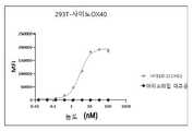
도 1b, 293T 세포의 표면에서 발현된 인간 OX40 단백질에 대한 HFB10-1E1hG1의 결합. EC50은 2 nM(MFI)였다.
도 1c, 293T 세포의 표면에서 발현된 사이노몰거스 원숭이 OX40 단백질에 대한 HFB10-1E1hG1의 결합. EC50은 2.9 nM(MFI)였다.
도 1d, 293T 세포의 표면에서 발현된 마우스 OX40 단백질에 대한 HFB10-1E1hG1의 결합. HFB10-1E1hG1은 293T 세포의 표면에서 발현된 마우스 OX40 단백질(MFI)에 결합하지 않는다.
도 1e, 293T 세포의 표면에서 발현된 인간 CD40 단백질에 대한 HFB10-1E1hG1의 결합. HFB10-1E1hG1은 293T 세포의 표면에서 발현된 인간 CD40 단백질(MFI)에 결합하지 않는다.
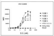
도 1f, 각각 293T 세포의 표면에서 발현된 인간 OX40 단백질에 대한 HFB10-1E1hG1, 기준품 1, 기준품 2, 기준품 3 및 기준품 4의 결합. HFB10-1E1hG1의 EC50은 2.02 nM(MFI)이고; 기준품 1의 EC50은 2.67 nM(MFI)이고; 기준품 2의 EC50은 4.17 nM(MFI)이고; 기준품 3의 EC50은 1.92 nM(MFI)이고; 기준품 4의 EC50은 2.11 nM(MFI)이다.
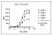
도 1g, 각각 293T 세포의 표면에서 발현된 사이노몰거스 원숭이 OX40 단백질에 대한 HFB10-1E1hG1, 기준품 1, 기준품 2, 기준품 3 및 기준품 4의 결합. HFB10-1E1hG1의 EC50은 2.94 nM(MFI)이고; 기준품 1의 EC50은 2.91 nM(MFI)이고; 기준품 2의 EC50은 6.13 nM(MFI)이고; 기준품 3의 EC50은 2.57 nM(MFI)이고; 기준품 4의 EC50은 3.54 nM(MFI)이다.
도 2a, Jurkat 리포터 세포에서의 HFB10-1E1hG1의 작용제성 활성. HFB10-1E1hG1의 EC50은 항-인간 IgG와의 가교결합의 경우에 2.9 nM(GFP의 MFI)이다.
도 2b, Jurkat 리포터 세포에서의 HFB10-1E1hG1의 작용제성 활성. 항-인간 IgG와의 가교결합 없이, HFB10-1E1hG1의 EC50은 이것이 항-인간 IgG와 가교결합될 때보다 훨씬 더 크다. 삼합체성 OX40L 재조합 단백질 단독은 항-인간 IgG와의 가교결합 없이 EC50 = 45 nM(GFP의 MFI)으로 NF-kb 신호전달을 활성화할 수 있다.
도 2c, Jurkat 리포터 세포에서의 HFB10-1E1hG1의 작용제성 활성. HFB10-1E1hG1은 항-인간 IgG와의 가교결합의 경우에 OX40L과의 협동적 작용제성 효과를 보여주고, OX40L에 의한 HFB10-1E1hG1의 첨가는 단일 성분과 비교하여 GFP의 MFI를 향상시킨다.
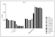
도 2d, 1차 CD4+ T 세포에서의 HFB10-1E1hG1의 작용제성 활성. 1차 CD4+ T 세포에서의 HFB10-1E1hG1의 작용제성 활성은 0.2 nM의 EC50으로 IL-2 분비에 의해 분석된다.
도 2e, 1차 CD4+ T 세포에서의 HFB10-1E1hG1의 작용제성 활성. 1차 CD4+ T 세포에서, HFB10-1E1hG1은 OX40L과의 협동적 작용제성 효과를 보여준다.
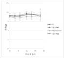
도 3a, HFB10-1E1hG1의 약력학적 시험.
도 4a, HFB10-1E1hG1의 생체내 항종양 효능. HFB10-1E1hG1은 PBS 대조군과 비교하여 종양 성장을 유의미하게 억제한다.
도 4b, HFB10-1E1hG1의 생체내 항종양 효능. HFB10-1E1hG1은 마우스에서의 유의미한 중량 소실을 생성시킬 수 있는 부작용이 없음을 나타낸다.
도 4c, 상이한 용량에서의 HFB10-1E1hG1의 생체내 항종양 효능(종양 크기).
도 4d, 상이한 용량에서의 HFB10-1E1hG1의 생체내 항종양 효능(체중).
도 5a, HFB10-1E1hG1의 가속 안정성 실험. SDS-PAGE 결과는 HFB10-1E1hG1이 상이한 온도 조건 하에 양호한 안정성을 갖는다는 것을 보여준다.
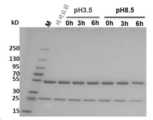
도 5b, HFB10-1E1hG1의 분해 실험. SDS-PAGE 결과는 HFB10-1E1hG1이 상이한 pH 조건 하에 양호한 안정성을 갖는다는 것을 보여준다.
도 5c, HFB10-1E1hG1의 산화 스트레스 실험. SDS-PAGE 결과는 HFB10-1E1hG1이 산화 스트레스 조건 하에 양호한 안정성을 갖는다는 것을 보여준다.
도 5d, HFB10-1E1hG1의 동결-해동 실험. SDS-PAGE 결과는 HFB10-1E1hG1이 동결-해동 조건 하에 양호한 안정성을 갖는다는 것을 보여준다.
도 6, 활성화된 원숭이 CD4+ T 세포에 대한 HFB10-1E1hG1의 결합. 기호: 실제 데이터 점; 곡선: 4개-매개변수 모델을 사용한 비선형 곡선 적합화. 3명의 공여자: 200107호(A: % 양성 세포 및 B: MFI), M20Z013009호(C: 양성 세포의 백분율 및 D: MFI) 및 20Z025008호(E: 양성 세포의 백분율 및 F: MFI)로부터 단리된 활성화된 T 세포에 대한 HFB10-1E1hG1의 결합.
도 7, 시간에 따른 WT 마우스에서의 Bmk 1 및 HFB10-1E1hG1의 평균 농도. 기호: 실제 데이터 점의 평균.
도 8, 시간에 따른 hOX40-KI 마우스에서의 Bmk 1 및 HFB10-1E1 hG1의 평균 농도. 기호: 실제 데이터 점의 평균.
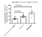
도 9, 종양 조직의 CD45+ 세포에서의 CD8+ T 세포의 백분율.
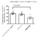
도 10, 종양 조직의 CD4+ T 세포에서의 Treg의 백분율.
도 11, 종양 조직의 CD4+ 및 CD8+ T 세포에서의 Ki67 발현.
도 12, 종양 조직의 CD4+ 및 CD8+ T 세포에서의 PD-1 발현.
도 13은 에피토프가 복합체에서 OX40 및 OX40L의 3D 결정 구조 모델로 맵핑된 항-OX40 항체 HFB301001의 에피토프를 보여준다.
도 14a 내지 도 14b는 세포-기반 생물발광 검정에 의해 측정 가능한 OX40 리간드의 작용제성 활성을 보여준다. 도 14a는 개별 OX40 리간드의 활성을 보여준다; 도 14b는 항-OX40 항체 HFB301001, 벤치마크 1 또는 벤치마크 2의 존재 하의 OX40 리간드의 활성을 보여준다. y축은 상대 발광(RLU)을 보여준다.
도 15a 내지 도 15b는 항체 처리 후 CD4+ T 세포에서의 OX40 발현을 보여준다. 도 15a는 유세포분석법에 의해 측정된 것과 같은 항-OX40 항체의 상이한 농도에 의한 처리 후 인간 PBMC로부터 단리된 OX40 양성 CD4+ T 세포의 백분율을 보여준다. 도 15b는 유세포분석법에 의해 측정된 것과 같은 인간 OX40 넉인 마우스에서 MC-38 쥣과 결장직장암 모델에서 10 mg/kg의 항-OX40 항체에 의한 제3 처리 후 24시간에 CD4+ T 세포에서의 OX40의 발현을 보여준다. 발현은 평균 형광 강도(MFI)로서 측정된다.
도 16은 아이소타입 대조군(10 mg/kg), 항-OX40 항체 벤치마크 1(1 mg/kg), 또는 항-OX40 항체 HFB301001(1 mg/kg, 0.1 mg/kg)에 의한 처리 후 인간 OX40 (hOX40) 넉인 (K/I) 마우스에서 MC-38 종양 모델에서의 종양 크기를 보여준다. 처리 시점은 화살표에 의해 표시되고, 오류 막대는 평균의 표준 오류를 나타내고, 유의도는 마지막 시점에서 1방향 ANOVA에 의해 계산된다(*: p-값 < 0.05, **: p-값 < 0.01).
도 17은 항-OX40 항체 HFB301001(10 mg/kg, 1 mg/kg, 0.1 mg/kg), 항-OX40 항체 벤치마크 1(10 mg/kg 또는 1 mg/kg), 아이소타입 대조군(10 mg/kg)에 의한 처리, 또는 인산염-완충 식염수(PBS) 처리 후 MC-38 쥣과 결장직장암 모델에 의한 인간 OX40(hOX40) 넉인(K/I) 마우스의 퍼센트 생존을 보여준다. 유의도는 log-랭크 시험(*: p-값 < 0.05, **: p-값 < 0.01)에 의해 계산된다.
도 18a 내지 도 18e는 대조군(IgG1), OX40 항체 벤치마크 1 또는 항-OX40 항체 HFB301001에 의한 처리 후 인간 OX40(hOX40) 넉인(KI) 마우스에서 MC-38 쥣과 결장직장암 모델에서 종양 미세환경 내에 면역 세포 집단의 유세포분석법 측정을 보여준다. 도 18a는 생존 가능 CD45+CD3+CD4+ 세포에서의 CD3+CD4+ Ki67+ T 세포의 백분율을 보여준다. 도 18b는 생존 가능 CD45+CD3+CD8+ 세포에서의 CD3+CD8+ Ki67+ T 세포의 백분율을 보여준다. 도 18c는 생존 가능 CD45+CD3+CD4+ 세포에서의 CD3+CD4+ PD-1+ T 세포의 백분율을 보여준다. 도 18d는 생존 가능 CD45+CD3+CD8+ 세포에서의 CD3+CD8+ PD-1+ T 세포의 백분율을 보여준다. 도 18e는 생존 가능 CD45+CD3+CD4+ 세포에서의 CD3+CD4+CD25+Foxp3+ 세포의 백분율을 보여준다.Figure 1a, Binding of HFB10-1E1hG1 to OX40 protein. The EC80 of HFB10-1E1hG1 is 0.71 nM.
1b , Binding of HFB10-1E1hG1 to human OX40 protein expressed on the surface of 293T cells. EC50 was 2 nM (MFI).
1C , Binding of HFB10-1E1hG1 to cynomolgus monkey OX40 protein expressed on the surface of 293T cells. The EC50 was 2.9 nM (MFI).
1d , Binding of HFB10-1E1hG1 to mouse OX40 protein expressed on the surface of 293T cells. HFB10-1E1hG1 does not bind mouse OX40 protein (MFI) expressed on the surface of 293T cells.
FIG. 1E , Binding of HFB10-1E1hG1 to human CD40 protein expressed on the surface of 293T cells. HFB10-1E1hG1 does not bind to human CD40 protein (MFI) expressed on the surface of 293T cells.
1F , Binding of HFB10-1E1hG1,
Figure 1g, Binding of HFB10-1E1hG1,
2A , Agonistic activity of HFB10-1E1hG1 in Jurkat reporter cells. The EC50 of HFB10-1E1hG1 is 2.9 nM (MFI of GFP) for cross-linking with anti-human IgG.
Figure 2b, Agonistic activity of HFB10-1E1hG1 in Jurkat reporter cells. Without cross-linking with anti-human IgG, the EC50 of HFB10-1E1hG1 is much greater than when it is cross-linked with anti-human IgG. Trimeric OX40L recombinant protein alone can activate NF-kb signaling with an EC50 = 45 nM (MFI of GFP) without cross-linking with anti-human IgG.
Figure 2c, Agonistic activity of HFB10-1E1hG1 in Jurkat reporter cells. HFB10-1E1hG1 shows a cooperative agonistic effect with OX40L in case of cross-linking with anti-human IgG, and addition of HFB10-1E1hG1 by OX40L enhances the MFI of GFP compared to single component.
2D , Agonistic activity of HFB10-1E1hG1 in primary CD4+ T cells. Agonistic activity of HFB10-1E1hG1 on primary CD4+ T cells is assayed by IL-2 secretion with an EC50 of 0.2 nM.
2e , Agonistic activity of HFB10-1E1hG1 in primary CD4+ T cells. On primary CD4+ T cells, HFB10-1E1hG1 shows cooperative agonistic effects with OX40L.
Figure 3a, pharmacodynamic test of HFB10-1E1hG1.
Figure 4a, in vivo anti-tumor efficacy of HFB10-1E1hG1. HFB10-1E1hG1 significantly inhibits tumor growth compared to the PBS control.
Figure 4b, in vivo antitumor efficacy of HFB10-1E1hG1. HFB10-1E1hG1 shows no side effects that could produce significant weight loss in mice.
4C , In vivo antitumor efficacy (tumor size) of HFB10-1E1hG1 at different doses.
4D , In vivo antitumor efficacy (body weight) of HFB10-1E1hG1 at different doses.
Figure 5a, accelerated stability experiment of HFB10-1E1hG1. SDS-PAGE results show that HFB10-1E1hG1 has good stability under different temperature conditions.
Figure 5b, degradation experiment of HFB10-1E1hG1. SDS-PAGE results show that HFB10-1E1hG1 has good stability under different pH conditions.
Figure 5c, oxidative stress experiment of HFB10-1E1hG1. SDS-PAGE results show that HFB10-1E1hG1 has good stability under oxidative stress conditions.
5D , Freeze-thaw experiment of HFB10-1E1hG1. SDS-PAGE results show that HFB10-1E1hG1 has good stability under freeze-thaw conditions.
6 , Binding of HFB10-1E1hG1 to activated monkey CD4+ T cells. Symbols: real data points; Curves: nonlinear curve fitting using a 4-parameter model. Isolated from 3 donors: 200107 (A: % positive cells and B: MFI), M20Z013009 (C: percentage of positive cells and D: MFI) and 20Z025008 (E: percentage of positive cells and F: MFI) Binding of HFB10-1E1hG1 to activated T cells.
Figure 7, Mean concentrations of Bmk1 and HFB10-1E1hG1 in WT mice over time. Symbol: Average of actual data points.
Figure 8, Mean concentrations of Bmk1 and HFB10-1E1 hG1 in hOX40-KI mice over time. Symbol: Average of actual data points.
9 , Percentage of CD8+ T cells in CD45+ cells in tumor tissue.
10 , Percentage of Tregs in CD4+ T cells in tumor tissue.
11 , Ki67 expression on CD4+ and CD8+ T cells in tumor tissue.
12 , PD-1 expression on CD4+ and CD8+ T cells in tumor tissue.
13 shows the epitopes of the anti-OX40 antibody HFB301001 where the epitopes were mapped to a 3D crystal structure model of OX40 and OX40L in complex.
14A-14B show the agonistic activity of OX40 ligands measurable by a cell-based bioluminescence assay. 14A shows the activity of individual OX40 ligands; 14B shows the activity of OX40 ligands in the presence of anti-OX40 antibody HFB301001,
15A-15B show OX40 expression in CD4+ T cells after antibody treatment. 15A shows the percentage of OX40 positive CD4+ T cells isolated from human PBMCs after treatment with different concentrations of anti-OX40 antibody as measured by flow cytometry. Figure 15B shows OX40 in CD4+ T cells 24 hours after third treatment with 10 mg/kg anti-OX40 antibody in MC-38 murine colorectal cancer model in human OX40 knock-in mice as measured by flow cytometry. shows the expression of Expression is measured as mean fluorescence intensity (MFI).
Figure 16 shows human OX40 after treatment with isotype control (10 mg/kg), anti-OX40 antibody Benchmark 1 (1 mg/kg), or anti-OX40 antibody HFB301001 (1 mg/kg, 0.1 mg/kg). (hOX40) shows the tumor size in the MC-38 tumor model in knock-in (K/I) mice. Treatment time points are indicated by arrows, error bars represent standard error of the mean, and significance is calculated by one-way ANOVA at the last time point (*: p-value < 0.05, **: p-value < 0.01) .
17 shows anti-OX40 antibody HFB301001 (10 mg/kg, 1 mg/kg, 0.1 mg/kg), anti-OX40 antibody benchmark 1 (10 mg/kg or 1 mg/kg), isotype control (10 mg /kg), or after treatment with phosphate-buffered saline (PBS), the percent survival of human OX40 (hOX40) knock-in (K/I) mice by the MC-38 murine colorectal cancer model is shown. Significance is calculated by log-rank test (*: p-value < 0.05, **: p-value < 0.01).
Figures 18A-18E show the tumor microenvironment in the MC-38 murine colorectal cancer model in human OX40 (hOX40) knock-in (KI) mice after treatment with control (IgG1),
일 양태에서, 본 발명은 서열 번호 1에 기재된 것과 같은 중쇄 CDR1 구조 도메인, 서열 번호 2에 기재된 것과 같은 중쇄 CDR2 구조 도메인 및 서열 번호 3에 기재된 것과 같은 중쇄 CDR3 구조 도메인을 갖는 중쇄 가변 영역; 및 서열 번호 9에 기재된 것과 같은 경쇄 CDR1 도메인, 서열 번호 10에 기재된 것과 같은 경쇄 CDR2 도메인 및 서열 번호 11에 기재된 것과 같은 경쇄 CDR3 도메인을 갖는 경쇄 가변 영역을 포함하는 단리된 항-OX40 항체 또는 이의 항원-결합 단편을 제공한다.In one aspect, the invention provides a heavy chain variable region having a heavy chain CDR1 structural domain as set forth in SEQ ID NO: 1, a heavy chain CDR2 structural domain as set forth in SEQ ID NO: 2 and a heavy chain CDR3 structural domain as set forth in SEQ ID NO: 3; and an isolated anti-OX40 antibody or antigen thereof comprising a light chain variable region having a light chain CDR1 domain as set forth in SEQ ID NO:9, a light chain CDR2 domain as set forth in SEQ ID NO:10, and a light chain CDR3 domain as set forth in SEQ ID NO:11. -provides binding fragments;
일 실시형태에서, 본원에 기재된 것과 같은 항-OX40 항체 또는 이의 항원-결합 단편은 서열 번호 4에 기재된 것과 같은 중쇄 가변 영역, 또는 서열 번호 4와 적어도 80%, 81%, 82%, 83%, 84%, 85%, 86%, 87%, 88%, 89%, 90%, 91%, 92%, 93%, 94%, 95%, 96%, 97%, 98%, 99% 또는 100%의 상동성을 갖는 중쇄 가변 영역; 및 서열 번호 12에 기재된 것과 같은 경쇄 가변 영역, 또는 서열 번호 12와 적어도 80%, 81%, 82%, 83%, 84%, 85%, 86%, 87%, 88%, 89%, 90%, 91%, 92%, 93%, 94%, 95%, 96%, 97%, 98%, 99% 또는 100%의 상동성을 갖는 경쇄 가변 영역을 포함한다.In one embodiment, the anti-OX40 antibody or antigen-binding fragment thereof as described herein is a heavy chain variable region as set forth in SEQ ID NO:4, or at least 80%, 81%, 82%, 83%, 84%, 85%, 86%, 87%, 88%, 89%, 90%, 91%, 92%, 93%, 94%, 95%, 96%, 97%, 98%, 99% or 100% A heavy chain variable region having the homology of; and a light chain variable region as set forth in SEQ ID NO: 12, or at least 80%, 81%, 82%, 83%, 84%, 85%, 86%, 87%, 88%, 89%, 90% of SEQ ID NO: 12 , a light chain variable region having 91%, 92%, 93%, 94%, 95%, 96%, 97%, 98%, 99% or 100% homology.
일 실시형태에서, 본원에 기재된 것과 같은 항-OX40 항체 또는 이의 항원-결합 단편은 중쇄 불변 영역 및 경쇄 불변 영역을 추가로 포함하고; 바람직하게는, 중쇄 불변 영역은 서열 번호 5에 기재된 것과 같은 중쇄 불변 영역, 또는 서열 번호 5와 적어도 80%, 81%, 82%, 83%, 84%, 85%, 86%, 87%, 88%, 89%, 90%, 91%, 92%, 93%, 94%, 95%, 96%, 97%, 98%, 99% 또는 100%의 상동성을 갖는 중쇄 불변 영역이고/이거나; 바람직하게는, 경쇄 불변 영역은 서열 번호 13에 기재된 것과 같은 경쇄 불변 영역, 또는 서열 번호 13과 적어도 80%, 81%, 82%, 83%, 84%, 85%, 86%, 87%, 88%, 89%, 90%, 91%, 92%, 93%, 94%, 95%, 96%, 97%, 98%, 99% 또는 100%의 상동성을 갖는 경쇄 불변 영역이다.In one embodiment, an anti-OX40 antibody or antigen-binding fragment thereof as described herein further comprises a heavy chain constant region and a light chain constant region; Preferably, the heavy chain constant region is a heavy chain constant region as set forth in SEQ ID NO: 5, or at least 80%, 81%, 82%, 83%, 84%, 85%, 86%, 87%, 88 of SEQ ID NO: 5 %, 89%, 90%, 91%, 92%, 93%, 94%, 95%, 96%, 97%, 98%, 99% or 100% homology; Preferably, the light chain constant region is a light chain constant region as set forth in SEQ ID NO: 13, or at least 80%, 81%, 82%, 83%, 84%, 85%, 86%, 87%, 88% of SEQ ID NO: 13 %, 89%, 90%, 91%, 92%, 93%, 94%, 95%, 96%, 97%, 98%, 99% or 100% homology.
일 실시형태에서, 본원에 기재된 것과 같은 항-OX40 항체 또는 이의 항원-결합 단편은 중쇄 가변 영역에 연결된 중쇄 신호 펩타이드 및/또는 경쇄 가변 영역에 연결된 경쇄 신호 펩타이드를 추가로 포함하고; 바람직하게는, 중쇄 신호 펩타이드는 서열 번호 6에 기재된 것과 같은 중쇄 신호 펩타이드, 또는 서열 번호 6과 적어도 80%, 81%, 82%, 83%, 84%, 85%, 86%, 87%, 88%, 89%, 90%, 91%, 92%, 93%, 94%, 95%, 96%, 97%, 98%, 99% 또는 100%의 상동성을 갖는 중쇄 신호 펩타이드이고/이거나; 바람직하게는, 경쇄 신호 펩타이드는 서열 번호 14에 기재된 것과 같은 경쇄 신호 펩타이드, 또는 서열 번호 14와 적어도 80%, 81%, 82%, 83%, 84%, 85%, 86%, 87%, 88%, 89%, 90%, 91%, 92%, 93%, 94%, 95%, 96%, 97%, 98%, 99% 또는 100%의 상동성을 갖는 경쇄 신호 펩타이드이다.In one embodiment, an anti-OX40 antibody or antigen-binding fragment thereof as described herein further comprises a heavy chain signal peptide linked to a heavy chain variable region and/or a light chain signal peptide linked to a light chain variable region; Preferably, the heavy chain signal peptide is a heavy chain signal peptide as set forth in SEQ ID NO: 6, or at least 80%, 81%, 82%, 83%, 84%, 85%, 86%, 87%, 88 of SEQ ID NO: 6 %, 89%, 90%, 91%, 92%, 93%, 94%, 95%, 96%, 97%, 98%, 99% or 100% homologous heavy chain signal peptide; Preferably, the light chain signal peptide is a light chain signal peptide as set forth in SEQ ID NO: 14, or at least 80%, 81%, 82%, 83%, 84%, 85%, 86%, 87%, 88% of SEQ ID NO: 14 %, 89%, 90%, 91%, 92%, 93%, 94%, 95%, 96%, 97%, 98%, 99% or 100% homology.
일 실시형태에서, 본원에 기재된 것과 같은 항-OX40 항체 또는 이의 항원-결합 단편은 IgG 항체 또는 이의 항원-결합 단편, 및 바람직하게는 IgG1 항체 또는 이의 항원-결합 단편이다.In one embodiment, the anti-OX40 antibody or antigen-binding fragment thereof as described herein is an IgG antibody or antigen-binding fragment thereof, and preferably an IgG1 antibody or antigen-binding fragment thereof.
일 실시형태에서, 본원에 기재된 것과 같은 항-OX40 항체 또는 이의 항원-결합 단편은 단일클론 항체 또는 이의 항원-결합 단편이다.In one embodiment, the anti-OX40 antibody or antigen-binding fragment thereof as described herein is a monoclonal antibody or antigen-binding fragment thereof.
일 실시형태에서, 본원에 기재된 것과 같은 이의 항원-결합 단편은 Fab, Fab', F(ab')2, Fv, scFv 또는 sdAb이다.In one embodiment, an antigen-binding fragment thereof as described herein is a Fab, Fab', F(ab')2, Fv, scFv or sdAb.
일 양태에서, 본 발명은 본원에 기재된 것과 같은 항-OX40 항체 또는 이의 항원-결합 단편, 및 추가 치료제를 포함하는 항체-약물 접합체를 제공하고; 바람직하게는, 항-OX40 항체 또는 이의 항원-결합 단편은 링커를 통해 추가 치료제에 연결된다.In one aspect, the invention provides antibody-drug conjugates comprising an anti-OX40 antibody or antigen-binding fragment thereof as described herein, and an additional therapeutic agent; Preferably, the anti-OX40 antibody or antigen-binding fragment thereof is linked to the additional therapeutic agent via a linker.
일 양태에서, 본 발명은 본원에 기재된 것과 같은 항-OX40 항체 또는 이의 항원-결합 단편을 암호화하는 핵산을 제공한다.In one aspect, the invention provides a nucleic acid encoding an anti-OX40 antibody or antigen-binding fragment thereof as described herein.
일 실시형태에서, 본원에 기재된 것과 같은 핵산은 서열 번호 20에 기재된 것과 같은 서열을 암호화하는 중쇄 가변 영역 뉴클레오타이드, 및/또는 서열 번호 28에 기재된 것과 같은 서열을 암호화하는 경쇄 가변 영역 뉴클레오타이드를 포함하고; 바람직하게는, 핵산은 서열 번호 21에 기재된 것과 같은 서열을 암호화하는 중쇄 불변 영역 뉴클레오타이드, 및/또는 서열 번호 29에 기재된 것과 같은 서열을 암호화하는 경쇄 불변 영역 뉴클레오타이드를 추가로 포함한다.In one embodiment, a nucleic acid as described herein comprises heavy chain variable region nucleotides encoding a sequence as set forth in SEQ ID NO: 20, and/or light chain variable region nucleotides encoding a sequence as set forth in SEQ ID NO: 28; Preferably, the nucleic acid further comprises heavy chain constant region nucleotides encoding a sequence as set forth in SEQ ID NO:21, and/or light chain constant region nucleotides encoding a sequence as set forth in SEQ ID NO:29.
일 양태에서, 본 발명은 본원에 기재된 것과 같은 핵산을 포함하는 발현 벡터를 제공한다.In one aspect, the invention provides expression vectors comprising a nucleic acid as described herein.
일 양태에서, 본 발명은 본원에 기재된 것과 같은 핵산 또는 본원에 기재된 것과 같은 발현 벡터를 포함하는 숙주 세포를 제공한다.In one aspect, the invention provides a host cell comprising a nucleic acid as described herein or an expression vector as described herein.
일 양태에서, 본 발명은, 항체 또는 이의 항원-결합 단편을 발현하기에 적합한 조건 하에 본원에 기재된 것과 같은 숙주 세포를 배양하는 단계 및 배양 배지로부터 발현된 항체 또는 이의 항원-결합 단편을 회수하는 단계를 포함하는, 본원에 기재된 것과 같은 항-OX40 항체 또는 이의 항원-결합 단편을 제조하는 방법을 제공한다.In one aspect, the invention provides a method comprising culturing a host cell as described herein under conditions suitable for expressing an antibody or antigen-binding fragment thereof and recovering the expressed antibody or antigen-binding fragment thereof from the culture medium. A method of making an anti-OX40 antibody or antigen-binding fragment thereof as described herein, comprising:
일 양태에서, 본 발명은 본원에 기재된 것과 같은 항-OX40 항체 또는 이의 항원-결합 단편, 또는 본원에 기재된 것과 같은 항체-약물 접합체, 또는 본원에 기재된 것과 같은 핵산, 또는 본원에 기재된 것과 같은 발현 벡터; 및 약제학적으로 허용 가능한 담체를 포함하는 약제학적 조성물을 제공한다.In one aspect, the invention provides an anti-OX40 antibody or antigen-binding fragment thereof as described herein, or an antibody-drug conjugate as described herein, or a nucleic acid as described herein, or an expression vector as described herein. ; And it provides a pharmaceutical composition comprising a pharmaceutically acceptable carrier.
일 실시형태에서, 본원에 기재된 것과 같은 항-OX40 항체 또는 이의 항원-결합 단편, 또는 본원에 기재된 것과 같은 항체-약물 접합체, 또는 본원에 기재된 것과 같은 약제학적 조성물은 암의 치료에 사용된다. 일 실시형태에서, 암은 편평 세포 암종(예를 들어, 상피 편평 세포 암종), 폐암(소세포 폐암, 비소세포 폐암, 페의 선암 및 페의 편평 세포 암종을 포함), 복막 암종, 간세포 암종, 위암(위장관암 및 위장관 기질 암을 포함), 췌장암, 교모세포종, 자궁경부암, 난소암, 간암, 방광암, 요로암, 간 종양, 유방암, 결장암, 직장암, 결장직장암, 자궁내막암 또는 자궁암, 침샘암, 신장암, 전립선암, 음문암, 갑상선암, 간암, 항문암, 음경암, 흑색종, 표재성 미만성 흑색종, 악성 흑자 흑색종, 말단 흑색종, 결절성 흑색종, 다발성 골수종 및 B-세포 림프종, 만성 림프구성 백혈병(CLL: chronic lymphocytic leukemia), 급성 림프아구성 백혈병(ALL: acute lymphoblastic leukemia), 모발 세포 백혈병, 만성 골수아구성 백혈병, 및 이식후 림프증식성 장애(PTLD: post-transplantation lymphoproliferative disorder), 켈로이드와 연관된 비정상 혈관 증식, 부종(예를 들어, 뇌종양과 연관됨) 및 메이그 증후군(Meigs syndrome), 뇌종양 및 뇌암, 두경부암 및 연관된 전이로부터 선택된다.In one embodiment, the anti-OX40 antibody or antigen-binding fragment thereof as described herein, or antibody-drug conjugate as described herein, or pharmaceutical composition as described herein is used for the treatment of cancer. In one embodiment, the cancer is squamous cell carcinoma (eg, epithelial squamous cell carcinoma), lung cancer (including small cell lung cancer, non-small cell lung cancer, adenocarcinoma of the lung and squamous cell carcinoma of the lung), peritoneal carcinoma, hepatocellular carcinoma, gastric cancer (including gastrointestinal cancer and gastrointestinal stromal cancer); pancreatic cancer; glioblastoma; cervical cancer; ovarian cancer; liver cancer; bladder cancer; urinary tract cancer; Kidney cancer, prostate cancer, vulvar cancer, thyroid cancer, liver cancer, anal cancer, penile cancer, melanoma, superficial diffuse melanoma, lentigo malignant melanoma, acral melanoma, nodular melanoma, multiple myeloma and B-cell lymphoma, chronic lymphoma chronic lymphocytic leukemia (CLL), acute lymphoblastic leukemia (ALL), hairy cell leukemia, chronic myeloblastic leukemia, and post-transplantation lymphoproliferative disorder (PTLD); abnormal vascular proliferation associated with keloids, edema (eg associated with brain tumors) and Meigs syndrome, brain tumors and brain cancers, head and neck cancers and associated metastases.
일 양태에서, 본 발명은, 암을 치료하도록 본원에 기재된 것과 같은 항-OX40 항체 또는 이의 항원-결합 단편, 또는 본원에 기재된 것과 같은 항체-약물 접합체, 또는 본원에 기재된 것과 같은 약제학적 조성물의 치료학적 유효량을 이를 필요로 하는 대상체에게 투여하는 단계를 포함하는, 암을 치료하는 방법을 제공한다. 일 실시형태에서, 암은 편평 세포 암종(예를 들어, 상피 편평 세포 암종), 폐암(소세포 폐암, 비소세포 폐암, 페의 선암 및 페의 편평 세포 암종을 포함), 복막 암종, 간세포 암종, 위암(위장관암 및 위장관 기질 암을 포함), 췌장암, 교모세포종, 자궁경부암, 난소암, 간암, 방광암, 요로암, 간 종양, 유방암, 결장암, 직장암, 결장직장암, 자궁내막암 또는 자궁암, 침샘암, 신장암, 전립선암, 음문암, 갑상선암, 간암, 항문암, 음경암, 흑색종, 표재성 미만성 흑색종, 악성 흑자 흑색종, 말단 흑색종, 결절성 흑색종, 다발성 골수종 및 B-세포 림프종, 만성 림프구성 백혈병(CLL), 급성 림프아구성 백혈병(ALL), 모발 세포 백혈병, 만성 골수아구성 백혈병, 및 이식후 림프증식성 장애(PTLD), 켈로이드와 연관된 비정상 혈관 증식, 부종(예를 들어, 뇌종양과 연관됨) 및 메이그 증후군, 뇌종양 및 뇌암, 두경부암 및 연관된 전이로부터 선택된다.In one aspect, the invention relates to treatment of an anti-OX40 antibody or antigen-binding fragment thereof as described herein, or an antibody-drug conjugate as described herein, or a pharmaceutical composition as described herein for the treatment of cancer. A method for treating cancer is provided, comprising administering a therapeutically effective amount to a subject in need thereof. In one embodiment, the cancer is squamous cell carcinoma (eg, epithelial squamous cell carcinoma), lung cancer (including small cell lung cancer, non-small cell lung cancer, adenocarcinoma of the lung and squamous cell carcinoma of the lung), peritoneal carcinoma, hepatocellular carcinoma, gastric cancer (including gastrointestinal cancer and gastrointestinal stromal cancer); pancreatic cancer; glioblastoma; cervical cancer; ovarian cancer; liver cancer; bladder cancer; urinary tract cancer; Kidney cancer, prostate cancer, vulvar cancer, thyroid cancer, liver cancer, anal cancer, penile cancer, melanoma, superficial diffuse melanoma, lentigo malignant melanoma, acral melanoma, nodular melanoma, multiple myeloma and B-cell lymphoma, chronic lymphoma Constitutive leukemia (CLL), acute lymphoblastic leukemia (ALL), hairy cell leukemia, chronic myeloblastic leukemia, and post-transplant lymphoproliferative disorder (PTLD), abnormal vascular proliferation associated with keloids, edema (e.g., brain tumors) associated with) and Meigs syndrome, brain tumors and brain cancers, head and neck cancers and associated metastases.
일 양태에서, 본 발명은 암의 치료를 위한 약제의 제조에서의 본원에 기재된 것과 같은 항-OX40 항체 또는 이의 항원-결합 단편, 또는 본원에 기재된 것과 같은 항체-약물 접합체, 또는 본원에 기재된 것과 같은 약제학적 조성물의 용도를 제공한다. 일 실시형태에서, 암은 편평 세포 암종(예를 들어, 상피 편평 세포 암종), 폐암(소세포 폐암, 비소세포 폐암, 페의 선암 및 페의 편평 세포 암종을 포함), 복막 암종, 간세포 암종, 위암(위장관암 및 위장관 기질 암을 포함), 췌장암, 교모세포종, 자궁경부암, 난소암, 간암, 방광암, 요로암, 간 종양, 유방암, 결장암, 직장암, 결장직장암, 자궁내막암 또는 자궁암, 침샘암, 신장암, 전립선암, 음문암, 갑상선암, 간암, 항문암, 음경암, 흑색종, 표재성 미만성 흑색종, 악성 흑자 흑색종, 말단 흑색종, 결절성 흑색종, 다발성 골수종 및 B-세포 림프종, 만성 림프구성 백혈병(CLL), 급성 림프아구성 백혈병(ALL), 모발 세포 백혈병, 만성 골수아구성 백혈병, 및 이식후 림프증식성 장애(PTLD), 켈로이드와 연관된 비정상 혈관 증식, 부종(예를 들어, 뇌종양과 연관됨) 및 메이그 증후군, 뇌종양 및 뇌암, 두경부암 및 연관된 전이로부터 선택된다.In one aspect, the invention relates to the use of an anti-OX40 antibody or antigen-binding fragment thereof as described herein, or an antibody-drug conjugate as described herein, or as described herein, in the manufacture of a medicament for the treatment of cancer. Use of the pharmaceutical composition is provided. In one embodiment, the cancer is squamous cell carcinoma (eg, epithelial squamous cell carcinoma), lung cancer (including small cell lung cancer, non-small cell lung cancer, adenocarcinoma of the lung and squamous cell carcinoma of the lung), peritoneal carcinoma, hepatocellular carcinoma, gastric cancer (including gastrointestinal cancer and gastrointestinal stromal cancer); pancreatic cancer; glioblastoma; cervical cancer; ovarian cancer; liver cancer; bladder cancer; urinary tract cancer; Kidney cancer, prostate cancer, vulvar cancer, thyroid cancer, liver cancer, anal cancer, penile cancer, melanoma, superficial diffuse melanoma, lentigo malignant melanoma, acral melanoma, nodular melanoma, multiple myeloma and B-cell lymphoma, chronic lymphoma Constitutive leukemia (CLL), acute lymphoblastic leukemia (ALL), hairy cell leukemia, chronic myeloblastic leukemia, and post-transplant lymphoproliferative disorder (PTLD), abnormal vascular proliferation associated with keloids, edema (e.g., brain tumors) associated with) and Meigs syndrome, brain tumors and brain cancers, head and neck cancers and associated metastases.
일 양태에서, 본 발명은 Treg 기능을 억제하는 것(예를 들어, Treg의 억제 기능을 억제하는 것), OX40을 발현하는 세포(예를 들어, OX40의 높은 수준을 발현하는 세포)를 사멸하는 것, 효과기 T 세포 기능을 향상시키는 것 및/또는 기억 T 세포 기능을 향상시키는 것, 종양 면역력을 감소시키는 것, T 세포 융합을 향상시키는 것 및/또는 OX40-발현 세포를 고갈시키는 것의 상기 중 하나 이상에서 본원에 기재된 것과 같은 항-OX40 항체 또는 이의 항원-결합 단편, 또는 본원에 기재된 것과 같은 항체-약물 접합체, 또는 본원에 기재된 것과 같은 약제학적 조성물의 용도를 제공한다.In one aspect, the present invention is directed to suppressing Treg function (eg, inhibiting the suppressive function of Treg), or killing cells expressing OX40 (eg, cells expressing high levels of OX40). any of the above of enhancing effector T cell function and/or enhancing memory T cell function, reducing tumor immunity, enhancing T cell fusion and/or depleting OX40-expressing cells. Provided are uses of an anti-OX40 antibody or antigen-binding fragment thereof as described herein above, or an antibody-drug conjugate as described herein, or a pharmaceutical composition as described herein.
일 양태에서, 본 발명은 Treg 기능을 억제하는 것(예를 들어, Treg의 억제 기능을 억제하는 것), OX40을 발현하는 세포(예를 들어, OX40의 높은 수준을 발현하는 세포)를 사멸하는 것, 효과기 T 세포 기능을 향상시키는 것 및/또는 기억 T 세포 기능을 향상시키는 것, 종양 면역력을 감소시키는 것, T 세포 융합을 향상시키는 것 및/또는 OX40-발현 세포를 고갈시키는 것의 상기 중 하나 이상을 위한 약제의 제조에서의 본원에 기재된 것과 같은 항-OX40 항체 또는 이의 항원-결합 단편, 또는 본원에 기재된 것과 같은 항체-약물 접합체, 또는 본원에 기재된 것과 같은 약제학적 조성물의 용도를 제공한다.In one aspect, the present invention is directed to suppressing Treg function (eg, inhibiting the suppressive function of Treg), or killing cells expressing OX40 (eg, cells expressing high levels of OX40). any of the above of enhancing effector T cell function and/or enhancing memory T cell function, reducing tumor immunity, enhancing T cell fusion and/or depleting OX40-expressing cells. Use of an anti-OX40 antibody or antigen-binding fragment thereof as described herein, or an antibody-drug conjugate as described herein, or a pharmaceutical composition as described herein is provided in the manufacture of a medicament for any of the above.
일 양태에서, 본 발명은 본원에 기재된 것과 같은 항-OX40 항체 또는 이의 항원-결합 단편, 또는 본원에 기재된 것과 같은 항체-약물 접합체, 또는 본원에 기재된 것과 같은 약제학적 조성물; 및 하나 이상의 추가 치료제를 포함하는 약제학적 조합을 제공한다.In one aspect, the invention provides an anti-OX40 antibody or antigen-binding fragment thereof as described herein, or an antibody-drug conjugate as described herein, or a pharmaceutical composition as described herein; and one or more additional therapeutic agents.
일 양태에서, 본 발명은 본원에 기재된 것과 같은 항-OX40 항체 또는 이의 항원-결합 단편, 또는 본원에 기재된 것과 같은 항체-약물 접합체, 또는 본원에 기재된 것과 같은 약제학적 조성물을 포함하고, 바람직하게는 약물 전달 장치를 추가로 포함하는 키트를 제공한다.In one aspect, the invention comprises an anti-OX40 antibody or antigen-binding fragment thereof as described herein, or an antibody-drug conjugate as described herein, or a pharmaceutical composition as described herein, preferably A kit further comprising a drug delivery device is provided.
본 발명의 일부 실시형태는 OX-40에 대한 작용제성 항체를 제공한다. 이는 다른 작용제성 항-OX-40 항체와 비교하여 OX-40 수용체의 하향조절을 발생시키지 않거나 덜 발생시켰다. 수용체 하향조절 또는 감소된 수용체 하향조절의 이 결여는 본 발명에 의해 제공된 작용제성 항체가 이의 에피토프를 인식할 수 있다는 사실로 인할 수 있다. 본 발명에 의해 제공된 작용제성 항체는 특히 당해 분야에 공지된 다른 작용제성 항-OX-40 항체와 비교하여 최적화된 결합 역학을 또한 가질 수 있다.Some embodiments of the invention provide agonistic antibodies to OX-40. It resulted in no or less downregulation of the OX-40 receptor compared to other agonistic anti-OX-40 antibodies. This lack of receptor downregulation or reduced receptor downregulation may be due to the fact that the agonistic antibodies provided by the present invention are capable of recognizing their epitopes. The agonistic antibodies provided by the present invention may also have optimized binding kinetics, especially compared to other agonistic anti-OX-40 antibodies known in the art.
본원에 기재된 다양한 실시형태의 하나, 일부 또는 모든 특징이 본 발명의 추가의 실시형태를 형성하도록 조합될 수 있다고 이해되어야 한다. 본 발명의 이들 양태 및 다른 양태는 당해 분야의 숙련자에게 자명하다. 본 발명의 이들 실시형태 및 다른 실시형태는 하기 자세히 추가로 기재될 것이다.It should be understood that one, some or all features of the various embodiments described herein may be combined to form additional embodiments of the present invention. These and other aspects of the invention will be apparent to those skilled in the art. These and other embodiments of the present invention will be described further in detail below.
2개의 서열에서의 뉴클레오타이드 또는 아미노산의 서열이 최대 거울상이성질체 정렬에 대해 기재될 때 동일하면 2개의 폴리뉴클레오타이드 또는 폴리펩타이드 서열은 "동일"하다고 칭해진다. 2개의 서열 사이의 비교는 통상적으로 서열 유사성의 국소 영역을 확인하고 비교하기 위해 비교 윈도우에 걸쳐 서열을 비교함으로써 수행된다. 본원에 사용된 것과 같이 "비교 윈도우"는 적어도 약 20개(보통 약 30개 내지 약 75개, 또는 약 40개 내지 약 50개)의 인접한 위치를 갖는 분절을 지칭하고; 2개의 서열이 최적으로 정렬된 후, 서열은 동일한 수의 인접한 위치를 갖는 기준 서열과 비교될 수 있다.Two polynucleotide or polypeptide sequences are said to be “identical” if the sequences of nucleotides or amino acids in the two sequences are identical when described for maximum enantiomeric alignment. A comparison between two sequences is typically performed by comparing the sequences over a window of comparison to identify and compare local regions of sequence similarity. As used herein, “comparison window” refers to a segment having at least about 20 (usually about 30 to about 75, or about 40 to about 50) contiguous locations; After the two sequences are optimally aligned, the sequences can be compared to a reference sequence having the same number of contiguous positions.
디폴트 매개변수를 사용하여 생물정보 소프트웨어(Inc., 위스콘신주 매디슨)의 스위트에서의 프로그램을 사용하여 비교를 위한 서열의 최적 정렬을 수행할 수 있다. 이 프로그램은 하기 참고문헌에 기재된 몇몇 정렬 계획을 실행한다: Dayhoff, M.O., 1978, A model of evolutionary change in proteins-Matrices for detecting distant relationships. In Dayhoff, M.O. (ed.) Atlas of Protein Sequence and Structure, National Biomedical Research Foundation, Washington DC, Vol. 5, Supplement 3, pp. 345 to 358 pages; Hein J., 1990, Unified Approach to Alignment and Phylogenes, pp. 626-645, Methods in Enzymology, Vol. 183, Academic Press, Inc., San Diego, CA; Higgins, D.G. and Sharp, P.M., 1989, CABIOS 5: 151-153; Myers, E.W. and Muller W., 1988, CABIOS 4:11-17; Robinson, E.D., 1971, Comb. Theor. 11:105; Santou, N., Nes, M., 1987, Mol.Biol.Evol.4:406-425; Sneath, P.H.A. and Sokal, R.R., 1973, Numerical Taxonomy the Principles and Practice of Numerical Taxonomy, Freeman Press, San Francisco, CA; Wilbur, W.J. and Lipman, D.J., 1983, Proc. Natl. Acad. Sci. USA 80:726-730.Optimal alignment of sequences for comparison can be performed using programs in the Suite of Bioinformatic Software (Inc., Madison, Wis.) using default parameters. This program runs several alignment schemes described in the following reference: Dayhoff, M.O., 1978, A model of evolutionary change in proteins-Matrices for detecting distant relationships. In Dayhoff, M.O. (ed.) Atlas of Protein Sequence and Structure, National Biomedical Research Foundation, Washington DC, Vol. 5,
일부 실시형태에서, "퍼센트 서열 동일성/상동성"은 적어도 20개의 위치를 갖는 비교 윈도우에 걸쳐 2개의 최적으로 정렬된 서열을 비교함으로써 결정되고, 여기서 비교 윈도우에서 폴리뉴클레오타이드 또는 폴리펩타이드 서열의 부분은 2개의 서열의 최적 정렬을 위해 기준 서열(부가 또는 결실을 함유하지 않음)과 비교하여 부가 또는 결실(즉, 갭)의 20% 이하, 통상적으로 5% 내지 15% 또는 10% 내지 12%를 포함할 수 있다. 백분율은 하기와 같이 계산될 수 있다: 위치의 수를 결정하는 것(여기서, 동일한 핵산 염기 또는 아미노산 잔기는 일치하는 위치의 수를 생성하기 위해 서열 둘 모두에서 생김), 기준 서열의 위치에서의 총 수(즉, 윈도우 크기)에 의해 일치하는 위치의 수를 나누는 것, 및 이후 결과에 100을 곱하여 퍼센트 서열 동일성/상동성을 생성하는 것.In some embodiments, "percent sequence identity/homology" is determined by comparing two optimally aligned sequences over a comparison window having at least 20 positions, wherein a portion of a polynucleotide or polypeptide sequence in the comparison window is contains no more than 20%, usually 5% to 15% or 10% to 12% of additions or deletions (i.e. gaps) compared to the reference sequence (which contains no additions or deletions) for optimal alignment of the two sequences can do. Percentages can be calculated as follows: determining the number of positions (where identical nucleic acid bases or amino acid residues occur in both sequences to yield the number of identical positions), total at positions in the reference sequence. Dividing the number of matched positions by the number (ie window size), and then multiplying the result by 100 to generate the percent sequence identity/homology.
대안적으로, 변이체는 또한 자연적 유전자 또는 이의 부분 또는 보체와 실질적으로 상동일 수 있다. 이 폴리뉴클레오타이드 변이체는 보통으로 엄격한 조건 하에 자연적 항체를 암호화하는 자연 발생 DNA 서열(또는 상보성 서열)에 혼성화할 수 있다.Alternatively, the variant may also be substantially homologous to a natural gene or part or complement thereof. This polynucleotide variant is capable of hybridizing to a naturally occurring DNA sequence (or complementary sequence) encoding a natural antibody, usually under stringent conditions.
적합한 "보통으로 엄격한 조건"은 5X SSC, 0.5% SDS, 1.0 mM EDTA(pH 8.0에서)에서의 에비세척; 5X SSC 중의 50℃ 내지 65℃에서 밤샘 혼성화; 이어서 65℃에서 20분 동안 2회 세척(각각의 세척은 0.1% SDS를 함유하는 2X, 0.5X 및 0.2X SSC를 사용함)을 포함한다.Suitable "moderately stringent conditions" include prewash in 5X SSC, 0.5% SDS, 1.0 mM EDTA at pH 8.0; overnight hybridization at 50° C. to 65° C. in 5X SSC; This was followed by two washes at 65° C. for 20 minutes (each wash using 2X, 0.5X and 0.2X SSC containing 0.1% SDS).
본원에 사용된 것과 같이, "고도로 엄격한 조건" 또는 "높은 엄격성 조건"은 하기 조건 중 하나 또는 일부를 지칭한다: (1) 예를 들어 50℃에서 0.015 M 염화나트륨/0.0015 M 시트르산나트륨/0.1% 황산 라우릴 나트륨을 사용한 세척을 위해 낮은 이온 농도 및 높은 온도를 채택하는 것; (2) 예를 들어 50% (v/v) 포름아미드 중의 0.1% 소 혈청 알부민/0.1% 폴리수크로스/0.1% 폴리비닐피롤리돈/50 mM 인산나트륨 완충액(pH 6.5에서, 및 42℃에서), 750 mM 염화나트륨, 75 mM 염화시트르산과 포름아미드와 같은 혼성화 동안의 변성제를 사용하는 것; 또는 (3) 42℃에서 50% 포름아미드, 5X SSC(0.75 M NaCl, 0.075 M 염화시트르산), 50 mM 인산나트륨(pH 6.8), 0.1% 피로인산나트륨, 5X 덴하르트(Denhardt) 혼성화 용액, 음파처리된 연어 정자 DNA(50 μg/mL), 0.1% SDS 및 10% 황산덱스트란을 사용하는 것, 및 42℃에서 세척하기 위해 0.2X SSC(염화나트륨/염화시트르산)를 사용하는 것 및 55℃에서 세척하기 위해 50% 포름아미드를 사용하는 것, 이어서 55℃에서 세척하기 위해 EDTA를 함유하는 0.1X SSC의 높은 엄격성 세척 용액을 사용하는 것. 당해 분야의 숙련자는 프로브 길이 등과 같은 인자를 수용하기 위해 온도, 이온 농도 등을 선택적으로 어떻게 조정할지를 알고 있다.As used herein, "highly stringent conditions" or "high stringency conditions" refers to one or some of the following conditions: (1) 0.015 M sodium chloride/0.0015 M sodium citrate/0.1%, for example, at 50°C. adopting low ionic concentration and high temperature for washing with sodium lauryl sulfate; (2) eg 0.1% bovine serum albumin/0.1% polysucrose/0.1% polyvinylpyrrolidone/50 mM sodium phosphate buffer in 50% (v/v) formamide at pH 6.5 and at 42° C. ), using denaturants during hybridization such as 750 mM sodium chloride, 75 mM citric acid chloride and formamide; or (3) 50% formamide, 5X SSC (0.75 M NaCl, 0.075 M chlorocitric acid), 50 mM sodium phosphate (pH 6.8), 0.1% sodium pyrophosphate, 5X Denhardt's hybridization solution, sonic at 42 °C. using treated salmon sperm DNA (50 μg/mL), 0.1% SDS and 10% dextran sulfate, and using 0.2X SSC (sodium chloride/citrate chloride) for washing at 42°C and at 55°C Use of 50% formamide to wash followed by a high stringency wash solution of 0.1X SSC containing EDTA to wash at 55°C. One skilled in the art knows how to selectively adjust temperature, ion concentration, etc. to accommodate factors such as probe length and the like.
당해 분야의 숙련자는 유전 코드의 축퇴성으로 인해 본원에 기재된 것과 같은 폴리펩타이드를 암호화하는 많은 뉴클레오타이드 서열이 있다는 것을 이해한다. 이들 폴리뉴클레오타이드의 일부는 임의의 자연적 유전자의 뉴클레오타이드 서열에 최소 상동성을 갖는다. 그러나, 본 발명은 코돈 용법의 차이로 인해 변하는 폴리뉴클레오타이드를 구체적으로 고려한다. 더욱이, 본원에 제공된 폴리뉴클레오타이드 서열을 포함하는 유전자의 대립유전자는 본 발명의 범위 내에 있다. 대립유전자는 하나 이상의 돌연변이, 예컨대 뉴클레오타이드의 결실, 부가 및/또는 치환으로 인해 변하는 내인성 유전자이다. 생성된 mRNA 및 단백질은 변경된 구조 또는 기능을 가질 수 있지만, 가질 필요는 없다. 대립유전자는 소정의 표준 기법, 예컨대 혼성화, 증폭 및/또는 데이터베이스 서열 비교를 사용하여 확인될 수 있다.One skilled in the art understands that due to the degeneracy of the genetic code, there are many nucleotide sequences that encode polypeptides such as those described herein. Some of these polynucleotides have minimal homology to the nucleotide sequence of any natural gene. However, the present invention specifically contemplates polynucleotides that vary due to differences in codon usage. Moreover, alleles of genes comprising the polynucleotide sequences provided herein are within the scope of the present invention. Alleles are endogenous genes that change due to one or more mutations, such as deletions, additions and/or substitutions of nucleotides. The mRNAs and proteins produced may, but need not have, altered structure or function. Alleles can be identified using any standard technique, such as hybridization, amplification, and/or database sequence comparison.
본 발명의 폴리뉴클레오타이드는 화학 합성, 재조합 방법 또는 PCR을 사용하여 얻어질 수 있다. 화학적 폴리뉴클레오타이드 합성의 방법은 당해 분야에 잘 공지되어 있고, 본원에 자세히 기재될 필요는 없다. 당해 분야의 숙련자는 본원에 제공된 서열 및 원하는 DNA 서열을 생성하기 위한 상업적으로 입수 가능한 DNA 합성장치를 사용할 수 있다.The polynucleotides of the present invention can be obtained using chemical synthesis, recombinant methods or PCR. Methods of chemical polynucleotide synthesis are well known in the art and need not be described in detail herein. One skilled in the art can use commercially available DNA synthesizers to generate the sequences provided herein and the desired DNA sequences.
재조합 방법을 사용한 폴리뉴클레오타이드의 제조를 위해, 원하는 서열을 포함하는 폴리뉴클레오타이드는 적합한 벡터로 삽입될 수 있고, 벡터는 본원에 기재된 것과 같이 복제 및 증폭을 위해 적합한 숙주 세포로 추가로 도입될 수 있다. 폴리뉴클레오타이드는 당해 분야에 공지된 임의의 수단에 의해 숙주 세포로 삽입될 수 있다. 세포는 외인성 폴리뉴클레오타이드를 도입하기 위해 직접 흡수, 내포작용, 형질주입, F-혼성화 또는 전기천공에 의해 형질전환될 수 있다. 도입되면, 외인성 폴리뉴클레오타이드는 세포 내에 비통합 벡터(예컨대, 플라스미드)로서 유지되거나 숙주 세포 게놈으로 통합될 수 있다. 이렇게 증폭된 폴리뉴클레오타이드는 당해 분야에 공지된 소정의 방법에 의해 숙주 세포로부터 단리될 수 있다. 예를 들어, Sambrook et al., 1989를 참조할 수 있다.For production of polynucleotides using recombinant methods, polynucleotides comprising the desired sequence can be inserted into a suitable vector, and the vector can be further introduced into a suitable host cell for replication and amplification as described herein. Polynucleotides can be inserted into host cells by any means known in the art. Cells can be transformed by direct uptake, endocytosis, transfection, F-hybridization or electroporation to introduce exogenous polynucleotides. Once introduced, the exogenous polynucleotide can be maintained within the cell as a non-integrating vector (eg, plasmid) or integrated into the host cell genome. Polynucleotides thus amplified can be isolated from host cells by any method known in the art. See, eg, Sambrook et al., 1989.
대안적으로, PCR은 DNA 서열의 복제가 가능하게 한다.Alternatively, PCR allows cloning of DNA sequences.
적합한 벡터에서 단리된 DNA를 사용하고 이것을 적합한 숙주 세포로 삽입하여 RNA가 얻어질 수 있고; 세포가 복제하고 DNA를 RNA로 전사시키면서, RNA는 이후 당해 분야의 숙련자에게 공지된 소정의 방법을 사용하여 단리될 수 있다.RNA can be obtained by using DNA isolated from a suitable vector and inserting it into a suitable host cell; As the cell replicates and transcribes DNA into RNA, the RNA can then be isolated using any method known to those skilled in the art.
적합한 클로닝 및 발현 벡터는 다양한 성분, 예컨대 프로모터, 인핸서 및 다른 전사 조절 서열을 포함할 수 있다. 벡터는 상이한 벡터로의 항체 가변 도메인의 후속하는 클로닝이 가능하게 하도록 또한 작제될 수 있다.Suitable cloning and expression vectors can include various components such as promoters, enhancers and other transcriptional regulatory sequences. Vectors can also be constructed to allow subsequent cloning of antibody variable domains into different vectors.
적합한 클로닝 벡터는 표준 기법에 따라 작제될 수 있거나, 당해 분야에서 이용 가능한 다수의 클로닝 벡터로부터 선택될 수 있다. 선택된 클로닝 벡터는 사용하도록 의도된 숙주 세포에 따라 변할 수 있다. 그러나, 유용한 클로닝 벡터는 일반적으로 자체가 복제하는 능력을 가질 것이다 이들은 특정 제한 엔도뉴클레아제에 대한 단일 표적을 가질 수 있고/있거나, 벡터를 함유하는 클론을 선택하도록 사용될 수 있는 마커에 대한 유전자를 보유할 수 있다. 적합한 예는 플라스미드 및 박테리아 바이러스, 예를 들어 pUC18, pUC19, Bluescript(예를 들어, pBS SK+) 및 이의 유도체, mp18, mp19, pBR322, pMB9, ColE1, pCR1, RP4, 파지 DNA, 및 셔틀 벡터, 예컨대 pSA3 및 pAT28을 포함한다. BioRad, Strategene 및 Invitrogen과 같은 상업적 공급자로부터 이들 및 많은 다른 클로닝 벡터가 입수 가능하다.Suitable cloning vectors can be constructed according to standard techniques or can be selected from a number of cloning vectors available in the art. The cloning vector selected may vary depending on the host cell intended for use. However, useful cloning vectors will generally have the ability to replicate themselves. They may have a single target for a particular restriction endonuclease and/or contain a gene for a marker that can be used to select clones containing the vector. can hold Suitable examples include plasmids and bacterial viruses such as pUC18, pUC19, Bluescript (eg pBS SK+) and derivatives thereof, mp18, mp19, pBR322, pMB9, ColE1, pCR1, RP4, phage DNA, and shuttle vectors such as pSA3 and pAT28. These and many other cloning vectors are available from commercial suppliers such as BioRad, Strategene and Invitrogen.
발현 벡터가 추가로 제공된다. 발현 벡터는 통상적으로 본 발명에 따른 폴리뉴클레오타이드를 함유하는 소정의 복제 가능한 폴리뉴클레오타이드 작제물이다. 발현 벡터가 에피솜으로서 또는 이의 염색체 DNA의 통합 부분으로서 숙주 세포에서 복제 가능해야 한다는 것이 암시된다. 적합한 발현 벡터는 PCT 공보 WO 87/04462호에 개시된 것과 같은 플라스미드, 아데노바이러스, 아데노 연관 바이러스, 레트로바이러스를 포함하는 바이러스 벡터, 코스미드 및 발현 벡터를 포함하지만, 이들로 제한되지는 않는다. 담체 성분은 통상적으로 신호 서열; 복제 기원; 하나 이상의 마커 유전자; 적합한 전사 제어 요소(예컨대, 프로모터, 인핸서 및 종결자) 중 하나 이상을 포함할 수 있지만, 이들로 제한되지는 않는다. 발현(즉, 번역)을 위해, 리보솜 결합 부위, 번역 개시 부위 및 중단 코돈과 같은 하나 이상의 전사 제어 요소가 또한 통상적으로 필요하다.Expression vectors are further provided. An expression vector is any replicable polynucleotide construct that typically contains a polynucleotide according to the present invention. It is implied that the expression vector must be replicable in the host cell either as an episomal or as an integral part of its chromosomal DNA. Suitable expression vectors include, but are not limited to, plasmids such as those disclosed in PCT Publication No. WO 87/04462, adenoviruses, adeno-associated viruses, viral vectors including retroviruses, cosmids and expression vectors. The carrier component typically includes a signal sequence; origin of replication; one or more marker genes; It may include, but is not limited to, one or more of suitable transcriptional control elements (eg, promoters, enhancers and terminators). For expression (ie translation), one or more transcriptional control elements such as a ribosome binding site, a translation initiation site and a stop codon are also usually required.
관심 폴리뉴클레오타이드 및/또는 폴리뉴클레오타이드를 함유하는 벡터는 임의의 다수의 적합한 수단에 의해 숙주 세포로 도입될 수 있다. 수단은 전기천공, 염화칼슘, 루비듐 클로라이드, 인산칼슘, DEAE-덱스트란 또는 다른 물질에 의한 형질주입; 미립가속장치 포격; 리포펙션; 및 감염(예를 들어, 여기서 벡터는 감염제, 예컨대 폭스 바이러스임)을 포함한다. 도입 벡터 또는 폴리뉴클레오타이드의 선택은 일반적으로 숙주 세포의 특징에 따라 달라진다.A polynucleotide of interest and/or a vector containing the polynucleotide may be introduced into a host cell by any of a number of suitable means. Means include electroporation, transfection with calcium chloride, rubidium chloride, calcium phosphate, DEAE-dextran or other substances; bombardment of particle accelerators; lipofection; and infection (eg, wherein the vector is an infectious agent, such as a pox virus). The choice of transduction vector or polynucleotide generally depends on the characteristics of the host cell.
본 발명의 항체는 재조합 방법에 의해 준비되거나 발현되거나 제조되거나 단리된 인간 항체, 예를 들어 숙주 세포로 형질주입된 재조합 발현 벡터를 사용하여 발현된 항체(하기 II 부문에 추가로 기재될 것임), 재조합 조합적 인간 항체 라이브러리로부터 단리된 항체(하기 III 부문에 추가로 기재될 것임), 인간 면역글로불린 유전자 유전자이식 동물(예를 들어, 마우스)로부터 단리된 항체(예를 들어, Taylor, L.D. et al. (1992) Nucl. Acids Res. 20: 6287-6295 참조), 또는 다른 DNA 서열로의 인간 면역글로불린 유전자 서열의 스플라이싱을 수반하는 임의의 다른 방법에 의해 준비되거나 발현되거나 제조되거나 단리된 항체를 포함한다. 이러한 재조합 인간 항체는 인간 생식선 면역글로불린 서열로부터 유래된 가변 영역 및 불변 영역을 갖는다(Kabat, E.A., et al. (1991) Sequences of Proteins of Immunological Interest, Fifth Edition, U.S. Department of Health and Human Services, NIH Publication No. 91-3242 참조).Antibodies of the invention include human antibodies prepared, expressed, produced or isolated by recombinant methods, e.g., antibodies expressed using recombinant expression vectors transfected into host cells (as will be described further in Section II below); Antibodies isolated from recombinant combinatorial human antibody libraries (which will be described further in Section III below), antibodies isolated from human immunoglobulin gene transgenic animals (eg, mice) (eg, Taylor, L.D. et al. (1992) Nucl. Acids Res. 20: 6287-6295), or prepared or expressed or prepared or isolated by any other method involving splicing of human immunoglobulin gene sequences into other DNA sequences. includes Such recombinant human antibodies have variable and constant regions derived from human germline immunoglobulin sequences (Kabat, E.A., et al. (1991) Sequences of Proteins of Immunological Interest, Fifth Edition, U.S. Department of Health and Human Services, NIH See Publication No. 91-3242).
본 발명의 항체 또는 항체 부분은 숙주 세포에서 면역글로불린 경쇄 유전자 및 중쇄 유전자를 재조합으로 발현함으로써 제조될 수 있다. 항체의 재조합 발현을 위해, 숙주 세포는 항체의 면역글로불린 경쇄 및 중쇄를 암호화하는 DNA 단편(들)을 보유하는 하나 이상의 재조합 발현 벡터로 형질주입되어서 경쇄 및 중쇄는 숙주 세포에서 발현될 수 있다. 항체는 또한 바람직하게는 배지로 분비되고, 여기서 숙주 세포가 배양되고, 이로부터 항체가 회수될 수 있다. 표준 재조합 DNA 방법론은 항체 중쇄 유전자 및 항체 경쇄 유전자를 얻고, 이들 유전자를 재조합 발현 벡터로 도입하고, 이후 벡터를 숙주 세포로 도입하도록 사용될 수 있다. 본원에서, 표준 방법은 예를 들어 Sambrook, Fritsch and Maniatis (eds.), Molecular Cloning; A Laboratory Manual, Second Edition, Cold Spring Harbor, N.Y., (1989), Ausubel, F.M. et al. (eds.) Current Protocols in Molecuar Biology, Greene Publishing Associates, (1989), 및 미국 특허 4,816,397호(Boss et al.)에 기재된 방법일 수 있다.Antibodies or antibody portions of the invention can be produced by recombinantly expressing immunoglobulin light chain genes and heavy chain genes in a host cell. For recombinant expression of an antibody, a host cell is transfected with one or more recombinant expression vectors carrying DNA fragment(s) encoding the immunoglobulin light and heavy chains of the antibody so that the light and heavy chains can be expressed in the host cell. The antibody is also preferably secreted into the medium, where the host cells are cultured, from which the antibody can be recovered. Standard recombinant DNA methodologies can be used to obtain antibody heavy chain genes and antibody light chain genes, introduce these genes into recombinant expression vectors, and then introduce the vectors into host cells. Standard methods herein include, for example, Sambrook, Fritsch and Maniatis (eds.), Molecular Cloning; A Laboratory Manual, Second Edition, Cold Spring Harbor, N.Y., (1989), Ausubel, F.M. et al. (eds.) Current Protocols in Molecular Biology, Greene Publishing Associates, (1989), and US Pat. No. 4,816,397 (Boss et al.).
항체 또는 이의 항원-결합 단편은 적합한 숙주 세포를 사용하여 재조합으로 제조될 수 있다. 항체 또는 이의 항원-결합 단편을 암호화하는 핵산은 발현 벡터로 클로닝될 수 있고, 이것은 이후 숙주 세포, 예컨대 이. 콜라이 세포, 효모 세포, 곤충 세포, 유인원 COS 세포, 중국 햄스터 난소(CHO) 세포, 또는 골수종 세포로 도입될 수 있고, 여기서 세포는 재조합 숙주 세포에서 항체 합성을 얻도록 면역글로불린을 추가적으로 제조하지 않는다. 당해 분야에 잘 공지된 많은 세포 중에서, 바람직한 숙주 세포는 CHO 세포, 인간 배아 신장(HEK) 293 세포 및 Sp2.0 세포를 포함한다.Antibodies or antigen-binding fragments thereof can be produced recombinantly using suitable host cells. Nucleic acids encoding antibodies or antigen-binding fragments thereof can be cloned into expression vectors, which can then be cloned into host cells such as E. coli. coli cells, yeast cells, insect cells, simian COS cells, Chinese hamster ovary (CHO) cells, or myeloma cells, wherein the cells do not additionally produce immunoglobulins to obtain antibody synthesis in the recombinant host cell. Among many cells well known in the art, preferred host cells include CHO cells, human embryonic kidney (HEK) 293 cells and Sp2.0 cells.
항체 단편은 재조합 방법에 의한 전장 항체의 단백분해 또는 다른 분해에 의해 또는 화학 합성에 의해 제조될 수 있다. 항체의 폴리펩타이드 단편(특히 약 50개 이하의 아미노산의 더 짧은 폴리펩타이드)은 편리하게는 화학 합성에 의해 제조될 수 있다. 단백질 및 펩타이드의 화학 합성을 위한 방법은 당해 분야에 공지되어 있고, 상업적으로 입수 가능하다.Antibody fragments can be prepared by proteolytic or other degradation of full-length antibodies by recombinant methods or by chemical synthesis. Polypeptide fragments of antibodies (particularly shorter polypeptides of less than about 50 amino acids) may conveniently be prepared by chemical synthesis. Methods for the chemical synthesis of proteins and peptides are known in the art and are commercially available.
본 발명의 항체 또는 이의 항원-결합 단편은 친화도-성숙될 수 있다. 예를 들어, 친화도-성숙된 항체는 당해 분야에 공지된 절차로부터 제조될 수 있다(Marks et al., 1992, Bio/Technology, 10: 779-783; Barbas et al., 1994, Proc Nat. Acad. Sci, USA 91: 3809-3813; Schier et al., 1995, Gene, 169: 147-155; Yelton et al., 1995, J. Immunol., 155: 1994-2004; Jackson et al, 1995, J. Immunol., 154(7): 3310-9; Hawkins et al., 1992, J. Mol. Biol., 226: 889-896; 및 WO2004/058184).Antibodies or antigen-binding fragments thereof of the present invention may be affinity-matured. For example, affinity-matured antibodies can be prepared from procedures known in the art (Marks et al., 1992, Bio/Technology, 10: 779-783; Barbas et al., 1994, Proc Nat. Acad.Sci, USA 91: 3809-3813; Schier et al., 1995, Gene, 169: 147-155; Yelton et al., 1995, J. Immunol., 155: 1994-2004; Jackson et al, 1995, J. Immunol., 154(7): 3310-9; Hawkins et al., 1992, J. Mol. Biol., 226: 889-896; and WO2004/058184).
항체 변이체antibody variant
일부 실시형태에서, 본 발명은 본원에 제공된 항체의 아미노산 서열 변이체를 포함한다. 예를 들어, 항체의 결합 친화도 및/또는 다른 생물학적 특성을 개선하는 것이 바람직할 수 있다. 항체의 아미노산 서열 변이체는 항체를 암호화하는 뉴클레오타이드 서열로 적절한 변형을 도입함으로써, 또는 펩타이드 합성에 의해 제조될 수 있다. 이러한 변형은 예를 들어 항체의 아미노산 서열 내의 잔기의 결실, 및/또는 삽입 및/또는 치환을 포함한다. 최종 작제물이 원하는 특징, 예를 들어 항원 결합을 보유하는 한, 최종 작제물을 얻도록 결실, 삽입 및 치환의 임의의 조합이 이루어질 수 있다.In some embodiments, the invention includes amino acid sequence variants of antibodies provided herein. For example, it may be desirable to improve the binding affinity and/or other biological properties of the antibody. Amino acid sequence variants of antibodies may be prepared by introducing appropriate modifications into the nucleotide sequence encoding the antibody or by peptide synthesis. Such modifications include, for example, deletions, and/or insertions and/or substitutions of residues within the amino acid sequence of the antibody. Any combination of deletions, insertions and substitutions can be made to obtain the final construct, as long as the final construct retains the desired characteristics, eg antigen binding.
일부 실시형태에서, 본 발명의 항체는 이의 아미노산 서열에서 하나 이상의 점 돌연변이를 포함할 수 있다. 점 돌연변이는 항체의 발생가능성을 개선하도록 설계된다. 예를 들어, Raybould et al., "Five computational developability guidelines for therapeutic antibody profiling," PNAS, Mar. 5, 2019, 116 (10) 4025-4030은 다운로드 가능한 가변 도메인 서열의 동종성 모델을 구축하고, 5개의 발생가능성 가이드라인에 대해 이들을 시험하고, 잠재적인 서열 책임성 및 정규 형태를 보고하기 위한 컴퓨터 도구인 치료 항체 프로파일링 도구(TAP: Therapeutic Antibody Profiling Tool)를 기재한다. 저자는 자유롭게 opig.stats.ox.ac.uk/webapps/sabdab-sabpred/TAP.php.에서 이용 가능한 TAP를 추가로 제공한다. 항원에 원하는 친화도를 달성하는 것 이외에, 치료학적 단일클론 항체의 개발에 대한 많은 장애물이 있다. 이들 장애물은 고유한 면역원성, 화학적 및 구성적 불안정성, 자가-회합, 높은 점도, 다중특이성, 불량한 발현 및 기타를 포함한다. 예를 들어, 특히 고도로 가변적인 상보성 결정 영역(CDR)에서의 소수화도의 높은 수준은 응집, 점도 및 다중특이성에서 반복해서 연루되었다. 중쇄 가변 도메인 및 경쇄 가변 도메인의 순전하의 비대칭은 또한 높은 농도에서 자기-회합 및 점도와 연관된다. CDR에서의 양성으로 및 음성으로 하전된 패치는 높은 제거율 및 불량한 발현 수준과 연관된다. (예를 들어, 산화, 이성질체화 또는 글리코실화를 통한) 생성물 이종성은 대개 번역후 변형 또는 동시번역 변형에 취약한 특이적 서열 모티프에 의해 야기된다. 컴퓨터 도구는 서열 반응성의 확인을 촉진하도록 이용 가능하다. Warszawski는 또한 가변 경쇄-중쇄 계면의 자동화 설계를 통해 항체 친화도 및 안정성을 최적화하기 위한 방법을 기재한다. Warszawski et al. (2019), Optimizing antibody affinity and stability by the automated design of the variable light-heavy chain interfaces, PLoS Comput Biol 15 (8): e1007207,https://doi.org/10.1371/journal.pcbi.1007207. 추가 방법은 후보 항체에 대한 잠재적인 발생 문제를 확인하도록 사용될 수 있고; 게다가, 본 발명의 일부 바람직한 실시형태에서, 하나 이상의 점 돌연변이는 이러한 문제를 해결하기 위해 종래의 방법에 의해 후보 항체로 도입되어서, 본 발명의 최적화된 치료학적 항체를 얻을 수 있다.In some embodiments, an antibody of the invention may include one or more point mutations in its amino acid sequence. Point mutations are designed to improve the likelihood of developing an antibody. See, eg, Raybould et al., "Five computational developability guidelines for therapeutic antibody profiling," PNAS, Mar. 5, 2019, 116 (10) 4025-4030 Computer for building homology models of downloadable variable domain sequences, testing them against five likelihood guidelines, and reporting potential sequence accountability and canonical forms. The tool, Therapeutic Antibody Profiling Tool (TAP), is described. The authors provide additional TAPs, freely available at opig.stats.ox.ac.uk/webapps/sabdab-sabpred/TAP.php. In addition to achieving the desired affinity for the antigen, there are many obstacles to the development of therapeutic monoclonal antibodies. These hurdles include inherent immunogenicity, chemical and constitutive instability, self-association, high viscosity, multispecificity, poor expression and others. For example, high levels of hydrophobicity, particularly in the highly variable complementarity determining regions (CDRs), have been repeatedly implicated in aggregation, viscosity and multispecificity. Asymmetry in the net charge of the heavy and light chain variable domains is also associated with self-association and viscosity at high concentrations. Positively and negatively charged patches in the CDRs are associated with high clearance and poor expression levels. Product heterogeneity (eg, via oxidation, isomerization or glycosylation) is usually caused by specific sequence motifs susceptible to post-translational or co-translational modification. Computer tools are available to facilitate confirmation of sequence reactivity. Warszawski also describes methods for optimizing antibody affinity and stability through automated design of variable light-heavy chain interfaces. Warszawski et al. (2019), Optimizing antibody affinity and stability by the automated design of the variable light-heavy chain interfaces, PLoS Comput Biol 15 (8): e1007207,https://doi.org/10.1371/journal.pcbi.1007207 . Additional methods can be used to identify potential developmental problems for candidate antibodies; Moreover, in some preferred embodiments of the invention, one or more point mutations can be introduced into the candidate antibody by conventional methods to address these issues, resulting in an optimized therapeutic antibody of the invention.
a) 치환, 삽입 및 결실 변이체a) Substitution, insertion and deletion variants
일부 실시형태에서, 하나 이상의 아미노산 치환을 갖는 항체 변이체가 제공된다. 대안적인 돌연변이유발을 위한 관심 부위는 HVR 및 FR을 포함한다. 보존적 치환은 제목 "바람직한 치환" 하에 표 A에 기재되어 있다. 더 실질적인 변화는 제목 "예시적인 치환" 하에 표 A에 제공되고, 이의 아미노산 측쇄 종류를 참조하여 하기에 추가로 기재된다. 아미노산 치환은 관심 항체로 도입될 수 있고, 생성물은 원하는 활성, 예를 들어 보유된/개선된 항원 결합, 감소된 면역원성, 또는 개선된 ADCC 또는 CDC에 대해 스크리닝될 수 있다.In some embodiments, antibody variants with one or more amino acid substitutions are provided. Sites of interest for alternative mutagenesis include the HVRs and FRs. Conservative substitutions are listed in Table A under the heading "Preferred Substitutions". More substantial changes are provided in Table A under the heading "Exemplary Substitutions" and are further described below with reference to their amino acid side chain types. Amino acid substitutions can be introduced into the antibody of interest and the product screened for the desired activity, eg, retained/improved antigen binding, reduced immunogenicity, or improved ADCC or CDC.

일반 측쇄 특성에 따라, 아미노산은 하기와 같이 그룹화될 수 있다:According to their general side chain properties, amino acids can be grouped as follows:
(1) 소수성: Nle, Met, Ala, Val, Leu, IIe;(1) hydrophobicity: Nle, Met, Ala, Val, Leu, IIe;
(2) 중성, 친수성: Cys, Ser, Thr, Asn, Gin;(2) neutral, hydrophilic: Cys, Ser, Thr, Asn, Gin;
(3) 산성: Asp, Glu;(3) acidic: Asp, Glu;
(4) 염기성: His, Lys, Arg;(4) basicity: His, Lys, Arg;
(5) 사슬 배향에 영향을 미치는 잔기: Gly, Pro;(5) residues affecting chain orientation: Gly, Pro;
(6) 방향족: Trp, Tyr, Phe.(6) Aromatic: Trp, Tyr, Phe.
비보존적 치환은 다른 종류에 대해 이 종류 중 하나의 구성원을 치환하는 것을 포괄할 것이다.Non-conservative substitutions will encompass substituting a member of one of these classes for another.
치환 변이체의 하나의 종류는 모 항체(예를 들어, 인간화된 또는 인간 항체)의 하나 이상의 초가변 영역 잔기의 치환을 수반한다. 일반적으로, 추가 연구를 위해 선택된 생성된 변이체는 모 항체에 대한 변경된(예를 들어, 개선된) 소정의 생물학적 특성(예를 들어, 증가된 친화도, 감소된 면역원성)을 가질 것이고/것이거나, 모 항체의 소정의 생물학적 특성을 실질적으로 보유할 것이다. 예시적인 치환 변이체는 예를 들어 파지 디스플레이-기반 친화도 성숙 기법, 예컨대 본원에 기재된 것을 사용하여 편리하게 생성될 수 있는 친화도 성숙된 항체이다. 간단히, 하나 이상의 HVR 잔기는 돌연변이될 수 있고, 변이체 항체는 파지에 제시되고, 특이적 생물학적 활성(예를 들어, 결합 친화도)에 대해 스크리닝될 수 있다.One type of substitutional variant involves substituting one or more hypervariable region residues of a parent antibody (eg, a humanized or human antibody). Generally, the resulting variant selected for further study will have altered (e.g., improved) certain biological properties (e.g., increased affinity, reduced immunogenicity) relative to the parent antibody and/or , will substantially retain the desired biological properties of the parent antibody. Exemplary substitutional variants are affinity matured antibodies that can conveniently be generated using, for example, phage display-based affinity maturation techniques such as those described herein. Briefly, one or more HVR residues can be mutated and the variant antibodies displayed on phage and screened for specific biological activity (eg binding affinity).
예를 들어, 항체 친화도를 개선하기 위해 HVR에 변화(예를 들어, 치환)가 이루어질 수 있다. HVR "핫 스팟", 즉 체세포 성숙 과정 동안 높은 빈도로 돌연변이를 겪는 코돈에 의해 암호화된 잔기(예를 들어, Chowdhury, Methods Mol. Biol. 207: 179-196 (2008) 참조), 및/또는 항원이 접촉하는 잔기는 결합 친화도에 대해 생성된 변이체 VH 또는 VL이 시험되는 이러한 변화를 가질 수 있다. 2차 라이브러리 작제 및 재선택에 의한 친화도 성숙은 예를 들어 Hoogenboom et al., Methods in Molecular Biology 178: 1-37 (eds. O'Brien et al., Human Press, Totowa, NJ, (2001))에 기재되어 있다. 친화도 성숙의 일부 실시형태에서, 다양성은 다양한 방법(예를 들어, 오류-취약 PCR, 가닥 셔플링 또는 올리고뉴클레오타이드-유발 돌연변이유발)에 의해 성숙에 선택된 가변 유전자로 도입된다. 다음에, 2차 라이브러리가 생성된다. 이후, 라이브러리는 원하는 친화도를 갖는 임의의 항체 변이체를 확인하도록 스크리닝된다. 다양성을 도입하기 위한 다른 접근법은 몇몇 HVR 잔기(예를 들어, 한번에 4개 내지 6개의 잔기)가 무작위화되는 HVR-유발 접근법을 수반한다. 항원 결합에 관여된 HVR 잔기는 예를 들어 알라닌 스캐닝 돌연변이유발 또는 모델링을 사용하여 특이적으로 확인될 수 있다. 특히, CDR-H3 및 CDR-L3이 대개 표적화된다.For example, changes (eg, substitutions) can be made to the HVRs to improve antibody affinity. HVR “hot spots”, i.e., residues encoded by codons that undergo mutations at high frequency during somatic maturation (see, eg, Chowdhury, Methods Mol. Biol. 207: 179-196 (2008)), and/or antigens These contacting residues may have such changes that the resulting variant VH or VL is tested for binding affinity. Secondary library construction and affinity maturation by reselection are described, for example, in Hoogenboom et al., Methods in Molecular Biology 178: 1-37 (eds. O'Brien et al., Human Press, Totowa, NJ, (2001) ) is described in In some embodiments of affinity maturation, diversity is introduced into the variable genes selected for maturation by various methods (eg, error-prone PCR, strand shuffling, or oligonucleotide-induced mutagenesis). Next, a secondary library is created. The library is then screened to identify any antibody variants with the desired affinity. Another approach to introduce diversity involves HVR-driven approaches in which several HVR residues (eg, 4-6 residues at a time) are randomized. HVR residues involved in antigen binding can be specifically identified using, for example, alanine scanning mutagenesis or modeling. In particular, CDR-H3 and CDR-L3 are often targeted.
일부 실시형태에서, 이러한 변화가 항원에 결합하는 항체의 능력을 실질적으로 감소시키지 않는 한, 치환, 삽입 또는 결실은 하나 이상의 HVR 내에 발생할 수 있다. 예를 들어, 보존적 변화(예를 들어, 본원에 제공된 것과 같은 보존적 치환)는 결합 친화도를 실질적으로 감소시키지 않는 HVR에 이루어질 수 있다. 예를 들어, 이러한 변화는 HVR에서 항원-접촉 잔기의 밖일 수 있다. 상기 제공된 변이체 VH 및 VL 서열의 일부 실시형태에서, 각각의 HVR은 변하지 않거나, 1개, 2개 또는 3개 이하의 아미노산 치환을 함유한다.In some embodiments, substitutions, insertions or deletions may occur within one or more HVRs, provided that such changes do not substantially reduce the ability of the antibody to bind antigen. For example, conservative changes (eg, conservative substitutions as provided herein) can be made to HVRs that do not substantially reduce binding affinity. For example, these changes may be outside of antigen-contacting residues in the HVRs. In some embodiments of the variant VH and VL sequences provided above, each HVR is unchanged or contains no more than 1, 2 or 3 amino acid substitutions.
돌연변이유발에 대해 표적화될 수 있는 항체의 잔기 또는 영역을 확인하도록 사용될 수 있는 하나의 방법은 Cunningham and Wells (1989) Science, 244: 1081-1085에 기재된 것과 같은 "알라닌 스캐닝 돌연변이유발"이라 칭해진다. 이 방법에서, 잔기 또는 표적 잔기의 그룹(예를 들어, 하전된 잔기, 예컨대 arg, asp, his, lys 및 glu)은 확인되고, 항체와 항원의 상호작용이 영향을 받는지를 결정하기 위해 아미노산(예를 들어, 알라닌 산 또는 폴리알라닌) 치환에 의해 중성화되거나 음성으로 하전된다. 추가의 치환은 초기 치환에 기능적 민감도를 나타내는 아미노산 위치에서 도입될 수 있다. 대안적으로 또는 추가적으로, 항원-항체 복합체의 결정 구조는 항체와 항원 사이의 접촉점을 확인하도록 사용될 수 있다. 대안적인 후보(들)로서, 이러한 접촉 잔기 및 인접한 잔기는 표적화되거나 제거될 수 있다. 변이체는 이것이 원하는 특성을 함유하는지를 결정하기 위해 스크리닝될 수 있다.One method that can be used to identify residues or regions of an antibody that can be targeted for mutagenesis is called "alanine scanning mutagenesis" as described in Cunningham and Wells (1989) Science, 244: 1081-1085. In this method, a residue or group of target residues (e.g., charged residues such as arg, asp, his, lys, and glu) are identified, and an amino acid ( For example, alanic acid or polyalanine) is neutralized or negatively charged by substitution. Additional substitutions may be introduced at amino acid positions that exhibit functional sensitivity to the initial substitution. Alternatively or additionally, the crystal structure of the antigen-antibody complex can be used to identify the point of contact between the antibody and the antigen. As an alternative candidate(s), these contact residues and adjacent residues can be targeted or eliminated. Variants can be screened to determine whether they contain the desired properties.
아미노산 서열 삽입은 길이에서 1개의 잔기 내지 100개 이상의 잔기를 함유하는 폴리펩타이드의 범위의 아미노-말단 및/또는 카복시-말단 융합뿐만 아니라 단일 또는 다수의 아미노산 잔기의 서열내 삽입을 포함한다. 말단 삽입의 예는 N-말단 메티오닐 잔기를 갖는 항체를 포함한다. 항체 분자의 다른 삽입 변이체는 항체의 N-말단 또는 C-말단과 항체의 혈청 반감기를 연장시키는 (예를 들어, ADEPT에 대한) 효소 또는 폴리펩타이드의 융합을 포함한다.Amino acid sequence insertions include amino-terminal and/or carboxy-terminal fusions of polypeptides ranging from 1 residue to 100 or more residues in length, as well as insertions of single or multiple amino acid residues into the sequence. Examples of terminal insertions include antibodies with an N-terminal methionyl residue. Other insertional variants of the antibody molecule include the fusion of the N-terminus or C-terminus of the antibody to an enzyme or polypeptide (eg to ADEPT) that prolongs the serum half-life of the antibody.
b) 글리코실화 변이체b) glycosylation variants
일부 실시형태에서, 본원에 제공된 항체는 항체 글리코실화의 정도를 증가시키거나 감소키도록 변경될 수 있다. 항체에 대한 글리코실화 부위의 부가 또는 결실은 하나 이상의 글리코실화 부위가 생성되거나 제거되도록 편리하게는 아미노산 서열을 변경함으로써 달성될 수 있다.In some embodiments, an antibody provided herein may be altered to increase or decrease the extent of antibody glycosylation. Addition or deletion of glycosylation sites to an antibody may conveniently be accomplished by altering the amino acid sequence such that one or more glycosylation sites are created or removed.
Fc 영역을 포함하는 항체의 경우에, 이것이 부착된 탄수화물은 변경될 수 있다. 포유류 세포에 의해 제조된 자연 항체는 Fc 영역의 CH2 도메인의 Asn297에 통상적으로 N-연결된 분지된 바이안테나 올리고사카라이드를 통상적으로 함유하고, 예를 들어 Wright et al., TIBTECH 15: 26-32 (1997)를 참조한다. 올리고사카라이드는 다양한 탄수화물, 예를 들어 만노스, N-아세틸글루코사민(GlcNAc), 갈락토스 및 시알산뿐만 아니라 바이안테나 올리고사카라이드 구조의 "골격"에서 GlcNAc에 부착된 푸코스를 포함할 수 있다. 일부 실시형태에서, 본 발명의 항체에서의 올리고사카라이드는 소정의 개선된 특성을 갖는 항체 변이체를 생성하기 위해 변형될 수 있다.In the case of antibodies comprising an Fc region, the carbohydrate to which it is attached may be altered. Natural antibodies produced by mammalian cells usually contain a branched biantennary oligosaccharide usually N-linked to Asn297 of the CH2 domain of the Fc region, see for example Wright et al., TIBTECH 15: 26-32 ( 1997). Oligosaccharides may include various carbohydrates such as mannose, N-acetylglucosamine (GlcNAc), galactose and sialic acid as well as fucose attached to GlcNAc in the "backbone" of the biantennary oligosaccharide structure. In some embodiments, oligosaccharides in antibodies of the invention may be modified to create antibody variants with certain improved properties.
일 실시형태에서, Fc 영역에 (직접적으로 또는 간접적으로) 부착된 푸코스가 결여된 탄수화물 구조를 갖는 항체 변이체가 제공된다. 예를 들어, 이러한 항체에서의 푸코스의 양은 1% 내지 80%, 1% 내지 65%, 5% 내지 65%, 또는 20% 내지 40%일 수 있다. 푸코스의 양은 예를 들어 WO 2008/077546에 기재된 것과 같이 MALDI-TOF 질량 분석법에 의해 측정된 것과 이 Asn297에 부착된 모든 당 구조(예를 들어, 복합체, 하이브리드 및 고 만노스 구조)의 합에 대한 Asn297에서의 당 사슬 내의 푸코스의 평균 양을 계산함으로써 결정될 수 있다. Asn297은 Fc 영역에서 약 297번 위치에 위치한 아스파라긴 잔기(Fe 영역 잔기의 Eu 넘버링)를 지칭한다. 그러나, Asn297은 또한 항체에서의 작은 서열 변이로 인해 297번 위치, 예를 들어 294번 및 300번 위치 사이의 상류 또는 하류의 약 ±3 아미노산에 위치할 수 있다. 이러한 푸코실화된 변이체는 개선된 ADCC 기능을 가질 수 있다. 예를 들어, 미국 특허 공보 US 2003/0157108호(Presta, L.), US 2004/0093621호(Kyowa Hakko Kogyo Co., Ltd)를 참조할 수 있다. "탈푸코실된" 또는 "푸코스-결핍된" 항체 변이체를 수반하는 공보의 예는 US 2003/0157108호; WO 2000/61739호; WO 2001/29246호; US 2003/0115614호; US 2002/0164328호; US 2004/0093621호; US US 2004/0132140호; US 2004/0110704호; US 2004/0110282호; US 2004/0109865호; WO 2003/085119호; WO 2003/084570호; WO 2005/035586호; TO 2005/035778호; TO 2005/053742호; TO 2002/031140호; Okazaki et al, J. Mol. Biol. 336: 1239-1249 (2004); Yamane-Ohnuki et al, Biotech. Bioeng. 87: 614 (2004)를 포함한다. 탈푸코실화된 항체를 제조할 수 있는 세포의 예는 단백질 푸코실화에서 결핍된 Lec I3CHO 세포(Ripka et al., Arch. Biochem. Biophys. 249: 533-545 (1986)); 미국 특허 출원 US 2003/0157108A1호, Presta, L; 및 WO 2004/056312A1호, Adams et al.), 및 넉아웃 세포주, 예컨대 α-1,6-푸코실트랜스퍼라제 유전자 FUT8 넉아웃 CHO 세포(예를 들어, Yamane-Ohnuki et al., Biotech. Bioeng. 87: 614 (2004); Kanda, Y. et al., Biotechnol. Bioeng., 94(4): 680-688 (2006); 및 WO 2003/085107호 참조)를 포함한다.In one embodiment, antibody variants are provided having a carbohydrate structure that lacks fucose attached (directly or indirectly) to an Fc region. For example, the amount of fucose in such antibodies may be 1% to 80%, 1% to 65%, 5% to 65%, or 20% to 40%. The amount of fucose is determined by MALDI-TOF mass spectrometry as described for example in WO 2008/077546 and the sum of all sugar structures where is attached to Asn297 (eg complex, hybrid and high mannose structures). It can be determined by calculating the average amount of fucose in the sugar chain at Asn297 for Asn297 refers to an asparagine residue located at about position 297 in the Fc region (Eu numbering of Fe region residues). However, Asn297 may also be located about ±3 amino acids upstream or downstream of position 297, eg between positions 294 and 300, due to minor sequence variations in antibodies. Such fucosylated variants may have improved ADCC function. See, for example, US Patent Publication Nos. US 2003/0157108 (Presta, L.), US 2004/0093621 (Kyowa Hakko Kogyo Co., Ltd). Examples of publications involving “defucosylated” or “fucose-deficient” antibody variants include US 2003/0157108; WO 2000/61739; WO 2001/29246; US 2003/0115614; US 2002/0164328; US 2004/0093621; US US 2004/0132140; US 2004/0110704; US 2004/0110282; US 2004/0109865; WO 2003/085119; WO 2003/084570; WO 2005/035586; TO 2005/035778; TO 2005/053742; TO 2002/031140; Okazaki et al, J. Mol. Biol. 336: 1239-1249 (2004); Yamane-Ohnuki et al, Biotech. Bioeng. 87: 614 (2004). Examples of cells capable of producing afucosylated antibodies include Lec I3CHO cells deficient in protein fucosylation (Ripka et al., Arch. Biochem. Biophys. 249: 533-545 (1986)); US Patent Application No. US 2003/0157108A1, Presta, L; and WO 2004/056312A1, Adams et al.), and knockout cell lines such as α-1,6-fucosyltransferase gene FUT8 knockout CHO cells (eg Yamane-Ohnuki et al., Biotech. Bioeng 87: 614 (2004); Kanda, Y. et al., Biotechnol. Bioeng., 94(4): 680-688 (2006); and WO 2003/085107).
2부분 올리고사카라이드를 갖는, 예를 들어 항체의 Fc 영역에 부착된 바이안테나 올리고사카라이드가 GlcNAc에 의해 이분된 항체 변이체가 추가로 제공된다. 이러한 항체 변이체는 감소된 푸코실화 및/또는 개선된 ADCC 기능을 가질 수 있다. 이러한 항체 변이체의 예는 예를 들어 WO 2003/011878호(Jean-Mairet et al.); 미국 특허 6,602,684호(Umana et al.); 및 US 2005/0123546호(Umana et al.)에 기재되어 있다. Fc 영역에 부착된 올리고사카라이드에서 적어도 하나의 갈락토스 잔기를 갖는 항체 변이체가 또한 제공된다. 이러한 항체 변이체는 개선된 CDC 기능을 가질 수 있다. 이러한 항체 변이체는 예를 들어 WO 1997/30087호(Patel et al.); WO 1998/58964호(Raju, S.); 및 WO 1999/22764호(Raju, S.)에 기재되어 있다.Further antibody variants are provided with bipartite oligosaccharides, eg, in which the biantennary oligosaccharide attached to the Fc region of the antibody is bisected by GlcNAc. Such antibody variants may have reduced fucosylation and/or improved ADCC function. Examples of such antibody variants are eg WO 2003/011878 (Jean-Mairet et al.); U.S. Patent No. 6,602,684 to Umana et al.; and US 2005/0123546 (Umana et al.). Antibody variants having at least one galactose residue in the oligosaccharide attached to the Fc region are also provided. Such antibody variants may have improved CDC function. Such antibody variants are described, for example, in WO 1997/30087 (Patel et al.); WO 1998/58964 (Raju, S.); and WO 1999/22764 (Raju, S.).
c) Fc 영역 변이체c) Fc region variants
일부 실시형태에서, 하나 이상의 아미노산 변형은 본원에 제공된 항체의 Fc 영역로 도입될 수 있어서, Fc 영역 변이체를 생성한다. Fc 영역 변이체는 하나 이상의 아미노산 위치에서 아미노산 변형(예를 들어, 치환)을 포함하는 인간 Fc 영역 서열(예를 들어, 인간 IgGl, IgG2, IgG3 또는 IgG4 Fc 영역)을 포함할 수 있다.In some embodiments, one or more amino acid modifications can be introduced into the Fc region of an antibody provided herein, resulting in an Fc region variant. An Fc region variant may comprise a human Fc region sequence (eg a human IgG1, IgG2, IgG3 or IgG4 Fc region) comprising an amino acid modification (eg substitution) at one or more amino acid positions.
일부 실시형태에서, 본 발명은 모든 효과기 기능이 아니라 일부를 보유하는 항체 변이체를 포함한다. 효과기 기능은 나중에 기재된 분야에 대한 분야에 대해 이것이 바람직한 후보로 만든다. 항체의 생체내 반감기는 중요한데; 소정의 효과기 기능, 예컨대 보체 및 ADCC는 불필요하거나 해롭다. 시험관내 및/또는 생체내 세포독성 검정은 CDC 및/또는 ADCC 활성의 감소/고갈을 확인하기 위해 수행될 수 있다. 예를 들어, 항체가 FcγR 결합이 결여되지만(그리고 이에 따라 ADCC 활성이 결여될 것임), FcRn 결합 능력을 보유한다는 것을 보장하도록 Fc 수용체 (FcR) 결합 검정이 수행될 수 있다. 1차 세포인 NK 세포는 ADCC를 매개하고, FcγRIII만을 발현하는 반면, 단핵구는 FcγRI, FcγRII 및 FcγRIII를 발현한다. 조혈 세포에서의 FcR 발현은 Ravetch and Kinet, Annu. Rev. Immunol. 9: 457-492 (1991)의 464페이지의 표 3에 요약되어 있다. 관심 분자의 ADCC 활성을 평가하기 위한 시험관내 검정의 비제한적인 예는 미국 특허 5,500,362호(예를 들어, Hellstrom, I. et al., Proc. Nat'l Acad. Sci USA 83: 7059-7063 (1986) 및 Hellstrom, I et al., Proc. Nat'l Acad. Sci. USA 82: 1499-1502 (1985) 참조); 미국 특허 5,821,337호(Bruggemann, M. et al., J. Exp. Med. 166: 1351-1361 (1987) 참조)에 기재되어 있다. 대안적으로, 비방사성 검정(예를 들어, 유세포분석법에 대한 ACT ITM 비방사성 세포독성 검정(Cell Technology, Inc. 캘리포니아주 마운틴 뷰) 및 CytoTox96 비방사성 세포독성 검정(Promega, 위스콘신주 매디슨) 참조)이 사용될 수 있다. 이러한 검정에 대한 유용한 효과기 세포는 말초 혈액 단핵 세포(PBMC: peripheral blood mononuclear cell) 및 자연 살해(NKL natural killer) 세포를 포함한다. 대안적으로 또는 추가적으로, 관심 분자의 ADCC 활성은 생체내, 예를 들어 동물 모델, 예컨대 문헌[Clynes et al., Proc Nat'l Acad Sci USA 95: 652-656 (1998)]에 기재된 것에서 평가될 수 있다. 항체가 Clq에 결합할 수 없고 따라서 CDC 활성이 결여된다는 것을 확인하도록 Clq 결합 검정이 또한 수행될 수 있다. 예를 들어, WO 2006/029879호 및 WO 2005/100402호에서 Clq 및 C3c 결합 ELISA를 참조한다. 보체 활성화를 평가하기 위해, CDC 검정이 수행될 수 있다(예를 들어, Gazzano-Santoro et al., J. Immunol. Methods 202: 163 (1996); Cragg, M. S. et al., Blood 101: 1045-1052 (2003); 및 Cragg, M. S. and M. J. Glennie, Blood 103: 2738-2743 (2004) 참조). FcRn 결합 및 생체내 제거/반감기 검정은 또한 당해 분야에 공지된 방법을 사용하여 수행될 수 있다(예를 들어, Petkova, S.B. et al., Int'l. Immunol. 18(12): 1759-1769 (2006) 참조).In some embodiments, the invention includes antibody variants that retain some but not all effector functions. The effector function makes it a desirable candidate for the field to field described later. The half-life of an antibody in vivo is important; Certain effector functions, such as complement and ADCC, are unnecessary or deleterious. In vitro and/or in vivo cytotoxicity assays can be performed to confirm reduction/depletion of CDC and/or ADCC activity. For example, an Fc receptor (FcR) binding assay can be performed to ensure that the antibody lacks FcγR binding (and thus will lack ADCC activity), but retains FcRn binding ability. Primary cells, NK cells, mediate ADCC and express only FcγRIII, whereas monocytes express FcγRI, FcγRII and FcγRIII. FcR expression in hematopoietic cells has been studied by Ravetch and Kinet, Annu. Rev. Immunol. 9: 457-492 (1991), summarized in Table 3 on page 464. A non-limiting example of an in vitro assay for evaluating the ADCC activity of a molecule of interest is described in US Pat. 1986) and Hellstrom, I et al., Proc. Nat'l Acad. Sci. USA 82: 1499-1502 (1985)); U.S. Patent No. 5,821,337 (see Bruggemann, M. et al., J. Exp. Med. 166: 1351-1361 (1987)). Alternatively, a non-radioactive assay (see, e.g., ACT ITM non-radioactive cytotoxicity assay for flow cytometry (Cell Technology, Inc. Mountain View, Calif.) and CytoTox96 non-radioactive cytotoxicity assay (Promega, Madison, Wis.) ) can be used. Useful effector cells for this assay include peripheral blood mononuclear cells (PBMCs) and NKL natural killer cells. Alternatively or additionally, the ADCC activity of the molecule of interest can be evaluated in vivo, eg in an animal model, such as that described in Clynes et al., Proc Nat'l Acad Sci USA 95: 652-656 (1998). can A Clq binding assay can also be performed to confirm that the antibody is unable to bind Clq and therefore lacks CDC activity. See, eg, C1q and C3c binding ELISAs in WO 2006/029879 and WO 2005/100402. To assess complement activation, a CDC assay can be performed (eg, Gazzano-Santoro et al., J. Immunol. Methods 202: 163 (1996); Cragg, MS et al., Blood 101: 1045- 1052 (2003); and Cragg, MS and MJ Glennie, Blood 103: 2738-2743 (2004)). FcRn binding and in vivo clearance/half-life assays can also be performed using methods known in the art (eg, Petkova, SB et al., Int'l. Immunol. 18(12): 1759-1769 (2006)).
감소된 효과기 기능을 갖는 항체는 Fc 영역 잔기 238번, 265번, 269번, 270번, 297번, 327번 및 329번 중 하나 이상의 치환을 갖는 것을 포함한다(미국 특허 6,737,056호). 이러한 Fc 돌연변이체는 265번 및 297번 잔기가 각각 알라닌에 의해 대체된 소위 "DANA" Fc 돌연변이체를 포함하는 265번, 269번, 270번, 297번 및 327번 아미노산 위치 중 2개 이상에서의 치환을 갖는 Fc 돌연변이체를 포함한다(미국 특허 7,332,581호).Antibodies with reduced effector function include those with substitutions of one or more of Fc region residues 238, 265, 269, 270, 297, 327 and 329 (US Pat. No. 6,737,056). These Fc mutants are at least two of amino acid positions 265, 269, 270, 297 and 327, including the so-called "DANA" Fc mutant in which residues 265 and 297 are replaced by alanine, respectively. including Fc mutants with substitutions (US Pat. No. 7,332,581).
FcR에 대한 개선된 결합 또는 감소된 결합을 갖는 소정의 항체 변이체가 기재되어 있다(예를 들어, 미국 특허 6,737,056호; WO 2004/056312호 및 Shields et al., J. Biol. Chem. 9(2):6591-6604 (2001) 참조).Certain antibody variants with improved or reduced binding to FcRs have been described (eg US Pat. No. 6,737,056; WO 2004/056312 and Shields et al., J. Biol. Chem. 9(2 ):6591-6604 (2001)).
일부 실시형태에서, 항체 변이체는 ADCC를 개선하는 하나 이상의 아미노산 치환, 예컨대 Fc 영역의 298번, 333번 및/또는 334번 위치에서의 치환(잔기의 EU 넘버링)을 갖는 Fc 영역을 포함할 수 있다.In some embodiments, an antibody variant may comprise an Fc region with one or more amino acid substitutions that improve ADCC, such as substitutions at positions 298, 333 and/or 334 of the Fc region (EU numbering of residues). .
일부 실시형태에서, 예를 들어 미국 특허 6,194,551호, WO 99/51642호 및 Idusogie et al., J. Immunol. 164: 4178-4184 (2000)에 기재된 것과 같이 변경된 (즉, 개선된 또는 감소된) C1q 결합 및/또는 보체-의존적 세포독성(CDC: complement-dependent cytotoxicity)을 생성시키는 Fc 영역에 변경이 이루어진다.In some embodiments, see, for example, US Pat. Nos. 6,194,551, WO 99/51642 and Idusogie et al., J. Immunol. 164: 4178-4184 (2000) alterations are made to the Fc region that result in altered (i.e., improved or reduced) C1q binding and/or complement-dependent cytotoxicity (CDC).
연장된 반감기 및 신생 Fc 수용체 (FcRn)에 대한 개선된 결합을 갖는 항체는 US 2005/0014934A1호(Hinton et al.)에 기재되어 있다. 신생 Fc 수용체 (FcRn)는 태아에 모체 IgG의 전달을 담당한다(Guyer et al., J. Immunol. 117: 587 (1976) 및 Kim et al. J. Immunol. 24: 249 (1994)). 이 항체는 FcRn에 대한 Fc 영역의 결합을 개선하는 내부에 하나 이상의 치환을 갖는 Fc 영역을 포함할 수 있다. 이러한 Fc 변이체는 Fc 영역 잔기 238번, 256번, 265번, 272번, 286번, 303번, 305번, 307번, 311번, 312번, 317번, 340번, 356번, 360번, 362번, 376번, 378번, 380번, 382번, Fc 영역 잔기 434번의 치환과 같은 치환을 갖는 413번, 424번 또는 434번 중 하나 이상에서의 것을 포함한다(미국 특허 7,371,826호).Antibodies with extended half-life and improved binding to nascent Fc receptors (FcRn) are described in US 2005/0014934A1 (Hinton et al.). Neonatal Fc receptors (FcRn) are responsible for the transfer of maternal IgG to the fetus (Guyer et al., J. Immunol. 117: 587 (1976) and Kim et al. J. Immunol. 24: 249 (1994)). The antibody may comprise an Fc region with one or more substitutions therein which improve binding of the Fc region to FcRn. These Fc variants are Fc region residues 238, 256, 265, 272, 286, 303, 305, 307, 311, 312, 317, 340, 356, 360, 362 No. 376, No. 378, No. 380, No. 382, including those at one or more of Nos. 413, 424 or 434 with substitutions such as a substitution of Fc region residue 434 (U.S. Patent No. 7,371,826).
또한 Duncan and Winter, Nature 322: 738-40 (1988); 미국 특허 5,648,260호; 미국 특허 5,624,821호; 및 WO 94/29351호를 참조하고, 이들은 Fc 영역 변이체의 다른 예에 집중한다.See also Duncan and Winter, Nature 322: 738-40 (1988); U.S. Patent No. 5,648,260; U.S. Patent No. 5,624,821; and WO 94/29351, which focus on other examples of Fc region variants.
d) 시스테인-조작된 항체 변이체d) Cysteine-engineered antibody variants
일부 실시형태에서, 항체의 하나 이상의 잔기가 시스테인 잔기에 의해 대체된 시스테인-조작된 항체, 예를 들어 "thioMAb"를 생성하는 것이 바람직할 수 있다. 소정의 구체적인 실시형태에서, 치환된 잔기는 항체의 접근 가능한 부위에 존재한다. 이 잔기를 시스테인으로 대체함으로써, 반응성 티올 기는 이로써 항체의 접근 가능한 부위에 배치되고; 게다가, 이는 본원에 추가로 기재된 것처럼 면역접합체를 생성하기 위해 다른 모이어티, 예컨대 약물 모이어티 또는 링커-약물 모이어티에 항체를 접합하도록 사용될 수 있다. 일부 실시형태에서, 시스테인은 하기 잔기 중 임의의 하나 이상에 대해 치환될 수 있다: 경쇄의 V205(카밧 넘버링); 중쇄의 A118(EU 넘버링); 및 중쇄 S400의 Fc 영역의 것(EU 넘버링 방법). 시스테인-조작된 항체는 예를 들어 미국 특허 7,521,541호에 기재된 것처럼 생성될 수 있다.In some embodiments, it may be desirable to create cysteine-engineered antibodies, eg “thioMAbs,” in which one or more residues of an antibody are replaced by cysteine residues. In certain specific embodiments, the substituted residue is at an accessible site of the antibody. By replacing this residue with a cysteine, a reactive thiol group is thereby placed in an accessible site of the antibody; Moreover, it can be used to conjugate the antibody to other moieties, such as drug moieties or linker-drug moieties, to generate immunoconjugates, as described further herein. In some embodiments, a cysteine may be substituted for any one or more of the following residues: V205 of the light chain (Kabat numbering); A118 of the heavy chain (EU numbering); and that of the Fc region of heavy chain S400 (EU numbering method). Cysteine-engineered antibodies can be generated as described, for example, in US Pat. No. 7,521,541.
e) 항체 유도체e) antibody derivatives
일부 실시형태에서, 본원에 제공된 항체는 당해 분야에 공지되고 용이하게 이용 가능한 추가 비단백질성 모이어티를 함유하도록 추가로 변형될 수 있다. 항체 유도체화를 위한 적합한 모이어티는 수용성 중합체를 포함하지만, 이것으로 제한되지는 않는다. 수용성 중합체의 비제한적인 예는 폴리에틸렌 글리콜(PEG), 에틸렌 글리콜/프로필렌 글리콜 공중합체, 카복시메틸 셀룰로스, 덱스트란, 폴리비닐 알코올, 폴리비닐피롤리돈, 폴리-1,3-디옥솔란, 폴리-1,3,6-트리옥산, 에틸렌/말레산 무수물 공중합체, 폴리아미노산(동종중합체 또는 랜덤 공중합체), 덱스트란 또는 폴리(n-비닐피롤리돈) 폴리에틸렌 글리콜, 프로필렌 글리콜 동종중합체, 프로필렌 옥사이드/에틸렌 옥사이드 공중합체, 폴리옥시에틸화된 폴리올(예를 들어, 글리세롤), 폴리비닐 알코올, 및 이들의 혼합물을 포함하지만, 이들로 제한되지는 않는다. 폴리에틸렌 글리콜 프로피온알데하이드는 물 중의 이의 안정성으로 인해 제조에서 이점을 가질 수 있다. 중합체는 임의의 분자량을 가질 수 있고, 분지되거나 비분지될 수 있다. 항체에 부착된 중합체의 수는 변할 수 있고, 하나 초과의 중합체가 부착되면 이것은 동일한 분자 또는 상이한 분자일 수 있다. 일반적으로, 유도체화에 사용된 중합체의 수 및/또는 타입은 하기 고려사항에 기초하여 결정될 수 있고: 이는 항체가 개선하려는 특정 특성 또는 기능, 항체 유도체가 특정 조건 하에 치료에 사용될지, 및 기타를 포함할 수 있지만, 이들로 제한되지는 않는다.In some embodiments, antibodies provided herein may be further modified to contain additional nonproteinaceous moieties known in the art and readily available. Suitable moieties for antibody derivatization include, but are not limited to, water soluble polymers. Non-limiting examples of water soluble polymers include polyethylene glycol (PEG), ethylene glycol/propylene glycol copolymers, carboxymethyl cellulose, dextran, polyvinyl alcohol, polyvinylpyrrolidone, poly-1,3-dioxolane, poly- 1,3,6-trioxane, ethylene/maleic anhydride copolymer, polyamino acid (homopolymer or random copolymer), dextran or poly(n-vinylpyrrolidone) polyethylene glycol, propylene glycol homopolymer, propylene oxide /ethylene oxide copolymers, polyoxyethylated polyols (eg, glycerol), polyvinyl alcohol, and mixtures thereof. Polyethylene glycol propionaldehyde may have advantages in manufacturing due to its stability in water. The polymer can be of any molecular weight and can be branched or unbranched. The number of polymers attached to the antibody can vary, and if more than one polymer is attached it can be the same molecule or different molecules. In general, the number and/or type of polymers used for derivatization can be determined based on the following considerations: which particular property or function the antibody is intended to improve, whether the antibody derivative will be used for treatment under a particular condition, and others. may include, but are not limited to.
다른 실시형태에서, 방사선에 대한 노출에 의해 선택적으로 가열될 수 있는 항체 및 비단백질성 모이어티의 접합체가 제공된다. 일 실시형태에서, 비단백질성 모이어티는 탄소 나노관이다(Kam et al., Proc. Nat'l. Acad. Sci. USA 102: 11600-11605 (2005)). 방사선은 임의의 파장일 수 있고, 보통의 세포에 해롭지 않고, 항체-비단백질성 모이어티의 근처의 세포가 사멸되는 온도로 비단백질성 모이어티를 가열하는 파장을 포함하지만, 이것으로 제한되지는 않는다.In another embodiment, conjugates of an antibody and a nonproteinaceous moiety that can be selectively heated by exposure to radiation are provided. In one embodiment, the nonproteinaceous moiety is a carbon nanotube (Kam et al., Proc. Nat'l. Acad. Sci. USA 102: 11600-11605 (2005)). The radiation can be of any wavelength, including but not limited to wavelengths that are not harmful to normal cells and that heat the antibody-nonproteinaceous moiety to a temperature at which cells in the vicinity of the moiety will die. don't
검정black
본원에 제공된 항-OX40 항체는 당해 분야에 공지된 다양한 검정에 의해 이의 물리적/화학적 특성 및/또는 생물학적 활성에 대해 확인되고 스크리닝되고 규명될 수 있다.Anti-OX40 antibodies provided herein can be identified, screened for, and characterized for their physical/chemical properties and/or biological activities by a variety of assays known in the art.
1. 결합 검정 및 다른 검정1. Combined assays and other assays
일 양태에서, 본 발명의 항체는 예를 들어 공지된 방법, 예컨대 ELISA, 웨스턴 블로팅, 및 기타에 의해 이의 항원-결합 활성에 대해 시험된다. OX40 결합은 당해 분야에 공지된 방법을 사용하여 결정할 수 있고; 예시적인 방법은 본원에 개시되어 있다. 일 실시형태에서, 결합은 방사면역검정을 사용하여 측정된다. 예시적인 방사면역검정이 예시되어 있다. OX40 항체는 요오드화되고, 요오드화된 항체의 고정된 농도 및 비표지된 OX40 항체의 연속 희석액의 감소하는 농도를 함유하는 경쟁 반응 혼합물이 제조된다. OX40-발현 세포(예를 들어, 인간 OX40에 의해 안정하게 형질주입된 BT474 세포)는 반응 혼합물에 첨가된다. 항온처리 후, 세포에 결합된 OX40 항체로부터 유리 요오드화된 OX40 항체를 분리하도록 세포를 세척하였다. 결합된 OX40 요오다이드 항체의 수준은 예를 들어 세포와 연관된 방사능을 계수함으로써 결정될 수 있고, 결합 친화도는 표준 방법을 사용하여 결정된다. 다른 실시형태에서, (예를 들어, T 세포의 서브세트에서) 표면-발현된 OX40에 결합하는 OX40 항체의 능력은 유세포분석법을 사용하여 평가된다. (예를 들어, 인간, 사이노몰거스, 래트 또는 마우스로부터의) 말초 백혈구가 얻어지고, 세포는 혈청에 의해 차단된다. 표지된 OX40 항체는 연속 희석액으로 첨가될 수 있고, T 세포는 (당해 분야에 공지된 방법을 사용하여) T 세포 서브세트를 확인하기 위해 또한 염색된다. 샘플 항온처리 및 세척 후, 세포를 유세포분석기를 사용하여 분류하고, 당해 분야에 잘 공지된 방법을 사용하여 데이터를 분석한다. 다른 실시형태에서, 표면 플라즈몬 공명은 OX40 결합을 분석하기 위해 사용될 수 있다. 예시적인 표면 플라즈몬 공명 방법이 예시되어 있다.In one aspect, antibodies of the invention are tested for their antigen-binding activity, eg, by known methods such as ELISA, Western blotting, and others. OX40 binding can be determined using methods known in the art; Exemplary methods are disclosed herein. In one embodiment, binding is measured using a radioimmunoassay. Exemplary radioimmunoassays are illustrated. The OX40 antibody is iodinated and a competition reaction mixture is prepared containing a fixed concentration of iodinated antibody and decreasing concentrations of serial dilutions of unlabeled OX40 antibody. OX40-expressing cells (eg, BT474 cells stably transfected with human OX40) are added to the reaction mixture. After incubation, the cells were washed to separate the free iodinated OX40 antibody from the OX40 antibody bound to the cells. The level of bound OX40 iodide antibody can be determined, for example, by counting the radioactivity associated with the cells, and binding affinity determined using standard methods. In another embodiment, the ability of an OX40 antibody to bind to surface-expressed OX40 (eg, on a subset of T cells) is assessed using flow cytometry. Peripheral leukocytes (eg from human, cynomolgus, rat or mouse) are obtained and the cells are blocked with serum. Labeled OX40 antibody can be added in serial dilutions, and T cells are also stained to identify T cell subsets (using methods known in the art). After sample incubation and washing, cells are sorted using flow cytometry and data are analyzed using methods well known in the art. In another embodiment, surface plasmon resonance can be used to assay OX40 binding. Exemplary surface plasmon resonance methods are illustrated.
다른 양태에서, 경쟁 검정은 OX40에 대한 결합에 대해 본원에 개시된 임의의 항-OX40 항체와 경쟁하는 항체를 확인하도록 사용될 수 있다. 소정의 실시형태에서, 이러한 경쟁 항체는 본원에 개시된 임의의 항-OX40 항체가 결합할 수 있는 동일한 에피토프(예를 들어, 선형 에피토프 또는 구성적 에피토프)에 결합한다. 항체에 의해 결합된 에피토프를 국재화하는 자세한 예시적인 방법은 Morris (1996) "Epitope Mapping Protocols," Methods in Molecular Biology vol. 66 (Humana Press, Totowa, NJ)에서 발견될 수 있다. 경쟁 검정이 예시되어 있다.In another aspect, competition assays can be used to identify antibodies that compete for binding to OX40 with any of the anti-OX40 antibodies disclosed herein. In certain embodiments, such competing antibodies bind the same epitope (eg, a linear epitope or a constitutive epitope) that any anti-OX40 antibody disclosed herein can bind. Detailed exemplary methods for localizing epitopes bound by antibodies are found in Morris (1996) "Epitope Mapping Protocols," Methods in Molecular Biology vol. 66 (Humana Press, Totowa, NJ). A competition assay is exemplified.
예시적인 경쟁 검정에서, (OX, 예를 들어 mab 1A7.gr.1, mab 3C8.gr5에 결합하는) 제1 표지된 항체 및 (제1 항체의 OX40 결합 능력과 경쟁하기 위해 시험된) 제2 비표지된 항체는 용액 중에 부동화된 OX40과 항온처리될 수 있다. 2차 항체는 하이브리도마 상청액에 존재할 수 있다. 대조군으로서, 부동화된 OX40은 제2 비표지된 항체가 아니라 제1 표지된 항체를 함유하는 용액에서 항온처리된다. OX40에 대한 1차 항체의 결합이 가능하게 하는 조건 하에 항온처리 후, 과량의 비결합된 항체가 제거되고, 부동화된 OX40에 결합된 표지의 양이 측정된다. 부동화된 OX40과 연관된 표지의 양이 대조군 샘플과 비교하여 시험 샘플에서 실질적으로 감소되면, 이는 2차 항체가 OX40에 대한 결합에 대해 1차 항체와 경쟁한다는 것을 나타내고, Harlow and Lane (1988) Antibodies: A Laboratory Manual ch. 14 (Cold Spring Harbor Laboratory, Cold Spring Harbor, NY)를 참조한다.In an exemplary competition assay, a first labeled antibody (which binds OX, eg mab 1A7.gr.1, mab 3C8.gr5) and a second antibody (tested to compete with the OX40 binding ability of the first antibody) Unlabeled antibody can be incubated with immobilized OX40 in solution. Secondary antibodies may be present in the hybridoma supernatant. As a control, immobilized OX40 is incubated in a solution containing the first labeled antibody but not the second unlabeled antibody. After incubation under conditions allowing binding of the primary antibody to OX40, excess unbound antibody is removed and the amount of label bound to immobilized OX40 is measured. If the amount of label associated with immobilized OX40 is substantially reduced in the test sample compared to the control sample, this indicates that the secondary antibody competes with the primary antibody for binding to OX40, Harlow and Lane (1988) Antibodies: A Laboratory Manual ch. 14 (Cold Spring Harbor Laboratory, Cold Spring Harbor, NY).
2. 활성 검정2. Active Assay
일 양태에서, 생물학적 활성 항-OX40 항체를 확인하기 위한 검정이 제공된다. 본원에서 생물학적 활성은 예를 들어 OX40을 결합시키는 것(예를 들어, 인간 및/또는 사이노몰거스 원숭이 OX40에 결합하는 것), OX40-매개된 신호 전달을 증가시키는 것(예를 들어, NFkB-매개된 전사를 증가시키는 것), 인간 OX40을 발현하는 세포(예를 들어, T 세포)를 고갈시키는 것, ADCC 및/또는 식세포작용에 의한 인간 OX40-발현 세포를 고갈시키는 것, T 효과기 세포 기능(예를 들어, CD4+ 효과기 T 세포)을 향상시키는 것(예를 들어, 효과기 T 세포 증식을 증가시킴으로써 그리고/또는 효과기 T 세포 사이토카인 생성(예를 들어, 인터페론 γ)을 증가시킴으로써), 기억 T 세포 기능(예를 들어, CD4+ 기억 T 세포)을 향상시키는 것(예를 들어, 기억 T 세포 증식을 증가시킴으로써 그리고/또는 기억 T 세포 사이토카인 생성(예를 들어, 인터페론 γ)을 증가시킴으로써), T 세포 기능을 조절하는 Treg 억제를 억제하는 것(예를 들어, 효과기 T 세포 기능(예를 들어, CD4+ 효과기 T 세포 기능)을 감소시킴으로써), 그리고 인간 효과기 세포에 대한 결합을 포함할 수 있다. 생체내 및/또는 시험관내 이러한 생물학적 활성을 갖는 항체가 또한 제공된다.In one aspect, assays for identifying biologically active anti-OX40 antibodies are provided. A biological activity herein includes, for example, binding OX40 (eg, binding to human and/or cynomolgus monkey OX40), increasing OX40-mediated signaling (eg, NFkB- mediated transcription), depleting cells expressing human OX40 (eg, T cells), depleting human OX40-expressing cells by ADCC and/or phagocytosis, T effector cell function (eg, by enhancing CD4+ effector T cells) (eg, by increasing effector T cell proliferation and/or by increasing effector T cell cytokine production (eg, interferon γ)), memory T enhancing cell function (eg, CD4+ memory T cells) (eg, by increasing memory T cell proliferation and/or increasing memory T cell cytokine production (eg, interferon γ)); inhibiting Treg suppression that modulates T cell function (eg, by reducing effector T cell function (eg, CD4+ effector T cell function)), and binding to human effector cells. Antibodies having such biological activity in vivo and/or in vitro are also provided.
일부 실시형태에서, 본 발명의 항체는 이러한 생물학적 활성에 대해 시험된다.In some embodiments, antibodies of the invention are tested for such biological activity.
T 세포 공동자극은 당해 분야에 공지된 방법을 사용하여 결정할 수 있고; 예시적인 방법은 본원에 개시되어 있다. 예를 들어, T 세포(예를 들어, 기억 또는 효과기 T 세포)는 말초 백혈구(예를 들어, Ficoll 구배 원심분리를 사용하여 인간 전혈로부터 단리됨)로부터 얻을 수 있다. 기억 T 세포(예를 들어, CD4+ 기억 T 세포) 또는 효과기 T 세포(예를 들어, CD4+ Teff 세포)는 당해 분야에 공지된 방법을 사용하여 PBMC로부터 단리될 수 있다. 예를 들어, Miltenyi CD4+ Memory T Cell Isolation Kit 또는 Miltenyi Naive CD4+ T Cell Isolation Kit가 사용될 수 있다. 단리된 T 세포는 항원 제시 세포(예를 들어, 조사된 CD32- 및 CD80-발현 L 세포)의 존재 하에 배양되고, OX40 작용제성 항체의 존재 또는 부재 하에 항-CD3 항체의 첨가에 의해 활성화된다. T 세포 증식에 대한 작용제성 OX40 항체의 효과는 당해 분야에 잘 공지된 방법을 사용하여 측정될 수 있다. 예를 들어, Cell Titer Glo 키트(Promega)가 사용되고, 결과는 멀티-라벨 판독기(Perkin Elmer)에서 판독될 수 있다. T 세포 기능에 대한 작용제성 OX40 항체의 효과는 T 세포에 의한 사이토카인 생성을 분석함으로써 결정될 수 있다. 일 실시형태에서, CD4+ T 세포에 의한 인터페론 감마 생성은 예를 들어 세포 배양 상청액에서 인터페론 감마를 측정함으로써 결정된다. 인터페론 감마를 측정하는 방법은 당해 분야에 잘 공지되어 있다.T cell costimulation can be determined using methods known in the art; Exemplary methods are disclosed herein. For example, T cells (eg, memory or effector T cells) can be obtained from peripheral leukocytes (eg, isolated from human whole blood using Ficoll gradient centrifugation). Memory T cells (eg, CD4+ memory T cells) or effector T cells (eg, CD4+ Teff cells) can be isolated from PBMCs using methods known in the art. For example, Miltenyi CD4+ Memory T Cell Isolation Kit or Miltenyi Naive CD4+ T Cell Isolation Kit can be used. Isolated T cells are cultured in the presence of antigen presenting cells (eg, irradiated CD32- and CD80-expressing L cells) and activated by the addition of anti-CD3 antibodies in the presence or absence of OX40 agonistic antibodies. The effect of an agonistic OX40 antibody on T cell proliferation can be measured using methods well known in the art. For example, the Cell Titer Glo kit (Promega) is used and the results can be read on a multi-label reader (Perkin Elmer). The effect of agonistic OX40 antibodies on T cell function can be determined by analyzing cytokine production by T cells. In one embodiment, interferon gamma production by CD4+ T cells is determined, for example, by measuring interferon gamma in a cell culture supernatant. Methods of measuring interferon gamma are well known in the art.
Treg 세포 기능은 당해 분야에 공지된 방법을 사용하여 평가될 수 있고; 예시적인 방법은 본원에 개시되어 있다. 일 예에서, 효과기 T 세포의 증식을 억제하는 Treg의 능력이 분석된다. T 세포(예를 들어, 기억 T 세포 또는 자연적 T 세포의 단리물)는 당해 분야에 공지된 방법을 사용하여 인간 전혈로부터 단리된다. 정제된 CD4+ 자연적 T 세포는 (예를 들어, CFSE에 의해) 표지되고, 정제된 Treg 세포는 상이한 시약에 의해 표지된다. 조사된 항원-제시 세포(예를 들어, CD32 및 CD80을 발현하는 L 세포)는 표지된 정제된 자연적 CD4+ T 세포 및 정제된 Treg와 공동배양된다. 공동배양물은 항-CD3 항체에 의해 활성화되고 작용제성 OMO 항체의 존재 또는 부재 하에 시험될 수 있다. 적절한 시간(예를 들어, 공동배양의 6일) 후, FACS 분석은 이후 감소된 마커 염색(예를 들어, 감소된 CFSE 마커 염색)에서 염료 희석에 의한 CD4+ 자연적 T 세포 증식의 수준을 추적하도록 사용된다.Treg cell function can be assessed using methods known in the art; Exemplary methods are disclosed herein. In one example, the ability of Tregs to inhibit the proliferation of effector T cells is assayed. T cells (eg, isolates of memory T cells or natural T cells) are isolated from human whole blood using methods known in the art. Purified CD4+ natural T cells are labeled (eg by CFSE) and purified Treg cells are labeled with different reagents. Irradiated antigen-presenting cells (eg, L cells expressing CD32 and CD80) are co-cultured with labeled purified natural CD4+ T cells and purified Tregs. Co-cultures can be activated by anti-CD3 antibodies and tested in the presence or absence of agonistic OMO antibodies. After an appropriate time (eg, 6 days of co-culture), FACS analysis is then used to track the level of CD4+ natural T cell proliferation by dye dilution at reduced marker staining (eg, reduced CFSE marker staining). do.
OX40 신호전달은 당해 분야에 잘 공지된 방법을 사용하여 평가될 수 있고; 예시적인 방법은 본원에 개시되어 있다. 일 실시형태에서, 인간 OX40을 발현하는 유전자이식 세포 및 리포터 유전자에 융합된 NFkB 프로모터를 포함하는 리포터 유전자(예를 들어, β 루시퍼라제)가 생성된다. 세포에 대한 OX40 작용제성 항체의 첨가는 리포터 유전자에 대한 검정을 사용하여 검출될 수 있는 NFkB 전사를 증가시킬 수 있다.OX40 signaling can be assessed using methods well known in the art; Exemplary methods are disclosed herein. In one embodiment, a transgenic cell expressing human OX40 and a reporter gene (eg, β luciferase) comprising an NFkB promoter fused to the reporter gene are generated. Addition of an OX40 agonistic antibody to cells can increase NFkB transcription, which can be detected using an assay for a reporter gene.
식세포작용은 예를 들어 성숙 대식세포의 형태 및 특징을 갖는 인간 조직구 림프종 세포주인 단핵구-유래 대식세포 또는 U937 세포를 사용함으로써 측정될 수 있다. OX40-발현 세포는 항-OX40 작용제성 항체의 존재 또는 부재 하에 단핵구-유래 대식세포 또는 U937 세포에 첨가될 수 있다. 세포는 적절한 기간 동안 배양되고, 1) 대식세포 또는 U937 세포 및 2) OX40-발현 세포의 마커에 대해 이중 염색된 세포의 백분율이 결정되고, 이후 퍼센트 식세포작용은 OX40을 발현하는 세포의 마커(예를 들어, GFP)를 나타내는 세포의 총 수에 의해 상기를 나눔으로써 결정될 수 있다. 유세포분석법에 의해 분석이 달성될 수 있다. 다른 실시형태에서, 형광 현미경검사 분석에 의해 분석이 수행될 수 있다.Phagocytosis can be measured, for example, by using monocyte-derived macrophages or U937 cells, which are human histiocytic lymphoma cell lines that have the morphology and characteristics of mature macrophages. OX40-expressing cells can be added to monocyte-derived macrophages or U937 cells with or without anti-OX40 agonistic antibodies. Cells are cultured for an appropriate period of time and the percentage of cells double-stained for markers of 1) macrophages or U937 cells and 2) OX40-expressing cells is determined, then the percentage phagocytosis is the marker of cells expressing OX40 (e.g. For example, it can be determined by dividing the above by the total number of cells expressing GFP). Analysis can be accomplished by flow cytometry. In another embodiment, analysis may be performed by fluorescence microscopy analysis.
ADCC는 예를 들어 당해 분야에 잘 공지된 방법을 사용하여 결정될 수 있다. 예시적인 방법은 정의 부문에 기재되어 있다. 일부 실시형태에서, ADCC 검정에서 시험을 위한 OX40-발현 세포에 대한 OX40 수준이 규명된다. 세포는 검출 가능하게 표지된 (예를 들어, PE-표지된) 항-OX40 항체에 의해 염색되고, 형광 수준은 유세포분석법을 사용하여 결정되고, 결과는 중앙치 형광 강도(MFI: median fluorescence intensity)로서 제시된다. 다른 실시형태에서, ADCC는 CellTiter Glo 검정 키트에 의해 분석될 수 있고, 화학발광에 의해 세포 생존능력/세포독성이 결정될 수 있다.ADCC can be determined, for example, using methods well known in the art. Exemplary methods are described in the Definitions section. In some embodiments, OX40 levels are characterized for OX40-expressing cells for testing in an ADCC assay. Cells are stained with a detectably labeled (e.g., PE-labeled) anti-OX40 antibody, fluorescence levels are determined using flow cytometry, and results are expressed as median fluorescence intensity (MFI). presented In another embodiment, ADCC can be assayed by the CellTiter Glo assay kit and cell viability/cytotoxicity can be determined by chemiluminescence.
FcγRIA, FcγRIIA, FcγRIIB, 및 FcγRIIIA의 2개의 알로타입(F158 및 V158)에 대한 다양한 항체의 반응은 상응하는 재조합 Fcγ 수용체를 사용하여 ELISA-기반 리간드 결합 검정에서 결합 친화도에 의해 측정될 수 있다. 정제된 인간 Fcγ 수용체는 C-말단 Gly/6xHis/글루타티온 S-트랜스퍼라제(GST) 폴리펩타이드 태그에 연결된 수용체 γ 사슬의 엑토도메인을 함유하는 융합 단백질로서 발현된다. 이 인간 Fcγ 수용체에 대한 항체의 결합 친화도는 하기와 같이 결정될 수 있다. 낮은 친화도 수용체, 즉 FcγRIIA (CD32A), FcγRIIB (CD32B), 및 F-158 및 V-158인 FcγRIIIA(CD16)의 2개의 알로타입에 대해, 이것은 항체를 시험하기 위한 다합체로서 (염소 항-인간 카파 사슬(ICN Biomedical; 캘리포니아주 얼바인) ab') 2의 F(ab')2 단편과 1:3 항체:가교결합의 근사 몰 비로) 가교결합에 의해 얻어질 수 있다. 플레이트를 항-GST 항체(Genentech)에 의해 코팅하고, 소 혈청 알부민(BSA)에 의해 차단할 수 있다. 0.05% Tween-20 및 ELx405™ 플레이트 세척기 장치(Biotek Instruments; 버몬트주 위누스키)를 함유하는 인산염-완충 식염수(PBS)로 세척 후, Fcγ 수용체를 이후 25 ng/웰로 플레이트에 첨가하고 실온에서 1시간 항온처리하였다. 플레이트로 세척 후, 시험 항체의 연속 희석액을 다합체 복합체로서 첨가할 수 있고, 플레이트를 이후 실온에서 2시간 동안 항온처리하였다. 비결합 항체를 제거하기 위해 플레이트를 세척 후, Fcγ 감마 수용체에 결합된 항체를 검출하기 위해 겨자무 과산화효소(HRP)에 접합된 염소 항-인간 F(ab')2의 F(ab')2 단편(Jackson ImmunoResearch Laboratories; 펜실베니아주 웨스트 그로브)이 사용될 수 있다. 이후, 테트라메틸벤지딘(TMB)(Kirkegaard and Perry Laboratories; 메릴랜드주 게이더스버그)인 기질을 첨가한다. 시험된 Fcγ 수용체에 따라, 색상 전개가 가능하게 하도록 플레이트를 5분 내지 20분 동안 실온에서 배양할 수 있다. 이후, 반응을 1 M H3PO4에 의해 중단하고, 450 nm에서의 흡광도를 마이크로플레이트 판독기(SpectraMax® 190, Molecular Devices 캘리포니아주 서니베일)에 의해 측정하였다. 이후, 항체 농도에 대한 이중 항체 희석액으로부터 평균 흡광도 값을 작도함으로써 용량-반응 결합 곡선이 생성된다. Fcγ 수용체에 대한 결합으로부터의 최대 반응의 50%가 검출되는 유효 항체 농도의 값(EC50)은 SoftMax 190(Molecular Devices)을 사용하여 4개-매개변수 식에 의해 결합 곡선을 적합화한 후 결정될 수 있다.Responses of various antibodies to FcγRIA, FcγRIIA, FcγRIIB, and the two allotypes of FcγRIIIA (F158 and V158) can be measured by binding affinity in an ELISA-based ligand binding assay using the corresponding recombinant Fcγ receptor. The purified human Fcγ receptor is expressed as a fusion protein containing the ectodomain of the receptor γ chain linked to a C-terminal Gly/6xHis/glutathione S-transferase (GST) polypeptide tag. The binding affinity of an antibody to this human Fcγ receptor can be determined as follows. For two allotypes of low affinity receptors, namely FcγRIIA (CD32A), FcγRIIB (CD32B), and FcγRIIIA (CD16), which are F-158 and V-158, this is as a multimer to test antibodies (goat anti- human kappa chain (ICN Biomedical; Irvine, Calif.) ab')2 fragment with an F(ab')2 fragment at an approximate molar ratio of 1:3 antibody:crosslink). Plates can be coated with anti-GST antibody (Genentech) and blocked with bovine serum albumin (BSA). After washing with phosphate-buffered saline (PBS) containing 0.05% Tween-20 and an ELx405™ plate washer device (Biotek Instruments; Winooski, VT), Fcγ receptors were then added to the plate at 25 ng/well and incubated for 1 hour at room temperature. Incubated. After washing with the plate, serial dilutions of the test antibody can be added as a multimeric complex, and the plate is then incubated at room temperature for 2 hours. F(ab')2 of goat anti-human F(ab')2 conjugated to horseradish peroxidase (HRP) to detect antibody bound to the Fcγ gamma receptor after washing the plate to remove unbound antibody. Fragments (Jackson ImmunoResearch Laboratories; West Grove, PA) may be used. A substrate, tetramethylbenzidine (TMB) (Kirkegaard and Perry Laboratories; Gaithersburg, MD) is then added. Depending on the Fcγ receptor tested, the plate can be incubated at room temperature for 5 to 20 minutes to allow for color development. The reaction was then stopped with 1 MH3 PO4 and absorbance at 450 nm was measured by microplate reader (SpectraMax® 190, Molecular Devices Sunnyvale, Calif.). A dose-response binding curve is then generated by plotting the average absorbance values from the dilutions of the double antibody against the antibody concentration. The value of the effective antibody concentration at which 50% of the maximal response from binding to the Fcγ receptor is detected (EC50) can be determined after fitting the binding curve by a four-parameter equation using SoftMax 190 (Molecular Devices). there is.
세포사를 유도하는 항체를 선택하기 위해, 예를 들어 프로피듐 요오다이드(PI), 트립판 블루 또는 7AAD의 흡수에 의해 입증된 것과 같은 막 통합성의 소실은 대조군(들)에 대해 평가될 수 있다. 보체 및 면역 효과기 세포의 존재 하에 PI 흡수 검정을 수행할 수 있다. OX40-발현 세포는 배지 단독으로 또는 예를 들어 약 10 μg/ml의 농도에서의 적절한 단일클론 항체를 함유하는 배지에서 항온처리한다. 세포를 소정의 시간 기간(예를 들어, 1일 또는 3일) 동안 항온처리한다. 각각의 처리 후, 세포를 이후 세척하고 분취한다. 일부 실시형태에서, 세포 무리를 제거하도록 35 mm 스트레이너-캡핑된 12x75 관(관당 1 ml, 치료 그룹당 3개의 관)으로 세포를 분취한다. 이후, PI(10 μg/ml)를 관에 첨가한다. FACSCAN™ 유세포분석기 및 FACSCONVERT™ CellQuest 소프트웨어(Becton Dickinson)를 사용하여 샘플을 분석할 수 있다.To select antibodies that induce cell death, loss of membrane integrity, e.g., as evidenced by uptake of propidium iodide (PI), trypan blue or 7AAD, can be assessed against control(s). . PI uptake assays can be performed in the presence of complement and immune effector cells. OX40-expressing cells are incubated in medium alone or medium containing an appropriate monoclonal antibody, for example at a concentration of about 10 μg/ml. Cells are incubated for a predetermined period of time (eg, 1 day or 3 days). After each treatment, the cells are then washed and aliquoted. In some embodiments, cells are aliquoted into 35 mm strainer-capped 12x75 tubes (1 ml per tube, 3 tubes per treatment group) to remove clumps of cells. PI (10 μg/ml) is then added to the tube. Samples can be analyzed using a FACSCAN™ flow cytometer and FACSCONVERT™ CellQuest software (Becton Dickinson).
임의의 상기 시험관내 검정에서 사용하기 위한 세포는 OX40을 자연적으로 발현하거나 OX40을 발현하도록 조작될 수 있는 세포 또는 세포주를 포함한다. 이러한 세포는 OX40, Treg 세포 및 활성화된 기억 T 세포를 자연적으로 발현하는 활성화된 T 세포를 포함한다. 이러한 세포는 또한 OX40을 발현하는 세포주 및 보통 OX40을 발현하지 않고 OX40을 암호화하는 핵산에 의해 형질주입된 세포주를 포함한다. 임의의 상기 시험관내 검정과 사용하기 위한 본원에 제공된 예시적인 세포주는 인간 OX40을 발현하는 유전자이식 BT474 세포(인간 유방암 세포주)를 포함한다.Cells for use in any of the above in vitro assays include cells or cell lines that naturally express OX40 or can be engineered to express OX40. Such cells include activated T cells that naturally express OX40, Treg cells and activated memory T cells. Such cells also include cell lines that express OX40 and cell lines that normally do not express OX40 and have been transfected with a nucleic acid encoding OX40. Exemplary cell lines provided herein for use with any of the above in vitro assays include transgenic BT474 cells expressing human OX40 (a human breast cancer cell line).
항-OX40 항체 대신에 또는 이외에 본 발명의 면역접합체를 사용하여 임의의 상기 검정이 실행될 수 있다고 이해된다.It is understood that any of the above assays may be performed using an immunoconjugate of the invention in place of or in addition to an anti-OX40 antibody.
항-OX40 항체 및 다른 치료제를 사용하여 임의의 상기 검정이 수행될 수 있다고 이해된다.It is understood that any of the above assays may be performed using anti-OX40 antibodies and other therapeutic agents.
제형 및 이의 용도Formulations and uses thereof
본 발명의 항체 또는 이의 항원-결합 단편은 약제학적 조성물로 제형화될 수 있다. 약제학적 조성물은 동결-건조 제제를 위한 형태 또는 수성 용액으로 소정의 약제학적으로 허용 가능한 담체, 부형제 및/또는 안정화제를 추가로 포함할 수 있다(Remington: The Science and practice of Pharmacy, 20th Edition, 2000, Lippincott Williams and Wilkins, Ed. K.E. Hoover). 허용 가능한 담체, 부형제 또는 안정화제는 투여량 및 농도에서 수혜자에게 비독성이고, 완충액, 예컨대 인산, 시트르산 및 다른 유기 산; 아스코르브산 및 메티오닌을 포함하는 항산화제; 보존제(예컨대, 옥타데실디메틸벤질 암모늄 클로라이드; 헥사메토늄 클로라이드; 살조제; 벤소닌 클로라이드; 페놀성, 부탄올 또는 벤질 알코올; 알킬 파라벤, 예컨대 메틸파라벤 또는 프로필파라벤; 카테콜; 레소르시놀; 사이클로헥사놀; 3-펜타놀; 및 m-크레솔; 저분자량(약 10개 미만의 잔기) 폴리펩타이드; 단백질, 예컨대 혈청 알부민, 젤라틴 또는 면역글로불린; 친수성 중합체, 예컨대 폴리비닐피롤리돈; 아미노산, 예컨대 글리신, 글루타민, 아스파라긴, 히스티딘, 아르기닌 또는 리신; 모노사카라이드, 디사카라이드 및 글루코스, 만노스 또는 덱스트란을 포함하는 다른 당; 킬레이트화제, 예컨대 EDTA; 당, 예컨대 수크로스, 만니톨, 트레할로스 또는 소르비톨; 염 형성 반대 이온, 예컨대 나트륨; 금속 착물(예를 들어, Zn-단백질 복합체); 및/또는 비이온성 계면활성제, 예컨대 TWEEN™, PLURONICS™ 또는 폴리에틸렌 글리콜(PEG)을 함유할 수 있다. 약제학적으로 허용 가능한 부형제는 추가로 본원에 기재될 것이다.Antibodies or antigen-binding fragments thereof of the present invention may be formulated into pharmaceutical compositions. The pharmaceutical composition may further contain certain pharmaceutically acceptable carriers, excipients and/or stabilizers in the form of a freeze-dried formulation or as an aqueous solution (Remington: The Science and practice of Pharmacy, 20th Edition, 2000, Lippincott Williams and Wilkins, Ed. K.E. Hoover). Acceptable carriers, excipients, or stabilizers are nontoxic to recipients at dosages and concentrations, and include buffers such as phosphoric acid, citric acid, and other organic acids; antioxidants including ascorbic acid and methionine; Preservatives (eg octadecyldimethylbenzyl ammonium chloride; hexamethonium chloride; algicides; bensonine chloride; phenolic, butanol or benzyl alcohol; alkyl parabens such as methylparaben or propylparaben; catechol; resorcinol; cyclo hexanol; 3-pentanol; and m-cresol; low molecular weight (less than about 10 residues) polypeptides; proteins such as serum albumin, gelatin or immunoglobulins; hydrophilic polymers such as polyvinylpyrrolidone; amino acids; such as glycine, glutamine, asparagine, histidine, arginine or lysine; monosaccharides, disaccharides and other sugars including glucose, mannose or dextran; chelating agents such as EDTA; sugars such as sucrose, mannitol, trehalose or sorbitol salt-forming counter ions such as sodium; metal complexes (eg, Zn-protein complexes); and/or non-ionic surfactants such as TWEEN™, PLURONICS™ or polyethylene glycol (PEG). Acceptable excipients will be further described herein.
본 발명의 항체 또는 이의 항원-결합 단편은 다양한 치료 목적 또는 진단 목적을 위해 사용될 수 있다. 예를 들어, 본 발명의 항체 또는 이의 항원-결합 단편은 (예를 들어, 시험관내 정제를 위한) 친화도 정제 물질 및 (예를 들어, 특정 세포, 조직 또는 혈청에서 발현의 검출을 위한) 진단제로서 사용될 수 있다.Antibodies or antigen-binding fragments thereof of the invention may be used for a variety of therapeutic or diagnostic purposes. For example, antibodies or antigen-binding fragments thereof of the invention may be used as affinity purification materials (eg, for in vitro purification) and diagnostics (eg, for detection of expression in specific cells, tissues or serum). can be used as zero.
본 발명의 항체 또는 이의 항원-결합 단편의 예시적인 치료학적 용도는 암의 치료를 포함한다. 본 발명의 항체 또는 이의 항원-결합 단편은 또한 예방적 치료를 위해 사용될 수 있다.Exemplary therapeutic uses of the antibodies or antigen-binding fragments thereof of the present invention include the treatment of cancer. Antibodies or antigen-binding fragments thereof of the present invention may also be used for prophylactic treatment.
치료학적 분야를 위해, 본 발명의 항체 또는 이의 항원-결합 단편은 종래의 기법에 의해 포유류, 특히 인간에게 투여될 수 있고; 이러한 기법은 정맥내(볼루스로서 또는 시간에 걸친 지속적인 점적에 의해), 근육내, 복강내, 대뇌내, 피하, 관절내, 활액내, 척추강내, 경구, 국소, 또는 흡입에 의해서 일 수 있다. 본 발명의 항체 또는 이의 항원-결합 단편은 또한 적절한 것처럼 종양내, 종양주위, 병변내 또는 병변주위 경로에 의해 투여될 수 있다.For therapeutic applications, antibodies or antigen-binding fragments thereof of the present invention may be administered to mammals, particularly humans, by conventional techniques; Such techniques may be intravenous (as a bolus or by continuous instillation over time), intramuscular, intraperitoneal, intracerebral, subcutaneous, intraarticular, intrasynovial, intrathecal, oral, topical, or by inhalation. . Antibodies or antigen-binding fragments thereof of the invention may also be administered by intratumoral, peritumoral, intralesional or perilesional routes, as appropriate.
일부 실시형태에서, 본 발명의 항체 또는 이의 항원-결합 단편은 피하로 투여된다. 일부 실시형태에서, 본 발명의 항체 또는 이의 항원-결합 단편은 정맥내로 투여된다.In some embodiments, an antibody or antigen-binding fragment thereof of the invention is administered subcutaneously. In some embodiments, an antibody or antigen-binding fragment thereof of the invention is administered intravenously.
약제학적 조성물은 질환의 중증도에 따라 변할 수 있는 빈도로 이를 필요로 하는 대상체에게 투여될 수 있다. 예방적 치료의 경우에, 빈도는 대상체의 질환에 대한 감수성 또는 소인에 따라 달라질 수 있다.The pharmaceutical composition can be administered to a subject in need thereof at a frequency that can vary depending on the severity of the disease. In the case of prophylactic treatment, the frequency may vary depending on the subject's susceptibility or predisposition to the disease.
상기 조성물은 볼루스 주사로서 또는 지속적인 점적에 의해 이를 필요로 하는 환자에게 투여될 수 있다. 예를 들어, Fab 단편으로서 제시된 항체의 볼루스 투여는 0.0025 내지 100 mg/kg 체중, 0.025 내지 0.25 mg/kg, 0.010 내지 0.10 mg/kg, 또는 0.10 내지 0.50 mg/kg의 양으로 투여될 수 있다. 지속적인 점적을 위해, Fab 단편으로서 제시된 항체는 0.001 내지 100 mg/kg 체중/min, 0.0125 내지 1.25 mg/kg/분, 0.010 내지 0.75 mg/kg/분, 0.010 내지 1.0 mg/kg/분 또는 0.10 내지 0.50 mg/kg/분의 양일 수 있고, 1시간 내지 24시간, 1시간 내지 12시간, 2시간 내지 12시간, 6시간 내지 12시간, 2시간 내지 8시간, 또는 1시간 내지 2시간 동안 투여될 수 있다.The composition can be administered to a patient in need thereof as a bolus injection or by continuous instillation. For example, a bolus administration of an antibody presented as a Fab fragment can be administered in an amount of 0.0025 to 100 mg/kg body weight, 0.025 to 0.25 mg/kg, 0.010 to 0.10 mg/kg, or 0.10 to 0.50 mg/kg. . For continuous instillation, antibodies presented as Fab fragments can be administered at 0.001-100 mg/kg body weight/min, 0.0125-1.25 mg/kg/min, 0.010-0.75 mg/kg/min, 0.010-1.0 mg/kg/min or 0.10-1.0 mg/kg/min. 0.50 mg/kg/min, administered for 1 hour to 24 hours, 1 hour to 12 hours, 2 hours to 12 hours, 6 hours to 12 hours, 2 hours to 8 hours, or 1 hour to 2 hours. can
(온전한 불변 영역과 함께) 전장 항체로서 제시된 항체의 투여를 위해, 투여량은 약 1 mg/kg 내지 약 10 mg/kg, 약 2 mg/kg 내지 약 10 mg/kg, 약 3 mg/kg 내지 약 10 mg/kg, 약 4 mg/kg 내지 약 10 mg/kg, 약 5 mg/kg 내지 약 10 mg/kg, 약 1 mg/kg 내지 약 20 mg/kg, 약 2 mg/kg 내지 약 20 mg/kg, 약 3 mg/kg 내지 약 20 mg/kg, 약 4 mg/kg 내지 약 20 mg/kg, 약 5 mg/kg 내지 약 20 mg/kg, 약 1 mg/kg 이상, 약 2 mg/kg 이상, 약 3 mg /kg 이상, 약 4 mg/kg 이상, 약 5 mg/kg 이상, 약 6 mg/kg 이상, 약 7 mg/kg 이상, 약 8 mg/kg 이상, 약 9 mg/kg 이상, 약 10 mg/kg 이상, 약 11 mg/kg 이상, 약 12 mg/kg 이상, 약 13 mg/kg 이상, 약 14 mg/kg 이상, 약 15 mg/kg 이상, 약 16 mg/kg 이상, 약 17 mg/kg 이상, 약 19 mg/kg 이상, 또는 약 20 mg/kg 이상일 수 있다. 투여 빈도는 병태의 중증도에 따라 달라질 수 있다. 빈도는 1주에 3회 내지 2주 또는 3주에 1회로 변할 수 있다.For administration of an antibody presented as a full-length antibody (with intact constant regions), dosages range from about 1 mg/kg to about 10 mg/kg, from about 2 mg/kg to about 10 mg/kg, from about 3 mg/kg to about 10 mg/kg. About 10 mg/kg, about 4 mg/kg to about 10 mg/kg, about 5 mg/kg to about 10 mg/kg, about 1 mg/kg to about 20 mg/kg, about 2 mg/kg to about 20 mg/kg, about 3 mg/kg to about 20 mg/kg, about 4 mg/kg to about 20 mg/kg, about 5 mg/kg to about 20 mg/kg, about 1 mg/kg or more, about 2 mg /kg or more, about 3 mg /kg or more, about 4 mg/kg or more, about 5 mg/kg or more, about 6 mg/kg or more, about 7 mg/kg or more, about 8 mg/kg or more, about 9 mg/ kg or more, about 10 mg/kg or more, about 11 mg/kg or more, about 12 mg/kg or more, about 13 mg/kg or more, about 14 mg/kg or more, about 15 mg/kg or more, about 16 mg/kg or more, about 17 mg/kg or more, about 19 mg/kg or more, or about 20 mg/kg or more. The frequency of administration may vary depending on the severity of the condition. The frequency can vary from 3 times per week to once every 2 weeks or once every 3 weeks.
대안적으로, 상기 조성물은 피하 주사를 통해 환자에게 투여될 수 있다. 예를 들어, 항-OX40 항체의 1 내지 100 mg의 용량은 1주마다 2회, 1주마다 1회, 2주마다 1회, 3주마다 1회, 4주마다 1회, 5주마다 1회, 6주마다 1회, 7주마다 1회, 8주마다 1회, 9주마다 1회, 10주마다 1회, 매달 2회, 매달 1회, 2달마다 1회, 또는 3달마다 1회의 빈도로 환자에게 피하 또는 정맥내 주사를 통해 투여될 수 있다.Alternatively, the composition may be administered to a patient via subcutaneous injection. For example, a dose of 1 to 100 mg of anti-OX40 antibody can be administered twice per week, once per week, once every 2 weeks, once every 3 weeks, once every 4 weeks, once every 5 weeks. once every 6 weeks, once every 7 weeks, once every 8 weeks, once every 9 weeks, once every 10 weeks, twice a month, once a month, once every 2 months, or once every 3 months It may be administered via subcutaneous or intravenous injection to the patient at any frequency.
일부 실시형태에서, 인간에서의 항-OX40 항체의 반감기는 약 5일, 약 6일, 약 7일, 약 8일, 약 9일, 약 10일, 약 11일, 약 12일, 약 13일, 약 14일, 약 15일, 약 16일, 약 17일, 약 18일, 약 19일, 약 20일, 약 21일, 약 22일, 약 23일, 약 24일, 약 25일, 약 26일, 약 27일, 약 28일, 약 29일, 약 30일, 약 5일 내지 약 40일, 약 5일 내지 약 35일, 약 5일 내지 약 30일, 약 5일 내지 약 25일, 약 10일 내지 약 40일, 약 10일 내지 약 35일, 약 10일 내지 약 30일, 약 10일 내지 약 25일, 약 15일 내지 약 40일, 약 15일 내지 약 35일, 약 15일 내지 약 30일, 또는 약 15일 내지 약 25일일 수 있다.In some embodiments, the half-life of the anti-OX40 antibody in a human is about 5 days, about 6 days, about 7 days, about 8 days, about 9 days, about 10 days, about 11 days, about 12 days, about 13 days. , about 14 days, about 15 days, about 16 days, about 17 days, about 18 days, about 19 days, about 20 days, about 21 days, about 22 days, about 23 days, about 24 days, about 25 days, about 26 days, about 27 days, about 28 days, about 29 days, about 30 days, about 5 days to about 40 days, about 5 days to about 35 days, about 5 days to about 30 days, about 5 days to about 25 days , about 10 days to about 40 days, about 10 days to about 35 days, about 10 days to about 30 days, about 10 days to about 25 days, about 15 days to about 40 days, about 15 days to about 35 days, about 15 days to about 30 days, or about 15 days to about 25 days.
일부 실시형태에서, 약제학적 조성물은 하기 용량으로 2주 내지 6주마다 피하로 또는 정맥내로 투여될 수 있다: 약 0.1 mg/kg 내지 약 10 mg/kg, 약 0.5 mg/kg 내지 약 10 mg/kg, 약 1 mg/kg 내지 약 10 mg/kg, 약 1.5 mg/kg 내지 약 10 mg/kg, 약 2 mg /kg 내지 약 10 mg/kg, 약 0.1 mg/kg 내지 약 8 mg/kg, 약 0.5 mg/kg 내지 약 8 mg/kg, 약 1 mg/kg 내지 약 8 mg/kg, 약 1.5 mg/kg 내지 약 8 mg/kg, 약 2 mg/kg 내지 약 8 mg/kg, 약 0.1 mg/kg 내지 약 5 mg/kg, 약 0.5 mg/kg 내지 약 5 mg/kg, 약 1 mg/kg 내지 약 5 mg/kg, 약 1.5 mg/kg 내지 약 5 mg/kg, 약 2 mg/kg 내지 약 5 mg/kg, 약 0.5 mg/kg, 약 1.0 mg/kg, 약 1.5 mg/kg, 약 2.0 mg/kg, 약 2.5 mg/kg, 약 3.0 mg/kg, 약 3.5 mg/kg, 약 4.0 mg/kg, 약 4.5 mg/kg, 약 5.0 mg/kg, 약 5.5 mg/kg, 약 6.0 mg/kg, 약 6.5 mg/kg, 약 7.0 mg/kg, 약 7.5 mg/kg, 약 8.0 mg/kg, 약 8.5 mg/kg, 약 9.0 mg/kg, 약 9.5 mg/kg, 또는 약 10.0 mg/kg.In some embodiments, the pharmaceutical composition can be administered subcutaneously or intravenously every 2 to 6 weeks at the following doses: about 0.1 mg/kg to about 10 mg/kg, about 0.5 mg/kg to about 10 mg/kg. kg, about 1 mg/kg to about 10 mg/kg, about 1.5 mg/kg to about 10 mg/kg, about 2 mg/kg to about 10 mg/kg, about 0.1 mg/kg to about 8 mg/kg, About 0.5 mg/kg to about 8 mg/kg, about 1 mg/kg to about 8 mg/kg, about 1.5 mg/kg to about 8 mg/kg, about 2 mg/kg to about 8 mg/kg, about 0.1 mg/kg to about 5 mg/kg, about 0.5 mg/kg to about 5 mg/kg, about 1 mg/kg to about 5 mg/kg, about 1.5 mg/kg to about 5 mg/kg, about 2 mg/kg kg to about 5 mg/kg, about 0.5 mg/kg, about 1.0 mg/kg, about 1.5 mg/kg, about 2.0 mg/kg, about 2.5 mg/kg, about 3.0 mg/kg, about 3.5 mg/kg, About 4.0 mg/kg, about 4.5 mg/kg, about 5.0 mg/kg, about 5.5 mg/kg, about 6.0 mg/kg, about 6.5 mg/kg, about 7.0 mg/kg, about 7.5 mg/kg, about 8.0 mg/kg, about 8.5 mg/kg, about 9.0 mg/kg, about 9.5 mg/kg, or about 10.0 mg/kg.
일부 실시형태에서, 약제학적 조성물은 2주 내지 6주마다 2.0 mg/kg의 용량으로 피하로 또는 정맥내로 투여된다. 일부 실시형태에서, 약제학적 조성물은 2주 내지 6주마다 약 2.0 mg/kg 내지 약 10.0 mg/kg의 용량으로 피하로 또는 정맥내로 투여된다.In some embodiments, the pharmaceutical composition is administered subcutaneously or intravenously at a dose of 2.0 mg/kg every 2 to 6 weeks. In some embodiments, the pharmaceutical composition is administered subcutaneously or intravenously at a dose of about 2.0 mg/kg to about 10.0 mg/kg every 2 to 6 weeks.
예시적인 실시형태에서, 약제학적 조성물은 2주마다 피하로 투여된다.In an exemplary embodiment, the pharmaceutical composition is administered subcutaneously every two weeks.
본 발명의 항체 또는 이의 항원-결합 단편은 암을 치료하기 위해 단독치료로서 또는 다른 치료와 조합되어 사용될 수 있다.Antibodies or antigen-binding fragments thereof of the present invention can be used as monotherapy or in combination with other therapies to treat cancer.
정의Justice
본원에 달리 정의되지 않는 한, 본 발명과 연결되어 사용된 과학 용어 및 기술 용어는 당업자에 의해 흔히 이해되는 의미를 갖는다. 더욱이, 문맥상 달리 요구되지 않는 한, 단수 용어는 복수를 포함해야 하고, 복수 용어는 단수를 포함해야 한다. 일반적으로, 본원에 기재된 세포 및 조직 배양, 분자 생물학, 면역학, 미생물학, 유전학 및 단백질 및 핵산 화학 및 혼성화와 연결되어 사용된 전문용어 및 기법은 잘 공지되고 당해 분야에 흔히 사용되는 것이다.Unless defined otherwise herein, scientific and technical terms used in connection with the present invention have the meanings commonly understood by one of ordinary skill in the art. Moreover, unless otherwise required by context, singular terms shall include pluralities and plural terms shall include the singular. In general, the terminology and techniques used in connection with cell and tissue culture, molecular biology, immunology, microbiology, genetics, and protein and nucleic acid chemistry and hybridization described herein are those well known and commonly used in the art.
항체의 "항원-결합 단편"은 (바람직하게는, 실질적으로 동일한 결합 친화도를 갖는) 항원에 특이적으로 결합하는 능력을 보유하는 전장 항체의 단편을 지칭한다. 항원-결합 단편의 예는 (i) VL 도메인, VH 도메인, CL 도메인 및 CH1 도메인으로 이루어진 1가 단편인 Fab 단편; (ii) 힌지 영역에서 다이설파이드 결합에 의해 연결된 2개의 Fab 단편을 포함하는 2가 단편인 F(ab')2 단편; (iii) VH 도메인 및 CH1 도메인으로 이루어진 Fd 단편; (iv) 1-아암 항체의 VL 도메인 및 VH 도메인으로 이루어진 Fv 단편; (v) VH 도메인으로 이루어진 dAb 단편(Ward et al. (1989) Nature 341: 544-546); 및 (vi) 단리된 상보성 결정 영역(CDR), 디설파이드 연결된 Fv(dsFv), 항-특발성(항-Id) 항체 및 인트라바디를 포함한다. 더욱이, Fv 단편의 2개의 도메인(VL 및 VH)이 상이한 유전자에 의해 암호화되지만, 이들은 재조합 방법을 사용하여 합성 링커에 의해 연결될 수 있다. 합성 링커는 VL 영역 및 VH 영역이 1가 분자(단쇄 Fv(scFv)라고 칭함)를 형성하기 위해 페어링된 단일 단백질 사슬로 이들이 제조되게 하고; 예를 들어, Bird et al. Science 242: 423-426 (1988) 및 Huston et al., Proc. Nat'l. Acad. Sci. USA 85: 5879-5883 (1988))을 참조한다. 디아바디와 같은 단일-사슬 항체의 다른 형태가 또한 본 발명에 포함된다. 디아바디는 VH 도메인 및 VL 도메인이 단일 폴리펩타이드 사슬에서 발현된 2가 이중특이적 항체의 유형이고, 이러한 경우에 링커가 사용되면, 이것은 동일한 사슬에서 2개의 도메인 사이에 페어링이 가능하게 하기에 너무 짧아서, 도메인들이 다른 사슬의 상보성 도메인과 페어링하게 하고, 2개의 항원-결합 부위를 생성한다(예를 들어, Holliger et al., Proc. Nat'l. Acad. Sci. USA 90: 6444-6448 (1993); 및 Poljak et al., 1994, Structure 2: 1121-1123 참조).An “antigen-binding fragment” of an antibody refers to a fragment of a full-length antibody that retains the ability to specifically bind an antigen (preferably with substantially the same binding affinity). Examples of antigen-binding fragments include (i) a Fab fragment, which is a monovalent fragment consisting of a VL domain, a VH domain, a CL domain and a CH1 domain; (ii) F(ab')2 fragment, a bivalent fragment comprising two Fab fragments linked by a disulfide bond at the hinge region; (iii) a Fd fragment consisting of a VH domain and a CH1 domain; (iv) an Fv fragment consisting of the VL domain and the VH domain of a one-armed antibody; (v) dAb fragments consisting of the VH domain (Ward et al. (1989) Nature 341: 544-546); and (vi) isolated complementarity determining regions (CDRs), disulfide linked Fvs (dsFvs), anti-specific (anti-Id) antibodies and intrabodies. Moreover, although the two domains (VL and VH) of the Fv fragment are encoded by different genes, they can be linked by synthetic linkers using recombinant methods. A synthetic linker allows them to be made of a single protein chain in which the VL and VH regions are paired to form a monovalent molecule (referred to as a single-chain Fv (scFv)); For example, Bird et al. Science 242: 423-426 (1988) and Huston et al., Proc. Nat'l. Acad. Sci. USA 85: 5879-5883 (1988)). Other forms of single-chain antibodies, such as diabodies, are also included in the present invention. Diabodies are a type of bivalent bispecific antibody in which the VH domain and the VL domain are expressed on a single polypeptide chain, in which case a linker is used, which is too complex to allow pairing between the two domains on the same chain. It is short, allowing the domains to pair with the complementary domains of other chains and creating two antigen-binding sites (e.g., Holliger et al., Proc. Nat'l. Acad. Sci. USA 90: 6444-6448 ( 1993); and Poljak et al., 1994, Structure 2: 1121-1123).
항체 "가변 도메인"은 단독의 또는 조합된 항체 경쇄(VL)의 가변 영역 또는 항체 중쇄(VH)의 가변 영역을 지칭한다. 당해 분야에 공지된 것처럼, 중쇄 및 경쇄의 가변 영역은 각각 4개의 프레임워크 영역(FR)에 의해 연결된 3개의 상보성 결정 영역(CDR)으로 이루어지고, 항체의 항원-결합 부위의 형성에 기여한다.An antibody "variable domain" refers to either the variable region of an antibody light chain (VL) or the variable region of an antibody heavy chain (VH), either alone or in combination. As is known in the art, the variable regions of the heavy and light chains each consist of three complementarity determining regions (CDRs) connected by four framework regions (FRs) and contribute to the formation of the antigen-binding site of an antibody.
가변 도메인에서의 잔기는 카밧 넘버링 시스템에 따라 넘버링되고; 카밧은 항체의 컴파일에 사용된 중쇄 가변 도메인 또는 경쇄 가변 도메인에 대한 넘버링 시스템이고, Kabat et al., Sequences of Proteins of Immunological Interest, 5thed., Public Health Service, National Institutes of Health, Bethesda, MD (1991)을 참조한다. 이 넘버링 시스템을 사용하여, 실제 선형 아미노산 서열은 가변 도메인의 FR 또는 CDR의 짧아짐 또는 삽입에 상응하는 더 적은 아미노산 또는 추가 아미노산을 함유할 수 있다. 소정의 항체에 대해, 잔기의 카밧 넘버링은 항체의 서열을 "표준" 카밧 넘버링 서열에 상동성의 영역과 정렬함으로써 결정될 수 있다. 카밧 숫자를 배정하기 위한 다양한 알고리즘이 이용 가능하다. 본원에 달리 기술되지 않는 한, 카밧 숫자는 2012년에 공개된 Abysis(www.abysis.org)에서 수행된 알고리즘을 사용하여 가변 영역에 배정된다.Residues in the variable domains are numbered according to the Kabat numbering system; Kabat is a numbering system for heavy chain variable domains or light chain variable domains used in the compilation of antibodies, Kabat et al., Sequences of Proteins of Immunological Interest, 5th ed., Public Health Service, National Institutes of Health, Bethesda, MD (1991). Using this numbering system, an actual linear amino acid sequence may contain fewer or additional amino acids corresponding to a shortening or insertion of a FR or CDR of a variable domain. For a given antibody, the Kabat numbering of residues can be determined by aligning the antibody's sequence with regions of homology to the "standard" Kabat numbering sequence. A variety of algorithms are available for assigning Kabat numbers. Unless otherwise stated herein, Kabat numbers are assigned to variable regions using an algorithm performed by Abysis (www.abysis.org) published in 2012.
파라토프 잔기와 같은 항체에서의 특이적 아미노산 잔기 위치는 또한 카밧 시스템에 따라 넘버링된다.Specific amino acid residue positions in antibodies, such as paratope residues, are also numbered according to the Kabat system.
"상보성 결정 영역"(CDR)은 응집, AbM, 접촉 및/또는 카밧, 쵸티아, 카밧과 쵸티아 둘 모두의 구성의 정의, 또는 당해 분야에 잘 공지된 CDR 결정의 임의의 방법에 따라 확인될 수 있고, 예를 들어 Kabat et al., 1991, Sequences of Proteins of Immunological Interest, 5th Ed. (hypervariable regions); Chothia et al., 1989, Nature 342: 877-883 (structural loop structures)을 참조한다. CDR의 AbM 정의는 카밧과 쵸티아 사이의 절충이고, Oxford Molecular의 AbM 항체 모델링 소프트웨어 CDR을 사용한 "접촉"의 정의는 MacCallum et al., 1996, J. Mol. Biol., 262: 732-745에 기재된 가시적인 항원 접촉에 기초한다. CDR의 "구성적" 정의는 항원 결합의 엔탈피에 기여하는 잔기의 생성에 기초하고(예를 들어, Makabe et al., 2008, Journal of Biological Chemistry, 283: 1156-1166 참조); 또 다른 CDR 경계 정의는 상기 방법 중 하나에 엄격히 부착하지 않을 수 있지만, 카밧 CDR의 적어도 일부와 그럼에도 불구하고 중첩하지 않을 수 있고, 이는 그러나 특정 잔기 또는 잔기 그룹, 또는 심지어 예측 또는 실험적 발견에 의해 짧아지거나 연장된 모든 CDR에 따라 항원 결합에 유의미하게 영향을 미치지 않을 수 있다. 본원에 사용된 것과 같이, CDR은 상기 방법의 조합을 포함하는 당해 분야에 공지된 임의의 방법에 의해 정의된 CDR을 지칭할 수 있다.A “complementarity determining region” (CDR) can be identified according to the definition of aggregation, AbM, contact and/or composition of Kabat, Chothia, both Kabat and Chothia, or any method of CDR determination well known in the art. Can be, for example Kabat et al., 1991, Sequences of Proteins of Immunological Interest, 5th Ed. (hypervariable regions); See Chothia et al., 1989, Nature 342: 877-883 (structural loop structures). The AbM definition of CDRs is a compromise between Kabat and Chothia, and the definition of "contact" using Oxford Molecular's AbM antibody modeling software CDRs is MacCallum et al., 1996, J. Mol. Biol., 262: 732-745. The “constitutive” definition of a CDR is based on the creation of residues that contribute to the enthalpy of antigen binding (see, eg, Makabe et al., 2008, Journal of Biological Chemistry, 283: 1156-1166); Other CDR boundary definitions may not strictly adhere to either of the above methods, but may nonetheless overlap with at least some of the Kabat CDRs, which may however be shortened by specific residues or groups of residues, or even predictions or experimental findings. Depending on all the CDRs that are missing or extended, antigen binding may not be significantly affected. As used herein, a CDR may refer to a CDR defined by any method known in the art, including combinations of the above methods.
"에피토프"는 항체가 특이적으로 결합하는 항원(Ag)의 영역 또는 범위, 예를 들어 항체(Ab)가 상호작용하는 아미노산 잔기를 포함하는 영역 또는 범위를 지칭한다. 에피토프는 선형 또는 비선형(예를 들어, 구성적)일 수 있다."Epitope" refers to a region or range of an antigen (Ag) that an antibody specifically binds to, eg, a region or range comprising amino acid residues with which an antibody (Ab) interacts. Epitopes can be linear or non-linear (eg, constitutive).
항체 또는 이의 항원-결합 단편은 상응하는 항체 또는 이의 항원-결합 단편의 결합이 상호 배타적일 때 다른 항체 또는 이의 항원-결합 단편과 동일한 에피토프에 실질적으로 결합한다. 즉, 하나의 항체 또는 이의 항원-결합 단편의 결합은 다른 항체 또는 이의 항원-결합 단편의 동시적 결합 또는 순차적 결합을 배제한다. 항원이 2개의 상응하는 항체 또는 이의 항원-결합 단편의 동시적 결합을 수용할 수 있으면 에피토프는 고유하거나 실질적으로 동일하지 않는 것으로 여겨진다.An antibody or antigen-binding fragment thereof binds to substantially the same epitope as another antibody or antigen-binding fragment thereof when binding of the corresponding antibody or antigen-binding fragment thereof is mutually exclusive. That is, binding of one antibody or antigen-binding fragment thereof precludes simultaneous or sequential binding of other antibodies or antigen-binding fragments thereof. Epitopes are not considered unique or substantially identical if the antigen is capable of accommodating simultaneous binding of two corresponding antibodies or antigen-binding fragments thereof.
용어 "파라토프"는 트위스트 각에 의해 "에피토프"의 상기 정의로부터 유래되고, 항원 결합에 관여된 항체 분자의 영역 또는 범위, 예를 들어 항원과 상호작용하는 잔기를 포함하는 영역 또는 범위를 지칭한다. 파라토프는 선형 또는 구성적(예컨대, CDR에서의 불연속 잔기)일 수 있다.The term "paratope" is derived from the above definition of "epitope" by angle of twist, and refers to a region or range of an antibody molecule involved in antigen binding, e.g., a region or range comprising residues that interact with the antigen. . A paratope can be linear or constitutive (eg, discontinuous residues in a CDR).
소정의 항체/항원 결합 쌍의 에피토프/파라토프는 다양한 실험 및 컴퓨터 에피토프 맵핑 방법을 사용하여 상세내용의 다양한 수준에서 정의되고 규명될 수 있다. 실험 방법은 돌연변이유발, X선 결정학, 핵 자기 공명(NMR: nuclear magnetic resonance) 분광학, 수소/중수소 교환 질량 분석법(HX-MS: hydrogen/deuterium exchange mass spectrometry), 및 다양한 경쟁적 결합 방법을 포함한다. 각각의 방법이 고유한 원칙에 의존하므로, 에피토프의 설명은 에피토프가 결정되는 방법과 밀접히 관련된다. 이와 같이, 소정의 항체/항원 쌍의 에피토프/파라토프는 사용된 맵핑 방법에 따라 다르게 정의될 수 있다.The epitope/paratope of a given antibody/antigen binding pair can be defined and characterized at varying levels of detail using a variety of experimental and computational epitope mapping methods. Experimental methods include mutagenesis, X-ray crystallography, nuclear magnetic resonance (NMR) spectroscopy, hydrogen/deuterium exchange mass spectrometry (HX-MS), and various competitive binding methods. As each method relies on unique principles, the description of epitopes is closely related to how epitopes are determined. As such, the epitope/paratope of a given antibody/antigen pair may be defined differently depending on the mapping method used.
가장 자세한 수준에서, 항체(Ab)와 항원(Ag) 사이의 상호작용에 사용된 에피토프/파라토프는 Ag-Ab 상호작용에 존재하는 원자 접촉의 공간 좌표 및 결합의 열역학에 대한 이의 상대 기여를 정의하는 것에 대한 정보를 정의함으로써 결정될 수 있다. 하나의 정도로, 에피토프/파라토프 잔기는 Ag와 Ab 사이의 분자 접촉의 공간 좌표를 정의함으로써 특징지어질 수 있다. 일 양태에서, 에피토프/파라토프 잔기는 특정 기준, 예컨대 Ab 및 Ag에서의 원자 사이의 거리(예를 들어, 상동성 항체의 무거운 원자로부터의 항원의 무거운 원자의 거리는 이의 근사 정의이거나 이보다 낮음)에 의해 정의될 수 있다. 다른 양태에서, 에피토프/파라토프 잔기는 동족 항체/항원과의, 또는 동족 항체/항체에 결합된 수소를 또한 갖는 물 분자와의 수소 결합 상호작용(물-매개된 수소 결합)에 참여하는 것을 특징으로 할 수 있다. 다른 양태에서, 에피토프/파라토프 잔기는 상동성 항체/항원의 잔기와 염 브리지를 형성하는 것을 특징으로 할 수 있다. 또 다른 양태에서, 에피토프/파라토프 잔기는 동족 항체/항원과의 상호작용으로 인해 차단된 표면적(BSA)에서 비-0 변동을 갖는 잔기를 특징으로 할 수 있다. 덜 자세한 수준에서, 에피토프/파라토프는 다른 Ab와의 경쟁적 결합과 같은 기능을 특징으로 할 수 있다. 에피토프/파라토프는 또한 더 일반적으로 아미노산 잔기를 포함하는 것으로 정의될 수 있고, 여기서 다른 아미노산에 의한 치환은 Ab와 Ag 사이의 상호작용의 특징(예를 들어, 알라닌 스캐닝)을 변경할 것이다.At the most detailed level, the epitope/paratope used in the interaction between antibody (Ab) and antigen (Ag) defines the spatial coordinates of atomic contacts present in Ag-Ab interactions and their relative contributions to the thermodynamics of binding. It can be determined by defining information about what it does. To one degree, epitope/paratope residues can be characterized by defining the spatial coordinates of molecular contact between Ag and Ab. In one aspect, epitope/paratope residues are defined according to a specific criterion, such as the distance between atoms in Ab and Ag (e.g., the distance of an antigen's heavy atom from a homologous antibody's heavy atom is, or less than, its approximate definition). can be defined by In another embodiment, the epitope/paratope residues participate in hydrogen bonding interactions (water-mediated hydrogen bonding) with the cognate antibody/antigen or with a water molecule that also has hydrogen attached to the cognate antibody/antibody. can be done with In another embodiment, epitope/paratope residues may be characterized as forming salt bridges with residues of homologous antibodies/antigens. In another embodiment, epitope/paratope residues may be characterized by residues with non-zero variation in surface area blocked (BSA) due to interaction with a cognate antibody/antigen. At a less detailed level, epitopes/paratopes can be characterized by functions such as competitive binding with other Abs. An epitope/paratope can also be defined more generally as comprising an amino acid residue, wherein substitution by another amino acid will alter the nature of the interaction between the Ab and Ag (eg, alanine scanning).
에피토프의 설명 및 정의가 사용된 에피토프 맵핑 방법 및 이것이 상세내용의 상이한 수준에서 얻어진다는 사실에 의존하므로, 상세내용의 상이한 수준에서 동일한 Ag에 대한 상이한 Ab의 에피토프의 비교가 유사할 수 있다는 것이 추론될 수 있다. 예를 들어, 이는 아미노산 수준, 예컨대 X선 구조로부터 분석된 에피토프에서 기재될 수 있고; 이것은 아미노산 잔기의 동일한 세트를 함유하면 동일하다고 여겨진다. 상응하는 항체의 결합이 상호 배타적이면, 즉 경쟁적 결합을 특징으로 하는 에피토프는, 하나의 항체의 결합이 다른 항체의 동시적 결합 또는 순차적 결합을 배제하면, 중첩으로 여겨질 수 있고; 또한, 항원이 2개의 상응하는 항체의 동시적 결합을 수용할 수 있으면, 에피토프는 별개(고유)인 것으로 여겨진다.As the description and definition of the epitope depends on the epitope mapping method used and the fact that it is obtained at different levels of detail, it can be inferred that comparisons of epitopes of different Abs for the same Ag at different levels of detail may be similar. can For example, it can be described at the amino acid level, such as an epitope resolved from X-ray structure; They are considered identical if they contain the same set of amino acid residues. Epitopes characterized by competitive binding may be considered overlapping if the binding of corresponding antibodies is mutually exclusive, i.e., if binding of one antibody precludes simultaneous or sequential binding of the other antibody; Also, epitopes are considered distinct (unique) if the antigen is capable of accommodating the simultaneous binding of two corresponding antibodies.
소정의 항체/항원 쌍의 에피토프 및 파라토프는 일상적 방법에 의해 확인될 수 있다. 예를 들어, 본원에 더 완전히 이전에 기재된 것과 같은 다양한 단편 또는 변이체 폴리펩타이드에 결합하는 항체의 능력을 평가함으로써 에피토프의 일반 위치가 결정될 수 있다. 항체 내의 특이적 잔기와 접촉할 수 있는 OX40 내의 특이적 잔기는 또한 소정의 일상적 방법을 사용하여 결정될 수 있다. 예를 들어, 항체/항원 복합체가 결정화될 수 있다. 결정 구조는 항체와 항원 사이의 상호작용의 특정 부위를 확인하도록 결정되고 사용될 수 있다.Epitopes and paratopes of a given antibody/antigen pair can be identified by routine methods. For example, the general location of an epitope can be determined by assessing the ability of an antibody to bind to various fragments or variant polypeptides, such as those described more fully previously herein. Specific residues within OX40 that can contact specific residues within an antibody can also be determined using certain routine methods. For example, antibody/antigen complexes can be crystallized. Crystal structures can be determined and used to identify specific sites of interaction between antibody and antigen.
용어 "특이적 결합"은 당해 분야에 잘 공지된 용어이고, 이 특이적 결합을 결정하는 방법은 또한 당해 분야에 잘 공지되어 있다. 분자가 대리 세포 또는 물질과 반응하거나 이에 결합하는 것보다 더 빈번히, 더 빨리, 더 긴 기간 동안, 그리고/또는 더 큰 친화도로 특정 세포 또는 물질과 반응하거나 이에 결합하면 분자는 "특이적 결합"을 나타내는 것으로 여겨진다. 항체 또는 이의 항원-결합 단편이 더 높은 친화도, 더 높은 결합 능력, 더 높은 용이성, 그리고/또는 다른 물질보다 더 긴 기간을 가짐의 특징으로 표적에 결합하면 항체 또는 이의 항원-결합 단편은 표적에 "실질적으로 결합한다".The term “specific binding” is a term well known in the art, and methods for determining this specific binding are also well known in the art. A molecule exhibits "specific binding" if it reacts with or binds to a particular cell or substance more frequently, more quickly, for a longer period of time, and/or with greater affinity than the molecule reacts with or binds to a surrogate cell or substance. is considered to represent An antibody or antigen-binding fragment thereof may bind to a target with higher affinity, higher binding capacity, greater ease, and/or longer duration than other substances. "Substantially combine".
예를 들어, OX40에 특이적으로 결합하는 항체 또는 이의 항원-결합 단편은 다른 항원보다 더 높은 친화도, 더 높은 결합 능력, 더 높은 용이성, 그리고/또는 더 긴 기간을 가짐의 특징으로 이의 동족 항원에 결합하는 항체이다. 예를 들어, 표준 결합 검정 조건 하에, 항-OX40 항체는 샘플에서 인간 OX40에 특이적으로 결합할 수 있지만, 동일한 샘플에서 다른 분자를 실질적으로 인식하거나 이에 결합하지 않는다. 제1 표적에 특이적으로 결합하는 항체 또는 이의 항원-결합 단편이 제2 표적에 특이적으로 결합하거나 결합하지 않을 수 있다고 또한 이해된다. 이와 같이, 본원에서 "특이적 결합"은 배타적 결합을 반드시 요하지는 않는다(이것이 포함할 수는 있지만). 반드시는 아니고 보통, 본원에 사용된 "결합"은 특이적으로 결합을 의미한다.For example, an antibody or antigen-binding fragment thereof that specifically binds to OX40 is characterized by having a higher affinity, higher binding capacity, higher ease, and/or longer duration than other antigens, thereby retaining its cognate antigen. It is an antibody that binds to For example, under standard binding assay conditions, an anti-OX40 antibody can specifically bind to human OX40 in a sample, but does not substantially recognize or bind other molecules in the same sample. It is also understood that an antibody or antigen-binding fragment thereof that specifically binds a first target may or may not specifically bind a second target. As such, “specific binding” herein does not necessarily require (although it can include) exclusive binding. Usually, but not necessarily, “binding” as used herein refers specifically to binding.
다양한 검정 모드는 관심 분자에 특이적으로 결합하는 항체 또는 이의 항원-결합 단편을 선택하도록 사용될 수 있다. 예를 들어, 많은 검정 중에서, 고상 ELISA 면역검정, 면역침전, Biacore™(GE Healthcare), KinExA, 형광-활성화된 세포 분류(FACS), Octet™(Forte Bio, Inc.) 및 웨스턴 블롯 분석은 항원에 결합하는 특이적 항체 또는 이의 항원-결합 단편을 확인하기 위해 사용될 수 있다. 통상적으로, 특이적 결합은 배경 신호 또는 노이즈의 적어도 2배, 더 통상적으로 배경의 적어도 10배, 배경의 적어도 50배, 배경의 적어도 100배, 배경의 적어도 500배, 배경의 적어도 1000배, 또는 배경의 적어도 10,000배일 수 있다.A variety of assay modes can be used to select antibodies or antigen-binding fragments thereof that specifically bind to a molecule of interest. For example, solid phase ELISA immunoassay, immunoprecipitation, Biacore™ (GE Healthcare), KinExA, fluorescence-activated cell sorting (FACS), Octet™ (Forte Bio, Inc.), and Western blot analysis, among many assays, are antigenic. It can be used to identify specific antibodies or antigen-binding fragments thereof that bind to. Typically, specific binding occurs at least twice the background signal or noise, more typically at least 10 times background, at least 50 times background, at least 100 times background, at least 500 times background, at least 1000 times background, or It may be at least 10,000 times the background.
항체와 OX40 사이의 특이적 결합에 대한 KD 값을 결정하고, KD 값을 OX40에 결합하지 않는 것으로 공지된 대조군 항체의 KD 값과 비교함으로써 항체 결합의 특이성이 평가될 수 있다. 일반적으로, KD가 약 x 10-5 M 이하일 때 항체는 항원에 "특이적으로" 결합한다.The specificity of antibody binding can be assessed by determining the KD value for specific binding between the antibody and OX40 and comparing the KD value to the KD value of a control antibody known not to bind OX40. Generally, an antibody “specifically” binds an antigen when the KD is less than or equal to about x 10−5 M.
항체 또는 이의 항원-결합 단편이 더 높은 친화도, 더 높은 결합 능력, 더 높은 용이성, 그리고/또는 다른 항원에 결합하는 항체 또는 이의 항원-결합 단편보다 더 긴 기간을 가짐의 특징으로 항원에 결합하지 않을 때, 항체 또는 이의 항원-결합 단편은 항원에 "실질적으로 결합하지 않는다". 통상적으로, 결합은 배경 신호 또는 노이즈의 2배 이하일 것이다. 일반적으로, 이것은 1 x 10⁻⁴ M 이상, 1 x 10⁻³ M 이상, 1 x 10⁻² M 이상, 또는 1 x 10⁻¹M 이상의 KD로 항원에 결합한다.Antibodies or antigen-binding fragments thereof are characterized by higher affinity, higher binding capacity, higher ease, and/or longer duration than antibodies or antigen-binding fragments thereof that bind to other antigens, thereby failing to bind an antigen. When not present, the antibody or antigen-binding fragment thereof "does not substantially bind" to the antigen. Typically, the coupling will be less than twice the background signal or noise. Typically, it binds antigen with a KD of greater than 1 x 10⁻⁴ M, greater than 1 x 10⁻³ M, greater than 1 x 10⁻² M, or greater than 1 x 10⁻¹ M.
항체와 관련하여 본원에 사용된 것과 같이 용어 "경쟁한다"는 항원에 대한 제1 항체 또는 이의 항원-결합 부분의 결합이 동일한 항원에 대한 후속하는 제2 항체 또는 이의 항원-결합 부분의 결합을 감소시킨다는 것을 의미한다. 일반적으로, 제1 항체의 결합은 입체 장애, 구성적 변화 또는 공통 에피토프(또는 이의 부분)에 대한 결합을 생성시켜서 동일한 항원에 대한 제2 항체의 결합은 감소된다. 표준 경쟁적 결합 검정은 2개의 항체가 서로 경쟁하는지를 결정하도록 사용될 수 있다.The term "competes" as used herein with reference to an antibody means that the binding of a first antibody or antigen-binding portion thereof to an antigen reduces the binding of a subsequent second antibody or antigen-binding portion thereof to the same antigen. means to do Generally, binding of a first antibody results in steric hindrance, a constitutive change, or binding to a common epitope (or portion thereof) such that binding of a second antibody to the same antigen is reduced. Standard competitive binding assays can be used to determine whether two antibodies compete with each other.
항체 경쟁을 위한 적합한 검정은 Biacore 기술의 사용을 수반한다. 이 기술은 표면 플라즈몬 공명(SPR) 기술을 사용할 수 있다. 상호작용의 정도는 통상적으로 바이오센서 시스템, 예컨대 시스템을 사용하여 측정된다. 예를 들어, SPR은 제2 항체의 결합을 억제하는 하나의 항체의 능력을 결정하기 위한 시험관내 경쟁적 결합 억제 검정에 사용될 수 있다. 항체 경쟁을 측정하기 위한 다른 검정은 ELISA-기반 방법을 사용한다. 더욱이, 항체의 경쟁에 기초한 항체의 "분별화"에 대한 높은-쓰루풋 방법은 WO 2003/48731호에 기재되어 있다. 하나의 항체 또는 이의 항원-결합 단편이 OX40에 대한 다른 항체 또는 이의 항원-결합 단편의 결합을 감소시키면 경쟁이 존재한다. 예를 들어, 순차적 결합 경쟁 검정은 상이한 항체의 순차적 첨가에 의해 사용될 수 있다. 1차 항체를 근-포화 결합을 달성하도록 첨가할 수 있고, 이후, 2차 항체를 첨가한다. OX40에 대한 제2 항체의 결합이 제1 항체가 부재한 경우(중앙치 값은 100%로 설정될 수 있음)와 비교하여 검출 불가능하거나 유의미하게 감소되면(예를 들어, 적어도 약 10%, 적어도 약 20%, 적어도 약 30%, 적어도 약 40%, 적어도 약 50%, 적어도 약 60%, 적어도 약 70%, 적어도 약 80%, 또는 적어도 약 90%), 2개의 항체는 이후 서로 경쟁하는 것으로 여겨진다.A suitable assay for antibody competition involves the use of Biacore technology. This technology may use surface plasmon resonance (SPR) technology. The degree of interaction is typically measured using a biosensor system, such as a system. For example, SPR can be used in an in vitro competitive binding inhibition assay to determine the ability of one antibody to inhibit the binding of a second antibody. Another assay to measure antibody competition uses ELISA-based methods. Moreover, a high-throughput method for “fractionation” of antibodies based on competition of antibodies is described in WO 2003/48731. Competition exists if one antibody or antigen-binding fragment thereof reduces binding of the other antibody or antigen-binding fragment thereof to OX40. For example, sequential binding competition assays can be used with sequential addition of different antibodies. A primary antibody can be added to achieve near-saturating binding, followed by the addition of a secondary antibody. If binding of the second antibody to OX40 is undetectable or significantly reduced (e.g., by at least about 10%, at least about 20%, at least about 30%, at least about 40%, at least about 50%, at least about 60%, at least about 70%, at least about 80%, or at least about 90%), the two antibodies are then believed to compete with each other .
경쟁적 결합 검정이 또한 수행될 수 있다. 항원에 대한 항체의 결합은 달리 표적에 결합하는 다른 항체 또는 가용성 수용체와 같은 표적에 의한 다른 결합 파트너에 대한 표적의 결합과 비교된다. 50% 억제가 발생하는 농도는 Ki로 칭해진다. 이상적인 조건 하에, 이 Ki는 KD이다. 이와 같이, 일반적으로, Ki의 측정은 편리하게는 KD에 대한 상한을 제공하도록 사용될 수 있다. 상이한 분자 상호작용과 연관된 결합 친화도(예를 들어, 소정의 항원에 대한 상이한 항체의 결합 친화도의 비교)는 개별 항체/항원 복합체에 대한 KD 값의 비교에 의해 비교될 수 있다. 항체 또는 다른 결합 파트너에 대한 KD 값은 당해 분야에 확립된 방법을 사용하여 결정될 수 있다.Competitive binding assays can also be performed. Binding of an antibody to an antigen is compared to binding of the target to another binding partner by a target, such as a soluble receptor or another antibody that otherwise binds the target. The concentration at which 50% inhibition occurs is called Ki . Under ideal conditions, thisKi is KD . Thus, in general, a measure of Ki can conveniently be used to provide an upper bound on KD . Binding affinities associated with different molecular interactions (eg, comparison of binding affinities of different antibodies for a given antigen) can be compared by comparison of KD values for individual antibody/antigen complexes. KD values for antibodies or other binding partners can be determined using methods established in the art.
"Fc 융합" 단백질은 하나 이상의 폴리펩타이드가 Fc 폴리펩타이드에 작동 가능하게 연결된 단백질이다. Fc 융합은 면역글로불린의 Fc 영역을 융합 파트너와 조합한다. "Fc 영역"은 자연적 서열 Fc 영역 또는 변이체 Fc 영역일 수 있다. 면역글로불린 중쇄의 Fc 영역의 경계가 변할 수 있지만, 인간 IgG 중쇄의 Fc 영역은 일반적으로 Cys226 또는 Pro230 위치에서의 아미노산 잔기로부터 이의 카복시 말단으로 연장되는 것으로 정의된다. Fc 영역에서의 잔기의 넘버링은 Kabat et al., Sequences of Proteins of Immunological Interest, 5th edition, Public Health Service, National Institutes of Health, Bethesda, Md., 1991에 기재된 것과 같은 EU 인덱스에 따른 넘버링일 수 있다. 면역글로불린의 Fc 영역은 통상적으로 2개의 불변 도메인(CH2 및 CH3)을 함유한다. 당해 분야에 공지된 것처럼, Fc 영역은 이합체 또는 단량체 형태로 존재할 수 있다.An “Fc fusion” protein is a protein in which one or more polypeptides are operably linked to an Fc polypeptide. Fc fusion combines the Fc region of an immunoglobulin with a fusion partner. An “Fc region” may be a native sequence Fc region or a variant Fc region. Although the boundaries of the Fc region of an immunoglobulin heavy chain may vary, the Fc region of a human IgG heavy chain is generally defined as extending from an amino acid residue at position Cys226 or Pro230 to its carboxy terminus. The numbering of residues in the Fc region may be according to the EU index as described in Kabat et al., Sequences of Proteins of Immunological Interest, 5th edition, Public Health Service, National Institutes of Health, Bethesda, Md., 1991 . The Fc region of an immunoglobulin usually contains two constant domains (CH2 and CH3 ). As is known in the art, an Fc region may exist in dimeric or monomeric form.
용어 "치료학적 유효량"은 의도된 목적을 달성하기에 충분한 항-OX40 항체 또는 이의 항원-결합 단편, 또는 이러한 항체 또는 이의 항원-결합 단편을 포함하는 조합의 양을 의미한다. 정확한 양은 비제한적인 예로서 치료학적 조성물의 성분 및 물리적 특징, 의도된 환자 집단, 개별 환자 고려사항 등을 포함하는 많은 인자에 따라 달라질 수 있고, 당해 분야의 숙련자에 의해 결정될 수 있다.The term "therapeutically effective amount" refers to an amount of an anti-OX40 antibody or antigen-binding fragment thereof, or a combination comprising such antibody or antigen-binding fragment thereof, sufficient to achieve the intended purpose. The exact amount can depend on many factors including, but not limited to, the components and physical characteristics of the therapeutic composition, the intended patient population, individual patient considerations, and the like, and can be determined by one skilled in the art.
용어 "치료"는 예방학적 치료 및/또는 치료학적 치료를 포함한다. 치료는 질환, 장애 또는 병태의 임상 출현 전에 투여되면 예방학적 또는 예방적으로 여겨진다. 치료학적 치료는 예를 들어 질환, 장애 또는 병태의 중증도를 감소시키거나 저하시키거나 이의 길이를 줄이는 것을 포함한다.The term “treatment” includes prophylactic treatment and/or therapeutic treatment. Treatment is considered prophylactic or prophylactic if administered prior to the clinical appearance of the disease, disorder or condition. Therapeutic treatment includes, for example, reducing the severity of, reducing the severity of, or shortening the length of a disease, disorder or condition.
본원에 사용된 것과 같이, 용어 "약"은 값의 +/- 10%를 지칭한다.As used herein, the term "about" refers to +/- 10% of a value.
치료학적 방법 및 조성물Therapeutic methods and compositions
본원에 제공된 임의의 항-인간 OX40 항체는 치료의 방법에 사용될 수 있다.Any anti-human OX40 antibody provided herein can be used in a method of treatment.
일 양태에서, 항-인간 OX40 작용제성 항체는 약제로서의 용도를 위해 제공된다. 또 다른 양태에서, 항-인간 OX40 작용제성 항체는 암의 치료에 사용하기 위해 제공된다. 일부 실시형태에서, 항-인간 OX40 작용제성 항체는 치료의 방법에 사용하기 위해 제공된다. 일부 실시형태에서, 항-인간 OX40 작용제성 항체는 항-인간 OX40 작용제성 항체의 유효량을 개체에게 투여하는 단계를 포함하는 암을 갖는 개체의 치료 방법에 사용하기 위해 제공된다. 하나의 이러한 실시형태에서, 상기 방법은 예를 들어 하기 기재된 것과 같은 적어도 하나의 추가 치료제의 유효량을 개체에게 투여하는 단계를 추가로 포함한다.In one aspect, an anti-human OX40 agonistic antibody is provided for use as a medicament. In another aspect, anti-human OX40 agonistic antibodies are provided for use in the treatment of cancer. In some embodiments, anti-human OX40 agonistic antibodies are provided for use in a method of treatment. In some embodiments, an anti-human OX40 agonistic antibody is provided for use in a method of treating an individual having cancer comprising administering to the individual an effective amount of an anti-human OX40 agonistic antibody. In one such embodiment, the method further comprises administering to the subject an effective amount of at least one additional therapeutic agent, eg, as described below.
일 양태에서, 항-인간 OX40 작용제성 항체의 유효량을 개체에게 투여하는 것을 포함하는, 암을 갖는 개체에서 (예를 들어, 세포-매개된 면역 반응을 상향조절함으로써) 면역 기능을 향상시키는 데 사용하기 위한 항-인간 OX40 작용제성 항체가 제공된다. 일 양태에서, 항-인간 OX40 작용제성 항체의 유효량을 개체에게 투여하는 것을 포함하는, 암을 갖는 개체에서 T 세포 기능을 향상시키는 데 사용하기 위한 항-인간 OX40 작용제성 항체가 제공된다. 일 양태에서, 항-인간 OX40 작용제성 항체의 유효량을 개체에게 투여하는 것을 포함하는, 인간 OX40-발현 세포(예를 들어, OX40-발현 T 세포, 예를 들어 OX40-발현 Treg)를 고갈시키는 데 사용하기 위한 항-인간 OX40 작용제성 항체가 제공된다. 일부 실시형태에서, 고갈은 ADCC를 통해 달성된다. 일부 실시형태에서, 고갈은 식세포작용에 의해 달성된다. 종양 면역력을 갖는 개체의 치료에 사용하기 위한 항-인간 OX40 작용제성 항체가 제공된다.In one aspect, use to enhance immune function (e.g., by upregulating a cell-mediated immune response) in an individual having cancer comprising administering to the individual an effective amount of an anti-human OX40 agonistic antibody Anti-human OX40 agonistic antibodies for In one aspect, an anti-human OX40 agonistic antibody for use in enhancing T cell function in an individual having cancer comprising administering to the individual an effective amount of the anti-human OX40 agonistic antibody is provided. In one aspect, to deplete human OX40-expressing cells (eg, OX40-expressing T cells, eg, OX40-expressing Tregs) comprising administering to the individual an effective amount of an anti-human OX40 agonistic antibody. Anti-human OX40 agonistic antibodies for use are provided. In some embodiments, depletion is achieved through ADCC. In some embodiments, depletion is achieved by phagocytosis. Anti-human OX40 agonistic antibodies for use in the treatment of individuals with tumor immunity are provided.
또 다른 양태에서, 항-인간 OX40 작용제성 항체는 감염(예를 들어, 박테리아 또는 바이러스 또는 다른 병원균성 감염)의 치료에 사용하기 위해 제공된다. 일부 실시형태에서, 본 발명은 항-인간 OX40 작용제성 항체의 유효량을 개체에게 투여하는 것을 포함하는 감염을 갖는 개체의 치료 방법에 사용하기 위한 항-인간 OX40 작용제성 항체를 제공한다. 일부 실시형태에서, 감염은 바이러스 및/또는 박테리아 감염이다. 일부 실시형태에서, 감염은 병원균 감염이다.In another embodiment, an anti-human OX40 agonistic antibody is provided for use in the treatment of an infection (eg, a bacterial or viral or other pathogenic infection). In some embodiments, the invention provides an anti-human OX40 agonistic antibody for use in a method of treating an individual having an infection comprising administering to the individual an effective amount of the anti-human OX40 agonistic antibody. In some embodiments, the infection is a viral and/or bacterial infection. In some embodiments, the infection is a pathogen infection.
또 다른 양태에서, 본 발명은 약제의 제조 또는 준비를 위한 항-OX40 항체의 용도를 제공한다. 일 실시형태에서, 약제는 암의 치료를 위한 것이다. 또 다른 실시형태에서, 약제는 약제의 유효량을 암을 갖는 개체에게 투여하는 단계를 포함하는 암을 치료하는 방법에 사용하기 위한 것이다. 하나의 이러한 실시형태에서, 상기 방법은 적어도 하나의 추가 치료제, 예컨대 하기에 기재된 것의 유효량을 개체에게 투여하는 단계를 추가로 포함한다.In another aspect, the invention provides use of an anti-OX40 antibody for the manufacture or preparation of a medicament. In one embodiment, the medicament is for treatment of cancer. In another embodiment, the medicament is for use in a method of treating cancer comprising administering to a subject having cancer an effective amount of the medicament. In one such embodiment, the method further comprises administering to the individual an effective amount of at least one additional therapeutic agent, such as one described below.
일 양태에서, 약제는, 약제의 유효량을 개체에게 투여하는 것을 포함하는, 암을 갖는 개체에서 (예를 들어, 세포-매개된 면역 반응을 상향조절함으로써) 면역 기능을 향상시키기 위한 것이다. 일 양태에서, 약제는, 약제의 유효량을 개체에게 투여하는 것을 포함하는, 암을 갖는 개체에서 T 세포 기능을 향상시키기 위한 것이다. 일부 실시형태에서, T 세포 기능이상 장애는 암이다. 일 양태에서, 약제는, 약제의 유효량을 개체에게 투여하는 것을 포함하는, 인간 OX40-발현 세포(예를 들어, 높은 OX40-발현 세포, 예를 들어 OX40-발현 T 세포)를 고갈시키기 위한 것이다. 일부 실시형태에서, 고갈은 ADCC를 통해 달성된다. 일부 실시형태에서, 고갈은 식세포작용에 의해 달성된다. 일 양태에서, 약제는 종양 면역력을 갖는 개체를 치료하기 위해 사용된다.In one aspect, the medicament is for enhancing immune function (eg, by upregulating a cell-mediated immune response) in an individual having cancer, comprising administering to the individual an effective amount of the medicament. In one aspect, the medicament is for enhancing T cell function in an individual having cancer comprising administering to the individual an effective amount of the medicament. In some embodiments, the T cell dysfunctional disorder is cancer. In one aspect, the medicament is for depleting human OX40-expressing cells (eg, high OX40-expressing cells, eg OX40-expressing T cells), comprising administering to the subject an effective amount of the medicament. In some embodiments, depletion is achieved through ADCC. In some embodiments, depletion is achieved by phagocytosis. In one aspect, the medicament is used to treat a subject having tumor immunity.
또 다른 양태에서, 약제는 감염(예를 들어, 박테리아 또는 바이러스 또는 다른 병원균성 감염)의 치료를 위해 제공된다. 일부 실시형태에서, 약제는 약제의 유효량을 개체에게 투여하는 단계를 포함하는 감염을 갖는 개체를 치료하는 방법에 사용된다. 일부 실시형태에서, 감염은 바이러스 및/또는 박테리아 감염이다. 일부 실시형태에서, 감염은 병원균 감염이다.In another aspect, the medicament is provided for the treatment of an infection (eg, a bacterial or viral or other pathogenic infection). In some embodiments, the medicament is used in a method of treating an individual having an infection comprising administering to the individual an effective amount of the medicament. In some embodiments, the infection is a viral and/or bacterial infection. In some embodiments, the infection is a pathogen infection.
또 다른 양태에서, 본 발명을 암을 치료하는 방법을 제공한다. 일 실시형태에서, 상기 방법은 항-OX40 항체의 유효량을 암을 갖는 개체에게 투여하는 단계를 포함한다. 하나의 이러한 실시형태에서, 상기 방법은 예를 들어 하기 기재된 것과 같은 적어도 하나의 추가 치료제의 유효량을 개체에게 투여하는 단게를 추가로 포함한다. 임의의 상기 실시형태에 따른 "개체"는 인간일 수 있다.In another aspect, the present invention provides a method of treating cancer. In one embodiment, the method comprises administering to an individual having cancer an effective amount of an anti-OX40 antibody. In one such embodiment, the method further comprises administering to the individual an effective amount of at least one additional therapeutic agent, such as described below. An “individual” according to any of the above embodiments may be a human.
일 양태에서, 항-인간 OX40 작용제성 항체의 유효량을 개체에게 투여하는 단계를 포함하는, 암을 갖는 개체에서 (예를 들어, 세포-매개된 면역 반응을 상향조절함으로써) 면역 기능을 향상시키는 방법이 제공된다. 일 양태에서, 항-인간 OX40 작용제성 항체의 유효량을 개체에게 투여하는 단계를 포함하는 암을 갖는 개체에서 T 세포 기능을 향상시키는 방법이 제공된다. 일 양태에서, 항-인간 OX40 작용제성 항체의 유효량을 개체에게 투여하는 단계를 포함하는 인간 OX40(예를 들어, 높은 수준의 OX40을 발현하는 세포, 예를 들어 OX40을 발현하는 T 세포)을 고갈시키는 방법이 제공된다. 일부 실시형태에서, 고갈은 ADCC를 통해 달성된다. 일부 실시형태에서, 고갈은 식세포작용에 의해 달성된다. 종양 면역력을 갖는 개체의 치료에 사용하기 위한 항-인간 OX40 작용제성 항체가 제공된다.In one aspect, a method of enhancing immune function in an individual having cancer (eg, by upregulating a cell-mediated immune response) comprising administering to the individual an effective amount of an anti-human OX40 agonistic antibody. is provided. In one aspect, a method of enhancing T cell function in a subject having cancer comprising administering to the subject an effective amount of an anti-human OX40 agonistic antibody is provided. In one aspect, depleting human OX40 (e.g., cells expressing high levels of OX40, e.g., T cells expressing OX40) comprising administering to the individual an effective amount of an anti-human OX40 agonistic antibody. A way to do this is provided. In some embodiments, depletion is achieved through ADCC. In some embodiments, depletion is achieved by phagocytosis. Anti-human OX40 agonistic antibodies for use in the treatment of individuals with tumor immunity are provided.
일부 실시형태에서, 암의 예는 B-세포 림프종(저등급/소포성 비호지킨 림프종(NHL), 소형 림프구성(SL) NHU, 중등급/소포성 NHL, 중간 미만성 NHL, 고등급 면역아구성 NHL, 고등급 림프아구성 NHL, 고등급 소형 무핵 NHL, 벌키 질환 NHL, 외투 세포 림프종, AIDS-관련 림프종 및 Var 발덴스트롬 거대글로불린혈증을 포함), 만성 림프구성 백혈병(CLL), 급성 림프아구성 백혈병(ALL), 모발 세포 백혈병, 만성 골수구성 백혈병 및 이식후 림프구성 증식성 장애(PTLD)뿐만 아니라 모반증과 연관된 비정상 혈관 증식, 부종(예컨대, 뇌종양과 연관됨), B 세포 증식성 장애, 및 메이그 증후군을 추가로 포함하지만, 이들로 제한되지는 않는다. 보다 구체적인 예는 재발성 또는 불응성 NHL, 최전선 저등급 NHL, III/IV 병기 NHL, 화학치료-내성 NHL, 전구체 B 림프아구성 백혈병 및/또는 림프종, 소형 림프구성 림프종, B 세포 만성 림프구성 백혈병 및/또는 전림프구성 백혈병 및/또는 소형 림프구성 림프종, B-세포 전림프구성 림프종, 면역세포종 및/또는 림프형질세포 림프종, 림프형질세포 B-세포 림프종, 가장자리 구역 B-세포 림프종, 비장 가장자리 구역 림프종, 림프절외 가장자리 구역-MALT 림프종, 결절성 가장자리 구역 림프종, 모발 세포 백혈병, 플레오사이토마(pleocytoma) 및/또는 혈장 세포 골수종, 저등급/소포성 림프종, 중간/소포성 NHL, 외투 세포 림프종, 소포성 중앙 림프종, 중등급 미만성 NHL, 미만성 대형 B-세포 림프종, 공격성 NHL(공격성 최전선 NHL 및 공격성 재발성 NHL을 포함), 자가유래 줄기 세포 이식 후 재발성 또는 불응성 NHL, 원발성 종격동 대형 B-세포 림프종, 원발성 삼출 림프종, 진행된 면역아구 NHL, 진행된 림프아구성 NHL, 진행된 소형 무핵 세포 NHL, 벌키 질환 NHL, 버킷 림프종, 전구체 (말초) 대형 과립 림프구성 백혈병, 균상식육종 및/또는 세자리 증후군, 피부 림프종, 역형성 대형 세포 림프종 및 혈관중심성 림프종을 포함하지만, 이들로 제한되지는 않는다.In some embodiments, the example of cancer is a B-cell lymphoma (low-grade/follicular non-Hodgkin's lymphoma (NHL), small lymphocytic (SL) NHU, intermediate-grade/follicular NHL, intermediate diffuse NHL, high-grade immunoblastic NHL, high-grade lymphoblastic NHL, high-grade small enucleated NHL, bulky disease NHL, mantle cell lymphoma, including AIDS-related lymphoma and Var Waldenstrom's macroglobulinemia), chronic lymphocytic leukemia (CLL), acute lymphoblastic leukemia (ALL), hairy cell leukemia, chronic myelocytic leukemia and post-transplant lymphocytic proliferative disorder (PTLD) as well as abnormal vascular proliferation associated with nevus, edema (e.g. associated with brain tumors), B cell proliferative disorders; and Meigs Syndrome. More specific examples include relapsed or refractory NHL, frontline low grade NHL, stage III/IV stage NHL, chemotherapy-resistant NHL, precursor B lymphoblastic leukemia and/or lymphoma, small lymphocytic lymphoma, B cell chronic lymphocytic leukemia. and/or prolymphocytic leukemia and/or small lymphocytic lymphoma, B-cell prelymphocytic lymphoma, immunocytoma and/or lymphoplasmacytic lymphoma, lymphoplasmacytic B-cell lymphoma, marginal zone B-cell lymphoma, splenic marginal Regional lymphoma, extranodal marginal zone-MALT lymphoma, nodular marginal zone lymphoma, hairy cell leukemia, pleocytoma and/or plasma cell myeloma, low-grade/follicular lymphoma, intermediate/follicular NHL, mantle cell lymphoma , follicular central lymphoma, intermediate-grade diffuse NHL, diffuse large B-cell lymphoma, aggressive NHL (including aggressive front-line NHL and aggressive recurrent NHL), relapsed or refractory NHL after autologous stem cell transplantation, primary mediastinal large B -cellular lymphoma, primary effusion lymphoma, advanced immunoblastic NHL, advanced lymphoblastic NHL, advanced small anucleated cell NHL, bulky disease NHL, Burkitt's lymphoma, precursor (peripheral) large granular lymphocytic leukemia, mycosis fungoides and/or Sezary syndrome , cutaneous lymphoma, anaplastic large cell lymphoma, and central angiocentric lymphoma.
일부 실시형태에서, 암의 예는 비제한적인 예로서 림프종(예를 들어, B-세포 비호지킨 림프종(NHL)) 및 림프구성 백혈병을 포함하는 B 세포 증식성 장애를 추가로 포함할 수 있지만, 이들로 제한되지는 않는다. 이러한 림프종 및 림프구성 백혈병은 예를 들어 a) 소포성 림프종, b) 작은 비절단된 세포 림프종/버킷 림프종(풍토성 버킷 림프종, 산발성 버킷 림프종 및 비버킷 림프종을 포함), c) 가장자리 구역 림프종(림프절외 가장자리 구역 B-세포 림프종(점막 연관 림프구성 조직 림프종, MALT), 결절성 가장자리 구역 B-세포 림프종 및 비장 가장자리 구역 림프종을 포함), d) 외투 세포 림프종(MCL), e) 대형 세포 림프종(B-세포 미만성 대형 세포 림프종(DLCL), 미만성 혼합 세포 림프종, 면역아구성 림프종, 원발성 종격동 B-세포 림프종, 혈관중심성 림프종-폐 B-세포 림프종을 포함), f) 모발 세포 백혈병, g) 림프구성 림프종, 발데스트롬 거대글로불린혈증, h) 급성 림프구성 백혈병(ALL), 만성 림프구성 백혈병(CLL)/소형 림프구성 림프종(SLL), B-세포 전림프구성 백혈병, i) 혈장 세포 신생물, 혈장 세포 골수종, 다발성 골수종, 형질세포종, 및/또는 j) 호지킨 질환을 포함한다.In some embodiments, examples of cancer may further include B cell proliferative disorders including, but not limited to, lymphoma (eg, B-cell non-Hodgkin's lymphoma (NHL)) and lymphocytic leukemia; It is not limited to these. These lymphomas and lymphocytic leukemias include, for example, a) follicular lymphoma, b) small unsevered cell lymphoma/Burkitt's lymphoma (including endemic Burkitt's lymphoma, sporadic Burkitt's lymphoma and non-Burkitt's lymphoma), c) marginal zone lymphoma ( extranodal marginal zone B-cell lymphoma (including mucosal associated lymphocytic tissue lymphoma, MALT), nodular marginal zone B-cell lymphoma and splenic marginal zone lymphoma), d) mantle cell lymphoma (MCL), e) large cell lymphoma ( including B-cell diffuse large cell lymphoma (DLCL), diffuse mixed cell lymphoma, immunoblastic lymphoma, primary mediastinal B-cell lymphoma, angiocentric lymphoma-pulmonary B-cell lymphoma), f) hairy cell leukemia, g) lymphoma constitutive lymphoma, Waldenstrom's macroglobulinemia, h) acute lymphocytic leukemia (ALL), chronic lymphocytic leukemia (CLL)/small lymphocytic lymphoma (SLL), B-cell prolymphocytic leukemia, i) plasma cell neoplasia, plasma cell myeloma, multiple myeloma, plasmacytoma, and/or j) Hodgkin's disease.
임의의 방법의 일부 실시형태에서, 암은 B 세포 증식성 장애이다. 일부 실시형태에서, B 세포 증식성 장애는 림프종, 비호지킨 림프종(NHL), 공격성 NHL, 재발성 공격성 NHL, 재발성 무통성 NHL, 불응성 NHL, 불응성 무통성 NHL, 만성 림프구성 백혈병(CLL), 소형 림프구성 림프종, 백혈병, 모발 세포 백혈병(HCL), 급성 림프아구성 백혈병(ALL) 또는 외투 세포 림프종이다. 일부 실시형태에서, B 세포 증식성 장애는 NHL, 예컨대 지연형 NHL 및/또는 공격성 NHL이다. 일부 실시형태에서, B 세포 증식성 장애는 지연형 소포성 림프종 또는 미만성 대형 B 세포 림프종이다.In some embodiments of any of the methods, the cancer is a B cell proliferative disorder. In some embodiments, the B cell proliferative disorder is lymphoma, non-Hodgkin's lymphoma (NHL), aggressive NHL, recurrent aggressive NHL, relapsing indolent NHL, refractory NHL, refractory indolent NHL, chronic lymphocytic leukemia (CLL ), small lymphocytic lymphoma, leukemia, hairy cell leukemia (HCL), acute lymphoblastic leukemia (ALL) or mantle cell lymphoma. In some embodiments, the B cell proliferative disorder is NHL, such as delayed NHL and/or aggressive NHL. In some embodiments, the B cell proliferative disorder is indolent follicular lymphoma or diffuse large B cell lymphoma.
또 다른 양태에서, 본 발명은 예를 들어 임의의 상기 치료 방법에 사용하기 위한 본원에 제공된 임의의 항-OX40 항체를 포함하는 약제학적 제형을 제공한다. 일 실시형태에서, 약제학적 제형은 본원에 제공된 임의의 항-OX40 항체 및 약제학적으로 허용 가능한 담체를 포함한다. 다른 실시형태에서, 약제학적 제형은 본원에 제공된 임의의 항-OX40 항체 및 예를 들어 하기 기재된 것과 같은 적어도 하나의 추가 치료제를 포함한다.In another aspect, the invention provides a pharmaceutical formulation comprising any of the anti-OX40 antibodies provided herein for use, eg, in any of the above methods of treatment. In one embodiment, a pharmaceutical formulation includes any anti-OX40 antibody provided herein and a pharmaceutically acceptable carrier. In another embodiment, the pharmaceutical formulation comprises any anti-OX40 antibody provided herein and at least one additional therapeutic agent, eg, as described below.
본 발명의 임의의 방법의 일부 실시형태에서, 항-인간 OX40 작용제성 항체는 Treg 기능을 억제하는 것(예를 들어, Treg의 억제 기능을 억제하는 것)에 의해 OX40-발현 세포(예를 들어, OX40의 높은 수준을 발현하는 세포)를 사멸하고, 종양 면역력을 억제하기 위해 효과기 T 세포 기능을 증가시키고/시키거나 기억 T 세포 기능을 증가시킨다. 본 발명의 임의의 방법의 일부 실시형태에서, 항-인간 OX40 작용제성 항체는 OX40-발현 세포(예를 들어, 높은 수준의 OX40을 발현하는 세포)를 사멸하고, 암을 치료하기 위해 효과기 T 세포 기능 및/또는 기억 T 세포 기능의 향상을 증가시킨다. 본 발명의 임의의 방법의 일부 실시형태에서, 항-인간 OX40 작용제성 항체는 OX40-발현 세포(예를 들어, 높은 수준의 OX40을 발현하는 세포)를 사멸하고, 면역 기능을 향상시키기 위해 효과기 T 세포 기능 및/또는 기억 T 세포 기능의 향상을 증가시킨다. 본 발명의 임의의 방법의 일부 실시형태에서, 항-인간 OX40 작용제성 항체는 OX40-발현 세포(예를 들어, 높은 수준의 OX40을 발현하는 세포)를 사멸하고, T 세포 기능을 향상시키기 위해 효과기 T 세포 기능 및/또는 기억 T 세포 기능의 향상을 증가시킨다.In some embodiments of any of the methods of the invention, the anti-human OX40 agonistic antibody inhibits Treg function (e.g., inhibits the suppressive function of Tregs), thereby inhibiting OX40-expressing cells (e.g., , cells expressing high levels of OX40), increase effector T cell function to suppress tumor immunity, and/or increase memory T cell function. In some embodiments of any of the methods of the invention, the anti-human OX40 agonistic antibody kills OX40-expressing cells (eg, cells that express high levels of OX40) and induces effector T cells to treat cancer. increase functional and/or enhancement of memory T cell function. In some embodiments of any of the methods of the invention, the anti-human OX40 agonistic antibody kills OX40-expressing cells (eg, cells that express high levels of OX40) and activates an effector T to enhance immune function. Increase cellular function and/or enhancement of memory T cell function. In some embodiments of any of the methods of the invention, the anti-human OX40 agonistic antibody kills OX40-expressing cells (eg, cells that express high levels of OX40) and activates an effector to enhance T cell function. Increase T cell function and/or enhancement of memory T cell function.
임의의 방법의 일부 실시형태에서, 항-인간 OX40 작용제성 항체는 고갈하는 항-인간 OX40 작용제성 항체이다. 일부 실시형태에서, 항-인간 OX40 작용제성 항체에 의한 치료는 세포의 고갈(예를 들어, OX40을 발현하는 세포의 고갈, 예를 들어 높은 수준의 OX40을 발현하는 세포의 고갈)을 발생시킨다. 일부 실시형태에서, 고갈은 ADCC를 통해 달성된다. 일부 실시형태에서, 고갈은 식세포작용에 의해 달성된다.In some embodiments of any of the methods, the anti-human OX40 agonistic antibody is a depleting anti-human OX40 agonistic antibody. In some embodiments, treatment with an anti-human OX40 agonistic antibody results in depletion of cells (eg, depletion of cells expressing OX40, eg, depletion of cells expressing high levels of OX40). In some embodiments, depletion is achieved through ADCC. In some embodiments, depletion is achieved by phagocytosis.
OX40 작용제성 항체의 투여 전에 Treg 기능과 관련하여 임의의 방법의 일부 실시형태에서, 항-인간 OX40 작용제성 항체는 예를 들어 효과기 및/또는 기억 T 세포 기능(일부 실시형태에서, 효과기 T 세포 및/또는 기억 T 세포 증식 및/또는 사이토카인 분비)의 Treg 억제를 억제함으로써 Treg 기능을 억제한다. 임의의 방법의 일부 실시형태에서, OX40 작용제성 항체의 투여 전의 효과기 T 세포 증식과 관련하여, 항-인간 OX40 작용제성 항체는 효과기 T 세포 증식을 증가시킨다. 임의의 방법의 일부 실시형태에서, OX40 작용제성 항체의 투여 전의 기억 T 세포 증식과 관련하여, 항-인간 OX40 작용제성 항체는 기억 T 세포 증식을 증가시킨다. 임의의 방법의 일부 실시형태에서, OX40 작용제성 항체의 투여 전의 효과기 T 세포 사이토카인 생성과 관련하여, 항-인간 OX40 작용제성 항체는 효과기 T 세포 사이토카인 생성(예를 들어, 감마 인터페론 생성)을 증가시킨다. 임의의 방법의 일부 실시형태에서, OX40 작용제성 항체의 투여 전의 기억 T 세포 사이토카인 생성과 관련하여, 항-인간 OX40 작용제성 항체는 기억 T 세포 사이토카인 생성(예를 들어, 감마 인터페론 생성)을 증가시킨다. 임의의 방법의 일부 실시형태에서, OX40 작용제성 항체의 투여 전의 CD4+ 효과기 T 세포 증식 및/또는 CD8+ 효과기 T 세포 증식과 관련하여, 항-인간 OX40 작용제성 항체는 CD4+ 효과기 T 세포 증식 및/또는 CD8+ 효과기 T 세포 증식을 증가시킨다. 임의의 방법의 일부 실시형태에서, OX40 작용제성 항체의 투여 전의 기억 T 세포 증식과 관련하여, 항-인간 OX40 작용제성 항체는 기억 T 세포 증식(예를 들어, CD4+ 기억 T 세포 증식)을 증가시킨다. 일부 실시형태에서, 항-인간 OX40 작용제성 항체의 투여 전의 증식, 사이토카인 분비 및/또는 용해 활성과 관련하여, 개체에서의 CD4+ 효과기 T 세포는 향상된 증식, 사이토카인 분비 및/또는 용해 활성을 갖는다.In some embodiments of any of the methods with respect to Treg function prior to administration of the OX40 agonistic antibody, the anti-human OX40 agonistic antibody is eg, effector and/or memory T cell function (in some embodiments, effector T cell and and/or memory T cell proliferation and/or cytokine secretion), thereby inhibiting Treg function. In some embodiments of any of the methods, with respect to effector T cell proliferation prior to administration of the OX40 agonistic antibody, the anti-human OX40 agonistic antibody increases effector T cell proliferation. In some embodiments of any of the methods, with respect to memory T cell proliferation prior to administration of the OX40 agonistic antibody, the anti-human OX40 agonistic antibody increases memory T cell proliferation. In some embodiments of any of the methods, with respect to effector T cell cytokine production prior to administration of the OX40 agonistic antibody, the anti-human OX40 agonistic antibody inhibits effector T cell cytokine production (eg, gamma interferon production). increase In some embodiments of any of the methods, with respect to memory T cell cytokine production prior to administration of the OX40 agonistic antibody, the anti-human OX40 agonistic antibody reduces memory T cell cytokine production (eg, gamma interferon production). increase In some embodiments of any of the methods, with respect to CD4+ effector T cell proliferation and/or CD8+ effector T cell proliferation prior to administration of the OX40 agonistic antibody, the anti-human OX40 agonistic antibody increases the CD4+ effector T cell proliferation and/or CD8+ effector T cell proliferation. Increases effector T cell proliferation. In some embodiments of any of the methods, with respect to memory T cell proliferation prior to administration of the OX40 agonistic antibody, the anti-human OX40 agonistic antibody increases memory T cell proliferation (eg, CD4+ memory T cell proliferation). . In some embodiments, with respect to proliferation, cytokine secretion and/or lytic activity prior to administration of the anti-human OX40 agonistic antibody, the CD4+ effector T cells in the subject have enhanced proliferation, cytokine secretion and/or lytic activity .
본 발명의 임의의 방법의 일부 실시형태에서, CD4+ 효과기 T 세포의 수는 항-인간 OX40 작용제성 항체의 투여 전의 것과 비교하여 상승된다. 일부 실시형태에서, CD4+ 효과기 T 세포 사이토카인 분비는 항-인간 OX40 작용제성 항체의 투여 전에 비해 상승된다. 임의의 방법의 일부 실시형태에서, 개체에서의 CD8+ 효과기 T 세포는 항-인간 OX40 작용제성 항체의 투여 전의 것과 비교하여 향상된 증식, 사이토카인 분비 및/또는 용해 활성을 갖는다. 일부 실시형태에서, CD8+ 효과기 T 세포의 수는 항-인간 OX40 작용제성 항체의 투여 전의 것과 비교하여 상승된다. 일부 실시형태에서, CD8+ 효과기 T 세포 사이토카인 분비는 항-인간 OX40 작용제성 항체의 투여 전의 것과 비교하여 상승된다.In some embodiments of any of the methods of the invention, the number of CD4+ effector T cells is elevated compared to that prior to administration of the anti-human OX40 agonistic antibody. In some embodiments, CD4+ effector T cell cytokine secretion is elevated relative to prior to administration of the anti-human OX40 agonistic antibody. In some embodiments of any of the methods, the CD8+ effector T cells in the individual have enhanced proliferation, cytokine secretion, and/or lytic activity compared to those prior to administration of the anti-human OX40 agonistic antibody. In some embodiments, the number of CD8+ effector T cells is elevated compared to that prior to administration of the anti-human OX40 agonistic antibody. In some embodiments, CD8+ effector T cell cytokine secretion is elevated compared to prior to administration of the anti-human OX40 agonistic antibody.
본 발명의 임의의 방법의 일부 실시형태에서, 항-인간 OX40 작용제성 항체는 인간 효과기 세포에 결합하고, 예를 들어 인간 효과기 세포에 의해 발현된 FcγR에 결합한다. 일부 실시형태에서, 인간 효과기 세포는 ADCC 효과기 기능을 수행한다. 일부 실시형태에서, 인간 효과기 세포는 식세포 효과기 기능을 수행한다.In some embodiments of any of the methods of the invention, the anti-human OX40 agonistic antibody binds to a human effector cell, eg, binds to an FcγR expressed by a human effector cell. In some embodiments, human effector cells perform ADCC effector functions. In some embodiments, human effector cells perform phagocytic effector functions.
본 발명의 임의의 방법의 일부 실시형태에서, 변이체 IgGl Fc 폴리펩타이드(인간 효과기 세포에 대한 결합을 폐지하는 돌연변이, 예를 들어 DANA 또는 N297G 돌연변이를 포함)를 포함하는 항-인간 OX40 작용제성 항체는 자연적 서열 IgGl Fc 부분을 포함하는 항-인간 OX40 작용제성 항체와 비교하여 감소된 활성(예를 들어, CD4+ 효과기 T 세포 기능, 예컨대 증식)을 갖는다. 일부 실시형태에서, 변이체 IgGl Fc 폴리펩타이드(인간 효과기 세포에 대한 결합을 폐지하는 돌연변이, 예를 들어 DANA 또는 N297G 돌연변이를 포함)를 포함하는 항-인간 OX40 작용제성 항체는 실질적인 활성(예를 들어, CD4+ 효과기 T 세포 기능, 예컨대 증식)을 보유하지 않는다.In some embodiments of any of the methods of the invention, an anti-human OX40 agonistic antibody comprising a variant IgGl Fc polypeptide (including a mutation that abolishes binding to human effector cells, e.g., a DANA or N297G mutation) is Has reduced activity (eg, CD4+ effector T cell functions such as proliferation) compared to anti-human OX40 agonistic antibodies comprising a native sequence IgG1 Fc portion. In some embodiments, an anti-human OX40 agonistic antibody comprising a variant IgGl Fc polypeptide (including a mutation that abolishes binding to human effector cells, e.g., a DANA or N297G mutation) has substantial activity (e.g., CD4+ effector T cell functions, such as proliferation).
본 발명의 임의의 방법의 일부 실시형태에서, 항체 가교결합은 항-인간 OX40 작용제성 항체 기능에 필요하다. 일부 실시형태에서, 그 기능은 CD4+ 효과기 T 세포 증식을 자극하는 것이다. 일부 실시형태에서, 항체 가교결합은 고체 표면(예를 들어, 세포 배양 플레이트)에 부착하는 항-인간 OX40 작용제성 항체를 제공함으로써 결정된다. 일부 실시형태에서, 항체 가교결합은 돌연변이(예를 들어, DANA 또는 N297S 돌연변이)를 항체의 IgGl Fc 부분으로 도입하고 이후 기능에 대해 돌연변이체 항체를 시험함으로써 결정된다.In some embodiments of any of the methods of the invention, antibody cross-linking is required for anti-human OX40 agonistic antibody function. In some embodiments, the function is to stimulate CD4+ effector T cell proliferation. In some embodiments, antibody cross-linking is determined by providing an anti-human OX40 agonistic antibody that adheres to a solid surface (eg, a cell culture plate). In some embodiments, antibody cross-linking is determined by introducing a mutation (eg, the DANA or N297S mutation) into the IgGl Fc portion of the antibody and then testing the mutant antibody for function.
임의의 방법의 일부 실시형태에서, 개체에서의 기억 T 세포는 항-인간 OX40 작용제성 항체의 투여 전의 것에 비해 향상된 증식 및/또는 사이토카인 분비를 갖는다. 일부 실시형태에서, 기억 T 세포의 수는 항-인간 OX40 작용제성 항체의 투여 전의 것에 비해 상승된다. 일부 실시형태에서, 기억 T 세포 사이토카인 분비(수준)는 항-인간 OX40 작용제성 항체의 투여 전의 것에 비해 상승된다. 임의의 방법의 일부 실시형태에서, 개체에서의 Treg는 항-인간 OX40 작용제성 항체의 투여 전의 것에 비해 효과기 T 세포 기능(예를 들어, 증식 및/또는 사이토카인 분비)에서 감소된 억제를 갖는다. 일부 실시형태에서, 효과기 T 세포의 수는 항-인간 OX40 작용제성 항체의 투여 전의 것에 비해 상승된다. 일부 실시형태에서, 효과기 T 세포 사이토카인 분비(수준)는 항-인간 OX40 작용제성 항체의 투여 전의 것에 비해 상승된다.In some embodiments of any of the methods, the memory T cells in the individual have enhanced proliferation and/or cytokine secretion relative to those prior to administration of the anti-human OX40 agonistic antibody. In some embodiments, the number of memory T cells is elevated relative to that prior to administration of the anti-human OX40 agonistic antibody. In some embodiments, memory T cell cytokine secretion (levels) is elevated relative to that prior to administration of the anti-human OX40 agonistic antibody. In some embodiments of any of the methods, Tregs in the individual have reduced inhibition in effector T cell function (eg, proliferation and/or cytokine secretion) relative to those prior to administration of the anti-human OX40 agonistic antibody. In some embodiments, the number of effector T cells is elevated relative to that prior to administration of the anti-human OX40 agonistic antibody. In some embodiments, effector T cell cytokine secretion (levels) is elevated relative to that prior to administration of the anti-human OX40 agonistic antibody.
본 발명의 임의의 방법의 일부 실시형태에서, 종양내(침윤) CD4+ 효과기 T 세포의 수(예를 들어, CD4+ 효과기 T 세포의 총 수 또는 예를 들어 CD45+ 세포에서의 CD4+ 세포의 백분율)는 항-인간 OX40 작용제성 항체의 투여 전의 것과 비교하여 상승된다. 본 발명의 임의의 방법의 일부 실시형태에서, 인터페론-γ를 발현하는 종양내(침윤) CD4+ 효과기 T 세포(예를 들어, 총 인터페론 감마 발현 CD4+ 세포 또는 예를 들어 총 CD4+ 세포에서 인터페론을 발현하는 CD4+ 세포의 백분율)의 수는 항-인간 OX40 작용제성 항체의 투여 전의 것에 비해 상승된다.In some embodiments of any of the methods of the invention, the number of intratumoral (infiltrating) CD4+ effector T cells (eg, total number of CD4+ effector T cells or percentage of CD4+ cells, eg, in CD45+ cells) is - Elevated compared to that before administration of human OX40 agonistic antibody. In some embodiments of any of the methods of the invention, intratumoral (infiltrating) CD4+ effector T cells expressing interferon-γ (e.g., total interferon gamma expressing CD4+ cells or expressing interferon in, e.g., total CD4+ cells) The number of CD4+ cells) is elevated compared to that before administration of anti-human OX40 agonistic antibody.
본 발명의 임의의 방법의 일부 실시형태에서, 종양내(침윤) CD8+ 효과기 T 세포(예를 들어, CD8+ 효과기 T 세포의 총 수 또는 예를 들어, CD85+ 세포에서의 CD8+ 세포의 백분율)의 수는 항-인간 OX40 작용제성 항체의 투여 전의 것과 비교하여 상승된다. 본 발명의 임의의 방법의 일부 실시형태에서, 인터페론-γ를 발현하는 종양내(침윤) CD8+ 효과기 T 세포(예를 들어, 총 CD8+ 세포에서 인터페론을 발현하는 CD8+ 세포의 백분율)의 수는 항-인간 OX40 작용제성 항체의 투여 전의 것과 비교하여 상승된다.In some embodiments of any of the methods of the invention, the number of intratumoral (infiltrating) CD8+ effector T cells (eg, total number of CD8+ effector T cells or percentage of CD8+ cells, eg, in CD85+ cells) is Elevated compared to before administration of anti-human OX40 agonistic antibody. In some embodiments of any of the methods of the invention, the number of intratumoral (infiltrating) CD8+ effector T cells expressing interferon-γ (eg, percentage of CD8+ cells expressing interferon in total CD8+ cells) is anti- Elevated compared to that before administration of human OX40 agonistic antibody.
본 발명의 임의의 방법의 일부 실시형태에서, 종양내(침윤) Treg(예를 들어, CD4+ 세포에서 예를 들어 Treg의 총 수 또는 Fox3p+ 세포의 백분율)의 수는 항-인간 OX40 작용제성 항체의 투여 전의 것과 비교하여 감소된다.In some embodiments of any of the methods of the invention, the number of intratumoral (infiltrating) Tregs (eg, the total number of Tregs or the percentage of Fox3p+ cells in CD4+ cells) is greater than that of an anti-human OX40 agonistic antibody. decreased compared to that before administration.
본 발명의 임의의 방법의 일부 실시형태에서, 항-인간 OX40 작용제성 항체의 투여는 종양 항원의 투여와 조합된다. 일부 실시형태에서, 종양 항원은 단백질을 포함한다. 일부 실시형태에서, 종양 항원은 핵산을 포함한다. 일부 실시형태에서, 종양 항원은 종양 세포이다.In some embodiments of any of the methods of the invention, administration of an anti-human OX40 agonistic antibody is combined with administration of a tumor antigen. In some embodiments, a tumor antigen comprises a protein. In some embodiments, a tumor antigen comprises a nucleic acid. In some embodiments, the tumor antigen is a tumor cell.
본 발명의 임의의 방법의 일부 실시형태에서, 암은 인간 효과기 세포를 나타낸다(예를 들어, 인간 효과기 세포에 의해 침윤됨). 인간 효과기 세포를 검출하는 방법은 예를 들어 IHC를 포함하여 당해 분야에 잘 공지되어 있다. 일부 실시형태에서, 암은 높은 수준의 인간 효과기 세포를 나타낸다. 일부 실시형태에서, 인간 효과기 세포는 NK 세포, 대식세포 및 단핵구 중 하나 이상이다. 일부 실시형태에서, 암은 본원에 기재된 임의의 암일 수 있다. 일부 실시형태에서, 암은 비소세포 폐암(NSCLC), 교모세포종, 신경모세포종, 흑색종, 유방암(예를 들어, 삼중 음성 유방암), 위암, 결장직장암(CRC) 또는 간세포 암종이다.In some embodiments of any of the methods of the invention, the cancer represents human effector cells (eg, is infiltrated by human effector cells). Methods of detecting human effector cells are well known in the art including, for example, IHC. In some embodiments, the cancer exhibits high levels of human effector cells. In some embodiments, the human effector cells are one or more of NK cells, macrophages, and monocytes. In some embodiments, the cancer can be any cancer described herein. In some embodiments, the cancer is non-small cell lung cancer (NSCLC), glioblastoma, neuroblastoma, melanoma, breast cancer (eg, triple negative breast cancer), gastric cancer, colorectal cancer (CRC), or hepatocellular carcinoma.
본 발명의 임의의 방법의 일부 실시형태에서, 암은 FcR-발현 세포를 나타낸다(예를 들어, FcR-발현 세포에 의해 침윤됨). FcR을 검출하는 방법은 예를 들어 IHC를 포함하여 당해 분야에 잘 공지되어 있다. 일부 실시형태에서, 암은 높은 수준의 FcR-발현 세포를 나타낸다. 일부 실시형태에서, FcR은 FcyR이다.In some embodiments of any of the methods of the invention, the cancer exhibits FcR-expressing cells (eg, is infiltrated by FcR-expressing cells). Methods of detecting FcRs are well known in the art including, for example, IHC. In some embodiments, the cancer exhibits high levels of FcR-expressing cells. In some embodiments, FcR is FcyR.
일부 실시형태에서, FcR은 활성화 FcγR이다. 일부 실시형태에서, 암은 비소세포 폐암(NSCLC), 교모세포종, 신경모세포종, 흑색종, 유방암(예를 들어, 삼중 음성 유방암), 위암, 결장직장암(CRC), 또는 간세포 암종이다.In some embodiments, the FcR is an activating FcγR. In some embodiments, the cancer is non-small cell lung cancer (NSCLC), glioblastoma, neuroblastoma, melanoma, breast cancer (eg, triple negative breast cancer), gastric cancer, colorectal cancer (CRC), or hepatocellular carcinoma.
임의의 상기 실시형태에 따른 "개체"는 바람직하게는 인간이다.An “individual” according to any of the above embodiments is preferably a human.
본 발명의 항체는 단독으로 또는 치료에서 다른 물질과 조합되어 사용될 수 있다. 예를 들어, 본 발명의 항체는 적어도 하나의 추가 치료제와 공동투여될 수 있다.Antibodies of the present invention may be used alone or in combination with other substances in therapy. For example, an antibody of the invention may be co-administered with at least one additional therapeutic agent.
상기 기재된 이러한 조합 치료는 조합된 투여(여기서 2종 이상의 치료제는 동일한 제형 또는 별개의 제형에 함유됨), 및 별개의 투여를 포함한다. 이 경우에, 본 발명의 항체의 투여는 다른 치료제(들) 및/또는 약제의 투여 전에, 이와 동시에 및/또는 후에 발생할 수 있다. 일 실시형태에서, 항-OX40 항체의 투여 및 추가 치료제의 투여는 약 1개월 내, 또는 약 1주, 2주 또는 3주 내, 또는 약 1일, 2일, 3일, 4일, 5일 또는 6일 내이다. 본 발명의 항체는 또한 방사선 치료와 조합되어 사용될 수 있다.Such combination treatments described above include combined administration (where the two or more therapeutic agents are contained in the same formulation or in separate formulations), and separate administration. In this case, administration of the antibody of the invention may occur prior to, concurrently with, and/or after administration of the other therapeutic agent(s) and/or medicament. In one embodiment, the administration of the anti-OX40 antibody and the administration of the additional therapeutic agent are within about 1 month, or within about 1 week, 2 weeks or 3 weeks, or within about 1 day, 2 days, 3 days, 4 days, 5 days or within 6 days. Antibodies of the invention may also be used in combination with radiation therapy.
일부 실시형태에서, 항-인간 OX40 작용제성 항체는 화학요법 또는 화학치료제와 조합되어 투여될 수 있다. 일부 실시형태에서, 항-인간 OX40 작용제성 항체는 방사선 치료 또는 방사선제와 조합되어 투여될 수 있다. 일부 실시형태에서, 항-인간 OX40 작용제성 항체는 표적화된 치료 또는 표적화된 치료제와 조합되어 투여될 수 있다. 일부 실시형태에서, 항-인간 OX40 작용제성 항체는 면역요법 또는 면역치료제, 예를 들어 단일클론 항체와 조합되어 투여될 수 있다.In some embodiments, anti-human OX40 agonistic antibodies may be administered in combination with chemotherapy or chemotherapeutic agents. In some embodiments, anti-human OX40 agonistic antibodies can be administered in combination with radiation therapy or radiation agents. In some embodiments, an anti-human OX40 agonistic antibody can be administered in combination with a targeted treatment or a targeted treatment. In some embodiments, anti-human OX40 agonistic antibodies may be administered in combination with immunotherapy or an immunotherapeutic agent, eg, a monoclonal antibody.
일부 실시형태에서, 항-인간 OX40 작용제성 항체는 PARP 억제제(예를 들어, 올라파라닙(Olaparanib), 루카파립(Rucaparib), 니라파립(Niraparib), 세디라닙(Cediranib), BMN673, 벨리파립(Veliparib)), 크라벡테딘(Trabectedin), nab-파클리탁셀(알부민-결합 파클리탁셀, 아브락산(ABRAXANE)), 트레바나닙(Trebananib), 파조파닙(Pazopanib), 세디라닙, 팔보시클립(Palbociclib), 에버롤리무스, 플루오로우라실(예를 들어, FOLFOX, FOLFIRI), IFL, 레고라페닙, 레올리신(Reolysin), 알림타(Alimta), 자이카디아(Zykadia), 수텐트(Sutent), 토리셀(Torisel)(템시롤리무스), 인라이타(Inlyta)(악시티닙, Pfizer), 아피니토(Afinitor)(에버롤리무스, Novartis), 넥사바(Nexavar)(소라페닙, Onyx/Bayer), 보트리엔트, 파조파닙, 악시티닙, IMA-901, AGS_003, 카보잔티닙, 빈플루닌(Vinflunine), Hsp90 억제제(예를 들어, 아파토르신), Ad-GM-CSF(CT-0070), 테마졸로마이드(Temazolomide), IL-2, IFNa, 빈블라스틴, 탈로미드(Thalomid), 다카르바진, 사이클로포스파미드, 레날리도마이드, 아자시티딘, 레날리도마이드, 보르테조미드(VELCADE), 암루비신, 카르필조밉, 프랄라트렉세이트, 및/또는 엔자스타우린과 조합되어 투여될 수 있다.In some embodiments, the anti-human OX40 agonistic antibody is a PARP inhibitor (e.g., Olaparanib, Rucaparib, Niraparib, Cediranib, BMN673, veliparib ( Veliparib), Trabectedin, nab-paclitaxel (albumin-binding paclitaxel, ABRAXANE), Trebananib, Pazopanib, cediranib, palbociclib , everolimus, fluorouracil (e.g., FOLFOX, FOLFIRI), IFL, regorafenib, reolysin, Alimta, Zykadia, Sutent, Torisel ( Torisel (temsirolimus), Inlyta (axitinib, Pfizer), Afinitor (everolimus, Novartis), Nexavar (sorafenib, Onyx/Bayer), botrient , Pazopanib, Axitinib, IMA-901, AGS_003, Cabozantinib, Vinflunine, Hsp90 inhibitors (eg, apatorsin), Ad-GM-CSF (CT-0070), Tema Temazolomide, IL-2, IFNa, vinblastine, Thalomid, dacarbazine, cyclophosphamide, lenalidomide, azacitidine, lenalidomide, bortezamide (VELCADE) , amrubicin, carfilzomib, pralatrexate, and/or enzastaurin.
일부 실시형태에서, 항-인간 OX40 작용제성 항체는 PD-1 축 결합 길항제와 조합되어 투여될 수 있다. 본원에서 PD-1 축 결합 길항제는 PD-1 결합 길항제, PD-L1 결합 길항제 및 PD-L2 결합 길항제를 포함하지만, 이들로 제한되지는 않는다. "PD-1"의 대안적인 명칭은 CD279 및 SLEB2를 포함한다. "PD-L1"의 대안적인 명칭은 B7-H1, B7-4, CD274 및 B7-H를 포함한다. "PD-L2"의 대안적인 명칭은 B7-DC, Btdc 및 CD273을 포함한다. 일부 실시형태에서, PD-1, PD-L1 및 PD-L2는 인간 PD-1, PD-L1 및 PD-L2이다. 일부 실시형태에서, PD-1 결합 길항제는 리간드 결합 파트너에 대한 PD-1의 결합을 억제하는 분자이다. 구체적인 양태에서, PD-1 리간드 결합 파트너는 PD-L1 및/또는 PD-L2이다. 다른 실시형태에서, PD-L1 결합 길항제는 결합 파트너에 대한 PD-L1의 결합을 억제하는 분자이다. 구체적인 양태에서, PD-L1 결합 파트너는 PD-1 및/또는 B7-1이다. 다른 실시형태에서, PD-L2 결합 길항제는 결합 파트너에 대한 PD-L2의 결합을 억제하는 분자이다. 구체적인 양태에서, PD-L2 결합 파트너는 PD-1이다. 길항제는 항체, 이의 항원-결합 단편, 면역접합체, 융합 단백질 또는 올리고펩타이드일 수 있다. 일부 실시형태에서, PD-1 결합 길항제는 항-PD-1 항체(예를 들어, 인간 항체, 인간화된 항체 또는 키메라 항체)일 수 있다. 일부 실시형태에서, 항-PD-1 항체는 MDX-1106(니볼루맙, OPDIVO), Merck 3475(MK-3475, 펨브롤리주맙, KEYTRUDAWPCT-011(피딜리주맙)로 이루어진 군으로부터 선택된다. 일부 실시형태에서, PD-1 결합 길항제는 면역접착제(예를 들어, 불변 영역에 융합된 PD-L1 또는 PD-L2의 세포외 또는 PD-1 결합 부분을 포함하는 면역접착제(예를 들어, 면역글로불린 서열의 Fc 영역)이다. 일부 실시형태에서, PD-1 결합 길항제는 AMP-224이다. 일부 실시형태에서, PD-L1 결합 길항제는 항-PD-L1 항체이다. 일부 실시형태에서, 항-PD-L1 결합 길항제는 YW243.55.S70, MPDL3280A, MEDI4736 및 MDX-1105로 이루어진 군으로부터 선택된다. BMS-936559로도 공지된 MDX-1105는 WO2007/005874호에 기재된 항-PD-L1 항체이다. 항체 YW243.55.S70은 WO 2010/077634 A1호에 기재된 것과 같은 항-PD-L1 항체이다. MDX-1106-04, ONO-4538, BMS-936558 또는 니볼루맙으도 공지된 MDX-1106은 WO2006/121168호에 기재된 항-PD-1 항체이다. MK-3475, SCH-900475 또는 펨브롤리주맙으로도 공지된 Merck 3475는 WO2009/114335호에 기재된 항-PD-1 항체이다. hBAT, hBAT-1 또는 피딜리주맙으로도 공지된 CT-011은 WO2009/101611호에 기재된 항-PD-1 항체이다. B7-DCIg로도 공지된 AMP-224는 WO2010/027827호 및 WO2011/066342호에 기재된 것과 같은 PD-L2-Fc 융합 가용성 수용체이다. 일부 실시형태에서, 항-PD-1 항체는 MDX-1106이다. "MDX-1106"의 대안적인 명칭은 MDX-1106-04, 0N0-4538, BMS-936558 또는 니볼루맙을 포함한다. 일부 실시형태에서, 항-PD-1 항체는 니볼루맙(CAS 등륵 번호 946414-94-4)이다.In some embodiments, an anti-human OX40 agonistic antibody may be administered in combination with a PD-1 axis binding antagonist. PD-1 axis binding antagonists herein include, but are not limited to, PD-1 binding antagonists, PD-L1 binding antagonists and PD-L2 binding antagonists. Alternative names for “PD-1” include CD279 and SLEB2. Alternative names for “PD-L1” include B7-H1, B7-4, CD274 and B7-H. Alternative names for “PD-L2” include B7-DC, Btdc and CD273. In some embodiments, PD-1, PD-L1 and PD-L2 are human PD-1, PD-L1 and PD-L2. In some embodiments, a PD-1 binding antagonist is a molecule that inhibits the binding of PD-1 to its ligand binding partner. In a specific embodiment, the PD-1 ligand binding partner is PD-L1 and/or PD-L2. In another embodiment, a PD-L1 binding antagonist is a molecule that inhibits the binding of PD-L1 to its binding partner. In a specific embodiment, the PD-L1 binding partner is PD-1 and/or B7-1. In another embodiment, a PD-L2 binding antagonist is a molecule that inhibits the binding of PD-L2 to its binding partner. In a specific embodiment, the PD-L2 binding partner is PD-1. An antagonist can be an antibody, antigen-binding fragment thereof, immunoconjugate, fusion protein or oligopeptide. In some embodiments, the PD-1 binding antagonist can be an anti-PD-1 antibody (eg, a human antibody, humanized antibody, or chimeric antibody). In some embodiments, the anti-PD-1 antibody is selected from the group consisting of MDX-1106 (nivolumab, OPDIVO), Merck 3475 (MK-3475, pembrolizumab, KEYTRUDAWPCT-011 (pidilizumab). Some Implementations In a form, the PD-1 binding antagonist is an immunoadhesive (eg, an immunoadhesive comprising an extracellular or PD-1 binding portion of PD-L1 or PD-L2 fused to a constant region (eg, an immunoglobulin sequence) In some embodiments, the PD-1 binding antagonist is AMP-224. In some embodiments, the PD-L1 binding antagonist is an anti-PD-L1 antibody. In some embodiments, the anti-PD-1 binding antagonist is an anti-PD-L1 antibody. The L1 binding antagonist is selected from the group consisting of YW243.55.S70, MPDL3280A, MEDI4736 and MDX-1105 MDX-1105, also known as BMS-936559, is an anti-PD-L1 antibody described in WO2007/005874 Antibody YW243 .55.S70 is an anti-PD-L1 antibody as described in WO 2010/077634 A1 MDX-1106, also known as MDX-1106-04, ONO-4538, BMS-936558 or Nivolumab, is WO2006/121168 Merck 3475, also known as MK-3475, SCH-900475 or pembrolizumab, is an anti-PD-1 antibody described in WO2009/114335. hBAT, hBAT-1 or blood CT-011, also known as Dilizumab, is an anti-PD-1 antibody described in WO2009/101611 AMP-224, also known as B7-DCIg, is a PD-L2 antibody as described in WO2010/027827 and WO2011/066342. -Fc fusion soluble receptor.In some embodiments, the anti-PD-1 antibody is MDX-1106.Alternative names for "MDX-1106" are MDX-1106-04, 0N0-4538, BMS-936558 or nivolumab In some embodiments, the anti-PD-1 antibody is nivolumab (CAS Reg. No. 946 414-94-4).
일부 실시형태에서, 항-인간 OX40 작용제성 항체는 활성화 공동자극 분자에 대한 효능제와 조합되어 투여될 수 있다. 일부 실시형태에서, 활성화 공동자극 분자는 CD40, CD226, CD28, GITR, CD137, CD27, HVEM 및 CD127을 포함할 수 있다. 일부 실시형태에서, 활성화 공동자극 분자에 대한 효능제는 CD40, CD226, CD28, OX40, GITR, CD137, CD27, HVEM 또는 CD127에 결합하는 작용제성 항체이다. 일부 실시형태에서, 항-인간 OX40 작용제성 항체는 억제성 공동자극 분자에 대한 길항제와 조합되어 투여될 수 있다. 일부 실시형태에서, 억제성 공동자극 분자는 CTLA-4(CD152로도 공지됨), PD-1, TIM-3, BTLA, VISTA, LAG-3, B7-H3, B7-H4, IDO, TIGIT, MICA/B 또는 아르기나제를 포함할 수 있다. 일부 실시형태에서, 억제성 공동자극 분자에 대한 길항제는 CTLA-4, PD-1, TIM-3, BTLA, VISTA, LAG-3(예컨대, LAG-3-IgG 융합 단백질 (IMP321)), B7-H3, B7-H4, IDO, TIGIT, MICA/B 또는 아르기나제에 결합하는 길항제성 항체이다.In some embodiments, an anti-human OX40 agonistic antibody may be administered in combination with an agonist for an activating costimulatory molecule. In some embodiments, activating costimulatory molecules may include CD40, CD226, CD28, GITR, CD137, CD27, HVEM and CD127. In some embodiments, the agonist for an activating costimulatory molecule is an agonistic antibody that binds CD40, CD226, CD28, OX40, GITR, CD137, CD27, HVEM or CD127. In some embodiments, anti-human OX40 agonistic antibodies may be administered in combination with an antagonist to an inhibitory costimulatory molecule. In some embodiments, the inhibitory costimulatory molecule is CTLA-4 (also known as CD152), PD-1, TIM-3, BTLA, VISTA, LAG-3, B7-H3, B7-H4, IDO, TIGIT, MICA /B or arginase. In some embodiments, the antagonist to an inhibitory costimulatory molecule is CTLA-4, PD-1, TIM-3, BTLA, VISTA, LAG-3 (eg, LAG-3-IgG fusion protein (IMP321)), B7- It is an antagonistic antibody that binds H3, B7-H4, IDO, TIGIT, MICA/B or arginase.
일부 실시형태에서, 항-인간 OX40 작용제성 항체는 CTLA_4에 대해 지향된 길항제, 예를 들어 차단 항체(CD152로도 공지됨)와 조합되어 투여될 수 있다. 일부 실시형태에서, 항-인간 OX40 작용제성 항체는 이필리무맙(MDX-010, MDX-101 또는 Yervoy®로도 공지됨)과 조합되어 투여될 수 있다. 일부 실시형태에서, 항-인간 OX40 작용제성 항체는 트레멜리무맙(티실리무맙 또는 CP-675, 206으로도 공지됨)과 조합되어 투여될 수 있다. 일부 실시형태에서, 항-인간 OX40 작용제성 항체는 B7-H3에 대해 지향된 길항제, 예를 들어 차단 항체(CD276로도 공지됨)와 조합되어 투여될 수 있다. 일부 실시형태에서, 항-인간 OX40 작용제성 항체는 MGA271과 조합되어 투여될 수 있다. 일부 실시형태에서, 항-인간 OX40 작용제성 항체는 TGF-β에 대한 길항제, 예를 들어 메텔리무맙(CAT-192로도 공지됨), 프레솔리무맙(GC1008로도 공지됨) 또는 LY2157299와 조합되어 투여될 수 있다.In some embodiments, an anti-human OX40 agonistic antibody may be administered in combination with an antagonist directed against CTLA_4, eg, a blocking antibody (also known as CD152). In some embodiments, an anti-human OX40 agonistic antibody may be administered in combination with ipilimumab (also known as MDX-010, MDX-101 or Yervoy®). In some embodiments, an anti-human OX40 agonistic antibody may be administered in combination with tremelimumab (also known as ticilimumab or CP-675, 206). In some embodiments, an anti-human OX40 agonistic antibody may be administered in combination with an antagonist directed against B7-H3, eg, a blocking antibody (also known as CD276). In some embodiments, an anti-human OX40 agonistic antibody may be administered in combination with MGA271. In some embodiments, the anti-human OX40 agonistic antibody is administered in combination with an antagonist to TGF-β, e.g., metelimumab (also known as CAT-192), presolimumab (also known as GC1008), or LY2157299 It can be.
일부 실시형태에서, 항-인간 OX40 작용제성 항체는 T 세포(예를 들어, 세포독성 T 세포 또는 CTL)를 발현하는 키메라 항원 수용체(CAR)의 입양 전달을 포함하는 치료와 조합되어 투여될 수 있다. 일부 실시형태에서, 항-인간 OX40 작용제성 항체는 UCART19와 조합되어 투여될 수 있다. 일부 실시형태에서, 항-인간 OX40 작용제성 항체는 WT128z와 조합되어 투여될 수 있다. 일부 실시형태에서, 항-인간 OX40 작용제성 항체는 KTE-C19(Kite)와 조합되어 투여될 수 있다. 일부 실시형태에서, 항-인간 OX40 작용제성 항체는 CTL019(Novartis)와 조합되어 투여될 수 있다. 일부 실시형태에서, 항-인간 OX40 작용제성 항체는 우성-음성 TGFI3 수용체, 예를 들어 우성-음성 TGFWI 타입 수용체를 포함하는 T 세포의 입양 전달을 포함하는 치료와 조합되어 투여될 수 있다. 일부 실시형태에서, 항-인간 OX40 작용제성 항체는 HERCREEM 요법(예를 들어, ClinicalTrials.gov Identifier NCT00889954 참조)을 포함하는 치료와 조합되어 투여될 수 있다.In some embodiments, an anti-human OX40 agonistic antibody may be administered in combination with a treatment comprising adoptive transfer of a chimeric antigen receptor (CAR) expressing T cell (eg, cytotoxic T cell or CTL). . In some embodiments, an anti-human OX40 agonistic antibody may be administered in combination with UCART19. In some embodiments, an anti-human OX40 agonistic antibody may be administered in combination with WT128z. In some embodiments, an anti-human OX40 agonistic antibody may be administered in combination with KTE-C19 (Kite). In some embodiments, anti-human OX40 agonistic antibodies may be administered in combination with CTL019 (Novartis). In some embodiments, anti-human OX40 agonistic antibodies may be administered in combination with a treatment comprising adoptive transfer of T cells comprising a dominant-negative TGFI3 receptor, eg, a dominant-negative TGFWI type receptor. In some embodiments, anti-human OX40 agonistic antibodies may be administered in combination with treatment comprising HERCREEM therapy (see, eg, ClinicalTrials.gov Identifier NCT00889954).
일부 실시형태에서, 항-인간 OX40 작용제성 항체는 CD19에 대한 길항제와 조합되어 투여될 수 있다. 일부 실시형태에서, 항-인간 OX40 작용제성 항체는 MOR00208과 조합되어 투여될 수 있다. 일부 실시형태에서, 항-인간 OX40 작용제성 항체는 CD38에 대한 길항제와 조합되어 투여될 수 있다. 일부 실시형태에서, 항-인간 OX40 작용제성 항체는 다라투무맙과 조합되어 투여될 수 있다.In some embodiments, an anti-human OX40 agonistic antibody may be administered in combination with an antagonist to CD19. In some embodiments, an anti-human OX40 agonistic antibody may be administered in combination with MOR00208. In some embodiments, an anti-human OX40 agonistic antibody may be administered in combination with an antagonist to CD38. In some embodiments, an anti-human OX40 agonistic antibody may be administered in combination with daratumumab.
일부 실시형태에서, 항-인간 OX40 작용제성 항체는 CD137에 대해 지향된 효능제, 예를 들어 활성화 항체(TNFRSF9, 4-1BB 또는 ILA로도 공지됨)와 조합되어 투여될 수 있다. 일부 실시형태에서, 항-인간 OX40 작용제성 항체는 우렐루맙(BMS-663513으로도 공지됨)과 조합되어 투여될 수 있다. 일부 실시형태에서, 항-인간 OX40 작용제성 항체는 CD40에 대해 지향된 효능제, 예를 들어 활성화 항체와 조합되어 투여될 수 있다. 일부 실시형태에서, 항-인간 OX40 작용제성 항체는 CP-870893과 조합되어 투여될 수 있다. 일부 실시형태에서, 항-인간 OX40 작용제성 항체는 효능제, 예를 들어 OX40의 효능제(CD134로도 공지됨)와 조합되어 투여될 수 있다. 일부 실시형태에서, 항-인간 OX40 작용제성 항체는 상이한 항-OX40 항체(예를 들어, AgonOX)와 조합되어 투여될 수 있다. 일부 실시형태에서, 항-인간 OX40 작용제성 항체는 CD27의 효능제, 예를 들어 활성화 항체와 조합되어 투여될 수 있다. 일부 실시형태에서, 항-인간 OX40 작용제성 항체는 CDX-1127과 조합되어 투여될 수 있다. 일부 실시형태에서, 항-인간 OX40 작용제성 항체는 인돌아민-2,3-디옥시게나제(IDO)에 대한 길항제와 조합되어 투여될 수 있다. 일부 실시형태에서, IDO 길항제는 1-메틸-D-트립토판(I-D-MT로도 공지됨)이다.In some embodiments, an anti-human OX40 agonistic antibody may be administered in combination with an agonist directed against CD137, eg, an activating antibody (also known as TNFRSF9, 4-1BB or ILA). In some embodiments, an anti-human OX40 agonistic antibody may be administered in combination with urelumab (also known as BMS-663513). In some embodiments, an anti-human OX40 agonistic antibody may be administered in combination with an agonist directed against CD40, eg, an activating antibody. In some embodiments, an anti-human OX40 agonistic antibody may be administered in combination with CP-870893. In some embodiments, an anti-human OX40 agonistic antibody may be administered in combination with an agonist, eg, an agonist of OX40 (also known as CD134). In some embodiments, an anti-human OX40 agonistic antibody may be administered in combination with a different anti-OX40 antibody (eg, AgonOX). In some embodiments, an anti-human OX40 agonistic antibody may be administered in combination with an agonist of CD27, eg, an activating antibody. In some embodiments, an anti-human OX40 agonistic antibody may be administered in combination with CDX-1127. In some embodiments, anti-human OX40 agonistic antibodies may be administered in combination with an antagonist to indoleamine-2,3-dioxygenase (IDO). In some embodiments, the IDO antagonist is 1-methyl-D-tryptophan (also known as I-D-MT).
일부 실시형태에서, 항-인간 OX40 작용제성 항체는 CD137의 효능제, 예를 들어 활성화 항체(TNFRSF9, 4-1BB 또는 ILA로도 공지됨)와 조합되어 투여될 수 있다. 일부 실시형태에서, 항-인간 OX40 작용제성 항체는 우렐루맙(BMS-663513으로도 공지됨)과 조합되어 투여될 수 있다. 일부 실시형태에서, 항-인간 OX40 작용제성 항체는 CD40의 효능제, 예를 들어 활성화 항체와 조합되어 투여될 수 있다. 일부 실시형태에서, 항-인간 OX40 작용제성 항체는 CP-870893 또는 R07009789와 조합되어 투여될 수 있다. 일부 실시형태에서, 항-인간 OX40 작용제성 항체는 OX40의 효능제, 예를 들어 활성화 항체(CD134로도 공지됨)와 조합되어 투여될 수 있다. 일부 실시형태에서, 항-인간 OX40 작용제성 항체는 CD27의 효능제, 예를 들어 활성화 항체와 조합되어 투여될 수 있다. 일부 실시형태에서, 항-인간 OX40 작용제성 항체는 CDX-1127(바릴루맙으로도 공지됨)과 조합되어 투여될 수 있다. 일부 실시형태에서, 항-인간 OX40 작용제성 항체는 인돌아민-2,3-디옥시게나제(IDO)에 대한 길항제와 조합되어 투여될 수 있다. 일부 실시형태에서, IDO 길항제는 1-메틸-D-트립토판(1-D-MT로도 공지됨)이다. 일부 실시형태에서, IDO 길항제는 WO2010/005958호에 기재된 것과 같은 IDO 길항제이고, 이의 내용은 본원에 전체가 포함된다. 일부 실시형태에서, IDO 길항제는 4-({2-[(아미노설포닐)아미노]에틸}아미노)-N-(3-브로모-4-플루오로페닐)-N'-하이드록시-1,2,5-옥사디아졸-3-카복사미딘(예를 들어, WO2010/005958호의 실시예 23에 기재된 것과 같음)이다. 일부 실시형태에서, IDO 길항제는 하기와 같다:In some embodiments, an anti-human OX40 agonistic antibody may be administered in combination with an agonist of CD137, eg, an activating antibody (also known as TNFRSF9, 4-1BB or ILA). In some embodiments, an anti-human OX40 agonistic antibody may be administered in combination with urelumab (also known as BMS-663513). In some embodiments, an anti-human OX40 agonistic antibody may be administered in combination with an agonist of CD40, e.g., an activating antibody. In some embodiments, an anti-human OX40 agonistic antibody may be administered in combination with CP-870893 or R07009789. In some embodiments, an anti-human OX40 agonistic antibody may be administered in combination with an agonist of OX40, e.g., an activating antibody (also known as CD134). In some embodiments, an anti-human OX40 agonistic antibody may be administered in combination with an agonist of CD27, eg, an activating antibody. In some embodiments, an anti-human OX40 agonistic antibody may be administered in combination with CDX-1127 (also known as barilumab). In some embodiments, anti-human OX40 agonistic antibodies may be administered in combination with an antagonist to indoleamine-2,3-dioxygenase (IDO). In some embodiments, the IDO antagonist is 1-methyl-D-tryptophan (also known as 1-D-MT). In some embodiments, the IDO antagonist is an IDO antagonist as described in WO2010/005958, the contents of which are incorporated herein in their entirety. In some embodiments, the IDO antagonist is 4-({2-[(aminosulfonyl)amino]ethyl}amino)-N-(3-bromo-4-fluorophenyl)-N'-hydroxy-1, 2,5-oxadiazole-3-carboxamidine (eg as described in Example 23 of WO2010/005958). In some embodiments, the IDO antagonist is:

일부 실시형태에서, IDO 길항제는 INCB24360이다. 일부 실시형태에서, IDO 길항제는 인독시모드(1-메틸-트립토판의 D 이성질체)이다. 일부 실시형태에서, 항-인간 OX40 작용제성 항체는 항체-약물 접합체와 조합되어 투여될 수 있다. 일부 실시형태에서, 항체-약물 접합체는 메르탄신 또는 모노메틸 아우리스타틴 E(MMAE)를 포함한다. 일부 실시형태에서, 항-인간 OX40 작용제성 항체는 항-NaPi2b 항체-MMAE 접합체(DNIB0600A, RG7599 또는 리파스투주맙 베도틴으로도 공지됨)와 조합되어 투여될 수 있다. 일부 실시형태에서, 항-인간 OX40 작용제성 항체는 트라스투주맙 엠탄신(T-DM1, 아도-트라스투주맙 엠탄신 또는 KADCYL.A®로도 공지됨, Genentech)과 조합되어 투여될 수 있다. 일부 실시형태에서, 항-인간 OX40 작용제성 항체는 항-MUC16 항체-MMAE 접합체, DMUC5754A와 조합되어 투여될 수 있다. 일부 실시형태에서, 항-인간 OX40 작용제성 항체는 항-MUC16 항체-MMAE 접합체, DMUC4064A와 조합되어 투여될 수 있다. 일부 실시형태에서, 항-인간 OX40 작용제성 항체는 엔도텔린 B 수용체(EDNBR)-표적화 항체-약물 접합체, 예를 들어 EDNBR에 대한 MMAE-접합된 항체와 조합되어 투여될 수 있다. 일부 실시형태에서, 항-인간 OX40 작용제성 항체는 림프구 항원 6 복합체, 좌위 E(Ly6E)를 표적화하는 항체-약물 접합체, 예를 들어 Ly6E에 대한 MMAE-접합된 항체(DLYE5953A로도 공지됨)와 조합되어 투여될 수 있다. 일부 실시형태에서, 항-인간 OX40 작용제성 항체는 폴라투주맙 베도틴과 조합되어 투여될 수 있다. 일부 실시형태에서, 항-인간 OX40 작용제성 항체는 CD30을 표적화하는 항체-약물 접합체와 조합되어 투여될 수 있다. 일부 실시형태에서, 항-인간 OX40 작용제성 항체는 ADCETRIS(브렌툭시맙 베도틴으로도 공지됨)와 조합되어 투여될 수 있다. 일부 실시형태에서, 항-인간 OX40 작용제성 항체는 폴라투주맙 베도틴과 조합되어 투여될 수 있다.In some embodiments, the IDO antagonist is INCB24360. In some embodiments, the IDO antagonist is indoxymod (the D isomer of 1-methyl-tryptophan). In some embodiments, an anti-human OX40 agonistic antibody may be administered in combination with an antibody-drug conjugate. In some embodiments, the antibody-drug conjugate comprises mertansine or monomethyl auristatin E (MMAE). In some embodiments, an anti-human OX40 agonistic antibody may be administered in combination with an anti-NaPi2b antibody-MMAE conjugate (also known as DNIB0600A, RG7599 or Rifastuzumab Vedotin). In some embodiments, the anti-human OX40 agonistic antibody may be administered in combination with Trastuzumab Emtansine (T-DM1, also known as Ado-Trastuzumab Emtansine or KADCYL.A®, Genentech). In some embodiments, an anti-human OX40 agonistic antibody may be administered in combination with an anti-MUC16 antibody-MMAE conjugate, DMUC5754A. In some embodiments, an anti-human OX40 agonistic antibody may be administered in combination with an anti-MUC16 antibody-MMAE conjugate, DMUC4064A. In some embodiments, an anti-human OX40 agonistic antibody may be administered in combination with an endothelin B receptor (EDNBR)-targeting antibody-drug conjugate, eg, an MMAE-conjugated antibody to EDNBR. In some embodiments, the anti-human OX40 agonistic antibody is combined with an antibody-drug conjugate targeting the
일부 실시형태에서, 항-인간 OX40 작용제성 항체는 혈관신생 억제제와 조합되어 투여될 수 있다. 일부 실시형태에서, 항-인간 OX40 작용제성 항체는 VEGF, 예를 들어 VEGF-A에 대해 지향된 항체와 조합되어 투여될 수 있다. 일부 실시형태에서, 항-인간 OX40 작용제성 항체는 베바시주맙(AVASTIN®으로도 공지됨, Genentech)과 조합되어 투여될 수 있다. 일부 실시형태에서, 항-인간 OX40 작용제성 항체는 안지오포이에틴 2(Ang2로도 공지됨)에 대한 항체와 조합되어 투여될 수 있다. 일부 실시형태에서, 항-인간 OX40 작용제성 항체는 MEDI3617과 조합되어 투여될 수 있다. 일부 실시형태에서, 항-인간 OX40 작용제성 항체는 VEGFR2에 대한 항체와 조합되어 투여될 수 있다. 일부 실시형태에서, 항-인간 OX40 작용제성 항체는 라무시루맙과 조합되어 투여될 수 있다. 일부 실시형태에서, 항-인간 OX40 작용제성 항체는 VEGF 수용체 융합 단백질과 조합되어 투여될 수 있다. 일부 실시형태에서, 항-인간 OX40 작용제성 항체는 아플리베르셉트와 조합되어 투여될 수 있다. 일부 실시형태에서, 항-인간 OX40 작용제성 항체는 아플리베르셉트(VEGF 트랩 또는 ZALTRAP®로도 공지됨)와 조합되어 투여될 수 있다. 일부 실시형태에서, 항-인간 OX40 작용제성 항체는 VEGF 및 Ang2에 대한 이중특이적 항체와 조합되어 투여될 수 있다. 일부 실시형태에서, 항-인간 OX40 작용제성 항체는 RG7221(바누시주맙으로도 공지됨)과 조합되어 투여될 수 있다. 일부 실시형태에서, 항-인간 OX40 작용제성 항체는 혈관신생 억제제 및 PD-1 축 결합 길항제(예를 들어, PD-1 결합 길항제, 예컨대 항-PD-1 항체, PD-L1 결합 길항제, 예컨대 항-PD-L1 항체, 및 PD-L2 결합 길항제, 예컨대 항-PD-L2 항체)와 조합되어 투여될 수 있다. 일부 실시형태에서, 항-인간 OX40 작용제성 항체는 베바시주맙 및 PD-1 축 결합 길항제(예를 들어, PD-1 결합 길항제, 예컨대 항-PD-1 항체, PD-L1 결합 길항제, 예컨대 항-PD-L1 항체, 및 PD-L2 결합 길항제, 예컨대 항-PD-L2 항체)와 조합되어 투여될 수 있다. 일부 실시형태에서, 항-인간 OX40 작용제성 항체는 베바시주맙 및 MDX-1106(니볼루맙, OPDIVO)과 조합되어 투여될 수 있다. 일부 실시형태에서, 항-인간 OX40 작용제성 항체는 베바시주맙 및 Merck 3475(MK-3475, 펨브롤리주맙, KEYTRUDA)와 조합되어 투여될 수 있다. 일부 실시형태에서, 항-인간 OX40 작용제성 항체는 베바시주맙 및 CT-011(피딜리주맙)과 조합되어 투여될 수 있다. 일부 실시형태에서, 항-인간 OX40 작용제성 항체는 베바시주맙 및 YW243.55.S70과 조합되어 투여될 수 있다. 일부 실시형태에서, 항-인간 OX40 작용제성 항체는 베바시주맙 및 MPDL3280A와 조합되어 투여될 수 있다. 일부 실시형태에서, 항-인간 OX40 작용제성 항체는 베바시주맙 및 MEDI4736과 조합되어 투여될 수 있다. 일부 실시형태에서, 항-인간 OX40 작용제성 항체는 베바시주맙 및 MDX-1105와 조합되어 투여될 수 있다.In some embodiments, an anti-human OX40 agonistic antibody may be administered in combination with an angiogenesis inhibitor. In some embodiments, an anti-human OX40 agonistic antibody may be administered in combination with an antibody directed against VEGF, eg, VEGF-A. In some embodiments, an anti-human OX40 agonistic antibody may be administered in combination with bevacizumab (also known as AVASTIN®, Genentech). In some embodiments, an anti-human OX40 agonistic antibody may be administered in combination with an antibody to angiopoietin 2 (also known as Ang2). In some embodiments, an anti-human OX40 agonistic antibody may be administered in combination with MEDI3617. In some embodiments, an anti-human OX40 agonistic antibody may be administered in combination with an antibody to VEGFR2. In some embodiments, an anti-human OX40 agonistic antibody may be administered in combination with ramucirumab. In some embodiments, an anti-human OX40 agonistic antibody may be administered in combination with a VEGF receptor fusion protein. In some embodiments, an anti-human OX40 agonistic antibody may be administered in combination with aflibercept. In some embodiments, an anti-human OX40 agonistic antibody may be administered in combination with aflibercept (also known as VEGF Trap or ZALTRAP®). In some embodiments, an anti-human OX40 agonistic antibody may be administered in combination with a bispecific antibody to VEGF and Ang2. In some embodiments, an anti-human OX40 agonistic antibody may be administered in combination with RG7221 (also known as vanucizumab). In some embodiments, the anti-human OX40 agonistic antibody is an angiogenesis inhibitor and a PD-1 axis binding antagonist (e.g., a PD-1 binding antagonist such as an anti-PD-1 antibody, a PD-L1 binding antagonist such as an anti-PD-1 binding antagonist) - a PD-L1 antibody, and a PD-L2 binding antagonist, such as an anti-PD-L2 antibody). In some embodiments, the anti-human OX40 agonistic antibody is bevacizumab and a PD-1 axis binding antagonist (e.g., a PD-1 binding antagonist such as an anti-PD-1 antibody, a PD-L1 binding antagonist such as an anti-PD-1 binding antagonist) - a PD-L1 antibody, and a PD-L2 binding antagonist, such as an anti-PD-L2 antibody). In some embodiments, an anti-human OX40 agonistic antibody may be administered in combination with bevacizumab and MDX-1106 (nivolumab, OPDIVO). In some embodiments, an anti-human OX40 agonistic antibody may be administered in combination with bevacizumab and Merck 3475 (MK-3475, pembrolizumab, KEYTRUDA). In some embodiments, an anti-human OX40 agonistic antibody may be administered in combination with bevacizumab and CT-011 (pidilizumab). In some embodiments, an anti-human OX40 agonistic antibody may be administered in combination with bevacizumab and YW243.55.S70. In some embodiments, an anti-human OX40 agonistic antibody may be administered in combination with bevacizumab and MPDL3280A. In some embodiments, an anti-human OX40 agonistic antibody may be administered in combination with bevacizumab and MEDI4736. In some embodiments, an anti-human OX40 agonistic antibody may be administered in combination with bevacizumab and MDX-1105.
일부 실시형태에서, 항-인간 OX40 작용제성 항체는 항종양제와 조합되어 투여될 수 있다. 일부 실시형태에서, 항-인간 OX40 작용제성 항체는 CSF-1R을 표적화하는 물질(M-CSFR 또는 CD115로도 공지됨)과 조합되어 투여될 수 있다. 일부 실시형태에서, 항-인간 OX40 작용제성 항체는 항-CSF-1R 항체(IMC-CS4 또는 LY3022855로도 공지됨)와 조합되어 투여될 수 있다. 일부 실시형태에서, 항-인간 OX40 작용제성 항체는 항-CSF-1R 항체, RG7155(R05509554 또는 에막투주맙으로도 공지됨)와 조합되어 투여될 수 있다. 일부 실시형태에서, 항-인간 OX40 작용제성 항체는 인터페론, 예를 들어 인터페론-α 또는 인터페론-γ와 조합되어 투여될 수 있다. 일부 실시형태에서, 항-인간 OX40 작용제성 항체는 Roferon-A(재조합 인터페론 α-2a로도 공지됨)와 조합되어 투여될 수 있다. 일부 실시형태에서, 항-인간 OX40 작용제성 항체는 GM-CSF(재조합 인간 과립구 대식세포 콜로니 자극 인자, rhu GM-CSF, 사르그라모스팀 또는 Leukine®으로도 공지됨)와 조합되어 투여될 수 있다. 일부 실시형태에서, 항-인간 OX40 작용제성 항체는 IL-2(알데스류킨 또는 Proleukin®으로도 공지됨)와 조합되어 투여될 수 있다. 일부 실시형태에서, 항-인간 OX40 작용제성 항체는 IL-12와 조합되어 투여될 수 있다. 일부 실시형태에서, 항-인간 OX40 작용제성 항체는 IL27과 조합되어 투여될 수 있다. 일부 실시형태에서, 항-인간 OX40 작용제성 항체는 IL-15와 조합되어 투여될 수 있다. 일부 실시형태에서, 항-인간 OX40 작용제성 항체는 ALT-803과 조합되어 투여될 수 있다. 일부 실시형태에서, 항-인간 OX40 작용제성 항체는 CD20을 표적화하는 항체와 조합되어 투여될 수 있다. 일부 실시형태에서, CD20을 표적화하는 항체는 오눌리주맙(GAlO 1 또는 Gazyva®로도 공지됨) 또는 리툭시맙이다. 일부 실시형태에서, 항-인간 OX40 작용제성 항체는 GITR을 표적화하는 항체와 조합되어 투여될 수 있다. 일부 실시형태에서, 항체 표적화 GITR은 TRX518이다. 일부 실시형태에서, GITR을 표적화하는 항체는 MK04166(Merck)이다.In some embodiments, anti-human OX40 agonistic antibodies may be administered in combination with an anti-tumor agent. In some embodiments, an anti-human OX40 agonistic antibody may be administered in combination with an agent that targets CSF-1R (also known as M-CSFR or CD115). In some embodiments, an anti-human OX40 agonistic antibody may be administered in combination with an anti-CSF-1R antibody (also known as IMC-CS4 or LY3022855). In some embodiments, an anti-human OX40 agonistic antibody may be administered in combination with an anti-CSF-1R antibody, RG7155 (also known as R05509554 or Emactuzumab). In some embodiments, the anti-human OX40 agonistic antibody may be administered in combination with an interferon, eg, interferon-α or interferon-γ. In some embodiments, an anti-human OX40 agonistic antibody may be administered in combination with Roferon-A (also known as recombinant interferon α-2a). In some embodiments, an anti-human OX40 agonistic antibody may be administered in combination with GM-CSF (also known as recombinant human granulocyte macrophage colony stimulating factor, rhu GM-CSF, sargramostim or Leukine®) . In some embodiments, anti-human OX40 agonistic antibodies may be administered in combination with IL-2 (also known as Aldesleukin or Proleukin®). In some embodiments, anti-human OX40 agonistic antibodies may be administered in combination with IL-12. In some embodiments, an anti-human OX40 agonistic antibody may be administered in combination with IL27. In some embodiments, anti-human OX40 agonistic antibodies may be administered in combination with IL-15. In some embodiments, an anti-human OX40 agonistic antibody may be administered in combination with ALT-803. In some embodiments, an anti-human OX40 agonistic antibody may be administered in combination with an antibody that targets CD20. In some embodiments, the antibody that targets CD20 is onulizumab (also known as
일부 실시형태에서, 항-인간 OX40 작용제성 항체는 브루톤 티로신 키나제(BTK: Bruton's tyrosine kinase)의 억제제와 조합되어 투여될 수 있다. 일부 실시형태에서, 항-인간 OX40 작용제성 항체는 이브루티닙과 조합되어 투여될 수 있다. 일부 실시형태에서, 항-인간 OX40 작용제성 항체는 백신 탈수소효소 1(IDH1) 및/또는 백신 탈수소효소 2(IDH2)의 억제제와 조합되어 투여될 수 있다. 일부 실시형태에서, 항-인간 OX40 작용제성 항체는 AG-120(Agios)과 조합되어 투여될 수 있다.In some embodiments, anti-human OX40 agonistic antibodies may be administered in combination with an inhibitor of Bruton's tyrosine kinase (BTK). In some embodiments, an anti-human OX40 agonistic antibody may be administered in combination with ibrutinib. In some embodiments, the anti-human OX40 agonistic antibody may be administered in combination with an inhibitor of vaccine dehydrogenase 1 (IDH1) and/or vaccine dehydrogenase 2 (IDH2). In some embodiments, anti-human OX40 agonistic antibodies may be administered in combination with AG-120 (Agios).
일부 실시형태에서, 항-인간 OX40 작용제성 항체는 오눌리주맙 및 PD-1 축 결합 길항제(예를 들어, PD-1 결합 길항제, 예컨대 항-PD-1 항체, PD-L1 결합 길항제, 예컨대 항-PD-1 PD-L1 항체, 및 PD-L2 결합 길항제, 예컨대 항-PD-L2 항체)와 조합되어 투여될 수 있다.In some embodiments, the anti-human OX40 agonistic antibody is a combination of onulizumab and a PD-1 axis binding antagonist (e.g., a PD-1 binding antagonist such as an anti-PD-1 antibody, a PD-L1 binding antagonist such as an anti-PD-1 binding antagonist) -PD-1 PD-L1 antibody, and a PD-L2 binding antagonist, such as an anti-PD-L2 antibody).
일부 실시형태에서, 항-인간 OX40 작용제성 항체는 암 백신과 조합되어 투여될 수 있다. 일부 실시형태에서, 암 백신은 일부 실시형태에서 개인화된 펩타이드 백신인 펩타이드 암 백신이다. 일부 실시형태에서, 펩타이드 암 백신은 다가 긴 펩타이드, 다수의 펩타이드, 펩타이드 혼합물, 하이브리드 펩타이드 또는 펩타이드-펄스 수지상 세포 백신이다(예를 들어, Yamada et al., Cancer Sci, 104:14- 21, 2013 참조). 일부 실시형태에서, 항-인간 OX40 작용제성 항체는 아쥬반트와 조합되어 투여될 수 있다. 일부 실시형태에서, 항-인간 OX40 작용제성 항체는 TLR 효능제, 예컨대 폴리-ICLC(Hiltonol®로도 공지됨), LPS, MPL 또는 CpG ODN을 포함하는 치료와 조합되어 투여될 수 있다. 일부 실시형태에서, 항-인간 OX40 작용제성 항체는 종양 괴사 인자(TNF) α와 조합되어 투여될 수 있다. 일부 실시형태에서, 항-인간 OX40 작용제성 항체는 IL-1과 조합되어 투여될 수 있다. 일부 실시형태에서, 항-인간 OX40 작용제성 항체는 HMGB1과 조합되어 투여될 수 있다. 일부 실시형태에서, 항-인간 OX40 작용제성 항체는 IL-10 길항제와 조합되어 투여될 수 있다. 일부 실시형태에서, 항-인간 OX40 작용제성 항체는 IL-4 길항제와 조합되어 투여될 수 있다. 일부 실시형태에서, 항-인간 OX40 작용제성 항체는 IL-13 길항제와 조합되어 투여될 수 있다. 일부 실시형태에서, 항-인간 OX40 작용제성 항체는 IL-17 길항제와 조합되어 투여될 수 있다. 일부 실시형태에서, 항-인간 OX40 작용제성 항체는 HVEM 길항제와 조합되어 투여될 수 있다. 일부 실시형태에서, 항-인간 OX40 작용제성 항체는 ICOS 효능제(예를 들어, ICOS-L 또는 ICOS의 작용제성 항체를 투여하는 것)와 조합되어 투여될 수 있다. 일부 실시형태에서, 항-인간 OX40 작용제성 항체는 CX3CL1을 표적화하는 치료와 조합되어 투여될 수 있다. 일부 실시형태에서, 항-인간 OX40 작용제성 항체는 CXCL9를 표적화하는 치료와 조합되어 투여될 수 있다. 일부 실시형태에서, 항-인간 OX40 작용제성 항체는 CXCL10을 표적화하는 치료와 조합되어 투여될 수 있다. 일부 실시형태에서, 항-인간 OX40 작용제성 항체는 CCL5를 표적화하는 치료와 조합되어 투여될 수 있다. 일부 실시형태에서, 항-인간 OX40 작용제성 항체는 LFA-I 또는 ICAM1 효능제와 조합되어 투여될 수 있다. 일부 실시형태에서, 항-인간 OX40 작용제성 항체는 설렉틴 효능제와 조합되어 투여될 수 있다.In some embodiments, anti-human OX40 agonistic antibodies may be administered in combination with a cancer vaccine. In some embodiments, the cancer vaccine is a peptide cancer vaccine, which in some embodiments is a personalized peptide vaccine. In some embodiments, the peptide cancer vaccine is a multivalent peptide, multiple peptides, a peptide mixture, a hybrid peptide or a peptide-pulsed dendritic cell vaccine (eg, Yamada et al., Cancer Sci, 104:14-21, 2013 reference). In some embodiments, anti-human OX40 agonistic antibodies may be administered in combination with an adjuvant. In some embodiments, anti-human OX40 agonistic antibodies may be administered in combination with a treatment comprising a TLR agonist such as poly-ICLC (also known as Hilton®), LPS, MPL or CpG ODN. In some embodiments, anti-human OX40 agonistic antibodies may be administered in combination with tumor necrosis factor (TNF) α. In some embodiments, anti-human OX40 agonistic antibodies may be administered in combination with IL-1. In some embodiments, an anti-human OX40 agonistic antibody may be administered in combination with HMGB1. In some embodiments, an anti-human OX40 agonistic antibody may be administered in combination with an IL-10 antagonist. In some embodiments, anti-human OX40 agonistic antibodies may be administered in combination with an IL-4 antagonist. In some embodiments, an anti-human OX40 agonistic antibody may be administered in combination with an IL-13 antagonist. In some embodiments, an anti-human OX40 agonistic antibody may be administered in combination with an IL-17 antagonist. In some embodiments, an anti-human OX40 agonistic antibody may be administered in combination with an HVEM antagonist. In some embodiments, an anti-human OX40 agonistic antibody can be administered in combination with an ICOS agonist (eg, administering ICOS-L or an agonistic antibody of ICOS). In some embodiments, anti-human OX40 agonistic antibodies may be administered in combination with a treatment targeting CX3CL1. In some embodiments, anti-human OX40 agonistic antibodies may be administered in combination with a treatment targeting CXCL9. In some embodiments, anti-human OX40 agonistic antibodies may be administered in combination with a treatment targeting CXCL10. In some embodiments, anti-human OX40 agonistic antibodies may be administered in combination with a treatment targeting CCL5. In some embodiments, an anti-human OX40 agonistic antibody may be administered in combination with an LFA-I or ICAM1 agonist. In some embodiments, anti-human OX40 agonistic antibodies may be administered in combination with a selectin agonist.
일부 실시형태에서, 항-인간 OX40 작용제성 항체는 B-Raf의 억제제와 조합되어 투여될 수 있다. 일부 실시형태에서, 항-인간 OX40 효능제 항체는 베무라페닙(Zelboraf®로도 공지됨)과 조합되어 투여될 수 있다. 일부 실시형태에서, 항-인간 OX40 작용제성 항체는 다브라페닙(Tafinlar®로도 공지됨)과 조합되어 투여될 수 있다. 일부 실시형태에서, 항-인간 OX40 작용제성 항체는 엔코라페닙(LGX818)과 조합되어 투여될 수 있다.In some embodiments, an anti-human OX40 agonistic antibody may be administered in combination with an inhibitor of B-Raf. In some embodiments, an anti-human OX40 agonist antibody may be administered in combination with vemurafenib (also known as Zelboraf®). In some embodiments, an anti-human OX40 agonistic antibody may be administered in combination with dabrafenib (also known as Tafinlar®). In some embodiments, an anti-human OX40 agonistic antibody may be administered in combination with encorafenib (LGX818).
일부 실시형태에서, 항-인간 OX40 작용제성 항체는 EGFR 억제제와 조합되어 투여될 수 있다. 일부 실시형태에서, 항-인간 OX40 작용제성 항체는 에를로티닙(Tarceva®로도 공지됨)과 조합되어 투여될 수 있다. 일부 실시형태에서, 항-인간 OX40 작용제성 항체는 EGFR-T790M의 억제제와 조합되어 투여될 수 있다. 일부 실시형태에서, 항-인간 OX40 작용제성 항체는 게피티닙과 조합되어 투여될 수 있다. 일부 실시형태에서, 항-인간 OX40 작용제성 항체는 아파티닙과 조합되어 투여될 수 있다. 일부 실시형태에서, 항-인간 OX40 작용제성 항체는 세툭시맙(Erbitux®로도 공지됨)과 조합되어 투여될 수 있다. 일부 실시형태에서, 항-인간 OX40 작용제성 항체는 파니투무맙(Vectibix®로도 공지됨)과 조합되어 투여될 수 있다. 일부 실시형태에서, 항-인간 OX40 작용제성 항체는 로실레티닙과 조합되어 투여될 수 있다. 일부 실시형태에서, 항-인간 OX40 작용제성 항체는 AZD9291과 조합되어 투여될 수 있다. 일부 실시형태에서, 항-인간 OX40 작용제성 항체는 MEK, 예컨대 MEK1(MAP2K1로도 공지됨) 및/또는 MEK2(MAP2K2로도 공지됨)의 억제제와 조합되어 투여될 수 있다. 일부 실시형태에서, 항-인간 OX40 작용제성 항체는 코비메티닙(CDC-0973 또는 XL-518로도 공지됨)과 조합되어 투여될 수 있다. 일부 실시형태에서, 항-인간 OX40 작용제성 항체는 트라메티닙(Mekinist®로도 공지됨)과 조합되어 투여될 수 있다. 일부 실시형태에서, 항-인간 OX40 작용제성 항체는 비니메티닙과 조합되어 투여될 수 있다.In some embodiments, an anti-human OX40 agonistic antibody may be administered in combination with an EGFR inhibitor. In some embodiments, an anti-human OX40 agonistic antibody may be administered in combination with erlotinib (also known as Tarceva®). In some embodiments, an anti-human OX40 agonistic antibody may be administered in combination with an inhibitor of EGFR-T790M. In some embodiments, an anti-human OX40 agonistic antibody may be administered in combination with gefitinib. In some embodiments, an anti-human OX40 agonistic antibody may be administered in combination with afatinib. In some embodiments, an anti-human OX40 agonistic antibody may be administered in combination with cetuximab (also known as Erbitux®). In some embodiments, an anti-human OX40 agonistic antibody may be administered in combination with panitumumab (also known as Vectibix®). In some embodiments, an anti-human OX40 agonistic antibody may be administered in combination with rosiletinib. In some embodiments, an anti-human OX40 agonistic antibody may be administered in combination with AZD9291. In some embodiments, an anti-human OX40 agonistic antibody may be administered in combination with an inhibitor of MEK, such as MEK1 (also known as MAP2K1) and/or MEK2 (also known as MAP2K2). In some embodiments, an anti-human OX40 agonistic antibody may be administered in combination with cobimetinib (also known as CDC-0973 or XL-518). In some embodiments, an anti-human OX40 agonistic antibody may be administered in combination with trametinib (also known as Mekinist®). In some embodiments, an anti-human OX40 agonistic antibody may be administered in combination with binimetinib.
일부 실시형태에서, 항-인간 OX40 작용제성 항체는 B-Raf의 억제제(예를 들어, 베무라페닙 또는 다브라페닙) 및 MEK의 억제제(예를 들어, MEK1 및/또는 MEK2)(예를 들어, 코비메티닙 또는 트라메티닙)와 조합되어 투여될 수 있다. 일부 실시형태에서, 항-인간 OX40 작용제성 항체는 ERK(예를 들어, ERK1/2)의 억제제와 조합되어 투여될 수 있다. 일부 실시형태에서, 항-인간 OX40 작용제성 항체는 GDC-0994와 조합되어 투여될 수 있다. 일부 실시형태에서, 항-인간 OX40 작용제성 항체는 B-Raf의 억제제, MEK의 억제제 및 ERK1/2의 억제제와 조합되어 투여될 수 있다. 일부 실시형태에서, 항-인간 OX40 작용제성 항체는 EGFR의 억제제, MEK의 억제제 및 ERK1/2의 억제제와 조합되어 투여될 수 있다. 일부 실시형태에서, 항-인간 OX40 작용제성 항체는 하나 이상의 MAP 키나제 경로 억제제와 조합되어 투여될 수 있다. 일부 실시형태에서, 항-인간 OX40 작용제성 항체는 CK127과 조합되어 투여될 수 있다. 일부 실시형태에서, 항-인간 OX40 작용제성 항체는 K-Ras의 억제제와 조합되어 투여될 수 있다.In some embodiments, the anti-human OX40 agonistic antibody is an inhibitor of B-Raf (eg, vemurafenib or dabrafenib) and an inhibitor of MEK (eg, MEK1 and/or MEK2) (eg, MEK1 and/or MEK2) , cobimetinib or trametinib). In some embodiments, an anti-human OX40 agonistic antibody may be administered in combination with an inhibitor of ERK (eg, ERK1/2). In some embodiments, anti-human OX40 agonistic antibodies may be administered in combination with GDC-0994. In some embodiments, an anti-human OX40 agonistic antibody can be administered in combination with an inhibitor of B-Raf, an inhibitor of MEK and an inhibitor of ERK1/2. In some embodiments, the anti-human OX40 agonistic antibody can be administered in combination with an inhibitor of EGFR, an inhibitor of MEK and an inhibitor of ERK1/2. In some embodiments, anti-human OX40 agonistic antibodies may be administered in combination with one or more MAP kinase pathway inhibitors. In some embodiments, an anti-human OX40 agonistic antibody may be administered in combination with CK127. In some embodiments, an anti-human OX40 agonistic antibody may be administered in combination with an inhibitor of K-Ras.
일부 실시형태에서, 항-인간 OX40 작용제성 항체는 c-Met의 억제제와 조합되어 투여될 수 있다. 일부 실시형태에서, 항-인간 OX40 작용제성 항체는 오나르투주맙(MetMAb로도 공지됨)과 조합되어 투여될 수 있다. 일부 실시형태에서, 항-인간 OX40 작용제성 항체는 역형성 림프종 키나제(ALK: anaplatic lymphoma kinase)의 억제제와 조합되어 투여될 수 있다. 일부 실시형태에서, 항-인간 OX40 작용제성 항체는 AF802(CH5424802 또는 알렉티닙으로도 공지됨)와 조합되어 투여될 수 있다. 일부 실시형태에서, 항-인간 OX40 작용제성 항체는 크리조티닙과 조합되어 투여될 수 있다. 일부 실시형태에서, 항-인간 OX40 작용제성 항체는 세리티닙과 조합되어 투여될 수 있다. 일부 실시형태에서, 항-인간 OX40 작용제성 항체는 포스파티딜이노시톨 3-키나제(PI3K)의 억제제와 조합되어 투여될 수 있다. 일부 실시형태에서, 항-인간 OX40 작용제성 항체는 부파리십(BKM-120)과 조합되어 투여될 수 있다. 일부 실시형태에서, 항-인간 OX40 작용제성 항체는 피크틸리십(GDC-0941로도 공지됨)과 조합되어 투여될 수 있다. 일부 실시형태에서, 항-인간 OX40 작용제성 항체는 부파리십(BKM-120으로도 공지됨)과 조합되어 투여될 수 있다. 일부 실시형태에서, 항-인간 OX40 작용제성 항체는 페리포신(KRX-0401로도 공지됨)과 조합되어 투여될 수 있다. 일부 실시형태에서, 항-인간 OX40 작용제성 항체는 포스파티딜이노시톨 3-키나제(PI3K)의 델타-선택적 억제제와 조합되어 투여될 수 있다. 일부 실시형태에서, 항-인간 OX40 작용제성 항체는 이델랄리십(GS-1101 또는 CAL-101로도 공지됨)과 조합되어 투여될 수 있다. 일부 실시형태에서, 항-인간 OX40 작용제성 항체는 타셀리십(GDC-0032로도 공지됨)과 조합되어 투여될 수 있다. 일부 실시형태에서, 항-인간 OX40 작용제성 항체는 BYL-719와 조합되어 투여될 수 있다. 일부 실시형태에서, 항-인간 OX40 작용제성 항체는 Akt의 억제제와 조합되어 투여될 수 있다. 일부 실시형태에서, 항-인간 OX40 작용제성 항체는 MK2206과 조합되어 투여될 수 있다. 일부 실시형태에서, 항-인간 OX40 작용제성 항체는 GSK690693과 조합되어 투여될 수 있다. 일부 실시형태에서, 항-인간 OX40 작용제성 항체는 이파타세르팁(CDC-0068로도 공지됨)과 조합되어 투여될 수 있다. 일부 실시형태에서, 항-인간 OX40 효능제 항체는 mTOR의 억제제와 조합되어 투여될 수 있다. 일부 실시형태에서, 항-인간 OX40 작용제성 항체는 시롤리무스(라파마이신으로도 공지됨)와 조합되어 투여될 수 있다. 일부 실시형태에서, 항-인간 OX40 작용제성 항체는 템시롤리무스(CCI-779 또는 Torisel®로도 공지됨)와 조합되어 투여될 수 있다. 일부 실시형태에서, 항-인간 OX40 작용제성 항체는 에버롤리무스(RAD001로도 공지됨)와 조합되어 투여될 수 있다. 일부 실시형태에서, 항-인간 OX40 작용제성 항체는 리다포롤리무스(AP-23573, MK_8669 또는 데포롤리무스로도 공지됨)와 조합되어 투여될 수 있다. 일부 실시형태에서, 항-인간 OX40 작용제성 항체는 OSI-027과 조합되어 투여될 수 있다. 일부 실시형태에서, 항-인간 OX40 작용제성 항체는 AZD8055와 조합되어 투여될 수 있다. 일부 실시형태에서, 항-인간 OX40 작용제성 항체는 INK128과 조합되어 투여될 수 있다. 일부 실시형태에서, 항-인간 OX40 작용제성 항체는 이중 PI3K/mTOR 억제제와 조합되어 투여될 수 있다. 일부 실시형태에서, 항-인간 OX40 작용제성 항체는 XL765와 조합되어 투여될 수 있다. 일부 실시형태에서, 항-인간 OX40 작용제성 항체는 GDC-0980과 조합되어 투여될 수 있다. 일부 실시형태에서, 항-인간 OX40 작용제성 항체는 BEZ235(NVP-BEZ235로도 공지됨)와 조합되어 투여될 수 있다. 일부 실시형태에서, 항-인간 OX40 작용제성 항체는 BGT226과 조합되어 투여될 수 있다. 일부 실시형태에서, 항-인간 OX40 작용제성 항체는 GSK2126458과 조합되어 투여될 수 있다. 일부 실시형태에서, 항-인간 OX40 작용제성 항체는 PF-04691502와 조합되어 투여될 수 있다. 일부 실시형태에서, 항-인간 OX40 작용제성 항체는 PF-05212384(PKI-587로도 공지됨)와 조합되어 투여될 수 있다.In some embodiments, an anti-human OX40 agonistic antibody may be administered in combination with an inhibitor of c-Met. In some embodiments, an anti-human OX40 agonistic antibody may be administered in combination with onartuzumab (also known as MetMAb). In some embodiments, an anti-human OX40 agonistic antibody may be administered in combination with an inhibitor of anaplastic lymphoma kinase (ALK). In some embodiments, an anti-human OX40 agonistic antibody may be administered in combination with AF802 (also known as CH5424802 or alectinib). In some embodiments, an anti-human OX40 agonistic antibody may be administered in combination with crizotinib. In some embodiments, an anti-human OX40 agonistic antibody may be administered in combination with ceritinib. In some embodiments, an anti-human OX40 agonistic antibody may be administered in combination with an inhibitor of phosphatidylinositol 3-kinase (PI3K). In some embodiments, an anti-human OX40 agonistic antibody may be administered in combination with buparisib (BKM-120). In some embodiments, an anti-human OX40 agonistic antibody may be administered in combination with pictilisib (also known as GDC-0941). In some embodiments, an anti-human OX40 agonistic antibody may be administered in combination with buparisib (also known as BKM-120). In some embodiments, an anti-human OX40 agonistic antibody may be administered in combination with perifosine (also known as KRX-0401). In some embodiments, an anti-human OX40 agonistic antibody may be administered in combination with a delta-selective inhibitor of phosphatidylinositol 3-kinase (PI3K). In some embodiments, an anti-human OX40 agonistic antibody may be administered in combination with idelalisib (also known as GS-1101 or CAL-101). In some embodiments, an anti-human OX40 agonistic antibody may be administered in combination with Tacelisib (also known as GDC-0032). In some embodiments, an anti-human OX40 agonistic antibody may be administered in combination with BYL-719. In some embodiments, an anti-human OX40 agonistic antibody may be administered in combination with an inhibitor of Akt. In some embodiments, an anti-human OX40 agonistic antibody may be administered in combination with MK2206. In some embodiments, an anti-human OX40 agonistic antibody may be administered in combination with GSK690693. In some embodiments, an anti-human OX40 agonistic antibody may be administered in combination with ipatasertib (also known as CDC-0068). In some embodiments, an anti-human OX40 agonist antibody may be administered in combination with an inhibitor of mTOR. In some embodiments, an anti-human OX40 agonistic antibody may be administered in combination with sirolimus (also known as rapamycin). In some embodiments, an anti-human OX40 agonistic antibody may be administered in combination with temsirolimus (also known as CCI-779 or Torisel®). In some embodiments, an anti-human OX40 agonistic antibody may be administered in combination with everolimus (also known as RAD001). In some embodiments, an anti-human OX40 agonistic antibody may be administered in combination with ridaforolimus (also known as AP-23573, MK_8669 or deforolimus). In some embodiments, an anti-human OX40 agonistic antibody may be administered in combination with OSI-027. In some embodiments, an anti-human OX40 agonistic antibody may be administered in combination with AZD8055. In some embodiments, an anti-human OX40 agonistic antibody may be administered in combination with INK128. In some embodiments, an anti-human OX40 agonistic antibody may be administered in combination with a dual PI3K/mTOR inhibitor. In some embodiments, an anti-human OX40 agonistic antibody may be administered in combination with XL765. In some embodiments, an anti-human OX40 agonistic antibody may be administered in combination with GDC-0980. In some embodiments, an anti-human OX40 agonistic antibody may be administered in combination with BEZ235 (also known as NVP-BEZ235). In some embodiments, an anti-human OX40 agonistic antibody may be administered in combination with BGT226. In some embodiments, an anti-human OX40 agonistic antibody may be administered in combination with GSK2126458. In some embodiments, an anti-human OX40 agonistic antibody may be administered in combination with PF-04691502. In some embodiments, an anti-human OX40 agonistic antibody may be administered in combination with PF-05212384 (also known as PKI-587).
일부 실시형태에서, 항-인간 OX40 작용제성 항체는 에스트로겐 수용체를 선택적으로 분해하는 물질과 조합되어 투여될 수 있다. 일부 실시형태에서, 항-인간 OX40 작용제성 항체는 GDC-0927과 조합되어 투여될 수 있다. 일부 실시형태에서, 항-인간 OX40 작용제성 항체는 HER3의 억제제와 조합되어 투여될 수 있다. 일부 실시형태에서, 항-인간 OX40 작용제성 항체는 둘리고투주맙과 조합되어 투여될 수 있다. 일부 실시형태에서, 항-인간 OX40 작용제성 항체는 LSD1의 억제제와 조합되어 투여될 수 있다. 일부 실시형태에서, 항-인간 OX40 작용제성 항체는 MDM2의 억제제와 조합되어 투여될 수 있다. 일부 실시형태에서, 항-인간 OX40 작용제성 항체는 BCL2의 억제제와 조합되어 투여될 수 있다. 일부 실시형태에서, 항-인간 OX40 작용제성 항체는 베네토클락스와 조합되어 투여될 수 있다. 일부 실시형태에서, 항-인간 OX40 작용제성 항체는 CHK1의 억제제와 조합되어 투여될 수 있다. 일부 실시형태에서, 항-인간 OX40 작용제성 항체는 CDC-0575와 조합되어 투여될 수 있다. 일부 실시형태에서, 항-인간 OX40 작용제성 항체는 활성화된 헤지호그 신호전달 경로의 억제제와 조합되어 투여될 수 있다. 일부 실시형태에서, 항-인간 OX40 작용제성 항체는 ERIVEDGE와 조합되어 투여될 수 있다.In some embodiments, an anti-human OX40 agonistic antibody may be administered in combination with an agent that selectively degrades the estrogen receptor. In some embodiments, anti-human OX40 agonistic antibodies may be administered in combination with GDC-0927. In some embodiments, an anti-human OX40 agonistic antibody may be administered in combination with an inhibitor of HER3. In some embodiments, an anti-human OX40 agonistic antibody may be administered in combination with duligotuzumab. In some embodiments, an anti-human OX40 agonistic antibody may be administered in combination with an inhibitor of LSD1. In some embodiments, an anti-human OX40 agonistic antibody may be administered in combination with an inhibitor of MDM2. In some embodiments, an anti-human OX40 agonistic antibody may be administered in combination with an inhibitor of BCL2. In some embodiments, an anti-human OX40 agonistic antibody may be administered in combination with venetoclax. In some embodiments, an anti-human OX40 agonistic antibody may be administered in combination with an inhibitor of CHK1. In some embodiments, an anti-human OX40 agonistic antibody may be administered in combination with CDC-0575. In some embodiments, an anti-human OX40 agonistic antibody may be administered in combination with an inhibitor of an activated hedgehog signaling pathway. In some embodiments, an anti-human OX40 agonistic antibody may be administered in combination with ERIVEDGE.
일부 실시형태에서, 항-인간 OX40 작용제성 항체는 방사선 치료와 조합되어 투여될 수 있다. 일부 실시형태에서, 항-인간 OX40 작용제성 항체는 겜시타빈과 조합되어 투여될 수 있다. 일부 실시형태에서, 항-인간 OX40 작용제성 항체는 nab-파클리탁셀(ABRAXANE)과 조합되어 투여될 수 있다. 일부 실시형태에서, 항-인간 OX40 작용제성 항체는 트라스투주맙과 조합되어 투여될 수 있다. 일부 실시형태에서, 항-인간 OX40 작용제성 항체는 TVEC와 조합되어 투여될 수 있다. 일부 실시형태에서, 항-인간 OX40 작용제성 항체는 IL27과 조합되어 투여될 수 있다. 일부 실시형태에서, 항-인간 OX40 작용제성 항체는 사이클로포스파미드와 조합되어 투여될 수 있다. 일부 실시형태에서, 항-인간 OX40 작용제성 항체는 종양에 T 세포를 동원하는 물질과 조합되어 투여될 수 있다. 일부 실시형태에서, 항-인간 OX40 작용제성 항체는 릴리루맙(IPH2102/BMS-986015)과 조합되어 투여될 수 있다. 일부 실시형태에서, 항-인간 OX40 작용제성 항체는 이델랄리십과 조합되어 투여될 수 있다. 일부 실시형태에서, 항-인간 OX40 작용제성 항체는 CD3 및 CD20을 표적화하는 항체와 조합되어 투여될 수 있다. 일부 실시형태에서, 항-인간 OX40 작용제성 항체는 REGN1979와 조합되어 투여될 수 있다. 일부 실시형태에서, 항-인간 OX40 작용제성 항체는 CD3 및 CD19를 표적화하는 항체와 조합되어 투여될 수 있다. 일부 실시형태에서, 항-인간 OX40 작용제성 항체는 블리나투모맙과 조합되어 투여될 수 있다.In some embodiments, anti-human OX40 agonistic antibodies may be administered in combination with radiation therapy. In some embodiments, an anti-human OX40 agonistic antibody may be administered in combination with gemcitabine. In some embodiments, an anti-human OX40 agonistic antibody may be administered in combination with nab-paclitaxel (ABRAXANE). In some embodiments, an anti-human OX40 agonistic antibody may be administered in combination with Trastuzumab. In some embodiments, an anti-human OX40 agonistic antibody may be administered in combination with a TVEC. In some embodiments, an anti-human OX40 agonistic antibody may be administered in combination with IL27. In some embodiments, an anti-human OX40 agonistic antibody may be administered in combination with cyclophosphamide. In some embodiments, an anti-human OX40 agonistic antibody may be administered in combination with an agent that recruits T cells to the tumor. In some embodiments, an anti-human OX40 agonistic antibody may be administered in combination with Lilimumab (IPH2102/BMS-986015). In some embodiments, an anti-human OX40 agonistic antibody may be administered in combination with idelalisib. In some embodiments, anti-human OX40 agonistic antibodies may be administered in combination with antibodies targeting CD3 and CD20. In some embodiments, an anti-human OX40 agonistic antibody may be administered in combination with REGN1979. In some embodiments, anti-human OX40 agonistic antibodies may be administered in combination with antibodies targeting CD3 and CD19. In some embodiments, an anti-human OX40 agonistic antibody may be administered in combination with blinatumomab.
일부 실시형태에서, 항-인간 OX40 작용제성 항체는 종양분해성 바이러스와 조합되어 투여될 수 있다. 일부 실시형태에서, 항-인간 OX40 작용제성 항체는 카보플라틴 및 nab-파클리탁셀과 조합되어 투여될 수 있다. 일부 실시형태에서, 항-인간 OX40 작용제성 항체는 카보플라틴 및 파클리탁셀과 조합되어 투여될 수 있다. 일부 실시형태에서, 항-인간 OX40 작용제성 항체는 시스플라틴 및 페메트렉세드와 조합되어 투여될 수 있다. 일부 실시형태에서, 항-인간 OX40 작용제성 항체는 시스플라틴 및 겜시타빈과 조합되어 투여될 수 있다. 일부 실시형태에서, 항-인간 OX40 작용제성 항체는 FOLFOX와 조합되어 투여될 수 있다. 일부 실시형태에서, 항-인간 OX40 작용제성 항체는 FOLFIRI과 조합되어 투여될 수 있다.In some embodiments, anti-human OX40 agonistic antibodies may be administered in combination with an oncolytic virus. In some embodiments, anti-human OX40 agonistic antibodies may be administered in combination with carboplatin and nab-paclitaxel. In some embodiments, anti-human OX40 agonistic antibodies may be administered in combination with carboplatin and paclitaxel. In some embodiments, anti-human OX40 agonistic antibodies may be administered in combination with cisplatin and pemetrexed. In some embodiments, anti-human OX40 agonistic antibodies may be administered in combination with cisplatin and gemcitabine. In some embodiments, anti-human OX40 agonistic antibodies may be administered in combination with FOLFOX. In some embodiments, an anti-human OX40 agonistic antibody may be administered in combination with FOLFIRI.
상기 기재된 조합 치료는 조합된 투여(여기서 2종 이상의 치료제는 동일한 제형 또는 별개의 제형에 함유됨), 및 별개의 투여를 포함한다. 이 경우에, 본 발명의 항체의 투여는 추가 치료제 및/또는 아쥬반트의 투여 전에, 이와 동시에 그리고/또는 이에 후속하여 발생할 수 있다. 본 발명의 항체는 또한 방사선 치료와 조합되어 사용될 수 있다.Combination treatments described above include combined administration, wherein two or more therapeutic agents are contained in the same formulation or in separate formulations, and separate administration. In this case, administration of the antibody of the invention may occur prior to, concurrently with, and/or subsequent to administration of the additional therapeutic agent and/or adjuvant. Antibodies of the invention may also be used in combination with radiation therapy.
본 발명의 항체(및 임의의 다른 치료제)는 비경구, 폐내 및 비강내, 및 원하면 국소 치료에 대한 병변내 투여를 포함하는 임의의 적합한 수단에 의해 투여될 수 있다. 비경구 점적은 근육내, 정맥내, 동맥내, 복강내 및 피하 투여를 포함한다. 부분적으로 투여가 단기간 또는 장기간인지에 따라, 투여는 임의의 적합한 경로(예를 들어, 주사, 예컨대 정맥내 또는 피하 주사)에 의할 수 있다. 비제한적인 예로서 단일 투여, 다수의 시점에 걸친 다중 투여, 볼루스 투여 및 펄스 점적을 포함하는 다양한 투여 스케줄이 고려된다.Antibodies of the invention (and any other therapeutic agents) may be administered by any suitable means, including parenteral, intrapulmonary and intranasal, and, if desired, intralesional administration for topical treatment. Parenteral instillation includes intramuscular, intravenous, intraarterial, intraperitoneal and subcutaneous administration. Administration may be by any suitable route (eg, by injection, such as intravenous or subcutaneous injection), depending in part on whether administration is brief or chronic. Various dosing schedules are contemplated including, but not limited to, single dosing, multiple dosing over multiple time points, bolus dosing and pulse instillation.
본 발명의 항체는 우수 의학 기준(good medical practice)과 일치하는 방식으로 제형화되고 투약되고 투여될 수 있다. 이 문맥에서 고려되는 인자는 치료되는 특정 병태, 치료되는 특정 포유류, 개별 환자의 임상 병태, 병태의 원인, 약물 전달 부위, 투여 방법, 투여 스케줄 및 의학 실행자에게 공지된 다른 인자를 포함한다. 항체는 해당 장애를 예방하거나 치료하기 위해 현재 사용되는 하나 이상의 물질에 의해 제형화될 필요는 없지만 선택적으로 제형화된다. 이러한 다른 물질의 유효량은 제제에 존재하는 항체의 양, 장애 또는 치료의 유형, 및 상기 기재된 다른 인자에 따라 달라진다. 이들은 본원에 기재된 것과 같은 투여의 동일한 투여량에서 및 동일한 경로에서, 또는 본원에 기재된 용량의 약 1% 내지 99%에서, 또는 경험적으로/임상적으로 적절한 것으로 결정된 임의의 용량 및 임의의 경로에서 일반적으로 사용된다.Antibodies of the invention may be formulated, dosed, and administered in a manner consistent with good medical practice. Factors considered in this context include the particular condition being treated, the particular mammal being treated, the clinical condition of the individual patient, the cause of the condition, the site of drug delivery, the method of administration, the schedule of administration, and other factors known to medical practitioners. Antibodies need not, but are optionally formulated with one or more substances currently used to prevent or treat the disorder in question. The effective amount of these other substances will depend on the amount of antibody present in the formulation, the type of disorder or treatment, and other factors described above. They are common at the same dosages and at the same route of administration as described herein, or at about 1% to 99% of the doses described herein, or at any dose and any route determined empirically/clinically appropriate. is used as
질환의 예방 또는 치료를 위해, (단독으로 또는 하나 이상의 다른 치료제와 조합되어 사용될 때) 본 발명의 항체의 적절한 용량은 치료되는 질환의 유형, 항체의 유형, 질환의 중증도 및 질환의 과정, 항체가 예방 목적 또는 치료 목적을 투여되는지, 과거의 치료, 환자의 임상 병력 및 항체에 대한 반응, 및 주치의의 결정에 따라 달라질 수 있다. 항체는 하나의 치료 또는 일련의 치료에서 환자에 대한 투여에 적합하다. 질환의 유형 및 중증도에 따라, 약 1 μg/kg 내지 약 40 mg/kg의 항체는 예를 들어 하나 이상의 분할 투여에 의해 초기 후보 용량으로서 또는 지속적인 점적에 의해 환자에게 투여될 수 있다. 통상적인 일일 용량은 상기 기재된 인자에 따라 약 1 μg/kg 내지 약 100 mg/kg 이상의 범위일 수 있다. 몇일에 걸쳐 또는 더 길게 반복 투여를 위해, 병태에 따라, 질환 증상의 원하는 억제가 발생할 때까지 치료가 보통 지속된다. 용량은 간헐적으로, 예를 들어 주마다 또는 3주마다 투여될 수 있다(예를 들어, 그래서 환자는 항체의 약 2 내지 약 20 용량, 또는 예를 들어, 약 6 용량을 받는다). 더 높은 초기 용량, 이어서 하나 이상의 더 낮은 용량이 투여될 수 있다. 그러나, 다른 용량 요법이 또한 유용할 수 있다. 종래의 기법 및 검정에 의해 치료의 진행이 쉽게 모니터링될 수 있다.For the prevention or treatment of disease, an appropriate dosage of an antibody of the invention (when used alone or in combination with one or more other therapeutic agents) depends on the type of disease being treated, the type of antibody, the severity of the disease and the course of the disease, Whether it is administered for prophylactic or therapeutic purposes, it may depend on past treatment, the patient's clinical history and response to the antibody, and the decision of the attending physician. The antibody is suitable for administration to a patient in a treatment or series of treatments. Depending on the type and severity of the disease, about 1 μg/kg to about 40 mg/kg of the antibody can be administered to the patient as an initial candidate dose, eg by one or more divided doses or by continuous infusion. A typical daily dose may range from about 1 μg/kg to about 100 mg/kg or more, depending on the factors described above. For repeated administrations over several days or longer, depending on the condition, treatment is usually sustained until a desired suppression of disease symptoms occurs. Doses can be administered intermittently, eg weekly or every 3 weeks (eg, so that the patient receives from about 2 to about 20 doses of the antibody, or, for example, about 6 doses). A higher initial dose may be administered followed by one or more lower doses. However, other dosage regimens may also be useful. The progress of treatment can be easily monitored by conventional techniques and assays.
임의의 상기 제형 또는 치료학적 방법이 항-OX40 항체 대신에 또는 이외에 본 발명의 면역접합체를 사용하여 실행될 수 있다고 이해된다.It is understood that any of the above formulations or therapeutic methods may be practiced using an immunoconjugate of the present invention in place of or in addition to an anti-OX40 antibody.
일부 실시형태에서, 본원에 개시된 항-OX40 항체의 정맥내 (IV) 점적은 3주, 4주, 5주, 6주, 7주 또는 8주마다 환자에게 투여될 수 있다. 항-OX40 항체의 용량은 환자마다 5 mg, 10 mg, 15 mg, 30 mg, 50 mg, 100 mg, 150 mg, 200 mg, 300 mg, 또는 500 mg일 수 있다. 일부 실시형태에서, 환자는 신장 세포 암종, 간세포 암종 또는 두경부 편평 세포 암종을 가질 수 있다.In some embodiments, an intravenous (IV) infusion of an anti-OX40 antibody disclosed herein may be administered to the patient every 3 weeks, 4 weeks, 5 weeks, 6 weeks, 7 weeks, or 8 weeks. The dose of anti-OX40 antibody may be 5 mg, 10 mg, 15 mg, 30 mg, 50 mg, 100 mg, 150 mg, 200 mg, 300 mg, or 500 mg per patient. In some embodiments, the patient may have renal cell carcinoma, hepatocellular carcinoma, or head and neck squamous cell carcinoma.
일부 실시형태에서, 항-OX40 항체는 하기 중 하나 이상을 달성하는 용량 및 투약 요법에서 환자에게 투여될 수 있다:In some embodiments, an anti-OX40 antibody can be administered to a patient at a dosage and dosing regimen that achieves one or more of the following:
환자의 혈액 및 종양 미세환경(TME)에서의 면역 세포의 원하는 변화, 예컨대:Desired changes in immune cells in the patient's blood and tumor microenvironment (TME), such as:


일부 실시형태에서, 본원에서 항-OX40 항체의 투여의 용량 및 빈도는 적절한 피크-대-트로프 비를 보장하고 반복 투약 시 축적을 최소화하는 환자에 대한 노출 프로파일을 달성하기 위해 선택되거나 결정될 수 있어서, 연장된 수용체 포화 및 후속하는 T 세포 고갈을 방지하고, 환자에서 지속적인 면역-매개된 항종양 활성을 생성시킨다.In some embodiments, the dose and frequency of administration of the anti-OX40 antibody herein can be selected or determined to achieve an exposure profile for the patient that ensures an appropriate peak-to-trough ratio and minimizes accumulation upon repeated dosing, Prevents prolonged receptor saturation and subsequent T cell depletion and produces sustained immune-mediated antitumor activity in patients.
일부 실시형태에서, 소정의 약력학적(PD) 소견, 예컨대 상기 기재된 것과 같은 환자의 면역 세포 및/또는 TME의 원하는 변화는 항-OX40 항체의 투여의 용량 및/또는 빈도를 선택하거나 결정하기 위해 노출 프로파일와 함께 평가된다.In some embodiments, certain pharmacodynamic (PD) findings, such as those described above, of the patient's immune cells and/or desired changes in the TME are exposed to select or determine the dose and/or frequency of administration of the anti-OX40 antibody. evaluated along with the profile.
실시예Example
실시예 1. 항-OX40 항체 HFB10-1E1hG1의 제조Example 1. Preparation of anti-OX40 antibody HFB10-1E1hG1
합성 항-OX40 항체 HFB10-1E1hG1(서열 번호 24 및 서열 번호 32)(Goldwisdom Corporation)의 중쇄 및 경쇄를 암호화하는 뉴클레오타이드 서열을 합성하였고; 이들을 각각 벡터 pFUSE로 클로닝하고; 전장 항체를 발현시키도록 중쇄 및 경쇄-함유 플라스미드 둘 모두를 1:1의 비로 293F 현탁액 세포로 PEI로 일시적으로 공동형질주입하였다. 배양의 1주 후 AKTA 시스템에 의해 Superdex™ 200 Increase 예비패키징된 칼럼을 사용하여 정제를 수행하였다.Nucleotide sequences encoding the heavy and light chains of the synthetic anti-OX40 antibody HFB10-1E1hG1 (SEQ ID NO: 24 and SEQ ID NO: 32) (Goldwisdom Corporation) were synthesized; Each of them was cloned into the vector pFUSE; Both heavy and light chain-containing plasmids were transiently cotransfected with PEI into 293F suspension cells at a 1:1 ratio to express full-length antibody. Purification was performed using a
실시예 2. HFB10-1E1hG1의 결합 특성Example 2. Binding characteristics of HFB10-1E1hG1
실시예 2.1 OX40 단백질에 의한 HFB10-1E1hG1의 결합 특성Example 2.1 Binding characteristics of HFB10-1E1hG1 by OX40 protein
예비-시험 ELISA를 위해, 96웰 플레이트를 기준품 1, 기준품 2, 기준품 3, 기준품 4, HFB10-1E1hG1 및 아이소타입 대조군을 포함하는 5 μg/ml의 항-OX40 항체와 밤새 코팅하였다. 다음날, 정의된 농도(들)를 갖는 비오티닐화된 OX40 재조합 단백질을 첨가하기 전에 플레이트를 37℃에서 2시간 동안 PBST 중의 1% BSA(Sangon Biotech, 카탈로그 A600332-0100호)로 차단하였다.For the pre-test ELISA, 96-well plates were coated overnight with 5 μg/ml of anti-OX40
항-OX40 항체에 결합하는 비오티닐화된 OX40 재조합 단백질의 EC80이 측정되었다. 도 1a에 도시된 것과 같이 기준품 1의 EC80은 0.09 nM이고; 기준품 2의 EC80은 0.22 nM이고; 기준품 3의 EC80은 0.16 nM이고; 기준품 4의 EC80은 0.24 nM이고; HFB10-1E1hG1의 EC80은 0.71 nM이고; OX40L의 EC80은 1.6 nM였다.The EC80 of biotinylated OX40 recombinant protein binding to anti-OX40 antibody was determined. As shown in Fig. 1a,
실시예 2.2 293T 세포의 표면에서 발현된 OX40 단백질에 대한 HFB10-1E1hG1의 결합 특성Example 2.2 Binding characteristics of HFB10-1E1hG1 to OX40 protein expressed on the surface of 293T cells
인간-OX40(Sinobiological, 카탈로그 HG10481-UT)을 과발현하기 위해, 사이노몰거스-OX40(Sinobiological, 카탈로그 CG90846-UT), 마우스-OX40(Sinobiological, 카탈로그 MG50808-UT) 또는 인간 CD40(Sinobiological, 카탈로그 HG10774-UT)을 표적으로서 사용하였다. 이들 표적을 암호화하는 DNA 플라스미드를 Lipusectamine LTX Reagent 및 PLUS Reagent(Thermo, 카탈로그 15338100)의 지시에 따라 293T 세포로 일시적으로 옮겼다. 형질주입 후 48시간에, 미래의 사용을 위해 세포를 수확하였다. 결합 친화도를 결정하기 위해, 수확된 세포를 4℃에서 1시간 동안 정의된 농도의 1차 항체 HFB10-1E1hG1과 항온처리하였다. 이후,PBS로 2회 세척 후, 세포를 실온에서 30분 동안 2차 항체 염소 항-인간 IgG PE(1:200; Abcam, 카탈로그 ab98596)와 항온처리하였다. Beckman CytoFLEX 유세포분석기에 의해 검출을 수행하였다.To overexpress human-OX40 (Sinobiological, catalog HG10481-UT), cynomolgus-OX40 (Sinobiological, catalog CG90846-UT), mouse-OX40 (Sinobiological, catalog MG50808-UT) or human CD40 (Sinobiological, catalog HG10774-UT) UT) was used as a target. DNA plasmids encoding these targets were transiently transferred into 293T cells according to the instructions for Lipusectamine LTX Reagent and PLUS Reagent (Thermo, catalog 15338100). 48 hours after transfection, cells were harvested for future use. To determine binding affinity, harvested cells were incubated with a defined concentration of primary antibody HFB10-1E1hG1 for 1 hour at 4°C. Then, after washing twice with PBS, the cells were incubated with secondary antibody goat anti-human IgG PE (1:200; Abcam, catalog ab98596) for 30 minutes at room temperature. Detection was performed by Beckman CytoFLEX flow cytometer.
293T 세포의 표면에서 발현된 인간 OX40 단백질에 대한 HFB10-1E1hG1의 결합에 대한 EC50은 도 1b에 도시된 것과 같이 2 nM(MFI)였다.The EC50 for the binding of HFB10-1E1hG1 to human OX40 protein expressed on the surface of 293T cells was 2 nM (MFI) as shown in FIG. 1B.
293T 세포의 표면에서 발현된 사이노몰거스 원숭이 OX40 단백질에 대한 HFB10-1E1hG1의 결합에 대한 EC50은 도 1c에 도시된 것과 같이 2.9 nM(MFI)였다.The EC50 for the binding of HFB10-1E1hG1 to the cynomolgus monkey OX40 protein expressed on the surface of 293T cells was 2.9 nM (MFI), as shown in FIG. 1c.
HFB10-1E1hG1은 도 1d에 도시된 것과 같이 293T 세포의 표면에서 발현된 마우스 OX40 단백질(MFI)에 결합하지 않았다.HFB10-1E1hG1 did not bind to mouse OX40 protein (MFI) expressed on the surface of 293T cells as shown in Figure 1d.
HFB10-1E1hG1은 도 1e에 도시된 것과 같이 293T 세포의 표면에서 발현된 인간 CD40 단백질(MFI)에 결합하지 않았다.HFB10-1E1hG1 did not bind to human CD40 protein (MFI) expressed on the surface of 293T cells as shown in Figure 1e.
각각 293T 세포의 표면에서 발현된 인간 OX40 단백질에 대한 HFB10-1E1hG1, 기준품 1, 기준품 2, 기준품 3 및 기준품 4의 결합에 대한 EC50은 도 1f에 도시되어 있다. HFB10-1E1hG1의 EC50은 2.02 nM(MFI)였고; 기준품 1의 EC50은 2.67 nM(MFI)였고; 기준품 2의 EC50은 4.17 nM(MFI)였고; 기준품 3의 EC50은 1.92 nM(MFI)였고; 기준품 4의 EC50은 2.11 nM(MFI)였다.The EC50s for the binding of HFB10-1E1hG1,
각각 293T 세포의 표면에서 발현된 사이노몰거스 원숭이 OX40 단백질에 대한 HFB10-1E1hG1, 기준품 1, 기준품 2, 기준품 3 및 기준품 4의 결합에 대한 EC50은 도 1g에 도시되어 있다. HFB10-1E1hG1의 EC50은 2.94 nM(MFI)였고; 기준품 1의 EC50은 2.91 nM(MFI)였고; 기준품 2의 EC50은 6.13 nM(MFI)였고; 기준품 3의 EC50은 2.57 nM(MFI)였고; 기준품 4의 EC50은 3.54 nM(MFI)였다.The EC50s for the binding of HFB10-1E1hG1,
실시예 3. HFB10-1E1hG1의 작용제성 활성Example 3. Agonistic activity of HFB10-1E1hG1
실시예 3.1 Jurkat 리포터 세포에서의 HFB10-1E1hG1의 작용제성 활성Example 3.1 Agonistic activity of HFB10-1E1hG1 in Jurkat reporter cells
항-OX40 항체는 활성화의 이의 상이한 방식에 따라 2개의 카테고리로 분류될 수 있고, OX40 작용제성 활성의 제1 종류는 Fc 수용체 가교결합에 독립적이고; 다른 종류는 OX40 작용제성 활성에 대한 Fc 수용체 가교결합을 요한다. 종양 조직 및 둘러싼 배수 림프절은 더 많은 종양-연관 염증성 세포를 가졌다. Fc 수용체 FcγR2b는 종양 세포 주위에 더 클러스터링된다. 따라서, "가교결합된 항체" 효능제는 더 높은 조직 선택성을 갖는다. 오직 종양 미세환경에서 항체는 유의미한 작용제성 효과를 가질 수 있지만, 신체의 정상 조직 부분에서, 그 효과는 낮은 수준에 머무를 것이다. 이는 치료에 대한 안전성 윈도우를 개선할 수 있다.Anti-OX40 antibodies can be classified into two categories according to their different modes of activation, the first type of OX40 agonistic activity being independent of Fc receptor cross-linking; Other classes require Fc receptor cross-linking for OX40 agonistic activity. Tumor tissues and surrounding draining lymph nodes had more tumor-associated inflammatory cells. The Fc receptor FcγR2b is further clustered around tumor cells. Thus, "cross-linked antibody" agonists have higher tissue selectivity. Only in the tumor microenvironment can an antibody have a significant agonistic effect, but in normal tissue parts of the body, the effect will remain low. This may improve the safety window for treatment.
인간 OX40의 구성적 발현을 갖는 NF-kb 반응 요소의 제어 하에 GFP 유전자를 발현하는 재조합 Jurkat 리포터 세포를 이 연구에 사용하였다. HFB10-1E1hG1의 작용제성 활성을 결정하기 위해, 96웰 플레이트를 5 μg/ml의 항-인간 IgG Fc 특이적 항체(Sigma, 카탈로그. SAB3701275)로 밤새 코팅하였다. HFB10-1E1hG1의 정의된 농도는 1 x 10⁵개의 Jurkat 리포터 세포와 함께 하나의 웰에 첨가되었다. 다른 실험에서, 협동 효과를 결정하기 위해 HFB10-1E1hG1과 함께 50 nM OX40L((Acrobiosystems, 카탈로그 OXL-H52Q8)을 첨가하였다. 항온처리의 24시간 후, Jurkat 리포터 세포를 수확하였다. 게다가, Beckman CytoFLEX 유세포분석기는 Jurkat 리포터 세포에서 HFB10-1E1hG1의 작용제성 활성을 나타내도록 GFP-양성 신호를 검출하도록 사용되었다.Recombinant Jurkat reporter cells expressing the GFP gene under the control of an NF-kb response element with constitutive expression of human OX40 were used in this study. To determine the agonistic activity of HFB10-1E1hG1, 96-well plates were coated overnight with 5 μg/ml of an anti-human IgG Fc specific antibody (Sigma, catalog. SAB3701275). A defined concentration of HFB10-1E1hG1 was added to one well along with 1 x 10⁵ Jurkat reporter cells. In another experiment, 50 nM OX40L ((Acrobiosystems, catalog OXL-H52Q8) was added along with HFB10-1E1hG1 to determine the cooperative effect. After 24 hours of incubation, Jurkat reporter cells were harvested. In addition, Beckman CytoFLEX flow cytometry The assay was used to detect GFP-positive signals to indicate agonistic activity of HFB10-1E1hG1 in Jurkat reporter cells.
HFB10-1E1hG1의 작용제성 활성은 항-인간 IgG 가교결합 의존적이다. HFB10-1E1hG1의 EC50은 도 2a에 도시된 것과 같이 항-인간 IgG와의 가교결합의 존재 하에 2.9 nM(GFP의 MFI)였다. 항-인간 IgG와의 가교결합 없이, HFB10-1E1hG1의 EC50은 이것이 도 2b에 도시된 것과 같이 항-인간 IgG와 가교결합될 때보다 훨씬 컸다. 삼합체성 OX40L 재조합 단백질 단독은 도 2b에 도시된 것과 같이 항-인간 IgG와의 가교결합 없이 EC50 = 45 nM(GFP의 MFI)에서 NF-kb 신호전달을 활성화할 수 있었다.The agonistic activity of HFB10-1E1hG1 is anti-human IgG crosslinking dependent. The EC50 of HFB10-1E1hG1 was 2.9 nM (MFI of GFP) in the presence of cross-linking with anti-human IgG as shown in Figure 2a. Without cross-linking with anti-human IgG, the EC50 of HFB10-1E1hG1 was much greater than when it was cross-linked with anti-human IgG as shown in FIG. 2B. Trimeric OX40L recombinant protein alone was able to activate NF-kb signaling at EC50 = 45 nM (MFI of GFP) without cross-linking with anti-human IgG, as shown in Figure 2b.
HFB10-1E1hG1은 항-인간 IgG와의 가교결합의 존재 하에 OX40L에 의한 협동적 작용제성 효과를 나타냈다. OX40L과 함께 HFB10-1E1hG1의 첨가는 도 2c에 도시된 것과 같이 성분 단독과 비교하여 GFP의 MFI를 향상시켰다.HFB10-1E1hG1 showed a cooperative agonistic effect with OX40L in the presence of cross-linking with anti-human IgG. Addition of HFB10-1E1hG1 together with OX40L improved the MFI of GFP compared to either component alone, as shown in Figure 2c.
실시예 3.2 1차 CD4+ T 세포에서의 HFB10-1E1hG1의 작용제성 활성Example 3.2 Agonistic activity of HFB10-1E1hG1 in primary CD4+ T cells
1차 CD4+ T 세포에서의 HFB10-1E1hG1의 작용제성 활성을 결정하기 위해, 96웰 플레이트를 0.3 μg/ml 또는 1 μg/ml의 항-CD3 항체(Thermo, 카탈로그 16-0037-81) 및 5 μg/ml의 항-인간 IgG Fc 특이적(Sigma, 카탈로그 SAB3701275)으로 밤새 코팅하였다. 다음날, CD4+ T 세포 단리 키트(Miltenyi, 카탈로그 130-045-101)의 지시에 따라 1차 CD4+ T 세포를 수확하였다. HFB10-1E1hG1 및 2 μg/ml의 항-CD28 항체(Thermo, 카탈로그 16-0289-81)의 정의된 농도는 1 x 10⁵개의 1차 CD4+ T 세포와 함께 하나의 웰에 첨가되었다. 다른 실험에서, 협동 효과를 결정하기 위해 HFB10-1E1hG1과 함께 50 nM OX40L((Acrobiosystems, 카탈로그 OXL-H52Q8)을 첨가하였다. 항온처리의 3일 후, 1차 CD4+ T 세포에서의 HFB10-1E1hG1의 작용제성 활성을 나타내기 위해 상청액에서의 IL-2 분비의 수준은 인간 IL-2 DuoSet ELISA 키트(R&D, 카탈로그 DY202-05)의 설명에 따라 검출되었다.To determine the agonistic activity of HFB10-1E1hG1 on primary CD4+ T cells, 96-well plates were plated with 0.3 μg/ml or 1 μg/ml of anti-CD3 antibody (Thermo, catalog 16-0037-81) and 5 μg /ml of anti-human IgG Fc specific (Sigma, catalog SAB3701275) overnight. The next day, primary CD4+ T cells were harvested according to the instructions of the CD4+ T cell isolation kit (Miltenyi, catalog 130-045-101). Defined concentrations of HFB10-1E1hG1 and 2 μg/ml of anti-CD28 antibody (Thermo, catalog 16-0289-81) were added to one well along with 1×10⁵ primary CD4+ T cells. In another experiment, 50 nM OX40L ((Acrobiosystems, catalog OXL-H52Q8) was added together with HFB10-1E1hG1 to determine the synergistic effect. Action of HFB10-1E1hG1 on primary CD4+ T cells after 3 days of incubation. The level of IL-2 secretion in the supernatant was detected according to the description of the human IL-2 DuoSet ELISA kit (R&D, catalog DY202-05) to indicate the active activity.
1차 CD4+ T 세포에서 HFB10-1E1hG1의 작용제성 활성을 결정하기 위해, 정제된 CD4+ T 세포를 1 μg/ml의 항-CD3 항체 및 2 μg/ml의 항-CD28 항체에 의해 예비활성화하였다. 플레이트를 HFB10-1E1hG1과의 가교결합을 촉진하도록 5 μg/ml의 항-인간 IgG로 예비코팅하였다. 항온처리의 3일 후, 1차 CD4+ T 세포에서의 HFB10-1E1hG1의 작용제성 활성은 IL-2 분비에 의해 결정되어서, 도 2d에 도시된 것과 같이 0.2 nM의 EC50을 생성시켰다.To determine the agonistic activity of HFB10-1E1hG1 on primary CD4+ T cells, purified CD4+ T cells were pre-activated with 1 μg/ml anti-CD3 antibody and 2 μg/ml anti-CD28 antibody. Plates were pre-coated with 5 μg/ml of anti-human IgG to promote cross-linking with HFB10-1E1hG1. After 3 days of incubation, agonistic activity of HFB10-1E1hG1 on primary CD4+ T cells was determined by IL-2 secretion, resulting in an EC50 of 0.2 nM, as shown in Figure 2D.
1차 CD4+ T 세포에서, HFB10-1E1hG1은 OX40L과의 협동적 작용제성 효과를 나타냈다. HFB10-1E1hG1과 50 nM OX40L과의 항온처리는 도 2e에 도시된 것과 같이 성분 단독과 비교하여 IL-2 분비를 향상시켰다.On primary CD4+ T cells, HFB10-1E1hG1 showed cooperative agonistic effects with OX40L. Incubation of HFB10-1E1hG1 with 50 nM OX40L enhanced IL-2 secretion compared to either component alone, as shown in Figure 2E.
실시예 4. HFB10-1E1hG1의 약력학적 시험Example 4. Pharmacodynamic test of HFB10-1E1hG1
384웰 플레이트를 30 μl/웰의 PBS 중의 1 μg/ml에서 F(ab')2 염소 항-인간 IgG Fc 특이적 항체(Jackson IR, 카탈로그 109-006-098호)로 밤새 코팅하였다. PBST 완충액으로 3회 세척 후, 플레이트를 37℃에서 1시간 동안 PBS 중의 1 mM EDTA, 0.05% Tween 및 2% BSA를 함유하는 차단 완충액으로 차단하였다. 이후, 1/150에서 시작하여, 1/3의 연속 희석에서, 마우스 혈청 샘플을 15 μL/웰에서 첨가하고, 37℃에서 2시간 동안 항온처리하였다. PBST 완충액으로 3회 세척 후, 2차 항체 과산화효소-염소 항-인간 IgG(Jackson IR, 카탈로그 109-035-003)를 1/5000으로 첨가하고, 37℃에서 0.5시간 동안 항온처리하였다. 이후, TMB 기질(Biolegend, 카탈로그 421101)을 첨가하고, 추가 15분 동안 항온처리하였다. 다음에, ELISA 중단 용액(Beijing Dingguo Changsheng Biotechnology Co., Ltd.)을 첨가하였다. Multiskan Sky Microplate Spectrophotometer(ThermoFisher)에 의해 450 nm에서 플레이트를 판독하였다.A 384-well plate was coated overnight with F(ab')2 goat anti-human IgG Fc specific antibody (Jackson IR, catalog number 109-006-098) at 1 μg/ml in 30 μl/well PBS. After washing three times with PBST buffer, the plate was blocked with blocking buffer containing 1 mM EDTA, 0.05% Tween and 2% BSA in PBS for 1 hour at 37°C. Then, starting at 1/150, at 1/3 serial dilution, mouse serum samples were added at 15 μL/well and incubated at 37° C. for 2 hours. After washing 3 times with PBST buffer, secondary antibody peroxidase-goat anti-human IgG (Jackson IR, catalog 109-035-003) was added at 1/5000 and incubated at 37°C for 0.5 hour. Then, TMB substrate (Biolegend, catalog 421101) was added and incubated for an additional 15 minutes. Then, ELISA stop solution (Beijing Dingguo Changsheng Biotechnology Co., Ltd.) was added. Plates were read at 450 nm by Multiskan Sky Microplate Spectrophotometer (ThermoFisher).
10 mg/kg의 HFB10-1E1hG1을 hOX40 넉인 마우스(131, 132, 133, Shanghai Southern Model Organism Research Center로부터 구입됨)에 정맥내로 투여하였다. 각각 투여 후 1시간, 24시간, 48시간, 72시간, 96시간 및 196시간에 마우스 혈청을 수집하였다. hOX40-KI 마우스 혈청에서의 HFB10-1E1hG1의 반감기는 도 3a에 도시된 것과 같이 24시간인 것으로 결정되었다. 0시간에서의 데이터 점은 상부로부터 하부로 데이터 점 131, 132 및 133이었다.HFB10-1E1hG1 at 10 mg/kg was administered intravenously to hOX40 knock-in mice (131, 132, 133, purchased from Shanghai Southern Model Organism Research Center). Mouse serum was collected at 1 hour, 24 hours, 48 hours, 72 hours, 96 hours and 196 hours after administration, respectively. The half-life of HFB10-1E1hG1 in hOX40-KI mouse serum was determined to be 24 hours, as shown in FIG. 3A. The data points at
실시예 5. HFB10-1E1hG1의 생체내 항종양 효능Example 5. In vivo antitumor efficacy of HFB10-1E1hG1
Shanghai Southern Model Organism Research Center로부터 hOX40 넉인 마우스를 구입하였다. 5일 동안 격리된 후, 각각의 마우스에 100 μl의 PBS 중의 8 x 10⁵개의 MC38 종양 세포(Professor Zhang Hongkai, Nankai University에 의해 공급됨)를 피하로 접종하였다. 종양 크기가 70 내지 100 mm3에 도달할 때, 항-OX40 항체 치료는 10 mg/kg에서의 마우스에 대한 항-OX40 항체의 복강내 투여 및 100 μl의 PBS 중의 q3dx5에 의해 개시되었다. 매주 2회 종양 크기 및 마우스 체중을 측정하였다. 방향 둘 모두에서 종양 크기를 측정하도록 캘리퍼를 사용하였다. 하기 식을 사용하여 종양의 부피는 mm3으로 표시되었다: V = 0.5a × b2, 여기서 a 및 b는 각각 종양의 긴 직경 및 짧은 직경이었다.hOX40 knock-in mice were purchased from Shanghai Southern Model Organism Research Center. After isolation for 5 days, each mouse was inoculated subcutaneously with 8×10⁵ MC38 tumor cells (supplied by Professor Zhang Hongkai, Nankai University) in 100 μl of PBS. When tumor size reached 70-100 mm3 , anti-OX40 antibody treatment was initiated by intraperitoneal administration of anti-OX40 antibody to mice at 10 mg/kg and q3dx5 in 100 μl PBS. Tumor size and mouse weight were measured twice weekly. Calipers were used to measure tumor size in both directions. Tumor volume was expressed in mm3 using the formula: V = 0.5a x b2 , where a and b were the major and minor diameters of the tumor, respectively.
7일차: MC38 종양 세포 접종, 35마리의 8주령 마우스Day 7: MC38 tumor cell inoculation, 35 8-week-old mice
- Zhang Hongkai 교수로부터 MC38 세포를 얻었다- Obtained MC38 cells from Professor Zhang Hongkai
- 8 x 10⁵개의 MC38 세포를 마우스마다 접종하였다- 8 x 10⁵ MC38 cells were inoculated per mouse
0일차: 평균 종양 크기는 75 mm3(40 내지 120 mm3)였다Day 0: Mean tumor size was 75 mm3 (40 to 120 mm3 )
- 그룹마다 5마리의 마우스, 총 4개의 그룹:- 5 mice per group, 4 groups in total:


- 10 mg/kg Q3Dx5 항체의 복강내 투여에 의해 개시되었다.- Initiated by intraperitoneal administration of 10 mg/kg Q3Dx5 antibody.
18일차: PBS 대조군 그룹에서의 종양은 2000 mm3 크기에 도달하였고; 마우스를 희생시켰다.Day 18: Tumors in the PBS control group reached a size of 2000 mm3 ; mice were sacrificed.
- 조직: 혈액, LN, 간, 비장, 종양- Tissues: Blood, LN, Liver, Spleen, Tumor
- 표현형 분석: T 세포 CD3/CD4/CD8; Treg CD4/CD25/Foxp3; NK CD3/CD16/CD56- Phenotype analysis: T cell CD3/CD4/CD8; Treg CD4/CD25/Foxp3; NK CD3/CD16/CD56
실험은 HFB10-1E1hG1이 PBS 대조군과 비교하여 종양 성장을 유의미하게 억제한다는 것을 보여주고, 도 4a 및 도 4b에 도시된 것과 같이, 마우스에서의 유의미한 중량 소실을 발생시킬 수 있는 부작용이 없음을 보여주었다. 도 4a에서, 훨씬 오른쪽에서의 데이터 점은 상부로부터 하부의 순서로 PBS, 기준 항체 1, HFB10-1E1hG1 및 기준 항체 2였다. 도 4b에서, 왼쪽으로부터의 제2 데이터 점은 상부로부터 하부의 순서로 PBS, 기준 항체 1, 기준 항체 2 및 HFB10-1E1hG1였다.Experiments showed that HFB10-1E1hG1 significantly inhibited tumor growth compared to the PBS control group and showed no side effects that could result in significant weight loss in mice, as shown in FIGS. 4A and 4B . . In FIG. 4A , the data points farther to the right were, in order from top to bottom, PBS,
hOX40 넉인 마우스에서 항체 용량-반응 효능을 연구하였다. 연구에서의 특정 그룹은 하기와 같았다:Antibody dose-response efficacy was studied in hOX40 knock-in mice. Specific groups in the study were:
그룹 1: PBS: Q3D x 5, 복강내;Group 1: PBS: Q3D x 5, intraperitoneal;
그룹 2: HFB10-1E1hG1: 1 mg/kg, 복강내, Q3/4D x 5, 복강내;Group 2: HFB10-1E1hG1: 1 mg/kg, intraperitoneal, Q3/
그룹 3: HFB10-1E1hG1: 0.1 mg/kg, 복강내, Q3/4D x 5, 복강내;Group 3: HFB10-1E1hG1: 0.1 mg/kg, intraperitoneal, Q3/
그룹 4: 기준 항체 1: 1 mg/kg, 복강내, Q3D x 5, 복강내.Group 4: Reference antibody 1: 1 mg/kg, intraperitoneal, Q3D x 5, intraperitoneal.
총 20마리의 hOX40 넉인 마우스를 연구에 사용하고, 각각의 그룹에서 5마리의 마우스로 4개의 그룹으로 나눴다.A total of 20 hOX40 knock-in mice were used for the study and divided into 4 groups with 5 mice in each group.
마우스는 MC38 종양 세포가 접종되었다. 평균 종양 크기가 75 mm3일 때 시작하여 0일, 3일, 6일, 10일 및 13일에 5개의 항체 주사를 수행하였다. 3주 이하 동안 또는 종양 크기가 2000 mm3 초과일 때 주마다 2회 종양 크기 및 마우스 체중을 측정하였다.Mice were inoculated with MC38 tumor cells. Five antibody injections were performed on
HFB10-1E1hG1의 상이한 용량 하의 마우스의 종양 크기 및 체중의 반응 결과는 도 4c 및 도 4d에 도시되어 있다. 실험은 1 mg/kg에서의 HFB10-1E1hG1이 생체내 최고의 항종양 효능을 나타냈다는 것을 보여주었다.The response results of tumor size and body weight of mice under different doses of HFB10-1E1hG1 are shown in Figures 4c and 4d. Experiments showed that HFB10-1E1hG1 at 1 mg/kg showed the highest antitumor efficacy in vivo.
실시예 6. HFB10-1E1hG1 가단성 특징의 시험관내 규명Example 6. In vitro characterization of HFB10-1E1hG1 malleability characteristics
실시예 6.1 HFB10-1E1hG1의 가속 안정성 실험Example 6.1 Accelerated stability test of HFB10-1E1hG1
항체 샘플(HFB10-1E1hG1, 3.1 mg/mL, 로트 번호 CP181130004)을 원심분리 필터 장치(Millipore, 카탈로그 번호 UFC503096)로 10 mg/mL(로트 번호 JW20181203, 로트 번호 20190624)로 농축시켰다. 이후, 적절한 양의 진한 항체 샘플을 깨끗한 600 μl의 관에 옮기고, 각각 7일, 14일 및 30일 동안 25℃ 및 40℃에서 항온처리하였다.The antibody sample (HFB10-1E1hG1, 3.1 mg/mL, lot number CP181130004) was concentrated to 10 mg/mL (lot number JW20181203, lot number 20190624) with a centrifugal filter device (Millipore, catalog number UFC503096). Appropriate amounts of concentrated antibody samples were then transferred to clean 600 μl tubes and incubated at 25° C. and 40° C. for 7, 14 and 30 days, respectively.
항온처리된 샘플을 SEC-HPLC 및 SDS-PAGE에 의해 분석하였다:Incubated samples were analyzed by SEC-HPLC and SDS-PAGE:
SEC-HPLC를 위해, 50 μg의 각각의 처리된 샘플이 주사되었고, (40분/시험 동안 0.7 mL/분의 유속으로 10x PBS 완충액(Sangon, 카탈로그 번호 E607016-0500)으로부터 Milli-Q pure 물에 의해 희석된) pH 7.4에서에서 1x PBS 완충액에서 시험되었다. 흡광도는 UV 280 nm에서 검출되었다(분석을 위해 4℃에서 저장된 비처리된 샘플이 또한 로딩되었다). 결과는 하기 표 1에 기재되어 있다. 30일 이하 동안 25℃ 및 40℃에서의 항온처리 후에도 SEC 곡선에서 HFB10-1E1hG1의 응집 또는 분해 피크의 유의미한 증가가 관찰되지 않았고, 이는 HFB10-1E1hG1이 이러한 처리 조건 하에 양호한 안정성을 갖는다는 것을 나타낸다. 40℃에서의 항온처리의 30일 후 관찰된 약간의 분해는 높은 온도에서 연장된 항온처리에 의한 이의 불안정성을 나타낼 수 있다.For SEC-HPLC, 50 μg of each treated sample was injected (in Milli-Q pure water from 10x PBS buffer (Sangon, catalog number E607016-0500) at a flow rate of 0.7 mL/min for 40 min/test). (diluted by ) in 1x PBS buffer at pH 7.4. Absorbance was detected at UV 280 nm (untreated samples stored at 4°C were also loaded for analysis). The results are listed in Table 1 below. No significant increase in the aggregation or degradation peaks of HFB10-1E1hG1 was observed in the SEC curve even after incubation at 25° C. and 40° C. for up to 30 days, indicating that HFB10-1E1hG1 has good stability under these treatment conditions. The slight degradation observed after 30 days of incubation at 40° C. may indicate its instability with prolonged incubation at high temperatures.

SDS-PAGE를 위해, 4 μg의 각각의 처리된 샘플을 각각 비환원 조건 및 환원 조건 하에 4% 내지 20% 구배 겔에 로딩하였다. 겔은 150 V에서 1시간 동안 트리스-글리신 완충액에서 흐르고, 1시간 초과 동안 염색 용액 (TaKaRa, 카탈로그 번호 T9320A)에서 염색되고, 이후 증류수에서 수회 탈염색되고, 백광 플레이트에서 영상을 취했다(분석을 위해 4℃에서 저장된 비처리된 샘플이 또한 로딩되었다). 결과는 도 5a에 도시되어 있다.For SDS-PAGE, 4 μg of each treated sample was loaded on a 4% to 20% gradient gel under non-reducing and reducing conditions, respectively. Gels were run in Tris-glycine buffer for 1 hour at 150 V, stained in staining solution (TaKaRa, catalog number T9320A) for more than 1 hour, then destained several times in distilled water, and images were taken on white light plates (for analysis Untreated samples stored at 4°C were also loaded). The results are shown in Figure 5a.
25℃ 및 40℃에서 30일 동안 항온처리 후, HFB10-1E1hG1의 명확한 응집 또는 분해 밴드가 SDS-PAGE 영상에서 관찰되지 않았고, 이는 HFB10-1E1hG1이 이러한 처리 조건 하에 양호한 안정성을 갖는다는 것을 나타낸다. 게다가, 40℃에서의 항온처리의 30일 후, 비환원 겔 및 환원된 겔에서의 응집된 밴드에 의해 약간 분해된 밴드의 관찰은 높은 온도에서의 연장된 항온처리에서의 이의 불안정성을 나타낼 수 있다.After incubation at 25° C. and 40° C. for 30 days, no clear aggregation or degradation bands of HFB10-1E1hG1 were observed in SDS-PAGE images, indicating that HFB10-1E1hG1 has good stability under these treatment conditions. Moreover, the observation of slightly degraded bands by aggregated bands in non-reducing gels and reduced gels after 30 days of incubation at 40° C. may indicate its instability in prolonged incubation at high temperatures. .
실시예 6.2 HFB10-1E1hG1의 분해 실험Example 6.2 Degradation experiment of HFB10-1E1hG1
1. 낮은 pH 압력 시험:1. Low pH pressure test:
항체의 적절한 양(HFB10-1E1hG1, 3.1 mg/mL)을 600 μl 관으로 옮겼고, 이후 2 M 아세트산(항체 샘플에 산의 v/v, 최종 pH는 약 3.5로 조정됨)을 1:20의 비로 첨가하였다. 0시간, 3시간 또는 6시간 동안 실온에서 샘플을 잘 혼합하고 항온처리하였다. 항온처리 후, 항체 용액의 pH를 중화 완충액(pH 9.0에서의 1 M Tris-HCl은 13:100, v/v의 비로 항온처리된 샘플에 첨가됨)에 의해 7.4로 조정하였다. 이후, 샘플을 이전에 기재된 것처럼 SEC-HPLC 및 SDS-PAGE에 의해 분석하였다(가속 안정성 실험 참조; 주의: 분석을 위해 4℃에서 저장된 비처리된 샘플이 또한 로딩되었다).The appropriate amount of antibody (HFB10-1E1hG1, 3.1 mg/mL) was transferred to a 600 μl tube, followed by 2 M acetic acid (v/v of acid in antibody sample, final pH adjusted to approximately 3.5) at a ratio of 1:20. added. Samples were mixed well and incubated at room temperature for 0, 3 or 6 hours. After incubation, the pH of the antibody solution was adjusted to 7.4 with neutralization buffer (1 M Tris-HCl at pH 9.0 was added to the incubated samples in a ratio of 13:100, v/v). Samples were then analyzed by SEC-HPLC and SDS-PAGE as previously described (see accelerated stability experiments; note: untreated samples stored at 4° C. were also loaded for analysis).
2. 높은 pH 압력 시험:2. High pH pressure test:
항체의 적절한 양(HFB10-1E1hG1, 3.1 mg/mL)을 600 μl 관으로 옮겼고, 이후 1 M Tris-HCl pH 8.5(v/v, 최종 pH는 약 8.5로 조정됨)을 1:25의 비로 첨가하였다. 0시간 또는 6시간 동안 실온에서 샘플을 잘 혼합하고 항온처리하였다. 이후, 샘플을 이전에 기재된 것처럼 SEC-HPLC 및 SDS-PAGE(오직 환원 조건 하에)에 의해 분석하였다.The appropriate amount of antibody (HFB10-1E1hG1, 3.1 mg/mL) was transferred to a 600 μl tube, after which 1 M Tris-HCl pH 8.5 (v/v, final pH adjusted to approximately 8.5) was added at a ratio of 1:25. did Samples were mixed well and incubated at room temperature for 0 or 6 hours. Samples were then analyzed by SEC-HPLC and SDS-PAGE (under reducing conditions only) as previously described.
SEC 분석은 표 2에 기재되어 있다. 6시간 이하 동안 상응하는 pH 3.5 및 pH 8.5 용액에서의 항온처리 후, SEC 곡선에서 HFB10-1E1hG1의 응집 또는 분해 피크의 증가가 관찰되지 않았고, 이는 HFB10-1E1hG1이 이러한 처리 조건 하에 양호한 안정성을 갖는다는 것을 나타낸다.SEC analysis is listed in Table 2. After incubation in the corresponding pH 3.5 and pH 8.5 solutions for up to 6 hours, no increase in aggregation or degradation peaks of HFB10-1E1hG1 was observed in the SEC curve, indicating that HFB10-1E1hG1 has good stability under these treatment conditions. indicates that

SDS-PAGE 분석은 도 5b에 도시되어 있다. 6시간 이하 동안 낮은 pH 및 높은 pH 완충액 중의 항온처리 후, SDS-PAGE 겔에서 HFB10-1E1hG1의 변화가 관찰되지 않았고, 이는 이러한 가공 조건 하에 HFB10-1E1hG1의 양호한 안정성을 나타낸다.SDS-PAGE analysis is shown in Figure 5b. After incubation in low pH and high pH buffers for up to 6 hours, no change of HFB10-1E1hG1 was observed in the SDS-PAGE gel, indicating good stability of HFB10-1E1hG1 under these processing conditions.
실시예 6.3 HFB10-1E1hG1의 산화 스트레스 실험Example 6.3 Oxidative stress test of HFB10-1E1hG1
항체의 적절한 양(HFB10-1E1hG1, 3.1 mg/mL)을 600 μl 관으로 옮겼고, 이후 H2O2(각각 0.1% 및 1%) 또는 t-BHP의 최종 농도(0.1%)를 첨가하였다. 0시간 또는 6시간 동안 실온에서 샘플을 잘 혼합하고 항온처리하였다. 이후, 샘플을 이전에 기재된 것처럼 SEC-HPLC 및 SDS-PAGE에 의해 분석하였다.The appropriate amount of antibody (HFB10-1E1hG1, 3.1 mg/mL) was transferred to a 600 μl tube, after which a final concentration of H2 O2 (0.1% and 1%, respectively) or t-BHP (0.1%) was added. Samples were mixed well and incubated at room temperature for 0 or 6 hours. Samples were then analyzed by SEC-HPLC and SDS-PAGE as previously described.
주석: 1) 분석을 위해 4℃에서 저장된 비처리된 샘플을 로딩하였다; 2) pH 6.8에서 100 mM NaH2PO4, 150 mM NaCl을 함유하는 SEC 러닝 완충액을 제조하였다.Notes: 1) Untreated samples stored at 4°C were loaded for analysis; 2) SEC running buffer containing 100 mM NaH2 PO4, 150 mM NaCl at pH 6.8 was prepared.
SEC 분석은 표 3에 기재되어 있다. 0.1% H2O2, 1% H2O2 및 0.1% t-BHP(tert-부틸 하이드로퍼옥사이드)의 상응하는 용액에서의 항온처리의 6시간 후, SEC 곡선에서 HFB10-1E1hG1에서의 명확한 변화가 관찰되지 않아서, HFB10-1E1hG1이 이러한 처리 조건 하에 양호한 안정성을 갖는다는 것을 나타낸다.SEC analysis is listed in Table 3. Clear change in HFB10-1E1hG1 in the SEC curve after 6 h of incubation in the corresponding solutions of 0.1% H2 O2 , 1% H2 O2 and 0.1% t-BHP (tert-butyl hydroperoxide) is not observed, indicating that HFB10-1E1hG1 has good stability under these treatment conditions.

SDS-PAGE 분석은 도 5c에 도시되어 있다. 0.1% H2O2, 1% H2O2 및 0.1% t-BHP(tert-부틸 하이드로퍼옥사이드)의 상응하는 용액에서의 항온처리의 6시간 후, 비환원 겔에서 약간 분해된 분해를 제외하고는 SDS-PAGE 겔에서 HFB10-1E1hG1에서의 명확한 변화가 관찰되지 않았다. 이는 HFB10-1E1hG1이 이러한 가공 조건 하에 양호한 안정성을 갖는다는 것을 나타낸다.SDS-PAGE analysis is shown in Figure 5c. After 6 h of incubation in the corresponding solutions of 0.1% H2 O2 , 1% H2 O2 and 0.1% t-BHP (tert-butyl hydroperoxide), except for slightly digested degradation in non-reducing gels and no clear change in HFB10-1E1hG1 was observed on the SDS-PAGE gel. This indicates that HFB10-1E1hG1 has good stability under these processing conditions.
실시예 6.4 HFB10-1E1hG1의 동결-해동 실험Example 6.4 HFB10-1E1hG1 Freeze-Thaw Experiment
항체 샘플(HFB10-1E1hG1, 3.1 mg/mL, 로트 번호 CP181130004)을 원심분리 필터 장치(Millipore, 카탈로그 번호 UFC503096)로 10 mg/mL(로트 번호 JW20181203)로 농축시켰다. 다음에, 진한 항체 샘플의 적절한 양을 깨끗한 600 μl의 관(3x 관)으로 옮기고, 샘플을 2분 동안 액체 질소에 동결시키고, 이후 실온에서 수욕에서 해동시키고, 동일한 절차를 2회, 4회 또는 초과 횟수로 추가로 수행하였다.The antibody sample (HFB10-1E1hG1, 3.1 mg/mL, lot number CP181130004) was concentrated to 10 mg/mL (lot number JW20181203) with a centrifugal filter device (Millipore, catalog number UFC503096). Next, the appropriate amount of thick antibody sample was transferred to a clean 600 μl tube (3x tube), the sample was frozen in liquid nitrogen for 2 minutes, then thawed in a water bath at room temperature, and the same procedure was repeated 2, 4 or 4 times. Additional runs in excess were performed.
주석: 1) 분석을 위해 4℃에서 저장된 비처리된 샘플을 로딩하였다; 2) pH 6.8에서 100 mM NaH2PO4, 150 mM NaCl을 함유하는 SEC 러닝 완충액을 제조하였다.Notes: 1) Untreated samples stored at 4°C were loaded for analysis; 2) SEC running buffer containing 100 mM NaH2 PO4, 150 mM NaCl at pH 6.8 was prepared.
DSF-기반 열 안정성 분석을 위해: 제조사의 지시에 따라 각각의 25 μl의 반응에서 PCR 플레이트(Bio-Rad 플레이트, 카탈로그 HSP9655호; Bio-Rad 막, 카탈로그 MSB1001호)에서 ProteoStat 검정 키트(Enzo Life Sciences, 카탈로그 번호 ENZ-51027-K400)에 의해 3 μg의 각각의 처리된 샘플을 사용하였다. 하기와 같이 Bio-Rad PCR 기계(C1000 터치, CFX96 실시간 시스템)에서 가열 프로그램을 설정하였다: 2분 동안 25℃, 10초마다 0.5℃ 내지 95℃만큼 증가시킴. Texas Red의 모드를 사용하여 형광 흡광도를 판독하였다. Tm 값은 -dF/dT의 가장 낮은 점과 관련되었다.For DSF-based thermal stability assay: ProteoStat assay kit (Enzo Life Sciences , catalog number ENZ-51027-K400), 3 μg of each treated sample was used. The heating program was set on the Bio-Rad PCR machine (C1000 Touch, CFX96 Real Time System) as follows: 25°C for 2 minutes, increasing by 0.5°C to 95°C every 10 seconds. Fluorescence absorbance was read using Texas Red's mode. The Tm value was related to the lowest point of -dF/dT.
SEC 분석은 표 4에 기재되어 있다. DSF 분석은 표 5에 기재되어 있다. SEC 분석은 HFB10-1E1hG1의 명확한 변화가 동결/해동 처리의 5회 이하의 사이클 후 SEC 곡선에서 관찰되지 않는다는 것을 보여주었다. DSF 분석은 HFB10-1E1hG1의 Tm 값이 동일한 처리 후 유의미하게 변하지 않았다는 것을 보여주고, HFB10-1E1hG1이 이러한 처리 조건 하에 양호한 안정성을 갖는다는 것을 나타낸다.SEC analysis is listed in Table 4. DSF analysis is listed in Table 5. SEC analysis showed that no obvious change of HFB10-1E1hG1 was observed in the SEC curve after up to 5 cycles of freeze/thaw treatment. DSF analysis showed that the Tm value of HFB10-1E1hG1 did not change significantly after the same treatment, indicating that HFB10-1E1hG1 has good stability under these treatment conditions.


SDS-PAGE 분석은 도 5d에 도시되어 있다. 동결/해동 처리의 5회 이하의 사이클 후 SDS-PAGE 겔에서 HFB10-1E1hG1의 명확한 변화가 관찰되지 않았고, 이는 이러한 처리 조건 하에 HFB10-1E1hG1의 양호한 안정성을 나타낸다.SDS-PAGE analysis is shown in Figure 5d. No clear change of HFB10-1E1hG1 was observed in the SDS-PAGE gel after up to 5 cycles of freeze/thaw treatment, indicating good stability of HFB10-1E1hG1 under these treatment conditions.
실시예 7Example 7
OX-40에 대한 작용제성 항체의 결합은 수용체 햐향조절을 생성시키는 것으로 나타났지만, 이는 시험관내 및 임상 실험에서 관찰되었다(Wang et al. Cancer Research 2019). 제1 주사 후 환자에서 관찰된 표적 발현의 결과적인 소실이 임상 실험에서 OX-40 작용제성 항체의 성공을 제한하였다고 추가로 가정되었다(Wang et al. Cancer Research 2019). PBMC로부터 단리된 생체외 활성화된 자연적 T 세포를 사용하여, 본 발명은 HFB10-1E1hG1에 의한 처리가 기준선과 비교하여 OX-40 수준에서의 처리후 감소를 덜 발생시킨다는 것을 보여주었다. 고유한 결합 에피토프는 최적화된 결합 역학과 함께 표적 분해를 최소화하여서, 제1 주사 후 표적 발현의 소실을 피한다. 이것은 이에 따라 환자에 대한 약물의 지속적인 투여가 가능하게 한다.Binding of agonistic antibodies to OX-40 has been shown to result in receptor downregulation, which has been observed in in vitro and clinical trials (Wang et al. Cancer Research 2019). It was further hypothesized that the consequent loss of target expression observed in patients after the first injection limited the success of OX-40 agonistic antibodies in clinical trials (Wang et al. Cancer Research 2019). Using ex vivo activated natural T cells isolated from PBMCs, the present invention showed that treatment with HFB10-1E1hG1 resulted in less post-treatment reduction in OX-40 levels compared to baseline. Unique binding epitopes together with optimized binding kinetics minimize target degradation, avoiding loss of target expression after the first injection. This thus enables continuous administration of the drug to the patient.
실시예 8Example 8
HFB10-1E1hG1의 인간 약력학(PK) 예측Prediction of human pharmacodynamics (PK) of HFB10-1E1hG1
사이노몰거스 원숭이에서의 PK 데이터는 2-구획 모델에 적합화되었다. 도 6은 적합화된 PK 곡선 및 관찰된 데이터를 보여주었다. 적합화의 RMSE는 0.17이고 R2는 0.99여서, 관찰된 데이터에 대한 양호한 적합화를 나타낸다.PK data in cynomolgus monkeys were fit to a two-compartment model. Figure 6 shows the fitted PK curves and observed data. The RMSE of the fit is 0.17 and R2 is 0.99, indicating a good fit to the observed data.
지수 법칙(ROE: Rule of Exponent)(Dong et al., 2011)에 기초하여 상응하는 원숭이 매개변수로부터 인간 2-구획 PK 매개변수가 예측되었다.Human two-compartment PK parameters were predicted from corresponding monkey parameters based on the Rule of Exponent (ROE) (Dong et al., 2011).
이후, 예측된 매개변수를 사용하여 인간 PK 곡선을 모의하였다. 도 7은 1시간 정맥내 점적 후 1 mg/kg의 단일-용량 인간 PK 프로파일의 예를 보여주었다.Then, human PK curves were simulated using the predicted parameters. Figure 7 shows an example of a single-dose human PK profile of 1 mg/kg after a 1 hour intravenous instillation.
hOX40 KI 마우스에서의 HFB10-1E1hG1 PK 데이터에 기초한 최저 인간 PAD 예측Prediction of knockdown human PAD based on HFB10-1E1hG1 PK data in hOX40 KI mice
hOX40 KI 마우스에서의 10 mg/kg의 용량에서의 HFB10-1E1hG1의 Cmax 및 AUC(0 내지 72 h) 값은 각각 160.5 μg/mL 및 2591 μg/mL*h였다. 이것이 선형 PK임을 추정하여, 1 mg/kg 용량에서의 Cmax 및 AUC(0-72h) 값은 각각 16 μg/mL 및 259 μg/mL*h였다.The Cmax and AUC (0 to 72 h) values of HFB10-1E1hG1 at a dose of 10 mg/kg in hOX40 KI mice were 160.5 μg/mL and 2591 μg/mL*h, respectively. Assuming that this is a linear PK, the Cmax and AUC (0-72h) values at the 1 mg/kg dose were 16 μg/mL and 259 μg/mL*h, respectively.
1 mg/kg의 용량에서, 예측된 인간 Cmax 및 AUC(0 내지 72 h) 값은 각각 23.7 μg/mL 및 1213 μg/mL*h였다. 이것이 인간에서 선형 PK임을 추정하여, 최저 PAD는 0.68 mg/kg(Cmax에 기초함) 또는 0.21 mg/kg(AUC(0-72h)에 기초함)인 것으로 예측되었다. 보수적이도록, 최소 인간 PAD는 (70 kg의 표준 체중을 가정하여) 0.21 mg/kg 또는 15 mg의 고정된 용량인 것으로 추정되었다.At a dose of 1 mg/kg, the predicted human Cmax and AUC (0 to 72 h) values were 23.7 μg/mL and 1213 μg/mL*h, respectively. Assuming that this is a linear PK in humans, the lowest PAD was predicted to be 0.68 mg/kg (based on Cmax) or 0.21 mg/kg (based on AUC (0-72h)). To be conservative, the minimum human PAD was estimated to be 0.21 mg/kg or a fixed dose of 15 mg (assuming a standard body weight of 70 kg).
인간에서의 투여 빈도에 대한 고려사항Considerations for dosing frequency in humans
OX40 효현작용 및 효과기 T 세포의 후속하는 팽창 및 종양 미세환경에서의 조절 T 세포의 고갈을 최대화하기 위해, 연장된 시간 기간 동안 OX40 수용체를 포화시키지 않도록 이것이 고려되어야 한다. OX40 효현작용 사이의 적절한 시간을 보장하기 위한 하나의 전략은 관심 효능제의 유의미한 축적을 피하는 것이다. 이와 같이, 본 발명자들은 2주마다(Q2W), 3주마다(Q3W) 또는 4주마다(Q4W)의 스케줄에서 HFB10-1E1hG1의 PK 프로파일을 모의하였다. 도 8은 Q4W에서 모의된 PK 곡선을 보여주었다.To maximize OX40 agonism and subsequent expansion of effector T cells and depletion of regulatory T cells in the tumor microenvironment, this should be taken into account so as not to saturate the OX40 receptor for extended periods of time. One strategy to ensure adequate time between OX40 agonism is to avoid significant accumulation of the agonist of interest. Thus, we simulated the PK profile of HFB10-1E1hG1 on a schedule of every 2 weeks (Q2W), every 3 weeks (Q3W) or every 4 weeks (Q4W). Figure 8 showed the simulated PK curves in Q4W.
이후, 본 발명자들은 각각의 스케줄에 대한 Cmax 및 Cmin의 축적 인덱스뿐만 아니라 정상 상태에서의 Cmax/Cmin을 계산하였다.Then, the present inventors calculated the accumulation index of Cmax and Cmin for each schedule as well as Cmax/Cmin in the steady state.
실행적 고려사항 이외에, 15 mg의 출발 용량 및 Q4W 스케줄은 계산된 AI 및 Cmax/Cmin 비에 기초하여 HFB10-1E1hG1의 최초-인간-사용 연구에 선택되었다.In addition to practical considerations, the starting dose of 15 mg and the Q4W schedule were selected for the first-human-use study of HFB10-1E1hG1 based on the calculated AI and Cmax/Cmin ratio.
HFB10-1E1hG1의 인간 PK는 낮은 제거율(CL, 0.084 mL/h/kg), 낮은 분포 부피(Vdss, 0.084 L/kg) 및 통상적인 mAb 반감기(T1/2, 731시간, 약 30일)에 의해 통상적인 IgG1 단일클론 항체(mAb)와 유사한 것으로 예측되었다.The human PK of HFB10-1E1hG1 is characterized by low clearance (CL, 0.084 mL/h/kg), low volume of distribution (Vdss , 0.084 L/kg) and typical mAb half-life (T1/2 , 731 hours, approximately 30 days). It was predicted to be similar to a conventional IgG1 monoclonal antibody (mAb) by .
HFB10-1E1hG1에 대한 최저 인간 PAD는 사이노몰거스 PK로부터의 인간 PK 예측, hOX40 KI 마우스에서의 MC38 종양 효능 연구로부터의 최소 유효 용량, hOX40 KI 마우스에서의 HFB10-1E1hG1 노출 데이터에 기초하여 예측되었다. 인간에 대한 KI 마우스 노출/효력 관계의 외삽은 인간에서 0.21 mg/kg의 최소 PAD를 나타냈다. 편의를 위해, 인간에서 출발 용량으로서 15 mg의 동등한 고정된 용량이 추천된다. 상이한 투약 빈도에서 예측된 정상-상태 PK 프로파일에 기초하여, 추천된 투약 빈도는 4주마다 1회(Q4W)이다.The lowest human PAD for HFB10-1E1hG1 was predicted based on human PK prediction from cynomolgus PK, minimum effective dose from MC38 tumor efficacy study in hOX40 KI mice, and HFB10-1E1hG1 exposure data in hOX40 KI mice. Extrapolation of the KI mouse exposure/potency relationship to humans showed a minimal PAD of 0.21 mg/kg in humans. For convenience, an equivalent fixed dose of 15 mg as the starting dose in humans is recommended. Based on predicted steady-state PK profiles at different dosing frequencies, the recommended dosing frequency is once every 4 weeks (Q4W).
본원에서 본 발명자들은 유세포분석법을 사용한 활성화된 1차 원숭이 T 세포에 대한 HFB10-1E1hG1의 결합을 평가하였다. 결과는 도 1에 도시되어 있다.Here we assessed the binding of HFB10-1E1hG1 to activated primary monkey T cells using flow cytometry. The results are shown in Figure 1.
CD4+ T 세포를 인식할 수 있는 Bmk 1은 양성 대조군으로서 사용되었다. HFB10-1E1hG1은 3명의 상이한 공여자(로트 번호 200107, M20Z013009 및 M20Z025008)로부터 단리된 활성화된 CD3/CD28 1차 원숭이 T 세포에 결합하는 것으로 나타났다. MFI 용량-반응 곡선으로부터 계산된 EC50 값은 각각 0.09 nM, 0.19 nM 및 0.17 nM였다.
ELISA 검정의 선형 검출 범위는 각각 실험 20200730에서의 125-1000 pg/ml와 실험 20200731에서의 125-2000 pg/ml였다. 실험 둘 모두에서 LLOQ는 18.75 ng/ml였다.The linear detection range of the ELISA assay was 125-1000 pg/ml in experiment 20200730 and 125-2000 pg/ml in experiment 20200731, respectively. The LLOQ for both experiments was 18.75 ng/ml.
본원에서 본 발명자들은 10 mg/kg의 단일 용량을 사용하여 hOX40-KI 마우스에서 및 WT 마우스에서 10 mg/kg 및 1 mg/kg의 단일 용량에서 i.v. 투여에 의해 Bmk 1 및 HFB10-1E1hG1d의 약력학을 평가하였다. 시간에 대한 Bmk 1 및 HFB10-1E1hG1의 평균 농도의 선도는 선형 및 반-log 척도에서 도 7 및 도 8에 도시되어 있다.Herein we found i.v. The pharmacodynamics of
HFB10-1E1hG1 및 Bmk1의 전신 노출은 모든 처리된 마우스에서 달성되었다. WT 마우스(도 7)에서, HFB10-1E1hG1 및 Bmk 1은 용량 비례에 의해 유사한 선형 제거율을 나타냈다. HFB10-1E1hG1 대 Bmk 1의 용량 비는 1:10이지만, HFB10-1E1hG1의 Cmax 및 AUC (0-72h)의 비는 각각 1:6.8 및 1:7.9이고; Bmk 1의 용량 비는 각각 1:11.8 및 1:8.6였다.Systemic exposure of HFB10-1E1hG1 and Bmk1 was achieved in all treated mice. In WT mice (FIG. 7), HFB10-1E1hG1 and
1 mg/kg 및 10 mg/kg에서의 HFB10-1E1hG1의 혈청 제거율은 각각 야생형 마우스에서 0.71 ± 0.34 및 1.06 ± 0.77 ml/h/kg였지만, KI 마우스에서 hOX40-에서 3.09±0.27 ml/h/kg였다. 혈청 제거율의 유사한 차이는 또한 Bmk 1 PK에서 관찰되었다. 야생형 마우스에서의 Bmk 1의 혈청 제거율은 각각 1 mg/kg 및 10 mg/kg에서 0.45±0.13 및 0.62±0.10 ml/h/kg였지만; 이것은 hOX40-KI 마우스에서 7.24±2.24 ml/h/kg였다. hOX40이 hOX40-KI 마우스에서 오직 발현되므로, hOX40-KI 마우스에서의 더 높은 제거율은 표적-매개된 약물 배치(TMDD)로 인할 수 있다.The serum clearance rates of HFB10-1E1hG1 at 1 mg/kg and 10 mg/kg were 0.71 ± 0.34 and 1.06 ± 0.77 ml/h/kg in wild-type mice, respectively, but 3.09 ± 0.27 ml/h/kg in hOX40- in KI mice. was Similar differences in serum clearance were also observed with
1 mg/kg 및 10 mg/kg에서의 HFB10-1E1hG1의 분포의 정상-상태 부피(Vdss) 값은 야생형 마우스에서 0.21 ± 0.03 및 0.26 ± 0.02 L/kg였지만, 10 mg/kg에서, 이것은 hOX40-KI 마우스에서 0.14±0.01 L/kg였다. 유사하게, Bmk 1의 Vdss 값은 야생형 마우스에서 0.19±0.02 및 0.21±0.01 L/kg였고, hOX40-KI 마우스에서 0.08±0.03 L/kg였다.The steady-state volume (Vdss) values of the distribution of HFB10-1E1hG1 at 1 mg/kg and 10 mg/kg were 0.21 ± 0.03 and 0.26 ± 0.02 L/kg in wild-type mice, whereas at 10 mg/kg, this was hOX40-1E1hG1. It was 0.14±0.01 L/kg in KI mice. Similarly, the Vdss values of Bmk1 were 0.19±0.02 and 0.21±0.01 L/kg in wild-type mice and 0.08±0.03 L/kg in hOX40-KI mice.
T1/2에서의유의미한 차이는 이들 2개의 항체에 대해 WT 및 hOX40 KI 마우스 사이에 또한 관찰되었다. 야생형 마우스에서, 10 mg/kg 및 1 mg/kg에서의 HFB10-1E1hG1의 말단 반감기(T1/2)는 각각 239.72±140.48 h 및 274.52±193.12 h였다. 10 mg/kg 및 1 mg/kg에서의 Bmk1의 말단 반감기는 각각 252.04±51.82 h 및 321.05±75.89 h였다. 10 mg/kg의 용량에서의 HFB10-1E1hG1 및 Bmk 1에 대해, 이의 T1/2값은 hOX40 KI 마우스에서 유의미하게 더 짧았는데, 이는 각각 38.65±4.84 h 및 5.45±1.09 h였다.at T1/2Significant differences were also observed between WT and hOX40 KI mice for these two antibodies. In wild-type mice, the terminal half-lives (T1/2 ) of HFB10-1E1hG1 at 10 mg/kg and 1 mg/kg were 239.72±140.48 h and 274.52±193.12 h, respectively. The terminal half-lives of Bmk1 at 10 mg/kg and 1 mg/kg were 252.04±51.82 h and 321.05±75.89 h, respectively. For HFB10-1E1hG1 and
실시예 9. 약력학적 연구Example 9. Pharmacodynamic studies
방법: 30마리의 암컷 인간 OX40 넉인 마우스(C57BL/6 배경)에 종양 성장을 위해 이의 오른쪽 측에서 MC-38 종양 세포를 피하로 접종하였다. 종양 접종 후 9일에, 76 내지 226 mm3의 종양 크기(155 mm3의 평균 종양 크기)를 갖는 20마리의 마우스가 선택되었고, 계층화된 무작위화에 의해 이의 종양 부피에 기초하여 각각의 그룹에서 5마리의 마우스를 갖는 4개의 그룹으로 나눴다. (0일(D0)로서 정의된) 무작위화의 일자로부터의 처리는 아이소타입 대조군, 10 mg/kg; HFB10-1E1hG1, 0.1 mg/kg; HFB10-1E1hG1, 1 mg/kg; 및 HFB10-1E1hG1, 10 mg/kg를 포함하였다. D0, D3 및 D7에 i.p. 주사에 의해 모든 치료가 투여되었다. 종양 크기 및 동물 체중을 1주마다 적어도 3회 측정하였다. 제3 용량 후 6시간에, 종양 샘플은 유세포분석법(FCM) 분석을 위해 수집되었고, 혈액 샘플은 수용체 점유도(RO) 분석을 위해 수집되고, 혈장 샘플은 HiFiBiO로 보내졌다.Methods: Thirty female human OX40 knock-in mice (C57BL/6 background) were inoculated subcutaneously with MC-38 tumor cells on their right flank for tumor growth. On
결과: HFB10-1E1hG1의 PD 효과는 MC-38 종양-보유 hOX40 KI 마우스에서 시험되었다. 혈액 및 종양 미세환경 둘 모두에서(주로 종양에서), HFB10-1E1hG1은 T 세포, 주로 CD4+ T 세포에서 양성 및 MFI의 백분율로서 표시된 OX40 발현의 감소를 생성시켰다. 동시에, HFB10-1E1hG1은 또한 Treg 집단을 유의미하게 감소시켰다. 종양 미세환경에서, CD4+ T 세포의 비율은 Treg(CD25+ FOXP3+ 세포)의 감소에 의해 주로 유도되어 감소되었고; CD69+ T 세포가 감소되면서 CD4+ T 세포(Ki67+ 세포)의 증식은 증가되었다. PD1 발현은 종양 미세환경에서 CD8+ T 세포에 대해 감소된다. 이들 관찰의 모두는 HFB10-1E1hG1-처리된 그룹에서 용량-의존적이었다. 종합하면, 상기 데이터는 HFB10-1E1hG1 처리가 OX40 신호전달 경로를 활성화할 수 있다는 것을 제시하였고, 이는 효능 연구에서 오래가는 반응에 기여할 수 있는 종양 미세환경에서의 관찰로 이어진다.Results: The PD effect of HFB10-1E1hG1 was tested in MC-38 tumor-bearing hOX40 KI mice. In both the blood and tumor microenvironment (mainly in tumors), HFB10-1E1hG1 produced a decrease in OX40 expression expressed as a percentage of positivity and MFI in T cells, primarily CD4+ T cells. At the same time, HFB10-1E1hG1 also significantly reduced the Treg population. In the tumor microenvironment, the proportion of CD4+ T cells was reduced primarily driven by a decrease in Tregs (CD25+ FOXP3+ cells); The proliferation of CD4+ T cells (Ki67+ cells) increased while CD69+ T cells decreased. PD1 expression is reduced on CD8+ T cells in the tumor microenvironment. All of these observations were dose-dependent in the HFB10-1E1hG1-treated group. Taken together, these data suggest that HFB10-1E1hG1 treatment can activate the OX40 signaling pathway, leading to observations in the tumor microenvironment that may contribute to durable responses in efficacy studies.
혈액 샘플에서의 OX40 발현에 대한 HFB10-1E1hG1의 효과는 HFB10-1E1hG1에 비경쟁적인 마우스 항-hOX40 항체(클론 ACT35)를 사용하여 분석되었다. CD4+, CD4+CD25+ 및 CD11b+ 단핵구에 대한 총 OX40 발현이 측정되었다(데이터 비기재). HFB10-1E1hG1 처리는 Treg를 포함하는 CD4+ 및 CD4+CD25+ T 세포에 대한 OX40 발현(퍼센트 양성 및 MFI)의 유의미한 햐향조절을 생성시켰다. CD11b+ 세포에서의 OX40 발현에서의 변화가 관찰되지 않았다.The effect of HFB10-1E1hG1 on OX40 expression in blood samples was analyzed using a mouse anti-hOX40 antibody (clone ACT35) that is noncompetitive to HFB10-1E1hG1. Total OX40 expression on CD4+ , CD4+ CD25+ and CD11b+ monocytes was determined (data not shown). HFB10-1E1hG1 treatment resulted in significant downregulation of OX40 expression (percent positive and MFI) on CD4+ and CD4+ CD25+ T cells, including Tregs. No change in OX40 expression in CD11b+ cells was observed.
HFB10-1E1hG1 처리 후 표적 관여를 평가하기 위해, 항-인간 IgG(hIgG)는 FCM 분석에서 비경쟁 Ab ACT35와 조합되어 사용되었다. OX40+ CD4+ T 세포 및 OX40+ CD4+ CD25+ T 세포에 대한 hIgG+ 집단의 백분율은 낮았다. 그러나, OX40+ CD4+ CD25+ 세포(Treg를 포함)에 대한 MFI로서 표시된 hIgG+ 신호는 HFB10-1E1hG1에서 유의미한 용량-의존적 증가를 입증하였다.To assess target engagement after HFB10-1E1hG1 treatment, anti-human IgG (hIgG) was used in combination with the uncompetitive Ab ACT35 in FCM assays. The percentages of the hIgG+ population for OX40+ CD4+ T cells and OX40+ CD4+ CD25+ T cells were low. However, the hIgG+ signal expressed as MFI for OX40+ CD4+ CD25+ cells (including Tregs) demonstrated a significant dose-dependent increase in HFB10-1E1hG1.
OX40+ CD11b+ 세포에서, hIgG+ 신호의 높은 수준은 아이소타입 그룹 및 10 mg/kg의 HFB10-1EhG1 처리의 그룹에서 관찰될 수 있었다. 이것은 처리 용량과 상관되어서, hIgG가 CD11b+ 세포 상의 FcgR에 의해 포획된다는 것을 나타낸다.In OX40+ CD11b+ cells, high levels of hIgG+ signals could be observed in the isotype group and the group of 10 mg/kg of HFB10-1EhG1 treatment. This correlates with treatment dose, indicating that hIgG is captured by FcgR on CD11b+ cells.
종양 조직에서의 침윤 면역 세포 집단은 유세포분석법("FCM")에 의해 분석되었다. 이 실시예에서 모든 FCM 분석 선도에 대해:Infiltrating immune cell populations in tumor tissues were analyzed by flow cytometry ("FCM"). For all FCM analysis plots in this example:


T 세포(CD3+), CD4+ T, CD8+ T 세포 및 Treg(CD3+ CD4+ CD25+ Foxp3+)의 백분율이 측정되었고, 선택된 결과는 도 9 및 도 10에 도시되어 있다. 자연적 T 세포(CD3+CD44-CD62L+), 기억 T 세포(CD3+CD44+CD62L+) 및 효과기 T 세포(CD3+CD44+CD62L-)를 또한 측정하였다(데이터 비기재). 현재의 실험 조건 하에, 아이소타입 대조군 그룹(G1)과 비교하여, HFB10-1E1hG1(G3 및 G4, 그러나 G2는 아님)로 처리된 그룹은 CD4+ T 세포, Treg, 자연적 T 세포 및 자연적 CD4+ T 세포의 유의미하게 감소된 백분율을 보여주었다. 더욱이, 모든 HFB10-1E1hG1 처리는 기억 T 세포 및 기억 CD4+ T 세포의 백분율을 유의미하게 감소시켰다. 모든 HFB10-1E1hG1-처리된 그룹이 기억 CD8+ T 세포의 백분율의 감소를 보여주었지만, 오직 G2 및 G3이 통계 유의성에 도달하였다. 처리 그룹 사이에 T 세포 (CD3+), CD8+ T 세포 및 효과기 T 세포 집단에서 유의미한 변화가 관찰되지 않았다.The percentages of T cells (CD3+ ), CD4+ T, CD8+ T cells and Tregs (CD3+ CD4+ CD25+ Foxp3+ ) were measured and selected results are shown in FIGS. 9 and 10 . Natural T cells (CD3+ CD44-CD62L+ ), memory T cells (CD3+ CD44+ CD62L+ ) and effector T cells (CD3+ CD44+ CD62L-) were also measured (data not shown). Under the current experimental conditions, compared to the isotype control group (G1), the group treated with HFB10-1E1hG1 (G3 and G4, but not G2) had CD4+ T cells, Tregs, natural T cells and natural CD4+ T cells. showed a significantly reduced percentage of cells. Moreover, all HFB10-1E1hG1 treatments significantly reduced the percentage of memory T cells and memory CD4+ T cells. All HFB10-1E1hG1-treated groups showed a decrease in the percentage of memory CD8+ T cells, but only G2 and G3 reached statistical significance. No significant changes were observed in T cell (CD3+ ), CD8+ T cell and effector T cell populations between treatment groups.
종양에서의 상이한 T 세포 하위유형의 OX40 발현, 활성화 및 증식에 대한 HFB10-1EhG1 처리의 효과가 추가로 조사되었다. 총 OX40 발현에 대한 HFB10-1E1hG1의 효과는 HFB10-1E1hG1에 비경쟁적인 마우스 항-hOX40 항체(클론 ACT35)를 사용하여 분석되었다. CD3+, CD4+, CD8+ T 세포 및 Treg (CD4+ CD25+ FoxP3+)에서의 OX40 발현이 측정되었다(데이터 비기재). CD3+ 및 CD4+ T 세포 상의 OX40 발현은 아이소타입 대조군 그룹(G1)과 비교하여 HFB10-1E1hG1-처리된 그룹에서 백분율 및 MFI 둘 모두에서 유의미하게 감소되었다. Treg에 대해, HFB10-1E1hG1 처리는 백분율로서 표현된 OX40 발현을 유의미하게 감소시키지 않지만, MFI에 의해 표현된 OX40 발현을 유의미하게 감소시켰다(데이터 비도시). HFB10-1E1hG1-처리된 그룹에서의 CD8+ T 세포에서, 백분율로서 표시된 OX40 발현은 유의미하게 감소되었지만, MFI의 감소는 유의미한 차이에 도달하지 않았다.The effect of HFB10-1EhG1 treatment on OX40 expression, activation and proliferation of different T cell subtypes in tumors was further investigated. The effect of HFB10-1E1hG1 on total OX40 expression was analyzed using a mouse anti-hOX40 antibody (clone ACT35) that is noncompetitive to HFB10-1E1hG1. OX40 expression on CD3+ , CD4+ , CD8+ T cells and Tregs (CD4+ CD25+ FoxP3+ ) was measured (data not shown). OX40 expression on CD3+ and CD4+ T cells was significantly reduced in both percentage and MFI in the HFB10-1E1hG1-treated group compared to the isotype control group (G1). For Tregs, HFB10-1E1hG1 treatment did not significantly reduce OX40 expression expressed as percentage, but significantly reduced OX40 expression expressed by MFI (data not shown). In CD8+ T cells in the HFB10-1E1hG1-treated group, OX40 expression expressed as a percentage was significantly reduced, but the decrease in MFI did not reach a significant difference.
HFB10-1E1hG1(G3 및 G4)에 의한 처리는 아이소타입 대조군 그룹(G1)과 비교하여 CD69+CD4+ T 세포 및 PD-1+CD8+ T 세포의 유의미하게 더 낮은 백분율을 생성시켰다(도 12). 동일한 처리는 또한 증식하는 (Ki67+) CD4+ T 세포의 유의미한 백분율 증가를 생성시켰다(도 11). 이들 관찰의 모두는 HFB10-1E1hG1 처리된 그룹에서 용량-의존적이었다.Treatment with HFB10-1E1hG1 (G3 and G4) resulted in significantly lower percentages of CD69+ CD4+ T cells and PD-1+ CD8+ T cells compared to the isotype control group (G1) (FIG. 12) . The same treatment also produced a significant increase in the percentage of proliferating (Ki67+ ) CD4+ T cells (FIG. 11). All of these observations were dose-dependent in the HFB10-1E1hG1 treated group.
실시예 10: 에피토프 맵핑Example 10: Epitope Mapping
단일클론 항-OX40 항체 HFB301001은 작용제성 항체로서 개발되었다. 표 1은 HFB301001의 CDR, VH/VL 및 HC/LC 서열을 보여주었다.The monoclonal anti-OX40 antibody HFB301001 was developed as an agonistic antibody. Table 1 shows the CDR, VH/VL and HC/LC sequences of HFB301001.
결합 에피토프는 이전에 공개된 선택된 항체 및 OX40 리간드 결합 에피토프에 비해 상이한 항-OX40 항체의 OX40 결합 에피토프를 탐구하도록 경쟁적 ELISA 검정을 사용한 에피토프 비닝을 특징으로 하였다. 간단히, 플레이트를 50 μl의 1 μg/ml의 항-OX40 포획 항체, OX40L 또는 아이소타입 대조군에 의해 코팅하고, 4℃에서 밤새 항온처리하였다. 이후, 플레이트를 실온에서 2시간 동안 PBST 중의 1% BSA에 의해 차단하였다. 다음에, 50 μl/웰의 Biotin-OX40(로트 번호 160921111, ChemPartner; 일본 도쿄)을 첨가하였다. 비오틴-OX40을 0.5 μg/mL로부터 출발하여 8 지점에서 1회 내지 3회 희석하고, 이후 37℃에서 1시간 동안 항온처리하였다. 이후, HRP-접합된 스트렙타비딘(1/5000 희석; 50 μl/웰)을 첨가하고, 37℃에서 1시간 동안 항온처리하고, 이후 100 μl/웰의 TMB를 첨가하고, 실온에서 15분 동안 항온처리하였다. 반응은 50 μl/웰의 ELISA 중단 용액을 첨가함으로써 결정되었다. OD 값은 Thermo Multiscan 스카이 분광광도계를 사용하여 450 nm의 파장에서 결정되었다.Binding epitopes were characterized by epitope binning using competitive ELISA assays to explore the OX40 binding epitopes of anti-OX40 antibodies that were different compared to previously published selected antibodies and OX40 ligand binding epitopes. Briefly, plates were coated with 50 μl of 1 μg/ml anti-OX40 capture antibody, OX40L or isotype control and incubated overnight at 4° C. Plates were then blocked with 1% BSA in PBST for 2 hours at room temperature. Next, 50 μl/well of Biotin-OX40 (lot number 160921111, ChemPartner; Tokyo, Japan) was added. Biotin-OX40 was diluted 1 to 3 times at 8 points starting from 0.5 μg/mL, then incubated at 37° C. for 1 hour. Then, HRP-conjugated streptavidin (1/5000 dilution; 50 μl/well) was added and incubated at 37° C. for 1 hour, then 100 μl/well of TMB was added and room temperature for 15 minutes Incubated. Response was determined by adding 50 μl/well of ELISA stop solution. OD values were determined at a wavelength of 450 nm using a Thermo Multiscan sky spectrophotometer.
수소 중수소 교환 질량 분석법을 사용하여 결합 에피토프의 더 자세한 해상을 얻었다. 간단히, H/D 교환 에피토프 맵핑은 HFB301001과 상호작용하는 OX40(재조합 인간 OX40)의 아미노산 잔기를 결정하기 위해 질량 분석법(HDX-MS)을 사용하여 수행되었다. H/D 교환 방법의 일반 설명은 예를 들어 Ehring (1999) Analytical Biochemistry 267(2):252-259; 및 Engen and Smith (2001) Anal. Chem. 73:256A-265A에 기재되어 있다. Sinobiological(카탈로그 10481-H08H호; Sinobiological, Inc.; 중국 베이징)로부터 His-태그화된 OX40 항원성 단백질을 구입하였다. HDX-MS 실험은 Novabioassays LLC에 의해 제공된 펩타이드 질량 측정을 위한 통합된 HDX/MS 플랫폼 및 Thermo Q Exactive HF 질량 분광계에서 수행되었다. 4.5 μL의 각각의 샘플을 300초, 600초, 1800초 및 3600초 동안 20℃에서 75 μL의 중수소 옥사이드 표지 완충액(1X PBS, pD 7.4)과 항온처리하였다. 80 μL의 완충액과 4 M 구아니딘 하이드로클로라이드 및 0.85 M TCEP(최종 pH 2.5)를 첨가하여 수소/중수소 교환을 켄칭하였다. 이후, 켄칭된 샘플을 온-칼럼 펩신/프로테아제 XIII 분해, 이어서 LC-MS 분석으로 처리하였다. MS-단독 모드에서 질량 스펙트럼을 기록하였다. 펩신/프로테아제 XIII 분해, LC-MS 및 데이터 분석을 이후 수행하였다. HDX 표지 후, 80 μL의 완충액과 4 M 구아니딘 하이드로클로라이드 및 0.85 M TCEP(최종 pH 2.5)를 첨가하고, 이후 20℃에서 3분 동안 항온처리함으로써 샘플을 변성시켰다. 이후, 혼합물을 내부 패킹된 펩신/프로테아제 XIII(w/w, 1:3) 칼럼을 사용하여 온-칼럼 펩신/프로테아제 XIII 분해로 처리하였다. Q Exactive™과 Hybrid Quadrupole-Orbitrap 질량 분광계(Thermo; 메사추세츠주 왈탐)에 커플링된 Waters Acquity UPLC로 이루어진 UPLC-MS 시스템을 사용하여 생성된 펩타이드를 분석하였다. 2% 내지 33%의 용매 B(아세토니트릴 중의 0.2% 포름산)로 시직하는 20.5분 구배로 50 x 1 mm C8 칼럼에서 펩타이드를 분리하였다. 용매 A는 0.1% 포름산을 갖는 물이었다. 주사 밸브 및 효소 칼럼 및 이의 연관 연결 배관을 15℃에서 유지된 냉각 박스에 위치시켰다. 제2 스위치 밸브, C8 칼럼 및 이의 연관 연결 스테인리스 강 배관을 -6℃에서 유지된 냉동 순환 박스에 위치시켰다. 공통 변수로서 비특이적 효소 분해 및 인간 글리코실화를 고려하여 변형된 Byonics(Protein Metrics; 캘리포니아주 산 카를로스) 소프트웨어를 사용하여 HFB301001 서열에 대해 MS/MS 데이터를 조사하여 펩타이드 확인을 수행하였다. 전구체 및 생성물 이온에 대한 질량 관용성은 각각 10 ppm 및 0.02 Da였다. H/D 교환 MS 데이터(J. Am. Soc. Mass Spectrom. 2012, 23(9), 1512-1521)를 분석하기 위해 HDX WorkBench 소프트웨어(버전 3.3)를 사용하여 원시 MS 데이터를 프로세싱하였다. 중수소화된 펩타이드와 이의 비표지된 형태(t0) 사이의 평균 질량 차이를 사용하여 중수소 흡수 수준을 계산하였다. 항원 단독의 샘플 및 항원-항체 복합체의 샘플에서 동일한 펩타이드의 중수소 흡수 수준을 비교하였다. 5% 초과의 상대 차이는 유의미한 것으로 여겨졌다. 중수소 흡수의 유의미한 수준을 보여주는 다수의 중첩하는 펩타이드는 에피토프 영역을 정의하였다. hOX40-his로부터의 총 46개의 펩타이드는 hOX40.his 단독 및 HFB301001 샘플과 복합체화된 hOX40-his로부터 확인되어서, hOX40의 70% 서열 커버리지를 나타낸다(도 13). 5% 초과의 D 흡수 값의 백분율 차이를 나타낸 임의의 펩타이드는 유의미하게 보호되는 것으로 정의되었다. hOX40-his에 대해, 아미노산 56-74 CSRSQNTVCRPCGPGFYND에 상응하는 영역에서의 펩타이드는 HFB301001에 의해 유의미하게 보호되었고 이와 같이 에피토프로서 정의되었다. 도 13에 도시된 것과 같이, 에피토프 영역은 Compaan and Hymowitz. Structure. 2006, 14(8), 1321-30으로부터 채택된 OX40/OX40L 복합체의 3D 모델로 맵핑되었다. HFB301001의 에피토프는 서열 번호 33의 아미노산 56-74, CSRSQNTVCRPCGPGFYND(서열 번호 34)로서 정의되었다.A more detailed resolution of the binding epitope was obtained using hydrogen deuterium exchange mass spectrometry. Briefly, H/D exchange epitope mapping was performed using mass spectrometry (HDX-MS) to determine which amino acid residues of OX40 (recombinant human OX40) interact with HFB301001. A general description of H/D exchange methods can be found, for example, in Ehring (1999) Analytical Biochemistry 267(2):252-259; and Engen and Smith (2001) Anal. Chem. 73:256A-265A. His-tagged OX40 antigenic protein was purchased from Sinobiological (catalog number 10481-H08H; Sinobiological, Inc.; Beijing, China). HDX-MS experiments were performed on an integrated HDX/MS platform for peptide mass determination and a Thermo Q Exactive HF mass spectrometer provided by Novabioassays LLC. 4.5 μL of each sample was incubated with 75 μL of deuterium oxide labeling buffer (IX PBS, pD 7.4) at 20° C. for 300 sec, 600 sec, 1800 sec and 3600 sec. Hydrogen/deuterium exchange was quenched by adding 80 μL of buffer with 4 M guanidine hydrochloride and 0.85 M TCEP (final pH 2.5). The quenched samples were then subjected to on-column pepsin/protease XIII digestion followed by LC-MS analysis. Mass spectra were recorded in MS-only mode. Pepsin/Protease XIII digestion, LC-MS and data analysis were then performed. After HDX labeling, samples were denatured by adding 80 μL of buffer with 4 M guanidine hydrochloride and 0.85 M TCEP (final pH 2.5) followed by incubation at 20° C. for 3 minutes. The mixture was then subjected to on-column pepsin/protease XIII digestion using an inner packed pepsin/protease XIII (w/w, 1:3) column. The resulting peptides were analyzed using a UPLC-MS system consisting of a Waters Acquity UPLC coupled to a Q Exactive™ and a Hybrid Quadrupole-Orbitrap mass spectrometer (Thermo; Waltham, MA). Peptides were separated on a 50 x 1 mm C8 column with a 20.5 minute gradient starting with 2% to 33% solvent B (0.2% formic acid in acetonitrile). Solvent A was water with 0.1% formic acid. The injection valve and enzyme column and their associated connecting piping were placed in a cold box maintained at 15°C. The second switch valve, C8 column and its associated connecting stainless steel tubing were placed in a refrigeration circulation box maintained at -6°C. Peptide identification was performed by examining MS/MS data for the HFB301001 sequence using modified Byonics (Protein Metrics; San Carlos, Calif.) software, taking into account non-specific enzymatic digestion and human glycosylation as common variables. The mass latitudes for precursor and product ions were 10 ppm and 0.02 Da, respectively. Raw MS data were processed using HDX WorkBench software (version 3.3) to analyze H/D exchanged MS data (J. Am. Soc. Mass Spectrom. 2012, 23(9), 1512-1521). Deuterium uptake levels were calculated using the average mass difference between the deuterated peptide and its unlabeled form (t0). Deuterium uptake levels of the same peptides in samples of antigen alone and samples of antigen-antibody complexes were compared. Relative differences greater than 5% were considered significant. Multiple overlapping peptides showing significant levels of deuterium uptake defined epitope regions. A total of 46 peptides from hOX40-his were identified from hOX40.his alone and from hOX40-his complexed with the HFB301001 sample, indicating 70% sequence coverage of hOX40 (FIG. 13). Any peptide exhibiting a percentage difference in D uptake values greater than 5% was defined as significantly protected. For hOX40-his, the peptide in the region corresponding to amino acids 56-74 CSRSQNTVCRPCGPGFYND was significantly protected by HFB301001 and thus defined as an epitope. As shown in Figure 13, the epitope region is Compaan and Hymowitz. Structure. 2006, 14(8), 1321-30 and mapped to a 3D model of the OX40/OX40L complex. The epitope of HFB301001 was defined as amino acids 56-74 of SEQ ID NO: 33, CSRSQNTVCRPCGPGFYND (SEQ ID NO: 34).
실시예 11: OX40 리간드의 작용제성 활성Example 11: Agonistic activity of OX40 ligands
OX40 리간드(OX40L)의 작용제성 활성을 결정하기 위해, 제조사의 프로토콜에 따라 OX40 생물검정 키트(Promega, 카탈로그 CS197704호; Promega; 위스콘신주 매디슨)를 사용하였다. 검정은 인간 OX40을 발현하는 유전 조작된 Jurkat T 세포주(OX40 효과기 세포) 및 반응 요소에 의해 유도된 루시퍼라제 리포터 유전자를 사용하였다. 간단히, 사용 전일에 검정 완충액에서 OX40 효과기 세포를 배양하였다. 검정 일자에, OX40L을 연속 희석하고, 플레이트에 첨가하였다. 유도의 5시간 후 Bio-Glo Luciferase Assay System을 사용하여 생물발광 신호를 정량화하였다. 결과는 OX40L이 용량-의존적 방식으로 OX40 효과기 세포에서 생물발광 신호를 유도하였다는 것을 보여주었다(도 14a).To determine the agonistic activity of the OX40 ligand (OX40L), the OX40 Bioassay Kit (Promega, catalog number CS197704; Promega; Madison, Wis.) was used according to the manufacturer's protocol. The assay used a genetically engineered Jurkat T cell line expressing human OX40 (OX40 effector cells) and a luciferase reporter gene driven by a response element. Briefly, OX40 effector cells were cultured in assay buffer the day before use. On assay day, OX40L was serially diluted and added to the plate. Bioluminescence signals were quantified using the Bio-Glo Luciferase Assay System after 5 hours of induction. Results showed that OX40L induced a bioluminescent signal in OX40 effector cells in a dose-dependent manner (FIG. 14A).
OX40-L의 작용제성 활성에 대한 항-OX40 항체의 효과를 조사하기 위해, OX40 효과기 세포를 10 nM OX40L의 존재 하에 항-OX40 항체(HFB301001, 벤치마크 1 또는 벤치마크 2)의 연속 희석액과 항온처리하였다. 벤치마크 1 및 벤치마크 2는 이전에 공개된 항-OX40 항체이고 OX40에서의 항체 에피토프가 세포막으로부터 더 멀리 있으므로 선택되었다. 다른 벤치마크는 OX40의 리간드-결합 포켓 근처의 결합으로서 기재되었다. 유도의 5시간 후, Bio-Glo 루시퍼라제 검정 시스템을 사용하여 생물발광 신호를 정량화하였다. 벤치마크 1 및 벤치마크 2 둘 모두는 HFB301001이 아니라 용량-의존적 방식으로 OX40L 효현작용을 차단하였다(도 14B). 결과는 HFB301001이 벤치마크 1 및 벤치마크 2보다 OX40에서 상이한 결합 부위를 인식할 수 있다는 것을 보여주었다. 결합 부위는 OX40L의 작용제성 기능을 방해하지 않는다.To investigate the effect of anti-OX40 antibodies on the agonistic activity of OX40-L, OX40 effector cells were incubated with serial dilutions of anti-OX40 antibody (HFB301001,
실시예 12: OX40 수용체 조절Example 12: OX40 receptor modulation
OX40 수용체에 대한 항-OX40 항체 결합의 수용체 조절 효과를 연구하기 위해, Miltenyi의 Human CD4+ T Isolation Kit(카탈로그 130-096-533호; Miltenyi Biotec; 독일 베르기슈 글라트바흐)를 사용하여 인간 말초 혈액 단핵 세포(PBMC)로부터 CD4+ T 세포를 단리하였다. 1차 CD4+ T 세포를 기준 항체 또는 HFB301001 중 어느 하나의 연속 희석액, 및 0.5 μg/mL에서의 플레이트-결합된 항-CD3 항체(클론 OKT3, eBioscience, 카탈로그 16-0037-81) 및 2 μg/mL에서의 가용성 항-CD28 항체(clone CD28.2, eBioscience, 카탈로그 16-0289-81; eBioscience, Inc.; 캘리포니아주 샌 디에고)와 항온처리하고, 이들을 동시에 활성화하였다. 항온처리의 3일 후, 항-OX40 항체(PerCP-Cy5.5, 클론 ACT35, Thermo Fisher, 카탈로그 17-1347-42)에 의해 염색하여 총 OX40 표면 발현을 검출하였다. 아이소타입 대조군과 비교하여, HFB301001은 세포 표면에서 안정하게 보유된 OX40 발현을 향상시켰다(도 15a). 벤치마크를 사용한 모든 처리를 위해, OX40 표면 발현은 증가하는 항체 농도에 의해 U-형상화되었다: OX40 수준은 감소하여서 대략 1 nM인 이의 가장 낮은 점에 도달하였고 이후 다시 증가하였다.To study the receptor modulatory effect of anti-OX40 antibody binding to the OX40 receptor, Miltenyi's Human CD4+ T Isolation Kit (Catalog No. 130-096-533; Miltenyi Biotec; Bergisch Glatbach, Germany) was used to analyze human peripheral CD4+ T cells were isolated from blood mononuclear cells (PBMCs). Primary CD4+ T cells were cultured in serial dilutions of either the reference antibody or HFB301001, and plate-bound anti-CD3 antibody (clone OKT3, eBioscience, catalog 16-0037-81) at 0.5 μg/mL and 2 μg/mL. Incubated with soluble anti-CD28 antibody (clone CD28.2, eBioscience, catalog 16-0289-81; eBioscience, Inc.; San Diego, Calif.) in mL and activated simultaneously. After 3 days of incubation, total OX40 surface expression was detected by staining with an anti-OX40 antibody (PerCP-Cy5.5, clone ACT35, Thermo Fisher, catalog 17-1347-42). Compared to the isotype control, HFB301001 enhanced OX40 expression stably retained at the cell surface (FIG. 15A). For all treatments using the benchmark, OX40 surface expression was U-shaped with increasing antibody concentration: OX40 levels decreased to reach their lowest point at approximately 1 nM and then increased again.
실시예 13: 결합 친화도의 결정Example 13: Determination of Binding Affinity
인간 OX40 단백질의 재조합 이합체성 (mFc-태그화된) 형태에 대한 HFB301001의 결합 친화도를 결정하기 위해 Octet QKe 장치에서 생물층 간섭(BLI: biofilm layer interference) 실험을 수행하였다. 재조합 인간 OX40 단백질의 다양한 농도를 pH 7.4 및 25℃에서 센서-포획된 시험 항체 또는 아이소타입 대조군과 혼합하고, 이후 해리 단게가 있었다. 결합 신호의 변화가 기록되었고, 역학 결합 매개변수는 질량 이송 제한에 의해 1:1 결합 모델을 사용하여 결정되었다. 러닝 완충액을 0.1% BSA 및 0.1% Tween 20을 함유하는 PBS, pH 7.4로 만들었다. 역학 결합 실험 전에 러닝 완충액에 의해 항-인간 Fc 포획 센서를 린징하였다. 장치는 25℃의 온도를 갖도록 조정되었다. 시험 항체 및 아이소타입 대조군 항체를 러닝 완충액으로 200 nM으로 희석하였다. OX40-mFc 단백질을 러닝 완충액으로 250, 125, 62.5, 31.25 및 15.125 nM으로 희석하였다. 항체에 대한 OX40 단백질의 결합은 pH 7.4 및 25℃에서 러닝 완충액에 의해 측정되었다. 중복으로 실험이 수행되었다. 간단히, 항-인간 Fc 포획 센서는 처음에 300초 동안 러닝 완충액 중에 기준선화되었다. 이후, 센서를 600초 동안 200 nM 시험 항체 용액과 혼합하여 항체를 로딩하였다. 이후, 센서를 300초 동안 러닝 완충액에서 다시 기준선화하고, 이후 900초 동안 OX40 항원의 다양한 농도와 연관되었다. 이후, 해리는 추가 900초 동안 러닝 완충액에서 센서를 액침시킴으로써 수행되었다. 동시에, 하나의 센서는 기준 실험에 선택되었고, 여기서 러닝 완충액에서 모든 로딩, 결합 및 해리 단계를 수행하였다. 다른 센서는 음성 대조군 실험으로서 선택되었고, 여기서 HFB-TT-hG1 아이소타입 대조군 항체는 로딩 단계에 사용되었고, 500 nM OX40-mFc 단백질은 결합 단계에 사용되었다. Fortebio 데이터 분석 소프트웨어 버전 8.2(ForteBio; Fremont, CA)에 의해 센서그램을 분석하였다. 신호를 처음에 음성 대조군 실험으로부터 공제하고, 기준선 단계를 통해 정렬하였다. 이 특이적 센서그램을 1:1 결합 모델로 상이한 항원 농도에 의해 전체 적합화함으로써 역학 매개변수를 얻었다. 평형 해리 상수(KD)는 해리 속도 상수 대 결합 속도 상수의 비(KD = kd/ka)에 기초하여 계산되었다. HFB301001과 인간으로부터의 OX40 단백질의 상호작용을 위한 역학 결합 매개변수는 BLI 기법을 사용하여 결정되었다. 검정은 25℃ 및 pH 7.4에서 포획된 HFB10-1E1 및 다른 항체와 상호작용하도록 OX40 단백질의 농도의 범위를 사용하였다. 하기 결합 친화도 표는 계산된 역학 결합 매개변수를 요약하였다. 25℃ 및 pH 7.4에서 수행된 실험에서, HFB301001은 4.5 nM의 KD 값으로 높은 친화도로 재조합 인간 OX40(hOX40.mFc) 단백질에 결합하였다. 다른 2개의 비교용 항체는 더 느린 해리 속도를 보여주어서, 벤치마크 1에 대한 0.49 nM 및 벤치마크 2에 대한 0.58 nM의 더 작은 KD 값을 생성시켰다. 인간 OX40-mFc와의 HFB301001 상호작용의 역학 결합 매개변수는 25℃ 및 pH 7.4에서 BLI 기법에 의해 결정되었다. HFB301001은 단일-디지트 nM 친화도로 재조합 인간 OX40-mFc 단백질에 특이적으로 결합할 수 있다. HFB301001은 다른 시험된 비교용 항체와 비교하여 더 빠른 해리를 나타냈다.To determine the binding affinity of HFB301001 to the recombinant dimeric (mFc-tagged) form of human OX40 protein, biofilm layer interference (BLI) experiments were performed on an Octet QKe device. Various concentrations of recombinant human OX40 protein were mixed with sensor-captured test antibodies or isotype controls at pH 7.4 and 25° C. followed by a dissociation step. Changes in binding signals were recorded, and kinetic binding parameters were determined using a 1:1 binding model with mass transfer limitations. The running buffer was made up of PBS, pH 7.4, containing 0.1% BSA and 0.1

실시예 14. 생체내 종양 모델Example 14. In vivo tumor model
MC-38 쥣과 결장암 모델은 인간 OX40(hOX40) 넉인(KI) 마우스(카탈로그 NM-HU-00041, Shanghai Model Organisms Center, Inc.)에 사용되었다. MC-38 쥣과 결장직장암 모델에서 HFB301001의 약력학적(PD) 효과를 평가하기 위해, 45마리의 암컷 hOX40 KI 마우스를 종양 발생을 위해 MC-38 종양 세포(마우스당 8x10⁵개의 세포)에 의해 이의 오른쪽 측에서 피하로 접종하였다. 종양 접종 후 8일에, 104 내지 243 mm3의 범위의 종양(평균 종양 크기 181 mm3)을 갖는 12마리의 마우스가 선택되었고; 이들을 이의 종양 부피에 따라 계층화된 무작위화에 기초하여 각각의 그룹에서 4마리의 마우스를 갖는 3개의 그룹으로 나눴다. 마우스를 10 mg/kg의 용량에서의 항-OX40 항체 HFB301001, 10 mg/kg의 용량에서의 항-OX40 항체 벤치마크 1 및 10 mg/kg의 용량에서의 IgG1 아이소타입 대조군으로 주사하였다. D0 일(D)에 복강내(i.p) 주사에 의해 모든 치료가 투여되었다.The MC-38 murine colon cancer model was used in human OX40 (hOX40) knock-in (KI) mice (catalog NM-HU-00041, Shanghai Model Organisms Center, Inc.). To evaluate the pharmacodynamic (PD) effect of HFB301001 in the MC-38 murine colorectal cancer model, 45 female hOX40 KI mice were challenged by MC-38 tumor cells (8 × 10⁵ cells per mouse) for tumorigenesis. Inoculated subcutaneously on the right side. On day 8 post tumor inoculation, 12 mice with tumors ranging from 104 to 243 mm3 (average tumor size 181 mm3 ) were selected; They were divided into 3 groups with 4 mice in each group based on stratified randomization according to their tumor volume. Mice were injected with anti-OX40 antibody HFB301001 at a dose of 10 mg/kg,
CD4+ T 세포에서의 OX40 발현은 10 mg/kg 항-OX40 항체에 의한 제3 처리 후 24시간에 유세포분석법에 의해 측정되었다. 벤치마크 1은 HFB301001이 아니라 혈액에서 CD4+ T 세포에서 OX40 발현의 유의미한 햐향조절을 유도하였다(도 15b).OX40 expression on CD4+ T cells was measured by flow cytometry 24 hours after the third treatment with 10 mg/kg anti-OX40 antibody.
피하 MC-38 쥣과 결장직장암 모델에서 HFB301001의 생체내 항종양 활성을 평가하기 위해, 26마리의 암컷 hOX40 KI 마우스를 종양 발생을 위해 MC-38 종양 세포(마우스당 8x10⁵개의 세포)에 의해 이의 오른쪽 측에서 피하로 접종하였다. 종양 접종 후 7일에, 101 내지 175 mm3의 종양 크기(133 mm3의 평균 종양 크기)를 갖는 20마리의 마우스가 선택되었고; 이들을 이의 종양 부피에 따라 계층화된 무작위화에 기초하여 각각의 그룹에서 5마리의 마우스를 갖는 4개의 처리 그룹으로 나눴다. 마우스를 1 mg/kg 및 0.1 mg/kg의 용량에서의 항-OX40 항체 HFB301001, 1 mg/kg에서의 항-OX40 항체 벤치마크 1 및 10 mg/kg에서의 IgG1 아이소타입 대조군으로 주사하였다. D0, D3, D6, D10 및 D13에 복강내 주사에 의해 모든 치료가 투여되었다. 처리 시간 점은 화살표에 의해 표시되고, 오류 막대는 평균의 표준 오류에 의해 표시되고, 유의도는 마지막 시점에서 1방향 ANOVA에 의해 계산되었다(*: p-값 < 0.05, **: p-값 < 0.01). 디지털 캘리퍼를 사용하여 측정된 종양 직경(폭, 길이)에 의해 무작위화 후 D0, D3, D6, D10, D12 및 D15에 각각의 처리 그룹에 대한 종양 크기를 측정하였다. 도 16에 도시된 것과 같이, HFB301001은 대조군과 비교하여 유의미한 종양 성장 억제를 생성시켰고, 벤치마크 1보다 더 양호하였다.To evaluate the in vivo antitumor activity of HFB301001 in a subcutaneous MC-38 murine colorectal cancer model, 26 female hOX40 KI mice were challenged with MC-38 tumor cells (8 × 10⁵ cells per mouse) for tumorigenesis. Inoculated subcutaneously on the right side. At 7 days post tumor inoculation, 20 mice with tumor sizes ranging from 101 to 175 mm3 (average tumor size of 133 mm3 ) were selected; They were divided into 4 treatment groups with 5 mice in each group based on stratified randomization according to their tumor volume. Mice were injected with anti-OX40 antibody HFB301001 at doses of 1 mg/kg and 0.1 mg/kg,
피하 MC-38 쥣과 결장직장암 모델에서 HFB301001의 생체내 항종양 활성을 평가하기 위해, 80마리의 암컷 hOX40 KI 마우스를 종양 발생을 위해 MC-38 종양 세포(마우스당 8x10⁵개의 세포)에 의해 이의 오른쪽 측에서 피하로 접종하였다. 종양 접종 후 7일에, 42 내지 147 mm3의 범위의 종양(평균 종양 크기 82 mm3)을 갖는 65마리의 마우스가 선택되었고; 이들을 이의 종양 부피에 따라 계층화된 무작위화에 기초하여 각각의 그룹에서 10마리의 마우스를 갖는 7개의 그룹으로 나눴다(오직 5마리의 마우스를 갖는 PBS 그룹을 제외). 처리는 (0일(D0)로서 정의됨) 무작위화의 일자에 시작하였다. 마우스를 10 mg/kg, 1 mg/kg 및 0.1 mg/kg의 용량에서의 항-OX40 항체 HFB301001, 10 mg/kg 및 1 mg/kg의 용량에서의 항-OX40 항체 벤치마크 1 및 10 mg/kg의 용량에서의 IgG1 아이소타입 대조군으로 주사하였다. D0, D3, D6, D10 및 D13에 복강내 주사에 의해 모든 치료가 투여되었다. 종양 크기 및 동물 체중을 61일에 걸친 처리 개시 후 1주마다 적어도 3회 측정하였다. 유의도는 로그-랭크 시험(*: p-값 < 0.05,**: p-값 < 0.01)에 의해 계산되었다. 생존은 무작위화 후 60일 동안 따랐다. 10 mg/kg HFB301001에 의해 처리된 마우스의 퍼센트 생존은 10 mg/kg의 벤치마크 1로 처리된 마우스의 것보다 유의미하게 더 높았다(도 17).To evaluate the in vivo antitumor activity of HFB301001 in a subcutaneous MC-38 murine colorectal cancer model, 80 female hOX40 KI mice were challenged with MC-38 tumor cells (8 × 10⁵ cells per mouse) for tumorigenesis. Inoculated subcutaneously on the right side. At 7 days post tumor inoculation, 65 mice with tumors ranging from 42 to 147 mm3 (average tumor size 82 mm3 ) were selected; They were divided into 7 groups with 10 mice in each group based on stratified randomization according to their tumor volume (except for the PBS group which had only 5 mice). Treatment started on the day of randomization (defined as Day 0 (D0)). Mice were treated with anti-OX40 antibody HFB301001 at doses of 10 mg/kg, 1 mg/kg and 0.1 mg/kg,
항-OX40 항체 HFB301001에 의한 처리의 효과를 추가로 조사하기 위해, 항-OX40 항체에 의한 처리 후 종양 T 세포에서 유도된 변화는 제3 처리 후 24시간에 유세포분석법에 의해 측정되었다. 종양 접종 후 8일에, 104 내지 243 mm3의 범위의 종양(평균 종양 크기 181 mm3)을 갖는 12마리의 마우스가 선택되었고; 이들을 이의 종양 부피에 따라 계층화된 무작위화에 기초하여 각각의 그룹에서 4마리의 마우스를 갖는 3개의 그룹으로 나눴다. HFB301001에 의한 KI67+ 세포의 유의미한 증가는 CD4+ 및 CD8+ T 세포 둘 모두에서 관찰되었다(도 18a 내지 도 18b). 게다가, HFB301001에 의한 PD-1+ 세포의 유의미한 감소가 CD4+ 및 CD8+ T 세포 둘 모두에서 관찰되었다(도 18c 내지 도 18d). 더욱이, 벤치마크 및 HFB301001 둘 모두는 종양 Treg의 유의미한 감소를 발생시켰고, HFB301001 그룹은 더 뚜렷한 감소를 갖는다(도 18e).To further investigate the effect of treatment with the anti-OX40 antibody HFB301001, changes induced in tumor T cells after treatment with the anti-OX40 antibody were measured by flow cytometry 24 hours after the third treatment. On day 8 post tumor inoculation, 12 mice with tumors ranging from 104 to 243 mm3 (average tumor size 181 mm3 ) were selected; They were divided into 3 groups with 4 mice in each group based on stratified randomization according to their tumor volume. A significant increase in KI67+ cells by HFB301001 was observed in both CD4+ and CD8+ T cells (FIGS. 18A-18B). In addition, a significant reduction of PD-1+ cells by HFB301001 was observed in both CD4+ and CD8+ T cells (FIGS. 18C-18D). Moreover, both benchmark and HFB301001 resulted in a significant reduction in tumor Tregs, with the HFB301001 group having a more pronounced reduction (FIG. 18E).
참고문헌references
Wang, R., Gao, C., Raymond, M., Dito, G., Kabbabe, D., Shao, X., Hilt, E., Sun, Y., Pak, I., Gutierrez, M., Melero, I., Spreafico, A., Carvajal, R., Ong, M., Olszanski, A., Milburn, C., Thudium, K., Yang, Z., Feng, Y., Fracasso, P., Korman, A., Aanur, P., Huang, S., Quigley, M. (2019).An Integrative Approach to Inform Optimal Administration of OX40 Agonist Antibodies in Patients with Advanced Solid Tumors, Clinical Cancer Research, https://dx.doi.org/10.1158/1078-0432.CCR-19-0526.Wang, R., Gao, C., Raymond, M., Dito, G., Kabbabe, D., Shao, X., Hilt, E., Sun, Y., Pak, I., Gutierrez, M., Melero, I., Spreafico, A., Carvajal, R., Ong, M., Olszanski, A., Milburn, C., Thudium, K., Yang, Z., Feng, Y., Fracasso, P., Korman, A., Aanur, P., Huang, S., and Quigley, M. (2019).An Integrative Approach to Inform Optimal Administration of OX40 Agonist Antibodies in Patients with Advanced Solid Tumors , Clinical Cancer Research, https://dx.doi.org/10.1158/1078-0432.CCR-19-0526.
본 발명의 원칙이 바람직한 실시형태와 연결되어 상기 기재되어 있지만, 이 설명이 본 발명의 범위에 대한 제한으로서가 아니라 오직 예로 주어진다고 이해되어야 한다.Although the principles of the present invention have been described above in connection with preferred embodiments, it should be understood that this description is given only as an example and not as a limitation on the scope of the present invention.


Sequence Listing<110> HIFIBIO (HK) Limited <120> ANTI-OX40 ANTIBODY AND USE THEREOF<130> CIE200110PCT<160> 32<170> BiSSAP 1.3.6<210> 1<211> 8<212> PRT<213> Artificial sequence<220> <223> HFB10-1E1hG1 heavy chain variable region CDR1 domain amino acid sequence<400> 1Gly Phe Thr Phe Ser Ser Tyr Ala 1 5 <210> 2<211> 8<212> PRT<213> Artificial sequence<220> <223> HFB10-1E1hG1 heavy chain variable region CDR2 domain amino acid sequence<400> 2Ile Ser Gly Ser Gly Gly Ser Thr 1 5 <210> 3<211> 15<212> PRT<213> Artificial sequence<220> <223> HFB10-1E1hG1 heavy chain variable region CDR3 domain amino acid sequence<400> 3Ala Arg Asp Leu Ser Ser Ser Trp Tyr Leu Asp Ala Phe Asp Ile 1 5 10 15 <210> 4<211> 122<212> PRT<213> Artificial sequence<220> <223> HFB10-1E1hG1 heavy chain variable region amino acid sequence<400> 4Glu Val Gln Leu Leu Glu Ser Gly Gly Gly Leu Val Gln Pro Gly Gly 1 5 10 15 Ser Leu Arg Leu Ser Cys Ala Ala Ser Gly Phe Thr Phe Ser Ser Tyr 20 25 30 Ala Met Ser Trp Val Arg Gln Ala Pro Gly Lys Gly Leu Glu Trp Val 35 40 45 Ser Ala Ile Ser Gly Ser Gly Gly Ser Thr Tyr Tyr Ala Asp Ser Val 50 55 60 Lys Gly Arg Phe Thr Ile Ser Arg Asp Asn Ser Lys Asn Thr Leu Tyr 65 70 75 80 Leu Gln Met Asn Ser Leu Arg Ala Glu Asp Thr Ala Val Tyr Tyr Cys 85 90 95 Ala Arg Asp Leu Ser Ser Ser Trp Tyr Leu Asp Ala Phe Asp Ile Trp 100 105 110 Gly Gln Gly Thr Met Val Thr Val Ser Ser 115 120 <210> 5<211> 330<212> PRT<213> Artificial sequence<220> <223> HFB10-1E1hG1 heavy chain constant region amino acid sequence<400> 5Ala Ser Thr Lys Gly Pro Ser Val Phe Pro Leu Ala Pro Ser Ser Lys 1 5 10 15 Ser Thr Ser Gly Gly Thr Ala Ala Leu Gly Cys Leu Val Lys Asp Tyr 20 25 30 Phe Pro Glu Pro Val Thr Val Ser Trp Asn Ser Gly Ala Leu Thr Ser 35 40 45 Gly Val His Thr Phe Pro Ala Val Leu Gln Ser Ser Gly Leu Tyr Ser 50 55 60 Leu Ser Ser Val Val Thr Val Pro Ser Ser Ser Leu Gly Thr Gln Thr 65 70 75 80 Tyr Ile Cys Asn Val Asn His Lys Pro Ser Asn Thr Lys Val Asp Lys 85 90 95 Lys Val Glu Pro Lys Ser Cys Asp Lys Thr His Thr Cys Pro Pro Cys 100 105 110 Pro Ala Pro Glu Leu Leu Gly Gly Pro Ser Val Phe Leu Phe Pro Pro 115 120 125 Lys Pro Lys Asp Thr Leu Met Ile Ser Arg Thr Pro Glu Val Thr Cys 130 135 140 Val Val Val Asp Val Ser His Glu Asp Pro Glu Val Lys Phe Asn Trp 145 150 155 160 Tyr Val Asp Gly Val Glu Val His Asn Ala Lys Thr Lys Pro Arg Glu 165 170 175 Glu Gln Tyr Asn Ser Thr Tyr Arg Val Val Ser Val Leu Thr Val Leu 180 185 190 His Gln Asp Trp Leu Asn Gly Lys Glu Tyr Lys Cys Lys Val Ser Asn 195 200 205 Lys Ala Leu Pro Ala Pro Ile Glu Lys Thr Ile Ser Lys Ala Lys Gly 210 215 220 Gln Pro Arg Glu Pro Gln Val Tyr Thr Leu Pro Pro Ser Arg Glu Glu 225 230 235 240 Met Thr Lys Asn Gln Val Ser Leu Thr Cys Leu Val Lys Gly Phe Tyr 245 250 255 Pro Ser Asp Ile Ala Val Glu Trp Glu Ser Asn Gly Gln Pro Glu Asn 260 265 270 Asn Tyr Lys Thr Thr Pro Pro Val Leu Asp Ser Asp Gly Ser Phe Phe 275 280 285 Leu Tyr Ser Lys Leu Thr Val Asp Lys Ser Arg Trp Gln Gln Gly Asn 290 295 300 Val Phe Ser Cys Ser Val Met His Glu Ala Leu His Asn His Tyr Thr 305 310 315 320 Gln Lys Ser Leu Ser Leu Ser Pro Gly Lys 325 330 <210> 6<211> 26<212> PRT<213> Artificial sequence<220> <223> HFB10-1E1hG1 heavy chain signal peptide amino acid sequence<400> 6Met Asp Leu Leu His Lys Asn Met Lys His Leu Trp Phe Phe Leu Leu 1 5 10 15 Leu Val Ala Ala Pro Arg Trp Val Leu Ser 20 25 <210> 7<211> 452<212> PRT<213> Artificial sequence<220> <223> HFB10-1E1hG1 heavy chain full-length amino acid sequence (without the signal peptide)<400> 7Glu Val Gln Leu Leu Glu Ser Gly Gly Gly Leu Val Gln Pro Gly Gly 1 5 10 15 Ser Leu Arg Leu Ser Cys Ala Ala Ser Gly Phe Thr Phe Ser Ser Tyr 20 25 30 Ala Met Ser Trp Val Arg Gln Ala Pro Gly Lys Gly Leu Glu Trp Val 35 40 45 Ser Ala Ile Ser Gly Ser Gly Gly Ser Thr Tyr Tyr Ala Asp Ser Val 50 55 60 Lys Gly Arg Phe Thr Ile Ser Arg Asp Asn Ser Lys Asn Thr Leu Tyr 65 70 75 80 Leu Gln Met Asn Ser Leu Arg Ala Glu Asp Thr Ala Val Tyr Tyr Cys 85 90 95 Ala Arg Asp Leu Ser Ser Ser Trp Tyr Leu Asp Ala Phe Asp Ile Trp 100 105 110 Gly Gln Gly Thr Met Val Thr Val Ser Ser Ala Ser Thr Lys Gly Pro 115 120 125 Ser Val Phe Pro Leu Ala Pro Ser Ser Lys Ser Thr Ser Gly Gly Thr 130 135 140 Ala Ala Leu Gly Cys Leu Val Lys Asp Tyr Phe Pro Glu Pro Val Thr 145 150 155 160 Val Ser Trp Asn Ser Gly Ala Leu Thr Ser Gly Val His Thr Phe Pro 165 170 175 Ala Val Leu Gln Ser Ser Gly Leu Tyr Ser Leu Ser Ser Val Val Thr 180 185 190 Val Pro Ser Ser Ser Leu Gly Thr Gln Thr Tyr Ile Cys Asn Val Asn 195 200 205 His Lys Pro Ser Asn Thr Lys Val Asp Lys Lys Val Glu Pro Lys Ser 210 215 220 Cys Asp Lys Thr His Thr Cys Pro Pro Cys Pro Ala Pro Glu Leu Leu 225 230 235 240 Gly Gly Pro Ser Val Phe Leu Phe Pro Pro Lys Pro Lys Asp Thr Leu 245 250 255 Met Ile Ser Arg Thr Pro Glu Val Thr Cys Val Val Val Asp Val Ser 260 265 270 His Glu Asp Pro Glu Val Lys Phe Asn Trp Tyr Val Asp Gly Val Glu 275 280 285 Val His Asn Ala Lys Thr Lys Pro Arg Glu Glu Gln Tyr Asn Ser Thr 290 295 300 Tyr Arg Val Val Ser Val Leu Thr Val Leu His Gln Asp Trp Leu Asn 305 310 315 320 Gly Lys Glu Tyr Lys Cys Lys Val Ser Asn Lys Ala Leu Pro Ala Pro 325 330 335 Ile Glu Lys Thr Ile Ser Lys Ala Lys Gly Gln Pro Arg Glu Pro Gln 340 345 350 Val Tyr Thr Leu Pro Pro Ser Arg Glu Glu Met Thr Lys Asn Gln Val 355 360 365 Ser Leu Thr Cys Leu Val Lys Gly Phe Tyr Pro Ser Asp Ile Ala Val 370 375 380 Glu Trp Glu Ser Asn Gly Gln Pro Glu Asn Asn Tyr Lys Thr Thr Pro 385 390 395 400 Pro Val Leu Asp Ser Asp Gly Ser Phe Phe Leu Tyr Ser Lys Leu Thr 405 410 415 Val Asp Lys Ser Arg Trp Gln Gln Gly Asn Val Phe Ser Cys Ser Val 420 425 430 Met His Glu Ala Leu His Asn His Tyr Thr Gln Lys Ser Leu Ser Leu 435 440 445 Ser Pro Gly Lys 450 <210> 8<211> 478<212> PRT<213> Artificial sequence<220> <223> HFB10-1E1hG1 heavy chain full-length amino acid sequence (with the signal peptide)<400> 8Met Asp Leu Leu His Lys Asn Met Lys His Leu Trp Phe Phe Leu Leu 1 5 10 15 Leu Val Ala Ala Pro Arg Trp Val Leu Ser Glu Val Gln Leu Leu Glu 20 25 30 Ser Gly Gly Gly Leu Val Gln Pro Gly Gly Ser Leu Arg Leu Ser Cys 35 40 45 Ala Ala Ser Gly Phe Thr Phe Ser Ser Tyr Ala Met Ser Trp Val Arg 50 55 60 Gln Ala Pro Gly Lys Gly Leu Glu Trp Val Ser Ala Ile Ser Gly Ser 65 70 75 80 Gly Gly Ser Thr Tyr Tyr Ala Asp Ser Val Lys Gly Arg Phe Thr Ile 85 90 95 Ser Arg Asp Asn Ser Lys Asn Thr Leu Tyr Leu Gln Met Asn Ser Leu 100 105 110 Arg Ala Glu Asp Thr Ala Val Tyr Tyr Cys Ala Arg Asp Leu Ser Ser 115 120 125 Ser Trp Tyr Leu Asp Ala Phe Asp Ile Trp Gly Gln Gly Thr Met Val 130 135 140 Thr Val Ser Ser Ala Ser Thr Lys Gly Pro Ser Val Phe Pro Leu Ala 145 150 155 160 Pro Ser Ser Lys Ser Thr Ser Gly Gly Thr Ala Ala Leu Gly Cys Leu 165 170 175 Val Lys Asp Tyr Phe Pro Glu Pro Val Thr Val Ser Trp Asn Ser Gly 180 185 190 Ala Leu Thr Ser Gly Val His Thr Phe Pro Ala Val Leu Gln Ser Ser 195 200 205 Gly Leu Tyr Ser Leu Ser Ser Val Val Thr Val Pro Ser Ser Ser Leu 210 215 220 Gly Thr Gln Thr Tyr Ile Cys Asn Val Asn His Lys Pro Ser Asn Thr 225 230 235 240 Lys Val Asp Lys Lys Val Glu Pro Lys Ser Cys Asp Lys Thr His Thr 245 250 255 Cys Pro Pro Cys Pro Ala Pro Glu Leu Leu Gly Gly Pro Ser Val Phe 260 265 270 Leu Phe Pro Pro Lys Pro Lys Asp Thr Leu Met Ile Ser Arg Thr Pro 275 280 285 Glu Val Thr Cys Val Val Val Asp Val Ser His Glu Asp Pro Glu Val 290 295 300 Lys Phe Asn Trp Tyr Val Asp Gly Val Glu Val His Asn Ala Lys Thr 305 310 315 320 Lys Pro Arg Glu Glu Gln Tyr Asn Ser Thr Tyr Arg Val Val Ser Val 325 330 335 Leu Thr Val Leu His Gln Asp Trp Leu Asn Gly Lys Glu Tyr Lys Cys 340 345 350 Lys Val Ser Asn Lys Ala Leu Pro Ala Pro Ile Glu Lys Thr Ile Ser 355 360 365 Lys Ala Lys Gly Gln Pro Arg Glu Pro Gln Val Tyr Thr Leu Pro Pro 370 375 380 Ser Arg Glu Glu Met Thr Lys Asn Gln Val Ser Leu Thr Cys Leu Val 385 390 395 400 Lys Gly Phe Tyr Pro Ser Asp Ile Ala Val Glu Trp Glu Ser Asn Gly 405 410 415 Gln Pro Glu Asn Asn Tyr Lys Thr Thr Pro Pro Val Leu Asp Ser Asp 420 425 430 Gly Ser Phe Phe Leu Tyr Ser Lys Leu Thr Val Asp Lys Ser Arg Trp 435 440 445 Gln Gln Gly Asn Val Phe Ser Cys Ser Val Met His Glu Ala Leu His 450 455 460 Asn His Tyr Thr Gln Lys Ser Leu Ser Leu Ser Pro Gly Lys 465 470 475 <210> 9<211> 6<212> PRT<213> Artificial sequence<220> <223> HFB10-1E1hG1 light chain variable region CDR1 domain amino acid sequence<400> 9Gln Gly Ile Ser Ser Trp 1 5 <210> 10<211> 3<212> PRT<213> Artificial sequence<220> <223> HFB10-1E1hG1 light chain variable region CDR2 domain amino acid sequence<400> 10Ala Ala Ser 1 <210> 11<211> 9<212> PRT<213> Artificial sequence<220> <223> HFB10-1E1hG1 light chain variable region CDR3 domain amino acid sequence<400> 11Gln Gln Ala Asn Ser Phe Pro Leu Thr 1 5 <210> 12<211> 107<212> PRT<213> Artificial sequence<220> <223> HFB10-1E1hG1 light chain variable region amino acid sequence<400> 12Asp Ile Gln Met Thr Gln Ser Pro Ser Ser Val Ser Ala Ser Val Gly 1 5 10 15 Asp Arg Val Thr Ile Thr Cys Arg Ala Ser Gln Gly Ile Ser Ser Trp 20 25 30 Leu Ala Trp Tyr Gln Gln Lys Pro Gly Lys Ala Pro Lys Leu Leu Ile 35 40 45 Tyr Ala Ala Ser Ser Leu Gln Ser Gly Val Pro Ser Arg Phe Ser Gly 50 55 60 Ser Gly Ser Gly Thr Asp Phe Thr Leu Thr Ile Ser Ser Leu Gln Pro 65 70 75 80 Glu Asp Phe Ala Thr Tyr Tyr Cys Gln Gln Ala Asn Ser Phe Pro Leu 85 90 95 Thr Phe Gly Gly Gly Thr Lys Leu Glu Ile Lys 100 105 <210> 13<211> 107<212> PRT<213> Artificial sequence<220> <223> HFB10-1E1hG1 light chain constant region amino acid sequence<400> 13Arg Thr Val Ala Ala Pro Ser Val Phe Ile Phe Pro Pro Ser Asp Glu 1 5 10 15 Gln Leu Lys Ser Gly Thr Ala Ser Val Val Cys Leu Leu Asn Asn Phe 20 25 30 Tyr Pro Arg Glu Ala Lys Val Gln Trp Lys Val Asp Asn Ala Leu Gln 35 40 45 Ser Gly Asn Ser Gln Glu Ser Val Thr Glu Gln Asp Ser Lys Asp Ser 50 55 60 Thr Tyr Ser Leu Ser Ser Thr Leu Thr Leu Ser Lys Ala Asp Tyr Glu 65 70 75 80 Lys His Lys Val Tyr Ala Cys Glu Val Thr His Gln Gly Leu Ser Ser 85 90 95 Pro Val Thr Lys Ser Phe Asn Arg Gly Glu Cys 100 105 <210> 14<211> 26<212> PRT<213> Artificial sequence<220> <223> HFB10-1E1hG1 light chain signal peptide amino acid sequence<400> 14Met Asp Leu Leu His Lys Asn Met Lys His Leu Trp Phe Phe Leu Leu 1 5 10 15 Leu Val Ala Ala Pro Arg Trp Val Leu Ser 20 25 <210> 15<211> 214<212> PRT<213> Artificial sequence<220> <223> HFB10-1E1hG1 light chain full-length amino acid sequence (without the signal peptide)<400> 15Asp Ile Gln Met Thr Gln Ser Pro Ser Ser Val Ser Ala Ser Val Gly 1 5 10 15 Asp Arg Val Thr Ile Thr Cys Arg Ala Ser Gln Gly Ile Ser Ser Trp 20 25 30 Leu Ala Trp Tyr Gln Gln Lys Pro Gly Lys Ala Pro Lys Leu Leu Ile 35 40 45 Tyr Ala Ala Ser Ser Leu Gln Ser Gly Val Pro Ser Arg Phe Ser Gly 50 55 60 Ser Gly Ser Gly Thr Asp Phe Thr Leu Thr Ile Ser Ser Leu Gln Pro 65 70 75 80 Glu Asp Phe Ala Thr Tyr Tyr Cys Gln Gln Ala Asn Ser Phe Pro Leu 85 90 95 Thr Phe Gly Gly Gly Thr Lys Leu Glu Ile Lys Arg Thr Val Ala Ala 100 105 110 Pro Ser Val Phe Ile Phe Pro Pro Ser Asp Glu Gln Leu Lys Ser Gly 115 120 125 Thr Ala Ser Val Val Cys Leu Leu Asn Asn Phe Tyr Pro Arg Glu Ala 130 135 140 Lys Val Gln Trp Lys Val Asp Asn Ala Leu Gln Ser Gly Asn Ser Gln 145 150 155 160 Glu Ser Val Thr Glu Gln Asp Ser Lys Asp Ser Thr Tyr Ser Leu Ser 165 170 175 Ser Thr Leu Thr Leu Ser Lys Ala Asp Tyr Glu Lys His Lys Val Tyr 180 185 190 Ala Cys Glu Val Thr His Gln Gly Leu Ser Ser Pro Val Thr Lys Ser 195 200 205 Phe Asn Arg Gly Glu Cys 210 <210> 16<211> 240<212> PRT<213> Artificial sequence<220> <223> HFB10-1E1hG1 light chain full-length amino acid sequence (with the signal peptide)<400> 16Met Asp Leu Leu His Lys Asn Met Lys His Leu Trp Phe Phe Leu Leu 1 5 10 15 Leu Val Ala Ala Pro Arg Trp Val Leu Ser Asp Ile Gln Met Thr Gln 20 25 30 Ser Pro Ser Ser Val Ser Ala Ser Val Gly Asp Arg Val Thr Ile Thr 35 40 45 Cys Arg Ala Ser Gln Gly Ile Ser Ser Trp Leu Ala Trp Tyr Gln Gln 50 55 60 Lys Pro Gly Lys Ala Pro Lys Leu Leu Ile Tyr Ala Ala Ser Ser Leu 65 70 75 80 Gln Ser Gly Val Pro Ser Arg Phe Ser Gly Ser Gly Ser Gly Thr Asp 85 90 95 Phe Thr Leu Thr Ile Ser Ser Leu Gln Pro Glu Asp Phe Ala Thr Tyr 100 105 110 Tyr Cys Gln Gln Ala Asn Ser Phe Pro Leu Thr Phe Gly Gly Gly Thr 115 120 125 Lys Leu Glu Ile Lys Arg Thr Val Ala Ala Pro Ser Val Phe Ile Phe 130 135 140 Pro Pro Ser Asp Glu Gln Leu Lys Ser Gly Thr Ala Ser Val Val Cys 145 150 155 160 Leu Leu Asn Asn Phe Tyr Pro Arg Glu Ala Lys Val Gln Trp Lys Val 165 170 175 Asp Asn Ala Leu Gln Ser Gly Asn Ser Gln Glu Ser Val Thr Glu Gln 180 185 190 Asp Ser Lys Asp Ser Thr Tyr Ser Leu Ser Ser Thr Leu Thr Leu Ser 195 200 205 Lys Ala Asp Tyr Glu Lys His Lys Val Tyr Ala Cys Glu Val Thr His 210 215 220 Gln Gly Leu Ser Ser Pro Val Thr Lys Ser Phe Asn Arg Gly Glu Cys 225 230 235 240 <210> 17<211> 24<212> DNA<213> Artificial sequence<220> <223> HFB10-1E1hG1 heavy chain variable region CDR1 domain nucleotide encoding sequence<400> 17ggcttcacct tctcctctta cgcc 24<210> 18<211> 24<212> DNA<213> Artificial sequence<220> <223> HFB10-1E1hG1 heavy chain variable region CDR2 domain nucleotide encoding sequence<400> 18atctccggct ctggcggatc tacc 24<210> 19<211> 45<212> DNA<213> Artificial sequence<220> <223> HFB10-1E1hG1 heavy chain variable region CDR1 domain nucleotide encoding sequence<400> 19gccagagatc tgtcctcctc ctggtatctg gacgcctttg atatc 45<210> 20<211> 366<212> DNA<213> Artificial sequence<220> <223> HFB10-1E1hG1 heavy chain variable region nucleotide encoding sequence<400> 20gaagttcagc tgctggaatc tggcggagga ctggttcagc ctggcggctc tttgagactg 60tcttgtgccg cctctggctt caccttctcc tcttacgcca tgtcctgggt ccgacaggct 120cctggaaaag gactggaatg ggtgtccgct atctccggct ctggcggatc tacctactac 180gccgactccg tgaagggcag attcaccatc agccgggaca actccaagaa caccctgtac 240ctgcagatga actccctgag agccgaggac accgccgtgt actactgtgc cagagatctg 300tcctcctcct ggtatctgga cgcctttgat atctggggcc agggcaccat ggtcaccgtg 360tcctct 366<210> 21<211> 990<212> DNA<213> Artificial sequence<220> <223> HFB10-1E1hG1 heavy chain constant region nucleotide encoding sequence<400> 21gcttctacca agggaccctc tgtgttccct ctggctcctt cctctaaatc cacctctggt 60ggcaccgctg ctctgggctg tctggtcaag gattacttcc ctgagcctgt gaccgtgtct 120tggaactctg gtgctctgac ctccggcgtg cacacattcc ctgctgtgct gcagtcctct 180ggcctgtact ctctgtcctc tgtcgtgacc gtgccttcta gctctctggg cacccagacc 240tacatctgca acgtgaacca caagccttcc aacaccaagg tggacaagaa ggtggaaccc 300aagtcctgcg acaagaccca cacctgtcct ccatgtccag ctccagaact gctcggcgga 360ccttccgtgt tcctgtttcc tccaaagcct aaggacaccc tgatgatctc tcggacccct 420gaagtgacct gcgtggtggt ggatgtgtct cacgaggacc cagaagtgaa gttcaattgg 480tacgtggacg gcgtggaagt gcacaacgcc aagaccaagc ctagagagga acagtacaac 540tccacctaca gagtggtgtc cgtgctgacc gtgctgcacc aggattggct gaacggcaaa 600gagtacaagt gcaaggtgtc caacaaggcc ctgcctgctc ctatcgaaaa gaccatctct 660aaggccaagg gccagcctcg cgaacctcag gtttacaccc tgcctccaag ccgggaagag 720atgaccaaga accaggtgtc cctgacctgc ctggttaagg gcttctaccc ctccgatatc 780gccgtggaat gggagtctaa cggccagcct gagaacaact acaagacaac ccctcctgtg 840ctggactccg acggctcatt cttcctgtac tccaagctga cagtggacaa gtccagatgg 900cagcagggca acgtgttctc ctgttctgtg atgcacgagg ccctgcacaa ccactacaca 960cagaagtccc tgtctctgtc ccctggcaag 990<210> 22<211> 78<212> DNA<213> Artificial sequence<220> <223> HFB10-1E1hG1 heavy chain signal peptide nucleotide encoding sequence<400> 22atggacctgc tgcacaagaa catgaagcac ctgtggttct ttctgctgct ggtggccgct 60cctagatggg tgctgtct 78<210> 23<211> 1356<212> DNA<213> Artificial sequence<220> <223> HFB10-1E1hG1 heavy chain full-length nucleotide encoding sequence (without the signal peptide)<400> 23gaagttcagc tgctggaatc tggcggagga ctggttcagc ctggcggctc tttgagactg 60tcttgtgccg cctctggctt caccttctcc tcttacgcca tgtcctgggt ccgacaggct 120cctggaaaag gactggaatg ggtgtccgct atctccggct ctggcggatc tacctactac 180gccgactccg tgaagggcag attcaccatc agccgggaca actccaagaa caccctgtac 240ctgcagatga actccctgag agccgaggac accgccgtgt actactgtgc cagagatctg 300tcctcctcct ggtatctgga cgcctttgat atctggggcc agggcaccat ggtcaccgtg 360tcctctgctt ctaccaaggg accctctgtg ttccctctgg ctccttcctc taaatccacc 420tctggtggca ccgctgctct gggctgtctg gtcaaggatt acttccctga gcctgtgacc 480gtgtcttgga actctggtgc tctgacctcc ggcgtgcaca cattccctgc tgtgctgcag 540tcctctggcc tgtactctct gtcctctgtc gtgaccgtgc cttctagctc tctgggcacc 600cagacctaca tctgcaacgt gaaccacaag ccttccaaca ccaaggtgga caagaaggtg 660gaacccaagt cctgcgacaa gacccacacc tgtcctccat gtccagctcc agaactgctc 720ggcggacctt ccgtgttcct gtttcctcca aagcctaagg acaccctgat gatctctcgg 780acccctgaag tgacctgcgt ggtggtggat gtgtctcacg aggacccaga agtgaagttc 840aattggtacg tggacggcgt ggaagtgcac aacgccaaga ccaagcctag agaggaacag 900tacaactcca cctacagagt ggtgtccgtg ctgaccgtgc tgcaccagga ttggctgaac 960ggcaaagagt acaagtgcaa ggtgtccaac aaggccctgc ctgctcctat cgaaaagacc 1020atctctaagg ccaagggcca gcctcgcgaa cctcaggttt acaccctgcc tccaagccgg 1080gaagagatga ccaagaacca ggtgtccctg acctgcctgg ttaagggctt ctacccctcc 1140gatatcgccg tggaatggga gtctaacggc cagcctgaga acaactacaa gacaacccct 1200cctgtgctgg actccgacgg ctcattcttc ctgtactcca agctgacagt ggacaagtcc 1260agatggcagc agggcaacgt gttctcctgt tctgtgatgc acgaggccct gcacaaccac 1320tacacacaga agtccctgtc tctgtcccct ggcaag 1356<210> 24<211> 1434<212> DNA<213> Artificial sequence<220> <223> HFB10-1E1hG1 heavy chain full-length nucleotide encoding sequence (with the signal peptide)<400> 24atggacctgc tgcacaagaa catgaagcac ctgtggttct ttctgctgct ggtggccgct 60cctagatggg tgctgtctga agttcagctg ctggaatctg gcggaggact ggttcagcct 120ggcggctctt tgagactgtc ttgtgccgcc tctggcttca ccttctcctc ttacgccatg 180tcctgggtcc gacaggctcc tggaaaagga ctggaatggg tgtccgctat ctccggctct 240ggcggatcta cctactacgc cgactccgtg aagggcagat tcaccatcag ccgggacaac 300tccaagaaca ccctgtacct gcagatgaac tccctgagag ccgaggacac cgccgtgtac 360tactgtgcca gagatctgtc ctcctcctgg tatctggacg cctttgatat ctggggccag 420ggcaccatgg tcaccgtgtc ctctgcttct accaagggac cctctgtgtt ccctctggct 480ccttcctcta aatccacctc tggtggcacc gctgctctgg gctgtctggt caaggattac 540ttccctgagc ctgtgaccgt gtcttggaac tctggtgctc tgacctccgg cgtgcacaca 600ttccctgctg tgctgcagtc ctctggcctg tactctctgt cctctgtcgt gaccgtgcct 660tctagctctc tgggcaccca gacctacatc tgcaacgtga accacaagcc ttccaacacc 720aaggtggaca agaaggtgga acccaagtcc tgcgacaaga cccacacctg tcctccatgt 780ccagctccag aactgctcgg cggaccttcc gtgttcctgt ttcctccaaa gcctaaggac 840accctgatga tctctcggac ccctgaagtg acctgcgtgg tggtggatgt gtctcacgag 900gacccagaag tgaagttcaa ttggtacgtg gacggcgtgg aagtgcacaa cgccaagacc 960aagcctagag aggaacagta caactccacc tacagagtgg tgtccgtgct gaccgtgctg 1020caccaggatt ggctgaacgg caaagagtac aagtgcaagg tgtccaacaa ggccctgcct 1080gctcctatcg aaaagaccat ctctaaggcc aagggccagc ctcgcgaacc tcaggtttac 1140accctgcctc caagccggga agagatgacc aagaaccagg tgtccctgac ctgcctggtt 1200aagggcttct acccctccga tatcgccgtg gaatgggagt ctaacggcca gcctgagaac 1260aactacaaga caacccctcc tgtgctggac tccgacggct cattcttcct gtactccaag 1320ctgacagtgg acaagtccag atggcagcag ggcaacgtgt tctcctgttc tgtgatgcac 1380gaggccctgc acaaccacta cacacagaag tccctgtctc tgtcccctgg caag 1434<210> 25<211> 18<212> DNA<213> Artificial sequence<220> <223> HFB10-1E1hG1 light chain variable region CDR1 domain nucleotide encoding sequence<400> 25cagggcatct ctagctgg 18<210> 26<211> 9<212> DNA<213> Artificial sequence<220> <223> HFB10-1E1hG1 light chain variable region CDR2 domain nucleotide encoding sequence<400> 26gccagttct 9<210> 27<211> 27<212> DNA<213> Artificial sequence<220> <223> HFB10-1E1hG1 light chain variable region CDR3 domain nucleotide encoding sequence<400> 27cagcaggcca acagcttccc tctgacc 27<210> 28<211> 321<212> DNA<213> Artificial sequence<220> <223> HFB10-1E1hG1 light chain variable region nucleotide encoding sequence<400> 28gacatccaga tgacccagtc tccatcctcc gtgtctgcct ctgtgggcga cagagtgacc 60atcacctgta gagcctctca gggcatctct agctggctgg cctggtatca gcagaagcct 120ggaaaggccc ctaagctgct gatctacgct gccagttctc tgcagtctgg cgtgccctct 180agattctctg gctctggatc tggcaccgac ttcaccctga caatctctag cctgcagcct 240gaggacttcg ccacctacta ctgtcagcag gccaacagct tccctctgac ctttggcggc 300ggaacaaagc tggaaatcaa g 321<210> 29<211> 321<212> DNA<213> Artificial sequence<220> <223> HFB10-1E1hG1 light chain constant region nucleotide encoding sequence<400> 29agaaccgtgg ctgccccttc cgtgttcatc ttcccaccat ctgacgagca gctgaagtcc 60ggcacagctt ctgtcgtgtg cctgctgaac aacttctacc ctcgggaagc caaggtgcag 120tggaaggtgg acaatgccct gcagtccggc aactcccaag agtctgtgac cgagcaggac 180tccaaggact ctacctacag cctgtcctcc acactgaccc tgtctaaggc cgactacgag 240aagcacaagg tgtacgcctg tgaagtgacc caccagggac tgtctagccc cgtgaccaag 300tctttcaaca gaggcgagtg c 321<210> 30<211> 78<212> DNA<213> Artificial sequence<220> <223> HFB10-1E1hG1 light chain signal peptide nucleotide encoding sequence<400> 30atggacctgc tgcacaagaa catgaagcac ctgtggttct ttctgctgct ggtggccgct 60cctagatggg tgctgtct 78<210> 31<211> 642<212> DNA<213> Artificial sequence<220> <223> HFB10-1E1hG1 light chain full-length nucleotide encoding sequence (without the signal peptide)<400> 31gacatccaga tgacccagtc tccatcctcc gtgtctgcct ctgtgggcga cagagtgacc 60atcacctgta gagcctctca gggcatctct agctggctgg cctggtatca gcagaagcct 120ggaaaggccc ctaagctgct gatctacgct gccagttctc tgcagtctgg cgtgccctct 180agattctctg gctctggatc tggcaccgac ttcaccctga caatctctag cctgcagcct 240gaggacttcg ccacctacta ctgtcagcag gccaacagct tccctctgac ctttggcggc 300ggaacaaagc tggaaatcaa gagaaccgtg gctgcccctt ccgtgttcat cttcccacca 360tctgacgagc agctgaagtc cggcacagct tctgtcgtgt gcctgctgaa caacttctac 420cctcgggaag ccaaggtgca gtggaaggtg gacaatgccc tgcagtccgg caactcccaa 480gagtctgtga ccgagcagga ctccaaggac tctacctaca gcctgtcctc cacactgacc 540ctgtctaagg ccgactacga gaagcacaag gtgtacgcct gtgaagtgac ccaccaggga 600ctgtctagcc ccgtgaccaa gtctttcaac agaggcgagt gc 642<210> 32<211> 720<212> DNA<213> Artificial sequence<220> <223> HFB10-1E1hG1 light chain full-length nucleotide encoding sequence (with the signal peptide)<400> 32atggacctgc tgcacaagaa catgaagcac ctgtggttct ttctgctgct ggtggccgct 60cctagatggg tgctgtctga catccagatg acccagtctc catcctccgt gtctgcctct 120gtgggcgaca gagtgaccat cacctgtaga gcctctcagg gcatctctag ctggctggcc 180tggtatcagc agaagcctgg aaaggcccct aagctgctga tctacgctgc cagttctctg 240cagtctggcg tgccctctag attctctggc tctggatctg gcaccgactt caccctgaca 300atctctagcc tgcagcctga ggacttcgcc acctactact gtcagcaggc caacagcttc 360cctctgacct ttggcggcgg aacaaagctg gaaatcaaga gaaccgtggc tgccccttcc 420gtgttcatct tcccaccatc tgacgagcag ctgaagtccg gcacagcttc tgtcgtgtgc 480ctgctgaaca acttctaccc tcgggaagcc aaggtgcagt ggaaggtgga caatgccctg 540cagtccggca actcccaaga gtctgtgacc gagcaggact ccaaggactc tacctacagc 600ctgtcctcca cactgaccct gtctaaggcc gactacgaga agcacaaggt gtacgcctgt 660gaagtgaccc accagggact gtctagcccc gtgaccaagt ctttcaacag aggcgagtgc 720<210> 33<211> 277<212> PRT<213> Homo sapiens<400> 33Met Cys Val Gly Ala Arg Arg Leu Gly Arg Gly Pro Cys Ala Ala Leu 1 5 10 15 Leu Leu Leu Gly Leu Gly Leu Ser Thr Val Thr Gly Leu His Cys Val 20 25 30 Gly Asp Thr Tyr Pro Ser Asn Asp Arg Cys Cys His Glu Cys Arg Pro 35 40 45 Gly Asn Gly Met Val Ser Arg Cys Ser Arg Ser Gln Asn Thr Val Cys 50 55 60 Arg Pro Cys Gly Pro Gly Phe Tyr Asn Asp Val Val Ser Ser Lys Pro 65 70 75 80 Cys Lys Pro Cys Thr Trp Cys Asn Leu Arg Ser Gly Ser Glu Arg Lys 85 90 95 Gln Leu Cys Thr Ala Thr Gln Asp Thr Val Cys Arg Cys Arg Ala Gly 100 105 110 Thr Gln Pro Leu Asp Ser Tyr Lys Pro Gly Val Asp Cys Ala Pro Cys 115 120 125 Pro Pro Gly His Phe Ser Pro Gly Asp Asn Gln Ala Cys Lys Pro Trp 130 135 140 Thr Asn Cys Thr Leu Ala Gly Lys His Thr Leu Gln Pro Ala Ser Asn 145 150 155 160 Ser Ser Asp Ala Ile Cys Glu Asp Arg Asp Pro Pro Ala Thr Gln Pro 165 170 175 Gln Glu Thr Gln Gly Pro Pro Ala Arg Pro Ile Thr Val Gln Pro Thr 180 185 190 Glu Ala Trp Pro Arg Thr Ser Gln Gly Pro Ser Thr Arg Pro Val Glu 195 200 205 Val Pro Gly Gly Arg Ala Val Ala Ala Ile Leu Gly Leu Gly Leu Val 210 215 220 Leu Gly Leu Leu Gly Pro Leu Ala Ile Leu Leu Ala Leu Tyr Leu Leu 225 230 235 240 Arg Arg Asp Gln Arg Leu Pro Pro Asp Ala His Lys Pro Pro Gly Gly 245 250 255 Gly Ser Phe Arg Thr Pro Ile Gln Glu Glu Gln Ala Asp Ala His Ser 260 265 270 Thr Leu Ala Lys Ile 275 <210> 34<211> 19<212> PRT<213> Homo sapiens<400> 34Cys Ser Arg Ser Gln Asn Thr Val Cys Arg Pro Cys Gly Pro Gly Phe 1 5 10 15 Tyr Asn Asp Sequence Listing <110> HIFIBIO (HK) Limited <120> ANTI-OX40 ANTIBODY AND USE THEREOF <130> CIE200110PCT <160> 32 <170> BiSSAP 1.3.6 <210> 1 <211> 8 <212> PRT <213> Artificial sequence <220> <223> HFB10-1E1hG1 heavy chain variable region CDR1 domain amino acid sequence <400> 1 Gly Phe Thr Phe Ser Ser Tyr Ala 1 5 <210> 2 <211> 8 <212> PRT <213> Artificial sequence <220> <223> HFB10-1E1hG1 heavy chain variable region CDR2 domain amino acid sequence <400> 2 Ile Ser Gly Ser Gly Gly Ser Thr 1 5 <210> 3 <211> 15 <212> PRT <213> Artificial sequence <220> <223> HFB10-1E1hG1 heavy chain variable region CDR3 domain amino acid sequence <400> 3 Ala Arg Asp Leu Ser Ser Ser Trp Tyr Leu Asp Ala Phe Asp Ile 1 5 10 15 <210> 4 <211> 122 < 212> PRT <213> Artificial sequence <220> <223> HFB10-1E1hG1 heavy chain variable region amino acid sequence <400> 4 Glu Val Gln Leu Leu Glu Ser Gly Gly Gly Leu Val Gln Pro Gly Gly 1 5 10 15 Ser Leu Arg Leu Ser Cys A la Ala Ser Gly Phe Thr Phe Ser Ser Tyr 20 25 30 Ala Met Ser Trp Val Arg Gln Ala Pro Gly Lys Gly Leu Glu Trp Val 35 40 45 Ser Ala Ile Ser Gly Ser Gly Gly Ser Thr Tyr Tyr Ala Asp Ser Val 50 55 60 Lys Gly Arg Phe Thr Ile Ser Arg Asp Asn Ser Lys Asn Thr Leu Tyr 65 70 75 80 Leu Gln Met Asn Ser Leu Arg Ala Glu Asp Thr Ala Val Tyr Tyr Cys 85 90 95 Ala Arg Asp Leu Ser Ser Ser Trp Tyr Leu Asp Ala Phe Asp Ile Trp 100 105 110 Gly Gln Gly Thr Met Val Thr Val Ser Ser 115 120 <210> 5 <211> 330 <212> PRT <213> Artificial sequence <220> <223> HFB10-1E1hG1 heavy chain constant region amino acid sequence <400> 5 Ala Ser Thr Lys Gly Pro Ser Val Phe Pro Leu Ala Pro Ser Ser Lys 1 5 10 15 Ser Thr Ser Gly Gly Thr Ala Ala Leu Gly Cys Leu Val Lys Asp Tyr 20 25 30 Phe Pro Glu Pro Val Thr Val Ser Trp Asn Ser Gly Ala Leu Thr Ser 35 40 45 Gly Val His Thr Phe Pro Ala Val Leu Gln Ser Ser Gly Leu Tyr Ser 50 55 60 Leu Ser Ser Val Val Thr Val Pro Ser Ser Ser Leu Gly Thr Gln Thr 65 70 75 80 Tyr Ile Cys Asn Val Asn His Lys Pro Ser Asn Thr Lys Val Asp Lys 85 90 95 Lys Val Glu Pro Lys Ser Cys Asp Lys Thr His Thr Cys Pro Pro Cys 100 105 110 Pro Ala Pro Glu Leu Leu Gly Gly Pro Ser Val Phe Leu Phe Pro Pro 115 120 125 Lys Pro Lys Asp Thr Leu Met Ile Ser Arg Thr Pro Glu Val Thr Cys 130 135 140 Val Val Val Asp Val Ser His Glu Asp Pro Glu Val Lys Phe Asn Trp 145 150 155 160 Tyr Val Asp Gly Val Glu Val His Asn Ala Lys Thr Lys Pro Arg Glu 165 170 175 Glu Gln Tyr Asn Ser Thr Tyr Arg Val Val Ser Val Leu Thr Val Leu 180 185 190 His Gln Asp Trp Leu Asn Gly Lys Glu Tyr Lys Cys Lys Val Ser Asn 195 200 205 Lys Ala Leu Pro Ala Pro Ile Glu Lys Thr Ile Ser Lys Ala Lys Gly 210 215 220 Gln Pro Ar g Glu Pro Gln Val Tyr Thr Leu Pro Pro Ser Arg Glu Glu 225 230 235 240 Met Thr Lys Asn Gln Val Ser Leu Thr Cys Leu Val Lys Gly Phe Tyr 245 250 255 Pro Ser Asp Ile Ala Val Glu Trp Glu Ser Asn Gly Gln Pro Glu Asn 260 265 270 Asn Tyr Lys Thr Thr Pro Pro Val Leu Asp Ser Asp Gly Ser Phe Phe 275 280 285 Leu Tyr Ser Lys Leu Thr Val Asp Lys Ser Arg Trp Gln Gln Gly Asn 290 295 300 Val Phe Ser Cys Ser Val Met His Glu Ala Leu His Asn His Tyr Thr 305 310 315 320 Gln Lys Ser Leu Ser Leu Ser Pro Gly Lys 325 330 <210> 6 <211> 26 <212> PRT <213> Artificial sequence <220> <223> HFB10 -1E1hG1 heavy chain signal peptide amino acid sequence <400> 6 Met Asp Leu Leu His Lys Asn Met Lys His Leu Trp Phe Phe Leu Leu 1 5 10 15 Leu Val Ala Ala Pro Arg Trp Val Leu S er 20 25 <210> 7 <211> 452 <212> PRT <213> Artificial sequence <220> <223> HFB10-1E1hG1 heavy chain full-length amino acid sequence (without the signal peptide) <400> 7 Glu Val Gln Leu Leu Glu Ser Gly Gly Gly Leu Val Gln Pro Gly Gly 1 5 10 15 Ser Leu Arg Leu Ser Cys Ala Ala Ser Gly Phe Thr Phe Ser Ser Tyr 20 25 30 Ala Met Ser Trp Val Arg Gln Ala Pro Gly Lys Gly Leu Glu Trp Val 35 40 45 Ser Ala Ile Ser Gly Ser Gly Gly Ser Thr Tyr Tyr Ala Asp Ser Val 50 55 60 Lys Gly Arg Phe Thr Ile Ser Arg Asp Asn Ser Lys Asn Thr Leu Tyr 65 70 75 80 Leu Gln Met Asn Ser Leu Arg Ala Glu Asp Thr Ala Val Tyr Tyr Cys 85 90 95 Ala Arg Asp Leu Ser Ser Ser Ser Trp Tyr Leu Asp Ala Phe Asp Ile Trp 100 105 110 Gly Gln Gly Thr Met Val Thr Val Ser Ser Ala Ser Thr Lys Gly Pro 115 120 125 Ser Val Phe Pro Leu Ala Pro Ser Ser Lys Ser Thr Ser Gly Gly Thr 130 135 140 Ala Ala Leu Gly Cys Leu Val Lys Asp Tyr Phe Pro Glu Pro Val Thr 145 150 155 160 Val Ser Trp Asn Ser Gly Ala Leu Thr Ser Gly Val His Thr Phe Pro 165 170 175 Ala Val Leu Gln Ser Ser Gly Leu Tyr Ser Leu Ser Ser Val Val Thr 180 185 190 Val Pro Ser Ser Ser Leu Gly Thr Gln Thr Tyr Ile Cys Asn Val Asn 195 200 205 His Lys Pro Ser Asn Thr Lys Val Asp Lys Lys Val Glu Pro Lys Ser 210 215 220 Cys Asp Lys Thr His Thr Cys Pro Pro Cys Pro Ala Pro Glu Leu Leu 225 230 235 240 Gly Gly Pro Ser Val Phe Leu Phe Pro Pro Lys Pro Lys Asp Thr Leu 245 250 255 Met Ile Ser Arg Thr Pro Glu Val Thr Cys Val Val Val Asp Val Ser 260 265 270 His Glu Asp Pro Glu Val Lys Phe Asn Trp Tyr Val Asp Gly Val Glu 275 280 285 Val His Asn Ala Lys Thr Lys Pro Arg Glu Glu Gln Tyr Asn Ser Thr 290 295 300 Tyr Arg Val Val Ser Val Leu Thr Val Leu His Gln Asp Trp Leu Asn 305 310 315 320 Gly Lys Glu Tyr Lys Cys Lys Val Ser Asn Lys Ala Leu Pro Ala Pro 325 330 335 Ile Glu Lys Thr Ile Ser Lys Ala Lys Gly Gln Pro Arg Glu Pro Gln 340 345 350 Val Tyr Thr Leu Pro Pro Ser Arg Glu Glu Met Thr Lys Asn Gln Val 355 360 365 Ser Leu Thr Cys Leu Val Lys Gly Phe Tyr Pro Ser Asp Ile Ala Val 370 375 380 Glu Trp Glu Ser Asn Gly Gln Pro Glu Asn Asn Tyr Lys Thr Thr Pro 385 390 395 400 Pro Val Leu Asp Ser Asp Gly Ser Phe Phe Leu Tyr Ser Lys Leu Thr 405 410 415 Val Asp Lys Ser Arg Trp Gln Gln Gly Asn Val Phe Ser Cys Ser Val 420 425 430 Met His Glu Ala Leu His Asn His Tyr Thr Gln Lys Ser Leu Ser Leu 435 440 445 Ser Pro Gly Lys 450 <210> 8 <211> 478 <212> PRT <213> Artificial sequence <220> <223> HFB10-1E1hG1 heavy chain full-length amino acid sequence (with the signal peptide) <400> 8 Met Asp Leu Leu His Lys Asn Met Lys His Leu Trp Phe Phe Leu Leu 1 5 10 15 Leu Val Ala Ala Pro Arg Trp Val Leu Ser Glu Val Gln Leu Leu Glu 20 25 30 Ser Gly Gly Gly Leu Val Gln Pro Gly Gly Ser Leu Arg Leu Ser Cys 35 40 45 Ala Ala Ser Gly Phe Thr Phe Ser Ser Tyr Ala Met Ser Trp Val Arg 50 55 60 Gln Ala Pro Gly Lys Gly Leu Glu Trp Val Ser Ala Ile Ser Gly Ser 65 70 75 80 Gly Gly Ser Thr Tyr Tyr Ala Asp Ser Val Lys Gly Arg Phe Thr Ile 85 90 95 Ser Arg Asp Asn Ser Lys Asn Thr Leu Tyr Leu Gln Met Asn Ser Leu 100 105 110 Arg Ala Glu Asp Thr Ala Val Tyr Tyr Cys Ala Arg Asp Leu Ser Ser Ser 115 120 125 Ser Trp Tyr Leu Asp Ala Phe Asp Ile Trp Gly Gln Gly Thr Met Val 130 135 140 Thr Val Ser Ser Ala Ser Thr Lys Gly Pro Ser Val Phe Pro Leu Ala 145 150 155 160 Pro Ser Ser Lys Ser Thr Ser Gly Gly Thr Ala Ala Leu Gly Cys Leu 165 170 175 Val Lys Asp Tyr Phe Pro Glu Pro Val Thr Val Ser Trp Asn Ser Gly 180 185 190 Ala Leu Thr Ser Gly Val His Thr Phe Pro Ala Val Leu Gln Ser Ser 195 200 205 Gly Leu Tyr Ser Leu Ser Ser Val Val Val Thr Val Pro Ser Ser Ser Leu 210 215 220 Gly Thr Gln Thr Tyr Ile Cys Asn Val Asn His Lys Pro Ser Asn Thr 225 230 235 240 Lys Val Asp Lys Lys Val Glu Pro Lys Ser Cys Asp Lys Thr His Thr 245 250 255 Cys Pro Pro Cys Pro Ala Pro Glu Leu Leu Gly Gly Pro Ser Val Phe 260 265 270 Leu Phe Pro Pro Lys Pro Lys Asp Thr Leu Met Ile Ser Arg Th r Pro 275 280 285 Glu Val Thr Cys Val Val Val Asp Val Ser His Glu Asp Pro Glu Val 290 295 300 Lys Phe Asn Trp Tyr Val Asp Gly Val Glu Val His Asn Ala Lys Thr 305 310 315 320 Lys Pro Arg Glu Glu Gln Tyr Asn Ser Thr Tyr Arg Val Val Ser Val 325 330 335 Leu Thr Val Leu His Gln Asp Trp Leu Asn Gly Lys Glu Tyr Lys Cys 340 345 350 Lys Val Ser Asn Lys Ala Leu Pro Ala Pro Ile Glu Lys Thr Ile Ser 355 360 365 Lys Ala Lys Gly Gln Pro Arg Glu Pro Gln Val Tyr Thr Leu Pro Pro 370 375 380 Ser Arg Glu Glu Met Thr Lys Asn Gln Val Ser Leu Thr Cys Leu Val 385 390 395 400 Lys Gly Phe Tyr Pro Ser Asp Ile Ala Val Glu Trp Glu Ser Asn Gly 405 410 415 Gln Pro Glu Asn Asn Tyr Lys Thr Thr Pro Pro Va l Leu Asp Ser Asp 420 425 430 Gly Ser Phe Phe Leu Tyr Ser Lys Leu Thr Val Asp Lys Ser Arg Trp 435 440 445 Gln Gln Gly Asn Val Phe Ser Cys Ser Val Met His Glu Ala Leu His 450 455 460 Asn His Tyr Thr Gln Lys Ser Leu Ser Leu Ser Pro Gly Lys 465 470 475 <210> 9 <211> 6 <212> PRT <213> Artificial sequence <220> <223> HFB10-1E1hG1 light chain variable region CDR1 domain amino acid sequence <400 > 9 Gln Gly Ile Ser Ser Trp 1 5 <210> 10 <211> 3 <212> PRT <213> Artificial sequence <220> <223> HFB10-1E1hG1 light chain variable region CDR2 domain amino acid sequence <400> 10 Ala Ala Ser 1 <210> 11 <211> 9 <212> PRT <213> Artificial sequence <220> <223> HFB10-1E1hG1 light chain variable region CDR3 domain amino acid sequence <400> 11 Gln Gln Ala Asn Ser Phe Pro Leu Thr 1 5 <210> 12 <211> 107 <212> PRT <213> Artificial sequence <220> <223> HFB10- 1E1hG1 light chain variable region amino acid sequence <400> 12 Asp Ile Gln Met Thr Gln Ser Pro Ser Ser Val Ser Ala Ser Val Gly 1 5 10 15 Asp Arg Val Thr Ile Thr Cys Arg Ala Ser Gln Gly Ile Ser Ser Trp 20 25 30 Leu Ala Trp Tyr Gln Gln Lys Pro Gly Lys Ala Pro Lys Leu Leu Ile 35 40 45 Tyr Ala Ala Ser Ser Leu Gln Ser Gly Val Pro Ser Arg Phe Ser Gly 50 55 60 Ser Gly Ser Gly Thr Asp Phe Thr Leu Thr Ile Ser Ser Leu Gln Pro 65 70 75 80 Glu Asp Phe Ala Thr Tyr Tyr Cys Gln Gln Ala Asn Ser Phe Pro Leu 85 90 95 Thr Phe Gly Gly Gly Thr Lys Leu Glu Ile Lys 100 105 <210> 13 <211> 107 < 212> PRT <213> Artificial sequence <220> <223> HFB10-1E1hG1 light chain constant region amino acid sequence <400> 13 Arg Thr Val Ala Ala Pro Ser Val Phe Ile Phe Pro Pro Ser Asp Glu 1 5 10 15 Gln Leu Lys Ser Gly Thr Ala Ser Val Val Val Cys Leu Leu Asn Asn Phe 20 25 30 Tyr Pro Arg Glu Ala Lys Val Gln Trp Lys Val Asp Asn Ala Leu Gln 35 40 45 Ser Gly Asn Ser Gln Glu Ser Val Thr Glu G ln Asp Ser Lys Asp Ser 50 55 60 Thr Tyr Ser Leu Ser Ser Thr Leu Thr Leu Ser Lys Ala Asp Tyr Glu 65 70 75 80 Lys His Lys Val Tyr Ala Cys Glu Val Val Thr His Gln Gly Leu Ser Ser 85 90 95 Pro Val Thr Lys Ser Phe Asn Arg Gly Glu Cys 100 105 <210> 14 <211> 26 <212> PRT <213> Artificial sequence <220> <223> HFB10-1E1hG1 light chain signal peptide amino acid sequence <400> 14 Met Asp Leu Leu His Lys Asn Met Lys His Leu Trp Phe Phe Leu Leu 1 5 10 15 Leu Val Ala Ala Pro Arg Trp Val Leu Ser 20 25 <210> 15 <211> 214 <212> PRT <213> Artificial sequence <220> <223> HFB10-1E1hG1 light chain full-length amino acid sequence (without the signal peptide) <400> 15 Asp Ile Gln Met Thr Gln Ser Pro Ser Ser Val Ser Ala Ser Val Gly 1 5 10 15 Asp Arg Val Thr Ile Thr Cys Arg Ala Ser Gln Gly Ile Ser Ser Trp 20 25 30 Leu Ala Trp Tyr Gln Gln Lys Pro Gly Lys Ala Pro Lys Leu Leu Ile 35 40 45 Tyr Ala Ala Ser Ser Leu Gln Ser Gly Val Pro Ser Arg Phe Ser Gly 50 55 60 Ser Gly Ser G ly Thr Asp Phe Thr Leu Thr Ile Ser Ser Leu Gln Pro 65 70 75 80 Glu Asp Phe Ala Thr Tyr Tyr Cys Gln Gln Ala Asn Ser Phe Pro Leu 85 90 95 Thr Phe Gly Gly Gly Thr Lys Leu Glu Ile Lys Arg Thr Val Ala Ala 100 105 110 Pro Ser Val Phe Ile Phe Pro Pro Ser Asp Glu Gln Leu Lys Ser Gly 115 120 125 Thr Ala Ser Val Val Cys Leu Leu Asn Asn Phe Tyr Pro Arg Glu Ala 130 135 140 Lys Val Gln Trp Lys Val Asp Asn Ala Leu Gln Ser Gly Asn Ser Gln 145 150 155 160 Glu Ser Val Thr Glu Gln Asp Ser Lys Asp Ser Thr Tyr Ser Leu Ser 165 170 175 Ser Thr Leu Thr Leu Ser Lys Ala Asp Tyr Glu Lys His Lys Val Tyr 180 185 190 Ala Cys Glu Val Thr His Gln Gly Leu Ser Ser Pro Val Thr Lys Ser 195 200 205 Phe Asn Arg Gly Glu Cys 210 <210> 16 <211> 240 <212> PRT <213> Artificial sequence <220> <223> HFB10-1E1hG1 light chain full-length amino acid sequence (with the signal peptide) <400> 16 Met Asp Leu Leu His Lys Asn Met Lys His Leu Trp Phe Phe Leu Leu 1 5 10 15 Leu Val Ala Ala Pro Arg Trp Val Leu Ser Asp Ile Gln Met Thr Gln 20 25 30 Ser Pro Ser Ser Val Ser Ala Ser Val Gly Asp Arg Val Thr Ile Thr 35 40 45 Cys Arg Ala Ser Gln Gly Ile Ser Ser Ser Trp Leu Ala Trp Tyr Gln Gln 50 55 60 Lys Pro Gly Lys Ala Pro Lys Leu Leu Ile Tyr Ala Ala Ser Ser Leu 65 70 75 80 Gln Ser Gly Val Pro Ser Arg Phe Ser Gly Ser Gly Ser Gly Thr Asp 85 90 95 Phe Thr Leu Thr Ile Ser Ser Leu Gln Pro Glu Asp Phe Ala Thr Tyr 100 105 110 Tyr Cys Gln Gln Ala Asn Ser Phe Pro Leu Thr Phe Gly Gly Gly Thr 115 120 125 Lys Leu Glu Ile Lys Arg Thr Val Ala Ala Pro Ser Val Phe Ile Phe 130 135 140 Pro Pro Ser Asp Glu Gln Leu Lys Ser Gly Thr Ala Ser Val Val Cys 145 150 155 160 Leu Leu Asn Asn Phe Tyr Pro Arg Glu Ala Lys Val Gln Trp Lys Val 165 170 175 Asp Asn Ala Leu Gln Ser Gly Asn Ser Gln Glu Ser Val Thr Glu Gln 180 185 190 Asp Ser Lys Asp Ser Thr Tyr Ser Leu Ser Ser Thr Leu Thr Leu Ser 195 200 205 Lys Ala Asp Tyr Glu Lys His Lys Val Tyr Ala Cys Glu Val Thr His 210 215 220 Gln Gly Leu Ser Pro Val Thr Lys Ser Phe Asn Arg Gly Glu Cys 225 230 235 240 <210> 17 <211> 24 <212> DNA <213> Artificial sequence <220> <223> HFB10-1E1hG1 heavy chain variable region CDR1 domain nucleotide encoding sequence <400> 17 ggcttcacct tctcctctta cgcc 24 <210> 18 <211> 24 <212 > DNA <213> Artificial sequence <220> <223> HFB10-1E1hG1 heavy chain variable region CDR2 domain nucleotide encoding sequence <400> 18 atctccggct ctggcggatc tacc 24 <210> 19 <211> 45 <212> DNA <213> Artificial sequence <220> <223> HFB10-1E1hG1 heavy chain variable region CDR1 domain nucleotide encoding sequence <400> 19 gccagagatc tgtcctcctc ctggtatctg gacgcctttg atatc 45 <210> 20 <211> 366 <212> DNA <213> Artificial sequence <220> <223> HFB10- 1E1hG1 heavy chain variable region nucleotide encoding sequence <400> 20 gaagttcagc tgctggaatc tggcggagga ctggttcagc ctggcggctc tttgagactg 60 tcttgtgccg cctctggctt caccttctcc tcttacgcca tgtcctgggt ccgacaggct 120 cctggaaaag gactggaatg ggtgtccgct atctccggct ctggcggatc tacctactac 180 gccgactccg tgaagggcag attcaccatc agccgggaca actccaagaa caccctgtac 240 ctgcagatga actccctgag agccgaggac accgccgtgt actactgtgc cagagatctg 300 tcctcctcct ggtatctgga cgcctttgat atctggggcc agggcaccat ggtcaccgtg 360 tcctct 366 <210 > 21 <211> 990 <212> DNA <213> Artificial sequence <220> <223> HFB10-1E1hG1 heavy chain constant region nucleotide encoding sequence <400> 21 gcttctacca agggaccctc tgtgttccct ctggctcctt cctctaaatc cacctctggt 60 ggcaccgctg ctctgggctg tctggtcaag gattacttcc ctgagcctgt gaccgtgtct 120 tggaactctg gtgctctgac ctccggcgtg cacacattcc ctgctgtgct gcagtcctct 180 ggcctgtact ctctgtcctc tgtcgtgacc gtgccttcta gctctctggg cacccagacc 240 tacatctgca acgtgaacca caagccttcc aacaccaagg tggacaagaa ggtggaaccc 300 aagtcctgcg acaagaccca cacctgtcct ccatgtccag ctccagaact gctcggcgga 360 ccttccgtgt tcctgtttcc tccaaagcct aaggacaccc tgatgatctc tcggacccct 420 gaagtgacct gcgtggtggt ggatgtgtct cacgaggacc cagaagtgaa gttcaattgg 480 tacgtggacg gcgtggaagt gcacaacgcc aagaccaagc ctagagagga acagtacaac 540 tccacctaca gagtggtgtc cgtgctgacc gtgctgcacc aggattggct gaacggcaaa 600 gagtacaagt gcaaggtgtc caacaaggcc ctgcctgctc ctatcgaaaa gaccatctct 660 aaggccaagg gccagcctcg cgaacctcag gtttacaccc tgcctccaag ccgggaagag 720 atgaccaaga accaggtgtc cctgacctgc ctggttaagg gcttctaccc ctccgatatc 780 gccgtggaat gggagtctaa cggccagcct gagaacaact acaagacaac ccctcctgtg 840 ctggactccg acggctcatt cttcctgtac tccaagctga cagtggacaa gtccagatgg 900 cagcagggca acgtgttctc ctgttctgtg atgcacgagg ccctgcacaa ccactacaca 960 cagaagtccc tgtctctgtc ccctggcaag 990 <210> 22 <211> 78 <212> DNA <213> Artificial sequence <220> <223> HFB10- 1E1hG1 heavy chain signal peptide nucleotide encoding sequence <400> 22 atggacctgc tgcacaagaa catgaagcac ctgtggttct ttctgctgct ggtggccgct 60 cctagatggg tgctgtct 78 <210> 23 <211> 1356 <21 2> DNA <213> Artificial sequence <220> <223> HFB10-1E1hG1 heavy chain full-length nucleotide encoding sequence (without the signal peptide) <400> 23 gaagttcagc tgctggaatc tggcggagga ctggttcagc ctggcggctc tttgagactg 60 tcttgtgccg cctctggctt caccttctcc tcttacgcca tgtcctgggt ccgacaggct 120 cctggaaaag gactggaatg ggtgtccgct atctccggct ctggcggatc tacctactac 180 gccgactccg tgaagggcag attcaccatc agccgggaca actccaagaa caccctgtac 240 ctgcagatga actccctgag agccgaggac accgccgtgt actactgtgc cagagatctg 300 tcctcctcct ggtatctgga cgcctttgat atctggggcc agggcaccat ggtcaccgtg 360 tcctctgctt ctaccaaggg accctctgtg ttccctctgg ctccttcctc taaatccacc 420 tctggtggca ccgctgctct gggctgtctg gtcaaggatt acttccctga gcctgtgacc 480 gtgtcttgga actctggtgc tctgacctcc ggcgtgcaca cattccctgc tgtgctgcag 540 tcctctggcc tgtactctct gtcctctgtc gtgaccgtgc cttctagctc tctgggcacc 600 cagacctaca tctgcaacgt gaaccacaag ccttccaaca ccaaggtgga caagaaggtg 660 gaacccaagt cctgcgacaa gacccacacc tgtcctccat gtccagctcc agaactgctc 720 ggcggacctt c cgtgttcct gtttcctcca aagcctaagg acaccctgat gatctctcgg 780 acccctgaag tgacctgcgt ggtggtggat gtgtctcacg aggacccaga agtgaagttc 840 aattggtacg tggacggcgt ggaagtgcac aacgccaaga ccaagcctag agaggaacag 900 tacaactcca cctacagagt ggtgtccgtg ctgaccgtgc tgcaccagga ttggctgaac 960 ggcaaagagt acaagtgcaa ggtgtccaac aaggccctgc ctgctcctat cgaaaagacc 1020 atctctaagg ccaagggcca gcctcgcgaa cctcaggttt acaccctgcc tccaagccgg 1080 gaagagatga ccaagaacca ggtgtccctg acctgcctgg ttaagggctt ctacccctcc 1140 gatatcgccg tggaatggga gtctaacggc cagcctgaga acaactacaa gacaacccct 1200 cctgtgctgg actccgacgg ctcattcttc ctgtactcca agctgacagt ggacaagtcc 1260 agatggcagc agggcaacgt gttctcctgt tctgtgatgc acgaggccct gcacaaccac 1320 tacacacaga agtccctgtc tctgtcccct ggcaag 1356 <210> 24 <211> 1434 <212> DNA <213> Artificial sequence <220> <223> HFB10-1E1hG1 heavy chain full-length nucleotide encoding sequence (with the signal peptide) <400> 24 atggacctgc tgcacaagaa catgaagcac ctgtggttct ttctgctgct ggtggccgct 60 cctagatggg tgctgtctga a gttcagctg ctggaatctg gcggaggact ggttcagcct 120 ggcggctctt tgagactgtc ttgtgccgcc tctggcttca ccttctcctc ttacgccatg 180 tcctgggtcc gacaggctcc tggaaaagga ctggaatggg tgtccgctat ctccggctct 240 ggcggatcta cctactacgc cgactccgtg aagggcagat tcaccatcag ccgggacaac 300 tccaagaaca ccctgtacct gcagatgaac tccctgagag ccgaggacac cgccgtgtac 360 tactgtgcca gagatctgtc ctcctcctgg tatctggacg cctttgatat ctggggccag 420 ggcaccatgg tcaccgtgtc ctctgcttct accaagggac cctctgtgtt ccctctggct 480 ccttcctcta aatccacctc tggtggcacc gctgctctgg gctgtctggt caaggattac 540 ttccctgagc ctgtgaccgt gtcttggaac tctggtgctc tgacctccgg cgtgcacaca 600 ttccctgctg tgctgcagtc ctctggcctg tactctctgt cctctgtcgt gaccgtgcct 660 tctagctctc tgggcaccca gacctacatc tgcaacgtga accacaagcc ttccaacacc 720 aaggtggaca agaaggtgga acccaagtcc tgcgacaaga cccacacctg tcctccatgt 780 ccagctccag aactgctcgg cggaccttcc gtgttcctgt ttcctccaaa gcctaaggac 840 accctgatga tctctcggac ccctgaagtg acctgcgtgg tggtggatgt gtctcacgag 900 gacccagaag tgaagttcaa ttggtacgtg gacggcgtgg aagtgcacaa cgccaagacc 960 aagcctagag aggaacagta caactccacc tacagagtgg tgtccgtgct gaccgtgctg 1020 caccaggatt ggctgaacgg caaagagtac aagtgcaagg tgtccaacaa ggccctgcct 1080 gctcctatcg aaaagaccat ctctaaggcc aagggccagc ctcgcgaacc tcaggtttac 1140 accctgcctc caagccggga agagatgacc aagaaccagg tgtccctgac ctgcctggtt 1200 aagggcttct acccctccga tatcgccgtg gaatgggagt ctaacggcca gcctgagaac 1260 aactacaaga caacccctcc tgtgctggac tccgacggct cattcttcct gtactccaag 1320 ctgacagtgg acaagtccag atggcagcag ggcaacgtgt tctcctgttc tgtgatgcac 1380 gaggccctgc acaaccacta cacacagaag tccctgtctc tgtcccctgg caag 1434 <210> 25 <211> 18 <212> DNA <213> Artificial sequence <220> <223> HFB10-1E1hG1 light chain variable region CDR1 domain nucleotide encoding sequence <400> ctag5 cggcggcatct 18 <210> 26 <211> 9 <212> DNA <213> Artificial sequence <220> <223> HFB10-1E1hG1 light chain variable region CDR2 domain nucleotide encoding sequence <400> 26 gccagttct 9 <210> 27 <211> 27 <212> DNA <213> Artificial sequence <220> <22 3> HFB10-1E1hG1 light chain variable region CDR3 domain nucleotide encoding sequence <400> 27 cagcaggcca acagcttccc tctgacc 27 <210> 28 <211> 321 <212> DNA <213> Artificial sequence <220> <223> HFB10-1E1hG1 light chain variable region nucleotide encoding sequence <400> 28 gacatccaga tgacccagtc tccatcctcc gtgtctgcct ctgtgggcga cagagtgacc 60 atcacctgta gagcctctca gggcatctct agctggctgg cctggtatca gcagaagcct 120 ggaaaggccc ctaagctgct gatctacgct gccagttctc tgcagtctgg cgtgccctct 180 agattctctg gctctggatc tggcaccgac ttcaccctga caatctctag cctgcagcct 240 gaggacttcg ccacctacta ctgtcagcag gccaacagct tccctctgac ctttggcggc 300 ggaacaaagc tggaaatcaa g 321 <210 > 29 <211> 321 <212> DNA <213> Artificial sequence <220> <223> HFB10-1E1hG1 light chain constant region nucleotide encoding sequence <400> 29 agaaccgtgg ctgccccttc cgtgttcatc ttcccaccat ctgacgagca gctgaagtcc 60 ggcacagctt ctgtcgtgtg cctgctgaac aacttctacc ctcgggaagc caaggtgcag 120 tggaaggtgg acaatgccct gcagtccggc aactcccaag agtctgtgac cgagcaggac 180 tccaaggact ctacctacag cctgtcctcc acactgaccc tgtctaaggc cgactacgag 240 aagcacaagg tgtacgcctg tgaagtgacc caccagggac tgtctagccc cgtgaccaag 300 tctttcaaca gaggcgagtg c 321 <210> 30 <211> 78 <212> DNA <213> Artificial sequence <220> <223> HFB10-1E1hG1 light chain signal peptide nucleotide encoding sequence <400> 30 atggacctgc tgcacaagaa catgaagcac ctgtggttct ttctgctgct ggtggccgct 60 cctagatggg tgctgtct 78 <210> 31 <211> 642 <212> DNA <213> Artificial sequence <220> <223> HFB10-1E1hG1 light chain full-length encoding sequence nucleotide (without the signal peptide) <400> 31 gacatccaga tgacccagtc tccatcctcc gtgtctgcct ctgtgggcga cagagtgacc 60 atcacctgta gagcctctca gggcatctct agctggctgg cctggtatca gcagaagcct 120 ggaaaggccc ctaagctgct gatctacgct gccagttctc tgcagtctgg cgtgccctct 180 agattctctg gctctggatc tggcaccgac ttcaccctga caatctctag cctgcagcct 240 gaggacttcg ccacctacta ctgtcagcag gccaacagct tccctctgac ctttggcggc 300 ggaacaaagc tggaaatcaa gagaaccgtg gctgcccctt ccgtgttcat cttcccacca 360t ctgacgagc agctgaagtc cggcacagct tctgtcgtgt gcctgctgaa caacttctac 420 cctcgggaag ccaaggtgca gtggaaggtg gacaatgccc tgcagtccgg caactcccaa 480 gagtctgtga ccgagcagga ctccaaggac tctacctaca gcctgtcctc cacactgacc 540 ctgtctaagg ccgactacga gaagcacaag gtgtacgcct gtgaagtgac ccaccaggga 600 ctgtctagcc ccgtgaccaa gtctttcaac agaggcgagt gc 642 <210> 32 <211> 720 <212> DNA <213> Artificial sequence <220> <223> HFB10-1E1hG1 light chain full-length nucleotide encoding sequence (with the signal peptide) <400> 32 atggacctgc tgcacaagaa catgaagcac ctgtggttct ttctgctgct ggtggccgct 60 cctagatggg tgctgtctga catccagatg acccagtctc catcctccgt gtctgcctct 120 gtgggcgaca gagtgaccat cacctgtaga gcctctcagg gcatctctag ctggctggcc 180 tggtatcagc agaagcctgg aaaggcccct aagctgctga tctacgctgc cagttctctg 240 cagtctggcg tgccctctag attctctggc tctggatctg gcaccgactt caccctgaca 300 atctctagcc tgcagcctga ggacttcgcc acctactact gtcagcaggc caacagcttc 360 cctctgacct ttggcggcgg aacaaagctg gaaatcaaga gaaccgtggc tgccccttcc 420 gtgttcatct tc ccaccatc tgacgagcag ctgaagtccg gcacagcttc tgtcgtgtgc 480 ctgctgaaca acttctaccc tcgggaagcc aaggtgcagt ggaaggtgga caatgccctg 540 cagtccggca actcccaaga gtctgtgacc gagcaggact ccaaggactc tacctacagc 600 ctgtcctcca cactgaccct gtctaaggcc gactacgaga agcacaaggt gtacgcctgt 660 gaagtgaccc accagggact gtctagcccc gtgaccaagt ctttcaacag aggcgagtgc 720 <210> 33 <211> 277 <212> PRT <213> Homo sapiens <400> 33 Met Cys Val Gly Ala Arg Arg Leu Gly Arg Gly Pro Cys Ala Ala Leu 1 5 10 15 Leu Leu Leu Gly Leu Gly Leu Ser Thr Val Thr Gly Leu His Cys Val 20 25 30 Gly Asp Thr Tyr Pro Ser Asn Asp Arg Cys Cys His Glu Cys Arg Pro 35 40 45 Gly Asn Gly Met Val Ser Arg Cys Ser Arg Ser Gln Asn Thr Val Cys 50 55 60 Arg Pro Cys Gly Pro Gly Phe Tyr Asn Asp Val Val Ser Ser Lys Pro 65 70 75 80 Cys Lys Pro Cys Thr Trp Cys Asn Leu Arg Ser Gly Ser Glu Arg Lys 85 90 95 Gln Leu Cys Thr Ala Thr Gln Asp Thr Val Cys Arg Cys Arg Ala Gly 100 105 110 Thr Gln Pro Leu Asp Ser Tyr Lys Pro Gly Va l Asp Cys Ala Pro Cys 115 120 125 Pro Pro Gly His Phe Ser Pro Gly Asp Asn Gln Ala Cys Lys Pro Trp 130 135 140 Thr Asn Cys Thr Leu Ala Gly Lys His Thr Leu Gln Pro Ala Ser Asn 145 150 155 160 Ser Ser Asp Ala Ile Cys Glu Asp Arg Asp Pro Pro Ala Thr Gln Pro 165 170 175 Gln Glu Thr Gln Gly Pro Pro Ala Arg Pro Ile Thr Val Gln Pro Thr 180 185 190 Glu Ala Trp Pro Arg Thr Ser Gln Gly Pro Ser Thr Arg Pro Val Glu 195 200 205 Val Pro Gly Gly Arg Ala Val Ala Ala Ile Leu Gly Leu Gly Leu Val 210 215 220 Leu Gly Leu Leu Gly Pro Leu Ala Ile Leu Leu Ala Leu Tyr Leu Leu 225 230 235 240 Arg Arg Asp Gln Arg Leu Pro Pro Asp Ala His Lys Pro Pro Gly Gly 245 250 255 Gly Ser Phe Arg Thr Pro Ile Gl n Glu Glu Gln Ala Asp Ala His Ser 260 265 270 Thr Leu Ala Lys Ile 275 <210> 34 <211> 19 <212> PRT <213> Homo sapiens <400> 34 Cys Ser Arg Ser Gln Asn Thr Val Cys Arg Pro Cys Gly Pro Gly Phe 1 5 10 15 Tyr Asn Asp
Claims (24)
Translated fromKorean서열 번호 1에 기재된 것과 같은 중쇄 CDR1 구조 도메인, 서열 번호 2에 기재된 것과 같은 중쇄 CDR2 구조 도메인 및 서열 번호 3에 기재된 것과 같은 중쇄 CDR3 구조 도메인을 갖는 중쇄 가변 영역; 및
서열 번호 9에 기재된 것과 같은 경쇄 CDR1 도메인, 서열 번호 10에 기재된 것과 같은 경쇄 CDR2 도메인 및 서열 번호 11에 기재된 것과 같은 경쇄 CDR3 도메인을 갖는 경쇄 가변 영역을 포함하는, 단리된 항-OX40 항체 또는 이의 항원-결합 단편.As an isolated anti-OX40 antibody or antigen-binding fragment thereof,
a heavy chain variable region having a heavy chain CDR1 structural domain as set forth in SEQ ID NO: 1, a heavy chain CDR2 structural domain as set forth in SEQ ID NO: 2, and a heavy chain CDR3 structural domain as set forth in SEQ ID NO: 3; and
An isolated anti-OX40 antibody or antigen thereof comprising a light chain variable region having a light chain CDR1 domain as set forth in SEQ ID NO:9, a light chain CDR2 domain as set forth in SEQ ID NO:10, and a light chain CDR3 domain as set forth in SEQ ID NO:11. - binding fragments.
서열 번호 4에 기재된 것과 같은 중쇄 가변 영역, 또는 서열 번호 4와 적어도 80%, 81%, 82%, 83%, 84%, 85%, 86%, 87%, 88%, 89%, 90%, 91%, 92%, 93%, 94%, 95%, 96%, 97%, 98%, 99% 또는 100%의 상동성을 갖는 중쇄 가변 영역; 및
서열 번호 12에 기재된 것과 같은 경쇄 가변 영역, 또는 서열 번호 12와 적어도 80%, 81%, 82%, 83%, 84%, 85%, 86%, 87%, 88%, 89%, 90%, 91%, 92%, 93%, 94%, 95%, 96%, 97%, 98%, 99% 또는 100%의 상동성을 갖는 경쇄 가변 영역을 포함하는, 항-OX40 항체 또는 이의 항원-결합 단편.According to claim 1,
a heavy chain variable region as set forth in SEQ ID NO: 4, or at least 80%, 81%, 82%, 83%, 84%, 85%, 86%, 87%, 88%, 89%, 90% of SEQ ID NO: 4; a heavy chain variable region having 91%, 92%, 93%, 94%, 95%, 96%, 97%, 98%, 99% or 100% homology; and
a light chain variable region as set forth in SEQ ID NO: 12, or at least 80%, 81%, 82%, 83%, 84%, 85%, 86%, 87%, 88%, 89%, 90% of SEQ ID NO: 12; 91%, 92%, 93%, 94%, 95%, 96%, 97%, 98%, 99% or 100% homology, wherein the anti-OX40 antibody or antigen-binding thereof comprises a light chain variable region snippet.
바람직하게는, 중쇄 불변 영역은 서열 번호 5에 기재된 것과 같은 중쇄 불변 영역, 또는 서열 번호 5와 적어도 80%, 81%, 82%, 83%, 84%, 85%, 86%, 87%, 88%, 89%, 90%, 91%, 92%, 93%, 94%, 95%, 96%, 97%, 98%, 99% 또는 100%의 상동성을 갖는 중쇄 불변 영역이고/이거나;
바람직하게는, 경쇄 불변 영역은 서열 번호 13에 기재된 것과 같은 경쇄 불변 영역, 또는 서열 번호 13과 적어도 80%, 81%, 82%, 83%, 84%, 85%, 86%, 87%, 88%, 89%, 90%, 91%, 92%, 93%, 94%, 95%, 96%, 97%, 98%, 99% 또는 100%의 상동성을 갖는 경쇄 불변 영역인, 항-OX40 항체 또는 이의 항원-결합 단편.3. The method according to claim 1 or 2, further comprising a heavy chain constant region and a light chain constant region;
Preferably, the heavy chain constant region is a heavy chain constant region as set forth in SEQ ID NO: 5, or at least 80%, 81%, 82%, 83%, 84%, 85%, 86%, 87%, 88 of SEQ ID NO: 5 %, 89%, 90%, 91%, 92%, 93%, 94%, 95%, 96%, 97%, 98%, 99% or 100% homology;
Preferably, the light chain constant region is a light chain constant region as set forth in SEQ ID NO: 13, or at least 80%, 81%, 82%, 83%, 84%, 85%, 86%, 87%, 88% of SEQ ID NO: 13 anti-OX40, a light chain constant region having %, 89%, 90%, 91%, 92%, 93%, 94%, 95%, 96%, 97%, 98%, 99% or 100% homology Antibodies or antigen-binding fragments thereof.
바람직하게는, 중쇄 신호 펩타이드는 서열 번호 6에 기재된 것과 같은 중쇄 신호 펩타이드, 또는 서열 번호 6과 적어도 80%, 81%, 82%, 83%, 84%, 85%, 86%, 87%, 88%, 89%, 90%, 91%, 92%, 93%, 94%, 95%, 96%, 97%, 98%, 99% 또는 100%의 상동성을 갖는 중쇄 신호 펩타이드이고/이거나;
바람직하게는, 경쇄 신호 펩타이드는 서열 번호 14에 기재된 것과 같은 경쇄 신호 펩타이드, 또는 서열 번호 14와 적어도 80%, 81%, 82%, 83%, 84%, 85%, 86%, 87%, 88%, 89%, 90%, 91%, 92%, 93%, 94%, 95%, 96%, 97%, 98%, 99% 또는 100%의 상동성을 갖는 경쇄 신호 펩타이드인, 항-OX40 항체 또는 이의 항원-결합 단편.The method according to any one of claims 1 to 3, further comprising a heavy chain signal peptide linked to the heavy chain variable region and/or a light chain signal peptide linked to the light chain variable region;
Preferably, the heavy chain signal peptide is a heavy chain signal peptide as set forth in SEQ ID NO: 6, or at least 80%, 81%, 82%, 83%, 84%, 85%, 86%, 87%, 88 of SEQ ID NO: 6 %, 89%, 90%, 91%, 92%, 93%, 94%, 95%, 96%, 97%, 98%, 99% or 100% homologous heavy chain signal peptide;
Preferably, the light chain signal peptide is a light chain signal peptide as set forth in SEQ ID NO: 14, or at least 80%, 81%, 82%, 83%, 84%, 85%, 86%, 87%, 88% of SEQ ID NO: 14 Anti-OX40, a light chain signal peptide with %, 89%, 90%, 91%, 92%, 93%, 94%, 95%, 96%, 97%, 98%, 99% or 100% homology Antibodies or antigen-binding fragments thereof.
바람직하게는, 핵산은 서열 번호 21에 기재된 것과 같은 서열을 암호화하는 중쇄 불변 영역 뉴클레오타이드, 및/또는 서열 번호 29에 기재된 것과 같은 서열을 암호화하는 경쇄 불변 영역 뉴클레오타이드를 추가로 포함하는, 핵산.10. The method of claim 9 comprising heavy chain variable region nucleotides encoding a sequence as set forth in SEQ ID NO: 20, and/or light chain variable region nucleotides encoding a sequence as set forth in SEQ ID NO: 28;
Preferably, the nucleic acid further comprises heavy chain constant region nucleotides encoding a sequence as set forth in SEQ ID NO: 21, and/or light chain constant region nucleotides encoding a sequence as set forth in SEQ ID NO: 29.
Applications Claiming Priority (5)
| Application Number | Priority Date | Filing Date | Title |
|---|---|---|---|
| CN201911383060.5 | 2019-12-27 | ||
| CN201911383060.5ACN113045655A (en) | 2019-12-27 | 2019-12-27 | anti-OX 40 antibodies and uses thereof |
| US202063041532P | 2020-06-19 | 2020-06-19 | |
| US63/041,532 | 2020-06-19 | ||
| PCT/CN2020/140186WO2021129872A1 (en) | 2019-12-27 | 2020-12-28 | Anti-ox40 antibody and use thereof |
Publications (1)
| Publication Number | Publication Date |
|---|---|
| KR20230019065Atrue KR20230019065A (en) | 2023-02-07 |
Family
ID=76507054
Family Applications (1)
| Application Number | Title | Priority Date | Filing Date |
|---|---|---|---|
| KR1020227025959APendingKR20230019065A (en) | 2019-12-27 | 2020-12-28 | Anti-OX40 Antibodies and Uses Thereof |
Country Status (10)
| Country | Link |
|---|---|
| US (1) | US20230061604A1 (en) |
| EP (1) | EP4083069A4 (en) |
| JP (1) | JP7702954B2 (en) |
| KR (1) | KR20230019065A (en) |
| CN (3) | CN113045655A (en) |
| AU (1) | AU2020412166A1 (en) |
| BR (1) | BR112022012731A2 (en) |
| CA (1) | CA3163126A1 (en) |
| TW (1) | TWI824217B (en) |
| WO (1) | WO2021129872A1 (en) |
Families Citing this family (7)
| Publication number | Priority date | Publication date | Assignee | Title |
|---|---|---|---|---|
| CN113045655A (en)* | 2019-12-27 | 2021-06-29 | 高诚生物医药(香港)有限公司 | anti-OX 40 antibodies and uses thereof |
| WO2023275616A1 (en)* | 2021-06-29 | 2023-01-05 | Hifibio (Hk) Limited | Anti-ox40 monoclonal antibody and methods of use thereof |
| WO2023187468A2 (en) | 2022-03-28 | 2023-10-05 | Hifibio (Hk) Limited | Predictive response biomarker discovery process |
| US12195546B2 (en)* | 2022-12-19 | 2025-01-14 | Sanofi | CD28/OX40 bispecific antibodies |
| WO2025162471A1 (en)* | 2024-01-30 | 2025-08-07 | Hanx Biopharmaceuticals, (Wuhan) Ltd. | Antibodies and antibody-drug conjugates targeting ox40 and uses thereof |
| CN118105483B (en)* | 2024-02-02 | 2025-03-11 | 济宁医学院附属医院 | Application of OX40 activating antibody and anti-angiogenesis agent in preparation of anti-tumor drugs |
| CN119044509B (en)* | 2024-10-31 | 2025-01-28 | 兰州生物技术开发有限公司 | Detection products and methods for determining botulinum toxin activity or synaptotageous proteins |
Family Cites Families (66)
| Publication number | Priority date | Publication date | Assignee | Title |
|---|---|---|---|---|
| GB8308235D0 (en) | 1983-03-25 | 1983-05-05 | Celltech Ltd | Polypeptides |
| GB8601597D0 (en) | 1986-01-23 | 1986-02-26 | Wilson R H | Nucleotide sequences |
| IL85035A0 (en) | 1987-01-08 | 1988-06-30 | Int Genetic Eng | Polynucleotide molecule,a chimeric antibody with specificity for human b cell surface antigen,a process for the preparation and methods utilizing the same |
| EP0307434B2 (en) | 1987-03-18 | 1998-07-29 | Scotgen Biopharmaceuticals, Inc. | Altered antibodies |
| US6407213B1 (en) | 1991-06-14 | 2002-06-18 | Genentech, Inc. | Method for making humanized antibodies |
| JPH08511420A (en) | 1993-06-16 | 1996-12-03 | セルテック・セラピューテイクス・リミテッド | Body |
| GB9603256D0 (en) | 1996-02-16 | 1996-04-17 | Wellcome Found | Antibodies |
| WO1998058964A1 (en) | 1997-06-24 | 1998-12-30 | Genentech, Inc. | Methods and compositions for galactosylated glycoproteins |
| DE69840412D1 (en) | 1997-10-31 | 2009-02-12 | Genentech Inc | METHODS AND COMPOSITIONS CONTAINING GLYCOPROTEIN GLYCOR FORMS |
| US6194551B1 (en) | 1998-04-02 | 2001-02-27 | Genentech, Inc. | Polypeptide variants |
| ES2292236T3 (en) | 1998-04-02 | 2008-03-01 | Genentech, Inc. | VARIATIONS OF ANTIBODIES AND THEIR FRAGMENTS. |
| WO1999054342A1 (en) | 1998-04-20 | 1999-10-28 | Pablo Umana | Glycosylation engineering of antibodies for improving antibody-dependent cellular cytotoxicity |
| HUP0104865A3 (en) | 1999-01-15 | 2004-07-28 | Genentech Inc | Polypeptide variants with altered effector function |
| US6737056B1 (en) | 1999-01-15 | 2004-05-18 | Genentech, Inc. | Polypeptide variants with altered effector function |
| EP3031917A1 (en) | 1999-04-09 | 2016-06-15 | Kyowa Hakko Kirin Co., Ltd. | Method for controlling the activity of immunologically functional molecule |
| EP1229125A4 (en) | 1999-10-19 | 2005-06-01 | Kyowa Hakko Kogyo Kk | PROCESS FOR PRODUCING A POLYPEPTIDE |
| US7064191B2 (en) | 2000-10-06 | 2006-06-20 | Kyowa Hakko Kogyo Co., Ltd. | Process for purifying antibody |
| US6946292B2 (en) | 2000-10-06 | 2005-09-20 | Kyowa Hakko Kogyo Co., Ltd. | Cells producing antibody compositions with increased antibody dependent cytotoxic activity |
| EA013563B1 (en) | 2000-10-06 | 2010-06-30 | Киова Хакко Кирин Ко., Лтд. | A transgenic non-human animal, producing antibodies with modified sugar chains, a process for producing antibodies composition and a medicament comprising the antibodies |
| NZ592087A (en) | 2001-08-03 | 2012-11-30 | Roche Glycart Ag | Antibody glycosylation variants having increased antibody-dependent cellular cytotoxicity |
| PL213948B1 (en) | 2001-10-25 | 2013-05-31 | Genentech Inc | Glycoprotein compositions |
| EP1461423B1 (en) | 2001-12-03 | 2008-05-14 | Amgen Fremont Inc. | Antibody categorization based on binding characteristics |
| US20040093621A1 (en) | 2001-12-25 | 2004-05-13 | Kyowa Hakko Kogyo Co., Ltd | Antibody composition which specifically binds to CD20 |
| PL373256A1 (en) | 2002-04-09 | 2005-08-22 | Kyowa Hakko Kogyo Co, Ltd. | Cells with modified genome |
| ES2362419T3 (en) | 2002-04-09 | 2011-07-05 | Kyowa Hakko Kirin Co., Ltd. | CELLS WITH DEPRESSION OR DELETION OF THE ACTIVITY OF THE PROTEIN THAT PARTICIPATES IN THE TRANSPORT OF GDP-FUCOSA. |
| AU2003236015A1 (en) | 2002-04-09 | 2003-10-20 | Kyowa Hakko Kirin Co., Ltd. | Process for producing antibody composition |
| JPWO2003085119A1 (en) | 2002-04-09 | 2005-08-11 | 協和醗酵工業株式会社 | Method for enhancing binding activity of antibody composition to Fcγ receptor IIIa |
| CA2481920A1 (en) | 2002-04-09 | 2003-10-16 | Kyowa Hakko Kogyo Co., Ltd. | Antibody composition-containing medicament |
| EP1502603A4 (en) | 2002-04-09 | 2006-12-13 | Kyowa Hakko Kogyo Kk | MEDICAMENT CONTAINING ANTIBODY COMPOSITION APPROPRIATE TO PATIENT SUFFERING FROM POLYMORPHISM FC gammma RIIIA |
| US7361740B2 (en) | 2002-10-15 | 2008-04-22 | Pdl Biopharma, Inc. | Alteration of FcRn binding affinities or serum half-lives of antibodies by mutagenesis |
| EP2301966A1 (en) | 2002-12-16 | 2011-03-30 | Genentech, Inc. | Immunoglobulin variants and uses thereof |
| KR101410692B1 (en) | 2002-12-24 | 2014-06-24 | 리나트 뉴로사이언스 코프. | Anti-ngf antibodies and methods using same |
| US20080241884A1 (en) | 2003-10-08 | 2008-10-02 | Kenya Shitara | Fused Protein Composition |
| US20070134759A1 (en) | 2003-10-09 | 2007-06-14 | Harue Nishiya | Process for producing antibody composition by using rna inhibiting the function of alpha1,6-fucosyltransferase |
| DE602004026470D1 (en) | 2003-11-05 | 2010-05-20 | Roche Glycart Ag | FC RECEPTOR AND EFFECTOR FUNCTION |
| JPWO2005053742A1 (en) | 2003-12-04 | 2007-06-28 | 協和醗酵工業株式会社 | Medicament containing antibody composition |
| PL1737891T3 (en) | 2004-04-13 | 2013-08-30 | Hoffmann La Roche | Anti-p-selectin antibodies |
| TWI309240B (en) | 2004-09-17 | 2009-05-01 | Hoffmann La Roche | Anti-ox40l antibodies |
| WO2006034488A2 (en) | 2004-09-23 | 2006-03-30 | Genentech, Inc. | Cysteine engineered antibodies and conjugates |
| CA3151350A1 (en) | 2005-05-09 | 2006-11-16 | E. R. Squibb & Sons, L.L.C. | Human monoclonal antibodies to programmed death 1 (pd-1) and methods for treating cancer using anti-pd-1 antibodies alone or in combination with other immunotherapeutics |
| CA3201163A1 (en) | 2005-07-01 | 2007-01-11 | E. R. Squibb & Sons, L.L.C. | Human monoclonal antibodies to programmed death ligand 1 (pd-l1) |
| US20080226635A1 (en) | 2006-12-22 | 2008-09-18 | Hans Koll | Antibodies against insulin-like growth factor I receptor and uses thereof |
| CN101970499B (en) | 2008-02-11 | 2014-12-31 | 治疗科技公司 | Monoclonal Antibodies for Cancer Therapy |
| US8168757B2 (en) | 2008-03-12 | 2012-05-01 | Merck Sharp & Dohme Corp. | PD-1 binding proteins |
| MY171866A (en) | 2008-07-08 | 2019-11-05 | Incyte Holdings Corp | 1,2,5-oxadiazoles as inhibitors of indoleamine 2,3-dioxygenase |
| US20110223188A1 (en) | 2008-08-25 | 2011-09-15 | Solomon Langermann | Targeted costimulatory polypeptides and methods of use to treat cancer |
| PT4209510T (en) | 2008-12-09 | 2024-04-02 | Hoffmann La Roche | Anti-pd-l1 antibodies and their use to enhance t-cell function |
| JP2013512251A (en) | 2009-11-24 | 2013-04-11 | アンプリミューン、インコーポレーテッド | Simultaneous inhibition of PD-L1 / PD-L2 |
| LT2731677T (en)* | 2011-07-11 | 2018-07-10 | Glenmark Pharmaceuticals S.A. | Antibodies that bind to ox40 and their uses |
| WO2014148895A1 (en) | 2013-03-18 | 2014-09-25 | Biocerox Products B.V. | Humanized anti-cd134 (ox40) antibodies and uses thereof |
| CA2967368A1 (en)* | 2014-11-17 | 2016-05-26 | Genentech, Inc. | Combination therapy comprising ox40 binding agonists and pd-1 axis binding antagonists |
| CN107531773A (en)* | 2015-03-11 | 2018-01-02 | 俄勒冈州普罗维登斯健康与服务部 | Compositions and methods for enhancing the efficacy of cancer therapy |
| EP3292147A1 (en)* | 2015-05-07 | 2018-03-14 | NovImmune SA | Methods and compositions for diagnosis and treatment of disorders in patients with elevated levels of cxcl9 and other biomarkers |
| CN107847570A (en)* | 2015-05-08 | 2018-03-27 | 优瑞科生物技术公司 | Constructs targeting HPV16‑E7 peptide/MHC complex and uses thereof |
| EP3298045A1 (en)* | 2015-05-21 | 2018-03-28 | Alligator Bioscience AB | Novel polypeptides |
| TN2019000101A1 (en)* | 2015-05-29 | 2020-07-15 | Bristol Myers Squibb Co | Antibodies against ox40 and uses thereof. |
| CN114380908B (en)* | 2015-10-15 | 2023-03-17 | 苏州丁孚靶点生物技术有限公司 | anti-OX40 antibodies and uses thereof |
| US20190375847A1 (en)* | 2017-02-15 | 2019-12-12 | Glaxosmithkline Intellectual Property Development Limited | Combination treatment for cancer |
| CN108623686A (en)* | 2017-03-25 | 2018-10-09 | 信达生物制药(苏州)有限公司 | Anti- OX40 antibody and application thereof |
| GB201710973D0 (en)* | 2017-07-07 | 2017-08-23 | Avacta Life Sciences Ltd | Scaffold proteins |
| WO2019028182A2 (en)* | 2017-08-01 | 2019-02-07 | Remd Biotherapeutics, Inc. | Cancer treatment using antibodies that bind human cd134 (ox40) receptor |
| WO2019178852A1 (en)* | 2018-03-23 | 2019-09-26 | 苏州丁孚靶点生物技术有限公司 | Ox40 polypeptide antigen and uses thereof |
| CN114685665B (en)* | 2018-05-11 | 2025-02-14 | 无锡药明生物技术股份有限公司 | Fully human antibodies against OX40 and preparation methods and uses thereof |
| US10442866B1 (en)* | 2019-01-23 | 2019-10-15 | Beijing Mabworks Biotech Co. Ltd | Antibodies binding OX40 and uses thereof |
| CN113045655A (en)* | 2019-12-27 | 2021-06-29 | 高诚生物医药(香港)有限公司 | anti-OX 40 antibodies and uses thereof |
| CN113045654A (en)* | 2019-12-27 | 2021-06-29 | 南开大学 | anti-OX 40 antibodies and uses thereof |
- 2019
- 2019-12-27CNCN201911383060.5Apatent/CN113045655A/enactivePending
- 2020
- 2020-12-28JPJP2022539717Apatent/JP7702954B2/enactiveActive
- 2020-12-28USUS17/789,350patent/US20230061604A1/enactivePending
- 2020-12-28AUAU2020412166Apatent/AU2020412166A1/enactivePending
- 2020-12-28CNCN202311188366.1Apatent/CN117186231A/enactivePending
- 2020-12-28EPEP20907635.5Apatent/EP4083069A4/enactivePending
- 2020-12-28TWTW109146459Apatent/TWI824217B/enactive
- 2020-12-28CNCN202080073991.XApatent/CN114599680B/enactiveActive
- 2020-12-28CACA3163126Apatent/CA3163126A1/enactivePending
- 2020-12-28WOPCT/CN2020/140186patent/WO2021129872A1/ennot_activeCeased
- 2020-12-28BRBR112022012731Apatent/BR112022012731A2/enunknown
- 2020-12-28KRKR1020227025959Apatent/KR20230019065A/enactivePending
Also Published As
| Publication number | Publication date |
|---|---|
| CN114599680A (en) | 2022-06-07 |
| CN113045655A (en) | 2021-06-29 |
| CN114599680B (en) | 2023-09-08 |
| JP7702954B2 (en) | 2025-07-04 |
| EP4083069A1 (en) | 2022-11-02 |
| BR112022012731A2 (en) | 2022-10-11 |
| AU2020412166A1 (en) | 2022-07-21 |
| JP2023508218A (en) | 2023-03-01 |
| EP4083069A4 (en) | 2024-01-24 |
| US20230061604A1 (en) | 2023-03-02 |
| CN117186231A (en) | 2023-12-08 |
| WO2021129872A1 (en) | 2021-07-01 |
| CA3163126A1 (en) | 2021-07-01 |
| TWI824217B (en) | 2023-12-01 |
| TW202136313A (en) | 2021-10-01 |
Similar Documents
| Publication | Publication Date | Title |
|---|---|---|
| TWI803637B (en) | Antibodies specific for gucy2c and uses thereof | |
| JP7702954B2 (en) | Anti-OX40 Antibodies and Uses Thereof | |
| JP7086066B2 (en) | Antibodies to PD-1 and its use | |
| JP2023099088A (en) | Activatable anti-pdl1 antibodies, and methods of use thereof | |
| EP3215850B1 (en) | Assays for detecting t cell immune subsets and methods of use thereof | |
| KR20200021087A (en) | Antibody Molecules for CD73 and Uses thereof | |
| WO2017091580A1 (en) | Predicting response to cancer treatment with fgfr2 inhibitors | |
| AU2016298823A1 (en) | Combination of PD-1 antagonist with an EGFR inhibitor | |
| AU2016300208A1 (en) | Combined use of anti PD-1 and anti M-CSF antibodies in the treatment of cancer | |
| JP2025118688A (en) | Antibodies to the poliovirus receptor (PVR) and uses thereof | |
| US20240317874A1 (en) | Anti-ox40 monoclonal antibody and methods of use thereof | |
| CN114630839B (en) | Anti-OX40 antibodies and uses thereof | |
| WO2021091960A1 (en) | Methods of treating cancer with anti-pd-1 antibodies | |
| HK40074868A (en) | Anti-ox40 antibody and use thereof | |
| HK40053431A (en) | Anti-ox40 antibody and use thereof | |
| HK40053430A (en) | Anti-ox40 antibody and use thereof | |
| KR20240158391A (en) | Anti-programmed death-ligand 1 (PD-L1) antibody molecules, encoding polynucleotides and methods of use | |
| HK40073610A (en) | Antibodies to pd-1 and uses thereof | |
| HK1247848B (en) | Combined use of anti pd-1 and anti m-csf antibodies in the treatment of cancer |
Legal Events
| Date | Code | Title | Description |
|---|---|---|---|
| PA0105 | International application | Patent event date:20220726 Patent event code:PA01051R01D Comment text:International Patent Application | |
| PG1501 | Laying open of application |