KR20210116268A - Liver disease activity estimation with ultrasound medical imaging - Google Patents
Liver disease activity estimation with ultrasound medical imagingDownload PDFInfo
- Publication number
- KR20210116268A KR20210116268AKR1020210030006AKR20210030006AKR20210116268AKR 20210116268 AKR20210116268 AKR 20210116268AKR 1020210030006 AKR1020210030006 AKR 1020210030006AKR 20210030006 AKR20210030006 AKR 20210030006AKR 20210116268 AKR20210116268 AKR 20210116268A
- Authority
- KR
- South Korea
- Prior art keywords
- shear wave
- disease activity
- ultrasound
- estimating
- liver disease
- Prior art date
- Legal status (The legal status is an assumption and is not a legal conclusion. Google has not performed a legal analysis and makes no representation as to the accuracy of the status listed.)
- Granted
Links
Images


Classifications
- G—PHYSICS
- G16—INFORMATION AND COMMUNICATION TECHNOLOGY [ICT] SPECIALLY ADAPTED FOR SPECIFIC APPLICATION FIELDS
- G16H—HEALTHCARE INFORMATICS, i.e. INFORMATION AND COMMUNICATION TECHNOLOGY [ICT] SPECIALLY ADAPTED FOR THE HANDLING OR PROCESSING OF MEDICAL OR HEALTHCARE DATA
- G16H30/00—ICT specially adapted for the handling or processing of medical images
- G16H30/40—ICT specially adapted for the handling or processing of medical images for processing medical images, e.g. editing
- A—HUMAN NECESSITIES
- A61—MEDICAL OR VETERINARY SCIENCE; HYGIENE
- A61B—DIAGNOSIS; SURGERY; IDENTIFICATION
- A61B8/00—Diagnosis using ultrasonic, sonic or infrasonic waves
- A61B8/48—Diagnostic techniques
- A61B8/485—Diagnostic techniques involving measuring strain or elastic properties
- A—HUMAN NECESSITIES
- A61—MEDICAL OR VETERINARY SCIENCE; HYGIENE
- A61B—DIAGNOSIS; SURGERY; IDENTIFICATION
- A61B8/00—Diagnosis using ultrasonic, sonic or infrasonic waves
- A61B8/08—Clinical applications
- A—HUMAN NECESSITIES
- A61—MEDICAL OR VETERINARY SCIENCE; HYGIENE
- A61B—DIAGNOSIS; SURGERY; IDENTIFICATION
- A61B8/00—Diagnosis using ultrasonic, sonic or infrasonic waves
- A61B8/08—Clinical applications
- A61B8/0833—Clinical applications involving detecting or locating foreign bodies or organic structures
- A—HUMAN NECESSITIES
- A61—MEDICAL OR VETERINARY SCIENCE; HYGIENE
- A61B—DIAGNOSIS; SURGERY; IDENTIFICATION
- A61B8/00—Diagnosis using ultrasonic, sonic or infrasonic waves
- A61B8/52—Devices using data or image processing specially adapted for diagnosis using ultrasonic, sonic or infrasonic waves
- A61B8/5215—Devices using data or image processing specially adapted for diagnosis using ultrasonic, sonic or infrasonic waves involving processing of medical diagnostic data
- A61B8/5223—Devices using data or image processing specially adapted for diagnosis using ultrasonic, sonic or infrasonic waves involving processing of medical diagnostic data for extracting a diagnostic or physiological parameter from medical diagnostic data
- G—PHYSICS
- G06—COMPUTING OR CALCULATING; COUNTING
- G06N—COMPUTING ARRANGEMENTS BASED ON SPECIFIC COMPUTATIONAL MODELS
- G06N20/00—Machine learning
- G—PHYSICS
- G06—COMPUTING OR CALCULATING; COUNTING
- G06N—COMPUTING ARRANGEMENTS BASED ON SPECIFIC COMPUTATIONAL MODELS
- G06N3/00—Computing arrangements based on biological models
- G06N3/02—Neural networks
- G06N3/04—Architecture, e.g. interconnection topology
- G06N3/045—Combinations of networks
- G—PHYSICS
- G06—COMPUTING OR CALCULATING; COUNTING
- G06N—COMPUTING ARRANGEMENTS BASED ON SPECIFIC COMPUTATIONAL MODELS
- G06N3/00—Computing arrangements based on biological models
- G06N3/02—Neural networks
- G06N3/04—Architecture, e.g. interconnection topology
- G06N3/0464—Convolutional networks [CNN, ConvNet]
- G—PHYSICS
- G06—COMPUTING OR CALCULATING; COUNTING
- G06N—COMPUTING ARRANGEMENTS BASED ON SPECIFIC COMPUTATIONAL MODELS
- G06N3/00—Computing arrangements based on biological models
- G06N3/02—Neural networks
- G06N3/08—Learning methods
- G—PHYSICS
- G06—COMPUTING OR CALCULATING; COUNTING
- G06N—COMPUTING ARRANGEMENTS BASED ON SPECIFIC COMPUTATIONAL MODELS
- G06N3/00—Computing arrangements based on biological models
- G06N3/02—Neural networks
- G06N3/08—Learning methods
- G06N3/09—Supervised learning
- G—PHYSICS
- G16—INFORMATION AND COMMUNICATION TECHNOLOGY [ICT] SPECIALLY ADAPTED FOR SPECIFIC APPLICATION FIELDS
- G16H—HEALTHCARE INFORMATICS, i.e. INFORMATION AND COMMUNICATION TECHNOLOGY [ICT] SPECIALLY ADAPTED FOR THE HANDLING OR PROCESSING OF MEDICAL OR HEALTHCARE DATA
- G16H50/00—ICT specially adapted for medical diagnosis, medical simulation or medical data mining; ICT specially adapted for detecting, monitoring or modelling epidemics or pandemics
- G16H50/20—ICT specially adapted for medical diagnosis, medical simulation or medical data mining; ICT specially adapted for detecting, monitoring or modelling epidemics or pandemics for computer-aided diagnosis, e.g. based on medical expert systems
- G—PHYSICS
- G16—INFORMATION AND COMMUNICATION TECHNOLOGY [ICT] SPECIALLY ADAPTED FOR SPECIFIC APPLICATION FIELDS
- G16H—HEALTHCARE INFORMATICS, i.e. INFORMATION AND COMMUNICATION TECHNOLOGY [ICT] SPECIALLY ADAPTED FOR THE HANDLING OR PROCESSING OF MEDICAL OR HEALTHCARE DATA
- G16H50/00—ICT specially adapted for medical diagnosis, medical simulation or medical data mining; ICT specially adapted for detecting, monitoring or modelling epidemics or pandemics
- G16H50/30—ICT specially adapted for medical diagnosis, medical simulation or medical data mining; ICT specially adapted for detecting, monitoring or modelling epidemics or pandemics for calculating health indices; for individual health risk assessment
Landscapes
- Health & Medical Sciences (AREA)
- Engineering & Computer Science (AREA)
- Life Sciences & Earth Sciences (AREA)
- Medical Informatics (AREA)
- Public Health (AREA)
- General Health & Medical Sciences (AREA)
- Biomedical Technology (AREA)
- Physics & Mathematics (AREA)
- Pathology (AREA)
- Biophysics (AREA)
- Molecular Biology (AREA)
- Radiology & Medical Imaging (AREA)
- Nuclear Medicine, Radiotherapy & Molecular Imaging (AREA)
- Theoretical Computer Science (AREA)
- Data Mining & Analysis (AREA)
- Heart & Thoracic Surgery (AREA)
- Veterinary Medicine (AREA)
- Animal Behavior & Ethology (AREA)
- Surgery (AREA)
- Primary Health Care (AREA)
- Epidemiology (AREA)
- Software Systems (AREA)
- Artificial Intelligence (AREA)
- Mathematical Physics (AREA)
- Computing Systems (AREA)
- General Engineering & Computer Science (AREA)
- General Physics & Mathematics (AREA)
- Evolutionary Computation (AREA)
- Databases & Information Systems (AREA)
- Computational Linguistics (AREA)
- Computer Vision & Pattern Recognition (AREA)
- Physiology (AREA)
- Ultra Sonic Daignosis Equipment (AREA)
Abstract
Translated fromKoreanDescription
Translated fromKorean[0001]본 특허 문서는 2017년 9월 26일에 출원된 미국 특허 출원 일련 번호 제15/716,444호의 일부 계속 출원이고, 상기 출원은 35 U.S.C.§119(e) 하에서 2017년 4월 6일에 출원된 미국 가특허 출원 일련 번호 제62/482,606호의 출원일의 이익을 주장한다. 이로써, 상기 출원들 둘 모두는 인용에 의해 본원에 포함된다.[0001]This patent document is a continuation-in-part of U.S. Patent Application Serial No. 15/716,444, filed on September 26, 2017, filed on April 6, 2017 under 35 USC §119(e). Claims the benefit of the filing date of Patent Application Serial No. 62/482,606. As such, both of these applications are incorporated herein by reference.
[0002]본 실시예들은 초음파 이미징(ultrasound imaging)에 관한 것이다. 간(liver)과 같은 조직(tissue)의 질환-관련 활성도(disease-related activity)가 초음파를 사용하여 측정된다.[0002]The present embodiments relate to ultrasound imaging. Disease-related activity of a tissue, such as the liver, is measured using ultrasound.
[0003]비알코올성 지방 간 질환(NAFLD; nonalcoholic fatty liver disease)은 미국 성인들 및 어린이들에게 가장 흔한 간 질환이다. NAFLD는 과도한 간 지방 축적(excess hepatic fat accumulation)뿐만 아니라 간 섬유증(hepatic fibrosis)을 특징으로 한다. 지방 분율(fat fraction)은 NAFLD의 표시자로서 측정될 수 있다. 간 또는 유방 조직과 같은 다른 조직들의 지방 분율 및/또는 다른 조직 특성들(예컨대, 섬유증의 정도)은 진단적으로 유용한 정보를 제공한다.[0003]Nonalcoholic fatty liver disease (NAFLD) is the most common liver disease in adults and children in the United States. NAFLD is characterized by hepatic fibrosis as well as excessive hepatic fat accumulation. Fat fraction can be measured as an indicator of NAFLD. Fat fraction and/or other tissue characteristics (eg, extent of fibrosis) of other tissues, such as liver or breast tissue, provide diagnostically useful information.
[0004]NAFLD를 갖는 환자들 중 25 % 이상이 비알코올성 지방 간염(NASH; non-alcoholic steatohepatitis)이 걸린다(develop). NASH는 간경변 및 간세포 암종으로 진행될 수 있다. NAFLD 활성도 점수(NAS; NAFLD activity score)는 NASH의 변화들 또는 레벨(level)을 진단하고 모니터링(monitor)하는 데 사용된다. NAS는 간 생검들(liver biopsies)의 조직학적 평가(histologic evaluation)로부터 제공되며, 관찰된 지방증, 소엽 염증 및 풍선 점수들(ballooning scores)의 비가중된 합계(unweighted sum)로서 계산된다.[0004]More than 25% of patients with NAFLD develop non-alcoholic steatohepatitis (NASH). NASH can progress to cirrhosis and hepatocellular carcinoma. The NAFLD activity score (NAS) is used to diagnose and monitor changes or levels of NASH. NAS is provided from histologic evaluation of liver biopsies and is calculated as the unweighted sum of observed steatosis, lobular inflammation and ballooning scores.
[0005]자기 공명 이미징(MRI; magnetic resonance imaging)은 간의 지방 함량의 바이오마커(biomarker)로서 양성자 밀도 지방 분율(PDFF; proton density fat fraction)을 측정할 수 있다. MRI는 NAS를 추가로 추정하는 데 사용될 수 있다. 그러나, MRI는 널리 이용가능하지 않으며 비용이 많이 든다.[0005]Magnetic resonance imaging (MRI) may measure proton density fat fraction (PDFF) as a biomarker of fat content in the liver. MRI can be used to further estimate NAS. However, MRI is not widely available and expensive.
[0006]서론으로서, 아래에 설명되는 바람직한 실시예들은 NAFLD에 대한 NAS 또는 다른 활성도 지수(activity index)와 같은 질병 활성도의 초음파-기반 추정을 위한 방법들, 명령들 및 시스템들(systems)을 포함한다. 초음파는, 음향 후방 산란 계수, 전단파 속도 및 전단파 감쇠비의 측정과 같이, 음향 산란 및 전단파 전파 파라미터들(parameters)을 측정한다. 질병 활성도에 대한 점수는 이러한 산란 및 전단파 전파 파라미터들로부터 결정된다. 의사는, NAFLD와 같은 질병 활성도의 점수화(scoring)에 있어서, 생검 또는 MRI 기반 점수화와 비교하여, 상대적으로 저렴하고 빠른 초음파의 도움을 받을 수 있다. 초음파는 비-외과적(non-invasive)이고, MRI보다 더 쉽게 이용가능하고 비용이 더 저렴하다.[0006]As an introduction, preferred embodiments described below include methods, instructions and systems for ultrasound-based estimation of disease activity, such as NAS or other activity index for NAFLD. Ultrasound measures acoustic scattering and shear wave propagation parameters, such as measurements of acoustic backscatter coefficient, shear wave velocity, and shear wave damping ratio. A score for disease activity is determined from these scattering and shear wave propagation parameters. Physicians can benefit from a relatively inexpensive and fast ultrasound in scoring of disease activity, such as NAFLD, as compared to biopsy or MRI-based scoring. Ultrasound is non-invasive, more readily available and less expensive than MRI.
[0007]제1 양상에서, 초음파 스캐너(ultrasound scanner)를 사용한 비알코올성 간 질환 활성도 추정(non-alcoholic liver disease activity estimation)을 위한 방법이 제공된다. 조직 내의 산란의 제1 측정치는 초음파 스캐너에 의한 환자의 스캔(scan)으로부터 생성된다. 산란의 제1 측정치는 후방 산란 계수이다. 조직 내의 전단파 전파의 제2 및 제3 측정치들은 초음파 스캐너에 의한 환자의 스캔으로부터 생성된다. 제2 측정치는 전단파 속도이고, 제3 측정치는 전단파 감쇠비이다. 초음파-도출 간 질환 활성도 지수(ultrasound-derived liver disease activity index)에 대한 제1 값은 후방 산란 계수, 전단파 속도 및 전단파 감쇠비로부터 추정된다. 추정된 바와 같은 초음파-도출 간 질환 활성도 지수의 제1 값의 표시를 포함하는 초음파 이미지(ultrasound image)가 출력된다.[0007]In a first aspect, a method for non-alcoholic liver disease activity estimation using an ultrasound scanner is provided. A first measure of scatter in the tissue is generated from a scan of the patient with an ultrasound scanner. The first measure of scatter is the backscatter coefficient. Second and third measurements of shear wave propagation in the tissue are generated from a scan of the patient with an ultrasound scanner. The second measurement is the shear wave velocity and the third measurement is the shear wave damping ratio. A first value for the ultrasound-derived liver disease activity index is estimated from the backscatter coefficient, the shear wave velocity and the shear wave attenuation ratio. An ultrasound image including an indication of the first value of the ultrasound-induced liver disease activity index as estimated is output.
[0008]제2 양상에서, 질환 활성도의 추정을 위한 시스템이 제공된다. 빔포머(beamformer)는 트랜스듀서(transducer)를 사용하여 환자에서 펄스들의 시퀀스들(sequences of pulses)을 송신 및 수신하도록 구성된다. 펄스들의 시퀀스는 산란 파라미터(parameter) 및 제1 및 제2 전단파 파라미터들에 대한 것이다. 이미지 프로세서(image processor)는 산란 파라미터, 제1 전단파 파라미터 및 제2 전단파 파라미터의 조합으로부터 질병 활성도의 지수에 대한 점수를 생성하도록 구성된다. 디스플레이(display)는 질병 활성도의 지수에 대한 점수를 디스플레이하도록 구성된다.[0008]In a second aspect, a system for estimation of disease activity is provided. A beamformer is configured to transmit and receive sequences of pulses in a patient using a transducer. The sequence of pulses is for a scattering parameter and first and second shear wave parameters. The image processor is configured to generate a score for an index of disease activity from a combination of the scattering parameter, the first shear wave parameter and the second shear wave parameter. A display is configured to display a score for an index of disease activity.
[0009]제3 양상에서, 초음파 시스템을 사용한 간 질환 활성도 추정을 위한 방법이 제공된다. 초음파 시스템은 환자의 간 조직의 복수의 산란 파라미터들을 결정한다. 초음파 시스템은 환자의 간 조직의 복수의 전단파 파라미터들을 결정한다. 산란 파라미터들 중 적어도 하나로부터 지방 분율이 추정된다. 전단파 파라미터들 중 적어도 하나 및 지방 분율로부터 간 질환 활성도의 레벨이 추정된다. 간 질환의 레벨이 디스플레이된다.[0009]In a third aspect, a method for estimating liver disease activity using an ultrasound system is provided. The ultrasound system determines a plurality of scattering parameters of the patient's liver tissue. The ultrasound system determines a plurality of shear wave parameters of the patient's liver tissue. A fat fraction is estimated from at least one of the scattering parameters. A level of liver disease activity is estimated from at least one of the shear wave parameters and the fat fraction. The level of liver disease is displayed.
[0010]본 발명은 다음의 청구항들에 의해 정의되고, 본 섹션(section)의 아무것도 그러한 청구항들에 대한 제한으로서 취해지지 않아야 한다. 본 발명의 추가적인 양상들 및 장점들은 바람직한 실시예들과 함께 아래에서 논의되며, 독립적으로 또는 결합하여 나중에 청구될 수 있다.[0010]The invention is defined by the following claims, and nothing in this section should be taken as a limitation on those claims. Additional aspects and advantages of the invention are discussed below in conjunction with preferred embodiments and may be claimed later, independently or in combination.
[0011]구성요소들 및 도면들이 반드시 실척에 맞는 것은 아니며, 대신에, 본 발명의 원리들을 예시할 때 강조가 이루어진다. 게다가, 도면들에서, 유사한 참조 부호들은 상이한 도면들 전체에 걸쳐 대응하는 부분들을 표기한다.
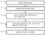
[0012]도 1은 초음파를 사용한 조직 특성을 추정하기 위한 방법의 일 실시예의 흐름도이다.
[0013]도 2는 초음파를 사용한 조직 특성을 추정하기 위한 시스템의 일 실시예의 블록도(block diagram)이다.
[0014]도 3은 초음파를 사용한 NAS와 같은 질병 활성도를 추정하기 위한 방법의 일 실시예의 흐름도이다.
[0015]도 4는, 조직학적(histologic) NAS와 비교하여, 초음파 측정치들로부터 NAS를 예측하는 데 있어서 정확도를 도시하는 예시적인 플롯(plot)이다.[0011] Elements and drawings are not necessarily to scale, emphasis instead being placed upon illustrating the principles of the invention. Moreover, in the drawings, like reference numerals denote corresponding parts throughout the different drawings.
1 is a flowchart of one embodiment of a method for estimating tissue properties using ultrasound.
2 is a block diagram of an embodiment of a system for estimating tissue properties using ultrasound.
3 is a flowchart of one embodiment of a method for estimating disease activity, such as NAS, using ultrasound.
4 is an exemplary plot illustrating accuracy in predicting NAS from ultrasound measurements as compared to histologic NAS.
[0016]질병 활성도는 건강 상태들의 진단, 검진(screening), 모니터링(monitoring) 및/또는 예측을 지원하는 것으로 추정된다. 예컨대, NAS 또는 다른 간 질환 활성도가 추정된다. 지수의 점수는 초음파를 사용하여 추정되어, 질병 활성도를 빠르고 저렴하며 비-외과적인 추정을 허용한다. 초음파-도출 NAFLD 활성도 점수가 추정되어, 생검 또는 MRI를 회피한다.[0016]Disease activity is estimated to support diagnosis, screening, monitoring and/or prediction of health conditions. For example, NAS or other liver disease activity is estimated. The score of the index is estimated using ultrasound, allowing a fast, inexpensive and non-surgical estimation of disease activity. An ultrasound-derived NAFLD activity score is estimated, avoiding biopsy or MRI.
[0017]질병 활성도는 간 지방 분율 및/또는 간 지방 분율의 추정치들과 같은 조직 특성들을 추정하는 데 사용되는 측정치들로부터 추정될 수 있다. 지방 분율 또는 다른 조직 특성들의 측정들 및 추정이 도 1과 관련하여 아래에서 논의된다. 그런 다음, 질병 활성도를 추정하는 데 있어서 이러한 측정들 및/또는 조직 특성들의 사용은 도 3과 관련하여 논의된다.[0017]Disease activity may be estimated from measurements used to estimate tissue properties, such as liver fat fraction and/or estimates of liver fat fraction. Measurements and estimations of fat fraction or other tissue properties are discussed below with respect to FIG. 1 . The use of these measures and/or tissue characteristics in estimating disease activity is then discussed with respect to FIG. 3 .
[0018]조직 특성 추정과 관하여, 정량적 초음파(QUS; quantitative ultrasound)는 건강 상태들의 검진, 진단, 모니터링 및/또는 예측하는 데 사용된다. 인간 조직의 복잡성은 해당 조직의 정확한 특징화를 위해 다수의 QUS 파라미터들을 사용하여 측정될 수 있다. 예컨대, 간 지방 분율은 종파들(longitudinal waves)의 산란 및 감쇠, 전단파들의 전파 및 감쇠, 및/또는 음향 복사력 임펄스(ARFI; acoustic radiation force impulse)로부터의 축상 파들의 전파 및 감쇠와 같은 상이한 파 현상들(wave phenomena)의 수신된 신호들로부터 추출된 정량적 파라미터들(quantitative parameters)을 결합하는 다중-파라미터 접근법을 사용하여 추정된다.[0018]Regarding tissue characteristic estimation, quantitative ultrasound (QUS) is used for screening, diagnosing, monitoring and/or predicting health conditions. The complexity of a human tissue can be measured using a number of QUS parameters for accurate characterization of that tissue. For example, the hepatic fat fraction can be affected by different waves, such as the scattering and attenuation of longitudinal waves, the propagation and attenuation of shear waves, and/or the propagation and attenuation of axial waves from an acoustic radiation force impulse (ARFI). It is estimated using a multi-parameter approach that combines quantitative parameters extracted from the received signals of wave phenomena.
[0019]일 실시예에서, 조직 특성(예컨대, 간 지방 분율)은, 산란 파라미터들을 추정하기 위해 펄스들의 시퀀스를 송신 및 수신하고 그리고 전단파 파라미터들을 획득하기 위해 펄스들의 시퀀스를 송신 및 수신함으로써 추정된다. 추정은 또한 음향 복사력 임펄스(ARFI; acoustic radiation force impulse)들에 의해 발생되는 축방향 변위들(axial displacements)로부터 파라미터들을 추정하기 위해 펄스들의 시퀀스를 송신 및 수신하는 것을 포함할 수 있다. QUS 파라미터들이 추정되고 결합되어 조직 특성을 추정한다. 비초음파 데이터(data)(예컨대, 혈액 바이오마커)와 같은 다른 정보가 조직 특성의 추정에 포함될 수 있다.[0019]In one embodiment, a tissue characteristic (eg, liver fat fraction) is estimated by transmitting and receiving a sequence of pulses to estimate scattering parameters and transmitting and receiving a sequence of pulses to obtain shear wave parameters. Estimation may also include transmitting and receiving a sequence of pulses to estimate parameters from axial displacements generated by acoustic radiation force impulses (ARFI). QUS parameters are estimated and combined to estimate tissue properties. Other information such as non-ultrasound data (eg, blood biomarkers) may be included in the estimation of tissue properties.
[0020]도 1은 초음파 스캐너 또는 시스템을 사용한 조직 특성 추정을 위한 방법을 도시한다. 상이한 타입들(types)의 파들 또는 파 현상들에 대한 조직 반응들이 측정된다. 이러한 상이한 반응들의 측정치들의 조합은 조직 특성을 추정하는 데 사용된다.[0020]1 shows a method for tissue property estimation using an ultrasound scanner or system. Tissue responses to different types of waves or wave phenomena are measured. The combination of measures of these different responses is used to estimate tissue properties.
[0021]방법은 도 2의 시스템 또는 상이한 시스템에 의해 구현된다. 의료 진단 초음파 스캐너는 음향적으로 파들을 생성하고 응답들을 측정함으로써 측정들을 수행한다. 스캐너, 컴퓨터(computer), 서버(server) 또는 다른 디바이스(device)의 이미지 프로세서는 측정치들로부터 추정한다. 디스플레이 디바이스, 네트워크(network) 또는 메모리(memory)는 추정된 조직 특성을 출력하는 데 사용된다.[0021]The method is implemented by the system of FIG. 2 or a different system. A medical diagnostic ultrasound scanner performs measurements by acoustically generating waves and measuring responses. An image processor of a scanner, computer, server or other device makes an estimate from the measurements. A display device, network or memory is used to output the estimated tissue characteristic.
[0022] 부가적인, 상이한, 또는 더 적은 수의 동작들이 제공될 수 있다. 예컨대, 동작들(33, 및/또는 38)이 제공되지 않는다. 다른 예로서, 동작들(36 및 37)은 대체 가능한 것들(alternatives)이거나 이들 둘 모두로부터의 결과들을 평균하는 것과 같이 함께 사용될 수 있다. 다른 예에서, 초음파 스캐너를 구성하고 그리고/또는 스캐닝(scanning)을 위한 동작들이 제공된다.[0022] Additional, different, or fewer operations may be provided. For example,
[0023]동작들은 설명되거나 또는 도시된 순서로(예컨대, 최상부에서 최하부로 또는 숫자로 표시된 대로) 수행되지만, 다른 순서들로 수행될 수 있다. 예컨대, 동작들(30, 32 및 33)이, 이를테면, 동일한 송신 및 수신 펄스들을 사용하여 동시에 수행되거나, 임의의 순서로 수행된다.[0023]The operations are performed in the order described or shown (eg, top to bottom or numerically), but may be performed in other orders. For example,
[0024]동작(30)에서, 초음파 스캐너는 환자의 스캔으로부터 조직 내의 산란 측정치를 생성한다. 산란 측정치는 초음파 스캐너로부터 송신되는 종파에 대한 조직 반응을 측정한다. 조직에 충돌하는 종파의 산란 또는 에코(echo)가 측정된다.[0024]In
[0025]임의의 산란 측정이 사용될 수 있다. 예시적인 산란 파라미터들은 사운드 스피드(sound speed), 사운드 분산(sound dispersion), 각도 산란 계수(angular scattering coefficient)(예컨대, 후방 산란 계수), 주파수-종속 감쇠 계수, 감쇠 계수 기울기, 정규화된 로그 스펙트럼(log-spectrum)의 스펙트럼 기울기, 정규화된 로그-스펙트럼의 스펙트럼 절편(spectral intercept), 정규화된 로그-스펙트럼의 스펙트럼 중간 대역(spectral midband), 유효 산란기 직경(effective scatterer diameter), 음향 농도(acoustic concentration), 산란기 수 밀도(scatterer number density), 평균 산란기 간격(mean scatterer spacing), 비선형성 파라미터(B/A) 및/또는 코히어런트 대 인코히어런트 산란(coherent to incoherent scattering)의 비율을 포함한다.[0025]Any scattering measurement may be used. Exemplary scattering parameters include sound speed, sound dispersion, angular scattering coefficient (eg, backscattering coefficient), frequency-dependent attenuation coefficient, attenuation coefficient slope, normalized log spectrum ( spectral slope of log-spectrum, spectral intercept of normalized log-spectrum, spectral midband of normalized log-spectrum, effective scatterer diameter, acoustic concentration , scatterer number density, mean scatterer spacing, nonlinearity parameter (B/A) and/or ratio of coherent to incoherent scattering.
[0026]하나 초과의 측정이 수행될 수 있다. 예컨대, 초음파 시스템은 환자 조직의 둘 이상의 산란 파라미터들에 대한 값들을 결정한다. 일 실시예에서, 음향 감쇠 계수, 후방 산란 계수, 및/또는 주파수-종속 후방 산란 계수의 로그(logarithm)의 스펙트럼 기울기가 측정된다.[0026]More than one measurement may be performed. For example, the ultrasound system determines values for two or more scattering parameters of the patient's tissue. In one embodiment, the spectral slope of the logarithm of the acoustic damping coefficient, the backscattering coefficient, and/or the frequency-dependent backscattering coefficient is measured.
[0027]산란을 측정하기 위해, 초음파 스캐너는 초음파를 사용하여 조직을 스캔한다. 정량적 초음파 산란 파라미터들을 추정하기 위한 신호들을 획득하기 위해 일련의 송신 및 수신 이벤트들(events)이 수행된다. 일 실시예에서, 1, 2 또는 3차원 구역은 B-모드 시퀀스(B-mode sequence)에 의해 스캔된다(예컨대, 광대역(예컨대, 1-2 사이클(cycle)) 송신 빔(transmit beam)을 송신하고 하나 이상의 응답 수신 빔들을 형성함). 선형, 섹터(sector) 또는 벡터(vector)와 같은 임의의 스캔 포맷(scan format)이 사용될 수 있다. 송신 및 수신 동작들은 각각의 스캔 라인(scan line)에 대해 반복될 수 있다. 협대역 펄스들(예컨대, 3개 이상의 사이클들)은, 오버랩핑하는 스펙트럼들(overlapping spectra)과 관계없이 별개의 중심 주파수들로부터 송신 및 수신될 수 있다. 협대역 송신 펄스들은 단일 또는 다수의 송신 및 수신 이벤트들에서 사용될 수 있다. 송신 펄스들 및 대응하는 수신 빔들은, 상이한 방향들로부터 동일한 조직 위치의 샘플링(sampling)과 같이 상이한 스티어링(steering) 각도들에서 형성될 수 있다. 단지 송신에 대해 또는 단지 수신에 대해 상이한 스티어링이 수행될 수 있다. 상이한 송신 빔들은 상이한 송신 전력들 및/또는 F 수들을 가질 수 있다. 단일 송신 또는 다수의 송신들에 초점이 맞춰지거나, 초점이 맞춰지지 않거나, 이들은 평면파를 사용할 수 있다. 임의의 스캔 시퀀스가 사용될 수 있다.[0027]To measure scattering, an ultrasound scanner uses ultrasound to scan tissue. A series of transmit and receive events are performed to obtain signals for estimating quantitative ultrasonic scattering parameters. In one embodiment, a 1, 2 or 3 dimensional region is scanned by a B-mode sequence (eg, a wideband (eg, 1-2 cycle)) transmit beam is transmitted and forming one or more response receive beams). Any scan format may be used, such as linear, sector or vector. The transmit and receive operations may be repeated for each scan line. Narrowband pulses (eg, 3 or more cycles) may be transmitted and received from separate center frequencies independent of overlapping spectra. Narrowband transmit pulses may be used in single or multiple transmit and receive events. The transmit pulses and corresponding receive beams may be formed at different steering angles, such as sampling of the same tissue location from different directions. Different steering may be performed for transmit only or only receive. Different transmit beams may have different transmit powers and/or F numbers. Focused or unfocused on a single transmission or multiple transmissions, they may use a plane wave. Any scan sequence may be used.
[0028]상이한 송신 및/또는 수신 설정들과 관계없는 반복은 산란을 한 번 측정하거나 산란을 상이하게 측정하는 데 사용될 수 있다. 동일한 위치에 대해 동일한 산란 파라미터의 다수의 측정치들이 제공되는 경우, 측정치들이 평균화되거나 결합될 수 있다. 인접한 위치들 또는 정해진 범위 내의 위치들과 같은 상이한 위치들로부터의 측정치들이 평균화될 수 있다. 예컨대, 산란 측정치는 동일한 위치로의 다수의 송신들로부터 평균화된 주파수-종속 측정치이다. 깊이, 각도 및/또는 주파수의 함수로서 전력 스펙트럼의 변화들이 측정될 수 있다. 다른 예로서, 상이한 송신 및/또는 수신 각도들로부터의 감쇠 계수의 추정치들은 분산(variance)을 감소시키기 위해 평균화되거나, 감쇠의 각도 종속성을 정량화하는 데 사용된다.[0028]An iteration independent of different transmit and/or receive settings can be used to measure scatter once or measure scatter differently. Where multiple measurements of the same scattering parameter are provided for the same location, the measurements may be averaged or combined. Measurements from different locations, such as adjacent locations or locations within a defined range, may be averaged. For example, a scatter measurement is a frequency-dependent measurement averaged from multiple transmissions to the same location. Changes in the power spectrum as a function of depth, angle and/or frequency may be measured. As another example, estimates of the attenuation coefficient from different transmit and/or receive angles are averaged to reduce variance, or used to quantify the angular dependence of attenuation.
[0029]일 실시예에서, 측정하기 위한 스캐닝은 적응적이다. 송신들 및/또는 수신들은 적응적일 수 있다. 예컨대, 하나의 측정의 결과들은 후속 송신들에 대한 진폭, 각도, 주파수 및/또는 F #을 설정하는 데 사용된다.[0029]In one embodiment, the scanning to measure is adaptive. Transmissions and/or receptions may be adaptive. For example, the results of one measurement are used to set the amplitude, angle, frequency and/or F # for subsequent transmissions.
[0030]일 예에서, 감쇠 계수가 측정된다. 기준 팬텀 방법(reference-phantom method)이 사용되지만, 감쇠 계수의 다른 측정치들이 사용될 수 있다. 음향 에너지(acoustic energy)는 깊이의 함수로서 기하급수적인 감쇠(exponential decay)를 갖는다. 깊이 게인 보정(depth gain correction) 전에 또는 그 보정 없이 깊이의 함수로서 음향 강도의 측정이 수행된다. 시스템 영향들을 제거하기 위해, 측정은 팬텀의 깊이 함수로서 음향 강도의 측정치들에 기반하여 교정된다. 측정은 1차원, 2차원 또는 3차원 구역에 대해 평균화함으로써 잡음에 영향을 덜 받을 수 있다. 빔포밍된 샘플들(beamformed samples) 또는 음향 강도는 주파수 도메인(frequency domain)으로 변환될 수 있으며, 계산은 주파수 도메인에서 수행된다.[0030]In one example, an attenuation coefficient is measured. The reference-phantom method is used, but other measures of the damping coefficient may be used. Acoustic energy has an exponential decay as a function of depth. Measurement of sound intensity as a function of depth is performed before or without depth gain correction. To remove system effects, the measurement is calibrated based on measurements of sound intensity as a function of the depth of the phantom. Measurements can be less susceptible to noise by averaging over 1-D, 2-D, or 3-D regions. Beamformed samples or sound intensity may be converted into a frequency domain, and the calculation is performed in the frequency domain.
[0031]다른 예에서, 후방 산란 계수가 측정된다. 음향 감쇠가 결정된다. 이 음향 감쇠는 기준 교정을 결정하는 데 사용된다. 음향 감쇠를 교정함으로써, 산란된 에너지가 후방 산란 계수로서 제공된다. 계산은 주파수 도메인에서 수행되어, 주파수의 함수로서 측정치들을 제공할 수 있다.[0031]In another example, a backscatter coefficient is measured. Acoustic attenuation is determined. This acoustic attenuation is used to determine the reference calibration. By correcting the acoustic attenuation, the scattered energy is provided as the backscattering coefficient. Calculations can be performed in the frequency domain to provide measurements as a function of frequency.
[0032]주파수-종속 후방 산란의 로그의 스펙트럼 기울기는 후방 산란 계수로부터 측정된다. 후방 산란 계수의 로그는 주파수의 함수로서 결정된다. 스펙트럼 기울기를 결정하기 위해, 주파수의 함수로서 후방 산란의 로그에 라인이 적합하다(예컨대, 최소 제곱).[0032]The spectral slope of the logarithm of the frequency-dependent backscatter is determined from the backscatter coefficient. The logarithm of the backscattering coefficient is determined as a function of frequency. To determine the spectral slope, a line is fit (eg least squares) to the logarithm of the backscatter as a function of frequency.
[0033]동작(32)에서, 초음파 스캐너는 환자의 스캔으로부터 조직 내의 전단파 전파의 측정치를 생성한다. 전단파 이미징에 대해, 음향 복사력 임펄스(ARFI 또는 푸싱 펄스(pushing pulse))가 조직에 전달된다. 임펄스는 한 위치에서 조직의 변위를 발생시켜, 전단파의 생성을 초래한다. 전단파는 일반적으로 푸싱 펄스의 송신 빔을 가로질러(transversely) 이동한다. 하나 이상의 측방향으로 이격된 위치들에서 조직 변위를 추적함으로써, 이러한 위치들을 통과하는 전단파가 검출될 수 있다. 전단파가 원점으로부터 나중의 위치로 이동하는 시간 및 그 위치들 사이의 거리는 전단파 스피드를 제공한다.[0033]In
[0034]임의의 전단파 파라미터가 결정될 수 있다. 예컨대, 조직 내의 전단파 스피드 또는 속도가 측정된다. 다른 전단파 파라미터들은 각도 및/또는 주파수-종속 전단파 스피드, 각도 및 주파수-종속 전단파 감쇠, 각도 및/또는 주파수-종속 저장 모듈러스(modulus), 각도 및/또는 주파수-종속 손실 모듈러스, 점도 및/또는 각도 및/또는 주파수-종속 음향 흡수 계수를 포함한다.[0034]Any shear wave parameters may be determined. For example, the shear wave speed or velocity within the tissue is measured. Other shear wave parameters may include angle and/or frequency-dependent shear wave speed, angle and frequency-dependent shear wave attenuation, angle and/or frequency-dependent storage modulus, angle and/or frequency-dependent loss modulus, viscosity and/or angle and/or a frequency-dependent acoustic absorption coefficient.
[0035]음향 흡수 계수는 전단파의 흡수가 아니라 음향 펄스의 흡수로부터 기인한다. 음향 흡수는 F
[0036]전단파를 측정하기 위해, 푸싱 펄스 또는 ARFI가 조직 내의 초점 위치로 송신된다. 휴식 상태 조직 포지션(resting state tissue position)에 대한 기준 스캔은, 푸싱 펄스 전에 또는 조직이 휴식 상태로 돌아간 후에 수행된다. 초점 위치로부터 하나 이상의 위치 공간에서 조직의 포지션 변화 또는 변위는 시간이 지남에 따라 측정된다. 추적 스캔들이 반복적으로 수행된다. 상관관계 또는 다른 유사성 측정을 사용하여, 현재 추적 시간과 비교하여 기준 시간으로부터 조직의 축항, 2D 또는 3D 시프트(shift)가 결정된다. 최대 변위의 시간은 전단파의 시간을 나타낸다. 변위의 시작 또는 끝과 같은 다른 타이밍(timing)이 사용될 수 있다. 전단파가 추적 위치에 도달하는 시간 및 추적 위치로부터 푸싱 펄스의 초점 위치까지의 거리는 전단파 속도를 제공한다. 상이한 추적 위치들에 대한 변위 프로파일(profile)의 시프트(시간 함수로서의 변위)를 결정하거나 위치의 함수로서 변위들로부터 해결함으로써, 다수의 위치들에서 전단파 속도를 해결하는 것과 같은 다른 접근법들이 사용될 수 있다.[0036]To measure the shear wave, a pushing pulse or ARFI is transmitted to a focal location within the tissue. A baseline scan for a resting state tissue position is performed before the pushing pulse or after the tissue returns to the resting state. A change in position or displacement of tissue in one or more location spaces from a focal position is measured over time. Tracking scans are performed iteratively. Using a correlation or other similarity measure, an axis “‡ term, 2D or 3D shift of the tissue is determined from the baseline time compared to the current follow-up time. The time of maximum displacement represents the time of the shear wave. Other timings may be used, such as the start or end of the displacement. The time the shear wave arrives at the tracking location and the distance from the tracking location to the focal point of the pushing pulse gives the shear wave velocity. Other approaches may be used, such as solving the shear wave velocity at multiple locations by determining the shift (displacement as a function of time) of the displacement profile for different tracking locations or solving from the displacements as a function of location. .
[0037]전단파 파라미터들의 측정은 주파수 및/또는 각도의 함수일 수 있다. 상이한 각도들 및/또는 상이한 주파수들로부터 빔들로 푸시 펄스들을 송신함으로써, 측정이 반복된다. 시공간 변위 프로파일들(spatio-temporal displacement profiles)은 측정을 결정하기 위해 시간 또는 주파수 도메인에서 사용된다. 상이한 각도들로부터의 결과들은 각도 종속 측정치를 결정하는 데 사용될 수 있다.[0037]The measurement of shear wave parameters may be a function of frequency and/or angle. By transmitting push pulses in beams from different angles and/or different frequencies, the measurement is repeated. The spatio-temporal displacement profiles are used in the time or frequency domain to determine the measurement. Results from different angles can be used to determine an angle dependent measure.
[0038]전단파 파라미터는 상이한 위치들에서 측정될 수 있다. 측정은 하나 또는 단일 푸싱 펄스에 대한 조직 변위에 기반할 수 있다. 대신에, 측정은 다수의 푸싱 펄스들에 대한 조직 변위에 기반할 수 있다. 측정은 상이한 푸싱 펄스들을 사용하여 상이한 구역들에 대해 반복된다.[0038]The shear wave parameter may be measured at different locations. Measurements may be based on tissue displacement for one or a single pushing pulse. Instead, the measurement may be based on tissue displacement for multiple pushing pulses. The measurement is repeated for different regions using different pushing pulses.
[0039]전단파 파라미터를 측정하기 위해, 푸싱 펄스 및 추적 송신들 둘 모두가 발생한다. 변위들은, 푸싱 펄스 송신들이 아닌 추적 송신들에 대한 음향 응답을 수신함으로써 측정된다. 산란 파라미터를 측정하는 데 사용된 것과 동일한 스캔을 사용하여 전단파 파라미터를 측정할 수 있다. 예컨대, 푸싱 펄스를 송신하기 전에 추적에 사용되는 기준 스캔은 산란을 측정하는 데 사용된다. 다른 실시예들에서, 전단파 파라미터에 대한 스캔은 산란 파라미터에 대한 송신들 및/또는 수신들과 상이한 송신들 및/또는 수신들을 사용한다. 측정들을 위한 스캔은 상이한 측정들에 대한 송신 및 수신 이벤트들의 개별 시퀀스들로 분할된다.[0039]To measure the shear wave parameter, both pushing pulse and tracking transmissions occur. Displacements are measured by receiving an acoustic response to tracking transmissions that are not pushing pulse transmissions. The same scans used to measure the scattering parameters can be used to measure the shear wave parameters. For example, a reference scan used for tracking before sending a pushing pulse is used to measure scatter. In other embodiments, the scan for the shear wave parameter uses different transmissions and/or receptions than the transmissions and/or receptions for the scattering parameter. The scan for measurements is split into separate sequences of transmit and receive events for different measurements.
[0040]푸싱 펄스는, 푸싱 펄스에 대해 수십, 수백 또는 수천 개의 사이클들 및 추적 송신들에 대해 1 내지 3개의 사이클들과 같이, 추적 펄스들과 비교하여, 상대적으로 긴 지속기간을 갖는다. 반복이 제공되는 경우, 상이한 초점 위치들, 주파수들, 각도들, 전력들 및/또는 F 수들이 푸시 펄스들에 사용될 수 있다.[0040]A pushing pulse has a relatively long duration compared to tracking pulses, such as tens, hundreds or thousands of cycles for a pushing pulse and 1 to 3 cycles for tracking transmissions. Where repetition is provided, different focal positions, frequencies, angles, powers and/or F numbers may be used for the push pulses.
[0041]동일한 위치 및/또는 다른 위치들에 대해 동일한 측정이 반복될 수 있다. 임의의 반복을 위해 상이한 주파수, F 수, 각도, 전력, 초점 위치들 및/또는 다른 차이들이 사용될 수 있다. 결과적인 측정치들은 다른 측정치를 결정하기 위해 함께 사용될 수 있거나, 잡음을 감소시키기 위해 평균화되는 것과 같이 결합될 수 있다.[0041]The same measurement may be repeated for the same location and/or other locations. Different frequencies, F-numbers, angles, powers, focal positions, and/or other differences may be used for any repetition. The resulting measurements can be used together to determine other measurements, or can be combined as averaged to reduce noise.
[0042]초음파 스캐너는 전단파 파라미터 측정을 위해 스캐닝을 조정할 수 있다. 예컨대, 전단파의 감쇠 계수의 추정을 위해 푸시 펄스가 조정된다. 중심 주파수, 지속기간, f-수 또는 푸시 펄스의 다른 특성이 나중의 송신을 위해 변경된다. 초점이 더 좁거나(tighter) 더 약하다(weaker). 전단파를 생성하기 위한 변위는 더 크거나 더 적다. 또 다른 예로서, ARFI 푸시 펄스를 사용한 흡수 계수의 추정을 위해, 더 좁은 초점 또는 더 긴 지속기간으로 또 다른 푸시 펄스가 송신된다. 이러한 변화는 신호 대 잡음비(SNR; signal-to-noise ratio)를 개선하거나 측정들의 변동성(variability)을 감소시킬 수 있다.[0042]The ultrasound scanner can adjust the scanning for shear wave parameter measurements. For example, the push pulse is adjusted for estimation of the attenuation coefficient of the shear wave. The center frequency, duration, f-number or other characteristics of the push pulse are changed for later transmission. The focus is either tighter or weaker. The displacement to generate the shear wave is greater or less. As another example, for estimation of the absorption coefficient using ARFI push pulses, another push pulse is transmitted with a narrower focus or longer duration. This change may improve the signal-to-noise ratio (SNR) or reduce the variability of measurements.
[0043]조정(adaptation)은 임의의 정보에 기반한다. 예컨대, 변위 프로파일은 기준 또는 교정 프로파일과 비교된다. 다른 예로서, 최대, 평균, 또는 중앙 변위의 변위량이 결정된다. 정보는, 더 강하거나 더 높은 강도의 푸시 펄스에 대한 필요성을 나타낼 수 있거나, 더 적은 강도의 푸시 펄스들이 필요로 된다는 것을 나타낼 수 있으며, 이는 더 짧은 냉각(cool down) 시간을 허용한다.[0043]Adaptation is based on arbitrary information. For example, the displacement profile is compared to a reference or calibration profile. As another example, the amount of displacement of the maximum, average, or median displacement is determined. The information may indicate a need for a stronger or higher intensity push pulse, or it may indicate that less intensity push pulses are needed, which allows for a shorter cool down time.
[0044]동작(33)에서, 초음파 스캐너는 조직의 축방향 변위에 대한 ARFI 측정치를 생성한다. ARFI 송신은 조직으로 하여금 송신 빔의 축 또는 스캔 라인을 따라 변위하게 한다. 전단파를 추적하기보다는, ARFI에 의해 발생되거나 ARFI에 의해 생성된 종파에 대한 응답으로 발생되는, 축을 따른 조직 변위가 시간이 지남에 따라 추적된다.[0044]In
[0045]임의의 ARFI 측정이 사용될 수 있다. 예컨대, ARFI 펄스의 종파의 감쇠는, ARFI의 초점으로부터 이격된 위치들에서 추적된 변위들로부터 추정될 수 있다. 측정치들은 축방향 스캔 라인을 따라 초점 또는 다른 위치들에 있을 수 있다.[0045]Any ARFI measurement may be used. For example, the attenuation of the longitudinal wave of an ARFI pulse can be estimated from displacements tracked at locations spaced from the focus of the ARFI. The measurements may be at a focal point or other locations along the axial scan line.
[0046]측정하기 위해, ARFI는 스캔 라인을 따라 송신된다. 추적 스캔들은 ARFI의 송신 후에 수행된다. 스캔 라인을 따른 추적 송신들로부터의 음향 에코들이 수신된다. 수신된 데이터는 ARFI-발생 변위들 이전 또는 이후의 기준과 상관된다. 시간, 위치, 송신 각도 및/또는 송신 주파수의 함수로서 변위의 양이 결정된다. 최대 변위의 양, 깊이의 함수로서의 변위 및/또는 시간의 함수로서의 변위는 ARFI 측정치를 계산하는 데 사용된다.[0046]To measure, ARFI is transmitted along a scan line. Trace scans are performed after transmission of ARFI. Acoustic echoes from tracking transmissions along the scan line are received. The received data is correlated with a criterion before or after ARFI-occurring displacements. The amount of displacement is determined as a function of time, position, transmission angle and/or transmission frequency. The amount of maximum displacement, displacement as a function of depth, and/or displacement as a function of time are used to calculate ARFI measurements.
[0047]다른 시간들 및/또는 위치들에서 동일한 측정이 수행될 수 있다. 반복으로부터의 결과들은 또 다른 측정치를 도출하는 데 사용될 수 있거나 평균화될 수 있다.[0047]The same measurement may be performed at other times and/or locations. Results from iterations can be used to derive another measure or can be averaged.
[0048]송신들은 F 수, 주파수, 지속기간, 전력 및/또는 각도를 조정하는 것과 같이 조정될 수 있다. 조정(adaption)은 최대 변위의 크기와 같은 임의의 측정치에 응답할 수 있다.[0048]Transmissions may be adjusted, such as by adjusting the F number, frequency, duration, power and/or angle. The adaptation may be responsive to any measure, such as the magnitude of the maximum displacement.
[0049]다른 측정치들이 사용될 수 있다. 상이한 타입들의 파들 및/또는 스캐닝에 대한 조직의 반응이 측정된다. 동일한 타입의 하나 이상의 측정치들이 사용된다. 정해진 측정치에 대해, 단일 인스턴스(instance), 평균 또는 분포(예컨대, 시간에 따른 표준 편차, 지속기간, 주파수, 각도 및/또는 공간)가 수행된다. 임의의 수의 동일하거나 상이한 타입들의 측정들이 수행될 수 있다.[0049]Other measures may be used. The tissue's response to different types of waves and/or scanning is measured. One or more measurements of the same type are used. For a given measurement, a single instance, mean, or distribution (eg, standard deviation over time, duration, frequency, angle and/or space) is performed. Any number of the same or different types of measurements may be performed.
[0050]동작(34)에서, 초음파 스캐너 또는 다른 이미지 프로세서는 상이한 측정치들로부터 환자 조직의 조직 특성을 추정한다. 둘 이상의 상이한 파 현상들로부터의 측정치들이 사용된다. 둘 이상의 측정치들의 값들은 조직 특성을 추정하는 데 사용된다. 예컨대, 산란의 측정치 및 전단파 전파의 측정치 둘 모두는 조직 특성을 추정하는 데 사용된다. 다른 예에서, 축상 변위의 측정치(예컨대, ARFI 측정치)는 음향 산란 측정치 및/또는 전단파 전파 측정치와 함께 사용된다.[0050]In
[0051]조직 특성을 추정하기 위해 다른 정보가 사용될 수 있다. 예컨대, 환자에 대한 임상 정보가 사용된다. 임상 정보는 병력(medical history), 나이, 체질량 지수, 성별, 금식 여부(fasting or not), 혈압, 당뇨병 여부(diabetic or not) 및/또는 혈액 바이오마커 측정치일 수 있다. 예시적인 혈액 바이오마커들은 알라닌 아미노트랜스퍼레이스(ALT; alanine aminotransferase) 레벨, 아스파테이트 아미노트랜스퍼레이스(AST; aspartate aminotransferase) 레벨 및/또는 알칼리 포스파타제(ALP; alkaline phosphatase) 레벨을 포함한다. 환자에 관한 임의의 정보가 포함될 수 있다.[0051]Other information may be used to estimate tissue characteristics. For example, clinical information about the patient is used. Clinical information may be medical history, age, body mass index, sex, fasting or not, blood pressure, diabetes or not and/or blood biomarker measurements. Exemplary blood biomarkers include alanine aminotransferase (ALT) levels, aspartate aminotransferase (AST) levels, and/or alkaline phosphatase (ALP) levels. Any information about the patient may be included.
[0052]임의의 조직 특성이 추정될 수 있다. 예컨대, 조직의 지방 분율이 추정된다. 간, 유방 또는 다른 조직의 지방 분율은 진단적으로 유용하다. 환자의 간의 지방 분율은 NAFLD의 진단을 돕는다. 다른 진단적으로 유용한 조직 특성들은 염증, 밀도, 섬유증 및/또는 네프론 특성(nephron characteristic)(개수 및/또는 직경)을 포함한다. 조직 특성은 존재 여부와 같이 이진(binary)이거나, 스케일(scale)에 따른 추정치(즉, 조직 특성의 레벨 또는 크기)이다. 일 실시예에서 오직 하나의 조직 특성만이 추정된다. 다른 실시예들에서, 둘 이상의 상이한 조직 특성들은 동일하거나 상이한 측정치들로부터 추정된다.[0052]Any tissue characteristic can be estimated. For example, the fat fraction of the tissue is estimated. Fat fractions of liver, breast or other tissues are useful diagnostically. Fat fraction in a patient's liver aids in the diagnosis of NAFLD. Other diagnostically useful tissue characteristics include inflammation, density, fibrosis and/or nephron characteristics (number and/or diameter). A tissue characteristic is either binary, such as presence or absence, or an estimate along a scale (ie, the level or magnitude of a tissue characteristic). In one embodiment only one tissue characteristic is estimated. In other embodiments, two or more different tissue characteristics are estimated from the same or different measurements.
[0053]동작들(36 및 37)은 동작(34)에서 추정하기 위한 2개의 상이한 실시예들을 나타낸다. 상이한 실시예들은 대체 가능한 것들이다. 다른 실시예들이 사용될 수 있다. 2개의 방식들로 조직 특성에 대한 값을 결정하고 그런 다음 결과들을 평균화하거나 가장 정확할 것 같은 결과를 선택하는 것과 같이, 양자 또는 다수의 실시예들이 사용될 수 있다.[0053]
[0054]조직 특성의 값이 추정된다. 동작(36)의 실시예에서, 머신-학습 분류기(machine-learnt classifier)는 조직 특성을 추정한다. 머신-트레이닝 분류기(machine-trained classifier)는 비선형 모델을 제공한다. 임의의 머신 학습 및 결과적인 머신-학습 분류기가 사용될 수 있다. 예컨대, 서포트 벡터 머신(support vector machine), 확률적 부스팅 트리(probabilistic boosting tree), 베이지안 네트워크(Bayesian network), 뉴럴 네트워크(neural network) 또는 다른 머신 학습이 사용된다.[0054]The value of the tissue characteristic is estimated. In an embodiment of
[0055]머신 학습은 트레이닝 데이터(training data)로부터 학습한다. 트레이닝 데이터는 수십, 수백 또는 수천 개의 샘플들과 같은 다양한 예들 및 실측 자료(ground truth)를 포함한다. 예들은, 산란 및 전단파 전파 파라미터들에 대한 값들과 같이 사용될 입력 데이터를 포함한다. 실측 자료는 각각의 예의 조직 특성에 대한 값이다. 일 실시예에서, 머신 학습은, 산란 및 전단파 전파 파라미터들에 기반하여, 지방 분율을 분류하는 것을 학습하는 것이다. 지방 분율에 대한 실측 자료에는, 양성자 밀도 지방 분율(PDFF; proton density fat fraction)를 제공하는 자기 공명(MR; magnetic resonance) 스캔이 제공된다. MR-PDFF는 위치 또는 구역에 대한 지방의 퍼센티지(percentage)를 제공한다. 지방의 퍼센티지는, 머신 학습이 초음파 파라미터들에 대한 입력 값들로부터 지방의 퍼센티지를 분류하는 것을 학습하도록 실측 자료로서 사용된다. 이를테면, 생검, 모델링(modeling) 또는 다른 측정들로부터의 정해진 조직 특성에 대해 다른 실측 자료의 소스들(sources)이 사용될 수 있다.[0055]Machine learning learns from training data. Training data includes ground truth and various examples such as tens, hundreds or thousands of samples. Examples include input data to be used, such as values for scattering and shear wave propagation parameters. The actual data are values for the tissue characteristics of each example. In one embodiment, the machine learning is learning to classify the fat fraction based on scattering and shear wave propagation parameters. The ground truth for fat fraction is provided with magnetic resonance (MR) scans that provide proton density fat fraction (PDFF). MR-PDFF provides the percentage of province for a location or district. The percentage of fat is used as a ground truth so that machine learning learns to classify the percentage of fat from input values for ultrasound parameters. Other sources of ground truth for a given tissue characteristic, such as from biopsies, modeling or other measurements, may be used.
[0056]일 실시예에서, 머신 학습은 뉴럴 네트워크를 트레이닝시킨다. 뉴럴 네트워크는, 조직 특성의 값들을 구별하기 위해 필터 커널(filter kernel)을 학습하는 하나 이상의 컨벌루셔널 계층들(convolution layers)을 포함한다. 머신 트레이닝은, 입력 값들의 어떤 가중치 조합(예컨대, 학습된 커널을 사용하는 컨볼루션(convolution))이 출력을 나타내는지를 학습한다. 결과적인 머신-학습 분류기는 입력 값들을 사용하여 구별 정보를 추출하고, 그런 다음, 추출된 정보에 기반하여 조직 특성을 분류한다.[0056]In one embodiment, machine learning trains a neural network. A neural network includes one or more convolutional layers that learn a filter kernel to distinguish values of tissue characteristics. Machine training learns which weighted combination of input values (eg, a convolution using a learned kernel) represents an output. The resulting machine-learning classifier extracts distinguishing information using the input values, and then classifies the tissue characteristics based on the extracted information.
[0057]트레이닝은 하나 이상의 행렬들(matrices)을 제공한다. 행렬 또는 행렬들은 입력 정보와 출력 클래스(output class)를 관련시킨다. 계층적 트레이닝 및 결과적인 분류기가 사용될 수 있다. 상이한 조직 특성들에 대해 상이한 분류기들이 사용될 수 있다. 동일한 조직 특성 및 평균화되거나 결합된 결과들에 대해 다수의 분류기들이 사용될 수 있다.[0057]Training provides one or more matrices. A matrix or matrices associates input information with an output class. Hierarchical training and resulting classifiers can be used. Different classifiers may be used for different tissue properties. Multiple classifiers can be used for the same tissue characteristic and averaged or combined results.
[0058]동작(37)의 실시예에서, 머신-학습 모델 대신에 또는 이외에도 선형 모델이 사용된다. 미리 결정되거나 프로그래밍된(programmed) 함수는 입력 값들과 출력 값들을 관련시킨다. 함수 및/또는 함수에 사용되는 가중치들은 실험적으로(experimentally) 결정될 수 있다. 예컨대, 가중치들은 MR-PDFF 값들을 사용하여 최소 제곱 최소화에 의해 획득된다.[0058]In an embodiment of
[0059]임의의 선형 함수가 사용될 수 있다. 예컨대, 조직 특성의 값은 하나 이상의 산란기 파라미터들 및 하나 이상의 전단파 전파 파라미터들로부터 추정된다. 덧셈, 뺄셈, 곱셈 또는 나눗셈의 임의의 조합이 사용될 수 있다.[0059]Any linear function may be used. For example, the value of the tissue property is estimated from one or more scatterer parameters and one or more shear wave propagation parameters. Any combination of addition, subtraction, multiplication or division may be used.
[0060]일 실시예에서, 둘 이상의 함수들(예컨대, 가중된 측정들의 조합)이 제공된다. 파라미터들 중 하나의 값에 기반하여 함수들 중 하나가 선택된다. 예컨대, 초음파-도출 지방 분율(UDFF; ultrasound-derived fat fraction) 추정은 가중된 조합들로서 표현되는 2개의 함수들을 포함한다.[0060]In one embodiment, two or more functions (eg, a combination of weighted measures) are provided. One of the functions is selected based on the value of one of the parameters. For example, ultrasound-derived fat fraction (UDFF) estimation includes two functions expressed as weighted combinations.


여기서 d와 δ는 상수들이고, a, b, c, α 및 β는 가중치들이고, P는 파라미터의 측정치이다. 어떤 함수를 선택할지를 결정하기 위해 하나의 파라미터(Pi, k)가 사용된다. 가능한 함수들은 2개 또는 3개의 다른 파라미터들 및 가중치들을 포함한다. 부가적이거나, 상이하거나 더 적은 수들의 함수들, 함수의 파라미터들, 가중치들 및/또는 상수들이 사용될 수 있다. 상이한 선택 기준들이 사용될 수 있다. 선택 파라미터는 하나의 타입일 수 있고, 각각의 함수의 가중된 파라미터는 다른 타입일 수 있다. 대안적으로, 선택에 사용되는 파라미터들의 타입 또는 타입들과 관계없이, 상이한 타입들(예컨대, 산란 및 전단파 전파)이 가중된 파라미터들로서 포함된다.where d and δ are constants, a, b, c, α and β are weights, and P is a measure of the parameter.One parameter (P i, k ) is used to determine which function to select. Possible functions include 2 or 3 different parameters and weights. Additional, different or fewer numbers of functions, parameters of the function, weights and/or constants may be used. Different selection criteria may be used. The optional parameter may be of one type, and the weighted parameter of each function may be of another type. Alternatively, different types (eg, scattering and shear wave propagation) are included as weighted parameters, regardless of the type or types of parameters used for selection.
[0061]일 예에서, AC는 음향 감쇠 계수(예컨대, 산란 파라미터)이고, BSC는 후방 산란 계수(예컨대, 산란 파라미터)이고, SS는 주파수-종속 후방 산란 계수(예컨대, 또한 산란 파라미터)의 로그의 스펙트럼 기울기이다. SWS는 전단파 스피드(예컨대, 전단파 전파 파라미터)이다. 산란 파라미터들에 기반하는 2개의 함수들이 사용되고, 여기서 정해진 추정에 대한 함수는, 다음과 같이 표현된 바와 같이, 전단파 전파 파라미터에 기반하여 선택된다.[0061]In one example, AC is the acoustic damping coefficient (eg, scattering parameter), BSC is the backscattering coefficient (eg, scattering parameter), and SS is the spectral slope of the logarithm of the frequency-dependent backscattering coefficient (eg, also the scattering parameter). am. SWS is the shear wave speed (eg, the shear wave propagation parameter). Two functions based on the scattering parameters are used, where a function for the given estimate is selected based on the shear wave propagation parameter, as expressed as follows.


가중치들 및 상수들은 MR-PDFF에 의해 제공된 지방 분율로부터의 차이를 최소화하는 것에 기반한다. 전문가가 선택한 또는 다른 가중치들 및/또는 상수들이 사용될 수 있다.The weights and constants are based on minimizing the difference from the fat fraction provided by the MR-PDFF. Expert selected or other weights and/or constants may be used.
[0062]동작(38)에서, 초음파 스캐너 또는 디스플레이 디바이스는 추정된 조직 파라미터를 디스플레이한다. 예컨대, 지방 분율의 이미지가 생성된다. 추정된 지방 분율을 나타내는 값이 스크린 상에 디스플레이된다. 대안적으로 또는 부가적으로, 추정된 지방 분율을 나타내는 그래픽(graphic)(예컨대, 곡선 또는 아이콘(icon))이 디스플레이된다. 스케일에 대한 기준 또는 다른 기준이 디스플레이될 수 있다. 다른 실시예들에서, 위치의 함수로서 지방 분율은 컬러(color), 밝기, 색조(hue), 휘도, 또는 1차원, 2차원 또는 3차원 표현의 디스플레이 값의 다른 변조에 의해 디스플레이된다. 조직 특성은 픽셀 컬러(pixel color)로 선형적으로 또는 비선형적으로 맵핑될(mapped) 수 있다.[0062]In
[0063]조직 특성은 단독으로 또는 다른 정보와 함께 표시된다. 예컨대, 전단파 이미징이 수행된다. 전단파 속도, 모듈러스 또는 전단파에 대한 조직 반응으로부터 결정된 또는 다른 정보가 디스플레이된다. 임의의 전단 이미징이 사용될 수 있다. 디스플레이된 이미지는 관심 구역 또는 전체 이미징 구역에 대한 전단파 정보를 나타낸다. 예컨대, 관심 구역 또는 시야의 모든 그리드 포인트들(grid points)에 대해 전단 속도 값들이 결정되는 경우, 디스플레이의 픽셀들은 해당 구역에 대한 전단파 속도들을 나타낸다. 디스플레이 그리드(display grid)는, 변위들이 계산되는 그리드 및/또는 스캔 그리드와 상이할 수 있다.[0063]Tissue characteristics are indicated alone or with other information. For example, shear wave imaging is performed. The shear wave velocity, modulus, or other information determined from the tissue response to the shear wave or other information is displayed. Any shear imaging may be used. The displayed image shows shear wave information for the region of interest or the entire imaging region. For example, if shear rate values are determined for all grid points of a region of interest or field of view, then the pixels of the display represent shear wave velocities for that region. The display grid may be different from the grid and/or scan grid for which displacements are calculated.
[0064]전단파 정보는 컬러 오버레이(color overlay) 또는 디스플레이 값들의 다른 변조에 사용된다. 컬러, 밝기, 휘도, 색조 또는 다른 디스플레이 특성은 전단파 속도와 같은 전단파 특성의 함수로서 변조된다. 이미지는 2-차원 또는 3-차원 위치들의 구역을 나타낸다. 전단 데이터는 디스플레이 포맷이거나, 디스플레이 포맷으로 스캔 변환될 수 있다. 전단 데이터는 컬러 또는 그레이 스케일 데이터(gray scale data)이지만, 그레이 스케일 또는 컬러 스케일로 맵핑(mapping)하기 전의 데이터일 수 있다. 정보는 디스플레이 값들로 선형적으로 또는 비선형적으로 맵핑될 수 있다.[0064]The shear wave information is used for color overlay or other modulation of display values. Color, brightness, luminance, hue or other display characteristics are modulated as a function of shear wave characteristics, such as shear wave velocity. An image represents a region of two-dimensional or three-dimensional positions. The front-end data may be in a display format or may be scan-converted into a display format. The front-end data is color or gray scale data, but may be data before mapping to gray scale or color scale. Information may be mapped linearly or non-linearly to display values.
[0065]이미지는 다른 데이터를 포함할 수 있다. 예컨대, 전단파 정보는 B-모드 정보 위에 또는 B-모드 정보와 함께 디스플레이된다. 전단파 속도가 임계치 미만이거나 품질이 열악한 임의의 위치에 대한 B-모드 데이터를 디스플레이하는 것과 같이, B-모드 또는 동일한 구역의 조직, 유체 또는 조영제들(contrast agents)을 나타내는 다른 데이터가 포함될 수 있다. 다른 데이터는 사용자가 전단 정보의 위치를 결정하는 데 도움을 준다. 다른 실시예들에서, 전단파 특성은 다른 데이터 없이 이미지로서 디스플레이된다. 또 다른 실시예들에서, B-모드 또는 다른 이미지 정보는 전단파 정보 없이 제공된다.[0065]Images may contain other data. For example, the shear wave information is displayed over or together with the B-mode information. Other data indicative of the B-mode or tissue, fluid or contrast agents in the same area may be included, such as displaying B-mode data for any location where the shear wave velocity is below a threshold or of poor quality. Other data helps the user determine the location of the flyer information. In other embodiments, the shear wave characteristic is displayed as an image without other data. In still other embodiments, the B-mode or other image information is provided without shear wave information.
[0066]조직 특성의 부가적인 추정된 값은, 전단파, B-모드, 컬러 또는 유동 모드, M-모드, 조영제 모드 및/또는 다른 이미징과 실질적으로 동시에 디스플레이된다. 보기의 시각적 지각이 실질적으로 감안(account for)된다. 충분한 빈도로 2개의 이미지들을 순차적으로 디스플레이하는 것은, 시청자가 이미지들을 동시에 디스플레이되는 것으로 지각하는 것을 허용할 수 있다. 조직 특성을 추정하는 데 사용되는 컴포넌트 측정치들(component measures)이 또한, 이를테면, 표에 디스플레이될 수 있다.[0066]Additional estimated values of tissue properties are displayed substantially simultaneously with shear wave, B-mode, color or flow mode, M-mode, contrast mode and/or other imaging. The visual perception of the view is actually accounted for. Displaying two images sequentially with sufficient frequency may allow a viewer to perceive the images as being displayed simultaneously. Component measures used to estimate tissue characteristics may also be displayed, such as in a table.
[0067]실질적으로 동시의 디스플레이를 위한 임의의 포맷이 사용될 수 있다. 일 예에서, 전단파 또는 해부학적 이미지는 2 차원 이미지이다. 조직 특성의 값은 텍스트(text), 그래프(graph), 2 차원 이미지 또는 추정의 값들의 다른 표시자이다. 커서(cursor) 또는 다른 위치 선택은 전단 또는 해부학적 이미지에 대해 포지셔닝될(positioned) 수 있다. 커서는 위치의 선택을 나타낸다. 예컨대, 사용자는 병변, 낭포, 내포물 또는 다른 구조의 내부 구역과 연관된 픽셀을 선택한다. 그런 다음, 선택된 위치에 대한 조직 특성이 값, 스케일을 따른 포인터(pointer) 또는 다른 표시로서 디스플레이된다. 다른 예에서, 조직 특성은 관심 구역(시야의 서브-부분(sub-part)) 또는 전체 시야에 표시된다.[0067]Virtually any format for simultaneous display may be used. In one example, the shear wave or anatomical image is a two-dimensional image. The value of the tissue property is text, graph, two-dimensional image or other indicator of the values of the estimate. A cursor or other location selection may be positioned relative to the anterior or anatomical image. The cursor indicates the selection of a position. For example, the user selects a pixel associated with an internal region of a lesion, cyst, inclusion, or other structure. The tissue characteristic for the selected location is then displayed as a value, a pointer along a scale, or other indicia. In another example, the tissue characteristic is indicated in a region of interest (a sub-part of the field of view) or the entire field of view.
[0068]다른 실시예에서, 전단파 또는 B-모드 및 지방 분율 이미지들이 실질적으로 동시에 디스플레이된다. 예컨대, 듀얼-스크린 디스플레이(dual-screen display)가 사용된다. 전단파 이미지(예컨대, 전단파 속도) 및/또는 B-모드 이미지가 스크린의 하나의 영역에 디스플레이된다. 위치의 함수로서의 지방 분율은 스크린의 다른 영역에 디스플레이된다. 사용자는 진단을 위해 스크린 상의 상이한 이미지들을 볼 수 있다. 조직 특성의 부가적인 정보 또는 표시는 사용자가 그 구역을 진단하는 것을 돕는다.[0068]In another embodiment, the shear wave or B-mode and fat fraction images are displayed substantially simultaneously. For example, a dual-screen display is used. A shear wave image (eg, shear wave velocity) and/or a B-mode image is displayed in one area of the screen. Fat fractions as a function of location are displayed in different areas of the screen. The user can view different images on the screen for diagnosis. Additional information or indication of tissue characteristics helps the user to diagnose the area.
[0069]일 실시예에서, 조직 추정은 실시간 숫자 또는 정량적 이미지로서 제공된다. 조직 파라미터들이 신속하게 추정될 수 있기 때문에, 조직 파라미터의 값이 추정되고, 스캐닝을 시작하고 1-3 초 이내에 출력된다. 조직 특성은 치료 전, 치료 동안 및/또는 치료 후와 같이 상이한 시간들에 추정될 수 있다. 상이한 시간들로부터의 추정치들은 질병의 진행 및/또는 치료에 대한 반응을 모니터링하는 데 사용된다. 예컨대, 시간에 따른 조직 특성의 값의 퍼센트 변화(percent change)가 계산되고 출력된다.[0069]In one embodiment, the tissue estimate is provided as a real-time numerical or quantitative image. Since the tissue parameters can be estimated quickly, the value of the tissue parameter is estimated and output within 1-3 seconds of starting scanning. Tissue properties may be estimated at different times, such as before, during, and/or after treatment. Estimates from different times are used to monitor disease progression and/or response to treatment. For example, a percent change in the value of the tissue characteristic over time is calculated and output.
[0070]조직 특성 및/또는 조직 특성들을 도출하는 데 사용되는 측정들은 질병 활성도를 추정하는 데 사용될 수 있다. 도 3은 초음파 시스템을 사용한 질병 활성도 추정을 위한 방법의 일 실시예의 흐름도이다. 예컨대, 방법은 초음파-도출 비알코올성 간 질환 활성도 추정을 위한 것이다. 초음파 스캐너는 질병 활성도를 직접 또는 간접적으로 추정하기 위해 환자의 조직 내의 산란 및/또는 전단파 전파를 측정한다.[0070]Tissue characteristics and/or measurements used to derive tissue characteristics may be used to estimate disease activity. 3 is a flowchart of one embodiment of a method for disease activity estimation using an ultrasound system. For example, the method is for ultrasound-guided estimation of nonalcoholic liver disease activity. Ultrasound scanners measure scatter and/or shear wave propagation within a patient's tissue to directly or indirectly estimate disease activity.
[0071]예컨대, 비알코올성 지방 간 질환 활성도 점수(NAS; non-alcoholic fatty liver disease activity score)는 정량적 초음파를 사용하여 예측된다. NAS는 조직의 기계적 및 음향적 특성들의 초음파 추정치들에 기반하여 예측된다. 모델은 의료용 초음파를 사용하여 추정된 조직의 기계적 및 음향적 특성들에 기반하여 NAS를 예측한다. 일 실시예에서, 초음파 시스템은 NAS의 예측자로서 초음파-도출 NAFLD 활성도(UDNA; ultrasound-derived NAFLD activity) 지수를 비외과적으로 획득한다. 초음파 시스템은 산란 및 전단파 전파의 측정치들을 생성하기 위한 펄스 시퀀스를 수행하도록 구성된다. UDNA는, 음향 후방 산란 계수, 전단파 속도 및 전단파 감쇠비를 포함하는, 간의 적어도 3개의 특성들의 모델들을 사용하여 결정된다.[0071]For example, the non-alcoholic fatty liver disease activity score (NAS) is predicted using quantitative ultrasound. NAS is predicted based on ultrasound estimates of the mechanical and acoustic properties of tissue. The model predicts NAS based on the mechanical and acoustic properties of the tissue estimated using medical ultrasound. In one embodiment, the ultrasound system non-surgically acquires an ultrasound-derived NAFLD activity (UDNA) index as a predictor of NAS. The ultrasound system is configured to perform a pulse sequence to produce measurements of scattering and shear wave propagation. UDNA is determined using models of at least three properties of the liver, including acoustic backscatter coefficient, shear wave velocity, and shear wave damping ratio.
[0072]조직학적 NAS는 지방증, 소엽 염증 및 풍선 조직학적 점수의 합계이지만, 생검을 요구한다. 일 실시예에서, 제안된 초음파-도출 모델은 적절한 기계적 및 음향적 특성들과 NAS 특징들을 페어링(pair)한다. 후방 산란, 감쇠 및/또는 사운드 스피드에 기반한 초음파-도출 지방 분율은 지방증 등급의 측정치로서 사용된다. 전단파 감쇠비는 염증의 측정치로서 사용되고, 전단파 스피드는 풍선의 측정치로서 사용된다. 다른 초음파 측정치들이 사용될 수 있다.[0072]Histological NAS is the sum of steatosis, lobular inflammation and balloon histological scores, but requires a biopsy. In one embodiment, the proposed ultrasound-derived model pairs NAS features with appropriate mechanical and acoustic properties. Ultrasound-derived fat fraction based on backscatter, attenuation and/or sound speed is used as a measure of steatosis grade. The shear wave damping ratio is used as a measure of inflammation, and the shear wave speed is used as a measure of the balloon. Other ultrasound measurements may be used.
[0073]도 3의 방법은 도 2의 시스템 또는 상이한 시스템에 의해 구현된다. 의료 진단 초음파 스캐너는 음향적으로 파들을 생성하고 응답들을 측정함으로써 측정들을 수행한다. 스캐너, 컴퓨터, 서버 또는 다른 디바이스의 이미지 프로세서는 측정들로부터 추정한다. 디스플레이 디바이스, 네트워크 또는 메모리는 추정된 질병 활성도 점수를 출력하는 데 사용된다.[0073]The method of FIG. 3 is implemented by the system of FIG. 2 or a different system. A medical diagnostic ultrasound scanner performs measurements by acoustically generating waves and measuring responses. An image processor of a scanner, computer, server, or other device makes inferences from the measurements. A display device, network or memory is used to output the estimated disease activity score.
[0074] 부가적인, 상이한, 또는 더 적은 수의 동작들이 제공될 수 있다. 예컨대, 지방 분율 또는 다른 조직 특성의 추정에서 ARFI 측정치를 사용하는 것과 같은, 도 1의 동작들(33)이 포함된다. 다른 예로서, 조직 특성(예컨대, 지방 분율)의 별개의 추정 없이 측정치들로부터 질병 활성도 지수 점수가 추정되는 경우와 같은 동작(34)이 포함되지 않는다. 또 다른 예에서, 동작(38)이 제공되지 않는다. 다른 예에서, 초음파 스캐너를 구성하고 그리고/또는 스캐닝을 위한 동작들이 제공된다.[0074] Additional, different, or fewer operations may be provided.
[0075]동작들은 설명되거나 또는 도시된 순서로(예컨대, 최상부에서 최하부로 또는 숫자로 표시된 대로) 수행되지만, 다른 순서들로 수행될 수 있다. 예컨대, 동작들(30 및 32)이, 이를테면, 동일한 송신 및 수신 펄스들을 사용하여 동시에 수행되거나, 임의의 순서로 수행된다.[0075]The operations are performed in the order described or shown (eg, top to bottom or numerically), but may be performed in other orders. For example,
[0076]동작(30)에서, 초음파 스캐너는 환자의 초음파 스캔으로부터 조직 내의 산란의 하나 이상의 측정치들을 생성한다. 간 조직과의 음향 상호작용의 측정치들과 같은 임의의 음향 산란 파라미터들이 사용될 수 있다. 예컨대, 초음파 스캐너 또는 시스템은 후방 산란 계수, 주파수-종속 후방 산란 계수, 감쇠, 사운드 스피드 및/또는 도 1에 대해 위에서 논의된 산란 측정치들 중 임의의 다른 측정치를 측정한다. 측정치는 주파수-종속적이고, 이를테면, 다수의 송신들로부터 평균화될 수 있다. 적응형 스캐닝이 사용될 수 있다.[0076]At
[0077]산란의 측정치들은, 이를테면, 음향 후방 산란(예컨대, 주파수-종속 음향 후방 산란) 및 음향 감쇠를 사용하여, 지방 분율을 추정하는 데 사용될 수 있다. 산란의 측정치들은 대안적으로 또는 부가적으로, 이를테면, 음향 후방 산란 또는 주파수-종속 음향 후방 산란을 사용하여, 질병 활성도의 추정에 사용된다.[0077]Measures of scatter can be used to estimate fat fraction, such as using acoustic backscatter (eg, frequency-dependent acoustic backscatter) and acoustic attenuation. Measures of scattering are alternatively or additionally used for estimation of disease activity, such as using acoustic backscattering or frequency-dependent acoustic backscattering.
[0078]동작(32)에서, 초음파 스캐너는 환자의 초음파 스캔으로부터 조직 내의 전단파 전파의 하나 이상의 측정치들을 생성한다. 임의의 전단파 전파 파라미터들이 사용될 수 있다. 예컨대, 전단파 속도 및 전단파 감쇠비가 사용된다. 도 1에 대해 위에서 논의된 전단파 전파 측정치 중 임의의 것이 사용될 수 있다. 측정치들은 환자의 관심 조직, 이를테면, 간 조직에서 ARFI 유도 전단파에 대한 것이다. 적응형 스캐닝이 사용될 수 있다.[0078]At
[0079]도 1에 대해 위에서 논의된 바와 같이, 산란을 측정하고 전단파 전파를 측정하는 데 사용되는 초음파 스캐닝은 동일하거나 상이한 송신 및 수신 이벤트들을 사용한다. 예컨대, 전단파를 생성하고 전단파에 대한 조직 반응을 측정하는 데 사용되는 산란을 측정하기 위해 별개의 송신들 및 수신들이 사용된다.[0079]As discussed above with respect to FIG. 1 , ultrasonic scanning used to measure scatter and measure shear wave propagation uses the same or different transmit and receive events. Separate transmissions and receptions are used, for example, to measure scattering, which is used to generate the shear wave and measure the tissue response to the shear wave.
[0080]일 실시예에서, 전단파 감쇠비가 생성된다. 복소수로서 전단파 점도, 이를테면, 손실 모듈러스에 대한 저장 모듈러스의 비율을 결정하기 위해, 전단파에 대한 조직 반응을 측정하기 위해 초음파 스캐닝이 수행된다. 이러한 복소 표현은 점도의 실수 부분을 저장 모듈러스로서 사용하고 점도의 허수 부분을 손실 모듈러스로서 사용한다.[0080]In one embodiment, a shear wave damping ratio is generated. To determine the shear wave viscosity as a complex number, such as the ratio of storage modulus to loss modulus, ultrasound scanning is performed to measure the tissue response to shear wave. This complex representation uses the real part of the viscosity as the storage modulus and the imaginary part of the viscosity as the loss modulus.
[0081]하나의 접근법에서, 시공간 변위 측정치들은 전단파 전파 동안에 획득된다. 이러한 측정치들은, 이를테면, 고속 푸리에 변환(fast Fourier transform)을 사용하여 주파수 도메인으로 푸리에 변환되며(Fourier transformed), 복소파 수(complex wave number)를 결정하는 데 사용된다. 시간의 함수로서 변위 스펙트럼의 로그는 전단 또는 다른 파에 영향을 받는 다양한 위치들 각각에 대해 결정될 수 있다. 위치의 함수로서 로그를 사용하여 해결하는 것은 복소파 수를 제공한다. 손실 모듈러스 및 저장 모듈러스와 같은 다양한 점탄성 파라미터들(viscoelastic parameters)은 복소파 수로부터 결정된다. 일 실시예에서, 미국 공개 특허 출원 번호 제2016/0302769호에 개시된 복소파 수, 점도, 또는 다른 감쇠비 측정치를 결정하기 위한 측정치들이 사용된다.[0081]In one approach, spatiotemporal displacement measurements are obtained during shear wave propagation. These measurements are Fourier transformed into the frequency domain, for example using a fast Fourier transform, and used to determine a complex wave number. The logarithm of the displacement spectrum as a function of time can be determined for each of the various locations affected by a shear or other wave. Solving using the logarithm as a function of position gives a complex number. Various viscoelastic parameters such as loss modulus and storage modulus are determined from the complex wave number. In one embodiment, measurements are used to determine the complex wave number, viscosity, or other damping ratio measurement disclosed in US Published Patent Application No. 2016/0302769.
[0082]또 다른 접근법으로서, 전단파 감쇠 및 전단파 분산이 측정된다. 분산은 주파수의 함수로서 전단파 스피드 또는 속도의 변화이다. 전단파 감쇠는 또한 주파수의 함수로서 측정될 수 있다. 정해진 주파수에 대해 또는 다수의 주파수들의 결합(예컨대, 평균)에 대해, 감쇠 및 분산 값에 기반하여 페이저(phasor)가 생성된다. 이 페이저는 복소수로 변환되고, 복소수로부터의 실수 및 허수 부분들이 감쇠비로서 사용된다. 전단파에 대한 조직 반응으로부터의 감쇠비의 다른 측정치들이 사용될 수 있다.[0082]As another approach, shear wave attenuation and shear wave dispersion are measured. Dispersion is the change in shear wave speed or velocity as a function of frequency. Shear wave attenuation can also be measured as a function of frequency. For a given frequency or for a combination (eg, average) of multiple frequencies, a phasor is generated based on the attenuation and dispersion values. This phasor is converted to a complex number, and the real and imaginary parts from the complex number are used as damping ratios. Other measures of the damping ratio from tissue response to shear waves may be used.
[0083]이미지 프로세서는, 후방 산란 계수, 전단파 속도 및 전단파 감쇠비로부터 초음파-도출 간 질환 활성도 지수에 대한 값을 추정한다. 다른 측정들이 사용될 수 있다. 이를테면, 환자 의료 기록으로부터의 비초음파 정보가 부가적으로 사용될 수 있다.[0083]The image processor estimates values for the ultrasound-derived liver disease activity index from the backscatter coefficient, the shear wave velocity and the shear wave attenuation ratio. Other measurements may be used. For example, non-ultrasound information from patient medical records may additionally be used.
[0084]질병 활성도는 측정들로부터 직접 또는 간접적으로 추정된다. 직접적인 측정들에 대해, 측정들이 모델에 입력되며, 이 모델은 동작(40)에서 질병 활성도 값의 추정치를 출력한다. 점수는 측정치로부터 직접적으로 추정된다. 간접적인 측정들에 대해, 동작(34)에서 다른 값 또는 추정치(예컨대, 지방 분율)를 결정하는 데 하나 이상의 타입들의 측정들이 사용되며, 그런 다음, 이 추정치는 단독으로, 다른 타입들의 추정치들과 함께, 다른 측정치들과 함께, 또는 동작(40)에서 질병 활성도를 추정하기 위한 다른 타입들의 추정치들 및 다른 측정치들과 함께 사용된다.[0084]Disease activity is estimated directly or indirectly from measurements. For direct measurements, the measurements are input to a model, which in
[0085]일 실시예에서, 후방 산란 계수, 음향 감쇠 및/또는 전단파 속도(예컨대, 전단파 속도가 없는 경우, 후방 산란 계수(음향 산란) 및 음향 감쇠를 사용함)는 동작(34)에서 지방 분율을 추정하는 데 사용된다. 하나 이상의 산란 및/또는 하나 이상의 전단파 전파 파라미터들이 지방 분율을 추정하는 데 사용된다. 도 1에 대해 위에서 논의된 지방 분율을 추정하기 위한 실시예들 중 임의의 실시예가 사용될 수 있다. 예컨대 음향 감쇠 계수(예컨대, 산란 파라미터), 후방 산란 계수(예컨대, 산란 파라미터) 및 주파수-종속 후방 산란 계수의 로그의 스펙트럼 기울기(예컨대, 또한 산란 파라미터)는 지방 분율을 추정하는 데 사용된다. 전단파 스피드(예컨대, 전단파 전파 파라미터)는, 이를테면, 산란 파라미터들로부터 지방 분율을 추정하기 위한 함수를 선택하는 데 사용될 수 있다.[0085]In one embodiment, the backscatter coefficient, acoustic attenuation, and/or shear wave velocity (eg, using backscatter coefficient (acoustic scattering) and acoustic attenuation in the absence of shear wave velocity) are used in estimating the fat fraction in
[0086] 동작(40)에서, 이미지 프로세서는 간 질환 활성도의 레벨(예컨대, NAS 또는 UDNA)을 추정한다. 간접적인 추정에 대해, 질병 활성도의 레벨은 전단파 파라미터들 중 적어도 하나 및 지방 분율로부터 추정된다. 예컨대, 질병 활성도의 추정에서 3개의 입력들이 사용된다. 초음파 기계적 및 음향적 특성들은 조직학적 NAS 특징들을 대체한다. 이를테면, 후방 산란, 감쇠 및/또는 사운드 스피드에 기반한 초음파-도출 지방 분율은 지방증 등급의 측정치로서 사용된다. 전단파 감쇠비는 염증의 측정치로서 사용되고, 전단파 스피드는 풍선의 측정치로서 사용된다. 의사를 보조하는 지표에 대한 간 질환 활성도의 값은 지방 분율, 감쇠비 및 전단파 속도와 같은 다양한 측정치들 및/또는 추정치들로부터 직접적으로 또는 간접적으로 추정된다.[0086] At
[0087]다른 측정치들 및/또는 추정치들이 대체물로서 사용될 수 있다. 하나의 조직학적 NAS 변수를 대신해서 다수의 추정치들 및/또는 측정치들이 사용될 수 있다. 활성도는, 조직학적 변수들과 상이하여, 질병 활성도를 결정하는 상이한 접근법을 제공하는 추정치들 및/또는 측정치들로부터 추정될 수 있다.[0087]Other measures and/or estimates may be used as a substitute. Multiple estimates and/or measurements may be used in place of one histological NAS variable. Activity may be estimated from estimates and/or measurements, which differ from histological variables, providing a different approach to determining disease activity.
[0088]이미지 프로세서는 모델을 사용하여 간 질환 활성도 지수에 대한 점수 또는 값을 추정한다. 점수 또는 값은, 이를테면, 정수 범위에 걸친 지수의 부분이다. 3 개의 레벨들(예컨대, 지방 간염, 간경변 또는 간세포 암종), 2 개의 레벨들(예컨대, 지방증 또는 섬유증) 또는 4 개의 이상의 레벨들과 같은 임의의 범위가 사용될 수 있다. 점수화는 질병의 특정 스테이지들(stages)과 관련될 수 있거나, 특정 스테이지들을 참조하지 않고 질병의 레벨을 나타낼 수 있다(예컨대, 0-7은 각각의 레벨에 대해 상이한 양의 활성도를 갖는 8개의 레벨들을 제공함). 추정된 값 또는 점수는 스테이지 및/또는 수치 표현이다.[0088]The image processor uses the model to estimate a score or value for the liver disease activity index. A score or value is, for example, a portion of an exponent over a range of integers. Any range may be used, such as three levels (eg steatohepatitis, cirrhosis or hepatocellular carcinoma), two levels (eg steatosis or fibrosis) or 4 or more levels. The scoring may relate to specific stages of the disease, or may indicate the level of the disease without reference to specific stages (eg 0-7 8 levels with different amounts of activity for each level) provided). The estimated value or score is a stage and/or numerical representation.
[0089]모델은, 이를테면, 변수들에서 추정치들 및/또는 측정치들을 사용하는 함수일 수 있다. 다른 실시예들에서, 모델은 머신-학습 분류기이다. 이를테면, 완전히 연결된 뉴럴 네트워크, 컨벌루셔널 뉴럴 네트워크 또는 서포트 벡터 머신을 사용하는 머신 학습은, 입력 추정치들 및/또는 측정치들이 정해지면, 질병 활성도의 값을 분류-출력하기 위한 모델을 트레이닝시킨다. 다른 실시예에서, 로지스틱 회귀 모델(logistic regression model)이 사용된다. 질병 활성도의 값은 로지스틱 회귀를 사용한 측정치들 및/또는 추정치들로부터 추정된다. 예컨대, 질병 활성도는 지방 분율, 전단파 속도 및 감쇠비의 로지스틱 회귀를 사용하여 추정된다. 또 다른 예로서, 질병 활성도는 후방 산란 계수, 전단파 속도 및 전단파 감쇠비의 로지스틱 회귀로서 추정된다.[0089]A model may be, for example, a function that uses estimates and/or measurements in the variables. In other embodiments, the model is a machine-learning classifier. Machine learning, such as using a fully connected neural network, convolutional neural network or support vector machine, trains a model to classify-output a value of disease activity once input estimates and/or measures are established. In another embodiment, a logistic regression model is used. The value of disease activity is estimated from measurements and/or estimates using logistic regression. For example, disease activity is estimated using logistic regression of fat fraction, shear wave velocity, and damping ratio. As another example, disease activity is estimated as a logistic regression of the backscatter coefficient, shear wave velocity, and shear wave damping ratio.
[0090]이미지 프로세서는 질병 활성도를 추정하기 위해 다른 입력 정보를 사용할 수 있다. 예컨대, ARFI로부터의 종파에 대한 조직 반응의 측정치들이 사용될 수 있다. 다른 예로서, 환자에 대한 임상 정보가 사용될 수 있다.[0090]The image processor may use other input information to estimate disease activity. For example, measures of tissue response to longitudinal waves from ARFI can be used. As another example, clinical information about a patient may be used.
[0091]동작(42)에서, 이미지 프로세서는 질병 활성도의 추정치를 생성하고, 디스플레이(예컨대, 디스플레이 스크린)는 이를 디스플레이한다. 간 질환 활성도 레벨(예컨대, UDNA)이 사용자에게 디스플레이되어 질병 진단 또는 모니터링을 보조한다. 초음파가 사용되기 때문에, 외과적 생검 및/또는 MRI의 시간 및 비용 없이, 보조가 제공된다.[0091]In
[0092]UNDA 지수의 레벨(예컨대, 값) 또는 다른 질병 활성도의 스케일은 영숫자 텍스트로서, 그래프로서 또는 환자의 조직을 나타내는 이미지에서 컬러 코딩(color coding) 또는 라벨링(labeling)으로서 출력된다. 예컨대, 간 조직의 초음파 이미지가 디스플레이된다. 이미지는 추정된 바와 같이 초음파-도출 간 질환 활성도 지수의 값의 표시를 포함한다. 지방 분율, 다른 조직 특성 추정치 및/또는 측정치들이 또한 출력될 수 있다. 동작(38)에 대해 위에서 논의된 출력들 중 임의의 것은 지방 분율 대신에 또는 그에 추가하여 질병 활성도와 함께 사용될 수 있다.[0092]The level (eg, value) or other scale of disease activity of the UNDA index is output as alphanumeric text, as a graph, or as color coding or labeling in an image representing the patient's tissue. For example, an ultrasound image of liver tissue is displayed. The image includes an indication of the value of the ultrasound-induced liver disease activity index as estimated. Fat fraction, other tissue property estimates and/or measurements may also be output. Any of the outputs discussed above for
[0093]도 4는 조직학적 NAS와 비교하여 예측된 NAS의 그래프를 도시한다. 예측된 NAS는, 전단파 전파에 기반한, 지방 분율, 전단파 속도 및 전단파 감쇠비로부터의 로지스틱 회귀로부터 UDNA를 추정한 것이다. 지수는 점수화하기 위해 8개의 레벨들(0-7)을 사용한다. 82명의 환자들에 기반하여, 예측된 NAS와 조직학적 NAS 사이의 제곱 평균 제곱근 오차는 1.14이다. 예측은 특히 2-5의 중간-위 점수들에 대해 조직학적 NAS와 잘 상관된다. 양호한 진단 성능이 제공된다.[0093]4 shows a graph of predicted NAS compared to histological NAS. The predicted NAS is an estimate of UDNA from logistic regression from fat fraction, shear wave velocity and shear wave damping ratio, based on shear wave propagation. The exponent uses eight levels (0-7) to score. Based on 82 patients, the root mean square error between predicted NAS and histological NAS is 1.14. Prediction correlates well with histological NAS, especially for mid-upper scores of 2-5. Good diagnostic performance is provided.
[0094]도 2는 상이한 타입들의 파들에 대한 응답으로 측정치들로부터 조직 특성 및/또는 질병 활성도 추정을 위한 시스템(10)의 일 실시예를 도시한다. 시스템(10)은 도 1의 방법, 도 3의 방법 또는 다른 방법들을 구현한다. 시스템(10)은 송신 빔포머(12), 트랜스듀서(14), 수신 빔포머(16), 이미지 프로세서(18), 디스플레이(20), 및 메모리(22)를 포함한다. 부가적인, 상이한, 또는 더 적은 수의 구성요소들이 제공될 수 있다. 예컨대, 시스템과의 사용자 상호작용을 위한 사용자 입력이 제공된다.[0094]2 shows one embodiment of a
[0095]시스템(10)은 의료 진단 초음파 이미징 시스템이다. 대안적인 실시예들에서, 시스템(10)은 개인용 컴퓨터(computer), 워크스테이션(workstation), PACS 스테이션(station), 또는 실시간 또는 획득 후 이미징을 위해 동일한 위치에 있거나 또는 네트워크(network)에 걸쳐 분포된 다른 어레인지먼트(arrangement)이다.[0095]
[0096]송신 및 수신 빔포머들(12, 16)은 트랜스듀서(14)를 사용하여 송신 및 수신하는 데 사용되는 빔포머를 형성한다. 펄스들의 시퀀스들이 송신되고, 빔포머의 동작 또는 구성에 기반하여 응답들이 수신된다. 빔포머는 산란, 전단파 및/또는 ARFI 파라미터들을 측정하기 위해 스캔한다. 빔포머들(12, 16)은 트랜스듀서(14)를 사용하여 환자에서 펄스들의 시퀀스를 송신 및 수신하도록 구성된다. 펄스들의 시퀀스는 하나 이상의 산란 파라미터들 및 하나 이상의 전단파 파라미터들에 대한 것이다.[0096]Transmit and receive
[0097]송신 빔포머(12)는 초음파 송신기, 메모리, 펄서(pulser), 아날로그(analog) 회로, 디지털(digital) 회로, 또는 이들의 결합들이다. 송신 빔포머(12)는, 상이한 또는 상대적인 진폭들, 지연들, 및/또는 페이징(phasing)을 갖는 복수의 채널(channel)들에 대한 파형들을 생성하도록 동작가능하다. 생성된 전기 파형들에 대한 응답으로 트랜스듀서(14)로부터 음향 파들의 송신 시, 하나 이상의 빔들이 형성된다. 2차원 또는 3차원 구역을 스캐닝하기 위해 송신 빔들의 시퀀스가 생성된다. 섹터(sector), Vector®, 선형, 또는 다른 스캔 포맷(format)들이 사용될 수 있다. 동일한 구역은 상이한 스캔 라인 각도들, F 수들 및/또는 파형 중심 주파수들을 사용하여 여러 번 스캔될 수 있다. 유동 또는 도플러 이미징(Doppler imaging)에 대해 그리고 전단 이미징에 대해, 동일한 라인 또는 라인들을 따른 일련의 스캔들이 사용된다. 도플러 이미징에서, 시퀀스는, 인접 스캔 라인을 스캐닝하기 전에 동일한 스캔 라인을 따른 다수의 빔들을 포함할 수 있다. 전단 이미징에 대해, 스캔 또는 프레임 인터리빙(frame interleaving)이 사용될 수 있다(즉, 다시 스캐닝하기 전에 전체 구역을 스캐닝함). 라인 또는 라인의 그룹(group) 인터리빙이 사용될 수 있다. 대안적인 실시예들에서, 송신 빔포머(12)는 더욱 신속한 스캐닝을 위해 평면파 또는 발산파를 생성한다.[0097]The transmit
[0098]동일한 송신 빔포머(12)는, 변위를 발생시키기 위한 음향 에너지를 생성하기 위해 임펄스 여기들(impulse excitations) 또는 전기 파형들을 생성한다. 음향 복사력 임펄스들을 위한 전기 파형들이 생성된다. 대안적인 실시예들에서, 임펄스 여기를 생성하기 위해 상이한 송신 빔포머가 제공된다. 송신 빔포머(12)는 트랜스듀서(14)로 하여금 푸싱 펄스들 또는 음향 복사력 펄스들을 생성하게 한다.[0098]The same transmit
[0099]트랜스듀서(14)는 전기 파형들로부터 음향 에너지를 생성하기 위한 어레이(array)이다. 어레이의 경우, 상대 지연들이 음향 에너지를 포커싱(focus)한다. 지연들이 주어지면, 주어진 송신 이벤트는 실질적으로 동일한 시간에 상이한 엘리먼트(element)들에 의한 음향 에너지의 송신에 대응한다. 송신 이벤트는 조직을 변위시키기 위한 초음파 에너지의 펄스를 제공할 수 있다. 펄스는 임펄스 여기, 추적 펄스, B-모드 펄스, 또는 다른 측정들을 위한 펄스들일 수 있다. 임펄스 여기는 많은 사이클들(예컨대, 500개의 사이클들)을 갖는 파형들을 포함하지만, 그것은 더 긴 시간에 걸쳐 조직 변위를 유발하기 위해 비교적 짧은 시간에 일어난다. 추적 펄스는, 이를테면, 15개의 사이클들을 사용하는 B-모드 송신일 수 있다. 추적 펄스들은 환자의 구역을 스캔하는 데 사용된다.[0099]
[00100]트랜스듀서(14)는 압전기(piezoelectric) 또는 용량성 멤브레인(membrane) 요소들의 1-차원, 1.25-차원, 1.5-차원, 1.75-차원 또는 2 차원 어레이이다. 트랜스듀서(14)는 음향 에너지와 전기 에너지 사이를 변환하기 위한 복수의 엘리먼트들을 포함한다. 트랜스듀서(14)의 엘리먼트들에 충돌하는 초음파 에너지(에코들)에 대한 응답으로, 수신 신호들이 생성된다. 엘리먼트들은 송신 빔포머(12) 및 수신 빔포머(16)의 채널들과 연결된다. 대안적으로, 기계적 포커스(mechanical focus)를 갖는 단일 엘리먼트가 사용된다.[00100]
[00101]수신 빔포머(16)는 증폭기들, 지연들, 및/또는 위상 회전기들을 갖는 복수의 채널들, 그리고 하나 이상의 합산기(summer)들을 포함한다. 각각의 채널은 하나 이상의 트랜스듀서 엘리먼트들과 연결된다. 수신 빔포머(16)는, 각각의 이미징 또는 추적 송신에 대한 응답으로, 하나 이상의 수신 빔들을 형성하기 위해, 상대적 지연들, 위상들, 및/또는 아포다이제이션(apodization)을 적용하도록 하드웨어(hardware) 또는 소프트웨어(software)에 의해 구성된다. 조직을 변위시키기 위해 사용되는 임펄스 여기로부터의 에코들에 대한 수신 동작은 일어나지 않을 수 있다. 수신 빔포머(16)는, 수신 신호들을 사용하여, 공간 위치들을 표현하는 데이터를 출력한다. 상이한 요소들로부터의 신호들의 상대적 지연들 및/또는 페이징 그리고 합산이 빔포메이션(beamformation)을 제공한다. 대안적인 실시예들에서, 수신 빔포머(16)는 푸리에 또는 다른 변환들을 사용하여 샘플들을 생성하기 위한 프로세서이다.[00101]Receive
[00102]수신 빔포머(16)는 필터(filter), 이를테면, 송신 주파수 대역에 대해 제2 고조파 또는 다른 주파수 대역에서의 정보를 격리하기 위한 필터를 포함할 수 있다. 그러한 정보는 원하는 조직, 조영제, 및/또는 유동 정보를 포함할 가능성이 더 높을 수 있다. 다른 실시예에서, 수신 빔포머(16)는 메모리 또는 버퍼(buffer), 그리고 필터 또는 가산기(adder)를 포함한다. 원하는 주파수 대역, 이를테면 제2 고조파 대역, 입방(cubic) 기본 대역, 또는 다른 대역에서의 정보를 격리시키기 위해, 둘 이상의 수신 빔들이 결합된다.[00102]The receive
[00103]송신 빔포머(12)와 협력하여, 수신 빔포머(16)는 ROI를 표현하는 데이터를 생성한다. 전단파 또는 축의 종파를 추적하기 위해, 상이한 시간들에 구역을 표현하는 데이터가 생성된다. 음향 임펄스 여기 후에, 수신 빔포머(16)는 상이한 시간들에 하나의 라인 또는 복수의 라인들을 따른 위치들을 표현하는 빔들을 생성한다. 초음파를 이용하여 관심 구역을 스캐닝함으로써, 데이터(예컨대, 빔포밍된(beamformed) 샘플들)가 생성된다. 스캐닝을 반복함으로써, 임펄스 여기 후의 상이한 시간들에 구역을 표현하는 초음파 데이터가 획득된다.[00103]In cooperation with transmit
[00104]수신 빔포머(16)는 공간 위치들을 표현하는 빔 합산 데이터를 출력한다. 단일 위치, 라인을 따른 위치들, 영역에 대한 위치들, 또는 볼륨(volume)에 대한 위치들에 대한 데이터가 출력된다. 동적 포커싱(focusing)이 제공될 수 있다. 데이터는 상이한 목적들을 위한 것일 수 있다. 예컨대, 변위를 위한 것과는 상이한 부분들의 스캔들이 B-모드 또는 조직 데이터에 대해 수행된다. 대안적으로, B-모드 데이터는 또한 변위를 결정하는 데 사용된다. 다른 예로서, 일련의 공유된 스캔들을 이용하여, 상이한 타입들의 측정들을 위한 데이터가 획득되고, 동일한 데이터의 일부를 사용하여 또는 별개로 B-모드 또는 도플러 스캐닝이 수행된다.[00104]The receive
[00105]이미지 프로세서(18)는 빔포밍된 초음파 샘플들로부터 디스플레이를 위한 정보를 검출 및 프로세싱(processing)하기 위한 B-모드 검출기, 도플러 검출기, 펄스형(pulsed) 파 도플러 검출기, 상관 프로세서, 푸리에 변환 프로세서, 주문형 집적 회로, 일반 프로세서, 제어 프로세서, 이미지 프로세서, 필드 프로그램가능 게이트 어레이(field programmable gate array), 디지털 신호 프로세서, 아날로그 회로, 디지털 회로, 이들의 결합들, 또는 다른 현재 알려진 또는 추후에 개발되는 디바이스이다. 일 실시예에서, 이미지 프로세서(18)는 하나 이상의 검출기들, 및 별개의 이미지 프로세서를 포함한다. 별개의 이미지 프로세서는 제어 프로세서, 일반 프로세서, 디지털 신호 프로세서, 주문형 집적 회로, 필드 프로그래밍 가능 게이트 어레이, 네트워크, 서버, 프로세서들의 그룹, 데이터 경로, 이들의 조합, 또는 빔포밍 및/또는 검출된 초음파 데이터로부터 상이한 타입들의 파라미터들의 값들을 계산하기 위한 그리고/또는 상이한 타입들의 측정들로부터 값들을 추정하기 위한 현재 알려지거나 나중에 개발되는 다른 디바이스이다. 예컨대, 별개의 이미지 프로세서는 도 1에 도시된 동작들(34-38) 및/또는 도 3에서 도시된 동작들(34-42)을 수행하도록 하드웨어, 펌웨어(firmware), 및/또는 소프트웨어에 의해 구성된다.[00105]The
[00106]이미지 프로세서(18)는 상이한 타입들의 파라미터들의 결합으로부터 조직 특성 및/또는 질병 활성도에 대한 값을 추정하도록 구성된다. 예컨대, 측정된 산란 파라미터와 하나, 둘 또는 그 초과의 측정된 전단파 파라미터들이 사용된다. 다양한 타입들의 파라미터들은 송신 및 수신 시퀀스들 및 결과들의 계산에 기반하여 측정된다. 지방 분율 추정을 위해, 적어도 2개의 타입들(예컨대, 산란, 전단파 전파 또는 축상 ARFI)의 파라미터들 각각의 하나 이상의 측정치들의 값들이 결정된다. 하나 이상의 산란 및 하나 이상(예컨대, 2개)의 전단파 전파 파라미터들의 값들은 간 질환 활성도 추정을 위해 결정된다.[00106]The
[00107]일 실시예에서, 이미지 프로세서(18)는 상이한 타입들의 파면들(wave fronts)에 대한 조직 반응의 상이한 타입들의 파라미터들 또는 측정치들에 기반하여 조직 특성을 추정한다. 추정은 머신-학습 분류기를 적용한다. 다른 정보와 상관없이 측정치들의 입력 값들은 학습된 행렬에 의해 조직 특성 값을 출력하는 데 사용된다. 다른 실시예들에서, 이미지 프로세서(18)는 파라미터들의 값들의 가중된 결합을 사용한다. 예컨대, 둘 이상의 함수들이 제공된다. 하나 이상의 파라미터들의 값(예컨대, 전단파 스피드)을 사용하여, 함수들 중 하나가 선택된다. 선택된 함수는, 조직 특성의 값을 결정하기 위해, 동일하거나 상이한 파라미터들의 값들을 사용한다. 선형 또는 비선형 맵핑은 하나 이상의 파라미터들의 값들과 조직 특성 값을 관련시킨다. 예컨대, 둘 이상의 산란 파라미터들은 전단파 전파 선택 함수를 사용하여 조직 특성의 값을 결정하는 데 사용된다.[00107]In one embodiment,
[00108]다른 실시예에서, 이미지 프로세서(18)는, 하나 이상의 산란 파라미터들 및 하나 이상(예컨대, 2개)의 전단파 파라미터들의 조합으로부터 질병 활성도의 인덱스에 대한 점수를 생성하도록 구성된다. 예컨대, 이미지 프로세서(18)는 하나 이상의 산란 파라미터들로부터 환자의 간의 지방 분율을 추정한다. 이미지 프로세서(18)는, 지방 분율 및 둘 이상의 전단파 파라미터들로부터 초음파-도출 비알코올성 간 질환 활성도로서 점수를 생성한다. 점수는 머신-학습 분류기 또는 로지스틱 회귀 모델을 사용하여 생성된다. 예컨대, 로지스틱 회귀 모델은 산란(예컨대, 음향 후방 산란 계수) 및 둘 이상의 전단파 파라미터들(예컨대, 전단파 속도 및 전단파 감쇠비)과 질병 활성도의 레벨을 관련시킨다.[00108]In another embodiment,
[00109]프로세서(18)는 하나 이상의 이미지들을 생성하도록 구성된다. 예컨대, 전단파 속도, B-모드, 조영제, M-모드, 유동 또는 컬러 모드, ARFI 및/또는 다른 타입의 이미지가 생성된다. 전단파 속도, 유동 또는 ARFI 이미지는 단독으로 또는 B-모드 이미지 내에서 오버레이 또는 관심 구역으로서 제공될 수 있다. 전단파 속도, 유동 또는 ARFI 데이터는 관심 구역의 위치들에서 컬러를 변조한다. 전단파 속도, 유동 또는 ARFI 데이터가 임계치 미만인 경우, B-모드 정보는 전단파 속도에 의한 변조 없이 디스플레이될 수 있다.[00109]The
[00110]다른 정보는 이미지에 포함되거나 순차적으로 또는 실질적으로 동시에 디스플레이된다. 예컨대, 조직 특성 추정 이미지 및/또는 질병 활성도 레벨은 다른 이미지와 동시에 디스플레이된다. 조직 특성 및/또는 질병 활성도 맵(map)의 값 또는 값들은 정보를 디스플레이할 수 있다. 조직 특성 및/또는 질병 활성도가 상이한 위치들에서 측정되는 경우, 조직 특성 및/또는 질병 활성도의 값들은 B-모드 이미지들의 관심 구역에서 컬러 오버레이로서 생성될 수 있다. 전단파 속도, 조직 특성 및/또는 질병 활성도 데이터는 하나의 B-모드 이미지 상에서 단일 오버레이로서 결합될 수 있다. 대안적으로, 조직 특성 및/또는 질병 활성도의 값 또는 값들은 B-모드 또는 전단파 이미징 이미지 상에서 인접하거나 오버레이된(overlaid) 텍스트 또는 수치 값(들)으로서 디스플레이된다. 이미지 프로세서(18)는 다른 디스플레이들을 생성하도록 구성될 수 있다. 예컨대, 전단파 속도 이미지는 지방 분율 및/또는 섬유증의 정도와 같은 조직 특성, 및/또는 UDNA의 레벨을 나타내는 지수 값과 같은 질병 활성도의 그래프, 텍스트 또는 그래픽 표시자들 옆에 디스플레이된다. 조직 특성 정보 및/또는 질병 활성도는, 별개의 2 차원 또는 3 차원 표현이 아니라 하나의 차원 표현으로 관심 구역의 하나 이상의 위치들에 제공되고, 이를테면, 여기서 사용자가 위치를 선택하고, 그런 다음 초음파 스캐너가 해당 위치에 대한 조직 특성 및/또는 질병 활성도를 제공한다.[00110]Other information is included in the image or displayed sequentially or substantially simultaneously. For example, the tissue characteristic estimation image and/or disease activity level is displayed simultaneously with other images. A value or values of a tissue characteristic and/or disease activity map may display information. If the tissue property and/or disease activity are measured at different locations, the values of the tissue property and/or disease activity may be generated as a color overlay in the region of interest of the B-mode images. Shear wave velocity, tissue properties and/or disease activity data can be combined as a single overlay on one B-mode image. Alternatively, the value or values of the tissue characteristic and/or disease activity are displayed as adjacent or overlaid text or numerical value(s) on the B-mode or shear wave imaging image.
[00111]이미지 프로세서(18)는, 상이한 타입들의 파들(예컨대, 송신된 초음파로부터의 산란, 축상의 조직 변위 및/또는 조직 변위에 의해 발생된 전단파)에 대한 조직 반응의 측정치들로부터 추정을 위해, 메모리(22) 또는 다른 메모리에 저장된 명령들에 따라 동작한다. 메모리(22)는 비-일시적인 컴퓨터 판독가능 저장 매체이다. 본원에서 논의된 프로세스(process)들, 방법들 및/또는 기법들을 구현하기 위한 명령들은 컴퓨터-판독가능 저장 매체 또는 메모리들, 이를테면, 캐시(cache), 버퍼(buffer), RAM, 착탈가능 매체, 하드 드라이브(hard drive) 또는 다른 컴퓨터 판독가능 저장 매체 상에 제공된다. 컴퓨터 판독가능 저장 매체는 다양한 타입들의 휘발성 및 비휘발성 저장 매체를 포함한다. 본원에서 설명되거나 또는 도면들에서 예시된 기능들, 동작들 또는 작업들은 컴퓨터 판독가능 저장 매체에 또는 이러한 컴퓨터 판독가능 저장 매체 상에 저장된 명령들의 하나 이상의 세트들(sets)에 대한 응답으로 실행된다. 기능들, 동작들 또는 태스크들(tasks)은 특정 타입의 명령 세트, 저장 매체, 프로세서 또는 프로세싱 전략에 독립적이며, 그리고 단독으로 또는 결합하여 동작하여, 소프트웨어, 하드웨어, 집적 회로들, 펌웨어, 마이크로 코드(micro code) 등에 의해 수행될 수 있다. 마찬가지로, 프로세싱 전략들은 멀티프로세싱(multiprocessing), 멀티태스킹(multitasking), 병렬 프로세싱 등을 포함할 수 있다. 일 실시예에서, 명령들은, 로컬(local) 또는 원격 시스템들에 의한 판독을 위해 착탈가능 매체 디바이스 상에 저장된다. 다른 실시예들에서, 명령들은, 컴퓨터 네트워크를 통한 또는 전화 라인들을 통한 전송을 위해 원격 위치에 저장된다. 또 다른 실시예들에서, 명령들은 주어진 컴퓨터, CPU, GPU 또는 시스템 내에 저장된다.[00111]The
[00112]디스플레이(20)는 1 또는 2-차원 이미지들 또는 3-차원 표현들을 디스플레이(displaying)하기 위한 CRT, LCD, 프로젝터(projector), 플라즈마(plasma), 또는 다른 디스플레이와 같은 디바이스이다. 2차원 이미지들은 영역의 공간 분포를 표현한다. 3차원 표현들은, 볼륨으로 공간 분포를 표현하는 데이터로부터 렌더링된다(rendered). 디스플레이(20)는, 이미지로서 디스플레이될 신호들의 입력에 의해, 이미지 프로세서(18) 또는 다른 디바이스에 의해 구성된다. 디스플레이(20)는 관심 구역 또는 전체 이미지의 단일 위치에 대한 조직 특성 및/또는 질병 활성도(예컨대, 인접한 위치들을 포함하여 조직 특성 추정치들로부터 평균화됨)를 나타내는 이미지를 디스플레이한다. 예컨대, 디스플레이(20)는 지방 분율에 대한 값 및/또는 질병 활성도의 지수에 대한 점수를 디스플레이한다. 상이한 타입들의 파들에 기반한 조직 특성 및/또는 질병 활성도의 디스플레이는 진단을 위한 더 정확한 조직 특성 및 또는 질병 레벨 정보를 제공한다.[00112]
[00113]본 발명이 다양한 실시예들을 참조하여 위에서 설명되었지만, 본 발명의 범위로부터 벗어나지 않고, 많은 변화들 및 수정들이 이루어질 수 있다는 것이 이해되어야 한다. 따라서, 앞선 상세한 설명은 제한적인 것이 아니라 예시적인 것으로서 간주되며, 본 발명의 사상 및 범위를 정의하도록 의도되는 것은, 모든 등가물들을 포함하는 다음의 청구항들이라는 것이 이해되는 것이 의도된다.[00113]While the present invention has been described above with reference to various embodiments, it should be understood that many changes and modifications may be made without departing from the scope of the present invention. Accordingly, it is intended that the foregoing detailed description be regarded as illustrative rather than restrictive, and that it is to be understood that it is the following claims, including all equivalents, that are intended to define the spirit and scope of the invention.
Claims (20)
Translated fromKorean상기 초음파 스캐너에 의한 환자의 스캔(scan)으로부터 조직(tissue) 내의 산란(scattering)의 제1 측정치를 생성하는 단계(30) ― 상기 산란의 제1 측정치는 후방 산란 계수(backscatter coefficient)를 포함함 ― ;
상기 초음파 스캐너에 의한 상기 환자의 스캔으로부터 상기 조직 내의 전단파 전파(shear wave propagation)의 제2 측정치 및 제3 측정치를 생성하는 단계(32) ― 상기 제2 측정치는 전단파 속도를 포함하고, 상기 제3 측정치는 전단파 감쇠비(shear wave damping ratio)를 포함함 ― ;
상기 후방 산란 계수, 상기 전단파 속도 및 상기 전단파 감쇠비로부터 초음파-도출 간 질환 활성도 지수(ultrasound-derived liver disease activity index)에 대한 제1 값을 추정하는 단계(40); 및
추정된 바와 같이 상기 초음파-도출 간 질환 활성도 지수의 상기 제1 값의 표시를 포함하는 초음파 이미지(ultrasound image)를 출력하는 단계(42)를 포함하는,
초음파 스캐너를 사용한 비알코올성 간 질환 활성도 추정을 위한 방법.As a method for non-alcoholic liver disease activity estimation using an ultrasound scanner,
generating ( 30 ) a first measurement of scattering in tissue from a scan of the patient by the ultrasound scanner, wherein the first measurement of scattering comprises a backscatter coefficient —;
generating (32) a second measure and a third measure of shear wave propagation in the tissue from the scan of the patient by the ultrasound scanner, the second measure comprising a shear wave velocity, the third measure Measurements include shear wave damping ratio - ;
estimating a first value for an ultrasound-derived liver disease activity index from the backscattering coefficient, the shear wave velocity and the shear wave damping ratio (40); and
outputting (42) an ultrasound image comprising an indication of the first value of the ultrasound-induced liver disease activity index as estimated;
Method for Estimation of Nonalcoholic Liver Disease Activity Using Ultrasound Scanner.
상기 스캔으로부터 상기 제1 측정치, 상기 제2 측정치 및 상기 제3 측정치를 생성하는 단계(30, 32)는 (1) 상기 산란의 제1 측정치 및 (2) 상기 전단파 전파의 제2 측정치 및 제3 측정치에 대한 별개의 송신 및 수신 이벤트들(events)을 포함하는,
초음파 스캐너를 사용한 비알코올성 간 질환 활성도 추정을 위한 방법.According to claim 1,
The steps 30, 32 of generating the first measurement, the second measurement and the third measurement from the scan include (1) the first measurement of scattering and (2) the second measurement and third measurement of the shear wave propagation. including separate transmit and receive events for the measurement;
Method for Estimation of Nonalcoholic Liver Disease Activity Using Ultrasound Scanner.
상기 산란의 제1 측정치를 생성하는 단계(30)는 상기 후방 산란 계수로서 주파수-종속 후방 산란 계수(frequency-dependent backscatter coefficient)를 생성하는 단계(30)를 포함하는,
초음파 스캐너를 사용한 비알코올성 간 질환 활성도 추정을 위한 방법.According to claim 1,
wherein generating (30) the first measure of scatter comprises generating (30) a frequency-dependent backscatter coefficient as the backscatter coefficient;
Method for Estimation of Nonalcoholic Liver Disease Activity Using Ultrasound Scanner.
상기 제3 측정치를 생성하는 단계(32)는, 상기 전단파 전파에 의해 발생된 시공간 변위들(spatial-temporal displacements)의 푸리에 변환(Fourier transformation)으로부터 복소수(complex number)의 실수(real) 부분 및 허수(imaginary) 부분의 비율로서 상기 전단파 감쇠비를 생성하는 단계(32)를 포함하는,
초음파 스캐너를 사용한 비알코올성 간 질환 활성도 추정을 위한 방법.According to claim 1,
The step 32 of generating the third measure comprises the real and imaginary parts of a complex number from a Fourier transformation of the spatial-temporal displacements generated by the shear wave propagation. generating (32) the shear wave damping ratio as a fraction of an (imaginary) portion,
Method for Estimation of Nonalcoholic Liver Disease Activity Using Ultrasound Scanner.
상기 제3 측정치를 생성하는 단계(32)는, 전단파 감쇠(attenuation) 및 전단파 분산(dispersion)으로부터 상기 전단파 감쇠비를 생성하는 단계(32)를 포함하는,
초음파 스캐너를 사용한 비알코올성 간 질환 활성도 추정을 위한 방법.According to claim 1,
generating (32) the third measurement comprises generating (32) the shear wave attenuation ratio from shear wave attenuation and shear wave dispersion;
Method for Estimation of Nonalcoholic Liver Disease Activity Using Ultrasound Scanner.
상기 제1 값을 추정하는 단계(40)는 음향 감쇠(acoustic attenuation), 상기 후방 산란 계수 및 상기 전단파 속도로부터 상기 환자의 간의 지방 분율(fat fraction)을 추정하는 단계(34), 및 상기 지방 분율, 상기 감쇠비 및 상기 전단파 속도로부터 상기 초음파-도출 간 질환 활성도 지수에 대한 상기 제1 값을 추정하는 단계(40)를 포함하는,
초음파 스캐너를 사용한 비알코올성 간 질환 활성도 추정을 위한 방법.According to claim 1,
Estimating the first value (40) comprises estimating (34) a fat fraction of the patient's liver from an acoustic attenuation, the backscatter coefficient and the shear wave velocity, and the fat fraction , estimating (40) the first value for the ultrasound-induced liver disease activity index from the damping ratio and the shear wave velocity,
Method for Estimation of Nonalcoholic Liver Disease Activity Using Ultrasound Scanner.
상기 제1 값을 추정하는 단계(40)는 상기 지방 분율, 상기 전단파 속도 및 상기 감쇠비의 로지스틱 회귀(logistic regression)를 사용하여 추정하는 단계(40)를 포함하는,
초음파 스캐너를 사용한 비알코올성 간 질환 활성도 추정을 위한 방법.7. The method of claim 6,
estimating (40) the first value comprises estimating (40) using logistic regression of the fat fraction, the shear wave velocity and the damping ratio;
Method for Estimation of Nonalcoholic Liver Disease Activity Using Ultrasound Scanner.
상기 추정하는 단계(40)는 머신-학습 분류기(machine-learnt classifier)를 사용하여 추정하는 단계(40)를 포함하는,
초음파 스캐너를 사용한 비알코올성 간 질환 활성도 추정을 위한 방법.According to claim 1,
wherein said estimating (40) comprises estimating (40) using a machine-learnt classifier;
Method for Estimation of Nonalcoholic Liver Disease Activity Using Ultrasound Scanner.
상기 추정하는 단계(40)는 로지스틱 회귀 모델(logistic regression model)을 사용하여 추정하는 단계(40)를 포함하는,
초음파 스캐너를 사용한 비알코올성 간 질환 활성도 추정을 위한 방법.According to claim 1,
wherein said estimating (40) comprises estimating (40) using a logistic regression model;
Method for Estimation of Nonalcoholic Liver Disease Activity Using Ultrasound Scanner.
상기 로지스틱 회귀 모델을 사용하여 추정하는 단계(40)는 상기 후방 산란 계수, 상기 전단파 속도 및 상기 전단파 감쇠비의 로지스틱 회귀로서 추정하는 단계(40)를 포함하는,
초음파 스캐너를 사용한 비알코올성 간 질환 활성도 추정을 위한 방법.9. The method of claim 8,
estimating (40) using the logistic regression model comprises estimating (40) as a logistic regression of the backscatter coefficient, the shear wave velocity and the shear wave damping ratio;
Method for Estimation of Nonalcoholic Liver Disease Activity Using Ultrasound Scanner.
상기 산란의 측정치를 생성하는 단계(30)는 다수의 송신들로부터 평균화된 주파수-종속 측정치로서 상기 산란의 측정치를 생성하는 단계(30)를 포함하는,
초음파 스캐너를 사용한 비알코올성 간 질환 활성도 추정을 위한 방법.According to claim 1,
wherein generating (30) the measure of scatter comprises generating (30) the measure of scatter as a frequency-dependent measure averaged from a plurality of transmissions;
Method for Estimation of Nonalcoholic Liver Disease Activity Using Ultrasound Scanner.
상기 제1 측정치, 상기 제2 측정치 및 상기 제3 측정치를 생성하는 단계(30, 32)는 적응형 스캐닝(adaptive scanning)을 포함하는,
초음파 스캐너를 사용한 비알코올성 간 질환 활성도 추정을 위한 방법.According to claim 1,
generating (30, 32) the first measurement, the second measurement and the third measurement comprises adaptive scanning;
Method for Estimation of Nonalcoholic Liver Disease Activity Using Ultrasound Scanner.
상기 추정하는 단계(40)는 상기 환자에 대한 임상 정보의 함수로서 추정하는 단계(40)를 포함하는,
초음파 스캐너를 사용한 비알코올성 간 질환 활성도 추정을 위한 방법.According to claim 1,
wherein said estimating (40) comprises estimating (40) as a function of clinical information for said patient.
Method for Estimation of Nonalcoholic Liver Disease Activity Using Ultrasound Scanner.
트랜스듀서(transducer)(14);
상기 트랜스듀서(14)를 사용하여 환자의 펄스들의 시퀀스들(sequences of pulses)을 송신 및 수신하도록 구성된 빔포머(beamformer)(12, 16) ― 상기 펄스들의 시퀀스는 산란 파라미터(scatter parameter) 및 제1 전단파 파라미터 및 제2 전단파 파라미터에 대한 것임 ― ;
상기 산란 파라미터, 상기 제1 전단파 파라미터 및 상기 제2 전단파 파라미터의 조합으로부터 상기 질병 활성도의 지수에 대한 점수(score)를 생성하도록 구성된 이미지 프로세서(image processor)(18); 및
상기 질병 활성도의 지수에 대한 상기 점수를 디스플레이(display)하도록 구성된 디스플레이(20)를 포함하는,
질병 활성도의 추정을 위한 시스템.A system for estimating disease activity, comprising:
transducer (14);
a beamformer (12, 16) configured to transmit and receive sequences of pulses of a patient using the transducer (14), the sequence of pulses being a scatter parameter and a second for one shear wave parameter and a second shear wave parameter;
an image processor (18) configured to generate a score for the index of disease activity from a combination of the scattering parameter, the first shear wave parameter and the second shear wave parameter; and
a display (20) configured to display the score for the index of disease activity;
A system for estimation of disease activity.
상기 이미지 프로세서(18)는 머신-학습 분류기를 사용하여 상기 점수를 생성하도록 구성되는,
질병 활성도의 추정을 위한 시스템.15. The method of claim 14,
wherein the image processor (18) is configured to generate the score using a machine-learning classifier;
A system for estimation of disease activity.
상기 이미지 프로세서(18)는 상기 산란 파라미터, 상기 제1 전단파 파라미터 및 상기 제2 전단파 파라미터의 로지스틱 회귀 모델을 사용하여 상기 점수를 생성하도록 구성되는,
질병 활성도의 추정을 위한 시스템.15. The method of claim 14,
wherein the image processor (18) is configured to generate the score using a logistic regression model of the scattering parameter, the first shear wave parameter and the second shear wave parameter.
A system for estimation of disease activity.
상기 산란 파라미터는 음향 후방 산란 계수를 포함하고, 상기 제1 전단파 파라미터는 전단파 속도를 포함하고, 그리고 상기 제3 전단파 파라미터는 전단파 감쇠비를 포함하는,
질병 활성도의 추정을 위한 시스템.15. The method of claim 14,
wherein the scattering parameter comprises an acoustic backscatter coefficient, the first shear wave parameter comprises a shear wave velocity, and the third shear wave parameter comprises a shear wave attenuation ratio.
A system for estimation of disease activity.
상기 이미지 프로세서(18)는 상기 산란 파라미터로부터 상기 환자의 간의 지방 분율을 추정하도록 구성되고, 그리고 상기 이미지 프로세서(18)는 상기 지방 분율, 상기 제1 전단파 파라미터 및 상기 제2 전단파 파라미터로부터 초음파-도출 비알코올성 간 질환 활성도로서 상기 점수를 생성하도록 구성되는,
질병 활성도의 추정을 위한 시스템.15. The method of claim 14,
the image processor 18 is configured to estimate a fat fraction of the patient's liver from the scattering parameter, and the image processor 18 is ultrasound-derived from the fat fraction, the first shear wave parameter and the second shear wave parameter. configured to generate the score as non-alcoholic liver disease activity;
A system for estimation of disease activity.
상기 초음파 시스템에 의해, 환자의 간 조직의 복수의 산란 파라미터들을 결정하는 단계(30);
상기 초음파 시스템에 의해, 상기 환자의 상기 간 조직의 복수의 전단파 파라미터들을 결정하는 단계(32);
상기 산란 파라미터들 중 적어도 하나로부터 지방 분율을 추정하는 단계(34);
상기 전단파 파라미터들 중 적어도 하나 및 상기 지방 분율로부터 상기 간 질환 활성도의 레벨(level)을 추정하는 단계(40); 및
상기 간 질환 활성도의 레벨을 디스플레이하는 단계(42)를 포함하는,
초음파 시스템을 사용한 간 질환 활성도 추정을 위한 방법.A method for estimating liver disease activity using an ultrasound system, comprising:
determining (30), by the ultrasound system, a plurality of scattering parameters of the patient's liver tissue;
determining (32), by the ultrasound system, a plurality of shear wave parameters of the liver tissue of the patient;
estimating (34) a fat fraction from at least one of the scattering parameters;
estimating (40) the level of liver disease activity from at least one of the shear wave parameters and the fat fraction; and
displaying (42) the level of liver disease activity;
A method for estimating liver disease activity using an ultrasound system.
상기 지방 분율을 추정하는 단계(34)는 음향 감쇠 및 음향 산란으로부터 상기 산란 파라미터들로서 추정하는 단계(34)를 포함하고, 그리고 상기 간 질환 활성도의 레벨을 추정하는 단계(40)는 상기 지방 분율, 상기 전단파 속도 및 상기 전단파 감쇠비로부터 상기 전단파 파라미터들로서 추정하는 단계(40)를 포함하는,
초음파 시스템을 사용한 간 질환 활성도 추정을 위한 방법.20. The method of claim 19,
Estimating (34) the fat fraction comprises estimating (34) as the scattering parameters from acoustic attenuation and acoustic scattering, and estimating the level of liver disease activity (40) comprises: the fat fraction; estimating (40) from the shear wave velocity and the shear wave damping ratio as the shear wave parameters;
A method for estimating liver disease activity using an ultrasound system.
Applications Claiming Priority (2)
| Application Number | Priority Date | Filing Date | Title |
|---|---|---|---|
| US16/816,351 | 2020-03-12 | ||
| US16/816,351US11553901B2 (en) | 2017-04-06 | 2020-03-12 | Liver disease activity estimation with ultrasound medical imaging |
Publications (2)
| Publication Number | Publication Date |
|---|---|
| KR20210116268Atrue KR20210116268A (en) | 2021-09-27 |
| KR102617857B1 KR102617857B1 (en) | 2023-12-22 |
Family
ID=77457325
Family Applications (1)
| Application Number | Title | Priority Date | Filing Date |
|---|---|---|---|
| KR1020210030006AActiveKR102617857B1 (en) | 2020-03-12 | 2021-03-08 | Liver disease activity estimation with ultrasound medical imaging |
Country Status (3)
| Country | Link |
|---|---|
| KR (1) | KR102617857B1 (en) |
| CN (1) | CN113440166B (en) |
| DE (1) | DE102021105965A1 (en) |
Cited By (1)
| Publication number | Priority date | Publication date | Assignee | Title |
|---|---|---|---|---|
| KR20220063096A (en)* | 2020-11-09 | 2022-05-17 | 지멘스 메디컬 솔루션즈 유에스에이, 인크. | Ultrasound-derived proxy for physical quantity |
Families Citing this family (1)
| Publication number | Priority date | Publication date | Assignee | Title |
|---|---|---|---|---|
| CN113936788B (en)* | 2021-09-23 | 2023-04-07 | 北京大学 | System and computer storage medium for distinguishing simple non-alcoholic fatty liver disease from non-alcoholic steatohepatitis patients |
Citations (7)
| Publication number | Priority date | Publication date | Assignee | Title |
|---|---|---|---|---|
| JPS5753633B2 (en)* | 1979-06-11 | 1982-11-13 | ||
| WO2014162966A1 (en)* | 2013-04-03 | 2014-10-09 | 日立アロカメディカル株式会社 | Diagnostic ultrasound apparatus and elasticity evaluation method |
| JP2015126955A (en)* | 2015-04-08 | 2015-07-09 | 日立アロカメディカル株式会社 | Ultrasonic diagnostic device and ultrasonic diagnostic method |
| JP2017535327A (en)* | 2015-08-03 | 2017-11-30 | コーニンクレッカ フィリップス エヌ ヴェKoninklijke Philips N.V. | Ultrasound system and method for measurements using shear waves |
| KR20180007678A (en)* | 2016-07-13 | 2018-01-23 | 지멘스 메디컬 솔루션즈 유에스에이, 인크. | Tissue characterization in medical diagnostic ultrasound |
| KR20180014773A (en)* | 2015-06-02 | 2018-02-09 | 에코쌍스 | Non-invasive device for detecting liver damage |
| JP2020503142A (en)* | 2017-01-05 | 2020-01-30 | コーニンクレッカ フィリップス エヌ ヴェKoninklijke Philips N.V. | Ultrasound imaging system using neural network to derive imaging data and tissue information |
Family Cites Families (8)
| Publication number | Priority date | Publication date | Assignee | Title |
|---|---|---|---|---|
| JP5753633B2 (en) | 2013-03-05 | 2015-07-22 | 日立アロカメディカル株式会社 | Ultrasonic diagnostic apparatus and transmission / reception method |
| US10743814B2 (en)* | 2013-03-15 | 2020-08-18 | Siemens Medical Solutions Usa, Inc. | Fat fraction estimation using ultrasound with shear wave propagation |
| DE102014003105A1 (en)* | 2013-03-15 | 2014-09-18 | Siemens Medical Solutions Usa, Inc. | FAT RELATED TO ULTRASOUND WITH SHEAR WAVE SPREAD |
| US10376242B2 (en) | 2015-04-16 | 2019-08-13 | Siemens Medical Solutions Usa, Inc. | Quantitative viscoelastic ultrasound imaging |
| US10376233B2 (en)* | 2016-04-08 | 2019-08-13 | Siemens Medical Solutions Usa, Inc. | Diffraction source compensation in medical diagnostic ultrasound viscoelastic imaging |
| US11523774B2 (en)* | 2017-04-06 | 2022-12-13 | Siemens Medical Solutions Usa, Inc. | Tissue property estimation with ultrasound medical imaging |
| CN109044402A (en)* | 2017-09-12 | 2018-12-21 | 乐普(北京)医疗器械股份有限公司 | A kind of Diagnosis of Fatty liver system based on Ultrasonic Elasticity Imaging |
| CN109363718A (en)* | 2018-10-26 | 2019-02-22 | 深圳迈瑞生物医疗电子股份有限公司 | Ultrasonic imaging method and supersonic imaging apparatus |
- 2021
- 2021-03-08KRKR1020210030006Apatent/KR102617857B1/enactiveActive
- 2021-03-11DEDE102021105965.8Apatent/DE102021105965A1/enactivePending
- 2021-03-12CNCN202110270447.0Apatent/CN113440166B/enactiveActive
Patent Citations (8)
| Publication number | Priority date | Publication date | Assignee | Title |
|---|---|---|---|---|
| JPS5753633B2 (en)* | 1979-06-11 | 1982-11-13 | ||
| WO2014162966A1 (en)* | 2013-04-03 | 2014-10-09 | 日立アロカメディカル株式会社 | Diagnostic ultrasound apparatus and elasticity evaluation method |
| JP2015126955A (en)* | 2015-04-08 | 2015-07-09 | 日立アロカメディカル株式会社 | Ultrasonic diagnostic device and ultrasonic diagnostic method |
| KR20180014773A (en)* | 2015-06-02 | 2018-02-09 | 에코쌍스 | Non-invasive device for detecting liver damage |
| JP2017535327A (en)* | 2015-08-03 | 2017-11-30 | コーニンクレッカ フィリップス エヌ ヴェKoninklijke Philips N.V. | Ultrasound system and method for measurements using shear waves |
| US20180228463A1 (en)* | 2015-08-03 | 2018-08-16 | Koninklijke Philips N.V. | Ultrasound system and method for measuement using shear wave |
| KR20180007678A (en)* | 2016-07-13 | 2018-01-23 | 지멘스 메디컬 솔루션즈 유에스에이, 인크. | Tissue characterization in medical diagnostic ultrasound |
| JP2020503142A (en)* | 2017-01-05 | 2020-01-30 | コーニンクレッカ フィリップス エヌ ヴェKoninklijke Philips N.V. | Ultrasound imaging system using neural network to derive imaging data and tissue information |
Cited By (1)
| Publication number | Priority date | Publication date | Assignee | Title |
|---|---|---|---|---|
| KR20220063096A (en)* | 2020-11-09 | 2022-05-17 | 지멘스 메디컬 솔루션즈 유에스에이, 인크. | Ultrasound-derived proxy for physical quantity |
Also Published As
| Publication number | Publication date |
|---|---|
| DE102021105965A1 (en) | 2021-09-16 |
| CN113440166B (en) | 2025-07-01 |
| CN113440166A (en) | 2021-09-28 |
| KR102617857B1 (en) | 2023-12-22 |
Similar Documents
| Publication | Publication Date | Title |
|---|---|---|
| CN108685596B (en) | Tissue property estimation using ultrasound medical imaging | |
| KR102223048B1 (en) | Region of interest placement for quantitative ultrasound imaging | |
| US12048590B2 (en) | Liver disease activity estimation with ultrasound medical imaging | |
| CN107616809B (en) | Tissue characterization in medical diagnostic ultrasound | |
| US8801614B2 (en) | On-axis shear wave characterization with ultrasound | |
| US9468421B2 (en) | Visualization of associated information in ultrasound shear wave imaging | |
| US10338203B2 (en) | Classification preprocessing in medical ultrasound shear wave imaging | |
| US11154277B2 (en) | Tissue viscoelastic estimation from shear velocity in ultrasound medical imaging | |
| US20140276058A1 (en) | Fat Fraction Estimation Using Ultrasound with Shear Wave Propagation | |
| US12089995B2 (en) | Ultrasound medical imaging with optimized speed of sound based on fat fraction | |
| US20230233187A1 (en) | Additional diagnostic data in parametric ultrasound medical imaging | |
| KR102617857B1 (en) | Liver disease activity estimation with ultrasound medical imaging | |
| US20240293105A1 (en) | Liver disease activity estimation with ultrasound medical imaging | |
| US20240156441A1 (en) | Tissue property estimation with ultrasound medical imaging | |
| CN114451914B (en) | Ultrasound-derived substitutes for physical quantities |
Legal Events
| Date | Code | Title | Description |
|---|---|---|---|
| PA0109 | Patent application | Patent event code:PA01091R01D Comment text:Patent Application Patent event date:20210308 | |
| PA0201 | Request for examination | Patent event code:PA02012R01D Patent event date:20210310 Comment text:Request for Examination of Application Patent event code:PA02011R01I Patent event date:20210308 Comment text:Patent Application | |
| PG1501 | Laying open of application | ||
| E902 | Notification of reason for refusal | ||
| PE0902 | Notice of grounds for rejection | Comment text:Notification of reason for refusal Patent event date:20230327 Patent event code:PE09021S01D | |
| E701 | Decision to grant or registration of patent right | ||
| PE0701 | Decision of registration | Patent event code:PE07011S01D Comment text:Decision to Grant Registration Patent event date:20230920 | |
| GRNT | Written decision to grant | ||
| PR0701 | Registration of establishment | Comment text:Registration of Establishment Patent event date:20231220 Patent event code:PR07011E01D | |
| PR1002 | Payment of registration fee | Payment date:20231220 End annual number:3 Start annual number:1 | |
| PG1601 | Publication of registration |