KR20210104410A - Apparatus and method for estimating bio-information, ultrasonic device and mobile device - Google Patents
Apparatus and method for estimating bio-information, ultrasonic device and mobile deviceDownload PDFInfo
- Publication number
- KR20210104410A KR20210104410AKR1020200019097AKR20200019097AKR20210104410AKR 20210104410 AKR20210104410 AKR 20210104410AKR 1020200019097 AKR1020200019097 AKR 1020200019097AKR 20200019097 AKR20200019097 AKR 20200019097AKR 20210104410 AKR20210104410 AKR 20210104410A
- Authority
- KR
- South Korea
- Prior art keywords
- biometric information
- blood vessel
- ultrasound
- diameter
- estimating
- Prior art date
- Legal status (The legal status is an assumption and is not a legal conclusion. Google has not performed a legal analysis and makes no representation as to the accuracy of the status listed.)
- Ceased
Links
Images


Classifications
- A—HUMAN NECESSITIES
- A61—MEDICAL OR VETERINARY SCIENCE; HYGIENE
- A61B—DIAGNOSIS; SURGERY; IDENTIFICATION
- A61B8/00—Diagnosis using ultrasonic, sonic or infrasonic waves
- A61B8/08—Clinical applications
- A61B8/0891—Clinical applications for diagnosis of blood vessels
- A—HUMAN NECESSITIES
- A61—MEDICAL OR VETERINARY SCIENCE; HYGIENE
- A61B—DIAGNOSIS; SURGERY; IDENTIFICATION
- A61B8/00—Diagnosis using ultrasonic, sonic or infrasonic waves
- A61B8/06—Measuring blood flow
- A—HUMAN NECESSITIES
- A61—MEDICAL OR VETERINARY SCIENCE; HYGIENE
- A61B—DIAGNOSIS; SURGERY; IDENTIFICATION
- A61B8/00—Diagnosis using ultrasonic, sonic or infrasonic waves
- A61B8/42—Details of probe positioning or probe attachment to the patient
- A61B8/4209—Details of probe positioning or probe attachment to the patient by using holders, e.g. positioning frames
- A61B8/4227—Details of probe positioning or probe attachment to the patient by using holders, e.g. positioning frames characterised by straps, belts, cuffs or braces
- A—HUMAN NECESSITIES
- A61—MEDICAL OR VETERINARY SCIENCE; HYGIENE
- A61B—DIAGNOSIS; SURGERY; IDENTIFICATION
- A61B8/00—Diagnosis using ultrasonic, sonic or infrasonic waves
- A61B8/44—Constructional features of the ultrasonic, sonic or infrasonic diagnostic device
- A61B8/4444—Constructional features of the ultrasonic, sonic or infrasonic diagnostic device related to the probe
- A—HUMAN NECESSITIES
- A61—MEDICAL OR VETERINARY SCIENCE; HYGIENE
- A61B—DIAGNOSIS; SURGERY; IDENTIFICATION
- A61B8/00—Diagnosis using ultrasonic, sonic or infrasonic waves
- A61B8/46—Ultrasonic, sonic or infrasonic diagnostic devices with special arrangements for interfacing with the operator or the patient
- A61B8/461—Displaying means of special interest
- A61B8/463—Displaying means of special interest characterised by displaying multiple images or images and diagnostic data on one display
- A—HUMAN NECESSITIES
- A61—MEDICAL OR VETERINARY SCIENCE; HYGIENE
- A61B—DIAGNOSIS; SURGERY; IDENTIFICATION
- A61B8/00—Diagnosis using ultrasonic, sonic or infrasonic waves
- A61B8/52—Devices using data or image processing specially adapted for diagnosis using ultrasonic, sonic or infrasonic waves
- A61B8/5207—Devices using data or image processing specially adapted for diagnosis using ultrasonic, sonic or infrasonic waves involving processing of raw data to produce diagnostic data, e.g. for generating an image
- A—HUMAN NECESSITIES
- A61—MEDICAL OR VETERINARY SCIENCE; HYGIENE
- A61B—DIAGNOSIS; SURGERY; IDENTIFICATION
- A61B8/00—Diagnosis using ultrasonic, sonic or infrasonic waves
- A61B8/52—Devices using data or image processing specially adapted for diagnosis using ultrasonic, sonic or infrasonic waves
- A61B8/5215—Devices using data or image processing specially adapted for diagnosis using ultrasonic, sonic or infrasonic waves involving processing of medical diagnostic data
- A61B8/5223—Devices using data or image processing specially adapted for diagnosis using ultrasonic, sonic or infrasonic waves involving processing of medical diagnostic data for extracting a diagnostic or physiological parameter from medical diagnostic data
- A—HUMAN NECESSITIES
- A61—MEDICAL OR VETERINARY SCIENCE; HYGIENE
- A61B—DIAGNOSIS; SURGERY; IDENTIFICATION
- A61B8/00—Diagnosis using ultrasonic, sonic or infrasonic waves
- A61B8/54—Control of the diagnostic device
- A—HUMAN NECESSITIES
- A61—MEDICAL OR VETERINARY SCIENCE; HYGIENE
- A61B—DIAGNOSIS; SURGERY; IDENTIFICATION
- A61B8/00—Diagnosis using ultrasonic, sonic or infrasonic waves
- A61B8/56—Details of data transmission or power supply
- A61B8/565—Details of data transmission or power supply involving data transmission via a network
Landscapes
- Health & Medical Sciences (AREA)
- Life Sciences & Earth Sciences (AREA)
- Engineering & Computer Science (AREA)
- General Health & Medical Sciences (AREA)
- Veterinary Medicine (AREA)
- Pathology (AREA)
- Public Health (AREA)
- Biophysics (AREA)
- Biomedical Technology (AREA)
- Heart & Thoracic Surgery (AREA)
- Medical Informatics (AREA)
- Molecular Biology (AREA)
- Surgery (AREA)
- Animal Behavior & Ethology (AREA)
- Physics & Mathematics (AREA)
- Nuclear Medicine, Radiotherapy & Molecular Imaging (AREA)
- Radiology & Medical Imaging (AREA)
- Vascular Medicine (AREA)
- Computer Vision & Pattern Recognition (AREA)
- Physiology (AREA)
- Cardiology (AREA)
- Computer Networks & Wireless Communication (AREA)
- Hematology (AREA)
- Ultra Sonic Daignosis Equipment (AREA)
- Dentistry (AREA)
- Ophthalmology & Optometry (AREA)
- Measuring Pulse, Heart Rate, Blood Pressure Or Blood Flow (AREA)
- Measurement Of The Respiration, Hearing Ability, Form, And Blood Characteristics Of Living Organisms (AREA)
Abstract
Translated fromKoreanDescription
Translated fromKorean초음파 기반의 혈관의 직경 변화를 측정하여 생체정보를 추정하는 기술과 관련된다.It is related to a technology for estimating biometric information by measuring changes in the diameter of blood vessels based on ultrasound.
고혈압, 고지혈증, 당뇨, 심장병, 비만 환자에게 죽상경화증이 일어나는 원인으로 혈관내피세포의 기능이 저하되어 내피세포의존성 혈관확장반응에 장애가 있음이 알려져 있다. 일반적으로 혈관 내피 세포 기능 장애를 진단하는 방법으로 침습적 진단법과 비침습적 진단법이 있다. 침습적 진단법은 비교적 정확한 검사 결과를 얻을 수 있다고 하나, 검사 과정이 복잡하고 침습적이어서 일반적으로 내피검사를 위한 스크리닝 방법으로 이용되기 어려운 문제점이 있다. 일반적인 비침습적 진단법으로 혈류 매개 혈관 확장 반응(FMD, flow mediated dilation) 검사 방법이 있다. FMD는 검사시에 계속 초음파로 같은 위치의 혈관을 찾아 내경과 혈류속도를 측정해야 하는 등 검사자의 숙련된 측정기술이 요구되며 여러 검사자의 검사결과가 일치하기 어려운 등 검사방법에 한계가 있다.It is known that atherosclerosis occurs in patients with high blood pressure, hyperlipidemia, diabetes, heart disease, and obesity. In general, there are invasive diagnostic methods and non-invasive diagnostic methods for diagnosing vascular endothelial cell dysfunction. Although the invasive diagnostic method can obtain relatively accurate test results, there is a problem in that it is generally difficult to use as a screening method for endothelial examination because the examination process is complicated and invasive. As a general non-invasive diagnostic method, there is a flow mediated dilation (FMD) test method. FMD requires an examiner's skillful measurement skills, such as continuously finding blood vessels in the same location with ultrasound and measuring the inner diameter and blood flow velocity during examination, and has limitations in the examination method, such as difficult to match the examination results of several examiners.
혈관의 직경 변화를 통해 생체정보를 추정하는 장치 및 방법과, 초음파 장치 및 모바일 장치가 제시된다.An apparatus and method for estimating biometric information through a change in the diameter of a blood vessel, an ultrasound device, and a mobile device are provided.
일 양상에 따르면, 생체정보 추정 장치는 피검체의 초음파 영상을 획득하는 초음파 측정부, 피검체에 압력을 가하여 혈관을 폐색하는 가압부 및 초음파 측정부에 의해 획득된 혈관 폐색 전후의 초음파 영상을 이용하여 혈관 직경의 변화를 추정하고, 추정된 혈관 직경의 변화를 기초로 생체정보를 추정하는 프로세서를 포함할 수 있다.According to an aspect, the apparatus for estimating biometric information uses an ultrasound image before and after occlusion of a blood vessel obtained by an ultrasound measuring unit that acquires an ultrasound image of a subject, a pressurizing unit that occludes a blood vessel by applying pressure to the subject, and an ultrasound measuring unit to estimate a change in the diameter of the blood vessel, and may include a processor for estimating bio-information based on the change in the estimated diameter of the blood vessel.
초음파 측정부는 피검체에 초음파를 송신하여 피검체로부터 반사되는 반사파를 수신하고, 상기 수신된 반사파를 기초로 초음파 영상을 생성할 수 있다.The ultrasound measuring unit may transmit ultrasound to the subject to receive a reflected wave reflected from the subject, and may generate an ultrasound image based on the received reflected wave.
가압부는 커프를 포함할 수 있다.The pressing unit may include a cuff.
프로세서는 생체정보 추정 요청이 수신되면 초음파 측정부를 제어하고, 제1 시간 경과 후 피검체를 가압하도록 하고 제2 시간 경과 후 가압을 해제하도록 가압부를 제어할 수 있다.When the biometric information estimation request is received, the processor may control the ultrasonic measurement unit to pressurize the subject after a first time elapses, and control the press unit to release the pressurization after a second time period has elapsed.
프로세서는 추정된 혈관 직경의 변화를 기초로 혈관 수축도, 혈관 확장도, 혈관 회복도 및 혈관 직경 변화 추세 중의 적어도 하나의 특징을 획득하고, 획득된 특징을 기초로 생체정보를 추정할 수 있다.The processor may acquire at least one characteristic of a vasoconstriction degree, a vasodilation degree, a vascular recovery degree, and a vascular diameter change trend based on a change in the estimated blood vessel diameter, and estimate biometric information based on the acquired characteristic.
프로세서는 추정된 혈관 직경의 변화 그래프를 생성하고, 생성된 혈관 직경의 변화 그래프를 기초로 특징을 획득할 수 있다.The processor may generate a change graph of the estimated blood vessel diameter, and acquire a characteristic based on the generated change graph of the blood vessel diameter.
프로세서는 가압 전의 혈관 직경과 축소 혈관 직경 사이의 차이를 기초로 상기 혈관 수축도를 획득할 수 있다.The processor may acquire the degree of vasoconstriction based on a difference between the diameter of the blood vessel before pressurization and the diameter of the reduced blood vessel.
프로세서는 가압 전의 혈관 직경과 확장 혈관 직경 사이의 차이를 기초로 상기 혈관 확대도를 획득할 수 있다.The processor may obtain the enlarged blood vessel based on a difference between the blood vessel diameter before pressurization and the dilated blood vessel diameter.
프로세서는 확장 혈관 직경과, 확장 혈관 직경이 발생한 시점으로부터 소정 시간 후의 혈관 직경 사이의 기울기를 기초로 혈관 회복도를 획득할 수 있다.The processor may acquire the degree of vascular recovery based on a slope between the dilated vessel diameter and the blood vessel diameter after a predetermined time from the time when the dilated vessel diameter occurs.
프로세서는 특징이 획득되면, 특징과 생체정보 간의 상관관계를 정의한 생체정보 추정 모델을 적용하여 생체정보를 추정할 수 있다.When the feature is acquired, the processor may estimate the biometric information by applying a biometric information estimation model that defines a correlation between the feature and the biometric information.
생체정보는 혈당, 당 섭취량, 칼로리, 중성지방, 단백질, 콜레스테롤, 카로테노이드, 젖산, 체내 수분, 체외 수분, 체수분 및 요산 중의 하나 이상을 포함할 수 있다.The biometric information may include one or more of blood sugar, sugar intake, calories, triglyceride, protein, cholesterol, carotenoids, lactic acid, body water, extracorporeal water, body water, and uric acid.
초음파 영상, 혈관 직경의 변화 및 생체정보 추정 결과 중의 적어도 하나를 출력하는 출력부를 더 포함할 수 있다.The apparatus may further include an output unit for outputting at least one of an ultrasound image, a change in a blood vessel diameter, and a result of estimating biometric information.
일 양상에 따르면, 생체정보 추정 방법은 초음파 측정부를 통해 피검체의 초음파 영상을 획득하는 단계, 가압부를 통해 상기 피검체를 가압하여 혈관을 폐색하는 단계, 프로세서가 초음파 측정부에 의해 획득된 혈관 폐색 전 후의 초음파 영상을 이용하여 혈관 직경의 변화를 추정하는 단계 및 프로세서가 상기 추정된 혈관 직경의 변화를 기초로 생체정보를 추정하는 단계를 포함할 수 있다.According to an aspect, the method for estimating biometric information includes: obtaining an ultrasound image of a subject through an ultrasound measuring unit; The method may include estimating a change in blood vessel diameter by using the before and after ultrasound images, and estimating, by the processor, biometric information based on the estimated change in vessel diameter.
생체정보를 추정하는 단계는 혈관 직경의 변화를 기초로 혈관 수축도, 혈관 확장도, 혈관 회복도 및 혈관 직경 변화 추세 중의 적어도 하나의 특징을 획득하는 단계 및 획득된 특징을 기초로 생체정보를 추정하는 단계를 포함할 수 있다.The estimating of the bioinformation includes acquiring at least one characteristic of vasoconstriction, vasodilation, vascular recovery, and vascular diameter change trend based on the change in vessel diameter, and estimating biometric information based on the acquired characteristic. may include the step of
특징을 획득하는 단계는 가압 전의 혈관 직경과 축소 혈관 직경 사이의 차이를 기초로 혈관 수축도를 획득할 수 있다.The acquiring of the characteristic may acquire the degree of vasoconstriction based on a difference between the diameter of the blood vessel before pressurization and the diameter of the reduced blood vessel.
특징을 획득하는 단계는 가압 전의 혈관 직경과 확장 혈관 직경 사이의 차이를 기초로 상기 혈관 확대도를 획득할 수 있다.The acquiring of the feature may include acquiring the vascular enlargement diagram based on a difference between the vascular diameter before pressurization and the dilated vascular diameter.
특징을 획득하는 단계는 확장 혈관 직경과, 확장 혈관 직경이 발생한 시점으로부터 소정 시간 후의 혈관 직경 사이의 기울기를 기초로 혈관 회복도를 획득할 수 있다.The step of acquiring the feature may include obtaining a degree of vascular recovery based on a slope between the dilated vessel diameter and the blood vessel diameter after a predetermined time from the time when the dilated vessel diameter occurs.
특징을 기초로 생체정보를 추정하는 단계는 특징과 생체정보 간의 상관관계를 정의한 생체정보 추정 모델을 적용하여 생체정보를 추정할 수 있다.In the step of estimating the biometric information based on the feature, the biometric information may be estimated by applying a biometric information estimation model in which a correlation between the feature and the biometric information is defined.
또한, 생체정보 추정 방법은 혈관 직경의 변화 및 생체정보 추정 결과 중의 적어도 하나를 출력하는 단계를 더 포함할 수 있다.In addition, the bioinformation estimation method may further include outputting at least one of a change in a blood vessel diameter and a bioinformation estimation result.
일 양상에 따르면 초음파 장치는 피검체에 초음파를 송신하고, 피검체로부터 반사된 반사파를 수신하여 초음파 영상을 획득하는 초음파 측정부 및 가압 장치를 제어하여 피검체의 혈관을 폐색하고, 초음파 측정부에 의해 획득된 혈관 폐색 전후 초음파 영상을 기초로 혈관 직경의 변화를 추정하며, 추정된 혈관 변화 직경을 기초로 생체정보를 추정하는 프로세서를 포함할 수 있다.According to an aspect, the ultrasound apparatus transmits ultrasound to a subject, receives a reflected wave reflected from the subject, and controls an ultrasound measuring unit and a pressurizing device to obtain an ultrasound image to occlude a blood vessel of the subject, and to the ultrasound measuring unit. and a processor for estimating a change in the diameter of a blood vessel based on the ultrasound image before and after occlusion of the blood vessel obtained by the vascular system, and estimating bio-information based on the estimated change in the diameter of the blood vessel.
또한, 초음파 장치는 프로세서에 의해 생성된 제어신호를 가압 장치에 전송하는 통신부를 더 포함할 수 있다.In addition, the ultrasound apparatus may further include a communication unit for transmitting the control signal generated by the processor to the pressurizing apparatus.
통신부는 초음파 영상, 혈관 직경의 변화 및 생체정보 추정 결과 중의 적어도 하나를 모바일 장치에 전송할 수 있다.The communication unit may transmit at least one of an ultrasound image, a change in blood vessel diameter, and a result of estimating biometric information to the mobile device.
프로세서는 혈관 직경의 변화를 기초로 혈관 수축도, 혈관 확장도, 혈관 회복도 및 혈관 직경 변화 추세 중의 적어도 하나의 특징을 획득하고, 획득된 특징을 이용하여 생체정보를 추정할 수 있다.The processor may acquire at least one characteristic of a vasoconstriction degree, a vasodilation degree, a vascular recovery degree, and a vascular diameter change trend based on a change in the blood vessel diameter, and estimate biometric information using the acquired characteristic.
또한, 초음파 장치는 초음파 영상, 혈관 직경 변화 및 생체정보 추정 결과 중의 적어도 하나를 출력하는 출력부를 더 포함할 수 있다.Also, the ultrasound apparatus may further include an output unit for outputting at least one of an ultrasound image, a change in blood vessel diameter, and a result of estimating biometric information.
또한, 초음파 장치는 초음파 측정부 및 프로세서가 실장되는 본체 및 본체에 연결되어 사용자의 손목을 감싸도록 형성된 스트랩을 더 포함할 수 있다.In addition, the ultrasound apparatus may further include a main body on which the ultrasound measuring unit and the processor are mounted, and a strap connected to the main body to wrap a user's wrist.
일 양상에 따르면, 모바일 장치는 제어신호를 초음파 장치 및 가압 장치에 전송하고, 상기 초음파 장치로부터 피검체의 초음파 영상을 수신하는 통신부 및 제어신호를 생성하고, 초음파 장치로부터 수신된 혈관 폐색 전후의 초음파 영상을 기초로 혈관 직경의 변화를 추정하며, 추정된 혈관 직경의 변화를 기초로 생체정보를 추정하는 프로세서를 포함할 수 있다.According to one aspect, the mobile device transmits a control signal to the ultrasound apparatus and the pressurization apparatus, generates a control signal and a communication unit for receiving an ultrasound image of a subject from the ultrasound apparatus, and receives the ultrasound before and after occlusion of the blood vessel from the ultrasound apparatus. It may include a processor for estimating a change in blood vessel diameter based on the image, and estimating bio-information based on the estimated change in blood vessel diameter.
프로세서는 생체정보 추정 요청이 수신되면 초음파 영상을 획득하도록 초음파 장치를 제어하는 제1 제어신호를 생성하고, 제1 시간 경과 후 피검체를 가압하도록 가압 장치를 제어하는 제2 제어신호를 생성하며, 제2 시간 경과 후 가압을 해제하도록 가압 장치를 제어하는 제3 제어신호를 생성할 수 있다.When a biometric information estimation request is received, the processor generates a first control signal for controlling the ultrasound apparatus to acquire an ultrasound image, and generates a second control signal for controlling the pressing apparatus to press the subject after a first time elapses, A third control signal for controlling the pressurization device to release the pressurization after the lapse of the second time period may be generated.
프로세서는 추정된 혈관 직경의 변화를 기초로 혈관 수축도, 혈관 확장도, 혈관 회복도 및 혈관 직경 변화 추세 중의 적어도 하나의 특징을 획득하고, 획득된 특징을 기초로 생체정보를 추정할 수 있다.The processor may acquire at least one characteristic of a vasoconstriction degree, a vasodilation degree, a vascular recovery degree, and a vascular diameter change trend based on a change in the estimated blood vessel diameter, and estimate biometric information based on the acquired characteristic.
또한, 모바일 장치는 초음파 영상, 혈관 직경의 변화 및 생체정보 추정 결과 중의 적어도 하나를 출력하는 출력부를 더 포함할 수 있다.Also, the mobile device may further include an output unit for outputting at least one of an ultrasound image, a change in a blood vessel diameter, and a result of estimating biometric information.
혈관의 직경 변화를 측정하여 생체정보와의 관계를 명확히 함으로써 생체정보 추정의 정확성을 향상시킬 수 있다.The accuracy of biometric information estimation can be improved by measuring changes in the diameter of blood vessels and clarifying the relationship with biometric information.
도 1은 일 실시예에 따른 생체정보 추정 장치의 블록도이다.
도 2는 다른 실시예에 따른 생체정보 추정 장치의 블록도이다.
도 3은 혈관 폐색 전후의 혈관 직경의 변화 그래프를 도시한 것이다.
도 4는 일 실시예에 따른 초음파 장치의 블록도이다.
도 5a 내지 도 5c는 웨어러블 기기 형태의 초음파 장치의 실시예를 도시한 것이다.
도 6은 일 실시예에 따른 모바일 장치의 블록도이다.
도 7a 및 도 7b는 모바일 장치의 혈당 추정 기능을 설명하기 위한 도면이다.
도 8은 일 실시예에 따른 생체정보 추정 방법의 흐름도이다.1 is a block diagram of an apparatus for estimating biometric information according to an exemplary embodiment.
2 is a block diagram of an apparatus for estimating biometric information according to another embodiment.
3 is a graph showing changes in blood vessel diameter before and after vessel occlusion.
4 is a block diagram of an ultrasound apparatus according to an exemplary embodiment.
5A to 5C illustrate an embodiment of an ultrasound device in the form of a wearable device.
6 is a block diagram of a mobile device according to an embodiment.
7A and 7B are diagrams for explaining a blood glucose estimating function of a mobile device.
8 is a flowchart of a method for estimating biometric information according to an embodiment.
기타 실시예들의 구체적인 사항들은 상세한 설명 및 도면들에 포함되어 있다. 기재된 기술의 이점 및 특징, 그리고 그것들을 달성하는 방법은 도면과 함께 상세하게 후술되어 있는 실시예들을 참조하면 명확해질 것이다. 명세서 전체에 걸쳐 동일 참조 부호는 동일 구성 요소를 지칭한다.The details of other embodiments are included in the detailed description and drawings. Advantages and features of the described technology, and how to achieve them, will become apparent with reference to the embodiments described below in detail in conjunction with the drawings. Like reference numerals refer to like elements throughout.
제1, 제2 등의 용어는 다양한 구성요소들을 설명하는데 사용될 수 있지만, 구성요소들은 용어들에 의해 한정되어서는 안 된다. 용어들은 하나의 구성요소를 다른 구성요소로부터 구별하는 목적으로만 사용된다. 단수의 표현은 문맥상 명백하게 다르게 뜻하지 않는 한, 복수의 표현을 포함한다. 또한 어떤 부분이 어떤 구성요소를 "포함"한다고 할 때, 이는 특별히 반대되는 기재가 없는 한 다른 구성요소를 제외하는 것이 아니라 다른 구성요소를 더 포함할 수 있는 것을 의미한다. 또한, 명세서에 기재된 "…부, "모듈" 등의 용어는 적어도 하나의 기능이나 동작을 처리하는 단위를 의미하며, 이는 하드웨어 또는 소프트웨어로 구현되거나 하드웨어와 소프트웨어의 결합으로 구현될 수 있다.Terms such as first, second, etc. may be used to describe various elements, but the elements should not be limited by the terms. The terms are used only for the purpose of distinguishing one component from another. The singular expression includes the plural expression unless the context clearly dictates otherwise. In addition, when a part "includes" a certain component, this means that other components may be further included, rather than excluding other components, unless otherwise stated. In addition, terms such as “…unit” and “module” described in the specification mean a unit that processes at least one function or operation, which may be implemented as hardware or software, or a combination of hardware and software.
도 1은 일 실시예에 따른 생체정보 추정 장치의 블록도이다.1 is a block diagram of an apparatus for estimating biometric information according to an exemplary embodiment.
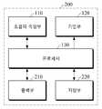
도 1을 참조하면, 생체정보 추정 장치(100)는 초음파 측정부(110), 가압부(120) 및 프로세서(130)를 포함한다. 초음파 측정부(110), 가압부(120) 및 프로세서(130)는 하나의 하드웨어 장치에 일체로 형성되거나 둘 이상의 하드웨어 장치에 분리 형성될 수 있다.Referring to FIG. 1 , the
초음파 측정부(110)는 프로세서(130)의 제어에 따라 피검체에 초음파를 송신하고, 피검체로부터 반사되는 반사파를 수신하여 피검체의 초음파 영상을 획득할 수 있다. 초음파 측정부(110)는 전기적 신호를 초음파 신호로 변환시키거나 초음파 신호를 전기적 신호로 변환시키는 초음파 트랜스듀서를 포함할 수 있다. 초음파 트랜스듀서는 초음파 소자들이 선형 1차원 또는 2차원 어레이로 배열되어 초음파 영상을 획득할 수 있다. 초음파 소자들은 압전 소자(piezoelectric element)들로 이루어질 수 있다.The
예를 들어, 어레이 피치(ptich)는 1차원의 경우 2cm의 애퍼처(aperture)를 확보하기 위해 70채널 이상일 수 있으며, 2차원의 경우 측면(leteral) 방향 2cm, 높이(elevation) 방향 1cm 애퍼처를 확보하기 위해 2100 채널 이상으로 형성될 수 있다. 다만, 이에 제한되는 것은 아니며 1.5 차원 또는 1.75 차원으로 형성되는 것도 가능하다. 중심 주파수는 5MHz 이상 15MHz 이하로 형성되고, 전력 소모량은 구동하는 동안 2W 이하로 형성되는 것이 가능하다.For example, the array pitch (ptich) may be 70 channels or more to secure an aperture of 2 cm in one dimension, and an aperture of 2 cm in the lateral direction and 1 cm in the elevation direction in the case of two dimensions. It may be formed in 2100 or more channels to secure. However, the present invention is not limited thereto and may be formed in 1.5 dimensions or 1.75 dimensions. The center frequency is formed to be 5 MHz or more and 15 MHz or less, and it is possible that the power consumption is formed to be 2 W or less during operation.
가압부(120)는 피검체에 압력을 가하여 피검체 내의 혈관을 폐색하거나 폐색 상태를 해제하는 기능을 수행할 수 있다. 예를 들어, 가압부(120)는 커프(cuff)를 포함할 수 있다.The pressurizing
프로세서(130)는 유선 또는 무선 통신을 이용하여 초음파 측정부(110) 및 가압부(120)를 제어하고, 초음파 측정부(110)가 획득한 피검체의 초음파 영상을 기초로 생체정보를 추정할 수 있다. 이때, 피검체는 사용자의 인체의 부위로서 예컨대, 요골 동맥에 인접한 손목 피부 영역, 모세혈이나 정맥혈이 지나가는 인체 피부 영역을 포함할 수 있다. 다만, 이에 제한되는 것은 아니며 기타 인체 내의 혈관 밀도가 높은 부위인 손가락, 발가락 등 인체의 말초 부위일 수도 있다.The
프로세서(130)는 생체정보 추정 요청이 수신되면 초음파 측정부(110)를 제어하여 소정 시간 동안 초음파 영상을 획득하도록 하며, 미리 정의된 제1 시간 경과 후 가압부(120)를 제어하여 피검체를 점차 가압하도록 하여 혈관을 폐색할 수 있다. 또한, 가압에 따라 피검체의 혈관이 폐색된 후 미리 정의된 제2 시간 경과하면 가압을 해제하도록 가압부(120)를 제어할 수 있다.When a biometric information estimation request is received, the
프로세서(130)는 초음파 측정부(110)에 의해 획득된 혈관 폐색 전후의 초음파 영상을 분석하여 혈관 직경의 변화를 추정할 수 있다. 또한, 혈관 직경의 변화를 기초로 생체정보를 추정할 수 있다. 이때, 생체정보는 혈당, 당 섭취량, 칼로리, 중성지방, 단백질, 콜레스테롤, 카로테노이드, 젖산, 체내 수분, 체외 수분, 체수분 및 요산 등을 포함할 수 있으나, 이에 제한되는 것은 아니다.The
프로세서(130)는 혈관 폐색 전후의 혈관 직경의 변화를 기초로 특징을 추출하고, 추출된 특징을 기초로 생체정보를 추정할 수 있다. 이때, 특징은 혈관 수축도, 혈관 확장도, 혈관 회복도 및 혈관 직경 변화 추세 등의 정보를 포함할 수 있다. 다만 이에 제한되는 것은 아니다.The
도 3은 공복 상태(93mg/dL)와 음식 섭취 후 혈당이 증가한 상태(125mg/dL)에서의 혈관 폐색 전후의 혈관 직경의 변화 그래프를 도시한 것이다.3 is a graph showing changes in blood vessel diameter before and after occlusion of blood vessels in a fasting state (93 mg/dL) and in a state in which blood glucose is increased after food intake (125 mg/dL).
프로세서(130)는 소정 시간 동안의 혈관 직경의 변화를 나타내는 그래프를 생성할 수 있다. 혈관 직경의 변화 그래프의 X축은 초음파 측정 시간을 나타내고, Y축은 가압부(120)가 피검체를 가압하기 전의 혈관 직경을 '1'로 정규화한 혈관 직경을 나타낸다. 도 3을 참조하면 혈당이 증가하여 고혈당이 되면 세포가 스트레스를 받아 활성 산소가 생성되고, 이로 인해 혈관 확장 인자 NO(Nitric Oxide)의 활성을 저하시켜서 혈관 폐색 전후의 혈관 확장 반응이 감소하는 것을 알 수 있다.The
도 3을 참조하면, 프로세서(130)는 가압에 따라 혈관이 폐색되어 혈관 직경이 축소된 축소 혈관 직경과 가압 전의 혈관 직경 사이의 차이를 기초로 혈관 수축도(FMC)를 획득할 수 있다. 예컨대 가압 전 혈관 직경에서 축소 혈관 직경을 뺀 값을 가압 전의 혈관 직경으로 나누어 혈관 수축도(FMC)를 획득할 수 있다. 이때, 축소 혈관 직경은 가압에 따라 혈관 직경이 최소가 된 상태의 최소값 또는 가압 이후 특정 구간의 평균값일 수 있다. 다만, 이에 제한되는 것은 아니다.Referring to FIG. 3 , the
또한, 프로세서(130)는 가압 전의 혈관 직경과 가압을 해제함에 따라 혈관이 회복되어 혈관 직경이 확장된 확장 혈관 직경 사이의 차이를 기초로 혈관 확장도(FMD)를 획득할 수 있다. 예컨대 확장 혈관 직경에서 가압 전 혈관 직경을 뺀 값을 가압 전의 혈관 직경으로 나누어 혈관 확장도(FMD)를 획득할 수 있다. 이때, 확장 혈관 직경은 가압이 해제됨에 따라 혈관 직경이 최대가 된 상태의 최대값 또는 가압 해제 이후 특정 구간의 평균값일 수 있다. 다만, 이에 제한되는 것은 아니다.In addition, the
또한, 프로세서(130)는 가압을 해제함에 따라 확장 혈관 직경이 발생한 지점으로부터 소정 시간 후의 혈관 직경 사이의 기울기를 기초로 혈관이 회복되는 정도를 나타내는 혈관 회복도(RES)를 획득할 수 있다. 예컨대, 획득된 기울기 값 또는 기울기 값에 미리 정의된 함수식을 적용하여 산출한 값을 혈관 회복도(RES)로 결정할 수 있다.In addition, the
또한, 프로세서(130)는 소정 기간 동안의 생체정보 추정 이력 정보를 기초로 소정 기간 동안 혈관 직경의 변화 추세 예컨대 혈관 확장도, 혈관 수축도, 혈관 회복도의 기울기가 변화하는 추세를 분석할 수 있다.In addition, the
프로세서(130)는 혈관 폐색 전후의 혈관 직경의 변화를 기초로 추출된 특징 중의 하나 또는 둘 이상을 조합하고, 특징과 생체정보 간의 상관 관계를 정의한 생체정보 추정 모델을 이용하여 생체정보를 추정할 수 있다. 이때, 생체정보 추정 모델은 선형 함수식 형태일 수 있으나, 이에 제한되는 것은 아니며 선형/비선형 회귀분석(linear/nonlinear regression analysis), 신경망(neural network), 딥러닝(deep learning) 등의 다양한 방법을 통해 정의될 수 있다.The
예를 들어, 아래의 수학식 1은 혈관 확장도 기반의 생체정보 추정 모델의 일 예이다.For example,

여기서, FMD1은 공복 상태에서 측정한 혈관 확장도, FMD2는 혈당이 상승하였을 때의 혈관 확장도를 나타낸다. b는 캘리브레이션 계수를 의미하는 것으로 개인별로 정의된 값일 수 있다. Y는 혈당 변화량을 나타낸 것이다.Here, FMD1 denotes vasodilation measured in a fasting state, and FMD2 denotes vasodilation when blood sugar rises. b denotes a calibration coefficient and may be a value defined for each individual. Y represents the amount of change in blood sugar.
프로세서(130)는 혈당 변화량이 획득되면 공복 상태의 혈당 값을 더하여 혈당 추정값을 획득할 수 있다When the amount of change in blood glucose is obtained, the
도 2는 다른 실시예에 따른 생체정보 추정 장치의 블록도이다.2 is a block diagram of an apparatus for estimating biometric information according to another embodiment.
도 2를 참조하면, 생체정보 추정 장치(200)의 다른 실시예는 초음파 측정부(110), 가압부(120), 프로세서(130), 출력부(210) 및 저장부(220)를 포함할 수 있다. 초음파 측정부(110), 가압부(120) 및 프로세서(130)는 앞에서 자세히 설명하였으므로 이하 생략한다.Referring to FIG. 2 , another embodiment of the biometric
출력부(210)는 초음파 측정부(110)에 의해 획득된 초음파 영상, 프로세서(200)의 처리 결과를 사용자에게 제공할 수 있다. 일 예로, 출력부(210)는 디스플레이 등의 시각적 출력 모듈을 통해 시각적으로 출력할 수 있다.The
예컨대, 출력부(210)는 디스플레이를 둘 이상의 영역으로 분리하고, 제1 영역에 생체정보 추정에 이용된 기본 정보 예컨대, 혈관 직경의 변화 그래프, 생체정보 추정에 이용한 혈관 확장도, 혈관 수축도, 혈관 회복도 등의 정보를 출력할 수 있다. 또한, 제2 영역에는 생체정보 추정값, 경고 정보, 조치 사항 등에 정보를 출력할 수 있다. 이때, 생체정보 추정값이 정상 범위를 벗어나는 경우, 빨간 색 등을 사용하여 강조하거나 정상 범위를 함께 표시할 수 있다.For example, the
다른 예로, 출력부(210)는 시각적 표시와 함께 또는 단독으로, 스피커 등의 음성 출력 모듈이나 햅틱 모듈을 이용하여 음성, 진동, 촉감 등의 비시각적인 방식으로 사용자에게 생체정보 추정 결과를 제공할 수 있다.As another example, the
저장부(220)는 생체정보 추정을 위한 기준 정보, 혈관 직경 변화, 추출된 특징 등을 저장할 수 있다. 이때, 기준 정보는 사용자의 나이, 성별, 건강상태 등과 같은 사용자 특성 정보, 생체정보 추정 모델 등의 정보를 포함할 수 있다.The
저장부(220)는 플래시 메모리 타입(flash memory type), 하드디스크 타입(hard disk type), 멀티미디어 카드 마이크로 타입(multimedia card micro type), 카드 타입의 메모리(예를 들어, SD 또는 XD 메모리 등), 램(Random Access Memory: RAM) SRAM(Static Random Access Memory), 롬(Read-Only Memory: ROM), EEPROM(Electrically Erasable Programmable Read-Only Memory), PROM(Programmable Read-Only Memory), 자기 메모리, 자기 디스크, 광디스크 등의 저장매체를 포함하며, 이에 제한되는 것은 아니다.The
도 4는 일 실시예에 따른 초음파 장치의 블록도이다. 도 5a 내지 도 5c는 웨어러블 기기 형태의 초음파 장치의 실시예를 도시한 것이다.4 is a block diagram of an ultrasound apparatus according to an exemplary embodiment. 5A to 5C illustrate an embodiment of an ultrasound device in the form of a wearable device.
도 4를 참조하면, 초음파 장치는 초음파 측정부(410), 프로세서(420), 출력부(430) 및 통신부(440)를 포함할 수 있다.Referring to FIG. 4 , the ultrasound apparatus may include an
초음파 측정부(410)는 프로세서(420)의 제어에 따라 피검체에 초음파를 송신하고, 피검체로부터 반사된 반사파를 수신하여 초음파 영상을 획득할 수 있다. 초음파 측정부(410)는 앞에서 상세히 설명하였으므로 자세한 설명은 생략한다.The
프로세서(420)는 초음파 측정부(410)와 전기적으로 연결될 수 있다. 프로세서(420)는 생체정보 추정 요청이 수신되면 초음파 측정부(410)를 제어하여 초음파 영상을 획득하도록 할 수 있다.The
또한, 프로세서(420)는 초음파 영상이 획득되는 동안 가압 장치(500)를 제어하여 피검체를 가압하도록 하고, 소정 시간 경과 후 가압을 해제하도록 제어할 수 있다. 이때, 가압 장치(500)는 외부 별도의 하드웨어 장치일 수 있다.Also, the
프로세서(420)는 무선 통신을 통해 가압 장치(500)와 연결하고, 가압 장치(500)를 제어하는 제어신호를 생성하여 통신부(440)를 통해 가압 장치(500)에 전송할 수 있다.The
프로세서(420)는 가압 장치(500)의 가압 및 가압 해제에 따른 혈관 폐색 전후의 초음파 영상을 기초로 혈관 직경 변화를 추정할 수 있다. 이때, 프로세서(420)는 초음파 영상을 분석하여 혈관 직경을 산출하는 알고리즘을 실행할 수 있다. 또한, 프로세서(420)는 생체정보 추정 알고리즘을 실행하여, 혈관 직경의 변화로부터 혈관 확장도, 혈관 수축도, 혈관 회복도 등의 특징을 추출하고, 생체정보 추정 모델을 적용하여 특징을 기초로 생체정보를 추정할 수 있다.The
출력부(430)는 초음파 측정부(410)에 의해 획득된 초음파 영상, 혈관 직경 변화 및 생체정보 추정 결과 중의 적어도 하나를 출력할 수 있다.The
통신부(440)는 통신 기술을 이용하여 가압 장치(500) 및 외부 기기와 통신할 수 있다. 통신부(440)는 모바일 기기로부터 초음파 영상 획득 및 가압 장치(500)의 제어신호를 수신하여 프로세서(420)에 전달할 수 있다. 통신부(440)는 초음파 측정부(410)에 의해 측정된 초음파 영상, 프로세서(420)에 의해 추정된 혈관 직경의 변화, 추출된 특징 및 생체정보 추정 결과 등을 모바일 기기에 전송할 수 있다.The
이때, 통신 기술은 블루투스(bluetooth) 통신, BLE(Bluetooth Low Energy) 통신, 근거리 무선 통신(Near Field Communication, NFC), WLAN 통신, 지그비(Zigbee) 통신, 적외선(Infrared Data Association, IrDA) 통신, WFD(Wi-Fi Direct) 통신, UWB(ultra-wideband) 통신, Ant+ 통신, WIFI 통신, RFID(Radio Frequency Identification) 통신, 3G 통신, 4G 통신 및 5G 통신 등을 포함할 수 있다. 다만, 이에 한정되는 것은 아니다.At this time, the communication technology is Bluetooth (bluetooth) communication, BLE (Bluetooth Low Energy) communication, near field communication (Near Field Communication, NFC), WLAN communication, Zigbee communication, infrared (Infrared Data Association, IrDA) communication, WFD (Wi-Fi Direct) communication, ultra-wideband (UWB) communication, Ant+ communication, WIFI communication, radio frequency identification (RFID) communication, 3G communication, 4G communication, 5G communication, etc. may be included. However, the present invention is not limited thereto.
도 5a는 스마트 워치 또는 스마트 밴드형 웨어러블 기기로 제작된 초음파 장치(400)의 일 실시예를 예시한 것이다. 다만, 이에 제한되는 것은 아니며 팔찌와 같은 액서서리 형태로 제작될 수 있다.5A illustrates an embodiment of the
도시된 바와 같이 스마트워치 형태의 초음파 장치(400)는 본체(MB)와 스트랩(ST)을 포함할 수 있다.As shown, the
본체(MB)는 다양한 형태를 갖도록 형성될 수 있다. 본체(MB)에 전술한 생체정보 추정과 관련된 초음파 측정부(410), 프로세서(420), 출력부(430) 및 통신부(440)와 그 밖의 기능을 수행하는 모듈들이 장착될 수 있다. 본체(MB) 또는 스트랩(ST)의 내부에는 초음파 장치(400)의 각종 모듈에 전원을 공급하는 배터리가 내장될 수 있다.The body MB may be formed to have various shapes. The main body MB may be equipped with an
스트랩(ST)은 본체(MB)에 연결될 수 있다. 스트랩(ST)은 사용자의 손목을 감싸는 형태로 구부려질 수 있도록 플렉시블(flexible)하게 형성될 수 있다. 스트랩(ST)은 도시된 바와 같이 분리/체결되는 형태 또는 일체형 밴드 형태로 형성될 수 있다.The strap ST may be connected to the body MB. The strap ST may be formed to be flexible so that it can be bent in a shape surrounding the user's wrist. The strap ST may be formed in a form that is separated/fastened as shown or in the form of an integrated band.
디스플레이(DP)는 본체(MB)의 상단에 배치될 수 있다. 출력부(430)는 프로세서(420)에 의해 추정된 생체정보 추정 결과를 디스플레이(DP)에 표시할 수 있다. 디스플레이(DP)는 터치 입력이 가능한 터치 패널로 형성될 수 있다. 출력부(430)는 디스플레이(DP)를 통해 사용자의 터치 입력이 수신되면 프로세서(420)에 전달할 수 있다.The display DP may be disposed on the upper end of the main body MB. The
또한, 본체(MB) 내부에는 프로세서의 처리 결과 및 각종 정보를 저장하는 저장부가 장착될 수 있다.In addition, a storage unit for storing processing results and various information of the processor may be mounted inside the main body MB.
또한, 사용자의 제어 명령을 수신하여 프로세서로 전달하는 조작부가 본체(MB)의 측면에 장착될 수 있다. 조작부는 초음파 장치(400)의 전원을 온/오프시키는 명령을 입력하기 위한 전원 버튼을 포함할 수 있다.In addition, a manipulation unit that receives a user's control command and transmits it to the processor may be mounted on the side of the main body MB. The manipulation unit may include a power button for inputting a command for turning on/off the power of the
도 5b 및 도 5c를 참조하면, 초음파 장치(400)는 피검체의 손목 부분에 착용되고, 가압 장치(500)는 별개의 하드웨어 장치로 형성되어 초음파 장치(400)와 이격된 위치에 착용될 수 있다. 초음파 장치(400)와 가압 장치(500)는 케이블(TC)을 통해 전기적으로 연결될 수 있다. 도 5c에 도시된 바와 같이 초음파 장치(400)와 가압 장치(500)는 서로 밀착하여 배치되는 경우 서로 일체감을 갖도록 하나의 세트로 형성될 수 있다.Referring to FIGS. 5B and 5C , the
가압 장치(500)는 초음파 장치(400)가 사용자의 손목의 혈관 부위에서 초음파 영상을 획득할 때, 전기적인 케이블(TC)을 통해 초음파 장치(400)의 프로세서(420)의 제어를 통해 사용자의 팔을 가압하여 혈관을 폐색할 수 있다. 또는, 가압 장치(500)는 내부에 통신 모듈을 포함할 수 있으며, 무선 통신을 통해 초음파 장치(400) 또는 모바일 기기로부터 제어신호를 수신할 수 있다.When the
한편, 가압 장치(500)는 초음파 장치(400)의 스트랩(ST)에 형성될 수 있다. 예를 들어, 스트랩(ST)은 손목에 가해지는 압력의 변화에 따라 탄성을 갖도록 내부에 공기가 주입되거나 공기 주머니를 포함할 수 있다.Meanwhile, the
도 6은 일 실시예에 따른 모바일 장치의 블록도이다. 도 7a 및 도 7b는 모바일 장치의 혈당 추정 기능을 설명하기 위한 도면이다.6 is a block diagram of a mobile device according to an embodiment. 7A and 7B are diagrams for explaining a blood glucose estimating function of a mobile device.
도 6을 참조하면 모바일 장치(600)는 프로세서(610), 저장부(620), 출력부(630) 및 통신부(640)를 포함할 수 있다.Referring to FIG. 6 , the
프로세서(610)는 생체정보 추정 요청을 수신하면, 저장부(620)를 참조하여 외부 초음파 장치(710) 및 가압 장치(720)를 제어하는 제어신호를 생성하고, 통신부(640)를 통해 초음파 장치(710) 및 가압 장치(720)에 전송할 수 있다. 한편, 가압 장치(720)를 제어하는 제어신호는 가압 장치(720)에 직접 전송하는 대신 초음파 장치(710)에 전송하고 초음파 장치(710)가 수신된 제어신호를 전기적으로 연결된 가압 장치(720)에 전송할 수 있다.When receiving the biometric information estimation request, the
또한, 프로세서(610)는 초음파 장치(710)로부터 혈관 폐색 전후의 초음파 영상을 수신하면, 혈관 직경 산출 알고리즘을 실행하여 혈관 직경의 변화를 추정할 수 있다. 또한, 혈관 직경의 변화로부터 특징을 추출하고, 생체정보 추정 모델을 이용하여 생체정보를 추정할 수 있다.Also, when receiving ultrasound images before and after occlusion of blood vessels from the
저장부(620)는 초음파 장치(710), 가압 장치(720)를 제어하는 제어 기준에 관한 정보를 저장할 수 있다. 예를 들어, 초음파 주파수, 초음파 송신 주기, 초음파 측정 시간, 가압 시작 시점, 가압 해제 시점, 가압의 세기 등에 관한 정보를 포함할 수 있다. 또한, 초음파 영상을 분석하여 혈관 직경을 산출하는 알고리즘, 생체정보 추정 모델 등의 정보를 포함할 수 있다. 또한, 사용자의 개인별 특성에 관한 정보를 저장할 수 있다. 또한, 저장부(620)는 초음파 장치(710)로부터 수신된 초음파 영상, 프로세서(610)의 처리 결과를 저장할 수 있다.The
출력부(630)는 초음파 장치(710)로부터 수신된 초음파 영상, 프로세서(610)의 처리 결과를 출력할 수 있다. 도 7a를 참조하면 출력부(630)는 디스플레이(DP)에 혈당 추정 결과를 표시할 수 있다. 또한, 도 7b를 참조하면 출력부(630)는 디스플레이(DP)를 제1 영역(DA1) 및 제2 영역(DA2)으로 분할하고, 제1 영역(DA1)에 혈관 직경 변화 그래프(GR)를 출력하고, 제2 영역(DA2)에 혈당 추정 결과, 전당뇨 위험 및/또는 고혈당 위험에 관한 경고 정보를 출력할 수 있다.The
도 8은 일 실시예에 따른 생체정보 추정 방법의 흐름도이다. 도 8은 도 1 및 도 2의 실시예에 따른 생체정보 추정 장치(100,200)에 의해 수행되는 실시예이다. 앞에서 상세히 기술하였으므로 이하 중복 설명을 피하기 위해 간단하게 설명한다.8 is a flowchart of a method for estimating biometric information according to an embodiment. 8 is an embodiment performed by the biometric
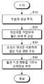
먼저, 초음파 측정부를 통해 초음파 영상을 획득할 수 있다(810). 초음파 측정부는 피검체에 초음파를 송신하고, 피검체로부터 반사되는 반사파를 기초로 초음파 영상을 획득할 수 있다.First, an ultrasound image may be acquired through the ultrasound measuring unit ( 810 ). The ultrasound measuring unit may transmit ultrasound to the subject and acquire an ultrasound image based on a reflected wave reflected from the subject.
그 다음, 초음파 영상을 시작한 이후 소정 시간이 경과하면 가압부를 통해 피검체에 압력을 가하여 혈관을 폐색하고 소정 시간 경과 후 가압을 해제할 수 있다(820).Next, when a predetermined time has elapsed since the start of the ultrasound image, pressure may be applied to the subject through the pressurizer to occlude the blood vessel, and the pressure may be released after a predetermined time has elapsed ( 820 ).
그 다음, 초음파 영상을 분석하여 혈관 직경 변화를 추정할 수 있다(830).Next, the ultrasound image may be analyzed to estimate a change in the diameter of the blood vessel ( 830 ).
그 다음, 혈관 직경 변화를 기초로 생체정보를 추정할 수 있다(840). 예를 들어, 혈관 직경 변화 그래프로부터 혈관 확장도, 혈관 수축도, 혈관 회복도 및 혈관 직경 변화 추세 정보 등의 특징을 획득하고, 획득된 특징을 하나 이상 조합하여 생체정보를 추정할 수 있다.Next, biometric information may be estimated based on the change in the diameter of the blood vessel ( 840 ). For example, characteristics such as vasodilation, vasoconstriction, vascular recovery, and vascular diameter change trend information may be acquired from the vascular diameter change graph, and biometric information may be estimated by combining one or more of the acquired features.
한편, 본 실시 예들은 컴퓨터로 읽을 수 있는 기록 매체에 컴퓨터가 읽을 수 있는 코드로 구현하는 것이 가능하다. 컴퓨터가 읽을 수 있는 기록 매체는 컴퓨터 시스템에 의하여 읽혀질 수 있는 데이터가 저장되는 모든 종류의 기록 장치를 포함한다.Meanwhile, the present embodiments can be implemented as computer-readable codes on a computer-readable recording medium. The computer-readable recording medium includes all types of recording devices in which data readable by a computer system is stored.
컴퓨터가 읽을 수 있는 기록 매체의 예로는 ROM, RAM, CD-ROM, 자기 테이프, 플로피디스크, 광 데이터 저장장치 등이 있으며, 또한 캐리어 웨이브(예를 들어 인터넷을 통한 전송)의 형태로 구현하는 것을 포함한다. 또한, 컴퓨터가 읽을 수 있는 기록 매체는 네트워크로 연결된 컴퓨터 시스템에 분산되어, 분산 방식으로 컴퓨터가 읽을 수 있는 코드가 저장되고 실행될 수 있다. 그리고 본 실시예들을 구현하기 위한 기능적인(functional) 프로그램, 코드 및 코드 세그먼트들은 본 발명이 속하는 기술 분야의 프로그래머들에 의하여 용이하게 추론될 수 있다.Examples of computer-readable recording media include ROM, RAM, CD-ROM, magnetic tape, floppy disk, optical data storage, etc. include In addition, the computer-readable recording medium may be distributed in a network-connected computer system, and the computer-readable code may be stored and executed in a distributed manner. In addition, functional programs, codes, and code segments for implementing the present embodiments can be easily inferred by programmers in the technical field to which the present invention pertains.
본 개시가 속하는 기술분야의 통상의 지식을 가진 자는 개시된 기술적 사상이나 필수적인 특징을 변경하지 않고서 다른 구체적인 형태로 실시될 수 있다는 것을 이해할 수 있을 것이다. 그러므로 이상에서 기술한 실시예들은 모든 면에서 예시적인 것이며 한정적이 아닌 것으로 이해해야만 한다.Those of ordinary skill in the art to which the present disclosure pertains will understand that it may be implemented in other specific forms without changing the disclosed technical spirit or essential features. Therefore, it should be understood that the embodiments described above are illustrative in all respects and not restrictive.
100: 생체정보 추정 장치110: 초음파 측정부
120: 가압부130: 프로세서
210: 출력부220: 저장부
400: 초음파 장치410: 초음파 측정부
420: 프로세서430: 출력부
440: 통신부600: 모바일 장치
610: 프로세서620: 저장부
630: 출력부640: 통신부100: biometric information estimation device 110: ultrasonic measurement unit
120: pressing unit 130: processor
210: output unit 220: storage unit
400: ultrasonic device 410: ultrasonic measuring unit
420: processor 430: output unit
440: communication unit 600: mobile device
610: processor 620: storage
630: output unit 640: communication unit
Claims (29)
Translated fromKorean상기 피검체에 압력을 가하여 혈관을 폐색하는 가압부; 및
상기 초음파 측정부에 의해 획득된 혈관 폐색 전후의 초음파 영상을 이용하여 혈관 직경의 변화를 추정하고, 추정된 혈관 직경의 변화를 기초로 생체정보를 추정하는 프로세서를 포함하는 생체정보 추정 장치.an ultrasound measurement unit configured to acquire an ultrasound image of the subject;
a pressing unit that occludes blood vessels by applying pressure to the subject; and
and a processor for estimating a change in vessel diameter using the ultrasound images before and after vessel occlusion obtained by the ultrasound measuring unit, and estimating biometric information based on the estimated change in vessel diameter.
상기 초음파 측정부는
피검체에 초음파를 송신하여 피검체로부터 반사되는 반사파를 수신하고, 상기 수신된 반사파를 기초로 초음파 영상을 생성하는 생체정보 추정 장치.According to claim 1,
The ultrasonic measuring unit
An apparatus for estimating biometric information that transmits ultrasound to a subject, receives a reflected wave reflected from the subject, and generates an ultrasound image based on the received reflected wave.
상기 가압부는 커프를 포함하는 생체정보 추정 장치.According to claim 1,
The biometric information estimating device including a cuff.
상기 프로세서는
생체정보 추정 요청이 수신되면 초음파 측정부를 제어하고, 제1 시간 경과 후 피검체를 가압하도록 하고 제2 시간 경과 후 가압을 해제하도록 가압부를 제어하는 생체정보 추정 장치.According to claim 1,
the processor
When a biometric information estimation request is received, the ultrasonic measuring unit is controlled, the biometric information estimating apparatus controls the pressing unit to pressurize the subject after a first time elapses and release the pressurization after a second time elapses.
상기 프로세서는
상기 추정된 혈관 직경의 변화를 기초로 혈관 수축도, 혈관 확장도, 혈관 회복도 및 혈관 직경 변화 추세 중의 적어도 하나의 특징을 획득하고, 획득된 특징을 기초로 생체정보를 추정하는 생체정보 추정 장치.According to claim 1,
the processor
A biometric information estimating apparatus for acquiring at least one characteristic of vasoconstriction, vasodilation, vascular recovery, and vascular diameter change trend based on the estimated change in vessel diameter, and estimating biometric information based on the acquired characteristic .
상기 프로세서는
상기 추정된 혈관 직경의 변화 그래프를 생성하고, 생성된 혈관 직경의 변화 그래프를 기초로 상기 특징을 획득하는 생체정보 추정 장치.6. The method of claim 5,
the processor
A biometric information estimating apparatus for generating a change graph of the estimated blood vessel diameter, and acquiring the characteristic based on the generated change graph of the blood vessel diameter.
상기 프로세서는
가압 전의 혈관 직경과 축소 혈관 직경 사이의 차이를 기초로 상기 혈관 수축도를 획득하는 생체정보 추정 장치.6. The method of claim 5,
the processor
An apparatus for estimating biometric information for obtaining the degree of vasoconstriction based on a difference between a diameter of a blood vessel before pressurization and a diameter of a reduced blood vessel.
상기 프로세서는
가압 전의 혈관 직경과 확장 혈관 직경 사이의 차이를 기초로 상기 혈관 확대도를 획득하는 생체정보 추정 장치.6. The method of claim 5,
the processor
An apparatus for estimating bioinformation to obtain the enlargement of the blood vessel based on a difference between the diameter of the blood vessel before pressurization and the diameter of the enlarged blood vessel.
상기 프로세서는
확장 혈관 직경과, 확장 혈관 직경이 발생한 시점으로부터 소정 시간 후의 혈관 직경 사이의 기울기를 기초로 상기 혈관 회복도를 획득하는 생체정보 추정 장치.6. The method of claim 5,
the processor
An apparatus for estimating blood vessel recovery for obtaining the blood vessel recovery degree based on a slope between the dilated blood vessel diameter and the blood vessel diameter after a predetermined time from the point at which the dilated blood vessel diameter occurs.
상기 프로세서는
상기 특징이 획득되면, 상기 특징과 생체정보 간의 상관관계를 정의한 생체정보 추정 모델을 적용하여 생체정보를 추정하는 생체정보 추정 장치.6. The method of claim 5,
the processor
When the feature is acquired, a biometric information estimation apparatus for estimating biometric information by applying a biometric information estimation model defining a correlation between the feature and biometric information.
상기 생체정보는
혈당, 당 섭취량, 칼로리, 중성지방, 단백질, 콜레스테롤, 카로테노이드, 젖산, 체내 수분, 체외 수분, 체수분 및 요산 중의 하나 이상을 포함하는 생체정보 추정 장치.According to claim 1,
The biometric information is
Biometric information estimation device including one or more of blood sugar, sugar intake, calories, triglyceride, protein, cholesterol, carotenoids, lactic acid, body water, extracorporeal water, body water, and uric acid.
상기 초음파 영상, 혈관 직경의 변화 및 생체정보 추정 결과 중의 적어도 하나를 출력하는 출력부를 더 포함하는 생체정보 추정 장치.According to claim 1,
and an output unit for outputting at least one of the ultrasound image, a change in a blood vessel diameter, and a result of estimating biometric information.
가압부를 통해 상기 피검체를 가압하여 혈관을 폐색하는 단계;
프로세서가 상기 초음파 측정부에 의해 획득된 혈관 폐색 전 후의 초음파 영상을 이용하여 혈관 직경의 변화를 추정하는 단계; 및
상기 프로세서가 상기 추정된 혈관 직경의 변화를 기초로 생체정보를 추정하는 단계를 포함하는 생체정보 추정 방법.acquiring an ultrasound image of the subject through an ultrasound measuring unit;
occluding a blood vessel by pressing the subject through a pressurizing unit;
estimating, by a processor, a change in the diameter of a blood vessel using the ultrasound images before and after the vessel occlusion acquired by the ultrasound measuring unit; and
Biometric information estimation method comprising the step of the processor estimating the biometric information based on the change in the estimated blood vessel diameter.
상기 생체정보를 추정하는 단계는
상기 혈관 직경의 변화를 기초로 혈관 수축도, 혈관 확장도, 혈관 회복도 및 혈관 직경 변화 추세 중의 적어도 하나의 특징을 획득하는 단계; 및
상기 획득된 특징을 기초로 생체정보를 추정하는 단계를 포함하는 생체정보 추정 방법.14. The method of claim 13,
The step of estimating the biometric information
acquiring at least one characteristic of a vasoconstriction degree, a vasodilation degree, a vascular recovery degree, and a vascular diameter change trend based on the change in the blood vessel diameter; and
and estimating biometric information based on the acquired characteristics.
상기 특징을 획득하는 단계는
가압 전의 혈관 직경과 축소 혈관 직경 사이의 차이를 기초로 상기 혈관 수축도를 획득하는 생체정보 추정 방법.15. The method of claim 14,
The step of acquiring the feature is
A method of estimating biometric information for obtaining the degree of vasoconstriction based on a difference between the diameter of the blood vessel before pressurization and the diameter of the reduced blood vessel.
상기 특징을 획득하는 단계는
가압 전의 혈관 직경과 확장 혈관 직경 사이의 차이를 기초로 상기 혈관 확대도를 획득하는 생체정보 추정 방법.15. The method of claim 14,
The step of acquiring the feature is
A method of estimating bioinformation for obtaining the degree of enlargement of the blood vessel based on a difference between the diameter of the blood vessel before pressurization and the diameter of the enlarged blood vessel.
상기 특징을 획득하는 단계는
확장 혈관 직경과, 확장 혈관 직경이 발생한 시점으로부터 소정 시간 후의 혈관 직경 사이의 기울기를 기초로 상기 혈관 회복도를 획득하는 생체정보 추정 방법.15. The method of claim 14,
The step of acquiring the feature is
A bioinformation estimation method for obtaining the degree of vascular recovery based on a slope between the dilated blood vessel diameter and the blood vessel diameter after a predetermined time from the time when the dilated blood vessel diameter occurs.
상기 특징을 기초로 생체정보를 추정하는 단계는
상기 특징과 생체정보 간의 상관관계를 정의한 생체정보 추정 모델을 적용하여 생체정보를 추정하는 생체정보 추정 방법.5. The method of claim 4,
The step of estimating biometric information based on the characteristics
A biometric information estimation method for estimating biometric information by applying a biometric information estimation model defining a correlation between the characteristics and biometric information.
상기 혈관 직경의 변화 및 생체정보 추정 결과 중의 적어도 하나를 출력하는 단계를 더 포함하는 생체정보 추정 방법.14. The method of claim 13,
and outputting at least one of a change in the blood vessel diameter and a result of estimating biometric information.
가압 장치를 제어하여 피검체의 혈관을 폐색하고, 상기 초음파 측정부에 의해 획득된 혈관 폐색 전후 초음파 영상을 기초로 혈관 직경의 변화를 추정하며, 추정된 혈관 변화 직경을 기초로 생체정보를 추정하는 프로세서를 포함하는 초음파 장치.an ultrasound measuring unit that transmits ultrasound to a subject and receives a reflected wave reflected from the subject to obtain an ultrasound image; and
Controlling a pressure device to occlude a blood vessel of the subject, estimating a change in vessel diameter based on an ultrasound image before and after vessel occlusion obtained by the ultrasound measurement unit, and estimating biometric information based on the estimated vessel change diameter An ultrasound device comprising a processor.
상기 프로세서에 의해 생성된 제어신호를 가압 장치에 전송하는 통신부를 더 포함하는 초음파 장치.21. The method of claim 20,
The ultrasound apparatus further comprising a communication unit for transmitting the control signal generated by the processor to the pressing device.
상기 통신부는
상기 초음파 영상, 혈관 직경의 변화 및 생체정보 추정 결과 중의 적어도 하나를 모바일 장치에 전송하는 초음파 장치.22. The method of claim 21,
the communication unit
An ultrasound apparatus for transmitting at least one of the ultrasound image, a change in blood vessel diameter, and a result of estimating biometric information to a mobile device.
상기 프로세서는
상기 혈관 직경의 변화를 기초로 혈관 수축도, 혈관 확장도, 혈관 회복도 및 혈관 직경 변화 추세 중의 적어도 하나의 특징을 획득하고, 획득된 특징을 이용하여 생체정보를 추정하는 초음파 장치.21. The method of claim 20,
the processor
An ultrasound apparatus for acquiring at least one characteristic of a vasoconstriction degree, a vasodilation degree, a vascular recovery degree, and a vascular diameter change trend based on a change in the blood vessel diameter, and estimating biometric information using the acquired characteristic.
상기 초음파 영상, 혈관 직경 변화 및 생체정보 추정 결과 중의 적어도 하나를 출력하는 출력부를 더 포함하는 초음파 장치.21. The method of claim 20,
The ultrasound apparatus further comprising an output unit for outputting at least one of the ultrasound image, a change in vessel diameter, and a result of estimating biometric information.
상기 초음파 측정부 및 프로세서가 실장되는 본체; 및
상기 본체에 연결되어 사용자의 손목을 감싸도록 형성된 스트랩을 더 포함하는 초음파 장치.21. The method of claim 20,
a main body on which the ultrasonic measuring unit and the processor are mounted; and
The ultrasound apparatus further comprising a strap connected to the main body to wrap a user's wrist.
상기 제어신호를 생성하고, 상기 초음파 장치로부터 수신된 혈관 폐색 전후의 초음파 영상을 기초로 혈관 직경의 변화를 추정하며, 추정된 혈관 직경의 변화를 기초로 생체정보를 추정하는 프로세서를 포함하는 모바일 장치.a communication unit transmitting a control signal to the ultrasound apparatus and the pressurizing apparatus, and receiving an ultrasound image of the subject from the ultrasound apparatus; and
and a processor for generating the control signal, estimating a change in vessel diameter based on ultrasound images before and after vessel occlusion received from the ultrasound device, and estimating biometric information based on the estimated change in vessel diameter. .
상기 프로세서는
생체정보 추정 요청이 수신되면 초음파 영상을 획득하도록 초음파 장치를 제어하는 제1 제어신호를 생성하고, 제1 시간 경과 후 피검체를 가압하도록 가압 장치를 제어하는 제2 제어신호를 생성하며, 제2 시간 경과 후 가압을 해제하도록 가압 장치를 제어하는 제3 제어신호를 생성하는 모바일 장치.27. The method of claim 26,
the processor
When a biometric information estimation request is received, a first control signal for controlling the ultrasound apparatus to acquire an ultrasound image is generated, a second control signal for controlling the pressure apparatus to pressurize the subject after a first time has elapsed, and a second control signal is generated. A mobile device that generates a third control signal for controlling the pressurization device to release the pressurization after a time has elapsed.
상기 프로세서는
상기 추정된 혈관 직경의 변화를 기초로 혈관 수축도, 혈관 확장도, 혈관 회복도 및 혈관 직경 변화 추세 중의 적어도 하나의 특징을 획득하고, 획득된 특징을 기초로 생체정보를 추정하는 모바일 장치.27. The method of claim 26,
the processor
A mobile device for acquiring at least one characteristic of vasoconstriction, vasodilation, vascular recovery, and vascular diameter change trend based on the estimated change in vessel diameter, and estimating biometric information based on the acquired characteristic.
상기 초음파 영상, 혈관 직경의 변화 및 생체정보 추정 결과 중의 적어도 하나를 출력하는 출력부를 더 포함하는 모바일 장치.27. The method of claim 26,
The mobile device further comprising an output unit for outputting at least one of the ultrasound image, changes in blood vessel diameter, and biometric information estimation results.
Priority Applications (5)
| Application Number | Priority Date | Filing Date | Title |
|---|---|---|---|
| KR1020200019097AKR20210104410A (en) | 2020-02-17 | 2020-02-17 | Apparatus and method for estimating bio-information, ultrasonic device and mobile device |
| US16/992,799US11684339B2 (en) | 2020-02-17 | 2020-08-13 | Apparatus and method for estimating bio-information, ultrasonic device, and mobile device |
| CN202010856741.5ACN113261990A (en) | 2020-02-17 | 2020-08-24 | Apparatus for estimating biological information, ultrasound device, and mobile device |
| EP20196188.5AEP3865073A1 (en) | 2020-02-17 | 2020-09-15 | Apparatus and method for estimating bio-information, ultrasonic device, and mobile device |
| JP2020167002AJP2021126498A (en) | 2020-02-17 | 2020-10-01 | Device and method for estimating bio-information, ultrasonic device, and mobile device |
Applications Claiming Priority (1)
| Application Number | Priority Date | Filing Date | Title |
|---|---|---|---|
| KR1020200019097AKR20210104410A (en) | 2020-02-17 | 2020-02-17 | Apparatus and method for estimating bio-information, ultrasonic device and mobile device |
Publications (1)
| Publication Number | Publication Date |
|---|---|
| KR20210104410Atrue KR20210104410A (en) | 2021-08-25 |
Family
ID=72521389
Family Applications (1)
| Application Number | Title | Priority Date | Filing Date |
|---|---|---|---|
| KR1020200019097ACeasedKR20210104410A (en) | 2020-02-17 | 2020-02-17 | Apparatus and method for estimating bio-information, ultrasonic device and mobile device |
Country Status (5)
| Country | Link |
|---|---|
| US (1) | US11684339B2 (en) |
| EP (1) | EP3865073A1 (en) |
| JP (1) | JP2021126498A (en) |
| KR (1) | KR20210104410A (en) |
| CN (1) | CN113261990A (en) |
Cited By (1)
| Publication number | Priority date | Publication date | Assignee | Title |
|---|---|---|---|---|
| KR20230136515A (en) | 2022-03-18 | 2023-09-26 | 주식회사 에어스 메디컬 | A conditional controlled input generator for medical AI and a method thereof |
Family Cites Families (26)
| Publication number | Priority date | Publication date | Assignee | Title |
|---|---|---|---|---|
| SG126677A1 (en) | 2001-06-26 | 2006-11-29 | Meng Ting Choon | Method and device for measuring blood sugar level |
| JP4799833B2 (en) | 2003-06-19 | 2011-10-26 | サラヤ株式会社 | Method and apparatus for measuring blood vessel diameter using echo |
| EP1954175B1 (en) | 2005-11-10 | 2016-07-13 | Biovotion AG | Device for determining the glucose level in body tissue |
| US8016761B2 (en)* | 2006-10-23 | 2011-09-13 | The General Electric Company | Method and apparatus for automated flow mediated dilation |
| US8043223B2 (en)* | 2006-11-22 | 2011-10-25 | The General Electric Company | Method and apparatus for automated vascular function testing |
| KR101491853B1 (en) | 2008-07-15 | 2015-02-09 | 엘지전자 주식회사 | Non-invasive blood glucose measuring device and method |
| KR101491854B1 (en) | 2008-08-20 | 2015-02-09 | 엘지전자 주식회사 | Non-invasive blood glucose measuring device and method |
| KR101020001B1 (en)* | 2008-11-11 | 2011-03-09 | 한국 한의학 연구원 | Pulse wave measuring device and method |
| US8235897B2 (en) | 2010-04-27 | 2012-08-07 | A.D. Integrity Applications Ltd. | Device for non-invasively measuring glucose |
| JP6098101B2 (en)* | 2011-12-14 | 2017-03-22 | セイコーエプソン株式会社 | Blood pressure measurement device and blood pressure measurement method |
| RU2518134C2 (en) | 2012-02-24 | 2014-06-10 | Хилби Корпорейшн | Method for determining individual's blood glucose concentration |
| EP2967362B1 (en) | 2013-03-13 | 2018-12-12 | Everist Genomics, Inc. | Method and device to diagnose a potential to develop a cardiovascular disease |
| TWM460634U (en)* | 2013-03-19 | 2013-09-01 | Avita Corp | Device for monitoring physiological condition |
| JP2016043093A (en) | 2014-08-25 | 2016-04-04 | セイコーエプソン株式会社 | Ultrasound blood pressure measuring device and blood pressure measuring method |
| US20160058305A1 (en)* | 2014-08-26 | 2016-03-03 | University Of Washington | Device and method for analyzing reperfusion injury |
| NL2013884B1 (en) | 2014-11-27 | 2016-10-11 | Umc Utrecht Holding Bv | Wearable ultrasound device for signalling changes in human or animal body. |
| KR102335739B1 (en) | 2014-12-19 | 2021-12-06 | 삼성전자주식회사 | Apparatus and method for measuring a blood glucose in a noninvasive manner |
| JP6484787B2 (en)* | 2014-12-19 | 2019-03-20 | 学校法人 関西大学 | Diagnosis support apparatus and computer program |
| US10932727B2 (en) | 2015-09-25 | 2021-03-02 | Sanmina Corporation | System and method for health monitoring including a user device and biosensor |
| US9737217B2 (en)* | 2015-08-14 | 2017-08-22 | The Regents Of The University Of California | Assessing endothelial function and providing calibrated UFMD data using a blood pressure cuff |
| CN105852910A (en)* | 2016-04-18 | 2016-08-17 | 何宗彦 | Equipment and method for detecting vascular endothelial functions by Doppler ultrasonography |
| JP6371334B2 (en) | 2016-05-27 | 2018-08-08 | 株式会社ユネクス | Ultrasound cross-sectional image measuring device |
| GB201610040D0 (en)* | 2016-06-08 | 2016-07-20 | Isis Innovation | Flow mediated dilatation |
| US20180092631A1 (en) | 2016-10-03 | 2018-04-05 | National Kaohsiung University Of Applied Sciences | Blood Vessel Analysis Device and Operating Method Thereof |
| JP7019176B2 (en)* | 2017-05-15 | 2022-02-15 | 株式会社ユネクス | Endothelial function tester for arterial blood vessels |
| IL255098B (en)* | 2017-10-17 | 2022-07-01 | Pulsenmore Ltd | Wearable ultrasound device |
- 2020
- 2020-02-17KRKR1020200019097Apatent/KR20210104410A/ennot_activeCeased
- 2020-08-13USUS16/992,799patent/US11684339B2/enactiveActive
- 2020-08-24CNCN202010856741.5Apatent/CN113261990A/enactivePending
- 2020-09-15EPEP20196188.5Apatent/EP3865073A1/enactivePending
- 2020-10-01JPJP2020167002Apatent/JP2021126498A/enactivePending
Cited By (1)
| Publication number | Priority date | Publication date | Assignee | Title |
|---|---|---|---|---|
| KR20230136515A (en) | 2022-03-18 | 2023-09-26 | 주식회사 에어스 메디컬 | A conditional controlled input generator for medical AI and a method thereof |
Also Published As
| Publication number | Publication date |
|---|---|
| EP3865073A1 (en) | 2021-08-18 |
| CN113261990A (en) | 2021-08-17 |
| JP2021126498A (en) | 2021-09-02 |
| US11684339B2 (en) | 2023-06-27 |
| US20210251508A1 (en) | 2021-08-19 |
Similar Documents
| Publication | Publication Date | Title |
|---|---|---|
| US12318223B2 (en) | Apparatus and method for measuring biometric information | |
| KR102806306B1 (en) | Apparatus and method for estimating bio-information | |
| US11432775B2 (en) | Apparatus and method for estimating blood pressure | |
| KR102655674B1 (en) | Apparatus and method for estimating bio-information | |
| JP2006326293A (en) | Device for evaluating cardiovascular function to provide index in accordance with health condition | |
| KR20200034422A (en) | Apparatus and method for estimating bio-information | |
| EP3632302B1 (en) | Apparatus and method for estimating blood pressure | |
| US20210161402A1 (en) | System and method for early prediction of a predisposition of developing preeclampsia with severe features | |
| KR20200072865A (en) | Apparatus and method for estimating blood glucose | |
| KR20200040563A (en) | Apparatus and method for estimating blood pressure | |
| EP3669763B1 (en) | Apparatus and method for estimating cardiovascular information | |
| KR20210155163A (en) | Apparatus and method for estimating blood pressure | |
| KR102822727B1 (en) | Apparatus and method for monitoring health, and mobile divece | |
| KR20210104410A (en) | Apparatus and method for estimating bio-information, ultrasonic device and mobile device | |
| EP3424422B1 (en) | Biometric information measurement device, individual identification device, individual identification method, and individual identification program | |
| KR102792449B1 (en) | Apparatus and method for estimating bio-information | |
| KR20210072998A (en) | Apparatus and method for estimating bio-information | |
| KR20210061595A (en) | Apparatus and method for detecting characacteristic point of oscillometric envelope and, apparatus for estimating bio-information | |
| US12268481B2 (en) | Photoplethysmography-based blood pressure monitoring device | |
| EP3939500B1 (en) | Apparatus and method for estimating bio-information | |
| KR20220012581A (en) | Apparatus and method for estimating bio-information | |
| KR20220012582A (en) | Apparatus and method for estimating bio-information |
Legal Events
| Date | Code | Title | Description |
|---|---|---|---|
| PA0109 | Patent application | Patent event code:PA01091R01D Comment text:Patent Application Patent event date:20200217 | |
| PG1501 | Laying open of application | ||
| A201 | Request for examination | ||
| PA0201 | Request for examination | Patent event code:PA02012R01D Patent event date:20230206 Comment text:Request for Examination of Application Patent event code:PA02011R01I Patent event date:20200217 Comment text:Patent Application | |
| E902 | Notification of reason for refusal | ||
| PE0902 | Notice of grounds for rejection | Comment text:Notification of reason for refusal Patent event date:20241118 Patent event code:PE09021S01D | |
| E601 | Decision to refuse application | ||
| PE0601 | Decision on rejection of patent | Patent event date:20250123 Comment text:Decision to Refuse Application Patent event code:PE06012S01D |