KR20210047489A - Apparatus for distinguishing diagnostic images using artificial neural networks, method thereof and computer recordable medium storing program to perform the method - Google Patents
Apparatus for distinguishing diagnostic images using artificial neural networks, method thereof and computer recordable medium storing program to perform the methodDownload PDFInfo
- Publication number
- KR20210047489A KR20210047489AKR1020190131146AKR20190131146AKR20210047489AKR 20210047489 AKR20210047489 AKR 20210047489AKR 1020190131146 AKR1020190131146 AKR 1020190131146AKR 20190131146 AKR20190131146 AKR 20190131146AKR 20210047489 AKR20210047489 AKR 20210047489A
- Authority
- KR
- South Korea
- Prior art keywords
- image
- diagnostic
- artificial neural
- neural network
- diagnostic image
- Prior art date
- Legal status (The legal status is an assumption and is not a legal conclusion. Google has not performed a legal analysis and makes no representation as to the accuracy of the status listed.)
- Granted
Links
Images


Classifications
- A—HUMAN NECESSITIES
- A61—MEDICAL OR VETERINARY SCIENCE; HYGIENE
- A61B—DIAGNOSIS; SURGERY; IDENTIFICATION
- A61B6/00—Apparatus or devices for radiation diagnosis; Apparatus or devices for radiation diagnosis combined with radiation therapy equipment
- A61B6/52—Devices using data or image processing specially adapted for radiation diagnosis
- A61B6/5211—Devices using data or image processing specially adapted for radiation diagnosis involving processing of medical diagnostic data
- A—HUMAN NECESSITIES
- A61—MEDICAL OR VETERINARY SCIENCE; HYGIENE
- A61B—DIAGNOSIS; SURGERY; IDENTIFICATION
- A61B6/00—Apparatus or devices for radiation diagnosis; Apparatus or devices for radiation diagnosis combined with radiation therapy equipment
- A61B6/52—Devices using data or image processing specially adapted for radiation diagnosis
- A61B6/5205—Devices using data or image processing specially adapted for radiation diagnosis involving processing of raw data to produce diagnostic data
- G06N3/0427—
- G—PHYSICS
- G06—COMPUTING OR CALCULATING; COUNTING
- G06N—COMPUTING ARRANGEMENTS BASED ON SPECIFIC COMPUTATIONAL MODELS
- G06N3/00—Computing arrangements based on biological models
- G06N3/02—Neural networks
- G06N3/04—Architecture, e.g. interconnection topology
- G06N3/0464—Convolutional networks [CNN, ConvNet]
- G—PHYSICS
- G06—COMPUTING OR CALCULATING; COUNTING
- G06N—COMPUTING ARRANGEMENTS BASED ON SPECIFIC COMPUTATIONAL MODELS
- G06N3/00—Computing arrangements based on biological models
- G06N3/02—Neural networks
- G06N3/08—Learning methods
- G—PHYSICS
- G16—INFORMATION AND COMMUNICATION TECHNOLOGY [ICT] SPECIALLY ADAPTED FOR SPECIFIC APPLICATION FIELDS
- G16H—HEALTHCARE INFORMATICS, i.e. INFORMATION AND COMMUNICATION TECHNOLOGY [ICT] SPECIALLY ADAPTED FOR THE HANDLING OR PROCESSING OF MEDICAL OR HEALTHCARE DATA
- G16H30/00—ICT specially adapted for the handling or processing of medical images
- G16H30/40—ICT specially adapted for the handling or processing of medical images for processing medical images, e.g. editing
- G—PHYSICS
- G16—INFORMATION AND COMMUNICATION TECHNOLOGY [ICT] SPECIALLY ADAPTED FOR SPECIFIC APPLICATION FIELDS
- G16H—HEALTHCARE INFORMATICS, i.e. INFORMATION AND COMMUNICATION TECHNOLOGY [ICT] SPECIALLY ADAPTED FOR THE HANDLING OR PROCESSING OF MEDICAL OR HEALTHCARE DATA
- G16H50/00—ICT specially adapted for medical diagnosis, medical simulation or medical data mining; ICT specially adapted for detecting, monitoring or modelling epidemics or pandemics
- G16H50/20—ICT specially adapted for medical diagnosis, medical simulation or medical data mining; ICT specially adapted for detecting, monitoring or modelling epidemics or pandemics for computer-aided diagnosis, e.g. based on medical expert systems
Landscapes
- Engineering & Computer Science (AREA)
- Health & Medical Sciences (AREA)
- Life Sciences & Earth Sciences (AREA)
- Medical Informatics (AREA)
- Biomedical Technology (AREA)
- General Health & Medical Sciences (AREA)
- Physics & Mathematics (AREA)
- Public Health (AREA)
- Biophysics (AREA)
- Molecular Biology (AREA)
- Pathology (AREA)
- Radiology & Medical Imaging (AREA)
- Nuclear Medicine, Radiotherapy & Molecular Imaging (AREA)
- Theoretical Computer Science (AREA)
- Data Mining & Analysis (AREA)
- Animal Behavior & Ethology (AREA)
- Heart & Thoracic Surgery (AREA)
- Primary Health Care (AREA)
- Epidemiology (AREA)
- High Energy & Nuclear Physics (AREA)
- Veterinary Medicine (AREA)
- Surgery (AREA)
- Optics & Photonics (AREA)
- Computer Vision & Pattern Recognition (AREA)
- General Engineering & Computer Science (AREA)
- Software Systems (AREA)
- Computing Systems (AREA)
- Mathematical Physics (AREA)
- Artificial Intelligence (AREA)
- Evolutionary Computation (AREA)
- Computational Linguistics (AREA)
- General Physics & Mathematics (AREA)
- Databases & Information Systems (AREA)
- Image Analysis (AREA)
Abstract
Translated fromKoreanDescription
Translated fromKorean본 발명은 진단 영상 구분 기술에 관한 것으로, 보다 상세하게는, 인공신경망을 이용한 진단 영상 내에서 신체의 좌우를 구분하기 위한 장치, 이를 위한 방법 및 이 방법을 수행하기 위한 프로그램이 기록된 컴퓨터 판독 가능한 기록매체에 관한 것이다.The present invention relates to a diagnostic image classification technology, and more particularly, an apparatus for distinguishing the left and right of a body in a diagnostic image using an artificial neural network, a method for the same, and a computer-readable computer in which a program for performing the method is recorded. It relates to recording media.
종래에는 엑스레이(X-ray) 영상 판독 시 의무 기록상의 일치 여부를 확인하여 영상 내의 신체의 좌우를 구분하였다. 이러한 구분은 촬영 방사선사의 업무상 주의를 요하지만, 영상 내에서 신체의 좌우가 뒤바뀌는 문제가 발생할 수 있으며, 이러한 실수를 예방할 수 있는 방안이 요구된다.Conventionally, when reading an X-ray image, it was checked whether or not the medical record matched to distinguish the left and right of the body in the image. This distinction requires attention in the work of a radiologist, but there may be a problem in which the left and right sides of the body are reversed within the image, and a method to prevent such mistakes is required.
본 발명의 목적은 진단 영상 내에서 신체의 좌우가 뒤바뀌는 문제를 방지할 수 있는 인공신경망을 이용한 진단 영상을 구분하기 위한 장치, 이를 위한 방법 및 이 방법을 수행하기 위한 프로그램이 기록된 컴퓨터 판독 가능한 기록매체를 제공함에 있다.An object of the present invention is an apparatus for discriminating a diagnostic image using an artificial neural network that can prevent the problem that the left and right sides of the body are inverted in the diagnostic image, a method for the same, and a computer-readable recording in which a program for performing the method is recorded. It is in providing the medium.
상술한 바와 같은 목적을 달성하기 위한 본 발명의 바람직한 실시예에 따른 인공신경망을 이용한 진단 영상을 구분하기 위한 장치는 각각이 가중치가 적용되는 복수의 연산을 포함하는 복수의 계층으로 이루어진 인공신경망과, 영상 내 신체의 좌우가 알려지지 않은 진단 영상이 입력되면, 진단 영상의 원본과 상기 진단 영상의 픽셀값을 가공한 가공본을 병합한 소스 영상을 생성하는 전처리부와, 상기 소스 영상을 상기 인공신경망에 입력하여 상기 인공신경망의 출력값으로부터 상기 진단 영상 내의 신체의 좌우가 반전되지 않은 정상 상태인지 혹은 반전된 비정상 상태인지 여부를 판단하는 판단부를 포함한다.An apparatus for classifying diagnostic images using an artificial neural network according to a preferred embodiment of the present invention for achieving the above object includes an artificial neural network consisting of a plurality of layers each including a plurality of operations to which weights are applied, When a diagnostic image of which the left and right of the body in the image is unknown is input, a preprocessor for generating a source image by merging the original diagnosis image and the processed copy of the pixel value of the diagnosis image, and the source image to the artificial neural network And a determination unit configured to determine whether the left and right sides of the body in the diagnostic image are in an uninverted normal state or an inverted abnormal state based on an input value of the artificial neural network.
상기 전처리부는 상기 진단 영상의 원본과 상기 진단 영상의 모든 픽셀의 명도를 증가시킨 가공본과 상기 진단 영상의 모든 픽셀의 명도를 감소시킨 가공본을 병합하여 상기 소스 영상을 생성하는 것을 특징으로 한다.The preprocessor may generate the source image by merging an original of the diagnostic image, a processed copy in which the brightness of all pixels of the diagnostic image is increased, and a processed copy in which the brightness of all pixels of the diagnostic image is reduced.
상기 전처리부는 상기 진단 영상의 원본과 상기 진단 영상을 좌우로 균등분할 했을 때 좌우 중 어느 일 측의 픽셀의 명도를 증가시키고, 다른 측의 픽셀의 명도를 감소시킨 가공본을 병합하여 상기 소스 영상을 생성하는 것을 특징으로 한다.The pre-processor merges the original image of the diagnostic image and the image of the diagnostic image to increase the brightness of one of the left and right pixels and decreases the brightness of the other pixel to obtain the source image. It is characterized by generating.
상기 전처리부는 영상 내 신체의 좌우가 알려진 진단 영상이 입력되면, 상기 진단 영상의 원본과 상기 진단 영상의 픽셀값을 가공한 가공본을 병합한 소스 영상을 생성하고, 상기 장치는 상기 소스 영상을 상기 인공신경망에 입력하여 상기 인공신경망의 출력값이 진단 영상 내의 신체의 좌우가 반전되지 않은 정상 상태인지 혹은 반전된 비정상 상태인지 여부를 판단하도록 상기 가중치를 수정하는 학습부를 더 포함한다.When a diagnostic image of which the left and right sides of the body in the image are known is input, the preprocessor generates a source image obtained by merging an original of the diagnostic image and a processed copy of the pixel value of the diagnostic image, and the device generates the source image. Further comprising a learning unit for inputting into the artificial neural network and modifying the weight to determine whether the output value of the artificial neural network is a normal state in which the left and right sides of the body in the diagnostic image are not inverted or in an abnormal state.
상술한 바와 같은 목적을 달성하기 위한 본 발명의 바람직한 실시예에 따른 인공신경망을 이용한 진단 영상을 구분하기 위한 장치는 입력층, 적어도 하나의 컨볼루션층, 상기 컨볼루션층에 대응하는 적어도 하나의 풀링층 및 출력층을 포함하는 복수의 계층을 포함하고, 상기 복수의 계층 각각은 복수의 연산을 포함하며, 상기 복수의 계층 중 어느 하나의 계층의 연산의 결과는 가중치가 적용되어 다음 계층의 입력이 되는 인공신경망과, 영상 내 신체의 좌우가 알려지지 않은 진단 영상이 입력되면, 상기 진단 영상을 상기 인공신경망에 입력하여 상기 인공신경망의 출력값으로부터 진단 영상 내의 신체의 좌우가 반전되지 않은 정상 상태인지 혹은 반전된 비정상 상태인지 여부를 판단하는 판단부를 포함한다.An apparatus for classifying a diagnostic image using an artificial neural network according to a preferred embodiment of the present invention for achieving the above object includes an input layer, at least one convolutional layer, and at least one pooling corresponding to the convolutional layer. A plurality of layers including a layer and an output layer are included, and each of the plurality of layers includes a plurality of operations, and a weight is applied to the result of the operation of any one of the plurality of layers to become an input of the next layer. When an artificial neural network and a diagnostic image of which the left and right of the body in the image are not known are input, the diagnostic image is input to the artificial neural network, and the left and right sides of the body in the diagnostic image are not inverted from the output value of the artificial neural network It includes a determination unit that determines whether or not it is in an abnormal state.
상기 컨볼루션층은 상기 입력층에 입력된 상기 진단 영상과 커널의 컨벌루션 연산을 통해 생성되는 복수의 특징지도를 포함하며, 상기 커널의 컨벌루션 연산은 상기 입력층의 상기 진단 영상의 모든 픽셀의 명도를 증가시키거나, 상기 입력층에 입력되는 상기 진단 영상의 모든 픽셀의 명도를 감소시키는 것을 특징으로 한다.The convolutional layer includes a plurality of feature maps generated through a convolution operation of the kernel and the diagnostic image input to the input layer, and the convolution operation of the kernel determines the brightness of all pixels of the diagnostic image of the input layer. It may increase or decrease the brightness of all pixels of the diagnostic image input to the input layer.
상기 컨볼루션층은 상기 입력층에 입력된 상기 진단 영상과 커널의 컨벌루션 연산을 통해 생성되는 복수의 특징지도를 포함하며, 상기 커널의 컨벌루션 연산은 상기 진단 영상의 원본과 상기 진단 영상을 좌우를 균등분할 했을 때 좌우 중 어느 일 측 픽셀의 명도를 증가시키고, 다른 측 픽셀의 명도를 감소시키는 것을 특징으로 한다.The convolutional layer includes a plurality of feature maps generated through a convolution operation of the kernel and the diagnosis image input to the input layer, and the convolution operation of the kernel equalizes the left and right of the original diagnosis image and the diagnosis image. When divided, the brightness of one of the left and right pixels is increased, and the brightness of the other pixel is decreased.
상기 장치는 영상 내 신체의 좌우가 알려진 진단 영상이 입력되면, 상기 진단 영상을 상기 인공신경망에 입력하여 상기 인공신경망의 출력값이 상기 진단 영상 내의 신체의 좌우가 반전되지 않은 정상 상태인지 혹은 반전된 비정상 상태인지 여부를 구분하도록 상기 가중치를 수정하는 학습부를 더 포함한다.When a diagnostic image in which the left and right sides of the body in the image are known is input, the device inputs the diagnostic image to the artificial neural network, and the output value of the artificial neural network is a normal state in which the left and right sides of the body in the diagnostic image are not reversed or an inverted abnormality. It further includes a learning unit for modifying the weight to determine whether the state is.
상술한 바와 같은 목적을 달성하기 위한 본 발명의 바람직한 실시예에 따른 인공신경망을 이용한 진단 영상을 구분하기 위한 방법은 전처리부가 영상 내 신체의 좌우가 알려지지 않은 진단 영상을 입력받는 단계와, 전처리부가 진단 영상의 원본과 상기 진단 영상의 픽셀값을 가공한 가공본을 병합한 소스 영상을 생성하는 단계와, 판단부가 상기 소스 영상을 각각이 가중치가 적용되는 복수의 연산을 포함하는 복수의 계층으로 이루어진 인공신경망에 입력하고, 상기 인공신경망의 출력값으로부터 상기 진단 영상 내의 신체의 좌우가 반전되지 않은 정상 상태인지 혹은 반전된 비정상 상태인지 여부를 판단하는 단계를 포함한다.A method for classifying a diagnostic image using an artificial neural network according to a preferred embodiment of the present invention for achieving the above object includes the steps of receiving, by a preprocessor, a diagnosis image of which the left and right sides of the body in the image are unknown, and the preprocessor receives a diagnosis. Generating a source image obtained by merging the original image and the processed image processed by the pixel value of the diagnostic image, and the determination unit comprising a plurality of layers including a plurality of operations to which weights are applied to each of the source images. And determining whether the left and right sides of the body in the diagnostic image are in an uninverted normal state or an inverted abnormal state from the output value of the artificial neural network.
상기 소스 영상을 생성하는 단계는 상기 전처리부가 상기 진단 영상의 모든 픽셀의 명도를 증가시킨 가공본 및 상기 진단 영상의 모든 픽셀의 명도를 감소시킨 가공본을 생성하는 단계와, 상기 전처리부가 상기 진단 영상의 원본과 상기 명도를 증가시킨 가공본과 상기 명도를 감소시킨 가공본을 병합하여 상기 소스 영상을 생성하는 단계를 포함한다.The generating of the source image includes: generating, by the pre-processing unit, a processed copy in which the brightness of all pixels of the diagnostic image is increased and a processed copy in which the brightness of all pixels of the diagnosis image is reduced, and the pre-processing unit And generating the source image by merging the original of of and the processed copy with increased brightness and the processed copy with reduced brightness.
상기 소스 영상을 생성하는 단계는 상기 전처리부가 상기 진단 영상의 원본과 상기 진단 영상을 좌우로 균등분할 했을 때 좌우 중 어느 일 측의 픽셀의 명도를 증가시키고, 다른 측의 픽셀의 명도를 감소시킨 가공본을 생성하는 단계와, 상기 전처리부는 상기 진단 영상의 원본과 상기 가공본을 병합하여 상기 소스 영상을 생성하는 단계를 포함한다.In the step of generating the source image, when the preprocessor divides the original diagnosis image and the diagnosis image horizontally, the brightness of one of the left and right pixels is increased, and the brightness of the other side is reduced. Generating a bone, and the preprocessing unit includes generating the source image by merging the original and the processed text of the diagnostic image.
상기 영상 내 신체의 좌우가 알려지지 않은 진단 영상을 입력받는 단계 전, 상기 전처리부가 영상 내 신체의 좌우가 알려진 진단 영상이 입력되면, 입력된 진단 영상의 원본과 상기 진단 영상의 픽셀값을 가공한 가공본을 병합한 소스 영상을 생성하는 단계와, 학습부가 상기 소스 영상을 상기 인공신경망에 입력하여 상기 인공신경망의 출력값이 상기 진단 영상 내의 신체의 좌우가 반전되지 않은 정상 상태인지 혹은 반전된 비정상 상태인지 여부를 구분하도록 상기 가중치를 수정하는 단계를 더 포함한다.Before the step of receiving a diagnostic image of which the left and right of the body in the image is not known, if the preprocessor receives a diagnostic image of which the left and right of the body in the image is known, processing of the original diagnostic image and the pixel value of the diagnostic image Generating a source image in which bones are merged, and whether the learning unit inputs the source image to the artificial neural network to determine whether the output value of the artificial neural network is a normal state in which the left and right sides of the body in the diagnostic image are not inverted or in an inverted abnormal state. It further comprises the step of modifying the weight to distinguish whether or not.
상술한 바와 같은 목적을 달성하기 위한 본 발명의 바람직한 실시예에 따른 입력층, 적어도 하나의 컨볼루션층, 상기 컨볼루션층에 대응하는 적어도 하나의 풀링층 및 출력층을 포함하는 복수의 계층을 포함하고, 상기 복수의 계층 각각은 복수의 연산을 포함하며, 상기 복수의 계층 중 어느 하나의 계층의 연산의 결과는 가중치가 적용되어 다음 계층의 입력이 되는 인공신경망을 이용한 진단 영상을 구분하기 위한 방법은 판단부가 영상 내 신체의 좌우가 알려지지 않은 진단 영상을 입력받는 단계와, 상기 판단부가 상기 진단 영상을 상기 인공신경망에 입력하는 단계와, 상기 인공신경망이 상기 진단 영상에 대해 가중치가 적용되는 복수의 연산을 수행하여 출력값을 출력하는 단계와, 상기 판단부가 상기 인공신경망의 출력값으로부터 상기 진단 영상 내의 신체의 좌우가 반전되지 않은 정상 상태인지 혹은 반전된 비정상 상태인지 여부를 판단하는 단계를 포함한다.A plurality of layers including an input layer, at least one convolutional layer, at least one pooling layer corresponding to the convolutional layer, and an output layer according to a preferred embodiment of the present invention for achieving the above object, and , Each of the plurality of layers includes a plurality of operations, and a weight is applied to the result of the operation of any one of the plurality of layers, and a method for classifying a diagnostic image using an artificial neural network that is an input of the next layer is A plurality of operations in which a determination unit receives a diagnostic image of which the left and right of the body in the image is not known, the determination unit inputs the diagnosis image to the artificial neural network, and the artificial neural network applies a weight to the diagnosis image And outputting an output value by performing, and determining whether the left and right sides of the body in the diagnostic image are in an uninverted normal state or an inverted abnormal state from the output value of the artificial neural network.
상기 출력값을 출력하는 단계는 상기 입력층에 입력된 상기 진단 영상과 커널의 컨벌루션 연산을 통해 복수의 특징지도를 생성하는 단계를 포함하며, 상기 커널의 컨벌루션 연산은 상기 입력층의 상기 진단 영상의 모든 픽셀의 명도를 증가시키거나, 상기 입력층에 입력되는 상기 진단 영상의 모든 픽셀의 명도를 감소시키는 것을 특징으로 한다.The outputting of the output value includes generating a plurality of feature maps through a convolution operation between the diagnostic image input to the input layer and a kernel, and the convolution operation of the kernel includes all of the diagnostic images of the input layer. The brightness of a pixel is increased or the brightness of all pixels of the diagnostic image input to the input layer is decreased.
상기 출력값을 출력하는 단계는 상기 입력층에 입력된 상기 진단 영상과 커널의 컨벌루션 연산을 통해 복수의 특징지도를 생성하는 단계를 포함하며, 상기 커널의 컨벌루션 연산은 상기 진단 영상의 원본과 상기 진단 영상을 좌우를 균등분할 했을 때 좌우 중 어느 일 측 픽셀의 명도를 증가시키고, 다른 측 픽셀의 명도를 감소시키는 것을 특징으로 한다.The outputting of the output value includes generating a plurality of feature maps through a convolution operation between the diagnosis image input to the input layer and a kernel, and the convolution operation of the kernel includes the original diagnosis image and the diagnosis image When the left and right are equally divided, the brightness of one of the left and right pixels is increased, and the brightness of the other pixel is decreased.
상기 신체의 좌우가 알려지지 않은 진단 영상을 입력받는 단계 전, 학습부가 영상 내 신체의 좌우가 알려진 진단 영상을 입력받는 단계와, 상기 학습부가 상기 영상 내 신체의 좌우가 알려진 진단 영상을 상기 인공신경망에 입력하는 단계와, 상기 인공신경망이 상기 영상 내 신체의 좌우가 알려진 진단 영상에 대해 가중치가 적용되는 복수의 연산을 수행하여 출력값을 출력하는 단계와, 상기 영상 내 신체의 좌우가 알려진 진단 영상 내의 신체의 좌우가 반전되지 않은 정상 상태인지 혹은 반전된 비정상 상태인지 여부를 구분하도록 상기 가중치를 수정하는 단계를 더 포함한다.Before the step of receiving a diagnostic image of which the left and right sides of the body are not known, the learning unit receives a diagnosis image of which the left and right sides of the body are known in the image, and the learning unit sends a diagnostic image of which the left and right sides of the body in the image are known to the artificial neural network. Inputting, and outputting an output value by performing a plurality of calculations in which weights are applied to a diagnosis image in which the left and right sides of the body in the image are known, and outputting an output value, and the body in the diagnosis image in which the left and right sides of the body in the image are known. And correcting the weight to distinguish whether the left and right sides of are in a non-inverted normal state or an inverted abnormal state.
본 발명의 다른 견지에 따르면, 상술한 본 발명의 실시예에 따른 진단 영상을 구분하기 위한 방법을 수행하기 위한 프로그램이 기록된 컴퓨터 판독 가능한 기록매체를 제공한다.According to another aspect of the present invention, there is provided a computer-readable recording medium in which a program for performing a method for classifying a diagnostic image according to an embodiment of the present invention is recorded.
본 발명의 다른 견지에 따르면, 상술한 본 발명의 실시예에 따른 진단 영상을 구분하기 위한 방법을 수행하기 위한 프로그램이 기록된 컴퓨터 판독 가능한 기록매체를 제공한다.According to another aspect of the present invention, there is provided a computer-readable recording medium in which a program for performing a method for classifying a diagnostic image according to an embodiment of the present invention is recorded.
본 발명에 따르면, 인공신경망을 이용하여 진단 영상 내에서 신체의 좌우를 명확하게 구분할 수 있다. 이에 따라, 진단 영상 내에서 신체의 좌우가 뒤바꿔 진단하는 실수를 방지할 수 있다.According to the present invention, the left and right sides of the body can be clearly distinguished within a diagnostic image using an artificial neural network. Accordingly, it is possible to prevent a mistake in which the left and right sides of the body are reversed in the diagnosis image and diagnosed.
도 1은 본 발명의 실시예에 따른 인공신경망을 이용한 진단 영상을 구분하기 위한 장치의 구성을 설명하기 위한 블록도이다.
도 2는 본 발명의 실시예에 따른 인공신경망의 내부 구성을 설명하기 위한 개념도이다.
도 3 및 도 4는 본 발명의 실시예에 따른 전처리부의 동작을 설명하기 위한 도면이다.
도 5 및 도 6은 본 발명의 실시예에 따른 컨벌루션 연산을 통해 생성되는 컨벌루션층의 특징 지도를 생성하는 방법을 설명하기 위한 도면이다.
도 7은 본 발명의 실시예에 따른 진단 영상의 좌우를 구분하도록 인공신경망을 학습시키는 방법을 설명하기 위한 흐름도이다.
도 8은 본 발명의 실시예에 따른 인공신경망을 이용한 진단 영상을 구분하기 위한 방법을 설명하기 위한 흐름도이다.1 is a block diagram illustrating a configuration of an apparatus for classifying a diagnostic image using an artificial neural network according to an embodiment of the present invention.
2 is a conceptual diagram illustrating an internal configuration of an artificial neural network according to an embodiment of the present invention.
3 and 4 are diagrams for explaining an operation of a preprocessor according to an embodiment of the present invention.
5 and 6 are diagrams for explaining a method of generating a feature map of a convolutional layer generated through a convolution operation according to an embodiment of the present invention.
7 is a flowchart illustrating a method of training an artificial neural network to distinguish left and right of a diagnostic image according to an embodiment of the present invention.
8 is a flowchart illustrating a method for classifying a diagnostic image using an artificial neural network according to an embodiment of the present invention.
본 발명의 상세한 설명에 앞서, 이하에서 설명되는 본 명세서 및 청구범위에 사용된 용어나 단어는 통상적이거나 사전적인 의미로 한정해서 해석되어서는 아니 되며, 발명자는 그 자신의 발명을 가장 최선의 방법으로 설명하기 위해 용어의 개념으로 적절하게 정의할 수 있다는 원칙에 입각하여 본 발명의 기술적 사상에 부합하는 의미와 개념으로 해석되어야만 한다. 따라서 본 명세서에 기재된 실시예와 도면에 도시된 구성은 본 발명의 가장 바람직한 실시예에 불과할 뿐, 본 발명의 기술적 사상을 모두 대변하는 것은 아니므로, 본 출원시점에 있어서 이들을 대체할 수 있는 다양한 균등물과 변형 예들이 있을 수 있음을 이해하여야 한다.Prior to the detailed description of the present invention, terms or words used in the present specification and claims to be described below should not be construed as being limited to their usual or dictionary meanings, and the inventors will use their own invention in the best way. In order to explain, based on the principle that it can be appropriately defined as a concept of a term, it should be interpreted as a meaning and concept consistent with the technical idea of the present invention. Therefore, the embodiments described in the present specification and the configurations shown in the drawings are only the most preferred embodiments of the present invention, and do not represent all the technical ideas of the present invention, and thus various equivalents that can replace them at the time of application It should be understood that there may be water and variations.
이하, 첨부된 도면을 참조하여 본 발명의 바람직한 실시예들을 상세히 설명한다. 이때, 첨부된 도면에서 동일한 구성 요소는 가능한 동일한 부호로 나타내고 있음을 유의해야 한다. 또한, 본 발명의 요지를 흐리게 할 수 있는 공지 기능 및 구성에 대한 상세한 설명은 생략할 것이다. 마찬가지의 이유로 첨부 도면에 있어서 일부 구성요소는 과장되거나 생략되거나 또는 개략적으로 도시되었으며, 각 구성요소의 크기는 실제 크기를 전적으로 반영하는 것이 아니다.Hereinafter, exemplary embodiments of the present invention will be described in detail with reference to the accompanying drawings. In this case, it should be noted that the same components in the accompanying drawings are indicated by the same reference numerals as possible. In addition, detailed descriptions of known functions and configurations that may obscure the subject matter of the present invention will be omitted. For the same reason, some components in the accompanying drawings are exaggerated, omitted, or schematically illustrated, and the size of each component does not entirely reflect the actual size.
먼저, 본 발명의 실시예에 따른 인공신경망을 이용한 진단 영상을 구분하기 위한 장치의 구성을 설명하기로 한다. 도 1은 본 발명의 실시예에 따른 인공신경망을 이용한 진단 영상을 구분하기 위한 장치의 구성을 설명하기 위한 블록도이다.First, a configuration of an apparatus for classifying a diagnostic image using an artificial neural network according to an embodiment of the present invention will be described. 1 is a block diagram illustrating a configuration of an apparatus for classifying a diagnostic image using an artificial neural network according to an embodiment of the present invention.
도 1을 참조하면, 본 발명의 실시예에 따른 진단 영상을 구분하기 위한 장치(10, 이하, '구분장치'로 칭함)는 진단 영상이 입력되면, 진단 영상 내의 신체의 좌우가 반전되지 않은 정상 상태인지 여부를 구분하기 위한 장치이다. 이를 위하여, 본 발명의 실시예에 따른 구분장치(10)는 인공신경망(100), 학습부(200) 및 판단부(300)를 포함한다. 또한, 구분장치(10)는 선택적으로, 전처리부(400)를 더 포함할 수 있다.Referring to FIG. 1, the device for classifying a diagnostic image according to an embodiment of the present invention (10, hereinafter, referred to as a'dividing device') is a normal body in which the left and right sides of the body in the diagnostic image are not reversed when a diagnostic image is input. It is a device to distinguish whether it is in a state. To this end, the
인공신경망(100)은 본 발명의 실시예에 따라 진단 영상에서 신체의 좌우가 반전되었는지 여부를 구분하기 위한 학습을 수행한다. 이때, 학습을 위하여 영상 내 신체의 좌우가 알려진 진단 영상을 학습 데이터로 이용한다. 그리고 인공신경망(100)은 충분히 학습이 수행된 경우, 영상 내 신체의 좌우가 알려진 진단 영상을 입력받아 영상 내 신체의 좌우가 반전되지 않은 정상 상태인지 혹은 좌우가 반전된 비정상 상태인지를 확률값으로 출력한다.The artificial
학습부(200)는 인공신경망(100)을 학습시키기 위한 것이다. 학습을 위하여 영상 내 신체의 좌우가 알려진 진단 영상을 이용할 수 있다.The
판단부(300)는 학습이 완료된 인공신경망(100)에 영상 내 신체의 좌우가 알려지지 않은 진단 영상을 입력하고, 그 인공신경망(100)의 출력값에 따라 영상 내 신체의 좌우가 반전되었는지 여부를 판단한다.The
전처리부(400)는 학습부(200) 및 판단부(300)에 의해 인공신경망(100)에 진단 영상을 입력하기 전, 진단 영상의 좌우의 구별이 더욱 명확해지도록 진단 영상의 원본과 상기 진단 영상의 픽셀값을 가공한 가공본을 병합한 소스 영상을 생성한다. 이러한 전처리부(400)은 선택적으로 사용할 수 있다.The
그러면, 본 발명의 실시예에 따른 인공신경망(100)에 대해서 보다 상세하게 설명하기로 한다. 도 2는 본 발명의 실시예에 따른 인공신경망의 내부 구성을 설명하기 위한 개념도이다.Then, the artificial
도 2를 참조하면, 본 발명의 실시예에 따른 인공신경망(100)은 복수의 계층을 포함한다. 인공신경망(100)은 입력계층(input layer: IL), 컨볼루션계층(convolution layer: CL), 풀링계층(pooling layer: PL), 완전연결계층(fully-connected layer: FL) 및 출력계층(output layer: OL)을 포함한다. 여기서, 컨볼루션 계층(CL)과 풀링 계층(PL)은 각각 하나 또는 그 이상이 쌍으로 존재할 수 있다. 이 실시예에서 인공신경망(100)은 제1 컨볼루션 계층(CL1), 제1 풀링 계층(PL1), 제2 컨볼루션 계층(CL2) 및 제2 풀링 계층(PL2)을 포함한다.2, the artificial
입력계층(IL)은 소정 크기의 행렬로 이루어진다. 입력계층(IL) 행렬의 크기는 입력되는 영상, 즉, 진단 영상 혹은 소스 영상의 크기와 동일하다. 입력계층(IL) 행렬의 각 원소는 영상의 각 픽셀에 대응한다. 학습부(200) 또는 판단부(300)는 진단 영상 혹은 소스 영상을 입력 행렬(IM)에 입력할 수 있다.The input layer IL consists of a matrix of a predetermined size. The size of the input layer (IL) matrix is the same as the size of an input image, that is, a diagnosis image or a source image. Each element of the input layer (IL) matrix corresponds to each pixel of the image. The
컨볼루션계층(CL: CL1, CL2)과 풀링 계층(PL: PL1, PL2) 각각은 복수의 특징 지도(Feature Map)로 이루어지며, 이러한 특징 지도 각각은 소정 크기의 행렬이다. 특징 지도를 이루는 행렬의 원소 각각의 값은 커널을 이용한 컨볼루션 연산(convolution) 혹은 풀링 연산(pooling 혹은 subsampling)을 통해 산출된다. 여기서, 여기서, 커널은 소정 크기의 행렬이며, 그 행렬 원소의 각 값은 가중치(w)가 된다.Each of the convolutional layers (CL: CL1, CL2) and the pooling layers (PL: PL1, PL2) consists of a plurality of feature maps, and each of these feature maps is a matrix having a predetermined size. The value of each element of the matrix constituting the feature map is calculated through a convolution operation or a pooling operation (pooling or subsampling) using a kernel. Here, here, the kernel is a matrix of a predetermined size, and each value of the matrix element becomes a weight (w).
완전연결계층(FL)은 복수의 노드(혹은 sigmoid: F1, F2, F3, ..., Fn)를 포함하며, 완전연결계층의 연산 또한 가중치(w)가 적용되어 출력계층(OL)의 복수의 노드(N1, N2)에 입력된다.The fully-connected layer (FL) includes a plurality of nodes (or sigmoid: F1, F2, F3, ..., Fn), and the weight (w) is applied to the operation of the fully-connected layer. Is input to the nodes N1 and N2 of.
출력계층(OL)은 복수의 출력노드(혹은 sigmoid: N1, N2)로 구성될 수 있다. 특히, 2개의 출력 노드(N1, N2) 각각은 진단 영상 내의 신체의 좌우가 반전되지 않은 정상 상태와, 진단 영상 내의 신체의 좌우가 반전된 비정상 상태에 대응한다. 이러한 복수의 출력 노드(N1, N2) 각각의 출력값은 확률값이다. 예를 들면, 제1 출력 노드(N1)의 출력값은 진단 영상이 영상 내의 신체의 좌우가 반전되지 않은 정상 상태일 확률을 나타내며, 제2 출력 노드(N2)의 출력값은 진단 영상이 영상 내의 신체의 좌우가 반전된 비정상 상태일 확률을 나타낸다.The output layer OL may be composed of a plurality of output nodes (or sigmoid: N1, N2). In particular, each of the two output nodes N1 and N2 corresponds to a normal state in which the left and right sides of the body in the diagnostic image are not reversed and an abnormal state in which the left and right sides of the body in the diagnostic image are reversed. The output values of each of the plurality of output nodes N1 and N2 are probability values. For example, the output value of the first output node N1 represents the probability that the diagnostic image is a normal state in which the left and right sides of the body in the image are not reversed, and the output value of the second output node N2 is the diagnostic image of the body in the image. It represents the probability of an abnormal state in which the left and right are reversed.
인공신경망(100)의 복수의 계층(IL, CL, PL, FL, OL) 각각은 복수의 연산을 포함한다. 특히, 인공신경망(100)의 복수의 계층의 복수의 연산 각각은 가중치(w)가 적용되어 그 결과가 다음 계층으로 전달되어 다음 계층의 입력이 된다. 좀 더 자세히, 도 2에 도시된 바를 예로 하여 인공신경망(100)의 각 계층의 연산과 그 가중치(w)에 대해 설명하기로 한다.Each of the plurality of layers (IL, CL, PL, FL, OL) of the artificial
예컨대, 도시된 바에 따르면, 입력계층(IL)은 32×32 크기의 행렬이 될 수 있다. 이에 따라, 입력되는 영상(진단 영상 혹은 소스 영상) 또한 32×32 픽셀의 크기의 영상이다. 학습부(200) 혹은 판단부(300)는 영상(진단 영상 또는 소스 영상)의 각 픽셀값을 입력계층(IL)의 32 × 32 크기의 행렬에 입력한다. 그러면, 입력계층 행렬에 대해 커널(w)을 이용한 컨벌루션 연산(convolution)을 수행하여, 컨벌루션 연산의 수행 결과 제1 컨벌루션 계층(CL1)의 복수의 특징지도가 생성된다. 일례로, 커널(w)은 5×5 크기의 행렬을 이용하면, 28×28 크기의 행렬인 제1 컨벌루션 계층(CL1)의 특징지도가 생성된다.For example, as illustrated, the input layer IL may be a matrix having a size of 32×32. Accordingly, the input image (diagnosis image or source image) is also an image with a size of 32×32 pixels. The
그런 다음, 제1 컨벌루션 계층(CL1)의 복수의 특징 지도에 대해 2×2 크기의 행렬인 커널(w)을 이용한 풀링 연산(subsampling)을 수행하여 각각이 14×14 크기의 행렬인 6개의 특징 지도로 이루어진 제1 풀링 계층(PL1)을 구성한다. 이어서, 제1 폴링 계층(PL1)의 복수의 특징 지도에 대해 5×5 크기의 행렬인 커널을 이용한 컨벌루션 연산(convolution)을 수행하여, 각각이 10×10 크기의 행렬인 16개의 특징 지도로 이루어진 제2 컨벌루션 계층(CL2)을 구성한다. 다음으로, 제2 컨벌루션 계층(CL2)의 복수의 특징 지도에 대해 2×2 크기의 행렬인 커널을 이용한 풀링 연산(subsampling)을 수행하여 각각이 5×5 크기의 행렬인 16개의 특징 지도로 이루어진 제2 풀링 계층(PL2)을 구성한다. 그런 다음, 제2 폴링 계층(PL2)의 복수의 특징 지도에 대해 5×5 크기의 행렬인 커널을 이용한 컨벌루션 연산(convolution)을 수행하여, 완전연결계층(FL)의 복수의 노드(F1 내지 Fn)에 입력된다. 완전연결계층(FL)의 복수의 노드(F1 내지 Fn) 각각은 제2 폴링 계층(PL2)으로부터 입력에 대해 전달함수 등을 이용한 소정의 연산을 수행하고, 그 연산에 가중치를 적용하여 출력계층(OL)의 각 노드에 입력한다. 이에 따라, 출력계층(OL)의 복수의 노드(N1, N2)는 완전연결계층(FL)으로부터 입력된 값에 대해 소정의 연산을 수행하고, 그 결과를 출력한다. 전술한 바와 같이, 복수의 출력 노드(N1, N2) 각각의 출력값은 확률값이다.Then, a pooling operation (subsampling) using a kernel w, which is a 2×2 matrix, is performed on a plurality of feature maps of the first convolutional layer CL1, and each of the six features is a 14×14 matrix. A first pooling layer PL1 consisting of a map is formed. Subsequently, a convolution operation (convolution) using a kernel, which is a matrix having a size of 5×5, is performed on a plurality of feature maps of the first polling layer (PL1), and consists of 16 feature maps, each of which is a matrix having a size of 10×10. A second convolutional layer CL2 is configured. Next, a pooling operation (subsampling) using a kernel, which is a 2×2 matrix, is performed on a plurality of feature maps of the second convolutional layer (CL2), and consists of 16 feature maps, each of which is a 5×5 matrix. A second pooling layer PL2 is configured. Then, a convolution operation (convolution) using a kernel, which is a matrix having a size of 5×5, is performed on the plurality of feature maps of the second polling layer PL2, and the plurality of nodes F1 to Fn of the fully connected layer FL. ) Is entered. Each of the plurality of nodes F1 to Fn of the fully connected layer FL performs a predetermined operation using a transfer function or the like for an input from the second polling layer PL2, and applies a weight to the operation to the output layer ( OL) in each node. Accordingly, the plurality of nodes N1 and N2 of the output layer OL perform a predetermined operation on the value input from the fully connected layer FL, and output the result. As described above, the output value of each of the plurality of output nodes N1 and N2 is a probability value.
예를 들면, 제1 출력 노드(N1)의 출력값은 진단 영상이 영상 내의 신체의 좌우가 반전되지 않은 정상 상태일 확률을 나타내며, 제2 출력 노드(N2)의 출력값은 진단 영상이 영상 내의 신체의 좌우가 반전된 비정상 상태일 확률일 수 있다.For example, the output value of the first output node N1 represents the probability that the diagnostic image is a normal state in which the left and right sides of the body in the image are not reversed, and the output value of the second output node N2 is the diagnostic image of the body in the image. It may be a probability that the left and right are inverted abnormal state.
만약, 제1 출력 노드(N1)의 출력값이 제2 출력 노드(N2)의 출력값 보다 크면, 판단부(300)는 진단 영상이 영상 내의 신체의 좌우가 반전되지 않은 정상 상태인 것으로 판단할 수 있다. 반면, 제1 출력 노드(N1)의 출력값이 제2 출력 노드(N2)의 출력값 보다 작으면, 판단부(300)는 진단 영상이 영상 내의 신체의 좌우가 반전된 비정상 상태인 것으로 판단할 수 있다.If the output value of the first output node N1 is greater than the output value of the second output node N2, the
전술한 바와 같이, 인공신경망(100)을 통해 진단 영상 내의 신체의 반전 여부를 판단하기 위해, 학습부(200)는 복수의 학습 데이터(진단 영상 혹은 소스 영상)를 이용하여 인공신경망(100)을 학습(learning)시킨다. 이러한 학습 방법에 대해서 설명하기로 한다. 전술한 바와 같이, 인공신경망(100)의 복수의 계층 각각은 복수의 연산으로 이루어지며, 어느 하나의 계층의 그 연산 결과는 가중치(w)가 적용되어 다른 하나의 계층의 입력이 된다. 이때, 어느 하나의 계층의 출력은 가중치(w)가 곱해진 후, 다음 계층의 입력이 될 수 있다. 예컨대, 전술한 바와 같이, 32×32 크기의 행렬인 입력계층(IL)에 대해 5×5 크기의 행렬인 커널을 이용한 컨벌루션 연산을 수행하여, 각각이 28×28 크기의 행렬인 6개의 특징 지도로 이루어진 제1 컨벌루션 계층(CL1)을 구성한다. 이때, 커널의 크기 5×5, 입력 행렬의 수 1 그리고 특징 지도의 수 6을 고려하면, 1 × 5 × 5 × 6개의 가중치(w)가 존재한다. 다른 예로, 완전연결계층(FL)이 120개의 노드(F1~F120)으로 이루어진 것으로 가정하면, 16개의 특징 지도를 가지는 제2 풀링계층(PL2)의 출력은 120개의 노드를 가지는 완전연결계층(FL)의 입력이 된다. 제2 풀링 계층(PL2)은 5×5 행렬로 이루어진 커널을 통해 컨볼루션 연산의 결과가 출력된다. 또한, 하나의 노드 당 16개의 입력이 있으며, 120개의 노드가 존재하기 때문에 5 × 16 × 120개의 가중치가 존재한다. 또 다른 예로, 120개의 노드를 가지는 완전연결 계층(FL)의 출력은 3개의 출력노드(N1, N2)를 가지는 출력 계층에 입력된다. 이에 따라, 120 × 2개의 가중치가 존재할 수 있다.As described above, in order to determine whether the body is inverted in the diagnostic image through the artificial
진단 영상 혹은 소스 영상이 인공신경망(100)에 입력되면, 인공신경망(100)은 가중치가 적용되는 복수의 연산을 통해 연산 결과를 출력할 것이다. 인공신경망(100)의 출력, 즉, 출력계층(OL)의 출력 노드 각각의 출력값은 진단 영상이 영상 내의 신체의 좌우가 반전되지 않은 정상 상태일 확률 및 진단 영상이 영상 내의 신체의 좌우가 반전된 비정상 상태일 확률을 나타낸다.When a diagnostic image or a source image is input to the artificial
이때, 입력 학습 영상에 포함된 학습 영상은 사용되는 은닉 기법이 알려진 것이기 때문에 학습부(200)가 해당 입력 학습 영상을 인공신경망(100)에 입력했을 때, 인공신경망(100)의 출력은 대응하는 출력 노드의 출력값이 가장 높은 확률값이 출력될 것으로 기대할 수 있다. 이러한 기대에 따른 출력값을 목표값이라고 한다. 하지만, 학습되지 않은 인공신경망(100)의 실제 출력값과 목표값은 차이가 있다.At this time, since the learning image included in the input learning image is a known concealment technique used, when the
따라서 프로세스부(300)는 학습 데이터를 입력할 때마다, 목표값과 출력값의 차이가 최소가 되도록 역전파(Back-propagation) 알고리즘을 통해 인공신경망(100)의 가중치(W)를 수정하는 학습을 수행한다. 이와 같이, 본 발명의 실시예에 따른 학습은 입력되는 학습 데이터에 따른 답인 목표값을 정해 놓고, 그 학습 영상에 따라 출력값과 목표값의 차이가 최소가 되도록 인공신경망(100)의 연산에 적용되는 가중치(w)를 수정하는 것이다.Therefore, the
다음으로, 본 발명의 실시예에 따른 전처리부(400)에 대해서 보다 상세하게 설명하기로 한다. 도 3 및 도 4는 본 발명의 실시예에 따른 전처리부의 동작을 설명하기 위한 도면이다.Next, the
도 3 및 도 4를 참조하면, 전처리부(400)는 학습부(200) 및 판단부(300)에 의해 인공신경망(100)에 진단 영상을 입력하기 전, 진단 영상의 좌우의 구별이 더욱 명확해지도록 진단 영상의 원본과 상기 진단 영상의 픽셀값을 가공한 가공본을 병합한 소스 영상을 생성한다.3 and 4, the
일 실시예에 따르면, 도 3에 도시된 바와 같이, 전처리부(400)는 진단 영상(A0)이 입력되면, 진단 영상의 원본(A0), 진단 영상의 모든 픽셀의 명도를 증가시킨 가공본(A+) 및 진단 영상의 모든 픽셀의 명도를 감소시킨 가공본(A-)을 생성하고, 이들(A-, A0, A+)을 병합하여 소스 영상(S)을 생성할 수 있다.According to an embodiment, as shown in FIG. 3, when a diagnosis image A0 is input, the
다른 실시예에 따르면, 도 4에 도시된 바와 같이, 전처리부(400)는 진단 영상(A0)이 입력되면, 진단 영상의 원본(A0), 진단 영상(A0)을 좌우로 균등분할 했을 때 좌우 중 어느 일 측의 픽셀의 명도를 증가시키고, 다른 측의 픽셀의 명도를 감소시킨 가공본(A1, A2)을 생성하고, 이들(A1, A0, A2)을 병합하여 소스 영상(S)을 생성할 수 있다.According to another embodiment, as shown in FIG. 4, when the diagnosis image A0 is input, the
다음으로, 본 발명의 실시예에 따른 입력층에 입력된 영상과 커널의 컨벌루션 연산을 통해 생성되는 특징지도로 이루어진 컨벌루션층에 대해서 보다 상세하게 설명하기로 한다. 도 5 및 도 6은 본 발명의 실시예에 따른 컨벌루션 연산을 통해 생성되는 컨벌루션층의 특징 지도를 생성하는 방법을 설명하기 위한 도면이다.Next, a convolutional layer consisting of a feature map generated through a convolution operation of a kernel and an image input to an input layer according to an embodiment of the present invention will be described in more detail. 5 and 6 are diagrams for explaining a method of generating a feature map of a convolutional layer generated through a convolution operation according to an embodiment of the present invention.
도 2, 도 5 및 도 6을 참조하면, 제1 컨볼루션층(CL)은 입력층(IL)에 입력된 진단 영상과 커널(W)의 컨벌루션 연산을 통해 생성되는 복수의 특징지도(FM1, FM2, ... FMn)를 포함한다. 커널(w)의 컨벌루션 연산을 통해 진단 영상의 좌우의 구별이 더욱 명확해지도록 진단 영상의 픽셀값을 가공할 수 있다.2, 5, and 6, the first convolution layer CL includes a plurality of feature maps FM1, which are generated through a convolution operation of the kernel W and a diagnostic image input to the input layer IL. FM2, ... FMn). Through the convolution operation of the kernel w, pixel values of the diagnostic image may be processed so that the left and right distinction of the diagnostic image becomes more clear.
일 실시예에 따르면, 도 5에 도시된 바와 같이, 커널(w)의 컨벌루션 연산은 입력층(IL)에 입력된 진단 영상(A0)의 모든 픽셀의 명도를 증가(A+)시키거나, 입력층(IL)에 입력된 진단 영상(A0)의 모든 픽셀의 명도를 감소(A-)시킬 수 있다. 이에 따라, 진단 영상(A0)의 모든 픽셀의 명도가 증가(A+)된 제1 특징지도(FM1) 및 진단 영상(A0)의 모든 픽셀의 명도가 감소(A-)된 제2 특징지도(FM2)를 생성할 수 있다.According to an embodiment, as shown in FIG. 5, the convolution operation of the kernel w increases (A+) the brightness of all pixels of the diagnostic image A0 input to the input layer IL, or It is possible to reduce (A-) the brightness of all pixels of the diagnostic image A0 input to (IL). Accordingly, the first feature map FM1 in which the brightness of all the pixels in the diagnostic image A0 is increased (A+) and the second feature map FM2 in which the brightness of all the pixels in the diagnostic image A0 is decreased (A-). ) Can be created.
다른 실시예에 따르면, 도 6에 도시된 바와 같이, 커널(w)의 컨벌루션 연산은 입력층(IL)에 입력된 진단 영상(A0)을 좌우로 균등분할 했을 때 좌우 중 어느 일 측 픽셀의 명도를 증가시키고, 다른 측 픽셀의 명도를 감소시킬 수 있다(A1, A2). 이에 따라, 진단 영상(A0)을 좌우로 균등분할 했을 때 좌우 중 우측 픽셀의 명도가 증가되고, 좌측 픽셀의 명도가 감소된 영상(A1)을 가지는 제1 특징지도(FM1) 및 진단 영상(A0)을 좌우로 균등분할 했을 때 좌우 중 좌측 픽셀의 명도가 증가되고, 우측 픽셀의 명도가 감소된 영상(A2)을 가지는 제2 특징지도(FM2)를 생성할 수 있다.According to another embodiment, as shown in FIG. 6, the convolution operation of the kernel w is performed by equally dividing the diagnostic image A0 input to the input layer IL to the left and right. May increase and decrease the brightness of the other pixel (A1, A2). Accordingly, when the diagnostic image A0 is divided equally to the left and right, the brightness of the right pixel increases and the brightness of the left pixel is decreased. When) is equally divided to the left and right, a second feature map FM2 having an image A2 in which the brightness of the left pixel is increased and the brightness of the right pixel is decreased may be generated.
그러면, 보다 자세히 본 발명의 실시예에 따른 인공신경망(100)을 이용한 진단 영상을 구분하기 위한 방법에 대해서 설명하기로 한다. 인공신경망(100)을 이용하여 진단 영상의 영상 내 신체의 좌우를 구분하기 위해서는 우선 인공신경망(100)을 학습시켜야 한다. 이에 따라, 먼저, 인공신경망(100)을 학습시키는 방법에 대해서 설명하기로 한다. 도 7은 본 발명의 실시예에 따른 진단 영상의 좌우를 구분하도록 인공신경망을 학습시키는 방법을 설명하기 위한 흐름도이다.Then, in more detail, a method for distinguishing a diagnostic image using the artificial
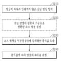
도 7을 참조하면, 구분장치(10)는 S110 단계에서 영상 내에서 신체의 좌우가 알려진 진단 영상(A0)을 입력받는다. 진단 영상이 입력되면, 선택적으로, 전처리부(400)는 S120 단계에서 소스 영상(S)을 생성할 수 있다. 진단 영상, 즉, X-ray 영상은 'anode heel effect' 등의 이유로 좌우의 밀도(density)가 상이하다. 따라서 본 발명의 전처리부(400)는 이러한 차이를 극대화하기 위해 진단 영상의 픽셀값을 가공할 수 있다.Referring to FIG. 7, the
일 실시예에 따르면, 소스 영상(S)은 도 3에 도시된 바와 같이, 진단 영상의 원본(A0)과 진단 영상의 모든 픽셀의 명도를 증가시킨 가공본(A+)과 진단 영상의 모든 픽셀의 명도를 감소시킨 가공본(A-)을 병합한 영상이 될 수 있다. 다른 실시예에 따르면, 소스 영상(S)은 도 4에 도시된 바와 같이, 진단 영상의 원본(A0)과 진단 영상을 좌우로 균등분할 했을 때 좌우 중 어느 일 측의 픽셀의 명도를 증가시키고, 다른 측의 픽셀의 명도를 감소시킨 가공본(A1, A2)을 병합한 영상이 될 수 있다.According to an embodiment, as shown in FIG. 3, the source image S includes an original A0 of the diagnostic image and a processed copy (A+) in which the brightness of all pixels of the diagnostic image is increased, and all pixels of the diagnostic image. It can be an image that merges the processed version (A-) with reduced brightness. According to another embodiment, the source image S increases the brightness of a pixel on either side of the left and right when the original A0 of the diagnosis image and the diagnosis image are equally divided left and right, as shown in FIG. 4, It may be an image obtained by merging the processed copies (A1, A2) in which the brightness of the pixels on the other side is reduced.
전술한 S120 단계는 선택적인 단계로 생략될 수도 있다.The above-described step S120 is an optional step and may be omitted.
그러면, 학습부(200)는 S140 단계에서 진단 영상의 알려진 영상의 좌우에 맞춰 목표값을 설정한다.Then, the
일례로, 진단 영상의 영상 내 신체가 좌우가 반전되지 않은 정상 상태인 경우, 제1 출력 노드(N1)의 출력값이 제2 출력 노드(N2)의 출력값 보다 크면서, 소정 수치이상이 되도록 목표값을 설정한다. 구체적인 예로, 진단 영상의 영상 내 신체가 좌우가 반전되지 않은 정상 상태인 경우, 학습부(200)는 'N1 ≥ 0.70, N2 < 0.30'와 같이 목표값을 설정할 수 있다.For example, when the body in the image of the diagnostic image is in a normal state in which the left and right sides are not inverted, the target value so that the output value of the first output node N1 is greater than the output value of the second output node N2 and exceeds a predetermined value. Is set. As a specific example, when the body in the image of the diagnostic image is in a normal state in which the left and right sides are not inverted, the
반대되는 예로, 진단 영상의 영상 내 신체가 좌우가 반전된 비정상 상태인 경우, 제1 출력 노드(N1)의 출력값이 제2 출력 노드(N2)의 출력값 보다 작으면서, 제2 출력 노드(N2)의 출력값이 소정 수치이상이 되도록 목표값을 설정한다. 구체적인 예로, 진단 영상의 영상 내 신체가 좌우가 반전된 비정상 상태인 경우, 학습부(200)는 'N1 < 0.30, N2 ≥ 0.70'과 같이 목표값을 설정할 수 있다.As an opposite example, when the body in the image of the diagnostic image is in an abnormal state in which the left and right sides are inverted, the output value of the first output node N1 is smaller than the output value of the second output node N2 and the second output node N2 Set the target value so that the output value of is more than a predetermined value. As a specific example, when the body in the image of the diagnosis image is in an abnormal state in which the left and right sides are inverted, the
그런 다음, 학습부(200)는 S140 단계에서 학습 데이터(진단 영상/소스 영상)를 인공신경망(100)에 입력한다.Then, the
그러면, 인공신경망(100)은 복수의 계층의 가중치(w)가 적용되는 복수의 연산을 수행하여 출력값을 출력할 것이다. 전술한 바와 같이, 진단 영상, 즉, X-ray 영상은 'anode heel effect' 등의 이유로 좌우의 밀도(density)가 상이하며, 인공신경망(100)은 인공신경망(100)의 복수의 연산 중 적어도 하나의 컨벌루션 연산을 통해 전술한 차이를 극대화하기 위해 입력된 학습 데이터(진단 영상)의 픽셀값을 가공할 수 있다. 구체적으로, 일 실시예에 따르면, 도 5에 도시된 바와 같이, 커널(w)의 컨벌루션 연산은 입력층(IL)에 입력된 진단 영상(A0)의 모든 픽셀의 명도를 증가(A+)시키거나, 입력층(IL)에 입력된 진단 영상(A0)의 모든 픽셀의 명도를 감소(A-)시킬 수 있다. 이에 따라, 진단 영상(A0)의 모든 픽셀의 명도가 증가(A+)된 제1 특징지도(FM1) 및 진단 영상(A0)의 모든 픽셀의 명도가 감소(A-)된 제2 특징지도(FM2)를 생성할 수 있다. 다른 실시예에 따르면, 도 6에 도시된 바와 같이, 커널(w)의 컨벌루션 연산은 입력층(IL)에 입력된 진단 영상(A0)을 좌우로 균등분할 했을 때 좌우 중 어느 일 측 픽셀의 명도를 증가시키고, 다른 측 픽셀의 명도를 감소시킬 수 있다(A1, A2). 이에 따라, 진단 영상(A0)을 좌우로 균등분할 했을 때 좌우 중 우측 픽셀의 명도가 증가되고, 좌측 픽셀의 명도가 감소된 영상(A1)을 가지는 제1 특징지도(FM1) 및 진단 영상(A0)을 좌우로 균등분할 했을 때 좌우 중 좌측 픽셀의 명도가 증가되고, 우측 픽셀의 명도가 감소된 영상(A2)을 가지는 제2 특징지도(FM2)를 생성할 수 있다.Then, the artificial
인공신경망(100)의 출력값이 출력되면, 학습부(200)는 S150 단계에서 인공신경망(100)의 출력값과 앞서(S130) 설정된 목표값과의 차이가 최소가 되도록 인공신경망(100)의 복수의 연산 각각에 적용되는 가중치(w)를 수정한다.When the output value of the artificial
전술한 S110 단계 내지 S150 단계는 다양한 학습 데이터이 입력되었을 때, 목표값과 출력값의 차이가 최소가 되면서 출력값이 변동이 없을 때까지 반복하여 수행할 수 있다. 이러한 경우, 학습부(200)는 인공신경망(100)이 충분히 학습이 이루어진 것으로 판단하고, 학습을 종료할 수 있다.The above-described steps S110 to S150 may be repeatedly performed when various training data are input, until the difference between the target value and the output value is minimized and there is no change in the output value. In this case, the
전술한 바와 같이 인공신경망(100)의 학습이 충분히 이루어진 경우, 그 인공신경망(100)을 이용한 진단 영상을 구분하기 위한 방법에 대해서 설명하기로 한다. 도 8은 본 발명의 실시예에 따른 인공신경망을 이용한 진단 영상을 구분하기 위한 방법을 설명하기 위한 흐름도이다.As described above, when the artificial
도 8을 참조하면, S210 단계에서 영상 내에서 신체의 좌우가 알려지지 않은 진단 영상을 입력받는다. 진단 영상이 입력되면, 선택적으로, 전처리부(400)는 S220 단계에서 진단영상의 원본 및 가공본을 결합하여 소스 영상(S)을 생성할 수 있다. 보다 구체적으로, 전처리부(400)는 진단 영상의 좌우의 밀도의 차이를 극대화시키기 위하여 도 3 혹은 도 4에서 설명된 바와 같이 진단 영상의 픽셀값을 가공하여 가공본을 생성할 수 있다. 그리고 전처리부(400)는 진단영상의 원본 및 가공본을 결합하여 소스 영상(S)을 생성할 수 있다. 전술한 S220 단계는 선택적인 단계로 생략될 수 있다.Referring to FIG. 8, in step S210, a diagnostic image of which the left and right sides of the body are unknown in the image is input. When the diagnostic image is input, the
다음으로, 판단부(300)는 S230 단계에서 의심 영상을 인공신경망(100)에 입력한다. 그러면, 인공신경망(100)은 복수의 계층의 가중치(w)가 적용되는 복수의 연산을 수행하여 출력값을 출력할 것이다. 전술한 바와 같이, 진단 영상, 즉, X-ray 영상은 'anode heel effect' 등의 이유로 좌우의 밀도(density)가 상이하며, 인공신경망(100)은 도 5 혹은 도 6에 도시된 바와 같이, 인공신경망(100)의 복수의 연산 중 적어도 하나의 컨벌루션 연산을 통해 전술한 차이를 극대화하기 위해 입력된 학습 데이터(진단 영상)의 픽셀값을 가공할 수 있다.Next, the
인공신경망(100)이 출력값을 출력하면, 판단부(300)는 S240 단계에서 인공신경망(100)의 출력값에 따라 진단 영상 내의 신체의 좌우가 반전되지 않은 정상 상태인지 혹은 반전된 비정상 상태인지 여부를 판별한다. 예를 들면, 인공신경망(100)의 출력 계층(OL)의 제1 출력 노드(N1)의 출력값은 진단 영상이 영상 내의 신체의 좌우가 반전되지 않은 정상 상태일 확률을 나타내며, 제2 출력 노드(N2)의 출력값은 진단 영상이 영상 내의 신체의 좌우가 반전된 비정상 상태일 확률을 나타낸다. 만약, 제1 출력 노드(N1) 및 제2 출력 노드(N2) 각각의 출력값이 0.89, 0.11이라면, 판별부(300)는 해당 진단 영상이 영상 내의 신체의 좌우가 반전되지 않은 정상 상태인 것으로 판단할 수 있다. 반면, 제1 출력 노드(N1) 및 제2 출력 노드(N2) 각각의 출력값이 0.27, 0.73이라면, 판별부(300)는 해당 진단 영상이 영상 내의 신체의 좌우가 반전된 비정상 상태인 것으로 판단할 수 있다.When the artificial
이와 같이, 본 발명의 실시예에 따르면, 구분장치(10)는 인공신경망(100)을 이용하여 진단 영상 내 신체의 좌우가 반전되었는지 여부를 판별할 수 있다.As described above, according to an embodiment of the present invention, the
한편, 앞서 설명된 본 발명의 실시예에 따른 방법은 다양한 컴퓨터수단을 통하여 판독 가능한 프로그램 형태로 구현되어 컴퓨터로 판독 가능한 기록매체에 기록될 수 있다. 여기서, 기록매체는 프로그램 명령, 데이터 파일, 데이터구조 등을 단독으로 또는 조합하여 포함할 수 있다. 기록매체에 기록되는 프로그램 명령은 본 발명을 위하여 특별히 설계되고 구성된 것들이거나 컴퓨터 소프트웨어 당업자에게 공지되어 사용 가능한 것일 수도 있다. 예컨대 기록매체는 하드 디스크, 플로피 디스크 및 자기 테이프와 같은 자기 매체(magnetic media), CD-ROM, DVD와 같은 광 기록 매체(optical media), 플롭티컬 디스크(floptical disk)와 같은 자기-광 매체(magneto-optical media), 및 롬(ROM), 램(RAM), 플래시 메모리 등과 같은 프로그램 명령을 저장하고 수행하도록 특별히 구성된 하드웨어 장치를 포함한다. 프로그램 명령의 예에는 컴파일러에 의해 만들어지는 것과 같은 기계어 뿐만 아니라 인터프리터 등을 사용해서 컴퓨터에 의해서 실행될 수 있는 고급 언어를 포함할 수 있다. 이러한 하드웨어 장치는 본 발명의 동작을 수행하기 위해 하나 이상의 소프트웨어 모듈로서 작동하도록 구성될 수 있으며, 그 역도 마찬가지이다.Meanwhile, the method according to the embodiment of the present invention described above may be implemented in the form of a program readable through various computer means and recorded on a computer-readable recording medium. Here, the recording medium may include a program command, a data file, a data structure, or the like alone or in combination. The program instructions recorded on the recording medium may be specially designed and constructed for the present invention, or may be known and usable to those skilled in computer software. For example, the recording medium includes magnetic media such as hard disks, floppy disks, and magnetic tapes, optical media such as CD-ROMs and DVDs, and magnetic-optical media such as floptical disks ( magneto-optical media), and a hardware device specially configured to store and execute program instructions such as ROM, RAM, flash memory, and the like. Examples of program instructions may include not only a machine language such as produced by a compiler, but also a high-level language that can be executed by a computer using an interpreter or the like. Such a hardware device may be configured to operate as one or more software modules to perform the operation of the present invention, and vice versa.
이상 본 발명을 몇 가지 바람직한 실시예를 사용하여 설명하였으나, 이들 실시예는 예시적인 것이며 한정적인 것이 아니다. 이와 같이, 본 발명이 속하는 기술분야에서 통상의 지식을 지닌 자라면 본 발명의 사상과 첨부된 특허청구범위에 제시된 권리범위에서 벗어나지 않으면서 균등론에 따라 다양한 변화와 수정을 가할 수 있음을 이해할 것이다.Although the present invention has been described using several preferred embodiments, these embodiments are illustrative and not limiting. As such, those of ordinary skill in the art to which the present invention pertains will understand that various changes and modifications can be made according to the equivalence theory without departing from the spirit of the present invention and the scope of the rights presented in the appended claims.
100: 인공신경망
IL: 입력계층
CL: 컨볼루션계층
PL: 풀링계층
FL: 완전연결계층
OL: 출력계층
200: 학습부
300: 판단부
400: 전처리부100: artificial neural network
IL: input layer
CL: Convolutional layer
PL: Pooling layer
FL: fully connected layer
OL: output layer
200: Learning Department
300: judgment unit
400: pretreatment unit
Claims (18)
Translated fromKorean각각이 가중치가 적용되는 복수의 연산을 포함하는 복수의 계층으로 이루어진 인공신경망;
영상 내 신체의 좌우가 알려지지 않은 진단 영상이 입력되면, 진단 영상의 원본과 상기 진단 영상의 픽셀값을 가공한 가공본을 병합한 소스 영상을 생성하는 전처리부; 및
상기 소스 영상을 상기 인공신경망에 입력하여 상기 인공신경망의 출력값으로부터 상기 진단 영상 내의 신체의 좌우가 반전되지 않은 정상 상태인지 혹은 반전된 비정상 상태인지 여부를 판단하는 판단부;를 포함하는 것을 특징으로 하는
진단 영상을 구분하기 위한 장치.In the device for classifying diagnostic images using an artificial neural network,
An artificial neural network consisting of a plurality of layers each including a plurality of operations to which a weight is applied;
A preprocessor for generating a source image by merging an original diagnosis image and a processed copy obtained by processing pixel values of the diagnosis image when a diagnosis image of which the left and right sides of the body in the image are not known is input; And
And a determination unit that inputs the source image to the artificial neural network and determines whether the left and right sides of the body in the diagnostic image are in an uninverted normal state or an inverted abnormal state from an output value of the artificial neural network.
A device for classifying diagnostic images.
상기 전처리부는
상기 진단 영상의 원본과 상기 진단 영상의 모든 픽셀의 명도를 증가시킨 가공본과 상기 진단 영상의 모든 픽셀의 명도를 감소시킨 가공본을 병합하여 상기 소스 영상을 생성하는 것을 특징으로 하는
진단 영상을 구분하기 위한 장치.The method of claim 1,
The pretreatment unit
The source image is generated by merging the original of the diagnostic image and a processed copy in which the brightness of all pixels of the diagnostic image is increased, and a processed copy in which the brightness of all pixels of the diagnostic image is reduced.
A device for classifying diagnostic images.
상기 전처리부는
상기 진단 영상의 원본과 상기 진단 영상을 좌우로 균등분할 했을 때 좌우 중 어느 일 측의 픽셀의 명도를 증가시키고, 다른 측의 픽셀의 명도를 감소시킨 가공본을 병합하여 상기 소스 영상을 생성하는 것을 특징으로 하는
진단 영상을 구분하기 위한 장치.The method of claim 1,
The pretreatment unit
When the original diagnosis image and the diagnosis image are divided equally to the left and right, the brightness of one of the left and right pixels is increased, and the resulting image is generated by merging a processed copy in which the brightness of the other pixel is reduced. Characterized
A device for classifying diagnostic images.
상기 전처리부는
영상 내 신체의 좌우가 알려진 진단 영상이 입력되면, 상기 진단 영상의 원본과 상기 진단 영상의 픽셀값을 가공한 가공본을 병합한 소스 영상을 생성하고,
상기 장치는
상기 소스 영상을 상기 인공신경망에 입력하여 상기 인공신경망의 출력값이 진단 영상 내의 신체의 좌우가 반전되지 않은 정상 상태인지 혹은 반전된 비정상 상태인지 여부를 판단하도록 상기 가중치를 수정하는 학습부;를 더 포함하는 것을 특징으로 하는
진단 영상을 구분하기 위한 장치.The method of claim 1,
The pretreatment unit
When a diagnostic image in which the left and right sides of the body in the image are known is input, a source image is generated by merging the original of the diagnostic image and a processed copy of the pixel values of the diagnostic image,
The device is
A learning unit that inputs the source image to the artificial neural network and modifies the weight to determine whether the output value of the artificial neural network is a normal state in which the left and right sides of the body in the diagnostic image are not inverted or in an inverted abnormal state; Characterized by
A device for classifying diagnostic images.
입력층, 적어도 하나의 컨볼루션층, 상기 컨볼루션층에 대응하는 적어도 하나의 풀링층 및 출력층을 포함하는 복수의 계층을 포함하고, 상기 복수의 계층 각각은 복수의 연산을 포함하며, 상기 복수의 계층 중 어느 하나의 계층의 연산의 결과는 가중치가 적용되어 다음 계층의 입력이 되는 인공신경망; 및
영상 내 신체의 좌우가 알려지지 않은 진단 영상이 입력되면, 상기 진단 영상을 상기 인공신경망에 입력하여 상기 인공신경망의 출력값으로부터 진단 영상 내의 신체의 좌우가 반전되지 않은 정상 상태인지 혹은 반전된 비정상 상태인지 여부를 판단하는 판단부;를 포함하는 것을 특징으로 하는
진단 영상을 구분하기 위한 장치.In the device for classifying diagnostic images using an artificial neural network,
A plurality of layers including an input layer, at least one convolutional layer, at least one pooling layer corresponding to the convolutional layer, and an output layer, and each of the plurality of layers includes a plurality of operations, and the plurality of layers An artificial neural network to which a weight is applied to the result of the operation of one of the layers to be input to the next layer; And
When a diagnostic image in which the left and right sides of the body in the image are unknown is input, the diagnostic image is input to the artificial neural network and whether the left and right sides of the body in the diagnosis image are not inverted from the output value of the artificial neural network is a normal state or an inverted abnormal state. Characterized in that it comprises a; determining unit to determine the
A device for classifying diagnostic images.
상기 컨볼루션층은
상기 입력층에 입력된 상기 진단 영상과 커널의 컨벌루션 연산을 통해 생성되는 복수의 특징지도를 포함하며,
상기 커널의 컨벌루션 연산은
상기 입력층의 상기 진단 영상의 모든 픽셀의 명도를 증가시키거나, 상기 입력층에 입력되는 상기 진단 영상의 모든 픽셀의 명도를 감소시키는 것을 특징으로 하는
진단 영상을 구분하기 위한 장치.The method of claim 5,
The convolutional layer
Includes a plurality of feature maps generated through a convolution operation between the diagnostic image input to the input layer and a kernel,
The convolution operation of the kernel is
Increasing the brightness of all pixels of the diagnostic image of the input layer or reducing the brightness of all pixels of the diagnostic image input to the input layer.
A device for classifying diagnostic images.
상기 컨볼루션층은
상기 입력층에 입력된 상기 진단 영상과 커널의 컨벌루션 연산을 통해 생성되는 복수의 특징지도를 포함하며,
상기 커널의 컨벌루션 연산은
상기 진단 영상의 원본과 상기 진단 영상을 좌우를 균등분할 했을 때 좌우 중 어느 일 측 픽셀의 명도를 증가시키고, 다른 측 픽셀의 명도를 감소시키는 것을 특징으로 하는
진단 영상을 구분하기 위한 장치.The method of claim 5,
The convolutional layer
Includes a plurality of feature maps generated through a convolution operation between the diagnostic image input to the input layer and a kernel,
The convolution operation of the kernel is
When the original of the diagnostic image and the diagnostic image are equally divided left and right, the brightness of one of the left and right pixels is increased, and the brightness of the other pixel is reduced.
A device for classifying diagnostic images.
영상 내 신체의 좌우가 알려진 진단 영상이 입력되면, 상기 진단 영상을 상기 인공신경망에 입력하여 상기 인공신경망의 출력값이 상기 진단 영상 내의 신체의 좌우가 반전되지 않은 정상 상태인지 혹은 반전된 비정상 상태인지 여부를 구분하도록 상기 가중치를 수정하는 학습부;를 더 포함하는 것을 특징으로 하는
진단 영상을 구분하기 위한 장치.The method of claim 1,
When a diagnostic image in which the left and right sides of the body in the image are known is input, the diagnostic image is input to the artificial neural network, and the output value of the artificial neural network is whether the left and right sides of the body in the diagnostic image are in a normal state or an inverted abnormal state. Characterized in that it further comprises a; learning unit for modifying the weight to distinguish
A device for classifying diagnostic images.
전처리부가 영상 내 신체의 좌우가 알려지지 않은 진단 영상을 입력받는 단계;
전처리부가 진단 영상의 원본과 상기 진단 영상의 픽셀값을 가공한 가공본을 병합한 소스 영상을 생성하는 단계; 및
판단부가 상기 소스 영상을 각각이 가중치가 적용되는 복수의 연산을 포함하는 복수의 계층으로 이루어진 인공신경망에 입력하고, 상기 인공신경망의 출력값으로부터 상기 진단 영상 내의 신체의 좌우가 반전되지 않은 정상 상태인지 혹은 반전된 비정상 상태인지 여부를 판단하는 단계;를 포함하는 것을 특징으로 하는
진단 영상을 구분하기 위한 방법.In a method for classifying diagnostic images using an artificial neural network,
Receiving, by a preprocessing unit, a diagnostic image in which the left and right sides of the body in the image are not known;
Generating, by a preprocessing unit, a source image obtained by merging an original diagnosis image and a processed image obtained by processing pixel values of the diagnosis image; And
The determination unit inputs the source image to an artificial neural network consisting of a plurality of layers each including a plurality of operations to which weights are applied, and whether the left and right sides of the body in the diagnostic image are not inverted from the output value of the artificial neural network, or Characterized in that it comprises a; determining whether or not the inverted abnormal state
A method for classifying diagnostic images.
상기 소스 영상을 생성하는 단계는
상기 전처리부가 상기 진단 영상의 모든 픽셀의 명도를 증가시킨 가공본 및 상기 진단 영상의 모든 픽셀의 명도를 감소시킨 가공본을 생성하는 단계; 및
상기 전처리부가 상기 진단 영상의 원본과 상기 명도를 증가시킨 가공본과 상기 명도를 감소시킨 가공본을 병합하여 상기 소스 영상을 생성하는 단계;를 포함하는 것을 특징으로 하는
진단 영상을 구분하기 위한 방법.The method of claim 9,
Generating the source image
Generating, by the pre-processing unit, a processed copy in which the brightness of all pixels of the diagnostic image is increased and a processed copy in which the brightness of all pixels of the diagnostic image is decreased; And
And generating the source image by merging, by the pre-processing unit, an original of the diagnostic image, a processed copy with increased brightness, and a processed copy with reduced brightness.
A method for classifying diagnostic images.
상기 소스 영상을 생성하는 단계는
상기 전처리부가 상기 진단 영상의 원본과 상기 진단 영상을 좌우로 균등분할 했을 때 좌우 중 어느 일 측의 픽셀의 명도를 증가시키고, 다른 측의 픽셀의 명도를 감소시킨 가공본을 생성하는 단계; 및
상기 전처리부는 상기 진단 영상의 원본과 상기 가공본을 병합하여 상기 소스 영상을 생성하는 단계;를 포함하는 것을 특징으로 하는
진단 영상을 구분하기 위한 방법.The method of claim 9,
Generating the source image
Generating a processed copy in which the brightness of one of the left and right pixels is increased and the brightness of the other side is reduced when the preprocessing unit divides the original diagnosis image and the diagnosis image horizontally; And
And generating the source image by merging the original and the processed image of the diagnostic image by the preprocessor.
A method for classifying diagnostic images.
상기 영상 내 신체의 좌우가 알려지지 않은 진단 영상을 입력받는 단계 전,
상기 전처리부가 영상 내 신체의 좌우가 알려진 진단 영상이 입력되면, 입력된 진단 영상의 원본과 상기 진단 영상의 픽셀값을 가공한 가공본을 병합한 소스 영상을 생성하는 단계;
학습부가 상기 소스 영상을 상기 인공신경망에 입력하여 상기 인공신경망의 출력값이 상기 진단 영상 내의 신체의 좌우가 반전되지 않은 정상 상태인지 혹은 반전된 비정상 상태인지 여부를 구분하도록 상기 가중치를 수정하는 단계;를 더 포함하는 것을 특징으로 하는
진단 영상을 구분하기 위한 방법.The method of claim 9,
Before the step of receiving a diagnostic image in which the left and right sides of the body in the image are unknown,
Generating, by the preprocessing unit, a source image obtained by merging an original of the input diagnostic image and a processed version of a pixel value of the diagnostic image when a diagnostic image in which the left and right sides of the body in the image are known is input;
Modifying the weight so that the learning unit inputs the source image to the artificial neural network, and the output value of the artificial neural network distinguishes whether the left and right sides of the body in the diagnostic image are in a normal state or an inverted abnormal state; Characterized in that it further comprises
A method for classifying diagnostic images.
판단부가 영상 내 신체의 좌우가 알려지지 않은 진단 영상을 입력받는 단계;
상기 판단부가 상기 진단 영상을 상기 인공신경망에 입력하는 단계;
상기 인공신경망이 상기 진단 영상에 대해 가중치가 적용되는 복수의 연산을 수행하여 출력값을 출력하는 단계; 및
상기 판단부가 상기 인공신경망의 출력값으로부터 상기 진단 영상 내의 신체의 좌우가 반전되지 않은 정상 상태인지 혹은 반전된 비정상 상태인지 여부를 판단하는 단계;를 포함하는 것을 특징으로 하는
진단 영상을 구분하기 위한 방법.A plurality of layers including an input layer, at least one convolutional layer, at least one pooling layer corresponding to the convolutional layer, and an output layer, and each of the plurality of layers includes a plurality of operations, and the plurality of layers In a method for classifying a diagnostic image using an artificial neural network that is an input of a next layer by applying a weight to the result of the operation of one of the layers,
Receiving, by the determination unit, a diagnostic image in which the left and right sides of the body in the image are unknown;
Inputting, by the determination unit, the diagnostic image to the artificial neural network;
Performing, by the artificial neural network, a plurality of calculations to which weights are applied to the diagnostic image and outputting an output value; And
And determining, from the output value of the artificial neural network, whether the left and right sides of the body in the diagnostic image are in an uninverted normal state or an inverted abnormal state.
A method for classifying diagnostic images.
상기 출력값을 출력하는 단계는
상기 입력층에 입력된 상기 진단 영상과 커널의 컨벌루션 연산을 통해 복수의 특징지도를 생성하는 단계;를 포함하며,
상기 커널의 컨벌루션 연산은
상기 입력층의 상기 진단 영상의 모든 픽셀의 명도를 증가시키거나, 상기 입력층에 입력되는 상기 진단 영상의 모든 픽셀의 명도를 감소시키는 것을 특징으로 하는
진단 영상을 구분하기 위한 방법.The method of claim 13,
The step of outputting the output value
Generating a plurality of feature maps through a convolution operation between the diagnostic image input to the input layer and the kernel; and
The convolution operation of the kernel is
Increasing the brightness of all pixels of the diagnostic image of the input layer or reducing the brightness of all pixels of the diagnostic image input to the input layer.
A method for classifying diagnostic images.
상기 출력값을 출력하는 단계는
상기 입력층에 입력된 상기 진단 영상과 커널의 컨벌루션 연산을 통해 복수의 특징지도를 생성하는 단계;를 포함하며,
상기 커널의 컨벌루션 연산은
상기 진단 영상의 원본과 상기 진단 영상을 좌우를 균등분할 했을 때 좌우 중 어느 일 측 픽셀의 명도를 증가시키고, 다른 측 픽셀의 명도를 감소시키는 것을 특징으로 하는
진단 영상을 구분하기 위한 방법.The method of claim 13,
The step of outputting the output value
Generating a plurality of feature maps through a convolution operation between the diagnostic image input to the input layer and the kernel; and
The convolution operation of the kernel is
When the original of the diagnostic image and the diagnostic image are equally divided left and right, the brightness of one of the left and right pixels is increased, and the brightness of the other pixel is reduced.
A method for classifying diagnostic images.
상기 신체의 좌우가 알려지지 않은 진단 영상을 입력받는 단계 전,
학습부가 영상 내 신체의 좌우가 알려진 진단 영상을 입력받는 단계;
상기 학습부가 상기 영상 내 신체의 좌우가 알려진 진단 영상을 상기 인공신경망에 입력하는 단계;
상기 인공신경망이 상기 영상 내 신체의 좌우가 알려진 진단 영상에 대해 가중치가 적용되는 복수의 연산을 수행하여 출력값을 출력하는 단계; 및
상기 영상 내 신체의 좌우가 알려진 진단 영상 내의 신체의 좌우가 반전되지 않은 정상 상태인지 혹은 반전된 비정상 상태인지 여부를 구분하도록 상기 가중치를 수정하는 단계;를 더 포함하는 것을 특징으로 하는
진단 영상을 구분하기 위한 방법.The method of claim 13,
Before the step of receiving a diagnostic image of which the left and right sides of the body are unknown,
Receiving, by the learning unit, a diagnostic image in which the left and right sides of the body in the image are known;
Inputting, by the learning unit, a diagnostic image in which the left and right sides of the body in the image are known to the artificial neural network;
Performing, by the artificial neural network, a plurality of calculations to which weights are applied to a diagnostic image in which the left and right sides of the body in the image are known, and outputting an output value; And
And modifying the weight to distinguish whether the left and right sides of the body in the image are known whether the left and right sides of the body in the diagnostic image are in an uninverted normal state or an inverted abnormal state.
A method for classifying diagnostic images.
Priority Applications (1)
| Application Number | Priority Date | Filing Date | Title |
|---|---|---|---|
| KR1020190131146AKR102322927B1 (en) | 2019-10-22 | 2019-10-22 | Apparatus for distinguishing diagnostic images using artificial neural networks, method thereof and computer recordable medium storing program to perform the method |
Applications Claiming Priority (1)
| Application Number | Priority Date | Filing Date | Title |
|---|---|---|---|
| KR1020190131146AKR102322927B1 (en) | 2019-10-22 | 2019-10-22 | Apparatus for distinguishing diagnostic images using artificial neural networks, method thereof and computer recordable medium storing program to perform the method |
Publications (2)
| Publication Number | Publication Date |
|---|---|
| KR20210047489Atrue KR20210047489A (en) | 2021-04-30 |
| KR102322927B1 KR102322927B1 (en) | 2021-11-04 |
Family
ID=75740841
Family Applications (1)
| Application Number | Title | Priority Date | Filing Date |
|---|---|---|---|
| KR1020190131146AActiveKR102322927B1 (en) | 2019-10-22 | 2019-10-22 | Apparatus for distinguishing diagnostic images using artificial neural networks, method thereof and computer recordable medium storing program to perform the method |
Country Status (1)
| Country | Link |
|---|---|
| KR (1) | KR102322927B1 (en) |
Citations (3)
| Publication number | Priority date | Publication date | Assignee | Title |
|---|---|---|---|---|
| JP2000157519A (en)* | 1998-11-25 | 2000-06-13 | Konica Corp | Image processor |
| KR20110098077A (en) | 2010-02-26 | 2011-09-01 | 제주대학교 산학협력단 | Secret information insertion device and method using bit pattern block pattern matching |
| JP2019115558A (en)* | 2017-12-27 | 2019-07-18 | キヤノン株式会社 | Radiography apparatus, image processing apparatus, and image determination method |
- 2019
- 2019-10-22KRKR1020190131146Apatent/KR102322927B1/enactiveActive
Patent Citations (3)
| Publication number | Priority date | Publication date | Assignee | Title |
|---|---|---|---|---|
| JP2000157519A (en)* | 1998-11-25 | 2000-06-13 | Konica Corp | Image processor |
| KR20110098077A (en) | 2010-02-26 | 2011-09-01 | 제주대학교 산학협력단 | Secret information insertion device and method using bit pattern block pattern matching |
| JP2019115558A (en)* | 2017-12-27 | 2019-07-18 | キヤノン株式会社 | Radiography apparatus, image processing apparatus, and image determination method |
Non-Patent Citations (2)
| Title |
|---|
| Rikiya Yamashita 외 3명, Convolutional neural networks: an overview and application in radiology. Insights into Imaging (2018) 9:pp.611-629. 2018년 6월 22일 공개. 1부.** |
| Ross W.Filice 외 1명. Effectiveness of Deep Learning Algorithms to Determine Laterality in Radiographs. Journal of Digital Imaging (2019) 32:pp.656-664. 2019년 5월 7일 공개. 1부.** |
Also Published As
| Publication number | Publication date |
|---|---|
| KR102322927B1 (en) | 2021-11-04 |
Similar Documents
| Publication | Publication Date | Title |
|---|---|---|
| US10892050B2 (en) | Deep image classification of medical images | |
| US12417541B2 (en) | Method and device for training generative adversarial network for converting between heterogeneous domain data | |
| EP3861482A1 (en) | Verification of classification decisions in convolutional neural networks | |
| US11720795B2 (en) | Neural network structure and a method thereto | |
| KR102370910B1 (en) | Method and apparatus for few-shot image classification based on deep learning | |
| JP2020536337A5 (en) | ||
| KR102548519B1 (en) | Semi-synthetic data generation apparatus and method thereof | |
| KR102028825B1 (en) | Apparatus for processing watermarking using artificial neural network which identifies watermarking attack, method thereof and computer recordable medium storing program to perform the method | |
| US11741363B2 (en) | Computer-readable recording medium, method for learning, and learning device | |
| CN114463605B (en) | Continuous learning image classification method and device based on deep learning | |
| Chang | [Retracted] Neural Reversible Steganography with Long Short‐Term Memory | |
| JP2020107042A (en) | Learning model generation device, learning model generation method, and program | |
| US20200250544A1 (en) | Learning method, storage medium, and learning apparatus | |
| CN113055546B (en) | System and method for processing images | |
| CN116664921B (en) | Waste circuit board defect classification method based on transfer learning and multi-attention mechanism | |
| CN111753980B (en) | Method for transferring features of a first image to a second image | |
| KR102028824B1 (en) | Apparatus for processing watermarking using artificial neural network which identifies objects, method thereof and computer recordable medium storing program to perform the method | |
| KR102322927B1 (en) | Apparatus for distinguishing diagnostic images using artificial neural networks, method thereof and computer recordable medium storing program to perform the method | |
| US20240037910A1 (en) | Quantum Method and System for Classifying Images | |
| KR20230127508A (en) | GAN-based model training method and apparatus | |
| KR102045140B1 (en) | Apparatus for drawing watermarking attack technique using artificial neural network, method thereof and computer recordable medium storing program to perform the method | |
| Xi et al. | A Layer-Wise Ensemble Technique for Binary Neural Network | |
| CN112541906B (en) | Data processing method and device, electronic equipment and storage medium | |
| US12217092B2 (en) | Electronic device and controlling method of electronic device | |
| EP4310793A1 (en) | Quantum method and system for classifying images |
Legal Events
| Date | Code | Title | Description |
|---|---|---|---|
| PA0109 | Patent application | Patent event code:PA01091R01D Comment text:Patent Application Patent event date:20191022 | |
| PA0201 | Request for examination | ||
| PE0902 | Notice of grounds for rejection | Comment text:Notification of reason for refusal Patent event date:20210224 Patent event code:PE09021S01D | |
| PG1501 | Laying open of application | ||
| E90F | Notification of reason for final refusal | ||
| PE0902 | Notice of grounds for rejection | Comment text:Final Notice of Reason for Refusal Patent event date:20210708 Patent event code:PE09021S02D | |
| E701 | Decision to grant or registration of patent right | ||
| PE0701 | Decision of registration | Patent event code:PE07011S01D Comment text:Decision to Grant Registration Patent event date:20211026 | |
| GRNT | Written decision to grant | ||
| PR0701 | Registration of establishment | Comment text:Registration of Establishment Patent event date:20211101 Patent event code:PR07011E01D | |
| PR1002 | Payment of registration fee | Payment date:20211101 End annual number:3 Start annual number:1 | |
| PG1601 | Publication of registration | ||
| PR1001 | Payment of annual fee | Payment date:20240927 Start annual number:4 End annual number:4 |