KR20210027416A - Convection-enhanced transmission cranial implant device compatible with magnetic resonance imaging and related methods - Google Patents
Convection-enhanced transmission cranial implant device compatible with magnetic resonance imaging and related methodsDownload PDFInfo
- Publication number
- KR20210027416A KR20210027416AKR1020217002911AKR20217002911AKR20210027416AKR 20210027416 AKR20210027416 AKR 20210027416AKR 1020217002911 AKR1020217002911 AKR 1020217002911AKR 20217002911 AKR20217002911 AKR 20217002911AKR 20210027416 AKR20210027416 AKR 20210027416A
- Authority
- KR
- South Korea
- Prior art keywords
- fluid
- ced
- pump
- cavity
- cranial implant
- Prior art date
- Legal status (The legal status is an assumption and is not a legal conclusion. Google has not performed a legal analysis and makes no representation as to the accuracy of the status listed.)
- Withdrawn
Links
- 239000007943implantSubstances0.000titleclaimsabstractdescription341
- 238000000034methodMethods0.000titleclaimsabstractdescription148
- 238000002595magnetic resonance imagingMethods0.000titleclaimsabstractdescription62
- 230000005540biological transmissionEffects0.000title1
- 239000012530fluidSubstances0.000claimsabstractdescription431
- 239000003814drugSubstances0.000claimsabstractdescription138
- 229940124597therapeutic agentDrugs0.000claimsabstractdescription122
- 238000004891communicationMethods0.000claimsabstractdescription57
- 239000000463materialSubstances0.000claimsabstractdescription45
- 238000011282treatmentMethods0.000claimsabstractdescription27
- 238000002513implantationMethods0.000claimsabstractdescription25
- 238000007917intracranial administrationMethods0.000claimsabstractdescription25
- 238000012544monitoring processMethods0.000claimsabstractdescription22
- 208000037265diseases, disorders, signs and symptomsDiseases0.000claimsabstractdescription20
- 201000010099diseaseDiseases0.000claimsabstractdescription11
- 238000004519manufacturing processMethods0.000claimsabstractdescription11
- 238000003384imaging methodMethods0.000claimsdescription58
- 210000003625skullAnatomy0.000claimsdescription54
- 229920001746electroactive polymerPolymers0.000claimsdescription42
- 210000004556brainAnatomy0.000claimsdescription29
- 238000007789sealingMethods0.000claimsdescription27
- 238000012014optical coherence tomographyMethods0.000claimsdescription26
- 238000002560therapeutic procedureMethods0.000claimsdescription25
- 230000007246mechanismEffects0.000claimsdescription21
- 230000000926neurological effectEffects0.000claimsdescription18
- 239000012528membraneSubstances0.000claimsdescription16
- 238000002604ultrasonographyMethods0.000claimsdescription16
- 208000003174Brain NeoplasmsDiseases0.000claimsdescription14
- 230000003287optical effectEffects0.000claimsdescription12
- 210000004761scalpAnatomy0.000claimsdescription12
- 230000001225therapeutic effectEffects0.000claimsdescription12
- 229940079593drugDrugs0.000claimsdescription11
- 206010028980NeoplasmDiseases0.000claimsdescription10
- 239000004696Poly ether ether ketoneSubstances0.000claimsdescription10
- RTAQQCXQSZGOHL-UHFFFAOYSA-NTitaniumChemical compound[Ti]RTAQQCXQSZGOHL-UHFFFAOYSA-N0.000claimsdescription10
- 229910052588hydroxylapatiteInorganic materials0.000claimsdescription10
- XYJRXVWERLGGKC-UHFFFAOYSA-Dpentacalcium;hydroxide;triphosphateChemical compound[OH-].[Ca+2].[Ca+2].[Ca+2].[Ca+2].[Ca+2].[O-]P([O-])([O-])=O.[O-]P([O-])([O-])=O.[O-]P([O-])([O-])=OXYJRXVWERLGGKC-UHFFFAOYSA-D0.000claimsdescription10
- 229920003229poly(methyl methacrylate)Polymers0.000claimsdescription10
- 229920002530polyetherether ketonePolymers0.000claimsdescription10
- 239000004926polymethyl methacrylateSubstances0.000claimsdescription10
- 208000035475disorderDiseases0.000claimsdescription9
- 229920000642polymerPolymers0.000claimsdescription9
- 230000005855radiationEffects0.000claimsdescription9
- 208000003906hydrocephalusDiseases0.000claimsdescription8
- 208000006096Attention Deficit Disorder with HyperactivityDiseases0.000claimsdescription7
- 208000013738Sleep Initiation and Maintenance diseaseDiseases0.000claimsdescription7
- 210000004204blood vesselAnatomy0.000claimsdescription7
- 230000000694effectsEffects0.000claimsdescription7
- 206010022437insomniaDiseases0.000claimsdescription7
- 230000007257malfunctionEffects0.000claimsdescription7
- 210000002569neuronAnatomy0.000claimsdescription7
- 230000004962physiological conditionEffects0.000claimsdescription7
- 238000001959radiotherapyMethods0.000claimsdescription7
- 238000002271resectionMethods0.000claimsdescription7
- 201000000980schizophreniaDiseases0.000claimsdescription7
- 230000000007visual effectEffects0.000claimsdescription7
- 201000011510cancerDiseases0.000claimsdescription6
- 238000002512chemotherapyMethods0.000claimsdescription6
- 239000010987cubic zirconiaSubstances0.000claimsdescription6
- 230000002708enhancing effectEffects0.000claimsdescription6
- 108010087765AntipainProteins0.000claimsdescription5
- 208000036864Attention deficit/hyperactivity diseaseDiseases0.000claimsdescription5
- 102000004190EnzymesHuman genes0.000claimsdescription5
- 108090000790EnzymesProteins0.000claimsdescription5
- 239000004698PolyethyleneSubstances0.000claimsdescription5
- 230000007131anti Alzheimer effectEffects0.000claimsdescription5
- 230000001093anti-cancerEffects0.000claimsdescription5
- 230000000648anti-parkinsonEffects0.000claimsdescription5
- 230000000573anti-seizure effectEffects0.000claimsdescription5
- SDNYTAYICBFYFH-TUFLPTIASA-NantipainChemical compoundNC(N)=NCCC[C@@H](C=O)NC(=O)[C@H](C(C)C)NC(=O)[C@H](CCCN=C(N)N)NC(=O)N[C@H](C(O)=O)CC1=CC=CC=C1SDNYTAYICBFYFH-TUFLPTIASA-N0.000claimsdescription5
- 239000000939antiparkinson agentSubstances0.000claimsdescription5
- 210000002865immune cellAnatomy0.000claimsdescription5
- 230000006993memory improvementEffects0.000claimsdescription5
- 230000003340mental effectEffects0.000claimsdescription5
- 229910052751metalInorganic materials0.000claimsdescription5
- 239000002184metalSubstances0.000claimsdescription5
- -1polyethylenePolymers0.000claimsdescription5
- 229920000573polyethylenePolymers0.000claimsdescription5
- 229940043274prophylactic drugDrugs0.000claimsdescription5
- 102000004169proteins and genesHuman genes0.000claimsdescription5
- 108090000623proteins and genesProteins0.000claimsdescription5
- 230000002441reversible effectEffects0.000claimsdescription5
- 210000000130stem cellAnatomy0.000claimsdescription5
- 239000010936titaniumSubstances0.000claimsdescription5
- 229910052719titaniumInorganic materials0.000claimsdescription5
- 239000002246antineoplastic agentSubstances0.000claimsdescription4
- 229940127089cytotoxic agentDrugs0.000claimsdescription4
- 238000012377drug deliveryMethods0.000claimsdescription4
- 230000004007neuromodulationEffects0.000claimsdescription4
- 230000004112neuroprotectionEffects0.000claimsdescription4
- 230000035790physiological processes and functionsEffects0.000claimsdescription4
- 229920001296polysiloxanePolymers0.000claimsdescription4
- 230000000638stimulationEffects0.000claimsdescription4
- 230000001154acute effectEffects0.000claimsdescription3
- 230000003712anti-aging effectEffects0.000claimsdescription3
- 238000002591computed tomographyMethods0.000claimsdescription3
- 238000004146energy storageMethods0.000claimsdescription3
- 238000012961medicinal therapyMethods0.000claimsdescription3
- 230000001537neural effectEffects0.000claimsdescription3
- 230000002285radioactive effectEffects0.000claimsdescription3
- 238000012546transferMethods0.000claimsdescription3
- 208000024827Alzheimer diseaseDiseases0.000claimsdescription2
- 208000020925Bipolar diseaseDiseases0.000claimsdescription2
- 208000023105Huntington diseaseDiseases0.000claimsdescription2
- 208000002193PainDiseases0.000claimsdescription2
- 208000018737Parkinson diseaseDiseases0.000claimsdescription2
- 208000028683bipolar I diseaseDiseases0.000claimsdescription2
- 230000003925brain functionEffects0.000claimsdescription2
- 206010015037epilepsyDiseases0.000claimsdescription2
- 230000001988toxicityEffects0.000claimsdescription2
- 231100000419toxicityToxicity0.000claimsdescription2
- 230000002596correlated effectEffects0.000claims2
- 230000000324neuroprotective effectEffects0.000claims1
- 238000013461designMethods0.000description11
- 238000001356surgical procedureMethods0.000description7
- 241001465754MetazoaSpecies0.000description6
- 208000005017glioblastomaDiseases0.000description5
- 230000009977dual effectEffects0.000description3
- 239000000975dyeSubstances0.000description3
- 230000006870functionEffects0.000description3
- 208000015181infectious diseaseDiseases0.000description3
- 238000001990intravenous administrationMethods0.000description3
- 239000000203mixtureSubstances0.000description3
- 230000002123temporal effectEffects0.000description3
- 241000283690Bos taurusSpecies0.000description2
- 206010010904ConvulsionDiseases0.000description2
- RYGMFSIKBFXOCR-UHFFFAOYSA-NCopperChemical compound[Cu]RYGMFSIKBFXOCR-UHFFFAOYSA-N0.000description2
- 208000012902Nervous system diseaseDiseases0.000description2
- 208000025966Neurological diseaseDiseases0.000description2
- 210000000988bone and boneAnatomy0.000description2
- 210000004027cellAnatomy0.000description2
- 230000001413cellular effectEffects0.000description2
- 210000003169central nervous systemAnatomy0.000description2
- 230000001684chronic effectEffects0.000description2
- 238000011960computer-aided designMethods0.000description2
- 229910052802copperInorganic materials0.000description2
- 239000010949copperSubstances0.000description2
- 238000007428craniotomyMethods0.000description2
- 238000006073displacement reactionMethods0.000description2
- 238000005516engineering processMethods0.000description2
- 238000001125extrusionMethods0.000description2
- 230000036541healthEffects0.000description2
- 238000001802infusionMethods0.000description2
- 238000003780insertionMethods0.000description2
- 230000037431insertionEffects0.000description2
- 210000003140lateral ventricleAnatomy0.000description2
- 230000007170pathologyEffects0.000description2
- 230000008569processEffects0.000description2
- 238000005070samplingMethods0.000description2
- 241000894007speciesSpecies0.000description2
- 230000000699topical effectEffects0.000description2
- 2380000101463D printingMethods0.000description1
- 241000271566AvesSpecies0.000description1
- OKTJSMMVPCPJKN-UHFFFAOYSA-NCarbonChemical compound[C]OKTJSMMVPCPJKN-UHFFFAOYSA-N0.000description1
- 208000024172Cardiovascular diseaseDiseases0.000description1
- 208000017667Chronic DiseaseDiseases0.000description1
- 241000283086EquidaeSpecies0.000description1
- 208000019693Lung diseaseDiseases0.000description1
- 241000124008MammaliaSpecies0.000description1
- 241000288906PrimatesSpecies0.000description1
- XUIMIQQOPSSXEZ-UHFFFAOYSA-NSiliconChemical compound[Si]XUIMIQQOPSSXEZ-UHFFFAOYSA-N0.000description1
- FAPWRFPIFSIZLT-UHFFFAOYSA-MSodium chlorideChemical compound[Na+].[Cl-]FAPWRFPIFSIZLT-UHFFFAOYSA-M0.000description1
- 208000006011StrokeDiseases0.000description1
- 241000282887SuidaeSpecies0.000description1
- 208000016599Uterine diseaseDiseases0.000description1
- 241000251539Vertebrata <Metazoa>Species0.000description1
- 210000000683abdominal cavityAnatomy0.000description1
- 230000003187abdominal effectEffects0.000description1
- 238000002679ablationMethods0.000description1
- 230000002159abnormal effectEffects0.000description1
- NIXOWILDQLNWCW-UHFFFAOYSA-Nacrylic acid groupChemical groupC(C=C)(=O)ONIXOWILDQLNWCW-UHFFFAOYSA-N0.000description1
- 230000003213activating effectEffects0.000description1
- 230000004913activationEffects0.000description1
- 210000000577adipose tissueAnatomy0.000description1
- 238000013459approachMethods0.000description1
- 208000015802attention deficit-hyperactivity diseaseDiseases0.000description1
- 230000004888barrier functionEffects0.000description1
- 230000000740bleeding effectEffects0.000description1
- 230000008499blood brain barrier functionEffects0.000description1
- 230000017531blood circulationEffects0.000description1
- 210000001218blood-brain barrierAnatomy0.000description1
- 210000001124body fluidAnatomy0.000description1
- 239000010839body fluidSubstances0.000description1
- 230000007177brain activityEffects0.000description1
- 229910052799carbonInorganic materials0.000description1
- 238000005266castingMethods0.000description1
- 239000012829chemotherapy agentSubstances0.000description1
- 210000000038chestAnatomy0.000description1
- 238000005094computer simulationMethods0.000description1
- 239000002872contrast mediaSubstances0.000description1
- 238000012937correctionMethods0.000description1
- 230000007547defectEffects0.000description1
- 238000001514detection methodMethods0.000description1
- 239000000032diagnostic agentSubstances0.000description1
- 229940039227diagnostic agentDrugs0.000description1
- 238000009826distributionMethods0.000description1
- 230000002526effect on cardiovascular systemEffects0.000description1
- 238000009429electrical wiringMethods0.000description1
- 238000004049embossingMethods0.000description1
- 238000005530etchingMethods0.000description1
- 238000000605extractionMethods0.000description1
- 230000001815facial effectEffects0.000description1
- 239000004519greaseSubstances0.000description1
- 210000003128headAnatomy0.000description1
- 239000000677immunologic agentSubstances0.000description1
- 229940124541immunological agentDrugs0.000description1
- 230000006698inductionEffects0.000description1
- 238000001746injection mouldingMethods0.000description1
- 238000007918intramuscular administrationMethods0.000description1
- 238000007912intraperitoneal administrationMethods0.000description1
- 238000007913intrathecal administrationMethods0.000description1
- 238000011835investigationMethods0.000description1
- 150000002500ionsChemical class0.000description1
- 238000001459lithographyMethods0.000description1
- 208000019423liver diseaseDiseases0.000description1
- 230000007774longtermEffects0.000description1
- 238000003754machiningMethods0.000description1
- 230000014759maintenance of locationEffects0.000description1
- 238000005259measurementMethods0.000description1
- 239000002207metaboliteSubstances0.000description1
- 238000000465mouldingMethods0.000description1
- 210000003205muscleAnatomy0.000description1
- 210000005036nerveAnatomy0.000description1
- 239000011664nicotinic acidSubstances0.000description1
- 201000011107obstructive hydrocephalusDiseases0.000description1
- 238000005457optimizationMethods0.000description1
- 230000002611ovarianEffects0.000description1
- 208000015124ovarian diseaseDiseases0.000description1
- 230000002572peristaltic effectEffects0.000description1
- 230000010287polarizationEffects0.000description1
- 244000144977poultrySpecies0.000description1
- 230000002028prematureEffects0.000description1
- 238000012545processingMethods0.000description1
- 208000017497prostate diseaseDiseases0.000description1
- 230000001681protective effectEffects0.000description1
- 238000004445quantitative analysisMethods0.000description1
- 230000000306recurrent effectEffects0.000description1
- 230000001105regulatory effectEffects0.000description1
- 230000003252repetitive effectEffects0.000description1
- 238000012216screeningMethods0.000description1
- 229910052710siliconInorganic materials0.000description1
- 239000010703siliconSubstances0.000description1
- 239000011780sodium chlorideSubstances0.000description1
- 238000011255standard chemotherapyMethods0.000description1
- 238000005728strengtheningMethods0.000description1
- 238000007920subcutaneous administrationMethods0.000description1
- 239000013589supplementSubstances0.000description1
- 230000004083survival effectEffects0.000description1
- 208000024891symptomDiseases0.000description1
- 239000003826tabletSubstances0.000description1
- 210000000115thoracic cavityAnatomy0.000description1
- 231100000331toxicToxicity0.000description1
- 230000002588toxic effectEffects0.000description1
- 230000001052transient effectEffects0.000description1
- 230000004614tumor growthEffects0.000description1
- 230000009278visceral effectEffects0.000description1
- 238000012800visualizationMethods0.000description1
- 239000011782vitaminSubstances0.000description1
- 229940088594vitaminDrugs0.000description1
- 229930003231vitaminNatural products0.000description1
- 235000013343vitaminNutrition0.000description1
Images


Classifications
- A—HUMAN NECESSITIES
- A61—MEDICAL OR VETERINARY SCIENCE; HYGIENE
- A61M—DEVICES FOR INTRODUCING MEDIA INTO, OR ONTO, THE BODY; DEVICES FOR TRANSDUCING BODY MEDIA OR FOR TAKING MEDIA FROM THE BODY; DEVICES FOR PRODUCING OR ENDING SLEEP OR STUPOR
- A61M39/00—Tubes, tube connectors, tube couplings, valves, access sites or the like, specially adapted for medical use
- A61M39/02—Access sites
- A61M39/0247—Semi-permanent or permanent transcutaneous or percutaneous access sites to the inside of the body
- A—HUMAN NECESSITIES
- A61—MEDICAL OR VETERINARY SCIENCE; HYGIENE
- A61M—DEVICES FOR INTRODUCING MEDIA INTO, OR ONTO, THE BODY; DEVICES FOR TRANSDUCING BODY MEDIA OR FOR TAKING MEDIA FROM THE BODY; DEVICES FOR PRODUCING OR ENDING SLEEP OR STUPOR
- A61M5/00—Devices for bringing media into the body in a subcutaneous, intra-vascular or intramuscular way; Accessories therefor, e.g. filling or cleaning devices, arm-rests
- A61M5/14—Infusion devices, e.g. infusing by gravity; Blood infusion; Accessories therefor
- A61M5/142—Pressure infusion, e.g. using pumps
- A61M5/14244—Pressure infusion, e.g. using pumps adapted to be carried by the patient, e.g. portable on the body
- A61M5/14276—Pressure infusion, e.g. using pumps adapted to be carried by the patient, e.g. portable on the body specially adapted for implantation
- A—HUMAN NECESSITIES
- A61—MEDICAL OR VETERINARY SCIENCE; HYGIENE
- A61F—FILTERS IMPLANTABLE INTO BLOOD VESSELS; PROSTHESES; DEVICES PROVIDING PATENCY TO, OR PREVENTING COLLAPSING OF, TUBULAR STRUCTURES OF THE BODY, e.g. STENTS; ORTHOPAEDIC, NURSING OR CONTRACEPTIVE DEVICES; FOMENTATION; TREATMENT OR PROTECTION OF EYES OR EARS; BANDAGES, DRESSINGS OR ABSORBENT PADS; FIRST-AID KITS
- A61F2/00—Filters implantable into blood vessels; Prostheses, i.e. artificial substitutes or replacements for parts of the body; Appliances for connecting them with the body; Devices providing patency to, or preventing collapsing of, tubular structures of the body, e.g. stents
- A61F2/02—Prostheses implantable into the body
- A61F2/28—Bones
- A61F2/2875—Skull or cranium
- A—HUMAN NECESSITIES
- A61—MEDICAL OR VETERINARY SCIENCE; HYGIENE
- A61B—DIAGNOSIS; SURGERY; IDENTIFICATION
- A61B5/00—Measuring for diagnostic purposes; Identification of persons
- A61B5/05—Detecting, measuring or recording for diagnosis by means of electric currents or magnetic fields; Measuring using microwaves or radio waves
- A61B5/055—Detecting, measuring or recording for diagnosis by means of electric currents or magnetic fields; Measuring using microwaves or radio waves involving electronic [EMR] or nuclear [NMR] magnetic resonance, e.g. magnetic resonance imaging
- A—HUMAN NECESSITIES
- A61—MEDICAL OR VETERINARY SCIENCE; HYGIENE
- A61F—FILTERS IMPLANTABLE INTO BLOOD VESSELS; PROSTHESES; DEVICES PROVIDING PATENCY TO, OR PREVENTING COLLAPSING OF, TUBULAR STRUCTURES OF THE BODY, e.g. STENTS; ORTHOPAEDIC, NURSING OR CONTRACEPTIVE DEVICES; FOMENTATION; TREATMENT OR PROTECTION OF EYES OR EARS; BANDAGES, DRESSINGS OR ABSORBENT PADS; FIRST-AID KITS
- A61F2/00—Filters implantable into blood vessels; Prostheses, i.e. artificial substitutes or replacements for parts of the body; Appliances for connecting them with the body; Devices providing patency to, or preventing collapsing of, tubular structures of the body, e.g. stents
- A61F2/02—Prostheses implantable into the body
- A61F2/30—Joints
- A61F2002/30001—Additional features of subject-matter classified in A61F2/28, A61F2/30 and subgroups thereof
- A61F2002/30003—Material related properties of the prosthesis or of a coating on the prosthesis
- A61F2002/3006—Properties of materials and coating materials
- A61F2002/3009—Transparent or translucent
- A—HUMAN NECESSITIES
- A61—MEDICAL OR VETERINARY SCIENCE; HYGIENE
- A61F—FILTERS IMPLANTABLE INTO BLOOD VESSELS; PROSTHESES; DEVICES PROVIDING PATENCY TO, OR PREVENTING COLLAPSING OF, TUBULAR STRUCTURES OF THE BODY, e.g. STENTS; ORTHOPAEDIC, NURSING OR CONTRACEPTIVE DEVICES; FOMENTATION; TREATMENT OR PROTECTION OF EYES OR EARS; BANDAGES, DRESSINGS OR ABSORBENT PADS; FIRST-AID KITS
- A61F2/00—Filters implantable into blood vessels; Prostheses, i.e. artificial substitutes or replacements for parts of the body; Appliances for connecting them with the body; Devices providing patency to, or preventing collapsing of, tubular structures of the body, e.g. stents
- A61F2/02—Prostheses implantable into the body
- A61F2/30—Joints
- A61F2002/30001—Additional features of subject-matter classified in A61F2/28, A61F2/30 and subgroups thereof
- A61F2002/30667—Features concerning an interaction with the environment or a particular use of the prosthesis
- A61F2002/30677—Means for introducing or releasing pharmaceutical products, e.g. antibiotics, into the body
- A61F2002/3068—Means for introducing or releasing pharmaceutical products, e.g. antibiotics, into the body the pharmaceutical product being in a reservoir
- A—HUMAN NECESSITIES
- A61—MEDICAL OR VETERINARY SCIENCE; HYGIENE
- A61M—DEVICES FOR INTRODUCING MEDIA INTO, OR ONTO, THE BODY; DEVICES FOR TRANSDUCING BODY MEDIA OR FOR TAKING MEDIA FROM THE BODY; DEVICES FOR PRODUCING OR ENDING SLEEP OR STUPOR
- A61M39/00—Tubes, tube connectors, tube couplings, valves, access sites or the like, specially adapted for medical use
- A61M39/02—Access sites
- A61M39/0247—Semi-permanent or permanent transcutaneous or percutaneous access sites to the inside of the body
- A61M2039/025—Semi-permanent or permanent transcutaneous or percutaneous access sites to the inside of the body through bones or teeth, e.g. through the skull
- A—HUMAN NECESSITIES
- A61—MEDICAL OR VETERINARY SCIENCE; HYGIENE
- A61M—DEVICES FOR INTRODUCING MEDIA INTO, OR ONTO, THE BODY; DEVICES FOR TRANSDUCING BODY MEDIA OR FOR TAKING MEDIA FROM THE BODY; DEVICES FOR PRODUCING OR ENDING SLEEP OR STUPOR
- A61M39/00—Tubes, tube connectors, tube couplings, valves, access sites or the like, specially adapted for medical use
- A61M39/02—Access sites
- A61M39/0247—Semi-permanent or permanent transcutaneous or percutaneous access sites to the inside of the body
- A61M2039/0276—Semi-permanent or permanent transcutaneous or percutaneous access sites to the inside of the body for introducing or removing fluids into or out of the body
- A—HUMAN NECESSITIES
- A61—MEDICAL OR VETERINARY SCIENCE; HYGIENE
- A61M—DEVICES FOR INTRODUCING MEDIA INTO, OR ONTO, THE BODY; DEVICES FOR TRANSDUCING BODY MEDIA OR FOR TAKING MEDIA FROM THE BODY; DEVICES FOR PRODUCING OR ENDING SLEEP OR STUPOR
- A61M39/00—Tubes, tube connectors, tube couplings, valves, access sites or the like, specially adapted for medical use
- A61M39/02—Access sites
- A61M39/0247—Semi-permanent or permanent transcutaneous or percutaneous access sites to the inside of the body
- A61M2039/0282—Semi-permanent or permanent transcutaneous or percutaneous access sites to the inside of the body with implanted tubes connected to the port
- A—HUMAN NECESSITIES
- A61—MEDICAL OR VETERINARY SCIENCE; HYGIENE
- A61M—DEVICES FOR INTRODUCING MEDIA INTO, OR ONTO, THE BODY; DEVICES FOR TRANSDUCING BODY MEDIA OR FOR TAKING MEDIA FROM THE BODY; DEVICES FOR PRODUCING OR ENDING SLEEP OR STUPOR
- A61M2205/00—General characteristics of the apparatus
- A61M2205/02—General characteristics of the apparatus characterised by a particular materials
- A61M2205/0272—Electro-active or magneto-active materials
- A61M2205/0283—Electro-active polymers [EAP]
- A—HUMAN NECESSITIES
- A61—MEDICAL OR VETERINARY SCIENCE; HYGIENE
- A61M—DEVICES FOR INTRODUCING MEDIA INTO, OR ONTO, THE BODY; DEVICES FOR TRANSDUCING BODY MEDIA OR FOR TAKING MEDIA FROM THE BODY; DEVICES FOR PRODUCING OR ENDING SLEEP OR STUPOR
- A61M2205/00—General characteristics of the apparatus
- A61M2205/35—Communication
- A61M2205/3507—Communication with implanted devices, e.g. external control
- A61M2205/3523—Communication with implanted devices, e.g. external control using telemetric means
- A—HUMAN NECESSITIES
- A61—MEDICAL OR VETERINARY SCIENCE; HYGIENE
- A61M—DEVICES FOR INTRODUCING MEDIA INTO, OR ONTO, THE BODY; DEVICES FOR TRANSDUCING BODY MEDIA OR FOR TAKING MEDIA FROM THE BODY; DEVICES FOR PRODUCING OR ENDING SLEEP OR STUPOR
- A61M2205/00—General characteristics of the apparatus
- A61M2205/82—Internal energy supply devices
- A61M2205/8206—Internal energy supply devices battery-operated
- A—HUMAN NECESSITIES
- A61—MEDICAL OR VETERINARY SCIENCE; HYGIENE
- A61M—DEVICES FOR INTRODUCING MEDIA INTO, OR ONTO, THE BODY; DEVICES FOR TRANSDUCING BODY MEDIA OR FOR TAKING MEDIA FROM THE BODY; DEVICES FOR PRODUCING OR ENDING SLEEP OR STUPOR
- A61M2210/00—Anatomical parts of the body
- A61M2210/06—Head
- A61M2210/0687—Skull, cranium
- A—HUMAN NECESSITIES
- A61—MEDICAL OR VETERINARY SCIENCE; HYGIENE
- A61M—DEVICES FOR INTRODUCING MEDIA INTO, OR ONTO, THE BODY; DEVICES FOR TRANSDUCING BODY MEDIA OR FOR TAKING MEDIA FROM THE BODY; DEVICES FOR PRODUCING OR ENDING SLEEP OR STUPOR
- A61M2210/00—Anatomical parts of the body
- A61M2210/06—Head
- A61M2210/0693—Brain, cerebrum
Landscapes
- Health & Medical Sciences (AREA)
- Heart & Thoracic Surgery (AREA)
- Life Sciences & Earth Sciences (AREA)
- Animal Behavior & Ethology (AREA)
- Public Health (AREA)
- Veterinary Medicine (AREA)
- Biomedical Technology (AREA)
- Engineering & Computer Science (AREA)
- General Health & Medical Sciences (AREA)
- Vascular Medicine (AREA)
- Hematology (AREA)
- Anesthesiology (AREA)
- Cardiology (AREA)
- Transplantation (AREA)
- Oral & Maxillofacial Surgery (AREA)
- Neurosurgery (AREA)
- Orthopedic Medicine & Surgery (AREA)
- Pulmonology (AREA)
- Gastroenterology & Hepatology (AREA)
- Biophysics (AREA)
- External Artificial Organs (AREA)
- Infusion, Injection, And Reservoir Apparatuses (AREA)
- Magnetic Resonance Imaging Apparatus (AREA)
- Media Introduction/Drainage Providing Device (AREA)
- Measuring And Recording Apparatus For Diagnosis (AREA)
- Electrotherapy Devices (AREA)
- Prostheses (AREA)
- Endoscopes (AREA)
Abstract
Translated fromKoreanDescription
Translated fromKorean본 출원은 2018 년 6 월 29 일에 출원된 미국 임시 특허 출원 일련번호 제 62/692,111의 우선권을 주장하며, 그 전체가 본원에 참조로 포함된다.This application claims priority to U.S. Provisional Patent Application Serial No. 62/692,111, filed on June 29, 2018, which is incorporated herein by reference in its entirety.
혈액-뇌 장벽 및 예컨대 악성 뇌종양과 같은 일반적인 신경 질환을 둘러싼 도전은 신경 외과 의사와 신경 종양 전문의 모두에게 위협적인 상태로 남아 있다[포겔바움(Vogelbaum) 등, "교모세포종 치료를 위한 대류 강화 전달(Convection-enhanced delivery for the treatment of glioblastoma)", "신경 종양학(Neuro Oncol), 17(2): 3-8(2015)]. 이러한 도전과 병행하여, 두피가 아래 또는 위에서 뇌에 충돌하지 않도록 시각적 기형이 없는 최적의 재건 및 생체친화적인 배치를 보장하기 위해 두개골 임플란트 크기와 치수의 최적화가 필요하고, 이는 두개골 임플란트 또는 두개골 성형술 재건이 필요할 때, 환자에 대한 안전한 결과를 보장한다. 임플란트 설계의 최근 혁신들, 주로 측두근(temporalis muscle)과 측두 지방 패드의 낭비에 따른 시간적 공동화 문제를 완화시키는 혁신들은 표준 크기의 말단 두개골 임플란트에 추가적인 두께를 추가함으로써 내장 기술을 위한 공간을 제공하는 큰 발전을 이루었다[종(Zhong) 등. 기형 교정을 위한 이중 목적의 환자 별 두개 안면 임플란트의 정량적 분석(Quantitative analysis of dual-purpose, patient-specific craniofacial implants for correction of temporal deformity),”신경수술 11: 220-229(2015) 및 미국 특허 출원 공개 번호 US 2019/0021863]The challenges surrounding the blood-brain barrier and common neurological disorders such as malignant brain tumors remain threatening for both neurosurgeons and neurooncologists [Vogelbaum et al., "Convective-enhanced delivery for the treatment of glioblastoma. (Convection-enhanced delivery for the treatment of glioblastoma)", "Neuro Oncol, 17(2): 3-8(2015)]. In parallel with this challenge, ensure that the scalp does not hit the brain below or above. Optimization of cranial implant size and dimensions is required to ensure optimal reconstruction and bio-friendly placement without visual anomalies, which ensures safe results for the patient when cranial implants or cranioplasty reconstruction are required. Recent innovations, primarily those that mitigate the temporal cavitation problem caused by wasting temporalis muscle and temporal fat pads, have made a huge step forward in providing room for visceral technology by adding additional thickness to a standard-sized distal cranial implant. [Zhong et al. Quantitative analysis of dual-purpose, patient-specific craniofacial implants for correction of temporal deformity," Neurosurgery 11: 220-229 (2015) and US Patent Application Publication No. US 2019/0021863]
신경 종양 전문의, 신경 외과의 및 신경 성형 외과의는 모두 환자의 수명을 연장하기 위해 재발성 교모세포종 다형성 종양 부위에 치료제를 직접 전달하는 만성적 방법을 필요로 한다. 따라서 대류 강화 전달(CED)은 뇌종양 및 효과적인 화학 요법 전달과 관련하여 어려운 딜레마에 대한 큰 가능성을 보여준 직접(즉, 국소) 약물 전달 기술이다. 요약하면, 기존 형상의 CED 기술은 두피와 작은 두개골 결손을 통해 배치되고 감염 위험으로 인해 최대 10 일 동안 제자리에 유지된 여러 개의 뇌 카테터를 통해 저항을 극복하기 위해 압력 보조 흐름을 사용하여 환자의 머리를 큰 정맥 주사 기둥에 직접 연결하는 것과 관련이 있다. 이 기존 접근 방식은 주입 카테터 팁에서 압력 구배를 생성하여 중추 신경계(CNS)의 간질 공간을 통해 직접 치료제를 전달할 수 있으며, 이는 방사선 및/또는 정맥/경구 화학 요법과 같은 요법으로 관찰되는 독성 대사 산물을 피함으로써 교모세포종과 유사한 악성 뇌종양에 대한 표준 화학 요법과 관련하여 개선된 생존을 제안했다.Neurooncologists, neurosurgeons, and neuroplastic surgeons all require chronic methods of delivering therapeutic agents directly to the site of recurrent glioblastoma polymorphic tumors to prolong patient life. Thus, convection-enhanced delivery (CED) is a direct (i.e., topical) drug delivery technique that has shown great potential for brain tumors and the difficult dilemma associated with effective chemotherapy delivery. In summary, the conventional geometry CED technique uses pressure assisted flow to overcome resistance through multiple brain catheters placed over the scalp and small cranial defects and held in place for up to 10 days due to the risk of infection in the patient's head. It is related to connecting directly to the large intravenous column. This conventional approach creates a pressure gradient at the tip of the infusion catheter to allow direct delivery of therapeutic agents through the interstitial space of the central nervous system (CNS), which is a toxic metabolite observed with therapy such as radiation and/or intravenous/oral chemotherapy. By avoiding glioblastoma, suggested improved survival with standard chemotherapy for malignant brain tumors similar to glioblastoma.
그러나 CED의 기존 적용 가능성의 한계는 예컨대 만성적이고 안전하며 효과적인 방식으로 CED가 5 내지 10 일을 지나서 발생하도록 허용하고, 병원에서 퇴원할 수 있고, 뇌암 치료에 수반되는 사회적 낙인과 함께 어떤 형상의 가시적 기형을 피할 수 있는 신뢰할 수 있는 전달 수단이 없다는 사실이다.However, limitations of the existing applicability of CED are, for example, allowing CED to occur beyond 5 to 10 days in a chronic, safe and effective manner, being discharged from the hospital, and some form of visible, along with the social stigma accompanying brain cancer treatment. The fact is that there is no reliable means of delivery to avoid deformities.
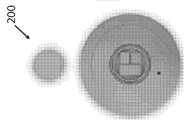
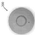
본 출원은 다양한 치료 및/또는 모니터링 적용을 수행하기 위한 자기 공명 영상(MRI) 호환 가능한, 대류 강화 전달(CED) 두개골 임플란트 장치 및 관련 방법을 개시한다. 일단 대상자에게 이식되면, 장치는 경피 바늘을 사용하여 다시 채울 수 있으므로 감염 위험을 최소화하면서 무한으로 제자리에 잔류할 수 있다. 이 장치는 실질적으로 해부학적으로 호환 가능한 형상을 가지므로 대상자에게 이식할 때, 본질적으로 검출할 수 없으므로 두피 또는 뇌 충돌을 피하기 위해 두개골 공간을 사용한다. 대상자에게 치료제를 선택적으로 투여하는 것 외에도, 장치는 치료의 효능 및/또는 반복 수술의 필요성을 모니터링하기 위해 의료 제공자에게 이미지 데이터를 제공할 수 있는 내장형 영상 장치를 포함할 수도 있다. 일부 실시예에서, 본원에 개시된 임플란트는 예컨대 이전 수술 절차로부터 누락된 두개골 절편을 대체하는데 사용되는 반면, 다른 예시적인 실시예에서, 임플란트는 두개골 뼈 플랩의 제거후에 수술 중 사용된다.This application discloses a magnetic resonance imaging (MRI) compatible, convection enhanced delivery (CED) cranial implant device and related method for performing a variety of treatment and/or monitoring applications. Once implanted into a subject, the device can be refilled using a transdermal needle, allowing it to remain in place indefinitely while minimizing the risk of infection. Since the device has a substantially anatomically compatible shape, when implanted into a subject, it is essentially undetectable and uses the cranial space to avoid scalp or brain collisions. In addition to selectively administering a therapeutic agent to a subject, the device may include an embedded imaging device capable of providing image data to a healthcare provider to monitor the efficacy of the treatment and/or the need for repetitive surgery. In some embodiments, the implants disclosed herein are used to replace missing skull sections, such as from a previous surgical procedure, while in other exemplary embodiments, the implant is used during surgery after removal of the skull bone flaps.
한 측면에서, 본 개시는 대상자의 적어도 하나의 두개골 개구에 두개골내 이식을 위해 구성된 적어도 하나의 두개골 임플란트 하우징을 포함하는 자기 공명 영상(MRI) 호환 가능한, 대류-증강 전달(CED) 두개골 임플란트 장치를 제공한다. 두개골 임플란트 하우징은 실질적으로 해부학적으로 호환 가능한 형상, 적어도 제 1 및 제 2 표면, 및 적어도 하나의 공동 및 적어도 제 2 표면을 통해 공동과 유체 연통하는 적어도 하나의 포트를 포함하는 적어도 하나의 유체 회로를 포함하고, 상기 공동은 적어도 하나의 유체 치료제를 포함하거나 포함할 수 있다. 상기 장치는 또한 유체 회로에 작동 가능하게 연결된 적어도 하나의 CED 펌프를 포함한다. 상기 CED 펌프는, 적어도 하나의 유체 도관이 적어도 상기 유체 도관의 출구의 근위에서 상기 유체 치료제의 적어도 하나의 양압 구배를 유지하기 위해 상기 포트에 작동 가능하게 연결될 때, 상기 적어도 하나의 유체 도관을 통해 상기 공동으로부터 상기 유체 치료제를 운반하도록 구성된다. 상기 장치는 또한 적어도 CED 펌프에 작동 가능하게 연결된 적어도 하나의 제어기를 포함한다. 상기 제어기는 상기 유체 도관이 상기 포트에 작동 가능하게 연결되고 상기 공동이 상기 유체 치료제를 포함할 때, 상기 유체 도관을 통해 상기 유체 치료제를 운반하도록 상기 CED 펌프를 선택적으로 실행시키도록 구성된다. 또한, 상기 장치는 또한 적어도 제어기에 작동 가능하게 연결된 적어도 하나의 전원을 포함한다. 두개골 임플란트 하우징, CED 펌프, 제어기 및 전원은 하나 이상의 MRI 호환 재료로 제조된다. 특정 실시예에서, 두개골 임플란트 하우징은 표준화된 형태(즉, 기성품 이용 가능)를 포함하는 반면, 다른 실시예에서 두개골 임플란트 하우징은 대상자에 대해 맞춤화되고 환자에 특정한 형태를 포함한다. 일반적으로 두개골내 이식은 무한 지속이다.In one aspect, the present disclosure provides a magnetic resonance imaging (MRI) compatible, convection-enhanced delivery (CED) cranial implant device comprising at least one cranial implant housing configured for intracranial implantation in at least one cranial opening of a subject. to provide. The cranial implant housing comprises at least one fluid circuit comprising a substantially anatomically compatible shape, at least first and second surfaces, and at least one cavity and at least one port in fluid communication with the cavity through the at least second surface. And the cavity may contain or contain at least one fluid therapeutic agent. The device also includes at least one CED pump operably connected to the fluid circuit. The CED pump is through the at least one fluid conduit when at least one fluid conduit is operatively connected to the port to maintain at least one positive pressure gradient of the fluid therapeutic agent at least proximal to the outlet of the fluid conduit. Configured to deliver the fluid therapeutic agent from the cavity. The device also includes at least one controller operably connected to at least the CED pump. The controller is configured to selectively execute the CED pump to deliver the fluid therapeutic agent through the fluid conduit when the fluid conduit is operatively connected to the port and the cavity contains the fluid therapeutic agent. In addition, the device also comprises at least one power source operably connected to at least the controller. The cranial implant housing, CED pump, controller and power source are made of one or more MRI compatible materials. In certain embodiments, the cranial implant housing includes a standardized configuration (ie, ready-made available), while in other embodiments the cranial implant housing is customized for a subject and includes a patient-specific configuration. In general, intracranial implantation is endless.
일부 실시예에서, 유체 치료제는 광유전 단백질, 줄기 세포, 면역 세포, 항체, 효소, 방사선 치료제, 화학 치료제, 신경학적 약물, 신경학적 예방 약물, 신경학적 강화 약물, 또는 이들의 조합들을 포함한다. 특정 실시예에서, 유체 치료제는 항암, 항 발작, 항 파킨슨, 항 헌팅턴, 항 수두증, 항 ADHD, 항 알츠하이머, 항 통증, 항 불면증, 항 우울증, 항 정신 분열증, 에너지 강화, 정신 강화, 신경 보호, 기억 강화 및 이들의 조합으로 이루어진 그룹으로부터 선택된 하나 이상의 요법을 포함한다 ,In some embodiments, fluid therapeutics include optogenetic proteins, stem cells, immune cells, antibodies, enzymes, radiation therapeutics, chemotherapeutic agents, neurological drugs, neurological prophylactic drugs, neurological enhancing drugs, or combinations thereof. In certain embodiments, the fluid therapy is anti-cancer, anti-seizure, anti-Parkinson, anti-Huntington, anti-hydrocephalus, anti-ADHD, anti-Alzheimer's, anti-pain, anti-insomnia, anti-depression, anti-schizophrenia, energy enhancing, mental strengthening, neuroprotection, And one or more therapies selected from the group consisting of memory enhancement and combinations thereof,
상기 유체 회로는 일반적으로 공동 및 포트에 작동 가능하게 연결된 하나 이상의 유체 채널을 포함한다. 두개골 임플란트 하우징은 각각 하나 이상의 유체 치료제 및/또는 기타 유체 재료를 포함하거나 포함할 수 있는 다중 공동을 선택적으로 포함한다. 일부 실시예에서, 두개골 임플란트는 적어도 제 2 표면을 통해 공동과 유체 연통하는 다중 포트를 포함한다. 특정 실시예에서, 두개골 임플란트 하우징은 MRI 호환 중합체, MRI 호환 금속, MRI 호환 생체공학 재료 또는 이들의 조합을 포함한다. 일부 실시예에서, 두개골 임플란트 하우징은 의료 등급 티타늄, 티타늄 메시, 다공성 하이드록시아파타이트(HA), 폴리메틸메타크릴레이트(PMMA), 폴리에테르 에테르 케톤(PEEK), 다공성 폴리에틸렌, 입방 지르코니아(CZ) 또는 그 조합 중 하나 이상을 포함한다. 일부 예에서, 두개골 임플란트 하우징은 실질적으로 반투명한 재료를 포함한다.The fluid circuit generally includes one or more fluid channels operatively connected to a cavity and a port. The cranial implant housing optionally includes multiple cavities that each contain or may contain one or more fluid therapeutic agents and/or other fluid materials. In some embodiments, the cranial implant includes multiple ports in fluid communication with the cavity through at least the second surface. In certain embodiments, the cranial implant housing comprises an MRI compatible polymer, an MRI compatible metal, an MRI compatible bionic material, or a combination thereof. In some embodiments, the cranial implant housing is medical grade titanium, titanium mesh, porous hydroxyapatite (HA), polymethylmethacrylate (PMMA), polyether ether ketone (PEEK), porous polyethylene, cubic zirconia (CZ), or Includes one or more of the combinations. In some examples, the cranial implant housing includes a material that is substantially translucent.
일반적으로, 두개골 임플란트 장치는 두개골 임플란트 하우징 및/또는 유체 도관에 작동 가능하게 연결되거나 연결될 수 있는 적어도 하나의 부착 메커니즘 또는 그 일부를 포함한다. 부착 메커니즘 또는 그 일부는 유체 도관이 시각적 반투명도 및/또는 음파를 향상시키기 위해 유체 회로와 유체 연통하도록 유체 도관을 두개골 임플란트 하우징에 부착하도록 구성된다.In general, the cranial implant device includes at least one attachment mechanism or portions thereof that are operably connected or connectable to the cranial implant housing and/or a fluid conduit. The attachment mechanism or portion thereof is configured to attach the fluid conduit to the cranial implant housing such that the fluid conduit is in fluid communication with the fluid circuit to improve visual translucency and/or sound waves.
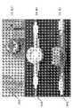
CED 펌프, 제어기 및 전원은 두개골 임플란트 하우징 내에 봉입된다. 일부 실시예에서, 예컨대, CED 펌프, 제어기 및 전원 및 선택적으로 하나 이상의 다른 장치 구성요소는 제 1 표면과 제 2 표면 사이의 사공간 사용을 최대화하기 위해 두개골 임플란트 하우징 내에 봉입된다. 특정 실시예에서, CED 펌프는 적어도 하나의 전기활성 중합체(EAP) 밸브-게이트 펌프를 포함한다. 일반적으로 제어기는 원격으로 모니터링, 활성화 또는 둘 다할 수 있도록 무선 연결되도록 구성된다. 일부 실시예에서, 전원은 적어도 하나의 배터리(예: 제로 볼트 배터리, 재충전 가능한 배터리 등)를 포함한다. 특정 실시예에서, 두개골 임플란트 장치는 적어도 부분적으로 제 1 표면 내에 또는 그를 통해 배치된 적어도 하나의 자체 밀봉 액세스 포트를 포함한다. 자체 밀봉 액세스 포트는 공동과 유체 연통하고 대상자의 두피를 통해 하나 이상의 주사기 바늘(예:자체 밀봉 주사기 바늘)을 수용하여 공동에/공동으로부터 유체 치료제를 추가 및/또는 제거하도록 구성된다. 이들 실시예 중 일부에서, 자체 밀봉 액세스 포트는 격막을 포함한다.The CED pump, controller and power source are enclosed within the skull implant housing. In some embodiments, for example, the CED pump, controller and power source and optionally one or more other device components are enclosed within the skull implant housing to maximize dead space usage between the first and second surfaces. In certain embodiments, the CED pump comprises at least one electroactive polymer (EAP) valve-gate pump. Typically the controller is configured to be wirelessly connected so that it can be remotely monitored, activated, or both. In some embodiments, the power source includes at least one battery (eg, zero volt battery, rechargeable battery, etc.). In certain embodiments, the cranial implant device includes at least one self-sealing access port disposed at least partially within or through the first surface. The self-sealing access port is in fluid communication with the cavity and is configured to receive one or more syringe needles (eg, self-sealing syringe needles) through the scalp of the subject to add and/or remove fluid therapy to/from the cavity. In some of these embodiments, the self-sealing access port includes a septum.
특정 실시예에서, 두개골 임플란트 장치는 두개골 임플란트 하우징 내에 적어도 부분적으로 배치되고 적어도 제어기에 작동 가능하게 연결된 하나 이상의 검출기를 포함한다. 검출기는 대상자 및/또는 장치로부터 정보를 검출하도록 구성되며, 이 정보는 공동에 배치된 유체 치료제의 용적, 유체 회로를 통해 전달되는 유체 치료제의 용적, 유체 회로 내 및/또는 그 근위부의 유체 치료제의 압력, 유체 회로로부터 유체 치료제의 누출, 전원 상태, 장치 구성요소 오작동, 이식된 영상 장치를 통해 뇌 또는 뇌 공동의 시각적 이미지, 및 대상자의 검출 가능한 신호로 이루어지는 그룹에서 선택된다. 일반적으로, 대상자로부터의 검출 가능한 신호는 적어도 하나의 신경학적 관련 질환, 상태 또는 장애의 특징이다. 특정 실시예에서, 대상자로부터의 검출 가능한 신호는 이미지 데이터를 포함한다.In certain embodiments, the cranial implant device includes one or more detectors disposed at least partially within the cranial implant housing and operably connected to at least a controller. The detector is configured to detect information from the subject and/or device, the information being a volume of a fluid therapeutic agent disposed in the cavity, a volume of a fluid therapeutic agent delivered through the fluid circuit, of a fluid therapeutic agent in and/or proximal to the fluid circuit. Pressure, leakage of fluid therapy from the fluid circuit, power state, device component malfunction, visual image of the brain or brain cavity through the implanted imaging device, and detectable signals of the subject. In general, the detectable signal from the subject is characteristic of at least one neurologically related disease, condition or disorder. In certain embodiments, the detectable signal from the subject comprises image data.
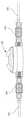
일부 실시예에서, 유체 도관은 (예: 장치 제조 동안) 포트에 작동 가능하게 연결된다. 다른 실시예에서, 유체 도관은 이식 직전에 수술실의 포트에 작동 가능하게 연결된다. 특정 실시예에서, 유체 도관은 유체 치료제를 뇌 실질의 질환 부분, 뇌종양 절제 후 사공간 공동, 및/또는 뇌의 혈관, 뉴런 또는 뇌실에 전달한다. 일부 실시예에서, 유체 도관은 중합체 튜브를 포함한다. 특정 실시예에서, 유체 도관은 카테터를 포함한다. 일반적으로, 유체 도관은 두개골 임플란트 하우징에 작동 가능하게 연결된 캐뉼라 내에 적어도 부분적으로 배치된다. 일부 실시예에서, 두개골 임플란트 하우징의 제 2 표면은 두개골 임플란트 하우징 내에 배치된 하나 이상의 유체 회로와 유체 연통하는 2, 3, 4 또는 5 개의 포트를 포함한다. 이들 실시예 중 일부에서, 두개골 임플란트 장치는 진단 또는 치료 가치가 있는 포트에 작동 가능하게 연결된 2, 3, 4 또는 5 개의 유체 도관을 포함한다.In some embodiments, the fluid conduit is operably connected to the port (eg, during device manufacturing). In another embodiment, the fluid conduit is operably connected to a port in the operating room immediately prior to implantation. In certain embodiments, the fluid conduit delivers the fluid therapeutic agent to the diseased portion of the brain parenchyma, the dead space cavity after resection of the brain tumor, and/or to the blood vessels, neurons or ventricles of the brain. In some embodiments, the fluid conduit comprises a polymer tube. In certain embodiments, the fluid conduit comprises a catheter. Generally, the fluid conduit is disposed at least partially within a cannula operably connected to the skull implant housing. In some embodiments, the second surface of the cranial implant housing includes 2, 3, 4 or 5 ports in fluid communication with one or more fluid circuits disposed within the cranial implant housing. In some of these embodiments, the cranial implant device includes 2, 3, 4 or 5 fluid conduits operably connected to a port of diagnostic or therapeutic value.
특정 실시예에서, 두개골 임플란트 장치는 두개골 임플란트 하우징 및/또는 제어기에 작동 가능하게 연결되거나 연결 가능한 적어도 하나의 전극을 포함한다. 전극은 (예: 흐름 변경 또는 낮은 수준의 약물 수량에 영향을 미치기 위해) 대상자에게 하나 이상의 전기 신호를 선택적으로 전송하도록 구성된다. 일부 실시예에서, 전극의 적어도 일부는 두개골 임플란트 하우징 내에 배치된다. 특정 실시예에서, 전극의 적어도 일부는 두개골 임플란트 하우징의 제 2 표면으로부터 연장된다.In certain embodiments, the cranial implant device comprises at least one electrode operably connected or connectable to the cranial implant housing and/or a controller. The electrodes are configured to selectively transmit one or more electrical signals to the subject (eg, to affect flow changes or low drug levels). In some embodiments, at least a portion of the electrode is disposed within the skull implant housing. In certain embodiments, at least a portion of the electrode extends from the second surface of the cranial implant housing.
일부 실시예에서, 두개골 임플란트 장치는 두개골 임플란트 하우징 및/또는 제어기에 작동 가능하게 연결되거나 연결 가능한 하나 이상의 영상 장치를 포함하며, 이 영상 장치는 대상자로부터 이미지 데이터를 선택적으로 캡처하도록 구성된다. 특정 실시예에서, 영상 장치는 카메라, 초음파 또는 관련 기술을 포함한다. 일부 실시예에서, 영상 장치의 적어도 일부는 두개골 임플란트 하우징 내에 배치된다. 특정 실시예에서, 영상 장치의 적어도 일부는 두개골 임플란트 하우징의 제 2 표면으로부터 연장된다. 선택적으로, 영상 장치는 초음파 또는 비 침습적 영상 장치를 포함한다. 일부 실시예에서, 영상 장치는 광 간섭 단층촬영(OCT) 장치를 포함한다. 일부 실시예에서, 이미지 데이터는 저화질 이미지 데이터를 포함하는 반면, 다른 실시예에서, 이미지 데이터는 고화질 이미지 데이터를 포함한다. 일부 실시예에서, 초음파는 혈류의 변화를 추가로 검출하기 위한 이중 기능을 갖는다.In some embodiments, the cranial implant device comprises one or more imaging devices operably connected or connectable to the cranial implant housing and/or a controller, the imaging device configured to selectively capture image data from a subject. In certain embodiments, the imaging device includes a camera, ultrasound, or related technology. In some embodiments, at least a portion of the imaging device is disposed within the cranial implant housing. In certain embodiments, at least a portion of the imaging device extends from the second surface of the cranial implant housing. Optionally, the imaging device comprises an ultrasonic or non-invasive imaging device. In some embodiments, the imaging device comprises an optical coherence tomography (OCT) device. In some embodiments, the image data includes low-quality image data, while in other embodiments, the image data includes high-quality image data. In some embodiments, ultrasound has a dual function to further detect changes in blood flow.
또 다른 측면에서, 본 출원은 대상자의 적어도 하나의 두개골 개구에 두개골내 이식을 위해 구성된 적어도 하나의 두개골 임플란트 하우징을 포함하는 자기 공명 영상(MRI) 호환 가능한, 대류 강화 전달(CED) 두개골 임플란트 장치를 개시한다. 두개골 임플란트 하우징은 실질적으로 해부학적으로 호환 가능한 형상(예: 가시적 변형 및 최적의 생체 적합성을 방지하기 위해), 적어도 제 1 및 제 2 표면, 및 적어도 하나의 공동 및 적어도 제 2 표면을 통해 상기 공동과 유체 연통하는 적어도 하나의 포트를 포함하는 적어도 하나의 유체 회로를 포함하고, 상기 공동은 적어도 하나의 유체 치료제를 포함하거나 포함할 수 있다. 두개골 임플란트 장치는 또한 유체 회로에 작동 가능하게 연결된 적어도 하나의 CED 펌프를 포함한다. 상기 CED 펌프는, 적어도 하나의 유체 도관이 적어도 상기 유체 도관의 출구의 근위에서 상기 유체 치료제의 적어도 하나의 양압 구배를 유지하기 위해 상기 포트에 작동 가능하게 연결될 때, 상기 적어도 하나의 유체 도관을 통해 상기 공동으로부터 상기 유체 치료제를 운반하도록 구성된다. 추가로, 두개골 임플란트 장치는 또한 적어도 CED 펌프에 작동 가능하게 연결된 적어도 하나의 전원을 포함한다. 일반적으로 두개골 임플란트 하우징, CED 펌프 및 전원은 [예: 종양상 감시(tumor bed surveillance)와의 간섭 방지하기 위해] 하나 이상의 MRI 호환 재료로 제조된다.In another aspect, the present application provides a magnetic resonance imaging (MRI) compatible, convection enhanced delivery (CED) cranial implant device comprising at least one cranial implant housing configured for intracranial implantation in at least one cranial opening of a subject. Start. The cranial implant housing has a substantially anatomically compatible shape (e.g., to prevent visible deformation and optimal biocompatibility), at least a first and a second surface, and the cavity through at least one cavity and at least a second surface. And at least one fluid circuit comprising at least one port in fluid communication with the cavity, wherein the cavity may contain or contain at least one fluid therapeutic agent. The cranial implant device also includes at least one CED pump operably connected to the fluid circuit. The CED pump is through the at least one fluid conduit when at least one fluid conduit is operatively connected to the port to maintain at least one positive pressure gradient of the fluid therapeutic agent at least proximal to the outlet of the fluid conduit. Configured to deliver the fluid therapeutic agent from the cavity. Additionally, the cranial implant device also includes at least one power source operably connected to at least the CED pump. Typically, the cranial implant housing, CED pump, and power source are made of one or more MRI-compatible materials (eg to prevent interference with tumor bed surveillance).
또 다른 측면에서, 본 출원은 대상자의 적어도 하나의 두개골 개구에 두개골내 이식을 위해 구성된 적어도 하나의 두개골 임플란트 하우징을 포함하는 두개골 임플란트 장치를 개시한다. 일반적으로, 두개골 임플란트 하우징은 실질적으로 해부학적으로 호환 가능한 형상(예: 기성품 또는 다른 환자 특정 형상)을 포함한다. 두개골 임플란트 장치는 또한 두개골 임플란트 하우징 내에 적어도 부분적으로 배치된 적어도 2 개의 기능적 구성요소를 포함한다. 제 1 기능적 구성요소는 적어도 하나의 유체 도관을 통해 대상자에게 제 1 기능적 구성요소로부터 적어도 하나의 유체 치료제를 운반하도록 구성된 적어도 하나의 대류 강화 전달(CED) 펌프[예: 전기활성 중합체(EAP) 밸브 게이트 펌프]를 포함하는 유체 기반 생리학적 상태 개입 시스템을 포함한다. 제 2 기능적 구성요소는 적어도 하나의 비 유체 도관을 통해 제 2 기능적 구성요소로부터 대상자에게 하나 이상의 치료 신호를 전송하도록 구성된 비 유체 기반 생리학적 상태 개입 시스템을 포함한다. 두개골 임플란트 장치는 또한 적어도 부분적으로 두개골 임플란트 하우징 내에 배치된 적어도 하나의 전원(예: 제로-볼트 배터리, 무선 충전식 배터리 등)을 포함하며, 이 전원은 기능적 구성요소에 작동 가능하게 연결된다. 일반적으로, 두개골 임플란트 하우징, 기능적 구성요소 및/또는 전원은 하나 이상의 자기 공명 영상(MRI) 호환 재료로 제조된다. 일부 실시예에서, 예컨대, 두개골 임플란트 하우징, 기능적 구성요소 및/또는 전원은 MRI 호환 중합체, MRI 호환 금속, MRI 호환 생체공학 재료, 또는 이들의 조합을 포함한다. 선택적으로, 두개골 임플란트 하우징, 기능성 구성요소 및/또는 전원은 의료 등급 티타늄, 티타늄 메시, 다공성 하이드록시아파타이트(HA), 폴리메틸메타크릴레이트(PMMA), 폴리에테르 에테르 케톤(PEEK), 다공성 폴리에틸렌, 입방 지르코니아(CZ) 또는 이들의 조합 중 하나 이상을 포함한다. ,In yet another aspect, the present application discloses a cranial implant device comprising at least one cranial implant housing configured for intracranial implantation in at least one cranial opening of a subject. In general, the cranial implant housing includes a substantially anatomically compatible shape (eg, off-the-shelf or other patient specific shape). The cranial implant device also includes at least two functional components disposed at least partially within the cranial implant housing. The first functional component is at least one convection enhanced delivery (CED) pump (e.g., an electroactive polymer (EAP) valve) configured to deliver at least one fluid therapeutic agent from the first functional component to the subject through at least one fluid conduit. And a fluid-based physiological state intervention system including] a gate pump. The second functional component includes a non-fluid based physiological condition intervention system configured to transmit one or more therapeutic signals from the second functional component to the subject through at least one non-fluid conduit. The cranial implant device also includes at least one power source (e.g., zero-volt battery, wireless rechargeable battery, etc.) at least partially disposed within the cranial implant housing, which power source is operably connected to the functional component. Typically, the cranial implant housing, functional component, and/or power source is made of one or more magnetic resonance imaging (MRI) compatible materials. In some embodiments, for example, the cranial implant housing, functional component and/or power source comprises an MRI compatible polymer, an MRI compatible metal, an MRI compatible bioengineered material, or a combination thereof. Optionally, the cranial implant housing, functional component and/or power source is medical grade titanium, titanium mesh, porous hydroxyapatite (HA), polymethylmethacrylate (PMMA), polyether ether ketone (PEEK), porous polyethylene, Cubic zirconia (CZ) or a combination thereof. ,
일부 실시예에서, 두개골 임플란트 하우징은 적어도 제 1 및 제 2 표면, 및 적어도 하나의 공동 및 적어도 제 2 표면을 통해 공동과 유체 연통하는 적어도 하나의 포트를 포함하는 적어도 하나의 유체 회로를 포함하고, 상기 공동은 유체 치료제를 포함하거나 포함할 수 있다. 특정 실시예에서, CED 펌프는 유체 회로에 작동 가능하게 연결된다. 일반적으로, 유체 회로는 공동 및 포트에 작동 가능하게 연결된 하나 이상의 유체 채널을 포함한다. 일부 실시예에서, 두개골 임플란트 하우징은 제 1 표면에 적어도 부분적으로 배치된 적어도 하나의 자체 밀봉 액세스 포트를 포함하고, 자체 밀봉 액세스 포트는 유체 치료제를 공동으로/상기 공동으로부터 및/또는 근처 카테터 배치로부터의 세포 병리학을 추가 및/또는 제거하기 위해 공동과 유체 연통하고 대상자의 두피를 통해 하나 이상의 주사기 바늘(예: 자체 밀봉 주사기 바늘)을 수용하도록 구성된다.In some embodiments, the cranial implant housing comprises at least one fluid circuit comprising at least first and second surfaces, and at least one cavity and at least one port in fluid communication with the cavity through the at least second surface, The cavity may or may contain a fluid therapeutic. In certain embodiments, the CED pump is operably connected to the fluid circuit. Generally, a fluid circuit includes one or more fluid channels operably connected to a cavity and a port. In some embodiments, the cranial implant housing includes at least one self-sealing access port disposed at least partially on the first surface, and the self-sealing access port provides a fluid therapeutic agent jointly/from the cavity and/or from a nearby catheter placement. It is configured to receive one or more syringe needles (eg, self-sealing syringe needles) through the subject's scalp and in fluid communication with the cavity to add and/or eliminate the cellular pathology of the.
특정 실시예에서, 기능적 구성요소는 항암, 항 발작, 항 파킨슨, 항 헌팅턴, 항 수두증, 항 ADHD, 항 알츠하이머, 항 통증, 항 불면증, 항 우울증, 항 정신 분열증, 에너지 강화, 정신 강화, 신경 보호, 기억 강화 및 이들의 조합으로 이루어진 그룹으로부터 선택된 하나 이상의 요법을 대상자에게 전달하도록 구성된다. 일부 실시예에서, 상기 기능적 구성요소들 및 상기 전원은 두개골 임플란트 하우징 내에 봉입된다.In certain embodiments, the functional component is anti-cancer, anti-seizure, anti-Parkinson, anti-Huntington, anti-hydrocephalus, anti-ADHD, anti-Alzheimer's, anti-pain, anti-insomnia, anti-depression, anti-schizophrenia, energy enhancement, mental enhancement, neuroprotection. , Memory enhancement, and combinations thereof. In some embodiments, the functional components and the power source are enclosed within a skull implant housing.
일부 실시예에서, 두개골 임플란트 장치는 두개골 임플란트 하우징 내에 적어도 부분적으로 배치된 적어도 하나의 제어기를 포함하고, 이 제어기는 기능적 구성요소 및 전원에 작동 가능하게 연결되고, 유체 도관을 통해 대상자에게 유체 치료제를 운반하기 위한 제 1 기능적 구성요소 및 비-유체 도관을 통해 대상자에게 치료 신호를 전송하기 위한 제 2 기능적 구성요소의 CED 펌프를 선택적으로 실행시키도록 구성된다. 제어기는 일반적으로 원격 모니터링, 활성화, 조정 및/또는 충전을 위해 무선 연결되도록 구성된다. 일부 실시예에서, 예컨대, 장치 주입 등급, 투여 량 및/또는 타이밍은 일반적으로 특정 치료 효능, 환자 증상, 종양 성장 및/또는 생체 사인에 따라 무선 연결을 통해 변경된다. 이들 중 특정 실시예에서, 유체 운반은 예컨대 모니터링된 흐름 등에 기초하여 유체를 펌핑하는 주어진 임플란트 장치에 작동 가능하게 연결된 단일 또는 다중 카테터를 원격으로 선택하는 단계를 포함한다.In some embodiments, the cranial implant device comprises at least one controller disposed at least partially within the cranial implant housing, the controller operably connected to a functional component and power source, and delivering a fluid therapeutic agent to the subject through a fluid conduit. And a CED pump of the first functional component for transporting and the second functional component for transmitting treatment signals to the subject via a non-fluidic conduit. The controller is generally configured to be wirelessly connected for remote monitoring, activation, coordination and/or charging. In some embodiments, for example, device infusion grade, dosage, and/or timing are generally changed over a wireless connection depending on the specific treatment efficacy, patient symptoms, tumor growth, and/or vital signs. In certain of these embodiments, fluid delivery includes remotely selecting single or multiple catheters operably connected to a given implant device that pumps fluid based on, for example, a monitored flow or the like.
제 1 기능적 구성요소는 일반적으로 두개골 임플란트 하우징 내에 적어도 부분적으로 배치되고 적어도 제어기에 작동 가능하게 연결된 하나 이상의 검출기를 포함한다. 검출기는 대상자 및/또는 장치로부터 정보를 검출하도록 구성되며, 이 정보는 장치의 공동에 배치된 유체 치료제의 용적, 유체 회로를 통해 전달되는 유체 치료제의 용적, 유체 회로 내 및/또는 그 근접한 유체 치료제의 압력, 유체 회로로부터 유체 치료제의 누출, 전원의 상태, 장치 구성요소 오작동 및 대상자로부터의 검출 가능한 신호로 구성된 그룹에서 선택된다.The first functional component generally comprises one or more detectors disposed at least partially within the cranial implant housing and operably connected to at least a controller. The detector is configured to detect information from the subject and/or the device, the information being a volume of fluid therapy placed in the cavity of the device, a volume of fluid therapy delivered through the fluid circuit, fluid therapy within and/or in proximity thereof. Pressure, leakage of fluid therapy from the fluid circuit, state of the power source, device component malfunction, and detectable signals from the subject.
일반적으로, 유체 도관 및/또는 비 유체 도관은 두개골 임플란트 하우징으로부터 연장된다. 일부 실시예에서, 유체 도관 및 비 유체 도관은 기능적 구성요소와 대상자 사이의 유체, 전기, 자기, 이미징 및 광학 통신을 위해 구성된다. 일부 실시예에서, 치료 신호는 전기 신호, 자기 신호, 광학 신호, 영상 신호 또는 이들의 조합을 포함한다. 선택적으로, 제 2 기능적 구성요소는 대상자 및/또는 장치로부터 정보를 검출하도록 구성된 적어도 하나의 검출기를 포함한다. 일부 실시예에서, 기능적 구성요소는 의약 요법, 전기 자극 요법, 방사선 요법, 화학 요법, 방사선 요법 또는 이들의 조합을 포함하는 급성 신경학적 중재를 제공하도록 구성된다. 특정 실시예에서, 기능적 구성요소 중 하나 이상은 생체 사인 모니터, 광학 간섭 단층촬영(OCT) 이미지 모니터, 고화질 카메라, 두개골내 압력(ICP) 모니터, 뇌파 센서(ECOG), 지역 치료 및/또는 원격 영상 모니터를 위한 일부 방사선 시드를 포함한다.Typically, the fluid conduit and/or non-fluid conduit extends from the cranial implant housing. In some embodiments, fluidic and non-fluidic conduits are configured for fluid, electrical, magnetic, imaging, and optical communication between a functional component and a subject. In some embodiments, the therapeutic signal comprises an electrical signal, a magnetic signal, an optical signal, an imaging signal, or a combination thereof. Optionally, the second functional component comprises at least one detector configured to detect information from the subject and/or device. In some embodiments, the functional component is configured to provide an acute neurological intervention including medicinal therapy, electrical stimulation therapy, radiation therapy, chemotherapy, radiation therapy, or combinations thereof. In certain embodiments, one or more of the functional components are a vital sign monitor, an optical coherence tomography (OCT) image monitor, a high-definition camera, an intracranial pressure (ICP) monitor, an electroencephalogram sensor (ECOG), local therapy, and/or remote imaging. Includes some radiation seeds for monitoring.
일부 실시예에서, 제 2 기능적 구성요소는 광학 센서를 통해 뉴런 변조를 제공하도록 구성된다. 일반적으로, 제 2 기능적 구성요소는 적어도 하나의 생리적 상태의 컴퓨터 모니터링을 위해 구성된다. 일부 실시예에서, 제 2 기능적 구성요소는 뇌 실질의 질환 부분, 뇌종양 절제 후 사공간 공동, 및/또는 뇌의 혈관(예: 영양 공급 혈관), 뉴런 또는 뇌실을 모니터링하도록 구성된다. 선택적으로, 제 2 기능적 구성요소는 적어도 하나의 두개골내 압력(ICP) 모니터를 포함한다. 일부 실시예에서, 제 2 기능적 구성요소는 적어도 하나의 생체 사인 또는 뇌 기능 모니터를 포함한다. 특정 실시예에서, 제 2 기능적 구성요소는 적어도 하나의 영상 장치를 포함한다. 일부 실시예에서, 영상 장치는 카메라를 포함한다. 특정 실시예에서, 영상 장치는 광 간섭 단층촬영(OCT) 장치를 포함한다. 일부 실시예에서, 영상 장치는 듀플렉스 기능이 있거나 없는 초음파 장치를 포함한다. 선택적으로, 제 2 기능적 구성요소는 전기 시스템, 원격 이미징 시스템, 방사선 치료 시스템, 반응 신경자극 시스템 및/또는 신경조절 시스템을 포함한다. 일부 실시예에서, 제 2 기능적 구성요소는 약물 전달 장치, 전기 신호 전달 장치, 이미지 캡처 장치, 방사성 시드 장치, 에너지 저장 장치 및/또는 컴퓨팅 장치를 포함한다. 특정 실시예에서, 제 2 기능적 구성요소는 전기 에너지 소스, 전기 에너지 검출기, 전자기 에너지 소스 및/또는 전자기 에너지 검출기를 포함한다. 일반적으로, 전기 에너지 소스는 전기 신호를 생성하도록 구성되고, 전자기 에너지 소스는 광 신호를 생성하도록 구성되며, 전자기 에너지 검출기는 이미지 데이터를 캡처하도록 구성된다.In some embodiments, the second functional component is configured to provide neuronal modulation via an optical sensor. In general, the second functional component is configured for computer monitoring of at least one physiological condition. In some embodiments, the second functional component is configured to monitor the diseased portion of the brain parenchyma, the dead space cavity after resection of the brain tumor, and/or blood vessels in the brain (eg, feeding vessels), neurons, or ventricles. Optionally, the second functional component comprises at least one intracranial pressure (ICP) monitor. In some embodiments, the second functional component comprises at least one vital sign or brain function monitor. In a specific embodiment, the second functional component comprises at least one imaging device. In some embodiments, the imaging device comprises a camera. In certain embodiments, the imaging device comprises an optical coherence tomography (OCT) device. In some embodiments, the imaging device includes an ultrasound device with or without a duplex function. Optionally, the second functional component comprises an electrical system, a remote imaging system, a radiation therapy system, a responsive neurostimulation system and/or a neuromodulation system. In some embodiments, the second functional component comprises a drug delivery device, an electrical signal delivery device, an image capture device, a radioactive seed device, an energy storage device, and/or a computing device. In certain embodiments, the second functional component comprises an electrical energy source, an electrical energy detector, an electromagnetic energy source and/or an electromagnetic energy detector. In general, the electrical energy source is configured to generate an electrical signal, the electromagnetic energy source is configured to generate an optical signal, and the electromagnetic energy detector is configured to capture image data.
또 다른 측면에서, 본 출원은 대상자의 적어도 하나의 두개골 개구에 적어도 하나의 두개골 임플란트 장치를 외과적으로 이식하는 것을 포함하는, 대상자의 신경학적 관련 질환, 상태 또는 장애를 치료하는 방법을 개시한다. 두개골 임플란트 장치는 적어도 하나의 두개골 임플란트 하우징을 포함하고, 상기 적어도 하나의 두개골 임플란트 하우징은 실질적으로 해부학적으로 호환 가능한 형상[표준(즉, 기성품) 디자인 또는 맞춤형(즉, 환자 특정) 디자인], 적어도 제 1 및 제 2 표면, 및 적어도 하나의 공동 및 적어도 제 2 표면을 통해 공동과 유체 연통하는 적어도 하나의 포트를 포함하는 적어도 하나의 유체 회로를 포함하고, 여기서 공동은 적어도 하나의 유체 치료제를 포함하고 적어도 유체 도관은 제 2 표면으로부터 연장되고 유체 회로와 유체 연통한다. 두개골 임플란트 장치는 또한 유체 회로에 작동 가능하게 연결된 하나 이상의 대류 강화 전달(CED) 펌프를 포함하며, 상기 CED 펌프는 대상자의 두개골 공동 내의 유체 도관의 출구에 적어도 근접한 유체 치료제의 적어도 하나의 양압 구배를 유지하기 위해 유체 도관을 통해 상기 공동으로부터 상기 유체 치료제를 운반하도록 구성된다. 두개골 임플란트 장치는 또한 적어도 CED 펌프에 작동 가능하게 연결된 적어도 하나의 제어기를 포함하며, 상기 제어기는 유체 도관을 통해 유체 치료제를 운반하도록 CED 펌프를 선택적으로 실행시키도록 구성된다. 두개골 임플란트 장치는 추가로 적어도 제어기에 작동 가능하게 연결된 적어도 하나의 전원을 포함한다. 두개골 임플란트 하우징, CED 펌프, 제어기 및 전원은 (예: 후속 영상과의 간섭을 방지하기 위해) 하나 이상의 자기 공명 영상(MRI) 호환 재료로 제조된다. 상기 방법은 또한 대상자의 두개골 공동 내의 유체 도관의 출구에 적어도 근접한 유체 치료제의 양압 구배를 유지하기 위해 유체 도관을 통해 공동으로부터 유체 치료제의 유효량을 운반하는 단계를 포함하고, 이에 따라 대상자의 신경학적 관련 질환, 상태 또는 장애를 치료한다.In another aspect, the present application discloses a method of treating a neurologically related disease, condition or disorder in a subject comprising surgically implanting at least one cranial implant device into at least one cranial opening in the subject. The cranial implant device comprises at least one cranial implant housing, the at least one cranial implant housing having a substantially anatomically compatible shape (standard (ie, off-the-shelf) design or custom (ie, patient specific) design), at least At least one fluid circuit comprising first and second surfaces, and at least one cavity and at least one port in fluid communication with the cavity through at least the second surface, wherein the cavity comprises at least one fluid therapeutic agent And at least the fluid conduit extends from the second surface and is in fluid communication with the fluid circuit. The cranial implant device also includes one or more convection enhanced delivery (CED) pumps operably connected to the fluid circuit, the CED pump generating at least one positive pressure gradient of the fluid therapeutic at least proximate the outlet of the fluid conduit within the cranial cavity of the subject. Configured to convey the fluid therapeutic agent from the cavity through a fluid conduit for retention. The cranial implant device also includes at least one controller operably connected to at least the CED pump, the controller configured to selectively execute the CED pump to deliver the fluid therapeutic agent through the fluid conduit. The cranial implant device further comprises at least one power source operably connected to at least the controller. The cranial implant housing, CED pump, controller and power source are made of one or more magnetic resonance imaging (MRI) compatible materials (eg, to prevent interference with subsequent images). The method also includes delivering an effective amount of the fluid therapeutic agent from the cavity through the fluid conduit to maintain a positive pressure gradient of the fluid therapeutic agent at least proximate the outlet of the fluid conduit within the subject's cranial cavity, and thus neurological involvement of the subject. Treat the disease, condition or disorder.
특정 실시예에서, 신경학적 관련 질환, 상태 또는 장애는 암(예: 뇌암), 간질, 파킨슨 병, 헌팅턴 병, 수두증, 주의력 결핍 과잉 행동 장애(ADHD), 통증, 알츠하이머 병, 불면증, 우울증, 조울증, 정신 분열증 중 하나 이상을 포함한다. 선택적으로, 유체 치료제는 광유전 단백질, 줄기 세포, 면역 세포, 항체, 효소, 방사선 치료제, 화학 치료제, 신경학적 강화 약물, 신경학적 예방 약물 또는 이들의 조합을 포함한다. 일반적으로, 상기 방법은 유체 치료제의 유효량을 뇌 실질의 질환 부분, 뇌종양 절제 후 사공간 공동 및/또는 혈관(예: 영양 공급 혈관), 대상자의 뇌의 뉴런 또는 뇌실로 전달하는 단계를 포함한다.In certain embodiments, the neurologically related disease, condition or disorder is cancer (e.g., brain cancer), epilepsy, Parkinson's disease, Huntington's disease, hydrocephalus, attention deficit hyperactivity disorder (ADHD), pain, Alzheimer's disease, insomnia, depression, manic depression. , Including one or more of schizophrenia. Optionally, fluid therapeutics include optogenetic proteins, stem cells, immune cells, antibodies, enzymes, radiation therapeutics, chemotherapeutic agents, neurological enhancing drugs, neurological prophylactic drugs, or combinations thereof. In general, the method includes delivering an effective amount of a fluid therapeutic agent to a diseased part of the brain parenchyma, a dead space cavity and/or a blood vessel (e.g., a feeding vessel), a neuron or a ventricle of the subject's brain after resection of the brain tumor.
일부 실시예에서, 적어도 하나의 자체 밀봉 액세스 포트는 두개골 임플란트 하우징의 제 1 표면에 또는 제 1 표면을 통해 적어도 부분적으로 배치되고, 상기 자체 밀봉 액세스 포트는 공동과 유체 연통하고, 상기 방법은 대상자의 두피(예: 장치 위 또는 주변) 및 자체 밀봉 액세스 포트를 통해 주사기 바늘(예: 자체 밀봉 주사기 바늘)을 삽입하는 단계, 및 유체 치료제를 공동(예: 내장된 공동)에 추가하는 단계를 포함한다. 특정 실시예에서, 제어기는 원격으로 모니터링, 활성화 및/또는 조정되도록 무선 연결을 위해 구성되고, 상기 방법은 제어기로/제어기로부터 정보 및/또는 명령을 무선으로 전송 및/또는 수신하는 단계를 포함한다.In some embodiments, at least one self-sealing access port is disposed at least partially on or through a first surface of the cranial implant housing, the self-sealing access port in fluid communication with the cavity, and the method comprises Inserting a syringe needle (e.g., a self-sealing syringe needle) through the scalp (e.g., on or around the device) and a self-sealing access port, and adding a fluid therapeutic agent to the cavity (e.g., an embedded cavity). . In certain embodiments, the controller is configured for a wireless connection to be monitored, activated and/or controlled remotely, the method comprising wirelessly transmitting and/or receiving information and/or commands to/from the controller. .
특정 실시예에서, 두개골 임플란트 장치는 두개골 임플란트 하우징 내에 적어도 부분적으로 배치되고 적어도 제어기에 작동 가능하게 연결된 하나 이상의 검출기를 포함하고, 상기 검출기는 대상자 및/또는 장치로부터 정보를 검출하도록 구성된다. 이러한 실시예에서, 상기 방법은 일반적으로 공동 내에 배치된 유체 치료제의 용적, 유체 회로를 통해 운반되는 유체 치료제의 용적, 유체 회로 내 및/또는 그의 근위에서의 유체 치료제의 압력, 유체 회로로부터 유체 치료제의 누출, 전원 상태, 장치 구성요소 오작동 및/또는 대상자로부터 검출 가능한 신호를 검출하는 단계를 포함한다. 일부 실시예에서, 두개골 임플란트 장치는 제어기에 작동 가능하게 연결된 적어도 하나의 두개골내 압력(ICP) 모니터를 포함하고, 상기 방법은 (예: 가성 뇌졸중, NPH 또는 폐쇄성 뇌수종을 검출하기 위해) ICP 모니터를 사용하여 대상자의 ICP를 모니터링하는 단계를 포함한다. 특정 실시예에서, 두개골 임플란트 장치는 제어기에 작동 가능하게 연결된 적어도 하나의 생체 신호 모니터를 포함하고, 상기 방법은 생체 신호 모니터를 사용하여 대상자의 하나 이상의 생체 신호를 모니터링하는 단계를 포함한다. 일부 실시예에서, 두개골 임플란트 장치는 제어기에 작동 가능하게 연결된 적어도 하나의 영상 장치를 포함하고, 상기 방법은 영상 장치를 사용하여 대상자로부터 이미지 데이터를 캡처하는 단계를 포함한다. 일부 실시예에서, 영상 장치는 광학 간섭 단층촬영(OCT) 장치를 포함하고, 상기 방법은 OCT 장치를 사용하여 대상자로부터 OCT 이미지 데이터를 캡처하는 단계를 포함한다. 특정 실시예에서, 영상 장치는 듀플렉스 기능이 있거나 없는 초음파 장치를 포함하고, 상기 방법은 초음파 장치를 사용하여 대상자로부터 초음파 이미지 데이터를 캡처하는 단계를 포함한다. 일부 실시예에서, 상기 방법은 대상자의 두개골 공동의 하나 이상의 MRI 이미지를 획득하는 단계를 포함한다.In certain embodiments, the cranial implant device comprises one or more detectors disposed at least partially within the cranial implant housing and operably connected to at least a controller, the detectors configured to detect information from the subject and/or the device. In such embodiments, the method generally comprises a volume of fluid therapeutic agent disposed within a cavity, a volume of fluid therapeutic agent delivered through a fluid circuit, pressure of the fluid therapeutic agent in and/or proximal to the fluid circuit, fluid therapeutic agent from the fluid circuit. And detecting detectable signals from leaks, power conditions, device component malfunctions, and/or subjects. In some embodiments, the cranial implant device comprises at least one intracranial pressure (ICP) monitor operably connected to the controller, the method comprising an ICP monitor (e.g., to detect pseudostroke, NPH, or obstructive hydrocephalus). Monitoring the subject's ICP. In certain embodiments, the cranial implant device includes at least one physiological signal monitor operably connected to the controller, the method comprising monitoring one or more physiological signals of the subject using the physiological signal monitor. In some embodiments, the cranial implant device includes at least one imaging device operably connected to a controller, the method comprising using the imaging device to capture image data from the subject. In some embodiments, the imaging device includes an optical coherence tomography (OCT) device, and the method includes capturing OCT image data from the subject using the OCT device. In certain embodiments, the imaging device includes an ultrasound device with or without a duplex function, and the method includes capturing ultrasound image data from the subject using the ultrasound device. In some embodiments, the method includes acquiring one or more MRI images of the subject's cranial cavity.
또 다른 측면에서, 본 출원은 (예: 임상 연구 및/또는 제어 재판을 보조하기 위해) 복수의 대상자들 각각에 적어도 하나의 두개골 임플란트 장치를 외과적으로 이식하는 단계를 포함하는 복수의 대상자들에서 치료제 투여를 모니터링하는 방법을 개시한다. 각각의 두개골 임플란트 장치는 적어도 하나의 두개골 임플란트 하우징을 포함하고, 상기 적어도 하나의 두개골 임플란트 하우징은 실질적으로 해부학적으로 호환 가능한 형상[표준(즉, 기성품) 디자인 또는 맞춤형(즉, 환자 특정) 디자인], 적어도 제 1 및 제 2 표면, 및 적어도 하나의 공동 및 적어도 제 2 표면을 통해 공동과 유체 연통하는 적어도 하나의 포트를 포함하는 적어도 하나의 유체 회로를 포함하고, 상기 공동은 적어도 하나의 유체 치료제를 포함하고, 적어도 유체 도관은 제 2 표면으로부터 연장되고 유체 회로와 유체 연통한다. 두개골 임플란트 장치는 또한 유체 회로에 작동 가능하게 연결된 적어도 하나의 대류 강화 전달(CED) 펌프를 포함한다. CED 펌프는 주어진 대상자의 두개골 공동 내에서 유체 도관의 출구에 적어도 근접한 유체 치료제의 적어도 하나의 양압 구배를 유지하기 위해 공동으로부터 유체 도관을 통해 유체 치료제를 운반하도록 구성된다. 특정 실시예에서, 가역적 압력은 세포학 검색 등을 위한 진공을 생성하기 위해 사용된다. 두개골 임플란트 장치는 또한 적어도 CED 펌프에 작동 가능하게 연결된 적어도 하나의 제어기를 포함하며, 상기 제어기는 CED 펌프가 유체 도관을 통해 유체 치료제를 운반하고 원격으로 모니터링, 활성화 및/또는 조정되도록 무선 연결을 위해 선택적으로 실행시키도록 구성된다. 두개골 임플란트 장치는 또한 적어도 제어기에 작동 가능하게 연결된 적어도 하나의 전원을 포함한다. 두개골 임플란트 하우징, CED 펌프, 제어기 및 전원은 하나 이상의 자기 공명 영상(MRI) 호환 재료로 제조된다. 상기 방법은 또한 이식된 두개골 임플란트 장치를 사용하여 선택된 양의 유체 치료제를 복수의 대상자들 중 하나 이상의 구성원에게 운반하는 단계를 포함한다. 또한, 상기 방법은 또한 이식된 두개골 임플란트 장치의 무선 연결을 사용하여 복수의 대상자들의 구성원의 하나 이상의 선택된 세트로부터 데이터를 수집하고, 이에 의해 복수의 대상자들에서 (예: 임상 시험 조사를 통해) 치료제 투여를 모니터링하는 단계를 포함한다. 일부 실시예에서, 데이터는 복수의 대상자들에서 치료제의 효능 및/또는 독성의 측정치와 관련된다. 특정 실시예에서, 데이터는 복수의 대상자들에서 두개골 임플란트 장치의 성능 측정치와 관련된다.In another aspect, the present application provides in a plurality of subjects comprising the step of surgically implanting at least one cranial implant device in each of the plurality of subjects (e.g., to aid in clinical studies and/or control trials). A method of monitoring the administration of a therapeutic agent is disclosed. Each cranial implant device comprises at least one cranial implant housing, the at least one cranial implant housing having a substantially anatomically compatible shape (standard (i.e., off-the-shelf) design or custom (i.e., patient specific) design). , At least one fluid circuit comprising at least first and second surfaces, and at least one port in fluid communication with the cavity through at least one cavity and at least the second surface, wherein the cavity is at least one fluid therapeutic agent And at least the fluid conduit extends from the second surface and is in fluid communication with the fluid circuit. The cranial implant device also includes at least one convection enhanced delivery (CED) pump operably connected to the fluid circuit. The CED pump is configured to deliver the fluid therapeutic agent through the fluid conduit from the cavity to maintain at least one positive pressure gradient of the fluid therapeutic agent at least proximate the outlet of the fluid conduit within the cranial cavity of a given subject. In certain embodiments, reversible pressure is used to create a vacuum for cytology screening and the like. The cranial implant device also includes at least one controller operably connected to at least the CED pump, the controller for wireless connection such that the CED pump carries the fluid therapeutic agent through the fluid conduit and is remotely monitored, activated, and/or adjusted. It is configured to run selectively. The cranial implant device also includes at least one power source operably connected to at least the controller. The cranial implant housing, CED pump, controller and power source are made of one or more magnetic resonance imaging (MRI) compatible materials. The method also includes delivering a selected amount of a fluid therapeutic agent to one or more members of the plurality of subjects using the implanted cranial implant device. In addition, the method also uses a wireless connection of an implanted cranial implant device to collect data from one or more selected sets of members of a plurality of subjects, whereby a therapeutic agent in a plurality of subjects (e.g., through a clinical trial investigation). Monitoring administration. In some embodiments, the data relate to a measure of the efficacy and/or toxicity of a therapeutic agent in a plurality of subjects. In certain embodiments, the data relate to a measure of the performance of a cranial implant device in a plurality of subjects.
또 다른 측면에서, 본 출원은 대상자의 적어도 하나의 두개골 개구에 적어도 하나의 두개골 임플란트 장치를 외과적으로 이식하는 단계를 포함하는 수술 방법을 개시한다. 두개골 임플란트 장치는 실질적으로 해부학적으로 호환 가능한 형상[표준(즉, 기성품) 디자인 또는 맞춤형(즉, 환자 특정) 디자인], 적어도 제 1 및 제 2 표면, 및 적어도 하나의 공동 및 적어도 제 2 표면을 통해 공동과 유체 연통하는 적어도 하나의 포트를 포함하는 적어도 하나의 두개골 임플란트 하우징을 포함하고, 상기 공동은 적어도 하나의 유체 치료제를 포함하고 적어도 유체 도관은 제 2 표면으로부터 연장되고 유체 회로와 유체 연통한다. 두개골 임플란트 장치는 또한 유체 회로에 작동 가능하게 연결된 적어도 하나의 CED 펌프를 포함한다. CED 펌프는 대상자의 두개골 공동 내에서 유체 도관의 출구에 적어도 근접한 유체 치료제의 적어도 하나의 양압 구배를 유지하기 위해 유체 도관을 통해 상기 공동으로부터 상기 유체 치료제를 운반하도록 구성되고, 일부 실시예에서 세포 측정 검색을 위해 일시적인 음압을 유지하기 위해 사용될 수도 있다. 두개골 임플란트 장치는 또한 적어도 CED 펌프에 작동 가능하게 연결된 적어도 하나의 제어기를 포함한다. 제어기는 유체 도관을 통해 유체 치료제를 운반하도록 CED 펌프를 선택적으로 실행시키도록 구성된다. 두개골 임플란트 장치는 또한 적어도 제어기에 작동 가능하게 연결된 적어도 하나의 전원을 포함한다. 두개골 임플란트 하우징, CED 펌프, 제어기 및 전원은 (예: 관련 영상 처리와의 간섭을 방지하기 위해) 하나 이상의 자기 공명 영상(MRI) 호환 재료로 제조된다.In yet another aspect, the present application discloses a surgical method comprising the step of surgically implanting at least one cranial implant device into at least one cranial opening of a subject. The cranial implant device has a substantially anatomically compatible shape [standard (ie, ready-made) design or custom (ie, patient specific) design], at least a first and a second surface, and at least one cavity and at least a second surface. At least one cranial implant housing comprising at least one port in fluid communication with the cavity through, the cavity comprising at least one fluid therapeutic agent and at least a fluid conduit extending from the second surface and in fluid communication with the fluid circuit. . The cranial implant device also includes at least one CED pump operably connected to the fluid circuit. The CED pump is configured to deliver the fluid therapeutic agent from the cavity through a fluid conduit to maintain at least one positive pressure gradient of the fluid therapeutic agent in at least proximate the outlet of the fluid conduit within the subject's cranial cavity, and in some embodiments, a cellular measurement. It can also be used to maintain a temporary sound pressure for retrieval. The cranial implant device also includes at least one controller operably connected to at least the CED pump. The controller is configured to selectively execute the CED pump to deliver the fluid therapeutic agent through the fluid conduit. The cranial implant device also includes at least one power source operably connected to at least the controller. The cranial implant housing, CED pump, controller, and power supply are made of one or more magnetic resonance imaging (MRI) compatible materials (eg, to prevent interference with related image processing).
다른 측면에서, 본 출원은 두개골 임플란트 하우징의 적어도 제 1 및 제 2 부분을 형성하는 단계를 포함하는 두개골 임플란트 장치를 제조하는 방법을 개시하고, 여기서 일단 조립되면, 제 1 및 제 2 부분은 적어도 하나의 공동을 형성하고 적어도 두개골 임플란트 하우징의 적어도 하나의 표면을 통해 공동과 유체적으로 연통하는 적어도 하나의 포트를 형성하여 적어도 하나의 유체 회로를 생성하고, 제 1 및 제 2 부분은 하나 이상의 자기 공명 영상(MRI) 호환 재료로 형성된다. 상기 방법은 또한 상기 제 1 및/또는 제 2 부분에 대해 적어도 하나의 대류 강화 전달(CED) 펌프를 배치하는 단계로서, CED 펌프는 하나 이상의 MRI 호환 재료로 형성되는 상기 CED 펌프를 배치하는 단계, 상기 제 1 및/또는 제 2 부분에 대해 적어도 하나의 제어기를 배치하고 상기 제어기를 상기 CED 펌프에 작동 가능하게 연결하는 단계로서, 상기 제어기는 하나 이상의 MRI 호환 재료로 형성되는, 상기 제어기를 배치하고 연결하는 단계, 및 상기 제 1 및/또는 제 2 부분에 대해 적어도 하나의 전원을 배치하고 상기 전원을 상기 제어기에 작동 가능하게 연결하는 단계로서, 상기 전원은 하나 이상의 MRI 호환 재료로 형성되는, 상기 적어도 하나의 전원을 배치하고 연결하는 단계를 포함한다. 추가로, 상기 방법은 또한 상기 두개골 임플란트 하우징의 제 1 및 제 2 부분을 서로 부착하여 상기 유체 회로를 생성하고 상기 CED 펌프, 상기 제어기 및 상기 전원은 상기 제 1 및 제 2 부분 내에 봉입되도록 하고 상기 두개골 임플란트 하우징은 실질적으로 해부학적으로 호환 가능한 형상을 포함하여, (예: 인간 두개골의 자연적인 곡률 및 두께를 반영하거나 이에 상응하는) 상기 두개골 임플란트 장치를 제조하는, 상기 부착 단계를 포함한다.In another aspect, the present application discloses a method of manufacturing a cranial implant device comprising forming at least first and second portions of a cranial implant housing, wherein once assembled, the first and second portions are at least one And at least one port in fluid communication with the cavity through at least one surface of the cranial implant housing to create at least one fluid circuit, the first and second portions having at least one magnetic resonance It is formed of an imaging (MRI) compatible material. The method also includes placing at least one convection enhanced delivery (CED) pump for the first and/or second portion, wherein the CED pump is formed of one or more MRI compatible materials, Arranging at least one controller for the first and/or second portion and operatively connecting the controller to the CED pump, wherein the controller is formed of one or more MRI compatible materials, and Connecting, and disposing at least one power source for the first and/or second portion and operatively connecting the power source to the controller, wherein the power source is formed of one or more MRI compatible materials. Disposing and connecting at least one power source. In addition, the method also attaches the first and second portions of the cranial implant housing to each other to create the fluid circuit, wherein the CED pump, the controller and the power supply are enclosed within the first and second portions and the The cranial implant housing includes the step of attaching to fabricate the cranial implant device (e.g., reflecting or corresponding to the natural curvature and thickness of a human skull) comprising a substantially anatomically compatible shape.
다른 측면에서, 본 출원은 적어도 상면을 포함하는 상단 하우징 구조체로서, 적어도 하나의 상부 오리피스가 상기 상면을 통해 배치되는, 상기 상단 하우징 구조체를 포함하는 전기활성 중합체(EAP) 밸브 게이트 펌프를 개시한다. 상기 펌프는 또한 상부 개구를 갖는 실질적으로 오목한 유체 챔버를 포함하는 바닥 하우징 구조체로서, 적어도 제 1 및 제 2 유체 채널이 상기 유체 챔버와 유체 연통하는, 상기 바닥 하우징 구조체를 포함한다. 추가로, 상기 펌프는 또한 상기 상부 및 바닥 하우징 구조체들 사이에 배치되는 막 부분으로서, 상기 상부 및 바닥 하우징 구조체들이 서로 부착될 때, 상기 오목한 유체 챔버를 봉입하는, 상기 막 부분을 포함한다. 상기 펌프는 또한 상기 막 부분에 작동 가능하게 연결된 EAP-작동 메커니즘(예: 유전체 EAP-작동 메커니즘, 이온성 EAP-작동 메커니즘 등)을 포함한다. 상기 EAP-작동 메커니즘은 상기 막 부분을 변위시켜 (예: 펌프를 통해 양 방향으로) 유체 전달을 수행하도록 구성된다. 일부 실시예에서, 상부 및 바닥 하우징 구조체는 상부 및 바닥 하우징 구조체들을 서로 가역적으로 부착하도록 구성된 하나 이상의 가역적 부착 특징부를 포함한다. 특정 실시예에서, 막 부분은 실리콘 또는 다른 재 밀봉 가능한 막을 포함한다. 다른 예시적인 실시예에서, 두개골 임플란트 장치는 펌프를 포함한다. 이들 실시예에서, 적어도 제 1 유체 도관은 바닥 하우징 구조체의 제 1 유체 채널 및 두개골 임플란트 장치 내에 배치된 공동에 작동 가능하게 연결된다. 이들 실시예에서, 적어도 제 2 유체 도관은 또한 바닥 하우징 구조체의 제 2 유체 채널에 작동 가능하게 연결되고 두개골 임플란트 장치의 적어도 하나의 표면을 통해 배치된 포트로부터 연장된다. 이들 실시예에서, 펌프는 또한 두개골 임플란트 장치 내에 배치된 제어기에 작동 가능하게 연결된다.In another aspect, the present application discloses an electroactive polymer (EAP) valve gate pump comprising the top housing structure, wherein the top housing structure includes at least a top surface, wherein at least one top orifice is disposed through the top surface. The pump also includes a bottom housing structure comprising a substantially concave fluid chamber having an upper opening, wherein at least first and second fluid channels are in fluid communication with the fluid chamber. In addition, the pump also includes a membrane portion disposed between the top and bottom housing structures, the membrane portion sealing the concave fluid chamber when the top and bottom housing structures are attached to each other. The pump also includes an EAP-actuating mechanism (eg, a dielectric EAP-actuating mechanism, an ionic EAP-actuating mechanism, etc.) operably connected to the membrane portion. The EAP-actuating mechanism is configured to displace the membrane portion (eg, via a pump in both directions) to effect fluid transfer. In some embodiments, the top and bottom housing structures include one or more reversible attachment features configured to reversibly attach the top and bottom housing structures to each other. In certain embodiments, the membrane portion comprises silicone or other resealable membrane. In another exemplary embodiment, the cranial implant device includes a pump. In these embodiments, at least the first fluid conduit is operably connected to the first fluid channel of the bottom housing structure and the cavity disposed within the skull implant device. In these embodiments, at least a second fluid conduit is also operably connected to a second fluid channel of the bottom housing structure and extends from a port disposed through at least one surface of the skull implant device. In these embodiments, the pump is also operably connected to a controller disposed within the skull implant device.
또 다른 측면에서, 본 개시는 대상자의 적어도 하나의 두개골 개구에 두개골내 이식을 위해 구성된 적어도 하나의 두개골 임플란트 하우징을 포함하는 (예: 일반적으로 제거되거나 누락된 두개골 부분의 두께와 일치하는) 대류-증강 전달(CED) 두개골 임플란트 장치를 제공한다. 두개골 임플란트 하우징은 실질적으로 해부학적으로 실질적으로 해부학적으로 호환 가능한 형상[표준(즉, 기성품) 디자인 또는 맞춤형(즉, 환자 특정) 디자인], 적어도 제 1 및 제 2 표면, 및 적어도 하나의 공동 및 적어도 제 2 표면을 통해 공동과 유체 연통하는 적어도 하나의 포트를 포함하는 적어도 하나의 유체 회로를 포함하고, 여기서 공동은 적어도 하나의 유체 치료제를 포함하거나 포함할 수 있다. CED 두개골 임플란트 장치는 또한 유체 회로에 작동 가능하게 연결된 적어도 하나의 CED 펌프를 포함하며, CED 펌프는 유체 도관이 유체 도관의 출구에 적어도 근접한 유체 치료제의 적어도 하나의 양압 구배를 유지하기 위해 포트에 작동 가능하게 연결될 때, 상기 적어도 하나의 유체 도관을 통해 상기 공동으로부터 상기 유체 치료제를 운반하도록 구성된다. 일부 실시예에서, 상기 장치는 세포 검색을 위한 일시적인 흡입을 위한 역압 적용을 선택적으로 수행하도록 구성된다. CED 두개골 임플란트 장치는 또한 적어도 CED 펌프에 작동 가능하게 연결된 적어도 하나의 제어기로서, 상기 제어기는 유체 도관이 포트에 작동 가능하게 연결되고 공동이 유체 치료제를 포함할 때, 유체 도관을 통해 유체 치료제를 운반하도록 CED 펌프를 선택적으로 실행시키도록 구성된, 상기 제어기와 적어도 상기 제어기에 작동 가능하게 연결된 적어도 하나의 전원을 포함한다. 또한, 두개골 임플란트 하우징, CED 펌프, 제어기, 전원 또는 그 하위 구성요소 중 하나 이상은 하나 이상의 MRI 비호환 재료로 제조되고, 상기 MRI 비호환 재료는 CED 두개골 임플란트 장치가 대상자에게 이식될 때, CED 두개골 임플란트 장치에서 선택적으로 및 가역적으로 제거될 수 있다.In another aspect, the present disclosure is a convection-convection- comprising at least one cranial implant housing configured for intracranial implantation in at least one cranial opening of a subject (e.g., generally matching the thickness of the removed or missing cranial portion). An enhanced delivery (CED) cranial implant device is provided. The cranial implant housing has a substantially anatomically substantially anatomically compatible shape [standard (ie off-the-shelf) design or custom (ie, patient specific) design], at least first and second surfaces, and at least one cavity and At least one fluid circuit comprising at least one port in fluid communication with the cavity through at least a second surface, wherein the cavity comprises or may contain at least one fluid therapeutic agent. The CED cranial implant device also includes at least one CED pump operably connected to the fluid circuit, the CED pump being actuated at the port to maintain at least one positive pressure gradient of the fluid therapeutic agent in which the fluid conduit is at least proximate the outlet of the fluid conduit. When operatively connected, it is configured to convey the fluid therapeutic agent from the cavity through the at least one fluid conduit. In some embodiments, the device is configured to selectively perform back pressure application for transient inhalation for cell retrieval. The CED cranial implant device is also at least one controller operably connected to at least the CED pump, the controller delivering a fluid therapeutic agent through the fluid conduit when the fluid conduit is operably connected to the port and the cavity contains the fluid therapeutic agent. And at least one power source operatively connected to the controller and at least the controller, configured to selectively execute the CED pump to be performed. In addition, at least one of the cranial implant housing, CED pump, controller, power supply, or sub-components thereof is made of one or more non-MRI materials, and the non-MRI material is a CED skull when the CED cranial implant device is implanted in the subject. It can be selectively and reversibly removed from the implant device.
또 다른 측면에서, 본 개시는 대상자의 적어도 하나의 개구(예: 흉부 개구, 복부 개구 등)에 이식하도록 구성된 적어도 하나의 임플란트 하우징을 포함하는 대류 강화 전달(CED) 임플란트 장치를 제공한다. 임플란트 하우징은 실질적으로 해부학적으로 호환 가능한 형상, 적어도 제 1 및 제 2 표면, 및 적어도 하나의 공동 및 적어도 제 2 표면을 통해 공동과 유체 연통하는 적어도 하나의 포트를 포함하는 적어도 하나의 유체 회로를 포함하며, 여기서 공동은 적어도 하나의 유체 치료제를 포함하거나 포함할 수 있다. CED 임플란트 장치는 또한 유체 회로에 작동 가능하게 연결된 하나 이상의 CED 펌프를 포함하며, CED 펌프는 유체 도관이 유체 도관의 출구에 적어도 근접한 유체 치료제의 적어도 하나의 양압 구배를 유지하기 위해 포트에 작동 가능하게 연결될 때, 상기 적어도 하나의 유체 도관을 통해 상기 공동으로부터 상기 유체 치료제를 운반하도록 구성된다. CED 임플란트 장치는 또한 적어도 CED 펌프에 작동 가능하게 연결된 적어도 하나의 제어기로서, 상기 제어기는 유체 도관이 포트에 작동 가능하게 연결되고 공동이 유체 치료제를 포함할 때, 유체 도관을 통해 유체 치료제를 운반하도록 CED 펌프를 선택적으로 실행시키도록 구성된, 상기 제어기와 적어도 상기 제어기에 작동 가능하게 연결된 적어도 하나의 전원(예: 무선 충전식 배터리 등)을 포함한다. 또한 임플란트 하우징, CED 펌프, 제어기 및 전원은 하나 이상의 MRI 호환 재료로 제조된다.In yet another aspect, the present disclosure provides a convection enhanced delivery (CED) implant device comprising at least one implant housing configured to be implanted in at least one opening (eg, chest opening, abdominal opening, etc.) of a subject. The implant housing comprises at least one fluid circuit comprising a substantially anatomically compatible shape, at least first and second surfaces, and at least one cavity and at least one port in fluid communication with the cavity through the at least second surface. Wherein the cavity may contain or contain at least one fluid therapeutic agent. The CED implant device also includes one or more CED pumps operably connected to the fluid circuit, the CED pump being operable to the port to maintain at least one positive pressure gradient of the fluid therapeutic agent in which the fluid conduit is at least proximate the outlet of the fluid conduit. When connected, configured to convey the fluid therapeutic agent from the cavity through the at least one fluid conduit. The CED implant device is also at least one controller operably connected to at least the CED pump, the controller configured to deliver the fluid therapeutic agent through the fluid conduit when the fluid conduit is operably connected to the port and the cavity contains the fluid therapeutic agent. And at least one power source (eg, wireless rechargeable battery, etc.) operably connected to the controller and at least the controller, configured to selectively run the CED pump. In addition, the implant housing, CED pump, controller and power supply are made of one or more MRI compatible materials.
본 명세서에 포함되고 본 명세서의 일부를 구성하는 첨부 도면은 특정 실시예를 예시하고, 기록된 설명과 함께 본원에 개시된 두개골 임플란트 장치, 펌프 및 관련 방법의 특정 원리를 설명하는 역할을 한다. 본원에 제공된 설명은 제한이 아닌 예로서 포함된 첨부 도면과 함께 읽을 때 더 잘 이해된다. 문맥이 달리 지시하지 않는 한, 동일한 도면부호는 도면 전체에 걸쳐 동일한 구성요소를 식별하는 것으로 이해될 것이다. 도면의 일부 또는 전부는 예시의 목적을 위한 개략적 표현일 수 있고, 도시된 요소의 실제 상대적 크기 또는 위치를 반드시 묘사하는 것은 아님을 이해할 것이다.
도 1은 하나의 실시예에 따라 절단 영역 및 CED 두개골 임플란트 장치가 제거되거나 누락된 두개골의 절제된 부분에 삽입되는 좌측, 후방, 전 두께 두개골 절제부를 부착하는 방법을 개략적으로 도시한다.
도 2는 단단한 고정을 갖는 도 1의 CED 두개골 임플란트 장치의 결과적인 두개골내 이식을 개략적으로 도시한다.
도 3a는 예시적인 일 실시예에 따른 사시도로부터 CED 두개골 임플란트 장치를 개략적으로 도시한다.
도 3b는 도 3a의 CED 두개골 임플란트 장치를 분해 사시도로부터 개략적으로 도시한다.
도 3c는 도 3a의 CED 두개골 임플란트 장치를 분해 평면도로부터 개략적으로 도시한다.
도 3d는 도 3a의 CED 두개골 임플란트 장치를 분해 평면도로부터 개략적으로 도시한다.
도 3e는 도 3a의 CED 두개골 임플란트 장치를 평면도로부터 개략적으로 도시한다.
도 4a는 도 3a의 CED 두개골 임플란트 장치의 두개골 임플란트 하우징으로부터의 구성요소 공동을 저면도로부터 개략적으로 도시한다.
도 4b는 도 4a의 구성요소 공동을 평면도로부터 개략적으로 도시한다.
도 4c는 도 4a의 구성요소 공동을 측면도로부터 개략적으로 도시한다.
도 4d는 도 4a의 구성요소 공동을 사시도로부터 개략적으로 도시한다.
도 5a는 도 3a의 CED 두개골 임플란트 장치의 두개골 임플란트 하우징으로부터의 유체 치료제 공동을 사시도로부터 개략적으로 도시한다.
도 5b는 도 5a의 유체 치료제 공동을 측면도로부터 개략적으로 도시한다.
도 5c는 도 5a의 유체 치료제 공동을 평면도로부터 개략적으로 도시한다.
도 6a는 전기활성 중합체(EAP) 밸브 게이트 펌프를 하나의 예시적인 실시예에 따른 측면도로부터 개략적으로 도시한다.
도 6b는 도 6a의 펌프로부터의 상부 및 바닥 하우징 구조체를 분해 사시도로부터 개략적으로 도시한다.
도 6c는 도 6b로부터의 상단 하우징 구조체를 평면도로부터 개략적으로 도시한다.
도 6d는 도 6b의 상단 하우징 구조체를 측면도로부터 개략적으로 도시한다.
도 6e는 도 6b로부터의 바닥 하우징 구조체를 평면도로부터 개략적으로 도시한다.
도 6f는 도 6b의 상단 하우징 구조체를 측면도로부터 개략적으로 도시한다.
도 6g는 예시적인 일 실시예에 따른 평면도로부터 유전체 EAP-작동 메커니즘을 개략적으로 도시한다.
도 6h는 하나의 예시적인 실시예에 따른 저면도로부터 전기활성 중합체(EAP) 밸브 게이트 펌프를 개략적으로 도시한다.
도 6i는 도 6h의 EAP 밸브-게이트 펌프를 저면도로부터 개략적으로 도시한다.
도 6j는 도 6h의 EAP 밸브-게이트 펌프를 측면도로부터 개략적으로 도시한다.
도 7은 하나의 예시적인 실시예에 따라 두개골내 이식된 CED 두개골 임플란트 장치를 갖는 하나 이상의 대상자로부터 데이터를 수집하기 위한 무선 통신 네트워크를 개략적으로 도시한다.
도 8은 하나의 예시적인 실시예에 따라 제거되거나 누락된 두개골의 절제된 부분에 삽입되는 CED 두개골 임플란트 장치를 개략적으로 도시한다.
도 9a 내지 도 9c는 하나의 예시적인 실시예에 따른 전기활성 중합체(EAP) 구성을 개략적으로 도시한다. 도 9a는 단면도로부터 EAP를 개략적으로 도시한다. 도 9b는 인가된 전류가 없을 때 도 9a로부터의 EAP의 상세도를 개략적으로 도시한다. 도 9c는 이온을 이동시키고 중합체를 구부리게 하여 본 명세서에 기술된 임플란트 장치에서 유체 전달을 유도하는 압력 차이에 영향을 미치는 전류의 인가하에 도 9a로부터의 EAP의 상세도를 개략적으로 도시한다.BRIEF DESCRIPTION OF THE DRAWINGS The accompanying drawings, which are incorporated herein and constitute a part of this specification, illustrate specific embodiments and, together with the written description, serve to explain the specific principles of the cranial implant devices, pumps and related methods disclosed herein. The description provided herein is better understood when read in conjunction with the accompanying drawings, which are incorporated by way of example and not limitation. It will be understood that the same reference numbers identify the same elements throughout the drawings, unless the context dictates otherwise. It will be appreciated that some or all of the drawings may be schematic representations for purposes of illustration and do not necessarily depict the actual relative size or position of the illustrated elements.
FIG. 1 schematically illustrates a method of attaching a left, posterior, full-thick cranial incision to be inserted into a cut area and a CED cranial implant device removed or a resected part of a missing skull according to one embodiment.
FIG. 2 schematically depicts the resulting intracranial implantation of the CED cranial implant device of FIG. 1 with rigid fixation.
3A schematically shows a CED cranial implant device from a perspective view according to an exemplary embodiment.
3B schematically shows the CED skull implant device of FIG. 3A from an exploded perspective view.
3C schematically shows the CED skull implant device of FIG. 3A from an exploded plan view.
3D schematically shows the CED cranial implant device of FIG. 3A from an exploded plan view.
3E schematically shows the CED cranial implant device of FIG. 3A from a top view.
FIG. 4A schematically shows a component cavity from the cranial implant housing of the CED cranial implant device of FIG. 3A from a bottom view.
Fig. 4b schematically shows the component cavity of Fig. 4a from a top view.
Fig. 4c schematically shows the component cavity of Fig. 4a from a side view.
Fig. 4d schematically shows the component cavity of Fig. 4a from a perspective view.
FIG. 5A schematically shows a fluid therapy cavity from the cranial implant housing of the CED cranial implant device of FIG. 3A from a perspective view.
5B schematically shows the fluid therapeutic cavity of FIG. 5A from a side view.
5C schematically shows the fluid therapy cavity of FIG. 5A from a top view.
6A schematically shows an electroactive polymer (EAP) valve gate pump from a side view according to one exemplary embodiment.
6b schematically shows the top and bottom housing structures from the pump of FIG. 6a from an exploded perspective view.
6C schematically shows the top housing structure from FIG. 6B from a top view.
6D schematically shows the upper housing structure of FIG. 6B from a side view.
FIG. 6e schematically shows the floor housing structure from FIG. 6b from a top view.
Fig. 6f schematically shows the top housing structure of Fig. 6b from a side view.
6G schematically illustrates a dielectric EAP-actuating mechanism from a top view according to an exemplary embodiment.
6H schematically depicts an electroactive polymer (EAP) valve gate pump from a bottom view according to one exemplary embodiment.
Fig. 6i schematically shows the EAP valve-gate pump of Fig. 6h from a bottom view.
6J schematically shows the EAP valve-gate pump of FIG. 6H from a side view.
7 schematically depicts a wireless communication network for collecting data from one or more subjects having an intracranial implanted CED cranial implant device according to one exemplary embodiment.
Fig. 8 schematically depicts a CED cranial implant device inserted into an ablated portion of a skull that has been removed or missing according to one exemplary embodiment.
9A-9C schematically illustrate an electroactive polymer (EAP) configuration according to one exemplary embodiment. 9A schematically shows an EAP from a cross-sectional view. Fig. 9b schematically shows a detailed view of the EAP from Fig. 9a when there is no applied current. FIG. 9C schematically depicts a detailed view of the EAP from FIG. 9A under the application of an electric current that influences the pressure difference that moves ions and bends the polymer to induce fluid delivery in the implant device described herein.
본 개시 내용을 보다 쉽게 이해하기 위해, 특정 용어가 먼저 아래에서 한정된다. 다음 용어 및 기타 용어에 대한 추가 정의는 명세서를 통해 명시될 수 있다. 아래에 설명된 용어의 정의가 참조로 통합된 출원 또는 특허의 정의와 일치하지 않는 경우, 이 출원에 설명된 정의를 사용하여 용어의 의미를 이해해야 한다.In order to make the present disclosure easier to understand, certain terms are first defined below. Additional definitions of the following terms and other terms may be specified throughout the specification. If the definitions of terms set forth below do not coincide with those of an application or patent incorporated by reference, the definitions set forth in this application should be used to understand the meaning of the term.
본 명세서 및 첨부된 청구범위에서 사용된 바와 같이, 단수 용어는 문맥이 달리 명확하게 지시하지 않는 한 복수 참조를 포함한다. 따라서, 예컨대, "방법"에 대한 언급은 하나 이상의 방법 및/또는 본원에 기술된 유형의 단계를 포함하고 및/또는 이들은 본 개시 내용 등을 읽을 때 당업자에게 명백해질 것이다.As used in this specification and the appended claims, singular terms include plural references unless the context clearly dictates otherwise. Thus, for example, reference to “a method” includes one or more methods and/or steps of the type described herein and/or these will become apparent to those skilled in the art upon reading this disclosure and the like.
본원에 사용된 용어는 단지 특정 실시예를 설명하기 위한 것이며 제한하려는 의도가 아님을 또한 이해해야 한다. 또한, 달리 정의되지 않는 한, 본원에 사용되는 모든 기술적 및 과학적 용어는 본 개시가 속하는 기술 분야의 통상의 기술자에 의해 일반적으로 이해되는 것과 동일한 의미를 갖는다. 방법, 두개골 임플란트 장치 및 구성요소 부분을 설명하고 주장할 때, 다음 용어 및 그 문법적 변형이 아래에 설명된 정의에 따라 사용될 것이다.It is also to be understood that the terms used herein are for the purpose of describing particular embodiments only and are not intended to be limiting. In addition, unless otherwise defined, all technical and scientific terms used herein have the same meaning as commonly understood by one of ordinary skill in the art to which this disclosure belongs. When describing and claiming the method, cranial implant device and component part, the following terms and their grammatical variations will be used in accordance with the definitions set out below.
약: 본원에 사용된 바와 같이, 하나 이상의 관심 값 또는 요소에 적용되는 "약" 또는 "대략"은 언급된 기준 값 또는 요소와 유사한 값 또는 요소를 지칭한다. 특정 실시예에서, 용어 "약" 또는 "대략"은 25%, 20%, 19%, 18%, 17%, 16%, 15%, 14%, 13%, 12%, 11%, 10%, 9%, 8%, 7%, 6%, 5%, 4%, 3%, 2%, 1%, 또는 달리 명시되지 않거나 문맥에서 달리 명백하지 않는 한 명시된 기준 값 또는 요소의 값(해당 숫자가 가능한 값 또는 요소의 100 %를 초과하는 경우 제외)의 어느 방향으로 든(보다 크거나 작음) 이하에 속하는 값 또는 요소의 범위를 지칭한다.About: As used herein, “about” or “approximately” applied to one or more values or elements of interest refers to values or elements that are similar to the referenced values or elements. In certain embodiments, the term “about” or “approximately” means 25%, 20%, 19%, 18%, 17%, 16%, 15%, 14%, 13%, 12%, 11%, 10%, 9%, 8%, 7%, 6%, 5%, 4%, 3%, 2%, 1%, or the value of a specified reference value or element (the number is Refers to a range of values or elements that fall below (greater than or less than) in any direction (except when exceeding 100% of the possible values or elements).
투여: 본원에 사용된 바와 같이, 조성물 또는 치료제를 대상자에게 "투여"하는 것은 조성물을 대상자와 접촉시키거나 제공, 적용 또는 가져오는 것을 의미한다. 투여는 예컨대 국소, 경구, 피하, 두개골내, 근육 내, 복강 내, 정맥 내, 척수강 내 및 피내를 포함하는 임의의 많은 경로에 의해 달성될 수 있다.Administration: As used herein, “administering” a composition or therapeutic agent to a subject means contacting, providing, applying, or bringing the composition into contact with the subject. Administration can be accomplished by any of a number of routes including, for example, topical, oral, subcutaneous, intracranial, intramuscular, intraperitoneal, intravenous, intrathecal and intradermal.
맞춤형: 본원에 사용된 바와 같이, 두개골 임플란트 형상와 관련하여 "맞춤형"은 특히 개별 대상자를 위해 제조 시점에서 생성된 형상을 지칭한다. 일부 실시예에서, 예컨대, 맞춤형 두개골 안면 임플란트(CCI)는 부분적으로 미세 절단 수술 전 컴퓨터 단층촬영(CT) 스캔 및 3 차원 재건(+/-스테레오 리소그래피 모델))에 부분적으로 기초하여 컴퓨터 지원 설계/제조(CAD/CAM)를 사용하여 설계 및 제조된다.Custom: As used herein, “custom” with respect to a cranial implant shape refers to a shape created at the time of manufacture, particularly for an individual subject. In some embodiments, for example, a custom cranial facial implant (CCI) is a computer-aided design / based in part on computed tomography (CT) scans and three-dimensional reconstruction (+/-stereo lithography model) before microsection surgery). Designed and manufactured using manufacturing (CAD/CAM).
검출: 본원에 사용된 "검출하다", "검출하는" 또는 "검출"은 대상자에서 얻거나 유래된 샘플에서, 장치, 시스템 또는 구성요소에서 대상자의 하나 이상의 특징, 특성, 상태 또는 컨디션의 존재 또는 제공을 결정하는 행위를 의미한다.Detection: As used herein, “detect,” “detecting,” or “detecting” is the presence of one or more characteristics, characteristics, conditions, or conditions of a subject in a sample obtained or derived from a subject, or It means the act of deciding to provide.
기능적 구성요소: 본원에 사용된 "기능적 구성요소"는 환자 특이적 질병을 치료하기 위한 의약품, 또는 원격 모니터링(예: Wi-Fi 연결을 통해)하거나 이미징, 모니터링, 전기자극, 방사선 요법, 편광/레이저 신경 변조 장치를 포함한 특정 신경 질환에 개입하는 전자적, 기계적, 영상화 방식 및/또는 전자 기계 장치를 포함하나 이에 제한되지 않는 임의의 치료 하드웨어 또는 조성물을 의미한다.Functional component: As used herein, "functional component" refers to a drug for treating a patient-specific disease, or for remote monitoring (eg via a Wi-Fi connection) or for imaging, monitoring, electrical stimulation, radiation therapy, polarization/ It refers to any treatment hardware or composition including, but not limited to, electronic, mechanical, imaging, and/or electromechanical devices that intervene in specific neurological diseases, including laser neuromodulation devices.
표준화: 본원에 사용된 바와 같이, 두개골 임플란트 형상과 관련하여 "표준화된"은 어떤 개별 대상자를 위해 특별히 제조 시점에서 생성되지 않은 형상을 지칭한다. 대신, 쉽게 재현 가능한 제조를 위해 표준화된 임플란트 형상이 일반적으로 선택된다. 표준화된 형태를 갖는 두개골 임플란트는 "기성품(off the shelf)" 신경 임플란트라고도 한다.Standardization: As used herein, “normalized” with respect to a cranial implant shape refers to a shape that has not been created at the time of manufacture specifically for any individual subject. Instead, standardized implant geometries are generally chosen for easily reproducible manufacturing. Cranial implants that have a standardized shape are also referred to as "off the shelf" nerve implants.
대상자: 본원에 사용된 "대상자"는 포유 동물 종(예: 인간) 또는 조류(예: 조류) 종과 같은 동물을 지칭한다. 보다 구체적으로, 대상자는 척추 동물, 예컨대 쥐, 영장류, 유인원 또는 인간과 같은 포유 동물일 수 있다. 동물에는 농장 동물(예: 생산 소, 젖소, 가금류, 말, 돼지 등), 스포츠 동물 및 반려 동물(예: 애완 동물 또는 지원 동물)이 포함된다. 대상자는 건강한 개인, 질병 또는 질병에 대한 소인이 있거나 있는 것으로 의심되는 개인, 또는 치료의 필요성이 있거나 치료가 필요한 것으로 의심되는 개인일 수 있다. 용어 "개인" 또는 "환자"는 "대상자"와 상호 교환할 수 있도록 의도된다. 예컨대, 대상자는 암 진단을 받고 암 치료를 받을 예정이며/또는 적어도 한 번의 암 치료를 받은 개인일 수 있다. 대상자는 암이 완화될 수 있다.Subject: As used herein, “subject” refers to an animal such as a mammalian species (eg human) or avian (eg bird) species. More specifically, the subject may be a vertebrate, such as a rat, a primate, an ape or a mammal such as a human. Animals include farm animals (eg producer cows, cows, poultry, horses, pigs, etc.), sports animals and companion animals (eg pets or support animals). The subject may be a healthy individual, an individual with or suspected of having a predisposition to a disease or condition, or an individual in need of or suspected of needing treatment. The terms “individual” or “patient” are intended to be interchangeable with “subject”. For example, the subject may be an individual who has been diagnosed with cancer and is going to receive cancer treatment and/or has received at least one cancer treatment. The subject may have alleviated cancer.
실질적으로 해부학적으로 호환 가능한 형상: 본원에 사용된 바와 같이, 두개골 임플란트 장치와 관련하여 "실질적으로 해부학적으로 호환 가능한 형상"은 장치가 대상자에게 이식될 때, 장치가 예컨대, X-레이 기반 이미징 등과 같은 분석 이미징의 부재에서 본질적으로 시각적으로 인식할 수 없는 형상을 말한다.Substantially anatomically compatible shape: As used herein, a "substantially anatomically compatible shape" with respect to a cranial implant device means that when the device is implanted into a subject, the device is, for example, X-ray based imaging. It refers to a shape that is essentially visually unrecognizable in the absence of analytical imaging, such as.
상세 설명detailed description
본 출원은 다양한 치료 및/또는 모니터링 적용을 수행하기 위한 자기 공명 영상(MRI) 호환 가능한, 대류 강화 전달(CED) 두개골 임플란트 장치 및 관련 방법을 개시한다. 일단 대상자에게 이식되면, 장치는 무한으로 제자리에 남아있을 수 있다. 장치는 실질적으로 해부학적으로 호환 가능한 형상을 가지고 있어 대상자에게 이식할 때, 육안으로 본질적으로 시각적으로 검출할 수 없고 두피 위 또는 아래 뇌에 대한 압력을 안전하게 피할 수 있다. 대상자에게 치료제를 선택적으로 투여하는 것 외에도, 장치는 또한 일반적으로 환자, 환자 가족/친구, 의료 서비스 제공자에게 치료 과정을 모니터링하는 이미지 데이터를 제공하는 영상 장치를 포함한다. 본원에 기술된 이식형 장치는 (예: 두피 관련 합병증 및 조기 적출로 이어지는 높은 압출 위험을 방지하기 위해) 일반적으로 낮은 프로파일을 포함한다. 선택적으로, 본원에 기술된 장치는 비뇌 관련 병리 및 만성 질환에 대해 흉강(예: 심혈관 또는 폐 질환 치료), 복강(예: 간 질환 치료), 또는 골반강(예: 난소, 자궁 또는 전립선 질환 치료)과 같은 환자 신체의 다른 곳에 이식하도록 구성된다. 이들 실시예에서, 임플란트 장치는 또한 일반적으로 MRI 호환되도록 구성된다.The present application discloses a magnetic resonance imaging (MRI) compatible, convection enhanced delivery (CED) cranial implant device and related method for performing a variety of treatment and/or monitoring applications. Once implanted into a subject, the device can remain in place indefinitely. The device has a substantially anatomically compatible shape so that when implanted into a subject, it is essentially visually undetectable to the naked eye and pressure on the brain above or below the scalp can be safely avoided. In addition to selectively administering a therapeutic agent to a subject, the device also generally includes an imaging device that provides image data to the patient, patient family/friends, and health care provider to monitor the course of treatment. The implantable devices described herein generally comprise a low profile (eg, to avoid scalp-related complications and high risk of extrusion leading to premature extraction). Optionally, the device described herein is a thoracic cavity (e.g., treatment of cardiovascular or pulmonary disease), abdominal cavity (e.g., treatment of liver disease), or pelvic cavity (e.g., treatment of ovarian, uterine or prostate disease) for non-brain related pathologies and chronic diseases. It is configured to be implanted elsewhere on the patient's body, such as. In these embodiments, the implant device is also generally configured to be MRI compatible.
개괄적으로, 도 1 및 도 2는 하나의 예시적인 실시예에 따른 임플란트 기반 두개골 성형술을 포함하는 다양한 형태의 신경 성형 수술, 두개 악안면 수술 및/또는 신경수술에 대한 수술 이식과 같은 수술 절차 동안 두개골(104)의 절제되거나 누락된 부분(102)에 두개골 임플란트 장치(100)(예: MRI 호환 재료로 제조됨)의 삽입을 개략적으로 도시한다. 추가 예시하기 위해, 도 8은 또한 하나의 예시적인 실시예에 따라 제거되거나 누락된 두개골의 절제된 부분에 삽입되는 CED 두개골 임플란트 장치(800)를 개략적으로 도시한다. 특정 예시적인 실시예에서, 본원에 기재된 두개골 임플란트 장치는 소형화되고 일반적인 신경 외과적 개두술 후에 교체되는 환자 자신의 뼈 플랩 내에 이식된다. 이들 실시예 중 일부에서, 주어진 두개골 임플란트 장치는 개두술 후 자신의 뼈 플랩의 밑면 내에 부분적으로 또는 완전히 움푹 들어가고 그에 따라 교체될 수 있다. 도시된 바와 같이, 두개골 임플란트 장치(100)는 두개골(104)의 누락 또는 절제된 부분(102)을 위해 맞춤화된 형태 또는 형상을 포함하는 두개골 임플란트 하우징(106)을 포함한다. 이러한 실시예 중 일부에서, 예컨대, 주어진 두개골 임플란트 장치는 범용 또는 표준 디자인으로서 두개골 공간 내에 또는 환자 별 치수를 위한 컴퓨터 지원 설계 및 모델링에 따른 맞춤형 디자인을 사용하는 환자 별 임플란트 장치 내에 내장될 수 있다. 다른 실시예에서, 두개골 임플란트 하우징은 표준화된 형태(예: 보편적이거나 표준일 수 있고 독립형 장치로서 두개골 공간 내에 내장될 수 있는 기성 모듈식 디자인)로 제조되며, 그 형상은 수술 이식 이전에 선택적으로 추가 변형된다. 또한 도시된 바와 같이, 두개골 임플란트 장치(100)는 또한 유체 도관(110)과 유체 연통하는 기능적 구성요소(108)를 포함한다. 유체 도관(110)은 일반적으로 선택된 길이를 가지며 유체 도관(110)의 출구가 두개골(104)의 두개골 공동 내 원하는 위치[예: 뇌 실질의 질환 부분, 뇌종양 절제 후 사공간 공동 및/또는 뇌의 혈관(예: 영양 공급 혈관), 뉴런 또는 뇌실]에 배치되도록 두개골 임플란트 하우징(106)에 대해 일정 각도로 배치된다. 본원에 추가로 설명된 바와 같이, 기능적 구성요소(108)는 두개골 임플란트 장치(100)가 이식되면 유체 도관(110)을 통해 일반적으로 하나 이상의 유체 치료제 및/또는 진단제[예: 광유전 단백질, 줄기 세포, 면역 세포, 항체, 효소, 식염수, 비타민, 보충제, 염료(예: 음향 활성화 염료 등), 방사선 치료제, 화학 치료제, 신경학적 약물, 신경학적 예방 약물, 신경학적 강화 약물, 또는 이들의 조합들]를 전달하도록 구성된 대류 강화 전달(CED) 펌프를 포함하는 유체 기반 생리학적 상태 개입 시스템을 포함한다. 추가로 예시하기 위해, 예컨대, 항암, 항 발작, 항 파킨슨, 항 수두증, 항 ADHD, 항 알츠하이머, 항 통증, 항 불면증, 항 우울증, 항 정신 분열증, 에너지 강화, 정신 강화, 기억력 강화, 신경 보호, 항 헌팅턴, 노화 방지 등을 포함하는 다양한 요법이 본원에 개시된 두개골 임플란트 장치를 사용하여 대상자에게 임의로 투여된다.In general, FIGS. 1 and 2 show the skull during surgical procedures such as surgical implantation for various types of neuroplastic surgery, craniomaxillary surgery, and/or neurosurgery including implant-based cranioplasty according to one exemplary embodiment. The insertion of the cranial implant device 100 (eg, made of an MRI compatible material) is schematically illustrated in the ablated or missing
특정 실시예에서, 다양한 비-유체 기반 생리학적 상태 개입 시스템과 같은 다른 또는 추가의 기능적 구성요소가 본 출원에 개시된 두개골 임플란트 장치에 포함된다. 일반적으로, 본원에 기술된 두개골 임플란트 장치의 두개골내 이식은 필요한만큼 오랫동안 치료적 투여를 허용하기 위해 무한 지속되도록 의도된다. 이 특징은 일반적으로 장기간에 걸친 감염 위험으로 인해 최대 5 내지 10 일 동안만 제자리에 유지될 수 있으며 인간의 두뇌에 대한 흐름 저항을 극복할 만큼의 흐름을 극복할 충분한 양의 압력이 없는 많은 기존 CED 적용 프로그램의 중요한 한계를 극복한다.In certain embodiments, other or additional functional components, such as various non-fluid based physiological state intervention systems, are included in the cranial implant devices disclosed herein. In general, the intracranial implantation of the cranial implant device described herein is intended to last indefinitely to allow for therapeutic administration as long as necessary. This trait is usually only able to remain in place for up to 5 to 10 days due to the risk of infection over the long term, and many conventional CEDs do not have enough pressure to overcome flow enough to overcome flow resistance to the human brain. Overcome important limitations of application programs.
추가로 예시하기 위해, 도 3 내지 도 5는 본원에 개시된 MRI 호환 CED 두개골 임플란트 장치의 추가 양태를 개략적으로 도시한다. 도시된 바와 같이, CED 두개골 임플란트 장치(200)는 이 예시적인 실시예에서, 예컨대 제조 용이성을 위해 표준화된 형태를 갖는 두개골 임플란트 하우징(202)을 포함한다. 이 실시예에서, 두개골 임플란트 하우징(202)은 일반적으로 원형 형태(예: 인간 두개골과 일치하는 곡률을 갖는)를 갖는 것으로 개략적으로 도시된다. 본질적으로 모든 표준화된 형태(예: 타원형, 정사각형, 직사각형, 삼각형 등)가 선택적으로 사용된다.To further illustrate, FIGS. 3-5 schematically depict additional aspects of the MRI compatible CED cranial implant device disclosed herein. As shown, the CED
도시된 바와 같이, 두개골 임플란트 하우징(202)은 대상자의 두개골 개구에 두개골내 이식을 위해 구성된다. 일반적으로, 두개골 임플란트 하우징(202)은 눈에 보이는 변형이 없는 대상자에게 이식할 때, 본질적으로 육안으로 인지할 수 없도록 실질적으로 해부학적으로 호환 가능한 형상을 갖는다. 두개골 임플란트 하우징(202)은 각각 제 1 및 제 2 표면(204 및 206)을 포함한다. 두개골 임플란트 하우징(202)은 또한 공동(208) 및 제 2 표면(206)을 통해 공동(208)과 유체 연통하는 포트(213)를 포함하는 유체 회로를 포함한다. 선택적으로 공동(208) 및 포트(213)는 두개골 임플란트 하우징(202)의 다른 표면을 통해 서로 유체 연통한다. 공동(208)은 이식 전에 두개골 임플란트 장치(200)에 미리로드되고 및/또는 대상자에 이식 후 추가되는 유체 치료제(예: 화학 요법제, 면역제 등)를 수용하도록 구성된다. 일부 실시예에서, 유체 회로는 예컨대 미세유체 채널 네트워크를 포함하는 공동 및 포트에 작동 가능하게 연결된 하나 이상의 유체 채널을 포함한다. 특정 다른 예시적인 실시예에서, 두개골 임플란트 하우징은 각각 하나 이상의 유체 치료제 및/또는 다른 유체 재료를 포함하거나 포함할 수 있는 다중 공동을 포함한다. 일부 실시예에서, 두개골 임플란트 장치는 예컨대 제 2 표면(206)을 통해 공동과 유체 연통하는 다중 포트들을 포함한다.As shown, the
제 1 표면(204)은 일반적으로 제 1 표면(204) 내에 또는 제 1 표면(204)을 통해 적어도 부분적으로 배치된 자체 밀봉 액세스 포트(218)(예: 격막 등)를 또한 포함한다. 자체 밀봉 액세스 포트(218)는 공동(208)과 유체 연통하고 대상자의 두피를 통해 주사기 바늘(220)(예: 자체 밀봉 주사기)을 반복적으로 수용하여 공동(208)/공동(208)으로부터 유체 치료제를 추가 및/또는 제거하도록 구성된다. 적합한 자체 밀봉 액세스 포트는 예컨대 스미스 메디컬(Smiths Medical)을 포함하는 다양한 공급 업체로부터 상업적으로 이용 가능하다. 특정 실시예에서, 자체 밀봉 액세스 포트는 장치 이식 후 촉각 인식을 위한 윤곽 형상을 갖는다. 다른 실시예에서, 보호 장벽(예: 티타늄 플레이트 등)은 삽입시 주사기 바늘(220)이 CED 두개골 임플란트 장치(200)를 손상시키는 것을 방지하기 위해 공동(208)의 자체 밀봉 액세스 포트(218) 아래에 위치한다.The
두개골 임플란트 장치(200)는 또한 이 예시적인 실시예에서 유체 회로에 작동 가능하게 연결되고 이식 코어 내에 배치된 CED 펌프(210)를 포함한다. 본질적으로 임의의 유형의 펌프 구성이 기어 펌프, 베인 펌프, 호스 펌프, 원심 펌프, 로브 펌프, 다이어프램 펌프, 연동 펌프, 양 변위 펌프, 비-양 변위 펌프 등을 포함하여 본원에 개시된 두개골 임플란트 장치에서 사용하기 위해 선택적으로 조정된다. 또한, 예컨대, 피에조 전기 모터, 왕복 모터, 회전 모터 등을 포함하는 다양한 액추에이터가 이러한 펌프를 사용하여 유체 전달을 수행하도록 선택적으로 조정된다. CED 펌프(210)는 CED 펌프(210) 및 포트(212)와 유체 연통하는 유체 도관(미도시) 과, 제 2 표면(206)을 통해 배치된 포트(213)를 통해 CED 펌프(210)와 유체 연통하는 유체 도관(211)을 통해 공동(208)로부터 유체 치료제를 운반하도록 구성된다. 일부 실시예에서, 예컨대, 유체 도관(211)(예: 카테터 또는 다른 중합체 튜브)은 CED 펌프(210)에 작동 가능하게 연결되고 두개골 임플란트 장치(200)로부터 포트(213)를 통해 연장된다. 일부 실시예에서, 유체 도관(211)은 포트(213)에 직접 작동가능하게 연결된다. CED 펌프(210)는 (예: 유체 치료제의 대류 강화 전달을 수행하기 위해) 유체 도관의 출구에 적어도 근접한 유체 치료제의 양압 구배를 유지하도록 구성된다. 이식을 위한 강성의 측정치를 제공하기 위해, 유체 도관은 일반적으로 두개골 임플란트 하우징에 작동 가능하게 연결된 캐뉼라 내에 적어도 부분적으로 배치된다. 다른 예시적인 실시예에서, 제 2 표면(206)은 두개골 임플란트 하우징 내에 배치된 유체 회로와 유체 연통하는 2, 3, 4, 5 또는 그 이상의 포트를 포함한다. 이러한 실시예에서, 유체 도관은 일반적으로 예컨대 매니폴드 등을 통해 포트 및/또는 CED 펌프(210)에 작동 가능하게 연결된다. 일부 실시예에서, 펌프는 양압, 뇌 실질, 측 심실, 절제 후 잠재적 공간, 수유 혈관, 및/또는 재충전 가능한 방광과 같은 인공 공동으로의 맥동 흐름을 갖는 유체를 전달하도록 구성된다. 특정 실시예에서, 펌프는 뇌 실질, 측 심실, 잠재적 공간, 뇌종양 공동 및/또는 인공 공동(예: 재충전가능한 방광)으로부터 가역적인 방식으로 외부 유체를 선택적으로 제거, 흡인(예: 음압으로) 또는 사이펀(syphon)하도록 구성된다. 이들 실시예 중 일부에서, 유체는 샘플링(예: 세포 샘플링)을 위해 경피적 바늘 천자에 의해 주사기로 사이펀 및 제거되고 및/또는 유체가 예컨대 재조정 등이 되면 원래 부위로 다시 펌핑된다. 일부 실시예에서, 펌프는 예컨대 무선 연결을 사용하여 유체 분포를 모니터링하기 위해 하나 이상의 원격 영상 장치와 상승적으로 쌍을 이룬다.The
본 개시 내용은 또한 본원에 기술된 CED 두개골 임플란트 장치에서 또는 본질적으로 유체 전달을 수행하기 위한 임의의 다른 적용에서(예: 뇌 이외의 환자 신체 부위에 치료제를 전달하기 위한 다른 유형의 임플란트 장치에서) 선택적으로 사용되는 전기활성 중합체(EAP) 밸브 게이트 펌프(예: 유전체 또는 이온 EAP 작동 메커니즘을 가짐)를 제공한다. 추가로 예시하기 위해, 도 9a 내지 도 9c는 일 예시적인 실시예에 따른 EAP 구성을 개략적으로 도시한다. EAP 동력 밸브-게이트 펌프, 또는 다른 유형의 펌프, 모터 또는 기타 관련 구성요소는 일반적으로 MRI 호환 재료(예: 투명 광중합체 또는 본원에 기술된 기타 재료 또는 당업자에게 알려진 기타 재료)로 제조된다. 펌프는 원격으로 제어, 조정 및/또는 모니터링되는 다양한 유속으로 유체를 전달하도록 선택적으로 구성된다. EAP 밸브-게이트 펌프가 CED 두개골 임플란트 장치에 포함되는 일부 실시예에서, 예컨대 약 5 ㎕/분 미만(예: 약 4 ㎕/분, 약 3 ㎕/분, 약 2 ㎕/분, 약 1 ㎕/분)의 상대적으로 낮은 유속은 치료 적용 부위에서 일정한 압력 구배를 유지하는데 사용된다.The present disclosure also provides in the CED cranial implant devices described herein or in any other application for essentially performing fluid delivery (e.g. in other types of implant devices for delivering therapeutic agents to parts of the patient's body other than the brain). Optional electroactive polymer (EAP) valve gate pumps (eg with dielectric or ionic EAP actuation mechanisms) are provided. To further illustrate, Figs. 9A-9C schematically illustrate an EAP configuration according to an exemplary embodiment. EAP powered valve-gate pumps, or other types of pumps, motors, or other related components are generally made of MRI compatible materials (eg, transparent photopolymers or other materials described herein or other materials known to those skilled in the art). The pump is optionally configured to deliver fluid at various flow rates that are remotely controlled, regulated and/or monitored. In some embodiments where the EAP valve-gate pump is included in the CED cranial implant device, such as less than about 5 μl/min (e.g., about 4 μl/min, about 3 μl/min, about 2 μl/min, about 1 μl/min. A relatively low flow rate of min) is used to maintain a constant pressure gradient at the treatment application site.
추가로 예시하기 위해, 도 6은 예시적인 EAP 밸브 게이트 펌프(300)를 개략적으로 도시한다. 예컨대, 도 6a에서, EAP 밸브 게이트 펌프(300)는 각각 부착 메커니즘(306 및 308)(루어 락형 연결부 등)을 통해 카테터(302 및 304)에 작동 가능하게 연결된다. 또한 도시된 바와 같이, EAP 밸브-게이트 펌프(300)는 상면(312)을 통해 배치된 상부 오리피스(314)를 갖는 상면(312)을 포함하는 상단 하우징 구조체(310)를 포함한다. EAP 밸브-게이트 펌프(300)는 또한 상단 개구(320)를 갖는 실질적으로 오목한 유체 챔버(318)를 포함하는 바닥 하우징 구조체(316)를 포함한다. 또한 도시된 바와 같이, 제 1 및 제 2 유체 채널(322 및 324)은 각각 유체 챔버(318)와 유체 연통한다. 비록 시야 내에 있지는 않지만, EAP 밸브-게이트 펌프(300)는 또한 상부 및 바닥 하우징 구조체들 사이에 배치된 막 부분(예: 실리콘 막 등)을 포함한다. 막 부분은 상부 및 바닥 하우징 구조체(310 및 316)가 각각 서로 부착될 때, 오목한 유체 챔버(318)를 봉입한다.To further illustrate, FIG. 6 schematically depicts an exemplary EAP valve gate pump 300. For example, in FIG. 6A, the EAP valve gate pump 300 is operatively connected to the
특정 실시예에서, 유전체 EAP-작동 메커니즘은 EAP 밸브-게이트 펌프(300)와 함께 사용되어 유체 운반을 수행한다. 선택적으로 이온 EAP 작동 메커니즘이 사용에 적합하다. 도 6g는 유전체 EAP-작동 메커니즘(330)을 개략적으로 도시한다. 도시된 바와 같이, 유전체 EAP-작동 메커니즘(330)은 구리 탭(332), 아크릴 프레임(334), VFIB 막(336) 및 구리 또는 탄소 그리스(338)를 포함한다. EAP-작동 메커니즘(330)은 일반적으로 상기 언급된 막 부분(예: 실리콘 막 등)에 부착된다. EAP-작동 메커니즘(330)이 작동되고 후퇴될 때, 실리콘 막이 변위되어 유체가 펌프(300)를 통해 흐르게 한다. EAP 밸브 게이트 펌프(300)에 대한 전기적 연결은 도면에 도시되어 있지 않지만 당업자에게 널리 공지되어 있다. 일부 실시예에서, 예컨대, 전기 배선은 EAP 밸브-게이트 펌프(300)의 상부 오리피스(314)를 통해 배치된다. 추가로 예시하기 위해, 도 6h 내지 도 6j는 전기활성 중합체(EAP) 밸브-게이트 펌프(340)를 개략적으로 도시한다.In certain embodiments, a dielectric EAP-actuating mechanism is used in conjunction with the EAP valve-gate pump 300 to perform fluid delivery. Optionally, the ionic EAP operating mechanism is suitable for use. 6G schematically shows the dielectric EAP-
일부 실시예에서, 상부 및 바닥 하우징 구조체(310 및 316)는 각각 상부 및 바닥 하우징 구조체(310 및 316)를 서로 가역적으로 부착하도록 구성된 가역적 부착 특징부(326)(대응하는 나사 영역으로 도시됨)를 포함한다.In some embodiments, top and
두개골 임플란트 장치는 또한 일반적으로 두개골 임플란트 하우징 및/또는 유체 도관에 작동 가능하게 연결되거나 연결될 수 있는 부착 메커니즘 또는 그 일부(예: 루어 락형 연결부 등)를 포함한다. 이러한 부착 메커니즘은 일반적으로 유체 도관이 유체 회로와 유체 연통하도록 유체 도관을 두개골 임플란트 장치에 부착하고 배치 후 결합부들이 분리될 위험을 최소화하도록 구성된다.The cranial implant device generally also includes an attachment mechanism or portion thereof (eg, a Luer lock type connection, etc.) operably connected or capable of being connected to the cranial implant housing and/or fluid conduit. Such attachment mechanisms are generally configured to attach a fluid conduit to the skull implant device such that the fluid conduit is in fluid communication with the fluid circuit and to minimize the risk of the joints detaching after placement.
또한, 두개골 임플란트 장치(200)는 또한 적어도 CED 펌프(210)에 작동 가능하게 연결된 제어기(214)(예: 마이크로 제어기 등)를 포함한다. 제어기(214)는 공동(208)으로부터 포트(213)를 통해 유체 도관을 통해 유체 치료제를 (예: 선택된 투여 량 및 한정된 시간에) 운반하기 위해 CED 펌프(210)를 선택적으로 작동하도록 구성된다. 일반적으로, 제어기(214)는 원격으로 모니터링, 활성화 및/또는 조정되도록 무선 연결을 위해 구성된다.In addition, the
두개골 임플란트 장치(200)는 또한 제어기(214) 및 CED 펌프(210)에 작동 가능하게 연결된 전원(216)을 포함한다. 본질적으로 임의의 적절한 전원(예: 재충전 가능 전원)이 두개골 임플란트 장치(200)의 구성요소들에 전력을 제공하도록 사용을 위해 선택적으로 사용되거나 구성된다. 일부 예시적인 실시예에서, 하나 이상의 배터리(예: 제로-볼트 배터리, 이식 가능한 배터리, 충전식 배터리 등)가 사용된다. 일반적으로 전원은 충전식(예: 유도 또는 무선 충전을 통해 재충전할 수 있는 배터리)이고 안전한 무선 활성화된다.The
두개골 임플란트 장치(200)의 두개골 임플란트 하우징(202), CED 펌프(210), 제어기(214), 및 전원(216)은 일반적으로 예컨대 하나 이상의 MRI 호환 재료로 제조되어, 두개골 임플란트 장치(200)가 대상자에 이식된 채로 있는 동안 대상자에 대한 치료의 주어진 코스의 연속적인 MRI 모니터링을 허용한다. 본질적으로 임의의 MRI 호환 재료는 본원에 개시된 두개골 임플란트 하우징을 제조하는데 선택적으로 사용되거나 사용에 적합하다. 일부 실시예에서, 예컨대, 두개골 임플란트 하우징은 MRI 호환 중합체, MRI 호환 금속, MRI 호환 생체공학 재료 또는 이들의 조합을 포함한다. 추가로 예시하기 위해, 두개골 임플란트 하우징은 의료 등급 티타늄, 티타늄 메시, 다공성 하이드록시아파타이트(HA), 폴리메틸메타크릴레이트(PMMA), 폴리에테르 에테르 케톤(PEEK), 다공성 폴리에틸렌, 입방 지르코니아(CZ) 또는 이들의 조합을 선택적으로 포함한다. 특정 실시예에서, 두개골 임플란트 하우징은 예컨대, 이식 중 및 이식 후에 하우징을 통해 외과의가 시각화를 용이하게 하기 위해(예: 시각적 반투명 및/또는 음파를 통해) 실질적으로 반투명 재료로 제조된다. 또한, CED 펌프, 제어기, 전원 및 기타 기능적 구성요소는 일반적으로 예컨대 체액이 이러한 구성요소와 접촉하는 것을 방지하고 및/또는 제 1 및 제 2 표면 사이의 사공간 사용을 최대화하기 위해 두개골 임플란트 하우징 내에 봉입되어 있다. 특정 실시예에서, 적어도 일부 임플란트 장치 구성요소는 비-MRI 호환 재료로 제조된다. 이러한 실시예에서, 이러한 장치 구성요소는 MRI 공정을 용이하게 하기 위해 이식된 장치의 나머지 부분으로부터 일반적으로 선택적으로 제거 가능하다. 장치 구성요소(예: 임플란트 하우징, 펌프 구성요소 등)는 예컨대, 3D 프린팅, 주조 성형, 기계가공, 스탬핑, 식각(engraving), 사출 성형, 에칭, 엠보싱, 압출, 또는 당업자에게 잘 알려진 다른 기술을 포함한 다양한 제조 기술 또는 이러한 기술의 조합에 의해 선택적으로 형성된다.The
일부 실시예에서, 두개골 임플란트 장치(200)는 비 유체 도관을 통해 기능적 구성요소로부터 대상자 및/또는 원격 수신기로 치료 신호를 전송하도록 구성된 비 유체 기반 생리학적 상태 개입 시스템과 같은 다른 기능적 구성요소를 포함한다. 특정 실시예에서, 예컨대, 두개골 임플란트 장치(200)는 두개골 임플란트 하우징(202) 내에 적어도 부분적으로 배치되고 적어도 제어기(214)에 작동 가능하게 연결된 하나 이상의 검출기 또는 센서를 포함한다. 이러한 검출기 또는 센서는 일반적으로 대상자 및/또는 장치로부터 검출 가능한 신호 또는 다른 정보를 검출하도록 구성된다. 예시하기 위해, 이 정보는 일반적으로, 예컨대 공동(208)에 배치된 유체 치료제의 용적, 유체 회로를 통해 전달되는 유체 치료제의 용적, 유체 회로 내 및/또는 그 근접부[예: 유체 도관(211)의 출구에서의 근접부]의 유체 치료제의 압력, 유체 회로로부터 유체 치료제의 누출, 전원(216)의 상태(예: 충전 상태), 장치 구성요소 오작동, 이식된 영상 장치를 통한 뇌 또는 뇌 공동의 시각적 이미지 , 대상자로부터의 검출 가능한 신호 등을 포함한다. 일반적으로 대상자로부터의 검출 가능한 신호는 적어도 하나의 신경학적 관련 질환, 상태 또는 장애의 특징이다. 특정 실시예에서, 대상자로부터의 검출 가능한 신호는 이미지 데이터를 포함한다. 일반적으로 본원에 개시된 유체 및 비 유체 도관은 유체, 전기, 자기 및 기능적 구성요소 및 대상자 사이의 광 통신을 위해 구성된다. 일부 실시예에서, 치료 신호는 전기 신호, 자기 신호, 광학 신호 또는 이들의 조합을 포함한다. 특정 실시예에서, 기능적 구성요소는 의약 요법, 전기 자극 요법, 방사선 요법, 화학 요법, 또는 이들의 조합을 포함하는 급성 신경학적 중재를 제공하도록 구성된다. 선택적으로 하나 이상의 기능적 구성요소는 예컨대 생체 사인 모니터, 광학 간섭 단층촬영(OCT) 이미지 모니터, 고화질 카메라, 두개골내 압력(ICP) 모니터, 뇌파 센서(ECOG), 이중 초음파 모니터 및/또는 원격 이미징 모니터를 포함한다. 본원에 개시된 장치와 함께 사용하기 위해 선택적으로 구성된 다른 기능적 구성요소에 관한 추가 세부 사항은 예컨대 WO 2017/039762 및 WO 2018/044984에서 발견되며, 이는 각각 전체가 참조로 포함된다.In some embodiments, the
추가로 예시하기 위해, 두개골 임플란트 장치(200)는 선택적으로 비 유체 도관(215)을 포함하는 비 유체 기반 생리학적 상태 개입 시스템(예: 센서, 검출기, 영상 장치 등)을 포함한다. 일부 실시예에서, 예컨대, 비 유체 도관(215)은 두개골 임플란트 하우징(202), 전원(216) 및/또는 제어기(214)에 작동 가능하게 연결된 전극을 포함한다. 전극은 예컨대, 치료 코스의 일부로서 하나 이상의 전기 신호를 대상자에게 선택적으로 전송하도록 구성된다. 특정 실시예에서, 전극의 적어도 일부는 두개골 임플란트 하우징(202) 내에 배치되고 및/또는 두개골 임플란트 하우징(202)의 제 2 표면(206)으로부터 연장된다. 다른 예시적인 실시예에서, 비 유체 도관(215)은 두개골 임플란트 하우징(202)에 작동가능하게 연결된 적어도 하나의 영상 장치[예: 시각 카메라, 초음파 장치(예: 이중 초음파 장치), 광학 간섭 단층촬영(OCT) 장치 등], 전원(216) 및/또는 제어기(214)를 포함한다. 상기 영상 장치는 일반적으로 대상자로부터 이미지 데이터(예: 저해상도 이미지 데이터 및/또는 고화질 이미지 데이터)를 선택적으로 캡처하도록 구성된다. 일반적으로, 영상 장치의 적어도 일부는 두개골 임플란트 하우징(202) 내에 배치되고 및/또는 영상 장치의 적어도 일부는 두개골 임플란트 하우징(202)의 제 2 표면(206)으로부터 연장된다.To further illustrate, the
기능적 구성요소는 다양한 실시예를 포함한다. 일부 실시예에서, 예컨대, 기능적 구성요소는 대상자 및/또는 장치로부터 정보를 검출하도록 구성된 적어도 하나의 검출기를 포함한다. 예시를 위해, 기능적 구성요소는 특정 실시예에서 광학 센서를 통해 뉴런 변조를 제공하도록 선택적으로 구성된다. 다른 예시적인 실시예에서, 기능적 구성요소는 적어도 하나의 생리적 상태의 컴퓨터 모니터링을 위해 구성된다. 선택적으로, 기능적 구성요소는 두개골내 압력(ICP) 모니터, 생체 사인 모니터, 영상 장치[예: 카메라, 광학 간섭 단층촬영(OCT; Optical Coherence Tomography) 장치, 초음파 장치 등] 중 하나 이상을 포함한다. 추가로 예시하기 위해, 기능적 구성요소는 선택적으로 전기 시스템, 원격 이미징 시스템, 방사선 시스템(예: 시드 치료 방사선 시스템), 반응 신경자극 시스템 및/또는 신경조절 시스템을 포함한다. 선택적으로, 기능적 구성요소는 약물 전달 장치, 전기 신호 전달 장치, 이미지 캡처 장치, 방사성 시드 장치, 에너지 저장 장치 및/또는 컴퓨팅 장치를 포함한다. 일부 실시예에서, 기능적 구성요소는 전기 에너지 소스, 전기 에너지 검출기, 전자기 에너지 소스 및/또는 전자기 에너지 검출기를 포함한다. 일반적으로, 전기 에너지 소스는 전기 신호를 생성하도록 구성되고 전자기 에너지 소스는 광학 신호를 생성하도록 구성되며, 전자기 에너지 검출기는 이미지 데이터를 캡처하도록 구성된다.Functional components include various embodiments. In some embodiments, for example, the functional component includes at least one detector configured to detect information from a subject and/or device. For illustration purposes, the functional component is optionally configured to provide neuronal modulation via an optical sensor in certain embodiments. In another exemplary embodiment, the functional component is configured for computer monitoring of at least one physiological condition. Optionally, the functional component comprises one or more of an intracranial pressure (ICP) monitor, a vital sign monitor, an imaging device (eg, a camera, an optical coherence tomography (OCT) device, an ultrasound device, etc.). To further illustrate, functional components optionally include an electrical system, a remote imaging system, a radiation system (eg, a seed therapy radiation system), a responsive neurostimulation system and/or a neuromodulation system. Optionally, the functional component comprises a drug delivery device, an electrical signal delivery device, an image capture device, a radioactive seed device, an energy storage device and/or a computing device. In some embodiments, the functional component comprises an electrical energy source, an electrical energy detector, an electromagnetic energy source and/or an electromagnetic energy detector. In general, the electrical energy source is configured to generate an electrical signal, the electromagnetic energy source is configured to generate an optical signal, and the electromagnetic energy detector is configured to capture image data.
추가로 예시하기 위해, 도 7은 CED 두개골 임플란트 장치(100)가 컴퓨터(403), 예컨대 서버(401)와 통신하기 위해(점선(400)으로 표시된 바와 같이) 무선 통신 능력을 갖는 다수의 대상자에 이식되어 선택적으로 제공되는 것을 개략적으로 예시한다. 일부 실시예에서, 이 구성은 무작위, 통제된 임상 시험을 모니터링하는데 사용된다. 특정 실시예에 제한되지 않지만, 그러한 통신은 전기 통신(예: USB 케이블을 통한) 또는 Wi-Fi, 블루투스 등을 통한 전자기 통신을 통해 이루어질 수 있다. 일례에서, 컴퓨터(403)는 장치(100)의 기능적 구성요소(108)와 통신하기 위한 소프트웨어 명령을 실행하는 프로세서를 포함할 수 있다. 이와 같이, 뇌 활동 및/또는 종양 재발의 원격 모니터링은 MRI와 같은 병원 기반 이미징과 관련된 의료 비용을 감소시키고 필요한 염료가 CED 두개골 임플란트 장치(100)에 의해 선택적으로 전달되고 이미징이 또한 CED 두개골 임플란트 장치(100)를 통해 원격으로 수행되기 때문에 조영제 투여를 위해 IV를 배치할 필요성을 제거한다. 특정 실시예로 제한되지 않지만, 컴퓨터(403)는 데스크톱 컴퓨터, 노트북 컴퓨터, 스마트 폰, 태블릿, 가상 현실 장치, 혼합 현실 장치일 수 있으며 서버(401)는 클라우드 서버 또는 다른 형식일 수 있다. 컴퓨터(403)는 인터넷을 통해 CED 두개골 임플란트 장치(100), 예컨대 CED 두개골 임플란트 장치(100)의 기능적 구성요소(108)와 통신할 수 있다. 기능적 구성요소(108)는 예컨대 컴퓨터(403)에서 생성된 신호를 통해 원격으로 작동될 수 있다. 하나의 예는 특정 기간 동안 심장 활동을 기록하는 24 시간 심장 모니터와 유사하다. 이 경우, 공동 내 종양의 재성장은 환자 및/또는 의료 서비스 제공자에게 알리기 위한 경보를 트리거한다. 특정 CED 두개골 임플란트 장치의 실시예에서, 임플란트 장치는 선택적으로 전기적 활동, 정상 두개골내 압력, 급성 뇌졸중과 같은 출혈, 뇌종양 재발 또는 비정상적인 발작 활동을 특정 기간 동안 모니터링 한 다음 언제든지 개입 의사는 2D 및/또는 3D 모니터 화면에서 시각화할 수 있는 특정 개입(즉, 무증상 발작 활동)과 관련된 모든 활동의 기록된 데이터 베이스를 선택적으로 다운로드한다. 특정 실시예에서, 컴퓨터(403)는 장치(100)가 부착된 환자를 모니터링할 때, (예: 다수의 환자에 대한 치료 과정을 동시에 모니터링하고, 유체 치료제가 CED 두개골 임플란트 장치(100) 등을 통해 환자에게 투여되는 임상 시험을 동시에 모니터링하기 위해) 기능적 구성요소(108)에 의해 생성된 신호와 관련된 데이터를 표시한다.To further illustrate, FIG. 7 shows a CED
전술한 개시 내용이 명확성과 이해를 목적으로 예시 및 예를 통해 일부 상세하게 설명되었지만, 당업자가 본 개시 내용을 읽음으로써 형태의 다양한 변화가 명백할 것이다. 세부 사항은 본 개시의 진정한 범위를 벗어나지 않고 만들어질 수 있으며 첨부된 청구범위 내에서 실행될 수 있다. 예컨대, 모든 방법, 두개골 임플란트 장치 및/또는 구성요소의 일부 또는 다른 양태는 다양한 조합으로 사용될 수 있다. 본 명세서에 인용된 모든 특허, 특허 출원, 웹 사이트, 기타 간행물 또는 문서 등은 각 개별 항목이 구체적으로 그리고 개별적으로 참조로 통합된 것으로 표시된 것과 동일한 정도로 모든 목적을 위해 전체적으로 참조로 통합된다.Although the foregoing disclosure has been described in some detail through illustrations and examples for purposes of clarity and understanding, various changes in form will be apparent as those skilled in the art read the disclosure. Details may be made without departing from the true scope of this disclosure and may be practiced within the scope of the appended claims. For example, all methods, some or other aspects of cranial implant devices and/or components, can be used in various combinations. All patents, patent applications, web sites, other publications or documents, etc. cited herein are incorporated by reference in their entirety for all purposes to the same extent as if each individual item was specifically and individually indicated to be incorporated by reference.
Claims (101)
Translated fromKorean대상자(subject)의 적어도 하나의 두개골 개구에 두개골내 이식을 위해 구성된 적어도 하나의 두개골 임플란트 하우징으로서, 상기 두개골 임플란트 하우징은 실질적으로 해부학적으로 호환 가능한 형상, 적어도 제 1 및 제 2 표면, 및 적어도 하나의 공동 및 적어도 상기 제 2 표면을 통해 상기 공동과 유체 연통하는 적어도 하나의 포트를 포함하는 적어도 하나의 유체 회로를 포함하고, 상기 공동은 적어도 하나의 유체 치료제를 포함하거나 포함할 수 있는, 상기 적어도 하나의 두개골 임플란트 하우징;
상기 유체 회로에 작동 가능하게 연결된 적어도 하나의 CED 펌프로서, 상기 CED 펌프는, 적어도 하나의 유체 도관이 적어도 상기 유체 도관의 출구의 근위에서 상기 유체 치료제의 적어도 하나의 양압 구배를 유지하기 위해 상기 포트에 작동 가능하게 연결될 때, 상기 유체 도관을 통해 상기 공동으로부터 상기 유체 치료제를 운반하도록 구성되는, 상기 적어도 하나의 CED 펌프;
적어도 상기 CED 펌프에 작동 가능하게 연결된 적어도 하나의 제어기로서, 상기 제어기는, 상기 유체 도관이 상기 포트에 작동 가능하게 연결되고 상기 공동이 상기 유체 치료제를 포함할 때, 상기 유체 도관을 통해 상기 유체 치료제를 운반하도록 상기 CED 펌프를 선택적으로 실행시키도록 구성되는, 상기 적어도 하나의 제어기; 및
적어도 상기 제어기에 작동 가능하게 연결된 적어도 하나의 전원을 포함하고,
상기 두개골 임플란트 하우징, 상기 CED 펌프, 상기 제어기 및 상기 전원은 하나 이상의 MRI 호환 재료로 제조되는, 두개골 임플란트 장치.As a convection-enhanced delivery (CED) skull implant device compatible with magnetic resonance imaging (MRI),
At least one cranial implant housing configured for intracranial implantation in at least one cranial opening of a subject, the cranial implant housing having a substantially anatomically compatible shape, at least a first and a second surface, and at least one At least one fluid circuit comprising a cavity of and at least one port in fluid communication with the cavity through at least the second surface, wherein the cavity comprises or may include at least one fluid therapeutic agent. One skull implant housing;
At least one CED pump operably connected to the fluid circuit, wherein the CED pump comprises at least one fluid conduit for maintaining at least one positive pressure gradient of the fluid therapeutic agent at least proximal to an outlet of the fluid conduit. The at least one CED pump, configured to convey the fluid therapeutic agent from the cavity through the fluid conduit when operably connected to the pump;
At least one controller operably connected to at least the CED pump, wherein the controller comprises: the fluid conduit through the fluid conduit when the fluid conduit is operably connected to the port and the cavity contains the fluid therapy The at least one controller, configured to selectively execute the CED pump to carry the CED pump; And
At least one power source operably connected to the controller,
The skull implant housing, the CED pump, the controller and the power source are made of one or more MRI compatible materials.
상기 두개골 임플란트 하우징은 표준화된 형태를 포함하는, 두개골 임플란트 장치.The method of claim 1,
The skull implant device, wherein the skull implant housing comprises a standardized shape.
상기 두개골 임플란트 하우징은 대상자에게 맞춤화된 형태를 포함하는, 두개골 임플란트 장치.The method according to claim 1 or 2,
The cranial implant housing comprises a shape tailored to a subject.
상기 유체 치료제는 광유전 단백질, 줄기 세포, 면역 세포, 항체, 효소, 방사선 치료제, 화학 치료제, 신경학적 약물, 신경학적 예방 약물, 신경학적 강화 약물, 또는 이들의 조합들을 포함하는, 두개골 임플란트 장치.The method according to any one of claims 1 to 3,
The fluid therapeutic agent comprises a photogenic protein, a stem cell, an immune cell, an antibody, an enzyme, a radiation therapeutic agent, a chemotherapeutic agent, a neurological drug, a neurological prophylactic drug, a neurological enhancing drug, or combinations thereof.
상기 유체 치료제는 항암, 항 발작, 항 파킨슨, 항 헌팅턴(anti-Huntington), 항 수두증, 항 ADHD, 항 알츠하이머, 항 통증, 항 불면증, 항 우울증, 항 정신 분열증, 에너지 강화, 정신 강화, 기억력 강화, 신경 보호, 노화 방지 및 이들의 조합으로 이루어진 그룹으로부터 선택된 하나 이상의 요법을 포함하는, 두개골 임플란트 장치.The method according to any one of claims 1 to 4,
The fluid therapy is anti-cancer, anti-seizure, anti-Parkinson, anti-Huntington, anti-hydrocephalus, anti-ADHD, anti-Alzheimer's, anti-pain, anti-insomnia, anti-depression, anti-schizophrenia, energy enhancement, mental enhancement, memory enhancement. , Neuroprotective, anti-aging, and combinations thereof.
상기 유체 회로는 상기 공동 및 상기 포트에 작동 가능하게 연결된 하나 이상의 유체 채널을 포함하는, 두개골 임플란트 장치.The method according to any one of claims 1 to 5,
Wherein the fluid circuit includes one or more fluid channels operably connected to the cavity and the port.
상기 두개골 임플란트 하우징은 각각 하나 이상의 유체 치료제 및/또는 다른 유체 재료를 포함하거나 포함할 수 있는 다중 공동들을 포함하는, 두개골 임플란트 장치.The method according to any one of claims 1 to 6,
Wherein the cranial implant housing comprises multiple cavities that each contain or may contain one or more fluid therapeutic agents and/or other fluid materials.
상기 두개골 임플란트 하우징은 MRI 호환 중합체, MRI 호환 금속, MRI 호환 생체공학 재료 또는 이들의 조합을 포함하는, 두개골 임플란트 장치.The method according to any one of claims 1 to 7,
The cranial implant housing comprises an MRI compatible polymer, an MRI compatible metal, an MRI compatible bioengineering material, or a combination thereof.
상기 두개골 임플란트 하우징은 의료 등급 티타늄, 티타늄 메시, 다공성 하이드록시아파타이트(HA), 폴리메틸메타크릴레이트(PMMA), 폴리에테르 에테르 케톤(PEEK), 다공성 폴리에틸렌, 입방 지르코니아(CZ) 또는 이들의 조합 중 하나 이상을 포함하는, 두개골 임플란트 장치.The method according to any one of claims 1 to 8,
The cranial implant housing is medical grade titanium, titanium mesh, porous hydroxyapatite (HA), polymethyl methacrylate (PMMA), polyether ether ketone (PEEK), porous polyethylene, cubic zirconia (CZ), or a combination thereof. A cranial implant device comprising one or more.
상기 두개골 임플란트 하우징은 실질적으로 반투명, 방사선 투과성 및/또는 초음파 투과성 재료(sonolucent material)를 포함하는, 두개골 임플란트 장치.The method according to any one of claims 1 to 9,
Wherein the cranial implant housing comprises a substantially translucent, radiolucent and/or sonolucent material.
상기 두개골 임플란트 하우징 및/또는 상기 유체 도관에 작동 가능하게 연결되거나 연결될 수 있는 적어도 하나의 부착 메커니즘 또는 그 일부를 포함하고, 상기 부착 메커니즘 또는 그 일부는 상기 유체 도관이 상기 유체 회로와 유체 연통하도록 상기 유체 도관을 상기 두개골 임플란트 하우징에 부착하도록 구성되는, 두개골 임플란트 장치.The method according to any one of claims 1 to 10,
And at least one attachment mechanism or portion thereof operably connected or operatively connectable to the skull implant housing and/or the fluid conduit, the attachment mechanism or portion thereof, wherein the fluid conduit is in fluid communication with the fluid circuit. A cranial implant device configured to attach a fluid conduit to the cranial implant housing.
상기 두개골내 이식은 무한 지속기간인, 두개골 임플란트 장치.The method according to any one of claims 1 to 11,
The intracranial implantation is of infinite duration, a cranial implant device.
적어도 상기 제 2 표면을 통해 상기 공동과 유체 연통하는 다중 포트들을 포함하는, 두개골 임플란트 장치.The method according to any one of claims 1 to 12,
A skull implant device comprising multiple ports in fluid communication with the cavity through at least the second surface.
상기 CED 펌프, 상기 제어기 및 상기 전원은 상기 두개골 임플란트 하우징 내에 봉입되는, 두개골 임플란트 장치.The method according to any one of claims 1 to 13,
The CED pump, the controller, and the power source are enclosed in the skull implant housing.
상기 CED 펌프, 상기 제어기, 및 상기 전원 및 선택적으로 하나 이상의 다른 장치 구성요소는 상기 제 1 표면과 상기 제 2 표면 사이의 사공간(dead space) 사용을 최대화하기 위해 상기 두개골 임플란트 하우징 내에 봉입되는, 두개골 임플란트 장치.The method according to any one of claims 1 to 14,
The CED pump, the controller, and the power source and optionally one or more other device components are enclosed within the cranial implant housing to maximize use of dead space between the first surface and the second surface. Skull implant device.
상기 CED 펌프는 적어도 하나의 전기활성 중합체(EAP) 밸브-게이트 펌프를 포함하는, 두개골 임플란트 장치.The method according to any one of claims 1 to 15,
The CED pump comprises at least one electroactive polymer (EAP) valve-gate pump.
상기 제어기는 원격으로 모니터링되거나, 활성화되거나, 또는 둘 모두가 되도록 무선 연결을 위해 구성되는, 두개골 임플란트 장치.The method according to any one of claims 1 to 16,
Wherein the controller is configured for a wireless connection to be remotely monitored, activated, or both.
상기 전원은 적어도 하나의 배터리를 포함하는, 두개골 임플란트 장치.The method according to any one of claims 1 to 17,
Wherein the power source comprises at least one battery.
적어도 부분적으로 상기 제 1 표면에 또는 상기 제 1 표면을 통해 배치된 적어도 하나의 자체 밀봉 액세스 포트를 포함하고, 상기 자체 밀봉 액세스 포트는 상기 공동과 유체 연통하고 상기 유체 치료제를 상기 공동으로/상기 공동으로부터 추가 및/또는 제거하기 위해 대상자의 두피를 통해 하나 이상의 주사기 바늘을 통해 수용하도록 구성되는, 두개골 임플란트 장치.The method according to any one of claims 1 to 18,
At least one self-sealing access port disposed at least in part on or through the first surface, wherein the self-sealing access port is in fluid communication with the cavity and transfers the fluid therapeutic agent into the cavity/the cavity. A cranial implant device configured to receive through one or more syringe needles through the scalp of the subject for addition and/or removal from.
상기 자체 밀봉 액세스 포트는 격막을 포함하는, 두개골 임플란트 장치.The method of claim 19,
Wherein the self-sealing access port comprises a septum.
상기 두개골 임플란트 하우징 내에 적어도 부분적으로 배치되고 적어도 상기 제어기에 작동 가능하게 연결된 하나 이상의 검출기를 포함하고, 상기 검출기는 대상자 및/또는 상기 장치로부터 정보를 검출하도록 구성되고, 상기 정보는 상기 공동에 배치된 유체 치료제의 용적, 상기 유체 회로를 통해 운반되는 상기 유체 치료제의 용적, 상기 유체 회로 내 및/또는 그의 근위에서의 상기 유체 치료제의 압력, 상기 유체 회로로부터 상기 유체 치료제의 누출, 상기 전원의 상태, 장치 구성요소 오작동, 이식된 영상 장치를 통한 뇌 또는 뇌 공동의 시각적 이미지 및 대상자로부터 검출 가능한 신호로 이루어지는 그룹으로부터 선택되는, 두개골 임플란트 장치.The method according to any one of claims 1 to 20,
At least one detector disposed at least partially within the cranial implant housing and operably connected to at least the controller, the detector configured to detect information from the subject and/or the device, the information disposed in the cavity. Volume of the fluid therapeutic agent, the volume of the fluid therapeutic agent carried through the fluid circuit, the pressure of the fluid therapeutic agent in and/or proximal to the fluid circuit, leakage of the fluid therapeutic agent from the fluid circuit, the state of the power source, Device component malfunction, a cranial implant device selected from the group consisting of a visual image of the brain or brain cavity through an implanted imaging device and a detectable signal from the subject.
대상자로부터의 검출 가능한 신호가 적어도 하나의 신경학적 관련 질환, 상태 또는 장애의 특징인, 두개골 임플란트 장치.The method of claim 21,
A cranial implant device, wherein the detectable signal from the subject is characteristic of at least one neurologically related disease, condition or disorder.
대상자로부터의 검출 가능한 신호는 이미지 데이터를 포함하는, 두개골 임플란트 장치.The method of claim 21,
The cranial implant device, wherein the detectable signal from the subject comprises image data.
상기 포트에 작동 가능하게 연결된 상기 유체 도관을 포함하는, 두개골 임플란트 장치.The method according to any one of claims 1 to 23,
And the fluid conduit operatively connected to the port.
상기 유체 도관은 상기 유체 치료제를 뇌 실질(brain parenchyma)의 질환 부분, 뇌종양 절제 후 사공간 공동, 및/또는 뇌의 혈관, 뉴런 또는 뇌실(ventricle of a brain)에 전달하는, 두개골 임플란트 장치.The method of claim 24,
The fluid conduit delivers the fluid therapeutic agent to the diseased part of the brain parenchyma, the dead space cavity after resection of the brain tumor, and/or to the blood vessels, neurons or ventricles of the brain (ventricle of a brain).
상기 유체 도관은 중합체 튜브를 포함하는, 두개골 임플란트 장치.The method of claim 24,
Wherein the fluid conduit comprises a polymer tube.
상기 유체 도관은 카테터를 포함하는, 두개골 임플란트 장치.The method of claim 24,
Wherein the fluid conduit comprises a catheter.
상기 유체 도관은 상기 두개골 임플란트 하우징에 작동 가능하게 연결된 캐뉼라 내에 적어도 부분적으로 배치되는, 두개골 임플란트 장치.The method of claim 24,
Wherein the fluid conduit is disposed at least partially within a cannula operably connected to the skull implant housing.
상기 두개골 임플란트 하우징의 제 2 표면은 상기 두개골 임플란트 하우징 내에 배치된 하나 이상의 유체 회로와 유체 연통하는 2, 3, 4 또는 5 개의 포트들을 포함하는, 두개골 임플란트 장치.The method of claim 24,
Wherein the second surface of the cranial implant housing comprises 2, 3, 4 or 5 ports in fluid communication with one or more fluid circuits disposed within the cranial implant housing.
상기 포트들에 작동 가능하게 연결된 2, 3, 4 또는 5 개의 유체 도관들을 포함하는, 두개골 임플란트 장치.The method of claim 29,
A skull implant device comprising 2, 3, 4 or 5 fluid conduits operably connected to the ports.
상기 두개골 임플란트 하우징 및/또는 상기 제어기에 작동 가능하게 연결되거나 연결될 수 있는 적어도 하나의 전극을 포함하고, 상기 전극은 하나 이상의 전기 신호를 대상자에게 선택적으로 전송하도록 구성되는, 두개골 임플란트 장치.The method according to any one of claims 1 to 30,
And at least one electrode operably connected or connectable to the skull implant housing and/or the controller, the electrode configured to selectively transmit one or more electrical signals to a subject.
상기 전극의 적어도 일부가 상기 두개골 임플란트 하우징 내에 배치되는, 두개골 임플란트 장치.The method of claim 31,
A cranial implant device, wherein at least a portion of the electrode is disposed within the cranial implant housing.
상기 전극의 적어도 일부가 상기 두개골 임플란트 하우징의 제 2 표면으로부터 연장되는, 두개골 임플란트 장치.The method of claim 31,
A cranial implant device, wherein at least a portion of the electrode extends from a second surface of the cranial implant housing.
상기 두개골 임플란트 하우징 및/또는 상기 제어기에 작동 가능하게 연결되거나 연결될 수 있는 적어도 하나의 영상 장치를 포함하고, 상기 영상 장치는 대상자로부터 이미지 데이터를 선택적으로 캡처하도록 구성되는, 두개골 임플란트 장치.The method according to any one of claims 1 to 33,
And at least one imaging device operably connected or connectable to the skull implant housing and/or the controller, the imaging device configured to selectively capture image data from a subject.
상기 영상 장치는 카메라를 포함하는, 두개골 임플란트 장치.The method of claim 34,
Wherein the imaging device comprises a camera.
상기 영상 장치의 적어도 일부가 상기 두개골 임플란트 하우징 내에 배치되는, 두개골 임플란트 장치.The method of claim 34,
A cranial implant device, wherein at least a portion of the imaging device is disposed within the cranial implant housing.
상기 영상 장치의 적어도 일부가 상기 두개골 임플란트 하우징의 제 2 표면으로부터 연장되는, 두개골 임플란트 장치.The method of claim 34,
A cranial implant device, wherein at least a portion of the imaging device extends from a second surface of the cranial implant housing.
상기 영상 장치는 초음파 장치를 포함하는, 두개골 임플란트 장치.The method of claim 34,
The imaging device comprises an ultrasound device, a skull implant device.
상기 영상 장치는 광학 간섭 단층촬영(OCT; Optical Coherence Tomography) 장치를 포함하는, 두개골 임플란트 장치.The method of claim 34,
The imaging device includes an optical coherence tomography (OCT) device.
상기 이미지 데이터는 저화질 이미지 데이터를 포함하는, 두개골 임플란트 장치.The method of claim 34,
The image data includes low-quality image data, a skull implant device.
상기 이미지 데이터는 고화질 이미지 데이터를 포함하는, 두개골 임플란트 장치.The method of claim 34,
The image data includes high-definition image data, a skull implant device.
대상자의 적어도 하나의 두개골 개구에 두개골내 이식을 위해 구성된 적어도 하나의 두개골 임플란트 하우징으로서, 상기 두개골 임플란트 하우징은 실질적으로 해부학적으로 호환 가능한 형상, 적어도 제 1 및 제 2 표면, 및 적어도 하나의 공동 및 적어도 상기 제 2 표면을 통해 상기 공동과 유체 연통하는 적어도 하나의 포트를 포함하는 적어도 하나의 유체 회로를 포함하고, 상기 공동은 적어도 하나의 유체 치료제를 포함하거나 포함할 수 있는, 상기 적어도 하나의 두개골 임플란트 하우징;
상기 유체 회로에 작동 가능하게 연결된 적어도 하나의 CED 펌프로서, 상기 CED 펌프는, 적어도 하나의 유체 도관이 적어도 상기 유체 도관의 출구의 근위에서 상기 유체 치료제의 적어도 하나의 양압 구배를 유지하기 위해 상기 포트에 작동 가능하게 연결될 때, 상기 적어도 하나의 유체 도관을 통해 상기 공동으로부터 상기 유체 치료제를 운반하도록 구성되는, 상기 CED 펌프; 및
적어도 상기 CED 펌프에 작동 가능하게 연결된 적어도 하나의 전원을 포함하며,
상기 두개골 임플란트 하우징, 상기 CED 펌프 및 상기 전원은 하나 이상의 MRI 호환 재료로 제조되는, 두개골 임플란트 장치.As a convection-enhanced delivery (CED) skull implant device compatible with magnetic resonance imaging (MRI),
At least one cranial implant housing configured for intracranial implantation in at least one cranial opening of a subject, the cranial implant housing having a substantially anatomically compatible shape, at least first and second surfaces, and at least one cavity and At least one fluid circuit comprising at least one port in fluid communication with the cavity through at least the second surface, wherein the cavity comprises or may contain at least one fluid therapeutic agent Implant housing;
At least one CED pump operably connected to the fluid circuit, wherein the CED pump comprises at least one fluid conduit for maintaining at least one positive pressure gradient of the fluid therapeutic agent at least proximal to an outlet of the fluid conduit. The CED pump, configured to convey the fluid therapeutic agent from the cavity through the at least one fluid conduit when operably connected to the CED pump; And
And at least one power source operably connected to the CED pump,
The cranial implant housing, the CED pump, and the power source are made of one or more MRI compatible materials.
대상자의 적어도 하나의 두개골 개구에 두개골내 이식을 위해 구성된 적어도 하나의 두개골 임플란트 하우징;
상기 두개골 임플란트 하우징 내에 적어도 부분적으로 배치된 적어도 2 개의 기능적 구성요소들로서, 제 1 기능적 구성요소는 적어도 하나의 유체 도관을 통해 상기 제 1 기능적 구성요소로부터 대상자에게 적어도 하나의 유체 치료제를 운반하도록 구성된 하나 이상의 대류 강화 전달(CED) 펌프를 포함하는 유체 기반 생리학적 상태 개입 시스템을 포함하고, 제 2 기능적 구성요소는 적어도 하나의 비 유체 도관을 통해 상기 제 2 기능적 구성요소로부터 대상자에게 하나 이상의 치료 신호를 전송하도록 구성된 비 유체 기반 생리학적 상태 개입 시스템을 포함하는, 상기 적어도 2 개의 기능적 구성요소들; 및
상기 두개골 임플란트 하우징 내에 적어도 부분적으로 배치된 적어도 하나의 전원으로서, 상기 기능적 구성요소들에 작동 가능하게 연결되는, 상기 전원을 포함하는, 두개골 임플란트 장치.As a cranial implant device,
At least one cranial implant housing configured for intracranial implantation in at least one cranial opening of a subject;
At least two functional components disposed at least partially within the cranial implant housing, the first functional component configured to deliver at least one fluid therapeutic agent from the first functional component to the subject via at least one fluid conduit. A fluid-based physiological condition intervention system comprising at least one convection enhanced delivery (CED) pump, wherein the second functional component transmits one or more treatment signals to the subject from the second functional component via at least one non-fluid conduit. The at least two functional components comprising a non-fluid based physiological state intervention system configured to transmit; And
At least one power source disposed at least partially within the skull implant housing, wherein the power source is operably connected to the functional components.
상기 두개골 임플란트 하우징, 상기 기능적 구성요소들, 및/또는 상기 전원은 하나 이상의 자기 공명 영상(MRI) 호환 재료로 제조되는, 두개골 임플란트 장치.The method of claim 43,
The cranial implant housing, the functional components, and/or the power source are made of one or more magnetic resonance imaging (MRI) compatible materials.
상기 두개골 임플란트 하우징, 상기 기능적 구성요소들, 및/또는 상기 전원이 MRI 호환 중합체, MRI 호환 금속, MRI 호환 생체공학 재료 또는 이들의 조합을 포함하는, 두개골 임플란트 장치.The method of claim 43 or 44,
The cranial implant device, wherein the cranial implant housing, the functional components, and/or the power source comprises an MRI compatible polymer, an MRI compatible metal, an MRI compatible bioengineered material, or a combination thereof.
상기 두개골 임플란트 하우징, 상기 기능적 구성요소들, 및/또는 상기 전원이 티타늄 메시, 다공성 하이드록시아파타이트(HA), 폴리메틸메타크릴레이트(PMMA), 폴리에테르 에테르 케톤(PEEK), 다공성 폴리에틸렌, 입방 지르코니아(CZ) 또는 이들의 조합 중 중 하나 이상을 포함하는, 두개골 임플란트 장치.The method according to any one of claims 43 to 45,
The cranial implant housing, the functional components, and/or the power source are titanium mesh, porous hydroxyapatite (HA), polymethylmethacrylate (PMMA), polyether ether ketone (PEEK), porous polyethylene, cubic zirconia. A cranial implant device comprising one or more of (CZ) or a combination thereof.
상기 두개골 임플란트 하우징이 실질적으로 해부학적으로 호환 가능한 형상을 포함하는, 두개골 임플란트 장치.The method according to any one of claims 43 to 46,
The cranial implant device, wherein the cranial implant housing comprises a substantially anatomically compatible shape.
상기 CED 펌프가 적어도 하나의 전기활성 중합체(EAP) 밸브-게이트 펌프를 포함하는, 두개골 임플란트 장치.The method according to any one of claims 43 to 47,
Wherein the CED pump comprises at least one electroactive polymer (EAP) valve-gate pump.
상기 전원은 적어도 하나의 제로-볼트 배터리를 포함하는, 두개골 임플란트 장치.The method according to any one of claims 43 to 48,
Wherein the power source comprises at least one zero-volt battery.
상기 두개골 임플란트 하우징은 적어도 제 1 및 제 2 표면, 및 적어도 하나의 공동 및 적어도 상기 제 2 표면을 통해 상기 공동과 유체 연통하는 적어도 하나의 포트를 포함하는 적어도 하나의 유체 회로를 포함하고, 상기 공동은 상기 유체 치료제를 포함하거나 포함할 수 있는, 두개골 임플란트 장치.The method according to any one of claims 43 to 49,
The cranial implant housing comprises at least one fluid circuit comprising at least first and second surfaces, and at least one cavity and at least one port in fluid communication with the cavity through at least the second surface, the cavity Wherein the cranial implant device comprises or may comprise the fluid therapeutic.
상기 CED 펌프는 상기 유체 회로에 작동 가능하게 연결되는, 두개골 임플란트 장치.The method of claim 50,
The CED pump is operatively connected to the fluid circuit.
상기 유체 회로는 상기 공동 및 상기 포트에 작동 가능하게 연결된 하나 이상의 유체 채널을 포함하는, 두개골 임플란트 장치.The method of claim 50,
Wherein the fluid circuit includes one or more fluid channels operably connected to the cavity and the port.
적어도 부분적으로 상기 제 1 표면 내에 또는 상기 제 1 표면을 통해 배치된 적어도 하나의 자체 밀봉 액세스 포트를 포함하고, 상기 자체 밀봉 액세스 포트는 상기 공동과 유체 연통하고 상기 공동으로/상기 공동으로부터 상기 유체 치료제를 추가 및/또는 제거하기 위해 대상자의 두피를 통해 하나 이상의 주사기 바늘을 수용하도록 구성되는, 두개골 임플란트 장치.The method of claim 50,
At least one self-sealing access port disposed at least partially within or through the first surface, the self-sealing access port in fluid communication with the cavity and into/from the cavity. A skull implant device configured to receive one or more syringe needles through the subject's scalp to add and/or remove.
상기 기능적 구성요소들이 항암, 항 발작, 항 파킨슨, 항 헌팅턴, 항 수두증, 항 ADHD, 항 알츠하이머, 항 통증, 항 불면증, 항 우울증, 항 정신분열증, 항 노화, 에너지 강화, 기억력 향상, 정신 강화, 신경 보호 및 이들의 조합으로 이루어진 그룹으로부터 선택된 하나 이상의 요법을 대상자에게 전달하도록 구성되는, 두개골 임플란트 장치.The method according to any one of claims 43 to 53,
These functional components are anti-cancer, anti-seizure, anti-Parkinson, anti-Huntington, anti-hydrocephalus, anti-ADHD, anti-Alzheimer's, anti-pain, anti-insomnia, anti-depression, anti-schizophrenia, anti-aging, energy enhancement, memory enhancement, mental enhancement, A cranial implant device configured to deliver to a subject one or more therapies selected from the group consisting of neuroprotection and combinations thereof.
상기 기능적 구성요소들 및 상기 전원이 상기 두개골 임플란트 하우징 내에 봉입되는, 두개골 임플란트 장치.The method according to any one of claims 43 to 54,
The cranial implant device, wherein the functional components and the power source are enclosed within the cranial implant housing.
상기 두개골 임플란트 하우징 내에 적어도 부분적으로 배치된 적어도 하나의 제어기를 포함하고, 상기 제어기는 상기 기능적 구성요소들 및 상기 전원에 작동 가능하게 연결되고, 상기 유체 도관을 통해 대상자에게 상기 유체 치료제를 운반하기 위한 상기 제 1 기능성 구성요소의 CED 펌프 및 상기 비-유체 도관을 통해 대상자에게 상기 치료 신호들을 전송하기 위한 제 2 기능적 구성요소를 선택적으로 실행시키도록 구성되는, 두개골 임플란트 장치.The method according to any one of claims 43 to 55,
And at least one controller disposed at least partially within the cranial implant housing, the controller operably connected to the functional components and the power source, for delivering the fluid therapeutic agent to the subject through the fluid conduit. A skull implant device configured to selectively execute a second functional component for transmitting the treatment signals to a subject via the CED pump of the first functional component and the non-fluid conduit.
상기 제어기는 원격으로 모니터링, 활성화 및/또는 조정되도록 무선 연결을 위해 구성되는, 두개골 임플란트 장치.The method of claim 56,
Wherein the controller is configured for a wireless connection to be remotely monitored, activated and/or controlled.
상기 제 1 기능적 구성요소는 상기 두개골 임플란트 하우징 내에 적어도 부분적으로 배치되고 적어도 상기 제어기에 작동 가능하게 연결된 하나 이상의 검출기를 포함하고, 상기 검출기는 대상자 및/또는 상기 장치로부터 정보를 검출하도록 구성되고, 상기 정보는 상기 장치의 공동에 배치된 유체 치료제의 용적, 상기 유체 회로를 통해 운반되는 상기 유체 치료제의 용적, 상기 유체 회로 내 및/또는 그의 근위에서의 상기 유체 치료제의 압력, 상기 유체 회로로부터 상기 유체 치료제의 누출, 상기 전원의 상태, 장치 구성요소 오작동, 및 대상자로부터 검출 가능한 신호로 이루어지는 그룹으로부터 선택되는, 두개골 임플란트 장치.The method of claim 56,
The first functional component comprises at least one detector disposed at least partially within the skull implant housing and operably connected to at least the controller, the detector configured to detect information from the subject and/or the device, the The information includes a volume of fluid therapeutic agent disposed in the cavity of the device, volume of the fluid therapeutic agent carried through the fluid circuit, pressure of the fluid therapeutic agent in and/or proximal to the fluid circuit, and the fluid from the fluid circuit. A cranial implant device selected from the group consisting of a leak of a therapeutic agent, a state of the power source, a device component malfunction, and a detectable signal from a subject.
상기 유체 도관 및/또는 상기 비-유체 도관이 상기 두개골 임플란트 하우징으로부터 연장되는, 두개골 임플란트 장치.The method according to any one of claims 43 to 58,
The cranial implant device, wherein the fluid conduit and/or the non-fluid conduit extends from the cranial implant housing.
상기 유체 도관 및 상기 비-유체 도관은 상기 기능적 구성요소들과 대상자 사이의 유체, 전기, 자기 및 광학 통신을 위해 구성되는, 두개골 임플란트 장치.The method according to any one of claims 42 to 58,
Wherein the fluid conduit and the non-fluid conduit are configured for fluid, electrical, magnetic and optical communication between the functional components and a subject.
상기 치료 신호들은 전기 신호, 자기 신호, 광학 신호, 또는 이들의 조합을 포함하는, 두개골 임플란트 장치.The method according to any one of claims 43 to 60,
The therapeutic signals include an electrical signal, a magnetic signal, an optical signal, or a combination thereof.
상기 제 2 기능적 구성요소는 대상자 및/또는 상기 장치로부터 정보를 검출하도록 구성된 적어도 하나의 검출기를 포함하는, 두개골 임플란트 장치.The method according to any one of claims 43 to 61,
Wherein the second functional component comprises at least one detector configured to detect information from a subject and/or the device.
상기 기능적 구성요소들은 의약 요법, 전기 자극 요법, 방사선 요법, 화학 요법 또는 이들의 조합을 포함하는 급성 신경학적 중재를 제공하도록 구성되는, 두개골 임플란트 장치.The method according to any one of claims 43 to 62,
The functional components are configured to provide an acute neurological intervention including medicinal therapy, electrical stimulation therapy, radiation therapy, chemotherapy, or combinations thereof.
하나 이상의 상기 기능적 구성요소가 생체 사인 모니터, 뇌 기능 모니터, 초음파 장치, 광학 간섭 단층촬영(OCT) 이미지 모니터, 카메라, 두개골내 압력(ICP) 모니터, 뇌파 센서(ECOG) 및/또는 원격 영상 모니터를 포함하는, 두개골 임플란트 장치.The method according to any one of claims 43 to 63,
One or more of the above functional components may include a vital sign monitor, a brain function monitor, an ultrasound device, an optical coherence tomography (OCT) image monitor, a camera, an intracranial pressure (ICP) monitor, an electroencephalogram sensor (ECOG), and/or a remote imaging monitor. Containing, a cranial implant device.
상기 제 2 기능적 구성요소는 광학 센서들을 통해 뉴런 변조를 제공하도록 구성되는, 두개골 임플란트 장치.The method according to any one of claims 43 to 64,
Wherein the second functional component is configured to provide neuronal modulation via optical sensors.
상기 제 2 기능적 구성요소는 적어도 하나의 생리적 상태의 컴퓨터화된 모니터링을 위해 구성되는, 두개골 임플란트 장치.The method according to any one of claims 43 to 65,
Wherein the second functional component is configured for computerized monitoring of at least one physiological condition.
상기 제 2 기능적 구성요소는 뇌 실질의 질환 부분, 뇌종양 절제 후 사공간 공동 및/또는 뇌의 혈관, 뉴런 또는 뇌실을 모니터링하도록 구성되는, 두개골 임플란트 장치.The method according to any one of claims 43 to 66,
The second functional component is configured to monitor the diseased portion of the brain parenchyma, the dead space cavity and/or blood vessels, neurons or ventricles of the brain after resection of a brain tumor.
상기 제 2 기능적 구성요소는 적어도 하나의 두개골내 압력(ICP) 모니터를 포함하는, 두개골 임플란트 장치.The method according to any one of claims 43 to 67,
Wherein the second functional component comprises at least one intracranial pressure (ICP) monitor.
상기 제 2 기능적 구성요소는 적어도 하나의 생체 사인 모니터를 포함하는, 두개골 임플란트 장치.The method according to any one of claims 43 to 68,
Wherein the second functional component comprises at least one vital sign monitor.
상기 제 2 기능적 구성요소는 적어도 하나의 영상 장치를 포함하는, 두개골 임플란트 장치.The method according to any one of claims 43 to 69,
The second functional component comprises at least one imaging device.
상기 영상 장치는 카메라를 포함하는, 두개골 임플란트 장치.The method of claim 70,
Wherein the imaging device comprises a camera.
상기 영상 장치는 광학 간섭 단층촬영(OCT; Optical Coherence Tomography) 장치를 포함하는, 두개골 임플란트 장치.The method of claim 70,
The imaging device includes an optical coherence tomography (OCT) device.
상기 영상 장치는 초음파 장치를 포함하는, 두개골 임플란트 장치.The method of claim 70,
The imaging device comprises an ultrasound device, a skull implant device.
상기 제 2 기능적 구성요소는 전기 시스템, 원격 이미징 시스템, 방사선 시스템, 반응 신경자극 시스템 및/또는 신경조절 시스템을 포함하는, 두개골 임플란트 장치.The method according to any one of claims 43 to 73,
The second functional component comprises an electrical system, a remote imaging system, a radiation system, a responsive neurostimulation system and/or a neuromodulation system.
상기 제 2 기능적 구성요소는 약물 전달 장치, 전기 신호 전달 장치, 이미지 캡처 장치, 방사성 시드 장치, 에너지 저장 장치 및/또는 컴퓨팅 장치를 포함하는, 두개골 임플란트 장치.The method according to any one of claims 43 to 74,
The second functional component comprises a drug delivery device, an electrical signal delivery device, an image capture device, a radioactive seed device, an energy storage device and/or a computing device.
상기 제 2 기능적 구성요소는 전기 에너지 소스, 전기 에너지 검출기, 전자기 에너지 소스, 및/또는 전자기 에너지 검출기를 포함하는, 두개골 임플란트 장치.The method according to any one of claims 43 to 75,
The second functional component comprises an electrical energy source, an electrical energy detector, an electromagnetic energy source, and/or an electromagnetic energy detector.
상기 전기 에너지 소스는 전기 신호를 생성하도록 구성되고, 상기 전자기 에너지 소스는 광 신호를 생성하도록 구성되며, 상기 전자기 에너지 검출기는 이미지 데이터를 캡처하도록 구성되는, 두개골 임플란트 장치.The method of claim 76,
Wherein the electrical energy source is configured to generate an electrical signal, the electromagnetic energy source is configured to generate an optical signal, and the electromagnetic energy detector is configured to capture image data.
대상자의 적어도 하나의 두개골 개구에 적어도 하나의 두개골 임플란트 장치를 외과적으로 이식하는 단계로서, 상기 두개골 임플란트 장치는:
실질적으로 해부학적으로 호환 가능한 형상, 적어도 제 1 및 제 2 표면, 및 적어도 하나의 공동 및 적어도 상기 제 2 표면을 통해 상기 공동과 유체 연통하는 적어도 하나의 포트를 포함하는 적어도 하나의 유체 회로를 포함하는 적어도 하나의 두개골 임플란트 하우징으로서, 상기 공동은 적어도 하나의 유체 치료제를 포함하고, 적어도 유체 도관은 상기 제 2 표면으로부터 연장되고 상기 유체 회로와 유체 연통하는, 상기 적어도 하나의 두개골 임플란트 하우징;
상기 유체 회로에 작동 가능하게 연결된 적어도 하나의 대류 강화 전달(CED) 펌프로서, 상기 CED 펌프는 대상자의 두개골 공동 내의 적어도 상기 유체 도관의 출구의 근위에서 상기 유체 치료제의 적어도 하나의 양압 구배를 유지하기 위해 상기 유체 도관을 통해 상기 공동으로부터 상기 유체 치료제를 운반하도록 구성되는 상기 적어도 하나의 대류 강화 전달(CED) 펌프;
적어도 상기 CED 펌프에 작동 가능하게 연결된 적어도 하나의 제어기로서, 상기 제어기는 상기 유체 도관을 통해 상기 유체 치료제를 운반하도록 상기 CED 펌프를 선택적으로 실행시키도록 구성되는, 상기 제어기; 및
적어도 상기 제어기에 작동 가능하게 연결된 적어도 하나의 전원을 포함하고,
상기 두개골 임플란트 하우징, 상기 CED 펌프, 상기 제어기 및 상기 전원은 하나 이상의 자기 공명 영상(MRI) 호환 재료로 제조되는, 상기 이식 단계; 그리고
적어도 대상자의 두개골 공동 내의 적어도 상기 유체 도관의 출구의 근위에서 상기 유체 치료제의 양압 구배를 유지하기 위해 상기 유체 도관을 통해 상기 공동으로부터 상기 유체 치료제의 유효량을 운반하고, 그에 의해 대상자의 신경학적 관련 질환, 상태 또는 장애를 치료하는 단계를 포함하는, 치료 방법.A method of treating a neurologically related disease, condition or disorder in a subject, the method comprising:
Surgically implanting at least one cranial implant device into at least one cranial opening of a subject, the cranial implant device comprising:
At least one fluid circuit comprising a substantially anatomically compatible shape, at least first and second surfaces, and at least one cavity and at least one port in fluid communication with the cavity through at least the second surface. At least one cranial implant housing, wherein the cavity comprises at least one fluid therapeutic agent, the at least a fluid conduit extending from the second surface and in fluid communication with the fluid circuit;
At least one convection enhanced delivery (CED) pump operably connected to the fluid circuit, wherein the CED pump maintains at least one positive pressure gradient of the fluid therapeutic agent at least proximal to the outlet of the fluid conduit within the cranial cavity of the subject. The at least one convection enhanced delivery (CED) pump configured to deliver the fluid therapeutic agent from the cavity through the fluid conduit for;
At least one controller operatively connected to at least the CED pump, the controller configured to selectively execute the CED pump to deliver the fluid therapeutic agent through the fluid conduit; And
At least one power source operably connected to the controller,
The implantation step, wherein the skull implant housing, the CED pump, the controller, and the power supply are made of one or more magnetic resonance imaging (MRI) compatible materials; And
Delivering an effective amount of the fluid therapeutic agent from the cavity through the fluid conduit to maintain a positive pressure gradient of the fluid therapeutic agent at least in the proximal of the outlet of the fluid conduit in the subject's cranial cavity, thereby , Treating the condition or disorder.
상기 신경학적 관련 질환, 상태 또는 장애는 암, 간질, 파킨슨 병, 헌팅턴병, 수두증, 주의력 결핍 과잉 행동 장애(ADHD), 통증, 알츠하이머 병, 불면증, 우울증, 조울증 및 조현병 중 하나 이상을 포함하는, 치료 방법.The method of claim 78,
The neurologically related diseases, conditions or disorders include one or more of cancer, epilepsy, Parkinson's disease, Huntington's disease, hydrocephalus, attention deficit hyperactivity disorder (ADHD), pain, Alzheimer's disease, insomnia, depression, manic depression, and schizophrenia. Method of treatment.
상기 유체 치료제는 광유전 단백질, 줄기 세포, 면역 세포, 항체, 효소, 방사선 치료제, 화학 치료제, 신경학적 약물, 신경학적 예방 약물, 신경학적 강화 약물, 또는 이들의 조합들을 포함하는, 치료 방법.The method of claim 78 or 79,
The fluid therapeutic agent comprises photogenic proteins, stem cells, immune cells, antibodies, enzymes, radiation therapeutic agents, chemotherapeutic agents, neurological drugs, neurological prophylactic drugs, neurological enhancing drugs, or combinations thereof.
상기 유체 치료제의 유효량을 뇌 실질의 질환 부분, 뇌종양 절제 후 사공간 공동 및/또는 대상자의 뇌의 혈관, 뉴런 또는 뇌실로 운반하는 단계를 포함하는, 치료 방법.The method according to any one of claims 78 to 80,
A method of treatment comprising the step of delivering an effective amount of the fluid therapeutic agent to a diseased part of the brain parenchyma, a dead space cavity and/or a blood vessel, neuron or ventricle of the subject's brain after resection of the brain tumor.
적어도 하나의 자체 밀봉 액세스 포트가 적어도 부분적으로 상기 두개골 임플란트 하우징의 제 1 표면 내에 또는 상기 제 1 표면을 통해 배치되고, 상기 자체 밀봉 액세스 포트가 상기 공동과 유체 연통하고,
상기 방법은 대상자의 두피와 상기 자체 밀봉 액세스 포트를 통해 주사기 바늘을 삽입하는 단계 및 상기 유체 치료제를 상기 공동에 추가하는 단계를 포함하는, 치료 방법.The method according to any one of claims 78 to 81,
At least one self-sealing access port is at least partially disposed within or through a first surface of the cranial implant housing, the self-sealing access port in fluid communication with the cavity,
The method includes inserting a syringe needle through the subject's scalp and the self-sealing access port and adding the fluid therapeutic agent to the cavity.
상기 제어기는 원격으로 모니터링되거나, 활성화되거나, 또는 둘 다되도록 무선 연결을 위해 구성되고,
상기 방법은 정보 및/또는 명령을 상기 제어기로/상기 제어기로부터 무선으로 송신 및/또는 수신하는 단계를 포함하는, 치료 방법.The method of any one of claims 78-82,
The controller is configured for a wireless connection to be remotely monitored, activated, or both,
The method includes wirelessly transmitting and/or receiving information and/or commands to/from the controller.
상기 두개골 임플란트 장치는 상기 두개골 임플란트 하우징 내에 적어도 부분적으로 배치되고 적어도 상기 제어기에 작동 가능하게 연결된 하나 이상의 검출기를 포함하고, 상기 검출기는 대상자 및/또는 상기 장치로부터 정보를 검출하도록 구성되고,
상기 방법은 상기 공동에 배치된 상기 유체 치료제의 용적, 상기 유체 회로를 통해 운반되는 상기 유체 치료제의 용적, 상기 유체 회로 및/또는 그의 근위에서의 상기 유체 치료제의 압력, 상기 유체 회로로부터 상기 유체 치료제의 누출, 상기 전원의 상태, 장치 구성요소 오작동 및/또는 대상자로부터 검출 가능한 신호를 검출하는 단계를 포함하는, 치료 방법.The method according to any one of claims 78 to 83,
The cranial implant device comprises one or more detectors disposed at least partially within the cranial implant housing and operably connected to at least the controller, the detector configured to detect information from the subject and/or the device,
The method comprises a volume of the fluid therapeutic agent disposed in the cavity, a volume of the fluid therapeutic agent carried through the fluid circuit, the pressure of the fluid therapeutic agent in the fluid circuit and/or proximal thereof, the fluid therapeutic agent from the fluid circuit. Detecting a leak of, the state of the power source, a device component malfunction, and/or a detectable signal from the subject.
상기 두개골 임플란트 장치는 상기 제어기에 작동 가능하게 연결된 적어도 하나의 두개골내 압력(ICP) 모니터를 포함하고,
상기 방법은 ICP 모니터를 사용하여 대상자의 ICP를 모니터링하는 단계를 포함하는, 치료 방법.The method according to any one of claims 78 to 84,
The cranial implant device comprises at least one intracranial pressure (ICP) monitor operably connected to the controller,
The method comprising monitoring the subject's ICP using an ICP monitor.
상기 두개골 임플란트 장치는 상기 제어기에 작동 가능하게 연결된 적어도 하나의 생체 신호 모니터를 포함하고,
상기 방법은 상기 생체 사인 모니터를 사용하여 대상자의 하나 이상의 생체 사인를 모니터링하는 단계를 포함하는, 치료 방법.The method according to any one of claims 78 to 85,
The cranial implant device comprises at least one physiological signal monitor operably connected to the controller,
Wherein the method comprises monitoring one or more physiological signs of the subject using the physiological sign monitor.
상기 두개골 임플란트 장치는 상기 제어기에 작동 가능하게 연결된 적어도 하나의 영상 장치를 포함하고,
상기 방법은 상기 영상 장치를 사용하여 대상자로부터 이미지 데이터를 캡처하는 단계를 포함하는, 치료 방법.The method according to any one of claims 78 to 86,
The skull implant device comprises at least one imaging device operably connected to the controller,
The method includes capturing image data from a subject using the imaging device.
상기 영상 장치는 광학 간섭 단층촬영(OCT; Optical Coherence Tomography) 장치를 포함하고,
상기 방법은 상기 OCT 장치를 사용하여 대상자로부터 OCT 이미지 데이터를 캡처하는 단계를 포함하는, 치료 방법.The method of claim 87,
The imaging device includes an optical coherence tomography (OCT) device,
The method comprises capturing OCT image data from a subject using the OCT device.
상기 영상 장치는 초음파 장치를 포함하고,
상기 방법은 상기 초음파 장치를 사용하여 대상자로부터 초음파 이미지 데이터를 캡처하는 단계를 포함하는, 치료 방법.The method of claim 87,
The imaging device includes an ultrasound device,
The method includes capturing ultrasound image data from a subject using the ultrasound device.
대상자의 두개골 공동의 하나 이상의 CT 스캔 및/또는 MRI 이미지를 획득하는 단계를 포함하는, 치료 방법.The method according to any one of claims 78 to 89,
A method of treatment comprising obtaining one or more CT scans and/or MRI images of a subject's cranial cavity.
상기 복수의 대상자들 각각에 적어도 하나의 두개골 임플란트들 장치를 외과적으로 이식하는 단계로서, 상기 두개골 임플란트 장치 각각은:
실질적으로 해부학적으로 호환 가능한 형상, 적어도 제 1 및 제 2 표면, 및 적어도 하나의 공동 및 적어도 상기 제 2 표면을 통해 상기 공동과 유체 연통하는 적어도 하나의 포트를 포함하는 적어도 하나의 유체 회로를 포함하는 적어도 하나의 두개골 임플란트 하우징으로서, 상기 공동은 적어도 하나의 유체 치료제를 포함하고, 적어도 유체 도관이 상기 제 2 표면으로부터 연장되고 상기 유체 회로와 유체 연통하는, 상기 적어도 하나의 두개골 임플란트 하우징;
상기 유체 회로에 작동 가능하게 연결된 적어도 하나의 대류 강화 전달(CED) 펌프로서, 상기 CED 펌프는 적어도 주어진 대상자의 두개골 공동 내의 유체 도관의 출구의 근위에서 상기 유체 치료제의 적어도 하나의 양압 구배를 유지하기 위해 상기 유체 도관을 통해 상기 공동으로부터 상기 유체 치료제를 운반하도록 구성되는; 상기 적어도 하나의 대류 강화 전달(CED) 펌프;
상기 CED 펌프에 작동 가능하게 연결된 적어도 하나의 제어기로서, 상기 제어기는 상기 CED 펌프가 상기 유체 도관을 통해 상기 유체 치료제를 운반하도록 상기 CED 펌프를 선택적으로 실행시키도록 구성되고 또한 원격으로 모니터링, 활성화 또는 둘 모두를 위한 무선 연결을 위한, 상기 적어도 하나의 제어기; 및
적어도 상기 제어기에 작동 가능하게 연결된 적어도 하나의 전원을 포함하고,
상기 두개골 임플란트 하우징, 상기 CED 펌프, 상기 제어기 및 상기 전원은 하나 이상의 자기 공명 영상(MRI) 호환 재료로 제조되는, 상기 상기 이식 단계;
상기 이식된 두개골 임플란트 장치들을 사용하여 상기 유체 치료제의 선택된 양을 상기 복수의 대상자들 중 하나 이상의 구성원에게 운반하는 단계; 및
상기 이식된 두개골 임플란트 장치들의 무선 연결을 사용하여 상기 복수의 대상자들의 구성원들의 하나 이상의 선택된 세트로부터 데이터를 수집함으로써, 상기 복수의 대상자들에서 상기 치료제 투여를 모니터링하는 단계를 포함하는, 모니터링 방법.A method of monitoring administration of a therapeutic agent in a plurality of subjects, the method comprising:
Surgically implanting at least one cranial implants device to each of the plurality of subjects, each of the cranial implants:
At least one fluid circuit comprising a substantially anatomically compatible shape, at least first and second surfaces, and at least one cavity and at least one port in fluid communication with the cavity through at least the second surface. At least one cranial implant housing, the cavity comprising at least one fluid therapeutic agent, and wherein at least a fluid conduit extends from the second surface and is in fluid communication with the fluid circuit;
At least one convection enhanced delivery (CED) pump operably connected to the fluid circuit, wherein the CED pump maintains at least one positive pressure gradient of the fluid therapeutic agent at least proximal to the outlet of the fluid conduit within the cranial cavity of a given subject. Configured to convey the fluid therapeutic agent from the cavity through the fluid conduit for; The at least one convection enhanced delivery (CED) pump;
At least one controller operably connected to the CED pump, wherein the controller is configured to selectively execute the CED pump so that the CED pump carries the fluid therapeutic agent through the fluid conduit and is also remotely monitored, activated or The at least one controller for wireless connection for both; And
At least one power source operably connected to the controller,
The implantation step, wherein the cranial implant housing, the CED pump, the controller and the power supply are made of one or more magnetic resonance imaging (MRI) compatible materials;
Delivering a selected amount of the fluid therapeutic to one or more members of the plurality of subjects using the implanted cranial implant devices; And
Monitoring the administration of the therapeutic agent in the plurality of subjects by collecting data from one or more selected sets of members of the plurality of subjects using a wireless connection of the implanted cranial implant devices.
상기 데이터는 상기 복수의 대상자들에서 상기 치료제의 효능 및/또는 독성의 측정치와 연관되는, 모니터링 방법.The method of claim 91,
The method of monitoring, wherein the data is correlated with a measure of the efficacy and/or toxicity of the therapeutic agent in the plurality of subjects.
상기 데이터는 상기 복수의 대상자들에서 상기 두개골 임플란트 장치의 성능 측정치와 연관되는, 모니터링 방법.The method of claim 91,
Wherein the data is correlated with a measure of the performance of the cranial implant device in the plurality of subjects.
상기 방법은 대상자의 적어도 하나의 두개골 개구에 적어도 하나의 두개골 임플란트 장치를 외과적으로 이식하는 단계를 포함하고, 상기 두개골 임플란트 장치는:
실질적으로 해부학적으로 호환 가능한 형상, 적어도 제 1 및 제 2 표면, 및 적어도 하나의 공동 및 적어도 상기 제 2 표면을 통해 상기 공동과 유체 연통하는 적어도 하나의 포트를 포함하는 적어도 하나의 유체 회로를 포함하는 적어도 하나의 두개골 임플란트 하우징으로서, 상기 공동은 적어도 하나의 유체 치료제를 포함하고, 적어도 유체 도관이 상기 제 2 표면으로부터 연장되고 상기 유체 회로와 유체 연통하는, 상기 적어도 하나의 두개골 임플란트 하우징;
상기 유체 회로에 작동 가능하게 연결된 적어도 하나의 CED 펌프로서, 상기 CED 펌프는 적어도 대상자의 두개골 공동 내의 유체 도관의 출구의 근위에서 상기 유체 치료제의 적어도 하나의 양압 구배를 유지하기 위해 상기 유체 도관을 통해 상기 공동으로부터 상기 유체 치료제를 운반하도록 구성되는; 상기 적어도 하나의 CED 펌프;
적어도 상기 CED 펌프에 작동 가능하게 연결된 적어도 하나의 제어기로서, 상기 제어기는 상기 CED 펌프가 상기 유체 도관을 통해 상기 유체 치료제를 운반하기 위해 상기 CED 펌프를 선택적으로 실행시키도록 구성되는, 상기 적어도 하나의 제어기; 및
적어도 상기 제어기에 작동 가능하게 연결된 적어도 하나의 전원을 포함하고,
상기 두개골 임플란트 하우징, 상기 CED 펌프, 상기 제어기 및 상기 전원은 하나 이상의 자기 공명 영상(MRI) 호환 재료로 제조되는, 수술 방법.As a surgical method,
The method includes surgically implanting at least one cranial implant device into at least one cranial opening of a subject, wherein the cranial implant device comprises:
At least one fluid circuit comprising a substantially anatomically compatible shape, at least first and second surfaces, and at least one cavity and at least one port in fluid communication with the cavity through at least the second surface. At least one cranial implant housing, the cavity comprising at least one fluid therapeutic agent, and wherein at least a fluid conduit extends from the second surface and is in fluid communication with the fluid circuit;
At least one CED pump operably connected to the fluid circuit, the CED pump through the fluid conduit to maintain at least one positive pressure gradient of the fluid therapeutic agent at least proximal to the outlet of the fluid conduit within the cranial cavity of the subject. Configured to deliver the fluid therapeutic agent from the cavity; The at least one CED pump;
At least one controller operably connected to at least the CED pump, wherein the controller is configured to selectively execute the CED pump to deliver the fluid therapeutic agent through the fluid conduit. Controller; And
At least one power source operably connected to the controller,
The cranial implant housing, the CED pump, the controller, and the power supply are made of one or more magnetic resonance imaging (MRI) compatible materials.
적어도 두개골 임플란트 하우징의 제 1 및 제 2 부분을 형성하는 단계로서, 일단 조립되면, 상기 제 1 및 제 2 부분은 적어도 하나의 공동 및 상기 두개골 임플란트 하우징의 적어도 하나의 표면을 통해 상기 공동과 유체 연통하는 적어도 하나의 포트를 형성하여, 적어도 하나의 유체 회로를 생성하고, 상기 제 1 및 제 2 부분은 하나 이상의 자기 공명 영상(MRI) 호환 재료로 형성되는, 상기 형성 단계;
상기 제 1 및/또는 제 2 부분에 대해 적어도 하나의 대류 강화 전달(CED; Convection-Enhanced Delivery) 펌프를 배치하는 단계로서, 상기 CED 펌프는 하나 이상의 MRI 호환 재료로 형성되는, 상기 적어도 하나의 CED 펌프를 배치하는 단계;
상기 제 1 및/또는 제 2 부분에 대해 적어도 하나의 제어기를 배치하고 상기 제어기를 상기 CED 펌프에 작동 가능하게 연결하는 단계로서, 상기 제어기는 하나 이상의 MRI 호환 재료로 형성되는, 상기 제어기를 배치하고 연결하는 단계;
상기 제 1 및/또는 제 2 부분에 대해 적어도 하나의 전원을 배치하고 상기 전원을 상기 제어기에 작동 가능하게 연결하는 단계로서, 상기 전원은 하나 이상의 MRI 호환 재료로 형성되는, 상기 적어도 하나의 전원을 배치하고 연결하는 단계;
상기 두개골 임플란트 하우징의 제 1 및 제 2 부분을 서로 부착하여 상기 유체 회로를 생성하고 상기 CED 펌프, 상기 제어기 및 상기 전원은 상기 제 1 및 제 2 부분 내에 봉입되도록 하고 상기 두개골 임플란트 하우징은 실질적으로 해부학적으로 호환 가능한 형상을 포함하게 하여, 상기 두개골 임플란트 장치를 제조하는, 상기 부착 단계를 포함하는, 제조 방법.A method of manufacturing a cranial implant device, the method comprising:
Forming at least first and second portions of a cranial implant housing, wherein once assembled, the first and second portions are in fluid communication with the cavity through at least one cavity and at least one surface of the cranial implant housing. Forming at least one port to generate at least one fluid circuit, wherein the first and second portions are formed of one or more magnetic resonance imaging (MRI) compatible materials;
Arranging at least one Convection-Enhanced Delivery (CED) pump for the first and/or second portion, wherein the CED pump is formed of one or more MRI compatible materials, the at least one CED Placing a pump;
Arranging at least one controller for the first and/or second portion and operatively connecting the controller to the CED pump, wherein the controller is formed of one or more MRI compatible materials, and Connecting;
Disposing at least one power source for the first and/or second portion and operably connecting the power source to the controller, wherein the power source is formed of one or more MRI compatible materials, wherein the at least one power source is Placing and connecting;
Attaching the first and second portions of the cranial implant housing to each other to create the fluid circuit, the CED pump, the controller, and the power source are enclosed within the first and second portions, and the cranial implant housing is substantially anatomical. And the attaching step of manufacturing the cranial implant device by including a highly compatible shape.
적어도 상면을 포함하는 상단 하우징 구조체로서, 적어도 하나의 상부 오리피스가 상기 상면을 통해 배치되는, 상기 상단 하우징 구조체;
상부 개구를 갖는 실질적으로 오목한 유체 챔버를 포함하는 바닥 하우징 구조체로서, 적어도 제 1 및 제 2 유체 채널이 상기 유체 챔버와 유체 연통하는, 상기 바닥 하우징 구조체;
상기 상부 및 바닥 하우징 구조체들 사이에 배치되는 막 부분으로서, 상기 상부 및 바닥 하우징 구조체들이 서로 부착될 때, 상기 오목한 유체 챔버를 봉입하는, 상기 막 부분; 및
상기 막 부분에 작동 가능하게 연결된 EAP-작동 메커니즘으로서, 상기 막 부분을 변위시켜 유체 전달을 수행하도록 구성되는, 상기 EAP-작동 메커니즘을 포함하는, 전기활성 중합체(EAP) 밸브 게이트 펌프.As an electroactive polymer (EAP) valve gate pump,
An upper housing structure including at least an upper surface, wherein at least one upper orifice is disposed through the upper surface;
A bottom housing structure comprising a substantially concave fluid chamber having an upper opening, wherein at least first and second fluid channels are in fluid communication with the fluid chamber;
A membrane portion disposed between the top and bottom housing structures, the membrane portion sealing the concave fluid chamber when the top and bottom housing structures are attached to each other; And
An EAP-actuated mechanism operatively connected to the membrane portion, comprising the EAP-actuated mechanism configured to displace the membrane portion to effect fluid transfer.
상기 상부 및 바닥 하우징 구조체는 상기 상부 및 바닥 하우징 구조체들을 서로 가역적으로 부착하도록 구성된 하나 이상의 가역적 부착 특징부를 포함하는, 전기활성 중합체(EAP) 밸브 게이트 펌프.The method of claim 96,
Wherein the top and bottom housing structures include one or more reversible attachment features configured to reversibly attach the top and bottom housing structures to each other.
상기 막 부분은 실리콘 막을 포함하는, 전기활성 중합체(EAP) 밸브 게이트 펌프.The method of claim 96,
An electroactive polymer (EAP) valve gate pump, wherein the membrane portion comprises a silicone membrane.
적어도 제 1 유체 도관은 바닥 하우징 구조체의 제 1 유체 채널 및 상기 두개골 임플란트 장치 내에 배치된 공동에 작동 가능하게 연결되고, 적어도 제 2 유체 도관은 상기 바닥 하우징 구조체의 제 2 유체 채널에 작동 가능하게 연결되고 상기 두개골 임플란트 장치의 적어도 하나의 표면을 통해 배치된 포트로부터 연장되며, 상기 펌프는 상기 두개골 임플란트 장치 내에 배치된 제어기에 작동 가능하게 연결되는, 두개골 임플란트 장치.A skull implant device comprising the pump of claim 96,
At least a first fluid conduit is operably connected to a first fluid channel of the bottom housing structure and a cavity disposed within the skull implant device, and at least a second fluid conduit is operably connected to a second fluid channel of the bottom housing structure And extending from a port disposed through at least one surface of the cranial implant device, the pump operatively connected to a controller disposed within the cranial implant device.
대상자의 적어도 하나의 두개골 개구에 두개골내 이식을 위해 구성된 적어도 하나의 두개골 임플란트 하우징으로서, 상기 두개골 임플란트 하우징은 실질적으로 해부학적으로 호환 가능한 형상, 적어도 제 1 및 제 2 표면, 및 적어도 하나의 공동 및 적어도 상기 제 2 표면을 통해 상기 공동과 유체 연통하는 적어도 하나의 포트를 포함하는 적어도 하나의 유체 회로를 포함하고, 상기 공동은 적어도 하나의 유체 치료제를 포함하거나 포함할 수 있는, 상기 적어도 하나의 두개골 임플란트 하우징;
상기 유체 회로에 작동 가능하게 연결된 적어도 하나의 CED 펌프로서, 상기 CED 펌프는, 적어도 하나의 유체 도관이 적어도 상기 유체 도관의 출구의 근위에서 상기 유체 치료제의 적어도 하나의 양압 구배를 유지하기 위해 상기 포트에 작동 가능하게 연결될 때, 상기 적어도 하나의 유체 도관을 통해 상기 공동으로부터 상기 유체 치료제를 운반하도록 구성되는, 상기 적어도 하나의 CED 펌프;
적어도 상기 CED 펌프에 작동 가능하게 연결된 적어도 하나의 제어기로서, 상기 제어기는 상기 유체 도관이 상기 포트에 작동 가능하게 연결되고 상기 공동이 상기 유체 치료제를 포함할 때, 상기 유체 도관을 통해 상기 유체 치료제를 운반하도록 상기 CED 펌프를 선택적으로 실행시키도록 구성되는, 상기 적어도 하나의 제어기; 및
적어도 상기 제어기에 작동 가능하게 연결된 적어도 하나의 전원을 포함하고,
상기 두개골 임플란트 하우징, 상기 CED 펌프, 상기 제어기, 상기 전원 또는 그 하위 구성요소 중 하나 이상은 하나 이상의 비-MRI 호환 재료로 제조되고, 상기 비-MRI 호환 재료는 상기 CED 두개골 임플란트 장치가 대상자에게 이식될 때, 상기 CED 두개골 임플란트 장치로부터 선택적이고 가역적으로 제거될 수 있는, 대류 강화 전달(CED) 두개골 임플란트 장치.Convective enhanced delivery (CED) cranial implant device, comprising:
At least one cranial implant housing configured for intracranial implantation in at least one cranial opening of a subject, the cranial implant housing having a substantially anatomically compatible shape, at least first and second surfaces, and at least one cavity and At least one fluid circuit comprising at least one port in fluid communication with the cavity through at least the second surface, wherein the cavity comprises or may contain at least one fluid therapeutic agent Implant housing;
At least one CED pump operably connected to the fluid circuit, wherein the CED pump comprises at least one fluid conduit for maintaining at least one positive pressure gradient of the fluid therapeutic agent at least proximal to an outlet of the fluid conduit. The at least one CED pump, configured to deliver the fluid therapeutic agent from the cavity through the at least one fluid conduit when operatively connected to the at least one fluid conduit;
At least one controller operably connected to the CED pump, wherein the controller is configured to deliver the fluid therapeutic agent through the fluid conduit when the fluid conduit is operably connected to the port and the cavity contains the fluid therapeutic agent. The at least one controller configured to selectively execute the CED pump to convey; And
At least one power source operably connected to the controller,
At least one of the cranial implant housing, the CED pump, the controller, the power source or its sub-components is made of one or more non-MRI compatible materials, and the non-MRI compatible material is implanted in the subject by the CED cranial implant device. A convective enhanced delivery (CED) cranial implant device that can be selectively and reversibly removed from the CED cranial implant device.
대상자의 적어도 하나의 개구에 이식하도록 구성된 적어도 하나의 임플란트 하우징으로서, 상기 임플란트 하우징은 실질적으로 해부학적으로 호환 가능한 형상, 적어도 제 1 및 제 2 표면, 및 적어도 하나의 공동 및 적어도 상기 제 2 표면을 통해 상기 공동과 유체 연통하는 적어도 하나의 포트를 포함하는 적어도 하나의 유체 회로를 포함하고, 상기 공동은 적어도 하나의 유체 치료제를 포함하거나 포함할 수 있는, 상기 적어도 하나의 임플란트 하우징;
상기 유체 회로에 작동 가능하게 연결된 적어도 하나의 CED 펌프로서, 상기 CED 펌프는, 적어도 하나의 유체 도관이 적어도 상기 유체 도관의 출구의 근위에서 상기 유체 치료제의 적어도 하나의 양압 구배를 유지하기 위해 상기 포트에 작동 가능하게 연결될 때, 상기 적어도 하나의 유체 도관을 통해 상기 공동으로부터 상기 유체 치료제를 운반하도록 구성되는, 상기 적어도 하나의 CED 펌프;
적어도 상기 CED 펌프에 작동 가능하게 연결된 적어도 하나의 제어기로서, 상기 제어기는 상기 유체 도관이 상기 포트에 작동 가능하게 연결되고 상기 공동이 상기 유체 치료제를 포함할 때, 상기 유체 도관을 통해 상기 유체 치료제를 운반하도록 상기 CED 펌프를 선택적으로 실행시키도록 구성되는, 상기 적어도 하나의 제어기; 및
적어도 상기 제어기에 작동 가능하게 연결된 적어도 하나의 전원을 포함하고,
상기 임플란트 하우징, 상기 CED 펌프, 상기 제어기 및 상기 전원은 하나 이상의 MRI 호환 재료로 제조되는, 대류 강화 전달(CED) 임플란트 장치.As a magnetic resonance imaging (MRI) compatible, convection-enhanced delivery (CED) implant device,
At least one implant housing configured for implantation in at least one opening of a subject, the implant housing having a substantially anatomically compatible shape, at least first and second surfaces, and at least one cavity and at least the second surface. The at least one implant housing comprising at least one fluid circuit comprising at least one port in fluid communication with the cavity through, wherein the cavity contains or may contain at least one fluid therapeutic agent;
At least one CED pump operably connected to the fluid circuit, wherein the CED pump comprises at least one fluid conduit for maintaining at least one positive pressure gradient of the fluid therapeutic agent at least proximal to an outlet of the fluid conduit. The at least one CED pump, configured to deliver the fluid therapeutic agent from the cavity through the at least one fluid conduit when operatively connected to the at least one fluid conduit;
At least one controller operably connected to the CED pump, wherein the controller is configured to deliver the fluid therapeutic agent through the fluid conduit when the fluid conduit is operably connected to the port and the cavity contains the fluid therapeutic agent. The at least one controller configured to selectively execute the CED pump to convey; And
At least one power source operably connected to the controller,
The implant housing, the CED pump, the controller, and the power source are made of one or more MRI compatible materials.
Applications Claiming Priority (3)
| Application Number | Priority Date | Filing Date | Title |
|---|---|---|---|
| US201862692111P | 2018-06-29 | 2018-06-29 | |
| US62/692,111 | 2018-06-29 | ||
| PCT/US2019/039519WO2020006240A1 (en) | 2018-06-29 | 2019-06-27 | Magnetic resonance imaging compatible, convection-enhanced delivery cranial implant devices and related methods |
Publications (1)
| Publication Number | Publication Date |
|---|---|
| KR20210027416Atrue KR20210027416A (en) | 2021-03-10 |
Family
ID=68986807
Family Applications (1)
| Application Number | Title | Priority Date | Filing Date |
|---|---|---|---|
| KR1020217002911AWithdrawnKR20210027416A (en) | 2018-06-29 | 2019-06-27 | Convection-enhanced transmission cranial implant device compatible with magnetic resonance imaging and related methods |
Country Status (8)
| Country | Link |
|---|---|
| US (1) | US20210260280A1 (en) |
| EP (1) | EP3813903A4 (en) |
| JP (1) | JP2021529602A (en) |
| KR (1) | KR20210027416A (en) |
| CN (1) | CN112638443A (en) |
| AU (1) | AU2019293242A1 (en) |
| CA (1) | CA3145097A1 (en) |
| WO (1) | WO2020006240A1 (en) |
Families Citing this family (16)
| Publication number | Priority date | Publication date | Assignee | Title |
|---|---|---|---|---|
| US20220016338A1 (en)* | 2020-07-15 | 2022-01-20 | Cerebral Therapeutics, Inc. | Implantable cranial medical device |
| US11589992B2 (en)* | 2018-01-09 | 2023-02-28 | Longeviti Neuro Solutions Llc | Universal low-profile intercranial assembly |
| SG11202005947RA (en) | 2018-05-24 | 2020-07-29 | Celanese Eva Performance Polymers Corp | Implantable device for sustained release of a macromolecular drug compound |
| CN119236191A (en) | 2018-05-24 | 2025-01-03 | 塞拉尼斯伊娃高性能聚合物公司 | Implantable devices for sustained release of macromolecular drug compounds |
| US11689849B2 (en) | 2018-05-24 | 2023-06-27 | Nureva, Inc. | Method, apparatus and computer-readable media to manage semi-constant (persistent) sound sources in microphone pickup/focus zones |
| CA3188530A1 (en)* | 2020-08-13 | 2022-02-17 | CraniUS LLC | High-profile, anatomy-specific craniofacial implants for combined hard and soft tissue reconstruction with embedded technology for medicine delivery |
| US11931545B2 (en)* | 2020-10-30 | 2024-03-19 | Medtronic, Inc. | Drug infusion port |
| KR102478854B1 (en)* | 2020-11-04 | 2022-12-16 | 인제대학교 산학협력단 | Artificial bone piece for cranioplasty |
| US12383402B2 (en)* | 2020-12-10 | 2025-08-12 | Longeviti Neuro Solutions, Inc. | Cranial plug including a lucent disk |
| WO2022256670A1 (en)* | 2021-06-03 | 2022-12-08 | Case Western Reserve University | Systems, methods, and media for presenting biophysical simulations in an interactive mixed reality environment |
| WO2023283602A1 (en)* | 2021-07-07 | 2023-01-12 | Longeviti Neuro Solutions Llc | Craniofacial implant including a passive pressure sensor |
| TWI830066B (en)* | 2021-09-29 | 2024-01-21 | 南臺學校財團法人南臺科技大學 | Medical implant and its manufacturing method |
| USD955582S1 (en)* | 2021-11-02 | 2022-06-21 | CraniUS LLC | Cranial implant |
| TWI828084B (en)* | 2022-03-18 | 2024-01-01 | 郭凡慶 | Hydrocephalus shunt pressure adjustable device |
| IL294216A (en)* | 2022-06-22 | 2024-01-01 | Mor Research Applic Ltd | Devices, systems and methods for protecting the brain in craniectomy patients |
| MX2023000928A (en)* | 2022-06-29 | 2024-01-01 | Arcosystem Spa | Craniofacial implant made of polyether ether ketone (peek) with deposits to store and release active substances or active ingredient. |
Family Cites Families (8)
| Publication number | Priority date | Publication date | Assignee | Title |
|---|---|---|---|---|
| GB0603037D0 (en)* | 2006-02-15 | 2006-03-29 | Renishaw Plc | Implantable fluid distribution device and a method of drug delivery |
| US8326431B2 (en)* | 2006-04-28 | 2012-12-04 | Medtronic, Inc. | Implantable medical device for the concurrent treatment of a plurality of neurological disorders and method therefore |
| US8721605B2 (en)* | 2009-04-27 | 2014-05-13 | The Alfred E. Mann Foundation For Scientific Research | Implantable infusion devices with palpable landmarks and methods of needle detection |
| GR20090100384A (en)* | 2009-07-08 | 2011-02-18 | Αχιλλεας Τσουκαλης | Insulin pump |
| US20150320560A1 (en)* | 2012-07-03 | 2015-11-12 | Massachusetts Institute Of Technology | Radiolucent cranial implants for neural applications |
| US20150038948A1 (en)* | 2013-07-31 | 2015-02-05 | G-Tech Electronic Research & Development, LLC | Apparatus and use of a neurochemisrty regulator device insertable in the cranium for thetreatment of cerebral cortical disorders |
| US10786155B2 (en)* | 2016-05-13 | 2020-09-29 | Cognos Therapeutics Inc. | Skull-mounted drug and pressure sensor |
| US10912648B2 (en)* | 2016-08-30 | 2021-02-09 | Longeviti Neuro Solutions Llc | Method for manufacturing a low-profile intercranial device and the low-profile intercranial device manufactured thereby |
- 2019
- 2019-06-27CACA3145097Apatent/CA3145097A1/enactivePending
- 2019-06-27WOPCT/US2019/039519patent/WO2020006240A1/ennot_activeCeased
- 2019-06-27AUAU2019293242Apatent/AU2019293242A1/ennot_activeAbandoned
- 2019-06-27USUS17/256,711patent/US20210260280A1/ennot_activeAbandoned
- 2019-06-27KRKR1020217002911Apatent/KR20210027416A/ennot_activeWithdrawn
- 2019-06-27JPJP2020573206Apatent/JP2021529602A/enactivePending
- 2019-06-27EPEP19825325.4Apatent/EP3813903A4/ennot_activeWithdrawn
- 2019-06-27CNCN201980057123.XApatent/CN112638443A/enactivePending
Also Published As
| Publication number | Publication date |
|---|---|
| JP2021529602A (en) | 2021-11-04 |
| WO2020006240A1 (en) | 2020-01-02 |
| EP3813903A4 (en) | 2022-03-30 |
| US20210260280A1 (en) | 2021-08-26 |
| EP3813903A1 (en) | 2021-05-05 |
| CN112638443A (en) | 2021-04-09 |
| AU2019293242A1 (en) | 2021-02-11 |
| CA3145097A1 (en) | 2020-01-02 |
Similar Documents
| Publication | Publication Date | Title |
|---|---|---|
| KR20210027416A (en) | Convection-enhanced transmission cranial implant device compatible with magnetic resonance imaging and related methods | |
| CN108348336B (en) | low profile intracranial device | |
| US11833050B2 (en) | Modulated ultra-sound compatible artificial cranial prosthesis | |
| JP4987082B2 (en) | Implantable device for endolymph drug delivery | |
| WO2021150522A1 (en) | Cranial implant devices and related methods | |
| US20200054454A1 (en) | Bone reconstruction implant | |
| AU2003244333A1 (en) | Positioning system for neurological procedures in the brain | |
| JP7682998B2 (en) | High-profile craniofacial implants with anatomical specificity for combined hard and soft tissue reconstruction using an implantable drug delivery technique | |
| Khalifa et al. | Tracking the migration of injectable microdevices in the rodent brain using a 9.4 T magnetic resonance imaging scanner | |
| WO2022147085A1 (en) | Devices and method for the conveyance of fluidic materials | |
| Ramadi | Focal delivery and sampling within heterogeneous tissues |
Legal Events
| Date | Code | Title | Description |
|---|---|---|---|
| PA0105 | International application | Patent event date:20210128 Patent event code:PA01051R01D Comment text:International Patent Application | |
| PG1501 | Laying open of application | ||
| PA0201 | Request for examination | Patent event code:PA02012R01D Patent event date:20220609 Comment text:Request for Examination of Application | |
| E902 | Notification of reason for refusal | ||
| PE0902 | Notice of grounds for rejection | Comment text:Notification of reason for refusal Patent event date:20240717 Patent event code:PE09021S01D | |
| PC1202 | Submission of document of withdrawal before decision of registration | Comment text:[Withdrawal of Procedure relating to Patent, etc.] Withdrawal (Abandonment) Patent event code:PC12021R01D Patent event date:20240828 | |
| WITB | Written withdrawal of application |