KR20200115469A - Provides continuity of care across multiple care environments - Google Patents
Provides continuity of care across multiple care environmentsDownload PDFInfo
- Publication number
- KR20200115469A KR20200115469AKR1020207016999AKR20207016999AKR20200115469AKR 20200115469 AKR20200115469 AKR 20200115469AKR 1020207016999 AKR1020207016999 AKR 1020207016999AKR 20207016999 AKR20207016999 AKR 20207016999AKR 20200115469 AKR20200115469 AKR 20200115469A
- Authority
- KR
- South Korea
- Prior art keywords
- less
- patient
- sem
- threshold
- delta value
- Prior art date
- Legal status (The legal status is an assumption and is not a legal conclusion. Google has not performed a legal analysis and makes no representation as to the accuracy of the status listed.)
- Granted
Links
Images


Classifications
- G—PHYSICS
- G16—INFORMATION AND COMMUNICATION TECHNOLOGY [ICT] SPECIALLY ADAPTED FOR SPECIFIC APPLICATION FIELDS
- G16H—HEALTHCARE INFORMATICS, i.e. INFORMATION AND COMMUNICATION TECHNOLOGY [ICT] SPECIALLY ADAPTED FOR THE HANDLING OR PROCESSING OF MEDICAL OR HEALTHCARE DATA
- G16H50/00—ICT specially adapted for medical diagnosis, medical simulation or medical data mining; ICT specially adapted for detecting, monitoring or modelling epidemics or pandemics
- G16H50/30—ICT specially adapted for medical diagnosis, medical simulation or medical data mining; ICT specially adapted for detecting, monitoring or modelling epidemics or pandemics for calculating health indices; for individual health risk assessment
- A—HUMAN NECESSITIES
- A61—MEDICAL OR VETERINARY SCIENCE; HYGIENE
- A61B—DIAGNOSIS; SURGERY; IDENTIFICATION
- A61B5/00—Measuring for diagnostic purposes; Identification of persons
- A—HUMAN NECESSITIES
- A61—MEDICAL OR VETERINARY SCIENCE; HYGIENE
- A61B—DIAGNOSIS; SURGERY; IDENTIFICATION
- A61B5/00—Measuring for diagnostic purposes; Identification of persons
- A61B5/0059—Measuring for diagnostic purposes; Identification of persons using light, e.g. diagnosis by transillumination, diascopy, fluorescence
- A61B5/0062—Arrangements for scanning
- A61B5/0064—Body surface scanning
- A—HUMAN NECESSITIES
- A61—MEDICAL OR VETERINARY SCIENCE; HYGIENE
- A61B—DIAGNOSIS; SURGERY; IDENTIFICATION
- A61B5/00—Measuring for diagnostic purposes; Identification of persons
- A61B5/0059—Measuring for diagnostic purposes; Identification of persons using light, e.g. diagnosis by transillumination, diascopy, fluorescence
- A61B5/0077—Devices for viewing the surface of the body, e.g. camera, magnifying lens
- A—HUMAN NECESSITIES
- A61—MEDICAL OR VETERINARY SCIENCE; HYGIENE
- A61B—DIAGNOSIS; SURGERY; IDENTIFICATION
- A61B5/00—Measuring for diagnostic purposes; Identification of persons
- A61B5/05—Detecting, measuring or recording for diagnosis by means of electric currents or magnetic fields; Measuring using microwaves or radio waves
- A61B5/053—Measuring electrical impedance or conductance of a portion of the body
- A61B5/0537—Measuring body composition by impedance, e.g. tissue hydration or fat content
- A—HUMAN NECESSITIES
- A61—MEDICAL OR VETERINARY SCIENCE; HYGIENE
- A61B—DIAGNOSIS; SURGERY; IDENTIFICATION
- A61B5/00—Measuring for diagnostic purposes; Identification of persons
- A61B5/44—Detecting, measuring or recording for evaluating the integumentary system, e.g. skin, hair or nails
- A61B5/441—Skin evaluation, e.g. for skin disorder diagnosis
- A61B5/443—Evaluating skin constituents, e.g. elastin, melanin, water
- A—HUMAN NECESSITIES
- A61—MEDICAL OR VETERINARY SCIENCE; HYGIENE
- A61B—DIAGNOSIS; SURGERY; IDENTIFICATION
- A61B5/00—Measuring for diagnostic purposes; Identification of persons
- A61B5/44—Detecting, measuring or recording for evaluating the integumentary system, e.g. skin, hair or nails
- A61B5/441—Skin evaluation, e.g. for skin disorder diagnosis
- A61B5/445—Evaluating skin irritation or skin trauma, e.g. rash, eczema, wound, bed sore
- A—HUMAN NECESSITIES
- A61—MEDICAL OR VETERINARY SCIENCE; HYGIENE
- A61B—DIAGNOSIS; SURGERY; IDENTIFICATION
- A61B5/00—Measuring for diagnostic purposes; Identification of persons
- A61B5/44—Detecting, measuring or recording for evaluating the integumentary system, e.g. skin, hair or nails
- A61B5/441—Skin evaluation, e.g. for skin disorder diagnosis
- A61B5/447—Skin evaluation, e.g. for skin disorder diagnosis specially adapted for aiding the prevention of ulcer or pressure sore development, i.e. before the ulcer or sore has developed
- A—HUMAN NECESSITIES
- A61—MEDICAL OR VETERINARY SCIENCE; HYGIENE
- A61B—DIAGNOSIS; SURGERY; IDENTIFICATION
- A61B5/00—Measuring for diagnostic purposes; Identification of persons
- A61B5/48—Other medical applications
- A61B5/4836—Diagnosis combined with treatment in closed-loop systems or methods
- A—HUMAN NECESSITIES
- A61—MEDICAL OR VETERINARY SCIENCE; HYGIENE
- A61B—DIAGNOSIS; SURGERY; IDENTIFICATION
- A61B5/00—Measuring for diagnostic purposes; Identification of persons
- A61B5/48—Other medical applications
- A61B5/4842—Monitoring progression or stage of a disease
- A—HUMAN NECESSITIES
- A61—MEDICAL OR VETERINARY SCIENCE; HYGIENE
- A61B—DIAGNOSIS; SURGERY; IDENTIFICATION
- A61B5/00—Measuring for diagnostic purposes; Identification of persons
- A61B5/48—Other medical applications
- A61B5/4869—Determining body composition
- A61B5/4875—Hydration status, fluid retention of the body
- A61B5/4878—Evaluating oedema
- A—HUMAN NECESSITIES
- A61—MEDICAL OR VETERINARY SCIENCE; HYGIENE
- A61B—DIAGNOSIS; SURGERY; IDENTIFICATION
- A61B5/00—Measuring for diagnostic purposes; Identification of persons
- A61B5/72—Signal processing specially adapted for physiological signals or for diagnostic purposes
- A61B5/7235—Details of waveform analysis
- A61B5/7264—Classification of physiological signals or data, e.g. using neural networks, statistical classifiers, expert systems or fuzzy systems
- A—HUMAN NECESSITIES
- A61—MEDICAL OR VETERINARY SCIENCE; HYGIENE
- A61B—DIAGNOSIS; SURGERY; IDENTIFICATION
- A61B5/00—Measuring for diagnostic purposes; Identification of persons
- A61B5/72—Signal processing specially adapted for physiological signals or for diagnostic purposes
- A61B5/7271—Specific aspects of physiological measurement analysis
- A61B5/7275—Determining trends in physiological measurement data; Predicting development of a medical condition based on physiological measurements, e.g. determining a risk factor
- A—HUMAN NECESSITIES
- A61—MEDICAL OR VETERINARY SCIENCE; HYGIENE
- A61M—DEVICES FOR INTRODUCING MEDIA INTO, OR ONTO, THE BODY; DEVICES FOR TRANSDUCING BODY MEDIA OR FOR TAKING MEDIA FROM THE BODY; DEVICES FOR PRODUCING OR ENDING SLEEP OR STUPOR
- A61M35/00—Devices for applying media, e.g. remedies, on the human body
- A—HUMAN NECESSITIES
- A61—MEDICAL OR VETERINARY SCIENCE; HYGIENE
- A61N—ELECTROTHERAPY; MAGNETOTHERAPY; RADIATION THERAPY; ULTRASOUND THERAPY
- A61N1/00—Electrotherapy; Circuits therefor
- A61N1/18—Applying electric currents by contact electrodes
- A61N1/32—Applying electric currents by contact electrodes alternating or intermittent currents
- A61N1/36—Applying electric currents by contact electrodes alternating or intermittent currents for stimulation
- G—PHYSICS
- G06—COMPUTING OR CALCULATING; COUNTING
- G06F—ELECTRIC DIGITAL DATA PROCESSING
- G06F16/00—Information retrieval; Database structures therefor; File system structures therefor
- G06F16/20—Information retrieval; Database structures therefor; File system structures therefor of structured data, e.g. relational data
- G06F16/27—Replication, distribution or synchronisation of data between databases or within a distributed database system; Distributed database system architectures therefor
- G—PHYSICS
- G16—INFORMATION AND COMMUNICATION TECHNOLOGY [ICT] SPECIALLY ADAPTED FOR SPECIFIC APPLICATION FIELDS
- G16H—HEALTHCARE INFORMATICS, i.e. INFORMATION AND COMMUNICATION TECHNOLOGY [ICT] SPECIALLY ADAPTED FOR THE HANDLING OR PROCESSING OF MEDICAL OR HEALTHCARE DATA
- G16H10/00—ICT specially adapted for the handling or processing of patient-related medical or healthcare data
- G16H10/60—ICT specially adapted for the handling or processing of patient-related medical or healthcare data for patient-specific data, e.g. for electronic patient records
- G—PHYSICS
- G16—INFORMATION AND COMMUNICATION TECHNOLOGY [ICT] SPECIALLY ADAPTED FOR SPECIFIC APPLICATION FIELDS
- G16H—HEALTHCARE INFORMATICS, i.e. INFORMATION AND COMMUNICATION TECHNOLOGY [ICT] SPECIALLY ADAPTED FOR THE HANDLING OR PROCESSING OF MEDICAL OR HEALTHCARE DATA
- G16H20/00—ICT specially adapted for therapies or health-improving plans, e.g. for handling prescriptions, for steering therapy or for monitoring patient compliance
- G16H20/10—ICT specially adapted for therapies or health-improving plans, e.g. for handling prescriptions, for steering therapy or for monitoring patient compliance relating to drugs or medications, e.g. for ensuring correct administration to patients
- G—PHYSICS
- G16—INFORMATION AND COMMUNICATION TECHNOLOGY [ICT] SPECIALLY ADAPTED FOR SPECIFIC APPLICATION FIELDS
- G16H—HEALTHCARE INFORMATICS, i.e. INFORMATION AND COMMUNICATION TECHNOLOGY [ICT] SPECIALLY ADAPTED FOR THE HANDLING OR PROCESSING OF MEDICAL OR HEALTHCARE DATA
- G16H20/00—ICT specially adapted for therapies or health-improving plans, e.g. for handling prescriptions, for steering therapy or for monitoring patient compliance
- G16H20/30—ICT specially adapted for therapies or health-improving plans, e.g. for handling prescriptions, for steering therapy or for monitoring patient compliance relating to physical therapies or activities, e.g. physiotherapy, acupressure or exercising
- G—PHYSICS
- G16—INFORMATION AND COMMUNICATION TECHNOLOGY [ICT] SPECIALLY ADAPTED FOR SPECIFIC APPLICATION FIELDS
- G16H—HEALTHCARE INFORMATICS, i.e. INFORMATION AND COMMUNICATION TECHNOLOGY [ICT] SPECIALLY ADAPTED FOR THE HANDLING OR PROCESSING OF MEDICAL OR HEALTHCARE DATA
- G16H40/00—ICT specially adapted for the management or administration of healthcare resources or facilities; ICT specially adapted for the management or operation of medical equipment or devices
- G16H40/60—ICT specially adapted for the management or administration of healthcare resources or facilities; ICT specially adapted for the management or operation of medical equipment or devices for the operation of medical equipment or devices
- G16H40/63—ICT specially adapted for the management or administration of healthcare resources or facilities; ICT specially adapted for the management or operation of medical equipment or devices for the operation of medical equipment or devices for local operation
- A—HUMAN NECESSITIES
- A61—MEDICAL OR VETERINARY SCIENCE; HYGIENE
- A61B—DIAGNOSIS; SURGERY; IDENTIFICATION
- A61B2505/00—Evaluating, monitoring or diagnosing in the context of a particular type of medical care
- A61B2505/01—Emergency care
- A—HUMAN NECESSITIES
- A61—MEDICAL OR VETERINARY SCIENCE; HYGIENE
- A61B—DIAGNOSIS; SURGERY; IDENTIFICATION
- A61B5/00—Measuring for diagnostic purposes; Identification of persons
- A61B5/0033—Features or image-related aspects of imaging apparatus, e.g. for MRI, optical tomography or impedance tomography apparatus; Arrangements of imaging apparatus in a room
- A61B5/0037—Performing a preliminary scan, e.g. a prescan for identifying a region of interest
- A—HUMAN NECESSITIES
- A61—MEDICAL OR VETERINARY SCIENCE; HYGIENE
- A61B—DIAGNOSIS; SURGERY; IDENTIFICATION
- A61B5/00—Measuring for diagnostic purposes; Identification of persons
- A61B5/05—Detecting, measuring or recording for diagnosis by means of electric currents or magnetic fields; Measuring using microwaves or radio waves
- A61B5/053—Measuring electrical impedance or conductance of a portion of the body
- A61B5/0531—Measuring skin impedance
- G—PHYSICS
- G16—INFORMATION AND COMMUNICATION TECHNOLOGY [ICT] SPECIALLY ADAPTED FOR SPECIFIC APPLICATION FIELDS
- G16H—HEALTHCARE INFORMATICS, i.e. INFORMATION AND COMMUNICATION TECHNOLOGY [ICT] SPECIALLY ADAPTED FOR THE HANDLING OR PROCESSING OF MEDICAL OR HEALTHCARE DATA
- G16H30/00—ICT specially adapted for the handling or processing of medical images
- G16H30/40—ICT specially adapted for the handling or processing of medical images for processing medical images, e.g. editing
Landscapes
- Health & Medical Sciences (AREA)
- Life Sciences & Earth Sciences (AREA)
- Engineering & Computer Science (AREA)
- Public Health (AREA)
- Medical Informatics (AREA)
- General Health & Medical Sciences (AREA)
- Biomedical Technology (AREA)
- Physics & Mathematics (AREA)
- Pathology (AREA)
- Animal Behavior & Ethology (AREA)
- Veterinary Medicine (AREA)
- Heart & Thoracic Surgery (AREA)
- Biophysics (AREA)
- Surgery (AREA)
- Molecular Biology (AREA)
- Epidemiology (AREA)
- Primary Health Care (AREA)
- Dermatology (AREA)
- Artificial Intelligence (AREA)
- Psychiatry (AREA)
- Computer Vision & Pattern Recognition (AREA)
- Physiology (AREA)
- Signal Processing (AREA)
- Databases & Information Systems (AREA)
- Data Mining & Analysis (AREA)
- Fuzzy Systems (AREA)
- Mathematical Physics (AREA)
- Evolutionary Computation (AREA)
- Nuclear Medicine, Radiotherapy & Molecular Imaging (AREA)
- Radiology & Medical Imaging (AREA)
- Theoretical Computer Science (AREA)
- Anesthesiology (AREA)
- Hematology (AREA)
- Chemical & Material Sciences (AREA)
- Medicinal Chemistry (AREA)
- Bioinformatics & Cheminformatics (AREA)
- Business, Economics & Management (AREA)
- General Business, Economics & Management (AREA)
- Physical Education & Sports Medicine (AREA)
- Computing Systems (AREA)
Abstract
Translated fromKoreanDescription
Translated fromKorean관련 출원들에 대한 상호 참조Cross-reference to related applications
이 출원은 2017년 11월 16일자로 출원된 미국 임시 출원 번호 제62/587,337호 및 2018년 7월 3일자로 출원된 미국 임시 출원 번호 제62/693,810호에 대한 이익을 주장한다. 이러한 출원들의 전체 내용은 본원에 참조로서 통합된다.This application claims benefit to U.S. Provisional Application No. 62/587,337, filed November 16, 2017, and U.S. Provisional Application No. 62/693,810, filed July 3, 2018. The entire contents of these applications are incorporated herein by reference.
기술분야Technical field
본 개시는 환자가 복수의 환경들에서 케어를 받을 때 케어를 향상시키기 위해 환자 정보, 특히 압박성 궤양 발병 위험과 관련된 정보를 전달 및 처리하는 방법들을 제공한다.The present disclosure provides methods for conveying and processing patient information, particularly information related to the risk of developing pressure ulcers, to improve care when a patient is receiving care in a plurality of environments.
피부는 인체에서 가장 큰 기관이다. 이는 다양한 종류의 손상들 및 상처들에 쉽게 노출된다. 피부와 그 주변 조직들이 외부 압력 및 기계적 힘들을 재분배할 수 없을 때, 궤양이 형성될 수 있다. 누워있는 환자의 후부 피부 표면에 그들의 체중에 의해 발생된 압력과 같은 약간의 압력이더라도 장시간 지속적으로 노출되는 것은 압박성 궤향으로 이어질 수 있다. 당뇨에 의해 유발될 수 있는 신경 장애 및 말초 조직 약화와 같은 다른 손상이 있을 경우, 적당한 레벨의 압력 및 스트레스에 주기적인 노출에도 궤양, 예를 들어 족부 궤양(foot ulcer)으로 이어질 수 있다.The skin is the largest organ in the human body. It is easily exposed to various kinds of injuries and wounds. When the skin and its surrounding tissues are unable to redistribute external pressure and mechanical forces, ulcers can form. Prolonged exposure to the posterior skin surface of a lying patient, even with a slight pressure, such as the pressure generated by their weight, can lead to a compressive ulcer. In the presence of neurological disorders that can be caused by diabetes and other injuries such as weakening of peripheral tissues, periodic exposure to moderate levels of pressure and stress can lead to ulcers, for example foot ulcers.
압박성 궤양은 미국에서 연간 대략 250만 명이 발병되며, 유럽 연합에서 동일한 수가 발병된다. 장기적이고 중환자 케어 환경에서, 노인 환자와 움직이지 못하는 환자의 최대 25 %가 압박성 궤양을 앓고 있다. 매년 약 60,000 명의 미국 환자들이 압박성 궤양에서 비롯된 감염 및 기타 합병증으로 인해 사망한다.Compression ulcers affect approximately 2.5 million people per year in the United States and the same number in the European Union. In a long-term, critical care setting, up to 25% of elderly and immobile patients suffer from pressure sores. Each year, about 60,000 US patients die from infections and other complications resulting from pressure sores.
기저 조직의 추가 악화를 막기 위해 피부 파괴 전 조직 손상을 검출하고 적절한 치료법을 개입시키는 것이 환자뿐만 아니라 사회에도 바람직하다. 가장 초기의 가시적 징후(1단계 궤양)에서 압박으로 인한 손상의 평균 치료 비용은 2,000 달러에 불과하지만, 궤양이 근육이나 뼈(단계 4 궤양)에 노출될 정도로 깊어지면 이는 129,000 달러로 상승한다. 현재, 환자들은 통상적으로 압박성 궤양의 범용적인 예방을 받고 있으며, 이는 그 예방이 임의의 특정 해부학적 부위들을 타겟으로 하지 않는다는 것을 의미한다. 환자들은 육안 평가로 식별될 수 있을 정도로 압박성 궤양이 발병된 이후에만 궤양의 표적화된 국소적 치료를 받는다. 압박성 궤양을 검출하는 현재 기준은 주관적이고, 신뢰할 수 없으며, 시기 적절하지 않고 특이성이 결여된 육안 검사에 의한 것이다. 따라서, 환자가 궤양 발병의 전조인 피부의 염증을 겪고 있는 경우라도, 그 또는 그녀는 궤양 발병에 대한 표적화된 국소적 치료를 받지 않을 것이다. 대신, 염증이 계속되어 완전히 진행된(full-blown) 궤양으로 발전할 것이다.In order to prevent further deterioration of the underlying tissue, it is desirable not only for patients but also for society to detect tissue damage prior to skin destruction and to intervene appropriate treatment. At the earliest visible signs (
현재 실시에서, 케어 환경에 도달 시 환자의 압박성 궤양의 발병 위험에 대한 독립적인 평가가 수행된다. 이전 케어 환경으로부터의 이러한 지식 부족은 새로운 케어 환경에서 받는 케어의 질을 떨어뜨릴 수 있다.In the current practice, an independent assessment of the risk of developing a patient's pressure sores upon reaching a care environment is performed. This lack of knowledge from the previous care environment can reduce the quality of care received in the new care environment.
일 양태에서, 본 개시는 복수의 표피하 수분(Sub-Epidermal Moisture; SEM) 측정들에 기초하여 환자에게 적절한 레벨의 압박성 궤양 케어를 식별 및 제공하는 방법을 제공하고 포함한다. 일 양태에서, 환자는 SEM 측정의 변화에 기초하여 환자에게 점점 더 효과적인 압박성 궤양 개입을 제공받는다. 일 양태에서, 환자는 SEM 측정의 변화에 기초하여 덜 집중적인 압박성 궤양 개입을 제공받는다.In one aspect, the present disclosure provides and includes a method of identifying and providing an appropriate level of compressive ulcer care to a patient based on a plurality of Sub-Epidermal Moisture (SEM) measurements. In one aspect, the patient is given an increasingly effective compression ulcer intervention to the patient based on changes in SEM measurements. In one aspect, the patient is given less intensive compression ulcer intervention based on changes in SEM measurements.
케어 환경들 간 이송 동안 환자에 대한 케어의 연속성을 제공하는 방법에 있어서, 상기 방법은, 제1 케어 환경으로부터 제2 케어 환경으로 환자를 이송하기로 결정하는 단계, 제1 케어 환경에서 환자의 제1 평가를 수행하는 단계, 평가의 이송 기록을 준비하는 단계, 및 환자가 갖는 이송 기록을 제2 케어 환경으로 전송하는 단계를 포함한다.A method of providing continuity of care for a patient during transfer between care environments, the method comprising: determining to transfer a patient from a first care environment to a second care environment, 1 performing the evaluation, preparing a transfer record of the evaluation, and transferring the transfer record the patient has to a second care environment.
일 양태에서, 본 개시는 압박성 궤양 치료가 필요한 환자를 식별하고 치료하는 방법을 제공 및 포함하며, 상기 방법은, 케어 시설에 입원 시 환자에게 압박성 궤양의 위험에 대해 환자를 평가하는 단계를 포함하며, 상기 평가하는 단계는 환자에서 제1 복수의 표피하 수분(SEM) 측정들을 수행하는 단계, 제1 복수의 SEM 측정들 중 일부로부터 제1 델타 값을 계산하는 단계, 제1 델타 값이 제1 임계치를 초과하는지를 결정하는 단계, 제1 델타 값이 제1 임계치를 초과하지 않으면 레벨-0의 제1 개입을 실시하는 단계, 및 제1 델타 값이 제1 임계치를 초과하면 레벨-N의 제1 개입을 실시하는 단계로서, N은 정수이고 N은 1 이상의 값을 갖는, 상기 레벨-N의 제1 개입을 실시하는 단계를 포함한다. 추가 양태에서, 본 개시는 실시된 개입 레벨에 대응되는 제1 미리 결정된 빈도로 환자에서 제2 복수의 SEM 측정들을 수행하는 단계, 제2 복수의 SEM 측정들 중 일부로부터 제2 델타 값을 계산하는 단계, 제2 델타 값이 제2 임계치를 초과하는지를 결정하는 단계, 제2 델타 값이 제2 임계치를 초과하지 않으면 제1 개입을 계속 실시하는 단계, 제2 델타 값이 제2 임계치를 초과하지 않으면 제1 미리 결정된 빈도로 복수의 SEM 측정들을 계속 수행하는 단계, 제2 델타 값이 제2 임계치를 초과하면 레벨-M의 제2 개입을 실시하는 단계로서, M은 정수이고 M은 N보다 큰, 상기 레벨-M의 제2 개입을 실시하는 단계, 및 제2 델타 값이 제2 임계치를 초과하면 레벨-M에 대응되는 제2 미리 결정된 빈도로 복수의 SEM 측정들을 수행하는 단계를 제공 및 포함한다. 또한 추가 양태에서, 본 개시는 제2 델타 값이 제3 임계치보다 작은지를 결정하는 단계, 제2 델타 값이 제3 임계치보다 작고 제1 개입이 레벨-0이 아닌 경우 레벨-(N-1) 개입을 실시하는 단계, 및 제2 델타 값이 제3 임계치보다 작은 경우 레벨-(N-1)에 대응되는 미리 결정된 빈도로 복수의 SEM 측정들을 수행하는 단계를 제공 및 포함한다.In one aspect, the present disclosure provides and includes a method of identifying and treating a patient in need of compression ulcer treatment, the method comprising evaluating the patient for a risk of compression ulcer upon admission to a care facility. Including, wherein the evaluating includes performing a first plurality of subepidermal moisture (SEM) measurements in the patient, calculating a first delta value from some of the first plurality of SEM measurements, and the first delta value is Determining whether a first threshold is exceeded, performing a first intervention of level-0 if the first delta value does not exceed the first threshold, and performing a first intervention of level-0 if the first delta value exceeds the first threshold Carrying out the first intervention, wherein N is an integer and N has a value of 1 or more, comprising performing the first intervention of the level-N. In a further aspect, the present disclosure provides the steps of performing a second plurality of SEM measurements in the patient at a first predetermined frequency corresponding to the level of intervention performed, calculating a second delta value from some of the second plurality of SEM measurements. Step, determining whether the second delta value exceeds a second threshold, continuing the first intervention if the second delta value does not exceed the second threshold, if the second delta value does not exceed the second threshold Continuously performing a plurality of SEM measurements at a first predetermined frequency, performing a second intervention of level-M when the second delta value exceeds a second threshold, where M is an integer and M is greater than N, Providing and comprising performing a second intervention of the level-M, and performing a plurality of SEM measurements at a second predetermined frequency corresponding to the level-M when the second delta value exceeds a second threshold. . In a further aspect, the present disclosure further comprises determining whether a second delta value is less than a third threshold, level-(N-1) when the second delta value is less than a third threshold and the first intervention is not level-0 Providing and including performing an intervention, and performing a plurality of SEM measurements at a predetermined frequency corresponding to level-(N-1) when the second delta value is less than a third threshold.
일 양태에서, 본 개시는 이를 필요로 하는 환자에서 압박성 궤양 발병의 진행을 늦추는 방법을 제공 및 포함하며, 상기 방법은, 환자에 의해 수신된 레벨-K의 현재 개입을 식별하는 단계, 환자에서 복수의 표피하 수분(SEM) 측정들을 수행하는 단계, 복수의 SEM 측정들 중 일부로부터 델타 값을 계산하는 단계, 델타 값이 제1 임계치를 초과하는지를 결정하는 단계, 델타 값이 제1 임계치를 초과하지 않으면 현재 개입을 계속 실시하는 단계, 델타 값이 제1 임계치를 초과하지 않으면 레벨-K에 대응되는 미리 설정된 빈도로 복수의 SEM 측정들을 계속 수행하는 단계, 델타 값이 제1 임계치를 초과하면 레벨-N의 새로운 개입을 실시하는 단계로서, N은 K보다 큰, 상기 레벨-N의 새로운 개입을 실시하는 단계, 및 델타 값이 제1 임계치를 초과하면 레벨-N에 대응되는 미리 결정된 빈도로 복수의 SEM 측정들을 수행하는 단계를 포함한다. 추가 양태에서, 본 개시는 델타 값이 제2 임계치보다 작은 지를 결정하는 단계, 델타 값이 제2 임계치보다 작으면 레벨-L 개입을 실시하는 단계로서, L은 K보다 작은 음이 아닌 값을 갖는, 상기 실시하는 단계, 및 델타 값이 제2 임계치보다 작으면 레벨-L에 대응되는 미리 결정된 빈도로 복수의 SEM 측정들을 수행하는 단계를 제공 및 포함한다.In one aspect, the present disclosure provides and includes a method of slowing the progression of the onset of compressive ulcers in a patient in need thereof, the method comprising: identifying a current intervention of Level-K received by the patient, in the patient Performing a plurality of subcutaneous moisture (SEM) measurements, calculating a delta value from some of the plurality of SEM measurements, determining whether the delta value exceeds a first threshold, the delta value exceeding a first threshold Otherwise, continuing the current intervention, if the delta value does not exceed the first threshold, continuing to perform a plurality of SEM measurements at a preset frequency corresponding to the level-K, if the delta value exceeds the first threshold, the level Performing a new intervention of -N, wherein N is greater than K, performing a new intervention of the level-N, and when the delta value exceeds the first threshold, a plurality of the predetermined frequency corresponding to the level-N Performing SEM measurements of. In a further aspect, the present disclosure provides a step of determining whether a delta value is less than a second threshold, performing a level-L intervention if the delta value is less than a second threshold, wherein L has a non-negative value less than K. , Performing the above, and if the delta value is less than the second threshold, performing and including performing a plurality of SEM measurements at a predetermined frequency corresponding to the level-L.
일 양태에서, 본 개시는 압박성 궤양 위험에 기초하여 케어 시설 내 환자 그룹을 계층화하는 방법을 제공 및 포함하며, 상기 방법은, 각각의 환자들에서 복수의 표피하 수분(SEM) 측정들을 수행하는 단계, 각각의 환자들에 대한 복수의 SEM 측정들 중 일부로부터 델타 값을 계산하는 단계, 각 델타 값이 N 케어 레벨들에 대응되는 임계값 세트의 임의의 값들을 초과하는지를 결정하고 케어 레벨을 각각의 환자들에게 배정하는 단계, 각각의 환자들의 배정된 케어 레벨에 기초하여 환자 그룹을 재배열하는 단계를 포함한다.In one aspect, the present disclosure provides and includes a method of stratifying a group of patients in a care facility based on pressure ulcer risk, the method comprising: performing a plurality of subepidermal moisture (SEM) measurements in each patient. Step, calculating a delta value from some of the plurality of SEM measurements for each patient, determining whether each delta value exceeds any values of a set of thresholds corresponding to the N care levels, and determining the care level, respectively Allocating to the patients of, rearranging the patient group based on the assigned care level of each patient.
일 양태에서, 본 개시는 케어 시설에 입원된 환자들에서 압박성 궤양의 발생률을 감소시키는 방법을 제공 및 포함하는 것으로, 상기 방법은, 케어 시설에 입원 시 압박성 궤양의 위험에 대해 환자를 평가하는 단계를 포함하며, 상기 평가하는 단계는 환자에서 제1 복수의 표피하 수분(SEM) 측정들을 수행하는 단계, 제1 복수의 SEM 측정들 중 일부로부터 제1 델타 값을 계산하는 단계, 제1 델타 값이 제1 임계치를 초과하는지를 결정하는 단계, 제1 델타 값이 제1 임계치를 초과하지 않으면 레벨-0의 제1 개입을 실시하는 단계, 및 제1 델타 값이 제1 임계치를 초과하면 레벨-N의 & 개입을 실시하는 단계로서, N은 정수이고 N은 1 이상의 값을 갖는, 상기 레벨-N의 제1 개입을 실시하는 단계를 포함한다.In one aspect, the present disclosure provides and includes a method for reducing the incidence of compressive ulcers in patients admitted to a care facility, the method comprising: evaluating a patient for risk of compressive ulcers upon admission to a care facility. And the evaluating includes performing a first plurality of subepidermal moisture (SEM) measurements in the patient, calculating a first delta value from some of the first plurality of SEM measurements, a first Determining whether the delta value exceeds a first threshold, performing a first intervention of level-0 if the first delta value does not exceed the first threshold, and performing a first intervention of level-0 if the first delta value exceeds the first threshold Performing the <RTI ID=0.0></RTI> of -N, wherein N is an integer and N has a value greater than or equal to 1;
일 양태에서, 본 개시는 환자의 힐(heel)에 배리어 크림(barrier cream)의 적용이 필요한 환자를 식별하고 치료하는 방법을 제공 및 포함하는 것으로, 상기 방법은, 환자의 힐에서 복수의 표피하 수분(SEM) 측정들을 수행하는 단계, 복수의 SEM 측정들 중 일부로부터 델타 값을 계산하는 단계, 델타 값이 레벨 N에 대응되는 임계치를 초과하는지를 결정하는 단계로서, N은 2 이상인, 상기 결정하는 단계, 델타 값이 임계치를 초과하면 환자의 힐에 배리어 크림을 투여하는 단계, 및 델타 값이 임계치를 초과하면 2시간마다 복수의 SEM 측정들을 수행하는 단계를 포함한다.In one aspect, the present disclosure provides and includes a method for identifying and treating a patient in need of application of a barrier cream to the heel of the patient, the method comprising: Performing moisture (SEM) measurements, calculating a delta value from some of the plurality of SEM measurements, determining whether the delta value exceeds a threshold corresponding to level N, wherein N is 2 or more. And administering a barrier cream to the patient's heel if the delta value exceeds the threshold, and performing a plurality of SEM measurements every 2 hours if the delta value exceeds the threshold.
일 양태에서, 본 개시는 환자의 힐에 신경근육자극(neuro-muscular stimulation)의 적용이 필요한 환자를 식별하고 치료하는 방법을 제공 및 포함하는 것으로, 상기 방법은, 환자의 힐에서 복수의 표피하 수분(SEM) 측정들을 수행하는 단계, 복수의 SEM 측정들 중 일부로부터 델타 값을 계산하는 단계, 델타 값이 레벨 N에 대응되는 임계치를 초과하는지를 결정하는 단계로서, N은 2 이상인, 상기 결정하는 단계, 델타 값이 임계치를 초과하면 환자의 힐에 신경근육자극을 실시하는 단계, 및 델타 값이 임계치를 초과하면 매시간마다 복수의 SEM 측정들을 수행하는 단계를 포함한다.In one aspect, the present disclosure provides and includes a method for identifying and treating a patient in need of applying neuro-muscular stimulation to the heel of the patient, the method comprising: Performing moisture (SEM) measurements, calculating a delta value from some of the plurality of SEM measurements, determining whether the delta value exceeds a threshold corresponding to level N, wherein N is 2 or more. Step, if the delta value exceeds the threshold value, performing neuromuscular stimulation on the heel of the patient, and if the delta value exceeds the threshold value, performing a plurality of SEM measurements every hour.
일 양태에서, 본 개시는 환자의 힐에 국소 크림의 적용이 필요한 환자를 식별하고 치료하는 방법을 제공 및 포함하는 것으로, 상기 방법은, 환자의 힐에서 복수의 표피하 수분(SEM) 측정들을 수행하는 단계, 복수의 SEM 측정들 중 일부로부터 델타 값을 계산하는 단계, 델타 값이 레벨 N에 대응되는 임계치를 초과하는지를 결정하는 단계로서, N은 2 이상인, 상기 결정하는 단계, 델타 값이 임계치를 초과하면 환자의 힐에 국소 크림을 투여하는 단계, 및 델타 값이 임계치를 초과하면 30분마다 복수의 SEM 측정들을 수행하는 단계를 포함한다.In one aspect, the present disclosure provides and includes a method of identifying and treating a patient in need of application of a topical cream to the patient's heel, the method comprising: performing a plurality of subepidermal moisture (SEM) measurements on the patient's heel. The step of, calculating a delta value from some of the plurality of SEM measurements, determining whether the delta value exceeds a threshold corresponding to level N, wherein N is 2 or more, the determining step, wherein the delta value is a threshold value. If exceeded, administering a topical cream to the patient's heel, and if the delta value exceeds a threshold, performing a plurality of SEM measurements every 30 minutes.
일 양태에서, 본 개시는 환자의 천골에 배리어 크림의 적용이 필요한 환자를 식별하고 치료하는 방법을 제공 및 포함하는 것으로, 상기 방법은, 환자의 천골에서 복수의 표피하 수분(SEM) 측정들을 수행하는 단계, 복수의 SEM 측정들 중 일부로부터 델타 값을 계산하는 단계, 델타 값이 레벨 N에 대응되는 임계치를 초과하는지를 결정하는 단계로서, N은 2 이상인, 상기 결정하는 단계, 델타 값이 임계치를 초과하면 환자의 천골에 배리어 크림을 투여하는 단계, 및 델타 값이 임계치를 초과하면 6시간마다 복수의 SEM 측정들을 수행하는 단계를 포함한다.In one aspect, the present disclosure provides and includes a method of identifying and treating a patient in need of application of a barrier cream to the patient's sacrum, the method comprising: performing a plurality of subepidermal moisture (SEM) measurements in the patient's sacrum. The step of, calculating a delta value from some of the plurality of SEM measurements, determining whether the delta value exceeds a threshold corresponding to level N, wherein N is 2 or more, the determining step, wherein the delta value is a threshold value. If exceeded, administering a barrier cream to the patient's sacrum, and if the delta value exceeds a threshold, performing a plurality of SEM measurements every 6 hours.
일 양태에서, 본 개시는 환자의 천골에 신경근육자극의 적용이 필요한 환자를 식별하고 치료하는 방법을 제공 및 포함하는 것으로, 상기 방법은, 환자의 천골에서 복수의 표피하 수분(SEM) 측정들을 수행하는 단계, 복수의 SEM 측정들 중 일부로부터 델타 값을 계산하는 단계, 델타 값이 레벨 N에 대응되는 임계치를 초과하는지를 결정하는 단계로서, N은 2 이상인, 상기 결정하는 단계, 델타 값이 임계치를 초과하면 환자의 천골에 신경근육자극을 실시하는 단계, 및 델타 값이 임계치를 초과하면 4 시간마다 복수의 SEM 측정들을 수행하는 단계를 포함한다.In one aspect, the present disclosure provides and includes a method of identifying and treating a patient in need of application of neuromuscular stimulation to the patient's sacrum, the method comprising: measuring a plurality of subepidermal water (SEM) measurements in the patient's sacrum. Performing, calculating a delta value from some of the plurality of SEM measurements, determining whether the delta value exceeds a threshold corresponding to level N, wherein N is 2 or more, the determining step, the delta value is a threshold If it exceeds, the step of performing neuromuscular stimulation on the sacrum of the patient, and if the delta value exceeds the threshold value, performing a plurality of SEM measurements every 4 hours.
일 양태에서, 본 개시는 환자의 천골에 국소 크림의 적용이 필요한 환자를 식별하고 치료하는 방법을 제공 및 포함하는 것으로, 상기 방법은, 환자의 천골에서 복수의 표피하 수분(SEM) 측정들을 수행하는 단계, 복수의 SEM 측정들 중 일부로부터 델타 값을 계산하는 단계, 델타 값이 레벨 N에 대응되는 임계치를 초과하는지를 결정하는 단계로서, N은 2 이상인, 상기 결정하는 단계, 델타 값이 임계치를 초과하면 환자의 천골에 국소 크림을 투여하는 단계, 및 델타 값이 임계치를 초과하면 2시간마다 복수의 SEM 측정들을 수행하는 단계를 포함한다.In one aspect, the present disclosure provides and includes a method for identifying and treating a patient in need of application of a topical cream to the patient's sacrum, the method comprising: performing a plurality of subepidermal moisture (SEM) measurements in the patient's sacrum. The step of, calculating a delta value from some of the plurality of SEM measurements, determining whether the delta value exceeds a threshold corresponding to level N, wherein N is 2 or more, the determining step, wherein the delta value is a threshold value. If exceeded, administering a topical cream to the patient's sacrum, and if the delta value exceeds a threshold, performing a plurality of SEM measurements every 2 hours.
특허 또는 출원 파일은 컬러로 실행된 적어도 하나의 도면을 포함한다. 컬러 도면(들)과 함께 이 특허 또는 특허 출원 공보의 사본들은 사무국에 의해 필요한 요금을 요청 및 지불 시 제공될 것이다.
본 개시의 양태들은 첨부 도면들을 참조하여 예시로만 본원에서 설명된다. 이제 도면들을 상세하게 참조하면, 도시된 세부 사항들은 예시적인 것이며 본 개시의 양태들의 예시적인 논의를 위한 것임을 강조한다. 이러한 점에서, 단독으로 그리고 함께 고려되는 설명 및 도면들은 본 개시의 양태들이 어떻게 실시될 수 있는지가 당업자에게 명백해지게 한다.
도 1은 본 개시에 따른, 케어 시설로의 입장부터 케어 시설로부터의 퇴원까지의 SEM 값들에 기초하여 압박성 궤양 치료를 선택하기 위한 전체 프로세스의 예를 도시한다.
도 2a는 본 개시에 따른 건강한 조직의 샘플 육안 평가이다.
도 2b는 본 개시에 따른 건강한 천골(sacrum) 및 그 주변에서의 각각의 위치에서 획득된 SEM 측정들의 평균치를 나타내는 도면이다.
도 3a는 본 개시에 따른 손상된 조직의 샘플 육안 평가이다.
도 3b는 본 개시에 따른 손상된 천골 및 그 주변에서의 각각의 위치에서 획득된 SEM 측정들의 평균치를 나타내는 도면이다.
도 4는 본 개시에 따른 SEM 측정들로부터 도출된 델타 값이 임계값을 초과하는 양에 기초하여 개입 레벨을 선택하고 모니터링하기 위한 프로세스의 예시이다.
도 5는 본 개시에 따른 현재의 개입 레벨과 새로운 델타 값을 사용하여 새로운 개입 레벨을 선택하는 워크플로우 안내 매트릭스의 예이다.
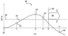
도 6a, 6b 및 6c는 본 개시에 따른 압박성 궤양이 발병하는 단일 위치에서 단일 환자에 대한 델타 값의 시간 경과에 따른 예시적인 진행을 도시한다.
도 6d는 본 개시에 따른 압박성 궤양이 발명되는 단일 위치에서 단일 환자에 대한 시간 경과에 따른 델타 값 변화의 예시적인 도면이다.
도 7a 및 7b는 본 개시에 따른 조직 손상 영역을 맵핑하는 방법들의 예들이다.
도 8a는 위험도 평가 및 육안 평가의 조합을 사용하여 병원 환자들의 압박성 궤양을 예방하기 위해 현재 권장되는 치료 결정 경로의 예이다.
도 8b는 일부 헬스 케어 시설들에서 현재 구현된 바와 같이 압박성 궤양을 예방하기 위한 현재 증강된 치료 결정 경로의 예이다.
도 9는 본 개시에 따른, 압박성 궤양을 예방하기 위해 SEM 스캐너가 독립형 프로세스에서 사용될 수 있는 방법에 대한 예시적인 흐름도이다.
도 10은 본 개시에 따른, 도 8b의 증강된 치료 결정 경로를 추가 개선하기 위해 SEM 스캐너가 보조물로서 사용될 있는 방법에 대한 예시적인 흐름도이다.
도 11은 본 개시에 따른, 복수의 케어 환경들에 걸쳐 케어의 연속성을 제공하는 개념을 예시한다.The patent or application file contains at least one drawing executed in color. Copies of this patent or patent application publication, along with color drawing(s), will be provided upon request and payment of the required fee by the Secretariat.
Aspects of the present disclosure are described herein by way of example only with reference to the accompanying drawings. Referring now in detail to the drawings, it is emphasized that the details shown are illustrative and are for illustrative discussion of aspects of the present disclosure. In this respect, the description and drawings, considered alone and together, make it clear to a person skilled in the art how aspects of the present disclosure may be practiced.
1 shows an example of an overall process for selecting a compressive ulcer treatment based on SEM values from entry to a care facility to discharge from a care facility, according to the present disclosure.
2A is a visual evaluation of a sample of healthy tissue according to the present disclosure.
FIG. 2B is a diagram showing an average value of SEM measurements obtained at each location in and around a healthy sacrum according to the present disclosure.
3A is a visual evaluation of a sample of damaged tissue according to the present disclosure.
3B is a diagram showing an average value of SEM measurements obtained at each location in a damaged sacrum and its periphery according to the present disclosure.
4 is an illustration of a process for selecting and monitoring an intervention level based on the amount by which the delta value derived from SEM measurements according to the present disclosure exceeds a threshold value.
5 is an example of a workflow guidance matrix for selecting a new intervention level using a current intervention level and a new delta value according to the present disclosure.
6A, 6B and 6C illustrate exemplary progression over time of delta values for a single patient at a single location where a compressive ulcer according to the present disclosure develops.
6D is an exemplary diagram of the change in delta value over time for a single patient at a single location where a compressive ulcer according to the present disclosure is invented.
7A and 7B are examples of methods of mapping a tissue damage area according to the present disclosure.
8A is an example of a currently recommended treatment decision pathway for preventing pressure ulcers in hospital patients using a combination of risk assessment and visual assessment.
8B is an example of a currently augmented treatment decision pathway to prevent compressive ulcers as currently implemented in some healthcare facilities.
9 is an exemplary flow diagram of how an SEM scanner can be used in a standalone process to prevent compressive ulcers according to the present disclosure.
10 is an exemplary flow diagram of how an SEM scanner may be used as an aid to further refine the augmented treatment decision pathway of FIG. 8B, in accordance with the present disclosure.
11 illustrates a concept of providing continuity of care across a plurality of care environments, according to the present disclosure.
이 설명은 본 개시가 구현될 수 있는 모든 상이한 방식들 또는 본 개시에 추가될 수 있는 모든 특징들에 대한 상세한 카탈로그인 것으로 의도되지 않는다. 예를 들어, 한 실시예에 대해 예시된 특징들은 다른 실시예들에 통합될 수 있으며, 특정 실시예에 대해 예시된 특징들은 해당 실시예로부터 삭제될 수 있다. 따라서, 본 개시는 본 개시의 일부 실시예들에서, 본원에 제시된 임의의 특징 또는 특징들의 조합이 배제되거나 생략될 수 있음을 고려한다. 추가로, 본원에 제시된 다양한 실시예들에 대한 많은 변형들 및 추가들은 본 개시에 비추어 당업자에게 명백할 것이며, 이는 본 개시를 벗어나지 않는다. 다른 경우, 발명을 불필요하게 모호하게 하지 않기 위해 주지의 구조들, 인터페이스들 및 프로세스들은 상세히 도시되지 않았다. 본 명세서의 일부는 본 개시의 전체 범위의 임의의 부분에 대한 부인(disavowal)을 초래하는 것으로 해석되지 않도록 의도된다. 따라서, 다음의 설명들은 본 개시의 일부 특정 실시예를 예시하기 위한 것이며, 그의 모든 순열, 조합 및 변형을 철저하게 명시하지는 않는다.This description is not intended to be a detailed catalog of all different ways in which the present disclosure may be implemented or all features that may be added to the present disclosure. For example, features illustrated for one embodiment may be incorporated into other embodiments, and features illustrated for a specific embodiment may be deleted from the corresponding embodiment. Accordingly, the present disclosure contemplates that in some embodiments of the disclosure, any feature or combination of features presented herein may be excluded or omitted. Additionally, many modifications and additions to the various embodiments presented herein will be apparent to those skilled in the art in light of this disclosure, without departing from this disclosure. In other instances, well-known structures, interfaces and processes have not been shown in detail in order not to unnecessarily obscure the invention. Portions of this specification are not intended to be construed as causing a disavowal of any part of the entire scope of this disclosure. Accordingly, the following descriptions are intended to illustrate some specific embodiments of the present disclosure, and not all permutations, combinations, and variations thereof are exhaustively specified.
달리 정의되지 않는 한, 본원에 사용된 모든 기술적 및 과학적 용어들은 본 개시가 속하는 기술 분야의 당업자에 의해 일반적으로 이해되는 것과 동일한 의미를 갖는다. 여기서 본 개시의 설명에 사용된 용어는 단지 특정한 양태 또는 실시예들을 설명하기 위한 것이며 본 개시를 제한하려는 것이 아니다.Unless otherwise defined, all technical and scientific terms used herein have the same meaning as commonly understood by one of ordinary skill in the art to which this disclosure belongs. Herein, terms used in the description of the present disclosure are only for describing specific aspects or embodiments and are not intended to limit the present disclosure.
본원에 인용된 모든 간행물들, 특허 출원들, 특허들 및 기타 참고문헌들은 참고 문헌이 제시하는 문장 및/또는 단락에 관련된 교시를 위해 그들 전체가 참조로서 통합된다. 본원에 사용된 기술들에 대한 인용들은 이러한 기술들에 대한 변형 또는 당업자에게 명백할 것인 동등한 기술들의 대체를 포함하여, 본 기술분야에서 통상적으로 기술들을 말하는 것으로 의도된다.All publications, patent applications, patents, and other references cited herein are incorporated by reference in their entirety for the teachings relating to the sentences and/or paragraphs presented by the reference. Citations to techniques used herein are intended to refer to techniques conventionally in the art, including variations on such techniques or replacement of equivalent techniques as will be apparent to those skilled in the art.
미국 특허 출원 일련 번호 제14/827,375호("'375 출원")은 바이폴라 센서를 사용하여 표피하 캐패시턴스를 측정하기 위해 무선 주파수(RF) 에너지를 사용하는 장치를 개시하며, 여기서 표피하 캐패시턴스는 환자 피부의 타겟 영역의 수분 함량에 대응된다. '375 출원은 또한 다양한 크기의 이러한 바이폴라 센서 어레이를 개시한다.U.S. Patent Application Serial No. 14/827,375 ("375 filed") discloses a device that uses radio frequency (RF) energy to measure subcutaneous capacitance using a bipolar sensor, wherein the subcutaneous capacitance is a patient It corresponds to the moisture content of the target area of the skin. The '375 application also discloses such an array of bipolar sensors in various sizes.
미국 특허 출원 일련 번호 제15/134,110호는 도 3에 도시된 장치와 유사한 표피하 수분(SEM)을 측정하는 장치를 개시하며, 여기서 상기 장치는 단일 동축 센서를 통해 32 kHz 주파수에서 RF 신호를 방출 및 수신하고 생체 임피던스 신호를 생성한 다음, 이 신호를 SEM 값으로 변환한다.U.S. Patent Application Serial No. 15/134,110 discloses a device for measuring subcutaneous moisture (SEM) similar to the device shown in Figure 3, wherein the device emits an RF signal at a frequency of 32 kHz via a single coaxial sensor. And receiving and generating a bioimpedance signal, then converting the signal to an SEM value.
미국 특허 출원 일련 번호 제14/827,375호 및 제15/134,110호 둘 다 그들 전체가 본원에 참조로서 통합된다. 그러나, 이 출원의 SEM 값들은 당업자에게 명백할 것인 임의의 유사하거나 동일한 장치들 또는 기술들에 의해 측정될 수 있다. 예를 들어, 이 출원의 SEM 값들을 측정하는 장치는 유선 장치, 무선 장치 또는 서로 통신하는 다양한 구성요소들을 포함하는 시스템일 수 있다.Both U.S. Patent Application Serial Nos. 14/827,375 and 15/134,110 are incorporated herein by reference in their entirety. However, the SEM values of this application can be measured by any similar or identical devices or techniques that will be apparent to those skilled in the art. For example, the device for measuring SEM values of this application may be a wired device, a wireless device, or a system including various components in communication with each other.
문맥이 달리 나타내지 않는 한, 구체적으로 본원에 설명된 개시의 다양한 특징들은 임의의 조합으로 사용될 수 있는 것으로 의도된다. 게다가, 본 개시는 또는 본 개시의 일부 실시예들에서, 본원에 제시된 임의의 특징들 또는 특징들의 조합이 제외되거나 생략될 수 있음을 고려한다.Unless the context indicates otherwise, it is intended that the various features of the disclosure specifically described herein can be used in any combination. In addition, the present disclosure contemplates that, or in some embodiments of the disclosure, any feature or combination of features presented herein may be excluded or omitted.
본원에 개시된 방법들은 설명된 방법을 달성하기 위한 하나 이상의 단계들 또는 동작들을 포함하고 이들로 구성된다. 방법 단계들 및/또는 동작들은 본 개시의 범위를 벗어나지 않고 상호 교환될 수 있다. 다시 말해서, 실시예의 적절한 동작을 위해 특정 순서의 단계들 또는 동작들이 요구되지 않는 한, 특정 단계들 및/또는 동작들의 순서 및/또는 사용은 본 개시의 범위를 벗어나지 않고 수정될 수 있다.The methods disclosed herein comprise and consist of one or more steps or actions for achieving the described method. Method steps and/or actions may be interchanged without departing from the scope of this disclosure. In other words, the order and/or use of specific steps and/or actions may be modified without departing from the scope of the present disclosure, unless a specific sequence of steps or actions is required for proper operation of the embodiment.
본 개시의 상세한 설명 및 첨부된 청구항들에 사용된 바와 같이, 단수 형태 "a", "an" 및 "the"는 문맥이 명백하게 달리 나타내지 않는 한, 복수 형태도 포함하는 것으로 의도된다.As used in the detailed description of the present disclosure and in the appended claims, the singular forms "a", "an" and "the" are intended to include the plural form as well, unless the context clearly indicates otherwise.
본원에 사용된 바와 같이, "및/또는"은 열거된 항목들 중 하나 이상의 임의의 및 모든 가능한 조합들, 뿐만 아니라 대안("또는")으로 해석될 때 조합들의 결여를 지칭하고 포괄한다.As used herein, “and/or” refers to and encompasses any and all possible combinations of one or more of the listed items, as well as the lack of combinations when interpreted as an alternative (“or”).
길이, 빈도 또는 SEM 값 등과 같은 측정 가능한 값을 언급할 때 본원에 사용된 바와 같은 "약(about)" 및 "대략(approximately)"이라는 용어들은 지정된 양의 ± 20%, ± 10%, ± 5%, ± 1%, ± 0.5%, 또는 심지어 ± 0.1%의 변동들을 포함하는 것을 의미한다.The terms “about” and “approximately” as used herein when referring to measurable values such as length, frequency or SEM values, etc., are ± 20%, ± 10%, ± 5 of the specified amount. It is meant to include fluctuations of %, ±1%, ±0.5%, or even ±0.1%.
본원에 사용된 바와 같이, "X와 Y 사이" 및 "대략 X와 Y 사이"와 같은 구문은 X 및 Y를 포함하는 것으로 해석되어야 한다. 본원에 사용된 바와 같이, "대략 X와 Y 사이"와 같은 구문은 "대략 X와 대략 Y 사이"를 의미하며, "대략 X 내지 Y"와 같은 구분은 "대략 X 내지 대략 Y"를 의미한다.As used herein, phrases such as "between X and Y" and "approximately between X and Y" are to be interpreted as including X and Y. As used herein, phrases such as "between approximately X and Y" mean "between approximately X and approximately Y" and distinctions such as "approximately X to Y" mean "approximately X to approximately Y" .
본원에 사용된 바와 같이, "표피하 수분(sub-epidermal moisture)" 또는 "SEM"이라는 용어는 조직, 세포자멸(apoptosis), 괴사(necrosis) 및 염증 과정에 지속적인 압박이 존재할 경우, 손상된 조직의 기저 구조를 변형시키는 혈관 누출 및 기타 변화들에 의해 발생된 조직액(tissue fluid) 및 국부 부종의 증가를 지칭한다.As used herein, the term "sub-epidermal moisture" or "SEM" refers to the presence of persistent pressure on the tissue, apoptosis, necrosis, and inflammatory processes. It refers to an increase in tissue fluid and local edema caused by vascular leakage and other changes that alter the underlying structure.
본원에 사용된 바와 같이, "환자"는 사람 또는 동물 대상일 수 있다.As used herein, a “patient” may be a human or animal subject.
본원에 사용된 바와 같이, "델타(delta)"는 두 SEM 값들 사이의 계산된 차이를 지칭한다.As used herein, “delta” refers to the calculated difference between two SEM values.
본원에 사용된 바와 같이, 변수 "K", "L", "M" 및 "N"은 음이 아닌 정수이다.As used herein, the variables “K”, “L”, “M” and “N” are non-negative integers.
도 1은 케어 시설로의 입원에서 케어 시설로부터의 퇴원까지, 본 개시에 따른 SEM 스캐너를 사용하여 수행된 SEM 측정들로부터 생성된 SEM 값들에 기초하여 압박성 궤양 치료를 선택하기 위한 전체 프로세스(100)를 도시한다. 일 양태에서, 케어 설비는 병원, 보조 생활 시설, 주거형 케어 시설, 요양원, 장기 케어 시설, 지속적인 케어 커뮤니티 및 독립적인 생활 커뮤니티로 구성된 그룹으로부터 선택된다. 일 양태에서, 케어 시설은 환자의 집 또는 다른 거주지일 수 있으며, 이때 "입원" 단계(102)는 간호사 또는 다른 간병인에 의해 그들의 집에서 환자에 대한 첫 번째 평가가 될 것이다. 일 양태에서, 가정 환경에서 사용된 개입 일정 및 평가 가격은 병원에서 사용된 해당 개입 및 간격과 다를 수 있다.1 is an
일 양태에서, 프로세스(100)에서, 새로 입원한 환자는 단계 104에서 환자 피부의 일부에 대한 육안 검사와, 영양, 이동성, 물리적 활동, 체력 및 의사 소통 능력 중 하나 이상을 평가하는 위험도 평가 프로토콜의 적어도 일부의 완료와, 환자 피부의 하나 이상의 위치에서 이루어지는 SEM 측정 중 하나 이상을 포함하는 인테이크 평가(intake evaluation)를 받는다. 일 양태에서, SEM 측정들은 환자 피부 상의 단일 "위치"에서 복수의 SEM 측정들을 수행하는 것을 포함할 수 있다. 일 양태에서, "위치"는 SEM 측정들이 그 치 내에서 공간적으로 분리된 지점들에서 이루어질 수 있도록 단일 지점이 아닌 영역으로 간주된다. 예를 들어, "힐(heel)" 위치는 힐 주위의 내측(medial), 측면(lateral) 및 후부(posterior) 표면들뿐만 아니라 발의 발바닥의 후부를 포함한다.In one aspect, in
일 양태에서, 평가 단계가 완료되면, 단계 106에서 환자가 "이탈"되었는지 여부, 즉 평가의 다양한 요소들에 대한 결과들의 조합이 환자가 압박성 궤양으로 이어질 조직 손상을 갖거나 발병 위험이 있음을 나타내는지에 대한 결정이 이루어진다. 평가의 각 요소는 위험 레벨에 대한 개별 기준, 예를 들어 허용될 수 없는 위험을 나타내는 임계값을 갖는 채점 시스템을 가질 수 있다. 일 양태에서, 개입 레벨을 선택하는 데 사용될 수 있는 복합 파라미터를 생성하기 위해 기준을 결합하는 프로토콜이 있다.In one aspect, upon completion of the evaluation phase, whether the patient has "departed" in
일 양태에서, 환자가 수용할 수 있는 위험 레벨에 있는 것으로 결정되면, 프로세스는 "레벨-제로" 또는 "레벨-0"로 본원에 지정된 최저 개입 레벨을 구현하는 단계 108로 분기한다. 단계 110 및 112을 통해 진행하면, 환자는 단계 114에서 적어도 SEM 측정 프로토콜을 사용하여 레벨-0과 연관된 빈도로 재평가되거나, 또는 반대로 레벨-0과 관련된 시간 간격으로 재평가될 것이다. 프로세스(100)는 그런 다음 단계 114에서 이루어진 SEM 측정들의 결과를 평가하기 위해 단계 106으로 되돌아간다.In one aspect, if it is determined that the patient is at an acceptable risk level, the process branches to step 108 implementing the lowest level of intervention designated herein as “level-zero” or “level-0”. Proceeding through
일 양태에서, 환자가 단계 106에서 이탈된 것으로 판단되면, 프로세스는 더 높은 레벨의 개입을 구현하는 단계 122로 분기한다. 일 양태에서, 개입 레벨들에 대한 정의된 계층이 있으며, 각각의 레벨은 다음 하위 레벨보다 더 효과적인 개입을 구현한다. 일 양태에서, 각각의 레벨은 또한 얼마나 자주 일련의 SEM 측정들이 이루어져야 하는지를 나타내는 정의된 모니터링 간격 또는 빈도를 가지며, 여기서 더 높은 레벨들은 일반적으로 더 짧은 간격들을 가질 것이다. 이 예에서, 프로세스는 이 시점에서 레벨-1 개입으로 1 레벨 상승하도록 병원 또는 다른 관리 기관에 의해 정의된다. 다른 양태에서, 단계 122는 레벨-2 이상의 레벨의 개입을 구현할 수 있다. 프로세스는 이제 단계 130에서 시작하여 새로운 루프에 진입하며, 여기서 환자는 이제 레벨-N 빈도로 모니터링될 것이며, 여기서 N은 1 내지 n의 범위에 있으며, n은 가장 높은 정의된 레벨의 개입 및 모니터링이다.In one aspect, if it is determined that the patient has departed from
일 양태에서, 단계 134에서, 환자의 이력은 그의 상태가 개선되고 있는지를 결정하기 위해 평가된다. 예를 들어, 델타 값 감소로 입증되는 바와 같이 환자의 상태가 개선되고 있다면, 프로세스는 단계 142로 분기한다. 이 예에서, 단계 142는 현재 레벨의 개입을 계속 구현하고 단계는 델타 값이 임계치 미만으로 떨어질 때까지 프로세스는 단계 140을 통해 단계 130-132-134-142-140으로 순환한다. 일 양태에서, 개입 레벨은 단계 142에서 델타 값이 아래로 향하는 추이에 따른 델타 값의 크기에 기초하여 감소될 수 있다.In one aspect, at
일 양태에서, 환자가 단계 134에서 개선을 보이지 않는다면, 프로세스는 단계 136에서 피부가 파괴되지 않았음, 즉 드러난 궤양(open ulcer)이 발병되지 않았음을 제공받는 단계 138에서 개입 레벨의 증가로 분기한다. 드러난 궤양이 발병했다면, SEM 스캐닝이 이제 단계 144에서 드러난 상처의 주변부 주위에서 수행될 것이다 궤양 자체는 단계 148에서 치료되며, 이 2차 루프(144-146-148-150)는 상처가 아물 때까지 계속되고, 그 결과 프로세스는 단계 130로 돌아간다.In one embodiment, if the patient does not show improvement at
일 양태에서, 프로세스(100)에서 언제든지, 환자의 퇴원은 단계 118로 분기하며, 여기서 퇴원 또는 이송 시 환자의 상태가 문서화된다. 일 양태에서, 단계 118은 환자 신체에 대한 하나 이상의 위치들에서 최종 SEM 측정 세트를 포함한다. 일 양태에서, 이러한 위치들은 개입을 받지 않았으며 이전에 위험한 것으로 식별되지 않은 영역들을 포함한다. 일 양태에서, 이 정보는 받는 간병인에게 제공된다. 환자는 그런 다음 단계 120에서 퇴원 또는 이송된다.In one aspect, at any time in
일 양태에서, 본 개시는 압박성 궤양 치료가 필요한 환자를 식별하고 치료하는 방법을 제공 및 포함하며, 상기 방법은, 케어 시설에 입원 시 환자에게 압박성 궤양의 위험에 대해 환자를 평가하는 단계를 포함하며, 상기 평가하는 단계는 환자에서 제1 복수의 표피하 수분(SEM) 측정들을 수행하는 단계, 제1 복수의 SEM 측정들 중 일부로부터 제1 델타 값을 계산하는 단계, 제1 델타 값이 제1 임계치를 초과하는지를 결정하는 단계, 제1 델타 값이 제1 임계치를 초과하지 않으면 레벨00의 제1 개입을 실시하는 단계, 및 제1 델타 값이 제1 임계치를 초과하면 레벨-N의 제1 개입을 실시하는 단계로서, N은 정수이고 N은 1 이상의 값을 갖는, 상기 레벨-N의 제1 개입을 실시하는 단계를 포함한다.In one aspect, the present disclosure provides and includes a method of identifying and treating a patient in need of compression ulcer treatment, the method comprising evaluating the patient for a risk of compression ulcer upon admission to a care facility. Including, wherein the evaluating includes performing a first plurality of subepidermal moisture (SEM) measurements in the patient, calculating a first delta value from some of the first plurality of SEM measurements, and the first delta value is Determining whether a first threshold is exceeded, performing a first intervention of level 00 if the first delta value does not exceed the first threshold, and performing a first intervention of level 00 if the first delta value exceeds the first threshold. Performing one intervention, wherein N is an integer and N has a value equal to or greater than 1, comprising performing the first intervention of the level-N.
[0061] 일 양태에서, 제1 복수의 SEM 측정들은 흉골, 천골, 힐(heel), 견갑골, 팔꿈치, 귀 및 환자의 기타 살이 있는 조직으로 이루어진 그룹으로부터 선택된 하나 이상의 해부학적 부위들에서 및 그 주위에서 수행된다. 일 양태에서, 제1 복수의 SEM 측정들은 측정이 수행되는 일반적인 위치에 기초하여 분석하기 위해 서브 그룹들로 분리된다. 일 양태에서, 제1 복수의 SEM 측정들은 해부학적 부위를 중심으로 한 하나 이상의 동심원들에 위치된 위치들에서 수행된다. 일 양태에서, 제1 복수의 SEM 측정들은 해부학적 부위로부터 대략 등거리로 직선 상에 위치된 위치들에서 수행된다.In one aspect, the first plurality of SEM measurements are at and around one or more anatomical sites selected from the group consisting of the sternum, sacrum, heel, scapula, elbow, ear, and other fleshy tissues of the patient. Is carried out in In one aspect, the first plurality of SEM measurements are separated into subgroups for analysis based on the general location at which the measurement is being performed. In one aspect, the first plurality of SEM measurements are performed at locations located in one or more concentric circles about the anatomical site. In one aspect, the first plurality of SEM measurements are performed at locations located on a straight line approximately equidistant from the anatomical site.
[0062] 일 양태에서, 제2 델타 값은 수집된 제2 복수의 SEM 측정들로부터 최대 SEM 값과 최소 SEM 값 사이의 차이로 결정된다. 일 양태에서, 제1 델타 값은 한 위치에서 수행된 최대 SEM 측정 평균과 제2 위치에서 수행된 최소 SEM 측정 평균 사이의 차이로 결정된다. 일 양태에서, 제1 델타 값은 수행된 위치에 의해 정의된 바와 같은 서브 그룹으로 이루어진 제1 복수의 SEM 측정들의 일부에 대해 결정된다. 일 양태에서, 한 위치에서의 평균 SEM 값은 해당 위치에서 측정된 2개, 3개, 4개, 5개, 6개, 7개, 8개, 9개, 10개 또는 10개 이상의 SEM 값들로부터 획득된다. 일 양태에서, 제1 델타 값은 중심선에 대한 두 개의 비대칭 위치들에서 측정된 측정들로부터 도출된 SEM 값들 사이의 차이로 결정된다.[0062] In an aspect, the second delta value is determined as the difference between the maximum and minimum SEM values from the collected second plurality of SEM measurements. In one aspect, the first delta value is determined as the difference between the average of the maximum SEM measurements performed at one location and the average of the minimum SEM measurements performed at the second location. In one aspect, a first delta value is determined for a portion of the first plurality of SEM measurements made up of a subgroup as defined by the location performed. In one aspect, the average SEM value at one location is from 2, 3, 4, 5, 6, 7, 8, 9, 10 or 10 or more SEM values measured at that location. Is obtained. In one aspect, the first delta value is determined as the difference between SEM values derived from measurements measured at two asymmetrical positions with respect to the center line.
일 양태에서, 델타 값은 복수의 방법들로, 특정 위치에서 또는 특정 위치 부근에 근접하여 이루어진 복수의 SEM 측정들로부터 계산될 수 있다. 일 양태에서, 복수의 SEM 측정들은 피부상의 미리 결정된 패턴으로 이루어지며, 델타 값은 패턴 내의 다른 위치들에서 이루어진 최대 SEM 값으로부터 패턴 내의 미리 결정된 위치와 관련된 SEM 값을 감산함으로써 계산된다. 일 양태에서, 복수의 SEM 측정들은 피부상의 미리 결정된 패턴으로 이루어지며, 델타 값은 패턴 내의 미리 결정된 위치와 관련된 SEM 값을 식별하고 패턴 내의 다른 위치들에서 이루어진 최대 SEM 값을 감산함으로써 계산된다. 일 양태에서, 평균 SEM 값은 단일 위치에서 복수의 SEM 측정들에 의해 생성된 SEM 값 세트의 일부 및 동일한 세트의 평균 SEM 값과 단일 SEM 값 사이의 최대 차이로서 계산된 델타 값으로부터 계산될 수 있다. 일 양태에서, 델타 값은 SEM 값 세트 내에서 가장 큰 SEM 값 대 가장 작은 SEM 값의 비로 계산될 수 있다.In one aspect, the delta value may be calculated from a plurality of SEM measurements made in a number of ways, at or in proximity to a specific location. In one aspect, the plurality of SEM measurements are made of a predetermined pattern on the skin, and the delta value is calculated by subtracting the SEM value associated with the predetermined location in the pattern from the maximum SEM value made at other locations in the pattern. In one aspect, the plurality of SEM measurements are made with a predetermined pattern on the skin, and the delta value is calculated by identifying the SEM value associated with the predetermined location in the pattern and subtracting the maximum SEM value made at other locations in the pattern. In one aspect, the average SEM value may be calculated from a portion of a set of SEM values generated by multiple SEM measurements at a single location and a delta value calculated as the maximum difference between the average SEM value and a single SEM value of the same set. . In one aspect, the delta value can be calculated as the ratio of the largest SEM value to the smallest SEM value within the set of SEM values.
[0064]일 양태에서, 제1 임계치는 약 0.3, 0.35, 0.4, 0.45, 0.5, 0.55, 0.6, 0.65, 0.7, 0.75, 0.8, 0.85, 0.9, 0.95, 1.0, 1.1, 1.2, 1.3, 1.4, 1.5, 1.6, 1.7, 1.8, 1.9, 2.0, 2.1, 2.2, 2.3, 2.4, 2.5, 2.6, 2.7, 2.8, 2.9, 3.0, 3.1, 3.2, 3.3, 3.4, 3.5, 3.6, 3.7, 3.8, 3.9, 4.0, 4.1, 4.2, 4.3, 4.4, 4.5, 4.6, 4.7, 4.8, 4.9, 5.0, 5.1, 5.2, 5.3, 5.4, 5.5, 5.6, 5.7, 5.8, 5.9, 6.0, 6.1, 6.2, 6.3, 6.4, 6.5, 6.6, 6.7, 6.8, 6.9, 7.0, 7.1, 7.2, 7.3, 7.4, 또는 7.5일 수 있다. 일 양태에서, 제1 임계치는 0.1 내지 8.0의 범위일 수 있는데, 예컨대 0.1 내지 1.0, 1.1 내지 2.0, 2.1 내지 3.0, 3.1 내지 4.0, 4.1 내지 5.0, 5.1 내지 6.0, 6.1 내지 7.0, 7.1 내지 8.0, 0.1 내지 7.5, 0.5 내지 8.0, 1.0 내지 7.0, 1.5 내지 6.5, 2.0 내지 6.0, 3.0 내지 5.5, 3.5 내지 5.0, 또는 4.0 내지 4.5의 범위일 수 있다. 일 양태에서, 제1 임계치는 본원에 제공된 값들에 기초하여 인수 또는 배수로 스케일링될 수 있다. 임계치는 설계에 의해 제한되지 않으며, 오히려 당업자는 주어진 SEM 단위에 기초하여 미리 결정된 값을 선택할 수 있다는 것이 이해될 것이다. 일 양태에서, 본 개시의 임계치들은 측정들이 이루어지는 환자 신체의 특정 부분, 또는 연령, 신장, 체중, 가족력, 인종 그룹, 및 기타 신체적 특성들 또는 의료 상태들과 같은 환자의 하나 이상의 특성들에 따라 달라진다.[0064] In one aspect, the first threshold is about 0.3, 0.35, 0.4, 0.45, 0.5, 0.55, 0.6, 0.65, 0.7, 0.75, 0.8, 0.85, 0.9, 0.95, 1.0, 1.1, 1.2, 1.3, 1.4, 1.5, 1.6, 1.7, 1.8, 1.9, 2.0, 2.1, 2.2, 2.3, 2.4, 2.5, 2.6, 2.7, 2.8, 2.9, 3.0, 3.1, 3.2, 3.3, 3.4, 3.5, 3.6, 3.7, 3.8, 3.9, 4.0, 4.1, 4.2, 4.3, 4.4, 4.5, 4.6, 4.7, 4.8, 4.9, 5.0, 5.1, 5.2, 5.3, 5.4, 5.5, 5.6, 5.7, 5.8, 5.9, 6.0, 6.1, 6.2, 6.3, 6.4, It can be 6.5, 6.6, 6.7, 6.8, 6.9, 7.0, 7.1, 7.2, 7.3, 7.4, or 7.5. In one aspect, the first threshold may be in the range of 0.1 to 8.0, such as 0.1 to 1.0, 1.1 to 2.0, 2.1 to 3.0, 3.1 to 4.0, 4.1 to 5.0, 5.1 to 6.0, 6.1 to 7.0, 7.1 to 8.0, 0.1 to 7.5, 0.5 to 8.0, 1.0 to 7.0, 1.5 to 6.5, 2.0 to 6.0, 3.0 to 5.5, 3.5 to 5.0, or 4.0 to 4.5. In an aspect, the first threshold may be scaled by a factor or multiple based on values provided herein. It will be appreciated that the threshold is not limited by design, but rather a person skilled in the art may select a predetermined value based on a given SEM unit. In one aspect, the thresholds of the present disclosure depend on the specific part of the patient's body for which measurements are made, or one or more characteristics of the patient, such as age, height, weight, family history, ethnic group, and other physical characteristics or medical conditions. .
일 양태에서, N은 1 내지 50 범위에 있는데, 예컨대 1 내지 2, 1 내지 3, 1 내지 4, 1 내지 5, 1 내지 6, 1 내지 7, 1 내지 8, 1 내지 9, 1 내지 10, 1 내지 15, 1 내지 20, 1 내지 25, 1 내지 30, 1 내지 35, 1 내지 40, 또는 1 내지 45의 범위에 있다.In one aspect, N is in the range of 1 to 50, such as 1 to 2, 1 to 3, 1 to 4, 1 to 5, 1 to 6, 1 to 7, 1 to 8, 1 to 9, 1 to 10, 1 to 15, 1 to 20, 1 to 25, 1 to 30, 1 to 35, 1 to 40, or 1 to 45.
일 양태에서, N은 제1 델타 값이 제1 임계치를 초과하는 양에 의해 결정된다. 일 양태에서, 델타 값이 (N+1)에 대해 설정된 임계치를 초과하는 양이 델타 값이 N에 대해 설정된 임계치를 초과하는 양보다 크다. 일 양태에서, 델타 값이 (N+1)에 대해 설정된 임계치를 초과하는 양이 델타 값이 N에 대해 설정된 임계치를 초과하는 양보다 적다.In one aspect, N is determined by the amount by which the first delta value exceeds the first threshold. In one aspect, the amount by which the delta value exceeds the threshold set for (N+1) is greater than the amount by which the delta value exceeds the threshold set for N. In one aspect, the amount by which the delta value exceeds the threshold set for (N+1) is less than the amount by which the delta value exceeds the threshold set for N.
[0067] 일 양태에서, 레벨-1(N=1) 개입은 임계값의 100% 이하로, 예컨대 임계값의 95% 이하, 90% 이하, 85% 이하, 80% 이하, 75% 이하, 70% 이하, 65% 이하, 60% 이하, 55% 이하, 50% 이하, 45% 이하, 40% 이하, 35% 이하, 30% 이하, 25% 이하로 20% 이하로 15% 이하, 10% 이하, 또는 5% 이하로 임계치를 초과하는 델타 값을 갖는 환자에게 적용된다.[0067] In one aspect, the level-1 (N=1) intervention is 100% or less of the threshold, such as 95% or less, 90% or less, 85% or less, 80% or less, 75% or less, 70 % Or less, 65% or less, 60% or less, 55% or less, 50% or less, 45% or less, 40% or less, 35% or less, 30% or less, 25% or less, 20% or less, 15% or less, 10% or less , Or to patients with a delta value exceeding the threshold by 5% or less.
[0068] 일 양태에서, 레벨-2(N=2) 개입은 임계값의 150% 이하로, 예컨대, 임계값의 145% 이하, 140% 이하, 135% 이하, 130% 이하, 125% 이하, 120% 이하, 115% 이하, 110% 이하, 100% 이하, 95% 이하, 90% 이하, 85% 이하, 80% 이하, 75% 이하, 70% 이하, 65% 이하, 60% 이하, 55% 이하, 50% 이하, 45% 이하, 40% 이하, 35% 이하, 30% 이하, 25% 이하, 20% 이하, 15% 이하, 10% 이하, 또는 5% 이하로 임계치를 초과하는 델타 값을 갖는 환자에게 적용된다.[0068] In one aspect, the level-2 (N=2) intervention is 150% or less of the threshold, for example, 145% or less, 140% or less, 135% or less, 130% or less, 125% or less of the threshold, 120% or less, 115% or less, 110% or less, 100% or less, 95% or less, 90% or less, 85% or less, 80% or less, 75% or less, 70% or less, 65% or less, 60% or less, 55% Delta values that exceed the threshold by less than, 50% or less, 45% or less, 40% or less, 35% or less, 30% or less, 25% or less, 20% or less, 15% or less, 10% or less, or 5% or less. Applies to patients having.
[0069] 일 양태에서, 레벨-3(N=3) 개입은 임계값의 200% 이하로, 예컨대, 임계값의 195% 이하, 190% 이하, 185% 이하, 180% 이하, 175% 이하, 170% 이하, 165% 이하, 160% 이하, 155% 이하, 150% 이하, 145% 이하, 140% 이하, 135% 이하, 130% 이하, 125% 이하, 120% 이하, 115% 이하, 110% 이하, 100% 이하, 95% 이하, 90% 이하, 85% 이하, 80% 이하, 75% 이하, 70% 이하, 65% 이하, 60% 이하, 55% 이하, 50% 이하, 45% 이하, 40% 이하, 35% 이하, 30% 이하, 25% 이하, 20% 이하, 15% 이하, 10% 이하 또는 5% 이하로 임계치를 초과하는 델타 값을 갖는 환자에게 적용된다.[0069] In one aspect, the level-3 (N=3) intervention is 200% or less of the threshold, for example, 195% or less, 190% or less, 185% or less, 180% or less, 175% or less of the threshold, 170% or less, 165% or less, 160% or less, 155% or less, 150% or less, 145% or less, 140% or less, 135% or less, 130% or less, 125% or less, 120% or less, 115% or less, 110% Or less, 100% or less, 95% or less, 90% or less, 85% or less, 80% or less, 75% or less, 70% or less, 65% or less, 60% or less, 55% or less, 50% or less, 45% or less, Applies to patients with a delta value that exceeds the threshold by 40% or less, 35% or less, 30% or less, 25% or less, 20% or less, 15% or less, 10% or less, or 5% or less.
[0070] 일 양태에서, 레벨-4(N=4) 개입은 임계값의 250% 이하로, 예컨대, 임계값의 245% 이하, 240% 이하, 235% 이하, 230% 이하, 225% 이하, 220% 이하, 215% 이하, 210% 이하, 205% 이하, 200% 이하, 195% 이하, 190% 이하, 185% 이하, 180% 이하, 175% 이하, 170% 이하, 165% 이하, 160% 이하, 155% 이하, 150% 이하, 145% 이하, 140% 이하, 135% 이하, 130% 이하, 125% 이하, 120% 이하, 115% 이하, 110% 이하, 100% 이하, 95% 이하, 90% 이하, 85% 이하, 80% 이하, 75% 이하, 70% 이하, 65% 이하, 60% 이하, 55% 이하, 50% 이하, 45% 이하, 40% 이하, 35% 이하, 30% 이하, 25% 이하, 20% 이하, 15% 이하, 10% 이하 또는 5% 이하로 임계치를 초과하는 델타 값을 갖는 환자에게 적용된다.In one aspect, level-4 (N=4) intervention is less than or equal to 250% of the threshold, for example, less than 245%, less than 240%, less than 235%, less than 230%, less than 225% of the threshold, 220% or less, 215% or less, 210% or less, 205% or less, 200% or less, 195% or less, 190% or less, 185% or less, 180% or less, 175% or less, 170% or less, 165% or less, 160% Or less, 155% or less, 150% or less, 145% or less, 140% or less, 135% or less, 130% or less, 125% or less, 120% or less, 115% or less, 110% or less, 100% or less, 95% or less, 90% or less, 85% or less, 80% or less, 75% or less, 70% or less, 65% or less, 60% or less, 55% or less, 50% or less, 45% or less, 40% or less, 35% or less, 30% It applies to patients with a delta value that exceeds the threshold by less than or equal to 25%, less than 20%, less than 15%, less than 10%, or less than 5%.
[0071] 일 양태에서, 레벨-5(N=5) 개입은 임계값의 300% 이하로, 예컨대, 임계값의 295% 이하, 290% 이하, 285% 이하, 280% 이하, 275% 이하, 270% 이하, 265% 이하, 260% 이하, 255% 이하, 250% 이하, 245% 이하, 240% 이하, 235% 이하, 230% 이하, 225% 이하, 220% 이하, 215% 이하, 210% 이하, 205% 이하, 200% 이하, 195% 이하, 190% 이하, 185% 이하, 180% 이하, 175% 이하, 170% 이하, 165% 이하, 160% 이하, 155% 이하, 150% 이하, 145% 이하, 140% 이하, 135% 이하, 130% 이하, 125% 이하, 120% 이하, 115% 이하, 110% 이하, 100% 이하, 95% 이하, 90% 이하, 85% 이하, 80% 이하, 75% 이하, 70% 이하, 65% 이하, 60% 이하, 55% 이하, 50% 이하, 45% 이하, 40% 이하, 35% 이하, 30% 이하, 25% 이하, 20% 이하, 15% 이하, 10% 이하 또는 5% 이하로 임계치를 초과하는 델타 값을 갖는 환자에게 적용된다.[0071] In one aspect, the level-5 (N=5) intervention is less than or equal to 300% of the threshold, eg, less than or equal to 295%, less than or equal to 290%, less than or equal to 285%, less than or equal to 280%, less than or equal to 275%, 270% or less, 265% or less, 260% or less, 255% or less, 250% or less, 245% or less, 240% or less, 235% or less, 230% or less, 225% or less, 220% or less, 215% or less, 210% Or less, 205% or less, 200% or less, 195% or less, 190% or less, 185% or less, 180% or less, 175% or less, 170% or less, 165% or less, 160% or less, 155% or less, 150% or less, 145% or less, 140% or less, 135% or less, 130% or less, 125% or less, 120% or less, 115% or less, 110% or less, 100% or less, 95% or less, 90% or less, 85% or less, 80% Or less, 75% or less, 70% or less, 65% or less, 60% or less, 55% or less, 50% or less, 45% or less, 40% or less, 35% or less, 30% or less, 25% or less, 20% or less, Applies to patients with delta values that exceed the threshold by 15% or less, 10% or less, or 5% or less.
[0072] 일 양태에서, 레벨-6(N=6) 개입은 임계값의 350% 이하로, 예컨대, 임계값의 345% 이하, 340% 이하, 335% 이하, 330% 이하, 325% 이하, 320% 이하, 315% 이하, 310% 이하, 305% 이하, 300% 이하, 295% 이하, 290% 이하, 285% 이하, 280% 이하, 275% 이하, 270% 이하, 265% 이하, 260% 이하, 255% 이하, 250% 이하, 245% 이하, 240% 이하, 235% 이하, 230% 이하, 225% 이하, 220% 이하, 215% 이하, 210% 이하, 205% 이하, 200% 이하, 195% 이하, 190% 이하, 185% 이하, 180% 이하, 175% 이하, 170% 이하, 165% 이하, 160% 이하, 155% 이하, 150% 이하, 145% 이하, 140% 이하, 135% 이하, 130% 이하, 125% 이하, 120% 이하, 115% 이하, 110% 이하, 100% 이하, 95% 이하, 90% 이하, 85% 이하, 80% 이하, 75% 이하, 70% 이하, 65% 이하, 60% 이하, 55% 이하, 50% 이하, 45% 이하, 40% 이하, 35% 이하, 30% 이하, 25% 이하, 20% 이하, 15% 이하, 10% 이하 또는 5% 이하로 임계치를 초과하는 델타 값을 갖는 환자에게 적용된다.[0072] In one aspect, level-6 (N=6) intervention is less than or equal to 350% of the threshold, for example, less than or equal to 345%, less than or equal to 340%, less than or equal to 335%, less than or equal to 330%, less than or equal to 325%, 320% or less, 315% or less, 310% or less, 305% or less, 300% or less, 295% or less, 290% or less, 285% or less, 280% or less, 275% or less, 270% or less, 265% or less, 260% Or less, 255% or less, 250% or less, 245% or less, 240% or less, 235% or less, 230% or less, 225% or less, 220% or less, 215% or less, 210% or less, 205% or less, 200% or less, 195% or less, 190% or less, 185% or less, 180% or less, 175% or less, 170% or less, 165% or less, 160% or less, 155% or less, 150% or less, 145% or less, 140% or less, 135% Or less, 130% or less, 125% or less, 120% or less, 115% or less, 110% or less, 100% or less, 95% or less, 90% or less, 85% or less, 80% or less, 75% or less, 70% or less, 65% or less, 60% or less, 55% or less, 50% or less, 45% or less, 40% or less, 35% or less, 30% or less, 25% or less, 20% or less, 15% or less, 10% or less, or 5% It applies to patients with delta values that exceed the threshold below.
[0073] 일 양태에서, 레벨-7(N=7) 개입은 임계값의 400% 이하로, 예컨대, 임계값의 395% 이하, 390% 이하, 385% 이하, 380% 이하, 375% 이하, 370% 이하, 365% 이하, 360% 이하, 355% 이하, 350% 이하, 345% 이하, 340% 이하, 335% 이하, 330% 이하, 325% 이하, 320% 이하, 315% 이하, 310% 이하, 305% 이하, 300% 이하, 295% 이하, 290% 이하, 285% 이하, 280% 이하, 275% 이하, 270% 이하, 265% 이하, 260% 이하, 255% 이하, 250% 이하, 245% 이하, 240% 이하, 235% 이하, 230% 이하, 225% 이하, 220% 이하, 215% 이하, 210% 이하, 205% 이하, 200% 이하, 195% 이하, 190% 이하, 185% 이하, 180% 이하, 175% 이하, 170% 이하, 165% 이하, 160% 이하, 155% 이하, 150% 이하, 145% 이하, 140% 이하, 135% 이하, 130% 이하, 125% 이하, 120% 이하, 115% 이하, 110% 이하, 100% 이하, 95% 이하, 90% 이하, 85% 이하, 80% 이하, 75% 이하, 70% 이하, 65% 이하, 60% 이하, 55% 이하, 50% 이하, 45% 이하, 40% 이하, 35% 이하, 30% 이하, 25% 이하, 20% 이하, 15% 이하, 10% 이하 또는 5% 이하로 임계치를 초과하는 델타 값을 갖는 환자에게 적용된다.[0073] In one aspect, the level-7 (N=7) intervention is less than or equal to 400% of the threshold, such as less than or equal to 395%, less than or equal to 390%, less than or equal to 385%, less than or equal to 380%, less than or equal to 375%, 370% or less, 365% or less, 360% or less, 355% or less, 350% or less, 345% or less, 340% or less, 335% or less, 330% or less, 325% or less, 320% or less, 315% or less, 310% Or less, 305% or less, 300% or less, 295% or less, 290% or less, 285% or less, 280% or less, 275% or less, 270% or less, 265% or less, 260% or less, 255% or less, 250% or less, 245% or less, 240% or less, 235% or less, 230% or less, 225% or less, 220% or less, 215% or less, 210% or less, 205% or less, 200% or less, 195% or less, 190% or less, 185% Or less, 180% or less, 175% or less, 170% or less, 165% or less, 160% or less, 155% or less, 150% or less, 145% or less, 140% or less, 135% or less, 130% or less, 125% or less, 120% or less, 115% or less, 110% or less, 100% or less, 95% or less, 90% or less, 85% or less, 80% or less, 75% or less, 70% or less, 65% or less, 60% or less, 55% A delta value that exceeds the threshold by less than, 50% or less, 45% or less, 40% or less, 35% or less, 30% or less, 25% or less, 20% or less, 15% or less, 10% or less, or 5% or less. Applies to the patient.
[0074] 일 양태에서, 레벨-8(N=8) 개입은 임계값의 450% 이하로, 예컨대, 임계값의 445% 이하, 440% 이하, 435% 이하, 430% 이하, 425% 이하, 420% 이하, 415% 이하, 410% 이하, 405% 이하, 400% 이하, 395% 이하, 390% 이하, 385% 이하, 380% 이하, 375% 이하, 370% 이하, 365% 이하, 360% 이하, 355% 이하, 350% 이하, 345% 이하, 340% 이하, 335% 이하, 330% 이하, 325% 이하, 320% 이하, 315% 이하, 310% 이하, 305% 이하, 300% 이하, 295% 이하, 290% 이하, 285% 이하, 280% 이하, 275% 이하, 270% 이하, 265% 이하, 260% 이하, 255% 이하, 250% 이하, 245% 이하, 240% 이하, 235% 이하, 230% 이하, 225% 이하, 220% 이하, 215% 이하, 210% 이하, 205% 이하, 200% 이하, 195% 이하, 190% 이하, 185% 이하, 180% 이하, 175% 이하, 170% 이하, 165% 이하, 160% 이하, 155% 이하, 150% 이하, 145% 이하, 140% 이하, 135% 이하, 130% 이하, 125% 이하, 120% 이하, 115% 이하, 110% 이하, 100% 이하, 95% 이하, 90% 이하, 85% 이하, 80% 이하, 75% 이하, 70% 이하, 65% 이하, 60% 이하, 55% 이하, 50% 이하, 45% 이하, 40% 이하, 35% 이하, 30% 이하, 25% 이하, 20% 이하, 15% 이하, 10% 이하 또는 5% 이하로 임계치를 초과하는 델타 값을 갖는 환자에게 적용된다.[0074] In one aspect, the level-8 (N=8) intervention is 450% or less of the threshold, for example, 445% or less, 440% or less, 435% or less, 430% or less, 425% or less of the threshold, 420% or less, 415% or less, 410% or less, 405% or less, 400% or less, 395% or less, 390% or less, 385% or less, 380% or less, 375% or less, 370% or less, 365% or less, 360% Or less, 355% or less, 350% or less, 345% or less, 340% or less, 335% or less, 330% or less, 325% or less, 320% or less, 315% or less, 310% or less, 305% or less, 300% or less, 295% or less, 290% or less, 285% or less, 280% or less, 275% or less, 270% or less, 265% or less, 260% or less, 255% or less, 250% or less, 245% or less, 240% or less, 235% Or less, 230% or less, 225% or less, 220% or less, 215% or less, 210% or less, 205% or less, 200% or less, 195% or less, 190% or less, 185% or less, 180% or less, 175% or less, 170% or less, 165% or less, 160% or less, 155% or less, 150% or less, 145% or less, 140% or less, 135% or less, 130% or less, 125% or less, 120% or less, 115% or less, 110% Or less, 100% or less, 95% or less, 90% or less, 85% or less, 80% or less, 75% or less, 70% or less, 65% or less, 60% or less, 55% or less, 50% or less, 45% or less, Applies to patients with a delta value that exceeds the threshold by 40% or less, 35% or less, 30% or less, 25% or less, 20% or less, 15% or less, 10% or less, or 5% or less.
[0075] 일 양태에서, 레벨-9(N=9) 개입은 임계값의 500% 이하로, , 예컨대, 임계값의 495% 이하, 490% 이하, 485% 이하, 480% 이하, 475% 이하, 470% 이하, 465% 이하, 460% 이하, 455% 이하, 450% 이하, 445% 이하, 440% 이하, 435% 이하, 430% 이하, 425% 이하, 420% 이하, 415% 이하, 410% 이하, 405% 이하, 400% 이하, 395% 이하, 390% 이하, 385% 이하, 380% 이하, 375% 이하, 370% 이하, 365% 이하, 360% 이하, 355% 이하, 350% 이하, 345% 이하, 340% 이하, 335% 이하, 330% 이하, 325% 이하, 320% 이하, 315% 이하, 310% 이하, 305% 이하, 300% 이하, 295% 이하, 290% 이하, 285% 이하, 280% 이하, 275% 이하, 270% 이하, 265% 이하, 260% 이하, 255% 이하, 250% 이하, 245% 이하, 240% 이하, 235% 이하, 230% 이하, 225% 이하, 220% 이하, 215% 이하, 210% 이하, 205% 이하, 200% 이하, 195% 이하, 190% 이하, 185% 이하, 180% 이하, 175% 이하, 170% 이하, 165% 이하, 160% 이하, 155% 이하, 150% 이하, 145% 이하, 140% 이하, 135% 이하, 130% 이하, 125% 이하, 120% 이하, 115% 이하, 110% 이하, 100% 이하, 95% 이하, 90% 이하, 85% 이하, 80% 이하, 75% 이하, 70% 이하, 65% 이하, 60% 이하, 55% 이하, 50% 이하, 45% 이하, 40% 이하, 35% 이하, 30% 이하, 25% 이하, 20% 이하, 15% 이하, 10% 이하 또는 5% 이하로 임계치를 초과하는 델타 값을 갖는 환자에게 적용된다.In one aspect, level-9 (N=9) intervention is less than or equal to 500% of the threshold, for example, less than 495%, less than 490%, less than 485%, less than 480%, less than 475% of the threshold , 470% or less, 465% or less, 460% or less, 455% or less, 450% or less, 445% or less, 440% or less, 435% or less, 430% or less, 425% or less, 420% or less, 415% or less, 410 % Or less, 405% or less, 400% or less, 395% or less, 390% or less, 385% or less, 380% or less, 375% or less, 370% or less, 365% or less, 360% or less, 355% or less, 350% or less , 345% or less, 340% or less, 335% or less, 330% or less, 325% or less, 320% or less, 315% or less, 310% or less, 305% or less, 300% or less, 295% or less, 290% or less, 285 % Or less, 280% or less, 275% or less, 270% or less, 265% or less, 260% or less, 255% or less, 250% or less, 245% or less, 240% or less, 235% or less, 230% or less, 225% or less , 220% or less, 215% or less, 210% or less, 205% or less, 200% or less, 195% or less, 190% or less, 185% or less, 180% or less, 175% or less, 170% or less, 165% or less, 160 % Or less, 155% or less, 150% or less, 145% or less, 140% or less, 135% or less, 130% or less, 125% or less, 120% or less, 115% or less, 110% or less, 100% or less, 95% or less , 90% or less, 85% or less, 80% or less, 75% or less, 70% or less, 65% or less, 60% or less, 55% or less, 50% or less, 45% or less, 40% or less, 35% or less, 30 % Or less, 25% or less, 20% or less, 15% or less, 10% or less, or 5% or less, which applies to patients with a delta value that exceeds the threshold.
[0076] 일 양태에서, 레벨-10(N=10) 개입은 임계값의 550% 이하로, 예컨대, 임계값의 545% 이하, 540% 이하, 535% 이하, 530% 이하, 525% 이하, 520% 이하, 515% 이하, 510% 이하, 505% 이하, 500% 이하, 495% 이하, 490% 이하, 485% 이하, 480% 이하, 475% 이하, 470% 이하, 465% 이하, 460% 이하, 455% 이하, 450% 이하,445% 이하, 440% 이하, 435% 이하, 430% 이하, 425% 이하, 420% 이하, 415% 이하, 410% 이하, 405% 이하, 400% 이하, 395% 이하, 390% 이하, 385% 이하, 380% 이하, 375% 이하, 370% 이하, 365% 이하, 360% 이하, 355% 이하, 350% 이하, 345% 이하, 340% 이하, 335% 이하, 330% 이하, 325% 이하, 320% 이하, 315% 이하, 310% 이하, 305% 이하, 300% 이하, 295% 이하, 290% 이하, 285% 이하, 280% 이하, 275% 이하, 270% 이하, 265% 이하, 260% 이하, 255% 이하, 250% 이하, 245% 이하, 240% 이하, 235% 이하, 230% 이하, 225% 이하, 220% 이하, 215% 이하, 210% 이하, 205% 이하, 200% 이하, 195% 이하, 190% 이하, 185% 이하, 180% 이하, 175% 이하, 170% 이하, 165% 이하, 160% 이하, 155% 이하, 150% 이하, 145% 이하, 140% 이하, 135% 이하, 130% 이하, 125% 이하, 120% 이하, 115% 이하, 110% 이하, 100% 이하, 95% 이하, 90% 이하, 85% 이하, 80% 이하, 75% 이하, 70% 이하, 65% 이하, 60% 이하, 55% 이하, 50% 이하, 45% 이하, 40% 이하, 35% 이하, 30% 이하, 25% 이하, 20% 이하, 15% 이하, 10% 이하 또는 5% 이하로 임계치를 초과하는 델타 값을 갖는 환자에게 적용된다.[0076] In one aspect, level-10 (N=10) intervention is less than or equal to 550% of the threshold, for example, less than or equal to 545%, less than or equal to 540%, less than or equal to 535%, less than or equal to 530%, less than or equal to 525%, 520% or less, 515% or less, 510% or less, 505% or less, 500% or less, 495% or less, 490% or less, 485% or less, 480% or less, 475% or less, 470% or less, 465% or less, 460% Or less, 455% or less, 450% or less, 445% or less, 440% or less, 435% or less, 430% or less, 425% or less, 420% or less, 415% or less, 410% or less, 405% or less, 400% or less, 395% or less, 390% or less, 385% or less, 380% or less, 375% or less, 370% or less, 365% or less, 360% or less, 355% or less, 350% or less, 345% or less, 340% or less, 335% Or less, 330% or less, 325% or less, 320% or less, 315% or less, 310% or less, 305% or less, 300% or less, 295% or less, 290% or less, 285% or less, 280% or less, 275% or less, 270% or less, 265% or less, 260% or less, 255% or less, 250% or less, 245% or less, 240% or less, 235% or less, 230% or less, 225% or less, 220% or less, 215% or less, 210% Or less, 205% or less, 200% or less, 195% or less, 190% or less, 185% or less, 180% or less, 175% or less, 170% or less, 165% or less, 160% or less, 155% or less, 150% or less, 145% or less, 140% or less, 135% or less, 130% or less, 125% or less, 120% or less, 115% or less, 110% or less, 100% or less, 95% or less, 90% or less, 85% or less, 80% Or less, 75% or less, 70% or less, 65% or less, 60% or less, 55% or less, 50% or less, 45% or less, 40% or less, 35% or less, 30% or less, 25% or less, 20% or less, 15% or less, 10% or less Applies to patients with delta values exceeding the threshold by 5% or less.
일 양태에서, 레벨-N 개입은 레벨-0 개입보다 더 효과적이다. 일 양태에서, 레벨-(N+1) 개입은 레벨-N 개입보다 더 효과적이다. 일 양태에서, 레벨-(N-1) 개입은 레벨-N 개입보다 덜 효과적이다.In one aspect, level-N intervention is more effective than level-0 intervention. In one aspect, level-(N+1) intervention is more effective than level-N intervention. In one aspect, level-(N-1) intervention is less effective than level-N intervention.
일 양태에서, 본 개시의 평가하는 단계는 육안 평가를 수행하는 단계를 더 포함한다. 일 양태에서, 육안 평가는 NPUAP(National Pressure Ulcer Advisory Panel)의 지침들에 따라 수행된다.In an aspect, the evaluating step of the present disclosure further comprises performing a visual evaluation. In one aspect, the visual assessment is performed according to the guidelines of the National Pressure Ulcer Advisory Panel (NPUAP).
일 양태에서, 본 개시의 평가하는 단계는 위험도 평가를 수행하는 단계를 더 포함한다. 일 양태에서, 위험도 평가는 브래든 스케일(Braden Scale), 고스넬 스케일(Gosnell Scale), 노튼 스케일(Norton Scale) 및 워털로 스케일(Waterlow Scale)로 이루어진 그룹으로부터 선택된 테스트에 따라 수행된다.In one aspect, the evaluating step of the present disclosure further comprises performing a risk assessment. In one aspect, the risk assessment is performed according to a test selected from the group consisting of Braden Scale, Gosnell Scale, Norton Scale, and Waterlow Scale.
추가 양태에서, 본 개시는 실시된 개입 레벨에 대응되는 제1 미리 결정된 빈도로 환자에서 제2 복수의 SEM 측정들을 수행하는 단계, 제2 복수의 SEM 측정들 중 일부로부터 제2 델타 값을 계산하는 단계, 제2 델타 값이 제2 임계치를 초과하는지를 결정하는 단계, 제2 델타 값이 제2 임계치를 초과하지 않으면 제1 개입을 계속 실시하는 단계, 제2 델타 값이 제2 임계치를 초과하지 않으면 제1 미리 결정된 빈도로 복수의 SEM 측정들을 계속 수행하는 단계, 제2 델타 값이 제2 임계치를 초과하면 레벨-M의 제2 개입을 실시하는 단계로서, M은 정수이고 M은 N보다 큰, 상기 레벨-M의 제2 개입을 실시하는 단계, 및 제2 델타 값이 제2 임계치를 초과하면 레벨-M에 대응되는 제2 미리 결정된 빈도로 복수의 SEM 측정들을 수행하는 단계를 더 제공 및 포함한다.In a further aspect, the present disclosure provides the steps of performing a second plurality of SEM measurements in the patient at a first predetermined frequency corresponding to the level of intervention performed, calculating a second delta value from some of the second plurality of SEM measurements. Step, determining whether the second delta value exceeds a second threshold, continuing the first intervention if the second delta value does not exceed the second threshold, if the second delta value does not exceed the second threshold Continuously performing a plurality of SEM measurements at a first predetermined frequency, performing a second intervention of level-M when the second delta value exceeds a second threshold, where M is an integer and M is greater than N, Performing the second intervention of the level-M, and if the second delta value exceeds a second threshold, performing a plurality of SEM measurements at a second predetermined frequency corresponding to the level-M is further provided and included. do.
일 양태에서, 미리 결정된 빈도는 72 시간마다 1회 이상, 48 시간마다 1회 이상, 24 시간마다 1회 이상, 12 시간마다 1회 이상, 8 시간마다 1회 이상, 6 시간마다 1회 이상, 4 시간마다 1회 이상, 3 시간마다 1회 이상, 2 시간마다 1회 이상, 1 시간마다 1회 이상, 30 분마다 1회 이상으로 이루어진 그룹으로부터 선택된다.In one aspect, the predetermined frequency is at least once every 72 hours, at least once every 48 hours, at least once every 24 hours, at least once every 12 hours, at least once every 8 hours, at least once every 6 hours, It is selected from the group consisting of at least once every 4 hours, at least once every 3 hours, at least once every 2 hours, at least once every hour, and at least once every 30 minutes.
일 양태에서, 제2 복수의 SEM 측정들은 [0061]에 따라 수행된다. 일 양태에서, 제2 복수의 SEM 측정들은 제1 복수의 SEM 측정들이 수행되었던 동일한 위치들에서 이루어진다. 일 양태에서, 제2 복수의 SEM 측정들은 제1 복수의 SEM 측정들이 수행되었던 동일한 위치들 중 일부에서 이루어진다. 일 양태에서, 제2 복수의 SEM 측정들은 제1 복수의 SEM 측정들이 수행되었던 위치들 근처에서 이루어진다. 일 양태에서, 제2 복수의 SEM 측정들은 제1 복수의 SEM 측정들이 수행되었던 곳과 다른 위치들에서 이루어진다.In one aspect, the second plurality of SEM measurements are performed according to. In one aspect, the second plurality of SEM measurements are made at the same locations where the first plurality of SEM measurements were performed. In one aspect, the second plurality of SEM measurements are made at some of the same locations where the first plurality of SEM measurements were performed. In one aspect, the second plurality of SEM measurements are made near locations where the first plurality of SEM measurements were performed. In an aspect, the second plurality of SEM measurements are made at locations different from where the first plurality of SEM measurements were performed.
일 양태에서, 제2 델타 값은 수집된 제2 복수의 SEM 측정들로부터 최대 SEM 값과 최소 SEM 값 사이의 차이로 결정된다. 일 양태에서, 제2 델타 값은 한 위치에서 수행된 최대 SEM 측정 평균과 제2 위치에서 수행된 최소 SEM 측정 평균 사이의 차이로 결정된다. 일 양태에서, 제2 델타 값은 수행된 위치에 의해 정의된 바와 같은 서브 그룹으로 이루어진 제2 복수의 SEM 측정들의 일부에 대해 결정된다.In one aspect, the second delta value is determined as the difference between the maximum and minimum SEM values from the collected second plurality of SEM measurements. In one aspect, the second delta value is determined as the difference between the average of the maximum SEM measurements performed at one location and the average of the minimum SEM measurements performed at the second location. In one aspect, the second delta value is determined for a portion of the second plurality of SEM measurements made up of a subgroup as defined by the location performed.
[0084] 일 양태에서, 제2 임계치는 약 0.3, 0.35, 0.4, 0.45, 0.5, 0.55, 0.6, 0.65, 0.7, 0.75, 0.8, 0.85, 0.9, 0.95, 1.0, 1.1, 1.2, 1.3, 1.4, 1.5, 1.6, 1.7, 1.8, 1.9, 2.0, 2.1, 2.2, 2.3, 2.4, 2.5, 2.6, 2.7, 2.8, 2.9, 3.0, 3.1, 3.2, 3.3, 3.4, 3.5, 3.6, 3.7, 3.8, 3.9, 4.0, 4.1, 4.2, 4.3, 4.4, 4.5, 4.6, 4.7, 4.8, 4.9, 5.0, 5.1, 5.2, 5.3, 5.4, 5.5, 5.6, 5.7, 5.8, 5.9, 6.0, 6.1, 6.2, 6.3, 6.4, 6.5, 6.6, 6.7, 6.8, 6.9, 7.0, 7.1, 7.2, 7.3, 7.4, 또는 7.5일 수 있다. 일 양태에서, 제2 임계치는 0.1 내지 8.0의 범위일 수 있는데, 예컨대 0.1 내지 1.0, 1.1 내지 2.0, 2.1 내지 3.0, 3.1 내지 4.0, 4.1 내지 5.0, 5.1 내지 6.0, 6.1 내지 7.0, 7.1 내지 8.0, 0.1 내지 7.5, 0.5 내지 8.0, 1.0 내지 7.0, 1.5 내지 6.5, 2.0 내지 6.0, 3.0 내지 5.5, 3.5 내지 5.0, 또는 4.0 내지 4.5의 범위일 수 있다. 일 양태에서, 제2 임계치는 본원에 제공된 값들에 기초하여 인수 또는 배수로 스케일링될 수 있다. 일 양태에서, 제2 임계치는 제1 임계치와 동일할 수 있다. 일 양태에서, 제2 임계치는 제1 임계치보다 클 수 있다. 일 양태에서, 제2 임계치는 제1 임계치보다 작을 수 있다.[0084] In one aspect, the second threshold is about 0.3, 0.35, 0.4, 0.45, 0.5, 0.55, 0.6, 0.65, 0.7, 0.75, 0.8, 0.85, 0.9, 0.95, 1.0, 1.1, 1.2, 1.3, 1.4, 1.5, 1.6, 1.7, 1.8, 1.9, 2.0, 2.1, 2.2, 2.3, 2.4, 2.5, 2.6, 2.7, 2.8, 2.9, 3.0, 3.1, 3.2, 3.3, 3.4, 3.5, 3.6, 3.7, 3.8, 3.9, 4.0, 4.1, 4.2, 4.3, 4.4, 4.5, 4.6, 4.7, 4.8, 4.9, 5.0, 5.1, 5.2, 5.3, 5.4, 5.5, 5.6, 5.7, 5.8, 5.9, 6.0, 6.1, 6.2, 6.3, 6.4, It can be 6.5, 6.6, 6.7, 6.8, 6.9, 7.0, 7.1, 7.2, 7.3, 7.4, or 7.5. In one aspect, the second threshold may range from 0.1 to 8.0, such as 0.1 to 1.0, 1.1 to 2.0, 2.1 to 3.0, 3.1 to 4.0, 4.1 to 5.0, 5.1 to 6.0, 6.1 to 7.0, 7.1 to 8.0, 0.1 to 7.5, 0.5 to 8.0, 1.0 to 7.0, 1.5 to 6.5, 2.0 to 6.0, 3.0 to 5.5, 3.5 to 5.0, or 4.0 to 4.5. In one aspect, the second threshold may be scaled by a factor or multiple based on values provided herein. In one aspect, the second threshold may be equal to the first threshold. In an aspect, the second threshold may be greater than the first threshold. In an aspect, the second threshold may be less than the first threshold.
일 양태에서, M은 2 내지 50 범위에 있는데, 예컨대 2 내지 3, 2 내지 4, 2 내지 5, 2 내지 6, 2 내지 7, 2 내지 8, 2 내지 9, 2 내지 10, 2 내지 15, 2 내지 20, 2 내지 25, 2 내지 30, 2 내지 35, 2 내지 40, 또는 2 내지 45의 범위에 있다.In one aspect, M is in the range of 2 to 50, such as 2 to 3, 2 to 4, 2 to 5, 2 to 6, 2 to 7, 2 to 8, 2 to 9, 2 to 10, 2 to 15, 2 to 20, 2 to 25, 2 to 30, 2 to 35, 2 to 40, or 2 to 45.
일 양태에서, M은 제2 델타 값이 제2 임계치를 초과하는 양에 의해 결정된다. 일 양태에서, 델타 값이 (M+1)에 대해 설정된 임계치를 초과하는 양이 델타 값이 M에 대해 설정된 임계치를 초과하는 양보다 크다. 일 양태에서, 델타 값이 (M+1)에 대해 설정된 임계치를 초과하는 양이 델타 값이 M에 대해 설정된 임계치를 초과하는 양보다 적다.In one aspect, M is determined by the amount by which the second delta value exceeds the second threshold. In one aspect, the amount by which the delta value exceeds the threshold set for (M+1) is greater than the amount by which the delta value exceeds the threshold set for M. In one aspect, the amount by which the delta value exceeds the threshold set for (M+1) is less than the amount by which the delta value exceeds the threshold set for M.
일 양태에서, 레벨 M 개입은 [0067] 내지 [0076]에 따라 선택되며, N을 M으로 대체한다.In one aspect, the level M intervention is selected according to [0067] to [0076], replacing N with M.
일 양태에서, 본 개시는 제2 델타 값이 제3 임계치보다 작은지를 결정하는 단계, 제2 델타 값이 제3 임계치보다 작고 제1 개입이 레벨-0이 아닌 경우 레벨-(N-1) 개입을 실시하는 단계, 및 제2 델타 값이 제3 임계치보다 작은 경우 레벨-(N-1)에 대응되는 미리 결정된 빈도로 복수의 SEM 측정들을 수행하는 단계를 더 제공 및 포함한다.In one aspect, the present disclosure comprises the steps of determining whether a second delta value is less than a third threshold, level-(N-1) intervention when the second delta value is less than a third threshold and the first intervention is not level-0 And performing a plurality of SEM measurements at a predetermined frequency corresponding to the level-(N-1) when the second delta value is less than the third threshold.
일 양태에서, 제3 임계치는 약 0.3, 0.35, 0.4, 0.45, 0.5, 0.55, 0.6, 0.65, 0.7, 0.75, 0.8, 0.85, 0.9, 0.95, 1.0, 1.1, 1.2, 1.3, 1.4, 1.5, 1.6, 1.7, 1.8, 1.9, 2.0, 2.1, 2.2, 2.3, 2.4, 2.5, 2.6, 2.7, 2.8, 2.9, 3.0, 3.1, 3.2, 3.3, 3.4, 3.5, 3.6, 3.7, 3.8, 3.9, 4.0, 4.1, 4.2, 4.3, 4.4, 4.5, 4.6, 4.7, 4.8, 4.9, 5.0, 5.1, 5.2, 5.3, 5.4, 5.5, 5.6, 5.7, 5.8, 5.9, 6.0, 6.1, 6.2, 6.3, 6.4, 6.5, 6.6, 6.7, 6.8, 6.9, 7.0, 7.1, 7.2, 7.3, 7.4, 또는 7.5일 수 있다. 일 양태에서, 제3 임계치는 0.1 내지 8.0의 범위일 수 있는데, 예컨대 0.1 내지 1.0, 1.1 내지 2.0, 2.1 내지 3.0, 3.1 내지 4.0, 4.1 내지 5.0, 5.1 내지 6.0, 6.1 내지 7.0, 7.1 내지 8.0, 0.1 내지 7.5, 0.5 내지 8.0, 1.0 내지 7.0, 1.5 내지 6.5, 2.0 내지 6.0, 3.0 내지 5.5, 3.5 내지 5.0, 또는 4.0 내지 4.5의 범위일 수 있다. 일 양태에서, 제3 임계치는 본원에 제공된 값들에 기초하여 인수 또는 배수로 스케일링될 수 있다. 일 양태에서, 제3 임계치는 제2 임계치와 동일할 수 있다. 일 양태에서, 제3 임계치는 제2 임계치보다 클 수 있다. 일 양태에서, 제3 임계치는 제2 임계치보다 작을 수 있다. 일 양태에서, 제3 임계치는 제1 임계치와 동일할 수 있다. 일 양태에서, 제3 임계치는 제1 임계치보다 클 수 있다. 일 양태에서, 제3 임계치는 제1 임계치보다 작을 수 있다.In one aspect, the third threshold is about 0.3, 0.35, 0.4, 0.45, 0.5, 0.55, 0.6, 0.65, 0.7, 0.75, 0.8, 0.85, 0.9, 0.95, 1.0, 1.1, 1.2, 1.3, 1.4, 1.5, 1.6 , 1.7, 1.8, 1.9, 2.0, 2.1, 2.2, 2.3, 2.4, 2.5, 2.6, 2.7, 2.8, 2.9, 3.0, 3.1, 3.2, 3.3, 3.4, 3.5, 3.6, 3.7, 3.8, 3.9, 4.0, 4.1 , 4.2, 4.3, 4.4, 4.5, 4.6, 4.7, 4.8, 4.9, 5.0, 5.1, 5.2, 5.3, 5.4, 5.5, 5.6, 5.7, 5.8, 5.9, 6.0, 6.1, 6.2, 6.3, 6.4, 6.5, 6.6 , 6.7, 6.8, 6.9, 7.0, 7.1, 7.2, 7.3, 7.4, or 7.5. In one aspect, the third threshold may range from 0.1 to 8.0, such as 0.1 to 1.0, 1.1 to 2.0, 2.1 to 3.0, 3.1 to 4.0, 4.1 to 5.0, 5.1 to 6.0, 6.1 to 7.0, 7.1 to 8.0, 0.1 to 7.5, 0.5 to 8.0, 1.0 to 7.0, 1.5 to 6.5, 2.0 to 6.0, 3.0 to 5.5, 3.5 to 5.0, or 4.0 to 4.5. In one aspect, the third threshold may be scaled by a factor or multiple based on values provided herein. In one aspect, the third threshold may be equal to the second threshold. In an aspect, the third threshold may be greater than the second threshold. In an aspect, the third threshold may be less than the second threshold. In one aspect, the third threshold may be the same as the first threshold. In an aspect, the third threshold may be greater than the first threshold. In an aspect, the third threshold may be less than the first threshold.
일 양태에서, 제2 델타 값은 제3 임계치의 0.1 내지 99.5%일 수 있는데, 예컨대, 제3 임계치의 0.1 내지 1%, 0.1 내지 5%, 1 내지 5%, 5 내지 15%, 10 내지 20%, 15 내지 25%, 20 내지 30%, 25 내지 35%, 30 내지 40%, 35 내지 45%, 40 내지 50%, 0.1 내지 25%, 15 내지 35%, 25 내지 50%, 25 내지 75%, 45 내지 55%, 50 내지 60%, 55 내지 65%, 60 내지 70%, 65 내지 75%, 40 내지 55%, 50 내지 75%, 50 내지 99.5%, 70 내지 80%, 75% 내지 85%, 80 내지 90%, 85 내지 95%, 90 내지 99.5%, 65 내지 85%, 또는 75 내지 99.5%일 수 있다.In one aspect, the second delta value may be 0.1 to 99.5% of the third threshold, for example, 0.1 to 1%, 0.1 to 5%, 1 to 5%, 5 to 15%, 10 to 20 of the third threshold. %, 15 to 25%, 20 to 30%, 25 to 35%, 30 to 40%, 35 to 45%, 40 to 50%, 0.1 to 25%, 15 to 35%, 25 to 50%, 25 to 75 %, 45 to 55%, 50 to 60%, 55 to 65%, 60 to 70%, 65 to 75%, 40 to 55%, 50 to 75%, 50 to 99.5%, 70 to 80%, 75% to 85%, 80 to 90%, 85 to 95%, 90 to 99.5%, 65 to 85%, or 75 to 99.5%.
일 양태에서, 본 개시는 이를 필요로 하는 환자에서 압박성 궤양 발병의 진행을 늦추는 방법을 제공 및 포함하며, 상기 방법은, 환자에 의해 수신된 레벨-K의 현재 개입을 식별하는 단계, 환자에서 복수의 표피하 수분(SEM) 측정들을 수행하는 단계, 복수의 SEM 측정들 중 일부로부터 델타 값을 계산하는 단계, 델타 값이 제1 임계치를 초과하는지를 결정하는 단계, 델타 값이 제1 임계치를 초과하지 않으면 현재 개입을 계속 실시하는 단계, 델타 값이 제1 임계치를 초과하지 않으면 레벨-K에 대응되는 미리 설정된 빈도로 복수의 SEM 측정들을 계속 수행하는 단계, 델타 값이 제1 임계치를 초과하면 레벨-N의 새로운 개입을 실시하는 단계로서, N은 K보다 큰, 상기 레벨-N의 새로운 개입을 실시하는 단계, 및 델타 값이 제1 임계치를 초과하면 레벨-N에 대응되는 미리 결정된 빈도로 복수의 SEM 측정들을 수행하는 단계를 포함한다. 일 양태에서, 이를 필요로 하는 환자는 케어의 변화, 이동성의 변화, 영양의 변화, 감각 지각의 변화 또는 이들의 조합을 겪고 있는 환자이다. 일 양태에서, 이를 필요로 하는 환자는 드러난 궤양이 발병한 환자이다. 일 양태에서, 이를 필요로 하는 환자는 드러난 궤양으로부터 회복된 환자이다. 일 양태에서, 이를 필요로 하는 환자는 수술을 받는 환자이다. 일 양태에서, 이를 필요로 하는 환자는 수술 동안 척추 진통제 또는 천골 진통제를 받는 환자이다. 일 양태에서, 이를 필요로 하는 환자는 4 시간 이상, 예컨대 5 시간 이상, 6 시간 이상, 7 시간 이상, 8 시간 이상, 9 시간 이상, 10 시간 이상, 11 시간 이상 또는 12 시간 이상의 지속시간 동안 수술을 받는 환자이다. 일 양태에서, 수술은 한 시간 이상, 예컨대, 두 시간 이상 또는 세 시간 이상의 지속시간을 갖는다.In one aspect, the present disclosure provides and includes a method of slowing the progression of the onset of compressive ulcers in a patient in need thereof, the method comprising: identifying a current intervention of Level-K received by the patient, in the patient Performing a plurality of subcutaneous moisture (SEM) measurements, calculating a delta value from some of the plurality of SEM measurements, determining whether the delta value exceeds a first threshold, the delta value exceeding a first threshold Otherwise, continuing the current intervention, if the delta value does not exceed the first threshold, continuing to perform a plurality of SEM measurements at a preset frequency corresponding to the level-K, if the delta value exceeds the first threshold, the level Performing a new intervention of -N, wherein N is greater than K, performing a new intervention of the level-N, and when the delta value exceeds the first threshold, a plurality of the predetermined frequency corresponding to the level-N Performing SEM measurements of. In one aspect, a patient in need thereof is a patient undergoing a change in care, a change in mobility, a change in nutrition, a change in sensory perception, or a combination thereof. In one aspect, the patient in need thereof is a patient who has developed an exposed ulcer. In one aspect, the patient in need thereof is a patient who has recovered from an exposed ulcer. In one aspect, the patient in need thereof is a patient undergoing surgery. In one aspect, the patient in need thereof is a patient who receives spinal pain medication or sacral pain medication during surgery. In one embodiment, the patient in need thereof is operated for a duration of 4 hours or more, such as 5 hours or more, 6 hours or more, 7 hours or more, 8 hours or more, 9 hours or more, 10 hours or more, 11 hours or more, or 12 hours or more. Is a patient receiving In one aspect, the surgery has a duration of at least one hour, such as at least two hours or at least three hours.
일 양태에서, 복수의 SEM 측정들은 [0061]에 따라 수행된다. 일 양태에서, 델타 값은 [0062]에 따라 결정된다. 일 양태에서, 제1 임계치는 [0064]에 따라 결정된다.In one aspect, a plurality of SEM measurements are performed according to. In one aspect, the delta value is determined according to. In one aspect, the first threshold is determined according to.
일 양태에서, K는 2 내지 50 범위에 있는데, 예컨대 2 내지 3, 2 내지 4, 2 내지 5, 2 내지 6, 2 내지 7, 2 내지 8, 2 내지 9, 2 내지 10, 2 내지 15, 2 내지 20, 2 내지 25, 2 내지 30, 2 내지 35, 2 내지 40, 또는 2 내지 45의 범위에 있다.In one aspect, K is in the range of 2 to 50, such as 2 to 3, 2 to 4, 2 to 5, 2 to 6, 2 to 7, 2 to 8, 2 to 9, 2 to 10, 2 to 15, 2 to 20, 2 to 25, 2 to 30, 2 to 35, 2 to 40, or 2 to 45.
일 양태에서, K는 델타 값이 임계치를 초과하는 양에 의해 결정된다. 일 양태에서, 델타 값이 (K+1)에 대해 설정된 임계치를 초과하는 양이 델타 값이 K에 대해 설정된 임계치를 초과하는 양보다 크다. 일 양태에서, 델타 값이 (K+1)에 대해 설정된 임계치를 초과하는 양이 델타 값이 K에 대해 설정된 임계치를 초과하는 양보다 적다.In one aspect, K is determined by the amount by which the delta value exceeds the threshold. In one aspect, the amount by which the delta value exceeds the threshold set for (K+1) is greater than the amount by which the delta value exceeds the threshold set for K. In one aspect, the amount by which the delta value exceeds the threshold set for (K+1) is less than the amount by which the delta value exceeds the threshold set for K.
일 양태에서, 레벨 K 개입은 [0067] 내지 [0076]에 따라 선택되며, N을 K로 대체한다.In one aspect, the level K intervention is selected according to [0067] to [0076], replacing N with K.
일 양태에서, 본 개시는 델타 값이 제2 임계치보다 작은 지를 결정하는 단계, 델타 값이 제2 임계치보다 작으면 레벨-L 개입을 실시하는 단계로서, L은 K보다 작은 음이 아닌 값을 갖는, 상기 실시하는 단계, 및 델타 값이 제2 임계치보다 작으면 레벨-L에 대응되는 미리 결정된 빈도로 복수의 SEM 측정들을 수행하는 단계를 더 제공 및 포함한다.In one aspect, the present disclosure is a step of determining whether a delta value is less than a second threshold, performing a level-L intervention if the delta value is less than a second threshold, wherein L has a non-negative value less than K. , Performing the above, and if the delta value is less than the second threshold, performing a plurality of SEM measurements at a predetermined frequency corresponding to the level-L.
일 양태에서, 제2 임계치는 [0084]에 따라 결정된다.In one aspect, the second threshold is determined according to.
일 양태에서, L은 K-1, K-2, K-3, K-4, K-5, K-6, K-7, K-8, K-9, 또는 K-10일 수 있다. 일 양태에서, L은 K-1이 0보다 작지 않는 한, 델타 값이 제2 임계치의 90 내지 99.5%, 즉 예컨대, 제2 임계치의 90 내지 95%, 91 내지 96%, 92 내지 97%, 93 내지 98%, 94 내지 99%, 또는 95 내지 99.5% 이면 K-1이며, 이 경우 L는 0이 될 것이다. 일 양태에서, L은 K-2가 0보다 작지 않는 한, 델타 값이 제2 임계치의 80 내지 90%, 즉 예컨대 80 내지 85%, 81 내지 86%, 82 내지 87%, 83 내지 88%, 84내지 89%, 또는 85 내지 89.9이면 K-2이며, 이 경우 L은 0이 될 것이다. 일 양태에서, L은 K-3이 0보다 작지 않는 한, 델타 값이 제2 임계치의 70 내지 79.9%, 즉 예컨대 70 내지 75%, 71 내지 76%, 72 내지 77%, 73 내지 78%, 74 내지 79%, 또는 75 내지 79.9%이면 K-3이며, 이 경우 L은 0이 될 것이다. 일 양태에서, L은 K-4가 0보다 작지 않는 한, 델타 값이 제2 임계치의 60 내지 69.9%, 즉 예컨대 60 내지 65%, 61 내지 66%, 62 내지 67%, 63 내지 68%, 64 내지 69%, 또는 65 내지 69.9%이면 K-4이며, 이 경우 L은 0이 될 것이다. 일 양태에서, L은 K-5가 0보다 작지 않는 한, 델타 값이 제2 임계치의 50 내지 59.9%, 즉 예컨대 50 내지 55%, 51 내지 56%, 52 내지 57%, 53 내지 58%, 54 내지 59%, 또는 55 내지 59.9%이면 K-5이며, 이 경우 L은 0이 될 것이다. 일 양태에서, L은 K-6이 0보다 작지 않는 한, 델타 값이 제2 임계치의 40 내지 49.9%, 즉 예컨대 40 내지 45%, 41 내지 46%, 42 내지 47%, 43 내지 48%, 44 내지 49%, 또는 45 내지 49.9%이면 K-6이며, 이 경우 L은 0이 될 것이다. 일 양태에서, L은 K-7이 0보다 작지 않는 한, 델타 값이 제2 임계치의 30 내지 39.9%, 즉 예컨대 30 내지 35%, 31 내지 36%, 32 내지 37%, 33 내지 38%, 34 내지 39%, 또는 35 내지 39.9%이면 K-7이며, 이 경우 L은 0이 될 것이다. 일 양태에서, L은 K-8이 0보다 작지 않는 한, 델타 값이 제2 임계치의 20 내지 29.9%, 즉 예컨대 20 내지 25%, 21 내지 26%, 22 내지 27%, 23 내지 28%, 24 내지 29%, 또는 25 내지 29.9%이면 K-8이며, 이 경우 L은 0이 될 것이다. 일 양태에서, L은 K-9이 0보다 작지 않는 한, 델타 값이 제2 임계치의 10 내지 19.9%, 즉 예컨대 10 내지 15%, 11 내지 16%, 12 내지 17%, 13 내지 18%, 14 내지 19%, 또는 15 내지 19.9%이면 K-9이며, 이 경우 L은 0이 될 것이다. 일 양태에서, L은 K-10이 0보다 작지 않는 한, 델타 값이 제2 임계치의 0.1 내지 9.9%, 즉 예컨대 0.1 내지 5%, 1 내지 6%, 2 내지 7%, 3 내지 8%, 4 내지 9%, 또는 5 내지 9.9%이면 K-10이며, 이 경우 L은 0이 될 것이다.In one aspect, L may be K-1, K-2, K-3, K-4, K-5, K-6, K-7, K-8, K-9, or K-10. In one aspect, L is a delta value of 90 to 99.5% of the second threshold, i.e., 90 to 95%, 91 to 96%, 92 to 97% of the second threshold, unless K-1 is less than 0, 93-98%, 94-99%, or 95-99.5% is K-1, in which case L will be zero. In one aspect, L is a delta value of 80-90% of the second threshold, i.e., such as 80-85%, 81-86%, 82-87%, 83-88%, unless K-2 is less than 0, 84 to 89%, or 85 to 89.9, is K-2, in which case L will be zero. In one aspect, L is a delta value of 70-79.9% of the second threshold, i.e. 70-75%, 71-76%, 72-77%, 73-78%, unless K-3 is less than 0, If it is 74 to 79%, or 75 to 79.9%, then it is K-3, in which case L will be zero. In one aspect, L is a delta value of 60 to 69.9% of the second threshold, i.e. 60 to 65%, 61 to 66%, 62 to 67%, 63 to 68%, unless K-4 is less than 0, If it is 64 to 69%, or 65 to 69.9% then it is K-4, in which case L will be 0. In one aspect, L is a delta value of 50 to 59.9% of the second threshold, i.e. 50 to 55%, 51 to 56%, 52 to 57%, 53 to 58%, unless K-5 is less than 0, If it is 54 to 59%, or 55 to 59.9%, it is K-5, in which case L will be zero. In one aspect, L has a delta value of 40 to 49.9% of the second threshold, i.e. 40 to 45%, 41 to 46%, 42 to 47%, 43 to 48%, unless K-6 is less than 0, If 44 to 49%, or 45 to 49.9%, then K-6, in which case L will be zero. In one aspect, L has a delta value of 30 to 39.9% of the second threshold, i.e. 30 to 35%, 31 to 36%, 32 to 37%, 33 to 38%, unless K-7 is less than 0, If it is 34 to 39%, or 35 to 39.9% then it is K-7, in which case L will be zero. In one aspect, L is a delta value of 20 to 29.9% of the second threshold, i.e., such as 20 to 25%, 21 to 26%, 22 to 27%, 23 to 28%, unless K-8 is less than 0, If it is 24 to 29%, or 25 to 29.9% then it is K-8, in which case L will be 0. In one aspect, L has a delta value of 10 to 19.9% of the second threshold, i.e. 10 to 15%, 11 to 16%, 12 to 17%, 13 to 18%, unless K-9 is less than 0, If 14 to 19%, or 15 to 19.9%, then K-9, in which case L will be zero. In one aspect, L has a delta value of 0.1 to 9.9% of the second threshold, i.e. 0.1 to 5%, 1 to 6%, 2 to 7%, 3 to 8%, unless K-10 is less than 0, If it is 4 to 9%, or 5 to 9.9%, it is K-10, in which case L will be 0.
일 양태에서, 본 개시는 압박성 궤양 위험에 기초하여 케어 시설 내 환자 그룹을 계층화하는 방법을 제공 및 포함하며, 상기 방법은, 각각의 환자들에서 복수의 표피하 수분(SEM) 측정들을 수행하는 단계, 각각의 환자들에 대한 복수의 SEM 측정들 중 일부로부터 델타 값을 계산하는 단계, 각 델타 값이 N 케어 레벨들에 대응되는 임계값 세트의 임의의 값들을 초과하는지를 결정하고 케어 레벨을 각각의 환자들에게 배정하는 단계, 각각의 환자들의 배정된 케어 레벨에 기초하여 환자 그룹을 재배열하는 단계를 포함한다.In one aspect, the present disclosure provides and includes a method of stratifying a group of patients in a care facility based on pressure ulcer risk, the method comprising: performing a plurality of subepidermal moisture (SEM) measurements in each patient. Step, calculating a delta value from some of the plurality of SEM measurements for each patient, determining whether each delta value exceeds any values of a set of thresholds corresponding to the N care levels, and determining the care level, respectively Allocating to the patients of, rearranging the patient group based on the assigned care level of each patient.
일 양태에서, 본 개시는 케어 시설에 입원된 환자들에서 압박성 궤양의 발생률을 감소시키는 방법을 제공 및 포함하는 것으로, 상기 방법은, 케어 시설에 입원 시 압박성 궤양의 위험에 대해 환자를 평가하는 단계를 포함하며, 상기 평가하는 단계는 환자에서 제1 복수의 표피하 수분(SEM) 측정들을 수행하는 단계, 제1 복수의 SEM 측정들 중 일부로부터 제1 델타 값을 계산하는 단계, 제1 델타 값이 제1 임계치를 초과하는지를 결정하는 단계, 제1 델타 값이 제1 임계치를 초과하지 않으면 레벨-0의 제1 개입을 실시하는 단계, 및 제1 델타 값이 제1 임계치를 초과하면 레벨-N의 제1 개입을 실시하는 단계로서, N은 정수이고 N은 1 이상의 값을 갖는, 상기 레벨-N의 제1 개입을 실시하는 단계를 포함한다. 일 양태에서, 케어 시설의 환자들에서 궤양들의 발생률은 100 분의 1 미만, 200 분의 1 미만, 300 분의 1 미만, 400 분의 1 미만, 500 분의 1 미만, 600 분의 1 미만, 700 분의 1 미만, 800 분의 1 미만, 900 분의 1 미만, 또는 1000 분의 1 미만으로 감소된다.In one aspect, the present disclosure provides and includes a method for reducing the incidence of compressive ulcers in patients admitted to a care facility, the method comprising: evaluating a patient for risk of compressive ulcers upon admission to a care facility. And the evaluating includes performing a first plurality of subepidermal moisture (SEM) measurements in the patient, calculating a first delta value from some of the first plurality of SEM measurements, a first Determining whether the delta value exceeds a first threshold, performing a first intervention of level-0 if the first delta value does not exceed the first threshold, and performing a first intervention of level-0 if the first delta value exceeds the first threshold Performing the first intervention of -N, wherein N is an integer and N has a value equal to or greater than 1, comprising performing the first intervention of the level-N. In one aspect, the incidence of ulcers in patients in a care facility is less than 1/100, less than 200, less than 300, less than 400, less than 500, less than 600, It is reduced to less than one in 700, less than one in 800, less than one in 900, or less than one in 1000.
일 양태에서, 본 개시는 환자의 힐에 배리어 크림의 적용이 필요한 환자를 식별하고 치료하는 방법을 제공 및 포함하는 것으로, 상기 방법은, 환자의 힐에서 복수의 표피하 수분(SEM) 측정들을 수행하는 단계, 복수의 SEM 측정들 중 일부로부터 델타 값을 계산하는 단계, 델타 값이 레벨 N에 대응되는 임계치를 초과하는지를 결정하는 단계로서, N은 2 이상인, 상기 결정하는 단계, 델타 값이 임계치를 초과하면 환자의 힐에 배리어 크림을 투여하는 단계, 및 델타 값이 임계치를 초과하면 2시간마다 복수의 SEM 측정들을 수행하는 단계를 포함한다. 일 양태에서, 델타 값이 임계치를 초과하는 경우 복수의 SEM 측정들이 1시간마다 1회 이상 또는 30분마다 1회 이상 이루어질 수 있다.In one aspect, the present disclosure provides and includes a method of identifying and treating a patient in need of application of a barrier cream to the patient's heel, the method comprising: performing a plurality of subepidermal moisture (SEM) measurements on the patient's heel. The step of, calculating a delta value from some of the plurality of SEM measurements, determining whether the delta value exceeds a threshold corresponding to level N, wherein N is 2 or more, the determining step, wherein the delta value is a threshold value. If exceeded, administering a barrier cream to the patient's heel, and if the delta value exceeds a threshold, performing a plurality of SEM measurements every two hours. In one aspect, when the delta value exceeds the threshold, the plurality of SEM measurements may be made once or more every hour or once or more every 30 minutes.
일 양태에서, 본 개시는 환자의 힐에 신경근육자극(neuro-muscular stimulation)의 적용이 필요한 환자를 식별하고 치료하는 방법을 제공 및 포함하는 것으로, 상기 방법은, 환자의 힐에서 복수의 표피하 수분(SEM) 측정들을 수행하는 단계, 복수의 SEM 측정들 중 일부로부터 델타 값을 계산하는 단계, 델타 값이 레벨 N에 대응되는 임계치를 초과하는지를 결정하는 단계로서, N은 2 이상인, 결정하는 단계, 델타 값이 임계치를 초과하면 환자의 힐에 신경근육자극을 실시하는 단계, 및 델타 값이 임계치를 초과하면 매시간마다 복수의 SEM 측정들을 수행하는 단계를 포함한다. 일 양태에서, 델타 값이 임계치를 초과하는 경우 복수의 SEM 측정들이 30분마다 1회 이상 이루어진다.In one aspect, the present disclosure provides and includes a method for identifying and treating a patient in need of applying neuro-muscular stimulation to the heel of the patient, the method comprising: Performing moisture (SEM) measurements, calculating a delta value from some of the plurality of SEM measurements, determining whether the delta value exceeds a threshold corresponding to level N, wherein N is 2 or more , When the delta value exceeds the threshold value, performing neuromuscular stimulation on the patient's heel, and when the delta value exceeds the threshold value, performing a plurality of SEM measurements every hour. In one aspect, a plurality of SEM measurements are made at least once every 30 minutes if the delta value exceeds the threshold.
일 양태에서, 본 개시는 환자의 힐에 국소 크림(topical cream)의 적용이 필요한 환자를 식별하고 치료하는 방법을 제공 및 포함하는 것으로, 상기 방법은, 환자의 힐에서 복수의 표피하 수분(SEM) 측정들을 수행하는 단계, 복수의 SEM 측정들 중 일부로부터 델타 값을 계산하는 단계, 델타 값이 레벨 N에 대응되는 임계치를 초과하는지를 결정하는 단계로서, N은 2 이상인, 상기 결정하는 단계, 델타 값이 임계치를 초과하면 환자의 힐에 국소 크림을 투여하는 단계, 및 델타 값이 임계치를 초과하면 30분마다 복수의 SEM 측정들을 수행하는 단계를 포함한다.In one aspect, the present disclosure provides and includes a method for identifying and treating a patient in need of applying a topical cream to the heel of a patient, the method comprising: a plurality of subepidermal moisture (SEM) ) Performing measurements, calculating a delta value from some of the plurality of SEM measurements, determining whether the delta value exceeds a threshold corresponding to level N, wherein N is 2 or more, the determining step, delta Administering a topical cream to the patient's heel if the value exceeds the threshold, and performing a plurality of SEM measurements every 30 minutes if the delta value exceeds the threshold.
일 양태에서, 본 개시는 환자의 천골에 배리어 크림의 적용이 필요한 환자를 식별하고 치료하는 방법을 제공 및 포함하는 것으로, 상기 방법은, 환자의 힐에서 복수의 표피하 수분(SEM) 측정들을 수행하는 단계, 복수의 SEM 측정들 중 일부로부터 델타 값을 계산하는 단계, 델타 값이 레벨 N에 대응되는 임계치를 초과하는지를 결정하는 단계로서, N은 2 이상인, 상기 결정하는 단계, 델타 값이 임계치를 초과하면 환자의 천골에 배리어 크림을 투여하는 단계, 및 델타 값이 임계치를 초과하면 6시간마다 복수의 SEM 측정들을 수행하는 단계를 포함한다. 일 양태에서, 델타 값이 임계치를 초과하는 경우 복수의 SEM 측정들이 4 시간마다 1회 이상, 3 시간마다 1회 이상, 2 시간마다 1회 이상, 1 시간마다 1회 이상, 또는 30분마다 1회 이상 이루어진다.In one aspect, the present disclosure provides and includes a method for identifying and treating a patient in need of application of a barrier cream to the patient's sacrum, the method comprising: performing a plurality of subepidermal moisture (SEM) measurements on the patient's heel. The step of, calculating a delta value from some of the plurality of SEM measurements, determining whether the delta value exceeds a threshold corresponding to level N, wherein N is 2 or more, the determining step, wherein the delta value is a threshold value. If exceeded, administering a barrier cream to the patient's sacrum, and if the delta value exceeds a threshold, performing a plurality of SEM measurements every 6 hours. In one aspect, when the delta value exceeds the threshold, the plurality of SEM measurements are at least once every 4 hours, at least once every 3 hours, at least once every 2 hours, at least once every hour, or 1 every 30 minutes. It is done more than once.
일 양태에서, 본 개시는 환자의 천골에 신경근육자극의 적용이 필요한 환자를 식별하고 치료하는 방법을 제공 및 포함하는 것으로, 상기 방법은, 환자의 천골에서 복수의 표피하 수분(SEM) 측정들을 수행하는 단계, 복수의 SEM 측정들 중 일부로부터 델타 값을 계산하는 단계, 델타 값이 레벨 N에 대응되는 임계치를 초과하는지를 결정하는 단계로서, N은 2 이상인, 상기 결정하는 단계, 델타 값이 임계치를 초과하면 환자의 천골에 신경근육자극을 실시하는 단계, 및 델타 값이 임계치를 초과하면 4 시간마다 복수의 SEM 측정들을 수행하는 단계를 포함한다. 일 양태에서, 델타 값이 임계치를 초과하는 경우 복수의 SEM 측정들이 3 시간마다 1회 이상, 2 시간마다 1회 이상, 1 시간마다 1회 이상, 또는 30분마다 1회 이상 이루어진다.In one aspect, the present disclosure provides and includes a method of identifying and treating a patient in need of application of neuromuscular stimulation to the patient's sacrum, the method comprising: measuring a plurality of subepidermal water (SEM) measurements in the patient's sacrum. Performing, calculating a delta value from some of the plurality of SEM measurements, determining whether the delta value exceeds a threshold corresponding to level N, wherein N is 2 or more, the determining step, the delta value is a threshold If it exceeds, the step of performing neuromuscular stimulation on the sacrum of the patient, and if the delta value exceeds the threshold value, performing a plurality of SEM measurements every 4 hours. In one aspect, if the delta value exceeds the threshold, the plurality of SEM measurements are made at least once every 3 hours, at least once every 2 hours, at least once every hour, or at least once every 30 minutes.
일 양태에서, 본 개시는 환자의 천골에 국소 크림의 적용이 필요한 환자를 식별하고 치료하는 방법을 제공 및 포함하는 것으로, 상기 방법은, 환자의 천골에서 복수의 표피하 수분(SEM) 측정들을 수행하는 단계, 복수의 SEM 측정들 중 일부로부터 델타 값을 계산하는 단계, 델타 값이 레벨 N에 대응되는 임계치를 초과하는지를 결정하는 단계로서, N은 2 이상인, 상기 결정하는 단계, 델타 값이 임계치를 초과하면 환자의 천골에 국소 크림을 투여하는 단계, 및 델타 값이 임계치를 초과하면 2시간마다 복수의 SEM 측정들을 수행하는 단계를 포함한다. 일 양태에서, 델타 값이 임계치를 초과하는 경우 복수의 SEM 측정들이 1시간에 1회 이상 또는 30분마다 1회 이상 이루어질 수 있다.In one aspect, the present disclosure provides and includes a method for identifying and treating a patient in need of application of a topical cream to the patient's sacrum, the method comprising: performing a plurality of subepidermal moisture (SEM) measurements in the patient's sacrum. The step of, calculating a delta value from some of the plurality of SEM measurements, determining whether the delta value exceeds a threshold corresponding to level N, wherein N is 2 or more, the determining step, wherein the delta value is a threshold value. If exceeded, administering a topical cream to the patient's sacrum, and if the delta value exceeds a threshold, performing a plurality of SEM measurements every 2 hours. In one aspect, when the delta value exceeds the threshold, the plurality of SEM measurements may be made at least once per hour or at least once every 30 minutes.
일 양태에서, 본 개시의 방법들은 미국 출원 번호 제14/827,375호 및 제15/134,110호에 개시된 장치들을 사용하여 수행된다. 일 양태에서, 수분 함량은 미리 결정된 스케일의 SEM 값과 동일한다. 일 양태에서, 미리 결정된 스케일은 0 내지 20의 범위, 예컨대, 0 내지 1, 0 내지 2, 0 내지 3, 0 내지 4, 0 내지 5, 0 내지 6, 0 내지 7, 0 내지 8, 0 내지 9, 0 내지 10, 0 내지 11, 0 내지 12, 0 내지 13, 0 내지 14, 0 내지 15, 0 내지 16, 0 내지 17, 0 내지 18, 0 내지 19의 범위일 수 있다. 일 양태에서, 미리 결정된 스케일은 본원에 제공된 값들에 기초하여 인수 또는 배수로 스케일링될 수 있다.In one aspect, the methods of the present disclosure are performed using the devices disclosed in U.S. Application Nos. 14/827,375 and 15/134,110. In one aspect, the moisture content is equal to the SEM value on a predetermined scale. In one aspect, the predetermined scale ranges from 0 to 20, such as 0 to 1, 0 to 2, 0 to 3, 0 to 4, 0 to 5, 0 to 6, 0 to 7, 0 to 8, 0 to 9, 0 to 10, 0 to 11, 0 to 12, 0 to 13, 0 to 14, 0 to 15, 0 to 16, 0 to 17, 0 to 18, 0 to 19. In one aspect, the predetermined scale may be scaled by a factor or multiple based on values provided herein.
일 양태에서, 본 개시는 육안 평가 및 SEM 스캔 측정들의 조합에 의해 손상된 것으로 식별된 환자의 해부학적 위치에 표적화된 치료를 제공하는 단계를 더 제공 및 포함한다. 일 양태에서, 발가락, 힐, 천골, 척추, 팔꿈치, 견갑골, 후두부 및 좌골결절로 이루어진 그룹으로부터 선택된 압박성 궤양에 대한 표적화된 치료가 공통 부위에 제공된다. 일 양태에서, 표적화된 치료는 발가락, 힐, 천골, 척추, 팔꿈치, 견갑골, 후두부 및 좌골결절로 이루어진 그룹으로부터 선택된 압박성 궤양에 대한 표적화된 치료가 제2 공통 부위에 동시에 제공된다. 일 양태에서, 표적화된 치료를 받는 제1 부위는 제2 부위에서 압박성 궤양의 발병을 일으키는 것으로 알려져 있다.In one aspect, the present disclosure further provides and includes providing a targeted treatment to the anatomical location of a patient identified as impaired by a combination of visual evaluation and SEM scan measurements. In one aspect, targeted treatment for a compressive ulcer selected from the group consisting of toe, heel, sacrum, spine, elbow, scapula, occipital and sciatic nodules is provided at a common site. In one aspect, the targeted treatment is a second common site with targeted treatment for a compressive ulcer selected from the group consisting of toe, heel, sacrum, spine, elbow, scapula, occipital and sciatic nodules. In one aspect, the first site receiving targeted treatment is known to cause the onset of compressive ulcers at the second site.
본 개시는 다음의 예들에 의해 예시된다. 본원에 제시된 예들은 본 개시의 여러 양태들을 예시하지만 임의의 방식으로 본 개시의 범위를 제한하는 것으로 해석되어서는 안된다.The present disclosure is illustrated by the following examples. The examples presented herein illustrate various aspects of the present disclosure, but should not be construed as limiting the scope of the disclosure in any way.
예들Examples
예 1: 힐에 있는Example 1: on the hill압박성Pressure 궤양을 치료하기 위한 개입 레벨들 Intervention levels to treat ulcers
힐에 압박성 궤양에 대한 위험이 있는 것으로 식별된 대상들이 다음의 방식에 따라 치료되었다:Subjects identified as at risk for compressive ulcers on the heel were treated in the following manner:
[표 1] 힐에 있는 압박성 궤양을 치료하기 위한 예시적인 개입 방식[Table 1] Exemplary interventions for treating pressure sores on heel
예 2:Example 2:천골에On the sacrum 있는 there is압박성Pressure 궤양을 치료하기 위한 개입 레벨들 Intervention levels to treat ulcers
천골에 압박성 궤양에 대한 위험이 있는 것으로 식별된 대상들이 다음의 방식에 따라 치료되었다:Subjects identified as at risk for pressure sores on the sacrum were treated in the following manner:
[표 2] 천골에 있는 압박성 궤양을 치료하기 위한 예시적인 개입 방식[Table 2] Exemplary intervention methods for treating pressure ulcers in the sacrum
예 3:Example 3:천골에서From the sacrum 레벨-0 개입이 필요한 환자 식별 Identifying patients requiring level-0 intervention
환자는 SEM 측정들을 측정할 수 있는 장치를 사용하여 천골의 허니 돌출부(honey prominence)에서 및 그 주위에서 복수의 SEM 측정들을 받았다. 측정들을 수행하기 전에, 환자의 피부 표면 위의 표면 수분 및 물질이 제거되었다. 장치의 전극은 각 SEM 측정치를 얻기 위해 대략 1초 동안 완전히 접촉하도록 충분한 압력으로 환자의 피부에 가해졌다.The patient took multiple SEM measurements at and around the honey prominence of the sacrum using a device capable of measuring SEM measurements. Prior to performing measurements, surface moisture and material on the patient's skin surface were removed. The electrodes of the device were applied to the patient's skin with sufficient pressure to make full contact for approximately 1 second to obtain each SEM measurement.
SEM 측정들은 환자의 천골에 걸쳐 직선으로 수행되었다. 복수의 측정들은 주어진 측정 위치에서 수행되었다. 도 2a는 건강한 조직의 샘플 육안 평가이다. 도 2b는 각 위치에서 수행된 SEM 측정 평균의 해당 도면이다. 0.5의 임계치가 선택되었다. 델타 값은 최대 평균 SEM 값과 최소 평균 SEM 값 사이의 차이로 계산되었으며, 이는 0.5 미만인 것으로 결정되었다. SEM 델타 값이 임계값 미만이므로, 환자는 레벨-0 개입이 필요한 것으로 식별되었다. 따라서, 환자는 표준 매트리스에 배치되며, 24 시간마다 돌렸다.SEM measurements were performed in a straight line across the patient's sacrum. Multiple measurements were made at a given measurement location. 2A is a visual evaluation of a sample of healthy tissue. 2B is a corresponding diagram of an average of SEM measurements performed at each location. A threshold of 0.5 was chosen. The delta value was calculated as the difference between the maximum mean SEM value and the minimum mean SEM value, which was determined to be less than 0.5. Since the SEM delta value was below the threshold, the patient was identified as requiring level-0 intervention. Thus, the patient is placed on a standard mattress and turned every 24 hours.
추가 SEM 측정들은 퇴원할 때까지 24 시간마다 수행되었다. 개입 레벨은 변화가 없었다.Additional SEM measurements were performed every 24 hours until discharge. There was no change in the level of intervention.
예 4:Example 4:천골에서From the sacrum 레벨-n 개입이 필요한 환자 식별 Identifying patients in need of level-n intervention
환자는 예 3에 설명된 것과 동일한 절차에 따라 천골에 걸쳐 직선으로 수행된 복수의 SEM 측정들을 받았다.The patient took multiple SEM measurements taken straight across the sacrum following the same procedure as described in Example 3.
도 3a는 손상된 조직의 샘플 육안 평가이다. 도 3b는 각 위치에서 수행된 SEM 측정 평균의 해당 도면이다. 0.5의 임계치가 선택되었다. 델타 값은 최대 평균 SEM 값과 최소 평균 SEM 값 사이의 차이로 계산되었으며, 이는 0.5를 초과하는 것으로 결정되었다. SEM 델타 값이 임계값 보다 200% 이상 높았으므로, 환자는 레벨-8 개입이 필요한 것으로 식별되었다. 따라서, 환자는 실리콘 패드에 배치되었고, 임계값의 170 % 미만의 SEM 델타 값이 관찰될 때까지 매시간 모니터링되었으며, 이 시점에서 환자는 레벨-7 개입으로 전환되었다.3A is a visual evaluation of a sample of damaged tissue. 3B is a corresponding diagram of an average of SEM measurements performed at each location. A threshold of 0.5 was chosen. The delta value was calculated as the difference between the maximum mean SEM value and the minimum mean SEM value, which was determined to be greater than 0.5. Since the SEM delta value was more than 200% above the threshold, the patient was identified as requiring Level-8 intervention. Thus, the patient was placed on a silicone pad and monitored every hour until an SEM delta value of less than 170% of the threshold was observed, at which point the patient switched to a level-7 intervention.
예 5: 개입 레벨 선택 및Example 5: Selecting the level of intervention and모니터링을Monitoring 위한 예시적인 프로세스 Example process for
도 4는 SEM 측정들로부터 도출된 델타 값이 임계값을 초과하는 양에 기초하여 개입 레벨을 선택하고 모니터링하기 위한 프로세스의 예시이다. 여기서, 간병인은 단계 402에서 SEM 스캐너를 사용하여 환자의 피부 상의 한 위치에서 복수의 SEM 측정들을 수행하였고, 각각의 측정은 SEM 값을 생성하였다. 이러한 SEM 값들 중 일부를 사용하여, 델타 값 "D"가 단계 404에서 계산되었다. 델타 값은 복수의 SEM 측정들로부터 생성된 가장 큰 SEM 값으로부터 가장 작은 SEM 값을 감산함으로써 계산되었다.4 is an illustration of a process for selecting and monitoring an intervention level based on the amount by which the delta value derived from SEM measurements exceeds a threshold value. Here, the caregiver performed a plurality of SEM measurements at one location on the patient's skin using an SEM scanner in
계산된 델타 값은 단계 406에서 임계값 "T"와 비교되었다. 델타 값이 임계값 이하이면, 단계 408이 수행되고, 간병인은 현재의 케어 레벨과 관련된 모니터링 간격이 경과될 때까지 기다린 다음, 단계 402에서 SEM 측정들을 반복하였다. 델타 값이 임계값보다 크면, 델타 값이 임계값을 초과한 양을 연속된 일련의 차이 값들과 비교하였다.The calculated delta value was compared to the threshold “T” in
일부 경우, 델타 값은 양(positive)이고, 양의 차이가 생긴 델타 값으로부터 임계값을 감산함으로써 비교가 실행된 다음, 단계 410에서 그 차이가 제1 차이(D1)를 초과했는지에 대한 결정이 이루어졌다. 그 차이가 D1 미만이면, 프로세스는 단계 412로 분기한 다음 단계 414로 분기되어 각각 레벨-N+1과 연관된 개입 및 측정 간격을 구현한다. 이 예에서, N은 0 이상의 값을 가졌다.In some cases, the delta value is positive, and the comparison is performed by subtracting the threshold value from the delta value at which the positive difference occurred, and then at step 410 a determination as to whether the difference exceeded the first difference (D1) is made. Was done. If the difference is less than D1, the process branches to step 412 and then to step 414 to implement the intervention and measurement intervals associated with level-N+1, respectively. In this example, N has a value of zero or more.
일부 경우, 예를 들어 도 3b의 데이터의 중심 위치에서의 SEM 측정이 도 3b의 가장 좌측 및 가장 우측 위치들로부터의 SEM 값 평균으로부터 감산되는 경우, 델타 값은 음(negative)이였다. 이런 경우, 차이들(D1, D2 내지 Dn)은 양의 델타 값에 사용된 해당 차이 값들(D1, D2 내지 Dn)과 상이한 절대값들을 가질 수 있는 음의 값들 갖도록 선택되었다. 대안으로, 단계 410, 420 및 430에서의 비교는 도 4에 도시된 ">" 대신 "<"로 변경되었다.In some cases, the delta value was negative, for example, if the SEM measurement at the center position of the data in FIG. 3B was subtracted from the average of the SEM values from the leftmost and rightmost positions in FIG. 3B. In this case, the differences D1, D2 to Dn were selected to have negative values that may have different absolute values from the corresponding difference values D1, D2 to Dn used for the positive delta value. Alternatively, the comparison in
예 6: 워크 플로우 안내 매트릭스Example 6: Workflow guidance matrix
도 5는 현재의 개입 레벨(502)과 새로운 델타 값(504)을 사용하여 새로운 개입 레벨(506)을 선택하는 워크플로우 안내 매트릭스(500)의 예이다. 여기서, 간병인은 환자 피부 상의 하나 이상의 위치들에서 주기적으로 복수의 SEM 측정들을 수행함으로써 환자의 상태를 모니터링하였다. 이러한 측정 시, 환자는 개입 레벨 및 모니터링과 연관된 케어를 받았다. 이 예에서, 레벨-0(제로)은 압박성 궤양의 발병에 대한 상당한 위험이 있는 것으로 여겨지지 않는 환자와 관련되었다. 더 높은 레벨의 개입 및 모니터링은 예를 들어, 비용, 실시 어려움 또는 케어 시설에 의해 식별된 기타 파라미터에 따라 랭킹된 개입 등급들로 식별되었다. 간병인이 새로운 SEM 측정 세트를 만들 때, 이들은 현재 개입 레벨(502)의 행(row), 최신 SEM 측정 세트로부터 결정된 델타 값(504)을 식별하여 이 매트릭스를 참조했으며, 행(502)과 열(504)의 교차점에 있는 셀의 개입 레벨(506)을 식별했다. 간병인은 다음 시간 기간 동안 개입 레벨을 선택할 때 현재 개입 레벨 및 델타 값 뿐만 아니라 식별된 개입 레벨을 고려할 수 있다.5 is an example of a
일부 경우, 셀의 새로운 개입 레벨들(506)의 값들은 행마다 유사하였다. 일부 경우, 인접 셀들의 새로운 개입 레벨들(506)의 값들은 단일 레벨 또는 하나 이상의 레벨로 상이하였다. 일부 경우, 인접 셀들의 새로운 개입 레벨들(506)의 값들은 인접 셀들에서 동일했다.In some cases, the values of the cell's
예 7: 압박성 궤양으로 이어지는 조직 상태의 진행Example 7: Progression of a tissue condition leading to a compressive ulcer
도 6a, 6b 및 6c는 압박성 궤양으로 이어지는 조직 상태의 시간 경과에 따른 진행의 예시적인 비-제한적 예를 도시한다. 도 6a는 표피/진피에서 각질층(602) 및 건강한 세포들(604)을 포함하는 건강한 조직(600)의 단면을 도시한다. SEM 스캐너의 중심 전극(606) 및 토로이드(toroidal) 전극(608)은 단면에 각질층(602)과 접촉하여 도시된다. SEM 스캐너의 감지 영역의 예시적인 표시는 타원형 영역(610)으로 도시된다. 영역(610)은 감도 깊이를 갖는다. 일부 경우, 감도 깊이는 0.14 0.16 인치의 범위에 있다. 일부 경우, 감도 깊이는 0.16 인치 미만이다.6A, 6B and 6C show illustrative, non-limiting examples of progression over time of a tissue condition leading to a compressive ulcer. 6A shows a cross-section of
도 6b는 약간 손상된 조직(620)의 예시적인 단면이다. 예를 들어 저-레벨 압력의 장기간 적용으로 생긴 세포 손상은 조직에 영향을 미쳤다. 이론에 의해 제한되지 않고, 세포들 중 일부(622)는 파열되어, 체액 함유물을 세포 간 공간(624)으로 방출한다. 대안으로, 이론에 의해 제한되지 않고, 염증 반응은 체액이 세포 간 공간(624)으로 이동되게 했다. 이러한 손상은 피부 표면에서는 보이지 않는다.6B is an exemplary cross-section of slightly damaged
도 6c는 더 진전된 레벨의 손상의 예시적인 단면(640)이다. 이론에 의해 제한되지 않고, 조직은 이제 대부분 파열된 세포(622)이며, 이는 지속적으로 가해지는 압력을 전달하기 위한 기계적 구조를 거의 제공할 수 없다. 조직 두께는 뼈(642)가 이제 피부 표면에 더 까지워지면서 감소된다. 파열된 세포(622) 및 세포 간 공간(624)은 압축되어, 화살표(646)로 나타낸 바와 같이 국소 조직에서 체액(644)을 배출한다.6C is an
도 6d는 압박성 궤양이 발병하는 단일 위치에서 단일 환자에 대한 델타 값의 예시적인 도면(660)을 도시한다. SEM 값들은 SEM 스캐너에 의해 측정되었다. 델타 값은 증분 시간에 수행된 SEM 측정 세트로부터 생성되었다. 포인트(672)는 모든 SEM 값들이 건강한 조직과 관련된 베이스라인 값을 갖고 델타 값이 0 인 시간=0에서의 측정이었다. 시간 t1에서, 다른 SEM 측정 세트가 수행되었고 관련 델타 값이 포인트(674)에 표시되었다. 이 델타 값은 임계치(662) 미만이며, 따라서 상당한 서브 표면 손상의 표시는 없었다.6D shows an
시간 t2에서, 손상이 진행되고 델타 값(676)이 임계치(662)보다 크므로, 상당한 손상이 있음을 나타낸다. 이러한 손상은 여전히 피부 표면에서는 보이지 않았다. 그럼에도 불구하고, 임계치(662)보다 큰 델타 값은 SEM 스캐너의 감지 깊이 미만의 깊이에서 세포 손상이 있음을 나타냈다.At time t2, the damage has progressed and the
시간 t3에서, 손상은 계속되었지만 세포 간 공간의 체액의 양은 도 6c에 도시된 바와 같이 기계적 배출로 인해 감소되었다. 이는 손상된 영역에 대해 수행된 SEM 값을 감소시켰으며, 이는 건강한 조직의 SEM 값이 이전 측정 동안과 거의 동일하게 유지되었기 때문에 계산된 델타 값(678)을 감소시켰다.At time t3, the damage continued but the amount of body fluid in the intercellular space decreased due to mechanical excretion as shown in FIG. 6C. This reduced the SEM values performed on the damaged area, which reduced the calculated
시간 t4에서, 손상은 도 3a에 도시된 바와 같이, 피부 표면에 보일 정도로 진행되었다. 일부 경우, 시간 t4는 t2 및 t3 중 하나 또는 둘 다 이전에 발생할 수 있다. 일부 경우, 시간 t4는 시간 t3 이후 및 t5 이전에 델타 값이 곡선(670)을 따라 다시 0에 도달한 후에 발생할 수 있다. 화살표(665)는 시간 t4 이후에 손상이 가시적으로 남아있음을 나타낸다. 일부 경우, 조직은 시간 t4 이후 "단계 1" 압박성 궤양으로 간주될 수 있다.At time t4, the damage proceeded to the extent visible on the skin surface, as shown in FIG. 3A. In some cases, time t4 may occur before one or both of t2 and t3. In some cases, time t4 may occur after time t3 and before t5 after the delta value reaches zero again along
시간 t5에서, 손상은 손상 영역에 대해 이루어진 측정의 SEM 값이 건강한 조직의 SEM 값보다 낮았던 국소 조직으로부터 충분한 체액이 배출될 정도로 진행되었다. 이는 도 3b에 도시된 바와 같이 델타 값(680)이 음이 되었다. 일부 경우, 음의 델타는 조직이 심각하게 손상된다는 것을 나타낼 것이다. 일부 경우, 음의 델타는 최저 SEM 값의 위치에서의 조직 일부가 괴사함을 나타낼 것이다.At time t5, the damage proceeded to the extent that sufficient body fluids were drained from the local tissue where the SEM value of the measurements made on the damaged area was lower than that of healthy tissue. As shown in FIG. 3B, the
예 8: 가능한 손상 영역을 맵핑하는 방법 IExample 8: How to map areas of possible damage I
도 7a는 가능한 손상 영역을 매핑하는 방법의 예이다. 손상 영역(700)은 건강한 조직(708)으로 둘러싸여 있다. 중심 영역(730)은 크게 손상되었다. 제1 주변 영역(720)은 덜 손상되었으며, 제2 주변 영역(710)은 덜 손상되었지만 여전히 건강한 조직은 아니다. 이러한 모든 영역들에 걸쳐 있는 피부는 표면 아래 손상의 징후가 전혀 없는 동일한 외관과 질감을 갖는다. 일련의 점선 원들(740, 742, 744, 746, 748 및 750)은 SEM 측정들이 수행된 예시적인 위치 세트를 나타낸다. 위치들(740, 742, 및 750)에서 수행된 SEM 측정들은 일반적으로 건강한 조직과 연관된 SEM 값을 생성했으며, 이 예에서는 "H"로 식별되었다. 위치들(744 및 748)에서 수행된 SEM 측정들은 일반적으로 H보다 약간 높은 SEM 값 "J"을 생성하였다. 위치(746)에서 측정된 SEM 측정은 일반적으로 J보다 큰 SEM 값 "P"를 생성하였다. 이러한 모든 측정들은 개별 위치들이 이 위치에 공간적으로 분산되어 있더라도, 환자의 신체에 있는 단일 "위치", 예를 들어 천골에서 수행된 것으로 간주되었다. 이러한 SEM 값 세트의 경우, 델타는 이 세트 내에서 위치(746)에서 발생했을 가능성이 있는 최고 SEM 값과 위치들(740, 742 및 750) 중 하나에서 발생했을 가능성이 있는 최저 SEM 값 간의 차이였다. 델타가 임계값 "T"보다 큰 경우, 이는 이 위치에서 심각한 손상이 있다는 표시였다. 가장 큰 손상의 정확한 위치는 가장 큰 SEM 값이 생성된 측정 위치(746)와 근접할 가능성이 높았다.7A is an example of a method of mapping possible damaged areas. The damaged
예 9: 가능한 손상 영역을 맵핑하는 방법 IIExample 9: How to map areas of possible damage II
도 7b는 가능한 손상 영역을 매핑하는 제2 예를 도시한다. 이 예에서, 가장 큰 손상의 대략적인 위치는, 예를 들어 도 7a에 도시된 방법의 사전 적용으로부터 알려져 있다. 이 방법의 의도는 손상 정도를 결정하기 위해 영역(710)과 영역(720) 사이의 경계를 매핑하는 것이었다. 간단히 하기 위해, 각 영역에서의 측정들에 의해 생성된 SEM 값들은 동일하였고, SEM 값들은 영역(710)에서 영역(720)으로 증가하고, 그런 다음 영역(730)으로 증가하였다. 제1 SEM 측정은 가장 큰 손상의 대략적인 위치인 것으로 알려진 위치(760)에서 수행되었다. 후속 측정들은 경로(780)로 표시된 순서대로 위치들(762, 764, 766, 및 768)에서 수행되었다. 위치(764)에서 생성된 SEM 값은 위치들(762 및 766)에서 생성된 SEM 값들보다 약간 높았으며, 이는 위치(764)가 부분적으로 영역(720) 내에 있고 위치들(762 및 766)이 덜 손상된 영역(710) 내에 완전히 있음을 나타낸다. 경계는 다양한 측정 위치들 사이를 보간함으로써 근사화될 수 있다. 예를 들어, 위치(770)에서 생성된 SEM 값은 그것이 영역(720) 내에 완전히 있다는 것을 제안할 정도로 충분히 높았으므로 영역들(710 및 720) 사이의 경계를 식별하는 데 도움이 되지 않았다. 후속 위치(722)는 따라서 시작 위치(760)로부터 바로 떨어져 있었다. 이 예에서, 위치(760)가 이제 영역(710) 내에 완전히 있었기 때문에, 영역들(710 및 720) 사이의 경계는 위치들(770 및 772) 사이에 있도록 보간될 수 있었다. 위치(774)에서의 측정으로부터 생성된 SEM 값은 위치(770)으로부터의 SEM 값과 유사했으며, 이는 위치(772)에 대응하는 위치에서 다른 측정을 수행하지 않고도 경계를 위치(774) 외부로 식별하기에 충분할 수 있다.7B shows a second example of mapping possible damaged areas. In this example, the approximate location of the greatest damage is known, for example, from prior application of the method shown in FIG. 7A. The intent of this method was to map the boundary between
이 측정 세트는 특정 레벨의 손상, 예를 들어 영역(720)의 맵의 생성을 가능하게 했다. 규칙적인 시간 간격으로 이 맵핑 프로세스를 반복하면 영역(720)이 증가하고 있는지의 여부를 나타내며, 이는 증가된 개입 레벨이 적절한지 또는 감소하는지를 나타낼 수 있으며, 이는 현재 개입 레벨이 손상을 치유하도록 함을 나타낼 수 있다.This set of measurements made it possible to create a map of a certain level of damage, for
예 10: 환자를 계층화하고 적절한 치료를 제공하기 위한 치료 결정 경로Example 10: Treatment decision path to stratify patients and provide appropriate treatment
도 8a는 NICE(National Institute of Health and Care Excellence)가 201년 4월 23일에 발행된,압박성 궤양: 예방 및관리에 대한 임상 지침서에서 제시된 바와 같이 병원 환자들에서 압박성 궤양을 예방을 위해 현재 권장되는 치료 결정 경로를 설명한다. 지침서는 상당히 제한된 이동성, 현저한 감각 상실, 이전 또는 현재의 압박성 궤양, 영양 결핍, 스스로 위치변경 불가능 또는 상당한 인지 장애와 같은 하나 이상의 위험 인자들을 보이는 케어 시설에 입원한 모든 환자에 대해 위험도 분석을 수행할 것을 권장한다. 위험도 평가는 일반적으로 특정 위험 인자들의 심각성을 평가하는 브래든 스케일과 같은 점수화된 체크리스트를 사용하여 수행된다.Figure 8a is a National Institute of Health and Care Excellence (NICE) for the prevention of pressure ulcers in hospital patients as presented in the clinical guidelines forcompressionulcers: prevention andmanagement , issued on April 23, 201. Describe the currently recommended treatment decision route. The guideline performs a risk analysis on all patients admitted to a care facility that exhibits one or more risk factors, such as significantly limited mobility, marked loss of sensation, previous or present pressure sores, nutritional deficiencies, inability to relocate themselves, or significant cognitive impairment It is recommended to do. Risk assessment is generally performed using a scored checklist such as the Braden Scale that assesses the severity of specific risk factors.
위험도 평가의 완료 시, 환자는 (i) 압박성 궤양의 발병 위험이 낮거나, (ii) 압박성 궤양의 발병 위험이 있거나, (iii) 압박성 궤양 발병 위험이 높은 것으로 식별된다. 환자가 가진 것으로 분류되는 위험 레벨에 따라, 환자는 육안 평가에 의해 상이한 치료 및 평가 순서를 겪는다.Upon completion of the risk assessment, the patient is identified as (i) at low risk of developing pressure ulcers, (ii) at risk of developing compressive ulcers, or (iii) high risk of developing pressure ulcers. Depending on the level of risk that the patient is classified as having, the patient undergoes different treatment and evaluation sequences by visual evaluation.
모든 환자들은 잠재적으로 압박성 궤양이 발병할 위험이 있다. 심하게 아프거나 신경학적 상태, 이동성 장애, 영양 장애, 자세 불량 또는 기형이 있는 사람들에게서 더 발생할 가능성이 있다.All patients are potentially at risk of developing pressure sores. It is more likely to occur in people who are severely ill or have a neurological condition, mobility impairment, dystrophy, poor posture, or deformity.
압박성 궤양은 단계 1 내지 단계 4로 분류되며, 단계 1이 가장 낮은 상태이다. NPUAP(National Pressure Ulcer Advisory Panel)는 비-블랜처블(non-blanchable) 홍반의 국소 영역이 있는 손상되지 않는 피부로 “단계 1” 궤양을 정의했으며, 여기서 "블랜처블(blanchable)"은 조직이 모든 압박될 때 모든 붉기(redness)를 잃는 것을 나타내고, "비-블랜처블(non-blanchable)"은 혈관 외부의 적혈구(혈관 외 유출)의 존재로 인해 압박될 때 붉기가 남아 있다. 일부 환자들에서, 블랜처블 홍반 또는 감각, 온도나 탄력의 변화들이 시각적 변화들보다 선행될 수 있다.Compression ulcers are classified into
시각적 피부 평가(visual skin assessment; VSA)는 현재 압박성 궤양을 식별하는 방법이다. 숙련된 헬스케어 전문가가 붉기 또는 조직의 탄력, 조직 온도나 수분의 변화를 찾아 시각 및 촉각으로 피부의 외양을 평가한다.The visual skin assessment (VSA) is currently a method of identifying pressure sores. An experienced healthcare professional evaluates the appearance of the skin visually and tactilely, looking for redness or changes in tissue elasticity, tissue temperature or moisture.
환자가 압박성 궤양 발병 위험이 낮은 것으로 확인된 경우, 환자는 수술 중, 기저 상태의 악화 또는 이동성 변화와 같은 임상 상태의 변화에 대해 단순히 모니터링된다. 휠체어를 사용하거나 장기간 앉아있는 환자는 하이-스펙 폼 쿠션(high-specification foam cushion) 또는 상응하는 압력 분배 쿠션이 제공될 수 있다. 임상 상태에 변화가 없으면, 위험이 낮은 환자는 이 지침 세트에 따라 재평가되지 않으며 케어 시설에서 퇴원할 때까지 동일한 치료 및 평가 경로 내에 머문다.If the patient is identified as having a low risk of developing pressure ulcers, the patient is simply monitored for changes in the clinical condition during surgery, such as exacerbation of the underlying condition or changes in mobility. Patients using wheelchairs or sitting for long periods may be provided with a high-specification foam cushion or a corresponding pressure distribution cushion. If there is no change in clinical status, low-risk patients are not reevaluated according to this set of guidelines and remain within the same treatment and evaluation pathway until discharged from the care facility.
환자가 압박성 궤양 발명 위험이 있는 것으로 확인되면, 6 시간마다 환자를 턴시키거나 "돌리도록" 예정될 것이다. 저-위험 환자와 마찬가지로, 환자가 휠체어를 사용하거나 장시간 앉아 있는 경우 하이-스펙 폼 쿠션이 제공될 수 있다. NICE 지침에 따라 다른 모니터링이나 개입은 권장되지 않는다.If a patient is identified as at risk of developing a compressive ulcer, it will be scheduled to turn or “turn” the patient every 6 hours. As with low-risk patients, high-spec foam cushions can be provided if the patient is in a wheelchair or sitting for an extended period of time. No other monitoring or intervention is recommended according to the NICE guidelines.
고위험 환자는 휠체어에 앉아 있거나 장시간 앉아 있는 경우 하이-스펙 쿠션이 제공된, 예방 조치로서 하이-스펙 폼 매트리스를 받으며, 4 시간마다 돌릴 것이다. 환자는 신체의 모든 영역에 대해 매일 VSA를 받는다. 비-블랜처블 홍반이 있는 영역이 발견되면, 적절한 개입이 실시될 것이며, 해당 영역은 2 시간마다 VSA에 의해 다시 체크된다. 비-블랜처블 홍반이 보이지 않는 영역들은 VSA에 의해 매일 다시 체크된다. 각각의 고위험 환자에 대한 개인화된 케어 플랜이 개발될 것이다.High-risk patients will receive a high-spec foam mattress as a precaution, provided with high-spec cushions if sitting in a wheelchair or for extended periods of time, and will be rotated every 4 hours. Patients receive daily VSA for all areas of the body. If an area with non-bleachable erythema is found, an appropriate intervention will be performed, and the area is checked again by the VSA every 2 hours. Areas in which non-bleachable erythema is not visible are checked again daily by the VSA. A personalized care plan will be developed for each high-risk patient.
이 흐름도로부터 간병인들이 보내는 대부분의 시간이 고위험 환자들에게 있을 것임을 알 수 있다. 이는 적절할 수 있지만, 위험 상태의 환자들은 모니터링되지 않고 방치되며, 간병인에 의해 그 상태가 관찰되지 전에 단계 1 궤양이 발생할 수 있다. 게다가, 문제를 검출하기 위해 VSA에 의존한 결과는 개입이 선택되거나 실시되기 전에 환자들이 단계-1 궤양이 반드시 발생한다는 것을 의미한다. 손상이 단계-1로 진행될 때쯤에는, 피부는 개입에도 불구하고 파괴되고 단계-2 궤양이 될 가능성이 있다. 개입이 단계-1 이상으로의 표피하 손상의 진행을 방지할 수 있도록 조직 손상을 더 빨리 식별할 명백한 필요가 있다.From this flow chart, it can be seen that most of the time caregivers spend will be with high-risk patients. While this may be appropriate, patients at risk are left unmonitored and
도 8b는 일부 헬스 케어 시설들에서 현재 구현된 바와 같이 압박성 궤양을 예방하기 위한 현재 증강된 치료 결정 경로의 예이다. 증강 경로는 위험 시 및 저위험 경로 모두에 모니터링 단계들을 추가한다. 저위험 환자는 주간 위험도 평가인, 예를 들어, 브래든 스케일 평가 완료를 받았다. 초기 평가에서 위험 시(at-risk)로 확인된 환자는 예방 조치로서 하이-스펙 폼 매트리스를 받고 VSA에 의해 매일 평가될 것이다. 위험 시 환자의 모니터링 및 치료에 대한 케어 플랜이 개시될 것이다. 고위험 환자의 경우 케어에 변화는 없다.8B is an example of a currently augmented treatment decision pathway to prevent compressive ulcers as currently implemented in some healthcare facilities. The enhancement pathway adds monitoring steps to both the in-risk and low-risk pathways. Low-risk patients have completed a weekly risk assessment, e.g., the Braden Scale assessment. Patients identified as at-risk in the initial assessment will receive a high-spec foam mattress as a preventive measure and will be assessed daily by VSA. A care plan will be initiated for monitoring and treatment of patients at risk. There is no change in care for high-risk patients.
증강 플랜은 압박성 궤양에 대해 모든 환자들의 기본 모니터링을 제공하는 이점이 있다. 그러나, 추가 단계들은 직원을 추가하거나 기존 직원에게 더 많이 부담시킴으로써 추가 시간을 필요로 한다. 도 8a의 권장된 케어 경로보다 우수하지만, 도 8b의 케어 경로는 더 많은 자원들을 필요로 하고, VSA가 손상을 확인하기 전에 환자가 단계-1 궤양을 발병해야 한다는 한계로 여전히 고통을 겪고 있다.The enhancement plan has the advantage of providing basic monitoring of all patients for pressure sores. However, additional steps require additional time by adding staff or by overburdening existing staff. Although superior to the recommended care route of Fig. 8A, the care route of Fig. 8B still suffers from the limitation that the care route of Fig. 8B requires more resources and the patient must develop a stage-1 ulcer before the VSA confirms the injury.
다양한 병원들 및 치료 시설들은 저위험 및 위험 시인 두 개의 카테고리에서, 도 8b의 예의 카테고리들에 "매우 고위험(very-high-risk)"과 같은 카테고리를 추가하여, 4개 이상의 카테고리까지 다양한 다른 위험 카테고리들을 사용했다. 환자들은 초기 위험도 평가의 결과들에 기초하여 다양한 카테고리들에 배정된다.Various hospitals and treatment facilities have added a category such as “very-high-risk” to the categories of the example in FIG. I used categories. Patients are assigned to various categories based on the results of the initial risk assessment.
도 9는 본 개시에 따른, 압박성 궤양을 예방하기 위해 SEM 스캐너가 독립형 프로세스에서 사용될 수 있는 방법에 대한 예시적인 흐름도이다. 모든 들어오는 환자들은 모니터링을 위해 선택된 모든 신체 위치들의 완전한 SEM 스캐너 평가를 받는다. 이러한 선택된 위치들은 천골 및 힐들과 같은 SEM 스캐너의 기타 지침(Instructions For Else; IFU)에서 권장되는 영역들을 포함할 수 있다. 추가 위치들은 병원에 의해 식별되고 해당 병원내 관행으로 통합될 수 있다. 복수의 SEM 측정들은 서로 분리된 위치들에서 수행되지만, 이는 일반적으로 그 신체 위치에서 복수의 측정들을 수행하는 것을 말한다. SEM 스캐너는 해당 위치에서 및 그 주변에서 수행된 측정 세트로부터 각 위치에 대한 "델타" 값을 계산한다. 그런 다음, 델타 값은 환자를 분류하기 위해 하나 이상의 임계값들과 비교된다. 이 예에서, 환자는 두 개의 위험 카테고리들인, 저위험 및 위험 시 중 하나에 배정된다.9 is an exemplary flow diagram of how an SEM scanner can be used in a standalone process to prevent compressive ulcers, according to the present disclosure. All incoming patients undergo a complete SEM scanner evaluation of all body positions selected for monitoring. These selected locations may include areas recommended in other Instructions For Else (IFU) of the SEM scanner, such as sacrum and heels. Additional locations may be identified by the hospital and incorporated into the practice within that hospital. Multiple SEM measurements are performed at locations separate from each other, but this generally refers to performing multiple measurements at that body location. The SEM scanner calculates a "delta" value for each location from a set of measurements performed at and around that location. Then, the delta value is compared to one or more threshold values to classify the patient. In this example, the patient is assigned to one of two risk categories, low risk and at risk.
일 양태에서, 임상의는 최초 SEM 스캔에서 제1 시간 간격으로 손상 가능성이 있는 것으로 확인된 신체 위치의 SEM 스캔을 수행할 것이다. 임상의는 또한 제1 시간 간격보다 긴 제2 시간 간격으로 모니터링하기 위해 선택된 다른 모든 신체 위치들의 SEM 스캔을 수행할 것이다. 일 양태에서, 제1 및 제2 시간 간격의 값들은 환자가 배정된 위험 카테고리에 따라 다르다. 예를 들어, 고위험 환자는 4 시간의 제1 시간 간격 및 하루의 제2 시간 간격을 가지는 반면, 위험 시 환자는 1루의 제1 시간 간격 및 1주일의 제2 시간 간격을 가질 것이다. 일 양태에서, 시간 간격은 예를 들어 시간에 엄격하게 기초하기 보다는 출석 직원의 변경 또는 교대 변경 시 이벤트 기반일 수 있다. 일반적으로, 델타 값들이 높은 신체 위치들은 모니터링은 되지만 이전 SEM 스캔에서 정상적인 델타 값들을 갖는 다른 신체 위치들보다 더 자주 스캔된다.In one aspect, the clinician will perform an SEM scan of body locations identified as potential damage at a first time interval on the initial SEM scan. The clinician will also perform an SEM scan of all other body positions selected for monitoring at a second time interval longer than the first time interval. In one aspect, the values of the first and second time intervals depend on the risk category to which the patient is assigned. For example, a high risk patient would have a first time interval of 4 hours and a second time interval of the day, while at risk the patient would have a first time interval of first base and a second time interval of one week. In one aspect, the time interval may be event-based, for example upon change of attendance or shift change rather than strictly based on time. In general, body positions with high delta values are monitored, but are scanned more often than other body positions with normal delta values in previous SEM scans.
일 양태에서, SEM 스캔이 수행되는 간격은 이전 SEM 스캔으로부터의 델타 값들에 의해 결정된다. 예를 들어, 이전 SEM 스캔에서 제1 임계치 이상인 델타 값을 갖는 신체 위치의 SEM 스캔은 제1 시간 간격으로 수행되는 반면, SEM 스캔은 신체 위치의 이전 SEM 스캔이 제1 임계치보다 큰 제2 임계치 이상인 델타 값을 가질 때 제1 시간 기간보다 짧은 제2 시간 간격으로 수행된다.In one aspect, the interval at which the SEM scan is performed is determined by the delta values from the previous SEM scan. For example, an SEM scan of a body position with a delta value that is greater than or equal to the first threshold in the previous SEM scan is performed at a first time interval, whereas the SEM scan shows that the previous SEM scan of the body position is greater than or equal to the second threshold greater than the first threshold. When it has a delta value, it is performed at a second time interval shorter than the first time period.
이 예에서, 저위험 환자들은 모니터링을 위해 선택된 모든 신체 위치들에 대해 매주 SEM 스캔을 받는다. 이는, 매주 SEM 스캔은 조직 손상이 VSA에 가시화되기 전에 조식 손상을 검출할 가능성이 있기 때문에, 가장 건강한 환자들에게도 기본적인 보호를 제공하는 작은 노력이다.In this example, low risk patients undergo weekly SEM scans for all body positions selected for monitoring. This is a small effort to provide basic protection even for the healthiest patients, since weekly SEM scans have the potential to detect breakfast damage before tissue damage is visible to the VSA.
도 8a 및 8b의 현재 케어 경로에서 고위험으로 식별될 수 있는 환자들을 포함하는 위험 시 환자들은 임계치를 초과하는 델타 값을 나타내는 신체 위치에 기초하여 전문화된 케어를 받을 것이다. 예를 들어, 천골 신체 위치가 임계치를 초과하는 델타 값을 갖는 경우, 환자는 6 시간마다 위치 재조정되고 매일 천골의 SEM 스캔을 받으며 매주 다른 신체 위치들의 SEM 스캔을 받는다.Patients at risk, including those who may be identified as high risk in the current care pathways of FIGS. 8A and 8B, will receive specialized care based on body position representing a delta value above a threshold. For example, if the sacral body position has a delta value that exceeds the threshold, the patient is repositioned every 6 hours and has a daily SEM scan of the sacrum and weekly SEM scans of the different body positions.
도 10은 본 개시에 따른, 도 8b의 증강된 치료 결정 경로를 추가 개선하기 위해 SEM 스캐너가 보조물로서 사용될 있는 방법에 대한 예시적인 흐름도이다. 들어오는 환자는 모니터링을 위해 병원에 의해 식별된 모든 신체 위치들의 위험도 평가 및 SEM 스캔 둘 다를 받고, 위험 카테고리로의 환자의 배정은 위험도 평가에 부분적으로 기초하고 SEM 스캔 결과들에 부분적으로 기초한다. 임계치보다 큰 초기 델타 값은 해당 신체 위치에 손상 가능성이 있음을 나타낸다. 일 양태에서, 배정은 초기 SEM 스캔 동안 발견된 가장 큰 초기 델타 값에만 기초한다.10 is an exemplary flow diagram of how an SEM scanner may be used as an aid to further refine the augmented treatment decision pathway of FIG. 8B, in accordance with the present disclosure. The incoming patient receives both a risk assessment and an SEM scan of all body positions identified by the hospital for monitoring, and the patient's assignment to a risk category is based in part on the risk assessment and in part on the SEM scan results. An initial delta value greater than the threshold indicates possible damage to the body position. In one aspect, the assignment is based only on the largest initial delta value found during the initial SEM scan.
예를 들어 환자를 제1 간격으로 돌리는 개입을 실시할지에 대한 결정은 현재 피부 아래에 초기 단계 손상이 있는지 여부에 대한 불확실성에도 불구하고 VSA 및 위험도 평가에 기초한다. 일 양태에서, 특정 신체 부위에 대한 개입을 실시할지 또는 하이-스펙 매트리스와 같은 일반적인 개입을 실시할지에 대한 결정은 SEM 스캔에서 해당 부위에 대해 발견된 델타 값에 기초한다. 델타 값이 미리 설정된 임계치 미만이면, 개입이 필요하지 않다. 델타 값이 미리 결정된 임계치보다 크면, 신체 위치에 부분적으로 기초하고 해당 신체 위치에 대한 델타 값에 부분적으로 기초하여 개입이 선택되고 구현된다. 개입을 선택하고 실시할지 여부에 대해 미리 결정된 임계치는 신체 위치에서 손상 가능성이 있고 결정하기 위한 임계치보다 높거나 낮을 수 있다.For example, the decision to perform an intervention that rotates the patient at first intervals is based on the VSA and risk assessment despite the uncertainty about whether there is currently an early stage injury under the skin. In one aspect, the determination of whether to perform an intervention on a specific body part or a general intervention, such as a high-spec mattress, is based on the delta value found for that area in the SEM scan. If the delta value is less than a preset threshold, no intervention is required. If the delta value is greater than the predetermined threshold, the intervention is selected and implemented based in part on the body position and in part on the delta value for that body position. The predetermined threshold for selecting and performing an intervention may be higher or lower than the threshold for determining and is likely to be impaired at the body position.
도 8a, 8b, 9 및 10의 케어 경로들에 제공된 비용의 비교는 환자들을 모니터링하기 위해 SEM 스캐너를 이용하는 이점들 중 하나를 보여준다. 본원에 인용된 비용은 압박성 궤양이 없거나 발병하지 않은 환자들을 위한 것이며, 이 경우 추정된 치료 비용은 단계-1 궤양의 경우 $ 2000로 상승한다는 점을 유의한다.The comparison of the cost provided for the care pathways of FIGS. 8A, 8B, 9 and 10 shows one of the advantages of using a SEM scanner to monitor patients. Note that the costs cited herein are for patients who do not have or do not develop compression ulcers, in which case the estimated cost of treatment rises to $2000 for stage-1 ulcers.
이러한 비교의 베이스라인은 압박성 궤양의 발생률을 줄이기 위해 노력하는 병원들에 대한 현재 "베스트 실시"를 나타내는 도 8b의 증강된 현재 실시이다. 저위험 케어 경로의 케어를 제공하는 데 5.6 일의 평균 입원 기간 동안 환자 당 평균 26 달러의 비용이 소요될 것으로 예상되고, 위험 시 환자에 대한 케어는 평균 121달러의 비용이 소요될 것으로 예상되며, 고위험 환자는 65달러의 비용이 소요될 것으로 예상된다. 모든 케어 경로들은 압박성 궤양을 검출하기 위해 VSA에 의존하며, 그렇지 않으면 특정 환자의 상태가 아닌 "일반적인" 환자 진행에 기초한 개입을 실시하고 있다.The baseline of this comparison is the augmented current implementation of FIG. 8B representing the current "best practice" for hospitals striving to reduce the incidence of pressure sores. Providing care in the low-risk care pathway is expected to cost an average of $26 per patient for an average hospital stay of 5.6 days, and care for patients at risk is expected to cost an average of $121, for high-risk patients. Is expected to cost $65. All care pathways rely on VSA to detect compression ulcers, otherwise interventions are based on "general" patient progression rather than a specific patient's condition.
도 10에 도시된 바와 같이, SEM 스캐너를 현재 "베스트 실시" 워크플로우에 통합하는 것은 작업 요소가 제거되는 것이 아니기 때문에 어떠한 케어 경로의 비용도 낮추지 않는다. 최소 증분 비용으로 초기 단계에서 조직 손상을 검출할 수 있는 능력이 있다는 점이 이점이다. 위험이 없는 케어 경로에 SEM 스캔을 추가하는 증분 비용은 2 달러이며, 비용은 약 26 달러에서 28 달러로 증가한다. 높아진 SEM 스캔 델타 값을 값지 않는, 즉 표피하 조직 손상이 없는 위험 시 환자에 대한 케어의 예상 비용도 2 달러만 증가된다. 그러나, 위험 시 환자가 높아진 SEM 스캔 델타 값을 갖는 것으로 판명되면, 환자는 케어의 예상 비용이 165 달러에서 169 달러로 증가하는 고위험 카테고리로 급등된다. 이것은 처음에는 추가 비용처럼 보일 수 있지만, 위험 시 환자에게 제공되는 보호 레벨의 증가를 나타낸다.As shown in Fig. 10, incorporating the SEM scanner into the current "best practice" workflow does not lower the cost of any care path as the work elements are not removed. The advantage is the ability to detect tissue damage at an early stage with minimal incremental cost. The incremental cost of adding an SEM scan to the risk-free care path is $2, and the cost increases from about $26 to $28. The estimated cost of care for the patient at risk not valued for the elevated SEM scan delta value, i.e. no subepidermal tissue damage, is also increased by only $2. However, if a patient at risk is found to have an elevated SEM scan delta value, the patient soars into the high risk category, where the estimated cost of care increases from $165 to $169. This may seem like an additional cost at first, but it represents an increase in the level of protection provided to the patient at risk.
도 9는 SEM 스캐너에만 의존하여 환자를 모니터링하고 일상적인 VSA를 포기하는 워크플로우의 예를 나타낸다. 저위험 환자에 대한 예방 케어의 예상 비용은 도 10의 통합된 저위험 케어 경로에 대한 비용 28 달러와 비교하여 4 달러이다. 도 9의 SEM 스캐너 케어 경로에 대한 유일한 다른 범주인 위험 시 환자의 경우, 예상 비용은 도 10의 통합된 케어 경로의 위험 시 및 고위험 환자들에 대한 123 달러 내지 169 달러 비용과 비교하여 97 달러이다.9 shows an example of a workflow for monitoring patients and giving up routine VSAs relying solely on the SEM scanner. The estimated cost of preventive care for low-risk patients is $4 compared to the $28 cost for the integrated low-risk care pathway in FIG. 10. For at-risk patients, the only other category for the SEM scanner care pathway in Figure 9, the estimated cost is 97 dollars compared to the $123 to $169 cost for at-risk and high-risk patients in the integrated care pathway in Figure 10. .
도 11은 본 개시에 따른, 복수의 케어 환경들에 걸쳐 케어의 연속성을 제공하는 개념을 예시한다. 이 예는 홈 케어 환경(1110)으로부터 시작하여, 복수의 케어 환경들에 대해 연속적으로 케어되는 환자에 대한 케어 경로(1100)를 도시한다. 환자가 초기에 의료/외과(med/Surg) 유닛(1120)에 입원한 병원(1150)으로 환자를 이송하기로 한 결정이 이루어진다. 몇 시간 동안 의료/외과 유닛(1120)에서 케어를 받은 후, 환자는 동일한 병원(1150) 내 장기 케어 유닛(1130)으로 이송된다. 추가 치료 후, 환자는 병원(1150)에서 전문 요양 시설(1140)로 퇴원한다. 도 11에 도시되지는 않았지만, 케어 경로(1100)는 이 샘플 시퀀스 및 케어 환경들의 조합으로 제한되지 않는다. 예를 들어, 케어 경로(1100)는 또한 다시 홈 케어 환경으로의 환자의 퇴원을 포함할 수 있다.11 illustrates a concept of providing continuity of care across a plurality of care environments, according to the present disclosure. This example shows a
일 양태에서, 중앙 "레지스트리" 또는 데이터베이스(1160)은 케어 환경들 사이의 이송 시 환자의 상태뿐만 아니라 모든 케어 환경들로부터의 데이터를 집계하도록 설정된다.In one aspect, a central “registry” or
각 케어 환경, 예를 들어, 홈 케어 환경(1120)에서, 환자에 대한 헬스 정보는 데이터 기록(1112)으로 데이터베이스(1160)에 기록 및 전송된다. 헬스 정보는 실험 또는 테스트 결과, 관측, 측정, 치료, 압박성 궤양의 발병을 방지하도록 의도된 개입의 실시, 규정량 표기(dietary notations) 및 환자의 상태 및 치료와 관련된 기타 기록들을 포함할 수 있다. 케어 환경으로부터의 데이터 기록은 환자 식별자, 영양 정보의 그룹으로부터의 데이터 요소, 실시된 개입, 위험도 평가도, 육안 피부 평가, 케어 플랜, 의사의 메모, 린 이벤트, 바이탈 사인, 피부 붉기 또는 이동성의 표시 또는 인지의 척도, 체중 및 실험 결과와 같은 "건강의 척도", 및 데이터 요소와 관련된 일자/시간 중 하나 이상을 포함할 수 있다. 데이터 기록은 구조와 함량에 따라 달라질 수 있다. 데이터베이스에 보고된 데이터 기록은 다음의 데이터 요소들 중 하나 이상을 포함할 수 있다:In each care environment, for example, the
a) 환자 식별자a) patient identifier
b) 거래 일자b) date of transaction
c) 시설 식별자c) facility identifier
d) 거래 위치d) transaction location
e) 영양 정보e) nutritional information
f) 실시된 개입f) interventions carried out
g) 위험도 평가g) risk assessment
h) 육안 피부 평가h) Visual skin evaluation
i) 케어 플랜i) care plan
j) 의사의 메모(진단, 오더, 처방, 처치 등)j) Doctor's memo (diagnosis, order, prescription, treatment, etc.)
k) 테스트, 진행 또는 치료의 결과k) the outcome of the test, progress or treatment
l) 이벤트l) event
m) 바이탈 사인m) vital signs
n) 체중n) weight
o) 실험 결과o) Experiment result
환자는 케어 환경 간 이송될 때, 예를 들어 홈 케어 환경(1110)으로부터 의료/외과 케어 환경(1120)로 이송될 때, 이송 기록(1114)이 데이터베이스(1160)에 생성 및 보고된다. 압박성 궤양과 관련된 위험 및 건강 정보에 대한 정보를 이송하는 것은 새로운 케어 환경에서 제공된 케어를 개선시킬 것이다. 일 양태에서, 이송 기록(1114)은 환자를 이송하기로 한 결정이 이루어진 후 "출발지" 케어 위치에서 수행된 환자 평가를 포함한다. 일 양태에서, 평가는 적어도 하나의 신체 위치의 SEM 스캔을 포함하며, SEM 스캔은 단일 신체 위치에서 측정된 복수의 SEM 값들 및 복수의 SEM 값들로부터의 델타 값의 계산을 포함한다. 일 양태에서, 이송 기록은 "출발지" 케어 환경에 있는 동안 이전 SEM 값들 및/또는 델타 값들의 이력을 포함한다. 일 양태에서, 이송 기록은 VSA, 위험 분석 및 기타 건강 데이터 중 하나 이상을 포함한다. 이송 기록은 다음 데이터 요소들 중 하나 이상을 포함할 수 있다:When a patient is transferred between care environments, for example, when transferred from
a) 환자 식별자a) patient identifier
b) 거래 일자/시간(데이터 및 시간, 표준 시간대 또는 그리니치 표준시)b) Date/time of transaction (data and time, standard time zone or Greenwich Mean Time)
c) 거래 유형(사전-입원, 입원, 이송, 퇴원 등)c) Type of transaction (pre-hospital, hospitalization, transfer, discharge, etc.)
d) "목적지(to)" 위치d) "destination (to)" location
e) "출발지(from)" 위치e) "from" location
f) 시설/단위 식별자f) Facility/unit identifier
g) 위험도 평가g) risk assessment
h) 육안 피부 평가h) Visual skin evaluation
i) 신체 위치의 사진i) photo of body position
환자는 주기적으로 일차 케어 환경, 예를 홈 헤커 환경(1110)으로부터 고급 케어 환경, 예를 들어 장기 급성 케어 유닛(1130)으로 이동될 수 있으며, 여기서 환자는 의사들 또는 다른 숙련된 전문가들에 의해 "케어의 에피소드"를 받게 될 것이다. 고급 케어 환경에 제공된 평가 및 케어의 데이터 기록은 데이터베이스(1160)에 보고된다. 장기 급속 케어 환경(1130)으로부터 홈 환경(1110)으로의 퇴원 시, 케어 지시들, 처방들 및 홈 환경(1110)에서의 케어에 대한 기타 지침을 포함하는 데이터 기록.Patients may be periodically moved from a primary care environment, e.g.,
데이터베이스(1160)에 집계된 전체 케이 경로(1100)로부터의 데이터를 사용하여, 복수의 케어 환경들 및 다양한 케어 에피소드들에 걸쳐 특정 환자에 대해 시간에 따라 관측된 델타 값들을 검색하기 위해 데이터베이스(1160)를 쿼리하는 것이 가능하다.
일 양태에서, 환자의 모니터링 및/또는 처치에서의 지시들이 준수되고 있는지를 결정하기 위한 쿼리가 구성된다.In one aspect, a query is constructed to determine whether instructions in the patient's monitoring and/or treatment are being followed.
일 양태에서, 환자의 치료가 효과적인지를 결정하기 위한 쿼리가 구성된다.In one aspect, a query is constructed to determine if the patient's treatment is effective.
일 양태에서, 환자의 결과가 데이터베이스(1160)에 관련된 데이터 요소들 중 하나 이상과 관련되는지를 결정하기 위한 쿼리가 구성된다.In one aspect, a query is constructed to determine if the patient's outcome is related to one or more of the data elements related to the
일 양태에서, 검색된 건강 척도들과 압박성 궤양 또는 다른 결과(다른 건강 상의 발병)의 발생 사이의 가능한 연관성을 평가하기 위해 델타 값들과 함께 하나 이상의 건강 척도들을 검색하기 위한 쿼리가 구성된다. 델타 값들은 신체 위치에 대한 델타 값들의 시간 이력을 형성하도록 측정 일자/시간에 대해 구성된다. 일 양태에서, 델타 값들은 기울기, 가속도, 곡선 형태 및 관련 특정들, 및 선택된 임계값의 인터셉트 시간(time-to-intercept) 중 하나 이상을 결정하기 위해 분석된다. 일 양태에서, 이러한 분석 결과는 도 1 및 4의 방법들 및 프로세스들을 구현하는 데 사용될 수 있다.In one aspect, a query is constructed to retrieve one or more health measures along with delta values to assess a possible association between the retrieved health measures and the occurrence of a pressure ulcer or other outcome (an onset of another health condition). The delta values are constructed over the date/time of measurement to form a time history of delta values for body position. In one aspect, the delta values are analyzed to determine one or more of slope, acceleration, curve shape and related characteristics, and a time-to-intercept of the selected threshold. In one aspect, the results of this analysis can be used to implement the methods and processes of FIGS. 1 and 4.
상기로부터, 본 개시는 제한하는 것은 아니나 다음을 포함하는 다양한 방식들로 구현될 수 있음이 이해될 것이다:From the above, it will be understood that the present disclosure is not limiting, but may be implemented in various ways, including:
본 개시는 특정 양태들을 참조하여 설명되었지만, 본 개시의 범위를 벗어나지 않으면서 다양한 변경들이 이루어질 수 있으며 그 요소들을 등가물들로 대체할 수 있음이 당업자에게 이해될 것이다. 또한, 본 개시의 범위를 벗어나지 않으면서 본 개시의 교시에 대한 특정 상황 또는 재료에 대한 많은 변형들이 이루어질 수 있다. 따라서, 본 개시는 개시된 특정 양태들로 제한되지 않고 본 개시는 첨부된 청구항들의 사상 및 범위 내에 속하는 모든 양태들을 포함할 것으로 의도된다.While the present disclosure has been described with reference to specific aspects, it will be understood by those skilled in the art that various changes may be made and equivalents may be substituted for the elements without departing from the scope of the disclosure. Further, many modifications may be made to specific circumstances or materials to the teachings of the present disclosure without departing from the scope of the present disclosure. Accordingly, the present disclosure is not limited to the specific aspects disclosed and it is intended that this disclosure cover all aspects that fall within the spirit and scope of the appended claims.
Claims (20)
Translated fromKorean제1 케어 환경으로부터 제2 케어 환경으로 환자를 이송하기로 결정하는 단계,
상기 제1 케어 환경에서 상기 환자의 제1 평가를 수행하는 단계,
상기 평가의 이송 기록을 준비하는 단계, 및
상기 환자가 갖는 상기 이송 기록을 상기 제2 케어 환경으로 전달하는 단계를 포함하는, 방법.A method of providing continuity of care for a patient during transfer between care environments, the method comprising:
Determining to transfer the patient from the first care environment to the second care environment,
Performing a first evaluation of the patient in the first care environment,
Preparing a transfer record of the evaluation, and
Transferring the transfer record with the patient to the second care environment.
상기 제1 케어 환경에 있는 동안 다른 시간들에서 상기 환자의 복수의 SEM 스캔들을 수행하는 단계;
각각의 개별 SEM 스캔들로부터 상기 델타 값들을 기록하는 단계를 더 포함하며,
상기 환자를 이송하기로 한 상기 결정은 상기 제1 케어 환경에 있는 동안 기록된 상기 델타 값들에 부분적으로 기초하는, 방법.The method of claim 3,
Performing a plurality of SEM scans of the patient at different times while in the first care environment;
Further comprising recording the delta values from each individual SEM scan,
Wherein the decision to transfer the patient is based in part on the delta values recorded while in the first care environment.
상기 제1 케어 환경에 있는 동안 상기 환자의 적어도 하나의 신체 위치의 제1 SEM 스캔을 수행하는 단계로서, 상기 제1 SEM 스캔은 상기 신체 위치에서의 복수의 SEM 값의 측정들 및 상기 복수의 SEM 값들로부터의 제1 델타 값의 계산을 포함하는, 상기 수행하는 단계,
상기 제1 델타 값을 포함하는 제1 데이터 기록을 생성하는 단계,
상기 제1 데이터 기록을 데이터베이스에 보고하는 단계, 및
상기 이송 기록을 상기 데이터베이스에 보고하는 단계를 더 포함하는, 방법.The method of claim 1,
Performing a first SEM scan of at least one body position of the patient while in the first care environment, wherein the first SEM scan includes measurements of a plurality of SEM values at the body position and the plurality of SEMs The performing step comprising calculating a first delta value from values,
Generating a first data record comprising the first delta value,
Reporting the first data record to a database, and
Reporting the transfer record to the database.
상기 제1 데이터 기록은 환자 식별자 및 상기 제1 SEM 스캔이 수행된 제1 일자/시간을 포함하고,
상기 이송 기록은 상기 환자 식별자 및 상기 제1 평가가 수행된 제2 일자/시간을 포함하는, 방법.The method of claim 9,
The first data record includes a patient identifier and a first date/time when the first SEM scan was performed,
The transfer record comprising the patient identifier and a second date/time when the first evaluation was performed.
상기 환자의 상기 적어도 하나의 신체 위치의 SEM 스캔을 수행하고 상기 제2 케어 환경에 있는 동안 제2 델타 값을 계산하는 단계,
상기 환자 식별자, 상기 제2 델타 값 및 상기 제2 SEM 스캔이 수행된 제3 일자/시간을 포함하는 제2 데이터 기록을 생성하는 단계, 및
상기 제2 데이터 기록을 데이터베이스로 보고하는 단계를 더 포함하는, 방법.The method of claim 10,
Performing an SEM scan of the at least one body position of the patient and calculating a second delta value while in the second care environment,
Generating a second data record comprising the patient identifier, the second delta value, and a third date/time when the second SEM scan was performed, and
Reporting the second data record to a database.
상기 환자의 상기 적어도 하나의 신체 위치의 상기 델타 값들을 포함하는 데이터 기록들 중 일부를 검색하기 위해 상기 데이터베이스를 쿼리하는 단계를 더 포함하는, 방법.The method of claim 11,
Further comprising querying the database to retrieve some of the data records comprising the delta values of the at least one body position of the patient.
상기 검색된 델타 값들을 일자/시간 순으로 포맷화하는 단계, 및
포맷화된 델타 값들을 디스플레이 하는 단계를 더 포함하는, 방법.The method of claim 12,
Formatting the retrieved delta values in order of date/time, and
The method further comprising displaying the formatted delta values.
상기 환자 식별자, 영양 정보 그룹으로부터의 데이터 요소, 실시된 개입, 위험도 평가, 육안 피부 평가, 케어 플랜, 의사의 메모, 린 이벤트(lean event), 바이탈 사인, 체중 및 실험 결과 및 상기 데이터 요소와 관련된 일자/날짜를 포함하는 추가 데이터 기록을 생성하는 단계, 및
상기 추가 데이터 기록을 상기 데이터베이스에 보고하는 단계를 더 포함하는, 방법.The method of claim 10,
The patient identifier, data elements from the nutritional information group, interventions performed, risk assessment, visual skin evaluation, care plan, physician's memo, lean event, vital signs, weight and experimental results, and associated data elements. Creating an additional data record comprising the date/date, and
Reporting the additional data record to the database.
상기 환자의 상기 적어도 하나의 신체 위치의 SEM 스캔을 수행하고 상기 제3 케어 환경에 있는 동안 제3 델타 값을 계산하는 단계,
상기 환자 식별자, 상기 제3 델타 값 및 상기 제3 SEM 스캔이 수행된 제4 일자/시간을 포함하는 제3 데이터 기록을 생성하는 단계, 및
상기 제3 데이터 기록을 데이터베이스에 보고하는 단계를 더 포함하는, 방법.The method of claim 11,
Performing an SEM scan of the at least one body position of the patient and calculating a third delta value while in the third care environment,
Generating a third data record comprising the patient identifier, the third delta value, and a fourth date/time when the third SEM scan was performed, and
Reporting the third data record to a database.
복수의 케어 환경들 및 다양한 케어 에피소드들에 걸쳐 특정 환자에 대해 시간에 따라 관측된 상기 델타 값들을 검색하기 위해 상기 데이터베이스를 쿼리하는 단계를 더 포함하는, 방법.
The method of claim 19,
And querying the database to retrieve the observed delta values over time for a particular patient across a plurality of care environments and various care episodes.
Priority Applications (1)
| Application Number | Priority Date | Filing Date | Title |
|---|---|---|---|
| KR1020247036078AKR20240159026A (en) | 2017-11-16 | 2018-11-16 | Providing a continuity of care across multiple care settings |
Applications Claiming Priority (5)
| Application Number | Priority Date | Filing Date | Title |
|---|---|---|---|
| US201762587337P | 2017-11-16 | 2017-11-16 | |
| US62/587,337 | 2017-11-16 | ||
| US201862693810P | 2018-07-03 | 2018-07-03 | |
| US62/693,810 | 2018-07-03 | ||
| PCT/US2018/061497WO2019099812A1 (en) | 2017-11-16 | 2018-11-16 | Providing a continuity of care across multiple care settings |
Related Child Applications (1)
| Application Number | Title | Priority Date | Filing Date |
|---|---|---|---|
| KR1020247036078ADivisionKR20240159026A (en) | 2017-11-16 | 2018-11-16 | Providing a continuity of care across multiple care settings |
Publications (2)
| Publication Number | Publication Date |
|---|---|
| KR20200115469Atrue KR20200115469A (en) | 2020-10-07 |
| KR102725068B1 KR102725068B1 (en) | 2024-11-04 |
Family
ID=66431538
Family Applications (5)
| Application Number | Title | Priority Date | Filing Date |
|---|---|---|---|
| KR1020247036078APendingKR20240159026A (en) | 2017-11-16 | 2018-11-16 | Providing a continuity of care across multiple care settings |
| KR1020227041711AActiveKR102648374B1 (en) | 2017-11-16 | 2018-11-16 | Strategic treatment of pressure ulcer using sub-epidermal moisture values |
| KR1020207016809ACeasedKR20200111671A (en) | 2017-11-16 | 2018-11-16 | Strategic Treatment of Compression Ulcers Using Subepidermal Water Value |
| KR1020247008385APendingKR20240036736A (en) | 2017-11-16 | 2018-11-16 | Strategic treatment of pressure ulcer using sub-epidermal moisture values |
| KR1020207016999AActiveKR102725068B1 (en) | 2017-11-16 | 2018-11-16 | Providing continuity of care across multiple care settings |
Family Applications Before (4)
| Application Number | Title | Priority Date | Filing Date |
|---|---|---|---|
| KR1020247036078APendingKR20240159026A (en) | 2017-11-16 | 2018-11-16 | Providing a continuity of care across multiple care settings |
| KR1020227041711AActiveKR102648374B1 (en) | 2017-11-16 | 2018-11-16 | Strategic treatment of pressure ulcer using sub-epidermal moisture values |
| KR1020207016809ACeasedKR20200111671A (en) | 2017-11-16 | 2018-11-16 | Strategic Treatment of Compression Ulcers Using Subepidermal Water Value |
| KR1020247008385APendingKR20240036736A (en) | 2017-11-16 | 2018-11-16 | Strategic treatment of pressure ulcer using sub-epidermal moisture values |
Country Status (12)
| Country | Link |
|---|---|
| US (8) | US20190147990A1 (en) |
| EP (3) | EP3562392A4 (en) |
| JP (5) | JP7209713B2 (en) |
| KR (5) | KR20240159026A (en) |
| CN (4) | CN117653027A (en) |
| AU (4) | AU2018368707B2 (en) |
| BR (2) | BR112020009672A2 (en) |
| CA (2) | CA3080407A1 (en) |
| GB (7) | GB2584808B (en) |
| MX (2) | MX2020005136A (en) |
| NZ (2) | NZ764094A2 (en) |
| WO (2) | WO2019099812A1 (en) |
Families Citing this family (22)
| Publication number | Priority date | Publication date | Assignee | Title |
|---|---|---|---|---|
| JP5763751B2 (en) | 2010-05-08 | 2015-08-12 | ザ、リージェンツ、オブ、ザ、ユニバーシティ、オブ、カリフォルニアThe Regents Of The University Of California | SEM scanner detection apparatus, system and method for early detection of ulcers |
| CA2982249C (en) | 2015-04-24 | 2019-12-31 | Bruin Biometrics, Llc | Apparatus and methods for determining damaged tissue using sub-epidermal moisture measurements |
| GB2591707B (en) | 2017-02-03 | 2021-11-17 | Bruin Biometrics Llc | Measurement of susceptibility to diabetic foot ulcers |
| ES2966366T3 (en) | 2017-02-03 | 2024-04-22 | Bbi Medical Innovations Llc | Tissue viability measurement |
| KR102694209B1 (en) | 2017-02-03 | 2024-08-13 | 브루인 바이오메트릭스, 엘엘씨 | Measurement of edema |
| AU2018368707B2 (en)* | 2017-11-16 | 2024-03-07 | Bruin Biometrics, Llc | Strategic treatment of pressure ulcer using sub-epidermal moisture values |
| AU2019217995B2 (en) | 2018-02-09 | 2024-11-14 | Bruin Biometrics, Llc | Detection of tissue damage |
| PL3861601T3 (en) | 2018-10-11 | 2024-06-10 | Bruin Biometrics, Llc | DEVICE WITH A SINGLE-USE ELEMENT |
| US20210076974A1 (en)* | 2019-09-17 | 2021-03-18 | Bruin Biometrics, Llc | System for Strategic Monitoring and Treatment of Pressure Ulcer Using Sub-Epidermal Moisture Values |
| CA3161707A1 (en) | 2019-11-14 | 2021-05-20 | Bruin Biometrics, Llc | Ultrasound imaging |
| WO2021097083A1 (en)* | 2019-11-15 | 2021-05-20 | Bruin Biometrics, Llc | Local interleukin-1 alpha measurement |
| IL293004A (en)* | 2019-11-15 | 2022-07-01 | Bruin Biometrics Llc | Pressure measurement |
| WO2021096993A1 (en)* | 2019-11-15 | 2021-05-20 | Bruin Biometrics, Llc | Positron emission tomography |
| US20220386941A1 (en)* | 2019-11-15 | 2022-12-08 | Bruin Biometrics, Llc | Transepidermal Water Loss |
| US20220401015A1 (en)* | 2019-11-15 | 2022-12-22 | Bruin Biometrics, Llc | Thermal Imaging |
| WO2021097033A1 (en)* | 2019-11-15 | 2021-05-20 | Bruin Biometrics, Llc | Magnetic resonance imaging |
| US20220400956A1 (en)* | 2019-11-15 | 2022-12-22 | Bruin Biometrics, Llc | Spectral Imaging |
| MX2023009108A (en)* | 2021-02-03 | 2023-08-09 | Bruin Biometrics Llc | Methods of treating deep and early-stage pressure induced tissue damage. |
| BR112023018220A2 (en)* | 2021-03-09 | 2023-11-28 | Bruin Biometrics Llc | METHOD FOR DIAGNOSIS AND TREATMENT OF DEEP TISSUE INJURY USING SUBEPIDERMAL MOISTURE MEASUREMENTS |
| KR102814602B1 (en)* | 2022-08-12 | 2025-05-28 | 아주대학교산학협력단 | Method and system for managing pressure ulcers and computing device for executing the same |
| WO2025053863A1 (en)* | 2023-09-08 | 2025-03-13 | Bbi Medical Innovations, Llc | Methods and systems for treatment based on tissue fluid volume |
| CN117898681B (en)* | 2024-03-19 | 2024-06-21 | 深圳启脉科技有限公司 | Edema signal acquisition method and device based on wireless monitoring |
Citations (3)
| Publication number | Priority date | Publication date | Assignee | Title |
|---|---|---|---|---|
| US20150363567A1 (en)* | 2014-06-13 | 2015-12-17 | T.K. Pettus LLC | Comprehensive health assessment system and method |
| US20160310034A1 (en)* | 2015-04-24 | 2016-10-27 | Bruin Biometrics Llc | Apparatus and Methods for Determining Damaged Tissue Using Sub-Epidermal Moisture Measurements |
| US20160338591A1 (en)* | 2015-05-21 | 2016-11-24 | Hill-Rom Services, Inc. | Systems and methods for mitigating tissue breakdown |
Family Cites Families (317)
| Publication number | Priority date | Publication date | Assignee | Title |
|---|---|---|---|---|
| US3851641A (en) | 1973-11-29 | 1974-12-03 | J Toole | Method and apparatus for determining internal impedance of animal body part |
| US4295009A (en) | 1980-03-07 | 1981-10-13 | Amp Incorporated | Piezoelectric audio transducer mounting and electrical connector |
| US4557271A (en) | 1983-05-11 | 1985-12-10 | Stoller Kenneth P | Method and apparatus for detecting body illness, dysfunction, disease and/or pathology |
| JPS6063039A (en) | 1983-09-19 | 1985-04-11 | 株式会社肌粧品科学開放研究所 | Skin moisture measuring apparatus |
| US4857716A (en) | 1986-05-12 | 1989-08-15 | Clinicom Incorporated | Patient identification and verification system and method |
| JPH0524131Y2 (en) | 1986-11-07 | 1993-06-18 | ||
| US4860753A (en) | 1987-11-04 | 1989-08-29 | The Gillette Company | Monitoring apparatus |
| FR2629204B1 (en) | 1988-03-25 | 1990-12-14 | Oreal | DEVICE FOR PERFORMING A MEASUREMENT OF THE WATER CONTENT OF A SUBSTRATE, IN PARTICULAR OF THE SKIN |
| US5152296A (en) | 1990-03-01 | 1992-10-06 | Hewlett-Packard Company | Dual-finger vital signs monitor |
| SE466987B (en) | 1990-10-18 | 1992-05-11 | Stiftelsen Ct Foer Dentaltekni | DEVICE FOR DEEP-SELECTIVE NON-INVASIVE, LOCAL SEATING OF ELECTRICAL IMPEDANCE IN ORGANIC AND BIOLOGICAL MATERIALS AND PROBE FOR SEATING ELECTRICAL IMPEDANCE |
| US5158091A (en) | 1990-11-30 | 1992-10-27 | Ivac Corporation | Tonometry system for determining blood pressure |
| US5226245A (en) | 1991-09-20 | 1993-07-13 | Lamont William D | Protective boot structure |
| DE69232191T2 (en) | 1991-12-17 | 2002-08-29 | Kinetic Concepts, Inc. | PNEUMATIC COMPRESSION DEVICE FOR USE IN THE MEDICAL AREA |
| US5292341A (en) | 1992-03-02 | 1994-03-08 | Siemens Pacesetter, Inc. | Method and system for determining and automatically adjusting the sensor parameters of a rate-responsive pacemaker |
| US5664231A (en) | 1994-04-29 | 1997-09-02 | Tps Electronics | PCMCIA interface card for coupling input devices such as barcode scanning engines to personal digital assistants and palmtop computers |
| NO180024C (en) | 1994-10-11 | 1997-01-29 | Oerjan G Martinsen | Measurement of moisture in the skin |
| US6671563B1 (en) | 1995-05-15 | 2003-12-30 | Alaris Medical Systems, Inc. | System and method for collecting data and managing patient care |
| US20010051783A1 (en) | 1996-02-23 | 2001-12-13 | Stuart D. Edwards | Method and apparatus for treatment of air way obstructions |
| US5815416A (en) | 1996-04-19 | 1998-09-29 | Vlsi Technology, Inc. | Method of measuring energy consumption in a circuit simulator |
| US6135781A (en) | 1996-07-17 | 2000-10-24 | Minnesota Mining And Manufacturing Company | Electrical interconnection system and device |
| US6778090B2 (en) | 1996-09-04 | 2004-08-17 | Paul Newham | Modular system for monitoring the presence of a person using a variety of sensing devices |
| JP3333810B2 (en) | 1996-09-18 | 2002-10-15 | アルプス電気株式会社 | Electrical component |
| JPH10201726A (en) | 1997-01-17 | 1998-08-04 | Omron Corp | Electrode for measuring skin electric phenomena, apparatus therefor, game machine and automobile |
| WO1998037926A1 (en) | 1997-02-26 | 1998-09-03 | Alfred E. Mann Foundation For Scientific Research | Battery-powered patient implantable device |
| US7657297B2 (en) | 2004-05-03 | 2010-02-02 | Dexcom, Inc. | Implantable analyte sensor |
| US20050096513A1 (en) | 1997-11-11 | 2005-05-05 | Irvine Sensors Corporation | Wearable biomonitor with flexible thinned integrated circuit |
| US6175752B1 (en) | 1998-04-30 | 2001-01-16 | Therasense, Inc. | Analyte monitoring device and methods of use |
| EP0970656B1 (en) | 1998-07-09 | 2002-07-31 | AccuSphyg, LLC | Hybrid sphygmomanometer |
| JP3677394B2 (en) | 1998-08-24 | 2005-07-27 | カシオ計算機株式会社 | Biological information measuring device |
| US6147492A (en)* | 1998-10-28 | 2000-11-14 | Toshiba America Mri, Inc. | Quantitative MR imaging of water and fat using a quadruple-echo sequence |
| US6223088B1 (en) | 1998-11-09 | 2001-04-24 | Katecho, Incorporated | Electrode and connector assembly and method for using same |
| US6330479B1 (en) | 1998-12-07 | 2001-12-11 | The Regents Of The University Of California | Microwave garment for heating and/or monitoring tissue |
| ATE343963T1 (en) | 1999-04-20 | 2006-11-15 | Nova Technology Corp | METHOD AND DEVICE FOR MEASURING THE WATER CONTENT IN A SUBSTRATE |
| US6254435B1 (en) | 1999-06-01 | 2001-07-03 | Molex Incorporated | Edge card connector for a printed circuit board |
| JP2001110488A (en) | 1999-08-04 | 2001-04-20 | Japan Aviation Electronics Industry Ltd | Connector structure for connection between boards |
| JP3722654B2 (en) | 1999-09-03 | 2005-11-30 | 株式会社タニタ | Physical condition recovery judgment device after childbirth |
| US6368284B1 (en)* | 1999-11-16 | 2002-04-09 | Cardiac Intelligence Corporation | Automated collection and analysis patient care system and method for diagnosing and monitoring myocardial ischemia and outcomes thereof |
| JP2001178705A (en) | 1999-12-22 | 2001-07-03 | Sousei Denshi:Kk | Contact measuring instrument |
| AU2001229916A1 (en) | 2000-01-27 | 2001-08-07 | National Research Council Of Canada | Visible-near infrared spectroscopy in burn injury assessment |
| US20020016535A1 (en) | 2000-01-28 | 2002-02-07 | Martin W. Blake | Subcutaneous glucose measurement device |
| US20010049609A1 (en) | 2000-03-09 | 2001-12-06 | Michael Girouard | System for assisting wound treatment management |
| DE60142988D1 (en) | 2000-03-29 | 2010-10-14 | Eric Flam | DEVICE FOR PREVENTING AND / OR CURING PRESENCE |
| JP2001326773A (en) | 2000-05-15 | 2001-11-22 | Univ Nihon | X-ray photo digitizer |
| US6738798B1 (en) | 2000-06-01 | 2004-05-18 | Ge Medical Technology Services, Inc. | Automated monitoring of collection of operational data from medical imaging devices |
| US7030764B2 (en)* | 2000-06-09 | 2006-04-18 | Bed-Check Corporation | Apparatus and method for reducing the risk of decubitus ulcers |
| DE60114318T2 (en) | 2000-07-03 | 2006-07-13 | Matsushita Electric Works, Ltd., Kadoma | Capacitive moisture sensor and method for its manufacture |
| JP3866943B2 (en) | 2000-08-04 | 2007-01-10 | 株式会社タニタ | Weight management device |
| US6606510B2 (en) | 2000-08-31 | 2003-08-12 | Mallinckrodt Inc. | Oximeter sensor with digital memory encoding patient data |
| CN1247149C (en) | 2000-12-14 | 2006-03-29 | 株式会社阿托哈本9 | Body Impedance Measuring Device |
| US7315767B2 (en) | 2001-03-06 | 2008-01-01 | Solianis Holding Ag | Impedance spectroscopy based systems and methods |
| FI109651B (en) | 2001-03-23 | 2002-09-30 | Delfin Technologies Ltd | Procedure for measuring edema in tissues |
| AU2002255953A1 (en) | 2001-03-27 | 2002-10-08 | Aron Z. Kain | Wireless system for measuring distension in flexible tubes |
| CA2447302C (en) | 2001-05-01 | 2008-07-29 | A.V. Topchiev Institute Of Petrochemical Synthesis | Two-phase, water-absorbent bioadhesive composition |
| US6577700B1 (en) | 2001-06-22 | 2003-06-10 | Liang-Shih Fan | Neural network based multi-criteria optimization image reconstruction technique for imaging two- and three-phase flow systems using electrical capacitance tomography |
| CA2455402C (en) | 2001-07-26 | 2011-10-11 | Medrad, Inc. | Electromagnetic sensors for biological tissue applications and methods for their use |
| WO2003009753A2 (en) | 2001-07-26 | 2003-02-06 | Chad Bouton | Detection of fluids in tissue |
| US20030116447A1 (en) | 2001-11-16 | 2003-06-26 | Surridge Nigel A. | Electrodes, methods, apparatuses comprising micro-electrode arrays |
| JP2003169787A (en) | 2001-12-05 | 2003-06-17 | Matsushita Electric Ind Co Ltd | Skin moisture meter |
| JP2003169788A (en) | 2001-12-05 | 2003-06-17 | Matsushita Electric Ind Co Ltd | Skin moisture meter |
| US20030110662A1 (en) | 2001-12-13 | 2003-06-19 | Gilman Thomas H. | Adherent orthotic pad |
| JP3826789B2 (en) | 2001-12-28 | 2006-09-27 | 松下電工株式会社 | Moisture sensor |
| US7016737B2 (en) | 2002-03-06 | 2006-03-21 | Loma Linda University | Method and device for wound healing |
| JP4071979B2 (en) | 2002-03-29 | 2008-04-02 | 株式会社フィジオン | Standing body composition measuring device |
| US6634045B1 (en) | 2002-04-01 | 2003-10-21 | Dudonis Matt | Heel elevator support |
| US6963772B2 (en) | 2002-04-17 | 2005-11-08 | The Board Of Trustees Of The Leland Stanford Junior University | User-retainable temperature and impedance monitoring methods and devices |
| JP3704685B2 (en) | 2002-07-29 | 2005-10-12 | 株式会社山武 | Capacitance sensor |
| US8111165B2 (en) | 2002-10-02 | 2012-02-07 | Orthocare Innovations Llc | Active on-patient sensor, method and system |
| GB0228375D0 (en) | 2002-12-05 | 2003-01-08 | Innovation And Entpr Off Of | Wound mapping |
| FR2849764B1 (en) | 2003-01-14 | 2012-12-14 | Oreal | DEVICE AND METHOD, IN PARTICULAR FOR EVALUATING THE MOISTURIZATION OF THE SKIN OR MUCOSES |
| US20040176754A1 (en) | 2003-03-06 | 2004-09-09 | Island Tobin C. | Method and device for sensing skin contact |
| FI20030806A0 (en) | 2003-05-28 | 2003-05-28 | Delfin Technologies Ltd | A method for measuring the amount of water in existing fat tissues and apparatus for applying the method |
| US7725151B2 (en) | 2003-06-02 | 2010-05-25 | Van Der Weide Daniel Warren | Apparatus and method for near-field imaging of tissue |
| US20050027175A1 (en) | 2003-07-31 | 2005-02-03 | Zhongping Yang | Implantable biosensor |
| JP2005052227A (en) | 2003-08-07 | 2005-03-03 | Naoyuki Minorikawa | Instrument measuring water content of stratum corneum non-affected by electrolyte component on skin surface |
| EP2382920A1 (en) | 2003-08-20 | 2011-11-02 | Philometron, Inc. | Hydration monitoring |
| US8870856B2 (en) | 2003-08-25 | 2014-10-28 | Cutera, Inc. | Method for heating skin using light to provide tissue treatment |
| US20050251418A1 (en) | 2003-10-15 | 2005-11-10 | Cerner Innovation, Inc. | System and method for processing ad hoc orders in an automated patient care environment |
| US20050086072A1 (en) | 2003-10-15 | 2005-04-21 | Fox Charles S.Jr. | Task-based system and method for managing patient care through automated recognition |
| JP2005253840A (en) | 2004-03-15 | 2005-09-22 | Tanita Corp | Skin condition estimation device |
| CA2583526A1 (en) | 2004-03-24 | 2005-10-13 | Noninvasive Medical Technologies, Llc | Thoracic impedance monitor and electrode array and method of use |
| GB0408492D0 (en) | 2004-04-16 | 2004-05-19 | Univ Strathclyde | Performance measurement of wound dressings |
| CA2563365A1 (en) | 2004-04-23 | 2005-11-03 | Mystic Pharmaceuticals, Inc. | Multiple unit dose drug delivery system |
| US8060315B2 (en) | 2004-07-27 | 2011-11-15 | Carefusion 303, Inc. | Method for measuring the incidence of hospital acquired infections |
| WO2006029034A2 (en) | 2004-09-02 | 2006-03-16 | Philometron, Inc. | Monitoring platform for detection of hypovolemia, hemorrhage and blood loss |
| WO2006030887A1 (en) | 2004-09-17 | 2006-03-23 | Cellgentech, Inc. | External preparation for treating skin ulcer |
| US7358927B2 (en) | 2004-10-26 | 2008-04-15 | Eaton Corporation | Antenna employing a cover |
| WO2006093807A2 (en)* | 2005-02-28 | 2006-09-08 | Michael Rothman | A system and method for improving hospital patient care by providing a continual measurement of health |
| US20060239547A1 (en) | 2005-04-20 | 2006-10-26 | Robinson M R | Use of optical skin measurements to determine cosmetic skin properties |
| US20080009764A1 (en) | 2005-04-21 | 2008-01-10 | Epi-Sci, Llc | Method and system for detecting electrophysiological changes in pre-cancerous and cancerous tissue and epithelium |
| US8287451B2 (en) | 2005-05-19 | 2012-10-16 | Industrial Technology Research Institute | Flexible biomonitor with EMI shielding and module expansion |
| JP2006330951A (en) | 2005-05-25 | 2006-12-07 | Keakomu:Kk | Nursing support system |
| US7301350B2 (en) | 2005-06-03 | 2007-11-27 | Synaptics Incorporated | Methods and systems for detecting a capacitance using sigma-delta measurement techniques |
| JP2008543513A (en) | 2005-06-27 | 2008-12-04 | センス エー/エス | Blood pressure determination method and apparatus |
| EP1919358A2 (en) | 2005-09-02 | 2008-05-14 | The Procter and Gamble Company | Method and device for indicating moisture content of skin |
| JP4418419B2 (en) | 2005-09-30 | 2010-02-17 | 有限会社アミカ | Skin condition evaluation apparatus, skin condition evaluation program, and computer-readable storage medium storing the program |
| US7733224B2 (en) | 2006-06-30 | 2010-06-08 | Bao Tran | Mesh network personal emergency response appliance |
| ES2399872T3 (en) | 2005-10-24 | 2013-04-04 | Marcio Marc Aurelio Martins Abreu | Apparatus for measuring biological parameters |
| US7691101B2 (en) | 2006-01-06 | 2010-04-06 | Arthrocare Corporation | Electrosurgical method and system for treating foot ulcer |
| US20120271654A1 (en)* | 2006-01-20 | 2012-10-25 | Telemedicine Solutions Llc | Method and System for Wound Care and Management Output |
| US20070179585A1 (en) | 2006-01-31 | 2007-08-02 | Mark Chandler | Method and apparatus for treating a wound |
| US9198981B2 (en) | 2006-02-01 | 2015-12-01 | The University Of Kentucky | Modulation of angiogenesis |
| US8473262B2 (en) | 2008-08-14 | 2013-06-25 | ARETé ASSOCIATES | Self-cleaning submerged instrumentation |
| BRPI0707961A2 (en) | 2006-02-28 | 2011-05-10 | Coloplast As | Method for detecting detachment of a dressing, appropriate dressing for applying the method, sensor assembly, and shield to reduce capacitive coupling from the environmental vicinity to the dressing electrodes |
| GB0607270D0 (en) | 2006-04-11 | 2006-05-17 | Univ Nottingham | The pulsing blood supply |
| US20070248542A1 (en) | 2006-04-24 | 2007-10-25 | Bodybio, Inc. | Devices and methods for individualized detection of nutrient imbalance via olfactory system |
| GB2439750A (en) | 2006-07-06 | 2008-01-09 | Wound Solutions Ltd | Monitoring a limb wound |
| US20080015894A1 (en) | 2006-07-17 | 2008-01-17 | Walgreen Co. | Health Risk Assessment Of A Medication Therapy Regimen |
| US20080027509A1 (en) | 2006-07-28 | 2008-01-31 | Biofisica Llc | Apparatus and methods for facilitating wound healing and treating skin |
| US8275243B2 (en) | 2006-08-31 | 2012-09-25 | Georgia Tech Research Corporation | Method and computer program product for synchronizing, displaying, and providing access to data collected from various media |
| DE102006040790B4 (en) | 2006-08-31 | 2012-04-26 | Fraunhofer-Gesellschaft zur Förderung der angewandten Forschung e.V. | Reflex coupler with integrated organic light emitter and use of such a reflex coupler |
| US8116852B2 (en) | 2006-09-29 | 2012-02-14 | Nellcor Puritan Bennett Llc | System and method for detection of skin wounds and compartment syndromes |
| US7291023B1 (en) | 2006-11-21 | 2007-11-06 | Autoliv Asp, Inc. | Electric vehicle motion sensor |
| JP2008167853A (en) | 2007-01-10 | 2008-07-24 | Fujifilm Corp | Test sheet, object diagnostic apparatus and method, and program |
| US20090047694A1 (en)* | 2007-08-17 | 2009-02-19 | Shuber Anthony P | Clinical Intervention Directed Diagnostic Methods |
| WO2008112567A2 (en) | 2007-03-09 | 2008-09-18 | The Regents Of The University Of California | Method and apparatus for quantitative assessment of neuromotor disorders |
| US7374460B1 (en) | 2007-04-17 | 2008-05-20 | Traxxas Lp | Electrical connector assembly |
| CA2685423C (en) | 2007-04-27 | 2014-02-18 | Echo Therapeutics, Inc. | Skin permeation device for analyte sensing or transdermal drug delivery |
| EP2006786B1 (en) | 2007-06-18 | 2018-05-09 | Roche Diabetes Care GmbH | Method and glucose monitoring system for monitoring individual metabolic response and for generating nutritional feedback |
| US20110046505A1 (en) | 2007-08-09 | 2011-02-24 | Impedimed Limited | Impedance measurement process |
| US20090054752A1 (en) | 2007-08-22 | 2009-02-26 | Motorola, Inc. | Method and apparatus for photoplethysmographic sensing |
| US8116841B2 (en) | 2007-09-14 | 2012-02-14 | Corventis, Inc. | Adherent device with multiple physiological sensors |
| WO2009036369A1 (en) | 2007-09-14 | 2009-03-19 | Corventis, Inc. | System and methods for wireless body fluid monitoring |
| US8011041B2 (en) | 2007-09-19 | 2011-09-06 | Persimmon Scientific, Inc. | Devices for prevention of pressure ulcers |
| CN101420076A (en) | 2007-10-22 | 2009-04-29 | 鸿富锦精密工业(深圳)有限公司 | Connector component |
| US7603171B2 (en) | 2007-10-25 | 2009-10-13 | Fresh Medical Laboratories, Inc. | Method for diagnosing a disease |
| GB0801264D0 (en) | 2008-01-24 | 2008-02-27 | Univ Ulster | Electrically enhances wound healing system and method |
| JP4444338B2 (en)* | 2008-01-30 | 2010-03-31 | 三菱重工業株式会社 | Radiotherapy apparatus control apparatus and radiation irradiation method |
| JP5317527B2 (en) | 2008-05-02 | 2013-10-16 | 花王株式会社 | Method for estimating the internal structure of the skin at a spot |
| WO2009144615A1 (en) | 2008-05-26 | 2009-12-03 | Koninklijke Philips Electronics N.V. | Moisture control within a multi-electrode patch for monitoring and electrical stimulation of wound healing |
| US9326711B2 (en) | 2008-06-30 | 2016-05-03 | Medtronic, Inc. | Optical perfusion sensor detector |
| WO2010009269A2 (en) | 2008-07-15 | 2010-01-21 | Piedmont Healthcare, Inc. | Method for coronary artery disease risk assessment |
| US20110191122A1 (en) | 2008-09-15 | 2011-08-04 | ZocDoc, Inc. | Method and apparatus for managing physician referrals |
| WO2010033928A1 (en) | 2008-09-22 | 2010-03-25 | Cardiac Pacemakers, Inc. | Congestive heart failure decompensation detection |
| US8355925B2 (en) | 2008-10-21 | 2013-01-15 | Perahealth, Inc. | Methods of assessing risk based on medical data and uses thereof |
| US8647575B2 (en) | 2008-11-21 | 2014-02-11 | Terumo Kabushiki Kaisha | Device for measuring blood component |
| AU2009316278B2 (en) | 2008-11-24 | 2013-09-26 | Corthera, Inc. | Prediction and prevention of preeclampsia |
| US9615766B2 (en) | 2008-11-28 | 2017-04-11 | Impedimed Limited | Impedance measurement process |
| US20100324611A1 (en) | 2008-12-10 | 2010-12-23 | Waverx, Inc. | Devices, systems and methods for preventing and treating sensation loss |
| TW201021759A (en) | 2008-12-12 | 2010-06-16 | Hmd Biomedical Inc | Integrated system for transmitting medical measurements through infrared rays |
| DE102009008885A1 (en) | 2009-02-14 | 2010-08-26 | Fresenius Medical Care Deutschland Gmbh | Device for detecting moisture for a device for monitoring access to a patient, in particular for monitoring the vascular access in the case of extracorporeal blood treatment |
| PL2364229T3 (en)* | 2009-04-01 | 2012-07-31 | Foseco Int | Mould for metal casting and method using same |
| US8246615B2 (en) | 2009-05-19 | 2012-08-21 | Vivant Medical, Inc. | Tissue impedance measurement using a secondary frequency |
| KR101006824B1 (en) | 2009-05-22 | 2011-01-10 | 한국과학기술원 | Wearable monitoring device and its driving method |
| US20100324455A1 (en) | 2009-05-23 | 2010-12-23 | Lasercure Sciences, Inc. | Devices for management of foot injuries and methods of use and manufacture thereof |
| US20100312076A1 (en) | 2009-06-03 | 2010-12-09 | Bly Deborah C | Pop box methods and systems for assessing risk of pressure related skin injury and determining a recommended treatment thereof |
| US9345531B2 (en) | 2009-06-05 | 2016-05-24 | Cynosure, Inc. | Radio-frequency treatment of skin tissue with shock-free handpiece |
| GB0912009D0 (en) | 2009-07-10 | 2009-08-19 | Univ Strathclyde | Sensor |
| CN102481110B (en) | 2009-08-17 | 2015-05-20 | 加利福尼亚大学董事会 | Distributed external and internal wireless sensor systems for characterizing surface and subsurface biomedical structures and conditions |
| US8390583B2 (en) | 2009-08-31 | 2013-03-05 | Qualcomm Incorporated | Pressure sensitive user interface for mobile devices |
| US9579039B2 (en) | 2011-01-10 | 2017-02-28 | Masimo Corporation | Non-invasive intravascular volume index monitor |
| EP2302606B1 (en) | 2009-09-23 | 2013-06-05 | Dräger Medical GmbH | Method for alarm generation, control device and device for carrying out the method |
| US8552994B2 (en) | 2009-09-25 | 2013-10-08 | Atmel Corporation | Method and apparatus to measure self-capacitance using a single pin |
| ES2621810T3 (en) | 2009-10-01 | 2017-07-05 | Seca Ag | Bioimpedance measurement device |
| WO2011048556A2 (en) | 2009-10-20 | 2011-04-28 | Reuven Gladshtein | Photoplethysmography at multiple depths |
| AU2010315490B2 (en) | 2009-10-28 | 2015-02-12 | The Board Of Governors For Higher Education, State Of Rhode Island And Providence Plantations | Biomedical electrode |
| JP2013513648A (en) | 2009-12-15 | 2013-04-22 | ウニヴァーシテテット イ オスロ | Composition comprising TiO2 nanoparticles |
| US10441185B2 (en) | 2009-12-16 | 2019-10-15 | The Board Of Trustees Of The University Of Illinois | Flexible and stretchable electronic systems for epidermal electronics |
| JP2013515266A (en) | 2009-12-21 | 2013-05-02 | テルモ株式会社 | Excitation / detection / projection system to visualize target cancer tissue |
| NO20093601A1 (en) | 2009-12-29 | 2011-06-30 | Idex Asa | surface Sensor |
| US8874186B2 (en) | 2009-12-30 | 2014-10-28 | Avery Dennison Corporation | Apparatus and method for monitoring physiological parameters using electrical measurements |
| US8519974B2 (en) | 2010-01-19 | 2013-08-27 | Sony Corporation | Touch sensing device, touch screen device comprising the touch sensing device, mobile device, method for sensing a touch and method for manufacturing a touch sensing device |
| JP2013517850A (en)* | 2010-01-27 | 2013-05-20 | エックスセンサー テクノロジー コーポレイション | A modeling approach to risk for pressure ulcer formation |
| US8828330B2 (en) | 2010-01-28 | 2014-09-09 | Abbott Diabetes Care Inc. | Universal test strip port |
| EP2538841A2 (en) | 2010-02-26 | 2013-01-02 | Myskin, Inc. | Analytic methods of tissue evaluation |
| JP6192032B2 (en) | 2010-04-22 | 2017-09-06 | リーフ ヘルスケア インコーポレイテッド | A system for monitoring a patient's physiological status |
| JP5763751B2 (en) | 2010-05-08 | 2015-08-12 | ザ、リージェンツ、オブ、ザ、ユニバーシティ、オブ、カリフォルニアThe Regents Of The University Of California | SEM scanner detection apparatus, system and method for early detection of ulcers |
| CA2811610A1 (en) | 2010-05-08 | 2011-11-17 | Majid Sarrafzadeh | Method, system, and apparatus for pressure image registration |
| AU2015275343C1 (en)* | 2010-05-08 | 2018-10-04 | Bruin Biometrics, Llc | SEM scanner sensing apparatus, system and methodology for early detection of ulcers |
| US20120061257A1 (en) | 2010-09-14 | 2012-03-15 | University Of Southern California | Concentric bipolar electrochemical impedance spectroscopy to assess vascular oxidative stress |
| US8684928B2 (en) | 2010-09-26 | 2014-04-01 | Zhiqiang Cui | Vitalimetrics-based methods, devices and systems for health/lifestyle maintenance and improvement |
| US20120078088A1 (en) | 2010-09-28 | 2012-03-29 | Point of Contact, LLC. | Medical image projection and tracking system |
| US20140095198A1 (en)* | 2010-10-07 | 2014-04-03 | R. Christopher deCharms | Localization of a brain network associated with sustained chronic pain |
| JP6004444B2 (en) | 2010-10-29 | 2016-10-05 | オーピクス メディカル テクノロジーズ インコーポレイテッドOrpyx Medical Technologies Inc. | Peripheral sensory and supersensory substitution system |
| WO2012085920A1 (en)* | 2010-12-22 | 2012-06-28 | Ilan Feferberg | Skin ulcer treatment |
| US20120271121A1 (en) | 2010-12-29 | 2012-10-25 | Basis Science, Inc. | Integrated Biometric Sensing and Display Device |
| KR102046666B1 (en) | 2011-05-25 | 2019-11-19 | 이나뜨 파르마 | Anti-kir antibodies for the treatment of inflammatory disorders |
| DE102012011212B4 (en) | 2011-06-06 | 2016-02-11 | Technische Universität Dresden | Sensor for determining the degree of infection of a wound under dressings |
| CN107252383A (en) | 2011-07-14 | 2017-10-17 | 史密夫及内修公开有限公司 | Wound dressing and treatment method |
| JP6129838B2 (en) | 2011-09-01 | 2017-05-17 | エムシー10 インコーポレイテッドMc10,Inc. | Electronic device that detects the condition of the tissue |
| DE102011113839B4 (en) | 2011-09-21 | 2013-05-02 | Fresenius Medical Care Deutschland Gmbh | Connection terminal for a humidity sensor and arrangement of a humidity sensor and a connection terminal for monitoring a vascular access and device for monitoring a vascular access |
| US20130090571A1 (en)* | 2011-10-06 | 2013-04-11 | The Board Of Regents Of The University Of Texas System | Methods and systems for monitoring and preventing pressure ulcers |
| CN104219999A (en) | 2012-01-30 | 2014-12-17 | 感官系统公司 | Sensors, interfaces and sensor systems for data collection and integrated remote monitoring of conditions at or near body surfaces |
| WO2013114356A1 (en) | 2012-01-31 | 2013-08-08 | Josef Luzon | System and method for automatic analysis and treatment of a condition |
| WO2013140714A1 (en) | 2012-03-22 | 2013-09-26 | テルモ株式会社 | Body water content meter |
| US9135804B2 (en)* | 2012-03-23 | 2015-09-15 | Persimmon Scientific, Inc. | Systems and methods for assessing risks of pressure ulcers |
| JP5922457B2 (en) | 2012-03-26 | 2016-05-24 | テルモ株式会社 | Pressure ulcer detection device and its operating method |
| JP2015521303A (en) | 2012-03-30 | 2015-07-27 | ザ ボード オブ トラスティーズ オブ ザ ユニヴァーシ | An electronic device that can be attached to the surface and can be attached to an accessory |
| CN108245138B (en)* | 2012-04-02 | 2020-11-24 | 珀迪迈垂克斯公司 | Device for monitoring a patient's foot |
| US10265219B2 (en) | 2012-04-12 | 2019-04-23 | Elwha Llc | Wound dressing monitoring systems including appurtenances for wound dressings |
| KR101347256B1 (en) | 2012-05-08 | 2014-01-06 | 루미리치 주식회사 | LED lighting apparatus |
| UY34812A (en)* | 2012-05-18 | 2013-12-31 | Teva Pharma | METHOD FOR TREATMENT OF LUNG CANCER OF NON-SMALL CELLS |
| RU2662895C2 (en) | 2012-08-24 | 2018-07-31 | Конинклейке Филипс Н.В. | Clinical support system and method |
| US8945328B2 (en) | 2012-09-11 | 2015-02-03 | L.I.F.E. Corporation S.A. | Methods of making garments having stretchable and conductive ink |
| US10238342B2 (en) | 2012-10-05 | 2019-03-26 | Reqbo Aps | Method and device for prediction and detection of adverse events in bedridden people |
| US9901298B2 (en) | 2012-11-01 | 2018-02-27 | Quaerimus Medical Incorporated | System and method for prevention of diabetic foot ulcers using total internal reflection imaging |
| US20140142984A1 (en) | 2012-11-21 | 2014-05-22 | Datcard Systems, Inc. | Cloud based viewing, transfer and storage of medical data |
| US8724833B1 (en) | 2012-12-18 | 2014-05-13 | Floyd Bell Inc. | Piezoelectric audible signal with spring contacts and retaining and spacer ring |
| TWI651106B (en) | 2012-12-24 | 2019-02-21 | 伯林格公司 | Catheter or cannula arrangement and method for monitoring length of stay of the same in a body |
| JP2014130077A (en)* | 2012-12-28 | 2014-07-10 | Hitachi High-Technologies Corp | Pattern shape evaluation method, semiconductor device manufacturing method, and pattern shape evaluation device |
| US20140200486A1 (en) | 2013-01-17 | 2014-07-17 | Quaerimus, Inc. | System and method for continuous monitoring of a human foot for signs of ulcer development |
| US20150371522A1 (en)* | 2013-01-28 | 2015-12-24 | Sensimat Systems Inc. | Multi-Station System for Pressure Ulcer Monitoring and Analysis |
| US10463273B2 (en) | 2013-02-01 | 2019-11-05 | Halo Wearables, Llc | Hydration monitor |
| US20140316297A1 (en) | 2013-02-22 | 2014-10-23 | Noninvasive Medical Technologies, Inc. | Impedance monitors, electrode arrays and methods of use |
| US20140273025A1 (en) | 2013-03-15 | 2014-09-18 | Wallac Oy | System and method for determining risk of pre-eclampsia based on biochemical marker analysis |
| US9808170B2 (en) | 2013-03-15 | 2017-11-07 | Welch Allyn, Inc. | Electrode with charge-operated indicator |
| MX2015015896A (en) | 2013-05-21 | 2016-06-29 | Orpyx Medical Technologies Inc | Pressure data acquisition assembly. |
| US10206604B2 (en) | 2013-05-23 | 2019-02-19 | Cutosense Oy | Arrangement for facilitating wound healing, a method for measuring wound healing and a wound dressing |
| WO2014188070A1 (en) | 2013-05-23 | 2014-11-27 | Åbo Akademi University | An arrangement for facilitating wound healing, a method for measuring wound healing and a wound dressing |
| US20140365241A1 (en) | 2013-06-05 | 2014-12-11 | ESO Solutions, Inc. | System for pre-hospital patient information exchange and methods of using same |
| US20150002168A1 (en) | 2013-06-27 | 2015-01-01 | General Electric Company | Systems and methods for soft-field tomography |
| US20160135741A1 (en) | 2013-07-01 | 2016-05-19 | Intersection Medical, Inc. | Apparatuses for home use in determining tissue wetness |
| US9729730B2 (en) | 2013-07-02 | 2017-08-08 | Immersion Corporation | Systems and methods for perceptual normalization of haptic effects |
| CA2921206A1 (en) | 2013-08-14 | 2015-02-19 | Nanyang Technological University | Systems and methods for revascularization assessment |
| US9999352B2 (en) | 2013-09-27 | 2018-06-19 | General Electric Company | System and method for determining a hydration level of a tissue region |
| CN103815875B (en) | 2013-10-28 | 2015-06-03 | 重庆西南医院 | Near-infrared spectrum imaging system for diagnosis of depth and area of burn skin necrosis |
| KR102334838B1 (en) | 2013-11-28 | 2021-12-06 | 웨스턴 시드니 유니버시티 | Blood volume monitor |
| US20150157435A1 (en) | 2013-12-06 | 2015-06-11 | PegaSense, Inc. | Equine fitness monitor |
| US9028407B1 (en) | 2013-12-13 | 2015-05-12 | Safer Care LLC | Methods and apparatus for monitoring patient conditions |
| JP2015134074A (en) | 2014-01-17 | 2015-07-27 | 株式会社タニタ | Biometric device |
| EP3108392A1 (en) | 2014-02-18 | 2016-12-28 | Tria Beauty, Inc. | Internet connected dermatological devices and systems |
| US11298064B1 (en) | 2014-02-18 | 2022-04-12 | Orbital Research Inc. | Head-mounted physiological signal monitoring system, devices and methods |
| WO2015168720A1 (en) | 2014-05-07 | 2015-11-12 | University Of South Australia | Wound sensor, system and method |
| CN106456042A (en) | 2014-05-07 | 2017-02-22 | 皇家飞利浦有限公司 | Method and apparatus for estimating the fluid content of part of the body of a subject |
| WO2015195720A1 (en) | 2014-06-16 | 2015-12-23 | The Regents Of The University Of California | Methods and apparatus for monitoring wound healing using impedance spectroscopy |
| WO2015196298A1 (en) | 2014-06-26 | 2015-12-30 | Biopeak Corporation | A multi-parameter sensor system for measuring physiological signals |
| US20160015962A1 (en) | 2014-07-16 | 2016-01-21 | Mehdi Shokoueinejad Maragheh | Smart Patch For Wound Management |
| RU2017105352A (en) | 2014-07-24 | 2018-08-27 | Конинклейке Филипс Н.В. | DEVICE FOR EXPOSURE USING RADIO FREQUENCY ELECTRIC CURRENT FOR HEATING FIRST INTERNAL AREA AND SECOND INTERNAL AREA OF SKIN |
| US9770185B2 (en) | 2014-08-06 | 2017-09-26 | Verily Life Sciences Llc | Sharing a single electrode between skin resistance and capacitance measurements |
| US10321858B2 (en) | 2014-08-18 | 2019-06-18 | Proteadx, Inc. | Apparatus and methods for transdermal sensing of analytes in interstitial fluid and associated data transmission systems |
| EP3189581A4 (en) | 2014-09-05 | 2018-03-28 | Solace Power Inc. | Wireless electric field power transfer system, method, transmitter and receiver therefor |
| CN204119175U (en) | 2014-09-23 | 2015-01-21 | 成都玖信科技有限公司 | A kind of ultra-wideband microwave power amplifier module based on PCB technology |
| US10117617B2 (en)* | 2014-10-08 | 2018-11-06 | Revealix, Inc. | Automated systems and methods for skin assessment and early detection of a latent pathogenic bio-signal anomaly |
| US9717417B2 (en) | 2014-10-29 | 2017-08-01 | Spectral Md, Inc. | Reflective mode multi-spectral time-resolved optical imaging methods and apparatuses for tissue classification |
| CN104352230A (en) | 2014-11-10 | 2015-02-18 | 电子科技大学 | Non-invasive thrombosis detector |
| US10285898B2 (en) | 2014-12-10 | 2019-05-14 | Nextern Inc. | Responsive whole patient care compression therapy and treatment system |
| EP3034054B1 (en) | 2014-12-16 | 2021-01-20 | Absorbest AB | Wound dressing with a sensor and method for manufacturing the same |
| CN107106857B (en) | 2014-12-18 | 2021-06-08 | 皇家飞利浦有限公司 | Wearable Cardioverter Defibrillator (WCD) apparatus and method for improved comfort and longer wearing |
| US10201198B2 (en) | 2014-12-23 | 2019-02-12 | Profit Royal Pharmaceutical Limited | Protective masks with coating comprising different electrospun fibers interweaved with each other, formulations forming the same, and method of producing thereof |
| CN104567657A (en) | 2014-12-23 | 2015-04-29 | 天津大学 | Method for representing metal surface corrosion roughness based on electrochemical impedance spectra |
| CN104644125B (en) | 2015-01-29 | 2017-09-29 | 长沙一卫医疗科技有限公司 | The equipment of human blood glucose sweat gland of skin sweat ion situation |
| FR3032184B1 (en) | 2015-02-03 | 2017-02-10 | Qualipac Sa | COMMUNICATION SYSTEM AND METHOD USING A CONTAINER FOR A COSMETIC OR PHARMACEUTICAL PRODUCT OR A WINE OR A SPIRITUAL AND TWO MOBILE TELECOMMUNICATION EQUIPMENT |
| US20160270968A1 (en) | 2015-03-20 | 2016-09-22 | Amanda J. Stanford | Two-part bandage with replaceable wound covering portion |
| US10058256B2 (en) | 2015-03-20 | 2018-08-28 | East Carolina University | Multi-spectral laser imaging (MSLI) methods and systems for blood flow and perfusion imaging and quantification |
| CN115737104A (en) | 2015-03-31 | 2023-03-07 | 昂科赛克医疗公司 | Systems and methods for improved tissue sensing-based electroporation |
| US10314638B2 (en) | 2015-04-07 | 2019-06-11 | Ethicon Llc | Articulating radio frequency (RF) tissue seal with articulating state sensing |
| JP2018509264A (en) | 2015-04-08 | 2018-04-05 | コーニンクレッカ フィリップス エヌ ヴェKoninklijke Philips N.V. | Non-invasive skin treatment device using RF current with treatment setting determiner |
| WO2016172264A1 (en) | 2015-04-24 | 2016-10-27 | Bruin Biometrics Llc | Apparatus and methods for determining damaged tissue using sub-epidermal moisture measurements |
| US20240138696A1 (en) | 2015-04-24 | 2024-05-02 | Bruin Biometrics, Llc | Apparatus and methods for determining damaged tissue using sub-epidermal moisture measurements |
| US20180360344A1 (en) | 2017-06-19 | 2018-12-20 | Bruin Biometrics, Llc | Apparatus and methods for determining damaged tissue using sub-epidermal moisture measurements |
| US20160374588A1 (en) | 2015-06-24 | 2016-12-29 | Microsoft Technology Licensing, Llc | Monitoring hydration based on galvanic skin response |
| US10149958B1 (en) | 2015-07-17 | 2018-12-11 | Bao Tran | Systems and methods for computer assisted operation |
| EP3337436A1 (en) | 2015-08-21 | 2018-06-27 | Qimova A/S | System and process for controlling the risks of appearance of pressure ulcers |
| KR102612874B1 (en) | 2015-08-31 | 2023-12-12 | 마시모 코오퍼레이션 | Wireless patient monitoring systems and methods |
| CA2997650A1 (en) | 2015-09-05 | 2017-03-09 | Nova Southeastern University | Detecting early tissue damage due to mechanical deformation, shear, friction, and/or prolonged application of pressure |
| US20170124279A1 (en) | 2015-10-29 | 2017-05-04 | Alive Sciences, Llc | Adaptive Complimentary Self-Assessment And Automated Health Scoring For Improved Patient Care |
| US10602932B2 (en) | 2015-12-16 | 2020-03-31 | Siren Care, Inc. | System and method for detecting inflammation in a foot |
| US9949683B2 (en) | 2015-12-22 | 2018-04-24 | Sharp Laboratories Of America, Inc. | Dual-function active matrix sensor array |
| US10194856B2 (en) | 2015-12-22 | 2019-02-05 | Sharp Laboratories Of America, Inc. | Matrix multi-sensor array |
| US11141100B2 (en) | 2015-12-23 | 2021-10-12 | Coloplast A/S | Moisture assessment system and method for wound care |
| US10463279B2 (en)* | 2016-02-19 | 2019-11-05 | Trustees Of Dartmouth College | Movement monitoring systems and methods |
| WO2017145180A1 (en)* | 2016-02-23 | 2017-08-31 | INDIAN INSTITUTE OF TECHNOLOGY MADRAS (IIT Madras) | Method and apparatus for scanning and estimating moisture in soil |
| CN105578333B (en) | 2016-02-23 | 2018-08-31 | 深圳市秦通科技有限公司 | Wireless headset with detachable casing |
| KR102493491B1 (en) | 2016-03-04 | 2023-01-31 | 삼성전자주식회사 | Electric device for measuring biometric information and method for operating the same |
| WO2017165544A1 (en) | 2016-03-22 | 2017-09-28 | Torres Evelyn | Smart bed |
| US20170311807A1 (en) | 2016-04-27 | 2017-11-02 | Hill-Rom Services, Inc. | Apparatuses for detecting biomarkers and methods for using the same |
| EP4385350A3 (en) | 2016-04-29 | 2024-08-07 | LifeLens Technologies, Inc. | Monitoring and management of physiologic parameters of a subject |
| US20170319066A1 (en) | 2016-05-05 | 2017-11-09 | Morelight Technologies, Inc. | Method and apparatus for measuring body core temperature and core to skin temperature gradients |
| WO2017214188A1 (en) | 2016-06-06 | 2017-12-14 | University Of Massachusetts | Systems and methods for prevention of pressure ulcers |
| WO2017218818A2 (en) | 2016-06-15 | 2017-12-21 | Cvr Global, Inc. | Method for eliminating noise in signal data from a piezoelectric device and detecting stenosis |
| CA3029899A1 (en) | 2016-07-07 | 2018-01-11 | The Regents Of The University Of California | Implants using ultrasonic backscatter for detecting electrophysiological signals |
| KR101776698B1 (en) | 2016-08-12 | 2017-09-11 | 연세대학교 산학협력단 | Capacitance bio sensor for identification of bacteria and antibiotics susceptibility test |
| JP7197473B2 (en) | 2016-10-13 | 2022-12-27 | マシモ・コーポレイション | System and method for patient fall detection |
| US20200008299A1 (en) | 2016-10-21 | 2020-01-02 | Bao Tran | Flexible printed electronics |
| GB2572313B (en) | 2016-10-28 | 2021-11-03 | Tridonic Gmbh & Co Kg | Lighting device |
| WO2018115461A1 (en) | 2016-12-22 | 2018-06-28 | Fleming Medical Ltd. | A dressing system |
| GB2591707B (en) | 2017-02-03 | 2021-11-17 | Bruin Biometrics Llc | Measurement of susceptibility to diabetic foot ulcers |
| ES2966366T3 (en) | 2017-02-03 | 2024-04-22 | Bbi Medical Innovations Llc | Tissue viability measurement |
| KR102694209B1 (en) | 2017-02-03 | 2024-08-13 | 브루인 바이오메트릭스, 엘엘씨 | Measurement of edema |
| GB2569921B (en) | 2017-02-03 | 2022-06-01 | Bruin Biometrics Llc | Bisymmetric comparison of sub-epidermal moisture values |
| JP6684957B2 (en) | 2017-03-14 | 2020-04-22 | アルプスアルパイン株式会社 | Electronic device having fixed conductive plate and elastic conductive plate |
| GB201800057D0 (en) | 2018-01-03 | 2018-02-14 | Smith & Nephew Inc | Component Positioning And stress Relief For Sensor Enabled Wound Dressings |
| US20210077304A1 (en) | 2017-05-10 | 2021-03-18 | Northwestern University | Functional fabric devices having integrated sensors |
| EP3635733A1 (en) | 2017-05-15 | 2020-04-15 | Smith & Nephew plc | Negative pressure wound therapy system using eulerian video magnification |
| JP7189159B2 (en) | 2017-06-23 | 2022-12-13 | スミス アンド ネフュー ピーエルシー | Sensor placement for sensor-enabled wound monitoring or therapy |
| GB201803496D0 (en) | 2018-03-05 | 2018-04-18 | Smith & Nephew | Skewing pads for impedance measurement |
| SG11202000913XA (en) | 2017-08-10 | 2020-02-27 | Smith & Nephew | Positioning of sensors for sensor enabled wound monitoring or therapy |
| GB201718870D0 (en) | 2017-11-15 | 2017-12-27 | Smith & Nephew Inc | Sensor enabled wound therapy dressings and systems |
| CN111093477B (en) | 2017-09-10 | 2023-09-12 | 史密夫及内修公开有限公司 | Systems and methods for inspecting packages and components in sensor-equipped wound dressings |
| GB201804971D0 (en) | 2018-03-28 | 2018-05-09 | Smith & Nephew | Electrostatic discharge protection for sensors in wound therapy |
| WO2019072531A1 (en) | 2017-09-28 | 2019-04-18 | Smith & Nephew Plc | Neurostimulation and monitoring using sensor enabled wound monitoring and therapy apparatus |
| WO2019073389A1 (en) | 2017-10-10 | 2019-04-18 | Stryker Global Technology Center Pvt. Ltd. | Patient monitoring system and method thereof |
| GB201718851D0 (en) | 2017-11-15 | 2017-12-27 | Smith & Nephew | Flocked conformable circuit boards for sensor enabled wound therapy dressings and systems |
| JP6995574B2 (en) | 2017-11-10 | 2022-01-14 | 日本航空電子工業株式会社 | connector |
| US11559438B2 (en) | 2017-11-15 | 2023-01-24 | Smith & Nephew Plc | Integrated sensor enabled wound monitoring and/or therapy dressings and systems |
| AU2018368707B2 (en) | 2017-11-16 | 2024-03-07 | Bruin Biometrics, Llc | Strategic treatment of pressure ulcer using sub-epidermal moisture values |
| US20190175098A1 (en) | 2017-12-07 | 2019-06-13 | Bruin Biometrics, Llc | SEM Trend Analysis |
| AU2019217995B2 (en) | 2018-02-09 | 2024-11-14 | Bruin Biometrics, Llc | Detection of tissue damage |
| EP3755223A1 (en) | 2018-02-21 | 2020-12-30 | T.J.Smith And Nephew, Limited | Monitoring of body loading and body position for the treatment of pressure ulcers or other injuries |
| US11504071B2 (en) | 2018-04-10 | 2022-11-22 | Hill-Rom Services, Inc. | Patient risk assessment based on data from multiple sources in a healthcare facility |
| CN208111467U (en) | 2018-05-24 | 2018-11-16 | 山西国惠光电科技有限公司 | A kind of infrared array detector novel package structure |
| ES3033565T3 (en) | 2018-07-16 | 2025-08-05 | Swift Medical Inc | Apparatus for visualization of tissue |
| EP3603492B1 (en) | 2018-08-01 | 2021-06-16 | Hill-Rom Services, Inc. | Systems for patient turn detection and confirmation |
| GB201814011D0 (en) | 2018-08-29 | 2018-10-10 | Smith & Nephew | Componet positioning and encapsulation for sensor enabled wound dressings |
| EP3849401A1 (en) | 2018-09-12 | 2021-07-21 | Smith & Nephew plc | Device, apparatus and method of determining skin perfusion pressure |
| PL3861601T3 (en) | 2018-10-11 | 2024-06-10 | Bruin Biometrics, Llc | DEVICE WITH A SINGLE-USE ELEMENT |
| US11406286B2 (en) | 2018-10-11 | 2022-08-09 | Masimo Corporation | Patient monitoring device with improved user interface |
| US20200296821A1 (en) | 2019-03-11 | 2020-09-17 | Signify Holding B.V. | Reducing Capacitive Coupling On Metal Core Boards |
| JP7529681B2 (en) | 2019-03-18 | 2024-08-06 | スミス アンド ネフュー ピーエルシー | Design rules for sensor integrated boards |
| US11986296B2 (en) | 2019-03-18 | 2024-05-21 | Purdue Research Foundation | Omniphobic paper-based smart bandage devices |
| WO2020187643A1 (en) | 2019-03-19 | 2020-09-24 | Smith & Nephew Plc | Systems and methods for measuring tissue impedance |
| EP3972483A2 (en) | 2019-05-23 | 2022-03-30 | T.J.Smith and Nephew,Limited | Systems and methods for monitoring and treating diabetic foot ulcers |
| US20210076974A1 (en) | 2019-09-17 | 2021-03-18 | Bruin Biometrics, Llc | System for Strategic Monitoring and Treatment of Pressure Ulcer Using Sub-Epidermal Moisture Values |
| IL296904A (en) | 2020-04-03 | 2022-12-01 | Bruin Biometrics Llc | Biocapacitance sensor |
| AU2020103438A4 (en) | 2020-11-13 | 2021-01-28 | Nijhawan, Parag MR | A face shield for sensing body temperature and oxygen level |
| MX2023009108A (en) | 2021-02-03 | 2023-08-09 | Bruin Biometrics Llc | Methods of treating deep and early-stage pressure induced tissue damage. |
| BR112023018220A2 (en) | 2021-03-09 | 2023-11-28 | Bruin Biometrics Llc | METHOD FOR DIAGNOSIS AND TREATMENT OF DEEP TISSUE INJURY USING SUBEPIDERMAL MOISTURE MEASUREMENTS |
| WO2022226043A2 (en) | 2021-04-20 | 2022-10-27 | Bruin Biometrics, Llc | Detection of tissue damage from personal protective equipment |
- 2018
- 2018-11-16AUAU2018368707Apatent/AU2018368707B2/enactiveActive
- 2018-11-16KRKR1020247036078Apatent/KR20240159026A/enactivePending
- 2018-11-16KRKR1020227041711Apatent/KR102648374B1/enactiveActive
- 2018-11-16KRKR1020207016809Apatent/KR20200111671A/ennot_activeCeased
- 2018-11-16USUS16/193,707patent/US20190147990A1/ennot_activeAbandoned
- 2018-11-16EPEP18878270.0Apatent/EP3562392A4/ennot_activeWithdrawn
- 2018-11-16AUAU2018368709Apatent/AU2018368709C1/enactiveActive
- 2018-11-16CACA3080407Apatent/CA3080407A1/enactivePending
- 2018-11-16WOPCT/US2018/061497patent/WO2019099812A1/ennot_activeCeased
- 2018-11-16GBGB2013527.3Apatent/GB2584808B/enactiveActive
- 2018-11-16MXMX2020005136Apatent/MX2020005136A/enunknown
- 2018-11-16JPJP2020526957Apatent/JP7209713B2/enactiveActive
- 2018-11-16BRBR112020009672-5Apatent/BR112020009672A2/enunknown
- 2018-11-16GBGB201911049Apatent/GB2574945A/ennot_activeWithdrawn
- 2018-11-16CACA3080405Apatent/CA3080405A1/enactivePending
- 2018-11-16GBGB2002889.0Apatent/GB2579323B/enactiveActive
- 2018-11-16NZNZ764094Apatent/NZ764094A2/enunknown
- 2018-11-16NZNZ764101Apatent/NZ764101A/enunknown
- 2018-11-16KRKR1020247008385Apatent/KR20240036736A/enactivePending
- 2018-11-16GBGB2108380.3Apatent/GB2592547B/enactiveActive
- 2018-11-16GBGB2219258.7Apatent/GB2610786A/ennot_activeWithdrawn
- 2018-11-16WOPCT/US2018/061494patent/WO2019099810A1/ennot_activeCeased
- 2018-11-16CNCN202311478750.5Apatent/CN117653027A/enactivePending
- 2018-11-16MXMX2020005138Apatent/MX2020005138A/enunknown
- 2018-11-16KRKR1020207016999Apatent/KR102725068B1/enactiveActive
- 2018-11-16EPEP25181955.3Apatent/EP4606298A2/enactivePending
- 2018-11-16GBGB2206981.9Apatent/GB2604790A/ennot_activeWithdrawn
- 2018-11-16EPEP18879501.7Apatent/EP3562390A4/enactivePending
- 2018-11-16USUS16/193,636patent/US11191477B2/enactiveActive
- 2018-11-16BRBR112020009676-8Apatent/BR112020009676A2/enunknown
- 2018-11-16GBGB1911048.5Apatent/GB2575186B/enactiveActive
- 2018-11-16CNCN201880070842.0Apatent/CN111315286B/enactiveActive
- 2018-11-16CNCN201880070795.XApatent/CN111295130B/enactiveActive
- 2018-11-16CNCN202310454735.0Apatent/CN116473517A/enactivePending
- 2018-11-16JPJP2020527021Apatent/JP7449858B2/enactiveActive
- 2019
- 2019-11-06USUS16/676,394patent/US10898129B2/enactiveActive
- 2019-11-07USUS16/677,623patent/US12336837B2/enactiveActive
- 2019-11-14USUS16/684,480patent/US11426118B2/enactiveActive
- 2021
- 2021-11-19USUS17/531,084patent/US20220071555A1/enactivePending
- 2023
- 2023-01-06JPJP2023001343Apatent/JP7620035B2/enactiveActive
- 2023-01-24USUS18/159,068patent/US20230363698A9/enactivePending
- 2023-11-10USUS18/506,817patent/US20240081727A1/enactivePending
- 2024
- 2024-03-04JPJP2024032159Apatent/JP7735450B2/enactiveActive
- 2024-06-06AUAU2024203841Apatent/AU2024203841A1/enactivePending
- 2025
- 2025-01-09JPJP2025003615Apatent/JP2025071390A/enactivePending
- 2025-01-17AUAU2025200344Apatent/AU2025200344A1/enactivePending
Patent Citations (3)
| Publication number | Priority date | Publication date | Assignee | Title |
|---|---|---|---|---|
| US20150363567A1 (en)* | 2014-06-13 | 2015-12-17 | T.K. Pettus LLC | Comprehensive health assessment system and method |
| US20160310034A1 (en)* | 2015-04-24 | 2016-10-27 | Bruin Biometrics Llc | Apparatus and Methods for Determining Damaged Tissue Using Sub-Epidermal Moisture Measurements |
| US20160338591A1 (en)* | 2015-05-21 | 2016-11-24 | Hill-Rom Services, Inc. | Systems and methods for mitigating tissue breakdown |
Also Published As
Similar Documents
| Publication | Publication Date | Title |
|---|---|---|
| KR102725068B1 (en) | Providing continuity of care across multiple care settings | |
| WO2021097083A1 (en) | Local interleukin-1 alpha measurement | |
| HK40032243A (en) | Strategic treatment of pressure ulcer using sub-epidermal moisture values | |
| HK40032243B (en) | Strategic treatment of pressure ulcer using sub-epidermal moisture values |
Legal Events
| Date | Code | Title | Description |
|---|---|---|---|
| PA0105 | International application | Patent event date:20200612 Patent event code:PA01051R01D Comment text:International Patent Application | |
| PG1501 | Laying open of application | ||
| PA0201 | Request for examination | Patent event code:PA02012R01D Patent event date:20211111 Comment text:Request for Examination of Application | |
| E902 | Notification of reason for refusal | ||
| PE0902 | Notice of grounds for rejection | Comment text:Notification of reason for refusal Patent event date:20231030 Patent event code:PE09021S01D | |
| E701 | Decision to grant or registration of patent right | ||
| PE0701 | Decision of registration | Patent event code:PE07011S01D Comment text:Decision to Grant Registration Patent event date:20240729 | |
| A107 | Divisional application of patent | ||
| GRNT | Written decision to grant | ||
| PA0104 | Divisional application for international application | Comment text:Divisional Application for International Patent Patent event code:PA01041R01D Patent event date:20241029 | |
| PR0701 | Registration of establishment | Comment text:Registration of Establishment Patent event date:20241029 Patent event code:PR07011E01D | |
| PR1002 | Payment of registration fee | Payment date:20241030 End annual number:3 Start annual number:1 | |
| PG1601 | Publication of registration |