KR20200087574A - Instrument for preventing adhesion of uterine cervix with dressing material - Google Patents
Instrument for preventing adhesion of uterine cervix with dressing materialDownload PDFInfo
- Publication number
- KR20200087574A KR20200087574AKR1020190004040AKR20190004040AKR20200087574AKR 20200087574 AKR20200087574 AKR 20200087574AKR 1020190004040 AKR1020190004040 AKR 1020190004040AKR 20190004040 AKR20190004040 AKR 20190004040AKR 20200087574 AKR20200087574 AKR 20200087574A
- Authority
- KR
- South Korea
- Prior art keywords
- dressing material
- cervical
- adhesion
- dressing
- drainage tube
- Prior art date
- Legal status (The legal status is an assumption and is not a legal conclusion. Google has not performed a legal analysis and makes no representation as to the accuracy of the status listed.)
- Granted
Links
Images


Classifications
- A—HUMAN NECESSITIES
- A61—MEDICAL OR VETERINARY SCIENCE; HYGIENE
- A61B—DIAGNOSIS; SURGERY; IDENTIFICATION
- A61B17/00—Surgical instruments, devices or methods
- A61B17/42—Gynaecological or obstetrical instruments or methods
- A—HUMAN NECESSITIES
- A61—MEDICAL OR VETERINARY SCIENCE; HYGIENE
- A61F—FILTERS IMPLANTABLE INTO BLOOD VESSELS; PROSTHESES; DEVICES PROVIDING PATENCY TO, OR PREVENTING COLLAPSING OF, TUBULAR STRUCTURES OF THE BODY, e.g. STENTS; ORTHOPAEDIC, NURSING OR CONTRACEPTIVE DEVICES; FOMENTATION; TREATMENT OR PROTECTION OF EYES OR EARS; BANDAGES, DRESSINGS OR ABSORBENT PADS; FIRST-AID KITS
- A61F13/00—Bandages or dressings; Absorbent pads
- A61F13/00008—
- A61F13/00021—
- A—HUMAN NECESSITIES
- A61—MEDICAL OR VETERINARY SCIENCE; HYGIENE
- A61F—FILTERS IMPLANTABLE INTO BLOOD VESSELS; PROSTHESES; DEVICES PROVIDING PATENCY TO, OR PREVENTING COLLAPSING OF, TUBULAR STRUCTURES OF THE BODY, e.g. STENTS; ORTHOPAEDIC, NURSING OR CONTRACEPTIVE DEVICES; FOMENTATION; TREATMENT OR PROTECTION OF EYES OR EARS; BANDAGES, DRESSINGS OR ABSORBENT PADS; FIRST-AID KITS
- A61F13/00—Bandages or dressings; Absorbent pads
- A61F13/00051—Accessories for dressings
- A61F13/00063—Accessories for dressings comprising medicaments or additives, e.g. odor control, PH control, debriding, antimicrobic
- A61F13/00068—
- A—HUMAN NECESSITIES
- A61—MEDICAL OR VETERINARY SCIENCE; HYGIENE
- A61F—FILTERS IMPLANTABLE INTO BLOOD VESSELS; PROSTHESES; DEVICES PROVIDING PATENCY TO, OR PREVENTING COLLAPSING OF, TUBULAR STRUCTURES OF THE BODY, e.g. STENTS; ORTHOPAEDIC, NURSING OR CONTRACEPTIVE DEVICES; FOMENTATION; TREATMENT OR PROTECTION OF EYES OR EARS; BANDAGES, DRESSINGS OR ABSORBENT PADS; FIRST-AID KITS
- A61F13/00—Bandages or dressings; Absorbent pads
- A61F13/01—Non-adhesive bandages or dressings
- A61F13/01008—Non-adhesive bandages or dressings characterised by the material
- A—HUMAN NECESSITIES
- A61—MEDICAL OR VETERINARY SCIENCE; HYGIENE
- A61F—FILTERS IMPLANTABLE INTO BLOOD VESSELS; PROSTHESES; DEVICES PROVIDING PATENCY TO, OR PREVENTING COLLAPSING OF, TUBULAR STRUCTURES OF THE BODY, e.g. STENTS; ORTHOPAEDIC, NURSING OR CONTRACEPTIVE DEVICES; FOMENTATION; TREATMENT OR PROTECTION OF EYES OR EARS; BANDAGES, DRESSINGS OR ABSORBENT PADS; FIRST-AID KITS
- A61F13/00—Bandages or dressings; Absorbent pads
- A61F13/01—Non-adhesive bandages or dressings
- A61F13/01021—Non-adhesive bandages or dressings characterised by the structure of the dressing
- A—HUMAN NECESSITIES
- A61—MEDICAL OR VETERINARY SCIENCE; HYGIENE
- A61F—FILTERS IMPLANTABLE INTO BLOOD VESSELS; PROSTHESES; DEVICES PROVIDING PATENCY TO, OR PREVENTING COLLAPSING OF, TUBULAR STRUCTURES OF THE BODY, e.g. STENTS; ORTHOPAEDIC, NURSING OR CONTRACEPTIVE DEVICES; FOMENTATION; TREATMENT OR PROTECTION OF EYES OR EARS; BANDAGES, DRESSINGS OR ABSORBENT PADS; FIRST-AID KITS
- A61F13/00—Bandages or dressings; Absorbent pads
- A61F13/05—Bandages or dressings; Absorbent pads specially adapted for use with sub-pressure or over-pressure therapy, wound drainage or wound irrigation, e.g. for use with negative-pressure wound therapy [NPWT]
- A—HUMAN NECESSITIES
- A61—MEDICAL OR VETERINARY SCIENCE; HYGIENE
- A61L—METHODS OR APPARATUS FOR STERILISING MATERIALS OR OBJECTS IN GENERAL; DISINFECTION, STERILISATION OR DEODORISATION OF AIR; CHEMICAL ASPECTS OF BANDAGES, DRESSINGS, ABSORBENT PADS OR SURGICAL ARTICLES; MATERIALS FOR BANDAGES, DRESSINGS, ABSORBENT PADS OR SURGICAL ARTICLES
- A61L31/00—Materials for other surgical articles, e.g. stents, stent-grafts, shunts, surgical drapes, guide wires, materials for adhesion prevention, occluding devices, surgical gloves, tissue fixation devices
- A61L31/04—Macromolecular materials
- A—HUMAN NECESSITIES
- A61—MEDICAL OR VETERINARY SCIENCE; HYGIENE
- A61B—DIAGNOSIS; SURGERY; IDENTIFICATION
- A61B17/00—Surgical instruments, devices or methods
- A61B17/42—Gynaecological or obstetrical instruments or methods
- A61B2017/4216—Operations on uterus, e.g. endometrium
- A61B2017/4225—Cervix uteri
- A—HUMAN NECESSITIES
- A61—MEDICAL OR VETERINARY SCIENCE; HYGIENE
- A61F—FILTERS IMPLANTABLE INTO BLOOD VESSELS; PROSTHESES; DEVICES PROVIDING PATENCY TO, OR PREVENTING COLLAPSING OF, TUBULAR STRUCTURES OF THE BODY, e.g. STENTS; ORTHOPAEDIC, NURSING OR CONTRACEPTIVE DEVICES; FOMENTATION; TREATMENT OR PROTECTION OF EYES OR EARS; BANDAGES, DRESSINGS OR ABSORBENT PADS; FIRST-AID KITS
- A61F13/00—Bandages or dressings; Absorbent pads
- A61F2013/00361—Plasters
- A61F2013/00365—Plasters use
- A61F2013/00463—Plasters use haemostatic
Landscapes
- Health & Medical Sciences (AREA)
- Life Sciences & Earth Sciences (AREA)
- Heart & Thoracic Surgery (AREA)
- Animal Behavior & Ethology (AREA)
- General Health & Medical Sciences (AREA)
- Public Health (AREA)
- Veterinary Medicine (AREA)
- Engineering & Computer Science (AREA)
- Vascular Medicine (AREA)
- Biomedical Technology (AREA)
- Surgery (AREA)
- Pregnancy & Childbirth (AREA)
- Gynecology & Obstetrics (AREA)
- Epidemiology (AREA)
- Reproductive Health (AREA)
- Nuclear Medicine, Radiotherapy & Molecular Imaging (AREA)
- Medical Informatics (AREA)
- Molecular Biology (AREA)
- Medicinal Chemistry (AREA)
- Chemical & Material Sciences (AREA)
- Materials For Medical Uses (AREA)
Abstract
Translated fromKoreanDescription
Translated fromKorean본 발명은 드레싱재가 포함된 자궁경부 유착 방지 장치에 관한 것으로서, 자궁경부 유착 방지 장치에 자궁경부를 향하도록 드레싱재를 형성시켜, 자궁경부 수술 후 자궁의 유착을 방지하고, 자궁경부의 형상을 잡아주는 역할을 하면서, 드레싱재의 성분에 따른 신속한 지혈, 유착방지, 조직 재생과 같은 기능을 보완시킨 드레싱재가 포함된 자궁경부 유착 방지 장치에 관한 것이다.The present invention relates to a device for preventing cervical adhesion, including a dressing material, to form a dressing material to face the cervix in the cervical adhesion preventing device, to prevent adhesion of the uterus after cervical surgery, and to hold the shape of the cervix It plays a role, and relates to a device for preventing cervical adhesion including a dressing material that complements functions such as rapid hemostasis, adhesion prevention, and tissue regeneration according to the ingredients of the dressing material.
여성의 생식기는 크게 난소, 자궁 및 질로 구성되어 있으며, 이중 가장 질병명의 빈도가 높은 곳이 자궁이다. 자궁은 다시 자궁체부, 자궁내막, 자궁경관으로 구성된다. 좀 더 구체적으로는 자궁과 질 사이에 있는 통로 전체를 자궁경관이라 지칭하고, 자궁과 질 사이에 있는 통로에 해당하는 부분이 외적으로 보이는 부위를 흔히 자궁경부라 일컫는다. 자궁의 세부구조 중 가장 질환의 빈도가 높은 곳은 자궁경부이다.Female genital organs are largely composed of the ovaries, uterus and vagina, of which the most frequent disease is the uterus. The uterus is composed of the uterine body, endometrium, and cervix. More specifically, the entire passage between the uterus and the vagina is referred to as the cervical canal, and the area where the part corresponding to the passage between the uterus and the vagina is externally referred to is often referred to as the cervix. Among the detailed structures of the uterus, the most frequent disease is the cervix.
자궁경부는 성적 접촉으로 인한 직접적인 바이러스 감염, 손상 및 여러 자극에 의한 자궁경부 이형성증, 상피내암, 침윤성 자궁경부암 등의 질병에 노출되어 있다.The cervix is exposed to diseases such as cervical dysplasia, intraepithelial cancer, and invasive cervical cancer caused by direct viral infection, damage, and various stimuli due to sexual contact.
자궁경부 질환이 육안적 및 조직 생검으로 발견되면 자궁경부를 꼬깔 모양으로 절제하는 자궁경부 원추절제술을 시행하여 이 부분에 발생하는 조직학적 변화를 현미경으로 확대 관찰하여 정확한 진단을 내리게 된다.When a cervical disease is detected by a gross and tissue biopsy, a cervical cone resection is performed to excise the cervix in the form of a tail, and histological changes occurring in this area are observed under a microscope to make an accurate diagnosis.
자궁경부 원추절제술 후 통상 4~6주의 회복기간이 필요하다. 자궁경부 원추절제술과 같은 자궁경부 수술 후에는 계속적인 출혈이 발생하거나, 염증이 발생하여 손상된 자궁경부의 회복을 더디게 하고, 과도한 염증은 손상된 조직의 불규칙한 재생과 재생조직의 상흔 조장 및 과도한 조직재생을 유발하게 된다.A recovery period of 4 to 6 weeks is usually required after cervical coneectomy. After cervical surgery such as cervical coneectomy, continuous bleeding or inflammation occurs, slowing the recovery of the damaged cervix, and excessive inflammation leads to irregular regeneration of damaged tissues, scarring of regenerated tissue, and excessive tissue regeneration. Trigger.
또한, 자궁경부 치유 후 자궁경부 내구의 협착 및 막힘이 발생하여 불임을 야기할 수 있다. 불임 이외에도 생리 혈의 배출이 원활하지 못하여 생리혈이 자궁에 축적되어 주기적인 복통 및 생리통을 야기하는 원인이 된다.In addition, after cervical healing, stenosis and blockage of the cervical endurance may occur, resulting in infertility. In addition to infertility, the discharge of menstrual blood is not smooth, causing menstrual blood to accumulate in the uterus, causing periodic abdominal pain and menstrual pain.
상기와 같이 자궁경부의 유착, 막힘 등과 같은 수술 후 합병증이 발생하면 날카로운 기구를 이용하여 인위적으로 자궁경부 내구를 개통시키거나 직경을 넓히는 수술 또는 시술을 시행하게 된다.As described above, if postoperative complications such as adhesion or blockage of the cervix occur, artificially opening the cervix or artificially expanding the diameter using a sharp instrument or performing a procedure.
이러한 개통 및 넓힘을 위한 수술을 시행하더라도 처음 손상된 조직이 재생하는 과정에서 형성된 자궁경부 유착 및 막힘이기 때문에 일시적인 개통은 근본적인 해결책이 될 수 없고, 이러한 개통 및 넓힘을 위한 수술 및 시술이 있은 후에도 지속적인 협착 및 막힘 증상이 발생한다.Even if surgery for such opening and widening is performed, temporary opening cannot be a fundamental solution because cervical adhesions and blockages formed during the regeneration of the damaged tissue for the first time cannot be a fundamental solution, and persistent stenosis remains after surgery and procedures for opening and widening And clogging symptoms.
그러므로, 빈번한 추가 시술이 필요하고 이러한 추가 시술은 환자에게 많은 고통을 주고, 특히 자궁경부 조직은 인체 다른 부위보다 높은 강도의 통증을 유발하기 때문에 시술에 따른 고통뿐만 아니라 환자에게 적지 않은 경제적인 부담도 부가하는 등 여러 문제점이 발생하고 있다.Therefore, frequent additional procedures are required, and these additional procedures cause a lot of pain to the patient. In particular, cervical tissue causes higher intensity pain than other parts of the human body. There are various problems such as adding.
종래에는 자궁경부 원추절제술 후 자연적인 회복을 기다리거나, 수술부위의 마감 및 치료를 위해 지혈, 소독 정도를 시행하는 정도였다.Conventionally, it was a degree of waiting for natural recovery after cervical cone resection, or performing hemostasis and disinfection to finish and treat the surgical site.
이에 대한 해결책으로 특허문헌 1에서는 자궁경관에 삽입되는 경관튜브와 상기 경관튜브와 일체로 형성되거나 또는 연결되어 환부를 차단하여 원래의 경부와 같은 형상으로 새로운 피부가 생성되도록 유도하는 후육캡으로 구성되는 자궁경부수술 후 회복도구를 소개하고 있다.As a solution to this, Patent Document 1 consists of a landscape tube inserted into the cervical canal and a thick cap formed integrally with or connected to the landscape tube to block the affected area and induce new skin to be created in the same shape as the original neck. Introducing recovery tools after cervical surgery.
특허문헌 2에서는 자궁경관에 삽입되는 중공형 배액관과, 상기 중공형 배액관의 일단부에 연결되어 자궁입구에 걸쳐지도록 구성된 지지캡 및 상기 중공형 배액관의 타단부에 연결되며 자궁 내에서 펼쳐져 상기 중공형 배액관의 위치를 잡아주는 적어도 하나의 고정 레그(Leg)를 포함하는 자궁경부 유착 방지 장치를 소개하고 있다.In patent document 2, the hollow drainage tube inserted into the cervical canal, the support cap configured to be connected to one end of the hollow drainage tube and spanning the uterine entrance, and is connected to the other end of the hollow drainage tube and unfolded in the uterus to the hollow type Introducing a device for preventing cervical adhesion, including at least one fixed leg for positioning the drainage tube.
상기 종래 기술들은 자궁경부 수술 후 자궁 유착을 방지하고, 시술이 용이하고 간편하며, 자궁에 위치를 고정하여 안정적으로 장착할 수 있는 자궁경부 유착 방지 장치를 제공하고 있다.The prior art provides a cervical adhesion prevention device that prevents uterine adhesion after cervical surgery, is easy and convenient to operate, and can be securely mounted by fixing its position in the uterus.
그러나, 상기 종래 기술은 단순히 자궁경부 수술 후 물리적으로 자궁의 유착을 방지하고, 자궁경부의 형상을 잡아주는 역할을 할 뿐, 신속한 지혈, 유착방지, 조직 재생과 같은 기능은 미흡하여 그에 대한 연구가 필요한 실정이다.However, the prior art merely prevents adhesion of the uterus physically after cervical surgery, and serves to fix the shape of the cervix. It is necessary.
본 발명은 상기 필요성에 의해 고안된 것으로서, 자궁경부 유착 방지 장치에 자궁경부를 향하도록 드레싱재를 형성시켜, 자궁경부 수술 후 자궁의 유착을 방지하고, 자궁경부의 형상을 잡아주는 역할을 하면서, 드레싱재의 성분에 따른 신속한 지혈, 유착방지, 조직 재생과 같은 기능을 보완시킨 드레싱재가 포함된 자궁경부 유착 방지 장치의 제공을 그 목적으로 한다.The present invention was devised by the necessity of the above, by forming a dressing material facing the cervix in the cervical adhesion preventing device, preventing adhesion of the uterus after cervical surgery, and serving to hold the shape of the cervix, The aim is to provide a device for preventing cervical adhesion, which includes a dressing material that complements functions such as rapid hemostasis, prevention of adhesion, and tissue regeneration according to ingredients.
상기 목적을 달성하기 위해 본 발명은 자궁경관에 삽입되는 중공형 배액관, 상기 중공형 배액관의 일단부에 연결되어 자궁경부에 걸쳐지도록 구성된 지지캡 및 상기 중공형 배액관의 타단부에 연결되며 자궁 내에서 펼쳐져 상기 중공형 배액관의 위치를 잡아주는 적어도 하나의 고정 레그(Leg)를 포함하는 자궁경부 유착 방지 장치에 있어서, 상기 지지캡 상층에 드레싱재가 적층되거나 상기 지지캡 표면에 드레싱재가 코팅되는 것으로, 상기 드레싱재는, 상기 중공형 배액관의 저부와 결합되는 결합홀과, 상면측이 상기 자궁경부를 향하도록 형성된 본체로 이루어진 것을 특징으로 하는 드레싱재가 포함된 자궁경부 유착 방지 장치를 기술적 요지로 한다.In order to achieve the above object, the present invention is a hollow drainage tube inserted into the cervix, a support cap configured to be connected to one end of the hollow drainage tube and spread over the cervix, and connected to the other end of the hollow drainage tube and within the uterus. In the cervical adhesion prevention apparatus including at least one fixing leg (Leg) that spreads to hold the position of the hollow drainage tube, the dressing material is laminated on the upper support cap or the dressing material is coated on the surface of the support cap, the The dressing material is made of a coupling hole coupled to the bottom of the hollow drainage tube, and a cervical adhesion prevention apparatus including a dressing material characterized in that the upper surface side is made of a body formed to face the cervix.
또한, 상기 드레싱재는, 단일 소재 또는 2종 이상의 소재가 혼합되어 단일층으로 형성되거나, 2종 이상의 소재를 사용하는 다층 구조로 형성되는 것이 바람직하다.In addition, the dressing material is preferably formed of a single material or a mixture of two or more materials to form a single layer, or a multilayer structure using two or more materials.
또한, 상기 드레싱재는, 내부에 이종(異種)의 소재가 캡슐 형태로 혼합되거나, 외피 내부에 상기 외피와는 다른 이종(異種)의 소재가 포함되거나, 외피 사이에 상기 외피와는 다른 이종(異種)의 소재가 위치하는 커버 구조로 형성할 수도 있다.In addition, the dressing material, the material of the heterogeneous (異種) is mixed in the form of a capsule inside, the material of a different species (종) different from the outer shell is contained in the outer shell, or a heterogeneous material different from the outer shell between the outer shell (피 ) May be formed of a cover structure in which the material is located.
또한, 상기 드레싱재의 결합홀은, 원형 또는 다각형 형태로 형성되거나, 상기 드레싱재의 본체는, 원형 또는 다각형 형태로 형성되는 것이 바람직하다.In addition, the coupling hole of the dressing material is formed in a circular or polygonal shape, or the main body of the dressing material is preferably formed in a circular or polygonal shape.
또한, 상기 드레싱재는, 혈액 또는 삼출물을 흡수하기 전에는 수직 단면이 사각형으로 형성되며, 상기 혈액 또는 삼출물의 흡수 후에는 수직 단면이 사각형 또는 하측의 폭보다 상측의 폭이 상대적으로 좁은 다각형 형태로 형성되는 것이 바람직하다.In addition, the dressing material, the vertical cross-section is formed before absorption of blood or exudate, the vertical cross-section after absorption of the blood or exudate is formed in a polygonal form with a relatively narrow upper width than the lower or lower width. It is preferred.
또한, 상기 드레싱재는, 천연 생체적합성 소재 또는 합성 생체적합성 소재로 구현되는 것이 바람직하다.In addition, the dressing material is preferably implemented as a natural biocompatible material or a synthetic biocompatible material.
여기에서, 상기 천연 생체적합성 소재는, 키토산 (Chitosan), 카복시메틸 키토산(Carboxymethyl chitosan), 키토산 숙시네이트(Succinyl chitosan), 셀룰로오스(Cellulose), 카복시메틸 셀룰로오스 (Carboxymethyl cellulose), 재생 셀룰로오스(또는 레이온)(Regenerated cellulose or Rayon), 히알루론산(Hyaluronic acid), 소듐 알지네이트(Sodium alginate), 콜라겐(Collagen), 젤라틴(Gelatin) 중 어느 하나 또는 둘 이상 혼합하여 사용하는 것이 바람직하다.Here, the natural biocompatible material is chitosan, carboxymethyl chitosan, chitosan succinate, cellulose, carboxymethyl cellulose, regenerated cellulose (or rayon) It is preferred to use one or more of (Regenerated cellulose or Rayon), hyaluronic acid, sodium alginate, collagen, or gelatin.
또한, 상기 합성 생체적합성 소재는, 카올린(Kaolin), 제올라이트(Zeolite), γ-폴리글루탐산(γ-PGA), 풀루란(Pullulan), 콘드로이친 설페이트(Chondroitin sulphate), 카테콜(Catechol), 칼슘 클로라이드(Calcium chloride), 트롬빈(Thrombin), 피브리노겐(Fibrinogen), 폴리우레탄 폼(PU foam) 중 어느 하나 또는 둘 이상 혼합하여 사용하는 것이 바람직하다.In addition, the synthetic biocompatible materials include kaolin, zeolite, γ-polyglutamic acid (γ-PGA), pullulan, chondroitin sulphate, catechol, and calcium chloride. (Calcium chloride), thrombin (Thrombin), fibrinogen (Fibrinogen), polyurethane foam (PU foam), one or two or more are preferably used in combination.
또한, 상기 드레싱재는, 드레싱재의 소재가 포함된 용액을 성형틀에 맞춰 부어 동결건조 후 압착하여 형성될 수 있다.In addition, the dressing material may be formed by pouring a solution containing the material of the dressing material in accordance with a molding frame, followed by lyophilization and compression.
또한, 상기 드레싱재는, 산성용액을 이용한 화학처리 또는 열처리를 수행하거나, UV를 통한 가교를 통해 흡수력이 향상시킬 수 있으며, 상기 드레싱재는, 계면활성처리가 더 수행되어 흡수속도를 개선시킬 수 있다.In addition, the dressing material, chemical treatment using an acidic solution or heat treatment, or through the cross-linking through UV can improve the absorption power, the dressing material, the surface active treatment can be further performed to improve the absorption rate.
본 발명은 자궁경부 원추절제술 후 환부의 회복을 도와 회복 후에 자궁경부의 유착을 방지할 수 있는 장치를 제공하는 것이다.The present invention is to provide a device capable of preventing adhesion of the cervix after recovery by helping recovery of the affected area after cervical cone resection.
또한, 본 발명은 자궁경부 조직을 향하도록 자궁경부 유착 방지 장치에 드레싱재를 형성시켜, 자궁경부 수술 후 자궁의 유착을 방지하고, 자궁경부의 형상을 잡아주는 역할을 하면서, 드레싱재의 성분에 따른 신속한 지혈, 유착방지, 조직 재생이 이루어지는 효과가 있다.In addition, the present invention forms a dressing material on the cervical adhesion preventing device to face the cervical tissue, prevents adhesion of the uterus after cervical surgery, and serves to shape the cervix, depending on the ingredients of the dressing material. It has the effect of rapid hemostasis, prevention of adhesions, and tissue regeneration.
그러므로, 환부가 감염되거나 이물질에 의하여 회복이 더뎌지는 것을 방지하고, 빠른 조직 재생이 이루어지게 되므로 빠른 회복을 도울 수 있게 된다.Therefore, the affected area is prevented from being infected or the recovery is slowed down by foreign substances, and rapid tissue regeneration is achieved, so that rapid recovery can be assisted.
도 1 - 본 발명의 일실시예에 따른 자궁경부 유착 방지 장치에 대한 모식도.
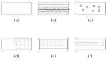
도 2 - 본 발명의 다양한 실시예에 따른 드레싱재에 대한 구조를 나타낸 모식도.
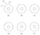
도 3 - 본 발명의 다양한 실시예에 따른 드레싱재에 대한 상측면 구조를 나타낸 모식도.
도 4 - 본 발명의 다양한 실시예에 따른 드레싱재의 결합홀 구조를 나타낸 모식도.
도 5 - 본 발명의 다양한 실시예에 따른 드레싱재의 혈액 또는 삼출물을 흡수하기 전 수직 단면 모식도(a) 그리고 흡수한 후(b) 수직 단면 모식도.
도 6 - 본 발명의 일실시예에 따른 드레싱재의 사시도.
도 7 - 본 발명의 일실시예에 따른 드레싱재가 포함된 자궁경부 유착 방지 장치에 대한 사시도.
도 8 - 본 발명의 다른 실시예에 따른 드레싱재가 포함된 자궁경부 유착 방지 장치에 대한 사시도.
도 9 - 본 발명의 일실시예에 따른 드레싱재의 혈액 응고 지수를 나타낸 그래프.
도 10 - 본 발명의 일실시예 따른 드레싱재가 포함된 자궁경부 유착 방지 장치가 자궁경부 부위에 적용된 사용상태에 대한 모식도.1-Schematic diagram of the cervical adhesion prevention device according to an embodiment of the present invention.
Figure 2-schematic diagram showing the structure of the dressing material according to various embodiments of the present invention.
Figure 3-Schematic diagram showing the upper side structure for the dressing material according to various embodiments of the present invention.
Figure 4-Schematic diagram showing the coupling hole structure of the dressing material according to various embodiments of the present invention.
Figure 5-a vertical cross-sectional schematic diagram (a) before absorbing blood or exudate of a dressing material according to various embodiments of the present invention and (b) vertical cross-sectional schematic diagram after absorption.
Figure 6-perspective view of a dressing material according to an embodiment of the present invention.
7-a perspective view of a device for preventing cervical adhesion with a dressing material according to an embodiment of the present invention.
8-a perspective view of a cervical adhesion prevention device including a dressing material according to another embodiment of the present invention.
Figure 9-a graph showing the blood coagulation index of the dressing material according to an embodiment of the present invention.
Figure 10-A schematic diagram of a state of use in which a cervical adhesion prevention device including a dressing material according to an embodiment of the present invention is applied to a cervical region.
본 발명은 자궁경부 유착 방지 장치에 있어서, 자궁경부를 향하도록 드레싱재를 형성시켜, 자궁경부 수술 후 자궁의 유착을 방지하고, 자궁경부의 형상을 잡아주는 역할을 하면서, 드레싱재의 성분에 따른 신속한 지혈, 유착방지, 조직 재생과 같은 기능을 보완시킨 드레싱재가 포함된 자궁경부 유착 방지 장치에 관한 것이다.In the present invention, in the apparatus for preventing cervical adhesion, a dressing material is formed to face the cervix, prevents adhesion of the uterus after cervical surgery, and serves to hold the shape of the cervix, and prompt hemostasis according to the components of the dressing material , Prevention of adhesions, and a cervical adhesion prevention device including a dressing material supplementing functions such as tissue regeneration.
이하에서는 첨부된 도면을 참조하여 본 발명에 대해 상세히 설명하고자 한다. 도 1은 본 발명의 일실시예에 따른 자궁경부 유착 방지 장치에 대한 모식도이고, 도 2는 본 발명의 다양한 실시예에 따른 드레싱재에 대한 구조를 나타낸 모식도이고, 도 3은 본 발명의 다양한 실시예에 따른 드레싱재에 대한 상측면 구조를 나타낸 모식도이고, 도 4는 본 발명의 다양한 실시예에 따른 드레싱재의 결합홀 구조를 나타낸 모식도이고, 도 5는 본 발명의 다양한 실시예에 따른 드레싱재의 혈액 또는 삼출물을 흡수하기 전 수직 단면 모식도(a) 그리고 흡수한 후(b) 수직 단면 모식도를 나타낸 것이고, 도 6은 본 발명의 일실시예에 따른 드레싱재의 사시도이고, 도 7은 본 발명의 일실시예에 따른 드레싱재가 포함된 자궁경부 유착 방지 장치에 대한 사시도이고, 도 8은 본 발명의 다른 실시예에 따른 드레싱재가 포함된 자궁경부 유착 방지 장치에 대한 사시도이고, 도 9는 본 발명의 일실시예에 따른 드레싱재의 혈액 응고 지수를 나타낸 그래프이며, 도 10은 본 발명의 일실시예 따른 드레싱재가 포함된 자궁경부 유착 방지 장치가 자궁경부 부위에 적용된 사용상태에 대한 모식도를 나타낸 것이다.Hereinafter, the present invention will be described in detail with reference to the accompanying drawings. 1 is a schematic diagram of a cervical adhesion prevention device according to an embodiment of the present invention, Figure 2 is a schematic diagram showing the structure of a dressing material according to various embodiments of the present invention, Figure 3 is a variety of embodiments of the present invention It is a schematic diagram showing the upper side structure for the dressing material according to an example, Figure 4 is a schematic diagram showing the coupling hole structure of the dressing material according to various embodiments of the present invention, Figure 5 is the blood of the dressing material according to various embodiments of the present invention Or it is a vertical cross-sectional schematic (a) before absorbing exudate and (b) showing a vertical cross-sectional schematic, Figure 6 is a perspective view of a dressing material according to an embodiment of the present invention, Figure 7 is an embodiment of the present invention 8 is a perspective view of a cervical adhesion prevention device including a dressing material according to an example, and FIG. 8 is a perspective view of a cervical adhesion prevention device including a dressing material according to another embodiment of the present invention, and FIG. 9 is an embodiment of the present invention It is a graph showing the blood coagulation index of the dressing material according to an example, and FIG. 10 shows a schematic diagram of a use state in which a cervical adhesion prevention device including a dressing material according to an embodiment of the present invention is applied to a cervical region.
도시한 바와 같이, 본 발명에 따른 드레싱재가 포함된 자궁경부 유착 방지 장치는, 자궁경관에 삽입되는 중공형 배액관(100), 상기 중공형 배액관(100)의 일단부에 연결되어 자궁경부에 걸쳐지도록 구성된 지지캡(200) 및 상기 중공형 배액관(100)의 타단부에 연결되며 자궁 내에서 펼쳐져 상기 중공형 배액관(100)의 위치를 잡아주는 적어도 하나의 고정 레그(Leg)(300)를 포함하는 자궁경부 유착 방지 장치에 있어서, 상기 지지캡(200) 상층에 드레싱재(400)가 적층되거나 상기 지지캡(200) 표면에 드레싱재(400)가 코팅되는 것으로, 상기 드레싱재(400)는, 상기 중공형 배액관(100)의 저부와 결합되는 결합홀(410)과, 상면측이 상기 자궁경부를 향하도록 형성된 본체(420)로 이루어진 것을 특징으로 한다.As shown, the apparatus for preventing cervical adhesion including the dressing material according to the present invention is connected to one end of the
상기 자궁경부 유착 방지 장치는 체내에 삽입된 상태에서 회복기간 동안 유지되는 것이므로 인체에 무해한 재질로 구성하는 것이 바람직한데, 실리콘, 폴리에틸렌, 또는 이들 중 일종 이상이 포함된 복합소재 등을 그 예로 들 수 있으나, 이에 제한되는 것은 아니다.Since the cervical adhesion prevention device is maintained during a recovery period in a state inserted in the body, it is preferable to be made of a material harmless to the human body. Examples thereof include silicone, polyethylene, or a composite material containing one or more of these. However, it is not limited thereto.
예를 들어, 상기 중공형 배액관(100)과 지지캡(200)을 실리콘재질로 일체 성형하여 구성하는 형태 또는, 중공형 배액관(100)은 실리콘재질로 구성하고 지지캡(200)은 봉합사재질로 구성하여 서로 연결하여 구성하는 형태 또는 중공형 배액관(100)과 지지캡(200)을 모두 봉합사재질로 구성하는 등 다양한 형태로의 실시가 가능할 것이다.For example, the
상기 봉합사재질의 경우에는 일반적인 의료 수술이나 시술과정에서 절개부위를 봉합하는 데 사용하는 것으로서, 환부의 치료가 있은 후 봉합사를 제거하게 되나, 흡수가 가능한(Absorbable) 소재, 예를 들어, 폴리락틱산(Polylactic Acid)이나 폴리디아소논(Polydiaxonone), 락틱산(Lactic Acid)과 글리콜릭산(Glycolic Acid)의 코폴리머 등을 사용하여 시술 후 이를 제거하지 않아도 생체 내에 흡수 가능하도록 하여도 될 것이다.In the case of the suture material, it is used to suture the incision in a general medical operation or procedure, and after treatment of the affected area, the suture is removed, but an absorbable material, for example, polylactic acid (Polylactic Acid), polydiaxonone (Polydiaxonone), lactic acid (Lactic Acid) and glycolic acid (Glycolic Acid) copolymer, etc. may be used to be absorbed in vivo without removing it after the procedure.
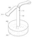
도 1에 도시한 바와 같이, 상기 중공형 배액관(100)은 자궁경관에 삽입되는 것으로서 내부에 공간이 형성되어 자궁으로부터 생성된 분비물들이 외부로 배출될 수 있는 통로를 가진 중공형일 수 있다. 또한, 상기 중공형 배액관(100)은 측부에 상기 분비물들의 배액을 위한 배액구(110)를 적어도 하나 구비할 수 있다.As illustrated in FIG. 1, the
그리고 상기 지지캡(200)은 상기 중공형 배액관(100)의 일단부에 연결되어 자궁입구(경부)에 걸쳐지도록 구성될 수 있다. 이때, 상기 중공형 배액관(100)을 중심축으로 하여 연결되는 것이 바람직하다. 즉, 상기 지지캡(200)의 중앙부와 상기 중공형 배액관(100)의 일단부가 연결된다.And the
상기 지지캡(200)은 표면에는 적어도 하나의 배액구(210)를 구비하여, 환부가 회복되면서 발생하는 염증물질과 같은 이물질을 외부로 순조롭게 배출시켜 회복을 도울 수 있도록 한다.The
상기 지지캡(200)의 배액구(210)의 경우에도, 상기 지지캡(200)이 실리콘 재질인 경우에는 성형 과정에서 하나 또는 복수 개의 배액구가 일체로 형성되도록 구성하면 될 것이며, 봉합사재질로 구성할 경우에는 상기 지지캡(200)을 메쉬 타입으로 구성(직조)하여 자연스럽게 하나 또는 복수의 배액구가 형성되도록 할 수 있을 것이다.Even in the case of the
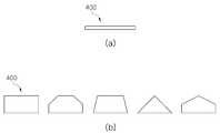
상기 지지캡(200)은 상기 중공형 배액관(100)을 중심축으로 사방으로 일정한 길이로 확장된 형태이며, 원형, 다각형, 별 모양 또는 방사형을 이루는 것일 수 있으나, 이에 제한되는 것은 아니다.The
또한, 상기 지지캡(200)은 전체적인 단면이 완만한 산 형상이거나, 사각형상, 타원형상 등 환자의 원래 자궁과 근접한 형상이면 어떠한 형상으로도 할 수 있을 것이므로, 그 형상을 어느 하나로 제한하지 않는다. 바람직하게는, 상기 지지캡(200)은 상기 중공형 배액관(100)으로부터 멀어질수록 두께가 작아지도록 경사를 이루게 하여 원활한 배액이 진행되도록 할 수 있다.In addition, the
또한, 상기 지지캡(200)은 자궁경부와의 밀착방지를 위하여 상부면에 돌기나 요철을 형성할 수도 있다.In addition, the
그리고, 상기 고정 레그(300)는 상기 중공형 배액관(100)의 타단부에 연결되며 자궁 내에서 펼쳐져 상기 중공형 배액관(100)의 위치를 잡아주는 것으로서 적어도 하나 이상으로 구성될 수 있다. 따라서, 상기 고정 레그(300)는 유연한(Flexible) 소재인 것이 바람직하다.In addition, the
상기 고정 레그(300)는 상기 중공형 배액관(100)으로부터 복수의 방향으로 길게 연장되어 형성되고, 중간부에 좌우방향으로 각각 연장 형성되는 날개 부분을 포함하여, 자궁 내에서 고정될 수 있는 면적을 보다 넓게 확보할 수 있다. 또한, 상기 고정 레그(300)의 말단부에는 고정용 돌기(310)가 형성되어, 자궁 내에 고정이 잘 이루어지도록 한다.The
여기에서, 상기 중공형 배액관(100)의 일단부의 외경과 상기 지지캡(200)이 연결되는 위치에 삽입홀(120)이 형성되어, 상기 중공형 배액관(100)의 외경부를 따라 지지 외관(미도시)이 삽입될 수 있도록 하여, 상기 고정 레그(300)가 상기 중공형 배액관(100)을 따라 상하로 움직이면서 상기 고정 레그(300)를 펼치거나(지지 외관을 제거한 경우), 지지 외관 내에 감싸인 형태로 자궁경관을 통해 삽입될 수 있도록 한다.Here, the
그리고 상기 지지캡(200) 상층에는 드레싱재(400)가 적층되거나, 상기 지지캡(200) 표면에 드레싱재(400)가 코팅되어 형성된다. 상기 지지캡(200)은 자궁경부에 걸쳐지도록 구성되며, 상기 드레싱재(400)는 자궁경부 표면과 상기 지지캡(200) 사이에 형성되어, 수술 후 환부로부터 방출되는 혈액이나 삼출물을 흡수하거나, 환부 조직의 재생이 신속하게 이루어지도록 보조하는 역할을 하는 것이다.In addition, a dressing
이에 상기 드레싱재(400)는 기본적으로 생체적합성이 뛰어난 소재로 형성되며, 지혈작용을 하면서, 자궁경부 조직의 유착방지, 조직 재생 기능을 갖춘 기능성 성분이 포함된 것일 수 있다.Accordingly, the dressing
상기 드레싱재(400)는 지혈을 위해 섬유 기반의 부직포만으로 이루어질 수도 있으며, 키토산의 유용한 기능을 활용하기 위해 키토산 기반의 부직포만으로도 이루어질 수도 있다. 이 경우 키토산만을 적용할 시 겔화가 되면서 소재가 물러져 강도 및 형상 유지가 어려울 수 있어, 키토산에 셀룰로오스(면, 견 또는 마), 재생셀룰로오스(레이온 또는 인견), 알지네이트 및 카제인을 혼합하여 제작할 수 있다.The dressing
또한, 상기 드레싱재(400)는 지혈재가 혼합되어 부직포 형태로 제공되거나, 표면에 지혈재로 이루어진 코팅층이 단일층 또는 복수층으로 형성된 부직포 형태로 제공될 수 있다.In addition, the dressing
또한, 상기 드레싱재(400)는, 천연 생체적합성 소재 또는 합성 생체적합성 소재로 구현될 수 있으며, 상기 소재 단독 또는 상기 소재를 지지하기 위해 상술한 셀룰로오스(면, 견 또는 마), 재생셀룰로오스(레이온 또는 인견), 알지네이트 및 카제인 등이 혼합되어 사용되어 질 수도 있다.In addition, the dressing
구체적으로는 상기 천연 생체적합성 소재는 키토산 (Chitosan), 카복시메틸 키토산(Carboxymethyl chitosan), 키토산 숙시네이트(Succinyl chitosan), 셀룰로오스(Cellulose), 카복시메틸 셀룰로오스 (Carboxymethyl cellulose), 재생 셀룰로오스(또는 레이온)(Regenerated cellulose or Rayon), 히알루론산(Hyaluronic acid), 소듐 알지네이트(Sodium alginate), 콜라겐(Collagen), 젤라틴(Gelatin) 중 어느 하나 또는 둘 이상 혼합하여 사용한다.Specifically, the natural biocompatible material is chitosan, carboxymethyl chitosan, chitosan succinate, cellulose, carboxymethyl cellulose, regenerated cellulose (or rayon) ( Regenerated cellulose or Rayon, hyaluronic acid, sodium alginate, collagen, gelatin, or a mixture of two or more are used.
또한, 상기 합성 생체적합성 소재는 카올린(Kaolin), 제올라이트(Zeolite), γ-폴리글루탐산(γ-PGA), 풀루란(Pullulan), 콘드로이친 설페이트(Chondroitin sulphate), 카테콜(Catechol), 칼슘 클로라이드(Calcium chloride), 트롬빈(Thrombin), 피브리노겐(Fibrinogen), 폴리우레탄 폼(PU foam) 중 어느 하나 또는 둘 이상 혼합하여 사용할 수 있다.In addition, the synthetic biocompatible materials include kaolin, zeolite, γ-polyglutamic acid (γ-PGA), pullulan, chondroitin sulphate, catechol, and calcium chloride ( Calcium chloride, thrombin, fibrinogen, or polyurethane foam (PU foam) can be used in combination.
본 발명에 따른 드레싱재(400)는 상기 지지캡(200) 상에 형성되는 것으로서, 상기 중공형 배액관(100)의 저부와 결합되는 결합홀(410)과 상면측이 상기 자궁경부를 향하도록 형성된 본체(420)로 이루어진 것을 특징으로 한다.
상기 드레싱재(400)는 상기 지지캡(200)에 상기 중공형 배액관(100)을 중심부에 두고 결합되는 것으로서, 중심부에 상기 중공형 배액관(100)과의 결합을 위한 결합홀(410)이 형성되어야 한다. 상기 결합홀(410)은 상기 중공형 배액관(100)이 수용될 정도의 크기로 형성되면 무방하며, 그 형태는 도 4에 도시한 바와 같이 원형 또는 다각형으로 형성될 수 있다.The dressing
또한, 도 3에 도시한 바와 같이, 상기 드레싱재(400)의 본체(420)는 상기 결합홀(410)을 중심으로 원형 또는 다각형으로 형성되며, 상기 지지캡(200) 상에 위치하여 자궁경부를 향하도록 상기 지지캡(200)과 비슷한 크기와 형태를 갖도록 형성된다.In addition, as shown in Figure 3, the
상기 드레싱재(400)의 형태는 환부의 적용 환경에 따라 변형하여 형성한다.The shape of the dressing
도 5는 본 발명의 다양한 실시예에 따른 드레싱재(400)의 혈액 또는 삼출물을 흡수하기 전 수직 단면 모식도(a) 그리고 흡수한 후(b) 수직 단면 모식도를 나타낸 것으로, 흡수 후의 형태를 기준으로 성형하고, 동결 건조를 통해 드레싱재(400)를 제조한 후, 흡수 전의 형태로 압착하여 제공하게 된다.Figure 5 shows a schematic view of the vertical cross-section before absorbing the blood or exudate of the dressing
여기에서 상기 드레싱재(400)가 혈액 또는 삼출물을 흡수한 후에는 수직 단면이 사각형 또는 하측의 폭보다 상측의 폭이 상대적으로 좁은 다각형 형태로 형성되는 것을 특징으로 한다. 이는 자궁경부와 같은 형태의 환부에 지혈관리 등이 더욱 잘 이루어지도록 하기 위한 것이다.Here, after the
즉, 본 발명에 따른 드레싱재(400)는 흡수 후의 형태를 기준으로 성형틀에 의해 제작되며, 동결건조되어 제공된다. 제공된 드레싱재(400)의 상부를 압착하여 도 5(a)와 같은 흡수 전 형상으로 제조하며, 혈액 또는 삼출물을 흡수하여 부풀어 오르게 되면 도 5(b)와 같은 흡수 후 형상으로 변하게 된다. 즉, 압착되기 전 동결건조된 초기 형태로 돌아오게 된다.That is, the dressing
도 6은 본 발명의 일실시예로 원형의 결합홀(410)과 원형의 몸체를 갖는 드레싱재(400)를 나타내었으며, 상기 결합홀(410)에 중공형 배액관(100)이 결합되고 몸체는 상기 지지캠 상에 안착되게 된다.6 shows a
도 7(a)에 도시한 바와 같이, 일실시예로, 드레싱재(400)의 직경은 20~26mm, 결합홀(410)의 직경은 3~6mm, 그 높이는 흡수 전의 경우 1~5mm, 흡수 후의 경우 10~35mm로 제공되게 된다. 이는 환부의 크기 및 지혈관리 부위에 따라 그 크기를 조절하여 형성할 수 있다.As shown in Figure 7 (a), in one embodiment, the diameter of the dressing
도 7(b)는 본 발명의 다른 실시예로 드레싱재(400)를 자궁경부 유착 방지 장치에 물리적으로 결합하기 위해 상기 드레싱재(400)의 일부에 절개부(430)를 형성한 것으로, 상기 중공형 배액관(100)에 끼움결합하여 상기 중공형 배액관(100)과 상기 드레싱재(400)의 결합홀(410)이 결합되도록 한다.Figure 7 (b) is to form a cut-out
이 경우 상기 드레싱재(400)는 드레싱재(400)의 소재가 포함된 용액을 성형틀에 맞춰 부어 동결, 동결건조 후 압착하여 소정 두께로 형성한다. 상기 용액은 1~5%의 드레싱재(400)의 소재를 증류수에 녹인 후 1~5% 글리세린을 첨가하여 2시간 이상 교반하여 제조된 것이다.In this case, the dressing
상기 동결건조는 상기 성혈틀을 동결건조기에 집어넣어 급냉 후 동결건조시킴으로써 수분을 증발시키는 것으로, 진공상태에서 -40℃부터 30℃까지 온도를 올리면서 건조가 이루어지게 하는 것이다. 그 후 드레싱재(400)는 원하는 두께로 압착하여 제공되게 된다.The lyophilization is to evaporate moisture by placing the sex blood in a freeze dryer and then quenching it and freeze-drying it, and drying is performed while raising the temperature from -40°C to 30°C in a vacuum. Thereafter, the dressing
여기에서, 상기 드레싱재(400)는 산성용액을 이용한 화학처리 또는 열처리를 수행하거나, UV를 통한 가교를 통해 흡수력이 향상시키거나, 계면활성처리를 더 수행하여 흡수속도를 더욱 높일 수도 있다.Here, the dressing
구체적으로는 상기 드레싱재(400)의 흡수성을 강화하기 위해 산성용액 (아세트산, 염산, 아스코르브산, 글루탐산, 구연산, 포름산 등)을 이용한 화학처리 또는 열처리 및 글리세린, 아세트알데히드, 글루탈알데히드, 포름알데히드, 폴리에틸렌글라이콜 다이아크릴레이트, UV를 통한 가교를 이용할 수 있다.Specifically, chemical treatment or heat treatment using an acidic solution (acetic acid, hydrochloric acid, ascorbic acid, glutamic acid, citric acid, formic acid, etc.) or heat treatment and glycerin, acetaldehyde, glutalaldehyde, and formaldehyde to enhance the absorption of the dressing
또한, 흡수속도의 강화를 위해 tween 20, tween 60, tween 80과 같은 계면활성 처리를 할 수도 있다.In addition, to enhance the absorption rate, surface active treatments such as
물론, 상기 드레싱재(400)는 상술한 바와 같이 동결건조하여 압착되어 상기 중공형 배액관(100)에 물리적으로 결합되어 상기 지지캡(200) 상층에 적층될 수도 있고, 상기 지지캡(200) 표면에 드레싱재(400)가 코팅되어 형성될 수도 있다. 즉, 드레싱재(400)가 포함된 용액에 상기 지지캡(200)을 함침하거나, 전기방사 등의 방법으로 상기 드레싱재(400)를 자궁경부 유착 방지 장치 상에 구현할 수 있다.Of course, the dressing
도 8은 상기 지지캡(200) 표면에 드레싱재(400)가 코팅되어 형성된 것으로서, 상기 드레싱재(400)가 포함된 용액에 지지캡(200)을 함침하여 형성하므로, 상기 드레싱재(400) 내부에 지지캡(200)이 위치하게 된다.FIG. 8 shows that the dressing
이와 같이 자궁경부 유착 방지 장치에 드레싱재(400)가 포함되도록 하여 자궁경부 수술인 원추절제술 후 환부의 출혈 및 삼출물 관리가 용이하도록 하며, 기능성 성분을 추가하여 빠른 지혈관리, 유착방지, 조직 재생 등의 기능이 더 부여되도록 한다.As such, the dressing
한편 본 발명에 사용되는 드레싱재(400)는 상기 지지캡(200) 상에 단일 소재(도 2(a) 또는 2종 이상의 소재(도 2(d))가 혼합되어 단일층으로 형성되거나, 2종 이상의 소재를 사용하는 다층 구조(도 2(b))로 형성할 수 있다.Meanwhile, the dressing
또한, 상기 드레싱재(400)는 내부에 이종(異種)의 소재가 캡슐 형태로 혼합(도 2(c))되거나, 외피 내부에 상기 외피와는 다른 이종(異種)의 소재가 포함된 것(도 2(e)) 커버 구조를 형성할 수 있다.In addition, the dressing
또한, 상기 드레싱재(400)는, 외피 사이에 상기 외피와는 다른 이종(異種)의 소재 형성된 샌드위치 구조(도 2(f))로 형성할 수 있다.In addition, the dressing
상기 드레싱재(400)가 상기 단일층으로 형성된 구조일 경우 상술한 바와 같이 드레싱재(400)의 소재가 포함된 용액을 성형틀에 부어 이를 동결 후 동결건조하여 형성한다. 그리고 상기 다층 구조 및 샌드위치 구조의 경우에는 첫 층을 동결 후 다음 층에 해당하는 용액을 붓고 동결하며, 이 과정을 반복한 후 동결건조를 진행한다.When the dressing
상기 드레싱재(400)에 캡슐이 포함된 경우에는 캡슐을 용액과 섞어 분산시켜준 후 용액을 동결하고 동결건조하여 형성하며, 상기 커버 구조의 경우 내부 성분을 동결 후, 외피 용액에 이를 담궈서 동결건조하여 형성한다.When the dressing
상술한 바와 같기 동결건조 후에는 적절한 크기로 압착하고 제품 형태에 맞는 틀을 이용하여 타발하여 드레싱재를 제공하게 된다.As described above, after freeze-drying, it is compressed to an appropriate size and punched using a mold suitable for the product form to provide a dressing material.
이하에서는 본 발명의 실시예에 따른 드레싱재의 흡수성 및 지혈성에 대한 시험에 따른 데이타에 대해 설명하고자 한다.Hereinafter, the data according to the test for the absorption and hemostatic properties of the dressing material according to the embodiment of the present invention will be described.
① 흡수성 시험① Absorbency test
- 시험 규격: EN13726-1: Test methods for primary wound dressings. Aspects of absorbency -Test specification: EN13726-1: Test methods for primary wound dressings. Aspects of absorbency
- 적용 범위: 흡수력이 있는 제품. 삼출물의 흡수 등을 목적으로 하는 부착하는 형태의 단시간 사용하는 제품에 적용 -Scope of application: absorbent products. Applied to products used for a short time in the form of attachment for the purpose of absorbing exudate
- 시험 목적: 상처 표면의 삼출액 및 체액 등의 흡수력을 측정하기 위한 시험규격임. -Purpose of the test: This is a test standard for measuring the absorbency of exudates and body fluids on the wound surface.
- 시험방법: - Test Methods:
⑴ 제품의 사용방법 및 목적 등을 고려하여 시료를 제작하여 (예 : (5 × 5) cm의 크기로 잘라) 페트리디쉬에 놓고 무게(W1)를 잰다.시료 Prepare a sample considering the usage method and purpose of the product (eg, cut to a size of (5 × 5) cm) and place it in a petri dish and measure the weight (W1).
⑵ (37 ± 1) ℃로 미리 데워진 증류수를 ± 0.5 g까지 정확하게 측정하여 제품의 흡수력을 고려하여 (예 : 샘플의 무게의 40배를) 첨가한다.정확하게 Accurately measure the pre-warmed distilled water at (37 ± 1) ℃ to ± 0.5 g and add it considering the product's absorption capacity (eg 40 times the weight of the sample).
⑶ (37 ± 1) ℃ 항온기에서 30분 동안 방치한 후 핀셋을 이용해 샘플을 30 초간 매단 후 무게(W2)를 잰다.⑶ (37 ± 1) ℃ After standing for 30 minutes in the incubator, suspend the sample for 30 seconds using tweezers and weigh (W2).
⑷ 아래의 계산식을 이용하여 흡수력을 측정한다.흡수 Measure the absorption power using the following formula.

다음 표 1은 각 실시예에 따른 흡수력을 테스트한 시험결과이다.Table 1 below shows the test results of testing the absorbency according to each example.
다음 표 2는 타사제품 흡수력 테스트한 데이터이다.The following table 2 is the data of other companies' product absorption test.
본 발명에 따른 실시예(드레싱재)가 기존 타사 제품에 비해 흡수력이 월등히 뛰어남을 확인할 수 있었다.It was confirmed that the embodiment according to the present invention (dressing material) has an excellent absorbency compared to other conventional products.
② 지혈성 시험② Hemostatic test
- 시험 규격: 자사규격 -Test standard: Company standard
- 시험 목적: 드레싱의 혈액 응고 능력을 평가하기 위함 -Purpose of the test: To evaluate the blood clotting ability of the dressing
- 시험방법: - Test Methods:
⑴ 시료를 2 x 2 cm 크기로 자른 후, 각각 50 ml 튜브에 넣는다. ⑴ Samples are cut to 2 x 2 cm size and placed in 50 ml tubes each.
⑵ 토끼로부터 채혈한 혈액 및 ACD, CaCl2의 혼합액을 준비한다.⑵ Prepare a mixture of blood, ACD, and CaCl2 collected from rabbits.
(혈액:ACD:1M CaCl2 = 0.270ml:0.030ml:0.024ml) (Blood:ACD:1M CaCl2 = 0.270ml:0.030ml:0.024ml)
여기에서, ACD는 시트르산, 시트르산 나트륨, 클루코스의 혼합액으로 Acid citrate dextrose solution의 약자임. 채취한 혈액의 보관을 위해 응고를 방지하고자하는 목적으로 첨가되는 것임.Here, ACD stands for Acid citrate dextrose solution as a mixture of citric acid, sodium citrate, and klucos. It is added for the purpose of preventing coagulation for storage of collected blood.
그리고, CaCl2는 ACD로 항 응고된 혈액의 항응고제 역할을 막기 위해 투여되는 것임.And, CaCl2 is administered to prevent the anticoagulant role of blood coagulated with ACD.
⑶ 각 시료가 담긴 튜브에 (2)과정에서 준비한 혈액 100ul를 시료 위에 처리한 후, 실온에서 시간대 별로 0, 15, 30, 60, 90초간 반응시킨다. 100 After treating 100 ul of blood prepared in step (2) on the tube containing each sample on the sample, react at room temperature for 0, 15, 30, 60, 90 seconds per time zone.
⑷ 시간대 별로 반응시킨 시료에 생리식염수 10 ml를 튜브 안쪽 벽을 통해 조심스럽게 추가한다. 10 10 ml of physiological saline is carefully added to the sample reacted over time through the inner wall of the tube.
⑸ 100G로 30초간 원심분리 한다. 원심 Centrifuge for 30 seconds at 100G.
⑹ 상층액(시료, 혈액 제외)만을 조심스럽게 새로운 튜브로 옮긴 후 초 부피 50 ml가 되도록 초순수 증류수를 첨가한다. 만을 Only the supernatant (except sample, blood) is carefully transferred to a new tube, and then ultrapure distilled water is added to a volume of 50 ml.
⑺ 상온에서 30분 간 반응시킨 후, 96-well 투명 plate의 각 well 당 200㎕의 시료를 주입한 뒤 ELISA 분광기를 이용하여 540nm에서 흡광도를 측정한다.반응 After reacting for 30 minutes at room temperature, 200 μl of sample is injected per well of a 96-well transparent plate, and absorbance is measured at 540 nm using an ELISA spectrometer.
도 9는 본 발명의 일실시예에 따른 드레싱재의 혈액 응고 지수를 나타낸 그래프로, BCI: Blood coagulation index의 약자로 혈액 응고 지수를 나타낸다.9 is a graph showing a blood coagulation index of a dressing material according to an embodiment of the present invention, and stands for BCI: Blood coagulation index.
이것은 응고되지 않은 혈액이 많을수록, 증류수 내에서 혈액의 분산이 증가하기 때문에 용액의 색이 붉은색으로 점점 진해지고 특정 파장에서의 흡광도가 높아지게 됨. 이 때, 흡광도가 높을수록 BCI 수치가 높게 나타나는데, 이는 BCI 수치가 높을수록 응고되지 않은 혈액이 많다는 것을 나타내며, 지혈 성능이 비교적 좋지 않다는 것을 의미함.This means that the more uncoagulated blood, the more the dispersion of blood in distilled water increases, so the color of the solution becomes darker and the absorbance at a specific wavelength increases. At this time, the higher the absorbance, the higher the BCI level, which indicates that the higher the BCI level, the more uncoagulated blood, and the hemostatic performance is relatively poor.
도 9에 도시한 바와 같이, 일반 드레싱재에 비해 본 발명의 실시예에 따른 드레싱재의 BCI가 훨씬 낮게 나왔으며, 본 발명의 실시예가 지혈 성능이 월등히 우수한 것으로 확인되었다.As shown in FIG. 9, the BCI of the dressing material according to the embodiment of the present invention was much lower than that of the general dressing material, and it was confirmed that the hemostatic performance of the embodiment of the present invention was superior.

ODb : 순수한 증류수에서 흡광도ODb : Absorbance in pure distilled water
ODt : 시료에서 방출된 혈액의 흡광도ODt : Absorbance of blood released from the sample
도 10은 본 발명의 일실시예 따른 드레싱재가 포함된 자궁경부 유착 방지 장치가 자궁경부 부위에 적용된 사용상태에 대한 모식도를 나타낸 것으로, 자궁경관(10)에 드레싱재가 포함된 자궁경부 유착 방지 장치가 삽입되어 있고, 자궁경부(20)에 지지캡(200)이 걸쳐지고, 자궁내부에 고정 레그(300)가 펼쳐져 위치를 잡아주고 있다.FIG. 10 is a schematic view of a state of use in which a cervical adhesion prevention device including a dressing material according to an embodiment of the present invention is applied to a cervical region, wherein a cervical adhesion prevention device including a dressing material in the
이와 같이 본 발명은 자궁경부 원추절제술이 완료된 후에는 자궁경관을 통해 삽입되어 시술된 본 발명의 자궁경부 유착 방지 장치는 자궁경부가 협착 또는 유착되는 것을 방지할 수 있게 된다. 기본적으로 자궁에서 생성된 분비물은 배액관 및 상기 배액관의 측부에 형성된 배액구를 통해 배출될 수 있으며, 지지캡에 형성된 하나 이상의 배액구를 통해서는 환부가 회복되는 과정에서 발생하는 이물질이 외부로 배출될 수 있다.As described above, after the cervical cone resection is completed, the apparatus for preventing cervical adhesion of the present invention, which is inserted and operated through the cervical canal, can prevent the cervical stenosis or adhesion. Basically, secretions generated in the uterus can be discharged through a drainage tube and a drainage hole formed on the side of the drainage tube, and through one or more drainage holes formed in the support cap, foreign matter generated in the process of recovering the affected part is discharged to the outside. Can.
그리고, 지지캡 상에 형성된 드레싱재는 자궁경부 수술 후 자궁의 유착을 방지하고, 자궁경부의 형상을 잡아주는 역할을 하면서, 드레싱재의 성분에 따른 신속한 지혈, 유착방지, 조직 재생과 같은 기능을 하게 된다.In addition, the dressing material formed on the support cap serves to prevent adhesion of the uterus after cervical surgery and to serve to shape the cervix, and has functions such as rapid hemostasis, adhesion prevention, and tissue regeneration according to the components of the dressing material. .
그러므로, 환부가 감염되거나 이물질에 의하여 회복이 더뎌지는 것을 방지하고, 빠른 조직 재생이 이루어지게 되므로 빠른 회복을 도울 수 있게 된다.Therefore, the affected area is prevented from being infected or the recovery is slowed down by foreign substances, and rapid tissue regeneration is achieved, so that rapid recovery can be assisted.
10 : 자궁경관20 : 자궁경부
100 : 배액관110 : 배액구
200 : 지지캡210 : 배액구
300 : 고정 레그310 : 고정용 돌기
400 : 드레싱재410 : 결합홀
420 : 본체10: cervical canal 20: cervix
100: drain pipe 110: drain port
200: support cap 210: drain hole
300: fixed leg 310: fixed projection
400: dressing material 410: coupling hole
420: main body
Claims (14)
Translated fromKorean상기 지지캡 상층에 드레싱재가 적층되거나 상기 지지캡 표면에 드레싱재가 코팅되는 것으로,
상기 드레싱재는,
상기 중공형 배액관의 저부와 결합되는 결합홀;
상면측이 상기 자궁경부를 향하도록 형성된 본체;로 이루어진 것을 특징으로 하는 드레싱재가 포함된 자궁경부 유착 방지 장치.A hollow drainage tube inserted into the cervical canal, a support cap configured to be connected to one end of the hollow drainage tube and spread across the cervix, and connected to the other end of the hollow drainage tube and spread in the uterus to hold the position of the hollow drainage tube In the apparatus for preventing cervical adhesion comprising at least one fixed leg (Leg),
As the dressing material is laminated on the upper layer of the support cap or the dressing material is coated on the surface of the support cap,
The dressing material,
A coupling hole coupled with the bottom of the hollow drainage tube;
Device for preventing cervical adhesion with a dressing material, characterized in that the upper surface side is formed to face the cervix.
단일 소재 또는 2종 이상의 소재가 혼합되어 단일층으로 형성되거나,
2종 이상의 소재를 사용하는 다층 구조로 형성되는 것을 특징으로 하는 드레싱재가 포함된 자궁경부 유착 방지 장치.According to claim 1, The dressing material,
A single material or a mixture of two or more materials is formed as a single layer,
Device for preventing cervical adhesion with a dressing material, characterized in that it is formed of a multi-layer structure using two or more materials.
내부에 이종(異種)의 소재가 캡슐 형태로 혼합된 것을 특징으로 하는 드레싱재가 포함된 자궁경부 유착 방지 장치.According to claim 1, The dressing material,
Device for preventing cervical adhesion with a dressing material, characterized in that the material of different species is mixed in a capsule form.
외피 내부에 상기 외피와는 다른 이종(異種)의 소재가 포함된 것을 특징으로 하는 드레싱재가 포함된 자궁경부 유착 방지 장치.According to claim 1, The dressing material,
A device for preventing cervical adhesion with a dressing material, characterized in that a different material from the outer shell is included in the outer shell.
외피 사이에 상기 외피와는 다른 이종(異種)의 소재가 위치하는 것을 특징으로 하는 드레싱재가 포함된 자궁경부 유착 방지 장치.According to claim 1, The dressing material,
Device for preventing cervical adhesion with a dressing material, characterized in that a material of a different type from the shell is located between the shells.
원형 또는 다각형 형태로 형성된 것을 특징으로 하는 드레싱재가 포함된 자궁경부 유착 방지 장치.According to claim 1, The coupling hole of the dressing material,
Device for preventing cervical adhesion with a dressing material, characterized in that formed in a circular or polygonal shape.
원형 또는 다각형 형태로 형성된 것을 특징으로 하는 드레싱재가 포함된 자궁경부 유착 방지 장치.According to claim 1, The main body of the dressing material,
Device for preventing cervical adhesion with a dressing material, characterized in that formed in a circular or polygonal shape.
혈액 또는 삼출물을 흡수하기 전에는 수직 단면이 사각형으로 형성되며,
상기 혈액 또는 삼출물의 흡수 후에는 수직 단면이 사각형 또는 하측의 폭보다 상측의 폭이 상대적으로 좁은 다각형 형태로 형성되는 것을 특징으로 하는 드레싱재가 포함된 자궁경부 유착 방지 장치.According to claim 1, The dressing material,
Before absorbing blood or exudate, the vertical cross section is square,
After absorption of the blood or exudate, the cervical adhesion prevention device including a dressing material, characterized in that the vertical cross-section is formed in a polygonal shape with a relatively narrow upper width than a rectangular or lower width.
천연 생체적합성 소재 또는 합성 생체적합성 소재로 구현되는 것을 특징으로 하는 드레싱재가 포함된 자궁경부 유착 방지 장치.According to claim 1, The dressing material,
Device for preventing cervical adhesion with a dressing material, characterized in that it is implemented as a natural biocompatible material or a synthetic biocompatible material.
키토산 (Chitosan), 카복시메틸 키토산(Carboxymethyl chitosan), 키토산 숙시네이트(Succinyl chitosan), 셀룰로오스(Cellulose), 카복시메틸 셀룰로오스 (Carboxymethyl cellulose), 재생 셀룰로오스(또는 레이온)(Regenerated cellulose or Rayon), 히알루론산(Hyaluronic acid), 소듐 알지네이트(Sodium alginate), 콜라겐(Collagen), 젤라틴(Gelatin) 중 어느 하나 또는 둘 이상 혼합하여 사용하는 것을 특징으로 하는 드레싱재가 포함된 자궁경부 유착 방지 장치.The method of claim 9, wherein the natural biocompatible material,
Chitosan, Carboxymethyl chitosan, Chitosan Succinate, Cellulose, Carboxymethyl cellulose, Regenerated cellulose or Rayon, Hyaluronic acid ( Hyaluronic acid), sodium alginate (Sodium alginate), collagen (Collagen), gelatin (Gelatin) any one or two or more of the mixture of the cervical adhesion device containing a dressing material characterized in that used in combination.
카올린(Kaolin), 제올라이트(Zeolite), γ-폴리글루탐산(γ-PGA), 풀루란(Pullulan), 콘드로이친 설페이트(Chondroitin sulphate), 카테콜(Catechol), 칼슘 클로라이드(Calcium chloride), 트롬빈(Thrombin), 피브리노겐(Fibrinogen), 폴리우레탄 폼(PU foam) 중 어느 하나 또는 둘 이상 혼합하여 사용하는 것을 특징으로 하는 드레싱재가 포함된 자궁경부 유착 방지 장치.The method of claim 9, wherein the synthetic biocompatible material,
Kaolin, Zeolite, γ-polyglutamic acid (γ-PGA), Pullulan, Chondroitin sulphate, Catechol, Calcium chloride, Thrombin , Fibrinogen (Fibrinogen), polyurethane foam (PU foam) any one or two or more of the cervical adhesion prevention device containing a dressing material, characterized in that used in combination.
드레싱재의 소재가 포함된 용액을 성형틀에 맞춰 부어 동결건조 후 압착하여 형성되는 것을 특징으로 하는 드레싱재가 포함된 자궁경부 유착 방지 장치.According to claim 1, The dressing material,
A device for preventing cervical adhesion with a dressing material, characterized in that the solution containing the material of the dressing material is poured into a molding frame and lyophilized and then compressed.
산성용액을 이용한 화학처리 또는 열처리를 수행하거나,
UV를 통한 가교를 통해 흡수력이 향상된 것을 특징으로 하는 드레싱재가 포함된 자궁경부 유착 방지 장치.According to claim 1, The dressing material,
Chemical treatment or heat treatment using an acidic solution, or
A device for preventing cervical adhesion with a dressing material characterized in that the absorption power is improved through cross-linking through UV.
계면활성처리가 더 수행된 것을 특징으로 하는 드레싱재가 포함된 자궁경부 유착 방지 장치.The dressing material of claim 13,
Device for preventing cervical adhesion with a dressing material characterized in that the surface active treatment is further performed.
Priority Applications (2)
| Application Number | Priority Date | Filing Date | Title |
|---|---|---|---|
| KR1020190004040AKR102294242B1 (en) | 2019-01-11 | 2019-01-11 | Instrument for preventing adhesion of uterine cervix with dressing material |
| PCT/KR2019/004852WO2020145456A1 (en) | 2019-01-11 | 2019-04-23 | Intrauterine adhesion prevention device including dressing material |
Applications Claiming Priority (1)
| Application Number | Priority Date | Filing Date | Title |
|---|---|---|---|
| KR1020190004040AKR102294242B1 (en) | 2019-01-11 | 2019-01-11 | Instrument for preventing adhesion of uterine cervix with dressing material |
Publications (2)
| Publication Number | Publication Date |
|---|---|
| KR20200087574Atrue KR20200087574A (en) | 2020-07-21 |
| KR102294242B1 KR102294242B1 (en) | 2021-08-26 |
Family
ID=71520683
Family Applications (1)
| Application Number | Title | Priority Date | Filing Date |
|---|---|---|---|
| KR1020190004040AActiveKR102294242B1 (en) | 2019-01-11 | 2019-01-11 | Instrument for preventing adhesion of uterine cervix with dressing material |
Country Status (2)
| Country | Link |
|---|---|
| KR (1) | KR102294242B1 (en) |
| WO (1) | WO2020145456A1 (en) |
Cited By (1)
| Publication number | Priority date | Publication date | Assignee | Title |
|---|---|---|---|---|
| KR20220090271A (en)* | 2020-12-22 | 2022-06-29 | 주식회사 엔도비전 | Instrument for preventing adhesion of uterine cervix |
Citations (5)
| Publication number | Priority date | Publication date | Assignee | Title |
|---|---|---|---|---|
| US6526980B1 (en)* | 1999-08-26 | 2003-03-04 | West Virginia University | Cervical drug delivery system |
| KR20050045113A (en)* | 2003-11-10 | 2005-05-17 | 주식회사 바이오레인 | Anti-adhesion agent with gas bubble |
| KR20130025337A (en)* | 2011-08-26 | 2013-03-11 | 주식회사 엘지생명과학 | Adhesion-preventing agent and method for preventing adhesion using the same |
| KR101435661B1 (en) | 2014-02-18 | 2014-08-28 | 주식회사 인코어 | Recovery tools for uterine cervix |
| KR20170121990A (en)* | 2016-04-26 | 2017-11-03 | 주식회사 엔도비전 | Instrument for preventing adhesion of uterine cervix |
Family Cites Families (1)
| Publication number | Priority date | Publication date | Assignee | Title |
|---|---|---|---|---|
| AU2707500A (en)* | 1998-12-04 | 2000-06-26 | Incept Llc | Biocompatible crosslinked polymers |
- 2019
- 2019-01-11KRKR1020190004040Apatent/KR102294242B1/enactiveActive
- 2019-04-23WOPCT/KR2019/004852patent/WO2020145456A1/ennot_activeCeased
Patent Citations (6)
| Publication number | Priority date | Publication date | Assignee | Title |
|---|---|---|---|---|
| US6526980B1 (en)* | 1999-08-26 | 2003-03-04 | West Virginia University | Cervical drug delivery system |
| KR20050045113A (en)* | 2003-11-10 | 2005-05-17 | 주식회사 바이오레인 | Anti-adhesion agent with gas bubble |
| KR20130025337A (en)* | 2011-08-26 | 2013-03-11 | 주식회사 엘지생명과학 | Adhesion-preventing agent and method for preventing adhesion using the same |
| KR101435661B1 (en) | 2014-02-18 | 2014-08-28 | 주식회사 인코어 | Recovery tools for uterine cervix |
| KR20170121990A (en)* | 2016-04-26 | 2017-11-03 | 주식회사 엔도비전 | Instrument for preventing adhesion of uterine cervix |
| KR101851737B1 (en) | 2016-04-26 | 2018-04-24 | 주식회사 엔도비전 | Instrument for preventing adhesion of uterine cervix |
Cited By (2)
| Publication number | Priority date | Publication date | Assignee | Title |
|---|---|---|---|---|
| KR20220090271A (en)* | 2020-12-22 | 2022-06-29 | 주식회사 엔도비전 | Instrument for preventing adhesion of uterine cervix |
| WO2022139046A1 (en)* | 2020-12-22 | 2022-06-30 | 주식회사 엔도비전 | Uterine cervix adhesion prevention device |
Also Published As
| Publication number | Publication date |
|---|---|
| WO2020145456A1 (en) | 2020-07-16 |
| KR102294242B1 (en) | 2021-08-26 |
Similar Documents
| Publication | Publication Date | Title |
|---|---|---|
| EP3843796B1 (en) | Composite dressings, manufacturing methods and applications thereof | |
| US10314937B2 (en) | Biocompatible hemostatic product and preparation method thereof | |
| AU2017203958B2 (en) | Hemostatic device | |
| JP5638521B2 (en) | Healing wound dressing that reduces temperature | |
| KR101105081B1 (en) | Tissue Dressing Assemblies, Systems, and Methods Formed from Hydrophilic Polymer Sponge Structures such as Chitosan | |
| RU2498825C2 (en) | Absorbent medical body, in particular for removing wound liquids from human and animal body cavities, and method for making it | |
| ZA200605125B (en) | Tissue dressing assemblies, systems, and methods formed from hydrophilic polymer sponge structures such as chistosan | |
| CN101564557B (en) | Preparation method of antibacterial self-expanding material for disposable cervical dilator | |
| CN101472620A (en) | Compositions and methods for affecting movement of contaminants, bodily fluids or other entities, and/or affecting other physiological conditions | |
| TW201438769A (en) | Hydrogel forming material | |
| JP2021518803A (en) | Chitosan fibrous sponge structure medical dressing and its manufacturing method | |
| KR102294242B1 (en) | Instrument for preventing adhesion of uterine cervix with dressing material | |
| KR102169801B1 (en) | hemostatic for uterine cervical and manufacturing method of the same | |
| RU2151615C1 (en) | Pileless porous medicinal-destination sorption material and dressing facilities | |
| CN104744723A (en) | Chitosan medical material, and preparation method and use thereof | |
| AU2012347804B8 (en) | Hemostatic device | |
| CN110251718A (en) | A kind of preparation method of gelatin nasal cavity hemostatic sponge |
Legal Events
| Date | Code | Title | Description |
|---|---|---|---|
| PA0109 | Patent application | St.27 status event code:A-0-1-A10-A12-nap-PA0109 | |
| PA0201 | Request for examination | St.27 status event code:A-1-2-D10-D11-exm-PA0201 | |
| D13-X000 | Search requested | St.27 status event code:A-1-2-D10-D13-srh-X000 | |
| P11-X000 | Amendment of application requested | St.27 status event code:A-2-2-P10-P11-nap-X000 | |
| P13-X000 | Application amended | St.27 status event code:A-2-2-P10-P13-nap-X000 | |
| R15-X000 | Change to inventor requested | St.27 status event code:A-3-3-R10-R15-oth-X000 | |
| R16-X000 | Change to inventor recorded | St.27 status event code:A-3-3-R10-R16-oth-X000 | |
| D14-X000 | Search report completed | St.27 status event code:A-1-2-D10-D14-srh-X000 | |
| R18-X000 | Changes to party contact information recorded | St.27 status event code:A-3-3-R10-R18-oth-X000 | |
| PN2301 | Change of applicant | St.27 status event code:A-3-3-R10-R13-asn-PN2301 St.27 status event code:A-3-3-R10-R11-asn-PN2301 | |
| PG1501 | Laying open of application | St.27 status event code:A-1-1-Q10-Q12-nap-PG1501 | |
| E902 | Notification of reason for refusal | ||
| PE0902 | Notice of grounds for rejection | St.27 status event code:A-1-2-D10-D21-exm-PE0902 | |
| T11-X000 | Administrative time limit extension requested | St.27 status event code:U-3-3-T10-T11-oth-X000 | |
| T11-X000 | Administrative time limit extension requested | St.27 status event code:U-3-3-T10-T11-oth-X000 | |
| AMND | Amendment | ||
| P11-X000 | Amendment of application requested | St.27 status event code:A-2-2-P10-P11-nap-X000 | |
| P13-X000 | Application amended | St.27 status event code:A-2-2-P10-P13-nap-X000 | |
| E601 | Decision to refuse application | ||
| PE0601 | Decision on rejection of patent | St.27 status event code:N-2-6-B10-B15-exm-PE0601 | |
| AMND | Amendment | ||
| E13-X000 | Pre-grant limitation requested | St.27 status event code:A-2-3-E10-E13-lim-X000 | |
| P11-X000 | Amendment of application requested | St.27 status event code:A-2-2-P10-P11-nap-X000 | |
| P13-X000 | Application amended | St.27 status event code:A-2-2-P10-P13-nap-X000 | |
| PX0901 | Re-examination | St.27 status event code:A-2-3-E10-E12-rex-PX0901 | |
| PX0701 | Decision of registration after re-examination | St.27 status event code:A-3-4-F10-F13-rex-PX0701 | |
| X701 | Decision to grant (after re-examination) | ||
| PR0701 | Registration of establishment | St.27 status event code:A-2-4-F10-F11-exm-PR0701 | |
| PR1002 | Payment of registration fee | St.27 status event code:A-2-2-U10-U11-oth-PR1002 Fee payment year number:1 | |
| PG1601 | Publication of registration | St.27 status event code:A-4-4-Q10-Q13-nap-PG1601 | |
| P22-X000 | Classification modified | St.27 status event code:A-4-4-P10-P22-nap-X000 | |
| PR1001 | Payment of annual fee | St.27 status event code:A-4-4-U10-U11-oth-PR1001 Fee payment year number:4 | |
| PR1001 | Payment of annual fee | St.27 status event code:A-4-4-U10-U11-oth-PR1001 Fee payment year number:5 |