KR20190129046A - Compositions and Methods for Activating NK Cells - Google Patents
Compositions and Methods for Activating NK CellsDownload PDFInfo
- Publication number
- KR20190129046A KR20190129046AKR1020197026964AKR20197026964AKR20190129046AKR 20190129046 AKR20190129046 AKR 20190129046AKR 1020197026964 AKR1020197026964 AKR 1020197026964AKR 20197026964 AKR20197026964 AKR 20197026964AKR 20190129046 AKR20190129046 AKR 20190129046A
- Authority
- KR
- South Korea
- Prior art keywords
- cells
- cell
- osteoclasts
- cancer
- cytotoxicity
- Prior art date
- Legal status (The legal status is an assumption and is not a legal conclusion. Google has not performed a legal analysis and makes no representation as to the accuracy of the status listed.)
- Withdrawn
Links
- 210000000822natural killer cellAnatomy0.000titleclaimsabstractdescription657
- 238000000034methodMethods0.000titleclaimsabstractdescription185
- 230000003213activating effectEffects0.000titleclaimsabstractdescription34
- 239000000203mixtureSubstances0.000titleclaimsdescription78
- 210000002997osteoclastAnatomy0.000claimsabstractdescription313
- 210000004443dendritic cellAnatomy0.000claimsabstractdescription74
- 208000037265diseases, disorders, signs and symptomsDiseases0.000claimsabstractdescription41
- 201000010099diseaseDiseases0.000claimsabstractdescription31
- 238000000338in vitroMethods0.000claimsabstractdescription14
- 206010028980NeoplasmDiseases0.000claimsdescription373
- 210000001744T-lymphocyteAnatomy0.000claimsdescription328
- 210000004027cellAnatomy0.000claimsdescription269
- 201000011510cancerDiseases0.000claimsdescription166
- 230000028327secretionEffects0.000claimsdescription104
- 102100037850Interferon gammaHuman genes0.000claimsdescription82
- 108010074328Interferon-gammaProteins0.000claimsdescription82
- 230000003013cytotoxicityEffects0.000claimsdescription81
- 231100000135cytotoxicityToxicity0.000claimsdescription81
- 230000014509gene expressionEffects0.000claimsdescription80
- 239000006228supernatantSubstances0.000claimsdescription72
- 238000004113cell cultureMethods0.000claimsdescription61
- 241000282414Homo sapiensSpecies0.000claimsdescription58
- 102000004127CytokinesHuman genes0.000claimsdescription48
- 108090000695CytokinesProteins0.000claimsdescription48
- 230000010261cell growthEffects0.000claimsdescription48
- 241000186660LactobacillusSpecies0.000claimsdescription47
- 229940039696lactobacillusDrugs0.000claimsdescription47
- 241000894006BacteriaSpecies0.000claimsdescription36
- 230000001580bacterial effectEffects0.000claimsdescription27
- 239000003795chemical substances by applicationSubstances0.000claimsdescription27
- 108010012236ChemokinesProteins0.000claimsdescription23
- 102000019034ChemokinesHuman genes0.000claimsdescription23
- 230000009089cytolysisEffects0.000claimsdescription19
- 241000186000BifidobacteriumSpecies0.000claimsdescription16
- 238000002784cytotoxicity assayMethods0.000claimsdescription16
- 231100000263cytotoxicity testToxicity0.000claimsdescription16
- 241000186012Bifidobacterium breveSpecies0.000claimsdescription13
- 101001109501Homo sapiens NKG2-D type II integral membrane proteinProteins0.000claimsdescription13
- 102100022680NKG2-D type II integral membrane proteinHuman genes0.000claimsdescription13
- 238000012258culturingMethods0.000claimsdescription13
- 206010041823squamous cell carcinomaDiseases0.000claimsdescription13
- 241000186015Bifidobacterium longum subsp. infantisSpecies0.000claimsdescription11
- 101000971513Homo sapiens Natural killer cells antigen CD94Proteins0.000claimsdescription11
- 102000013462Interleukin-12Human genes0.000claimsdescription11
- 108010065805Interleukin-12Proteins0.000claimsdescription11
- 102100021462Natural killer cells antigen CD94Human genes0.000claimsdescription11
- 241000194017StreptococcusSpecies0.000claimsdescription11
- 230000004913activationEffects0.000claimsdescription11
- 229940004120bifidobacterium infantisDrugs0.000claimsdescription11
- 102000003812Interleukin-15Human genes0.000claimsdescription10
- 108090000172Interleukin-15Proteins0.000claimsdescription10
- 208000035475disorderDiseases0.000claimsdescription10
- 108010004217Natural Cytotoxicity Triggering Receptor 1Proteins0.000claimsdescription9
- 102100032870Natural cytotoxicity triggering receptor 1Human genes0.000claimsdescription9
- 239000001963growth mediumSubstances0.000claimsdescription9
- 238000004519manufacturing processMethods0.000claimsdescription9
- 101000589305Homo sapiens Natural cytotoxicity triggering receptor 2Proteins0.000claimsdescription8
- 108010004222Natural Cytotoxicity Triggering Receptor 3Proteins0.000claimsdescription8
- 102100032851Natural cytotoxicity triggering receptor 2Human genes0.000claimsdescription8
- 102100032852Natural cytotoxicity triggering receptor 3Human genes0.000claimsdescription8
- 239000008194pharmaceutical compositionSubstances0.000claimsdescription5
- 230000002238attenuated effectEffects0.000claimsdescription2
- 238000004090dissolutionMethods0.000claims2
- 206010000060Abdominal distensionDiseases0.000claims1
- 208000024330bloatingDiseases0.000claims1
- 230000001502supplementing effectEffects0.000claims1
- 238000001727in vivoMethods0.000abstractdescription12
- 241000699670Mus sp.Species0.000description154
- 102100034922T-cell surface glycoprotein CD8 alpha chainHuman genes0.000description64
- 102000017420CD3 protein, epsilon/gamma/delta subunitHuman genes0.000description57
- 108050005493CD3 protein, epsilon/gamma/delta subunitProteins0.000description57
- 210000001616monocyteAnatomy0.000description53
- 238000011282treatmentMethods0.000description47
- 210000004698lymphocyteAnatomy0.000description39
- 210000001519tissueAnatomy0.000description39
- 108010002350Interleukin-2Proteins0.000description37
- 230000006870functionEffects0.000description37
- 239000000427antigenSubstances0.000description36
- 108091007433antigensProteins0.000description36
- 102000036639antigensHuman genes0.000description36
- 102000000588Interleukin-2Human genes0.000description35
- 210000003819peripheral blood mononuclear cellAnatomy0.000description35
- 102100036011T-cell surface glycoprotein CD4Human genes0.000description32
- 230000004044responseEffects0.000description31
- 230000004083survival effectEffects0.000description31
- 230000000694effectsEffects0.000description30
- 230000001965increasing effectEffects0.000description30
- 238000002347injectionMethods0.000description30
- 239000007924injectionSubstances0.000description30
- 238000000684flow cytometryMethods0.000description29
- 101000917858Homo sapiens Low affinity immunoglobulin gamma Fc region receptor III-AProteins0.000description26
- 101000917839Homo sapiens Low affinity immunoglobulin gamma Fc region receptor III-BProteins0.000description26
- 102100029185Low affinity immunoglobulin gamma Fc region receptor III-BHuman genes0.000description26
- 239000011651chromiumSubstances0.000description26
- 239000000463materialSubstances0.000description26
- 239000006041probioticSubstances0.000description25
- 235000018291probioticsNutrition0.000description25
- 238000002965ELISAMethods0.000description24
- 238000003556assayMethods0.000description24
- 230000000529probiotic effectEffects0.000description24
- 239000000523sampleSubstances0.000description24
- 238000002560therapeutic procedureMethods0.000description24
- 238000003501co-cultureMethods0.000description23
- 230000002829reductive effectEffects0.000description23
- 210000000130stem cellAnatomy0.000description23
- 210000004881tumor cellAnatomy0.000description23
- 239000000090biomarkerSubstances0.000description22
- 230000003915cell functionEffects0.000description22
- 230000001225therapeutic effectEffects0.000description22
- 230000004069differentiationEffects0.000description20
- 238000001959radiotherapyMethods0.000description20
- 210000002865immune cellAnatomy0.000description19
- 239000003446ligandSubstances0.000description19
- 238000010186stainingMethods0.000description19
- 101000581981Homo sapiens Neural cell adhesion molecule 1Proteins0.000description18
- 230000001472cytotoxic effectEffects0.000description18
- 108090000623proteins and genesProteins0.000description18
- 210000004988splenocyteAnatomy0.000description18
- 102100027347Neural cell adhesion molecule 1Human genes0.000description17
- 101710125691Phosphatidylinositol 4,5-bisphosphate 3-kinase catalytic subunit beta isoformProteins0.000description17
- 239000002246antineoplastic agentSubstances0.000description17
- 238000009169immunotherapyMethods0.000description17
- 230000001404mediated effectEffects0.000description17
- 239000002609mediumSubstances0.000description17
- 210000005259peripheral bloodAnatomy0.000description17
- 239000011886peripheral bloodSubstances0.000description17
- 206010027476MetastasesDiseases0.000description16
- 241001465754MetazoaSpecies0.000description16
- 102100036061Phosphatidylinositol 4,5-bisphosphate 3-kinase catalytic subunit beta isoformHuman genes0.000description16
- 238000002512chemotherapyMethods0.000description16
- 238000009472formulationMethods0.000description16
- 230000009401metastasisEffects0.000description16
- 102000005962receptorsHuman genes0.000description16
- 108020003175receptorsProteins0.000description16
- 210000000952spleenAnatomy0.000description16
- 231100000433cytotoxicToxicity0.000description15
- 238000002474experimental methodMethods0.000description15
- 239000012634fragmentSubstances0.000description15
- 230000030833cell deathEffects0.000description14
- 210000000581natural killer T-cellAnatomy0.000description14
- 230000027455bindingEffects0.000description13
- 239000000047productSubstances0.000description13
- 238000002054transplantationMethods0.000description13
- 102100037877Intercellular adhesion molecule 1Human genes0.000description12
- 210000004369bloodAnatomy0.000description12
- 239000008280bloodSubstances0.000description12
- 229940127089cytotoxic agentDrugs0.000description12
- 108090000765processed proteins & peptidesProteins0.000description12
- 102000003814Interleukin-10Human genes0.000description11
- 108090000174Interleukin-10Proteins0.000description11
- 210000003719b-lymphocyteAnatomy0.000description11
- 210000001072colonAnatomy0.000description11
- 230000034994deathEffects0.000description11
- 238000011275oncology therapyMethods0.000description11
- 229920001184polypeptidePolymers0.000description11
- 102000004196processed proteins & peptidesHuman genes0.000description11
- 230000000638stimulationEffects0.000description11
- 230000004614tumor growthEffects0.000description11
- 201000009030CarcinomaDiseases0.000description10
- 241000282412HomoSpecies0.000description10
- 101000599852Homo sapiens Intercellular adhesion molecule 1Proteins0.000description10
- 241000699666Mus <mouse, genus>Species0.000description10
- 206010061902Pancreatic neoplasmDiseases0.000description10
- 210000001185bone marrowAnatomy0.000description10
- 238000005259measurementMethods0.000description10
- 210000002307prostateAnatomy0.000description10
- 235000018102proteinsNutrition0.000description10
- 102000004169proteins and genesHuman genes0.000description10
- 210000002966serumAnatomy0.000description10
- 206010006187Breast cancerDiseases0.000description9
- 208000026310Breast neoplasmDiseases0.000description9
- 102100032912CD44 antigenHuman genes0.000description9
- 101000868273Homo sapiens CD44 antigenProteins0.000description9
- 102000004889Interleukin-6Human genes0.000description9
- 108090001005Interleukin-6Proteins0.000description9
- 102000007651Macrophage Colony-Stimulating FactorHuman genes0.000description9
- 108010046938Macrophage Colony-Stimulating FactorProteins0.000description9
- 241000124008MammaliaSpecies0.000description9
- 108010025832RANK LigandProteins0.000description9
- 102000014128RANK LigandHuman genes0.000description9
- 230000007423decreaseEffects0.000description9
- 230000003247decreasing effectEffects0.000description9
- 238000011577humanized mouse modelMethods0.000description9
- 230000028993immune responseEffects0.000description9
- 239000007788liquidSubstances0.000description9
- 208000015486malignant pancreatic neoplasmDiseases0.000description9
- 201000002528pancreatic cancerDiseases0.000description9
- 208000008443pancreatic carcinomaDiseases0.000description9
- XJMOSONTPMZWPB-UHFFFAOYSA-Mpropidium iodideChemical compound[I-].[I-].C12=CC(N)=CC=C2C2=CC=C(N)C=C2[N+](CCC[N+](C)(CC)CC)=C1C1=CC=CC=C1XJMOSONTPMZWPB-UHFFFAOYSA-M0.000description9
- 108091003079Bovine Serum AlbuminProteins0.000description8
- 108700018351Major Histocompatibility ComplexProteins0.000description8
- 229930012538PaclitaxelNatural products0.000description8
- 230000000735allogeneic effectEffects0.000description8
- 238000004458analytical methodMethods0.000description8
- 230000008901benefitEffects0.000description8
- 239000006285cell suspensionSubstances0.000description8
- 239000012636effectorSubstances0.000description8
- 230000012010growthEffects0.000description8
- 239000003102growth factorSubstances0.000description8
- 238000001802infusionMethods0.000description8
- 229960001592paclitaxelDrugs0.000description8
- 210000000496pancreasAnatomy0.000description8
- 230000009467reductionEffects0.000description8
- 230000020382suppression by virus of host antigen processing and presentation of peptide antigen via MHC class IEffects0.000description8
- RCINICONZNJXQF-MZXODVADSA-NtaxolChemical compoundO([C@@H]1[C@@]2(C[C@@H](C(C)=C(C2(C)C)[C@H](C([C@]2(C)[C@@H](O)C[C@H]3OC[C@]3([C@H]21)OC(C)=O)=O)OC(=O)C)OC(=O)[C@H](O)[C@@H](NC(=O)C=1C=CC=CC=1)C=1C=CC=CC=1)O)C(=O)C1=CC=CC=C1RCINICONZNJXQF-MZXODVADSA-N0.000description8
- 238000012360testing methodMethods0.000description8
- 230000004580weight lossEffects0.000description8
- 101000914514Homo sapiens T-cell-specific surface glycoprotein CD28Proteins0.000description7
- 239000004698PolyethyleneSubstances0.000description7
- BQCADISMDOOEFD-UHFFFAOYSA-NSilverChemical compound[Ag]BQCADISMDOOEFD-UHFFFAOYSA-N0.000description7
- 102100027213T-cell-specific surface glycoprotein CD28Human genes0.000description7
- 230000008859changeEffects0.000description7
- 239000003153chemical reaction reagentSubstances0.000description7
- 238000005516engineering processMethods0.000description7
- 230000005764inhibitory processEffects0.000description7
- 230000000977initiatory effectEffects0.000description7
- 230000031942natural killer cell mediated cytotoxicityEffects0.000description7
- 238000009099neoadjuvant therapyMethods0.000description7
- 229910052709silverInorganic materials0.000description7
- 239000004332silverSubstances0.000description7
- 230000009469supplementationEffects0.000description7
- 230000008961swellingEffects0.000description7
- 230000009885systemic effectEffects0.000description7
- 206010009944Colon cancerDiseases0.000description6
- 108010017213Granulocyte-Macrophage Colony-Stimulating FactorProteins0.000description6
- 102100039620Granulocyte-macrophage colony-stimulating factorHuman genes0.000description6
- 101000738771Homo sapiens Receptor-type tyrosine-protein phosphatase CProteins0.000description6
- 206010020843HyperthermiaDiseases0.000description6
- 102000043129MHC class I familyHuman genes0.000description6
- 108091054437MHC class I familyProteins0.000description6
- 208000003445Mouth NeoplasmsDiseases0.000description6
- 206010033128Ovarian cancerDiseases0.000description6
- DNIAPMSPPWPWGF-UHFFFAOYSA-NPropylene glycolChemical compoundCC(O)CODNIAPMSPPWPWGF-UHFFFAOYSA-N0.000description6
- 102100037422Receptor-type tyrosine-protein phosphatase CHuman genes0.000description6
- CZMRCDWAGMRECN-UGDNZRGBSA-NSucroseChemical compoundO[C@H]1[C@H](O)[C@@H](CO)O[C@@]1(CO)O[C@@H]1[C@H](O)[C@@H](O)[C@H](O)[C@@H](CO)O1CZMRCDWAGMRECN-UGDNZRGBSA-N0.000description6
- 108060008682Tumor Necrosis FactorProteins0.000description6
- 239000002775capsuleSubstances0.000description6
- 239000000969carrierSubstances0.000description6
- 150000001875compoundsChemical class0.000description6
- 239000012091fetal bovine serumSubstances0.000description6
- 230000036031hyperthermiaEffects0.000description6
- 238000011534incubationMethods0.000description6
- 230000003993interactionEffects0.000description6
- 208000012987lip and oral cavity carcinomaDiseases0.000description6
- 210000004185liverAnatomy0.000description6
- 102000003998progesterone receptorsHuman genes0.000description6
- 108090000468progesterone receptorsProteins0.000description6
- 230000005855radiationEffects0.000description6
- 238000011160researchMethods0.000description6
- 230000035945sensitivityEffects0.000description6
- 239000003826tabletSubstances0.000description6
- 238000002626targeted therapyMethods0.000description6
- LFQSCWFLJHTTHZ-UHFFFAOYSA-NEthanolChemical compoundCCOLFQSCWFLJHTTHZ-UHFFFAOYSA-N0.000description5
- 101001117317Homo sapiens Programmed cell death 1 ligand 1Proteins0.000description5
- -1IFN-gProteins0.000description5
- 102100030301MHC class I polypeptide-related sequence AHuman genes0.000description5
- 102100024216Programmed cell death 1 ligand 1Human genes0.000description5
- 239000012980RPMI-1640 mediumSubstances0.000description5
- 102100040247Tumor necrosis factorHuman genes0.000description5
- 102000005789Vascular Endothelial Growth FactorsHuman genes0.000description5
- 108010019530Vascular Endothelial Growth FactorsProteins0.000description5
- 238000010822cell death assayMethods0.000description5
- 230000004663cell proliferationEffects0.000description5
- 230000001419dependent effectEffects0.000description5
- 239000000499gelSubstances0.000description5
- 229940088597hormoneDrugs0.000description5
- 239000005556hormoneSubstances0.000description5
- 230000003463hyperproliferative effectEffects0.000description5
- 210000000987immune systemAnatomy0.000description5
- 230000006872improvementEffects0.000description5
- 239000003112inhibitorSubstances0.000description5
- 208000032839leukemiaDiseases0.000description5
- 230000036961partial effectEffects0.000description5
- 230000001575pathological effectEffects0.000description5
- 230000035755proliferationEffects0.000description5
- 238000011084recoveryMethods0.000description5
- 230000003248secreting effectEffects0.000description5
- 239000007787solidSubstances0.000description5
- 241000894007speciesSpecies0.000description5
- 239000000126substanceSubstances0.000description5
- 238000001356surgical procedureMethods0.000description5
- 238000011269treatment regimenMethods0.000description5
- MZOFCQQQCNRIBI-VMXHOPILSA-N(3s)-4-[[(2s)-1-[[(2s)-1-[[(1s)-1-carboxy-2-hydroxyethyl]amino]-4-methyl-1-oxopentan-2-yl]amino]-5-(diaminomethylideneamino)-1-oxopentan-2-yl]amino]-3-[[2-[[(2s)-2,6-diaminohexanoyl]amino]acetyl]amino]-4-oxobutanoic acidChemical compoundOC[C@@H](C(O)=O)NC(=O)[C@H](CC(C)C)NC(=O)[C@H](CCCN=C(N)N)NC(=O)[C@H](CC(O)=O)NC(=O)CNC(=O)[C@@H](N)CCCCNMZOFCQQQCNRIBI-VMXHOPILSA-N0.000description4
- 102100036301C-C chemokine receptor type 7Human genes0.000description4
- 108020004414DNAProteins0.000description4
- PEDCQBHIVMGVHV-UHFFFAOYSA-NGlycerineChemical compoundOCC(O)COPEDCQBHIVMGVHV-UHFFFAOYSA-N0.000description4
- 101000716065Homo sapiens C-C chemokine receptor type 7Proteins0.000description4
- 101001043809Homo sapiens Interleukin-7 receptor subunit alphaProteins0.000description4
- 101001018097Homo sapiens L-selectinProteins0.000description4
- 101000946889Homo sapiens Monocyte differentiation antigen CD14Proteins0.000description4
- 102000004388Interleukin-4Human genes0.000description4
- 108090000978Interleukin-4Proteins0.000description4
- 102100021593Interleukin-7 receptor subunit alphaHuman genes0.000description4
- 102100033467L-selectinHuman genes0.000description4
- 244000199885Lactobacillus bulgaricusSpecies0.000description4
- 235000013960Lactobacillus bulgaricusNutrition0.000description4
- 206010058467Lung neoplasm malignantDiseases0.000description4
- GXCLVBGFBYZDAG-UHFFFAOYSA-NN-[2-(1H-indol-3-yl)ethyl]-N-methylprop-2-en-1-amineChemical compoundCN(CCC1=CNC2=C1C=CC=C2)CC=CGXCLVBGFBYZDAG-UHFFFAOYSA-N0.000description4
- 206010061535Ovarian neoplasmDiseases0.000description4
- 102000003993Phosphatidylinositol 3-kinasesHuman genes0.000description4
- 108090000430Phosphatidylinositol 3-kinasesProteins0.000description4
- 206010060862Prostate cancerDiseases0.000description4
- 208000000236Prostatic NeoplasmsDiseases0.000description4
- 229930006000SucroseNatural products0.000description4
- 102000016266T-Cell Antigen ReceptorsHuman genes0.000description4
- 238000009098adjuvant therapyMethods0.000description4
- 238000011319anticancer therapyMethods0.000description4
- 230000006907apoptotic processEffects0.000description4
- 206010003549astheniaDiseases0.000description4
- 238000001574biopsyMethods0.000description4
- 230000000903blocking effectEffects0.000description4
- 239000000872bufferSubstances0.000description4
- 239000012830cancer therapeuticSubstances0.000description4
- 210000000038chestAnatomy0.000description4
- 239000000356contaminantSubstances0.000description4
- 238000011109contaminationMethods0.000description4
- 230000016396cytokine productionEffects0.000description4
- 239000003814drugSubstances0.000description4
- 208000025750heavy chain diseaseDiseases0.000description4
- 230000001976improved effectEffects0.000description4
- 230000006698inductionEffects0.000description4
- 230000002401inhibitory effectEffects0.000description4
- 229940004208lactobacillus bulgaricusDrugs0.000description4
- 239000003550markerSubstances0.000description4
- 201000001441melanomaDiseases0.000description4
- 239000010445micaSubstances0.000description4
- 229910052618mica groupInorganic materials0.000description4
- 235000019813microcrystalline celluloseNutrition0.000description4
- 239000008108microcrystalline celluloseSubstances0.000description4
- 229940016286microcrystalline celluloseDrugs0.000description4
- 238000000386microscopyMethods0.000description4
- 210000000056organAnatomy0.000description4
- 230000008569processEffects0.000description4
- 238000000746purificationMethods0.000description4
- 230000001105regulatory effectEffects0.000description4
- 238000002271resectionMethods0.000description4
- 210000003491skinAnatomy0.000description4
- 238000007619statistical methodMethods0.000description4
- 230000004936stimulating effectEffects0.000description4
- 239000005720sucroseSubstances0.000description4
- 239000000725suspensionSubstances0.000description4
- 229940124597therapeutic agentDrugs0.000description4
- XLYOFNOQVPJJNP-UHFFFAOYSA-NwaterSubstancesOXLYOFNOQVPJJNP-UHFFFAOYSA-N0.000description4
- 206010008342Cervix carcinomaDiseases0.000description3
- UHDGCWIWMRVCDJ-CCXZUQQUSA-NCytarabineChemical compoundO=C1N=C(N)C=CN1[C@H]1[C@@H](O)[C@H](O)[C@@H](CO)O1UHDGCWIWMRVCDJ-CCXZUQQUSA-N0.000description3
- 206010061818Disease progressionDiseases0.000description3
- LYCAIKOWRPUZTN-UHFFFAOYSA-NEthylene glycolChemical compoundOCCOLYCAIKOWRPUZTN-UHFFFAOYSA-N0.000description3
- 102000001398GranzymeHuman genes0.000description3
- 108060005986GranzymeProteins0.000description3
- 101000971533Homo sapiens Killer cell lectin-like receptor subfamily G member 1Proteins0.000description3
- UFHFLCQGNIYNRP-UHFFFAOYSA-NHydrogenChemical compound[H][H]UFHFLCQGNIYNRP-UHFFFAOYSA-N0.000description3
- 102000015696InterleukinsHuman genes0.000description3
- 108010063738InterleukinsProteins0.000description3
- 102100021457Killer cell lectin-like receptor subfamily G member 1Human genes0.000description3
- OKKJLVBELUTLKV-UHFFFAOYSA-NMethanolChemical compoundOCOKKJLVBELUTLKV-UHFFFAOYSA-N0.000description3
- 208000034578Multiple myelomasDiseases0.000description3
- 206010035226Plasma cell myelomaDiseases0.000description3
- 102100023712Poly [ADP-ribose] polymerase 1Human genes0.000description3
- 206010039491SarcomaDiseases0.000description3
- 108010092262T-Cell Antigen ReceptorsProteins0.000description3
- 208000006105Uterine Cervical NeoplasmsDiseases0.000description3
- 208000008383Wilms tumorDiseases0.000description3
- 238000010171animal modelMethods0.000description3
- 229940124650anti-cancer therapiesDrugs0.000description3
- 230000004071biological effectEffects0.000description3
- 210000000988bone and boneAnatomy0.000description3
- 210000000481breastAnatomy0.000description3
- 230000024245cell differentiationEffects0.000description3
- 238000001516cell proliferation assayMethods0.000description3
- 230000001413cellular effectEffects0.000description3
- 201000010881cervical cancerDiseases0.000description3
- 238000006243chemical reactionMethods0.000description3
- 230000006378damageEffects0.000description3
- 238000001514detection methodMethods0.000description3
- 238000003745diagnosisMethods0.000description3
- 230000005750disease progressionEffects0.000description3
- 230000005782double-strand breakEffects0.000description3
- 239000003937drug carrierSubstances0.000description3
- 235000019441ethanolNutrition0.000description3
- MMXKVMNBHPAILY-UHFFFAOYSA-Nethyl laurateChemical compoundCCCCCCCCCCCC(=O)OCCMMXKVMNBHPAILY-UHFFFAOYSA-N0.000description3
- 238000011156evaluationMethods0.000description3
- MHMNJMPURVTYEJ-UHFFFAOYSA-Nfluorescein-5-isothiocyanateChemical compoundO1C(=O)C2=CC(N=C=S)=CC=C2C21C1=CC=C(O)C=C1OC1=CC(O)=CC=C21MHMNJMPURVTYEJ-UHFFFAOYSA-N0.000description3
- 210000001035gastrointestinal tractAnatomy0.000description3
- 238000010438heat treatmentMethods0.000description3
- 238000003384imaging methodMethods0.000description3
- 230000001939inductive effectEffects0.000description3
- 238000002955isolationMethods0.000description3
- 230000002147killing effectEffects0.000description3
- 230000000670limiting effectEffects0.000description3
- 208000014018liver neoplasmDiseases0.000description3
- 201000005202lung cancerDiseases0.000description3
- 208000020816lung neoplasmDiseases0.000description3
- 210000002540macrophageAnatomy0.000description3
- 230000007246mechanismEffects0.000description3
- 108020004999messenger RNAProteins0.000description3
- 230000035772mutationEffects0.000description3
- 238000011227neoadjuvant chemotherapyMethods0.000description3
- 210000000963osteoblastAnatomy0.000description3
- 229930192851perforinNatural products0.000description3
- 238000002428photodynamic therapyMethods0.000description3
- 102000040430polynucleotideHuman genes0.000description3
- 108091033319polynucleotideProteins0.000description3
- 239000002157polynucleotideSubstances0.000description3
- 238000002360preparation methodMethods0.000description3
- 230000005783single-strand breakEffects0.000description3
- 210000000813small intestineAnatomy0.000description3
- 238000000527sonicationMethods0.000description3
- 230000000153supplemental effectEffects0.000description3
- 208000024891symptomDiseases0.000description3
- 230000008685targetingEffects0.000description3
- 238000011285therapeutic regimenMethods0.000description3
- 230000035899viabilityEffects0.000description3
- IAKHMKGGTNLKSZ-INIZCTEOSA-N(S)-colchicineChemical compoundC1([C@@H](NC(C)=O)CC2)=CC(=O)C(OC)=CC=C1C1=C2C=C(OC)C(OC)=C1OCIAKHMKGGTNLKSZ-INIZCTEOSA-N0.000description2
- SRNWOUGRCWSEMX-KEOHHSTQSA-NADP-beta-D-riboseChemical compoundC([C@H]1O[C@H]([C@@H]([C@@H]1O)O)N1C=2N=CN=C(C=2N=C1)N)OP(O)(=O)OP(O)(=O)OC[C@H]1O[C@@H](O)[C@H](O)[C@@H]1OSRNWOUGRCWSEMX-KEOHHSTQSA-N0.000description2
- 241000416162Astragalus gummiferSpecies0.000description2
- 206010003591AtaxiaDiseases0.000description2
- 102100024222B-lymphocyte antigen CD19Human genes0.000description2
- KXDAEFPNCMNJSK-UHFFFAOYSA-NBenzamideChemical compoundNC(=O)C1=CC=CC=C1KXDAEFPNCMNJSK-UHFFFAOYSA-N0.000description2
- 206010005003Bladder cancerDiseases0.000description2
- 208000003174Brain NeoplasmsDiseases0.000description2
- 108091008038CHOPProteins0.000description2
- 208000005623CarcinogenesisDiseases0.000description2
- 208000005243ChondrosarcomaDiseases0.000description2
- 208000001333Colorectal NeoplasmsDiseases0.000description2
- 108010047041Complementarity Determining RegionsProteins0.000description2
- 102100021246DDIT3 upstream open reading frame proteinHuman genes0.000description2
- AOJJSUZBOXZQNB-TZSSRYMLSA-NDoxorubicinChemical compoundO([C@H]1C[C@@](O)(CC=2C(O)=C3C(=O)C=4C=CC=C(C=4C(=O)C3=C(O)C=21)OC)C(=O)CO)[C@H]1C[C@H](N)[C@H](O)[C@H](C)O1AOJJSUZBOXZQNB-TZSSRYMLSA-N0.000description2
- 239000006144Dulbecco’s modified Eagle's mediumSubstances0.000description2
- 108700039887Essential GenesProteins0.000description2
- 108010010803GelatinProteins0.000description2
- 208000032612Glial tumorDiseases0.000description2
- 206010018338GliomaDiseases0.000description2
- WQZGKKKJIJFFOK-GASJEMHNSA-NGlucoseNatural productsOC[C@H]1OC(O)[C@H](O)[C@@H](O)[C@@H]1OWQZGKKKJIJFFOK-GASJEMHNSA-N0.000description2
- 102100031573Hematopoietic progenitor cell antigen CD34Human genes0.000description2
- 208000017604Hodgkin diseaseDiseases0.000description2
- 208000010747Hodgkins lymphomaDiseases0.000description2
- 101000980825Homo sapiens B-lymphocyte antigen CD19Proteins0.000description2
- 101000777663Homo sapiens Hematopoietic progenitor cell antigen CD34Proteins0.000description2
- 101001055157Homo sapiens Interleukin-15Proteins0.000description2
- 101001113483Homo sapiens Poly [ADP-ribose] polymerase 1Proteins0.000description2
- 101001012157Homo sapiens Receptor tyrosine-protein kinase erbB-2Proteins0.000description2
- 206010061598ImmunodeficiencyDiseases0.000description2
- 108010054477Immunoglobulin Fab FragmentsProteins0.000description2
- 102000001706Immunoglobulin Fab FragmentsHuman genes0.000description2
- 108010067060Immunoglobulin Variable RegionProteins0.000description2
- 102000017727Immunoglobulin Variable RegionHuman genes0.000description2
- 108010064593Intercellular Adhesion Molecule-1Proteins0.000description2
- XEEYBQQBJWHFJM-UHFFFAOYSA-NIronChemical compound[Fe]XEEYBQQBJWHFJM-UHFFFAOYSA-N0.000description2
- PIWKPBJCKXDKJR-UHFFFAOYSA-NIsofluraneChemical compoundFC(F)OC(Cl)C(F)(F)FPIWKPBJCKXDKJR-UHFFFAOYSA-N0.000description2
- 208000008839Kidney NeoplasmsDiseases0.000description2
- 240000001929Lactobacillus brevisSpecies0.000description2
- 235000010643Leucaena leucocephalaNutrition0.000description2
- 240000007472Leucaena leucocephalaSpecies0.000description2
- 108010000817LeuprolideProteins0.000description2
- 206010025323LymphomasDiseases0.000description2
- 102100035877Monocyte differentiation antigen CD14Human genes0.000description2
- LRHPLDYGYMQRHN-UHFFFAOYSA-NN-ButanolChemical compoundCCCCOLRHPLDYGYMQRHN-UHFFFAOYSA-N0.000description2
- 208000003788Neoplasm MicrometastasisDiseases0.000description2
- DFPAKSUCGFBDDF-UHFFFAOYSA-NNicotinamideChemical compoundNC(=O)C1=CC=CN=C1DFPAKSUCGFBDDF-UHFFFAOYSA-N0.000description2
- KDLHZDBZIXYQEI-UHFFFAOYSA-NPalladiumChemical compound[Pd]KDLHZDBZIXYQEI-UHFFFAOYSA-N0.000description2
- KHGNFPUMBJSZSM-UHFFFAOYSA-NPerforineNatural productsCOC1=C2CCC(O)C(CCC(C)(C)O)(OC)C2=NC2=C1C=CO2KHGNFPUMBJSZSM-UHFFFAOYSA-N0.000description2
- 108091000080PhosphotransferaseProteins0.000description2
- 206010037660PyrexiaDiseases0.000description2
- 102100030086Receptor tyrosine-protein kinase erbB-2Human genes0.000description2
- 206010038389Renal cancerDiseases0.000description2
- 206010070834SensitisationDiseases0.000description2
- 238000000692Student's t-testMethods0.000description2
- 210000000662T-lymphocyte subsetAnatomy0.000description2
- NKANXQFJJICGDU-QPLCGJKRSA-NTamoxifenChemical compoundC=1C=CC=CC=1C(/CC)=C(C=1C=CC(OCCN(C)C)=CC=1)/C1=CC=CC=C1NKANXQFJJICGDU-QPLCGJKRSA-N0.000description2
- 208000024313Testicular NeoplasmsDiseases0.000description2
- 206010057644Testis cancerDiseases0.000description2
- MUMGGOZAMZWBJJ-DYKIIFRCSA-NTestostosteroneChemical compoundO=C1CC[C@]2(C)[C@H]3CC[C@](C)([C@H](CC4)O)[C@@H]4[C@@H]3CCC2=C1MUMGGOZAMZWBJJ-DYKIIFRCSA-N0.000description2
- 206010062129Tongue neoplasmDiseases0.000description2
- 229920001615TragacanthPolymers0.000description2
- XSQUKJJJFZCRTK-UHFFFAOYSA-NUreaChemical compoundNC(N)=OXSQUKJJJFZCRTK-UHFFFAOYSA-N0.000description2
- 208000007097Urinary Bladder NeoplasmsDiseases0.000description2
- 239000012190activatorSubstances0.000description2
- 230000033289adaptive immune responseEffects0.000description2
- 239000002671adjuvantSubstances0.000description2
- 239000000443aerosolSubstances0.000description2
- 229930013930alkaloidNatural products0.000description2
- 229940100198alkylating agentDrugs0.000description2
- 239000002168alkylating agentSubstances0.000description2
- 239000005557antagonistSubstances0.000description2
- 230000001093anti-cancerEffects0.000description2
- 210000000612antigen-presenting cellAnatomy0.000description2
- 230000000890antigenic effectEffects0.000description2
- 239000002256antimetaboliteSubstances0.000description2
- 230000009286beneficial effectEffects0.000description2
- WQZGKKKJIJFFOK-VFUOTHLCSA-Nbeta-D-glucoseChemical compoundOC[C@H]1O[C@@H](O)[C@H](O)[C@@H](O)[C@@H]1OWQZGKKKJIJFFOK-VFUOTHLCSA-N0.000description2
- 210000001124body fluidAnatomy0.000description2
- 239000010839body fluidSubstances0.000description2
- 210000002798bone marrow cellAnatomy0.000description2
- 230000036952cancer formationEffects0.000description2
- 231100000504carcinogenesisToxicity0.000description2
- 230000032823cell divisionEffects0.000description2
- 230000006037cell lysisEffects0.000description2
- 208000006990cholangiocarcinomaDiseases0.000description2
- DQLATGHUWYMOKM-UHFFFAOYSA-LcisplatinChemical compoundN[Pt](N)(Cl)ClDQLATGHUWYMOKM-UHFFFAOYSA-L0.000description2
- 229960004316cisplatinDrugs0.000description2
- 208000029742colonic neoplasmDiseases0.000description2
- 238000002648combination therapyMethods0.000description2
- 239000002872contrast mediaSubstances0.000description2
- 239000013068control sampleSubstances0.000description2
- 238000011443conventional therapyMethods0.000description2
- 230000002596correlated effectEffects0.000description2
- 239000006071creamSubstances0.000description2
- 230000001186cumulative effectEffects0.000description2
- 210000005220cytoplasmic tailAnatomy0.000description2
- 210000001151cytotoxic T lymphocyteAnatomy0.000description2
- 230000029087digestionEffects0.000description2
- 238000010790dilutionMethods0.000description2
- 239000012895dilutionSubstances0.000description2
- 239000003534dna topoisomerase inhibitorSubstances0.000description2
- 230000003511endothelial effectEffects0.000description2
- 239000003623enhancerSubstances0.000description2
- 102000015694estrogen receptorsHuman genes0.000description2
- 108010038795estrogen receptorsProteins0.000description2
- 210000003608feceAnatomy0.000description2
- 230000001605fetal effectEffects0.000description2
- 239000000796flavoring agentSubstances0.000description2
- 235000019634flavorsNutrition0.000description2
- 238000013467fragmentationMethods0.000description2
- 238000006062fragmentation reactionMethods0.000description2
- 230000008014freezingEffects0.000description2
- 238000007710freezingMethods0.000description2
- 239000008273gelatinSubstances0.000description2
- 229920000159gelatinPolymers0.000description2
- 235000019322gelatineNutrition0.000description2
- 235000011852gelatine dessertsNutrition0.000description2
- 235000011187glycerolNutrition0.000description2
- 150000002334glycolsChemical class0.000description2
- 239000008187granular materialSubstances0.000description2
- 231100001261hazardousToxicity0.000description2
- 210000003630histaminocyteAnatomy0.000description2
- 238000001794hormone therapyMethods0.000description2
- 230000005745host immune responseEffects0.000description2
- 102000046699human CD14Human genes0.000description2
- 102000056003human IL15Human genes0.000description2
- JYGXADMDTFJGBT-VWUMJDOOSA-NhydrocortisoneChemical compoundO=C1CC[C@]2(C)[C@H]3[C@@H](O)C[C@](C)([C@@](CC4)(O)C(=O)CO)[C@@H]4[C@@H]3CCC2=C1JYGXADMDTFJGBT-VWUMJDOOSA-N0.000description2
- 239000002955immunomodulating agentSubstances0.000description2
- 238000002513implantationMethods0.000description2
- 108091008042inhibitory receptorsProteins0.000description2
- 210000005007innate immune systemAnatomy0.000description2
- 210000000936intestineAnatomy0.000description2
- 238000007912intraperitoneal administrationMethods0.000description2
- 230000007794irritationEffects0.000description2
- 229960002725isofluraneDrugs0.000description2
- 201000010982kidney cancerDiseases0.000description2
- 238000002372labellingMethods0.000description2
- RGLRXNKKBLIBQS-XNHQSDQCSA-Nleuprolide acetateChemical compoundCC(O)=O.CCNC(=O)[C@@H]1CCCN1C(=O)[C@H](CCCNC(N)=N)NC(=O)[C@H](CC(C)C)NC(=O)[C@@H](CC(C)C)NC(=O)[C@@H](NC(=O)[C@H](CO)NC(=O)[C@H](CC=1C2=CC=CC=C2NC=1)NC(=O)[C@H](CC=1N=CNC=1)NC(=O)[C@H]1NC(=O)CC1)CC1=CC=C(O)C=C1RGLRXNKKBLIBQS-XNHQSDQCSA-N0.000description2
- 201000007270liver cancerDiseases0.000description2
- 210000004072lungAnatomy0.000description2
- 210000001165lymph nodeAnatomy0.000description2
- 108010082117matrigelProteins0.000description2
- 244000005700microbiomeSpecies0.000description2
- 230000005012migrationEffects0.000description2
- 238000013508migrationMethods0.000description2
- 230000036457multidrug resistanceEffects0.000description2
- 239000003921oilSubstances0.000description2
- 244000309459oncolytic virusSpecies0.000description2
- 238000001543one-way ANOVAMethods0.000description2
- 238000011369optimal treatmentMethods0.000description2
- 230000001582osteoblastic effectEffects0.000description2
- 201000008968osteosarcomaDiseases0.000description2
- 238000002559palpationMethods0.000description2
- 239000000546pharmaceutical excipientSubstances0.000description2
- 102000020233phosphotransferaseHuman genes0.000description2
- 239000003504photosensitizing agentSubstances0.000description2
- 239000006187pillSubstances0.000description2
- 230000002980postoperative effectEffects0.000description2
- 239000000843powderSubstances0.000description2
- 229960004618prednisoneDrugs0.000description2
- XOFYZVNMUHMLCC-ZPOLXVRWSA-NprednisoneChemical compoundO=C1C=C[C@]2(C)[C@H]3C(=O)C[C@](C)([C@@](CC4)(O)C(=O)CO)[C@@H]4[C@@H]3CCC2=C1XOFYZVNMUHMLCC-ZPOLXVRWSA-N0.000description2
- 238000012545processingMethods0.000description2
- 238000004393prognosisMethods0.000description2
- 208000037821progressive diseaseDiseases0.000description2
- 230000001737promoting effectEffects0.000description2
- 238000011321prophylaxisMethods0.000description2
- 238000012342propidium iodide stainingMethods0.000description2
- 230000002285radioactive effectEffects0.000description2
- 238000012552reviewMethods0.000description2
- 230000008313sensitizationEffects0.000description2
- 230000011664signalingEffects0.000description2
- 230000002269spontaneous effectEffects0.000description2
- 238000002198surface plasmon resonance spectroscopyMethods0.000description2
- 201000003120testicular cancerDiseases0.000description2
- 210000001541thymus glandAnatomy0.000description2
- WYWHKKSPHMUBEB-UHFFFAOYSA-NtioguanineChemical compoundN1C(N)=NC(=S)C2=C1N=CN2WYWHKKSPHMUBEB-UHFFFAOYSA-N0.000description2
- 239000003053toxinSubstances0.000description2
- 231100000765toxinToxicity0.000description2
- 108700012359toxinsProteins0.000description2
- 239000000196tragacanthSubstances0.000description2
- 235000010487tragacanthNutrition0.000description2
- 229940116362tragacanthDrugs0.000description2
- 238000002604ultrasonographyMethods0.000description2
- 201000005112urinary bladder cancerDiseases0.000description2
- 235000013311vegetablesNutrition0.000description2
- 210000003462veinAnatomy0.000description2
- 239000001993waxSubstances0.000description2
- 230000003442weekly effectEffects0.000description2
- HDTRYLNUVZCQOY-UHFFFAOYSA-Nα-D-glucopyranosyl-α-D-glucopyranosideNatural productsOC1C(O)C(O)C(CO)OC1OC1C(O)C(O)C(O)C(CO)O1HDTRYLNUVZCQOY-UHFFFAOYSA-N0.000description1
- IEXUMDBQLIVNHZ-YOUGDJEHSA-N(8s,11r,13r,14s,17s)-11-[4-(dimethylamino)phenyl]-17-hydroxy-17-(3-hydroxypropyl)-13-methyl-1,2,6,7,8,11,12,14,15,16-decahydrocyclopenta[a]phenanthren-3-oneChemical compoundC1=CC(N(C)C)=CC=C1[C@@H]1C2=C3CCC(=O)C=C3CC[C@H]2[C@H](CC[C@]2(O)CCCO)[C@@]2(C)C1IEXUMDBQLIVNHZ-YOUGDJEHSA-N0.000description1
- LKJPYSCBVHEWIU-KRWDZBQOSA-N(R)-bicalutamideChemical compoundC([C@@](O)(C)C(=O)NC=1C=C(C(C#N)=CC=1)C(F)(F)F)S(=O)(=O)C1=CC=C(F)C=C1LKJPYSCBVHEWIU-KRWDZBQOSA-N0.000description1
- 108091032973(ribonucleotides)n+mProteins0.000description1
- FUFLCEKSBBHCMO-UHFFFAOYSA-N11-dehydrocorticosteroneNatural productsO=C1CCC2(C)C3C(=O)CC(C)(C(CC4)C(=O)CO)C4C3CCC2=C1FUFLCEKSBBHCMO-UHFFFAOYSA-N0.000description1
- NHBKXEKEPDILRR-UHFFFAOYSA-N2,3-bis(butanoylsulfanyl)propyl butanoateChemical compoundCCCC(=O)OCC(SC(=O)CCC)CSC(=O)CCCNHBKXEKEPDILRR-UHFFFAOYSA-N0.000description1
- GSCPDZHWVNUUFI-UHFFFAOYSA-N3-aminobenzamideChemical compoundNC(=O)C1=CC=CC(N)=C1GSCPDZHWVNUUFI-UHFFFAOYSA-N0.000description1
- SSMIFVHARFVINF-UHFFFAOYSA-N4-amino-1,8-naphthalimideChemical compoundO=C1NC(=O)C2=CC=CC3=C2C1=CC=C3NSSMIFVHARFVINF-UHFFFAOYSA-N0.000description1
- PWJFNRJRHXWEPT-UHFFFAOYSA-NADP riboseNatural productsC1=NC=2C(N)=NC=NC=2N1C1OC(COP(O)(=O)OP(O)(=O)OCC(O)C(O)C(O)C=O)C(O)C1OPWJFNRJRHXWEPT-UHFFFAOYSA-N0.000description1
- 102100033350ATP-dependent translocase ABCB1Human genes0.000description1
- 208000024893Acute lymphoblastic leukemiaDiseases0.000description1
- 208000014697Acute lymphocytic leukaemiaDiseases0.000description1
- 208000031261Acute myeloid leukaemiaDiseases0.000description1
- 229920001817AgarPolymers0.000description1
- 108700028369AllelesProteins0.000description1
- GUBGYTABKSRVRQ-XLOQQCSPSA-NAlpha-LactoseChemical compoundO[C@@H]1[C@@H](O)[C@@H](O)[C@@H](CO)O[C@H]1O[C@@H]1[C@@H](CO)O[C@H](O)[C@H](O)[C@H]1OGUBGYTABKSRVRQ-XLOQQCSPSA-N0.000description1
- 208000012791Alpha-heavy chain diseaseDiseases0.000description1
- 201000003076AngiosarcomaDiseases0.000description1
- 206010073360Appendix cancerDiseases0.000description1
- 101100068402Arabidopsis thaliana GL3 geneProteins0.000description1
- 206010003571AstrocytomaDiseases0.000description1
- 108091008875B cell receptorsProteins0.000description1
- 108010012919B-Cell Antigen ReceptorsProteins0.000description1
- 102000019260B-Cell Antigen ReceptorsHuman genes0.000description1
- 208000010839B-cell chronic lymphocytic leukemiaDiseases0.000description1
- 206010004146Basal cell carcinomaDiseases0.000description1
- 208000006386Bone ResorptionDiseases0.000description1
- 206010005949Bone cancerDiseases0.000description1
- 208000018084Bone neoplasmDiseases0.000description1
- 238000010152Bonferroni least significant differenceMethods0.000description1
- 241000283690Bos taurusSpecies0.000description1
- 206010006417Bronchial carcinomaDiseases0.000description1
- 102100025279C-X-C motif chemokine 11Human genes0.000description1
- UJKPHYRXOLRVJJ-MLSVHJFASA-NCC(O)C1=C(C)/C2=C/C3=N/C(=C\C4=C(CCC(O)=O)C(C)=C(N4)/C=C4\N=C(\C=C\1/N\2)C(C)=C4C(C)O)/C(CCC(O)=O)=C3CChemical compoundCC(O)C1=C(C)/C2=C/C3=N/C(=C\C4=C(CCC(O)=O)C(C)=C(N4)/C=C4\N=C(\C=C\1/N\2)C(C)=C4C(C)O)/C(CCC(O)=O)=C3CUJKPHYRXOLRVJJ-MLSVHJFASA-N0.000description1
- 102100038077CD226 antigenHuman genes0.000description1
- 108091033409CRISPRProteins0.000description1
- 238000010354CRISPR gene editingMethods0.000description1
- 241000282472Canis lupus familiarisSpecies0.000description1
- 102000014914Carrier ProteinsHuman genes0.000description1
- 108010078791Carrier ProteinsProteins0.000description1
- 102000053642Catalytic RNAHuman genes0.000description1
- 108090000994Catalytic RNAProteins0.000description1
- 241000282693CercopithecidaeSpecies0.000description1
- 201000009047ChordomaDiseases0.000description1
- 208000006332ChoriocarcinomaDiseases0.000description1
- VYZAMTAEIAYCRO-UHFFFAOYSA-NChromiumChemical compound[Cr]VYZAMTAEIAYCRO-UHFFFAOYSA-N0.000description1
- 244000037364Cinnamomum aromaticumSpecies0.000description1
- 235000014489Cinnamomum aromaticumNutrition0.000description1
- 101000904177Clupea pallasii Gonadoliberin-1Proteins0.000description1
- 108010035532CollagenProteins0.000description1
- 102000008186CollagenHuman genes0.000description1
- 229920002261Corn starchPolymers0.000description1
- MFYSYFVPBJMHGN-ZPOLXVRWSA-NCortisoneChemical compoundO=C1CC[C@]2(C)[C@H]3C(=O)C[C@](C)([C@@](CC4)(O)C(=O)CO)[C@@H]4[C@@H]3CCC2=C1MFYSYFVPBJMHGN-ZPOLXVRWSA-N0.000description1
- MFYSYFVPBJMHGN-UHFFFAOYSA-NCortisoneNatural productsO=C1CCC2(C)C3C(=O)CC(C)(C(CC4)(O)C(=O)CO)C4C3CCC2=C1MFYSYFVPBJMHGN-UHFFFAOYSA-N0.000description1
- CMSMOCZEIVJLDB-UHFFFAOYSA-NCyclophosphamideChemical compoundClCCN(CCCl)P1(=O)NCCCO1CMSMOCZEIVJLDB-UHFFFAOYSA-N0.000description1
- FBPFZTCFMRRESA-FSIIMWSLSA-ND-GlucitolNatural productsOC[C@H](O)[C@H](O)[C@@H](O)[C@H](O)COFBPFZTCFMRRESA-FSIIMWSLSA-N0.000description1
- FBPFZTCFMRRESA-KVTDHHQDSA-ND-MannitolChemical compoundOC[C@@H](O)[C@@H](O)[C@H](O)[C@H](O)COFBPFZTCFMRRESA-KVTDHHQDSA-N0.000description1
- FBPFZTCFMRRESA-JGWLITMVSA-ND-glucitolChemical compoundOC[C@H](O)[C@@H](O)[C@H](O)[C@H](O)COFBPFZTCFMRRESA-JGWLITMVSA-N0.000description1
- 206010011953Decreased activityDiseases0.000description1
- 206010059866Drug resistanceDiseases0.000description1
- 238000012286ELISA AssayMethods0.000description1
- 101150029707ERBB2 geneProteins0.000description1
- LVGKNOAMLMIIKO-UHFFFAOYSA-NElaidinsaeure-aethylesterNatural productsCCCCCCCCC=CCCCCCCCC(=O)OCCLVGKNOAMLMIIKO-UHFFFAOYSA-N0.000description1
- 206010014733Endometrial cancerDiseases0.000description1
- 206010014759Endometrial neoplasmDiseases0.000description1
- 102000004190EnzymesHuman genes0.000description1
- 108090000790EnzymesProteins0.000description1
- 102000018651Epithelial Cell Adhesion MoleculeHuman genes0.000description1
- 108010066687Epithelial Cell Adhesion MoleculeProteins0.000description1
- 241000283086EquidaeSpecies0.000description1
- 208000000461Esophageal NeoplasmsDiseases0.000description1
- 239000001856Ethyl celluloseSubstances0.000description1
- ZZSNKZQZMQGXPY-UHFFFAOYSA-NEthyl celluloseChemical compoundCCOCC1OC(OC)C(OCC)C(OCC)C1OC1C(O)C(O)C(OC)C(CO)O1ZZSNKZQZMQGXPY-UHFFFAOYSA-N0.000description1
- 208000006168Ewing SarcomaDiseases0.000description1
- 241000282326Felis catusSpecies0.000description1
- 201000008808FibrosarcomaDiseases0.000description1
- GHASVSINZRGABV-UHFFFAOYSA-NFluorouracilChemical compoundFC1=CNC(=O)NC1=OGHASVSINZRGABV-UHFFFAOYSA-N0.000description1
- 208000031448Genomic InstabilityDiseases0.000description1
- 239000000579Gonadotropin-Releasing HormoneSubstances0.000description1
- 102000004269Granulocyte Colony-Stimulating FactorHuman genes0.000description1
- 108010017080Granulocyte Colony-Stimulating FactorProteins0.000description1
- 108010043121Green Fluorescent ProteinsProteins0.000description1
- 102000004144Green Fluorescent ProteinsHuman genes0.000description1
- 206010018852HaematomaDiseases0.000description1
- 208000032843HemorrhageDiseases0.000description1
- 206010019695Hepatic neoplasmDiseases0.000description1
- 102100034458Hepatitis A virus cellular receptor 2Human genes0.000description1
- SQUHHTBVTRBESD-UHFFFAOYSA-NHexa-Ac-myo-InositolNatural productsCC(=O)OC1C(OC(C)=O)C(OC(C)=O)C(OC(C)=O)C(OC(C)=O)C1OC(C)=OSQUHHTBVTRBESD-UHFFFAOYSA-N0.000description1
- 101000884298Homo sapiens CD226 antigenProteins0.000description1
- 101100222381Homo sapiens CXCL11 geneProteins0.000description1
- 101001068133Homo sapiens Hepatitis A virus cellular receptor 2Proteins0.000description1
- 101001046686Homo sapiens Integrin alpha-MProteins0.000description1
- 101000991060Homo sapiens MHC class I polypeptide-related sequence AProteins0.000description1
- 101000991061Homo sapiens MHC class I polypeptide-related sequence BProteins0.000description1
- VEXZGXHMUGYJMC-UHFFFAOYSA-NHydrochloric acidChemical compoundClVEXZGXHMUGYJMC-UHFFFAOYSA-N0.000description1
- VSNHCAURESNICA-UHFFFAOYSA-NHydroxyureaChemical compoundNC(=O)NOVSNHCAURESNICA-UHFFFAOYSA-N0.000description1
- 208000031226HyperlipidaemiaDiseases0.000description1
- 206010020751HypersensitivityDiseases0.000description1
- 108010073807IgG ReceptorsProteins0.000description1
- 208000029462Immunodeficiency diseaseDiseases0.000description1
- 108060003951ImmunoglobulinProteins0.000description1
- 108010009817Immunoglobulin Constant RegionsProteins0.000description1
- 102000009786Immunoglobulin Constant RegionsHuman genes0.000description1
- 208000007866Immunoproliferative Small Intestinal DiseaseDiseases0.000description1
- UGQMRVRMYYASKQ-KQYNXXCUSA-NInosineChemical compoundO[C@@H]1[C@H](O)[C@@H](CO)O[C@H]1N1C2=NC=NC(O)=C2N=C1UGQMRVRMYYASKQ-KQYNXXCUSA-N0.000description1
- 102100022338Integrin alpha-MHuman genes0.000description1
- 108010050904InterferonsProteins0.000description1
- 102000014150InterferonsHuman genes0.000description1
- 108010002352Interleukin-1Proteins0.000description1
- 102000003810Interleukin-18Human genes0.000description1
- 108090000171Interleukin-18Proteins0.000description1
- 108090001007Interleukin-8Proteins0.000description1
- 102000004890Interleukin-8Human genes0.000description1
- 108020003285Isocitrate lyaseProteins0.000description1
- 102000002698KIR ReceptorsHuman genes0.000description1
- 108010043610KIR ReceptorsProteins0.000description1
- FBOZXECLQNJBKD-ZDUSSCGKSA-NL-methotrexateChemical compoundC=1N=C2N=C(N)N=C(N)C2=NC=1CN(C)C1=CC=C(C(=O)N[C@@H](CCC(O)=O)C(O)=O)C=C1FBOZXECLQNJBKD-ZDUSSCGKSA-N0.000description1
- 240000002605Lactobacillus helveticusSpecies0.000description1
- 240000006024Lactobacillus plantarumSpecies0.000description1
- 235000013965Lactobacillus plantarumNutrition0.000description1
- GUBGYTABKSRVRQ-QKKXKWKRSA-NLactoseNatural productsOC[C@H]1O[C@@H](O[C@H]2[C@H](O)[C@@H](O)C(O)O[C@@H]2CO)[C@H](O)[C@@H](O)[C@H]1OGUBGYTABKSRVRQ-QKKXKWKRSA-N0.000description1
- 206010023825Laryngeal cancerDiseases0.000description1
- 208000018142LeiomyosarcomaDiseases0.000description1
- 208000031422Lymphocytic Chronic B-Cell LeukemiaDiseases0.000description1
- 102000008072LymphokinesHuman genes0.000description1
- 108010074338LymphokinesProteins0.000description1
- KDXKERNSBIXSRK-UHFFFAOYSA-NLysineNatural productsNCCCCC(N)C(O)=OKDXKERNSBIXSRK-UHFFFAOYSA-N0.000description1
- 239000004472LysineSubstances0.000description1
- 102100030300MHC class I polypeptide-related sequence BHuman genes0.000description1
- FYYHWMGAXLPEAU-UHFFFAOYSA-NMagnesiumChemical compound[Mg]FYYHWMGAXLPEAU-UHFFFAOYSA-N0.000description1
- 239000005913MaltodextrinSubstances0.000description1
- 229930195725MannitolNatural products0.000description1
- 208000000172MedulloblastomaDiseases0.000description1
- 108010047230Member 1 Subfamily B ATP Binding Cassette TransporterProteins0.000description1
- 206010027406MesotheliomaDiseases0.000description1
- 206010059282Metastases to central nervous systemDiseases0.000description1
- 229920000168Microcrystalline cellulosePolymers0.000description1
- 229930192392MitomycinNatural products0.000description1
- 208000012799Mu-heavy chain diseaseDiseases0.000description1
- 241001529936MurinaeSpecies0.000description1
- 208000033776Myeloid Acute LeukemiaDiseases0.000description1
- NWIBSHFKIJFRCO-WUDYKRTCSA-NMytomycinChemical compoundC1N2C(C(C(C)=C(N)C3=O)=O)=C3[C@@H](COC(N)=O)[C@@]2(OC)[C@@H]2[C@H]1N2NWIBSHFKIJFRCO-WUDYKRTCSA-N0.000description1
- ZDZOTLJHXYCWBA-VCVYQWHSSA-NN-debenzoyl-N-(tert-butoxycarbonyl)-10-deacetyltaxolChemical compoundO([C@H]1[C@H]2[C@@](C([C@H](O)C3=C(C)[C@@H](OC(=O)[C@H](O)[C@@H](NC(=O)OC(C)(C)C)C=4C=CC=CC=4)C[C@]1(O)C3(C)C)=O)(C)[C@@H](O)C[C@H]1OC[C@]12OC(=O)C)C(=O)C1=CC=CC=C1ZDZOTLJHXYCWBA-VCVYQWHSSA-N0.000description1
- BAWFJGJZGIEFAR-NNYOXOHSSA-NNAD zwitterionChemical compoundNC(=O)C1=CC=C[N+]([C@H]2[C@@H]([C@H](O)[C@@H](COP([O-])(=O)OP(O)(=O)OC[C@@H]3[C@H]([C@@H](O)[C@@H](O3)N3C4=NC=NC(N)=C4N=C3)O)O2)O)=C1BAWFJGJZGIEFAR-NNYOXOHSSA-N0.000description1
- 102000027581NK cell receptorsHuman genes0.000description1
- 108091008877NK cell receptorsProteins0.000description1
- 239000005104Neeliglow 4-amino-1,8-naphthalimideSubstances0.000description1
- 208000005890NeuromaDiseases0.000description1
- 206010030155Oesophageal carcinomaDiseases0.000description1
- 240000007594Oryza sativaSpecies0.000description1
- 235000007164Oryza sativaNutrition0.000description1
- 239000012661PARP inhibitorSubstances0.000description1
- 229940116355PI3 kinase inhibitorDrugs0.000description1
- 235000019483Peanut oilNutrition0.000description1
- 102000004503PerforinHuman genes0.000description1
- 108010056995PerforinProteins0.000description1
- 208000009565Pharyngeal NeoplasmsDiseases0.000description1
- 206010034811Pharyngeal cancerDiseases0.000description1
- 108010064218Poly (ADP-Ribose) Polymerase-1Proteins0.000description1
- 229940121906Poly ADP ribose polymerase inhibitorDrugs0.000description1
- 102100023652Poly [ADP-ribose] polymerase 2Human genes0.000description1
- 101710144590Poly [ADP-ribose] polymerase 2Proteins0.000description1
- 108010061844Poly(ADP-ribose) PolymerasesProteins0.000description1
- 102000012338Poly(ADP-ribose) PolymerasesHuman genes0.000description1
- 229920000776Poly(Adenosine diphosphate-ribose) polymerasePolymers0.000description1
- 239000002202Polyethylene glycolSubstances0.000description1
- 208000006664Precursor Cell Lymphoblastic Leukemia-LymphomaDiseases0.000description1
- 241000288906PrimatesSpecies0.000description1
- 206010037211Psychomotor hyperactivityDiseases0.000description1
- 108091030071RNAIProteins0.000description1
- 241000700159RattusSpecies0.000description1
- 108020004511Recombinant DNAProteins0.000description1
- 208000006265Renal cell carcinomaDiseases0.000description1
- 108700008625Reporter GenesProteins0.000description1
- 208000007660Residual NeoplasmDiseases0.000description1
- 201000000582RetinoblastomaDiseases0.000description1
- QBKSWRVVCFFDOT-UHFFFAOYSA-NSJ000286711Natural productsCC(C)C1=C(O)C(O)=C(C=O)C2=C(O)C(C=3C(O)=C4C(C=O)=C(O)C(O)=C(C4=CC=3C)C(C)C)=C(C)C=C21QBKSWRVVCFFDOT-UHFFFAOYSA-N0.000description1
- 235000019485Safflower oilNutrition0.000description1
- 208000004337Salivary Gland NeoplasmsDiseases0.000description1
- 206010061934Salivary gland cancerDiseases0.000description1
- 208000003837Second Primary NeoplasmsDiseases0.000description1
- 229940124639Selective inhibitorDrugs0.000description1
- 208000000453Skin NeoplasmsDiseases0.000description1
- FAPWRFPIFSIZLT-UHFFFAOYSA-MSodium chlorideChemical compound[Na+].[Cl-]FAPWRFPIFSIZLT-UHFFFAOYSA-M0.000description1
- 101000857870Squalus acanthias GonadoliberinProteins0.000description1
- 208000000102Squamous Cell Carcinoma of Head and NeckDiseases0.000description1
- 229920002472StarchPolymers0.000description1
- 208000005718Stomach NeoplasmsDiseases0.000description1
- 230000006044T cell activationEffects0.000description1
- 229940126530T cell activatorDrugs0.000description1
- 108091008874T cell receptorsProteins0.000description1
- 230000005867T cell responseEffects0.000description1
- 229940123237TaxaneDrugs0.000description1
- 208000024770Thyroid neoplasmDiseases0.000description1
- HDTRYLNUVZCQOY-WSWWMNSNSA-NTrehaloseNatural productsO[C@@H]1[C@@H](O)[C@@H](O)[C@@H](CO)O[C@@H]1O[C@@H]1[C@H](O)[C@@H](O)[C@@H](O)[C@@H](CO)O1HDTRYLNUVZCQOY-WSWWMNSNSA-N0.000description1
- 208000003721Triple Negative Breast NeoplasmsDiseases0.000description1
- 102100031988Tumor necrosis factor ligand superfamily member 6Human genes0.000description1
- 108050002568Tumor necrosis factor ligand superfamily member 6Proteins0.000description1
- 102400000084Tumor necrosis factor ligand superfamily member 6, soluble formHuman genes0.000description1
- 101800000859Tumor necrosis factor ligand superfamily member 6, soluble formProteins0.000description1
- 208000002495Uterine NeoplasmsDiseases0.000description1
- 241000251539Vertebrata <Metazoa>Species0.000description1
- JXLYSJRDGCGARV-WWYNWVTFSA-NVinblastineNatural productsO=C(O[C@H]1[C@](O)(C(=O)OC)[C@@H]2N(C)c3c(cc(c(OC)c3)[C@]3(C(=O)OC)c4[nH]c5c(c4CCN4C[C@](O)(CC)C[C@H](C3)C4)cccc5)[C@@]32[C@H]2[C@@]1(CC)C=CCN2CC3)CJXLYSJRDGCGARV-WWYNWVTFSA-N0.000description1
- 241000700605VirusesSpecies0.000description1
- WOYYTQHMNDWRCW-JGWLITMVSA-N[(2r,3r,4r,5s)-2,3,4,5-tetrahydroxy-6-phosphonooxyhexyl] dihydrogen phosphateChemical compoundOP(=O)(O)OC[C@H](O)[C@@H](O)[C@H](O)[C@H](O)COP(O)(O)=OWOYYTQHMNDWRCW-JGWLITMVSA-N0.000description1
- PNDPGZBMCMUPRI-XXSWNUTMSA-N[125I][125I]Chemical compound[125I][125I]PNDPGZBMCMUPRI-XXSWNUTMSA-N0.000description1
- 206010000059abdominal discomfortDiseases0.000description1
- 230000003187abdominal effectEffects0.000description1
- 230000002159abnormal effectEffects0.000description1
- DPXJVFZANSGRMM-UHFFFAOYSA-Nacetic acid;2,3,4,5,6-pentahydroxyhexanal;sodiumChemical compound[Na].CC(O)=O.OCC(O)C(O)C(O)C(O)C=ODPXJVFZANSGRMM-UHFFFAOYSA-N0.000description1
- 239000002253acidSubstances0.000description1
- 230000009471actionEffects0.000description1
- 239000013543active substanceSubstances0.000description1
- 210000005006adaptive immune systemAnatomy0.000description1
- 208000009956adenocarcinomaDiseases0.000description1
- 210000000577adipose tissueAnatomy0.000description1
- 238000011226adjuvant chemotherapyMethods0.000description1
- 201000005188adrenal gland cancerDiseases0.000description1
- 208000024447adrenal gland neoplasmDiseases0.000description1
- 230000002411adverseEffects0.000description1
- 239000008272agarSubstances0.000description1
- 230000008484agonismEffects0.000description1
- 239000000556agonistSubstances0.000description1
- 150000001298alcoholsChemical class0.000description1
- 239000000783alginic acidSubstances0.000description1
- 235000010443alginic acidNutrition0.000description1
- 229920000615alginic acidPolymers0.000description1
- 229960001126alginic acidDrugs0.000description1
- 150000004781alginic acidsChemical class0.000description1
- 150000004347all-trans-retinol derivativesChemical class0.000description1
- 208000025751alpha chain diseaseDiseases0.000description1
- HDTRYLNUVZCQOY-LIZSDCNHSA-Nalpha,alpha-trehaloseChemical compoundO[C@@H]1[C@@H](O)[C@H](O)[C@@H](CO)O[C@@H]1O[C@@H]1[C@H](O)[C@@H](O)[C@H](O)[C@@H](CO)O1HDTRYLNUVZCQOY-LIZSDCNHSA-N0.000description1
- WNROFYMDJYEPJX-UHFFFAOYSA-Kaluminium hydroxideChemical compound[OH-].[OH-].[OH-].[Al+3]WNROFYMDJYEPJX-UHFFFAOYSA-K0.000description1
- 230000003321amplificationEffects0.000description1
- 206010002022amyloidosisDiseases0.000description1
- 239000012491analyteSubstances0.000description1
- 239000004037angiogenesis inhibitorSubstances0.000description1
- 239000003242anti bacterial agentSubstances0.000description1
- 230000002280anti-androgenic effectEffects0.000description1
- 238000011224anti-cancer immunotherapyMethods0.000description1
- 230000003432anti-folate effectEffects0.000description1
- 230000003110anti-inflammatory effectEffects0.000description1
- 230000000692anti-sense effectEffects0.000description1
- 239000000051antiandrogenSubstances0.000description1
- 229940088710antibiotic agentDrugs0.000description1
- 229940127074antifolateDrugs0.000description1
- 229940121375antifungal agentDrugs0.000description1
- 239000003429antifungal agentSubstances0.000description1
- 239000003080antimitotic agentSubstances0.000description1
- 239000003972antineoplastic antibioticSubstances0.000description1
- 229940045985antineoplastic platinum compoundDrugs0.000description1
- 239000003963antioxidant agentSubstances0.000description1
- 235000006708antioxidantsNutrition0.000description1
- 239000003418antiprogestinSubstances0.000description1
- 208000021780appendiceal neoplasmDiseases0.000description1
- 238000013459approachMethods0.000description1
- 239000008365aqueous carrierSubstances0.000description1
- 239000007900aqueous suspensionSubstances0.000description1
- 238000003491arrayMethods0.000description1
- QVGXLLKOCUKJST-UHFFFAOYSA-Natomic oxygenChemical compound[O]QVGXLLKOCUKJST-UHFFFAOYSA-N0.000description1
- 230000001363autoimmuneEffects0.000description1
- 210000003651basophilAnatomy0.000description1
- 230000006399behaviorEffects0.000description1
- 108010005774beta-GalactosidaseProteins0.000description1
- 102000005936beta-GalactosidaseHuman genes0.000description1
- 229960002537betamethasoneDrugs0.000description1
- UREBDLICKHMUKA-DVTGEIKXSA-NbetamethasoneChemical compoundC1CC2=CC(=O)C=C[C@]2(C)[C@]2(F)[C@@H]1[C@@H]1C[C@H](C)[C@@](C(=O)CO)(O)[C@@]1(C)C[C@@H]2OUREBDLICKHMUKA-DVTGEIKXSA-N0.000description1
- 229960000997bicalutamideDrugs0.000description1
- 239000011230binding agentSubstances0.000description1
- 230000000035biogenic effectEffects0.000description1
- 230000008827biological functionEffects0.000description1
- 239000012472biological sampleSubstances0.000description1
- 230000015572biosynthetic processEffects0.000description1
- 238000001815biotherapyMethods0.000description1
- 201000001531bladder carcinomaDiseases0.000description1
- 230000000740bleeding effectEffects0.000description1
- 210000004204blood vesselAnatomy0.000description1
- RSIHSRDYCUFFLA-DYKIIFRCSA-NboldenoneChemical compoundO=C1C=C[C@]2(C)[C@H]3CC[C@](C)([C@H](CC4)O)[C@@H]4[C@@H]3CCC2=C1RSIHSRDYCUFFLA-DYKIIFRCSA-N0.000description1
- 210000002449bone cellAnatomy0.000description1
- 230000024279bone resorptionEffects0.000description1
- 229940098773bovine serum albuminDrugs0.000description1
- 238000002725brachytherapyMethods0.000description1
- 210000004556brainAnatomy0.000description1
- 201000000220brain stem cancerDiseases0.000description1
- 210000005013brain tissueAnatomy0.000description1
- 201000008275breast carcinomaDiseases0.000description1
- 208000003362bronchogenic carcinomaDiseases0.000description1
- 239000003560cancer drugSubstances0.000description1
- 229940022399cancer vaccineDrugs0.000description1
- 238000009566cancer vaccineMethods0.000description1
- 239000004202carbamideSubstances0.000description1
- 239000001768carboxy methyl celluloseSubstances0.000description1
- 238000012754cardiac punctureMethods0.000description1
- 239000005018caseinSubstances0.000description1
- BECPQYXYKAMYBN-UHFFFAOYSA-Ncasein, tech.Chemical compoundNCCCCC(C(O)=O)N=C(O)C(CC(O)=O)N=C(O)C(CCC(O)=N)N=C(O)C(CC(C)C)N=C(O)C(CCC(O)=O)N=C(O)C(CC(O)=O)N=C(O)C(CCC(O)=O)N=C(O)C(C(C)O)N=C(O)C(CCC(O)=N)N=C(O)C(CCC(O)=N)N=C(O)C(CCC(O)=N)N=C(O)C(CCC(O)=O)N=C(O)C(CCC(O)=O)N=C(O)C(COP(O)(O)=O)N=C(O)C(CCC(O)=N)N=C(O)C(N)CC1=CC=CC=C1BECPQYXYKAMYBN-UHFFFAOYSA-N0.000description1
- 235000021240caseinsNutrition0.000description1
- 230000020411cell activationEffects0.000description1
- 230000022131cell cycleEffects0.000description1
- 230000022534cell killingEffects0.000description1
- 239000002771cell markerSubstances0.000description1
- 210000002421cell wallAnatomy0.000description1
- 230000005754cellular signalingEffects0.000description1
- 235000010980celluloseNutrition0.000description1
- 239000001913celluloseSubstances0.000description1
- 229920002678cellulosePolymers0.000description1
- 229920002301cellulose acetatePolymers0.000description1
- 201000007455central nervous system cancerDiseases0.000description1
- 238000005119centrifugationMethods0.000description1
- 210000001175cerebrospinal fluidAnatomy0.000description1
- 208000019065cervical carcinomaDiseases0.000description1
- KPMVHELZNRNSMN-UHFFFAOYSA-Nchembl1985849Chemical compoundN1=CC=C2NCCN21KPMVHELZNRNSMN-UHFFFAOYSA-N0.000description1
- 230000014564chemokine productionEffects0.000description1
- 229940044683chemotherapy drugDrugs0.000description1
- 229910052804chromiumInorganic materials0.000description1
- 230000001684chronic effectEffects0.000description1
- 208000024207chronic leukemiaDiseases0.000description1
- 208000032852chronic lymphocytic leukemiaDiseases0.000description1
- 238000003776cleavage reactionMethods0.000description1
- 229940110456cocoa butterDrugs0.000description1
- 235000019868cocoa butterNutrition0.000description1
- 229960001338colchicineDrugs0.000description1
- 229920001436collagenPolymers0.000description1
- 239000003086colorantSubstances0.000description1
- 230000002301combined effectEffects0.000description1
- 230000000295complement effectEffects0.000description1
- 239000002299complementary DNASubstances0.000description1
- 235000009508confectioneryNutrition0.000description1
- 235000005687corn oilNutrition0.000description1
- 239000002285corn oilSubstances0.000description1
- 239000008120corn starchSubstances0.000description1
- 229960004544cortisoneDrugs0.000description1
- 230000000139costimulatory effectEffects0.000description1
- 235000012343cottonseed oilNutrition0.000description1
- 239000002385cottonseed oilSubstances0.000description1
- SBRXTSOCZITGQG-UHFFFAOYSA-NcrisnatolChemical compoundC1=CC=C2C(CNC(CO)(CO)C)=CC3=C(C=CC=C4)C4=CC=C3C2=C1SBRXTSOCZITGQG-UHFFFAOYSA-N0.000description1
- 229950007258crisnatolDrugs0.000description1
- 238000004132cross linkingMethods0.000description1
- 230000009260cross reactivityEffects0.000description1
- 239000012228culture supernatantSubstances0.000description1
- 229960004397cyclophosphamideDrugs0.000description1
- 229960000978cyproterone acetateDrugs0.000description1
- UWFYSQMTEOIJJG-FDTZYFLXSA-Ncyproterone acetateChemical compoundC1=C(Cl)C2=CC(=O)[C@@H]3C[C@@H]3[C@]2(C)[C@@H]2[C@@H]1[C@@H]1CC[C@@](C(C)=O)(OC(=O)C)[C@@]1(C)CC2UWFYSQMTEOIJJG-FDTZYFLXSA-N0.000description1
- 230000001086cytosolic effectEffects0.000description1
- 230000002950deficientEffects0.000description1
- RSIHSRDYCUFFLA-UHFFFAOYSA-NdehydrotestosteroneNatural productsO=C1C=CC2(C)C3CCC(C)(C(CC4)O)C4C3CCC2=C1RSIHSRDYCUFFLA-UHFFFAOYSA-N0.000description1
- 230000003111delayed effectEffects0.000description1
- 238000012217deletionMethods0.000description1
- 230000037430deletionEffects0.000description1
- 239000003599detergentSubstances0.000description1
- 230000001627detrimental effectEffects0.000description1
- 238000011161developmentMethods0.000description1
- 230000018109developmental processEffects0.000description1
- 229960003957dexamethasoneDrugs0.000description1
- UREBDLICKHMUKA-CXSFZGCWSA-NdexamethasoneChemical compoundC1CC2=CC(=O)C=C[C@]2(C)[C@]2(F)[C@@H]1[C@@H]1C[C@@H](C)[C@@](C(=O)CO)(O)[C@@]1(C)C[C@@H]2OUREBDLICKHMUKA-CXSFZGCWSA-N0.000description1
- 239000008121dextroseSubstances0.000description1
- 239000003085diluting agentSubstances0.000description1
- 230000003292diminished effectEffects0.000description1
- 230000009266disease activityEffects0.000description1
- 238000010494dissociation reactionMethods0.000description1
- 230000005593dissociationsEffects0.000description1
- 229960003668docetaxelDrugs0.000description1
- 239000002552dosage formSubstances0.000description1
- 230000003828downregulationEffects0.000description1
- 229960004679doxorubicinDrugs0.000description1
- 229940079593drugDrugs0.000description1
- 239000000839emulsionSubstances0.000description1
- 230000013931endocrine signalingEffects0.000description1
- 230000012202endocytosisEffects0.000description1
- 230000002357endometrial effectEffects0.000description1
- 210000002889endothelial cellAnatomy0.000description1
- 230000002708enhancing effectEffects0.000description1
- 230000007613environmental effectEffects0.000description1
- 230000002255enzymatic effectEffects0.000description1
- 210000003979eosinophilAnatomy0.000description1
- 208000037828epithelial carcinomaDiseases0.000description1
- 210000002919epithelial cellAnatomy0.000description1
- 201000004101esophageal cancerDiseases0.000description1
- 150000002148estersChemical class0.000description1
- 229940011871estrogenDrugs0.000description1
- 239000000262estrogenSubstances0.000description1
- 235000019325ethyl celluloseNutrition0.000description1
- 229920001249ethyl cellulosePolymers0.000description1
- LVGKNOAMLMIIKO-QXMHVHEDSA-Nethyl oleateChemical compoundCCCCCCCC\C=C/CCCCCCCC(=O)OCCLVGKNOAMLMIIKO-QXMHVHEDSA-N0.000description1
- 229940093471ethyl oleateDrugs0.000description1
- 239000013604expression vectorSubstances0.000description1
- 238000002710external beam radiation therapyMethods0.000description1
- 239000012894fetal calf serumSubstances0.000description1
- 210000002950fibroblastAnatomy0.000description1
- 239000000945fillerSubstances0.000description1
- 239000013020final formulationSubstances0.000description1
- 235000013312flourNutrition0.000description1
- ODKNJVUHOIMIIZ-RRKCRQDMSA-NfloxuridineChemical compoundC1[C@H](O)[C@@H](CO)O[C@H]1N1C(=O)NC(=O)C(F)=C1ODKNJVUHOIMIIZ-RRKCRQDMSA-N0.000description1
- 229960000390fludarabineDrugs0.000description1
- GIUYCYHIANZCFB-FJFJXFQQSA-Nfludarabine phosphateChemical compoundC1=NC=2C(N)=NC(F)=NC=2N1[C@@H]1O[C@H](COP(O)(O)=O)[C@@H](O)[C@@H]1OGIUYCYHIANZCFB-FJFJXFQQSA-N0.000description1
- 229960002949fluorouracilDrugs0.000description1
- MKXKFYHWDHIYRV-UHFFFAOYSA-NflutamideChemical compoundCC(C)C(=O)NC1=CC=C([N+]([O-])=O)C(C(F)(F)F)=C1MKXKFYHWDHIYRV-UHFFFAOYSA-N0.000description1
- 229960002074flutamideDrugs0.000description1
- 239000006260foamSubstances0.000description1
- 239000004052folic acid antagonistSubstances0.000description1
- 235000013305foodNutrition0.000description1
- 230000037406food intakeEffects0.000description1
- 235000012631food intakeNutrition0.000description1
- 230000004927fusionEffects0.000description1
- 108020001507fusion proteinsProteins0.000description1
- 102000037865fusion proteinsHuman genes0.000description1
- 230000005251gamma rayEffects0.000description1
- 108010074605gamma-GlobulinsProteins0.000description1
- 206010017758gastric cancerDiseases0.000description1
- 230000030279gene silencingEffects0.000description1
- 230000009368gene silencing by RNAEffects0.000description1
- 238000001415gene therapyMethods0.000description1
- 210000004907glandAnatomy0.000description1
- 239000003862glucocorticoidSubstances0.000description1
- 239000008103glucoseSubstances0.000description1
- 239000003292glueSubstances0.000description1
- 230000013595glycosylationEffects0.000description1
- 238000006206glycosylation reactionMethods0.000description1
- XLXSAKCOAKORKW-AQJXLSMYSA-NgonadorelinChemical compoundC([C@@H](C(=O)NCC(=O)N[C@@H](CC(C)C)C(=O)N[C@@H](CCCNC(N)=N)C(=O)N1[C@@H](CCC1)C(=O)NCC(N)=O)NC(=O)[C@H](CO)NC(=O)[C@H](CC=1C2=CC=CC=C2NC=1)NC(=O)[C@H](CC=1N=CNC=1)NC(=O)[C@H]1NC(=O)CC1)C1=CC=C(O)C=C1XLXSAKCOAKORKW-AQJXLSMYSA-N0.000description1
- 210000003714granulocyteAnatomy0.000description1
- 239000005090green fluorescent proteinSubstances0.000description1
- 229940093920gynecological arsenic compoundDrugs0.000description1
- 230000003394haemopoietic effectEffects0.000description1
- 201000010536head and neck cancerDiseases0.000description1
- 208000014829head and neck neoplasmDiseases0.000description1
- 230000035876healingEffects0.000description1
- 230000036541healthEffects0.000description1
- 201000002222hemangioblastomaDiseases0.000description1
- 230000002489hematologic effectEffects0.000description1
- 210000003958hematopoietic stem cellAnatomy0.000description1
- 229960003569hematoporphyrinDrugs0.000description1
- 230000013632homeostatic processEffects0.000description1
- 230000003054hormonal effectEffects0.000description1
- 108091008147housekeeping proteinsProteins0.000description1
- 102000049018human NCAM1Human genes0.000description1
- 230000008348humoral responseEffects0.000description1
- 229960000890hydrocortisoneDrugs0.000description1
- 229960001330hydroxycarbamideDrugs0.000description1
- 201000010759hypertrophy of breastDiseases0.000description1
- 230000036039immunityEffects0.000description1
- 230000007813immunodeficiencyEffects0.000description1
- 102000018358immunoglobulinHuman genes0.000description1
- 230000016784immunoglobulin productionEffects0.000description1
- 229940072221immunoglobulinsDrugs0.000description1
- 229940121354immunomodulatorDrugs0.000description1
- 230000007365immunoregulationEffects0.000description1
- 230000003308immunostimulating effectEffects0.000description1
- 229960003444immunosuppressant agentDrugs0.000description1
- 239000003018immunosuppressive agentSubstances0.000description1
- 238000011293immunotherapeutic strategyMethods0.000description1
- 238000010348incorporationMethods0.000description1
- 208000015181infectious diseaseDiseases0.000description1
- 230000008595infiltrationEffects0.000description1
- 238000001764infiltrationMethods0.000description1
- 239000004615ingredientSubstances0.000description1
- 238000011221initial treatmentMethods0.000description1
- 230000000266injurious effectEffects0.000description1
- 229910052500inorganic mineralInorganic materials0.000description1
- 229960000367inositolDrugs0.000description1
- CDAISMWEOUEBRE-GPIVLXJGSA-NinositolChemical compoundO[C@H]1[C@H](O)[C@@H](O)[C@H](O)[C@H](O)[C@@H]1OCDAISMWEOUEBRE-GPIVLXJGSA-N0.000description1
- 230000002452interceptive effectEffects0.000description1
- 229940047124interferonsDrugs0.000description1
- 230000004073interleukin-2 productionEffects0.000description1
- 229940047122interleukinsDrugs0.000description1
- 238000007913intrathecal administrationMethods0.000description1
- 238000001990intravenous administrationMethods0.000description1
- 230000009545invasionEffects0.000description1
- 230000005865ionizing radiationEffects0.000description1
- 229910052741iridiumInorganic materials0.000description1
- GKOZUEZYRPOHIO-UHFFFAOYSA-Niridium atomChemical compound[Ir]GKOZUEZYRPOHIO-UHFFFAOYSA-N0.000description1
- 229910052742ironInorganic materials0.000description1
- 239000002085irritantSubstances0.000description1
- 231100000021irritantToxicity0.000description1
- 210000003734kidneyAnatomy0.000description1
- 229940072205lactobacillus plantarumDrugs0.000description1
- 239000008101lactoseSubstances0.000description1
- 210000001821langerhans cellAnatomy0.000description1
- 206010023841laryngeal neoplasmDiseases0.000description1
- 238000002647laser therapyMethods0.000description1
- 231100000225lethalityToxicity0.000description1
- 230000021633leukocyte mediated immunityEffects0.000description1
- 229960004338leuprorelinDrugs0.000description1
- 206010024627liposarcomaDiseases0.000description1
- 239000002502liposomeSubstances0.000description1
- 244000144972livestockSpecies0.000description1
- 230000007774longtermEffects0.000description1
- 239000007937lozengeSubstances0.000description1
- 201000005296lung carcinomaDiseases0.000description1
- 229940087857lupronDrugs0.000description1
- 208000012804lymphangiosarcomaDiseases0.000description1
- 230000001926lymphatic effectEffects0.000description1
- 210000003738lymphoid progenitor cellAnatomy0.000description1
- 230000002934lysing effectEffects0.000description1
- 239000011777magnesiumSubstances0.000description1
- 229910052749magnesiumInorganic materials0.000description1
- VTHJTEIRLNZDEV-UHFFFAOYSA-Lmagnesium dihydroxideChemical compound[OH-].[OH-].[Mg+2]VTHJTEIRLNZDEV-UHFFFAOYSA-L0.000description1
- 239000000347magnesium hydroxideSubstances0.000description1
- 229910001862magnesium hydroxideInorganic materials0.000description1
- 238000012423maintenanceMethods0.000description1
- 230000003211malignant effectEffects0.000description1
- 229940035034maltodextrinDrugs0.000description1
- 238000009607mammographyMethods0.000description1
- 239000000594mannitolSubstances0.000description1
- 235000010355mannitolNutrition0.000description1
- 230000035800maturationEffects0.000description1
- 239000012528membraneSubstances0.000description1
- 206010027191meningiomaDiseases0.000description1
- 230000036630mental developmentEffects0.000description1
- GLVAUDGFNGKCSF-UHFFFAOYSA-NmercaptopurineChemical compoundS=C1NC=NC2=C1NC=N2GLVAUDGFNGKCSF-UHFFFAOYSA-N0.000description1
- 229960001428mercaptopurineDrugs0.000description1
- 230000004060metabolic processEffects0.000description1
- 239000002207metaboliteSubstances0.000description1
- 208000037819metastatic cancerDiseases0.000description1
- 208000011575metastatic malignant neoplasmDiseases0.000description1
- 229960000485methotrexateDrugs0.000description1
- HPNSFSBZBAHARI-UHFFFAOYSA-Nmicophenolic acidNatural productsOC1=C(CC=C(C)CCC(O)=O)C(OC)=C(C)C2=C1C(=O)OC2HPNSFSBZBAHARI-UHFFFAOYSA-N0.000description1
- 238000007431microscopic evaluationMethods0.000description1
- VKHAHZOOUSRJNA-GCNJZUOMSA-NmifepristoneChemical compoundC1([C@@H]2C3=C4CCC(=O)C=C4CC[C@H]3[C@@H]3CC[C@@]([C@]3(C2)C)(O)C#CC)=CC=C(N(C)C)C=C1VKHAHZOOUSRJNA-GCNJZUOMSA-N0.000description1
- 229960003248mifepristoneDrugs0.000description1
- 239000011707mineralSubstances0.000description1
- 229960004857mitomycinDrugs0.000description1
- 230000009456molecular mechanismEffects0.000description1
- 230000000877morphologic effectEffects0.000description1
- 208000026114mu chain diseaseDiseases0.000description1
- 238000007837multiplex assayMethods0.000description1
- 238000002703mutagenesisMethods0.000description1
- 231100000350mutagenesisToxicity0.000description1
- HPNSFSBZBAHARI-RUDMXATFSA-Nmycophenolic acidChemical compoundOC1=C(C\C=C(/C)CCC(O)=O)C(OC)=C(C)C2=C1C(=O)OC2HPNSFSBZBAHARI-RUDMXATFSA-N0.000description1
- 229960000951mycophenolic acidDrugs0.000description1
- 210000001167myeloblastAnatomy0.000description1
- ISGGVCWFTPTHIX-UHFFFAOYSA-Nn'-(2-hydroxy-3-piperidin-1-ylpropoxy)pyridine-3-carboximidamide;dihydrochlorideChemical compoundCl.Cl.C1CCCCN1CC(O)CONC(=N)C1=CC=CN=C1ISGGVCWFTPTHIX-UHFFFAOYSA-N0.000description1
- 229950006238nadideDrugs0.000description1
- 201000008026nephroblastomaDiseases0.000description1
- 210000002569neuronAnatomy0.000description1
- 230000007935neutral effectEffects0.000description1
- 229960003966nicotinamideDrugs0.000description1
- 235000005152nicotinamideNutrition0.000description1
- 239000011570nicotinamideSubstances0.000description1
- 208000002154non-small cell lung carcinomaDiseases0.000description1
- 239000012457nonaqueous mediaSubstances0.000description1
- 201000011330nonpapillary renal cell carcinomaDiseases0.000description1
- 230000009871nonspecific bindingEffects0.000description1
- 231100000252nontoxicToxicity0.000description1
- 230000003000nontoxic effectEffects0.000description1
- 210000001331noseAnatomy0.000description1
- 230000001473noxious effectEffects0.000description1
- 238000003199nucleic acid amplification methodMethods0.000description1
- 102000039446nucleic acidsHuman genes0.000description1
- 108020004707nucleic acidsProteins0.000description1
- 150000007523nucleic acidsChemical class0.000description1
- 239000002777nucleosideSubstances0.000description1
- 150000003833nucleoside derivativesChemical class0.000description1
- 235000015097nutrientsNutrition0.000description1
- 235000019198oilsNutrition0.000description1
- 239000002674ointmentSubstances0.000description1
- 229960000572olaparibDrugs0.000description1
- FAQDUNYVKQKNLD-UHFFFAOYSA-NolaparibChemical compoundFC1=CC=C(CC2=C3[CH]C=CC=C3C(=O)N=N2)C=C1C(=O)N(CC1)CCN1C(=O)C1CC1FAQDUNYVKQKNLD-UHFFFAOYSA-N0.000description1
- 238000006384oligomerization reactionMethods0.000description1
- 229920001542oligosaccharidePolymers0.000description1
- 150000002482oligosaccharidesChemical class0.000description1
- 239000004006olive oilSubstances0.000description1
- 235000008390olive oilNutrition0.000description1
- 229950011093onapristoneDrugs0.000description1
- 201000002740oral squamous cell carcinomaDiseases0.000description1
- 230000011164ossificationEffects0.000description1
- 210000001672ovaryAnatomy0.000description1
- 230000002018overexpressionEffects0.000description1
- 239000001301oxygenSubstances0.000description1
- 229910052760oxygenInorganic materials0.000description1
- 210000002741palatine tonsilAnatomy0.000description1
- 229910052763palladiumInorganic materials0.000description1
- 208000004019papillary adenocarcinomaDiseases0.000description1
- 201000010198papillary carcinomaDiseases0.000description1
- 238000007911parenteral administrationMethods0.000description1
- 239000002245particleSubstances0.000description1
- 239000006072pasteSubstances0.000description1
- 230000007170pathologyEffects0.000description1
- 230000037361pathwayEffects0.000description1
- 239000000312peanut oilSubstances0.000description1
- 230000006320pegylationEffects0.000description1
- 239000008188pelletSubstances0.000description1
- 230000010412perfusionEffects0.000description1
- 210000005105peripheral blood lymphocyteAnatomy0.000description1
- 230000002093peripheral effectEffects0.000description1
- 208000029255peripheral nervous system cancerDiseases0.000description1
- 238000012831peritoneal equilibrium testMethods0.000description1
- 239000008177pharmaceutical agentSubstances0.000description1
- 230000000144pharmacologic effectEffects0.000description1
- RZFVLEJOHSLEFR-UHFFFAOYSA-NphenanthridoneChemical compoundC1=CC=C2C(O)=NC3=CC=CC=C3C2=C1RZFVLEJOHSLEFR-UHFFFAOYSA-N0.000description1
- 239000008363phosphate bufferSubstances0.000description1
- 239000002935phosphatidylinositol 3 kinase inhibitorSubstances0.000description1
- 238000001126phototherapyMethods0.000description1
- IEQIEDJGQAUEQZ-UHFFFAOYSA-NphthalocyanineChemical compoundN1C(N=C2C3=CC=CC=C3C(N=C3C4=CC=CC=C4C(=N4)N3)=N2)=C(C=CC=C2)C2=C1N=C1C2=CC=CC=C2C4=N1IEQIEDJGQAUEQZ-UHFFFAOYSA-N0.000description1
- 210000002381plasmaAnatomy0.000description1
- 150000003058platinum compoundsChemical class0.000description1
- 229920001223polyethylene glycolPolymers0.000description1
- 229920005862polyolPolymers0.000description1
- 150000003077polyolsChemical class0.000description1
- 238000012636positron electron tomographyMethods0.000description1
- 238000012877positron emission topographyMethods0.000description1
- 229920001592potato starchPolymers0.000description1
- 230000002265preventionEffects0.000description1
- 229940002612prodrugDrugs0.000description1
- 239000000651prodrugSubstances0.000description1
- 239000000583progesterone congenerSubstances0.000description1
- 230000000750progressive effectEffects0.000description1
- 230000000770proinflammatory effectEffects0.000description1
- 230000002062proliferating effectEffects0.000description1
- 230000002035prolonged effectEffects0.000description1
- BDERNNFJNOPAEC-UHFFFAOYSA-Npropan-1-olChemical compoundCCCOBDERNNFJNOPAEC-UHFFFAOYSA-N0.000description1
- 230000017854proteolysisEffects0.000description1
- 150000003212purinesChemical class0.000description1
- 150000003230pyrimidinesChemical class0.000description1
- GZUITABIAKMVPG-UHFFFAOYSA-NraloxifeneChemical compoundC1=CC(O)=CC=C1C1=C(C(=O)C=2C=CC(OCCN3CCCCC3)=CC=2)C2=CC=C(O)C=C2S1GZUITABIAKMVPG-UHFFFAOYSA-N0.000description1
- 229960004622raloxifeneDrugs0.000description1
- 230000009257reactivityEffects0.000description1
- 230000037425regulation of transcriptionEffects0.000description1
- 238000007634remodelingMethods0.000description1
- 230000010076replicationEffects0.000description1
- 230000004043responsivenessEffects0.000description1
- 230000002441reversible effectEffects0.000description1
- 201000009410rhabdomyosarcomaDiseases0.000description1
- 108091092562ribozymeProteins0.000description1
- 235000009566riceNutrition0.000description1
- 235000005713safflower oilNutrition0.000description1
- 239000003813safflower oilSubstances0.000description1
- 210000003296salivaAnatomy0.000description1
- 239000012266salt solutionSubstances0.000description1
- 230000007017scissionEffects0.000description1
- 238000012216screeningMethods0.000description1
- CDAISMWEOUEBRE-UHFFFAOYSA-Nscyllo-inosotolNatural productsOC1C(O)C(O)C(O)C(O)C1OCDAISMWEOUEBRE-UHFFFAOYSA-N0.000description1
- 201000008407sebaceous adenocarcinomaDiseases0.000description1
- 230000001235sensitizing effectEffects0.000description1
- 239000008159sesame oilSubstances0.000description1
- 235000011803sesame oilNutrition0.000description1
- 230000019491signal transductionEffects0.000description1
- 238000009097single-agent therapyMethods0.000description1
- 238000005549size reductionMethods0.000description1
- 201000000849skin cancerDiseases0.000description1
- 208000000587small cell lung carcinomaDiseases0.000description1
- 201000002314small intestine cancerDiseases0.000description1
- 150000003384small moleculesChemical class0.000description1
- 235000019812sodium carboxymethyl celluloseNutrition0.000description1
- 229920001027sodium carboxymethylcellulosePolymers0.000description1
- 239000000243solutionSubstances0.000description1
- 239000002904solventSubstances0.000description1
- 239000000600sorbitolSubstances0.000description1
- 235000010356sorbitolNutrition0.000description1
- 239000003549soybean oilSubstances0.000description1
- 235000012424soybean oilNutrition0.000description1
- 230000009870specific bindingEffects0.000description1
- 230000004763spore germinationEffects0.000description1
- 239000007921spraySubstances0.000description1
- 235000019698starchNutrition0.000description1
- 239000008174sterile solutionSubstances0.000description1
- 150000003431steroidsChemical class0.000description1
- 210000002784stomachAnatomy0.000description1
- 201000011549stomach cancerDiseases0.000description1
- 210000002536stromal cellAnatomy0.000description1
- CIOAGBVUUVVLOB-OUBTZVSYSA-Nstrontium-89Chemical compound[89Sr]CIOAGBVUUVVLOB-OUBTZVSYSA-N0.000description1
- 229940006509strontium-89Drugs0.000description1
- 238000007920subcutaneous administrationMethods0.000description1
- 239000000758substrateSubstances0.000description1
- 235000000346sugarNutrition0.000description1
- 150000008163sugarsChemical class0.000description1
- 239000013589supplementSubstances0.000description1
- 239000000829suppositorySubstances0.000description1
- 230000001629suppressionEffects0.000description1
- 238000011477surgical interventionMethods0.000description1
- 230000032025susceptibility to natural killer cell mediated cytotoxicityEffects0.000description1
- 230000002195synergetic effectEffects0.000description1
- 206010042863synovial sarcomaDiseases0.000description1
- 239000006188syrupSubstances0.000description1
- 235000020357syrupNutrition0.000description1
- 229940037128systemic glucocorticoidsDrugs0.000description1
- 239000000454talcSubstances0.000description1
- 229910052623talcInorganic materials0.000description1
- 229960001603tamoxifenDrugs0.000description1
- NRUKOCRGYNPUPR-QBPJDGROSA-NteniposideChemical compoundCOC1=C(O)C(OC)=CC([C@@H]2C3=CC=4OCOC=4C=C3[C@@H](O[C@H]3[C@@H]([C@@H](O)[C@@H]4O[C@@H](OC[C@H]4O3)C=3SC=CC=3)O)[C@@H]3[C@@H]2C(OC3)=O)=C1NRUKOCRGYNPUPR-QBPJDGROSA-N0.000description1
- 229960001278teniposideDrugs0.000description1
- 229960003604testosteroneDrugs0.000description1
- 238000011287therapeutic doseMethods0.000description1
- 231100001274therapeutic indexToxicity0.000description1
- 201000002510thyroid cancerDiseases0.000description1
- 229960003087tioguanineDrugs0.000description1
- 208000025093tonsil carcinomaDiseases0.000description1
- 230000000699topical effectEffects0.000description1
- 230000001988toxicityEffects0.000description1
- 231100000419toxicityToxicity0.000description1
- 238000013518transcriptionMethods0.000description1
- 230000035897transcriptionEffects0.000description1
- 208000022679triple-negative breast carcinomaDiseases0.000description1
- 229960000875trofosfamideDrugs0.000description1
- UMKFEPPTGMDVMI-UHFFFAOYSA-NtrofosfamideChemical compoundClCCN(CCCl)P1(=O)OCCCN1CCClUMKFEPPTGMDVMI-UHFFFAOYSA-N0.000description1
- 230000005740tumor formationEffects0.000description1
- 102000003390tumor necrosis factorHuman genes0.000description1
- 208000029729tumor suppressor gene on chromosome 11Diseases0.000description1
- 230000003827upregulationEffects0.000description1
- 208000010570urinary bladder carcinomaDiseases0.000description1
- 210000002700urineAnatomy0.000description1
- 206010046766uterine cancerDiseases0.000description1
- 239000013598vectorSubstances0.000description1
- 239000003981vehicleSubstances0.000description1
- 229950011257veliparibDrugs0.000description1
- JNAHVYVRKWKWKQ-CYBMUJFWSA-NveliparibChemical compoundN=1C2=CC=CC(C(N)=O)=C2NC=1[C@@]1(C)CCCN1JNAHVYVRKWKWKQ-CYBMUJFWSA-N0.000description1
- 230000002861ventricularEffects0.000description1
- 229960003048vinblastineDrugs0.000description1
- JXLYSJRDGCGARV-XQKSVPLYSA-NvincaleukoblastineChemical compoundC([C@@H](C[C@]1(C(=O)OC)C=2C(=CC3=C([C@]45[C@H]([C@@]([C@H](OC(C)=O)[C@]6(CC)C=CCN([C@H]56)CC4)(O)C(=O)OC)N3C)C=2)OC)C[C@@](C2)(O)CC)N2CCC2=C1NC1=CC=CC=C21JXLYSJRDGCGARV-XQKSVPLYSA-N0.000description1
- OGWKCGZFUXNPDA-XQKSVPLYSA-NvincristineChemical compoundC([N@]1C[C@@H](C[C@]2(C(=O)OC)C=3C(=CC4=C([C@]56[C@H]([C@@]([C@H](OC(C)=O)[C@]7(CC)C=CCN([C@H]67)CC5)(O)C(=O)OC)N4C=O)C=3)OC)C[C@@](C1)(O)CC)CC1=C2NC2=CC=CC=C12OGWKCGZFUXNPDA-XQKSVPLYSA-N0.000description1
- 229960004528vincristineDrugs0.000description1
- OGWKCGZFUXNPDA-UHFFFAOYSA-NvincristineNatural productsC1C(CC)(O)CC(CC2(C(=O)OC)C=3C(=CC4=C(C56C(C(C(OC(C)=O)C7(CC)C=CCN(C67)CC5)(O)C(=O)OC)N4C=O)C=3)OC)CN1CCC1=C2NC2=CC=CC=C12OGWKCGZFUXNPDA-UHFFFAOYSA-N0.000description1
- 238000012800visualizationMethods0.000description1
- 239000011782vitaminSubstances0.000description1
- 229940088594vitaminDrugs0.000description1
- 235000013343vitaminNutrition0.000description1
- 229930003231vitaminNatural products0.000description1
- QYSXJUFSXHHAJI-YRZJJWOYSA-Nvitamin D3Chemical classC1(/[C@@H]2CC[C@@H]([C@]2(CCC1)C)[C@H](C)CCCC(C)C)=C\C=C1\C[C@@H](O)CCC1=CQYSXJUFSXHHAJI-YRZJJWOYSA-N0.000description1
- 238000005303weighingMethods0.000description1
Images


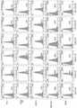


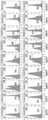
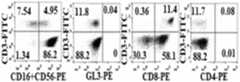
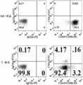


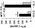
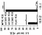

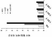


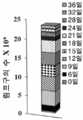
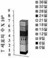

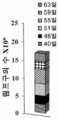


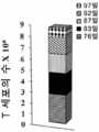

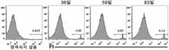
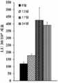

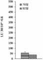


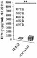


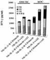

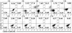

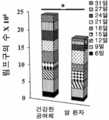


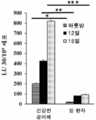
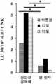


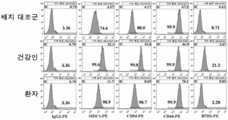

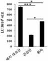

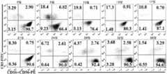


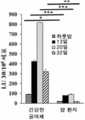
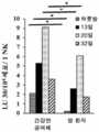

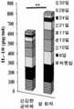


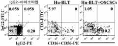


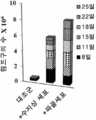


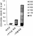


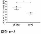


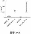

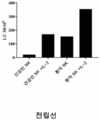
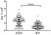


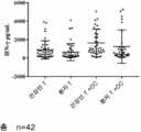

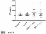


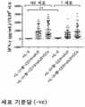


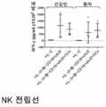


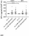


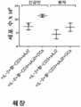


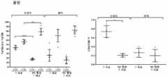


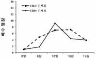

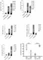
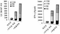

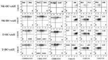

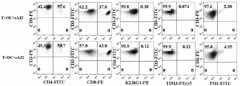

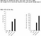


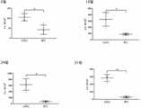


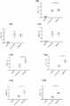
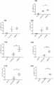


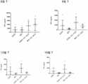

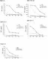


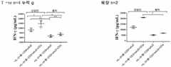
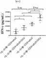


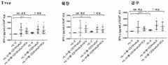


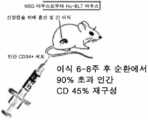


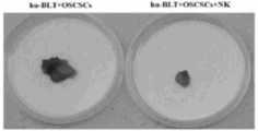
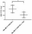
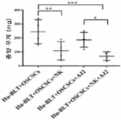


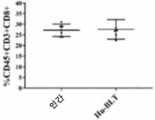
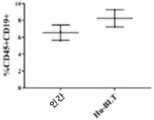

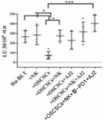

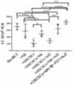
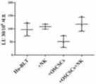

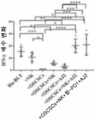
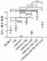

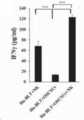
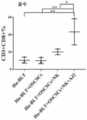


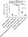
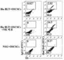

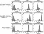
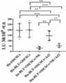


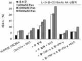
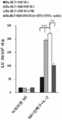
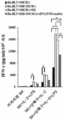


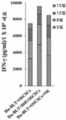
Classifications
- C—CHEMISTRY; METALLURGY
- C12—BIOCHEMISTRY; BEER; SPIRITS; WINE; VINEGAR; MICROBIOLOGY; ENZYMOLOGY; MUTATION OR GENETIC ENGINEERING
- C12N—MICROORGANISMS OR ENZYMES; COMPOSITIONS THEREOF; PROPAGATING, PRESERVING, OR MAINTAINING MICROORGANISMS; MUTATION OR GENETIC ENGINEERING; CULTURE MEDIA
- C12N5/00—Undifferentiated human, animal or plant cells, e.g. cell lines; Tissues; Cultivation or maintenance thereof; Culture media therefor
- C12N5/06—Animal cells or tissues; Human cells or tissues
- C12N5/0602—Vertebrate cells
- C12N5/0634—Cells from the blood or the immune system
- C12N5/0646—Natural killers cells [NK], NKT cells
- A—HUMAN NECESSITIES
- A61—MEDICAL OR VETERINARY SCIENCE; HYGIENE
- A61P—SPECIFIC THERAPEUTIC ACTIVITY OF CHEMICAL COMPOUNDS OR MEDICINAL PREPARATIONS
- A61P35/00—Antineoplastic agents
- A—HUMAN NECESSITIES
- A61—MEDICAL OR VETERINARY SCIENCE; HYGIENE
- A61K—PREPARATIONS FOR MEDICAL, DENTAL OR TOILETRY PURPOSES
- A61K35/00—Medicinal preparations containing materials or reaction products thereof with undetermined constitution
- A61K35/12—Materials from mammals; Compositions comprising non-specified tissues or cells; Compositions comprising non-embryonic stem cells; Genetically modified cells
- A61K35/14—Blood; Artificial blood
- A61K35/17—Lymphocytes; B-cells; T-cells; Natural killer cells; Interferon-activated or cytokine-activated lymphocytes
- A—HUMAN NECESSITIES
- A61—MEDICAL OR VETERINARY SCIENCE; HYGIENE
- A61K—PREPARATIONS FOR MEDICAL, DENTAL OR TOILETRY PURPOSES
- A61K35/00—Medicinal preparations containing materials or reaction products thereof with undetermined constitution
- A61K35/12—Materials from mammals; Compositions comprising non-specified tissues or cells; Compositions comprising non-embryonic stem cells; Genetically modified cells
- A61K35/32—Bones; Osteocytes; Osteoblasts; Tendons; Tenocytes; Teeth; Odontoblasts; Cartilage; Chondrocytes; Synovial membrane
- A—HUMAN NECESSITIES
- A61—MEDICAL OR VETERINARY SCIENCE; HYGIENE
- A61K—PREPARATIONS FOR MEDICAL, DENTAL OR TOILETRY PURPOSES
- A61K35/00—Medicinal preparations containing materials or reaction products thereof with undetermined constitution
- A61K35/66—Microorganisms or materials therefrom
- A61K35/74—Bacteria
- A61K35/741—Probiotics
- A61K35/744—Lactic acid bacteria, e.g. enterococci, pediococci, lactococci, streptococci or leuconostocs
- A61K35/745—Bifidobacteria
- A—HUMAN NECESSITIES
- A61—MEDICAL OR VETERINARY SCIENCE; HYGIENE
- A61K—PREPARATIONS FOR MEDICAL, DENTAL OR TOILETRY PURPOSES
- A61K35/00—Medicinal preparations containing materials or reaction products thereof with undetermined constitution
- A61K35/66—Microorganisms or materials therefrom
- A61K35/74—Bacteria
- A61K35/741—Probiotics
- A61K35/744—Lactic acid bacteria, e.g. enterococci, pediococci, lactococci, streptococci or leuconostocs
- A61K35/747—Lactobacilli, e.g. L. acidophilus or L. brevis
- A—HUMAN NECESSITIES
- A61—MEDICAL OR VETERINARY SCIENCE; HYGIENE
- A61K—PREPARATIONS FOR MEDICAL, DENTAL OR TOILETRY PURPOSES
- A61K39/00—Medicinal preparations containing antigens or antibodies
- A61K39/395—Antibodies; Immunoglobulins; Immune serum, e.g. antilymphocytic serum
- A—HUMAN NECESSITIES
- A61—MEDICAL OR VETERINARY SCIENCE; HYGIENE
- A61K—PREPARATIONS FOR MEDICAL, DENTAL OR TOILETRY PURPOSES
- A61K40/00—Cellular immunotherapy
- A61K40/10—Cellular immunotherapy characterised by the cell type used
- A61K40/15—Natural-killer [NK] cells; Natural-killer T [NKT] cells
- A—HUMAN NECESSITIES
- A61—MEDICAL OR VETERINARY SCIENCE; HYGIENE
- A61K—PREPARATIONS FOR MEDICAL, DENTAL OR TOILETRY PURPOSES
- A61K40/00—Cellular immunotherapy
- A61K40/40—Cellular immunotherapy characterised by antigens that are targeted or presented by cells of the immune system
- A61K40/41—Vertebrate antigens
- A61K40/42—Cancer antigens
- C—CHEMISTRY; METALLURGY
- C07—ORGANIC CHEMISTRY
- C07K—PEPTIDES
- C07K16/00—Immunoglobulins [IGs], e.g. monoclonal or polyclonal antibodies
- C07K16/18—Immunoglobulins [IGs], e.g. monoclonal or polyclonal antibodies against material from animals or humans
- C07K16/28—Immunoglobulins [IGs], e.g. monoclonal or polyclonal antibodies against material from animals or humans against receptors, cell surface antigens or cell surface determinants
- C07K16/2803—Immunoglobulins [IGs], e.g. monoclonal or polyclonal antibodies against material from animals or humans against receptors, cell surface antigens or cell surface determinants against the immunoglobulin superfamily
- C07K16/283—Immunoglobulins [IGs], e.g. monoclonal or polyclonal antibodies against material from animals or humans against receptors, cell surface antigens or cell surface determinants against the immunoglobulin superfamily against Fc-receptors, e.g. CD16, CD32, CD64
- C—CHEMISTRY; METALLURGY
- C12—BIOCHEMISTRY; BEER; SPIRITS; WINE; VINEGAR; MICROBIOLOGY; ENZYMOLOGY; MUTATION OR GENETIC ENGINEERING
- C12N—MICROORGANISMS OR ENZYMES; COMPOSITIONS THEREOF; PROPAGATING, PRESERVING, OR MAINTAINING MICROORGANISMS; MUTATION OR GENETIC ENGINEERING; CULTURE MEDIA
- C12N5/00—Undifferentiated human, animal or plant cells, e.g. cell lines; Tissues; Cultivation or maintenance thereof; Culture media therefor
- C12N5/06—Animal cells or tissues; Human cells or tissues
- C12N5/0602—Vertebrate cells
- C12N5/0634—Cells from the blood or the immune system
- C12N5/0643—Osteoclasts
- A—HUMAN NECESSITIES
- A61—MEDICAL OR VETERINARY SCIENCE; HYGIENE
- A61K—PREPARATIONS FOR MEDICAL, DENTAL OR TOILETRY PURPOSES
- A61K35/00—Medicinal preparations containing materials or reaction products thereof with undetermined constitution
- A61K2035/11—Medicinal preparations comprising living procariotic cells
- A61K2035/115—Probiotics
- A—HUMAN NECESSITIES
- A61—MEDICAL OR VETERINARY SCIENCE; HYGIENE
- A61K—PREPARATIONS FOR MEDICAL, DENTAL OR TOILETRY PURPOSES
- A61K2121/00—Preparations for use in therapy
- A—HUMAN NECESSITIES
- A61—MEDICAL OR VETERINARY SCIENCE; HYGIENE
- A61K—PREPARATIONS FOR MEDICAL, DENTAL OR TOILETRY PURPOSES
- A61K2300/00—Mixtures or combinations of active ingredients, wherein at least one active ingredient is fully defined in groups A61K31/00 - A61K41/00
- A—HUMAN NECESSITIES
- A61—MEDICAL OR VETERINARY SCIENCE; HYGIENE
- A61K—PREPARATIONS FOR MEDICAL, DENTAL OR TOILETRY PURPOSES
- A61K35/00—Medicinal preparations containing materials or reaction products thereof with undetermined constitution
- A61K35/66—Microorganisms or materials therefrom
- A61K35/74—Bacteria
- A61K35/741—Probiotics
- C—CHEMISTRY; METALLURGY
- C12—BIOCHEMISTRY; BEER; SPIRITS; WINE; VINEGAR; MICROBIOLOGY; ENZYMOLOGY; MUTATION OR GENETIC ENGINEERING
- C12N—MICROORGANISMS OR ENZYMES; COMPOSITIONS THEREOF; PROPAGATING, PRESERVING, OR MAINTAINING MICROORGANISMS; MUTATION OR GENETIC ENGINEERING; CULTURE MEDIA
- C12N2500/00—Specific components of cell culture medium
- C12N2500/70—Undefined extracts
- C12N2500/72—Undefined extracts from bacteria
- C—CHEMISTRY; METALLURGY
- C12—BIOCHEMISTRY; BEER; SPIRITS; WINE; VINEGAR; MICROBIOLOGY; ENZYMOLOGY; MUTATION OR GENETIC ENGINEERING
- C12N—MICROORGANISMS OR ENZYMES; COMPOSITIONS THEREOF; PROPAGATING, PRESERVING, OR MAINTAINING MICROORGANISMS; MUTATION OR GENETIC ENGINEERING; CULTURE MEDIA
- C12N2501/00—Active agents used in cell culture processes, e.g. differentation
- C12N2501/50—Cell markers; Cell surface determinants
- C12N2501/515—CD3, T-cell receptor complex
- C—CHEMISTRY; METALLURGY
- C12—BIOCHEMISTRY; BEER; SPIRITS; WINE; VINEGAR; MICROBIOLOGY; ENZYMOLOGY; MUTATION OR GENETIC ENGINEERING
- C12N—MICROORGANISMS OR ENZYMES; COMPOSITIONS THEREOF; PROPAGATING, PRESERVING, OR MAINTAINING MICROORGANISMS; MUTATION OR GENETIC ENGINEERING; CULTURE MEDIA
- C12N2502/00—Coculture with; Conditioned medium produced by
- C12N2502/11—Coculture with; Conditioned medium produced by blood or immune system cells
- C12N2502/1114—T cells
- C—CHEMISTRY; METALLURGY
- C12—BIOCHEMISTRY; BEER; SPIRITS; WINE; VINEGAR; MICROBIOLOGY; ENZYMOLOGY; MUTATION OR GENETIC ENGINEERING
- C12N—MICROORGANISMS OR ENZYMES; COMPOSITIONS THEREOF; PROPAGATING, PRESERVING, OR MAINTAINING MICROORGANISMS; MUTATION OR GENETIC ENGINEERING; CULTURE MEDIA
- C12N2502/00—Coculture with; Conditioned medium produced by
- C12N2502/11—Coculture with; Conditioned medium produced by blood or immune system cells
- C12N2502/1142—Osteoclasts
Landscapes
- Health & Medical Sciences (AREA)
- Life Sciences & Earth Sciences (AREA)
- Chemical & Material Sciences (AREA)
- General Health & Medical Sciences (AREA)
- Engineering & Computer Science (AREA)
- Veterinary Medicine (AREA)
- Public Health (AREA)
- Animal Behavior & Ethology (AREA)
- Biomedical Technology (AREA)
- Microbiology (AREA)
- Epidemiology (AREA)
- Medicinal Chemistry (AREA)
- Organic Chemistry (AREA)
- Zoology (AREA)
- Biotechnology (AREA)
- Immunology (AREA)
- Pharmacology & Pharmacy (AREA)
- Mycology (AREA)
- Genetics & Genomics (AREA)
- Bioinformatics & Cheminformatics (AREA)
- Wood Science & Technology (AREA)
- Cell Biology (AREA)
- Molecular Biology (AREA)
- Biochemistry (AREA)
- Hematology (AREA)
- Orthopedic Medicine & Surgery (AREA)
- General Engineering & Computer Science (AREA)
- Developmental Biology & Embryology (AREA)
- Virology (AREA)
- Rheumatology (AREA)
- Nuclear Medicine, Radiotherapy & Molecular Imaging (AREA)
- Chemical Kinetics & Catalysis (AREA)
- General Chemical & Material Sciences (AREA)
- Biophysics (AREA)
- Proteomics, Peptides & Aminoacids (AREA)
- Medicines Containing Material From Animals Or Micro-Organisms (AREA)
- Medicines That Contain Protein Lipid Enzymes And Other Medicines (AREA)
- Micro-Organisms Or Cultivation Processes Thereof (AREA)
- Medicines Containing Antibodies Or Antigens For Use As Internal Diagnostic Agents (AREA)
- Materials For Medical Uses (AREA)
Abstract
Translated fromKoreanDescription
Translated fromKorean관련relation출원에 대한 교차참조Cross Reference to Application
본원은 하기 미국 특허의 우선권의 이점을 주장한다: 본 명세서에 전체적으로 참고로 편입되는, 2017년 2월 15일 출원된, 특허 가출원 일련번호 62/459,397.This application claims the benefit of the following priorities of the following US patents: Patent Provisional Serial No. 62 / 459,397, filed February 15, 2017, which is incorporated herein by reference in its entirety.
자연살해 (NK) 세포는 MHC 부류 I, CD54 및 B7H1의 낮은 발현과 CD44의 높은 발현을 갖는 암 줄기 세포/미분화된 종양을 용해하고 분화한다. 주변-혈액 림프구의 중간 및 높은 세포독성 활성은 감소된 암 위험과 연관되고, 종양의 높은 NK-세포 침윤은 더 나은 예후와 연관되고, 반면에 낮은 활성은 증가된 암 위험과 연관된다.Natural killer (NK) cells lyse and differentiate cancer stem cells / undifferentiated tumors with low expression of MHC class I, CD54 and B7H1 and high expression of CD44. Medium and high cytotoxic activity of peripheral-blood lymphocytes is associated with reduced cancer risk, and high NK-cell infiltration of tumors is associated with better prognosis, while low activity is associated with increased cancer risk.
NK 세포의 억제는 종양 미세환경에서 NK 수용체의 하향조절에 의해 매개된다. NK 세포의 기능은 이전에 종양 환자에서 상당히 감소되는 것으로 나타났다. 몇 개의시험관내 NK 팽창 기술은 더 높은 치료적 세포 용량을 허용하기 위해 개발되었다. 피더 세포 예컨대 인터류킨 (IL)-15 및 41BB 리간드를 발현하는 K562 세포, EBV-TM-LCL, 윌름스 종양 또는 조사된 PBMC로 말초 혈액 단핵 세포 (PBMC) 또는 NK 세포의 정제된 모집단의 자극은 적절한 기능을 갖는 더 많은 수의 NK 세포를 초래했다. 생성된 NK 세포는 더 높은 수준의 NKG2D, 천연 세포독성 수용체, DNAM-1, 및 ICAM-1을 발현하였다. 따라서,생체외-팽창된, 활성화된, 및 CD3+ T 세포-감손된 NK 세포를 수득하기 위한 다양한 방법이 임상적 사용에 대해 확립되었다. 또한, 진행 암이 있는 환자에서 HLA-반일치 NK 세포의 입양 세포 전이의 안전성 및 효능이 확립되었다. 추가로, 임상 시험은 동종이계 NK 세포가 고형 종양에서 치료적 역할을 하고, 환자에게 전이하기에 안전하다는 것을 보여 주었다.Inhibition of NK cells is mediated by downregulation of NK receptors in the tumor microenvironment. The function of NK cells has previously been shown to be significantly reduced in tumor patients. Several invitro NK expansion techniques have been developed to allow for higher therapeutic cell doses. Stimulation of purified populations of peripheral blood mononuclear cells (PBMCs) or NK cells with feeder cells such as K562 cells, EBV-TM-LCL, Wilms' tumor, or irradiated PBMCs expressing interleukin (IL) -15 and 41BB ligands are appropriate. It resulted in a greater number of NK cells with function. The resulting NK cells expressed higher levels of NKG2D, natural cytotoxic receptors, DNAM-1, and ICAM-1. Thus, various methods for obtainingex vivo -expanded, activated, and CD3 + T cell-depleted NK cells have been established for clinical use. In addition, the safety and efficacy of adoptive cell metastasis of HLA-matched NK cells in patients with advanced cancer has been established. In addition, clinical trials have shown that allogeneic NK cells play a therapeutic role in solid tumors and are safe to metastasize to patients.
NK 세포로의 면역요법은 충분한 수의 고도로 기능적 NK 세포를 얻을 수 없기 때문에 제한되어 왔다. 또한, 건강한 개체로부터의 NK 세포와 달리, 종양-담지 인간화된 마우스로부터의 것에 유사한 환자 NK 세포의 팽창은 그것의 더 빠른 증식하는 능력에 의해 NK 세포를 떼어내는 T 세포를 오염시키는 작은 분획의 팽창으로 인해 상당히 제한된다.Immunotherapy with NK cells has been limited because a sufficient number of highly functional NK cells cannot be obtained. In addition, unlike NK cells from healthy individuals, the expansion of patient NK cells similar to that from tumor-supported humanized mice results in the expansion of a small fraction that contaminates T cells that detach NK cells by their faster proliferative capacity. Due to this is quite limited.
NK 세포 면역조절의 기본 기전은 이해되지 않는다. 큰 필요성이 개선된 NK 면역요법을 위한 치료적 조성물 및 방법을 식별하는 것에 대해 존재한다.The basic mechanism of NK cell immunoregulation is not understood. A great need exists for identifying therapeutic compositions and methods for improved NK immunotherapy.
본 발명은, 적어도 부분적으로, 파골세포가 NK 세포의 팽창을 유도할 수 있다는 발견에 기초하며, 이는 건강한 인간 및 암 환자 둘 모두에서 CD8+/CD4+ T 세포 비를 추가로 증가시킨다. 비록 암 환자는 일반적으로 건강한 인간에 비하여 생체내에서 보다 많은 NK 세포 및 CD8+/CD4+ T 세포 비를 가지지만, 과잉 NK 및 CD8+ T 세포는 (NK 세포 기능을 억제할 수 있는 오염 T 세포의 팽창에 기인하여) 단 수명이고 활성 (예를 들어, 세포 독성 및 사이토카인 선택)을 결한다. 그러나, 파골세포는 NK 세포 팽창을 유도할 수 있고 (예를 들어, 그것의 사이토카인 분비 능력에 의해 측정될 때) 암 환자에서 NK 세포의 세포 수 및 기능 둘 모두를 증가시킬 수 있다. 수지상 세포는 T 세포의 팽창을 우선적으로 촉진하는 반면에 파골세포는 NK 세포의 팽창을 우선적으로 촉진하여, T 및 NK 세포의 선택적 팽창에 대한 미세환경적 차이를 시사한다. 따라서, 본 발명은 면역치료적 전략에서 사용하기 위한 다수의 활성화된 NK 세포를 팽창시키는 방법을 제공한다. 이러한 세포는 암 줄기 세포를 억제 또는 제거하고 줄기-유사/저조하게 분화된 종양의 분화를 촉진함으로써 종양 성장을 제어하는데 이용될 수 있다The present invention is based, at least in part, on the discovery that osteoclasts can induce expansion of NK cells, which further increases the CD8 + / CD4 + T cell ratio in both healthy human and cancer patients. Although cancer patients generally have more NK cell and CD8 + / CD4 + T cell ratios in vivo compared to healthy humans, excess NK and CD8 + T cells (extensive expansion of contaminating T cells that can inhibit NK cell function). Due to short lifespan and lack activity (eg, cytotoxicity and cytokine selection). However, osteoclasts can induce NK cell expansion (as measured by its cytokine secreting capacity, for example) and increase both cell number and function of NK cells in cancer patients. Dendritic cells preferentially promote T cell expansion, while osteoclasts preferentially promote NK cell expansion, suggesting microenvironmental differences in the selective expansion of T and NK cells. Accordingly, the present invention provides a method of expanding multiple activated NK cells for use in immunotherapeutic strategies. Such cells can be used to control tumor growth by inhibiting or eliminating cancer stem cells and promoting the differentiation of stem-like / poorly differentiated tumors.
파골세포 (OC)와 함께 배지에서 NK 세포를 배양하는 것을 포함하는, 시험관내 또는 생체외에서 NK 세포를 활성화시키는 방법이 본 명세서에 제공된다. NK는 선택적으로 이것이 형질전환되지 않은 일차 NK 세포일 수 있다. 활성화된 NK 세포는 4주 내에 약 10, 11, 12, 13, 14, 15, 16, 17, 18, 19, 20, 21, 22, 23, 24, 25, 또는 그 초과 모집단 배증 (population doublings)으로 팽창될 수 있다. 배양물은 복수의 파골세포 (OC) 및 복수의 NK 세포를 포함할 수 있고, 예를 들어, 세포 배양물에서 OC:NK 세포의 비는 적어도 1:2이다. 파골세포는, 예를 들어, NK 세포에 의한 경구 편평상피 암종 줄기-유사 세포 (OSCSC)의 용해 또는51Cr 방출 세포독성 검정에 의해 측정될 때 NK 세포 세포독성을 고양할 수 있다.Provided herein are methods of activating NK cells in vitro or ex vivo, including culturing NK cells in media with osteoclasts (OC). The NK may optionally be primary NK cells in which it has not been transformed. Activated NK cells have about 10, 11, 12, 13, 14, 15, 16, 17, 18, 19, 20, 21, 22, 23, 24, 25, or more population doublings within 4 weeks. Can be expanded. The culture may comprise a plurality of osteoclasts (OC) and a plurality of NK cells, eg, the ratio of OC: NK cells in the cell culture is at least 1: 2. Osteoclasts can enhance NK cell cytotoxicity, for example, as measured by lysis of oral squamous carcinoma stem-like cells (OSCSC) by NK cells or51 Cr release cytotoxicity assays.
추가로, 파골세포는 NK 세포에 의해 생산된 적어도 하나의 사이토카인 또는 케모카인의 생산, 분비, 및/또는 기능을 고양할 수 있다. 예를 들어, 파골세포는 NK 세포에 의한 IFN-γ 및/또는 IL-12의 분비, 및/또는 NK 세포에 의한 NKG2D, NKp46, NKp44, NKp30, CD94, KIR2, 및 KIR3 중 하나 이상의 발현을 고양할 수 있다.In addition, osteoclasts can enhance the production, secretion, and / or function of at least one cytokine or chemokine produced by NK cells. For example, osteoclasts enhance the secretion of IFN-γ and / or IL-12 by NK cells and / or expression of one or more of NKG2D, NKp46, NKp44, NKp30, CD94, KIR2, and KIR3 by NK cells. can do.
NK 세포는 인간 대상체의 암 샘플로부터 정제된 세포일 수 있다. 특정 구현예에서, 세포 배양물은 또한 암 샘플로부터 유래하는 T 세포를 더 포함한다. 그와 같은 특정 구현예에서, NK 세포는 T 세포에 비하여 우선적으로 팽창될 수 있다. NK 세포는 임의의 시간의 길이, 예를 들어, 적어도 1개월 동안 팽창될 수 있다. 배양 배지에는 우선적으로 NK 세포가 팽창하는 것을 지속하도록 적어도 하나의 파골세포가 보충될 수 있다. T 세포, 예를 들어,51Cr 방출 세포독성 검정에서, 예를 들어, T 세포에 의한 OSCSC의 용해에 의해 측정될 때 IFN-γ를 분비할 수 있지만 세포독성을 조정하지 않을 수 있다. 팽창된 NK 세포는 CD8+ T 세포를 팽창할 수 있다. OC에 의해 팽창된 NK 세포는 또한 CD4+ T 세포에 비하여 CD8+ T 세포를 우선적으로 팽창할 수 있다. 특정 구현예에서, 본 방법은 추가로, 예를 들어, NK 세포에 의한 IFN-γ의 분비를 더 향상시키기 위해 세포 배양에 항-CD3 항체를 부가하는 것을 포함한다. 활성화된 NK 세포는 스플릿 무력화될 수 있다.NK cells can be cells purified from cancer samples of a human subject. In certain embodiments, the cell culture further comprises T cells derived from a cancer sample. In such specific embodiments, NK cells may preferentially expand over T cells. NK cells can be expanded for any length of time, eg, for at least 1 month. The culture medium may be supplemented with at least one osteoclast to preferentially continue to expand NK cells. In T cells, eg,51 Cr release cytotoxicity assays, they may secrete IFN-γ as measured, for example, by lysis of OSCSC by T cells, but may not modulate cytotoxicity. Expanded NK cells can expand CD8 + T cells. NK cells expanded by OC can also preferentially expand CD8 + T cells as compared to CD4 + T cells. In certain embodiments, the method further comprises adding an anti-CD3 antibody to the cell culture, for example, to further enhance the secretion of IFN-γ by NK cells. Activated NK cells can be split neutralized.
특정 구현예에서, 본 방법은 다음으로부터 선택된 적어도 하나의 세균 균주를 포함하는 조성물을 세포 배양에 부가하는 것을 추가로 포함할 수 있다:연쇄상구균써모필,비피도박테리움론구움,비피도박테리움브레베,비피도박테리움인판티스,락토바실러스악시도필루스,락토바실러스플란타룸,락토바실러스파라카세이, KE99, 및락토바실러스불가리쿠스, 선택적으로 여기서 적어도 하나의 세균 균주는 살아 있을 수 있거나 또는 초음파처리될 수 있다. 예를 들어, 본 조성물은 하기를 포함할 수 있다:연쇄상구균써모필,비피도박테리움론구움,비피도박테리움브레베,비피도박테리움인판티스,락토바실러스악시도필루스,락토바실러스플란타룸,락토바실러스파라카세이, KE99, 및락토바실러스불가리쿠스. 대안적으로 또는 추가로, 본 조성물은 sAJ2 박테리아를 포함할 수 있다. 세포 배양에서 NK 세포에 대한 sAJ2 박테리아 농도 및/또는 OC 농도의 비는, 예를 들어, i) NK 세포:sAJ2에 대해 적어도 1:2; ii) OC:sAJ2에 대해 적어도 1:4; 및/또는 iii) OC:NK 세포:sAJ2에 대해 적어도 1:2:4일 수 있다.In certain embodiments, the method may further comprise adding to the cell culture a composition comprising at least one bacterial strain selected from:Streptococcusthermophil ,BifidobacteriumRonguum ,BifidobacteriumBreve ,BifidobacteriumInfante Tees,Lactobacillusevil attempts Phil Ruth,LactobacillusPlanta Room ,LactobacillusFarah Casey, KE99, andLactobacillusBulgari , optionally wherein at least one bacterial strain may be live or sonicated. For example, the composition may include:streptococciThermophile ,BifidobacteriumRonguum ,BifidobacteriumBreve ,BifidobacteriumInfantis ,LactobacillusAxidophyllus ,LactobacillusPlanta Room ,LactobacillusFarah Casey, KE99, andLactobacillusBulgaricus. In the anjeokfor or in addition, the composition may comprise sAJ2 bacteria. The ratio of sAJ2 bacterial concentration and / or OC concentration to NK cells in cell culture may be, for example, i) at least 1: 2 for NK cells: sAJ2; ii) at least 1: 4 for OC: sAJ2; And / or iii) at least 1: 2: 4 for OC: NK cells: sAJ2.
특정 구현예에서, 본 방법은 세포 배양에 NK 세포를 활성화시킬 수 있는 또 다른 제제를 부가하는 것을 추가로 포함할 수 있다.In certain embodiments, the method may further comprise adding another agent capable of activating NK cells to the cell culture.
특정 바람직한 구현예에서, 상기 방법은 하기를 포함한다: i) 파골세포 (OC), NK 세포, 및 T 세포를 포함하는 세포 배양을 제공함; 및 ii) 세포 배양에서 NK 세포, T 세포, 및 파골세포를 배양하고, 그것에 의해 우선적으로 T 세포에 비하여 NK 세포를 활성화시킴.In certain preferred embodiments, the method comprises: i) providing a cell culture comprising osteoclasts (OC), NK cells, and T cells; And ii) culturing NK cells, T cells, and osteoclasts in cell culture, thereby activating NK cells preferentially over T cells.
유사하게, 하기를 포함하는 방법이 본 명세서에 제공된다: i) 수지상 세포 (DC), NK 세포 및 T 세포를 포함하는 세포 배양을 제공함; 및 ii) 세포 배양에서 NK 세포, T 세포, 및 수지상 세포의 세포를 배양하고, 그것에 의해 우선적으로 NK 세포에 비하여 T 세포를 활성화시킴. 일 구현예에서, NK 세포는 선택적으로 일차 NK 세포 형질전환되지 않은 일차 NK 세포일 수 있다. 배양물은 복수의 파골세포 (OC) 및 복수의 NK 세포를 포함할 수 있고, 예를 들어, 세포 배양물에서 OC:NK 세포의 비는 적어도 1:2이다. 파골세포는 NK 세포 팽창, 및/또는 NK 세포에 의한 IL-15의 분비를 고양할 수 있다. 활성화된 NK 세포는 4주 내에 적어도 약 10, 11, 12, 13, 14, 15, 16, 17, 18, 19, 20, 21, 22, 23, 24, 25, 또는 그 초과 모집단 배증으로 팽창할 수 있다. 추가로, 파골세포는, 예를 들어, NK 세포에 의해 경구 편평상피 암종 줄기-유사세포 (OSCSC)의 용해에 의해 측정될 때, NK 세포 세포독성을 고양할 수 있다. 세포 세포독성은51Cr 방출 세포독성 검정에 의해 측정될 수 있다.Similarly, provided herein is a method comprising: i) providing a cell culture comprising dendritic cells (DC), NK cells and T cells; And ii) culturing the cells of NK cells, T cells, and dendritic cells in cell culture, thereby preferentially activating T cells over NK cells. In one embodiment, the NK cells may be primary NK cells, optionally not primary NK cell transformed. The culture may comprise a plurality of osteoclasts (OC) and a plurality of NK cells, eg, the ratio of OC: NK cells in the cell culture is at least 1: 2. Osteoclasts can enhance NK cell expansion and / or secretion of IL-15 by NK cells. Activated NK cells will expand with at least about 10, 11, 12, 13, 14, 15, 16, 17, 18, 19, 20, 21, 22, 23, 24, 25, or more population doubling within 4 weeks. Can be. In addition, osteoclasts can enhance NK cell cytotoxicity, for example, as measured by lysis of oral squamous carcinoma stem-like cells (OSCSCs) by NK cells. Cell cytotoxicity can be measured by51 Cr release cytotoxicity assay.
특정 구현예에서, 파골세포 (OC)는 NK 세포에 의해 생산된 적어도 하나의 사이토카인 또는 케모카인의 생산, 분비, 및/또는 기능을 고양할 수 있다. 예를 들어, 파골세포는 NK 세포에 의해 IFN-γ 및/또는 IL-12의 분비를 고양할 수 있다. 파골세포는 NK 세포에 의해 NKG2D, NKp46, NKp44, NKp30, CD94, KIR2, 및 KIR3 중 하나 이상의 발현을 고양할 수 있다. NK 세포 및/또는 T 세포는 대상체, 예를 들어, 인간 대상체로부터의 암 샘플로부터 정제될 수 있다. 특정 구현예에서, NK 세포의 우선적인 활성화는 적어도 1개월 동안 지속할 수 있다. 추가로, NK 세포의 우선적인 활성화가 약화하거나 또는 중단한 후, 적어도 하나의 파골세포는 NK 세포 배양 후 적어도 1개월 동안 세포 배양에 첨가될 수 있고, 그것에 의해 NK 세포의 활성화가 지속된다. 일부 구현예에서, T 세포는 IFN-γ를 분비할 수 있지만 세포독성을 조정하지 않을 수 있다. 세포독성은, 예를 들어,51Cr 방출 세포독성 검정에 의해, T 세포에 의한 OSCSC의 용해에 의해 측정될 수 있다. 팽창된 NK 세포는 CD8+ T 세포를 팽창시킬 수 있고, 그리고 CD4+ T 세포에 비해 우선적으로 CD8+ T 세포를 팽창시킬 수 있다. 특정 구현예에서, DC에 의해 팽창된 NK 세포는 CD8+ T 세포에 대해 CD4+ T 세포를 우선적으로 팽창시킬 수 있다. 다른 구현예에서, 항-CD3 항체는, 예를 들어, NK 세포에 의한 IFN-γ의 분비를 추가로 고양하도록 세포 배양에 첨가될 수 있다. 추가로, 활성화된 NK 세포는 스플릿 무력화될 수 있다.In certain embodiments, osteoclasts (OC) can enhance the production, secretion, and / or function of at least one cytokine or chemokine produced by NK cells. For example, osteoclasts can enhance the secretion of IFN-γ and / or IL-12 by NK cells. Osteoclasts can enhance expression of one or more of NKG2D, NKp46, NKp44, NKp30, CD94, KIR2, and KIR3 by NK cells. NK cells and / or T cells can be purified from cancer samples from a subject, eg, a human subject. In certain embodiments, preferential activation of NK cells can last for at least 1 month. In addition, after preferential activation of NK cells has weakened or stopped, at least one osteoclast can be added to the cell culture for at least one month after NK cell culture, whereby activation of NK cells is continued. In some embodiments, T cells may secrete IFN-γ but may not modulate cytotoxicity. Cytotoxicity can be measured, for example, by lysis of OSCSC by T cells, by a51 Cr release cytotoxicity assay. Expanded NK cells can expand CD8 + T cells, and preferentially expand CD8 + T cells over CD4 + T cells. In certain embodiments, NK cells expanded by DC may preferentially expand CD4 + T cells relative to CD8 + T cells. In other embodiments, anti-CD3 antibodies can be added to the cell culture, for example, to further boost the secretion of IFN-γ by NK cells. In addition, activated NK cells can be split neutralized.
특정 구현예에서, 본 방법은 다음으로부터 선택된 적어도 하나의 세균 균주를 포함하는 조성물을 세포 배양에 부가하는 것을 추가로 포함할 수 있다:연쇄상구균써모필,비피도박테리움론구움,비피도박테리움브레베,비피도박테리움인판티스,락토바실러스악시도필루스,락토바실러스플란타룸,락토바실러스파라카세이, KE99, 및락토바실러스불가리쿠스, 선택적으로 여기서 적어도 하나의 세균 균주는 살아 있을 수 있거나 또는 초음파처리됨.예를 들어, 본 조성물은 하기를 포함할 수 있다:연쇄상구균써모필,비피도박테리움론구움,비피도박테리움브레베,비피도박테리움인판티스,락토바실러스악시도필루스,락토바실러스플란타룸,락토바실러스파라카세이,KE99, 및락토바실러스불가리쿠스. 대안적으로 또는 추가로, 본 조성물은 sAJ2 박테리아를 포함할 수 있다. 그와 같은 특정 구현예에서, 세포 배양에서 NK 세포에 대한 sAJ2 박테리아 농도 및/또는 OC 농도의 비는 i) NK 세포:sAJ2에 대해 적어도 1:2; ii) OC:sAJ2에 대해 적어도 1:4; 및/또는 iii) OC:NK 세포:sAJ2에 대해 적어도 1:2:4일 수 있다In certain embodiments, the method may further comprise adding to the cell culture a composition comprising at least one bacterial strain selected from:Streptococcusthermophil ,BifidobacteriumRonguum ,BifidobacteriumBreve ,BifidobacteriumInfante Tees,Lactobacillusevil attempts Phil Ruth,LactobacillusPlanta Room ,LactobacillusFarah Casey, KE99, andLactobacillusBulgaricus , optionally wherein at least one bacterial strain may be alive or sonicated. For example, the composition may comprise:Streptococcusthermophil,BifidobacteriumRonguum,BifidobacteriumBreve,BifidobacteriumInfantis,LactobacillusAxidophyllus,LactobacillusPlanta Room,LactobacillusFarahCasey,KE99, andLactobacillusBulgaricus. In the anjeokfor or in addition, the composition may comprise sAJ2 bacteria. In such specific embodiments, the ratio of sAJ2 bacterial concentration and / or OC concentration to NK cells in cell culture is i) at least 1: 2 for NK cells: sAJ2; ii) at least 1: 4 for OC: sAJ2; And / or iii) at least 1: 2: 4 for OC: NK cells: sAJ2.
추가로, 본 방법은 NK 세포를 활성화시킬 수 있는 또 다른 제제를 세포 배양에 부가하는 것을 추가로 포함할 수 있다. 본 방법은 T 세포를 활성화시킬 수 있는 또 다른 제제를 세포 배양에 부가하는 것을 추가로 포함할 수 있다.In addition, the method may further comprise adding another agent to the cell culture that can activate NK cells. The method may further comprise adding another agent to the cell culture that can activate T cells.
유사하게, 암 또는 암-관련된 질환 또는 장애가 있거나 있는 것으로 의심되는 대상체에게 치료적 유효량의 파골세포 (OC), 파골세포 (OC)를 포함하는 세포 배양물, 및/또는 파골세포 (OC)를 포함하는 세포 배양물의 상청액을 투여함에 의해 상기 대상체에서 암 또는 암-관련된 질환 또는 장애를 치료하는 방법이 본 명세서에 제공된다.Similarly, to a subject suspected of having or suspected of having a cancer or cancer-related disease or disorder, a therapeutically effective amount of osteoclasts (OC), cell cultures comprising osteoclasts (OC), and / or osteoclasts (OC) Provided herein are methods of treating cancer or cancer-associated diseases or disorders in such subjects by administering supernatants of cell cultures.
파골세포는 대상체에서 NK 세포 팽창을 고양할 수 있고, 선택적으로 여기서 파골세포는 NK 세포에 의해 IL-15의 분비를 고양한다. 특정 구현예에서, 향상된 NK 세포 팽창은 4주 내에 적어도 약 10, 11, 12, 13, 14, 15, 16, 17, 18, 19, 20, 21, 22, 23, 24, 25, 또는 그 초과 모집단 배증일 수 있다. 추가로, 파골세포는, 예를 들어, NK 세포에 의해 경구 편평상피 암종 줄기-유사세포 (OSCSC)의 용해에 의해 측정될 때, NK 세포 세포독성을 고양할 수 있다. 세포 세포독성은51Cr 방출 세포독성 검정에 의해 측정될 수 있다.Osteoclasts can elevate NK cell expansion in a subject, optionally where the osteoclasts elevate the secretion of IL-15 by NK cells. In certain embodiments, enhanced NK cell expansion is at least about 10, 11, 12, 13, 14, 15, 16, 17, 18, 19, 20, 21, 22, 23, 24, 25, or more within 4 weeks. It may be a population doubling. In addition, osteoclasts can enhance NK cell cytotoxicity, for example, as measured by lysis of oral squamous carcinoma stem-like cells (OSCSCs) by NK cells. Cell cytotoxicity can be measured by51 Cr release cytotoxicity assay.
특정 구현예에서, 파골세포는 NK 세포에 의해 생산된 적어도 하나의 사이토카인 또는 케모카인의 생산, 분비, 및/또는 기능을 증가 또는 향상시킬 수 있다. 예를 들어, 파골세포는 NK 세포에 의해 IFN-γ 및/또는 IL-12의 분비를 고양할 수 있다. 파골세포는 T 세포에 비해 NK 세포를 우선적으로 활성화시킬 수 있고 및/또는 T 세포에 비해 NK 세포의 팽창을 우선적으로 고양한다. 특정 구현예에서, NK 세포의 우선적인 팽창은 적어도 1개월 동안 지속할 수 있다. T 세포는, 예를 들어,51Cr 방출 세포독성 검정에서 T 세포에 의한 OSCSC의 용해에 의해 측정될 때, IFN-γ를 분비할 수 있지만 암의 세포독성은 조절하지 않을 수 있다.In certain embodiments, the osteoclasts can increase or enhance the production, secretion, and / or function of at least one cytokine or chemokine produced by NK cells. For example, osteoclasts can enhance the secretion of IFN-γ and / or IL-12 by NK cells. Osteoclasts can preferentially activate NK cells relative to T cells and / or preferentially boost NK cell expansion over T cells. In certain embodiments, preferential expansion of NK cells can last for at least 1 month. T cells may secrete IFN- [gamma] but may not modulate the cytotoxicity of cancer, eg, as measured by lysis of OSCSC by T cells in a51 Cr release cytotoxicity assay.
특정 구현예에서, 활성화된 NK 세포는 대상체에서 CD8+ T 세포를 팽창시킬 수 있고, 예를 들어, 우선적으로 CD4+ T 세포에 비해 CD8+ T 세포를 팽창시킬 수 있다. 대상체는 또한 NK 세포에 의해 IFN-γ의 분비를 더 고양하기 위해 항-CD3 항체로 처리될 수 있다. 활성화된 NK 세포는 스플릿 무력화될 수 있다.In certain embodiments, activated NK cells can inflate CD8 + T cells in a subject, eg, preferentially inflate CD8 + T cells as compared to CD4 + T cells. Subjects can also be treated with anti-CD3 antibodies to further boost the secretion of IFN-γ by NK cells. Activated NK cells can be split neutralized.
특정 구현예에서, 본 치료 방법은 다음으로부터 선택된 적어도 하나의 세균 균주를 포함하는 조성물을 대상체에게 투여하는 것을 추가로 포함할 수 있다:연쇄상구균써모필,비피도박테리움론구움,비피도박테리움브레베,비피도박테리움 인판티스,락토바실러스악시도필루스,락토바실러스플란타룸,락토바실러스파라카세이, KE99, 및락토바실러스불가리쿠스, 선택적으로 여기서 적어도 하나의 세균 균주는 살아 있을 수 있거나 또는 초음파처리됨.예를 들어, 본 조성물은 하기를 포함할 수 있다:연쇄상구균써모필,비피도박테리움론구움,비피도박테리움브레베,비피도박테리움인판티스,락토바실러스악시도필루스,락토바실러스플란타룸,락토바실러스파라카세이,KE99, 및락토바실러스불가리쿠스.특정 구현예에서, 본 조성물은 sAJ2 박테리아를 포함할 수 있다.In certain embodiments, the method of treatment may further comprise administering to the subject a composition comprising at least one bacterial strain selected from:Streptococcusthermophil ,BifidobacteriumRonguum ,BifidobacteriumBreve ,BifidobacteriumInfantis ,LactobacillusAxidophyllus ,LactobacillusPlanta Room ,LactobacillusFarah Casey, KE99, andLactobacillusBulgari , optionally wherein at least one bacterial strain may be alive or sonicated. For example, the composition may comprise:streptococciThermophile,BifidobacteriumRonguum,BifidobacteriumBreve,BifidobacteriumInfantis,LactobacillusAxidophyllus,LactobacillusPlanta Room,LactobacillusFarahCasey,KE99, andLactobacillusBulgaricus . In certain embodiments, the compositions may comprise sAJ2 bacteria.
특정 구현예에서, 본 방법은 세포 배양에 NK 세포를 활성화시킬 수 있는 또 다른 제제를 부가하는 것을 추가로 포함할 수 있다.In certain embodiments, the method may further comprise adding another agent capable of activating NK cells to the cell culture.
특정 구현예에서, 파골세포, 세포 배양물, 및/또는 상청액은 암에 대해 전신으로 또는 국소적으로 투여될 수 있는 약제학적 조성물에 투여될 수 있다. 특정 구현예에서, 파골세포, 세포 배양물, 및/또는 상청액은 대상체에게 적어도 2회 투여될 수 있고, 예를 들어, 파골세포, 세포 배양물, 및/또는 상청액은 제1 투여 이래 적어도 1개월 후에 대상체에게 투여될 수 있다.In certain embodiments, osteoclasts, cell cultures, and / or supernatants may be administered to pharmaceutical compositions that can be administered systemically or locally to cancer. In certain embodiments, the osteoclasts, cell culture, and / or supernatant may be administered to the subject at least twice, for example, the osteoclasts, cell culture, and / or supernatant are at least 1 month since the first administration. May be administered to the subject later.
일부 구현예에서, 대상체는 인간일 수 있다.In some embodiments, the subject can be a human.
도 1은 패널 A, B, C, 및 D로 확인된 4개의 패널을 포함하며, 이들은 파골세포에 의한 NK 활성화 리간드의 더 높은 발현을 도시한다. 파골세포 (OC)를 생성하기 위해, 단핵구는 대식세포 콜로니-자극 인자 (M-CSF) (25 ng/ml) 및 RANKL (25 ng/ml)을 함유하는 배지에서 21일 동안 배양되었다. 고순도 NK 세포 (1 x 10⁶세포/ml)는 각각 1:2:4 비 (OC:NK:sAJ2)에서 sAJ2 박테리아의 존재 또는 부재에서 자가 OC와 공 배양되기 전에 18시간 동안 IL-2 (1000 U/ml) 및 항-CD16 mAb (3 μg/ml)의 조합으로 처리되었다. CD3, CD16, 및 CD56의 표면 발현은 유세포측정을 사용하여 6, 9, 12, 15, 19, 24, 29, 및 34일째에서 공-배양물로부터의 1 x 10⁴ 림프구 샘플에서 분석되었고, 배양 배지는 새롭게 되고 rh-IL-2 (1000 U/ml)가 보충되었다 (도 1A). 세포는 도 1A에서 기재된 바와 같이 공배양되었고 팽창된 림프구는 현미경을 사용하여 수작업으로 카운트되었다 (도 1B). 단핵구는 건강한 공여체의 PBMC로부터 단리되었다. 수지상 세포를 생성하기 위해, 단핵구는 GM-CSF (150 ng/ml) 및 IL-4 (50 ng/ml)를 함유하는 배지에서 8일 동안 배양되었다. 파골세포가 생성되었고 NK 세포는 각각 1:2:4 비 (표적 세포:NK:sAJ2)에서 sAJ2 박테리아의 존재에서 자가 세포와 공 배양되기 전에 도 1A에서 기재된 바와 같이 정제되었다. 배양 6일째에서, 배양 배지가 제거되고 NK 세포는 상청액이 수확되기 전 5시간 동안 rh-IL-2로 처리되고, IFN-γ 분비는 단일 ELISA를 사용하여 결정되었다 (도 1C). 단핵구가 단리되고, 수지상 세포 및 파골세포는 도 1A 및 도 1D에서 기재된 바와 같이 생성되었고, OSCSC 및 K562 종양 세포주는 물질 및 방법에서 기재된 바와 같이 배양되었고, 1 x 10⁴ 세포를 사용하여, PE-접합된 항체 및 유세포측정을 이용한 MHC-1, CD54, KIR2, KIR3, KLRG1 및 MICA/B 표면 발현을 분석하였다. IgG2 아이소타입은 대조군으로서 사용되었다 (도 1D).
도 2는 패널 A 내지 J로 확인된 10개의 패널을 포함하며, 이들은 파골세포에 의한 NK 세포 및 수지상 세포에 의한 T 세포의 기능에서 우선적인 팽창 및 상당한 이득을 도시한다. 단핵구는 인간 PBMC로부터 정제되었고 GM-CSF (150 ng/ml) 및 IL-4 (50 ng/ml)로 8일 동안 배양되어 DC를 생성했다. 파골세포를 생성하기 위해, 단핵구는 M-CSF (25 ng/ml) 및 RANKL (25 ng/ml)을 함유하는 알파-MEM 배지에서 21일 동안 배양되었다. 팽창을 위해, 정제된 NK 세포 (1 x 10⁶ 세포/ml)를 1:2:4 비 (단핵구, DC 또는 OC:NK:sAJ2)에서 sAJ2의 존재에서 자가 단핵구, DC 또는 OC와 공배양하기 전에 18시간 동안 IL-2 (1000 U/ml) 및 항-CD16 mAb (3 μg/ml)의 조합으로 처리하였다. CD3, CD16, 및 CD56의 표면 발현은 유세포측정을 사용하여 도면에 표시된 일에 분석되었고, 배양 배지는 새롭게 되고 rh-IL-2 (1000 U/ml)가 보충되었다 (도 2A). 세포는 도 2A에 기재된 바와 같이 공배양되었고 팽창된 림프구의 수는 현미경적 결정을 사용하여 평가되었다 (도 2B). NK 세포 (도 2C) 및 T/NKT (도 2D) 세포의 수는 도 2B에서 총 팽창 세포 내의 NK 및 T/NKT 세포의 백분율 (도 2A)을 사용하여 결정되었다. 세포는 도 2A에 기재된 바와 같이 공배양되었고 세포독성은 경구 편평상피 세포 암종 줄기 세포주 (OSCSC)에 대한 표준 4-시간51Cr 방출 검정을 사용하여 도면에 도시된 일자에 결정되었다. 용해 단위 30/106 세포는 OSCSC x 100의 30%를 용해하는데 요구된 림프구의 역수를 사용하여 결정되었다 (도 2E). 상청액은 6, 9, 12 및 15일째에 도 1A에 기재된 바와 같이 OC와 함께 NK의 공-배양물로부터 수확되었고, IFN-γ 분비는 단일 ELISA를 사용하여 결정되었다 (도 2F). NK 세포는 도 2A에서 기재된 바와 같이 10명의 건강한 공여체로부터의 자가 파골세포로 공배양되고 팽창되었다. NK 세포의 누적 배수 팽창은 31일 동안 각각의 공여체에 대해 계산되었고 (도 2G), 모집단 배가는 기준선 카운트에 대한 최종 카운트의 비의 로그를 2의 로그로 나눈 값에 기반하여 계산되었다 (도 2H). 수지상 세포 및 파골세포는 도 2A에서 기재된 바와 같이 생성되었고, 1 x 10⁴ 세포를 사용하여, PE-접합된 항체 및 유세포측정 분석을 이용한 ULBP, KIR2, KIR3, KLRG1 및 MICA/B 표면 발현을 분석하였다. IgG2 아이소타입 대조군 항체가 대조군으로 사용되었다 (도 2I). 새롭게 단리된 NK 세포 (상부 열) 및 도 2A에서 기재된 바와 같이 자가 파골세포로 공배양되고 팽창된 NK 세포 (하부 열)를 사용하여, PE-접합된 항체를 이용한 CD16, Nkp30, Nkp44, Nkp46, KIR2, KIR3, CD94, 및 NKG2D 표면 발현을 분석하였다. IgG2 아이소타입 대조군 항체가 대조군으로 사용되었다 (도 2J).
도 3은 패널 A 내지 H로 확인된 8개 패널을 포함하며, 이것은 NK 세포와 달리 파골세포-팽창된 NK 세포로부터 정제된 T 세포는 OSCSC에 대해 세포독성을 조정하지 않고 IFN-γ를 중간 정도로 분비한다는 것을 도시한다. 새롭게 정제된 NK 세포는 물질 및 방법에서 기재된 바와 같이 단핵구-유래된 자가 파골세포로 처리되고 공배양되었다. CD3, CD16, CD56, GL3 (TCR γ/δ), CD4 및 CD8의 표면 발현은 FITC- 및 PE-접합된 항체 및 유세포측정을 사용하여 9일째에 공-배양물로부터의 림프구 샘플에서 분석되었다 (도 3A). NK 세포는 도 1A에서 기재된 바와 같이 자가 파골세포로 9일째 처리되고 공배양되었고, CD3T-양성 세포는 CD3T 양성 선택 키트를 사용하여 분류되었고, CD3T-음성 (NK) 세포의 순도는 CD3, CD16, CD56 FITC 및 PE-접합된 항체 및 유세포측정을 사용하여 평가되었다 (도 3B). CD3T-양성 세포 및 CD3T-음성 세포 (CD16 양성 세포)는 OSCSC (도 3C) 및 K562 (도 3D) 세포주에 대한 표준 4-시간 51Cr 방출 검정을 사용하여 세포독성에 대해 시험되기 전 18-20시간 동안 rh-IL-2 (1000 U/ml)로 처리되었다. 용해 단위 30/106 세포는 OSCSC 및 K562 각각에 대해 물질 및 방법에 기재된 방법을 사용하여 결정되었다. 상청액은 배양물로부터 수확되었고 IFN-γ 분비는 단일 ELISA를 사용하여 결정되었다 (도 3E). NK 세포, CD3T, CD4T, CD8T, 및 γδT 세포는 물질 및 방법에서 기재된 바와 같이 PBMC로부터 정제되었고, 이들이 OSCSC에 대해 표준 4-시간 51Cr 방출 검정을 사용한 세포독성에 대해 시험되기 전에 18-20시간 동안 rh-IL-2로 활성화되었다 (도 3F). 용해 단위 30/106 세포는 OSCSC x 100의 30%를 용해하는데 요구된 림프구의 역수를 사용하여 결정되었다 (도 3F). NK 및 T 세포는 물질 및 방법에서 기재된 바와 같이 PBMC로부터 정제되었고, NK 세포는 물질 및 방법에서 기재된 바와 같이 처리되었다. T 세포는 자가 OC로 배양되기 전 18-20시간 항-CD3 (1 μg/ml) 및 항-CD28 (3 μg/ml)로 활성화되었고, 팽창된 림프구는 배양 후 4일째 현미경을 사용하여 수작업으로 카운트되었다 (도 3G). NK 및 T 세포는 OC로 정제 및 배양되었고, 그리고 도 3G에서 기재된 바와 같이 4일째에 카운트되고, OC에 의해 팽창된 림프구의 배수 팽창은 OC 없는 림프구의 배수 팽창으로 나누어졌다 (도 3H).
도 4는 패널 A 내지 S로 확인된 19개 패널을 포함하며, 이것은 NK 세포의 감소된 비율, NK 세포-매개된 세포독성, 및 파골세포와 sAJ2 박테리아와 함께 NK 세포 배양물의 각각의 연속적인 재-자극으로 IFN-γ 분비를 도시한다. 새롭게 정제된 NK 세포는 도 2A에서 기재된 바와 같이 단핵구-유래된 자가 파골세포로 처리되고 공배양되었다. CD3, CD16 및 CD56의 표면 발현은 유세포측정 분석을 사용하여 도면에서 표시된 일에서 공-배양물로부터 1 x 10⁴ 림프구에서 분석되었다 (도 4A). 36일 후, NK 세포가 팽창이 중단될 때, 이들은 도 2A에서 기재된 바와 같이 신선한 자가 파골세포로 재-배양되었다. CD3, CD16, 및 CD56의 표면 발현은 항체 염색 및 유세포측정 분석을 사용하여 도면에서 표시된 일에 분석되었다 (도 4B). 63일 후, 세포가 팽창이 중단될 때, 기재된 바와 같이 OC로 재-배양되었고 CD3, CD16 및 CD56의 표면 발현은 도면에서 도시된 일에 분석되었다 (도 4C). 팽창된 림프구의 수는 현미경적 결정을 사용하여 평가되었고 (도 4D, 4G, 4J), 그리고 NK 세포 (도 4F, 4I, 4L) 및 T/NKT (도 4E, 4H, 4K) 세포의 수는 총 팽창 세포 내의 NK 및 T/NKT 세포의 백분율을 사용하여 결정되었다 (도 4D). 세포사는 프로피디움 아이오다이드 염색 및 유세포측정 분석을 사용하여 36, 59 및 83일째에 림프구에서 결정되었다 (도 4M). 새롭게 정제된 NK 세포는 도 2A에서 기재된 바와 같이 자가 파골세포로 처리되고 공배양되었다. 림프구는 그 다음 6, 17, 및 34일의 공-배양 (도 4N), 40 및 63일의 공-배양 (도 4O) 또는 76 및 92일의 공-배양 (도 4P) 후 OSCSC에 대한 표준 4-시간51Cr 방출 검정을 사용하여 세포독성에 대해 시험되었다. 용해 단위 30/106 세포는 도 2E에 기재된 방법을 사용하여 결정되었다. 상청액이 수확되고 IFN-γ 분비는 6, 9, 12, 15, 18 및 21일째, (도 4Q); 40, 46, 51, 55, 및 59일째 (도 4R), 및 76, 83, 92, 및 97일째 (도 4S)로부터의 상청액을 사용한 단일 ELISA를 사용하여 결정되었다.
도 5는 패널 A 내지 H로 확인된 8개의 패널을 포함하며, 이것은 K562 또는 OSCSC가 아닌 파골세포가 NK 세포를 팽창시키고 NK 세포 기능을 실질적으로 증가시킨다는 것을 도시한다. 파골세포를 생성하기 위해, 단핵구는 M-CSF (25 ng/ml) 및 RANKL (25 ng/ml)을 함유하는 배지에서 21일 동안 배양되었고, K562 종양 세포주는 물질 및 방법에서 기재된 바와 같이 배양되었다. 고순도 NK 세포 (1 x 10⁶ 세포/ml)를 각각 1:2:4 비 (OC:NK:sAJ2)에서 sAJ2 박테리아의 존재에서 K562 및 자가 OC로 공배양되기 전에 18시간 동안 IL-2 (1000 U/ml) 및 항-CD16mAb (3 μg/ml)의 조합으로 처리하였다. 배양된 것으로부터의 세포는 6, 10 및 13일째에 현미경을 사용하여 수작업으로 카운트되었다 (도 5A). 도 1A에서 기재된 바와 같이 생성된 파골세포 및 K562 종양 세포주는 물질 및 방법에서 기재된 바와 같이 40 그레이 (Gy)에서 조사되었다. NK 세포는 각각 1:2:4 비 (OC:NK:sAJ2)에서 sAJ2 박테리아의 존재에서 조사된 K562 및 조사된 자가 OC로 공배양되기 전에 물질 및 방법에서 기재된 바와 같이 정제되고 처리되었다. 배양된 것으로부터의 세포는 6, 10 및 13일째에 현미경을 사용하여 수작업으로 카운트되었다 (도 5B). NK 세포는 도 1A에서 기재된 바와 같이 OC 및 K562로 정제되고 배양되었으며, 6일 동안 공배양된 림프구의 세포독성은 OSCSC에 대한 표준 4-시간 51Cr 방출 검정을 사용하여 결정되었다. 용해 단위 30/106 세포는 도 3F에 기재된 방법을 사용하여 결정되었다 (도 5C). NK 세포는 도 5B에서 기재된 바와 같이 OC 및 K562로 정제되고 배양되었으며, 6일 동안 공배양된 림프구의 세포독성은 OSCSC에 대한 표준 4-시간 51Cr 방출 검정을 사용하여 결정되었다. 용해 단위 30/106 세포는 도 3F에 기재된 방법을 사용하여 결정되었다 (도 5D). NK 세포는 도 5A에서 기재된 바와 같이 OC 및 OSCSC로 정제되고 배양되었으며, 6일 동안 공배양된 림프구의 세포독성은 OSCSC에 대한 표준 4-시간51Cr 방출 검정을 사용하여 결정되었다. 용해 단위 30/106 세포는 도 3F에 기재된 방법을 사용하여 결정되었다 (도 5E). NK 세포는 도 3A에서 기재된 바와 같이 OC 및 K562로 정제되고 배양되었으며, 상청액은 3, 6, 7, 10 및 13일째에 수확되었고, IFN-γ 분비는 단일 ELISA를 사용하여 결정되었다 (도 5F). NK 세포는 조사된 OC 및 조사된 K562로 정제되고 배양되었으며, 상청액은 3, 6, 7, 10 및 13일째에 수확되었고, IFN-γ 분비는 단일 ELISA를 사용하여 결정되었다 (도 5G). NK 세포는 OC 및 OSCSC로 정제되고 배양되었으며, 상청액은 1, 3, 6 및 8일째에 수확되었고, IFN-γ 분비는 단일 ELISA를 사용하여 결정되었다 (도 5H).
도 6은 패널 A 내지 P로 확인된 16개의 패널을 포함하며, 이것은 암 환자로부터의 OC로 배양된 정제된 NK 세포가 NK 세포보다 T 세포를 더 팽창시키고, 건강한 공여체로부터 팽창된 것들에 비교하여 아주 낮은 세포독성, 및 사이토카인 분비를 중재한다는 것을 도시한다. 건강한 공여체 및 암 환자로부터 새롭게 정제된 NK 세포는 도 2A에서 기재된 바와 같이 단핵구-유래된 OC로 처리되고 공배양되었다. CD3, CD16 및 CD56의 표면 발현은 항체 염색 이어서 유세포측정 분석을 사용하여 암 환자 (도 6A) 및 건강한 공여체 (도 6B)의 6, 9, 12, 15, 18, 21, 24, 27, 및 31일째에 팽창 세포에 대해 분석되었다. 세포사는 PI 염색 및 유세포측정 분석을 사용하여 19일째에 암 환자 및 건강한 공여체로부터의 팽창 NK 세포에 대해 결정되었다 (도 6C). 공-배양의 6, 9, 12, 15, 18, 21, 24, 27, 및 31일 후, 팽창된 림프구는 현미경 검사를 사용하여 수작업으로 카운트되었다 (도 6D). NK 세포 (도 6E) 및 T/NKT (도 6F) 세포의 수는 도 6A 및 도 6B에서 총 팽창 세포 내의 NK 및 T/NKT 세포의 백분율을 사용하여 결정되었다. 림프구의 세포독성은 OSCSC에 대해 표준 4-시간51Cr 방출 검정을 사용하여 12 및 15일째에 결정되었다. 용해 단위 30/106 세포는 도 2E에 기재된 방법을 사용하여 결정되었다. 도 6F로부터의 용해 단위는 NK 세포당에 기반하여 정규화되었다 (도 6H). 상청액은 하룻밤, 6, 9, 12, 15, 18, 21, 24, 및 27일째 공-배양물로부터 수확되었고 IFN-γ (도 6I), IL-10 (도 6J), 및 IL-6 (도 6K) 분비는 단일 ELISA를 사용하여 결정되었다. 건강한 공여체, 편도선 암종 (환자 포함하는1) 및 췌장암 (환자 포함하는2)이 있는 암 환자로부터의 새롭게 정제된 NK 세포가 도 2A에서 기재된 바와 같이 OC로 처리되고 공배양되었다. CD3, CD16, 및 CD56의 표면 발현은 건강한 공여체 및 환자 NK에 대해 21일째 및 건강한 공여체의 87일째 (세 번째 자극)의 공-배양물로부터 림프구에 대해 분석되었고 (도 6L), Nkp30, Nkp44, Nkp46, KIR2, KIR3, CD94 및 NKG2D의 표면 발현은 CD16 양성 세포 내에서 분석되었다 (도 6M). IgG2 아이소타입 대조군 항체가 대조군으로 사용되었다 (도 6L 및 도 6M). 상청액은 13일째에 공-배양물로부터 수확되었고 각각의 공여체로부터 동등량의 상청액 (200 μl)이 MHC-I, CD54, CD44 및 B7H1 표면 발현의 수준이 OSCSC에 대해 결정되기 전에 밤새 OSCSC를 분화시키기 위해 사용되었다. IgG2 아이소타입 대조군 항체가 대조군으로 사용되었다 (도 6N). 세포사는 프로피디움 아이오다이드 염색 및 유세포측정 분석을 사용하여 처리되지 않은 NK 세포 상청액-분화된 OSCSC에서 결정되었다 (도 6O). 고순도 NK 세포는 IL-2 (1000 U/ml)로 처리되고 4-시간51Cr 방출 검정에서 처리되지 않은 NK 세포 상청액-분화된 OSCSC에 대한 세포독성을 결정하기 위해 사용되었다. 용해 단위 30/106 세포는 도 2E에 기재된 방법을 사용하여 결정되었다 (도 6P).
도 7은 패널 A 내지 K로 확인된 11개 패널을 포함하며, 이것은 암 환자로부터의 정제된 NK 세포 내에 오염 T 세포의 작은 분획이 줄어든 NK 세포 기능에 기인하여 유사하게 NK 세포를 더 빠르게 팽창시키고 떼어낸다는 것을 도시한다. 건강한 공여체 및 췌장암 환자로부터의 새롭게 정제된 NK 세포는 도 1A에서 기재된 바와 같이 (상이한 건강한 공여체로부터의) 단핵구-유래된 동종이계 파골세포로 처리되고 공배양되었다. CD3, CD16 및 CD56의 표면 발현은 FITC- 및 PE-접합된 항체 및 유세포측정을 사용하여 암 환자 (도 7A) 및 건강한 공여체 (도 7B)의 6, 10, 13, 17, 21, 24, 28, 32 및 36일째에 공-배양물로부터의 1 x 10⁴ 림프구 샘플에서 분석되었다. 공배양의 6, 10, 13, 17, 21, 24, 28 및 32일 후, 팽창된 림프구는 현미경을 사용하여 수작업으로 카운트되었다 (도 7C). 세포는 도 7C에서 언급된 바와 같이 카운트되었고 도 7A 및 7B에서 분석된 표면 발현에 기반하여 조정되어 매일 T/NKT 세포 (도 7D) 및 NK 세포 (도 7E)의 수를 결정하였다. 18-20시간, 13, 20 및 32일 동안 공배양된 림프구의 세포독성은 OSCSC에 대한 표준 4-시간51Cr 방출 검정을 사용하여 결정되었다. 용해 단위 30/106 세포는 도 3F에 기재된 방법을 사용하여 결정되었다 (G). 도 7F로부터의 용해 단위는 도 7A 및 7B에서 분석된 표면 발현에 기반하여 조정되어 OSCSC에 대한 1 NK 세포에 의해 매개된 세포독성을 결정하였다 (도 7H). 상청액은 공배양의 밤새, 6, 10, 13, 17, 21, 24, 28 및 32일로부터 수확되었고 IFN-γ (도 7I), IL-10 (도 7J), 및 IL-6 (도 7K) 분비는 단일 ELISA를 사용하여 결정되었다.
도 8은 hu-BLT 마우스의 비장세포로부터 CD3 T 세포 감손된 림프구의 표현형을 도시한다. 인간화된-BLT (hu-BLT; 인간 골수/간/가슴샘) 마우스는 6-8 주령 면역저하된 NOD.CB17-Prkdcscid/J 및 NOD.Cg-Prkdcscid Il2rgtm1Wjl/SzJ (NSG) 마우스의 신장 캡슐 하에서 인간 태아 간 및 가슴샘 조직의 수술적 이식에 의해 생성되었다. 조직 이식 4-6 주 후, 마우스는 치명적 이하로 조사되고 인간 골수의 전체 재구성을 지탱하기 위해 태아의 간으로부터 단리된 CD34+ 세포가 정맥내로 주입되었다. CD34+ 세포로 주사 8-12 주 후, 인간 면역계의 재구성이 혈액 시료를 사용하여 분석되었다. 이 실험의 말단에서 인간 면역 세포의 생착이 항-인간 CD45, CD3, CD4 및 CD8 항체를 갖는 비장세포 및 골수 세포를 염색함에 의해 확인되었고 유세포측정에 의해 분석되었다 (데이터 도시되지 않음). 성공적으로 재구성된 hu-BLT 마우스 (건강한 공여체에 비교할만한 T 세포의 수준 및 계열)에 입의 바닥 안으로 인간 OSCSC의 1 x 10⁶이 동소이식으로 주입되었다. 질환 진행 및 체중 감소는 다른 3-4 주 동안 모니터링되었다. 동물은 희생되고, 비장이 희생된 동물로부터 수확되었고, 단일 세포 현탁액은 물질 및 방법에서 기재된 바와 같이 수득되었다. CD3T 세포는 인간 CD3T 양성 선택 키트를 사용하여 분류되었다. 세포 (CD3-음성 세포)를 통한 유동은 각각의 PE-접합된, PE-Cy5-접합된 및 FITC-접합된 항체로 염색 후 인간 CD3, CD16, CD56, CD45, CD19, CD14의 표면 발현에 대해 분석되었다. 아이소타입 대조군 항체가 대조군으로 사용되었다.
도 9는 패널 A 내지 K로 확인된 11개 패널을 포함하며, 이것은 종양 담지 인간화된 BLT 마우스로부터 T 세포가 감손된시험관내 팽창된 림프구가 T 세포를 팽창시키고, 건강한 hu-BLT 마우스로부터 수득된 것들에 비교할 때 NK 세포를 거의 함유하지 않으며 보다 낮은 세포독성을 조정한다는 것을 도시한다. 재구성된 BLT (건강한 공여체에 비교할만한 T 세포의 수준 및 계열)는 입의 바닥 안으로 인간 OSCSC의 1 x 10⁶으로 동소이식으로 주입되었다. 질환 진행 및 체중 감소는 또 다른 4-5 주 동안 모니터링되었다. 마우스는 희생되었고, 비장이 수확되고, 단일 세포 현탁액은 보충의 물질 및 방법에서 기재된 바와 같이 수득되었다. CD3+ T 세포는 양성 선택 키트를 사용하여 분류되었고 세포를 통한 유동은 각각의 항체로 염색 후 인간 CD3/CD16/CD56의 표면 발현에 대해 분석되었다. 아이소타입 대조군 항체는 대조군으로서 사용되었다 (도 9A). hu-BLT 마우스로부터의 CD3-음성 세포 (1 x 10⁶ 세포/ml)는 1:2:4 비 (OC:NK:sAJ2)에서 sAJ2의 존재에서 OC로 배양되기 전에 18시간 동안 IL-2 (1000 U/ml) 및 항-CD16mAb (3 μg/ml)의 조합으로 처리되었다. CD3, CD16, 및 CD56의 표면 발현은 유세포측정 분석을 사용하여 6, 10, 14, 18, 및 22일째에 분석되었다 (도 9B). 공배양의 6, 10, 18, 및 22일 후, 팽창된 림프구는 현미경 검사를 사용하여 수작업으로 카운트되었다 (도 9C). NK 세포 (도 9D) 및 T/NKT (도 9E) 세포의 수는 총 팽창 세포 내의 NK 및 T/NKT 세포의 백분율을 사용하여 결정되었다. 10 및 18일 동안 공배양된 NK 세포의 세포독성은 OSCSC에 대한 표준 4-시간51Cr 방출 검정을 사용하여 결정되었고 용해 단위 30/106 세포는 OSCSC x 100의 30%를 용해하는데 요구된 NK 세포의 역수를 사용하여 결정되었다 (도 9F). 용해 단위는 OSCSC에 대한 NK 세포 용해당 정규화되고 조정되었다 (도 9G). 상청액은 6, 10, 및 13일째에 공배양으로부터 수확되었고 IFN-γ (도 9H), IL-10 (도 9I), 및 IL-6 (도 9J) 분비는 단일 ELISA를 사용하여 결정되었다. 말초 혈액은 hu-BLT 마우스로부터 심장 천자에 의해 사후 수집되었고 혈청 샘플이 수확되고 멀티플렉스 어레이를 사용하여 IFN-γ, IL-10, 및 IL-6 분비에 대해 분석되었다 (도 9K).
도 10은 패널 A 내지 C로 확인된 3개 패널을 포함하며, 이것은 일차 및 파골세포-팽창된 NK 세포에 의해 분비된 사이토카인, 케모카인 및 성장 인자와 리간드를 도시한다. 고순도 NK 세포 및 단핵구는 건강한 공여체의 말초 혈액 단핵 세포 (PBMC)로부터 수득되었고 NK 세포는 상청액이 수확되기 전 18시간 동안 IL-2 (1000 U/ml)로 처리되었다 (1 x 10⁶ 세포/ml). 파골세포를 생성하기 위해, 단핵구는 M-CSF (25 ng/ml) 및 RANKL (25 ng/ml)을 함유하는 알파-MEM 배지에서 21일 동안 배양되었다. 팽창을 위해, 정제된 NK 세포 (1 x 10⁶ 세포/ml)는 각각 1:2:4 비 (OC:NK:sAJ2)에서 sAJ2 박테리아의 존재에서 자가 파골세포로 공배양되기 전 18시간 동안 IL-2 (1000 U/ml) 및 항-CD16mAb (3 μg/ml)의 조합으로 처리되었다. 상청액은 공배양의 6일 후에 수확되었고 멀티플렉스 검정을 사용하여 일차 및 팽창된 NK 세포에 의해 분비된 사이토카인 (도 10A), 케모카인 (도 10B) 및 성장 인자 (도 10C) 수준을 결정하였다.
도 11은 패널 A 내지 C로 확인된 3개 패널을 포함하며, 이것은 IL-12, IL-15, 또는 양자의 조합을 차단하는 것이 감소된 NK 세포 팽창, NK 세포 매개된 세포독성 및 사이토카인 분비를 초래한다는 것을 도시한다. 건강한 공여체로부터 새롭게 정제된 NK 세포는 각각 100 ng/ml 및 1 μg/ml에서 항-IL12, -IL-15, 또는 항-IL-12 및 -IL-15 mAbs의 조합의 존재 및 부재에서 도 2A에서 기재된 바와 같이 자가 파골세포로 처리되고 공배양되었다. 공-배양물에는 2일 마다 IL-2 (1000 단위/mL)가 보충되었다. NK 세포는 6, 8, 12, 14, 및 20일째에 현미경 검사를 사용하여 카운트되었다 (도 11A). 9일 및 15일째에, 각각의 팽창된 샘플로부터 1 x 10⁵ NK 세포가 OSCSC에 대한 표준 4-시간51Cr 방출에 사용되었다. 용해 단위 30/106 세포는 OSCSC x 100의 30%를 용해하는데 요구된 NK 세포의 역수를 사용하여 결정되었다 (도 11B). 상청액은 8, 12, 15 및 20일째에 공-배양물로부터 수확되었고 IFN-γ 분비는 단일 ELISA를 사용하여 결정되었다 (도 11C).
도 12는 패널 A 내지 G로 확인된 7개 패널을 포함하며, 이것은 항-CD3 항체의 첨가가 T 세포 팽창을 억제하고 OC-팽창된 NK 세포를 증가시킨다는 것을 도시한다. 건강한 공여체 및 암 환자로부터 새롭게 정제된 NK 세포는 배양물이 rh-IL-2 및 항-CD3 (1 μg/ml)으로 처리되기 전, NK 세포 (도 12A) 및 T 세포 (도 12B)의 수가 29, 31, 및 35 (건강인 및 환자 둘 모두)일째에 현미경적 평가에 의해 결정되기 전, 27일 동안 OC로 팽창되었다. 전방 및 측면 산란검출기의 손실은 도 12A 및 도 12B에서 기재된 바와 같이 처리된 건강한 세포 (도 12C) 및 환자 세포 (도 12D)에서 결정되었다. 세포는 PI로 염색되고 DNA 단편화에 대해 분석되었다 (도 12E). 31일째에 배양물로부터의 림프구가 OSCSC에 대한 표준 4-시간51Cr 방출에 사용되었다. 용해 단위 30/106 세포는 OSCSC x 100의 30%를 용해하는데 요구된 NK 세포의 역수를 사용하여 결정되었다 (도 12F). 상청액은 35일째에 공배양물로부터 수확되었고 IFN-γ 분비는 단일 ELISA를 사용하여 결정되었다 (도 12G).
도 13은 NK 세포의 부재에서 항-CD3 mAb로 처리된 정제된 T 세포가 전방 및 측면 산란검출을 상실하기 않았다는 것을 도시한다. 고순도 T 세포 및 단핵구가 건강한 공여체의 말초 혈액 단핵 세포 (PBMC)로부터 수득되었고 T 세포는 각각 1:2:4 비 (OC:T 세포:sAJ2)에서 sAJ2 박테리아의 존재에서 자가 파골세포로 공배양되기 전 18시간 동안 IL-2 (100 U/ml) 및 항-CD3 (1 μg/ml)으로 처리되었다 (1 x 10⁶ 세포/ml). 세포는 배양 후 9일째에 CD3, CD16 및 CD56에 대해 분석되었다. 항-CD3 처리된 T 세포는 NK 세포의 부재에서 전방 및 측면 산란검출을 상실하지 않았다.
도 14는 패널 A 내지 H로 확인된 8개 패널을 포함하며, 이것은 파골세포 활성화된 NK 세포가 실질적으로 CD8+ T 세포 수를 증가시킨다는 것을 도시한다. 건강한 공여체 및 암 환자로부터의 PBMC가 PE- 및 FITC-접합된 항체 염색과 이어서 유세포측정 분석을 사용하여 CD3, CD4 및 CD8의 표면 발현에 대해 분석되었다 (도 14A). 건강한 공여체 및 암 환자로부터의 새롭게 정제된 NK 세포는 도 2A에서 기재된 바와 같이 OC로 처리되고 공배양되었다. T 세포는 CD3 양성 선택 키트를 사용하여 건강한 공여체 및 암 환자의 PBMC로부터 정제되었고, T 세포는 1:2:4 비 (OC:T 세포:sAJ2)에서 sAJ2의 존재에서 OC로 공배양되기 전 18-20시간 동안 rh-IL2 (100 u/ml) 및 항-CD3 (1 μg/ml) 및 항-CD28 mAbs (1 μg/ml)의 조합으로 활성화되었다. CD3, CD4, 및 CD8의 표면 발현은 림프구에 대해 분석되었다 (도 14B). 단핵구는 인간 PBMC 및 OC로부터 정제되었고 DC가 생성되고, 정제된 NK 세포는 도 2A에서 기재된 바와 같이 공배양되고, 팽창된 림프구의 수는 현미경적 결정을 사용하여 평가되었고 (도 14C), T 세포 (도 14D) 및 NK 세포 (도 14E)의 수는 도 14C에의 총 팽창 세포 내에서 도 2A에서 기재된 바와 같이 NK 및 T 세포의 백분율을 사용하여 결정되었다. CD3+CD4+, 및 CD3+CD8+ 세포의 표면 발현은 림프구에 대해 분석되었고, CD3+CD4+ T 세포 (도 14F) 및 CD3+CD8+ T 세포 (도 14G)의 수는 패널 D에서 총 T 세포 내의 CD4 및 CD8 세포의 백분율을 사용하여 결정되었다. OC 활성화된 NK 팽창된 T 세포, DC 활성화된 NK 팽창된 T 세포, OC 팽창된/활성화된 T 세포 및 DC 팽창된/활성화된 T 세포는 CD45RO, CD62L, CD28, CD44, CCR7 및 CD127에 대한 항체로 염색되고 유세포측정에 의해 분석되었다. 사분면 2에서의 수는 CD3+ T 세포 내의 각각의 항체에 대한 양성인 세포의 백분율을 나타낸다 (도 14H).
도 15는 패널 A 및 B로 확인된 2개 패널을 포함하며, 이것은 파골세포-팽창된 NK 세포가 동결 후 그것의 사이토카인 분비 및 세포독성 기능을 보유한다는 것을 도시한다. 새롭게 정제된 NK 세포는 도 1A에서 기재된 바와 같이 단핵구-유래된 자가 파골세포로 처리되고 공배양되었으며, 배양 후 9일째에 팽창된 NK 세포는 냉동되었다. NK 세포는 해동되고 rh-IL-2 (1000 U/ml)로 처리되었고, 배양 후 6일 9일째에, 상청액이 수확되고 IFN-γ 분비가 단일 ELISA를 사용하여 결정되었다 (도 15A). NK 세포는 도 1A에서 기재된 바와 같이 배양되었고, 배양 후 6일 9일째에 림프구의 세포독성이 OSCSC에 대한 표준 4-시간51Cr 방출 검정을 사용하여 결정되었다. 용해 단위 30/106 세포는 도 3F에 기재된 방법을 사용하여 결정되었다 (도 15B)
도 16은 패널 A-E로 확인된 5개 패널을 포함하며, 이것은 췌장 (도 16B), 결장 (도 16C), 경구 (도 16D), 및 전립선 (도 16E) 암 환자의 말초 혈액으로부터 수득된 PBMC의 줄어든 수를 나타낸다. 도 16A는 환자에 대비하여 건강한 대상체에서 감소를 도시한다.
도 17은 패널 A-E로 확인된 5개 패널을 포함하며, 이것은 하기의 말초 혈액으로부터 수득된 PBMC로부터의 T 세포 및 B 세포의 상당한 감소를 갖지만 NK 및 CD14 단핵구의 증가된 백분율을 도시한다: 건강한 대상체 (도 17A), 췌장 (도 17B), 결장 (도 17C), 경구 (도 17D), 및 전립선 (도 17E) 암 환자.
도 18은 패널 A-D로 확인된 4개 패널을 포함하며 이것은 건강한 NK 세포에 비교하여 환자 NK 세포에 의한 줄어든 NK 세포 세포독성을 도시한다.
도 19는 패널 A-H로 확인된 8개 패널을 포함하며, 이것은 환자로부터의 파골세포-팽창된 NK 세포 세포독성과 IFN-γ의 분비를 도시한다.
도 20은 췌장암 환자로부터 비-파골세포 팽창된 NK 세포에서 사이토카인 분비를 도시한다.
도 21은 패널 A-E로 확인된 5개 패널을 포함하며, 이것은 건강한 대상체 (도 21A), 췌장 (도 21B), 결장 (도 21C), 경구 (도 21D), 및 전립선 (도 21E) 암 환자로부터 파골세포-팽창된 T 세포의 IFN-γ 분비를 도시한다.
도 22는 패널 A-F로 확인된 6개 패널을 포함하며, 이것은 NK 세포 대 T 세포 (도 22A), 건강한 대상체 (도 22B), 췌장 (도 22C), 결장 (도 22D), 전립선 (도 22E), 및 경구 (도 22F) 암 환자로부터 NK 세포, T 세포, 및 파골세포-팽창된 NK 세포와 T 세포로부터의 IFN-γ 분비를 도시한다.
도 23은 패널 A-D로 확인된 4개 패널을 포함하며, 이것은 건강한 대상체 (도 23A), 췌장 (도 23B), 결장 (도 23C), 및 전립선 (도 23D) 암 환자로부터의 IFN-γ의 T 세포 분비를 도시한다.
도 24는 패널 A-F로 확인된 6개 패널을 포함하며, 이것은 누적 NK 세포 대 T 세포 (도 24A), NK 세포로 건강한 대상체 (도 24B), 췌장 (도 24C), 결장 (도 24D), 경구 (도 24E), 및 전립선 (도 24F) 암 환자로부터의 것에서 0 내지 15일째 내에 결정된 표면 수용체 가교결합을 통해 활성화된 T 세포의 효과를 통해 활성화된 NK 세포에 비하여 팽창한 T 세포의 총 수를 도시한다.
도 25는 패널 A-E로 확인된 5개 패널을 포함하며, 이것은 0일에서 15일째에 결정된 건강한 개체 (도 25A) 능력에 비교하여 T 세포를 팽창시키는 췌장 (도 25B), 결장 (도 25C), 및 전립선 (도 25D) 암 환자 능력을 나타낸다.
도 26은 건강한 개체에 비하여 환자의 혈청에서 사이토카인 및 케모카인의 감소를 도시한다.
도 27은 2개 패널, A-B를 포함하며, 이것은 건강한 개체에 비교하여 췌장 (도 27A) 및 결장 (도 27B) 암 환자로부터 PBMC에서의 CD4 T 세포 및 CD8 T 세포의 백분율을 나타낸다.
도 28은 5개 패널, A-E를 포함하며, 이것은 건강한 개체 (도 28A)에 비교하여 췌장 (도 28B), 결장 (도 28C), 경구 (도 28D), 및 전립선 (도 28E) 암환자의 PBMC 내의 CD4/CD8 T 세포의 비를 나타낸다.
도 29는 췌장 (도 29B), 결장 (도 29C), 경구 (도 29D), 및 전립선 (도 29E) 암 환자에 비하여 건강한 대상체 (도 29A)에서 CD4 T 세포 대 CD8 T 세포의 비를 나타내는 5개 패널을 포함한다.
도 30은 파골세포 없이 (도 30A) 및 이와 함께 (도 30B) CD8 T 세포 및 CD4 T 세포를 배양하는 효과를 나타내는 2개의 패널, A 및 B를 포함한다.
도 31은 NK 및 CD8 T 세포의 IFN-γ 분비를 도시한다.
도 32는 각각 파골세포 및 NK 세포에 의해 촉진된 CD8 T 세포 팽창 및 CD4 T 세포 팽창을 도시한다.
도 33은 암 줄기 세포/미분화된 종양에 대한 파골세포-팽창된 NK 세포의 세포독성을 도시한다.
도 34는 파골세포-팽창된 세포에 의해 NK 세포의 세포독성 효과와 NK 세포팽창에 대한 파골세포-팽창된 NK 세포의 효과를 도시한다.
도 35는 DC-팽창된 NK 세포에 비교하여 OC-팽창된 NK 세포에서의 효과기 메모리 표현형을 갖는 T 세포를 도시한다.
도 36은 OC-팽창된 대 DC-팽창된 NK 세포에서 소모된 표현형을 갖는 T 세포의 수를 도시한다.
도 37은 췌장암 환자로부터 OC-팽창된 NK 세포에서 IFN-γ 발현 NK 세포를 도시한다.
도 38은 OC-팽창된 T 세포에 의해 분비된 CD8 및 NK 특이적 사이토카인, 공통 자극 리간드, 그란자임, 페르포린 및 가용성 Fas 및 Fas 리간드의 표를 도시한다.
도 39는 OC-팽창된 NK 세포 배양으로부터 CD8+ T 세포에 의해 분비된 CD8 관련된 사이토카인, 케모카인, 공통 자극 리간드, sFas 및 Fas 리간드 및 그란자임 및 페르포린의 표를 도시한다.
도 40은 NK- 또는 OC-팽창된 CD8 세포로부터 분비된 GM-CSF, IFN-g, IL-10, TNF-a, 낮은 공통 자극 리간드 sCD137, 그란자임, 페르포린, 가용성 Fas, 및 Fas 리간드의 수준을 도시한다.
도 41은 종양이 없는 마우스에 비교하여 경구 종양이식된 BLT 마우스로부터의 NK 세포의 세포독성 활성을 도시한다.
도 42는 종양 이식 후 초-충전된 NK 세포로 면역요법 후 BM, 비장, 및 혈액에서의 CD8+ T 세포를 도시한다.
도 43은 종양 이식 후 BLT 마우스에서 초-충전된 NK 세포로 면역요법 후 혈청 IFN-γ, IL-6, ITAC, GM-CSF, 및 IL-8을 도시한다.
도 44는 팽창의 상이한 날들에서 파골세포-팽창된 환자의 NK 세포의 세포독성을 도시한다.
도 45는 팽창의 상이한 날들에서 파골세포-팽창된 NK 세포 모집단에서 NK 세포의 수를 도시한다.
도 46은 15일 내지 25일째에 DC-팽창된 세포에 비교하여 파골세포-팽창된 NK 세포의 수를 도시한다.
도 47은 팽창의 상이한 날들에서 DC-팽창된 NK 세포에 비교하여 파골세포-팽창된 NK 세포의 수를 도시한다.
도 48은 팽창의 상이한 날들에서 파골세포-팽창된 T 세포에 비교하여 DC-팽창된 T 세포의 수를 도시한다.
도 49는 팽창의 상이한 날들에서 DC-팽창된 NK 세포에 비교하여 파골세포-팽창된 NK 세포의 세포독성을 도시한다.
도 50은 팽창의 상이한 날들에서 건강한 공여체 NK 세포에 비교하여 일차, 비-파골세포 팽창된 및 파골세포-팽창된 환자의 NK 세포에 의한 IFN-γ의 분비를 도시한다.
도 51은 팽창의 상이한 날들에서 건강한 공여체 T 세포에 비교하여 일차, 비-파골세포 팽창된 및 파골세포-팽창된 환자의 T 세포에 의한 IFN-γ의 분비를 도시한다.
도 52는 팽창의 상이한 날들에서 건강한 공여체의 NK 세포로부터 수득된 것들에 비교할 때 파골세포-팽창된 환자의 NK 세포에 의한 IFN-γ의 분비를 도시한다.
도 53은 팽창의 상이한 날들에서 일부 환자에서 건강한 공여체의 T 세포로부터 수득된 것들에 비교할 때 일차, 비-파골세포 팽창된 및 파골세포-팽창된 환자의 T 세포에 의한 IFN-γ 분비를 도시한다.
도 54는 팽창의 상이한 날들에서 건강한 공여체의 T 세포로부터 수득된 것들에 비교할 때 일차, 비-파골세포 팽창된 및 파골세포-팽창된 환자 T 세포 (T 세포는 양성으로 선택되었음)에 의한 IFN-γ 분비 (모든 일자의 팽창으로부터 조합된 분비)를 도시한다.
도 55는 건강한 공여체로부터 음성적으로 선택된 T 세포에 비교할 때 양성으로 선택된 일차, 비-파골세포 팽창된 및 파골세포-팽창된 환자 T 세포에 의한 IFN-γ 분비 (모든 일자의 팽창으로부터 조합된 분비)를 도시한다.
도 56은 건강한 공여체로부터 수득된 NK 세포에 비교할 때 파골세포-팽창된 환자 T 세포 (T 세포는 양성으로 선택되었음)에 의해 기초된 세포당 IFN-γ 분비를 도시한다. IL-2로 활성화된 일차 양성으로 선택된 T 세포는 NK 세포에 비교할 때 더 높은 IFN-γ를 분비한다.
도 57은 건강한 공여체로부터 수득된 T 세포에 비교할 때 일차, 비-파골세포 및 파골세포-팽창된 환자 T 세포 (T 세포는 양성으로 선택되었음)에 의해 기초된 세포당 IFN-g 분비를 도시한다.
도 58은 파골세포-팽창된 환자 T 세포 (T 세포는 양성으로 선택되었음) 및 NK 세포에 의해 기초된 세포당 IFN-g 분비를 도시한다. IL-2로 일차, 비-파골세포 팽창된 T 세포는 IL-2로 처리된 일차 NK 세포에 비교할 때 더 높은 IFN-g 분비를 갖는다.
도 59는 건강한 공여체로부터 음성적으로 선택된 NK 세포 또는 음성적으로 선택된 T 세포에 비교할 때 양성으로 선택된 일차, 비-파골세포 팽창된 및 파골세포-팽창된 T 세포에 의한 팽창 세포의 수에서의 증가를 도시한다.
도 60은 건강한 공여체로부터 수득된 것들에 비교할 때 양성으로 선택된 일차, 비-파골세포 팽창된 및 파골세포-팽창된 환자의 T 세포에 의한 팽창 세포의 수에서의 감소를 도시한다.
도 61은 췌장 환자의 혈액으로부터 혈청에서의 사이토카인 및 케모카인의 수준을 도시한다.
도 62는 패널 A-C로 확인된 11개 패널을 포함하며, 이것은 AJ2를 공급하는/공급 없이 초-충전된 NK-세포의 단일 주입이 hu-BLT 마우스에서 종양 성장을 억제했다는 것을 도시한다. Hu-BLT 마우스는 물질 및 방법에서 기재되고 도면에 도시된 바와 같이 생성되었다 (도 62A). Hu-BLT 및 NSG 마우스에 입의 바닥 안으로 1 x 10⁶ 인간 OSCSC가 동소이식으로 이식되었고, 7- 10일 후 hu-BLT 마우스의 그룹에는 꼬리 정맥을 통해 1.5 x 10⁶ 초-충전된 NK 세포가 주입되었고, 그리고 마우스는 질환 진행에 대해 모니터링되었다. hu-BLT 마우스의 또 다른 그룹에는 실험이 종결될 때까지 OSCSC의 이식 이전 2주 및 NK 주입의 존재 및 부재에서 종양의 이식 후 매 48시간마다 AJ2 생균제 박테리아 (50억/1일)가 공급되었다 (도 62B). 체중 감소는 일주일 단위로 마우스를 칭량함에 의해 모니터링되었다. 3개의 대표적인 실험 중 하나가 이 도면에 도시되어 있다 (도 62C). 실험의 종결 시에, 마우스는 희생되었고, 절제 후 종양의 사진을 찍었고 (도 62D), 칭량하였다 (n = 4) (도 62E). 마우스에는 인간 OSCSC가 이식되고 NK 세포가 주입되었으며 도 62B에서 나타낸 바와 같이 AJ2가 공급되었고 그리고 종양은 절제되고 사후 칭량되었다 (n = 4) (도 62F). PBMC는 hu-BLT 마우스 및 인간으로부터 단리되었고 인간 CD3 (n = 5) (도 62G), CD4 (n = 5) (도 62H), CD8 (n = 5) (도 62I), CD19 (n = 3) (도 62J) 및 CD16 (n = 5) (도 62K)의 표면 발현은 항체 염색과 이어서 유세포측정 분석을 사용하여 CD45+ 면역 세포 내에서 결정되었다.
도 63은 패널 A-I로 확인된 9개 패널을 포함하며, 이것은 AJ2를 공급하면서/공급 없이 초-충전된 NK-세포의 주입은 종양-담지 hu-BLT 마우스에서의 혈액, 비장, BM, 풍부한-NK 세포, 및 정제된 CD3+T-세포에서 NK-세포의 IFN-y 분비 및 세포독성 기능을 회복하고 증가시켰다는 것을 도시한다. Hu-BLT 마우스에는 인간 OSCSC가 이식되었고 NK 세포가 주입되었고, 그리고 도 62B에서 기재된 바와 같이 AJ2가 공급되고, NK 세포 주입 일주 후 마우스에는 꼬리-정맥 주사를 통해 항-PD1 (50 μ,g/마우스)이 주입되었다. 희생에 이어, 비장 (n = 5) (도 63A), BM (n = 5) (도 63B) 및 말초 혈액 (n = 5) (도 63C)이 수집되었고, 단일 세포 현탁액은 각각의 조직으로부터 제조되었고 그리고 (비장 및 BM에 대해 1 x 10⁶ 세포/ml 및 PBMC에 대해 0.7 x 10⁶ 세포/ml)가 7일 동안 IL-2 (1000 U/ ml)로 처리되었다. NK 풍부한 세포는 비장세포로부터 단리되었고 (1 x 10⁶ 세포/ml)가 7일 동안 IL-2 (1000 U/ ml)로 처리되었다 (n = 3) (도 63D). 세포독성 검정은 OSCSC에 대해 표준 4-시간51Cr 방출 검정을 사용하여 수행되었고, LU 30/106 세포는 OSCSC x 100의 30%를 용해하는데 요구된 세포의 역수를 사용하여 결정되었다. 비장세포 (n = 5) (도 63E), BM 세포 (n = 5) (도 63F), PBMC (n = 5) (도 63G)는 (비장 및 BM에 대해 1 x 10⁶ 세포/ml 및 PBMC에 대해 0.7 x 10⁶ 세포/ml)에서 각각 IL-2 (1000 U/ml)로 처리되었고, 1 x 10⁶ 세포/ml에서 비장세포로부터 양성으로 선택된 CD3+T 세포 (n = 4)는 7일 동안 IL-2 (100 U/ml)로 처리되었고 (도 63H), 그 후 상청액이 수확되었고 IFN-γ의 수준은 특이적 ELISA를 사용하여 결정되었다. 마우스의 각각의 그룹으로부터 각각의 조직에서 IFN-y 분비에서의 배수 변화는 OSCSC 단독으로 주입된 마우스로부터 수득된 것들에 비하여 결정되었다 (도 63E-도 63H).
도 64는 패널 A 및 B로 확인된 2개 패널을 포함하며, 이것은 AJ2를 공급하면서/공급 없이 초-충전된 NK 세포의 단일 주입이 hu-BLT 마우스에서 CD8+T 세포의 수를 증가시켰다는 것을 도시한다. Hu-BLT 마우스에는 OSCSC가 이식되었고 NK 세포가 주입되었고, 그리고 도 62B에서 기재된 바와 같이 AJ2가 공급되었고, BM 세포 (n = 3) (도 64A) 및 비장세포 (n = 3) (도 64B) 내의 인간 CD8+ T 세포의 백분율은 항체 염색과 이어서 유세포측정 분석을 사용하여 결정되었다.
도 65는 패널 A-J로 확인된 10개 패널을 포함하며, 이것은 BLT 마우스 매개된 생체내 종양 분화에서 AJ2를 공급하면서/공급 없이 초-충전된 NK 세포의 단일 주입이 IFN-γ 분비를 증가시키고 종양에 대해 인간 면역 세포의 증가된 수를 동원하고 그리고 줄어든 생체외 종양 성장을 초래했다는 것을 도시한다. Hu-BLT 및 NSG 마우스에 OSCSC에 이식되었고 도 62B에서 기재된 바와 같이 NK 세포가 주입되었다. 희생에 이어, 경구 종양이 수확되었고, 단일 세포 현탁액이 제조되고 각각의 그룹으로부터 동일한 수의 세포 (1 x 10⁶ 세포/ml에서 총 3 x 10⁶ 세포)가 0일째에서 배양되었다. 10일째에 상청액이 제거되고 부착된 종양 세포가 카운트되고, 그리고 후속적인 배양을 위해, 각각의 그룹에서의 수는 이들이 종양의 최소 수를 팽창시켰기 때문에 NK 주입된 마우스로부터 수득된 것들에 조정되었다. 10, 14, 19 및 20일째에 생체외 팽창 종양 세포의 총수는 각각의 그룹에서 결정되었다. 몇 개의 대표적인 실험 중 하나가 이 도면에 도시되어 있다 (도 65A). Hu-BLT 마우스에 OSCSC 또는 시험관내 NK-분화된-OSCSC (diff-OSCSC), 또는 분화를 차단하기 위해 IFN-γ 및 TNF-α에 대한 항체로 처리된 NK-분화된-OSCSC가 이식되었고, 이어서 도 62B에서 기재된 바와 같이 hu-BLT 마우스에 NK 주입이 따랐다. 희생에 이어, 경구 종양이 해리되었고, 단일 세포가 제조되고 (1 x 10⁶ 세포/ml에서 총 3 x 10⁶ 세포)에서 배양되고, 팽창한 종양 세포의 수는 도 65A에서 기재된 바와 같이 결정되었다. 몇 개의 대표적인 실험 중 하나가 이 도면에 도시되어 있다 (도 65B). Hu-BLT 마우스에는 OSCSC 또는 diff-OSCSC, 또는 IFN-γ 및 TNF-α에 대한 항체로 처리된 diff-OSCSC가 이식되었고, NK 세포가 주입되고 및/또는 도 62B에서 기재된 바와 같이 AJ2가 공급되었다. 희생에 이어, 경구 종양이 수확되고 배양되고 팽창한 종양 세포의 수는 도 65A에서 기재된 바와 같이 결정되었다 (n = 7) (도 65C). Hu-BLT 및 NSG 마우스에는 OSCSC가 이식되었고, 이어서 도 62A에서 기재된 바와 같이 hu-BLT 마우스에 NK 주입이 따랐다. 경구 종양이 수확되었고, 단일 세포 현탁액이 제조되었다. 배양의 12일째에 비-부착된 세포 내에서 침윤하는 hu-CD45C 면역 세포의 백분율은 항체 염색과 이어서 유세포측정 분석을 사용하여 결정되었다. 3개의 대표적인 도면 중 하나가 이 도면에 도시되어 있다 (도 65D). hu-BLT 및 NSG 마우스로부터의 경구 종양이 도 4A에서 기재된 바와 같이 배양되고 IL-2 (1000 U/ml)로 처리되었고, 그것의 상청액은 도면에서 도시된 일자에 수확되고 IFN-g의 수준이 ELISA를 사용하여 결정되었다. 몇 개의 대표적인 도면 중 하나가 이 도면에 도시되어 있다 (도 65E). 인간 CD54 및 MHC-I의 발현은 그것의 각각의 항체로 염색 후 유세포측정 분석을 사용하여 hu-BLT 및 NSG 마우스로부터의 경구 종양 배양의 10일째에 평가되었다. 몇 개의 대표적인 실험 중 하나가 이 도면에 도시되어 있다 (도 65F). 건강한 인간 공여체의 말초 혈액으로부터 정제된 NK 세포 (1 x 10⁶ 세포/ml)는 처리되지 않은 채로 남거나 또는 18시간 동안 IL-2 (1000 U/ml)로 처리되어 이들은 hu-BLT 마우스의 상이한 실험적 군의 절제된 종양으로부터 배양된51Cr 라벨링된 OSCSC에 첨가되고 표적 비에 대해 다양한 효과기에서 실험실에서 유지된 OSCSC의 배양물에 비교하였다. NK 세포-매개된 세포독성은 표준 4-시간51Cr 방출 검정을 사용하여 결정되었다. LU30/106 세포는 물질 및 방법에서 기재된 바와 같이 결정되었다 (n = 4) (도 65G 및 도 65H). 도 65A에서 기재된 바와 같이 hu-BLT 마우스로부터 경구 종양 세포는 IL-2 (1000 U/ml)로 처리되고, 상청액은 3 및 7일 후에 수확되었고 VEGF 분비의 수준은 특이적 ELISA를 사용하여 결정되었다. NK-주입된 동물 (n = 6)로부터 수득된 종양에 의한 VEGF 분비에서의 감소는 OSCSC 단독 주입된 마우스로부터 수득된 양에 기반하여 계산되었다 (도 65I). hu-BLT 마우스의 상이한 실험적 군으로부터 해리된 경구 종양 내의 hu-CD45C 면역 세포의 침윤하는 백분율은 항체로 염색 후 유세포측정 분석을 사용하여 결정되었다. 몇 개의 대표적인 실험 중 하나가 이 도면에 도시되어 있다 (도 65J).
도 66은 패널 A-C로 확인된 3개 패널을 포함하며, 이것은 종양 담지 마우스에서 AJ2를 공급하면서/공급 없이 초 충전된 NK 세포의 단일 주입이 hu-BLT 마우스의 말초 혈액으로부터 수득된 혈청 내에 사이토카인, 케모카인 및 성장 인자 분비를 회복시키고 증가시켰다는 것을 도시한다. 말초 혈액으로부터의 혈청은 실시예 4에서 기재된 바와 같이 수득되었고 멀티플렉스 어레이가 수행되어 IFN-γ의 분비를 결정하고, 4개의 대표적인 도면 중 하나가 여기에 도시되어 있다 (도 66A). IFN-γ의 배수 변화는 대조군 hu-BLT 마우스 (n=5)로부터 수득된 값에 기반하여 결정되었다 (도 66B). 멀티플렉스 어레이가 사용되어 말초 혈액으로부터 수득된 혈청에서 사이토카인, 케모카인, 및 성장 인자 분비가 결정되었다 (도 66C).
도 67은 패널 A 및 B로 확인된 2개 패널을 포함하며, 이것은 NAC가 있는 및 없는 CDDP 또는 파클리탁셀이 NK-상청액으로 분화된 OSCSC에서 상당한 세포사를 유도하고 저조하게-분화된 종양에서는 그렇지 않다는 것을 도시한다. 고순도 NK 세포는 18시간 동안 IL-2 (1000 U/mL) 및 항-CD16mAb (3 mg/mL)의 조합으로 처리되었고, 그 후 NK 상청액은 5일의 기간 동안 항-TNF-a (1:100) 및 항-IFN-g (1:100)의 존재에서 OSCSC에 첨가되었다. 그 후에, OSCSC는 탈착되고 18-24시간 동안 시스플라틴으로/없이 처리되었다. OSCSC의 생존력은 그런 다음 PI 염색 및 유세포측정 분석을 사용하여 결정되었다. 3의 대표적인 실험 중 하나가 이 도면에 도시되어 있다 (도 67A). OSCSC는 도 67A에서 기재된 바와 같이 NK 세포로부터의 상청액으로 처리되었다. 나중에, 종양은 탈착되고 24시간 동안 NAC (20 nM)로/없이 처리되었고, 이어서 18-24시간 동안 파클리탁셀로의 처리가 따랐다. OSCSC 생존력은 PI 염색 및 유세포측정 분석에 의해 결정되었다. 3개의 대표적인 실험 중 하나가 이 도면에 도시되어 있다 (도 67B).
도 68은 패널 A-E로 확인된 5개 패널을 포함하며, 이것은 NK-세포가 주입되거나 또는 단지 NK-분화된 OSCSC-종양만 이식된 종양-담지 마우스로부터의 단핵구 또는 파골세포가 NK 세포를 갖는 종양-단독 이식된 마우스의 것과 비교할 때 자가 또는 동종이계 NK-종양 공-배양물로부터 상당히 많은 IFN-g를 유도했다는 것을 도시한다. Hu-BLT 마우스에는 OSCSC가 이식되고 NK 세포가 주입되었고, 그리고 도 62B에서 기재된 바와 같이 AJ2가 공급되었다. 희생 후, 비장세포로부터의 NK 세포 및 BM 세포로부터의 단핵구가 본 명세서에서 기재된 바와 같이 단리되었다. 자가 NK 세포는 처리되지 않은 채로 남겨지거나 단핵구와 조합 (NK:단핵구, 2:1)한 IL-2 (1000 U/ml)로 처리되었고 그리고 공배양 7일 후, NK 세포는 OSCSC에대한 표준 4-시간51크로뮴 방출 검정에서 효과기 세포로 사용되었다. LU 30/106 세포는 표적 세포 X100의 30%를 용해하는데 요구된 NK 세포의 역수를 사용하여 결정되었다 (도 68A). 자가 NK 세포는 처리되지 않은 채로 남겨지거나 7일 동안 단핵구의 부재 및 존재 (NK:단핵구, 2:1)에서 IL-2 (1000 U/ml)로 또는 IL-2 (1000 U/ml) 및 LPS (100 ng/ml)의 조합으로 처리되었고, 그 후 상청액은 수확되었고 IFN-g 분비는 단일 ELISA를 사용하여 결정되었다 (도 68B). OC는 실시예 4에서 기재된 바와 같이 정제된 hu-BLT 단핵구로부터 생성되었다. 건강한 인간 공여체로부터 정제된 동종이계 NK 세포는 IL-2 (1000 U/mL) 및 항-CD16mAb (3 mg/mL)로 18시간 동안 전처리되었고 그 다음 sAJ2의 존재에서 hu-BLT-OC로 배양되었다 (NK:OC:sAJ2, 2:1:4). 배양 후, 배양물에서 NK 세포의 수는 현미경 검사를 사용하여 5, 8, 12 및 15일째에 카운트되었다 (도 68C). 상청액은 5, 8, 12 및 15일째에 배양물로부터 수확되었고 IFN-g 분비는 단일 ELISA를 사용하여 결정되었다 (도 68E). ELISA로부터 수득된 IFN-g의 수준은 도 68C로부터의 세포 수를 사용하여 1 £ 106 세포에서 결정되었다.1 Includes four panels identified as Panels A, B, C, and D, which show higher expression of NK activating ligands by osteoclasts. To produce osteoclasts (OC), monocytes were incubated for 21 days in a medium containing macrophage colony-stimulating factor (M-CSF) (25 ng / ml) and RANKL (25 ng / ml). High Purity NK Cells (1 x 10⁶ Cells / ml) were treated with IL-2 (1000 U / ml) and anti-CD16 for 18 hours before co-incubation with autologous OC in the presence or absence of the sAJ2 bacteria in a 1: 2: 4 ratio (OC: NK: sAJ2), respectively. Treated with a combination of mAb (3 μg / ml). Surface expression of CD3, CD16, and CD56 was 1 × 10 from co-cultures at
2 10 panels identified as Panels A to J, which show preferential expansion and significant gains in the function of NK cells by osteoclasts and T cells by dendritic cells. Monocytes were purified from human PBMC and incubated with GM-CSF (150 ng / ml) and IL-4 (50 ng / ml) for 8 days to produce DC. To generate osteoclasts, monocytes were incubated for 21 days in alpha-MEM medium containing M-CSF (25 ng / ml) and RANKL (25 ng / ml). For expansion, purified NK cells (1 x 10⁶ Cells / ml) IL-2 (1000 U / ml) for 18 hours before co-culture with autologous monocytes, DC or OC in the presence of sAJ2 in a 1: 2: 4 ratio (monocytes, DC or OC: NK: sAJ2) And anti-CD16 mAb (3 μg / ml). Surface expression of CD3, CD16, and CD56 was analyzed on the day shown in the figure using flow cytometry, culture medium was fresh and supplemented with rh-IL-2 (1000 U / ml) (FIG. 2A). Cells were cocultured as described in FIG. 2A and the number of expanded lymphocytes was assessed using microscopic determination (FIG. 2B). The number of NK cells (FIG. 2C) and T / NKT (FIG. 2D) cells was determined using the percentage of NK and T / NKT cells in the total expanded cells in FIG. 2B (FIG. 2A). Cells were co-cultured as described in FIG. 2A and cytotoxicity was standard 4-hours for oral squamous cell carcinoma stem cell line (OSCSC)51 The Cr release assay was used to determine the date shown in the figure.
3
4 Comprises 19 panels identified as Panels A to S, which exhibited reduced rates of NK cells, NK cell-mediated cytotoxicity, and each subsequent re-stimulation of NK cell culture with osteoclasts and sAJ2 bacteria. IFN-γ secretion is shown. Freshly purified NK cells were treated and cocultured with monocyte-derived autologous osteoclasts as described in FIG. 2A. Surface expression of CD3, CD16 and CD56 was 1 × 10 from co-cultures at the days indicated in the figure using flow cytometry analysis.4 It was analyzed in lymphocytes (FIG. 4A). After 36 days, when NK cells ceased to expand, they were re-cultured into fresh autologous osteoclasts as described in Figure 2A. Surface expression of CD3, CD16, and CD56 was analyzed on the days indicated in the figure using antibody staining and flow cytometry analysis (FIG. 4B). After 63 days, when cells ceased expansion, they were re-cultured with OC as described and surface expression of CD3, CD16 and CD56 was analyzed in the day shown in the figure (FIG. 4C). The number of expanded lymphocytes was assessed using microscopic determinations (Figures 4D, 4G, 4J), and the number of NK cells (Figures 4F, 4I, 4L) and T / NKT (Figures 4E, 4H, 4K) cells The percentage of NK and T / NKT cells in total expanded cells was determined using FIG. 4D. Cell death was determined in lymphocytes at
5 Includes eight panels identified as Panels A through H, showing that osteoclasts other than K562 or OSCSC expand NK cells and substantially increase NK cell function. To produce osteoclasts, monocytes were incubated for 21 days in medium containing M-CSF (25 ng / ml) and RANKL (25 ng / ml) and K562 tumor cell lines were cultured as described in Materials and Methods. . High Purity NK Cells (1 x 10⁶ Cells / ml) were IL-2 (1000 U / ml) and anti-CD16 mAb for 18 hours before being co-cultured with K562 and autologous OC in the presence of sAJ2 bacteria in a 1: 2: 4 ratio (OC: NK: sAJ2), respectively. (3 μg / ml) in combination. Cells from the cultures were manually counted using microscopes at
6 Silver panels comprise 16 panels identified as Panels A to P, which purify NK cells cultured with OC from cancer patients expand T cells more than NK cells and are very low compared to those expanded from healthy donors. Mediates cytotoxicity, and cytokine secretion. Freshly purified NK cells from healthy donors and cancer patients were treated and cocultured with monocyte-derived OCs as described in FIG. 2A. Surface expression of CD3, CD16 and CD56 was followed by antibody staining followed by flow cytometry analysis in 6, 9, 12, 15, 18, 21, 24, 27, and 31 of cancer patients (FIG. 6A) and healthy donors (FIG. 6B). Day one was analyzed for expanded cells. Cell death was determined for expanded NK cells from cancer patients and healthy donors on
7 Includes 11 panels identified as Panels A to K, which similarly expands and detaches NK cells faster due to reduced NK cell function with a small fraction of contaminating T cells within purified NK cells from cancer patients. Shows that. Freshly purified NK cells from healthy donors and pancreatic cancer patients were treated and co-cultured with monocyte-derived allogeneic osteoclasts (from different healthy donors) as described in FIG. 1A. Surface expression of CD3, CD16 and CD56 was determined using 6, 10, 13, 17, 21, 24, 28 of cancer patients (FIG. 7A) and healthy donors (FIG. 7B) using FITC- and PE-conjugated antibodies and flow cytometry. 1 × 10 from co-cultures on
8 Shows the phenotype of CD3 T cell depleted lymphocytes from splenocytes of hu-BLT mice. Humanized-BLT (hu-BLT; human bone marrow / liver / chest) mice were human under kidney capsules of 6-8 week old immunocompromised NOD.CB17-Prkdcscid / J and NOD.Cg-Prkdcscid Il2rgtm1Wjl / SzJ (NSG) mice. It was produced by surgical transplantation of fetal liver and thymus tissue. 4-6 weeks after tissue transplantation, mice were injected subcutaneously with CD34 + cells isolated from fetal liver to irradiate sub-fatally and to sustain total reconstitution of human bone marrow. 8-12 weeks after injection with CD34 + cells, reconstitution of the human immune system was analyzed using blood samples. Endocytosis of human immune cells at the end of this experiment was confirmed by staining splenocytes and bone marrow cells with anti-human CD45, CD3, CD4 and CD8 antibodies and analyzed by flow cytometry (data not shown). 1 x 10 of human OSCSC into the bottom of the mouth in successfully reconstituted hu-BLT mice (comparable levels and lineage of T cells to healthy donors)6 This allograft was injected. Disease progression and weight loss were monitored for another 3-4 weeks. Animals were sacrificed and spleens were harvested from animals sacrificed and single cell suspensions were obtained as described in the materials and methods. CD3T cells were sorted using human CD3T positive selection kit. Flow through the cells (CD3-negative cells) for surface expression of human CD3, CD16, CD56, CD45, CD19, CD14 after staining with respective PE-conjugated, PE-Cy5-conjugated and FITC-conjugated antibodies Analyzed. Isotype control antibodies were used as controls.
9 Includes 11 panels identified as Panels A-K, which are depleted of T cells from tumor-bearing humanized BLT miceIn vitro It is shown that expanded lymphocytes expand T cells and contain little NK cells and modulate lower cytotoxicity as compared to those obtained from healthy hu-BLT mice. Reconstituted BLT (level and lineage of T cells comparable to healthy donors) is 1 x 10 of human OSCSC into the bottom of the mouth.6 Was injected into the allograft. Disease progression and weight loss were monitored for another 4-5 weeks. Mice were sacrificed, spleens were harvested, and single cell suspensions were obtained as described in Supplemental Materials and Methods. CD3 + T cells were sorted using a positive selection kit and flow through the cells was analyzed for surface expression of human CD3 / CD16 / CD56 after staining with each antibody. Isotype control antibodies were used as controls (FIG. 9A). CD3-negative cells from hu-BLT mice (1 × 10⁶ Cells / ml), IL-2 (1000 U / ml) and anti-CD16 mAb (3 μg / ml) for 18 hours before incubating with OC in the presence of sAJ2 in a 1: 2: 4 ratio (OC: NK: sAJ2) Was treated as a combination. Surface expression of CD3, CD16, and CD56 was analyzed at
10 Includes three panels identified by Panels A through C, which depict cytokines, chemokines and growth factors and ligands secreted by primary and osteoclast-expanded NK cells. High purity NK cells and monocytes were obtained from peripheral blood mononuclear cells (PBMC) of healthy donors and NK cells were treated with IL-2 (1000 U / ml) for 18 hours before supernatant was harvested (1 × 10).6 Cells / ml). To generate osteoclasts, monocytes were incubated for 21 days in alpha-MEM medium containing M-CSF (25 ng / ml) and RANKL (25 ng / ml). For expansion, purified NK cells (1 x 10⁶ Cells / ml) were treated with IL-2 (1000 U / ml) and anti-CD16 mAb (18 U) for 18 hours before being cocultured with autologous osteoclasts in the presence of the sAJ2 bacteria in a 1: 2: 4 ratio (OC: NK: sAJ2), respectively. 3 μg / ml). Supernatants were harvested 6 days after coculture and multiplexed assays were used to determine cytokine (FIG. 10A), chemokine (FIG. 10B) and growth factor (FIG. 10C) levels secreted by primary and expanded NK cells.
11 Silver includes three panels identified as Panels A to C, which block blocking IL-12, IL-15, or a combination of both results in reduced NK cell expansion, NK cell mediated cytotoxicity, and cytokine secretion It shows that. Freshly purified NK cells from healthy donors are shown in FIG. 2A in the presence and absence of anti-IL12, -IL-15, or a combination of anti-IL-12 and -IL-15 mAbs at 100 ng / ml and 1 μg / ml, respectively. And treated with autologous osteoclasts as described in. Co-cultures were supplemented with IL-2 (1000 units / mL) every two days. NK cells were counted using microscopy at
Figure 12 Includes seven panels identified as Panels A through G, which show that the addition of anti-CD3 antibodies inhibits T cell expansion and increases OC-expanded NK cells. Freshly purified NK cells from healthy donors and cancer patients were evaluated for the number of NK cells (FIG. 12A) and T cells (FIG. 12B) before the cultures were treated with rh-IL-2 and anti-CD3 (1 μg / ml). On
13 Shows that purified T cells treated with anti-CD3 mAb did not lose anterior and lateral scattering detection in the absence of NK cells. High-purity T cells and monocytes were obtained from peripheral blood mononuclear cells (PBMC) of healthy donors and T cells were cocultured with autologous osteoclasts in the presence of sAJ2 bacteria in a 1: 2: 4 ratio (OC: T cells: sAJ2), respectively. 18 hours before treatment with IL-2 (100 U / ml) and anti-CD3 (1 μg / ml) (1 × 10⁶ Cells / ml). Cells were analyzed for CD3, CD16 and
14 Includes eight panels identified as Panels A through H, which show that osteoclast activated NK cells substantially increase the number of CD8 + T cells. PBMCs from healthy donors and cancer patients were analyzed for surface expression of CD3, CD4 and CD8 using PE- and FITC-conjugated antibody staining followed by flow cytometry analysis (FIG. 14A). Freshly purified NK cells from healthy donors and cancer patients were treated with OC and cocultured as described in FIG. 2A. T cells were purified from PBMCs from healthy donors and cancer patients using a CD3 positive selection kit, and T cells were co-cultured with OC in the presence of sAJ2 in a 1: 2: 4 ratio (OC: T cells: sAJ2). Activated with a combination of rh-IL2 (100 u / ml) and anti-CD3 (1 μg / ml) and anti-CD28 mAbs (1 μg / ml) for -20 hours. Surface expression of CD3, CD4, and CD8 was analyzed for lymphocytes (FIG. 14B). Monocytes were purified from human PBMCs and OCs and DCs were generated, purified NK cells were cocultured as described in FIG. 2A, and the number of expanded lymphocytes was assessed using microscopic determination (FIG. 14C), T cells (FIG. 14D) and the number of NK cells (FIG. 14E) were determined using the percentage of NK and T cells as described in FIG. 2A in the total expanded cells in FIG. 14C. Surface expression of CD3 + CD4 +, and CD3 + CD8 + cells was analyzed for lymphocytes, and the number of CD3 + CD4 + T cells (FIG. 14F) and CD3 + CD8 + T cells (FIG. 14G) was determined in Panel D for CD4 and total T cells and It was determined using the percentage of CD8 cells. OC activated NK expanded T cells, DC activated NK expanded T cells, OC expanded / activated T cells and DC expanded / activated T cells are antibodies against CD45RO, CD62L, CD28, CD44, CCR7 and CD127. Stained and analyzed by flow cytometry. The number in
Figure 15 Includes two panels identified as Panels A and B, which show that osteoclast-expanded NK cells retain their cytokine secretion and cytotoxic function after freezing. Freshly purified NK cells were treated and co-cultured with monocyte-derived autologous osteoclasts as described in FIG. 1A and expanded NK cells were frozen on
Figure 16 Includes five panels identified as Panel AE, which reduced the number of PBMCs obtained from peripheral blood of pancreatic (FIG. 16B), colon (FIG. 16C), oral (FIG. 16D), and prostate (FIG. 16E) cancer patients Indicates. 16A shows the reduction in healthy subjects relative to the patient.
Figure 17 Includes five panels identified as Panel AE, which shows an increased percentage of NK and CD14 monocytes with a significant decrease in T cells and B cells from PBMCs obtained from the peripheral blood: 17A), pancreas (FIG. 17B), colon (FIG. 17C), oral (FIG. 17D), and prostate (FIG. 17E) cancer patients.
Figure 18 The panel contains four panels identified as AD, which shows reduced NK cell cytotoxicity by patient NK cells compared to healthy NK cells.
Figure 19 Includes eight panels identified as Panel AH, which illustrates osteoclast-expanded NK cell cytotoxicity and secretion of IFN-γ from the patient.
Figure 20 Shows cytokine secretion in non- osteoclast expanded NK cells from pancreatic cancer patients.
Figure 21 Includes five panels identified as Panel AE, which represent osteoclasts from healthy subjects (FIG. 21A), pancreas (FIG. 21B), colon (FIG. 21C), oral (FIG. 21D), and prostate (FIG. 21E) cancer patients. IFN-γ secretion of expanded T cells is shown.
Figure 22 Includes six panels identified by Panel AF, which include NK cells vs. T cells (FIG. 22A), healthy subjects (FIG. 22B), pancreas (FIG. 22C), colon (FIG. 22D), prostate (FIG. 22E), and NK cells, T cells, and osteoclast-expanded NK cells and IFN-γ secretion from oral (FIG. 22F) cancer patients are shown.
Figure 23 Includes four panels identified as Panel AD, which secrete T cells of IFN-γ from healthy subjects (FIG. 23A), pancreas (FIG. 23B), colon (FIG. 23C), and prostate (FIG. 23D) cancer patients. Shows.
Figure 24 Includes six panels identified by Panel AF, which are cumulative NK cells vs. T cells (FIG. 24A), healthy subjects with NK cells (FIG. 24B), pancreas (FIG. 24C), colon (FIG. 24D), oral (FIG. 24E), and the total number of T cells expanded compared to activated NK cells via the effect of activated T cells via surface receptor crosslinking determined within days 0-15 in prostate (FIG. 24F) cancer patients. .
Figure 25 Includes five panels identified as Panel AE, which include pancreas (FIG. 25B), colon (FIG. 25C), and prostate to inflate T cells compared to healthy individuals (FIG. 25A) ability determined on days 0-15. (FIG. 25D) Cancer patient ability.
Figure 26 Shows reduction of cytokines and chemokines in the serum of patients compared to healthy individuals.
Figure 27 Includes two panels, AB, which represent the percentage of CD4 T cells and CD8 T cells in PBMC from pancreatic (FIG. 27A) and colon (FIG. 27B) cancer patients compared to healthy individuals.
Figure 28 Includes five panels, AE, which are CD4 in PBMCs of cancer of the pancreas (FIG. 28B), colon (FIG. 28C), oral (FIG. 28D), and prostate (FIG. 28E) cancers as compared to healthy individuals (FIG. 28A). Ratio of / CD8 T cells.
Figure 29 5 panels showing the ratio of CD4 T cells to CD8 T cells in healthy subjects (FIG. 29A) compared to pancreas (FIG. 29B), colon (FIG. 29C), oral (FIG. 29D), and prostate (FIG. 29E) cancer patients. It includes.
Figure 30 Includes two panels, A and B, which show the effect of culturing CD8 T cells and CD4 T cells without osteoclasts (FIG. 30A) and (FIG. 30B).
Figure 31 Shows IFN-γ secretion of NK and CD8 T cells.
Figure 32 Shows CD8 T cell expansion and CD4 T cell expansion promoted by osteoclasts and NK cells, respectively.
Figure 33 Shows cytotoxicity of osteoclast-expanded NK cells against cancer stem cells / undifferentiated tumors.
Figure 34 Shows the cytotoxic effect of NK cells by osteoclast-expanded cells and the effect of osteoclast-expanded NK cells on NK cell expansion.
Figure 35 Shows T cells with effector memory phenotype in OC-expanded NK cells as compared to DC-expanded NK cells.
Figure 36 Shows the number of T cells with the phenotype consumed in OC-expanded vs. DC-expanded NK cells.
Figure 37 Shows IFN-γ expressing NK cells in OC-expanded NK cells from pancreatic cancer patients.
Figure 38 Shows a table of CD8 and NK specific cytokines, common stimulating ligands, granzymes, perforin and soluble Fas and Fas ligands secreted by OC-expanded T cells.
Figure 39 Shows a table of CD8 related cytokines, chemokines, common stimulating ligands, sFas and Fas ligands and granzymes and perforins secreted by CD8 + T cells from OC-expanded NK cell cultures.
40 Levels of GM-CSF, IFN-g, IL-10, TNF-a, low common stimulating ligand sCD137, granzyme, perforin, soluble Fas, and Fas ligand secreted from NK- or OC-expanded CD8 cells Illustrated.
Fig 41 Shows the cytotoxic activity of NK cells from oral tumor transplanted BLT mice compared to mice without tumors.
Figure 42 Shows CD8 + T cells in BM, spleen, and blood following immunotherapy with super-filled NK cells after tumor transplantation.
Figure 43 Shows serum IFN-γ, IL-6, ITAC, GM-CSF, and IL-8 after immunotherapy with super-filled NK cells in BLT mice after tumor transplantation.
Figure 44 Shows the cytotoxicity of NK cells in osteoclast-expanded patients on different days of expansion.
Figure 45 Shows the number of NK cells in the osteoclast-expanded NK cell population on different days of expansion.
Figure 46 Shows the number of osteoclast-expanded NK cells as compared to DC-expanded cells on days 15-25.
Fig 47 Shows the number of osteoclast-expanded NK cells compared to DC-expanded NK cells on different days of expansion.
Figure 48 Shows the number of DC-expanded T cells as compared to osteoclast-expanded T cells on different days of expansion.
Figure 49 Shows the cytotoxicity of osteoclast-expanded NK cells compared to DC-expanded NK cells on different days of expansion.
50 is The secretion of IFN- [gamma] by NK cells in primary, non- osteoclast expanded and osteoclast-expanded patients is shown as compared to healthy donor NK cells on different days of expansion.
Figure 51 Shows the secretion of IFN- [gamma] by T cells of primary, non- osteoclast expanded and osteoclast-expanded patients as compared to healthy donor T cells on different days of expansion.
Figure 52 Shows the secretion of IFN-γ by NK cells of osteoclast-expanded patients as compared to those obtained from NK cells of healthy donors on different days of expansion.
Figure 53 Shows IFN-γ secretion by T cells of primary, non-osteoblast expanded and osteoclast-expanded patients as compared to those obtained from T cells of healthy donors in some patients on different days of swelling.
Figure 54 IFN-γ secretion by primary, non- osteoclast expanded and osteoclast-expanded patient T cells (T cells selected positive) as compared to those obtained from T cells of healthy donors on different days of swelling (Secreted combined from all days of expansion) is shown.
Figure 55 Shows IFN-γ secretion (combined secretion from all day swelling) by primary, non- osteoclast expanded and osteoclast-expanded patient T cells that were positively selected as compared to negatively selected T cells from healthy donors do.
Figure 56 Shows IFN-γ secretion per cell based on osteoclast-expanded patient T cells (T cells selected positive) as compared to NK cells obtained from healthy donors. Primary positively selected T cells activated with IL-2 secrete higher IFN-γ as compared to NK cells.
Fig 57 Shows IFN-g secretion per cell based by primary, non- osteoclasts and osteoclast-expanded patient T cells (T cells selected positive) as compared to T cells obtained from healthy donors.
Figure 58 Silver osteoclast-expanded patient T cells (T cells were selected positive) and IFN-g secretion per cell based by NK cells. Primary, non-osteoblastic expanded T cells with IL-2 have higher IFN-g secretion as compared to primary NK cells treated with IL-2.
Figure 59 Shows an increase in the number of dilated cells by primary, non- osteoclast expanded and osteoclast-expanded T cells that were positively selected as compared to negatively selected NK cells or negatively selected T cells from healthy donors.
Figure 60 Shows a decrease in the number of dilated cells by T cells of primary, non-osteoblastic expanded and osteoclast-expanded patients selected positively as compared to those obtained from healthy donors.
Figure 61 Shows the levels of cytokines and chemokines in serum from the blood of pancreatic patients.
Figure 62 11 panels identified as Panels AC, which shows that a single infusion of super-filled NK-cells with / without AJ2 inhibited tumor growth in hu-BLT mice. Hu-BLT mice were generated as described in the materials and methods and shown in the figures (FIG. 62A). 1 x 10 into the bottom of the mouth in Hu-BLT and NSG mice6 Human OSCSCs were transplanted into allografts, and after 7-10 days, groups of hu-BLT mice were 1.5 x 10 through the tail vein.6 Super-filled NK cells were injected and mice were monitored for disease progression. Another group of hu-BLT mice were fed AJ2 probiotic bacteria (50 billion / 1 day) every 48 hours after transplantation of tumor in the presence and absence of NK injection and 2 weeks before OSCSC transplantation until the end of the experiment. (Figure 62B). Weight loss was monitored by weighing mice on a weekly basis. One of three representative experiments is shown in this figure (FIG. 62C). At the end of the experiment, mice were sacrificed, pictures of tumors after resection (FIG. 62D) and weighed (n = 4) (FIG. 62E). Mice were implanted with human OSCSCs and injected with NK cells and fed with AJ2 as shown in FIG. 62B and tumors were excised and post-weighed (n = 4) (FIG. 62F). PBMCs were isolated from hu-BLT mice and humans and contained human CD3 (n = 5) (FIG. 62G), CD4 (n = 5) (FIG. 62H), CD8 (n = 5) (FIG. 62I), CD19 (n = 3 ) (FIG. 62J) and CD16 (n = 5) (FIG. 62K) surface expression was determined in CD45 + immune cells using antibody staining followed by flow cytometry analysis.
Figure 63 Silver panel contains nine panels identified as AI, which infusion of super-filled NK-cells with / without supplying AJ2 resulted in blood, spleen, BM, enriched-NK cells in tumor-supported hu-BLT mice. , And restored and increased IFN-y secretion and cytotoxic function of NK-cells in purified CD3 + T-cells. Hu-BLT mice were implanted with human OSCSCs and NK cells injected, and AJ2 was fed as described in FIG. 62B, and mice one week after NK cell injection, mice received anti-PD1 (50 μg / g / s) via tail-vein injection. Mice) were injected. Following sacrifice, the spleen (n = 5) (FIG. 63A), BM (n = 5) (FIG. 63B) and peripheral blood (n = 5) (FIG. 63C) were collected and a single cell suspension was prepared from each tissue. And (1 × 10 6 cells / ml for spleen and BM and 0.7 × 10 6 cells / ml for PBMC) were treated with IL-2 (1000 U / ml) for 7 days. NK enriched cells were isolated from splenocytes (1 × 10 6 cells / ml) and treated with IL-2 (1000 U / ml) for 7 days (n = 3) (FIG. 63D). Cytotoxicity assay is standard 4-hour for OSCSC51 Performed using Cr release assay,
Figure 64 Includes two panels identified as Panels A and B, which show that a single infusion of super-filled NK cells with / without AJ2 increased the number of CD8 + T cells in hu-BLT mice. . Hu-BLT mice were implanted with OSCSC and injected with NK cells, and fed with AJ2 as described in FIG. 62B, BM cells (n = 3) (FIG. 64A) and splenocytes (n = 3) (FIG. 64B). The percentage of human CD8 + T cells within was determined using antibody staining followed by flow cytometry analysis.
Figure 65 Includes 10 panels identified as panel AJ, which indicates that a single infusion of super-filled NK cells with / without AJ2 in BLT mouse mediated in vivo tumor differentiation increases IFN-γ secretion and It shows that it mobilized an increased number of human immune cells and resulted in decreased in vitro tumor growth. Hu-BLT and NSG mice were implanted into OSCSCs and injected with NK cells as described in FIG. 62B. Following sacrifice, oral tumors were harvested, single cell suspensions were prepared and the same number of cells (1 × 10) from each group6 3 x 10 total in cells / ml6 Cells) were cultured on
Figure 66 Silver panel comprises three panels identified as AC, which is a cytokine, chemokine in serum obtained from the peripheral blood of hu-BLT mice with a single infusion of super charged NK cells with / without supplying AJ2 in tumor bearing mice And restored and increased growth factor secretion. Serum from peripheral blood was obtained as described in Example 4 and a multiplex array was performed to determine the secretion of IFN-γ and one of four representative figures is shown here (FIG. 66A). Fold change in IFN-γ was determined based on values obtained from control hu-BLT mice (n = 5) (FIG. 66B). Multiplex arrays were used to determine cytokine, chemokine, and growth factor secretion in serum obtained from peripheral blood (FIG. 66C).
Figure 67 Includes two panels identified as Panels A and B, which show that CDDP or paclitaxel with and without NAC induces significant cell death in OSCSCs differentiated into NK-supernatant and not in poorly-differentiated tumors. . High purity NK cells were treated with a combination of IL-2 (1000 U / mL) and anti-CD16 mAb (3 mg / mL) for 18 hours, after which the NK supernatant was treated with anti-TNF-a (1: 100) and anti-IFN-g (1: 100) were added to OSCSC. Thereafter, OSCSCs were desorbed and treated with / without cisplatin for 18-24 hours. The viability of OSCSCs was then determined using PI staining and flow cytometry analysis. One representative experiment of 3 is shown in this figure (FIG. 67A). OSCSCs were treated with supernatants from NK cells as described in FIG. 67A. Later, tumors were detached and treated with / without NAC (20 nM) for 24 hours, followed by treatment with paclitaxel for 18-24 hours. OSCSC viability was determined by PI staining and flow cytometry analysis. One of three representative experiments is shown in this figure (FIG. 67B).
Figure 68 Silver panel includes five panels identified as AE, which are mononuclear or osteoclasts from tumor-bearing mice implanted with NK-cells or only transplanted with NK-differentiated OSCSC-tumors with tumor cells alone It shows that a significant amount of IFN-g was derived from autologous or allogeneic NK-tumor co-cultures as compared to that of transplanted mice. Hu-BLT mice were implanted with OSCSCs and injected with NK cells, and fed with AJ2 as described in FIG. 62B. After sacrifice, NK cells from splenocytes and monocytes from BM cells were isolated as described herein. Autologous NK cells were left untreated or treated with IL-2 (1000 U / ml) combined with monocytes (NK: monocytes, 2: 1) and after 7 days of coculture, NK cells were standard 4 for OSCSC. -time51 Used as effector cells in chromium release assay.
본 발명은, 부분적으로, 파골세포 (OC)를 포함하는 배지에서 NK 세포를 배양하는 것을 포함하는,시험관내 또는생체외 NK 세포를 활성화시키는 방법에 관한 것이다. 유사하게, 수지상 세포 (DC)를 포함하는 배지에서 T 세포를 배양하는 것을 포함하는시험관내 또는생체외 T 세포를 활성화시키는 방법이 본 명세서에 제공된다. 본 발명은 파골세포 (OC)를 포함하는 배지에서 NK 세포 및 T 세포를 배양하는 것을 포함하는,시험관내 또는생체외 T 세포에 비하여 NK 세포를 활성화시키는 방법을 추가로 제공한다. 본 발명은 수지상 세포 (DC)를 포함하는 배지에서 NK 세포 및 T 세포를 배양하는 것을 포함하는,시험관내 또는생체외 NK 세포에 비하여 T 세포를 활성화시키는 방법을 추가로 제공한다. 이러한 활성화된 NK 세포는 숙주 면역 반응을 개선하기 위해 사용될 수 있고 질환 (예를 들어, 암)을 치료하기 위해 사용될 수 있다. 일부 양태에서, 본 발명은 파골세포 (OC)에 의해,생체내 NK 세포를 활성화하는, 선택적으로 T 세포에 비해 NK 세포를 활성화하는 방법을 제공한다. 일부 구현예에서, OC 또는 OC 배양 상청액은 질환 (예를 들어, 암)을 치료하기 위해 대상체에게 투여될 수 있다. 일부 구현예에서, 생균제 박테리아 (예를 들어, sAJ2)는 NK 세포를 활성화시키는 OC의 기능을 개선하기 위해 첨가될 수 있다. 파골세포 (OC) 및 수지상 세포 (DC) 이외에, 임의의 유전자, 단백질, 대사물, 및 기타 동종의 것을 포함한, NK 세포 또는 T 세포를 활성화시킬 수 있는 다른 제제가 또한, 세포 배양에 첨가될 수 있거나 또는 대상체에게 투여될 수 있다.The invention relates, in part, to a method of activating invitro orex vivo NK cells comprising culturing NK cells in a medium comprising osteoclasts (OC). Similarly, provided herein are methods of activating invitro orex vivo T cells comprising culturing T cells in a medium comprising dendritic cells (DC). The invention further provides a method of activating NK cells as compared to T cells invitro orex vivo , comprising culturing NK cells and T cells in a medium comprising osteoclasts (OC). The invention further provides a method of activating T cells as compared to NK cells invitro orex vivo , comprising culturing NK cells and T cells in a medium comprising dendritic cells (DC). Such activated NK cells can be used to improve the host immune response and can be used to treat a disease (eg cancer). In some embodiments, the present invention provides a method for activating NK cells, as compared to T cells, selectively activating NK cells invivo by osteoclasts (OC). In some embodiments, OC or OC culture supernatant may be administered to a subject to treat a disease (eg, cancer). In some embodiments, probiotic bacteria (eg , sAJ2) can be added to improve the function of the OC to activate NK cells. In addition to osteoclasts (OC) and dendritic cells (DC), other agents capable of activating NK cells or T cells, including any genes, proteins, metabolites, and the like, may also be added to the cell culture. Or may be administered to a subject.
I. 정의I. Definition
관사 “a” 및 “an”은 관사의 문법적 대상체의 하나 또는 1 초과 (즉 적어도 하나)를 지칭하기 위해 본 명세서에서 사용된다. 예로써, 요소는 하나의 요소 또는 1 초과의 요소를 의미한다.The articles “a” and “an” are used herein to refer to one or more than one (ie at least one) of the grammatical object of the article. By way of example, an element means one element or more than one element.
용어 “투여하는”은 제제 (예를 들어,적어도 하나의 파골세포 (OC) 또는 수지상 세포 (DC), 적어도 하나의 파골세포 (OC) 또는 수지상 세포 (DC)를 포함하는 세포 배양, 그와 같은 세포 배양의 상청액, 그와 같은 OC(들) 또는 DC(들), 적어도 하나의 생균제 박테리아를 포함하는 임의의 조성물, 그와 같은 생균제 박테리아, NK 세포 및/또는 T 세포를 활성화시킬 수 있거나 또는 그와 같은 OC(들) 또는 DC(들) 및/또는 생균제 박테리아의 기능을 촉진시킬 수 있는 다른 제제 등을 포함하는 임의의 조성물로, 또한 본 명세서에 기재된 다양한 제제의 가공된 (즉, 치료적 또는 기타 용도를 위해 단리된, 정제된, 농축된, 또는 다른 가공 후) 형태를 포함한, 조성물)가 그것의 의도된 기능을 수행하도록 하는 투여 경로를 포함하는 것으로 의도된다. 사용될 수 있는 신체의 치료를 위한 투여 경로의 예는 주사 (피하, 정맥내, 비경구로, 복강내로, 척추강내, 등), 경구, 흡입, 및 경피 경로를 포함한다. 주사는 볼러스 주사일 수 있거나 또는 연속적 주입일 수 있다. 투여 경로에 의존하여, 제제는 그것의 의도된 기능을 수행하는 그것의 능력에 해로운 영향을 줄 수 있는 천연 조건으로부터 제제를 보호하기 위해 선택된 물질로 코팅되거나 선택된 물질 안에 배치될 수 있다. 제제는 단독, 또는 약제학적으로 허용가능한 담체와 공조하여 투여될 수 있다. 제제는 또한,생체내에서 그것의 활성 형태로 전환되는, 전구약물로 투여될 수 있다.The term “administering” refers to an agent (eg, a cell culture comprising at least one osteoclast (OC) or dendritic cell (DC), at least one osteoclast (OC) or dendritic cell (DC), such as Supernatant of cell culture, such OC (s) or DC (s), any composition comprising at least one probiotic bacterium, such probiotic bacteria, NK cells and / or T cells can be activated or Any composition, including OC (s) or DC (s) and / or other agents capable of promoting the function of probiotic bacteria, and the like, as well as processed (ie , therapeutic or Compositions, including forms isolated, purified, concentrated, or after other processing) for other uses, are intended to include a route of administration such that the intended function is performed. Examples of routes of administration for the treatment of the body that can be used include injection (subcutaneous, intravenous, parenteral, intraperitoneal, intrathecal, etc.), oral, inhalation, and transdermal routes. The injection may be a bolus injection or may be a continuous infusion. Depending on the route of administration, the formulation may be coated or placed in a selected material to protect the formulation from natural conditions that may adversely affect its ability to perform its intended function. The formulations may be administered alone or in combination with a pharmaceutically acceptable carrier. The agent may also be administered as a prodrug, which is converted to its active form invivo .
용어 “활성화하는” 또는 “활성화”는 목표 기능의 향상을 지칭한다. 예를 들어, 본 개시내용은시험관내,생체외, 및/또는생체내 NK 세포 또는 T 세포를 활성화시키는 방법을 제공하고, 선택적으로 여기서 그와 같은 활성화는 각각 T 세포 또는 NK 세포에 대해 우선적이다. 본 개시내용에서, 세포의 활성화는 적어도 세포 유형 (예를 들어, NK 세포 또는 T 세포) 중 각각의 세포의 활성 및/또는 적어도 하나의 세포 기능 (예를 들어, 세포독성, 세포 분열 및/또는 성장률, 등)의 향상, 세포 유형의 세포 수 (예를 들어, 세포 팽창)의 향상, 또는 둘 모두를 포함하여, 그와 같은 세포의 기능의 향상을 지칭한다. 일부 구현예에서, 본 명세서에서 사용된 제제는 적어도 하나의 세포, 예컨대 NK 세포(들) 또는 T 세포(들)를 활성화시킨다. 다른 구현예에서, 본 명세서에서 사용된 제제는 또 다른 것 (예를 들어, T 세포, 또는 NK 세포)에 비하여 하나의 세포 유형 (예를 들어, NK 세포, 또는 T 세포)를 우선적으로 활성화시킨다.The term “activating” or “activating” refers to the enhancement of the target function. For example, the present disclosure provides a method of activating NK cells or T cells invitro ,ex vivo , and / or invivo , wherein optionally such activation is preferential for T cells or NK cells, respectively. . In the present disclosure, activation of cells is at least the activity of each cell of a cell type (eg NK cell or T cell) and / or at least one cell function (eg cytotoxicity, cell division and / or Improvement of growth of cells, etc.), improvement in the number of cells of a cell type (eg, cell expansion), or both. In some embodiments, an agent used herein activates at least one cell, such as NK cell (s) or T cell (s). In other embodiments, the agents used herein preferentially activate one cell type (eg, NK cells, or T cells) over another (eg, T cells, or NK cells). .
용어 “향상”은 본 개시내용에서 용어들 “증가”, “상향조절”, “개선” 등과 교환가능하게 사용되며, 제제 및/또는 표적의 기능에 유의미한 양으로 임의의 증가를 지칭한다. 예를 들어, 세포 유형 (예를 들어, NK 세포 또는 T 세포)의 활성 및/또는 세포 수의 향상은 양에서의 증가가 대조군에서의 최초 양 및/또는 정상적인 양 (예를 들어, 정상 대상체 또는 질환 또는 장애 (예를 들어, 암)이 없는 대상체에서의 세포 유형의 활성 및/또는 세포 수)보다 양을 평가하기 위해 이용된 검정의 표준 오차보다 큰 양, 그리고 바람직하게는 그 양보다 적어도 20%, 30%, 40%, 50%, 60%, 70%, 80%, 90%, 100%, 150%, 200%, 300%, 350%, 400%, 500%, 600%, 700%, 800%, 900%, 1000% 또는 그 초과보다 큰 양일 때 “상당”할 수 있다. 대안적으로, 세포 유형의 활성 및/또는 세포 수의 향상은 그와 같은 향상이 최초 활성/양 및/또는 정상의 활성/양보다 적어도 약 2배, 바람직하게는 적어도 약 3배, 4배 또는 5배 또는 그 초과인 경우, "상당한" 것으로 간주될 수 있다. 그와 같은 “유의성”은 또한 본 명세서에 기재된 임의의 다른 측정된 파라미터, 예컨대 발현, 억제, 세포독성, 세포 성장, 및 기타 동종의 것에 적용될 수 있다. 일부 구현예에서, 세포 유형 (예를 들어, NK 세포 또는 T 세포)의 활성 및/또는 세포 수의 향상은 본 명세서에서 기재된 바와 같이 “상당”하지 않을 수 있으나 여전히 숙련가가 그것의 생물학적 관련성을 이해하기에 충분한 증가일 수다.The term "enhancement" is used interchangeably with the terms "increase", "upregulation", "improvement", etc. in the present disclosure, and refers to any increase in an amount significant to the function of the agent and / or target. For example, an increase in the activity and / or cell number of a cell type (eg, NK cells or T cells) is such that an increase in amount is achieved in the initial and / or normal amounts in the control group (eg, in a normal subject or An amount greater than the standard error of the assay used to assess the amount, and preferably at least 20 greater than the amount, of the assay used to assess the amount above the disease or disorder (e.g., cancer) %, 30%, 40%, 50%, 60%, 70%, 80%, 90%, 100%, 150%, 200%, 300%, 350%, 400%, 500%, 600%, 700%, A quantity greater than 800%, 900%, 1000% or more can be “significant”. Alternatively, the enhancement of cell type activity and / or cell number may be such that the improvement is at least about 2 times, preferably at least about 3 times, 4 times or more than the initial activity / amount and / or normal activity / amount. If 5 times or more, it may be considered "fair". Such “significance” can also be applied to any other measured parameter described herein, such as expression, inhibition, cytotoxicity, cell growth, and other homologous. In some embodiments, the enhancement of cell type (eg, NK cells or T cells) and / or improvement in cell number may not be “significant” as described herein, but the skilled person still understands its biological relevance. It can be an increase sufficient to do so.
여기에서 달리 구체화되지 않는 한, 용어들 “항체” 및 “항체들”은 광범위하게 자연발생 형태의 항체 (예를 들어 IgG, IgA, IgM, IgE) 및 재조합 항체 예컨대 단일-사슬 항체, 키메라 및 인간화된 항체 및 다중-특이적 항체뿐만 아니라 전술한 모든 것의 단편 및 유도체로, 적어도 항원성 결합 부위를 가지는 단편 및 유도체를 포괄한다. 항체 유도체는 항체에 접합된 단백질 또는 화학적 모이어티를 포함할 수 있다.Unless otherwise specified herein, the terms “antibody” and “antibodies” refer to broadly naturally occurring forms of antibody (eg, IgG, IgA, IgM, IgE) and recombinant antibodies such as single-chain antibodies, chimeric and humanized Antibodies and multi-specific antibodies as well as fragments and derivatives of all of the foregoing, encompass fragments and derivatives having at least antigenic binding sites. Antibody derivatives may include proteins or chemical moieties conjugated to antibodies.
본 명세서에서 사용된 바와 같은 용어 “항체”는 또한 항체의 “항원-결합부” (또는 단순히 “항체부”)를 포함한다. 본 명세서에서 사용된 바와 같은 용어 “항원-결합부”는 항원에 특이적으로 결합하는 능력을 보유하는 항체의 하나 이상의 단편 (예를 들어, 바이오마커 폴리펩타이드 또는 이의 단편)을 지칭한다. 항체의 항원-결합 기능은 전장 항체의 단편에 의해 수행될 수 있는 것으로 나타났다. 용어 항체의 “항원-결합부” 내에 포괄된 결합 단편의 예는 (i) Fab 단편, VL, VH, CL 및 CH1 도메인으로 구성된 1가 단편; (ii) F(ab')2단편, 힌지 영역에서 디설파이드 브릿지에 의해 연결된 2개의 Fab 단편을 포함하는 2가 단편; (iii) VH 및 CH1 도메인으로 구성되는 Fd 단편; (iv) 항체의 단일 아암의 VL 및 VH 도메인으로 구성되는 Fv 단편, (v) VH 도메인으로 구성되는, dAb 단편 (Ward등, (1989)Nature 341:544-546); 및 (vi) 단리된 상보성 결정 영역 (CDR)을 포함한다. 게다가, 비록 Fv 단편의 2개의 도메인인, VL 및 VH가 별개의 유전자에 의해 코딩되지만, 이들은 1가 폴리펩타이드를 형성하도록 VL와 VH 영역이 쌍으로 되는 단일 단백질 사슬이 되도록 이들을 만들 수 있는 합성 링커에 의해 재조합 방법을 사용하여 연결될 수 있다 (단일 사슬 Fv (scFv)로 알려짐; 하기 참고:예를 들어, Bird등 (1988)Science 242:423-426; 및 Huston등 (1988)Proc.Natl.Acad.Sci. USA 85:5879-5883; 및 Osbourn등 1998, Nature Biotechnology 16:778). 그와 같은 단일 사슬 항체는 또한 용어 항체의 “항원-결합부” 내에 포괄되는 것으로 의도된다. 특이적 scFv의 임의의 VH 및 VL 서열은 완전한 IgG 폴리펩타이드 또는 다른 아이소타입을 인코딩하는 발현 벡터를 생성하기 위해 인간 면역글로불린 불변 영역 cDNA 또는 유전자 서열에 연결될 수 있다. VH 및 VL은 단백질 화학 또는 재조합 DNA 기술 중 어느 하나를 사용하여 Fab, Fv 또는 면역글로불린의 다른 단편의 생성에 또한 사용될 수 있다. 단일 사슬 항체의 다른 형태, 예컨대 디아바디가 또한 포괄된다. 디아바디는 VH 및 VL 도메인이 단일 폴리펩타이드 사슬 상에서 발현되지만, 동일한 사슬 상의 두 도메인 사이에 쌍을 이루기에는 너무 짧은 링커를 사용하여, 그것에 의해 도메인이 또 다른 사슬의 상보적 도메인과 쌍을 이루도록 하여 2개의 항원 결합 부위를 생성시키는, 2가의 이중 특이적 항체이다 (하기 참고:예를 들어, Holliger, P.,등 (1993)Proc.Natl.Acad.Sci. USA 90:6444-6448; Poljak, R.J.,등 (1994)Structure 2:1121-1123).The term “antibody” as used herein also includes the “antigen-binding portion” (or simply “antibody portion”) of an antibody. The term “antigen-binding portion” as used herein refers to one or more fragments of an antibody (eg , a biomarker polypeptide or fragment thereof) that retain the ability to specifically bind an antigen. It has been shown that the antigen-binding function of antibodies can be performed by fragments of full length antibodies. Examples of binding fragments encompassed within the term “antigen-binding portion” of an antibody include (i) a monovalent fragment consisting of a Fab fragment, VL, VH, CL and CH1 domains; (ii) a F (ab ')2 fragment, a bivalent fragment comprising two Fab fragments linked by disulfide bridges in the hinge region; (iii) a Fd fragment consisting of the VH and CH1 domains; (iv) a Fv fragment consisting of the VL and VH domains of a single arm of an antibody, (v) a dAb fragment consisting of the VH domain (Wardet al. , (1989)Nature 341: 544-546); And (vi) isolated complementarity determining regions (CDRs). Furthermore, although the two domains of the Fv fragment, VL and VH, are encoded by separate genes, they are synthetic linkers that can make them into a single protein chain that pairs the VL and VH regions to form a monovalent polypeptide. (Referred to as single chain Fv (scFv); see, eg , Birdet al. (1988)Science 242: 423-426; and Hustonet al. (1988)Proc.Natl.Acad )..SciUSA85:. 5879-5883; andthe like Osbourn 1998, Nature Biotechnology 16: 778) . Such single chain antibodies are also intended to be encompassed within the term “antigen-binding portion” of an antibody. Any VH and VL sequences of specific scFvs can be linked to human immunoglobulin constant region cDNA or gene sequences to produce expression vectors encoding complete IgG polypeptides or other isotypes. VH and VL may also be used for the production of Fab, Fv or other fragments of immunoglobulins using either protein chemistry or recombinant DNA techniques. Other forms of single chain antibodies, such as diabodies, are also encompassed. Diabodies use linkers where the VH and VL domains are expressed on a single polypeptide chain, but are too short to pair between two domains on the same chain, thereby allowing the domains to pair with the complementary domains of another chain. Bivalent bispecific antibodies that produce two antigen binding sites (see, eg , Holliger, P.,et al. (1993)Proc.Natl.Acad.Sci. USA 90: 6444-6448; Poljak, RJ,et al. (1994)Structure 2: 1121-1123).
항체는 다클론성 또는 단클론성; 이종발생성, 동종이계, 또는 동계의; 또는 그것의 변형된 형태 (예를 들어 인간화된, 키메라, 등)일 수 있다. 항체는 또한 완전 인간일 수 있다. 바람직하게는, 본 발명의 항체는 특이적으로 또는 실질적으로 구체적으로 바이오마커 폴리펩타이드 또는 이의 단편에 결합한다. 본 명세서에서 사용된 바와 같은 용어들 “단클론성 항체” 및 “단클론성 항체 조성물”은 항원의 특정 에피토프와 면역반응할 수 있는 항원 결합 부위의 단 하나의 종을 함유하는 항체 폴리펩타이드의 모집단을 지칭하고, 반면에 용어 “다클론성 항체” 및 “다클론성 항체 조성물”은 특정 항원과 상호작용할 수 있는 항원 결합 부위의 다중 종을 함유하는 항체 폴리펩타이드의 모집단을 지칭한다. 단클론성 항체 조성물은 전형적으로 이것이 면역반응하는 특정 항원에 대해 단일 결합 친화도를 나타낸다.Antibodies may be polyclonal or monoclonal; Heterologous, allogeneic, or syngeneic; Or a modified form thereof (eg, humanized, chimeric, etc.). The antibody may also be fully human. Preferably, the antibodies of the invention specifically or substantially specifically bind to a biomarker polypeptide or fragment thereof. The terms “monoclonal antibody” and “monoclonal antibody composition” as used herein refer to a population of antibody polypeptides containing only one species of antigen binding site capable of immunoreacting with a particular epitope of antigen. On the other hand, the terms “polyclonal antibody” and “polyclonal antibody composition” refer to a population of antibody polypeptides containing multiple species of antigen binding sites that can interact with a particular antigen. Monoclonal antibody compositions typically exhibit a single binding affinity for the particular antigen to which it immunoreacts.
용어들 “암” 또는 “종양” 또는 “과증식성”은 암-유발 세포에 전형적인 특성, 예컨대 조절되지 않는 증식, 불멸, 전이 잠재력, 신속 성장 및 증식율, 및 특정한 특징적인 형태적 특징을 보유하는 세포의 존재를 지칭한다. 달리 언급되지 않는 한, 용어들은 화생을 포함한다. 일부 구현예에서, 그와 같은 특성은 숙주 면역 반응을 침묵화, 감소 및/또는 회피하는 것 및/또는 숙주 세포 (예를 들어, NK 세포) 용해 및/또는 분화에 대해 저항성인 것 중 적어도 하나를 포함한다. 일부 구현예에서, 그와 같은 암-유발 세포는 암 줄기 세포 (예를 들어, 경구 편평상피 암종 줄기 세포 (OSCSC))이다. 일부 구현예에서, 그와 같은 세포는 적어도 하나의 유전적 돌연변이에 기인하여 부분적으로 또는 전체적으로 그와 같은 특성을 나타낸다. 암 세포는 종종 종양의 형태이지만, 그와 같은 세포는 동물 내에 단독으로 존재할 수 있거나, 또는 비-종양 발생 암 세포, 예컨대 백혈병 세포일 수 있다. 본 명세서에서 사용된 바와 같이, 용어 “암”은 전암성뿐만 아니라 악성 암을 포함한다. 암은, 비제한적으로, B 세포 암,예를 들어, 다발성 골수종, 발덴스트롬 거대글로불린 혈증, 중쇄 질환, 예컨대, 예를 들어, 알파 사슬 질환, 감마 사슬 질환, 및 뮤 사슬 질환, 양성 단클론성 감마 글로불린 병증, 및 이뮤노사이트 아밀로이드증, 흑색종, 유방암, 폐암, 기관지암, 결장직장암, 전립선암, 췌장암, 위암, 난소암, 비뇨기방광암, 뇌 또는 중추신경계암, 말초신경계암, 식도암, 자궁경부암, 자궁 또는 자궁내막암, 구강 또는 인두의 암, 간암, 신장암, 고환암, 담관암, 소장 또는 맹장암, 타액샘암, 갑상선암, 부신암, 골육종, 연골육종, 혈액성 조직의 암, 및 기타 동종의 것을 포함한다. 본 발명에 의해 포괄된 방법에 적용될 수 있는 암 유형의 다른 비-제한적인 예는 인간 육종 및 암종,예를 들어, 섬유육종, 점액육종, 지방육종, 연골육종, 골원성육종, 척색종, 혈관육종, 내피육종, 림프관육종, 림프관내피육종, 활막종, 중피종, 유잉종양, 평활근육종, 횡문근육종, 결장암종, 결장직장암, 췌장암, 유방암, 난소암, 전립선암, 편평상피세포암종, 기저세포암종, 선암종, 땀샘암종, 피지샘암종, 유두상암종, 유두상선암종, 낭샘암종, 수질암종, 기관지암종, 신장세포암종, 간종양, 담관암종, 간암, 융모막암종, 정상피종, 배아암종, 윌름스종양, 자궁경부암, 골암, 뇌종양, 고환암, 폐암종, 소세포폐암종, 방광암종, 상피성암종, 신경아교종, 별아교세포종, 수모세포종, 두개인두종, 뇌실막세포종, 송과체종, 혈관모세포종, 청신경종, 희소돌기아교세포종, 수막종, 흑색종, 신경교세포종, 망막모세포종; 백혈병,예를 들어, 급성림프구성백혈병 및 급성골수구성백혈병 (골수모세포, 전골수구성, 골수단핵구성, 단구성 및 적백혈병); 만성백혈병 (만성골수구성 (과립구성) 백혈병 및 만성림프구성백혈병); 및 진성적혈구증가증, 림프종 (호지킨질환 및 비-호지킨질환), 다발성골수종, 발덴스트롬 거대글로불린 혈증, 및 중쇄 질환을 포함한다. 일부 구현예에서, 암은 본질적으로 상피성 암이고, 비제한적으로, 방광암, 유방암, 자궁경부암, 결장암, 부인과암, 신장암, 후두암, 폐암, 구강암, 두경부암, 난소암, 췌장암, 전립선암, 또는 피부암을 포함한다. 다른 구현예에서, 암은 구강암, 유방암, 전립선암, 폐암, 또는 결장암이다. 또 다른 구현예에서, 상피성 암은 비-소세포폐암, 비유두상 신장세포암종, 자궁경부암종, 난소암종 (예를 들어, 장액난소암종), 또는 유방암종이다. 상피성 암은 비제한적으로, 장액, 자궁내막모양, 점액성, 투명 세포, 브레너를 비롯한 다양한 다른 방식으로 특성규명될 수 있거나, 또는 미분화될 수 있다.The terms “cancer” or “tumor” or “hyperproliferative” refer to cells typical of cancer-induced cells, such as uncontrolled proliferation, immortality, metastasis potential, rapid growth and proliferation rate, and certain characteristic morphological features. Refers to the presence of. Unless stated otherwise, the terms include metabolism. In some embodiments, such property is at least one of silencing, reducing and / or avoiding a host immune response and / or being resistant to host cell (eg, NK cell) lysis and / or differentiation. It includes. In some embodiments, such cancer-inducing cells are cancer stem cells (eg, oral squamous carcinoma stem cells (OSCSCs)). In some embodiments, such cells exhibit such properties in part or in whole due to at least one genetic mutation. Cancer cells are often in the form of tumors, but such cells may be present alone in an animal or may be non-tumor generating cancer cells such as leukemia cells. As used herein, the term “cancer” includes not only precancerous but also malignant cancers. Cancers include, but are not limited to, B cell cancerssuch as multiple myeloma, Waldenstrom megaglobulinemia, heavy chain diseases such as, for example, alpha chain disease, gamma chain disease, and mu chain disease, positive monoclonal gamma Globulin disease and immunosite amyloidosis, melanoma, breast cancer, lung cancer, bronchial cancer, colorectal cancer, prostate cancer, pancreatic cancer, gastric cancer, ovarian cancer, urinary bladder cancer, brain or central nervous system cancer, peripheral nervous system cancer, esophageal cancer, cervical cancer, Uterine or endometrial cancer, oral or pharyngeal cancer, liver cancer, kidney cancer, testicular cancer, cholangiocarcinoma, small intestine or appendix cancer, salivary gland cancer, thyroid cancer, adrenal cancer, osteosarcoma, chondrosarcoma, cancer of hematologic tissue, and the like Include. Other non-limiting examples of cancer types that may be applied to the methods encompassed by the present invention include human sarcomas and carcinomas,such as fibrosarcoma, myxarcoma, liposarcoma, chondrosarcoma, osteosarcoma, chordoma, vascular Sarcoma, Endothelial Sarcoma, Lymphangiosarcoma, Lymphatic Endothelial Sarcoma, Synovial Sarcoma, Mesothelioma, Ewing's Tumor, Leiomyosarcoma, Rhabdomyosarcoma, Colon Carcinoma, Colorectal Cancer, Pancreatic Cancer, Breast Cancer, Ovarian Cancer, Prostate Cancer, Squamous Cell Carcinoma, Basal Cell Carcinoma , Adenocarcinoma, gland carcinoma, sebaceous gland carcinoma, papillary carcinoma, papillary adenocarcinoma, cystic carcinoma, medulla carcinoma, bronchial carcinoma, renal cell carcinoma, liver tumor, cholangiocarcinoma, liver cancer, chorionic carcinoma, normal carcinoma, embryonic carcinoma, Wilms tumor , Cervical cancer, bone cancer, brain tumor, testicular cancer, lung carcinoma, small cell lung carcinoma, bladder carcinoma, epithelial carcinoma, glioma, astrocytoma, medulloblastoma, cranioblastoma, ventricular cell tumor, pineal cell tumor, hemangioblastoma, auditory neuroma, rare spinous process glue Hematoma, meningioma, melanoma, glioma, retinoblastoma; Leukemiassuch as acute lymphocytic leukemia and acute myeloid leukemia (myeloblasts, promyelocytic, osteomyeloid, monocytic and red leukemia); Chronic leukemia (chronic myeloid (granulocytic) leukemia and chronic lymphocytic leukemia); And hyperlipidemia, lymphoma (Hodgkin's disease and non-Hodgkin's disease), multiple myeloma, Waldenstrom megaglobulinemia, and heavy chain disease. In some embodiments, the cancer is essentially epithelial cancer, including but not limited to bladder cancer, breast cancer, cervical cancer, colon cancer, gynecological cancer, kidney cancer, laryngeal cancer, lung cancer, oral cancer, head and neck cancer, ovarian cancer, pancreatic cancer, prostate cancer, Or skin cancer. In other embodiments, the cancer is oral cancer, breast cancer, prostate cancer, lung cancer, or colon cancer. In another embodiment, the epithelial cancer is non-small cell lung cancer, non-papillary renal cell carcinoma, cervical carcinoma, ovarian carcinoma (eg , serous ovarian carcinoma), or breast carcinoma. Epithelial cancers can be characterized in a variety of different ways, including, but not limited to, serous, endometrial, mucous, clear cells, burners, or undifferentiated.
일부 구현예에서, 암은 에스트로겐 수용체 (ER) 음성, 프로게스테론 수용체 (PR) 음성, 및 인간 표피 성장 인자 수용체 2 (HER-2) 음성인 유방암을 지칭하는 “삼중 음성 유방암” 또는 “TNBC”이다 (Pegram등 (1998)J. Clin.Oncol.16:2659-2671; Wiggans등 (1979)Cancer Chemother.Pharmacol.3:45-48; Carey등 (2007)Clin.Cancer Res.13:2329-2334).In some embodiments, the cancer is “triple negative breast cancer” or “TNBC” which refers to breast cancer that is estrogen receptor (ER) negative, progesterone receptor (PR) negative, and human epidermal growth factor receptor 2 (HER-2) negative ( Pegramet al. (1998)J. Clin. Oncol. 16: 2659-2671; Wigganset al. (1979)Cancer Chemother. Pharmacol. 3: 45-48; Careyet al. (2007)Clin. Cancer Res. 13: 2329-2334).
특정 구현예에서, 상기 암은 PI3K베타에 기능적으로 의존적인 암을 지칭하는 “PI3K베타-의존적 암”이다. 예를 들어, 종양 조직에서 PI3K베타 (예를 들어, PI3K베타 mRNA, PI3K베타 단백질, 새로 합성된 PI3K베타 단백질, 등)의 발현 수준이 정상 조직에서의 그것의 발현 수준에 비교할 만한 경우에도, 직접적으로 또는 간접적으로 예컨대 RNAi 또는 임의의 다른 수단을 사용함에 의해 PI3K베타 mRNA 및/또는 단백질의 억제, 또는 (예를 들어, 넉아웃 또는 클러스터링된 규칙적으로 공간사이의 짧은 회문성 반복 (CRISPR) 기술에 의해) PI3K베타 유전자의 결실이 종양형성, 종양 세포 증식, 종양 전이의 억제를 유발시키거나 또는 종양 세포 분화를 유도하는 경우 암은 PI3K베타 -의존적이다. 용어 “PI3K베타 -의존적 암”은 또한 PI3K베타가 PI3K베타-의존적 암과 동일한 세포 유형의 비-암성 세포에서 발현된 PI3K베타의 정상적인 양보다 상당히 더 높은 수준으로 발현되는 암을 지칭한다 (예를 들어, PI3K베타 mRNA, PI3K베타 단백질, 새로 합성된 PI3K베타 단백질, 등).In certain embodiments, the cancer is a “PI3Kbeta-dependent cancer” that refers to a cancer that is functionally dependent on PI3Kbeta. For example,even if the expression level of PI3Kbeta (eg , PI3Kbeta mRNA, PI3Kbeta protein, newly synthesized PI3Kbeta protein, etc.) in tumor tissue is comparable to its expression level in normal tissue, Suppression of PI3Kbeta mRNA and / or protein, such as by using RNAi or any other means, or indirectly (e.g., in a short palindromic repetition (CRISPR) technique between knockout or clustered regularly spaces). The cancer is PI3Kbeta-dependent if the deletion of the PI3Kbeta gene causes tumorigenesis, tumor cell proliferation, inhibition of tumor metastasis or induces tumor cell differentiation. The term “PI3Kbeta-dependent cancer” also refers to cancers in which PI3Kbeta is expressed at significantly higher levels than normal amounts of PI3Kbeta expressed in noncancerous cells of the same cell type as PI3Kbeta-dependent cancers (eg, For example , PI3Kbeta mRNA, PI3Kbeta protein, newly synthesized PI3Kbeta protein, etc.).
본 명세서에서 사용된 바와 같은 용어 “미세전이”는 바람직하게는 0.2 mm 초과 및/또는 200 초과 세포를 갖는 것 내지 최대 폭 2 mm까지 측정되는 융합성 암 세포의 군으로 정의된다. 더 바람직하게는 “미세전이”는 최대 폭이 0.2 mm 내지 2 mm인 융합성 암 세포의 군으로 정의된다 (하기 참고: Edge등 (2010)AJCC Cancer Staging Manual and Handbook (7th ed.)). “미세전이”의 대안적인 바람직한 정의는 적어도 1000개 암 세포 및 가장 넓은 치수가 적어도 0.1 mm에서 가장 넓은 치수가 최대 1 mm의 융합성 군이다. 미세전이는 일반적으로 표준 콘트라스트 MRI 이미지형성 또는 다른 임상 이미지형성 기술에서는 가시적이지 않다. 그러나, 특정 암에서, 종양 선택적 항원 (예를 들어, 유방암 전이의 경우 Her2)에 대해 지향된 방사성 항체는 미세전이의 가시화를 가능하게 한다. 다른 간접적인 검출 방법은 VEGF 유도된 혈관 누출에 기인한 뇌 미세전이 부위에서의 조영제 누출을 포함한다 (Yano등 (2000)Cancer Res.60:4959-49067; 미국특허 공개공보2015/0352113). 보다 감수성 이미지형성 기술이 또한 미세전이를 검출하기 위해 적용될 수 있다. 예를 들어, 혈액량은 미세전이를 검출하기 위해 대안적인 조영제인, USPIO (Molday Iron, 매사추세츠주 우스터 바이오팔 소재)를 사용하는 MRI에 의해 이미지화될 수 있다 (Yin등 (2009)Clin.Exp. Metastasis.26:403-414).The term “microtransition” as used herein is preferably defined as a group of fused cancer cells, measured from those having greater than 0.2 mm and / or greater than 200 cells to a maximum width of 2 mm. More preferably “micrometres” is defined as the group of fusion cancer cells with a maximum width of 0.2 mm to 2 mm (see Edgeet al. (2010)AJCCCancer Staging Manual and Handbook (7th ed.)). An alternative preferred definition of “micrometres” is a confluent group of at least 1000 cancer cells and the widest dimension at least 0.1 mm and the widest dimension at most 1 mm. Microtransitions are generally not visible in standard contrast MRI imaging or other clinical imaging techniques. However, in certain cancers, radioactive antibodies directed against tumor selective antigens (eg, Her2 in the case of breast cancer metastasis) allow visualization of micrometastases. Other indirect detection methods include contrast agent leakage at brain micrometastatic sites due to VEGF induced blood vessel leakage (Yanoet al. (2000)Cancer Res. 60: 4959-49067; US Patent Publication 2015/0352113). More sensitive imaging techniques can also be applied to detect microtransitions. For example, blood volume can be imaged by MRI using an alternative contrast agent, USPIO (Molday Iron, Worcester Biopal, Mass.) To detect micrometastases (Yinetal. (2009)Clin. Exp. Metastasis ). 26: 403-414).
용어 “대조군”은 테스트 샘플에서 발현 생성물, 세포 수, 및/또는 세포 기능에 대한 비교를 제공하기에 적합한 임의의 참조 표준을 지칭한다. 특정 구현예에서, 대조군은 발현 생성물 수준, 세포 수 및/또는 세포 기능이 검출되고 테스트 샘플로부터 발현 생성물 수준, 세포 수 및/또는 세포 기능과 비교되는 "대조군 샘플"을 얻는 단계를 포함한다. 그와 같은 대조군 샘플은, 비제한적으로, 알려진 결과를 갖는 대조군 암 환자로부터의 샘플 (저장된 샘플 또는 이전의 샘플 측정일 수 있음); 대상체, 예컨대 정상 환자 또는 암 환자로부터 단리된 정상 조직 또는 세포, 대상체 예컨대 정상 대상체 또는 암환자로부터 단리된 배양된 일차 세포/조직, 암 환자의 동일한 장기 또는 신체 위치로부터 수득된 인접한 정상 세포/조직, 정상 대상체로부터 단리된 조직 또는 세포 샘플, 또는 저장소로부터 수득된 일차 세포/조직을 포함한, 임의의 적합한 샘플을 포함할 수 있다. 다른 바람직한 구현예에서, 대조군은 비제한적으로 하우스키핑 유전자를 포함한 임의의 적합한 공급원으로부터 참조 표준 발현 생성물 수준, 정상 조직에 걸친 발현 생성물 수준 (또는 다른 이전에 분석된 대조군 샘플), 특정 결과 (예를 들어, 1, 2, 3, 4년 등 동안 생존)가 있거나 또는 특정 치료 (예를 들어, 관리 기준 암요법)를 받은 환자 군, 또는 환자의 세트로부터 테스트 샘플 내에 걸친 이전에 결정된 발현 생성물 수준을 포함할 수 있다. 당해 분야의 숙련가는 그와 같은 대조군 샘플 및 참조 표준 발현 생성물 수준, 세포 수, 및/또는 세포 기능이 본 발명의 방법에서 대조군으로서 조합하여 사용될 수 있음을 이해할 것이다. 특정 구현예에서, 대조군은 정상 또는 비-암성 세포/조직 샘플을 포함할 수 있다. 다른 바람직한 구현예에서, 대조군은 환자 세트, 예컨대 암 환자 세트에 대해, 또는 특정 치료를 받은 암 환자 세트에 대해, 또는 또 다른 결과에 비하여 하나의 결과를 갖는 환자 세트에 대한 발현 수준, 특정 세포 유형 (예를 들어, NK 세포 또는 T 세포)의 수, 및/또는 특정 세포 유형의 세포 기능을 포함할 수 있다. 전자의 경우에, 각각의 환자의 특이적 발현 생성물 수준, 세포 수, 및/또는 세포 기능은 백분위수 발현 수준, 세포 수, 및/또는 세포 기능으로 배정될 수 있거나 또는 참조 표준 발현 수준, 세포 수, 및/또는 세포 기능의 평균 또는 평균보다 더 높거나 또는 더 낮게 발현될 수 있다. 다른 바람직한 구현예에서, 대조군은 정상 세포, 조합 화학요법으로 치료된 환자로부터의 세포, 및 양성 암이 있는 환자로부터의 세포를 포함할 수 있다. 다른 구현예에서, 대조군은 또한 하우스키핑 유전자 또는 동일한 모집단에서 또 다른 세포 유형의 발현 수준에 비교하여 모집단에서 특정 세포 유형 (예를 들어, NK 세포 또는 T 세포)의 측정된 값 예를 들어, 특정 유전자의 평균 발현 수준, 세포 수 및/또는 세포 기능을 포함할 수 있다. 그와 같은 모집단은 정상 대상체, 임의의 치료를 당하지 않은 (즉, 치료경험 없는) 암환자, 표준 케어 요법을 겪은 암 환자, 또는 양성 암이 있는 환자를 포함할 수 있다. 아래 데이터에 의해 실증되는 바와 같이, 본 발명의 방법은 대조군에 대해 테스트 샘플에서의 발현 생성물, 세포 수, 및/또는 세포 기능의 수준을 비교하는 특이적 컷-포인트의 사용에 제한되지 않는다.The term “control” refers to any reference standard suitable for providing a comparison of expression product, cell number, and / or cell function in a test sample. In certain embodiments, the control comprises obtaining an “control sample” in which expression product level, cell number and / or cell function is detected and compared to expression product level, cell number and / or cell function from a test sample. Such control samples include, but are not limited to, samples from control cancer patients with known outcomes (which may be stored samples or previous sample measurements); Normal tissues or cells isolated from a subject, such as a normal patient or cancer patient, cultured primary cells / tissues isolated from a subject such as a normal subject or cancer patient, adjacent normal cells / tissues obtained from the same organ or body position of a cancer patient, Any suitable sample, including tissue or cell samples isolated from normal subjects, or primary cells / tissues obtained from reservoirs. In other preferred embodiments, the control comprises, but is not limited to, reference standard expression product levels from any suitable source including housekeeping genes, expression product levels across normal tissues (or other previously analyzed control samples), specific results (e.g., Previously determined expression product levels within a test sample from a group of patients, or a set of patients, who have survived for 1, 2, 3, 4 years, etc.) or who have received a specific treatment (e.g., baseline cancer therapy). It may include. Those skilled in the art will appreciate that such control samples and reference standard expression product levels, cell numbers, and / or cell functions can be used in combination as controls in the methods of the invention. In certain embodiments, the control may comprise normal or non-cancerous cell / tissue samples. In another preferred embodiment, the control group comprises expression levels, specific cell types, for a set of patients, such as a set of cancer patients, or for a set of cancer patients undergoing specific treatment, or for a set of patients with one outcome compared to another outcome. (Eg, NK cells or T cells), and / or cellular function of a particular cell type. In the former case, the specific expression product level, cell number, and / or cell function of each patient may be assigned to percentile expression level, cell number, and / or cell function or reference standard expression level, cell number And / or higher or lower than the mean or mean of cellular function. In another preferred embodiment, the control group may comprise normal cells, cells from patients treated with combinatorial chemotherapy, and cells from patients with benign cancer. In other embodiments, the control also measures the measured value of a particular cell type (eg, NK cell or T cell) in the population, eg, specific, relative to the expression level of the housekeeping gene or another cell type in the same population. Average expression level, cell number and / or cell function of the gene. Such populations can include normal subjects, cancer patients who have not undergone any treatment (ie , no treatment), cancer patients who have undergone standard care therapy, or patients with benign cancer. As demonstrated by the data below, the methods of the present invention are not limited to the use of specific cut-points to compare the level of expression product, cell number, and / or cell function in the test sample to the control.
용어 “대상체에 대해 적합한 치료 레지멘을 결정하는”은 본 발명에 따른 분석의 결과에 기반하거나 본질적으로 기반하거나 또는 적어도 부분적으로 기반하여 개시되고, 변형되고 및/또는 종료된 대상체에 대한 치료 레지멘 (즉, 대상체에서 암의 예방 및/또는 치료를 위해 사용되는 단일 요법 또는 상이한 요법의 조합)의 결정을 의미하는 것으로 여겨진다. 하나의 예는 재발의 위험을 감소시키는 것이 그 목적인 수술 후 보조 요법을 시작하는 것이고, 또 다른 것은 특정 화학요법의 투약량을 변경하는 것이다. 결정은 본 발명에 따른 분석의 결과에 부가하여 치료되는 대상체의 개인 특성에 기반될 수 있다. 대개의 경우에 있어, 대상체에 대해 적합한 치료 레지멘의 실제 결정은 주치의 또는 의사에 의해 수행될 것이다.The term “determining a suitable treatment regimen for a subject” refers to a treatment regimen for a subject initiated, modified, and / or terminated based on, in essence, or at least in part based on the results of an analysis according to the invention. (Ie , monotherapy or combination of different therapies used for the prophylaxis and / or treatment of cancer in a subject). One example is to initiate postoperative adjuvant therapy whose purpose is to reduce the risk of recurrence, and another is to change the dosage of certain chemotherapy. The decision may be based on the individual characteristics of the subject being treated in addition to the results of the analysis according to the invention. In most cases, the actual determination of a suitable treatment regimen for a subject will be performed by the attending physician or physician.
용어 “암을 진단하는”은 개체에서 암 또는 그것의 아형의 존재 또는 부재를 결정하기 위한 본 발명의 방법, 시스템 및 코드의 사용을 포함한다. 본 용어는 또한 개체에서 질환 활성의 수준을 평가하기 위한 방법, 시스템 및 코드를 포함한다.The term “diagnosing cancer” includes the use of the methods, systems and codes of the present invention to determine the presence or absence of cancer or subtypes thereof in an individual. The term also includes methods, systems and codes for assessing the level of disease activity in a subject.
용어 “면역 세포”는 면역 반응에서 역할을 수행하는 세포를 지칭한다. 면역 세포는 조혈 기원의 것이고, 림프구, 예컨대 B 세포 및 T 세포; 자연 살해 세포; 골수 세포, 예컨대 단핵구, 대식세포, 호산구, 비만 세포, 호염기구, 및 과립구를 포함한다.The term “immune cell” refers to a cell that plays a role in an immune response. Immune cells are of hematopoietic origin and include lymphocytes such as B cells and T cells; Natural killer cells; Bone marrow cells such as monocytes, macrophages, eosinophils, mast cells, basophils, and granulocytes.
용어 “사이토카인”은 세포 신호전달에서 중요한 작은 단백질 (~5-20 kDa)의 넓은 및 느슨한 카테고리를 지칭한다. 그것의 방출은 그 주변 세포의 거동에 대해 효과를 미친다. 사이토카인은 면역 조절제로서 자가분비 신호전달, 주변분비 신호전달 및 내분비 신호전달에 관여된다. 사이토카인은 케모카인, 인터페론, 인터류킨, 림포카인, 및 종양 괴사 인자를 포함하고, 추가로 본 개시내용에서의 호르몬 또는 성장 인자를 포함할 수 있다. 사이토카인은 면역 세포 유사 대식세포, B 림프구, T 림프구 및 비만 세포뿐만 아니라 내피 세포, 섬유모세포, 및 다양한 기질 세포를 포함한 넓은 범위의 세포에 의해 생산된다. 바람직한 사이토카인은 본 개시내용의 명세서 및 도면, 예를 들어, 표 1에 예시되어 있다.The term “cytokine” refers to a broad and loose category of small proteins (˜5-20 kDa) that are important in cell signaling. Its release has an effect on the behavior of its surrounding cells. Cytokines are involved in autoimmune signaling, peripheral secretion signaling and endocrine signaling as immunomodulators. Cytokines include chemokines, interferons, interleukins, lymphokines, and tumor necrosis factors, and may further include hormones or growth factors in the present disclosure. Cytokines are produced by a wide range of cells, including immune cell-like macrophages, B lymphocytes, T lymphocytes, and mast cells, as well as endothelial cells, fibroblasts, and various stromal cells. Preferred cytokines are illustrated in the specification and drawings of the present disclosure, for example in Table 1.
용어 “사이토카인/케모카인 활성”은 적어도 세포 기능을 조절하는 사이토카인 또는 케모카인의 능력을 포함한다. 일반적으로, 사이토카인 또는 케모카인은 체액성과 세포 기반 면역 반응 사이의 균형을 조절하고, 특정 세포 모집단의 성숙, 성장, 및 반응성을 규제한다. 따라서, 용어 “사이토카인/케모카인 활성”은 그것의 천연 세포 수용체(들)에 결합하는 사이토카인 또는 케모카인의 능력, 세포 신호를 조절하는 능력 및 면역 반응을 조절하는 능력을 포함한다.The term “cytokine / chemokine activity” includes at least the ability of a cytokine or chemokine to modulate cellular function. In general, cytokines or chemokines regulate the balance between humoral and cell-based immune responses and regulate the maturation, growth, and reactivity of certain cell populations. Thus, the term “cytokine / chemokine activity” includes the ability of a cytokine or chemokine to bind its natural cell receptor (s), the ability to modulate cellular signals and the ability to modulate immune responses.
용어 “면역 반응”은 T 세포 매개된 및/또는 B 세포 매개된 면역 반응을 포함한다. 예시적인 면역 반응은 T 세포 반응,예를 들어, 사이토카인 생산 및 세포의 세포독성을 포함한다. 또한, 용어 면역 반응은 T 세포 활성화,예를 들어, 항체 생산 (체액성 반응) 및 사이토카인 반응성 세포,예를 들어, 대식세포의 활성화에 의해 간접적으로 영향을 받는 면역 반응을 포함한다.The term “immune response” includes T cell mediated and / or B cell mediated immune responses. Exemplary immune responses include T cell responsessuch as cytokine production and cytotoxicity of cells. The term immune response also encompasses immune responses that are indirectly affected by T cell activation,eg , antibody production (humoral response) and activation of cytokine reactive cells,eg, macrophages.
용어 “면역 치료제”는 대상체에서 종양 또는 암에 대한 면역 반응을 생성하기 위해 숙주 면역계를 자극할 수 있는 임의의 분자, 펩타이드, 항체 또는 다른 제제를 포함할 수 있다. 다양한 면역치료제가 본 명세서에서 기재된 조성물 및 방법에서 유용하다.The term “immune therapeutic agent” can include any molecule, peptide, antibody or other agent capable of stimulating the host immune system to produce an immune response against a tumor or cancer in a subject. Various immunotherapeutic agents are useful in the compositions and methods described herein.
용어 “억제하다”는 예를 들어 특정 작용, 기능, 또는 상호작용의 감소 제한, 또는 차단을 포함한다. 일부 구현예에서, 암의 적어도 하나의 증상이 경감되거나, 종결되거나, 느려지거나, 또는 예방되는 경우 상기 암은 “억제된다”.본 명세서에서 사용된 바와 같이, 암의 재발 또는 전이가 감소되거나, 느려지거나, 지연되거나, 또는 예방된 경우 상기 암은 또한 “억제된다”. 유사하게, 생물학적 기능, 예컨대 단백질의 기능은 이것이 참조 상태 예컨대 대조군 같은 야생형 상태에 비교하여 줄어든 경우 억제된다. 예를 들어, 돌연변이체 PI3 키나제 또는 PI3 키나제 억제제와 접촉된 PI3 키나제의 키나제 활성은 상기 키나제 활성이 야생형 PI3 키나제 및/또는 억제제와 접촉되지 않은 PI3 키나제에 비교하여 돌연변이 및/또는 억제제와의 접촉에 기인하여 줄어든 경우 억제된다. 그와 같은 억제는 예컨대 특정 시간 및/또는 장소에서 제제의 적용에 의해 유도될 수 있거나, 또는 예컨대 유전 돌연변이에 의해 구성적일 수 있다. 그와 같은 억제는 또한 부분적일 수 있거나 또는 완전할 수 있다 (예를 들어, 참조 상태 예컨대 대조군 같은 야생형 상태에 비교하여 본질적으로 측정가능하지 않은 활성). 본질적으로 완전한 억제는 차단된 것으로 지칭된다.The term "inhibit" includes, for example, a reduction restriction, or a blockage of a particular action, function, or interaction. In some embodiments, the cancer is “suppressed” when at least one symptom of the cancer is alleviated, terminated, slowed, or prevented. As used herein, recurrence or metastasis of the cancer is reduced, The cancer is also “suppressed” when slowed, delayed or prevented. Similarly, biological function, such as the function of a protein, is inhibited when it is reduced compared to a reference state such as a wild type state such as a control. For example, the kinase activity of a PI3 kinase in contact with a mutant PI3 kinase or a PI3 kinase inhibitor may be compared to a PI3 kinase whose kinase activity is not in contact with a wild type PI3 kinase and / or inhibitor. It is suppressed if it decreases due to the cause. Such inhibition can be induced, for example, by the application of the agent at a specific time and / or place, or can be constitutive, eg by genetic mutation. Such inhibition can also be partial or complete (eg, activity that is essentially not measurable compared to a wild type state such as a reference state such as a control). In essence complete inhibition is referred to as blocked.
두 분자 사이의 상호작용을 언급할 때, 용어 “상호작용”은 서로간에 분자의 물리적 접촉 (예를 들어, 결합)을 지칭한다. 일반적으로, 그와 같은 상호작용은 상기 분자의 하나 또는 둘 모두의 (생물학적 효과를 생성하는) 활성을 초래한다.When referring to an interaction between two molecules, the term “interaction” refers to the physical contact (eg , bond) of the molecules with each other. In general, such interactions result in the activity (which produces a biological effect) of one or both of the molecules.
“키트”는 구체적으로 본 발명의 마커의 발현을 검출하고 및/또는 영향을 미치기 위한 적어도 하나의 시약, 예를 들어 프로브 또는 소분자를 포함하는 임의의 제조물 (예를 들어 패키지 또는 용기)이다. 본 키트는 본 발명의 방법을 수행하기 위한 단위로서 홍보, 배포 또는 판매될 수 있다. 본 키트는 본 발명의 방법에 유용한 조성물을 발현시키기 위해 필요한 하나 이상의 시약을 포함할 수 있다. 특정 구현예에서, 본 키트는 추가로 참조 표준,예를 들어, 세포 성장, 분할, 이동, 생존 또는 세포자멸사를 조절하는 신호전달 경로에 영향을 주거나 규제하지 않는 단백질을 인코딩하는 핵산을 포함할 수 있다당해 분야의 숙련가는, 비제한적으로, 일반 분자 태그 (예를 들어, 녹색 형광 단백질 및 베타-갈락토시다아제), GeneOntology 레퍼런스에 의한 세포 성장, 분할, 이동, 생존 또는 세포자멸사를 포괄하는 임의의 경로에 분류되지 않는 단백질, 또는 도처에 존재하는 하우스키핑 단백질을 포함한 많은 그와 같은 대조군 단백질을 구상할 수 있다. 본 키트에서 시약은 개별 용기에 또는 단일 용기에 둘 이상의 시약의 혼합물로 제공될 수 있다. 또한, 키트 내의 조성물의 사용을 설명하는 지침용 자료가 포함될 수 있다.A “kit” is specifically any preparation (eg, package or container) comprising at least one reagent, eg a probe or small molecule, for detecting and / or affecting the expression of a marker of the invention. The kit may be promoted, distributed or sold as a unit for carrying out the method of the present invention. The kit may comprise one or more reagents necessary to express a composition useful for the methods of the invention. In certain embodiments, the kit may further comprise a nucleic acid encoding a reference standard,such as a protein that does not affect or regulate signaling pathways that regulate cell growth, division, migration, survival or apoptosis. Those skilled in the art will recognize, but are not limited to, generic molecular tags (e.g. , green fluorescent protein and beta-galactosidase), any encompassing cell growth, division, migration, survival or apoptosis by GeneOntology reference. Many such control proteins can be envisioned, including proteins not classified in the pathway of, or housekeeping proteins present everywhere. Reagents in the kit may be provided in a separate container or in a mixture of two or more reagents in a single container. In addition, instructions may be included describing the use of the composition in the kit.
용어 “네오아쥬반트 요법”은 1차 치료 전에 제공된 치료를 지칭한다. 네오아쥬반트 요법의 예는 화학요법, 방사선요법, 및 호르몬요법을 포함할 수 있다. 예를 들어, 유방암을 치료함에 있어, 네오아쥬반트 요법은 큰 유방암이 있는 환자가 유방-보존 수술을 받도록 허용할 수 있다.The term “neoadjuvant therapy” refers to the treatment given prior to the primary treatment. Examples of neoadjuvant therapy may include chemotherapy, radiation therapy, and hormonal therapy. For example, in treating breast cancer, neoadjuvant therapy may allow patients with large breast cancer to undergo breast-conserving surgery.
바이오마커의 “정상” 발현 수준 및/또는 활성은 암으로 시달리지 않는 대상체,예를 들어, 인간 환자의 세포에서 바이오마커의 발현 수준 및/또는 활성이다. 바이오마커의 “과-발현” 또는 “상당히 더 높은 발현 수준”은 발현을 평가하기 위해 이용된 검정의 표준 오차보다 더 크고, 바람직하게는 대조군 샘플 (예를 들어, 바이오마커 연관된 질환을 갖지 않는 건강한 대상체로부터의 샘플)에서의 바이오마커의 발현 활성 또는 수준 및 바람직하게는, 몇 개의 대조군 샘플에서의 바이오마커의 평균 발현 수준보다 적어도 10%, 그리고 더 바람직하게는 1.2, 1.3, 1.4, 1.5, 1.6, 1.7, 1.8, 1.9, 2.0, 2.1, 2.1, 2.2, 2.3, 2.4, 2.5, 2.6, 2.7, 2.8, 2.9, 3, 3.5, 4, 4.5, 5, 5.5, 6, 6.5, 7, 7.5, 8, 8.5, 9, 9.5, 10, 10.5, 11, 12, 13, 14, 15, 16, 17, 18, 19, 20배 또는 그 초과로 더 높은 테스트 샘플에서의 발현 수준을 지칭한다. 바이오마커의 “상당히 낮은 발현 수준”은 대조군 샘플 (예를 들어, 바이오마커 연관된 질환을 갖지 않는 건강한 대상체로부터의 샘플)에서의 바이오마커의 발현 수준 및 바람직하게는, 몇 개의 대조군 샘플에서의 바이오마커의 평균 발현 수준보다 적어도 10%, 그리고 더 바람직하게는 1.2, 1.3, 1.4, 1.5, 1.6, 1.7, 1.8, 1.9, 2.0, 2.1, 2.1, 2.2, 2.3, 2.4, 2.5, 2.6, 2.7, 2.8, 2.9, 3, 3.5, 4, 4.5, 5, 5.5, 6, 6.5, 7, 7.5, 8, 8.5, 9, 9.5, 10, 10.5, 11, 12, 13, 14, 15, 16, 17, 18, 19, 20배 또는 그 초과로 더 낮은 테스트 샘플에서의 발현 수준을 지칭한다. 동일한 결정이 과잉활성 또는 과소활성을 결정하기 위해 이루어질 수 있다.The “normal” expression level and / or activity of the biomarker is the expression level and / or activity of the biomarker in the cells of a subject,eg , a human patient, who does not suffer from cancer. The “over-expression” or “significantly higher expression level” of the biomarker is greater than the standard error of the assay used to assess expression, preferably a healthy control group (eg, healthy with no biomarker associated disease). Expression activity or level of the biomarker in a sample from the subject), and preferably, at least 10%, and more preferably 1.2, 1.3, 1.4, 1.5, 1.6 than the average expression level of the biomarker in several control samples. , 1.7, 1.8, 1.9, 2.0, 2.1, 2.1, 2.2, 2.3, 2.4, 2.5, 2.6, 2.7, 2.8, 2.9, 3, 3.5, 4, 4.5, 5, 5.5, 6, 6.5, 7, 7.5, 8 , 8.5, 9, 9.5, 10, 10.5, 11, 12, 13, 14, 15, 16, 17, 18, 19, 20 fold or more, which refers to expression levels in test samples that are higher. The “significantly low expression level” of a biomarker is the expression level of the biomarker in a control sample (eg, a sample from a healthy subject without a biomarker associated disease) and preferably the biomarker in several control samples. At least 10%, and more preferably 1.2, 1.3, 1.4, 1.5, 1.6, 1.7, 1.8, 1.9, 2.0, 2.1, 2.1, 2.2, 2.3, 2.4, 2.5, 2.6, 2.7, 2.8, 2.9, 3, 3.5, 4, 4.5, 5, 5.5, 6, 6.5, 7, 7.5, 8, 8.5, 9, 9.5, 10, 10.5, 11, 12, 13, 14, 15, 16, 17, 18, Refers to expression levels in test samples that are 19, 20-fold or more lower. The same determination can be made to determine overactivity or underactivity.
NK 세포NK cells
자연 살해 세포 또는 NK 세포는 타고난 면역계에 중요한 세포독성 림프구의 유형이다. NK 세포가 수행하는 역할은 척추동물 적응성 면역 반응에서 세포독성 T 세포의 것에 유사하다. NK 세포는 감염 후 대략 3일에 작용하여 바이러스-감염된 세포에 대한 신속한 반응을 제공하고, 종양 형성에 반응한다. 전형적으로, 면역 세포는 감염된 세포 표면에 제시된 주 조직적합성 복합체 (MHC)를 검출하여, 사이토카인 방출을 유발하여 용해 또는 세포자멸사를 야기한다. NK 세포는, 그러나, 이들이 항체 및 MHC의 부재에서 스트레스 받은 세포를 인식하는 능력을 가지기 때문에, 훨씬 더 빠른 면역 반응을 허용하므로 독특하다. 이들은 MHC 부류 1의 "자기" 마커가 없는 세포를 사멸시키기 위해 활성화를 필요로 하지 않는다는 초기 개념 때문에 "자연 살해"로 명명되었다. MHC I 마커가 없는 유해한 세포는 다른 면역 세포, 예컨대 T 림프구 세포에 의해 검출되고 파괴될 수 없기 때문에, 이 역할은 특히 중요하다.Natural killer cells or NK cells are a type of cytotoxic lymphocytes important for the innate immune system. The role NK cells play is similar to that of cytotoxic T cells in vertebrate adaptive immune responses. NK cells act approximately 3 days after infection, providing a rapid response to virus-infected cells and responding to tumor formation. Typically, immune cells detect a major histocompatibility complex (MHC) presented on the infected cell surface, causing cytokine release, resulting in lysis or apoptosis. NK cells, however, are unique because they allow much faster immune responses because they have the ability to recognize stressed cells in the absence of antibodies and MHC. They were named "natural killing" because of the initial concept that they do not require activation to kill cells without the "magnetic" marker of
(타고난 림프양 세포의 군에 속하는) NK 세포는 큰 과립 림프구 (LGL)로 정의되고 일반적인 림프양 선조-생성 B 및 T 림프구로부터 분화된 제3 종류의 세포를 구성한다. NK 세포는 골수, 림프절, 비장, 편도선 및 가슴샘에서 분화되고 성숙하고, 여기서 그 다음 순환계로 진입하는 것으로 알려져 있다. NK 세포는 표현형으로, 기원 및 각각의 효과기 기능에 의해 자연 살해 T 세포 (NKT)와 상이하고; 종종, NKT 세포 활성은 IFNγ를 분비함에 의해 NK 세포 활성을 촉진시킨다. NKT 세포에 대조적으로, NK 세포는 T-세포 항원 수용체 (TCR) 또는 범 T 마커 CD3 또는 표면 면역글로불린 (Ig) B 세포 수용체를 발현하지 않지만, 이들은 일반적으로 인간에서 표면 마커 CD16 (FcγRIII) 및 CD56, C57BL/6 마우스에서 NK1.1 또는 NK1.2를 발현한다. NKp46 세포 표면 마커는 현재 양 인간, 마우스의 몇 개의 계통 (BALB/c 마우스 포함) 및 3개의 일반적인 원숭이 종에서 발현되는 선호도의 또 다른 NK 세포 마커를 구성한다.NK cells (belonging to the group of born lymphoid cells) are defined as large granular lymphocytes (LGL) and constitute a third type of cell differentiated from common lymphoid progenitor-producing B and T lymphocytes. NK cells are known to differentiate and mature in the bone marrow, lymph nodes, spleen, tonsils and thymus, where they then enter the circulatory system. NK cells are phenotypically different from natural killer T cells (NKT) by origin and respective effector function; Often, NKT cell activity promotes NK cell activity by secreting IFNγ. In contrast to NKT cells, NK cells do not express the T-cell antigen receptor (TCR) or pan-T marker CD3 or surface immunoglobulin (Ig) B cell receptors, but they are generally surface markers CD16 (FcγRIII) and CD56 in humans. , C57BL / 6 mice express NK1.1 or NK1.2. NKp46 cell surface markers currently constitute another NK cell marker of preference expressed in both humans, several strains of mice (including BALB / c mice) and three common monkey species.
NK 세포는 주 조직적합성 복합체 (MHC) 부류 I-특이적 억제성 수용체에 의해 음성으로 조절된다 (Karre 등, 1986; Ohlen et al, 1989). 이들 특이적 수용체는 다른 세포 상에 존재하는 MHC 부류 I 분자 또는 HLA의 다형성 결정인자에 결합하고 NK 세포 용해를 억제한다. 인간에서, 일명 살해 Ig-유사 수용체 (KIR)인 수용체 계열의 특정 구성원은 HLA 부류 I 대립유전자의 그룹을 인식한다. NK cells are negatively regulated by major histocompatibility complex (MHC) class I-specific inhibitory receptors (Karre et al., 1986; Ohlen et al, 1989). These specific receptors bind to MHC class I molecules or polymorphic determinants of HLA present on other cells and inhibit NK cell lysis. In humans, certain members of the family of receptors, also known as killing Ig-like receptors (KIR), recognize a group of HLA class I alleles.
KIR은 NK 세포를 포함한 림프구의 특정 서브셋 상에 존재하는 수용체의 큰 계열이다. KIR에 대한 명명법은 세포외 도메인의 수 (KIR2D 또는 KIR3D)와 세포질 꼬리가 긴지 (KIR2DL 또는 KIR3DL) 또는 짧은 지 (KIR2DS 또는 KIR3DS) 여부에 기반한다. 인간 내에서, 주어진 KIR의 존재 또는 부재는 단일 개체에 존재하는 NK 모집단 내에서 하나의 NK 세포로부터 또 다른 NK 세포로 가변성이다. 인간 모집단 내에는 KIR 분자의 상대적으로 높은 수준의 다형성이 또한 있어, 특정 KIR 분자가 일부 개체에 존재하지만 모든 개체에는 존재하지 않는다. 특정 KIR 유전자 산물은 적절한 리간드에 결합될 때 림프구 활성의 자극을 유발한다. 확인된 자극성 KIR은 모두 면역자극성 모티프 (ITAM)를 갖는 어댑터 분자와 회합하는 충전된 막관통 잔기를 갖는 짧은 세포질 꼬리를 갖는다. 다른 KIR 유전자 산물은 본질적으로 억제성이다.KIR is a large family of receptors present on specific subsets of lymphocytes, including NK cells. The nomenclature for KIR is based on the number of extracellular domains (KIR2D or KIR3D) and whether the cytoplasmic tail is long (KIR2DL or KIR3DL) or short (KIR2DS or KIR3DS). In humans, the presence or absence of a given KIR is variable from one NK cell to another within the NK population present in a single individual. Within the human population there is also a relatively high level of polymorphism of KIR molecules, such that certain KIR molecules exist in some individuals but not all. Certain KIR gene products trigger stimulation of lymphocyte activity when bound to the appropriate ligand. The irritant KIRs identified all have short cytoplasmic tails with filled transmembrane residues that associate with adapter molecules with immunostimulatory motifs (ITAMs). Other KIR gene products are inherently inhibitory.
생균제 박테리아 (probiotic bacteria)Probiotic bacteria
일부 구현예에서, 본 발명은 NK 세포 기능을 조절할 수 있는 적어도 하나의 생균제 박테리아 균주를 포함하는 조성물에 관한 것이다. 그와 같은 생균제 박테리아는 활성화된 NK 세포에서 상당한 분할 에너지를 유도하여 IFN-γ 및 TNF-α의 상당한 유도를 초래한다. 또한, 그와 같은 생균제 박테리아 NK 세포의 상당한 팽창을 유도한다.In some embodiments, the invention relates to a composition comprising at least one probiotic bacterial strain capable of modulating NK cell function. Such probiotic bacteria induce significant cleavage energy in activated NK cells resulting in significant induction of IFN-γ and TNF-α. It also induces significant expansion of such probiotic bacterial NK cells.
위장 불편을 감소시키거나 면역계를 강화시키는 다양한 효과를 갖는 많은 상업적 프로바이오틱스가 이용가능하다. 본 명세서에서 기재된 조성물 및 방법에서 사용하기 위한 바람직한 생균제 박테리아 종은 이들 상업적으로 입수가능한 생균제 박테리아의 균주 (예컨대 sAJ2 박테리아), 특히연쇄상구균 (예를 들어, S.써모필),비피도박테리움 (예를 들어,B.론구움,B.브레베, B.인판티스, B.브레베,B.인판티스), 및락토바실러스 속 (예를 들어,L. 악시도필루스,L.헬베티쿠스,L. 불가리쿠스,L.람노서스,L.플란타룸, 및L.카세이)으로부터의 것들을 포함한다. 본 개시내용은 적어도 하나의 생균제 박테리아 균주, 바람직하게는 2 이상의 상이한 박테리아 균주의 조합을, 대상체, 바람직하게는 포유동물 (예를 들어, 인간)에게 투여하는 방법을 포함함. 그와 같은 투여는 전신으로 또는 국소적으로 (예를 들어, 장에 직접적으로) 수행될 수 있다. 바람직하게는 투여 경로는 경구 투여이다. 다른 경로 (예를 들어, 직장)가 또한 사용될 수 있다. 투여를 위해, 박테리아 (예를 들어, 습윤된, 초음파처리된, 분쇄된, 또는 건조된 형태 또는 조성), 박테리아를 함유하는 박테리아 배양 배지, 또는 (박테리아를 함유하지 않는) 박테리아 배양 배지 상청액이 투여될 수 있다.Many commercial probiotics are available with various effects that reduce gastrointestinal discomfort or enhance the immune system. A preferred probiotic bacterial species for use in the compositions and methods described herein are those commercially available strains of probiotic bacteria (e.g. sAJ2 bacteria), especiallyStreptococcus(e.g., S.write a brush),Bifidobacterium ( for example,B.Ron baking,B.breve, B.InfanteTees,B.breve,B.Infanteteeth), andLactobacillus genus (eg,L. Ill try Phil Ruth,L.helveticus Syracuse, include those fromBulgaria kusu L.,L.ramno suspension,L.Planta room, andL.Kasei). The present disclosure includes a method of administering at least one probiotic bacterial strain, preferably a combination of two or more different bacterial strains, to a subject, preferably a mammal (eg, a human). Such administration can be carried out systemically or locally (eg, directly to the intestine). Preferably the route of administration is oral administration. Other routes (eg, rectal) can also be used. For administration, bacteria (eg, in wet, sonicated, ground, or dried form or composition), bacterial culture medium containing bacteria, or bacterial culture medium supernatant (without bacteria) are administered. Can be.
파골세포 (osteoclasts)Osteoclasts
파골세포는 조혈 줄기 세포로부터 유래된 뼈 세포의 유형이다. 뼈 조직을 흡수하는 그것의 기능은 뼈의 유지, 회복, 및 리모델링에 중요하다. 뼈 항상성은 골모세포 뼈 형성과 파골세포 골 흡수 사이에 균형이 있을 때 달성된다. 파골세포는 RANKL을 발현하는 모골세포로부터의 자극 및 하기를 통한 확고한 접착에 의해 매개되는 그것의 상호작용을 통해 성숙된다: ICAM-1. 파골세포는 또한 활성화된 NK 세포에 존재하는 수용체에 대한 많은 리간드를 발현한다. 파골세포가 ULBP-1, ULBP-2/5/6 및 ULBP-3을 발현하지만, NK 세포의 활성화 수용체인, NKG2D에 대한 MIC-A, MIC-B, 또는 MHC 부류 I-유사 리간드는 거의 또는 전혀 발현하지 않는다는 것을 보고했다.Osteoclasts are a type of bone cell derived from hematopoietic stem cells. Its ability to absorb bone tissue is important for bone maintenance, recovery, and remodeling. Bone homeostasis is achieved when there is a balance between osteoblast bone formation and osteoclast bone resorption. Osteoclasts mature through their interactions mediated by stimulation from RANKL-expressing osteoblasts and firm adhesion through: ICAM-1. Osteoclasts also express many ligands for receptors present in activated NK cells. While osteoclasts express ULBP-1, ULBP-2 / 5/6 and ULBP-3, the MIC-A, MIC-B, or MHC class I-like ligands for NKG2D, which are activating receptors for NK cells, are few or Reported no expression at all.
수지상 세포 (DC) 및 단핵구에 비교하여 파골세포 (OC)는 NK 세포 팽창 및 기능의 상당한 활성제이다 (Tseng 등 (2015)Oncotarget6(24):20002-25). 추가로, 파골세포는 NK 세포를 활성화시키는 것으로 알려진 상당한 양의 IL-12, IL-15, IFN-γ 및 IL-18을 분비한다; 파골세포는 또한 중요한 NK-활성화 리간드를 발현한다. 본 개시내용은 고도록 기능성인, 초-충전된 파골세포-팽창된 NK 세포를 다른 방법론에 의해 확립된 것보다 상당히 더 높은 수준으로 팽창시키는 방법에 대한 신규한 전략을 제공한다. 몇 개의시험관내NK 팽창기술이 개발되어 NK 세포의 활성 및생체내 증식적 잠재성을 향상하면서 더 높은 치료적 세포 투약량을 확립하였다. 이들 기술 중 일부는 말초 혈액 단핵세포 (PBMC)의 자극, NK 세포의 PBMC-정제된 모집단, 또는, 때때로 다양한 피더 세포 예컨대 막-결합 IL-15 및 41BB 리간드를 발현하는 K562 세포 (K562-mb15-41BBL), EBV-TM-LCL, 윌름스 종양 또는 조사된 PBMC와 조합하여, 인간 제대혈의 사용을 포함한다. 이들 연구는 양호한 기능을 갖는 임상적으로 관련된 NK 세포 수를 생성했다.Osteoclasts (OC) are significant activators of NK cell expansion and function compared to dendritic cells (DC) and monocytes (Tseng et al. (2015)Oncotarget6 (24): 20002-25). In addition, osteoclasts secrete significant amounts of IL-12, IL-15, IFN-γ, and IL-18 known to activate NK cells; Osteoclasts also express important NK-activating ligands. The present disclosure provides a novel strategy for the method of expanding highly functional, ultra-filled osteoclast-expanded NK cells to levels significantly higher than those established by other methodologies. Several invitro The NK expansion techniques have been developed and established a higher therapeutic dose while increasing the active cell proliferation andin vivo enemy potential of NK cells. Some of these techniques include stimulation of peripheral blood mononuclear cells (PBMCs), PBMC-purified populations of NK cells, or sometimes K562 cells (K562-mb15- expressing various feeder cells such as membrane-bound IL-15 and 41BB ligands). 41BBL), EBV-TM-LCL, Wilms' tumor, or in combination with irradiated PBMCs. These studies produced clinically relevant NK cell numbers with good function.
수지상 세포 (dendritic cells)Dendritic cells
수지상 세포 (DC)는 포유동물 면역계의 항원-제시 세포 (부속 세포로도 알려져 있음)이다. 그것의 주요 기능은 항원 물질을 처리하여 세포 표면에서 면역계의 T 세포에 제공하는 것이다. 그들은 타고난 면역계와 적응성 면역계 사이의 메신저로 작용한다.Dendritic cells (DCs) are antigen-presenting cells (also known as accessory cells) of the mammalian immune system. Its main function is to process antigenic material and provide it to T cells of the immune system at the cell surface. They act as messengers between the innate and adaptive immune systems.
수지상 세포는 외부환경, 예컨대 피부 (랑게르한스 세포라 불리는 특수화된 수지상 세포 유형이 있는 경우) 및 코, 폐, 위 및 장의 내벽과 접촉하는 이들 조직에 존재한다. 그들은 또한 아래에서 발견될 수 있다: 혈액에서의 미성숙한 상태.일단 활성화되면, 그들은 T 세포 및 B 세포와 상호작용하는 림프절로 이동하여 적응성 면역 반응을 시작하고 형성한다. 특정 발달 단계에서 그들은 세포에 그것의 명칭을 제공하는 수지상조직인 분지형 돌기를 성장시킨다. 외관에서 유사하지만, 이들은 뉴런의 수지상조직과 구별되는 구조이다. 미성숙한 수지상 세포는 이들이 수지상조직보다 큰 세포질 "베일"을 가지기 때문에 가려진 세포라고도 또한 불린다.Dendritic cells are present in the external environment, such as those in contact with the skin (if there is a specialized dendritic cell type called Langerhans cells) and the inner walls of the nose, lungs, stomach and intestines. They can also be found: immature conditions in the blood. Once activated, they migrate to lymph nodes that interact with T and B cells to initiate and form an adaptive immune response. At certain stages of development, they grow branched processes, dendritic tissues that give the cell its name. Although similar in appearance, they are distinct from neurons' dendritic tissue. Immature dendritic cells are also called shielded cells because they have a larger cytoplasmic "veil" than dendritic tissue.
본 개시내용은 NK 세포-매개된 세포자멸사뿐만 아니라 사이토카인 생산에 대한 종양 표적 세포의 향상된 민감화를 초래하는, 파골세포를 사용하여 NK 세포를 활성화시키는 신규한 방법을 제공한다. 용어 “활성화한다”, “활성화” 또는 “활성화하는”은 단독으로 또는 조합하여 NK 세포의 팽창을 향상시키는 것 및/또는 NK 세포 기능을 활성화하는 것을 지칭한다. 용어 “NK 세포 기능(들)”은 NK 세포의 임의의 기능, 예컨대 세포독성 및/또는 사이토카인/케모카인 생산/분비 활성을 지칭한다.The present disclosure provides a novel method of activating NK cells using osteoclasts, which results in enhanced sensitization of tumor target cells to cytokine production as well as NK cell-mediated apoptosis. The terms “activate”, “activation” or “activating”, alone or in combination, refer to enhancing NK cell expansion and / or activating NK cell function. The term “NK cell function (s)” refers to any function of NK cells, such as cytotoxic and / or cytokine / chemokine production / secretory activity.
용어들 “예방한다”, “예방하는”, “예방,” “예방적 치료” 및 기타 동종의 것은 질환, 장애, 또는 병태를 갖지 않지만 이를 전개할 위험이 있거나 또는 이에 민감한 대상체에서 질환, 장애, 또는 병태를 전개한 개연성을 감소시키는 것을 지칭한다.The terms “prevent”, “preventing”, “prevention,” “prophylactic treatment” and other homologous have a disease, disorder, or condition in a subject who does not have, or is at risk of developing, a disease, disorder, or condition Or reducing the likelihood of developing the condition.
용어 “항-암 요법에 반응” 또는 “단독으로 또는 다른 NK 면역요법과 조합하여, 생균제 박테리아 중 적어도 하나를 포함하는 조성물로의 요법에 반응”은 단독으로 또는 다른 NK 면역요법과 조합하여 생균제 박테리아 중 적어도 하나를 포함하는 조성물로 치료와 같은 항암제(들)에 대해 과증식성 장애 (예를 들어, 암)의 임의의 반응, 바람직하게는 네오아쥬반트 또는 아쥬반트 요법의 개시 후 종양 질량 및/또는 용적에서의 변화에 관한 것이다. 과증식성 장애 반응은, 예를 들어 효능에 대해 또는 네오아쥬반트 또는 아쥬반트 상황에서 평가될 수 있고, 여기서 전신 개입 후 종양의 크기는 CT, PET, 유방 X선 사진, 초음파 또는 촉진에 의해 측정된 바와 같은 초기 크기 및 치수에 비교될 수 있다. 반응은 또한 생검 또는 외과적 절제 후 종양의 캘리퍼스 측정 또는 병리적 시험에 의해 평가될 수 있다. 반응은 종양 부피에서의 백분율 변화 같은 정량적 방식 또는 “병리적 완전한 반응” (pCR), “임상 완전한 차도” (cCR), “임상 부분적인 차도” (cPR), “임상 안정한 질환” (cSD), “임상 진행성 질환” (cPD) 또는 다른 정성적 기준 같은 정성적 방식으로 기록될 수 있다. 과증식성 장애 반응의 평가는 네오아쥬반트 또는 아쥬반트 요법의 개시 후,예를 들어, 몇 시간, 몇 일, 몇 주 후 또는 바람직하게는 몇 개월 후 조기에 수행될 수 있다. 반응 평가에 대한 전형적인 종점은 네오아쥬반트 화학요법의 종결에 의하거나 또는 잔존 종양 세포 및/또는 종양 베드의 외과적 제거에 의한다. 이것은 전형적으로는 네오아쥬반트 요법의 개시 후 3개월이다. 일부 구현예에서, 본 명세서에 기재된 치료적 처치의 임상 효능은 임상 이득율 (CBR)을 측정함에 의해 결정될 수 있다. 임상 이득율은 요법의 종료로부터 적어도 6개월 지난 시점에서 완전한 차도 (CR)에 있는 환자의 백분율, 부분적인 차도 (PR)에 있는 환자의 수, 안정한 질환 (SD)을 갖는 환자의 수의 합을 결정함에 의해 측정된다. 이 식에 대한 속기는 6개월에 걸친 CBR = CR+PR+SD이다. 일부 구현예에서, 특정 암 치료제 레지멘에 대한 CBR은 적어도 25%, 30%, 35%, 40%, 45%, 50%, 55%, 60%, 65%, 70%, 75%, 80%, 85%, 또는 그 초과이다. 암 요법에 대한 반응을 평가하기 위한 추가의 기준은 하기의 모든 것을 포함하는 “생존”에 관한 것이다: 전체 생존으로도 알려져 있는 사망까지의 생존 (여기서 상기 사망은 원인에 무관할 수 있거나 또는 종양 관련될 수 있음); “재발-없는 생존” (여기서 용어 재발은 국소화된 재발 및 떨어진 재발 양자를 포함함); 전이가 없는 생존; 질환이 없는 생존 (여기서, 용어 질환은 암 및 이와 관련된 질환을 포함함). 상기 생존의 길이는 정의된 시점 (예를 들어, 진단 시 또는 치료 시작) 및 종점 (예를 들어, 사망, 재발 또는 전이)을 참고로 계산될 수 있다. 또한, 치료의 효능에 대한 기준은 화학요법에 대한 반응, 생존의 개연성, 주어진 기간 내에 전이의 개연성 및 종양 재발의 개연성을 포함하도록 확장될 수 있다. 예를 들어, 적절한 역치 값을 결정하기 위해, 특정 암 치료제 레지멘이 대상체의 모집단에게 투여될 수 있고, 그 결과는 임의의 암 요법의 투여 전에 결정된 바이오마커 측정과 상관될 수 있다. 결과 측정은 네오아쥬반트 환경에서 제공된 요법에 대한 병리적 반응일 수 있다. 대안적으로, 결과 방안, 예컨대 전체 생존 및 질환-없는 생존은 바이오마커 측정값이 공지되어 있는 암 요법에 이어 대상체에 대해 일정 기간에 걸쳐 모니터링될 수 있다. 특정 구현예에서, 투여된 용량은 암 치료제 제제에 대해 당 업계에서 알려진 표준 용량이다. 대상체가 모니터링되는 기간은 변할 수 있다. 예를 들어, 대상체는 적어도 2, 4, 6, 8, 10, 12, 14, 16, 18, 20, 25, 30, 35, 40, 45, 50, 55, 또는 60개월 동안 모니터링될 수 있다. 암 요법의 결과와 상호관련이 있는 바이오마커 측정 역치 값은 실시예 섹션에 기재된 것과 같이 당해 분야에서 잘 알려진 방법을 사용하여 결정될 수 있다.The term “responsive to anti-cancer therapy” or “responsive to therapy with a composition comprising at least one of probiotic bacteria, alone or in combination with other NK immunotherapy” refers to probiotic bacteria alone or in combination with other NK immunotherapy. Tumor mass and / or after any reaction of a hyperproliferative disorder (eg cancer)to an anticancer agent (s), such as treatment with a composition comprising at least one of, preferably neo neoadjuvant or adjuvant therapy It is about change in volume. Hyperproliferative disorder responses can be assessed, for example, for efficacy or in neoadjuvant or adjuvant situations, where tumor size after systemic intervention is measured by CT, PET, mammogram, ultrasound or palpation. It can be compared to the initial size and dimensions as. Response can also be assessed by caliper measurement or pathological examination of the tumor after biopsy or surgical resection. Responses can be either quantitative, such as percent change in tumor volume, or “pathologically complete response” (pCR), “clinical complete differential” (cCR), “clinical partial differential” (cPR), “clinically stable disease” (cSD), It may be recorded in a qualitative manner such as “clinical progressive disease” (cPD) or other qualitative criteria. Evaluation of the hyperproliferative disorder response can be performed early after initiation of neoadjuvant or adjuvant therapy,eg, hours, days, weeks or preferably months later. Typical endpoints for response assessment are by termination of neoadjuvant chemotherapy or by surgical removal of remaining tumor cells and / or tumor beds. This is typically 3 months after the start of neoadjuvant therapy. In some embodiments, the clinical efficacy of the therapeutic treatments described herein can be determined by measuring clinical benefit rate (CBR). The clinical benefit rate is the sum of the percentage of patients in complete driveway (CR), the number of patients in partial driveway (PR), and the number of patients with stable disease (SD) at least 6 months after the end of therapy. Is determined by determining. The shorthand for this equation is CBR = CR + PR + SD over 6 months. In some embodiments, the CBR for a particular cancer therapeutic regimen is at least 25%, 30%, 35%, 40%, 45%, 50%, 55%, 60%, 65%, 70%, 75%, 80% , 85%, or more. Additional criteria for evaluating the response to cancer therapy relate to “survival,” which includes all of the following: survival to death, also known as overall survival, where the death may be irrelevant to the cause or is tumor related Can be); “Relapse-free survival” where the term relapse includes both localized relapses and fallen relapses; Survival without metastasis; Disease free survival, where the term disease includes cancer and related diseases. The length of survival can be calculated with reference to defined time points (eg , at diagnosis or start of treatment) and endpoints (eg , death, relapse or metastasis). In addition, the criteria for efficacy of treatment can be extended to include the response to chemotherapy, the likelihood of survival, the likelihood of metastasis within a given period of time and the likelihood of tumor recurrence. For example, to determine an appropriate threshold value, certain cancer therapeutic regimens may be administered to a population of subjects, and the results may be correlated with the biomarker measurements determined prior to the administration of any cancer therapy. The outcome measure may be a pathological response to the therapy provided in a neoadjuvant environment. Alternatively, outcome measures, such as overall survival and disease-free survival, can be monitored over a period of time for the subject following cancer therapy where biomarker measurements are known. In certain embodiments, the administered dose is a standard dose known in the art for cancer therapeutic agent formulations. The time period over which the subject is monitored can vary. For example, the subject may be monitored for at least 2, 4, 6, 8, 10, 12, 14, 16, 18, 20, 25, 30, 35, 40, 45, 50, 55, or 60 months. Biomarker measurement threshold values correlated with the results of cancer therapy can be determined using methods well known in the art, as described in the Examples section.
용어 “저항”은 암 요법에 대해 암 샘플 또는 포유동물의 획득된 또는 천연 저항 (즉, 치료적 처치에 대해 무반응성이거나 또는 감소된 또는 제한된 반응을 갖는 것), 예컨대 치료적 처치에 대해 25% 이상, 예를 들어, 30%, 40%, 50%, 60%, 70%, 80%, 또는 그 초과, 내지 2-배, 3-배, 4-배, 5-배, 10-배, 15-배, 20-배 또는 그 초과의 감소된 반응을 갖는 것을 지칭한다. 반응에서의 감소는 저항성이 획득되기 전에 동일한 암 샘플 또는 포유동물과 비교하거나, 치료적 처치에 저항성이 없는 것으로 알려진 다른 암 샘플 또는 포유동물과 비교함으로써 측정될 수 있다. 전형적인 화학요법에 대해 획득된 저항은 “다중약물 내성”으로 불린다. 다중약물 내성은 P-당단백질에 의해 매개될 수 있거나 또는 다른 기전에 의해 매개될 수 있거나, 또는 이것은 포유동물이 다중-약물 내성 미생물 또는 미생물들의 조합으로 감염될 때 일어날 수 있다. 치료적 처치에 대한 저항의 결정은 당 업계에서 그리고 통상적으로 숙련된 임상의의 기술 내에서 일상적이고, 예를 들어, “감작화”와 같이, 본 명세서에서 기재된 바와 같은 세포 증식성 검정 및 세포사 검정에 의해 측정될 수 있다. 일부 구현예에서, 용어 “역 저항”은 일차 암 요법 (예를 들어, 화학요법적 또는 방사선 요법)과 조합하여 제2 제제의 사용이, 일차 암 요법 (예를 들어, 화학요법적 또는 방사선 요법) 단독이 치료되지 않은 종양의 종양 부피에 비교하여 종양 부피에서 통계적으로 상당한 감소를 생성할 수 없는 상황에서 치료되지 않은 종양의 종양 부피에 비교될 때 통계적 유의도 (예를 들어, p<0.05)의 수준에서 종양 부피에서의 상당한 감소를 생성할 수 있다는 것을 의미한다. 이것은 일반적으로 치료되지 않은 종양이 로그 리드미컬하게 성장할 때 한번에 수행된 종양 부피 측정에 적용한다.The term “resistance” refers to the obtained or natural resistance of a cancer sample or mammal to a cancer therapy (ie , being unresponsive to or having a reduced or limited response to a therapeutic treatment), such as 25% for a therapeutic treatment. Or more, eg, 30%, 40%, 50%, 60%, 70%, 80%, or more, to 2-fold, 3-fold, 4-fold, 5-fold, 10-fold, 15 -Refers to having a fold, 20-fold or more reduced response. The reduction in response can be measured by comparing the same cancer sample or mammal before resistance is obtained, or by comparing with another cancer sample or mammal known to be resistant to therapeutic treatment. The resistance obtained for typical chemotherapy is called “multidrug resistance”. Multidrug resistance can be mediated by P-glycoprotein or by other mechanisms, or this can occur when a mammal is infected with a multi-drug resistant microorganism or a combination of microorganisms. Determination of resistance to therapeutic treatment is routine in the art and typically within the skill of a skilled clinician, and cell proliferation assays and cell death assays as described herein, such as, for example, “sensitization”. Can be measured by In some embodiments, the term "reverse resistance" is primary cancer therapy(e. G., Chemistry John legal or radiotherapy), and the combination use of a second agent, the primary cancer therapy (e. G., Chemistry John legal or radiation therapy ) Statistical significance (eg, p <0.05) when compared to the tumor volume of an untreated tumor in a situation where alone cannot produce a statistically significant decrease in tumor volume compared to the tumor volume of an untreated tumor Means that a significant reduction in tumor volume can be produced at the level of. This generally applies to tumor volume measurements performed at one time when untreated tumors grow log rhythmically.
용어들 “반응” 또는 “반응성”은 항암 반응, 예컨대종양 크기의 감소 또는 종양 성장을 억제하는 관점에서 반응을 지칭한다. 본 용어들은 또한, 예를 들어, 재발에 대한 증가된 시간에 의해 반영된 바와 같은 개선된 예후를 지칭할 수 있고, 이것은 재발의 증거가 없는 제1 사건 또는 사망으로서의 제2 원발성 암에 대한 제1 재발 검열까지의 기간이거나, 또는 치료로부터 임의의 원인으로부터의 사망까지의 기간인 증가된 전체 생존율이다. 반응하거나 또는 반응한다는 것은 자극에 노출될 때 달성되는 유익한 종점이 있다는 것을 의미한다. 대안적으로, 부정적 또는 해로운 증상은 자극에 노출 시에 최소화되거나, 완화되거나 또는 약화된다. 종양 또는 대상체가 양호한 반응을 나타낼 가능성을 평가하는 것은 종양 또는 대상체가 양호한 반응을 나타내지 않을 (즉, 반응의 결여를 나타내거나 또는 비-반응성일) 가능성을 평가하는 것과 동등하다는 것이 인정될 것이다.The terms “response” or “responsive” refer to an anticancer response, such as in terms of reducing tumor size or inhibiting tumor growth. The terms may also refer to an improved prognosis as reflected by, for example, an increased time for relapse, which is a first relapse for a second primary cancer as a first event or death without evidence of recurrence. Increased overall survival, either to screening or to death from any cause from treatment. Responding or responding means that there is a beneficial endpoint that is achieved when exposed to a stimulus. Alternatively, negative or detrimental symptoms are minimized, alleviated or attenuated upon exposure to irritation. It will be appreciated that assessing the likelihood that a tumor or subject will have a good response is equivalent to assessing the likelihood that the tumor or subject will not have a good response (ie , exhibit a lack of response or be non-responsive).
적어도 하나의 바이오마커의 존재 또는 수준을 검출 또는 결정하기 위해 사용된 용어 “샘플”은 전형적으로 뇌 조직, 뇌척수액, 전혈, 혈장, 혈청, 타액, 소변, 대변 (예를 들어, 대변), 눈물, 및 임의의 다른 체액 (예를 들어, “체액”의 정의 하에서 상기에 기재된 바와 같음), 또는 조직 샘플 (예를 들어, 생검) 예컨대 소장, 결장 샘플, 또는 외과적 절제 조직이다. 특정 사례에서, 본 발명의 방법은 샘플에서 적어도 하나의 마커의 존재 또는 수준을 검출 또는 결정하기 이전에 개체로부터의 샘플을 획득하는 것을 추가로 포함한다.The term “sample” used to detect or determine the presence or level of at least one biomarker typically refers to brain tissue, cerebrospinal fluid, whole blood, plasma, serum, saliva, urine, feces (eg , feces), tears, And any other body fluid (eg, as described above under the definition of “body fluid”), or a tissue sample (eg , a biopsy) such as a small intestine, colon sample, or surgical excision tissue. In certain instances, the methods of the present invention further comprise obtaining a sample from the subject prior to detecting or determining the presence or level of at least one marker in the sample.
용어 “감작화한다”는 암 요법으로 (예를 들어, 본 명세서에 기재된 조성물로 치료함에 의해) 연관된 암의 보다 효과적인 치료를 허용하는 방식으로 암 세포 또는 종양 세포를 바꾸는 것을 의미한다. 일부 구현예에서, 정상 세포는 정상 세포가 과도하게 손상되는 것을 야기할 정도로 영향을 받지 않는다. 치료적 처치에 대한 증가된 감수성 또는 감소된 감수성은, 비제한적으로, 하기를 포함하여, 특정한 치료에 대해 당 업계에서 알려진 방법 및 본 명세서에서 하기에 기재된 방법에 따라 측정된다: 세포 증식성 검정 (Tanigawa등 (1982)Cancer Res.42:2159-2164), 세포사 검정 (Weisenthal등 (1984)Cancer Res.94:161-173; Weisenthal등 (1985)Cancer Treat.Rep.69:615-632; Weisenthal L M, In:Kaspers등 eds. Drug Resistance in Leukemia and Lymphoma.Langhorne, P A:Harwood Academic Publishers, 1993:415-432; Weisenthal등 (1994)Contrib.Gynecol.Obstet.19:82-90). 민감성 또는 저항은 또한 일정 기간에 걸쳐, 예를 들어, 인간의 경우 6개월 및 마우스의 경우 4-6주에 걸쳐 종양 크기 감소를 측정함에 의해 동물에서 측정될 수 있다. 조성물 또는 방법은 치료 민감성에서의 증가 또는 저항에서의 감소가 그와 같은 조성물 또는 방법의 부재에서 치료 민감성 또는 저항에 비교하여 25% 이상, 예를 들어, 30%, 40%, 50%, 60%, 70%, 80%, 또는 그 초과, 내지 2-배, 3-배, 4-배, 5-배, 10-배, 15-배, 20-배 이상인 경우에 치료적 처치에 대한 반응을 감작화한다. 치료적 처치에 대한 민감성 또는 저항의 결정은 당 업계에서 그리고 통상적으로 숙련된 임상의의 기술 내에서 일상적이다. 암 요법의 효능을 향상시키기 위해 본 명세서에 기재된 임의의 방법이 암 요법에 대해 과증식성 또는 달리는 암성 세포 (예를 들어, 저항성 세포)를 감작화시키는 방법에 동등하게 적용될 수 있다는 것을 이해해야 한다.The term “sensitize” means altering cancer cells or tumor cells in a manner that allows for more effective treatment of cancers associated with cancer therapy (eg , by treating with a composition described herein). In some embodiments, normal cells are not affected so much that they cause excessive damage to normal cells. Increased or decreased sensitivity to therapeutic treatments is measured according to methods known in the art for particular therapies and methods described herein below, including but not limited to cell proliferation assays ( Tanigawaet al. (1982)Cancer Res. 42: 2159-2164), cell death assay (Weisenthalet al. (1984)Cancer Res. 94: 161-173; Weisenthalet al. (1985)Cancer Treat. Rep. 69: 615-632; Weisenthal LM ., in: Kasperssuch eds Drug Resistance in Leukemia and Lymphoma.Langhorne, PA: Harwood Academic Publishers, 1993:. 415-432; Weisenthalthe like(1994) Contrib.Gynecol.Obstet 19: 82-90) . Sensitivity or resistance can also be measured in animals by measuring tumor size reduction over a period of time, for example over six months in humans and four to six weeks in mice. The composition or method has an increase in treatment sensitivity or a decrease in resistance that is at least 25%, eg, 30%, 40%, 50%, 60% compared to treatment sensitivity or resistance in the absence of such composition or method. , 70%, 80%, or more, to 2-fold, 3-fold, 4-fold, 5-fold, 10-fold, 15-fold, 20-fold or greater, response to therapeutic treatment Drawing up. Determination of sensitivity or resistance to therapeutic treatment is routine in the art and usually within the skill of a skilled clinician. It should be understood that any of the methods described herein may be equally applicable to methods of sensitizing hyperproliferative or otherwise cancerous cells (eg , resistant cells) for cancer therapy in order to enhance the efficacy of cancer therapy.
용어 “특이적 결합”은 사전-결정된 표적, 예컨대 항원에 결합하는 제제, 예컨대 항체를 지칭한다. 전형적으로, 항체는 피분석물로서 관심 있는 항원 및 리간드로서 항체를 사용하여 BIACORE® 검정 기기에서 표면 플라즈몬 공명 (SPR) 기술에 의해 결정될 때 대략 10-7 M 미만, 예컨대 대략 10-8 M 미만, 10-9 M 또는 10-10 M 또는 그 보다 더 아래의 친화도 (KD)로 결합하고, 그리고 사전결정된 항원 또는 밀접하게-관련된 항원 이외의 비-특이적 항원 (예를 들어, BSA, 카세인)에 대한 결합에 대해 그것의 친화도보다 적어도 1.1-, 1.2-, 1.3-, 1.4-, 1.5-, 1.6-, 1.7-, 1.8-, 1.9-, 2.0-, 2.5-, 3.0-, 3.5-, 4.0-, 4.5-, 5.0-, 6.0-, 7.0-, 8.0-, 9.0-, 또는 10.0-배 또는 그 초과인 친화도로 사전결정된 항원에 결합한다. 어구 “항원을 인식하는 항체” 및 “항원에 대해 특이적인 항체”는 용어 “항원에 특이적으로 결합하는 항체”와 본 명세서에 상호교환적으로 사용된다. 선택적 결합은 또 다른 항원에 비하여 하나의 항원의 결합을 식별하는 항체의 능력을 언급하는 상대적인 용어이다.The term “specific binding” refers to an agent, such as an antibody, that binds to a predetermined target, such as an antigen. Typically, the antibody is less than about 10−7 M, such as less than about 10−8 M, as determined by surface plasmon resonance (SPR) techniques in a BIACORE® assay instrument using the antibody as the antigen and ligand of interest as the analyte, Non-specific antigens (eg , BSA, casein) that bind with 10-9 M or 10-10 M or even lower affinity (KD ) and are other than predetermined or closely-associated antigens ) At least 1.1-, 1.2-, 1.3-, 1.4-, 1.5-, 1.6-, 1.7-, 1.8-, 1.9-, 2.0-, 2.5-, 3.0-, 3.5- Binds to a predetermined antigen with an affinity that is, 4.0-, 4.5-, 5.0-, 6.0-, 7.0-, 8.0-, 9.0-, or 10.0-fold or more. The phrases “antibodies that recognize antigens” and “antibodies specific for antigens” are used interchangeably herein with the terms “antibodies that specifically bind to antigens”. Selective binding is a relative term that refers to the ability of an antibody to identify binding of one antigen relative to another antigen.
용어 “상승작용 효과”는 2 또는 그 초과 항암제의 조합된 효과 (예를 들어, 단독으로 또는 다른 NK 면역요법과 조합하여, 생균제 박테리아 중 적어도 하나를 포함하는 조성물의 조합으로 치료)가 항암제 단독의 별개의 효과의 합보다 클 수 있는 것을 지칭한다.The term “synergistic effect” refers to the combined effect of two or more anticancer agents (eg , treated alone or in combination with other NK immunotherapy, in combination with a composition comprising at least one of the probiotic bacteria) of the anticancer agent alone. It refers to something that can be greater than the sum of the discrete effects.
용어 “대상체”는 임의의 건강한 동물, 포유동물 또는 인간, 또는 암,예를 들어, 뇌 전이, 폐, 난소, 췌장, 간, 유방, 전립선, 결장암종, 흑색종, 다발성골수종, 및 기타 동종의 것으로 시달리는 임의의 동물, 포유동물 또는 인간을 지칭한다. 용어 “대상체”는 “환자”로 상호 교환가능하다.The term “subject” refers to any healthy animal, mammal or human, or of cancer,for example , brain metastases, lungs, ovaries, pancreas, liver, breast, prostate, colon carcinoma, melanoma, multiple myeloma, and other allogenes. Refers to any animal, mammal or human suffering from. The term “subject” is interchangeable with “patient”.
용어 “생존”은 하기의 모든 것을 포함한다: 전체 생존으로도 알려져 있는 사망까지의 생존 (여기서 상기 사망은 원인에 무관할 수 있거나 또는 종양 관련될 수 있음); “재발-없는 생존” (여기서 용어 재발은 국소화된 재발 및 떨어진 재발 양자를 포함함); 전이가 없는 생존; 질환이 없는 생존 (여기서, 용어 질환은 암 및 이와 관련된 질환을 포함함). 상기 생존의 길이는 정의된 시점 (예를 들어, 진단 시 또는 치료 시작) 및 종점 (예를 들어, 사망, 재발 또는 전이)을 참고로 계산될 수 있다. 또한, 치료의 효능에 대한 기준은 화학요법에 대한 반응, 생존의 개연성, 주어진 기간 내에 전이의 개연성 및 종양 재발의 개연성을 포함하도록 확장될 수 있다.The term “survival” includes all of the following: survival to death, also known as overall survival, wherein the death may be irrelevant to the cause or may be tumor related; “Relapse-free survival” where the term relapse includes both localized relapses and fallen relapses; Survival without metastasis; Disease free survival, where the term disease includes cancer and related diseases. The length of survival can be calculated with reference to defined time points (eg , at diagnosis or start of treatment) and endpoints (eg , death, relapse or metastasis). In addition, the criteria for efficacy of treatment can be extended to include the response to chemotherapy, the likelihood of survival, the likelihood of metastasis within a given period of time and the likelihood of tumor recurrence.
용어 “치료적 효과”는 약리적으로 활성 서브스턴스에 의해 야기된, 동물, 특히 포유동물, 및 더 상세하게는 인간에서 국소 또는 전신 효과를 지칭한다. 본 용어는 따라서 질환의 진단, 치유, 완화, 치료 또는 예방 또는 동물 또는 인간에서 바람직한 신체적 또는 정신적 전개 및 병태의 향상에 사용하기 위해 의도된 임의의 서브스턴스를 의미한다. 어구 “치료적으로-유효량”은 임의의 치료에 적용할 수 있는 합리적인 유익/유해 비율로 일부 원하는 국소 또는 전신 효과를 생성하는 그와 같은 서브스턴스의 양을 의미한다. 특정 구현예에서, 화합물의 치료적으로 유효량은 그것의 치료 지수, 용해도, 및 기타 동종의 것에 의존할 것이다. 예를 들어, 본 발명의 방법에 의해 발견된 특정 화합물은 그와 같은 치료에 적용할 수 있는 합리적인 유익/유해 비율을 생산하기에 충분한 양으로 투여될 수 있다.The term “therapeutic effect” refers to local or systemic effects in animals, in particular mammals, and more particularly in humans, caused by pharmacologically active substances. The term thus refers to any substance intended for use in diagnosing, healing, alleviating, treating or preventing a disease or ameliorating the desired physical or mental development and condition in an animal or human. The phrase “therapeutically-effective amount” means the amount of such substance that produces some desired local or systemic effect at a reasonable benefit / hazardous ratio applicable to any treatment. In certain embodiments, a therapeutically effective amount of a compound will depend on its therapeutic index, solubility, and other like species. For example, certain compounds found by the methods of the present invention may be administered in an amount sufficient to produce a reasonable benefit / hazardous ratio applicable to such treatment.
본 명세서에서 사용된 바와 같이, 용어 “비반응성”은 요법에 대한 암 세포의 굴절성 또는 자극,예를 들어, 활성화 수용체 또는 사이토카인을 통한 자극에 대해 치료적 세포, 예컨대 면역 세포의 굴절성을 포함한다. 비반응성은,예를 들어, 면역억제제에 노출 또는 고용량의 항원에 노출 때문에 일어날 수 있다. 본 명세서에서 사용된 바와 같이, 용어 “무력증” 또는 “내성”은 수용체-매개된 자극을 활성화하는 것에 대한 굴절성을 포함한다. 그와 같은 굴절성은 일반적으로 항원-특이이고 내약성 항원에 대한 노출이 중단 된 후에도 지속한다. 예를 들어, (비반응성과는 대조적으로) T 세포에서 무력증은 사이토카인 생산,예를 들어, IL-2의 결여를 특징으로 한다. T 세포 무력증은 T 세포가 항원에 노출되고 제2 신호 (공동자극 신호)의 부재에서 제1 신호 (T 세포 수용체 또는 CD-3 매개된 신호)를 받을 때 발생한다. 이들 조건 하에서, (공동 자극 폴리펩타이드의 존재 하에서 재노출이 발생하더라도) 동일한 항원에 대해 세포의 재노출은 사이토카인을 생성하는데 실패를 초래하고, 따라서 증식에 실패한다. 무력성 T 세포는, 그러나, 사이토카인 (예를 들어, IL-2)으로 배양되는 경우 증식할 수 있다. 예를 들어, T 세포 무력증은 또한 ELISA에 의하거나 또는 지표 세포주를 사용한 증식 검정에 의해 측정될 때 T 림프구에 의한 IL-2 생산의 결여에 의해 관측될 수 있다. 대안적으로, 리포터 유전자 작제물이 사용될 수 있다. 예를 들어, 무력성 T 세포는 향상제 내에서 발견될 수 있는 AP1 서열의 다량체에 의하거나 또는 5’ IL-2 유전자 향상제의 조절 하에서 이종성 프로모터에 의해 유도된 IL-2 유전자 전사를 개시할 수 없다 (Kang등 (1992)Science 257:1134).As used herein, the term “non-responsive” refers to the refraction of therapeutic cells, such as immune cells, against refraction or stimulation of cancer cells for therapy,eg, stimulation via an activating receptor or cytokine. Include. Non-reactivity may occur, for example, due to exposure to immunosuppressants or exposure to high doses of antigen. As used herein, the term “asthenia” or “tolerance” includes refraction for activating receptor-mediated stimulation. Such refractive properties are generally antigen-specific and persist even after exposure to the tolerant antigen is stopped. For example, asthenia in T cells (as opposed to non-reactive) is characterized by lack of cytokine production,eg, IL-2. T cell ataxia occurs when T cells are exposed to antigen and receive a first signal (T cell receptor or CD-3 mediated signal) in the absence of a second signal (costimulatory signal). Under these conditions, re-exposure of cells to the same antigen (even if re-exposure occurs in the presence of a co-stimulatory polypeptide) results in a failure to produce cytokines and thus fails to proliferate. Disability T cells, however, can proliferate when cultured with cytokines (eg, IL-2). For example, T cell ataxia can also be observed by the lack of IL-2 production by T lymphocytes as measured by ELISA or by proliferation assays using indicator cell lines. Alternatively, reporter gene constructs can be used. For example, helpless T cells cannot initiate IL-2 gene transcription induced by heterologous promoters by multimers of the AP1 sequence that can be found in the enhancer or under the control of the 5 ′ IL-2 gene enhancer. (Kanget al. (1992)Science 257: 1134).
II.대상체II.Object
특정 구현예에서, 본 명세서에 개시된 조성물 및 방법에 대해 적합한 대상체는 포유동물 (예를 들어, 마우스, 랫트, 영장류, 비-인간 포유동물, 가축, 예컨대 개, 고양이, 소, 말, 및 기타 동종의 것)이고, 바람직하게는 인간이다. 다른 구현예에서, 대상체는 암의 동물 모델이다. 예를 들어, 동물 모델은 인간 경구 편평상피 암종의 동소이식 이종이식 동물 모델일 수 있거나, 또는 암 줄기 세포 (CSC)/미분화된 종양을 포함할 수 있다. 본 발명의 방법의 다른 구현예에서, 상기 대상체는 치료, 예컨대 화학요법, 방사선 요법, 표적 요법, 및/또는 항-면역 요법 (예컨대NK 세포-관련된 면역요법)을 경험하지 않았다. 또 다른 구현예에서, 상기 대상체는 치료, 예컨대 화학요법, 방사선 요법, 표적 요법, 및/또는 항-면역 요법 (예컨대NK 세포-관련된 면역요법)을 경험하였다. 특정 구현예에서, 상기 대상체는 암성 또는 전-암성 조직을 제거하기 위해 수술을 받았다. 다른 구현예에서, 암성 조직은 제거되지 않았고,예를 들어, 암성 조직은 신체의 수술불가능한 영역, 예컨대 생명에 필수적인 조직, 또는 수술 과정이 환자에게 상당한 해를 입히는 위험을 야기할 수 있는 영역에 위치할 수 있다. 본 발명의 방법은 본 명세서에 기재된 것과 같은 대상체에서 다수의 상이한 암의, 치료 및/또는, 생균제 박테리아 중 적어도 하나를 단독으로 또는 다른 NK 면역 요법과 조합하여 포함하는 조성물에 대한 반응성을 결정하는데 사용될 수 있다.In certain embodiments, subjects suitable for the compositions and methods disclosed herein include mammals (eg , mice, rats, primates, non-human mammals, livestock, such as dogs, cats, cattle, horses, and other homologs). ), Preferably human. In other embodiments, the subject is an animal model of cancer. For example, the animal model can be an allograft xenograft animal model of human oral squamous carcinoma, or can comprise cancer stem cells (CSC) / undifferentiated tumors. In other embodiments of the methods of the invention, the subject has not experienced treatment, such as chemotherapy, radiation therapy, targeted therapy, and / or anti-immune therapy (such as NK cell-related immunotherapy). In another embodiment, the subject has undergone treatment, such as chemotherapy, radiation therapy, targeted therapy, and / or anti-immune therapy (such as NK cell-related immunotherapy). In certain embodiments, the subject underwent surgery to remove cancerous or precancerous tissue. In other embodiments, the cancerous tissue has not been removed,for example , the cancerous tissue is located in an inoperable area of the body, such as a tissue essential for life, or in an area where the surgical process may cause a risk of significant harm to the patient. can do. The methods of the present invention can be used to determine responsiveness to a composition comprising at least one of a number of different cancers, treatments and / or probiotic bacteria, alone or in combination with other NK immunotherapy, in a subject as described herein. Can be.
III.항암 요법. 일 양태에서, 다른 항암 요법 및/또는 면역요법 조합 또는 요법들의 조합 (예를 들어, 단독으로 또는 여전히 추가의 항암 요법, 예컨대 표적 요법과 조합한, 하나 이상의 면역관문 억제제, 예컨대 항-PD-1 항체와 조합한 하나 이상의 PI3K베타-선택적 억제제, 예컨대 KIN193)은 특히 대상체가 본 명세서에서 개시된 바와 같은 조성물에 대해 유사하게 반응군인 것으로 먼저 나타난 경우 투여될 수 있다. 다른 구현예에서, 일단 대상체가 그와 같은 요법에 대해 유사하게 반응군이 아닌 것으로 나타나면 그와 같은 요법은 회피될 수 있고 대안적인 치료 레지멘, 예컨대 표적화된 및/또는 표적화되지 않은 항암 요법이 본 명세서에서 개시된 바와 같은 조성물과 함께 투여될 수 있다.III.Chemotherapy . In one aspect, other anticancer therapies and / or immunotherapy combinations or combinations of therapies (eg , one or more immunogate inhibitors, such as anti-PD-1, alone or in combination with further anticancer therapies such as targeted therapy) One or more PI3Kbeta-selective inhibitors such as KIN193) in combination with an antibody may be administered, particularly if the subject has first appeared to be a similar group of reactions to the composition as disclosed herein. In other embodiments, once a subject appears to be similarly unresponsive to such therapies, such therapies can be avoided and alternative treatment regimens, such as targeted and / or untargeted anticancer therapies, are seen. It may be administered with a composition as disclosed in the specification.
병용 요법이 또한 고려되고, 예를 들어 하나 이상의 화학치료제 및 방사선, 하나 이상의 화학치료제 및 면역요법, 또는 하나 이상의 화학치료제, 방사선 및 화학요법을 포함할 수 있으며, 이들 각각의 조합은 본 명세서에서 개시된 바와 같은 요법과 함께 될 수 있다. 아래에 기재된 바와 같이, 제제는,예를 들어, 화학치료제, 호르몬, 항혈관형성제, 방사선표지된 화합물과 함께, 또는 수술, 한랭요법, 및/또는 방사선요법과 함께 병용 요법으로 투여될 수 있다. 이전의 치료 방법은 다른 형태의 통상적인 요법 (예를 들어, 숙련가에게 잘 알려진 암에 대한 관리기준 치료)과 공조하여, 통상적인 요법과 연속적으로, 그 이전에 또는 그 이후에 투여될 수 있다. 예를 들어, 이들 조절제는 화학치료제의 치료적으로 효과적인 용량으로 투여될 수 있다. 다른 구현예에서, 이들 조절제는 화학치료제의 활성 및 효능을 증진하기 위해 화학요법과 공조하여 투여된다. 의사의 데스크 레퍼런스 (PDR)는 다양한 암의 치료에 사용된 화학치료제의 투약량을 개시한다. 치료적으로 효과적인 이들 상기언급된 화학치료 약물의 투약 레지멘 및 투약량은 치료되는 특정한 흑색종, 질환의 정도 및 당 업계에게 숙련된 의사에게 친숙한 다른 인자에 의존할 것이고, 의사에 의해 결정될 수 있다.Combination therapies are also contemplated and may include, for example, one or more chemotherapeutic agents and radiation, one or more chemotherapeutic agents and immunotherapy, or one or more chemotherapeutic agents, radiation and chemotherapy, each combination of which is disclosed herein. May be combined with a therapy as described above. As described below, the formulations may be administered in combination therapy,for example, with chemotherapeutic agents, hormones, antiangiogenic agents, radiolabelled compounds, or in combination with surgery, cold therapy, and / or radiotherapy. . The previous method of treatment can be administered in succession, before, or after the conventional therapy in coordination with other forms of conventional therapy (eg, baseline therapy for cancer well known to the skilled person). For example, these modulators can be administered in therapeutically effective doses of chemotherapeutic agents. In other embodiments, these modulators are administered in conjunction with chemotherapy to enhance the activity and efficacy of the chemotherapeutic agent. Doctor's Desk Reference (PDR) discloses dosages of chemotherapeutic agents used to treat various cancers. The dosage regimen and dosage of these aforementioned chemotherapeutic drugs that are therapeutically effective will depend on the particular melanoma being treated, the severity of the disease and other factors familiar to those skilled in the art and may be determined by the physician.
용어 “표적 요법”는 선택된 생체분자와 선택적으로 상호작용하여 그것에 의해 암을 치료하는 제제의 투여를 지칭한다. 일 예는 유방 또는 난소 암 항원을 포함한다.The term “target therapy” refers to administration of an agent that selectively interacts with and thereby treats cancer with a selected biomolecule. One example includes a breast or ovarian cancer antigen.
대안적으로, 면역요법은, 예를 들어, 암 백신 및/또는 민감화된 항원 제시 세포의 사용을 포함할 수 있는 표적 요법의 일 형태이다. 예를 들어, 종양용해 바이러스는 암 세포를 감염시키고 용해시킬 수 있는 반면, 정상 세포는 손상시키지 않아 암 치료에 잠재적으로 유용하게 하는 바이러스이다. 종양용해 바이러스의 복제는 종양 세포 파괴를 촉진하고 또한 종양 부위에서 용량 증폭을 생성한다. 이들은 또한 항암 유전자에 대한 벡터로서 작용할 수 있어, 이들이 종양 부위로 구체적으로 전달될 수 있게 한다. 면역요법은 암 항원 또는 질환 항원에 대해 지향된 사전-형성된 항체의 투여 (예를 들어, 화학치료제 또는 독소에, 종양 항원에 선택적으로 관련된 단클론성 항체의 투여)에 의해 달성된, 숙주의 단기 보호를 위한 수동적인 면역력을 포함할 수 있다. 면역요법은 또한 암 세포주의 세포독성 림프구-인식된 에피토프를 사용하는데 초점을 맞출 수 있다. 대안적으로, 안티센스 폴리뉴클레오타이드, 리보자임, RNA 간섭 분자, 삼중 나선 폴리뉴클레오타이드 및 동종의 것이 종양 또는 암의 개시, 진행, 및/또는 병리학에 관련된 생체분자를 선택적으로 조절하는데 사용될 수 있다.Alternatively, immunotherapy is a form of targeted therapy that may include, for example, the use of cancer vaccines and / or sensitized antigen presenting cells. For example, oncolytic viruses are viruses that can infect and lyse cancer cells, while not damaging normal cells, making them potentially useful for treating cancer. Replication of oncolytic viruses promotes tumor cell destruction and also produces dose amplification at the tumor site. They can also act as vectors for anticancer genes, allowing them to be specifically delivered to tumor sites. Immunotherapy is a short term protection of the host achieved by administration of a pre-formed antibody directed against a cancer antigen or disease antigen (eg, administration of a monoclonal antibody selectively associated with a tumor antigen to a chemotherapeutic agent or toxin) May include passive immunity for Immunotherapy can also focus on using cytotoxic lymphocyte-recognized epitopes of cancer cell lines. Alternatively, antisense polynucleotides, ribozymes, RNA interfering molecules, triple helix polynucleotides and the like can be used to selectively regulate biomolecules involved in the initiation, progression, and / or pathology of a tumor or cancer.
용어 “비표적 요법”는 선택된 생체분자와 선택적으로 상호작용하지 않고 여전히 암을 치료하는 제제의 투여를 지칭한다. 표적화되지 않은 요법의 대표적인 예는, 비제한적으로, 화학요법, 유전자 요법, 및 방사선 요법을 포함한다.The term “non-target therapy” refers to the administration of an agent that does not selectively interact with the selected biomolecule and still treats cancer. Representative examples of untargeted therapies include, but are not limited to, chemotherapy, gene therapy, and radiation therapy.
특정 구현예에서, 화학요법이 사용된다. 화학요법은 화학치료제의 투여를 포함한다. 그와 같은 화학치료제는, 비제한적으로, 하기 화합물 군 중에서 선택된 것들일 수 있다: 백금 화합물, 세포독성 항생제, 항대사물질, 항-유사분열제제, 알킬화제, 제이비소 화합물, DNA 토포이소머라제 억제제, 탁산, 뉴클레오사이드 유사체, 식물성 알칼로이드, 및 독소; 및 이들의 합성 유도체.예시적인 화합물은, 비제한적으로, 하기를 포함한다: 알킬화제: 시스플라틴, 트레오설판, 및 트로포스파마이드; 식물성 알칼로이드: 빈블라스틴, 파클리탁셀, 도세탁솔; DNA 토포이소머라제 억제제: 테니포시드, 크리스나톨, 및 미토마이신; 항엽산제: 메토트렉세이트, 마이코페놀산, 및 하이드록시우레아; 피리미딘 유사체:5-플루오로우라실, 독시플루리딘, 및 시토신아라비노시드; 퓨린유사체: 머캅토퓨린 및 티오구아닌; DNA 항대사물질: 2'-데옥시-5-플루오로우리딘, 아파이디콜린글리시네이트, 및 피라졸로이미다졸; 및 세포분열저지성 제제: 할리콘드린, 콜히친, 및 라이족신.하나 이상의 화학치료제 (예를 들어, FLAG, CHOP)를 포함하는 조성물이 또한 사용될 수 있다. FLAG는 플루다라빈, 시토신아라비노시드 (Ara-C) 및 G-CSF를 포함한다. CHOP는 사이클로포스파마이드, 빈크리스틴, 독소루비신, 및 프레드니손을 포함한다. 또 다른 구현예에서, PARP (예를 들어, PARP-1 및/또는 PARP-2) 억제제가 사용되고 그와 같은 억제제는 당업계에서 잘 알려져 있다 (예를 들어, 올라파립, ABT-888, BSI-201, BGP-15 (N-Gene Research Laboratories, Inc.); INO-1001 (Inotek Pharmaceuticals Inc.); PJ34 (Soriano등, 2001; Pacher등, 2002b); 3-아미노벤즈아미드 (Trevigen); 4-아미노-1,8-나프탈이미드; (Trevigen); 6(5H)-페난트리디논 (Trevigen); 벤즈아미드 (미국특허 등록36,397); 및 NU1025 (Bowman 등). 작용기전은 일반적으로 PARP를 결합하고 그것의 활성을 감소시키는 PARP 억제제의 능력에 관련된다. PARP는 .베타.-니코틴아미드 아데닌 디뉴클레오타이드 (NAD+)의 니코틴아미드 및 폴리-ADP-리보오스 (PAR)로의 전환을 촉매한다. 폴리 (ADP-리보오스) 및 PARP 둘 모두는 전사, 세포 증식, 게놈 안정성, 및 발암의 조절에 관련된다 (Bouchard V.J. et.al. Experimental Hematology, Volume 31, Number 6, June 2003, pp. 446-454(9); Herceg Z.; Wang Z.-Q.Mutation Research/Fundamental and Molecular Mechanisms of Mutagenesis, Volume 477, Number 1, 2 Jun. 2001, pp. 97-110(14)). 폴리(ADP-리보오스) 중합효소 1 (PARP1)는 DNA 단일-가닥 절단 (SSB)의 회복에서 핵심 분자이다 (de Murcia J. 등 1997.Proc Natl Acad Sci USA 94:7303-7307; Schreiber V, Dantzer F, Ame J C, de Murcia G (2006) Nat Rev Mol Cell Biol 7:517-528; Wang Z Q, 등 (1997) Genes Dev 11:2347-2358). PARP1 기능의 억제에 의한 SSB 회복의 녹아웃은 결함 있는 상동성-지향된 DSB 회복을 갖는 암 세포에서 합성 치사를 유발할 수 있는 DNA 이중-가닥 절단 (DSB)을 유도한다 (Bryant H E, 등 (2005) Nature 434:913-917; Farmer H, 등 (2005) Nature 434:917-921). 화학치료제의 전술한 예는 예시이고, 제한하는 것으로 의도되지 않는다.In certain embodiments, chemotherapy is used. Chemotherapy includes the administration of a chemotherapeutic agent. Such chemotherapeutic agents can be, but are not limited to, those selected from the group of compounds: platinum compounds, cytotoxic antibiotics, anti-metabolic agents, anti-mitotic agents, alkylating agents, j-arsenic compounds, DNA topoisomerase inhibitors Taxanes, nucleoside analogs, vegetable alkaloids, and toxins; And synthetic derivatives thereof. Exemplary compounds include, but are not limited to, alkylating agents: cisplatin, threosulfane, and trophosphamide; Vegetable alkaloids: vinblastine, paclitaxel, docetaxol; DNA topoisomerase inhibitors: teniposide, crisnatol, and mitomycin; Antifolates: methotrexate, mycophenolic acid, and hydroxyurea; Pyrimidine analogs: 5-fluorouracil, doxyfluridine, and cytosine arabinoside; Purine analogs: mercaptopurine and thioguanine; DNA antimetabolic agents: 2'-deoxy-5-fluorouridine, apidicholineglycinate, and pyrazoloimidazole; And cell division inhibitory agents: haliconerin, colchicine, and lysine. A composition comprising one or more chemotherapeutic agents (eg , FLAG, CHOP) can also be used. FLAG includes fludarabine, cytosine arabinoside (Ara-C) and G-CSF. CHOPs include cyclophosphamide, vincristine, doxorubicin, and prednisone. In another embodiment, PARP (eg , PARP-1 and / or PARP-2) inhibitors are used and such inhibitors are well known in the art (eg, Olaparib, ABT-888, BSI- 201, BGP-15 (N-Gene Research Laboratories, Inc.); INO-1001 (Inotek Pharmaceuticals Inc.); PJ34 (Sorianoet al. , 2001; Pacheret al. , 2002b); 3-aminobenzamide (Trevigen); 4- Amino-1,8-naphthalimide; (Trevigen); 6 (5H) -phenanthridinone (Trevigen); benzamide (US Pat. No. 36,397); and NU1025 (Bowman et al.). It is related to the ability of a PARP inhibitor to bind and reduce its activity PARP catalyzes the conversion of .beta.-nicotinamide adenine dinucleotide (NAD +) to nicotinamide and poly-ADP-ribose (PAR). ADP-ribose) and PARP are both involved in the regulation of transcription, cell proliferation, genomic stability, and carcinogenesis (Bouchard VJ et.al. Experimental He matology,
다른 구현예에서, 방사선 요법이 사용된다. 방사선 요법에서 사용된 방사선은 이온화 방사선일 수 있다. 방사선 요법은 또한 감마선, X-선, 또는 양성자 빔일 수 있다. 방사선 요법의 예는, 비제한적으로, 외부-빔 방사선 요법, 방사성 동위원소 (I-125, 팔라듐, 이리듐)의 간질성 이식, 방사성 동위원소 예컨대 스트론튬-89, 흉부 방사선 요법, 복강 내 P-32 방사선 요법, 및/또는 총 복부 및 골반 방사선 요법을 포함한다. 방사선 요법의 일반적인 개요에 대해서는 하기를 참고한다: Hellman, Chapter 16:Principles of Cancer Management:Radiation Therapy, 6th edition, 2001, DeVita 등, eds., J. B.Lippencott Company, Philadelphia. 방사선요법은 외부빔방사선 또는 원격요법으로서 투여될 수 있고, 상기 방사선은 원격공급원으로부터 지향된다. 방사선 치료는 또한 내부요법 또는 근접요법으로 투여될 수 있고 여기서 방사성 공급원은 암 세포 또는 종양 덩어리에 근접한 신체 내측에 배치된다. 또한 광감작제, 예컨대 헤마토포르피린 및 그것의 유도체, 베르토포르핀 (BPD-MA), 프탈로시아닌, 광감작제 Pc4, 데메톡시-하이포크렐린 A; 및 2BA-2-DMHA의 투여를 포함한 광역학적 요법의 사용이 포괄된다.In other embodiments, radiation therapy is used. The radiation used in radiation therapy may be ionizing radiation. Radiation therapy may also be gamma ray, X-ray, or proton beam. Examples of radiation therapy include, but are not limited to, external-beam radiation therapy, interstitial transplantation of radioisotopes (I-125, palladium, iridium), radioisotopes such as strontium-89, chest radiotherapy, intraperitoneal P-32 Radiation therapy, and / or total abdominal and pelvic radiation therapy. For a general overview of radiation therapy, see Hellman, Chapter 16: Principles of Cancer Management: Radiation Therapy, 6th edition, 2001, DeVita et al., Eds., J. B. Lippencott Company, Philadelphia. Radiotherapy may be administered as external beam radiation or teletherapy, the radiation being directed from a remote source. Radiation therapy may also be administered by internal therapy or brachytherapy, where the radioactive source is placed inside the body in proximity to the cancer cell or tumor mass. Also photosensitizers such as hematoporphyrin and derivatives thereof, Bertoporphine (BPD-MA), phthalocyanine, photosensitizers Pc4, demethoxy-hypokrelin A; And the use of photodynamic therapy, including the administration of 2BA-2-DMHA.
또 다른 구현예에서, 수술 개입이 암성 세포 및/또는 조직을 물리적으로 제거할 수 있다.In another embodiment, surgical intervention can physically remove cancerous cells and / or tissues.
또 다른 구현예에서, 호르몬 요법이 사용된다. 호르몬 치료적 처치는, 예를 들어, 호르몬 효능제, 호르몬 길항제 (예를 들어, 플루타미드, 바이칼루타마이드, 타목시펜, 랄록시펜, 류프롤라이드 아세테이트 (LUPRON), LH-RH 길항제), 호르몬 생합성 및 가공의 억제제, 및 스테로이드 (예를 들어, 덱사메타손, 레티노이드, 델토이드, 베타메타손, 코르티솔, 코르티손, 프레드니손, 데하이드로테스토스테론, 글루코코르티코이드, 염류코르티코이드, 에스트로겐, 테스토스테론, 프로게스틴), 비타민 A 유도체 (예를 들어, 올-트랜스 레틴산 (ATRA)); 비타민 D3 유사체; 안티게스타젠 (예를 들어, 미페프리스톤, 오나프리스톤), 또는 안티안드로겐 (예를 들어, 사이프로테론 아세테이트)을 포함할 수 있다.In another embodiment, hormone therapy is used. Hormone therapeutic treatments include, for example, hormone agonists, hormonal antagonists (eg , flutamide, bicalutamide, tamoxifen, raloxifene, leuprolide acetate (LUPRON), LH-RH antagonists), hormone biosynthesis and Inhibitors of processing, and steroids (e.g. , dexamethasone, retinoids, deltoids, betamethasone, cortisol, cortisone, prednisone, dehydrotestosterone, glucocorticoids, hydrochloride corticoids, estrogens, testosterone, progestins), vitamin A derivatives (e.g. , All-trans retinic acid (ATRA)); Vitamin D3 analogs; Antigestagen (eg , mifepristone, onapristone), or antiandrogen (eg, cyproterone acetate).
또 다른 구현예에서, 신체 조직이 고온 (최대 106 ℉)에 노출되는 절차인 이상고열이 사용된다. 열은 세포를 손상시키거나 이들이 생존하기 위해 필요한 서브스턴스를 빼앗아 종양을 줄이는 데 도움이 될 수 있다. 고열요법은 외부 및 내부 가열 장치를 사용한, 국소, 지역, 및 전신 이상고열일 수 있다. 이상고열은 거의 항상 그것의 유효성을 증가시키기 위해 다른 형태의 요법 (예를 들어, 방사선 요법, 화학요법, 및 생물학적 요법)과 함께 사용된다. 국소 이상고열은 매우 작은 영역, 예컨대 종양에 적용되는 열을 지칭한다. 본 영역은 신체 외부의 장치로부터의 종양을 겨냥한 고주파 파장으로 외부적으로 가열될 수 있다. 내부 가열을 달성하기 위해, 얇은 가열된 와이어 또는 온수로 채워진 중공 튜브; 이식된 마이크로웨이브 안테나; 및 고주파 전극을 포함하여 몇 개의 유형의 멸균 프로브 중 하나가 사용될 수 있다. 영역의 이상고열에서, 장기 또는 팔다리가 가열된다. 높은 에너지를 생산하는 자석과 장치는 가열될 영역 위에 배치된다. 관류라고 하는 또 다른 접근법에서, 환자의 혈액 중 일부는 제거되고, 가열되고 그 다음 내부적으로 가열되는 영역으로 펌핑 (살포)된다. 전신 가열은 신체 전반에 걸쳐 퍼져있는 전이 암을 치료하기 위해 사용된다. 온수 담요, 핫 왁스, 유도 코일 (전기 담요에서의 것과 유사) 또는 열 챔버 (대형 인큐베이터와 유사)를 사용하여 달성될 수 있다. 이상고열은 방사선 부작용 또는 합병증에서 임의의 현저한 증가를 야기하지 않는다. 그러나, 피부에 직접적으로 가해지는 열은 치료받는 환자의 약 절반에서 불편함 또는 심지어 상당한 국소 통증을 유발할 수 있다. 또한 물집을 야기할 수 있고, 이는 일반적으로 빠르게 치유된다.In another embodiment, hyperthermia is used, a procedure in which body tissue is exposed to high temperatures (up to 106 ° F.). Fever can help reduce tumors by damaging cells or depriving them of the substances needed to survive. Hyperthermia can be local, regional, and systemic hyperthermia, using external and internal heating devices. Hyperthermia is almost always used in combination with other forms of therapy (eg , radiation therapy, chemotherapy, and biological therapy) to increase its effectiveness. Local hyperthermia refers to fever that is applied to a very small area, such as a tumor. The area may be externally heated to high frequency wavelengths targeting tumors from devices external to the body. Hollow tubes filled with thin heated wire or hot water to achieve internal heating; Implanted microwave antennas; And one of several types of sterile probes, including a high frequency electrode. In the abnormal heat of the region, the organs or limbs are heated. High energy producing magnets and devices are placed over the area to be heated. In another approach called perfusion, some of the patient's blood is pumped (sprayed) into areas that are removed, heated and then internally heated. Systemic heating is used to treat metastatic cancer that spreads throughout the body. It can be achieved using hot blankets, hot waxes, induction coils (similar to those in electric blankets) or heat chambers (similar to large incubators). Hyperthermia does not cause any significant increase in radiation side effects or complications. However, heat applied directly to the skin can cause discomfort or even significant local pain in about half of the treated patients. It can also cause blisters, which generally heal quickly.
또 다른 구현예에서, 광역학적 요법 (또한 소위 PDT, 광방사선 요법, 광선요법, 또는 광화학요법)이 일부 유형의 암 치료에 사용된다.In another embodiment, photodynamic therapy (also called PDT, photoradiation, phototherapy, or photochemotherapy) is used to treat some types of cancer.
또 다른 구현예에서, 레이저 요법은 암 세포를 파괴하기 위한 고-강도 광을 이용하기 위해 사용된다. 이 기술은, 특히 다른 치료에 의해 암을 치료할 수 없을 때, 출혈이나 폐쇄와 같은 암의 증상을 완화시키기 위해 종종 사용된다. 이것은 또한 종양을 수축 또는 파괴함에 의해 암을 치료하기 위해 사용될 수 있다.In another embodiment, laser therapy is used to utilize high-intensity light to destroy cancer cells. This technique is often used to relieve symptoms of cancer, such as bleeding or obstruction, especially when cancer cannot be cured by other therapies. It can also be used to treat cancer by shrinking or destroying tumors.
요법으로 치료의 기간 및/또는 용량은 특정한 치료제 또는 이들의 조합에 따라 다양할 수 있다. 특정 암 치료제 제제에 대한 적절한 치료 시간은 숙련가에 의해 인정될 것이다. 본 발명은 각각의 암 치료제 제제에 대한 최적의 치료 스케줄의 계속적 평가를 고려하며, 여기서 본 발명의 방법에 의해 결정된 바와 같은 대상체의 암의 표현형은 최적의 치료 용량 및 스케줄을 결정하는 인자이다.The duration and / or dose of treatment with therapy may vary depending on the particular therapeutic agent or combination thereof. Appropriate treatment times for certain cancer drug formulations will be recognized by the skilled person. The present invention contemplates the continual evaluation of the optimal treatment schedule for each cancer therapeutic agent formulation, where the phenotype of the subject's cancer as determined by the methods of the invention is a factor in determining the optimal treatment dose and schedule.
다른 구현예에서, 재조합 바이오마커 폴리펩타이드, 및 이의 단편이 대상체에게 투여될 수 있다. 일부 구현예에서, 향상된 생물학적 특성을 갖는 융합 단백질이 구성되고 투여될 수 있다. 또한, 바이오마커 폴리펩타이드, 및 이의 단편은 바람직한 생물학적 활성, 예컨대 증가된 생체이용률 및 줄어든 단백질 분해열화를 추가로 향상시키기 위해 당 업계에서 잘 알려진 약리적 방법 (예를 들어, 페길화, 당화, 올리고머화,등)에 따라 변형될 수 있다.In other embodiments, recombinant biomarker polypeptides, and fragments thereof, can be administered to a subject. In some embodiments, fusion proteins with improved biological properties can be constructed and administered. In addition, biomarker polypeptides, and fragments thereof, are well known in the art for pharmacological methods (eg , PEGylation, glycosylation, oligomerization) to further enhance desirable biological activity such as increased bioavailability and reduced proteolytic degradation. ,Etc. ).
임상 효능은 당해 분야에 알려진 임의의 방법에 의해 측정될 수 있다. 예를 들어, 본 명세서에서 개시된 바와 같은 조성물과 같은, 요법에 대한 반응은 요법에 대한 암,예를 들어, 종양의 임의의 반응, 바람직하게는 네오아쥬반트 또는 아쥬반트 화학요법의 개시 후 종양 질량 및/또는 용적에서의 변화에 관한 것이다. 종양 반응은 전신 개입 후 종양의 크기가 CT, PET, 유방촬영, 초음파 또는 촉진에 의해 측정될 때 초기 크기 및 치수와 비교될 수 있고 종양의 세포성이 조직학적으로 추정될 수 있고 치료의 개시 전에 취한 종양 생검의 세포성에 비교될 수 있는 네오아쥬반트 또는 아쥬반트 상황에서 평가될 수 있다. 반응은 또한 생검 또는 외과적 절제 후 종양의 캘리퍼 측정 또는 병리적 검사에 의해 평가될 수 있다. 반응은 종양 부피 또는 세포성에서의 백분율 변화와 같은 정량적 방식으로 또는 하기를 사용하여 기록될 수 있다: 반-정량적 평점 체계 예컨대 잔존 암 부담 (Symmans등,J. Clin.Oncol.(2007) 25:4414-4422) 또는 Miller-Payne 점수 (Ogston등, (2003)Breast (Edinburgh, Scotland) 12:320-327)로 “병리적 완전한 반응” (pCR), “임상 완전한 차도” (cCR), “임상 부분적인 차도” (cPR), “임상 안정한 질환” (cSD), “임상 진행성 질환” (cPD) 같은 정성적 방식 또는 다른 정성적 기준.종양 반응의 평가는 네오아쥬반트 또는 아쥬반트 요법의 개시 후,예를 들어, 몇 시간, 몇 일, 몇 주 후 또는 바람직하게는 몇 개월 후 조기에 수행될 수 있다. 반응 평가에 대한 전형적인 종점은 네오아쥬반트 화학요법의 종결에 의하거나 또는 잔존 종양 세포 및/또는 종양 베드의 외과적 제거에 의한다.Clinical efficacy can be measured by any method known in the art. For example, the response to a therapy, such as a composition as disclosed herein, may be determined by any response of cancer,eg, tumor, to tumor therapy, preferably tumor mass after initiation of neoadjuvant or adjuvant chemotherapy. And / or changes in volume. Tumor response can be compared with initial size and dimensions when tumor size is measured by CT, PET, mammography, ultrasound, or palpation after systemic intervention, and the cellularity of the tumor can be histologically estimated and before initiation of treatment It can be evaluated in neoadjuvant or adjuvant situations, which can be compared to the cellularity of the tumor biopsy taken. Response can also be assessed by caliper measurement or pathological examination of the tumor after biopsy or surgical resection. Responses can be recorded in a quantitative manner, such as percentage change in tumor volume or cellular or using a semi-quantitative grading system such as the residual cancer burden (Symmansetal. ,J. Clin. Oncol. (2007) 25: 4414-4422) or Miller-Payne scores (Ogstonet al. , (2003)Breast (Edinburgh, Scotland) 12: 320-327), “pathologically complete response” (pCR), “clinical complete remission” (cCR), “clinical Qualitative modalities or other qualitative criteria, such as partial remission "(cPR)," clinical stable disease "(cSD)," clinical progressive disease "(cPD). Assessment of tumor response is determined after initiation of neoadjuvant or adjuvant therapy. ,For example , early hours, hours, days, weeks or preferably months later. Typical endpoints for response assessment are by termination of neoadjuvant chemotherapy or by surgical removal of remaining tumor cells and / or tumor beds.
일부 구현예에서, 본 명세서에 기재된 치료적 처치의 임상 효능은 임상 이득율 (CBR)을 측정함에 의해 결정될 수 있다. 임상 이득율은 요법의 종료로부터 적어도 6개월 지난 시점에서 완전한 차도 (CR)에 있는 환자의 백분율, 부분적인 차도 (PR)에 있는 환자의 수, 안정한 질환 (SD)을 갖는 환자의 수의 합을 결정함에 의해 측정된다. 이 식에 대한 속기는 6개월에 걸친 CBR = CR+PR+SD이다. 일부 구현예에서, 특정 치료 레지멘에 대한 CBR은 적어도 25%, 30%, 35%, 40%, 45%, 50%, 55%, 60%, 65%, 70%, 75%, 80%, 85%, 또는 그 초과이다.In some embodiments, the clinical efficacy of the therapeutic treatments described herein can be determined by measuring clinical benefit rate (CBR). The clinical benefit rate is the sum of the percentage of patients in complete driveway (CR), the number of patients in partial driveway (PR), and the number of patients with stable disease (SD) at least 6 months after the end of therapy. Is determined by determining. The shorthand for this equation is CBR = CR + PR + SD over 6 months. In some embodiments, the CBR for a particular treatment regimen is at least 25%, 30%, 35%, 40%, 45%, 50%, 55%, 60%, 65%, 70%, 75%, 80%, 85%, or more.
본 명세서에서 개시된 바와 같은 요법에 대한 반응을 평가하기 위한 추가의 기준은 하기의 모든 것을 포함하는 “생존”에 관한 것이다: 전체 생존으로도 알려져 있는 사망까지의 생존 (여기서 상기 사망은 원인에 무관할 수 있거나 또는 종양 관련될 수 있음); “재발-없는 생존” (여기서 용어 재발은 국소화된 재발 및 떨어진 재발 양자를 포함함); 전이가 없는 생존; 질환이 없는 생존 (여기서, 용어 질환은 암 및 이와 관련된 질환을 포함함). 상기 생존의 길이는 정의된 시점 (예를 들어, 진단 시 또는 치료 시작) 및 종점 (예를 들어, 사망, 재발 또는 전이)을 참고로 계산될 수 있다. 또한, 치료의 효능에 대한 기준은 화학요법에 대한 반응, 생존의 개연성, 주어진 기간 내에 전이의 개연성 및 종양 재발의 개연성을 포함하도록 확장될 수 있다.Further criteria for assessing response to therapy as disclosed herein relate to “survival,” including all of the following: survival to death, also known as overall survival, where the death is irrelevant to the cause Can be or can be tumor related); “Relapse-free survival” where the term relapse includes both localized relapses and fallen relapses; Survival without metastasis; Disease free survival, where the term disease includes cancer and related diseases. The length of survival can be calculated with reference to defined time points (eg , at diagnosis or start of treatment) and endpoints (eg , death, relapse or metastasis). In addition, the criteria for efficacy of treatment can be extended to include the response to chemotherapy, the likelihood of survival, the likelihood of metastasis within a given period of time and the likelihood of tumor recurrence.
예를 들어, 적절한 역치 값을 결정하기 위해, 특정 항-암 치료제 레지멘이 대상체의 모집단에게 투여될 수 있고, 그 결과는 본 명세서에서 개시된 바와 같이 임의의 조성물의 투여 전에 결정된 바이오마커 측정과 상관될 수 있다. 결과 측정은 네오아쥬반트 환경에서 제공된 요법에 대한 병리적 반응일 수 있다. 대안적으로, 결과 방안, 예컨대 전체 생존 및 질환-없는 생존은 바이오마커 측정값이 공지되어 있는 요법에 이어 대상체에 대해 일정 기간에 걸쳐 모니터링될 수 있다. 특정 구현예에서, 치료적 조성물의 동일한 용량이 각각의 대상체에게 투여된다. 대상체가 모니터링되는 기간은 변할 수 있다. 예를 들어, 대상체는 적어도 2, 4, 6, 8, 10, 12, 14, 16, 18, 20, 25, 30, 35, 40, 45, 50, 55, 또는 60개월 동안 모니터링될 수 있다. 본 명세서에서 개시된 바와 같은 요법의 결과와 상호관련이 있는 바이오마커 측정 역치 값은 실시예 섹션에 기재된 것과 같은 방법을 사용하여 결정될 수 있다.For example, to determine an appropriate threshold value, certain anti-cancer therapeutic regimens may be administered to a population of subjects, and the results correlate with biomarker measurements determined prior to administration of any composition as disclosed herein. Can be. The outcome measure may be a pathological response to the therapy provided in a neoadjuvant environment. Alternatively, outcome measures, such as overall survival and disease-free survival, can be monitored over a period of time for a subject following a regimen for which biomarker measurements are known. In certain embodiments, the same dose of therapeutic composition is administered to each subject. The time period over which the subject is monitored can vary. For example, the subject may be monitored for at least 2, 4, 6, 8, 10, 12, 14, 16, 18, 20, 25, 30, 35, 40, 45, 50, 55, or 60 months. Biomarker measurement threshold values that correlate with the results of therapy as disclosed herein can be determined using methods such as those described in the Examples section.
3.약제학적 조성물3.Pharmaceutical Composition
본 발명은 본 명세서에 개시된 조성물의 약제학적으로 허용가능한 조성물을 제공한다. 아래에 상세히 기재된 바와 같이, 본 발명의 약제학적 조성물은 하기에 대해 적합한 것들을 포함하여 고체 또는 액체 형태로 투여를 위해 특별히 제형화될 수 있다: (1) 경구 투여, 예를 들어, 드렌치 (수성 또는 비-수용액 또는 현탁액), 정제, 볼러스, 분말, 과립, 페이스트; (2) 비경구 투여, 예를 들어, 피하로, 근육내 또는 정맥내 주사, 예를 들어, 멸균 용액 또는 현탁액; (3) 국소 도포, 예를 들어, 크림, 연고 또는 피부에 적용된 분무로; (4) 질내로 또는 직장내로, 예를 들어, 페서리, 크림 또는 포움으로; 또는 (5) 에어로졸, 예를 들어, 수성 에어로졸, 리포좀 제형 또는 고체 입자로.The present invention provides pharmaceutically acceptable compositions of the compositions disclosed herein. As described in detail below, the pharmaceutical compositions of the present invention may be formulated specifically for administration in solid or liquid form, including those suitable for the following: (1) Oral administration, for example, a wrench (aqueous) Or non-aqueous solutions or suspensions), tablets, bolus, powders, granules, pastes; (2) parenteral administration, eg, subcutaneously, intramuscularly or intravenously, for example, sterile solutions or suspensions; (3) topical application, such as a cream, ointment or spray applied to the skin; (4) intravaginally or rectally, for example with pessary, cream or foam; Or (5) as an aerosol, such as an aqueous aerosol, liposome formulation or solid particles.
본 명세서에 기재된 조성물, 예를 들어, 생균제 박테리아의 조성물은 위장관의 조직에 생균제 박테리아를 도입할 목적으로 지향된, 위장관에 경구 투여를 위해 사용될 수 있다. 본 발명의 치료적 조성물을 위한 제형은 또한 포자 발아 및/또는 박테리아 성장을 촉진하는 다른 생균제 제제 또는 영양소를 포함할 수 있다. 예시적인 물질은 이생성 올리고당이며, 이는 유익한 생균제 박테리아의 성장을 촉진시킨다. 특정 구현예에서, 생균제 박테리아 균주는 치료적으로-효과적인 용량 (바람직하게는, 광범위 스펙트럼)의 항생제, 또는 항진균 제제와 조합된다. 일부 구현예에서, 본 명세서에 기재된 조성물은 장용으로-코팅된, 적기 방출 캡슐 또는 정제로 캡슐화된다. 장용피는 캡슐/정제가 위장관을 통과할 때 특정 시간 이후까지 및/또는 위장관의 특정 부위 (예를 들어, 소장)에 도달할 때까지 온전한 (즉, 용해되지 않은) 상태로 유지되도록 한다. 적기 방출 성분은 사전-결정된 기간 동안 본 명세서에 기재된 조성물에서 생균제 박테리아 균주의 "방출"을 방지한다.The compositions described herein, eg, compositions of probiotic bacteria, can be used for oral administration to the gastrointestinal tract, which is directed for the purpose of introducing probiotic bacteria into the tissues of the gastrointestinal tract. Formulations for therapeutic compositions of the invention may also include other probiotic formulations or nutrients that promote spore germination and / or bacterial growth. Exemplary materials are biogenic oligosaccharides, which promote the growth of beneficial probiotic bacteria. In certain embodiments, probiotic bacterial strains are combined with therapeutically-effective doses (preferably broad spectrum) of antibiotics, or antifungal agents. In some embodiments, the compositions described herein are encapsulated in enteric-coated, timed release capsules or tablets. Enteric skin allows the capsule / tablet to remain intact (ie, undissolved) until after a certain time and / or until it reaches a specific site (eg, the small intestine) of the gastrointestinal tract. The timely release component prevents "release" of the probiotic bacterial strains in the compositions described herein for a predetermined period of time.
본 발명의 치료적 조성물은 또한 하기를 포함할 수 있다: 알려진 산화방지제, 완충제, 및 다른 제제 예컨대 착색제, 풍미제, 비타민 또는Therapeutic compositions of the present invention may also include: known antioxidants, buffers, and other agents such as colorants, flavors, vitamins or
미네랄.mineral.
일부 구현예에서, 본 발명의 치료적 조성물, 예를 들어, 파골세포, 파골세포 배양물, 및/또는 파골세포 배양물의 상청액은 단독으로, 또는 이것이 투여되는 종에 생리적으로 양립가능한 담체와 조합하여 투여될 수 있다. 캐리어는 정제, 캡슐 또는 분말화된 형태로 제형화하기 위한 고체-기반 건조 물질로 구성될 수 있거나; 또는 담체는 액체 또는 겔 형태로 제형화하기 위한 액체 또는 겔-기반 물질로 구성될 수 있다. 담체뿐만 아니라 최종 제형의 특이적 유형은 선택된 투여의 경로(들)에 부분적으로 의존한다. 본 발명의 치료적 조성물은 또한 다양한 캐리어 및/또는 결합제를 포함할 수 있다. 바람직한 담체는 1그램 투약량 총 중량을 완성하기에 충분한 양으로 첨가된 마이크로-결정성 셀룰로스 (MCC)이다. 캐리어는 정제, 캡슐 또는 분말화된 형태로 제형화하기 위한 고체-기반 건조 물질일 수 있고 액체 또는 겔 형태로 제형화하기 위한 액체 또는 겔-기반 물질일 수 있으며, 그 형태는 투여 경로에 부분적으로 의존한다. 건조 제형에 대한 전형적인 캐리어는, 비제한적으로: 트레할로스, 말토-덱스트린, 쌀가루, 미세 결정성 셀룰로스 (MCC) 마그네슘 스테레이트, 이노시톨, FOS, GOS, 덱스트로스, 수크로스, 및 유사 캐리어를 포함한다. 적합한 액체 또는 겔-기반 캐리어는 하기를 포함하지만In some embodiments, the therapeutic compositions of the invention, such as osteoclasts, osteoclast cultures, and / or supernatants of osteoclast cultures, are used alone or in combination with a physiologically compatible carrier to the species to which it is administered. May be administered. The carrier may consist of a solid-based dry material for formulating in tablet, capsule or powdered form; Alternatively, the carrier may consist of a liquid or gel-based material for formulation in liquid or gel form. The specific type of final formulation as well as the carrier depends in part on the route (s) of administration chosen. Therapeutic compositions of the invention may also comprise various carriers and / or binders. Preferred carriers are micro-crystalline cellulose (MCC) added in an amount sufficient to complete the 1 gram dosage total weight. The carrier may be a solid-based dry material for formulation in tablet, capsule or powdered form and may be a liquid or gel-based material for formulation in liquid or gel form, the form of which is in part in the route of administration Depends. Typical carriers for dry formulations include, but are not limited to: trehalose, malto-dextrin, rice flour, microcrystalline cellulose (MCC) magnesium sterate, inositol, FOS, GOS, dextrose, sucrose, and similar carriers. Suitable liquid or gel-based carriers include
이에 제한되지 않는다: 물 및 생리적 염 용액; 우레아; 알코올 및 유도체 (예를 들어, 메탄올, 에탄올, 프로판올, 부탄올); 글리콜 (예를 들어, 에틸렌 글리콜, 프로필렌 글리콜, 및 기타 동종의 것). 바람직하게는, 수계 캐리어는 중성 pH 값 (즉, pH 7.0)를 갖는다. 본 명세서에 기재된 조성물을 투여하기 위한 다른 캐리어 또는 제제는 당해 기술, 예를 들어, 미국 특허 번호 6,461,607에 공지되어 있다. 본 개시내용의 파골세포, 파골세포 배양물, 및/또는 파골세포 배양물의 상청액은 당해 분야에 알려지고 본 명세서에 기재된 국소 및/또는 전신 투여 경로를 사용하여 투여될 수 있다.It is not limited to this: water and physiological salt solutions; Urea; Alcohols and derivatives (eg methanol, ethanol, propanol, butanol); Glycols (eg, ethylene glycol, propylene glycol, and the like). Preferably, the aqueous carrier has a neutral pH value (ie, pH 7.0). Other carriers or agents for administering the compositions described herein are known in the art, for example in US Pat. No. 6,461,607. Supernatants of osteoclasts, osteoclast cultures, and / or osteoclast cultures of the present disclosure can be administered using local and / or systemic routes of administration known in the art and described herein.
본 명세서에 기재된 파골세포 (OC) 또는 수지상 세포 (DC)는 당 업계에서 알려진 임의의 투여 경로를 통해 임의의 약제학적으로 허용가능한 조성물로 대상체에게 투여하기 위해 사용될 수 있다. 예를 들어, OC 또는 DC, 그와 같은 OC 또는 DC를 포함하는 세포 배양물, 또는 그와 같은 세포 배양물의 상청액은, 선택적으로 추가 제제(들)와 함께, 전신 및/또는 국소 (예를 들어, 암 또는 종양 조직에 또는 근처에) 주사 (예를 들어, 정맥내로)를 통해 약제학적 조성물로 투여될 수 있다.The osteoclasts (OC) or dendritic cells (DCs) described herein can be used for administration to a subject in any pharmaceutically acceptable composition via any route of administration known in the art. For example, supernatant of OC or DC, cell culture comprising such OC or DC, or such cell culture, optionally with additional agent (s), may be systemically and / or topically (eg , Or in the vicinity of cancer or tumor tissue) by injection (eg, intravenously).
어구 “약제학적으로 허용가능한”은 건전한 의료 판단의 범위 내에서 합리적인 유익/유해 비율에 비례한 과도한 독성, 자극, 알러지성 반응, 또는 다른 문제 또는 합병증 없이 인간 및 동물의 조직과 접촉에 사용하기에 적합한 제제들, 물질, 조성물, 및/또는 투약 형태를 지칭하기 위해 본 명세서에 이용된다.The phrase “pharmaceutically acceptable” is intended for use in contact with human and animal tissue without excessive toxicity, irritation, allergic reactions, or other problems or complications within the scope of sound medical judgment. It is used herein to refer to suitable agents, materials, compositions, and / or dosage forms.
본 명세서에서 사용된 바와 같은 어구 “약제학적으로-허용가능한 담체”는 하나의 장기, 또는 신체의 일부분으로부터 또 다른 장기, 또는 신체의 일부분으로 화합물을 대상체에게 전달 또는 이송하는데 관여된 약제학적으로-허용가능한 물질, 조성물 또는 비히클, 예컨대 액체 또는 고체 충전제, 희석제, 부형제, 용매 또는 캡슐화 물질을 의미한다. 각각의 담체는 제형의 다른 성분과 양립가능하다는 관점에서 “허용가능”하여야 하며 대상체에게 해롭지 않아야 한다. 약제학적으로-허용가능한 캐리어로 작용할 수 있는 물질의 일부 예는 하기를 포함한다: (1) 당, 예컨대 락토스, 글루코스 및 수크로스; (2) 전분, 예컨대 옥수수 전분 및 감자 전분; (3) 셀룰로스, 및 그것의 유도체, 예컨대 나트륨 카복시메틸 셀룰로스, 에틸 셀룰로스 및 셀룰로스 아세테이트; (4) 분말화된 트라가칸쓰; (5) 맥아; (6) 젤라틴; (7) 탈크; (8) 부형제, 예컨대 코코아 버터 및 좌약 왁스; (9) 오일, 예컨대 땅콩오일, 목화씨오일, 잇꽃오일, 참께오일, 올리브오일, 옥수수오일 및 대두오일; (10) 글리콜, 예컨대 프로필렌 글리콜; (11) 폴리올, 예컨대 글리세린, 소르비톨, 만니톨 및 폴리에틸렌 글리콜; (12) 에스테르, 예컨대 에틸올레에이트 및 에틸라우레이트; (13) 한천; (14) 완충제, 예컨대 수산화마그네슘 및 알루미늄하이드록사이드; (15) 알긴산; (16) 무발열원 물; (17) 등장성 염수; (18) 링거액; (19) 에틸알코올; (20) 포스페이트 완충용액; 및 (21) 약제학적 제형에 이용된 다른 무독성 양립가능한 서브스턴스.The phrase “pharmaceutically-acceptable carrier” as used herein refers to a pharmaceutical agent involved in delivering or delivering a compound to a subject from one organ, or part of the body, to another organ, or part of the body. Acceptable materials, compositions or vehicles such as liquid or solid fillers, diluents, excipients, solvents or encapsulating materials. Each carrier must be "acceptable" in the sense of being compatible with the other ingredients of the formulation and not injurious to the subject. Some examples of materials that can act as pharmaceutically-acceptable carriers include: (1) sugars such as lactose, glucose and sucrose; (2) starches such as corn starch and potato starch; (3) cellulose, and derivatives thereof such as sodium carboxymethyl cellulose, ethyl cellulose and cellulose acetate; (4) powdered tragacanth; (5) malt; (6) gelatin; (7) talc; (8) excipients such as cocoa butter and suppository waxes; (9) oils such as peanut oil, cottonseed oil, safflower oil, sesame oil, olive oil, corn oil and soybean oil; (10) glycols, such as propylene glycol; (11) polyols such as glycerin, sorbitol, mannitol and polyethylene glycol; (12) esters such as ethyloleate and ethyllaurate; (13) agar; (14) buffers such as magnesium hydroxide and aluminum hydroxide; (15) alginic acid; (16) pyrogen-free water; (17) isotonic saline; (18) Ringer's solution; (19) ethyl alcohol; (20) phosphate buffers; And (21) other non-toxic compatible substances used in pharmaceutical formulations.
경구 투여에 적합한 제형은 캡슐, 카셰, 알약, 정제, 로젠지 (풍미 기재, 일반적으로 수크로스 및 아카시아 또는 트라가칸쓰를 사용함), 분말, 과립의 형태로, 또는 수성 또는 비-수성 액체에서 용액 또는 현탁액으로, 또는 수중유 또는 유중수 액체 에멀션으로, 또는 엘릭시르 또는 시럽으로, 또는 사탕형 알약으로 (불활성 기재, 예컨대 젤라틴 및 글리세린, 또는 수크로스 및 아카시아를 사용함) 및/또는 구세액 및 동종의 것으로 될 수 있고, 그 각각은 본 명세서에서 개시된 바와 같은 하나 이상의 박테리아 균주의 사전결정된 양을 함유한다.Formulations suitable for oral administration may be in the form of capsules, cachets, pills, tablets, lozenges (based on flavors, generally using sucrose and acacia or tragacanth), powders, granules, or in aqueous or non-aqueous liquids. Or as a suspension, or in an oil-in-water or water-in-oil liquid emulsion, or in an elixir or syrup, or as a candy pill (using inert bases such as gelatin and glycerin, or sucrose and acacia) and / or salvage and homogeneous Each of which contains a predetermined amount of one or more bacterial strains as disclosed herein.
본 발명은 또한 본 명세서에 기재된 바이오마커를 검출 및/또는 조절하기 위한 키트를 포함한다. 본 발명의 키트는 또한 본 명세서에서 제공된 바와 같은 개시된 본 발명의 방법에서 키트 또는 개시된 본 발명의 항체의 사용을 개시하거나 기술하는 지침용 자료를 포함할 수 있다. 키트는 또한 키트가 설계된 특정한 응용을 용이하게 하기 위해 추가의 성분을 포함할 수 있다. 예를 들어, 키트는 표지를 검출하기 위한 수단 (예를 들어, 효소적 라벨용 효소 기질, 형광 라벨을 검출하기 위한 필터 세트, 적절한 이차 라벨 예컨대 양 항-마우스-HRP,등) 및 대조군 (예를 들어, 대조군 생물학적 샘플 또는 표준)에 필요한 시약을 추가로 함유할 수 있다. 키트는 추가로 개시된 발명의 방법에서의 사용에 대해 인식된 완충액 및 다른 시약을 포함할 수 있다. 비-제한적인 예는 운반 단백질 또는 세제와 같은 비-특이적 결합을 감소시키는 제제를 포함한다.The invention also includes kits for detecting and / or regulating the biomarkers described herein. The kits of the invention may also include instructions for initiating or describing the use of the kit or the disclosed antibodies of the invention in the disclosed methods of the invention as provided herein. The kit may also include additional components to facilitate the particular application for which the kit is designed. For example, the kit may comprise a means for detecting a label (eg, an enzyme substrate for enzymatic labels, a filter set for detecting fluorescent labels, a suitable secondary label such as both anti-mouse-HRP,etc. ) and a control (eg For example , a control biological sample or standard). The kit may further comprise buffers and other reagents recognized for use in the disclosed methods. Non-limiting examples include agents that reduce non-specific binding, such as carrier proteins or detergents.
실시예Example
실시예 1: 실시예 2 및 3에 대한 물질 및 방법Example 1 Materials and Methods for Examples 2 and 3
세포주, 시약, 및 항체Cell Lines, Reagents, and Antibodies
세포 배양을 위해 10% 우태 혈청을 갖는 RPMI 1640 완전 배지 (FBS) (Gemini Bio-Product)를 사용하였다. 경구 편평상피 암종 세포 (OSCC) 및 경구 편평상피 암종 줄기 세포 (OSCSC)를 UCLA에서 혀 종양이 있는 암 환자로부터 단리하였다 [참조문헌 2 및 33-35 참고; 하기 모든 인용은 동일한 참조문헌 목록을 지칭한다]. 파골세포 및 DC 배양을 위해 10% FBS를 갖는 알파-MEM (Life Technologies, CA)을 사용하였다. M-CSF (Biolegend, CA) 및 RANKL, GM-CSF 및 IL-4는 PeproTech (NJ)로부터 구매하였고 rh-IL-2는 NIH-BRB로부터 수득하였다. 인간 CD3/CD28 T 세포 활성제는 줄기 세포 테크놀로지로부터 구매하였다.RPMI 1640 Complete Medium (FBS) (Gemini Bio-Product) with 10% fetal calf serum was used for cell culture. Oral Squamous Carcinoma Cells (OSCC) and Oral Squamous Carcinoma Stem Cells (OSCSC) were isolated from cancer patients with tongue tumors at UCLA (see
MHC-I, KIR2, KIR3, CD44, CD54, B7H1, CD16, NKG2D, MICA/B, KLGR1, CD45, CD3/16/56, CD8, CD3, CD28,CD4, GL3, NKp40, NKp30, NKp44, NKp46 및 CD94에 대한 항체는 Biolegend (캘리포니아주 샌디에고 소재)로부터 구매하였다. ULBP 1-6 항체는 R&D Systems로부터 구매하였다. 프로피듐 아이오다이드 (PI)는 Sigma (미주리주 세인트루이스 소재)로부터 구매하였다. sAJ2는 이전에 기재된 바와 같이 제조하였다 [36].MHC-I, KIR2, KIR3, CD44, CD54, B7H1, CD16, NKG2D, MICA / B, KLGR1, CD45, CD3 / 16/56, CD8, CD3, CD28, CD4, GL3, NKp40, NKp30, NKp44, NKp46 and Antibodies against CD94 were purchased from Biolegend (San Diego, Calif.). ULBP 1-6 antibodies were purchased from R & D Systems. Propidium iodide (PI) was purchased from Sigma (St. Louis, MO). sAJ2 was prepared as previously described [36].
인간 PBMC 및 hu-BLT 비장세포로부터 NK 세포 및 T 세포의 정제Purification of NK Cells and T Cells from Human PBMC and hu-BLT Splenocytes
NK 세포 및 T 세포는 이전에 기재된 바와 같이 정제하였다 [37]. hu-BLT 비장세포로부터의 T 세포는 Stem Cell Technologies (Stem Cell Technologies, 캐나다 밴쿠버 소재)로부터의 단리 키트를 사용하여 양성으로 정제하였다.NK cells and T cells were purified as previously described [37]. T cells from hu-BLT splenocytes were positively purified using an isolation kit from Stem Cell Technologies (Stem Cell Technologies, Vancouver, Canada).
hu-BLT 마우스 및 인간 PBMC로부터 단핵구 정제 및 수지상 세포와 파골세포의 생성Monocyte Purification and Production of Dendritic and Osteoclasts from hu-BLT Mouse and Human PBMC
건강한 혈액 공여체로부터 UCLA 기관 검토 위원회 (IRB)에 의해 승인된 서면 고지에 의한 동의서를 받았으며 모든 절차는 UCLA-IRB에 의해 승인되었다. 단핵구는 이전에 기재된 바와 같이 정제하였다 [37]. hu-BLT 마우스로부터의 단핵구는 인간 CD14 단리 키트 (eBioscience, 캘리포니아주 샌디에고 소재)를 사용하여 골수로부터 양성으로 단리하였다. 95% 초과 순도가 유세포측정 분석에 기반하여 각각의 서브셋에 대해 달성되었다. 단핵구는 M-CSF (25 ng/mL) 및 RANKL (25 ng/mL)로 21일 동안 처리함에 의해 파골세포로 분화되었다. DC를 얻기 위해, 단핵구를 7일 동안 GM-CSF (150 ng/mL) 및 IL-4 (50 ng/mL)로 처리하였다.Written informed consent was obtained from the Healthy Blood Donor by the UCLA Institutional Review Board (IRB), and all procedures were approved by UCLA-IRB. Monocytes were purified as previously described [37]. Monocytes from hu-BLT mice were positively isolated from bone marrow using the human CD14 isolation kit (eBioscience, San Diego, CA). Greater than 95% purity was achieved for each subset based on flow cytometry analysis. Monocytes were differentiated into osteoclasts by treatment with M-CSF (25 ng / mL) and RANKL (25 ng / mL) for 21 days. To obtain DC, monocytes were treated with GM-CSF (150 ng / mL) and IL-4 (50 ng / mL) for 7 days.
NK 세포의 팽창NK cell expansion
인간 정제되고 hu-BLT 풍부한 NK 세포는 이들이 피더 세포 및 sAJ2로 공배양되기 전에 rh-IL-2 (1000 U/ml) 및 항-CD16 mAb (3 μg/ml)로 18-20시간 동안 활성화되었다. IL-2를 갖는 배양 배지는 매 3일 마다 신선하게 되었다.Human purified and hu-BLT rich NK cells were activated for 18-20 hours with rh-IL-2 (1000 U / ml) and anti-CD16 mAb (3 μg / ml) before they were cocultured with feeder cells and sAJ2 . Culture medium with IL-2 became fresh every three days.
hu-BLT 마우스로부터 종양 이식 및 조직 제조Tumor transplantation and tissue preparation from hu-BLT mice
이 명세서에 기재된 동물 연구는 모든 연방, 주 및 지역 가이드 라인에 따라 UCLA 동물 연구 위원회 (ARC)의 서면 승인하에 수행되었다. 조합된 면역결핍된 NOD.CB17-Prkdcscid/J 및 NOD.Cg-Prkdcscid Il2rgtm1Wjl/SzJ (NSG 결여 T, B, 및 자연 살해 세포)를 Jackson Laboratory로부터 구매하였다. 인간화된-BLT (hu-BLT; 인간 골수/간/가슴샘) 마우스는 이전에 기재된 바와 같이 그리고 도 8에서와 같은 NSG 배경 상에서 제조하였다 [38, 39]. 동소이식 종양을 확립하기 위해, 마우스를 먼저 산소와 조합하여 이소플루란으로 마취시키고, 그런 다음 종양 세포를 10 μl HC 매트리겔 (Corning, 미국 뉴욕 소재)과의 현탁액으로 입의 바닥에 직접적으로 주입하였다 (1 x 10⁶세포). 종양 주입 4 내지 5주 후, 마우스를 안락사시키고 골수, 비장 및 혈액을 수거하여 단일 세포 현탁액을 제조하였다 [40].The animal studies described in this specification were performed with written approval of the UCLA Animal Research Council (ARC) in accordance with all federal, state, and local guidelines. Combined immunodeficient NOD.CB17-Prkdcscid / J and NOD.Cg-Prkdcscid Il2rgtm1Wjl / SzJ (NSG lacking T, B, and natural killer cells) were purchased from the Jackson Laboratory. Humanized-BLT (hu-BLT; human bone marrow / liver / chest) mice were prepared as previously described and on an NSG background as in FIG. 8 [38, 39]. To establish an allograft tumor, mice were first anesthetized with isoflurane in combination with oxygen and then tumor cells were injected directly into the bottom of the mouth in suspension with 10 μl HC Matrigel (Corning, NY, USA). (1 × 10⁶ cells). Four to five weeks after tumor injection, mice were euthanized and bone marrow, spleen and blood were harvested to prepare single cell suspensions [40].
ELISA 및 멀티플렉스 사이토카인 어레이 키트ELISA and Multiplex Cytokine Array Kits
단일 ELISA 및 멀티플렉스 검정을 이전에 기재된 바와 같이 수행하였다 [37].Single ELISA and multiplex assays were performed as previously described [37].
NK 세포 상청액으로 암 줄기 세포 분화Cancer Stem Cell Differentiation into NK Cell Supernatants
NK 세포로부터 상청액을 제조하고 이전에 기재된 바와 같이 OSCSC의 분화를 위해 사용하였다 [36]. OC 팽창된 NK 세포로부터의 13일 상청액을 분화에 사용하였다.Supernatants were prepared from NK cells and used for differentiation of OSCSCs as previously described [36]. 13 day supernatants from OC expanded NK cells were used for differentiation.
표면 염색 및 세포사 검정Surface staining and cell death assay
세포를 이전에 기재된 바와 같이 항체 또는 프로피듐 아이오다이드로 라벨링함으로써 염색을 수행하였다 [37, 41, 42]. Beckman Coulter Epics XL 세포계산기 (캘리포니아주 브레아 소재)를 사용하여 유세포측정 분석을 수행하였고 결과는 FlowJo vX 소프트웨어 (오리건주 애슐랜드 소재)를 사용하여 분석하였다.Staining was performed by labeling cells with antibodies or propidium iodide as described previously [37, 41, 42]. Flow cytometry analysis was performed using a Beckman Coulter Epics XL cytometer (Brea, Calif.) And the results were analyzed using FlowJo vX software (Ashland, Oregon).
5151Cr 방출 세포독성 검정Cr release cytotoxicity assay
51Cr 방출 검정을 이전에 기재된 바와 같이 수행하였다 [43].51 Cr release assay was performed as previously described [43].
통계적인 분석Statistical analysis
통계적인 분석을 위해 쌍으로 되지 않은 또는 쌍으로 된, 2-테일드 스튜던트 t-시험을 수행하였다. 상이한 군을 비교하기 위해 본페로니 후-시험으로 원-웨이 ANOVA를 사용하였다. ***(p 값<0.001), **(p 값 0.001-0.01), *(p 값 0.01-0.05).Unpaired or paired, two-tailed Student's t-test was performed for statistical analysis. One-way ANOVA was used as a Bonferroni post-test to compare different groups. *** (p value <0.001), ** (p value 0.001-0.01), * (p value 0.01-0.05).
실시예Example 2: 2:파골세포 활성화된 초-충전된Osteoclast activated super-chargedNKNK 세포는 암 환자 및 Cells are cancer patients andBLTBLT 인간화된 마우스로부터의 자연 살해 세포 수에서의 감소를 초래하는 CD8+ T 세포를 우선적으로 그리고 빠르게 팽창한다 Preferentially and rapidly expands CD8 + T cells resulting in a reduction in natural killer cell numbers from humanized mice
파골세포에 의한OsteoclastNKNK 세포 및 수지상 세포에 의한 T 세포의 기능에서 우선적인 팽창 및 상당한 이득 Preferential expansion and significant benefit in the function of T cells by cells and dendritic cells
NK 세포 팽창에 대한 파골세포, 단핵구 및 DC의 활성화 효과 및 기능을 비교하였다. NK 세포는 OC 및/또는 sAJ2와의 공동-배양 18-20시간 전에 IL-2 및 항-CD16 mAb로 활성화되었다. OC 및 sAJ2의 조합은 T 세포의 낮은 비율을 유지하면서 NK 세포를 우선적으로 팽창하였다 (도 1). 그런 다음 NK 배양물에서의 팽창의 속도 및 T 세포 오염 수준을 OC, DC 및 sAJ2로 처리된 단핵구와의 공-배양물 사이에서 비교하였다. OC와 공동 배양된 NK 세포는 NK 세포를 우선적으로 팽창시켰으며, T 세포 오염 속도는 배양 첫 1개월 내지 2개월에 걸쳐 매우 낮게 유지되었다 (도 2 및 4A). 그에 반해서, DC는 우선적으로 T 세포를 팽창시켰으며 NK 세포의 비율은 낮게 유지되었다. 단핵구는 또한 T 세포를 팽창시킬 수 있었지만, T 세포 팽창은 NK 세포의 것보다 낮게 유지되었다. 3가지 배양 모두에서 T 세포의 비율에서의 초기 증가가 있었으나, 후속적인 배양에서는 OC와의 공-배양물에서 T 세포 팽창 속도가 감소한 반면에 T 세포는 계속 팽창하고 DC와의 배양에서 실질적으로 증가하였다. T 세포 팽창의 정상 상태가 또한 단핵구와의 공동 배양에서 관찰될 수 있다. (도 2A-D). The activation effect and function of osteoclasts, monocytes and DCs on NK cell expansion were compared. NK cells were activated with IL-2 and anti-CD16 mAbs 18-20 hours prior to co-culture with OC and / or sAJ2. The combination of OC and sAJ2 preferentially expanded NK cells while maintaining a low proportion of T cells (FIG. 1). The rate of expansion and T cell contamination levels in NK cultures were then compared between co-cultures with monocytes treated with OC, DC and sAJ2. NK cells co-cultured with OC preferentially expanded NK cells, and T cell contamination rates remained very low over the first 1 to 2 months of culture (FIGS. 2 and 4A). In contrast, DC preferentially expanded T cells and the proportion of NK cells remained low. Monocytes could also expand T cells, but T cell expansion remained lower than that of NK cells. There was an initial increase in the proportion of T cells in all three cultures, but in subsequent cultures the rate of T cell expansion decreased in co-culture with OC, while T cells continued to expand and substantially increased in culture with DC. Steady state of T cell expansion can also be observed in co-culture with monocytes. (Figures 2A-D).
OC와 공동 배양된 NK 세포는 단핵구 또는 DC와 공동 배양된 NK 세포보다 OSCSC를 상당히 더 많이 용해시킬 수 있었고, 9일째에서부터 15일째까지 유의미한 증가가 있어, 15일째에 OC로의 공동 배양에서 NK 세포의 더 높은 팽창과 공동 관련되었다 (도 2E). OC로 배양된 IL-2 및 항-CD16 mAb 활성화된 NK 세포는 단핵구 또는 DC로 공동 배양된 NK 세포와 비교하여 상당히 더 많은 양의 IFN-γ를 분비하였다 (도 2F). NK 세포 팽창 비 및 모집단 배가 (2의 로그로 나눈 기준선 계수에 대한 최종 계수의 비율의 로그에 의해 정의됨)에 대한 분석에 의해 OC가 NK 세포를 4주 이내에 17-21 모집단 배증으로 20일째에서 21,000-132,000배 및 31일째에 300,000-5,100,000배 팽창시켰다는 것이 밝혀졌다 (도 2G-H).NK cells co-cultured with OC were able to dissolve significantly more OSCSCs than NK cells co-cultured with monocytes or DCs and there was a significant increase from
새롭게 단리된 단핵구를 주요 표면 수용체의 발현을 위해 성숙한 DC 및 OC와 비교하였다. CD54는 DC 및 OC에서 상향 조절되는 반면에 MHC-1은 단핵구와 비교할 때 DC 및 OC에서 감소되었다 (도 1D). 살해 세포 면역글로불린-유사 수용체 (KIR), KLRG1, 및 MICA/B는 OC에서 더 높았고, 단핵구에서 중간이며 DC에서 매우 낮았다 (도 2I). ULBP1-6은 단핵구에서는 높았고, OC에서는 중간이고 DC에서는 낮았다 (도 2I). CD94 및 NKG2D를 포함한 NK 세포수용체는 처리되지 않은 일차 NK 세포 (도 2J 상부 열)와 비교하여 OC-팽창된 NK 세포 (도 2J 하부 열)에서 더 높았다. KIR2 및 KIR3 발현은 팽창된 NK 세포 (도 2J 하부 열)에서 중간이었다.Newly isolated monocytes were compared with mature DCs and OCs for expression of major surface receptors. CD54 is upregulated in DC and OC while MHC-1 is decreased in DC and OC compared to monocytes (FIG. 1D). Killing cell immunoglobulin-like receptors (KIR), KLRG1, and MICA / B were higher in OC, medium in monocytes and very low in DC (FIG. 2I). ULBP1-6 was high in monocytes, medium in OC and low in DC (FIG. 2I). NK cell receptors, including CD94 and NKG2D, were higher in OC-expanded NK cells (FIG. 2J bottom row) compared to untreated primary NK cells (FIG. 2J top row). KIR2 and KIR3 expression was intermediate in expanded NK cells (FIG. 2J lower column).
OCOC-팽창된-InflatedNKNK 세포로부터 정제된 T 세포의 잔존 모집단은 세포독성을 조정하지 않지만 IFN-γ를 분비한다 Residual populations of T cells purified from cells do not modulate cytotoxicity but secrete IFN-γ
OC-팽창된 NK 세포로부터 T 세포 오염물질의 다수는 CD8+ T 세포였다 (도 3A). 9일째에 OC-팽창된 NK 세포로부터 T 세포 오염물질을 분류하여 정제된 T 세포 및 NK 세포를 수득하였다. NK 세포는 그런 다음 CD16 및 CD3/56 항체를 사용하여 순도에 대해 시험하였다 (도 3B). NK 세포 및 T 세포는 그 다음 OSCSC 및 K562s에 대한51Cr 방출 검정에 사용되기 전 18-20시간 동안 IL-2로 처리되었다. OC-팽창된 NK 세포로부터 단리된 CD3+ T 세포는 OSCSC (도 3C) 또는 K562s (도 3D)를 용해하는 데 실패하였다. NK 세포로부터의 상청액은 T 세포와 비교하여 상당히 더 높은 수준의 IFN-γ를 분비하였다 (도 3E).Many of the T cell contaminants from OC-expanded NK cells were CD8 + T cells (FIG. 3A). On day 9 T cell contaminants were sorted from OC-expanded NK cells to obtain purified T cells and NK cells. NK cells were then tested for purity using CD16 and CD3 / 56 antibodies (FIG. 3B). NK cells and T cells were then treated with IL-2 for 18-20 hours before being used for the51 Cr release assay for OSCSC and K562s. CD3 + T cells isolated from OC-expanded NK cells failed to lyse OSCSC (FIG. 3C) or K562s (FIG. 3D). Supernatants from NK cells secreted significantly higher levels of IFN-γ as compared to T cells (FIG. 3E).
파골 세포를 갖는Having osteoclastsNKNK 세포의 팽창은 첫 달에 높게 유지되었고, 두 번째 달에 서서히 감소하고 세 번째 달에 실질적으로 감소했다 Cell expansion remained high in the first month, slowly decreasing in the second month and substantially decreasing in the third month
OC로 배양된 NK 세포는 31-36일 동안 팽창된 반면에 T 세포 오염 비율은 낮게 유지되었다 (도 4A). 36일째에 팽창된 NK 세포를 제2 팽창 라운드 동안 OC로 재-배양하고 NK 팽창을 27일 동안 계속하였다 (도 4B). 유사하게, T 세포 팽창의 속도는 OC로 NK 세포 팽창의 제2 라운드에서 매우 낮게 유지되었다 (도 4B). 67일째에 팽창된 NK 세포를 제3 팽창 라운드 동안 OC로 재-배양하였고, 그러나, NK 세포 T 세포 팽창으로 인해 서서히 상실되었다 (도 4C). OC로 제3 팽창 라운드에서 팽창하는 NK 세포에서 세포사의 수준이 관찰되지 않거나 약간의 수준이 관찰되었다 (도 4M). 암 줄기 세포를 용해시키고 IFN-γ를 분비하는 NK 세포의 능력은 제1로부터 제2 라운드까지의 팽창에서 서서히 줄어들었고, T 세포의 더 많은 백분율이 팽창하는 제3 라운드에서 이들 기능이 최소화되었다 (도 4N-S).NK cells incubated with OC expanded for 31-36 days while the T cell contamination rate remained low (FIG. 4A). On day 36, expanded NK cells were re-cultured with OC during the second round of expansion and NK expansion continued for 27 days (FIG. 4B). Similarly, the rate of T cell expansion remained very low in the second round of NK cell expansion with OC (FIG. 4B). On day 67 expanded NK cells were re-cultured with OC during the third round of expansion, but were slowly lost due to NK cell T cell expansion (FIG. 4C). In NK cells expanding in the third expansion round with OC, no or no level of cell death was observed (FIG. 4M). The ability of NK cells to lyse cancer stem cells and secrete IFN-γ was gradually diminished in the expansion from the first to the second round, and these functions were minimized in the third round, where a greater percentage of T cells expanded ( 4N-S).
K562 또는K562 orOSCSC가OSCSC 아닌 파골세포는 Not osteoclastsNKNK 세포를 팽창시키고 Inflate the cellsNKNK 세포 cell기능을 증가시킨다Increases functionality..
활성화된 NK 세포를 sAJ2의 존재에서 OSCSC, K562, OC, 조사된 K562 또는 조사된 OC와 함께 배양하고 NK 팽창 수준 및 그것의 기능을 결정하였다 (도 5A-5H). 조사되지 않거나, 조사된 K562 또는 OSCSC에 의해 유도된 NK 세포 팽창 및 기능 (세포독성 및 IFN-g 분비)은 조사되지 않거나 또는 조사된 OC에 의해 유도된 것보다 상당히 낮았다 (도 5).Activated NK cells were incubated with OSCSC, K562, OC, irradiated K562 or irradiated OC in the presence of sAJ2 and NK expansion levels and their function were determined (FIGS. 5A-5H). NK cell expansion and function (cytotoxicity and IFN-g secretion) induced by K562 or OSCSC irradiated or irradiated were significantly lower than those induced by OC irradiated or irradiated (FIG. 5).
환자로부터의From the patientNKNK 세포에 의한 줄어든 세포독성 Reduced Cytotoxicity by Cells및 보다 낮은And lowerIFNIFN-γ 분비는 T 세포의 증가된 팽창과 일치한다-γ secretion coincides with increased swelling of T cells
OC로 배양할 때, 암 환자로부터 정제된 NK 세포는 NK 세포의 팽창을 유지할 수 없었고, 사실상 12일째까지 팽창 세포의 절반 초과가 T 세포였다. 나아가, 31일째까지, 단지 10% NK 세포만이 배양물에 남았다 (도 6A 및 도 7A). 또한, 암 환자에서 팽창의 31-36일 이내에 팽창된 NK 및 T 세포의 총 수가 결정된 경우, 건강한 대조군 (도 6D 및 도 7C)과 비교할 때 암 환자로부터의 팽창 세포는 적었고 팽창하는 T 세포의 수준은 NK 세포보다 상당히 더 높았다 (도 6E-F 및 도 7D-F). 그에 반해서, 건강한 공여체로부터 단리된 NK 세포는 NK 세포의 팽창을 유지하였고 NK 팽창의 수준은 T 세포보다 상당히 더 높았다 (도 6B, E, F 및 도 7B, 7E, 7F). 다른 날의 배양에서 건강한 NK 세포와 비교할 때 환자 NK 세포에서 NK 세포의 증식이 거의 관찰되지 않았거나 또는 훨씬 더 낮게 관찰되었다 (도 7F). 건강한 공여체 또는 환자로부터의 팽창 세포에서는 상당한 세포사가 관찰될 수 없었지만, 건강한 공여체보다 환자로부터의 세포에서 사망률이 약간 더 높았다 (도 6C). When incubated with OC, purified NK cells from cancer patients could not maintain expansion of NK cells, and virtually more than half of the expanded cells were T cells by
OC로 배양된 환자의 NK 세포는 OC로 배양된 건강한 NK 세포와 비교할 때 OSCSC를 상당히 덜 용해시켰다 (도 6G 및 도 7G). NK 세포의 수에 기반하여 정규화될 때, 건강한 대조군으로부터의 NK 세포와 비교할 때 환자에 의해 NK 세포 당 유도된 세포독성은 적었다 (도 6H 및도 7H). OC-팽창된 환자 NK 세포는 건강한 OC-팽창된 NK 세포와 비교할 때 상당히 적은 IFN-γ를 분비하였다 (도 6I 및 도 7I). OC 팽창된 구강암 환자의 NK 세포는 건강한 NK 세포와 비교할 때 IL-10을 상당히 적게 분비하였고 (도 6J), 반면에 췌장암 환자로부터의 것은 건강한 NK 세포와 비교할 때 더 높은 IL-10을 분비했다 (도 7J). 건강한 또는 암 환자 NK 세포에 의한 IL-6 분비의 수준에 대해서는 유의 차가 관찰되지 않았다 (도 6K 및 도 7K). NKG2D 표면 발현의 수준은 유사한 반면에 CD94 발현은 높았고, KIR2, NKp30, NKp44 및 NKp46 발현은 파골 세포에 의해 팽창된 건강한 대조군 NK 세포와 비교하여 환자 NK 세포의 표면에서 더 낮았다 (도 6L 및 6M).NK cells of patients incubated with OC significantly lysed OSCSC compared to healthy NK cells incubated with OC (FIGS. 6G and 7G). When normalized based on the number of NK cells, there was less cytotoxicity induced per NK cell by the patient compared to NK cells from healthy controls (FIGS. 6H and 7H). OC-expanded patient NK cells secreted significantly less IFN-γ as compared to healthy OC-expanded NK cells (FIGS. 6I and 7I). NK cells from OC-swelled oral cancer patients secreted significantly less IL-10 compared to healthy NK cells (FIG. 6J), while those from pancreatic cancer patients secreted higher IL-10 compared to healthy NK cells ( 7J). No significant difference was observed for the level of IL-6 secretion by healthy or cancer patient NK cells (FIGS. 6K and 7K). The levels of NKG2D surface expression were similar while CD94 expression was high, and KIR2, NKp30, NKp44 and NKp46 expression was lower at the surface of patient NK cells compared to healthy control NK cells expanded by osteoclasts (Figures 6L and 6M). .
환자 팽창된 NK 세포로부터의 상청액은 OSCSC를 분화하는 능력이 훨씬 낮다Supernatants from patient expanded NK cells have a much lower ability to differentiate OSCSCs
OSCSC는 CD44, B7H1, CD54 및 MHC-1 발현의 수준이 분석되기 전 18-20시간 동안 환자 및 건강한 공여체로부터의 13일째 상청액의 동등 용적으로 처리되었다 (도 6N). 건강한 NK 상청액에 의한 MHC-1 발현에서 7.1배수 증가가 있었지만, 환자 NK 상청액에 의해는 단지 2.56배수 증가만 유도했다 (도 6N). CD54 발현의 경우, 환자 NK 상청액에 의한 2.1배수 증가와 비교하여 건강한 NK 상청액에 의해 13-배수 증가가 관찰되었다. B7H1의 경우, 환자 NK 상청액에 의한 1.5배수 증가와 비교하여 건강한 NK 상청액에 의해 3.75배수 증가가 관찰되었다. CD44는 건강한 NK 상청액에 의해 감소된 반면에 환자 NK 상청액에 의한 감소는 관찰되지 않았다 (도 6N). NK 상청액으로 OSCSC를 처리한 후 상당한 세포사는 관찰되지 않았다 (도 6O). 도 6P에서 나타낸 바와 같이, 건강한 NK 상청액으로 처리할 때 NK 세포-매개된 세포독성에서 74% 감소가 관찰되었고, 반면에 환자 NK 상청액에서는 33% 감소 만이 관찰될 수 있었다 (도 6P).OSCSCs were treated with equal volumes of supernatant from
인간화된 마우스에서 경구 종양은 우선적으로 T 세포를 확장시켜Oral tumors in humanized mice preferentially expand T cellsIFNIFN-γ 분비를 유지하면서 NK 세포독성의 손실을 초래한다.Maintaining -γ secretion results in loss of NK cytotoxicity.
인간화된-BLT 마우스는 경구 종양으로 이식되었고 마우스는 종양 이식 4주 후에 희생되었다. hu-BLT 마우스로부터의 비장을 수확하고, T 세포를 분류하였다. B 세포를 함유하는 통과액 세포 (도 8)를 그런 다음 18-20시간 동안 IL-2 및 항-CD16mAb로 처리하고 그 후 BLT-OC와 함께 배양하였다. 종양 담지 hu-BLT 마우스가 더 큰 백분율의 NK 세포를 함유하더라도 (도 9A), 팽창은 6일째에 개시하여 22일째까지 계속되는 점진적이고 상당한 T 세포 팽창을 초래하였으며, 이 시점에서 세포의 96%는 T 세포였고 1.1%만이 NK 세포였다. 그에 반해서, 초기에 더 적은 NK 세포를 함유 한 종양이 없는 hu-BLT 마우스로부터의 통과액 세포는 NK 세포를 팽창시켰고, 수준은 6일째에 28.6% NK 세포에서 22일째에 69% NK 세포로 상승하였다 (도 9B). 무 종양 마우스는 0일째부터 6일째까지 10.59배수 증가한 반면에 종양이 있는 마우스는 4.56배수 증가하였지만, 자가 OC로 배양될 때 NK 세포의 수준은 배양 초기부터 양 동물에서 증가하였다 (도 9B). 팽창된 림프구의 총 수는 종양이 없는 마우스와 비교하여 종양-담지 마우스에서 낮았으며 (도 9C), 다수는 NK 세포가 아닌 T 세포이다 (도 9D 및 E).Humanized-BLT mice were transplanted into oral tumors and mice were sacrificed 4 weeks after tumor implantation. Spleens from hu-BLT mice were harvested and T cells sorted. Pass-through cells containing B cells (FIG. 8) were then treated with IL-2 and anti-CD16 mAb for 18-20 hours and then incubated with BLT-OC. Although tumor bearing hu-BLT mice contained a greater percentage of NK cells (FIG. 9A), expansion resulted in progressive and significant T cell expansion beginning on
OC와 함께 공배양된 경우, 종양-담지 마우스로부터의 NK 풍부한 세포는 종양이 없는 대조군 마우스의 것보다 OSCSC를 상당히 적게 용해시킬 수 있었다 (도 9F). 또한, NK 세포 기준으로 세포 독성을 평가할 때, 종양이 없는 대조군 마우스로부터의 NK 세포와 비교하여 종양-담지 마우스로부터의 NK 세포에서 세포독성이 더 적게 관찰되었다 (도 9G). 종양-담지 마우스로부터의 NK 풍부한 세포는 종양이 없는 대조군 마우스에 비교하여 상당히 더 높은 IFN-γ (도 9H), 보다 낮은 IL-10 (도 9I), 및 약간 낮은 IL-6 (도 9J)을 분비했다. hu-BLT 마우스를 담지한 종양의 말초 혈액으로부터의 혈청은 종양이 없는 대조군 마우스에 비교하여 IFN-γ, IL-10 및 IL-6의 증가된 분비를 나타냈다 (도 9K).When co-cultured with OC, NK-rich cells from tumor-bearing mice were able to lyse OSCSCs significantly less than that of control mice without tumors (FIG. 9F). In addition, when assessing cytotoxicity on the basis of NK cells, less cytotoxicity was observed in NK cells from tumor-bearing mice compared to NK cells from tumor-free control mice (FIG. 9G). NK-rich cells from tumor-bearing mice showed significantly higher IFN-γ (FIG. 9H), lower IL-10 (FIG. 9I), and slightly lower IL-6 (FIG. 9J) compared to control mice without tumors. Secreted. Serum from the peripheral blood of tumors carrying hu-BLT mice showed increased secretion of IFN-γ, IL-10 and IL-6 compared to control mice without tumors (FIG. 9K).
IL-15는 부분적으로 파골세포에 의한IL-15 is partially caused by osteoclastsNKNK 세포의 팽창을 조정하는 반면에 IL-12는 NK 세포에 의한 IFN-γ 분비의 증가를 담당한다 IL-12 is responsible for the increase in IFN-γ secretion by NK cells, while regulating cell expansion
본 발명자들은 일차 NK 세포 및 6일째에 OC-팽창된 NK 세포에 의해 분비된 사이토카인, 케모카인, 성장인자, 및 리간드의 수준을 결정했다 (도 10). 다수의 분비된 사이토카인, 케모카인, 성장인자, 및 리간드는 IL-2 및 항-CD16 mAb로 활성화된 일차 NK 세포에 의해 분비된 것들에 비교할 때 OC-팽창된 NK 세포에 의해서 더 높았다 (도 10). IL-12의 50-60배 더 높은 유도 및 20-26배 더 높은 IL-15 분비가 일차 NK 세포와 비교하여 OC-팽창된 NK 세포에 의해 관찰되었다 (도 10). 항-IL-12 및/또는 항-IL-15 mAb의 첨가는 최대 효과를 갖는 1 μg/ml의 항-IL-15로 세포 팽창을 상당히 감소시켰다 (도 11A). 항-IL-12 및/또는 항-IL-15로의 처리는 9일째에 NK 세포의 세포독성 기능에 영향을 미치지 않았지만 (도 11B), 15일째에는 상당히 억제되었다 (도 11B). OC-팽창된 NK 세포에 의한 IFN-γ 분비의 수준은 항-IL-15 처리와 비교할 때 항-IL-12에 의해 더 감소되었다 (도 11C).We determined the levels of cytokines, chemokines, growth factors, and ligands secreted by primary NK cells and OC-expanded NK cells at day 6 (FIG. 10). Many secreted cytokines, chemokines, growth factors, and ligands were higher by OC-expanded NK cells as compared to those secreted by primary NK cells activated with IL-2 and anti-CD16 mAb (FIG. 10). ). 50-60 fold higher induction of IL-12 and 20-26 fold higher IL-15 secretion were observed by OC-expanded NK cells compared to primary NK cells (FIG. 10). Addition of anti-IL-12 and / or anti-IL-15 mAb significantly reduced cell expansion with 1 μg / ml of anti-IL-15 with maximum effect (FIG. 11A). Treatment with anti-IL-12 and / or anti-IL-15 did not affect cytotoxic function of NK cells on day 9 (FIG. 11B), but was significantly inhibited on day 15 (FIG. 11B). The level of IFN- [gamma] secretion by OC-expanded NK cells was further reduced by anti-IL-12 as compared to anti-IL-15 treatment (FIG. 11C).
항-CD3 항체의 첨가는 T 세포 팽창을 제어하고 OC-팽창된 NK 세포를 증가시킨다Addition of anti-CD3 antibody controls T cell expansion and increases OC-expanded NK cells
림프구를 항-CD3으로 처리하고 다른 날에 NK 또는 T 세포 팽창을 평가하였다 (도 12A 및 12B). 전방 및 측면 산란검출기 둘 모두를 잃은 세포의 수준이 증가하였고 (도 12C 및 12D), 세포주기 분석에서 서브 G0/G1 피크의 증가에 의해 입증된 바와 같이 DNA 단편화에서의 상승이 T 세포의 손실을 나타내는 항-CD3 처리된 세포에서 수득었다 (도 12E). 따라서, NK 세포의 수준은 환자 및 건강한 공여체 둘 모두에서 상승했다 (도 12A-12B). 전방 및 측면 산란검출기를 잃은 모집단은 CD3 및 CD16mAb 염색에 의해 결정된 바와 같이 CD3 + T 세포였다 (데이터 도시되지 않음). NK 세포의 부재에서 항-CD3 mAb 처리된 T 세포는 전방 및 측면 산란검출기를 잃지 않았고 (도 13) 세포사를 나타내지도 않았다 (데이터 도시되지 않음). 항-CD3 mAb의 첨가 전후에 NK 세포의 세포독성을 평가할 때, 건강인 및 환자 공여체 둘 모두로부터의 NK 세포는 상당히 낮은 세포독성을 가진 반면 건강한 NK 세포에서는 그 수준이 여전히 더 높았다 (도 12G). 대조적으로, IFN-γ 분비의 수준은 건강인 및 환자 NK 샘플 둘 모두에서 상당히 상승하여 항-CD3mAb 결합된 T 세포에 의해 NK 세포에서 스플릿 무력증의 유도를 나타냈다 (도 12F).Lymphocytes were treated with anti-CD3 and NK or T cell expansion was assessed on another day (FIGS. 12A and 12B). Levels of cells that lost both anterior and lateral scatterers increased (FIGS. 12C and 12D), and an increase in DNA fragmentation led to loss of T cells, as evidenced by an increase in sub G0 / G1 peaks in cell cycle analysis. Obtained in the anti-CD3 treated cells shown (FIG. 12E). Thus, levels of NK cells were elevated in both patients and healthy donors (FIGS. 12A-12B). Populations that lost the anterior and lateral scattering detector were CD3 + T cells as determined by CD3 and CD16 mAb staining (data not shown). Anti-CD3 mAb treated T cells in the absence of NK cells did not lose anterior and lateral scatter detectors (FIG. 13) and showed no cell death (data not shown). When assessing the cytotoxicity of NK cells before and after the addition of anti-CD3 mAb, NK cells from both healthy and patient donors had significantly lower cytotoxicity while still higher levels in healthy NK cells (FIG. 12G). . In contrast, the level of IFN- [gamma] secretion was significantly elevated in both healthy and patient NK samples, indicating the induction of split asthenia in NK cells by anti-CD3 mAb bound T cells (FIG. 12F).
파골세포-활성화된 NK 세포는 실질적으로 CD8+ T 세포 수를 증가시킨다Osteoclast-activated NK cells substantially increase the number of CD8 + T cells
건강한 개체과 비교할 때, 암 환자는 건강한 대조군과 비교할 때 평균적으로 중간 정도로 더 높은 CD8+ T 세포의 백분율과 CD4+ T 세포의 더 낮은 백분율을 갖는다 (도 14A). NK 세포의 부재에서 파골세포의 T 세포와 함께 배양될 때 CD8+ T 세포를 팽창시키는데 실패하였지만, 매우 작은 분획의 오염 T 세포를 함유한 OC로 활성화된 정제된 NK 세포는 건강인 및 환자 배양 둘 모두로부터 CD8+ T 세포를 팽창시켰고 반면 환자 NK 세포 배양은 건강한 NK 세포보다 T 세포를 더 빨리 확장시켰다 (도 14B). OC 활성화된 NK 세포 팽창된 T 세포를 DC 활성화된 NK 세포 팽창된 T 세포와 비교할 때, OC는 우선적으로 NK 세포 매개된 CD8+ T 세포를 팽창시키는 반면에 DC는 NK 세포 매개된 CD4+ T 세포를 팽창시켰다 (도 14C- 도 14G). 또한, OC 활성화된 NK 세포 팽창된 CD8+ T 세포는 이들이 메모리/효과기 표현형을 나타내는 CD45RO+CD44+CD62L-/lowCCR7-/low이었기 때문에 모두 활성화된 표현형이었다 (도 14H).Compared to healthy individuals, cancer patients have, on average, a moderately higher percentage of CD8 + T cells and a lower percentage of CD4 + T cells when compared to healthy controls (FIG. 14A). Purified NK cells activated with OC containing very small fractions of contaminating T cells failed to expand CD8 + T cells when cultured with T cells of osteoclasts in the absence of NK cells, but both healthy and patient cultures CD8 + T cells were expanded from while patient NK cell culture expanded T cells faster than healthy NK cells (FIG. 14B). When comparing OC activated NK cell expanded T cells with DC activated NK cell expanded T cells, OC preferentially expands NK cell mediated CD8 + T cells, while DC expands NK cell mediated CD4 + T cells (FIGS. 14C-14G). In addition, OC activated NK cell expanded CD8 + T cells were all activated phenotypes because they were CD45RO + CD44 + CD62L− / lowCCR7− / low, indicating a memory / effector phenotype (FIG. 14H).
생체내 및시험관내 연구는 최근에 파골세포가 NK 세포의 주요 활성제임을 보여 주었다. 보다 중요하게는, 파골세포로 단일 매월 자극으로 건강한 공여체로부터 2개월 이상 초-충전된 NK 세포의 팽창을 유지할 수 있었다. 또한, NK 세포의 세포독성 기능은 첫 달에 상당히 높게 유지되었고, 팽창의 두 번째 달에서 감소하였다. 다수의 NK 세포가 계속 팽창하더라도 NK 세포 기능이 자극의 두 번째 달에 계속 감소하는 이유는 불확실하다. 두 번째 달에서 NK 세포 세포독성의 유지를 위해 추가의 신호가 필요하거나 및/또는 OC로 더 빈번한 보충이 요구될 수도 있다.In vivo and invitro studies have recently shown that osteoclasts are the major activators of NK cells. More importantly, single monthly stimulation with osteoclasts was able to maintain the expansion of super-filled NK cells from healthy donors for at least two months. In addition, the cytotoxic function of NK cells remained significantly higher in the first month and decreased in the second month of expansion. Although many NK cells continue to expand, it is uncertain why NK cell function continues to decrease in the second month of stimulation. In the second month additional signals may be needed to maintain NK cell cytotoxicity and / or more frequent supplementation with OC.
피더 세포로서 OC를 사용하는 것이 이전에 기재된 것과 비교하여 다수의 NK 세포를 팽창시키는 최상의 전략이었다 (표 1). 첫째, 출원인은 20일째에 21,000-132,000배 확장 및 31일째에 0.3-5.1백만을, 팽창의 4주 이내에 17-21 모집단 배증으로 수득했으며, 이는 이전에 보고된 임의의 방법보다 훨씬 높은 비율이다 (표 1). 연구에 걸친 팽창된 NK 세포의 세포독성 기능은 사용된 상이한 유형의 표적으로 인해 비교하기 어렵지만, 본 명세서에 개시된 전략은 암 줄기 세포를 표적화하고 용해시키고 IFN-γ를 분비하는 상당한 능력을 갖는 다수의 NK 세포를 제공한다 (표 1). 나아가, 일차 NK 세포는 형질전환 가능성이 거의 없거나 매우 적게 팽창되었는데, 이는 대부분의 NK 계열 (예컨대 YT, NK92 또는 NK-L) 또는 형질감염된 NK 세포가 각각 그것의 세포독성 및 IFN-γ 분비 능력의 대부분 또는 일부를 잃는 경향이 있기 때문에 NK 세포 사멸화 가능성에 영향을 미치는 것으로 알려져 있다. 또한, NK 세포 팽창을 유지하기 위한 항-CD3 항체 처리가 T 세포 팽창을 제한하는 훌륭한 전략일지라도, 그와 같은 NK 세포의 세포독성 활성은 NK 세포에서 유도된 상당한 스플릿 무력증으로 인해 더 낮다. 이 전략은 IFN-γ 분비에서의 더 큰 증가로 인해 종양의 분화를 유도하는데 효과적일 수 있다; 그러나, 이것은 종양의 제거에 훨씬 덜 적합하다. 이것은 고형 종양의 면역요법에서 NK 세포의 사용이 시험된 소수의 환자에서 종양의 퇴행을 초래하지 않는 한 가지 이유일 수 있다.Using OC as the feeder cell was the best strategy to inflate a large number of NK cells as compared to previously described (Table 1). First, Applicants obtained 21,000-132,000-fold expansion on


OC에 대조적으로, DC는 정제된 NK 세포에서 소수의 오염 T 세포의 우선적인 팽창을 자극하였고, T 세포의 수준은 한 달 동안 계속 상승하였고 그것에 의해 NK 팽창을 감소시켰다. 따라서, OSCSC에 대한 더 낮은 수준의 세포독성이 관찰되었다. 흥미롭게도, NK 및 DC 배양에서 유사한 비율의 NK 세포가 관찰된 보다 이른 시점에서 NK 세포의 세포독성을 평가할 때, NK 세포독성에서의 증가는 OC로 배양된 NK 세포에 의해 관찰될 수 있었다 (데이터 도시되지 않음). 유사하게, DC로 배양된 것들과 비교할 때 OC로 배양된 NK 세포에 의한 IFN-γ 분비에서의 증가가 있어 세포 기준당으로, DC로 배양된 것들과 비교하여 OC로 배양될 때 NK 세포가 더 높은 IFN-γ를 분비한다는 것을 나타낸다 (도 1C). OC-팽창된 NK 세포로부터 분류된 T 세포는 세포독성이 매우 낮고, T 세포에 의해 나타난 최소 세포독성은 오염 NK 세포에 기인한 것으로 보인다 (도 3C 및 도 3B). 다른 한편으로, T 세포는 IFN-γ를 방출했을 가능성이 있지만, 수준은 NK 세포에 의해 방출된 것보다 상당히 낮았다 (도 3E). T 세포의 서브셋; CD8, CD4 및 gdT 세포를 분류하여 세포독성 검정에 사용할 때, NK 세포 만이 이들 세포를 용해시킬 수 있었고, T 세포 서브셋에 의해 나타나는 잔존 세포독성은 오염 NK 세포에 기인하였다 (도 3F). DC와 비교하여 OC는 활성화 NK 리간드의 더 높은 발현을 나타낸 반면에 이들 둘 모두는 분화 항원 CD54의 유사한 수준을 실증했다. 흥미롭게도, 이들 둘 모두는 단핵구와 비교할 때 MHC 부류 I 발현의 보다 낮은 수준을 실증했다 (도 1D). 또한, IL-15 분비는 NK 세포의 증식에 중요한 것으로 나타났고 (도 11A), 반면에 IL-12는 IFN-γ의 분비에 중요하였다 (도 11C). 양 사이토카인 모두는 팽창에서 보다 이른 시점과 비교하여 더 늦은 날짜에 평가될 때 NK 세포의 세포독성 기능에 중요하였다 (도 11B).In contrast to OC, DC stimulated preferential expansion of a small number of contaminating T cells in purified NK cells, where levels of T cells continued to rise for a month, thereby decreasing NK expansion. Thus, lower levels of cytotoxicity to OSCSCs were observed. Interestingly, when evaluating the cytotoxicity of NK cells at an earlier time point when similar proportions of NK cells were observed in NK and DC cultures, an increase in NK cytotoxicity could be observed by NK cells cultured with OC (data Not shown). Similarly, there is an increase in IFN- [gamma] secretion by NK cells cultured with OC as compared to those cultured with DC per cell basis, resulting in more NK cells per cell basis when cultured with OC compared to those cultured with DC. Show high IFN-γ secretion (FIG. 1C). T cells sorted from OC-expanded NK cells are very low in cytotoxicity, and the minimal cytotoxicity exhibited by T cells appears to be due to contaminating NK cells (FIGS. 3C and 3B). On the other hand, T cells may have released IFN-γ, but levels were significantly lower than those released by NK cells (FIG. 3E). Subset of T cells; When sorting CD8, CD4 and gdT cells for use in cytotoxicity assays, only NK cells were able to lyse these cells and the remaining cytotoxicity represented by the T cell subset was due to contaminating NK cells (FIG. 3F). OC showed higher expression of activating NK ligands as compared to DC, both of which demonstrated similar levels of differentiation antigen CD54. Interestingly, both demonstrated lower levels of MHC class I expression when compared to monocytes (FIG. 1D). In addition, IL-15 secretion was shown to be important for proliferation of NK cells (FIG. 11A), while IL-12 was important for secretion of IFN-γ (FIG. 11C). Both cytokines were important for the cytotoxic function of NK cells when assessed later on compared to earlier time points in expansion (FIG. 11B).
OC에 비교하여, 조사되지 않았거나 또는 조사되었는지 여부에 무관하게 시험된 모든 종양 세포주는 IFN-γ의 분비가 K562 및 OSCSC로 배양에서 관찰될 수 있는 경우에도 장기적으로 및 단기적으로 NK 세포의 팽창을 지지하지 않았고, 이 효과가 짧았다 (도 5). 또한, OC-유도된 팽창은 또한 조사된 PBMC와 비교되었고, 이는 OC의 것보다 상당히 열등한 것으로 밝혀졌다 (데이터 도시되지 않음). OC에 의해 팽창된 NK 세포는 일차 NK 세포와 비교할 때 CD16 수용체의 훨씬 더 낮은 발현으로 NKG2D, NKp46, NKp44, NKp30, CD94 및 증가된 억제성 수용체 KIR2 및 KIR3을 포함한 활성화 수용체 발현의 훨씬 높은 수준을 실증했다 (도 2J, 하단 열). 건강한 공여체의 OC-팽창된 NK 세포를 비교할 때, 암 환자는 일반적으로 수용체 발현이 훨씬 낮았으며, 상당한 세포독성 및 사이토카인 분비 능력을 상실하고 T 세포의 팽창이 허용된 자극된 건강한 NK 세포의 제3 라운드에서 관찰된 것보다 그 수준이 훨씬 낮았다. 환자 NK 세포와 유사하게, K562 및 OSCSC에 의해 팽창된 NK 세포는 수명이 짧았으며, 세포독성 및 사이토카인 분비 능력이 훨씬 낮았다. OC와 달리 K562 또는 OSCSC는 더 낮은 수준의 NK 활성화 리간드를 나타내었고 (도 1D), OC에 의한 이들 신호에서의 증가가 NK 세포의 팽창 및 기능을 향상시킬 수 있었기 때문에 NK 세포의 팽창을 담당하는 주요 사이토카인의 분비를 결하였다 (도 10A). 조작된 K562 세포에 의한 NK 세포의 팽창 속도가 OC 팽창된 NK 세포로부터 100배의 크기만큼 낮기 때문에 조작된 K562s (표 1)에 의한 이러한 신호가 OC에 의해 전달된 신호보다 열등한지 여부는 여전히 결정되어야 한다 (표 1 및 도 5A 및 도 5B). 또한, 조작된 K562s에 의한 연속적 자극은 NK 팽창을 유지하는 데 요구되는 반면에 OC로 단 하나의 자극 만이 한 달 이상 초-충전된 NK 세포를 팽창시키기에 충분하였다. 나아가, OC 팽창된 NK 세포는 일차 NK 세포와는 달리 냉동 온도를 상당히 잘 견뎌내고 생존력 또는 기능의 손실 없이 그것의 초-충전된 특성 및 팽창 속도를 유지한다 (도 15).All tumor cell lines tested, irrespective of whether they were irradiated or irrespective of OC, prolonged NK cell expansion in the long term and short term, even if the secretion of IFN-γ could be observed in culture with K562 and OSCSC. It was not supported and this effect was short (FIG. 5). In addition, OC-induced expansion was also compared with the investigated PBMCs, which were found to be significantly inferior to that of OC (data not shown). NK cells expanded by OC have much lower levels of CD16 receptors compared to primary NK cells with much higher levels of activated receptor expression including NKG2D, NKp46, NKp44, NKp30, CD94 and increased inhibitory receptors KIR2 and KIR3. Demonstrated (Figure 2J, bottom column). When comparing OC-expanded NK cells of healthy donors, cancer patients generally have much lower receptor expression and are more likely to lose their cytotoxic and cytokine secreting capacity and allow for the expansion of T cells. The level was much lower than that observed in the third round. Similar to patient NK cells, NK cells expanded by K562 and OSCSC had a short life span and much lower cytotoxicity and cytokine secretion capacity. Unlike OC, K562 or OSCSC showed lower levels of NK activating ligand (FIG. 1D) and was responsible for the expansion of NK cells because an increase in these signals by OC could improve NK cell expansion and function. The secretion of major cytokines was absent (FIG. 10A). Since the expansion rate of NK cells by engineered K562 cells is as low as 100 times the size from OC expanded NK cells, it is still determined whether this signal by engineered K562s (Table 1) is inferior to the signal delivered by OC. (Table 1 and Figures 5A and 5B). In addition, continuous stimulation with engineered K562s was required to maintain NK expansion, whereas only one stimulation with OC was sufficient to inflate super-filled NK cells for more than a month. Furthermore, OC expanded NK cells, unlike primary NK cells, tolerate freezing temperatures fairly well and maintain their super-charged properties and expansion rates without loss of viability or function (FIG. 15).
자가 또는 동종이계 OC를 사용하여 암 환자로부터 NK 세포를 팽창시키는 경우, 매우 뚜렷한 프로파일이 관찰되었다. 암 환자의 OC는 또한 다른 날에 NK 세포의 전체적인 팽창이 감소함에 따라 배양에서 초기에 T 세포를 팽창시켰다. OC 배양 후 환자 NK 세포의 기능을 평가할 때, NK 세포 세포독성의 상당한 손실 및 IFN-γ 분비의 감소가 NK 세포당 관찰될 수 있었다 (도 6H 및 도 15G). 이 관찰은 암 환자에서 정제된 NK 배양물에서 작은 분획의 T 세포의 오염의 더 빠른 팽창이 NK 세포의 세포독성 기능의 손실과 관련됨을 나타내기 때문에 중요하다. 따라서, NK 세포의 손실은 또한 암 줄기 세포의 성장 및 전이를 위한 비옥한 기초를 제공할 수 있다.When autologous or allogeneic OC was used to inflate NK cells from cancer patients, a very distinct profile was observed. OC in cancer patients also expanded T cells initially in culture as the overall expansion of NK cells decreased on another day. When assessing the function of patient NK cells after OC culture, a significant loss of NK cell cytotoxicity and a decrease in IFN-γ secretion could be observed per NK cell (FIG. 6H and FIG. 15G). This observation is important because faster expansion of contamination of small fractions of T cells in purified NK cultures in cancer patients is associated with a loss of cytotoxic function of NK cells. Thus, loss of NK cells can also provide a fertile basis for growth and metastasis of cancer stem cells.
암 환자 OC에 유사하게, 종양이 이식된 인간화된 마우스로부터 수득된 OC가 그것의 비-종양 담지 대응 부분보다 정제된 NK 배양물 내에서 T 세포의 작은 분획을 더 빠르게 팽창하는지 여부를 시험하기 위해, 본 발명자들은 입의 바닥에 OSCSC를 이식하고 성장시켰다. 5주 성장 후, 마우스를 안락사시키고 T 세포를 감손시키고 그 후 세포를 자가 및 동종이계 OC로 배양하고 NK 세포 팽창의 속도를 결정하였다. 건강한 공여체 NK 세포와 유사하게, 종양이 없는 hu-BLT 마우스로부터의 NK 세포는 장기간 동안 NK 세포를 팽창시켰고, 반면에 종양 담지 마우스로부터의 NK 세포는 NK 배양물 내에서 작은 분획의 오염 T 세포를 더 빠르게 팽창시켜 NK 세포보다 T 세포의 팽창에 유리한다. 흥미롭게도, 암 환자에서 관찰된 NK 세포 세포독성의 손실과 유사하게, 본 발명자들은 또한 T 세포의 팽창을 위한 기본 기전일 수 있는 종양이 이식된 hu-BLT 마우스에서 NK 세포 세포독성의 상당한 손실을 관찰하였다. 그러나, 종양 담지 마우스로부터의 OC-팽창된 NK+ T 세포는 종양이 없는 것들과 비교할 때 더 높은 수준의 IFN-γ를 분비하여, 암 줄기 세포의 분화를 유도하기 위해 NK 세포에서 스플릿 무력증의 잠재적 유도를 시사한다. NK 주입된 종양 담지 hu-BLT 마우스에서 종양의 단일 세포 제제는 더 높은 분화 항원을 실증하고 NK 세포 매개된 세포독성에 대해 내성이기 때문에 이것은 그 경우인 것으로 밝혀졌다.Similar to cancer patient OC, to test whether OC obtained from a humanized mouse transplanted with tumor expands a smaller fraction of T cells faster in purified NK culture than its non-tumor supported counterpart. We implanted and grown OSCSC at the bottom of the mouth. After 5 weeks of growth, mice were euthanized and T cells depleted, then the cells were incubated with autologous and allogeneic OC and the rate of NK cell expansion was determined. Similar to healthy donor NK cells, NK cells from tumor-free hu-BLT mice expanded NK cells for long periods of time, whereas NK cells from tumor-bearing mice produced small fractions of contaminating T cells in NK culture. It expands faster, favoring the expansion of T cells than NK cells. Interestingly, similar to the loss of NK cell cytotoxicity observed in cancer patients, we also found significant loss of NK cell cytotoxicity in tumor-grafted hu-BLT mice, which may be the underlying mechanism for expansion of T cells. Observed. However, OC-expanded NK + T cells from tumor-bearing mice secrete higher levels of IFN-γ as compared to those without tumors, potentially inducing split asthenia in NK cells to induce differentiation of cancer stem cells. Suggests. This was found to be the case since single cell preparations of tumors in NK injected tumor bearing hu-BLT mice demonstrate higher differentiation antigens and are resistant to NK cell mediated cytotoxicity.
인간 IL-15의 첨가가 NK 세포에서의 증가를 촉진하기 때문에, 인간화된 마우스에서의 미세 환경이 쥣과 및 인간 IL-15 사이의 교차 반응성의 결여로 인해 NK 세포의 팽창에 도움이 되지 않는다는 것이 가정된다. 그것의 팽창을 위해 파골세포로부터 신호를 필요로 하는 NK 세포와는 달리, T 세포는 파골세포의 부재에서 빠르게 팽창하고 파골세포는 T 세포 팽창을 중간 정도로 자극한다 (도 3G 및 도 3H). NK 세포 증식이 골수성 서브셋으로부터의 신호를 요구하는 반면에 마우스 조직 미세 환경으로부터 수신된 신호는 T 및 B 세포 증식을 유지하는데 적절할 가능성이 있다. 흥미롭게도, NK 및 골수성 서브셋 둘 모두의 빈도는 말초 혈액 및 조직에서 감소되는 반면에 골수 세포가 풍부한 골수에서는 T 및 NK 세포의 수준이 유사하게 유지된다. DC가 말초에서 T 세포의 팽창을 선호하고 파골세포가 골수에서 NK 세포의 팽창을 선호하는지 여부는 추가 연구가 필요하다. 인간화된 마우스는 인간 암 모델에 최상의 가장 근접한 모델이며, 이는 양 모델 모두에서 NK 세포의 보다 낮은 백분율뿐만 아니라 줄어든 기능이 잠재적으로 암 진행에 기여하는 것으로 밝혀졌기 때문이다.Since the addition of human IL-15 promotes an increase in NK cells, it is understood that the microenvironment in humanized mice does not aid in the expansion of NK cells due to the lack of cross-reactivity between murine and human IL-15. Is assumed. Unlike NK cells, which require signals from osteoclasts for their expansion, T cells expand rapidly in the absence of osteoclasts and osteoclasts moderately stimulate T cell expansion (FIGS. 3G and 3H). While NK cell proliferation requires signals from myeloid subsets, signals received from the mouse tissue microenvironment are likely to be appropriate for maintaining T and B cell proliferation. Interestingly, the frequency of both NK and myeloid subsets is reduced in peripheral blood and tissue, while in bone marrow-rich bone marrow the levels of T and NK cells remain similar. Whether DC prefers expansion of T cells in the periphery and osteoclasts prefers expansion of NK cells in bone marrow requires further study. Humanized mice are the closest model to the best human cancer model, as both functions have been found to contribute to cancer progression, as well as reduced percentages of NK cells as well as reduced function.
가장 흥미로운 발견은 OC의 존재에서 배양될 때 CD8+ T 세포를 증가시키는 NK 세포의 능력이다. 이 데이터는 NK 세포가 CD8+ T 세포의 팽창에서 중요한 효과기임을 나타내며, 그것에 의해 더 높은 MHC 부류 I을 발현하는 종양 세포의 표적화 및 용해를 증가시킨다. NK 세포가 항원-특이적 CD8+ T 세포를 증가시켜 그것에 의해 항원 특이적 방식으로 종양 세포의 용해를 증가시키는지 여부는 미래 연구를 필요로 한다. 암환자 및 인간화된 마우스의 말초 혈액/조직에서 T 세포의 신속 팽창 및 줄어든 NK 세포 수는 종양 부하를 최소화하기 위해 NK 세포에 의한 암 줄기 세포/미분화된 종양을 포함하는 MHC 부류 I 저 표적을 표적화하는 데 유해할 수 있다. 또한, NK 세포는 또한 암 줄기 세포의 최적의 분화 및 CD8+ T 세포로 표적화하기 위한 MHC 부류 I의 더 높은 발현을 촉진하기 위해 다량의 IFN-γ를 제공한다. 따라서, 암 환자에서의 NK 세포 수 및 기능의 회복은 효과적인 종양 제어를 확립하는데 중요할 것이다.The most interesting finding is the ability of NK cells to increase CD8 + T cells when cultured in the presence of OC. This data indicates that NK cells are important effectors in the expansion of CD8 + T cells, thereby increasing the targeting and lysis of tumor cells expressing higher MHC class I. Whether NK cells increase antigen-specific CD8 + T cells thereby increasing lysis of tumor cells in an antigen specific manner requires future research. Rapid expansion of T cells and reduced NK cell numbers in peripheral blood / tissues of cancer patients and humanized mice targets MHC class I low targets including cancer stem cells / undifferentiated tumors by NK cells to minimize tumor burden Can be harmful. In addition, NK cells also provide large amounts of IFN-γ to promote optimal differentiation of cancer stem cells and higher expression of MHC class I for targeting to CD8 + T cells. Thus, recovery of NK cell number and function in cancer patients will be important for establishing effective tumor control.
실시예Example 3: 3:파골세포 활성화된 초-충전된Osteoclast activated super-chargedNKNK 세포는 우선적으로 그리고 빠르게 초-충전된 CD8+ T 세포를 팽창시킨다: 암 환자 및 Cells preferentially and rapidly expand super-filled CD8 + T cells: cancer patients andBLTBLT 인간화된 마우스에서 In humanized miceOCOC-팽창된 NK 세포에 의한 CD8+ T 세포 팽창의 증가된 동력학Increased Kinetics of CD8 + T Cell Expansion by Expanded NK Cells
실시예 2의 연구에 대한 추가 연구가 수행되었으며 그 결과는 아래에 요약되어 있다.Further studies on the study of Example 2 were performed and the results are summarized below.
OCOC-팽창된-InflatedNKNK 세포로부터 정제된 T 세포의 잔존 모집단은 세포독성을 조정하지 않지만 IFN-γ를 분비한다 Residual populations of T cells purified from cells do not modulate cytotoxicity but secrete IFN-γ
OC-팽창된 NK 세포로부터의 다수의 T 세포 오염물질을 CD8+ T 세포였다 (보충의 도 S2A). 9일째에 OC-팽창된 NK 세포로부터 T 세포 오염물질을 분류하여 정제된 T 세포 및 NK 세포를 수득하였다. NK 세포는 그 다음 CD16 및 CD3/56 항체를 사용하여 순도에 대해 시험되었다 (보충의 도 S2B). NK 세포 및 T 세포는 그 다음 OSCSC 및 K562s에 대한51Cr 방출 검정에 사용되기 전 18-20시간 동안 IL-2로 처리되었다. OC-팽창된 NK 세포로부터 단리된 CD3+ T 세포는 OSCSC (보충의 도 S2C) 또는 K562s (보충의 도 S2D)를 용해할 수 없었다. NK 세포로부터의 상청액은 T 세포에 비교하여 상당히 더 높은 수준의 IFN-γ를 분비했다 (보충의 도 S2E).Many T cell contaminants from OC-expanded NK cells were CD8 + T cells (Supplementary Figure S2A). On day 9 T cell contaminants were sorted from OC-expanded NK cells to obtain purified T cells and NK cells. NK cells were then tested for purity using CD16 and CD3 / 56 antibodies (Supplementary Figure S2B). NK cells and T cells were then treated with IL-2 for 18-20 hours before being used for the51 Cr release assay for OSCSC and K562s. CD3 + T cells isolated from OC-expanded NK cells were unable to lyse OSCSC (Supplementary S2C) or K562s (Supplementary S2D). Supernatants from NK cells secreted significantly higher levels of IFN-γ as compared to T cells (Supplementary Figure S2E).
환자로부터의From the patientNKNK 세포에 의한 줄어든 세포독성 Reduced Cytotoxicity by Cells및 보다 낮은And lowerIFNIFN-γ 분비는 T 세포의 증가된 팽창과 일치한다-γ secretion coincides with increased swelling of T cells
OC로 배양할 때, 암 환자로부터 정제된 NK 세포는 NK 세포의 팽창을 유지할 수 없었고, 사실상 12일째까지 팽창 세포의 절반 초과가 T 세포였다. 나아가, 31일째까지, 배양물에 남아있는 세포의 10% 만이 NK 세포였다 (도 3A 및 보충의 도 S4A). 또한, 팽창된 NK 및 T 세포의 총 수가 암 환자에서 팽창의 31-36일 이내에 결정되었을 때, 건강한 대조군과 비교할 때 암 환자로부터의 팽창 세포는 적었고 (도 3D 및 보충의 도 S4C), T 세포를 팽창시키는 수준은 NK 세포보다 상당히 더 높았다 (도.3E-도 3F 및 보충의 도 S4D-E). 그에 반해서, 건강한 공여체로부터 단리된 NK 세포는 NK 세포의 팽창을 유지하였고 NK 팽창의 수준은 T 세포보다 상당히 더 높았다 (도 3B, 도 3E, 도 3F 및 보충의 도 S4B, S4D, S4E). 상이한 날의 배양에서 건강한 NK 세포와 비교할 때 환자 NK 세포에서 NK 세포의 증식이 거의 관찰되지 않았거나 또는 훨씬 더 낮게 관찰되었다 (도.3E 및 S4D). 건강한 공여체 또는 환자로부터의 팽창 세포에서는 상당한 세포사가 관찰되지 않았지만, 건강한 공여체보다 환자로부터의 세포에서 사망률이 약간 더 높았다 (도 3C).When incubated with OC, purified NK cells from cancer patients could not maintain expansion of NK cells, and virtually more than half of the expanded cells were T cells by
OC로 배양된 환자의 NK 세포는 OC로 배양된 건강한 NK 세포와 비교할 때 OSCSC를 상당히 낮게 용해했다 (도 3G 및 보충의 도 S4F). NK 세포의 수에 기초하여 정규화될 때, 건강한 대조군으로부터 NK 세포와 비교할 때 환자에 의해 NK 세포당 유도된 세포독성은 적었다 (도 3H 및 보충의 도 S4G). OC-팽창된 환자 NK 세포는 건강한 OC-팽창된 NK 세포에 비교할 때 상당히 적은 IFN-γ를 분비했다 (도 3I 및 보충의 도 S4H). OC 팽창된 구강암 환자의 NK 세포는 건강한 NK 세포와 비교할 때 상당히 적은 IL-10을 분비하였고 (도 3J), 반면에 췌장암 환자로부터의 것은 건강한 NK 세포와 비교할 때 더 높은 IL-10을 분비하였다 (보충의 도 S4I). 건강한 또는 암 환자 NK 세포에 의한 IL-6 분비의 수준에 대해서는 유의 차가 관찰될 수 없었다 (도 3K 및 보충의 도 S4J). NKG2D 표면 발현의 수준은 파골세포에 의해 팽창된 환자 NK 세포와 비교하여 건강인에 대해 유사하였다 (도 3L). CD94 발현의 강도는 건강한 대조군과 비교하여 환자 NK 세포의 표면에서 더 높다 (도 3L). KIR2, NKp30, NKp44 및 NKp46 발현은 건강한 NK 세포와 비교할 때 OC-팽창된 환자 NK 세포의 표면에서 더 낮았고 (도 3L), 반면에 KIR3 발현은 건강한 NK 세포와 비교할 때 OC-팽창된 환자 NK 세포의 표면에서 동일하거나 더 낮았다 (도 3L).NK cells of patients incubated with OC significantly lysed OSCSC compared to healthy NK cells incubated with OC (FIG. 3G and FIG. S4F of supplementation). When normalized based on the number of NK cells, there was less cytotoxicity induced per NK cell by the patient compared to NK cells from healthy controls (FIG. 3H and FIG. S4G of supplementation). OC-expanded patient NK cells secreted significantly less IFN-γ as compared to healthy OC-expanded NK cells (FIG. 3I and FIG. S4H of supplementation). NK cells in OC-swelled oral cancer patients secreted significantly less IL-10 as compared to healthy NK cells (FIG. 3J), whereas those from pancreatic cancer patients secreted higher IL-10 as compared to healthy NK cells ( Figure S4I of Supplementation). No significant difference could be observed for the level of IL-6 secretion by healthy or cancer patient NK cells (FIG. 3K and FIG. S4J of supplementation). Levels of NKG2D surface expression were similar for healthy individuals compared to patient NK cells expanded by osteoclasts (FIG. 3L). The intensity of CD94 expression is higher at the surface of patient NK cells compared to healthy controls (FIG. 3L). KIR2, NKp30, NKp44 and NKp46 expression was lower on the surface of OC-expanded patient NK cells when compared to healthy NK cells (FIG. 3L), while KIR3 expression was OC-expanded patient NK cells when compared to healthy NK cells The same or lower at the surface of (Figure 3L).
파골세포 활성화된 NK 세포는 실질적으로 CD8+ T 세포 수를 증가시킨다Osteoclast-activated NK cells substantially increase the number of CD8 + T cells
암 환자는 건강한 대조군과 비교할 때 평균적으로 더 높은 백분율의 CD8+ T 세포를 가지고, 더 낮은 백분율의 CD4+ T 세포를 갖는다 (도 7A). 파골세포로 배양될 때 NK 세포의 부재에서 T 세포는 CD8+ T 세포를 팽창시키지 못하였으나, 정제된 NK 세포는 검출불가능하거나 또는 매우 작은 분획의 오염 T 세포를 함유한 OC로 활성화되었고, 건강한 및 환자 배양물 둘 모두로부터 CD8+ T 세포를 팽창시켰고, 반면 환자 NK 세포 배양물은 건강한 NK 세포보다 T 세포를 더 빠르게 팽창시켰다 (도 7B). 환자로부터 단리된 T 세포는 건강한 대조군으로부터 단리된 T 세포와 비교할 때 더 높은 수준의 CD45RO 및 더 낮은 CD45RA, CD62L, CD28, CCR7 및 CD127을 가졌다 (도 7C).Cancer patients have, on average, a higher percentage of CD8 + T cells and a lower percentage of CD4 + T cells compared to healthy controls (FIG. 7A). In the absence of NK cells when incubated with osteoclasts, T cells did not swell CD8 + T cells, but purified NK cells were activated with OC containing undetectable or very small fractions of contaminating T cells, healthy and patients. CD8 + T cells were expanded from both cultures, while patient NK cell cultures expanded T cells faster than healthy NK cells (FIG. 7B). T cells isolated from patients had higher levels of CD45RO and lower CD45RA, CD62L, CD28, CCR7 and CD127 as compared to T cells isolated from healthy controls (FIG. 7C).
OCOC 활성화된 ActivatedNKNK 세포는 우선적으로 CD8+ T 세포를 팽창시켰고 반면에 Cells preferentially expanded CD8 + T cells, whileDC활성화된DC activated NK 세포는 CD4+ T 세포를 팽창시켰다 NK cells expanded CD4 + T cells
OC 대 DC 활성화된 NK 세포에 의해 팽창된 T 세포의 부분모집단 사이에 차이가 있는지를 결정하기 위해, 본 발명자들은 건강한 공여체에서 CD4 및 CD8 부분모집단을 분석하였다. OC 활성화된 NK 세포는 우선적으로 CD8+ T 세포를 팽창시켰고 반면에 DC 활성화된 NK 세포는 CD4+ T 세포를 팽창시켰다(도 7D-7I). OC 활성화된 NK 세포에 의해 팽창된 CD8+ T 세포는 더 높은 CD45RO 및 CD44, 훨씬 낮은 수준의 CD62L, CCR7 및 CD127 및 중간 수준의 CD28을 나타냈고 반면에 DC 활성화된 NK 세포에 의해 팽창된 CD4+ T 세포는 보다 낮은 수준의 CD45RO, 중간 수준의 CD44 및 더 높은 수준의 CD62L, CCR7을 CD127에서의 적은 변화 및 보다 낮은 수준의 CD28로 나타냈다 (도 7J). NK 세포의 부재에서 OC 또는 DC에 의해 활성화된 T 세포는 OC 활성화된 NK에 의해 수득된 것과 유사한 CD28 발현을 제외하고는 NK 활성화된 DC에 의해 수득된 것과 유사한 표면 프로파일을 나타냈다 (도 7J). CD3+ T 세포 내 CD4 및 CD8의 비율은 PBMC와 NK 세포의 부재에서 OC 또는 DC 중 어느 하나에 의해 팽창된 것 사이에서 유사하였고, NK 세포의 존재 또는 부재에서 OC 또는 DC 어느 하나에 의해 활성화된 T 세포상의 PD-1, Tim 3 또는 KLRG-1의 관찰될 수 있는 상당한 수준은 없었다 (도 7K-도 7M).To determine if there is a difference between a subset of T cells expanded by OC vs. DC activated NK cells, we analyzed CD4 and CD8 subpopulations in healthy donors. OC activated NK cells preferentially expanded CD8 + T cells, whereas DC activated NK cells expanded CD4 + T cells (FIGS. 7D-7I). CD8 + T cells expanded by OC activated NK cells showed higher CD45RO and CD44, much lower levels of CD62L, CCR7 and CD127 and medium levels of CD28, whereas CD4 + T cells expanded by DC activated NK cells Showed lower levels of CD45RO, medium levels of CD44 and higher levels of CD62L, CCR7 with less change in CD127 and lower levels of CD28 (FIG. 7J). T cells activated by OC or DC in the absence of NK cells exhibited a surface profile similar to that obtained by NK activated DC except for CD28 expression similar to that obtained by OC activated NK (FIG. 7J). The ratio of CD4 and CD8 in CD3 + T cells was similar between PBMC and inflated by either OC or DC in the absence of NK cells, and T activated by either OC or DC in the presence or absence of NK cells. There was no significant level of PD-1,
실시예 4: 실시예 5에 대한 물질 및 방법Example 4: Materials and Methods for Example 5
세포주, 시약, 및 항체Cell Lines, Reagents, and Antibodies
10% 우태 혈청 (FBS)이 보충된 RPMI 1640 (Gemini Bio-Products, CA)을 면역 세포의 배양에 사용하였다. OSCSC 및 OSCC를 UCLA에서 환자의 혀 종양으로부터 분리 및 성장시키고, 10% FBS가 보충된 RPMI 1640으로 배양하였다. 재조합 IL-2는 NIH-BRB로부터 수득하였다. 이 연구에서 사용된 유세포측정 항체는 Biolegend (캘리포니아주 샌디에고 소재) 로부터 수득되었다. 단클론성 항-TNF-a 및 단클론성 항-IFN-g 항체는 상업적 공급원으로부터 수득하거나 또는 본 발명자들의 실험실에서 제조하였고 1:100 희석은 이전에 기재된 바와 같이 실험을 차단하는데 사용하기 위한 최적의 농도인 것으로 밝혀졌다.RPMI 1640 (Gemini Bio-Products, CA) supplemented with 10% fetal bovine serum (FBS) was used for the culture of immune cells. OSCSC and OSCC were isolated and grown from the patient's tongue tumors in UCLA and incubated with RPMI 1640 supplemented with 10% FBS. Recombinant IL-2 was obtained from NIH-BRB. Flow cytometry antibodies used in this study were obtained from Biolegend (San Diego, Calif.). Monoclonal anti-TNF-a and monoclonal anti-IFN-g antibodies were obtained from commercial sources or prepared in our laboratory and a 1: 100 dilution is an optimal concentration for use in blocking experiments as previously described. Was found to be.
인간 NK 세포 및 단핵구의 정제Purification of Human NK Cells and Monocytes
UCLA 기관 검토 위원회 (IRB)에 의해 승인된 서면 고지에 의한 동의서를 얻었으며 모든 절차는 UCLA-IRB에 의해 승인되었다. 건강한 인간 공여체 380로부터 PBMC를 단리하고, NK 세포 및 단핵구를 전술한 바와 같이 Stem Cell Technologies로부터 수득된 단리 키트를 사용하여 정제하였다. NK 세포 및 단핵구 모집단의 순도는 유세포측정 분석에 기반하여 각각 95% 이상인 것으로 밝혀졌다.Written informed consent was obtained by the UCLA Institutional Review Board (IRB), and all procedures were approved by UCLA-IRB. PBMCs were isolated from healthy human donor 380 and NK cells and monocytes were purified using an isolation kit obtained from Stem Cell Technologies as described above. Purity of NK cell and monocyte populations was found to be at least 95% based on flow cytometry analysis, respectively.
생균제 박테리아Probiotic bacteria
AJ2는 줄기 세포의 분화를 유도하기 위해 사용된 그램-양성 생균제 박테리아 (연쇄상구균 써모필, 비피도박테리움 론구움, 비피도박테리움 브레베, 비피도박테리움 인판티스, 락토바실러스 악시도필루스, 락토바실러스 플란타룸, 락타바실러스 카세이, 및 락토바실러스 불가리쿠스)의 8개 상이한 균주의 조합이고 NK 세포에서 전-염증 및 항-염증성 사이토카인 둘 모두의 최적의 분비를 유도하는 그것의 우월한 능력에 대해 선택된다. 또한, 각각의 균주를 성장시키고, 온도 및 산도와 같은 환경 압력을 견딜 수 있는 능력에 기반하여 3 라운드의 서브클로닝 후 특정한 콜로니를 선택하였다AJ2 is a Gram-positive probiotic bacterium used to induce differentiation of stem cells (Streptococcus thermophile, Bifidobacterium longuum, Bifidobacterium breve, Bifidobacterium infantis, Lactobacillus axidophyllus) , Lactobacillus plantarum, Lactavacillus cassia, and Lactobacillus vulgaris) and its superior ability to induce optimal secretion of both pro-inflammatory and anti-inflammatory cytokines in NK cells Is selected for. In addition, each strain was grown and specific colonies were selected after three rounds of subcloning based on their ability to withstand environmental pressures such as temperature and acidity.
파골세포의 생성과 초-충전된 인간 및Production of osteoclasts and super-filled humans andhuhu--BLTBLT--유래된DerivedNKNK 세포의 팽창 Expansion of cells
정제된 단핵구를 21일 동안 M-CSF (25 ng/mL) 및 RANKL (25 ng/mL)을 함유하는 알파-MEM 배지 400에서 배양하거나 달리 명시하였다. M-CSF 및 RANKL을 함유하는 신선한 알파-MEM으로 배지를 3일마다 새롭게 하였다. 인간 정제된 및 hu-BLT 풍부한 NK 세포를 파골세포 및 초음파 처리된 AJ2와 공동 배양하기 전 18-20시간 동안 rh-IL-2 (1000 U/ml) 및 항-CD16mAb (3 ug/ml)로 활성화시켰다. 이전에 기재된 바와 같이 배양 배지를 3일마다 rh-IL-2로 새롭게 하였다. 파골세포 및 sAJ2로 생체외에서 팽창된 NK 세포는 다른 팽창 방법론과 비교할 때 우수한 세포독성 및 IFN-g 분비를 갖고 더 오랜 기간 동안 생존하기 때문에, 그들은 초-충전된 NK 세포로 불렸다. 초음파 처리를 위해, AJ2를 칭량하고 10 mg/ml의 농도로 10% FBS를 함유한 RPMI 1640 배지에 재현탁시켰다. 박테리아를 철저하게 와동시키고 그 다음 얼음상에서 6 내지 8 진폭에서 15초 동안 초음파 처리하였다. 초음파 처리된 샘플을 그 다음 얼음상에서 30초 동안 인큐베이션하였다. 매 5회 펄스 후, 세포 벽의 적어도 80 퍼센트가 용해될 때까지 샘플을 현미경으로 관찰하였다. 완전한 초음파 처리를 달성하기 위해 얼음상에서 대략 20 라운드의 초음파 처리/인큐베이션이 수행되는 것으로 결정되었다. 마지막으로, 초음파 처리된 샘플 (sAJ2)을 분취하여 사용할 때까지 -80 섭씨 온도 냉동고에 저장하였다.Purified monocytes were incubated or otherwise specified in Alpha-
면역결핍 및 인간화된 마우스에서 인간 구강암 세포 성장의 분석Analysis of Human Oral Cancer Cell Growth in Immunodeficiency and Humanized Mice
동물 연구는 UCLA 동물 연구 위원회 (ARC) (2012-101-13A)의 서면 승인하에 수행되었다. 인간화된-BLT (hu-BLT; 인간 골수/간/가슴샘) 마우스는 이전에 기재된 바와 같이 본 발명자들의 핵심 시설에서 제조되었다.Animal studies were performed with written approval of the UCLA Animal Research Council (ARC) (2012-101-13A). Humanized-BLT (hu-BLT; human bone marrow / liver / chest) mice were prepared at our core facility as previously described.
생체내 경구 종양의 성장은 입의 바닥에서 종양 세포를 8-10 주령의 NSG 마우스 또는 hu-BLT 마우스에 동소이식으로 이식함에 의해 수행하였다. 마우스를 이소플루란을 사용하여 마취시키고, 그런 다음 종양 세포를 10pl HC Matrigel (Corning, NY, USA)로 입의 바닥에 직접적인 주사에 의해 이전하였다. 수술 7 내지 10일 수 마우스는 1.5 £ 106 초-충전된 NK 세포를 꼬리 정맥을 통해 투여받았다. 마우스에 AJ2 (50억 박테리아/용량)를 공급하였고, 종양 이식 1 또는 2주 전에 공급을 시작하였고, 실험 전반에 걸쳐, 48시간 마다 공급하였다. 이환율의 징후가 분명할 때 마우스를 안락사시켰다. 구강 종양, BM, 비장 및 말초 혈액을 수확하였다.Oral tumor growth invivo was performed by transplantation of tumor cells at the base of the mouth into 8-10 week old NSG mice or hu-BLT mice. Mice were anesthetized using isoflurane and tumor cells were then transferred by direct injection to the bottom of the mouth with 10 pl HC Matrigel (Corning, NY, USA). Seven to 10 days post-operative mice received 1.5 £ 106 second-filled NK cells via the tail vein. Mice were fed AJ2 (5 billion bacteria / dose) and feeding started one or two weeks before tumor implantation, and were fed every 48 hours throughout the experiment. Mice were euthanized when signs of morbidity were evident. Oral tumors, BM, spleen and peripheral blood were harvested.
hu-BLT 및 NSG 마우스의 조직으로부터 세포의 해리 및 배양Dissociation and Culture of Cells from Tissues of hu-BLT and NSG Mice
후속적인 분석을 위한 마우스 조직의 단일 세포 현탁액을 제조하기 위해, 동물을 희생시켜 BM, 비장, 말초 혈액 및 경구 종양을 수득하였다. 구강 종양을 즉시 1 mm3 조각으로 절단하고 DMEM 내 1mg/ml 콜라겐 분해효소 II (지방 조직용), 10 U/ml DNAse I, 및 1% 소과 혈청 알부민을 함유하는 소화 완충액에 넣고, 150-rpm 진탕기 상에서 37 ℃ 오븐에서 20분 동안 인큐베이션하였다. 소화 후, 샘플을 40 pm 세포 스트레이너를 통해 여과하고 1500 rpm에서 10분 동안 4 ℃에서 원심분리하였다. 펠렛을 DMEM에 재현탁시키고 세포를 계수하였다. BM 및 비장으로부터의 단일 세포 현탁액을 이전에 기재된 바와 같이 조직을 소화시켜 수득하였다. PBMC는 헤파린처리된 혈액 시료의 피콜-하이파크 원심분리를 사용하여 수득하였다. PBMC를 함유한 버피 코트를 수확, 세정하고 RPMI 1640 배지에 재현탁시켰다.To prepare single cell suspensions of mouse tissue for subsequent analysis, animals were sacrificed to obtain BM, spleen, peripheral blood and oral tumors. Oral tumors are immediately cut into 1 mm3 pieces and placed in digestion buffer containing 1 mg / ml collagen degradase II (for adipose tissue), 10 U / ml DNAse I, and 1% bovine serum albumin in DMEM, and 150-rpm Incubate on a shaker in a 37 ° C. oven for 20 minutes. After digestion, samples were filtered through a 40 pm cell strainer and centrifuged at 4 ° C. for 10 minutes at 1500 rpm. The pellet was resuspended in DMEM and the cells were counted. Single cell suspensions from BM and spleen were obtained by digesting the tissue as described previously. PBMCs were obtained using Piccol-Highpark centrifugation of heparinized blood samples. Buffy coats containing PBMC were harvested, washed and resuspended in RPMI 1640 medium.
hu-BLT 마우스로부터 NK 세포, CD3T-세포 및 단핵구의 정제Purification of NK Cells, CD3T-Cells, and Monocytes from hu-BLT Mice
CD3C T-세포를 T 세포 선택 키트 (Stem-Cell Technologies)를 사용하여 hu-BLT 비장세포로부터 단리하였고, T 세포가 감손된 세포를 NK-풍부한 세포로 사용하였다; hu-BLT 마우스로부터의 NK 세포는 인간 CD56C 양성 선택 키트 (Stem-Cell Technologies, 캐나다 소재)를 사용하여 단리되었다. hu-BLT 마우스로부터의 단핵구는 인간 CD14 양성 선택 키트 (eBioscience, 캘리포니아주 샌디에고 소재)를 사용하여 BM 세포로부터 단리하였다.CD3C T-cells were isolated from hu-BLT splenocytes using the T cell selection kit (Stem-Cell Technologies), and cells depleted of T cells were used as NK-rich cells; NK cells from hu-BLT mice were isolated using human CD56C positive selection kit (Stem-Cell Technologies, Canada). Monocytes from hu-BLT mice were isolated from BM cells using the human CD14 positive selection kit (eBioscience, San Diego, CA).
ELISAELISA
단일 ELISA는 이전에 기재된 바와 같이 수행되었다. 사이토카인 및 케모카인 농도를 분석하고 수득하기 위해, 제조자에 의해 제공된 재조합 사이토카인의 2배 또는 3배 희석에 의해 표준 곡선을 생성하였다.Single ELISA was performed as previously described. To analyze and obtain cytokine and chemokine concentrations, standard curves were generated by two- or three-fold dilutions of recombinant cytokines provided by the manufacturer.
표면 염색 및 세포사 검정Surface staining and cell death assay
염색은 이전에 기재된 바와 같이, 세포를 항체 또는 프로피듐 아이오다이드로 표지함에 의해 수행하였다. 세포를 1% BSA를 함유하는 빙랭 PBS로 2회 세정하였다. 특이적 인간 단클론성 항체의 사전결정된 최적의 농도를 50 pl의 냉-BSA에서 1 x 10⁴ 세포에 첨가하고, 세포를 얼음에서 30분 동안 인큐베이션하였다. 그 후에 세포를 차가운 PBS-BSA에서 세정하고 PBS-BSA로 500 pl로 만들었다. 세포사 검정을 위해 50 pl의 냉-BSA에서 1 x 10⁴ 세포는 8 pg/ml 프로피듐 아이오다이드로 염색하고 세포를 얼음에서 10분 동안 인큐베이션하고 PBS-BSA로 500 pl로 만들었다. 유세포측정 분석은 Beckman 485 Coulter Epics XL 세포계산기 (캘리포니아주 브레아 소재)를 사용하여 수행하였고 결과는 FlowJo vX 소프트웨어 (오리건주 애슐랜드 소재)에서 분석하였다.Staining was performed by labeling cells with antibodies or propidium iodide, as described previously. Cells were washed twice with ice cold PBS containing 1% BSA. A predetermined optimal concentration of specific human monoclonal antibody was added to 1 × 10⁴ cells in 50 pl of cold-BSA and the cells were incubated for 30 minutes on ice. The cells were then washed in cold PBS-BSA and made 500 pl with PBS-BSA. For
51Cr 방출 세포독성 검정51Cr release cytotoxicity assay
51Cr 방출 검정은 이전에 기재된 바와 같이 수행하였다. 간단히, 상이한 수의 효과기 세포를51Cr-라벨링된 표적 세포로 인큐베이션하였다. 4-시간 인큐베이션 기간 후 상청액은 각각의 샘플로부터 수확하였고 감마 계수기를 사용하여 방출된 방사능을 카운트하였다. 백분율 특이적 세포독성은 아래와 같이 계산되었다:51 Cr release assay was performed as previously described. Briefly, different numbers of effector cells were incubated with51 Cr-labeled target cells. Supernatants were harvested from each sample after the 4-hour incubation period and the radioactivity released was counted using a gamma counter. Percent specific cytotoxicity was calculated as follows:
%세포독성D실험적 cpm--자발적인 cpm 총 cpm--자발적인 cpm%Cytotoxic DExperimental cpm -Spontaneous cpm Total cpm -Spontaneous cpm
LU 30/106은 종양 표적 세포의 30%를 용해하는데 필요한 효과기 세포의 역수를 사용하여 계산된다.
NK 세포 상청액으로 줄기 세포 분화Stem Cell Differentiation into NK Cell Supernatants
NK 세포를 18시간 동안 항-CD16 단클론성 항체 (3 pg/mL) 및 IL-2 (1,000 U/mL)의 조합으로 처리한 후 상청액을 제거하고 분화 실험에 사용하였다. 활성화된 NK 세포에 의해 생산된 IFN-y의 양을 IFN-y ELISA (Biolegend, CA, USA)로 측정하였다. OSCSC는 증가하는 양의 NK 세포 상청액의 점진적인 매일 첨가로 분화되었다. 평균적으로, 분화를 유도하기 위해, NK 세포-매개된 세포독성에 대한 OSCSCS의 분화 및 내성을 유도하기 위해 총 3,500 pg의 IFN-y 함유 상청액을 4일 동안 첨가하였다. 그 후에, 표적 세포를 1xPBS로 세정하고, 탈착하고 이전에 기재된 바와 같이 실험에 사용하였다.NK cells were treated with a combination of anti-CD16 monoclonal antibody (3 pg / mL) and IL-2 (1,000 U / mL) for 18 hours after which the supernatant was removed and used for differentiation experiments. The amount of IFN-y produced by activated NK cells was measured by IFN-y ELISA (Biolegend, CA, USA). OSCSCs were differentiated by gradual daily addition of increasing amounts of NK cell supernatants. On average, to induce differentiation, a total of 3,500 pg of IFN-y containing supernatant was added for 4 days to induce differentiation and resistance of OSCSCS to NK cell-mediated cytotoxicity. Thereafter, target cells were washed with 1 × PBS, detached and used in the experiment as previously described.
통계적인 분석Statistical analysis
통계적인 분석을 위해 짝을 이루지 않은 2-테일드 스튜던트 t-시험을 수행했다. 본페로니 사후 테스트로 원-웨이 ANOVA를 사용하여 상이한 군을 비교했다. (n)은 실험에 사용된 마우스의 수를 나타낸다. 세포독성 및 사이토카인 분석을 위해, 중복 또는 삼중 샘플이 평가를 위해 사용되었다. 하기 기호는 각각의 분석 내에서 통계적 유의도를 나타낸다,***(p값 <0.001),**(p값 0.001-0.01),*(p값 0.01-0.05).Unpaired two-tailed Student's t-test was performed for statistical analysis. Bonferroni post hoc tests were used to compare different groups using one-way ANOVA. (n) represents the number of mice used in the experiment. For cytotoxicity and cytokine analysis, duplicate or triple samples were used for evaluation. The following symbols indicate statistical significance within each analysis:*** (p value <0.001),** (p value 0.001-0.01),* (p value 0.01-0.05).
실시예Example 5: 초-충전된 5: ultra-chargedNKNK 세포는 인간화된 Cells are humanizedBLTBLT 마우스에서 In the mouse생체내In vivo 줄기-유사/저조하게 분화된 경구 종양의 성장 및 진행을 억제한다 Inhibits the growth and progression of stem-like / poorly differentiated oral tumors
초-충전된Super-chargedNKNK 세포의 단일 주사는 Single injection of cellsOSCSC의OSCSC 종양 성장을 억제하고 마우스의 건강을 상당히 Inhibit tumor growth and significantly increase mouse health개선시켰다Improved
Hu-BLT 마우스를 생성하고 인간 OSCSC를 NSG 및 hu-BLT 마우스의 입의 바닥에 이식하고 (도 62A 및 도 62B) 일주일 단위로 체중 감소를 모니터링하였다 (도 62C). 초-충전된 NK 세포의 단일 주사는 OSCSC가 이식된 마우스의 보다 낮은 체중 감소를 초래했다 (도 62C). OSCSC가 이식되고 NK 세포가 주입된 마우스는 이환율을 나타내지 않았으며, 음식을 섭취할 수 있었다; 반면에 NK 주입의 부재에서 구강 종양을 가진 마우스는 병적인 상태가 되었고, 종양 성장으로 인해 음식 섭취에 합병증이 있었으며 (데이터 도시되지 않음) 빠른 체중 감소를 나타냈다 (도 62C). 흥미롭게도, NK 주입이 없는 종양-담지 hu-BLT 마우스는 종양-담지 NSG 마우스와 비교할 때 체중 감소가 적었으며, 이는 재구성된 인간 면역 세포가 종양 성장을 약간 제한할 수는 있지만 효율적이지 않다는 것을 나타낸다 (도 62C). hu-BLT 마우스에서 NK 주입의 치료 효과는 또한 종양 절제 후 종양 크기를 비교할 때 관찰되었다. NK 주입이 없는 종양-담지 hu-BLT 마우스로부터의 종양은 NK-주입된 종양-담지 hu-BLT 마우스의 종양보다 훨씬 더 컸다 (도 62D 및 62E). NK 처리를 받지 않은 종양-담지 마우스에서 형성된 큰 종양과 비교하여, 종양 중량은 NK 또는 NK-주입된/AJ2 공급 마우스 (도 62F)에서 실질적으로 더 낮게 유지되었다 (도 62D -도 62F). 또한, 체중 감소 데이터와 일치하여, NK 주입이 없는 종양-담지 hu-BLT 마우스는 종양-담지 NSG 마우스와 비교할 때 약간 더 작은 종양을 가져, 이는 재구성된 인간 면역 세포가 종양 성장을 약간 제한할 수 있지만 효율적이지 않다는 것을 나타낸다 (데이터 도시되지 않음).Hu-BLT mice were generated and human OSCSCs were implanted at the bottom of the mouth of NSG and hu-BLT mice (FIGS. 62A and 62B) and weight loss was monitored weekly (FIG. 62C). Single injection of super-filled NK cells resulted in lower weight loss in mice implanted with OSCSC (FIG. 62C). Mice implanted with OSCSC and injected with NK cells showed no morbidity and were able to eat food; In contrast, mice with oral tumors in the absence of NK injection became pathological and had complications in food intake due to tumor growth (data not shown) and showed rapid weight loss (FIG. 62C). Interestingly, tumor-supported hu-BLT mice without NK injection had less weight loss compared to tumor-supported NSG mice, indicating that reconstituted human immune cells may slightly limit tumor growth but are not efficient (Figure 62C). The therapeutic effect of NK infusion in hu-BLT mice was also observed when comparing tumor size after tumor resection. Tumors from tumor-supported hu-BLT mice without NK injection were much larger than tumors of NK-injected tumor-supported hu-BLT mice (FIGS. 62D and 62E). Compared to large tumors formed in tumor-supported mice that did not receive NK treatment, tumor weight remained substantially lower in NK or NK-injected / AJ2 fed mice (FIG. 62F) (FIG. 62D-FIG. 62F). In addition, consistent with weight loss data, tumor-supported hu-BLT mice without NK injection had slightly smaller tumors compared to tumor-supported NSG mice, which allows reconstituted human immune cells to slightly limit tumor growth. But not efficient (data not shown).
Hu-BLT 마우스는 오염 마우스 CD45C 면역 세포를 고려할 때 BM, 비장 및 100 말초 혈액에서 hCD45C 면역 세포로 96%-99% 초과 재구성을 나타냈다. hu-BLT 마우스의 말초 혈액에서 CD3CT 세포 (도 16G), CD3CCD4CT 세포 (도 16H) 및 CD3CCD8CT 세포 (도 62I)의 프로파일은 인간 말초 혈액의 것과 유사하였다; 그러나, B 세포의 백분율은 약간 더 높았고 (도 62J) 반면 NK 세포의 백분율은 hu-BLT 마우스에서 인간과 비교하여 낮았다 (도 62K).Hu-BLT mice exhibited greater than 96% -99% reconstitution with hCD45C immune cells in BM, spleen and 100 peripheral blood when considering contaminated mouse CD45C immune cells. The profiles of CD3C T cells (FIG. 16G), CD3C CD4C T cells (FIG. 16H) and CD3C CD8C T cells (FIG. 62I) in peripheral blood of hu-BLT mice were similar to those of human peripheral blood; However, the percentage of B cells was slightly higher (FIG. 62J) while the percentage of NK cells was lower compared to humans in hu-BLT mice (FIG. 62K).
NKNK 세포독성의 손실 및 Loss of cytotoxicity andIFNIFN--yy분비로 모든 조직 구획 내의 종양-담지 마우스에서의 것, 및 NK 주입 및/또는 AJ2 공급 및 항-PD1 주입으로 회복Secretion restored in tumor-bearing mice in all tissue compartments, and with NK injection and / or AJ2 feed and anti-PD1 injection
종양-담지 마우스는 비장세포 (도 63A), BM-유래된 면역 세포 (도 63B), PBMC (도 63C) 및 CD3+ 감손된 비장세포 (도 63D)에서 더 낮은 NK-매개된 세포독성을 나타냈다. NK-주입된 종양-담지 마우스는 단독 및 AJ2 공급과 조합한 둘 모두에서, 마우스에 AJ2를 공급되고 NK 세포 및 항-PD1 항체가 주사되었을 때 관찰된 최고 증가로, 모든 조직 구획에서 상승된 NK 세포독성을 나타냈다 (도 63A-도 64D). CD3+ 양성 선택 키트에 의해 T 세포가 비장세포에서 감손될 때, NK 세포독성의 유사한 프로파일이 도 63A에서 관찰된 바와 같이 CD3+ 감손된 비장세포에서 볼 수 있었다 (도 63D). 125개 종양-담지 hu-BLT 마우스의 비장세포 (도 63E 및 표 2), BM-유래된 면역 세포 (도 63F), PBMC (도 63G) 및 비장세포로부터의 CD3+ T 세포 (도 63H)는 더 낮은 IFN-γ를 분비하였다. NK 세포 및 AJ2와 조합된 항-PD1 항체의 주입은 NK 및 AJ2 그룹과 비교할 때 모든 조직 구획에서 IFN-γ 분비에 영향을 미치지 않거나 감소시켰다 (도 63E -도 63G). NK-주입된 종양-담지 마우스는 모든 조직 구획 내에서 마우스에 AJ2가 또한 공급될 때 관찰된 최고 증가로 상승된 IFN-γ 분비를 보였다 (도 63E-도 63H). CD3+양성 선택 키트에 의해 T 세포가 비장세포로부터 감손될 때, NK 주사를 받은 종양-담지 마우스는 NK 주입의 부재에서 종양-담지 마우스 또는 NK 주입된 비-종양 담지 대조군 마우스와 비교하여 IFN-γ 분비에서 가장 높은 증가를 가졌다 (도 63I).Tumor-supported mice showed lower NK-mediated cytotoxicity in splenocytes (FIG. 63A), BM-derived immune cells (FIG. 63B), PBMC (FIG. 63C) and CD3+ depleted splenocytes (FIG. 63D). . NK-injected tumor-supported mice were elevated in all tissue compartments, with the highest increase observed when AJ2 was fed to mice and injected with NK cells and anti-PD1 antibodies, both alone and in combination with AJ2 feed. Cytotoxicity was shown (FIGS. 63A-64D). When T cells were depleted in splenocytes by the CD3+ positive selection kit, a similar profile of NK cytotoxicity was seen in CD3+ depleted splenocytes as observed in FIG. 63A (FIG. 63D). Splenocytes (FIG. 63E and Table 2), BM-derived immune cells (FIG. 63F), PBMC (FIG. 63G) and CD3+ T cells from splenocytes (FIG. 63H) of 125 tumor-supported hu-BLT mice Lower IFN-γ was secreted. Injection of anti-PD1 antibodies in combination with NK cells and AJ2 did not affect or reduce IFN-γ secretion in all tissue compartments as compared to the NK and AJ2 groups (FIG. 63E- FIG. 63G). NK-injected tumor-supported mice showed elevated IFN-γ secretion with the highest increase observed when AJ2 was also fed to mice in all tissue compartments (FIG. 63E- FIG. 63H). When T cells are depleted from splenocytes by the CD3+ positive selection kit, tumor-supported mice that received NK injection were compared to tumor-supported mice or NK-injected non-tumor supported control mice in the absence of NK injection. had the highest increase in γ secretion (FIG. 63I).
표 2TABLE 2

huhu--BLTBLT 마우스에 On mouseNKNK 세포를 주입하고 AJ2를 공급할 때 다양한 조직 구획에서 CD3 CD3 in various tissue compartments when injecting cells and feeding AJ2++CD8CD8++T 세포의T cell증가된Increased 비율 ratio
CD3+ 또는 CD3+CD8+T 세포의 증가된 비율이 NK 세포 단독으로 또는 AJ2 공급과 조합으로 주입된 마우스의 BM (도 64A), 비장 (도 64B) 및 혈액 (데이터 도시되지 않음)에서 관찰되었다 (표 3 및 표 4). 희생의 시점에서 T 세포의 백분율을 고려할 때, PBMC, 비장 및 BM은 더 높은 백분율의 CD3+ T 세포를 나타냈고, BM은 NK 세포 단독으로 또는 AJ2 공급과 조합으로 주입된 종양-담지 마우스에서 HLADR+CD11B+ 면역 서브셋에서의 상승을 나타냈다 (표 3). CD3+ T 세포 및 CD3+CD8+T 세포의 백분율은 BM 및 비장세포가 배양될 때 NK 세포 단독으로 또는 AJ2 공급과 조합으로 주입된 종양-담지 마우스에서 증가하였다 (표 3). 유사하게, NK 및/또는 공급된 AJ2가 주입된 종양-담지 마우스로부터 해리된 종양은 희생의 시점에서 CD3+T 세포의 상승된 수준을 실증했다 (표 4).Increased proportions of CD3+ or CD3+ CD8+ T cells were observed in BM (FIG. 64A), spleen (FIG. 64B) and blood (data not shown) of mice injected with NK cells alone or in combination with AJ2 feed. (Table 3 and Table 4). Given the percentage of T cells at the time of sacrifice, PBMCs, spleen and BM showed higher percentages of CD3+ T cells, and BM HLADR in tumor-bearing mice injected with NK cells alone or in combination with AJ2 feed.+ CD11B+ elevation in immune subsets is shown (Table 3). The percentage of CD3+ T cells and CD3+ CD8+ T cells increased in tumor-supported mice injected with NK cells alone or in combination with AJ2 feed when BM and splenocytes were cultured (Table 3). Similarly, tumors dissociated from tumor-supported mice injected with NK and / or fed AJ2 demonstrated elevated levels of CD3+ T cells at the time of sacrifice (Table 4).
표 3TABLE 3

표 4Table 4

NKNK 주입 및/또는 AJ2 공급은 Injection and / or AJ2 supplyhuhu--BLTBLT 마우스에서 In the mouse생체내In vivo 줄기-유사 경구 종양의 성장 및 진행을 억제하고 CSC를 분화한다 Inhibits the growth and progression of stem-like oral tumors and differentiates CSCs
NK 주입이 없는 NSG (도 65A) 및 hu-BLT 마우스로부터의 종양 세포는 빠르게 성장하는 반면에 NK 세포를 갖는 것들은 매우 느리게 성장하거나 성장하지 않았다 (도 65A -도 65C). 유사하게, NK 상청액-분화된 종양이 이식된 hu-BLT 마우스로부터의 종양은 성장하지 않았고 (도 65C), 이식 전 항-IFN-γ 및 항-TNF-α 항체의 조합으로 종양 분화를 차단하는 것은 생체내에서 종양 성장을 회복시켰다 (도 65B 및 도 65C). 종양 성장은 NK 단독-주입된 마우스와 비교하여 AJ2를 공급한 NK-주입된 마우스에서 더 적었고 (도 65C), 둘 모두 경구 종양 이식만을 받은 것보다 실질적으로 더 적었다 (도 65C). 종양 단독 마우스와 비교할 때 NK 세포가 주입된 종양-담지 마우스의 종양에서 9 내지 11-배 더 많은 hCD45C 면역 세포가 침윤되었다 (도 65D). 종양에서 표면 EpCAM을 발현하는 상피 세포의 백분율은 NK-주입된 종양-담지와 비교할 때 NK 주입 없는 종양-담지 마우스로부터 대략 5-배 더 높았다.Tumor cells from NSG (FIG. 65A) and hu-BLT mice without NK injection grew rapidly, whereas those with NK cells grew very slowly or did not grow (FIG. 65A-FIG. 65C). Similarly, tumors from hu-BLT mice transplanted with NK supernatant-differentiated tumors did not grow (FIG. 65C) and prevent tumor differentiation with a combination of anti-IFN-γ and anti-TNF-α antibodies prior to transplantation. It restored tumor growth in vivo (FIG. 65B and FIG. 65C). Tumor growth was less in NK-injected mice fed AJ2 compared to NK alone-injected mice (FIG. 65C), and both were substantially less than those receiving only oral tumor transplantation (FIG. 65C). 9-11-fold more hCD45C immune cells were infiltrated in tumors of tumor-supported mice injected with NK cells compared to tumors alone (FIG. 65D). The percentage of epithelial cells expressing surface EpCAM in tumors was approximately 5-fold higher from tumor-supported mice without NK injection compared to NK-injected tumor-supported.
상당히 더 높은 IFN-g 분비는 종양-담지 hu-BLT 또는 NSG 마우스로부터의 것과 비교할 때 상이한 배양 일에 NK 세포가 주입된 종양-담지 마우스의 종양으로부터 수득되었다 (도 65E). CD54 및 MHC-1의 발현은 종양만을 받은 hu-BLT 및 NSG 마우스와 비교하여 NK-주입된 종양-담지 마우스로부터 수득된 종양상에서 더 높았다 (도 65F). AJ2로의 공급의 부재 및 존재에서 NK-주입된 종양-담지 마우스로부터의 종양은 NK 주입 없는 종양-담지 마우스로부터의 종양과 비교할 때 NK 세포-매개된 세포독성에 대해 매우 내성이어서 그것의 분화된 표현형을 입증하였다 (도 65G 및 도 65H). 종양 세포의 NK-매개된 분화가 이식 전에 항-IFN-γ 및 항-TNF-α 항체로 차단되었을 때, NK 세포-매개된 세포독성에 대한 종양의 민감성이 회복되었다 (도 65H). NK 세포가 주입된 hu-BLT 마우스로부터의 경구 종양은 종양-담지 마우스로부터의 VEGF의 분비에 기반하여 표준화될 때 상대적으로 적은 VEGF를 분비하였다 (도 65I). NK-주입된 hu-BLT 마우스로부터의 종양은 더 높은 수준의 hCD45C 침윤하는 림프구를 가졌으며, NK 세포가 주입되고 AJ2를 공급받은 것들에서 가장 높은 증가가 관찰되었다 (도 65J). 다수의 침윤하는 CD45C 면역 세포는 CD8+ T 세포보다 중간 정도로 더 높은 비율을 가지고 CD3+CD56+CD16C NKT 서브셋을 입증하는 CD4+T 세포 서브셋을 갖는 CD3+T 세포였다. 종양이 IL-2로 처리될 때, 종양과의 상호작용 동안 하향-조절된 높은 강도 CD4, CD8 및 CD16/CD56 표면 발현이 회복되었다.Significantly higher IFN-g secretion was obtained from tumors of tumor-bearing mice injected with NK cells on different culture days as compared to those from tumor-supporting hu-BLT or NSG mice (FIG. 65E). Expression of CD54 and MHC-1 was higher on tumors obtained from NK-injected tumor-supported mice compared to hu-BLT and NSG mice receiving tumors only (FIG. 65F). Tumors from NK-injected tumor-supported mice in the absence and presence of a feed to AJ2 are highly resistant to NK cell-mediated cytotoxicity when compared to tumors from tumor-supported mice without NK injection, resulting in their differentiated phenotype. (FIG. 65G and 65H). When NK-mediated differentiation of tumor cells was blocked with anti-IFN-γ and anti-TNF-α antibodies prior to transplantation, the tumor's susceptibility to NK cell-mediated cytotoxicity was restored (FIG. 65H). Oral tumors from hu-BLT mice injected with NK cells secreted relatively little VEGF when normalized based on the secretion of VEGF from tumor-bearing mice (FIG. 65I). Tumors from NK-injected hu-BLT mice had higher levels of hCD45C infiltrating lymphocytes, with the highest increase observed in those injected with NK cells and fed AJ2 (FIG. 65J). Many infiltrating CD45C immune cells were CD3 + T cells with a CD4 + T cell subset demonstrating a CD3+ CD56+ CD16C NKT subset with a moderately higher ratio than CD8 + T cells. When tumors were treated with IL-2, down-regulated high intensity CD4, CD8 and CD16 / CD56 surface expression was restored during interaction with the tumor.
NK-주입된 종양-담지 hu-BLT 마우스의 말초 혈액으로부터의 혈청은 NK 세포 없는 종양-담지 hu-BLT 마우스로부터의 것과 비교할 때 증가된 IFN-γ 분비뿐만 아니라 다른 사이토카인/케모카인/성장 인자 및 리간드를 나타냈으며, 특히, IFN-γ 분비는 마우스 AJ2를 공급함으로써 추가로 향상되었다 (도 66A 및 도 66C).Serum from the peripheral blood of NK-injected tumor-supported hu-BLT mice, as well as increased IFN-γ secretion as well as other cytokine / chemokine / growth factors and increased IFN-γ secretion compared to that from tumor-supported hu-BLT mice without NK cells Ligands were shown and, in particular, IFN- [gamma] secretion was further enhanced by feeding mouse AJ2 (FIG. 66A and FIG. 66C).
NAC가NAC 있거나 없는 With or withoutCDDPCDDP 또는 or파클리탁셀은Paclitaxel IL-2+항- IL-2 + anti-CD16mAbCD16mAb 처리된 ProcessedNKNK상청액으로As supernatant 분화된 OSCSC에서 상당한 세포사를 유도한다 Induces significant cell death in differentiated OSCSCs
NK 상청액으로 OSCSC의 분화는 CDDP에 대한 종양의 상당한 민감성을 초래 하였다 (도 67A). 유사하게, 파클리탁셀은 NK-상청액 분화된 OSCSC의 더 높은 세포사를 매개하였고, NAC는 파클리탁셀-매개된 세포사를 상당히 증가시켰다 (도 67B). 항-IFN-γ 및 항-TNF-α 항체로 OSCSC의 NK-매개된 분화를 차단하는 것은 NAC로 및 NAC 없이 CDDP 또는 파클리탁셀에 의해 유도된 세포사를 실질적으로 감소시켰다 (도 67B). CDDP 또는 파클리탁셀 및 NAC로 환자-유래된 분화된 경구 종양인 OSCC의 치료는 더 높은 세포사를 나타냈다.Differentiation of OSCSCs into NK supernatants resulted in significant sensitivity of tumors to CDDP (FIG. 67A). Similarly, paclitaxel mediated higher cell death of NK-supernatant differentiated OSCSCs and NAC significantly increased paclitaxel-mediated cell death (FIG. 67B). Blocking NK-mediated differentiation of OSCSCs with anti-IFN-γ and anti-TNF-α antibodies substantially reduced cell death induced by CDDP or paclitaxel with and without NAC (FIG. 67B). Treatment of OSCC, a patient-derived differentiated oral tumor with CDDP or paclitaxel and NAC, showed higher cell death.
NKNK 주입된 종양-담지 마우스로부터의 단핵구 및 파골세포는 Monocytes and osteoclasts from injected tumor-bearing miceNKNK 주입의 부재에서 종양-담지 마우스로부터의 것과 비교할 때 NK 세포를 활성화시키는 능력이 더 컸다 The ability to activate NK cells was greater when compared to that from tumor-supported mice in the absence of infusion
자가 단핵구로 배양할 때 NK 주입된 종양-담지 마우스 및 NK 상청액-분화된 종양-담지 마우스로부터의 NK 세포는 세포 NK 세포 주입의 부재에서 미분화된 종양으로 이식된 것들과 비교할 때 상당히 증강된 세포독성 (도 68A) 및 IFN-γ 분비 (도 68B)를 가졌다. 이식 전에 TNF-α 및 IFN-γ에 항체의 첨가를 통한 종양의 NK-매개된 분화를 차단하는 것은 NK 세포의 세포독성 (도 68A), 및 IFN-γ 분비 (도 68B)를 감소시켰다. 그런 다음 본 발명자들은 hu-BLT 마우스의 BM-유래된 단핵구에서 OC를 생성하고 건강한 인간 공여체로부터의 동종이계 NK 세포로 배양하여 NK 세포 팽창의 정도를 연구했다. NK 세포 팽창 (도 68C), 및 IFN-g 분비 (도 68D 및 도 68E)는 NK-주입이 없는 종양-담지마우스로부터 수득된 것과 비교할 때 NK-주입된 종양-담지 마우스 또는 NK 상청액-분화된 종양-담지 마우스에서 증강되었다 (도 68C -도 68E). 따라서, NK 주입된 종양-담지 마우스로부터의 단핵구 및 파골세포는 NK 주입의 부재에서 종양-담지 마우스로부터의 것보다 NK 세포를 활성화시키는 능력이 더 크다.NK cells from NK-injected tumor-supported mice and NK supernatant-differentiated tumor-supported mice when cultured with autologous monocytes were significantly enhanced cytotoxicity compared to those transplanted into undifferentiated tumors in the absence of cellular NK cell injection. (FIG. 68A) and IFN-γ secretion (FIG. 68B). Blocking NK-mediated differentiation of tumors through the addition of antibodies to TNF-α and IFN-γ prior to transplantation reduced the cytotoxicity of NK cells (FIG. 68A), and IFN-γ secretion (FIG. 68B). We then produced OC in BM-derived monocytes of hu-BLT mice and incubated with allogeneic NK cells from healthy human donors to study the extent of NK cell expansion. NK cell expansion (FIG. 68C), and IFN-g secretion (FIG. 68D and FIG. 68E) were compared with those obtained from tumor-supported mice without NK-injection, NK-infused tumor-supported mice or NK supernatant-differentiated It was enhanced in tumor-supported mice (FIG. 68C-FIG. 68E). Thus, monocytes and osteoclasts from NK injected tumor-supported mice have a greater ability to activate NK cells than from tumor-supported mice in the absence of NK injection.
참조문헌Reference


참고에 의한 편입Incorporation by reference
본 명세서에 언급된 모든 공보, 특허, 및 특허 출원은 각각의 개별 공보, 특허 또는 특허 출원이 구체적으로 그리고 개별적으로 참조로 편입된 것으로 나타난 바와 같이 이로써 전체적으로 참고로 편입된다. 상충하는 경우에, 본 명세서의 임의의 정의를 포함하는 본원이 우선할 것이다.All publications, patents, and patent applications mentioned in this specification are hereby incorporated by reference in their entirety, as each individual publication, patent or patent application appears to be specifically and individually incorporated by reference. In case of conflict, the present application, including any definitions herein, will control.
tigr.org에서의 전세계적인 웹 상의 The Institute for Genomic Research (TIGR) 및/또는 ncbi.nlm.nih.gov에서의 전세계적인 웹 상의 National Center for Biotechnology Information (NCBI)에 의해 유지되는 것과 같은 공공 데이터베이스에서의 항목과 관련된 수탁번호를 참조하는 임의의 폴리뉴클레오타이드 및 폴리펩타이드 서열이 또한 전체적으로 참고로 편입된다.in public databases such as maintained by The Institute for Genomic Research (TIGR) on the global web at tigr.org and / or National Center for Biotechnology Information (NCBI) on the global web at ncbi.nlm.nih.gov Any polynucleotide and polypeptide sequence that references an accession number associated with the item of is also incorporated by reference in its entirety.
등가물Equivalent
당해 분야의 숙련가는 본 명세서에 기재된 본 발명의 특이적 구현예들과 많은 등가물을 일상적인 실험과정 미만을 사용하여 인식하거나 확인할 수 있을 것이다.그와 같은 등가물은 하기 청구범위에 의해 포괄되는 것으로 의도된다.Those skilled in the art will be able to recognize or identify many equivalents of the specific embodiments of the invention described herein using routine laboratory procedures. Such equivalents are intended to be encompassed by the following claims. do.
Claims (82)
Translated fromKoreanii) 상기 세포 배양물에서 NK 세포 및 파골세포를 세포 배양물에서 배양함을 포함하고
그것에 의해 NK 세포를 활성화시키는 방법.i) providing a cell culture comprising NK cells and osteoclasts; And
ii) culturing NK cells and osteoclasts in the cell culture in the cell culture;
Thereby activating NK cells.
i) NK 세포:sAJ2에 대해 적어도 1:2;
ii) OC:sAJ2에 대해 적어도 1:4; 및/또는
iii) OC:NK 세포:sAJ2에 대해 적어도 1:2:4.The method of claim 25, wherein the ratio of sAJ2 bacterial concentration to NK cell and / or OC concentration in the cell culture is as follows:
i) at least 1: 2 for NK cells: sAJ2;
ii) at least 1: 4 for OC: sAJ2; And / or
iii) at least 1: 2: 4 for OC: NK cells: sAJ2.
ii) 상기 세포 배양물에서 NK 세포, T 세포, 및 파골세포를 배양함을 포함하고,
그것에 의해 T 세포에 비하여 NK 세포를 우선적으로 활성화시키는 방법.i) providing a cell culture comprising osteoclasts (OC), NK cells, and T cells; And
ii) culturing NK cells, T cells, and osteoclasts in said cell culture,
Thereby activating NK cells preferentially as compared to T cells.
ii) 상기 세포 배양물에서 NK 세포, T 세포, 및 수지상 세포를 배양함을 포함하고,
그것에 의해 NK 세포에 비하여 T 세포를 우선적으로 활성화시키는 방법.i) providing a cell culture comprising dendritic cells (DC), NK cells, and T cells; And
ii) culturing NK cells, T cells, and dendritic cells in the cell culture,
Thereby preferentially activating T cells as compared to NK cells.
i) NK 세포:sAJ2에 대해 적어도 1:2;
ii) OC:sAJ2에 대해 적어도 1:4; 및/또는
iii) OC:NK 세포:sAJ2에 대해 적어도 1:2:4.The method of claim 53, wherein the ratio of sAJ2 bacterial concentration to NK cell and / or OC concentration in the cell culture is as follows:
i) at least 1: 2 for NK cells: sAJ2;
ii) at least 1: 4 for OC: sAJ2; And / or
iii) at least 1: 2: 4 for OC: NK cells: sAJ2.
The method of any one of claims 58-81, wherein the subject is a human.
Applications Claiming Priority (3)
| Application Number | Priority Date | Filing Date | Title |
|---|---|---|---|
| US201762459397P | 2017-02-15 | 2017-02-15 | |
| US62/459,397 | 2017-02-15 | ||
| PCT/US2018/018389WO2018152340A1 (en) | 2017-02-15 | 2018-02-15 | Compositions and methods for activating nk cells |
Publications (1)
| Publication Number | Publication Date |
|---|---|
| KR20190129046Atrue KR20190129046A (en) | 2019-11-19 |
Family
ID=63169632
Family Applications (1)
| Application Number | Title | Priority Date | Filing Date |
|---|---|---|---|
| KR1020197026964AWithdrawnKR20190129046A (en) | 2017-02-15 | 2018-02-15 | Compositions and Methods for Activating NK Cells |
Country Status (9)
| Country | Link |
|---|---|
| US (3) | US20200306300A1 (en) |
| EP (1) | EP3582856A4 (en) |
| JP (2) | JP7374769B2 (en) |
| KR (1) | KR20190129046A (en) |
| CN (1) | CN110799201A (en) |
| AU (1) | AU2018221745A1 (en) |
| CA (1) | CA3059058A1 (en) |
| SG (1) | SG11201908472VA (en) |
| WO (1) | WO2018152340A1 (en) |
Cited By (2)
| Publication number | Priority date | Publication date | Assignee | Title |
|---|---|---|---|---|
| KR102472076B1 (en)* | 2021-12-16 | 2022-11-30 | 주식회사 아리아코스메틱 | Manufacturing method for cosmetic with microbiome lactic acid bacteria using NK cell culture medium composition |
| WO2023038492A1 (en)* | 2021-09-10 | 2023-03-16 | 이종균 | Biological composition for preventing or treating cancer, comprising dendritic cells, natural killer cells and cytotoxic t cells |
Families Citing this family (9)
| Publication number | Priority date | Publication date | Assignee | Title |
|---|---|---|---|---|
| WO2020102360A1 (en)* | 2018-11-13 | 2020-05-22 | Cn.Usa Biotech Holdings, Inc. | Compositions containing an expanded and enriched population of superactivated cytokine killer t cells and methods for making same |
| EP3982983A4 (en)* | 2019-06-14 | 2023-11-15 | G Tech Bio LLC | Activated lymphocytic cells and methods of using the same to treat cancer and infectious conditions |
| WO2021022124A1 (en)* | 2019-08-01 | 2021-02-04 | The Regents Of The University Of California | Systems and methods for evaluating nk cells |
| CA3144018A1 (en)* | 2019-08-05 | 2021-02-11 | The Board Of Trustees Of The Leland Stanford Junior University | Manipulation and use of antigen-specific regulatory t cells |
| CN111235106B (en)* | 2019-08-30 | 2022-08-05 | 武汉大学 | A kind of Aptamer-CD3+ T cell targeting tumor cells and its construction method and application |
| CN112266413A (en)* | 2020-10-22 | 2021-01-26 | 中国人民解放军海军军医大学 | Factors, feeder cell lines and methods for expanding NK cells and enhancing killing activity thereof |
| WO2023114445A1 (en)* | 2021-12-16 | 2023-06-22 | The Regents Of The University Of California | Prevention of bone loss |
| CN115537397B (en)* | 2022-12-02 | 2023-03-24 | 广东先康达细胞库有限公司 | NK cell induction culture medium and culture method thereof |
| CN119410580A (en)* | 2024-12-04 | 2025-02-11 | 潍坊吉涛医学科技有限公司 | A method for in vitro expansion and cultivation of NK cells |
Family Cites Families (7)
| Publication number | Priority date | Publication date | Assignee | Title |
|---|---|---|---|---|
| US5587384A (en) | 1994-02-04 | 1996-12-24 | The Johns Hopkins University | Inhibitors of poly(ADP-ribose) synthetase and use thereof to treat NMDA neurotoxicity |
| US6461607B1 (en) | 1998-08-24 | 2002-10-08 | Ganeden Biotech, Inc. | Probiotic, lactic acid-producing bacteria and uses thereof |
| BR112012003103A2 (en) | 2009-08-10 | 2016-04-19 | Univ Texas | treatment of tumor astrocyte cells with endothelin receptor inhibitor |
| US9763982B2 (en) | 2010-06-29 | 2017-09-19 | The Regents Of The University Of California | Depletion of cancer stem cells |
| EP3012271A1 (en) | 2014-10-24 | 2016-04-27 | Effimune | Method and compositions for inducing differentiation of myeloid derived suppressor cell to treat cancer and infectious diseases |
| KR101697473B1 (en)* | 2014-11-26 | 2017-01-18 | 주식회사 녹십자랩셀 | Method for Preparation of Natural Killer Cells Using T Cells |
| US20180171298A1 (en)* | 2015-06-30 | 2018-06-21 | Cellectis | Methods for improving functionality in nk cell by gene inactivation using specific endonuclease |
- 2018
- 2018-02-15KRKR1020197026964Apatent/KR20190129046A/ennot_activeWithdrawn
- 2018-02-15SGSG11201908472Vpatent/SG11201908472VA/enunknown
- 2018-02-15WOPCT/US2018/018389patent/WO2018152340A1/ennot_activeCeased
- 2018-02-15EPEP18753992.9Apatent/EP3582856A4/enactivePending
- 2018-02-15JPJP2019564396Apatent/JP7374769B2/enactiveActive
- 2018-02-15CACA3059058Apatent/CA3059058A1/enactivePending
- 2018-02-15AUAU2018221745Apatent/AU2018221745A1/ennot_activeAbandoned
- 2018-02-15CNCN201880025074.7Apatent/CN110799201A/enactivePending
- 2018-02-15USUS16/486,123patent/US20200306300A1/ennot_activeAbandoned
- 2023
- 2023-06-13USUS18/209,148patent/US20240165152A1/ennot_activeAbandoned
- 2023-10-25JPJP2023183162Apatent/JP2024010093A/enactivePending
- 2024
- 2024-08-21USUS18/810,813patent/US20250049859A1/enactivePending
Cited By (2)
| Publication number | Priority date | Publication date | Assignee | Title |
|---|---|---|---|---|
| WO2023038492A1 (en)* | 2021-09-10 | 2023-03-16 | 이종균 | Biological composition for preventing or treating cancer, comprising dendritic cells, natural killer cells and cytotoxic t cells |
| KR102472076B1 (en)* | 2021-12-16 | 2022-11-30 | 주식회사 아리아코스메틱 | Manufacturing method for cosmetic with microbiome lactic acid bacteria using NK cell culture medium composition |
Also Published As
| Publication number | Publication date |
|---|---|
| EP3582856A4 (en) | 2021-03-10 |
| US20200306300A1 (en) | 2020-10-01 |
| SG11201908472VA (en) | 2019-10-30 |
| WO2018152340A1 (en) | 2018-08-23 |
| US20240165152A1 (en) | 2024-05-23 |
| CN110799201A (en) | 2020-02-14 |
| AU2018221745A1 (en) | 2019-10-03 |
| CA3059058A1 (en) | 2018-08-23 |
| JP7374769B2 (en) | 2023-11-07 |
| US20250049859A1 (en) | 2025-02-13 |
| JP2020507343A (en) | 2020-03-12 |
| JP2024010093A (en) | 2024-01-23 |
| EP3582856A1 (en) | 2019-12-25 |
Similar Documents
| Publication | Publication Date | Title |
|---|---|---|
| US20240165152A1 (en) | Compositions and methods for activating nk cells | |
| Svanberg et al. | Targeting the tumor microenvironment in chronic lymphocytic leukemia | |
| US12156893B2 (en) | Oral composition comprising lactic acid bacteria for regulating immune responses and methods related thereto | |
| TWI810145B (en) | Non-hla matched humanized nsg mouse model with patient-derived xenograft | |
| Deng et al. | Synergistic cytotoxicity of ex vivo expanded natural killer cells in combination with monoclonal antibody drugs against cancer cells | |
| JP2022512161A (en) | Compositions and Methods for Immunotherapy | |
| US20220381772A1 (en) | Systems and methods for evaluating nk cells | |
| WO2023114445A1 (en) | Prevention of bone loss | |
| Graham et al. | Nk cells in the lymph nodes and their role in anti-tumour immunity | |
| Zhai et al. | Myeloid cells meet CD8+ T cell exhaustion in cancer: What, why and how | |
| JP7606225B2 (en) | Peripheral blood biomarkers for evaluating antitumor immune effects after radiation therapy | |
| WO2024220667A1 (en) | Invariant natural killer t cells for treating cancer | |
| US20180228775A1 (en) | Methods of treating cancer | |
| Du et al. | Cardiovascular adverse effects of immunotherapy in cancer: insights and implications | |
| Zhao et al. | Exposure to paclitaxel or vinblastine down-regulates CD11a and CD54 expression by P815 mastocytoma cells and renders the tumor cells resistant to killing by nonspecific cytotoxic T lymphocytes induced with anti-CD3 antibody | |
| TWI882495B (en) | Factors for optimizing immunotherapy | |
| US20240307497A1 (en) | Adiponectin alone or in combination with extracorporeal photopheresis (ecp) for immune related adverse events of immune checkpoint inhibitors | |
| WO2024216374A1 (en) | Tumor specific gamma delta t cells | |
| HK40018404A (en) | Oral composition comprising lactic acid bacteria for regulating immune responses and methods related thereto | |
| Wirsching et al. | Arming oHSV with ULBP3 drives abscopal immunity in |
Legal Events
| Date | Code | Title | Description |
|---|---|---|---|
| PA0105 | International application | Patent event date:20190916 Patent event code:PA01051R01D Comment text:International Patent Application | |
| PG1501 | Laying open of application | ||
| PC1203 | Withdrawal of no request for examination |