KR20180030561A - Method and pharmaceutical composition for increasing NK cell killing activity - Google Patents
Method and pharmaceutical composition for increasing NK cell killing activityDownload PDFInfo
- Publication number
- KR20180030561A KR20180030561AKR1020187002986AKR20187002986AKR20180030561AKR 20180030561 AKR20180030561 AKR 20180030561AKR 1020187002986 AKR1020187002986 AKR 1020187002986AKR 20187002986 AKR20187002986 AKR 20187002986AKR 20180030561 AKR20180030561 AKR 20180030561A
- Authority
- KR
- South Korea
- Prior art keywords
- antibody
- cells
- cell
- leu
- glu
- Prior art date
- Legal status (The legal status is an assumption and is not a legal conclusion. Google has not performed a legal analysis and makes no representation as to the accuracy of the status listed.)
- Withdrawn
Links
- 210000000822natural killer cellAnatomy0.000titleclaimsabstractdescription129
- 238000000034methodMethods0.000titleclaimsabstractdescription77
- 230000000694effectsEffects0.000titleclaimsabstractdescription27
- 230000001965increasing effectEffects0.000titleclaimsabstractdescription24
- 230000022534cell killingEffects0.000titleclaimsabstractdescription11
- 239000008194pharmaceutical compositionSubstances0.000titleabstractdescription12
- 102100038932Unconventional myosin-XVIIIaHuman genes0.000claimsabstractdescription62
- 150000001875compoundsChemical class0.000claimsabstractdescription51
- 230000004936stimulating effectEffects0.000claimsabstractdescription15
- 230000002708enhancing effectEffects0.000claimsabstractdescription6
- 210000004027cellAnatomy0.000claimsdescription148
- 239000000427antigenSubstances0.000claimsdescription80
- 108091007433antigensProteins0.000claimsdescription80
- 102000036639antigensHuman genes0.000claimsdescription80
- 241000282414Homo sapiensSpecies0.000claimsdescription65
- 206010028980NeoplasmDiseases0.000claimsdescription57
- 230000027455bindingEffects0.000claimsdescription55
- 201000011510cancerDiseases0.000claimsdescription39
- 108090000623proteins and genesProteins0.000claimsdescription29
- 102000004169proteins and genesHuman genes0.000claimsdescription25
- 101000851370Homo sapiens Tumor necrosis factor receptor superfamily member 9Proteins0.000claimsdescription24
- 102100036856Tumor necrosis factor receptor superfamily member 9Human genes0.000claimsdescription23
- 241000700605VirusesSpecies0.000claimsdescription20
- 230000010056antibody-dependent cellular cytotoxicityEffects0.000claimsdescription19
- 239000003795chemical substances by applicationSubstances0.000claimsdescription19
- 108090000765processed proteins & peptidesProteins0.000claimsdescription14
- 102000008394Immunoglobulin FragmentsHuman genes0.000claimsdescription13
- 108010021625Immunoglobulin FragmentsProteins0.000claimsdescription13
- 210000001744T-lymphocyteAnatomy0.000claimsdescription12
- 102000004196processed proteins & peptidesHuman genes0.000claimsdescription12
- 229920001184polypeptidePolymers0.000claimsdescription10
- 229940127089cytotoxic agentDrugs0.000claimsdescription8
- 230000004927fusionEffects0.000claimsdescription8
- 239000002955immunomodulating agentSubstances0.000claimsdescription8
- 108010003723Single-Domain AntibodiesProteins0.000claimsdescription7
- 239000002246antineoplastic agentSubstances0.000claimsdescription7
- 238000011275oncology therapyMethods0.000claimsdescription6
- 239000000539dimerSubstances0.000claimsdescription5
- 239000000463materialSubstances0.000claimsdescription4
- 241000894006BacteriaSpecies0.000claimsdescription2
- 230000034994deathEffects0.000claimsdescription2
- 239000000411inducerSubstances0.000claims1
- 102000003505MyosinHuman genes0.000description63
- 108060008487MyosinProteins0.000description63
- 230000003211malignant effectEffects0.000description35
- 102000005962receptorsHuman genes0.000description33
- 108020003175receptorsProteins0.000description32
- 230000003013cytotoxicityEffects0.000description30
- 231100000135cytotoxicityToxicity0.000description30
- -1Kabat amino acidChemical class0.000description28
- 239000000203mixtureSubstances0.000description27
- 206010057190Respiratory tract infectionsDiseases0.000description24
- 150000001413amino acidsChemical group0.000description24
- 238000011282treatmentMethods0.000description24
- 235000018102proteinsNutrition0.000description23
- 108010047041Complementarity Determining RegionsProteins0.000description22
- 210000003719b-lymphocyteAnatomy0.000description22
- 201000010099diseaseDiseases0.000description22
- 208000037265diseases, disorders, signs and symptomsDiseases0.000description22
- 108060003951ImmunoglobulinProteins0.000description18
- 239000012634fragmentSubstances0.000description18
- 102000018358immunoglobulinHuman genes0.000description18
- 101000638251Homo sapiens Tumor necrosis factor ligand superfamily member 9Proteins0.000description16
- 102100032101Tumor necrosis factor ligand superfamily member 9Human genes0.000description16
- 235000001014amino acidNutrition0.000description16
- 239000012636effectorSubstances0.000description15
- 210000003819peripheral blood mononuclear cellAnatomy0.000description15
- 230000000638stimulationEffects0.000description15
- 239000003814drugSubstances0.000description14
- 210000004881tumor cellAnatomy0.000description13
- 108010002350Interleukin-2Proteins0.000description12
- 102000000588Interleukin-2Human genes0.000description12
- 238000004519manufacturing processMethods0.000description12
- 230000009469supplementationEffects0.000description12
- 230000004913activationEffects0.000description11
- 239000002671adjuvantSubstances0.000description11
- 238000004458analytical methodMethods0.000description11
- 230000003993interactionEffects0.000description11
- 229920001223polyethylene glycolPolymers0.000description11
- 102000004127CytokinesHuman genes0.000description10
- 108090000695CytokinesProteins0.000description10
- 239000003446ligandSubstances0.000description10
- 230000001225therapeutic effectEffects0.000description10
- 102100022005B-lymphocyte antigen CD20Human genes0.000description9
- 201000009030CarcinomaDiseases0.000description9
- 101000897405Homo sapiens B-lymphocyte antigen CD20Proteins0.000description9
- 101000581981Homo sapiens Neural cell adhesion molecule 1Proteins0.000description9
- 108010001336Horseradish PeroxidaseProteins0.000description9
- 241001465754MetazoaSpecies0.000description9
- 230000006051NK cell activationEffects0.000description9
- 102100027347Neural cell adhesion molecule 1Human genes0.000description9
- 239000002202Polyethylene glycolSubstances0.000description9
- 230000001419dependent effectEffects0.000description9
- 210000004072lungAnatomy0.000description9
- 210000004698lymphocyteAnatomy0.000description9
- 229940121358tyrosine kinase inhibitorDrugs0.000description9
- 239000005483tyrosine kinase inhibitorSubstances0.000description9
- 108010054477Immunoglobulin Fab FragmentsProteins0.000description8
- 102000007615Pulmonary Surfactant-Associated Protein AHuman genes0.000description8
- 108010007100Pulmonary Surfactant-Associated Protein AProteins0.000description8
- 208000009956adenocarcinomaDiseases0.000description8
- 230000000259anti-tumor effectEffects0.000description8
- 210000004369bloodAnatomy0.000description8
- 239000008280bloodSubstances0.000description8
- 239000012133immunoprecipitateSubstances0.000description8
- 230000002147killing effectEffects0.000description8
- 241000701044Human gammaherpesvirus 4Species0.000description7
- 102000001706Immunoglobulin Fab FragmentsHuman genes0.000description7
- 108010043610KIR ReceptorsProteins0.000description7
- 102000002698KIR ReceptorsHuman genes0.000description7
- 206010025323LymphomasDiseases0.000description7
- 108010004222Natural Cytotoxicity Triggering Receptor 3Proteins0.000description7
- 102100032852Natural cytotoxicity triggering receptor 3Human genes0.000description7
- 102100033019Tyrosine-protein phosphatase non-receptor type 11Human genes0.000description7
- 101710116241Tyrosine-protein phosphatase non-receptor type 11Proteins0.000description7
- 238000003556assayMethods0.000description7
- 238000004090dissolutionMethods0.000description7
- 238000000338in vitroMethods0.000description7
- 238000001727in vivoMethods0.000description7
- 238000011534incubationMethods0.000description7
- 208000015181infectious diseaseDiseases0.000description7
- 208000032839leukemiaDiseases0.000description7
- WVDDGKGOMKODPV-UHFFFAOYSA-NBenzyl alcoholChemical compoundOCC1=CC=CC=C1WVDDGKGOMKODPV-UHFFFAOYSA-N0.000description6
- 102100039620Granulocyte-macrophage colony-stimulating factorHuman genes0.000description6
- 241000699666Mus <mouse, genus>Species0.000description6
- 102100027939Serine/threonine-protein kinase PAK 2Human genes0.000description6
- FAPWRFPIFSIZLT-UHFFFAOYSA-MSodium chlorideChemical compound[Na+].[Cl-]FAPWRFPIFSIZLT-UHFFFAOYSA-M0.000description6
- 239000013592cell lysateSubstances0.000description6
- 230000003436cytoskeletal effectEffects0.000description6
- 230000001472cytotoxic effectEffects0.000description6
- 229940079593drugDrugs0.000description6
- 230000012010growthEffects0.000description6
- 210000000987immune systemAnatomy0.000description6
- 238000001114immunoprecipitationMethods0.000description6
- 238000009169immunotherapyMethods0.000description6
- 239000006166lysateSubstances0.000description6
- 238000004949mass spectrometryMethods0.000description6
- 239000013641positive controlSubstances0.000description6
- 230000019491signal transductionEffects0.000description6
- 239000000126substanceSubstances0.000description6
- 238000002560therapeutic procedureMethods0.000description6
- 101001012157Homo sapiens Receptor tyrosine-protein kinase erbB-2Proteins0.000description5
- 102100029193Low affinity immunoglobulin gamma Fc region receptor III-AHuman genes0.000description5
- 241001529936MurinaeSpecies0.000description5
- 241000699670Mus sp.Species0.000description5
- 241000283973Oryctolagus cuniculusSpecies0.000description5
- 102100030086Receptor tyrosine-protein kinase erbB-2Human genes0.000description5
- KOSRFJWDECSPRO-UHFFFAOYSA-Nalpha-L-glutamyl-L-glutamic acidNatural productsOC(=O)CCC(N)C(=O)NC(CCC(O)=O)C(O)=OKOSRFJWDECSPRO-UHFFFAOYSA-N0.000description5
- 238000013459approachMethods0.000description5
- 239000011651chromiumSubstances0.000description5
- 230000000295complement effectEffects0.000description5
- 238000002784cytotoxicity assayMethods0.000description5
- 231100000263cytotoxicity testToxicity0.000description5
- LOKCTEFSRHRXRJ-UHFFFAOYSA-Idipotassium trisodium dihydrogen phosphate hydrogen phosphate dichlorideChemical compoundP(=O)(O)(O)[O-].[K+].P(=O)(O)([O-])[O-].[Na+].[Na+].[Cl-].[K+].[Cl-].[Na+]LOKCTEFSRHRXRJ-UHFFFAOYSA-I0.000description5
- 108010055341glutamyl-glutamic acidProteins0.000description5
- 230000003053immunizationEffects0.000description5
- 230000001024immunotherapeutic effectEffects0.000description5
- 230000007246mechanismEffects0.000description5
- 210000005105peripheral blood lymphocyteAnatomy0.000description5
- 239000002953phosphate buffered salineSubstances0.000description5
- 150000003839saltsChemical class0.000description5
- 230000011664signalingEffects0.000description5
- 239000000725suspensionSubstances0.000description5
- 229940124597therapeutic agentDrugs0.000description5
- XLYOFNOQVPJJNP-UHFFFAOYSA-NwaterSubstancesOXLYOFNOQVPJJNP-UHFFFAOYSA-N0.000description5
- 108010032595Antibody Binding SitesProteins0.000description4
- 241000283707CapraSpecies0.000description4
- 206010009944Colon cancerDiseases0.000description4
- 108010071942Colony-Stimulating FactorsProteins0.000description4
- 102100032560Golgin subfamily A member 4Human genes0.000description4
- 108010017213Granulocyte-Macrophage Colony-Stimulating FactorProteins0.000description4
- 241000282412HomoSpecies0.000description4
- 101000589305Homo sapiens Natural cytotoxicity triggering receptor 2Proteins0.000description4
- 108010004217Natural Cytotoxicity Triggering Receptor 1Proteins0.000description4
- 102100032870Natural cytotoxicity triggering receptor 1Human genes0.000description4
- 102100032851Natural cytotoxicity triggering receptor 2Human genes0.000description4
- 239000000020NitrocelluloseSubstances0.000description4
- 102000004160Phosphoric Monoester HydrolasesHuman genes0.000description4
- 108090000608Phosphoric Monoester HydrolasesProteins0.000description4
- 239000004698PolyethyleneSubstances0.000description4
- 102000004142TrypsinHuman genes0.000description4
- 108090000631TrypsinProteins0.000description4
- 230000015572biosynthetic processEffects0.000description4
- 230000000903blocking effectEffects0.000description4
- 239000000969carrierSubstances0.000description4
- 208000029742colonic neoplasmDiseases0.000description4
- 230000004540complement-dependent cytotoxicityEffects0.000description4
- 231100000433cytotoxicToxicity0.000description4
- 239000002270dispersing agentSubstances0.000description4
- 239000002552dosage formSubstances0.000description4
- 238000005516engineering processMethods0.000description4
- 230000006870functionEffects0.000description4
- 210000003958hematopoietic stem cellAnatomy0.000description4
- 210000003630histaminocyteAnatomy0.000description4
- 210000004408hybridomaAnatomy0.000description4
- 230000001900immune effectEffects0.000description4
- 238000002649immunizationMethods0.000description4
- 230000006698inductionEffects0.000description4
- 230000002401inhibitory effectEffects0.000description4
- 230000000670limiting effectEffects0.000description4
- 210000002540macrophageAnatomy0.000description4
- 201000001441melanomaDiseases0.000description4
- 239000012528membraneSubstances0.000description4
- 229920001220nitrocellulosPolymers0.000description4
- 210000000056organAnatomy0.000description4
- 230000008520organizationEffects0.000description4
- 108010091379p 230Proteins0.000description4
- 210000005259peripheral bloodAnatomy0.000description4
- 239000011886peripheral bloodSubstances0.000description4
- 102000013415peroxidase activity proteinsHuman genes0.000description4
- 108040007629peroxidase activity proteinsProteins0.000description4
- 210000003491skinAnatomy0.000description4
- 239000000243solutionSubstances0.000description4
- UCSJYZPVAKXKNQ-HZYVHMACSA-NstreptomycinChemical compoundCN[C@H]1[C@H](O)[C@@H](O)[C@H](CO)O[C@H]1O[C@@H]1[C@](C=O)(O)[C@H](C)O[C@H]1O[C@@H]1[C@@H](NC(N)=N)[C@H](O)[C@@H](NC(N)=N)[C@H](O)[C@H]1OUCSJYZPVAKXKNQ-HZYVHMACSA-N0.000description4
- 210000002536stromal cellAnatomy0.000description4
- 239000013589supplementSubstances0.000description4
- WYWHKKSPHMUBEB-UHFFFAOYSA-NtioguanineChemical compoundN1C(N)=NC(=S)C2=C1N=CN2WYWHKKSPHMUBEB-UHFFFAOYSA-N0.000description4
- 210000001519tissueAnatomy0.000description4
- 229960000575trastuzumabDrugs0.000description4
- 239000012588trypsinSubstances0.000description4
- STQGQHZAVUOBTE-UHFFFAOYSA-N7-Cyan-hept-2t-en-4,6-diinsaeureNatural productsC1=2C(O)=C3C(=O)C=4C(OC)=CC=CC=4C(=O)C3=C(O)C=2CC(O)(C(C)=O)CC1OC1CC(N)C(O)C(C)O1STQGQHZAVUOBTE-UHFFFAOYSA-N0.000description3
- QTBSBXVTEAMEQO-UHFFFAOYSA-NAcetic acidChemical compoundCC(O)=OQTBSBXVTEAMEQO-UHFFFAOYSA-N0.000description3
- OTZMRMHZCMZOJZ-SRVKXCTJSA-NArg-Leu-GluChemical compound[H]N[C@@H](CCCNC(N)=N)C(=O)N[C@@H](CC(C)C)C(=O)N[C@@H](CCC(O)=O)C(O)=OOTZMRMHZCMZOJZ-SRVKXCTJSA-N0.000description3
- 206010003571AstrocytomaDiseases0.000description3
- 102100038080B-cell receptor CD22Human genes0.000description3
- 241000193830Bacillus <bacterium>Species0.000description3
- 108010022366Carcinoembryonic AntigenProteins0.000description3
- 102100025475Carcinoembryonic antigen-related cell adhesion molecule 5Human genes0.000description3
- 208000035473Communicable diseaseDiseases0.000description3
- 108010074604Epoetin AlfaProteins0.000description3
- 102000003951ErythropoietinHuman genes0.000description3
- 108090000394ErythropoietinProteins0.000description3
- 108010087819Fc receptorsProteins0.000description3
- 102000009109Fc receptorsHuman genes0.000description3
- 241000710781FlaviviridaeSpecies0.000description3
- GHASVSINZRGABV-UHFFFAOYSA-NFluorouracilChemical compoundFC1=CNC(=O)NC1=OGHASVSINZRGABV-UHFFFAOYSA-N0.000description3
- DNPCBMNFQVTHMA-DCAQKATOSA-NGlu-Leu-GlnChemical compound[H]N[C@@H](CCC(O)=O)C(=O)N[C@@H](CC(C)C)C(=O)N[C@@H](CCC(N)=O)C(O)=ODNPCBMNFQVTHMA-DCAQKATOSA-N0.000description3
- 102100021181Golgi phosphoprotein 3Human genes0.000description3
- 108010017080Granulocyte Colony-Stimulating FactorProteins0.000description3
- 102000004269Granulocyte Colony-Stimulating FactorHuman genes0.000description3
- 101000884305Homo sapiens B-cell receptor CD22Proteins0.000description3
- 101001040734Homo sapiens Golgi phosphoprotein 3Proteins0.000description3
- 101001055157Homo sapiens Interleukin-15Proteins0.000description3
- 101000987310Homo sapiens Serine/threonine-protein kinase PAK 2Proteins0.000description3
- 108010050904InterferonsProteins0.000description3
- 102000014150InterferonsHuman genes0.000description3
- 102000015696InterleukinsHuman genes0.000description3
- 108010063738InterleukinsProteins0.000description3
- FBOZXECLQNJBKD-ZDUSSCGKSA-NL-methotrexateChemical compoundC=1N=C2N=C(N)N=C(N)C2=NC=1CN(C)C1=CC=C(C(=O)N[C@@H](CCC(O)=O)C(O)=O)C=C1FBOZXECLQNJBKD-ZDUSSCGKSA-N0.000description3
- 239000005551L01XE03 - ErlotinibSubstances0.000description3
- 102000018697Membrane ProteinsHuman genes0.000description3
- 108010052285Membrane ProteinsProteins0.000description3
- NWIBSHFKIJFRCO-WUDYKRTCSA-NMytomycinChemical compoundC1N2C(C(C(C)=C(N)C3=O)=O)=C3[C@@H](COC(N)=O)[C@@]2(OC)[C@@H]2[C@H]1N2NWIBSHFKIJFRCO-WUDYKRTCSA-N0.000description3
- 208000015914Non-Hodgkin lymphomasDiseases0.000description3
- DNIAPMSPPWPWGF-UHFFFAOYSA-NPropylene glycolChemical compoundCC(O)CODNIAPMSPPWPWGF-UHFFFAOYSA-N0.000description3
- 239000004365ProteaseSubstances0.000description3
- 102000004022Protein-Tyrosine KinasesHuman genes0.000description3
- 108090000412Protein-Tyrosine KinasesProteins0.000description3
- 206010039491SarcomaDiseases0.000description3
- 241000961587SecoviridaeSpecies0.000description3
- 101710148167Serine/threonine-protein kinase PAK 2Proteins0.000description3
- 108010090804StreptavidinProteins0.000description3
- 108091008874T cell receptorsProteins0.000description3
- 102000016266T-Cell Antigen ReceptorsHuman genes0.000description3
- 239000002253acidSubstances0.000description3
- 230000009471actionEffects0.000description3
- 239000004480active ingredientSubstances0.000description3
- 108010005233alanylglutamic acidProteins0.000description3
- 108010004469allophycocyaninProteins0.000description3
- 230000000890antigenic effectEffects0.000description3
- 230000009286beneficial effectEffects0.000description3
- 229950002826canertinibDrugs0.000description3
- 238000006243chemical reactionMethods0.000description3
- 239000003638chemical reducing agentSubstances0.000description3
- 238000002512chemotherapyMethods0.000description3
- 210000001072colonAnatomy0.000description3
- STQGQHZAVUOBTE-VGBVRHCVSA-NdaunorubicinChemical compoundO([C@H]1C[C@@](O)(CC=2C(O)=C3C(=O)C=4C=CC=C(C=4C(=O)C3=C(O)C=21)OC)C(C)=O)[C@H]1C[C@H](N)[C@H](O)[C@H](C)O1STQGQHZAVUOBTE-VGBVRHCVSA-N0.000description3
- 210000004443dendritic cellAnatomy0.000description3
- 238000011161developmentMethods0.000description3
- 230000018109developmental processEffects0.000description3
- 239000003085diluting agentSubstances0.000description3
- 239000003937drug carrierSubstances0.000description3
- 238000001378electrochemiluminescence detectionMethods0.000description3
- 206010014599encephalitisDiseases0.000description3
- AAKJLRGGTJKAMG-UHFFFAOYSA-NerlotinibChemical compoundC=12C=C(OCCOC)C(OCCOC)=CC2=NC=NC=1NC1=CC=CC(C#C)=C1AAKJLRGGTJKAMG-UHFFFAOYSA-N0.000description3
- 229940105423erythropoietinDrugs0.000description3
- 229960002949fluorouracilDrugs0.000description3
- 238000009472formulationMethods0.000description3
- 108010049041glutamylalanineProteins0.000description3
- 102000056003human IL15Human genes0.000description3
- KTUFNOKKBVMGRW-UHFFFAOYSA-NimatinibChemical compoundC1CN(C)CCN1CC1=CC=C(C(=O)NC=2C=C(NC=3N=C(C=CN=3)C=3C=NC=CC=3)C(C)=CC=2)C=C1KTUFNOKKBVMGRW-UHFFFAOYSA-N0.000description3
- 230000028993immune responseEffects0.000description3
- 108010044426integrinsProteins0.000description3
- 102000006495integrinsHuman genes0.000description3
- 229940047124interferonsDrugs0.000description3
- 229940047122interleukinsDrugs0.000description3
- 239000007788liquidSubstances0.000description3
- 239000002609mediumSubstances0.000description3
- GLVAUDGFNGKCSF-UHFFFAOYSA-NmercaptopurineChemical compoundS=C1NC=NC2=C1NC=N2GLVAUDGFNGKCSF-UHFFFAOYSA-N0.000description3
- 229960000485methotrexateDrugs0.000description3
- 239000003921oilSubstances0.000description3
- 235000019198oilsNutrition0.000description3
- BASFCYQUMIYNBI-UHFFFAOYSA-NplatinumChemical compound[Pt]BASFCYQUMIYNBI-UHFFFAOYSA-N0.000description3
- OXCMYAYHXIHQOA-UHFFFAOYSA-Npotassium;[2-butyl-5-chloro-3-[[4-[2-(1,2,4-triaza-3-azanidacyclopenta-1,4-dien-5-yl)phenyl]phenyl]methyl]imidazol-4-yl]methanolChemical compound[K+].CCCCC1=NC(Cl)=C(CO)N1CC1=CC=C(C=2C(=CC=CC=2)C2=N[N-]N=N2)C=C1OXCMYAYHXIHQOA-UHFFFAOYSA-N0.000description3
- 230000005855radiationEffects0.000description3
- 238000001959radiotherapyMethods0.000description3
- 230000009257reactivityEffects0.000description3
- 230000002829reductive effectEffects0.000description3
- 239000011780sodium chlorideSubstances0.000description3
- 239000002904solventSubstances0.000description3
- 208000024891symptomDiseases0.000description3
- 238000012360testing methodMethods0.000description3
- 230000000699topical effectEffects0.000description3
- 201000008827tuberculosisDiseases0.000description3
- 235000002374tyrosineNutrition0.000description3
- 150000004917tyrosine kinase inhibitor derivativesChemical class0.000description3
- 125000001493tyrosinyl groupChemical class[H]OC1=C([H])C([H])=C(C([H])=C1[H])C([H])([H])C([H])(N([H])[H])C(*)=O0.000description3
- 239000013598vectorSubstances0.000description3
- 230000003442weekly effectEffects0.000description3
- PUPZLCDOIYMWBV-UHFFFAOYSA-N(+/-)-1,3-ButanediolChemical compoundCC(O)CCOPUPZLCDOIYMWBV-UHFFFAOYSA-N0.000description2
- 1080100828084-1BB LigandProteins0.000description2
- 108010085238ActinsProteins0.000description2
- 102000007469ActinsHuman genes0.000description2
- LWUWMHIOBPTZBA-DCAQKATOSA-NAla-Arg-LysChemical compoundNC(=N)NCCC[C@H](NC(=O)[C@@H](N)C)C(=O)N[C@@H](CCCCN)C(O)=OLWUWMHIOBPTZBA-DCAQKATOSA-N0.000description2
- MDNAVFBZPROEHO-UHFFFAOYSA-NAla-Lys-ValNatural productsCC(C)C(C(O)=O)NC(=O)C(NC(=O)C(C)N)CCCCNMDNAVFBZPROEHO-UHFFFAOYSA-N0.000description2
- VJVQKGYHIZPSNS-FXQIFTODSA-NAla-Ser-ArgChemical compoundC[C@H](N)C(=O)N[C@@H](CO)C(=O)N[C@H](C(O)=O)CCCN=C(N)NVJVQKGYHIZPSNS-FXQIFTODSA-N0.000description2
- VKKYFICVTYKFIO-CIUDSAMLSA-NArg-Ala-GluChemical compoundOC(=O)CC[C@@H](C(O)=O)NC(=O)[C@H](C)NC(=O)[C@@H](N)CCCN=C(N)NVKKYFICVTYKFIO-CIUDSAMLSA-N0.000description2
- NKBQZKVMKJJDLX-SRVKXCTJSA-NArg-Glu-LeuChemical compound[H]N[C@@H](CCCNC(N)=N)C(=O)N[C@@H](CCC(O)=O)C(=O)N[C@@H](CC(C)C)C(O)=ONKBQZKVMKJJDLX-SRVKXCTJSA-N0.000description2
- OGUPCHKBOKJFMA-SRVKXCTJSA-NArg-Glu-LysChemical compoundNCCCC[C@@H](C(O)=O)NC(=O)[C@H](CCC(O)=O)NC(=O)[C@@H](N)CCCN=C(N)NOGUPCHKBOKJFMA-SRVKXCTJSA-N0.000description2
- NYGILGUOUOXGMJ-YUMQZZPRSA-NAsn-Lys-GlyChemical compound[H]N[C@@H](CC(N)=O)C(=O)N[C@@H](CCCCN)C(=O)NCC(O)=ONYGILGUOUOXGMJ-YUMQZZPRSA-N0.000description2
- NAAAPCLFJPURAM-HJGDQZAQSA-NAsp-Thr-LysChemical compoundC[C@H]([C@@H](C(=O)N[C@@H](CCCCN)C(=O)O)NC(=O)[C@H](CC(=O)O)N)ONAAAPCLFJPURAM-HJGDQZAQSA-N0.000description2
- 241001533362AstroviridaeSpecies0.000description2
- NOWKCMXCCJGMRR-UHFFFAOYSA-NAziridineChemical compoundC1CN1NOWKCMXCCJGMRR-UHFFFAOYSA-N0.000description2
- 208000010839B-cell chronic lymphocytic leukemiaDiseases0.000description2
- 241000702628BirnaviridaeSpecies0.000description2
- 102000004506Blood ProteinsHuman genes0.000description2
- 108010017384Blood ProteinsProteins0.000description2
- 208000007690Brenner tumorDiseases0.000description2
- 241001533462BromoviridaeSpecies0.000description2
- 102000003930C-Type LectinsHuman genes0.000description2
- 108090000342C-Type LectinsProteins0.000description2
- 102100024263CD160 antigenHuman genes0.000description2
- 108010065524CD52 AntigenProteins0.000description2
- 102000013135CD52 AntigenHuman genes0.000description2
- 201000000274CarcinosarcomaDiseases0.000description2
- 241000606161ChlamydiaSpecies0.000description2
- 208000005243ChondrosarcomaDiseases0.000description2
- 230000005778DNA damageEffects0.000description2
- 231100000277DNA damageToxicity0.000description2
- 108010092160DactinomycinProteins0.000description2
- WEAHRLBPCANXCN-UHFFFAOYSA-NDaunomycinNatural productsCCC1(O)CC(OC2CC(N)C(O)C(C)O2)c3cc4C(=O)c5c(OC)cccc5C(=O)c4c(O)c3C1WEAHRLBPCANXCN-UHFFFAOYSA-N0.000description2
- 208000001490DengueDiseases0.000description2
- 206010012310Dengue feverDiseases0.000description2
- AOJJSUZBOXZQNB-TZSSRYMLSA-NDoxorubicinChemical compoundO([C@H]1C[C@@](O)(CC=2C(O)=C3C(=O)C=4C=CC=C(C=4C(=O)C3=C(O)C=21)OC)C(=O)CO)[C@H]1C[C@H](N)[C@H](O)[C@H](C)O1AOJJSUZBOXZQNB-TZSSRYMLSA-N0.000description2
- 102000001301EGF receptorHuman genes0.000description2
- 108060006698EGF receptorProteins0.000description2
- 238000002965ELISAMethods0.000description2
- 206010014733Endometrial cancerDiseases0.000description2
- 206010014759Endometrial neoplasmDiseases0.000description2
- 241000588914EnterobacterSpecies0.000description2
- 102000004190EnzymesHuman genes0.000description2
- 108090000790EnzymesProteins0.000description2
- 241000588722EscherichiaSpecies0.000description2
- 241000588724Escherichia coliSpecies0.000description2
- 201000008808FibrosarcomaDiseases0.000description2
- 241000233866FungiSpecies0.000description2
- LZRMPXRYLLTAJX-GUBZILKMSA-NGln-Arg-GluChemical compound[H]N[C@@H](CCC(N)=O)C(=O)N[C@@H](CCCNC(N)=N)C(=O)N[C@@H](CCC(O)=O)C(O)=OLZRMPXRYLLTAJX-GUBZILKMSA-N0.000description2
- HWEINOMSWQSJDC-SRVKXCTJSA-NGln-Leu-ArgChemical compound[H]N[C@@H](CCC(N)=O)C(=O)N[C@@H](CC(C)C)C(=O)N[C@@H](CCCNC(N)=N)C(O)=OHWEINOMSWQSJDC-SRVKXCTJSA-N0.000description2
- IRXNJYPKBVERCW-DCAQKATOSA-NGlu-Leu-GluChemical compound[H]N[C@@H](CCC(O)=O)C(=O)N[C@@H](CC(C)C)C(=O)N[C@@H](CCC(O)=O)C(O)=OIRXNJYPKBVERCW-DCAQKATOSA-N0.000description2
- ILWHFUZZCFYSKT-AVGNSLFASA-NGlu-Lys-LeuChemical compound[H]N[C@@H](CCC(O)=O)C(=O)N[C@@H](CCCCN)C(=O)N[C@@H](CC(C)C)C(O)=OILWHFUZZCFYSKT-AVGNSLFASA-N0.000description2
- JHSRJMUJOGLIHK-GUBZILKMSA-NGlu-Met-GluChemical compoundCSCC[C@@H](C(=O)N[C@@H](CCC(=O)O)C(=O)O)NC(=O)[C@H](CCC(=O)O)NJHSRJMUJOGLIHK-GUBZILKMSA-N0.000description2
- WIKMTDVSCUJIPJ-CIUDSAMLSA-NGlu-Ser-ArgChemical compoundOC(=O)CC[C@H](N)C(=O)N[C@@H](CO)C(=O)N[C@H](C(O)=O)CCCN=C(N)NWIKMTDVSCUJIPJ-CIUDSAMLSA-N0.000description2
- YPHPEHMXOYTEQG-LAEOZQHASA-NGlu-Val-AspChemical compoundOC(=O)C[C@@H](C(O)=O)NC(=O)[C@H](C(C)C)NC(=O)[C@@H](N)CCC(O)=OYPHPEHMXOYTEQG-LAEOZQHASA-N0.000description2
- DHMQDGOQFOQNFH-UHFFFAOYSA-NGlycineChemical compoundNCC(O)=ODHMQDGOQFOQNFH-UHFFFAOYSA-N0.000description2
- 102000016285Guanine Nucleotide Exchange FactorsHuman genes0.000description2
- 108010067218Guanine Nucleotide Exchange FactorsProteins0.000description2
- 241001112094HepevirusSpecies0.000description2
- 208000017604Hodgkin diseaseDiseases0.000description2
- 208000010747Hodgkins lymphomaDiseases0.000description2
- 101000761938Homo sapiens CD160 antigenProteins0.000description2
- 101001002657Homo sapiens Interleukin-2Proteins0.000description2
- 101001023379Homo sapiens Lysosome-associated membrane glycoprotein 1Proteins0.000description2
- 108091006905Human Serum AlbuminProteins0.000description2
- 102000008100Human Serum AlbuminHuman genes0.000description2
- 108010067060Immunoglobulin Variable RegionProteins0.000description2
- 108010064593Intercellular Adhesion Molecule-1Proteins0.000description2
- 102100037877Intercellular adhesion molecule 1Human genes0.000description2
- 108010047761Interferon-alphaProteins0.000description2
- 102000006992Interferon-alphaHuman genes0.000description2
- 102000013462Interleukin-12Human genes0.000description2
- 108010065805Interleukin-12Proteins0.000description2
- 241000710842Japanese encephalitis virusSpecies0.000description2
- SENJXOPIZNYLHU-UHFFFAOYSA-NL-leucyl-L-arginineNatural productsCC(C)CC(N)C(=O)NC(C(O)=O)CCCN=C(N)NSENJXOPIZNYLHU-UHFFFAOYSA-N0.000description2
- OUYCCCASQSFEME-QMMMGPOBSA-NL-tyrosineChemical compoundOC(=O)[C@@H](N)CC1=CC=C(O)C=C1OUYCCCASQSFEME-QMMMGPOBSA-N0.000description2
- 239000005517L01XE01 - ImatinibSubstances0.000description2
- 239000002147L01XE04 - SunitinibSubstances0.000description2
- 241000186660LactobacillusSpecies0.000description2
- GUBGYTABKSRVRQ-QKKXKWKRSA-NLactoseNatural productsOC[C@H]1O[C@@H](O[C@H]2[C@H](O)[C@@H](O)C(O)O[C@@H]2CO)[C@H](O)[C@@H](O)[C@H]1OGUBGYTABKSRVRQ-QKKXKWKRSA-N0.000description2
- 241000589248LegionellaSpecies0.000description2
- 208000007764Legionnaires' DiseaseDiseases0.000description2
- KAFOIVJDVSZUMD-UHFFFAOYSA-NLeu-Gln-GlnNatural productsCC(C)CC(N)C(=O)NC(CCC(N)=O)C(=O)NC(CCC(N)=O)C(O)=OKAFOIVJDVSZUMD-UHFFFAOYSA-N0.000description2
- WIDZHJTYKYBLSR-DCAQKATOSA-NLeu-Glu-GluChemical compound[H]N[C@@H](CC(C)C)C(=O)N[C@@H](CCC(O)=O)C(=O)N[C@@H](CCC(O)=O)C(O)=OWIDZHJTYKYBLSR-DCAQKATOSA-N0.000description2
- HQUXQAMSWFIRET-AVGNSLFASA-NLeu-Glu-LysChemical compoundCC(C)C[C@H](N)C(=O)N[C@@H](CCC(O)=O)C(=O)N[C@H](C(O)=O)CCCCNHQUXQAMSWFIRET-AVGNSLFASA-N0.000description2
- 241000186781ListeriaSpecies0.000description2
- 101710099301Low affinity immunoglobulin gamma Fc region receptor III-AProteins0.000description2
- 108010064548Lymphocyte Function-Associated Antigen-1Proteins0.000description2
- 208000031422Lymphocytic Chronic B-Cell LeukemiaDiseases0.000description2
- VQXAVLQBQJMENB-SRVKXCTJSA-NLys-Glu-MetChemical compound[H]N[C@@H](CCCCN)C(=O)N[C@@H](CCC(O)=O)C(=O)N[C@@H](CCSC)C(O)=OVQXAVLQBQJMENB-SRVKXCTJSA-N0.000description2
- 102100035133Lysosome-associated membrane glycoprotein 1Human genes0.000description2
- 241000124008MammaliaSpecies0.000description2
- 201000005505MeaslesDiseases0.000description2
- 102100034256Mucin-1Human genes0.000description2
- 108010063954MucinsProteins0.000description2
- 241000186359MycobacteriumSpecies0.000description2
- 241000186364Mycobacterium intracellulareSpecies0.000description2
- SITLTJHOQZFJGG-UHFFFAOYSA-NN-L-alpha-glutamyl-L-valineNatural productsCC(C)C(C(O)=O)NC(=O)C(N)CCC(O)=OSITLTJHOQZFJGG-UHFFFAOYSA-N0.000description2
- ZDZOTLJHXYCWBA-VCVYQWHSSA-NN-debenzoyl-N-(tert-butoxycarbonyl)-10-deacetyltaxolChemical compoundO([C@H]1[C@H]2[C@@](C([C@H](O)C3=C(C)[C@@H](OC(=O)[C@H](O)[C@@H](NC(=O)OC(C)(C)C)C=4C=CC=CC=4)C[C@]1(O)C3(C)C)=O)(C)[C@@H](O)C[C@H]1OC[C@]12OC(=O)C)C(=O)C1=CC=CC=C1ZDZOTLJHXYCWBA-VCVYQWHSSA-N0.000description2
- 108091008877NK cell receptorsProteins0.000description2
- 102100022682NKG2-A/NKG2-B type II integral membrane proteinHuman genes0.000description2
- 102000010648Natural Killer Cell ReceptorsHuman genes0.000description2
- 241001292005NidoviralesSpecies0.000description2
- 241000187654NocardiaSpecies0.000description2
- 241000723741NodaviridaeSpecies0.000description2
- 201000010133OligodendrogliomaDiseases0.000description2
- 101710160107Outer membrane protein AProteins0.000description2
- 102000000470PDZ domainsHuman genes0.000description2
- 108050008994PDZ domainsProteins0.000description2
- 206010061332Paraganglion neoplasmDiseases0.000description2
- 102000057297Pepsin AHuman genes0.000description2
- 108090000284Pepsin AProteins0.000description2
- 102000035195PeptidasesHuman genes0.000description2
- 108091005804PeptidasesProteins0.000description2
- 108091000080PhosphotransferaseProteins0.000description2
- 241000709664PicornaviridaeSpecies0.000description2
- 206010035226Plasma cell myelomaDiseases0.000description2
- 108010072866Prostate-Specific AntigenProteins0.000description2
- 102100038358Prostate-specific antigenHuman genes0.000description2
- 108010029485Protein IsoformsProteins0.000description2
- 102000001708Protein IsoformsHuman genes0.000description2
- 241000588769Proteus <enterobacteria>Species0.000description2
- 241000589516PseudomonasSpecies0.000description2
- 241000702247ReoviridaeSpecies0.000description2
- 102000014400SH2 domainsHuman genes0.000description2
- 108050003452SH2 domainsProteins0.000description2
- WDXYVIIVDIDOSX-DCAQKATOSA-NSer-Arg-LeuChemical compoundCC(C)C[C@@H](C(O)=O)NC(=O)[C@@H](NC(=O)[C@@H](N)CO)CCCN=C(N)NWDXYVIIVDIDOSX-DCAQKATOSA-N0.000description2
- FUMGHWDRRFCKEP-CIUDSAMLSA-NSer-Leu-AlaChemical compound[H]N[C@@H](CO)C(=O)N[C@@H](CC(C)C)C(=O)N[C@@H](C)C(O)=OFUMGHWDRRFCKEP-CIUDSAMLSA-N0.000description2
- 241000607715Serratia marcescensSpecies0.000description2
- 201000003176Severe Acute Respiratory SyndromeDiseases0.000description2
- 241000194017StreptococcusSpecies0.000description2
- 241000187747StreptomycesSpecies0.000description2
- 102100034922T-cell surface glycoprotein CD8 alpha chainHuman genes0.000description2
- NKANXQFJJICGDU-QPLCGJKRSA-NTamoxifenChemical compoundC=1C=CC=CC=1C(/CC)=C(C=1C=CC(OCCN(C)C)=CC=1)/C1=CC=CC=C1NKANXQFJJICGDU-QPLCGJKRSA-N0.000description2
- 206010043276TeratomaDiseases0.000description2
- FOCVUCIESVLUNU-UHFFFAOYSA-NThiotepaChemical compoundC1CN1P(N1CC1)(=S)N1CC1FOCVUCIESVLUNU-UHFFFAOYSA-N0.000description2
- 108060008682Tumor Necrosis FactorProteins0.000description2
- 208000036142Viral infectionDiseases0.000description2
- RJURFGZVJUQBHK-UHFFFAOYSA-Nactinomycin DNatural productsCC1OC(=O)C(C(C)C)N(C)C(=O)CN(C)C(=O)C2CCCN2C(=O)C(C(C)C)NC(=O)C1NC(=O)C1=C(N)C(=O)C(C)=C2OC(C(C)=CC=C3C(=O)NC4C(=O)NC(C(N5CCCC5C(=O)N(C)CC(=O)N(C)C(C(C)C)C(=O)OC4C)=O)C(C)C)=C3N=C21RJURFGZVJUQBHK-UHFFFAOYSA-N0.000description2
- 230000003213activating effectEffects0.000description2
- 108010047495alanylglycineProteins0.000description2
- 229960000548alemtuzumabDrugs0.000description2
- 229940037003alumDrugs0.000description2
- 230000002707ameloblastic effectEffects0.000description2
- 125000000539amino acid groupChemical group0.000description2
- 239000003242anti bacterial agentSubstances0.000description2
- 230000001093anti-cancerEffects0.000description2
- 230000005809anti-tumor immunityEffects0.000description2
- 230000000840anti-viral effectEffects0.000description2
- 229940088710antibiotic agentDrugs0.000description2
- 239000007900aqueous suspensionSubstances0.000description2
- 108010013835arginine glutamateProteins0.000description2
- 108010062796arginyllysineProteins0.000description2
- 235000019445benzyl alcoholNutrition0.000description2
- 230000004071biological effectEffects0.000description2
- 238000001815biotherapyMethods0.000description2
- 210000000988bone and boneAnatomy0.000description2
- 210000001185bone marrowAnatomy0.000description2
- 238000002725brachytherapyMethods0.000description2
- 229940022399cancer vaccineDrugs0.000description2
- 238000009566cancer vaccineMethods0.000description2
- 239000002775capsuleSubstances0.000description2
- 239000001768carboxy methyl celluloseSubstances0.000description2
- 230000024245cell differentiationEffects0.000description2
- 230000006037cell lysisEffects0.000description2
- 210000000170cell membraneAnatomy0.000description2
- 230000004663cell proliferationEffects0.000description2
- 230000001413cellular effectEffects0.000description2
- 208000006990cholangiocarcinomaDiseases0.000description2
- 208000032852chronic lymphocytic leukemiaDiseases0.000description2
- 239000003086colorantSubstances0.000description2
- 230000008878couplingEffects0.000description2
- 238000010168coupling processMethods0.000description2
- 238000005859coupling reactionMethods0.000description2
- PSNOPSMXOBPNNV-VVCTWANISA-Ncryptophycin 1Chemical compoundC1=C(Cl)C(OC)=CC=C1C[C@@H]1C(=O)NC[C@@H](C)C(=O)O[C@@H](CC(C)C)C(=O)O[C@H]([C@H](C)[C@@H]2[C@H](O2)C=2C=CC=CC=2)C/C=C/C(=O)N1PSNOPSMXOBPNNV-VVCTWANISA-N0.000description2
- PSNOPSMXOBPNNV-UHFFFAOYSA-Ncryptophycin-327Natural productsC1=C(Cl)C(OC)=CC=C1CC1C(=O)NCC(C)C(=O)OC(CC(C)C)C(=O)OC(C(C)C2C(O2)C=2C=CC=CC=2)CC=CC(=O)N1PSNOPSMXOBPNNV-UHFFFAOYSA-N0.000description2
- 208000035250cutaneous malignant susceptibility to 1 melanomaDiseases0.000description2
- 125000000151cysteine groupChemical groupN[C@@H](CS)C(=O)*0.000description2
- 230000009089cytolysisEffects0.000description2
- 230000001461cytolytic effectEffects0.000description2
- 210000000805cytoplasmAnatomy0.000description2
- 230000007423decreaseEffects0.000description2
- 238000012217deletionMethods0.000description2
- 230000037430deletionEffects0.000description2
- 208000025729dengue diseaseDiseases0.000description2
- 235000014113dietary fatty acidsNutrition0.000description2
- POULHZVOKOAJMA-UHFFFAOYSA-Ndodecanoic acidChemical compoundCCCCCCCCCCCC(O)=OPOULHZVOKOAJMA-UHFFFAOYSA-N0.000description2
- 230000003828downregulationEffects0.000description2
- 230000008030eliminationEffects0.000description2
- 238000003379elimination reactionMethods0.000description2
- 239000003995emulsifying agentSubstances0.000description2
- 201000003914endometrial carcinomaDiseases0.000description2
- 239000003623enhancerSubstances0.000description2
- 229940088598enzymeDrugs0.000description2
- 210000002919epithelial cellAnatomy0.000description2
- 229960001433erlotinibDrugs0.000description2
- VJJPUSNTGOMMGY-MRVIYFEKSA-NetoposideChemical compoundCOC1=C(O)C(OC)=CC([C@@H]2C3=CC=4OCOC=4C=C3[C@@H](O[C@H]3[C@@H]([C@@H](O)[C@@H]4O[C@H](C)OC[C@H]4O3)O)[C@@H]3[C@@H]2C(OC3)=O)=C1VJJPUSNTGOMMGY-MRVIYFEKSA-N0.000description2
- 230000028023exocytosisEffects0.000description2
- 238000002474experimental methodMethods0.000description2
- 239000000194fatty acidSubstances0.000description2
- 229930195729fatty acidNatural products0.000description2
- 150000004665fatty acidsChemical class0.000description2
- MHMNJMPURVTYEJ-UHFFFAOYSA-Nfluorescein-5-isothiocyanateChemical compoundO1C(=O)C2=CC(N=C=S)=CC=C2C21C1=CC=C(O)C=C1OC1=CC(O)=CC=C21MHMNJMPURVTYEJ-UHFFFAOYSA-N0.000description2
- OVBPIULPVIDEAO-LBPRGKRZSA-Nfolic acidChemical compoundC=1N=C2NC(N)=NC(=O)C2=NC=1CNC1=CC=C(C(=O)N[C@@H](CCC(O)=O)C(O)=O)C=C1OVBPIULPVIDEAO-LBPRGKRZSA-N0.000description2
- CHPZKNULDCNCBW-UHFFFAOYSA-Ngallium nitrateChemical compound[Ga+3].[O-][N+]([O-])=O.[O-][N+]([O-])=O.[O-][N+]([O-])=OCHPZKNULDCNCBW-UHFFFAOYSA-N0.000description2
- 108010063718gamma-glutamylaspartic acidProteins0.000description2
- 208000015419gastrin-producing neuroendocrine tumorDiseases0.000description2
- 201000000052gastrinomaDiseases0.000description2
- 108010057083glutamyl-aspartyl-leucineProteins0.000description2
- 108010079547glutamylmethionineProteins0.000description2
- 125000005456glyceride groupChemical group0.000description2
- 239000003102growth factorSubstances0.000description2
- 201000009277hairy cell leukemiaDiseases0.000description2
- 201000005787hematologic cancerDiseases0.000description2
- 206010073071hepatocellular carcinomaDiseases0.000description2
- 231100000844hepatocellular carcinomaToxicity0.000description2
- 239000000833heterodimerSubstances0.000description2
- 208000029824high grade gliomaDiseases0.000description2
- 229940088597hormoneDrugs0.000description2
- 239000005556hormoneSubstances0.000description2
- 210000005260human cellAnatomy0.000description2
- 210000005104human peripheral blood lymphocyteAnatomy0.000description2
- 229960002411imatinibDrugs0.000description2
- 230000036039immunityEffects0.000description2
- 229940072221immunoglobulinsDrugs0.000description2
- 238000003364immunohistochemistryMethods0.000description2
- 206010022000influenzaDiseases0.000description2
- 238000001802infusionMethods0.000description2
- 239000003112inhibitorSubstances0.000description2
- 108010074108interleukin-21Proteins0.000description2
- 210000000936intestineAnatomy0.000description2
- 238000007918intramuscular administrationMethods0.000description2
- 238000001990intravenous administrationMethods0.000description2
- 210000003734kidneyAnatomy0.000description2
- 229940039696lactobacillusDrugs0.000description2
- 239000008101lactoseSubstances0.000description2
- BCFGMOOMADDAQU-UHFFFAOYSA-NlapatinibChemical compoundO1C(CNCCS(=O)(=O)C)=CC=C1C1=CC=C(N=CN=C2NC=3C=C(Cl)C(OCC=4C=C(F)C=CC=4)=CC=3)C2=C1BCFGMOOMADDAQU-UHFFFAOYSA-N0.000description2
- 108010000761leucylarginineProteins0.000description2
- 210000001165lymph nodeAnatomy0.000description2
- 108010025153lysyl-alanyl-alanineProteins0.000description2
- HQKMJHAJHXVSDF-UHFFFAOYSA-Lmagnesium stearateChemical compound[Mg+2].CCCCCCCCCCCCCCCCCC([O-])=O.CCCCCCCCCCCCCCCCCC([O-])=OHQKMJHAJHXVSDF-UHFFFAOYSA-L0.000description2
- 201000009020malignant peripheral nerve sheath tumorDiseases0.000description2
- 201000006512mast cell neoplasmDiseases0.000description2
- 229960004961mechlorethamineDrugs0.000description2
- HAWPXGHAZFHHAD-UHFFFAOYSA-NmechlorethamineChemical classClCCN(C)CCClHAWPXGHAZFHHAD-UHFFFAOYSA-N0.000description2
- 230000001404mediated effectEffects0.000description2
- 229960001428mercaptopurineDrugs0.000description2
- 108010056582methionylglutamic acidProteins0.000description2
- 229960004857mitomycinDrugs0.000description2
- 229960001156mitoxantroneDrugs0.000description2
- KKZJGLLVHKMTCM-UHFFFAOYSA-NmitoxantroneChemical compoundO=C1C2=C(O)C=CC(O)=C2C(=O)C2=C1C(NCCNCCO)=CC=C2NCCNCCOKKZJGLLVHKMTCM-UHFFFAOYSA-N0.000description2
- 230000004048modificationEffects0.000description2
- 238000012986modificationMethods0.000description2
- 230000035772mutationEffects0.000description2
- OHDXDNUPVVYWOV-UHFFFAOYSA-Nn-methyl-1-(2-naphthalen-1-ylsulfanylphenyl)methanamineChemical compoundCNCC1=CC=CC=C1SC1=CC=CC2=CC=CC=C12OHDXDNUPVVYWOV-UHFFFAOYSA-N0.000description2
- 208000029974neurofibrosarcomaDiseases0.000description2
- 239000000346nonvolatile oilSubstances0.000description2
- 230000002611ovarianEffects0.000description2
- 208000007312paragangliomaDiseases0.000description2
- 230000036961partial effectEffects0.000description2
- 230000006320pegylationEffects0.000description2
- 229940111202pepsinDrugs0.000description2
- 239000012071phaseSubstances0.000description2
- 230000026731phosphorylationEffects0.000description2
- 238000006366phosphorylation reactionMethods0.000description2
- 102000020233phosphotransferaseHuman genes0.000description2
- 239000000049pigmentSubstances0.000description2
- 238000002360preparation methodMethods0.000description2
- 239000000047productSubstances0.000description2
- 235000019419proteasesNutrition0.000description2
- 230000009467reductionEffects0.000description2
- 238000011160researchMethods0.000description2
- 230000004044responseEffects0.000description2
- 229960004641rituximabDrugs0.000description2
- 208000000649small cell carcinomaDiseases0.000description2
- 238000002415sodium dodecyl sulfate polyacrylamide gel electrophoresisMethods0.000description2
- 239000007787solidSubstances0.000description2
- 230000002269spontaneous effectEffects0.000description2
- 206010041823squamous cell carcinomaDiseases0.000description2
- 238000010186stainingMethods0.000description2
- 238000010561standard procedureMethods0.000description2
- 210000002784stomachAnatomy0.000description2
- 229960005322streptomycinDrugs0.000description2
- 238000007920subcutaneous administrationMethods0.000description2
- 150000003467sulfuric acid derivativesChemical class0.000description2
- WINHZLLDWRZWRT-ATVHPVEESA-NsunitinibChemical compoundCCN(CC)CCNC(=O)C1=C(C)NC(\C=C/2C3=CC(F)=CC=C3NC\2=O)=C1CWINHZLLDWRZWRT-ATVHPVEESA-N0.000description2
- 239000006228supernatantSubstances0.000description2
- 108010033090surfactant protein A receptorProteins0.000description2
- 239000000375suspending agentSubstances0.000description2
- 230000008685targetingEffects0.000description2
- 229960003087tioguanineDrugs0.000description2
- 231100000331toxicToxicity0.000description2
- 230000002588toxic effectEffects0.000description2
- 102000003390tumor necrosis factorHuman genes0.000description2
- 210000003171tumor-infiltrating lymphocyteAnatomy0.000description2
- OUYCCCASQSFEME-UHFFFAOYSA-NtyrosineNatural productsOC(=O)C(N)CC1=CC=C(O)C=C1OUYCCCASQSFEME-UHFFFAOYSA-N0.000description2
- 241000701161unidentified adenovirusSpecies0.000description2
- 229960000241vandetanibDrugs0.000description2
- UHTHHESEBZOYNR-UHFFFAOYSA-NvandetanibChemical compoundCOC1=CC(C(/N=CN2)=N/C=3C(=CC(Br)=CC=3)F)=C2C=C1OCC1CCN(C)CC1UHTHHESEBZOYNR-UHFFFAOYSA-N0.000description2
- 230000009385viral infectionEffects0.000description2
- 239000001993waxSubstances0.000description2
- 238000001262western blotMethods0.000description2
- URLVCROWVOSNPT-XOTOMLERSA-N(2s)-4-[(13r)-13-hydroxy-13-[(2r,5r)-5-[(2r,5r)-5-[(1r)-1-hydroxyundecyl]oxolan-2-yl]oxolan-2-yl]tridecyl]-2-methyl-2h-furan-5-oneChemical compoundO1[C@@H]([C@H](O)CCCCCCCCCC)CC[C@@H]1[C@@H]1O[C@@H]([C@H](O)CCCCCCCCCCCCC=2C(O[C@@H](C)C=2)=O)CC1URLVCROWVOSNPT-XOTOMLERSA-N0.000description1
- CGMTUJFWROPELF-YPAAEMCBSA-N(3E,5S)-5-[(2S)-butan-2-yl]-3-(1-hydroxyethylidene)pyrrolidine-2,4-dioneChemical compoundCC[C@H](C)[C@@H]1NC(=O)\C(=C(/C)O)C1=OCGMTUJFWROPELF-YPAAEMCBSA-N0.000description1
- TVIRNGFXQVMMGB-OFWIHYRESA-N(3s,6r,10r,13e,16s)-16-[(2r,3r,4s)-4-chloro-3-hydroxy-4-phenylbutan-2-yl]-10-[(3-chloro-4-methoxyphenyl)methyl]-6-methyl-3-(2-methylpropyl)-1,4-dioxa-8,11-diazacyclohexadec-13-ene-2,5,9,12-tetroneChemical compoundC1=C(Cl)C(OC)=CC=C1C[C@@H]1C(=O)NC[C@@H](C)C(=O)O[C@@H](CC(C)C)C(=O)O[C@H]([C@H](C)[C@@H](O)[C@@H](Cl)C=2C=CC=CC=2)C/C=C/C(=O)N1TVIRNGFXQVMMGB-OFWIHYRESA-N0.000description1
- INAUWOVKEZHHDM-PEDBPRJASA-N(7s,9s)-6,9,11-trihydroxy-9-(2-hydroxyacetyl)-7-[(2r,4s,5s,6s)-5-hydroxy-6-methyl-4-morpholin-4-yloxan-2-yl]oxy-4-methoxy-8,10-dihydro-7h-tetracene-5,12-dione;hydrochlorideChemical compoundCl.N1([C@H]2C[C@@H](O[C@@H](C)[C@H]2O)O[C@H]2C[C@@](O)(CC=3C(O)=C4C(=O)C=5C=CC=C(C=5C(=O)C4=C(O)C=32)OC)C(=O)CO)CCOCC1INAUWOVKEZHHDM-PEDBPRJASA-N0.000description1
- RCFNNLSZHVHCEK-IMHLAKCZSA-N(7s,9s)-7-(4-amino-6-methyloxan-2-yl)oxy-6,9,11-trihydroxy-9-(2-hydroxyacetyl)-4-methoxy-8,10-dihydro-7h-tetracene-5,12-dione;hydrochlorideChemical compound[Cl-].O([C@H]1C[C@@](O)(CC=2C(O)=C3C(=O)C=4C=CC=C(C=4C(=O)C3=C(O)C=21)OC)C(=O)CO)C1CC([NH3+])CC(C)O1RCFNNLSZHVHCEK-IMHLAKCZSA-N0.000description1
- NOPNWHSMQOXAEI-PUCKCBAPSA-N(7s,9s)-7-[(2r,4s,5s,6s)-4-(2,3-dihydropyrrol-1-yl)-5-hydroxy-6-methyloxan-2-yl]oxy-6,9,11-trihydroxy-9-(2-hydroxyacetyl)-4-methoxy-8,10-dihydro-7h-tetracene-5,12-dioneChemical compoundN1([C@H]2C[C@@H](O[C@@H](C)[C@H]2O)O[C@H]2C[C@@](O)(CC=3C(O)=C4C(=O)C=5C=CC=C(C=5C(=O)C4=C(O)C=32)OC)C(=O)CO)CCC=C1NOPNWHSMQOXAEI-PUCKCBAPSA-N0.000description1
- FPVKHBSQESCIEP-UHFFFAOYSA-N(8S)-3-(2-deoxy-beta-D-erythro-pentofuranosyl)-3,6,7,8-tetrahydroimidazo[4,5-d][1,3]diazepin-8-olNatural productsC1C(O)C(CO)OC1N1C(NC=NCC2O)=C2N=C1FPVKHBSQESCIEP-UHFFFAOYSA-N0.000description1
- WRIDQFICGBMAFQ-UHFFFAOYSA-N(E)-8-Octadecenoic acidNatural productsCCCCCCCCCC=CCCCCCCC(O)=OWRIDQFICGBMAFQ-UHFFFAOYSA-N0.000description1
- LKJPYSCBVHEWIU-KRWDZBQOSA-N(R)-bicalutamideChemical compoundC([C@@](O)(C)C(=O)NC=1C=C(C(C#N)=CC=1)C(F)(F)F)S(=O)(=O)C1=CC=C(F)C=C1LKJPYSCBVHEWIU-KRWDZBQOSA-N0.000description1
- VPBYZLCHOKSGRX-UHFFFAOYSA-N1-[2-chloro-4-(6,7-dimethoxyquinazolin-4-yl)oxyphenyl]-3-propylureaChemical compoundC1=C(Cl)C(NC(=O)NCCC)=CC=C1OC1=NC=NC2=CC(OC)=C(OC)C=C12VPBYZLCHOKSGRX-UHFFFAOYSA-N0.000description1
- SPMVMDHWKHCIDT-UHFFFAOYSA-N1-[2-chloro-4-[(6,7-dimethoxy-4-quinolinyl)oxy]phenyl]-3-(5-methyl-3-isoxazolyl)ureaChemical compoundC=12C=C(OC)C(OC)=CC2=NC=CC=1OC(C=C1Cl)=CC=C1NC(=O)NC=1C=C(C)ON=1SPMVMDHWKHCIDT-UHFFFAOYSA-N0.000description1
- KTADSLDAUJLZGL-UHFFFAOYSA-N1-bromo-2-phenylbenzeneChemical groupBrC1=CC=CC=C1C1=CC=CC=C1KTADSLDAUJLZGL-UHFFFAOYSA-N0.000description1
- IIZPXYDJLKNOIY-JXPKJXOSSA-N1-palmitoyl-2-arachidonoyl-sn-glycero-3-phosphocholineChemical compoundCCCCCCCCCCCCCCCC(=O)OC[C@H](COP([O-])(=O)OCC[N+](C)(C)C)OC(=O)CCC\C=C/C\C=C/C\C=C/C\C=C/CCCCCIIZPXYDJLKNOIY-JXPKJXOSSA-N0.000description1
- CHHHXKFHOYLYRE-UHFFFAOYSA-M2,4-Hexadienoic acid, potassium salt (1:1), (2E,4E)-Chemical compound[K+].CC=CC=CC([O-])=OCHHHXKFHOYLYRE-UHFFFAOYSA-M0.000description1
- YCZTUBHUGXHSKE-UHFFFAOYSA-N2,5-dioxo-1-[5-(2-oxo-1,3,3a,4,6,6a-hexahydrothieno[3,4-d]imidazol-4-yl)pentanoyloxy]pyrrolidine-3-sulfonic acidChemical compoundO=C1C(S(=O)(=O)O)CC(=O)N1OC(=O)CCCCC1C2NC(=O)NC2CS1YCZTUBHUGXHSKE-UHFFFAOYSA-N0.000description1
- RTQWWZBSTRGEAV-PKHIMPSTSA-N2-[[(2s)-2-[bis(carboxymethyl)amino]-3-[4-(methylcarbamoylamino)phenyl]propyl]-[2-[bis(carboxymethyl)amino]propyl]amino]acetic acidChemical compoundCNC(=O)NC1=CC=C(C[C@@H](CN(CC(C)N(CC(O)=O)CC(O)=O)CC(O)=O)N(CC(O)=O)CC(O)=O)C=C1RTQWWZBSTRGEAV-PKHIMPSTSA-N0.000description1
- QCXJFISCRQIYID-IAEPZHFASA-N2-amino-1-n-[(3s,6s,7r,10s,16s)-3-[(2s)-butan-2-yl]-7,11,14-trimethyl-2,5,9,12,15-pentaoxo-10-propan-2-yl-8-oxa-1,4,11,14-tetrazabicyclo[14.3.0]nonadecan-6-yl]-4,6-dimethyl-3-oxo-9-n-[(3s,6s,7r,10s,16s)-7,11,14-trimethyl-2,5,9,12,15-pentaoxo-3,10-di(propaChemical compoundC[C@H]1OC(=O)[C@H](C(C)C)N(C)C(=O)CN(C)C(=O)[C@@H]2CCCN2C(=O)[C@H](C(C)C)NC(=O)[C@H]1NC(=O)C1=C(N=C2C(C(=O)N[C@@H]3C(=O)N[C@H](C(N4CCC[C@H]4C(=O)N(C)CC(=O)N(C)[C@@H](C(C)C)C(=O)O[C@@H]3C)=O)[C@@H](C)CC)=C(N)C(=O)C(C)=C2O2)C2=C(C)C=C1QCXJFISCRQIYID-IAEPZHFASA-N0.000description1
- LEACJMVNYZDSKR-UHFFFAOYSA-N2-octyldodecan-1-olChemical compoundCCCCCCCCCCC(CO)CCCCCCCCLEACJMVNYZDSKR-UHFFFAOYSA-N0.000description1
- LQJBNNIYVWPHFW-UHFFFAOYSA-N20:1omega9c fatty acidNatural productsCCCCCCCCCCC=CCCCCCCCC(O)=OLQJBNNIYVWPHFW-UHFFFAOYSA-N0.000description1
- YIMDLWDNDGKDTJ-QLKYHASDSA-N3'-deamino-3'-(3-cyanomorpholin-4-yl)doxorubicinChemical compoundN1([C@H]2C[C@@H](O[C@@H](C)[C@H]2O)O[C@H]2C[C@@](O)(CC=3C(O)=C4C(=O)C=5C=CC=C(C=5C(=O)C4=C(O)C=32)OC)C(=O)CO)CCOCC1C#NYIMDLWDNDGKDTJ-QLKYHASDSA-N0.000description1
- NDMPLJNOPCLANR-UHFFFAOYSA-N3,4-dihydroxy-15-(4-hydroxy-18-methoxycarbonyl-5,18-seco-ibogamin-18-yl)-16-methoxy-1-methyl-6,7-didehydro-aspidospermidine-3-carboxylic acid methyl esterNatural productsC1C(CC)(O)CC(CC2(C(=O)OC)C=3C(=CC4=C(C56C(C(C(O)C7(CC)C=CCN(C67)CC5)(O)C(=O)OC)N4C)C=3)OC)CN1CCC1=C2NC2=CC=CC=C12NDMPLJNOPCLANR-UHFFFAOYSA-N0.000description1
- NHFDRBXTEDBWCZ-ZROIWOOFSA-N3-[2,4-dimethyl-5-[(z)-(2-oxo-1h-indol-3-ylidene)methyl]-1h-pyrrol-3-yl]propanoic acidChemical compoundOC(=O)CCC1=C(C)NC(\C=C/2C3=CC=CC=C3NC\2=O)=C1CNHFDRBXTEDBWCZ-ZROIWOOFSA-N0.000description1
- AOJJSUZBOXZQNB-VTZDEGQISA-N4'-epidoxorubicinChemical compoundO([C@H]1C[C@@](O)(CC=2C(O)=C3C(=O)C=4C=CC=C(C=4C(=O)C3=C(O)C=21)OC)C(=O)CO)[C@H]1C[C@H](N)[C@@H](O)[C@H](C)O1AOJJSUZBOXZQNB-VTZDEGQISA-N0.000description1
- DODQJNMQWMSYGS-QPLCGJKRSA-N4-[(z)-1-[4-[2-(dimethylamino)ethoxy]phenyl]-1-phenylbut-1-en-2-yl]phenolChemical compoundC=1C=C(O)C=CC=1C(/CC)=C(C=1C=CC(OCCN(C)C)=CC=1)/C1=CC=CC=C1DODQJNMQWMSYGS-QPLCGJKRSA-N0.000description1
- HHFBDROWDBDFBR-UHFFFAOYSA-N4-[[9-chloro-7-(2,6-difluorophenyl)-5H-pyrimido[5,4-d][2]benzazepin-2-yl]amino]benzoic acidChemical compoundC1=CC(C(=O)O)=CC=C1NC1=NC=C(CN=C(C=2C3=CC=C(Cl)C=2)C=2C(=CC=CC=2F)F)C3=N1HHFBDROWDBDFBR-UHFFFAOYSA-N0.000description1
- IDPUKCWIGUEADI-UHFFFAOYSA-N5-[bis(2-chloroethyl)amino]uracilChemical compoundClCCN(CCCl)C1=CNC(=O)NC1=OIDPUKCWIGUEADI-UHFFFAOYSA-N0.000description1
- NMUSYJAQQFHJEW-KVTDHHQDSA-N5-azacytidineChemical compoundO=C1N=C(N)N=CN1[C@H]1[C@H](O)[C@H](O)[C@@H](CO)O1NMUSYJAQQFHJEW-KVTDHHQDSA-N0.000description1
- WYXSYVWAUAUWLD-SHUUEZRQSA-N6-azauridineChemical compoundO[C@@H]1[C@H](O)[C@@H](CO)O[C@H]1N1C(=O)NC(=O)C=N1WYXSYVWAUAUWLD-SHUUEZRQSA-N0.000description1
- 2299600055386-diazo-5-oxo-L-norleucineDrugs0.000description1
- YCWQAMGASJSUIP-YFKPBYRVSA-N6-diazo-5-oxo-L-norleucineChemical compoundOC(=O)[C@@H](N)CCC(=O)C=[N+]=[N-]YCWQAMGASJSUIP-YFKPBYRVSA-N0.000description1
- ZGXJTSGNIOSYLO-UHFFFAOYSA-N88755TAZ87Chemical compoundNCC(=O)CCC(O)=OZGXJTSGNIOSYLO-UHFFFAOYSA-N0.000description1
- QSBYPNXLFMSGKH-UHFFFAOYSA-N9-HeptadecensaeureNatural productsCCCCCCCC=CCCCCCCCC(O)=OQSBYPNXLFMSGKH-UHFFFAOYSA-N0.000description1
- OXEUETBFKVCRNP-UHFFFAOYSA-N9-ethyl-3-carbazolamineChemical compoundNC1=CC=C2N(CC)C3=CC=CC=C3C2=C1OXEUETBFKVCRNP-UHFFFAOYSA-N0.000description1
- HDZZVAMISRMYHH-UHFFFAOYSA-N9beta-Ribofuranosyl-7-deazaadeninNatural productsC1=CC=2C(N)=NC=NC=2N1C1OC(CO)C(O)C1OHDZZVAMISRMYHH-UHFFFAOYSA-N0.000description1
- QTBSBXVTEAMEQO-UHFFFAOYSA-MAcetateChemical compoundCC([O-])=OQTBSBXVTEAMEQO-UHFFFAOYSA-M0.000description1
- 208000016557Acute basophilic leukemiaDiseases0.000description1
- 208000031261Acute myeloid leukaemiaDiseases0.000description1
- 208000004804Adenomatous PolypsDiseases0.000description1
- YLTKNGYYPIWKHZ-ACZMJKKPSA-NAla-Ala-GluChemical compoundC[C@H](N)C(=O)N[C@@H](C)C(=O)N[C@H](C(O)=O)CCC(O)=OYLTKNGYYPIWKHZ-ACZMJKKPSA-N0.000description1
- YYSWCHMLFJLLBJ-ZLUOBGJFSA-NAla-Ala-SerChemical compoundC[C@H](N)C(=O)N[C@@H](C)C(=O)N[C@@H](CO)C(O)=OYYSWCHMLFJLLBJ-ZLUOBGJFSA-N0.000description1
- QDRGPQWIVZNJQD-CIUDSAMLSA-NAla-Arg-GlnChemical compound[H]N[C@@H](C)C(=O)N[C@@H](CCCNC(N)=N)C(=O)N[C@@H](CCC(N)=O)C(O)=OQDRGPQWIVZNJQD-CIUDSAMLSA-N0.000description1
- XQGIRPGAVLFKBJ-CIUDSAMLSA-NAla-Asn-LysChemical compoundN[C@@H](C)C(=O)N[C@@H](CC(N)=O)C(=O)N[C@@H](CCCCN)C(=O)OXQGIRPGAVLFKBJ-CIUDSAMLSA-N0.000description1
- CXQODNIBUNQWAS-CIUDSAMLSA-NAla-Gln-ArgChemical compoundC[C@H](N)C(=O)N[C@@H](CCC(N)=O)C(=O)N[C@H](C(O)=O)CCCN=C(N)NCXQODNIBUNQWAS-CIUDSAMLSA-N0.000description1
- FVSOUJZKYWEFOB-KBIXCLLPSA-NAla-Gln-IleChemical compoundCC[C@H](C)[C@@H](C(O)=O)NC(=O)[C@H](CCC(N)=O)NC(=O)[C@H](C)NFVSOUJZKYWEFOB-KBIXCLLPSA-N0.000description1
- HMRWQTHUDVXMGH-GUBZILKMSA-NAla-Glu-LysChemical compoundC[C@H](N)C(=O)N[C@@H](CCC(O)=O)C(=O)N[C@H](C(O)=O)CCCCNHMRWQTHUDVXMGH-GUBZILKMSA-N0.000description1
- CKLDHDOIYBVUNP-KBIXCLLPSA-NAla-Ile-GluChemical compound[H]N[C@@H](C)C(=O)N[C@@H]([C@@H](C)CC)C(=O)N[C@@H](CCC(O)=O)C(O)=OCKLDHDOIYBVUNP-KBIXCLLPSA-N0.000description1
- SUMYEVXWCAYLLJ-GUBZILKMSA-NAla-Leu-GlnChemical compound[H]N[C@@H](C)C(=O)N[C@@H](CC(C)C)C(=O)N[C@@H](CCC(N)=O)C(O)=OSUMYEVXWCAYLLJ-GUBZILKMSA-N0.000description1
- CCDFBRZVTDDJNM-GUBZILKMSA-NAla-Leu-GluChemical compound[H]N[C@@H](C)C(=O)N[C@@H](CC(C)C)C(=O)N[C@@H](CCC(O)=O)C(O)=OCCDFBRZVTDDJNM-GUBZILKMSA-N0.000description1
- XHNLCGXYBXNRIS-BJDJZHNGSA-NAla-Lys-IleChemical compound[H]N[C@@H](C)C(=O)N[C@@H](CCCCN)C(=O)N[C@@H]([C@@H](C)CC)C(O)=OXHNLCGXYBXNRIS-BJDJZHNGSA-N0.000description1
- PMQXMXAASGFUDX-SRVKXCTJSA-NAla-Lys-LeuChemical compoundCC(C)C[C@@H](C(O)=O)NC(=O)[C@@H](NC(=O)[C@H](C)N)CCCCNPMQXMXAASGFUDX-SRVKXCTJSA-N0.000description1
- OINVDEKBKBCPLX-JXUBOQSCSA-NAla-Lys-ThrChemical compound[H]N[C@@H](C)C(=O)N[C@@H](CCCCN)C(=O)N[C@@H]([C@@H](C)O)C(O)=OOINVDEKBKBCPLX-JXUBOQSCSA-N0.000description1
- MDNAVFBZPROEHO-DCAQKATOSA-NAla-Lys-ValChemical compound[H]N[C@@H](C)C(=O)N[C@@H](CCCCN)C(=O)N[C@@H](C(C)C)C(O)=OMDNAVFBZPROEHO-DCAQKATOSA-N0.000description1
- DWYROCSXOOMOEU-CIUDSAMLSA-NAla-Met-GluChemical compoundC[C@@H](C(=O)N[C@@H](CCSC)C(=O)N[C@@H](CCC(=O)O)C(=O)O)NDWYROCSXOOMOEU-CIUDSAMLSA-N0.000description1
- WEZNQZHACPSMEF-QEJZJMRPSA-NAla-Phe-LysChemical compoundNCCCC[C@@H](C(O)=O)NC(=O)[C@@H](NC(=O)[C@@H](N)C)CC1=CC=CC=C1WEZNQZHACPSMEF-QEJZJMRPSA-N0.000description1
- YCRAFFCYWOUEOF-DLOVCJGASA-NAla-Phe-SerChemical compoundOC[C@@H](C(O)=O)NC(=O)[C@@H](NC(=O)[C@@H](N)C)CC1=CC=CC=C1YCRAFFCYWOUEOF-DLOVCJGASA-N0.000description1
- IYKVSFNGSWTTNZ-GUBZILKMSA-NAla-Val-ArgChemical compoundC[C@H](N)C(=O)N[C@@H](C(C)C)C(=O)N[C@H](C(O)=O)CCCN=C(N)NIYKVSFNGSWTTNZ-GUBZILKMSA-N0.000description1
- XCIGOVDXZULBBV-DCAQKATOSA-NAla-Val-LysChemical compoundCC(C)[C@H](NC(=O)[C@H](C)N)C(=O)N[C@@H](CCCCN)C(O)=OXCIGOVDXZULBBV-DCAQKATOSA-N0.000description1
- 208000012791Alpha-heavy chain diseaseDiseases0.000description1
- 241000961634AlphaflexiviridaeSpecies0.000description1
- CEIZFXOZIQNICU-UHFFFAOYSA-NAlternaria alternata Crofton-weed toxinNatural productsCCC(C)C1NC(=O)C(C(C)=O)=C1OCEIZFXOZIQNICU-UHFFFAOYSA-N0.000description1
- 201000003076AngiosarcomaDiseases0.000description1
- 108091023037AptamerProteins0.000description1
- 241000712892ArenaviridaeSpecies0.000description1
- BIOCIVSVEDFKDJ-GUBZILKMSA-NArg-Arg-AspChemical compound[H]N[C@@H](CCCNC(N)=N)C(=O)N[C@@H](CCCNC(N)=N)C(=O)N[C@@H](CC(O)=O)C(O)=OBIOCIVSVEDFKDJ-GUBZILKMSA-N0.000description1
- RWDVGVPHEWOZMO-GUBZILKMSA-NArg-Cys-ValChemical compoundCC(C)[C@H](NC(=O)[C@H](CS)NC(=O)[C@@H](N)CCCNC(N)=N)C(O)=ORWDVGVPHEWOZMO-GUBZILKMSA-N0.000description1
- VDBKFYYIBLXEIF-GUBZILKMSA-NArg-Gln-GluChemical compound[H]N[C@@H](CCCNC(N)=N)C(=O)N[C@@H](CCC(N)=O)C(=O)N[C@@H](CCC(O)=O)C(O)=OVDBKFYYIBLXEIF-GUBZILKMSA-N0.000description1
- RYRQZJVFDVWURI-SRVKXCTJSA-NArg-Gln-HisChemical compoundC1=C(NC=N1)C[C@@H](C(=O)O)NC(=O)[C@H](CCC(=O)N)NC(=O)[C@H](CCCN=C(N)N)NRYRQZJVFDVWURI-SRVKXCTJSA-N0.000description1
- VNFWDYWTSHFRRG-SRVKXCTJSA-NArg-Gln-LeuChemical compound[H]N[C@@H](CCCNC(N)=N)C(=O)N[C@@H](CCC(N)=O)C(=O)N[C@@H](CC(C)C)C(O)=OVNFWDYWTSHFRRG-SRVKXCTJSA-N0.000description1
- UAOSDDXCTBIPCA-QXEWZRGKSA-NArg-Ile-GlyChemical compoundCC[C@H](C)[C@@H](C(=O)NCC(=O)O)NC(=O)[C@H](CCCN=C(N)N)NUAOSDDXCTBIPCA-QXEWZRGKSA-N0.000description1
- UZGFHWIJWPUPOH-IHRRRGAJSA-NArg-Leu-LysChemical compoundCC(C)C[C@@H](C(=O)N[C@@H](CCCCN)C(=O)O)NC(=O)[C@H](CCCN=C(N)N)NUZGFHWIJWPUPOH-IHRRRGAJSA-N0.000description1
- BNYNOWJESJJIOI-XUXIUFHCSA-NArg-Lys-IleChemical compoundCC[C@H](C)[C@@H](C(=O)O)NC(=O)[C@H](CCCCN)NC(=O)[C@H](CCCN=C(N)N)NBNYNOWJESJJIOI-XUXIUFHCSA-N0.000description1
- FKQITMVNILRUCQ-IHRRRGAJSA-NArg-Phe-AspChemical compound[H]N[C@@H](CCCNC(N)=N)C(=O)N[C@@H](CC1=CC=CC=C1)C(=O)N[C@@H](CC(O)=O)C(O)=OFKQITMVNILRUCQ-IHRRRGAJSA-N0.000description1
- OWSMKCJUBAPHED-JYJNAYRXSA-NArg-Pro-TyrChemical compoundNC(N)=NCCC[C@H](N)C(=O)N1CCC[C@H]1C(=O)N[C@H](C(O)=O)CC1=CC=C(O)C=C1OWSMKCJUBAPHED-JYJNAYRXSA-N0.000description1
- JOTRDIXZHNQYGP-DCAQKATOSA-NArg-Ser-LysChemical compoundC(CCN)C[C@@H](C(=O)O)NC(=O)[C@H](CO)NC(=O)[C@H](CCCN=C(N)N)NJOTRDIXZHNQYGP-DCAQKATOSA-N0.000description1
- DDBMKOCQWNFDBH-RHYQMDGZSA-NArg-Thr-LysChemical compoundC[C@H]([C@@H](C(=O)N[C@@H](CCCCN)C(=O)O)NC(=O)[C@H](CCCN=C(N)N)N)ODDBMKOCQWNFDBH-RHYQMDGZSA-N0.000description1
- 229940122815Aromatase inhibitorDrugs0.000description1
- DJHGAFSJWGLOIV-UHFFFAOYSA-KArsenate3-Chemical compound[O-][As]([O-])([O-])=ODJHGAFSJWGLOIV-UHFFFAOYSA-K0.000description1
- BDMIFVIWCNLDCT-CIUDSAMLSA-NAsn-Arg-GluChemical compound[H]N[C@@H](CC(N)=O)C(=O)N[C@@H](CCCNC(N)=N)C(=O)N[C@@H](CCC(O)=O)C(O)=OBDMIFVIWCNLDCT-CIUDSAMLSA-N0.000description1
- UGXVKHRDGLYFKR-CIUDSAMLSA-NAsn-Asp-LeuChemical compoundCC(C)C[C@@H](C(O)=O)NC(=O)[C@H](CC(O)=O)NC(=O)[C@@H](N)CC(N)=OUGXVKHRDGLYFKR-CIUDSAMLSA-N0.000description1
- WPOLSNAQGVHROR-GUBZILKMSA-NAsn-Gln-LeuChemical compoundCC(C)C[C@@H](C(=O)O)NC(=O)[C@H](CCC(=O)N)NC(=O)[C@H](CC(=O)N)NWPOLSNAQGVHROR-GUBZILKMSA-N0.000description1
- HCAUEJAQCXVQQM-ACZMJKKPSA-NAsn-Glu-AspChemical compound[H]N[C@@H](CC(N)=O)C(=O)N[C@@H](CCC(O)=O)C(=O)N[C@@H](CC(O)=O)C(O)=OHCAUEJAQCXVQQM-ACZMJKKPSA-N0.000description1
- JREOBWLIZLXRIS-GUBZILKMSA-NAsn-Glu-LeuChemical compound[H]N[C@@H](CC(N)=O)C(=O)N[C@@H](CCC(O)=O)C(=O)N[C@@H](CC(C)C)C(O)=OJREOBWLIZLXRIS-GUBZILKMSA-N0.000description1
- QEQVUHQQYDZUEN-GUBZILKMSA-NAsn-His-GluChemical compoundC1=C(NC=N1)C[C@@H](C(=O)N[C@@H](CCC(=O)O)C(=O)O)NC(=O)[C@H](CC(=O)N)NQEQVUHQQYDZUEN-GUBZILKMSA-N0.000description1
- ALHMNHZJBYBYHS-DCAQKATOSA-NAsn-Lys-ArgChemical compound[H]N[C@@H](CC(N)=O)C(=O)N[C@@H](CCCCN)C(=O)N[C@@H](CCCNC(N)=N)C(O)=OALHMNHZJBYBYHS-DCAQKATOSA-N0.000description1
- FTSAJSADJCMDHH-CIUDSAMLSA-NAsn-Lys-AspChemical compoundC(CCN)C[C@@H](C(=O)N[C@@H](CC(=O)O)C(=O)O)NC(=O)[C@H](CC(=O)N)NFTSAJSADJCMDHH-CIUDSAMLSA-N0.000description1
- FMNBYVSGRCXWEK-FOHZUACHSA-NAsn-Thr-GlyChemical compound[H]N[C@@H](CC(N)=O)C(=O)N[C@@H]([C@@H](C)O)C(=O)NCC(O)=OFMNBYVSGRCXWEK-FOHZUACHSA-N0.000description1
- AMGQTNHANMRPOE-LKXGYXEUSA-NAsn-Thr-SerChemical compound[H]N[C@@H](CC(N)=O)C(=O)N[C@@H]([C@@H](C)O)C(=O)N[C@@H](CO)C(O)=OAMGQTNHANMRPOE-LKXGYXEUSA-N0.000description1
- IXIWEFWRKIUMQX-DCAQKATOSA-NAsp-Arg-LeuChemical compoundCC(C)C[C@@H](C(O)=O)NC(=O)[C@H](CCCN=C(N)N)NC(=O)[C@@H](N)CC(O)=OIXIWEFWRKIUMQX-DCAQKATOSA-N0.000description1
- XYBJLTKSGFBLCS-QXEWZRGKSA-NAsp-Arg-ValChemical compoundNC(N)=NCCC[C@@H](C(=O)N[C@@H](C(C)C)C(O)=O)NC(=O)[C@@H](N)CC(O)=OXYBJLTKSGFBLCS-QXEWZRGKSA-N0.000description1
- NAPNAGZWHQHZLG-ZLUOBGJFSA-NAsp-Asp-AlaChemical compoundC[C@@H](C(=O)O)NC(=O)[C@H](CC(=O)O)NC(=O)[C@H](CC(=O)O)NNAPNAGZWHQHZLG-ZLUOBGJFSA-N0.000description1
- FANQWNCPNFEPGZ-WHFBIAKZSA-NAsp-Asp-GlyChemical compound[H]N[C@@H](CC(O)=O)C(=O)N[C@@H](CC(O)=O)C(=O)NCC(O)=OFANQWNCPNFEPGZ-WHFBIAKZSA-N0.000description1
- IJHUZMGJRGNXIW-CIUDSAMLSA-NAsp-Glu-ArgChemical compound[H]N[C@@H](CC(O)=O)C(=O)N[C@@H](CCC(O)=O)C(=O)N[C@@H](CCCNC(N)=N)C(O)=OIJHUZMGJRGNXIW-CIUDSAMLSA-N0.000description1
- GHODABZPVZMWCE-FXQIFTODSA-NAsp-Glu-GluChemical compoundOC(=O)C[C@H](N)C(=O)N[C@@H](CCC(O)=O)C(=O)N[C@@H](CCC(O)=O)C(O)=OGHODABZPVZMWCE-FXQIFTODSA-N0.000description1
- WSGVTKZFVJSJOG-RCOVLWMOSA-NAsp-Gly-ValChemical compound[H]N[C@@H](CC(O)=O)C(=O)NCC(=O)N[C@@H](C(C)C)C(O)=OWSGVTKZFVJSJOG-RCOVLWMOSA-N0.000description1
- KLYPOCBLKMPBIQ-GHCJXIJMSA-NAsp-Ile-SerChemical compoundCC[C@H](C)[C@@H](C(=O)N[C@@H](CO)C(=O)O)NC(=O)[C@H](CC(=O)O)NKLYPOCBLKMPBIQ-GHCJXIJMSA-N0.000description1
- XLILXFRAKOYEJX-GUBZILKMSA-NAsp-Leu-GlnChemical compound[H]N[C@@H](CC(O)=O)C(=O)N[C@@H](CC(C)C)C(=O)N[C@@H](CCC(N)=O)C(O)=OXLILXFRAKOYEJX-GUBZILKMSA-N0.000description1
- DWOGMPWRQQWPPF-GUBZILKMSA-NAsp-Leu-GluChemical compound[H]N[C@@H](CC(O)=O)C(=O)N[C@@H](CC(C)C)C(=O)N[C@@H](CCC(O)=O)C(O)=ODWOGMPWRQQWPPF-GUBZILKMSA-N0.000description1
- CJUKAWUWBZCTDQ-SRVKXCTJSA-NAsp-Leu-LysChemical compoundOC(=O)C[C@H](N)C(=O)N[C@@H](CC(C)C)C(=O)N[C@@H](CCCCN)C(O)=OCJUKAWUWBZCTDQ-SRVKXCTJSA-N0.000description1
- QNIACYURSSCLRP-GUBZILKMSA-NAsp-Lys-GlnChemical compound[H]N[C@@H](CC(O)=O)C(=O)N[C@@H](CCCCN)C(=O)N[C@@H](CCC(N)=O)C(O)=OQNIACYURSSCLRP-GUBZILKMSA-N0.000description1
- DPNWSMBUYCLEDG-CIUDSAMLSA-NAsp-Lys-SerChemical compound[H]N[C@@H](CC(O)=O)C(=O)N[C@@H](CCCCN)C(=O)N[C@@H](CO)C(O)=ODPNWSMBUYCLEDG-CIUDSAMLSA-N0.000description1
- HXVILZUZXFLVEN-DCAQKATOSA-NAsp-Met-LeuChemical compound[H]N[C@@H](CC(O)=O)C(=O)N[C@@H](CCSC)C(=O)N[C@@H](CC(C)C)C(O)=OHXVILZUZXFLVEN-DCAQKATOSA-N0.000description1
- DJCAHYVLMSRBFR-QXEWZRGKSA-NAsp-Met-ValChemical compoundCC(C)[C@@H](C(O)=O)NC(=O)[C@H](CCSC)NC(=O)[C@@H](N)CC(O)=ODJCAHYVLMSRBFR-QXEWZRGKSA-N0.000description1
- CUQDCPXNZPDYFQ-ZLUOBGJFSA-NAsp-Ser-AspChemical compound[H]N[C@@H](CC(O)=O)C(=O)N[C@@H](CO)C(=O)N[C@@H](CC(O)=O)C(O)=OCUQDCPXNZPDYFQ-ZLUOBGJFSA-N0.000description1
- BRRPVTUFESPTCP-ACZMJKKPSA-NAsp-Ser-GluChemical compoundOC(=O)C[C@H](N)C(=O)N[C@@H](CO)C(=O)N[C@H](C(O)=O)CCC(O)=OBRRPVTUFESPTCP-ACZMJKKPSA-N0.000description1
- GWWSUMLEWKQHLR-NUMRIWBASA-NAsp-Thr-GluChemical compoundC[C@H]([C@@H](C(=O)N[C@@H](CCC(=O)O)C(=O)O)NC(=O)[C@H](CC(=O)O)N)OGWWSUMLEWKQHLR-NUMRIWBASA-N0.000description1
- 206010065869Astrocytoma, low gradeDiseases0.000description1
- 102100035526B melanoma antigen 1Human genes0.000description1
- 208000003950B-cell lymphomaDiseases0.000description1
- 230000003844B-cell-activationEffects0.000description1
- 102100024222B-lymphocyte antigen CD19Human genes0.000description1
- MLDQJTXFUGDVEO-UHFFFAOYSA-NBAY-43-9006Chemical compoundC1=NC(C(=O)NC)=CC(OC=2C=CC(NC(=O)NC=3C=C(C(Cl)=CC=3)C(F)(F)F)=CC=2)=C1MLDQJTXFUGDVEO-UHFFFAOYSA-N0.000description1
- 241000193738Bacillus anthracisSpecies0.000description1
- 208000035143Bacterial infectionDiseases0.000description1
- 206010004146Basal cell carcinomaDiseases0.000description1
- 208000035821Benign schwannomaDiseases0.000description1
- VGGGPCQERPFHOB-MCIONIFRSA-NBestatinChemical compoundCC(C)C[C@H](C(O)=O)NC(=O)[C@@H](O)[C@H](N)CC1=CC=CC=C1VGGGPCQERPFHOB-MCIONIFRSA-N0.000description1
- 108010006654BleomycinProteins0.000description1
- 241000588807BordetellaSpecies0.000description1
- 241000588779Bordetella bronchisepticaSpecies0.000description1
- 241000588832Bordetella pertussisSpecies0.000description1
- 241000283690Bos taurusSpecies0.000description1
- 108091003079Bovine Serum AlbuminProteins0.000description1
- 206010006187Breast cancerDiseases0.000description1
- 208000026310Breast neoplasmDiseases0.000description1
- 206010073258Brenner tumourDiseases0.000description1
- 208000003170Bronchiolo-Alveolar AdenocarcinomaDiseases0.000description1
- 241000589562BrucellaSpecies0.000description1
- 102100038077CD226 antigenHuman genes0.000description1
- LLVZBTWPGQVVLW-SNAWJCMRSA-NCP-724714Chemical compoundC12=CC(/C=C/CNC(=O)COC)=CC=C2N=CN=C1NC(C=C1C)=CC=C1OC1=CC=C(C)N=C1LLVZBTWPGQVVLW-SNAWJCMRSA-N0.000description1
- 241000714198CaliciviridaeSpecies0.000description1
- 208000008889California EncephalitisDiseases0.000description1
- 241000282836Camelus dromedariusSpecies0.000description1
- KLWPJMFMVPTNCC-UHFFFAOYSA-NCamptothecinNatural productsCCC1(O)C(=O)OCC2=C1C=C3C4Nc5ccccc5C=C4CN3C2=OKLWPJMFMVPTNCC-UHFFFAOYSA-N0.000description1
- 241000589876CampylobacterSpecies0.000description1
- 229920002134Carboxymethyl cellulosePolymers0.000description1
- 206010007275Carcinoid tumourDiseases0.000description1
- 102000014914Carrier ProteinsHuman genes0.000description1
- 102000009410Chemokine receptorHuman genes0.000description1
- 108050000299Chemokine receptorProteins0.000description1
- 241001227713ChironSpecies0.000description1
- 241001647378Chlamydia psittaciSpecies0.000description1
- 241000606153Chlamydia trachomatisSpecies0.000description1
- JWBOIMRXGHLCPP-UHFFFAOYSA-NChloditanChemical compoundC=1C=CC=C(Cl)C=1C(C(Cl)Cl)C1=CC=C(Cl)C=C1JWBOIMRXGHLCPP-UHFFFAOYSA-N0.000description1
- 206010008583ChloromaDiseases0.000description1
- 201000009047ChordomaDiseases0.000description1
- 208000006332ChoriocarcinomaDiseases0.000description1
- VYZAMTAEIAYCRO-UHFFFAOYSA-NChromiumChemical compound[Cr]VYZAMTAEIAYCRO-UHFFFAOYSA-N0.000description1
- 241000973027ClosteroviridaeSpecies0.000description1
- 102000007644Colony-Stimulating FactorsHuman genes0.000description1
- 102100032768Complement receptor type 2Human genes0.000description1
- 229920002261Corn starchPolymers0.000description1
- 241000186216CorynebacteriumSpecies0.000description1
- 241000186227Corynebacterium diphtheriaeSpecies0.000description1
- 229930188224CryptophycinNatural products0.000description1
- 102000008130Cyclic AMP-Dependent Protein KinasesHuman genes0.000description1
- 108010049894Cyclic AMP-Dependent Protein KinasesProteins0.000description1
- CMSMOCZEIVJLDB-UHFFFAOYSA-NCyclophosphamideChemical compoundClCCN(CCCl)P1(=O)NCCCO1CMSMOCZEIVJLDB-UHFFFAOYSA-N0.000description1
- TVYMKYUSZSVOAG-ZLUOBGJFSA-NCys-Ala-AlaChemical compound[H]N[C@@H](CS)C(=O)N[C@@H](C)C(=O)N[C@@H](C)C(O)=OTVYMKYUSZSVOAG-ZLUOBGJFSA-N0.000description1
- ZVNFONSZVUBRAV-CIUDSAMLSA-NCys-Gln-ArgChemical compoundC(C[C@@H](C(=O)O)NC(=O)[C@H](CCC(=O)N)NC(=O)[C@H](CS)N)CN=C(N)NZVNFONSZVUBRAV-CIUDSAMLSA-N0.000description1
- 241000702221CystoviridaeSpecies0.000description1
- UHDGCWIWMRVCDJ-CCXZUQQUSA-NCytarabineChemical compoundO=C1N=C(N)C=CN1[C@H]1[C@@H](O)[C@H](O)[C@@H](CO)O1UHDGCWIWMRVCDJ-CCXZUQQUSA-N0.000description1
- ZBNZXTGUTAYRHI-UHFFFAOYSA-NDasatinibChemical compoundC=1C(N2CCN(CCO)CC2)=NC(C)=NC=1NC(S1)=NC=C1C(=O)NC1=C(C)C=CC=C1ClZBNZXTGUTAYRHI-UHFFFAOYSA-N0.000description1
- 206010012735DiarrhoeaDiseases0.000description1
- BWGNESOTFCXPMA-UHFFFAOYSA-NDihydrogen disulfideChemical compoundSSBWGNESOTFCXPMA-UHFFFAOYSA-N0.000description1
- 208000037162Ductal Breast CarcinomaDiseases0.000description1
- 208000007033DysgerminomaDiseases0.000description1
- 201000011001Ebola Hemorrhagic FeverDiseases0.000description1
- 241000607473Edwardsiella <enterobacteria>Species0.000description1
- 241000589566Elizabethkingia meningosepticaSpecies0.000description1
- 201000009051Embryonal CarcinomaDiseases0.000description1
- 206010014596Encephalitis Japanese BDiseases0.000description1
- 206010014584Encephalitis californiaDiseases0.000description1
- 201000009273EndometriosisDiseases0.000description1
- 241000792859EnemaSpecies0.000description1
- SAMRUMKYXPVKPA-VFKOLLTISA-NEnocitabineChemical compoundO=C1N=C(NC(=O)CCCCCCCCCCCCCCCCCCCCC)C=CN1[C@H]1[C@@H](O)[C@H](O)[C@@H](CO)O1SAMRUMKYXPVKPA-VFKOLLTISA-N0.000description1
- 241000194033EnterococcusSpecies0.000description1
- 101710181478Envelope glycoprotein GP350Proteins0.000description1
- 206010014958Eosinophilic leukaemiaDiseases0.000description1
- 206010014967EpendymomaDiseases0.000description1
- HTIJFSOGRVMCQR-UHFFFAOYSA-NEpirubicinNatural productsCOc1cccc2C(=O)c3c(O)c4CC(O)(CC(OC5CC(N)C(=O)C(C)O5)c4c(O)c3C(=O)c12)C(=O)COHTIJFSOGRVMCQR-UHFFFAOYSA-N0.000description1
- 108010066687Epithelial Cell Adhesion MoleculeProteins0.000description1
- 102000018651Epithelial Cell Adhesion MoleculeHuman genes0.000description1
- 102100031940Epithelial cell adhesion moleculeHuman genes0.000description1
- OBMLHUPNRURLOK-XGRAFVIBSA-NEpitiostanolChemical compoundC1[C@@H]2S[C@@H]2C[C@]2(C)[C@H]3CC[C@](C)([C@H](CC4)O)[C@@H]4[C@@H]3CC[C@H]21OBMLHUPNRURLOK-XGRAFVIBSA-N0.000description1
- 208000031637Erythroblastic Acute LeukemiaDiseases0.000description1
- 208000036566ErythroleukaemiaDiseases0.000description1
- 229930189413EsperamicinNatural products0.000description1
- LFQSCWFLJHTTHZ-UHFFFAOYSA-NEthanolChemical compoundCCOLFQSCWFLJHTTHZ-UHFFFAOYSA-N0.000description1
- VGGSQFUCUMXWEO-UHFFFAOYSA-NEtheneChemical compoundC=CVGGSQFUCUMXWEO-UHFFFAOYSA-N0.000description1
- JOYRKODLDBILNP-UHFFFAOYSA-NEthyl urethaneChemical compoundCCOC(N)=OJOYRKODLDBILNP-UHFFFAOYSA-N0.000description1
- 208000006168Ewing SarcomaDiseases0.000description1
- 201000006107Familial adenomatous polyposisDiseases0.000description1
- 108010008177Fd immunoglobulinsProteins0.000description1
- 241000589565FlavobacteriumSpecies0.000description1
- 208000004463Follicular AdenocarcinomaDiseases0.000description1
- 206010017708GanglioneuroblastomaDiseases0.000description1
- 241000207202GardnerellaSpecies0.000description1
- 241000207201Gardnerella vaginalisSpecies0.000description1
- 208000008999Giant Cell CarcinomaDiseases0.000description1
- 208000002966Giant Cell Tumor of BoneDiseases0.000description1
- 206010018338GliomaDiseases0.000description1
- REJJNXODKSHOKA-ACZMJKKPSA-NGln-Ala-AspChemical compoundC[C@@H](C(=O)N[C@@H](CC(=O)O)C(=O)O)NC(=O)[C@H](CCC(=O)N)NREJJNXODKSHOKA-ACZMJKKPSA-N0.000description1
- NNQHEEQNPQYPGL-FXQIFTODSA-NGln-Ala-GlnChemical compound[H]N[C@@H](CCC(N)=O)C(=O)N[C@@H](C)C(=O)N[C@@H](CCC(N)=O)C(O)=ONNQHEEQNPQYPGL-FXQIFTODSA-N0.000description1
- HHWQMFIGMMOVFK-WDSKDSINSA-NGln-Ala-GlyChemical compoundOC(=O)CNC(=O)[C@H](C)NC(=O)[C@@H](N)CCC(N)=OHHWQMFIGMMOVFK-WDSKDSINSA-N0.000description1
- KVYVOGYEMPEXBT-GUBZILKMSA-NGln-Ala-LeuChemical compoundCC(C)C[C@@H](C(O)=O)NC(=O)[C@H](C)NC(=O)[C@@H](N)CCC(N)=OKVYVOGYEMPEXBT-GUBZILKMSA-N0.000description1
- MLZRSFQRBDNJON-GUBZILKMSA-NGln-Ala-LysChemical compoundC[C@@H](C(=O)N[C@@H](CCCCN)C(=O)O)NC(=O)[C@H](CCC(=O)N)NMLZRSFQRBDNJON-GUBZILKMSA-N0.000description1
- MWLYSLMKFXWZPW-ZPFDUUQYSA-NGln-Arg-IleChemical compoundCC[C@H](C)[C@@H](C(O)=O)NC(=O)[C@H](CCCN=C(N)N)NC(=O)[C@@H](N)CCC(N)=OMWLYSLMKFXWZPW-ZPFDUUQYSA-N0.000description1
- ODBLJLZVLAWVMS-GUBZILKMSA-NGln-Asn-LysChemical compoundC(CCN)C[C@@H](C(=O)O)NC(=O)[C@H](CC(=O)N)NC(=O)[C@H](CCC(=O)N)NODBLJLZVLAWVMS-GUBZILKMSA-N0.000description1
- XEYMBRRKIFYQMF-GUBZILKMSA-NGln-Asp-LeuChemical compound[H]N[C@@H](CCC(N)=O)C(=O)N[C@@H](CC(O)=O)C(=O)N[C@@H](CC(C)C)C(O)=OXEYMBRRKIFYQMF-GUBZILKMSA-N0.000description1
- XFKUFUJECJUQTQ-CIUDSAMLSA-NGln-Gln-GluChemical compoundNC(=O)CC[C@H](N)C(=O)N[C@@H](CCC(N)=O)C(=O)N[C@@H](CCC(O)=O)C(O)=OXFKUFUJECJUQTQ-CIUDSAMLSA-N0.000description1
- KVXVVDFOZNYYKZ-DCAQKATOSA-NGln-Gln-LeuChemical compound[H]N[C@@H](CCC(N)=O)C(=O)N[C@@H](CCC(N)=O)C(=O)N[C@@H](CC(C)C)C(O)=OKVXVVDFOZNYYKZ-DCAQKATOSA-N0.000description1
- TWTWUBHEWQPMQW-ZPFDUUQYSA-NGln-Ile-ArgChemical compound[H]N[C@@H](CCC(N)=O)C(=O)N[C@@H]([C@@H](C)CC)C(=O)N[C@@H](CCCNC(N)=N)C(O)=OTWTWUBHEWQPMQW-ZPFDUUQYSA-N0.000description1
- QKCZZAZNMMVICF-DCAQKATOSA-NGln-Leu-GluChemical compoundNC(=O)CC[C@H](N)C(=O)N[C@@H](CC(C)C)C(=O)N[C@@H](CCC(O)=O)C(O)=OQKCZZAZNMMVICF-DCAQKATOSA-N0.000description1
- IULKWYSYZSURJK-AVGNSLFASA-NGln-Leu-LysChemical compoundNC(=O)CC[C@H](N)C(=O)N[C@@H](CC(C)C)C(=O)N[C@@H](CCCCN)C(O)=OIULKWYSYZSURJK-AVGNSLFASA-N0.000description1
- KHNJVFYHIKLUPD-SRVKXCTJSA-NGln-Leu-MetChemical compoundCC(C)C[C@@H](C(=O)N[C@@H](CCSC)C(=O)O)NC(=O)[C@H](CCC(=O)N)NKHNJVFYHIKLUPD-SRVKXCTJSA-N0.000description1
- GURIQZQSTBBHRV-SRVKXCTJSA-NGln-Lys-ArgChemical compound[H]N[C@@H](CCC(N)=O)C(=O)N[C@@H](CCCCN)C(=O)N[C@@H](CCCNC(N)=N)C(O)=OGURIQZQSTBBHRV-SRVKXCTJSA-N0.000description1
- JNENSVNAUWONEZ-GUBZILKMSA-NGln-Lys-AsnChemical compound[H]N[C@@H](CCC(N)=O)C(=O)N[C@@H](CCCCN)C(=O)N[C@@H](CC(N)=O)C(O)=OJNENSVNAUWONEZ-GUBZILKMSA-N0.000description1
- QKWBEMCLYTYBNI-GVXVVHGQSA-NGln-Lys-ValChemical compoundCC(C)[C@@H](C(O)=O)NC(=O)[C@H](CCCCN)NC(=O)[C@@H](N)CCC(N)=OQKWBEMCLYTYBNI-GVXVVHGQSA-N0.000description1
- LGWNISYVKDNJRP-FXQIFTODSA-NGln-Ser-GlnChemical compound[H]N[C@@H](CCC(N)=O)C(=O)N[C@@H](CO)C(=O)N[C@@H](CCC(N)=O)C(O)=OLGWNISYVKDNJRP-FXQIFTODSA-N0.000description1
- LPIKVBWNNVFHCQ-GUBZILKMSA-NGln-Ser-LeuChemical compound[H]N[C@@H](CCC(N)=O)C(=O)N[C@@H](CO)C(=O)N[C@@H](CC(C)C)C(O)=OLPIKVBWNNVFHCQ-GUBZILKMSA-N0.000description1
- YRHZWVKUFWCEPW-GLLZPBPUSA-NGln-Thr-GlnChemical compoundC[C@H]([C@@H](C(=O)N[C@@H](CCC(=O)N)C(=O)O)NC(=O)[C@H](CCC(=O)N)N)OYRHZWVKUFWCEPW-GLLZPBPUSA-N0.000description1
- OACPJRQRAHMQEQ-NHCYSSNCSA-NGln-Val-ArgChemical compoundNC(=O)CC[C@H](N)C(=O)N[C@@H](C(C)C)C(=O)N[C@@H](CCCN=C(N)N)C(O)=OOACPJRQRAHMQEQ-NHCYSSNCSA-N0.000description1
- ICRKQMRFXYDYMK-LAEOZQHASA-NGln-Val-AsnChemical compound[H]N[C@@H](CCC(N)=O)C(=O)N[C@@H](C(C)C)C(=O)N[C@@H](CC(N)=O)C(O)=OICRKQMRFXYDYMK-LAEOZQHASA-N0.000description1
- MKRDNSWGJWTBKZ-GVXVVHGQSA-NGln-Val-LysChemical compoundCC(C)[C@@H](C(=O)N[C@@H](CCCCN)C(=O)O)NC(=O)[C@H](CCC(=O)N)NMKRDNSWGJWTBKZ-GVXVVHGQSA-N0.000description1
- RUFHOVYUYSNDNY-ACZMJKKPSA-NGlu-Ala-AlaChemical compoundOC(=O)[C@H](C)NC(=O)[C@H](C)NC(=O)[C@@H](N)CCC(O)=ORUFHOVYUYSNDNY-ACZMJKKPSA-N0.000description1
- LKDIBBOKUAASNP-FXQIFTODSA-NGlu-Ala-GluChemical compoundOC(=O)CC[C@H](N)C(=O)N[C@@H](C)C(=O)N[C@@H](CCC(O)=O)C(O)=OLKDIBBOKUAASNP-FXQIFTODSA-N0.000description1
- MXOODARRORARSU-ACZMJKKPSA-NGlu-Ala-SerChemical compoundC[C@@H](C(=O)N[C@@H](CO)C(=O)O)NC(=O)[C@H](CCC(=O)O)NMXOODARRORARSU-ACZMJKKPSA-N0.000description1
- WOMUDRVDJMHTCV-DCAQKATOSA-NGlu-Arg-ArgChemical compound[H]N[C@@H](CCC(O)=O)C(=O)N[C@@H](CCCNC(N)=N)C(=O)N[C@@H](CCCNC(N)=N)C(O)=OWOMUDRVDJMHTCV-DCAQKATOSA-N0.000description1
- CVPXINNKRTZBMO-CIUDSAMLSA-NGlu-Arg-AsnChemical compoundC(C[C@@H](C(=O)N[C@@H](CC(=O)N)C(=O)O)NC(=O)[C@H](CCC(=O)O)N)CN=C(N)NCVPXINNKRTZBMO-CIUDSAMLSA-N0.000description1
- DIXKFOPPGWKZLY-CIUDSAMLSA-NGlu-Arg-AspChemical compound[H]N[C@@H](CCC(O)=O)C(=O)N[C@@H](CCCNC(N)=N)C(=O)N[C@@H](CC(O)=O)C(O)=ODIXKFOPPGWKZLY-CIUDSAMLSA-N0.000description1
- KKCUFHUTMKQQCF-SRVKXCTJSA-NGlu-Arg-LeuChemical compound[H]N[C@@H](CCC(O)=O)C(=O)N[C@@H](CCCNC(N)=N)C(=O)N[C@@H](CC(C)C)C(O)=OKKCUFHUTMKQQCF-SRVKXCTJSA-N0.000description1
- SRZLHYPAOXBBSB-HJGDQZAQSA-NGlu-Arg-ThrChemical compound[H]N[C@@H](CCC(O)=O)C(=O)N[C@@H](CCCNC(N)=N)C(=O)N[C@@H]([C@@H](C)O)C(O)=OSRZLHYPAOXBBSB-HJGDQZAQSA-N0.000description1
- VAIWPXWHWAPYDF-FXQIFTODSA-NGlu-Asp-GlnChemical compound[H]N[C@@H](CCC(O)=O)C(=O)N[C@@H](CC(O)=O)C(=O)N[C@@H](CCC(N)=O)C(O)=OVAIWPXWHWAPYDF-FXQIFTODSA-N0.000description1
- JVSBYEDSSRZQGV-GUBZILKMSA-NGlu-Asp-LeuChemical compoundCC(C)C[C@@H](C(O)=O)NC(=O)[C@H](CC(O)=O)NC(=O)[C@@H](N)CCC(O)=OJVSBYEDSSRZQGV-GUBZILKMSA-N0.000description1
- GZWOBWMOMPFPCD-CIUDSAMLSA-NGlu-Asp-MetChemical compoundCSCC[C@@H](C(=O)O)NC(=O)[C@H](CC(=O)O)NC(=O)[C@H](CCC(=O)O)NGZWOBWMOMPFPCD-CIUDSAMLSA-N0.000description1
- PVBBEKPHARMPHX-DCAQKATOSA-NGlu-Gln-LeuChemical compoundCC(C)C[C@@H](C(O)=O)NC(=O)[C@H](CCC(N)=O)NC(=O)[C@@H](N)CCC(O)=OPVBBEKPHARMPHX-DCAQKATOSA-N0.000description1
- BUZMZDDKFCSKOT-CIUDSAMLSA-NGlu-Glu-GluChemical compoundOC(=O)CC[C@H](N)C(=O)N[C@@H](CCC(O)=O)C(=O)N[C@@H](CCC(O)=O)C(O)=OBUZMZDDKFCSKOT-CIUDSAMLSA-N0.000description1
- AUTNXSQEVVHSJK-YVNDNENWSA-NGlu-Glu-IleChemical compoundCC[C@H](C)[C@@H](C(O)=O)NC(=O)[C@H](CCC(O)=O)NC(=O)[C@@H](N)CCC(O)=OAUTNXSQEVVHSJK-YVNDNENWSA-N0.000description1
- KASDBWKLWJKTLJ-GUBZILKMSA-NGlu-Glu-MetChemical compound[H]N[C@@H](CCC(O)=O)C(=O)N[C@@H](CCC(O)=O)C(=O)N[C@@H](CCSC)C(O)=OKASDBWKLWJKTLJ-GUBZILKMSA-N0.000description1
- IQACOVZVOMVILH-FXQIFTODSA-NGlu-Glu-SerChemical compound[H]N[C@@H](CCC(O)=O)C(=O)N[C@@H](CCC(O)=O)C(=O)N[C@@H](CO)C(O)=OIQACOVZVOMVILH-FXQIFTODSA-N0.000description1
- WRNAXCVRSBBKGS-BQBZGAKWSA-NGlu-Gly-GlnChemical compound[H]N[C@@H](CCC(O)=O)C(=O)NCC(=O)N[C@@H](CCC(N)=O)C(O)=OWRNAXCVRSBBKGS-BQBZGAKWSA-N0.000description1
- KRGZZKWSBGPLKL-IUCAKERBSA-NGlu-Gly-LysChemical compoundC(CCN)C[C@@H](C(=O)O)NC(=O)CNC(=O)[C@H](CCC(=O)O)NKRGZZKWSBGPLKL-IUCAKERBSA-N0.000description1
- ZJFNRQHUIHKZJF-GUBZILKMSA-NGlu-His-AspChemical compound[H]N[C@@H](CCC(O)=O)C(=O)N[C@@H](CC1=CNC=N1)C(=O)N[C@@H](CC(O)=O)C(O)=OZJFNRQHUIHKZJF-GUBZILKMSA-N0.000description1
- ZWABFSSWTSAMQN-KBIXCLLPSA-NGlu-Ile-AlaChemical compound[H]N[C@@H](CCC(O)=O)C(=O)N[C@@H]([C@@H](C)CC)C(=O)N[C@@H](C)C(O)=OZWABFSSWTSAMQN-KBIXCLLPSA-N0.000description1
- VSRCAOIHMGCIJK-SRVKXCTJSA-NGlu-Leu-ArgChemical compoundOC(=O)CC[C@H](N)C(=O)N[C@@H](CC(C)C)C(=O)N[C@@H](CCCN=C(N)N)C(O)=OVSRCAOIHMGCIJK-SRVKXCTJSA-N0.000description1
- NJCALAAIGREHDR-WDCWCFNPSA-NGlu-Leu-ThrChemical compound[H]N[C@@H](CCC(O)=O)C(=O)N[C@@H](CC(C)C)C(=O)N[C@@H]([C@@H](C)O)C(O)=ONJCALAAIGREHDR-WDCWCFNPSA-N0.000description1
- SJJHXJDSNQJMMW-SRVKXCTJSA-NGlu-Lys-ArgChemical compoundOC(=O)CC[C@H](N)C(=O)N[C@@H](CCCCN)C(=O)N[C@@H](CCCN=C(N)N)C(O)=OSJJHXJDSNQJMMW-SRVKXCTJSA-N0.000description1
- CUPSDFQZTVVTSK-GUBZILKMSA-NGlu-Lys-AspChemical compoundOC(=O)C[C@@H](C(O)=O)NC(=O)[C@H](CCCCN)NC(=O)[C@@H](N)CCC(O)=OCUPSDFQZTVVTSK-GUBZILKMSA-N0.000description1
- UJMNFCAHLYKWOZ-DCAQKATOSA-NGlu-Lys-GlnChemical compound[H]N[C@@H](CCC(O)=O)C(=O)N[C@@H](CCCCN)C(=O)N[C@@H](CCC(N)=O)C(O)=OUJMNFCAHLYKWOZ-DCAQKATOSA-N0.000description1
- ITVBKCZZLJUUHI-HTUGSXCWSA-NGlu-Phe-ThrChemical compound[H]N[C@@H](CCC(O)=O)C(=O)N[C@@H](CC1=CC=CC=C1)C(=O)N[C@@H]([C@@H](C)O)C(O)=OITVBKCZZLJUUHI-HTUGSXCWSA-N0.000description1
- BPLNJYHNAJVLRT-ACZMJKKPSA-NGlu-Ser-AlaChemical compound[H]N[C@@H](CCC(O)=O)C(=O)N[C@@H](CO)C(=O)N[C@@H](C)C(O)=OBPLNJYHNAJVLRT-ACZMJKKPSA-N0.000description1
- DAHLWSFUXOHMIA-FXQIFTODSA-NGlu-Ser-GlnChemical compoundOC(=O)CC[C@H](N)C(=O)N[C@@H](CO)C(=O)N[C@@H](CCC(N)=O)C(O)=ODAHLWSFUXOHMIA-FXQIFTODSA-N0.000description1
- IDEODOAVGCMUQV-GUBZILKMSA-NGlu-Ser-LeuChemical compound[H]N[C@@H](CCC(O)=O)C(=O)N[C@@H](CO)C(=O)N[C@@H](CC(C)C)C(O)=OIDEODOAVGCMUQV-GUBZILKMSA-N0.000description1
- HZISRJBYZAODRV-XQXXSGGOSA-NGlu-Thr-AlaChemical compound[H]N[C@@H](CCC(O)=O)C(=O)N[C@@H]([C@@H](C)O)C(=O)N[C@@H](C)C(O)=OHZISRJBYZAODRV-XQXXSGGOSA-N0.000description1
- TWYSSILQABLLME-HJGDQZAQSA-NGlu-Thr-ArgChemical compound[H]N[C@@H](CCC(O)=O)C(=O)N[C@@H]([C@@H](C)O)C(=O)N[C@@H](CCCNC(N)=N)C(O)=OTWYSSILQABLLME-HJGDQZAQSA-N0.000description1
- RXJFSLQVMGYQEL-IHRRRGAJSA-NGlu-Tyr-GluChemical compoundOC(=O)CC[C@H](N)C(=O)N[C@H](C(=O)N[C@@H](CCC(O)=O)C(O)=O)CC1=CC=C(O)C=C1RXJFSLQVMGYQEL-IHRRRGAJSA-N0.000description1
- YQPFCZVKMUVZIN-AUTRQRHGSA-NGlu-Val-GlnChemical compound[H]N[C@@H](CCC(O)=O)C(=O)N[C@@H](C(C)C)C(=O)N[C@@H](CCC(N)=O)C(O)=OYQPFCZVKMUVZIN-AUTRQRHGSA-N0.000description1
- YYPFZVIXAVDHIK-IUCAKERBSA-NGly-Glu-LeuChemical compoundCC(C)C[C@@H](C(O)=O)NC(=O)[C@H](CCC(O)=O)NC(=O)CNYYPFZVIXAVDHIK-IUCAKERBSA-N0.000description1
- JNGJGFMFXREJNF-KBPBESRZSA-NGly-Glu-TrpChemical compound[H]NCC(=O)N[C@@H](CCC(O)=O)C(=O)N[C@@H](CC1=CNC2=C1C=CC=C2)C(O)=OJNGJGFMFXREJNF-KBPBESRZSA-N0.000description1
- WNZOCXUOGVYYBJ-CDMKHQONSA-NGly-Phe-ThrChemical compoundC[C@H]([C@@H](C(=O)O)NC(=O)[C@H](CC1=CC=CC=C1)NC(=O)CN)OWNZOCXUOGVYYBJ-CDMKHQONSA-N0.000description1
- 239000004471GlycineSubstances0.000description1
- 206010018498GoitreDiseases0.000description1
- 102100035710Golgi phosphoprotein 3-likeHuman genes0.000description1
- BLCLNMBMMGCOAS-URPVMXJPSA-NGoserelinChemical compoundC([C@@H](C(=O)N[C@H](COC(C)(C)C)C(=O)N[C@@H](CC(C)C)C(=O)N[C@@H](CCCN=C(N)N)C(=O)N1[C@@H](CCC1)C(=O)NNC(N)=O)NC(=O)[C@H](CO)NC(=O)[C@H](CC=1C2=CC=CC=C2NC=1)NC(=O)[C@H](CC=1NC=NC=1)NC(=O)[C@H]1NC(=O)CC1)C1=CC=C(O)C=C1BLCLNMBMMGCOAS-URPVMXJPSA-N0.000description1
- 108010069236GoserelinProteins0.000description1
- 206010060980Granular cell tumourDiseases0.000description1
- 102000004457Granulocyte-Macrophage Colony-Stimulating FactorHuman genes0.000description1
- 208000005234Granulosa Cell TumorDiseases0.000description1
- 102000001398GranzymeHuman genes0.000description1
- 108060005986GranzymeProteins0.000description1
- 108010091938HLA-B7 AntigenProteins0.000description1
- 102000006354HLA-DR AntigensHuman genes0.000description1
- 108010058597HLA-DR AntigensProteins0.000description1
- 229940121710HMGCoA reductase inhibitorDrugs0.000description1
- 208000002125HemangioendotheliomaDiseases0.000description1
- 208000006050HemangiopericytomaDiseases0.000description1
- 208000001258HemangiosarcomaDiseases0.000description1
- 241000700739HepadnaviridaeSpecies0.000description1
- 241000700586HerpesviridaeSpecies0.000description1
- TXLQHACKRLWYCM-DCAQKATOSA-NHis-Glu-GluChemical compound[H]N[C@@H](CC1=CNC=N1)C(=O)N[C@@H](CCC(O)=O)C(=O)N[C@@H](CCC(O)=O)C(O)=OTXLQHACKRLWYCM-DCAQKATOSA-N0.000description1
- UROVZOUMHNXPLZ-AVGNSLFASA-NHis-Leu-GlnChemical compoundNC(=O)CC[C@@H](C(O)=O)NC(=O)[C@H](CC(C)C)NC(=O)[C@@H](N)CC1=CN=CN1UROVZOUMHNXPLZ-AVGNSLFASA-N0.000description1
- PGRPSOUCWRBWKZ-DLOVCJGASA-NHis-Lys-AlaChemical compoundOC(=O)[C@H](C)NC(=O)[C@H](CCCCN)NC(=O)[C@@H](N)CC1=CN=CN1PGRPSOUCWRBWKZ-DLOVCJGASA-N0.000description1
- 208000002291Histiocytic SarcomaDiseases0.000description1
- 208000021519Hodgkin lymphomaDiseases0.000description1
- 101000690301Homo sapiens Aldo-keto reductase family 1 member C4Proteins0.000description1
- 101000874316Homo sapiens B melanoma antigen 1Proteins0.000description1
- 101000980825Homo sapiens B-lymphocyte antigen CD19Proteins0.000description1
- 101000884298Homo sapiens CD226 antigenProteins0.000description1
- 101000941929Homo sapiens Complement receptor type 2Proteins0.000description1
- 101000935587Homo sapiens Flavin reductase (NADPH)Proteins0.000description1
- 101001074265Homo sapiens Golgi phosphoprotein 3-likeProteins0.000description1
- 101001057504Homo sapiens Interferon-stimulated gene 20 kDa proteinProteins0.000description1
- 101001055144Homo sapiens Interleukin-2 receptor subunit alphaProteins0.000description1
- 101000945339Homo sapiens Killer cell immunoglobulin-like receptor 2DS2Proteins0.000description1
- 101000945342Homo sapiens Killer cell immunoglobulin-like receptor 2DS4Proteins0.000description1
- 101000878605Homo sapiens Low affinity immunoglobulin epsilon Fc receptorProteins0.000description1
- 101001133056Homo sapiens Mucin-1Proteins0.000description1
- 101000934338Homo sapiens Myeloid cell surface antigen CD33Proteins0.000description1
- 101001109508Homo sapiens NKG2-A/NKG2-B type II integral membrane proteinProteins0.000description1
- 101000589301Homo sapiens Natural cytotoxicity triggering receptor 1Proteins0.000description1
- 101001116548Homo sapiens Protein CBFA2T1Proteins0.000description1
- 101001059454Homo sapiens Serine/threonine-protein kinase MARK2Proteins0.000description1
- 101000874179Homo sapiens Syndecan-1Proteins0.000description1
- 101000716102Homo sapiens T-cell surface glycoprotein CD4Proteins0.000description1
- 101000946843Homo sapiens T-cell surface glycoprotein CD8 alpha chainProteins0.000description1
- 101000914484Homo sapiens T-lymphocyte activation antigen CD80Proteins0.000description1
- 101000648075Homo sapiens Trafficking protein particle complex subunit 1Proteins0.000description1
- 238000012450HuMAb MouseMethods0.000description1
- VEXZGXHMUGYJMC-UHFFFAOYSA-NHydrochloric acidChemical compoundClVEXZGXHMUGYJMC-UHFFFAOYSA-N0.000description1
- VSNHCAURESNICA-UHFFFAOYSA-NHydroxyureaChemical compoundNC(=O)NOVSNHCAURESNICA-UHFFFAOYSA-N0.000description1
- 206010048643Hypereosinophilic syndromeDiseases0.000description1
- 206010020649HyperkeratosisDiseases0.000description1
- XQFRJNBWHJMXHO-RRKCRQDMSA-NIDURChemical compoundC1[C@H](O)[C@@H](CO)O[C@H]1N1C(=O)NC(=O)C(I)=C1XQFRJNBWHJMXHO-RRKCRQDMSA-N0.000description1
- RWIKBYVJQAJYDP-BJDJZHNGSA-NIle-Ala-LysChemical compoundCC[C@H](C)[C@H](N)C(=O)N[C@@H](C)C(=O)N[C@H](C(O)=O)CCCCNRWIKBYVJQAJYDP-BJDJZHNGSA-N0.000description1
- UAVQIQOOBXFKRC-BYULHYEWSA-NIle-Asn-GlyChemical compoundCC[C@H](C)[C@H](N)C(=O)N[C@@H](CC(N)=O)C(=O)NCC(O)=OUAVQIQOOBXFKRC-BYULHYEWSA-N0.000description1
- HVWXAQVMRBKKFE-UGYAYLCHSA-NIle-Asp-AspChemical compoundCC[C@H](C)[C@@H](C(=O)N[C@@H](CC(=O)O)C(=O)N[C@@H](CC(=O)O)C(=O)O)NHVWXAQVMRBKKFE-UGYAYLCHSA-N0.000description1
- KMBPQYKVZBMRMH-PEFMBERDSA-NIle-Gln-AsnChemical compoundCC[C@H](C)[C@H](N)C(=O)N[C@@H](CCC(N)=O)C(=O)N[C@@H](CC(N)=O)C(O)=OKMBPQYKVZBMRMH-PEFMBERDSA-N0.000description1
- GVNNAHIRSDRIII-AJNGGQMLSA-NIle-Lys-LysChemical compoundCC[C@H](C)[C@@H](C(=O)N[C@@H](CCCCN)C(=O)N[C@@H](CCCCN)C(=O)O)NGVNNAHIRSDRIII-AJNGGQMLSA-N0.000description1
- ZNOBVZFCHNHKHA-KBIXCLLPSA-NIle-Ser-GluChemical compoundCC[C@H](C)[C@@H](C(=O)N[C@@H](CO)C(=O)N[C@@H](CCC(=O)O)C(=O)O)NZNOBVZFCHNHKHA-KBIXCLLPSA-N0.000description1
- 206010061598ImmunodeficiencyDiseases0.000description1
- 208000029462Immunodeficiency diseaseDiseases0.000description1
- 108700005091Immunoglobulin GenesProteins0.000description1
- 102000006496Immunoglobulin Heavy ChainsHuman genes0.000description1
- 108010019476Immunoglobulin Heavy ChainsProteins0.000description1
- 102000013463Immunoglobulin Light ChainsHuman genes0.000description1
- 108010065825Immunoglobulin Light ChainsProteins0.000description1
- 102000017727Immunoglobulin Variable RegionHuman genes0.000description1
- 208000007866Immunoproliferative Small Intestinal DiseaseDiseases0.000description1
- 208000002979Influenza in BirdsDiseases0.000description1
- 102100026720Interferon betaHuman genes0.000description1
- 102100037850Interferon gammaHuman genes0.000description1
- 108090000467Interferon-betaProteins0.000description1
- 108010074328Interferon-gammaProteins0.000description1
- 108090000177Interleukin-11Proteins0.000description1
- 102000003815Interleukin-11Human genes0.000description1
- 102000003812Interleukin-15Human genes0.000description1
- 108090000172Interleukin-15Proteins0.000description1
- 102100026878Interleukin-2 receptor subunit alphaHuman genes0.000description1
- 108090000978Interleukin-4Proteins0.000description1
- 102000004388Interleukin-4Human genes0.000description1
- 201000005807Japanese encephalitisDiseases0.000description1
- 201000008869Juxtacortical OsteosarcomaDiseases0.000description1
- 101150069255KLRC1 geneProteins0.000description1
- 208000007766Kaposi sarcomaDiseases0.000description1
- 102100033630Killer cell immunoglobulin-like receptor 2DS2Human genes0.000description1
- 102100033624Killer cell immunoglobulin-like receptor 2DS4Human genes0.000description1
- 102100023678Killer cell lectin-like receptor subfamily B member 1Human genes0.000description1
- 241000588748KlebsiellaSpecies0.000description1
- 241000710912Kunjin virusSpecies0.000description1
- ONIBWKKTOPOVIA-BYPYZUCNSA-NL-ProlineChemical compoundOC(=O)[C@@H]1CCCN1ONIBWKKTOPOVIA-BYPYZUCNSA-N0.000description1
- 239000005411L01XE02 - GefitinibSubstances0.000description1
- 239000002136L01XE07 - LapatinibSubstances0.000description1
- 201000009908La Crosse encephalitisDiseases0.000description1
- 239000005639Lauric acidSubstances0.000description1
- 208000018142LeiomyosarcomaDiseases0.000description1
- 229920001491LentinanPolymers0.000description1
- ZRLUISBDKUWAIZ-CIUDSAMLSA-NLeu-Ala-AspChemical compoundCC(C)C[C@H](N)C(=O)N[C@@H](C)C(=O)N[C@H](C(O)=O)CC(O)=OZRLUISBDKUWAIZ-CIUDSAMLSA-N0.000description1
- CQQGCWPXDHTTNF-GUBZILKMSA-NLeu-Ala-GluChemical compoundCC(C)C[C@H](N)C(=O)N[C@@H](C)C(=O)N[C@H](C(O)=O)CCC(O)=OCQQGCWPXDHTTNF-GUBZILKMSA-N0.000description1
- KWTVLKBOQATPHJ-SRVKXCTJSA-NLeu-Ala-LysChemical compoundC[C@@H](C(=O)N[C@@H](CCCCN)C(=O)O)NC(=O)[C@H](CC(C)C)NKWTVLKBOQATPHJ-SRVKXCTJSA-N0.000description1
- XBBKIIGCUMBKCO-JXUBOQSCSA-NLeu-Ala-ThrChemical compound[H]N[C@@H](CC(C)C)C(=O)N[C@@H](C)C(=O)N[C@@H]([C@@H](C)O)C(O)=OXBBKIIGCUMBKCO-JXUBOQSCSA-N0.000description1
- YOZCKMXHBYKOMQ-IHRRRGAJSA-NLeu-Arg-LysChemical compoundCC(C)C[C@@H](C(=O)N[C@@H](CCCN=C(N)N)C(=O)N[C@@H](CCCCN)C(=O)O)NYOZCKMXHBYKOMQ-IHRRRGAJSA-N0.000description1
- OGCQGUIWMSBHRZ-CIUDSAMLSA-NLeu-Asn-SerChemical compound[H]N[C@@H](CC(C)C)C(=O)N[C@@H](CC(N)=O)C(=O)N[C@@H](CO)C(O)=OOGCQGUIWMSBHRZ-CIUDSAMLSA-N0.000description1
- BPANDPNDMJHFEV-CIUDSAMLSA-NLeu-Asp-AlaChemical compound[H]N[C@@H](CC(C)C)C(=O)N[C@@H](CC(O)=O)C(=O)N[C@@H](C)C(O)=OBPANDPNDMJHFEV-CIUDSAMLSA-N0.000description1
- ILJREDZFPHTUIE-GUBZILKMSA-NLeu-Asp-GluChemical compound[H]N[C@@H](CC(C)C)C(=O)N[C@@H](CC(O)=O)C(=O)N[C@@H](CCC(O)=O)C(O)=OILJREDZFPHTUIE-GUBZILKMSA-N0.000description1
- ZDSNOSQHMJBRQN-SRVKXCTJSA-NLeu-Asp-HisChemical compoundCC(C)C[C@@H](C(=O)N[C@@H](CC(=O)O)C(=O)N[C@@H](CC1=CN=CN1)C(=O)O)NZDSNOSQHMJBRQN-SRVKXCTJSA-N0.000description1
- VQPPIMUZCZCOIL-GUBZILKMSA-NLeu-Gln-AlaChemical compoundCC(C)C[C@H](N)C(=O)N[C@@H](CCC(N)=O)C(=O)N[C@@H](C)C(O)=OVQPPIMUZCZCOIL-GUBZILKMSA-N0.000description1
- VPKIQULSKFVCSM-SRVKXCTJSA-NLeu-Gln-ArgChemical compound[H]N[C@@H](CC(C)C)C(=O)N[C@@H](CCC(N)=O)C(=O)N[C@@H](CCCNC(N)=N)C(O)=OVPKIQULSKFVCSM-SRVKXCTJSA-N0.000description1
- DZQMXBALGUHGJT-GUBZILKMSA-NLeu-Glu-AlaChemical compound[H]N[C@@H](CC(C)C)C(=O)N[C@@H](CCC(O)=O)C(=O)N[C@@H](C)C(O)=ODZQMXBALGUHGJT-GUBZILKMSA-N0.000description1
- KVMULWOHPPMHHE-DCAQKATOSA-NLeu-Glu-GlnChemical compound[H]N[C@@H](CC(C)C)C(=O)N[C@@H](CCC(O)=O)C(=O)N[C@@H](CCC(N)=O)C(O)=OKVMULWOHPPMHHE-DCAQKATOSA-N0.000description1
- NEEOBPIXKWSBRF-IUCAKERBSA-NLeu-Glu-GlyChemical compound[H]N[C@@H](CC(C)C)C(=O)N[C@@H](CCC(O)=O)C(=O)NCC(O)=ONEEOBPIXKWSBRF-IUCAKERBSA-N0.000description1
- LAGPXKYZCCTSGQ-JYJNAYRXSA-NLeu-Glu-PheChemical compound[H]N[C@@H](CC(C)C)C(=O)N[C@@H](CCC(O)=O)C(=O)N[C@@H](CC1=CC=CC=C1)C(O)=OLAGPXKYZCCTSGQ-JYJNAYRXSA-N0.000description1
- LAPSXOAUPNOINL-YUMQZZPRSA-NLeu-Gly-AspChemical compoundCC(C)C[C@H](N)C(=O)NCC(=O)N[C@H](C(O)=O)CC(O)=OLAPSXOAUPNOINL-YUMQZZPRSA-N0.000description1
- CSFVADKICPDRRF-KKUMJFAQSA-NLeu-His-LeuChemical compoundCC(C)C[C@H]([NH3+])C(=O)N[C@H](C(=O)N[C@@H](CC(C)C)C([O-])=O)CC1=CN=CN1CSFVADKICPDRRF-KKUMJFAQSA-N0.000description1
- USLNHQZCDQJBOV-ZPFDUUQYSA-NLeu-Ile-AsnChemical compound[H]N[C@@H](CC(C)C)C(=O)N[C@@H]([C@@H](C)CC)C(=O)N[C@@H](CC(N)=O)C(O)=OUSLNHQZCDQJBOV-ZPFDUUQYSA-N0.000description1
- HGFGEMSVBMCFKK-MNXVOIDGSA-NLeu-Ile-GluChemical compound[H]N[C@@H](CC(C)C)C(=O)N[C@@H]([C@@H](C)CC)C(=O)N[C@@H](CCC(O)=O)C(O)=OHGFGEMSVBMCFKK-MNXVOIDGSA-N0.000description1
- WXUOJXIGOPMDJM-SRVKXCTJSA-NLeu-Lys-AsnChemical compoundCC(C)C[C@H](N)C(=O)N[C@@H](CCCCN)C(=O)N[C@@H](CC(N)=O)C(O)=OWXUOJXIGOPMDJM-SRVKXCTJSA-N0.000description1
- ZGUMORRUBUCXEH-AVGNSLFASA-NLeu-Lys-GlnChemical compound[H]N[C@@H](CC(C)C)C(=O)N[C@@H](CCCCN)C(=O)N[C@@H](CCC(N)=O)C(O)=OZGUMORRUBUCXEH-AVGNSLFASA-N0.000description1
- KPYAOIVPJKPIOU-KKUMJFAQSA-NLeu-Lys-LysChemical compoundCC(C)C[C@H](N)C(=O)N[C@@H](CCCCN)C(=O)N[C@@H](CCCCN)C(O)=OKPYAOIVPJKPIOU-KKUMJFAQSA-N0.000description1
- IZPVWNSAVUQBGP-CIUDSAMLSA-NLeu-Ser-AspChemical compound[H]N[C@@H](CC(C)C)C(=O)N[C@@H](CO)C(=O)N[C@@H](CC(O)=O)C(O)=OIZPVWNSAVUQBGP-CIUDSAMLSA-N0.000description1
- AMSSKPUHBUQBOQ-SRVKXCTJSA-NLeu-Ser-LysChemical compoundCC(C)C[C@@H](C(=O)N[C@@H](CO)C(=O)N[C@@H](CCCCN)C(=O)O)NAMSSKPUHBUQBOQ-SRVKXCTJSA-N0.000description1
- ZJZNLRVCZWUONM-JXUBOQSCSA-NLeu-Thr-AlaChemical compoundCC(C)C[C@H](N)C(=O)N[C@@H]([C@@H](C)O)C(=O)N[C@@H](C)C(O)=OZJZNLRVCZWUONM-JXUBOQSCSA-N0.000description1
- LFSQWRSVPNKJGP-WDCWCFNPSA-NLeu-Thr-GluChemical compoundCC(C)C[C@H](N)C(=O)N[C@@H]([C@@H](C)O)C(=O)N[C@H](C(O)=O)CCC(O)=OLFSQWRSVPNKJGP-WDCWCFNPSA-N0.000description1
- ILDSIMPXNFWKLH-KATARQTJSA-NLeu-Thr-SerChemical compoundCC(C)C[C@H](N)C(=O)N[C@@H]([C@@H](C)O)C(=O)N[C@@H](CO)C(O)=OILDSIMPXNFWKLH-KATARQTJSA-N0.000description1
- VKVDRTGWLVZJOM-DCAQKATOSA-NLeu-Val-SerChemical compoundCC(C)C[C@H](N)C(=O)N[C@@H](C(C)C)C(=O)N[C@@H](CO)C(O)=OVKVDRTGWLVZJOM-DCAQKATOSA-N0.000description1
- 206010024305Leukaemia monocyticDiseases0.000description1
- 108010000817LeuprolideProteins0.000description1
- 241000714210LeviviridaeSpecies0.000description1
- 208000000265Lobular CarcinomaDiseases0.000description1
- 102100038007Low affinity immunoglobulin epsilon Fc receptorHuman genes0.000description1
- 206010058467Lung neoplasm malignantDiseases0.000description1
- 241000253097LuteoviridaeSpecies0.000description1
- 208000028018Lymphocytic leukaemiaDiseases0.000description1
- YNNPKXBBRZVIRX-IHRRRGAJSA-NLys-Arg-LeuChemical compound[H]N[C@@H](CCCCN)C(=O)N[C@@H](CCCNC(N)=N)C(=O)N[C@@H](CC(C)C)C(O)=OYNNPKXBBRZVIRX-IHRRRGAJSA-N0.000description1
- QYOXSYXPHUHOJR-GUBZILKMSA-NLys-Asn-GluChemical compound[H]N[C@@H](CCCCN)C(=O)N[C@@H](CC(N)=O)C(=O)N[C@@H](CCC(O)=O)C(O)=OQYOXSYXPHUHOJR-GUBZILKMSA-N0.000description1
- ZQCVMVCVPFYXHZ-SRVKXCTJSA-NLys-Asn-LysChemical compoundNCCCC[C@H](N)C(=O)N[C@@H](CC(N)=O)C(=O)N[C@H](C(O)=O)CCCCNZQCVMVCVPFYXHZ-SRVKXCTJSA-N0.000description1
- OVIVOCSURJYCTM-GUBZILKMSA-NLys-Asp-GluChemical compoundNCCCC[C@H](N)C(=O)N[C@@H](CC(O)=O)C(=O)N[C@H](C(O)=O)CCC(O)=OOVIVOCSURJYCTM-GUBZILKMSA-N0.000description1
- WTZUSCUIVPVCRH-SRVKXCTJSA-NLys-Gln-ArgChemical compoundNCCCC[C@H](N)C(=O)N[C@@H](CCC(N)=O)C(=O)N[C@H](C(O)=O)CCCN=C(N)NWTZUSCUIVPVCRH-SRVKXCTJSA-N0.000description1
- CKSBRMUOQDNPKZ-SRVKXCTJSA-NLys-Gln-MetChemical compound[H]N[C@@H](CCCCN)C(=O)N[C@@H](CCC(N)=O)C(=O)N[C@@H](CCSC)C(O)=OCKSBRMUOQDNPKZ-SRVKXCTJSA-N0.000description1
- ZXEUFAVXODIPHC-GUBZILKMSA-NLys-Glu-AsnChemical compoundNCCCC[C@H](N)C(=O)N[C@@H](CCC(O)=O)C(=O)N[C@@H](CC(N)=O)C(O)=OZXEUFAVXODIPHC-GUBZILKMSA-N0.000description1
- GRADYHMSAUIKPS-DCAQKATOSA-NLys-Glu-GlnChemical compound[H]N[C@@H](CCCCN)C(=O)N[C@@H](CCC(O)=O)C(=O)N[C@@H](CCC(N)=O)C(O)=OGRADYHMSAUIKPS-DCAQKATOSA-N0.000description1
- DCRWPTBMWMGADO-AVGNSLFASA-NLys-Glu-LeuChemical compound[H]N[C@@H](CCCCN)C(=O)N[C@@H](CCC(O)=O)C(=O)N[C@@H](CC(C)C)C(O)=ODCRWPTBMWMGADO-AVGNSLFASA-N0.000description1
- ONPDTSFZAIWMDI-AVGNSLFASA-NLys-Leu-GlnChemical compound[H]N[C@@H](CCCCN)C(=O)N[C@@H](CC(C)C)C(=O)N[C@@H](CCC(N)=O)C(O)=OONPDTSFZAIWMDI-AVGNSLFASA-N0.000description1
- SKRGVGLIRUGANF-AVGNSLFASA-NLys-Leu-GluChemical compound[H]N[C@@H](CCCCN)C(=O)N[C@@H](CC(C)C)C(=O)N[C@@H](CCC(O)=O)C(O)=OSKRGVGLIRUGANF-AVGNSLFASA-N0.000description1
- JQSIGLHQNSZZRL-KKUMJFAQSA-NLys-Lys-HisChemical compoundC1=C(NC=N1)C[C@@H](C(=O)O)NC(=O)[C@H](CCCCN)NC(=O)[C@H](CCCCN)NJQSIGLHQNSZZRL-KKUMJFAQSA-N0.000description1
- HVAUKHLDSDDROB-KKUMJFAQSA-NLys-Lys-LeuChemical compound[H]N[C@@H](CCCCN)C(=O)N[C@@H](CCCCN)C(=O)N[C@@H](CC(C)C)C(O)=OHVAUKHLDSDDROB-KKUMJFAQSA-N0.000description1
- WBSCNDJQPKSPII-KKUMJFAQSA-NLys-Lys-LysChemical compoundNCCCC[C@H](N)C(=O)N[C@@H](CCCCN)C(=O)N[C@@H](CCCCN)C(O)=OWBSCNDJQPKSPII-KKUMJFAQSA-N0.000description1
- UIJVKVHLCQSPOJ-XIRDDKMYSA-NLys-Ser-TrpChemical compoundNCCCC[C@H](N)C(=O)N[C@@H](CO)C(=O)N[C@@H](Cc1c[nH]c2ccccc12)C(O)=OUIJVKVHLCQSPOJ-XIRDDKMYSA-N0.000description1
- DRRXXZBXDMLGFC-IHRRRGAJSA-NLys-Val-LeuChemical compoundCC(C)C[C@@H](C(O)=O)NC(=O)[C@H](C(C)C)NC(=O)[C@@H](N)CCCCNDRRXXZBXDMLGFC-IHRRRGAJSA-N0.000description1
- 229940124640MK-2206Drugs0.000description1
- 101100404845Macaca mulatta NKG2A geneProteins0.000description1
- 102100028123Macrophage colony-stimulating factor 1Human genes0.000description1
- 101710127797Macrophage colony-stimulating factor 1Proteins0.000description1
- 108700018351Major Histocompatibility ComplexProteins0.000description1
- 208000006644Malignant Fibrous HistiocytomaDiseases0.000description1
- 208000007369Malignant Mixed TumorDiseases0.000description1
- 206010072448Malignant blue naevusDiseases0.000description1
- 229930126263MaytansineNatural products0.000description1
- 208000007054Medullary CarcinomaDiseases0.000description1
- 206010027145Melanocytic naevusDiseases0.000description1
- 206010027193Meningioma malignantDiseases0.000description1
- 201000009906MeningitisDiseases0.000description1
- 208000002030Merkel cell carcinomaDiseases0.000description1
- 241001436793MeruSpecies0.000description1
- 201000009574Mesenchymal ChondrosarcomaDiseases0.000description1
- 206010027406MesotheliomaDiseases0.000description1
- XMMWDTUFTZMQFD-GMOBBJLQSA-NMet-Asp-IleChemical compoundCC[C@H](C)[C@@H](C(O)=O)NC(=O)[C@H](CC(O)=O)NC(=O)[C@@H](N)CCSCXMMWDTUFTZMQFD-GMOBBJLQSA-N0.000description1
- OSOLWRWQADPDIQ-DCAQKATOSA-NMet-Asp-LeuChemical compound[H]N[C@@H](CCSC)C(=O)N[C@@H](CC(O)=O)C(=O)N[C@@H](CC(C)C)C(O)=OOSOLWRWQADPDIQ-DCAQKATOSA-N0.000description1
- UYAKZHGIPRCGPF-CIUDSAMLSA-NMet-Glu-AlaChemical compoundC[C@@H](C(=O)O)NC(=O)[C@H](CCC(=O)O)NC(=O)[C@H](CCSC)NUYAKZHGIPRCGPF-CIUDSAMLSA-N0.000description1
- KQBJYJXPZBNEIK-DCAQKATOSA-NMet-Glu-ArgChemical compoundCSCC[C@H](N)C(=O)N[C@@H](CCC(O)=O)C(=O)N[C@H](C(O)=O)CCCNC(N)=NKQBJYJXPZBNEIK-DCAQKATOSA-N0.000description1
- VZBXCMCHIHEPBL-SRVKXCTJSA-NMet-Glu-LysChemical compoundCSCC[C@H](N)C(=O)N[C@@H](CCC(O)=O)C(=O)N[C@H](C(O)=O)CCCCNVZBXCMCHIHEPBL-SRVKXCTJSA-N0.000description1
- OOSPRDCGTLQLBP-NHCYSSNCSA-NMet-Glu-ValChemical compound[H]N[C@@H](CCSC)C(=O)N[C@@H](CCC(O)=O)C(=O)N[C@@H](C(C)C)C(O)=OOOSPRDCGTLQLBP-NHCYSSNCSA-N0.000description1
- HGAJNEWOUHDUMZ-SRVKXCTJSA-NMet-Leu-GluChemical compoundCSCC[C@H](N)C(=O)N[C@@H](CC(C)C)C(=O)N[C@H](C(O)=O)CCC(O)=OHGAJNEWOUHDUMZ-SRVKXCTJSA-N0.000description1
- HAQLBBVZAGMESV-IHRRRGAJSA-NMet-Lys-LysChemical compoundCSCC[C@H](N)C(=O)N[C@@H](CCCCN)C(=O)N[C@@H](CCCCN)C(O)=OHAQLBBVZAGMESV-IHRRRGAJSA-N0.000description1
- UDOYVQQKQHZYMB-DCAQKATOSA-NMet-Met-GluChemical compoundCSCC[C@H](N)C(=O)N[C@@H](CCSC)C(=O)N[C@@H](CCC(O)=O)C(O)=OUDOYVQQKQHZYMB-DCAQKATOSA-N0.000description1
- 206010054949MetaplasiaDiseases0.000description1
- 206010027476MetastasesDiseases0.000description1
- 229930192392MitomycinNatural products0.000description1
- 241000711513MononegaviralesSpecies0.000description1
- 108010008707Mucin-1Proteins0.000description1
- 206010057269Mucoepidermoid carcinomaDiseases0.000description1
- 208000010357Mullerian Mixed TumorDiseases0.000description1
- 208000034578Multiple myelomasDiseases0.000description1
- 101100537555Mus musculus Tnfrsf9 geneProteins0.000description1
- 241001467553Mycobacterium africanumSpecies0.000description1
- 241000186367Mycobacterium aviumSpecies0.000description1
- 241000186366Mycobacterium bovisSpecies0.000description1
- 241000186363Mycobacterium kansasiiSpecies0.000description1
- 208000033776Myeloid Acute LeukemiaDiseases0.000description1
- 102100025243Myeloid cell surface antigen CD33Human genes0.000description1
- 102100038303Myosin-2Human genes0.000description1
- 101710204037Myosin-2Proteins0.000description1
- 241001553014Myrsine salicinaSpecies0.000description1
- OUSFTKFNBAZUKL-UHFFFAOYSA-NN-(5-{[(5-tert-butyl-1,3-oxazol-2-yl)methyl]sulfanyl}-1,3-thiazol-2-yl)piperidine-4-carboxamideChemical compoundO1C(C(C)(C)C)=CN=C1CSC(S1)=CN=C1NC(=O)C1CCNCC1OUSFTKFNBAZUKL-UHFFFAOYSA-N0.000description1
- YBAFDPFAUTYYRW-UHFFFAOYSA-NN-L-alpha-glutamyl-L-leucineNatural productsCC(C)CC(C(O)=O)NC(=O)C(N)CCC(O)=OYBAFDPFAUTYYRW-UHFFFAOYSA-N0.000description1
- OVBPIULPVIDEAO-UHFFFAOYSA-NN-Pteroyl-L-glutaminsaeureNatural productsC=1N=C2NC(N)=NC(=O)C2=NC=1CNC1=CC=C(C(=O)NC(CCC(O)=O)C(O)=O)C=C1OVBPIULPVIDEAO-UHFFFAOYSA-N0.000description1
- XMBSYZWANAQXEV-UHFFFAOYSA-NN-alpha-L-glutamyl-L-phenylalanineNatural productsOC(=O)CCC(N)C(=O)NC(C(O)=O)CC1=CC=CC=C1XMBSYZWANAQXEV-UHFFFAOYSA-N0.000description1
- KZNQNBZMBZJQJO-UHFFFAOYSA-NN-glycyl-L-prolineNatural productsNCC(=O)N1CCCC1C(O)=OKZNQNBZMBZJQJO-UHFFFAOYSA-N0.000description1
- 108010002311N-glycylglutamic acidProteins0.000description1
- 241000588653NeisseriaSpecies0.000description1
- 241000588652Neisseria gonorrhoeaeSpecies0.000description1
- 244000038458Nepenthes mirabilisSpecies0.000description1
- 206010029260NeuroblastomaDiseases0.000description1
- 101710167259Neuromedin-K receptorProteins0.000description1
- SYNHCENRCUAUNM-UHFFFAOYSA-NNitrogen mustard N-oxide hydrochlorideChemical compoundCl.ClCC[N+]([O-])(C)CCClSYNHCENRCUAUNM-UHFFFAOYSA-N0.000description1
- KGTDRFCXGRULNK-UHFFFAOYSA-NNogalamycinNatural productsCOC1C(OC)(C)C(OC)C(C)OC1OC1C2=C(O)C(C(=O)C3=C(O)C=C4C5(C)OC(C(C(C5O)N(C)C)O)OC4=C3C3=O)=C3C=C2C(C(=O)OC)C(C)(O)C1KGTDRFCXGRULNK-UHFFFAOYSA-N0.000description1
- 102000007607Non-Receptor Type 11 Protein Tyrosine PhosphataseHuman genes0.000description1
- 108010032107Non-Receptor Type 11 Protein Tyrosine PhosphataseProteins0.000description1
- 108091028043Nucleic acid sequenceProteins0.000description1
- 208000007871Odontogenic TumorsDiseases0.000description1
- 239000005642Oleic acidSubstances0.000description1
- ZQPPMHVWECSIRJ-UHFFFAOYSA-NOleic acidNatural productsCCCCCCCCC=CCCCCCCCC(O)=OZQPPMHVWECSIRJ-UHFFFAOYSA-N0.000description1
- 108091034117OligonucleotideProteins0.000description1
- 241000712464OrthomyxoviridaeSpecies0.000description1
- 208000010191Osteitis DeformansDiseases0.000description1
- 208000001132OsteoporosisDiseases0.000description1
- 206010073261Ovarian theca cell tumourDiseases0.000description1
- SUDAHWBOROXANE-VIFPVBQESA-NPD 0325901-ClChemical compoundOC[C@H](O)CONC(=O)C1=CC=C(F)C(F)=C1NC1=CC=C(I)C=C1FSUDAHWBOROXANE-VIFPVBQESA-N0.000description1
- 229910019142PO4Inorganic materials0.000description1
- PBBRWFOVCUAONR-UHFFFAOYSA-NPP2Chemical compoundC12=C(N)N=CN=C2N(C(C)(C)C)N=C1C1=CC=C(Cl)C=C1PBBRWFOVCUAONR-UHFFFAOYSA-N0.000description1
- 229930012538PaclitaxelNatural products0.000description1
- 208000027868Paget diseaseDiseases0.000description1
- 108090000526PapainProteins0.000description1
- 241001631646PapillomaviridaeSpecies0.000description1
- 241000711504ParamyxoviridaeSpecies0.000description1
- 208000002606Paramyxoviridae InfectionsDiseases0.000description1
- 241000150350PeribunyaviridaeSpecies0.000description1
- 244000046052Phaseolus vulgarisSpecies0.000description1
- MPFGIYLYWUCSJG-AVGNSLFASA-NPhe-Glu-AspChemical compoundOC(=O)C[C@@H](C(O)=O)NC(=O)[C@H](CCC(O)=O)NC(=O)[C@@H](N)CC1=CC=CC=C1MPFGIYLYWUCSJG-AVGNSLFASA-N0.000description1
- JWQWPTLEOFNCGX-AVGNSLFASA-NPhe-Glu-SerChemical compoundOC[C@@H](C(O)=O)NC(=O)[C@H](CCC(O)=O)NC(=O)[C@@H](N)CC1=CC=CC=C1JWQWPTLEOFNCGX-AVGNSLFASA-N0.000description1
- PEFJUUYFEGBXFA-BZSNNMDCSA-NPhe-Lys-LysChemical compoundNCCCC[C@@H](C(O)=O)NC(=O)[C@H](CCCCN)NC(=O)[C@@H](N)CC1=CC=CC=C1PEFJUUYFEGBXFA-BZSNNMDCSA-N0.000description1
- PTDAGKJHZBGDKD-OEAJRASXSA-NPhe-Thr-LysChemical compoundC[C@H]([C@@H](C(=O)N[C@@H](CCCCN)C(=O)O)NC(=O)[C@H](CC1=CC=CC=C1)N)OPTDAGKJHZBGDKD-OEAJRASXSA-N0.000description1
- YFXXRYFWJFQAFW-JHYOHUSXSA-NPhe-Thr-ThrChemical compoundC[C@H]([C@@H](C(=O)N[C@@H]([C@@H](C)O)C(=O)O)NC(=O)[C@H](CC1=CC=CC=C1)N)OYFXXRYFWJFQAFW-JHYOHUSXSA-N0.000description1
- 208000002163Phyllodes TumorDiseases0.000description1
- 206010071776Phyllodes tumourDiseases0.000description1
- 241001492235PicobirnavirusSpecies0.000description1
- 208000009077Pigmented NevusDiseases0.000description1
- 208000019262Pilomatrix carcinomaDiseases0.000description1
- 208000007641PinealomaDiseases0.000description1
- 229920005372Plexiglas®Polymers0.000description1
- 208000000474PoliomyelitisDiseases0.000description1
- 241000276498Pollachius virensSpecies0.000description1
- 229920003171Poly (ethylene oxide)Polymers0.000description1
- 229920001213Polysorbate 20Polymers0.000description1
- 229920001214Polysorbate 60Polymers0.000description1
- 241000243142PoriferaSpecies0.000description1
- ZLMJMSJWJFRBEC-UHFFFAOYSA-NPotassiumChemical compound[K]ZLMJMSJWJFRBEC-UHFFFAOYSA-N0.000description1
- 241001533393PotyviridaeSpecies0.000description1
- 206010057846Primitive neuroectodermal tumourDiseases0.000description1
- JARJPEMLQAWNBR-GUBZILKMSA-NPro-Asp-ArgChemical compound[H]N1CCC[C@H]1C(=O)N[C@@H](CC(O)=O)C(=O)N[C@@H](CCCNC(N)=N)C(O)=OJARJPEMLQAWNBR-GUBZILKMSA-N0.000description1
- CLJLVCYFABNTHP-DCAQKATOSA-NPro-Leu-AspChemical compound[H]N1CCC[C@H]1C(=O)N[C@@H](CC(C)C)C(=O)N[C@@H](CC(O)=O)C(O)=OCLJLVCYFABNTHP-DCAQKATOSA-N0.000description1
- SXJOPONICMGFCR-DCAQKATOSA-NPro-Ser-LysChemical compoundC1C[C@H](NC1)C(=O)N[C@@H](CO)C(=O)N[C@@H](CCCCN)C(=O)OSXJOPONICMGFCR-DCAQKATOSA-N0.000description1
- RMJZWERKFFNNNS-XGEHTFHBSA-NPro-Thr-SerChemical compound[H]N1CCC[C@H]1C(=O)N[C@@H]([C@@H](C)O)C(=O)N[C@@H](CO)C(O)=ORMJZWERKFFNNNS-XGEHTFHBSA-N0.000description1
- ONIBWKKTOPOVIA-UHFFFAOYSA-NProlineNatural productsOC(=O)C1CCCN1ONIBWKKTOPOVIA-UHFFFAOYSA-N0.000description1
- XBDQKXXYIPTUBI-UHFFFAOYSA-MPropionateChemical compoundCCC([O-])=OXBDQKXXYIPTUBI-UHFFFAOYSA-M0.000description1
- 102000007327ProtaminesHuman genes0.000description1
- 108010007568ProtaminesProteins0.000description1
- 208000010362Protozoan InfectionsDiseases0.000description1
- 108010003201RGH 0205Proteins0.000description1
- 239000012980RPMI-1640 mediumSubstances0.000description1
- 108020004511Recombinant DNAProteins0.000description1
- 208000015634Rectal NeoplasmsDiseases0.000description1
- 201000000582RetinoblastomaDiseases0.000description1
- 241000712907RetroviridaeSpecies0.000description1
- 241000606683RickettsiaceaeSpecies0.000description1
- 241000239226ScorpionesSpecies0.000description1
- 229920002684SepharosePolymers0.000description1
- BRKHVZNDAOMAHX-BIIVOSGPSA-NSer-Ala-ProChemical compoundC[C@@H](C(=O)N1CCC[C@@H]1C(=O)O)NC(=O)[C@H](CO)NBRKHVZNDAOMAHX-BIIVOSGPSA-N0.000description1
- MESDJCNHLZBMEP-ZLUOBGJFSA-NSer-Asp-AspChemical compound[H]N[C@@H](CO)C(=O)N[C@@H](CC(O)=O)C(=O)N[C@@H](CC(O)=O)C(O)=OMESDJCNHLZBMEP-ZLUOBGJFSA-N0.000description1
- FTVRVZNYIYWJGB-ACZMJKKPSA-NSer-Asp-GluChemical compound[H]N[C@@H](CO)C(=O)N[C@@H](CC(O)=O)C(=O)N[C@@H](CCC(O)=O)C(O)=OFTVRVZNYIYWJGB-ACZMJKKPSA-N0.000description1
- TUYBIWUZWJUZDD-ACZMJKKPSA-NSer-Cys-GlnChemical compoundOC[C@H](N)C(=O)N[C@@H](CS)C(=O)N[C@H](C(O)=O)CCC(N)=OTUYBIWUZWJUZDD-ACZMJKKPSA-N0.000description1
- CRZRTKAVUUGKEQ-ACZMJKKPSA-NSer-Gln-AlaChemical compound[H]N[C@@H](CO)C(=O)N[C@@H](CCC(N)=O)C(=O)N[C@@H](C)C(O)=OCRZRTKAVUUGKEQ-ACZMJKKPSA-N0.000description1
- ZOHGLPQGEHSLPD-FXQIFTODSA-NSer-Gln-GluChemical compound[H]N[C@@H](CO)C(=O)N[C@@H](CCC(N)=O)C(=O)N[C@@H](CCC(O)=O)C(O)=OZOHGLPQGEHSLPD-FXQIFTODSA-N0.000description1
- OJPHFSOMBZKQKQ-GUBZILKMSA-NSer-Gln-LeuChemical compoundCC(C)C[C@@H](C(O)=O)NC(=O)[C@H](CCC(N)=O)NC(=O)[C@@H](N)COOJPHFSOMBZKQKQ-GUBZILKMSA-N0.000description1
- UOLGINIHBRIECN-FXQIFTODSA-NSer-Glu-GluChemical compound[H]N[C@@H](CO)C(=O)N[C@@H](CCC(O)=O)C(=O)N[C@@H](CCC(O)=O)C(O)=OUOLGINIHBRIECN-FXQIFTODSA-N0.000description1
- LALNXSXEYFUUDD-GUBZILKMSA-NSer-Glu-LeuChemical compound[H]N[C@@H](CO)C(=O)N[C@@H](CCC(O)=O)C(=O)N[C@@H](CC(C)C)C(O)=OLALNXSXEYFUUDD-GUBZILKMSA-N0.000description1
- GJFYFGOEWLDQGW-GUBZILKMSA-NSer-Leu-GlnChemical compoundCC(C)C[C@@H](C(=O)N[C@@H](CCC(=O)N)C(=O)O)NC(=O)[C@H](CO)NGJFYFGOEWLDQGW-GUBZILKMSA-N0.000description1
- UBRMZSHOOIVJPW-SRVKXCTJSA-NSer-Leu-LysChemical compoundOC[C@H](N)C(=O)N[C@@H](CC(C)C)C(=O)N[C@@H](CCCCN)C(O)=OUBRMZSHOOIVJPW-SRVKXCTJSA-N0.000description1
- NNFMANHDYSVNIO-DCAQKATOSA-NSer-Lys-ArgChemical compound[H]N[C@@H](CO)C(=O)N[C@@H](CCCCN)C(=O)N[C@@H](CCCNC(N)=N)C(O)=ONNFMANHDYSVNIO-DCAQKATOSA-N0.000description1
- PPNPDKGQRFSCAC-CIUDSAMLSA-NSer-Lys-AspChemical compoundNCCCC[C@H](NC(=O)[C@@H](N)CO)C(=O)N[C@@H](CC(O)=O)C(O)=OPPNPDKGQRFSCAC-CIUDSAMLSA-N0.000description1
- ASGYVPAVFNDZMA-GUBZILKMSA-NSer-Met-ValChemical compoundCC(C)[C@@H](C(=O)O)NC(=O)[C@H](CCSC)NC(=O)[C@H](CO)NASGYVPAVFNDZMA-GUBZILKMSA-N0.000description1
- XQJCEKXQUJQNNK-ZLUOBGJFSA-NSer-Ser-SerChemical compoundOC[C@H](N)C(=O)N[C@@H](CO)C(=O)N[C@@H](CO)C(O)=OXQJCEKXQUJQNNK-ZLUOBGJFSA-N0.000description1
- JURQXQBJKUHGJS-UHFFFAOYSA-NSer-Ser-Ser-SerChemical compoundOCC(N)C(=O)NC(CO)C(=O)NC(CO)C(=O)NC(CO)C(O)=OJURQXQBJKUHGJS-UHFFFAOYSA-N0.000description1
- JGUWRQWULDWNCM-FXQIFTODSA-NSer-Val-SerChemical compound[H]N[C@@H](CO)C(=O)N[C@@H](C(C)C)C(=O)N[C@@H](CO)C(O)=OJGUWRQWULDWNCM-FXQIFTODSA-N0.000description1
- 102100028904Serine/threonine-protein kinase MARK2Human genes0.000description1
- 241000607720SerratiaSpecies0.000description1
- 241000607717Serratia liquefaciensSpecies0.000description1
- 208000003252Signet Ring Cell CarcinomaDiseases0.000description1
- VYPSYNLAJGMNEJ-UHFFFAOYSA-NSilicium dioxideChemical compoundO=[Si]=OVYPSYNLAJGMNEJ-UHFFFAOYSA-N0.000description1
- 208000009574Skin Appendage CarcinomaDiseases0.000description1
- 208000021712Soft tissue sarcomaDiseases0.000description1
- HVUMOYIDDBPOLL-XWVZOOPGSA-NSorbitan monostearateChemical compoundCCCCCCCCCCCCCCCCCC(=O)OC[C@@H](O)[C@H]1OC[C@H](O)[C@H]1OHVUMOYIDDBPOLL-XWVZOOPGSA-N0.000description1
- 241000710888St. Louis encephalitis virusSpecies0.000description1
- 241000191940StaphylococcusSpecies0.000description1
- 241001478878StreptobacillusSpecies0.000description1
- 241000193998Streptococcus pneumoniaeSpecies0.000description1
- 241000193996Streptococcus pyogenesSpecies0.000description1
- 241000282887SuidaeSpecies0.000description1
- 206010042553Superficial spreading melanoma stage unspecifiedDiseases0.000description1
- 102100035721Syndecan-1Human genes0.000description1
- BXFOFFBJRFZBQZ-QYWOHJEZSA-NT-2 toxinChemical compoundC([C@@]12[C@]3(C)[C@H](OC(C)=O)[C@@H](O)[C@H]1O[C@H]1[C@]3(COC(C)=O)C[C@@H](C(=C1)C)OC(=O)CC(C)C)O2BXFOFFBJRFZBQZ-QYWOHJEZSA-N0.000description1
- 102100036011T-cell surface glycoprotein CD4Human genes0.000description1
- 102100027222T-lymphocyte activation antigen CD80Human genes0.000description1
- PZBFGYYEXUXCOF-UHFFFAOYSA-NTCEPChemical compoundOC(=O)CCP(CCC(O)=O)CCC(O)=OPZBFGYYEXUXCOF-UHFFFAOYSA-N0.000description1
- CGMTUJFWROPELF-UHFFFAOYSA-NTenuazonic acidNatural productsCCC(C)C1NC(=O)C(=C(C)/O)C1=OCGMTUJFWROPELF-UHFFFAOYSA-N0.000description1
- 241000724318TenuivirusSpecies0.000description1
- 241000203770Thermoactinomyces vulgarisSpecies0.000description1
- XOTBWOCSLMBGMF-SUSMZKCASA-NThr-Glu-ThrChemical compound[H]N[C@@H]([C@@H](C)O)C(=O)N[C@@H](CCC(O)=O)C(=O)N[C@@H]([C@@H](C)O)C(O)=OXOTBWOCSLMBGMF-SUSMZKCASA-N0.000description1
- KRGDDWVBBDLPSJ-CUJWVEQBSA-NThr-His-SerChemical compound[H]N[C@@H]([C@@H](C)O)C(=O)N[C@@H](CC1=CNC=N1)C(=O)N[C@@H](CO)C(O)=OKRGDDWVBBDLPSJ-CUJWVEQBSA-N0.000description1
- ZBKDBZUTTXINIX-RWRJDSDZSA-NThr-Ile-GlnChemical compound[H]N[C@@H]([C@@H](C)O)C(=O)N[C@@H]([C@@H](C)CC)C(=O)N[C@@H](CCC(N)=O)C(O)=OZBKDBZUTTXINIX-RWRJDSDZSA-N0.000description1
- KKPOGALELPLJTL-MEYUZBJRSA-NThr-Lys-TyrChemical compoundC[C@@H](O)[C@H](N)C(=O)N[C@@H](CCCCN)C(=O)N[C@H](C(O)=O)CC1=CC=C(O)C=C1KKPOGALELPLJTL-MEYUZBJRSA-N0.000description1
- 201000009365Thymic carcinomaDiseases0.000description1
- 241000710771Tick-borne encephalitis virusSpecies0.000description1
- 241000710924TogaviridaeSpecies0.000description1
- 241001533336TombusviridaeSpecies0.000description1
- IWEQQRMGNVVKQW-OQKDUQJOSA-NToremifene citrateChemical compoundOC(=O)CC(O)(C(O)=O)CC(O)=O.C1=CC(OCCN(C)C)=CC=C1C(\C=1C=CC=CC=1)=C(\CCCl)C1=CC=CC=C1IWEQQRMGNVVKQW-OQKDUQJOSA-N0.000description1
- 241000710915TotiviridaeSpecies0.000description1
- 102100025256Trafficking protein particle complex subunit 1Human genes0.000description1
- UMILHIMHKXVDGH-UHFFFAOYSA-NTriethylene glycol diglycidyl etherChemical compoundC1OC1COCCOCCOCCOCC1CO1UMILHIMHKXVDGH-UHFFFAOYSA-N0.000description1
- FYAMXEPQQLNQDM-UHFFFAOYSA-NTris(1-aziridinyl)phosphine oxideChemical compoundC1CN1P(N1CC1)(=O)N1CC1FYAMXEPQQLNQDM-UHFFFAOYSA-N0.000description1
- NWQCKAPDGQMZQN-IHPCNDPISA-NTrp-Lys-LeuChemical compound[H]N[C@@H](CC1=CNC2=C1C=CC=C2)C(=O)N[C@@H](CCCCN)C(=O)N[C@@H](CC(C)C)C(O)=ONWQCKAPDGQMZQN-IHPCNDPISA-N0.000description1
- JGLXHHQUSIULAK-OYDLWJJNSA-NTrp-Pro-TrpChemical compoundC1=CC=C2C(C[C@H](NC(=O)[C@@H]3CCCN3C(=O)[C@H](CC=3C4=CC=CC=C4NC=3)N)C(O)=O)=CNC2=C1JGLXHHQUSIULAK-OYDLWJJNSA-N0.000description1
- 241000223104TrypanosomaSpecies0.000description1
- 241001059845TymoviridaeSpecies0.000description1
- GAYLGYUVTDMLKC-UWJYBYFXSA-NTyr-Asp-AlaChemical compoundOC(=O)[C@H](C)NC(=O)[C@H](CC(O)=O)NC(=O)[C@@H](N)CC1=CC=C(O)C=C1GAYLGYUVTDMLKC-UWJYBYFXSA-N0.000description1
- XQYHLZNPOTXRMQ-KKUMJFAQSA-NTyr-Glu-ArgChemical compound[H]N[C@@H](CC1=CC=C(O)C=C1)C(=O)N[C@@H](CCC(O)=O)C(=O)N[C@@H](CCCNC(N)=N)C(O)=OXQYHLZNPOTXRMQ-KKUMJFAQSA-N0.000description1
- LVILBTSHPTWDGE-PMVMPFDFSA-NTyr-Trp-LysChemical compoundC([C@H](N)C(=O)N[C@@H](CC=1C2=CC=CC=C2NC=1)C(=O)N[C@@H](CCCCN)C(O)=O)C1=CC=C(O)C=C1LVILBTSHPTWDGE-PMVMPFDFSA-N0.000description1
- 101710204865Tyrosine-protein phosphatase 1Proteins0.000description1
- 101710204864Tyrosine-protein phosphatase 2Proteins0.000description1
- 102100021657Tyrosine-protein phosphatase non-receptor type 6Human genes0.000description1
- 101710128901Tyrosine-protein phosphatase non-receptor type 6Proteins0.000description1
- 101710193504Unconventional myosin-XVIIIaProteins0.000description1
- 208000015778Undifferentiated pleomorphic sarcomaDiseases0.000description1
- IZFVRRYRMQFVGX-NRPADANISA-NVal-Ala-GlnChemical compoundC[C@@H](C(=O)N[C@@H](CCC(=O)N)C(=O)O)NC(=O)[C@H](C(C)C)NIZFVRRYRMQFVGX-NRPADANISA-N0.000description1
- CVUDMNSZAIZFAE-TUAOUCFPSA-NVal-Arg-ProChemical compoundCC(C)[C@@H](C(=O)N[C@@H](CCCN=C(N)N)C(=O)N1CCC[C@@H]1C(=O)O)NCVUDMNSZAIZFAE-TUAOUCFPSA-N0.000description1
- CVUDMNSZAIZFAE-UHFFFAOYSA-NVal-Arg-ProNatural productsNC(N)=NCCCC(NC(=O)C(N)C(C)C)C(=O)N1CCCC1C(O)=OCVUDMNSZAIZFAE-UHFFFAOYSA-N0.000description1
- YODDULVCGFQRFZ-ZKWXMUAHSA-NVal-Asp-SerChemical compoundCC(C)[C@H](N)C(=O)N[C@@H](CC(O)=O)C(=O)N[C@@H](CO)C(O)=OYODDULVCGFQRFZ-ZKWXMUAHSA-N0.000description1
- VFOHXOLPLACADK-GVXVVHGQSA-NVal-Gln-LeuChemical compoundCC(C)C[C@@H](C(=O)O)NC(=O)[C@H](CCC(=O)N)NC(=O)[C@H](C(C)C)NVFOHXOLPLACADK-GVXVVHGQSA-N0.000description1
- VLDMQVZZWDOKQF-AUTRQRHGSA-NVal-Glu-GlnChemical compoundCC(C)[C@@H](C(=O)N[C@@H](CCC(=O)O)C(=O)N[C@@H](CCC(=O)N)C(=O)O)NVLDMQVZZWDOKQF-AUTRQRHGSA-N0.000description1
- SZTTYWIUCGSURQ-AUTRQRHGSA-NVal-Glu-GluChemical compoundCC(C)[C@H](N)C(=O)N[C@@H](CCC(O)=O)C(=O)N[C@@H](CCC(O)=O)C(O)=OSZTTYWIUCGSURQ-AUTRQRHGSA-N0.000description1
- VCAWFLIWYNMHQP-UKJIMTQDSA-NVal-Glu-IleChemical compoundCC[C@H](C)[C@@H](C(=O)O)NC(=O)[C@H](CCC(=O)O)NC(=O)[C@H](C(C)C)NVCAWFLIWYNMHQP-UKJIMTQDSA-N0.000description1
- FOADDSDHGRFUOC-DZKIICNBSA-NVal-Glu-PheChemical compoundCC(C)[C@@H](C(=O)N[C@@H](CCC(=O)O)C(=O)N[C@@H](CC1=CC=CC=C1)C(=O)O)NFOADDSDHGRFUOC-DZKIICNBSA-N0.000description1
- KTEZUXISLQTDDQ-NHCYSSNCSA-NVal-Lys-AspChemical compoundCC(C)[C@@H](C(=O)N[C@@H](CCCCN)C(=O)N[C@@H](CC(=O)O)C(=O)O)NKTEZUXISLQTDDQ-NHCYSSNCSA-N0.000description1
- YMTOEGGOCHVGEH-IHRRRGAJSA-NVal-Lys-LysChemical compoundCC(C)[C@H](N)C(=O)N[C@@H](CCCCN)C(=O)N[C@@H](CCCCN)C(O)=OYMTOEGGOCHVGEH-IHRRRGAJSA-N0.000description1
- VENKIVFKIPGEJN-NHCYSSNCSA-NVal-Met-GluChemical compoundCC(C)[C@@H](C(=O)N[C@@H](CCSC)C(=O)N[C@@H](CCC(=O)O)C(=O)O)NVENKIVFKIPGEJN-NHCYSSNCSA-N0.000description1
- 241000700647Variola virusSpecies0.000description1
- 108010053099Vascular Endothelial Growth Factor Receptor-2Proteins0.000description1
- 102100033177Vascular endothelial growth factor receptor 2Human genes0.000description1
- JXLYSJRDGCGARV-WWYNWVTFSA-NVinblastineNatural productsO=C(O[C@H]1[C@](O)(C(=O)OC)[C@@H]2N(C)c3c(cc(c(OC)c3)[C@]3(C(=O)OC)c4[nH]c5c(c4CCN4C[C@](O)(CC)C[C@H](C3)C4)cccc5)[C@@]32[C@H]2[C@@]1(CC)C=CCN2CC3)CJXLYSJRDGCGARV-WWYNWVTFSA-N0.000description1
- 201000006449West Nile encephalitisDiseases0.000description1
- 206010057293West Nile viral infectionDiseases0.000description1
- 241000710886West Nile virusSpecies0.000description1
- 208000008383Wilms tumorDiseases0.000description1
- 238000012452Xenomouse strainsMethods0.000description1
- 208000003152Yellow FeverDiseases0.000description1
- 241000710772Yellow fever virusSpecies0.000description1
- PVNFMCBFDPTNQI-UIBOPQHZSA-N[(1S,2R,5S,6S,16E,18E,20R,21S)-11-chloro-21-hydroxy-12,20-dimethoxy-2,5,9,16-tetramethyl-8,23-dioxo-4,24-dioxa-9,22-diazatetracyclo[19.3.1.110,14.03,5]hexacosa-10,12,14(26),16,18-pentaen-6-yl] acetate [(1S,2R,5S,6S,16E,18E,20R,21S)-11-chloro-21-hydroxy-12,20-dimethoxy-2,5,9,16-tetramethyl-8,23-dioxo-4,24-dioxa-9,22-diazatetracyclo[19.3.1.110,14.03,5]hexacosa-10,12,14(26),16,18-pentaen-6-yl] 3-methylbutanoate [(1S,2R,5S,6S,16E,18E,20R,21S)-11-chloro-21-hydroxy-12,20-dimethoxy-2,5,9,16-tetramethyl-8,23-dioxo-4,24-dioxa-9,22-diazatetracyclo[19.3.1.110,14.03,5]hexacosa-10,12,14(26),16,18-pentaen-6-yl] 2-methylpropanoate [(1S,2R,5S,6S,16E,18E,20R,21S)-11-chloro-21-hydroxy-12,20-dimethoxy-2,5,9,16-tetramethyl-8,23-dioxo-4,24-dioxa-9,22-diazatetracyclo[19.3.1.110,14.03,5]hexacosa-10,12,14(26),16,18-pentaen-6-yl] propanoateChemical compoundCO[C@@H]1\C=C\C=C(C)\Cc2cc(OC)c(Cl)c(c2)N(C)C(=O)C[C@H](OC(C)=O)[C@]2(C)OC2[C@H](C)[C@@H]2C[C@@]1(O)NC(=O)O2.CCC(=O)O[C@H]1CC(=O)N(C)c2cc(C\C(C)=C\C=C\[C@@H](OC)[C@@]3(O)C[C@H](OC(=O)N3)[C@@H](C)C3O[C@@]13C)cc(OC)c2Cl.CO[C@@H]1\C=C\C=C(C)\Cc2cc(OC)c(Cl)c(c2)N(C)C(=O)C[C@H](OC(=O)C(C)C)[C@]2(C)OC2[C@H](C)[C@@H]2C[C@@]1(O)NC(=O)O2.CO[C@@H]1\C=C\C=C(C)\Cc2cc(OC)c(Cl)c(c2)N(C)C(=O)C[C@H](OC(=O)CC(C)C)[C@]2(C)OC2[C@H](C)[C@@H]2C[C@@]1(O)NC(=O)O2PVNFMCBFDPTNQI-UIBOPQHZSA-N0.000description1
- UZQJVUCHXGYFLQ-AYDHOLPZSA-N[(2s,3r,4s,5r,6r)-4-[(2s,3r,4s,5r,6r)-4-[(2r,3r,4s,5r,6r)-4-[(2s,3r,4s,5r,6r)-3,5-dihydroxy-6-(hydroxymethyl)-4-[(2s,3r,4s,5s,6r)-3,4,5-trihydroxy-6-(hydroxymethyl)oxan-2-yl]oxyoxan-2-yl]oxy-3,5-dihydroxy-6-(hydroxymethyl)oxan-2-yl]oxy-3,5-dihydroxy-6-(hyChemical compoundO([C@H]1[C@H](O)[C@@H](CO)O[C@H]([C@@H]1O)O[C@H]1[C@H](O)[C@@H](CO)O[C@H]([C@@H]1O)O[C@H]1CC[C@]2(C)[C@H]3CC=C4[C@@]([C@@]3(CC[C@H]2[C@@]1(C=O)C)C)(C)CC(O)[C@]1(CCC(CC14)(C)C)C(=O)O[C@H]1[C@@H]([C@@H](O[C@H]2[C@@H]([C@@H](O[C@H]3[C@@H]([C@@H](O[C@H]4[C@@H]([C@@H](O[C@H]5[C@@H]([C@@H](O)[C@H](O)[C@@H](CO)O5)O)[C@H](O)[C@@H](CO)O4)O)[C@H](O)[C@@H](CO)O3)O)[C@H](O)[C@@H](CO)O2)O)[C@H](O)[C@@H](CO)O1)O)[C@@H]1O[C@H](CO)[C@@H](O)[C@H](O)[C@H]1OUZQJVUCHXGYFLQ-AYDHOLPZSA-N0.000description1
- LUJZZYWHBDHDQX-QFIPXVFZSA-N[(3s)-morpholin-3-yl]methyl n-[4-[[1-[(3-fluorophenyl)methyl]indazol-5-yl]amino]-5-methylpyrrolo[2,1-f][1,2,4]triazin-6-yl]carbamateChemical compoundC=1N2N=CN=C(NC=3C=C4C=NN(CC=5C=C(F)C=CC=5)C4=CC=3)C2=C(C)C=1NC(=O)OC[C@@H]1COCCN1LUJZZYWHBDHDQX-QFIPXVFZSA-N0.000description1
- IHGLINDYFMDHJG-UHFFFAOYSA-N[2-(4-methoxyphenyl)-3,4-dihydronaphthalen-1-yl]-[4-(2-pyrrolidin-1-ylethoxy)phenyl]methanoneChemical compoundC1=CC(OC)=CC=C1C(CCC1=CC=CC=C11)=C1C(=O)C(C=C1)=CC=C1OCCN1CCCC1IHGLINDYFMDHJG-UHFFFAOYSA-N0.000description1
- XZSRRNFBEIOBDA-CFNBKWCHSA-N[2-[(2s,4s)-4-[(2r,4s,5s,6s)-4-amino-5-hydroxy-6-methyloxan-2-yl]oxy-2,5,12-trihydroxy-7-methoxy-6,11-dioxo-3,4-dihydro-1h-tetracen-2-yl]-2-oxoethyl] 2,2-diethoxyacetateChemical compoundO([C@H]1C[C@](CC2=C(O)C=3C(=O)C4=CC=CC(OC)=C4C(=O)C=3C(O)=C21)(O)C(=O)COC(=O)C(OCC)OCC)[C@H]1C[C@H](N)[C@H](O)[C@H](C)O1XZSRRNFBEIOBDA-CFNBKWCHSA-N0.000description1
- JLCPHMBAVCMARE-UHFFFAOYSA-N[3-[[3-[[3-[[3-[[3-[[3-[[3-[[3-[[3-[[3-[[3-[[5-(2-amino-6-oxo-1H-purin-9-yl)-3-[[3-[[3-[[3-[[3-[[3-[[5-(2-amino-6-oxo-1H-purin-9-yl)-3-[[5-(2-amino-6-oxo-1H-purin-9-yl)-3-hydroxyoxolan-2-yl]methoxy-hydroxyphosphoryl]oxyoxolan-2-yl]methoxy-hydroxyphosphoryl]oxy-5-(5-methyl-2,4-dioxopyrimidin-1-yl)oxolan-2-yl]methoxy-hydroxyphosphoryl]oxy-5-(6-aminopurin-9-yl)oxolan-2-yl]methoxy-hydroxyphosphoryl]oxy-5-(6-aminopurin-9-yl)oxolan-2-yl]methoxy-hydroxyphosphoryl]oxy-5-(6-aminopurin-9-yl)oxolan-2-yl]methoxy-hydroxyphosphoryl]oxy-5-(6-aminopurin-9-yl)oxolan-2-yl]methoxy-hydroxyphosphoryl]oxyoxolan-2-yl]methoxy-hydroxyphosphoryl]oxy-5-(5-methyl-2,4-dioxopyrimidin-1-yl)oxolan-2-yl]methoxy-hydroxyphosphoryl]oxy-5-(4-amino-2-oxopyrimidin-1-yl)oxolan-2-yl]methoxy-hydroxyphosphoryl]oxy-5-(5-methyl-2,4-dioxopyrimidin-1-yl)oxolan-2-yl]methoxy-hydroxyphosphoryl]oxy-5-(5-methyl-2,4-dioxopyrimidin-1-yl)oxolan-2-yl]methoxy-hydroxyphosphoryl]oxy-5-(6-aminopurin-9-yl)oxolan-2-yl]methoxy-hydroxyphosphoryl]oxy-5-(6-aminopurin-9-yl)oxolan-2-yl]methoxy-hydroxyphosphoryl]oxy-5-(4-amino-2-oxopyrimidin-1-yl)oxolan-2-yl]methoxy-hydroxyphosphoryl]oxy-5-(4-amino-2-oxopyrimidin-1-yl)oxolan-2-yl]methoxy-hydroxyphosphoryl]oxy-5-(4-amino-2-oxopyrimidin-1-yl)oxolan-2-yl]methoxy-hydroxyphosphoryl]oxy-5-(6-aminopurin-9-yl)oxolan-2-yl]methoxy-hydroxyphosphoryl]oxy-5-(4-amino-2-oxopyrimidin-1-yl)oxolan-2-yl]methyl [5-(6-aminopurin-9-yl)-2-(hydroxymethyl)oxolan-3-yl] hydrogen phosphatePolymersCc1cn(C2CC(OP(O)(=O)OCC3OC(CC3OP(O)(=O)OCC3OC(CC3O)n3cnc4c3nc(N)[nH]c4=O)n3cnc4c3nc(N)[nH]c4=O)C(COP(O)(=O)OC3CC(OC3COP(O)(=O)OC3CC(OC3COP(O)(=O)OC3CC(OC3COP(O)(=O)OC3CC(OC3COP(O)(=O)OC3CC(OC3COP(O)(=O)OC3CC(OC3COP(O)(=O)OC3CC(OC3COP(O)(=O)OC3CC(OC3COP(O)(=O)OC3CC(OC3COP(O)(=O)OC3CC(OC3COP(O)(=O)OC3CC(OC3COP(O)(=O)OC3CC(OC3COP(O)(=O)OC3CC(OC3COP(O)(=O)OC3CC(OC3COP(O)(=O)OC3CC(OC3COP(O)(=O)OC3CC(OC3COP(O)(=O)OC3CC(OC3CO)n3cnc4c(N)ncnc34)n3ccc(N)nc3=O)n3cnc4c(N)ncnc34)n3ccc(N)nc3=O)n3ccc(N)nc3=O)n3ccc(N)nc3=O)n3cnc4c(N)ncnc34)n3cnc4c(N)ncnc34)n3cc(C)c(=O)[nH]c3=O)n3cc(C)c(=O)[nH]c3=O)n3ccc(N)nc3=O)n3cc(C)c(=O)[nH]c3=O)n3cnc4c3nc(N)[nH]c4=O)n3cnc4c(N)ncnc34)n3cnc4c(N)ncnc34)n3cnc4c(N)ncnc34)n3cnc4c(N)ncnc34)O2)c(=O)[nH]c1=OJLCPHMBAVCMARE-UHFFFAOYSA-N0.000description1
- 238000010521absorption reactionMethods0.000description1
- QUHYUSAHBDACNG-UHFFFAOYSA-Nacerogenin 3Natural productsC1=CC(O)=CC=C1CCCCC(=O)CCC1=CC=C(O)C=C1QUHYUSAHBDACNG-UHFFFAOYSA-N0.000description1
- DPXJVFZANSGRMM-UHFFFAOYSA-Nacetic acid;2,3,4,5,6-pentahydroxyhexanal;sodiumChemical compound[Na].CC(O)=O.OCC(O)C(O)C(O)C(O)C=ODPXJVFZANSGRMM-UHFFFAOYSA-N0.000description1
- 150000007513acidsChemical class0.000description1
- 208000006336acinar cell carcinomaDiseases0.000description1
- 229930183665actinomycinNatural products0.000description1
- RJURFGZVJUQBHK-IIXSONLDSA-Nactinomycin DChemical compoundC[C@H]1OC(=O)[C@H](C(C)C)N(C)C(=O)CN(C)C(=O)[C@@H]2CCCN2C(=O)[C@@H](C(C)C)NC(=O)[C@H]1NC(=O)C1=C(N)C(=O)C(C)=C2OC(C(C)=CC=C3C(=O)N[C@@H]4C(=O)N[C@@H](C(N5CCC[C@H]5C(=O)N(C)CC(=O)N(C)[C@@H](C(C)C)C(=O)O[C@@H]4C)=O)C(C)C)=C3N=C21RJURFGZVJUQBHK-IIXSONLDSA-N0.000description1
- 239000012190activatorSubstances0.000description1
- 208000021841acute erythroid leukemiaDiseases0.000description1
- 230000010933acylationEffects0.000description1
- 238000005917acylation reactionMethods0.000description1
- 238000007792additionMethods0.000description1
- 208000002517adenoid cystic carcinomaDiseases0.000description1
- 201000008395adenosquamous carcinomaDiseases0.000description1
- 208000020990adrenal cortex carcinomaDiseases0.000description1
- 239000000674adrenergic antagonistSubstances0.000description1
- 208000007128adrenocortical carcinomaDiseases0.000description1
- 230000002776aggregationEffects0.000description1
- 238000004220aggregationMethods0.000description1
- 239000000556agonistSubstances0.000description1
- 108010044940alanylglutamineProteins0.000description1
- 108010070944alanylhistidineProteins0.000description1
- 108010087924alanylprolineProteins0.000description1
- 150000001299aldehydesChemical class0.000description1
- 108700025316aldesleukinProteins0.000description1
- 229940100198alkylating agentDrugs0.000description1
- 239000002168alkylating agentSubstances0.000description1
- 230000029936alkylationEffects0.000description1
- 238000005804alkylation reactionMethods0.000description1
- 229960000473altretamineDrugs0.000description1
- PNEYBMLMFCGWSK-UHFFFAOYSA-Naluminium oxideInorganic materials[O-2].[O-2].[O-2].[Al+3].[Al+3]PNEYBMLMFCGWSK-UHFFFAOYSA-N0.000description1
- CEGOLXSVJUTHNZ-UHFFFAOYSA-Kaluminium tristearateChemical compound[Al+3].CCCCCCCCCCCCCCCCCC([O-])=O.CCCCCCCCCCCCCCCCCC([O-])=O.CCCCCCCCCCCCCCCCCC([O-])=OCEGOLXSVJUTHNZ-UHFFFAOYSA-K0.000description1
- 229940063655aluminum stearateDrugs0.000description1
- 206010065867alveolar rhabdomyosarcomaDiseases0.000description1
- 208000006431amelanotic melanomaDiseases0.000description1
- 208000010029ameloblastomaDiseases0.000description1
- LXQXZNRPTYVCNG-YPZZEJLDSA-Namericium-241Chemical compound[241Am]LXQXZNRPTYVCNG-YPZZEJLDSA-N0.000description1
- ROBVIMPUHSLWNV-UHFFFAOYSA-NaminoglutethimideChemical compoundC=1C=C(N)C=CC=1C1(CC)CCC(=O)NC1=OROBVIMPUHSLWNV-UHFFFAOYSA-N0.000description1
- 229960003437aminoglutethimideDrugs0.000description1
- 229960002749aminolevulinic acidDrugs0.000description1
- 229960001220amsacrineDrugs0.000description1
- XCPGHVQEEXUHNC-UHFFFAOYSA-NamsacrineChemical compoundCOC1=CC(NS(C)(=O)=O)=CC=C1NC1=C(C=CC=C2)C2=NC2=CC=CC=C12XCPGHVQEEXUHNC-UHFFFAOYSA-N0.000description1
- BBDAGFIXKZCXAH-CCXZUQQUSA-NancitabineChemical compoundN=C1C=CN2[C@@H]3O[C@H](CO)[C@@H](O)[C@@H]3OC2=N1BBDAGFIXKZCXAH-CCXZUQQUSA-N0.000description1
- 229950000242ancitabineDrugs0.000description1
- 239000003098androgenSubstances0.000description1
- 229940030486androgensDrugs0.000description1
- 230000033115angiogenesisEffects0.000description1
- 239000002870angiogenesis inducing agentSubstances0.000description1
- 239000004037angiogenesis inhibitorSubstances0.000description1
- 238000010171animal modelMethods0.000description1
- 239000005557antagonistSubstances0.000description1
- PYKYMHQGRFAEBM-UHFFFAOYSA-NanthraquinoneNatural productsCCC(=O)c1c(O)c2C(=O)C3C(C=CC=C3O)C(=O)c2cc1CC(=O)OCPYKYMHQGRFAEBM-UHFFFAOYSA-N0.000description1
- 150000004056anthraquinonesChemical class0.000description1
- 230000002280anti-androgenic effectEffects0.000description1
- 229940046836anti-estrogenDrugs0.000description1
- 230000001833anti-estrogenic effectEffects0.000description1
- 230000002924anti-infective effectEffects0.000description1
- 239000000051antiandrogenSubstances0.000description1
- 229940030495antiandrogen sex hormone and modulator of the genital systemDrugs0.000description1
- 239000013059antihormonal agentSubstances0.000description1
- 229940045687antimetabolites folic acid analogsDrugs0.000description1
- 201000007436apocrine adenocarcinomaDiseases0.000description1
- 230000006907apoptotic processEffects0.000description1
- 239000007864aqueous solutionSubstances0.000description1
- 108010043240arginyl-leucyl-glycineProteins0.000description1
- 108010068380arginylarginineProteins0.000description1
- 239000003886aromatase inhibitorSubstances0.000description1
- 229940000489arsenateDrugs0.000description1
- 108010040443aspartyl-aspartic acidProteins0.000description1
- 108010069205aspartyl-phenylalanineProteins0.000description1
- 108010093581aspartyl-prolineProteins0.000description1
- 108010092854aspartyllysineProteins0.000description1
- 201000005476astroblastomaDiseases0.000description1
- 206010064097avian influenzaDiseases0.000description1
- 229960002756azacitidineDrugs0.000description1
- VSRXQHXAPYXROS-UHFFFAOYSA-Nazanide;cyclobutane-1,1-dicarboxylic acid;platinum(2+)Chemical compound[NH2-].[NH2-].[Pt+2].OC(=O)C1(C(O)=O)CCC1VSRXQHXAPYXROS-UHFFFAOYSA-N0.000description1
- 229940065181bacillus anthracisDrugs0.000description1
- 208000022362bacterial infectious diseaseDiseases0.000description1
- 201000007551basophilic adenocarcinomaDiseases0.000description1
- 239000011324beadSubstances0.000description1
- 235000013527bean curdNutrition0.000description1
- 235000013871bee waxNutrition0.000description1
- 239000012166beeswaxSubstances0.000description1
- 229960000997bicalutamideDrugs0.000description1
- 108091008324binding proteinsProteins0.000description1
- 230000003115biocidal effectEffects0.000description1
- 230000033228biological regulationEffects0.000description1
- 230000008512biological responseEffects0.000description1
- 238000001574biopsyMethods0.000description1
- 230000006287biotinylationEffects0.000description1
- 238000007413biotinylationMethods0.000description1
- 229960001561bleomycinDrugs0.000description1
- OYVAGSVQBOHSSS-UAPAGMARSA-Obleomycin A2Chemical compoundN([C@H](C(=O)N[C@H](C)[C@@H](O)[C@H](C)C(=O)N[C@@H]([C@H](O)C)C(=O)NCCC=1SC=C(N=1)C=1SC=C(N=1)C(=O)NCCC[S+](C)C)[C@@H](O[C@H]1[C@H]([C@@H](O)[C@H](O)[C@H](CO)O1)O[C@@H]1[C@H]([C@@H](OC(N)=O)[C@H](O)[C@@H](CO)O1)O)C=1N=CNC=1)C(=O)C1=NC([C@H](CC(N)=O)NC[C@H](N)C(N)=O)=NC(N)=C1COYVAGSVQBOHSSS-UAPAGMARSA-O0.000description1
- 210000000601blood cellAnatomy0.000description1
- 210000004204blood vesselAnatomy0.000description1
- 201000011143bone giant cell tumorDiseases0.000description1
- 230000008416bone turnoverEffects0.000description1
- 210000004556brainAnatomy0.000description1
- 210000000481breastAnatomy0.000description1
- 201000003714breast lobular carcinomaDiseases0.000description1
- 229960005520bryostatinDrugs0.000description1
- MJQUEDHRCUIRLF-TVIXENOKSA-Nbryostatin 1Chemical compoundC([C@@H]1CC(/[C@@H]([C@@](C(C)(C)/C=C/2)(O)O1)OC(=O)/C=C/C=C/CCC)=C\C(=O)OC)[C@H]([C@@H](C)O)OC(=O)C[C@H](O)C[C@@H](O1)C[C@H](OC(C)=O)C(C)(C)[C@]1(O)C[C@@H]1C\C(=C\C(=O)OC)C[C@H]\2O1MJQUEDHRCUIRLF-TVIXENOKSA-N0.000description1
- MUIWQCKLQMOUAT-AKUNNTHJSA-Nbryostatin 20Natural productsCOC(=O)C=C1C[C@@]2(C)C[C@]3(O)O[C@](C)(C[C@@H](O)CC(=O)O[C@](C)(C[C@@]4(C)O[C@](O)(CC5=CC(=O)O[C@]45C)C(C)(C)C=C[C@@](C)(C1)O2)[C@@H](C)O)C[C@H](OC(=O)C(C)(C)C)C3(C)CMUIWQCKLQMOUAT-AKUNNTHJSA-N0.000description1
- 239000000872bufferSubstances0.000description1
- 210000004900c-terminal fragmentAnatomy0.000description1
- 108700002839cactinomycinProteins0.000description1
- 229950009908cactinomycinDrugs0.000description1
- 229940112129campathDrugs0.000description1
- 229940127093camptothecinDrugs0.000description1
- VSJKWCGYPAHWDS-FQEVSTJZSA-NcamptothecinChemical compoundC1=CC=C2C=C(CN3C4=CC5=C(C3=O)COC(=O)[C@]5(O)CC)C4=NC2=C1VSJKWCGYPAHWDS-FQEVSTJZSA-N0.000description1
- 238000002619cancer immunotherapyMethods0.000description1
- 208000035269cancer or benign tumorDiseases0.000description1
- 229960004562carboplatinDrugs0.000description1
- 235000010948carboxy methyl celluloseNutrition0.000description1
- 239000008112carboxymethyl-celluloseSubstances0.000description1
- 208000002458carcinoid tumorDiseases0.000description1
- 210000000845cartilageAnatomy0.000description1
- BBZDXMBRAFTCAA-AREMUKBSSA-NcarzelesinChemical compoundC1=2NC=C(C)C=2C([C@H](CCl)CN2C(=O)C=3NC4=CC=C(C=C4C=3)NC(=O)C3=CC4=CC=C(C=C4O3)N(CC)CC)=C2C=C1OC(=O)NC1=CC=CC=C1BBZDXMBRAFTCAA-AREMUKBSSA-N0.000description1
- 229950007509carzelesinDrugs0.000description1
- 239000004359castor oilSubstances0.000description1
- 235000019438castor oilNutrition0.000description1
- 230000003197catalytic effectEffects0.000description1
- CZTQZXZIADLWOZ-CRAIPNDOSA-NcefaloridineChemical compoundO=C([C@@H](NC(=O)CC=1SC=CC=1)[C@H]1SC2)N1C(C(=O)[O-])=C2C[N+]1=CC=CC=C1CZTQZXZIADLWOZ-CRAIPNDOSA-N0.000description1
- 229960004261cefotaximeDrugs0.000description1
- AZZMGZXNTDTSME-JUZDKLSSSA-Mcefotaxime sodiumChemical compound[Na+].N([C@@H]1C(N2C(=C(COC(C)=O)CS[C@@H]21)C([O-])=O)=O)C(=O)\C(=N/OC)C1=CSC(N)=N1AZZMGZXNTDTSME-JUZDKLSSSA-M0.000description1
- 230000003915cell functionEffects0.000description1
- 230000007910cell fusionEffects0.000description1
- 230000005754cellular signalingEffects0.000description1
- 230000007541cellular toxicityEffects0.000description1
- 239000001913celluloseSubstances0.000description1
- 229920002678cellulosePolymers0.000description1
- 235000010980celluloseNutrition0.000description1
- AEULIVPVIDOLIN-UHFFFAOYSA-Ncep-11981Chemical compoundC1=C2C3=C4CNC(=O)C4=C4C5=CN(C)N=C5CCC4=C3N(CC(C)C)C2=CC=C1NC1=NC=CC=N1AEULIVPVIDOLIN-UHFFFAOYSA-N0.000description1
- 230000002490cerebral effectEffects0.000description1
- TVFDJXOCXUVLDH-RNFDNDRNSA-Ncesium-137Chemical compound[137Cs]TVFDJXOCXUVLDH-RNFDNDRNSA-N0.000description1
- 229940081733cetearyl alcoholDrugs0.000description1
- 229960005395cetuximabDrugs0.000description1
- 238000012512characterization methodMethods0.000description1
- USVCWSAJUAARAL-MEMLXQNLSA-Nchembl551064Chemical compoundC1=2C(N)=NC=NC=2N([C@@H]2C[C@H](C2)N2CCC2)C=C1C(C=1)=CC=CC=1OCC1=CC=CC=C1USVCWSAJUAARAL-MEMLXQNLSA-N0.000description1
- 239000003153chemical reaction reagentSubstances0.000description1
- 229940038705chlamydia trachomatisDrugs0.000description1
- 229960004630chlorambucilDrugs0.000description1
- JCKYGMPEJWAADB-UHFFFAOYSA-NchlorambucilChemical compoundOC(=O)CCCC1=CC=C(N(CCCl)CCCl)C=C1JCKYGMPEJWAADB-UHFFFAOYSA-N0.000description1
- WIIZWVCIJKGZOK-RKDXNWHRSA-NchloramphenicolChemical compoundClC(Cl)C(=O)N[C@H](CO)[C@H](O)C1=CC=C([N+]([O-])=O)C=C1WIIZWVCIJKGZOK-RKDXNWHRSA-N0.000description1
- 229960005091chloramphenicolDrugs0.000description1
- 201000005217chondroblastomaDiseases0.000description1
- 229910052804chromiumInorganic materials0.000description1
- ZYVSOIYQKUDENJ-WKSBCEQHSA-Nchromomycin A3Chemical compoundO([C@@H]1C[C@@H](O[C@H](C)[C@@H]1OC(C)=O)OC=1C=C2C=C3C[C@H]([C@@H](C(=O)C3=C(O)C2=C(O)C=1C)O[C@@H]1O[C@H](C)[C@@H](O)[C@H](O[C@@H]2O[C@H](C)[C@@H](O)[C@H](O[C@@H]3O[C@@H](C)[C@H](OC(C)=O)[C@@](C)(O)C3)C2)C1)[C@H](OC)C(=O)[C@@H](O)[C@@H](C)O)[C@@H]1C[C@@H](O)[C@@H](OC)[C@@H](C)O1ZYVSOIYQKUDENJ-WKSBCEQHSA-N0.000description1
- 201000010240chromophobe renal cell carcinomaDiseases0.000description1
- 208000021668chronic eosinophilic leukemiaDiseases0.000description1
- DQLATGHUWYMOKM-UHFFFAOYSA-LcisplatinChemical compoundN[Pt](N)(Cl)ClDQLATGHUWYMOKM-UHFFFAOYSA-L0.000description1
- 229960004316cisplatinDrugs0.000description1
- 208000029664classic familial adenomatous polyposisDiseases0.000description1
- 208000009060clear cell adenocarcinomaDiseases0.000description1
- 238000003776cleavage reactionMethods0.000description1
- GUTLYIVDDKVIGB-YPZZEJLDSA-Ncobalt-57Chemical compound[57Co]GUTLYIVDDKVIGB-YPZZEJLDSA-N0.000description1
- 201000003486coccidioidomycosisDiseases0.000description1
- 229940110456cocoa butterDrugs0.000description1
- 235000019868cocoa butterNutrition0.000description1
- 239000008119colloidal silicaSubstances0.000description1
- 229940047120colony stimulating factorsDrugs0.000description1
- 238000012875competitive assayMethods0.000description1
- 230000024203complement activationEffects0.000description1
- 239000002131composite materialSubstances0.000description1
- 238000002591computed tomographyMethods0.000description1
- 238000007796conventional methodMethods0.000description1
- RYGMFSIKBFXOCR-AKLPVKDBSA-Ncopper-67Chemical compound[67Cu]RYGMFSIKBFXOCR-AKLPVKDBSA-N0.000description1
- 239000008120corn starchSubstances0.000description1
- 239000006071creamSubstances0.000description1
- 108010006226cryptophycinProteins0.000description1
- 108010089438cryptophycin 1Proteins0.000description1
- 108010090203cryptophycin 8Proteins0.000description1
- 239000013078crystalSubstances0.000description1
- 229960004397cyclophosphamideDrugs0.000description1
- 208000002445cystadenocarcinomaDiseases0.000description1
- UFULAYFCSOUIOV-UHFFFAOYSA-NcysteamineChemical compoundNCCSUFULAYFCSOUIOV-UHFFFAOYSA-N0.000description1
- XUJNEKJLAYXESH-UHFFFAOYSA-NcysteineNatural productsSCC(N)C(O)=OXUJNEKJLAYXESH-UHFFFAOYSA-N0.000description1
- 235000018417cysteineNutrition0.000description1
- 229960000684cytarabineDrugs0.000description1
- 210000005220cytoplasmic tailAnatomy0.000description1
- 210000004292cytoskeletonAnatomy0.000description1
- 230000001086cytosolic effectEffects0.000description1
- 239000002254cytotoxic agentSubstances0.000description1
- 231100000599cytotoxic agentToxicity0.000description1
- 229960000640dactinomycinDrugs0.000description1
- 229950006418dactolisibDrugs0.000description1
- JOGKUKXHTYWRGZ-UHFFFAOYSA-NdactolisibChemical compoundO=C1N(C)C2=CN=C3C=CC(C=4C=C5C=CC=CC5=NC=4)=CC3=C2N1C1=CC=C(C(C)(C)C#N)C=C1JOGKUKXHTYWRGZ-UHFFFAOYSA-N0.000description1
- 229960000975daunorubicinDrugs0.000description1
- 230000003247decreasing effectEffects0.000description1
- 230000002939deleterious effectEffects0.000description1
- 238000000432density-gradient centrifugationMethods0.000description1
- 238000001212derivatisationMethods0.000description1
- URLVCROWVOSNPT-QTTMQESMSA-NdesacetyluvaricinNatural productsO=C1C(CCCCCCCCCCCC[C@@H](O)[C@H]2O[C@@H]([C@@H]3O[C@@H]([C@@H](O)CCCCCCCCCC)CC3)CC2)=C[C@H](C)O1URLVCROWVOSNPT-QTTMQESMSA-N0.000description1
- 238000011033desaltingMethods0.000description1
- 238000013461designMethods0.000description1
- 229950003913detorubicinDrugs0.000description1
- 238000002405diagnostic procedureMethods0.000description1
- WVYXNIXAMZOZFK-UHFFFAOYSA-NdiaziquoneChemical compoundO=C1C(NC(=O)OCC)=C(N2CC2)C(=O)C(NC(=O)OCC)=C1N1CC1WVYXNIXAMZOZFK-UHFFFAOYSA-N0.000description1
- 229950002389diaziquoneDrugs0.000description1
- CEJLBZWIKQJOAT-UHFFFAOYSA-Ndichloroisocyanuric acidChemical compoundClN1C(=O)NC(=O)N(Cl)C1=OCEJLBZWIKQJOAT-UHFFFAOYSA-N0.000description1
- 230000004069differentiationEffects0.000description1
- GXGAKHNRMVGRPK-UHFFFAOYSA-Ndimagnesium;dioxido-bis[[oxido(oxo)silyl]oxy]silaneChemical compound[Mg+2].[Mg+2].[O-][Si](=O)O[Si]([O-])([O-])O[Si]([O-])=OGXGAKHNRMVGRPK-UHFFFAOYSA-N0.000description1
- 238000006471dimerization reactionMethods0.000description1
- 206010013023diphtheriaDiseases0.000description1
- ZPWVASYFFYYZEW-UHFFFAOYSA-Ldipotassium hydrogen phosphateChemical compound[K+].[K+].OP([O-])([O-])=OZPWVASYFFYYZEW-UHFFFAOYSA-L0.000description1
- 229910000396dipotassium phosphateInorganic materials0.000description1
- 235000019797dipotassium phosphateNutrition0.000description1
- BNIILDVGGAEEIG-UHFFFAOYSA-Ldisodium hydrogen phosphateChemical compound[Na+].[Na+].OP([O-])([O-])=OBNIILDVGGAEEIG-UHFFFAOYSA-L0.000description1
- BFMYDTVEBKDAKJ-UHFFFAOYSA-Ldisodium;(2',7'-dibromo-3',6'-dioxido-3-oxospiro[2-benzofuran-1,9'-xanthene]-4'-yl)mercury;hydrateChemical compoundO.[Na+].[Na+].O1C(=O)C2=CC=CC=C2C21C1=CC(Br)=C([O-])C([Hg])=C1OC1=C2C=C(Br)C([O-])=C1BFMYDTVEBKDAKJ-UHFFFAOYSA-L0.000description1
- 238000009826distributionMethods0.000description1
- VHJLVAABSRFDPM-QWWZWVQMSA-NdithiothreitolChemical compoundSC[C@@H](O)[C@H](O)CSVHJLVAABSRFDPM-QWWZWVQMSA-N0.000description1
- VSJKWCGYPAHWDS-UHFFFAOYSA-Ndl-camptothecinNatural productsC1=CC=C2C=C(CN3C4=CC5=C(C3=O)COC(=O)C5(O)CC)C4=NC2=C1VSJKWCGYPAHWDS-UHFFFAOYSA-N0.000description1
- 229960003668docetaxelDrugs0.000description1
- AOARVGJNIOXRPG-UHFFFAOYSA-Ldodecyl-[3-[dodecyl(dimethyl)azaniumyl]-2-hydroxypropyl]-dimethylazanium;dibromideChemical compound[Br-].[Br-].CCCCCCCCCCCC[N+](C)(C)CC(O)C[N+](C)(C)CCCCCCCCCCCCAOARVGJNIOXRPG-UHFFFAOYSA-L0.000description1
- AMRJKAQTDDKMCE-UHFFFAOYSA-NdolastatinChemical compoundCC(C)C(N(C)C)C(=O)NC(C(C)C)C(=O)N(C)C(C(C)C)C(OC)CC(=O)N1CCCC1C(OC)C(C)C(=O)NC(C=1SC=CN=1)CC1=CC=CC=C1AMRJKAQTDDKMCE-UHFFFAOYSA-N0.000description1
- 229930188854dolastatinNatural products0.000description1
- ZWAOHEXOSAUJHY-ZIYNGMLESA-NdoxifluridineChemical compoundO[C@@H]1[C@H](O)[C@@H](C)O[C@H]1N1C(=O)NC(=O)C(F)=C1ZWAOHEXOSAUJHY-ZIYNGMLESA-N0.000description1
- 229950005454doxifluridineDrugs0.000description1
- 229960004679doxorubicinDrugs0.000description1
- 241001493065dsRNA virusesSpecies0.000description1
- 230000009977dual effectEffects0.000description1
- 239000003792electrolyteSubstances0.000description1
- 229940096118ellaDrugs0.000description1
- 201000009409embryonal rhabdomyosarcomaDiseases0.000description1
- 230000001804emulsifying effectEffects0.000description1
- 239000008387emulsifying waxeSubstances0.000description1
- 239000000839emulsionSubstances0.000description1
- 210000002472endoplasmic reticulumAnatomy0.000description1
- 239000007920enemaSubstances0.000description1
- 229940095399enemaDrugs0.000description1
- 229950011487enocitabineDrugs0.000description1
- 230000002255enzymatic effectEffects0.000description1
- 229960001904epirubicinDrugs0.000description1
- 230000010393epithelial cell migrationEffects0.000description1
- 201000010877epithelioid cell melanomaDiseases0.000description1
- 229950002973epitiostanolDrugs0.000description1
- 229960003388epoetin alfaDrugs0.000description1
- 229940089118epogenDrugs0.000description1
- 229930013356epothiloneNatural products0.000description1
- 150000003883epothilone derivativesChemical class0.000description1
- 229940082789erbituxDrugs0.000description1
- 210000003238esophagusAnatomy0.000description1
- 229950002017esorubicinDrugs0.000description1
- ITSGNOIFAJAQHJ-BMFNZSJVSA-NesorubicinChemical compoundO([C@H]1C[C@@](O)(CC=2C(O)=C3C(=O)C=4C=CC=C(C=4C(=O)C3=C(O)C=21)OC)C(=O)CO)[C@H]1C[C@H](N)C[C@H](C)O1ITSGNOIFAJAQHJ-BMFNZSJVSA-N0.000description1
- LJQQFQHBKUKHIS-WJHRIEJJSA-NesperamicinChemical compoundO1CC(NC(C)C)C(OC)CC1OC1C(O)C(NOC2OC(C)C(SC)C(O)C2)C(C)OC1OC1C(\C2=C/CSSSC)=C(NC(=O)OC)C(=O)C(OC3OC(C)C(O)C(OC(=O)C=4C(=CC(OC)=C(OC)C=4)NC(=O)C(=C)OC)C3)C2(O)C#C\C=C/C#C1LJQQFQHBKUKHIS-WJHRIEJJSA-N0.000description1
- 150000002148estersChemical class0.000description1
- 239000000328estrogen antagonistSubstances0.000description1
- BEFDCLMNVWHSGT-UHFFFAOYSA-NethenylcyclopentaneChemical compoundC=CC1CCCC1BEFDCLMNVWHSGT-UHFFFAOYSA-N0.000description1
- 229960005237etoglucidDrugs0.000description1
- 239000013604expression vectorSubstances0.000description1
- 239000000284extractSubstances0.000description1
- 229940043168farestonDrugs0.000description1
- 239000012894fetal calf serumSubstances0.000description1
- 201000001169fibrillary astrocytomaDiseases0.000description1
- 201000008825fibrosarcoma of boneDiseases0.000description1
- 238000009093first-line therapyMethods0.000description1
- 239000000796flavoring agentSubstances0.000description1
- 229960002074flutamideDrugs0.000description1
- MKXKFYHWDHIYRV-UHFFFAOYSA-NflutamideChemical compoundCC(C)C(=O)NC1=CC=C([N+]([O-])=O)C(C(F)(F)F)=C1MKXKFYHWDHIYRV-UHFFFAOYSA-N0.000description1
- 229960000304folic acidDrugs0.000description1
- 235000019152folic acidNutrition0.000description1
- 239000011724folic acidSubstances0.000description1
- 150000002224folic acidsChemical class0.000description1
- 230000003325follicularEffects0.000description1
- 230000005714functional activityEffects0.000description1
- 229940044658gallium nitrateDrugs0.000description1
- XGALLCVXEZPNRQ-UHFFFAOYSA-NgefitinibChemical compoundC=12C=C(OCCCN3CCOCC3)C(OC)=CC2=NC=NC=1NC1=CC=C(F)C(Cl)=C1XGALLCVXEZPNRQ-UHFFFAOYSA-N0.000description1
- SDUQYLNIPVEERB-QPPQHZFASA-NgemcitabineChemical compoundO=C1N=C(N)C=CN1[C@H]1C(F)(F)[C@H](O)[C@@H](CO)O1SDUQYLNIPVEERB-QPPQHZFASA-N0.000description1
- 229960005277gemcitabineDrugs0.000description1
- 229960003297gemtuzumab ozogamicinDrugs0.000description1
- 210000004602germ cellAnatomy0.000description1
- 229940080856gleevecDrugs0.000description1
- 208000005017glioblastomaDiseases0.000description1
- 201000002264glomangiosarcomaDiseases0.000description1
- ZEMPKEQAKRGZGQ-XOQCFJPHSA-Nglycerol triricinoleateNatural productsCCCCCC[C@@H](O)CC=CCCCCCCCC(=O)OC[C@@H](COC(=O)CCCCCCCC=CC[C@@H](O)CCCCCC)OC(=O)CCCCCCCC=CC[C@H](O)CCCCCCZEMPKEQAKRGZGQ-XOQCFJPHSA-N0.000description1
- 229960002449glycineDrugs0.000description1
- 201000003872goiterDiseases0.000description1
- PCHJSUWPFVWCPO-OUBTZVSYSA-Ngold-198Chemical compound[198Au]PCHJSUWPFVWCPO-OUBTZVSYSA-N0.000description1
- 229960002913goserelinDrugs0.000description1
- 208000030316grade III meningiomaDiseases0.000description1
- 201000007574granular cell carcinomaDiseases0.000description1
- 201000006604granular cell tumorDiseases0.000description1
- 239000008187granular materialSubstances0.000description1
- 230000002489hematologic effectEffects0.000description1
- 208000024200hematopoietic and lymphoid system neoplasmDiseases0.000description1
- 208000006454hepatitisDiseases0.000description1
- 231100000283hepatitisToxicity0.000description1
- 208000006359hepatoblastomaDiseases0.000description1
- 229940022353herceptinDrugs0.000description1
- UUVWYPNAQBNQJQ-UHFFFAOYSA-NhexamethylmelamineChemical compoundCN(C)C1=NC(N(C)C)=NC(N(C)C)=N1UUVWYPNAQBNQJQ-UHFFFAOYSA-N0.000description1
- 108010025306histidylleucineProteins0.000description1
- 108010092114histidylphenylalanineProteins0.000description1
- 102000054751human RUNX1T1Human genes0.000description1
- 102000050327human TNFRSF9Human genes0.000description1
- 210000004754hybrid cellAnatomy0.000description1
- 230000007062hydrolysisEffects0.000description1
- 238000006460hydrolysis reactionMethods0.000description1
- 229960001330hydroxycarbamideDrugs0.000description1
- 229960001001ibritumomab tiuxetanDrugs0.000description1
- RAXXELZNTBOGNW-UHFFFAOYSA-NimidazoleSubstancesC1=CNC=N1RAXXELZNTBOGNW-UHFFFAOYSA-N0.000description1
- 210000002865immune cellAnatomy0.000description1
- 230000007124immune defenseEffects0.000description1
- 230000036737immune functionEffects0.000description1
- 102000027596immune receptorsHuman genes0.000description1
- 108091008915immune receptorsProteins0.000description1
- 230000000899immune system responseEffects0.000description1
- 238000003119immunoblotMethods0.000description1
- 230000007813immunodeficiencyEffects0.000description1
- 238000010166immunofluorescenceMethods0.000description1
- 239000000568immunological adjuvantSubstances0.000description1
- 230000003308immunostimulating effectEffects0.000description1
- 238000000099in vitro assayMethods0.000description1
- 229940055742indium-111Drugs0.000description1
- APFVFJFRJDLVQX-AHCXROLUSA-Nindium-111Chemical compound[111In]APFVFJFRJDLVQX-AHCXROLUSA-N0.000description1
- 210000004969inflammatory cellAnatomy0.000description1
- 230000002757inflammatory effectEffects0.000description1
- 208000037798influenza BDiseases0.000description1
- 230000005764inhibitory processEffects0.000description1
- 108091008042inhibitory receptorsProteins0.000description1
- 230000000977initiatory effectEffects0.000description1
- 229940102223injectable solutionDrugs0.000description1
- 229940102213injectable suspensionDrugs0.000description1
- 238000002347injectionMethods0.000description1
- 239000007924injectionSubstances0.000description1
- 239000002054inoculumSubstances0.000description1
- 238000003780insertionMethods0.000description1
- 230000037431insertionEffects0.000description1
- 230000010354integrationEffects0.000description1
- 230000008611intercellular interactionEffects0.000description1
- 230000002452interceptive effectEffects0.000description1
- 108040003610interleukin-12 receptor activity proteinsProteins0.000description1
- 108040002039interleukin-15 receptor activity proteinsProteins0.000description1
- 102000008616interleukin-15 receptor activity proteinsHuman genes0.000description1
- 108040002014interleukin-18 receptor activity proteinsProteins0.000description1
- 102000008625interleukin-18 receptor activity proteinsHuman genes0.000description1
- 108040006849interleukin-2 receptor activity proteinsProteins0.000description1
- 108040002099interleukin-21 receptor activity proteinsProteins0.000description1
- 102000008640interleukin-21 receptor activity proteinsHuman genes0.000description1
- 230000003834intracellular effectEffects0.000description1
- 238000007917intracranial administrationMethods0.000description1
- 238000007912intraperitoneal administrationMethods0.000description1
- 238000007919intrasynovial administrationMethods0.000description1
- 238000007913intrathecal administrationMethods0.000description1
- 229940065638intron aDrugs0.000description1
- 206010073096invasive lobular breast carcinomaDiseases0.000description1
- XMBWDFGMSWQBCA-AHCXROLUSA-Miodine-123(1-)Chemical compound[123I-]XMBWDFGMSWQBCA-AHCXROLUSA-M0.000description1
- XMBWDFGMSWQBCA-RNFDNDRNSA-Miodine-131(1-)Chemical compound[131I-]XMBWDFGMSWQBCA-RNFDNDRNSA-M0.000description1
- 150000002500ionsChemical class0.000description1
- 229940084651iressaDrugs0.000description1
- GKOZUEZYRPOHIO-IGMARMGPSA-Niridium-192Chemical compound[192Ir]GKOZUEZYRPOHIO-IGMARMGPSA-N0.000description1
- 238000002955isolationMethods0.000description1
- QXJSBBXBKPUZAA-UHFFFAOYSA-Nisooleic acidNatural productsCCCCCCCC=CCCCCCCCCC(O)=OQXJSBBXBKPUZAA-UHFFFAOYSA-N0.000description1
- 238000005304joiningMethods0.000description1
- 208000022013kidney Wilms tumorDiseases0.000description1
- 229940043355kinase inhibitorDrugs0.000description1
- 229960004891lapatinibDrugs0.000description1
- 239000000787lecithinSubstances0.000description1
- 235000010445lecithinNutrition0.000description1
- 229940067606lecithinDrugs0.000description1
- 229940115286lentinanDrugs0.000description1
- 108010034529leucyl-lysineProteins0.000description1
- 108010047926leucyl-lysyl-tyrosineProteins0.000description1
- 229940087875leukineDrugs0.000description1
- GFIJNRVAKGFPGQ-LIJARHBVSA-NleuprolideChemical compoundCCNC(=O)[C@@H]1CCCN1C(=O)[C@H](CCCNC(N)=N)NC(=O)[C@H](CC(C)C)NC(=O)[C@@H](CC(C)C)NC(=O)[C@@H](NC(=O)[C@H](CO)NC(=O)[C@H](CC=1C2=CC=CC=C2NC=1)NC(=O)[C@H](CC=1N=CNC=1)NC(=O)[C@H]1NC(=O)CC1)CC1=CC=C(O)C=C1GFIJNRVAKGFPGQ-LIJARHBVSA-N0.000description1
- 229960004338leuprorelinDrugs0.000description1
- 125000003473lipid groupChemical group0.000description1
- 206010024627liposarcomaDiseases0.000description1
- 210000004185liverAnatomy0.000description1
- 239000006210lotionSubstances0.000description1
- 239000000314lubricantSubstances0.000description1
- 201000005202lung cancerDiseases0.000description1
- 201000000014lung giant cell carcinomaDiseases0.000description1
- 208000020816lung neoplasmDiseases0.000description1
- 208000012804lymphangiosarcomaDiseases0.000description1
- 230000000527lymphocytic effectEffects0.000description1
- 201000010953lymphoepithelioma-like carcinomaDiseases0.000description1
- 208000003747lymphoid leukemiaDiseases0.000description1
- 208000025036lymphosarcomaDiseases0.000description1
- 230000002934lysing effectEffects0.000description1
- 108010009298lysylglutamic acidProteins0.000description1
- 108010054155lysyllysineProteins0.000description1
- 239000000391magnesium silicateSubstances0.000description1
- 235000019359magnesium stearateNutrition0.000description1
- 229940099273magnesium trisilicateDrugs0.000description1
- 229910000386magnesium trisilicateInorganic materials0.000description1
- 235000019793magnesium trisilicateNutrition0.000description1
- 201000007055malignant Leydig cell tumorDiseases0.000description1
- 201000011614malignant gliomaDiseases0.000description1
- 208000018013malignant glomus tumorDiseases0.000description1
- 201000006812malignant histiocytosisDiseases0.000description1
- 206010061526malignant mesenchymomaDiseases0.000description1
- 208000021810malignant mixed neoplasmDiseases0.000description1
- 201000002338malignant struma ovariiDiseases0.000description1
- 201000000289malignant teratomaDiseases0.000description1
- 208000027202mammary Paget diseaseDiseases0.000description1
- 208000000516mast-cell leukemiaDiseases0.000description1
- 201000008749mast-cell sarcomaDiseases0.000description1
- 230000013011matingEffects0.000description1
- 238000000816matrix-assisted laser desorption--ionisationMethods0.000description1
- 238000000074matrix-assisted laser desorption--ionisation tandem time-of-flight detectionMethods0.000description1
- WKPWGQKGSOKKOO-RSFHAFMBSA-NmaytansineChemical compoundCO[C@@H]([C@@]1(O)C[C@](OC(=O)N1)([C@H]([C@@H]1O[C@@]1(C)[C@@H](OC(=O)[C@H](C)N(C)C(C)=O)CC(=O)N1C)C)[H])\C=C\C=C(C)\CC2=CC(OC)=C(Cl)C1=C2WKPWGQKGSOKKOO-RSFHAFMBSA-N0.000description1
- 208000023356medullary thyroid gland carcinomaDiseases0.000description1
- SGDBTWWWUNNDEQ-LBPRGKRZSA-NmelphalanChemical compoundOC(=O)[C@@H](N)CC1=CC=C(N(CCCl)CCCl)C=C1SGDBTWWWUNNDEQ-LBPRGKRZSA-N0.000description1
- 229960001924melphalanDrugs0.000description1
- 206010027191meningiomaDiseases0.000description1
- 229960003151mercaptamineDrugs0.000description1
- 208000010569mesonephric adenocarcinomaDiseases0.000description1
- 230000002503metabolic effectEffects0.000description1
- 230000015689metaplastic ossificationEffects0.000description1
- 230000009401metastasisEffects0.000description1
- 208000037819metastatic cancerDiseases0.000description1
- 208000011575metastatic malignant neoplasmDiseases0.000description1
- MYWUZJCMWCOHBA-VIFPVBQESA-NmethamphetamineChemical compoundCN[C@@H](C)CC1=CC=CC=C1MYWUZJCMWCOHBA-VIFPVBQESA-N0.000description1
- HPNSFSBZBAHARI-UHFFFAOYSA-Nmicophenolic acidNatural productsOC1=C(CC=C(C)CCC(O)=O)C(OC)=C(C)C2=C1C(=O)OC2HPNSFSBZBAHARI-UHFFFAOYSA-N0.000description1
- 238000012737microarray-based gene expressionMethods0.000description1
- 235000013336milkNutrition0.000description1
- 239000008267milkSubstances0.000description1
- 210000004080milkAnatomy0.000description1
- 239000002480mineral oilSubstances0.000description1
- 235000010446mineral oilNutrition0.000description1
- 229940042472mineral oilDrugs0.000description1
- 229960003539mitoguazoneDrugs0.000description1
- MXWHMTNPTTVWDM-NXOFHUPFSA-NmitoguazoneChemical compoundNC(N)=N\N=C(/C)\C=N\N=C(N)NMXWHMTNPTTVWDM-NXOFHUPFSA-N0.000description1
- 229960000350mitotaneDrugs0.000description1
- 201000010225mixed cell type cancerDiseases0.000description1
- 208000029638mixed neoplasmDiseases0.000description1
- 238000002156mixingMethods0.000description1
- 238000003032molecular dockingMethods0.000description1
- 238000011242molecular targeted therapyMethods0.000description1
- 210000001616monocyteAnatomy0.000description1
- 201000006894monocytic leukemiaDiseases0.000description1
- 230000004899motilityEffects0.000description1
- ZTFBIUXIQYRUNT-MDWZMJQESA-NmubritinibChemical compoundC1=CC(C(F)(F)F)=CC=C1\C=C\C1=NC(COC=2C=CC(CCCCN3N=NC=C3)=CC=2)=CO1ZTFBIUXIQYRUNT-MDWZMJQESA-N0.000description1
- 201000010879mucinous adenocarcinomaDiseases0.000description1
- 208000010492mucinous cystadenocarcinomaDiseases0.000description1
- 238000012243multiplex automated genomic engineeringMethods0.000description1
- 210000003205muscleAnatomy0.000description1
- 229960000951mycophenolic acidDrugs0.000description1
- HPNSFSBZBAHARI-RUDMXATFSA-Nmycophenolic acidChemical compoundOC1=C(C\C=C(/C)CCC(O)=O)C(OC)=C(C)C2=C1C(=O)OC2HPNSFSBZBAHARI-RUDMXATFSA-N0.000description1
- 201000005962mycosis fungoidesDiseases0.000description1
- 208000025113myeloid leukemiaDiseases0.000description1
- 201000000050myeloid neoplasmDiseases0.000description1
- 201000005987myeloid sarcomaDiseases0.000description1
- 208000001611myxosarcomaDiseases0.000description1
- LBWFXVZLPYTWQI-IPOVEDGCSA-Nn-[2-(diethylamino)ethyl]-5-[(z)-(5-fluoro-2-oxo-1h-indol-3-ylidene)methyl]-2,4-dimethyl-1h-pyrrole-3-carboxamide;(2s)-2-hydroxybutanedioic acidChemical compoundOC(=O)[C@@H](O)CC(O)=O.CCN(CC)CCNC(=O)C1=C(C)NC(\C=C/2C3=CC(F)=CC=C3NC\2=O)=C1CLBWFXVZLPYTWQI-IPOVEDGCSA-N0.000description1
- 239000007922nasal spraySubstances0.000description1
- 208000014761nasopharyngeal type undifferentiated carcinomaDiseases0.000description1
- QZGIWPZCWHMVQL-UIYAJPBUSA-Nneocarzinostatin chromophoreChemical compoundO1[C@H](C)[C@H](O)[C@H](O)[C@@H](NC)[C@H]1O[C@@H]1C/2=C/C#C[C@H]3O[C@@]3([C@@H]3OC(=O)OC3)C#CC\2=C[C@H]1OC(=O)C1=C(O)C=CC2=C(C)C=C(OC)C=C12QZGIWPZCWHMVQL-UIYAJPBUSA-N0.000description1
- 201000008026nephroblastomaDiseases0.000description1
- 229950008835neratinibDrugs0.000description1
- ZNHPZUKZSNBOSQ-BQYQJAHWSA-NneratinibChemical compoundC=12C=C(NC\C=C\CN(C)C)C(OCC)=CC2=NC=C(C#N)C=1NC(C=C1Cl)=CC=C1OCC1=CC=CC=N1ZNHPZUKZSNBOSQ-BQYQJAHWSA-N0.000description1
- 229940071846neulastaDrugs0.000description1
- 229940082926neumegaDrugs0.000description1
- 230000001537neural effectEffects0.000description1
- 208000007538neurilemmomaDiseases0.000description1
- 208000027831neuroepithelial neoplasmDiseases0.000description1
- 230000001272neurogenic effectEffects0.000description1
- OSTGTTZJOCZWJG-UHFFFAOYSA-NnitrosoureaChemical compoundNC(=O)N=NOOSTGTTZJOCZWJG-UHFFFAOYSA-N0.000description1
- 229950009266nogalamycinDrugs0.000description1
- KGTDRFCXGRULNK-JYOBTZKQSA-NnogalamycinChemical compoundCO[C@@H]1[C@@](OC)(C)[C@@H](OC)[C@H](C)O[C@H]1O[C@@H]1C2=C(O)C(C(=O)C3=C(O)C=C4[C@@]5(C)O[C@H]([C@H]([C@@H]([C@H]5O)N(C)C)O)OC4=C3C3=O)=C3C=C2[C@@H](C(=O)OC)[C@@](C)(O)C1KGTDRFCXGRULNK-JYOBTZKQSA-N0.000description1
- 231100000344non-irritatingToxicity0.000description1
- 102000037979non-receptor tyrosine kinasesHuman genes0.000description1
- 108091008046non-receptor tyrosine kinasesProteins0.000description1
- 239000012740non-selective inhibitorSubstances0.000description1
- 210000004882non-tumor cellAnatomy0.000description1
- 230000009871nonspecific bindingEffects0.000description1
- 231100000252nontoxicToxicity0.000description1
- 230000003000nontoxic effectEffects0.000description1
- 150000007523nucleic acidsChemical group0.000description1
- GLDOVTGHNKAZLK-UHFFFAOYSA-Noctadecan-1-olChemical compoundCCCCCCCCCCCCCCCCCCOGLDOVTGHNKAZLK-UHFFFAOYSA-N0.000description1
- 208000027825odontogenic neoplasmDiseases0.000description1
- ZQPPMHVWECSIRJ-KTKRTIGZSA-Noleic acidChemical compoundCCCCCCCC\C=C/CCCCCCCC(O)=OZQPPMHVWECSIRJ-KTKRTIGZSA-N0.000description1
- 239000004006olive oilSubstances0.000description1
- 235000008390olive oilNutrition0.000description1
- 230000000771oncological effectEffects0.000description1
- 108010046821oprelvekinProteins0.000description1
- 239000007935oral tabletSubstances0.000description1
- 201000008968osteosarcomaDiseases0.000description1
- 229940127084other anti-cancer agentDrugs0.000description1
- 210000001672ovaryAnatomy0.000description1
- 108010058266p21-Activated KinasesProteins0.000description1
- 102000006271p21-Activated KinasesHuman genes0.000description1
- 229960001592paclitaxelDrugs0.000description1
- 229960001972panitumumabDrugs0.000description1
- 229940055729papainDrugs0.000description1
- 235000019834papainNutrition0.000description1
- 208000004019papillary adenocarcinomaDiseases0.000description1
- 201000010198papillary carcinomaDiseases0.000description1
- 201000010210papillary cystadenocarcinomaDiseases0.000description1
- 208000024641papillary serous cystadenocarcinomaDiseases0.000description1
- 201000001494papillary transitional carcinomaDiseases0.000description1
- 208000031101papillary transitional cell carcinomaDiseases0.000description1
- 244000052769pathogenSpecies0.000description1
- 108010044644pegfilgrastimProteins0.000description1
- 229960002340pentostatinDrugs0.000description1
- FPVKHBSQESCIEP-JQCXWYLXSA-NpentostatinChemical compoundC1[C@H](O)[C@@H](CO)O[C@H]1N1C(N=CNC[C@H]2O)=C2N=C1FPVKHBSQESCIEP-JQCXWYLXSA-N0.000description1
- 238000010647peptide synthesis reactionMethods0.000description1
- 235000019271petrolatumNutrition0.000description1
- 239000003208petroleumSubstances0.000description1
- 239000000546pharmaceutical excipientSubstances0.000description1
- 238000009520phase I clinical trialMethods0.000description1
- 238000009521phase II clinical trialMethods0.000description1
- 238000009522phase III clinical trialMethods0.000description1
- 125000001997phenyl groupChemical group[H]C1=C([H])C([H])=C(*)C([H])=C1[H]0.000description1
- 208000028591pheochromocytomaDiseases0.000description1
- NBIIXXVUZAFLBC-UHFFFAOYSA-KphosphateChemical compound[O-]P([O-])([O-])=ONBIIXXVUZAFLBC-UHFFFAOYSA-K0.000description1
- 239000010452phosphateSubstances0.000description1
- 239000003757phosphotransferase inhibitorSubstances0.000description1
- 230000035790physiological processes and functionsEffects0.000description1
- 208000024724pineal body neoplasmDiseases0.000description1
- 201000004123pineal gland cancerDiseases0.000description1
- 108010025488pinealonProteins0.000description1
- 208000021857pituitary gland basophilic carcinomaDiseases0.000description1
- 208000031223plasma cell leukemiaDiseases0.000description1
- 229920003023plasticPolymers0.000description1
- 239000004033plasticSubstances0.000description1
- 150000003057platinumChemical class0.000description1
- 229910052697platinumInorganic materials0.000description1
- 230000010287polarizationEffects0.000description1
- 238000002264polyacrylamide gel electrophoresisMethods0.000description1
- 229920000058polyacrylatePolymers0.000description1
- 229920000573polyethylenePolymers0.000description1
- 229920000642polymerPolymers0.000description1
- 239000004926polymethyl methacrylateSubstances0.000description1
- 239000000256polyoxyethylene sorbitan monolaurateSubstances0.000description1
- 235000010486polyoxyethylene sorbitan monolaurateNutrition0.000description1
- 239000000244polyoxyethylene sorbitan monooleateSubstances0.000description1
- 235000010482polyoxyethylene sorbitan monooleateNutrition0.000description1
- 239000001818polyoxyethylene sorbitan monostearateSubstances0.000description1
- 235000010989polyoxyethylene sorbitan monostearateNutrition0.000description1
- 229920001451polypropylene glycolPolymers0.000description1
- 229920000136polysorbatePolymers0.000description1
- 229940113124polysorbate 60Drugs0.000description1
- 229940068968polysorbate 80Drugs0.000description1
- 229920000053polysorbate 80Polymers0.000description1
- 229920000036polyvinylpyrrolidonePolymers0.000description1
- 239000001267polyvinylpyrrolidoneSubstances0.000description1
- 235000013855polyvinylpyrrolidoneNutrition0.000description1
- 238000012805post-processingMethods0.000description1
- 239000011591potassiumSubstances0.000description1
- 229910052700potassiumInorganic materials0.000description1
- 239000004302potassium sorbateSubstances0.000description1
- 235000010241potassium sorbateNutrition0.000description1
- 229940069338potassium sorbateDrugs0.000description1
- 230000003389potentiating effectEffects0.000description1
- 239000000843powderSubstances0.000description1
- 239000003755preservative agentSubstances0.000description1
- 208000029340primitive neuroectodermal tumorDiseases0.000description1
- CPTBDICYNRMXFX-UHFFFAOYSA-NprocarbazineChemical compoundCNNCC1=CC=C(C(=O)NC(C)C)C=C1CPTBDICYNRMXFX-UHFFFAOYSA-N0.000description1
- 229960000624procarbazineDrugs0.000description1
- 230000008569processEffects0.000description1
- 229940029359procritDrugs0.000description1
- 229940087463proleukinDrugs0.000description1
- 230000035755proliferationEffects0.000description1
- 230000001737promoting effectEffects0.000description1
- 210000002307prostateAnatomy0.000description1
- 229950008679protamine sulfateDrugs0.000description1
- 230000012743protein taggingEffects0.000description1
- 238000000575proteomic methodMethods0.000description1
- 201000008520protoplasmic astrocytomaDiseases0.000description1
- 150000003212purinesChemical class0.000description1
- 150000003230pyrimidinesChemical class0.000description1
- 239000001397quillaja saponaria molina barkSubstances0.000description1
- 230000002285radioactive effectEffects0.000description1
- 239000012217radiopharmaceuticalSubstances0.000description1
- 229940121896radiopharmaceuticalDrugs0.000description1
- 230000002799radiopharmaceutical effectEffects0.000description1
- 229910052705radiumInorganic materials0.000description1
- HCWPIIXVSYCSAN-UHFFFAOYSA-Nradium atomChemical compound[Ra]HCWPIIXVSYCSAN-UHFFFAOYSA-N0.000description1
- 229960004622raloxifeneDrugs0.000description1
- GZUITABIAKMVPG-UHFFFAOYSA-NraloxifeneChemical compoundC1=CC(O)=CC=C1C1=C(C(=O)C=2C=CC(OCCN3CCCCC3)=CC=2)C2=CC=C(O)C=C2S1GZUITABIAKMVPG-UHFFFAOYSA-N0.000description1
- 108091008598receptor tyrosine kinasesProteins0.000description1
- 238000010188recombinant methodMethods0.000description1
- 206010038038rectal cancerDiseases0.000description1
- 229940100618rectal suppositoryDrugs0.000description1
- 239000006215rectal suppositorySubstances0.000description1
- 210000000664rectumAnatomy0.000description1
- 201000001275rectum cancerDiseases0.000description1
- 230000022983regulation of cell cycleEffects0.000description1
- 230000001105regulatory effectEffects0.000description1
- 230000033458reproductionEffects0.000description1
- 230000000241respiratory effectEffects0.000description1
- 230000002441reversible effectEffects0.000description1
- 201000009410rhabdomyosarcomaDiseases0.000description1
- MBABCNBNDNGODA-WPZDJQSSSA-Nrolliniastatin 1Natural productsO1[C@@H]([C@@H](O)CCCCCCCCCC)CC[C@H]1[C@H]1O[C@@H]([C@H](O)CCCCCCCCCC[C@@H](O)CC=2C(O[C@@H](C)C=2)=O)CC1MBABCNBNDNGODA-WPZDJQSSSA-N0.000description1
- 235000002020sageNutrition0.000description1
- YGSDEFSMJLZEOE-UHFFFAOYSA-MsalicylateChemical compoundOC1=CC=CC=C1C([O-])=OYGSDEFSMJLZEOE-UHFFFAOYSA-M0.000description1
- 229960001860salicylateDrugs0.000description1
- 201000007416salivary gland adenoid cystic carcinomaDiseases0.000description1
- 229930182490saponinNatural products0.000description1
- 150000007949saponinsChemical class0.000description1
- 229950009919saracatinibDrugs0.000description1
- OUKYUETWWIPKQR-UHFFFAOYSA-NsaracatinibChemical compoundC1CN(C)CCN1CCOC1=CC(OC2CCOCC2)=C(C(NC=2C(=CC=C3OCOC3=2)Cl)=NC=N2)C2=C1OUKYUETWWIPKQR-UHFFFAOYSA-N0.000description1
- 208000014212sarcomatoid carcinomaDiseases0.000description1
- 108010038379sargramostimProteins0.000description1
- 229920006395saturated elastomerPolymers0.000description1
- 206010039667schwannomaDiseases0.000description1
- 230000007017scissionEffects0.000description1
- 238000012216screeningMethods0.000description1
- 201000008407sebaceous adenocarcinomaDiseases0.000description1
- 238000010517secondary reactionMethods0.000description1
- 230000003248secreting effectEffects0.000description1
- 230000028327secretionEffects0.000description1
- 238000000926separation methodMethods0.000description1
- 210000000717sertoli cellAnatomy0.000description1
- 210000002966serumAnatomy0.000description1
- 108010069117seryl-lysyl-aspartic acidProteins0.000description1
- 108010048397seryl-lysyl-leucineProteins0.000description1
- 108010026333seryl-prolineProteins0.000description1
- 201000008123signet ring cell adenocarcinomaDiseases0.000description1
- 201000002078skin pilomatrix carcinomaDiseases0.000description1
- 150000003384small moleculesChemical class0.000description1
- 235000019812sodium carboxymethyl celluloseNutrition0.000description1
- 229920001027sodium carboxymethylcellulosePolymers0.000description1
- NLJMYIDDQXHKNR-UHFFFAOYSA-Ksodium citrateChemical compoundO.O.[Na+].[Na+].[Na+].[O-]C(=O)CC(O)(CC([O-])=O)C([O-])=ONLJMYIDDQXHKNR-UHFFFAOYSA-K0.000description1
- 229960000999sodium citrate dihydrateDrugs0.000description1
- 238000010532solid phase synthesis reactionMethods0.000description1
- 229960003787sorafenibDrugs0.000description1
- 239000004334sorbic acidSubstances0.000description1
- 235000010199sorbic acidNutrition0.000description1
- 229940075582sorbic acidDrugs0.000description1
- 239000001587sorbitan monostearateSubstances0.000description1
- 235000011076sorbitan monostearateNutrition0.000description1
- 229940035048sorbitan monostearateDrugs0.000description1
- VIDRYROWYFWGSY-UHFFFAOYSA-Nsotalol hydrochlorideChemical compoundCl.CC(C)NCC(O)C1=CC=C(NS(C)(=O)=O)C=C1VIDRYROWYFWGSY-UHFFFAOYSA-N0.000description1
- 241000894007speciesSpecies0.000description1
- 210000000952spleenAnatomy0.000description1
- 239000007921spraySubstances0.000description1
- 102000009076src-Family KinasesHuman genes0.000description1
- 108010087686src-Family KinasesProteins0.000description1
- 239000008227sterile water for injectionSubstances0.000description1
- 208000028210stromal sarcomaDiseases0.000description1
- 230000004960subcellular localizationEffects0.000description1
- 238000006467substitution reactionMethods0.000description1
- 239000000758substrateSubstances0.000description1
- 229960001796sunitinibDrugs0.000description1
- 208000030457superficial spreading melanomaDiseases0.000description1
- 239000000829suppositorySubstances0.000description1
- 230000001629suppressionEffects0.000description1
- 239000004094surface-active agentSubstances0.000description1
- 230000004083survival effectEffects0.000description1
- 229940034785sutentDrugs0.000description1
- 239000003765sweetening agentSubstances0.000description1
- 206010042863synovial sarcomaDiseases0.000description1
- 239000003826tabletSubstances0.000description1
- 229960001603tamoxifenDrugs0.000description1
- 229940120982tarcevaDrugs0.000description1
- 238000002626targeted therapyMethods0.000description1
- RCINICONZNJXQF-MZXODVADSA-NtaxolChemical compoundO([C@@H]1[C@@]2(C[C@@H](C(C)=C(C2(C)C)[C@H](C([C@]2(C)[C@@H](O)C[C@H]3OC[C@]3([C@H]21)OC(C)=O)=O)OC(=O)C)OC(=O)[C@H](O)[C@@H](NC(=O)C=1C=CC=CC=1)C=1C=CC=CC=1)O)C(=O)C1=CC=CC=C1RCINICONZNJXQF-MZXODVADSA-N0.000description1
- NRUKOCRGYNPUPR-QBPJDGROSA-NteniposideChemical compoundCOC1=C(O)C(OC)=CC([C@@H]2C3=CC=4OCOC=4C=C3[C@@H](O[C@H]3[C@@H]([C@@H](O)[C@@H]4O[C@@H](OC[C@H]4O3)C=3SC=CC=3)O)[C@@H]3[C@@H]2C(OC3)=O)=C1NRUKOCRGYNPUPR-QBPJDGROSA-N0.000description1
- 229960001278teniposideDrugs0.000description1
- 210000001550testisAnatomy0.000description1
- 229960005353testolactoneDrugs0.000description1
- BPEWUONYVDABNZ-DZBHQSCQSA-NtestolactoneChemical compoundO=C1C=C[C@]2(C)[C@H]3CC[C@](C)(OC(=O)CC4)[C@@H]4[C@@H]3CCC2=C1BPEWUONYVDABNZ-DZBHQSCQSA-N0.000description1
- 208000001644thecomaDiseases0.000description1
- 229940126585therapeutic drugDrugs0.000description1
- 230000004797therapeutic responseEffects0.000description1
- 229960001196thiotepaDrugs0.000description1
- 108010031491threonyl-lysyl-glutamic acidProteins0.000description1
- 208000030901thyroid gland follicular carcinomaDiseases0.000description1
- 208000015191thyroid gland papillary and follicular carcinomaDiseases0.000description1
- YFTWHEBLORWGNI-UHFFFAOYSA-NtiamiprineChemical compoundCN1C=NC([N+]([O-])=O)=C1SC1=NC(N)=NC2=C1NC=N2YFTWHEBLORWGNI-UHFFFAOYSA-N0.000description1
- 229950011457tiamiprineDrugs0.000description1
- 210000002105tongueAnatomy0.000description1
- 238000011200topical administrationMethods0.000description1
- 239000012049topical pharmaceutical compositionSubstances0.000description1
- 229960000303topotecanDrugs0.000description1
- UCFGDBYHRUNTLO-QHCPKHFHSA-NtopotecanChemical compoundC1=C(O)C(CN(C)C)=C2C=C(CN3C4=CC5=C(C3=O)COC(=O)[C@]5(O)CC)C4=NC2=C1UCFGDBYHRUNTLO-QHCPKHFHSA-N0.000description1
- 229960005026toremifeneDrugs0.000description1
- XFCLJVABOIYOMF-QPLCGJKRSA-NtoremifeneChemical compoundC1=CC(OCCN(C)C)=CC=C1C(\C=1C=CC=CC=1)=C(\CCCl)C1=CC=CC=C1XFCLJVABOIYOMF-QPLCGJKRSA-N0.000description1
- 239000003053toxinSubstances0.000description1
- 231100000765toxinToxicity0.000description1
- 108700012359toxinsProteins0.000description1
- 208000029335trabecular adenocarcinomaDiseases0.000description1
- 230000009261transgenic effectEffects0.000description1
- 230000001052transient effectEffects0.000description1
- 206010044412transitional cell carcinomaDiseases0.000description1
- 238000013519translationMethods0.000description1
- 229950001353tretamineDrugs0.000description1
- IUCJMVBFZDHPDX-UHFFFAOYSA-NtretamineChemical compoundC1CN1C1=NC(N2CC2)=NC(N2CC2)=N1IUCJMVBFZDHPDX-UHFFFAOYSA-N0.000description1
- 230000001960triggered effectEffects0.000description1
- KVJXBPDAXMEYOA-CXANFOAXSA-NtrilostaneChemical compoundOC1=C(C#N)C[C@]2(C)[C@H]3CC[C@](C)([C@H](CC4)O)[C@@H]4[C@@H]3CC[C@@]32O[C@@H]31KVJXBPDAXMEYOA-CXANFOAXSA-N0.000description1
- 229960001670trilostaneDrugs0.000description1
- NOYPYLRCIDNJJB-UHFFFAOYSA-NtrimetrexateChemical compoundCOC1=C(OC)C(OC)=CC(NCC=2C(=C3C(N)=NC(N)=NC3=CC=2)C)=C1NOYPYLRCIDNJJB-UHFFFAOYSA-N0.000description1
- 229960001099trimetrexateDrugs0.000description1
- 229950000212trioxifeneDrugs0.000description1
- 108010080629tryptophan-leucineProteins0.000description1
- 108010045269tryptophyltryptophanProteins0.000description1
- HDZZVAMISRMYHH-LITAXDCLSA-NtubercidinChemical compoundC1=CC=2C(N)=NC=NC=2N1[C@@H]1O[C@@H](CO)[C@H](O)[C@H]1OHDZZVAMISRMYHH-LITAXDCLSA-N0.000description1
- 230000004614tumor growthEffects0.000description1
- 230000005760tumorsuppressionEffects0.000description1
- 229950009811ubenimexDrugs0.000description1
- OOLLAFOLCSJHRE-ZHAKMVSLSA-Nulipristal acetateChemical compoundC1=CC(N(C)C)=CC=C1[C@@H]1C2=C3CCC(=O)C=C3CC[C@H]2[C@H](CC[C@]2(OC(C)=O)C(C)=O)[C@]2(C)C1OOLLAFOLCSJHRE-ZHAKMVSLSA-N0.000description1
- 241000712461unidentified influenza virusSpecies0.000description1
- 238000011144upstream manufacturingMethods0.000description1
- 229960001055uracil mustardDrugs0.000description1
- 210000003932urinary bladderAnatomy0.000description1
- 210000004291uterusAnatomy0.000description1
- 229960005486vaccineDrugs0.000description1
- LLDWLPRYLVPDTG-UHFFFAOYSA-Nvatalanib succinateChemical compoundOC(=O)CCC(O)=O.C1=CC(Cl)=CC=C1NC(C1=CC=CC=C11)=NN=C1CC1=CC=NC=C1LLDWLPRYLVPDTG-UHFFFAOYSA-N0.000description1
- 235000013311vegetablesNutrition0.000description1
- 239000003981vehicleSubstances0.000description1
- 229960003048vinblastineDrugs0.000description1
- JXLYSJRDGCGARV-XQKSVPLYSA-NvincaleukoblastineChemical compoundC([C@@H](C[C@]1(C(=O)OC)C=2C(=CC3=C([C@]45[C@H]([C@@]([C@H](OC(C)=O)[C@]6(CC)C=CCN([C@H]56)CC4)(O)C(=O)OC)N3C)C=2)OC)C[C@@](C2)(O)CC)N2CCC2=C1NC1=CC=CC=C21JXLYSJRDGCGARV-XQKSVPLYSA-N0.000description1
- 229960004528vincristineDrugs0.000description1
- OGWKCGZFUXNPDA-XQKSVPLYSA-NvincristineChemical compoundC([N@]1C[C@@H](C[C@]2(C(=O)OC)C=3C(=CC4=C([C@]56[C@H]([C@@]([C@H](OC(C)=O)[C@]7(CC)C=CCN([C@H]67)CC5)(O)C(=O)OC)N4C=O)C=3)OC)C[C@@](C1)(O)CC)CC1=C2NC2=CC=CC=C12OGWKCGZFUXNPDA-XQKSVPLYSA-N0.000description1
- OGWKCGZFUXNPDA-UHFFFAOYSA-NvincristineNatural productsC1C(CC)(O)CC(CC2(C(=O)OC)C=3C(=CC4=C(C56C(C(C(OC(C)=O)C7(CC)C=CCN(C67)CC5)(O)C(=O)OC)N4C=O)C=3)OC)CN1CCC1=C2NC2=CC=CC=C12OGWKCGZFUXNPDA-UHFFFAOYSA-N0.000description1
- 229960004355vindesineDrugs0.000description1
- UGGWPQSBPIFKDZ-KOTLKJBCSA-NvindesineChemical compoundC([C@@H](C[C@]1(C(=O)OC)C=2C(=CC3=C([C@]45[C@H]([C@@]([C@H](O)[C@]6(CC)C=CCN([C@H]56)CC4)(O)C(N)=O)N3C)C=2)OC)C[C@@](C2)(O)CC)N2CCC2=C1N=C1[C]2C=CC=C1UGGWPQSBPIFKDZ-KOTLKJBCSA-N0.000description1
- 229960002066vinorelbineDrugs0.000description1
- GBABOYUKABKIAF-GHYRFKGUSA-NvinorelbineChemical compoundC1N(CC=2C3=CC=CC=C3NC=22)CC(CC)=C[C@H]1C[C@]2(C(=O)OC)C1=CC([C@]23[C@H]([C@]([C@H](OC(C)=O)[C@]4(CC)C=CCN([C@H]34)CC2)(O)C(=O)OC)N2C)=C2C=C1OCGBABOYUKABKIAF-GHYRFKGUSA-N0.000description1
- 230000003612virological effectEffects0.000description1
- 230000000007visual effectEffects0.000description1
- 238000012800visualizationMethods0.000description1
- 230000002747voluntary effectEffects0.000description1
- 229920003169water-soluble polymerPolymers0.000description1
- 239000000080wetting agentSubstances0.000description1
- 239000003871white petrolatumSubstances0.000description1
- 210000002268woolAnatomy0.000description1
- 238000012447xenograft mouse modelMethods0.000description1
- 229940051021yellow-fever virusDrugs0.000description1
- 150000003751zincChemical class0.000description1
- 229950009268zinostatinDrugs0.000description1
Images


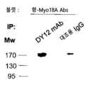
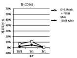

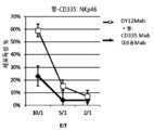


Classifications
- A—HUMAN NECESSITIES
- A61—MEDICAL OR VETERINARY SCIENCE; HYGIENE
- A61K—PREPARATIONS FOR MEDICAL, DENTAL OR TOILETRY PURPOSES
- A61K39/00—Medicinal preparations containing antigens or antibodies
- A61K39/395—Antibodies; Immunoglobulins; Immune serum, e.g. antilymphocytic serum
- A61K39/39533—Antibodies; Immunoglobulins; Immune serum, e.g. antilymphocytic serum against materials from animals
- A61K39/39566—Antibodies; Immunoglobulins; Immune serum, e.g. antilymphocytic serum against materials from animals against immunoglobulins, e.g. anti-idiotypic antibodies
- A—HUMAN NECESSITIES
- A61—MEDICAL OR VETERINARY SCIENCE; HYGIENE
- A61K—PREPARATIONS FOR MEDICAL, DENTAL OR TOILETRY PURPOSES
- A61K39/00—Medicinal preparations containing antigens or antibodies
- A61K39/39—Medicinal preparations containing antigens or antibodies characterised by the immunostimulating additives, e.g. chemical adjuvants
- A—HUMAN NECESSITIES
- A61—MEDICAL OR VETERINARY SCIENCE; HYGIENE
- A61P—SPECIFIC THERAPEUTIC ACTIVITY OF CHEMICAL COMPOUNDS OR MEDICINAL PREPARATIONS
- A61P31/00—Antiinfectives, i.e. antibiotics, antiseptics, chemotherapeutics
- A61P31/04—Antibacterial agents
- A—HUMAN NECESSITIES
- A61—MEDICAL OR VETERINARY SCIENCE; HYGIENE
- A61P—SPECIFIC THERAPEUTIC ACTIVITY OF CHEMICAL COMPOUNDS OR MEDICINAL PREPARATIONS
- A61P31/00—Antiinfectives, i.e. antibiotics, antiseptics, chemotherapeutics
- A61P31/12—Antivirals
- A—HUMAN NECESSITIES
- A61—MEDICAL OR VETERINARY SCIENCE; HYGIENE
- A61P—SPECIFIC THERAPEUTIC ACTIVITY OF CHEMICAL COMPOUNDS OR MEDICINAL PREPARATIONS
- A61P35/00—Antineoplastic agents
- A—HUMAN NECESSITIES
- A61—MEDICAL OR VETERINARY SCIENCE; HYGIENE
- A61P—SPECIFIC THERAPEUTIC ACTIVITY OF CHEMICAL COMPOUNDS OR MEDICINAL PREPARATIONS
- A61P37/00—Drugs for immunological or allergic disorders
- A61P37/02—Immunomodulators
- A61P37/04—Immunostimulants
- A—HUMAN NECESSITIES
- A61—MEDICAL OR VETERINARY SCIENCE; HYGIENE
- A61P—SPECIFIC THERAPEUTIC ACTIVITY OF CHEMICAL COMPOUNDS OR MEDICINAL PREPARATIONS
- A61P43/00—Drugs for specific purposes, not provided for in groups A61P1/00-A61P41/00
- C—CHEMISTRY; METALLURGY
- C07—ORGANIC CHEMISTRY
- C07K—PEPTIDES
- C07K16/00—Immunoglobulins [IGs], e.g. monoclonal or polyclonal antibodies
- C07K16/18—Immunoglobulins [IGs], e.g. monoclonal or polyclonal antibodies against material from animals or humans
- C07K16/28—Immunoglobulins [IGs], e.g. monoclonal or polyclonal antibodies against material from animals or humans against receptors, cell surface antigens or cell surface determinants
- C07K16/2896—Immunoglobulins [IGs], e.g. monoclonal or polyclonal antibodies against material from animals or humans against receptors, cell surface antigens or cell surface determinants against molecules with a "CD"-designation, not provided for elsewhere
- A—HUMAN NECESSITIES
- A61—MEDICAL OR VETERINARY SCIENCE; HYGIENE
- A61K—PREPARATIONS FOR MEDICAL, DENTAL OR TOILETRY PURPOSES
- A61K39/00—Medicinal preparations containing antigens or antibodies
- A61K2039/505—Medicinal preparations containing antigens or antibodies comprising antibodies
- A—HUMAN NECESSITIES
- A61—MEDICAL OR VETERINARY SCIENCE; HYGIENE
- A61K—PREPARATIONS FOR MEDICAL, DENTAL OR TOILETRY PURPOSES
- A61K39/00—Medicinal preparations containing antigens or antibodies
- A61K2039/505—Medicinal preparations containing antigens or antibodies comprising antibodies
- A61K2039/507—Comprising a combination of two or more separate antibodies
- A—HUMAN NECESSITIES
- A61—MEDICAL OR VETERINARY SCIENCE; HYGIENE
- A61K—PREPARATIONS FOR MEDICAL, DENTAL OR TOILETRY PURPOSES
- A61K39/00—Medicinal preparations containing antigens or antibodies
- A61K2039/57—Medicinal preparations containing antigens or antibodies characterised by the type of response, e.g. Th1, Th2
- A61K2039/572—Medicinal preparations containing antigens or antibodies characterised by the type of response, e.g. Th1, Th2 cytotoxic response
- C—CHEMISTRY; METALLURGY
- C07—ORGANIC CHEMISTRY
- C07K—PEPTIDES
- C07K2317/00—Immunoglobulins specific features
- C07K2317/20—Immunoglobulins specific features characterized by taxonomic origin
- C07K2317/24—Immunoglobulins specific features characterized by taxonomic origin containing regions, domains or residues from different species, e.g. chimeric, humanized or veneered
- C—CHEMISTRY; METALLURGY
- C07—ORGANIC CHEMISTRY
- C07K—PEPTIDES
- C07K2317/00—Immunoglobulins specific features
- C07K2317/30—Immunoglobulins specific features characterized by aspects of specificity or valency
- C07K2317/31—Immunoglobulins specific features characterized by aspects of specificity or valency multispecific
- C—CHEMISTRY; METALLURGY
- C07—ORGANIC CHEMISTRY
- C07K—PEPTIDES
- C07K2317/00—Immunoglobulins specific features
- C07K2317/50—Immunoglobulins specific features characterized by immunoglobulin fragments
- C07K2317/56—Immunoglobulins specific features characterized by immunoglobulin fragments variable (Fv) region, i.e. VH and/or VL
- C07K2317/569—Single domain, e.g. dAb, sdAb, VHH, VNAR or nanobody®
Landscapes
- Health & Medical Sciences (AREA)
- Chemical & Material Sciences (AREA)
- Life Sciences & Earth Sciences (AREA)
- Immunology (AREA)
- Medicinal Chemistry (AREA)
- Organic Chemistry (AREA)
- General Health & Medical Sciences (AREA)
- Animal Behavior & Ethology (AREA)
- Pharmacology & Pharmacy (AREA)
- Public Health (AREA)
- Veterinary Medicine (AREA)
- General Chemical & Material Sciences (AREA)
- Chemical Kinetics & Catalysis (AREA)
- Nuclear Medicine, Radiotherapy & Molecular Imaging (AREA)
- Genetics & Genomics (AREA)
- Proteomics, Peptides & Aminoacids (AREA)
- Molecular Biology (AREA)
- Biochemistry (AREA)
- Biophysics (AREA)
- Bioinformatics & Cheminformatics (AREA)
- Engineering & Computer Science (AREA)
- Microbiology (AREA)
- Mycology (AREA)
- Epidemiology (AREA)
- Oncology (AREA)
- Communicable Diseases (AREA)
- Virology (AREA)
- Medicines Containing Antibodies Or Antigens For Use As Internal Diagnostic Agents (AREA)
- Peptides Or Proteins (AREA)
- Medicines That Contain Protein Lipid Enzymes And Other Medicines (AREA)
- Pharmaceuticals Containing Other Organic And Inorganic Compounds (AREA)
- Medicines Containing Material From Animals Or Micro-Organisms (AREA)
Abstract
Translated fromKoreanDescription
Translated fromKorean본 발명은 NK 세포 살해 활성을 증대시키기 위한 방법 및 약학 조성물에 관한 것이다.The present invention relates to a method and a pharmaceutical composition for enhancing NK cell killing activity.
자연 살해(NK) 세포는 40여년 전에 예비-자극의 부재하에서 종양세포를 자발적으로 살해할 수 있는 림프구의 부분집합으로서 동정되었다(1-4). 대부분의 포유동물 및 조류 종에 존재하는 NK 세포는 항-종양 및 항-감염성 면역(5,6) 및 번식(7)에 중요한 역할을 한다. 인간에서, NK 림프구는 신경세포 부착 분자의 동형인 CD56의 발현, 및 CD3의 부재를 표현형적 특징으로 한다(8). NK 세포 세포독성은 활성화 및 억제 수용체의 연계로부터의 신호들간의 균형에 의해 엄격하게 조절된다(6,9). 표적 세포와 접촉시, 상기 NK 세포 표면상의 인테그린은 표적 세포상의 부착 분자에 결합하여 세포-세포 상호작용을 안정화시킨다(10). 표적 세포상의 세포간 부착 분자 1(ICAM-1)에 대한 인테그린 림프구 기능-관련 항원 1(LFA-1)의 결합은 구아닌 뉴클레오티드 교환 인자(GEF) 도메인-함유 Vav1 및 p21-활성화된 키나제(PAK)의 활성화를 통해 NK 세포의 초기 신호전달 캐스케이드를 개시시킨다(11). 이는 세포골격 재구성 및 NK-표적 세포 계면(NK 면역 시냅스(NKIS)(13)라 칭한다)에서의 활성화 NK 수용체(NKR)의 응집을 유도한다(12). 활성화 NKR의 연계는 면역수용체 티로신 활성화 동기(ITAM)-함유 연결자 단백질의 보충을 유도한다. 상기 ITMA 중의 2개의 티로신은 Src-키나제과 구성원에 의해 인산화되고, 인산화된 ITAM은 티로신 키나제의 Src-상동성 도메인 2(SH2) 도메인에 대한 결합 부위를 형성하여(14), 상기 표적 세포의 과립 분극, 탈과립, 및 세포용해의 원인이 되는 신호전달 캐스케이드를 촉발한다. 활성화 NKR은 천연 세포독성 수용체과(예를 들어 CD245(15), NKp44(16), 및 NKp30(17)), C-형 렉틴 수용체의 NKG2과(NKG2D)(18), 살해세포 Ig-유사 수용체(KIR)(19,20), Ig-유사 신호전달 림프구 활성화 분자(SLAM)과(2B4)(21), 및 기타, 예를 들어 CD160(22,23)의 구성원들을 포함한다. 4-1BB(CD137)는, 항체-의존적인 세포 세포독성 중의 경우와 같이(25), 상기 NK 세포 표면상의 Fc 수용체들의 연계에 의해 발현이 촉발되는 T, B 및 NK 세포상에서 발현되는 보조자극 수용체(24)이다. CD137의 자극은 상이한 암 모델들에서 세툭시맵-, 리툭시맵-, 및 트라스투주맵-의존적인 NK 세포 세포독성을 증가시킨다(26-28). NK 세포 활성화는 상기 억제성 NKR이 표적 세포상의 주 조직적합성 복합체(MHC) 부류 I 분자에 결합하는 경우 우세하게 억제된다(29). 인간에서, 이들 수용체는 주로, NKG2A 이종이량체(30)로서 C-형 렉틴 수용체에, 또는 수용체들의 KIR 상과(19,20)에 속한다. 상기 활성화 KIR과 달리, 상기 억제성 KIR은 면역수용체 티로신 억제 동기(ITIM) 서열(31)을 갖는 긴 세포질 꼬리를 지닌다. 후자는 Src 상동성 영역 2-함유 단백질 티로신 포스파타제-1(SHP-1) 또는 SHP-2의 나란한 SH2 도메인에 특이적인 결합 부위를 제공한다. 상기 NKIS에서의 SHP 보충은 상기 신호전달 캐스케이드의 핵심 단계들 중 다수를 차단하여 세포용해를 유도할 수 있다(32). CD245는 앞서 단클론 항체 DY12에 의해 인식된, 인간 말초 혈액 림프구의 표면상의 독특한 표면 항원으로서 기재되었다(33). 그러나 CD245 분자 및 기능 특성은 대체로 여전히 알려져 있지 않다.Natural killer (NK) cells were identified more than 40 years ago as a subset of lymphocytes capable of spontaneously killing tumor cells in the absence of pre-stimulation (1-4). NK cells present in most mammalian and algal species play an important role in anti-tumor and anti-infective immunity (5,6) and reproduction (7). In humans, NK lymphocytes are phenotypic characterized by the expression of CD56, an isotype of neuronal adhesion molecule, and absence of CD3 (8). NK cell cytotoxicity is tightly regulated by a balance between signals from the association of activation and inhibitory receptors (6, 9). Upon contact with the target cell, the integrin on the NK cell surface binds to the adhesion molecule on the target cell to stabilize the cell-cell interaction (10). Binding of integrin lymphocyte function-associated antigen 1 (LFA-1) to intercellular adhesion molecule 1 (ICAM-1) on the target cell leads to the formation of the guanine nucleotide exchange factor (GEF) domain-containing Vavl and p21- Activates the initial signaling cascade of NK cells (11). This leads to aggregation of the activated NK receptor (NKR) at the cytoskeleton reconstitution and at the NK-target cell interface (NKIS) (12). Activation of the NKR induces the replacement of the immune receptor tyrosine activation motif (ITAM) -containing linker protein. The two tyrosines in ITMA are phosphorylated by members of the Src-kinase, and the phosphorylated ITAM forms a binding site for the Src-homology domain 2 (SH2) domain of tyrosine kinase (14) , Degranulation, and signal transduction cascades that cause cell lysis. Activated NKRs can bind to natural cytotoxic receptors (for example CD245 (15), NKp44 (16), and NKp30 (17)), C-type lectin receptors NKG2 and (NKG2D) KIR) (19,20), Ig-like signaling lymphocyte activation molecules (SLAM) and (2B4) (21), and others such as CD160 (22,23). 4-1BB (CD137) expresses on the T, B, and NK cells, which are induced by expression of Fc receptors on the surface of NK cells, as in antibody-dependent cell cytotoxicity (25) (24). Stimulation of CD137 increases Cetuximab-, Rituximab-, and Trastuzumab-dependent NK cell cytotoxicity in different cancer models (26-28). NK cell activation is predominantly inhibited when the inhibitory NKR binds to the main histocompatibility complex (MHC) class I molecule on the target cell (29). In humans, these receptors mainly belong to the C-type lectin receptor as NKG2A heterodimer (30) or to the KIR phase of receptors (19,20). Unlike the activated KIR, the inhibitory KIR has a long cytoplasmic tail with an immunoreceptor tyrosine inhibitory motif (ITIM) sequence (31). The latter provides a binding site specific for the side chain SH2 domain of Src homology region 2-containing protein tyrosine phosphatase-1 (SHP-1) or SHP-2. SHP replenishment in NKIS can block many of the key steps of the signal transduction cascade leading to cell lysis (32). CD245 has been described as a unique surface antigen on the surface of human peripheral blood lymphocytes previously recognized by monoclonal antibody DY12 (33). However, CD245 molecules and functional properties are generally still unknown.
본 발명은 NK 세포 살해 활성을 증대시키기 위한 방법 및 약학 조성물에 관한 것이다. 특히, 본 발명은 청구의 범위에 의해 한정된다.The present invention relates to a method and a pharmaceutical composition for enhancing NK cell killing activity. In particular, the invention is defined by the claims.
발명자들은 면역학적 및 프로테오믹 접근법을 병행하여, 세포골격 구성 및 골지 출아에 중요한 역할을 하는 고도로 보존된 운동 효소인 비통상적인 미오신 18A로서 CD245를 동정하였다. 건강한 인간의 혈액 및 폐로부터의 NK 세포는 미오신 18A를 구성적으로 발현하였다. 미오신 18A는 NK 세포의 원형질막 및 세포질에 국소화되며, 그의 막 발현은 활성화-유도되었다. NK 세포상의 미오신 18A의 보충은 항-CD335(NKp46) 또는 항-CD337(NKp30)로 코팅된 P815 표적 세포에 대한 그의 세포독성 및 엡스타인-바 바이러스(EBV)-감염된 b 세포에 대한 림포킨-활성화된 살해 활성을 모두 강하게 증대시켰다. 미오신 18A의 자극은 NK 세포상에서 CD137 표면 발현을 유도하였다. CD137L의 차단은 CD137L-발현 B 림프종 세포에 대한 미오신 18A-유도된 NK 세포 세포독성을 무효화하였다. 미오신 18A는 포스파타제 활성과 관련되었으며 Src 상동성 영역 2-함유 단백질 티로신 포스파타제-2(SHP-2), 다수의 NK 수용체의 신호 전달인자, 및 상기 NK 면역 시냅스의 형성 및 액틴 구성에 관련된 p21-활성화된 키나제 2 PAK2에 결합할 수 있었다. 새롭게 기술된 NK 활성화 수용체 미오신 18A는 암 면역요법에 유망한 표적으로 보인다.The inventors identified CD245 as an unconventional myosin 18A, a highly conserved locus enzyme that plays an important role in cytoskeletal and Golgiogenesis, in combination with an immunological and proteomic approach. NK cells from healthy human blood and lungs constitutively expressed myosin 18A. Myosin 18A was localized to the plasma membrane and cytoplasm of NK cells, and its membrane expression was activated-induced. The supplementation of myosin 18A on NK cells was confirmed by its cytotoxicity against P815 target cells coated with anti-CD335 (NKp46) or anti-CD337 (NKp30), and cytotoxicity against Epstein-Barr virus (EBV) All of the killed activity was strongly increased. Myosin 18A stimulation induced CD137 surface expression on NK cells. Blocking CD137L nullified myosin 18A-induced NK cell cytotoxicity on CD137L-expressing B lymphoma cells. Myosin 18A was associated with phosphatase activity and was associated with Src homologous region 2-containing protein tyrosine phosphatase-2 (SHP-2), signaling factors of multiple NK receptors, and p21-activation associated with the formation of NK- RTI ID = 0.0 > PAK2 < / RTI > The newly described NK-activated receptor myosin 18A appears to be a promising target for cancer immunotherapy.
상응하게, 본 발명의 하나의 목적은 NK 세포 살해 활성의 증대가 필요한 피험자에게 치료 유효량의, NK 세포상의 CD245를 자극할 수 있는 화합물을 투여함을 포함하는, 상기 피험자에서 상기 활성을 증대시키는 방법에 관한 것이다.Correspondingly, one object of the present invention is to provide a method of increasing said activity in said subject, comprising administering to a subject in need of an increase in NK cell killing activity a compound capable of stimulating a CD245 on a NK cell in a therapeutically effective amount .
본 명세서에 사용되는 바와 같이, "NK 세포"는 비-통상적인 면역에 관련된 림프구의 아집단을 지칭한다. NK 세포는 몇몇 특징 및 생물학적 성질, 예를 들어 인간 NK 세포에 대한 CD56 및/또는 CD16을 포함하는 특이적인 표면 항원의 발현, 상기 세포 표면상의 알파/베타 또는 감마/델타 TCR 복합체의 부재, 특정한 세포용해 기구의 활성화에 의해 "자기" MHC/HLA 항원을 발현하지 못하는 세포에 결합하여 세포를 살해할 수 있는 능력, NK 활성화 수용체에 대한 리간드를 발현하는 종양 세포 또는 다른 병든 세포를 살해하는 능력, 및 면역 반응을 자극하거나 억제하는 사이토킨이라 칭하는 단백질 분자를 방출하는 능력("NK 세포 살해 활성들") 덕분에 동정될 수 있다. NK 세포의 임의의 아집단이 또한 NK 세포란 용어에 의해 포함될 것이다. 본 발명과 관련하여, "활성" NK 세포는 생물학적으로 활성인 NK 세포, 예를 들어 표적 세포를 용해하거나 다른 세포의 면역 기능을 증대시키는 능력을 갖는 NK 세포를 가리킨다. 예를 들어, "활성" NK 세포는 NK 수용체를 활성화하기 위해 리간드를 발현하고/하거나 상기 NK 세포상의 KIR에 의해 인식된 MHC/HLA 항원을 발현하지 못하는 세포를 살해할 수 있다.As used herein, "NK cell" refers to a subpopulation of lymphocytes associated with non-conventional immunity. NK cells are characterized by several characteristics and biological properties, such as expression of specific surface antigens comprising CD56 and / or CD16 on human NK cells, absence of alpha / beta or gamma / delta TCR complexes on the cell surface, The ability to kill cells by binding to cells that do not express "self" MHC / HLA antigens by activation of a dissolution mechanism, the ability to kill tumor cells or other diseased cells expressing a ligand to an NK activating receptor, and The ability to release protein molecules called cytokines that stimulate or inhibit the immune response ("NK cell killing activities") can be identified. Any subpopulation of NK cells will also be encompassed by the term NK cell. In the context of the present invention, "active" NK cells refer to NK cells that are biologically active, e. G., Capable of lysing target cells or enhancing the immune function of other cells. For example, "active" NK cells can express ligands to activate NK receptors and / or kill cells that do not express MHC / HLA antigens recognized by KIR on the NK cells.
본 명세서에 사용되는 바와 같이, "CD245"란 용어는 비통상적인 미오신 18A, 비통상적인 미오신-XVIIIa 또는 Myo18A를 나타낸다. 예시적인 인간 핵산 서열은 NCBI 참조번호 NM_078471.3 및 NM_203318.1로 참조표시되어 있다. 예시적인 인간 아미노산 서열은 NCBI 참조번호 NP_510880.2 및 NP_976063.1로 참조표시되어 있다. 추가의 예시적인 아미노산 서열은 서열번호 1(도 1B)을 포함한다.As used herein, the term "CD245" refers to the unconventional myosin 18A, the unconventional myosin-XVIIIa or Myo18A. Exemplary human nucleic acid sequences are referenced by NCBI reference numbers NM_078471.3 and NM_203318.1. Exemplary human amino acid sequences are referenced by NCBI reference numbers NP_510880.2 and NP_976063.1. Additional exemplary amino acid sequences include SEQ ID NO: 1 (Figure IB).
본 명세서에 사용되는 바와 같이, "NK 세포상의 CD245를 자극할 수 있는 화합물"이란 표현은 CD245와 연계(즉 상기에 결합)될 수 있고 NK 세포 살해 활성을 증대시킬 수 있는 임의의 화합물을 지칭한다. NK 세포 살해 활성을 증대시키는 본 발명 화합물의 능력을 당해 분야에 충분히 공지된 임의의 분석에 의해 측정할 수 있다. 전형적으로 상기 분석은, NK 세포를 표적 세포(예를 들어 NK 세포에 의해 인식되고/되거나 용해되는 표적 세포)와 접촉되게 하는 시험관내 분석이다. 예를 들어, 상기 화합물은 NK 세포에 의한 비용해(specific lysis)를, 본 발명의 화합물에 의해 접촉된 NK 세포 또는 NK 세포주와 동일한 효과기:표적 세포 비로 획득된 비용해의 약 20% 초과, 바람직하게는 적어도 약 30%, 적어도 약 40%, 적어도 약 50%, 또는 그 이상으로 증가시키는 능력에 대해 선택될 수 있으며, 고전적인 세포독성 분석에 대한 프로토콜의 예는 예를 들어 하기 문헌들에 기재되어 있다: Pessino et al, J. Exp. Med, 1998, 188 (5): 953-960; Sivori et al, Eur J Immunol, 1999. 29:1656-1666; Brando et al, (2005) J. Leukoc. Biol. 78:359-371; El-Sherbiny et al, (2007) Cancer Research 67(18):8444-9; 및 Nolte-'t Hoen et al, (2007) Blood 109:670-673. 전형적으로, NK 세포 세포독성은 실시예에 기재된 임의의 분석에 의해 측정된다. NK 세포 세포독성을 세포독성의 고전적인 시험관내 크로뮴 방출 시험에 의해 측정할 수 있다. 효과기 세포는 전형적으로 건강한 공여자로부터의 신선한 PB-NK이다. 상기 표적 세포는 전형적으로 쥐 비만세포종 P815 세포 또는 EBV-감염된 B 세포주이다. 상응하게, 본 발명의 화합물은 상기가 표적 세포(감염된 세포, 종양 세포, 염증전 세포 등)에 대한 NK 세포의 반응성 또는 세포독성의 증가, NK 세포에서 증가된 활성화, 활성화 마커(예를 들어 CD107 발현) 및/또는 IFN감마 생성, 및/또는 상기와 같은 활성화된, 반응성인, 세포독성인 및/또는 활성화된 NK 세포의 증가된 생체내 빈도를 야기하는 경우 선택된다.As used herein, the expression "a compound capable of stimulating CD245 on NK cells" refers to any compound that can be associated with CD245 (i. E., Coupled to the above) and can increase NK cell killing activity . The ability of the compounds of the invention to increase NK cell killing activity can be measured by any assay well known in the art. Typically, the assay is an in vitro assay in which NK cells are contacted with target cells (e. G., Target cells recognized and / or dissolved by NK cells). For example, the compound may be used to inhibit specific lysis by NK cells by more than about 20%, preferably less than about 20%, of the cost-effectiveness obtained with the same effector: target cell ratio as the NK cell or NK cell line contacted by the compound of the invention For example, at least about 30%, at least about 40%, at least about 50%, or more, and examples of protocols for classical cytotoxicity assays can be found in, for example, Lt; / RTI >: Pessino et al, J. Exp. Med. 1998, 188 (5): 953-960; Sivori et al., Eur J Immunol, 1999. 29: 1656-1666; Brando et al, (2005) J. Leukoc. Biol. 78: 359-371; El-Sherbiny et al, (2007) Cancer Research 67 (18): 8444-9; And Nolte-t Hoen et al, (2007) Blood 109: 670-673. Typically, NK cell cytotoxicity is measured by any assay described in the Examples. NK cell cytotoxicity can be measured by a classical in vitro chromium release test of cytotoxicity. Effector cells are typically fresh PB-NK from healthy donors. The target cells are typically murine mast cell T cell P815 cells or EBV-infected B cell lines. Correspondingly, the compounds of the present invention can be used to increase the reactivity or cytotoxicity of NK cells to the target cells (infected cells, tumor cells, pre-inflammatory cells, etc.), increased activation in NK cells, activation markers Expression and / or IFN gamma production, and / or increased in vivo frequency of such activated, reactive, cytotoxic and / or activated NK cells.
일부 실시태양에서, 본 발명의 화합물은 CD245에 대해 특이성을 갖는 항체이다.In some embodiments, the compounds of the invention are antibodies that have specificity for CD245.
본 명세서에 사용되는 바와 같이, "항체"란 용어는 항원 결합 영역을 갖는 임의의 항체-유사 분자를 지칭하는데 사용되며, 상기 용어는 항원 결합 도메인을 포함하는 항체 단편, 예를 들어 Fab', Fab, F(ab')2, 단일 도메인 항체(DAB), TandAb 이량체, Fv, scFv(단쇄 Fv), dsFv, ds-scFv, Fd, 선형 항체, 미니바디, 디아바디, 이중특이성 항체 단편, 비바디(bibody), 트리바디(scFv-Fab 융합, 각각 이중특이성 또는 삼중특이성); sc-디아바디; 카파(람다) 바디(scFv-CL 융합); BiTE(이중특이성 T-세포 인게이저: Bispecific T-cell Engager, T 세포를 공격하는 scFv-scFv 탠덤); DVD-Ig(이중 가변성 도메인 항체, 이중특이성 포맷); SIP(작은 면역단백질, 일종의 미니바디); SMIP("작은 모듈 면역약제" scFv-Fc 이량체; DART(ds-안정화된 디아바디 "이중 친화성 재표적화"); 하나 이상의 CDR을 포함하는 작은 항체 유사물질(mimetic) 등을 포함한다. 다양한 항체-기재 구조물 및 단편의 제조 및 사용 기법은 당해 분야에 충분히 공지되어 있다(구체적으로 본 발명에 참고로 인용된 문헌[Kabat et al., 1991]을 참조하시오). 특히, 디아바디는 EP 404,097 및 WO 93/1 1 161에 추가로 기재되어 있는 반면; 선형 항체는 문헌[Zapata et al.(1995)]에 추가로 기재되어 있다. 항체를 통상적인 기법을 사용하여 단편화할 수 있다. 예를 들어, F(ab')2 단편을, 항체를 펩신으로 처리함으로써 생성시킬 수 있다. 상기 생성되는 F(ab')2 단편을, 디설파이드 가교를 환원시키도록 처리하여 Fab' 단편을 생성시킬 수 있다. 파파인 절단은 Fab 단편의 형성을 유도할 수 있다. Fab, Fab' 및 F(ab')2, scFv, Fv, dsFv, Fd, dAb, TandAb, ds-scFv, 이량체, 미니바디, 디아바디, 이중특이성 항체 단편 및 다른 단편들을 또한 재조합 기법에 의해 합성하거나 또는 화학적으로 합성할 수 있다. 항체 단편의 생성 기법은 당해 분야에 주지되어 있고 기재되어 있다. 예를 들어, 하기의 문헌들이 각각 유효 항체 단편의 생성을 추가로 기재하고 가능하게 한다: Beckman et al., 2006; Holliger & Hudson, 2005; Le Gall et al., 2004; Reff & Heard, 2001 ; Reiter et al., 1996; 및 Young et al., 1995.As used herein, the term "antibody" is used to refer to any antibody-like molecule having an antigen-binding region, wherein the term encompasses antibody fragments comprising an antigen-binding domain, , F (ab ') 2, single domain antibody (DAB), TandAb dimer, Fv, scFv (single chain Fv), dsFv, ds-scFv, Fd, linear antibody, minibody, diabody, bispecific antibody fragment, Bibody, tribody (scFv-Fab fusion, bispecific or triple specific, respectively); sc-diabody; Kappa (lambda) body (scFv-CL fusion); BiTE (Bispecific T-cell Engager, scFv-scFv tandem attacking T cells); DVD-Ig (double variable domain antibody, bispecific format); SIP (small immune protein, a kind of mini-body); SMIP ("small module immunoagent" scFv-Fc dimer; DART (ds-stabilized diabody "double affinity re-targeting"); small antibody mimetic including one or more CDRs, etc. Techniques for making and using antibody-based constructs and fragments are well known in the art (see specifically Kabat et al., 1991, incorporated herein by reference). In particular, diabodies are described in EP 404,097 And WO 93/1 1 161. Linear antibodies have been further described in Zapata et al. (1995). [0064] Antibodies can be fragmented using conventional techniques. For example, the F (ab ') 2 fragment can be generated by treating the antibody with pepsin. The resulting F (ab') 2 fragment can be treated to reduce disulfide bridges to generate Fab 'fragments Papain cleavage can lead to the formation of Fab fragments Fab, Fab ' And F (ab ') 2, scFv, Fv, dsFv, Fd, dAb, TandAb, ds-scFv, dimers, minibodies, diabodies, bispecific antibody fragments and other fragments can also be synthesized by recombinant techniques, For example, the following documents further describe and enable the production of effective antibody fragments, respectively: Beckman et al., ≪ RTI ID = 0.0 >2006; Lee et al., 1996; and Young et al., 1995. Lee et al., 2006; Holliger & Hudson, 2005;
본 명세서에 사용되는 바와 같이, "특이성"이란 용어는 CD245와 같은 항원상에 제공된 에피토프에 검출가능하게 결합하지만 비-CD245 단백질 또는 구조(예를 들어 NK 세포 또는 다른 세포 유형상에 제공된 다른 단백질)와는 비교적 적은 검출가능한 반응성을 갖는 항체의 능력을 지칭한다. 특이성을 본 명세서의 다른 어딘가에 기재된 바와 같은 결합 또는 경쟁 결합 분석에 의해, 예를 들어 비아코어 장비를 사용하여 상대적으로 측정할 수 있다. 특이성을, 예를 들어 약 10:1, 약 20:1, 약 50:1, 약 100:1, 10.000:1 이상의, 특정한 항원에 대한 결합 대 다른 관련없는 분자에 대한 비특이적인 결합의 친화성/결합활성 비에 의해 나타낼 수 있다(상기 경우에 상기 특정한 항원은 CD245 폴리펩티드이다). 본 명세서에 사용되는 바와 같은 "친화성"이란 용어는 에피토프에 대한 항체의 결합 강도를 의미한다. 항체의 친화성은 [Ab] x [Ag]/[Ab-Ag](여기에서 [Ab-Ag]는 항체-항원 복합체의 몰 농도이고, [Ab]는 결합되지 않은 항체의 몰 농도이고 [Ag]는 결합되지 않은 항원의 몰 농도이다)로서 정의된 해리 상수 Kd에 의해 제공된다. 친화성 상수 Ka는 1/Kd에 의해 정의된다. mAb의 친화성을 측정하기에 바람직한 방법을 하기 문헌들에서 찾을 수 있으며, 이들 참고문헌은 전체가 본 발명에 참고로 인용된다: Harlow, et al., Antibodies: A Laboratory Manual, Cold Spring Harbor Laboratory Press, Cold Spring Harbor, N.Y., 1988), Coligan et al., eds., Current Protocols in Immunology, Greene Publishing Assoc. and Wiley Interscience, N.Y., (1992, 1993), 및 Muller, Meth. Enzymol. 92:589-601 (1983). mAb의 친화성을 측정하기 위한 당해 분야에 주지된 하나의 바람직한 표준 방법은 비아코어 장비의 사용이다.As used herein, the term "specificity" refers to the ability to detectably bind an epitope provided on an antigen, such as CD245, but detect the presence of a non-CD245 protein or structure (e. G., Other proteins provided on NK cells or other cell types) Quot; refers to the ability of an antibody to have a relatively low detectable reactivity. The specificity can be measured relative to binding using competitive binding assay as described elsewhere herein, for example using a non-core instrument. Specific affinity of a non-specific binding to a binding to a specific antigen versus another unrelated molecule, for example, about 10: 1, about 20: 1, about 50: 1, about 100: 1, 10.000: Binding activity ratio (in this case the specific antigen is a CD245 polypeptide). The term "affinity ", as used herein, refers to the binding strength of an antibody to an epitope. The affinity of the antibody is determined by the following equation: [Ab] x [Ag] / [Ab-Ag], where [Ab-Ag] is the molarity of the antibody- Lt; / RTI > is the molar concentration of unbound antigen). The affinity constant Ka is defined by 1 / Kd. Preferred methods for determining the affinity of a mAb can be found in the following references, all of which are incorporated herein by reference: Harlow, et al., Antibodies: A Laboratory Manual, Cold Spring Harbor Laboratory Press , Cold Spring Harbor, NY, 1988), Coligan et al., Eds., Current Protocols in Immunology, Greene Publishing Assoc. and Wiley Interscience, N. Y., (1992, 1993), and Muller, Meth. Enzymol. 92: 589-601 (1983). One preferred standard method known in the art for measuring mAb affinity is the use of Via Core equipment.
천연 항체에서, 2개의 중쇄는 디설파이드 결합에 의해 서로 연결되며 각각의 중쇄는 디설파이드 결합에 의해 경쇄에 연결된다. 2가지 유형의 경쇄, 람다(l) 및 카파(k)가 존재한다. 항체 분자의 기능 활성을 결정하는 5개의 주요 중쇄 부류(또는 아이소타입)가 존재한다: IgM, IgD, IgG, IgA 및 IgE. 각각의 쇄는 특유의 서열 도메인을 함유한다. 상기 경쇄는 2개의 도메인, 가변 도메인(VL) 및 불변 도메인(CL)을 포함한다. 상기 중쇄는 4개의 도메인, 가변 도메인(VH) 및 3개의 불변 도메인(CH1, CH2 및 CH3, 집합적으로 CH라 지칭된다)을 포함한다. 경쇄(VL) 및 중쇄(VH) 모두의 가변 영역은 항원에 대한 결합 인식 및 특이성을 결정한다. 경쇄(CL) 및 중쇄(CH)의 불변 영역 도메인은 중요한 생물학적 성질, 예를 들어 항체 쇄 결합, 분비, 태반-통과 이동성, 보체 결합, 및 Fc 수용체(FcR)에의 결합을 부여한다. 상기 Fv 단편은 면역글로불린의 Fab 단편의 N-말단 부분이며 하나의 경쇄 및 하나의 중쇄의 가변 부분으로 이루어진다. 상기 항체의 특이성은 상기 항체 결합 부위와 항원 결정인자간의 구조적 상보성에 있다. 항체 결합 부위는 주로 고가변성 또는 상보성 결정 영역(CDR)으로부터의 잔기들로 구성된다. 때때로, 비고가변성 또는 프레임워크 영역(FR)으로부터의 잔기가 전체 도메인 구조 및 따라서 상기 결합 부위에 영향을 미친다. 상보성 결정 영역 또는 CDR은 함께 고유 면역글로불린 결합 부위의 천연 Fv 영역의 결합 친화성 및 특이성을 한정하는 아미노산 서열을 지칭한다. 면역글로불린의 경쇄 및 중쇄는 각각 3개의 CDR(각각 L-CDR1, L-CDR2, L-CDR3 및 H-CDR1, H-CDR2, H-CDR3으로 표시됨)을 갖는다. 따라서 항원-결합 부위는 중쇄 및 경쇄 V 영역 각각으로부터의 CDR 세트를 포함한 6개의 CDR을 포함한다. 프레임워크 영역(FR)은 CDR들 사이에 산재되어 있는 아미노산 서열을 지칭한다.In natural antibodies, the two heavy chains are linked together by a disulfide bond and each heavy chain is linked to a light chain by a disulfide bond. There are two types of light chains, lambda (l) and kappa (k). There are five major heavy chain classes (or isotype) that determine the functional activity of antibody molecules: IgM, IgD, IgG, IgA and IgE. Each chain contains a unique sequence domain. The light chain comprises two domains, a variable domain (VL) and a constant domain (CL). The heavy chain includes four domains, a variable domain (VH) and three constant domains (CH1, CH2 and CH3, collectively referred to as CH). The variable regions of both the light chain (VL) and the heavy chain (VH) determine the binding recognition and specificity for the antigen. The constant domain domains of the light chain (CL) and the heavy chain (CH) confer important biological properties such as antibody chain binding, secretion, placenta-passing mobility, complement binding, and binding to the Fc receptor (FcR). The Fv fragment is the N-terminal portion of the Fab fragment of the immunoglobulin and consists of a light chain and a variable portion of one heavy chain. The specificity of the antibody lies in the structural complementarity between the antibody binding site and the antigenic determinant. The antibody binding site consists primarily of residues from the highly variable or complementarity determining regions (CDRs). Sometimes, variants or residues from the framework region (FR) affect the entire domain structure and thus the binding site. The complementarity determining region or CDR together refers to an amino acid sequence that defines the binding affinity and specificity of the native Fv region of the native immunoglobulin binding site. The light and heavy chains of the immunoglobulin each have three CDRs (designated L-CDR1, L-CDR2, L-CDR3 and H-CDR1, H-CDR2 and H-CDR3, respectively). Thus, the antigen-binding site includes six CDRs, each containing a set of CDRs from each of the heavy and light chain V regions. The framework region (FR) refers to an amino acid sequence that is interspersed among CDRs.
"Fab"란 용어는 약 50,000의 분자량 및 항원 결합 활성을 갖는 항체 단편을 나타내며, 여기에서 프로테아제, 파파인으로 IgG를 처리하여 수득된 단편들 중에서 H 쇄의 N-말단부의 대략 절반 및 전체 L쇄가 디설파이드 결합을 통해 함께 결합된다.The term "Fab" refers to an antibody fragment having a molecular weight of about 50,000 and antigen-binding activity, wherein about 50% of the N-terminal portion of the H chain and the entire L chain of the fragments obtained by treating IgG with protease, Lt; RTI ID = 0.0 > disulfide < / RTI >
"F(ab')2"란 용어는 약 100,000의 분자량 및 항원 결합 활성을 갖는 항체 단편을 지칭하며, 상기는 프로테아제, 펩신으로 IgG를 처리하여 수득된 단편들 중에서 힌지 영역의 디설파이드 결합을 통해 결합된 Fab보다 약간 더 크다.The term "F (ab ') 2" refers to an antibody fragment having a molecular weight of about 100,000 and an antigen-binding activity, wherein the fragment is obtained by treating the IgG with protease, pepsin, through a disulfide bond in the hinge region Gt; Fab < / RTI >
"Fab'"란 용어는 약 50,000의 분자량 및 항원 결합 활성을 갖는 항체 단편을 지칭하며, 상기는 상기 F(ab')2의 힌지 영역의 디설파이드 결합을 절단함으로써 수득된다.The term "Fab" refers to an antibody fragment having a molecular weight of about 50,000 and antigen binding activity, which is obtained by cleaving disulfide bonds in the hinge region of F (ab ') 2.
단쇄 Fv("scFv") 폴리펩티드는 공유 결합된 VH::VL 이종이량체로, 대개는 펩티드-암호화 링커에 의해 연결된 VH 및 VL 암호화 유전자를 포함하는 유전자 융합으로부터 발현된다. "dsFv"는 디설파이드 결합에 의해 안정화된 VH::VL 이종이량체이다. 2가 및 다가 항체 단편은 1가 scFv의 결합에 의해 자발적으로 형성되거나 또는 2가 sc(Fv)2와 같이, 펩티드 링커에 의해 1가 scFv를 커플링시킴으로써 생성될 수 있다.A short chain Fv ("scFv") polypeptide is expressed from a gene fusion that includes the VH and VL coding genes linked by covalently linked VH:: VL heterodimers, usually by peptide-encoding linkers. "dsFv" is a VH:: VL heterodimer stabilized by disulfide bonds. The bivalent and polyvalent antibody fragments may be spontaneously formed by binding of monovalent scFv or may be produced by coupling monovalent scFv by a peptide linker, such as bivalent sc (Fv) 2.
"디아바디"란 용어는 2개의 항원-결합 부위를 갖는 작은 항체 단편을 지칭하며, 상기 단편은 동일한 폴리펩티드 쇄(VH-VL) 중의 경쇄 가변 도메인(VL)에 연결된 중쇄 가변 도메인(VH)을 포함한다. 동일한 쇄 상의 2개의 도메인간의 짝짓기를 허용하기에는 너무 짧은 링커를 사용함으로써, 상기 도메인들은 또 다른 쇄의 상보성 도메인과 강제로 짝을 이루어 2개의 항원-결합 부위를 생성시킨다.The term "diabody " refers to small antibody fragments having two antigen-binding sites, which comprise a heavy chain variable domain (VH) linked to a light chain variable domain (VL) in the same polypeptide chain (VH-VL) do. By using a linker that is too short to allow mating between the two domains on the same chain, the domains are forced to pair with another chain of complementary domains to generate two antigen-binding sites.
단클론 항체를 코흘러와 밀스테인(Kohler and Milstein)(Nature, 256:495, 1975)의 방법을 사용하여 생성시킬 수 있다. 본 발명에 유용한 단클론 항체를 제조하기 위해서, 마우스 또는 다른 적합한 숙주 동물을 적합한 항원 형태(즉 본 발명의 폴리펩티드)로 적합한 간격(예를 들어 2주마다, 매주, 2개월마다 또는 매달)으로 면역시킨다. 상기 동물을 죽이기 1주일내에 상기 동물에게 항원의 최종 "추가접종"을 투여할 수 있다. 면역기간 동안 면역학적 항원보강제를 사용하는 것이 또한 바람직할 수 있다. 적합한 면역학적 항원보강제는 프로인트 완전 항원보강제, 프로인트 불완전 항원보강제, 알룸, 리비(Ribi) 항원보강제, 헌터의 티터맥스(Hunter's Titermax), 사포닌 항원보강제, 예를 들어 QS21 또는 퀼(Quil) A, 또는 CpG-함유 면역자극 올리고뉴클레오티드를 포함한다. 다른 적합한 항원보강제는 당해 분야에 주지되어 있다. 상기 동물을 피하, 복강내, 근육내, 정맥내, 비내 또는 다른 경로에 의해 면역시킬 수 있다. 주어진 동물을 다중 경로에 의해 다수 형태의 항원으로 면역시킬 수 있다.Monoclonal antibodies can be generated using the methods of Kohler and Milstein (Nature, 256: 495, 1975). To prepare monoclonal antibodies useful in the present invention, mice or other suitable host animals are immunized with suitable antigenic forms (i. E., Polypeptides of the invention) at appropriate intervals (e. G. Every two weeks, every second, every month or every month) . A final "booster" of the antigen may be administered to the animal within one week of killing the animal. It may also be desirable to use an immunological antigen adjuvant during the immunization period. Suitable immunological adjuvants include Freund ' s complete adjuvant, Freund ' s incomplete adjuvant, Alum, Ribi antigen adjuvant, Hunter ' s Titermax, a saponin antigen adjuvant such as QS21 or Quil A , Or CpG-containing immunostimulatory oligonucleotides. Other suitable antigen adjuvants are well known in the art. The animal can be immunized subcutaneously, intraperitoneally, intramuscularly, intravenously, intranasally or by other routes. A given animal can be immunized with multiple forms of antigen by multipath.
간단히, 본 발명의 재조합 폴리펩티드를 재조합 세포주에 의한 발현에 의해 제공할 수 있다. 상기 폴리펩티드의 재조합 형태를 임의의 앞서 기재된 방법을 사용하여 제공할 수 있다. 상기 면역화 섭생에 이어서, 림프구를 상기 동물의 비장, 림프절 또는 다른 기관으로부터 단리하고 폴리에틸렌 글리콜과 같은 작용제를 사용하여 적합한 골수종 세포주와 융합시켜 하이브리도마를 형성시킨다. 융합에 이어서, 세포를 표준 방법을 사용하여, 하이브리도마의 생육에는 허용성이지만 융합 상대에는 허용성이지 않은 배지에 놓는다. 상기 하이브리도마의 배양에 이어서, 세포 상등액을 목적하는 특이성의, 즉 항원과 선택적으로 결합하는 항체의 존재에 대해 분석한다. 적합한 분석 기법은 ELISA, 유식 세포측정, 면역침전 및 웨스턴 블럿팅을 포함한다. 다른 선별 기법들이 당해 분야에 주지되어 있다. 바람직한 기법은 형태적으로 완전한, 고유하게 접힌 항원에 대한 항체의 결합을 입증하는 것들, 예를 들어 비-변성 ELISA, 유식 세포측정 및 면역침전이다.Briefly, recombinant polypeptides of the invention can be provided by expression in a recombinant cell line. Recombinant forms of the polypeptides can be provided using any of the methods described above. Following the immunization regimen, lymphocytes are isolated from the spleen, lymph nodes, or other organs of the animal and fused with a suitable myeloma cell line using an agent such as polyethylene glycol to form a hybridoma. Following fusion, the cells are placed in a medium that is tolerant to the growth of the hybridoma but not tolerant to the fusion partner, using standard methods. Following incubation of the hybridomas, the cell supernatants are assayed for the presence of antibodies of a desired specificity, i. E., Binding selectively to the antigen. Suitable analytical techniques include ELISA, stained cell count, immunoprecipitation and Western blotting. Other screening techniques are well known in the art. Preferred techniques are those that demonstrate the binding of antibodies to formally complete, uniquely folded antigens, such as non-denaturing ELISA, stained cell count and immunoprecipitation.
현저하게, 당해 분야에 주지된 바와 같이, 항체 분자의 단지 작은 부분, 파라토프만이 항체의 그의 에피토프에의 결합에 관련된다(일반적으로 문헌[Clark, W. R. (1986) The Experimental Foundations of Modern Immunology Wiley & Sons, Inc., New York]; [Roitt, I. (1991) Essential Immunology, 7th Ed., Blackwell Scientific Publications, Oxford]을 참조하시오). 예를 들어 Fc' 및 Fc 영역은 보체 캐스케이드의 효과기이나, 항원 결합에는 관련되지 않는다. pFc' 영역이 효소적으로 절단되었거나 또는 상기 pFc' 영역 없이 생성된 항체(F(ab')2 단편으로 표시됨)는 완전 항체의 항원 결합 부위를 둘 다 유지한다. 유사하게, Fc 영역이 효소적으로 절단되었거나 또는 상기 Fc 영역 없이 생성된 항체(Fab 단편으로 표시됨)는 완전 항체 분자의 항원 결합 부위 중 하나를 유지한다. 더 진행하여, Fab 단편은 공유적으로 결합된 항체 경쇄 및 Fd라 표시되는 항체 중쇄의 일부로 이루어진다. 상기 Fd 단편은 항체 특이성의 주요 결정인자이며(단일 Fd 단편은 항체 특이성의 변경 없이 10개 이하의 상이한 경쇄와 결합될 수 있다) Fd 단편은 단리시 에피토프-결합 능력을 유지한다.Significantly, as is well known in the art, only a small portion of the antibody molecule, paratope, is involved in the binding of the antibody to its epitope (see generally Clark, WR (1986) Experimental Foundations of Modern Immunology Wiley &Amp; Sons, Inc., New York); [Roitt, I. (1991) Essential Immunology, 7th Ed., Blackwell Scientific Publications, Oxford). For example, the Fc 'and Fc regions are the effectors of the complement cascade, but are not involved in antigen binding. An antibody (denoted F (ab ') 2 fragment) in which the pFc' region has been enzymatically cleaved or produced without the pFc 'region retains both antigen binding sites of the whole antibody. Similarly, an antibody (designated Fab fragment) in which the Fc region has been enzymatically cleaved or produced without the Fc region retains one of the antigen binding sites of the complete antibody molecule. Proceeding further, the Fab fragment consists of a portion of the antibody heavy chain, designated covalently bound antibody light chain and Fd. The Fd fragment is a major determinant of antibody specificity (a single Fd fragment can bind to no more than 10 different light chains without altering antibody specificity). The Fd fragment retains epitope-binding ability upon isolation.
항체의 항원-결합 부분내에는, 당해 분야에 주지된 바와 같이, 상기 항원의 에피토프와 직접적으로 상호작용하는 상보성 결정 영역(CDR), 및 파라토프의 3차 구조를 유지하는 프레임워크 영역(FR)이 존재한다(일반적으로 문헌[Clark, 1986]; [Roitt, 1991]을 참조하시오). 상기 IgG 면역글로불린의 중쇄 Fd 단편 및 경쇄 모두에서, 3개의 상보성 결정 영역(CDR1 내지 CDRS)에 의해 각각 분리된 4개의 프레임워크 영역(FR1 내지 FR4)이 존재한다. 상기 CDR 및 특히 상기 CDRS 영역, 및 보다 특히 중쇄 CDRS는 주로 항체 특이성의 원인이다.Within the antigen-binding portion of the antibody, a complementarity determining region (CDR) that directly interacts with the epitope of the antigen, as is well known in the art, and a framework region (FR) that retains the tertiary structure of the paratope, (See generally [Clark, 1986]; [Roitt, 1991]). In both of the heavy chain Fd fragment and the light chain of the IgG immunoglobulin, there are four framework regions FR1 to FR4 separated by three complementary crystal regions (CDR1 to CDRS), respectively. The CDRs and particularly the CDRS regions, and more particularly the heavy chain CDRSs, are primarily responsible for antibody specificity.
현재 포유동물 항체의 비 CDR 영역을, 원래 항체의 에피토프 특이성을 유지하면서 동종 또는 이종특이성 항체의 유사한 영역으로 대체할 수 있음은 당해 분야에 충분히 확립되어 있다. 이는 비-인간 CDR이 인간 FR 및/또는 Fc/pFc' 영역에 공유 결합되어 기능성 항체를 생성하는 "인간화된" 항체의 개발 및 사용에서 가장 명백히 나타난다.It is well established in the art that the present non-CDR regions of mammalian antibodies can be replaced with similar regions of homologous or heterospecific antibodies while retaining the epitope specificity of the original antibody. This is most evident in the development and use of "humanized" antibodies in which non-human CDRs are covalently linked to human FR and / or Fc / pFc 'regions to produce functional antibodies.
일부 실시태양에서, 상기 항체는 인간화된 항체이다. 본 명세서에 사용되는 바와 같이, "인간화된"은 상기 CDR 영역 밖의 아미노산의 일부, 대부분 또는 전부가 인간 면역글로불린 분자로부터 유래된 상응하는 아미노산으로 대체된 항체를 기술한다. 인간화의 방법은 비제한적으로 미국특허 제 4,816,567, 5,225,539, 5,585,089, 5,693,761, 5,693,762 및 5,859,205 호(이들은 본 발명에 참고로 인용된다)에 기재된 것들을 포함한다. 상기 미국특허 제 5,585,089 및 5,693,761 호 및 WO 90/07861은 또한 상기 인간화된 항체의 설계에 사용될 수 있는 4개의 가능한 기준을 제안한다. 첫 번째 제안은 수용체의 경우, 인간화시키고자 하는 공여 면역글로불린에 특이하게 상동성인 특정한 인간 면역글로불린으로부터의 프레임워크를 사용하거나, 또는 다수의 인간 항체로부터의 공통 프레임워크를 사용하는 것이었다. 두 번째 제안은 상기 인간 면역글로불린의 프레임워크 중의 아미노산이 특이하고 상기 위치의 공여 아미노산이 인간 서열에 전형적인 경우, 상기 수용체보다는 오히려 상기 공여 아미노산을 선택할 수 있다는 것이었다. 세 번째 제안은 상기 인간화된 면역글로불린 쇄 중의 3개의 CDR에 바로 인접한 위치들에서, 상기 수용 아미노산보다는 오히려 상기 공여 아미노산을 선택할 수 있다는 것이었다. 네 번째 제안은 공여 아미노산이 항체의 3차원 모델에서 CDR의 3A내에 측쇄 원자를 갖는 것으로 예상되고 상기 CDR과 상호작용할 수 있는 것으로 예상되는 프레임워크 위치에 있는 상기 공여 아미노산을 사용하는 것이었다. 상기 방법들은 단지 당해 분야의 숙련가가 인간화된 항체의 제조에 사용할 수 있는 방법들 중 일부의 예시일 뿐이다. 당해 분야의 통상적인 숙련가는 다른 항체 인간화 방법과 친숙할 것이다.In some embodiments, the antibody is a humanized antibody. As used herein, "humanized" describes an antibody in which some, most, or all of the amino acids outside the CDR region are replaced by corresponding amino acids derived from human immunoglobulin molecules. Methods of humanization include, but are not limited to those described in U.S. Patent Nos. 4,816,567, 5,225,539, 5,585,089, 5,693,761, 5,693,762 and 5,859,205, which are incorporated herein by reference. U.S. Pat. Nos. 5,585,089 and 5,693,761 and WO 90/07861 also propose four possible criteria that can be used in the design of the humanized antibody. The first proposal was to use a framework from a particular human immunoglobulin that is specifically homologous to the donor immunoglobulin to which it is intended to be humanized, or to use a common framework from multiple human antibodies. The second proposal was that if the amino acid in the framework of the human immunoglobulin is specific and the donor amino acid at that position is typical for the human sequence, then the donor amino acid can be selected rather than the receptor. The third proposal was that at these positions immediately adjacent to the three CDRs in the humanized immunoglobulin chain, the donor amino acid could be selected rather than the accepting amino acid. The fourth proposal was to use the donor amino acid in the framework position where the donor amino acid is expected to have a side chain atom within 3A of the CDR in the three-dimensional model of the antibody and is expected to interact with the CDR. These methods are merely illustrative of some of the methods that a person skilled in the art can use in the production of humanized antibodies. A person of ordinary skill in the art will be familiar with other methods of antibody humanization.
일부 실시태양에서, 상기 CDR 영역 밖의 아미노산의 일부, 대부분 또는 전부를 인간 면역글로불린 분자로부터의 아미노산으로 대체하였지만, 일부의 경우, 하나 이상의 CDR 영역내의 대부분 또는 모든 아미노산은 변하지 않는다. 아미노산의 작은 부가, 결실, 삽입, 치환 또는 변형은, 이들이 주어진 항원에 결합하는 항체의 능력을 무효화하지 않는 한 허용될 수 있다. 적합한 인간 면역글로불린 분자는 IgG1, IgG2, IgG3, IgG4, IgA 및 IgM 분자를 포함할 것이다. "인간화된" 항체는 원래 항체와 유사한 항원 특이성을 유지한다. 그러나, 몇몇 인간화 방법을 사용하는 경우, 상기 항체의 결합 친화성 및/또는 특이성을 문헌[Wu et al., /. Mol. Biol. 294:151, 1999]에 기재된 바와 같은 "유도 진화" 방법을 사용하여 증가시킬 수 있으며, 상기 문헌의 내용은 본 발명에 참고로 인용된다.In some embodiments, some, most, or all of the amino acids outside of the CDR region have been replaced by amino acids from human immunoglobulin molecules, but in some cases most or all of the amino acids within the one or more CDR regions remain unchanged. Small additions, deletions, insertions, substitutions or modifications of amino acids may be allowed provided they do not invalidate the ability of the antibody to bind to a given antigen. Suitable human immunoglobulin molecules will include IgGl, IgG2, IgG3, IgG4, IgA and IgM molecules. A "humanized" antibody retains antigen specificity similar to that of the original antibody. However, if several humanization methods are used, the binding affinity and / or specificity of the antibody can be determined by the method of Wu et al. Mol. Biol. 294: 151, 1999, the contents of which are incorporated herein by reference.
완전 인간 단클론 항체를 또한, 인간 면역글로불린 중쇄 및 경쇄 유전자좌의 대부분에 대해 트랜스제닉인 마우스를 면역시킴으로써 제조할 수 있다. 예를 들어 미국특허 제 5,591,669, 5,598,369, 5,545,806, 5,545,807, 6,150,584 호 및 상기 중에 인용된 참고문헌들(이들의 내용은 본 발명에 참고로 인용된다)을 참조하시오. 상기 동물을, 내인성(예를 들어 쥐) 항체의 생성에서 기능적 결실이 존재하도록 유전자 변형시켰다. 상기 동물을, 상기 동물의 면역화가 관심 항원에 대한 완전 인간 항체의 생성을 생성시키도록 인간 생식세포 면역글로불린 유전자 유전자좌의 전부 또는 일부를 함유하도록 추가로 변형시킨다. 상기 마우스(예를 들어 제노마우스(XenoMouse)(아브제닉스(Abgenix)), HuMAb 마우스(메다렉스/젠팜(Medarex/GenPharm))의 면역화에 이어서, 단클론 항체를 표준 하이브리도마 기술에 따라 제조할 수 있다. 상기 단클론 항체는 인간 면역글로불린 아미노산 서열을 가질 것이며 따라서 인간에 투여시 인간 항-마우스 항체(KAMA) 반응을 일으키지 않을 것이다. 인간 항체의 생성을 위한 시험관내 방법이 또한 존재한다. 여기에는 파지 디스플레이 기술(미국특허 제 5,565,332 및 5,573,905 호) 및 인간 B 세포의 시험관내 자극(미국특허 제 5,229,275 및 5,567,610 호)이 포함된다. 이들 특허의 내용은 본 발명에 참고로 인용된다.Whole human monoclonal antibodies can also be prepared by immunizing mice transgenic for most of the human immunoglobulin heavy chain and light chain locus. See, for example, U.S. Patent Nos. 5,591,669, 5,598,369, 5,545,806, 5,545,807, 6,150,584, and references cited therein, the contents of which are incorporated herein by reference. The animals were genetically engineered to have functional deletions in the production of endogenous (e. G., Murine) antibodies. The animal is further modified to contain all or part of the human germline immunoglobulin gene locus such that the immunization of the animal produces the production of a fully human antibody to the antigen of interest. Following immunization of the mice (e.g., XenoMouse (Abgenix)), HuMAb mice (Medarex / GenPharm), monoclonal antibodies can be prepared according to standard hybridoma techniques The monoclonal antibody will have a human immunoglobulin amino acid sequence and will therefore not result in a human anti-mouse antibody (KAMA) reaction upon administration to humans. There is also an in vitro method for the production of human antibodies, Display technologies (U.S. Pat. Nos. 5,565,332 and 5,573,905) and in vitro stimulation of human B cells (U.S. Patents 5,229,275 and 5,567,610), the disclosures of which are incorporated herein by reference.
따라서, 당해 분야의 통상적인 숙련가에게 자명한 바와 같이, 본 발명은 또한 F(ab')2 Fab, Fv 및 Fd 단편; Fc 및/또는 FR 및/또는 CDR1 및/또는 CDR2 및/또는 경쇄 CDR3 영역이 상동성 인간 또는 비-인간 서열로 대체된 키메릭 항체; FR 및/또는 CDR1 및/또는 CDR2 및/또는 경쇄 CDR3 영역이 상동성 인간 또는 비-인간 서열로 대체된 키메릭 F(ab')2 단편; FR 및/또는 CDR1 및/또는 CDR2 및/또는 경쇄 CDR3 영역이 상동성 인간 또는 비-인간 서열로 대체된 키메릭 Fab 단편; 및 FR 및/또는 CDR1 및/또는 CDR2 영역이 상동성 인간 또는 비-인간 서열로 대체된 키메릭 Fd 단편을 제공한다. 본 발명은 또한 소위 단쇄 항체를 포함한다.Thus, as would be apparent to one of ordinary skill in the art, the present invention also encompasses F (ab ') 2 Fab, Fv, and Fd fragments; A chimeric antibody in which the Fc and / or FR and / or CDR1 and / or CDR2 and / or light chain CDR3 regions are replaced by a homologous human or non-human sequence; A chimeric F (ab ') 2 fragment in which the FR and / or CDR1 and / or CDR2 and / or light chain CDR3 regions are replaced by a homologous human or non-human sequence; A chimeric Fab fragment in which the FR and / or CDR1 and / or CDR2 and / or light chain CDR3 regions are replaced by a homologous human or non-human sequence; And a chimeric Fd fragment in which the FR and / or CDR1 and / or CDR2 regions are replaced by a homologous human or non-human sequence. The present invention also includes so-called single chain antibodies.
다양한 항체 분자 및 단편이 통상적으로 공지된 면역글로불린 부류 중 어느 하나, 예를 들어 비제한적으로 IgA, 분비성 IgA, IgE, IgG 및 IgM으로부터 유래될 수 있다. IgG 하위부류가 또한 당해 분야의 숙련가들에게 주지되어 있으며 비제한적으로 인간 IgG1, IgG2, IgG3 및 IgG4를 포함한다.Various antibody molecules and fragments can be derived from any of the commonly known immunoglobulin classes, such as, but not limited to, IgA, secretory IgA, IgE, IgG and IgM. IgG subclasses are also well known to those skilled in the art and include, but are not limited to, human IgG1, IgG2, IgG3, and IgG4.
일부 실시태양에서, 본 발명의 항체는 단쇄 항체이다. 본 명세서에 사용되는 바와 같이 "단일 도메인 항체"란 용어는 당해 분야에서 그의 일반적인 의미를 가지며 자연적으로 경쇄가 없는 카멜리드 포유동물에서 발견될 수 있는 유형의 항체의 단일 중쇄 가변 도메인을 지칭한다. 상기와 같은 단일 도메인 항체는 또한 "나노바디(등록상표)"이다. (단일) 도메인 항체의 일반적인 개시에 대해서, 상기에 인용된 종래 기술뿐만 아니라 EP 0 368 684, 문헌[Ward et al., Nature 1989 Oct 12; 341 (6242): 544-6)], [Holt et al., Trends Biotechnol., 2003, 21(11):484-490]; 및 WO 06/030220, WO 06/003388을 참조한다. 단일 도메인 항체의 아미노산 서열 및 구조는 4개의 프레임워크 영역 또는 "FR"(이들은 당해 분야 및 본 명세서에서 각각 "프레임워크 영역 1" 또는 "FR1"으로서; "프레임워크 영역 2" 또는 "FR2"로서; "프레임워크 영역 3" 또는 "FR3"으로서; 및 "프레임워크 영역 4" 또는 "FR4"로서 지칭된다)로 구성되는 것으로 간주될 수 있으며; 상기 프레임워크 영역들은 3개의 상보성 결정 영역 또는 "CDR"(이들은 당해 분야에서 각각 ""CDR1"에 대한 "상보성 결정 영역"으로서; "상보성 결정 영역 2" 또는 "CDR2"로서 및 "상보성 결정 영역 3" 또는 "CDR3"으로서 지칭된다)에 의해 중단된다. 상응하게, 상기 단일 도메인 항체를 일반적인 구조: FRl - CDRl - FR2 - CDR2 - FR3 - CDR3 - FR4(여기에서 FR1 내지 FR4는 각각 프레임워크 영역 1 내지 4를 지칭하고, CDR1 내지 CDR3은 상보성 결정 영역 1 내지 3을 지칭한다)를 갖는 아미노산 서열로서 정의할 수 있다.In some embodiments, the antibody of the invention is a single chain antibody. As used herein, the term " single domain antibody " refers to a single heavy chain variable domain of an antibody of the type that can be found in camelid mammals that have their ordinary meaning in the art and naturally do not have light chains. Such single domain antibodies are also "nanobodies ". For the general disclosure of (single) domain antibodies, as well as the prior art cited above, see
일부 실시태양에서, 본 발명의 항체는 인간 중쇄 불변 영역 서열을 포함하지만 상기가 결합하는 NK 세포를 고갈시키지 않을 것이며 바람직하게는 항체 의존적인 세포 세포독성(ADCC)을 유도하는 Fc 부분을 포함하지 않는다. 본 명세서에 사용되는 바와 같이, CD245-발현 세포에 관하여 "고갈"이란 용어는 샘플 중에 또는 피험자 중에 존재하는 CD245 발현 세포의 수에 음의 영향을 미치기 위해서 살해, 제거, 용해하거나 또는 상기와 같은 살해, 제거 또는 용해를 유도할 수 있는 과정, 방법 또는 화합물을 의미한다. "FC 도메인", "Fc 부분" 및 "Fc 영역"이란 용어는 예를 들어 인간 감마 중쇄의 아미노산(aa) 약 230 내지 약 aa 450 또는 다른 유형의 항체 중쇄(예를 들어 인간 항체의 경우 α, δ, ε 및 μ) 또는 그의 천연 알로타입 중의 그의 대응 서열로부터의 항체 중쇄의 C-말단 단편을 지칭한다. 달리 명시되지 않는 한, 면역글로불린에 대해 통상적으로 허용되는 카밧(Kabat) 아미노산 넘버링이 본 명세 전체를 통해 사용된다(문헌[Kabat et al. (1991 ) Sequences of Protein of Immunological Interest, 5th ed., United States Public Health Service, National Institute of Health, Bethesda, MD]을 참조하시오). 일부 실시태양에서, 본 발명의 항체는 직접적으로 또는 간접적으로, CD245 폴리펩티드를 발현하는 NK 세포의 고갈을 유도하지 않는다(예를 들어 CD245+ NK 세포 수의 10%, 20%, 50%, 60% 이상의 제거 또는 감소를 유도하지 않는다). 일부 실시태양에서, 본 발명의 항체는 FcγRIIIA(CD16) 폴리펩티드에 실질적으로 결합할 수 있는 Fc 도메인을 포함하지 않는다. 일부 실시태양에서, 본 발명의 항체는 Fc 도메인이 없거나(예를 들어 CH2 및/또는 CH3 도메인이 없거나) 또는 IgG2 또는 IgG4 아이소타입의 Fc 도메인을 포함한다. 일부 실시태양에서, 본 발명의 항체는 Fab, Fab', Fab'-SH, F(ab')2, Fv, 디아바디, 단쇄 항체 단편, 또는 다수의 상이한 항체 단편을 포함하는 다중특이성 항체로 이루어진다. 일부 실시태양에서, 본 발명의 항체는 독성 부분에 연결되지 않는다. 일부 실시태양에서, 아미노산 잔기 중에서 선택된 하나 이상의 아미노산을, 상기 항체가 변경된 C2q 결합 및/또는 감소되거나 또는 제거된 보체 의존적인 세포독성(CDC)을 갖도록 상이한 아미노산 잔기로 대체시킬 수 있다. 이러한 접근법은 이두소기(Idusogie) 등의 미국특허 제 6,194,551 호에 더욱 상세히 기재되어 있다.In some embodiments, the antibody of the invention comprises a human heavy chain constant region sequence but will not deplete the NK cells to which it binds and preferably does not contain an Fc portion that induces antibody dependent cellular cytotoxicity (ADCC) . As used herein, the term "depletion " with respect to CD245-expressing cells refers to killing, removing, dissolving, or otherwise causing the killing, such as the above, to have negative effects on the number of CD245 expressing cells present in the subject or in the subject , Elimination, or dissolution of a compound of the present invention. The term " FC domain ", "Fc portion ", and" Fc region "refer to, for example, amino acids (aa) of the human gamma heavy chain of about 230 to about aa 450 or other types of antibody heavy chains delta, epsilon, and mu), or a C-terminal fragment of an antibody heavy chain from its corresponding sequence in its natural allotype. Unless otherwise specified, Kabat amino acid numbering that is commonly accepted for immunoglobulins is used throughout this specification (Kabat et al. (1991) Sequences of Protein of Immunological Interest, 5th ed., United Bethesda, Md.]). ≪ / RTI > In some embodiments, the antibodies of the invention do not directly or indirectly induce depletion of NK cells expressing CD245 polypeptides (e. G., 10%, 20%, 50%, 60% Elimination or reduction). In some embodiments, an antibody of the invention does not comprise an Fc domain that is capable of substantially binding to an FcγRIIIA (CD16) polypeptide. In some embodiments, an antibody of the invention does not have an Fc domain (e. G., Without a CH2 and / or CH3 domain) or includes an Fc domain of an IgG2 or IgG4 isotype. In some embodiments, an antibody of the invention comprises a multispecific antibody comprising Fab, Fab ', Fab'-SH, F (ab') 2, Fv, diabody, single chain antibody fragment, or a plurality of different antibody fragments . In some embodiments, the antibody of the invention is not linked to a toxic moiety. In some embodiments, one or more amino acids selected from amino acid residues can be replaced with a different amino acid residue so that the antibody has altered C2q binding and / or reduced or eliminated complement dependent cytotoxicity (CDC). This approach is described in more detail in U.S. Patent No. 6,194,551 to Idusogie et al.
일부 실시태양에서, CHI의 힌지 영역을, 상기 힌지 영역 중의 시스테인 잔기의 수가 변경되도록, 예를 들어 증가되거나 감소되도록 변형시킨다. 이러한 접근법은 보드머(Bodmer) 등의 미국특허 제 5,677,425 호에 추가로 기재되어 있다. 상기 CHI의 힌지 영역 중의 시스테인 잔기의 수를 예를 들어 경쇄 및 중쇄의 조립을 촉진하거나 또는 항체의 안정성을 증가 또는 감소시키기 위해서 변경시킨다.In some embodiments, the hinge region of CHI is modified such that, for example, the number of cysteine residues in the hinge region is increased or decreased. This approach is further described in U.S. Patent No. 5,677,425 to Bodmer et al. The number of cysteine residues in the hinge region of the CHI is altered, for example, to facilitate assembly of the light and heavy chains or to increase or decrease the stability of the antibody.
일부 실시태양에서, 본 발명의 항체를 그의 생물학적 반감기를 증가시키기 위해 변형시킨다. 다양한 접근법들이 가능하다. 예를 들어, 워드(Ward)의 미국특허 제 6,277,375 호에 기재된 바와 같이, 하기의 돌연변이들 중 하나 이상을 도입시킬 수 있다: T252L, T254S, T256F. 한편으로, 상기 생물학적 반감기를 증가시키기 위해서, 프레스타(Presta) 등의 미국특허 제 5,869,046 및 6,121,022 호에 기재된 바와 같이, IgG의 Fc 영역의 CH2 도메인의 2개 고리로부터 취한 구제 수용체 결합 에피토프를 함유하도록 상기 항체를 상기 CHI 또는 CL 영역내에서 변경시킬 수 있다.In some embodiments, the antibody of the invention is modified to increase its biological half-life. Various approaches are possible. For example, one or more of the following mutations can be introduced, as described in Ward, U.S. Patent No. 6,277,375: T252L, T254S, T256F. On the other hand, in order to increase the biological half-life, as described in Presta et al., U. S. Patent Nos. 5,869, 046 and 6,121, 022, the recombinant receptor binding epitope taken from the two rings of the CH2 domain of the Fc region of IgG The antibody may be altered within the CHI or CL region.
본 발명에 의해 고려되는 본 명세서의 항체의 또 다른 변형은 peg화이다. 항체를, 예를 들어 상기 항체의 생물학적(예를 들어 혈청) 반감기를 증가시키기 위해 peg화시킬 수 있다. 항체를 peg화시키기 위해서, 상기 항체 또는 그의 단편을 전형적으로는, 하나 이상의 폴리에틸렌 글리콜(PEG) 기가 상기 항체 또는 항체 단편에 부착되게 하는 조건하에서, 상기 PEG와, 예를 들어 PEG의 반응성 에스테르 또는 알데히드 유도체와 반응시킨다. 상기 peg화를 반응성 PEG 분자(또는 유사한 반응성 수용성 중합체)와의 아실화 반응 또는 알킬화 반응에 의해 수행할 수 있다. 본 명세서에 사용되는 바와 같이, "폴리에틸렌 글리콜"이란 용어는 다른 단백질의 유도체화에 사용된 PEG의 형태들 중 어느 하나, 예를 들어 모노(CI- CIO) 알콕시- 또는 아릴옥시-폴리 에틸렌 글리콜 또는 폴리에틸렌 글리콜-말레이미드를 포함하고자 한다. 일부 실시태양에서, 상기 peg화되는 항체는 아글리코실화된 항체이다. 단백질의 peg화 방법은 당해 분야에 공지되어 있으며 이를 본 발명의 인간 단클론 항체에 적용할 수 있다. 예를 들어 니시무라(Nishimura) 등의 EP O 154 316 및 이시카와(Ishikawa) 등의 EP 0 401 384를 참조하시오.Another variation of the antibodies herein considered by the present invention is pegylation. Antibodies can be pegylated, e. G., To increase the biological (e. G., Serum) half-life of the antibody. In order to pegylate the antibody, the antibody or fragment thereof is typically reacted with the PEG and a reactive ester or aldehyde, for example PEG, under conditions which allow one or more polyethylene glycol (PEG) groups to be attached to the antibody or antibody fragment Lt; / RTI > The pegylation can be carried out by acylation or alkylation with a reactive PEG molecule (or a similar reactive water-soluble polymer). As used herein, the term "polyethylene glycol" refers to any of the forms of PEG used in the derivatization of other proteins, such as mono (CI-CIO) alkoxy- or aryloxy- Polyethylene glycol-maleimide. In some embodiments, the pegylated antibody is an aglycosylated antibody. Methods for pegylating proteins are known in the art and can be applied to human monoclonal antibodies of the invention. See, for example,
일부 실시태양에서, 본 발명은 상기 본 명세서의 본 발명의 항체로부터의 제1 항원 결합 부위 및 적어도 하나의 제2 항원 결합 부위를 포함하는 다중특이성 항체를 제공한다. 상응하게, 상기 제1 항원-결합 부위는 살해 기전의 보충, 즉 NK 세포독성의 증가에 사용된다. 전형적으로, 상기 제2 항원-결합 부위는 암세포 또는 바이러스, 세균...에 의해 감염된 세포에 의해 발현되는 항원에 결합한다. 일부 실시태양에서, 상기 제2 항원-결합 부위는 인간 B 세포상의 항원, 에를 들어 CD19, CD20, CD21, CD22, CD23, CD80, CD138 및 HLA-DR에 결합한다. 일부 실시태양에서, 상기 제2 항원-결합 부위는 종양-관련 항원(TAA)인 항원에 결합한다. 예시적인 TAA는 발암배아성 항원(CEA), 전립선 특이항원(PSA), RAGE(신장 항원), a-태아단백질, CAMEL(흑색종상의 CTL-인식된 항원), CT 항원(예를 들어 MAGE-B5, -B6, -C2, -C3, 및 D; Mage-12; CT10; NY-ESO-1, SSX-2, GAGE, BAGE, MAGE, 및 SAGE), 뮤신 항원(예를 들어, MUC1, 뮤신-CA125 등), 강글리오사이드 항원, 티로시나제, gp75, c-Met, Marti, MelanA, MUM-1, MUM-2, MUM-3, HLA-B7, Ep-CAM 또는 암-관련 인테그린, 예를 들어 α5β3 인테그린을 포함한다. 본 발명의 다중특이성 항체 분자에 대한 예시적인 포맷은 비제한적으로 (i) 화학적 이종접합에 의해 가교결합된 2개의 항체, CD245에 대한 특이성을 갖는 것과 제2 항원에 대한 특이성을 갖는 또 다른 것; (ii) 2개의 상이한 항원-결합 영역을 포함하는 단일 항체; (iii) 2개의 상이한 항원-결합 영역, 예를 들어 여분의 펩티드 링커에 의해 나란히 연결된 2개의 scFv를 포함하는 단쇄 항체; (iv) 이중-가변성-도메인 항체(DVD-Ig), 이때 각각의 경쇄 및 중쇄는 짧은 펩티드 결합을 통해 2개의 가변 도메인을 나란히 함유한다(Wu et al., Generation and Characterization of a Dual Variable Domain Immunoglobulin (DVD-Ig™) Molecule, In : Antibody Engineering, Springer Berlin Heidelberg (2010)); (v) 화학적으로-연결된 이중특이성 (Fab')2 단편; (vi) Tandab, 상기는 각각의 표적 항원에 대한 2개의 결합 부위를 갖는 4가 이중특이성 항체를 생성시키는 2개의 단쇄 디아바디의 융합물이다; (vii) 플렉시바디, 상기는 다가 분자를 생성시키는 scFv와 디아바디의 조합이다; (viii) 단백질 키나제 A 중의 "이량체화 및 도킹 도메인"을 기본으로 하는, 소위 "도크 앤드 록(dock and lock)" 분자, 상기는 Fab에 적용될 때 상이한 Fab 단편에 연결된 2개의 동일한 Fab 단편으로 이루어지는 3가 이중특이성 결합 단백질을 제공할 수 있다; (ix) 예를 들어 인간 Fab-가지의 양쪽 말단에 융합된 2개의 scFv를 포함하는 소위 스콜피온 분자; 및 (x) 디아바디를 포함한다. 이중특이성 항체에 대한 또 다른 예시적인 포맷은 강제로 이종이량체화시키는 상보성 CH3 도메인을 갖는 IgG-유사 분자이다. 상기와 같은 분자를 공지된 기술, 예를 들어 트리오맵/콰드로마(Triomab/Quadroma)(트리온 파마/프레세니우스 바이오테크(Trion Pharma/Fresenius Biotech)), 구멍에 손잡이(Knob-into-Hole)(제넨테크(Genentech)), 크로쓰맵(CrossMAb)(롯슈(Roche)) 및 정전기적으로-합치된(암젠(Amgen)), LUZ-Y(제넨테크), 가닥 교환 조작된 도메인 바디(Strand Exchange Engineered Domain body)(SEEDbody)(EMD 세로노(Serono)), 비클로닉(Biclonic)(메루스(Merus)) 및 듀오바디(DuoBody)(젠맵(Genmab) A/S) 기술로서 공지된 것들을 사용하여 제조할 수 있다. 일부 실시태양에서, 상기 이중특이성 항체를 전형적으로는 듀오바디(DuoBody) 기술을 사용하여, 조절된 Fab-가지 교환을 통해 수득하거나 또는 수득할 수 있다. 조절된 Fab-가지 교환에 의한 이중특이성 항체의 시험관내 생성 방법은 WO2008119353 및 WO 2011131746(둘 다 젠맵 A/S에 의해)에 기재되었다. WO 2008119353에 기재된 하나의 예시적인 방법에서, 이중특이성 항체를, 환원 조건하에서 배양시 2개의 단일특이성 항체(둘 다 IgG4-유사 CH3 영역을 포함한다)간의 "Fab-가지" 또는 "절반-분자" 교환(중쇄 및 부착된 경쇄의 스와핑)에 의해 형성시킨다. 상기 생성되는 생성물은 상이한 서열을 포함할 수 있는 2개의 Fab 가지를 갖는 이중특이성 항체이다. WO 2011131746에 기재된 또 다른 예시적인 방법에서, 본 발명의 이중특이성 항체를 하기의 단계들을 포함하는 방법에 의해 제조하며, 여기에서 제1 및 제2 항체 중 적어도 하나는 본 발명의 항체이다: a) 면역글로불린의 Fc 영역을 포함하는 제1 항체(상기 Fc 영역은 제1 CH3 영역을 포함한다)를 제공하고; b) 면역글로불린의 Fc 영역을 포함하는 제2 항체(상기 Fc 영역은 제2 CH3 영역을 포함한다)를 제공하고; 여기에서 상기 제1 및 제2 CH3 영역의 서열은 상이하며 상기 제1 및 제2 CH3 영역들간의 이종이량체성 상호작용이 상기 제1 및 제2 CH3 영역의 각각의 동종이량체성 상호작용보다 더 강하도록 하는 것이고; c) 환원 조건하에서 상기 제1 항체를 상기 제2 항체와 함께 배양하고; d) 상기 이중특이성 항체를 수득하며, 여기에서 상기 제1 항체는 본 발명의 항체이고 상기 제2 항체는 상이한 결합 특이성을 갖거나, 또는 이와 역이다. 상기 환원 조건은 예를 들어 환원제, 예를 들어 2-머캅토에틸아민, 디티오쓰레이톨 및 트리스(2-카복시에틸)포스핀 중에서 선택된 환원제를 가함으로써 제공될 수 있다. 단계 d)는 상기 조건을 예를 들어 탈염에 의한 환원제의 제거에 의해 비-환원성 또는 적은 환원성으로되게 복원시킴을 추가로 포함할 수 있다. 바람직하게, 상기 제1 및 제2 CH3 영역의 서열들은 상이하며, 상기 제1 및 제2 CH3 영역들간의 이종이량체성 상호작용이 상기 제1 및 제2 CH3 영역들 각각의 동종이량체성 상호작용보다 더 강하도록, 단지 소수의, 매우 보존적인 비대칭 돌연변이를 포함한다. 이러한 상호작용 및 이를 어떻게 성취할 수 있는지에 대한 보다 상세한 내용은 WO 2011131746에 제공되어 있으며, 상기는 내용 전체가 본 발명에 참고로 인용된다.In some embodiments, the invention provides a multispecific antibody comprising a first antigen binding site and at least one second antigen binding site from an antibody of the invention herein. Correspondingly, the first antigen-binding site is used to supplement the killing mechanism, i. E. To increase NK cell toxicity. Typically, the second antigen-binding site binds to an antigen expressed by a cancer cell or a cell infected by a virus, a germ .... In some embodiments, the second antigen-binding site binds to an antigen on human B cells, such as CD19, CD20, CD21, CD22, CD23, CD80, CD138 and HLA-DR. In some embodiments, the second antigen-binding site binds to an antigen that is a tumor-associated antigen (TAA). Exemplary TAAs include but are not limited to carcinoembryonic antigen (CEA), prostate specific antigen (PSA), RAGE (kidney antigen), a-fetoprotein, CAMEL (melanoma CTL-recognized antigen),
본 발명에 의해 고려되는 항체의 또 다른 변형은 생성되는 분자의 반감기를 증가시키기 위한, 적어도 본 발명의 항체의 항원-결합 영역의, 혈청 단백질, 예를 들어 인간 혈청 알부민 또는 그의 단편에 대한 접합체 또는 단백질 융합이다. 상기와 같은 접근법은 예를 들어 발란스(Ballance) 등의 EP0322094에 기재되어 있다.Another variation of the antibodies contemplated by the present invention is the use of at least the antigen-binding region of the antibody of the invention to increase the half-life of the resulting molecule, to a serum protein, e. G., Human serum albumin, Protein fusion. Such an approach is described, for example, in EP 0322094 by Ballance et al.
본 발명의 항체를 당해 분야에 공지된 임의의 기법에 의해, 예를 들어 비제한적으로 임의의 화학적, 생물학적, 유전자 또는 효소 기법 단독으로 또는 이들의 조합에 의해 생성시킬 수 있다. 예를 들어, 목적하는 서열의 아미노산 서열을 알고 있는 경우, 당해 분야의 숙련가는 폴리펩티드의 표준 생성 기법에 의해 상기 항체를 쉽게 생성시킬 수 있다. 예를 들어, 상기를 주지된 고상 방법을 사용하여, 바람직하게는 상업적으로 입수할 수 있는 펩티드 합성 기구(예를 들어 어플라이드 바이오시스템스(Applied Biosystems)(미국 캘리포니아주 포스터 시티 소재)에 의해 제조된 것)를 사용하여 제조사의 설명에 따라 합성할 수 있다. 한편으로, 본 발명의 항체를 당해 분야에 주지된 재조합 DNA 기법에 의해 합성할 수 있다. 예를 들어, 항체를, 발현 벡터내로 상기 항체를 암호화하는 DNA 서열의 통합 및 상기와 같은 벡터의, 목적하는 항체를 발현하는 적합한 진핵생물 또는 원핵생물 숙주(상기로부터 상기 항체를 나중에 주지된 기법을 사용하여 단리시킬 수 있다)내로의 도입 후에 DNA 발현 생성물로서 수득할 수 있다.The antibodies of the present invention may be produced by any technique known in the art, for example, but not limited to, by any chemical, biological, gene or enzymatic technique alone or a combination thereof. For example, if the amino acid sequence of the desired sequence is known, one skilled in the art can easily generate the antibody by standard production techniques of the polypeptide. For example, using the above solid phase method, preferably using commercially available peptide synthesis machinery (e. G., Those manufactured by Applied Biosystems (Foster City, CA) ) According to the manufacturer's instructions. On the other hand, the antibody of the present invention can be synthesized by a recombinant DNA technique known in the art. For example, the antibody can be introduced into a suitable vector, such as the integration of a DNA sequence encoding the antibody into an expression vector, and a suitable eukaryotic or prokaryotic host expressing the desired antibody of such a vector, Which can be isolated by using the method of the present invention.
일부 실시태양에서, 상기 피험자는 암 또는 감염성 질병을 앓고 있다. 상응하게, 본 발명의 추가의 목적은 암 또는 감염성 질병의 치료가 필요한 피험자에게 치료 유효량의, NK 세포상의 CD245를 자극할 수 있는 화합물을 투여함을 포함하는 상기 피험자에서 상기 암 또는 질병을 치료하는 방법에 관한 것이다.In some embodiments, the subject is suffering from a cancer or infectious disease. Correspondingly, it is a further object of the present invention to provide a method of treating cancer or disease in a subject in need thereof, comprising administering to a subject in need thereof a therapeutically effective amount of a compound capable of stimulating CD245 on NK cells ≪ / RTI >
본 명세서에 사용되는 바와 같이, "치료" 또는 "치료하는"은 임상 결과를 포함하여 이롭거나 목적하는 결과를 획득하기 위한 접근법이다. 본 발명의 목적을 위해서, 이롭거나 목적하는 임상 결과는 비제한적으로 하기 중 하나 이상을 포함한다: 상기 질병으로부터 생성되는 하나 이상의 증상을 완화시키고, 상기 질병의 정도를 감소시키고, 상기 질병을 안정화시키고(예를 들어 상기 질병을 예방하거나 또는 지연시키고), 상기 질병의 확산(예를 들어 전이)을 예방하거나 또는 지연시키고, 상기 질병의 재발을 예방하거나 또는 지연시키고, 상기 질병의 진행을 지연시키거나 또는 늦추고, 상기 질병 상태를 개선시키고, 상기 질병의 완화(부분적인 또는 완전한)를 제공하고, 상기 질병을 치료하는데 필요한 하나 이상의 다른 약물의 용량을 감소시키고, 상기 질병의 진행을 지연시키고, 삶의 질을 증가시키고, 및/또는 생존을 연장시킴. 본 발명의 방법은 이들 치료 태양 중 임의의 하나 이상을 고려한다.As used herein, "treating" or "treating" is an approach to obtaining beneficial or desired results, including clinical results. For purposes of the present invention, a beneficial or desired clinical result includes, but is not limited to, one or more of the following: alleviating one or more symptoms resulting from the disease, reducing the severity of the disease, (E. G., Preventing or delaying the disease), preventing or delaying the spread of the disease (e. G., Metastasis), preventing or delaying the recurrence of the disease, delaying the progression of the disease Or delay the progression of the disease, improve the disease state, provide relief (partial or complete) of the disease, decrease the dose of one or more other drugs needed to treat the disease, Increasing quality, and / or prolonging survival. The methods of the invention contemplate any one or more of these therapeutic aspects.
본 명세서에 사용되는 바와 같이, "암"이란 용어는 당해 분야에서의 그의 일반적인 의미를 가지며, 비제한적으로 고형 종양 및 혈액성 종양을 포함한다. 상기 암이란 용어는 피부, 조직, 기관, 뼈, 연골, 혈액 및 혈관의 질병을 포함한다. "암"이란 용어는 원발성 및 전이성 암 모두를 추가로 포함한다. 본 발명의 방법 및 조성물에 의해 치료될 수 있는 암의 예는 비제한적으로 방광, 혈액, 뼈, 골수, 뇌, 유방, 결장, 식도, 위장, 잇몸, 두부, 신장, 간, 폐, 비인두, 목, 난소, 전립선, 피부, 위, 고환, 혀 또는 자궁으로부터의 암 세포를 포함한다. 또한, 상기 암은 구체적으로 하기의 조직 유형의 것일 수 있으나, 이들로 제한되지 않는다: 악성 신생물(neoplasm, malignant); 암종(carcinoma); 미분화된 암종(carcinoma, undifferentiated); 거대 및 방추세포 암종(giant and spindle cell carcinoma); 소세포 암종(small cell carcinoma); 유두상 암종(papillary carcinoma); 편평세포 암종(squamous cell carcinoma); 림프상피 암종(lymphoepithelial carcinoma); 기저세포 암종(basal cell carcinoma); 모기질 암종(pilomatrix carcinoma); 이행세포 암종(transitional cell carcinoma); 유두상 이행세포 암종(papillary transitional cell carcinoma); 선암종(adenocarcinoma); 악성 가스트린종(gastrinoma, malignant); 담관암종(cholangiocarcinoma); 간세포 암종(hepatocellular carcinoma); 혼합 간세포 암종 및 담관암종(combined hepatocellular carcinoma and cholangiocarcinoma); 소주골 선암종(trabecular adenocarcinoma); 선낭 암종(adenoid cystic carcinoma); 선종폴립 중 선암종(adenocarcinoma in adenomatous polyp); 선암종, 가족성 폴립증(adenocarcinoma, familial polyposis coli); 고형 암종(solid carcinoma); 악성 유암종(carcinoid tumor, malignant); 기관세지-폐포성 선암종(branchiolo-alveolar adenocarcinoma); 유두상 선암종(papillary adenocarcinoma); 혐색소성 선암종(chromophobe carcinoma); 호산성 선암종(acidophil carcinoma); 호산세포성 선암종(oxyphilic adenocarcinoma); 호염기성 암종(basophil carcinoma); 투명세포 선암종(clear cell adenocarcinoma); 과립구 암종(granular cell carcinoma); 여포성 선암종(follicular adenocarcinoma); 유두상 및 여포성 선암종(papillary and follicular adenocarcinoma); 비피낭 경화성 암종(nonencapsulating sclerosing carcinoma); 부신피질 암종(adrenal cortical carcinoma); 자궁내막양 암종(endometroid carcinoma); 피부 부속기 암종(skin appendage carcinoma); 아포크린 선암종(apocrine adenocarcinoma); 피지 선암종(sebaceous adenocarcinoma); 귀지(ceruminous); 선암종(adenocarcinoma); 점막표피양 암종(mucoepidermoid carcinoma); 낭선암종(cystadenocarcinoma); 유두상 낭선암종(papillary cystadenocarcinoma); 유두상 장액성 낭선암종(papillary serous cystadenocarcinoma); 점액성 낭선암종(mucinous cystadenocarcinoma); 점액성 선암종(mucinous adenocarcinoma); 반지세포 암종(signet ring cell carcinoma); 침윤성 유관암종(infiltrating duct carcinoma); 수질 암종(medullary carcinoma); 소엽 암종(lobular carcinoma); 염증성 암종(inflammatory carcinoma); 유방의 파제트병(paget's disease, mammary); 선포세포 암종(acinar cell carcinoma); 선편평세포 암종(adenosquamous carcinoma); 편평세포 화생을 갖는 선암종(adenocarcinoma w/squamous metaplasia); 악성 흉선종(thymoma, malignant); 악성 난소기질 종양(ovarian stromal tumor, malignant); 악성 협막종(thecoma, malignant); 악성 과립세포 종양(granulosa cell tumor, malignant); 및 악성 아세포종(roblastoma, malignant); 세르톨리세포 암종(Sertoli cell carcinoma); 악성 라이디히세포 종양(leydig cell tumor, malignant); 악성 지질세포 종양(lipid cell tumor, malignant); 악성 부신경절종(paraganglioma, malignant); 악성 유방외 부신경절종(extra-mammary paraganglioma, malignant); 갈색세포종(pheochromocytoma); 사구맥관육종(glomangiosarcoma); 악성 흑색종(malignant melanoma); 무멜라닌성 흑색종(amelanotic melanoma); 표재 확장성 흑색종(superficial spreading melanoma); 거대색소 모반 중 악성 흑색종(malign melanoma in giant pigmented nevus); 상피세포 흑색종(epithelioid cell melanoma); 악성 청색 모반(blue nevus, malignant); 육종(sarcoma); 섬유육종(fibrosarcoma); 악성 섬유성 조직구종(fibrous histiocytoma, malignant); 점액육종(myxosarcoma); 지질육종(liposarcoma); 평활근육종(leiomyosarcoma); 횡문근육종(rhabdomyosarcoma); 배아형 횡문근육종(embryonal rhabdomyosarcoma); 폐포성 횡문근육종(alveolar rhabdomyosarcoma); 기질 육종(stromal sarcoma); 악성 혼합 종양(mixed tumor, malignant); 뮐러 혼합 종양(mullerian mixed tumor); 신아세포종(nephroblastoma); 간모세포종(hepatoblastoma); 암육종(carcinosarcoma); 악성 간엽세포종(mesenchymoma, malignant); 악성 브레너 종양(brenner tumor, malignant); 악성 엽상종(phyllodes tumor, malignant); 활막 육종(synovial sarcoma); 악성 중피종(mesothelioma, malignant); 미분화세포종(dysgerminoma); 배아형 암종(embryonal carcinoma); 악성 기형종(teratoma, malignant); 악성 난소갑상선종(struma ovarii, malignant); 융모막암종(choriocarcinoma); 악성 중신종(mesonephroma, malignant); 혈관육종(hemangiosarcoma); 악성 혈관내막종(hemangioendothelioma, malignant); 카포시 육종(kaposi's sarcoma); 악성 혈관주위세포종(hemangiopericytoma, malignant); 림프관육종(lymphangiosarcoma); 골육종(osteosarcoma); 피질주위 골육종(juxtacortical osteosarcoma); 연골육종(chondrosarcoma); 악성 연골모세포종(chondroblastoma, malignant); 간엽성 연골육종(mesenchymal chondrosarcoma); 뼈의 거대세포 종양(giant cell tumor of bone); 유잉 육종(ewing's sarcoma); 악성 치원성 종양(odontogenic tumor, malignant); 사기질모세포성 치아육종(ameloblastic odontosarcoma); 악성 사기질모세포종(ameloblastoma, malignant); 사기질모세포성 섬유육종(ameloblastic fibrosarcoma); 악성 송과체종(pinealoma, malignant); 척삭종(chordoma); 악성 신경교종(glioma, malignant); 상의세포종(ependymoma); 성상세포종(astrocytoma); 원형질 성상세포종(protoplasmic astrocytoma); 섬유성 성상세포종(fibrillary astrocytoma); 성모세포종(astroblastoma); 교모세포종(glioblastoma); 핍지교종(oligodendroglioma); 핍지모세포종(oligodendroblastoma); 원시신경외배엽 종양(primitive neuroectodermal); 소뇌 육종(cerebellar sarcoma); 신경절신경모세포종(ganglioneuroblastoma); 신경모세포종(neuroblastoma); 망막모세포종(retinoblastoma); 후각 신경성 종양(olfactory neurogenic tumor); 악성 뇌수막종(meningioma, malignant); 신경섬유육종(neurofibrosarcoma); 악성 신경초종(neurilemmoma, malignant); 악성 과립세포 종양(granular cell tumor, malignant); 악성 림프종(malignant lymphoma); 호지킨병(Hodgkin's disease); 호지킨 림프종(Hodgkin's lymphoma); 파라육아종(paragranuloma); 소 림프구성 악성 림프종(malignant lymphoma, small lymphocytic); 대세포, 미만성, 악성 림프종(malignant lymphoma, large cell, diffuse); 여포성 악성 림프종(malignant lymphoma, follicular); 균상 식육종(mycosis fungoides); 기타 명시된 비호지킨 림프종(her specified non-Hodgkin's lymphomas); 악성 조직구증(malignant histiocytosis); 다발성 골수종(multiple myeloma); 비만세포 육종(mast cell sarcoma); 면역증식성 소장 질병(immunoproliferative small intestinal disease); 백혈병(leukemia); 림프구성 백혈병(lymphoid leukemia); 형질구성 백혈병(plasma cell leukemia); 적백혈병(erythroleukemia); 림프육종 세포 백혈병(lymphosarcoma cell leukemia); 골수성 백혈병(myeloid leukemia); 호염기성 백혈병(basophilic leukemia); 호산성 백혈병(eosinophilic leukemia); 단핵구성 백혈병(monocytic leukemia); 비만세포성 백혈병(mast cell leukemia); 거대모구성 백혈병(megakaryoblastic leukemia); 골수성 육종(myeloid sarcoma); 및 털세포성 백혈병(hairy cell leukemia).As used herein, the term "cancer" has its general meaning in the art and includes, but is not limited to, solid tumors and hematologic tumors. The term cancer includes diseases of the skin, tissues, organs, bones, cartilage, blood, and blood vessels. The term "cancer " further includes both primary and metastatic cancers. Examples of cancers that can be treated by the methods and compositions of the present invention include but are not limited to bladder, blood, bone, bone marrow, brain, breast, colon, esophagus, stomach, gums, tofu, kidney, liver, Cancer, ovary, prostate, skin, stomach, testicle, tongue, or uterus. In addition, the cancer may be, but is not limited to, those of the following tissue types: neoplasm, malignant; Carcinoma; Carcinoma, undifferentiated; Giant and spindle cell carcinoma; Small cell carcinoma; Papillary carcinoma; Squamous cell carcinoma; Lymphoepithelial carcinoma; Basal cell carcinoma; Pilomatrix carcinoma; Transitional cell carcinoma; Papillary transitional cell carcinoma; Adenocarcinoma; Malignant gastrinoma (gastrinoma, malignant); Cholangiocarcinoma; Hepatocellular carcinoma; Mixed hepatocellular carcinoma and cholangiocarcinoma; Trabecular adenocarcinoma; Adenoid cystic carcinoma; Adenocarcinoma in adenomatous polyp; Adenocarcinoma, familial polyposis coli; Solid carcinoma; Carcinoid tumor (malignant); Branchiolo-alveolar adenocarcinoma; Papillary adenocarcinoma; Chromophobe carcinoma; Acidophil carcinoma; Oxyphilic adenocarcinoma; Basophil carcinoma; Clear cell adenocarcinoma; Granular cell carcinoma; Follicular adenocarcinoma; Papillary and follicular adenocarcinoma; Nonencapsulating sclerosing carcinoma; Adrenal cortical carcinoma; Endometrial carcinoma (endometrial carcinoma); Skin appendage carcinoma; Apocrine adenocarcinoma; Sebaceous adenocarcinoma; Ceruminous; Adenocarcinoma; Mucoepidermoid carcinoma; Cystadenocarcinoma; Papillary cystadenocarcinoma; Papillary serous cystadenocarcinoma; Mucinous cystadenocarcinoma; Mucinous adenocarcinoma; Ring cell carcinoma (signet ring cell carcinoma); Infiltrating duct carcinoma; Medullary carcinoma; Lobular carcinoma; Inflammatory carcinoma; Paget's disease (mammary); Acinar cell carcinoma; Adenosquamous carcinoma; Adenocarcinoma w / squamous metaplasia with squamous cell carcinoma; Malignant thymoma (malignant); Ovarian stromal tumor (malignant); Thecoma, malignant; Granulosa cell tumor (malignant); And malignant neoplasm (roblastoma, malignant); Sertoli cell carcinoma; Malignant Leydig cell tumor (malignant); Malignant lipid cell tumor (malignant); Malignant neoplasm (paraganglioma, malignant); Extra-mammary paraganglioma, malignant; Pheochromocytoma; Glomangiosarcoma; Malignant melanoma; Amelanotic melanoma; Superficial spreading melanoma; Malignant melanoma in giant pigmented nevus; Epithelioid cell melanoma; Malignant blue nevus, malignant; Sarcoma; Fibrosarcoma; Malignant fibrous histiocytoma, malignant; Myxosarcoma; Liposarcoma; Leiomyosarcoma; Rhabdomyosarcoma; Embryonal rhabdomyosarcoma; Alveolar rhabdomyosarcoma; Stromal sarcoma; Malignant mixed tumor (mixed tumor, malignant); Mullerian mixed tumor; Nephroblastoma; Hepatoblastoma; Carcinosarcoma; Malignant mesenchymal tumors (mesenchymoma, malignant); Malignant Brenner tumor (brenner tumor, malignant); Phyllodes tumor (malignant); Synovial sarcoma; Mesothelioma, malignant; Dysgerminoma; Embryonal carcinoma; Malignant teratoma (teratoma, malignant); Malignant ovarian goiter (struma ovarii, malignant); Choriocarcinoma; Malignant mesonephroma (malignant); Hemangiosarcoma; Malignant endometrioma (hemangioendothelioma, malignant); Kaposi's sarcoma; Malignant angioma (hemangiopericytoma, malignant); Lymphangiosarcoma; Osteosarcoma; Juxtacortical osteosarcoma; Chondrosarcoma; Malignant chondroblastoma, malignant; Mesenchymal chondrosarcoma; Giant cell tumor of bone; Ewing's sarcoma; Malignant tumors (odontogenic tumor, malignant); Ameloblastic odontosarcoma; Malignant ameloblastoma (malignant); Ameloblastic fibrosarcoma; Malignant pinealoma (malignant); Chordoma; Malignant glioma (glioma, malignant); Ependymoma; Astrocytoma; Protoplasmic astrocytoma; Fibrillary astrocytoma; Astroblastoma; Glioblastoma; Oligodendroglioma; Oligodendroblastoma; Primitive neuroectodermal tumor; Cerebellar sarcoma; Ganglioneuroblastoma; Neuroblastoma; Retinoblastoma; Olfactory neurogenic tumor; Malignant meningioma (malignant); Neurofibrosarcoma; Malignant schwannoma (neurilemmoma, malignant); Granular cell tumor (malignant); Malignant lymphoma; Hodgkin's disease; Hodgkin's lymphoma; Paragranuloma; Malignant lymphoma (small lymphocytic); Large cell, diffuse, malignant lymphoma (large cell, diffuse); Malignant lymphoma (follicular); Mycosis fungoides; Other specified non-Hodgkin's lymphomas; Malignant histiocytosis; Multiple myeloma; Mast cell sarcoma; Immunoproliferative small intestinal disease; Leukemia; Lymphoid leukemia; Plasma cell leukemia; Erythroleukemia; Lymphosarcoma cell leukemia; Myeloid leukemia; Basophilic leukemia; Eosinophilic leukemia; Monocytic leukemia; Mast cell leukemia; Megakaryoblastic leukemia; Myeloid sarcoma; And hairy cell leukemia.
본 명세서에 사용되는 바와 같이, "감염성 질병"이란 용어는 바이러스, 세균, 원생동물, 곰팡이 또는 진균에 의해 유발되는 임의의 감염을 포함한다. 일부 실시태양에서, 상기 바이러스 감염은 아레나비리다에(Arenaviridae), 아스트로비리다에(Astroviridae), 비르나비리다에(Birnaviridae), 브로모비리다에(Bromoviridae, 분야비리다에(Bunyaviridae), 칼리시비리다에(Caliciviridae), 클로스테로비리다에(Closteroviridae), 코모비리다에(Comoviridae), 시스토비리다에(Cystoviridae), 플라비비리다에(Flaviviridae), 플렉시비리다에(Flexiviridae), 헤페바이러스(Hepevirus), 레비비리다에(Leviviridae), 루테오비리다에(Luteoviridae), 모노네가비랄레스(Mononegavirales), 모자이크 바이러스(Mosaic Viruses), 니도비랄레스(Nidovirales), 노다비리다에(Nodaviridae), 오르쏘믹소비리다에(Orthomyxoviridae), 피코비르나바이러스(Picobirnavirus), 피코르나비리다에(Picornaviridae), 포티비리다에(Potyviridae), 레오비리다에(Reoviridae), 레트로비리다에(Retroviridae), 세퀴비리다에(Sequiviridae), 테누이바이러스(Tenuivirus), 토가비리다에(Togaviridae), 톰부스비리다에(Tombusviridae), 토티비리다에(Totiviridae), 티모비리다에(Tymoviridae), 헤파드나비리다에(Hepadnaviridae), 헤르페스비리다에(Herpesviridae), 파라믹소비리다에(Paramyxoviridae) 또는 파필로마비리다에 바이러스(Papillomaviridae viruses)로 이루어지는 그룹 중에서 선택된 하나 이상의 바이러스에 의한 감염을 포함한다. RNA 바이러스의 관련된 분류학적 과는 비제한적으로 아스트로비리다에, 비르나비리다에, 브로모비리다에, 칼시비리다에, 클로스테로비리다에, 코모비리다에, 시스토비리다에, 플라비비리다에, 플렉시비리다에, 헤페바이러스, 레비비리다에, 루테오비리다에, 모노네가비랄레스, 모자이크 바이러스, 니도비랄레스, 노다비리다에, 오르쏘믹소비리다에, 피코비르나바이러스, 피코르나비리다에, 포티비리다에, 레오비리다에, 레트로비라다에, 세퀴비리다에, 테누이바이러스, 토가비리다에, 톰부스비리다에, 토티비리다에 및 티모비리다에 바이러스를 포함한다. 일부 실시태양에서, 상기 바이러스 감염은 아데노바이러스, 리노바이러스, 간염, 면역결핍 바이러스, 폴리오, 홍역, 에볼라, 콕삭키, 리노, 웨스트 나일, 천연두, 뇌염, 황열, 뎅기열, 인플루엔자(인간, 조류 및 돼지 포함), 라싸, 림프구성 맥락수막염, 후닌, 마추포, 구아나리토, 한타바이러스, 리프트 밸리열, 라크로스, 캘리포니아 뇌염, 크림-콩고, 마버그, 일본 뇌염, 키아사누 포레스트, 베네수엘라 말 뇌염, 동부 말 뇌염, 서부 말 뇌염, 중증 급성 호흡기 증후군(SARS), 파라인플루엔자, 호흡기 세포융합, 푼타 토로, 타카리베, 파친다에 바이러스, 아데노바이러스, 뎅기열, A형 인플루엔자 및 B형 인플루엔자(인간, 조류 및 돼지 포함), 후닌, 홍역, 파라인플루엔자, 피킨데, 푼타 토로, 호흡기 세포융합, 리노바이러스, 리프트 밸리열, 중증 급성 호흡기 증후군(SARS), 타카리베, 베네수엘라 말 뇌염, 웨스트 나일 및 황열 바이러스, 진드기-매개 뇌염 바이러스, 일본 뇌염 바이러스, 세인트루이스 뇌염 바이러스, 머레이 밸리 바이러스, 포와산 바이러스, 로시오 바이러스, 도약병 바이러스, 반지 바이러스, 일례우스 바이러스, 코코베라 바이러스, 쿤진 바이러스, 알푸이 바이러스, 소 설사 바이러스, 및 키아사누 포레스트 병으로 이루어지는 그룹 중에서 선택된 하나 이상의 바이러스에 의한 감염을 포함한다. 본 발명에 따라 치료될 수 있는 세균성 감염은 비제한적으로 하기에 의해 유발되는 감염을 포함한다: 스타필로코커스(Staphylococcus); 스트렙토코커스(Streptococcus), 에스 피오게네스(S. pyogenes) 포함; 엔테로코커스(Enterococcl); 바실러스(Bacillus), 바실러스 안트라시스(Bacillus anthracis) 및 락토바실러스(Lactobacillus) 포함; 리스테리아(Listeria); 코리네박테리움 디프테리아에(Corynebacterium diphtheriae); 가르드네렐라(Gardnerella), 지 바기날리스(G. vaginalis) 포함; 노카르디아(Nocardia); 스트렙토마이세스(Streptomyces); 써모액티노마이세스 불가리스(Thermoactinomyces vulgaris); 트레포네르나(Treponerna); 캄필로박터(Camplyobacter), 슈도모나스(Pseudomonas), 아에루기노사(aeruginosa) 포함; 레지오넬라(Legionella); 네이세리아(Neisseria), 엔 고노로에아에(N.gonorrhoeae) 및 엔 메닌지티데스(N.meningitides) 포함; 플라보박테리움(Flavobacterium), 에프 메닌고셉티쿰(F. meningosepticum) 및 에프 오도라투른(F. odoraturn) 포함; 브루셀라(Brucella); 보르데텔라(Bordetella), 비 페르투씨스(B. pertussis) 및 비 브론키셉티카(B. bronchiseptica) 포함; 에스케리키아(Escherichia), 이 콜라이(E. coli) 포함, 클렙시엘라(Klebsiella); 엔테로박터(Enterobacter), 세라티아(Serratia), 에스 마르세센스(S. marcescens) 및 에스 리쿠에파시엔스(S. liquefaciens) 포함; 에드와르드시엘라(Edwardsiella); 프로테우스(Proteus), 피 미라빌리스(P. mirabilis) 및 피 불가리스(P. vulgaris) 포함; 스트렙토바실러스(Streptobacillus); 리켓차세아에(Rickettsiaceae), 알 픽켓츠피(R. fickettsfi) 포함, 클라미디아(Chlamydia), 씨 프시타키(C. psittaci) 및 씨 트라코르나티스(C. trachornatis) 포함; 마이코박테리움(Mycobacterium), 엠 튜베르큘로시스(M. tuberculosis), 엠 인트라셀룰라레(M. intracellulare), 엠 폴루이투른(M. folluiturn), 엠 라프라에(M. laprae), 엠 아비움(M. avium), 엠 보비스(M. bovis), 엠 아프리카눔(M. africanum), 엠 칸사시이(M. kansasii), 엠 인트라셀룰라레(M. intracellulare) 및 엠 레프라에르누리움(M. lepraernurium) 포함; 및 노카르디아(Nocardia). 본 발명에 따라 치료될 수 있는 원생동물 감염은 비제한적으로 리슈마니아, 콕지디오아 및 트리파노소마에 의해 유발되는 감염을 포함한다. 감염성 질병의 완전한 목록은 질병 통제 센터(CDC)에 있는 국립 감염질병 센터(NCID)의 웹사이트(World Wide Web (www) at cdc.gov/ncidod/diseases/)에서 찾을 수 있으며, 상기 목록은 본 발명에 참고로 인용된다. 상기 질병들은 모두 본 발명에 따른 조성물을 사용하여 치료하기 위한 후보들이다.As used herein, the term "infectious disease" includes any infections caused by viruses, bacteria, protozoa, fungi or fungi. In some embodiments, the viral infection(Arenaviridae), in the Astro birida(Astroviridae), builder butterfly Florida(Birnaviridae), bromobiphenyl Florida in(Bromoviridae,(Bunyaviridae), potassium when birida Sector birida Arena birida (Caliciviridae), the Claus Tero birida(Closteroviridae), the Como birida(Comoviridae), the sheath Toby Florida(Cystoviridae), Flaviviridae(Flaviviridae), a Plexiglas birida(Flexiviridae), the hepe virus(Hepevirus), Levy birida(Leviviridae), Lu for Theo birida(Luteoviridae), mono you Bilal less(Mononegavirales), mosaic virus(mosaic viruses), Nido Bilal less(Nidovirales), in Noda birida(Nodaviridae), the climb ssomik consumption Florida(Orthomyxoviridae), Pico builder or viruses(Picobirnavirus), P cor butterfly in Florida(Picornaviridae), to Fourteen birida(Potyviridae), the birida Leo(Reoviridae),(Retrovirid Retro birida aae), sekwi birida(Sequiviridae), Te sister virus(Tenuivirus), Saturday is the birida(Togaviridae), Tom Booth birida the(Tombusviridae), on Totti birida(Totiviridae),(Tymoviridae), HEPA and out birida to Timothy birida in(Hepadnaviridae), include infections caused by one or more viruses selected from the group consisting of a herpes birida(Herpesviridae), para dynamic consumption Florida(Paramyxoviridae) or papil virus(Papillomaviridae viruses) in Rome birida the classification of related RNA virus But not limited to, Astroviridae, Birnaviridae, Bromoviridae, Calciviridae, Closteloviridae, Comoviridae, Systoviridae, Flaviviridae, Plexiglidae, Heppe virus, Levi Viridia, Rutheoviridae, Mononegaviraresu, Mosaic Virus, Nidovirales, Nodaviridae, Orthomic Sorbidae , Picovirnavirus, picornaviridae, potiviridae, reoviridae, retroviridae, sequoviridae, tenuey virus, togagairidae, tombusubiridae, tortibiridae and timoviridae Viruses. In some embodiments, the viral infection is selected from the group consisting of adenovirus, renovirus, hepatitis, immunodeficiency virus, polio, measles, ebola, cocksuckle, reno, West Nile, smallpox, encephalitis, yellow fever, dengue fever, influenza Lacrosse, California encephalitis, Crimean-Congo, Marburg, Japanese encephalitis, Chiassanu forrest, Venezuelan encephalitis, Chlamydia trachomatis, Adenovirus, dengue fever, type A influenza and influenza B (human, avian influenza, etc.), influenza virus (e. G. And pigs), Hunin, measles, parainfluenza, pikinde, punta toro, respiratory cell fusion, renovirus, lift valley fever, severe acute respiratory syndrome (SARS), Takaribe, Venezuelan encephalitis, West Nile and Yellow fever virus, tick-borne encephalitis virus, Japanese encephalitis virus, St. Louis encephalitis virus, Murray Valley virus, Poissan virus, Rossiovirus, An infection by one or more viruses selected from the group consisting of a virus, a covar virus, a kunjin virus, an alpouvirus, a bovine diarrhea virus, and a kyasanu forest disease. Bacterial infections that can be treated according to the present invention include, but are not limited to, infections caused by:Staphylococcus ; Streptococcus(Streptococcus), S. Ness blood coming comprising(S. pyogenes); Enterococcus(Enterococcl); Bacillus(Bacillus), Bacillus anthraquinone system comprising(Bacillus anthracis), and Lactobacillus(Lactobacillus); Listeria(Listeria); Corynebacterium diphtheriae inCorynebacterium diphtheria ;Gardnerella , includingG. vaginalis ;Nocardia ; Streptomyces(Streptomyces);Thermoactinomyces vulgaris ;Treponerna ; Campylobacter(Camplyobacter), Pseudomonas(Pseudomonas), O rugi labor(aeruginosa) to include; Legionella(Legionella);Neisseria , N.gonorrhoeae , and N.meningitides ;Flavobacterium ,F. meningosepticum andF. odoraturn ;Brucella ;Bordetella ,B. pertussis , andB. bronchiseptica ; Escherichia(Escherichia), E. coli(E. coli) include, keulrep when Ella(Klebsiella); Enterobacter(Enterobacter), Serratia marcescens(Serratia), S. Marseille sense including Pacific Enschede(S. liquefaciens) to(S. marcescens), and S. Riku;Edwardsiella ; Proteus including(Proteus), blood Mira Billy's(P. mirabilis) and f vulgaris(P. vulgaris);Streptobacillus ; Erie ketcha years old child(Rickettsiaceae), Al pikket cheupi comprising(. Rfickettsfi) include, chlamydia(Chlamydia), said cook psi(C. psittaci), and said tooth or Tra cor(C. trachornatis); Mycobacterium(Mycobacterium), M. Tudor suberic particulate tuberculosis(M. tuberculosis), intra-cell M. Le(M. intracellulare), M. Paul Louis tureun(folluiturn M.), M. La plastic in(M. laprae), M The present invention relates to a method for the treatment ofM. avium ,M. bovis ,M. africanum ,M. kansasii ,M. intracellulare andM. pneumoniae (IncludingM. lepraernurium ); AndNocardia . Protozoan infections that can be treated according to the present invention include, but are not limited to, infections caused by Richemnia, Coxidioa and Trypanosoma. A complete list of infectious diseases can be found on the National Infectious Disease Center (NCID) website (World Wide Web (www) at cdc.gov/ncidod/diseases/) in the Centers for Disease Control (CDC) Which is incorporated herein by reference. All of these diseases are candidates for treatment using a composition according to the present invention.
본 명세서에 사용되는 바와 같이, "치료 유효량"이란 용어는 목적하는 치료 결과를 성취하는데 필요한 투여량 및 기간 동안 상기 성취에 유효한 양을 지칭한다. 본 발명의 화합물의 치료 유효량은 개인의 질병 상태, 연령, 성별 및 체중, 및 상기 개인에서 목적하는 반응을 이끌어내는 본 발명의 화합물의 능력과 같은 인자에 따라 변할 수 있다. 치료 유효량은 또한 항체 또는 항체 부분의 임의의 독성 또는 유해 효과가 치료학적으로 이로운 효과에 의해 능가되는 것이다. 본 발명의 화합물에 효율적인 투여량 및 투여 섭생은 치료하고자 하는 질병 또는 상태에 따라 변하며 당해 분야의 숙련가에 의해 결정될 수 있다. 당해 분야의 통상적인 기술을 갖는 의사는 필요한 약학 조성물의 유효량을 쉽게 결정하고 처방할 수 있다. 예를 들어, 상기 의사는 목적하는 치료 효과를 성취하는데 필요한 것보다 더 낮은 수준의 상기 약학 조성물 중에 사용되는 본 발명의 화합물의 용량으로 시작하여 상기 목적하는 효과가 성취될 때까지 상기 투여량을 점차적으로 증가시킬 수 있다. 일반적으로, 본 발명의 조성물의 적합한 용량은 특정한 투여 섭생에 따라 치료 효과를 생성시키기에 유효한 최저 용량인 상기 화합물의 양일 것이다. 상기와 같은 유효 용량은 일반적으로 상술한 인자들에 따라 변할 것이다. 예를 들어, 치료학적 용도를 위한 치료 유효량을 질병의 진행을 안정화시키는 그의 능력에 의해 측정할 수 있다. 전형적으로, 암을 억제하는 화합물의 능력을 예를 들어 인간 종양에서의 효능을 예견하는 동물 모델 시스템에서 평가할 수 있다. 한편으로, 조성물의 상기 성질을, 숙련된 의사에게 공지된 시험관내 분석에 의해 세포독성을 유도하는 상기 화합물의 능력을 검사함으로써 평가할 수 있다. 치료학적 화합물의 치료 유효량은 종양 크기를 감소시키거나, 또는 피험자에서 증상을 달리 개선시킬 수 있다. 당해 분야의 통상적인 숙련가는 피험자의 크기, 상기 피험자의 증상의 중증도, 및 선택된 특정한 조성물 또는 투여 경로와 같은 인자를 근거로 상기와 같은 양을 결정할 수 있을 것이다. 본 발명의 화합물의 치료 유효량에 대한 예시적인, 비제한적인 범위는 약 0.1 내지 100 ㎎/㎏, 예를 들어 약 0.1 내지 50 ㎎/㎏, 예를 들어 약 0.1 내지 20 ㎎/㎏, 예를 들어 약 0.1 내지 10 ㎎/㎏, 예를 들어 약 0.5, 예를 들어 약 0.3, 약 1, 약 3 ㎎/㎏, 약 5 ㎎/㎏ 또는 약 8 ㎎/㎏이다. 본 발명의 화합물의 치료 유효량에 대한 예시적인, 비제한적인 범위는 0.02 내지 100 ㎎/㎏, 예를 들어 약 0.02 내지 30 ㎎/㎏, 예를 들어 약 0.05 내지 10 ㎎/㎏ 또는 0.1 내지 3 ㎎/㎏, 예를 들어 약 0.5 내지 2 ㎎/㎏이다. 투여는 예를 들어 정맥내, 근육내, 복강내 또는 피하일 수 있으며, 예를 들어 표적 부위에 근접하여 투여될 수 있다. 상기 치료 및 사용 방법에서 투여량 섭생을 최적의 목적하는 반응(예를 들어 치료학적 반응)을 제공하기 위해 조절한다. 예를 들어, 단일의 일시주사를 투여하거나, 수회의 분할 용량을 시간에 걸쳐 투여하거나, 또는 상기 용량을 치료학적 상황의 긴급성에 의해 지시되는 대로 비례하여 감소시키거나 증가시킬 수 있다. 일부 실시태양에서, 상기 치료의 효능을 상기 치료법 동안, 예를 들어 소정의 시점들에서 모니터한다. 일부 실시태양에서, 상기 효능을 상기 질병 영역의 시각화에 의해, 또는 본 명세서에 추가로 기재된 다른 진단학적 방법에 의해, 예를 들어 하나 이상의 PET-CT 스캔을 수행함으로써, 예를 들어 본 발명의 표지된 화합물, 본 발명의 화합물로부터 유래된 단편 또는 미니-항체를 사용함으로써 모니터할 수 있다. 경우에 따라, 약학 조성물의 유효 1일 용량을 임의로 단위 투여형으로, 당일 전체를 통해 적합한 간격으로 별도로 투여되는 2, 3, 4, 5, 6회 또는 그 이상의 하위-용량으로 투여할 수 있다. 일부 실시태양에서, 본 발명의 인간 단클론 항체를 임의의 불필요한 부작용을 최소화하기 위해서 긴 기간에 걸쳐, 예를 들어 24시간을 초과하여 느린 연속 주입에 의해 투여한다. 본 발명의 화합물의 유효 용량을 또한 매주, 매주 2회 또는 매주 3회 투여 기간을 사용하여 투여할 수 있다. 상기 투여 기간은 예를 들어 8주, 12주 또는 임상적인 진행이 확립될 때까지로 제한될 수 있다. 비제한적인 예로서, 본 발명에 따른 치료를 본 발명의 화합물의 1일 투여량으로서, 단일 또는 매 24, 12, 8, 6, 4 또는 2시간의 분할 용량, 또는 이들의 임의의 조합을 사용하여, 치료 개시 후 1, 2, 3, 4, 5, 6, 7, 8, 9, 10, 11, 12, 13, 14, 15, 16, 17, 18, 19, 20, 21, 22, 23, 24, 25, 26, 27, 28, 29, 30, 31, 32, 33, 34, 35, 36, 37, 38, 39, 또는 40일째 중 적어도 하나, 또는 한편으로, 1, 2, 3, 4, 5, 6, 7, 8, 9, 10, 11, 12, 13, 14, 15, 16, 17, 18, 19 또는 20주째 중 적어도 하나에서, 하루에 약 0.1 내지 100 ㎎/㎏, 예를 들어 0.2, 0.5, 0.9, 1.0, 1.1, 1.5, 2, 3, 4, 5, 6, 7, 8, 9, 10, 11, 12, 13, 14, 15, 16, 17, 18, 19, 20, 21, 22, 23, 24, 25, 26, 27, 28, 29, 30, 40, 45, 50, 60, 70, 80, 90 또는 100 ㎎/㎏의 양으로 제공할 수 있다.As used herein, the term "therapeutically effective amount " refers to an amount effective for achieving the desired dosage and duration of time required to achieve the desired therapeutic result. A therapeutically effective amount of a compound of the present invention may vary depending on factors such as the disease state, age, sex, and weight of the individual, and the ability of the compound of the invention to elicit the desired response in the individual. A therapeutically effective amount is also one in which any toxic or deleterious effect of the antibody or antibody portion is exacerbated by a therapeutically beneficial effect. The effective dosage and dosage regimen for the compounds of the present invention will vary depending upon the disease or condition to be treated and can be determined by those skilled in the art. Physicians having ordinary skill in the art can readily determine and prescribe the effective amount of the pharmaceutical composition required. For example, the physician may begin with a dose of a compound of the invention that is used in a lower level of the pharmaceutical composition than is necessary to achieve the desired therapeutic effect and gradually increase the dose until the desired effect is achieved . In general, a suitable dose of a composition of the invention will be that amount of the compound that is the lowest dose effective to produce a therapeutic effect depending on the particular dosage regimen. Such an effective capacity will generally vary depending on the factors discussed above. For example, a therapeutically effective amount for therapeutic use can be measured by its ability to stabilize the progression of the disease. Typically, the ability of a compound to inhibit cancer can be assessed, for example, in an animal model system that predicts efficacy in a human tumor. On the one hand, this property of the composition can be assessed by examining the ability of the compound to induce cytotoxicity by in vitro analysis known to the skilled practitioner. A therapeutically effective amount of the therapeutic compound may reduce the tumor size or otherwise improve the symptoms in the subject. Those skilled in the art will be able to determine such amounts based on factors such as the size of the subject, the severity of the subject's symptoms, and the particular composition or route of administration selected. An illustrative, non-limiting range for a therapeutically effective amount of a compound of the invention is about 0.1 to 100 mg / kg, such as about 0.1 to 50 mg / kg, such as about 0.1 to 20 mg / kg, About 0.1 to 10 mg / kg, for example about 0.5, such as about 0.3, about 1, about 3 mg / kg, about 5 mg / kg or about 8 mg / kg. An illustrative, non-limiting range for a therapeutically effective amount of a compound of the present invention is 0.02 to 100 mg / kg, such as from about 0.02 to 30 mg / kg, such as from about 0.05 to 10 mg / kg or 0.1 to 3 mg / / Kg, for example about 0.5 to 2 mg / kg. Administration can be, for example, intravenous, intramuscular, intraperitoneal or subcutaneous, and can be administered, for example, close to the target site. In such treatment and use methods, the dosage regimen is adjusted to provide the optimal desired response (e. G., Therapeutic response). For example, a single transient scan may be administered, multiple sub-doses may be administered over time, or the dose may be proportionally reduced or increased as indicated by the urgency of the therapeutic situation. In some embodiments, the efficacy of the treatment is monitored during the treatment, e. G., At certain points in time. In some embodiments, the efficacy may be assessed by visualization of the disease area, or by other diagnostic methods as further described herein, for example, by performing one or more PET-CT scans, , A fragment derived from a compound of the present invention, or a mini-antibody. In some cases, the effective daily dose of the pharmaceutical composition may be administered in unit dosage form, and in sub-doses of 2, 3, 4, 5, 6 or more doses administered separately at appropriate intervals throughout the day. In some embodiments, human monoclonal antibodies of the invention are administered by continuous infusion over a long period of time, e. G., In excess of 24 hours, to minimize any undesired side effects. Effective doses of the compounds of the present invention may also be administered using a weekly, twice weekly, or three times weekly administration period. The period of administration may be limited to, for example, 8 weeks, 12 weeks or until clinical progress is established. As a non-limiting example, the treatment according to the invention may be used as a single daily dose of a compound of the present invention in divided doses of single or every 24, 12, 8, 6, 4 or 2 hours, After the initiation of treatment, 1, 2, 3, 4, 5, 6, 7, 8, 9, 10, 11, 12, 13, 14, 15, 16, 17, 18, 19, 20, 21, 22, 23 3, 4, 5, 6, 7, 8, 9, 10, 20, 24, 25, 26, 27, 28, 29, 30, 31, 32, 33, 34, 35, About 0.1 to 100 mg / kg per day, in at least one of the following four, five, six, seven, eight, nine, 10, 11, 12, 13, 14, 15, 16, 17, 1, 2, 3, 4, 5, 6, 7, 8, 9, 10, 11, 12, 13, 14, 15, 16, 17, 18, 19, 20, 21, 22, 23, 24, 25, 26, 27, 28, 29, 30, 40, 45, 50, 60, 70, 80, 90 or 100 mg / kg.
본 발명은 또한 본 발명의 화합물을 적어도 하나의 추가의 치료제, 예를 들어 암 치료용 치료제와 함께 사용하는 치료학적 용도를 제공한다. 상기와 같은 투여는 동시, 별도 또는 연속적일 수 있다. 동시 투여의 경우 상기 작용제들을 적합한대로 단일 조성물로서 또는 별도의 조성물로서 투여할 수 있다. 상기 추가의 치료제는 전형적으로 치료하고자 하는 질환과 관련된다. 예시적인 치료제는 다른 항암 항체, 세포독성제, 화학요법제, 혈관형성 방지제, 항암 면역원, 세포주기 조절/세포사멸 조절제, 호르몬 조절제, 및 하기에 기재되는 다른 작용제들을 포함한다.The present invention also provides therapeutic uses of the compounds of the present invention in combination with at least one additional therapeutic agent, for example, a therapeutic agent for the treatment of cancer. Such administration may be simultaneous, separate or sequential. For simultaneous administration, the agents may be administered as a single composition or as a separate composition as appropriate. Such additional therapeutic agents are typically associated with the disease to be treated. Exemplary therapeutic agents include other anti-cancer antibodies, cytotoxic agents, chemotherapeutic agents, anti-angiogenic agents, anti-cancer immunogens, cell cycle control / apoptosis regulators, hormone regulators, and other agents described below.
일부 실시태양에서, 상기 제2 작용제는 NK 세포 활성화의 천연 리간드, 또는 CD245 외에 NK 세포 활성화 수용체에 결합하여 이를 활성화하는 항체이다. 일부 실시태양에서, 상기 작용제는 표적 세포(예를 들어 감염된 세포 또는 종양 세포)의 표면상의 NK 세포 활성화 수용체의 천연 리간드의 존재를 증가시키는 작용제이다. NK 세포 활성화 수용체는 예를 들어 NKG2D 또는 활성화 KIR 수용체(KIR2DS 수용체, KIR2DS2, KIR2DS4)를 포함한다. 본 명세서에 사용되는 바와 같이, "활성화 NK 수용체"란 용어는 자극시 NK 활성과 관련된 것으로서 당해 분야에 공지된 임의의 성질 또는 활성, 예를 들어 사이토킨(예를 들어 IFN-γ 및 TNF-α) 생성의 측정가능한 증가, 세포내 무칼슘 수준의 증가, 예를 들어 본 명세서의 다른 어딘가에 기재된 바와 같은 재지시된 살해 분석에서 세포를 표적화하는 능력, 또는 NK 세포 증식을 자극하는 능력을 야기하는 NK 세포 표면상의 임의의 분자를 지칭한다. "활성화 NK 수용체"란 용어는 비제한적으로 활성화 형태 또는 KIR 단백질(예를 들어 KIR2DS 단백질), NKG2D, IL-2R, IL-12R, IL-15R, IL-18R 및 IL-21R을 포함한다. 활성화 수용체에서 작용물질로서 작용하는 리간드의 예는 예를 들어 IL-2, IL-15, IL-21 폴리펩티드를 포함한다. 일부 실시태양에서, 상기 2차 항체는 CD137에 특이적이다. 본 명세서에 사용되는 바와 같이 "CD137"이란 용어는 당해 분야에서 그의 일반적인 의미를 가지며 또한 Ly63, ILA 또는 4-1BB라 지칭될 수 있다. CD137은 종양 괴사 인자(TNF) 수용체 과의 일원이다. 상기 수용체 과의 구성원들 및 그들의 구조적으로 관련된 리간드는 광범위한 생리 과정들의 중요한 조절제이며 면역 반응의 조절에 중요한 역할을 한다. CD137은 활성화된 NK 세포, T 및 B 림프구 및 단핵구/대식세포에 의해 발현된다. 상기 유전자는 세포외 도메인 중에 3 시스테인-풍부 동기를 갖는 255-아미노산 단백질(상기 수용체과의 특징), 막관통 영역, 및 잠재적인 인산화 부위를 함유하는 짧은 N-말단 세포질 부분을 암호화한다. 1차 세포에서의 발현은 엄격하게 활성화 의존적이다. 상기 수용체에 대한 리간드는 TNFSF9이다. 인간 CD137은 오직 그의 리간드에만 결합하는 것으로 보고되어 있다. 작용물질은 고유 리간드(TNFSF9), 앱타머(문헌[McNamara et al. (2008) J. Clin. Invest. 1 18: 376-386]을 참조하시오) 및 항체를 포함한다.In some embodiments, the second agent is a natural ligand of NK cell activation, or an antibody that binds to and activates an NK cell activation receptor in addition to CD245. In some embodiments, the agent is an agent that increases the presence of a natural ligand of an NK cell activation receptor on the surface of a target cell (e.g., an infected cell or tumor cell). NK cell activation receptors include, for example, NKG2D or activated KIR receptors (KIR2DS receptor, KIR2DS2, KIR2DS4). As used herein, the term "activated NK receptor" refers to any property or activity known in the art that is associated with NK activity at the time of stimulation, such as cytokines (such as IFN-y and TNF- NK cells that cause a measurable increase in production, an increase in intracellular calcium-free levels, for example, the ability to target cells in reassuring killing assays as described elsewhere herein, or the ability to stimulate NK cell proliferation Refers to any molecule on the surface. The term "activated NK receptor" includes, but is not limited to, activated forms or KIR proteins (e.g., KIR2DS proteins), NKG2D, IL-2R, IL-12R, IL-15R, IL-18R and IL-21R. Examples of ligands that act as agonists at the activating receptor include, for example, IL-2, IL-15, IL-21 polypeptides. In some embodiments, the secondary antibody is specific for CD137. The term "CD137 ", as used herein, has its ordinary meaning in the art and may also be referred to as Ly63, ILA or 4-1BB. CD137 is a member of the tumor necrosis factor (TNF) receptor. Members of these receptors and their structurally related ligands are important regulators of a wide range of physiological processes and play an important role in the regulation of the immune response. CD137 is expressed by activated NK cells, T and B lymphocytes and monocytes / macrophages. The gene encodes a short N-terminal cytoplasmic portion containing a 255-amino acid protein (characteristic of the receptor) with 3 cysteine-rich motifs in the extracellular domain, a transmembrane region, and a potential phosphorylation site. Expression in primary cells is strictly activation dependent. The ligand for this receptor is TNFSF9. Human CD137 has been reported to bind only to its ligand. Agents include the native ligand (TNFSF9), aptamer (see McNamara et al. (2008) J. Clin. Invest. 18: 376-386) and antibodies.
일부 실시태양에서, 본 발명의 화합물을 화학요법제와 함께 사용한다. 상기 "화학요법제"란 용어는 종양 성장을 억제하는데 유효한 화학적 화합물을 지칭한다. 화학요법제의 예는 알킬화제, 예를 들어 티오테파 및 사이클로포스파미드; 알킬 설포네이트, 예를 들어 부설판, 임프로설판 및 피포설판; 아지리딘, 예를 들어 벤조도파, 카르보쿠온, 메투레도파, 및 유레도파; 에틸렌이민 및 메틸라멜라민, 알트레타민, 트리에틸렌멜라민, 트리에틸렌포스포아미드, 트리에틸렌티오포스파오라미드 및 트리메틸롤로멜라멘 포함; 아세토제닌(특히 불라타신 및 불라타시논); 캄토테신(합성 유사체 토포테칸 포함); 브리오스타틴; 칼리스타틴; CC-1065(그의 아도젤레신, 카르젤레신 및 비젤레신 합성 유사체 포함); 크립토피신(특히 크립토피신 1 및 크립토피신 8); 돌라스타틴; 듀오카마이신(합성 유사체, KW-2189 및 CBI-TMI 포함); 엘류테로빈; 판크라티스타틴; 살코딕티인; 스폰지스타틴; 질소 머스터드, 예를 들어 클로람부실, 클로르나파진, 콜로포스파미드, 에스트라머스틴, 이포스파미드, 메클로르에타민, 메클로르에타민 옥사이드 하이드로클로라이드, 멜팔란, 노벰비친, 펜에스테린, 프레드니머스틴, 트로포스파미드, 우라실 머스타드; 니트로스우레아, 예를 들어 카머스틴, 클로로조토신, 포테머스틴, 로머스틴, 니머스틴, 라니머스틴; 항생제, 예를 들어 에네디인 항생제(예를 들어 칼케아미신, 특히 칼케아미신(11 및 칼케아미신 211, 예를 들어 문헌[Agnew Chem Intl. Ed. Engl. 33:183-186 (1994)]을 참조하시오); 다이네미신, 다이네미신 A 포함; 에스페라미신; 뿐만 아니라 네오카르지노스타틴 발색단 및 관련된 색소단백질 에네디인 항생제 발색단), 아클라시노마이신, 액티노마이신, 오트라마이신, 아자세린, 블레오마이신, 칵티노마이신, 카라비신, 카니노마이신, 카르지노필린, 크로모마이신, 닥티노마이신, 다우노루비신, 데토루비신, 6-디아조-5-옥소-L-노르류신, 독소루비신(모르폴리노-독소루비신, 시아노모르폴리노-독소루비신, 2-피롤리노-독소루비신 및 데옥시독소루비신 포함), 에피루비신, 에소루비신, 이단루비신, 마르셀로마이신, 미토마이신, 마이코페놀산, 노갈라마이신, 올리보마이신, 페플로마이신, 포트피로마이신, 퓨로마이신, 쿠엘라마이신, 로도루비신, 스트렙톰그린, 스트렙토조신, 튜베르시딘, 유베니멕스, 지노스타틴, 조루비신; 대사길항물질, 예를 들어 메토트렉세이트 및 5-플루오로우라실(5-FU); 폴산 유사체, 예를 들어 데노프테린, 메토트렉세이트, 프테로프테린, 트리메트렉세이트; 퓨린 유사체, 예를 들어 플루다라빈, 6-머캅토퓨린, 티아미프린, 티오구아닌; 피리미딘 유사체, 예를 들어 안시타빈, 아자시티딘, 6-아자유리딘, 카르모퓨어, 시타라빈, 디데옥시유리딘, 독시플루리딘, 에노시타빈, 플록슈리딘, 5-FU; 안드로젠, 예를 들어 칼루스테론, 드로모스타놀론 프로피오네이트, 에피티오스타놀, 메피티오스탄, 테스토락톤; 항-아드레날린제, 예를 들어 아미노글루테티미드, 미토탄, 트릴로스테인; 폴산 보충제, 예를 들어 프롤린산; 아세글라톤; 알도포스파미드 글리코시드; 아미노레뷸린산; 암사크린; 베스트라부실; 비산트렌; 에다트락세이트; 데포파민; 데메콜신; 디아지쿠온; 엘포르니틴; 엘리프티늄 아세테이트; 에포틸론; 에토글루시드; 갈륨 니트레이트; 하이드록시우레아; 렌티난; 로니다민; 메이탄시노이드, 예를 들어 메이탄신 및 안사미토신; 미토구아존; 미톡산트론; 모피다몰; 니트라크린; 펜토 스타틴; 페나메트; 피라루비신; 포도필린산; 2-에틸하이드라지드; 프로카바진; PSK(등록상표); 라족산; 리족신; 시조피란; 스피로제나니움; 테누아존산; 트리아지쿠온; 2,2',2"-트리클로로트리에틸아민; 트리코테세네스(특히 T-2 독소, 베라쿠린 A, 로리딘A 및 안구이딘); 유레탄; 빈데신; 다카바진; 만노머스틴; 미토브롬톨; 미토락톨; 피포브로만; 가시토신; 아라비노사이드("Ara-C"); 사이클로포스파미드; 티오테파; 탁소이드, 예를 들어 패클리탁셀(탁솔(TAXOL)(등록상표), 브리스톨-마이어스 스큅 온콜로지(Bristol-Myers Squibb Oncology), 미국 뉴저지주 프린스톤 소재].) 및 도세탁셀(탁소테레(TAXOTERE)(등록상표), 롱-푸랑 로러(Rhone-Poulenc Rorer), 프랑스 안토니 소재); 클로람부실; 젬시타빈; 6-티오구아닌; 머캅토퓨린; 메토트렉세이트; 백금 유사체, 예를 들어 시스플라틴 및 카보플라틴; 빈블라스틴; 백금; 에토포시드(VP-16); 이포스파미드; 미토마이신 C; 미톡산트론; 빈크리스틴; 비노렐빈; 나벨빈; 노반트론; 테니포시드; 다우노마이신; 아미노프테린; 젤로다; 이반드로네이트; CPT-1 1; 국소이성화효소 억제제 RFS 2000; 디플루오로메틸로르니틴(DMFO); 레티노산; 카페시타빈; 및 상기 중 어느 하나의 약학적으로 허용 가능한 염, 산 또는 유도체를 포함한다. 상기 정의내에는 종양에 대한 호르몬 작용을 조절하거나 억제하는 작용을 하는 항호르몬제, 예를 들어 항-에스트로젠, 예를 들어 타목시펜, 랄록시펜, 아로마타제 억제성 4(5)-이미다졸, 4-하이드록시타목시펜, 트리옥시펜, 케옥시펜, LY117018, 오나프리스톤 및 토레미펜(파레스톤(Fareston)); 및 항-안드로젠, 예를 들어 플루타미드, 닐루타미드, 비칼루타미드, 류프롤리드 및 고세렐린; 및 상기 중 어느 하나의 약학적으로 허용 가능한 염, 산 또는 유도체가 또한 포함된다.In some embodiments, a compound of the invention is used in combination with a chemotherapeutic agent. The term "chemotherapeutic agent" refers to a chemical compound that is effective in inhibiting tumor growth. Examples of chemotherapeutic agents include alkylating agents such as thiotepa and cyclophosphamide; Alkyl sulphonates, such as, for example, platelets, impro sulphates and poly sulphates; Aziridine, such as benzodopa, carbobucone, metouredopa, and yuredopa; Ethyleneimine and methylramelamine, altretamine, triethylene melamine, triethylenephosphoramide, triethylenethiophosphoramide, and trimethylolromelamine; Acetogenin (especially bulatacin and bulatacinone); Camptothecin (including synthetic analogue topotecan); Bryostatin; Calistatin; CC-1065 (including its adogelesin, carzelesin and non-gelsin analogs); Cryptophycin (especially cryptophycin 1 and cryptophycin 8); Dolastatin; Duocamycin (including synthetic analogs, KW-2189 and CBI-TMI); Elluteobin; Pancreatistin; Salicylate; Sponge statin; Nitrogen mustard, such as chlorambucil, chlorpavastine, colophosphamide, estramermin, ifposamide, mechlorethamine, mechlorethamine oxide hydrochloride, melphalan, novolacin, penestherin, Fred Nimericin, Troposphamide, Uracil Mustard; Nitrosourea, such as camerostine, chlorochomotocin, portermestin, romerstine, nimericstin, ranimastertin; Antibiotics such as enedin antibiotics (e.g., calkeamycin, especially calecamycin 11 and calecamycin 211, such as Agnew Chem Intl. Ed. Engl. 33: 183-186 (1994) ], Including daunomycin, daimyxin A; esperamicin; as well as the neocarginostatin chromophore and the associated pigment protein antineutrophil antibiotic), acclinomycin, actinomycin, otramicin , Azacerin, bleomycin, cactinomycin, carabicin, cannimethicin, carninoplatin, chromomycin, dactinomycin, daunorubicin, detorubicin, 6-diazo-5-oxo-L-nor Leucine, doxorubicin (including morpholino-doxorubicin, cyanomorpholino-doxorubicin, 2-pyrrolino-doxorubicin and deoxy doxorubicin), epirubicin, esorubicin, ectanubicin, marcelomycin, mitomycin, Mycophenolic acid, nogalamycin, olibomycin, Streptomycin, streptomycin, tubercidin, ubenimex, zinostatin, and jorubicin; and the like; Metabolic antagonists such as methotrexate and 5-fluorouracil (5-FU); Folic acid analogues such as denonopterin, methotrexate, proteopterin, trimetrexate; Purine analogs such as flurbarin, 6-mercaptopurine, thiamiprine, thioguanine; Pyrimidine analogs such as ancitabine, azacytidine, 6-aza uridine, carmopure, cytarabine, dideoxygladine, doxifluridine, enocitabine, fluoxuridine, 5-FU; Androgens, such as, for example, callus terran, dromoglomerolone propionate, epithiostanol, meptiostane, testolactone; Anti-adrenergic agents, such as aminoglutethimide, mitotan, trilostane; Folic acid supplements, such as proline acid; Acetic acid; Aldopospermydoglycoside; Aminolevulinic acid; Amsacrine; Best La Vucil; Arsenate; Edatroxate; Depopamin; Demechecine; Diaziquone; Elformin; Elifthinium acetate; Epothilone; Etoglucide; Gallium nitrate; Hydroxyurea; Lentinan; Ronidamin; Maytansinoids such as maytansine and ansamitocin; Mitoguazone; Mitoxantrone; Fur damol; Nitracrin; Pentostatin; Phenamate; Pyra rubicin; Grapefinal acid; 2-ethylhydrazide; Procarbazine; PSK (registered trademark); Lauric acid; Liqin; Xanthopyran; Spirogenanium; Tenuazonic acid; Triazicone; 2,2 ', 2 "-trichlorotriethylamine, tricothecene (especially T-2 toxin, veracoline A, loridine A and angiidine), urethane, vindesine, (E.g., TAXOL (registered trademark), < / RTI > < RTI ID = 0.0 & (Bristol-Myers Squibb Oncology, Princeton, NJ) and docetaxel (TAXOTERE (R), Rhone-Poulenc Rorer, Platinum analogs such as cisplatin and carboplatin Vinblastine Platinum etoposide (VP-16) Isofosampin (VP-16) Chloramphenicol, gemcitabine, 6-thioguanine, mercaptopurine, methotrexate, ; Mitomycin C; mitoxantrone; vincristine; vinorelbine; navelin; nobanthrone; teniposide; daunomycin; And a pharmaceutically acceptable salt of any of the foregoing, or a pharmaceutically acceptable salt thereof, and a pharmaceutically acceptable salt thereof, wherein the nontropaminase inhibitor is selected from the group consisting of: < RTI ID = 0.0 > Within the above definition are antihormonal agents that act to regulate or inhibit hormone action on tumors, such as anti-estrogens, such as tamoxifen, raloxifene, aromatase inhibitor 4 ( 5) -imidazole, 4-hydroxy tamoxifen, trioxifene, keoxifen, LY117018, onafristone and toremifene (Fareston); and anti-androgens such as flutamide, , Bicalutamide, leuprolide and goserelin, and pharmaceutically acceptable salts, acids or derivatives of any of the foregoing.
일부 실시태양에서, 본 발명의 화합물을 표적화된 암 요법과 함께 사용한다. 표적화된 암 요법은 암의 성장, 진행 및 확산에 관련된 특정 분자("분자 표적")를 방해함으로써 상기 암의 성장 및 확산을 차단하는 약물 또는 다른 물질이다. 표적화된 암 요법을 때때로 "분자 표적화된 약물", "분자 표적화된 치료법", "정밀 의학", 또는 유사한 이름으로 칭한다. 일부 실시태양에서, 상기 표적화된 치료법은 피험자에게 티로신 키나제 억제제를 투여하는 것으로 이루어진다. "티로신 키나제 억제제"란 용어는 수용체 및/또는 비-수용체 티로신 키나제의 선택적인 또는 비-선택적인 억제제로서 작용하는 다양한 치료제 또는 약물 중 어느 하나를 지칭한다. 티로신 키나제 억제제 및 관련 화합물들은 당해 분야에 주지되어 있으며 미국특허 공보 2007/0254295(상기는 내용 전체가 본 발명에 참고로 인용된다)에 기재되어 있다. 당해 분야의 숙련가는 티로신 키나제 억제제와 관련된 화합물이 상기 티로신 키나제 억제제의 효과를 재현할 것임을, 예를 들어 상기 관련된 화합물이 상기 티로신 키나제 신호전달 경로의 상이한 구성원에 작용하여 상기 티로신 키나제의 티로신 키나제 억제제와 동일한 효과를 생성시킬 것임을 알 것이다. 본 발명의 실시태양의 방법에 사용하기에 적합한 티로신 키나제 억제제 및 관련된 화합물의 예는 비제한적으로 다사티니브(BMS-354825), PP2, BEZ235, 사라카티니브, 제피티니브(이레사(Iressa)), 수니티니브(수텐트(Sutent); SU11248), 에를로티니브(타세바(Tarceva); OSI-1774), 라파티니브(GW572016; GW2016), 카네르티니브(CI 1033), 세막시니브(SU5416), 바탈라니브(PTK787/ZK222584), 소라페니브(BAY 43-9006), 이마티니브(글리벡(Gleevec); STI571), 레플루노미드(SU101), 반데타니브(작티마(Zactima); ZD6474), MK-2206(8-[4-아미노사이클로부틸)페닐]-9-페닐-1,2,4-트리아졸로[3,4-f][1,6]나프티리딘-3(2H)-온 하이드로클로라이드), 그의 유도체, 그의 유사체, 및 이들의 조합을 포함한다. 본 발명에 사용하기에 적합한 추가적인 티로신 키나제 억제제 및 관련된 화합물은 예를 들어 미국특허 공보 2007/0254295, 미국특허 제 5,618,829, 5,639,757, 5,728,868, 5,804,396, 6,100,254, 6,127,374, 6,245,759, 6,306,874, 6,313,138, 6,316,444, 6,329,380, 6,344,459, 6,420,382, 6,479,512, 6,498,165, 6,544,988, 6,562,818, 6,586,423, 6,586,424, 6,740,665, 6,794,393, 6,875,767, 6,927,293, 및 6,958,340 호(이들은 모두 내용 전체가 본 발명에 참고로 인용된다)에 기재되어 있다. 일부 실시태양에서, 상기 티로신 키나제 억제제는 피험자에게 경구로 투여되었고 적어도 하나의 I기 임상 시험, 보다 바람직하게는 적어도 하나의 II기 임상, 훨씬 더 바람직하게는 적어도 하나의 III기 임상 시험되었으며 가장 바람직하게는 적어도 하나의 혈액학적 또는 종양학적 적응증에 대해 FDA에 의해 승인된 소분자 키나제 억제제이다. 상기와 같은 억제제의 예는 비제한적으로 제피티니브, 에를로티니브, 라파티니브, 카네르티니브, BMS-599626(AC-480), 네라티니브, KRN-633, CEP-11981, 이마티니브, 닐로티니브, 다사티니브, AZM-475271, CP-724714, TAK-165, 수니티니브, 바탈라니브, CP-547632, 반데타니브, 보수티니브, 레스타우르티니브, 탄듀티니브, 미도스타우린, 엔자스타우린, AEE-788, 파조파니브, 액시티니브, 모타세니브, OSI-930, 세디라니브, KRN-951, 도비티니브, 셀리시클리브, SNS-032, PD-0332991, MKC-I (Ro-317453; R-440), 소라페니브, ABT-869, 브리바니브(BMS-582664), SU-14813, 텔라티니브, SU-6668, (TSU-68), L-21649, MLN-8054, AEW-541, 및 PD-0325901을 포함한다.In some embodiments, the compounds of the invention are used in conjunction with targeted cancer therapies. Targeted cancer therapies are drugs or other substances that block the growth and spread of cancer by interfering with specific molecules ("molecular targets") involved in the growth, progression and spread of cancer. Targeted cancer therapies are sometimes referred to as "molecular targeted drugs," "molecular targeted therapies," "precision medicine," or similar names. In some embodiments, the targeted therapy consists of administering a tyrosine kinase inhibitor to the subject. The term "tyrosine kinase inhibitor " refers to any of a variety of therapeutic agents or drugs that act as selective or non-selective inhibitors of the receptor and / or non-receptor tyrosine kinase. Tyrosine kinase inhibitors and related compounds are well known in the art and are described in U.S. Patent Publication No. 2007/0254295, which is hereby incorporated by reference in its entirety. It will be understood by those skilled in the art that the compounds associated with tyrosine kinase inhibitors will reproduce the effects of the tyrosine kinase inhibitors, for example, that the related compounds act on different members of the tyrosine kinase signaling pathway to inhibit tyrosine kinase inhibitors of the tyrosine kinases Will produce the same effect. Examples of tyrosine kinase inhibitors and related compounds suitable for use in the methods of embodiments of the present invention include, but are not limited to, dasatinib (BMS-354825), PP2, BEZ235, saracatinib, zetinib (Iressa) (GW572016; GW2016), carnestinib (CI 1033), cefacinib (CI 1033), and cefotaxime (CI 1033), succinib (Sutent; SU11248), erlotinib (Tarceva; OSI-1774) Zactima (SU5416), Batalinib (PTK787 / ZK222584), Sorapenib (BAY 43-9006), Imatinib (Gleevec; STI571), Re flunomide (SU101), Bandetanib ); ZD6474), MK-2206 (8- [4- aminocyclobutyl) phenyl] -9-phenyl-1,2,4-triazolo [3,4- f] [1,6] naphthyridin- 2H) -one hydrochloride), derivatives thereof, analogs thereof, and combinations thereof. Additional tyrosine kinase inhibitors and related compounds suitable for use in the present invention are described in, for example, U.S. Patent Publication No. 2007/0254295, U.S. Patent Nos. 5,618,829, 5,639,757, 5,728,868, 5,804,396, 6,100,254, 6,127,374, 6,245,759, 6,306,874, 6,313,138, 6,316,444, 6,329,380, 6,434,459, 6,420,382, 6,479,512, 6,498,165, 6,544,988, 6,562,818, 6,586,423, 6,586,424, 6,740,665, 6,794,393, 6,875,767, 6,927,293 and 6,958,340, all of which are incorporated herein by reference in their entirety. In some embodiments, the tyrosine kinase inhibitor has been orally administered to the subject and has been subjected to at least one Phase I clinical trial, more preferably at least one Phase II clinical trial, even more preferably at least one Phase III clinical trial, Is a small molecule kinase inhibitor approved by the FDA for at least one hematological or oncological indication. Examples of such inhibitors include, but are not limited to, zetinib, erlotinib, lapatinib, cannertinib, BMS-599626 (AC-480), neratinib, KRN-633, CEP-11981, AZ-475271, CP-724714, TAK-165, sunitinib, vataranib, CP-547632, vanetanib, conservative tinab, lestaurin tinib, tanitinib OSIN-930, Sediraine, KRN-951, Dobitinib, Selishi Cleve, SNS-032, PD- SU-6668, (TSU-68), and the like, as well as the compounds of the formula (I) L-21649, MLN-8054, AEW-541, and PD-0325901.
일부 실시태양에서, 본 발명의 화합물을 면역요법제와 함께 사용한다. 본 명세서에 사용되는 바와 같이, "면역요법제"란 용어는 암세포에 대한 신체의 면역 반응을 직접적으로 또는 간접적으로 증대시키거나, 자극하거나 또는 증가시키고/시키거나, 다른 항암 요법의 부작용을 감소시키는 화합물, 조성물 또는 치료를 지칭한다. 따라서 면역요법은 암세포에 대한 면역계의 반응을 직접적으로 또는 간접적으로 자극하거나 또는 증대시키고/시키거나, 다른 항암제에 의해 야기된 부작용을 줄이는 치료법이다. 면역요법은 또한 당해 분야에서 면역학적 치료법, 생물학적 치료법, 생물학적 반응 변형 치료법 및 생물요법으로서 지칭된다. 당해 분야에 공지된 통상적인 면역요법제의 예는 비제한적으로 사이토킨, 암 백신, 단클론 항체 및 비-사이토킨 항원보강제를 포함한다. 한편으로 상기 면역요법적 치료는 피험자에게 일정량의 면역 세포(T 세포, NK 세포, 수지상 세포, B 세포...)를 투여함으로 이루어질 수 있다. 면역요법제는 비-특이적일 수 있거나, 즉 일반적으로 인체가 암세포의 성장 및/또는 확산에 대항함에 있어서 보다 유효하게 되도록 상기 면역계를 부양시킬 수 있거나, 또는 특이적일 수 있다, 즉 상기 암세포 자체에 표적화될 수 있으며, 면역요법 섭생은 상기 비-특이적인 및 특이적인 면역요법제의 사용을 병행할 수 있다. 비-특이적인 면역요법제는 상기 면역계를 자극하거나 또는 간접적으로 개선시킬 수 있는 물질이다. 비-특이적인 면역요법제는 암 치료를 위한 주 치료법으로서 단독으로뿐만 아니라, 상기 비-특이적인 면역요법제가 다른 치료법(예를 들어 암 백신)의 유효성을 증대시키기 위한 항원보강제로서 기능하는 경우, 주 치료법에 더하여 사용되었다. 비-특이적인 면역요법제는 또한 상기 후자와 관련하여 다른 치료법의 부작용, 예를 들어 몇몇 화학요법제에 의해 유발된 골수 억제를 감소시키는 기능을 할 수 있다. 비-특이적인 면역요법제는 핵심 면역계 세포상에서 작용하고 2차 반응, 예를 들어 사이토킨 및 면역글로불린의 증가된 생성을 야기할 수 있다. 한편으로, 상기 작용제 자체는 사이토킨을 포함할 수 있다. 비-특이적인 면역요법제는 일반적으로 사이토킨 또는 비-사이토킨 항원보강제로서 분류된다. 다수의 사이토킨이 면역계를 부양하도록 설계된 일반적인 비-특이적인 면역요법으로서, 또는 다른 치료법과 함께 제공되는 항원보강제로서 암 치료에 적용되었다. 적합한 사이토킨은 비제한적으로 인터페론, 인터류킨 및 콜로니-자극 인자를 포함한다. 본 발명에 의해 고려되는 인터페론(IFN)은 통상적인 유형의 IFN, IFN-알파(IFN-α), IFN-베타(IFN-β) 및 IFN-감마(IFN-γ)를 포함한다. IFN은 예를 들어 암세포의 성장을 늦추고, 그의 세포로의 발달을 보다 정상적인 양상으로 촉진하고/하거나 그의 항원 생성을 증가시켜 면역계가 상기 암세포를 보다 용이하게 인식하고 파괴할 수 있게 함으로써 상기 암세포상에 직접적으로 작용을 할 수 있다. IFN은 또한 예를 들어 혈관형성을 늦추고, 면역계를 부양하고/하거나 자연 살해(NK) 세포, T 세포 및 대식세포를 자극함으로써 상기 암세포상에 간접적으로 작용할 수 있다. 재조합 IFN-알파를 로페론(롯슈 파마슈티칼스) 및 인트론 A(쉐링 코포레이션(Schering Corporation))로서 상업적으로 입수할 수 있다. 본 발명에 의해 고려되는 인터류킨은 IL-2, IL-4, IL-11 및 IL-12를 포함한다. 상업적으로 입수할 수 있는 재조합 인터류킨의 예는 프로류킨(Proleukin)(등록상표)(IL-2; 치론 코포레이션(Chiron Corporation)) 및 뉴메가(Neumega)(등록상표)(IL-12; 와이에트 파마슈티칼스(Wyeth Pharmaceuticals))를 포함한다. 자이모제네틱스 인코포레이티드(Zymogenetics, Inc.)(미국 워싱톤주 시애틀 소재)는 현재 IL-21의 재조합 형태를 시험 중에 있으며, 상기는 또한 본 발명의 조합에 사용이 고려된다. 본 발명에 의해 고려되는 콜로니-자극 인자(CSF)는 과립구 콜로니 자극 인자(G-CSF 또는 필그라스팀), 과립구-대식세포 콜로니 자극 인자(GM-CSF 또는 사르그라모스팀) 및 에리쓰로포이에틴(에포에틴 알파, 다르베포이에틴)을 포함한다. 하나 이상의 성장 인자에 의한 치료는 전통적인 화학요법을 겪고 있는 피험자에서 새로운 혈액 세포의 생성을 자극하는데 일조할 수 있다. 상응하게, CSF에 의한 치료는 화학요법과 관련된 부작용의 감소에 도움이 될 수 있으며 보다 높은 용량의 화학요법제를 사용할 수 있게 한다. 다양한-재조합 콜로니 자극 인자, 예를 들어 뉴포젠(Neupogen)(등록상표)(G-CSF; 암젠(Amgen)), 뉴라스타(Neulasta)(펠필그라스팀; 암젠), 류킨(Leukine)(GM-CSF; 베를렉스(Berlex), 프로크리트(Procrit)(에리쓰로포이에틴; 오르쏘 바이오테크(Ortho Biotech)), 에포젠(Epogen)(에리쓰로포에이틴; 암젠), 아르네스프(Arnesp)(에리쓰로포이에틴)를 상업적으로 입수할 수 있다. 본 발명의 복합 조성물 및 복합 투여 방법은 또한 "완전 세포" 및 "입양" 면역요법 방법을 포함할 수 있다. 예를 들어, 상기와 같은 방법은 면역계 세포(예를 들어 종양-침윤 림프구(TIL), 예를 들어 CC2+ 및/또는 CD8+ T 세포(예를 들어 종양-특이성 항원 및/또는 유전자 증강으로 확대된 T 세포), 항체-발현 B 세포 또는 다른 항체-생산 또는 -제공 세포, 수지상 세포(예를 들어 DC-확대제, 예를 들어 GM-CSF 및/또는 Flt3-L과 배양된 수지상 세포, 및/또는 종양-관련된 항원-부하된 수지상 세포), 항-종양 NK 세포, 소위 하이브리드 세포, 또는 이들의 조합의 주입 또는 재-주입을 포함할 수 있다. 세포 용해물이 또한 상기와 같은 방법 및 조성물에 유용할 수 있다. 상기와 같은 태양에 유용할 수 있는 임상 시험에서 세포성 "백신"은 칸백신(Canvaxin)(상표), APC-8015(덴드레온(Dendreon)), HSPPC-96(안티제닉스(Antigenics)), 및 멜라신(Melacine)(등록상표) 세포 용해물을 포함한다. 암 세포로부터 발산된 항원, 및 임의로 알룸과 같은 항원보강제와 혼합된 그의 혼합물(예를 들어 문헌[Bystryn et al., Clinical Cancer Research Vol. 7, 1882-1887, July 2001]을 참조하시오)이 또한 상기와 같은 방법 및 복합 조성물 중의 성분일 수 있다.In some embodiments, the compounds of the invention are used in combination with an immunotherapeutic agent. As used herein, the term "immunotherapeutic agent" refers to an agent that directly or indirectly augments, stimulates or increases the / the immune response of the body to cancer cells, or reduces the side effects of other anti- Compound, composition or treatment. Thus, immunotherapy is a therapy that directly or indirectly stimulates or augments the immune system's response to cancer cells, and / or reduces the side effects caused by other anti-cancer agents. Immunotherapy is also referred to in the art as an immunological therapy, a biological therapy, a biological response modification therapy and a biotherapy. Examples of conventional immunotherapeutics known in the art include, but are not limited to, cytokines, cancer vaccines, monoclonal antibodies, and non-cytokine antigen adjuvants. On the other hand, the immunotherapeutic treatment can be performed by administering a predetermined amount of immune cells (T cells, NK cells, dendritic cells, B cells, etc.) to a subject. The immunotherapeutic agent may be non-specific or it may be up to the immune system or may be specific so as to make the body more effective in countering the growth and / or spread of the cancer cells, And the immunotherapy regimen may be combined with the use of such non-specific and specific immunotherapeutic agents. Non-specific immunotherapeutic agents are substances that can stimulate or indirectly improve the immune system. Non-specific immunotherapeutics may be used alone as a primary therapy for cancer therapy, as well as when the non-specific immunotherapeutic agent functions as an antigen adjuvant to increase the efficacy of other therapies (e. G., A cancer vaccine) It was used in addition to the main treatment. A non-specific immunotherapeutic agent may also function in conjunction with the latter to reduce side effects of other therapies, such as bone marrow suppression induced by some chemotherapeutic agents. Non-specific immunotherapeutics can act on key immune system cells and cause secondary reactions, such as increased production of cytokines and immunoglobulins. On the other hand, the agent itself may comprise cytokines. Non-specific immunotherapeutics are generally classified as cytokines or non-cytokine antigen adjuvants. Many cytokines have been applied to cancer therapy as a general non-specific immunotherapy designed to support the immune system, or as an adjuvant provided with other therapies. Suitable cytokines include, but are not limited to, interferons, interleukins, and colony-stimulating factors. Interferons (IFNs) contemplated by the present invention include the usual types of IFN, IFN-alpha (IFN-?), IFN-beta (IFN-?) And IFN-gamma (IFN-?). IFN can be used to slow down the growth of, for example, cancer cells, promote its development into its cells in a more normal fashion, and / or increase its antigen production, allowing the immune system to more easily recognize and destroy the cancer cells, It can work directly. IFN may also act indirectly on the cancer cells, for example by slowing angiogenesis, boosting the immune system and / or stimulating natural killer (NK) cells, T cells and macrophages. Recombinant IFN-alpha is commercially available as Loperol (Roche Pharmacist) and Intron A (Schering Corporation). Interleukins contemplated by the present invention include IL-2, IL-4, IL-11 and IL-12. Examples of commercially available recombinant interleukins include Proleukin TM (IL-2; Chiron Corporation) and Neumega TM (IL-12; Wyeth Pharma Wyeth Pharmaceuticals). Zymogenetics, Inc. (Seattle, Wash., USA) is currently testing the recombinant form of IL-21, which is also contemplated for use in combinations of the present invention. The colony-stimulating factor (CSF) contemplated by the present invention may be selected from the group consisting of granulocyte colony stimulating factor (G-CSF or Pegrams team), granulocyte-macrophage colony stimulating factor (GM-CSF or Sarglamos team) Ethene (epoetin alfa, darbepoietin). Treatment with one or more growth factors can help stimulate the production of new blood cells in subjects undergoing traditional chemotherapy. Correspondingly, treatment with CSF can help reduce the side effects associated with chemotherapy and allows the use of higher doses of chemotherapeutic agents. (G-CSF; Amgen), Neulasta (Pelpilgrass team; Amgen), Leukine (GM- CSF; Berlex; Procrit (Erythropoietin; Ortho Biotech); Epogen (Erythropoietin; Amgen); Arnesp Arnesp) (erythropoietin). Composite compositions and multiple administration methods of the present invention may also include "whole cell" and "adoptive" immunotherapy methods. For example, Such as tumor-infiltrating lymphocytes (TIL), e.g., CC2 + and / or CD8 + T cells (e.g., tumor-specific antigen and / (E. G., DC-expanding agents such as GM-CSF and / or Flt3-L and < RTI ID = 0.0 & Injected dendritic cells, and / or tumor-associated antigen-loaded dendritic cells), anti-tumor NK cells, so-called hybrid cells, or combinations thereof. In clinical trials that may be useful in such an embodiment, the cellular "vaccine" may be a canvaxin (trademark), APC-8015 (Dendreon) HSPPC-96 (Antigenics), and Melacine (R) cell lysate. Antigens released from cancer cells, and mixtures thereof, optionally mixed with an adjuvant such as alum, For example, see Bystryn et al., Clinical Cancer Research Vol. 7, 1882-1887, July 2001) may also be a component in such methods and compositions as described above.
일부 실시태양에서, 본 발명의 화합물을 방사선요법과 함께 사용한다. 방사선요법은 환자에의 방사선 조사 또는 방사선약제의 관련된 투여를 포함할 수 있다. 방사선 소스는 치료되는 환자에 대해 외부 또는 내부에 있을 수 있다(방사선 치료는 예를 들어 외부 방사선조사 요법(EBRT) 또는 근접방사선치료(BT)의 형태일 수 있다). 상기와 같은 방법의 실시에 사용될 수 있는 방사능 원소는 예를 들어 라듐, 세슘-137, 이리듐-192, 아메리슘-241, 금-198, 코발트-57, 구리-67, 테크네슘-99, 요오다이드-123, 요오다이드-131, 및 인듐-111을 포함한다.In some embodiments, the compounds of the present invention are used in conjunction with radiation therapy. Radiotherapy may involve radiation to the patient or associated administration of a radiopharmaceutical. The radiation source may be external or internal to the patient being treated (radiation therapy may be, for example, in the form of external radiation irradiation (EBRT) or brachytherapy (BT)). The radioactive elements that may be used in the practice of the above method are, for example, radium, cesium-137, iridium-192, americium-241, gold-198, cobalt-57, copper-67, technenium-99, iodide -123, iodide-131, and indium-111.
일부 실시태양에서, 본 발명의 화합물을 보조자극 분자에 특이적인 항체와 함께 사용한다. 보조자극 분자에 특이적인 항체의 예는 비제한적으로 항-CTLA4 항체(예를 들어 이필리뮤맵), 항-PD1 항체, 항-PDL1 항체, 항-TIMP3 항체, 항-LAG3 항체, 항-B7H3 항체, 항-B7H4 항체 또는 항-B7H6 항체를 포함한다.In some embodiments, the compounds of the present invention are used in combination with antibodies specific for co-stimulatory molecules. Examples of antibodies specific for co-stimulatory molecules include, but are not limited to, anti-CTLA4 antibodies (e. G., Pililmemp), anti-PD1 antibodies, anti-PDL1 antibodies, anti-TIMP3 antibodies, anti-LAG3 antibodies, anti-B7H3 antibodies , Anti-B7H4 antibody or anti-B7H6 antibody.
일부 실시태양에서, 상기 제2 작용제는 ADCC를 통해 상기 제2 작용제가 결합하는 항원을 발현하는 세포의 사멸을 유도하는 작용제이다. 일부 실시태양에서, 상기 작용제는 항체가 결합하는 세포에 대한 ADCC의 유도를 수반하는 작용 방식을 갖는 항체(예를 들어 IgG1 또는 IgG3 아이소타입의)이다. NK 세포는 ADCC의 유도에 중요한 역할을 하며 NK 세포의 증가된 반응성은 상기와 같은 제2 작용제의 사용을 통해 표적 세포를 향할 수 있다. 일부 실시태양에서, 상기 제2 작용제는 세포 표면 항원, 예를 들어 막 항원에 특이적인 항체이다. 일부 실시태양에서, 상기 제2 항체는 상술한 바와 같은 종양 항원(예를 들어 종양 세포에 의해 특이적으로 발현되는 분자), 예를 들어 CD20, CD52, ErbB2(또는 HER2/Neu), CD33, CD22, CD25, MUC-1, CEA, KDR, αVβ3 등, 특히 림프종 항원(예를 들어 CD20)에 특이적이다. 상응하게, 본 발명은 또한 종양 항원(들)에 대한 단클론 항체의 항-종양 효과를 증대시키는 방법을 제공한다. 본 발명의 방법에서, ADCC 기능은 특이적으로 증대되며, 이는 차례로 하나 이상의 종양 항원에 대한 항체 및 본 발명의 화합물의 연속적인 투여에 의해 표적 세포 살해를 증대시킨다.In some embodiments, the second agent is an agent that induces the death of cells expressing an antigen to which the second agent binds through ADCC. In some embodiments, the agent is an antibody (e. G., Of the IgG1 or IgG3 isotype) with a mode of action involving induction of ADCC on the cells to which the antibody binds. NK cells play an important role in the induction of ADCC and increased reactivity of NK cells can be directed to target cells through the use of such second agents. In some embodiments, the second agent is an antibody specific for a cell surface antigen, e. G., A membrane antigen. In some embodiments, the second antibody comprises a tumor antigen (e. G., A molecule that is specifically expressed by a tumor cell) such as CD20, CD52, ErbB2 (or HER2 / Neu), CD33, CD22 , CD25, MUC-1, CEA, KDR, αVβ3, and particularly lymphoma antigens (eg CD20). Correspondingly, the present invention also provides a method for enhancing the anti-tumor effect of a monoclonal antibody against a tumor antigen (s). In the method of the invention, the ADCC function is specifically increased, which in turn increases target cell killing by the administration of the antibody to one or more tumor antigens and the compound of the invention.
상응하게, 추가의 목적은 항체의 NK 세포 항체-의존적인 세포 세포독성(ADCC)의 증대가 필요한 피험자에게 상기 항체를 투여하고, 상기 피험자에게 NK 세포상의 CD245를 자극할 수 있는 화합물을 투여함을 포함하는, 상기 피험자에서 상기 증대 방법에 관한 것이다.Correspondingly, a further object is to administer the antibody to a subject in need of an increase in NK cell antibody-dependent cellular cytotoxicity (ADCC) of the antibody, and administering to the subject a compound capable of stimulating CD245 on NK cells The method comprising the steps of: a.
본 발명의 추가의 목적은 암 치료가 필요한 피험자에게 암세포 항원에 선택적인 제1 항체를 투여하고, 상기 피험자에게 NK 세포상의 CD245를 자극할 수 있는 화합물을 투여함을 포함하는, 상기 피험자에서 상기 암 치료 방법에 관한 것이다.It is a further object of the present invention to provide a method of treating cancer in a subject comprising administering to a subject in need thereof a first antibody selective for cancer cell antigen and administering to the subject a compound capable of stimulating CD245 on NK cells, And to a method of treatment.
다수의 항체가 현재 암 치료에 임상적으로 사용되고 있으며 다른 것들은 다양한 임상 개발 단계 중에 있다. 본 발명의 방법을 위한 관심 항체는 ADCC를 통해 작용하며, 전형적으로는 종양 세포에 대해 선택성이지만, 당해 분야의 숙련가는 일부 임상적으로 유용한 항체가 비-종양 세포, 예를 들어 CD20상에서 작용함을 알 것이다. B 세포암의 치료를 위한 다수의 항원 및 상응하는 단클론 항체가 존재한다. 하나의 인기 있는 표적 항원은 CD20이며, 이는 B 세포암에서 발견된다. 리툭시맵은 CD20 항원에 대한 키메릭 비접합 단클론 항체이다. CD20은 B 세포 활성화, 증식 및 분화에 중요한 기능적 역할을 갖는다. 상기 CD52 항원은 단클론 항체 알렘투주맵(상기는 만성 림프구성 백혈병의 치료에 사용이 지시된다)에 의해 표적화된다. CD22는 다수의 항체에 의해 표적화되며, 최근에 화학요법-내성 털세포 백혈병에서 독소와 결합된 효능이 입증되었다. CD20을 표적화하는 단클론 항체는 또한 토시투모맵 및 이브리투모맵을 포함한다. 본 발명의 방법에 유용한 단클론 항체(고형 종양에 사용되었다)는 비제한적으로 에드레콜로맵 및 트라스투주맵(허셉틴)을 포함한다. 에드레콜로맵은 결장 및 직장암에서 발견되는 17-1 A 항원을 표적화하며, 상기 적응증에 대해 유럽에서 사용이 승인되었다. 그의 항종양 효과는 ADCC, CDC, 및 항-유전자형 네트워크의 유도를 통해 매개된다. 트라스투주맵은 HER-2/neu 항원을 표적화한다. 상기 항원은 유방암의 25% 내지 35%에서 발견된다. 트라스투주맵은 다양한 방식으로 작용하는 것으로 생각된다: HER-2 수용체 발현의 하향조절, HER-2 단백질을 과발현하는 인간 종양 세포의 증식의 억제, HER-2 단백질을 과발현하는 종양 세포에 대한 ADCC 및 면역 보충의 증대, 및 혈관형성 인자의 하향조절. 알렘투주맵(캄패쓰(Campath))은 만성 림프구성 백혈병; 결장암 및 폐암의 치료에 사용되며; 젬투주맵(마일로타르그(Mylotarg))은 급성 골수성 백혈병의 치료에 사용되고; 이브리투모맵(제발린(Zevalin))은 비-호지킨 림프종의 치료에 사용되며; 파니투무맵(벡티빅스(Vectibix))은 결장암의 치료에 사용된다. 세툭시맵(에르비툭스(Erbitux))은 또한 본 발명의 방법에 사용하기에 중요하다. 상기 항체는 EGF 수용체(EGFR)에 결합하며, 결장암 및 두경부의 편경세포 암종(SCCHN)을 포함한 고형 종양의 치료에 사용되었다.A number of antibodies are currently being used clinically for cancer treatment and others are in various clinical development stages. Antibodies of interest for the methods of the invention act through ADCC and are typically selective for tumor cells, but one of skill in the art will recognize that some clinically useful antibodies act on non-tumor cells, e. I will know. There are a number of antigens and corresponding monoclonal antibodies for the treatment of B cell cancer. One popular target antigen is CD20, which is found in B-cell cancers. Rituximab is a chimeric non-conjugated monoclonal antibody to the CD20 antigen. CD20 has an important functional role in B cell activation, proliferation and differentiation. The CD52 antigen is targeted by the monoclonal antibody Alemtuzumab, which is indicated for use in the treatment of chronic lymphocytic leukemia. CD22 is targeted by multiple antibodies and has recently been shown to be potent in combination with toxins in chemotherapy-resistant hairy cell leukemia. Monoclonal antibodies targeting CD20 also include tositumomaps and ibritumomaps. Monoclonal antibodies (used in solid tumors) useful in the methods of the present invention include, but are not limited to, edrecarmomaps and trachomycin (Herceptin). Edrecolor map targets the 17-1A antigen found in colon and rectal cancer and has been approved for use in Europe for this indication. Its antitumor effect is mediated through the induction of ADCC, CDC, and anti-genotypic networks. Trastuzumab targets HER-2 / neu antigens. The antigen is found in 25% to 35% of breast cancers. Trastuzumab is thought to act in a variety of ways: down regulation of HER-2 receptor expression, inhibition of proliferation of human tumor cells overexpressing HER-2 protein, ADCC for tumor cells overexpressing HER-2 protein, Increased immune supplementation, and downregulation of angiogenic factors. Alemtuzumab (Campath) is a chronic lymphocytic leukemia; Used for the treatment of colon and lung cancer; Zymtuzumab (Mylotarg) is used for the treatment of acute myelogenous leukemia; Iveritomo map (Zevalin) is used for the treatment of non-Hodgkin's lymphoma; Paney Tumemap (Vectibix) is used for the treatment of colon cancer. Cetuximab (Erbitux) is also important for use in the methods of the invention. The antibody binds to the EGF receptor (EGFR) and has been used for the treatment of solid tumors including colon cancer and diffuse small cell carcinoma of the head and neck (SCCHN).
전형적으로, 본 발명의 화합물을 피험자에게 약학적으로 허용 가능한 담체를 포함하는 약학 조성물의 형태로 투여한다. 상기 조성물에 사용될 수 있는 약학적으로 허용 가능한 담체는 비제한적으로 이온 교환제, 알루미나, 알루미늄 스테아레이트, 레시틴, 혈청 단백질, 예를 들어 인간 혈청 알부민, 완충제 물질, 예를 들어 포스페이트, 글리신, 소르브산, 칼륨 소르베이트, 포화된 식물성 지방산의 부분적인 글리세라이드 혼합물, 물, 염 또는 전해질, 예를 들어 프로타민 설페이트, 이나트륨 수소 포스페이트, 칼륨 수소 포스페이트, 염화 나트륨, 아연 염, 콜로이드성 실리카, 마그네슘 트리실리케이트, 폴리비닐 피롤리돈, 셀룰로스-계 물질, 폴리에틸렌 글리콜, 나트륨 카복시메틸셀룰로스, 폴리아크릴레이트, 왁스, 폴리에틸렌-폴리옥시프로필렌-블록 중합체, 폴리에틸렌 글리콜 및 양모지를 포함한다. 환자에의 투여에 사용하기 위해서, 상기 조성물을 상기 환자에게 투여하기 위해 제형화할 것이다. 본 발명의 조성물을 경구로, 비경구로, 흡입 스프레이에 의해, 국소로, 직장으로, 코로, 구강으로, 질로 또는 이식된 저장소를 통해 투여할 수 있다. 본 명세서에 사용되는 것은 피하, 정맥내, 근육내, 관절내, 활액내, 흉골내, 경막내, 간내, 병변내 및 두개내 주사 또는 주입 기법을 포함한다. 본 발명의 조성물의 멸균 주사성 형태는 수성 또는 유성 현탁액일 수 있다. 이들 현탁액을 적합한 분산 또는 습윤제 및 현탁제를 사용하여 당해 분야에 공지된 기법에 따라 제형화할 수 있다. 상기 멸균 주사성 제제는 또한 무독성의 비경구적으로 허용 가능한 희석제 또는 용매 중의 멸균 주사성 용액 또는 현탁액, 예를 들어 1,3-부탄디올 중의 용액일 수 있다. 사용될 수 있는 허용 가능한 비히클 및 용매 중에는 물, 링거액 및 등장성 염화 나트륨 용액이 있다. 또한, 멸균 고정유가 용매 또는 현탁 매질로서 통상적으로 사용된다. 이를 위해서, 합성 모노- 또는 디글리세라이드를 포함하는 임의의 블랜드 고정유를 사용할 수 있다. 지방산, 예를 들어 올레산 및 그의 글리세라이드 유도체가 천연의 약학적으로 허용 가능한 오일, 예를 들어 올리브 오일 또는 아주까리 오일로서, 특히 그의 폴리옥시에틸화된 버전으로 주사가능물질의 제조에 유용하다. 이들 오일 용액 또는 현탁액은 또한 유화액 및 현탁액을 포함한 약학적으로 허용 가능한 투여형의 제형화에 통상적으로 사용되는 장쇄 알콜 희석제 또는 분산제, 예를 들어 카복시메틸 셀룰로스 또는 유사한 분산제를 함유할 수 있다. 다른 통상적으로 사용되는 계면활성제, 예를 들어 트윈(Tween), 스판(Span) 및 약학적으로 허용 가능한 고체, 액체 또는 다른 투여형의 제조에 통상적으로 사용되는 다른 유화제 또는 생물학적 이용효능 향상제를 또한 제형화를 목적으로 사용할 수 있다. 본 발명의 조성물을 임의의 경구적으로 허용 가능한 투여형, 예를 들어 비제한적으로 캡슐, 정제, 수성 현탁액 또는 용액으로 경구 투여할 수 있다. 경구용 정제의 경우에, 통상적으로 사용되는 담체는 락토오스 및 옥수수 전분을 포함한다. 윤활제, 예를 들어 마그네슘 스테아레이트가 또한 전형적으로 첨가된다. 캡슐 형태의 경구 투여를 위해서, 유용한 희석제는 예를 들어 락토오스를 포함한다. 수성 현탁액이 경구용으로 요구되는 경우, 활성 성분을 유화 및 현탁제와 합한다. 경우에 따라, 몇몇 감미제, 풍미제 또는 착색제를 또한 가할 수 있다. 한편으로, 본 발명의 조성물을 직장 투여용 좌약의 형태로 투여할 수 있다. 이는 상기 작용제를, 실온에서 고체이나 직장 온도에서 액체이고 따라서 직장에서 용융되어 약물을 방출하기에 적합한 무자극 부형제와 혼합하여 제조될 수 있다. 상기와 같은 물질은 코코아 버터, 밀랍 및 폴리에틸렌 글리콜을 포함한다. 본 발명의 조성물을, 특히 치료 표적이 눈, 피부 또는 하부 장관의 질병을 포함하여 국소 적용에 의해 쉽게 접근 가능한 영역 또는 기관을 포함하는 경우에 또한 국소로 투여할 수 있다. 적합한 국소 제형이 각각의 이들 영역 또는 기관용으로 쉽게 제조된다. 국소 용도를 위해서, 상기 조성물을, 하나 이상의 담체 중에 현탁되거나 용해된 활성 성분을 함유하는 적합한 연구로 제형화할 수 있다. 본 발명의 화합물의 국소 투여용 담체는 비제한적으로 무기 오일, 액체 석유, 백색 바셀린, 프로필렌 글리콜, 폴리옥시에틸렌, 폴리옥시프로필렌 화합물, 유화 왁스 및 물을 포함한다. 한편으로, 상기 조성물을 하나 이상의 약학적으로 허용 가능한 담체 중에 현탁되거나 용해된 활성 성분을 함유하는 적합한 로션 또는 크림으로 제형화할 수 있다. 적합한 담체는 비제한적으로 무기 오일, 솔비탄 모노스테아레이트, 폴리솔베이트 60, 세틸 에스테르 왁스, 세테아릴 알콜, 2-옥틸도데칸올, 벤질 알콜 및 물을 포함한다. 하부 장관에 대한 국소 적용을 직장 좌약 제형(상기 참조)으로 또는 적합한 관장 제형으로 수행할 수 있다. 패치를 또한 사용할 수 있다. 본 발명의 조성물을 또한 코 에어로졸 또는 흡입에 의해 투여할 수 있다. 상기와 같은 조성물을 약학 제형 분야에 주지된 기법에 따라 제조하며 염수 중의 용액으로서, 벤질 알콜 또는 다른 적합한 보존제, 생물학적 이용 효능을 증대시키기 위한 흡수 촉진제, 플루오로카본, 및/또는 다른 통상적인 용해 또는 분산제를 사용하여 제조할 수 있다. 예를 들어, 본 발명의 약학 조성물 중에 존재하는 항체를 100 ㎎(10 ㎖) 또는 500 ㎎(50 ㎖) 단일-사용 바이알 중의 10 ㎎/㎖의 농도로 공급할 수 있다. 상기 생성물을 9.0 ㎎/㎖ 염화 나트륨, 7.35 ㎎/㎖ 나트륨 시트레이트 디하이드레이트, 0.7 ㎎/㎖ 폴리솔베이트 80, 및 멸균 주사용수 중에서 IV 투여용으로 제형화한다. pH를 6.5로 조절한다. 본 발명의 약학 조성물 중의 항체에 대한 예시적인 적합한 투여량 범위는 약 1 ㎎/㎡ 내지 500 ㎎/㎡일 수 있다. 그러나, 상기 스케줄은 예시적이며, 최적의 스케줄 및 섭생은 임상 시험에서 틀림없이 측정되는 상기 약학 조성물 중의 특정 항체의 친화성 및 관용성을 고려하여 적합화될 수 있음을 알 것이다. 본 발명의 주사용(즉 근육내, i.v.) 약학 조성물을 멸균 완충수(예를 들어 근육내의 경우 1 ㎖), 및 약 1 ng 내지약 100 ㎎, 예를 들어 약 50 ng 내지 약 30 ㎎ 이상, 바람직하게는 약 5 ㎎ 내지 약 25 ㎎의 본 발명의 항-CD245 항체를 함유하도록 제조할 수 있다.Typically, the compounds of the invention are administered to a subject in the form of a pharmaceutical composition comprising a pharmaceutically acceptable carrier. Pharmaceutically acceptable carriers that may be used in the composition include, but are not limited to, ion exchangers, alumina, aluminum stearate, lecithin, serum proteins such as human serum albumin, buffer substances such as phosphate, glycine, sorbic acid , Potassium sorbate, partial glyceride mixtures of saturated vegetable fatty acids, water, salts or electrolytes such as protamine sulfate, disodium hydrogen phosphate, potassium hydrogen phosphate, sodium chloride, zinc salts, colloidal silica, magnesium trisilicate , Polyvinylpyrrolidone, cellulose-based materials, polyethylene glycol, sodium carboxymethylcellulose, polyacrylates, waxes, polyethylene-polyoxypropylene-block polymers, polyethylene glycols and wool. For use in administration to a patient, the composition will be formulated for administration to the patient. The compositions of the present invention may be administered orally, parenterally, by inhalation spray, topically, rectally, nasally, buccally, vaginally or via an implanted reservoir. As used herein, subcutaneous, intravenous, intramuscular, intraarticular, intrasynovial, intrasternal, intrathecal, intralesional, intralesional and intracranial injection or infusion techniques are included. The sterile injectable form of the compositions of the present invention may be an aqueous or oily suspension. These suspensions may be formulated according to techniques known in the art using suitable dispersing or wetting agents and suspending agents. The sterile injectable preparation may also be a solution in a sterile injectable solution or suspension in a non-toxic parenterally acceptable diluent or solvent, for example, 1,3-butanediol. Among the acceptable vehicles and solvents that may be employed are water, Ringer's solution and isotonic sodium chloride solution. In addition, sterile, fixed oils are conventionally employed as a solvent or suspending medium. For this purpose, any blend fixed oil may be used including synthetic mono- or diglycerides. Fatty acids such as oleic acid and its glyceride derivatives are useful in the preparation of injectable materials as natural, pharmaceutically acceptable oils, for example as olive oil or castor oil, in particular in their polyoxyethylated versions. These oil solutions or suspensions may also contain a long-chain alcohol diluent or dispersant, such as carboxymethyl cellulose or a similar dispersing agent, which is commonly used in the formulation of pharmaceutically acceptable dosage forms, including emulsions and suspensions. Other commonly used surfactants, such as Tween, Span, and other emulsifiers or bioavailability enhancers commonly used in the manufacture of pharmaceutically acceptable solid, liquid or other dosage forms, may also be formulated It can be used for the purpose of anger. The compositions of the present invention may be orally administered in any orally acceptable dosage form, including, but not limited to, capsules, tablets, aqueous suspensions or solutions. In the case of oral tablets, commonly used carriers include lactose and corn starch. Lubricants, such as magnesium stearate, are also typically added. For oral administration in the form of capsules, useful diluents include, for example, lactose. When aqueous suspensions are required for oral use, the active ingredient is combined with emulsifying and suspending agents. In some cases, some sweetening, flavoring or coloring agents may also be added. On the other hand, the composition of the present invention can be administered in the form of suppositories for rectal administration. This can be prepared by mixing the agent with a non-irritating excipient which is liquid at room temperature or at a rectal temperature and is therefore suitable for dissolving the drug in the rectum to release the drug. Such materials include cocoa butter, beeswax and polyethylene glycols. The compositions of the present invention may also be administered topically, particularly where the therapeutic targets include areas or organs readily accessible by topical application, including diseases of the eye, skin or lower intestine. Suitable topical formulations are readily prepared for each of these areas or organs. For topical use, the compositions may be formulated into suitable studies containing the active ingredient suspended or dissolved in one or more carriers. Carriers for topical administration of the compounds of the present invention include, but are not limited to, inorganic oils, liquid petroleum, white petrolatum, propylene glycol, polyoxyethylene, polyoxypropylene compounds, emulsifying waxes and water. On the one hand, the composition may be formulated into a suitable lotion or cream containing the active ingredient suspended or dissolved in one or more pharmaceutically acceptable carriers. Suitable carriers include, but are not limited to, mineral oil, sorbitan monostearate,
본 발명을 하기의 도면 및 실시예에 의해 추가로 예시할 것이다. 그러나, 이들 실시예 및 도면을 어떠한 방식으로도 본 발명의 범위를 제한하는 것으로서 해석해서는 안 된다.The invention will be further illustrated by the following figures and examples. However, these embodiments and drawings are not to be construed in any way as limiting the scope of the invention.
도면의 간단한 설명Brief Description of Drawings
도 1. 인간 NK 세포는 미오신 18A의 긴(α) 및 짧은(β) 동형을 발현한다Figure 1. Human NK cells express long (?) And short (?) Isomorphisms of myosin 18A
A. DY12는 YT2C2 NK 세포의 세포 표면에서 독특한 220-240 kDa 단백질을 인식한다. 비오틴화된 YT2C2 백혈병 세포주를 DY12 mAb 또는 대조용 IgG1(D6212) 항체로 면역침전시켰다. 양고추냉이 페록시다제(HRP)-접합된 스트렙트아비딘에 의한 새로운 발견 후에, 상기 면역침전물은 독특한 220-240 kDa 세포 표면 단백질인 것으로 밝혀졌다.A. DY12 recognizes a unique 220-240 kDa protein on the cell surface of YT2C2 NK cells. Biotinylated YT2C2 leukemia cell lines were immunoprecipitated with DY12 mAb or control IgG1 (D6212) antibody. After a new discovery by horseradish peroxidase (HRP) -binding streptavidin, the immunoprecipitate was found to be a unique 220-240 kDa cell surface protein.
B. YT2C2 NK 세포의 세포 표면에서 DY12에 의해 인식된 단백질의 서열은 미오신 18A의 것에 상응한다. 질량 분광(MS) 분석에 의해 측정된 바와 같은 상기 면역침전물의 아미노산 서열. 밑줄은 미오신 18A의 것에 공통인 서열이다. 트립신 펩티드의 239 질량의 목록에서, 59가 미오신 18A의 경우에 상응하며, 이때 상응하는 이론적인 질량과 36 ppm 미만의 차이를 갖는다.B. The sequence of the protein recognized by DY12 on the cell surface of YT2C2 NK cells corresponds to that of myosin 18A. Amino acid sequence of said immunoprecipitate as determined by mass spectroscopy (MS) analysis. The underline is a sequence common to myosin 18A. In the list of 239 masses of trypsin peptides, 59 corresponds to the case of myosin 18A, with a difference of less than 36 ppm from the corresponding theoretical mass.
C. DY12에 의해 인식된 단백질은 항-미오신 18A 항체의 표적이다. YT2C2 세포 용해물을 DY12 mAb 또는 IgG1 대조용 아이소타입을 사용하여 면역침전시켰으며 상기 면역침전물을 다클론성 항-미오신 18A 항체를 사용하여 면역블롯팅시켰다. DY12는 미오신 18A의 230 kDa(미오신 18Aα, L-Myo18A) 및 190 kDa(미오신 18Aβ, S-Myo18A) 동형을 인식하는 것으로 나타났다.C. The protein recognized by DY12 is the target of anti-myosin 18A antibody. YT2C2 cell lysates were immunoprecipitated using DY12 mAb or IgG1 control isotype and the immunoprecipitates were immunoblotted using polyclonal anti-myosin 18A antibody. DY12 was found to recognize 230 kDa (myosin 18A alpha, L-Myo 18A) and 190 kDa (myosin 18A beta, S-Myo 18A) homotypes of myosin 18A.
D. DY12는 인간 말초 혈액 림프구의 세포 표면에서 미오신 18A의 짧은 동형을 인식한다. 건강한 피험자로부터의 신선한 말초 혈액 단핵세포(PBMC)를 용해시키고, DY12 또는 대조용 IgG1 항체로 면역침전시키고, 항-미오신18A 다클론 항체에 이어서 HRP-접합된 염소 항-마우스 항체를 사용하여 면역블럿팅시켰다. 전체 PBMC 용해물을 양성 대조용으로서 사용하였다. 건강한 피험자로부터의 PBMC 중 DY12에 의해 면역침전된 단백질은 미오신 18A의 짧은 동형(S-Myo18A)이었다.D. DY12 recognizes a short isotype of myosin 18A on the cell surface of human peripheral blood lymphocytes. Fresh peripheral blood mononuclear cells (PBMCs) from healthy subjects were lysed and immunoprecipitated with DY12 or a control IgG1 antibody and immunoprecipitated using anti-myosin 18A polyclonal antibody followed by HRP-conjugated goat anti-mouse antibody . Whole PBMC lysates were used as positive controls. The protein immunoprecipitated by DY12 in PBMC from healthy subjects was a short isotype of myosin 18A (S-Myo18A).
E. DY12는 신선한 인간 폐 용해물상의 미오신 18A의 짧은 및 긴 동형을 인식한다E. DY12 recognizes short and long isomorphisms of myosin 18A on fresh human lung lysates
인간 피험자로부터의 신선하고 건강한 폐 조직을 용해시키고, DY12 또는 대조용 IgG1 항체로 면역침전시키고, 항-미오신18A 다클론 항체에 이어서 HRP-접합된 염소 항-마우스 항체를 사용하여 면역블럿팅시켰다. 전체 PBMC 용해물을 양성 대조용으로서 사용하였다. DY12에 의해 면역침전된 단백질은 미오신 18A의 짧고 긴 동형들(S-Myo18A)이었다.Fresh and healthy lung tissue from human subjects was dissolved and immunoprecipitated with DY12 or a control IgG1 antibody and immunoblotted with anti-myosin 18A polyclonal antibody followed by HRP-conjugated goat anti-mouse antibody. Whole PBMC lysates were used as positive controls. The protein immunoprecipitated by DY12 was the short and long isomorphism of myosin 18A (S-Myo18A).
도 2. 미오신 18A의 보충은 NK 세포 세포독성을 증가시킨다Figure 2. Supplementation of myosin 18A increases NK cell cytotoxicity
A, B, C, D, E, F. P815 비만세포종 세포주에 대한 미오신 18A-유도된 역 세포독성. 세포독성 분석을 표준51Cr-방출 방법에 따라 수행하였다. 효과기 세포는 건강한 공여자로부터 새롭게 단리되거나(A, C, E) 또는 IL-2 활성화된(B, D, F) PB-NK이었다. 표적 세포는 쥐 비만세포종 P815 세포였다. P815를 10 ㎍/㎖의 DY12 또는 대조용 mAb, 및 항-CD335(NKp46) 또는 항-CD337(NKp30)과 예비배양하였다. 103 표적 세포를 다양한 효과기:표적(E:T) 세포비로 3회 중복하여 분석을 수행하였다.A, B, C, D, E, F. Myosin 18A-induced reverse cell cytotoxicity against P815 mast cell line. Cytotoxicity assays were performed according to the standard51 Cr-release method. Effector cells were freshly isolated (A, C, E) or IL-2 activated (B, D, F) PB-NK from healthy donors. The target cell was mouse mast cell tumor P815 cells. P815 was pre-incubated with 10 占 퐂 / ml of DY12 or a control mAb, and anti-CD335 (NKp46) or anti-CD337 (NKp30). 103 target cells were duplicated three times with various effector: target (E: T) cell ratios.
G. 미오신 18A의 보충은 인간 NK 세포의 림포킨-활성화된 살해 활성을 증가시킨다. 세포독성 분석을 표준51Cr-방출 방법에 따라 수행하였다. 효과기 세포는 건강한 공여자로부터의 새롭게 IL-2 활성화된 PB-NK이었다. 표적 세포는 엡스타인-바 바이러스(EBV)-감염된 B 세포주였다. 표적 세포를 10 ㎍/㎖의 DY12 대조용 F(ab')2 또는 대조용 F(ab')2 Ab와 예비배양하였다. 103 표적 세포를 다양한 효과기:표적(E:T) 세포비로 3회 중복하여 분석을 수행하였다.Supplementation of G. Myosin 18A increases the lymphokine-activated killing activity of human NK cells. Cytotoxicity assays were performed according to the standard51 Cr-release method. The effector cells were newly IL-2 activated PB-NK from healthy donors. The target cell was an Epstein-Barr virus (EBV) -infected B cell line. The target cells were pre-incubated with 10 [mu] g / ml of FY (ab ') 2 or F (ab') 2 Ab for control with DY12. 103 target cells were duplicated three times with various effector: target (E: T) cell ratios.
H. 미오신 18A의 보충은 표적 종양 세포의 존재하에서 NK 세포 탈과립을 증가시킨다.Supplementation of H. myosin 18A increases NK cell degranulation in the presence of target tumor cells.
효과기 세포는 건강한 공여자로부터 새로 단리된 PB-NK이었으며, 이를 10 ㎍/㎖ 농도의 DY12 또는 대조용 IgG1 항체와 1시간 동안 배양한 다음 토끼 항-마우스 IgG 10 ㎍/㎖과 가교결합시켰다. 이어서 표적 세포를 다양한 효과기/표적 비로 100 ㎕/웰의 최종 부피로 가하였다. PE-Cy7 항-CD107a 항체의 존재하에 37 ℃에서 4시간 배양 후에, 세포를 세척하고 CD107a 발현을 유식 세포측정에 의해 살아있는 CD3-CD56+ NK 세포상에서 측정하였다.The effector cells were newly isolated PB-NK from healthy donors and were cross-linked with 10 μg / ml of DY12 or a control IgG1 antibody for 1 hour and then with 10 μg / ml of rabbit anti-mouse IgG. The target cells were then added at final volume of 100 l / well at various effector / target ratios. After incubation for 4 hours at 37 DEG C in the presence of PE-Cy7 anti-CD107a antibody, cells were washed and CD107a expression was measured on live CD3- CD56+ NK cells by stromal cell count.
도 3. 미오신 18A-유도된 NK 세포 세포독성은 4-1BB(CD137)-의존성이다Figure 3. Myosin 18A-induced NK cell cytotoxicity is 4-1BB (CD137) -dependent
인간 CD137L 다클론 항체와의 CD137-CD137L 상호작용의 차단은 RAJI 세포의 존재하에서 CD245-유도된 NK 세포 탈과립을 완전히 무효화한다. 효과기 세포는 건강한 공여자로부터 새로 단리된 PB-NK이었으며, 이를 10 ㎍/㎖ 농도의 DY12 또는 대조용 IgG1 항체와 1시간 동안 배양한 다음 토끼 항-마우스 IgG 10 ㎍/㎖과 가교결합시켰다. 이어서 표적 세포를 인간 CD137L 다클론 항체 10 ㎍/㎖의 존재 또는 부재하에서 다양한 효과기/표적 비로 100 ㎕/웰의 최종 부피로 가하였다. PE-Cy7 항-CD107a 항체의 존재하에 37 ℃에서 4시간 배양 후에, 세포를 세척하고 CD107a 발현을 유식 세포측정에 의해 살아있는 CD3-CD56+ NK 세포상에서 측정하였다.Blocking of CD137-CD137L interaction with human CD137L polyclonal antibody completely nullifies CD245-induced NK cell degranulation in the presence of RAJI cells. The effector cells were newly isolated PB-NK from healthy donors and were cross-linked with 10 μg / ml of DY12 or a control IgG1 antibody for 1 hour and then with 10 μg / ml of rabbit anti-mouse IgG. Target cells were then added at a final volume of 100 [mu] l / well at various effector / target ratios in the presence or absence of 10 [mu] g / ml human CD137L polyclonal antibody. After incubation for 4 hours at 37 DEG C in the presence of PE-Cy7 anti-CD107a antibody, cells were washed and CD107a expression was measured on live CD3- CD56+ NK cells by stromal cell count.
실시예Example::
방법Way
세포cell
말초 혈액 단핵세포(PBMC)를 림프구 분리 배지(PAA 레보라토리즈/GE 헬쓰케어 유럽(PAA Laboratories/GE Healthcare Europe), 프랑스 벨리지-빌라쿠블레 소재)상에서 밀도 구배 원심분리에 의해 건강한 공여자로부터 수득된 헤파린처리된 정맥혈로부터 단리하였다. 말초 혈액 세포로부터의 신선한 NK(PB-NK) 세포를 제조사의 권장(밀테니 바이오텍(Miltenyi Biotec), 독일 베르기치 글라드바흐 소재)에 따라 NK 세포 단리 키트를 사용하여 자기-활성화된 세포 분류(MACS)에 의해 단리하였다. PB-NK 세포 순도는 유식 세포측정에 의해 평가시 >90%인 것으로 나타났다. YT2C2 NK 세포주(ATCC(미국 마나사스 소재)로부터 구입), 엡스타인-바 바이러스(EBV)-감염된 B 세포주(현지 생산된(34)) 및 RAJI 세포(버킷-림프종 B-세포주, ATCC)를, 페니실린/스트렙토마이신, L-글루타민, 및 10% 열-불활성화된 송아지 태아 혈청(FCS)(페르비오 사이언스(Perbio Science), 프랑스 빌봉-쉬르-이베트 소재)이 보충된 로스웰 파크 메모리얼 인스티튜트(Roswell Park Memorial Institute)(RPMI) 1640 배지에서 배양하였다.Peripheral blood mononuclear cells (PBMCs) were obtained from healthy donors by density gradient centrifugation on lymphocyte separation media (PAA Laboratories / GE Healthcare Europe, Bellerige-Villachblach, France) ≪ / RTI > heparinized venous blood. Fresh NK (PB-NK) cells from peripheral blood cells were subjected to self-activated cell sorting (MACS) using an NK cell isolation kit according to the manufacturer's recommendations (Miltenyi Biotec, Bergisch Gladbach, Germany) Lt; / RTI > PB-NK cell purity was> 90% when assessed by stained cell count. Infected B cell lines (locally produced (34)) and RAJI cells (bucket-lymphoma B-cell line, ATCC) were transfected with penicillin (Roswell Park Memorial) supplemented with streptomycin, streptomycin, L-glutamine, and 10% heat-inactivated fetal calf serum (FCS) (Perbio Science, Billings-sur- Institute) (RPMI) 1640 medium.
PBMC에 의한 CD245 발현의 유식 세포측정 분석Analysis of CD245 Expression by PBMC
본 연구에서 유식 세포측정 분석에 사용된 인간 항원에 대한 단클론 항체(mAb)는 하기와 같았다: 항-CD3, 항-CD4, 항-CD8, 항-CD20, 항-CD56, 항-CD279(예정세포사(PD)-1), 항-CD197(C-C 케모킨 수용체 유형 7(CCR7)), 항-γδ T-세포 수용체 mAb(밀테니) 및 항-CD245 mAb(DY12, 마우스 IgG1k, 현지 생산된). 관련없는 아이소타입-합치된 Ab를 음성 대조용으로서 사용하였다. 플루오레세인 이소티오시아네이트(FITC), 알로피코시아닌(APC)- 또는 R-피코에리쓰린(RPE)-접합된 염소 항-마우스 IgG 또는 IgM(벡크만 쿨터(Beckman Coulter), 미국 브레아 소재)을 2차 시약으로서 사용하였다. 세포를 간접 면역형광에 의해 표현형분류하였다. 간단히, 상기 세포를 4 ℃에서 30분 동안 특이적인 mAb와 배양하고, 포스페이트 완충염수(PBS)(라이프 테크놀로지스(Life Technologies), 미국 칼스바드 소재)에서 2회 세척하고, 적합한 FITC- 또는 RPE-표지된 2차 Ab와 함께 추가로 배양하였다. 세포를 세척하고 FC 500 분석기(벡크만 쿨터)상에서 유식 세포측정에 의해 분석하였다. 일부 실험에서, 전체 PBMC를 유식 세포측정 분석을 위한 세포 표지 72시간 전에 재조합 인간 IL-2 100 IU/㎖(펩프로테크 프랑스(Peprotech France), 프랑스 네이-쉬르-센 소재)로 자극하였다.CD3, anti-CD4, anti-CD8, anti-CD20, anti-CD56, anti-CD279 (prospective cell death), anti- (PD) -1), anti-CD197 (CC chemokine receptor type 7 (CCR7)), anti-γδ T-cell receptor mAb (Miltheny) and anti-CD245 mAb (DY12, mouse IgG1k, locally produced). Unrelated isotype-matched Ab was used as a negative control. Mouse anti-mouse IgG or IgM (Beckman Coulter, Brea, U.S.A.), fluorocarbocyanine (FITC), allophycocyanin (APC) - or R-phycoerythrin ) Was used as a secondary reagent. Cells were phenotyped by indirect immunofluorescence. Briefly, the cells were incubated with specific mAbs for 30 min at 4 째 C, washed twice with phosphate buffered saline (PBS) (Life Technologies, Carlsbad, USA) and incubated with appropriate FITC- or RPE-labeled Lt; RTI ID = 0.0 > Ab. ≪ / RTI > Cells were washed and analyzed by stained cell counts on an FC 500 analyzer (Beckman Coulter). In some experiments, whole PBMCs were stimulated with 100 IU / ml recombinant human IL-2 (Peprotech France, Nieuwe-sur-Saine, France) 72 hours before cell marking for staining of the inoculum.
면역조직화학Immunohistochemistry
건강한 피험자의 말초 혈액으로부터 포르말린-고정되고 파라핀-포매된 폐 생검 및 PB-NK를 표준 페록시다제 방법을 사용하여 CD245 발현에 대해 분석하였다. PB-NK를 재조합 인간 IL-15 10 ng/㎖과 함께 또는 상기 없이 밤새 예비-배양하였다. 마우스 항-인간 CD245 항체(DY12, 현지 생산된) 또는 단클론 마우스 항-인간 그랜자임 B(클론 GrB-7, DAKO)를 1차 항체로서 사용한 다음 아비딘-스트렙트아비딘 페록시다제(LSAB 키트, 다코(Dako), 프랑스 레쥘리 소재)에 의해 밝혀진 페록시다제-접합된 항-마우스 항체를 사용하였다. 이어서 상기 페록시다제 반응을 5 내지 8분 동안 3-아미노-9-에틸 카바졸 기질을 사용하여 전개시켰다.Formalin-fixed, paraffin-embedded lung biopsies and PB-NK from peripheral blood of healthy subjects were analyzed for CD245 expression using the standard peroxidase method. PB-NK was pre-incubated overnight with or without 10 ng / ml recombinant human IL-15. Mouse anti-human CD245 antibody (DY12, locally produced) or monoclonal mouse anti-human granzyme B (clone GrB-7, DAKO) was used as the primary antibody followed by avidin-streptavidin peroxidase (LSAB kit, (Dako, France) using a peroxidase-conjugated anti-mouse antibody. The peroxidase reaction was then developed using a 3-amino-9-ethylcarbazole substrate for 5-8 minutes.
세포 표면 비오틴화Cell surface biotinylation
세포를 설포숙신이미도비오틴(설포-NHS-비오틴, 피어스(Pierce), 미국 록포드 소재) 과정에 의해 비오틴화하였다. 간단히, PBS 중에서 3회 세척 후에, 세포를 PBS 및 1 ㎎/㎖의 설포-NHS-비오틴 중에 10 x 106/㎖로 현탁시켰다. 4 ℃에서 30-분 배양 후에, 세포를 완전 배지로 3회 세척하였다.Cells were biotinylated by the sulfosuccinimide biotin (sulfo-NHS-biotin, Pierce, Rockford, USA) procedure. Briefly, after 3 washes in PBS, cells were suspended at 10 x 106 / ml in PBS and 1 mg / ml sulfo-NHS-biotin. After 30 min incubation at 4 ° C, the cells were washed three times with complete medium.
면역침전 및 웨스턴 블럿Immunoprecipitation and Western blot
YT2C2 비오틴화된 세포를 용해시키고 DY12 또는 D6212 대조용 IgG1 Ab에 이어서 단백질 G-세파로스 비드와 배양하였다. 상기 침전된 단백질을 세척하고, SDS-8% PAGE에 의해 분리시키고 니트로셀룰로스 멤브레인(밀리포어(Millipore), 미국 베드포드 소재)상에 블럿팅하였다. 상기 멤브레인을 PBS 중 5% 분유 + 0.05% 트윈-20으로 1시간 동안 차단시켰으며 단백질 밴드를 양고추냉이 페록시다제(HRP)-접합된 스트렙트아비딘 및 증대된 화학발광(ECL) 시약(에머샴 바이오사이언시즈(Amersham Biosciences), GE 헬쓰케어 유럽)으로 전개시켰다.YT2C2 biotinylated cells were lysed and incubated with protein G-Sepharose beads followed by IgG1 Ab for DY12 or D6212 control. The precipitated proteins were washed, separated by SDS-8% PAGE and blotted onto nitrocellulose membranes (Millipore, Bedford, USA). The membrane was blocked with 5% milk powder + 0.05% Tween-20 in PBS for 1 hour and the protein band was incubated with either horseradish peroxidase (HRP) -linked streptavidin and enhanced chemiluminescence (ECL) Amersham Biosciences, GE Healthcare Europe).
면역블럿팅Immune blotting
DY12 또는 D6212 대조용 Ab를 사용한 면역침전을 상술한 바와 같이, YT2C2 세포 용해물, 새롭게 단리된 인간 NK 세포 또는 신선한 인간 폐상에서 수행하였다. 상기 면역침전물을 8% 나트륨 도데실설페이트 폴리아크릴아미드 젤 전기영동(SDS-PAGE)에 의해 분해시키고, 니트로셀룰로스 멤브레인상에 블럿팅하고, 토끼 다클론 항-미오신 18A(프로테인 테크 그룹(Protein Tech Group), 영국 맨체스터 소재), 항-SHP2 또는 항-PAK2 다클론 Ab(셀 시그널링 테크놀로지스(Cell Signaling Technologies), 미국 비벌리 소재)를 사용하여 면역블럿 분석을 수행하였다. HRP-접합된 염소 항-토끼 Ab(잭슨 임뮤노리써치 레보라토리즈(Jackson ImmunoResearch Laboratories), 미국 웨스트 그로브 소재)를 2차 Ab로서 사용하였으며, 면역반응성 단백질을 ECL 키트(애머샴 바이오사이언시즈)를 사용하여 시각화하였다. 전체 YT2C2 세포 용해물을 양성 대조용으로서 사용하였다.Immunoprecipitation with DY12 or D6212 control Ab was performed on YT2C2 cell lysate, freshly isolated human NK cells or fresh human lung as described above. The immunoprecipitates were digested with 8% sodium dodecyl sulfate polyacrylamide gel electrophoresis (SDS-PAGE), blotted onto nitrocellulose membranes, and incubated with rabbit polyclonal anti-myosin 18A (Protein Tech Group ), Anti-SHP2 or anti-PAK2 Polyclonal Ab (Cell Signaling Technologies, Beverly, USA). HRP-conjugated goat anti-rabbit Ab (Jackson ImmunoResearch Laboratories, West Grove, USA) was used as the secondary Ab and the immunoreactive protein was purified by ECL kit (Amersham Biosciences) . Whole YT2C2 cell lysates were used as positive controls.
질량 분석법(MS)Mass spectrometry (MS)
DY12 mAb에 의한 면역침전 후에, 밴드를 니트로셀룰로스로부터 메스로 절단하였다. 이어서 상기 블럿 조각을 앞서 기재된 바와 같이(35,36) 화학 처리 없이 MS 분석을 위해 처리하였다. 상기 밴드를 트립신으로 절단하고 MS 분석을 MALDI TOF/TOF ABI 4800(어플라이드 바이오시스템스(Applied Biosystems), 미국 포스터 시티 소재)을 사용하여 수행하였다. MS-MALDI에 의해 획득된 질량을 엑스파시(Expasy) 데이터베이스 및 소프트웨어[http://www.expasy.org] 및 로컬 비주얼 베이직 포 어플리케이션스(VBA) 소프트웨어(마이크로소프트 엑셀(Microsoft Excel), 마이크로소프트, 미국 레드몬드 소재)를 사용하여 분석하였다.After immunoprecipitation with DY12 mAb, the band was cleaved from the nitrocellulose into a scalpel. The blots were then processed for MS analysis without chemical treatment (35,36) as described previously. The band was digested with trypsin and MS analysis was performed using MALDI TOF / TOF ABI 4800 (Applied Biosystems, Foster City, USA). The masses obtained by MS-MALDI are stored in an Expasy database and software [http://www.expasy.org] and local Visual Basic Application (VBA) software (Microsoft Excel, , Redmond, USA).
세포독성 분석Cytotoxicity analysis
세포독성 분석을 표준51Cr-방출 방법에 따라 수행하였다. 효과기 세포는 건강한 공여자로부터의 신선한 PB-NK이었다. 표적 세포는 쥐 비만세포종 P815 세포 또는 EBV-감염된 B 세포주였다. 표적 세포를 37 ℃에서 90분 동안 100 μCi의 Na51CrO4로 표지하고, 10% FCS를 함유하는 RPMI 1640 배지에서 3회 세척하였다. 이어서 상기 표적 세포를 96-웰 V-바닥 미세적정 플레이트(그라이너 바이오원(Greiner BioOne), 프랑스 쿠타뵈프 소재)에 도말하였다.Cytotoxicity assays were performed according to the standard51 Cr-release method. The effector cells were fresh PB-NK from healthy donors. Target cells were murine mast cell tumor P815 cells or EBV-infected B cell lines. Target cells were labeled with 100 [mu] Ci of Na51CrO4 at 37 [deg.] C for 90 minutes and washed three times in RPMI 1640 medium containing 10% FCS. The target cells were then plated on 96-well V-bottom microtiter plates (Greiner BioOne, Kutahuebo, France).
P815 세포주에 대한 재지시된 세포독성 분석에서, PB-NK 세포를 재조합 인간 IL-2(100 국제 단위(IU)/㎖)의 존재하에서 72시간 배양에 의해 예비-활성화시키거나 또는 활성화시키지 않았다. 이어서 P815를 DY12 또는 대조용 mAb, 및 항-CD335(NKp46) 또는 항-CD337(NKp30)과 10 ㎍/㎖로 예비배양하였다. 103 표적 세포를 다양한 효과기:표적(E:T) 세포비로 3회 중복하여 분석을 수행하였다.In the reassorted cytotoxicity assay for P815 cell line, PB-NK cells were not pre-activated or activated by incubation for 72 hours in the presence of recombinant human IL-2 (100 International Units (IU) / ml). P815 was then pre-incubated with DY12 or control mAb, and anti-CD335 (NKp46) or anti-CD337 (NKp30) at 10 μg / ml. 103 target cells were duplicated three times with various effector: target (E: T) cell ratios.
EBV-감염된 B 세포주에 대한 림포킨-활성화된 살해 분석에서, PB-NK를 재조합 인간 IL-15 10 ng/㎖(펩프로테크)의 존재하에서 24시간 동안 예비활성화시키거나 또는 활성화시키지 않았다. 이어서 상기 효과기 세포를 DY12 mAb 또는 대조용 IgG1 항체(10 ㎍/㎖)의 존재하에서 150 ㎕/웰의 최종 부피로 가하였다.In the lymphokine-activated killing assay on EBV-infected B cell lines, PB-NK was not preactivated or activated for 24 hours in the presence of recombinant human IL-15 10 ng / ml (pepeptec). The effector cells were then added to a final volume of 150 [mu] l / well in the presence of DY12 mAb or control IgG1 antibody (10 [mu] g / ml).
37 ℃에서 4시간 배양 후에, 상기 플레이트를 회전하고 100 ㎕의 세포 상등액을 각 웰로부터 수집하였다. 상기51Cr 방출을 감마-카운터(팩카드 인스트루먼트 캄파니(Packard Instrument Company), 미국 메리덴 소재)를 사용하여 정량분석하였다. 비용해의 백분율을 하기와 같이 계산하였다: 비용해% = [(샘플 cpm - 자발적인 용해 대조용 cpm)/최대 용해 대조용 cpm - 자발적인 용해 대조용 cpm)] x 100. 상기 용해는 최대 세포 용해의 >10%인 경우 유의수준으로 간주되었다.After incubation at 37 ° C for 4 hours, the plate was spun and 100 μl of cell supernatant was collected from each well. The51 Cr release was quantitatively analyzed using a gamma-counter (Packard Instrument Company, Meriden, USA). The percentages of inefficiency were calculated as:% solubility% = [(sample cpm - voluntary dissolution control cpm) / max dissolution control cpm - spontaneous dissolution control cpm)] x 100. The dissolution > 10% were considered significant.
NK 세포 활성화 수용체 발현의 유식 세포측정 분석Analysis of Inoculated Cells of NK Cell Activated Receptor Expression
NK 세포 활성화 수용체의 발현을, 10 ㎍/㎖ 농도의 DY12 또는 대조용 IgG1 항체에 의한 Myo18A의 1시간 시험관내 자극하고 세척하고 토끼 항-마우스 IgG(잭슨 임뮤노리써치 레보라토리즈) 10 ㎍/㎖과 가교결합시킨 후에 새로 정제된 NK 세포주상에서 평가하였다. 이어서 세포를 세척하고 고정가능한 생육력 색소 450(벡톤 디킨슨(Becton Dickinson), 미국 프랭클린 레이크스 소재) 및 하기의 인간 세포 표면 항원에 대한 항체로 표지하였다: APC-접합된 항-CD137, PE-접합된 항-NKG2D, FITC-접합된 항-DNAX 보조 분자-1(DNAM-1, CD226), PE-접합된 항-CD160(벡톤 디킨슨), PE-접합된 항-NKp30(CD337), 항-NKp44(CD336), 및 항-NKp46(CD335)(벡크만-쿨터(Beckman-Coulter)). 세포를 세척하고 칸토(Canto) II 세포계(벡톤 디킨슨)상에서 분석하였다.Expression of NK cell activation receptors was stimulated by 1 hour in vitro stimulation of Myo18A with DY12 or a control IgG1 antibody at a concentration of 10 mu g / ml and incubated with 10 [mu] g / ml of rabbit anti-mouse IgG (Jackson ImmunoResearch levoratriz) And then evaluated on the newly purified NK cell line. Cells were then washed and labeled with an immobilizable growth factor colorant 450 (Becton Dickinson, Franklin Lakes, USA) and antibodies against the following human cell surface antigens: APC-conjugated anti-CD137, PE-conjugated (Becton Dickinson), PE-conjugated anti-NKp30 (CD337), anti-NKp44 (anti-CDK2), anti-NKG2D, FITC-conjugated anti- CD336), and anti-NKp46 (CD335) (Beckman-Coulter). Cells were washed and analyzed on a Canto II cell line (Becton Dickinson).
표적 B 세포주에 의한 CD137L 발현Expression of CD137L by Target B Cell Line
RAJI 및 EBV-감염된 B 세포주(34)를 상술한 바와 같이 배양하고, 세척하고 유식세포측정 분석을 위해 고정가능한 생육력 색소 450(벡톤 디킨슨) 및 PE-접합된 항-CD137L(벡톤 디킨슨)로 염색하였다.RAJI and EBV-infected B cell lines 34 were cultured as described above, washed and stained with fixed viable pigment 450 (Becton Dickinson) and PE-conjugated anti-CD137L (Becton Dickinson) for staining cell count assays Respectively.
RAJI, EBV에 대한 세포독성 및 CD137/CD137L 상호작용의 차단RAJI, cytotoxicity against EBV and blocking of CD137 / CD137L interaction
새로 단리된 PB-NK 세포를 재조합 인간 IL-15 10 ng/㎖(펩프로테크)의 존재하에서 12시간 배양에 의해 예비-활성화시키거나 또는 예비-활성화시키지 않고, 세척하고, 1시간 동안 10 ㎍/㎖ 농도의 DY12 또는 대조용 IgG1 항체와 배양하고, 다시 세척하고 토끼 항-마우스 IgG(잭슨 임뮤노리써치 레보라토리즈) 10 ㎍/㎖과 가교결합시켰다. 이어서 표적 세포를 다양한 효과기/표적 비로 100 ㎕/웰의 최종 부피로 가하였다. PE-Cy7 항-CD107a(벡톤 디킨슨)의 존재하에 37 ℃에서 4시간 배양 후에, 세포를 세척하고 유식 세포측정 분석을 위해 준비하였다. 일부 실험에서, 인간 4-1BB 리간드/TNFSF9 친화성 정제된 다클론 Ab(RnD 시스템스, 미국 미네아폴리스 소재)를 상기 배양물에 10 ㎍/㎖의 최종 농도로 가하여 상기 CD137/CD137L 상호작용을 차단시켰다.The newly isolated PB-NK cells were washed pre-activated or pre-activated by culture for 12 hours in the presence of recombinant human IL-15 10 ng / ml (Peprotech) / Ml of DY12 or a control IgG1 antibody, washed again, and cross-linked with 10 [mu] g / ml of rabbit anti-mouse IgG (Jackson ImmunoResearch Laboratories). The target cells were then added at final volume of 100 l / well at various effector / target ratios. After incubation for 4 h at 37 [deg.] C in the presence of PE-Cy7 anti-CD107a (Becton Dickinson), cells were washed and prepared for routine cell count analysis. In some experiments, the CD137 / CD137L interaction was blocked by adding human 4-1BB ligand / TNFSF9 affinity purified polyclonal Ab (RnD Systems, Minneapolis, USA) to the culture at a final concentration of 10 μg / ml.
분석analysis
유식 세포측정 분석을 플로우조(FlowJo) 소프트웨어를 사용하여 수행하였다. 모든 값을 평균으로서 나타낸다. 값들을 그들의 평균 및 표준 편차와 함께 플롯팅하고 양측 만-휘트니 U 검정에 의해 프리즘 소프트웨어(그래프 패드)로 그룹들간에 비교하여 2개의 샘플 그룹에서 연속적인 변수들을 비교한다. p≤0.05는 통계학적 유의수준으로 간주되었다.Inoculated cell count analysis was performed using FlowJo software. All values are expressed as an average. The values are plotted with their mean and standard deviation and compared between groups with prism software (graph pads) by a two-sided-Whitney U test to compare successive variables in two sample groups. p? 0.05 was considered to be statistically significant.
결과result
인간 NK 세포는 미오신 18A의 긴(α) 및 짧은(β) 동형을 발현한다Human NK cells express long (?) And short (?) Isomorphs of myosin 18A
우리는 앞서 단클론 항체 DY12에 의해 인식된 인간 조혈세포에 의해 발현된 표면 항원으로서 CD245를 기재하였다(33). CD245의 분자 특징을 측정하기 위해서, 우리는 비오틴화된 YT2C2 백혈병 세포주를 DY12 mAb 또는 대조용 IgG1 항체로 면역침전시켰다. 도 1A에 도시된 바와 같이, HRP-접합된 스트렙트아비딘에 의한 새로운 발견 후에, 상기 면역침전물은 독특한 220-240 kDa 세포 표면 단백질인 것으로 밝혀졌다. 상기 세포 표면 단백질을 추가로 특성화하기 위해서, 상기 밴드를 앞서 기재된 바와 같이(36) 니트로셀룰로스로부터 메스로 절단하고, 트립신으로 절단하고, 이어서 질량 분광(MS) 분석을 위해 처리하였다. 트립신 펩티드의 239 질량의 목록에서, 59가 미오신 18A의 경우에 상응하며, 이때 상응하는 이론적인 질량과 36 ppm 미만의 차이를 갖는다(도 1B). YT2C2 세포주의 세포 표면에서 발현된 CD245가 비통상적인 미오신 18A임을 확인하기 위해서, YT2C2 세포 용해물을 DY12 mAb 또는 IgG1 대조용 아이소타입을 사용하여 면역침전시켰으며 상기 면역침전물을 다클론성 항-미오신 18A 항체를 사용하여 면역블롯팅시켰다. DY12는 미오신 18A의 230 kDa(미오신 18Aα) 및 190 kDa(미오신 18Aβ) 동형을 인식하는 것으로 나타났다(도 1C). 따라서, 인간 YT2C2 NK 세포주의 세포 표면에서 발현된 CD245는 진정한 미오신 18A이다. 인간 조혈세포상에서 생체내 발현된 CD245가 미오신 18A임을 추가로 조사하기 위해서, 건강한 피험자로부터의 신선한 PBMC를 용해시키고, DY12 또는 대조용 IgG1 항체로 면역침전시키고, 항-미오신18A 다클론 항체에 이어서 HRP-접합된 염소 항-마우스 항체를 사용하여 면역블럿팅시켰다. 전체 PBMC 용해물을 양성 대조용으로서 사용하였다. 건강한 피험자로부터의 PBMC 중 DY12에 의해 면역침전된 단백질은 미오신 18A의 짧은 동형이었다(도 1D). 결론적으로, 이들 결과는 인간 조혈세포상에서 발현된 CD245가 비통상적인 미오신 18A임을 입증한다. NK 세포는 p190 및 p230 동형을 발현하였다. p230은 YT2C2 세포주의 NK 세포 표면에서 발현된 주 동형인 것으로 나타났다. 대조적으로, p190(p230은 아님)은 완전 인간 PBMC 용해물에서 발견되었다. 이들 데이터는 미오신 18Aα(p230) 및 β(p190)가 상이한 세포하 국소화를 가졌으며 전자는 소포체 및 골지 구조와 함께 존재함을 입증한 마우스에서의 선행 연구와 일치한다(37). 우리는 상기 결과를 신선한 인간 폐 용해물상에서 확인하였으며, 이는 DY12가 인간 폐 중의 미오신 18A의 2개의 동형을 인식함을 나타낸다(도 1E).We have previously described CD245 as a surface antigen expressed by human hematopoietic cells recognized by the monoclonal antibody DY12 (33). To determine the molecular characteristics of CD245, we immunoprecipitated the biotinylated YT2C2 leukemia cell line with DY12 mAb or a control IgG1 antibody. As shown in Figure 1A, following a new discovery by HRP-conjugated streptavidin, the immunoprecipitate was found to be a unique 220-240 kDa cell surface protein. To further characterize the cell surface proteins, the bands were cut from the nitrocellulose to a scalpel as described previously (36), digested with trypsin, and then processed for mass spectrometry (MS) analysis. In the list of 239 masses of trypsin peptides, 59 corresponds to the case of myosin 18A, with a difference of less than 36 ppm from the corresponding theoretical mass (FIG. 1B). To confirm that the CD245 expressed on the cell surface of the YT2C2 cell line was myosin 18A, YT2C2 cell lysates were immunoprecipitated using DY12 mAb or IgG1 isotype, and the immunoprecipitates were incubated with polyclonal anti-myosin 18A < / RTI > antibody. DY12 recognizes 230 kDa (myosin 18A alpha) and 190 kDa (myosin 18A beta) isoforms of myosin 18A (Fig. 1C). Thus, CD245 expressed on the cell surface of the human YT2C2 NK cell line is true Myosin 18A. To further investigate that in vivo expressed CD245 on human hematopoietic cells is myosin 18A, fresh PBMC from healthy subjects are dissolved and immunoprecipitated with DY12 or a control IgG1 antibody, followed by anti-myosin 18A polyclonal antibody followed by HRP - conjugated goat anti-mouse antibody. Whole PBMC lysates were used as positive controls. The protein immunoprecipitated by DY12 in PBMC from healthy subjects was a short isotype of myosin 18A (Fig. 1D). In conclusion, these results demonstrate that CD245 expressed on human hematopoietic cells is myosin 18A, which is uncommon. NK cells expressed p190 and p230 homologues. p230 was found to be homozygous on the surface of NK cells of the YT2C2 cell line. In contrast, p190 (but not p230) was found in whole human PBMC lysates. These data are consistent with previous studies in mice in which myosin 18Aα (p230) and β (p190) had different subcellular localization and the electrons were found to reside with the endoplasmic reticulum and the Golgi structure (37). We have identified the above results on fresh human lung lysates, indicating that DY12 recognizes two isomorphs of myosin 18A in human lung (Fig. 1E).
말초 혈액 림프구의 세포 표면에서 미오신 18A/CD245 발현은 구성적이며 활성화에 의해 증가된다Myosin 18A / CD245 expression on the cell surface of peripheral blood lymphocytes is constitutive and increased by activation
생체내 말초 혈액 림프구의 다양한 부분집합에서 Myo18A/CD245의 발현을 조사하기 위해서, 건강한 피험자로부터의 살아있는 PBMC를 단리하고 플루오로크롬-접합된 DY12 mAb 및 항-CD3, CD4, CD8, CD20, CD56, γδTCR(T-세포 수용체) mAb를 사용하여 유식 세포측정 분석을 수행하였다. 모든 말초 혈액 림프구 부분집합은 다양한 정도로 Myo18A/CD245를 발현하였다. 대부분의 CD3+ T 세포, γδ T 세포, CD56+(즉 NK 세포, 또한 숫자상 소수인 γδ T 및 NKT 말초 혈액 림프구 부분집합), 및 상기 CD20+ B 세포의 절반이 Myo18A/CD245를 발현하였다. CD245 발현은 보다 적은 정도이지만, 림프절 귀소(39)에 관련된 T, B 세포 및 CD56bright NK 세포(38)에서 발현된 케모킨 수용체인 CCR7의 발현과 관련되었다. CD56bright 세포는 CD56dim 세포보다 더 높은 수준으로 CD245를 발현하였다. 전체 PBMC의 IL-2 활성화 후에, 거의 모든 림프구가 CD245를 발현하였다. CD245 평균 형광 강도는 IL-2 다음에 3 내지 8배 증가하였다. 대조적으로, 샘튼(Samten) 등은 앞서 미오신 18A를 검출하는 것으로 입증된(40) 계면활성제 단백질 A-수용체(SP-R)-210에 대한 다클론 항체를 사용하여, 미오신 18A가 말초 혈액 CD3+ 림프구의 매우 작은 분획에 의해 발현됨을 발견하였다. 미오신 18A를 발현하는 CD3+ 세포의 백분율은 48시간 엠 튜베르큘로시스-자극된 PBMC에서 5 내지 10배 증가하였다(41).To investigate the expression of Myo18A / CD245 in various subsets of in vivo peripheral blood lymphocytes, live PBMCs from healthy subjects were isolated and incubated with fluorochrome-conjugated DY12 mAb and anti-CD3, CD4, CD8, CD20, CD56, The γδ TCR (T-cell receptor) mAb was used to perform stromal cell count assays. All peripheral blood lymphocyte subsets expressed Myo18A / CD245 to varying degrees. Most CD3+ T cells, γδ T cells, CD56+ (ie, NK cells, also the numerical minority γδ T and NKT peripheral blood lymphocyte subset), and half of the CD20+ B cells expressed Myo18A / CD245. CD245 expression was associated with expression of CCR7, a chemokine receptor expressed in T, B cells and CD56bright NK cells (38) associated with lymph node engraftment 39, to a lesser extent. CD56bright cells expressed CD245 at a higher level than CD56dim cells. After IL-2 activation of whole PBMC, almost all lymphocytes expressed CD245. CD245 mean fluorescence intensity increased 3 to 8-fold after IL-2. In contrast, Samten et al. Used a polyclonal antibody against (40) surfactant protein A-receptor (SP-R) -210, which has previously been shown to detect myosin 18A, suggesting that myosin 18A binds to peripheral blood CD3 + lymphocytes Lt; RTI ID = 0.0 > fractions. ≪ / RTI > Percentage of CD3+ cells expressing myosin 18A increased 5 to 10 fold in 48 hr. Of M tuberculosis-stimulated PBMC (41).
말초 혈액 NK 세포상에서 미오신 18A의 보충은 비만세포종 세포주에 대한 NK 세포 역 세포독성을 증가시킨다Supplementation of myosin 18A on peripheral blood NK cells increases NK cell cytotoxicity against mast cell line
말초 혈액으로부터의 인간 NK 세포에 의한 CD245의 발현을 확인하기 위해서, 건강한 공여체로부터 새로 단리된 PB-NK를 DY12 mAb를 사용하여 면역조직화학에 의해 CD245 발현에 대해 평가하였다. 항-그랜자임 B 항체를 양성 대조용으로서 사용하였다. PB-NK는 세포막 및 세포질에서 정상상태로 미오신 18A를 발현하였다.To confirm the expression of CD245 by human NK cells from peripheral blood, newly isolated PB-NK from healthy donors was evaluated for CD245 expression by immunohistochemistry using DY12 mAb. Anti-granzyme B antibody was used as a positive control. PB-NK expressed myosin 18A in the cell membrane and cytoplasm in a steady state.
Myo18A에 대한 리간드인 SP-A는 앞서 NK 세포-의존적인 방식으로 항-종양 면역성을 생체내에서 자극함이 입증되었다(42). 우리는 Myo18A의 자극이 종양 세포에 대한 상기 NK 세포 세포독성을 조절할 수 있는지의 여부를 조사하였다. 도 2A, B, C, D, E, F에 도시된 바와 같이, DY12 단독으로 자극된 NK 세포는 불충분한 세포독성 활성을 나타내었다. 대조적으로, DY12 자극은 P815 쥐 비만세포종 세포주에 대한 NKp46- 및 NKp30-유도된 세포 세포독성을 강하게 증대시켰다. 평균적으로, DY12에 의한 자극은 NKp46- 및 NKp30-유도된 세포 세포독성을 새로 단리된 NK 세포에서 각각 69% 및 283%까지; 및 IL-2 활성화된 NK 세포에서 75% 및 39%까지 증가시켰다. 이들 데이터는 CD245를 천연 세포독성 수용체에 의해 촉발된 NK 세포 항-종양 활성의 강한 활성제로서 판정한다.SP-A, a ligand for Myo18A, has previously been shown to stimulate anti-tumor immunity in vivo in an NK cell-dependent manner (42). We investigated whether the stimulation of Myo18A could regulate the NK cell cytotoxicity against tumor cells. As shown in Figures 2A, B, C, D, E, F, DY12 alone stimulated NK cells showed insufficient cytotoxic activity. In contrast, DY12 stimulation strongly enhanced NKp46- and NKp30-induced cell cytotoxicity against P815 murine mast cell line. On average, stimulation with DY12 reduced NKp46- and NKp30-induced cell cytotoxicity to 69% and 283%, respectively, in newly isolated NK cells; And 75% and 39% in IL-2 activated NK cells. These data determine CD245 as a strong activator of NK cell anti-tumor activity triggered by natural cytotoxic receptors.
미오 18A 보충은 엡스타인 바 바이러스(EBV)-감염된 B 세포에 대한 IL-2 활성화된 NK 세포 세포독성을 증가시킨다Mio 18A supplement increases IL-2 activated NK cell cytotoxicity against Epstein Barr virus (EBV) -infected B cells
(i) NK 세포는 항바이러스 면역 방어(5)에서 중요한 제1선 역할을 하고, (ii) 인간 NK 세포는 높은 수준의 미오신 18A를 발현하고, (iii) 미오신 18A 세포 표면 발현은 NK 세포에서 IL-2에 의해 유도되고, (iv) 미오신 18A는 인간 폐에서 바이러스 청소율에 관련된 SP-A에 대한 수용체인 것으로 입증되었기 때문에(40), 우리는 상기 NK 세포 표면상에서 Myo18A의 연계가 바이러스 감염된 세포에 대한 그의 IL-2-활성화된 살해 활성을 조절할 수 있는지의 여부가 궁금하였다. 건강한 피험자로부터의 IL-2 활성화된 PB-NK의 표면상 Myo18A의 연계는 EBV-감염된 B 세포주에 대한 그의 세포독성 활성을 평균적으로 110%(83 내지 158%)까지 증가시켰다(도 2G). Myo18A의 보충은 EBV-감염된 B 세포와 NK 세포간의 접합체의 형성을 그다지 증가시키지 않았으나(데이터 도시 안 됨), RAJI 세포의 존재하에서 PN-NK 세포 탈과립을 50/1 비로 25%까지 증가시켰다(도 2H). 이들 데이터는 Myo18A가 인간 NK 세포의 림포킨-활성화된 살해 세포 활성 및 항바이러스 기능에 한 역할을 함을 암시한다.(i) NK cells play an important first line in antiviral immune defense (5), (ii) human NK cells express high levels of myosin 18A, and (iii) myosin 18A cell surface expression is expressed in NK cells (Iv) myosin 18A has been shown to be a receptor for SP-A related to viral clearance in the human lung (40), we found that the association of Myo18A on the surface of NK cells resulted in a virus- Lt; RTI ID = 0.0 > IL-2-activated < / RTI > Ligation of Myo18A on the surface of IL-2 activated PB-NK from healthy subjects increased its cytotoxic activity against EBV-infected B cell lines to an average of 110% (83-158%) (Figure 2G). Supplementation of Myo18A did not significantly increase the formation of conjugates between EBV-infected B cells and NK cells (data not shown), but increased the PN-NK cell degranulation in a 50/1 ratio in the presence of RAJI cells by 25% 2H). These data suggest that Myo18A plays a role in the lymphokine-activated murine cell activity and antiviral function of human NK cells.
미오신 18A의 보충에 의해 유도된 NK 세포 세포독성은 4-1BB-의존성이다NK cell cytotoxicity induced by supplementation of myosin 18A is 4-1BB-dependent
CD245의 보충이 종양 세포주에 대한 NK 세포 세포독성을 증가시키는 기전을 더욱 이해하기 위해서, 우리는 DY12 mAb 또는 대조용 아이소타입에 의한 CD245의 연계 후 활성화 NK 세포 수용체의 발현을 연구하였다. CD245 보충은 NKp30, NKp44, NKp46 및 NKG2D의 발현에 어떠한 유의수준의 변화도 유도하지 않았다. 또한 DNAM1 또는 CD160의 발현에도 그다지 영향을 미치지 않았다(데이터 도시 안 됨). 대조적으로, CD245 자극은 CD137(4.1BB)의 발현을 평균 94%(56 내지 156%)까지 증가시켰다. 상기에 나타낸 바와 같이, CD245 자극은 EBV-감염된 B 세포주(도 2G) 및 RAJI B 세포(도 2H)에 대한 NK 세포 세포독성을 증가시켰다. B-세포 림프종 세포는 앞서 CD137 리간드, CD137L을 발현함이 입증되었다(44). 우리는 표적 세포, EBV-감염된 B 세포 및 RAJI 세포가 CD137L을 발현함을 확인하였다. 인간 4-1BB 리간드 다클론 Ab에 의한 CD137-CD137L 상호작용의 차단은 RAJI 세포의 존재하에서 CD245-유도된 NK 세포 탈과립을 완전히 무효화하였다(도 3). 대조적으로, 상기 NK 세포 탈과립은 CD137L(4-1BBL)을 발현하지 않는 세자리(Sezary) 세포의 존재하에서 DY12에 의해 그다지 증가하지 않았다(데이터 도시 안 됨). 일괄하여, 이들 데이터는 미오신 18A의 보충에 의해 유도된 NK 세포 세포독성이 4-1BB-의존성임을 암시한다.To further understand the mechanism by which CD245 supplementation increases NK cell cytotoxicity against tumor cell lines, we studied the expression of activated NK cell receptors after coupling of CD245 with DY12 mAb or control isotype. CD245 supplementation did not induce any significant level of changes in the expression of NKp30, NKp44, NKp46 and NKG2D. And did not significantly affect the expression of DNAM1 or CD160 (data not shown). In contrast, CD245 stimulation increased expression of CD137 (4.1BB) by an average of 94% (56 to 156%). As shown above, CD245 stimulation increased NK cell cytotoxicity against EBV-infected B cell lines (Figure 2G) and RAJI B cells (Figure 2H). B-cell lymphoma cells have previously been shown to express the CD137 ligand, CD137L (44). We confirmed that target cells, EBV-infected B cells and RAJI cells express CD137L. Blocking of CD137-CD137L interaction by human 4-1BB ligand polyclonal Ab completely nullified CD245-induced NK cell degranulation in the presence of RAJI cells (Figure 3). In contrast, the NK cell degranulation was not significantly increased by DY12 in the presence of Sezary cells that did not express CD137L (4-1BBL) (data not shown). Taken together, these data suggest that NK cell cytotoxicity induced by supplementation of myosin 18A is 4-1BB-dependent.
미오신 18A는 NK 세포 활성화 및 탈과립의 2개의 핵심적인 신호 전달인자인 PAK-2 및 SHP-2와 상호작용한다Myosin 18A interacts with two key signaling elements of NK cell activation and degranulation, PAK-2 and SHP-2
앞서 나타낸 바와 같이, Myo18A의 보충은 종양 세포 및 바이러스로 감염된 세포주에 대한 NK 세포 세포독성을 증대시켰다. NK 세포 세포독성은 세포골격 재구성에 의존적이며, NK 면역 시냅스를 처음으로 생성시키고(13), 이어서 세포용해성 과립의 분극 및 엑소사이토시스를 허용한다(45,46). Myo18A는 세포골격 구성에 한 역할을 하며 상피 세포주에서 PAK-2와 상호작용하는 것으로 나타났다(47). CD245 자극이 NK 세포 탈과립 및 표적 세포의 용해를 증가시키는 기전을 추가로 밝히기 위해서, 우리는 YT2C2 NK 세포주를 DY12 또는 대조용 IgG1 아이소타입으로 면역침전시키고 상기 면역침전물에 항-PAK2 항체를 사용하여 면역블럿팅을 수행하였다. 전체 YT2C2를 양성 대조용으로서 사용하였다. PAK2는 YT2C2 세포에서 Myo18A로 면역침전되었다. 이들 데이터는 최초로, Myo18A가 NK 세포에서 PAK2와 상호작용함을 보이며, 상기 PAK2 키나제를 상기 Myo18A-유도된 NK 세포 세포독성의 잠재적인 신호 전달인자로서 판정한다.As indicated above, supplementation of Myo18A enhanced NK cell cytotoxicity against tumor cells and virus-infected cell lines. NK cell cytotoxicity is dependent on cytoskeletal reconstitution, first producing NK immunostains (13), followed by polarization and exocytosis of cytolytic granules (45,46). Myo18A plays a role in cytoskeletal organization and interacts with PAK-2 in epithelial cells (47). To further elucidate the mechanism by which CD245 stimulation increases NK cell degranulation and lysis of target cells, we immunoprecipitate the YT2C2 NK cell line with DY12 or a control IgG1 isotype and immunize the immunoprecipitate with an anti-PAK2 antibody Blotting was performed. Whole YT2C2 was used as a positive control. PAK2 was immunoprecipitated with Myo18A from YT2C2 cells. These data show, for the first time, that Myo18A interacts with PAK2 in NK cells and determines the PAK2 kinase as a potential signaling factor for the Myo18A-induced NK cell cytotoxicity.
앞서 입증된 바와 같이(33), CD245는 NK YT2C2 세포주에서 자발적인 포스파타제 활성을 나타내는 것으로 나타났다. NK 수용체로부터의 신호 전달에 관련된 주요 포스파타제 중에는 Src-상동성 도메인-함유 포스파타제(SHP)가 있는데, 상기는 그의 SH2 도메인을 통해 다른 단백질상의 인산화된 티로신 잔기와 상호작용한다(32,48-50). 특히, SHP-2는 NK 세포 기능을 조절한다(50). 따라서 우리는 Myo18A가 SHP-2를 보충할 수 있는지의 여부를 조사하였다. DY12 항체에 의한 YT2C2 세포 용해물의 면역침전 및 항-SHP-2 Ab를 사용하는 추가적인 면역블럿팅은 Myo18A와 상기 포스파타제 SHP-2와의 결합을 밝혀내었다. 따라서, SHP-2는 상기 Myo18A 활성화 수용체로부터의 신호 전달에 관련될 수 있다.As previously demonstrated (33), CD245 was shown to exhibit spontaneous phosphatase activity in the NK YT2C2 cell line. Among the major phosphatases involved in signal transduction from NK receptors is the Src-homologous domain-containing phosphatase (SHP), which interacts with the phosphorylated tyrosine residue on other proteins through its SH2 domain (32, 48-50) . In particular, SHP-2 modulates NK cell function (50). Therefore, we investigated whether Myo18A could supplement SHP-2. Immunoprecipitation of YT2C2 cell lysate by DY12 antibody and additional immunoblotting using anti-SHP-2 Ab revealed binding of Myo18A to the phosphatase SHP-2. Thus, SHP-2 may be involved in signal transduction from the Myo18A activation receptor.
논의Argument
본 연구에서, 우리는 말초 혈액 림프구상에서 발현된 인간 세포 표면 항원 인 CD245를 세포골격 구성 및 골지 출아(51-54)에 관련된 고도로 보존된 운동 효소인 비통상적인 미오신 18A(Myo18A)로서 동정하였다. 우리는 Myo18A/CD245를 중요한 인간 NK 세포 활성화 수용체로서 동정하였으며, 상기 수용체의 세포 표면 발현은 IL-2에 의해 유도된다. Myo18A 자극은 NK 세포 탈과립 및 바이러스로 감염된 종양 B 세포에 대한 세포독성을 증가시킬 수 있었다. 우리는 Myo18A 자극이 상기 NK 세포 표면에서 CD137 발현을 유도할 수 있으며 상기 Myo18A-유도된 NK 세포 세포독성은 CD137/CD137L 상호작용에 의존성임을 발견하였다. 마지막으로, Myo18A는 NK 세포 수용체의 신호 전달에 핵심적인 역할을 하는 포스파타제, SHP-2(50), 및 세포골격 구성을 조절하는 세린/쓰레오닌 키나제, PAK-2와 상호작용할 수 있었다. 새롭게 기술된 NK 세포 활성화 수용체에 대한 상기 전적으로 새로운 분자 및 기능 데이터는 광범위한 잠재적인 용도를 갖는다.In this study, we identified CD245, a human cell surface antigen expressed on peripheral blood lymphocytes, as an unconventional myosin 18A (Myo18A), a highly conserved kinase associated with cytoskeletal organization and bone turnover (51-54). We identified Myo18A / CD245 as an important human NK cell activation receptor, and the cell surface expression of the receptor is induced by IL-2. Myo18A stimulation could increase cytotoxicity to NK cell degranulation and virus-infected tumor B cells. We have found that Myo18A stimulation can induce CD137 expression on the surface of NK cells and that Myo18A-induced NK cell cytotoxicity is dependent on CD137 / CD137L interaction. Finally, Myo18A was able to interact with phosphatase, SHP-2 (50), and serine / threonine kinase, PAK-2, which regulates cytoskeletal organization, which play a key role in NK cell receptor signaling. Such entirely new molecular and functional data on the newly described NK cell activating receptors have wide potential applications.
상기 비통상적인 미오신 18A(Myo18A)는 운동 효소의 미오신 상과의 일원이다. 미오신은 일반적으로 ATP 가수분해를 촉매화하고 F-액틴에 결합하여, 운동성을 촉진하는 보존된 촉매 헤드를 함유한다. 첫 번째 미오신, M2는 근육 추출물에서 발견되었으며 통상적인 미오신으로서 지칭되는 반면, 부류 18을 포함한 다른 부류들은 비통상적인 미오신이라 지칭된다(55). 미오신 2A는 인간 NK 세포에서 세포용해성 과립 엑소사이토시스에 필요하다(56). 부류 18 미오신은 상피세포 이동(47), 기질세포 분화(57) 및 종양 억제(58-60)를 포함하여, 포유동물에서의 근본적인 조직 과정들에 관련되었다. 인간 및 마우스에서, 미오신 18A는 2개의 이어맞추기 변체(α(230 kDa) 및 β(190 kDa)로서 지칭됨)로서 조혈세포에서 발현된다. 미오신-18Aα는 극단적인 N 말단에서 리신- 및 글루탐산-풍부(KE-풍부) 서열, 이어서 PDZ 도메인을 함유한다(37,57). 미오신-18Aβ는 KE-풍부 서열 및 PDZ 도메인이 없으며, 대신에 운동 상류에 짧은 선도 서열을 갖는다(37). 상기 2개의 동형은 모두 예측된 정규 IQ 칼모듈린-결합 동기에 이어서 이중 나선 꼬리(미오신-2의 꼬리와 유사하다)를 갖는다. Myo18A의 세 번째 동형인 p110 미오신은 대식세포에서 동정되었으며, 이는 대식세포 콜로니-자극 인자-1(CSF-1) 처리에 의한 대식세포 분화의 유도 후에 티로신 잔기의 인산화를 통해 Myo18Aα 또는 β의 번역-후 가공을 통해 발생할 수 있다(61). 세포 수준에서, 미오신 18A는 세포골격 구성(51), 골지 출아(52,53) 및 상피세포에서 F-액틴 및 골지 인단백질 3(GOLPH3)과의 결합에 의한 DNA-손상-유도된 골지 분산(54)에 관여하지만, NK 세포에서의 그의 특정한 역할은 아직 입증되지 않았다.The non-conventional myosin 18A (Myo18A) is a member of the myosin phase of the motor enzyme. Myosin generally contains a conserved catalytic head that catalyzes ATP hydrolysis and binds to F-actin, promoting motility. The first myosin, M2, was found in muscle extracts and is referred to as conventional myosin, while other classes, including class 18, are referred to as unconventional myosin (55). Myosin 2A is required for cytolytic granular exocytosis in human NK cells (56). Class 18 Myosin has been implicated in fundamental tissue processes in mammals, including epithelial cell migration (47), stromal cell differentiation (57) and tumor suppression (58-60). In humans and mice, myosin 18A is expressed in hematopoietic cells as two joining variants (referred to as alpha (230 kDa) and beta (190 kDa)). Myosin-18A [alpha] contains the lysine- and glutamic-rich (KE-rich) sequence at the extreme N terminus followed by the PDZ domain (37, 57). Myosin-18Aβ lacks the KE-rich sequence and the PDZ domain, but instead has a short leading sequence upstream of the kinase (37). The two isomorphs all have a double helix tail (similar to the tail of myosin-2) followed by the predicted normal IQ knock-in module lin-binding motif. The third isoform of Myo18A, p110 myosin, has been identified in macrophages, which are involved in the translation of myo18Aa or beta through the phosphorylation of tyrosine residues following induction of macrophage differentiation by macrophage colony-stimulating factor-1 (CSF- It can occur through post-processing (61). At the cellular level, myosin 18A inhibited DNA-damage-induced osteoporosis by binding to F-actin and Golgi 3 protein (GOLPH3) in cytoskeletal organization (51), Golgiogenesis (52,53) and epithelial cells 54), but its specific role in NK cells has not yet been demonstrated.
미오신 18A는 병원체의 제거에 관여하는(43), 인간 폐(62), 혈액(63), 장관(64) 및 피부(65) 중에 존재하는 콜렉틴인 계면활성제 단백질 A(SP-A)에 대한 수용체(40)로서 보고되었다. SP-A는 또한 이종이식편 마우스 모델에서 항-종양 면역을 강하게 자극하는 것으로 나타났다(42). SP-A에 의해 형질도입된 종양 세포는 벡터 단독에 의해 형질도입된 것들보다 더 느리게 성장하였다. 상기 SP-A의 항-종양 효과는 전적으로 생체내에서 NK 세포에 의존성이었지만(42) 정확한 기전은 알려지지 않은 채로 있었다. 우리의 데이터는 생체내에서 SP-A의 상기 주요 항-종양 효과가 NK 세포상의 Myo18A와의 상호작용에 의해 매개된다는 가설을 지지한다.Myosin 18A is a protein that is involved in the removal of pathogens (43), for surfactant protein A (SP-A), which is a colectin present in human lung 62, blood 63, intestine 64 and skin 65 Receptor (40). SP-A has also been shown to strongly stimulate anti-tumor immunity in xenograft mouse models (42). Tumor cells transduced by SP-A grew slower than those transduced by vector alone. The anti-tumor effect of SP-A was entirely dependent on NK cells in vivo (42) but the precise mechanisms remained unknown. Our data supports the hypothesis that the primary anti-tumor effect of SP-A in vivo is mediated by interaction with Myo18A on NK cells.
상기 NK 세포 항종양 기능을 조절하는 단클론 항체의 용도는 고속성장하는 연구 분야이다. 한편으로, 암세포 및 NK 세포상에 존재하는 FcγRIIIA/CD16 활성화 수용체를 모두 표적화함으로써 항체-의존적인 세포 세포독성을 유도할 수 있는 단클론 항체는 림프종(66-69) 및 인간 암(70,71)의 관리에 대변혁을 일으켰다. 다른 한편으로, 모든 암이 의약품이 될만한 표적으로 동정되지는 않았으며, 동정된 표적을 갖는 일부 암은 치료 항체로부터 달아났다. 이러한 상황에서, 단클론 항체의 효능을 증가시키는 것을 목적으로 하는 전략들이 유망하다. NK 세포상에 존재하는 CD137(4-1BB)의 자극은 인간 암의 시험관내 및 생체내 모델 모두에서 세툭시맵(26), 트라스투주맵(27) 및 리툭시맵(28)의 효능을 증가시킬 수 있는 것으로 나타났다. NK 세포 표면에서 CD137의 발현을 조절하는 단클론 항체, 예를 들어 본 연구에서 입증된 바와 같은 DY12의 용도는 상기 상황에서 흥미로울 수 있다. 결론적으로, 상기 NK 세포 활성화 수용체 Myo18A는 인간암 및 혈액암의 면역요법 분야에서 매우 유망한 표적으로 생각된다.The use of monoclonal antibodies that regulate NK cell antitumor function is a field of rapid growth. On the other hand, monoclonal antibodies that can induce antibody-dependent cell cytotoxicity by targeting all of the FcγRIIIA / CD16 activated receptors present on cancer cells and NK cells are useful for the treatment of lymphoma (66-69) and human cancer (70,71) It revolutionized management. On the other hand, not every cancer has been identified as a target to be a drug, and some cancers with identified targets have fled from the therapeutic antibody. In these situations, strategies aimed at increasing the efficacy of monoclonal antibodies are promising. Stimulation of CD137 (4-1BB) present on NK cells increased the efficacy of Cetuximap (26), Trastuzumab (27) and Rituximab (28) in both in vitro and in vivo models of human cancer . The use of monoclonal antibodies, such as DY12 as demonstrated in the present study, to modulate the expression of CD137 on NK cell surfaces may be of interest in this situation. In conclusion, the NK cell activation receptor Myo18A appears to be a very promising target in the field of immunotherapy of human cancer and blood cancer.
참고 문헌:references:
본 출원 전체를 통해, 다양한 참고문헌들이 본 발명이 속하는 분야의 기술 수준을 기재한다. 이들 참고문헌의 내용은 본 명세에 참고로 인용된다.Throughout this application, various references describe the level of skill in the art to which this invention pertains. The contents of these references are incorporated herein by reference.
1. Rosenberg EB, Herberman RB, Levine PH, Halterman RH, McCoy JL, Wunderlich JR. Lymphocyte cytotoxicity reactions to leukemia-associated antigens in identical twins. Int J Cancer J Int Cancer. 15 mai 1972;9(3):64858.One.Rosenberg EB, Herberman RB, Levine PH, Halterman RH, McCoy JL, Wunderlich JR. Lymphocyte cytotoxicity reactions to leukemia-associated antigens in identical twins. Int J Cancer J Int Cancer. 15, 1972; 9 (3): 64858.
2. Kiessling R, Klein E, Wigzell H. << Natural >> killer cells in the mouse. I. Cytotoxic cells with specificity for mouse Moloney leukemia cells. Specificity and distribution according to genotype. Eur J Immunol. fevr 1975;5(2):1127.2.Kiessling R, Klein E, Wigzell H. << Natural >> killer cells in the mouse. I. Cytotoxic cells with specificity for mouse Moloney leukemia cells. Specificity and distribution according to genotype. Eur J Immunol. Febr 1975; 5 (2): 1127.
3. Kiessling R, Klein E, Pross H, Wigzell H. << Natural >> killer cells in the mouse. II. Cytotoxic cells with specificity for mouse Moloney leukemia cells. Characteristics of the killer cell. Eur J Immunol. fevr 1975;5(2):11721.3.Kiessling R, Klein E, Pross H, Wigzell H. << Natural >> killer cells in the mouse. II. Cytotoxic cells with specificity for mouse Moloney leukemia cells. Characteristics of the killer cell. Eur J Immunol. Febr 1975; 5 (2): 11721.
4. Takasugi M, Mickey MR, Terasaki PI. Reactivity of lymphocytes from normal persons on cultured tumor cells. Cancer Res. nov 1973;33(11):2898902.4.Takasugi M, Mickey MR, Terasaki PI. Reactivity of lymphocytes from normal persons on cultured tumor cells. Cancer Res. nov 1973; 33 (11): 2898902.
5. Cerwenka A, Lanier LL. Natural killer cells, viruses and cancer. Nat Rev Immunol. oct 2001;1(1):419.5.Cerwenkae, Lanier LL. Natural killer cells, viruses and cancer. Nat Rev Immunol.
6. Caligiuri MA. Human natural killer cells. Blood. 1 aout 2008;112(3):4619.6.Caligiuri MA. Human natural killer cells. Blood. 1 aout 2008; 112 (3): 4619.
7. Moffett-King A. Natural killer cells and pregnancy. Nat Rev Immunol. sept 2002;2(9):65663.7.Moffett-King A. Natural killer cells and pregnancy. Nat Rev Immunol. sept 2002; 2 (9): 65663.
8. Hercend T, Griffin JD, Bensussan A, Schmidt RE, Edson MA, Brennan A, et al. Generation of monoclonal antibodies to a human natural killer clone. Characterization of two natural killer-associated antigens, NKH1A and NKH2, expressed on subsets of large granular lymphocytes. J Clin Invest. mars 1985;75(3):93243.8.Hercend T, Griffin JD, Bensussane, Schmidt RE, Edson MA, Brennan A, et al. Generation of monoclonal antibodies to a human natural killer clone. Characterization of two natural killer-associated antigens, NKH1A and NKH2, expressed on subsets of large granular lymphocytes. J Clin Invest. Mars 1985; 75 (3): 93243.
9. Lanier LL. Up on the tightrope: natural killer cell activation and inhibition. Nat Immunol. mai 2008;9(5):495502.9.Lanier LL. Up on the tightrope: natural killer cell activation and inhibition. Nat Immunol. mai 2008; 9 (5): 495502.
10. Melder RJ, Koenig GC, Witwer BP, Safabakhsh N, Munn LL, Jain RK. During angiogenesis, vascular endothelial growth factor and basic fibroblast growth factor regulate natural killer cell adhesion to tumor endothelium. Nat Med. sept 1996;2(9):9927.10.Melder RJ, Koenig GC, Witwer BP, Safabakhsh N, Munn LL, Jain RK. During angiogenesis, vascular endothelial growth factor and basic fibroblast growth factor regulate natural killer cell adhesion to tumor endothelium. Nat Med. sept 1996; 2 (9): 9927.
11. Riteau B, Barber DF, Long EO. Vav1 phosphorylation is induced by beta2 integrin engagement on natural killer cells upstream of actin cytoskeleton and lipid raft reorganization. J Exp Med. 4 aout 2003;198(3):46974.11.Riteau B, Barber DF, Long EO. Vav1 phosphorylation is induced by beta2 integrin engagement on natural killer cells upstream of actin cytoskeleton and lipid raft reorganization. J Exp Med. 4 aout 2003; 198 (3): 46974.
12. Watzl C, Long EO. Natural killer cell inhibitory receptors block actin cytoskeleton-dependent recruitment of 2B4 (CD244) to lipid rafts. J Exp Med. 6 janv 2003;197(1):7785.12.Watzl C, Long EO. Natural killer cell inhibitory receptors block actin cytoskeleton-dependent recruitment of 2B4 (CD244) to lipid rafts. J Exp Med. 6 Jan 2003; 197 (1): 7785.
13. Davis DM, Chiu I, Fassett M, Cohen GB, Mandelboim O, Strominger JL. The human natural killer cell immune synapse. Proc Natl Acad Sci U S A. 21 dec 1999;96(26):150627.13.Davis DM, Chiu I, Fassett M, Cohen GB, Mandelboim O, Strominger JL. The human natural killer cell immune synapse. Proc Natl Acad Sci U S A. 21 dec 1999; 96 (26): 150627.
14. Futterer K, Wong J, Grucza RA, Chan AC, Waksman G. Structural basis for Syk tyrosine kinase ubiquity in signal transduction pathways revealed by the crystal structure of its regulatory SH2 domains bound to a dually phosphorylated ITAM peptide. J Mol Biol. 21 aout 1998;281(3):52337.14.Futterer K, Wong J, Grucza RA, Chan AC, Waksman G. Structural basis for Syk tyrosine kinase ubiquity in signal transduction pathways revealed by the crystal structure of its regulatory SH2 domains bound to a dually phosphorylated ITAM peptide. J Mol Biol. 21 aout 1998; 281 (3): 52337.
15. Mandelboim O, Lieberman N, Lev M, Paul L, Arnon TI, Bushkin Y, et al. Recognition of haemagglutinins on virus-infected cells by NKp46 activates lysis by human NK cells. Nature. 22 fevr 2001;409(6823):105560.15.Mandelboim O, Lieberman N, Lev M, Paul L, Arnon TI, Bushkin Y, et al. Recognition of haemagglutinins on virus-infected cells by NKp46 activates lysis by human NK cells. Nature. 22 fevr 2001; 409 (6823): 105560.
16. Vitale M, Bottino C, Sivori S, Sanseverino L, Castriconi R, Marcenaro E, et al. NKp44, a novel triggering surface molecule specifically expressed by activated natural killer cells, is involved in non-major histocompatibility complex-restricted tumor cell lysis. J Exp Med. 15 juin 1998;187(12):206572.16.Vitale M, Bottino C, Sivori S, Sanseverino L, Castriconi R, Marcenaro E, et al. NKp44, a novel triggering surface molecule specifically expressed by activated natural killer cells, is involved in non-major histocompatibility complex-restricted tumor cell lysis. J Exp Med. 15 juin 1998; 187 (12): 206572.
17. Pende D, Parolini S, Pessino A, Sivori S, Augugliaro R, Morelli L, et al. Identification and molecular characterization of NKp30, a novel triggering receptor involved in natural cytotoxicity mediated by human natural killer cells. J Exp Med. 15 nov 1999;190(10):150516.17.Pende D, Parolini S, Pessino A, Sivori S, Augugliaro R, Morelli L, et al. Identification and molecular characterization of NKp30, a novel triggering receptor involved in natural cytotoxicity mediated by human natural killer cells. J Exp Med. 15 Nov 1999; 190 (10): 150516.
18. Bauer S, Groh V, Wu J, Steinle A, Phillips JH, Lanier LL, et al. Activation of NK cells and T cells by NKG2D, a receptor for stress-inducible MICA. Science. 30 juill 1999;285(5428):7279.18.Bauer S, Groh V, Wu J, Steinlee, Phillips JH, Lanier LL, et al. Activation of NK cells and T cells by NKG2D, a receptor for stress-inducible MICA. Science. 30 juill 1999; 285 (5428): 7279.
19. Moretta A, Tambussi G, Bottino C, Tripodi G, Merli A, Ciccone E, et al. A novel surface antigen expressed by a subset of human CD3- CD16+ natural killer cells. Role in cell activation and regulation of cytolytic function. J Exp Med. 1 mars 1990;171(3):695714.19.Moretta A, Tambussi G, Bottino C, Tripodi G, Merlia, Ciccone E, et al. A novel surface antigen expressed by a subset of human CD3- CD16 + natural killer cells. Role in cell activation and regulation of cytolytic function. J Exp Med. 1 mars 1990; 171 (3): 695714.
20. Moretta A, Bottino C, Pende D, Tripodi G, Tambussi G, Viale O, et al. Identification of four subsets of human CD3-CD16+ natural killer (NK) cells by the expression of clonally distributed functional surface molecules: correlation between subset assignment of NK clones and ability to mediate specific alloantigen recognition. J Exp Med. 1 dec 1990;172(6):158998.20.Moretta A, Bottino C, Pende D, Tripodi G, Tambussi G, Viale O, et al. Identification of four subsets of human CD3 + natural killer (NK) cells by the expression of clonally distributed functional surface molecules: correlation between subset assignment of NK clones and ability to mediate specific alloantigen recognition. J Exp Med. 1 dec 1990; 172 (6): 158998.
21. Brown MH, Boles K, van der Merwe PA, Kumar V, Mathew PA, Barclay AN. 2B4, the natural killer and T cell immunoglobulin superfamily surface protein, is a ligand for CD48. J Exp Med. 7 dec 1998;188(11):208390.21.Brown MH, Boles K, van der Merwe PA, Kumar V, Mathew PA, Barclay AN. 2B4, the natural killer and T cell immunoglobulin superfamily surface protein, is a ligand for CD48. J Exp Med. 7 dec 1998; 188 (11): 208390.
22. Le Bouteiller P, Barakonyi A, Giustiniani J, Lenfant F, Marie-Cardine A, Aguerre-Girr M, et al. Engagement of CD160 receptor by HLA-C is a triggering mechanism used by circulating natural killer (NK) cells to mediate cytotoxicity. Proc Natl Acad Sci U S A. 24 dec 2002;99(26):169638.22.Le Bouteiller P, Barakonyi A, Giustiniani J, Lenfant F, Marie-Cardine A, Aguerre-Girr M, et al. Engagement of CD160 receptor by HLA-C is a triggering mechanism used by circulating natural killer (NK) cells to mediate cytotoxicity. Proc Natl Acad Sci U S A. 24 dec 2002; 99 (26): 169638.
23. Bensussan A, Gluckman E, el Marsafy S, Schiavon V, Mansur IG, Dausset J, et al. BY55 monoclonal antibody delineates within human cord blood and bone marrow lymphocytes distinct cell subsets mediating cytotoxic activity. Proc Natl Acad Sci U S A. 13 sept 1994;91(19):913640.23.Bensussane, Gluckman E, el Marsafy S, Schiavon V, Mansur IG, Dausset J, et al. BY55 monoclonal antibody delineates within human cord blood and bone marrow lymphocytes distinct cell subsets mediating cytotoxic activity. Proc Natl Acad Sci U S A. 13 sept 1994; 91 (19): 913640.
24. Melero I, Johnston JV, Shufford WW, Mittler RS, Chen L. NK1.1 cells express 4-1BB (CDw137) costimulatory molecule and are required for tumor immunity elicited by anti-4-1BB monoclonal antibodies. Cell Immunol. 15 dec 1998;190(2):16772.24.Melero I, Johnston JV, Shufford WW, Mittler RS, Chen L. NK1.1 cells express 4-1BB (CDw137) costimulatory molecules and are required for tumor immunity elicited by anti-4-1BB monoclonal antibodies. Cell Immunol. 15 dec 1998; 190 (2): 16772.
25. Lin W, Voskens CJ, Zhang X, Schindler DG, Wood A, Burch E, et al. Fc-dependent expression of CD137 on human NK cells: insights into << agonistic >> effects of anti-CD137 monoclonal antibodies. Blood. 1 aout 2008;112(3):699707.25.Lin W, Voskens CJ, Zhang X, Schindler DG, Wood A, Burch E, et al. Fc-dependent expression of CD137 on human NK cells: insights into << agonistic >> effects of anti-CD137 monoclonal antibodies. Blood. 1 aout 2008; 112 (3): 699707.
26. Kohrt HE, Colevas AD, Houot R, Weiskopf K, Goldstein MJ, Lund P, et al. Targeting CD137 enhances the efficacy of cetuximab. J Clin Invest. 2 juin 2014;124(6):266882.26.Kohrt HE, Colevas AD, Houot R, Weiskopf K, Goldstein MJ, Lund P, et al. Targeting CD137 enhances the efficacy of cetuximab. J Clin Invest. 2 juin 2014; 124 (6): 266882.
27. Kohrt HE, Houot R, Weiskopf K, Goldstein MJ, Scheeren F, Czerwinski D, et al. Stimulation of natural killer cells with a CD137-specific antibody enhances trastuzumab efficacy in xenotransplant models of breast cancer. J Clin Invest. 1 mars 2012;122(3):106675.27.Kohrt HE, Houot R, Weiskopf K, Goldstein MJ, Scheeren F, Czerwinski D, et al. Stimulation of natural killer cells with a CD137-specific antibody enhances trastuzumab efficacy in xenotransplant models of breast cancer. J Clin Invest. 1 mars 2012; 122 (3): 106675.
28. Kohrt HE, Houot R, Goldstein MJ, Weiskopf K, Alizadeh AA, Brody J, et al. CD137 stimulation enhances the antilymphoma activity of anti-CD20 antibodies. Blood. 24 fevr 2011;117(8):242332.28.Kohrt HE, Houot R, Goldstein MJ, Weiskopf K, Alizadeh AA, Brody J, et al. CD137 stimulation enhances the antilymphoma activity of anti-CD20 antibodies. Blood. 24 fevr 2011; 117 (8): 242332.
29. Benson DM, Caligiuri MA. Killer immunoglobulin-like receptors and tumor immunity. Cancer Immunol Res. fevr 2014;2(2):99104.29.Benson DM, Caligiuri MA. Killer immunoglobulin-like receptors and tumor immunity. Cancer Immunol Res. FeRr 2014; 2 (2): 99104.
30. Braud VM, Allan DS, O'Callaghan CA, Soderstrom K, D'Andrea A, Ogg GS, et al. HLA-E binds to natural killer cell receptors CD94/NKG2A, B and C. Nature. 19 fevr 1998;391(6669):7959.30.Braud VM, Allan DS, O'Callaghance, Soderstrom K, D'Andrea A, Ogg GS, et al. HLA-E binds to natural killer cell receptors CD94 / NKG2A, B and C. Nature. 19 fevr 1998; 391 (6669): 7959.
31. Colonna M, Samaridis J. Cloning of immunoglobulin-superfamily members associated with HLA-C and HLA-B recognition by human natural killer cells. Science. 21 avr 1995;268(5209):4058.31.Colonna M, Samaridis J. Cloning of immunoglobulin-superfamily members associated with HLA-C and HLA-B recognition by human natural killer cells. Science. 21 avr 1995; 268 (5209): 4058.
32. Burshtyn DN, Scharenberg AM, Wagtmann N, Rajagopalan S, Berrada K, Yi T, et al. Recruitment of tyrosine phosphatase HCP by the killer cell inhibitor receptor. Immunity. janv 1996;4(1):7785.32.Burshtyn DN, Scharenberg AM, Wagtmann N, Rajagopalan S, Berrada K, Yi T, et al. Recruitment of tyrosine phosphatase HCP by the killer cell inhibitor receptor. Immunity. Janv 1996; 4 (1): 7785.
33. Mason T, Andre P, Bensussan A. Leukocyte Typing VII. White Cell Differentiation Antigens. Oxford University Press. 2002. 692-693 p.33.Mason T, Andre P, Bensussan A. Leukocyte Typing VII. White Cell Differentiation Antigens. Oxford University Press. 2002. 692-693 p.
34. Giustiniani J, Marie-Cardine A, Bensussan A. A Soluble Form of the MHC Class I-Specific CD160 Receptor Is Released from Human Activated NK Lymphocytes and Inhibits Cell-Mediated Cytotoxicity. J Immunol. 2 janv 2007;178(3):1293300.34.Giustiniani J, Marie-Cardine A, Bensussan A. Soluble Form of the MHC Class I-Specific CD160 Receptor Is Released from Human Activated NK Lymphocytes and Inhibits Cell-Mediated Cytotoxicity. J Immunol. 2 JanV 2007; 178 (3): 1293300.
35. Dufresne-Martin G, Lemay J-F, Lavigne P, Klarskov K. Peptide mass fingerprinting by matrix-assisted laser desorption ionization mass spectrometry of proteins detected by immunostaining on nitrocellulose. Proteomics. janv 2005;5(1):5566.35.Dufresne-Martin G, Lemay J-F, Lavigne P, Klarskov K. Peptide mass fingerprinting by matrix-assisted laser desorption ionization mass spectrometry of proteins detected by immunostaining on nitrocellulose. Proteomics. JANV 2005; 5 (1): 5566.
36. Mansur I-G, Schiavon V, Giustiniani J, Bagot M, Bensussan A, Marie-Cardine A. Engagement of IL-1 receptor accessory protein (IL-1RAcP) with the monoclonal antibody AY19 provides co-activating signals and prolongs the CD2-induced proliferation of peripheral blood lymphocytes. Immunol Lett. 30 sept 2011;139(1-2):527.36.Mansur IG, Schiavon V, Giustiniani J, Bagot M, Bensussan A, Marie-Cardine A. Engagement of IL-1 receptor accessory protein (IL-1RAcP) with the monoclonal antibody AY19 provides co-activating signals and prolongs the CD2-induced proliferation of peripheral blood lymphocytes. Immunol Lett. 30 Sept 2011; 139 (1-2): 527.
37. Mori K, Furusawa T, Okubo T, Inoue T, Ikawa S, Yanai N, et al. Genome structure and differential expression of two isoforms of a novel PDZ-containing myosin (MysPDZ) (Myo18A). J Biochem (Tokyo). avr 2003;133(4):40513.37.Mori K, Furusawa T, Okubo T, Inoue T, Ikawa S, Yanai N, et al. Genome structure and differential expression of two isoforms of a novel PDZ-containing myosin (MysPDZ) (Myo18A). J Biochem (Tokyo). avr 2003; 133 (4): 40513.
38. Yu J, Mao HC, Wei M, Hughes T, Zhang J, Park I, et al. CD94 surface density identifies a functional intermediary between the CD56bright and CD56dim human NK-cell subsets. Blood. 14 janv 2010;115(2):27481.38.Yu J, Mao HC, Wei M, Hughes T, Zhang J, Park I, et al. CD94 surface density identifies a functional intermediate between the CD56bright and CD56dim human NK-cell subsets. Blood. 14 Jan 2010; 115 (2): 27481.
39. Saeki H, Moore AM, Brown MJ, Hwang ST. Cutting Edge: Secondary Lymphoid-Tissue Chemokine (SLC) and CC Chemokine Receptor 7 (CCR7) Participate in the Emigration Pathway of Mature Dendritic Cells from the Skin to Regional Lymph Nodes. J Immunol. 3 janv 1999;162(5):24725.39.Saeki H, Moore AM, Brown MJ, Hwang ST. Cutting Edge: Secondary Lymphoid-Tissue Chemokine (SLC) and CC Chemokine Receptor 7 (CCR7) Participate in the Emigration Pathway of Mature Dendritic Cells from the Skin to Regional Lymph Nodes. J Immunol. 3 J.N. 1999; 162 (5): 24725.
40. Yang C-H, Szeliga J, Jordan J, Faske S, Sever-Chroneos Z, Dorsett B, et al. Identification of the surfactant protein A receptor 210 as the unconventional myosin 18A. J Biol Chem. 14 oct 2005;280(41):3444757.40.Yang C-H, Szeliga J, Jordan J, Faske S, Sever-Chroneos Z, Dorsett B, et al. Identification of the surfactant protein A receptor 210 as the unconventional myosin 18A. J Biol Chem. 14 Oct 2005; 280 (41): 3444757.
41. Samten B, Townsend JC, Sever-Chroneos Z, Pasquinelli V, Barnes PF, Chroneos ZC. An antibody against the surfactant protein A (SP-A)-binding domain of the SP-A receptor inhibits T cell-mediated immune responses to Mycobacterium tuberculosis. J Leukoc Biol. juill 2008;84(1):11523.41.Samten B, Townsend JC, Sever-Chroneos Z, Pasquinelli V, Barnes PF, Chroneos ZC. An antibody against the surfactant protein A (SP-A) -binding domain of the SP-A receptor inhibits T cell-mediated immune responses to Mycobacterium tuberculosis. J Leukoc Biol. Juill 2008; 84 (1): 11523.
42. Mitsuhashi A, Goto H, Kuramoto T, Tabata S, Yukishige S, Abe S, et al. Surfactant protein A suppresses lung cancer progression by regulating the polarization of tumor-associated macrophages. Am J Pathol. mai 2013;182(5):184353.42.Mitsuhashi A, Goto H, Kuramoto T, Tabata S, Yukishige S, Abe S, et al. Surfactant protein A suppresses lung cancer progression by regulating the polarization of tumor-associated macrophages. Am J Pathol. Mai 2013; 182 (5): 184353.
43. McNeely TB, Coonrod JD. Aggregation and opsonization of type A but not type B Hemophilus influenzae by surfactant protein A. Am J Respir Cell Mol Biol. juill 1994;11(1):11422.43.McNeely TB, Coonrod JD. Aggregation and opsonization of type A but not type B Hemophilus influenzae by surfactant protein A. Am J Respir Cell Mol Biol. Juill 1994; 11 (1): 11422.
44. Zhao S, Zhang H, Xing Y, Natkunam Y. CD137 ligand is expressed in primary and secondary lymphoid follicles and in B-cell lymphomas: diagnostic and therapeutic implications. Am J Surg Pathol. fevr 2013;37(2):2508.44.Zhao S, Zhang H, Xing Y, Natkunam Y. CD137 ligand is expressed in primary and secondary lymphoid follicles and in B-cell lymphomas: diagnostic and therapeutic implications. Am J Surg Pathol. FeRn 2013; 37 (2): 2508.
45. Rak GD, Mace EM, Banerjee PP, Svitkina T, Orange JS. Natural killer cell lytic granule secretion occurs through a pervasive actin network at the immune synapse. PLoS Biol. sept 2011;9(9):e1001151.45.Rak GD, Mace EM, Banerjee PP, Svitkina T, Orange JS. Natural killer cell lytic granule secretion occurs through a pervasive actin network at the immune synapse. PLoS Biol. sept 2011; 9 (9): e1001151.
46. Brown ACN, Oddos S, Dobbie IM, Alakoskela J-M, Parton RM, Eissmann P, et al. Remodelling of cortical actin where lytic granules dock at natural killer cell immune synapses revealed by super-resolution microscopy. PLoS Biol. sept 2011;9(9):e1001152.46.Brown ACN, Oddos S, Dobbie IM, Alakoskela J-M, Parton RM, Eissmann P, et al. Remodeling of cortical actin where lytic granules are found at the dock at the natural killer cell immune synapses revealed by super-resolution microscopy. PLoS Biol. sept 2011; 9 (9): e1001152.
47. Hsu R-M, Tsai M-H, Hsieh Y-J, Lyu P-C, Yu J-S. Identification of MYO18A as a novel interacting partner of the PAK2/betaPIX/GIT1 complex and its potential function in modulating epithelial cell migration. Mol Biol Cell. 15 janv 2010;21(2):287301.47.Hsu R-M, Tsai M-H, Hsieh Y-J, Lyu P-C, Yu J-S. Identification of MYO18A as a novel interacting partner of the PAK2 / betaPIX / GIT1 complex and its potential function in modulating epithelial cell migration. Mol Biol Cell. 15 janv 2010; 21 (2): 287301.
48. Stebbins CC, Watzl C, Billadeau DD, Leibson PJ, Burshtyn DN, Long EO. Vav1 dephosphorylation by the tyrosine phosphatase SHP-1 as a mechanism for inhibition of cellular cytotoxicity. Mol Cell Biol. sept 2003;23(17):62919.48.Stebbins CC, Watzl C, Billadeau DD, Leibson PJ, Burshtyn DN, Long EO. Vav1 dephosphorylation by the tyrosine phosphatase SHP-1 as a mechanism for inhibition of cellular cytotoxicity. Mol Cell Biol. sept 2003; 23 (17): 62919.
49. Mahmood S, Kanwar N, Tran J, Zhang M-L, Kung SKP. SHP-1 phosphatase is a critical regulator in preventing natural killer cell self-killing. PloS One. 2012;7(8):e44244.49.Mahmood S, Kanwar N, Tran J, Zhang M-L, Kung SKP. SHP-1 phosphatase is a critical regulator of natural killer cell self-killing. PloS One. 2012; 7 (8): e44244.
50. Purdy AK, Campbell KS. SHP-2 expression negatively regulates NK cell function. J Immunol Baltim Md 1950. 1 dec 2009;183(11):723443.50.Purdy AK, Campbell KS. SHP-2 expression negatively regulates NK cell function. J Immunol Baltim Md 1950. 1 dec 2009; 183 (11): 723443.
51. Taft MH, Behrmann E, Munske-Weidemann L-C, Thiel C, Raunser S, Manstein DJ. Functional characterization of human myosin-18A and its interaction with F-actin and GOLPH3. J Biol Chem. 18 oct 2013;288(42):3002941.51.Taft MH, Behrmann E, Munske-Weidemann L-C, Thiel C, Raunser S, Manstein DJ. Functional characterization of human myosin-18A and its interaction with F-actin and GOLPH3. J Biol Chem. 18 Oct 2013; 288 (42): 3002941.
52. Dippold HC, Ng MM, Farber-Katz SE, Lee S-K, Kerr ML, Peterman MC, et al. GOLPH3 bridges phosphatidylinositol-4- phosphate and actomyosin to stretch and shape the Golgi to promote budding. Cell. 16 oct 2009;139(2):33751.52.Dippold HC, Ng MM, Farber-Katz SE, Lee S-K, Kerr ML, Peterman MC, et al. GOLPH3 bridges phosphatidylinositol-4-phosphate and actomyosin to stretch and shape the Golgi to promote budding. Cell. 16 Oct 2009; 139 (2): 33751.
53. Ng MM, Dippold HC, Buschman MD, Noakes CJ, Field SJ. GOLPH3L antagonizes GOLPH3 to determine Golgi morphology. Mol Biol Cell. mars 2013;24(6):796808.53.Ng MM, Dippold HC, Buschman MD, Noakes CJ, Field SJ. GOLPH3L antagonizes GOLPH3 to determine Golgi morphology. Mol Biol Cell. Mars 2013; 24 (6): 796808.
54. Farber-Katz SE, Dippold HC, Buschman MD, Peterman MC, Xing M, Noakes CJ, et al. DNA damage triggers Golgi dispersal via DNA-PK and GOLPH3. Cell. 30 janv 2014;156(3):41327.54.Farber-Katz SE, Dippold HC, Buschman MD, Peterman MC, Xing M, Noakes CJ, et al. DNA damage triggers Golgi dispersal via DNA-PK and GOLPH3. Cell. 30 Jan 2014; 156 (3): 41327.
55. Hartman MA, Spudich JA. The myosin superfamily at a glance. J Cell Sci. 1 avr 2012;125(Pt 7):162732.55.Hartman MA, Spudich JA. The myosin superfamily at a glance. J Cell Sci. 1 avr 2012; 125 (Pt 7): 162732.
56. Andzelm MM, Chen X, Krzewski K, Orange JS, Strominger JL. Myosin IIA is required for cytolytic granule exocytosis in human NK cells. J Exp Med. 1 oct 2007;204(10):228591.56.Andzelm MM, Chen X, Krzewski K, Orange JS, Strominger JL. Myosin IIA is required for cytolytic granule exocytosis in human NK cells. J Exp Med. 1 Oct 2007; 204 (10): 228591.
57. Furusawa T, Ikawa S, Yanai N, Obinata M. Isolation of a novel PDZ-containing myosin from hematopoietic supportive bone marrow stromal cell lines. Biochem Biophys Res Commun. 2 avr 2000;270(1):6775.57.Furusawa T, Ikawa S, Yanai N, Obinata M. Isolation of a novel PDZ-containing myosin from hematopoietic supportive bone marrow stromal cell lines. Biochem Biophys Res Commun. 2 avr 2000; 270 (1): 6775.
58. Nakano T, Tani M, Nishioka M, Kohno T, Otsuka A, Ohwada S, et al. Genetic and epigenetic alterations of the candidate tumor-suppressor gene MYO18B, on chromosome arm 22q, in colorectal cancer. Genes Chromosomes Cancer. juin 2005;43(2):16271.58.Nakano T, Tani M, Nishioka M, Kohno T, Otsuka A, Ohwada S, et al. Genetic and epigenetic alterations of the candidate tumor-suppressor gene MYO18B, on chromosome arm 22q, in colorectal cancer. Genes Chromosomes Cancer. Juin 2005; 43 (2): 16271.
59. Nishioka M, Kohno T, Tani M, Yanaihara N, Tomizawa Y, Otsuka A, et al. MYO18B, a candidate tumor suppressor gene at chromosome 22q12.1, deleted, mutated, and methylated in human lung cancer. Proc Natl Acad Sci U S A. 17 sept 2002;99(19):1226974.59.Nishioka M, Kohno T, Tani M, Yanaihara N, Tomizawa Y, Otsuka A, et al. MYO18B, a candidate tumor suppressor gene at chromosome 22q12.1, deleted, mutated, and methylated in human lung cancer. Proc Natl Acad Sci U S A. 17 sept 2002; 99 (19): 1226974.
60. Yanaihara N, Nishioka M, Kohno T, Otsuka A, Okamoto A, Ochiai K, et al. Reduced expression of MYO18B, a candidate tumor-suppressor gene on chromosome arm 22q, in ovarian cancer. Int J Cancer J Int Cancer. 20 oct 2004;112(1):1504.60.Yanaihara N, Nishioka M, Kohno T, Otsuka A, Okamoto A, Ochiai K, et al. Reduced expression of MYO18B, a candidate tumor-suppressor gene on chromosome arm 22q, in ovarian cancer. Int J Cancer J Int Cancer. 20 Oct 2004; 112 (1): 1504.
61. Cross M, Csar XF, Wilson NJ, Manes G, Addona TA, Marks DC, et al. A novel 110 kDa form of myosin XVIIIA (MysPDZ) is tyrosine-phosphorylated after colony-stimulating factor-1 receptor signalling. Biochem J. 15 mai 2004;380(Pt 1):24353.61.Cross M, Csar XF, Wilson NJ, Manes G, Addona TA, Marks DC, et al. A novel 110 kDa form of myosin XVIIIA (MysPDZ) is tyrosine-phosphorylated after colony-stimulating factor-1 receptor signaling. Biochem J. 15 mai 2004; 380 (Pt 1): 24353.
62. Haagsman HP, Hawgood S, Sargeant T, Buckley D, White RT, Drickamer K, et al. The major lung surfactant protein, SP 28-36, is a calcium-dependent, carbohydrate-binding protein. J Biol Chem. 15 oct 1987;262(29):1387780.62.Haagsman HP, Hawgood S, Sargeant T, Buckley D, White RT, Drickamer K, et al. The major lung surfactant protein, SP 28-36, is a calcium-dependent, carbohydrate-binding protein. J Biol Chem. 15 Oct 1987; 262 (29): 1387780.
63. Kuroki Y, Tsutahara S, Shijubo N, Takahashi H, Shiratori M, Hattori A, et al. Elevated levels of lung surfactant protein A in sera from patients with idiopathic pulmonary fibrosis and pulmonary alveolar proteinosis. Am Rev Respir Dis. mars 1993;147(3):7239.63.Kuroki Y, Tsutahara S, Shijubo N, Takahashi H, Shiratori M, Hattoria, et al. Elevated levels of lung protein A in sera from patients with idiopathic pulmonary fibrosis and pulmonary alveolar proteinosis. Am Rev Respir Dis. mars 1993; 147 (3): 7239.
64. Rubio S, Lacaze-Masmonteil T, Chailley-Heu B, Kahn A, Bourbon JR, Ducroc R. Pulmonary surfactant protein A (SP-A) is expressed by epithelial cells of small and large intestine. J Biol Chem. 19 mai 1995;270(20):121629.64.Rubio S, Lacaze-Masmonteil T, Chailley-Heu B, Kahn A, Bourbon JR, Ducroc R. Pulmonary surfactant protein A (SP-A) is expressed by epithelial cells of small and large intestine. J Biol Chem. 19 mai 1995; 270 (20): 121629.
65. Aiad HAS, El-Farargy SM, Soliman MM, El-Wahed Gaber MA, El-Aziz Othman SA. Immunohistochemical staining of surfactant proteins A and B in skin of psoriatic patients before and after narrow-band UVB phototherapy. Am J Clin Dermatol. 1 oct 2012;13(5):3418.65.Aiad HAS, El-Farargy SM, Soliman MM, El-Wahed Gaber MA, El-Aziz Othman SA. Immunohistochemical staining of surfactant proteins A and B before and after narrow-band UVB phototherapy. Am J Clin Dermatol. 1 oct 2012; 13 (5): 3418.
66. Coiffier B, Lepage E, Briere J, Herbrecht R, Tilly H, Bouabdallah R, et al. CHOP Chemotherapy plus Rituximab Compared with CHOP Alone in Elderly Patients with Diffuse Large-B-Cell Lymphoma. N Engl J Med. 24 janv 2002;346(4):23542.66.Coiffier B, Lepage E, Briere J, Herbrecht R, Tilly H, Bouabdallah R, et al. CHOP Chemotherapy plus Rituximab Compared with CHOP Alone in Elderly Patients with Diffuse Large-B-Cell Lymphoma. N Engl J Med. 24 janv 2002; 346 (4): 23542.
67. Bouaziz J-D, Ortonne N, Giustiniani J, Schiavon V, Huet D, Bagot M, et al. Circulating natural killer lymphocytes are potential cytotoxic effectors against autologous malignant cells in sezary syndrome patients. J Invest Dermatol. dec 2005;125(6):12738.67.Bouaziz J-D, Ortonne N, Giustiniani J, Schiavon V, Huet D, Bagot M, et al. Circulating natural killer lymphocytes are potential cytotoxic effectors against autologous malignant cells in sezary syndrome patients. J Invest Dermatol. dec 2005; 125 (6): 12738.
68. De Masson A, Guitera P, Brice P, Moulonguet I, Mouly F, Bouaziz J-D, et al. Long-term efficacy and safety of alemtuzumab in advanced primary cutaneous T-cell lymphomas. Br J Dermatol. mars 2014;170(3):7204.68.De Masson, Guitera P, Brice P, Moulonguet I, Mouly F, Bouaziz J-D, et al. Long-term efficacy and safety of alemtuzumab in advanced primary cutaneous T-cell lymphomas. Br J Dermatol. Mars 2014; 170 (3): 7204.
69. Marie-Cardine A, Viaud N, Thonnart N, Joly R, Chanteux S, Gauthier L, et al. IPH4102, a humanized KIR3DL2 antibody with potent activity against cutaneous T-cell lymphoma. Cancer Res. 1 nov 2014;74(21):606070.69.Marie-Cardine A, Viaud N, Thonnart N, Joly R, Chanteux S, Gauthier L, et al. IPH4102, a humanized KIR3DL2 antibody with potent activity against cutaneous T-cell lymphoma. Cancer Res. 1 Nov 2014; 74 (21): 606070.
70. Cunningham D, Humblet Y, Siena S, Khayat D, Bleiberg H, Santoro A, et al. Cetuximab Monotherapy and Cetuximab plus Irinotecan in Irinotecan-Refractory Metastatic Colorectal Cancer. N Engl J Med. 22 juill 2004;351(4):33745.70.Cunningham D, Humblet Y, Siena S, Khayat D, Bleiberg H, Santoro A, et al. Cetuximab Monotherapy and Cetuximab plus Irinotecan in Irinotecan-Refractory Metastatic Colorectal Cancer. N Engl J Med. 22 juill 2004; 351 (4): 33745.
71. Romond EH, Perez EA, Bryant J, Suman VJ, Geyer CE, Davidson NE, et al. Trastuzumab plus Adjuvant Chemotherapy for Operable HER2-Positive Breast Cancer. N Engl J Med. 20 oct 2005;353(16):167384.71.Romond EH, Perez EA, Bryant J, Suman VJ, Geyer CE, Davidson NE, et al. Trastuzumab plus Adjuvant Chemotherapy for Operable HER2-Positive Breast Cancer. N Engl J Med. 20 Oct 2005; 353 (16): 167384.
SEQUENCE LISTING<110> INSERM (Institut National de la Sante et de la Recherche Medicale) UNIVERSITE PARIS DIDEROT-PARIS 7 ASSISTANCE PUBLIQUE-HOPITAUX DE PARIS (APHP) INSTITUT JEAN GODINOT <120> METHODS AND PHARMACEUTICAL COMPOSITIONS FOR ENHANCING NK CELL KILLING ACTIVITIES<130> IPA180011-FR<150> EP 15306109.8<151> 2015-07-07<160> 1 <170> PatentIn version 3.5<210> 1<211> 2054<212> PRT<213> Homo sapiens<400> 1Met Phe Asn Leu Met Lys Lys Asp Lys Asp Lys Asp Gly Gly Arg Lys 1 5 10 15 Glu Lys Lys Glu Lys Lys Glu Lys Lys Glu Arg Met Ser Ala Ala Glu 20 25 30 Leu Arg Ser Leu Glu Glu Met Ser Leu Arg Arg Gly Phe Phe Asn Leu 35 40 45 Asn Arg Ser Ser Lys Arg Glu Ser Lys Thr Arg Leu Glu Ile Ser Asn 50 55 60 Pro Ile Pro Ile Lys Val Ala Ser Gly Ser Asp Leu His Leu Thr Asp 65 70 75 80 Ile Asp Ser Asp Ser Asn Arg Gly Ser Val Ile Leu Asp Ser Gly His 85 90 95 Leu Ser Thr Ala Ser Ser Ser Asp Asp Leu Lys Gly Glu Glu Gly Ser 100 105 110 Phe Arg Gly Ser Val Leu Gln Arg Ala Ala Lys Phe Gly Ser Leu Ala 115 120 125 Lys Gln Asn Ser Gln Met Ile Val Lys Arg Phe Ser Phe Ser Gln Arg 130 135 140 Ser Arg Asp Glu Ser Ala Ser Glu Thr Ser Thr Pro Ser Glu His Ser 145 150 155 160 Ala Ala Pro Ser Pro Gln Val Glu Val Arg Thr Leu Glu Gly Gln Leu 165 170 175 Val Gln His Pro Gly Pro Gly Ile Pro Arg Pro Gly His Arg Ser Arg 180 185 190 Ala Pro Glu Leu Val Thr Lys Lys Phe Pro Val Asp Leu Arg Leu Pro 195 200 205 Pro Val Val Pro Leu Pro Pro Pro Thr Leu Arg Glu Leu Glu Leu Gln 210 215 220 Arg Arg Pro Thr Gly Asp Phe Gly Phe Ser Leu Arg Arg Thr Thr Met 225 230 235 240 Leu Asp Arg Gly Pro Glu Gly Gln Ala Cys Arg Arg Val Val His Phe 245 250 255 Ala Glu Pro Gly Ala Gly Thr Lys Asp Leu Ala Leu Gly Leu Val Pro 260 265 270 Gly Asp Arg Leu Val Glu Ile Asn Gly His Asn Val Glu Ser Lys Ser 275 280 285 Arg Asp Glu Ile Val Glu Met Ile Arg Gln Ser Gly Asp Ser Val Arg 290 295 300 Leu Lys Val Gln Pro Ile Pro Glu Leu Ser Glu Leu Ser Arg Ser Trp 305 310 315 320 Leu Arg Ser Gly Glu Gly Pro Arg Arg Glu Pro Ser Asp Ala Lys Thr 325 330 335 Glu Glu Gln Ile Ala Ala Glu Glu Ala Trp Asn Glu Thr Glu Lys Val 340 345 350 Trp Leu Val His Arg Asp Gly Phe Ser Leu Ala Ser Gln Leu Lys Ser 355 360 365 Glu Glu Leu Asn Leu Pro Glu Gly Lys Val Arg Val Lys Leu Asp His 370 375 380 Asp Gly Ala Ile Leu Asp Val Asp Glu Asp Asp Val Glu Lys Ala Asn 385 390 395 400 Ala Pro Ser Cys Asp Arg Leu Glu Asp Leu Ala Ser Leu Val Tyr Leu 405 410 415 Asn Glu Ser Ser Val Leu His Thr Leu Arg Gln Arg Tyr Gly Ala Ser 420 425 430 Leu Leu His Thr Tyr Ala Gly Pro Ser Leu Leu Val Leu Gly Pro Arg 435 440 445 Gly Ala Pro Ala Val Tyr Ser Glu Lys Val Met His Met Phe Lys Gly 450 455 460 Cys Arg Arg Glu Asp Met Ala Pro His Ile Tyr Ala Val Ala Gln Thr 465 470 475 480 Ala Tyr Arg Ala Met Leu Met Ser Arg Gln Asp Gln Ser Ile Ile Leu 485 490 495 Leu Gly Ser Ser Gly Ser Gly Lys Thr Thr Ser Cys Gln His Leu Val 500 505 510 Gln Tyr Leu Ala Thr Ile Ala Gly Ile Ser Gly Asn Lys Val Phe Ser 515 520 525 Val Glu Lys Trp Gln Ala Leu Tyr Thr Leu Leu Glu Ala Phe Gly Asn 530 535 540 Ser Pro Thr Ile Ile Asn Gly Asn Ala Thr Arg Phe Ser Gln Ile Leu 545 550 555 560 Ser Leu Asp Phe Asp Gln Ala Gly Gln Val Ala Ser Ala Ser Ile Gln 565 570 575 Thr Met Leu Leu Glu Lys Leu Arg Val Ala Arg Arg Pro Ala Ser Glu 580 585 590 Ala Thr Phe Asn Val Phe Tyr Tyr Leu Leu Ala Cys Gly Asp Gly Thr 595 600 605 Leu Arg Thr Glu Leu His Leu Asn His Leu Ala Glu Asn Asn Val Phe 610 615 620 Gly Ile Val Pro Leu Ala Lys Pro Glu Glu Lys Gln Lys Ala Ala Gln 625 630 635 640 Gln Phe Ser Lys Leu Gln Ala Ala Met Lys Val Leu Gly Ile Ser Pro 645 650 655 Asp Glu Gln Lys Ala Cys Trp Phe Ile Leu Ala Ala Ile Tyr His Leu 660 665 670 Gly Ala Ala Gly Ala Thr Lys Glu Ala Ala Glu Ala Gly Arg Lys Gln 675 680 685 Phe Ala Arg His Glu Trp Ala Gln Lys Ala Ala Tyr Leu Leu Gly Cys 690 695 700 Ser Leu Glu Glu Leu Ser Ser Ala Ile Phe Lys His Gln His Lys Gly 705 710 715 720 Gly Thr Leu Gln Arg Ser Thr Ser Phe Arg Gln Gly Pro Glu Glu Ser 725 730 735 Gly Leu Gly Asp Gly Thr Gly Pro Lys Leu Ser Ala Leu Glu Cys Leu 740 745 750 Glu Gly Met Ala Ala Gly Leu Tyr Ser Glu Leu Phe Thr Leu Leu Val 755 760 765 Ser Leu Val Asn Arg Ala Leu Lys Ser Ser Gln His Ser Leu Cys Ser 770 775 780 Met Met Ile Val Asp Thr Pro Gly Phe Gln Asn Pro Glu Gln Gly Gly 785 790 795 800 Ser Ala Arg Gly Ala Ser Phe Glu Glu Leu Cys His Asn Tyr Thr Gln 805 810 815 Asp Arg Leu Gln Arg Leu Phe His Glu Arg Thr Phe Val Gln Glu Leu 820 825 830 Glu Arg Tyr Lys Glu Glu Asn Ile Glu Leu Ala Phe Asp Asp Leu Glu 835 840 845 Pro Pro Thr Asp Asp Ser Val Ala Ala Val Asp Gln Ala Ser His Gln 850 855 860 Ser Leu Val Arg Ser Leu Ala Arg Thr Asp Glu Ala Arg Gly Leu Leu 865 870 875 880 Trp Leu Leu Glu Glu Glu Ala Leu Val Pro Gly Ala Ser Glu Asp Thr 885 890 895 Leu Leu Glu Arg Leu Phe Ser Tyr Tyr Gly Pro Gln Glu Gly Asp Lys 900 905 910 Lys Gly Gln Ser Pro Leu Leu His Ser Ser Lys Pro His His Phe Leu 915 920 925 Leu Gly His Ser His Gly Thr Asn Trp Val Glu Tyr Asn Val Thr Gly 930 935 940 Trp Leu Asn Tyr Thr Lys Gln Asn Pro Ala Thr Gln Asn Ala Pro Arg 945 950 955 960 Leu Leu Gln Asp Ser Gln Lys Lys Ile Ile Ser Asn Leu Phe Leu Gly 965 970 975 Arg Ala Gly Ser Ala Thr Val Leu Ser Gly Ser Ile Ala Gly Leu Glu 980 985 990 Gly Gly Ser Gln Leu Ala Leu Arg Arg Ala Thr Ser Met Arg Lys Thr 995 1000 1005 Phe Thr Thr Gly Met Ala Ala Val Lys Lys Lys Ser Leu Cys Ile 1010 1015 1020 Gln Met Lys Leu Gln Val Asp Ala Leu Ile Asp Thr Ile Lys Lys 1025 1030 1035 Ser Lys Leu His Phe Val His Cys Phe Leu Pro Val Ala Glu Gly 1040 1045 1050 Trp Ala Gly Glu Pro Arg Ser Ala Ser Ser Arg Arg Val Ser Ser 1055 1060 1065 Ser Ser Glu Leu Asp Leu Pro Ser Gly Asp His Cys Glu Ala Gly 1070 1075 1080 Leu Leu Gln Leu Asp Val Pro Leu Leu Arg Thr Gln Leu Arg Gly 1085 1090 1095 Ser Arg Leu Leu Asp Ala Met Arg Met Tyr Arg Gln Gly Tyr Pro 1100 1105 1110 Asp His Met Val Phe Ser Glu Phe Arg Arg Arg Phe Asp Val Leu 1115 1120 1125 Ala Pro His Leu Thr Lys Lys His Gly Arg Asn Tyr Ile Val Val 1130 1135 1140 Asp Glu Arg Arg Ala Val Glu Glu Leu Leu Glu Cys Leu Asp Leu 1145 1150 1155 Glu Lys Ser Ser Cys Cys Met Gly Leu Ser Arg Val Phe Phe Arg 1160 1165 1170 Ala Gly Thr Leu Ala Arg Leu Glu Glu Gln Arg Asp Glu Gln Thr 1175 1180 1185 Ser Arg Asn Leu Thr Leu Phe Gln Ala Ala Cys Arg Gly Tyr Leu 1190 1195 1200 Ala Arg Gln His Phe Lys Lys Arg Lys Ile Gln Asp Leu Ala Ile 1205 1210 1215 Arg Cys Val Gln Lys Asn Ile Lys Lys Asn Lys Gly Val Lys Asp 1220 1225 1230 Trp Pro Trp Trp Lys Leu Phe Thr Thr Val Arg Pro Leu Ile Glu 1235 1240 1245 Val Gln Leu Ser Glu Glu Gln Ile Arg Asn Lys Asp Glu Glu Ile 1250 1255 1260 Gln Gln Leu Arg Ser Lys Leu Glu Lys Ala Glu Lys Glu Arg Asn 1265 1270 1275 Glu Leu Arg Leu Asn Ser Asp Arg Leu Glu Ser Arg Ile Ser Glu 1280 1285 1290 Leu Thr Ser Glu Leu Thr Asp Glu Arg Asn Thr Gly Glu Ser Ala 1295 1300 1305 Ser Gln Leu Leu Asp Ala Glu Thr Ala Glu Arg Leu Arg Ala Glu 1310 1315 1320 Lys Glu Met Lys Glu Leu Gln Thr Gln Tyr Asp Ala Leu Lys Lys 1325 1330 1335 Gln Met Glu Val Met Glu Met Glu Val Met Glu Ala Arg Leu Ile 1340 1345 1350 Arg Ala Ala Glu Ile Asn Gly Glu Val Asp Asp Asp Asp Ala Gly 1355 1360 1365 Gly Glu Trp Arg Leu Lys Tyr Glu Arg Ala Val Arg Glu Val Asp 1370 1375 1380 Phe Thr Lys Lys Arg Leu Gln Gln Glu Phe Glu Asp Lys Leu Glu 1385 1390 1395 Val Glu Gln Gln Asn Lys Arg Gln Leu Glu Arg Arg Leu Gly Asp 1400 1405 1410 Leu Gln Ala Asp Ser Glu Glu Ser Gln Arg Ala Leu Gln Gln Leu 1415 1420 1425 Lys Lys Lys Cys Gln Arg Leu Thr Ala Glu Leu Gln Asp Thr Lys 1430 1435 1440 Leu His Leu Glu Gly Gln Gln Val Arg Asn His Glu Leu Glu Lys 1445 1450 1455 Lys Gln Arg Arg Phe Asp Ser Glu Leu Ser Gln Ala His Glu Glu 1460 1465 1470 Ala Gln Arg Glu Lys Leu Gln Arg Glu Lys Leu Gln Arg Glu Lys 1475 1480 1485 Asp Met Leu Leu Ala Glu Ala Phe Ser Leu Lys Gln Gln Leu Glu 1490 1495 1500 Glu Lys Asp Met Asp Ile Ala Gly Phe Thr Gln Lys Val Val Ser 1505 1510 1515 Leu Glu Ala Glu Leu Gln Asp Ile Ser Ser Gln Glu Ser Lys Asp 1520 1525 1530 Glu Ala Ser Leu Ala Lys Val Lys Lys Gln Leu Arg Asp Leu Glu 1535 1540 1545 Ala Lys Val Lys Asp Gln Glu Glu Glu Leu Asp Glu Gln Ala Gly 1550 1555 1560 Thr Ile Gln Met Leu Glu Gln Ala Lys Leu Arg Leu Glu Met Glu 1565 1570 1575 Met Glu Arg Met Arg Gln Thr His Ser Lys Glu Met Glu Ser Arg 1580 1585 1590 Asp Glu Glu Val Glu Glu Ala Arg Gln Ser Cys Gln Lys Lys Leu 1595 1600 1605 Lys Gln Met Glu Val Gln Leu Glu Glu Glu Tyr Glu Asp Lys Gln 1610 1615 1620 Lys Val Leu Arg Glu Lys Arg Glu Leu Glu Gly Lys Leu Ala Thr 1625 1630 1635 Leu Ser Asp Gln Val Asn Arg Arg Asp Phe Glu Ser Glu Lys Arg 1640 1645 1650 Leu Arg Lys Asp Leu Lys Arg Thr Lys Ala Leu Leu Ala Asp Ala 1655 1660 1665 Gln Leu Met Leu Asp His Leu Lys Asn Ser Ala Pro Ser Lys Arg 1670 1675 1680 Glu Ile Ala Gln Leu Lys Asn Gln Leu Glu Glu Ser Glu Phe Thr 1685 1690 1695 Cys Ala Ala Ala Val Lys Ala Arg Lys Ala Met Glu Val Glu Ile 1700 1705 1710 Glu Asp Leu His Leu Gln Ile Asp Asp Ile Ala Lys Ala Lys Thr 1715 1720 1725 Ala Leu Glu Glu Gln Leu Ser Arg Leu Gln Arg Glu Lys Asn Glu 1730 1735 1740 Ile Gln Asn Arg Leu Glu Glu Asp Gln Glu Asp Met Asn Glu Leu 1745 1750 1755 Met Lys Lys His Lys Ala Ala Val Ala Gln Ala Ser Arg Asp Leu 1760 1765 1770 Ala Gln Ile Asn Asp Leu Gln Ala Gln Leu Glu Glu Ala Asn Lys 1775 1780 1785 Glu Lys Gln Glu Leu Gln Glu Lys Leu Gln Ala Leu Gln Ser Gln 1790 1795 1800 Val Glu Phe Leu Glu Gln Ser Met Val Asp Lys Ser Leu Val Ser 1805 1810 1815 Arg Gln Glu Ala Lys Ile Arg Glu Leu Glu Thr Arg Leu Glu Phe 1820 1825 1830 Glu Arg Thr Gln Val Lys Arg Leu Glu Ser Leu Ala Ser Arg Leu 1835 1840 1845 Lys Glu Asn Met Glu Lys Leu Thr Glu Glu Arg Asp Gln Arg Ile 1850 1855 1860 Ala Ala Glu Asn Arg Glu Lys Glu Gln Asn Lys Arg Leu Gln Arg 1865 1870 1875 Gln Leu Arg Asp Thr Lys Glu Glu Met Gly Glu Leu Ala Arg Lys 1880 1885 1890 Glu Ala Glu Ala Ser Arg Lys Lys His Glu Leu Glu Met Asp Leu 1895 1900 1905 Glu Ser Leu Glu Ala Ala Asn Gln Ser Leu Gln Ala Asp Leu Lys 1910 1915 1920 Leu Ala Phe Lys Arg Ile Gly Asp Leu Gln Ala Ala Ile Glu Asp 1925 1930 1935 Glu Met Glu Ser Asp Glu Asn Glu Asp Leu Ile Asn Ser Leu Gln 1940 1945 1950 Asp Met Val Thr Lys Tyr Gln Lys Arg Lys Asn Lys Leu Glu Gly 1955 1960 1965 Asp Ser Asp Val Asp Ser Glu Leu Glu Asp Arg Val Asp Gly Val 1970 1975 1980 Lys Ser Trp Leu Ser Lys Asn Lys Gly Pro Ser Lys Ala Ala Ser 1985 1990 1995 Asp Asp Gly Ser Leu Lys Ser Ser Ser Pro Thr Ser Tyr Trp Lys 2000 2005 2010 Ser Leu Ala Pro Asp Arg Ser Asp Asp Glu His Asp Pro Leu Asp 2015 2020 2025 Asn Thr Ser Arg Pro Arg Tyr Ser His Ser Tyr Leu Ser Asp Ser 2030 2035 2040 Asp Thr Glu Ala Lys Leu Thr Glu Thr Asn Ala 2045 2050 SEQUENCE LISTING<110> INSERM (Institut National de la Sante et de la Recherche Medicale) UNIVERSITE PARIS DIDEROT-PARIS 7 ASSISTANCE PUBLIQUE-HOPITAUX DE PARIS (APHP) INSTITUT JEAN GODINOT <120> METHODS AND PHARMACEUTICAL COMPOSITIONS FOR ENHANCING NK CELL KILLING ACTIVITIES<130> IPA180011-FR≪ 150 > EP 15306109.8<151> 2015-07-07<160> 1<170> PatentIn version 3.5<210> 1<211> 2054<212> PRT<213> Homo sapiens<400> 1Met Phe Asn Leu Met Lys Lys Asp Lys Asp Lys Asp Gly Gly Arg Lys1 5 10 15Glu Lys Lys Glu Lys Lys Glu Lys Lys Glu Arg Met Ser Ala Ala Glu 20 25 30Leu Arg Ser Leu Glu Glu Met Ser Leu Arg Arg Gly Phe Phe Asn Leu 35 40 45Asn Arg Ser Ser Lys Arg Glu Ser Lys Thr Arg Leu Glu Ile Ser Asn 50 55 60Pro Ile Pro Ile Lys Val Ala Ser Gly Ser Asp Leu His Leu Thr Asp65 70 75 80Ile Asp Ser Asp Ser Asn Arg Gly Ser Val Ile Leu Asp Ser Gly His 85 90 95Leu Ser Thr Ala Ser Ser Ser Asp Leu Lys Gly Glu Glu Gly Ser 100 105 110Phe Arg Gly Ser Val Leu Gln Arg Ala Ala Lys Phe Gly Ser Leu Ala 115 120 125Lys Gln Asn Ser Gln Met Ile Val Lys Arg Phe Ser Phe Ser Gln Arg 130 135 140Ser Arg Asp Glu Ser Ala Ser Glu Thr Ser Thr Pro Ser Glu His Ser145 150 155 160Ala Ala Pro Ser Pro Gln Val Glu Val Arg Thr Leu Glu Gly Gln Leu 165 170 175Val Gln His Pro Gly Pro Gly Ile Pro Arg Pro Gly His Arg Ser Arg 180 185 190Ala Pro Glu Leu Val Thr Lys Lys Phe Pro Val Asp Leu Arg Leu Pro 195 200 205Pro Val Val Pro Leu Pro Pro Pro Thr Leu Arg Glu Leu Glu Leu Gln 210 215 220Arg Arg Pro Thr Gly Asp Phe Gly Phe Ser Leu Arg Arg Thr Thr Met225 230 235 240Leu Asp Arg Gly Pro Glu Gly Gln Ala Cys Arg Arg Val Val His Phe 245 250 255Ala Glu Pro Gly Ala Gly Thr Lys Asp Leu Ala Leu Gly Leu Val Pro 260 265 270Gly Asp Arg Leu Val Glu Ile Asn Gly His Asn Val Glu Ser Lys Ser 275 280 285Arg Asp Glu Ile Val Glu Met Ile Arg Gln Ser Gly Asp Ser Val Arg 290 295 300Leu Lys Val Gln Pro Ile Pro Glu Leu Ser Glu Leu Ser Arg Ser Trp305 310 315 320Leu Arg Ser Gly Glu Gly Pro Arg Arg Glu Pro Ser Asp Ala Lys Thr 325 330 335Glu Glu Gln Ile Ala Glu Glu Ala Trp Asn Glu Thr Glu Lys Val 340 345 350Trp Leu Val His Arg Asp Gly Phe Ser Leu Ala Ser Gln Leu Lys Ser 355 360 365Glu Glu Leu Asn Leu Pro Glu Gly Lys Val Arg Val Lys Leu Asp His 370 375 380Asp Gly Ala Ile Leu Asp Val Asp Glu Asp Asp Val Glu Lys Ala Asn385 390 395 400Ala Pro Ser Cys Asp Arg Leu Glu Asp Leu Ala Ser Leu Val Tyr Leu 405 410 415Asn Glu Ser Ser Val Leu His Thr Leu Arg Gln Arg Tyr Gly Ala Ser 420 425 430Leu Leu His Thr Tyr Ala Gly Pro Ser Leu Leu Val Leu Gly Pro Arg 435 440 445Gly Ala Pro Ala Val Tyr Ser Glu Lys Val Met His Met Phe Lys Gly 450 455 460Cys Arg Arg Glu Asp Met Ala Pro His Ile Tyr Ala Val Ala Gln Thr465 470 475 480Ala Tyr Arg Ala Met Leu Met Ser Arg Gln Asp Gln Ser Ile Ile Leu 485 490 495Leu Gly Ser Ser Gly Ser Gly Lys Thr Thr Ser Cys Gln His Leu Val 500 505 510Gln Tyr Leu Ala Thr Ile Ala Gly Ile Ser Gly Asn Lys Val Phe Ser 515 520 525Val Glu Lys Trp Gln Ala Leu Tyr Thr Leu Leu Glu Ala Phe Gly Asn 530 535 540Ser Pro Thr Ile Ile Asn Gly Asn Ala Thr Arg Phe Ser Gln Ile Leu545 550 555 560Ser Leu Asp Phe Asp Gln Ala Gly Gln Val Ala Ser Ala Ser Ile Gln 565 570 575Thr Met Leu Leu Glu Lys Leu Arg Val Ala Arg Arg Pro Ala Ser Glu 580 585 590Ala Thr Phe Asn Val Phe Tyr Tyr Leu Leu Ala Cys Gly Asp Gly Thr 595 600 605Leu Arg Thr Glu Leu His Leu Asn His Leu Ala Glu Asn Asn Val Phe 610 615 620Gly Ile Val Pro Leu Ala Lys Pro Glu Glu Lys Gln Lys Ala Ala Gln625 630 635 640Gln Phe Ser Lys Leu Gln Ala Ala Met Lys Val Leu Gly Ile Ser Pro 645 650 655Asp Glu Gln Lys Ala Cys Trp Phe Ile Leu Ala Ala Ile Tyr His Leu 660 665 670Gly Ala Gly Aly Gly Aly Gly Aly Gly Aly Gly Aly Gly Aly Gly Aly Gly Aly Gly Aly Gly 675 680 685Phe Ala Arg His Glu Trp Ala Gln Lys Ala Ala Tyr Leu Leu Gly Cys 690 695 700Ser Leu Glu Glu Leu Ser Ser Ala Ile Phe Lys His Gln His Lys Gly705 710 715 720Gly Thr Leu Gln Arg Ser Thr Ser Phe Arg Gln Gly Pro Glu Glu Ser 725 730 735Gly Leu Gly Asp Gly Thr Gly Pro Lys Leu Ser Ala Leu Glu Cys Leu 740 745 750Glu Gly Met Ala Gly Leu Tyr Ser Glu Leu Phe Thr Leu Leu Val 755 760 765Ser Leu Val Asn Arg Ala Leu Lys Ser Ser Gln His Ser Leu Cys Ser 770 775 780Met Met Ile Val Asp Thr Pro Gly Phe Gln Asn Pro Glu Gln Gly Gly785 790 795 800Ser Ala Arg Gly Ala Ser Phe Glu Glu Leu Cys His Asn Tyr Thr Gln 805 810 815Asp Arg Leu Gln Arg Leu Phe His Glu Arg Thr Phe Val Gln Glu Leu 820 825 830Glu Arg Tyr Lys Glu Glu Asn Ile Glu Leu Ala Phe Asp Asp Leu Glu 835 840 845Pro Pro Thr Asp Asp Ser Val Ala Ala Val Asp Gln Ala Ser His Gln 850 855 860Ser Leu Val Arg Ser Leu Ala Arg Thr Asp Glu Ala Arg Gly Leu Leu865 870 875 880Trp Leu Leu Glu Glu Glu Ala Leu Val Pro Gly Ala Ser Glu Asp Thr 885 890 895Leu Leu Glu Arg Leu Phe Ser Tyr Tyr Gly Pro Gln Glu Gly Asp Lys 900 905 910Lys Gly Gln Ser Pro Leu Leu His Ser Ser Lys Pro His His Phe Leu 915 920 925Leu Gly His Ser His Gly Thr Asn Trp Val Glu Tyr Asn Val Thr Gly 930 935 940Trp Leu Asn Tyr Thr Lys Gln Asn Pro Ala Thr Gln Asn Ala Pro Arg945 950 955 960Leu Leu Gln Asp Ser Gln Lys Lys Ile Ile Ser Asn Leu Phe Leu Gly 965 970 975Arg Ala Gly Ser Ala Thr Val Leu Ser Gly Ser Ile Ala Gly Leu Glu 980 985 990Gly Gly Ser Gln Leu Ala Leu Arg Arg Ala Thr Ser Met Arg Lys Thr 995 1000 1005Phe Thr Thr Gly Met Ala Ala Val Lys Lys Lys Ser Leu Cys Ile 1010 1015 1020Gln Met Lys Leu Gln Val Asp Ala Leu Ile Asp Thr Ile Lys Lys 1025 1030 1035Ser Lys Leu His Phe Val His Cys Phe Leu Pro Val Ala Glu Gly 1040 1045 1050Trp Ala Gly Glu Pro Arg Ser Ala Ser Ser Arg Arg Val Ser Ser 1055 1060 1065Ser Ser Glu Leu Asp Leu Pro Ser Gly Asp His Cys Glu Ala Gly 1070 1075 1080Leu Leu Gln Leu Asp Val Leu Leu Arg Thr Gln Leu Arg Gly 1085 1090 1095Ser Arg Leu Leu Asp Ala Met Arg Met Tyr Arg Gln Gly Tyr Pro 1100 1105 1110Asp His Met Val Phe Ser Glu Phe Arg Arg Phe Asp Val Leu 1115 1120 1125Ala Pro His Leu Thr Lys Lys His Gly Arg Asn Tyr Ile Val Val 1130 1135 1140Asp Glu Arg Arg Ala Val Glu Glu Leu Leu Glu Cys Leu Asp Leu 1145 1150 1155Glu Lys Ser Ser Cys Cys Met Gly Leu Ser Arg Val Phe Phe Arg 1160 1165 1170Ala Gly Thr Leu Ala Arg Leu Glu Glu Gln Arg Asp Glu Gln Thr 1175 1180 1185Ser Arg Asn Leu Thr Leu Phe Gln Ala Ala Cys Arg Gly Tyr Leu 1190 1195 1200Ala Arg Gln His Phe Lys Lys Arg Lys Ile Gln Asp Leu Ala Ile 1205 1210 1215Arg Cys Val Gln Lys Asn Ile Lys Lys Asn Lys Gly Val Lys Asp 1220 1225 1230Trp Pro Trp Trp Lys Leu Phe Thr Thr Val Arg Pro Leu Ile Glu 1235 1240 1245Val Gln Leu Ser Glu Glu Gln Ile Arg Asn Lys Asp Glu Glu Ile 1250 1255 1260Gln Gln Leu Arg Ser Lys Leu Glu Lys Ala Glu Lys Glu Arg Asn 1265 1270 1275Glu Leu Arg Leu Asn Ser Asp Arg Leu Glu Ser Arg Ile Ser Glu 1280 1285 1290Leu Thr Ser Glu Leu Thr Asp Glu Arg Asn Thr Gly Glu Ser Ala 1295 1300 1305Ser Gln Leu Leu Asp Ala Glu Thr Ala Glu Arg Leu Arg Ala Glu 1310 1315 1320Lys Glu Met Lys Glu Leu Gln Thr Gln Tyr Asp Ala Leu Lys Lys 1325 1330 1335Glu Met Glu Val Met Glu Met Glu Val Met Glu Ala Arg Leu Ile 1340 1345 1350Arg Ala Glu Ile Asn Gly Glu Val Asp Asp Asp Asp Ala Gly 1355 1360 1365Gly Glu Trp Arg Leu Lys Tyr Glu Arg Ala Val Arg Glu Val Asp 1370 1375 1380Phe Thr Lys Lys Arg Leu Gln Gln Glu Phe Glu Asp Lys Leu Glu 1385 1390 1395Val Glu Gln Gln Asn Lys Arg Gln Leu Glu Arg Arg Leu Gly Asp 1400 1405 1410Leu Gln Ala Asp Ser Glu Glu Ser Gln Arg Ala Leu Gln Gln Leu 1415 1420 1425Lys Lys Lys Cys Gln Arg Leu Thr Ala Glu Leu Gln Asp Thr Lys 1430 1435 1440Leu His Leu Glu Gly Gln Gln Val Arg Asn His Glu Leu Glu Lys 1445 1450 1455Lys Gln Arg Arg Phe Asp Ser Glu Leu Ser Gln Ala His Glu Glu 1460 1465 1470Ala Gln Arg Glu Lys Leu Gln Arg Glu Lys Leu Gln Arg Glu Lys 1475 1480 1485Asp Met Leu Leu Ala Glu Ala Phe Ser Leu Lys Gln Gln Leu Glu 1490 1495 1500Glu Lys Asp Met Asp Ile Ala Gly Phe Thr Gln Lys Val Val Ser 1505 1510 1515Leu Glu Ala Glu Leu Gln Asp Ile Ser Ser Gln Glu Ser Lys Asp 1520 1525 1530Glu Ala Ser Leu Ala Lys Val Lys Lys Gln Leu Arg Asp Leu Glu 1535 1540 1545Ala Lys Val Lys Asp Glu Glu Glu Glu Leu Asp Glu Gln Ala Gly 1550 1555 1560Thr Ile Gln Met Leu Glu Gln Ala Lys Leu Arg Leu Glu Met Glu 1565 1570 1575Met Glu Arg Met Met Glu Thr His Ser Lys Glu Met Glu Ser Arg 1580 1585 1590Asp Glu Glu Val Glu Glu Ala Arg Gln Ser Cys Gln Lys Lys Leu 1595 1600 1605Lys Gln Met Glu Val Gln Leu Glu Glu Glu Tyr Glu Asp Lys Gln 1610 1615 1620Lys Val Leu Arg Glu Lys Arg Glu Leu Glu Gly Lys Leu Ala Thr 1625 1630 1635Leu Ser Asp Gln Val Asn Arg Arg Asp Phe Glu Ser Glu Lys Arg 1640 1645 1650Leu Arg Lys Asp Leu Lys Arg Thr Lys Ala Leu Leu Ala Asp Ala 1655 1660 1665Gln Leu Met Leu Asp His Leu Lys Asn Ser Ala Pro Ser Lys Arg 1670 1675 1680Glu Ile Ala Gln Leu Lys Asn Gln Leu Glu Glu Ser Glu Phe Thr 1685 1690 1695Cys Ala Ala Ala Val Lys Ala Arg Lys Ala Met Glu Val Glu Ile 1700 1705 1710Glu Asp Leu His Leu Gln Ile Asp Asp Ile Ala Lys Ala Lys Thr 1715 1720 1725Ala Leu Glu Glu Gln Leu Ser Arg Leu Gln Arg Glu Lys Asn Glu 1730 1735 1740Ile Gln Asn Arg Leu Glu Glu Asp Gln Glu Asp Met Asn Glu Leu 1745 1750 1755Met Lys Lys His Lys Ala Ala Val Ala Gln Ala Ser Arg Asp Leu 1760 1765 1770Ala Gln Ile Asn Asp Leu Gln Ala Gln Leu Glu Glu Ala Asn Lys 1775 1780 1785Glu Lys Gln Glu Leu Gln Glu Lys Leu Gln Ala Leu Gln Ser Gln 1790 1795 1800Val Glu Phe Leu Glu Gln Ser Met Val Asp Lys Ser Leu Val Ser 1805 1810 1815Arg Gln Glu Ala Lys Ile Arg Glu Leu Glu Thr Arg Leu Glu Phe 1820 1825 1830Glu Arg Thr Gln Val Lys Arg Leu Glu Ser Leu Ala Ser Arg Leu 1835 1840 1845Lys Glu Asn Met Glu Lys Leu Thr Glu Glu Arg Asp Gln Arg Ile 1850 1855 1860Ala Ala Glu Asn Arg Glu Lys Glu Gln Asn Lys Arg Leu Gln Arg 1865 1870 1875Gln Leu Arg Asp Thr Lys Glu Glu Met Gly Glu Leu Ala Arg Lys 1880 1885 1890Glu Ala Glu Ala Ser Arg Lys Lys His Glu Leu Glu Met Asp Leu 1895 1900 1905Glu Ser Leu Glu Ala Ala Asn Gln Ser Leu Gln Ala Asp Leu Lys 1910 1915 1920Leu Ala Phe Lys Arg Ile Gly Asp Leu Gln Ala Ala Ile Glu Asp 1925 1930 1935Glu Met Glu Ser Asp Glu Asn Glu Asp Leu Ile Asn Ser Leu Gln 1940 1945 1950Asp Met Val Thr Lys Tyr Gln Lys Arg Lys Asn Lys Leu Glu Gly 1955 1960 1965Asp Ser Asp Val Asp Ser Glu Leu Glu Asp Arg Val Asp Gly Val 1970 1975 1980Lys Ser Trp Leu Ser Lys Asn Lys Gly Pro Ser Lys Ala Ala Ser 1985 1990 1995Asp Asp Gly Ser Leu Lys Ser Ser Ser Pro Thr Ser Tyr Trp Lys 2000 2005 2010Ser Leu Ala Pro Asp Arg Ser Asp Asp Glu His Asp Pro Leu Asp 2015 2020 2025Asn Thr Ser Arg Pro Tyr Ser Ser Ser Ser Ser Val Ser Asp Ser 2030 2035 2040Asp Thr Glu Ala Lys Leu Thr Glu Thr Asn Ala 2045 2050
Claims (17)
Translated fromKoreanNK 세포 상의 CD245를 자극할 수 있는 화합물이 CD245에 대해 특이성을 갖는 항체인 방법.The method according to claim 1,
Wherein the compound capable of stimulating CD245 on NK cells is an antibody having specificity for CD245.
항체가 Fab', Fab, F(ab')2, 단일 도메인 항체(DAB), TandAb 이량체, Fv, scFv(단쇄 Fv), dsFv, ds-scFv, Fd, 선형 항체, 미니바디, 디아바디, 이중특이성 항체 단편, 비바디, 트리바디(scFv-Fab 융합, 각각 이중특이성 또는 삼중특이성); sc-디아바디; 카파(람다) 바디(scFv-CL 융합); BiTE(이중특이성 T-세포 인게이저, T 세포를 공격하는 scFv-scFv 탠덤); DVD-Ig(이중 가변성 도메인 항체, 이중특이성 포맷); SIP(작은 면역단백질, 일종의 미니바디); SMIP("작은 모듈 면역약제" scFv-Fc 이량체; DART(ds-안정화된 디아바디 "이중 친화성 재표적화"); 하나 이상의 CDR을 포함하는 작은 항체 유사물질 등으로 이루어지는 그룹 중에서 선택되는 방법.3. The method of claim 2,
Wherein the antibody is selected from the group consisting of Fab ', Fab, F (ab') 2, single domain antibody (DAB), TandAb dimer, Fv, scFv (short chain Fv), dsFv, ds- Bispecific antibody fragments, nonbodies, tri-bodies (scFv-Fab fusion, bispecific or triphospheric specific, respectively); sc-diabody; Kappa (lambda) body (scFv-CL fusion); BiTE (bispecific T-cell inducer, scFv-scFv tandem attacking T cells); DVD-Ig (double variable domain antibody, bispecific format); SIP (small immune protein, a kind of mini-body); SMIP ("small module immunoagent" scFv-Fc dimer; DART (ds-stabilized diabody "double affinity re-targeting"); small antibody-like material comprising one or more CDRs.
항체가 단클론 항체인 방법.3. The method of claim 2,
Wherein the antibody is a monoclonal antibody.
항체가 키메릭, 인간화된 또는 인간 항체인 방법.3. The method of claim 2,
Wherein the antibody is chimeric, humanized or human.
항체가 단일 도메인 항체인 방법.3. The method of claim 2,
Wherein the antibody is a single domain antibody.
항체가 인간 중쇄 불변 영역 서열을 포함하지만 상기 서열이 결합되는 NK 세포를 고갈시키지 않을 것이며 바람직하게는 항체 의존적인 세포 세포독성(ADCC)을 유도하는 Fc 부분을 포함하지 않는 방법.3. The method of claim 2,
Wherein the antibody comprises a human heavy chain constant region sequence, but does not deplete the NK cell to which the sequence binds, and preferably does not comprise an Fc portion that induces antibody dependent cellular cytotoxicity (ADCC).
항체가 FcgamaRIIIA(CD16) 폴리펩티드에 실질적으로 결합할 수 있는 Fc 도메인을 포함하지 않는 방법.3. The method of claim 2,
Wherein the antibody does not comprise an Fc domain that is capable of substantially binding to an FcgamaRIIIA (CD16) polypeptide.
항체가 Fc 도메인이 없거나 또는 IgG2 또는 IgG4 아이소타입의 Fc 도메인을 포함하는 방법.3. The method of claim 2,
Wherein the antibody lacks the Fc domain or comprises the Fc domain of IgG2 or IgG4 isotype.
제2 항원-결합 부위가, 암세포 또는 바이러스 또는 세균에 의해 감염된 세포에 의해 발현되는 항원에 결합하는 다중특이성 항체.11. The method of claim 10,
The second antigen-binding site binds to an antigen expressed by a cancer cell or a cell infected by a virus or bacteria.
NK 세포 상의 CD245를 자극할 수 있는 화합물을, CD137에 대해 특이성을 갖는 항체와 함께 사용하는 방법.13. The method of claim 12,
A method of using a compound capable of stimulating CD245 on NK cells together with an antibody having specificity for CD137.
NK 세포 상의 CD245를 자극할 수 있는 화합물을 화학요법제, 표적화된 암 치료법, 면역요법제, 또는 보조자극 분자에 대해 특이적인 항체와 함께 사용하는 방법.13. The method of claim 12,
A method of using a compound capable of stimulating CD245 on NK cells with a chemotherapeutic agent, a targeted cancer therapy, an immunotherapeutic agent, or an antibody specific for a co-stimulatory molecule.
NK 세포 상의 CD245를 자극할 수 있는 화합물을, ADCC를 통해 제2 작용제가 결합하는 항원을 발현하는 세포의 사멸을 유도하는 상기 제2 작용제와 함께 사용하는 방법.13. The method of claim 12,
A method of using a compound capable of stimulating CD245 on NK cells with the second agent that induces the death of cells expressing the antigen to which the second agent binds through ADCC.
Applications Claiming Priority (3)
| Application Number | Priority Date | Filing Date | Title |
|---|---|---|---|
| EP15306109.8 | 2015-07-07 | ||
| EP15306109 | 2015-07-07 | ||
| PCT/EP2016/066099WO2017005845A1 (en) | 2015-07-07 | 2016-07-07 | Methods and pharmaceutical compositions for enhancing nk cell killing activities |
Publications (1)
| Publication Number | Publication Date |
|---|---|
| KR20180030561Atrue KR20180030561A (en) | 2018-03-23 |
Family
ID=53673041
Family Applications (1)
| Application Number | Title | Priority Date | Filing Date |
|---|---|---|---|
| KR1020187002986AWithdrawnKR20180030561A (en) | 2015-07-07 | 2016-07-07 | Method and pharmaceutical composition for increasing NK cell killing activity |
Country Status (11)
| Country | Link |
|---|---|
| US (1) | US20180214550A1 (en) |
| EP (1) | EP3319992A1 (en) |
| JP (1) | JP2018524404A (en) |
| KR (1) | KR20180030561A (en) |
| CN (1) | CN108026176A (en) |
| AU (1) | AU2016289493A1 (en) |
| BR (1) | BR112018000252A2 (en) |
| CA (1) | CA2991469A1 (en) |
| IL (1) | IL256740A (en) |
| RU (1) | RU2018104526A (en) |
| WO (1) | WO2017005845A1 (en) |
Families Citing this family (4)
| Publication number | Priority date | Publication date | Assignee | Title |
|---|---|---|---|---|
| SG10201913147WA (en) | 2017-07-11 | 2020-02-27 | Compass Therapeutics Llc | Agonist antibodies that bind human cd137 and uses thereof |
| US11718679B2 (en) | 2017-10-31 | 2023-08-08 | Compass Therapeutics Llc | CD137 antibodies and PD-1 antagonists and uses thereof |
| US11851497B2 (en) | 2017-11-20 | 2023-12-26 | Compass Therapeutics Llc | CD137 antibodies and tumor antigen-targeting antibodies and uses thereof |
| CN113491768A (en)* | 2020-04-01 | 2021-10-12 | 深圳市罗湖区人民医院 | Application of CD137 antibody in preparation of medicine for promoting NK cells to express CD16 molecules |
Family Cites Families (3)
| Publication number | Priority date | Publication date | Assignee | Title |
|---|---|---|---|---|
| CA2532547C (en)* | 2003-07-24 | 2020-02-25 | Innate Pharma | Methods and compositions for increasing the efficiency of therapeutic antibodies using nk cell potentiating compounds |
| WO2009085237A2 (en)* | 2007-12-21 | 2009-07-09 | Cell Genesys, Inc. | Methods and compositions for identifying lung cancer or a humoral immune response against lung cancer |
| SG10201501784YA (en)* | 2009-12-07 | 2015-05-28 | Univ Leland Stanford Junior | Methods for enhancing anti-tumor antibody therapy |
- 2016
- 2016-07-07CNCN201680051665.2Apatent/CN108026176A/enactivePending
- 2016-07-07USUS15/741,944patent/US20180214550A1/ennot_activeAbandoned
- 2016-07-07JPJP2018520646Apatent/JP2018524404A/enactivePending
- 2016-07-07BRBR112018000252-6Apatent/BR112018000252A2/ennot_activeApplication Discontinuation
- 2016-07-07RURU2018104526Apatent/RU2018104526A/ennot_activeApplication Discontinuation
- 2016-07-07EPEP16738110.2Apatent/EP3319992A1/ennot_activeWithdrawn
- 2016-07-07WOPCT/EP2016/066099patent/WO2017005845A1/ennot_activeCeased
- 2016-07-07AUAU2016289493Apatent/AU2016289493A1/ennot_activeAbandoned
- 2016-07-07CACA2991469Apatent/CA2991469A1/ennot_activeAbandoned
- 2016-07-07KRKR1020187002986Apatent/KR20180030561A/ennot_activeWithdrawn
- 2018
- 2018-01-04ILIL256740Apatent/IL256740A/enunknown
Also Published As
| Publication number | Publication date |
|---|---|
| JP2018524404A (en) | 2018-08-30 |
| CA2991469A1 (en) | 2017-01-12 |
| WO2017005845A1 (en) | 2017-01-12 |
| RU2018104526A (en) | 2019-08-07 |
| US20180214550A1 (en) | 2018-08-02 |
| AU2016289493A1 (en) | 2018-01-25 |
| IL256740A (en) | 2018-03-29 |
| BR112018000252A2 (en) | 2019-02-12 |
| CN108026176A (en) | 2018-05-11 |
| EP3319992A1 (en) | 2018-05-16 |
Similar Documents
| Publication | Publication Date | Title |
|---|---|---|
| RU2725811C1 (en) | Anti-human 4-1bb antibodies and use thereof | |
| JP7425604B2 (en) | Anti-CTLA4-anti-PD-1 bifunctional antibodies, pharmaceutical compositions and uses thereof | |
| KR102291971B1 (en) | Use of semaphorin-4d inhibitory molecules in combination with an immune modulating therapy to inhibit tumor growth and metastases | |
| CN114630838A (en) | Novel anti-CD 25 antibodies | |
| KR20220041848A (en) | Combination therapy for the treatment of cancer comprising an antibody to CLAUDIN 18.2 and an immune checkpoint inhibitor | |
| KR20180030561A (en) | Method and pharmaceutical composition for increasing NK cell killing activity | |
| JP2024504547A (en) | Anti-CD25 antibody | |
| US20210340232A1 (en) | Monoclonal antibodies against human dickkopf3 and uses thereof | |
| KR20190117467A (en) | IFN- [gamma] -induced regulatory T cell switchable anticancer (IRTCA) antibodies and uses thereof | |
| CN112703038B (en) | Anticancer combination therapies targeting CD38 and TGF-beta | |
| JP2022521541A (en) | Cells, compositions, and methods for enhancing immune function | |
| KR102487356B1 (en) | Antibodies targeting tumor-associated macrophages and uses thereof | |
| EP3800201A1 (en) | Cd28h stimulation enhances nk cell killing activities | |
| KR20230118108A (en) | Anti-CD25 antibody | |
| TW201729832A (en) | Immune modulation and treatment of solid tumors with antibodies that specifically bind CD38 | |
| US20180201687A1 (en) | Antibodies having specificity to myosin 18a and uses thereof | |
| US20240382592A1 (en) | Methods for the treatment of anaplastic large cell lymphoma | |
| KR20250004700A (en) | Compositions and methods for universal tumor cell killing | |
| HK1254148A1 (en) | Immune modulation and treatment of solid tumors with antibodies that specifically bind cd38 |
Legal Events
| Date | Code | Title | Description |
|---|---|---|---|
| PA0105 | International application | Patent event date:20180130 Patent event code:PA01051R01D Comment text:International Patent Application | |
| PG1501 | Laying open of application | ||
| PC1203 | Withdrawal of no request for examination |