KR20170031741A - Controlled self-annealing microgel particles for biomedical applications - Google Patents
Controlled self-annealing microgel particles for biomedical applicationsDownload PDFInfo
- Publication number
- KR20170031741A KR20170031741AKR1020177004012AKR20177004012AKR20170031741AKR 20170031741 AKR20170031741 AKR 20170031741AKR 1020177004012 AKR1020177004012 AKR 1020177004012AKR 20177004012 AKR20177004012 AKR 20177004012AKR 20170031741 AKR20170031741 AKR 20170031741A
- Authority
- KR
- South Korea
- Prior art keywords
- microgel particles
- annealing
- microgel
- gel system
- scaffold
- Prior art date
- Legal status (The legal status is an assumption and is not a legal conclusion. Google has not performed a legal analysis and makes no representation as to the accuracy of the status listed.)
- Granted
Links
- 239000002245particleSubstances0.000titleclaimsabstractdescription402
- 238000000137annealingMethods0.000titleclaimsabstractdescription162
- 239000003795chemical substances by applicationSubstances0.000claimsabstractdescription78
- 239000003431cross linking reagentSubstances0.000claimsabstractdescription54
- 239000000126substanceSubstances0.000claimsabstractdescription31
- 239000007864aqueous solutionSubstances0.000claimsabstractdescription29
- 230000000704physical effectEffects0.000claimsabstractdescription6
- 238000000034methodMethods0.000claimsdescription95
- 239000000243solutionSubstances0.000claimsdescription84
- 239000000203mixtureSubstances0.000claimsdescription51
- 108090000765processed proteins & peptidesProteins0.000claimsdescription40
- 239000004971Cross linkerSubstances0.000claimsdescription32
- 239000002243precursorSubstances0.000claimsdescription28
- 239000004094surface-active agentSubstances0.000claimsdescription24
- 229920001223polyethylene glycolPolymers0.000claimsdescription22
- 150000003254radicalsChemical class0.000claimsdescription18
- 229920000642polymerPolymers0.000claimsdescription17
- IYMAXBFPHPZYIK-BQBZGAKWSA-NArg-Gly-AspChemical compoundNC(N)=NCCC[C@H](N)C(=O)NCC(=O)N[C@@H](CC(O)=O)C(O)=OIYMAXBFPHPZYIK-BQBZGAKWSA-N0.000claimsdescription14
- 238000004132cross linkingMethods0.000claimsdescription14
- XLYOFNOQVPJJNP-UHFFFAOYSA-NwaterSubstancesOXLYOFNOQVPJJNP-UHFFFAOYSA-N0.000claimsdescription14
- 239000011159matrix materialSubstances0.000claimsdescription13
- 238000012546transferMethods0.000claimsdescription13
- 150000008574D-amino acidsChemical class0.000claimsdescription12
- 230000004956cell adhesive effectEffects0.000claimsdescription12
- 239000012634fragmentSubstances0.000claimsdescription11
- 230000001684chronic effectEffects0.000claimsdescription9
- 239000007788liquidSubstances0.000claimsdescription9
- 108010038807OligopeptidesProteins0.000claimsdescription8
- 102000015636OligopeptidesHuman genes0.000claimsdescription8
- 230000021164cell adhesionEffects0.000claimsdescription8
- SEACYXSIPDVVMV-UHFFFAOYSA-Leosin YChemical compound[Na+].[Na+].[O-]C(=O)C1=CC=CC=C1C1=C2C=C(Br)C(=O)C(Br)=C2OC2=C(Br)C([O-])=C(Br)C=C21SEACYXSIPDVVMV-UHFFFAOYSA-L0.000claimsdescription8
- 230000000977initiatory effectEffects0.000claimsdescription8
- 239000011800void materialSubstances0.000claimsdescription8
- 102000005741MetalloproteasesHuman genes0.000claimsdescription7
- 108010006035MetalloproteasesProteins0.000claimsdescription7
- 230000001154acute effectEffects0.000claimsdescription7
- 150000001875compoundsChemical class0.000claimsdescription7
- AFOSIXZFDONLBT-UHFFFAOYSA-Ndivinyl sulfoneChemical compoundC=CS(=O)(=O)C=CAFOSIXZFDONLBT-UHFFFAOYSA-N0.000claimsdescription6
- 125000000404glutamine groupChemical groupN[C@@H](CCC(N)=O)C(=O)*0.000claimsdescription6
- 108010000196Factor XIIIaProteins0.000claimsdescription5
- 102000002274Matrix MetalloproteinasesHuman genes0.000claimsdescription5
- 108010000684Matrix MetalloproteinasesProteins0.000claimsdescription5
- 108010072041arginyl-glycyl-aspartic acidProteins0.000claimsdescription5
- 230000009977dual effectEffects0.000claimsdescription5
- 125000003588lysine groupChemical group[H]N([H])C([H])([H])C([H])([H])C([H])([H])C([H])([H])C([H])(N([H])[H])C(*)=O0.000claimsdescription5
- 238000011144upstream manufacturingMethods0.000claimsdescription5
- 208000002847Surgical WoundDiseases0.000claimsdescription4
- 239000011248coating agentSubstances0.000claimsdescription4
- 238000000576coating methodMethods0.000claimsdescription4
- 230000003287optical effectEffects0.000claimsdescription4
- 229920003023plasticPolymers0.000claimsdescription4
- 239000004033plasticSubstances0.000claimsdescription4
- 230000037390scarringEffects0.000claimsdescription4
- 102000008186CollagenHuman genes0.000claimsdescription2
- 108010035532CollagenProteins0.000claimsdescription2
- 102000016359FibronectinsHuman genes0.000claimsdescription2
- 108010067306FibronectinsProteins0.000claimsdescription2
- 229920001730Moisture cure polyurethanePolymers0.000claimsdescription2
- 229920001436collagenPolymers0.000claimsdescription2
- 208000037816tissue injuryDiseases0.000claimsdescription2
- 239000003999initiatorSubstances0.000abstractdescription6
- 210000001519tissueAnatomy0.000description120
- 239000000499gelSubstances0.000description111
- 210000004027cellAnatomy0.000description109
- 208000027418Wounds and injuryDiseases0.000description89
- 206010052428WoundDiseases0.000description88
- 230000015572biosynthetic processEffects0.000description42
- 239000003921oilSubstances0.000description39
- 235000019198oilsNutrition0.000description39
- 239000000463materialSubstances0.000description34
- 238000001727in vivoMethods0.000description28
- 238000006243chemical reactionMethods0.000description27
- 239000011148porous materialSubstances0.000description24
- 238000000338in vitroMethods0.000description23
- 238000002347injectionMethods0.000description20
- 239000007924injectionSubstances0.000description20
- 230000015556catabolic processEffects0.000description18
- 239000000017hydrogelSubstances0.000description18
- 238000006731degradation reactionMethods0.000description17
- 238000004519manufacturing processMethods0.000description17
- 230000008569processEffects0.000description17
- 230000029663wound healingEffects0.000description17
- 210000003491skinAnatomy0.000description15
- 239000012530fluidSubstances0.000description13
- 239000000758substrateSubstances0.000description13
- 239000003814drugSubstances0.000description12
- 238000005516engineering processMethods0.000description12
- 230000035876healingEffects0.000description12
- 102000004196processed proteins & peptidesHuman genes0.000description12
- 239000002002slurrySubstances0.000description12
- 230000017423tissue regenerationEffects0.000description12
- XUJNEKJLAYXESH-REOHCLBHSA-NL-CysteineChemical compoundSC[C@H](N)C(O)=OXUJNEKJLAYXESH-REOHCLBHSA-N0.000description11
- 230000012292cell migrationEffects0.000description11
- 238000011069regeneration methodMethods0.000description11
- 241000699670Mus sp.Species0.000description10
- 238000013459approachMethods0.000description10
- 125000000524functional groupChemical group0.000description10
- 230000008929regenerationEffects0.000description10
- 238000003860storageMethods0.000description10
- 230000002146bilateral effectEffects0.000description9
- 239000012620biological materialSubstances0.000description9
- 238000002474experimental methodMethods0.000description9
- 239000007863gel particleSubstances0.000description9
- 230000010354integrationEffects0.000description9
- 238000002156mixingMethods0.000description9
- 231100000241scarToxicity0.000description9
- 230000008467tissue growthEffects0.000description9
- 230000008901benefitEffects0.000description8
- 230000012010growthEffects0.000description8
- 230000003993interactionEffects0.000description8
- 210000002901mesenchymal stem cellAnatomy0.000description8
- 239000007787solidSubstances0.000description8
- FWBHETKCLVMNFS-UHFFFAOYSA-N4',6-Diamino-2-phenylindolChemical compoundC1=CC(C(=N)N)=CC=C1C1=CC2=CC=C(C(N)=N)C=C2N1FWBHETKCLVMNFS-UHFFFAOYSA-N0.000description7
- 101001046686Homo sapiens Integrin alpha-MProteins0.000description7
- 102100022338Integrin alpha-MHuman genes0.000description7
- 108090000190ThrombinProteins0.000description7
- 230000001413cellular effectEffects0.000description7
- 238000000354decomposition reactionMethods0.000description7
- 239000011521glassSubstances0.000description7
- 230000008595infiltrationEffects0.000description7
- 238000001764infiltrationMethods0.000description7
- 230000004048modificationEffects0.000description7
- 238000012986modificationMethods0.000description7
- 239000012071phaseSubstances0.000description7
- 235000018102proteinsNutrition0.000description7
- 102000004169proteins and genesHuman genes0.000description7
- 108090000623proteins and genesProteins0.000description7
- 108010073385FibrinProteins0.000description6
- 102000009123FibrinHuman genes0.000description6
- BWGVNKXGVNDBDI-UHFFFAOYSA-NFibrin monomerChemical compoundCNC(=O)CNC(=O)CNBWGVNKXGVNDBDI-UHFFFAOYSA-N0.000description6
- 241000699666Mus <mouse, genus>Species0.000description6
- 208000025865UlcerDiseases0.000description6
- 230000006378damageEffects0.000description6
- 229940079593drugDrugs0.000description6
- 230000002500effect on skinEffects0.000description6
- 229950003499fibrinDrugs0.000description6
- 238000001879gelationMethods0.000description6
- 239000002105nanoparticleSubstances0.000description6
- 230000035755proliferationEffects0.000description6
- 229960004072thrombinDrugs0.000description6
- 231100000397ulcerToxicity0.000description6
- JKMHFZQWWAIEOD-UHFFFAOYSA-N2-[4-(2-hydroxyethyl)piperazin-1-yl]ethanesulfonic acidChemical compoundOCC[NH+]1CCN(CCS([O-])(=O)=O)CC1JKMHFZQWWAIEOD-UHFFFAOYSA-N0.000description5
- FWMNVWWHGCHHJJ-SKKKGAJSSA-N4-amino-1-[(2r)-6-amino-2-[[(2r)-2-[[(2r)-2-[[(2r)-2-amino-3-phenylpropanoyl]amino]-3-phenylpropanoyl]amino]-4-methylpentanoyl]amino]hexanoyl]piperidine-4-carboxylic acidChemical compoundC([C@H](C(=O)N[C@H](CC(C)C)C(=O)N[C@H](CCCCN)C(=O)N1CCC(N)(CC1)C(O)=O)NC(=O)[C@H](N)CC=1C=CC=CC=1)C1=CC=CC=C1FWMNVWWHGCHHJJ-SKKKGAJSSA-N0.000description5
- 239000007995HEPES bufferSubstances0.000description5
- 102000000380Matrix Metalloproteinase 1Human genes0.000description5
- 108010016113Matrix Metalloproteinase 1Proteins0.000description5
- 238000006845Michael addition reactionMethods0.000description5
- 125000002915carbonyl groupChemical group[*:2]C([*:1])=O0.000description5
- 239000006285cell suspensionSubstances0.000description5
- 230000008859changeEffects0.000description5
- 210000002950fibroblastAnatomy0.000description5
- 230000009969flowable effectEffects0.000description5
- 239000003102growth factorSubstances0.000description5
- 230000028993immune responseEffects0.000description5
- 230000001976improved effectEffects0.000description5
- 238000011534incubationMethods0.000description5
- 230000001404mediated effectEffects0.000description5
- 238000005086pumpingMethods0.000description5
- 229920001059synthetic polymerPolymers0.000description5
- KIUKXJAPPMFGSW-DNGZLQJQSA-N(2S,3S,4S,5R,6R)-6-[(2S,3R,4R,5S,6R)-3-Acetamido-2-[(2S,3S,4R,5R,6R)-6-[(2R,3R,4R,5S,6R)-3-acetamido-2,5-dihydroxy-6-(hydroxymethyl)oxan-4-yl]oxy-2-carboxy-4,5-dihydroxyoxan-3-yl]oxy-5-hydroxy-6-(hydroxymethyl)oxan-4-yl]oxy-3,4,5-trihydroxyoxane-2-carboxylic acidChemical compoundCC(=O)N[C@H]1[C@H](O)O[C@H](CO)[C@@H](O)[C@@H]1O[C@H]1[C@H](O)[C@@H](O)[C@H](O[C@H]2[C@@H]([C@@H](O[C@H]3[C@@H]([C@@H](O)[C@H](O)[C@H](O3)C(O)=O)O)[C@H](O)[C@@H](CO)O2)NC(C)=O)[C@@H](C(O)=O)O1KIUKXJAPPMFGSW-DNGZLQJQSA-N0.000description4
- NHBKXEKEPDILRR-UHFFFAOYSA-N2,3-bis(butanoylsulfanyl)propyl butanoateChemical compoundCCCC(=O)OCC(SC(=O)CCC)CSC(=O)CCCNHBKXEKEPDILRR-UHFFFAOYSA-N0.000description4
- IJGRMHOSHXDMSA-UHFFFAOYSA-NAtomic nitrogenChemical compoundN#NIJGRMHOSHXDMSA-UHFFFAOYSA-N0.000description4
- 108091003079Bovine Serum AlbuminProteins0.000description4
- 102000004190EnzymesHuman genes0.000description4
- 108090000790EnzymesProteins0.000description4
- 108010010803GelatinProteins0.000description4
- 102000005705Keratin-5Human genes0.000description4
- 108010070553Keratin-5Proteins0.000description4
- 241001465754MetazoaSpecies0.000description4
- 206010072170Skin woundDiseases0.000description4
- GSEJCLTVZPLZKY-UHFFFAOYSA-NTriethanolamineChemical compoundOCCN(CCO)CCOGSEJCLTVZPLZKY-UHFFFAOYSA-N0.000description4
- 239000008346aqueous phaseSubstances0.000description4
- 239000011324beadSubstances0.000description4
- 210000001185bone marrowAnatomy0.000description4
- 229940098773bovine serum albuminDrugs0.000description4
- 239000000872bufferSubstances0.000description4
- 230000010261cell growthEffects0.000description4
- XUJNEKJLAYXESH-UHFFFAOYSA-NcysteineNatural productsSCC(N)C(O)=OXUJNEKJLAYXESH-UHFFFAOYSA-N0.000description4
- 235000018417cysteineNutrition0.000description4
- 238000009792diffusion processMethods0.000description4
- 230000002255enzymatic effectEffects0.000description4
- 229940088598enzymeDrugs0.000description4
- 239000008273gelatinSubstances0.000description4
- 229920000159gelatinPolymers0.000description4
- 235000019322gelatineNutrition0.000description4
- 235000011852gelatine dessertsNutrition0.000description4
- 238000010348incorporationMethods0.000description4
- 238000005297material degradation processMethods0.000description4
- 229920005615natural polymerPolymers0.000description4
- 230000000269nucleophilic effectEffects0.000description4
- 210000004940nucleusAnatomy0.000description4
- 230000037361pathwayEffects0.000description4
- 238000006116polymerization reactionMethods0.000description4
- 239000003361porogenSubstances0.000description4
- 238000011002quantificationMethods0.000description4
- 230000001172regenerating effectEffects0.000description4
- 238000001878scanning electron micrographMethods0.000description4
- 210000001732sebaceous glandAnatomy0.000description4
- 238000010186stainingMethods0.000description4
- 238000012360testing methodMethods0.000description4
- 230000002792vascularEffects0.000description4
- 206010056340Diabetic ulcerDiseases0.000description3
- 206010016654FibrosisDiseases0.000description3
- HTTJABKRGRZYRN-UHFFFAOYSA-NHeparinChemical compoundOC1C(NC(=O)C)C(O)OC(COS(O)(=O)=O)C1OC1C(OS(O)(=O)=O)C(O)C(OC2C(C(OS(O)(=O)=O)C(OC3C(C(O)C(O)C(O3)C(O)=O)OS(O)(=O)=O)C(CO)O2)NS(O)(=O)=O)C(C(O)=O)O1HTTJABKRGRZYRN-UHFFFAOYSA-N0.000description3
- 208000034693LacerationDiseases0.000description3
- 239000004365ProteaseSubstances0.000description3
- NWGKJDSIEKMTRX-AAZCQSIUSA-NSorbitan monooleateChemical compoundCCCCCCCC\C=C/CCCCCCCC(=O)OC[C@@H](O)[C@H]1OC[C@H](O)[C@H]1ONWGKJDSIEKMTRX-AAZCQSIUSA-N0.000description3
- 108060008539TransglutaminaseProteins0.000description3
- 230000004913activationEffects0.000description3
- 235000001014amino acidNutrition0.000description3
- 230000000202analgesic effectEffects0.000description3
- 229920005601base polymerPolymers0.000description3
- 230000003115biocidal effectEffects0.000description3
- 239000007975buffered salineSubstances0.000description3
- 238000005266castingMethods0.000description3
- 229920001577copolymerPolymers0.000description3
- 239000002537cosmeticSubstances0.000description3
- -1cysteine amino acidsChemical class0.000description3
- 230000001419dependent effectEffects0.000description3
- 238000009826distributionMethods0.000description3
- 229920001971elastomerPolymers0.000description3
- 210000002889endothelial cellAnatomy0.000description3
- 230000004761fibrosisEffects0.000description3
- 238000001914filtrationMethods0.000description3
- 210000003780hair follicleAnatomy0.000description3
- 229920000669heparinPolymers0.000description3
- 229960002897heparinDrugs0.000description3
- 229920002674hyaluronanPolymers0.000description3
- 229960003160hyaluronic acidDrugs0.000description3
- 238000003384imaging methodMethods0.000description3
- 238000011065in-situ storageMethods0.000description3
- 208000014674injuryDiseases0.000description3
- 210000000265leukocyteAnatomy0.000description3
- 230000033001locomotionEffects0.000description3
- 238000005259measurementMethods0.000description3
- 239000002609mediumSubstances0.000description3
- 239000012528membraneSubstances0.000description3
- 239000002480mineral oilSubstances0.000description3
- 235000010446mineral oilNutrition0.000description3
- 230000005855radiationEffects0.000description3
- 230000004044responseEffects0.000description3
- 239000005060rubberSubstances0.000description3
- 238000007789sealingMethods0.000description3
- 230000011664signalingEffects0.000description3
- 210000000130stem cellAnatomy0.000description3
- 125000003396thiol groupChemical group[H]S*0.000description3
- 150000003573thiolsChemical class0.000description3
- 230000025366tissue developmentEffects0.000description3
- 230000009772tissue formationEffects0.000description3
- 102000003601transglutaminaseHuman genes0.000description3
- 239000007762w/o emulsionSubstances0.000description3
- FHVDTGUDJYJELY-UHFFFAOYSA-N6-{[2-carboxy-4,5-dihydroxy-6-(phosphanyloxy)oxan-3-yl]oxy}-4,5-dihydroxy-3-phosphanyloxane-2-carboxylic acidChemical compoundO1C(C(O)=O)C(P)C(O)C(O)C1OC1C(C(O)=O)OC(OP)C(O)C1OFHVDTGUDJYJELY-UHFFFAOYSA-N0.000description2
- NIXOWILDQLNWCW-UHFFFAOYSA-MAcrylateChemical compound[O-]C(=O)C=CNIXOWILDQLNWCW-UHFFFAOYSA-M0.000description2
- 239000012099Alexa Fluor familySubstances0.000description2
- YUGFLWBWAJFGKY-BQBZGAKWSA-NArg-Cys-GlyChemical compoundNC(N)=NCCC[C@H](N)C(=O)N[C@@H](CS)C(=O)NCC(O)=OYUGFLWBWAJFGKY-BQBZGAKWSA-N0.000description2
- 206010006802Burns second degreeDiseases0.000description2
- 206010006803Burns third degreeDiseases0.000description2
- 108091033409CRISPRProteins0.000description2
- 229920001661ChitosanPolymers0.000description2
- 208000032544CicatrixDiseases0.000description2
- XUJNEKJLAYXESH-UWTATZPHSA-ND-CysteineChemical compoundSC[C@@H](N)C(O)=OXUJNEKJLAYXESH-UWTATZPHSA-N0.000description2
- 229930195710D‐cysteineNatural products0.000description2
- LFQSCWFLJHTTHZ-UHFFFAOYSA-NEthanolChemical compoundCCOLFQSCWFLJHTTHZ-UHFFFAOYSA-N0.000description2
- 208000035874ExcoriationDiseases0.000description2
- 108010037362Extracellular Matrix ProteinsProteins0.000description2
- 102000010834Extracellular Matrix ProteinsHuman genes0.000description2
- 108010071289Factor XIIIProteins0.000description2
- WZUVPPKBWHMQCE-UHFFFAOYSA-NHaematoxylinChemical compoundC12=CC(O)=C(O)C=C2CC2(O)C1C1=CC=C(O)C(O)=C1OC2WZUVPPKBWHMQCE-UHFFFAOYSA-N0.000description2
- 229920002971Heparan sulfatePolymers0.000description2
- 238000012404In vitro experimentMethods0.000description2
- KDXKERNSBIXSRK-UHFFFAOYSA-NLysineNatural productsNCCCCC(N)C(O)=OKDXKERNSBIXSRK-UHFFFAOYSA-N0.000description2
- 239000004472LysineSubstances0.000description2
- PEEHTFAAVSWFBL-UHFFFAOYSA-NMaleimideChemical compoundO=C1NC(=O)C=C1PEEHTFAAVSWFBL-UHFFFAOYSA-N0.000description2
- 241000124008MammaliaSpecies0.000description2
- WHNWPMSKXPGLAX-UHFFFAOYSA-NN-Vinyl-2-pyrrolidoneChemical compoundC=CN1CCCC1=OWHNWPMSKXPGLAX-UHFFFAOYSA-N0.000description2
- 241000283973Oryctolagus cuniculusSpecies0.000description2
- 102000035195PeptidasesHuman genes0.000description2
- 108091005804PeptidasesProteins0.000description2
- 229920012266Poly(ether sulfone) PESPolymers0.000description2
- 239000004952PolyamideSubstances0.000description2
- XUIMIQQOPSSXEZ-UHFFFAOYSA-NSiliconChemical compound[Si]XUIMIQQOPSSXEZ-UHFFFAOYSA-N0.000description2
- 208000000558Varicose UlcerDiseases0.000description2
- 238000005299abrasionMethods0.000description2
- 235000010443alginic acidNutrition0.000description2
- 229920000615alginic acidPolymers0.000description2
- 150000001408amidesChemical group0.000description2
- 150000001413amino acidsChemical class0.000description2
- ROOXNKNUYICQNP-UHFFFAOYSA-Nammonium persulfateChemical compound[NH4+].[NH4+].[O-]S(=O)(=O)OOS([O-])(=O)=OROOXNKNUYICQNP-UHFFFAOYSA-N0.000description2
- 239000003242anti bacterial agentSubstances0.000description2
- 238000003491arrayMethods0.000description2
- 230000009286beneficial effectEffects0.000description2
- 230000000975bioactive effectEffects0.000description2
- 238000001574biopsyMethods0.000description2
- 239000006143cell culture mediumSubstances0.000description2
- 230000004709cell invasionEffects0.000description2
- 230000004663cell proliferationEffects0.000description2
- 229920002678cellulosePolymers0.000description2
- 238000005119centrifugationMethods0.000description2
- 229940045110chitosanDrugs0.000description2
- 238000005859coupling reactionMethods0.000description2
- 108010016616cysteinylglycineProteins0.000description2
- 210000004207dermisAnatomy0.000description2
- 238000010790dilutionMethods0.000description2
- 239000012895dilutionSubstances0.000description2
- 238000006073displacement reactionMethods0.000description2
- 239000000839emulsionSubstances0.000description2
- 210000002615epidermisAnatomy0.000description2
- 150000002148estersChemical class0.000description2
- 210000002744extracellular matrixAnatomy0.000description2
- 229940012444factor xiiiDrugs0.000description2
- 239000000945fillerSubstances0.000description2
- 229940014259gelatinDrugs0.000description2
- 230000002209hydrophobic effectEffects0.000description2
- 230000006872improvementEffects0.000description2
- 230000003834intracellular effectEffects0.000description2
- 239000003446ligandSubstances0.000description2
- 239000006193liquid solutionSubstances0.000description2
- 230000007246mechanismEffects0.000description2
- 229910052757nitrogenInorganic materials0.000description2
- JFNLZVQOOSMTJK-KNVOCYPGSA-NnorborneneChemical compoundC1[C@@H]2CC[C@H]1C=C2JFNLZVQOOSMTJK-KNVOCYPGSA-N0.000description2
- 238000010899nucleationMethods0.000description2
- 150000007523nucleic acidsChemical group0.000description2
- 239000002674ointmentSubstances0.000description2
- 238000012856packingMethods0.000description2
- 210000004786perivascular cellAnatomy0.000description2
- 230000002186photoactivationEffects0.000description2
- 229920002120photoresistant polymerPolymers0.000description2
- 229920001606poly(lactic acid-co-glycolic acid)Polymers0.000description2
- 229920002401polyacrylamidePolymers0.000description2
- 229920002647polyamidePolymers0.000description2
- 229920000728polyesterPolymers0.000description2
- 229920002635polyurethanePolymers0.000description2
- 239000004814polyurethaneSubstances0.000description2
- 150000003141primary aminesChemical class0.000description2
- RXWNCPJZOCPEPQ-NVWDDTSBSA-NpuromycinChemical compoundC1=CC(OC)=CC=C1C[C@H](N)C(=O)N[C@H]1[C@@H](O)[C@H](N2C3=NC=NC(=C3N=C2)N(C)C)O[C@@H]1CORXWNCPJZOCPEPQ-NVWDDTSBSA-N0.000description2
- 230000009103reabsorptionEffects0.000description2
- 239000011535reaction bufferSubstances0.000description2
- 230000009467reductionEffects0.000description2
- 239000000523sampleSubstances0.000description2
- 230000036573scar formationEffects0.000description2
- 230000037387scarsEffects0.000description2
- 239000000565sealantSubstances0.000description2
- 210000002966serumAnatomy0.000description2
- 229910052710siliconInorganic materials0.000description2
- 239000010703siliconSubstances0.000description2
- 238000000638solvent extractionMethods0.000description2
- 239000012798spherical particleSubstances0.000description2
- 230000000087stabilizing effectEffects0.000description2
- 150000003431steroidsChemical class0.000description2
- 238000001356surgical procedureMethods0.000description2
- 230000004083survival effectEffects0.000description2
- 229920002994synthetic fiberPolymers0.000description2
- 229920001897terpolymerPolymers0.000description2
- 230000001225therapeutic effectEffects0.000description2
- 230000008733traumaEffects0.000description2
- 210000005167vascular cellAnatomy0.000description2
- 125000000391vinyl groupChemical group[H]C([*])=C([H])[H]0.000description2
- 235000012431wafersNutrition0.000description2
- NGXDNMNOQDVTRL-UHFFFAOYSA-N(2,5-dioxopyrrolidin-1-yl) 6-(4-azido-2-nitroanilino)hexanoateChemical compound[O-][N+](=O)C1=CC(N=[N+]=[N-])=CC=C1NCCCCCC(=O)ON1C(=O)CCC1=ONGXDNMNOQDVTRL-UHFFFAOYSA-N0.000description1
- NNRFRJQMBSBXGO-CIUDSAMLSA-N(3s)-3-[[2-[[(2s)-2-amino-5-(diaminomethylideneamino)pentanoyl]amino]acetyl]amino]-4-[[(1s)-1-carboxy-2-hydroxyethyl]amino]-4-oxobutanoic acidChemical compoundNC(N)=NCCC[C@H](N)C(=O)NCC(=O)N[C@@H](CC(O)=O)C(=O)N[C@@H](CO)C(O)=ONNRFRJQMBSBXGO-CIUDSAMLSA-N0.000description1
- GJKGAPPUXSSCFI-UHFFFAOYSA-N2-Hydroxy-4'-(2-hydroxyethoxy)-2-methylpropiophenoneChemical groupCC(C)(O)C(=O)C1=CC=C(OCCO)C=C1GJKGAPPUXSSCFI-UHFFFAOYSA-N0.000description1
- 1250000039032-propenyl groupChemical group[H]C([*])([H])C([H])=C([H])[H]0.000description1
- 10210002680272 kDa type IV collagenaseHuman genes0.000description1
- 10171015180672 kDa type IV collagenaseProteins0.000description1
- 229920001817AgarPolymers0.000description1
- 229920000936AgarosePolymers0.000description1
- NNMUHYLAYUSTTN-FXQIFTODSA-NAsn-Gln-GluChemical compound[H]N[C@@H](CC(N)=O)C(=O)N[C@@H](CCC(N)=O)C(=O)N[C@@H](CCC(O)=O)C(O)=ONNMUHYLAYUSTTN-FXQIFTODSA-N0.000description1
- ICAYWNTWHRRAQP-FXQIFTODSA-NAsp-Arg-CysChemical compoundC(C[C@@H](C(=O)N[C@@H](CS)C(=O)O)NC(=O)[C@H](CC(=O)O)N)CN=C(N)NICAYWNTWHRRAQP-FXQIFTODSA-N0.000description1
- POTCZYQVVNXUIG-BQBZGAKWSA-NAsp-Gly-ProChemical compoundOC(=O)C[C@H](N)C(=O)NCC(=O)N1CCC[C@H]1C(O)=OPOTCZYQVVNXUIG-BQBZGAKWSA-N0.000description1
- 206010003694AtrophyDiseases0.000description1
- 238000010354CRISPR gene editingMethods0.000description1
- 241000282465CanisSpecies0.000description1
- 241000283707CapraSpecies0.000description1
- 229920002101ChitinPolymers0.000description1
- 206010010356Congenital anomalyDiseases0.000description1
- RYGMFSIKBFXOCR-UHFFFAOYSA-NCopperChemical compound[Cu]RYGMFSIKBFXOCR-UHFFFAOYSA-N0.000description1
- 229920000858CyclodextrinPolymers0.000description1
- 238000000116DAPI stainingMethods0.000description1
- 108020004414DNAProteins0.000description1
- 239000006144Dulbecco’s modified Eagle's mediumSubstances0.000description1
- 239000004593EpoxyChemical class0.000description1
- 108010080379Fibrin Tissue AdhesiveProteins0.000description1
- 108010049003FibrinogenProteins0.000description1
- 102000008946FibrinogenHuman genes0.000description1
- 208000017899Foot injuryDiseases0.000description1
- GNMQDOGFWYWPNM-LAEOZQHASA-NGln-Gly-IleChemical compoundCC[C@H](C)[C@H](NC(=O)CNC(=O)[C@@H](N)CCC(N)=O)C(O)=OGNMQDOGFWYWPNM-LAEOZQHASA-N0.000description1
- FITIQFSXXBKFFM-NRPADANISA-NGln-Val-SerChemical compound[H]N[C@@H](CCC(N)=O)C(=O)N[C@@H](C(C)C)C(=O)N[C@@H](CO)C(O)=OFITIQFSXXBKFFM-NRPADANISA-N0.000description1
- YZACQYVWLCQWBT-BQBZGAKWSA-NGly-Cys-ArgChemical compound[H]NCC(=O)N[C@@H](CS)C(=O)N[C@@H](CCCNC(N)=N)C(O)=OYZACQYVWLCQWBT-BQBZGAKWSA-N0.000description1
- DHDOADIPGZTAHT-YUMQZZPRSA-NGly-Glu-ArgChemical compoundNCC(=O)N[C@@H](CCC(O)=O)C(=O)N[C@H](C(O)=O)CCCN=C(N)NDHDOADIPGZTAHT-YUMQZZPRSA-N0.000description1
- AEMRFAOFKBGASW-UHFFFAOYSA-NGlycolic acidPolymersOCC(O)=OAEMRFAOFKBGASW-UHFFFAOYSA-N0.000description1
- 229920002907Guar gumPolymers0.000description1
- 102000002812Heat-Shock ProteinsHuman genes0.000description1
- 108010004889Heat-Shock ProteinsProteins0.000description1
- 206010061218InflammationDiseases0.000description1
- PIWKPBJCKXDKJR-UHFFFAOYSA-NIsofluraneChemical compoundFC(F)OC(Cl)C(F)(F)FPIWKPBJCKXDKJR-UHFFFAOYSA-N0.000description1
- 102000011782KeratinsHuman genes0.000description1
- 108010076876KeratinsProteins0.000description1
- 150000008575L-amino acidsChemical class0.000description1
- 206010049287Lipodystrophy acquiredDiseases0.000description1
- 108010015302Matrix metalloproteinase-9Proteins0.000description1
- 102100030412Matrix metalloproteinase-9Human genes0.000description1
- CERQOIWHTDAKMF-UHFFFAOYSA-MMethacrylateChemical compoundCC(=C)C([O-])=OCERQOIWHTDAKMF-UHFFFAOYSA-M0.000description1
- 241001529936MurinaeSpecies0.000description1
- KWYHDKDOAIKMQN-UHFFFAOYSA-NN,N,N',N'-tetramethylethylenediamineChemical compoundCN(C)CCN(C)CKWYHDKDOAIKMQN-UHFFFAOYSA-N0.000description1
- 206010028980NeoplasmDiseases0.000description1
- 108091034117OligonucleotideProteins0.000description1
- 239000005662Paraffin oilSubstances0.000description1
- 229930040373ParaformaldehydeNatural products0.000description1
- AUJWXNGCAQWLEI-KBPBESRZSA-NPhe-Lys-GlyChemical compound[H]N[C@@H](CC1=CC=CC=C1)C(=O)N[C@@H](CCCCN)C(=O)NCC(O)=OAUJWXNGCAQWLEI-KBPBESRZSA-N0.000description1
- 102000037602Platelet Endothelial Cell Adhesion Molecule-1Human genes0.000description1
- 108010069381Platelet Endothelial Cell Adhesion Molecule-1Proteins0.000description1
- 229920000954PolyglycolidePolymers0.000description1
- 229920001213Polysorbate 20Polymers0.000description1
- VYWNORHENYEQDW-YUMQZZPRSA-NPro-Gly-GluChemical compoundOC(=O)CC[C@@H](C(O)=O)NC(=O)CNC(=O)[C@@H]1CCCN1VYWNORHENYEQDW-YUMQZZPRSA-N0.000description1
- FXGIMYRVJJEIIM-UWVGGRQHSA-NPro-Leu-GlyChemical compoundOC(=O)CNC(=O)[C@H](CC(C)C)NC(=O)[C@@H]1CCCN1FXGIMYRVJJEIIM-UWVGGRQHSA-N0.000description1
- 206010037660PyrexiaDiseases0.000description1
- BLRPTPMANUNPDV-UHFFFAOYSA-NSilaneChemical compound[SiH4]BLRPTPMANUNPDV-UHFFFAOYSA-N0.000description1
- FAPWRFPIFSIZLT-UHFFFAOYSA-MSodium chlorideChemical compound[Na+].[Cl-]FAPWRFPIFSIZLT-UHFFFAOYSA-M0.000description1
- 206010041899Stab woundDiseases0.000description1
- 208000007107Stomach UlcerDiseases0.000description1
- 208000006011StrokeDiseases0.000description1
- 238000000692Student's t-testMethods0.000description1
- 208000007536ThrombosisDiseases0.000description1
- 229920004890Triton X-100Polymers0.000description1
- SVGAWGVHFIYAEE-JSGCOSHPSA-NTrp-Gly-GlnChemical compoundC1=CC=C2C(C[C@H](N)C(=O)NCC(=O)N[C@@H](CCC(N)=O)C(O)=O)=CNC2=C1SVGAWGVHFIYAEE-JSGCOSHPSA-N0.000description1
- GBIUHAYJGWVNLN-UHFFFAOYSA-NVal-Ser-ProNatural productsCC(C)C(N)C(=O)NC(CO)C(=O)N1CCCC1C(O)=OGBIUHAYJGWVNLN-UHFFFAOYSA-N0.000description1
- 206010048031Wound dehiscenceDiseases0.000description1
- HCHKCACWOHOZIP-UHFFFAOYSA-NZincChemical compound[Zn]HCHKCACWOHOZIP-UHFFFAOYSA-N0.000description1
- 150000001252acrylic acid derivativesChemical class0.000description1
- NIXOWILDQLNWCW-UHFFFAOYSA-Nacrylic acid groupChemical groupC(C=C)(=O)ONIXOWILDQLNWCW-UHFFFAOYSA-N0.000description1
- 239000004480active ingredientSubstances0.000description1
- 239000013543active substanceSubstances0.000description1
- 230000009692acute damageEffects0.000description1
- 239000000853adhesiveSubstances0.000description1
- 230000001070adhesive effectEffects0.000description1
- 210000000577adipose tissueAnatomy0.000description1
- 239000008272agarSubstances0.000description1
- 230000032683agingEffects0.000description1
- 229940072056alginateDrugs0.000description1
- 239000000783alginic acidSubstances0.000description1
- 229960001126alginic acidDrugs0.000description1
- 150000004781alginic acidsChemical class0.000description1
- 150000001412aminesChemical class0.000description1
- 125000002344aminooxy groupChemical group[H]N([H])O[*]0.000description1
- 229910001870ammonium persulfateInorganic materials0.000description1
- 238000010171animal modelMethods0.000description1
- 229940088710antibiotic agentDrugs0.000description1
- 239000000427antigenSubstances0.000description1
- 102000036639antigensHuman genes0.000description1
- 108091007433antigensProteins0.000description1
- 239000012062aqueous bufferSubstances0.000description1
- 108010089975arginyl-glycyl-aspartyl-serineProteins0.000description1
- 239000010426asphaltSubstances0.000description1
- QVGXLLKOCUKJST-UHFFFAOYSA-Natomic oxygenChemical compound[O]QVGXLLKOCUKJST-UHFFFAOYSA-N0.000description1
- 230000037444atrophyEffects0.000description1
- 230000003190augmentative effectEffects0.000description1
- 230000001363autoimmuneEffects0.000description1
- 150000001540azidesChemical class0.000description1
- 230000004888barrier functionEffects0.000description1
- 239000007640basal mediumSubstances0.000description1
- 238000004166bioassayMethods0.000description1
- 239000003139biocideSubstances0.000description1
- 229920000249biocompatible polymerPolymers0.000description1
- 229920002988biodegradable polymerPolymers0.000description1
- 239000004621biodegradable polymerSubstances0.000description1
- 230000004071biological effectEffects0.000description1
- 230000033228biological regulationEffects0.000description1
- RMRJXGBAOAMLHD-IHFGGWKQSA-NbuprenorphineChemical compoundC([C@]12[C@H]3OC=4C(O)=CC=C(C2=4)C[C@@H]2[C@]11CC[C@]3([C@H](C1)[C@](C)(O)C(C)(C)C)OC)CN2CC1CC1RMRJXGBAOAMLHD-IHFGGWKQSA-N0.000description1
- 229960001736buprenorphineDrugs0.000description1
- 201000011510cancerDiseases0.000description1
- 150000001720carbohydratesChemical class0.000description1
- 235000014633carbohydratesNutrition0.000description1
- 229910002091carbon monoxideInorganic materials0.000description1
- 150000001735carboxylic acidsChemical class0.000description1
- 230000003197catalytic effectEffects0.000description1
- 230000024245cell differentiationEffects0.000description1
- 210000003855cell nucleusAnatomy0.000description1
- 238000002659cell therapyMethods0.000description1
- 230000003833cell viabilityEffects0.000description1
- 229920003086cellulose etherPolymers0.000description1
- 206010008118cerebral infarctionDiseases0.000description1
- 208000026106cerebrovascular diseaseDiseases0.000description1
- 238000010382chemical cross-linkingMethods0.000description1
- 125000003636chemical groupChemical group0.000description1
- 238000001311chemical methods and processMethods0.000description1
- 239000013626chemical specieSubstances0.000description1
- 210000000078clawAnatomy0.000description1
- 230000000295complement effectEffects0.000description1
- 230000001143conditioned effectEffects0.000description1
- 238000004624confocal microscopyMethods0.000description1
- 230000008602contractionEffects0.000description1
- 229910052802copperInorganic materials0.000description1
- 239000010949copperSubstances0.000description1
- 238000002316cosmetic surgeryMethods0.000description1
- 230000008878couplingEffects0.000description1
- 238000010168coupling processMethods0.000description1
- 238000005520cutting processMethods0.000description1
- 229940097362cyclodextrinsDrugs0.000description1
- 208000031513cystDiseases0.000description1
- 230000007547defectEffects0.000description1
- 238000002716delivery methodMethods0.000description1
- 238000011161developmentMethods0.000description1
- 230000018109developmental processEffects0.000description1
- 125000000118dimethyl groupChemical group[H]C([H])([H])*0.000description1
- 238000011038discontinuous diafiltration by volume reductionMethods0.000description1
- KPUWHANPEXNPJT-UHFFFAOYSA-NdisiloxaneChemical class[SiH3]O[SiH3]KPUWHANPEXNPJT-UHFFFAOYSA-N0.000description1
- 238000010494dissociation reactionMethods0.000description1
- 230000005593dissociationsEffects0.000description1
- 238000001035dryingMethods0.000description1
- 208000000718duodenal ulcerDiseases0.000description1
- 230000000694effectsEffects0.000description1
- 238000001523electrospinningMethods0.000description1
- 238000005538encapsulationMethods0.000description1
- 230000007613environmental effectEffects0.000description1
- 238000003912environmental pollutionMethods0.000description1
- 230000007515enzymatic degradationEffects0.000description1
- 238000006911enzymatic reactionMethods0.000description1
- YQGOJNYOYNNSMM-UHFFFAOYSA-NeosinChemical compound[Na+].OC(=O)C1=CC=CC=C1C1=C2C=C(Br)C(=O)C(Br)=C2OC2=C(Br)C(O)=C(Br)C=C21YQGOJNYOYNNSMM-UHFFFAOYSA-N0.000description1
- 210000000981epitheliumAnatomy0.000description1
- 229940012952fibrinogenDrugs0.000description1
- 238000011049fillingMethods0.000description1
- 238000010304firingMethods0.000description1
- 238000002073fluorescence micrographMethods0.000description1
- 239000007850fluorescent dyeSubstances0.000description1
- 238000012632fluorescent imagingMethods0.000description1
- 238000003682fluorination reactionMethods0.000description1
- 230000004907fluxEffects0.000description1
- 239000000295fuel oilSubstances0.000description1
- 239000007789gasSubstances0.000description1
- 230000002496gastric effectEffects0.000description1
- 201000005917gastric ulcerDiseases0.000description1
- 210000001035gastrointestinal tractAnatomy0.000description1
- 210000004907glandAnatomy0.000description1
- PCHJSUWPFVWCPO-UHFFFAOYSA-NgoldChemical compound[Au]PCHJSUWPFVWCPO-UHFFFAOYSA-N0.000description1
- 229910052737goldInorganic materials0.000description1
- 239000010931goldSubstances0.000description1
- 229920000578graft copolymerPolymers0.000description1
- 239000001963growth mediumSubstances0.000description1
- 239000000665guar gumSubstances0.000description1
- 235000010417guar gumNutrition0.000description1
- 229960002154guar gumDrugs0.000description1
- 210000002216heartAnatomy0.000description1
- 210000005003heart tissueAnatomy0.000description1
- 238000007490hematoxylin and eosin (H&E) stainingMethods0.000description1
- 239000008241heterogeneous mixtureSubstances0.000description1
- 238000012188high-throughput screening assayMethods0.000description1
- 239000008240homogeneous mixtureSubstances0.000description1
- 230000005745host immune responseEffects0.000description1
- 210000005260human cellAnatomy0.000description1
- 229940042795hydrazides for tuberculosis treatmentDrugs0.000description1
- 230000007062hydrolysisEffects0.000description1
- 238000006460hydrolysis reactionMethods0.000description1
- 230000003301hydrolyzing effectEffects0.000description1
- 229920001477hydrophilic polymerPolymers0.000description1
- 229920000587hyperbranched polymerPolymers0.000description1
- 238000010191image analysisMethods0.000description1
- 238000007654immersionMethods0.000description1
- 210000002865immune cellAnatomy0.000description1
- 230000003308immunostimulating effectEffects0.000description1
- 238000002513implantationMethods0.000description1
- 230000001939inductive effectEffects0.000description1
- 230000002757inflammatory effectEffects0.000description1
- 230000004054inflammatory processEffects0.000description1
- 230000028709inflammatory responseEffects0.000description1
- 238000007918intramuscular administrationMethods0.000description1
- 230000009545invasionEffects0.000description1
- 239000002563ionic surfactantSubstances0.000description1
- 229960002725isofluraneDrugs0.000description1
- 238000002955isolationMethods0.000description1
- 210000003734kidneyAnatomy0.000description1
- 238000002386leachingMethods0.000description1
- 108010044311leucyl-glycyl-glycineProteins0.000description1
- 208000006132lipodystrophyDiseases0.000description1
- 238000001459lithographyMethods0.000description1
- 230000007774longtermEffects0.000description1
- 210000004072lungAnatomy0.000description1
- 238000005374membrane filtrationMethods0.000description1
- 239000002184metalSubstances0.000description1
- 229910052751metalInorganic materials0.000description1
- HZVOZRGWRWCICA-UHFFFAOYSA-NmethanediylChemical compound[CH2]HZVOZRGWRWCICA-UHFFFAOYSA-N0.000description1
- WSFSSNUMVMOOMR-NJFSPNSNSA-NmethanoneChemical compoundO=[14CH2]WSFSSNUMVMOOMR-NJFSPNSNSA-N0.000description1
- 239000003094microcapsuleSubstances0.000description1
- 238000001000micrographMethods0.000description1
- 239000011859microparticleSubstances0.000description1
- 230000005012migrationEffects0.000description1
- 238000013508migrationMethods0.000description1
- 230000003278mimic effectEffects0.000description1
- 238000002324minimally invasive surgeryMethods0.000description1
- 239000011259mixed solutionSubstances0.000description1
- 102000035118modified proteinsHuman genes0.000description1
- 108091005573modified proteinsProteins0.000description1
- 238000012544monitoring processMethods0.000description1
- 238000010172mouse modelMethods0.000description1
- 208000010125myocardial infarctionDiseases0.000description1
- 210000005036nerveAnatomy0.000description1
- 230000006855networkingEffects0.000description1
- 230000001537neural effectEffects0.000description1
- 210000000440neutrophilAnatomy0.000description1
- QJGQUHMNIGDVPM-UHFFFAOYSA-Nnitrogen groupChemical group[N]QJGQUHMNIGDVPM-UHFFFAOYSA-N0.000description1
- 239000002736nonionic surfactantSubstances0.000description1
- 108020004707nucleic acidsProteins0.000description1
- 102000039446nucleic acidsHuman genes0.000description1
- 235000015097nutrientsNutrition0.000description1
- 239000011022opalSubstances0.000description1
- 230000008520organizationEffects0.000description1
- 239000001301oxygenSubstances0.000description1
- 229910052760oxygenInorganic materials0.000description1
- 239000012188paraffin waxSubstances0.000description1
- 229920002866paraformaldehydePolymers0.000description1
- 239000006072pasteSubstances0.000description1
- 239000008188pelletSubstances0.000description1
- 230000035515penetrationEffects0.000description1
- 230000002093peripheral effectEffects0.000description1
- BWCCVIRGUMYIHE-UHFFFAOYSA-Nphosphane;azideChemical compoundP.[N-]=[N+]=[N-]BWCCVIRGUMYIHE-UHFFFAOYSA-N0.000description1
- 150000003003phosphinesChemical class0.000description1
- 229910000073phosphorus hydrideInorganic materials0.000description1
- 229920001992poloxamer 407Polymers0.000description1
- 229920000747poly(lactic acid)Polymers0.000description1
- 229920000070poly-3-hydroxybutyratePolymers0.000description1
- 229920001610polycaprolactonePolymers0.000description1
- 239000004632polycaprolactoneSubstances0.000description1
- 229920002338polyhydroxyethylmethacrylatePolymers0.000description1
- 239000005518polymer electrolyteSubstances0.000description1
- 229920000193polymethacrylatePolymers0.000description1
- 239000000256polyoxyethylene sorbitan monolaurateSubstances0.000description1
- 235000010486polyoxyethylene sorbitan monolaurateNutrition0.000description1
- 229920001451polypropylene glycolPolymers0.000description1
- 230000002980postoperative effectEffects0.000description1
- 230000002028prematureEffects0.000description1
- 230000002265preventionEffects0.000description1
- 230000002035prolonged effectEffects0.000description1
- 230000017854proteolysisEffects0.000description1
- 230000005180public healthEffects0.000description1
- 229950010131puromycinDrugs0.000description1
- 238000003908quality control methodMethods0.000description1
- 238000011160researchMethods0.000description1
- 238000002271resectionMethods0.000description1
- 108091008146restriction endonucleasesProteins0.000description1
- 230000000717retained effectEffects0.000description1
- 238000000518rheometryMethods0.000description1
- PYWVYCXTNDRMGF-UHFFFAOYSA-Nrhodamine BChemical compound[Cl-].C=12C=CC(=[N+](CC)CC)C=C2OC2=CC(N(CC)CC)=CC=C2C=1C1=CC=CC=C1C(O)=OPYWVYCXTNDRMGF-UHFFFAOYSA-N0.000description1
- 229940043267rhodamine bDrugs0.000description1
- 238000010963scalable processMethods0.000description1
- 238000001338self-assemblyMethods0.000description1
- 239000012679serum free mediumSubstances0.000description1
- 229910000077silaneInorganic materials0.000description1
- 238000005245sinteringMethods0.000description1
- 229940126586small molecule drugDrugs0.000description1
- 150000003384small moleculesChemical class0.000description1
- 210000004872soft tissueAnatomy0.000description1
- 238000001179sorption measurementMethods0.000description1
- 210000000278spinal cordAnatomy0.000description1
- 238000003756stirringMethods0.000description1
- 239000011550stock solutionSubstances0.000description1
- 238000006467substitution reactionMethods0.000description1
- 230000002459sustained effectEffects0.000description1
- 210000000106sweat glandAnatomy0.000description1
- 238000012353t testMethods0.000description1
- 229940124597therapeutic agentDrugs0.000description1
- 239000003106tissue adhesiveSubstances0.000description1
- 239000000606toothpasteSubstances0.000description1
- 229940034610toothpasteDrugs0.000description1
- 239000003860topical agentSubstances0.000description1
- 230000000699topical effectEffects0.000description1
- 231100000331toxicToxicity0.000description1
- 230000002588toxic effectEffects0.000description1
- 231100000419toxicityToxicity0.000description1
- 230000001988toxicityEffects0.000description1
- 238000001890transfectionMethods0.000description1
- 230000007704transitionEffects0.000description1
- 230000001960triggered effectEffects0.000description1
- 230000004614tumor growthEffects0.000description1
- 230000005641tunnelingEffects0.000description1
- 238000002604ultrasonographyMethods0.000description1
- 210000003741urotheliumAnatomy0.000description1
- 235000015112vegetable and seed oilNutrition0.000description1
- 239000008158vegetable oilSubstances0.000description1
- 230000035899viabilityEffects0.000description1
- 229920002554vinyl polymerPolymers0.000description1
- 238000012800visualizationMethods0.000description1
- 239000003357wound healing promoting agentSubstances0.000description1
- 230000037303wrinklesEffects0.000description1
- 229910052725zincInorganic materials0.000description1
- 239000011701zincSubstances0.000description1
Images


Classifications
- A—HUMAN NECESSITIES
- A61—MEDICAL OR VETERINARY SCIENCE; HYGIENE
- A61L—METHODS OR APPARATUS FOR STERILISING MATERIALS OR OBJECTS IN GENERAL; DISINFECTION, STERILISATION OR DEODORISATION OF AIR; CHEMICAL ASPECTS OF BANDAGES, DRESSINGS, ABSORBENT PADS OR SURGICAL ARTICLES; MATERIALS FOR BANDAGES, DRESSINGS, ABSORBENT PADS OR SURGICAL ARTICLES
- A61L26/00—Chemical aspects of, or use of materials for, wound dressings or bandages in liquid, gel or powder form
- A61L26/0061—Use of materials characterised by their function or physical properties
- A61L26/008—Hydrogels or hydrocolloids
- A—HUMAN NECESSITIES
- A61—MEDICAL OR VETERINARY SCIENCE; HYGIENE
- A61K—PREPARATIONS FOR MEDICAL, DENTAL OR TOILETRY PURPOSES
- A61K31/00—Medicinal preparations containing organic active ingredients
- A61K31/74—Synthetic polymeric materials
- A61K31/795—Polymers containing sulfur
- A—HUMAN NECESSITIES
- A61—MEDICAL OR VETERINARY SCIENCE; HYGIENE
- A61K—PREPARATIONS FOR MEDICAL, DENTAL OR TOILETRY PURPOSES
- A61K47/00—Medicinal preparations characterised by the non-active ingredients used, e.g. carriers or inert additives; Targeting or modifying agents chemically bound to the active ingredient
- A61K47/50—Medicinal preparations characterised by the non-active ingredients used, e.g. carriers or inert additives; Targeting or modifying agents chemically bound to the active ingredient the non-active ingredient being chemically bound to the active ingredient, e.g. polymer-drug conjugates
- A61K47/51—Medicinal preparations characterised by the non-active ingredients used, e.g. carriers or inert additives; Targeting or modifying agents chemically bound to the active ingredient the non-active ingredient being chemically bound to the active ingredient, e.g. polymer-drug conjugates the non-active ingredient being a modifying agent
- A61K47/62—Medicinal preparations characterised by the non-active ingredients used, e.g. carriers or inert additives; Targeting or modifying agents chemically bound to the active ingredient the non-active ingredient being chemically bound to the active ingredient, e.g. polymer-drug conjugates the non-active ingredient being a modifying agent the modifying agent being a protein, peptide or polyamino acid
- A—HUMAN NECESSITIES
- A61—MEDICAL OR VETERINARY SCIENCE; HYGIENE
- A61K—PREPARATIONS FOR MEDICAL, DENTAL OR TOILETRY PURPOSES
- A61K9/00—Medicinal preparations characterised by special physical form
- A61K9/06—Ointments; Bases therefor; Other semi-solid forms, e.g. creams, sticks, gels
- A—HUMAN NECESSITIES
- A61—MEDICAL OR VETERINARY SCIENCE; HYGIENE
- A61L—METHODS OR APPARATUS FOR STERILISING MATERIALS OR OBJECTS IN GENERAL; DISINFECTION, STERILISATION OR DEODORISATION OF AIR; CHEMICAL ASPECTS OF BANDAGES, DRESSINGS, ABSORBENT PADS OR SURGICAL ARTICLES; MATERIALS FOR BANDAGES, DRESSINGS, ABSORBENT PADS OR SURGICAL ARTICLES
- A61L26/00—Chemical aspects of, or use of materials for, wound dressings or bandages in liquid, gel or powder form
- A61L26/0009—Chemical aspects of, or use of materials for, wound dressings or bandages in liquid, gel or powder form containing macromolecular materials
- A61L26/0019—Chemical aspects of, or use of materials for, wound dressings or bandages in liquid, gel or powder form containing macromolecular materials obtained otherwise than by reactions only involving carbon-to-carbon unsaturated bonds
- A—HUMAN NECESSITIES
- A61—MEDICAL OR VETERINARY SCIENCE; HYGIENE
- A61L—METHODS OR APPARATUS FOR STERILISING MATERIALS OR OBJECTS IN GENERAL; DISINFECTION, STERILISATION OR DEODORISATION OF AIR; CHEMICAL ASPECTS OF BANDAGES, DRESSINGS, ABSORBENT PADS OR SURGICAL ARTICLES; MATERIALS FOR BANDAGES, DRESSINGS, ABSORBENT PADS OR SURGICAL ARTICLES
- A61L26/00—Chemical aspects of, or use of materials for, wound dressings or bandages in liquid, gel or powder form
- A61L26/0009—Chemical aspects of, or use of materials for, wound dressings or bandages in liquid, gel or powder form containing macromolecular materials
- A61L26/0028—Polypeptides; Proteins; Degradation products thereof
- A61L26/0047—Specific proteins or polypeptides not covered by groups A61L26/0033 - A61L26/0042
- A—HUMAN NECESSITIES
- A61—MEDICAL OR VETERINARY SCIENCE; HYGIENE
- A61L—METHODS OR APPARATUS FOR STERILISING MATERIALS OR OBJECTS IN GENERAL; DISINFECTION, STERILISATION OR DEODORISATION OF AIR; CHEMICAL ASPECTS OF BANDAGES, DRESSINGS, ABSORBENT PADS OR SURGICAL ARTICLES; MATERIALS FOR BANDAGES, DRESSINGS, ABSORBENT PADS OR SURGICAL ARTICLES
- A61L26/00—Chemical aspects of, or use of materials for, wound dressings or bandages in liquid, gel or powder form
- A61L26/0061—Use of materials characterised by their function or physical properties
- A61L26/0066—Medicaments; Biocides
- A—HUMAN NECESSITIES
- A61—MEDICAL OR VETERINARY SCIENCE; HYGIENE
- A61L—METHODS OR APPARATUS FOR STERILISING MATERIALS OR OBJECTS IN GENERAL; DISINFECTION, STERILISATION OR DEODORISATION OF AIR; CHEMICAL ASPECTS OF BANDAGES, DRESSINGS, ABSORBENT PADS OR SURGICAL ARTICLES; MATERIALS FOR BANDAGES, DRESSINGS, ABSORBENT PADS OR SURGICAL ARTICLES
- A61L26/00—Chemical aspects of, or use of materials for, wound dressings or bandages in liquid, gel or powder form
- A61L26/0061—Use of materials characterised by their function or physical properties
- A61L26/0085—Porous materials, e.g. foams or sponges
- A—HUMAN NECESSITIES
- A61—MEDICAL OR VETERINARY SCIENCE; HYGIENE
- A61L—METHODS OR APPARATUS FOR STERILISING MATERIALS OR OBJECTS IN GENERAL; DISINFECTION, STERILISATION OR DEODORISATION OF AIR; CHEMICAL ASPECTS OF BANDAGES, DRESSINGS, ABSORBENT PADS OR SURGICAL ARTICLES; MATERIALS FOR BANDAGES, DRESSINGS, ABSORBENT PADS OR SURGICAL ARTICLES
- A61L26/00—Chemical aspects of, or use of materials for, wound dressings or bandages in liquid, gel or powder form
- A61L26/0061—Use of materials characterised by their function or physical properties
- A61L26/009—Materials resorbable by the body
- A—HUMAN NECESSITIES
- A61—MEDICAL OR VETERINARY SCIENCE; HYGIENE
- A61L—METHODS OR APPARATUS FOR STERILISING MATERIALS OR OBJECTS IN GENERAL; DISINFECTION, STERILISATION OR DEODORISATION OF AIR; CHEMICAL ASPECTS OF BANDAGES, DRESSINGS, ABSORBENT PADS OR SURGICAL ARTICLES; MATERIALS FOR BANDAGES, DRESSINGS, ABSORBENT PADS OR SURGICAL ARTICLES
- A61L27/00—Materials for grafts or prostheses or for coating grafts or prostheses
- A61L27/14—Macromolecular materials
- A61L27/18—Macromolecular materials obtained otherwise than by reactions only involving carbon-to-carbon unsaturated bonds
- A—HUMAN NECESSITIES
- A61—MEDICAL OR VETERINARY SCIENCE; HYGIENE
- A61L—METHODS OR APPARATUS FOR STERILISING MATERIALS OR OBJECTS IN GENERAL; DISINFECTION, STERILISATION OR DEODORISATION OF AIR; CHEMICAL ASPECTS OF BANDAGES, DRESSINGS, ABSORBENT PADS OR SURGICAL ARTICLES; MATERIALS FOR BANDAGES, DRESSINGS, ABSORBENT PADS OR SURGICAL ARTICLES
- A61L27/00—Materials for grafts or prostheses or for coating grafts or prostheses
- A61L27/14—Macromolecular materials
- A61L27/22—Polypeptides or derivatives thereof, e.g. degradation products
- A61L27/227—Other specific proteins or polypeptides not covered by A61L27/222, A61L27/225 or A61L27/24
- A—HUMAN NECESSITIES
- A61—MEDICAL OR VETERINARY SCIENCE; HYGIENE
- A61L—METHODS OR APPARATUS FOR STERILISING MATERIALS OR OBJECTS IN GENERAL; DISINFECTION, STERILISATION OR DEODORISATION OF AIR; CHEMICAL ASPECTS OF BANDAGES, DRESSINGS, ABSORBENT PADS OR SURGICAL ARTICLES; MATERIALS FOR BANDAGES, DRESSINGS, ABSORBENT PADS OR SURGICAL ARTICLES
- A61L27/00—Materials for grafts or prostheses or for coating grafts or prostheses
- A61L27/50—Materials characterised by their function or physical properties, e.g. injectable or lubricating compositions, shape-memory materials, surface modified materials
- A61L27/52—Hydrogels or hydrocolloids
- A—HUMAN NECESSITIES
- A61—MEDICAL OR VETERINARY SCIENCE; HYGIENE
- A61L—METHODS OR APPARATUS FOR STERILISING MATERIALS OR OBJECTS IN GENERAL; DISINFECTION, STERILISATION OR DEODORISATION OF AIR; CHEMICAL ASPECTS OF BANDAGES, DRESSINGS, ABSORBENT PADS OR SURGICAL ARTICLES; MATERIALS FOR BANDAGES, DRESSINGS, ABSORBENT PADS OR SURGICAL ARTICLES
- A61L27/00—Materials for grafts or prostheses or for coating grafts or prostheses
- A61L27/50—Materials characterised by their function or physical properties, e.g. injectable or lubricating compositions, shape-memory materials, surface modified materials
- A61L27/54—Biologically active materials, e.g. therapeutic substances
- A—HUMAN NECESSITIES
- A61—MEDICAL OR VETERINARY SCIENCE; HYGIENE
- A61L—METHODS OR APPARATUS FOR STERILISING MATERIALS OR OBJECTS IN GENERAL; DISINFECTION, STERILISATION OR DEODORISATION OF AIR; CHEMICAL ASPECTS OF BANDAGES, DRESSINGS, ABSORBENT PADS OR SURGICAL ARTICLES; MATERIALS FOR BANDAGES, DRESSINGS, ABSORBENT PADS OR SURGICAL ARTICLES
- A61L27/00—Materials for grafts or prostheses or for coating grafts or prostheses
- A61L27/50—Materials characterised by their function or physical properties, e.g. injectable or lubricating compositions, shape-memory materials, surface modified materials
- A61L27/56—Porous materials, e.g. foams or sponges
- A—HUMAN NECESSITIES
- A61—MEDICAL OR VETERINARY SCIENCE; HYGIENE
- A61L—METHODS OR APPARATUS FOR STERILISING MATERIALS OR OBJECTS IN GENERAL; DISINFECTION, STERILISATION OR DEODORISATION OF AIR; CHEMICAL ASPECTS OF BANDAGES, DRESSINGS, ABSORBENT PADS OR SURGICAL ARTICLES; MATERIALS FOR BANDAGES, DRESSINGS, ABSORBENT PADS OR SURGICAL ARTICLES
- A61L27/00—Materials for grafts or prostheses or for coating grafts or prostheses
- A61L27/50—Materials characterised by their function or physical properties, e.g. injectable or lubricating compositions, shape-memory materials, surface modified materials
- A61L27/58—Materials at least partially resorbable by the body
- A—HUMAN NECESSITIES
- A61—MEDICAL OR VETERINARY SCIENCE; HYGIENE
- A61L—METHODS OR APPARATUS FOR STERILISING MATERIALS OR OBJECTS IN GENERAL; DISINFECTION, STERILISATION OR DEODORISATION OF AIR; CHEMICAL ASPECTS OF BANDAGES, DRESSINGS, ABSORBENT PADS OR SURGICAL ARTICLES; MATERIALS FOR BANDAGES, DRESSINGS, ABSORBENT PADS OR SURGICAL ARTICLES
- A61L2300/00—Biologically active materials used in bandages, wound dressings, absorbent pads or medical devices
- A61L2300/20—Biologically active materials used in bandages, wound dressings, absorbent pads or medical devices containing or releasing organic materials
- A61L2300/252—Polypeptides, proteins, e.g. glycoproteins, lipoproteins, cytokines
- A—HUMAN NECESSITIES
- A61—MEDICAL OR VETERINARY SCIENCE; HYGIENE
- A61L—METHODS OR APPARATUS FOR STERILISING MATERIALS OR OBJECTS IN GENERAL; DISINFECTION, STERILISATION OR DEODORISATION OF AIR; CHEMICAL ASPECTS OF BANDAGES, DRESSINGS, ABSORBENT PADS OR SURGICAL ARTICLES; MATERIALS FOR BANDAGES, DRESSINGS, ABSORBENT PADS OR SURGICAL ARTICLES
- A61L2300/00—Biologically active materials used in bandages, wound dressings, absorbent pads or medical devices
- A61L2300/40—Biologically active materials used in bandages, wound dressings, absorbent pads or medical devices characterised by a specific therapeutic activity or mode of action
- A61L2300/412—Tissue-regenerating or healing or proliferative agents
- A—HUMAN NECESSITIES
- A61—MEDICAL OR VETERINARY SCIENCE; HYGIENE
- A61L—METHODS OR APPARATUS FOR STERILISING MATERIALS OR OBJECTS IN GENERAL; DISINFECTION, STERILISATION OR DEODORISATION OF AIR; CHEMICAL ASPECTS OF BANDAGES, DRESSINGS, ABSORBENT PADS OR SURGICAL ARTICLES; MATERIALS FOR BANDAGES, DRESSINGS, ABSORBENT PADS OR SURGICAL ARTICLES
- A61L2400/00—Materials characterised by their function or physical properties
- A61L2400/06—Flowable or injectable implant compositions
- A—HUMAN NECESSITIES
- A61—MEDICAL OR VETERINARY SCIENCE; HYGIENE
- A61L—METHODS OR APPARATUS FOR STERILISING MATERIALS OR OBJECTS IN GENERAL; DISINFECTION, STERILISATION OR DEODORISATION OF AIR; CHEMICAL ASPECTS OF BANDAGES, DRESSINGS, ABSORBENT PADS OR SURGICAL ARTICLES; MATERIALS FOR BANDAGES, DRESSINGS, ABSORBENT PADS OR SURGICAL ARTICLES
- A61L2430/00—Materials or treatment for tissue regeneration
- A61L2430/34—Materials or treatment for tissue regeneration for soft tissue reconstruction
Landscapes
- Health & Medical Sciences (AREA)
- Chemical & Material Sciences (AREA)
- Life Sciences & Earth Sciences (AREA)
- Epidemiology (AREA)
- Animal Behavior & Ethology (AREA)
- General Health & Medical Sciences (AREA)
- Public Health (AREA)
- Veterinary Medicine (AREA)
- Medicinal Chemistry (AREA)
- Engineering & Computer Science (AREA)
- Materials Engineering (AREA)
- Dermatology (AREA)
- Oral & Maxillofacial Surgery (AREA)
- Transplantation (AREA)
- Dispersion Chemistry (AREA)
- Chemical Kinetics & Catalysis (AREA)
- Pharmacology & Pharmacy (AREA)
- Biomedical Technology (AREA)
- Molecular Biology (AREA)
- Bioinformatics & Cheminformatics (AREA)
- Proteomics, Peptides & Aminoacids (AREA)
- Medicinal Preparation (AREA)
- Materials For Medical Uses (AREA)
- Medicines That Contain Protein Lipid Enzymes And Other Medicines (AREA)
- Peptides Or Proteins (AREA)
- Apparatus Associated With Microorganisms And Enzymes (AREA)
- Micro-Organisms Or Cultivation Processes Thereof (AREA)
- Processes Of Treating Macromolecular Substances (AREA)
- Media Introduction/Drainage Providing Device (AREA)
- Polymers & Plastics (AREA)
- Organic Chemistry (AREA)
Abstract
Translated fromKoreanDescription
Translated fromKorean관련 출원Related application
본 출원은 2014년 7월 17일에 출원된 미국 가출원 번호 62/025,844, 2014년 10월 3일에 출원된 미국 가출원 번호 62/059,463, 및 2015년 1월 13일에 출원된 미국 가출원 번호 62/103,002를 우선권 주장한다. 우선권은 35 U.S.C. §119에 따라서 청구된다. 상기 언급된 특허 출원은 마치 그 전문이 본원에 제시된 것처럼 참조로 포함된다.This application claims the benefit of U.S. Provisional Application No. 62 / 025,844, filed July 17, 2014, U.S. Provisional Application No. 62 / 059,463, filed October 3, 2014, and U.S. Provisional Application No. 62 / 103,002. Priority is 35 U.S.C. It is claimed in accordance with §119. The above-mentioned patent applications are incorporated by reference as if fully set forth herein.
기술 분야Technical field
본 기술분야는 일반적으로, 상처 치료 분야, 및 특히, 상처를 치료하고 봉합하기 위한 및 조직 필러(filler) 적용을 위한, 마이크로겔 입자 및 이러한 입자를 포함한 스캐폴드(scaffold)의 용도에 관한 것이다.TECHNICAL FIELD [0002] This field generally relates to the field of wound treatment and, in particular, the use of microgel particles and scaffolds containing such particles for treating and sealing wounds and for tissue filler applications.
조직의 생성 및 재생과 관련된 핵심 개념은 세포의 전체 네트워크가 기능적 조직의 형성을 촉진하기 위한 발생 영역으로 함께 움직이는 과정인 집단 세포 이동이다. 연구자들은 상처 치유제를 개발하고자 하였으나, 이러한 물질은 배치-대-배치 변동성을 나타내며, 성장하는 조직에 대해 확장되는 구조적 지지를 제한하는 분해 속도를 나타낸다. 합성 물질은 자연 물질보다 더 조정 가능하며 그 기계적 특성은 광범위한 조직 유형에 사용할 수 있도록 조작되었다. 그러나 이러한 조정 가능성에도 불구하고, 주사 가능한 합성 생체 물질은 비-다공성 또는 나노 다공성 스캐폴드로 제한되어 왔으며 이는 이 물질을 통한 세포 이동을 위해서는 물리적 분해가 필요하다. 미리 형성된 마이크로스케일 상호 연결된 세공을 함유하는 다공성 합성 히드로겔은 분해 필요 없이 보다 큰 세포 이동성을 허용하여, 비-다공성 스캐폴드의 고유한 물질 안정성과 세포 이동성 간의 상호 절충을 피할 수 있다. 세공 형성의 전형적인 방식은 포로겐(porogen)의 독성 제거, 또는 피막화된 미립자의 분해를 포함하는데, 이러한 구축물은 생체 외에서 주조되어 주사 가능한 생체 물질과 같은 주변 조직과 완벽하게 통합되지 못하도록 해야 하거나 또는 다공성 구조를 분해하기 위해 장기간 생체내 진행이 필요하다. 예를 들어, 힐리오닉스 코포레이션(Healionics Corporation)은, 제어된 크기의 패킹된 비드 어레이를 함께 소결하고, 비드 사이의 틈새 공간 내로 중합체를 주조하고, 비드를 용해 제거하여 상호 연결된 구형 공극의 세공 네트워크를 산출시킴으로써 구체 템플릿 혈관형성 재생(Sphere Templated Anigiogenic Regeneration; STAR) 스캐폴드가 형성되는, 자칭 STAR 기술을 개발하였다. 그러나, 상기 언급된 바와 같이, 이들 통상적인 공정은 포로겐의 독성 제거를 필요로 한다.A key concept related to the creation and regeneration of tissues is group cell migration, a process by which the entire network of cells moves together into the developmental region to promote the formation of functional tissue. The researchers sought to develop wound healing agents, but these materials exhibit batch-to-batch variability and exhibit degradation rates that limit structural support for extended tissue growth. Synthetic materials are more modifiable than natural materials and their mechanical properties have been manipulated for use in a wide range of tissue types. However, despite this adjustability, injectable synthetic biomaterials have been limited to non-porous or nanoporous scaffolds, which require physical degradation for cell migration through the material. Porous synthetic hydrogels containing pre-formed microscale interconnected pores permit greater cell mobility without the need for degradation, thereby avoiding mutual tradeoffs between the inherent material stability and cell mobility of non-porous scaffolds. A typical method of pore formation involves the removal of the toxicity of the porogen or the degradation of the coated microparticles which must be prevented from being perfectly integrated with surrounding tissues such as injectable biomaterials cast in vitro, Prolonged in vivo progression is required to break down the porous structure. For example, Healionics Corporation discloses a method of sintering together packed bead arrays of controlled size, casting the polymer into the interstitial space between the beads, dissolving away the beads to form a network of interconnected spherical voids (STAR) scaffolds were formed by constructing spermatogonial stem cells and spermatogonial stem cells. However, as mentioned above, these conventional processes require the toxic removal of porogens.
인간의 피부 상처는 공중 보건과 경제에 대해 계속해서 늘어나는 위협이며 치료하기가 매우 어렵다. 의사들은 피부 상처를 치료할 때, 건조한 상처가 젖은 상처보다 훨씬 더 느리게 치유되기 때문에 이 부위를 촉촉하게 유지하려고 노력한다. 이것을 달성하기 위해, 의사들은 상처 부위를 채우기 위해 종종 연고를 사용하는데, 이는 새로운 아스팔트로 구멍을 채우는 것과 같다. 그러나, 상처 치유에 대한 이들 접근 방식 및 다른 통상적인 접근 방식은 새로운 조직을 성장시킬 수 있는 최적의 스캐폴드를 제공하지는 못한다. 그 결과, 새로운 조직 성장은 있다 해도 비교적 느리고 손상되기 쉽기 때문에, 시의 적절한 치유가 가능한 정도로의 치유 시간은 더 길어진다.Human skin wounds are a constantly growing threat to public health and the economy and are very difficult to treat. When treating skin wounds, doctors try to keep them moist because dry wounds are healed much slower than wet wounds. To achieve this, doctors often use ointments to fill the wound area, which is like filling a hole with a new asphalt. However, these approaches to wound healing and other conventional approaches do not provide the optimal scaffold to grow a new tissue. As a result, new tissue growth is relatively slow and susceptible to damage, so healing time to the extent that timely healing is possible is longer.
조작된 조직 치유의 맥락에서, 본 발명자들은 체내 이식을 위한 침습적 절차 또는 스캐폴드 분해를 필요로 하지 않으면서 상호 연결된 세포 네트워크 및 집단 이동을 허용하는 상호 연결된 미세다공성 스캐폴드 개발의 황금 표준이 주변 조직과의 벌크 통합에 필수적이라는 것을 확인하였다. 실제로, 본 발명자들은 가장 효과적이기 위해서는 이러한 물질이 상처 치유 및 니치(niche) 인식을 증진시키는 분자상 단서를 제공하면서 재생을 중재하는 집단 세포 이동을 촉진시켜야 한다는 사실을 확인하였다. 추가로, 본 발명자들은 또한, 이들 물질이 이동 세포 및 자연 매트릭스에 의해 완벽하게 대체될 수 있어야만 하고, 대체에 앞서 안정한 구조적 지지체를 제공할 수 있어야만 하며, 손상 부위에 용이하게 전달되고 이에 순응하여 섬유화 및 염증 반응을 최소화할 수 있어야 한다는 사실을 확인하였다.In the context of engineered tissue healing, the inventors have found that the golden standard of developing an interconnected microporous scaffold that allows interconnected cell networks and population migration without the need for invasive procedures or scaffold breakdown for implantation in the body, And it is essential for bulk integration with Indeed, the present inventors have found that these materials should promote cell migration that mediates regeneration, providing molecular clues that promote wound healing and niche recognition in order to be most effective. In addition, the present inventors have also found that these materials must be able to be completely replaced by mobile cells and natural matrices, be able to provide a stable structural support prior to substitution, be easily delivered to the site of injury, And to minimize the inflammatory response.
본원에는 이들 원리를 구현하고; 상처의 주변 조직의 구조적 지지체를 유지하면서 조직의 신속한 재생을 증진시켜 주는 생체 물질을 제공하는 시스템, 조성물, 방법 및 장치가 제공된다. 실제로, 본 발명자는 유동성 또는 주사 가능한 마이크로겔-기반, 맞춤형 재료 화학 및 예를 들어, 빌딩 블록, 인간 모발의 폭을 포함한 균일한 구형 빌딩 블록의 마이크로유체 제작을 사용하여 조직 공학 분야에서 오랫동안 절실하였던 충족되지 않은 의료 요구에 대한 해결책을 달성하였다.The subject matter embodies these principles; There is provided a system, composition, method, and apparatus for providing a biomaterial that promotes rapid regeneration of tissue while maintaining the structural support of the surrounding tissue of the wound. Indeed, the inventors have long been in the field of tissue engineering using flowable or injectable microgel-based, tailor-made material chemistries and microfluidic production of uniform spherical building blocks including, for example, building blocks, We have achieved a solution to unmet medical needs.
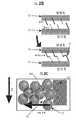
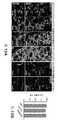
본원에 기재된 기술은 세포 침윤을 위한 통로를 남기면서 큰 단위로 어셈블리될 수 있는 작은 마이크로겔을 생성하는 화학기술을 이용하였다. 그 결과는 서로 붙어있는 검볼(gumball)의 항아리와 유사하게 그들 표면이 부착되어 있는 미세한 합성 중합체 본체(예를 들어, 구체)의 패킹된 클러스터이다. 상기 클러스터는 상처 부위를 채우는 미세다공성 어닐링된 입자의 스캐폴드(예를 들어, 다공성 겔 스캐폴드)를 만든다. 새로운 조직이 마이크로겔 입자들 간의 공극으로 빠르게 성장하고, 마이크로겔 입자가 체내로 분해됨에 따라, 새롭게 성장한 조직의 매트릭스가 상처가 있었던 부위에 남는다. 새로운 조직은 상처가 완전히 치유될 때까지 성장을 지속한다.The technique described herein utilizes a chemical technique that produces small microgels that can be assembled in large units while leaving a pathway for cell infiltration. The result is a packed cluster of fine synthetic polymer bodies (e.g., spheres) with their surfaces attached, similar to jars of gumball attached to each other. The cluster creates a scaffold of microporous annealed particles (e. G., A porous gel scaffold) that fills the wound site. As new tissue grows rapidly into the pores between the microgel particles and the microgel particles break down into the body, the matrix of newly grown tissue remains in the wounded area. The new tissue will continue to grow until the wound is completely healed.
본원에 기재된 마이크로겔 시스템은 기존 제품에 비해 상당한 개선을 나타낸다. 예를 들어, 본원에 기재된 기술은 세포를 물질에 끌어들이기 위해 성장 인자를 부가할 필요가 없다. 기재된 마이크로겔 네트워크의 기하형태는 세포가 마이크로겔 내로 이동하도록 유도한다.The microgel systems described herein exhibit significant improvements over conventional products. For example, the techniques described herein do not require the addition of growth factors to attract cells to the material. The geometry of the described microgel network induces the cell to migrate into the microgel.
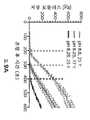
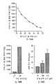
본 발명자들은 상기 기재된 마이크로겔이 기존에는 관찰되지 않았던 속도로 새로운 세포의 성장 및 연결된 세포의 네트워크 형성을 증진시킬 수 있다는 것을 입증하였다. 예를 들어, 생체 내 연구 동안, 상당한 조직 재생이 처음 48시간 내에 관찰되었는데, 이는 오늘날 사용되는 통상적인 물질과 비교해서 5일에 걸쳐 훨씬 더 많은 치유를 수반한다.The inventors have demonstrated that the microgels described above can promote the growth of new cells and the formation of network of connected cells at a rate not previously observed. For example, during in vivo studies, significant tissue regeneration was observed within the first 48 hours, which involves much more healing over 5 days compared to conventional materials used today.
본원에 기재된 기술은 광범위한 적용 어레이에 유용하다. 예를 들어, 개시된 마이크로겔 기술은 열상 및 수술 상처 봉합과 같은 급성 손상 및 당뇨병성 궤양 및 넓은 면적 화상 상처와 같은 보다 만성적인 적용을 포함한 상처 치료에 사용될 수 있다. 본원에 기재된 히드로겔 스캐폴드는 또한, 전장 또는 응급실과 같은 외상 상황에서도 유용할 수 있다.The techniques described herein are useful for a wide range of application arrays. For example, the disclosed microgel techniques can be used for wound healing including acute injury such as lacerations and surgical wound sutures and more chronic applications such as diabetic ulcers and wide area burn wounds. The hydrogel scaffolds described herein may also be useful in extracorporeal situations such as battlefield or emergency rooms.
특정 측면에서, 본원에는 복수 개의 마이크로겔 입자 및 예를 들어, 생분해성 가교 결합제를 포함한 가교 결합제를 포함하는 수성 용액을 포함하는 미세다공성 겔을 포함하는 시스템, 조성물, 방법 및 장치가 기재되어 있다. 본원에 기재된 미세다공성 겔은 유동성이고/이거나 주사 가능하며, 예를 들어 국소적으로 또는 주사에 의한 방식을 포함한 여러 상이한 방식으로 적용될 수 있다. 주사되고/되거나 유동성인 미세다공성 겔은 경피적으로 삽입되거나 또는 심부 조직 내로 삽입될 수 있다. 유동성 미세다공성 겔은 또한, 진피 및 다른 조직에 국소적으로 투여될 수 있다.In particular aspects, a system, composition, method, and apparatus are described herein that include a microporous gel comprising a plurality of microgel particles and an aqueous solution comprising, for example, a crosslinking agent comprising a biodegradable crosslinking agent. The microporous gels described herein are flowable and / or injectable, and may be applied in a variety of different ways, including, for example, topically or by injection. Microporous gels that are injected and / or flowable can be percutaneously inserted or inserted into deep tissue. The flowable microporous gel may also be administered topically to the dermis and other tissues.
한 측면에서, 어닐링 작용제를 복수 개의 마이크로겔 입자에 적용하면, 이러한 마이크로겔 입자는 그 내부에 틈새 공간이 있는 마이크로겔 입자의 공유적으로 안정화된 스캐폴드를 형성한다. 특정 적용에서는, 상기 시스템, 조성물, 방법, 및 장치는 생물의학적 적용을 위하여 특이적으로 조작된다. 일부 실시양태에서, 미세다공성 겔 입자는 가교 결합제를 추가로 포함하며, 여기서 이러한 가교 결합제는 매트릭스 메탈로프로테아제(MMP)-분해성 가교 결합제를 포함한다. 하나 이상의 실시양태에서, 어닐링 작용제는 인자 XIIIa를 포함한다. 추가 또는 부가의 실시양태에서, 어닐링 작용제는 에오신(Eosin) Y, 유리 라디칼 전달제, 또는 그의 조합물을 포함한다.In one aspect, when an annealing agent is applied to a plurality of microgel particles, such microgel particles form a covalently stabilized scaffold of microgel particles with interstitial spaces therein. In certain applications, the systems, compositions, methods, and devices are specifically engineered for biomedical applications. In some embodiments, the microporous gel particles further comprise a crosslinking agent, wherein such a crosslinking agent comprises a matrix metalloproteinase (MMP) -gradable crosslinking agent. In at least one embodiment, the annealing agent comprises Factor XIIIa. In additional or additional embodiments, the annealing agent comprises Eosin Y, a free radical transfer agent, or a combination thereof.
일부 실시양태에서, 상기 마이크로겔 시스템, 조성물, 방법, 및 장치는 복수 개의 마이크로겔 입자와 어닐링 작용제의 혼합물을 조명하도록 구성된 광원을 추가로 포함한다. 하나 이상의 실시양태에서, 미세다공성 겔 입자는 그의 표면 상에 노출된 세포 점착성 펩티드를 포함한다. 일부 실시양태에서, 미세다공성 겔 입자는 K-펩티드를 포함한다. 추가 또는 부가의 실시양태에서, 미세다공성 겔 입자는 인자 XIIIa-인식된 리신 기를 포함하는 K-펩티드를 포함한다. 일부 실시양태에서, 미세다공성 겔 입자는 Q-펩티드를 포함한다. 일부 실시양태에서, Q-펩티드는 인자 XIIIa-인식된 글루타민 기를 포함한다. 특정 실시양태에서, 미세다공성 겔 입자는 분해성인 가교 결합제를 포함한다. 특정 실시양태에서, 미세다공성 겔 입자는 음의 오목면을 나타내는 경계 표면을 포함하는 틈새 공간을 포함한다. 하나 이상의 실시양태에서, 마이크로겔 입자의 공유적으로 안정화된 스캐폴드는 약 10% 내지 약 50%의 공극 용적을 갖는다.In some embodiments, the microgel system, composition, method, and apparatus further comprise a light source configured to illuminate a mixture of a plurality of microgel particles and an annealing agent. In at least one embodiment, the microporous gel particle comprises a cell adhesive peptide exposed on its surface. In some embodiments, the microporous gel particles comprise a K-peptide. In a further or additional embodiment, the microporous gel particles comprise a K-peptide comprising a factor XIIIa-recognized lysine group. In some embodiments, the microporous gel particles comprise a Q-peptide. In some embodiments, the Q-peptide comprises factor XIIIa-recognized glutamine groups. In certain embodiments, the microporous gel particles comprise a crosslinking agent that is degradable. In certain embodiments, the microporous gel particles comprise a crevice space comprising a boundary surface representing a negative concave surface. In at least one embodiment, the covalently stabilized scaffold of microgel particles has a void volume of about 10% to about 50%.
한 실시양태에서, 생물의학적 적용을 위한 미세다공성 겔 시스템은 생분해성 가교 결합제, 예컨대 매트릭스 메탈로프로테아제(MMP)-분해성 가교 결합제와 함께 형성된 복수 개의 마이크로겔 입자를 함유하는 수성 용액; 및 복수 개의 마이크로겔 입자에 적용될 때, 마이크로겔 입자가 그 내부에 틈새 공간이 있는 마이크로겔 입자의 공유적으로 안정화된 스캐폴드를 형성하도록 유발시키는 어닐링 작용제를 포함한다.In one embodiment, a microporous gel system for biomedical applications comprises an aqueous solution containing a plurality of microgel particles formed with a biodegradable cross-linking agent, such as a matrix metalloproteinase (MMP) -decomposable cross-linking agent; And an annealing agent that, when applied to a plurality of microgel particles, causes the microgel particles to form a covalently stabilized scaffold of microgel particles with interstitial spaces therein.
또 다른 실시양태에서, 미세다공성 겔 시스템은 전달 장치; 및 수성 용액에 함유되어 있고 이러한 전달 장치 내에 저장된 생분해성 마이크로겔 입자의 컬렉션을 포함한다. 어닐링 작용제 또는 어닐링 작용제 전구체가 또한, 전달 장치 내에 저장된다. 이러한 전달 장치는 이용된 특별한 실시양태에 따라서 단일 또는 다수의 구획을 함유할 수 있다.In another embodiment, the microporous gel system comprises a delivery device; And a collection of biodegradable microgel particles contained in the aqueous solution and stored in such a delivery device. The annealing agent or annealing agent precursor is also stored in the delivery device. Such delivery devices may contain single or multiple compartments in accordance with the particular embodiment utilized.
또 다른 실시양태에서, 조직의 치료 방법은 세포 점착성 펩티드로 장식된 복수 개의 마이크로겔 입자를 함유하는 수성-기반 용액을 조직에 전달하는 단계를 포함하며, 여기서 상기 마이크로겔 입자는 생분해성 가교 결합제, 예컨대 매트릭스 메탈로프로테아제(MMP)-분해성 가교 결합제와 함께 형성된다. 복수 개의 마이크로겔 입자는 이러한 마이크로겔 입자를 어닐링시켜 그 내부에 틈새 공간이 있는 마이크로겔 입자의 공유적으로 안정화된 스캐폴드를 형성시키는 어닐링 작용제에 노출시킨다.In another embodiment, a method of treating tissue comprises delivering to an tissue an aqueous-based solution containing a plurality of microgel particles adorned with a cell adhesive peptide, wherein the microgel particles are biodegradable, Such as a matrix metalloprotease (MMP) -gradable crosslinking agent. A plurality of microgel particles are annealed to expose such microgel particles to an annealing agent that forms a covalently stabilized scaffold of microgel particles with interstitial spaces therein.
또 다른 실시양태에서, 생물의학적 적용을 위한 미세다공성 겔 시스템은 하나 이상의 세포 부착 모이어티(moiety), 하나 이상의 어닐링 성분, 및 생분해성 네트워크 가교 결합제 성분을 갖는 주쇄 중합체의 반응에 의해 형성된 마이크로겔 입자의 컬렉션을 포함한다. 미세다공성 겔 시스템은 그 내부에 틈새 공간이 있는 마이크로겔 입자의 공유적으로 안정화된 스캐폴드를 형성하기 위하여 어닐링 성분을 통하여 마이크로겔 입자를 계내에서 함께 연결시켜 주는 내인성 또는 외인성 어닐링 작용제를 포함한다.In another embodiment, a microporous gel system for biomedical applications comprises microgel particles formed by the reaction of a backbone polymer having one or more cell attachment moieties, one or more annealing components, and a biodegradable network crosslinker component ≪ / RTI > The microporous gel system includes an intrinsic or extrinsic annealing agent that links the microgel particles together in the system through the annealing component to form a covalently stabilized scaffold of microgel particles with interstitial spaces therein.
또 다른 측면에서, 본원에는 전달 장치 또는 기전 및 미세다공성 겔을 포함하는 시스템, 조성물, 방법, 및 장치가 기재되어 있다. 특정 실시양태에서, 상기 전달 장치는 복수 개의 마이크로겔 입자를 포함하는 수성 용액 및 어닐링 작용제 또는 어닐링 작용제 전구체를 함유한다. 하나 이상의 실시양태에서, 전달 장치는 복수 개의 마이크로겔 입자를 포함하는 수성 용액 및 어닐링 작용제를 함유하는 단일 구획 전달 장치를 포함한다. 하나 이상의 실시양태에서, 전달 장치는 다수의(예를 들어, 이중) 구획 전달 장치를 포함하며, 여기서 하나의 구획은 복수 개의 마이크로겔 입자를 함유하는 수성 용액 및 제1 어닐링 작용제 전구체를 함유하고, 제2 구획은 복수 개의 마이크로겔 입자를 함유하는 수성 용액 및 제2 어닐링 작용제 전구체를 함유한다. 특정 실시양태에서, 미세다공성 겔은 적어도 1종의 D-아미노산을 포함하는 (MMP)-분해성 가교 결합제를 추가로 포함한다. 추가 또는 부가의 실시양태에서, 마이크로겔 입자는 복수 종의 D-아미노산을 포함하는 (MMP)-분해성 가교 결합제를 포함한다.In another aspect, systems, compositions, methods, and devices are described herein that include a delivery device or an electrokinetic and microporous gel. In certain embodiments, the delivery device contains an aqueous solution comprising a plurality of microgel particles and an annealing agent or an annealing agent precursor. In one or more embodiments, the delivery device comprises a single compartment delivery device containing an aqueous solution comprising a plurality of microgel particles and an annealing agent. In one or more embodiments, the delivery device comprises a plurality of (e.g., dual) compartment delivery devices, wherein one compartment contains an aqueous solution containing a plurality of microgel particles and a first annealing agent precursor, The second compartment contains an aqueous solution containing a plurality of microgel particles and a second annealing agent precursor. In certain embodiments, the microporous gel further comprises a (MMP) -soluble cross-linking agent comprising at least one D-amino acid. In a further or additional embodiment, the microgel particles comprise (MMP) -soluble cross-linking agents comprising a plurality of D-amino acids.
또한 또 다른 측면에서, 본원에는 전달 장치; 수성 용액 내에 함유되고 상기 전달 장치 내에 저장된 복수 개의 생분해성 마이크로겔 입자; 및 상기 전달 장치 내에 저장된 어닐링 작용제 또는 어닐링 작용제 전구체를 포함하는 미세다공성 겔 시스템이 기재되어 있다. 하나 이상의 실시양태에서, 미세다공성 겔 입자는 수성 용액 내에 함유되고 전달 장치 내에 저장되는 2가지 이상 유형의 생분해성 마이크로겔 입자의 컬렉션을 추가로 포함한다. 특정 실시양태에서, 전달 장치는 2개의 구획을 포함하고, 생분해성 마이크로겔 입자는 이러한 2개의 구획 각각에서 저장되며, 제1 어닐링 전구체는 하나의 구획 내에 저장되고, 제2 어닐링 전구체는 다른 구획 내에 저장되며, 여기서 어닐링 작용제는 제1 어닐링 전구체와 제2 어닐링 전구체 둘 다의 존재에 의해 형성된다. 하나 이상의 실시양태에서, 전달 장치는 단일 구획을 포함하고; 생분해성 마이크로겔 입자의 컬렉션과 어닐링 작용제 둘 다가 단일 구획 내에 저장된다. 또한 추가 또는 부가의 실시양태에서, 어닐링 작용제는 단일 구획 내에 저장된 유리 라디칼 전달제 및 광개시제를 포함한다. 추가 또는 부가의 실시양태에서, 미세다공성 겔 시스템은 생분해성 마이크로겔 입자의 컬렉션과 어닐링 작용제의 혼합물을 조명하도록 구성된 발광 장치를 추가로 포함한다. 특정 실시양태에서, 마이크로겔 입자는 실질적으로 단분산 구체를 포함한다. 하나 이상의 실시양태에서, 이러한 실질적으로 단분산 구체는 약 30 마이크로미터 내지 약 150 마이크로미터의 범위 내의 직경을 갖는다. 추가 또는 부가의 실시양태에서, 마이크로겔 입자는 어닐링 후 서로 공유적으로 연결된다.In yet another aspect, the present disclosure provides a delivery device; A plurality of biodegradable microgel particles contained in the aqueous solution and stored in the delivery device; And a microporous gel system comprising an annealing agent or an annealing agent precursor stored in the delivery device. In one or more embodiments, the microporous gel particles further comprise a collection of two or more types of biodegradable microgel particles contained within an aqueous solution and stored in a delivery device. In certain embodiments, the delivery device comprises two compartments, the biodegradable microgel particles are stored in each of these two compartments, the first annealing precursor is stored in one compartment, and the second annealing precursor is stored in another compartment Wherein the annealing agent is formed by the presence of both the first annealing precursor and the second annealing precursor. In at least one embodiment, the delivery device comprises a single compartment; Both the collection of biodegradable microgel particles and the annealing agent are stored in a single compartment. Also in additional or additional embodiments, the annealing agent comprises a free radical transfer agent and a photoinitiator stored in a single compartment. In a further or additional embodiment, the microporous gel system further comprises a light emitting device configured to illuminate a mixture of the collection of biodegradable microgel particles and the annealing agent. In certain embodiments, the microgel particles comprise substantially monodisperse spheres. In one or more embodiments, such substantially monodisperse spheres have diameters in the range of about 30 micrometers to about 150 micrometers. In additional or additional embodiments, the microgel particles are covalently linked to each other after annealing.
또 다른 측면에서는 복수 개의 마이크로겔 입자를 함유하는 수성-기반 용액을 조직에 전달하는 단계; 및 마이크로겔 입자를 어닐링시켜 그 내부에 틈새 공간이 있는 마이크로겔 입자의 공유적으로 안정화된 스캐폴드를 형성시키는 어닐링 작용제에 복수 개의 마이크로겔 입자를 노출시키는 단계를 포함하는, 조직의 치료 방법이 제공된다. 일부 실시양태에서, 복수 개의 마이크로겔 입자는 세포 점착성 펩티드로 장식하고, 마이크로겔 입자는 매트릭스 메탈로프로테아제(MMP)-분해성 가교 결합제와 함께 형성된다. 하나 이상의 실시양태에서, 어닐링 작용제가 상기 조직에 전달된다. 일부 실시양태에서, 어닐링 작용제는 조직 내에 존재한다. 또한 부가의 실시양태에서, 상기 방법은 빛에 대한 노출을 이용하여 마이크로겔 입자의 어닐링을 개시하는 단계를 추가로 포함한다. 일부 실시양태에서, 광 파장이 가시광선 범위 내이다. 일부 실시양태에서, 광 파장이 적외선 범위 내이다. 하나 이상의 실시양태에서, 수성-기반 용액과 어닐링 작용제는 동시에 전달된다. 일부 실시양태에서, 수성-기반 용액과 어닐링 작용제는 순차적으로 전달된다. 또한 추가 또는 부가의 실시양태에서, 마이크로겔 입자는 치료상 활성 화학적 화합물을 포함한다. 특정 실시양태에서, 마이크로겔 입자는 상기 화학적 화합물을 조직에 노출 또는 용출시킨다. 하나 이상의 실시양태에서, 상기 조직은 성형 재건, 만성 상처 발생, 급성 조직 손상, 또는 수술 절개로 인해 유발된 조직 갭의 부위를 포함한다. 또한 부가의 실시양태에서, (MMP)-분해성 가교 결합제는 D-아미노산을 포함한다.In another aspect, there is provided a method comprising: delivering an aqueous-based solution containing a plurality of microgel particles to a tissue; And annealing the microgel particles to expose a plurality of microgel particles to an annealing agent that forms a covalently stabilized scaffold of microgel particles with interstitial spaces therein. do. In some embodiments, the plurality of microgel particles are decorated with a cell sticky peptide, and the microgel particles are formed with a matrix metalloprotease (MMP) -discriminating cross-linking agent. In one or more embodiments, an annealing agent is delivered to the tissue. In some embodiments, the annealing agent is present in the tissue. In yet further embodiments, the method further comprises initiating annealing of the microgel particles using exposure to light. In some embodiments, the optical wavelength is within the visible range. In some embodiments, the optical wavelength is within the infrared range. In one or more embodiments, the aqueous-based solution and the annealing agent are delivered simultaneously. In some embodiments, the aqueous-based solution and the annealing agent are delivered sequentially. Also in additional or additional embodiments, the microgel particles comprise therapeutically active chemical compounds. In certain embodiments, the microgel particles expose or elute the chemical compound to the tissue. In at least one embodiment, the tissue includes sites of tissue gaps caused by plastic reconstruction, chronic scarring, acute tissue injury, or surgical incision. Also in a further embodiment, the (MMP) -soluble cross-linking agent comprises a D-amino acid.
또 다른 측면에서, 하나 이상의 세포 부착 모이어티, 하나 이상의 어닐링 성분, 및 하나 이상의 생분해성 네트워크 가교 결합제 성분을 갖는 주쇄 중합체를 포함하는 마이크로겔 입자의 컬렉션; 및 그 내부에 틈새 공간이 있는 마이크로겔 입자의 공유적으로 안정화된 스캐폴드를 형성하기 위하여 어닐링 성분을 통하여 마이크로겔 입자를 계내에서 함께 연결시켜 주는 내인성 또는 외인성 어닐링 작용제를 포함하는, 미세다공성 겔 시스템 또는 장치가 제공된다. 특정 실시양태에서, 주쇄 중합체는 폴리(에틸렌 글리콜) 비닐 술폰을 포함한다. 하나 이상의 실시양태에서, 하나 이상의 세포 부착 모이어티는 RGD 펩티드 또는 그의 단편, 피브로넥틴 또는 그의 단편, 콜라겐 또는 그의 단편, 또는 라미닌 또는 그의 단편을 포함한다. 일부 실시양태에서, 하나 이상의 세포 부착 모이어티는 RGD 펩티드 또는 그의 단편을 포함한다. 특정 실시양태에서, 하나 이상의 세포 부착 모이어티는 서열식별번호(SEQ ID NO): 3 또는 그의 단편을 포함한다. 추가 또는 부가의 실시양태에서, 하나 이상의 어닐링 성분은 K-펩티드 및 Q-펩티드를 포함한다. 특정 실시양태에서, K-펩티드는 인자 XIIIa-인식된 리신 기를 포함하고, Q-펩티드는 인자 XIIIa-인식된 글루타민 기를 포함한다. 일부 실시양태에서, 생분해성 네트워크 가교 결합제 성분은 매트릭스 메탈로프로테아제(MMP)-분해성 가교 결합제를 포함한다. 하나 이상의 실시양태에서, (MMP)-분해성 가교 결합제는 D-아미노산을 포함한다. 특정 실시양태에서, 마이크로겔 입자의 컬렉션은 2가지 이상 유형의 마이크로겔 입자를 포함한다. 하나 이상의 실시양태에서, 제1 유형의 마이크로겔 입자는 D-아미노산을 포함하는 (MMP)-분해성 가교 결합제를 포함하고, 제2 유형의 마이크로겔 입자는 L-아미노산만을 포함하는 (MMP)-분해성 가교 결합제를 포함한다. 하나 이상의 실시양태에서, 상기 시스템 또는 장치는 마이크로겔 입자의 컬렉션과 어닐링 작용제를 함유하는 단일 구획 전달 장치를 포함한다. 하나 이상의 실시양태에서, 상기 시스템 또는 장치는 이중 구획 전달 장치를 추가로 포함하며, 여기서 하나의 구획은 복수 개의 마이크로겔 입자를 함유하는 수성 용액 및 제1 어닐링 작용제 전구체를 함유하고, 제2 구획은 복수 개의 마이크로겔 입자를 함유하는 수성 용액 및 제2 어닐링 작용제 전구체를 함유하며, 여기서 어닐링 작용제는 제1 어닐링 작용제 전구체와 제2 어닐링 작용제 전구체의 존재에 의해 형성된다.In yet another aspect, a collection of microgel particles comprising a backbone polymer having at least one cell attachment moiety, at least one annealing component, and at least one biodegradable network crosslinker component; And an intrinsic or extrinsic annealing agent that interconnects the microgel particles together in the system through an annealing component to form a covalently stabilized scaffold of microgel particles with interstitial spaces therein. Or device is provided. In certain embodiments, the backbone polymer comprises poly (ethylene glycol) vinyl sulfone. In one or more embodiments, the one or more cell attachment moieties comprise an RGD peptide or fragment thereof, fibronectin or a fragment thereof, collagen or a fragment thereof, or a laminin or fragment thereof. In some embodiments, the one or more cell adhesion moieties comprise RGD peptides or fragments thereof. In certain embodiments, the one or more cell attachment moieties comprise SEQ ID NO: 3 or fragments thereof. In additional or additional embodiments, the at least one annealing component comprises a K-peptide and a Q-peptide. In certain embodiments, the K-peptide comprises a factor XIIIa-recognized lysine group and the Q-peptide comprises a factor XIIIa-recognized glutamine group. In some embodiments, the biodegradable network crosslinker component comprises a matrix metalloproteinase (MMP) -discible crosslinker. In one or more embodiments, the (MMP) -gradable crosslinking agent comprises a D-amino acid. In certain embodiments, the collection of microgel particles comprises two or more types of microgel particles. In one or more embodiments, the first type of microgel particles comprise (MMP) -decomposable cross-linkers comprising D-amino acids and the second type of microgel particles comprise (MMP) -decomposable Crosslinking agents. In at least one embodiment, the system or apparatus comprises a single compartment delivery device containing a collection of microgel particles and an annealing agent. In one or more embodiments, the system or apparatus further comprises a dual compartment delivery device wherein one compartment contains an aqueous solution containing a plurality of microgel particles and a first annealing agent precursor, An aqueous solution containing a plurality of microgel particles and a second annealing agent precursor wherein the annealing agent is formed by the presence of the first annealing agent precursor and the second annealing agent precursor.
부가 측면에서, 세포 점착성 펩티드로 장식된 마이크로겔 입자의 제1 층을 조직에 전달하는 단계(여기서, 마이크로겔 입자는 생분해성 가교 결합제와 함께 형성된다); 이러한 제1 층을, 마이크로겔 입자를 어닐링시켜 그 내부에 틈새 공간이 있는 마이크로겔 입자의 공유적으로 안정화된 스캐폴드를 형성시키는 어닐링 작용제에 노출시키는 단계; 세포 점착성 펩티드로 장식된 마이크로겔 입자의 제2 층을 상기 조직에 전달하는 단계(여기서, 마이크로겔 입자는 생분해성 가교 결합제와 함께 형성되고, 제2 층 내의 마이크로겔 입자는 제1 층 내의 마이크로겔 입자와 비교해서 물리적 특성 또는 화학적 조성 중 하나에 있어서 상이하다); 및 상기 제2 층을, 마이크로겔 입자를 어닐링시켜 그 내부에 틈새 공간이 있는 마이크로겔 입자의 공유적으로 안정화된 스캐폴드를 형성시키는 어닐링 작용제에 노출시키는 단계를 포함하는, 조직의 치료 방법이 기재되어 있다. 하나 이상의 실시양태에서, 제2 층 내의 마이크로겔 입자는 상이한 크기를 갖는다. 또한 부가의 실시양태에서, 제2 층 내의 마이크로겔 입자는 상이한 형상을 갖는다. 하나 이상의 실시양태에서, 제2 층 내의 마이크로겔 입자는 상이한 강성을 갖는다. 특정 실시양태에서, 제2 층 내의 마이크로겔 입자는 제1 층 내의 화학적 성분과 상이한 화학적 성분을 갖는다. 추가 또는 부가의 실시양태에서, 제2 층 내의 마이크로겔 입자는 제1 층 내의 동일한 화학적 성분과 상이한 농도의 화학적 성분을 갖는다.In a further aspect, a method is provided for delivering a first layer of microgel particles adorned with a cell adhesive peptide to a tissue, wherein the microgel particles are formed with a biodegradable crosslinking agent; Exposing the first layer to an annealing agent that anneals the microgel particles to form a covalently stabilized scaffold of microgel particles with interstitial spaces therein; Transferring a second layer of microgel particles adorned with a cell adhesive peptide to the tissue wherein the microgel particles are formed with a biodegradable crosslinking agent and the microgel particles in the second layer are microgel particles in the first layer, Different in either physical properties or chemical composition as compared to the particles); And exposing the second layer to an annealing agent that anneals the microgel particles to form a covalently stabilized scaffold of microgel particles with interstitial spaces therein. . In at least one embodiment, the microgel particles in the second layer have different sizes. Also in a further embodiment, the microgel particles in the second layer have different shapes. In at least one embodiment, the microgel particles in the second layer have different stiffnesses. In certain embodiments, the microgel particles in the second layer have chemical components that are different from the chemical components in the first layer. In additional or additional embodiments, the microgel particles in the second layer have a chemical composition that is different from the same chemical composition in the first layer.
또 다른 측면에서, 세포 점착성 펩티드로 장식된 복수 개의 마이크로겔 입자를 함유하는 수성-기반 용액을 조직에 전달하는 단계(여기서, 마이크로겔 입자는 생분해성 가교 결합제와 함께 형성된다); 및 복수 개의 마이크로겔 입자를, 이러한 마이크로겔 입자를 어닐링시켜 그 내부에 틈새 공간이 있는 마이크로겔 입자의 공유적으로 안정화된 스캐폴드를 형성시키는 어닐링 작용제에 노출시키는 단계를 포함하는, 조직의 치료 방법이 제공된다.In another aspect, there is provided a method of making a microcapsule comprising the steps of delivering an aqueous-based solution containing a plurality of microgel particles adorned with a cell adhesion peptide to a tissue, wherein the microgel particles are formed with a biodegradable cross-linking agent; And exposing the plurality of microgel particles to an annealing agent that anneals the microgel particles to form a covalently stabilized scaffold of microgel particles with interstitial spaces therein. / RTI >
또 다른 실시양태에서, 조직의 치료 방법은 세포 점착성 펩티드로 장식된 마이크로겔 입자의 제1 층을 조직에 전달하는 단계를 포함하며, 여기서 마이크로겔 입자는 생분해성 가교 결합제와 함께 형성된다. 상기 제1 층은 마이크로겔 입자를 어닐링시켜 그 내부에 틈새 공간이 있는 마이크로겔 입자의 공유적으로 안정화된 스캐폴드를 형성시키는 어닐링 작용제에 노출시킨다. 세포 점착성 펩티드로 장식된 마이크로겔 입자의 제2 층을 상기 조직에 전달하며, 여기서 마이크로겔 입자는 생분해성 가교 결합제와 함께 형성되고, 제2 층 내의 마이크로겔 입자는 제1 층 내의 마이크로겔 입자와 비교해서 물리적 특성 또는 화학적 조성 중 하나에 있어서 상이하다. 제2 층을, 마이크로겔 입자를 어닐링시켜 그 내부에 틈새 공간이 있는 마이크로겔 입자의 공유적으로 안정화된 스캐폴드를 형성시키는 어닐링 작용제에 노출시킨다.In another embodiment, a method of treating tissue comprises delivering to the tissue a first layer of microgel particles adorned with a cell adhesive peptide, wherein the microgel particles are formed with a biodegradable cross-linking agent. The first layer is annealed to expose the microgel particles to an annealing agent that forms a covalently stabilized scaffold of microgel particles with interstitial spaces therein. Wherein the microgel particles are formed with a biodegradable cross-linking agent and the microgel particles in the second layer are mixed with the microgel particles in the first layer and the microgel particles in the second layer, They differ in either physical properties or chemical composition. The second layer is exposed to an annealing agent that anneals the microgel particles to form a covalently stabilized scaffold of microgel particles with interstitial spaces therein.
또 다른 실시양태에서, 조직의 치료 방법은 세포 점착성 펩티드로 장식된 복수 개의 마이크로겔 입자를 함유하는 수성-기반 용액을 조직에 전달하는 단계를 포함하며, 여기서 마이크로겔 입자는 생분해성 가교 결합제와 함께 형성된다. 복수 개의 마이크로겔 입자를, 이러한 마이크로겔 입자를 어닐링시켜 그 내부에 틈새 공간이 있는 마이크로겔 입자의 공유적으로 안정화된 스캐폴드를 형성시키는 어닐링 작용제에 노출시킨다.In another embodiment, a method of treating tissue comprises delivering to the tissue an aqueous-based solution containing a plurality of microgel particles adorned with a cell adhesive peptide, wherein the microgel particles are combined with a biodegradable crosslinking agent . A plurality of microgel particles are exposed to an annealing agent that anneals these microgel particles to form a covalently stabilized scaffold of microgel particles with interstitial spaces therein.
또한 부가의 측면에서, 공통 채널로 이어지는 복수 개의 투입 채널 및 올리고펩티드로 변형시킨 중합체 주쇄를 함유하는 제1 예비-중합체 용액을 제1 투입 채널 내로 유동시키는 하류 위치에서 공통 채널과 교차하는 한 쌍의 오일-핀칭(pinching) 채널을 갖는 유중수 액적 생성 마이크로유체 장치를 제공하는 단계; 생분해성 가교 결합제를 함유하는 제2 용액을 제2 투입 채널 내로 유동시키는 단계; 오일 및 계면활성제를 한 쌍의 오일 핀칭 채널 내로 유동시켜 제1 예비 중합체 용액과 제2 용액을 함유하는 액적을 형성하는 단계; 및 이러한 액적의 가교 결합에 의해 형성된 마이크로겔 입자를 수집하는 단계를 포함하는, 마이크로겔 입자의 제조 방법이 기재되어 있다. 또 다른 실시양태에서, 상기 방법은 제1 투입 채널과 제2 투입 채널 사이에 삽입된 제3 투입 채널을 추가로 포함하는데, 예비 중합체를 함유하는 제3의 불활성 용액을 제3 투입 채널 내로 유동시킨다. 하나 이상의 실시양태에서, 상기 방법은 상기 생성된 액적을, 한 쌍의 오일 핀칭 채널이 공통 채널과 교차하는 위치의 하류에 위치하는 부가 쌍의 외장 채널로 피복하는 단계를 추가로 포함하며, 여기서 부가 쌍의 외장 채널은 오일 핀칭 채널의 상류 쌍에 함유된 계면활성제보다 더 높은 농도로 오일 및 계면활성제를 운반한다. 한 실시양태에서, 상기 방법은 상기 수집된 마이크로겔 입자를 원심분리시키는 단계를 추가로 포함한다. 또 다른 측면에서, 상기 방법은 이와 같이 원심분리된 마이크로겔 입자의 자유수 부피 함량을 감소시키는 단계를 포함한다. 하나 이상의 실시양태에서, 상기 방법은 상기 수집된 마이크로겔 입자를 연장된 기간(예를 들어, 수개월 내지 수년) 동안 저장하는 단계를 포함한다.In a further aspect, there is also provided a polymer electrolyte membrane comprising a pair of cross-linked oligomeric oligomers crossing a common channel at a downstream location for flowing a first pre-polymer solution containing a polymeric backbone modified with an oligopeptide and a plurality of input channels leading to a common channel into a first input channel Providing a water / liquid droplet generating microfluidic device having an oil-pinching channel; Flowing a second solution containing a biodegradable cross-linking agent into a second charge channel; Flowing the oil and surfactant into a pair of oil pinch channels to form droplets containing the first prepolymer solution and the second solution; And collecting microgel particles formed by cross-linking of such droplets. In another embodiment, the method further comprises a third input channel inserted between the first input channel and the second input channel, wherein a third inert solution containing the prepolymer flows into the third input channel . In at least one embodiment, the method further comprises the step of coating the resulting droplets with an additional pair of exterior channels located downstream of the location where the pair of oil pinching channels intersect the common channel, The outer channels of the pair carry the oil and the surfactant at a higher concentration than the surfactant contained in the upstream pair of oil pinching channels. In one embodiment, the method further comprises centrifuging the collected microgel particles. In another aspect, the method includes reducing the free water volume content of the centrifuged microgel particles. In one or more embodiments, the method comprises storing the collected microgel particles for an extended period of time (e.g., months to years).
또한 또 다른 실시양태에서, 마이크로겔 입자의 제조 방법은 공통 채널로 이어지는 복수 개의 투입 채널 및 하류 위치에서 공통 채널과 교차하는 한 쌍의 오일-핀칭 채널을 갖는 유중수 액적 생성 마이크로유체 장치를 제공하는 단계를 포함한다. 올리고펩티드로 변형시킨 중합체 주쇄를 함유하는 제1 예비 중합체 용액을 제1 투입 채널 내로 유동시킨다. 생분해성 가교 결합제를 함유하는 제2 용액을 제2 투입 채널 내로 유동시킨다. 오일 및 계면활성제를 한 쌍의 오일 핀칭 채널 내로 유동시켜, 제1 예비 중합체 용액과 제2 용액을 함유하는 액적을 형성시킨다. 마이크로겔 입자는 상기 액적의 가교 결합에 의해 형성되고, 이어서 이를 수집한다.In yet another embodiment, the method of making microgel particles provides a plurality of input channels leading to a common channel and a water / liquid droplet generating microfluidic device having a pair of oil-pinched channels crossing a common channel at a downstream location . The first prepolymer solution containing the polymer backbone modified with the oligopeptide is flowed into the first charge channel. The second solution containing the biodegradable crosslinking agent is flowed into the second inlet channel. The oil and surfactant are flowed into the pair of oil pinch channels to form droplets containing the first prepolymer solution and the second solution. Microgel particles are formed by cross-linking of the droplets, which are then collected.
본 개시내용의 다른 목적, 특징 및 이점은 다음 상세한 설명으로부터 관련 기술분야의 통상의 기술자에게 명백할 것이다. 그러나, 상세한 설명과 구체적인 실시예가 본 개시내용의 일부 실시양태를 표시하긴 하지만, 이는 예시로써 제공되고 제한되지 않는 것으로 이해해야 한다. 본 개시내용의 범위 내에 있는 많은 변화 및 변형은 본 개시내용의 요지를 벗어나지 않고서도 이루어질 수 있고, 본 개시내용은 이러한 변형 모두를 포함한다. 더욱이, 한 실시양태의 측면은 다른 상이한 실시양태에서 활용될 수 있다.Other objects, features and advantages of the present disclosure will become apparent to those skilled in the art from the following detailed description. It should be understood, however, that the detailed description and specific examples, while indicating some embodiments of the disclosure, are provided by way of illustration and not by way of limitation. Many changes and modifications that are within the scope of this disclosure can be made without departing from the spirit of the present disclosure, and the present disclosure encompasses all such modifications. Moreover, aspects of one embodiment may be utilized in other different embodiments.
본 개시내용의 신규 특징은 특히 첨부된 청구범위에 제시된다. 본 개시내용의 특징 및 이점의 보다 좋은 이해는 예시적 실시양태(여기서, 본 개시내용의 원리들이 활용된다)를 제시하는 다음 상세한 설명, 및 수반되는 도면을 참조로 하여 수득될 것이다:
도 1은 복수 개의 어닐링된 마이크로겔 입자로부터 형성된 스캐폴드의 특정 부분을 도시한 것이다.
도 2A는 상처 부위를 치유하기 위하여 마이크로겔 입자를 이러한 상처 부위 내로 주사하는 예시적 방법을 도시한 것이다.
도 2B는 마이크로겔 입자의 표면 상에서 링커에 의해 증강된 상이한 마이크로겔 입자들 간의 예시적 어닐링 반응을 도식적으로 예시한 것이다.
도 2C는 조직 상의 전달 부위 내에 형성된 스캐폴드 내로의 조직 침윤의 예시적 과정을 도시한 것인데, 여기서 상기 조직과 마이크로겔 간의 경계는 이들 간의 임의의 계면을 나타내고, 세포는 조직으로부터 안쪽으로 또는 마이크로겔로부터 조직을 향해 바깥쪽으로 움직이면서 계면을 관통할 수 있다.
도 3A는 미세다공성 겔 시스템의 일부로서 복수 개의 마이크로겔 입자를 생성하는데 사용되는 하나의 실시양태에 따른 마이크로유체 장치의 하향식 보기를 도시한 것이다.
도 3B는 액적 생성 영역 및 하류 오일/계면활성제 핀칭 영역의 확대도를 도시한 것이다(도 3A의 박스 영역 참조).
도 3C는 도 3A에 도시된 2개의 분지 채널의 확대 사시도를 도시한 것이다.
도 3D는 하나의 실시양태에 따른 도 3A의 마이크로유체 장치의 측면도를 도시한 것이다.
도 3E는 도 3B에 예시된 도식의 실시에 대한 사진을 도시한 것인데, 여기서 왼쪽의 형광 용액은 가교 결합제를 함유하고, 오른쪽의 형광 용액은 중합체 및 반응 완충제를 함유하며, 중간 스트림은 불활성 액상 용액을 함유하여, 액적 분할 전에 왼쪽 용액과 오른쪽 용액이 혼합되는 것을 방지시킨다. 중간 스트림과 오른쪽 스트림 사이의 밝은 형광은 반응 완충제의 확산으로 인한 중간 스트림 내의 pH 변화를 예시한다.
도 3F는 도 3B 및 도 3E에 예시된 도식의 실시에 대한 사진을 도시한 것인데, 이는 또한, 핀칭 오일 스트림을 도입한 후 액적 분할의 광 현미경 보기를 나타낸다.
도 4A는 미세다공성 겔 시스템의 일부로서 복수 개의 마이크로겔 입자를 생성하는데 사용되는 또 다른 실시양태에 따른 마이크로유체 장치의 하향식 보기를 도시한 것이다.
도 4B는 액적 분할 영역에서는, 0.25% 스판(Span®) 80을 수반한 광유가 PEG 예비 겔을 핀칭 및 분할하고, 광유 중의 5% 스판® 80 용액의 하류가 혼합되며 완전한 겔화 전에 마이크로겔의 하류 유착이 방지된다는 것을 도시한 것이다.
도 4C는 액적이 분기 영역에서의 인큐베이션 동안 재조합되지 않고 마이크로 채널로부터 수집 웰로 빠져 나가는 것을 도시한 것이다.
도 5는 한 실시양태에 따라서 마이크로겔 액적을 생성하는데 사용될 수 있는 예시적인 마이크로유체 T-접합부를 도시한 것이다.
도 6A는 한 실시양태에 따른 이중 배럴 주사기 형태의 예시적인 투약 장치를 도시한 것이다.
도 6B는 또 다른 실시양태에 따른 단일 배럴 주사기 형태의 예시적인 투약 장치를 도시한 것이다.
도 6C는 한 실시양태에 따른 마이크로겔 입자를 보유하고 있는 튜브 형태의 예시적인 투약 장치를 도시한 것이다.
도 7A는 주사한 지 24시간 후 비-다공성 대조군뿐만 아니라 스캐폴드(미세다공성 어닐링된 입자 또는 "MAP" 스캐폴드)가 주사된 조직에 대한 SKH1-Hrhr 마우스의 조직 절편의 헤마톡실린 및 에오신 염색(H&E 염색)을 도시한 것이다.
도 7B는 주사 후 일수의 함수로서 상처 봉합(%)의 그래프를 도시한 것이다. 이 그래프는 5일 동안 비-다공성 양측 대조군(N = 5)과 비교하였을 때 스캐폴드를 사용하는 경우에 상처 봉합률이 통계상 유의적으로 개선되었다는 것을 보여준다.
도 7C는 겔 스캐폴드(왼쪽 패널)를 비-다공성 PEG 겔 대조군(오른쪽 패널)과 비교한, SKH1-Hrhr 마우스에서 5일 동안의 생체내 상처 치유 모델의 상처 봉합의 대표적인 영상을 도시한 것이다.
도 7D는 7일 생체 내 BALB/c 실험 동안 상처 봉합의 대표적인 영상을 도시한 것이다. 생체 내에서 7일 후, 스캐폴드는 비-처리 대조군, K 및 Q 펩티드가 결여된 겔, 비-다공성 PEG 겔보다 상당히 더 신속한 상처 치유를 증진시키고, 예비주조 다공성 겔보다 더 신속한 상처 치유를 증진시킨다. 정규의 포로겐-기반 주조 방법을 이용하여 상처 형상과 정확하게 일치시키기 위해 생체 외에서 만들어진 다공성 겔은 스캐폴드와 거의 동등한 수준으로 주목할만한 상처 치유율을 나타냈지만, 주사 가능성은 불충분하였다(N≥5).
도 7E는 도 7D에 상응하는 각각의 치료 카테고리에 대한 BALB/c 생체 내 상처 치유로부터의 상처 봉합 정량화 데이터를 도시하는 막대 그래프이다. 모든 데이터는 평균 +/- SEM으로서 제시된다. 통계적 유의성은 표준 양측 t-검정을 이용하여 수행하였다(*: p<0.05; **p<0.01).
도 7F는 도 7D 및 도 7E에 상응하는 각각의 치료 카테고리에 대한 생체 내 7일 동안 상처 바닥 봉합의 흔적을 도시한 것이다.
도 7G는 도 6A 또는 6B와 같은 주사기 장치(예를 들어, 25 게이지 주사기)를 사용하여, 마이크로겔 입자 함유 용액 또는 슬러리를 어떻게 치료 부위 내로 주사할 수 있고 [여기서, 마이크로겔은 주사 부위의 형상(예를 들어, 이 경우에는 별 형상의 레이저 커팅된 아크릴계 몰드)과 맞아야 한다], 연속해서 스캐폴드를 별 형상이 되도록 어닐링시킬 수 있는지를 도시한 것이다.
도 8A 및 8B는 피부 절개 및 겔 적용 후 21일에 마우스 모델에서 마이크로겔 스캐폴드로 처리한 경우(도 8A) 및 처리하지 않았거나 또는 "모의(sham)" 처리된 경우(도 8B) 손상된 조직(즉, 상처 부위)의 염색된 현미경 영상을 도시한 것이다. 마이크로겔 스캐폴드에 의해 가능하게 된 흉터 감소는 도 8A에서 명확하게 볼 수 있다. 사각형은 상처 부위에 겔을 적용한 후 개질 조직 내의 모낭 및 지방 분비선(피지선)을 표시한다. 원형은 개질 조직 내의 잔여 마이크로겔 입자를 표시한다.
도 8C는 겔 스캐폴드로 처리된 조직뿐만 아니라 모의로 처리된 조직에 대한 표피 두께를 보여주는 그래프를 도시한 것이다.
도 8D는 겔 스캐폴드로 처리된 조직뿐만 아니라 모의로 처리된 조직에 대한 피지선의 수를 보여주는 그래프를 도시한 것이다.
도 8E는 겔 스캐폴드로 처리된 조직뿐만 아니라 모의로 처리된 조직에 대한 모낭의 수를 보여주는 그래프를 도시한 것이다.
도 8F는 겔 스캐폴드로 처리된 조직뿐만 아니라 모의로 처리된 조직에 대한 흉터 폭을 보여주는 그래프를 도시한 것이다.
도 8G는 겔 스캐폴드로 처리된 조직뿐만 아니라 모의로 처리된 조직에 대한 밀리얼 낭종(milial cyst)의 수를 보여주는 그래프를 도시한 것이다.
도 9A는 상이한 겔화 역학(pH 및 온도)에 대한 혼합 후 시간의 함수로서의 저장 모듈러스의 그래프를 도시한 것이다. 25℃에서의 pH 8.25는 상기 그래프의 바닥 라인으로 나타내고; 25℃에서의 pH 8.8은 상기 그래프의 상부 라인으로 나타내며; 37℃에서의 pH 8.25는 상기 그래프의 중간 라인으로 나타낸다.
도 9B는 상이한 히드로겔 중량%를 사용하여 x-축 상에 상이한 강성 물질을 생성하였다는 것을 도시한 것이다. 본 그래프는 각종 히드로겔 중량%에 대한 저장 모듈러스(Pa)를 도시하였다.
도 9C는 x-축 상에 생성된 겔에서 상이한 강성 값을 생성하는데 사용되었던 상이한 가교 결합제 화학량론을 도시한 것이다. 본 그래프는 PEG 분자 상의 유리 가교 결합제 말단(-SH) 대 비닐 기(-VS)의 r-비의 함수로서 저장 모듈러스(Pa)를 도시하였다.
도 9D는 본원에 기재된 다공성 겔(그래프의 상부 라인)뿐만 아니라 비-다공성 대조군(그래프의 바닥 라인) 둘 다에 대한 시간의 함수로서 % 분해의 그래프를 도시한 것이다.
도 9E는 200 μm(상부 패널) 또는 100 μm(바닥 패널)에서 FXIIIa로 어닐링된 스캐폴드의 SEM 영상을 도시한 것이다.
도 9F는 200 μm(상부 패널) 또는 100 μm(바닥 패널)에서 FXIIIa를 수반하지 않은 마이크로겔 입자의 SEM 영상을 도시한 것이다. 어닐링되지 않은 마이크로겔 입자가 도 9F에서 관찰된다.
도 10은 포스핀-아지드 '클릭' 화학의 사용을 통하여 형광성 소 혈청 알부민(BSA) 단백질을 이용하여 마이크로겔의 표면을 증대시킨(바깥 둘레), 본원에 기재된 기술을 이용하여 제작된 마이크로겔을 나타낸다. 추가로, 마이크로유체 제작 동안 마이크로겔 내에 나노입자(500 nm)를 매립시킨다.
도 11은 본원에 기재된 미세다공성 겔 시스템을 사용하여 손상된 조직을 치료하는 예시적인 방법을 도시한 것이다. 마이크로겔 입자를 적용하고(상부 패널), 임의로, 어플리케이터(applicator)를 활용하며(두 번째 패널), 마이크로겔 입자의 어닐링을 개시하여 스캐폴드를 형성시키며(세 번째 패널), 개선된 상처 치유를 관찰한다(바닥 패널).
도 12A는 6일 후에 비-다공성 겔 뿐만 아니라 시험관내 다공성 겔 스캐폴드에서 배양 6일 동안 3D 세포성 네트워크의 형성을 명확히 보여주는 형광성 영상을 도시한 것이다. (350 Pa: 다공성 겔 스캐폴드와 동일한 벌크 모듈러스, 600 Pa: 개개의 마이크로겔과 일치시킨 마이크로스케일 모듈러스).
도 12B는 어닐링한 지 24시간 후의 세포 생존율이, 상이한 인간 조직 유형을 나타내는 3가지 세포주 전반에 걸쳐 93%를 초과한다는 것을 그래프로 도시한 것이다. HDF: 인간 진피 섬유모세포, AhMSC: 지방 유래 인간 중간엽 줄기 세포, BMhMSC: 골수 유래 인간 중간엽 줄기 세포.
도 13A는 어닐링하기에 앞서 살아있는 세포를 미리 형성된 마이크로겔 입자와 조합하기 위한 예시적 방법을 도시한 것이다. 마이크로겔 입자를 서로 어닐링시켜, 살아있는 세포를 마이크로겔 어닐링시 만들어진 상호 연결된 미세다공성 네트워크 내에 포획한다.
도 13B 내지 13D는 살아있는 세포와 조합된 마이크로겔 입자 용액을 매크로 스케일 형상으로 성형할 수 있고, 이를 주사하여, 어닐링 후에도 유지되는 복합 형상을 형성할 수 있다는 것을 도시하는 사진 영상이다. 도 13B는 예시적인 시험관내 주사기 주사를 도시한 것이다. 도 13C는 예시적인 시험관내 형상 성형을 도시한 것이다. 도 13D는 예시적인 시험관내 어닐링된 스캐폴드를 도시한 것이다. 도 13E는 마이크로겔 입자를 매크로 스케일 형상으로 성형할 수 있고, 이를 살아있는 세포(형광성 HEK-293T 세포를 가리키는 화살표로써 표시됨)의 존재하에 수행할 수 있다는 것을 도시한 것이다.
도 14A는 예시적인 실시양태에서 다양한 크기의 마이크로겔 입자가 특정 범위의 생성 주파수 상에서 합성될 수 있다는 것을 보여주는 그래프를 도시한 것이다.
도 14B는 또 다른 예시적인 실시양태에서 각각의 용액 주입구에 높은 주입구 압력을 제공함으로써(오일 주입구는 30 Psi를 초과한다), 생성 주파수를 증가시킬 수 있다는 것을 도시한 것이다.
도 14C는 마이크로겔 빌딩 블록을 고 정밀하게 제작하면, 규정된 겔 스캐폴드가 만들어질 수 있다는 것을 보여주는 그래프를 도시한 것이다. 상이한 빌딩 블록 크기는 중앙 세공 크기 +/- 표준 편차(SD)로서 본 도면에 제시된, 그에 따른 미세다공성 네트워크 특징 전반에 걸친 결정론적 제어를 허용한다.The novel features of the disclosure are set forth with particularity in the appended claims. BRIEF DESCRIPTION OF THE DRAWINGS A better understanding of the nature and advantages of the present disclosure will be obtained with reference to the following detailed description, which sets forth illustrative embodiments, in which the principles of the disclosure are utilized, and the accompanying drawings, in which:
Figure 1 shows a specific part of a scaffold formed from a plurality of annealed microgel particles.
Figure 2A shows an exemplary method of injecting microgel particles into such wound sites to heal a wound site.
Figure 2B schematically illustrates an exemplary annealing reaction between different microgel particles augmented by a linker on the surface of the microgel particle.
Figure 2C illustrates an exemplary process of tissue infiltration into a scaffold formed within a delivery region in a tissue where the boundary between the tissue and the microgels represents an arbitrary interface therebetween, Can move through the interface while moving outwardly from the tissue to the tissue.
Figure 3A shows a top-down view of a microfluidic device according to one embodiment used to generate a plurality of microgel particles as part of a microporous gel system.
FIG. 3B shows an enlarged view of the droplet generating region and the downstream oil / surfactant pinching region (see box area in FIG. 3A).
Figure 3C shows an enlarged perspective view of the two branch channels shown in Figure 3A.
Figure 3D illustrates a side view of the microfluidic device of Figure 3A in accordance with one embodiment.
Figure 3E shows a photograph of the implementation of the scheme illustrated in Figure 3B wherein the fluorescence solution on the left contains a cross-linking agent, the fluorescence solution on the right contains polymer and reaction buffer, the intermediate stream is an inert liquid solution To prevent mixing of the left solution and the right solution before the droplet splitting. The bright fluorescence between the middle stream and the right stream illustrates the pH change in the intermediate stream due to the diffusion of the reaction buffer.
FIG. 3F shows a photograph of the implementation of the schematics illustrated in FIGS. 3B and 3E, which also shows a light microscope view of droplet splitting after introducing a pinching oil stream.
Figure 4A illustrates a top-down view of a microfluidic device according to another embodiment used to generate a plurality of microgel particles as part of a microporous gel system.
FIG. 4B shows that in the droplet division, a mineral oil PEG pregel with 0.25% Span.RTM. 80 is pinched and divided and the downstream of a 5% Span.RTM. 80 solution in mineral oil is mixed and, prior to complete gelation, Adhesion is prevented.
Figure 4C illustrates that the droplet escapes from the microchannel to the collection wells without being recombined during incubation in the bifurcation region.
Figure 5 illustrates an exemplary microfluidic T-junction that may be used to create microgel droplets in accordance with one embodiment.
6A illustrates an exemplary dosing device in the form of a double barrel syringe according to one embodiment.
6B illustrates an exemplary dosing device in the form of a single barrel syringe according to yet another embodiment.
Figure 6C illustrates an exemplary dosing device in the form of a tube holding microgel particles according to one embodiment.
FIG. 7A shows hematoxylin of tissue sections of SKH1-Hrhr mice for tissues injected with scaffold (microporous annealed particle or "MAP " scaffold) as well as non-porous control after 24 hours of injection Eosin staining (H & E staining).
Figure 7B shows a graph of wound closure (%) as a function of days post-injection. This graph shows statistically significant improvements in wound closure rate when using a scaffold as compared to the non-porous bilateral control (N = 5) for 5 days.
Figure 7C shows a representative image of wound closure of an in vivo wound healing model for 5 days in SKH1-Hrhr mice, comparing the gel scaffold (left panel) to the non-porous PEG gel control (right panel) will be.
Figure 7D depicts representative images of wound closure during a 7-day in vivo BALB / c experiment. After 7 days in vivo, the scaffold promotes significantly faster wound healing than non-treated controls, K and Q peptide-free gel, non-porous PEG gels, and promotes faster wound healing than pre-cast porous gels . Porous gels prepared ex vivo to precisely match wound shapes using regular porogen-based casting methods exhibited remarkable wound healing rates at approximately the same level as the scaffold, but the injectability was insufficient (N 5).
Figure 7E is a bar graph showing wound closure quantification data from BALB / c wound healing for each treatment category corresponding to Figure 7D. All data are presented as mean +/- SEM. Statistical significance was performed using standard bilateral t-test (*: p <0.05; ** p <0.01).
Figure 7F shows the traces of wound floor sutures for 7 days in vivo for each treatment category corresponding to Figures 7D and 7E.
7G shows how a microgel particle containing solution or slurry can be injected into a treatment site using a syringe device (e.g., a 25 gauge syringe), such as the one shown in FIG. 6A or 6B (For example, in this case, a laser-cut acrylic mold with a star shape)], it is possible to continuously anneal the scaffold to have a star shape.
Figures 8A and 8B show the results of treatment of the injured tissue (Figure 8A) when treated with a microgel scaffold (Figure 8A) in a mouse model at 21 days after skin incision and gel application (Figure 8B) and in the case of untreated or "sham" (I. E., The wound site). ≪ / RTI > The scar reduction made possible by the microgel scaffold can be seen clearly in Figure 8A. The squares indicate follicles and fat glands (sebaceous glands) in the modified tissue after applying the gel to the wound site. The circle indicates the remaining microgal particles in the modified tissue.
Figure 8C shows a graph showing the skin thickness for the tissue treated with the gel scaffold as well as the simulated tissue.
Figure 8D shows a graph showing the number of sebaceous glands for tissue treated with gel scaffold as well as simulated tissue.
Figure 8E shows a graph showing the number of hair follicles for tissue treated with gel scaffold as well as simulated tissue.
Figure 8F shows a graph showing the scar width for tissue treated with gel scaffold as well as simulated.
Figure 8G shows a graph showing the number of milial cysts for tissue treated with gel scaffold as well as simulated tissue.
Figure 9A shows a graph of the storage modulus as a function of time after mixing for different gelation dynamics (pH and temperature). PH 8.25 at 25 캜 is indicated by the bottom line of the graph; PH 8.8 at 25 占 폚 is represented by the upper line of the graph; PH 8.25 at 37 ° C is indicated by the middle line of the graph.
Figure 9B shows that different weight percent hydrogels were used to produce different stiff substances on the x-axis. The graph shows the storage modulus (Pa) versus the weight percent of various hydrogels.
Figure 9C shows the different cross-linker stoichiometry that was used to generate different stiffness values in the resulting gel on the x-axis. The graph shows the storage modulus (Pa) as a function of the r-ratio of free cross-linker end (-SH) to vinyl group (-VS) on the PEG molecule.
Figure 9D shows a graph of% degradation as a function of time for both the porous gels (upper line of the graph) as well as for the non-porous control (bottom line of the graph).
Figure 9E shows a SEM image of a scaffold annealed with FXIIIa at 200 [mu] m (top panel) or 100 [mu] m (bottom panel).
Figure 9F shows a SEM image of microgel particles without FXIIIa at 200 [mu] m (top panel) or 100 [mu] m (bottom panel). Non-annealed microgel particles are observed in Figure 9F.
Figure 10 depicts the microgels prepared using techniques described herein using a fluorescent bovine serum albumin (BSA) protein through the use of phosphine-azide "click" chemistry to increase the surface of the microgel (outer circumference) . In addition, nanoparticles (500 nm) are embedded in the microgel during microfluid production.
Figure 11 illustrates an exemplary method of treating damaged tissue using the microporous gel system described herein. Application of microgel particles (upper panel), optionally using an applicator (second panel), initiating annealing of the microgel particles to form a scaffold (third panel) and improved wound healing Observe (floor panel).
Figure 12A shows a fluorescence image that clearly shows the formation of a 3D cellular network for 6 days after incubation in a non-porous gel as well as an in vitro porous gel scaffold after 6 days. (350 Pa: bulk modulus identical to the porous gel scaffold, 600 Pa: microscale modulus consistent with individual microgels).
FIG. 12B graphically shows that the cell survival rate after 24 hours of annealing exceeds 93% over all three cell lines representing different human tissue types. HDF: human dermal fibroblast, AhMSC: adipose derived human mesenchymal stem cell, BMhMSC: bone marrow derived human mesenchymal stem cell.
Figure 13A illustrates an exemplary method for combining live cells with preformed microgel particles prior to annealing. The microgel particles are annealed to each other to capture living cells into an interconnected microporous network created upon microgel annealing.
FIGS. 13B through 13D are photographic images showing that a microgel particle solution combined with living cells can be formed into a macroscopic shape and injected into it to form a complex shape that is retained even after annealing. Figure 13B illustrates an exemplary in vitro syringe scan. Figure 13C illustrates an exemplary in vitro shape configuration. Figure 13D shows an exemplary in vitro annealed scaffold. FIG. 13E illustrates that microgel particles can be formed into macroscopic shapes and can be performed in the presence of live cells (indicated by arrows pointing to fluorescent HEK-293T cells).
14A illustrates a graph showing that in an exemplary embodiment, microgel particles of various sizes can be synthesized on a specific range of production frequencies.
FIG. 14B illustrates that in another exemplary embodiment, by providing a high inlet pressure to each solution inlet (the oil inlet is greater than 30 Psi), the generation frequency can be increased.
Figure 14C shows a graph showing that a precise fabrication of the microgel building block can produce a defined gel scaffold. The different building block sizes allow deterministic control over the resulting microporous network features shown in this figure as median pore size +/- standard deviation (SD).
바람직한 실시양태에 관한 설명에서, 본 명세서의 일부를 형성하는 첨부 도면을 참조하고, 본원에 기재된 주제가 실시될 수 있는 특이적 실시양태를 예시로써 나타낸다. 다른 실시양태를 활용할 수 있고, 본원에 기재된 본 발명의 주제의 범위 및 요지를 벗어나지 않고서도 구조적 변화가 이루어질 수 있는 것으로 이해해야 한다. 추가로, 상이한 실시양태의 각종 측면이 본 발명의 범위를 벗어나지 않고서도 본원에 기재된 다른 실시양태와 함께 활용될 수 있다.In the description of the preferred embodiments, reference is made to the accompanying drawings which form a part hereof, and in which is shown by way of illustration specific embodiments in which the subject matter described herein may be practiced. It is to be understood that other embodiments may be utilized and structural changes may be made without departing from the scope and spirit of the subject matter of the present invention as set forth herein. In addition, various aspects of the different embodiments may be utilized with other embodiments described herein without departing from the scope of the present invention.
본원에 기재된 주제의 한 측면에서, 생물의학적 적용, 예컨대 상처 치유를 위한 고형 마이크로겔 스캐폴드가 개시되는데, 이는 복수 개의 마이크로겔 입자를 어닐링 반응에서 서로 어닐링시킬 때 형성된다. 본원에 기재된 주제의 한 측면에서 어닐링 반응은 인접한 마이크로겔 입자들 간에 공유 결합을 형성한다. 예를 들어, 어닐링된 상태 이후에서는, 상기 스캐폴드가 적용 또는 전달 부위에 맞는 3차원 구조를 형성한다. 마이크로겔 입자의 불완전한 패킹 때문에, 이러한 입자로부터 형성된 어닐링된 스캐폴드는 그 내부에 형성된 틈새 공간을 포함하며, 여기서 세포가 이동, 결합 및 성장할 수 있다. 이와 같이 형성된 스캐폴드 구조는 피브린-기반 생성물로써 제공된 비-다공성 고형 스캐폴드와는 달리, 상처 또는 다른 전달 부위에서의 어닐링시 다공성이다. 이러한 다공성은 상기 언급된 틈새 공간뿐만 아니라 입자 자체에서 창출되거나 또는 형성될 수 있는 나노 크기의 세공을 포함한다. 스캐폴드 구조의 미세다공성은 영양분, 세포 성장 및 분화 인자의 높은 확산성뿐만 아니라 세포 이동, 내성장 및 침윤을 허용해 준다. 스캐폴드의 미세다공성은 기존의 피브린 아교, 과분지형 중합체, 또는 분해 가능한 가교 결합제 옵션을 수반한 중합체에 비해 약물 또는 의약의 개선된 치료적 전달 또는 촉진된 치유를 제공하는데, 이는 전반적인 스캐폴드 완전성을 유지하면서도 틈새 공간을 통한 세포 이동이 증강되었기 때문이다. 또한, 생체 물질을 자연 물질로 제한하지 않음으로써, 분해 프로파일 및 물리적 특성(예를 들어, 강성, 내부 확산성 등)은, 예를 들어 보다 큰 이용 가능한 범위를 가짐으로써 개선되고, 생물학적 시그널 또는 치료상 활성 화학물질의 보다 넓은 어레이가 상기 물질 내에 포함될 수 있다(예를 들어, 항생제, 스테로이드, 성장 인자 등이 스캐폴드 내로 부하될 수 있다). 더욱이, 특정 실시양태에서, 생물학적 활성을 촉발 또는 제어하기 위하여 약물, 화합물 또는 다른 물질을 방출 또는 용출시키는 것은 목적하는 생체 물질의 변형을 통하여 조정할 수 있다. 상기 논의된 시그널 화합물 또는 분자를 스캐폴드의 분해시 또는 치유 과정 동안 조직에 노출시킬 수 있다. 이러한 시그널 화합물 또는 분자는 또한, 스캐폴드를 전달 부위에 초기에 놓아둔 후에 병든 부위 내로 방출 또는 용출될 수 있다.In one aspect of the subject matter described herein, a solid microgel scaffold for biomedical applications such as wound healing is disclosed, which is formed when a plurality of microgel particles are annealed to each other in an annealing reaction. In one aspect of the subject matter described herein, an annealing reaction forms a covalent bond between adjacent microgel particles. For example, after the annealed state, the scaffold forms a three-dimensional structure for the application or delivery site. Because of the incomplete packing of the microgel particles, the annealed scaffold formed from such particles contains a crevice space formed therein, where the cells can migrate, bind and grow. The scaffold structure thus formed is porous at the time of annealing at the wound or other transfer site, unlike the non-porous solid scaffold provided as a fibrin-based product. Such porosity includes nano-sized pores that can be created or formed in the particle itself as well as in the aforementioned interstitial spaces. The microporosity of the scaffold structure allows for cell migration, ingrowth and invasion as well as nutrients, high growth of cell growth and differentiation factors. The microporosity of the scaffold provides improved therapeutic delivery or facilitated healing of the drug or medicament compared to conventional fibrin glue, hyperbranched polymers, or polymers with degradable crosslinker options, which results in an overall scaffold integrity While maintaining cell migration through the interstitial space. In addition, by not restricting biomaterials to natural materials, degradation profiles and physical properties (e.g., stiffness, internal diffusivity, etc.) are improved, for example by having a larger usable range, A wider array of top active chemicals may be included in the material (e.g., antibiotics, steroids, growth factors, etc. may be loaded into the scaffold). Moreover, in certain embodiments, releasing or eluting a drug, compound or other substance to trigger or control biological activity can be controlled through modification of the biomaterial of interest. The signal compounds or molecules discussed above may be exposed to tissue during degradation of the scaffold or during the healing process. Such a signal compound or molecule may also be released or eluted into the diseased site after the scaffold is initially placed in the delivery site.
STAR™ 기술과 같은 방법을 훨씬 능가하는 본원에 기재된 주제의 한 가지 이점은 스캐폴드의 형성이 생체 내에서 일어나기 때문에, 목적하는 공간을 완전히 채울 수 있게 되고 이것이 주변 조직과 (화학적 또는 그외의 다른 방식으로) 결합하도록 조정될 수 있다는 것이다. 또한, 마이크로겔 입자가 상기 전달 전에 형성됨으로써, 그에 따라 형성된 스캐폴드가 주변 조직의 특성과 일치되도록 제어된 기계적 조정이 가능해진다. 이러한 능력으로 인해, 조직과의 더 나은 봉합과 전반적인 통합이 초래된다. 더 큰 통합은 재료 파손의 가능성을 감소시키고 장기간 재생을 증강시켜 준다. 이는 환경 오염을 방지하는 데에도 도움이 된다. 더욱이, 어닐링된 스캐폴드의 미세다공성 성질은 이러한 스캐폴드에 대한 면역 이물질 반응을 저하시키는 데 유익하다.One advantage of the subject matter described herein, which is far superior to methods such as STAR (TM) technology, is that since the formation of the scaffold occurs in vivo, it is possible to completely fill the desired space, As shown in FIG. Also, microgel particles are formed prior to the delivery to enable controlled mechanical adjustment so that the scaffold formed thereby conforms to the characteristics of the surrounding tissue. This ability results in better suture and overall integration with the organization. Larger integration reduces the likelihood of material failure and enhances long-term regeneration. This also helps prevent environmental pollution. Moreover, the microporous nature of the annealed scaffold is beneficial in reducing the immune foreign matter response to such scaffolds.
도 1은 복수 개의 어닐링된 마이크로겔 입자(12)에 의해 형성되는, 형성된 3차원 스캐폴드(10)의 특정 부분을 도시한 것이다. 스캐폴드(10)는 그 내부에, 더 큰 스캐폴드(10) 내에 미세 세공을 형성하는 공극인 틈새 공간(14)을 포함한다. 틈새 공간(14)은 세포의 침윤, 결합 및 성장을 허용해 주는 치수 및 기하학적 프로파일을 갖는다. 본원에 개시된 스캐폴드(10)의 미세다공성 성질은 더 큰 스캐폴드 구조를 형성하는 어닐링된 마이크로겔 입자(12)들 사이에 위치한 틈새 공간 또는 공극(14)의 네트워크를 포함한다는 것을 인지해야 한다. 한 실시양태에서, 스캐폴드(10) 내에 창출된 틈새 공간 또는 공극(14)은 음의 오목면을 나타낸다(예를 들어, 내부 공극 표면이 볼록하다). 도 1은 음의 오목면을 나타내는 공극 벽(16)을 수반한 예시적인 공극(14)을 도시한 것이다. 서로 어닐링되는 마이크로겔 입자(12)가 하나의 바람직한 실시양태에서 형상이 일반적 또는 실질적으로 구형이기 때문에 음의 오목면이 유발된다. 이로써, 한 실시양태에 따르면, 약 10% 내지 약 50%의 낮은 공극 용적 분율을 가져다주고, 또 다른 실시양태에서는 약 26% 내지 약 36%의 공극 용적 분율을 가져다주는 마이크로겔 입자(12)의 패킹이 가능하다. 공극 용적 분율이 낮긴 하지만, 특정 실시양태에서 공극(14)의 네트워크에 나타난 음의 오목면은 세포가 상호 작용할 수 있는 공극 용적에 비교적 높은 표면적을 제공한다. 소정의 용적의 세포는 평균적으로, 훨씬 더 많고 더 큰 표면(예를 들어, 공극 벽(16) 위에 있음)에 노출되어 스캐폴드(10) 내의 공극의 네트워크 내에서 상호 작용할 것이다.Figure 1 illustrates a specific portion of a formed three
공극 네트워크는 마이크로겔 표면이 밀접하게 근접한(예를 들어, 이웃한 어닐링된 마이크로겔 입자(12) 근처) 영역으로 구성되어 있어, 세포가 부착되어 신속하게 이동하는 고 표면적 점착 영역을 초래하는 반면, 마이크로겔 입자(12) 사이의 갭에서 추가의 인접한 영역은 이러한 공간에서 세포 및 조직 성장을 가능하게 할 수 있는 보다 큰 공극 공간을 갖는다는 것에 주목하는 것이 중요하다. 따라서, 좁은 간격의 공극 영역과 더 넓은 공극 갭의 조합된 인접성은 전체적으로 작은 공극이나 모두가 더 큰 공극과 비교해서, 조직의 내성장 및 재성장에 유익한 영향을 줄 것으로 예상된다.The pore network is composed of areas where the microgel surface is closely adjacent (e.g., near the neighboring annealed microgel particles 12), resulting in a high surface area tacky area in which the cells are attached and rapidly moving, It is important to note that additional adjacent regions in the gaps between the
상기 언급된 실시양태에서, 음의 오목면은 마이크로겔 입자(12)의 구형에 기인한다는 것에 주목해야 한다. 다른 실시양태에서, 마이크로겔 입자(12)는 그 형상이 구형이 아닐 수도 있다. 다른 비-구형 형상이 스캐폴드(10)에 사용될 수도 있다. 또한 도 1을 참조로 하면, 스캐폴드(10)는 어닐링 표면(17)을 통하여 서로 고정되는 마이크로겔 입자(12)에 의해 형성된다. 본원에 설명된 바와 같이, 어닐링 표면(17)은 마이크로겔 입자(12)를 의도한 전달 부위에 적용하는 동안 또는 적용 후에 형성된다.It should be noted that in the above-mentioned embodiment, the negative concave surface is due to the spherical shape of the
스캐폴드(10)는 각종 의료 적용, 예컨대 군사 분야 의학, 의료 외상 치료, 수술 후 봉합, 화상, 염증 및 유전성 및 자가면역성 수포 장애 등에 사용될 수 있다. 하나 이상의 실시양태에서, 스캐폴드(10)는 조직 봉합제 [예를 들어, 급성 상처 치유 물질, 수술용 봉합체; 부분 두께, 전체 두께 또는 터널링(tunneling) 상처, 압박성 궤양, 정맥성 궤양, 당뇨병성 궤양, 만성 혈관성 궤양, 공여자 피부 이식편 부위, 모스 수술 후(post-Moh's surgery), 레이저 수술 후, 족부 상처, 상처 열개(dehiscence), 찰과상, 열상, 2도 또는 3도 화상, 방사선 손상, 피부 열창, 및 유출성 상처 등을 위한 국소 작용제]로서 사용된다. 도 2A 내지 2C는 스캐폴드(10)를 사용하여 특정 포유류의 조직(102)에 형성된 상처 부위(100)를 치료하는 특정 실시양태를 도시한 것이다. 특정 실시양태에서, 스캐폴드(10)는 급성 상처의 즉각적인 치료에 사용된다. 급성 상처에서는, 스캐폴드(10)가 상처(100)를 봉합하는 신속한 방법을 포함한 몇 가지 이점을 제공하고, 경피 수분 손실을 방지하며, 세포 또는 의약(들)을 제공하고, 피부 상처(예를 들어, 수술 부위, 화상, 궤양)의 치유를 증강시켜 더 자연스러운 조직 발생을 제공한다(예를 들어, 흉터 조직이 형성되는 것을 피하게 해준다). 스캐폴드(10)의 한 가지 특별한 이점은 스캐폴드(10)가 흉터 조직의 형성을 감소 또는 최소화시킬 수 있는 능력이 있다는 것이다. 스캐폴드(10)는 조직 아교 및 현재의 다른 주사 가능한 조직 필러 및 접착제에 대한 더 효과적인 대안을 제공한다.The
도 2A에 도시된 바와 같이, 마이크로겔 입자(12)는 상처 부위(100)로 전달된 다음, 어닐링 반응을 개시하여 마이크로겔 입자(12)가 서로 어닐링되어 스캐폴드(10)를 형성한다. 도 2A에 도시된 바와 같이, 상처 부위(100)는 스캐폴드(10)에 의해 봉합되며, 시간이 지남에 따라, 상처 부위(100)는 치유되어 정상 조직이 된다(또한 도 11 참조). 도 2B는 인접한 마이크로겔 입자(12) (입자 A 및 입자 B)가 어닐링 반응의 화학적 또는 효소적 개시를 진행하여 마이크로겔 입자(12)들 간에 어닐링 표면(17)을 형성하는 방식이 도시되어 있다. 도 2C는 스캐폴드(10)가 구조적 지지체로서 작용하긴 하지만, 스캐폴드(10)의 다공성 성질로 인해 조직 침윤 및 생체 물질 흡수를 허용하는 방식을 예시하는 확대도를 도시한 것이다. 스캐폴드(10) 내에 형성된 틈새 공간에 침윤되는 세포(106)가 도시되어 있다.As shown in FIG. 2A, the
스캐폴드(10)는 또한, 화상, 급성 및 만성 상처 등의 조직에 적용되는 재생 능력에 사용될 수 있다. 한 실시양태에서, 스캐폴드(10)는 만성 상처에 사용된다. 정상적인 치유 과정이 억제되는 만성 상처에서는 스캐폴드(10)를 사용하여 상처를 봉합할 뿐만 아니라 과도한 수분을 제거하고, 정상적인 상처 치유 과정을 증진시키는 데 도움을 줄 수 있는 세포 요법을 포함한 약물을 투여할 수 있다. 노화, 지방조직 위축증, 지방이상증, 진피 흉터 형성, 또는 표재성 또는 심부성 주름과 관련된 용적 감소에 대한 조직 필러 적용의 경우에는, 바늘이나 캐뉼라를 통하여 마이크로겔 입자(12)를 진피에 직접 주사하는 것을 이용하여 조직 윤곽, 조직 손실, 또는 조직 변위를 개선시킬 수 있다. 재생 의약에 사용된 세포는 마이크로겔 입자(12) 내에서 성장할 수 있기 때문에, 세포(예를 들어, 중간엽 줄기 세포, 섬유모세포 등)는 마이크로겔 입자 내의 세포(1 내지 20개 세포)를 초기에 중합시킴으로써 요법으로서 포함될 수 있거나, 또는 세포는 마이크로겔 입자에 초기에 부착시킬 수 있거나, 또는 세포는 조직에서 계내 어닐링하기에 앞서, 마이크로겔 입자 용액(비-부착됨)으로 도입할 수 있다.The
스캐폴드(10)는 또한, 시험관내 조직 성장, 생물학적 과학 연구를 위한 3차원(3D) 매트릭스, 및 미용 및 피부과적 적용에 사용될 수 있다. 예를 들어, 암세포는 마이크로겔 전구체와 함께 시딩될 수 있으며 일단 어닐링되면, 다른 3D 배양 겔 [예를 들어, 매트리겔(Matrigel®)]에서는 요구되는 바와 같은 매트릭스 분해의 필요없이 보다 생리학상 적절한 약물 시험을 위해 종양 스페로이드의 신속한 3D 성장이 가능해질 수 있다. 어닐링된 겔의 3D 매트릭스에서 세포 간의 접촉을 형성할 수 있는 신속한 능력은 약물 또는 시험용 화장품에 관하여 스크리닝하기 위해 사용될 수 있는 다중 세포 유형 또는 단일 세포 유형으로부터 미세 조직의 성장 및 형성을 증강시킬 것으로 예상된다. 표피 층이 스캐폴드(10)의 표면 상에 형성될 수 있으므로, 동물 모델과 비교해서 피부와 더 유사한 대체물 상에서 약물 또는 화장품을 시험할 수 있었다. 이전의 3D 배양 물질은 그 용적을 통하여 균일하게 겔 내에서 세포 시딩을 가능하게 할 수 있지만, 다공성이 결여되기 때문에 세포 대 세포 접촉을 유지할 수 없거나, 또는 다공성을 창출하긴 하지만 제작 후에 세포를 시딩하고 이를 스캐폴드 내로 이동시켜야 한다.The
본원에 설명된 바와 같이, 어닐링된 스캐폴드(10)가 일반적으로 한정된 구조를 형성하지만, 최종 어닐링 이전의 전구체 물질은 유동성이며, 페이스트, 슬러리로서 전달될 수 있거나 또는 심지어 관심 전달 부위 내로 주입될 수 있다. 다른 주사 가능한 히드로겔은 계내 조직 재성장 및 재생을 위해 스캐폴드를 제공할 수 있지만, 이러한 주사된 물질은 조직 개질 이전에 겔 분해가 필요하므로, 물리적 지지체를 제공할 수 있는 그의 능력이 제한된다. 본원에 기재된 주사 가능한 미세다공성 겔 시스템은 동시 조직 개질과 물질 분해를 위한 상호 연결된 미세다공성 네트워크를 제공함으로써 이러한 문제를 회피한다.As described herein, the annealed
마이크로유체 형성은 (본원에 기재된 주제의 한 측면에서) 실질적으로 단분산 마이크로겔 입자(12)가 상호 연결된 미세다공성 어닐링된 입자 스캐폴드(10)를 형성할 수 있게 함으로써, 마이크로겔 입자(12)(예를 들어, 빌딩 블록)의 제어된 화학적, 물리적 및 기하학적 특성이 어셈블리된 스캐폴드(10)의 물리적 및 화학적 특성의 하류 제어를 제공할 수 있게 된다. 시험관 내에서, 스캐폴드(10) 형성 동안 혼입된 세포가 증식하고 48시간 내에 광범위한 3차원 네트워크를 형성한다. 생체 내에서, 스캐폴드(10)를 형성하는 주사 가능한 겔 시스템은 세포 이동을 촉진시켜, 피부 조직 재생을 빠르게 하고 5일 이내에 조직 구조를 형성한다. 스캐폴드(10)를 이용하여 달성된 주사 가능성과 미세다공성의 조합은 생체 내 조직 재생 및 새로운 조직 창출에 대한 신규 경로를 가능하게 해준다.Microfluidic formation allows the
도 2A는 상처 부위(100) 내에 형성된 스캐폴드(10)를 도시한 것이다. 조직 재생을 위한 성공적인 물질은 물질 분해 속도를 조직 발달과 정확히 일치시키는 이점이 있다. 분해가 너무 급속히 일어나면, 조직 내성장을 뒷받침하기에 불충분한 스캐폴드가 잔존하게 될 것이다. 반대로, 분해 속도가 너무 느리면 적절한 조직 발달을 방해할 것이고, 섬유증 및/또는 면역 거부가 증진될 수 있다. 가수분해적 및 효소적으로 분해 가능한 물질을 이용하여, 국소 환경에 근거하여 분해 속도를 조정하는 접근 방식이 사용되어 왔다. 그러나, 물질의 기계적 안정성과 세포성 침윤의 탈동조화 손실은 극도로 어려운 문제인 것으로 입증되었다. 물질 내로의 세포성 침윤의 증진은 또한, 가볍게 가교 결합된 매트릭스를 사용하여 접근할 수 있지만, 이는 종종 주변 조직과의 기계적 불일치 및 불량한 물질 안정성을 초래한다. 또다른 한편으론, 히드로겔 분해 속도는 중합체 주쇄 실체 또는 가교 결합 밀도를 변경함으로써 조정하여, 분해 속도와 조직 형성 속도를 일치시킬 수 있다. 이러한 기술은 주사 가능한 히드로겔의 특이적 적용을 다루기 위해 조정될 수 있지만, 물질 안정성의 손실에 의존하지 않는 벌크 조직 통합을 달성하기 위한 강력한 경로를 제공하지 못한다.2A shows a
모든 상처 부위는 기능적 조직 재생을 위한 물리적, 화학적 및 분해 요구 사항이 독특하므로, 각종 도전적인 환경에 견딜 수 있는 중요한 전략이 필요하다. 본원에 기재된 바와 같이 창출되는 미세다공성 겔 시스템 및 이로써 생성된 스캐폴드(10)는 세포 이동 및 주변 조직과의 벌크 통합을 위한 미세 세공의 안정적으로 연결된 상호 연결된 네트워크를 제공함으로써 조직 내성장 이전에 물질 분해의 필요성을 회피한다. 미세다공성 겔 시스템은 한 실시양태에 따라서 마이크로유체 유중수 액적 분할에 의해 형성된 "빌딩 블록" 또는 "서브유닛"으로서 마이크로겔 입자(12)의 자기-어셈블리를 사용함으로써 이러한 유리한 특징을 달성한다. 한 실시양태에 따르면, 이러한 방식으로 형성된 마이크로겔 입자(12)는 실질적으로 단분산이다. 마이크로겔 입자(12)는 목적하는 어떠한 형상으로도 주사 및 성형될 수 있다. 이때, 마이크로겔 입자(12)의 격자는 표면 기능성을 통해 서로 어닐링되어, 상호 연결된 다공성 네트워크 내에 존재하는 세포를 수반하거나 또는 수반하지 않는 상호 연결된 미세다공성 스캐폴드(10)를 형성한다. 스캐폴드(10)는 한 실시양태에서, 조직 재생 및 내성장을 위한 3차원 스캐폴드(10)를 형성하는 공유적으로 연결된 마이크로겔 입자(12)를 포함하는 것이 바람직하다.All wounded areas are unique in terms of physical, chemical, and degradation requirements for functional tissue regeneration, and require an important strategy to withstand challenging environments. The microporous gel system created as described herein and the resulting
주사 가능성과 미세다공성을 조합함으로써, 미세다공성 겔 시스템은 시험관 내에서의 효율적인 세포 네트워크 형성과 생체 내에서의 벌크 조직 통합을 위한 이상적인 생체 물질 스캐폴드를 제공한다. 모듈러 미세다공성 겔 시스템은 또한, 신속한 세포 이동, 세포 점착을 유도하는 분자상 단서, 및 조직 재생 동안 및 재생 후의 재흡수에 대한 기계적 지지체를 제공한다. 마이크로유체 제작을 통해, 마이크로겔 입자(12)의 화학적, 물리적 및 기하학적 특성을 예측 가능하고 균일하게 조정할 수 있으므로, 신생의 스캐폴드(10)의 특성을 하류 제어할 수 있다.불완전한 자기 어셈블리를 철저히 달성시킨 신규의 빌딩 블록 기반 접근 방식은 미세다공성을 달성하기 위해, 근본적으로 조직 모방 구축물로서의 히드로겔의 사용 및 구현을 변화시켜, 벌크 조직 통합을 위한 주사 가능한 스캐폴딩에 대한 접근 방식에서 철학적 변화를 제공하는 것이 바람직하다.By combining injectability and microporosity, the microporous gel system provides an ideal biocide scaffold for efficient cell network formation in vitro and bulk tissue integration in vivo. The modular microporous gel system also provides for rapid cell migration, molecular clues leading to cell adhesion, and a mechanical support for reabsorption during tissue regeneration and after regeneration. Through the fabrication of microfluids, the chemical, physical, and geometric properties of the
본원에 기재된 주제의 한 측면에서, 미세다공성 겔 시스템은 약 5 μm 내지 약 1,000 μm의 범위 내의 직경 치수를 갖는 마이크로겔 입자(12)를 사용한다. 마이크로겔 입자(12)는 친수성 중합체, 양친매성 중합체, 합성 또는 자연 중합체 [예를 들어, 폴리(에틸렌 글리콜)(PEG), 폴리(프로필렌 글리콜), 폴리(히드록시에틸메타크릴레이트), 히알루론산(HA), 젤라틴, 피브린, 키토산, 헤파린, 헤파란, 및 HA, 젤라틴, 피브린, 키토산, 헤파린, 또는 헤파란의 합성 버전]로부터 제조될 수 있다. 한 실시양태에서, 마이크로겔 입자(12)는 히드로겔을 형성할 수 있는 임의의 자연(예를 들어, 변형된 HA) 또는 합성 중합체(예를 들어, PEG)로부터 제조된다. 하나 이상의 실시양태에서, 고형 히드로겔 구축물을 형성할 수 있는 중합체성 네트워크 및/또는 임의의 다른 지지체 네트워크를 사용할 수 있다. 대부분의 조직 공학/재생 의학 적용에 적합한 지지체 물질은 일반적으로 생체 적합성이고 바람직하게는 생분해성이다. 적합한 생체 적합성 및 생분해성 지지체의 예는 자연 중합체성 탄수화물 및 그의 합성적으로 변형되거나, 가교 결합되거나 또는 치환된 유도체, 예컨대 젤라틴, 아가, 아가로스, 가교 결합된 알긴산, 키틴, 치환된 및 가교 결합된 구아 검, 셀룰로스 에스테르, 특히 아질산 및 카르복실산과의 셀룰로스 에스테르, 혼합 셀룰로스 에스테르, 및 셀룰로스 에테르; 질소를 함유하는 자연 중합체, 예컨대 단백질 및 유도체(가교 결합된 또는 변형된 젤라틴, 및 케라틴 포함); 비닐 중합체, 예컨대 폴리(에틸렌 글리콜)아크릴레이트/메타크릴레이트/비닐 술폰/말레이미드/노르보르넨/알릴, 폴리아크릴아미드, 폴리메타크릴레이트, 상기 중축합물의 공중합체 및 삼원 중합체, 예컨대 폴리에스테르, 폴리아미드 및 다른 중합체, 예컨대 폴리우레탄; 및 상기 부류의 혼합물 또는 공중합체, 예컨대 기존의 자연 중합체 상에서 합성 중합체의 중합 반응을 개시함으로써 수득된 그래프트 공중합체를 포함한다. 각종 생체 적합성 및 생분해성 중합체가 치료적 적용에 사용하는 데 이용 가능하며; 그의 예는 폴리카프롤락톤, 폴리글리콜리드, 폴리락티드, 폴리(락트산-공-글리콜산)(PLGA), 및 폴리-3-히드록시부티레이트를 포함한다. 이러한 물질로부터 네트워크를 제조하는 방법은 널리 공지되어 있다.In one aspect of the subject matter described herein, a microporous gel system uses
하나 이상의 실시양태에서, 마이크로겔 입자(12)는 마이크로겔 입자(12) 형성 동안 형성되는 시그널링 변형물로서 작용하는, 공유적으로 부착된 화학물질 또는 분자를 추가로 포함한다. 시그널링 변형은, 예를 들어 점착성 펩티드, 세포외 매트릭스(ECM) 단백질 등의 부가를 포함한다. 관능기 및/또는 링커는 또한, 공유적 방법 또는 비공유적 상호 작용(예를 들어, 정전기적 전하 - 전하 상호 작용 또는 확산 제한 격리)을 통한 형성 후에 마이크로겔 입자(12)에 부가될 수 있다. 가교 결합제는 목적하는 분해 특징에 따라서 선택된다. 예를 들어, 마이크로겔 입자(12)에 대한 가교 결합제는 가수분해적으로, 효소적으로, 광분해적 등으로 분해될 수 있다. 하나의 특히 바람직한 실시양태에서, 가교 결합제는 매트릭스 메탈로프로테아제(MMP)-분해성 가교 결합제이다.In one or more embodiments, the
이들 가교 결합제의 예는 MMP-1 표적 기질, MMP-2 표적 기질, MMP-9 표적 기질에 상응하는 서열, 무작위 서열, Omi 표적 서열, 열-쇽 단백질 표적 서열, 및 이들 열거된 서열 중 임의의 것을 수반한, 합성적으로 제조되거나 또는 자연적으로 단리된 펩티드이다(모든 또는 일부 아미노산은 D 키랄성 또는 L 키랄성이다). 또 다른 실시양태에서, 가교 결합제 서열은 상기 열거된 바와 동일한 주쇄로 이루어진 가수분해적으로 분해 가능한 자연 및 합성 중합체 [예를 들어, 헤파린, 알지네이트, 폴리(에틸렌글리콜), 폴리아크릴아미드, 폴리메타크릴레이트, 상기 열거된 중축합물의 공중합체 및 삼원 중합체, 예컨대 폴리에스테르, 폴리아미드, 및 다른 중합체, 예컨대 폴리우레탄]이다.Examples of these cross-linking agents include, but are not limited to, MMP-1 target substrates, MMP-2 target substrates, sequences corresponding to MMP-9 target substrates, random sequences, Omi target sequences, heat- shock protein target sequences, (All or some of the amino acids are D-chiral or L-chiral). In another embodiment, the crosslinker sequence is a hydrolytically degradable natural and synthetic polymer comprising the same backbone as listed above (e.g., heparin, alginate, poly (ethylene glycol), polyacrylamide, polymethacryl Copolymers and terpolymers of the polycondensates listed above, such as polyesters, polyamides, and other polymers such as polyurethanes.
또 다른 실시양태에서, 가교 결합제는 제한 효소 인식 서열, CpG 모티프, 아연 핑거 모티프, CRISPR 또는 Cas-9 서열, 탈론(Talon) 인식 서열, 및 전사 인자 결합성 도메인에 상응하는 서열을 수반한, 합성적으로 제조되거나 또는 자연적으로 단리된 DNA 올리고이다. 상기 열거된 실시양태로부터의 가교 결합제 중 임의의 것은 가교 결합제가 가교 결합 반응에 참여하여 중합체 네트워크 또는 겔을 형성하게하는 화학 기로서 정의되는 반응성 기에 의해 각 말단에서 활성화되며, 여기서 이들 관능기는 시스테인 아미노산, 합성 및 자연 발생적 티올 함유 분자, 카르벤 함유 기, 활성화 에스테르, 아크릴레이트, 노르보렌, 1급 아민, 히드라지드, 포스펜, 아지드, 에폭시 함유 기, SANPAH 함유 기, 및 디아지린 함유 기를 포함할 수 있다.In another embodiment, the cross-linking agent is selected from the group consisting of a synthetic sequence, which comprises a sequence corresponding to a restriction enzyme recognition sequence, a CpG motif, a zinc finger motif, a CRISPR or Cas-9 sequence, a Talon recognition sequence, Or naturally-isolated DNA oligos. Any of the cross-linking agents from the above listed embodiments are activated at each end by reactive groups defined as chemical groups in which the cross-linking agent participates in the cross-linking reaction to form a polymer network or gel, wherein these functional groups are cysteine amino acids , Synthetic and naturally occurring thiol containing molecules, carbene containing groups, activated esters, acrylates, norbornene, primary amines, hydrazides, phosphines, azides, epoxy containing groups, SANPAH containing groups and diazylene containing groups can do.
한 실시양태에서, 마이크로겔 입자(12)를 생성하기 위해 사용된 화학은 라디칼 개시 중합 반응을 통한 후속 어닐링 및 스캐폴드(10) 형성을 허용해 준다. 이는 화학적 개시제, 예컨대 테트라메틸에틸렌디아민과 조합된 과황산암모늄을 포함한다. 또 다른 한편으론, 광개시제, 예컨대 이르가큐어(Irgacure®) 2959 또는 에오신 Y를 유리 라디칼 전달제, 예컨대 유리 티올 기(10 μM 내지 1 mM의 범위 내의 농도로 사용됨)와 함께, 본원에 기재된 바와 같은 반응을 개시하기 위해 사용되는 광원과 조합하여 사용될 수 있다. 유리 티올 기의 하나의 예는, 예를 들어 본원에 기재된 바와 같은 아미노산 시스테인을 포함할 수 있다. 물론, 유리 시스테인을 포함한 펩티드 또는 유리 티올을 포함한 소분자를 사용할 수도 있다. 유리 라디칼 전달제의 또 다른 예는 N-비닐피롤리돈(NVP)을 포함한다.In one embodiment, the chemistry used to produce the
또 다른 한편으론, α,β-불포화 카르보닐 기(예를 들어, 아크릴레이트, 비닐 술폰, 말레이미드 등)를 친핵성 기(예를 들어, 티올, 아민, 아미녹시)에 부가하는 것을 포함한, 마이클(Michael) 및 가성-마이클 부가 반응을 이용하여, 마이크로겔 입자(12)를 어닐링시켜 스캐폴드(10)를 형성할 수 있다. 또 다른 대안 실시양태에서, 마이크로겔 입자(12) 형성 화학은 피브리노겐을 촉매적 효소 트롬빈의 부가를 통하여 피브린으로 전이시키는 것을 포함한, 개시된 졸-겔(sol-gel) 전이를 통한 네트워크 형성을 허용해 준다.On the other hand, addition of an alpha, beta -unsaturated carbonyl group (e.g., acrylate, vinylsulfone, maleimide, etc.) to a nucleophilic group (e.g., thiol, amine, aminoxy) Michael, and Michael-Michael addition reactions, the
입자-입자 어닐링을 허용해 주는 관능기가 마이크로겔 입자(12)의 형성 동안 또는 형성 후에 포함된다. 하나 이상의 실시양태에서, 이들 관능기는 인접한 입자 상에서 α,β-불포화 카르보닐 기와의 라디칼 개시된 반응을 통하여 어닐링하거나 또는 입자들 간의 다관능기 링커로서 또는 인접한 입자 상에 존재하는 관능기로서 외생적으로 존재하는 친핵성 관능기와의 마이클 및 가성-마이클 부가 반응을 통하여 어닐링하기 위해 활성화될 수 있는 α,β-불포화 카르보닐 기를 포함한다. 이러한 방법은 혼합될 때 스캐폴드(10)를 형성하는 다중 마이크로겔 입자(12) 집단 유형을 이용할 수 있다. 예를 들어, 친핵성 표면 기를 제시하는 유형 X의 마이크로겔 입자(12)는, 예를 들어 α,β-불포화 카르보닐 기를 제시하는 유형 Y의 마이크로겔 입자(12)와 함께 사용될 수 있다. 또 다른 실시양태에서, 클릭 화학에 참여하는 관능기를 포함시켜, 무료 클릭 관능기를 제시하는 인접한 마이크로겔 입자(12)에 직접적으로 부착시키거나 또는 클릭 반응에 참여하거나 이를 개시시켜 주는 외생적으로 제시된 다관능성 분자(예를 들어, 구리)를 통하여 부착시킬 수 있다.Functional groups that allow particle-particle annealing are included during or after the formation of the
어닐링 관능기는 초기 가교 결합 반응과 비교하여 그의 개시 조건(예를 들어, 온도, 빛, pH)의 관점에서 유사하거나 (잠재적 반응성 기가 남아있는 경우) 또는 직교인 마이크로겔 가교 결합에 사용된 임의의 전술된 관능기를 포함할 수 있다. 예를 들어, 초기 가교 결합 반응이 온도 의존성인 마이클-부가 반응으로 이루어진 경우, 후속 어닐링 관능기는 온도 또는 광개시를 통하여 개시될 수 있다(예를 들어, 에오신 Y, 이르가큐어®). 또 다른 예로서, 초기 마이크로겔은 하나의 광 파장(예를 들어, 이르가큐어®를 수반한 자외선)에서 광중합시킬 수 있고, 마이크로겔 입자(12)의 어닐링은 동일하거나 또 다른 광 파장(예를 들어, 에오신 Y를 수반한 가시광선)에서 발생될 수 있거나, 또는 그 반대의 경우도 가능하다. 공유 커플링 반응을 이용한 어닐링 이외에도, 어닐링 모이어티는 비-공유적 소수성, 게스트/호스트 상호 작용(예를 들어, 시클로덱스트린), 서로 접한 마이크로겔 입자(12) 상에서 상보적 핵산 서열들 간의 또는 핵산 모방체(예를 들어, 단백질 핵산)들 간의 혼성화, 또는 이온성 상호 작용을 포함할 수 있다. 이온성 상호 작용의 특정 예는 Ca2+와 함께 어닐링되는 마이크로겔 입자 표면 상의 알지네이트 관능기로 이루어질 것이다. 소위 "A+B" 반응을 이용하여, 마이크로겔 입자(12)를 또한 어닐링시킬 수 있다. 이러한 실시양태에서는, 2개의 별도의 마이크로겔 유형(유형 A 및 유형 B)을 각종 비(0.01:1 내지 1:100 A:B)로 혼합하고, 유형 A의 표면 관능기를 유형 B와 반응시켜(그 반대의 경우도 가능하다) 어닐링을 개시시킨다. 이들 반응 유형은 본원에 열거된 기전 중 임의의 것에 속할 수 있다.The annealing functionalities may be similar or different in terms of their initiation conditions (e.g., temperature, light, pH) as compared to the initial cross-linking reaction, or any tactics used in orthogonal microgel crosslinking Lt; / RTI > functional groups. For example, where the initial cross-linking reaction is a temperature-dependent Michael-addition reaction, the subsequent annealing functionality may be initiated via temperature or photoinitiation (e.g., Eosin Y, Irgacure®). As another example, the initial microgels may be photopolymerized at one light wavelength (e.g., ultraviolet with Irgacure), and the annealing of the
한 실시양태에서, 마이크로겔 입자(12)는 마이크로유체 방법 또는 밀리 유체 방법을 사용하여 제작되어, 작은 변동성과 고 처리능력을 지닌 결정론적인 마이크로겔 입자 길이 스케일이 생성한다(예를 들어, 1초당 10개 초과 입자의 주파수). 마이크로겔 입자(12) 길이 스케일(예를 들어, 직경)의 변동 계수는 평균 길이 스케일의 35% 이내, 또는 보다 바람직하게 15% 이내, 보다 더 바람직하게 5% 이내일 수 있다. 밀리 유체 또는 마이크로유체는 균일하고, 미리 결정된, 간결한 물질 특성이 마이크로겔 입자(12)의 형성 전에, 형성 중에 및 형성 후에 포함될 수 있도록 해준다. 더욱이, 마이크로유체/밀리 유체 생산 기전은 마이크로겔 입자(12)의 화학적 조성 및 물리적 특징 전반에 걸쳐 우수한 품질 관리뿐만 아니라 스케일링 업 생산의 용이성을 가능하게 해준다. 마이크로겔 입자(12) 생성을 위한 밀리 유체 기술 및/또는 마이크로유체 기술은 마이크로겔 입자(12) 특징에서 높은 정확도와 정밀도를 유지하면서도, 상업적 필요성을 위해 다량의 물질을 창출시키기 위한 용이하게 확장 가능한 공정이다. 더욱이, 이는 전부, 전기 방사 또는 대규모 피브린 정제를 포함하는 다른 기술과 비교해서 낮은 비용으로 달성된다.In one embodiment, the
한 실시양태에서, 마이크로겔 입자(12)는 유중수 에멀션 생성에 의존하는 자동화 유체 방법을 이용하여 형성된다. 이는 유리/PDMS, PDMS/PDMS, 유리/유리, 또는 성형된/주조/엠보싱된 플라스틱 칩을 활용하여 유중수 액적(크기 분포 변동은 35% 미만이다)을 창출시키는 마이크로유체 또는 밀리 유체 방법을 포함한다.In one embodiment, the
도 3A 내지 3F는 마이크로겔 입자(12)를 생성하기 위해 사용되는 마이크로유체 장치(20)의 한 실시양태를 도시한 것이다. 마이크로유체 장치(20)는 기질 재료(22)와 결합되는 또 다른 기질 재료(24)(예를 들어, 유리)를 포함할 수 있는 기질 재료(22), 예컨대 PDMS에서 형성된다. 이러한 실시양태에서, 마이크로유체 장치(20)는 제1 주입구(26), 제2 주입구(28), 및 제3 주입구(30)를 포함한다. 도 3A에 도시된 바와 같이, 제3 주입구(30)는 제1 주입구(26)와 제2 주입구(28) 사이에 놓여 있다. 이러한 실시양태에서, 제1 주입구(26)는 세포 점착 특성을 위한 올리고펩티드(예를 들어, RGD) 및 표면/조직 어닐링 관능성을 위한 올리고펩티드(예를 들어, K 및 Q 펩티드)로 미리 변형시킨 4-아암(arm) 폴리(에틸렌 글리콜) 비닐 술폰(PEG-VS) 주쇄(20 kDa)를 함유하는 용액과 커플링된다. PEG-VS 주쇄는 500 μM K-펩티드(Ac-FKGGERCG-NH2 [서열식별번호: 1]) [젠스크립트(Genscript)], 500 μM Q-펩티드(Ac-NQEQVSPLGGERCG-NH2 [서열식별번호: 2]), 및 1 mM RGD(Ac-RGDSPGERCG-NH2 [서열식별번호: 3])(젠스크립트)로 미리 관능화시킬 수 있다. 제1 주입구(26)에 대한 용액 투입은 0.3 M 트리에탄올아민 [시그마(Sigma)], pH 8.25의 완충제 내에 함유된 약 5%(중량을 기준으로 함) 변형된 PEG-VS를 함유할 수 있다. 제2 주입구(28)는 한 실시양태에서, 12 mM 디-시스테인 변형된 매트릭스 메탈로프로테아제(MMP)(Ac-GCRDGPQGIWGQDRCG-NH2 [서열식별번호: 4]) 기질(젠스크립트)인 가교 결합제를 함유하는 용액과 커플링된다. 형광성 영상화를 활용하여 수행된 실험에서는, 상기 MMP 기질을 10 μM 알렉사-플루오르(Alexa-fluor) 647-말레이미드 [라이프 테크놀로지(Life Technologies)]와 미리 반응시켰다. 물론, 실제적 적용에서는, 형광성 프로브를 사용하는 것이 필요하지 않다. 모든 용액은 루어록(Luer-lok) 주사기 필터 내의 0.2 μm 폴리에테르술폰(PES) 막을 통하여 멸균성 여과시킬 수 있다.Figures 3A-3F illustrate one embodiment of a
본원에 사용된 바와 같은, K-펩티드는 그 내부에 인자 XIIIa 인식된 리신 기를 함유하는 펩티드를 지칭한다. 본원에 사용된 바와 같은, Q-펩티드는 그 내부에 인자 XIIIa 인식된 글루타민 기를 함유하는 펩티드를 지칭한다. 따라서, 상기에 구체적으로 언급된 것 이외의 펩티드 서열을 사용할 수도 있다. 이는 상기에 열거되는 RGD 펩티드 서열에 대해서도 마찬가지로 적용된다.As used herein, a K-peptide refers to a peptide containing therein the recognized lysine group XIIIa. As used herein, a Q-peptide refers to a peptide that contains a recognized Glutamine group within Factor XIIIa. Thus, peptide sequences other than those specifically mentioned above may also be used. This applies equally to the RGD peptide sequences listed above.
제3 주입구(30)는 5 중량%의 PEG-VS(K, Q, 또는 RGD 펩티드에 의해 변형되지 않음)를 함유하는 수성 용액과 커플링된다. 이러한 수성 PEG-VS 용액은 바람직하게, 제1 주입구(26)를 통하여 도입된 PEG-VS 용액과 점도 면에서 일치하고, 이를 사용하여 가교 결합제 용액의 pH를 제어할 수 있으며 액적이 형성될 때까지 가교 결합을 억제할 수 있다. 제3 주입구(30)를 제1 주입구(26)와 제2 주입구(28) 사이에 놓아둠으로써, 수성 PEG-VS 용액은 액적 생성 영역의 상류에서 반응성 용액의 임의의 물질 확산성 혼합을 방지시키는 장벽으로서 작용한다. 이것은 파울 링(fouling)이 발생하기 전에 장치의 수명을 상당히 증가시켜 준다. 도 3E 및 3F는 불활성 액상 용액이 액적 분할 이전에 왼쪽 용액과 오른쪽 용액의 혼합을 어떻게 방지시키는지를 도시한 것이다. 마이크로겔 입자(12)의 제조 방법이 또한, 제3 주입구(30)를 생략하고, 이에 따라서 펩티드/가교 결합제 농도를 조정하면서 작업될 것이지만, 상기 장치의 수명은 오래 걸리지 않을 것이란 사실에 주목해야 한다.The
도 3A, 3B, 및 3C를 참조하면, 제1 주입구(26), 제2 주입구(28), 및 제3 주입구(30)는 각각 채널(32, 34, 36)과 연결된다. 이들 채널은 접합부(38)에서 교차되고 공통 채널(40)에 수반된다. 제4 주입구(42)가 상기 장치에 제공되고, 이는 계면활성제(예를 들어, 1 용적% 스판® 80이지만, 다른 계면활성제를 사용할 수도 있다)를 함유하는 오일 상과 커플링된다. 제4 주입구(42)는 공통 채널(40)의 하류 영역에 있는 접합부(48)에서 교차되는 2개의 채널(44, 46)과 연결된다. 장치(20) 내의 접합부(48)는 PEG-VS 성분 및 가교 결합제를 포함하는 수성 기반 액적이 형성되는 곳이다. 액적의 내용물은 혼합을 진행하고, 겔화시 마이크로겔 입자(12)를 형성할 것인데, 이는 본 실시양태에서, 주위 온도 및 시간 경과에 따른 함수이다. 이러한 장치에서는, 제4 주입구(42)와 연결된 것보다 더 높은 용적%로 계면활성제를 함유하는 또 다른 오일 상과 커플링되는 제5 주입구(50)가 제공된다. 예를 들어, 제5 주입구(50)는 5 용적% 스판® 80을 함유하는 오일 상과 연결될 수 있다. 다시 언급하면, 스판® 80 이외의 다른 계면활성제를 사용할 수도 있다. 제5 주입구(50)는 도시된 바와 같은 핀칭 배향으로 접합부(56)에서 교차되는 2개의 채널(52, 54)과 연결된다.3A, 3B, and 3C, the
공통 채널(40)은 일련의 점진적으로 분지되는 분지 채널(58)로 계속된다. 분지 채널(58)은 국소 환경 조건이 임의로 제어될 수 있는 개별 병렬 채널을 통하여 마이크로겔 입자(12)의 연속 흐름을 허용한다. 예를 들어, 개별 분지 채널(58)의 온도는 마이크로겔 입자(12)에 대한 가교 결합 조건을 조절하도록 제어될 수 있다. 마찬가지로, 분지 채널(58)은 빛을 조명하여 광 활성화 반응을 제어할 수 있다. 마이크로겔 입자(12)는 배출구(59)를 이용하여 장치(20)로부터 제거할 수 있다. 그러나, 분지 채널(58)의 온도 조절 또는 광 활성화의 사용은 전적으로 임의적인데, 이는 가교 결합 반응이 장치가 주위 온도에서 또는 대략 주위 온도에서 작동될 때 시간의 경과를 통해서만 발생할 수 있기 때문이라는 것을 이해해야한다.The
도 3D에 가장 잘 도시된 바와 같이, 제1 주입구(26), 제2 주입구(28), 제3 주입구(30), 제4 주입구(42), 및 제5 주입구(50)는 각각, 각각의 유체를 상응하게 연결된 주입구(26, 28, 30, 42, 50) 내로 펌핑해 주는 특정 펌핑 장치(51) 또는 다수의 펌핑 장치(51)와 연결되는 유체 라인(26', 28', 30', 42', 및 50')과 연결된다. 펌핑 장치(51)는 각각의 상이한 유체에 연결된 별도의 펌프를 포함할 수 있다. 사용될 수 있는 펌프의 유형의 예는 주사기 펌프, 또는 마이크로유체 장치와 연계해서 통상적으로 사용되는 기타 펌프를 포함한다. 한 측면에서, 펌핑 장치(51)는 유체를 이러한 장치를 통하여 목적하는 유속(들)으로 펌핑하기 위해 유체 보존기 위에 조절된 가압 기체를 사용한다.3D, the
도 4A 내지 4C는 마이크로겔 입자(12)를 생성하기 위해 사용되는 마이크로유체 장치(60)의 대안 실시양태를 도시한 것이다. 이러한 대안 실시양태에서, 도 3A 내지 3C의 실시양태와 달리, 액적 생성 이전에 PEG와 가교 결합성 성분을 분리시키기 위해 사용되는 수성 용액을 수반하는 제3 주입구(30)가 없다. 오히려, 이러한 실시양태에서는, 마이크로유체 장치(60)가 제1 주입구(62), 제2 주입구(64), 제3 주입구(66), 및 제4 주입구(68)를 포함한다. 제1 주입구(62)는 상기 언급된 바와 같은 변형된 PEG-VS 공급원과 커플링된다. 제2 주입구(64)는 가교 결합제와 커플링된다. 제3 주입구(66)는 오일 및 계면활성제를 함유하는 공급원과 커플링된다. 제4 주입구(68)는 제3 주입구(66)와 커플링된 것보다 높은 농도로 오일 및 계면활성제를 함유하는 공급원과 커플링된다. 이러한 실시양태에서, 제1 주입구(62) 및 제2 주입구(64)는 각각의 채널(70, 72)과 커플링되고, 이러한 채널은 공통 채널(74)로 이어진다. 제3 주입구(66)는 액적 생성이 일어나는 접합부(80)(도 4B에 가장 잘 도시됨)(액적은 반응시 마이크로겔 입자(12)를 형성할 것이다)에서 공통 채널(74)과 교차되는 한 쌍의 채널(76, 78)과 커플링된다. 제4 주입구(68)는 접합부(80)에 대하여 하류 위치(86)(도 4B에 가장 잘 도시됨)에서 공통 채널(74)과 교차되는 한 쌍의 채널(82, 84)과 커플링된다. 도 4A에 도시된 바와 같이, 장치(60)는 도 3A 내지 3C의 실시양태의 맥락에서 기재된 것과 유사한, 일련의 점진적으로 분지되는 분지 채널(88)을 포함한다. 분지 채널(88)을 관통하는 마이크로겔 입자(12)는 장치(60)로부터 제거될 수 있는 수집 챔버(90) 등에 수집될 수 있다. 유체는 도 3A 내지 3C의 실시양태의 맥락에서 앞서 기재된 바와 같이 유체 라인 및 펌핑 장치를 이용하여 장치(60)에 전달된다.Figures 4A-4C illustrate alternative embodiments of
마이크로겔 입자(12) 형성을 유도하는 유체 조건은 한 실시양태에서, PEG-기반 및 가교 결합제-기반 수성 용액의 온 칩(on-chip) 혼합을 포함하며, 여기서 한 부분은 기본 중합체를 함유하고, 다른 부분은 가교 결합제 또는 개시제를 함유한다. 물론, 도 3A 내지 3C의 실시양태에서는, 전술된 성분에 수성-기반 불활성 스트림을 부가하여 포함하는 3가지-투입 혼합이 있다. 이들 PEG 용액과 가교 결합제 용액을 1:1 용적 비로 혼합하거나, 또는 1:100까지의 또 다른 제어가능한 비(상대적 유속에 의해 장치 내로 제어된다)로 혼합된다. 오일 및 총 수성 유속의 비율은 특이적 크기의 마이크로겔 입자(12)를 결정하기 위해 제어되는데, 이들 비율은 4:1 (수성:오일) 내지 1:10 (수성:오일)의 범위일 수 있다.The fluid conditions that lead to the formation of the
상기 설명된 바와 같이, 도 3A 내지 3D의 실시양태에서, 칩 장치(20)는 마이크로겔 입자(12)를 형성하기 위해 3가지 수성-기반 용액을 조합하도록 설계되는데, 여기서 기본 중합체와 가교 결합제/개시제는 액적 생성의 상류 영역에서 시간 경과에 따라 칩의 파울링 및 용액의 반응을 방지하기 위한 액적 생성기의 상류의 비-반응성 용액에 의해 분리된다. 이러한 입체 배치에서는, 비-반응성 용액의 부분이 기본 및 가교 결합제 용액과 동등하거나 또는 그보다 적어야 하는데, 다른 용액의 용적률의 1배 내지 0.05배이다. 따라서, 이러한 실시양태는 마이크로겔 생성을 위해 사용된 칩의 신뢰성과 수명을 개선시킬 수 있다. 또한, 상기 또는 앞서의 실시양태에서, 세포는 피막화된 세포가 상처 치유 또는 세포 내성장을 증강시키는 인자를 생성할 수 있도록 마이크로겔 입자(12) 내에 이들 세포(입자당 단일 세포 또는 2 내지 20개 세포의 클러스터)의 피막화를 가능하게 하기 위해 2개 또는 3개의 도입된 수성 용액 중 어느 하나에 도입될 수 있다.As described above, in the embodiment of Figures 3A-3D, the
도 3A 내지 3D 및 4A 내지 4C는 마이크로겔 입자(12)를 생성하기 위해 사용될 수 있는 마이크로유체 장치(20, 60)의 상이한 실시양태를 도시하긴 하지만, 대안 실시양태에서, 마이크로유체 유동 통로는 도 5에 도시된 것과 같은 'T-접합부' 아키텍처(architecture)를 포함할 수 있다. 이러한 실시양태에서, 마이크로유체 장치(92)는 수성 상을 수반하는 제1 채널(94)과 오일 상을 포함하는 제2 채널(96) 사이에 형성된 접합부를 포함한다. 액적(97)이 형성되고, 이는 배출구 채널(98) [이는 제1 또는 제2 채널(94, 96)과 동일할 수 있다]을 통하여 운반된다. 또 다른 한편으론, 상이한 액적 형성 입체 배치를 이용하여 마이크로겔 입자(12)를 생성할 수 있다. 예를 들어, 상기 장치는 문헌 [Dangla et al., Droplet microfluidics driven by gradients of confinement, Proc Natl Acad Sci U S A, 110(3): 853-858(2013); 본원에 참조로 포함된다]에 개시된 것과 같은, 평행하지 않은 상부 및 바닥 벽으로 인한 제한 구배를 사용하여 액적(97)을 생성할 수 있다.Although Figures 3A-3D and 4A-4C illustrate different embodiments of
상기 언급된 마이크로유체 장치에서는, 공유적 실란 기반 처리 또는 또 다른 비특이적 흡착 기반 접근 방식에 의해 소수성 또는 플루오로필릭(fluorophilic)이 되도록 표면을 전환시키거나 또는 표면의 플루오린화를 포함할 수 있는, 수성 상이 비-습윤성이 되도록 채널 표면을 변형시켜야 한다. 또 다른 한편으론, 플루오로필릭 기를 함유하는 플라스틱 중합체는 칩 재료를 포함하고, 이를 앞서 언급된 표면 코팅과 조합하거나 또는 표면 코팅을 수반하지 않을 수 있다. 추가로, 바람직한 실시양태에 사용된 오일은 비이온성 계면활성제가 보충된 광유(파라핀 오일), 이온성 계면활성제가 보충된 식물성 오일, 또는 플루오린화 계면활성제가 보충된 플루오린화 오일 (또는 이들 2개 오일/계면활성제 시스템의 임의의 조합물) 중 어느 하나여야 한다. 이들 마이크로유체 또는 밀리 유체 방법은 10 Hz 이상의 속도로 마이크로겔 입자(12)의 단분산(변동 계수 35% 미만) 집단을 생성시키는데, 수집은 수동으로 달성되거나 또는 자동화 유체 조작 시스템을 이용하여 달성된다. 가교 결합 반응이 완료되기 전에 마이크로겔 입자(12)의 유착을 방지하기 위해, 예비 겔 액적을 안정화시키기에 충분한 계면활성제가 필요하지만, 높은 수준의 계면활성제는 액적 생성 과정을 불안정하게 만들기도 한다. 따라서, 마이크로겔 입자(12) 생성을 위한 마이크로유체 시스템의 바람직한 실시양태는 액적을 창출하는 초기 핀칭 오일 유동(1% 이하)에서 저농도의 계면활성제를 포함한 다음, 형성된 액적 및 오일 용액과 합쳐지며 더 높은 수준의 계면활성제(초기 계면활성제보다 10배 이하 또는 심지어 50배 더 높음)를 함유하는, 별도의 주입구로부터 오일 + 계면활성제 용액을 부가하는 것을 포함한다. 이는, 예를 들어 도 3A 내지 3D 및 4A 내지 4C의 실시양태에 도시되어 있다.In the above-mentioned microfluidic devices, aqueous fluids, which can convert the surface to be hydrophobic or fluorophilic by covalent silane-based treatment or another nonspecific adsorption-based approach, or may include surface fluorination, The channel surface must be deformed so that the phase is non-wettable. On the other hand, plastic polymers containing fluorophilic groups comprise a chip material, which may or may not be associated with a surface coating as mentioned above or with a surface coating. In addition, the oil used in the preferred embodiment may be a mineral oil (paraffin oil) supplemented with a nonionic surfactant, a vegetable oil supplemented with an ionic surfactant, or a fluorinated oil supplemented with a fluorinated surfactant (or two of these Oil / any combination of surfactant system). These microfluidic or millifluidic methods produce a monodisperse (less than 35% coefficient of variation) population of
또 다른 대안 실시양태에서, 2개의 오일 핀칭 유동은 동일한 농도의 계면활성제를 갖는다. 또한 또 다른 실시양태에서, 두 번째 핀칭 오일 유동은 없으며, 액적 생성을 위해 유동-포커싱 오일만이 유동한다. 더욱이, 상기 설명된 바와 같이, 일부 대안 실시양태에서, 두 번째 핀칭 오일 유동이 없으며, 액적 생성을 위해 t-접합부 오일 유동만이 사용된다. 물론, 이러한 t-접합부 액적 접합부는 임의로, 동등하거나 또는 더 큰 계면활성제 농도를 수반한 두 번째 포커싱 오일 주입구와 조합될 수 있다.In another alternative embodiment, the two oil pinching flows have the same concentration of surfactant. In yet another embodiment, there is no second pinching oil flow and only flow-focusing oil flows for droplet generation. Moreover, as described above, in some alternative embodiments, there is no second pinching oil flow, only t-junction oil flow is used for droplet generation. Of course, such a t-joint droplet junction may optionally be combined with a second focusing oil inlet with an equivalent or greater surfactant concentration.
형성 후, 마이크로겔 입자(12)는 수성 상을 통한 원심 분리 또는 고체 막 여과 장치를 통한 여과를 이용하여 오일 상으로부터 추출된다. 예를 들어, 여과는 마이크로겔 입자(12)를 보유하고 있는 자유 수성 용액의 체적(자유 체적)을 감소시키는데 사용될 수 있다. 한 실시양태에서, 수성 자유 체적은 총 체적의 약 35% 미만이다. 또 다른 실시양태에서, 의도적으로 다분산된 집단의 생성을 위해, 마이크로겔 입자 생성은 밀리 유체 또는 마이크로유체 플랫폼에서 수행되어, 상대적으로 단분산된 마이크로겔 입자(12)의 스톡을 생성한 다음, 이를 목적하는 비율로 혼합하여 마이크로겔 입자(12) 크기의 결정론적 분포와 비율을 수득한다. 마이크로겔 입자(12) 크기의 비율은 1:1, 10:1, 또는 100:1을 초과하는 화학량론적 비율로 최종 어닐링된 스캐폴드(10)의 세공 구조 또는 화학적 특성을 제어하도록 정밀하게 제어할 수 있다.After formation, the
또 다른 한편으론, 유중수 시스템을 통한 마이크로겔 입자(12)의 생성은 또한 음파 혼합 방법 또는 회전 와류를 이용하여 수행될 수 있다. 이들 후자 방법은 100 나노미터 내지 500 마이크로미터의 범위의 크기를 갖는 마이크로겔 입자(12)의 다분산 집단을 생성시킨다. 이어서, 이들 입자는 다공성 필터, 마이크로유체 여과, 또는 관련 기술분야에 공지된 다른 기술을 이용하여 여과시켜 마이크로겔 입자(12)의 더 협소한 크기 분포(예를 들어, 변동 계수 50% 미만)를 수득할 수 있다. 또 다른 대안으로서, 상이한 형상의 성분 마이크로겔 입자(12)는 형상을 규정하는 마스크를 통한 UV 개시된 중합 반응과 조합하여, 정지 유동 리소그래피(lithography), 연속 유동 리소그래피, 및 성형 유동에 의존하는 성형된 입자를 창출하는 다른 방법(본원에 참조로 포함되는, 아미니(Amini) 등의 국제 공개번호 WO/2013/049404 참조)을 사용하여 제작할 수 있다. 이러한 경우, 마이크로겔 입자(12)는 5 내지 1,000 마이크로미터로 다양할 수 있는 길고 짧은 치수의 비-구형이다. 성형된 입자는 또한, 유중수 에멀션에서 구형 입자를 생성한 다음, 상기 입자의 직경보다 작은 길이 스케일을 갖는 미세 제작된 수축부를 통해 상기 입자를 압출함으로써 제작될 수 있다. 이전의 구형 입자는 이들이 겔로 전이됨에 따라 수축 형상을 채택하고, 이들 입자가 상기 열거된 가교 결합제 반응 중 임의의 것에 의한 수축에서 겔로 변함에 따라 그 형상을 유지하고 있다. 겔은 미세 제작된 구축물을 빠져나간 후에도 그 형상을 유지한다. 성형된 입자는 마이크로겔 입자(12) 어닐링에 의해 형성된 최종 스캐폴드 내에서 세공, 전체 다공성, 세공의 비틀림 및 개선된 점착력을 추가로 제어할 수 있다.On the other hand, the generation of the
하나 이상의 실시양태에서, 마이크로겔 입자(12)는 생물학적으로 활성인 분자(예를 들어, 소분자 약물, 항생제, 펩티드, 단백질, 스테로이드, 매트릭스 중합체, 성장 인자, 항원, 항체 등)를 제공하기 위해 공유적으로 변형되거나 또는 그렇지 않다(예를 들어, 확산에 의해 공간적으로 내부에 봉입된다). 마이크로겔 입자(12)의 형성 후 시그널링 분자의 봉입은 수동 확산, 표면 고정화(영구적 또는 일시적) 및/또는 벌크 고정화(영구적 또는 일시적)를 통해 달성될 수 있다.In one or more embodiments, the
또 다른 실시양태에서, 나노 입자는 초기 예비 중합체 용액에 포함되어 초기 중합 또는 겔화 동안 마이크로겔 입자(12)에 혼입되며, 나노 입자는 지속 또는 급속 방출 및 전달을 위한 생물학적 활성 분자를 포함할 수 있다. 또 다른 실시양태에서, 유리 1급 아민(리신-함유 올리고펩티드의 일부로서 포함됨)을 함유하는 마이크로겔 입자(12)는 NHS-아지드로 변형시킬 수 있다. 이러한 세트의 마이크로겔 입자(12)에, NHS-포스핀으로 변형시킨 단백질을 부가하면, 마이크로겔 입자(12)를 상기 변형시킨 단백질로 표면 코팅시킬 수 있다. 도 10은 마이크로겔 입자(12)가 그 안에 매립된 나노 입자를 갖고 클릭 화학을 사용하여 단백질로 변형시킨 표면을 갖는 특정 실시양태를 도시한 것이다.In another embodiment, the nanoparticles are incorporated in the initial prepolymer solution and incorporated into the
생산 및 임의적 변형 후에, 마이크로겔 입자(12) (동질적 또는 이질적 혼합물일 수 있다)는 목적하는 위치(시험관내, 계내, 생체내)에 적용될 수 있다. 포유류 조직(102) 상의 목적하는 위치는, 예를 들어 상처 부위(100) 또는 손상된 조직의 다른 부위를 포함할 수 있다. 마이크로겔 입자(12)는 수성 등장성 식염수 용액 또는 슬러리에 단독으로 도입될 수 있다(바람직하게는 마이크로겔 입자(12)의 30 내지 99% 용적 분율, 덜 바람직하게는 1 내지 30% 용적 분율). 또 다른 한편으론, 마이크로겔 입자(12)는 10:1의 세포 대 입자 비율을 갖는 단일 세포 또는 응집체로서 세포와 함께 도입되어, 최종 어닐링된 스캐폴드(10) 내에 밀집 세포 네트워크를 창출시키거나 또는 1:100 또는 심지어 1:1,000의 세포 대 입자 비율의 경우에는 재생을 위해 유용한 가용성 인자를 생산하는 세포를 가진 드문드문 시딩된 스캐폴드(10)를 창출시킬 수 있다. 또 다른 실시양태에서 마이크로겔 입자(12)는 세포-허용 배지에서 일정 기간 동안 낮은 용적 분율의 입자(<10%)로 세포와 함께 배양하여 개개의 마이크로겔 입자(12)에 대한 점착을 증진시킬 수 있다. 이러한 복합 세포 부착된 마이크로겔 입자(12)는 재생 활성의 속도를 증강시키는 데 유리할 수 있는 미세다공성 세포 시딩된 스캐폴드(10)를 형성하기 위해 어닐링되는 활성 성분으로서 도입될 수 있다. 마이크로겔 입자(12)를 도입하고자 하는 목적하는 시험관내 위치는 어닐링 후 세포를 위한 3D 미세다공성 배양 환경을 형성하기 위한 웰 플레이트(예를 들어, 6-웰, 96-웰, 384-웰) 또는 마이크로유체 장치를 포함하고, 후속 생물학적 검정 또는 보다 생리학적으로 관련 있는 3D 또는 다세포 조건을 이용한 고 처리량 스크리닝 검정을 가능하게 한다. 시험관내 도입을 위해, 마이크로겔 입자(12) 용액을 웰 내로 피펫팅하거나 또는 주사기 주사를 통해 도입한 다음, 어닐링 용액을 도입하거나 또는 광화학적으로 어닐링을 촉발시킬 수 있다. 또 다른 한편으론, 마이크로겔 입자(12)의 용액은 전달 전에 천천히 작용하는 어닐링 용액(10분 내지 30분에 걸쳐 어닐링이 일어난다)과 혼합될 수 있었다. 계내 위치는 외부 상처 부위(예를 들어, 자상, 물집, 빨갛게 된 상처, 압박성 궤양, 정맥성 궤양, 당뇨병성 궤양, 만성 혈관성 궤양, 공여자 피부 이식편 부위, 모스 수술 후 부위, 레이저 수술 후 부위, 족부 상처, 상처 열개, 찰과상, 열상, 2도 또는 3도 화상, 방사선 손상, 피부 열창, 및 유출성 상처 등)를 포함한다. 표피는 상피 구조이기 때문에, 마이크로겔 입자 용액은 피부 상피(즉, 위 또는 십이지장 궤양; 폐, 방광 또는 장 누공 등의 관통 외상 후)와 유사하게, 다른 상피 표면 [즉, 요로상피(방광과 신장), 공기 소화기(폐, 위장관)]을 치유하는데 사용될 수 있다. 부가적으로, 마이크로겔 입자 용액은 카테터 또는 캐뉼라를 통해 다른 조직, 예컨대 신경 조직 및 심장 조직에 적용될 수 있는데, 여기서 조직 내성장은 흉터가 형성되는 것을 방지하고, 손상, 예컨대 척수 외상, 뇌경색/뇌졸중 및 심근 경색 후의 재생 치유를 촉진하는 데 유리할 것이다.After production and random deformation, the microgel particles 12 (which may be homogeneous or heterogeneous mixtures) can be applied to desired locations (in vitro, in situ, in vivo). The desired location on the
계내 도입을 위하여, 마이크로겔 입자를 함유하는 용액은 어닐링 용액과 별도로 저장할 수 있으며, 상처 부위(100) 내로 유입되기 전에 어닐링 반응이 미성숙 개시되는 것을 방지하기 위해 도입 동안에 혼합될 수 있다(에폭시 접착제와 유사한 방법).For intramuscular introduction, the solution containing the microgel particles may be stored separately from the annealing solution and mixed during the introduction to prevent premature initiation of the annealing reaction prior to entry into the wound site 100 (see, for example, Similar method).
또 다른 실시양태에서, 2개의 용액은 동일하거나 또는 동일하지 않은 직경의 2개의 배럴을 갖는 주사기 또는 압착 튜브 어플리케이터에 저장될 수 있으므로, 주사기의 플런저(plunger)가 압박되거나 또는 압착 튜브가 압축될 때 마이크로겔 입자(12)와 어닐링 용액 둘 다를 동시에 정확한 화학양론으로 전달한다. 도 6A는 제1 배럴(112), 제2 배럴(114), 및 각각의 배럴(112,114)로부터 마이크로겔 입자(12)를 함유하는 용액을 분배하는데 사용되는 플런저(116)를 포함하는 전달 장치(110)의 상기 하나의 실시양태를 도시한 것이다. 예를 들어, 제1 배럴(112)은 0.1 내지 5 U/ml 범위의 농도로 마이크로겔 입자(12) 및 트롬빈을 함유하고, 제2 배럴(114)은 0.1 내지 1,000 U/ml 농도로 마이크로겔 입자(12) 및 FXIII를 함유한다. 두 배럴(112, 114)에는 1 대 1 용적 분율의 K 및 Q 펩티드를 함유하는 마이크로겔 입자(12)가 있는데, 여기서 마이크로겔 입자(12) 내의 K 및 Q 펩티드의 농도는 10 내지 1,000 μM 범위이다. 이러한 실시양태에서, 혼합시 트롬빈은 FXIII를 활성화시키고(FXIIIa를 형성함), 이로써 생성된 FXIIIa는 인접한 마이크로겔 입자(12) 상에서의 K 및 Q 펩티드의 연결 및 표면 어닐링에 대한 책임이 있다.In another embodiment, the two solutions may be stored in a syringe or squeeze tube applicator having two barrels of the same or unequal diameter, so that when the plunger of the syringe is pushed or the squeeze tube is compressed Deliver both the microgel particles (12) and the annealing solution simultaneously with the correct stoichiometry. 6A shows a delivery device (not shown) including a
또 다른 한편으론, 2개의 배럴(112, 114)은 가교 결합을 개시하기 위해 조합을 필요로 하는 어닐링 모이어티를 수반한 2개의 별도의 마이크로겔 입자(12) 유형을 함유할 수 있다. 대안적인 저장 및 전달 방법은 도 6B에 도시된 바와 같은 단일 배럴 주사기(110) 또는 도 6C에 도시된 바와 같은 다수 용도 또는 단일 용도 압축성 튜브(예를 들어, 치약 또는 항생제 연고와 유사함)에서 이루어질 것인데, 여기서 마이크로겔 입자 슬러리는 목적하는 부피로 짜내어 상처 부위(100) 위에 확산시킨 다음, 광 노출을 통하여 어닐링할 수 있으며, 광화학용 활성제는 100 μM 농도의 에오신-Y이지만, 10 μM 내지 1 mM의 범위 내의 농도도 효과가 있을 것이다. 바람직하게, 에오신-Y는, 예를 들어 유리 티올 기를 갖는 화학적 종일 수 있는 라디칼 전달제를 수반한다. 이러한 하나의 라디칼 전달제의 예는 본원에 기재된 시스테인 또는 시스테인(들)을 포함하는 펩티드(예를 들어, 500 μM의 농도로 사용된다)를 포함한다. 빛은 넓은 스펙트럼 백색광(백열 또는 LED), 또는 녹색 또는 청색 LED 광을 통해 전달되어야 한다. 손전등, 완드(wand), 램프 또는 심지어 주변 광을 이용하여 백색광을 공급할 수 있다. 노출은 0.1초 내지 1,000초 사이에 일어나야 하고, 빛의 세기는 어닐링 부위에서 0.01 mW/cm2 내지 100 mW/cm2의 범위여야 한다. 또 다른 실시양태에서, 광 매개 어닐링은 자외선(파장 300 내지 450 nm)을 사용하여 수행할 수 있는데, 여기서 광화학용 작용제는 0.01% w/v 내지 10% w/v의 농도의 이르가큐어® 2959이다. 노출 시간은 0.1초 내지 100초여야 하는데, 어닐링 부위에서의 광 세기는 0.1 mW/cm2 내지 100 mW/cm2이다. 광 개시된 어닐링이 사용되는 실시양태의 경우, 마이크로겔 전구체(12)는 사용 전에 불투명한(어닐링을 개시하는 파장 범위에 대해 불투명함) 주사기 또는 압착 튜브(110) 용기에 저장될 것이다. 목적하는 계내 위치는 내부 절개 및 조직 갭(예를 들어, 수술 절개 또는 절제술로부터 유래됨), 화상 상처, 방사선 상처 및 궤양을 포함하거나, 또는 조직 위치를 채우고 현대의 주사제에 공통적인 섬유화 과정보다는 오히려 조직 내성장 및 재생을 촉진시키는 성형 수술 적용을 포함한다.On the other hand, the two
이중 또는 단일 배럴 주사기를 사용하여 전달하는 것은 또한, 상기 지표뿐만 아니라 수술 부위 내로 삽입될 수 있는 UV 또는 백색 광원 및 광활성화를 이용하는 어닐링에 적합하다. 계내 적용과 생체내 적용 둘 다를 위해, 마이크로겔 입자 슬러리는 멸균성 어플리케이터를 사용하여 상처와 동일 평면상에 있게 하거나 또는 상처 부위(100) 내에 및 주위(상처 내에 및 본래의 상처 범위보다 2 mm 내지 1 cm 벗어남)에 마운드되도록 확산시켜, 상처 부위(100) 또는 조직 결함을 넘어 연장되는 어닐링된 스캐폴드를 창출시켜 조직 재생을 지원하는 부가의 보호, 수분 및 구조를 제공할 수 있다.Delivery using dual or single barrel syringes is also suitable for the abovementioned indicators as well as for annealing using UV or white light sources and photoactivation which can be inserted into the surgical site. For both in-system and in-vivo applications, the microgel particle slurry may be coplanar with the wound using a sterilizable applicator, or may be in the
어닐링 공정은 자극(예를 들어, 라디칼 개시제, 효소, 마이클 부가 등)의 적용을 통해 또는 마이크로겔 입자(12)의 적용 부위에 이미 존재하는 자극과의 상호 작용을 통해 개시되는데, 상기 자극은 마이크로겔 입자(12)의 표면상의 관능기와 상호 작용하여, 어닐링된(연결된) 마이크로겔 입자(12)로부터 형성된 연속되는 고도로 다공성인 고형 스캐폴드(10)을 형성한다. 조직에 사용되는 경우, 어닐링 공정은 스캐폴드(10)를 주변 조직과 융합시킬 수 있게 하여, 효과적인 봉합, 국소 의약 및/또는 세포 전달 장치, 생체 내 감지를 위한 혈관화된 스캐폴드, 및 조직 재생에 대한 더 나은 통로를 제공할 수 있다. 어닐링 공정은 현장/주문형 겔 형성(시험관내 및 생체내 적용에 이상적임)을 가능하게 해주는데, 예를 들어 최소 침습 수술 부위에 작은 절개를 하거나 또는 바늘에 의한 주사 또는 카테터 또는 캐뉼라를 통하여 전달해준다. 스캐폴드(10)는 마이크로겔 입자(12)의 균질하거나 또는 이질적인 집단을 포함할 수 있다. 논의된 바와 같이, 마이크로겔 입자(12)의 이질적 집단은 물리적 조성(예를 들어, 크기, 형상 또는 강성) 면에서 다양할 수 있거나 또는 화학적 조성(예를 들어, 분해 속도를 변형시키기 위한 다양한 비의 분해성 링커, 또는 L- 또는 D- 아미노산, 다양한 어닐링 모이어티, 세포 점착성 모이어티, 또는 마이크로겔(12)에 생체 활성 분자 또는 나노입자를 부하함) 면에서 다양할 수 있다. 최종 어닐링된 스캐폴드(10)의 이질적 조성은 균일한 조성의 층 내에 무작위로 형성시키거나 또는 구조화시켜, 미세다공성 구조의 구배를 창출시키거나(예를 들어, 층 내에서의 마이크로겔 입자(12) 크기를 변화시킴으로써) 또는 화학적 조성의 구배를 창출시킬 수 있다(상이한 조성을 갖는 마이크로겔 입자(12)의 층 또는 생체 활성 분자 부하에 의해). 구배는 생체 내에서의 세포 내성장 및 조직 재생을 유도하거나, 또는 시험관 내에서의 조직 구조의 발생을 유도하는 데 유용할 수 있다. 마이크로겔 입자(12) 조성의 구배는 단일 조성의 겔의 순차적 슬러리를 전달한 다음, 어닐링한 후, 제2 조성의 그 다음 겔을 연속적으로 전달하고, 이어서 마이크로겔의 새로운 층을 목적하는 수의 층이 축적될 때까지 이전의 층과 연결시키는 어닐링에 의해 달성될 수 있다. 각 층의 두께는 주사된 슬러리의 용적과 주사 부위의 면적을 이용하여 제어할 수 있다. 구배를 달성하기 위한 대안 실시양태는 도 6A에 도시된 것과 같은 다중 배럴 주사기 어플리케이터에 각각의 배럴 중의 상이한 마이크로겔 조성물을 부하하는 것이다. 각각의 배럴은 동시에 압축되어 적층 시트 내의 노즐(120)로 공급된다. 주사기 어플리케이터의 노즐(120) 자체는 비-원형 또는 직사각형일 수 있어서, 리본 유사 구조의 부위에 주사되는 다중 조성물의 층상 슬러리를 창출할 수 있으며, 이어서 이러한 배열에서 어닐링될 수 있다. 구조적으로 연속되는 어닐링된 스캐폴드(10)의 형성은 라디칼, 효소적 또는 화학적(예를 들어, 클릭 화학) 공정을 통해 달성될 수 있다.The annealing process is initiated through the application of a stimulus (e. G., A radical initiator, enzyme, Michael addition, etc.) or through interaction with a stimulus already present at the application site of the
하나 이상의 실시양태에서, 어닐링은 일단 마이크로겔 입자(12)가 전달 부위에 놓일 준비가 되면, 이들 마이크로겔 입자 간의 표면 화학 상호 작용을 통해 일어난다. 한 실시양태에서, 그 과정은 표면 중합 가능한 기를 통한 라디칼 개시된 어닐링(예를 들어, 감광성 라디칼 개시제 등에 의한 라디칼 개시)을 통해 일어난다. 또 다른 실시양태에서, 상기 과정은 표면 제시된 효소적 활성 기질(예를 들어, 인자 XIIIa와 같은 트랜스글루타미나제 효소)을 통한 효소 화학을 통하여 일어난다. 또 다른 실시양태에서, 상기 과정은 마이클 및 가성-마이클 부가 반응을 통한 공유 커플링을 통하여 일어난다. 이러한 방법은 혼합될 때 고형 스캐폴드(10)를 형성하는 다중 마이크로겔 입자 집단 유형(예를 들어, 친핵성 표면 기를 제시하는 마이크로겔 입자(12) 유형 A, 및 예를 들어 α,β-불포화 카르보닐 기를 제시하는 마이크로겔 입자(12) 유형 B)을 이용할 수 있다. 또 다른 실시양태에서, 상기 과정은 클릭 화학 부착을 통하여 일어난다. 유사하게, 이러한 방법은 혼합될 때 고형의 미세다공성 겔을 형성하는 이질적 마이크로겔 입자(12) 집단을 이용할 수 있다. 또 다른 실시양태에서, 어닐링은 겔 표면 위의 분자들 간의 화학 반응을 개시하기 위해 빛(예를 들어, 백색광 또는 자외선)을 사용하여 달성될 수 있는데, 이는 본원에 기재된 바와 같은 마이크로겔 내 및 주변에서 용액 중의 광 활성화된 분자에 의해 매개된다.In one or more embodiments, annealing occurs through surface chemical interactions between these microgel particles once the
하나의 바람직한 실시양태에서, 마이크로겔 입자(12)는 생체 재흡수성을 고려하여 효소적으로 분해 가능한 가교 결합제와 조합된 PEG 기반 중합체성 주쇄를 포함한다. 특정 실시양태에서, PEG-기반 중합체성 주쇄는 세포 점착 특성을 위한 올리고펩티드(예를 들어, RGD) 및 표면 어닐링 관능성을 위한 올리고펩티드(예를 들어, K 및 Q 펩티드)로 미리 변형시킨 4-아암 폴리(에틸렌 글리콜) 비닐 술폰(PEG-VS) 주쇄이고, 가교 결합제는 매트릭스 메탈로프로테아제(MMP)-분해성 가교 결합제이다.In one preferred embodiment, the
하나 이상의 실시양태에서, 마이크로겔 입자(12)는 유중수 에멀션에 의해 형성된다. 마이크로겔 입자(12)의 겔화는 PEG 용액과 가교 결합제 용액의 조합시 일어난다(겔화가 완료되기 전에 바로 마이크로겔 액적으로 분할된다). 펩티드 리간드를 포함한 각종 기질을 추가로 부가하여 생체 활성을 증강시킬 수 있다. 한 실시양태에서, 스캐폴드 형성은 화학적 가교 결합을 유도하기 위해 정제된 마이크로겔 입자(12)에 라디칼 광 개시제의 부가 및 활성화에 의해 달성된다. 또 다른 실시양태에서, 스캐폴드 형성은 효소적 가교 결합을 유도하기 위해 형성 전에, 형성 동안, 또는 형성 후에 2개의 펩티드 리간드로 변형시킨 정제된 마이크로겔 입자(12)에 내생적으로 존재하거나 또는 외생적으로 적용된 트랜스글루타미나제 효소, 인자 XIII의 사용 및/또는 활성화에 의해 달성된다. 별도의 실시양태에서, 스캐폴드 형성은 전술된 라디칼 방법과 효소적 방법의 조합을 이용하여 달성된다.In at least one embodiment, the
이로써 생성된, 본원에 개시된 주제인 스캐폴드(10)는 생체 내에서 완전히 상호 연결된 미세다공성 스캐폴드를 형성할 수 있는 능력으로 인해 현재의 다공성 스캐폴드 기술에 비해 이점을 제공한다. 일반적으로, 다공성 스캐폴드는 그 세공을 통한 이동의 자유로 인해 살아있는 세포에 대한 더 큰 접근을 제공한다(즉, 현재 및 기전의 모든 비-다공성 및 나노-다공성 스캐폴드와 같이 관통을 허용하기 위해 분해를 필요로 하지 않는다). 예를 들어, 생체 내 피부 상처에 스캐폴드(10)를 체내 이식하고 어닐링할 때, 도 7B에 도시된 바와 동일한 물질의 비-다공성 겔과 비교해서 5일 후에 상당히 증강된 세포 침범 및 조직-구조 내성장이 관찰되었다. 도 7A는 주사한 지 24시간 후 비-다공성 대조군뿐만 아니라 스캐폴드(10)(MAP 스캐폴드로서 확인됨)가 주사된 조직에 대한 SKH1-Hrhr 마우스 내에서의 조직 절편의 H&E 염색을 도시한 것이다. 도 7B는 주사 후 일수의 함수로서 상처 봉합(%)의 그래프를 도시한 것이다. 이 그래프는 5일 동안 비-다공성 양측 대조군(N = 5)과 비교하였을 때 스캐폴드(10)를 사용하는 경우에 상처 봉합률이 통계상 유의적으로 개선되었다는 것을 보여준다. 도 7C는 SKH1-Hrhr 마우스에서 5일 동안의 생체내 상처 치유 모델의 상처 봉합의 대표적인 영상을 도시한 것이다. 도 7D는 7일 생체 내 BALB/c 마우스 실험 동안 상처 봉합의 대표적인 영상을 도시한 것이다. 도 7E는 BALB/c 생체내 상처 치유로부터의 상처 봉합 정량화 데이터를 도시한 것이다. 생체 내에서 7일 후, 스캐폴드(10)는 비-처리 대조군, 비-다공성 PEG 겔, 및 K 및 Q 펩티드가 결여된 겔보다 상당히 더 신속한 상처 치유를 증진시킨다. 정규의 포로겐-기반 주조 방법을 이용하여 상처 형상과 정확하게 일치시키기 위해 생체 외에서 창출된 다공성 겔은 스캐폴드(10)와 거의 동등한 수준으로 주목할만한 상처 치유율을 나타냈지만, 주사 가능성은 불충분하였다(N≥5). 도 7F는 도 7D에 상응하는 각각의 치료 카테고리에 대한 생체 내 7일 동안 상처 바닥 봉합의 흔적을 도시한 것이다.The resulting
더욱이, 마이크로겔 입자(12) 또는 스캐폴드(10)에 적용된 치료제는 서서히 또는 신속하게 방출될 수 있고, 스캐폴드(10)는 의도한 치료에 따라서(예를 들어, 만성 상처를 치료하기 위해 사용되는 경우, 시간이 경과함에 따라 서서히 분해되는 더 안정적인 가교 결합제가 사용된다), 가수분해, 단백질 분해 또는 효소 분해로부터 미리 결정된 기간에 걸쳐 붕괴될 수 있는 능력을 갖고 있다. 부가적으로, 마이크로겔 스캐폴드(10)의 어닐링 품질은 스캐폴드(10)가 조직 봉합제(예를 들어, 급성 상처, 수술 봉합 등)로서 기능할 수 있게 하며, 조직을 모방하는 데 임상적으로 유용한 상이한 성형된 형상을 채울 수 있게 한다. 도 7G는 도 6A 또는 6B와 같은 주사기 장치를 사용하여, 마이크로겔 입자 함유 용액 또는 슬러리를 어떻게 치료 부위 내로 주사할 수 있고 [여기서, 마이크로겔은 주사 부위의 형상(예를 들어, 이 경우에는 별 형상의 부위)과 맞아야 한다], 연속해서 스캐폴드(10)를 별 형상이 되도록 어닐링시킬 수 있는지를 도시한 것이다.Moreover, the therapeutic agent applied to the
마이크로겔 스캐폴드(10)의 분해 속도를 조정함으로써, 상처의 흉터 형성 또는 재생 반응을 변형시킬 수 있다. 한 실시양태에서, MMP-분해성 가교 결합제에서 L-아미노산 대신 D-아미노산을 사용하여 마이크로겔 스캐폴드(10)의 분해 속도를 변형시켰다. 가교 결합제 내의 D- 또는 L-키랄성을 이용하여 마이크로겔 입자(12)의 비율을 조정하면, 조직 내에서의 분해 속도가 조정되었다. D 가교 결합된 마이크로겔과 L 가교 결합된 마이크로겔의 혼합물(1:1 비)로부터 제조된 스캐폴드(10)는 주사 후 21일에 조직에 존재하는 겔을 생성하였지만, D-단독 겔에서는 생체내 21일 후에 겔이 남아 있지 않았다. 조직 치유 및 흉터 반응은 또한 D:L의 화학 양론에 좌우되므로, 분해 속도에 따라 결정된다. 도 8A 내지 8G는 비-처리 상처와 직접적으로 비교해서, D:L의 1:1 혼합물을 사용하는 경우의 흉터 감소의 효과를 나타낸다. 진피 두께는 두 배가 되고 흉터 크기는 1:1 D:L 겔 처리에서 25% 감소된다. 부가적으로, 겔 처리되지 않은 경우와 비교해서, 겔 처리된 경우에는 6배 더 많은 모낭과 땀샘이 존재한다.By adjusting the decomposition rate of the
실험Experiment
마이크로유체 유중수 에멀션 접근 방식을 사용하여, 연속적인 예비 겔 수성 상을 본원에 기재된 바와 같이 균일한 스캐폴드 빌딩 블록으로 분할하였다. 전형적인 와동 및 초음파 처리 기반 접근 방식을 사용하는 것 대신, 마이크로겔 입자(12)를 마이크로 스케일로 일련으로 빌딩 블록으로서 생성하면, 신생의 스캐폴드(10)의 형성 환경 및 궁극적인 물질 특성에 대해 엄격하게 제어할 수 있었다. 예비 겔 용액과 핀칭 오일 유동 둘 다의 유속뿐만 아니라 마이크로유체 채널의 기하학적 구조를 조정함으로써, 낮은 다분산성을 지닌 특정 범위의 마이크로겔 입자 크기가 창출되었다. 제작 방법은 연속적이었지만, 실제적으로 고 처리량 성질로 유지되었는데, 보다 큰 입자(> 100 μm)의 경우의 250 Hz에서 작은 입자(약 15 μm)의 경우의 약 1,200 Hz까지의 생성 속도를 나타내었다. 이는 단일 장치의 경우에 50분마다 미리 팽윤된 겔 약 100 μl로 번역되었다. 이러한 접근 방식으로 인해 궁극적으로, 물리적이고도 화학적으로 고도로 단분산성인 입자가 생성되었다. 마이크로겔 입자 "빌딩 블록"의 마이크로유체 생성은 용이하게 확장 가능한 공정인데, 이는 폭 넓은 채택과 사용을 위한 실제적인 요구 사항이다.Using a microfluidic water emulsion approach, a continuous pregelable aqueous phase was divided into uniform scaffold building blocks as described herein. Instead of using a typical vortex and ultrasound-based approach,
이로써 생성되는 마이크로겔 입자(12)는 세포-점착성 펩티드(RGD [서열식별번호: 3])로 장식된 폴리(에틸렌)글리콜-비닐 술폰(PEG-VS) 주쇄 및 2개의 트랜스글루타미나제 펩티드 기질(K [서열식별번호: 1] 및 Q [서열식별번호: 2])의 완전한 합성 히드로겔 메쉬로 구성되었다. 마이크로겔 입자(12)는 세포 제어된 물질 분해 및 후속 재흡수를 허용한 시스테인-말단화 매트릭스 메탈로프로테아제-감수성 펩티드 서열을 이용한 마이클 유형 부가를 통하여 가교 결합되었다.The
마이크로겔 입자(12)를, 저장을 위한 등장성 세포 배양 배지의 수성 용액으로 정제하고, 겔을 형성하는 데 사용된 경우에는, 활성화된 인자 XIII(FXIIIa)(혈전을 안정화시키는 데 책임이 있는 자연 발생적 효소)에 의해 매개된 K 펩티드와 Q 펩티드 간의 비-정규 아미드 연쇄를 통해 서로 어닐링시켰다. 이러한 효소 매개된 어닐링 공정으로, 상호 연결된 미세다공성 네트워크를 함유하고 동력학적으로 형성되는 스캐폴드(10) 내로 살아있는 세포를 혼입시킬 수 있었다. FXIIIa를 부가한 후, 스캐폴드 어닐링 이전에, 마이크로겔 입자(12)의 슬러리는 주사기 적용을 통해 전달될 수 있으며, 궁극적으로 이들 입자가 주사되는 공동의 형상으로 고형화될 수 있다. 도 9A는 어닐링 역학이 pH 및 온도의 조정에 의해 어떻게 변경될 수 있는지를 도시한 것이다. 본 실험을 위해 선택된 어닐링 환경은 pH 8.25 및 37℃의 온도였다.When the
어닐링된 겔에서 저장 모듈러스를 3배 이상 증가시켜 주는 구조적 변화는 마이크로겔 입자(12)에 FXIIIa를 부가할 때 관찰되었다. 어닐링은 고 진공 SEM 관찰을 통해 스캐폴드 형성에 필요한 것으로 확인되었는데, 여기서 탈수시 스캐폴드는 고도로 신장되었지만 상호 연결된 메쉬를 채택한 반면, FXIIIa를 수반하지 않은 빌딩 블록은 개별 구형 비드로 분리되었다 (도 9E).Structural changes that increase the storage modulus by more than 3-fold in the annealed gel were observed when adding FXIIIa to microgel particles (12). The annealing was found to be necessary for scaffold formation by high vacuum SEM observation where the scaffold dehydrated while adopting a highly elongated but interconnected mesh while the building blocks not bearing FXIIIa were separated into individual spherical beads ).
마이크로겔 입자의 크기와 조성을 조정함으로써, 어셈블리된 스캐폴드(10)의 다양한 세트를 생성할 수 있었다. 직경이 30 내지 150 μm인 마이크로겔 입자(12)를 사용함으로써, 약 10 μm 내지 약 35 μm의 중앙 세공 직경을 갖는 네트워크가 달성되었다. 포유류 연질 조직 모방체에 필요한 강성 체제에 전반에 걸친 약 10 내지 1,000 Pa의 범위의 용이하게 달성 가능한 저장 모듈러스를 입증하기 위해, 상이한 PEG 중량% 및 가교 결합제 화학양론을 스크리닝하였다. 도 9B는 상이한 히드로겔 중량%를 사용하여 상이한 강성 물질을 생성하였다는 것을 도시한 것이다. 도 9C는 생성된 겔에서 상이한 강성 값을 생성하는데 사용되었던 상이한 가교 결합제 화학량론 [가교 결합제 말단(-SH) 대 비닐 기(-VS)의 r-비]을 도시한 것이다. 도 9D는 비-다공성 대조군뿐만 아니라 본원에 기재된 본 발명의 다공성 겔 둘 다에 대한 시간의 함수로서 % 분해의 그래프를 도시한 것이다. 입자 기반의 다공성 겔과 비 다공성의 분해 역학은 시험관 내에서 동일한 용적의 겔에 대해 나타낸다. 입자 기반의 다공성 겔은 표면적 대 용적 비가 더 높고 미세다공성 겔을 통해 보다 신속하게 수송되기 때문에, 비-다공성 겔보다 더 신속하게 분해된다. 분해는 1:1,000 TrypLE®을 사용하여 수행하였는데, 이로써 상처 바닥에서보다 더 높은 프로테아제 농도가 생성되었고, 더 신속한 분해 역학이 초래되었다. 도 9E는 FXIIIa로 어닐링된 스캐폴드의 SEM 영상을 도시한 것이다. 도 9F는 FXIIIa를 수반하지 않은 마이크로겔 입자(12)의 SEM 영상을 도시한 것이다. 어닐링되지 않은 입자가 도 9F에서 관찰된다.By adjusting the size and composition of the microgel particles, various sets of assembled
생성된 스캐폴드가 세포 성장 및 네트워크 형성을 지지할 수 있는 능력을 평가하기 위해, 3가지 인간 세포주를 사용하여 시험관내 세포 형태 및 증식 모델을 개발하였다. 이들은 진피 섬유모세포(HDF), 지방 유래 중간엽 줄기 세포(AhMSC) 및 골수 유래 중간엽 줄기 세포(BMhMSC)를 포함하였다. 단일 세포 현탁액은 FXIIIa 어닐링된 겔 내에 동력학적으로 혼입되었다. 상기 3가지 세포주는 스캐폴드 내에서 24시간 동안 배양한 후에 높은 세포 생존율(≥ 93%)을 나타내었다. HDF 및 AhMSC 세포주는 6일간의 배양 기간 동안 지속적으로 증식한 것으로 입증되었는데, 배가 시간은 각각 1.5일 및 2일이었다. BMhMSC는 또한, 증식을 진행하는 것으로 관찰되었지만, 계산된 배가 시간은 약 12일로 연장되었다.To assess the ability of the resulting scaffold to support cell growth and network formation, three in vitro cell lines were used to develop in vitro cell morphology and proliferation models. These included dermal fibroblast (HDF), adipose derived mesenchymal stem cells (AhMSC), and bone marrow derived mesenchymal stem cells (BMhMSC). The single cell suspension was dynamically incorporated into the FXIIIa annealed gel. The three cell lines showed high cell viability (≥93%) after incubation in the scaffold for 24 hours. HDF and AhMSC cell lines have been shown to proliferate continuously over a 6 day incubation period, with doubling times of 1.5 days and 2 days, respectively. BMhMSC was also observed to proceed with proliferation, but the calculated doubling time was extended to about 12 days.
스캐폴드 내로 혼입된 세포는 어닐링이 시작된 후 90분에 확산된 형태를 나타내기 시작하였다. 배양 2일 후에, 스캐폴드 내에서 관찰된 모든 세포는 완전히 확산된 형태를 나타냈으며, 제6일까지 계속되었다. 중요하게, 모든 세포주에 대한 광범위한 네트워크 형성이 제2일 정도에 관찰되었다. 세포 네트워크는 실험 전체를 통하여 그 크기와 복잡성이 증가하였다. BMhMSC는 그들의 광대한 네트워크 형성과 더 느린 증식 속도로 인해, 이들 세포가 극도의 길이로 확산되어, 미세다공성 스캐폴드 내에 고도로 상호 연결된 세포성 네트워크를 형성할 수 있었기 때문에 특별한 주목을 받았다. 특히, 동일한 화학적 특성(5 중량%, G'=600 Pa 겔) 및 기계적 특성(4.5 중량%, G'= 350 Pa 겔)의 비-다공성 겔에서 성장한 세포는 생존력을 유지하였지만, 배양 6일이 지난 후에도 어떠한 인지할 만한 네트워크 형성을 나타내지 못하였다.Cells incorporated into the scaffold began to exhibit a diffuse pattern at 90 minutes after the start of annealing. After 2 days of incubation, all cells observed in the scaffold exhibited a fully diffuse morphology and continued to
시험관내에서 세포 증식과 편리한 네트워크 형성 둘 다를 가능하게 하는 스캐폴드의 능력은 스캐폴드 내에서의 생체내 세포 이동 및 벌크 조직 통합을 지지할 수 있는 능력을 나타낸다는 가설을 세웠다. 이러한 가설을 시험하기 위해, 이전의 체내이식된 다공성 생체 물질에 대한 관심 조직을 다루는 뮤린 피부 상처 치유 모델을 사용하였다. 중요하게, 상처 수축은 봉합을 조직 내성장으로 제한한 봉합된 고무 부목을 사용하여 방지하였는데, 이는 인간의 치유 반응을 더 잘 시뮬레이션하였다. 마이크로겔 입자 기반 스캐폴드의 주사 가능성으로 인해, 마이크로겔 입자는 상처 부위에 직접 전달될 수 있었고, 이어서 외인성 FXIIIa를 통한 계내 어닐링이 가능하였다. 이것은 마이크로겔 입자 "빌딩 블록"을 서로 동시에 연결시킬 뿐만 아니라 주변 조직에 존재하는 내인성 리신 및 글루타민 잔기를 동시에 연결함으로써 완벽한 계면을 제공하였다. 유사하게, 화학적으로 동일한 비-다공성 양측 대조군에 대하여 완벽한 계면이 관찰되었다. 그들의 유사한 계면에도 불구하고, 생성된 스캐폴드는 도 7B에 도시된 바와 같이 CLR:SKH1-Hrhr 마우스의 상처에 주사하였을 때 비-다공성 대조군보다 상당히 더 신속한 상처 봉합을 초래하였다(5일 후에 잔존하는 상처 부위가 각각 60% 대 100%이다).The ability of the scaffold to enable both cell proliferation and convenient networking in vitro has been hypothesized to demonstrate the capacity to support in vivo cell migration and bulk tissue integration within the scaffold. To test this hypothesis, we used a murine skin wound healing model that addresses the tissue of interest for previously transplanted porous biomaterials. Significantly, wound constriction prevented the use of a sutured rubber splint that limited suturing to tissue growth, which better simulated the human healing response. Due to the injectability of the microgel particle-based scaffold, microgel particles could be delivered directly to the wound site, followed by in situ annealing through exogenous FXIIIa. This provided a perfect interface by simultaneously linking the microgel particle "building blocks " together, as well as connecting the endogenous lysine and glutamine residues present in the surrounding tissue. Similarly, a perfect interface was observed for chemically identical non-porous bilateral controls. Despite their similar interface, the resulting scaffold resulted in a significantly faster wound closure than the non-porous control group when injected into the wound of a CLR: SKH1-Hrhr mouse as shown in Figure 7B (after 5 days The remaining wound areas are 60% to 100%, respectively).
상처 봉합률에 있어서의 불균형으로 인해, 비-다공성 및 주사 가능한 분할-기반 겔에 대한 조직 반응 상의 차이를 조사하게 되었다. 마이크로겔 입자를 이용한 스캐폴드 주사는 생체 내에서 5일 후에 광범위한 상처 재상피화를 초래하였다. 케라틴-5+ 세포는 스캐폴드의 정점 표면 상에 성층화 편평 형태로 관찰되었지만, 비-다공성 상처 가장자리를 지나서는 세포(케라틴-5+ 또는 그 밖의 것)가 관찰되지 않았다. 중요하게, 스캐폴드는 손상되지 않은 피부에서 피지선의 구조와 유사한 상처 바닥 내의 인접한 피지선을 동반한 완전한 모낭으로 보이는 것의 형성을 유지할 수 있었다. 추가로, 관상 구조물과 상피 함입(invagination)을 포함한, 큰 케라틴-5+ 조직 구조의 다른 사례가 스캐폴드 내에서 관찰되었다. 이들 결과는 함께, 표피 재생에 기여하는 보다 고 차수 집단 이동(즉, 다세포 클러스터의 협동 이동)의 지표라고 가정된다. 세포는 비-다공성 양측 대조군(DAPI 염색으로써 표시된 바와 같음)에 침윤될 수 있었지만, 생체 내에서 5일 후에 재상피화 또는 피부 조직 형성의 증거는 발견되지 않았다.Due to the imbalance in wound closure rate, the differences in tissue response to non-porous and injectable segment-based gels have been investigated. Scaffold injection with microgel particles resulted in extensive wound re-epithelization after 5 days in vivo. The keratin-5+ cells were observed in a stratified flattened form on the apex surface of the scaffold, but no cells (keratin-5+ or other) were observed beyond the non-porous wound edges. Importantly, the scaffold was able to maintain the formation of what appeared to be a complete hair follicle with adjacent sebaceous glands within the wound floor similar to the structure of the sebaceous glands in the intact skin. In addition, other examples of large keratin-5+ tissue structures, including tubular structures and epithelial invagination, were observed in the scaffold. Together, these results are assumed to be indicative of higher order group movements (ie, cooperative movement of multicellular clusters) that contribute to epidermal regeneration. Cells were able to infiltrate into non-porous bilateral controls (as indicated by DAPI staining), but no evidence of re-epithelization or skin tissue formation was observed after 5 days in vivo.
추가 연구를 통해, 스캐폴드가 생체 내에서 복잡한 혈관성 네트워크 형성을 통한 벌크 통합을 증진시켰다는 것이 밝혀졌다. 5일 후, 내피 세포와 이를 지지하는 혈관 주위 세포 모두가 스캐폴드 내에 존재하는 반면, 지지하는 혈관 주위 세포가 없는 내피 세포의 단일 분지만이 비-다공성 양측 대조군에 존재하였다. 공동-국재화된 내피 세포와 혈관 주위 세포의 존재는 성숙 혈관 네트워크 형성의 증거였다. 본 발명자들의 지식으로는, 이것이 외인성 성장 인자를 봉입하지 않고서도 주사 가능한 합성 물질 또는 체내 이식된 다공성 스캐폴드 내로의 초기(< 7일) 혈관 주위 세포 이동의 첫 번째 사례이다.Further studies have shown that scaffolds have enhanced bulk integration through complex vascular network formation in vivo. After 5 days, both endothelial cells and surrounding vascular cells were present in the scaffold, while a single segment of endothelial cells lacking peripheral vascular cells was present in both non-porous bilateral control groups. The presence of co-localized endothelial cells and perivascular cells was evidence of mature vascular network formation. With our knowledge, this is the first example of early (<7 day) perivascular cell migration into an injectable synthetic material or an implanted porous scaffold without encapsulating an exogenous growth factor.
주사 가능한 스캐폴드에 의해 제공된 완벽한 계면을 조사하는 동안, 제1일에 전체 및 면역 세포 양 모두에서 차이가 관찰되었다. 주사한지 1일 후에, 스캐폴드는 비-다공성 양측 대조군보다 상당히 더 많은 수의 세포를 이러한 스캐폴드 내에 함유하였다. 이로써, 본 발명자들의 시험관내 네트워크 형성 실험에서 이전에 관찰된 세포 이동성의 더 큰 용이성이 확증되었다. 추가로, 스캐폴드와 그의 주변 조직은 동일한 마우스의 비-다공성 양측 대조군과 비교해서 상당히 더 적은 수의 다형핵 세포를 함유하였다. 이러한 결과는 제1일에 스캐폴드에 대한 전반적으로 더 낮은 초기 선천 면역 반응을 표시하였다. 주사한지 5일 후에, 비-다공성 대조군과 비교하여 주변 조직 내와 스캐폴드 내 둘 다에 더 낮은 분획의 CD11b+ 세포(활성화된 백혈구)가 존재하였는데, 이는 염증성 면역 반응의 수준이 지속적으로 더 낮다는 것을 표시하는데, 생체 외에서 구축되고 체내 이식된 미세다공성 스캐폴드에서 관찰되었던 것과 일치한다. 이들 두 가지 결과를 조합해 보면, 이는 화학적으로 동일한 주사 가능한 생체 물질로부터 면역 자극에 대한 현재의 과소평가된 기하학적 성분을 뒷받침해준다.During irradiation of the perfect interface provided by the injectable scaffold, differences in both total and immune cell amounts were observed on
어닐링된 마이크로겔 입자 기반의 스캐폴드는 개별 빌딩 블록의 견고하게 달성된 불완전한 자기 어셈블리 및 어닐링을 통하여 마이크로 스케일 상호 연결된 다공성을 도입하는 새로운 부류의 주사 가능한 생체 물질을 나타낸다. 이러한 접근 방식은 결정론적 화학적 조성 및 마이크로유체 입자 생성을 통하여 마이크로 스케일 및 계층적 매크로 스케일 특성을 제어할 수 있게 해준다. 혼입된 살아있는 세포와 주변 숙주 조직 둘 다는 물질 분해를 필요로 하지 않으면서 스캐폴드에 즉시 침윤될 수 있는데, 이는 주사 가능한 스캐폴드를 사용하여 달성한 적이 없었던 업적이다.The annealed microgel particle-based scaffolds represent a new class of injectable biomaterials that introduce microscale interconnected porosity through the tightly achieved imperfect magnetic assembly and annealing of individual building blocks. This approach allows control of microscale and hierarchical macro-scale characteristics through deterministic chemical composition and microfluid particle generation. Both incorporated live cells and surrounding host tissues can be immediately infiltrated into the scaffold without the need for material degradation, an accomplishment never achieved using an injectable scaffold.
생체 내에서, 주사 가능한 마이크로겔 입자는 조직 공극을 완전히 채워, 주변 조직과의 완벽한 경계를 제공하였다. 이로써 생성되는 스캐폴드의 상호 연결된 미세다공성은 상처 부위에서의 세포 이동을 증진시켜, 주사 가능한 비-다공성 대조군과 비교해서, 저하된 숙주 면역 반응을 유도시키면서도 주변 조직과의 더 큰 벌크 통합을 야기시켰다. 궁극적으로, 유사하게 구성된 주사 가능한 비-다공성 겔을 이용한 경우보다 더 신속하게 건강한 조직 개질이 초래되었다.In vivo, injectable microgel particles completely filled the tissue pore and provided a perfect boundary with surrounding tissue. The interconnected microporosity of the resulting scaffolds promotes cell migration at the wound site, resulting in a greater bulk integration with surrounding tissues while inducing a reduced host immune response as compared to the injectable non-porous control . Ultimately, healthy tissue modification has occurred more rapidly than if a similarly configured injectable non-porous gel was used.
이러한 겔 시스템은 현재의 히드로겔 기술을 사용해서는 기존에 볼 수 없었던 시간 규모로 복잡한 3차원 네트워크의 발생을 증진하기 위해 격자 형성의 음의 공간을 활용함으로써 상향식 모듈형 생체 물질에 대한 접근 방식에 있어 근본적인 변화를 제시한다. 이러한 전략의 "플러그 앤 플레이(plug and play)" 성질은 이미 확립된 광범위한 물질(예를 들어, 피브린), 시그널(예를 들어, 성장 인자), 및 세포 집단(예를 들어, 줄기 세포)의 혼입을 허용해 준다. 결정론적 화학적 및 물리적 특성을 가진 빌딩 블록의 복합적인 조합은, 예를 들어 신경, 심장, 피부 등의 특정 범위의 별개의 생리적 니치(niche)에서 조직 재생을 가능하게 할 수 있는데, 여기서 입자-어닐링된 스캐폴드는 그들의 빌딩 블록 특성을 통하여 각각의 니치에 맞게 조정된다. 미세다공성, 주사 가능성, 및 스캐폴드에 내재된 모듈형 어셈블리의 독특한 조합은 생체내 조직 재생과 새로운 조직 창출의 상황을 변경시킬 수 있는 잠재력을 갖고 있다.This gel system is based on the approach to bottom-up modular biomaterials by utilizing the negative space of lattice formation to promote the generation of complex three-dimensional networks on a time scale previously unseen using current hydrogel technology It presents a fundamental change. The "plug and play" nature of such a strategy is evidenced by the presence of a wide range of established materials (e.g., fibrin), signals (e.g., growth factors) Allows incorporation. The complex combination of building blocks with deterministic chemical and physical properties can enable tissue regeneration in a distinct range of physiological niche, for example nerve, heart, skin, etc. where particle- The scaffolds are adapted to each niche through their building block characteristics. The unique combination of microporosity, injectability, and modular assembly inherent in the scaffold has the potential to change the situation of in vivo tissue regeneration and new tissue creation.
이전에 기재된 바와 같이 연질 리소그래피를 사용하여 마이크로유체 유중수 액적 생성기를 제작하였다. 간략하게 설명하면, 마스터 몰드는 KMPR 1025 또는 1050 포토레지스트(photoresist) [마이크로켐(Microchem)]를 사용하여 기계 등급 실리콘 웨이퍼(대학 웨이퍼) 상에서 제작되었다. 제조업체의 제안에 따라서 상이한 속도로 포토레지스트를 회전시킴으로써 다양한 채널 높이를 수득하였다. 장치는 폴리(디메틸)실록산(PDMS) SYLGARD® 184 키트 [다우 코닝(Dow Corning)]를 사용하여 상기 마스터로부터 성형되었다. 이러한 기본 중합체와 가교 결합제를 10:1의 질량비로 혼합하고, 몰드 위에 부으며, 65℃에서 6시간 동안 경화시키기 전에 탈기시켰다. PDMS 몰드 및 유리 현미경 슬라이드(VWR)를 산소 플라즈마로 500 mTorr 및 75 W에서 15초 동안 처리함으로써 채널을 밀봉하였다. 채널 밀봉 직후에, 100 μl의 RAIN-X® 용액을 주사하고 실온에서 20분 동안 반응시킴으로써 채널을 관능화시켰다. 이어서, 채널을 공기로 건조시킨 후 밤새 건조시켰다.Microfluidic water and liquid droplet generators were fabricated using flexible lithography as previously described. Briefly, master molds were fabricated on machine grade silicon wafers (university wafers) using KMPR 1025 or 1050 photoresist (Microchem). Various channel heights were obtained by rotating the photoresist at different rates according to the manufacturer's suggestion. The device was molded from the master using poly (dimethyl) siloxane (PDMS) SYLGARD® 184 kit (Dow Corning). These base polymers and crosslinking agents were mixed in a mass ratio of 10: 1, poured onto molds and degassed prior to curing at 65 占 폚 for 6 hours. The channel was sealed by treating the PDMS mold and the glass microscope slide (VWR) with oxygen plasma at 500 mTorr and 75 W for 15 seconds. Immediately after channel sealing, the channel was functionalized by injection of 100 μl of RAIN-X® solution and reaction at room temperature for 20 minutes. The channels were then air dried and then dried overnight.
액적은 도 3A 내지 3F 및 4A 내지 4C에 도시된 바와 같이 마이크로유체 유중수 분할 시스템을 사용하여 생성되었다. 수성 상은 다음 2개 파트의 1:1 용적 혼합물이다:(i) 500 μM K-펩티드(Ac-FKGGERCG-NH2 [서열식별번호: 1])(젠스크립트), 500 μM Q-펩티드(Ac-NQEQVSPLGGERCG-NH2 [서열식별번호: 2]), 및 1 mM RGD(Ac-RGDSPGERCG-NH2 [서열식별번호: 3])(젠스크립트)로 미리 관능화시킨, 300 mM 트리에탄올아민(시그마), pH 8.25 중의 10% w/v 4-아암 PEG-VS(20 kDa) 및(ii) 10 μM 알렉사-플루오르 647-말레이미드(라이프 테크놀로지)와 미리 반응시킨 8 mM(3개-주입구 장치의 경우에는 12 mM) 디-시스테인 변형된 매트릭스 메탈로-프로테아제(MMP)(Ac-GCRDGPQGIWGQDRCG-NH2 [서열식별번호: 4])(젠스크립트) 기질. 모든 용액은 분할 시스템에서 사용하기 전에 루어록 주사기 필터 내의 0.2 μm 폴리에테르술폰(PES) 막을 통하여 멸균성 여과하였다.The droplets were generated using a microfluidic water division system as shown in Figures 3A to 3F and 4A to 4C. The aqueous phase is a 1: 1 volumetric mixture of two parts: (i) 500 μM K-peptide (Ac-FKGGERCG-NH2 [SEQ ID NO: 1]) (Zen Script), 500 μM Q- NQEQVSPLGGERCG-NH2 [SEQ ID NO: 2]), and 1 mM RGD (Ac-RGDSPGERCG- NH 2 [ SEQ ID NO: 3]) was functionalized, 300 mM beforehand (Zhen script) triethanolamine (Sigma), (in the case of a 3-inlet device with 10% w / v 4-arm PEG-VS (20 kDa) in pH 8.25 and (ii) 10 μM Alexa-Fluor 647-maleimide (Life Technologies) 12 mM) D-cysteine as the modified matrix metal-proteases (MMP), (Ac-GCRDGPQGIWGQDRCG-NH2 [SEQ ID NO: 4]) (Zhen script) substrate. All solutions were sterile filtered through a 0.2 μm polyethersulfone (PES) membrane in a Luerock syringe filter before use in a split system.
생성은 마이크로겔 품질의 실시간 모니터링을 위해 인큐베이션된 현미경 스테이지 [니콘 이클립스 타이(NIKON® Eclipse Ti)] 상에서 37℃ 하에 수행되었다. 투입 수성 용액은 액적 분할 [페클렛(Peclet) 수 > 10]까지 뚜렷하게 혼합되지 않았다. 오일 상은 0.25% v/v 스판® 80 [시그마-알드리히(Sigma-Aldrich)]을 보충시킨 중광물 [피셔(Fisher)] 오일이었다. 분할 영역의 하류에서, 고농도의 스판® 80(5% v/v)을 수반한 제2의 오일 주입구를, 유동하는 액적 에멀션에 부가 및 혼합하였다. 궁극적으로, 오일 중 마이크로겔 혼합물은 큰(직경 12 mm, 약 1 mL 용적) 웰 내로 빠져나왔는데, 여기서 마이크로겔 입자는 37℃에서 최소 1시간 동안 경화되었다. 이어서, 오일 용액을 HEPES 완충 식염수(pH 7.4)의 수성 완충액 상에 중첩시키고 테이블 상단 원심 분리기에서 18,000 x g로 5분 동안 펠렛화함으로써 상기 혼합물을 추출하고 정제하였다. 마이크로겔 기반 펠렛을 HEPES 완충 식염수 pH 7.4에서 10 mM CaCl2 및 0.01% w/v 플루로닉(Pluronic) F-127(시그마)로 세척하였다. 이어서, 마이크로겔 수성 용액을 팽창시키고 37℃에서 적어도 2시간 동안 완충액으로 평형을 이루게 하였다.Generation was performed at 37 [deg.] C on an incubated microscope stage (Nikon Eclipse Ti) for real-time monitoring of microgel quality. The input aqueous solution was not significantly mixed until droplet splitting (Peclet number> 10). The oil phase was a heavy oil (Fisher) oil supplemented with 0.25% v / v Span 80 (Sigma-Aldrich). Downstream of the segment, a second oil inlet with a high concentration of Span 80 (5% v / v) was added and mixed into the flowing droplet emulsion. Ultimately, the microgel mixture in oil escaped into a large (12 mm diameter, about 1 mL volume) well where the microgel particles were cured for at least 1 hour at 37 ° C. The mixture was then extracted and purified by superposing the oil solution onto the aqueous buffer of HEPES buffered saline (pH 7.4) and pelleting for 5 minutes at 18,000 xg in a table top centrifuge. The microgel based pellets in HEPES-buffered saline pH 7.4 with 10 mM CaCl2 and 0.01% w / v flu washed with nick (Pluronic) F-127 (Sigma). The microgel aqueous solution was then expanded and equilibrated with buffer for at least 2 hours at 37 < 0 > C.
액적 분할의 작동 체제를 결정하기 위해, 고속 카메라 [팬텀(Phantom)]를 사용하여 장치 작동을 실시간으로 모니터링한 다음, 크기 및 다분산 측정(ImageJ 소프트웨어 사용)뿐만 아니라 분할 주파수(팬텀 PC2)에 대한 영상 분석을 수행하였다. 이러한 플랫폼에서 안정적인 액적 분할을 위하여: (i) 모든 공기가 장치로부터 플러싱될 때까지 5 μl/min으로 동시에 모든 유동을 시작하고(둘 다의 수성 유동과 둘 다의 오일 유동), (ii) 수성 유속을 목적하는 전체 용적 비율(5분 동안 1.5 내지 2 μL/min의 수성 유속 및 1 내지 5 μL/min의 오일 유속)로 낮추며, (iii) 단분산 마이크로겔의 수집을 보장하기 위해 수집 웰로부터 모든 축적된 액체를 흡인하고, (iv) 생성을 수행한다.To determine the operating system of the droplet splitting, a high-speed camera (Phantom) is used to monitor the device operation in real time, and then the magnitude and polydispersity measurement (using ImageJ software) as well as the split frequency (phantom PC2) Image analysis was performed. For stable droplet partitioning on these platforms: (i) start all the flows simultaneously (both the aqueous flow and both the oil flow) at 5 μl / min until all air is flushed from the device, (ii) The flow rate is lowered to the desired total volume ratio (aqueous flow rate of 1.5 to 2 μL / min for 5 minutes and oil flow rate of 1 to 5 μL / min for 5 minutes), (iii) Suck all accumulated liquids, and (iv) perform generation.
완전히 팽창되고 평형을 이룬 "빌딩 블록" 마이크로겔 입자는 18,000 x g에서 5분간 원심분리함으로써 펠렛화하고, 과량의 완충액(HEPES pH 7.4 + 10 mM CaCl2)을 클린룸 와이프(cleanroom wipe)로 흡인 및 건조함으로써 제거하였다. 연속해서, 마이크로겔 입자는 50 μl의 농축된 빌딩 블록을 각각 함유하는 분취액으로 나누었다. 등 용적의 HEPES pH 7.4 + 10 mM CaCl2를 상기 농축된 빌딩 블록 용액에 부가하였다. 이들 중 절반은 2 U/ml의 최종 농도로 트롬빈(시그마)을 포함하고 다른 절반은 10 U/ml의 최종 농도로 FXIII [CSL 베링(Behring)]를 포함한다. 이어서, 이들 용액을 잘 혼합하고, 18,000 x g로 침강시킨 다음, 클린룸 와이프 [아메리칸 클린스태트(American Cleanstat)]를 이용하여 과량의 액체를 제거하였다.Fully dilated and equilibrated "building block" microgel particles were pelleted by centrifugation at 18,000 xg for 5 min and the excess buffer (HEPES pH 7.4 + 10 mM CaCl2 ) was aspirated with a cleanroom wipe And then removed by drying. Subsequently, the microgel particles were divided into aliquots each containing 50 μl of concentrated building blocks. An equal volume of HEPES pH 7.4 + 10 mM CaCl2 was added to the concentrated building block solution. Half of them contain thrombin (Sigma) at a final concentration of 2 U / ml and the other half contain FXIII [CSL Behring] at a final concentration of 10 U / ml. These solutions were then mixed well, settled at 18,000 xg, and then the excess liquid was removed using a clean room wipe (American Cleanstat).
어닐링은 양변위 피펫 [길슨(Gilson)]을 사용하여 트롬빈 및 FXIII를 함유하는 동일한 용적의 빌딩 블록 용액을 혼합함으로써 개시되었다. 이들 용액은 피펫 팁을 사용하여 교반하는 것과 연계해서, 반복적으로 위아래로 피펫팅함으로써 잘 혼합시켰다. 이어서, 상기와 같이 혼합된 용액을 목적하는 위치(몰드, 웰 플레이트, 마우스 상처 등)에 피펫팅하였다.Annealing was initiated by mixing the same volume of building block solution containing thrombin and FXIII using a positive displacement pipette (Gilson). These solutions were mixed well by pipetting up and down repeatedly in conjunction with stirring using a pipette tip. The mixed solution was then pipetted to the desired position (mold, well plate, mouse wound, etc.).
각 마이크로겔에 대한 겔화 역학을 결정하기 위해, 동일한 화학적 조성을 갖는 매크로 스케일(50 μl) 비-다공성 겔을 생성하였다. 0.3 M TEOA에 용해된 2X PEG-VS+펩티드(RGD, K, 및 Q 펩티드)의 30 μL 용액을 물에 용해된 30 μL의 2X MMP-1 가교 결합제와 합하였다. 혼합물을 신속하게 와동시키고, 50 μL의 혼합물을 2개의 8 mm 레올로지 디스크 사이에 1 mm 간격으로 놓아 두었다 [안톤 파르 피지카(Anton Paar Physica) MCR301 레오미터]. 이어서, 저장 모듈러스를 20분의 기간에 걸쳐 측정하였다(2.5 Hz, 0.1% 변형).To determine the gelation dynamics for each microgel, a macroscale (50 [mu] l) non-porous gel with the same chemical composition was generated. A 30 μL solution of 2X PEG-VS + peptide (RGD, K, and Q peptides) dissolved in 0.3 M TEOA was combined with 30 μL of 2X MMP-1 crosslinker dissolved in water. The mixture was rapidly vortexed and a mixture of 50 μL was placed between the two 8 mm rheology disks at 1 mm intervals (Anton Paar Physica MCR301 Rheometer). The storage modulus was then measured over a period of 20 minutes (2.5 Hz, 0.1% strain).
미리 어닐링된 마이크로겔 입자와 어닐링된 후의 스캐폴드의 벌크 저장 모듈러스를 결정하기 위해, 각각에 대한 선형 진폭 범위를 찾기 위해 진폭 스윕(0.01 내지 10% 변형)을 수행하였다. 선형 범위 내의 진폭은 주파수 스윕(0.5 내지 5 Hz)을 수행하도록 선택되었다. 미리 어닐링된 마이크로겔 입자의 경우, 50 μL의 마이크로겔 입자(5 중량% PEG-VS 4-아암 MW = 20 KDa, r = 0.8 MMP-1 가교 결합제; 250 μM 합성 K, 250 μM 합성 Q, 500 μM 합성 RGD의 합성 펩티드 농도를 수반함)를 1 mm의 간격으로 2개의 8 mm 레올로지 디스크 사이에 주사하였다. 어닐링된 후의 스캐폴드 측정을 위하여, 본 발명자들은 5 U/mL 최종 농도의 FXIIIa와 1 U/mL 최종 농도의 트롬빈와 혼합된 50 μL의 마이크로겔 입자(N = 3) (5 중량% PEG-VS 4-아암 MW = 20 KDa, r = 0.8 MMP-1 가교 결합제; 250 μM 합성 K, 250 μM 합성 Q, 500 μM 합성 RGD의 합성 펩티드 농도를 수반함)를 2개의 유리 슬라이드 사이에 처음으로 피펫팅하였다. 이 혼합물을, 상부 유리 슬라이드를 제거하기 전에 10분 동안 부분적으로 어닐링시키고, 37℃에서 90분 동안 가습된 인큐베이터에 놓아두었다. 이어서, 스캐폴드를 HEPES 완충 식염수(pH 7.4) 내에 밤새 놓아두어 평형에 도달시켰다. 그 다음, 샘플을 레오미터 상의 2개의 8 mm 디스크 사이에 놓아두고, 미리 어닐링된 마이크로겔 입자와 동일하게 시험하였다.To determine the bulk storage modulus of the pre-annealed microgel particles and the scaffold after annealing, an amplitude sweep (0.01 to 10% strain) was performed to find the linear amplitude range for each. The amplitude within the linear range was chosen to perform a frequency sweep (0.5 to 5 Hz). For pre-annealed microgel particles, 50 μL of microgel particles (5 wt% PEG-VS 4-arm MW = 20 KDa, r = 0.8 MMP-1 crosslinker; 250 μM synthetic K, 250 μM synthetic Q, 500 mu] M synthetic RGD) was injected between two 8 mm rheological discs at intervals of 1 mm. For the scaffold measurement after annealing, the inventors used 50 μL of microgel particles (N = 3) (5 wt% PEG-VS 4 (pH 5)) mixed with FXIIIa at a final concentration of 5 U / mL and thrombin at a final concentration of 1 U / - arm MW = 20 KDa, r = 0.8 MMP-1 crosslinker; 250 μM synthetic K, 250 μM synthetic Q, 500 μM synthetic RGD) was first pipetted between two glass slides . The mixture was partially annealed for 10 minutes before removing the top glass slide and placed in a humidified incubator at 37 ° C for 90 minutes. The scaffold was then placed in HEPES buffered saline (pH 7.4) overnight to reach equilibrium. The sample was then placed between two 8 mm discs on a rheometer and tested in the same manner as pre-annealed microgel particles.
어닐링된 마이크로겔 스캐폴드 내의 중앙 세공 크기를 결정하기 위해, 상이한 크기의 마이크로겔 입자의 스톡 용액을 사용하여, 상기 언급된 바와 같이 각각(총 9개 스캐폴드)으로부터 3개의 별도의 스캐폴드를 어닐링하였다. C2 레이저 LED 공촛점이 장착된 니콘 타이 이클립스를 사용하여, 각 슬라이드 사이에 50 μm 정도 떨어진 개별 슬라이스를 각 겔에서 취하였다(겔당 10개 슬라이스; 각 겔 유형에 대해 총 30개 슬라이스를 수반함). 이어서, MATLAB®로 작성된 사용자 정의 스크립트를 사용하여 상기 영상을 분석하여, 세공 영역을 확인하고 각 영역의 크기를 px2로 계산하였다. 이어서, 각각의 개별 세공의 크기를 이용하여 그 겔에 대한 중앙 세공 크기를 계산하였고, 본래의 현미경 영상(0.31 μm/px)으로부터 픽셀 - μm 전환을 사용하여 μm2로 전환되었다. 그런 다음, 이들 영역에 원을 그리며 원의 특징적 직경을 계산함으로써, 상기 영역을 특징적 길이 측정치로 전환시켰다. 30 μm 마이크로겔 입자의 경우에는, 평균 세공 직경이 대략 12 μm였다. 100 μm 마이크로겔 입자의 경우에는, 평균 세공 직경이 대략 19 μm였다. 150 μm 마이크로겔 입자의 경우에는, 평균 세공 직경이 대략 37 μm였다. 틈새 또는 공극은 미립자 침출 또는 역 오팔 겔 제작 방법을 통하여 제조된 바와 같은 원형 세공에 의해 연결된 명확히 정의된 구형 개방 영역과 유사하지 않고 연속적이지만, 세공 직경을 언급하는 것이 단순히 공극 공간의 길이 스케일을 설명하는 데 유용하다는 것에 주목해야 한다.To determine the median pore size in the annealed microgel scaffold, three separate scaffolds were annealed from each (9 total scaffolds) as described above, using a stock solution of microgel particles of different sizes Respectively. Using a C2 laser LED confocal Nikon Tie Eclipse, an individual slice separated by 50 [mu] m between each slide was taken from each gel (10 slices per gel; 30 slices total for each gel type) . The image was then analyzed using a custom script created in MATLAB® to identify the pore regions and calculate the size of each region as px2 . The median pore size for the gel was then calculated using the size of each individual pore and converted to μm2 using a pixel-μm conversion from the original microscope image (0.31 μm / px). The area was then converted to a characteristic length measurement by calculating the characteristic diameter of the circle by circling these areas. For the 30 μm microgels, the average pore diameter was approximately 12 μm. For 100 μm microgels, the average pore diameter was approximately 19 μm. For 150 μm microgels, the mean pore diameter was approximately 37 μm. A gap or pore is continuous and not similar to a well-defined spherical open area connected by circular pores such as those produced through particulate leaching or inverse opal gel fabrication methods, but mentioning the pore diameter simply describes the length scale of the void space It is important to note that this is useful for
FXIIIa의 부가 후에 마이크로겔 입자가 공유적으로 연결되었는지를 결정하기 위해, SEM을 사용하여 스캐폴드를 직접적으로 가시화하였다. 마이크로겔 입자 혼합물을 FXIIIa(10 U/ml)로 처리하거나 또는 완충제 단독으로 처리하였다. 연속해서, 빌딩 블록 용액을 실리콘 웨이퍼 조각 내의 1 x 1 상에 놓아두고, SEM [히타치(Hitachi) S4700] 고 진공 챔버(1x10⁻³ mTorr)에서 건조시켰다. 그 다음, 도 9D 및 9F에 도시된 바와 같이 200 x 또는 500 x 상에서 10 kV(10 mA max)를 이용하여, FXIIIa를 수반하거나 수반하지 않은 빌딩 블록을 가시화하였다.To determine if the microgel particles were covalently linked after the addition of FXIIIa, the scaffold was visualized directly using SEM. The microgel particle mixture was treated with FXIIIa (10 U / ml) or with buffer alone. Subsequently, the building block solution was placed on a 1 × 1 layer in a piece of silicon wafer and dried in a SEM (Hitachi S4700) high vacuum chamber (1 × 10⁻³ mTorr). Building blocks with or without FXIIIa were then visualized using 10 kV (10 mA max) on 200 x or 500 x as shown in Figures 9D and 9F.
렌티바이러스 형질감염을 통하여 GFP를 구성적으로 발현하는 HEK293T 세포를, 10 μg/ml 푸로마이신으로 보충시킨 DMEM(라이프 테크놀로지)에서 유지시켰다. 다음 3가지 세포주를 시험관내 실험에 사용하였다: 인간 진피 섬유모세포(HDF, 라이프 테크놀로지), 골수 유래 인간 중간엽 줄기 세포(BMhMSC, 라이프 테크놀로지), 및 지방 유래 인간 중간엽 줄기 세포(AhMSC, 라이프 테크놀로지). 모든 세포주는 제조업체의 명세에 따라서 유지하였다(다공성 또는 비-다공성 겔 내로 혼입하기 전 및 후). 구체적으로 언급하면, MSC 집단의 경우에는, 감소된 혈청, 기저 배지(라이프 테크놀로지)를 사용하여 줄기성을 유지하였다.HEK293T cells constitutively expressing GFP through lentiviral transfection were maintained in DMEM (Life Technologies) supplemented with 10 μg / ml puromycin. The following three cell lines were used for in vitro experiments: human dermal fibroblasts (HDF, Life Technologies), bone marrow derived human mesenchymal stem cells (BMhMSC, Life Technologies), and adipose derived human mesenchymal stem cells (AhMSC, Life Technologies ). All cell lines were maintained according to the manufacturer's specifications (before and after incorporation into porous or non-porous gels). Specifically, in the case of the MSC group, reduced serum, basal medium (life technology) was used to maintain stemicity.
시험관 내에서 다공성 스캐폴드에서의 네트워크 형성의 가시화 및 세포 증식의 정량화를 위하여, 입자 기반의 스캐폴드를 상기 언급된 바와 같이 마이크로겔 입자로 어닐링시켰는데, 어닐링 이전에 빌딩 블록 용액에 세포 현탁액을 부가하였다. 각각의 세포주에 대하여, 혈청을 보충하지 않은 각각의 배양 배지에서 세포 현탁액을 25 x 106개 세포/ml의 최종 농도로 제조하였다. 연속해서, 2 μl의 세포 현탁액을 FXIII 함유 마이크로겔 입자 혼합물 50 μl에 부가하고, 트롬빈(500개 세포/μl 겔)을 함유하는 마이크로겔 입자 혼합물 50 μl와 합하였다. 이 혼합물을 커버슬립-바닥 PDMS 웰의 모서리에 주사하였다. 웰 상부를 제2 커버슬립으로 덮고, 마이크로겔/세포 혼합물을 37℃에서 90분 동안 어닐링 진행시킬 수 있었다.For visualization of network formation and quantification of cell proliferation in a porous scaffold in vitro, particle-based scaffolds were annealed to microgel particles as described above, where a cell suspension was added to the building block solution prior to annealing Respectively. For each cell line, the cell suspension from each culture is not supplemented with serum-free medium was prepared with a final concentration of 25 x 106 cells / ml. Subsequently, 2 μl of the cell suspension was added to 50 μl of the FXIII-containing microgel particle mixture and combined with 50 μl of the microgel particle mixture containing thrombin (500 cells / μl gel). This mixture was injected into the edge of the cover slip-bottom PDMS well. The top of the wells was covered with a second cover slip and the microgel / cell mixture could be annealed at 37 占 for 90 minutes.
어닐링을 완료한 후, 상부 커버슬립을 제거하고, 적당한 완전 배양 배지를 PDMS 웰에 부가하였다. 제0일 시점의 경우에는, 4% PFA를 PDMS 웰에 직접 부가하고, 4℃에서 밤새 고정시킬 수 있었다. 다른 세포를 표시된 시간(2, 4, 및 6일) 동안 5% CO2 및 37℃에서 성장시켰는데, 이 시점에서 이들 세포를 1X PBS로 1회 세척하였고, 4℃에서 밤새 4% PFA로 고정시켰다. 세포를 마이크로겔 입자(상기 언급된 바와 같음)와 혼합하고 이러한 혼합물 5 μl를 몰드의 중앙에 피펫팅함으로써, HEK-293-T 세포를 별 형상의 몰드 내로 혼입시켰다. 그런 직후, 세포를 수반하지 않은 마이크로겔 입자를 나머지 몰드 내에 피펫팅하고, 상기 언급된 바와 같이 어닐링하였다.After the annealing was completed, the top cover slip was removed and the appropriate complete culture medium was added to the PDMS wells. At the zeroth day, 4% PFA was added directly to the PDMS wells and allowed to settle overnight at 4 占 폚. Other cells were grown at 5% CO2 and 37 ° C for the indicated times (2, 4, and 6 days), at which point the cells were washed once with 1X PBS and fixed with 4% PFA overnight at 4 ° C . HEK-293-T cells were incorporated into star shaped molds by mixing the cells with microgel particles (as mentioned above) and
시험관 내에서 배양한 지 0, 2, 4 및 6일 후에 입자 기반 스캐폴드 구축물에 존재하는 세포 핵의 수를 계수함으로써 증식을 평가하였다. 핵을 1X PBS 중의 2 μg/ml DAPI 용액으로 2시간 동안 염색한 다음, 405 nm LED 레이저를 이용하여 니콘 C2 상에서 가시화하였다. 구체적으로 언급하면, 각각의 스캐폴드는 150 μm 총 z 높이에서 55개 z 슬라이스를 촬영하고 5개 슬라이스마다 최대 강도 투영(MIP) 영상으로 압축함으로써 영상화하였다. MIP 내의 핵은 사용자 정의 MATLAB® 스크립트를 사용하여 총 세포 수를 계산함으로써 열거되었다. 각 시점에 대해, 3개의 별도의 마이크로겔 스캐폴드의 z-스택 영상을 분석하였는데, 여기서 각각의 z-스택 영상은 1270 x 1270 x 150 μm3(또는 약 280 nL)의 총 용적을 측정하였다. 90분 계수함으로써, 약 525개 세포/μl 겔을 계산하게 되었는데, 이는 부가된 실험적 양(500개 세포/μl 겔)과 일치한다.Proliferation was assessed by counting the number of cell nuclei present in the particle-based scaffold constructs 0, 2, 4, and 6 days after in vitro culture. Nuclei were stained with 2 μg / ml DAPI solution in 1 × PBS for 2 hours and then visualized on Nikon C2 using a 405 nm LED laser. Specifically, each scaffold was imaged by photographing 55 z-slices at 150 μm total z height and compressing them into a maximum intensity projection (MIP) image at every 5 slices. Nuclei within the MIP are listed by calculating the total number of cells using a custom MATLAB® script. For each viewpoint, z-stack images of three separate microgel scaffolds were analyzed, wherein each z-stack image was 1270 x 1270 x 150 μm3 (Or about 280 nL) was measured. By counting 90 minutes, we calculated about 525 cells / μl gel, which is consistent with the added experimental amount (500 cells / μl gel).
시험관 내에서 마이크로겔 스캐폴드 내에서의 세포 네트워크 형성을 가시화하기 위하여, 구축물을 상기와 같이 제조하고, 성장시키며 고정시켰다. 스캐폴드를 실온에서 1시간 동안 1X PBS 중 1% BSA로 차단시킨 다음, 실온에서 3시간 동안 팔로이딘의 로다민-B 접합체(라이프 테크놀로지)를 통하여 f-액틴에 대하여 염색하였다. 이어서, 스캐폴드를 1X PBS 중 1% BSA로 세척한 다음, 실온에서 1시간 동안 1X PBS 중 2 μg/ml DAPI 용액으로 대조염색하였다. 영상화는 40배 확대 수 침지 렌즈를 사용하는 것을 제외하고는, 증식 영상화와 마찬가지로 수행되었다. 영상 스택의 총 높이는 130 μm였는데, 총 슬라이스 수는 260개이다 (부피 캡처 약 15 nL).To visualize cell network formation in a microgal scaffold in vitro, constructs were prepared, grown and fixed as described above. The scaffold was blocked with 1% BSA in 1X PBS for 1 hour at room temperature and then stained against f-actin through a rhodamine-B conjugate (Life Technologies) of Paloidin for 3 hours at room temperature. The scaffolds were then washed with 1% BSA in 1X PBS and then counterstained with 2 μg / ml DAPI solution in 1 × PBS for 1 hour at room temperature. Imaging was performed as with proliferation imaging, except that a 40x magnification water immersion lens was used. The total height of the image stack was 130 μm, and the total number of slices was 260 (about 15 nL volume capture).
PEG-VS 스캐폴드 (5 중량% PEG-VS 4-아암 MW = 20 KDa, r = 0.8 MMP-1 가교 결합제; 250 μM 합성 K [서열식별번호: 1], 250 μM 합성 Q [서열식별번호: 2], 500 μM 합성 RGD [서열식별번호: 3]의 합성 펩티드 농도를 수반함)를 사용하여 세포를 피막화하였다 (500개 세포/μL). 사용된 세포주는 마이크로겔 스캐폴드 실험에서와 동일하였다. 겔을 적당한 배지에 놓아두기 전에 20분 동안 형성시켰다 (TEOA 0.3 M, pH 8.25). 4℃에서 밤새 PFA를 이용하여 미리 결정된 시점 (t = 90분, 2일, 4일 및 6일) 후에 겔을 고정시키고, PBS에서 세척 및 저장하였다. 겔을 마이크로겔 스캐폴드에서와 같이 염색하였다. 모든 샘플은 영상화되지 않을 때 P/S가 있는 PBS에서 4℃ 하에 저장하였다. 영상화는 마이크로겔 스캐폴드 시험관내 실험에서와 똑같이 니콘® C2 공초점을 이용하여 수행하였다.PEG-VS scaffold (5 wt% PEG-VS 4-arm MW = 20 KDa, r = 0.8 MMP-1 crosslinker; 250 μM synthetic K [SEQ ID NO: 1], 250 μM synthetic Q [SEQ ID NO: 2], 500 μM synthetic RGD [SEQ ID NO: 3]) was used to coat the cells (500 cells / μL). The cell line used was the same as in the micro gel scaffold experiment. The gel was formed for 20 minutes (TEOA 0.3 M, pH 8.25) before being placed in the appropriate medium. The gels were fixed at predetermined time points (t = 90 min, 2 days, 4 days and 6 days) using PFA overnight at 4 ° C, washed and stored in PBS. The gel was stained as in a microgel scaffold. All samples were stored at 4 [deg.] C in PBS with P / S when not imaged. Imaging was performed using Nikon ® C2 confocal microscopy just as in the micro gel scaffold in vitro experiments.
CLR:SKH1-Hrhr 마우스 [찰스 리버 라보라토리즈(Charles River Laboratories)] (시험당 N=6)를 이소플루오란으로 마취시킨 다음 (10분 동안 1.5%), 발톱을 클리핑하고 진통제를 주사하였다 [부프레노르핀(buprenorphine), 0.015 μg/μl으로 20 g당 60 μl]. 피부는 팽팽하게 당겨졌고 4 mm 생검 펀치를 사용하여 마우스 등에 동일한 원형 상처를 만들었다. 상처의 주변은 수축에 의한 상처 봉합을 방지하기 위해 주변 피부에 7 내지 8회 바늘로 꿰매어진 고무 부목을 사용하여 고정시켰다. 10 U/ml FXIIIa를 포함한 비-다공성 또는 다공성 히드로겔을 상처 바닥 내로 주사하고, 10분 동안 겔화시킨 다음, 동물의 상호 작용을 방지하기 위해 신축성 있는 거즈 랩으로 상처를 덮었다. 그 다음, 마우스를 개별 우리 내로 분리시켰다. 진통제는 그 다음 48시간 동안 12시간마다 피하 투여하였다 (제1일 희생의 경우에는 진통제를 수술 후 1회 투여하였다).CLR: SKH1-Hrhr Anesthetize the mouse (Charles River Laboratories) (N = 6 per test) with isofluorane (1.5% for 10 minutes), then clasp the claws and inject the analgesic [Buprenorphine, 0.015 μg / μl with 60 μl per 20 g]. The skin was pulled tight and the same circular wound was made in the mouse using a 4 mm biopsy punch. The circumference of the wound was fixed using a rubber splint sewn to the surrounding skin 7 to 8 times with a needle to prevent suture suture due to contraction. A non-porous or porous hydrogel containing 10 U / ml FXIIIa was injected into the wound bottom and gelled for 10 minutes and then covered with a flexible gauze wrap to prevent animal interaction. The mice were then separated into individual mice. The analgesic was subcutaneously administered every 12 hours for the next 48 hours (in the case of
제1일에, 마우스(N = 6)에게 이소플루오란을 과다 투여함으로써 희생시킨 다음, 연속해서 척수 탈구시켰다. 외과용 가위를 사용하여 등의 피부를 제거하고, 10 mm 생검 펀치를 통해 상처 부위를 단리하였다. 샘플을 4℃에서 밤새 4% 포름알데히드를 사용하여 즉시 고정시킨 다음, 에탄올로 옮기고 샘플을 파라핀 블록에 매립하였다. 이어서, 블록을 마이크로톰 [레이카(Leica)]으로 6 μm 두께로 절단하고 헤마톡실린 및 에오신(H&E) 염색을 시행하였다. 히드로겔 내에서의 세포 침윤 및 히드로겔을 둘러싸고 있는 면역 반응의 정량화를 위하여, 각 절편당 일련의 3개의 무작위 고출력(40X) 장(HPF)을 조사하였다. 주사된 히드로겔(N = 5; 상처당 분석된 3개의 절편에서의 세포 합계) 내에서 임의의 유형의 세포 총수를 계수함으로써, 샘플을 세포 침윤(겔 내로 > 0.1 mm 침윤됨)에 관하여 분석하였다. 겔에 침윤되는 세포의 95% 초과가 호중구였다. 면역 반응을 측정하기 위해, 상처의 상이한 절편으로부터의 평균 3개의 HPF를 검사하였다. 상처 가장자리에서 0.2 mm의 히드로겔 내의 백혈구/HPF의 총수를 정량화하고, 각 상처 유형에 대하여 평균을 냈다. 각 상처에 대한 백혈구 계수치를 동일한 동물에 대한 그의 양측 대조군과 비교하고, 상대적인 차이를 각 동물의 전반적인 면역 반응의 분율로서 기록하였다. 이러한 비교는 각각의 동물이 마이크로겔 스캐폴드로 주사된 하나의 상처와 비-다공성 대조군으로 주사된 하나의 상처를 가졌기 때문에 가능하였다.On
상처의 봉합을 추적하기 위해 상처를 매일 영상화하였다. 각 상처 부위는 고 해상도 카메라(니콘® COOLPIX®)를 이용하여 영상화하였다. 봉합 분율은 상처의 픽셀 면적을 적색 고무 부목의 10 mm 중앙 구멍 내의 픽셀 면적과 비교함으로써 결정되었다. 봉합 분율은 각 마우스/스캐폴드 유형에 대한 제0일에 대해 정규화하였다(도 7B).The wounds were imaged daily to track the wound closure. Each wound was imaged using a high resolution camera (Nikon® COOLPIX®). Suture fraction was determined by comparing the area of the wound's pixels to the area of the pixels within the 10 mm center hole of the red rubber splint. Suture fractions were normalized for
제5일에는, 마우스(N = 6)를 제1일 마우스에서와 같이 희생시키고 조직을 수집하였다. 샘플을 TISSUE-TEK® 최적 커팅 온도(OCT) 유체에 즉시 잠기게 하고, 액체 질소를 수반한 고형 블록 내로 동결시켰다. 이어서, 상기 블록을 저온 유지 장치 마이크로톰(레이카)에 의해 25 μm 두께에서 냉동-절단하고, 사용할 때까지 동결을 유지시켰다. 그 다음, 상기 절편을 실온에서 30분 동안 파라포름알데히드로 고정시키고, PBS로 수화시키며, 염색될 때까지 4℃로 유지시켰다.On
조직 절편을 함유하는 슬라이드를 1X PBS + 0.05% 트윈(Tween)-20(PBST) 중 3% 정상 염소 혈청(NGS)으로 차단시키거나 또는 항-케라틴-5 단독으로 염색된 절편의 경우에는 1X PBST 중 3% NGS 내의 0.2% TRITON® X-100으로 동시에 차단 및 투과시켰다. 이어서, 절편을 1X PBST 중 3% NGS로 세척하였다. 1차 항체 희석액을 1X PBST 중 3% NGS에서 다음과 같이 제조하였다:Slides containing tissue sections were blocked with 3% normal goat serum (NGS) in 1X PBS + 0.05% Tween-20 (PBST), or in the case of sections stained with anti-keratin- Was blocked and permeabilized simultaneously with 0.2% TRITON® X-100 in 3% NGS. The sections were then washed with 3% NGS in 1X PBST. The primary antibody dilutions were made in 3% NGS in 1X PBST as follows:
래트 항 마우스 CD11b [BD 파밍겐(Pharmingen)] - 1:100Rat anti-mouse CD11b [BD Pharmingen] - 1: 100
래트 항 마우스 PECAM-1(BD 파밍겐) - 1:100Rat anti-mouse PECAM-1 (BD pharmingen) - 1: 100
토끼 항 마우스 NG2 [밀리포어(Millipore)] - 1:100Rabbit anti-mouse NG2 [Millipore] - 1: 100
토끼 항 마우스 케라틴 5 [코반스, 인크.(Covance, Inc.)] - 1:250.Rabbit anti-mouse keratin 5 [Covance, Inc.] - 1: 250.
절편을 4℃에서 밤새 1차 항체로 염색한 다음, 1X PBST 중 3% NGS로 세척하였다. 2차 항체는 모두, 1:100의 희석도로 1X PBST 중 3% NGS에서 제조하였다. 절편을 실온에서 1시간 동안 2차 항체에서 인큐베이션한 다음, 1X PBST로 세척하였다. 절편을 실온에서 30분 동안 1X PBST 중 2 μg/ml DAPI로 대조염색하였다. 절편을 안티페이드 골드(Antifade Gold) 봉입체에 올려 놓았다.The sections were stained with primary antibody overnight at 4 ° C and then washed with 3% NGS in 1X PBST. All secondary antibodies were prepared in 3% NGS in 1X PBST at a dilution of 1: 100. The sections were incubated in secondary antibody for 1 hour at room temperature and then washed with 1X PBST. The sections were counterstained with 2 μg / ml DAPI in 1X PBST for 30 minutes at room temperature. The slice was placed on an antifade gold enclosure.
비-다공성 조직 블록과 마이크로겔 스캐폴드 조직 블록 둘 다로부터의 제5일 조직 절편으로부터 획득한 공초점 z-스택 영상을 MIP로 압축한 다음, 각 레이저 채널(즉, 겔, DAPI, CD11b)에 상응하는 개별 영상으로 분리하였다. 겔 채널 영상은 어도비 일러스트레이터(Adobe illustrator)를 사용하여 겔-조직 계면의 가장자리를 추적하는 데 사용되었다. 이러한 라인의 폭은 조직 내로 그리고 계면으로부터 겔 내로 75 μm 확장되었다(총 두께 150 μm). 이어서, 이 라인의 새로운 가장자리를 사용하여, 조직 겔 계면으로부터 +/- 75 μm에 상응하는 영역만을 캡처하기 위해, 본래의 DAPI 및 CD11b 영상을 다듬었다. 그 다음, 이들 영상을 ImageJ로 가져와 오버레이하여 DAPI 및 CD11b 채널을 단일 영상으로 병합하였다. 이러한 영상은 ImageJ로부터의 세포 계수기 플러그인을 사용하여 분석하였는데, 여기서 핵의 총수와 CD11b+ 세포의 총수를 정량화하였다. 최종적으로, 조직 내에서와 겔 내에서 둘 다에 대한 상응하는 CD11b+ 시그널을 수반한 핵의 분율이 보고되었다.The confocal z-stack image obtained from the fifth day tissue sections from both the non-porous tissue block and the microgel scaffold tissue block was compressed with MIP and then analyzed on each laser channel (i.e., gel, DAPI, CD11b) And separated into corresponding individual images. The gel channel images were used to track the edges of the gel-tissue interface using an Adobe Illustrator. The width of these lines extended 75 μm into the gel into the tissue and from the interface (
도 11은 손상된 조직(102)을 치료하는 방법의 한 예를 도시한 것이다. 도 11은 포유류의 조직(102)에서 형성된 상처 부위(100)를 도시한 것이다. 작동(500)에서는, 수성 용액 내에 함유된 마이크로겔 입자(12)의 슬러리를 그 내부에 함유하는 전달 장치(110)(예를 들어, 도시된 바와 같은 튜브)를 사용하여 마이크로겔 입자(12)를 상처 부위(100)로 전달한다. 그 다음, 작동(510)에 도시된 바와 같이, 임의적 어플리케이터(122)를 사용하여, 마이크로겔 입자(12)를 상처 부위(100) 내로 및 상처 부위(100) 상으로 확산시킨다. 어플리케이터(122)를 또한 사용하여, 일반적으로 조직(102)의 표면과 동일 평면 상에 있는 마이크로겔 입자(12)의 상부의 노출된 표면을 만든다. 어플리케이터(122)를 또한 사용하여, 세포 내성장을 위한 증가된 구조를 허용해 주고 완전한 치유시 함몰된 조직 계면의 방지를 가능하게 해주는, 조직(102)의 표면에 대해 마운드되거나 상승된 마이크로겔 입자(12)의 상부의 노출된 표면을 만들 수 있다. 그 다음, 작동(520)에 도시된 바와 같이, 마이크로겔 입자(12)의 어닐링을 개시하여, 어닐링된 마이크로겔 입자(12)의 스캐폴드(10)를 형성한다. 이러한 특별한 예에서는, 손전등 형태의 광원(124)을 사용하여, 마이크로겔 입자(12), 광개시제(예를 들어, 에오신 Y), 및 유리 라디칼 전달제(예를 들어, RGD 펩티드)의 혼합물을 비춘다. 물론, 본원에 기재된 바와 같은 다른 어닐링 양식을 사용할 수도 있다. 도 11에 도시된 어닐링 반응은 그 내부에 틈새 공간이 있는 마이크로겔 입자(12)의 공유적으로 안정화된 스캐폴드(10)의 형성을 유발시킨다. 이어서, 주변 조직(102)으로부터의 세포(106)(도 2C에 도시된 바와 같음)가 스캐폴드(10) 내의 공간에 침윤되기 시작하고, 성장하며, 자극하고, 궁극적으로 조직(102)의 치유 과정을 유발시킨다. 한 실시양태에서, 어닐링 반응 후, 치유 과정 동안의 손상으로부터 보호하기 위해, 스캐폴드로 채워진 상처 위에 붕대 또는 촉촉한 드레싱을 임의로 놓아둔다. 경과 시간 후에, 작동(530)에 도시된 바와 같이, 스캐폴드(10)가 분해되었고 조직(102)은 치유된 상태로 복원되었다.FIG. 11 illustrates an example of a method of treating a
세포 성장과 네트워크 형성을 지지할 수 있는 다공성 겔 스캐폴드의 능력을 평가하기 위하여, 다음 3가지 인간 세포주를 이용하여 시험관내 세포 형태 및 증식 모델을 개발하였다: 진피 섬유모세포(HDF), 지방 유래 중간엽 줄기 세포(AhMSC), 및 골수 유래 중간엽 줄기 세포(BMhMSC). 단일 세포 현탁액을 FXIIIa 어닐링된 다공성 겔 스캐폴드 내에 동력학적으로 혼입하였다. 상기 3가지 세포주는 다공성 겔 스캐폴드 내에서 24시간 동안 배양한 후에 높은 세포 생존율(≥ 93%, 도 12B)을 나타내었다.To assess the ability of the porous gel scaffold to support cell growth and network formation, in vitro cell morphology and proliferation models were developed using three human cell lines: dermal fibroblast (HDF), adipose-derived medium Leaf stem cells (AhMSC), and bone marrow-derived mesenchymal stem cells (BMhMSC). Single cell suspensions were dynamically incorporated into FXIIIa annealed porous gel scaffolds. The three cell lines showed a high cell survival rate (≥93%, Fig. 12B) after 24 hours of culture in a porous gel scaffold.
다공성 겔 스캐폴드 내로 혼입된 세포는 어닐링이 시작된 후 90분에 확산된 형태를 나타내기 시작하였다. 배양 2일 후에, 다공성 겔 스캐폴드 내에서 관찰된 모든 세포는 완전히 확산된 형태를 나타냈으며, 제6일까지 계속되었다. 중요하게, 모든 세포주에 대한 광범위한 네트워크 형성이 제2일 정도에 관찰되었다. 세포 네트워크는 실험 전체를 통하여 그 크기와 복잡성이 증가하였다. BMhMSC는 그들의 광대한 네트워크 형성과 더 느린 증식 속도로 인해, 이들 세포가 극도의 길이로 확산되어, 도 12A에 도시된 바와 같이 미세다공성 스캐폴드 내에 고도로 상호 연결된 세포성 네트워크를 형성할 수 있었기 때문에 특별한 주목을 받았다.Cells incorporated into the porous gel scaffold began to exhibit diffuse
마이크로겔 입자(12)는 도 13A에 도시된 바와 같이, 미세다공성 네트워크에 상주하는 살아있는 세포(106)를 함유하는 미세다공성 스캐폴드(10)를 창출하기 위해 어닐링 이전에 살아있는 세포(106)의 용액과 조합하고 혼합할 수 있고, 이를 거시적으로 어닐링된 겔 스캐폴드(10) 내에 동질적 또는 이질적으로 분산시킬 수 있다.The
마이크로겔 입자(12)는, 저장을 위한 등장성 세포 배양 배지의 수성 용액으로 정제하고, 다공성 겔을 형성하는 데 사용된 경우에는, 활성화된 인자 XIII(FXIIIa)(혈전을 안정화시키는 데 책임이 있는 자연 발생적 효소)에 의해 매개된 K 펩티드와 Q 펩티드 간의 비-정규 아미드 연쇄를 통해 서로 어닐링시켰다. 이러한 효소 매개된 어닐링 공정으로, 상호 연결된 미세다공성 네트워크를 함유하고 동력학적으로 형성되는 다공성 스캐폴드(10) 내로 살아있는 세포(106)를 혼입시킬 수 있었다. FXIIIa를 부가한 후, 스캐폴드 어닐링 이전에, 마이크로겔 입자(12)의 슬러리는 주사기 적용을 통해 전달될 수 있으며(도 13A), 궁극적으로 도 13B 내지 13E에 도시된 바와 같이 이들이 주사되는 공동의 형상으로 고형화될 수 있다.The
마이크로겔 입자(12)의 마이크로유체 제작은 도 14A에 도시된 바와 같이 마이크로겔 크기 및 생성 주파수에 대한 결정론적인 제어를 가능하게 한다. 마이크로유체 시스템(20)의 주입구에 가해지는 압력은 마이크로겔 생성 주파수를 결정한다(도 14B). 추가로, 상이한 크기의 마이크로겔 입자(12)를 사용하여 창출된 다공성 마이크로겔 스캐폴드(10)는 별개의 다공성 특징, 예컨대 도 14C에 도시된 바와 같이 네트워크 내의 중앙 세공 크기를 갖는다.Microfluidic fabrication of
여러 실시양태가 제시되고 기재되긴 하였지만, 본원에 개시된 본 발명의 개념의 범위를 벗어나지 않고서도 각종 변형이 이루어질 수 있다. 따라서, 본원에 기재된 주제는 다음 청구범위 및 그의 등가물을 제외하고는 제한되지 않아야 한다.While various embodiments have been shown and described, various modifications may be made without departing from the scope of the inventive concept disclosed herein. Accordingly, the subject matter described herein should not be limited except as by the following claims and their equivalents.
SEQUENCE LISTING<110> The Regents of the University of California <120> CONTROLLABLE SELF-ANNEALING MICROGEL PARTICLES FOR BIOMEDICAL APPLICATIONS<130> 2014-908-4<140> PCT/US15/40962<141> 2015-07-17<150> US 62/025,844<151> 2014-07-17<150> US 62/059,463<151> 2014-10-03<150> US 62/103,002<151> 2015-01-13<160> 4 <170> PatentIn version 3.5<210> 1<211> 8<212> PRT<213> Artificial Sequence<220><223> Linker molecule<400> 1Phe Lys Gly Gly Glu Arg Cys Gly 1 5 <210> 2<211> 14<212> PRT<213> Artificial Sequence<220><223> Linker molecule<400> 2Asn Gln Glu Gln Val Ser Pro Leu Gly Gly Glu Arg Cys Gly 1 5 10 <210> 3<211> 10<212> PRT<213> Artificial Sequence<220><223> Cell adhesive peptide<400> 3Arg Gly Asp Ser Pro Gly Glu Arg Cys Gly 1 5 10 <210> 4<211> 16<212> PRT<213> Artificial Sequence<220><223> matrix metalloprotease degradable crosslinker<400> 4Gly Cys Arg Asp Gly Pro Gln Gly Ile Trp Gly Gln Asp Arg Cys Gly 1 5 10 15 SEQUENCE LISTING<110> The Regents of the University of California <120> CONTROLLABLE SELF-ANNEALING MICROGEL PARTICLES FOR BIOMEDICAL APPLICATIONS<130> 2014-908-4<140> PCT / US15 / 40962<141> 2015-07-17≪ 150 > US 62 / 025,844<151> 2014-07-17<150> US 62 / 059,463<151> 2014-10-03≪ 150 > US 62/103,002<151> 2015-01-13<160> 4<170> PatentIn version 3.5<210> 1<211> 8<212> PRT<213> Artificial Sequence<220><223> Linker molecule<400> 1Phe Lys Gly Gly GIy Arg Cys Gly1 5<210> 2<211> 14<212> PRT<213> Artificial Sequence<220><223> Linker molecule<400> 2Asn Gln Glu Gln Val Ser Pro Leu Gly Gly Glu Arg Cys Gly1 5 10<210> 3<211> 10<212> PRT<213> Artificial Sequence<220><223> Cell adhesive peptide<400> 3Arg Gly Asp Ser Pro Gly Glu Arg Cys Gly1 5 10<210> 4<211> 16<212> PRT<213> Artificial Sequence<220><223> matrix metalloprotease degradable crosslinker<400> 4Gly Cys Arg Asp Gly Pro Gln Gly Ile Trp Gly Gln Asp Arg Cys Gly1 5 10 15
Claims (66)
Translated fromKorean복수 개의 마이크로겔 입자에 적용될 때, 마이크로겔 입자가 그 내부에 틈새 공간이 있는 마이크로겔 입자의 공유적으로 안정화된 스캐폴드를 형성하도록 유발시키는 어닐링 작용제
를 포함하는, 미세다공성 겔 시스템.An aqueous solution comprising a plurality of microgel particles; And
When applied to a plurality of microgel particles, causes the microgel particles to form a covalently stabilized scaffold of microgel particles with interstitial spaces therein,
Wherein the microporous gel system is a microporous gel system.
수성 용액 내에 함유되고 전달 장치 내에 저장된 복수 개의 생분해성 마이크로겔 입자; 및
전달 장치 내에 저장된 어닐링 작용제 또는 어닐링 작용제 전구체
를 포함하는, 미세다공성 겔 시스템.Transmitting device;
A plurality of biodegradable microgel particles contained in the aqueous solution and stored in the delivery device; And
The annealing agent or annealing agent precursor
Wherein the microporous gel system is a microporous gel system.
마이크로겔 입자를 어닐링시켜 그 내부에 틈새 공간이 있는 마이크로겔 입자의 공유적으로 안정화된 스캐폴드를 형성시키는 어닐링 작용제에 복수 개의 마이크로겔 입자를 노출시키는 단계
를 포함하는, 조직의 치료 방법.Transferring an aqueous-based solution containing a plurality of microgel particles to the tissue; And
Exposing a plurality of microgel particles to an annealing agent that anneals the microgel particles to form a covalently stabilized scaffold of microgel particles having a void space therein
≪ / RTI >
그 내부에 틈새 공간이 있는 마이크로겔 입자의 공유적으로 안정화된 스캐폴드를 형성하기 위하여 어닐링 성분을 통해 마이크로겔 입자를 계내에서 함께 연결시켜 주는 내인성 또는 외인성 어닐링 작용제
를 포함하는, 미세다공성 겔 시스템.A collection of microgel particles comprising a backbone polymer having at least one cell attachment moiety, at least one annealing component, and at least one biodegradable network crosslinker component; And
An intrinsic or extrinsic annealing agent which links the microgel particles together in the system through an annealing component to form a covalently stabilized scaffold of microgel particles with interstitial spaces therein
Wherein the microporous gel system is a microporous gel system.
제1 층을, 마이크로겔 입자를 어닐링시켜 그 내부에 틈새 공간이 있는 마이크로겔 입자의 공유적으로 안정화된 스캐폴드를 형성시키는 어닐링 작용제에 노출시키는 단계;
세포 점착성 펩티드로 장식된 마이크로겔 입자의 제2 층을 상기 조직에 전달하며, 여기서 마이크로겔 입자가 생분해성 가교 결합제와 함께 형성되고, 제2 층 내의 마이크로겔 입자가 제1 층 내의 마이크로겔 입자와 비교해서 물리적 특성 또는 화학적 조성 중 하나에 있어서 상이한 것인 단계; 및
제2 층을, 마이크로겔 입자를 어닐링시켜 그 내부에 틈새 공간이 있는 마이크로겔 입자의 공유적으로 안정화된 스캐폴드를 형성시키는 어닐링 작용제에 노출시키는 단계
를 포함하는, 조직의 치료 방법.Delivering to the tissue a first layer of microgel particles adorned with a cell adhesive peptide wherein the microgel particles are formed with a biodegradable cross-linking agent;
Exposing the first layer to an annealing agent that anneals the microgel particles to form a covalently stabilized scaffold of microgel particles with interstitial spaces therein;
A second layer of microgel particles adorned with a cell adhesive peptide is delivered to the tissue wherein the microgel particles are formed with a biodegradable crosslinking agent and the microgel particles in the second layer are in contact with the microgel particles in the first layer Are different in one of the physical properties or the chemical composition compared to each other; And
Exposing the second layer to an annealing agent that anneals the microgel particles to form a covalently stabilized scaffold of microgel particles with interstitial spaces therein
≪ / RTI >
복수 개의 마이크로겔 입자를, 이러한 마이크로겔 입자를 어닐링시켜 그 내부에 틈새 공간이 있는 마이크로겔 입자의 공유적으로 안정화된 스캐폴드를 형성시키는 어닐링 작용제에 노출시키는 단계
를 포함하는, 조직의 치료 방법.Delivering an aqueous-based solution containing a plurality of microgel particles adorned with a cell adhesion peptide to a tissue, wherein the microgel particles are formed with a biodegradable cross-linking agent; And
Exposing a plurality of microgel particles to an annealing agent that anneals the microgel particles to form a covalently stabilized scaffold of microgel particles with interstitial spaces therein
≪ / RTI >
올리고펩티드로 변형시킨 중합체 주쇄를 함유하는 제1 예비-중합체 용액을 제1 투입 채널 내로 유동시키는 단계;
생분해성 가교 결합제를 함유하는 제2 용액을 제2 투입 채널 내로 유동시키는 단계;
오일 및 계면활성제를 한 쌍의 오일 핀칭 채널 내로 유동시켜 제1 예비 중합체 용액과 제2 용액을 함유하는 액적을 형성하는 단계; 및
액적의 가교 결합에 의해 형성된 마이크로겔 입자를 수집하는 단계
를 포함하는, 마이크로겔 입자의 제조 방법.Providing a water / liquid droplet generating microfluidic device having a plurality of input channels leading to a common channel and a pair of oil-pinched channels crossing a common channel at a downstream location;
Flowing a first pre-polymer solution containing a polymer backbone modified with an oligopeptide into a first input channel;
Flowing a second solution containing a biodegradable cross-linking agent into a second charge channel;
Flowing the oil and surfactant into a pair of oil pinch channels to form droplets containing the first prepolymer solution and the second solution; And
Collecting the microgel particles formed by crosslinking of droplets
≪ / RTI >
Priority Applications (1)
| Application Number | Priority Date | Filing Date | Title |
|---|---|---|---|
| KR1020217017499AKR102421923B1 (en) | 2014-07-17 | 2015-07-17 | Controllable self-annealing microgel particles for biomedical applications |
Applications Claiming Priority (7)
| Application Number | Priority Date | Filing Date | Title |
|---|---|---|---|
| US201462025844P | 2014-07-17 | 2014-07-17 | |
| US62/025,844 | 2014-07-17 | ||
| US201462059463P | 2014-10-03 | 2014-10-03 | |
| US62/059,463 | 2014-10-03 | ||
| US201562103002P | 2015-01-13 | 2015-01-13 | |
| US62/103,002 | 2015-01-13 | ||
| PCT/US2015/040962WO2016011387A1 (en) | 2014-07-17 | 2015-07-17 | Controllable self-annealing microgel particles for biomedical applications |
Related Child Applications (1)
| Application Number | Title | Priority Date | Filing Date |
|---|---|---|---|
| KR1020217017499ADivisionKR102421923B1 (en) | 2014-07-17 | 2015-07-17 | Controllable self-annealing microgel particles for biomedical applications |
Publications (2)
| Publication Number | Publication Date |
|---|---|
| KR20170031741Atrue KR20170031741A (en) | 2017-03-21 |
| KR102264607B1 KR102264607B1 (en) | 2021-06-14 |
Family
ID=55079098
Family Applications (4)
| Application Number | Title | Priority Date | Filing Date |
|---|---|---|---|
| KR1020217017499AActiveKR102421923B1 (en) | 2014-07-17 | 2015-07-17 | Controllable self-annealing microgel particles for biomedical applications |
| KR1020227024066AActiveKR102614915B1 (en) | 2014-07-17 | 2015-07-17 | Controllable self-annealing microgel particles for biomedical applications |
| KR1020177004012AActiveKR102264607B1 (en) | 2014-07-17 | 2015-07-17 | Controllable self-annealing microgel particles for biomedical applications |
| KR1020237043035AActiveKR102758574B1 (en) | 2014-07-17 | 2015-07-17 | Controllable self-annealing microgel particles for biomedical applications |
Family Applications Before (2)
| Application Number | Title | Priority Date | Filing Date |
|---|---|---|---|
| KR1020217017499AActiveKR102421923B1 (en) | 2014-07-17 | 2015-07-17 | Controllable self-annealing microgel particles for biomedical applications |
| KR1020227024066AActiveKR102614915B1 (en) | 2014-07-17 | 2015-07-17 | Controllable self-annealing microgel particles for biomedical applications |
Family Applications After (1)
| Application Number | Title | Priority Date | Filing Date |
|---|---|---|---|
| KR1020237043035AActiveKR102758574B1 (en) | 2014-07-17 | 2015-07-17 | Controllable self-annealing microgel particles for biomedical applications |
Country Status (11)
| Country | Link |
|---|---|
| US (7) | US20160279283A1 (en) |
| EP (1) | EP3169372B1 (en) |
| JP (3) | JP6651500B2 (en) |
| KR (4) | KR102421923B1 (en) |
| CN (2) | CN106714854B (en) |
| AU (1) | AU2015289474B2 (en) |
| BR (1) | BR112017000813B1 (en) |
| CA (1) | CA2955357A1 (en) |
| ES (1) | ES3002759T3 (en) |
| IL (2) | IL282559B (en) |
| WO (1) | WO2016011387A1 (en) |
Families Citing this family (35)
| Publication number | Priority date | Publication date | Assignee | Title |
|---|---|---|---|---|
| ES2588983T3 (en) | 2008-04-30 | 2016-11-08 | Wright Advanced Asphalt Systems | System and method for the pre-treatment of rubber-modified asphalt concrete, and its emulsions |
| EP2346940A4 (en) | 2008-09-24 | 2013-04-24 | Wright Advanced Asphalt Systems | System and method for high throughput preparation of rubber-modified asphalt cements |
| AU2015289474B2 (en) | 2014-07-17 | 2019-12-05 | The Regents Of The University Of California | Controllable self-annealing microgel particles for biomedical applications |
| GB201514788D0 (en)* | 2015-08-20 | 2015-10-07 | Ecole Polytech | Malleable scaffold material and uses thereof |
| US20210196863A1 (en)* | 2016-02-02 | 2021-07-01 | The Regents Of The University Of California | Hydrogel for endogenous neuroprogenitor cell recruitment |
| US11931480B2 (en) | 2016-02-16 | 2024-03-19 | The Regents Of The University Of California | Microporous annealed particle gels and methods of use |
| WO2017177225A1 (en) | 2016-04-08 | 2017-10-12 | The Regents Of The University Of California | Hydrogel for engineered immune response to d-chirality peptides |
| US11352661B2 (en) | 2016-07-12 | 2022-06-07 | Northeastern University | Single cell fluorescence in situ hybridization in microfluidic droplets |
| JP6345736B2 (en)* | 2016-07-15 | 2018-06-20 | 国立研究開発法人科学技術振興機構 | Droplet stabilization device, droplet sorting device and methods thereof |
| EP3562523A4 (en)* | 2016-12-29 | 2020-09-30 | Tempo Therapeutics, Inc. | METHODS AND SYSTEMS FOR TREATING A SITE OF A MEDICAL IMPLANT |
| WO2018144481A1 (en) | 2017-01-31 | 2018-08-09 | The Regents Of The University Of California | Multi-arm block-copolymers for multifunctional self-assembled systems |
| EP4218738B1 (en) | 2017-02-24 | 2024-10-16 | The Regents of The University of California | Particle-drop structures and methods for making and using the same |
| US11131673B2 (en) | 2017-04-27 | 2021-09-28 | Northeastern University | Live single-cell bioassay in microdroplets |
| US11129790B2 (en) | 2017-05-19 | 2021-09-28 | Northeastern University | Chemo-enzymatic site-specific modification of peptides and proteins to form cleavable conjugates |
| WO2019191299A1 (en) | 2018-03-27 | 2019-10-03 | San Diego State University | In situ partially degradable separation interface for fabrication of complex near net shape objects by pressure assisted sintering |
| WO2019200390A1 (en) | 2018-04-13 | 2019-10-17 | University Of Virginia Patent Foundation | Compositions and methods for preparing and using non-immunogenic fast annealing microporous annealed particle hydrogels |
| US11998654B2 (en) | 2018-07-12 | 2024-06-04 | Bard Shannon Limited | Securing implants and medical devices |
| EP3866867A4 (en)* | 2018-10-18 | 2022-07-13 | The Regents of the University of California | METHODS FOR MAKING MODULAR HYDROGELS FROM MACROMOLECULES WITH ORTHOGONAL PHYSICO-CHEMICAL SENSITIVITY |
| EP3946484B1 (en) | 2019-04-02 | 2023-11-15 | Volumina Medical SA | Composition comprising a cross-linked polyol |
| WO2021113812A1 (en)* | 2019-12-06 | 2021-06-10 | University Of Virginia Patent Foundation | Injectable micro-annealed porous scaffold for articular cartilage regeneration |
| WO2021174008A1 (en)* | 2020-02-28 | 2021-09-02 | University Of Florida Research Foundation | Compositions, methods, kits, and systems relating to charge-neutral microgels for 3d cell culture and printing |
| JP2021171180A (en)* | 2020-04-21 | 2021-11-01 | 株式会社ユニバーサルエンターテインメント | Game machine |
| US20230293438A1 (en)* | 2020-07-16 | 2023-09-21 | The Regents Of The University Of California | Injectable drug-releasing microporous annealed particle scaffolds for treating myocardial infarction |
| US12280540B2 (en) | 2020-09-27 | 2025-04-22 | San Diego State University Research Foundation | In situ partially degradable separation interface for fabrication of complex near net shape objects by pressure assisted sintering |
| US11879118B2 (en)* | 2020-11-10 | 2024-01-23 | C.C. Imex | Gel tray for bacteria transformation lab |
| US20240181137A1 (en)* | 2021-05-27 | 2024-06-06 | The Regents Of The University Of California | Superporous Gel Matrix for Encapsulation of Cells |
| KR20230059380A (en)* | 2021-10-26 | 2023-05-03 | 강원대학교산학협력단 | 3d scaffold for skin reproduction |
| US12370138B2 (en)* | 2022-03-29 | 2025-07-29 | The Penn State Research Foundation | Bioactive granular hydrogel scaffolds and use thereof |
| WO2024015656A1 (en)* | 2022-07-11 | 2024-01-18 | The Regents Of The University Of California | Cell-microgel encapsulation in injectable formulations |
| WO2024030599A1 (en)* | 2022-08-04 | 2024-02-08 | Duke University | Technologies for scaffold and void space analysis of granular particle scaffolds |
| WO2024036108A1 (en)* | 2022-08-09 | 2024-02-15 | Bionaut Labs Ltd. | Devices, systems, and methods for the treatment of malignant neoplasm disorders using controlled release devices |
| CN116077736A (en)* | 2022-09-26 | 2023-05-09 | 四川大学 | Injectable bone repair stent based on porous gelatin composite microspheres and preparation method thereof |
| WO2025054494A1 (en)* | 2023-09-08 | 2025-03-13 | Rasyn, Llc | Multi-omic analysis using capture particle aggregation |
| CN117462751A (en)* | 2023-10-30 | 2024-01-30 | 南京鼓楼医院 | A kind of methacryloyl gelatin microporous annealed particle scaffold and preparation method thereof |
| WO2025096900A1 (en)* | 2023-11-03 | 2025-05-08 | Board Of Regents, The University Of Texas System | Synthetic network materials and methods of making and use thereof |
Citations (6)
| Publication number | Priority date | Publication date | Assignee | Title |
|---|---|---|---|---|
| US4726877A (en)* | 1986-01-22 | 1988-02-23 | E. I. Du Pont De Nemours And Company | Methods of using photosensitive compositions containing microgels |
| US6063061A (en)* | 1996-08-27 | 2000-05-16 | Fusion Medical Technologies, Inc. | Fragmented polymeric compositions and methods for their use |
| US6066325A (en)* | 1996-08-27 | 2000-05-23 | Fusion Medical Technologies, Inc. | Fragmented polymeric compositions and methods for their use |
| US6403758B1 (en)* | 1997-08-18 | 2002-06-11 | Scimed Life Systems, Inc. | Bioresorbable compositions for implantable prostheses |
| JP2004523484A (en)* | 2000-11-17 | 2004-08-05 | ヴァージニア コモンウェルス ユニバーシティ インテレクチュアル プロパティー ファンデーション | Electroprocessed collagen |
| KR20130071126A (en)* | 2011-12-20 | 2013-06-28 | 한라비스테온공조 주식회사 | Controller for air conditioner in vehicles |
Family Cites Families (75)
| Publication number | Priority date | Publication date | Assignee | Title |
|---|---|---|---|---|
| US4753865A (en) | 1986-01-22 | 1988-06-28 | E. I. Du Pont De Nemours And Company | Photosensitive compositions containing microgels |
| US5575815A (en) | 1988-08-24 | 1996-11-19 | Endoluminal Therapeutics, Inc. | Local polymeric gel therapy |
| US5843156A (en) | 1988-08-24 | 1998-12-01 | Endoluminal Therapeutics, Inc. | Local polymeric gel cellular therapy |
| US5213580A (en) | 1988-08-24 | 1993-05-25 | Endoluminal Therapeutics, Inc. | Biodegradable polymeric endoluminal sealing process |
| US5124188A (en) | 1990-04-02 | 1992-06-23 | The Procter & Gamble Company | Porous, absorbent, polymeric macrostructures and methods of making the same |
| ES2153378T3 (en) | 1992-02-28 | 2001-03-01 | Univ Texas | PHOTOPOLIMERIZABLE BIODEGRADABLE HYDROGELS AS FABRIC CONTACT MATERIALS AND CONTROLLED DISCHARGE CARRIER. |
| JP3156955B2 (en)* | 1995-07-12 | 2001-04-16 | 大日精化工業株式会社 | Method for producing linked microgel particles and articles treated therewith |
| US8603511B2 (en) | 1996-08-27 | 2013-12-10 | Baxter International, Inc. | Fragmented polymeric compositions and methods for their use |
| US20020064546A1 (en) | 1996-09-13 | 2002-05-30 | J. Milton Harris | Degradable poly(ethylene glycol) hydrogels with controlled half-life and precursors therefor |
| US6316522B1 (en) | 1997-08-18 | 2001-11-13 | Scimed Life Systems, Inc. | Bioresorbable hydrogel compositions for implantable prostheses |
| US6007833A (en) | 1998-03-19 | 1999-12-28 | Surmodics, Inc. | Crosslinkable macromers bearing initiator groups |
| US7547445B2 (en) | 1998-03-19 | 2009-06-16 | Surmodics, Inc. | Crosslinkable macromers |
| US6410044B1 (en) | 1998-03-19 | 2002-06-25 | Surmodics, Inc. | Crosslinkable macromers |
| US6152943A (en) | 1998-08-14 | 2000-11-28 | Incept Llc | Methods and apparatus for intraluminal deposition of hydrogels |
| EP1041970B1 (en) | 1998-10-23 | 2013-07-03 | Polyheal Ltd. | Compositions of microspheres for wound healing |
| US7615373B2 (en)* | 1999-02-25 | 2009-11-10 | Virginia Commonwealth University Intellectual Property Foundation | Electroprocessed collagen and tissue engineering |
| DE60042414D1 (en) | 1999-08-27 | 2009-07-30 | Angiodevice Internat Gmbh | INTERPENETRATING POLYMER NETWORKS FOR USE |
| US6878384B2 (en) | 2001-03-13 | 2005-04-12 | Microvention, Inc. | Hydrogels that undergo volumetric expansion in response to changes in their environment and their methods of manufacture and use |
| US6669827B2 (en) | 2001-06-18 | 2003-12-30 | Austin & Neff, L.L.C. | Systems and methods for affecting the ultra-fast photodissociation of water molecules |
| GB0115320D0 (en) | 2001-06-22 | 2001-08-15 | Univ Nottingham | Matrix |
| US20040258731A1 (en) | 2001-11-21 | 2004-12-23 | Tsuyoshi Shimoboji | Preparation approriate for cartilage tissue formation |
| US7745532B2 (en) | 2002-08-02 | 2010-06-29 | Cambridge Polymer Group, Inc. | Systems and methods for controlling and forming polymer gels |
| US20040147016A1 (en) | 2002-09-30 | 2004-07-29 | Rowley Jonathan A. | Programmable scaffold and methods for making and using the same |
| US8574204B2 (en) | 2002-10-21 | 2013-11-05 | Angiodynamics, Inc. | Implantable medical device for improved placement and adherence in the body |
| WO2004081072A2 (en)* | 2003-03-13 | 2004-09-23 | Eugenia Kumacheva | Method of producing hybrid polymer-inorganic materials |
| JP2007527685A (en) | 2003-06-30 | 2007-09-27 | ニューパワー・セミコンダクター・インコーポレイテッド | Programmable calibration circuit for power supply current detection and droop loss compensation |
| WO2005032418A2 (en) | 2003-10-01 | 2005-04-14 | University Of Washington | Novel porous biomaterials |
| CA2544779A1 (en) | 2003-11-03 | 2005-05-12 | Medtronic, Inc. | Hydrogel providing cell-specific ingrowth |
| EP1828244A2 (en)* | 2004-12-22 | 2007-09-05 | Kuros Biosurgery AG | Michael-type addition reaction functionalised peg hydrogels with factor xiiia incorporated biofactors |
| WO2007044669A2 (en) | 2005-10-07 | 2007-04-19 | Lonza Walkersville, Inc. | Engineered biological matrices |
| GB2431104A (en) | 2005-10-10 | 2007-04-18 | Univ Greenwich | Microgel particles grafted to a substrate |
| US9132208B2 (en)* | 2008-08-07 | 2015-09-15 | Lifenet Health | Composition for a tissue repair implant and methods of making the same |
| US8029575B2 (en) | 2005-10-25 | 2011-10-04 | Globus Medical, Inc. | Porous and nonporous materials for tissue grafting and repair |
| AU2006326405B2 (en)* | 2005-12-13 | 2013-10-31 | President And Fellows Of Harvard College | Scaffolds for cell transplantation |
| EP1808187A1 (en)* | 2006-01-11 | 2007-07-18 | Straumann Holding AG | Cell selective implant surface with controlled release of bioactive agents |
| KR101411100B1 (en) | 2006-01-23 | 2014-07-08 | 이슘 리서치 디벨롭먼트 컴퍼니 오브 더 히브루 유니버시티 오브 예루살렘, 엘티디. | A microsphere containing nanocapsules containing a lipophilic drug |
| US20070212385A1 (en) | 2006-03-13 | 2007-09-13 | David Nathaniel E | Fluidic Tissue Augmentation Compositions and Methods |
| US20080193536A1 (en)* | 2006-08-14 | 2008-08-14 | Alireza Khademhosseini | Cell-Laden Hydrogels |
| DE102006040772A1 (en) | 2006-08-31 | 2008-03-20 | Kist-Europe Forschungsgesellschaft Mbh | Polymer matrix, process for their preparation and their use |
| GB0701896D0 (en)* | 2007-02-01 | 2007-03-14 | Regentec Ltd | Composition |
| US8277832B2 (en)* | 2007-10-10 | 2012-10-02 | The University Of Kansas | Microsphere-based materials with predefined 3D spatial and temporal control of biomaterials, porosity and/or bioactive signals |
| US20090294049A1 (en) | 2008-06-02 | 2009-12-03 | Medtronic Vascular, Inc. | Biodegradable Adhesive Hydrogels |
| AU2009268923B2 (en) | 2008-06-16 | 2015-09-17 | Pfizer Inc. | Drug loaded polymeric nanoparticles and methods of making and using same |
| US8557288B2 (en) | 2008-08-15 | 2013-10-15 | Washington University | Hydrogel microparticle formation in aqueous solvent for biomedical applications |
| EP2330182A4 (en) | 2008-09-08 | 2012-04-25 | Univ Tokyo Sci Educ Found | SPHEROID COMPOSITE, HYDROGEL CONTAINING SPHEROIDS AND METHODS OF MAKING SAME |
| WO2010074958A1 (en) | 2008-12-22 | 2010-07-01 | The Trustees Of The University Of Pennsylvania | Hydrolytically degradable polysaccharide hydrogels |
| HRP20201609T1 (en) | 2009-07-31 | 2021-01-08 | Ascendis Pharma A/S | BIODEGRADABLE WATER-INSOLUBLE HYDROGELS BASED ON POLYETHYLENE GLYCOL |
| CA2781518C (en) | 2009-11-25 | 2016-08-23 | Healionics Corporation | Granules of porous biocompatible materials |
| GB201002862D0 (en) | 2010-02-19 | 2010-04-07 | Univ Manchester | Microgel compositions |
| CN103037911B (en) | 2010-04-08 | 2015-07-15 | 矽瑞奥科技公司 | Implantable medical devices having microporous surface layers and methods of reducing foreign body reactions to the devices |
| US20110256628A1 (en) | 2010-04-20 | 2011-10-20 | The University Of Washington Through Its Center For Commercialization | Adaptive tissue engineering scaffold |
| US8524215B2 (en) | 2010-08-02 | 2013-09-03 | Janssen Biotech, Inc. | Absorbable PEG-based hydrogels |
| PL2624873T3 (en)* | 2010-10-06 | 2020-12-14 | President And Fellows Of Harvard College | Injectable, pore-forming hydrogels for materials-based cell therapies |
| WO2012067985A2 (en) | 2010-11-18 | 2012-05-24 | The Regents Of The University Of California | Method and device for high-throughput solution exchange for cell and particle suspensions |
| WO2012068003A2 (en) | 2010-11-18 | 2012-05-24 | The Regents Of The University Of California | Particle focusing systems and methods |
| US9234171B2 (en) | 2010-12-08 | 2016-01-12 | Rutgers, The State University Of New Jersey | Stem cell differentiation using novel light-responsive hydrogels |
| US20120202263A1 (en) | 2011-02-03 | 2012-08-09 | The Trustees Of The University Of Pennsylvania | Bioactive Macromers and Hydrogels and Methods for Producing Same |
| WO2012155110A1 (en)* | 2011-05-11 | 2012-11-15 | Massachusetts Institute Of Technology | Microgels and microtissues for use in tissue engineering |
| WO2012170706A2 (en) | 2011-06-08 | 2012-12-13 | Surmodics, Inc. | Photo-vinyl linking agents |
| WO2013033717A1 (en) | 2011-09-02 | 2013-03-07 | The Regents Of The University Of California | Enzyme responsive nanocapsules for protein delivery |
| JP6075735B2 (en) | 2011-09-30 | 2017-02-08 | ザ リージェンツ オブ ザ ユニバーシティ オブ カリフォルニア | Device and method for programming fluid flow using arrayed microstructures |
| WO2013071126A1 (en)* | 2011-11-10 | 2013-05-16 | The Regents Of The University Of California | Enzyme-assisted spatial decoration of biomaterials |
| SG11201500188YA (en) | 2012-08-08 | 2015-02-27 | Univ Nanyang Tech | Methods of manufacturing hydrogel microparticles having living cells, and compositions for manufacturing a scaffold for tissue engineering |
| WO2014039245A1 (en) | 2012-09-07 | 2014-03-13 | The Regents Of The University Of California | Method of creating hydrogels through oxime bond formation |
| US9364543B2 (en) | 2012-10-24 | 2016-06-14 | Indiana University Research And Technology Corporation | Visible light curable hydrogels and methods for using |
| EP2801613A1 (en) | 2013-05-08 | 2014-11-12 | Ecole Polytechnique Fédérale de Lausanne (EPFL) | Arrays of discrete cell culture microenvironments, methods of making such arrays and uses thereof |
| US9381217B2 (en) | 2013-09-09 | 2016-07-05 | Georgia Tech Research Corporation | Microgels for encapsulation of cells and other biologic agents |
| US20150290362A1 (en) | 2014-04-10 | 2015-10-15 | Georgia Tech Research Corporation | Hybrid fibrin-microgel constructs for tissue repair and regeneration |
| AU2015289474B2 (en)* | 2014-07-17 | 2019-12-05 | The Regents Of The University Of California | Controllable self-annealing microgel particles for biomedical applications |
| WO2016096054A1 (en) | 2014-12-19 | 2016-06-23 | Ecole Polytechnique Federale De Lausanne (Epfl) | Method and device for mixing two streams of droplets |
| US20160303281A1 (en) | 2015-04-17 | 2016-10-20 | Rochal Industries, Llc | Composition and kits for pseudoplastic microgel matrices |
| WO2017127717A1 (en) | 2016-01-21 | 2017-07-27 | The Regents Of The University Of California | Device and method for analyte sensing with microporous annealed particle gels |
| US20210196863A1 (en) | 2016-02-02 | 2021-07-01 | The Regents Of The University Of California | Hydrogel for endogenous neuroprogenitor cell recruitment |
| US11931480B2 (en) | 2016-02-16 | 2024-03-19 | The Regents Of The University Of California | Microporous annealed particle gels and methods of use |
| EP3562523A4 (en) | 2016-12-29 | 2020-09-30 | Tempo Therapeutics, Inc. | METHODS AND SYSTEMS FOR TREATING A SITE OF A MEDICAL IMPLANT |
- 2015
- 2015-07-17AUAU2015289474Apatent/AU2015289474B2/enactiveActive
- 2015-07-17CNCN201580049478.6Apatent/CN106714854B/enactiveActive
- 2015-07-17EPEP15821310.8Apatent/EP3169372B1/enactiveActive
- 2015-07-17CNCN202010830676.9Apatent/CN111939316B/enactiveActive
- 2015-07-17CACA2955357Apatent/CA2955357A1/enactivePending
- 2015-07-17KRKR1020217017499Apatent/KR102421923B1/enactiveActive
- 2015-07-17ILIL282559Apatent/IL282559B/enunknown
- 2015-07-17WOPCT/US2015/040962patent/WO2016011387A1/enactiveApplication Filing
- 2015-07-17KRKR1020227024066Apatent/KR102614915B1/enactiveActive
- 2015-07-17KRKR1020177004012Apatent/KR102264607B1/enactiveActive
- 2015-07-17BRBR112017000813-0Apatent/BR112017000813B1/enactiveIP Right Grant
- 2015-07-17KRKR1020237043035Apatent/KR102758574B1/enactiveActive
- 2015-07-17ESES15821310Tpatent/ES3002759T3/enactiveActive
- 2015-07-17JPJP2017502712Apatent/JP6651500B2/enactiveActive
- 2016
- 2016-06-10USUS15/179,151patent/US20160279283A1/ennot_activeAbandoned
- 2017
- 2017-01-12ILIL250092Apatent/IL250092B/enactiveIP Right Grant
- 2017-09-11USUS15/701,113patent/US10912860B2/enactiveActive
- 2017-12-01USUS15/829,440patent/US20180078671A1/ennot_activeAbandoned
- 2019
- 2019-01-31USUS16/264,466patent/US20190151497A1/ennot_activeAbandoned
- 2019-10-08USUS16/596,312patent/US20200085859A1/ennot_activeAbandoned
- 2020
- 2020-01-22JPJP2020008247Apatent/JP7188779B2/enactiveActive
- 2021
- 2021-01-08USUS17/144,158patent/US11464886B2/enactiveActive
- 2022
- 2022-08-26JPJP2022134723Apatent/JP7627498B2/enactiveActive
- 2022-09-24USUS17/935,096patent/US20230190995A1/ennot_activeAbandoned
Patent Citations (6)
| Publication number | Priority date | Publication date | Assignee | Title |
|---|---|---|---|---|
| US4726877A (en)* | 1986-01-22 | 1988-02-23 | E. I. Du Pont De Nemours And Company | Methods of using photosensitive compositions containing microgels |
| US6063061A (en)* | 1996-08-27 | 2000-05-16 | Fusion Medical Technologies, Inc. | Fragmented polymeric compositions and methods for their use |
| US6066325A (en)* | 1996-08-27 | 2000-05-23 | Fusion Medical Technologies, Inc. | Fragmented polymeric compositions and methods for their use |
| US6403758B1 (en)* | 1997-08-18 | 2002-06-11 | Scimed Life Systems, Inc. | Bioresorbable compositions for implantable prostheses |
| JP2004523484A (en)* | 2000-11-17 | 2004-08-05 | ヴァージニア コモンウェルス ユニバーシティ インテレクチュアル プロパティー ファンデーション | Electroprocessed collagen |
| KR20130071126A (en)* | 2011-12-20 | 2013-06-28 | 한라비스테온공조 주식회사 | Controller for air conditioner in vehicles |
Also Published As
Similar Documents
| Publication | Publication Date | Title |
|---|---|---|
| JP7627498B2 (en) | Controllable self-annealing microgel particles for biomedical applications | |
| Singh et al. | Natural polymer-based hydrogels as scaffolds for tissue engineering | |
| CN108289734B (en) | Mesenchymal cell-based composites for tissue restoration | |
| Babu et al. | Controlling structure with injectable biomaterials to better mimic tissue heterogeneity and anisotropy | |
| KR20180125937A (en) | Textile Healing Fiber - Hydrogel Composite Surgical Mesh | |
| Rahman et al. | Chemistry of polymer and ceramic-based injectable scaffolds and their applications in regenerative medicine | |
| US20190060522A1 (en) | Natural Polymer-Derived Scaffold Material and Methods for Production Thereof | |
| Joo et al. | Fibrinogen as a promising material for various biomedical applications | |
| US20230293438A1 (en) | Injectable drug-releasing microporous annealed particle scaffolds for treating myocardial infarction | |
| Miwa | Optimization of Particle-based Hydrogel Biophysical and Biochemical Properties for Biomedical Applications |
Legal Events
| Date | Code | Title | Description |
|---|---|---|---|
| PA0105 | International application | Patent event date:20170214 Patent event code:PA01051R01D Comment text:International Patent Application | |
| PG1501 | Laying open of application | ||
| A201 | Request for examination | ||
| A302 | Request for accelerated examination | ||
| PA0201 | Request for examination | Patent event code:PA02012R01D Patent event date:20200706 Comment text:Request for Examination of Application | |
| PA0302 | Request for accelerated examination | Patent event date:20200706 Patent event code:PA03022R01D Comment text:Request for Accelerated Examination | |
| E902 | Notification of reason for refusal | ||
| PE0902 | Notice of grounds for rejection | Comment text:Notification of reason for refusal Patent event date:20201017 Patent event code:PE09021S01D | |
| E701 | Decision to grant or registration of patent right | ||
| PE0701 | Decision of registration | Patent event code:PE07011S01D Comment text:Decision to Grant Registration Patent event date:20210308 | |
| A107 | Divisional application of patent | ||
| GRNT | Written decision to grant | ||
| PA0104 | Divisional application for international application | Comment text:Divisional Application for International Patent Patent event code:PA01041R01D Patent event date:20210608 | |
| PR0701 | Registration of establishment | Comment text:Registration of Establishment Patent event date:20210608 Patent event code:PR07011E01D | |
| PR1002 | Payment of registration fee | Payment date:20210609 End annual number:3 Start annual number:1 | |
| PG1601 | Publication of registration | ||
| PR1001 | Payment of annual fee | Payment date:20240523 Start annual number:4 End annual number:4 |