KR20170015674A - Estimation of Bone Mineral Density Using Medical Image Signal Without an External Phantom - Google Patents
Estimation of Bone Mineral Density Using Medical Image Signal Without an External PhantomDownload PDFInfo
- Publication number
- KR20170015674A KR20170015674AKR1020150107946AKR20150107946AKR20170015674AKR 20170015674 AKR20170015674 AKR 20170015674AKR 1020150107946 AKR1020150107946 AKR 1020150107946AKR 20150107946 AKR20150107946 AKR 20150107946AKR 20170015674 AKR20170015674 AKR 20170015674A
- Authority
- KR
- South Korea
- Prior art keywords
- bmd
- equation
- value
- phantom
- bone
- Prior art date
- Legal status (The legal status is an assumption and is not a legal conclusion. Google has not performed a legal analysis and makes no representation as to the accuracy of the status listed.)
- Granted
Links
Images


Classifications
- A—HUMAN NECESSITIES
- A61—MEDICAL OR VETERINARY SCIENCE; HYGIENE
- A61B—DIAGNOSIS; SURGERY; IDENTIFICATION
- A61B5/00—Measuring for diagnostic purposes; Identification of persons
- A61B5/45—For evaluating or diagnosing the musculoskeletal system or teeth
- A61B5/4504—Bones
- A61B5/4509—Bone density determination
- A—HUMAN NECESSITIES
- A61—MEDICAL OR VETERINARY SCIENCE; HYGIENE
- A61B—DIAGNOSIS; SURGERY; IDENTIFICATION
- A61B5/00—Measuring for diagnostic purposes; Identification of persons
- A61B5/05—Detecting, measuring or recording for diagnosis by means of electric currents or magnetic fields; Measuring using microwaves or radio waves
- A61B5/055—Detecting, measuring or recording for diagnosis by means of electric currents or magnetic fields; Measuring using microwaves or radio waves involving electronic [EMR] or nuclear [NMR] magnetic resonance, e.g. magnetic resonance imaging
- A—HUMAN NECESSITIES
- A61—MEDICAL OR VETERINARY SCIENCE; HYGIENE
- A61B—DIAGNOSIS; SURGERY; IDENTIFICATION
- A61B5/00—Measuring for diagnostic purposes; Identification of persons
- A61B5/45—For evaluating or diagnosing the musculoskeletal system or teeth
- A61B5/4538—Evaluating a particular part of the muscoloskeletal system or a particular medical condition
- A61B5/4566—Evaluating the spine
- A—HUMAN NECESSITIES
- A61—MEDICAL OR VETERINARY SCIENCE; HYGIENE
- A61B—DIAGNOSIS; SURGERY; IDENTIFICATION
- A61B6/00—Apparatus or devices for radiation diagnosis; Apparatus or devices for radiation diagnosis combined with radiation therapy equipment
- A61B6/50—Apparatus or devices for radiation diagnosis; Apparatus or devices for radiation diagnosis combined with radiation therapy equipment specially adapted for specific body parts; specially adapted for specific clinical applications
- A61B6/505—Apparatus or devices for radiation diagnosis; Apparatus or devices for radiation diagnosis combined with radiation therapy equipment specially adapted for specific body parts; specially adapted for specific clinical applications for diagnosis of bone
- A—HUMAN NECESSITIES
- A61—MEDICAL OR VETERINARY SCIENCE; HYGIENE
- A61B—DIAGNOSIS; SURGERY; IDENTIFICATION
- A61B6/00—Apparatus or devices for radiation diagnosis; Apparatus or devices for radiation diagnosis combined with radiation therapy equipment
- A61B6/52—Devices using data or image processing specially adapted for radiation diagnosis
- A61B6/5211—Devices using data or image processing specially adapted for radiation diagnosis involving processing of medical diagnostic data
- A61B6/5217—Devices using data or image processing specially adapted for radiation diagnosis involving processing of medical diagnostic data extracting a diagnostic or physiological parameter from medical diagnostic data
Landscapes
- Health & Medical Sciences (AREA)
- Life Sciences & Earth Sciences (AREA)
- Engineering & Computer Science (AREA)
- Medical Informatics (AREA)
- Physics & Mathematics (AREA)
- Heart & Thoracic Surgery (AREA)
- Animal Behavior & Ethology (AREA)
- Veterinary Medicine (AREA)
- Biophysics (AREA)
- Pathology (AREA)
- Public Health (AREA)
- Biomedical Technology (AREA)
- General Health & Medical Sciences (AREA)
- Surgery (AREA)
- Molecular Biology (AREA)
- Dentistry (AREA)
- Oral & Maxillofacial Surgery (AREA)
- Orthopedic Medicine & Surgery (AREA)
- Nuclear Medicine, Radiotherapy & Molecular Imaging (AREA)
- Rheumatology (AREA)
- High Energy & Nuclear Physics (AREA)
- Radiology & Medical Imaging (AREA)
- Physical Education & Sports Medicine (AREA)
- Optics & Photonics (AREA)
- Physiology (AREA)
- Computer Vision & Pattern Recognition (AREA)
- Apparatus For Radiation Diagnosis (AREA)
Abstract
Description
Translated fromKorean본 발명은 외부팬텀(external phantom)이 없이 (a) HU(Hounsfield unit) 값 또는 (b) HU(Hounsfield unit) 값과 환자고유인자를 이용하여 골밀도(bone mineral density, BMD)를 구하는 단계를 포함하는 골밀도의 측정 방법에 관한 것이다.The present invention includes the step of obtaining a bone mineral density (BMD) using an HU (Hounsfield unit) value or (b) HU (Hounsfield unit) value and a patient specific factor without an external phantom The present invention relates to a method for measuring bone density.
골다공증(osteoporosis)은 골절 위험을 증가시키는 일반적인 대사성 골 질환이다. 노령 인구가 증가함에 따라, 골다공증의 유병률 또한 증가할 것으로 기대된다(1, 2); 따라서, 골 강도 계산은 골다공증에 대한 진단 도구로서 보다 중요하게 되고 있다. 본질적으로, 골 강도는 두 개의 파라미터에 의존 한다: 골의 질(예, 골 구조) 및 골의 양(예, 골 밀도). 임상 시험에서, 골 강도의 계산은 DXA(dual energy X-ray absorptiometry)를 이용하여 얻어진 목적하는 해부학적 위치(예, 대퇴골, 팔목 및 척추골)에 대한 대표적인 면적 골밀도(areal bone mineral density; aBMD)에 기반 한다(3-5). 그러나, 골 구조에서 외면(out-of-plane) 변화를 무시하는 고유의 2D 투영으로 인해 aBMD만으로는 골 강도를 완벽하게 측정할 수 없었다. 동일한 aBMD를 가지는 두 개체가 다른 골 구조를 가지고 그 결과 다른 골절 위험을 가질 수 있음이 또한 보고되어왔다(6).Osteoporosis is a common metabolic bone disease that increases the risk of fracture. As the elderly population increases, the prevalence of osteoporosis is also expected to increase (1, 2); Therefore, calculation of bone strength is becoming more important as a diagnostic tool for osteoporosis. Essentially, bone strength depends on two parameters: bone quality (eg, bone structure) and amount of bone (eg, bone density). In clinical trials, the calculation of bone strength is based on a representative areal bone mineral density (aBMD) for the desired anatomic location (eg, femur, cuff, and vertebra) obtained using dual energy X-ray absorptiometry (3-5). However, due to the unique 2D projection that ignores out-of-plane changes in the bone structure, aBMD alone could not measure the bone strength completely. It has also been reported that two individuals with the same aBMD may have different bone structures and consequently have different fracture risk (6).
aBMD와 대조되는, 부피 BMD(volumetric BMDs; vBMDs)는 정량적인 CT(quantitative computed tomography; QCT)를 이용하여 측정되는 데(7, 8), 이는 3D 공간의 BMD 분포를 제공할 수 있어, 골 강도를 계산하는데 상기 오차의 원인(sources of errors)을 제거할 수 있다. 해면 뼈(trabecular bone)가 많이 분포하는 요추(lumbar spine) 및 근위 대퇴골(proximal femur)에 대해서, DXA와 비교하여 QCT는 골 강도 계산에 보다 큰 진단 민감도를 제공할 수 있다(9). 또한, QCT는 BMD에 영향을 줄 수 있는, 골극(osteophytes)(10) 및 대동맥/혈관 석회화(aortic/vascular calcifications)(9)를 제외한다. BMD 값들을 정확히 계산하기 위해서, QCT는 알려진 밀도 값을 가지는 K2HPO4로부터 구축된 기준(reference) 팬텀을 필요로 한다(11, 12). 기준 팬텀은 허리 둘레 및 체중과 같은 개별적인 환자 인자들(patient factors)에 따라 달라지는, 산란(scattering) 및 빔 경화(beam hardening)로부터의 잠재적인 교란 요인들(confounding factors)을 제거한다.Volumetric BMDs (vBMDs), as contrasted with aBMD, are measured using quantitative computed tomography (CT) (7, 8), which can provide a BMD distribution in 3D space, The sources of errors can be eliminated. For the lumbar spine and proximal femur, where trabecular bone is widely distributed, QCT can provide greater diagnostic sensitivity for bone strength calculations compared to DXA (9). QCT also excludes osteophytes 10 and aortic / vascular calcifications 9, which can affect BMD. To accurately calculate the BMD values, QCT requires a reference phantom constructed from K2 HPO4 with a known density value (11, 12). The reference phantom removes potential confounding factors from scattering and beam hardening, which depend on individual patient factors such as waist circumference and body weight.
임상 시험에서, 일반(routine) CT(computed tomography) 스캔(즉, 팬텀 없는 CT 스캔)은 다양한 목적을 위해 얻어진다. 이러한 일반 CT 스캔을 이용하여 BMD 값들을 계산하는 것은 유용할 것이다. 지금까지, 몇 가지의 HU(hounsfield unit)의 BMD로(HU-to-BMD)의 변환 방법이 소개되어왔다(13-16). BMD 값들은 상당히 HU 값들과 관련되어 있기 때문에, 조영제(contrast mediums)(17, 18) 및 kVp(19)를 포함하여, HU 값들에 대한 스캐닝 프로토콜의 다양한 효과들은 추가적으로 조사되었다. 허리 둘레 및 골 면적과 같은 환자 인자들은 HU의 BMD로의 정확한 변환을 위해 고려되어야 함을 주목해야 하는 데, 이는 이러한 인자들이 방사선 감쇠에 영향을 미쳐, HU 값들에 영향을 줄 수 있기 때문이다. 그러나, 지금까지 환자 인자들의 HU-to-BMD 변환에 대한 결과를 다루는 문헌은 없었다.In clinical trials routine CT (computed tomography) scans (ie, phantomless CT scans) are obtained for a variety of purposes. It would be useful to calculate BMD values using these general CT scans. Up to now, several HU-to-BMD transformations have been introduced in BMDs of hounsfield units (13-16). The various effects of the scanning protocol on HU values, including contrast mediums 17,18 and kVp 19, have been further investigated since BMD values are significantly related to HU values. It should be noted that patient parameters such as waist circumference and bone area should be considered for accurate conversion of HU to BMD because these factors can affect radiation attenuation and affect HU values. However, until now there has been no literature dealing with the results of HU-to-BMD conversion of patient parameters.
본 명세서 전체에 걸쳐 다수의 논문 및 특허문헌이 참조되고 그 인용이 표시되어 있다. 인용된 논문 및 특허문헌의 개시 내용은 그 전체로서 본 명세서에 참조로 삽입되어 본 발명이 속하는 기술 분야의 수준 및 본 발명의 내용이 보다 명확하게 설명된다.Numerous papers and patent documents are referenced and cited throughout this specification. The disclosures of the cited papers and patent documents are incorporated herein by reference in their entirety to better understand the state of the art to which the present invention pertains and the content of the present invention.
본 발명자는 골 밀도(bone mineral density, BMD) 측정을 위한 정량적 CT(quantitative computed tomography; QCT)가 아닌 일반 CT를 통해서도 골 밀도를 예측할 수 있는 방법을 개발하고자 노력하였다. 그 결과, 환자-관련 인자들을 포함하는 팬텀 없는 HU의 BMD(HU-to-BMD)로의 변환에 대한 회귀 모델을 제안하고, 기준 팬텀을 이용하여 유도된 BMD 값들과 예측된 BMD 값들을 비교하여, 일반 CT에서 얻을 수 있는 HU 값만으로도 간단한 수학식을 통해 골 강도의 가장 중요한 표지자인 BMD 값을 손쉽게 계산할 수 있음을 확인함으로써, 본 발명을 완성하였다.The present inventors have sought to develop a method for predicting bone density through general CT rather than quantitative computed tomography (QCT) for measuring bone mineral density (BMD). As a result, we propose a regression model for the conversion of phantom-free HUs containing patient-related factors into BMD (HU-to-BMD), compare the BMD values derived with the reference phantom to the predicted BMD values, The BMD value, which is the most important marker of the bone strength, can be easily calculated by a simple equation using only the HU value obtained by the general CT, thereby completing the present invention.
따라서, 본 발명의 목적은 외부팬텀(external phantom)이 없어도 HU(Houns field unit) 값 및/또는 환자고유인자를 이용하여 골밀도(bone mineral density, BMD)를 측정할 수 있는 방법을 제공하는 데 있다.Accordingly, an object of the present invention is to provide a method of measuring bone mineral density (BMD) using a Houns field unit (HU) value and / or a patient specific factor without an external phantom .
본 발명의 다른 목적 및 이점은 하기의 발명의 상세한 설명, 청구범위 및 도면에 의해 보다 명확하게 된다.Other objects and advantages of the present invention will become more apparent from the following detailed description of the invention, claims and drawings.
본 발명의 일 양태에 따르면, 본 발명은 외부팬텀(external phantom)이 없이 (a) HU(Hounsfield unit) 값 또는 (b) HU(Hounsfield unit) 값과 체중, 키 및 몸통 둘레(circumference of body)로 구성된 군으로부터 선택되는 환자고유인자를 이용하여 골밀도(bone mineral density, BMD)를 구하는 단계를 포함하는 골밀도의 측정 방법을 제공한다.According to one aspect of the present invention, the present invention relates to a method and apparatus for evaluating a human body, comprising the steps of: (a) determining a Hounsfield unit (HU) value or (b) Hounsfield unit value and a body weight, And determining a bone mineral density (BMD) using a patient specific factor selected from the group consisting of: < RTI ID = 0.0 >
본 발명자는 골 밀도(bone mineral density, BMD) 측정을 위한 정량적 CT(quantitative computed tomography; QCT)가 아닌 일반 CT를 통해서도 골 밀도를 예측할 수 있는 방법을 개발하고자 노력하였다. 그 결과, 환자-관련 인자들을 포함하는 팬텀 없는 HU의 BMD(HU-to-BMD)로의 변환에 대한 회귀 모델을 제안하고, 기준 팬텀을 이용하여 유도된 BMD 값들과 예측된 BMD 값들을 비교하여, 일반 CT에서 얻을 수 있는 HU 값만으로도 간단한 수학식을 통해 골 강도의 가장 중요한 표지자인 BMD 값을 손쉽게 계산할 수 있음을 확인하였다.The present inventors have sought to develop a method for predicting bone density through general CT rather than quantitative computed tomography (QCT) for measuring bone mineral density (BMD). As a result, we propose a regression model for the conversion of phantom-free HUs containing patient-related factors into BMD (HU-to-BMD), compare the BMD values derived with the reference phantom to the predicted BMD values, It was confirmed that the BMD value, which is the most important marker of the bone strength, can be easily calculated by a simple equation using the HU value obtained from the general CT.
본 발명의 일 구현예에 따르면, 본 발명의 방법으로 측정되는 골밀도는 요추, 대퇴골, 상완골, 요골, 슬관절 또는 족관절과 같은 CT영상에서 포함되는 뼈의 골밀도이고, 가장 바람직하게는 요추 또는 대퇴골의 골밀도이다.According to one embodiment of the present invention, the bone mineral density measured by the method of the present invention is the bone density of bone included in CT images such as lumbar spine, femur, humerus, radius, knee or ankle, and most preferably, to be.
본 발명의 다른 구현예에 따르면, 상기 방법은 외부팬텀이 없이 HU 값과 몸통 둘레를 이용하여 요추(lumbar spine)의 골밀도를 구하는 단계를 포함하고, 보다 바람직하게는, 상기 방법은 요추(lumbar spine)의 HU(Hounsfield unit) 값과 몸통 둘레(circumference of body)를 하기 수학식 1에 대입하여 요추의 골밀도(bone mineral density, BMD)를 구하는 단계를 포함한다:According to another embodiment of the present invention, the method includes obtaining a lumbar spine's bone density using an HU value and a waist circumference without an external phantom, more preferably, the method comprises measuring the lumbar spine (Hounsfield unit) value and circumference of the body of a lumbar vertebra (BM) according to Equation (1) to obtain a bone mineral density (BMD) of the lumbar vertebra:
수학식 1Equation 1
(BMD)lumbar spine= 0.89× (HU)lumbar spine+ 0.15× (몸통 둘레)-7.44(BMD )lumbar spine = 0.89 占 (HU )lumbar spine + 0.15 占 (waist circumference) -7.44
상기 수학식 1에서, 상기 (BMD)lumbar spine의 단위는mg/cc이고, 상기 몸통 둘레의 단위는 cm이다.In Equation (1), the unit of (BMD )lumbar spine is mg / cc, and the unit of the waist circumference is cm.
본 발명의 또 다른 구현예에 따르면, 상기 방법은 외부팬텀이 없이 HU 값만을 이용하여 대퇴골(hip)의 골밀도를 구하는 단계를 포함하며, 보다 바람직하게는, 상기 방법은 대퇴골(hip)의 HU(Hounsfield unit) 값을 하기 수학식 2에 대입하여 대퇴골의 골밀도(bone mineral density, BMD)를 구하는 단계를 포함한다:According to another embodiment of the present invention, the method comprises determining the bone density of the hip using only the HU value without an external phantom, more preferably, the method comprises determining the HU Hounsfield unit) value to the following equation (2) to obtain the bone mineral density (BMD) of the femur:
수학식 2Equation 2
(BMD)hip= 0.78× (HU)hip+ 16.62(BMD )hip = 0.78 x (HU )hip + 16.62
상기 수학식 2에서, 상기 (BMD)hip의 단위는mg/cc이다.In Equation (2), the unit of (BMD )hip is mg / cc.
본 발명에 따르면, 기준 팬텀을 이용하여 촬영한 QCT 영상을 제2 요추(L2 vertebrae) 및 근위 대퇴골(proximal femurs)에 대하여 후향적으로 분석하고, 기준 팬텀들의 HU 값들을 포함하는, HU 값들을 기록한 다음, 몸통 둘레(cm) 및 골 단면적(cm2)과 같은 환자-관련 데이터, HU 값들 및 BMD 값들에 대한 단변수 분석을 실시하여 통계학적으로 유의적인 인자들만을 다변수 분석에 포함시켰으며, 팬텀 없는 HU의 BMD로의 변환을 위해 다중 선형 회귀 모델을 이용하였다.According to the present invention, QCT images taken using a reference phantom are retrospectively analyzed for L2 vertebrae and proximal femurs, and HU values recorded including the HU values of reference phantoms are recorded Then, univariate analysis of patient-related data, such as waist circumference (cm) and bone cross-sectional area (cm2 ), HU values and BMD values were performed and only statistically significant factors were included in the multivariate analysis, A multiple linear regression model was used to convert the phantom-free HU to BMD.
그리고, 통계학적 분석을 위해, 예측된 BMD 값들(즉, 팬텀 없는 데이터) 및 기준 BMD 값들(즉, 팬텀-기반 데이터)간의 상관관계를 피어슨 상관관계 테스트를 이용하여 평가하였으며, 유한 요소 분석과 같은 추가적인 적용을 위해, 복셀(voxelwise) 비교는 RMSE(root mean square error)를 이용하여 실시하였다.And, for statistical analysis, the correlation between predicted BMD values (i.e., phantom-free data) and reference BMD values (i.e., phantom-based data) was evaluated using Pearson correlation tests, For further application, voxelwise comparison was performed using root mean square error (RMSE).
그 결과, 단변수 분석에서, 요추에 대해 HU 값들 및 둘레는 통계학적으로 유의하였고(p<0.05), 근위 대퇴골(proximal femur)에 대해서는 HU 값들만이 통계학적으로 유의함(p<0.05)에 따라, 회귀 모델로부터 요추 및 근위 대퇴골에 대한 팬텀 없는 HU의 BMD로의 변환 식, 즉 상기 수학식 1 및 2를 수립하게 되었다.As a result, in the univariate analysis, the HU values and circumference were statistically significant (p <0.05) for the lumbar spine and only the HU values for the proximal femur were statistically significant (p <0.05) From the regression model, the conversion equation of the phantom-free HU to the BMD for the lumbar and proximal femur, that is, the above equations 1 and 2, was established.
그리고, 예측된 BMD 값들은 기준 팬텀을 이용하여 측정된 BMD 값들과 유의적인 상관관계가 있음을 확인하였고, 복셀(voxelwise) 비교에서, 요추 및 대퇴골의 RSME 값들을 확인하였다.The predicted BMD values correlated significantly with the BMD values measured using the reference phantom. In the voxelwise comparison, the RSME values of the lumbar spine and femur were confirmed.
따라서, 본 발명은 둘레 및 골 면적을 포함하는 변환 식을 유도하고, 예측된 BMD 값들과 기준 팬텀을 이용하여 얻은 BMD 값들 간에 상관관계를 조사하며, 유한 요소(finite element) 분석을 위해 제안된 모델의 복셀(voxelwise) 정확성을 분석하였다.Thus, the present invention derives a transform equation that includes the perimeter and bony area, examines the correlation between the predicted BMD values and the BMD values obtained using the reference phantom, and provides the proposed model for finite element analysis And the voxelwise accuracy of the test was analyzed.
본 발명의 보다 더 구체적인 구현예에 따르면, 상기 요추의 HU 값 또는 상기 대퇴골의 HU 값은 외부 팬텀(external phantom) 없는 CT(computed tomography) 또는 MRI(magnetic resonance imaging) 영상으로부터 얻어지고, 바람직하게는 외부 팬텀(external phantom) 없는 CT(computed tomography) 영상으로부터 얻어진다.According to a more specific embodiment of the present invention, the HU value of the lumbar spine or the HU value of the femur is obtained from CT (computed tomography) or MRI (magnetic resonance imaging) images without external phantom, It is obtained from a CT (computed tomography) image without an external phantom.
본 발명의 보다 더 구체적인 다른 구현예에 따르면, 상기 수학식 1 또는 수학식 2로부터 측정된 골밀도와 비교를 위한 기준(reference) 골 밀도는 다음의 수학식 3으로 정의된다:According to another more specific embodiment of the present invention, the reference bone density for comparison with the bone density measured from Equation (1) or (2) is defined by Equation (3)
수학식 3

상기 수학식 3에서, 상기 (BMD)phantom는 외부 팬텀 있는 정량적인 CT(quantitative computed tomography; QCT)를 이용하여 측정된 값이고, 상기 (HU)는 외부 팬텀에 상응하는 값이며, 상기 수학식 1로부터 측정된 요추의 골밀도는 상기 수학식 3의 (BMD)phantom와의 관계에서 피어슨 상관관계 계수(Pearson’s correlation coefficient)가 0.986이고, 상기 수학식 2로부터 측정된 대퇴골의 골밀도는 상기 수학식 3의 (BMD)phantom와의 관계에서 피어슨 상관관계 계수가 0.948이다.(BMD )phantom is a value measured using quantitative computed tomography (CTT ) with an external phantom,HU is a value corresponding to an external phantom, BMD of the lumbar vertebrae measured from the above and the Pearson correlation coefficient (Pearson's correlation coefficient) in relation to(BMD) of the equation (3)phantom 0.986, bone density of the femur determined from the equation (2) is(BMD of the equation (3) ) The Pearson correlation coefficient is 0.948 in relation tophantom .
상기 수학식 3에서α및 β는 QCT 촬영을 통해 측정된 골 밀도 및 외부 팬텀에 상응하는 HU 값들을 이용하여 결정되는 환자-특이적 값이다.Theα andβ in
본 발명의 보다 더 구체적인 또 다른 구현예에 따르면, 상기 수학식 1 또는 수학식 2로부터 측정된 골밀도와 수학식 3의 기준(reference) 골 밀도는 다음의 수학식 4로 정의되는 평균 제곱근 오차(root mean square error; RMSE)를 이용하여 비교된다:According to another embodiment of the present invention, the bone density measured from Equation (1) or Equation (2) and the reference bone density of Equation (3) may be expressed as a root mean square error mean square error (RMSE):
수학식 4Equation 4

상기 수학식 4에서, 상기 (BMDphantom)i는 수학식 3을 이용하여 계산된i번째 복셀(voxel)의 BMD 값이고, 상기 (BMDphantomless)i는 수학식 1 또는 수학식 2를 이용하여 계산된i번째 복셀(voxel)의 BMD 값이며, 상기n은 복셀(voxel)의 총 수를 나타내며, 요추에 대한 RMSE는 4.26± 0.60 [mg/cc]이고 대퇴골에 대한 RMSE는 8.35± 0.57 [mg/cc]이다.(BMDphantom )i is the BMD value of thei- th voxel calculated using Equation (3), andBMDphantomlessi is calculated using Equation (1) or Equation (2) ai is the BMD value of the first voxel (voxel), whereinn is the total number of voxels (voxel), RMSE for the lumbar spine is 4.26 ± 0.60 [mg / cc] and the RMSE for the femur is 8.35 ± 0.57 [mg / cc].
본 발명의 보다 더욱 더 구체적인 구현예에 따르면, 상기 정량적인 CT는 120 kVp, 유효 관전류(effective mAs) 150 mAs, 3 mm의 슬라이스 두께(slice thickness) 및 B40s(medium) 커널(kernel)의 조건에서 실시되고, 가장 바람직하게는 120 kVp, 유효 관전류(effective mAs) 150 mAs, 빔 조준(beam collimation) = 20 mm, 회전 속도(rotation speed) = 0.6 초, 피치(pitch) = 1.0:1, 512 x 512 매트릭스(matrix), FOV(field of view) = 360 mm, 슬라이스 두께(slice thickness) = 3 mm, 그리고 B40s(medium)로 재구성(reconstruction)의 조건에서 실시된다.According to a more specific embodiment of the present invention, the quantitative CT is performed under conditions of 120 kVp,
본 발명의 특징 및 이점을 요약하면 다음과 같다:The features and advantages of the present invention are summarized as follows:
(ⅰ) 본 발명은 외부팬텀(external phantom)이 없이 (a) HU(Hounsfield unit) 값 또는 (b) HU(Hounsfield unit) 값과 환자고유인자를 이용하여 골밀도(bone mineral density, BMD)를 구하는 단계를 포함하는 골밀도의 측정 방법을 제공한다.(I) The present invention relates to a method for obtaining a bone mineral density (BMD) using an HU (Hounsfield unit) value or (b) HU (Hounsfield unit) value and a patient specific factor without an external phantom A method for measuring bone mineral density comprising the steps of:
(ⅱ) 본 발명의 HU의 BMD로의 변환에 대한 회귀 식을 이용할 경우, 외부 팬텀 없는 CT 영상으로부터 BMD 값들을 신뢰도 있게 계산할 수 있고, 이로 인해 BMD 측정을 위한 QCT 검사로 인한 추가적인 방사선량을 피할 수 있게 된다.(Ii) Using the regression equation for converting the HU of the present invention to BMD, it is possible to reliably calculate BMD values from CT images without external phantoms, thereby avoiding additional radiation dose due to QCT test for BMD measurement .
(ⅲ) 추가적으로, 본 발명의 모델은 큰 모집단에 대한 일반 CT 영상을 이용하여 FEA-기반 골다공증 연구를 위한 실현가능한 도구로 이용될 수 있다.(Iii) In addition, the model of the present invention can be used as a feasible tool for FEA-based osteoporosis research using generic CT images for large populations.
(ⅳ) 또한, 본 발명은 위의 개념을 토대로 각 환자에서 계산식을 통해, 외부팬텀 없이도 MRI 나 CT와 같은 일반적인 의료영상을 이용하여 골밀도를 계산할 수 있다.(Iv) Based on the above concept, the present invention can calculate the bone density using a general medical image such as MRI or CT without an external phantom through a calculation formula in each patient.
(ⅴ) 그리고, 계산되어 나온 골밀도 영상은 골밀도 기반의 골강성(bone strength)을 유추할 수 있어, 환자맞춤형 진단 또는 치료에 이용할 수 있다.(V) And, the computed bone density images can be used to diagnose or treat the bone strength of bone-density-based bone specimens.
도 1a 및 도 1b는 반자동 계산 소프트웨어(FatScan, N2 systems)의 스크린숏(screenshot)을 보여준다. 도 1a는 L2 레벨에서, 몸통의 둘레 및 골의 단면적이 나눠졌음을 보여주고, 도 1b는 대퇴골의 레벨에서, 몸통의 원주 및 골의 단면적이 나눠졌음을 보여준다.
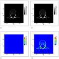
도 2a 및 도 2b는 예측된 BMD 값들 및 기준 팬텀을 이용하여 유도된 BMD 값들의 피어슨 상관관계 테스트를 보여준다. 도 2a는 L2 레벨에 대한 피어슨 상관관계 계수 0.986을 보여주고, 도 2b는 대퇴골 레벨에 대한 피어슨 상관관계 계수 0.948을 보여준다(p<0.05).
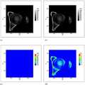
도 3은 제2 요추(L2 vertebra)의 축 방향(axial level)에서의 BMD 등고선도(contour plot)를 보여준다. 패널 (a)는 기준 BMD, 패널 (b)는 예측된 BMD, 패널 (c)는 BMD 편차, 및 패널 (d)는 다른 범례를 가지는 BMD 편차를 나타낸다.
도 4는 대퇴 관절(hip joint)의 축 방향에서의 BMD 등고선도(contour plot)를 보여준다. 패널 (a)는 기준 BMD, 패널 (b)는 예측된 BMD, 패널 (c)는 BMD 편차, 및 패널 (d)는 다른 범례를 가지는 BMD 편차를 나타낸다.Figures 1a and 1b show a screen shot of semi-automatic calculation software (FatScan, N2 systems). FIG. 1A shows that the circumference of the trunk and the cross-sectional area of the bone are divided at the L2 level, and FIG. 1B shows that the cross-sectional area of the circumference and bone of the trunk is divided at the level of the femur.
2a and 2b show Pearson correlation tests of BMD values derived using predicted BMD values and a reference phantom. FIG. 2A shows the Pearson correlation coefficient of 0.986 for the L2 level, and FIG. 2B shows the Pearson correlation coefficient of 0.948 for the femoral level (p < 0.05).
Figure 3 shows a BMD contour plot at the axial level of the L2 vertebra. Panel (a) shows the BMD deviation, panel (b) shows the predicted BMD, panel (c) shows the BMD deviation, and panel (d) shows the BMD deviation with another legend.
Figure 4 shows a BMD contour plot in the axial direction of the hip joint. Panel (a) shows the BMD deviation, panel (b) shows the predicted BMD, panel (c) shows the BMD deviation, and panel (d) shows the BMD deviation with another legend.
이하, 실시예를 통하여 본 발명을 더욱 상세히 설명하고자 한다. 이들 실시예는 오로지 본 발명을 보다 구체적으로 설명하기 위한 것으로, 본 발명의 요지에 따라 본 발명의 범위가 이들 실시예에 의해 제한되지 않는다는 것은 당업계에서 통상의 지식을 가진 자에 있어서 자명할 것이다.Hereinafter, the present invention will be described in more detail with reference to Examples. It is to be understood by those skilled in the art that these embodiments are only for describing the present invention in more detail and that the scope of the present invention is not limited by these embodiments in accordance with the gist of the present invention .
실시예Example
실험재료 및 방법Materials and Methods
연구 모집단(Study population)Study population
병원 정보 시스템을 이용하여 연구 모집단을 후향적으로(retrospectively) 확인하였다. 포함 기준은 (1) 2014년 4월에 실시된 검사 및 (2) 방사선 검사 보고서 상에 골 이상(bony abnormalities)이 없을 것을 기준으로 하였다. 39명의 확인된 경우에 대해서, QCT 검사의 목적은 건강 검진(n = 36), 유방 암 추적 검사(n = 1), 갑상선 암 추적 검사 (n = 2)이었다. 성별 분포는 남성 14명 및 여성 25명 이었다. 평균 연령은 49.1세(범위: 30-73세)이었다. 이러한 후향적 연구(retrospective study)는 병원의 IRB(Institutional Review Board)의 승인을 받았다.The research population was retrospectively verified using the hospital information system. The inclusion criteria were based on (1) the examination conducted in April 2014, and (2) no bony abnormalities on the radiology report. For 39 confirmed cases, the purpose of the QCT test was physical examination (n = 36), breast cancer follow-up (n = 1) and thyroid cancer follow-up (n = 2). Sex distribution was 14 males and 25 females. The mean age was 49.1 years (range: 30-73 years). This retrospective study was approved by the Institutional Review Board (IRB) of the hospital.
영상 프로토콜(Image protocol)Image protocol
QCT 스캔을 64-채널 CT(Somatom Definition AS+, Siemens, Erlangen, 독일)에서 실시하였다. QCT 검사를 위해 다음과 같이 CT 스캔 파라미터들을 최적화하였다: 120 kVp, 유효 관전류(effective mAs) 150 mAs, 빔 조준(beam collimation) = 20 mm, 회전 속도(rotation speed) = 0.6 초, 피치(pitch) = 1.0:1, 512 x 512 매트릭스(matrix), FOV(field of view) = 360 mm, 슬라이스 두께(slice thickness) = 3 mm, 그리고 B40s(medium)로 재구성(reconstruction). CARE Dose 4D 및 CARE kV 는 작동시키지 않았다. CTDI(CT dose index; CTDI 32 cm)는 10.10 mGy이었고, DLP(dose length product)는 218.7 mGy· cm이었다. QCT 스캔을 위하여 DIBH(deep-inspiration breath-hold) 방법을 이용하였다.QCT scans were performed on 64-channel CT (Somatom Definition AS +, Siemens, Erlangen, Germany). CT scan parameters were optimized for the QCT test as follows: 120 kVp,
외부 팬텀을 이용한 기준(reference) BMD 계산Calculation of reference BMD using external phantom
외부 팬텀(Mindways Inc., Austin, TX, 미국)의 다섯 개의 다른 골 함량(mineral contents)에 대한 5 개의 관심 영역(regions of interest; ROIs)을 동일한 CT 영상에 기록하였다. 환자들 각각의 CT 영상에 대해서, 팬텀-기반 보정(calibration) 알고리즘(20, 21)은 다음 식 (1)로 정의되는 알려진 골 밀도와 상응하는 HU 값 간의 선형 상관관계를 결정하였다:Five regions of interest (ROIs) for five different mineral contents of an external phantom (Mindways Inc., Austin, Tex., USA) were recorded on the same CT image. For each CT image of the patients, the phantom-based calibration algorithm 20, 21 determined a linear correlation between the known bone density, defined by the following equation (1), and the corresponding HU value:

여기에서,α및 β는 결정되는 환자-특이적 값들이다. 그 다음, 결정된α및 β를 가지는 상기 식 1을 이용하여, 요추(lumbar spines) 및 대퇴골(hips)의 BMD 값들은 비교를 위한 기준(reference)으로서 계산되었다.Here,? And? Are patient-specific values determined. Then, using the formula (1) having anα andβ is determined, the value of the lumbar BMD (lumbar spines) and femur (hips) were calculated as the standard (reference) for comparison.
영상 분석(Image analyses)Image analysis
모든 영상은 근골격 영상의학에서 10년의 경험이 있고 근골격계 전공의 과정을 수료한 방사선 전문의에 의해 평가되었다. 다른 환자들의 39개의 QCT 영상들을 제2 요추(L2 vertebra) 및 대퇴골의 축 방향(axial levels)에서 후향적으로(retrospectively) 분석하였다. 상기 ROI에 대한 정량 평가는 HU 값들이 기록된 L2 몸통 및 전체 대퇴골의 해면 뼈 구획(trabecular compartment)의 80-100 mm2 도면(drawings)에서 실시하였다.All images were evaluated by a radiologist who had 10 years of experience in musculoskeletal radiology and completed a course in musculoskeletal medicine. 39 QCT images of other patients were analyzed retrospectively in L2 vertebra and axial levels of the femur. Quantitative evaluation of the ROI was performed on 80-100 mm2 drawings of the trabecular compartment of the L2 body and the entire femur in which HU values were recorded.
몸통의 둘레 및 골 단면적의 계산Calculation of the circumference of the trunk and the bone cross-sectional area
도 1에 도시된 바와 같이, 몸통의 둘레 및 골 단면적을 반자동 계산 소프트웨어(semiautomatic calculation software)(FatScan, N2 systems, 오사카, 일본)를 이용하여, 목적 부위의 동일 축 방향에서 측정하였다. 상기 소프트웨어는 피하 및 내장 지방 면적 및 이들의 비율을 계산할 수 있기 때문에, 몸통의 둘레 및 골 단면적을 계산하기 위해서 이용되었다.As shown in Fig. 1, the circumference of the trunk and the bone cross-sectional area were measured in the same axial direction of the target site using semiautomatic calculation software (FatScan, N2 systems, Osaka, Japan). Because the software can calculate subcutaneous and visceral fat areas and their proportions, they were used to calculate the circumference and bone cross-sectional area of the trunk.
팬텀 없는 HU의 BMD로(HU-to-BMD)의 변환을 위한 회귀 모델 및 이들의 상관관계 테스트Regression models for conversion of HU-to-BMD to phantom-free HU BMD and their correlation test
둘레, 골 면적, HU 값들 및 BMD 값들을 다변수(multivariate) 분석에 고려하였다. 이러한 데이터를 이용하여, 단계별 회귀 방법으로 다중 회귀 모델을 이용하여 팬텀 없는 HU의 BMD로의 변환 식을 수립하였다. 독립 변수는 BMD 값이고, 종속 변수들은 HU 값, 둘레 및 골 면적이었다.Circumference, bone area, HU values and BMD values were considered for multivariate analysis. Using this data, a multivariate regression model was used as a step - by - step regression method to establish a phantom - free HU to BMD conversion equation. The independent variable was the BMD value, and the dependent variable was the HU value, circumference, and bone area.
이어, 예측된 BMD 값들 및 식 1에서 예측된 값들 간에 상관관계를 조사하기 위해서 피어슨 상관관계 테스트를 수행하였다. 모든 통계 분석들은 통계 소프트웨어(R package 2.15.1; http://cran.r-project.org)를 이용하여 실시하였다. 0.05 미만의P-값들은 통계학적으로 유의한 차이를 나타내는 것으로 고려하였다.Next, a Pearson correlation test was performed to investigate the correlation between the predicted BMD values and the predicted values in Equation (1). All statistical analyzes were performed using statistical software (R package 2.15.1; http://cran.r-project.org).P -values less than 0.05 were considered statistically significant.
복셀(voxelwise) BMD 비교Comparison of voxelwise BMD
복셀(voxelwise) 비교를 위해, 동일 축 방향의 요추 및 대퇴골의 HU 값들을 512x512 어레이 데이터로 재구성하였다. 그 다음, 예측된 BMD 및 기준 BMD를 제안된 모델(식 3) 및 식 1을 이용하여 각각 복셀(voxelwise) 계산하였다. 그리고, 이들은 다음 식 2의 평균 제곱근 오차(root mean square error; RMSE)를 이용하여 비교하였다:For voxelwise comparison, the HU values of lumbar and femur in the same axial direction were reconstructed into 512x512 array data. Then, the predicted BMD and the reference BMD were voxelly calculated using the proposed model (Equation 3) and Equation 1, respectively. They were then compared using the root mean square error (RMSE) of Equation 2:

여기에서, 상기 (BMDphantom)i및 (BMDphantomless)i은 각각 식 1을 이용하여 계산된i번째 복셀(voxel)의 BMD 값 및 제안된 모델을 이용하여 예측된i번째 복셀(voxel)의 BMD 값을 나타낸다;n은 복셀들(voxels)의 총 수를 나타낸다. 변환 및 통계학적 계산은 상업적으로 구입한 소프트웨어(Inter active Data Language(IDL), Exelis Vis Inc., Boulder, CO, 미국)를 이용하여 실시하였다.Here,BMDphantomi andBMDphantomlessi are the BMD values of thei- th voxel calculated using Equation 1 and BMD values of thei- th voxel estimated using the proposed model, Value;n Represents the total number of voxels. Conversion and statistical calculations were performed using commercially purchased software (Interactive Data Language (IDL), Exelis Vis Inc., Boulder, CO, USA).
실험 결과Experiment result
각 환자들의 CT 영상에 대해, 식 1의α및 β는 알려진 골 밀도 및 외부 팬텀의 상응하는 HU 값들을 이용하여 결정되었다. 그 다음, 요추 및 대퇴골의 BMD 값들은 기준 데이터로서 계산되었다.For each patient's CT image,α andβ in Equation 1 were determined using known bone density and corresponding HU values of the external phantom. BMD values of the lumbar and femur were then calculated as reference data.
단변수 분석으로부터, 골 면적은 요추 및 대퇴골 모두에 대해서 유의적이지 않은 것으로 결정되었다(p>0.05). 한편, HU 값들 및 몸통의 둘레는 요추에 대해서 통계학적으로 유의적이었고(p<0.05), 단지 HU 값들만이 대퇴골에 대해 통계학적으로 유의적이었다(p<0.05). 그 다음, 요추 및 대퇴골에서 환자 인자들을 고려하면서, 다중 선형 회귀 모델로부터 다음 식 3으로 정의되는 HU의 BMD로(HU-to-BMD)의 변환 식을 수립하였다:From univariate analysis, it was determined that the bone area was not significant for both lumbar spine and femur (p > 0.05). On the other hand, HU values and trunk circumference were statistically significant (p <0.05) for the lumbar spine, and only HU values were statistically significant for the femur (p <0.05). The HU-to-BMD conversion equation was then established from the multiple linear regression model, taking into account the patient factors in the lumbar spine and femur as follows:

여기에서, BMD 및 둘레의 단위는 각각 mg/cc 및 cm이다.Here, the units of BMD and perimeter are mg / cc and cm, respectively.
피어슨 상관관계 테스트(도 2)로부터, 식 3으로부터 예측된 BMD 값들은 식 1로 부터의 기준 BMD 값들과 유의적인 상관관계가 있음을 확인하였다(요추 및 대퇴골에 대한 피어슨 상관관계 계수는 각각 0.986 및 0.948;p<0.05). 복셀(voxelwise) 비교에 있어서, 요추 및 대퇴골의 RSME 값들은 각각 4.26± 0.60 [mg/cc] 및 8.35± 0.57 [mg/cc]이었다.From the Pearson correlation test (FIG. 2), it was found that the BMD values predicted from
고찰Review
골 밀도(bone mineral density; BMD)는 골 강도에 대한 중요한 표지자이기 때문에(22), 골다공증에 대한 진단 기준 및 치료 반응에 중요한 기능을 한다. 그러나 일반(routine) CT 스캔(즉, 외부 팬텀 없는 CT 스캔)은 BMD 값들을 직접적으로 제공하지 않으나, 오히려 HU 값들은 제공한다. 정확한 BMD 측정을 위해, 외부 고체 팬텀을 CT 스캐너에 위치 시켜 빔 경화(beam hardening) 및 방사선 산란(radiation scattering)의 효과를 보정하였다(11, 12).Because bone mineral density (BMD) is an important marker of bone strength (22), it plays an important role in the diagnostic criteria and treatment response to osteoporosis. However, routine CT scans (ie, external phantomless CT scans) do not provide BMD values directly, but rather provide HU values. For accurate BMD measurements, external solid phantoms were placed in a CT scanner to compensate for the effects of beam hardening and radiation scattering (11, 12).
종래 문헌에서, HU 및 BMD 값들은 선형 상관관계를 가진다고 보고되었고(23), 몇몇 회귀 모델들은 조영 증강(contrast-enhanced) CT(13, 14), CT 대장조영술(colonography)(17, 24), 복부 다중-검출(abdominal multi-detector) CT(15), 및 요추(spine) CT(16, 25)를 이용하여 제안되어 왔다. 이러한 연구 결과들은 HU 및 BMD 값들 간의 상관관계가 조영제(contrast medium), kVp, 및 CT 스캐닝 영역(scanning regions)에 따라 달라짐을 보여주었다. 비록 HU 값들이 허리 둘레 및 골의 단면적과 같은 환자 인자들에 의해 영향을 받는다고 할지라도(11, 26), 환자 인자들 및 BMD 계산과의 관계를 다루는 문헌은 없었다.In the prior art, HU and BMD values have been reported to have a linear correlation (23), and some regression models include contrast-enhanced CT (13,14), CT colonography (17,24) An abdominal multi-detector CT 15, and a spine CT 16, 25 have been proposed. These studies have shown that the correlation between HU and BMD values is dependent on the contrast medium, kVp, and CT scanning regions. Although the HU values are affected by patient factors such as waist circumference and cross-sectional area of the bone (11, 26), there is no literature dealing with patient factors and BMD calculations.
본 발명은 환자 인자들의 HU 값들에 대한 효과를 고려하여 신뢰도 있게 외부 팬텀 없이 BMD를 계산하고자 하는 필요성에 의해 개발되었다. 총 39개의 QCT 영상을 가지고, HU 값들 및 환자 인자들 간의 관계가 다변수 분석(multivariate analyses)을 통해 조사되었고, 그로 인해 팬텀 없는 HU의 BMD로의 변환 식에 대한 다중 선형 회귀 모델을 얻었다. 예측된 BMD 값들 및 기준 BMD 값들 간의 높은 양의 상관관계는 피어슨 상관관계 테스트를 통해 나타내었다. 본 발명의 결과로부터, HU 값들 및 둘레의 식을 이용하여 요추의 평균 BMD 값들은 계산될 수 있었으나, 대퇴골의 평균 BMD 값들은 오직 HU 값들만을 이용하여 계산될 수 있었다. 골의 단면적은 요추 및 대퇴골 모두에서 유의적이지 않는 것으로 결정 되었다. 따라서, 제안된 변환 식들은 환자 인자들로 인해 감소된 HU 값들을 통계학적으로 보상할 수 있다.The present invention has been developed by the need to reliably calculate the BMD without external phantom taking into account the effects on the HU values of the patient parameters. With a total of 39 QCT images, the relationship between HU values and patient parameters was investigated through multivariate analyzes, resulting in a multiple linear regression model for the phantom-free HU to BMD conversion equation. The high positive correlation between the predicted BMD values and the reference BMD values is shown through Pearson correlation tests. From the results of the present invention, the mean BMD values of the lumbar spine using the HU values and the perimeter equations could be calculated, but the mean BMD values of the femur could be calculated using only the HU values. The cross-sectional area of the bone was determined not to be significant in both lumbar spine and femur. Thus, the proposed transformations can statistically compensate for reduced HU values due to patient factors.
도 3 및 4는 각 복셀(voxel)의 예측된 BMD 및 기준 BMD의 유사성을 명확히 보여준다. 최대 BMD 값과 비교하여, 이들의 편차는 무시할 만한 것으로 간주할 수 있다(즉, 도 3(c) 및 도 4(c)의 1000 mg/cc).Figures 3 and 4 clearly show the similarity of the predicted BMD and reference BMD of each voxel. These deviations can be regarded as negligible (i.e., 1000 mg / cc in FIG. 3 (c) and FIG. 4 (c)) compared with the maximum BMD value.
철저한 확인을 위해, 각 복셀의 BMD 편차에 대한 대표 값을 제공할 수 있는, RMSE의 식으로 예측된 BMD 값들을 기준 BMD 값들과 비교하였다. 요추 및 대퇴골에 대한 RMSE가 각각 4.26± 0.60 [mg/cc] 및 8.35± 0.57 [mg/cc]임을 고려하면, 제안된 모델들은 각 복셀에 대해 신뢰도 있는 BMD 값들을 제공할 수 있고, 따라서 3D 공간 BMD 분포를 제공할 수 있다. BMD 분포 데이터는 FE(finite element) 모델들을 구축하기 위해서는 필수적임을 주목해야 한다. Crawfordet al.(27)이 보고에 따르며, FE 모델들은 골절 위험 평가(fracture risk assessments)에 대한 보다 신뢰성 있는 도구일 수 있기 때문이다. 또한, 보다 더 선명한 가시화를 위한 최대 BMD의 범례(legend)가 도 3(c) 및 4(c)와는 다른, 도 3(d) 및 4(d)에서 볼 수 있는 바와 같이, 제안된 모델의 BMD 편차들이 이들의 BMD 값들에 비례한다는 것을 알게 된 것은 또한 흥미롭다. 이러한 비례 오차들은 식 3에서 통계학적으로 미리 결정된 기울기 및 y-절편으로부터 발생하는데, 이는 감쇠(attenuation)로 환자-특이적인 차이들을 보상하기 위한 환자-특이적인 값들(즉, 식 1의α및 β)이다.For thorough verification, the BMD values predicted by the RMSE equation, which can provide a representative value for the BMD deviation of each voxel, were compared with the reference BMD values. Considering that the RMSE for lumbar spine and femur is 4.26 ± 0.60 [mg / cc] and 8.35 ± 0.57 [mg / cc] respectively, the proposed models can provide reliable BMD values for each voxel, BMD distribution. It should be noted that the BMD distribution data is essential for constructing FE (finite element) models. According to Crawfordet al . (27), FE models can be a more reliable tool for fracture risk assessments. Also, as can be seen in Figures 3 (d) and 4 (d), the legend of maximum BMD for more clear visualization is different from Figures 3 (c) and 4 It is also interesting to note that BMD deviations are proportional to their BMD values. These errors are proportional to occur from a predetermined slope and y- intercept statistically in
본 발명의 제한 조건을 살펴보면, 본 발명의 제안된 HU의 BMD로의 변환 식은 QCT 프로토콜에 기초 한다: 120 kVp, 유효 관전류(effective mAs) 150 mAs, 3 mm의 슬라이스 두께(slice thickness), 및 B40s(medium) 커널(kernel). 보다 나은 접근성을 위해, 제안된 변환 식은 일반 CT 프로토콜을 포함하는 것으로 확장 된다. 그러나, 방사선량 조절 방법(radiation dose modulation techniques)을 고려하면, kVp 또는 mAs의 단순 식들은 부적절할 수 있다. 또한, 조영제의 증가된 밀도의 영향은 평가되지 않았다. CT 밀도가 조영제 주입 후 상(phase)에 따라 달라지기 때문에, 팬텀 없는 BMD 계산은 조영-증강 CT 영상을 이용하는 경우에는 조심스럽게 실시해야한다.Looking at the constraints of the present invention, the proposed HU to BMD conversion equation is based on the QCT protocol: 120 kVp,
후속 연구로, 팬텀 없는 HU의 BMD(HU-to-BMD)로의 변환에 대한 제안된 모델은 일반 CT 영상을 이용하는 것으로 확장될 수 있다. 일반 CT 스캔은 일상 업무(daily practice)로 실시되기 때문에, 제안된 모델은 환자들로 하여금 BMD 측정을 위한 추가적인 방사선 노출을 피할 수 있게 할 것이다. 또한, 상업적으로 구입 가능한 PACS(Picture Archiving and Communication System)의 도움으로, FEA-기반 골절 위험 평가는 큰 모집단을 가지는 골다공증 연구를 위해 이용될 수 있는 데, 이는 중개 의학(translational medicine)에 보다 정확하고 의미있는 진단 데이터를 제공할 수 있을 것이다.As a follow-up study, the proposed model for the conversion of phantom-free HU to BMD (HU-to-BMD) can be extended to using generic CT images. Since a general CT scan is performed on a daily practice basis, the proposed model will allow patients to avoid additional radiation exposure for BMD measurements. In addition, with the help of commercially available Picture Archiving and Communication System (PACS), FEA-based fracture risk assessment can be used to study osteoporosis with large populations, which is more accurate for translational medicine Meaningful diagnostic data can be provided.
이상으로 본 발명의 특정한 부분을 상세히 기술하였는 바, 당업계의 통상의 지식을 가진 자에게 있어서 이러한 구체적인 기술은 단지 바람직한 구현예일 뿐이며, 이에 본 발명의 범위가 제한되는 것이 아닌 점은 명백하다. 따라서, 본 발명의 실질적인 범위는 첨부된 청구항과 그의 등가물에 의하여 정의된다고 할 것이다.While the present invention has been particularly shown and described with reference to exemplary embodiments thereof, it is to be understood that the same is by way of illustration and example only and is not to be construed as limiting the scope of the present invention. Accordingly, the actual scope of the present invention will be defined by the appended claims and their equivalents.
참고문헌references
1.Iki M. Epidemiology of bone and joint disease - the present and future - . Epidemiology of osteoporosis and osteoporotic fracture in Japan. Clinical calcium. 2014; 24:657-664.One.Iki M. Epidemiology of bone and joint disease - the present and future. Epidemiology of osteoporosis and osteoporotic fracture in Japan. Clinical calcium. 2014; 24: 657-664.
2.Leslie WD, Morin SN. Osteoporosis epidemiology 2013: implications for diagnosis, risk assessment, and treatment. Current opinion in rheumatology. 2014; 26:440-446.2.Leslie WD, Morin SN. Osteoporosis epidemiology 2013: implications for diagnosis, risk assessment, and treatment. Current opinion in rheumatology. 2014; 26: 440-446.
3.Blake GM, Fogelman I. The role of DXA bone density scans in the diagnosis and treatment of osteoporosis. Postgraduate medical journal. 2007; 83:509-517.3.Blake GM, Fogelman I. The role of DXA bone density scans in the diagnosis and treatment of osteoporosis. Postgraduate medical journal. 2007; 83: 509-517.
4.Finkelstein JS, Klibanski A, Neer RM. Evaluation of lumber spine bone mineral density (BMD) using dual energy x-ray absorptiometry (DXA) in 21 young men with histories of constitutionally-delayed puberty. The Journal of clinical endocrinology and metabolism. 1999; 84:3400-3401; author reply 3403-3404.4.Finkelstein JS, Klibanskie, Neer RM. Evaluation of lumbar spine bone mineral density (BMD) using dual energy x-ray absorptiometry (DXA) in 21 young men with histories of constitutionally-delayed puberty. The Journal of Clinical Endocrinology and Metabolism. 1999; 84: 3400-3401; author reply 3403-3404.
5.Pereira RM, Corrente JE, Chahade WH, Yoshinari NH. Evaluation by dual X-ray absorptiometry (DXA) of bone mineral density in children with juvenile chronic arthritis. Clinical and experimental rheumatology. 1998; 16:495-501.5.Pereira RM, Corrente JE, Chahade WH, Yoshinari NH. Evaluation of dual X-ray absorptiometry (DXA) of bone mineral density in children with juvenile chronic arthritis. Clinical and experimental rheumatology. 1998; 16: 495-501.
6.Marshall D, Johnell O, Wedel H. Meta-analysis of how well measures of bone mineral density predict occurrence of osteoporotic fractures. Bmj. 1996; 312:1254-1259.6.Marshall D, Johnell O, and Wedel H. Meta-analysis of how well measures of bone mineral density predicted osteoporotic fractures. Bmj. 1996; 312: 1254-1259.
7.ACRSPRSSR practice guideline for the performance of quantitative computed tomography (QCT) bone densitometry Available at http://www.acr.org/~/media/DE78D218C7A64526A821A9E8645AB46D.pdf. Accessed April 25, 2014. Accessed.7.ACRSPRSSR practice guideline for quantitative computed tomography (QCT) bone densitometry available at http://www.acr.org/~/media/DE78D218C7A64526A821A9E8645AB46D.pdf. Accessed April 25, 2014. Accessed.
8.Lewiecki EM. Imaging technologies for assessment of skeletal health in men. Current osteoporosis reports. 2013; 11:1-10.8.Lewiecki EM. Imaging technologies for assessment of skeletal health in men. Current osteoporosis reports. 2013; 11: 1-10.
9.Li N, Li XM, Xu L, Sun WJ, Cheng XG, Tian W. Comparison of QCT and DXA: Osteoporosis Detection Rates in Postmenopausal Women. International journal of endocrinology. 2013; 2013:895474.9.Li N, Li XM, Xu L, Sun WJ, Cheng XG, Tian W. Comparison of QCT and DXA: Osteoporosis Detection Rates in Postmenopausal Women. International journal of endocrinology. 2013; 2013: 895474.
10.Ito M, Hayashi K, Yamada M, Uetani M, Nakamura T. Relationship of osteophytes to bone mineral density and spinal fracture in men. Radiology. 1993; 189:497-502.10.Ito M, Hayashi K, Yamada M, Uetani M, Nakamura T. Relationship of osteophytes to bone mineral density and spinal fracture in men. Radiology. 1993; 189: 497-502.
11.Adams JE. Quantitative computed tomography. European journal of radiology. 2009; 71:415-424.11.Adams JE. Quantitative computed tomography. European journal of radiology. 2009; 71: 415-424.
12.Habashy AH, Yan X, Brown JK, Xiong X, Kaste SC. Estimation of bone mineral density in children from diagnostic CT images: a comparison of methods with and without an internal calibration standard. Bone. 2011; 48:1087-1094.12.Habashy AH, Yan X, Brown JK, Xiong X, Kaste SC. Estimation of bone mineral density in children from diagnostic CT images: a comparison of methods with and without an internal calibration standard. Bone. 2011; 48: 1087-1094.
13.Bauer JS, Henning TD, Mueller D, Lu Y, Majumdar S, Link TM. Volumetric quantitative CT of the spine and hip derived from contrast-enhanced MDCT: conversion factors. AJR American journal of roentgenology. 2007; 188:1294-1301.13.Bauer JS, Henning TD, Mueller D, Lu Y, Majumdar S, Link TM. Volumetric quantitative CT of the spine and hip derived from contrast-enhanced MDCT: conversion factors. AJR American journal of roentgenology. 2007; 188: 1294-1301.
14.Baum T, Muller D, Dobritz M, et al. Converted lumbar BMD values derived from sagittal reformations of contrast-enhanced MDCT predict incidental osteoporotic vertebral fractures. Calcified tissue international. 2012; 90:481-487.14.Baum T, Muller D, Dobritz M, et al. Converted lumbar BMD values derived from sagittal reformations of contrast-enhanced MDCT predict incidental osteoporotic vertebral fractures. Calcified tissue international. 2012; 90: 481-487.
15.Papadakis AE, Karantanas AH, Papadokostakis G, Petinellis E, Damilakis J. Can abdominal multi-detector CT diagnose spinal osteoporosis? European radiology. 2009; 19:172-176.15.Papadakis AE, Karantanas AH, Papadocostakis G, Petinellis E, Damilakis J. Can abdominal multi-detector CT diagnose spinal osteoporosis? European radiology. 2009; 19: 172-176.
16.Schreiber JJ, Anderson PA, Rosas HG, Buchholz AL, Au AG. Hounsfield units for assessing bone mineral density and strength: a tool for osteoporosis management. The Journal of bone and joint surgery American volume. 2011; 93:1057-1063.16.Schreiber JJ, Anderson PA, Rosas HG, Buchholz AL, Au AG. Hounsfield units for assessing bone mineral density and strength: a tool for osteoporosis management. The Journal of the American Medical Association. 2011; 93: 1057-1063.
17.Fletcher JG, Johnson CD, Krueger WR, et al. Contrast-enhanced CT colonography in recurrent colorectal carcinoma: feasibility of simultaneous evaluation for metastatic disease, local recurrence, and metachronous neoplasia in colorectal carcinoma. AJR American journal of roentgenology. 2002; 178:283-290.17.Fletcher JG, Johnson CD, Krueger WR, et al. Contrast-enhanced CT colonography in recurrent colorectal carcinoma: feasibility of simultaneous evaluation for metastatic disease, local recurrence, and metachronous neoplasia in colorectal carcinoma. AJR American journal of roentgenology. 2002; 178: 283-290.
18.Baum T, Muller D, Dobritz M, Rummeny EJ, Link TM, Bauer JS. BMD measurements of the spine derived from sagittal reformations of contrast-enhanced MDCT without dedicated software. European journal of radiology. 2011; 80:e140-145.18.Baum T, Muller D, Dobritz M, Rummeny EJ, Link TM, Bauer JS. BMD measurements of the spine derived from sagittal reformations of contrast-enhanced MDCT without dedicated software. European journal of radiology. 2011; 80: e140-145.
19.Schwaiger BJ, Gersing AS, Baum T, Noel PB, Zimmer C, Bauer JS. Bone mineral density values derived from routine lumbar spine multidetector row CT predict osteoporotic vertebral fractures and screw loosening. AJNR American journal of neuroradiology. 2014; 35:1628-1633.19.Schwaiger BJ, Gersing AS, Baum T, Noel PB, Zimmer C, Bauer JS. Bone mineral density values derived from routine lumbar spine multidetector row CT predicted osteoporotic vertebral fractures and screw loosening. AJNR American journal of neuroradiology. 2014; 35: 1628-1633.
20.Emami A, Ghadiri H, Ay MR, et al. A new phantom for performance evaluation of bone mineral densitometry using DEXA and QCT. Book A new phantom for performance evaluation of bone mineral densitometry using DEXA and QCT. City2011; 3441-3445.20.Emamia, Ghadiri H, Ay MR, et al. A new phantom for performance evaluation of bone mineral densitometry using DEXA and QCT. Book A new phantom for performance evaluation of bone mineral densitometry using DEXA and QCT. City2011; 3441-3445.
21.Cetin Celenk and Peruze Celenk (2012). Bone Density Measurement Using Computed Tomography, pp 131, Computed Tomography - Clinical Applications, Dr. Luca Saba (Ed.), ISBN: 978-953-307-378-1, InTech, DOI: 10.5772/22884. Available from: http://www.intechopen.com/books/computed-tomography-clinical-applications/bone-density-measurement-using-computed-tomography.21.Cetin Celenk and Peruze Celenk (2012). Bone Density Measurement Using Computed Tomography, pp. 131, Computed Tomography - Clinical Applications, Luca Saba (Ed.), ISBN: 978-953-307-378-1, InTech, DOI: 10.5772 / 22884. Available from: http://www.intechopen.com/books/computed-tomography-clinical-applications/bone-density-measurement-using-computed-tomography.
22.Lenchik L, Shi R, Register TC, Beck SR, Langefeld CD, Carr JJ. Measurement of trabecular bone mineral density in the thoracic spine using cardiac gated quantitative computed tomography. Journal of computer assisted tomography. 2004; 28:134-139.22.Lenchik L, Shi R, Register TC, Beck SR, Langefeld CD, Carr JJ. Measurement of trabecular bone mineral density in the thoracic spine using cardiac gated quantitative computed tomography. Journal of computer assisted tomography. 2004; 28: 134-139.
23.Link TM, Koppers BB, Licht T, Bauer J, Lu Y, Rummeny EJ. In vitro and in vivo spiral CT to determine bone mineral density: initial experience in patients at risk for osteoporosis. Radiology. 2004; 231:805-811.23.Link TM, Koppers BB, Licht T, Bauer J, Lu Y, Rummeny EJ. In vitro and in vivo spiral CT to determine bone mineral density: initial experience in patients at risk for osteoporosis. Radiology. 2004; 231: 805-811.
24.Summers RM, Baecher N, Yao J, et al. Feasibility of simultaneous computed tomographic colonography and fully automated bone mineral densitometry in a single examination. Journal of computer assisted tomography. 2011; 35:212-216.24.Summers RM, Baecher N, Yao J, et al. Feasibility of simultaneous computed tomographic colonography and fully automated bone mineral densitometry in a single examination. Journal of computer assisted tomography. 2011; 35: 212-216.
25.Schwaiger BJ, Gersing AS, Baum T, Noel PB, Zimmer C, Bauer JS. Bone Mineral Density Values Derived from Routine Lumbar Spine Multidetector Row CT Predict Osteoporotic Vertebral Fractures and Screw Loosening. AJNR American journal of neuroradiology. 2014.25.Schwaiger BJ, Gersing AS, Baum T, Noel PB, Zimmer C, Bauer JS. Bone Mineral Density Values Derived from Routine Lumbar Spine Multidetector Row CT Predict Osteoporotic Vertebral Fractures and Screw Loosening. AJNR American journal of neuroradiology. 2014.
26.Celenk C, Celenk P. Bone density measurement using computed tomography. Book Bone density measurement using computed tomography. City2012; pp. 131.26.Celenk C, Celenk P. Bone density measurement using computed tomography. Book Bone density measurement using computed tomography. City2012; pp. 131.
27.Crawford RP, Cann CE, Keaveny TM. Finite element models predict in vitro vertebral body compressive strength better than quantitative computed tomography. Bone. 2003; 33:744-750.27.Crawford RP, Cann CE, Keaveny TM. Finite element models predict in vitro vertebral body compressive strength better than quantitative computed tomography. Bone. 2003; 33: 744-750.
Claims (10)
Translated fromKoreanUsing patient specific factors selected from the group consisting of (a) Hounsfield unit (HU) value or (b) Hounsfield unit value and body weight, height and circumference of body without external phantom To obtain a bone mineral density (BMD).
The method according to claim 1, wherein the bone density is a bone density of the lumbar region, the femur, the humerus, the radius, the knee, or the ankle joint.
2. The method of claim 1, wherein the method comprises determining a bone density of a lumbar spine using an HU value and a waist circumference without an external phantom.
2. The method of claim 1, wherein the method comprises determining a bone density of the hip using only the HU value without an external phantom.
수학식 1
(BMD)lumbar spine= 0.89× (HU)lumbar spine+ 0.15× (몸통 둘레)-7.44
상기 수학식 1에서, 상기 (BMD)lumbar spine의 단위는mg/cc이고, 상기 몸통 둘레의 단위는 cm이다.
4. The method according to claim 3, wherein the method comprises: obtaining a bone mineral density (BMD) of a lumbar spine by substituting a Hounsfield unit (HU) value of a lumbar spine and a circumference of a body into Equation Comprising the steps < RTI ID = 0.0 > of:
Equation 1
(BMD )lumbar spine = 0.89 占 (HU )lumbar spine + 0.15 占 (waist circumference) -7.44
In Equation (1), the unit of (BMD )lumbar spine is mg / cc, and the unit of the waist circumference is cm.
수학식 2
(BMD)hip= 0.78× (HU)hip+ 16.62
상기 수학식 2에서, 상기 (BMD)hip의 단위는mg/cc이다.
5. The method according to claim 4, wherein the method comprises: obtaining a bone mineral density (BMD) of a femur by substituting a Hounsfield unit (HU) value of the hip into the following equation:
Equation 2
(BMD )hip = 0.78 x (HU )hip + 16.62
In Equation (2), the unit of (BMD )hip is mg / cc.
The method according to claim 5 or 6, wherein the HU value of the lumbar spine or the HU value of the femur is obtained from CT (computed tomography) or MRI (magnetic resonance imaging) without an external phantom.
수학식 3

상기 수학식 3에서, 상기 (BMD)phantom는 외부 팬텀 있는 정량적인 CT(quantitative computed tomography; QCT)를 이용하여 측정된 값이고, 상기 (HU)는 외부 팬텀에 상응하는 값이며, 상기 수학식 1로부터 측정된 요추의 골밀도는 상기 수학식 3의 (BMD)phantom와의 관계에서 피어슨 상관관계 계수(Pearson’s correlation coefficient)가 0.986이고, 상기 수학식 2로부터 측정된 대퇴골의 골밀도는 상기 수학식 3의 (BMD)phantom와의 관계에서 피어슨 상관관계 계수가 0.948이다.
The method of claim 5 or 6, wherein the reference bone density for comparison with the bone density measured from Equation (1) or (2) is defined by Equation (3)
Equation 3
(BMD )phantom is a value measured using quantitative computed tomography (CTT ) with an external phantom,HU is a value corresponding to an external phantom, BMD of the lumbar vertebrae measured from the above and the Pearson correlation coefficient (Pearson's correlation coefficient) in relation to(BMD) of the equation (3)phantom 0.986, bone density of the femur determined from the equation (2) is(BMD of the equation (3) ) The Pearson correlation coefficient is 0.948 in relation tophantom .
수학식 4

상기 수학식 4에서, 상기 (BMDphantom)i는 수학식 3을 이용하여 계산된i번째 복셀(voxel)의 BMD 값이고, 상기 (BMDphantomless)i는 수학식 1 또는 수학식 2를 이용하여 계산된i번째 복셀(voxel)의 BMD 값이며, 상기n은 복셀(voxel)의 총 수를 나타내며, 요추에 대한 RMSE는 4.26± 0.60 [mg/cc]이고 대퇴골에 대한 RMSE는 8.35± 0.57 [mg/cc]이다.
9. The method of claim 8, wherein the bone density measured from Equation (1) or Equation (2) and the reference bone density expressed by Equation (3) satisfy a root mean square error (RMSE) defined by Equation ≪ / RTI >
Equation 4
(BMDphantom )i is the BMD value of thei- th voxel calculated using Equation (3), andBMDphantomlessi is calculated using Equation (1) or Equation (2) ai is the BMD value of the first voxel (voxel), whereinn is the total number of voxels (voxel), RMSE for the lumbar spine is 4.26 ± 0.60 [mg / cc] and the RMSE for the femur is 8.35 ± 0.57 [mg / cc].
Priority Applications (1)
| Application Number | Priority Date | Filing Date | Title |
|---|---|---|---|
| KR1020150107946AKR101750108B1 (en) | 2015-07-30 | 2015-07-30 | Estimation of Bone Mineral Density Using Medical Image Signal Without an External Phantom |
Applications Claiming Priority (1)
| Application Number | Priority Date | Filing Date | Title |
|---|---|---|---|
| KR1020150107946AKR101750108B1 (en) | 2015-07-30 | 2015-07-30 | Estimation of Bone Mineral Density Using Medical Image Signal Without an External Phantom |
Publications (2)
| Publication Number | Publication Date |
|---|---|
| KR20170015674Atrue KR20170015674A (en) | 2017-02-09 |
| KR101750108B1 KR101750108B1 (en) | 2017-06-26 |
Family
ID=58154646
Family Applications (1)
| Application Number | Title | Priority Date | Filing Date |
|---|---|---|---|
| KR1020150107946AExpired - Fee RelatedKR101750108B1 (en) | 2015-07-30 | 2015-07-30 | Estimation of Bone Mineral Density Using Medical Image Signal Without an External Phantom |
Country Status (1)
| Country | Link |
|---|---|
| KR (1) | KR101750108B1 (en) |
Cited By (4)
| Publication number | Priority date | Publication date | Assignee | Title |
|---|---|---|---|---|
| KR20190018913A (en)* | 2017-08-16 | 2019-02-26 | 서울대학교산학협력단 | System and method for measuring bone mineral density |
| CN113491526A (en)* | 2020-04-07 | 2021-10-12 | 辽宁开普医疗系统有限公司 | Bone density correction and measurement method based on DR system |
| KR20220053122A (en) | 2020-10-22 | 2022-04-29 | 김동진 | Backward Power System of Waste Heat Recovery System Using Cyclones |
| CN116458909A (en)* | 2023-04-10 | 2023-07-21 | 清华大学 | Method and device for measuring three-dimensional bone density distribution by using cone beam DR equipment |
Families Citing this family (1)
| Publication number | Priority date | Publication date | Assignee | Title |
|---|---|---|---|---|
| EP4570182A1 (en) | 2023-12-12 | 2025-06-18 | 3D-Shaper Medical S.L. | A computer-implemented method, a system, and a computer program for phantomless calibration of medical images |
Family Cites Families (1)
| Publication number | Priority date | Publication date | Assignee | Title |
|---|---|---|---|---|
| EP2858571B1 (en) | 2012-06-07 | 2019-01-23 | The Johns Hopkins University | Integration of quantitative calibration systems in computed tomography scanners |
- 2015
- 2015-07-30KRKR1020150107946Apatent/KR101750108B1/ennot_activeExpired - Fee Related
Cited By (6)
| Publication number | Priority date | Publication date | Assignee | Title |
|---|---|---|---|---|
| KR20190018913A (en)* | 2017-08-16 | 2019-02-26 | 서울대학교산학협력단 | System and method for measuring bone mineral density |
| CN113491526A (en)* | 2020-04-07 | 2021-10-12 | 辽宁开普医疗系统有限公司 | Bone density correction and measurement method based on DR system |
| CN113491526B (en)* | 2020-04-07 | 2023-12-05 | 辽宁开普医疗系统有限公司 | Bone density correction and measurement method based on DR system |
| KR20220053122A (en) | 2020-10-22 | 2022-04-29 | 김동진 | Backward Power System of Waste Heat Recovery System Using Cyclones |
| CN116458909A (en)* | 2023-04-10 | 2023-07-21 | 清华大学 | Method and device for measuring three-dimensional bone density distribution by using cone beam DR equipment |
| CN116458909B (en)* | 2023-04-10 | 2024-05-07 | 清华大学 | Method and device for measuring three-dimensional bone density distribution by using cone beam DR equipment |
Also Published As
| Publication number | Publication date |
|---|---|
| KR101750108B1 (en) | 2017-06-26 |
Similar Documents
| Publication | Publication Date | Title |
|---|---|---|
| Engelke et al. | Opportunistic screening techniques for analysis of CT scans | |
| Phan et al. | Trabecular bone structure of the calcaneus: comparison of MR imaging at 3.0 and 1.5 T with micro-CT as the standard of reference | |
| Engelke et al. | Clinical use of quantitative computed tomography–based advanced techniques in the management of osteoporosis in adults: the 2015 ISCD official positions—part III | |
| Zhou et al. | Monoenergetic imaging of dual-energy CT reduces artifacts from implanted metal orthopedic devices in patients with factures | |
| Carrino et al. | Dedicated cone-beam CT system for extremity imaging | |
| Link | Metabolic bone disease | |
| Budoff et al. | Measurement of phantomless thoracic bone mineral density on coronary artery calcium CT scans acquired with various CT scanner models | |
| D’Elia et al. | Bone fragility and imaging techniques | |
| Grassi et al. | Prediction of femoral strength using 3D finite element models reconstructed from DXA images: validation against experiments | |
| Michalski et al. | Opportunistic CT screening predicts individuals at risk of major osteoporotic fracture | |
| Celenk et al. | Bone density measurement using computed tomography | |
| Issever et al. | Assessment of trabecular bone structure using MDCT: comparison of 64-and 320-slice CT using HR-pQCT as the reference standard | |
| KR101750108B1 (en) | Estimation of Bone Mineral Density Using Medical Image Signal Without an External Phantom | |
| Genant et al. | Advanced imaging of the macrostructure and microstructure of bone | |
| Link et al. | Osteoporosis imaging | |
| Lee et al. | Patient‐specific phantomless estimation of bone mineral density and its effects on finite element analysis results: a feasibility study | |
| Burch et al. | Prevalence of poor bone quality in women undergoing spinal fusion using biomechanical-CT analysis | |
| Papadakis et al. | Can abdominal multi-detector CT diagnose spinal osteoporosis? | |
| Johnston et al. | In vivo precision of a depth-specific topographic mapping technique in the CT analysis of osteoarthritic and normal proximal tibial subchondral bone density | |
| Gruber et al. | Bone mineral density measurements of the proximal femur from routine contrast-enhanced MDCT data sets correlate with dual-energy X-ray absorptiometry | |
| Lang | Quantitative computed tomography | |
| Carballido-Gamio et al. | Clinical utility of microarchitecture measurements of trabecular bone | |
| Kim et al. | Assessment of osteoporosis using pelvic diagnostic computed tomography | |
| Rayudu et al. | Low-dose and sparse sampling MDCT-based femoral bone strength prediction using finite element analysis | |
| Burghardt et al. | Automated simulation of areal bone mineral density assessment in the distal radius from high-resolution peripheral quantitative computed tomography |
Legal Events
| Date | Code | Title | Description |
|---|---|---|---|
| A201 | Request for examination | ||
| PA0109 | Patent application | St.27 status event code:A-0-1-A10-A12-nap-PA0109 | |
| PA0201 | Request for examination | St.27 status event code:A-1-2-D10-D11-exm-PA0201 | |
| D13-X000 | Search requested | St.27 status event code:A-1-2-D10-D13-srh-X000 | |
| R17-X000 | Change to representative recorded | St.27 status event code:A-3-3-R10-R17-oth-X000 | |
| D14-X000 | Search report completed | St.27 status event code:A-1-2-D10-D14-srh-X000 | |
| E902 | Notification of reason for refusal | ||
| PE0902 | Notice of grounds for rejection | St.27 status event code:A-1-2-D10-D21-exm-PE0902 | |
| AMND | Amendment | ||
| P11-X000 | Amendment of application requested | St.27 status event code:A-2-2-P10-P11-nap-X000 | |
| P13-X000 | Application amended | St.27 status event code:A-2-2-P10-P13-nap-X000 | |
| PG1501 | Laying open of application | St.27 status event code:A-1-1-Q10-Q12-nap-PG1501 | |
| E601 | Decision to refuse application | ||
| PE0601 | Decision on rejection of patent | St.27 status event code:N-2-6-B10-B15-exm-PE0601 | |
| AMND | Amendment | ||
| P11-X000 | Amendment of application requested | St.27 status event code:A-2-2-P10-P11-nap-X000 | |
| P13-X000 | Application amended | St.27 status event code:A-2-2-P10-P13-nap-X000 | |
| PX0901 | Re-examination | St.27 status event code:A-2-3-E10-E12-rex-PX0901 | |
| PX0701 | Decision of registration after re-examination | St.27 status event code:A-3-4-F10-F13-rex-PX0701 | |
| X701 | Decision to grant (after re-examination) | ||
| GRNT | Written decision to grant | ||
| PR0701 | Registration of establishment | St.27 status event code:A-2-4-F10-F11-exm-PR0701 | |
| PR1002 | Payment of registration fee | St.27 status event code:A-2-2-U10-U11-oth-PR1002 Fee payment year number:1 | |
| PG1601 | Publication of registration | St.27 status event code:A-4-4-Q10-Q13-nap-PG1601 | |
| P22-X000 | Classification modified | St.27 status event code:A-4-4-P10-P22-nap-X000 | |
| PR1001 | Payment of annual fee | St.27 status event code:A-4-4-U10-U11-oth-PR1001 Fee payment year number:4 | |
| PR1001 | Payment of annual fee | St.27 status event code:A-4-4-U10-U11-oth-PR1001 Fee payment year number:5 | |
| PR1001 | Payment of annual fee | St.27 status event code:A-4-4-U10-U11-oth-PR1001 Fee payment year number:6 | |
| R18-X000 | Changes to party contact information recorded | St.27 status event code:A-5-5-R10-R18-oth-X000 | |
| PN2301 | Change of applicant | St.27 status event code:A-5-5-R10-R13-asn-PN2301 St.27 status event code:A-5-5-R10-R11-asn-PN2301 | |
| PR1001 | Payment of annual fee | St.27 status event code:A-4-4-U10-U11-oth-PR1001 Fee payment year number:7 | |
| PC1903 | Unpaid annual fee | St.27 status event code:A-4-4-U10-U13-oth-PC1903 Not in force date:20240617 Payment event data comment text:Termination Category : DEFAULT_OF_REGISTRATION_FEE | |
| PC1903 | Unpaid annual fee | St.27 status event code:N-4-6-H10-H13-oth-PC1903 Ip right cessation event data comment text:Termination Category : DEFAULT_OF_REGISTRATION_FEE Not in force date:20240617 |