KR20160079960A - Injector equipment for endoscopic surgery - Google Patents
Injector equipment for endoscopic surgeryDownload PDFInfo
- Publication number
- KR20160079960A KR20160079960AKR1020140190375AKR20140190375AKR20160079960AKR 20160079960 AKR20160079960 AKR 20160079960AKR 1020140190375 AKR1020140190375 AKR 1020140190375AKR 20140190375 AKR20140190375 AKR 20140190375AKR 20160079960 AKR20160079960 AKR 20160079960A
- Authority
- KR
- South Korea
- Prior art keywords
- injector
- distal end
- lesion
- inner tube
- outer tube
- Prior art date
- Legal status (The legal status is an assumption and is not a legal conclusion. Google has not performed a legal analysis and makes no representation as to the accuracy of the status listed.)
- Ceased
Links
- 238000002674endoscopic surgeryMethods0.000titleabstractdescription13
- 230000003902lesionEffects0.000claimsabstractdescription48
- 238000000034methodMethods0.000claimsabstractdescription26
- 239000000243solutionSubstances0.000claimsabstractdescription26
- 239000003814drugSubstances0.000claimsabstractdescription21
- 229940079593drugDrugs0.000claimsabstractdescription20
- 230000008878couplingEffects0.000claimsdescription23
- 238000010168coupling processMethods0.000claimsdescription23
- 238000005859coupling reactionMethods0.000claimsdescription23
- 238000002347injectionMethods0.000claimsdescription11
- 239000007924injectionSubstances0.000claimsdescription11
- 239000000126substanceSubstances0.000claimsdescription8
- 238000005192partitionMethods0.000claimsdescription3
- 238000007790scrapingMethods0.000claimsdescription3
- 239000002699waste materialSubstances0.000claimsdescription3
- JOYRKODLDBILNP-UHFFFAOYSA-NEthyl urethaneChemical compoundCCOC(N)=OJOYRKODLDBILNP-UHFFFAOYSA-N0.000claimsdescription2
- 229920001296polysiloxanePolymers0.000claimsdescription2
- 239000007779soft materialSubstances0.000claimsdescription2
- 239000008155medical solutionSubstances0.000abstractdescription4
- 230000000694effectsEffects0.000abstractdescription3
- 238000012277endoscopic treatmentMethods0.000description7
- 238000011282treatmentMethods0.000description6
- 238000001356surgical procedureMethods0.000description5
- 208000002193PainDiseases0.000description4
- 210000005036nerveAnatomy0.000description4
- 210000002784stomachAnatomy0.000description4
- 210000001519tissueAnatomy0.000description4
- 238000001839endoscopyMethods0.000description3
- 239000000835fiberSubstances0.000description3
- 230000036407painEffects0.000description3
- 210000001835visceraAnatomy0.000description3
- 208000003618Intervertebral Disc DisplacementDiseases0.000description2
- 238000002679ablationMethods0.000description2
- 230000023597hemostasisEffects0.000description2
- 210000004400mucous membraneAnatomy0.000description2
- 210000000056organAnatomy0.000description2
- 230000037390scarringEffects0.000description2
- 230000002000scavenging effectEffects0.000description2
- 238000002560therapeutic procedureMethods0.000description2
- 206010002091AnaesthesiaDiseases0.000description1
- 208000019901Anxiety diseaseDiseases0.000description1
- 208000000094Chronic PainDiseases0.000description1
- 206010050296Intervertebral disc protrusionDiseases0.000description1
- 206010029174Nerve compressionDiseases0.000description1
- 206010029240NeuritisDiseases0.000description1
- 206010033799ParalysisDiseases0.000description1
- 230000037005anaesthesiaEffects0.000description1
- 210000003423ankleAnatomy0.000description1
- 230000036506anxietyEffects0.000description1
- 238000011888autopsyMethods0.000description1
- 238000013276bronchoscopyMethods0.000description1
- 230000000747cardiac effectEffects0.000description1
- 230000001112coagulating effectEffects0.000description1
- 239000003086colorantSubstances0.000description1
- 230000006378damageEffects0.000description1
- 230000003111delayed effectEffects0.000description1
- 238000003745diagnosisMethods0.000description1
- 210000002249digestive systemAnatomy0.000description1
- 201000010099diseaseDiseases0.000description1
- 208000037265diseases, disorders, signs and symptomsDiseases0.000description1
- 230000002183duodenal effectEffects0.000description1
- 210000003238esophagusAnatomy0.000description1
- 239000011152fibreglassSubstances0.000description1
- 239000012530fluidSubstances0.000description1
- 239000012634fragmentSubstances0.000description1
- 239000000446fuelSubstances0.000description1
- 230000002496gastric effectEffects0.000description1
- 238000002695general anesthesiaMethods0.000description1
- 230000002439hemostatic effectEffects0.000description1
- 230000004054inflammatory processEffects0.000description1
- 238000001802infusionMethods0.000description1
- 238000003780insertionMethods0.000description1
- 230000037431insertionEffects0.000description1
- 230000007794irritationEffects0.000description1
- 238000002690local anesthesiaMethods0.000description1
- 239000002184metalSubstances0.000description1
- 201000011591microinvasive gastric cancerDiseases0.000description1
- 238000012148non-surgical treatmentMethods0.000description1
- 230000003287optical effectEffects0.000description1
- 239000013307optical fiberSubstances0.000description1
- 230000002093peripheral effectEffects0.000description1
- 238000000554physical therapyMethods0.000description1
- 238000003825pressingMethods0.000description1
- 238000004904shorteningMethods0.000description1
- 230000001225therapeutic effectEffects0.000description1
- 230000000451tissue damageEffects0.000description1
- 231100000827tissue damageToxicity0.000description1
- 230000004304visual acuityEffects0.000description1
Images


Classifications
- A—HUMAN NECESSITIES
- A61—MEDICAL OR VETERINARY SCIENCE; HYGIENE
- A61B—DIAGNOSIS; SURGERY; IDENTIFICATION
- A61B1/00—Instruments for performing medical examinations of the interior of cavities or tubes of the body by visual or photographical inspection, e.g. endoscopes; Illuminating arrangements therefor
- A61B1/00064—Constructional details of the endoscope body
- A61B1/00071—Insertion part of the endoscope body
- A61B1/0008—Insertion part of the endoscope body characterised by distal tip features
- A61B1/00087—Tools
- A—HUMAN NECESSITIES
- A61—MEDICAL OR VETERINARY SCIENCE; HYGIENE
- A61B—DIAGNOSIS; SURGERY; IDENTIFICATION
- A61B18/00—Surgical instruments, devices or methods for transferring non-mechanical forms of energy to or from the body
- A—HUMAN NECESSITIES
- A61—MEDICAL OR VETERINARY SCIENCE; HYGIENE
- A61B—DIAGNOSIS; SURGERY; IDENTIFICATION
- A61B18/00—Surgical instruments, devices or methods for transferring non-mechanical forms of energy to or from the body
- A61B18/04—Surgical instruments, devices or methods for transferring non-mechanical forms of energy to or from the body by heating
- A61B18/12—Surgical instruments, devices or methods for transferring non-mechanical forms of energy to or from the body by heating by passing a current through the tissue to be heated, e.g. high-frequency current
- A61B18/14—Probes or electrodes therefor
- A61B18/1485—Probes or electrodes therefor having a short rigid shaft for accessing the inner body through natural openings
- A—HUMAN NECESSITIES
- A61—MEDICAL OR VETERINARY SCIENCE; HYGIENE
- A61B—DIAGNOSIS; SURGERY; IDENTIFICATION
- A61B18/00—Surgical instruments, devices or methods for transferring non-mechanical forms of energy to or from the body
- A61B18/04—Surgical instruments, devices or methods for transferring non-mechanical forms of energy to or from the body by heating
- A61B18/12—Surgical instruments, devices or methods for transferring non-mechanical forms of energy to or from the body by heating by passing a current through the tissue to be heated, e.g. high-frequency current
- A61B18/14—Probes or electrodes therefor
- A61B18/1492—Probes or electrodes therefor having a flexible, catheter-like structure, e.g. for heart ablation
- A—HUMAN NECESSITIES
- A61—MEDICAL OR VETERINARY SCIENCE; HYGIENE
- A61M—DEVICES FOR INTRODUCING MEDIA INTO, OR ONTO, THE BODY; DEVICES FOR TRANSDUCING BODY MEDIA OR FOR TAKING MEDIA FROM THE BODY; DEVICES FOR PRODUCING OR ENDING SLEEP OR STUPOR
- A61M5/00—Devices for bringing media into the body in a subcutaneous, intra-vascular or intramuscular way; Accessories therefor, e.g. filling or cleaning devices, arm-rests
- A61M5/14—Infusion devices, e.g. infusing by gravity; Blood infusion; Accessories therefor
- A61M5/158—Needles for infusions; Accessories therefor, e.g. for inserting infusion needles, or for holding them on the body
- A—HUMAN NECESSITIES
- A61—MEDICAL OR VETERINARY SCIENCE; HYGIENE
- A61B—DIAGNOSIS; SURGERY; IDENTIFICATION
- A61B18/00—Surgical instruments, devices or methods for transferring non-mechanical forms of energy to or from the body
- A61B2018/00571—Surgical instruments, devices or methods for transferring non-mechanical forms of energy to or from the body for achieving a particular surgical effect
- A61B2018/00589—Coagulation
- A—HUMAN NECESSITIES
- A61—MEDICAL OR VETERINARY SCIENCE; HYGIENE
- A61B—DIAGNOSIS; SURGERY; IDENTIFICATION
- A61B18/00—Surgical instruments, devices or methods for transferring non-mechanical forms of energy to or from the body
- A61B2018/00571—Surgical instruments, devices or methods for transferring non-mechanical forms of energy to or from the body for achieving a particular surgical effect
- A61B2018/00595—Cauterization
- A—HUMAN NECESSITIES
- A61—MEDICAL OR VETERINARY SCIENCE; HYGIENE
- A61B—DIAGNOSIS; SURGERY; IDENTIFICATION
- A61B18/00—Surgical instruments, devices or methods for transferring non-mechanical forms of energy to or from the body
- A61B2018/00982—Surgical instruments, devices or methods for transferring non-mechanical forms of energy to or from the body combined with or comprising means for visual or photographic inspections inside the body, e.g. endoscopes
Landscapes
- Health & Medical Sciences (AREA)
- Life Sciences & Earth Sciences (AREA)
- Surgery (AREA)
- Engineering & Computer Science (AREA)
- General Health & Medical Sciences (AREA)
- Heart & Thoracic Surgery (AREA)
- Biomedical Technology (AREA)
- Animal Behavior & Ethology (AREA)
- Public Health (AREA)
- Veterinary Medicine (AREA)
- Medical Informatics (AREA)
- Nuclear Medicine, Radiotherapy & Molecular Imaging (AREA)
- Molecular Biology (AREA)
- Physics & Mathematics (AREA)
- Otolaryngology (AREA)
- Plasma & Fusion (AREA)
- Optics & Photonics (AREA)
- Anesthesiology (AREA)
- Vascular Medicine (AREA)
- Biophysics (AREA)
- Hematology (AREA)
- Pathology (AREA)
- Radiology & Medical Imaging (AREA)
- Cardiology (AREA)
- Infusion, Injection, And Reservoir Apparatuses (AREA)
- Surgical Instruments (AREA)
- Endoscopes (AREA)
Abstract
Translated fromKoreanDescription
Translated fromKorean본 발명은 내시경 시술용 인젝터 기구에 관한 것으로 더욱 상세하게는 내시경을 통해 병변 부위를 관찰하면서 병변 부위에 약액을 주입하거나, 병변 부위를 소작하여 제거, 응고 처치하는 치료 과정을 기구 교체 없이 수행할 수 있도록 한 내시경 시술용 인젝터 기구에 관한 것이다.The present invention relates to an injector mechanism for endoscopic surgery, and more particularly, to a method for injecting a drug solution into a lesion site while observing a lesion site through an endoscope, or cauterizing a lesion site, To an injector mechanism for endoscopic surgery.
"내시경(Endoscope)"이란, 수술을 하거나 또는 부검(剖檢)을 하지 않고서는 직접 병변(病變)을 볼 수 없는 내장장기(內臟臟器)나 체강(體腔) 내부에 기계를 삽입하여 관찰하도록 고안된 의료기구이다.The term "endoscope" is used to insert a machine into the internal organs or body cavity where no direct disease can be seen without surgery or autopsy. It is a medical device designed.
내시경은 1868년 쿠스몰(Kussmaul)에 의해 처음으로 개발된 이후로 1881년 본 미큘리츠(vonMikulicz)가 경성 내시경을 개발하고 임상에 적용시켰으며, 1932년 쉰들러(Schindler)는 연성 내시경을 개발하고 임상에 적용시켰다.Endoscopy was first developed by Kussmaul in 1868. In 1881, vonMikulicz developed a hard endoscopy and applied it to clinical practice. In 1932, Schindler developed a soft endoscope Lt; / RTI >
그 후, 1960년에 광섬유가 개발되면서 내시경이 굽어진 상태에서도 직접 신체 내부 장기 및 조직을 확인할 수 있는 연성 광학내시경이 히르숀위츠(Hirschowitz)에 의해 개발되었으며, 1980년대에 들어오면서 현재 사용되고 있는 비디오 내시경(video endoscope)이 개발됨으로써 내시경을 통해 신체 내부의 장기 및 조직을 조직학적 진단이 가능하게 되었다.A flexible optical endoscope was developed by Hirschowitz, who was able to identify the internal organs and tissues of the body directly after the optical fiber was developed in 1960 and the endoscope was bent, With the development of a video endoscope, endoscopy has made it possible to histologically diagnose organs and tissues within the body.
일반적으로 사용되는 종류로는 기관지경(氣管支鏡)·식도경(食道鏡)·위경(胃鏡)·십이지장경(十二指腸鏡)·직장경(直腸鏡)·방광경(膀胱鏡)·복강경(腹腔鏡) 등이 있고, 그 밖에 특수한 것으로는 흉강경(胸腔鏡)·종격경(縱隔鏡)·심장경(心臟鏡) 등이 있다.Commonly used types are bronchoscopy, esophagus, stomach, duodenal, rectal, cystoscopic, laparoscopic, laparoscopic, ), And other special cases include thoracoscopic, mediastinal, and cardiac.
또한, 내시경에는 직달경(直達鏡)이라 하여 1개의 통(筒)으로 되어 있어서 장기를 직접 육안으로 볼 수 있는 형, 렌즈시스템을 이용한 형, 카메라를 직접 장기에 삽입하는 위카메라(胃 카메라)형과 유리섬유를 사용한 파이버스코프 등이 있다.In addition, the endoscope has a single tube called a direct-edged mirror, which can be viewed directly by the naked eye, a type using a lens system, a stomach camera for inserting the camera directly into the organ, And a fiber scope using fiberglass and the like.
최근 위암의 조기발견 진단에 위카메라·위파이버스코프 등이 크게 활용되고 있는데, 위카메라는 위 안으로 소형 카메라를 삽입하여 직접 위의 점막을 촬영·기록하여 미세한 위의 병변을 발견·진단할 수 있고, 위파이버스코프는 해상력(解像力)이 좋고, 점막 태를 관찰하면서 병변부 조직을 절단하여 검사할 수 있는 장치도 부착시킬 수 있다.Recently, the upper camera and upper fibroscope are widely used for the diagnosis of early gastric cancer. The upper camera inserts a small camera in the stomach to photograph and record the mucous membrane directly to detect and diagnose a small gastric lesion , The above fiber scope has good resolving power, and it is possible to attach a device capable of cutting and examining lesion tissue while observing the mucous membrane.
이와 같이 소화기, 특히 위(胃)에 관한 내시경의 고도발달로 인해 보통 내시경이라고 하면 위카메라·위파이버스코프를 가리킨다.In this way, the digestive system, especially the stomach, due to the high development of endoscopes, usually referred to as endoscopes, the upper camera refers to the fiber scopes.
한편, 최근에는 내시경을 이용한 치료술이 내장장기뿐만 아니라, 다른 의료 분야에서도 광범위하게 활용되고 있다.On the other hand, in recent years, endoscopic therapy has been extensively used not only in internal organs but also in other medical fields.
일 예로서 "허리디스크"라 불리는 "추간판 탈출증" 환자의 경우 과거에는 경미한 신경압박이나 신경 주변부의 염증이 통증의 원인이라면 우선 약물치료나 물리치료, 통증주사치료 등과 같은 비수술적 치료를 하다가, 호전되지 않고 만성 통증이 지속되거나 발가락 또는 발목의 힘이 현저하게 약해진 경우, 디스크 파열로 인해 격심한 통증 또는 다리를 움직이기 힘들거나 마비증상 등이 온 경우에는 수술적 치료가 시행하였다.For example, in the case of a "disc herniation" called "waist disc", in the past, if slight nerve compression or inflammation of the nerve periphery is the cause of pain, first, non-surgical treatment such as medication, physical therapy, Or if chronic pain persists or the strength of the toe or ankle is severely weakened, severe pain due to disc rupture, difficulty moving the legs, or paralysis is present.
그러나 외과적 척추수술 방법은 전신마취하에 발병 부위의 피부를 절개하고 탈출된 디스크를 제거하는 방식으로 이루어짐으로써 마취에 의한 합병증이나 척추불안증 및 디스크 붕괴현상으로 인한 심각한 합병증이 종종 발생하기도 하였다.However, surgical spinal surgery has been performed under general anesthesia by incising the skin of the affected area and removing the escaped disc, resulting in complications such as anesthesia complications, spinal anxiety, and disc collapse.
위와 같은 외과적 척추수술에 따른 문제점을 개선하기 위하여 개발된 것이 바로, 내시경 디스크 제거술(PELD)이다. 즉, 내시경 디스크 제거술은 척추용으로 만들어진 의료용 특수 내시경을 디스크가 발생한 부위에 삽입하고 내시경모니터로 보면서 신경을 압박하고 있는 디스크를 레이저 또는 고주파로 제거하여 치료하는 최첨단 수술기법으로서, 전신마취를 하지 않고 국소마취로 진행돼 고령의 환자들도 안전하며, 수술 부위의 반흔 및 유착이 적으며, 흉터가 없고 입원 기간(3~4일 이내)이 짧아 일상 생활로의 복귀가 쉽다는 장점으로 인하여 환자들의 만족도가 매우 높기 때문에 최근에는 그 시술 회수가 척추외과 수술 중 가장 높은 빈도를 차지하고 있는 실정이다.Endoscopic discectomy (PELD) has been developed to overcome the problems associated with surgical spine surgery. In other words, endoscopic disc removal is a state of the art surgical technique in which a medical special endoscope made for vertebrae is inserted into the area where the disc is formed and the disc is pressed with a laser or high frequency while observing it with an endoscope monitor. Because of the advantages of advanced local anesthesia, it is safe for older patients, less scarring and adhesion at the surgical site, less scarring, shorter hospital stay (3-4 days), and easier return to daily life Since the satisfaction rate is very high, the number of procedures is the highest in spinal surgery.
상술한 바와 같이 내시경 디스크 제거술은 환자의 환부를 1cm 이내로 최소한 작게 절개한 후, 그 피부절개부를 통해 환부를 들여다볼 수 있는 내시경과 함께 미세 특수 도구들을 함께 투입하여 치료 부위에 위치한 내시경으로 촬영된 탈출된 디스크 조각과 신경을 영상출력장치를 이용하여 실시간으로 확인하면서, 내시경과 함께 투입한 미세 특수 도구들을 이용해 탈출된 디스크 조각을 제거하거나, 병변에 약액 등을 주입함으로써 디스크 치료가 이루어진다.As described above, in the endoscopic disc removal method, the lesion portion of the patient is cut at least as small as 1 cm, and then the microscopic special tools are put together with the endoscope capable of looking through the lesion through the skin incision, The disc treatment is performed by removing the fragment of the escaped disc or by injecting the drug solution into the lesion using microscopic special tools inserted with the endoscope while confirming the disc piece and nerve in real time using the image output device.
이때, 내시경과 함께 환부에 투입하는 미세 특수 도구들은 각각의 사용 목적에 맞는 도구들을 투입하게 된다. 즉 디스크를 제거하기 위해서는 레이저 또는 고주파 소작기를 내시경과 함께 환부에 투입하여 디스크를 제거하게 되고, 환부에 치료 목적을 위한 약액을 주입하기 위해서는 약액이 주입된 주사기와 연결되는 약액주입구를 말단부에 형성하고, 약액주입구와 연결된 주사기에서 주입된 약액을 환부에 주사하는 니들을 선단부에 형성한 인젝터 등을 사용하게 된다.At this time, the microscopic special tools to be put into the affected part along with the endoscope are loaded with the tools suitable for the respective use purpose. That is, in order to remove the disk, a laser or a high-frequency fibrillator is put into the affected part together with the endoscope to remove the disk. In order to inject the medical solution for treatment purpose into the lesion, a chemical solution injection port connected to the injected drug solution is formed at the distal end , An injector having a needle at its distal end for injecting a drug solution injected from a syringe connected to a drug solution injection port into a diseased part is used.
그런데 환자의 상태에 따라서는 상기 소작기와 인젝터 등을 번갈아가며 사용하는 경우가 번번하다. 즉, 내시경 디스크 제거술 시술 과정은 통상 환자의 환부에서 디스크만을 제거하는 것으로 끝나는 게 아니라, 보통은 환자의 통증 완화 등을 위하여 디스크 제거와 함께 약액 주입을 병행하는 경우게 많은데 이를 위해서는 먼저 내시경과 함께 소작기를 환부에 투입하여 디스크를 제거한 후, 환부에서 소작기를 빼고, 다시 인젝터 등을 환부로 투입하여 약액을 환부에 주입함으로써 소작기와 인젝터 등을 번갈아 사용하는데 따른 불편이 따를 뿐만 아니라, 그에 따른 시술 시간이 지연되고, 신경이 몰려있는 환부에 소작기와 인젝터를 번갈아 투입하는 과정에서 환부 주변을 자극하거나, 신경 등이 손상되면서 심각한 합병증이 종종 발생하는 문제점이 있었다.However, depending on the condition of the patient, the cauterizer and the injector are used alternately. In other words, the endoscopic disc removal procedure usually involves removing the disc only from the affected part of the patient, but usually involves disc removal and chemical infusion to alleviate the patient's pain. In order to do this, The injector and the like are injected into the affected part and the chemical solution is injected into the affected part so that the inconvenience of using the cauterizer and the injector alternately is not only inconvenient but also the procedure time In the process of injecting the cauterizer and the injector alternately into the delayed and nervous lesion, irritation around the lesion, or damage to the nerve, causes serious complications.
따라서 내시경 디스크 제거술 시술 과정에서 미세 특수 도구를 번갈아가며 사용하는데 따른 번거로움과 그로 인한 각종 문제점을 해소하기 위하여 디스크를 제거하기 위해서 사용하는 소작기와, 약액을 주입하기 위하여 사용하는 인젝터를 하나로 구성한 내시경 시술용 인젝터 기구의 개발이 절실한 실정이다.Therefore, in order to solve the problems associated with the use of the micro-special tools alternately during the endoscopic disk removal procedure and to solve various problems caused by the endoscopic disk removal procedures, endoscopic procedures comprising a cauterizer used for removing the disk and an injector used for injecting the drug solution It is inevitable to develop an injector mechanism for a fuel injector.
본 발명의 내시경 시술용 인젝터 기구는 상술한 바와 같은 종래의 문제점을 해소하기 위하여 발명한 것으로서, 소작전극(101a)을 형성한 아우터튜브(101)를 구비하고, 상기 소작전극(101a)에 고주파전력을 공급하는 고주파발생기를 연결하여 병변 부위를 소작하는 코애귤레이터(10)와; 병변 부위에 침습하는 니들(202b)을 형성한 이너튜브(202a)를 구비하고, 상기 니들(202b)에 약액을 공급하는 주사기를 연결하여 병변 부위에 약액을 주입하는 인젝터(20)와; 선단부에는 상기 코애귤레이터(10)를 탈부착 가능케 결합하고, 말단부에는 상기 인젝터(20)를 탈부착 가능케 결합하여 상기 아우터튜브(101) 내부로 상기 이너튜브(202a)가 투입되면서 상기 코애귤레이터(10)와 인젝터(20)를 일체로 연결하는 링크커넥터(30)를 포함하여 구성함으로써 내시경을 통해 병변 부위를 관찰하면서 병변 부위에 약액을 주입하거나, 병변 부위를 소작하여 제거, 응고 처치하는 치료 과정을 기구 교체 없이 수행할 수 있도록 하여 내시경 시술에 소요되는 시간이 크게 단축할 수있도록 함으로써 시술에 따른 부담이 크게 경감되며, 내시경 시술 성공률이 향상될 뿐만 아니라, 조직 손상 등에 따른 부작용의 발병 위험이 현저히 감소하게 한 목적을 달성할 수 있다.The injector mechanism for endoscopic surgery according to the present invention is an injector mechanism for endoscopic treatment of the present invention. The injector mechanism includes an
이상과 같은 본 발명의 내시경 시술용 인젝터 기구는 내시경을 통해 병변 부위를 관찰하면서 병변 부위에 약액을 주입하거나, 병변 부위를 소작하여 제거하거나 응고 처치하는 치료 과정을 별도의 기구 교체 없이 일체화된 내시경 시술용 인젝터 기구를 이용하여 한꺼번에 수행할 수 있도록 함으로써 내시경 시술용 보조도구의 교체를 위한 피부절개부로의 삽입과 배출 과정을 반복하지 않아도 되기 때문에 내시경 시술에 소요되는 시간이 크게 단축되는 효과가 있다.The injector mechanism for endoscopic treatment according to the present invention as described above is characterized in that the injector injects a chemical solution into a lesion site while observing a lesion site through an endoscope or performs a treatment process of removing or coagulating a lesion site by means of an integrated endoscopic procedure So that it is not necessary to repeatedly perform the insertion and ejection into the skin incision for replacement of the auxiliary tool for the endoscopic procedure. Therefore, the time required for the endoscopic procedure is significantly shortened.
또한, 내시경 시술용 보조도구의 교체에 따른 병변 부위나 주변 조직의 손상을 방지할 수 있게 되어 시술을 수행하는 시술자로서는 시술에 따른 부담이 크게 경감되며, 내시경 시술 성공률이 향상될 뿐만 아니라, 조직 손상 등에 따른 부작용의 발병 위험이 현저히 감소함으로써 시술성공률이나 그에 따른 치료효과가 더욱 향상되는 이점이 있다.In addition, since it is possible to prevent lesion sites and surrounding tissue from being damaged due to replacement of an endoscopic assistant tool, the burden of the procedure is greatly reduced as a practitioner performing the procedure, and not only the success rate of the endoscopic procedure is improved, The risk of side effects is significantly reduced, and thus the success rate of the procedure and the therapeutic effect are further improved.
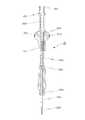
도 1은 본 발명의 내시경 시술용 인젝터 기구의 실시 예에 따른 사시도.
도 2는 본 발명의 내시경 시술용 인젝터 기구의 실시 예에 따른 분해 사시도
도 3은 본 발명의 내시경 시술용 인젝터 기구의 실시 예에 따른 코애귤레이터 단면도.
도 4는 본 발명의 내시경 시술용 인젝터 기구의 실시 예에 따른 인젝터 단면도.
도 5는 본 발명의 내시경 시술용 인젝터 기구의 실시 예에 따른 코애귤레이터와 인젝터와 링크커넥터가 결합된 상태 단면도.
도 6은 본 발명의 내시경 시술용 인젝터 기구의 실시 예에 따른 인젝터에 주사기를 연결한 사용상태 예시도.1 is a perspective view according to an embodiment of an injector mechanism for endoscopic surgery of the present invention.
2 is an exploded perspective view of an injector mechanism for endoscopic surgery according to an embodiment of the present invention.
3 is a cross-sectional view of a co-ordinator according to an embodiment of the injector mechanism for endoscopic treatment of the present invention.
4 is a sectional view of an injector according to an embodiment of the injector mechanism for endoscopic treatment of the present invention.
FIG. 5 is a state sectional view of a coagulator according to an embodiment of the injector mechanism for endoscopic surgery of the present invention, an injector, and a link connector. FIG.
FIG. 6 is a view showing an example of a use state in which a syringe is connected to an injector according to an embodiment of the injector mechanism for endoscopic surgery of the present invention. FIG.
이하, 본 발명의 내시경 시술용 인젝터 기구의 바람직한 실시 예에 따른 구성과 작용을 첨부 도면에 의하여 더욱 상세히 설명하면 다음과 같다.DETAILED DESCRIPTION OF THE PREFERRED EMBODIMENTS Hereinafter, the structure and operation of a preferred embodiment of the injector mechanism for endoscopic surgery according to the present invention will be described in detail with reference to the accompanying drawings.
본 발명의 내시경 시술용 인젝터 기구는 소작전극(101a)을 형성한 아우터튜브(101)를 구비하고, 상기 소작전극(101a)에 고주파전력을 공급하는 고주파발생기를 연결하여 병변 부위를 소작하는 코애귤레이터(10)와; 병변 부위에 침습하는 니들(202b)을 형성한 이너튜브(202a)를 구비하고, 상기 니들(202b)에 약액을 공급하는 주사기를 연결하여 병변 부위에 약액을 주입하는 인젝터(20)와; 선단부에는 상기 코애귤레이터(10)를 탈부착 가능케 결합하고, 말단부에는 상기 인젝터(20)를 탈부착 가능케 결합하여 상기 아우터튜브(101) 내부로 상기 이너튜브(202a)가 투입되면서 상기 코애귤레이터(10)와 인젝터(20)를 일체로 연결하는 링크커넥터(30)를 포함한다.The injector mechanism for endoscopic treatment of the present invention includes an
상기 코애귤레이터(10)는 병변 부위를 소작하기 위하여 병변 부위를 소작하는 소작전극(101a)을 선단부에 형성한 관체로 된 아우터튜브(101)와; 상기 아우터튜브(101)가 탈부착 가능케 결합하는 제1결합부(102a)를 선단부에 형성하고, 말단부에는 상기 링크커넥터(30)의 선단부가 탈부착 가능케 결합하는 제2결합부(102b)를 형성하며, 일측에는 상기 소작전극(101a)에 고주파전력을 공급하는 고주파발생기를 연결하는 연결관(102c)을 형성한 와이커넥터(102)를 포함한다.The
상기 아우터튜브(101)는 파지가 용이하게 하고, 상기 제1결합부(102a)에 탈부착 가능하도록 암나사부를 구비한 연결캡(101b)을 말단부에 일체로 형성하고, 상기 제1결합부(102a)에는 수나사부를 형성하여 상기 연결캡(101b)을 상기 제1결합부(102a)에 나사 결합하는 것이 바람직하다.The
상기 소작전극(101a)은 이너튜브(202a)를 통해 상기 연결관(102c)까지 이어진 인입선을 연결관(102c) 내부에 설치되는 연결단자(103)의 일단부와 연결하고, 상기 연결단자(103)의 타단부는 상기 연결관(102c)을 통해서 연결되는 고주파발생기(104)와 전기적으로 연결하여 상기 고주파발생기(104)에서 생성된 고주파전력이 상기 인입선을 통해 소작전극(101a)으로 공급됨으로써 전기 소작이 가능하다. 이때, 상기 인입선은 이너튜브(202a)의 말단부에 형성된 연결탭과 상기 제1결합부(102a)의 나사 결합에 의하여 외부로 노출되지 않도록 마감 처리되는 것이 바람직하다.The cauterizing
상기 고주파발생기(104)는 고주파 교류를 발생시키는 장치로서, 본 발명의 기술분야 즉, 의료기가 사용되는 의학계에서 일반적으로 널리 사용되는바, 고주파발생기에 대한 상세한 도면의 도시나, 구체적인 설명은 생략한다.The high-
상기 인젝터(20)는 병변 부위에 약액을 주입하기 위하여 상기 링크커넥터(30)의 말단부가 탈부착 가능케 결합하는 제3결합부(201a)를 선단부에 형성하고, 말단부에는 환상의 논슬립부재(201d)가 설치되는 그립부(201b)를 형성한 핸들보디(201)와; 병변 부위에 약액을 주입하는 니들(202b)을 선단부에 형성한 관체로 된 이너튜브(202a)를 선단부에 형성하고, 말단부에는 약액이 담긴 주사기(40)를 탈부착 가능케 결합하는 제4결합부(202c)를 형성하여 상기 이너튜브(202a)가 상기 아우터튜브(101) 내부를 통과하도록 상기 핸들보디(201)의 말단부에서 내외부로 스트로크 가능케 설치한 인젝션(202)을 포함한다.The
상기 핸들보디(201)의 선단부에 형성된 제3결합부(201a)는 상기 링크커넥터(30)와의 분해 조립의 편의성을 위하여 수나사부를 형성하는 것이 바람직할 것이나, 이에 한정하는 것은 아님을 밝혀둔다.It is preferable that the third
상기 핸들보디(201)는 가늘고 긴 관체로 형성하되, 말단부 외경을 점차 확대 돌출시켜 그립부(201b)를 형성하고, 상기 그립부(201b)에 환상요홈(201c)을 형성하여 상기 환상요홈(201c)에 실리콘이나 우레탄과 같이 마찰계수가 높고, 탄성을 가진 연질 소재의 논슬립부재(201d)를 삽입 설치하여 미끄러짐을 방지하고, 향상된 그립감을 제공한다.The
상기 논슬립부재(201d)는 다양한 색상의 표시를 통해서 상기 이너튜브(202a)의 선단부에 형성되는 니들(202b)의 사이즈, 즉 니들(202b)의 직경을 구분할 수 있도록 하는 것이 바람직하다.Preferably, the
상기 인젝션(202)은 병변 부위에 약액을 주입하는 니들(202b)을 선단부에 형성한 관체로 된 이너튜브(202a)를 선단부에 형성하고, 말단부에는 약액이 담긴 주사기(40)를 탈부착 가능케 결합하는 제4결합부(202c)를 형성한다.The
상기 인젝션(202)은 상기 핸들보디(201)의 말단부에서 핸들보디(201) 내외부로 스트로크 가능하도록, 상기 핸들보디(201)에는 길이 방향으로 결합장공(201e)을 형성하고, 상기 인젝션(202)의 선단부에는 상기 결합장공(201e)에 억지끼움 방식으로 결합하는 결합돌부(202e)를 구비하는 것이 바람직하다.The
또한, 상기 인젝션(202)에는 파지에 의한 스트로크가 용이하도록 파지부(202d)를 형성하는 것이 바람직한데, 상기 파지부(202d)의 크기나 형상은 한정하지 아니한다.In addition, it is preferable to form the
상기 이너튜브(202a)는 주사기(40)에 담긴 약액을 선단부에 형성된 니들(202b)로 공급하기 위하여 금속재 또는 비금속재로 된 관체로 형성하되, 직진도를 유지하기 위하여 금속재로 형성하는 것이 바람직할 것이나, 이에 한정하는 것은 아님을 밝혀둔다.The
상기 제4결합부(202c)는 선단부에 나사탭을 형성한 통상의 주사기(40)와 회전 결합이 이루어질 수 있도록 상기 나사탭 내부에 삽입 결합하는 결합돌기를 형성하여 약액이 담긴 주사기(40)를 탈부착 가능케 한다.The fourth
상기 링크커넥터(30)는 상기 코애귤레이터(10)와 인젝터(20)를 일체로 연결하기 위하여 관체로 되어 선단부에 길이 방향으로 절개홈(301)을 형성하고, 주벽 일측에는 상기 코애귤레이터(10)의 제2결합부(102b)의 탈부착을 가능케 하는 제1고정수단(302)을 형성하며, 말단부에는 상기 인젝터(20)의 제3결합부(201a)의 탈부착 가능케 하는 제2고정수단(303)을 구비하며 상기 제1고정수단(302)과 제2고정수단(303) 간에는 상기 인젝터(20)의 이너튜브(202a)가 관통하는 중공부(304a)를 구비한 격벽(304)을 일체로 형성한다.The
상기 제1고정수단(302)은 상기 코애귤레이터(10)의 제2결합부(102b)의 탈부착을 가능케 하기 위하여 다양한 구조로 형성할 수 있으나, 상기 주벽에 형성된 적어도 하나 이상의 결합공으로 구성하고, 상기 코애귤레이터(10)의 제2결합부(102b)에는 상기 결합공에 끼워지는 결합돌부(202e)를 형성하여 상기 제2결합부(102b)와 제1고정수단(302)은 억지끼움 방식에 결합이 이루어지고, 상술한 바와 같이 결합이 이루어지는 과정에서 상기 절개홈(301)에 의한 선단부 외경의 일시적인 확장에 의하여 코애귤레이터(10)의 제2결합부(102b)의 탈부착이 용이하게 이루어진다.The first fixing means 302 may be formed in a variety of structures to enable the
상기 제2고정수단(303)은 상기 링크커넥터(30)의 말단부 내경부에 암나사부를 형성함으로써 상기 인젝터(20)의 핸들보디(201) 선단부에 형성된 제3결합부(201a), 즉 수나사부와의 분해, 조립이 가능케 된다. 이때, 제2고정수단(303)은 상기 제3결합부(201a)와 분해 조립이 가능하도록 제3결합부(201a)와 대응하는 구조로 형성하는 것이므로 상기 제3결합부(201a)와 제2고정수단(303)의 분해 조립이 가능한 구조라면 어떠한 방식이든 사용 가능함을 밝혀둔다.The second fastening means 303 is provided with a
이상과 같이 구성된 본 발명의 기술이 적용된 내시경 시술용 인젝터 기구의 작동 관계를 첨부된 도면에 의하여 설명하면 다음과 같다.Hereinafter, the operation of the injector mechanism for endoscopic surgery according to the present invention will be described with reference to the accompanying drawings.
본 발명의 내시경 시술용 인젝터 기구는 내시경을 이용한 치료술에 있어서, 내시경과 함께 사용한다. 일 예로서 "추간판 탈출증" 환자의 치료를 위한 내시경 디스크 제거술을 시행하는 경우에 시술자는 부분 마취된 환자의 환부를 1cm 이내의 절개하여 피부절개창을 형성한 후 그 피부절개창을 통해 척추용으로 만들어진 의료용 특수 내시경을 디스크가 발생한 부위에 삽입함과 동시에 본 발명의 내시경 시술용 인젝터 기구의 선단부를 피부절개창을 통해서 환부에 삽입하거나, 내시경관에 구비된 워킹채널을 통해서 환부에 삽입한다.The injector mechanism for endoscopic surgery according to the present invention is used together with an endoscope in a therapeutic method using an endoscope. As an example, in the case of performing endoscopic disk removal for the treatment of a patient with "herniated disc herniation", the practitioner cuts the lesion of the partially anesthetized patient within 1 cm to form a skin incision, and then, through the skin incision, The special endoscope is inserted into the region where the disk is formed and the distal end portion of the injector mechanism for endoscopic treatment of the present invention is inserted into the affected portion through the skin incision or through the working channel provided in the endoscope tube.
그리하여 시술자는 병변 부위의 신경을 압박하고 있는 디스크를 제거하기 위하여 고주파발생기를 작동시킨 후 내시경으로 촬영된 영상을 출력하는 내시경모니터로 보면서 코애귤레이터(10)의 아우터튜브(101) 선단부에 위치한 소작전극(101a)을 병변 부위에 정확하게 위치시켜 병변 부위의 디스크를 절개, 지혈한다.Thus, the practitioner views the endoscope monitor as an endoscope monitor that operates the high frequency generator to remove the disk pressing the nerve at the lesion site and then outputs an image taken by the endoscope, The
또한, 상기와 같이 소작전극(101a)을 이용한 디스크를 절개, 지혈와 더불어 병변 부위에 약액의 주입이 필요한 경우에는 소작전극(101a)을 형성한 코애귤레이터(10)의 아우터튜브(101)를 제거할 필요 없이 링크커넥터(30)에 의해 코애귤레이터(10) 후방에 일체로 결합된 인젝터(20)의 말단부에 형성된 인젝션(202)의 제4결합부(202c)에 약액이 담긴 주사기(40)를 결합한 후, 인젝션(202)을 전방으로 밀어넣으면, 선단부에 니들(202b)을 형성한 인젝션(202)의 이너튜브(202a)가 상기 아우터튜브(101) 내부를 통과하여 상기 니들(202b)이 소작전극(101a) 중심부로 돌출되면서 병변 부위에 침습함으로써 주사기(40)에 담긴 약액을 상기 니들(202b)을 통해 병변 부위에 주입할 수 있게 된다.In addition, when the disc using the
따라서, 본 발명의 내시경 시술용 인젝터 기구는 내시경을 이용한 치료술을 시술함에 있어서, 치료를 위해서 사용하는 코애귤레이터(10)나 인젝터(20)의 교체에 따른 번거로움을 해소할 수 있을 뿐만 아니라 병변 부위의 제거나 지혈과, 약액 주입을 한꺼번에 수행할 수 있게 됨으로써 시술에 소요되는 시간을 크게 단축할 수 있게 된다.Accordingly, the injector mechanism for endoscopic treatment of the present invention can solve the troublesome task of replacing the
이상에서는 본 발명의 바람직한 실시 예에 대하여 도시하고 설명하였으나, 본 발명은 상술한 특정의 실시 예에 한정되지 아니하며, 청구범위에서 청구하는 본 발명의 요지를 벗어남이 없이 당해 발명이 속하는 기술 분야에서 통상의 지식을 가진 자에 의해 다양한 변형 실시나 응용이 가능한 것은 물론이고, 이러한 변형 실시나 응용 예는 본 발명의 기술적 사상이나 전망으로부터 개별적으로 이해돼서는 안 될 것이다.While the present invention has been particularly shown and described with reference to exemplary embodiments thereof, it is clearly understood that the same is by way of illustration and example only and is not to be construed as limiting the scope of the invention as defined by the appended claims. It will be understood by those skilled in the art that various changes in form and details may be made therein without departing from the spirit and scope of the invention.
10: 코애귤레이터 101: 아우터튜브
101a: 소작전극 102: 와이커넥터
102a: 제1결합부 102b: 제2결합부
102c: 연결관 20: 인젝터
201: 핸들보디 201a: 제3결합부
201b: 그립부 201c: 환상요홈
201d: 논슬립부재 202: 인젝션
202a: 이너튜브 202b: 니들
202c: 제4결합부 202d: 파지부
30: 링크커넥터 301: 절개홈
302: 제1고정수단 303: 제2고정수단
304: 격벽 304a: 중공부
10: coagulator 101: outer tube
101a: Waste electrode 102: Wye connector
102a:
102c: Connector 20: Injector
201: handle
201b:
201d: Non-slip member 202: Injection
202a:
202c:
30: link connector 301: incision groove
302: first fixing means 303: second fixing means
304: partition wall 304a: hollow portion
Claims (5)
Translated fromKorean병변 부위에 침습하는 니들(202b)을 형성한 이너튜브(202a)를 구비하고, 상기 니들(202b)에 약액을 공급하는 주사기를 연결하여 병변 부위에 약액을 주입하는 인젝터(20)와;
선단부에는 상기 코애귤레이터(10)를 탈부착 가능케 결합하고, 말단부에는 상기 인젝터(20)를 탈부착 가능케 결합하여 상기 아우터튜브(101) 내부로 상기 이너튜브(202a)가 투입되면서 상기 코애귤레이터(10)와 인젝터(20)를 일체로 연결하는 링크커넥터(30)를 포함하는 것을 특징으로 하는 내시경 시술용 인젝터 기구.
A coarrrator 10 having an outer tube 101 formed with a scraping electrode 101a and connecting a high frequency generator for supplying high frequency power to the scraping electrode 101a to cure the lesion;
An injector 20 having an inner tube 202a formed with a needle 202b to be infiltrated into a lesion and connecting a syringe for supplying a chemical solution to the needle 202b to inject a drug solution into a lesion;
The injector 20 is detachably coupled to a distal end of the inner tube 202 so that the inner tube 202a is inserted into the outer tube 101 and the coarranger 10 And a link connector (30) integrally connecting the injector (20) and the injector (20).
상기 코애귤레이터(10)는 병변 부위를 소작하는 소작전극(101a)을 선단부에 형성한 관체로 된 아우터튜브(101)와;
상기 아우터튜브(101)가 탈부착 가능케 결합하는 제1결합부(102a)를 선단부에 형성하고, 말단부에는 상기 링크커넥터(30)의 선단부가 탈부착 가능케 결합하는 제2결합부(102b)를 형성하며, 일측에는 상기 소작전극(101a)에 고주파전력을 공급하는 고주파발생기를 연결하는 연결관(102c)을 형성한 와이커넥터(102)를 포함하는 것을 특징으로 하는 내시경 시술용 인젝터 기구.
The method of claim 1, further comprising:
The co-aligner (10) includes an outer tube (101) having a tubular body formed with a cauterizing electrode (101a) at its distal end for cauterizing a lesion site;
A first engaging portion 102a for detachably coupling the outer tube 101 is formed at the distal end portion and a second engaging portion 102b for detachably coupling the distal end portion of the link connector 30 is formed at the distal end portion, And a wire connector (102) having a connection pipe (102c) for connecting a high frequency generator for supplying high frequency power to the waste electrode (101a) is formed on one side of the injector mechanism.
상기 인젝터(20)는 상기 링크커넥터(30)의 말단부가 탈부착 가능케 결합하는 제3결합부(201a)를 선단부에 형성하고, 말단부에는 환상의 논슬립부재(201d)가 설치되는 그립부(201b)를 형성한 핸들보디(201)와;
병변 부위에 약액을 주입하는 니들(202b)을 선단부에 형성한 관체로 된 이너튜브(202a)를 선단부에 형성하고, 말단부에는 약액이 담긴 주사기를 탈부착 가능케 결합하는 제4결합부(202c)를 형성하여 상기 이너튜브(202a)가 상기 아우터튜브(101) 내부를 통과하도록 상기 핸들보디(201)의 말단부에서 내외부로 스트로크 가능케 설치한 인젝션(202)을 포함하는 것을 특징으로 하는 내시경 시술용 인젝터 기구.
The method of claim 1, further comprising:
The injector 20 has a third engaging portion 201a at its distal end portion to which the distal end of the link connector 30 is detachably coupled and a grip portion 201b at which an annular non-slip member 201d is provided A handle body 201;
An inner tube 202a having a tubular body formed with a needle 202b for injecting a drug solution into a lesion is formed at the distal end portion and a fourth engaging portion 202c for detachably coupling a syringe containing a drug solution is formed at the distal end portion And an injection (202) provided so as to be able to stroke the inner tube (202a) from the distal end of the handle body (201) so as to pass through the inside of the outer tube (101).
상기 링크커넥터(30)는 관체로 되어 선단부에 길이 방향으로 절개홈(301)을 형성하고, 주벽 일측에는 상기 코애귤레이터(10)의 제2결합부(102b)의 탈부착을 가능케 하는 제1고정수단(302)을 형성하며, 말단부에는 상기 인젝터(20)의 제3결합부(201a)의 탈부착 가능케 하는 제2고정수단(303)을 구비하며, 상기 제1고정수단(302)과 제2고정수단(303) 간에는 상기 인젝터(20)의 이너튜브(202a)가 관통하는 중공부(304a)를 구비한 격벽(304)을 일체로 형성하는 것을 특징으로 하는 내시경 시술용 인젝터 기구.
The method of claim 1, further comprising:
The link connector 30 is formed as a tubular body and has a cutout groove 301 formed at its distal end in the longitudinal direction and has a first fixing part 32a at one side of the circumferential wall thereof for detachably attaching and detaching the second engaging part 102b of the co- And a second fixing means 303 for detachably attaching the third engaging portion 201a of the injector 20 to the distal end of the second fixing means 302. The first fixing means 302 and the second fixing Characterized in that a partition wall (304) having a hollow portion (304a) through which the inner tube (202a) of the injector (20) penetrates is integrally formed between the means (303).
상기 핸들보디(201)는 가늘고 긴 관체로 형성하되, 말단부 외경을 점차 확대 돌출시켜 그립부(201b)를 형성하고,
상기 그립부(201b)에는 환상요홈(201c)을 형성하여, 마찰계수가 높고, 탄성을 가진 연질 소재인 실리콘 또는 우레탄으로 된 환상의 논슬립부재(201d)를 환상요홈(201c)에 삽입 설치하여 미끄러짐을 방지하고, 향상된 그립감을 제공하는 것을 특징으로 하는 내시경 시술용 인젝터 기구.4. The method of claim 3, further comprising:
The handle body 201 is formed as an elongated tubular body, and the grip portion 201b is formed by gradually expanding the outer diameter of the distal end portion,
An annular groove 201c is formed in the grip portion 201b and an annular non-slip member 201d made of silicone or urethane, which is a soft material having high friction coefficient and elasticity, is inserted into the annular groove 201c to provide a slip And provides an improved grip feeling to the user.
Priority Applications (2)
| Application Number | Priority Date | Filing Date | Title |
|---|---|---|---|
| KR1020140190375AKR20160079960A (en) | 2014-12-26 | 2014-12-26 | Injector equipment for endoscopic surgery |
| PCT/KR2015/012892WO2016104971A1 (en) | 2014-12-26 | 2015-11-30 | Injector instrument for endoscopic operation |
Applications Claiming Priority (1)
| Application Number | Priority Date | Filing Date | Title |
|---|---|---|---|
| KR1020140190375AKR20160079960A (en) | 2014-12-26 | 2014-12-26 | Injector equipment for endoscopic surgery |
Publications (1)
| Publication Number | Publication Date |
|---|---|
| KR20160079960Atrue KR20160079960A (en) | 2016-07-07 |
Family
ID=56150945
Family Applications (1)
| Application Number | Title | Priority Date | Filing Date |
|---|---|---|---|
| KR1020140190375ACeasedKR20160079960A (en) | 2014-12-26 | 2014-12-26 | Injector equipment for endoscopic surgery |
Country Status (2)
| Country | Link |
|---|---|
| KR (1) | KR20160079960A (en) |
| WO (1) | WO2016104971A1 (en) |
Cited By (3)
| Publication number | Priority date | Publication date | Assignee | Title |
|---|---|---|---|---|
| WO2020022614A1 (en)* | 2018-07-23 | 2020-01-30 | 주식회사 엔도비전 | Double-tube for improving operational precision of treatment device for endoscopic treatment and treatment device comprising same |
| KR102229796B1 (en)* | 2020-09-23 | 2021-03-19 | 김창보 | Endoscopic injector having an electrode and method for using the same |
| KR102689002B1 (en)* | 2023-10-30 | 2024-07-26 | 주식회사 파인메딕스 | Hybrid injector apparatus for endoscope procedure |
Families Citing this family (3)
| Publication number | Priority date | Publication date | Assignee | Title |
|---|---|---|---|---|
| WO2021092375A1 (en)* | 2019-11-06 | 2021-05-14 | Focalcool, Llc | Thermosensitive therapeutic material delivery apparatus and methods of use |
| CN111387919B (en)* | 2020-03-28 | 2024-11-05 | 山东第一医科大学附属省立医院(山东省立医院) | A connection device used on bronchoscopes to facilitate the injection of drugs |
| CN113616145B (en)* | 2021-08-11 | 2023-12-15 | 江苏独角兽电子科技有限公司 | A hemostatic device for gastric bleeding |
Citations (1)
| Publication number | Priority date | Publication date | Assignee | Title |
|---|---|---|---|---|
| KR101260228B1 (en) | 2010-11-24 | 2013-05-03 | 주식회사 파인메딕스 | Injector for Endoscopic treatment |
Family Cites Families (4)
| Publication number | Priority date | Publication date | Assignee | Title |
|---|---|---|---|---|
| US5261889A (en)* | 1992-11-24 | 1993-11-16 | Boston Scientific Corporation | Injection therapy catheter |
| US5599346A (en)* | 1993-11-08 | 1997-02-04 | Zomed International, Inc. | RF treatment system |
| US6200315B1 (en)* | 1997-12-18 | 2001-03-13 | Medtronic, Inc. | Left atrium ablation catheter |
| US9986896B2 (en)* | 2008-03-05 | 2018-06-05 | The Board Of Regents Of The University Of Texas System | Disposable sheath designs for the stimulating endoscope and needle endoscopes having distal electrodes for nerve block under direct vision and methods for making and using same |
- 2014
- 2014-12-26KRKR1020140190375Apatent/KR20160079960A/ennot_activeCeased
- 2015
- 2015-11-30WOPCT/KR2015/012892patent/WO2016104971A1/enactiveApplication Filing
Patent Citations (1)
| Publication number | Priority date | Publication date | Assignee | Title |
|---|---|---|---|---|
| KR101260228B1 (en) | 2010-11-24 | 2013-05-03 | 주식회사 파인메딕스 | Injector for Endoscopic treatment |
Cited By (5)
| Publication number | Priority date | Publication date | Assignee | Title |
|---|---|---|---|---|
| WO2020022614A1 (en)* | 2018-07-23 | 2020-01-30 | 주식회사 엔도비전 | Double-tube for improving operational precision of treatment device for endoscopic treatment and treatment device comprising same |
| KR20200010846A (en)* | 2018-07-23 | 2020-01-31 | 주식회사 엔도비전 | Double tube for improving operation precision of endoscopic treatment and Treatment apparatus having the same |
| KR102229796B1 (en)* | 2020-09-23 | 2021-03-19 | 김창보 | Endoscopic injector having an electrode and method for using the same |
| KR102689002B1 (en)* | 2023-10-30 | 2024-07-26 | 주식회사 파인메딕스 | Hybrid injector apparatus for endoscope procedure |
| WO2025095418A1 (en)* | 2023-10-30 | 2025-05-08 | 주식회사 파인메딕스 | Endoscopic device |
Also Published As
| Publication number | Publication date |
|---|---|
| WO2016104971A1 (en) | 2016-06-30 |
Similar Documents
| Publication | Publication Date | Title |
|---|---|---|
| US10966740B2 (en) | Method and devices for performing minimally invasive surgery | |
| US9387034B2 (en) | High-frequency knife | |
| US9427247B2 (en) | Surgical cutting device and method for its use | |
| KR20160079960A (en) | Injector equipment for endoscopic surgery | |
| US20170325844A1 (en) | Uterine manipulator | |
| US20020183739A1 (en) | Endoscopic ablation system with sealed sheath | |
| US20020177847A1 (en) | Endoscopic ablation system with flexible coupling | |
| US9986896B2 (en) | Disposable sheath designs for the stimulating endoscope and needle endoscopes having distal electrodes for nerve block under direct vision and methods for making and using same | |
| JP6084697B2 (en) | Endoscopic surgical apparatus, outer tube, and outer tube | |
| EP3537945B1 (en) | Anoscope | |
| WO2005084356A3 (en) | Endoscopic suturing assembly and associated methodology using a temperature biased suture needle | |
| CA2966902A1 (en) | Uterine manipulator | |
| CN108618830A (en) | A kind of integrated interventional treatment system of soft tissue release visualization | |
| US20210100668A1 (en) | Thermopuncture stent implantation device | |
| KR101750654B1 (en) | Knife for Endoscopic Submucosal Dissection(ESD) | |
| JP6312465B2 (en) | Tissue excision instrument for loop endoscope | |
| AU2013200993B2 (en) | Method and devices for performing minimally invasive surgery | |
| US20210244463A1 (en) | Assembly aid tool for securing electrodes in a resectoscope | |
| KR20250075064A (en) | Endoscopic injector treatment device for easy drug injection | |
| AU2002309525B2 (en) | Endoscopic ablation system with sealed sheath | |
| JP2006087610A (en) | Biological tissue ablation instrument | |
| WO2016140386A1 (en) | Tissue dissection device | |
| AU2002309525A1 (en) | Endoscopic ablation system with sealed sheath |
Legal Events
| Date | Code | Title | Description |
|---|---|---|---|
| A201 | Request for examination | ||
| PA0109 | Patent application | Patent event code:PA01091R01D Comment text:Patent Application Patent event date:20141226 | |
| PA0201 | Request for examination | ||
| PG1501 | Laying open of application | ||
| E902 | Notification of reason for refusal | ||
| PE0902 | Notice of grounds for rejection | Comment text:Notification of reason for refusal Patent event date:20170405 Patent event code:PE09021S01D | |
| E601 | Decision to refuse application | ||
| PE0601 | Decision on rejection of patent | Patent event date:20170613 Comment text:Decision to Refuse Application Patent event code:PE06012S01D Patent event date:20170405 Comment text:Notification of reason for refusal Patent event code:PE06011S01I |