KR20160052949A - Method and apparatus for detecting vascular based on medical image - Google Patents
Method and apparatus for detecting vascular based on medical imageDownload PDFInfo
- Publication number
- KR20160052949A KR20160052949AKR1020140148520AKR20140148520AKR20160052949AKR 20160052949 AKR20160052949 AKR 20160052949AKR 1020140148520 AKR1020140148520 AKR 1020140148520AKR 20140148520 AKR20140148520 AKR 20140148520AKR 20160052949 AKR20160052949 AKR 20160052949A
- Authority
- KR
- South Korea
- Prior art keywords
- blood vessel
- curves
- parameter space
- medical image
- generating
- Prior art date
- Legal status (The legal status is an assumption and is not a legal conclusion. Google has not performed a legal analysis and makes no representation as to the accuracy of the status listed.)
- Granted
Links
Images


Classifications
- A—HUMAN NECESSITIES
- A61—MEDICAL OR VETERINARY SCIENCE; HYGIENE
- A61B—DIAGNOSIS; SURGERY; IDENTIFICATION
- A61B6/00—Apparatus or devices for radiation diagnosis; Apparatus or devices for radiation diagnosis combined with radiation therapy equipment
- A—HUMAN NECESSITIES
- A61—MEDICAL OR VETERINARY SCIENCE; HYGIENE
- A61B—DIAGNOSIS; SURGERY; IDENTIFICATION
- A61B3/00—Apparatus for testing the eyes; Instruments for examining the eyes
- A61B3/10—Objective types, i.e. instruments for examining the eyes independent of the patients' perceptions or reactions
- A61B3/14—Arrangements specially adapted for eye photography
- A—HUMAN NECESSITIES
- A61—MEDICAL OR VETERINARY SCIENCE; HYGIENE
- A61B—DIAGNOSIS; SURGERY; IDENTIFICATION
- A61B5/00—Measuring for diagnostic purposes; Identification of persons
- A61B5/02—Detecting, measuring or recording for evaluating the cardiovascular system, e.g. pulse, heart rate, blood pressure or blood flow
- A—HUMAN NECESSITIES
- A61—MEDICAL OR VETERINARY SCIENCE; HYGIENE
- A61B—DIAGNOSIS; SURGERY; IDENTIFICATION
- A61B5/00—Measuring for diagnostic purposes; Identification of persons
- A61B5/05—Detecting, measuring or recording for diagnosis by means of electric currents or magnetic fields; Measuring using microwaves or radio waves
- A61B5/055—Detecting, measuring or recording for diagnosis by means of electric currents or magnetic fields; Measuring using microwaves or radio waves involving electronic [EMR] or nuclear [NMR] magnetic resonance, e.g. magnetic resonance imaging
- A—HUMAN NECESSITIES
- A61—MEDICAL OR VETERINARY SCIENCE; HYGIENE
- A61B—DIAGNOSIS; SURGERY; IDENTIFICATION
- A61B6/00—Apparatus or devices for radiation diagnosis; Apparatus or devices for radiation diagnosis combined with radiation therapy equipment
- A61B6/02—Arrangements for diagnosis sequentially in different planes; Stereoscopic radiation diagnosis
- A61B6/03—Computed tomography [CT]
- A—HUMAN NECESSITIES
- A61—MEDICAL OR VETERINARY SCIENCE; HYGIENE
- A61B—DIAGNOSIS; SURGERY; IDENTIFICATION
- A61B8/00—Diagnosis using ultrasonic, sonic or infrasonic waves
- A61B8/13—Tomography
- A61B8/14—Echo-tomography
- G—PHYSICS
- G01—MEASURING; TESTING
- G01R—MEASURING ELECTRIC VARIABLES; MEASURING MAGNETIC VARIABLES
- G01R33/00—Arrangements or instruments for measuring magnetic variables
- G01R33/20—Arrangements or instruments for measuring magnetic variables involving magnetic resonance
- G01R33/44—Arrangements or instruments for measuring magnetic variables involving magnetic resonance using nuclear magnetic resonance [NMR]
- G01R33/48—NMR imaging systems
- G01R33/54—Signal processing systems, e.g. using pulse sequences ; Generation or control of pulse sequences; Operator console
- G01R33/56—Image enhancement or correction, e.g. subtraction or averaging techniques, e.g. improvement of signal-to-noise ratio and resolution
- G01R33/563—Image enhancement or correction, e.g. subtraction or averaging techniques, e.g. improvement of signal-to-noise ratio and resolution of moving material, e.g. flow contrast angiography
- G—PHYSICS
- G06—COMPUTING OR CALCULATING; COUNTING
- G06F—ELECTRIC DIGITAL DATA PROCESSING
- G06F18/00—Pattern recognition
Landscapes
- Health & Medical Sciences (AREA)
- Life Sciences & Earth Sciences (AREA)
- Engineering & Computer Science (AREA)
- Physics & Mathematics (AREA)
- Medical Informatics (AREA)
- General Health & Medical Sciences (AREA)
- Nuclear Medicine, Radiotherapy & Molecular Imaging (AREA)
- Biophysics (AREA)
- Biomedical Technology (AREA)
- Heart & Thoracic Surgery (AREA)
- Molecular Biology (AREA)
- Surgery (AREA)
- Animal Behavior & Ethology (AREA)
- Public Health (AREA)
- Veterinary Medicine (AREA)
- Radiology & Medical Imaging (AREA)
- Pathology (AREA)
- High Energy & Nuclear Physics (AREA)
- General Physics & Mathematics (AREA)
- Optics & Photonics (AREA)
- Theoretical Computer Science (AREA)
- Evolutionary Computation (AREA)
- Bioinformatics & Cheminformatics (AREA)
- Computer Vision & Pattern Recognition (AREA)
- Data Mining & Analysis (AREA)
- Evolutionary Biology (AREA)
- Ophthalmology & Optometry (AREA)
- General Engineering & Computer Science (AREA)
- Bioinformatics & Computational Biology (AREA)
- Artificial Intelligence (AREA)
- Vascular Medicine (AREA)
- Signal Processing (AREA)
- Condensed Matter Physics & Semiconductors (AREA)
- Cardiology (AREA)
- Physiology (AREA)
- Apparatus For Radiation Diagnosis (AREA)
Abstract
Description
Translated fromKorean본 발명은 의료영상 기반 혈관 검출 방법 및 그 장치에 관한 것으로, 보다 자세하게는 혈관 조영술을 통해 획득된 의료영상을 토대로 혈관을 검출하는 의료영상 기반 혈관 검출 방법 및 그 장치에 관한 것이다.The present invention relates to a medical image-based blood vessel detecting method and apparatus, and more particularly, to a medical image-based blood vessel detecting method and apparatus for detecting blood vessels based on medical images obtained through angiography.
종래에 심혈관 추적방법은 의료영상에서 혈관의 특성인 원모양의 혈관 단면을 이용하여 추정하는 방법 즉, 원 혹은 구, 타원이나 타원구 등의 모델을 기반으로 혈관의 존재 여부를 확인하는 방법이 주로 사용되었다.Conventionally, the cardiovascular tracking method is mainly used for estimating the shape of a blood vessel, which is a characteristic of a blood vessel, in a medical image, that is, a method for determining the existence of a blood vessel based on a model such as a circle or a sphere, an ellipse or an ellipse .
그러나, 이러한 방법은 제한된 환경에서 획득한 의료영상 화질에 의존하는 경우가 대부분이었다. 또한, 혈관의 하단부로 내려갈수록 좁아지며 그 단면이 원이 아닌 점의 형태로 수렴되기 때문에 실린더 형태로 생각하고 추적을 계속하다 보면 실제로 혈관이 존재함에도 불구하고 찾지 못하는 일이 발생하는 문제점이 있었다.However, most of these methods depend on medical image quality acquired in a limited environment. In addition, as it goes down to the lower end of the blood vessel, it becomes narrower and its cross-section converges in the form of a point that is not a circle. Therefore, if the blood vessel is thought to be in the form of a cylinder and continued to be tracked,
이와 관련하여, 한국공개특허 제2012-0119523호는 "혈관 위치 탐지용 장치"에 관하여 개시하고 있다.In this connection, Korean Patent Laid-Open Publication No. 2012-0119523 discloses a "device for detecting a blood vessel position ".
본 발명은 상기와 같은 문제점을 해결하기 위해 발명된 것으로서, 혈관 조영술을 통해 획득된 의료영상 내에 존재하는 혈관을 검출하기 위해, 혈관 방향으로 위치하는 법선벡터를 기준으로 다수개의 곡선을 생성하는 의료영상 기반 혈관 검출 방법 및 그 장치를 제공하는데 그 목적이 있다.SUMMARY OF THE INVENTION The present invention has been made to solve the above-mentioned problems, and it is an object of the present invention to provide a medical image processing apparatus and a medical image processing method, Based blood vessel detection method and apparatus therefor.
또한, 본 발명은 생성된 다수개의 곡선 중 혈관과 가장 유사한 하나의 혈관을 선택하는 의료영상 기반 혈관 검출 방법 및 그 장치를 제공하는데 그 목적이 있다.It is another object of the present invention to provide a medical image-based blood vessel detection method and apparatus that selects one blood vessel most similar to a blood vessel among a plurality of generated curves.
상기한 목적을 달성하기 위한 본 발명에 따른 의료영상 기반 혈관 검출 방법 은 혈관 조영술을 통해 획득된 의료영상에서 보여지는 혈관을 포함하는 모수 공간(parameter space)을 생성하는 제1 단계; 생성된 모수 공간의 크기를 가변적으로 조절하면서, 혈관방향으로 놓인 법선벡터를 기준으로 하는 베지어 곡선(bezier curve)을 이용하여 다수개의 곡선을 생성하는 제2 단계; 및 생성된 다수개의 곡선 중 적어도 하나의 곡선을 선택하는 제3 단계;를 포함하는 것을 특징으로 한다.According to an aspect of the present invention, there is provided a medical image-based blood vessel detecting method comprising: a first step of generating a parameter space including a blood vessel seen in a medical image acquired through an angiography; A second step of variably controlling the size of the generated parameter space and generating a plurality of curves using a Bezier curve based on a normal vector laid in a blood vessel direction; And a third step of selecting at least one curve among the plurality of generated curves.
또한, 혈관 조영술을 통해 획득된 의료영상에서 보여지는 혈관을 포함하는 모수 공간을 생성하는 제1 단계는, 혈관을 포함하는 원통 모양의 모수 공간을 생성하는 것을 특징으로 한다.The first step of generating the parameter space including the blood vessels shown in the medical image obtained through the angiography is characterized by generating a cylindrical parameter space including the blood vessels.
또한, 생성된 모수 공간의 크기를 가변적으로 조절하면서, 혈관방향으로 놓인 법선벡터를 기준으로 하는 베지어 곡선(bezier curve)을 이용하여 다수개의 곡선을 생성하는 제2 단계에서, 모수 공간의 반지름 크기를 가변적으로 조절하는 것을 특징으로 한다.In the second step of generating a plurality of curves using a bezier curve based on a normal vector placed in the direction of the blood vessel while variably controlling the size of the generated parameter space, And the like.
또한, 생성된 모수 공간의 크기를 가변적으로 조절하면서, 혈관방향으로 놓인 법선벡터를 기준으로 하는 베지어 곡선(bezier curve)을 이용하여 다수개의 곡선을 생성하는 단계는, 법선벡터의 시작점과 끝점을 고정하는 제2 단계; 베지어 곡선을 이용하여 모수 공간의 표면에 존재하는 제1 포인트와 제2 포인트를 반복하여 산출하는 단계; 및 산출된 제1 포인트의 집합과 제2 포인트의 집합의 곱을 통해 다수개의 곡선을 생성하는 단계;를 포함하는 것을 특징으로 한다.The step of generating a plurality of curves using a bezier curve based on a normal vector placed in the direction of the blood vessel while varying the size of the generated parameter space may include a step of calculating a start point and an end point of the normal vector A second step of fixing; Repeatedly calculating a first point and a second point existing on the surface of the parameter space using a Bezier curve; And generating a plurality of curves through multiplication of the set of the calculated first points and the set of the second points.
또한, 생성된 다수개의 곡선 중 적어도 하나의 곡선을 선택하는 제3 단계는, 다수개의 곡선 중 혈관과 유사하다고 판단되는 적어도 하나의 곡선을 선택하는 것을 특징으로 한다.In addition, the third step of selecting at least one curve among the plurality of generated curves is characterized in that at least one curve determined to be similar to the blood vessel among the plurality of curves is selected.
상기한 목적을 달성하기 위한 본 발명에 따른 의료영상 기반 혈관 검출 장치는 혈관 조영술을 통해 획득된 의료영상에서 보여지는 혈관을 포함하는 모수 공간(parameter space)을 생성하는 모수 공간 생성부; 생성된 모수 공간의 크기를 가변적으로 조절하면서, 혈관방향으로 놓인 법선벡터를 기준으로 하는 베지어 곡선(bezier curve)을 이용하여 다수개의 곡선을 생성하는 모수화부; 생성된 다수개의 곡선 중 적어도 하나의 곡선을 선택하는 혈관 검출부;를 포함한다.According to another aspect of the present invention, there is provided a medical image-based blood vessel detecting apparatus comprising: a parameter space generating unit for generating a parameter space including a blood vessel displayed in a medical image acquired through angiography; A parametric unit that variably adjusts the size of the generated parameter space and generates a plurality of curves using a Bezier curve based on a normal vector placed in a blood vessel direction; And a blood vessel detecting unit for selecting at least one curve among the plurality of generated curves.
또한, 상기 모수화부는, 법선벡터의 시작점과 끝점을 검출하여 고정하는 검출부; 베지어 곡선을 이용하여 모수 공간의 표면에 존재하는 제1 포인트와 제2 포인트를 반복하여 산출하는 포인트 산출부; 및 산출된 제1 포인트의 집합과 제2 포인트의 집합의 곱을 통해 다수개의 곡선을 생성하는 곡선 생성부;를 포함하는 것을 특징으로 한다.The demultiplexer may further include: a detector that detects and fixes a start point and an end point of the normal vector; A point calculating unit for calculating a first point and a second point on the surface of the parameter space by using a Bezier curve; And a curve generator for generating a plurality of curves by multiplying the set of the calculated first points and the set of the second points.
상기와 같은 구성을 갖는 본 발명에 의한 의료영상 기반 혈관 검출 방법 및 그 장치는 혈관 조영술을 통해 획득된 의료영상을 이용하여 혈관 방향으로 위치하는 법선벡터를 기준으로 곡선을 생성하는 과정을 반복하여 다수개의 곡선을 생성하고, 생성된 다수개의 곡선 중 혈관과 가장 유사한 하나의 혈관을 선택함으로써 정확한 실제 혈관을 검출할 수 있는 효과가 있다.The medical image-based blood vessel detection method and apparatus according to the present invention having the above-described structure repeatedly generates a curve based on a normal vector positioned in a blood vessel direction using a medical image obtained through angiography, And selecting one blood vessel most similar to the blood vessel among the plurality of generated curves, it is possible to detect an accurate actual blood vessel.
도 1은 본 발명에 따른 의료영상 기반 혈관 검출 장치의 구성을 설명하기 위한 도면이다.
도 2는 본 발명에 따른 의료영상 기반 혈관 검출 장치에 채용되는 모수화부를 설명하기 위한 도면이다.
도 3은 본 발명에 따른 의료영상 기반 혈관 검출 방법의 순서를 설명하기 위한 순서도이다.
도 4는 본 발명에 따른 의료영상 기반 혈관 검출 방법에서의 제1 단계 예시를 설명하기 위한 도면이다.
도 5는 본 발명에 따른 의료영상 기반 혈관 검출 방법에서의 제2 단계 예시를 설명하기 위한 도면이다.
도 6은 본 발명에 따른 의료영상 기반 혈관 검출 방법에서의 제3 단계 예시를 설명하기 위한 도면이다.1 is a view for explaining a configuration of a medical image-based blood vessel detecting apparatus according to the present invention.
2 is a view for explaining a parametric portion employed in a medical image-based blood vessel detecting apparatus according to the present invention.
FIG. 3 is a flowchart illustrating a procedure of a medical image-based blood vessel detection method according to the present invention.
4 is a view for explaining an example of a first step in a medical image-based blood vessel detection method according to the present invention.
FIG. 5 is a view for explaining an example of a second step in the medical image-based blood vessel detection method according to the present invention.
FIG. 6 is a view for explaining an example of a third step in the medical image-based blood vessel detecting method according to the present invention.
이하, 본 발명이 속하는 기술분야에서 통상의 지식을 가진 자가 본 발명의 기술적 사상을 용이하게 실시할 수 있을 정도로 상세히 설명하기 위하여, 본 발명의 가장 바람직한 실시예를 첨부 도면을 참조하여 설명하기로 한다. 우선, 각 도면의 구성요소들에 참조부호를 부가함에 있어서, 동일한 구성요소들에 대해서는 비록 다른 도면상에 출력되더라도 가능한 한 동일한 부호를 가지도록 하고 있음에 유의해야 한다. 또한, 본 발명을 설명함에 있어, 관련된 공지 구성 또는 기능에 대한 구체적인 설명이 본 발명의 요지를 흐릴 수 있다고 판단되는 경우에는 그 상세한 설명은 생략한다.
DETAILED DESCRIPTION OF THE PREFERRED EMBODIMENTS Hereinafter, preferred embodiments of the present invention will be described in detail with reference to the accompanying drawings in order to facilitate a person skilled in the art to easily carry out the technical idea of the present invention. . First, in adding reference numerals to the constituent elements of the drawings, it should be noted that the same constituent elements are denoted by the same reference numerals whenever possible even if they are displayed on other drawings. In the following description of the present invention, a detailed description of known functions and configurations incorporated herein will be omitted when it may make the subject matter of the present invention rather unclear.
이하, 본 발명의 실시예에 따른 의료영상 기반 혈관 검출 방법 및 그 장치에 대하여 설명하도록 한다.
Hereinafter, a medical image-based blood vessel detection method and apparatus according to an embodiment of the present invention will be described.
도 1은 본 발명에 따른 의료영상 기반 혈관 검출 장치의 구성을 설명하기 위한 도면이다.1 is a view for explaining a configuration of a medical image-based blood vessel detecting apparatus according to the present invention.
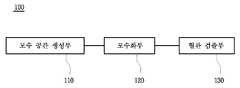
도 1을 참조하여 설명하면, 본 발명에 따른 의료영상 기반 혈관 검출 장치(100)는 크게 모수 공간 생성부(110), 모수화부(120) 및 혈관 검출부(130)를 포함한다.1, the medical image-based blood
모수 공간 생성부(110)는 혈관 조영술을 통해 획득된 의료영상에서 보여지는 혈관을 포함하는 모수 공간(parameter space)을 생성한다.The parameter space generating
모수 공간 생성부(110)는 혈관을 포함하는 원통 모양의 모수 공간을 생성한다.The parameter space generating
모수화부(120)는 생성된 모수 공간의 크기를 가변적으로 조절하면서, 혈관방향으로 놓인 법선벡터를 기준으로 하는 베지어 곡선(bezier curve)을 이용하여 다수개의 곡선을 생성한다. 이때, 모수화부(120)는 모수 공간의 반지름 크기를 가변적으로 조절하면서 다수개의 곡선을 생성한다.The
혈관 검출부(130)는 생성된 다수개의 곡선 중 적어도 하나의 곡선을 선택한다. 이때, 혈관 검출부(130)는 다수개의 곡선 중 혈관과 유사하다고 판단되는 적어도 하나의 곡선을 선택한다.
The blood
도 2는 본 발명에 따른 의료영상 기반 혈관 검출 장치에 채용되는 모수화부를 설명하기 위한 도면이다.2 is a view for explaining a parametric portion employed in a medical image-based blood vessel detecting apparatus according to the present invention.
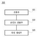
도 2를 참조하여 설명하면, 본 발명에 따른 모수화부(120)는 생성된 모수 공간의 크기를 가변적으로 조절하면서, 혈관방향으로 놓인 법선벡터를 기준으로 하는 베지어 곡선(bezier curve)을 이용하여 다수개의 곡선을 생성한다.Referring to FIG. 2, the
이를 위해, 모수화부(120)는 검출부(121), 포인트 산출부(122) 및 곡선 생성부(123)를 포함한다.For this, the
검출부(121)는 법선벡터의 시작점과 끝점을 검출하여 고정한다.The
포인트 산출부(122)는 베지어 곡선을 이용하여 모수 공간의 표면에 존재하는 제1 포인트와 제2 포인트를 반복하여 산출한다.The
곡선 생성부(123)는 산출된 제1 포인트의 집합과 제2 포인트의 집합의 곱을 통해 다수개의 곡선을 생성한다.
The
도 3은 본 발명에 따른 의료영상 기반 혈관 검출 방법의 순서를 설명하기 위한 순서도이고, 도 4는 본 발명에 따른 의료영상 기반 혈관 검출 방법에서의 제1 , 단계 예시를 설명하기 위한 도면이고, 도 5는 본 발명에 따른 의료영상 기반 혈관 검출 방법에서의 제2 단계 예시를 설명하기 위한 도면이고, 도 6은 본 발명에 따른 의료영상 기반 혈관 검출 방법에서의 제3 단계 예시를 설명하기 위한 도면이다.FIG. 3 is a flow chart for explaining a procedure of a medical image-based blood vessel detection method according to the present invention, FIG. 4 is a view for explaining a first and a step example in a medical image-based blood vessel detection method according to the present invention, 5 is a view for explaining an example of a second step in the medical image based blood vessel detecting method according to the present invention and FIG. 6 is a view for explaining an example of the third step in the medical image based blood vessel detecting method according to the present invention .
도 3을 참조하여 설명하면, 본 발명에 따른 의료영상 기반 혈관 검출 방법은 앞서 설명한 의료영상 기반 혈관 검출 장치를 이용하는 것으로, 이하 중복되는 설명은 생략하기로 한다.Referring to FIG. 3, the medical image-based blood vessel detection method according to the present invention uses the medical image-based blood vessel detection device described above, and a repeated description will be omitted.
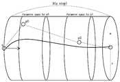
먼저, 제1 단계로 혈관 조영술을 통해 획득된 의료영상에서 보여지는 혈관을 포함하는 모수 공간(parameter space)을 생성한다(S100). 제1 단계는 도 4에 도시된 바와 같이, 혈관을 포함하는 원통 모양의 모수 공간을 생성한다.First, in step S100, a parameter space including a blood vessel displayed in the medical image obtained through the angiography is generated (S100). The first step creates a cylindrical parameter space including the blood vessel, as shown in Fig.
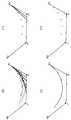
다음, 제2 단계로 생성된 모수 공간의 크기를 가변적으로 조절하면서, 혈관방향으로 놓인 법선벡터를 기준으로 하는 베지어 곡선(bezier curve)을 이용하여 다수개의 곡선을 생성한다(S200). 제2 단계는 도 5의 (a),(b),(c) 및 (d)에 도시된 바와 같이 법선벡터의 시작점(s)과 끝점(e)을 고정한다. 그 다음, 베지어 곡선을 이용하여 모수 공간의 표면에 존재하는 제1 포인트와 제2 포인트를 반복하여 산출한다. 이때, 제1 포인트를 p0 이고, 제2 포인트는 p1 이다. 그 다음, 산출된 제1 포인트의 집합과 제2 포인트의 집합의 곱을 통해 다수개의 곡선을 생성한다. 이때, 제1 포인트의 집합을 A라하고, 제2 포인트의 집합을 B라 할 때, 데카르트 곱 A X B는 제1 포인트의 집합과 제2 포인트의 집합의 법선벡터 쌍이되며, 쌍의 개수는 곧 법선벡터의 시작점(s)과 끝점(e) 사이에 존재할 수 있는 곡선의 개수(Nc)가 된다.Next, a plurality of curves are generated using a bezier curve based on a normal vector placed in a blood vessel direction while variably adjusting the size of the parameter space generated in the second step (S200). In the second step, the starting point (s) and the ending point (e) of the normal vector are fixed as shown in Figs. 5A, 5B, 5C and 5D. Then, the first point and the second point existing on the surface of the parameter space are repeatedly calculated using the Bezier curve. At this time, the first point is p0 and the second point is p1. Then, a plurality of curves are generated through the product of the set of the calculated first points and the set of the second points. At this time, when the set of the first points is A and the set of the second points is B, the Cartesian product AXB is a normal vector pair of the set of the first points and the set of the second points, The number Nc of curves that can exist between the starting point s and the end point e of the curve.
다음, 제3 단계로 생성된 다수개의 곡선 중 적어도 하나의 곡선을 선택한다(S300). 제3 단계는 도 6에 도시된 바와 같이 앞서 제2 단계에서 생성된 다수개의 곡선 중 혈관과 유사하다고 판단되는 적어도 하나의 곡선을 선택하고 반복적 시행 결과를 나타낸다.
Next, at least one curve among the plurality of curves generated in the third step is selected (S300). In the third step, as shown in FIG. 6, at least one curve that is determined to be similar to the blood vessel among the plurality of curves generated in the second step is selected and the result of iterative execution is shown.
이처럼, 본 발명에 의한 의료영상 기반 혈관 검출 방법 및 그 장치는 혈관 조영술을 통해 획득된 의료영상을 이용하여 혈관 방향으로 위치하는 법선벡터를 기준으로 곡선을 생성하는 과정을 반복하여 다수개의 곡선을 생성하고, 생성된 다수개의 곡선 중 혈관과 가장 유사한 하나의 혈관을 선택함으로써 정확한 실제 혈관을 검출할 수 있다.
As described above, the medical image-based blood vessel detection method and apparatus according to the present invention generate a plurality of curves by repeating a process of generating a curve based on a normal vector positioned in a blood vessel direction using a medical image obtained through angiography And by selecting one of the plurality of curves that is most similar to the blood vessel, an accurate actual blood vessel can be detected.
이상에서 본 발명에 따른 바람직한 실시예에 대해 설명하였으나, 다양한 형태로 변형이 가능하며, 본 기술분야에서 통상의 지식을 가진 자라면 본 발명의 특허청구범위를 벗어남이 없이 다양한 변형예 및 수정예를 실시할 수 있을 것으로 이해된다.While the present invention has been described in connection with what is presently considered to be practical exemplary embodiments, it is to be understood that the invention is not limited to the disclosed embodiments, but many variations and modifications may be made without departing from the scope of the present invention. It will be understood that the invention may be practiced.
100 : 의료영상 기반 혈관 검출 장치
110 : 모수 공간 생성부
120 : 모수화부
130 : 혈관 검출부100: Medical image based blood vessel detection device
110: Parameter space generating unit
120:
130:
Claims (7)
Translated fromKorean생성된 모수 공간의 크기를 가변적으로 조절하면서, 혈관방향으로 놓인 법선벡터를 기준으로 하는 베지어 곡선(bezier curve)을 이용하여 다수개의 곡선을 생성하는 제2 단계; 및
생성된 다수개의 곡선 중 적어도 하나의 곡선을 선택하는 제3 단계;
를 포함하는 것을 특징으로 하는 의료영상 기반 혈관 검출 방법.A first step of generating a parameter space including a blood vessel seen in a medical image obtained through angiography;
A second step of variably controlling the size of the generated parameter space and generating a plurality of curves using a Bezier curve based on a normal vector laid in a blood vessel direction; And
A third step of selecting at least one curve among the plurality of generated curves;
Wherein the medical image based blood vessel detection method comprises:
혈관 조영술을 통해 획득된 의료영상에서 보여지는 혈관을 포함하는 모수 공간을 생성하는 제1 단계는,
혈관을 포함하는 원통 모양의 모수 공간을 생성하는 것을 특징으로 하는 의료영상 기반 혈관 검출 방법.The method according to claim 1,
The first step of generating the parameter space including the blood vessel seen in the medical image obtained through the angiography includes:
And a cylindrical parameterizing space including a blood vessel is generated.
생성된 모수 공간의 크기를 가변적으로 조절하면서, 혈관방향으로 놓인 법선벡터를 기준으로 하는 베지어 곡선(bezier curve)을 이용하여 다수개의 곡선을 생성하는 제2 단계에서,
모수 공간의 반지름 크기를 가변적으로 조절하는 것을 특징으로 하는 의료영상 기반 혈관 검출 방법.The method according to claim 1,
In a second step of generating a plurality of curves by using a Bezier curve based on a normal vector placed in a blood vessel direction while variably controlling the size of the generated parameter space,
Wherein the radial size of the parameter space is variably controlled.
생성된 모수 공간의 크기를 가변적으로 조절하면서, 혈관방향으로 놓인 법선벡터를 기준으로 하는 베지어 곡선(bezier curve)을 이용하여 다수개의 곡선을 생성하는 제2 단계는,
법선벡터의 시작점과 끝점을 고정하는 단계;
베지어 곡선을 이용하여 모수 공간의 표면에 존재하는 제1 포인트와 제2 포인트를 반복하여 산출하는 단계; 및
산출된 제1 포인트의 집합과 제2 포인트의 집합의 곱을 통해 다수개의 곡선을 생성하는 단계;
를 포함하는 것을 특징으로 하는 의료영상 기반 혈관 검출 방법.The method according to claim 1,
A second step of generating a plurality of curves by using a bezier curve based on a normal vector placed in a blood vessel direction while variably adjusting the size of the generated parameter space,
Fixing a start point and an end point of the normal vector;
Repeatedly calculating a first point and a second point existing on the surface of the parameter space using a Bezier curve; And
Generating a plurality of curves through multiplication of the set of the first points and the set of the second points calculated;
Wherein the medical image based blood vessel detection method comprises:
생성된 다수개의 곡선 중 적어도 하나의 곡선을 선택하는 제3 단계는,
다수개의 곡선 중 혈관과 유사하다고 판단되는 적어도 하나의 곡선을 선택하는 것을 특징으로 하는 의료영상 기반 혈관 검출 방법.The method according to claim 1,
And a third step of selecting at least one curve among the plurality of generated curves,
Wherein at least one curve determined to be similar to a blood vessel among a plurality of curves is selected.
생성된 모수 공간의 크기를 가변적으로 조절하면서, 혈관방향으로 놓인 법선벡터를 기준으로 하는 베지어 곡선(bezier curve)을 이용하여 다수개의 곡선을 생성하는 모수화부;
생성된 다수개의 곡선 중 적어도 하나의 곡선을 선택하는 혈관 검출부;
를 포함하는 것을 특징으로 하는 의료영상 기반 혈관 검출 장치.A parameter space generating unit for generating a parameter space including a blood vessel seen in the medical image acquired through the angiography;
A parametric unit that variably adjusts the size of the generated parameter space and generates a plurality of curves using a Bezier curve based on a normal vector placed in a blood vessel direction;
A blood vessel detecting unit for selecting at least one curve among the plurality of generated curves;
Wherein the blood vessel detection device comprises:
상기 모수화부는,
법선벡터의 시작점과 끝점을 검출하여 고정하는 검출부;
베지어 곡선을 이용하여 모수 공간의 표면에 존재하는 제1 포인트와 제2 포인트를 반복하여 산출하는 포인트 산출부; 및
산출된 제1 포인트의 집합과 제2 포인트의 집합의 곱을 통해 다수개의 곡선을 생성하는 곡선 생성부;
를 포함하는 것을 특징으로 하는 의료영상 기반 혈관 검출 장치.The method according to claim 6,
The parametric section may further include:
A detector for detecting and fixing the start and end points of the normal vector;
A point calculating unit for calculating a first point and a second point on the surface of the parameter space by using a Bezier curve; And
A curve generation unit for generating a plurality of curves through multiplication of the set of the first points and the set of the second points;
Wherein the blood vessel detection device comprises:
Priority Applications (2)
| Application Number | Priority Date | Filing Date | Title |
|---|---|---|---|
| KR1020140148520AKR101630231B1 (en) | 2014-10-29 | 2014-10-29 | Method and apparatus for detecting vascular based on medical image |
| PCT/KR2015/000946WO2016068395A1 (en) | 2014-10-29 | 2015-01-29 | Method for detecting blood vessels on basis of medical image, and apparatus thereof |
Applications Claiming Priority (1)
| Application Number | Priority Date | Filing Date | Title |
|---|---|---|---|
| KR1020140148520AKR101630231B1 (en) | 2014-10-29 | 2014-10-29 | Method and apparatus for detecting vascular based on medical image |
Publications (2)
| Publication Number | Publication Date |
|---|---|
| KR20160052949Atrue KR20160052949A (en) | 2016-05-13 |
| KR101630231B1 KR101630231B1 (en) | 2016-06-27 |
Family
ID=56022897
Family Applications (1)
| Application Number | Title | Priority Date | Filing Date |
|---|---|---|---|
| KR1020140148520AExpired - Fee RelatedKR101630231B1 (en) | 2014-10-29 | 2014-10-29 | Method and apparatus for detecting vascular based on medical image |
Country Status (1)
| Country | Link |
|---|---|
| KR (1) | KR101630231B1 (en) |
Cited By (1)
| Publication number | Priority date | Publication date | Assignee | Title |
|---|---|---|---|---|
| KR20210023470A (en)* | 2019-08-23 | 2021-03-04 | 연세대학교 산학협력단 | Method and Apparatus for Object Feature Recognition Based on Bezier Curves |
Families Citing this family (1)
| Publication number | Priority date | Publication date | Assignee | Title |
|---|---|---|---|---|
| KR101852689B1 (en) | 2016-10-19 | 2018-06-11 | 순천향대학교 산학협력단 | Coronary Vessel extraction apparatus using vessel correspondence optimization and method thereof |
Citations (4)
| Publication number | Priority date | Publication date | Assignee | Title |
|---|---|---|---|---|
| US6148095A (en)* | 1997-09-08 | 2000-11-14 | University Of Iowa Research Foundation | Apparatus and method for determining three-dimensional representations of tortuous vessels |
| JP2008022928A (en)* | 2006-07-19 | 2008-02-07 | Gifu Univ | Image analysis apparatus and image analysis program |
| KR20110077740A (en)* | 2009-12-30 | 2011-07-07 | 서울여자대학교 산학협력단 | Blood vessel and calc automatic extraction device and method |
| US20120207366A1 (en)* | 2009-10-13 | 2012-08-16 | Agency For Science, Technology And Research | Method and system for segmenting a liver object in an image |
- 2014
- 2014-10-29KRKR1020140148520Apatent/KR101630231B1/ennot_activeExpired - Fee Related
Patent Citations (4)
| Publication number | Priority date | Publication date | Assignee | Title |
|---|---|---|---|---|
| US6148095A (en)* | 1997-09-08 | 2000-11-14 | University Of Iowa Research Foundation | Apparatus and method for determining three-dimensional representations of tortuous vessels |
| JP2008022928A (en)* | 2006-07-19 | 2008-02-07 | Gifu Univ | Image analysis apparatus and image analysis program |
| US20120207366A1 (en)* | 2009-10-13 | 2012-08-16 | Agency For Science, Technology And Research | Method and system for segmenting a liver object in an image |
| KR20110077740A (en)* | 2009-12-30 | 2011-07-07 | 서울여자대학교 산학협력단 | Blood vessel and calc automatic extraction device and method |
Cited By (1)
| Publication number | Priority date | Publication date | Assignee | Title |
|---|---|---|---|---|
| KR20210023470A (en)* | 2019-08-23 | 2021-03-04 | 연세대학교 산학협력단 | Method and Apparatus for Object Feature Recognition Based on Bezier Curves |
Also Published As
| Publication number | Publication date |
|---|---|
| KR101630231B1 (en) | 2016-06-27 |
Similar Documents
| Publication | Publication Date | Title |
|---|---|---|
| JP5465402B2 (en) | Method for performing quantitative analysis on medical image data of tubular organ, data processing apparatus and program storage device | |
| US20150257850A1 (en) | Image processing apparatus, image processing method and program | |
| US10019811B2 (en) | Image processing apparatus and method for estimating an error in registration between images | |
| JP6478136B1 (en) | Endoscope system and operation method of endoscope system | |
| WO2018170366A1 (en) | Geometric calibration for cone beam ct using line fiducials | |
| JP2019514547A5 (en) | ||
| CN106133789B (en) | Image processing apparatus and method for segmenting a region of interest | |
| JP2019530490A5 (en) | ||
| EP3569150A3 (en) | Systems and methods for predicting coronary plaque vulnerability from patient-specific anatomic image data | |
| JP2009072576A5 (en) | Method for performing quantitative analysis on medical image data of tubular organ, data processing apparatus and program storage device | |
| JP2019125112A5 (en) | ||
| JP2021101900A5 (en) | ||
| JPWO2015136853A1 (en) | Image processing apparatus, image processing method, and program | |
| WO2013031718A1 (en) | X-ray diagnostic apparatus and stent for x-ray diagnosis | |
| JP2017004464A5 (en) | Image processing apparatus, image processing method, and program | |
| US11324466B2 (en) | Creating monochromatic CT image | |
| JP2018526057A5 (en) | Interactive mesh editing system and method | |
| JP2012034024A5 (en) | ||
| JP2018071979A5 (en) | ||
| JP2019072190A5 (en) | ||
| KR101630231B1 (en) | Method and apparatus for detecting vascular based on medical image | |
| JP2018149171A5 (en) | ||
| JP2015217109A (en) | X-ray imaging apparatus and x-ray image processing apparatus | |
| JP2009082609A (en) | Magnetic resonance imaging apparatus, imaging method and imaging program | |
| JP5976126B2 (en) | System and method for estimating target size |
Legal Events
| Date | Code | Title | Description |
|---|---|---|---|
| A201 | Request for examination | ||
| PA0109 | Patent application | St.27 status event code:A-0-1-A10-A12-nap-PA0109 | |
| PA0201 | Request for examination | St.27 status event code:A-1-2-D10-D11-exm-PA0201 | |
| P11-X000 | Amendment of application requested | St.27 status event code:A-2-2-P10-P11-nap-X000 | |
| P13-X000 | Application amended | St.27 status event code:A-2-2-P10-P13-nap-X000 | |
| D13-X000 | Search requested | St.27 status event code:A-1-2-D10-D13-srh-X000 | |
| D14-X000 | Search report completed | St.27 status event code:A-1-2-D10-D14-srh-X000 | |
| E902 | Notification of reason for refusal | ||
| PE0902 | Notice of grounds for rejection | St.27 status event code:A-1-2-D10-D21-exm-PE0902 | |
| P11-X000 | Amendment of application requested | St.27 status event code:A-2-2-P10-P11-nap-X000 | |
| P13-X000 | Application amended | St.27 status event code:A-2-2-P10-P13-nap-X000 | |
| P11-X000 | Amendment of application requested | St.27 status event code:A-2-2-P10-P11-nap-X000 | |
| P13-X000 | Application amended | St.27 status event code:A-2-2-P10-P13-nap-X000 | |
| PG1501 | Laying open of application | St.27 status event code:A-1-1-Q10-Q12-nap-PG1501 | |
| PE0701 | Decision of registration | St.27 status event code:A-1-2-D10-D22-exm-PE0701 | |
| GRNT | Written decision to grant | ||
| PR0701 | Registration of establishment | St.27 status event code:A-2-4-F10-F11-exm-PR0701 | |
| PR1002 | Payment of registration fee | St.27 status event code:A-2-2-U10-U11-oth-PR1002 Fee payment year number:1 | |
| PG1601 | Publication of registration | St.27 status event code:A-4-4-Q10-Q13-nap-PG1601 | |
| FPAY | Annual fee payment | Payment date:20190904 Year of fee payment:4 | |
| PR1001 | Payment of annual fee | St.27 status event code:A-4-4-U10-U11-oth-PR1001 Fee payment year number:4 | |
| PR1001 | Payment of annual fee | St.27 status event code:A-4-4-U10-U11-oth-PR1001 Fee payment year number:5 | |
| PR1001 | Payment of annual fee | St.27 status event code:A-4-4-U10-U11-oth-PR1001 Fee payment year number:6 | |
| PR1001 | Payment of annual fee | St.27 status event code:A-4-4-U10-U11-oth-PR1001 Fee payment year number:7 | |
| R18-X000 | Changes to party contact information recorded | St.27 status event code:A-5-5-R10-R18-oth-X000 | |
| PN2301 | Change of applicant | St.27 status event code:A-5-5-R10-R13-asn-PN2301 St.27 status event code:A-5-5-R10-R11-asn-PN2301 | |
| P22-X000 | Classification modified | St.27 status event code:A-4-4-P10-P22-nap-X000 | |
| PR1001 | Payment of annual fee | St.27 status event code:A-4-4-U10-U11-oth-PR1001 Fee payment year number:8 | |
| PC1903 | Unpaid annual fee | St.27 status event code:A-4-4-U10-U13-oth-PC1903 Not in force date:20240609 Payment event data comment text:Termination Category : DEFAULT_OF_REGISTRATION_FEE | |
| P22-X000 | Classification modified | St.27 status event code:A-4-4-P10-P22-nap-X000 | |
| PC1903 | Unpaid annual fee | St.27 status event code:N-4-6-H10-H13-oth-PC1903 Ip right cessation event data comment text:Termination Category : DEFAULT_OF_REGISTRATION_FEE Not in force date:20240609 |