KR20140107103A - Micronized placental tissue compositions and methods for making and using the same - Google Patents
Micronized placental tissue compositions and methods for making and using the sameDownload PDFInfo
- Publication number
- KR20140107103A KR20140107103AKR1020137023726AKR20137023726AKR20140107103AKR 20140107103 AKR20140107103 AKR 20140107103AKR 1020137023726 AKR1020137023726 AKR 1020137023726AKR 20137023726 AKR20137023726 AKR 20137023726AKR 20140107103 AKR20140107103 AKR 20140107103A
- Authority
- KR
- South Korea
- Prior art keywords
- composition
- alcohol
- amniotic membrane
- tissue
- subject
- Prior art date
- Legal status (The legal status is an assumption and is not a legal conclusion. Google has not performed a legal analysis and makes no representation as to the accuracy of the status listed.)
- Ceased
Links
Images
Classifications
- A—HUMAN NECESSITIES
- A61—MEDICAL OR VETERINARY SCIENCE; HYGIENE
- A61K—PREPARATIONS FOR MEDICAL, DENTAL OR TOILETRY PURPOSES
- A61K35/00—Medicinal preparations containing materials or reaction products thereof with undetermined constitution
- A61K35/12—Materials from mammals; Compositions comprising non-specified tissues or cells; Compositions comprising non-embryonic stem cells; Genetically modified cells
- A61K35/48—Reproductive organs
- A61K35/50—Placenta; Placental stem cells; Amniotic fluid; Amnion; Amniotic stem cells
- A—HUMAN NECESSITIES
- A61—MEDICAL OR VETERINARY SCIENCE; HYGIENE
- A61K—PREPARATIONS FOR MEDICAL, DENTAL OR TOILETRY PURPOSES
- A61K35/00—Medicinal preparations containing materials or reaction products thereof with undetermined constitution
- A61K35/12—Materials from mammals; Compositions comprising non-specified tissues or cells; Compositions comprising non-embryonic stem cells; Genetically modified cells
- A—HUMAN NECESSITIES
- A61—MEDICAL OR VETERINARY SCIENCE; HYGIENE
- A61K—PREPARATIONS FOR MEDICAL, DENTAL OR TOILETRY PURPOSES
- A61K8/00—Cosmetics or similar toiletry preparations
- A61K8/18—Cosmetics or similar toiletry preparations characterised by the composition
- A61K8/96—Cosmetics or similar toiletry preparations characterised by the composition containing materials, or derivatives thereof of undetermined constitution
- A61K8/98—Cosmetics or similar toiletry preparations characterised by the composition containing materials, or derivatives thereof of undetermined constitution of animal origin
- A61K8/981—Cosmetics or similar toiletry preparations characterised by the composition containing materials, or derivatives thereof of undetermined constitution of animal origin of mammals or bird
- A61K8/982—Reproductive organs; Embryos, Eggs
- A—HUMAN NECESSITIES
- A61—MEDICAL OR VETERINARY SCIENCE; HYGIENE
- A61L—METHODS OR APPARATUS FOR STERILISING MATERIALS OR OBJECTS IN GENERAL; DISINFECTION, STERILISATION OR DEODORISATION OF AIR; CHEMICAL ASPECTS OF BANDAGES, DRESSINGS, ABSORBENT PADS OR SURGICAL ARTICLES; MATERIALS FOR BANDAGES, DRESSINGS, ABSORBENT PADS OR SURGICAL ARTICLES
- A61L27/00—Materials for grafts or prostheses or for coating grafts or prostheses
- A61L27/36—Materials for grafts or prostheses or for coating grafts or prostheses containing ingredients of undetermined constitution or reaction products thereof, e.g. transplant tissue, natural bone, extracellular matrix
- A61L27/3604—Materials for grafts or prostheses or for coating grafts or prostheses containing ingredients of undetermined constitution or reaction products thereof, e.g. transplant tissue, natural bone, extracellular matrix characterised by the human or animal origin of the biological material, e.g. hair, fascia, fish scales, silk, shellac, pericardium, pleura, renal tissue, amniotic membrane, parenchymal tissue, fetal tissue, muscle tissue, fat tissue, enamel
- A—HUMAN NECESSITIES
- A61—MEDICAL OR VETERINARY SCIENCE; HYGIENE
- A61L—METHODS OR APPARATUS FOR STERILISING MATERIALS OR OBJECTS IN GENERAL; DISINFECTION, STERILISATION OR DEODORISATION OF AIR; CHEMICAL ASPECTS OF BANDAGES, DRESSINGS, ABSORBENT PADS OR SURGICAL ARTICLES; MATERIALS FOR BANDAGES, DRESSINGS, ABSORBENT PADS OR SURGICAL ARTICLES
- A61L27/00—Materials for grafts or prostheses or for coating grafts or prostheses
- A61L27/36—Materials for grafts or prostheses or for coating grafts or prostheses containing ingredients of undetermined constitution or reaction products thereof, e.g. transplant tissue, natural bone, extracellular matrix
- A61L27/3683—Materials for grafts or prostheses or for coating grafts or prostheses containing ingredients of undetermined constitution or reaction products thereof, e.g. transplant tissue, natural bone, extracellular matrix subjected to a specific treatment prior to implantation, e.g. decellularising, demineralising, grinding, cellular disruption/non-collagenous protein removal, anti-calcification, crosslinking, supercritical fluid extraction, enzyme treatment
- A61L27/3695—Materials for grafts or prostheses or for coating grafts or prostheses containing ingredients of undetermined constitution or reaction products thereof, e.g. transplant tissue, natural bone, extracellular matrix subjected to a specific treatment prior to implantation, e.g. decellularising, demineralising, grinding, cellular disruption/non-collagenous protein removal, anti-calcification, crosslinking, supercritical fluid extraction, enzyme treatment characterised by the function or physical properties of the final product, where no specific conditions are defined to achieve this
- A—HUMAN NECESSITIES
- A61—MEDICAL OR VETERINARY SCIENCE; HYGIENE
- A61P—SPECIFIC THERAPEUTIC ACTIVITY OF CHEMICAL COMPOUNDS OR MEDICINAL PREPARATIONS
- A61P17/00—Drugs for dermatological disorders
- A61P17/02—Drugs for dermatological disorders for treating wounds, ulcers, burns, scars, keloids, or the like
- A—HUMAN NECESSITIES
- A61—MEDICAL OR VETERINARY SCIENCE; HYGIENE
- A61Q—SPECIFIC USE OF COSMETICS OR SIMILAR TOILETRY PREPARATIONS
- A61Q19/00—Preparations for care of the skin
- A—HUMAN NECESSITIES
- A61—MEDICAL OR VETERINARY SCIENCE; HYGIENE
- A61L—METHODS OR APPARATUS FOR STERILISING MATERIALS OR OBJECTS IN GENERAL; DISINFECTION, STERILISATION OR DEODORISATION OF AIR; CHEMICAL ASPECTS OF BANDAGES, DRESSINGS, ABSORBENT PADS OR SURGICAL ARTICLES; MATERIALS FOR BANDAGES, DRESSINGS, ABSORBENT PADS OR SURGICAL ARTICLES
- A61L2400/00—Materials characterised by their function or physical properties
- A61L2400/06—Flowable or injectable implant compositions
- A—HUMAN NECESSITIES
- A61—MEDICAL OR VETERINARY SCIENCE; HYGIENE
- A61L—METHODS OR APPARATUS FOR STERILISING MATERIALS OR OBJECTS IN GENERAL; DISINFECTION, STERILISATION OR DEODORISATION OF AIR; CHEMICAL ASPECTS OF BANDAGES, DRESSINGS, ABSORBENT PADS OR SURGICAL ARTICLES; MATERIALS FOR BANDAGES, DRESSINGS, ABSORBENT PADS OR SURGICAL ARTICLES
- A61L2430/00—Materials or treatment for tissue regeneration
- A61L2430/34—Materials or treatment for tissue regeneration for soft tissue reconstruction
Landscapes
- Health & Medical Sciences (AREA)
- Life Sciences & Earth Sciences (AREA)
- Chemical & Material Sciences (AREA)
- Veterinary Medicine (AREA)
- Public Health (AREA)
- General Health & Medical Sciences (AREA)
- Animal Behavior & Ethology (AREA)
- Engineering & Computer Science (AREA)
- Medicinal Chemistry (AREA)
- Biomedical Technology (AREA)
- Epidemiology (AREA)
- Dermatology (AREA)
- Chemical Kinetics & Catalysis (AREA)
- Developmental Biology & Embryology (AREA)
- Zoology (AREA)
- Botany (AREA)
- Oral & Maxillofacial Surgery (AREA)
- Transplantation (AREA)
- Molecular Biology (AREA)
- Cell Biology (AREA)
- Pharmacology & Pharmacy (AREA)
- Reproductive Health (AREA)
- Immunology (AREA)
- Virology (AREA)
- Biotechnology (AREA)
- Pregnancy & Childbirth (AREA)
- Bioinformatics & Cheminformatics (AREA)
- General Chemical & Material Sciences (AREA)
- Nuclear Medicine, Radiotherapy & Molecular Imaging (AREA)
- Organic Chemistry (AREA)
- Urology & Nephrology (AREA)
- Birds (AREA)
- Medicines Containing Material From Animals Or Micro-Organisms (AREA)
- Medicinal Preparation (AREA)
- Materials For Medical Uses (AREA)
- Medicines That Contain Protein Lipid Enzymes And Other Medicines (AREA)
Abstract
Translated fromKoreanDescription
Translated fromKorean관련 출원에 대한 상호 참조Cross-reference to related application
본 출원은 2011년 2월 14일자로 출원된 미국 가출원 제61/442,346호 및 2011년 10월 6일자로 출원된 제61/543,995호를 우선권 주장한다. 이들 출원은 이것들의 모든 교시내용에 대하여 그 전문이 본원에 참고로 포함된다.This application claims priority to U.S. Provisional Application No. 61 / 442,346, filed February 14, 2011, and 61 / 543,995, filed October 6, These applications are incorporated herein by reference in their entirety for all of their teachings.
태반 조직은 전형적으로 선택적 제왕절개술 후에 수확된다. 태반은 제대 및 양막낭으로 이루어진다. 양막낭은 통상적으로 양막이라고 지칭되며, 2개의 주요 조직층인 양막 및 융모막을 갖는다. 양막 조직은 양막낭의 최내층으로서, 양수와 직접 접촉하고 있다. 양막낭은 양수를 함유하여 태아 환경을 보호한다. 조직학적 평가는 양막의 막층이 단일 층의 상피 세포, 얇은 망상 섬유 (기저막), 두껍고 조밀한 층 및 섬유모세포층으로 이루어져 있음을 가리킨다. 양막의 섬유성 층 (즉, 기저막)은 콜라겐 제IV형, 제V형 및 제VII형, 및 피브로넥틴 및 라미닌을 비롯한 세포-접착 생물활성 인자를 함유한다. 본원은 창상 치유 및 기타 의학적 용도에 태반 조직 성분을 사용하는 특이한 접근법을 기재한다.Placental tissue is typically harvested after selective cesarean section. The placenta consists of umbilical cord and amniotic sac. The amniotic sac is commonly referred to as the amniotic membrane and has two major tissue layers, amniotic membrane and chorionic membrane. Amniotic membrane tissue is the innermost layer of the amniotic sac, in direct contact with amniotic fluid. Amniotic sacs contain amniotic fluid to protect the fetal environment. Histological evaluation indicates that the amniotic membrane layer consists of a monolayer of epithelial cells, thin reticular fibers (basement membrane), a thick dense layer and a fibroblast layer. The fibrous layer of the amniotic membrane (ie, basement membrane) contains collagen type IV, type V and type VII, and cell-adherent biologically active factors including fibronectin and laminin. This article describes a unique approach to using placenta tissue components for wound healing and other medical uses.
요약summary
본원은 마이크로화된 태반 성분으로 이루어진 조성물 및 그의 제약 조성물을 기재한다. 상기 조성물은 수많은 의학적 용도를 갖는다. 마이크로화된 조성물의 제조 및 사용 방법 또한 본원에서 기재된다.The present disclosure describes a composition comprising a micronized placental component and pharmaceutical compositions thereof. The compositions have numerous medical uses. Methods for making and using microfabricated compositions are also described herein.
본 발명의 이점은 후술하는 설명에서 부분적으로 제시될 것이고, 부분적으로는 이러한 설명으로부터 명확해질 것이고, 또는 이하에 기재된 측면들을 실시함으로써 알게 될 수도 있다. 이하에 기재된 이점은 특히 첨부하는 특허청구범위에서 언급한 요소 및 조합에 의해 실현되고 달성될 것이다. 상기 일반적인 설명 및 하기 상세한 설명은 둘 다 단지 예시하고 설명하기 위한 것일 뿐 제한하려는 것은 아님을 이해해야 한다.The advantages of the invention will be set forth in part in the description which follows, and in part will be apparent from the description, or may be learned by the practice of the aspects described hereinafter. The advantages described below will be realized and attained by means of the elements and combinations particularly pointed out in the appended claims. It is to be understood that both the foregoing general description and the following detailed description are exemplary and explanatory only and are not restrictive.
도면의 간단한 설명
첨부된 도면은 본 명세서에 포함되고 본 명세서의 일부를 구성하며, 이하에 기재된 여러 측면들을 예시한다.
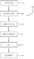
도 1은 본원에 기재된 마이크로화된 조성물의 제조 방법에 관한 개괄적인 흐름도이다.Brief Description of Drawings
BRIEF DESCRIPTION OF THE DRAWINGS The accompanying drawings, which are incorporated in and constitute a part of this specification, illustrate various aspects described below.
BRIEF DESCRIPTION OF THE DRAWINGS Figure 1 is a general flow chart of a method of making a microfabricated composition as described herein.
상세한 설명details
본 발명의 용품 및 방법을 개시하고 기재하기 전에, 이하에 기재된 측면들이 특정 화합물, 합성 방법 또는 용도에만 한정되는 것이 아니라, 이러한 것들은 달라질 수도 있는 것이 당연함을 이해해야 한다. 또한, 본원에서 사용된 용어는 단지 특정 측면들을 설명하는 것을 목적으로 할 뿐 한정하고자 하는 것은 아님을 이해해야 한다.Before disclosing and describing the articles and methods of the present invention, it is to be understood that the aspects described below are not limited to any particular compound, method of synthesis or use, but rather that they may vary. It is also to be understood that the terminology used herein is for the purpose of describing particular aspects only, and not for purposes of limitation.
후술하는 본 명세서 및 특허청구범위에서는, 하기 의미를 갖는 것으로 정의되는 다수의 용어를 언급할 것이다:In the following specification and claims, reference will be made to a number of terms that are defined to have the following meanings:
본 명세서 및 첨부하는 특허청구범위에서 사용된 바와 같은 단수 형태는, 문맥상 명확하게 달리 지시되지 않는 한은 복수의 대상을 포함함에 유의해야 한다. 따라서, 예를 들어 "생물활성제"에 대한 언급은 2개 이상의 이러한 작용제의 혼합물을 포함하는 식이다.It should be noted that the singular forms as used in this specification and the appended claims include plural referents unless the context clearly dictates otherwise. Thus, for example, reference to "bioactive agent" is an expression that includes a mixture of two or more such agents.
"임의의" 또는 "임의로"는 이어서 기재되는 사건 또는 상황이 발생할 수도 있고 그렇지 않을 수도 있으며, 그 기재내용이 사건 또는 상황이 발생한 경우 및 그것이 발생하지 않은 경우를 포함함을 의미한다. 예를 들어, 어구 "임의로 세정하는 단계"는 세정하는 단계가 수행될 수도 있고 그렇지 않을 수도 있음을 의미한다."Optional" or " optionally "means that the subsequently described event or circumstance may or may not occur, and that the description includes instances where the event or circumstance occurs and instances in which it does not occur. For example, the phrase "optionally cleaning" means that the cleaning step may or may not be performed.
본원에서 사용된 바와 같은 용어 "대상체"는 임의의 척추동물 유기체이다.The term "subject" as used herein is any vertebrate organism.
본원에서 사용된 바와 같은 용어 "양막"은 중간 조직층이 무손상 상태이거나 실질적으로 제거된 양막을 포함한다.The term "amniotic membrane" as used herein includes amniotic membrane in which the intermediate tissue layer is intact or substantially removed.
표제 또는 부제가 독자의 편의를 위해 본 명세서에서 사용될 수 있지만, 이는 본 발명의 범위에 영향을 주고자 하는 것은 아니다. 추가로, 본 명세서에 사용된 일부 용어는 이하에서 보다 구체적으로 정의된다.While a title or subtitle may be used herein for the convenience of the reader, it is not intended to affect the scope of the invention. In addition, some of the terms used herein are more specifically defined below.
I. 마이크로화된 조성물 및 그의 제조 방법I. Microfabricated compositions and methods for their preparation
본원은 마이크로화된 태반 성분으로 이루어진 조성물 및 그의 제약 조성물을 기재한다. 한 측면에서, 상기 조성물은 (a) 마이크로화된 양막, 융모막, 중간 조직층, 또는 이것들의 임의의 조합물 및 (b) 제약상 허용되는 담체를 포함한다. 한 측면에서, 상기 조성물은 마이크로화된 양막 및 중간 조직층을 포함한다. 또 다른 측면에서, 상기 조성물은 마이크로화된 양막 및 융모막을 포함한다.The present disclosure describes a composition comprising a micronized placental component and pharmaceutical compositions thereof. In one aspect, the composition comprises (a) a micrometric amnion, a chorionic membrane, an intermediate tissue layer, or any combination thereof and (b) a pharmaceutically acceptable carrier. In one aspect, the composition comprises a microfibrated amniotic membrane and an intermediate tissue layer. In yet another aspect, the composition comprises microfibrated amniotic membrane and chorionic membrane.
도 1은 본원에 기재된 마이크로화된 조성물의 제조에 사용하기 위한 태반 물질을 수확하고, 처리하고, 준비하는 단계의 개요 (100) 및 특정 측면들을 도시한다. 각각의 개별적인 단계와 관련한 보다 상세한 설명 및 논의가 이어질 것이다. 처음에는, 선택적 제왕절개술 후 동의한 환자로부터 태반 조직을 수집한다 (단계 110). 상기 물질을 통상적인 조직 보존 방식으로 보존하고 적합한 처리 장소 또는 시설로 이동시켜 확인 및 평가한다 (단계 120). 이후, 조직층의 총체적 처리, 조작 및 분리를 수행한다 (단계 130). 이어서, 허용되는 조직을 오염제거처리하고 (단계 140) 탈수시킨다 (단계 145). 오염제거처리 및 탈수 후에, 태반 성분 (예를 들어, 개별적인 또는 이식편으로서의 양막, 중간 조직층 및/또는 융모막)을 마이크로화한다 (단계 150). 각각의 단계를 이하에 상세하게 기재한다.Figure 1 shows an overview (100) and specific aspects of the steps of harvesting, processing, and preparing the placenta material for use in the manufacture of the microfabricated compositions described herein. A more detailed discussion and discussion will follow with respect to each individual step. Initially, placental tissue is collected from the consenting patient after the selective cesarean section (step 110). The material is preserved in a conventional tissue preservation manner and moved to the appropriate treatment site or facility for identification and evaluation (step 120). Thereafter, overall processing, manipulation, and separation of the tissue layer is performed (step 130). The allowed tissue is then decontaminated (step 140) and dehydrated (step 145). After decontamination treatment and dehydration, placental components (e.g., amniotic membrane, middle tissue layer and / or chorion membrane as individual or grafts) are microdosed (step 150). Each step is described in detail below.
초기 조직 수집 (단계 110)Initial tissue collection (step 110)
본원에 기재된 마이크로화된 조성물 제조에 사용되는 성분은 태반으로부터 유래된다. 태반의 공급원은 다양할 수 있다. 한 측면에서, 태반은 포유동물, 예컨대 인간으로부터 유래되고, 소, 돼지 등을 포함하지만 이에 제한되지 않는 다른 동물들이 본원에서 사용될 수 있다. 인간의 경우, 태반의 회수는 병원에서 비롯되는데, 여기서 태반은 제왕절개 분만 동안 수집된다. 막 분만하려고 하는 모체를 지칭하는 공여자는 이식 가능한 가장 안전한 조직을 제공하도록 디자인된 종합 스크리닝 처리를 자발적으로 받는다. 스크리닝 처리는 바람직하게는 인간 면역결핍 바이러스 제1형 및 제2형에 대한 항체 (항-HIV-1 및 항-HIV-2), B형 간염 바이러스에 대한 항체 (항-HBV), B형 간염 표면 항원 (HBsAg), C형 간염 바이러스에 대한 항체 (항-HCV), 인간 T-림프친화 바이러스 제I형 및 제II형에 대한 항체 (항-HTLV-I, 항-HTLV-II), CMV 및 매독에 대해 시험하고, 통상적인 혈청학적 시험을 사용하여 인간 면역-결핍 바이러스 제1형 (HIV-1) 및 C형 간염 바이러스 (HCV)에 대한 핵산 시험을 수행한다. 상기 시험 목록은 단지 예시적인 것이고, 당업자라면 알고 있는 바와 같이, 더 많거나 더 적거나 또는 상이한 시험이 시간 경과에 따라 또는 이식편의 의도된 용도에 기초하여 바람직하거나 필요할 수 있다.The components used in the preparation of the micronized compositions described herein are derived from the placenta. The source of the placenta can vary. In one aspect, the placenta is derived from a mammal, such as a human, and other animals, including but not limited to cows, pigs, etc., may be used herein. In humans, the placenta's recovery comes from the hospital, where the placenta is collected during the cesarean section. The donor, referring to the mother who is about to give birth, voluntarily receives a comprehensive screening process designed to provide the safest possible implantable tissue. The screening treatment is preferably carried out with antibodies (anti-HIV-1 and anti-HIV-2) against human immunodeficiency virus type 1 and type 2, antibodies against hepatitis B virus (anti-HBV) (Anti-HTLV-I, anti-HTLV-II) against human hepatitis C virus (HBV), surface antigen (HBsAg), hepatitis C virus (anti-HCV), antibodies against human T- And syphilis and nucleic acid tests on human immunodeficiency virus type 1 (HIV-1) and hepatitis C virus (HCV) are performed using conventional serological tests. The test list is merely illustrative, and as will be appreciated by those skilled in the art, more or less or different tests may be desirable or necessary over time or based on the intended use of the implant.
공여자의 정보 및 스크리닝 시험 결과의 검토에 기초하여, 공여자는 허용되거나 그렇지 않은 것으로 판단될 것이다. 추가로, 전달 시점에 박테리아, 예를 들어 클로스트리듐(Clostridium) 또는 스트렙토코커스(Streptococcus)의 존재를 규명하기 위해 배양을 수행한다. 공여자의 정보, 스크리닝 시험 및 전달 배양을 모두 충족시키면 (즉, 어떠한 위험도 나타내지 않거나 허용되는 수준의 위험만을 나타내면), 공여자는 의료 책임자에 의해 승인되고, 조직 표본은 추가 처리 및 평가에 1차적으로 적격인 것으로 정해진다.Based on a review of the donor's information and screening test results, the donor will be judged to be acceptable or not. In addition, the culture is performed in order to clarify the presence of bacteria, such as Clostridium(Clostridium) or Streptococcus(Streptococcus) to the delivery point. If all of the donor's information, screening tests and delivery cultures are met (ie, they represent no risk or only represent an acceptable level of risk), the donor is approved by the health care provider and the tissue sample is primary eligible for further processing and evaluation .
상기 선별 기준을 충족시키는 인간 태반은 바람직하게는 추가 처리를 위한 처리 장소 또는 실험실로 수송하기 위해 멸균 수송 백 안의 염수 용액에 담겨서 습윤 얼음 용기에 저장된다.The human placenta meeting the selection criteria is preferably stored in a wet ice container in a sterile transport bag in a saline solution for transport to a treatment site or laboratory for further processing.
태반을 스크리닝 시험 및 전달 배양의 결과 획득 완료 전에 수집한 경우에는, 이러한 조직을 표지하여 격리시켜 둔다. 태반은 조작 및 사용에 안전한 조직임을 공표하는 필수 스크리닝 평가 및 전달 배양을 충족시키고, 의료 책임자로부터 최종 승인을 획득한 후에만 추가 처리에 대해 승인된다.If the placenta is collected prior to completion of the screening and delivery cultures, mark these tissues and isolate them. The placenta is approved for further treatment only after it meets the mandatory screening assessment and delivery culture announcing that it is a safe tissue for manipulation and use, and after obtaining final approval from the health care provider.
물질 확인 및 평가 (Identification and evaluation of substances단계step 120) 120)
처리 센터 또는 연구소에 도달 직후, 수송물을 열어 멸균 수송 백/용기가 여전히 밀봉되어 있고 냉각수 중에 있는지, 적절한 공여자 서류가 들어있는지, 및 서류상의 공여자 번호가 조직을 함유하는 멸균 수송 백의 번호와 일치하는지를 확인한다. 이어서, 조직을 함유하는 멸균 수송 백을 추가의 처리 준비시까지 냉장 저장한다.Immediately after reaching the treatment center or laboratory, the transport is opened to determine whether the sterile transport bag / container is still sealed and in the coolant, contains the appropriate donor papers, and matches the donor number on the paper with the number of sterile transport bags containing tissue Check. The sterile transport bag containing the tissue is then refrigerated until further processing is ready.
총체적인 조직 처리 (단계 130)Overall tissue processing (step 130)
조직의 추가 처리가 준비되면, 태반 조직 처리를 위해 필수적인 멸균 용품들을 추가로 제어된 환경에서의 집결 지역에 모으고, 제어된 환경으로 도입할 준비를 한다. 한 측면에서, 태반을 실온에서 처리한다. 제어된 환경이 제조 후드인 경우, 멸균 용품들을 열고, 통상적인 멸균 기술을 이용하여 후드에 넣는다. 제어된 환경이 무균실인 경우, 멸균 용품들을 열고, 멸균 드레이프로 덮은 카트에 놓는다. 모든 작업 표면을 통상적인 멸균 기술을 이용하여 멸균 드레이프 조각으로 덮고, 멸균 용품 및 처리 장비를 다시 통상적인 멸균 기술을 이용하여 멸균 드레이프에 놓는다.Once the further processing of the tissue is ready, the sterile supplies necessary for placental tissue processing are collected in a collection area in an additional controlled environment and ready to be introduced into a controlled environment. In one aspect, the placenta is treated at room temperature. If the controlled environment is a manufacturing hood, the sterilization items are opened and placed in the hood using conventional sterilization techniques. If the controlled environment is clean room, open sterile supplies and place in a cart covered with a sterile drape. All work surfaces are covered with sterile drape pieces using conventional sterilization techniques, and sterile goods and processing equipment are again placed in a sterile drape using conventional sterilization techniques.
처리 장비를 통상적인 산업-승인된 오염제거처리 절차에 따라 오염제거처리한 후에 제어된 환경에 도입한다. 장비를 제어된 환경 내에 전략적으로 위치시켜 장비가 조직 표본에 의해 의도치않게 오염되거나 그에 근접해질 기회를 최소화한다.The treatment equipment is decontaminated in accordance with conventional industry-approved decontamination procedures and then introduced into the controlled environment. Strategically locate the equipment in a controlled environment to minimize the chance that the equipment will be inadvertently contaminated or approximated by tissue samples.
다음으로, 멸균 수송 백에서 태반을 꺼내어 제어된 환경 내의 멸균 처리 용기로 무균 이동시킨다. 멸균 용기는 실온이거나 실온에 가까운 고장성 염수 용액 (예를 들어, 18% NaCl)을 함유한다. 태반을 부드럽게 마사지하여 혈병 분리를 돕고 태반 조직이 실온에 이르도록 하여 태반 성분이 서로 (예를 들어, 양막 및 융모막) 분리되는 것을 용이하게 한다. 주위 온도로 가온시킨 후 (예를 들어, 약 10분 내지 30분 후), 태반을 멸균 처리 용기에서 꺼내어 양막층을 아래쪽으로 향하게 하여 처리 트레이에 평평하게 놓고 검사한다.The placenta is then removed from the sterile transport bag and aseptically transferred to a sterile disposal container in a controlled environment. The sterile container contains a high temperature saline solution (for example, 18% NaCl) at room temperature or near room temperature. Gently massage the placenta to help separate blood clots and allow the placental tissue to reach room temperature, facilitating the separation of the placental components from one another (eg amniotic membrane and chorion). After warming to ambient temperature (for example, after about 10 to 30 minutes), the placenta is removed from the sterilization treatment vessel and placed flat on the processing tray with the amniotic membrane layer facing downward.
태반을 변색, 파편 또는 다른 오염, 냄새 및 손상의 징후에 대해 조사한다. 조직의 크기도 확인한다. 이 시점에서 상기 조직이 추가의 처리에 허용되는지 여부를 결정한다.Investigate signs of discoloration, debris or other contamination, odor and damage to the placenta. Also check the size of the tissue. At this point, it is determined whether the organization is allowed to perform further processing.
이어서, 양막 및 융모막을 조심스럽게 분리한다. 한 측면에서, 이 절차에 사용되는 물질 및 장비는 처리 트레이, 18% 염수 용액, 멸균 4×4 스폰지, 및 2개의 멸균 날진(Nalgene) 단지를 포함한다. 이어서, 태반 조직을 면밀히 조사하여 양막이 융모막으로부터 분리될 수 있는 영역 (전형적으로는 모서리)을 확인한다. 양막은 융모막 상의 얇고 불투명한 층처럼 보인다.Then carefully separate the amniotic membrane and chorion. In one aspect, the materials and equipment used in this procedure include treatment trays, 18% saline solution, sterile 4x4 sponges, and two sterile Nalgene jars. The placental tissue is then carefully examined to identify areas where the amniotic membrane can be separated from the chorion (typically, the corners). The amniotic membrane looks like a thin, opaque layer on the chorion.
섬유모세포층은 양막의 각 면을 멸균 거즈 조각 또는 면봉과 조심스럽게 접촉시킴으로써 확인한다. 섬유모세포층은 시험 물질에 점착될 것이다. 양막을 기저막층을 아래로 하여 처리 트레이에 놓는다. 평활 기기, 세포 스크래퍼 또는 멸균 거즈를 사용하여 임의의 잔류 혈액도 제거한다. 상기 단계는 양막이 찢어지지 않도록 재차 적절히 주의하면서 행해야 한다. 양막이 외관상 평활하고 불투명한 백색이 되면 양막의 세정이 완료된다.The fibroblast layer is identified by carefully contacting each side of the amniotic membrane with a sterile gauze piece or cotton swab. The fibroblast layer will adhere to the test material. The amniotic membrane is placed on the treatment tray with the basement membrane layer down. Any residual blood is also removed using a smoothing device, a cell scraper or sterile gauze. This step should be done again with proper care so that the amniotic membrane is not torn. When the amniotic membrane is apparently smooth and opaque white, amniotic membrane cleaning is completed.
특정 측면에서, 스폰지층이라고 지칭되기도 하는 중간 조직층을 양막으로부터 실질적으로 제거하여 섬유모세포층을 노출시킨다. 중간 조직층이 제거되는 양과 관련하여, 본원에서 용어 "실질적 제거"는 양막으로부터 중간 조직층의 90% 초과, 95% 초과, 또는 99% 초과를 제거하는 것으로 정의된다. 이는 양막으로부터 중간 조직층을 벗겨내어 수행될 수 있다. 대안적으로, 중간 조직층을 거즈 또는 다른 적합한 와이프로 닦아내어, 양막으로부터 중간 조직층을 제거할 수 있다. 이후, 생성된 양막을 하기하는 방법을 이용하여 오염제거처리할 수 있다.In certain aspects, an intermediate tissue layer, also referred to as a sponge layer, is substantially removed from the amniotic membrane to expose the fibroblast layer. With respect to the amount by which the middle tissue layer is removed, the term "substantial removal" is defined herein as removing more than 90%, greater than 95%, or greater than 99% of the middle tissue layer from the amniotic membrane. This can be done by stripping the middle tissue layer from the amniotic membrane. Alternatively, the middle tissue layer can be wiped with gauze or other suitable wipe to remove the middle tissue layer from the amniotic membrane. Thereafter, the produced amniotic membrane can be decontaminated using the following method.
특정 측면에서, 양막에 존재하는 상피층을 실질적으로 제거하여 양막의 기저층을 노출시킨다. 상피가 제거되는 양과 관련하여, 본원에서 용어 "실질적 제거"는 양막으로부터 상피 세포의 90% 초과, 95% 초과, 또는 99% 초과를 제거하는 것으로 정의된다. 양막층에 잔류하는 상피 세포의 존재 또는 부재는 당업계 공지의 기술을 이용하여 평가할 수 있다. 예를 들어, 상피 세포층의 제거 후, 처리 완료품으로부터의 대표적인 조직 샘플을 표준 현미경 조사 슬라이드 위에 놓는다. 이어서, 조직 샘플을 에오신(Eosin) Y 염색법을 이용하여 염색하고 하기하는 바와 같이 평가한다. 이어서, 샘플을 덮어두고 방치한다. 염색될 만큼 적절한 시간이 지나면, 확대하여 시각적으로 관찰한다.In certain aspects, the epithelial layer present in the amniotic membrane is substantially removed to expose the basal layer of the amniotic membrane. With respect to the amount of epithelial removal, the term "substantial removal" is defined herein as removing more than 90%, greater than 95%, or greater than 99% of epithelial cells from the amniotic membrane. The presence or absence of epithelial cells remaining in the amniotic membrane layer can be evaluated using techniques known in the art. For example, after removal of the epithelial layer, a representative tissue sample from the treated sample is placed on a standard microscope slide. The tissue samples are then stained using Eosin Y staining and evaluated as follows. Then, the sample is left to stand. After a suitable amount of time to dye, enlarge and observe visually.
상피층은 당업계 공지의 기술로 제거할 수 있다. 예를 들어, 세포 스크래퍼를 사용하여 양막으로부터 상피층을 긁어낼 수 있다. 다른 기술은 막의 냉동, 세포 스크래퍼를 이용한 물리적 제거, 또는 상피 세포의 비이온성 세제, 음이온성 세제 및 뉴클레아제로의 노출을 포함하지만 이에 제한되지 않는다. 이어서, 상피제거된 조직을 평가하여 기저막이 손상되지 않고 무손상으로 남아있는 지를 결정한다. 상기 단계는 처리 단계의 완료 이후 및 다음 섹션에 기재된 바와 같은 조직의 탈수 이전에 실시한다. 예를 들어, 대표적인 샘플 이식편을 취하여 현미경 분석을 수행한다. 조직 샘플을 표준 슬라이드 위에 놓고, 에오신 Y로 염색하여 현미경하에 관찰한다. 상피가 존재하는 경우에는 이것이 자갈 형상의 세포로 보일 것이다.The epithelial layer can be removed by techniques known in the art. For example, the epithelial layer can be scraped from the amniotic membrane using a cell scraper. Other techniques include, but are not limited to, freezing of the membrane, physical removal using a cell scraper, or exposure of epithelial cells to nonionic detergents, anionic detergents and nuclease. The epithelialized tissue is then evaluated to determine if the basement membrane remains intact and intact. This step is carried out after the completion of the treatment step and before the dehydration of the tissue as described in the next section. For example, a representative sample graft is taken and a microscopic analysis is performed. The tissue sample is placed on a standard slide, stained with Eosin Y, and observed under a microscope. If the epithelium is present, it will look like gravel-like cells.
본원에 기재된 방법은 양막에 있는 모든 세포 성분을 제거하는 것은 아니다. 상기 기술은 당업계에서 "세포제거(decellularization)"라고 지칭된다. 세포제거는 일반적으로 상피 세포 및 섬유모세포를 포함하는, 양막에 존재하는 모든 세포의 물리적 및/또는 화학적 제거를 수반한다. 예를 들어, 상피 세포의 제거는 임의적이지만, 중간 조직층이 제거된다 하더라도 양막 간질층에 존재하는 섬유모세포층은 무손상이다.The methods described herein do not remove all cellular components in the amniotic membrane. This technique is referred to in the art as "decellularization ". Cellular elimination generally involves physical and / or chemical removal of all cells present in the amniotic membrane, including epithelial cells and fibroblasts. For example, removal of epithelial cells is arbitrary, but even if the middle tissue layer is removed, the fibroblast layer present in the amniotic membrane interstitial layer is intact.
화학적 오염제거처리 (단계 140)Chemical decontamination treatment (step 140)
상기 단리된 양막 및 융모막은 하기 기재된 기술을 이용하여 화학적으로 오염제거처리할 수 있다. 한 측면에서, 양막 및 융모막을 실온에서 오염제거처리한다. 한 측면에서, 단계 130에서 생성된 양막 (예를 들어, 중간 조직층을 포함하거나 포함하지 않음)을 다음 단계를 위하여 멸균 날진 단지에 넣을 수 있다. 한 측면에서, 하기 절차를 이용하여 양막을 세정할 수 있다. 날진 단지에 18% 염수 고장성 용액을 무균 충전하고 밀봉한다 (또는 뚜껑으로 밀봉한다). 이어서, 상기 단지를 로커(rocker) 플랫폼에 놓고 30분 내지 90분 동안 교반하여 양막으로부터 오염물질을 추가로 세정한다. 로커 플랫폼이 결정적인 환경 (예를 들어, 제조 후드) 내에 있지 않았다면, 날진 단지를 제어된/멸균 환경에 다시 넣고 개봉한다. 멸균 겸자를 사용하거나 내용물을 무균 상태로 경사분리하여, 양막을 18% 고장성 염수 용액을 함유하는 날진 단지에서 부드럽게 꺼내어 비어있는 날진 단지에 넣는다. 이어서, 양막이 들어있는 상기 빈 날진 단지에 사전 혼합된 항생제 용액을 무균 충전한다. 한 측면에서, 사전 혼합된 항생제 용액은 항생제, 예컨대 스트렙토마이신 술페이트(Streptomycin Sulfate) 및 겐타미신 술페이트(Gentamicin Sulfate)의 칵테일로 이루어진다. 다른 항생제, 예컨대 폴리믹신 B 술페이트(Polymixin B Sulfate) 및 바시트라신(Bacitracin), 또는 현재 이용가능하거나 추후 이용가능한 유사한 항생제 또한 적합하다. 추가로, 항생제 용액은 양막의 온도를 변화시키지 않거나 달리 양막을 손상시키지 않도록 첨가시에 실온인 것이 바람직하다. 이어서, 양막 및 항생제를 함유하는 상기 단지 또는 용기를 밀봉하거나 닫고, 로커 플랫폼에 놓고 바람직하게는 60분 내지 90분 동안 교반한다. 항생제 용액 내에서의 양막의 이러한 요동 또는 교반은 조직으로부터 오염물질 및 박테리아를 추가로 세정한다. 임의로, 양막을 세제로 세척할 수 있다. 한 측면에서, 양막을 0.1% 내지 10%, 0.1% 내지 5%, 0.1% 내지 1%, 또는 0.5% 트리톤(Triton)-X 세척 용액으로 세척할 수 있다.The isolated amniotic membrane and chorionic membrane can be chemically decontaminated using the techniques described below. In one aspect, amniotic membrane and chorion are decontaminated at room temperature. In one aspect, the amniotic membrane produced in step 130 (e.g., with or without an intermediate tissue layer) can be placed in a sterile saline jar for the next step. In one aspect, the amniotic membrane can be cleaned using the following procedure. Fill the wells with sterile filling and seal (or seal with a lid) 18% saline solution. The jar is then placed on a rocker platform and agitated for 30 to 90 minutes to further rinse contaminants from the amniotic membrane. If the rocker platform is not within a critical environment (e.g., a manufacturing hood), the wet complex is re-introduced into a controlled / sterile environment and unsealed. Using a sterile forceps or tearing the contents aseptically, gently remove the amniotic membrane from the vial containing 18% saline solution and place in an empty vial. Then, the antibiotic solution premixed in the empty sanitary napkin containing the amniotic membrane is sterilized. In one aspect, a premixed antibiotic solution consists of a cocktail of antibiotics, such as Streptomycin Sulfate and Gentamicin Sulfate. Other antibiotics such as Polymixin B Sulfate and Bacitracin, or similar antibiotics available now or later available, are also suitable. Further, the antibiotic solution is preferably room temperature at the time of addition so as not to change the temperature of the amniotic membrane or otherwise damage the amniotic membrane. The complex or container containing amniotic membrane and antibiotic is then sealed or closed, placed on a rocker platform and stirred for preferably 60 to 90 minutes. Such shaking or agitation of the amniotic membrane in the antibiotic solution further cleans contaminants and bacteria from the tissue. Optionally, the amniotic membrane can be washed with detergent. In one aspect, amniotic membrane can be washed with a 0.1% to 10%, 0.1% to 5%, 0.1% to 1%, or 0.5% Triton-X wash solution.
로커 플랫폼이 결정적인 환경 (예를 들어, 제조 후드) 내에 있지 않았다면, 양막 및 항생제를 함유하는 단지 또는 용기를 제어된/멸균 환경에 다시 넣고 개봉한다. 멸균 겸자를 사용하여, 양막을 단지 또는 용기에서 부드럽게 꺼내어, 멸균수 또는 통상의 염수 (0.9% 염수 용액)를 함유하는 멸균 용기에 넣는다. 양막을 멸균수/통상의 염수 용액 중에 적어도 10분 내지 15분 동안 제자리에서 잠기게 한다. 양막을 약간 교반하여, 조직으로부터 항생제 용액 및 임의의 다른 오염물질의 제거를 용이하게 할 수 있다. 적어도 10분 내지 15분 후에 양막은 탈수 및 추가 처리의 준비가 완료된다.If the rocker platform is not within a critical environment (e.g., a manufacturing hood), the amalgam and antimicrobial containing jar or container is re-introduced into a controlled / sterile environment and opened. Using sterile forceps, the amniotic membrane is gently removed from the jar or container and placed in a sterile container containing sterile water or conventional saline (0.9% saline solution). Amniotic membrane is immersed in sterile water / conventional saline solution for at least 10 minutes to 15 minutes. The amniotic membrane may be slightly agitated to facilitate removal of the antibiotic solution and any other contaminants from the tissue. After at least 10 to 15 minutes the amniotic membrane is ready for dehydration and further treatment.
융모막의 경우, 다음 예시적인 절차를 이용할 수 있다. 양막으로부터의 융모막의 분리 및 섬유성 층으로부터의 응고된 혈액의 제거 후, 융모막을 18% 염수 용액 중에서 15분 내지 60분 동안 헹군다. 제1 헹굼 주기 동안, 실험실 가열 플레이트를 이용하여 18% 염수를 멸균 용기 중에서 가열하여 용액 온도가 대략 48℃가 되도록 한다. 상기 용액을 경사분리하고, 융모막 조직을 멸균 용기에 위치시키고, 경사분리된 염수 용액을 상기 용기에 붓는다. 상기 용기를 밀봉하여 로커 플레이트 상에 위치시키고, 15분 내지 60분 동안 교반한다. 1시간 동안 교반 조에 둔 후, 융모막 조직을 취하여 추가의 15분 내지 60분의 헹굼 주기 동안 제2의 가열된 교반 조에 위치시킨다. 임의로, 양막의 오염제거처리를 위해 상기 논의된 바와 같이 세제 (예를 들어, 트리톤-X 세척 용액)로 융모막 조직을 세척할 수 있다. 상기 용기를 밀봉하고, 가열 없이 15분 내지 120분 동안 교반한다. 이어서, 융모막 조직을 각각의 헹굼 동안 격렬한 동작으로 탈이온수 (탈이온수 250 mL×4)로 세척한다. 상기 조직을 취하여, EDTA를 함유하는 1× PBS 용액의 용기에 위치시킨다. 상기 용기를 밀봉하고, 1시간 동안 제어된 온도에서 8시간 동안 교반한다. 융모막 조직을 취하여 멸균수로 헹군다. 시각적인 검사를 수행하여, 융모막 조직으로부터 임의의 잔류하는 변색된 섬유성 혈액 물질을 제거하였다. 융모막 조직은 갈색빛 변색의 흔적 없이 크림성 백색의 시각적인 외관을 가져야 한다.In the case of chorion, the following illustrative procedure can be used. After separation of the chorion from the amniotic membrane and removal of the clotted blood from the fibrous layer, the chorion is rinsed in 18% saline solution for 15 to 60 minutes. During the first rinse cycle, 18% brine is heated in a sterile container using a laboratory heating plate to a solution temperature of approximately 48 ° C. The solution is decorticated, the chorionic tissue is placed in a sterile container, and a decanted salt solution is poured into the container. The vessel is sealed and placed on a rocker plate and stirred for 15 to 60 minutes. After being placed in the stirring tank for 1 hour, the chorionic tissue is taken and placed in the second heated stirring tank for an additional 15-60 minute rinse cycle. Optionally, the chorionic tissue may be washed with detergent (e.g., Triton-X wash solution) as discussed above for decontamination treatment of the amniotic membrane. The vessel is sealed and stirred without heating for 15-120 minutes. The chorionic tissue is then rinsed with deionized water (deionized water 250 mL x 4) in intensive operation during each rinse. The tissue is taken and placed in a container of 1 x PBS solution containing EDTA. The vessel is sealed and stirred for 8 hours at a controlled temperature for 1 hour. The chorionic tissue is taken and rinsed with sterile water. A visual inspection was performed to remove any residual discolored fibrous blood material from the chorionic tissue. Chorionic tissue should have a visual appearance of creamy white without evidence of brown discoloration.
탈수 (단계 145)Dehydration (step 145)
한 측면에서, 양막, 융모막, 중간 조직층, 또는 이것들의 임의의 조합물을 후속적으로 마이크로화되는 조직 이식편 (즉, 적층물)으로 처리할 수 있다. 또 다른 측면에서, 개별 양막, 융모막, 중간 조직층을 독립적으로 탈수하고, 후속적으로 단독으로 또는 성분들의 혼합물로서 마이크로화할 수 있다. 한 측면에서, 조직 (즉, 개별 막 또는 이식편)을 화학적 탈수에 의해 탈수한 후에 냉동-건조시킨다. 한 측면에서, 화학적 탈수 단계는 조직 중에 존재하는 잔류 물을 실질적으로 (즉, 90% 초과, 95% 초과 또는 99% 초과) 또는 완전히 제거 (즉, 조직의 탈수)하기 위해 양막, 융모막 및/또는 중간층을 극성 유기 용매와 충분한 시간 동안 및 충분한 양으로 접촉시켜 수행한다. 용매는 양성자성 또는 비양성자성일 수 있다. 본원에서 유용한 극성 유기 용매의 예는 알콜, 케톤, 에테르, 알데히드, 또는 이것들의 임의의 조합물을 포함하지만 이에 제한되지 않는다. 구체적인 비-제한적인 예는 DMSO, 아세톤, 테트라히드로푸란, 에탄올, 이소프로판올, 또는 이것들의 임의의 조합물을 포함한다. 한 측면에서, 태반 조직을 극성 유기 용매와 실온에서 접촉시킨다. 추가의 단계는 필요하지 않으며, 조직을 하기 논의된 바와 같이 바로 냉동-건조시킬 수 있다.In one aspect, amniotic membrane, chorionic villi, middle tissue layer, or any combination thereof may be treated with tissue implants (i.e., laminates) that are subsequently micronized. In yet another aspect, the individual amniotic membrane, chorion, and intermediate tissue layers can be dehydrated independently and subsequently microinjected alone or as a mixture of components. In one aspect, tissue (i. E., Individual membranes or grafts) is dehydrated by chemical dehydration followed by freeze-drying. In one aspect, the chemical dehydration step is performed to remove substantially (i.e., greater than 90%, greater than 95%, or greater than 99%) or completely remove (i.e., dehydrate tissue) amniotic membrane, chorionic membrane, and / The intermediate layer is contacted with the polar organic solvent for a sufficient time and in a sufficient amount. The solvent may be protic or non-protic. Examples of polar organic solvents useful herein include, but are not limited to, alcohols, ketones, ethers, aldehydes, or any combination thereof. Specific non-limiting examples include DMSO, acetone, tetrahydrofuran, ethanol, isopropanol, or any combination thereof. In one aspect, placental tissue is contacted with polar organic solvent at room temperature. No additional steps are necessary, and the tissue can be frozen-dried immediately as discussed below.
화학적 탈수 후, 임의의 잔류 물 및 극성 유기 용매를 제거하기 위해 조직을 냉동-건조시킨다. 한 측면에서, 양막, 융모막 및/또는 중간층을 냉동-건조 전에 적합한 건조 고정구(fixture) 상에 놓을 수 있다. 예를 들어, 1개 이상의 양막 스트립을 적합한 건조 고정구 상에 놓을 수 있다. 이어서, 융모막을 양막의 상단에 놓는다. 이러한 측면에서, 양막/융모막 조직 이식편이 제조된다. 대안적으로, 양막 스트립을 제1 건조 고정구 상에 위치시킬 수 있고, 융모막 스트립을 제2 건조 고정구 상에 위치시킬 수 있다. 건조 고정구는 완전히 평평하게 펼친 태반 조직을 수용하기에 충분히 큰 크기를 갖는 것이 바람직하다. 한 측면에서, 건조 고정구는 테플론(Teflon) 또는 델린(Delrin)으로 제조되고, 이것은 듀폰(DuPont)이 개발하고 시판하는 플라스틱을 조작한 아세탈 수지에 대한 상표명이며 미국 조지아주 마리에타 소재의 베르너 머신, 인크.(Werner Machine, Inc.)도 이것을 시판한다. 습윤 조직을 수용하기에 적절한 형상으로 형성될 수 있는, 내열성 및 내절단성의 임의의 다른 적합한 물질을 또한 건조 고정구를 위해 사용할 수 있다.After chemical dehydration, the tissue is freeze-dried to remove any residues and polar organic solvent. In one aspect, the amniotic membrane, chorionic membrane and / or interlayer may be placed on a suitable drying fixture before freeze-drying. For example, one or more amniotic membrane strips may be placed on a suitable drying fixture. The chorion is then placed on top of the amniotic membrane. In this respect, amniotic membrane / chorionic tissue grafts are prepared. Alternatively, the amniotic membrane strip can be placed on the first drying fixture and the chorionic membrane strip can be placed on the second drying fixture. The drying fixture preferably has a size large enough to accommodate the fully flattened placental tissue. In one aspect, the drying fixture is made of Teflon or Delrin, which is a trade name for acetal resins engineered and marketed by DuPont and sold by Werner Machine, Incorporated of Marietta, Werner Machine, Inc. also sells it. Any other suitable material of heat resistance and cut resistance, which may be formed in a shape suitable for receiving the wet tissue, may also be used for the dry fixture.
조직이 건조 고정구 상에 위치되면, 건조 고정구를 냉동-건조기에 위치시킨다. 조직을 탈수하기 위해 냉동-건조기를 사용하는 것은 다른 기술, 예컨대 열 탈수와 비교하여 보다 효율적이고 철저할 수 있다. 일반적으로, 태반 조직 중 얼음 결정 형성은 조직 중 세포외 매트릭스를 손상시킬 수 있기 때문에 이를 피하는 것이 바람직하다. 냉동-건조 전에 태반 조직을 화학적으로 탈수시킴으로써 이러한 문제를 피할 수 있다.Once the tissue is placed on a drying fixture, place the drying fixture in a freeze-dryer. Using a freeze-dryer to dehydrate tissue may be more efficient and thorough compared to other techniques, such as thermal dehydration. In general, it is desirable to avoid ice crystal formation in the placental tissue because it can damage the extracellular matrix in tissue. This problem can be avoided by chemically dehydrating placental tissue before freeze-drying.
또 다른 측면에서, 탈수 단계는 조직에 열을 가하는 것을 수반한다. 한 측면에서, 양막, 융모막 및/또는 중간층을 적합한 건조 고정구 (상기 논의한 바와 같이 개별 스트립으로서 또는 적층물로서) 상에 놓고, 건조 고정구를 멸균 타이벡(Tyvex) (또는 유사한 통기성, 내열성 및 밀봉성을 갖는 물질) 탈수 백에 위치시키고 밀봉한다. 통기성 탈수 백은 조직이 지나치게 빨리 건조되는 것을 방지한다. 여러 개의 건조 고정구가 동시에 처리되는 경우, 각각의 건조 고정구는 그 자체의 타이벡 백에 위치시키거나, 또는 대안적으로는 여러 개의 건조 프레임을 그 위에 유지하도록 디자인된 적합한 탑재 프레임에 위치시킨 후에 전체 프레임을 더 큰 단일 멸균 타이벡 탈수 백에 위치시키고 밀봉한다.In another aspect, the dehydration step entails heating the tissue. In one aspect, the amniotic membrane, chorionic membrane and / or intervening layer is placed on a suitable drying fixture (either as an individual strip or as a laminate as discussed above) or a dry fixture with sterile Tyvex (or similar breathability, heat resistance and sealability The material is placed in a dehydration bag and sealed. The breathable dehydration bag prevents the tissue from drying too quickly. If multiple drying fixtures are to be processed simultaneously, each drying fixture may be placed in its own Tyvek bag, or alternatively may be placed on a suitable mounting frame designed to hold several drying frames thereon, Is placed in a larger single-sterilized Tyvek dehydration bag and sealed.
이어서, 1개 이상의 건조 고정구를 함유하는 타이벡 탈수 백을 대략 35℃ 내지 50℃로 예열된 비-진공 오븐 또는 인큐베이터에 위치시킨다. 타이벡 백을 30분 내지 120분 동안 오븐에서 유지시킨다. 한 측면에서, 조직의 과건조 또는 화상 없이 조직을 충분히 건조시키기 위해 대략 45℃의 온도에서 45분의 가열 단계를 수행할 수 있다. 임의의 특정 오븐을 위한 특정 온도 및 시간은 고도, 오븐의 크기, 오븐 온도의 정확도, 건조 고정구를 위해 사용되는 물질, 동시에 건조되는 건조 고정구의 개수, 건조 고정구의 프레임이 1개 또는 여러 개 동시에 건조되는지의 여부 등을 비롯한 다른 인자에 기초하여 보정 및 조정될 필요가 있을 것이다.The Tyvek dewatering bag containing one or more drying fixtures is then placed in a preheated non-vacuum oven or incubator at approximately 35 ° C to 50 ° C. The Tyvek bag is kept in the oven for 30-120 minutes. In one aspect, a 45 minute heating step can be performed at a temperature of about 45 DEG C to sufficiently dry the tissue without overdrying or burning. The specific temperature and time for any particular oven may vary depending on various factors such as the altitude, the size of the oven, the accuracy of the oven temperature, the material used for the drying fixture, the number of drying fixtures to be dried at the same time, And other factors, such as whether or not it is possible to determine whether or not it should be corrected.
마이크로화된 조성물 및 그의 제약 조성물의 제조 (단계 150)Preparation of the microfected composition and its pharmaceutical composition (step 150)
양막, 융모막 및/또는 중간 조직층이 개별적으로 또는 조직 이식편의 형태로 탈수되면, 탈수된 조직(들)을 마이크로화한다. 마이크로화된 조성물은 당업계에 공지된 기기로 제조할 수 있다. 예를 들어, 레치(Retsch) 진동 밀(Oscillating Mill) MM400이 본원에 기재된 마이크로화된 조성물을 제조하는데 사용될 수 있다. 마이크로화된 조성물 중 물질의 입자 크기는 마이크로화된 조성물의 용도에 따라서도 달라질 수 있다. 한 측면에서, 마이크로화된 조성물은 500 ㎛ 미만, 400 ㎛ 미만, 300 ㎛ 미만, 또는 25 ㎛ 내지 300 ㎛, 25 ㎛ 내지 200 ㎛, 또는 25 ㎛ 내지 150 ㎛의 입자를 갖는다. 특정 측면에서, 더 큰 직경 (예를 들어, 150 ㎛ 내지 350 ㎛)을 갖는 입자가 바람직하다.When the amniotic membrane, chorionic membrane and / or intermediate tissue layer is dehydrated individually or in the form of a tissue graft, the dehydrated tissue (s) are micro-sized. The micronized composition may be prepared by a device known in the art. For example, Retsch Oscillating Mill MM400 may be used to prepare the micronized compositions described herein. The particle size of the material in the microfabricated composition may also vary depending on the use of the microfabricated composition. In one aspect, the micronized composition has particles less than 500 microns, less than 400 microns, less than 300 microns, or 25 microns to 300 microns, 25 microns to 200 microns, or 25 microns to 150 microns. In certain aspects, particles having larger diameters (e.g., 150 [mu] m to 350 [mu] m) are preferred.
한 측면에서, 마이크로화는 기계적 분쇄 또는 파쇄에 의해 수행된다. 또 다른 측면에서, 마이크로화는 극저온 분쇄로 수행된다. 이러한 측면에서, 조직을 함유하는 분쇄 단지는 분쇄 공정 이전 및 분쇄 공정 동안에 통합 냉각 시스템으로부터의 액체 질소로 연속적으로 냉각된다. 따라서, 샘플은 취화되고 휘발성 성분은 보존된다. 더욱이, 양막, 중간 조직층 및/또는 융모막 중 단백질의 변성이 최소화되거나 방지된다. 한 측면에서, 레치에 의해 제조된 크리오밀(CryoMill)이 이러한 측면에 사용될 수 있다.In one aspect, micronization is performed by mechanical grinding or fracturing. In another aspect, the micronization is performed with cryogenic grinding. In this regard, the grinding jar containing the tissue is continuously cooled to liquid nitrogen from the integrated cooling system prior to and during the milling process. Thus, the sample is brittle and the volatile components are preserved. Moreover, denaturation of proteins in the amniotic membrane, middle tissue layer and / or chorion are minimized or prevented. In one aspect, CryoMill manufactured by Leto can be used in this aspect.
본원에 기재된 마이크로화된 성분을 제조하는데 사용되는 성분의 선택은 조성물의 최종 용도에 따라 달라질 수 있다. 예를 들어, 양막, 융모막, 중간 조직층, 또는 이것들의 임의의 조합물을 개별 성분으로서 서로 혼합한 후에 마이크로화할 수 있다. 또 다른 측면에서, 하나 이상의 양막, 융모막, 중간 조직층, 또는 이것들의 임의의 조합물 (즉, 적층물)로 이루어진 하나 이상의 조직 이식편을 마이크로화할 수 있다. 추가의 측면에서, 하나 이상의 양막, 융모막, 중간 조직층, 또는 임의의 조합물로 이루어진 하나 이상의 조직 이식편을 개별 성분으로서의 양막, 융모막, 중간 조직층, 또는 이것들의 임의의 조합물과 혼합한 후에 마이크로화할 수 있다.The choice of ingredients used to make the micronized components described herein may vary depending on the end use of the composition. For example, amniotic membrane, chorionic membrane, middle tissue layer, or any combination thereof may be mixed together as discrete components and then micronised. In yet another aspect, one or more tissue grafts comprising one or more amniotic membrane, chorionic villi, middle tissue layer, or any combination thereof (i.e., a laminate) may be microdosed. In a further aspect, one or more tissue grafts comprising at least one amniotic membrane, chorionic membrane, middle tissue layer, or any combination thereof may be combined with amniotic membrane, chorionic membrane, middle tissue layer, or any combination thereof as a separate component and then micronised have.
본원에 기재된 마이크로화된 조성물을 제조하는데 사용되는 여러가지 성분의 양은 마이크로화된 조성물의 용도에 따라 달라질 수 있다. 한 측면에서, 마이크로화된 조성물이 양막 (중간 조직층을 포함하거나 포함하지 않음) 및 중간 조직층으로 이루어진 경우, 양막:중간 조직층의 중량비는 10:1 내지 1:10, 9:1 내지 1:1, 8:1 내지 1:1, 7:1 내지 1:1, 6:1 내지 1:1, 5:1 내지 1:1, 4:1 내지 1:1, 3:1 내지 1:1, 2:1 내지 1:1, 또는 약 1:1이다. 또 다른 측면에서, 마이크로화된 조성물이 양막 (중간 조직층을 포함하거나 포함하지 않음) 및 융모막으로 이루어진 경우, 융모막:양막의 중량비는 10:1 내지 1:10, 9:1 내지 1:1, 8:1 내지 1:1, 7:1 내지 1:1, 6:1 내지 1:1, 5:1 내지 1:1, 4:1 내지 1:1, 3:1 내지 1:1, 2:1 내지 1:1, 또는 약 1:1이다.The amount of the various components used to make the microfilled compositions described herein may vary depending on the use of the microfabricated composition. In one aspect, when the micronized composition comprises amniotic membrane (with or without intermediate tissue layer) and an intermediate tissue layer, the weight ratio of amniotic membrane: middle tissue layer is from 10: 1 to 1:10, from 9: 1 to 1: 1, 1: 1, 1: 1, 1: 1, 1: 1, 1: 1 to 1: 1, or about 1: 1. In another aspect, when the micronized composition comprises amniotic membrane (with or without intermediate tissue layer) and chorionic membrane, the weight ratio of chorion: amniotic membrane is 10: 1 to 1:10, 9: 1 to 1: 1, 8: 1: 1 to 1: 1, 7: 1 to 1: 1, 6: 1 to 1: 1, 5: 1 to 1: To 1: 1, or about 1: 1.
양막, 중간 조직층, 및 융모막에 추가하여, 마이크로화 이전 및/또는 이후에 추가의 성분이 조성물에 첨가될 수 있다. 한 측면에서, 충전제가 첨가될 수 있다. 충전제의 예는 동종이식편 심막, 동종이식편 무세포 진피, 혈관 구조물 (즉, 제대 정맥 및 동맥) 및 주변 막으로부터 분리한 바르톤 젤리(Wharton's jelly), 정제된 이종이식편 제1형 콜라겐, 바이오셀룰로스 중합체 또는 공중합체, 생체적합성 합성 중합체 또는 공중합체 필름, 정제된 소장 점막하조직, 방광 무세포 매트릭스, 사체 근막, 또는 이것들의 임의의 조합물을 포함하지만 이에 제한되지 않는다.In addition to the amniotic membrane, middle tissue layer, and chorion, additional components may be added to the composition prior to and / or after micronization. In one aspect, a filler may be added. Examples of fillers include, but are not limited to, allograft pericardium, allograft dermal epithelium, Wharton's jelly isolated from vascular structures (i.e., umbilical veins and arteries) and peripheral membranes, purified xenograft type 1 collagen, Or a copolymer, a biocompatible synthetic polymer or copolymer film, a purified small intestinal submucosa, a bladder-free cell matrix, a cadaver fascia, or any combination thereof.
또 다른 측면에서, 마이크로화 이전 및/또는 이후에 생물활성제가 조성물에 첨가될 수 있다. 생물활성제의 예는 자가 혈액 수집 및 분리 생성물을 이용한 혈소판 농축물 또는 만기 보존 혈액(expired banked blood)으로부터 공급된 혈소판 농축물로부터 공급된 천연 발생 성장 인자; 골수 흡인물; 농축 인간 태반 제대혈 줄기 세포, 농축 양수 줄기 세포 또는 생물반응기에서 성장시킨 줄기 세포로부터 유래한 줄기 세포; 또는 항생제를 포함하지만 이에 제한되지 않는다. 생물활성제를 함유하는 마이크로화된 조성물을 관심 영역에 적용하면, 시간 경과에 따라 생물활성제가 상기 영역에 전달된다. 따라서, 본원에 기재된 마이크로화된 입자는 대상체에게 투여되는 경우에 생물활성제 및 기타 제약 작용제의 전달 장치로서 유용하다. 방출 프로파일은 무엇보다도 마이크로화된 조성물을 제조하는데 사용되는 성분의 선택 및 또한 입자의 크기에 기초하여 변형될 수 있다.In yet another aspect, a bioactive agent may be added to the composition prior to and / or after microfiltration. Examples of bioactive agents include naturally occurring growth factors supplied from platelet concentrates supplied from platelet concentrates or expired banked blood using autologous blood collection and separation products; Bone marrow aspirate; Stem cells derived from enriched human placental umbilical cord blood stem cells, enriched amniotic stem cells or stem cells grown in a bioreactor; Or antibiotics. Applying a microfluidized composition containing a bioactive agent to a region of interest causes the bioactive agent to be delivered to the region over time. Thus, the micronized particles described herein are useful as delivery devices for bioactive agents and other pharmaceutical agents when administered to a subject. The release profile can be modified, among other things, based on the choice of components used to make the microfabricated composition and also on the size of the particles.
추가의 측면에서, 양막은 중간 조직층, 융모막 또는 제2 양막 조직과 가교될 수 있다. 예를 들어, 마이크로화 이전 및/또는 이후에 가교제가 조성물 (예를 들어, 개별 성분으로서 및/또는 조직 이식편으로서의 양막, 융모막, 중간 조직층, 또는 이것들의 임의의 조합물)에 첨가될 수 있다. 일반적으로, 가교제는 비-독성 및 비-면역원성이다. 양막, 중간 조직층 및/또는 융모막 (또는 이들의 조직 이식편)을 가교제로 처리하는 경우에 가교제는 동일하거나 상이할 수 있다. 한 측면에서, 양막, 중간 조직층 및 융모막을 가교제로 개별 처리할 수도 있고, 또는 대안적으로는 양막, 중간 조직층 및 융모막을 동일 가교제로 함께 처리할 수도 있다. 특정 측면에서, 양막, 중간 조직층 및 융모막을 2종 이상의 상이한 가교제로 처리할 수 있다. 양막, 중간 조직층 및 융모막의 처리 조건은 다양할 수 있다. 다른 측면에서, 양막, 중간 조직층 및/또는 융모막은 마이크로화될 수 있고, 마이크로화된 조성물은 후속적으로 가교제로 처리될 수 있다. 한 측면에서, 가교제의 농도는 0.1 M 내지 5 M, 0.1 M 내지 4 M, 0.1 M 내지 3 M, 0.1 M 내지 2 M, 또는 0.1 M 내지 1 M이다.In a further aspect, the amniotic membrane can be crosslinked with an intermediate tissue layer, chorionic membrane or second amniotic membrane tissue. For example, a cross-linking agent may be added to the composition (e. G., As a separate component and / or amniotic membrane, chorionic membrane, middle tissue layer, or any combination thereof) prior to and / or after microinjection. Generally, cross-linking agents are non-toxic and non-immunogenic. When amniotic membrane, middle layer and / or chorion (or tissue grafts thereof) are treated with a cross-linking agent, the cross-linking agents may be the same or different. In one aspect, the amniotic membrane, middle tissue layer and chorionic membrane may be treated separately with a cross-linking agent, or alternatively the amniotic membrane, middle tissue layer and chorionic membrane may be treated together with the same cross-linking agent. In certain aspects, the amniotic membrane, middle tissue layer and chorion can be treated with two or more different crosslinking agents. The processing conditions of the amniotic membrane, middle tissue layer and chorionic membrane may vary. In another aspect, the amniotic membrane, middle tissue layer and / or chorionic membrane may be microdized, and the microdosed composition may subsequently be treated with a cross-linking agent. In one aspect, the concentration of the crosslinking agent is 0.1 M to 5 M, 0.1 M to 4 M, 0.1 M to 3 M, 0.1 M to 2 M, or 0.1 M to 1 M.
가교제는 일반적으로 단백질과 반응하여 공유 결합을 형성할 수 있는 2개 이상의 관능기를 갖는다. 한 측면에서, 가교제는 단백질에 존재하는 아미노기와 반응할 수 있는 기를 갖는다. 이러한 관능기의 예는 히드록실기, 치환되거나 치환되지 않은 아미노기, 카르복실기 및 알데히드기를 포함하지만 이에 제한되지 않는다. 한 측면에서, 가교제는 디알데히드, 예를 들어 글루타르알데히드일 수 있다. 또 다른 측면에서, 가교제는 카르보디이미드, 예를 들어 (N-(3-디메틸아미노프로필)-N'-에틸-카르보디이미드 (EDC)일 수 있다. 다른 측면에서, 가교제는 산화된 덱스트란, p-아지도벤조일 히드라지드, N-[알파-말레이미도아세톡시]숙신이미드 에스테르, p-아지도페닐 글리옥살 일수화물, 비스-[베타-(4-아지도살리실아미도)에틸]디술피드, 비스-[술포숙신이미딜]수베레이트, 디티오비스[숙신이미딜]프로피오네이트, 디숙신이미딜 수베레이트, 및 1-에틸-3-[3-디메틸아미노프로필]카르보디이미드 히드로클로라이드, 이관능성 옥시란 (OXR), 또는 에틸렌 글리콜 디글리시딜 에테르 (EGDE)일 수 있다.Crosslinking agents generally have two or more functional groups capable of reacting with proteins to form covalent bonds. In one aspect, the crosslinking agent has a group capable of reacting with the amino group present in the protein. Examples of such functional groups include, but are not limited to, a hydroxyl group, a substituted or unsubstituted amino group, a carboxyl group, and an aldehyde group. In one aspect, the crosslinking agent may be a dialdehyde, such as glutaraldehyde. In another aspect, the cross-linking agent may be a carbodiimide such as (N- (3-dimethylaminopropyl) -N'-ethyl-carbodiimide (EDC). In another aspect, , [beta] - [beta] - (4-azidosalicylamido) ethyl] p-azidobenzoyl hydrazide, N- [alpha-maleimidoacetoxy] succinimide ester, Disulfide, bis- [sulfosuccinimidyl] suverate, dithiobis [succinimidyl] propionate, disuccinimidyl soberate, and 1-ethyl-3- [3- dimethylaminopropyl] carbodiimide hydrate Chloride, difunctional oxirane (OXR), or ethylene glycol diglycidyl ether (EGDE).
한 측면에서, 당이 가교제이며, 여기서 당은 양막, 중간 조직층 및 융모막에 존재하는 단백질과 반응하여 공유 결합을 형성할 수 있다. 예를 들어, 당은 메일라드(Maillard) 반응에 의해 단백질과 반응할 수 있으며, 상기 반응은 환원당에 의한 단백질 상의 아미노기의 비효소 글리코실화에 의해 개시되어 후속적인 공유 결합 형성을 유발한다. 가교제로서 유용한 당의 예는 D-리보스, 글리세로스, 알트로스, 탈로스, 에리트로스, 글루코스, 릭소스, 만노스, 크실로스, 굴로스, 아라비노스, 이도스, 알로스, 갈락토스, 말토스, 락토스, 수크로스, 셀리비오스, 겐티비오스, 멜리비오스, 투라노스, 트레할로스, 이소말토스, 또는 이것들의 임의의 조합물을 포함하지만 이에 제한되지 않는다.In one aspect, the sugar is a crosslinking agent, wherein the sugar can react with proteins present in amniotic membrane, middle tissue layer and chorion to form covalent bonds. For example, a sugar can react with a protein by a Maillard reaction, which is initiated by nonenzymatic glycosylation of the amino group on the protein by the reducing sugar to cause subsequent covalent bond formation. Examples of sugars that can be used as crosslinking agents include D-ribose, glycerol, altrose, talos, erythrose, glucose, ricosic, mannose, xylose, gulose, arabinose, idose, alos, galactose, maltose, But are not limited to, sucrose, celiabios, gentivios, melibiose, turanose, trehalose, isomaltose, or any combination thereof.
특정 측면에서, 마이크로화된 조성물을 사용하여 3차원 구축물을 형성할 수 있다. 예를 들어, 마이크로화된 입자를 상기 기재된 가교제로 처리한 후에 특정 치수의 금형에 배치할 수 있다. 대안적으로, 마이크로화된 입자를 금형에 배치하고, 후속적으로 가교제로 처리할 수 있다. 한 측면에서, 가교된 입자를 손으로 임의의 원하는 형상으로 형성할 수 있다. 다른 측면에서, 1종 이상의 접착제를 금형에 도입하기 전에 접착제와 혼합할 수 있다. 이러한 접착제의 예는 피브린 밀봉제, 시아노아크릴레이트, 젤라틴 및 트롬빈 생성물, 폴리에틸렌 글리콜 중합체, 알부민 및 글루타르알데히드 생성물을 포함하지만 이에 제한되지 않는다. 이론에 얽매이고자 하는 것은 아니지만, 보다 작은 마이크로화된 입자로 이루어진 3차원 구축물이 기계적 부하를 견딜 수 있는 보다 치밀한 생성물을 생성할 것이다. 대안적으로, 마이크로화된 입자의 크기가 클수록 덜 치밀하고 압축 특성을 갖는 구축물을 생성할 것이다. 이러한 특성은 비-부하 공극 충전에 유용할 수 있으며, 이 경우에 특히 불규칙한 형상에 부합될 생성물을 갖는 것이 바람직하다. 3차원 구축물은 본원에 기재된 1종 이상의 생물활성제를 포함할 수 있다.In certain aspects, a microstructured composition can be used to form a three-dimensional structure. For example, the micronized particles can be treated with the crosslinking agents described above and then placed in a mold of a particular dimension. Alternatively, the micronized particles may be placed in a mold and subsequently treated with a cross-linking agent. In one aspect, the crosslinked particles can be formed by hand into any desired shape. In another aspect, one or more adhesives may be mixed with the adhesive prior to introduction into the mold. Examples of such adhesives include, but are not limited to, fibrin sealants, cyanoacrylates, gelatin and thrombin products, polyethylene glycol polymers, albumin and glutaraldehyde products. Without wishing to be bound by theory, a three-dimensional construct consisting of smaller micro-particles will produce a more dense product that can withstand mechanical loading. Alternatively, the larger the size of the micronized particle, the less dense the structure will have with compression properties. This property may be useful for non-loaded void filling, in which case it is particularly desirable to have a product that will conform to the irregular shape. The three-dimensional construct may comprise one or more bioactive agents described herein.
다른 측면에서, 본원에 기재된 마이크로화된 조성물은 생물학적 시스템 또는 물질이 허용할 수 있는 임의의 부형제에서 제제화되어 제약 조성물을 생성할 수 있다. 이러한 부형제의 예는 물, 수성 하이알루론산, 염수, 링거액(Ringer's solution), 덱스트로스 용액, 행크(Hank's) 용액, 및 기타 수성 생리적 균형 염 용액을 포함하지만 이에 제한되지 않는다. 비수성 비히클, 예컨대 불휘발성 오일, 식물성 오일, 예컨대 올리브 오일 및 참깨 오일, 트리글리세리드, 프로필렌 글리콜, 폴리에틸렌 글리콜, 및 주사가능한 유기 에스테르, 예컨대 에틸 올레에이트가 또한 사용될 수 있다. 다른 유용한 제제는 점도 증진제, 예컨대 나트륨 카르복시메틸셀룰로스, 소르비톨 또는 덱스트란을 함유하는 현탁액을 포함한다. 부형제는 또한 소량의 첨가제, 예컨대 등장성 및 화학적 안정성을 증진시키는 물질을 함유할 수 있다. 완충제의 예는 인산염 완충제, 중탄산염 완충제 및 트리스(Tris) 완충제를 포함하고, 보존제의 예는 티메로졸, 크레졸, 포르말린 및 벤질 알콜을 포함한다. 특정 측면에서, pH는 투여 방식에 따라 변형될 수 있다. 추가로, 제약 조성물은 본원에 기재된 화합물에 추가하여 담체, 증점제, 희석제, 보존제, 표면 활성제 등을 포함할 수 있다.In another aspect, the micronized compositions described herein can be formulated in any excipient that is acceptable to the biological system or material to produce a pharmaceutical composition. Examples of such excipients include, but are not limited to, water, aqueous hyaluronic acid, saline, Ringer's solution, dextrose solution, Hank's solution, and other aqueous physiological balanced salt solutions. Non-aqueous vehicles such as non-volatile oils, vegetable oils such as olive oil and sesame oil, triglycerides, propylene glycol, polyethylene glycols, and injectable organic esters such as ethyl oleate may also be used. Other useful formulations include suspensions containing viscosity enhancing agents such as sodium carboxymethylcellulose, sorbitol or dextran. The excipient may also contain minor amounts of additives, such as substances which promote isotonicity and chemical stability. Examples of buffers include phosphate buffers, bicarbonate buffers and Tris buffers, examples of preservatives include thimerosol, cresol, formalin and benzyl alcohol. In certain aspects, the pH can be modified depending on the mode of administration. In addition, the pharmaceutical composition may further comprise, in addition to the compounds described herein, a carrier, a thickener, a diluent, a preservative, a surface active agent, and the like.
당업계 공지의 기술을 이용하여 제약 조성물을 제조할 수 있다. 한 측면에서, 조성물은 본원에 기재된 마이크로화된 조성물을 제약상 허용되는 화합물 및/또는 담체와 혼합하여 제조된다. 용어 "혼합"은 화학적 반응 또는 물리적 상호작용이 없도록 2종의 성분을 함께 혼합하는 것으로 정의된다. 또한, 용어 "혼합"은 화합물과 제약상 허용되는 화합물 사이의 화학적 반응 또는 물리적 상호작용을 포함한다.Pharmaceutical compositions can be prepared using techniques known in the art. In one aspect, a composition is prepared by admixing the micronized composition described herein with a pharmaceutically acceptable compound and / or carrier. The term "mixing" is defined as mixing two components together so that there is no chemical reaction or physical interaction. In addition, the term "admixture " includes chemical reactions or physical interactions between a compound and a pharmaceutically acceptable compound.
명시된 경우에서 마이크로화된 조성물의 실제 바람직한 양은 사용될 특정 화합물, 제제화될 특정 조성물, 적용 방식, 및 치료할 특정 위치 및 대상체에 따라 달라질 것임을 알 것이다. 주어진 숙주에 대한 투여량은 통상적인 고려사항을 이용하여, 예를 들어 대상 화합물과 공지된 작용제의 활성 차이를 통상적으로 비교함으로써, 예를 들어 적절한 통상적인 약리 프로토콜을 이용함으로써 결정될 것이다. 제약 화합물의 용량을 결정하는데 숙련된 의사 및 조제사가 표준 권고사항 (문헌 [Physician's Desk Reference, Barnhart Publishing (1999)])에 따라 용량을 결정하는데 있어서 문제는 없을 것이다.It will be appreciated that the actual desirable amount of the microfabricated composition in the specified case will vary depending upon the particular compound to be used, the particular composition to be formulated, the mode of application, and the particular location and subject to be treated. The dosage for a given host will be determined, for example, by using appropriate conventional pharmacological protocols, for example, by comparing the activity differences of a known compound with a known agent, using conventional considerations. Skilled doctors and pharmacists in determining the dosage of pharmaceutical compounds will have no problem in determining the dosage according to standard recommendations (Physician's Desk Reference, Barnhart Publishing (1999)).
본원에 기재된 제약 조성물은 국부 또는 전신 치료를 원하는지 여부 및 치료할 부위에 따라 수많은 방식으로 투여될 수 있다. 한 측면에서, 투여는 주사에 의한 것일 수 있고, 여기서 마이크로화된 조성물은 액체 또는 겔로 제제화된다. 다른 측면에서, 마이크로화된 조성물은 대상체 내부에 적용되도록 제제화될 수 있다. 다른 측면에서, 마이크로화된 조성물은 국소 (예를 들어, 안과적, 질, 직장, 비내, 경구 또는 피부에 직접) 적용될 수 있다.The pharmaceutical compositions described herein may be administered in a number of ways depending upon whether local or systemic treatment is desired and the site to be treated. In one aspect, the administration may be by injection, wherein the micronized composition is formulated as a liquid or gel. In another aspect, the micronized composition may be formulated to be applied internally to the subject. In another aspect, the micronized composition may be applied topically (e.g., ophthalmically, vaginally, rectally, intranasally, orally or directly to the skin).
한 측면에서, 마이크로화된 조성물은 피부에 직접 적용되는 국소 조성물로서 제제화될 수 있다. 국소 투여용 제제는 에멀젼제, 크림제, 수용액제, 오일제, 연고제, 페이스트제, 겔제, 로션제, 밀크제, 발포제, 현탁액제 및 산제를 포함할 수 있다. 한 측면에서, 국소 조성물은 1종 이상의 계면활성제 및/또는 에멀젼화제를 포함할 수 있다.In one aspect, the micronized compositions can be formulated as topical compositions that are applied directly to the skin. Formulations for topical administration may include emulsions, creams, aqueous solutions, ointments, ointments, pastes, gels, lotions, milks, foams, suspensions and powders. In one aspect, the topical composition may comprise one or more surfactants and / or emulsifiers.
존재할 수 있는 계면활성제 (또는 표면-활성 물질)는 음이온성, 비-이온성, 양이온성 및/또는 양쪽성 계면활성제이다. 음이온성 계면활성제의 전형적인 예는 비누, 알킬벤젠술포네이트, 알칸술포네이트, 올레핀 술포네이트, 알킬 에테르 술포네이트, 글리세롤 에테르 술포네이트, .알파.-메틸 에스테르 술포네이트, 술포 지방산, 알킬 술페이트, 지방 알콜 에테르 술페이트, 글리세롤 에테르 술페이트, 지방산 에테르 술페이트, 히드록시 혼합 에테르 술페이트, 모노글리세리드 (에테르) 술페이트, 지방산 아미드 (에테르) 술페이트, 모노알킬 및 디알킬 술포숙시네이트, 모노알킬 및 디알킬 술포숙시나메이트, 술포트리글리세리드, 아미드 비누, 에테르 카르복실산 및 그의 염, 지방산 이세티오네이트, 지방산 사르코시네이트, 지방산 타우라이드, N-아실아미노산, 예를 들어 아실 락틸레이트, 아실 타르트레이트, 아실 글루타메이트 및 아실 아스파르테이트, 알킬 올리고글루코시드 술페이트, 단백질 지방산 축합물 (특히, 밀-기재의 식물성 생성물) 및 알킬 (에테르) 포스페이트를 포함하지만 이에 제한되지 않는다. 비-이온성 계면활성제의 예는 지방 알콜 폴리글리콜 에테르, 알킬페놀 폴리글리콜 에테르, 지방산 폴리글리콜 에스테르, 지방산 아미드 폴리글리콜 에테르, 지방 아민 폴리글리콜 에테르, 알콕실레이트 트리글리세리드, 혼합 에테르 또는 혼합 포르말, 임의로 부분적으로 산화된 알킬(알케닐) 올리고글리코시드 또는 글루코론산 유도체, 지방산 N-알킬글루카미드, 단백질 가수분해물 (특히, 밀-기재의 식물성 생성물), 폴리올 지방산 에스테르, 당 에스테르, 소르비탄 에스테르, 폴리소르베이트 및 아민 옥시드를 포함하지만 이에 제한되지 않는다. 양쪽성 또는 양쪽이온성 계면활성제의 예는 알킬베타인, 알킬아미도베타인, 아미노프로피오네이트, 아미노글리시네이트, 이미다졸리늄-베타인 및 술포베타인을 포함하지만 이에 제한되지 않는다.Surfactants (or surface-active substances) that may be present are anionic, non-ionic, cationic and / or amphoteric surfactants. Typical examples of anionic surfactants are soaps, alkylbenzenesulfonates, alkanesulfonates, olefin sulfonates, alkyl ether sulfonates, glycerol ether sulfonates, .alpha.-methyl ester sulfonates, sulfo fatty acids, alkyl sulfates, fats (Ether) sulphates, fatty acid amide (ether) sulphates, monoalkyl and dialkyl sulfosuccinates, monoalkyl ether sulfates, monoalkyl ether sulfates, fatty acid ether sulfates, fatty acid ether sulfates, hydroxy mixed ether sulfates, monoglyceride And fatty acid esters, fatty acid sarcosinates, fatty acid taurides, N-acyl amino acids such as acyl lactylate, acyl tartrate, acyl taurate, Acyl glutamate and acyl aspartate, alkyl oligoglue < RTI ID = 0.0 > Seed sulfates, protein fatty acid condensation product - including (in particular, wheat vegetable product of the substrate) and alkyl (ether) phosphates, but not limited to this. Examples of non-ionic surfactants are fatty alcohol polyglycol ethers, alkylphenol polyglycol ethers, fatty acid polyglycol esters, fatty acid amide polyglycol ethers, fatty amine polyglycol ethers, alkoxylate triglycerides, mixed ethers or mixed forms, Alkyl (alkenyl) oligoglycosides or gluconic acid derivatives, fatty acid N-alkyl glucamides, protein hydrolysates (especially wheat-based vegetable products), polyol fatty acid esters, sugar esters, sorbitan esters , Polysorbates and amine oxides. Examples of amphoteric or zwitterionic surfactants include, but are not limited to, alkylbetaines, alkylamidobetaines, aminopropionates, aminoglycinates, imidazolinium-betaines and sulphobetaines.
한 측면에서, 계면활성제는 지방 알콜 폴리글리콜 에테르 술페이트, 모노글리세리드 술페이트, 모노알킬 및/또는 디알킬 술포숙시네이트, 지방산 이세티오네이트, 지방산 사르코시네이트, 지방산 타우라이드, 지방산 글루타메이트, 알파-올레핀술포네이트, 에테르 카르복실산, 알킬 올리고글루코시드, 지방산 글루카미드, 알킬아미도베타인, 암포아세탈 및/또는 단백질 지방산 축합물일 수 있다.In one aspect, the surfactant is selected from the group consisting of fatty alcohol polyglycol ether sulphate, monoglyceride sulphate, monoalkyl and / or dialkyl sulphosuccinate, fatty acid isethionate, fatty acid sarcosinate, fatty acid tauride, fatty acid glutamate, -Olefin sulfonates, ether carboxylic acids, alkyloligoglucosides, fatty acid glucamides, alkylamidobetaines, ammo acetal and / or protein fatty acid condensates.
양쪽이온성 계면활성제의 예는 베타인, 예컨대 N-알킬-N,N-디메틸암모늄 글리시네이트, 예를 들어 코코알킬디메틸암모늄 글리시네이트, N-아실아미노프로필-N,N-디메틸암모늄 글리시네이트, 예를 들어 코코아실아미노프로필디메틸암모늄 글리시네이트 및 2-알킬-3-카르복시메틸-3-히드록시에틸이미다졸린 (각각의 경우에서, 알킬 또는 아실기에 8개 내지 18개의 탄소 원자를 가짐), 및 코코아실아미노에틸히드록시에틸-카르복시메틸 글리시네이트를 포함한다.Examples of amphoteric surfactants are betaines such as N-alkyl-N, N-dimethyl ammonium glycinates such as coco alkyldimethyl ammonium glycinate, N-acyl aminopropyl-N, N-dimethyl ammonium glycine For example, cocoacylaminopropyldimethylammonium glycinate and 2-alkyl-3-carboxymethyl-3-hydroxyethylimidazoline, in each case having from 8 to 18 carbon atoms in the alkyl or acyl group, And cocoa-silaminoethylethylhydroxyethyl-carboxymethyl glycinate.
한 측면에서, 에멀젼화제는 하기로부터 선택되는 비-이온발생성 계면활성제일 수 있다: 8개 내지 22개의 탄소 원자를 갖는 선형 지방 알콜, 12개 내지 22개의 탄소 원자를 갖는 지방산, 알킬기에 8개 내지 15개의 탄소 원자를 갖는 알킬페놀, 및 알킬 라디칼에 8개 내지 22개의 탄소 원자를 갖는 알킬 아민으로의 2 내지 30 mol 에틸렌 옥시드 및/또는 0 내지 5 mol 프로필렌 옥시드의 부가 생성물; 알킬(알케닐) 라디칼에 8개 내지 22개의 탄소 원자를 갖는 알킬 및/또는 알케닐 올리고글리코시드 및 그의 에톡실화 유사체; 피마자 오일 및/또는 수소화 피마자 오일로의 1 내지 15 mol 에틸렌 옥시드의 부가 생성물; 피마자 오일 및/또는 수소화 피마자 오일로의 15 내지 60 mol 에틸렌 옥시드의 부가 생성물; 글리세롤 및/또는 소르비탄과 12개 내지 22개의 탄소 원자를 갖는 불포화 선형 또는 포화 분지형 지방산 및/또는 3 내지 18개의 탄소 원자를 갖는 히드록시카르복실산의 부분 에스테르, 및 1 내지 30 mol 에틸렌 옥시드와의 그의 부가물; 폴리글리세롤 (평균 자가-축합 정도 2 내지 8), 트리메틸올프로판, 펜타에리트리톨, 당 알콜 (예를 들어, 소르비톨), 알킬 글루코시드 (예를 들어, 메틸 글루코시드, 부틸 글루코시드, 라우릴 글루코시드) 및 폴리글루코시드 (예를 들어, 셀룰로스)와 12개 내지 22개의 탄소 원자를 갖는 포화 및/또는 불포화 선형 또는 분지형 지방산 및/또는 3개 내지 18개의 탄소 원자를 갖는 히드록시카르복실산의 부분 에스테르, 및 1 내지 30 mol 에틸렌 옥시드와의 그의 부가물; 펜타에리트리톨, 지방산, 시트르산 및 지방 알콜의 혼합 에스테르 및/또는 6개 내지 22개의 탄소 원자를 갖는 지방산, 메틸글루코스 및 폴리올, 바람직하게는 글리세롤 또는 폴리글리세롤, 모노알킬, 디알킬 및 트리알킬 포스페이트, 및 모노-PEG, 디-PEG 및/또는 트리-PEG 알킬 포스페이트 및 그의 염의 혼합 에스테르; 양모 왁스 알콜; 폴리실록산-폴리알킬-폴리에테르 공중합체 및 상응하는 유도체; 및 블럭 공중합체, 예를 들어 폴리에틸렌 글리콜-30 디폴리히드록시스테아레이트. 한 측면에서, 에멀젼화제는 폴리알킬렌 글리콜, 예를 들어 폴리에틸렌 글리콜 또는 폴리프로필렌 글리콜이다. 또 다른 측면에서, 에멀젼화제는 분자량 100 Da 내지 5,000 Da, 200 Da 내지 2,500 Da, 300 Da 내지 1,000 Da, 400 Da 내지 750 Da, 550 Da 내지 650 Da, 또는 약 600 Da의 폴리에틸렌 글리콜이다.In one aspect, the emulsifying agent may be a non-ionogenic surfactant selected from the group consisting of linear fatty alcohols having 8 to 22 carbon atoms, fatty acids having 12 to 22 carbon atoms, Adducts of 2 to 30 mol ethylene oxide and / or 0 to 5 mol propylene oxide with an alkylphenol having from 1 to 15 carbon atoms and an alkylamine having 8 to 22 carbon atoms in the alkyl radical; Alkyl and / or alkenyl oligoglycosides having 8 to 22 carbon atoms in the alkyl (alkenyl) radical and ethoxylated analogs thereof; Addition products of 1 to 15 moles of ethylene oxide with castor oil and / or hydrogenated castor oil; Addition products of 15 to 60 mol ethylene oxide with castor oil and / or hydrogenated castor oil; Unsaturated linear or saturated branched fatty acids having from 12 to 22 carbon atoms and / or partial esters of hydroxycarboxylic acids having from 3 to 18 carbon atoms, glycerol and / or sorbitan and from 1 to 30 mol ethylene oxide His adduct with seed; Polyglycerol (average degree of self-condensation of 2 to 8), trimethylolpropane, pentaerythritol, sugar alcohol (for example, sorbitol), alkyl glucoside (for example, methyl glucoside, butyl glucoside, lauryl gluco Saturated and / or unsaturated linear or branched fatty acids having 12 to 22 carbon atoms and / or hydroxycarboxylic acids having 3 to 18 carbon atoms (for example, , And its adducts with 1 to 30 moles of ethylene oxide; Pentaerythritol, mixed esters of fatty acids, citric acid and fatty alcohols and / or fatty acids having from 6 to 22 carbon atoms, methylglucose and polyols, preferably glycerol or polyglycerol, monoalkyl, dialkyl and trialkylphosphates, And mixed esters of mono-PEG, di-PEG and / or tri-PEG alkyl phosphate and salts thereof; Wool wax alcohol; Polysiloxane-polyalkyl-polyether copolymers and corresponding derivatives; And block copolymers such as polyethylene glycol-30 dipolyhydroxy stearate. In one aspect, the emulsifying agent is a polyalkylene glycol, for example, polyethylene glycol or polypropylene glycol. In yet another aspect, the emulsifying agent is a polyethylene glycol having a molecular weight of 100 Da to 5,000 Da, 200 Da to 2,500 Da, 300 Da to 1,000 Da, 400 Da to 750 Da, 550 Da to 650 Da, or about 600 Da.
또 다른 측면에서, 에멀젼화제는 폴록사머이다. 한 측면에서, 폴록사머는 폴리옥시에틸렌 (예를 들어, 폴리(에틸렌 옥시드))의 2개의 친수성 쇄가 측접한 폴리옥시프로필렌 (예를 들어, (폴리(프로필렌 옥시드))의 중심 소수성 쇄로 이루어진 비-이온성 3중블럭 공중합체이다. 한 측면에서, 폴록사머는 하기 화학식을 갖는다:In another aspect, the emulsifying agent is poloxamer. In one aspect, the poloxamer is a hydrophobic chain of polyoxypropylene (e.g., poly (propylene oxide)) with two hydrophilic chains of polyoxyethylene (e.g., poly (ethylene oxide) In one aspect, the poloxamer has the following formula: < RTI ID = 0.0 >

상기 식에서, a는 10 내지 100, 20 내지 80, 25 내지 70, 또는 25 내지 70, 또는 50 내지 70이고; b는 5 내지 250, 10 내지 225, 20 내지 200, 50 내지 200, 100 내지 200, 또는 150 내지 200이다. 또 다른 측면에서, 폴록사머는 2,000 내지 15,000, 3,000 내지 14,000, 또는 4,000 내지 12,000의 분자량을 갖는다. 본원에서 유용한 폴록사머는 바스프(BASF) 제조하에 상표명 플루로닉(Pluronic)®으로 시판된다. 본원에서 유용한 폴록사머의 비-제한적인 예는 플루로닉® F68, P103, P105, P123, F127 및 L121을 포함하지만 이에 제한되지 않는다.Wherein a is from 10 to 100, from 20 to 80, from 25 to 70, or from 25 to 70, or from 50 to 70; b is from 5 to 250, from 10 to 225, from 20 to 200, from 50 to 200, from 100 to 200, or from 150 to 200. In another aspect, the poloxamer has a molecular weight of from 2,000 to 15,000, from 3,000 to 14,000, or from 4,000 to 12,000. Poloxamers useful herein are commercially available under the trade name Pluronic® under the designation of BASF. The ratio of the poloxamers useful in the present-limiting examples include the nick® F68, P103, P105, P123 , F127 and L121, but are not limited to flu.
또 다른 측면에서, 에멀젼화제는 1종 이상의 지방 알콜로 이루어져 있다. 한 측면에서, 지방 알콜은 선형 또는 분지형 C6 내지 C35 지방 알콜이다. 지방 알콜의 예는 카프릴 알콜 (1-옥탄올), 2-에틸 헥산올, 펠라르곤산 알콜 (1-노난올), 카프르산 알콜 (1-데칸올, 데실 알콜), 운데실 알콜 (1-운데칸올, 운데칸올, 헨데칸올), 라우릴 알콜 (도데칸올, 1-도데칸올), 트리데실 알콜 (1-트리데칸올, 트리데칸올, 이소트리데칸올), 미리스틸 알콜 (1-테트라데칸올), 펜타데실 알콜 (1-펜타데칸올, 펜타데칸올), 세틸 알콜 (1-헥사데칸올), 팔미톨레일 알콜 (시스-9-헥사데센-1-올), 헵타데실 알콜 (1-n-헵타데칸올, 헵타데칸올), 스테아릴 알콜 (1-옥타데칸올), 이소스테아릴 알콜 (16-메틸헵타데칸-1-올), 엘라이딜 알콜 (9E-옥타데센-1-올), 올레일 알콜 (시스-9-옥타데센-1-올), 리놀레일 알콜 (9Z,12Z-옥타데카디엔-1-올), 엘라이도리놀레일 알콜 (9E,12E-옥타데카디엔-1-올), 리놀레닐 알콜 (9Z,12Z,15Z-옥타데카트리엔-1-올), 엘라이도리놀레닐 알콜 (9E,12E,15E-옥타데카트리엔-1-올), 리신올레일 알콜 (12-히드록시-9-옥타데센-1-올), 노나데실 알콜 (1-노나데칸올), 아라키딜 알콜 (1-에이코산올), 헤네이코실 알콜 (1-헤네이코산올), 베헤닐 알콜 (1-도코산올), 에루실 알콜 (시스-13-도코센-1-올), 리그노세릴 알콜 (1-테트라코산올), 세릴 알콜 (1-헥사코산올), 몬타닐 알콜, 클루이틸 알콜 (1-옥타코산올), 미리실 알콜, 멜리실 알콜 (1-트리아콘탄올), 게딜 알콜 (1-테트라트리아콘탄올) 또는 세테아릴 알콜을 포함하지만 이에 제한되지 않는다.In another aspect, the emulsifying agent is comprised of one or more fatty alcohols. In one aspect, the fatty alcohol is a linear or branched C6 to C35 fatty alcohol. Examples of fatty alcohols include caprylic alcohol (1-octanol), 2-ethylhexanol, pelargonic acid alcohol (1-nonanol), capric acid alcohol (1-decanol, decyl alcohol), undecyl alcohol 1-undecanol, undecanol, and decanediol), lauryl alcohol (dodecanol, 1-dodecanol), tridecyl alcohol (1-tridecanol, tridecanol, isotridecanol), myristyl alcohol Pentadecanol, pentadecanol), cetyl alcohol (1-hexadecanol), palmitoleyl alcohol (cis-9-hexadecen-1-ol), hepta Heptadecanol, stearyl alcohol (1-octadecanol), isostearyl alcohol (16-methylheptadecan-1-ol), elaidil alcohol (9E-octa (9E, 12E-octadecadien-1-ol), elaidorinoleyl alcohol (9E, 12E- Octadecadien-1-ol), linolenyl alcohol (9Z, 12Z, 15Z-octadecatrien-1-ol) (12-hydroxy-9-octadecen-1-ol), nonadecyl alcohol (1-nonadecanol), lignin alcohol Alcohol such as arachidyl alcohol (1-eicosanol), heneicosyl alcohol (1-heneicosanol), behenyl alcohol (1-dococanol), erucyl alcohol (cis-13- (1-tetracoctanoyl), cresyl alcohol (1-hexacosanol), montanyl alcohol, clutyl alcohol (1-octachanol), myristyl alcohol, melissyl alcohol , Gadyl alcohol (1-tetra triacontanol) or cetearyl alcohol.
한 측면에서, 국소 조성물 제조에 사용되는 담체는 폴리에틸렌 및 1종 이상의 지방 알콜의 혼합물이다. 예를 들어, 담체는 50 중량% 내지 99 중량%, 75 중량% 내지 99 중량%, 90 중량% 내지 99 중량%, 또는 약 95 중량% 폴리에틸렌 글리콜, 및 1 중량% 내지 50 중량%, 1 중량% 내지 25 중량%, 1 중량% 내지 10 중량%, 또는 약 5 중량% 지방 알콜로 이루어진다. 추가의 측면에서, 담체는 폴리에틸렌 글리콜 및 세틸 알콜의 혼합물이다.In one aspect, the carrier used in preparing the topical composition is a mixture of polyethylene and one or more fatty alcohols. For example, the carrier may comprise from 50% to 99%, from 75% to 99%, from 90% to 99%, or about 95% by weight of polyethylene glycol, and from 1% to 50% To 25 wt%, 1 wt% to 10 wt%, or about 5 wt% fatty alcohol. In a further aspect, the carrier is a mixture of polyethylene glycol and cetyl alcohol.
또한, 국소 조성물은 이러한 조성물에 전형적으로 존재하는 추가의 성분을 포함할 수 있다. 한 측면에서, 국소 조성물은 1종 이상의 하기 성분을 포함할 수 있다: 지방, 왁스, 펄 광택의 왁스, 증량제(bodying agent), 증점제, 과지방제, 안정화제, 중합체, 실리콘 화합물, 레시틴, 인지질, 생물기원 활성 성분, 탈취제, 항미생물 작용제, 발한억제제, 팽윤제, 방충제, 하이드로트로프(hydrotrope), 가용화제, 보존제, 퍼퓸 오일 및 염료. 각각의 이들 성분의 예는 미국 특허 제8,067,044호에 개시되어 있으며, 상기 문헌은 이들 성분과 관련하여 본원에 참고로 포함된다.In addition, topical compositions may include additional components typically present in such compositions. In one aspect, the topical composition may comprise one or more of the following ingredients: waxes, waxes, pearlescent waxes, bodying agents, thickeners, topiraments, stabilizers, polymers, silicone compounds, lecithin, Biologically active ingredients, deodorants, antimicrobial agents, antiperspirants, swelling agents, insect repellents, hydrotropes, solubilizers, preservatives, perfume oils and dyes. Examples of each of these components are disclosed in U.S. Patent No. 8,067,044, which is incorporated herein by reference in the context of these components.
본원에 기재된 마이크로화된 조성물로 이루어진 국소 조성물은, 입자를 담체와 충분한 시간 동안 혼합하여 입자가 담체 전체에 균일하게 분산되도록 하여 제조할 수 있다. 담체가 2종 이상의 성분으로 이루어진 경우에는, 성분들을 서로 혼합하고, 이후 마이크로화된 조성물을 추가할 수 있다. 국소 조성물에 존재하는 마이크로화된 조성물의 양은 용도에 따라 달라질 수 있다. 한 측면에서, 마이크로화된 조성물은 국소 조성물의 0.5 중량% 내지 20 중량%, 1 중량% 내지 10 중량%, 2 중량% 내지 5 중량%, 또는 약 3 중량%이다. 본원에 기재된 국소 조성물의 제조에 대한 예시적인 절차는 실시예에서 제공된다.The topical composition consisting of the micronized composition described herein can be prepared by mixing the particles with the carrier for a sufficient time to uniformly disperse the particles throughout the carrier. When the carrier is composed of two or more components, the components may be mixed with each other and then the micronized composition may be added. The amount of micronized composition present in the topical composition may vary depending on the application. In one aspect, the micronized composition is 0.5% to 20%, 1% to 10%, 2% to 5%, or about 3% by weight of the topical composition. Exemplary procedures for the preparation of the topical compositions described herein are provided in the Examples.
IIII..마이크로화된Micronized 조성물의 용도 Use of composition
본원에 기재된 마이크로화된 조성물은 수많은 의학적 용도를 갖는다. 한 측면에서, 양막 및 중간 조직층으로 이루어진 마이크로화된 조성물은 진피 충전제로 사용될 수 있다. 피부는 표피, 진피 및 피하의 3개 층으로 이루어진다. 표피는 외층이며, 외부 환경에 대한 장벽으로서 기능한다. 진피는 콜라겐 및 엘라스틴 섬유의 구조적 요소를 함유하는 피부의 두번째 층이다. 콜라겐은 피부에 강도를 제공하고, 엘라스틴 섬유는 피부에 탄력을 제공한다. 표피와 진피 사이에는 진피-표피 접합부라 일컬어지는 영역이 있다. 이것은 망상 능선이라 불리는 손가락 모양의 돌출부를 형성하며 서로 맞물려서, 진피 내 혈관에 노출되는 표피의 표면적을 증가시킨다. 표피의 세포는 진피 내 혈관으로부터 영양분을 수용한다. 피부의 마지막 층은 지방 세포를 함유하는 피하 조직이다. 이들 지방 세포는 피부를 탱탱하고 어려보이게 한다. 이것은 또한 신체에 단열작용을 제공한다.The micronized compositions described herein have numerous medical uses. In one aspect, the micronized composition of amniotic membrane and intermediate tissue layer can be used as a dermis filler. The skin consists of three layers: epidermis, dermis and subcutis. The epidermis is the outer layer, and functions as a barrier against the external environment. The dermis is the second layer of skin that contains the structural elements of collagen and elastin fibers. Collagen provides strength to the skin, and elastin fibers provide elasticity to the skin. Between the epidermis and the dermis, there is a region called a dermis-epidermal junction. This forms a finger-shaped protrusion called a ridge ridge, which intermesh with each other to increase the surface area of the epidermis exposed to blood vessels in the dermis. The epidermal cells receive nutrients from the blood vessels in the dermis. The last layer of skin is subcutaneous tissue containing adipocytes. These adipocytes keep the skin firm and make it harder. It also provides thermal insulation to the body.
사람이 노화됨에 따라, 피부는 결국 주름이 생기게 되는 여러 변화를 겪게 된다. 표피 세포의 수가 감소하여 피부를 현저하게 더 얇아보이게 하고, 피부가 스스로 복구하는 것을 더 어렵게 만든다. 진피층은 얇아질 뿐만이 아니라 또한 콜라겐을 덜 생성하고, 엘라스틴 섬유가 손상되어 탄력 감소를 야기한다. 피부의 스캐폴딩에서의 이러한 변화는 피부를 주름지고 처지게 한다. 진피-표피 접합부의 망상 능선이 평평해져서 피부가 더 약해지고 피부가 더 쉽게 찢어지게 한다. 평평해진 망상 능선은 진피와 접촉하는 표피의 표면적을 감소시키고, 이는 표피가 이용가능한 영양분의 양을 감소시킨다. 이것은 또한 피부의 정상적인 복구 과정을 방해한다. 피하층에서, 지방 세포는 노화에 따라 점점 작아져서, 보다 현저하게 주름지고 처지게 한다.As a person ages, the skin undergoes various changes that eventually lead to wrinkles. The number of epidermal cells decreases, making the skin look significantly thinner and making it more difficult for the skin to recover on its own. Not only does the dermal layer become thinner, but it also produces less collagen and damages elastin fibers, resulting in reduced elasticity. This change in skin scaffolding causes the skin to wrinkle and sag. The dorsal ridge of the dermal-epidermal junction is flattened, making the skin weaker and making the skin tear more easily. Flattened reticular ridges reduce the surface area of the epidermis in contact with the dermis, which reduces the amount of nutrients available to the epidermis. This also hinders the normal repair process of the skin. In the subcutaneous layer, adipocytes become smaller and smaller as the aging process causes them to wrinkle and become more prominent.
양막은 창상 치유 및 재상피화를 촉진시키는 성장 인자, 예컨대 EGF, bFGF 및 PDGF를 함유한다. 이론에 얽매이고자 하는 것은 아니지만, 피부의 상피층이 손상된 경우에 본원에 기재된 마이크로화된 조성물로 이루어진 국소 조성물의 적용은 성장 인자를 손상된 부위로 직접 전달하여 치유를 촉진시키는데 효과적일 수 있다. 양막은, 양막을 물과 결합시켜 팽윤시킬 수 있는 콜라겐 제IV형, 제V형 및 제VII형의 존재로 인해 독특한 ECM이다.The amniotic membrane contains growth factors that promote wound healing and re-epithelization, such as EGF, bFGF and PDGF. Without wishing to be bound by theory, the application of a topical composition consisting of the microfected composition described herein when the epithelial layer of the skin is damaged may be effective in directing the growth factor to the damaged site to promote healing. The amniotic membrane is a unique ECM due to the presence of collagen type IV, type V and type VII, which can swell the amniotic membrane with water.
유사하게, 양막의 중간 조직층은 주로 당단백질 및 프로테오글리칸으로 이루어지며, 이들은 또한 중간 조직층을 물과 결합시킬 수 있다. 따라서, 마이크로화된 입자는 피부 또는 창상에 적용되는 경우에 피부에 물이 보유되도록 돕고, 창상 치유를 용이하게 한다. 예를 들어, 창상 치유 캐스케이드에서의 세포 이동은 친수성 환경에서 용이해진다. 중간층은 또한 콜라겐 제I형, 제III형 및 제IV형으로 이루어진다. 제I형 콜라겐은 세포 부착 및 거대분자의 고정을 위한 주요 생물기계적 스캐폴드를 제공하여 피부에 기계적 강도를 제공한다. 제III형 콜라겐은 탄력을 제공한다. 따라서, 심부 진피에 중간 조직층 조직을 부가하는 것은 피부의 탄력 및 스캐폴딩을 증가시킬 뿐 아니라, 피부가 더 부드러운 느낌이 나도록 할 수 있다. 중간 조직층에서 피부에 유익한 또 다른 중요한 성분은 프로테오글리칸이다. 상기 논의된 바와 같이, 프로테오글리칸은 중간 조직층을 상당한 정도의 물과 결합시켜서 많이 팽윤되게 한다. 앞서 언급한 바와 같이, 피하층에서의 지방 세포는 노화에 따라 더 작아져서 보다 현저하게 주름지게 한다. 따라서, 본원에 기재된 마이크로화된 조성물로 이루어진 진피 충전제를 주사하여, 피부가 보다 현저하게 탱탱하고 어려보이게 할 수 있다.Similarly, the intermediate tissue layer of the amniotic membrane consists mainly of glycoproteins and proteoglycans, which can also bind the middle tissue layer with water. Thus, the micronized particles help to retain water in the skin when applied to the skin or wound, and facilitate wound healing. For example, cell migration in a wound healing cascade is facilitated in a hydrophilic environment. The intermediate layer is also composed of collagen type I, type III and type IV. Type I collagen provides a major biomechanical scaffold for cell attachment and immobilization of macromolecules to provide mechanical strength to the skin. Type III collagen provides elasticity. Thus, adding an intermediate tissue layer to the deep dermis not only increases skin elasticity and scaffolding, but also makes the skin feel softer. Another important ingredient beneficial to skin in the middle layer is proteoglycans. As discussed above, proteoglycans bind the intermediate tissue layer to a significant degree of water, resulting in greater swelling. As mentioned earlier, adipocytes in the subcutaneous layer become smaller with aging and more noticeably wrinkled. Thus, by injecting a dermal filler consisting of the micronized composition described herein, the skin can become more pronounced and less visible.
국소 조성물의 담체의 선택 또한 피부에서 창상 치유를 증진시키는 친수성 환경을 만들어 낼 수 있다. 마이크로입자의 크기 및 치수 또한 진피 창상 치유를 증진시킬 수 있다. 예를 들어, 입자 크기 20 ㎛ 내지 100 ㎛, 또는 25 ㎛ 내지 75 ㎛의 마이크로입자는 진피용으로 효과적일 수 있다. 따라서, 한 측면에서, 본원에 기재된 국소 조성물은 대상체에서 주름 형성을 예방하거나 감소시키는데 도움을 줄 수 있다. 또 다른 측면에서, 국소 조성물은 레이저-박피 후에 진피 피부층의 재상피화를 증진시킬 수 있다. 본원에 기재된 국소 조성물은 단독으로 사용될 수도 있고, 또는 다른 피부 요법, 예를 들어 보습제, 비타민 (A) 크림, 비타민 (E), 재조합 하이알루론산, 또는 인간, 동물 및 천연 오일 (예를 들어, 티트리 오일(tee tree oil))과 조합하여 사용될 수도 있다.Selection of carriers for topical compositions can also produce a hydrophilic environment that promotes wound healing in the skin. The size and dimensions of the microparticles can also enhance the wound healing of the dermis. For example, microparticles having a particle size of from 20 탆 to 100 탆, or from 25 탆 to 75 탆 may be effective for dermis. Thus, in one aspect, the topical compositions described herein can help prevent or reduce wrinkling in a subject. In another aspect, the topical composition may promote re-epithelialization of the dermis skin layer after laser-peeling. The topical compositions described herein may be used alone or in combination with other skin regimens such as moisturizers, vitamin A cream, vitamin E, recombinant hyaluronic acid, or human, animal and natural oils, Oil (tee tree oil)).
다른 측면에서, 본원에 기재된 이식편은 정형외과적 용도 (즉, 스포츠 의약)에서 사용될 수 있다. 스포츠 의약은 활동적인 개체 또는 운동 선수에서의, 외상에 의한 관절 내 또는 관절 주변의 다양한 연조직 손상, 또는 반복된 움직임에 의해 초래된 만성 상태의 복구 및 재구축을 포함한다. 예를 들어, 스포츠 의약은, 어깨, 팔꿈치, 발, 발목, 손 및 손목과 관련이 있으나 이에 제한되지 않는 다양한 상이한 손상의 치료를 포함한다. 한 측면에서, 마이크로화된 조성물을 액체로 제제화하는 경우, 이는 염증을 경감시키기 위해 관절낭 (예를 들어, 테니스 엘보우, 수근관 등) 내로 주사될 수 있다. 다른 측면에서, 마이크로화된 조성물이 겔 또는 페이스트로 제제화되는 경우, 상기 조성물은 의학적 이점을 제공하기 위해서 관절 표면에 적용될 수 있다. 예를 들어, 마이크로화된 조성물은 관절 표면의 염증 또는 종창을 감소시킬 수 있다. 다른 측면에서, 마이크로화된 조성물은 연골세포를 복구 및/또는 재성장시키는데 도움을 줄 수 있다. 추가의 측면에서, 본원에 기재된 마이크로화된 조성물은 다른 정형외과적 용도에서 사용될 수 있고, 예컨대 골막의 복구를 보조하거나, 파열/손상된 활액낭의 복구를 돕거나, 뼈 복구 동안 공극 충전 물질의 고정을 돕거나, 또는 대상체의 사지와 관련된 용도 (예를 들어, 소골(small bone) 고정을 위한 항-접착 장벽, 금속 플레이팅 또는 하드웨어가 사용되는 항-접착 장벽, 또는 파열/손상된 활액낭의 복구 보조)에서 사용될 수 있다.In another aspect, the implants described herein can be used in orthopedic applications (i. E., Sports medicine). Sports medicines include restoration and reconstruction of chronic conditions caused by various soft tissue injuries, or repeated movements, in the joints or around the joints in an active individual or athlete, by trauma. Sports medicines, for example, include the treatment of a variety of different injuries, including, but not limited to, shoulders, elbows, feet, ankles, hands and wrists. In one aspect, when the micronized composition is formulated as a liquid, it may be injected into a capsule (e.g., a tennis elbow, carpal tunnel, etc.) to alleviate inflammation. In another aspect, when the micronized composition is formulated as a gel or paste, the composition can be applied to the joint surface to provide medical benefits. For example, micronized compositions can reduce inflammation or swelling of the joint surface. In another aspect, the micronized composition can help repair and / or regrowth the cartilage cells. In a further aspect, the microfabricated compositions described herein may be used in other orthopedic applications, for example to aid in the repair of periosteum, to aid in the repair of ruptured / damaged bursa, or to fix the pore filling material during bone repair (Eg, anti-adhesive barriers for small bone fixation, anti-adhesive barriers where metal plating or hardware is used, or a repair aid for ruptured / damaged bursa) Lt; / RTI >
한 측면에서, 본원에 기재된 마이크로화된 조성물은 창상 치유를 증진시키거나 개선시키는 데에 유용하다. 매일 의사에게 진찰되는 창상의 유형은 다양하다. 급성 창상은 수술적 개입, 외상 및 화상에 의해 초래된다. 만성 창상은, 다른 건강한 개체에서의 치유에 비해 아무는 것이 지연되는 창상이다. 환자를 괴롭히는 만성 창상 유형의 예는 당뇨성 족부 궤양, 정맥류성 하지 궤양, 압박성 궤양, 동맥류성 궤양 및 감염된 수술적 창상을 포함한다.In one aspect, the micronized compositions described herein are useful for enhancing or improving wound healing. There are various types of wounds that are examined by doctors every day. Acute wound is caused by surgical intervention, trauma and burns. Chronic wounds are a delayed injury to anything compared to healing in other healthy individuals. Examples of chronic wounds that afflict patients include diabetic foot ulcers, varicose veins, compressive ulcers, aneurysm ulcers, and infected surgical wounds.
외상성 창상을 치료하는 경우에 의사의 목표는, 환자가 자신의 일상을 수행하도록 하면서 창상 영역에서 반흔 및 감염이 최소가 되도록 창상을 치유하는 것이다. 창상이 감염되면 사지 또는 생명을 잃게 될 수 있다. 대부분의 경우, 의사는 이들 환자를 무사히 치유한다. 그러나, 만성 창상을 취급하는 의사는 사지 또는 생명을 잃게 할 수 있는 감염의 위험이 최소화되도록 가능한 한 신속하게 창상을 아물게 하는 것에 주로 관심이 있다. 만성 창상은 치유 캐스케이드를 복잡하게 하거나 지연시키는 동반이환 상태를 갖는 환자에서의 창상이다. 한 측면에서, 본원에 기재된 마이크로화된 조성물은 필수적인 창상 치유 인자, 세포외 매트릭스 단백질 및 염증성 매개자를 전달하는 조직 재생 주형으로서 기능하여, 염증 감소, 치유 증진 및 반흔 조직 형성 감소를 도울 수 있다.In the treatment of traumatic wounds, the doctor's goal is to heal wounds so that the patient can perform his or her daily routine while minimizing scarring and infection in the wound area. If the wounds become infected, you may lose your limbs or life. In most cases, the doctor heals these patients safely. However, doctors treating chronic wounds are primarily concerned with keeping wounds as soon as possible to minimize the risk of infection, which can lead to loss of limb or life. Chronic wound is a wound in patients with comorbid conditions that complicate or delay the healing cascade. In one aspect, the micronized compositions described herein function as tissue regeneration templates that deliver essential wound healing factors, extracellular matrix proteins, and inflammatory mediators to help reduce inflammation, improve healing, and reduce scar tissue formation.
또 다른 측면에서, 본원에 기재된 마이크로화된 조성물은 창상 치유를 증진시키고, 수술시 절개로 인한 반흔 형성을 예방하는데 사용될 수 있다. 한 측면에서, 마이크로화된 조성물을 개방된 절개부에 적용한 후에 절개부를 봉합할 수 있다. 예를 들어, 마이크로화된 조성물은, 절개부 내로 조성물을 뿌리거나, 또는 시린지, 소형 벨로우즈 장치 또는 기타 관련 장치를 사용하여 절개부 내로 조성물을 주사함으로써 개방된 절개에 직접 적용될 수 있다. 마이크로화된 조성물은 수술 절차에 의해 절개부가 크게 생성된 경우에 특히 유용하다. 이러한 절차의 예는 대상체의 등을 따른 유의한 절개가 요구되는 척추 측만증의 치료를 포함한다. 한 측면에서, 상피층이 무손상인 양막/융모막 적층물로 이루어진 마이크로화된 조성물은 수술 절개부를 반흔이 최소화되도록 하면서 치유하는데 유용하다. 창상 치유 및 반흔 형성의 예방과 관련하여, 본원에 기재된 마이크로화된 조성물은 다른 창상 치유 생성물과 조합되어 사용될 수 있다. 예를 들어, 마이크로화된 조성물에 대한 전구물질인 본원 기재의 임의의 조직 이식편은 상기 마이크로화된 조성물이 적용된 이후에 창상에 적용될 수 있다.In another aspect, the micronized compositions described herein can be used to enhance wound healing and prevent scarring due to incision during surgery. In one aspect, the incision can be sutured after the micronized composition is applied to the open incision. For example, the micronized composition may be applied directly to the open incision by spraying the composition into the incision, or by injecting the composition into the incision using a syringe, a small bellows device or other related device. The micronized composition is particularly useful when the incision is made largely by the surgical procedure. Examples of such procedures include the treatment of scoliosis requiring significant incision along the back of the subject. In one aspect, a microfabricated composition comprising amniotic membrane / choroidal lamina with no epithelium damage is useful for curing the surgical incision while minimizing scarring. In connection with the prevention of wound healing and scar formation, the micro-composition as described herein can be used in combination with other wound healing products. For example, any tissue implant of the present disclosure, which is a precursor to a microfabricated composition, can be applied to a wound after the microfabricated composition is applied.
또 다른 측면에서, 본원에 기재된 마이크로화된 조성물은 수술 후에 발생하는 척추 및 주변 영역의 합병증을 해결하거나 경감시키는데 유용하다. 급성 및 만성 척추 손상 및 통증은 척주에서의 외상 및/또는 퇴행성 변화의 결과일 수 있다. 퇴행성 환자의 경우, 가능한 수술은 환자의 증상 및 질환 상태에 따라 달라지는 것이 통상적이다. 보존적 요법이 실패한 경우의 제1 수술 옵션은 추궁절제술 또는 미세-추간판절제술이다. 이러한 최소 침습 절차는 척추관의 통증 원인부 또는 협착증을 경감시키는 것을 목적으로 한다. 질환이 진행되는 경우에는, 척추 유합술을 포함하지만 이에 제한되지 않는 다른 수술이 필요할 수 있다. 척추 유합술은 다수의 접근법: 전방 (앞쪽으로부터 복부를 통해), 후방 (등으로부터) 또는 측방 (측면을 통해) 접근법을 통해 달성될 수 있다. 각각의 접근법은 이점 및 단점을 갖는다. 목표는 전형적으로 척추 디스크를 제거하고, 디스크 높이를 복원시키고, 2개의 척추뼈를 함께 유합하여 이동 및 추가의 퇴화를 제한하는 것이다. 또한, 외과 전문의 및 환자에게는 척추 디스크를 인공 디스크로 대체하는 수술 옵션도 존재한다. 척추 외상은 전형적으로 척추 수준을 유합함으로써 치료되거나, 또는 척추뼈가 파쇄된 경우에 외과 전문의는 척추체제거술을 수행하고 이환된 수준을 유합하는 것을 선택할 수 있다.In another aspect, the micronized compositions described herein are useful for resolving or alleviating post-operative complications of the vertebral and peripheral regions. Acute and chronic spinal injuries and pain may be the result of trauma and / or degenerative changes in the spinal column. In the case of a degenerative patient, the possible surgery is usually dependent on the patient's symptoms and disease state. The first surgical option for failure of conservative therapy is cystectomy or micro - disc herniation. This minimally invasive procedure is intended to alleviate painful causes or stenosis of the spinal canal. When the disease progresses, other operations, including but not limited to spinal fusion, may be required. Spinal fusion can be achieved through a number of approaches: anterior (through the abdomen from the front), posterior (from the back) or lateral (through the sides) approach. Each approach has advantages and disadvantages. The goal is typically to remove the spinal disc, restore the disc height, and unite the two vertebrae together to limit movement and further degeneration. There are also surgical options for surgeons and patients to replace spinal discs with artificial discs. The spinal trauma typically is treated by union of the vertebral levels, or if the vertebral bones are fractured, the surgeon may choose to perform vertebral resection and unite the affected levels.
한 측면에서, 본원에 기재된 마이크로화된 조성물은 척추에서 또는 척추 근처에서 반흔 형성을 예방하거나 감소시키고, 경막 파열을 밀폐하는데 유용하다. 수술 후 척추에서의 또는 척추 근처의 반흔 형성은 심신을 매우 약화시킬 수 있고 가능하게는 상기 논의된 바와 같이 증상을 해결하기 위한 후속적 수술을 필요로 할 수 있다. 용어 "항-접착"은 또한 당업계에서 척추에서의 또는 척추 근처의 반흔 조직의 예방을 지칭하는데 사용된다. 다른 측면에서, 본원에 기재된 마이크로화된 조성물은 보호 장벽으로서 사용될 수 있으며, 여기서 조성물은 주변 수술 부위에서 수술후 외상으로부터 척추 경막을 보호한다. 예를 들어, 상기 조성물은 새로 골절된 뼈, 예컨대 척추뼈로부터의 날카로운 모서리에 의해 척추 경막의 손상이 유발되는 것을 예방할 수 있다. 다른 측면에서, 마이크로화된 조성물은 전방 요추 체간 유합술, 후방 요추 체간 유합술, 경추간공 요추 체간 유합술, 전방 경추 추간판절제술 및 유합술, 미세-추간판절제술, 척추 경막 복구를 위하여, 및 CSF 누출을 예방하는 경막 밀봉제로서 사용될 수 있다.In one aspect, the micronized compositions described herein are useful for preventing or reducing scar formation in the vertebrae or near the vertebrae, and for sealing the dural tear. Scar formation in the spine or near the vertebrae after surgery can greatly weaken the mind and body and possibly require subsequent surgery to resolve the symptoms as discussed above. The term " anti-adhesion "is also used in the art to refer to the prevention of scar tissue in or near the vertebrae. In another aspect, the micronized composition described herein can be used as a protective barrier, wherein the composition protects the vertebral epidermis from post-operative trauma at the peripheral surgical site. For example, the composition can prevent damage to the vertebral epidermis caused by sharp edges from newly fractured bones, such as vertebrae. In another aspect, the micronized composition is used for anterior lumbar interbody fusion, posterior lumbar interbody fusion, intervertebral lumbar interbody fusion, anterior cervical discectomy and fusion, micro-intervertebral disc surgery, spinal dural reconstruction, Can be used as a sealant.
수술 절차에 따라, 마이크로화된 조성물은 척추 경막, 신경근을 포함하는 척추의 주변 영역, 또는 이것들의 조합에 직접 적용될 수 있다. 척추뼈의 독특한 구조로 인해, 마이크로화된 조성물은 대상체 내에서 적절한 위치에 배치되고 고착될 수 있다. 마이크로화된 조성물은 또한 환부 노출을 위해 척추 판이 제거된 곳에 근위 및 원위 장벽 커버를 제공할 수 있다.Depending on the surgical procedure, the microfabricated composition may be applied directly to the vertebral column, the peripheral region of the vertebra including the nerve roots, or a combination thereof. Due to the unique structure of the vertebral bones, the micronized composition can be placed and stuck in place in the subject. The micronised composition may also provide a proximal and distal barrier cover where the vertebral disc is removed for lesion exposure.
마이크로화된 조성물은 척추와 관련된 다양한 수술 절차로부터 기인할 수 있는 반흔 형성을 예방하거나 감소시키는데 유용하다. 마이크로화된 조성물은 목, 등의 중간 부분, 또는 등의 하위 부분에서의 임의의 절차 후에 사용될 수 있다. 용도에 따라, 양막의 상피가 실질적으로 제거될 수 있다. 예를 들어, 후방 절차, 예컨대 추궁절제술 또는 추간판절제술에서는 양막의 상피층이 실질적으로 제거된다. 상피 세포층의 제거는 양막의 기저막층을 노출시키고, 이것은 세포 신호전달 특징을 증가시킨다. 이러한 상향조절 반응은 세포 이동 및 항-염증성 단백질의 발현을 증진시키고, 이것은 섬유증을 억제한다. 척추 경막은 전형적으로 후방 절차 후에 보호되지 않은 채로 방치된다.The micronized compositions are useful for preventing or reducing scarring that may result from a variety of surgical procedures involving the spine. The microfabricated composition may be used after any procedure in the neck, in the middle portion, etc., or in a lower portion thereof. Depending on the application, the amniotic membrane epithelium can be substantially removed. For example, in the posterior procedure, such as cholesteatectomy or disc herniation, the epithelial layer of the amniotic membrane is substantially removed. Elimination of the epithelial layer exposes the basement membrane layer of the amniotic membrane, which increases cell signaling characteristics. This up-regulation response promotes cell migration and expression of anti-inflammatory proteins, which inhibit fibrosis. The spinal dura is typically left unprotected after the posterior procedure.
다른 측면에서, 양막의 상피 세포층은 제거되지 않는다. 예를 들어, 전방 절차 또는 변형된 전방 절차, 예를 들어 전방 요추 체간 유합술 (Anterior Lumbar Interbody Fusion, ALIF) 및 경추간공 체간 유합술 (Transforaminal Interbody Fusion, TLIF)에서는 양막 상피층이 제거되지 않고 무손상 상태로 유지된다. 이러한 측면에서, 마이크로화된 조성물은 척추 수술 부위를 복막, 대혈관 및 복부 근조직으로부터 분리된 상태로 유지함으로써 척추 수술 부위를 추가로 보호한다. 마이크로화된 조성물은 접착 및 반흔에 대항하는, 마찰이 감소된 해부학상 장벽으로서 기능한다. 예를 들어, 마이크로화된 조성물은 반흔 조직이 척추쪽의 주요 혈관에 결합하는 것을 방지할 수 있다. 이것은 척추 수술후에 흔히 나타나는 문제이며, 이를 해결하기 위한 제2 수술 절차를 필요로 한다.In another aspect, the epithelial layer of the amniotic membrane is not removed. For example, in the anterior lumbar interbody fusion (ALIF) and transforaminal interbody fusion (TLIF), the anterior lumbar interbody fusion (TLIF) maintain. In this regard, the microfabricated composition further protects the spinal surgery site by keeping the spinal surgery site separate from the peritoneum, large vessels, and abdominal muscle tissue. The micronized composition functions as an anatomical barrier with reduced friction, against adhesion and scarring. For example, the micronized composition can prevent scar tissue from binding to the major blood vessels on the vertebrae side. This is a common problem after spinal surgery and requires a second surgical procedure to resolve it.
다른 측면에서, 액체, 겔 또는 퍼티(putty)로서 제제화된 마이크로화된 조성물은 치은염, 치주염, 점막염 및 임플란트 주위 염증과 관련된 염증을 감소시키는 치과 수술에서 사용되고, 신생 뼈, 치근막 및 시멘트질을 재생시키고 치과용 임플란트 주위 소실 뼈를 재생시키고 치아 교정술(osseous contouring) 후의 임상적 부착량을 증가시키기 위한 치주 내부골질 결함의 치료에서 사용되고, 치은 퇴축의 치료에서 사용되고, 수술적 재구축을 통한 또는 치간 유두로의 직접 주사를 통한 크기 및 두께 증가에 의한 치간 유두의 재생에서 사용되고, 수직 및 수평 치조골 증대술에서 장벽 막 또는 생체적합성 메쉬의 상부에 적용되고, 치유 보조를 위해 1차 봉합 후의 수술 부위에 적용되고, 치은 퇴축 치료에서 연조직 이식편의 배치 이전, 배치 동안 또는 배치 이후에 자가이식편, 이종이식편, 합성이식편(alloplast), 사체 유래(caderivic) 동종이식편 또는 태반 동종이식편 연조직 이식편에 적용되고, 임상적 부착량을 증가시키는데 사용되고, 치아 및 치과용 임플란트 주위 치은 증대에 사용되고, 각화 조직 구역을 확대시키는데 사용되고, 뼈 유도 재생에서 피개(overlying) 치은 조직을 비후하게 하는데 사용되고, 신생 뼈, 치근막 및 시멘트질을 재생시키기 위한 내부골질 결함의 치료에 사용하기 위해 배치 이전, 배치 동안 또는 배치 이후에 합성이식편, 이종이식편 및 또는 사체 유래 뼈 이식편과 혼합하여 사용되고, 임플란트 주위 소실 뼈, 부위 보존, 천공(fenestration) 및 열개(dehiscence) 결함을 재생시키는 뼈 유도 재생에서 사용되고, 1차 및 2차 치조 융선(alveolar ridge) 증대술, 부비동 거상술(sinus elevation) 및 치은 판막 천공술에서 사용될 수 있다. 상아질 및 치수 조직과 관련된 용도에서는, 치수 조직의 염증 감소, 치내 병변의 치료, 치수 재생에 사용되고, 치내 요법에서 폐색 전에 중공 치수강 내로 주사될 수 있다. 경구 점막 조직과 관련된 용도에서는, 경구 병변에서의 염증 감소, 경구 병변의 치료에 사용되고, 질환 또는 외상성 상해를 통해 소실된 더 많은 양의 점막 조직을 대체하는 연조직 이식편의 배치 이전, 배치 동안 또는 배치 이후에 자가이식편, 이종이식편, 합성이식편, 사체 유래 동종이식편 또는 태반 동종이식편 연조직 이식편에 적용될 수 있다.In another aspect, a micronized composition formulated as a liquid, gel, or putty is used in dental surgery to reduce inflammation associated with gingivitis, periodontitis, mucositis, and peri-implant inflammation, regenerating neonatal bone, Is used in the treatment of internal bony defects of the periodontal ligament to regenerate the bone around the implants and to increase the clinical adhesion after osseous contouring. It is used in the treatment of gingival recession, It is used in the regeneration of the interdental papilla by increasing the size and thickness through injection, applied to the upper part of the barrier membrane or biocompatible mesh in vertical and horizontal alveolar bone grafting, applied to the post-primary surgical site for healing aid, Before, during, or after placement of a soft tissue graft in a recession treatment Is applied to grafts, xenografts, alloplast, caderivic allografts, or placental allograft soft tissue grafts, is used to increase clinical attachment, is used for gingival surgeons in dental and dental implants, Is used to enlarge the area and is used to thicken the overlying gingiva in the bone induced regeneration and is used for pre-placement, placement or post-placement for use in the treatment of internal bony defects to regenerate the new bone, It is used in combination with synthetic grafts, xenografts, or carcass-derived bone grafts and is used in bone-induced regeneration to regenerate peri-implant bone loss, site preservation, fenestration and dehiscence defects, and primary and secondary alveolar ridges alveolar ridge augmentation, sinus elevation, and gingival valvuloplasty Can be used. In applications related to dentin and dental tissue, it can be used to reduce inflammation of dental tissues, to treat dental lesions, to regenerate dental tissues, and to be injected into a hollow dental cavity prior to occlusion in tooth care. In applications related to oral mucosal tissue, prior to placement, placement, or placement of soft tissue grafts that are used to reduce inflammation in oral lesions, treat oral lesions, and replace larger amounts of mucosal tissue lost through disease or traumatic injury Autologous grafts, xenografts, synthetic grafts, carcinoid allografts, or placental allograft soft tissue grafts.
한 측면에서, 마이크로화된 조성물은 말초 신경을 복구하는데 사용될 수 있다. 마이크로화된 조성물은 복구된 신경에 배치되어 치유 신경에서 반흔 형성을 예방할 수 있다. 마이크로화된 조성물은 또한 복구를 성공적으로 진행시키기 위한 보호성 폐쇄 환경을 제공할 수 있다. 다른 측면에서, 마이크로화된 조성물은 신경 재생 튜브 내로 제조되어 신경 말단이 재-접근할 수 없는 보호성 환경에서 신경 성장을 유도할 수 있다. 여기서, 신경은 그 말단이 다른 연조직 방해 없이 자유롭게 만나게 되는 경우에 특정 거리까지 재부착될 수 있다. 또 다른 측면에서, 마이크로화된 조성물은 전립선절제 절차 후에 신경 다발을 랩핑하는데 사용될 수 있다. 이들 신경은 발기 기능 및 유지능을 담당한다. 마이크로화된 조성물은 신경에 적용되어 반흔이 남지 않도록 하거나 가능하게는 신경을 손상시키지 않도록 할 수 있다.In one aspect, the micronized composition can be used to restore peripheral nerves. The micronized composition can be placed on the restored nerve to prevent scar formation in the healing nerve. The micronized compositions may also provide a protective closure environment for successful recovery. In another aspect, the micronized compositions may be prepared into nerve regeneration tubes to induce nerve growth in a protective environment where the nerve endings are not re-accessible. Here, the nerve can be reattached to a certain distance if its distal end is free to encounter without any other soft tissue interference. In another aspect, the micronized composition may be used to wrap the nerve bundles after a prostatectomy procedure. These nerves are responsible for erectile function and retention. The micronized composition may be applied to the nerve so as not to leave a scar or possibly to damage the nerve.
또 다른 측면에서, 마이크로화된 조성물은 여성의 생식능력과 관련이 있을 수 있는 질환, 생식기 계통에 의해 유발된 통증 또는 생식기 계통 내 암의 치료를 수반하는 산부인과학 (OB/GYN) 수술 절차에서 사용될 수 있다. 이들 절차는 자궁 섬유증의 제거 (자궁근종절제술), 난소 낭종의 제거, 난관 결찰술, 자궁내막증 치료, 일부 암성 또는 비-암성 종양의 제거, 및 질 슬링술을 포함한다. 이들 절차는 질경유 복부 또는 복강경 접근법을 통해 완료될 수 있다.In yet another aspect, the micronized composition may be used in a gynecological (OB / GYN) surgical procedure involving the treatment of diseases that may be associated with female reproductive capacity, pain caused by the reproductive system, or cancer in the reproductive system . These procedures include removal of uterine fibroids (uterine myomectomy), removal of ovarian cysts, tubal ligation, endometriosis treatment, removal of some cancerous or non-cancerous tumors, and vaginal sling surgery. These procedures can be completed via a vaginal or abdominal or laparoscopic approach.
마이크로화된 조성물은 수술 절차 후에 생식기 계통 내 반흔 조직의 양을 감소시키기 위한 패치로서 사용될 수 있다. 반흔 조직은 또 다른 형태의 섬유성 조직이고 또한 생식능력 문제 유발에 영향을 줄 수 있다. 난소에서 또는 나팔관 내에서 반흔의 양을 최소화하는 능력은 수술후 생식능력 및 심지어는 통증에도 도움이 될 수 있다. 또 다른 측면에서, 마이크로화된 조성물은 중증 자궁내막증 치료 후 자궁벽을 리라인(reline)하고 환자의 임신 능력을 증가시키는데 사용될 수 있다. 추가의 측면에서, 마이크로화된 조성물은 난소 낭종의 제거 후 항-접착 장벽으로서 사용될 수도 있고, 또는 질벽 미란의 복구에 도움이 될 수도 있다.The microfabricated composition can be used as a patch to reduce the amount of scar tissue in the reproductive system after the surgical procedure. Scar tissue is another type of fibrous tissue and can also affect reproductive performance problems. The ability to minimize the amount of scarring in the ovary or in the fallopian tubes can also aid post-operative fertility and even pain. In another aspect, the micronized composition may be used to reline the uterine wall after severe endometriosis treatment and to increase the pregnancy capacity of the patient. In a further aspect, the micronized composition may be used as an anti-adhesion barrier after removal of ovarian cysts or may assist in the repair of vaginal wall erosion.
다른 측면에서, 마이크로화된 조성물은 심장 용도에 사용될 수 있다. 협심증은 심장 근육의 허혈 (혈액 부족으로 인한 산소 공급량 부족), 일반적으로는 관상 동맥 (심장의 혈관)의 폐색 또는 경련으로 인한 중증 흉통이다. 협심증의 주요 원인인 관상 동맥 질환은, 심장 동맥의 아테롬성동맥경화증에 기인한다. 아테롬성동맥경화 혈전을 제거하는 다양한 개방 심장 및 혈관 수술 절차는 혈관의 복구, 재구축 및 봉합, 및 수술적 결함을 폐쇄하고 가리기 위한 재생 조직 패치의 지지를 필요로 한다. 심장 우회 이식 및 심장 결함 재구축 (개심 수술 절차의 일부로서)의 경우에도 쇠약한 연조직에 대한 지지대, 적합한 조직이 없는 경우의 조직 대체, 및 또한 심장 자체로의 접착 감소 가능성을 제공하기 위한 패치 또는 이식편의 사용이 유익할 수 있다. 본원에 기재된 마이크로화된 조성물은 수술 및 합병증으로 인한 혈관 및 심장 결함의 복구, 예컨대 경동맥 복구, 관상 동맥 우회 이식, 선천적 심장 질환, 심장 판막 복구, 및 혈관 복구 (즉, 말초 혈관)를 지지하는 패치로서 사용될 수 있다.In another aspect, the micronized composition may be used for cardiac use. Angina is a severe chest pain caused by ischemia of the heart muscle (lack of oxygen due to lack of blood), usually obstruction or convulsion of the coronary arteries (blood vessels of the heart). Coronary artery disease, a major cause of angina, is caused by atherosclerosis in the coronary arteries. A variety of open cardiac and vascular surgical procedures to remove atherosclerotic thrombosis require repair, reconstruction and suturing of vessels, and support of regenerative tissue patches to occlude and block surgical defects. In the case of heart bypass transplantation and reconstruction of heart defects (as part of an open surgical procedure), patches to provide support for weak soft tissue, tissue replacement in the absence of suitable tissue, The use of grafts can be beneficial. The micronized compositions described herein are useful in the repair of vascular and heart defects due to surgery and complications, such as patches that support carotid artery repair, coronary artery bypass graft, congenital heart disease, heart valve repair, and vascular repair (i.e., peripheral blood vessels) Lt; / RTI >
본원에 기재된 마이크로화된 조성물은 일반 수술 절차에서 사용될 수 있다. 예를 들어, 일반 수술 절차는 복강에 관련된 절차를 포함한다. 이것은 장, 위, 결장, 간, 담낭, 충수, 담관 및 갑상선을 포함한다. 절차는 탈장, 폴립절제술, 암 제거, 크론병 및 궤양성 대장염의 수술적 치료를 포함할 수 있다. 이들 절차는 개방하여 수행되거나 복강경 수술로 수행될 수 있다. 다른 측면에서, 마이크로화된 조성물은 문합의 폐쇄, 문합을 위한 항-접착 장벽, 또는 탈장 복구를 위한 항-접착 장벽을 용이하게 하는데 사용될 수 있다.The micronized compositions described herein may be used in general surgical procedures. For example, general surgical procedures include procedures related to the abdominal cavity. This includes bowel, stomach, colon, liver, gallbladder, appendix, bile duct and thyroid. Procedures may include surgical treatment of hernia, polypectomy, cancer removal, Crohn's disease and ulcerative colitis. These procedures may be performed openly or by laparoscopic surgery. In another aspect, the micronized composition can be used to facilitate anchorage closure, an anti-adhesion barrier for anastomosis, or an anti-adhesion barrier for hernia repair.
다른 측면에서, 마이크로화된 조성물은 ENT 절차에서 사용될 수 있다. 고실성형술은 고막 및/또는 중이 소골의 재구축을 위해 수행된다. 고막 천공을 치료하기 위한 다양한 옵션이 존재한다. 천공이 최근의 외상으로 인한 경우, 많은 이비인후과 전문의들은 이것이 스스로 치유되는지의 여부를 주시하는 것을 선택할 것이다. 스스로 치유되지 않거나 동일 구역에서 재-천공이 빈번하게 발생하는 경우에는 수술이 고려될 수 있다. 고실성형술은 외이도를 통해 또는 귀 뒤쪽 절개를 통해 수행될 수 있다. 여기서, 외과 전문의는 귀 주위 피부 아래 조직으로부터 이식편을 수확하고 이를 고막 재구축에 사용한다. 본원에 기재된 마이크로화된 조성물은 환자 자신의 조직 수확과 관련이 있는 추가의 외상을 예방하고 수술 시간을 절약하는데 사용될 수 있다. 다른 측면에서, 마이크로화된 조성물은 아데노이드절제술 후의 창상 덮개로서, 편도선절제술 후의 창상 덮개로서 사용될 수도 있고, 또는 쉬나이더 막(Schneiderian membrane)의 복구를 용이하게 할 수도 있다.In another aspect, the micronized compositions may be used in the ENT procedure. Osteoplasty is performed for reconstruction of the tympanic membrane and / or middle ear canal. There are various options for treating eardrum perforation. If perforation is due to recent trauma, many otolaryngologists will choose to watch for whether this is self-healing. Surgery may be considered if it is not self-healing or re-puncture frequently occurs in the same area. Osteoplasty can be performed through the ear canal or through the back of the ear. Here, the surgeon harvests grafts from subcutaneous tissue around the ears and uses them to reconstruct the eardrum. The micronized compositions described herein can be used to prevent additional trauma associated with the patient ' s own tissue harvest and to save surgical time. In another aspect, the microfabricated composition may be used as a wound dressing after adenoidectomy, as a wound dressing after a tonsillectomy, or may facilitate recovery of a Schneiderian membrane.
다른 측면에서, 본원에 기재된 마이크로화된 조성물은 성형 수술 절차에서 사용될 수 있다. 반흔 교정술은 반흔의 외관을 개선시키거나 감소시키는 수술이다. 이는 또한 기능을 복원하고, 손상, 창상 또는 이전의 수술에 의해 유발된 피부 변화 (기형)를 교정한다. 반흔 조직은 손상 또는 수술 후의 피부 치유물로서 형성된다. 반흔의 양은 창상 크기, 깊이 및 위치; 개인의 연령; 유전; 및 피부색 (색소침착)을 비롯한 피부 특징에 의해 결정될 수 있다. 수술은 반흔의 절제 및 결함의 주의 깊은 봉합을 수반한다. 한 측면에서, 본원에 기재된 마이크로화된 조성물은 반흔의 치유 및 예방; 및 연조직 가장자리의 세심한 접근이 달성될 수 없고 반흔 조직이 발생할 수 있는 켈로이드 또는 암의 교정/제거를 위한 패치로서 사용될 수 있다. 추가로, 마이크로화된 조성물의 항-염증성 특성은 치유를 증진시킬 수도 있다.In another aspect, the micronized compositions described herein may be used in cosmetic surgery procedures. Correcting scars is an operation that improves or reduces the appearance of scarring. It also restores function and corrects skin changes (malformations) caused by damage, wound or previous surgery. Scar tissue is formed as a damaged or post-operative skin healing. The amount of scarring is the wound size, depth and location; The age of the individual; heredity; And skin color (pigmentation). Surgery involves scar resection and careful suturing of defects. In one aspect, the microfabricated compositions described herein are useful for the treatment and prevention of scarring; And soft tissue edges can not be achieved and can be used as patches for correction / removal of keloids or cancers where scar tissue may occur. In addition, the anti-inflammatory properties of the micronized compositions may enhance healing.
다른 측면에서, 마이크로화된 조성물은 안과 용도 (예를 들어, 중첩(on-lay) 이식편 안구 표면 복구) 또는 비뇨기과 용도 (예를 들어, 정관 복원술 동안 정관의 봉합을 용이하게 하거나 또는 외상으로 인한 정관의 봉합을 용이하게 함)에서 사용될 수 있다.In another aspect, the micronised composition may be used in an ophthalmic application (e. G., On-lay graft ocular surface reconstruction) or in urology applications (e. G., To facilitate suturing of the vasculature during vasectomy, Lt; RTI ID = 0.0 > a < / RTI >
한 측면에서, 마이크로화된 조성물은 두개 경막 복구 및 대체, 소대(frenum pull) 제거, 소실된 슬개골 조직의 재생, 부비동강 내 쉬나이더 막의 복구, 치과용 임플란트 주위의 연조직의 복구, 구강전정성형술, 및 조직 유도 재생에서 사용될 수 있다.In one aspect, the micronized compositions can be used to treat a variety of conditions including, but not limited to, cranial repair and replacement, frenum pull removal, regeneration of the lost patella tissue, repair of the Schneider membrane in the sinus cavity, repair of soft tissue around the dental implant, Can be used in tissue-induced regeneration.
마이크로화된 조성물을 제조하는데 사용되는 성분의 선택 뿐만이 아니라, 입자의 크기 또한 그의 용도에 따라 달라질 수 있다. 특정 측면에서, 더 큰 입자 크기를 갖는 마이크로화된 입자는 여러가지 용도에 사용될 수 있다. 예를 들어, 150 ㎛ 내지 350 ㎛의 입자 크기를 갖는 마이크로화된 입자 (예를 들어, 마이크로화된 양막/융모막 조직 이식편)는 반흔 형성을 감소시키거나 예방하고 연조직 치유를 증진시키는 것이 바람직한 창상 치유에 효과적일 수 있다. 예를 들어, 마이크로화된 입자는 창상에 직접 주사될 수 있다. 다양한 상이한 창상이 마이크로화된 입자로 치료될 수 있다. 한 측면에서, 마이크로화된 입자는 진피 창상을 치유하는데 사용될 수 있다. 마이크로화된 입자는 대상체의 진피 조직 (예를 들어, 피하, 진피하 등) 내에 임의의 깊이로 투여될 수 있다. 한 측면에서, 마이크로화된 입자는 당뇨성 궤양 (예를 들어, 족부 궤양)을 치유하는데 유용하다. 다른 측면에서, 진피 창상은 트랙킹 창상(tracking wound) (즉, 근육 조직 내로 확장된 심부 창상)일 수 있다. 따라서, 깊고 큰 창상에 대한 공극 충전 물질로서 더 큰 마이크로화된 입자가 근육내 투여될 수 있다.The size of the particles as well as the choice of ingredients used to make the micronized compositions may vary depending on their use. In certain aspects, micronized particles with larger particle sizes can be used in a variety of applications. For example, micronized particles (e.g., microfibrated amniotic membrane / chorionic tissue grafts) with a particle size between 150 mu m and 350 mu m can be used for wound healing, which is desirable to reduce or prevent scar formation and promote soft tissue healing Lt; / RTI > For example, the micronized particles can be injected directly onto the wound. A wide variety of different wound heals can be treated with micronized particles. In one aspect, the micronized particles can be used to cure the dermis wound. The micronized particles may be administered at any depth within the dermal tissue (e. G., Subcutaneously, subcutaneously, etc.) of the subject. In one aspect, the micronized particles are useful for treating diabetic ulcers (e.g., foot ulcers). In another aspect, the dermis wound may be a tracking wound (i. E., A deep wound wound extending into muscle tissue). Thus, larger microparticles can be administered intramuscularly as a void filler material for deep and large wound beds.
더 큰 표면적을 갖는 더 큰 입자는 또한 더 작은 입자에 비해 더 많은 유체를 흡수하는 것이 가능하다. 이론에 얽매이고자 하는 것은 아니지만, 더 큰 입자 크기는 입자가 혈액 또는 다른 생리적 유체에 의해 창상 부위로부터 이동할 가능성을 방지하거나 최소화한다. 이 특징은 창상 치유를 추가로 증진시킬 수 있다. 더 큰 입자의 유체 흡수 능력이 입자를 지혈제로 만들고, 상기 입자는 창상에서 혈액 및 다른 생리적 유체와의 응고를 용이하게 할 수 있다. 따라서, 더 큰 입자는 염수, 혈액, 성장 인자 용액 등을 포함하지만 이에 제한되지 않는, 주사에 적합한 임의의 부형제와 혼합되고 재수화될 수 있다.Larger particles with larger surface area are also able to absorb more fluid than smaller particles. Without wishing to be bound by theory, larger particle sizes prevent or minimize the likelihood that particles will migrate from the wound site by blood or other physiological fluids. This feature can further enhance wound healing. The fluid-absorbing ability of the larger particles makes the particles hemostatic, and the particles can facilitate coagulation with blood and other physiological fluids on the wound. Thus, larger particles may be mixed and rehydrated with any excipient suitable for injection, including, but not limited to, saline, blood, growth factor solution, and the like.
상기 논의된 이점과 더불어, 더 큰 마이크로화된 입자의 유체 흡수 능력은 이것들이 다양한 물질 (예를 들어, 본원에 기재된 임의의 생물활성제)과 혼합되어 증진된 활성을 갖는 제약 조성물을 제조하는 것을 가능하게 한다. 예를 들어, 더 큰 입자는 추가의 지혈제와 혼합되어 창상에서의 혈액 응고를 증진시킬 수 있다. 다른 측면에서, 더 큰 입자는 자생 물질, 예컨대 환자의 뼈와 혼합될 수 있다. 여기서, 상기 마이크로화된 입자는 골막 계면에 직접 투여될 수 있다. 다른 측면에서, 더 큰 마이크로화된 입자는 피브린 글루와 혼합되어 창상 치유를 증진시킬 수 있다. 마이크로화된 입자는 피브린 글루가 피브린 혈병(fibrin clot)을 형성하고 조직 복구를 증진시키는 능력을 향상시킬 수 있다. 따라서, 피브린 글루와 조합된 더 큰 입자는 창상의 폐쇄에 전형적으로 사용되는 봉합의 필요성을 추가로 감소시킬 수 있다.In addition to the advantages discussed above, the fluid absorption capacity of the larger microparticles allows them to be combined with a variety of materials (e. G., Any of the bioactive agents described herein) to produce a pharmaceutical composition with enhanced activity . For example, larger particles may be mixed with additional hemostatic agents to promote blood coagulation in the wound. In another aspect, the larger particles may be mixed with native materials, such as bone of a patient. Here, the micronized particles can be directly administered to the periosteum interface. In another aspect, larger microparticles can be mixed with fibrin glue to enhance wound healing. The micronized particles can enhance the ability of fibrin glue to form fibrin clots and promote tissue repair. Thus, larger particles in combination with fibrin glue can further reduce the need for sutures typically used for closure of wound beds.
본원에 기재된 마이크로화된 조성물이 대상체의 조직에 직접 적용될 수 있지만, 이것들은 또한 이후에 대상체에 적용될 수 있는 창상 드레싱에 적용될 수도 있다. 예를 들어, 상기 창상 드레싱은 거즈, 붕대 또는 랩, 또는 대상체에 직접 적용될 수 있는 마이크로화된 조성물을 함유하거나 고착시킬 수 있는 임의의 다른 적합한 용품일 수 있다.Although the micronized compositions described herein can be applied directly to the tissue of a subject, they may also be applied to wound dressings that may subsequently be applied to the subject. For example, the wound dressing can be a gauze, a bandage or a wrap, or any other suitable article that can contain or adhere to a microfabricated composition that can be applied directly to a subject.
다른 측면에서, 본원에 기재된 마이크로화된 조성물은 의료 장치, 예를 들어 이식가능한 의료 장치에 적용될 수 있다. 이식가능한 의료 장치는 본원에 기재된 1종 이상의 마이크로화된 조성물로 코팅되어 살아있는 조직에서 사용될 경우에 유익한 특성 (예를 들어, 증진된 창상 치유 및 반흔의 예방)을 갖는 장치를 제공할 수 있다.In another aspect, the micronized compositions described herein may be applied to a medical device, for example, an implantable medical device. The implantable medical device may be coated with one or more of the micronized compositions described herein to provide an apparatus having beneficial properties when used in living tissue (e.g., prevention of enhanced wound healing and scarring).
본원에 기재된 마이크로화된 조성물로 코팅될 수 있는 적합한 이식가능한 장치의 예는 관상 스텐트, 말초 스텐트, 임플란트 (예를 들어, 치과용, 정형외과용, 척추용), 카테터, 동정맥 이식편, 우회로 이식편, 심박조율기 및 제세동기 전극선, 연결 클립, 동맥 폐쇄 장치, 개존 난원공(patent foramen ovale) 폐쇄 장치, 및 약물 전달 풍선을 포함하지만 이에 제한되지 않는다. 이식가능한 장치는 임의의 적합한 생체적합성 물질, 예컨대 생체안정적이고 생체흡수성인 물질로 제조될 수 있다. 적합한 생체적합성 금속성 물질은 스테인레스 스틸, 탄탈럼, 티타늄 합금 (니티놀 포함), 및 코발트 합금 (코발트-크로뮴-니켈 및 코발트-크로뮴-텅스텐 합금 포함)을 포함하지만 이에 제한되지 않는다. 적합한 비금속성 생체적합성 물질은 폴리아미드, 플루오린중합체, 폴리올레핀 (즉, 폴리프로필렌, 폴리에틸렌 등), 비흡수성 폴리에스테르 (즉, 폴리에틸렌 테레프탈레이트), 및 생체흡수성 지방족 폴리에스테르 (즉, 락트산, 글리콜산, 락티드, 글리콜리드, 파라-디옥사논, 트리메틸렌 카르보네이트, 엡실론-카프로락톤 등의 단독중합체 및 공중합체, 및 이것들의 조합물)를 포함하지만 이에 제한되지 않는다.Examples of suitable implantable devices that can be coated with the microfabricated compositions described herein include coronary stents, peripheral stents, implants (e.g., for dentistry, orthopedics, vertebrae), catheters, arteriovenous grafts, But are not limited to, pacemaker and defibrillator electrode lines, connection clips, arterial occlusion devices, patent foramen ovale occlusion devices, and drug delivery balloons. The implantable device can be made of any suitable biocompatible material, such as a biostable and bioabsorbable material. Suitable biocompatible metallic materials include, but are not limited to, stainless steel, tantalum, titanium alloys (including nitinol), and cobalt alloys (including cobalt-chromium-nickel and cobalt-chromium-tungsten alloys). Suitable nonmetal biocompatible materials include, but are not limited to, polyamides, fluorine polymers, polyolefins (i.e., polypropylene, polyethylene, etc.), nonabsorbable polyesters (i.e., polyethylene terephthalate), and bioabsorbable aliphatic polyesters , Homopolymers and copolymers such as lactide, glycolide, para-dioxanone, trimethylene carbonate, epsilon-caprolactone, and the like, and combinations thereof).
다른 측면에서, 본원에 기재된 마이크로화된 조성물은 창상 치유 장치에 적용될 수 있다. 예를 들어, 창상 치유 장치는 붕대, 랩, 거즈, 봉합, 또는 창상 치료에 사용되는 임의의 다른 장치일 수 있다. 이러한 측면에서, 마이크로화된 조성물은 상기 장치 상에 코팅되고/거나 장치 내에 함침될 수 있다. 다른 측면에서, 창상 치유 장치는 창상 치유 용도에 사용되는 막 또는 이식편일 수 있고, 마이크로화된 조성물은 상기 막 또는 이식편의 하나 이상의 면 상에 코팅된다. 예를 들어, 마이크로화된 조성물에 대한 전구물질인 본원 기재의 임의의 조직 이식편이 마이크로화된 조성물로 코팅될 수 있다.In another aspect, the micronized compositions described herein can be applied to wound healing devices. For example, the wound healing device can be a bandage, a lap, a gauze, a suture, or any other device used in wound healing. In this regard, the micronized composition may be coated on the device and / or impregnated within the device. In another aspect, the wound healing device may be a membrane or implant for use in wound healing applications, and the micronized composition is coated on one or more surfaces of the membrane or implant. For example, any tissue implant described herein, which is a precursor to the microfabricated composition, can be coated with the microfϊnized composition.
마이크로화된 조성물은 당업자에게 공지된 임의의 기술 또는 의료 장치로의 코팅 적용을 위해 개발될 수 있는 것들을 사용하여 의료 장치에 적용될 수 있다. 의료 장치로의 코팅 적용에 적합한 기술의 예는 분무, 딥 코팅, 롤 코팅, 회전 코팅, 분말 코팅, 및 브러쉬 또는 바늘에 의한 직접 적용을 포함한다. 당업자는 분말 코팅에서의 많은 상이한 기술을 이해할 것이다. 마이크로화된 조성물은 이식 장치의 표면에 직접 적용될 수도 있고, 또는 프라이머 또는 다른 코팅 물질 상에 적용될 수도 있다.The microfabricated composition may be applied to a medical device using any technique known to those skilled in the art or that may be developed for coating applications on a medical device. Examples of techniques suitable for coating applications on medical devices include spraying, dip coating, roll coating, spin coating, powder coating, and direct application with a brush or a needle. Those skilled in the art will understand many different techniques in powder coatings. The micronized composition may be applied directly to the surface of the implant device, or may be applied on a primer or other coating material.
마지막으로, 본원에 기재된 마이크로화된 조성물로 이루어진 3차원 구축물은 수많은 용도를 갖는다. 구축물의 용도에 따라, 구축물은 용도에 대한 맞춤형일 수 있다. 예를 들어, 구축물이 경질 뼈 복구 또는 체중 부하 용도로 사용될 경우, 더 작은 입자 크기를 갖는 본원 기재의 마이크로화된 입자로부터 치밀한 구축물이 제조될 수 있다. 대안적으로, 연질 또는 스폰지 구축물이 필요한 경우 (예를 들어, 낭종 제거 후의 골 결손 충전), 더 큰 입자 크기를 갖는 마이크로화된 입자로부터 구축물이 제조될 수 있다. 따라서, 한 측면에서, 본원에 기재된 구축물은 정형외과 용도, 예를 들어 골 공극 충전, 골연골 복구, 및 관절 표면 복구에 사용될 수 있다. 또 다른 측면에서, 구축물은 척추 용도, 예를 들어 공극 충전에 사용될 수 있고 또한 구조적 지지를 제공할 수 있다. 치과 용도에서, 구축물은 발치 후 소켓 뼈 충전제로서 사용될 수 있다. 또 다른 측면에서, 구축물은 블럭 이식편으로서 사용될 수 있고 수직 및 수평 치조골 증대시에 본택(bone tack) 또는 스크류를 사용하여 제자리에 고정될 수 있다.Finally, the three-dimensional constructions made of the microfabricated compositions described herein have numerous uses. Depending on the use of the construct, the construct may be tailored to the application. For example, if the construct is to be used for hard bone repair or weight bearing applications, dense constructions can be made from the micronized particles of the present disclosure having a smaller particle size. Alternatively, if a soft or spongy construct is required (e.g., bone defect filling after removal of the cyst), the construct can be made from the micronized particles having a larger particle size. Thus, in one aspect, the constructs described herein can be used for orthopedic applications, such as filling of bone voids, osteochondral repair, and articular surface repair. In another aspect, the construct can be used for spinal applications, such as pore filling, and can also provide structural support. In dental applications, the construct may be used as a socket bone filler after extraction. In yet another aspect, the construct can be used as a block graft and can be held in place using a bone tack or screw during vertical and horizontal alveolar bone augmentation.
다른 측면에서, 3차원 구축물은 본원에 기재된 임의의 태반 조직과 조합되어 사용될 수 있다. 예를 들어, 1종 이상의 태반 조직, 예를 들어, 양막 또는 융모막의 층, 또는 양막 및/또는 융모막의 적층물이 구축물에 고착될 수 있다. 이 측면에서, 구축물은 창상 또는 공극 내로 삽입될 수 있고, 여기서 양막, 융모막, 또는 적층물은 창상의 바깥쪽에 위치하여 붕대와 매우 유사하게 구축물을 제자리에 고정시킨다.In another aspect, the three-dimensional construct may be used in combination with any of the placental tissues described herein. For example, one or more placental tissues, for example a layer of amniotic membrane or chorion, or a laminate of amniotic membrane and / or chorion, may be anchored to the construct. In this aspect, the construct may be inserted into a wound or pore, wherein the amniotic membrane, chorionic membrane, or lamellae are located outside the wound and immobilize the construct in place, much like a bandage.
실시예Example
하기 실시예는 본원에 기재 및 청구된 화합물, 조성물, 및 방법이 어떻게 제조되고 평가되는지에 대해 완전한 개시내용 및 설명을 당업자에게 제공하기 위해 기재된 것으로, 단지 예시를 위한 것이며 본 발명자들이 본 발명에 대해 고려하는 범위를 제한하려는 의도가 아니다. 수치 (예를 들어, 양, 온도 등)와 관련된 정확성을 보장하기 위한 노력을 하였으나, 약간의 오차 및 편차가 고려되어야 한다. 달리 나타내지 않는다면, 부는 중량부이고, 온도는 섭씨 온도(℃) 단위이거나 주위 온도이고, 압력은 대기압이거나 대기압 근처이다. 반응 조건, 예를 들어 성분 농도, 원하는 용매, 용매 혼합물, 온도, 압력, 및 상기 기재된 방법으로부터 수득되는 생성물 순도 및 수율을 최적화하는데 이용될 수 있는 다른 반응 범위 및 조건의 수많은 변형 및 조합이 존재한다. 이러한 공정 조건을 최적화하기 위해서는 오직 합리적이고 통상적인 실험만이 필요할 것이다.The following examples are put forth so as to provide those of ordinary skill in the art with a complete disclosure and description of how the compounds and compositions, compositions, and methods described and claimed herein are prepared and evaluated, are provided by way of example only, It is not intended to limit the scope of the consideration. Efforts have been made to ensure accuracy with respect to numerical values (eg, quantity, temperature, etc.), but some errors and deviations should be considered. Unless otherwise indicated, parts are parts by weight, temperatures are in degrees Celsius (° C) or ambient temperature, and the pressure is at or near atmospheric. There are numerous variations and combinations of reaction conditions, such as component concentrations, desired solvents, solvent mixtures, temperatures, pressures, and other reaction ranges and conditions that can be used to optimize product purity and yield obtained from the methods described above . Only reasonable and routine experimentation will be required to optimize these process conditions.
마이크로화된 조성물의 제조Preparation of micronized compositions
실시예 1Example 1
마이크로화된 인간 양막 주사제는 상기 기재된 바와 같은 인간 양막 및 병원에서 기원된 태반 조직으로부터 수득한 중간층 조직으로 이루어졌고, 태반 조직은 이는 제왕절개 분만 동안 수집된 것이었다. 상기 조직의 마이크로화는 레치 진동 밀 MM400을 사용하여 수행하였다. 인산염 완충제가 담체로서 사용되었다. 상기 주사제의 비율은 50 mg/mL였다. 농도 비율은 60% (21 mg) 양막 및 40% (14 mg) 중간 조직층이었고, 0.70 mL의 인산염 완충제를 함유하였다. 상기 마이크로화된 조성물은 진피 충전제로서 27 게이지 바늘로 심부 진피 영역에 투여될 수 있다. 적합한 용량은 상기 기재된 조성물 0.5 cc 내지 1.0 cc였다.The microinjected human amniotic membrane injections consisted of human amniotic membrane as described above and interstitial tissue obtained from hospital-derived placental tissue, which was collected during cesarean delivery. This tissue microfabrication was carried out using an LV vibrating mill MM400. A phosphate buffer was used as the carrier. The injection ratio was 50 mg / mL. The concentration ratio was 60% (21 mg) amniotic membrane and 40% (14 mg) medium tissue layer and contained 0.70 mL of phosphate buffer. The micronized composition may be administered to the deep dermis area as a dermal filler with a 27 gauge needle. A suitable dosage was 0.5 cc to 1.0 cc of the composition described above.
실시예Example 2 2
마이크로화된 입자의 제조를 위해 본 실시예에서 사용된 양막/융모막 조직 이식편은 미국 2008/0046095에 기재된 방법으로 제조하였고, 상기 문헌은 그 전문이 참고로 포함된다. 조직 이식편 (4 cm×3 cm) 및 2개의 9.5 mm 스틸 분쇄 볼을 50 mL 바이알에 넣은 후에 바이알을 밀봉하였다. 상기 바이알을 크리오-블록(Cryo-block)에 넣고, 크리오-블록을 크리오-랙(Cryo-rack)에 넣었다. 상기 크리오-랙을 액체 질소 보유 듀어병(Dewar)에 넣었다. 조직 샘플을 30분 내지 60분 이하 동안 증기 상 냉각되도록 하였다. 듀어병에서 크리오-랙을 꺼내고, 크리오-랙에서 크리오-블록을 꺼내었다. 크리오-블록을 분쇄기 (SPEX 샘플 프렙 제노그라인더 2010(SPEX Sample Prep GenoGrinder 2010))에 넣고, 20분 동안 1,500 rpm으로 설정하였다. 20분이 경과한 후, 상기 조직을 검사하여 마이크로화를 확인하였다. 필요한 경우에는, 충분한 마이크로화가 일어나도록 조직을 다시 듀어병에 30분 내지 60분 더 넣었다가 분쇄기로 옮겨서 20분 더 작동시킬 수도 있다. 일단 조직이 충분히 마이크로화되었을 때, 일련의 미국 표준 ASTM 시브를 사용하여 이를 분류하였다. 시브를 하기 순서로 배치하였다: 355 ㎛, 300 ㎛, 250 ㎛, 150 ㎛, 및 125 ㎛. 마이크로화된 물질을 50 mL 바이알에서 355 ㎛ 시브로 옮겼다. 마이크로화된 입자를 철저하게 분리하기 위해 각각의 시브를 개별적으로 진탕하였다. 시브를 사용하여 마이크로화된 입자가 일단 효과적으로 분리되었을 때, 355 ㎛, 300 ㎛, 250 ㎛, 150 ㎛, 및 125 ㎛의 입자 크기를 갖는 마이크로화된 입자를 별개의 바이알에 수집하였다.Amniotic membrane / chorionic tissue grafts used in this example for the production of micronized particles were prepared by the method described in US 2008/0046095, which is incorporated herein by reference in its entirety. Tissue grafts (4 cm x 3 cm) and two 9.5 mm steel grinding balls were placed in 50 mL vials and the vials were then sealed. The vial was placed in a cryo-block and the cryo-block was placed in a cryo-rack. The cryo-racks were placed in a liquid nitrogen-retaining Dewar. Tissue samples were allowed to vapor-cool for 30 to 60 minutes or less. The creo-rack was removed from the Dewar bottle and the creo-block was removed from the creo-rack. The cryo-block was placed in a mill (SPEX Sample Prep GenoGrinder 2010) and set at 1,500 rpm for 20 minutes. After 20 minutes, the tissues were examined to confirm micronization. If necessary, the tissue may be placed in a Dewar bottle for 30 to 60 minutes to allow sufficient microinfection to occur, and then transferred to a mill for further 20 minutes. Once the tissue was sufficiently microfaced, it was sorted using a series of American standard ASTM sheaves. The sieves were arranged in the following order: 355 占 퐉, 300 占 퐉, 250 占 퐉, 150 占 퐉, and 125 占 퐉. The micronized material was transferred from a 50 mL vial to a 355 um sieve. Each sieve was individually shaken to thoroughly separate the micronized particles. Once the microparticles were effectively separated using a sheave, microparticles having particle sizes of 355, 300, 250, 150, and 125 microns were collected in separate vials.
마이크로화된Micronized 입자로 이루어진 국소 조성물의 제조 Preparation of topical compositions consisting of particles
하기 절차에 따라 95% 폴리에틸렌 글리콜 (PEG) 600, 5% 세틸 알콜, 및 10% 물로 이루어진 국소 로션제를 제조하였다. PEG 600을 비커에 붓고 45℃로 가온하였다. 가열 하에 교반하면서, 비커에 물을 첨가하였다. 5분 동안 철저하게 혼합한 후, 추가로 5분 동안 가열 하에 계속 교반하면서 세틸 알콜을 첨가하였다. 이어서, 상기 혼합물이 실온에서 5분 동안 냉각되도록 하였다.A topical lotion consisting of 95% polyethylene glycol (PEG) 600, 5% cetyl alcohol, and 10% water was prepared according to the following procedure. PEG 600 was poured into a beaker and heated to 45 < 0 > C. While stirring under heating, water was added to the beaker. After thorough mixing for 5 minutes, cetyl alcohol was added with continued stirring under heating for an additional 5 minutes. The mixture was then allowed to cool at room temperature for 5 minutes.
이어서, 상기 실시예 2에서 제조된 마이크로화된 양막/융모막 (입자 크기 25 ㎛ 내지 75 ㎛)을 상기 혼합물에 첨가하여 3 중량% 로션제를 제조하였다. 상기 혼합물을 연속으로 3분 내지 5분 동안 실온에서 교반하였다. 이어서, 상기 로션제를 4℃ 냉장고에 15분 동안 넣어 냉각시켰다. 최종 로션제는 헤비 크림 유형의 점조도 또는 점도를 가졌다. 피부에 직접 적용했을 때, 로션제는 양막 미립자 물질이 완전하게 흡수되면서 약간의 보습 광택을 제공하였다.Then, the microcanned amniotic membrane / chorion (particle size 25 to 75 mu m) prepared in Example 2 was added to the mixture to prepare a 3 wt% lotion agent. The mixture was stirred continuously for 3 to 5 minutes at room temperature. The lotion was then placed in a 4 DEG C refrigerator for 15 minutes and allowed to cool. The final lotion had viscosities or viscosities of the heavy cream type. When applied directly to the skin, the lotion agent provided a slight moisturizing luster with complete absorption of amniotic membrane particulate material.
본원에 기재된 화합물, 조성물 및 방법에 다양한 변형 및 변동이 가해질 수 있다. 본원에 기재된 화합물, 조성물 및 방법의 다른 측면은 명세서를 고려하고 본원에 개시된 화합물, 조성물 및 방법을 실시할 때 명백할 것이다. 명세서 및 실시예는 예시적인 것으로 간주된다.
Various modifications and variations can be made to the compounds, compositions and methods described herein. Other aspects of the compounds, compositions, and methods described herein will be apparent when the compounds, compositions, and methods described herein are considered in light of the specification. The specification and examples are to be regarded as illustrative.
Claims (65)
Translated fromKoreanAn implantable medical device comprising the micro-composition of claim 1.
Priority Applications (1)
| Application Number | Priority Date | Filing Date | Title |
|---|---|---|---|
| KR1020197008298AKR102331661B1 (en) | 2011-02-14 | 2012-02-13 | Micronized placental tissue compositions and methods for making and using the same |
Applications Claiming Priority (5)
| Application Number | Priority Date | Filing Date | Title |
|---|---|---|---|
| US201161442346P | 2011-02-14 | 2011-02-14 | |
| US61/442,346 | 2011-02-14 | ||
| US201161543995P | 2011-10-06 | 2011-10-06 | |
| US61/543,995 | 2011-10-06 | ||
| PCT/US2012/024798WO2012112410A2 (en) | 2011-02-14 | 2012-02-13 | Micronized placental tissue compositions and methods for making and using the same |
Related Child Applications (1)
| Application Number | Title | Priority Date | Filing Date |
|---|---|---|---|
| KR1020197008298ADivisionKR102331661B1 (en) | 2011-02-14 | 2012-02-13 | Micronized placental tissue compositions and methods for making and using the same |
Publications (1)
| Publication Number | Publication Date |
|---|---|
| KR20140107103Atrue KR20140107103A (en) | 2014-09-04 |
Family
ID=47996925
Family Applications (4)
| Application Number | Title | Priority Date | Filing Date |
|---|---|---|---|
| KR1020217038063APendingKR20210146436A (en) | 2011-02-14 | 2012-02-13 | Micronized placental tissue compositions and methods for making and using the same |
| KR1020247009039APendingKR20240042159A (en) | 2011-02-14 | 2012-02-13 | Micronized placental tissue compositions and methods for making and using the same |
| KR1020197008298AActiveKR102331661B1 (en) | 2011-02-14 | 2012-02-13 | Micronized placental tissue compositions and methods for making and using the same |
| KR1020137023726ACeasedKR20140107103A (en) | 2011-02-14 | 2012-02-13 | Micronized placental tissue compositions and methods for making and using the same |
Family Applications Before (3)
| Application Number | Title | Priority Date | Filing Date |
|---|---|---|---|
| KR1020217038063APendingKR20210146436A (en) | 2011-02-14 | 2012-02-13 | Micronized placental tissue compositions and methods for making and using the same |
| KR1020247009039APendingKR20240042159A (en) | 2011-02-14 | 2012-02-13 | Micronized placental tissue compositions and methods for making and using the same |
| KR1020197008298AActiveKR102331661B1 (en) | 2011-02-14 | 2012-02-13 | Micronized placental tissue compositions and methods for making and using the same |
Country Status (5)
| Country | Link |
|---|---|
| US (8) | US10105397B2 (en) |
| EP (3) | EP2585084A4 (en) |
| JP (1) | JP6081378B2 (en) |
| KR (4) | KR20210146436A (en) |
| CA (1) | CA2826433C (en) |
Cited By (2)
| Publication number | Priority date | Publication date | Assignee | Title |
|---|---|---|---|---|
| KR20190102564A (en)* | 2018-02-26 | 2019-09-04 | 주식회사 엔도비전 | Multi-layer dressing material for wound treatment |
| KR20200138682A (en)* | 2019-06-02 | 2020-12-10 | (주)카스 인 바이오 | Method for micronizing a soft biological tissue and biological tissue graft thereof |
Families Citing this family (62)
| Publication number | Priority date | Publication date | Assignee | Title |
|---|---|---|---|---|
| CA2623940C (en) | 2005-09-27 | 2017-01-10 | Tissuetech, Inc. | Amniotic membrane preparations and purified compositions and methods of use |
| US8372437B2 (en) | 2006-08-17 | 2013-02-12 | Mimedx Group, Inc. | Placental tissue grafts |
| EP2197270B8 (en) | 2007-09-07 | 2020-03-04 | Surgical Biologics, LLC | Placental tissue grafts and improved methods of preparing and using the same |
| US9358320B2 (en) | 2008-04-25 | 2016-06-07 | Allosource | Multi-layer tissue patches |
| US9480549B2 (en) | 2008-04-25 | 2016-11-01 | Allosource | Multi-layer tissue patches |
| US9205177B2 (en)* | 2009-03-04 | 2015-12-08 | Peytant Solutions, Inc. | Stents modified with material comprising amnion tissue and corresponding processes |
| KR20210146436A (en) | 2011-02-14 | 2021-12-03 | 미메딕스 그룹 인크. | Micronized placental tissue compositions and methods for making and using the same |
| JP6019040B2 (en) | 2011-02-14 | 2016-11-02 | ミメドックス グループ インコーポレーティッドMimedx Group Inc. | Tissue grafts modified with a cross-linking agent and methods of making and using the same |
| WO2012149486A1 (en) | 2011-04-28 | 2012-11-01 | Tissuetech, Inc. | Methods of modulating bone remodeling |
| US9682044B2 (en) | 2011-06-10 | 2017-06-20 | Tissuetech, Inc. | Methods of processing fetal support tissues, fetal support tissue powder products, and uses thereof |
| US10517902B2 (en)* | 2011-09-30 | 2019-12-31 | Prime Merger Sub, Llc | Expandable amnion membrane for treating non-healing wounds |
| US12290613B2 (en) | 2011-10-06 | 2025-05-06 | Mimedx Group, Inc. | Micronized compositions composed of bone grafts and methods of making and using the same |
| US9162011B2 (en) | 2011-12-19 | 2015-10-20 | Allosource | Flowable matrix compositions and methods |
| US9943551B2 (en) | 2012-08-15 | 2018-04-17 | Mimedx Group, Inc. | Tissue grafts composed of micronized placental tissue and methods of making and using the same |
| EP2884944B1 (en) | 2012-08-15 | 2020-10-07 | MiMedx Group, Inc. | Reinforced placental tissue grafts and methods of making and using the same |
| US20140106447A1 (en)* | 2012-10-12 | 2014-04-17 | Mimedx Group, Inc. | Compositions and methods for recruiting stem cells |
| US8946163B2 (en) | 2012-11-19 | 2015-02-03 | Mimedx Group, Inc. | Cross-linked collagen comprising metallic anticancer agents |
| US10517931B2 (en) | 2013-01-17 | 2019-12-31 | Mimedx Group, Inc. | Non-surgical, localized delivery of compositions for placental growth factors |
| US9827293B2 (en) | 2013-01-17 | 2017-11-28 | Mimedx Group, Inc. | Non-surgical, localized delivery of compositions for placental growth factors |
| WO2014113733A1 (en) | 2013-01-18 | 2014-07-24 | Mimedx Group, Inc. | Methods for treating cardiac conditions |
| AU2014248873B2 (en) | 2013-03-13 | 2017-03-23 | Allosource | Fascia fibrous compositions and methods for their use and manufacture |
| US10029030B2 (en)* | 2013-03-15 | 2018-07-24 | Mimedx Group, Inc. | Molded placental tissue compositions and methods of making and using the same |
| KR102312720B1 (en) | 2013-03-15 | 2021-10-13 | 알로소스 | Cell repopulated collagen matrix for soft tissue repair and regeneration |
| US10555897B1 (en)* | 2013-03-16 | 2020-02-11 | Brahm Holdings Llc | Cosmetic composition and methods of treatment |
| US9795638B1 (en)* | 2013-03-16 | 2017-10-24 | BioDlogics, LLC | Cardiothoracic construct and methods of use |
| JP6756612B2 (en) | 2013-08-30 | 2020-09-16 | ミメディクス グループ インコーポレイテッド | Finely divided placental composition containing a chelator |
| KR101832203B1 (en) | 2013-12-06 | 2018-02-27 | 알로소스 | Method of drying sheets of tissue |
| JP7464348B2 (en)* | 2014-01-17 | 2024-04-09 | ミメディクス グループ インコーポレイテッド | Methods for inducing angiogenesis |
| US20150335686A1 (en)* | 2014-05-21 | 2015-11-26 | Mimedx Group, Inc. | Micronized wharton's jelly |
| TW201603818A (en) | 2014-06-03 | 2016-02-01 | 組織科技股份有限公司 | Compositions and methods |
| WO2016007554A1 (en) | 2014-07-08 | 2016-01-14 | Mimedx Group, Inc. | Micronized wharton's jelly |
| WO2016033385A1 (en) | 2014-08-28 | 2016-03-03 | Mimedx Group, Inc. | Collagen reinforced tissue grafts |
| US20160067287A1 (en)* | 2014-09-09 | 2016-03-10 | Mimedx Group, Inc. | Micronized placental tissue compositions with optional sealant and methods of making and using the same |
| WO2016109826A1 (en)* | 2014-12-31 | 2016-07-07 | Applied Biologics, Llc | Standardized amniotic membrane tissue graft |
| US10736990B2 (en)* | 2015-02-11 | 2020-08-11 | Mimedx Group, Inc. | Collagen and micronized placental tissue compositions and methods of making and using the same |
| TW201642914A (en) | 2015-02-23 | 2016-12-16 | 組織科技股份有限公司 | Apparatuses and methods for treating ophthalmic diseases and disorders |
| US9603967B2 (en) | 2015-05-08 | 2017-03-28 | Vivex Biomedical, Inc. | Placental tissue assembly |
| CA2984443A1 (en) | 2015-05-20 | 2016-11-24 | Tissuetech, Inc. | Compositions and methods for preventing the proliferation and epithelial-mesenchymal transition of epithelial cells |
| US11077147B2 (en)* | 2015-07-20 | 2021-08-03 | Vivex Biologics Group, Inc. | Acellular biologic composition and method of manufacture |
| US11052175B2 (en) | 2015-08-19 | 2021-07-06 | Musculoskeletal Transplant Foundation | Cartilage-derived implants and methods of making and using same |
| WO2017041089A1 (en)* | 2015-09-03 | 2017-03-09 | Mimedx Group, Inc. | Non-surgical, localized delivery of compositions for placental growth factors |
| US20170136070A1 (en)* | 2015-11-18 | 2017-05-18 | Dregalla Patent Holdco, LLC | Acellular Regenerative Products and Methods of Their Manufacture |
| TW201733600A (en) | 2016-01-29 | 2017-10-01 | 帝聖工業公司 | Fetal support tissue products and methods of use |
| AU2017316682B2 (en) | 2016-08-24 | 2020-10-15 | Arthrex, Inc. | Tissue use for repair of injury |
| US10772986B2 (en) | 2017-01-26 | 2020-09-15 | Allosource | Fascia fibrous compositions and methods for their use and manufacture |
| US10478531B2 (en)* | 2017-06-22 | 2019-11-19 | Gary M. Petrucci | Methods and materials for treating blood vessels |
| US10251917B1 (en)* | 2017-09-19 | 2019-04-09 | Gary M. Petrucci | Methods and materials for treating tumors |
| AU2019269210B2 (en)* | 2018-05-18 | 2025-02-27 | Mimedx Group, Inc. | Placental tissue component compositions for treatment of skin defects and methods using same |
| US11511017B2 (en) | 2019-03-12 | 2022-11-29 | Arthrex, Inc. | Ligament reconstruction |
| US20220213430A1 (en)* | 2019-04-24 | 2022-07-07 | Viadigm Llc | Methods of tissue processing |
| US20210346437A1 (en)* | 2020-05-07 | 2021-11-11 | Board Of Trustees Of Southern Illinois University | Novel treatment of dysphagia |
| KR102366303B1 (en)* | 2021-07-05 | 2022-02-23 | 주식회사 이레텍코리아 | Cell-free dermal matrix using plant complex natural surfactant and manufacturing method thereof |
| JP2024536017A (en)* | 2021-09-29 | 2024-10-04 | オシリス セラピューティクス,インコーポレイテッド | Compositions of decidual tissue and uses thereof |
| JPWO2023054436A1 (en)* | 2021-09-29 | 2023-04-06 | ||
| CN114344549A (en)* | 2021-12-13 | 2022-04-15 | 动之医学技术(上海)有限公司 | Composite amnion dressing and preparation method thereof |
| CN118434459A (en)* | 2021-12-28 | 2024-08-02 | 上海其胜生物制剂有限公司 | Soft tissue augmentation using injectable, neutral pH soluble collagen-glycosaminoglycan compositions |
| EP4395798A4 (en) | 2022-06-29 | 2025-09-17 | Biostem Tech Inc | STERILE HUMAN PLACENTAL ALLOGRANS WITH A MULTIPLE NUMBER OF SLITTS, OPENINGS AND/OR FENESTRATIONS THEREON |
| EP4358981B1 (en) | 2022-06-29 | 2024-10-30 | Biostem Technologies, Inc. | Sterile human placental allografts and methods of making thereof |
| JP2025524577A (en)* | 2022-06-29 | 2025-07-30 | バイオステム テクノロジーズ,インコーポレイテッド | Micronized wound healing compositions prepared from intact human amniotic-chorionic tissue with an intact intermediate spongiosa layer therebetween |
| US20240226181A1 (en)* | 2023-01-11 | 2024-07-11 | Biostem Technologies, Inc. | Human umbilical cord composition for treatment of peyronie's disease |
| WO2024151724A1 (en) | 2023-01-11 | 2024-07-18 | Biostem Technologies, Inc. | Human umbilical cord composition for use in the treatment of peyronie's disease |
| NL2034251B1 (en) | 2023-03-02 | 2024-09-09 | Biostem Tech Inc | Human umbilical cord composition for treatment of peyronie's disease |
Citations (5)
| Publication number | Priority date | Publication date | Assignee | Title |
|---|---|---|---|---|
| EP0431164A1 (en)* | 1989-04-21 | 1991-06-12 | Kishinevsky Selskokhozyaistvenny Institut Imeni M.V. Frunze | Preparation with an anti-inflammatory, lactogenous and stimulating action, and method of obtaining it |
| KR20010100588A (en)* | 2000-05-04 | 2001-11-14 | 정원태 | Pharmaceutical composition containing amniotic membrane having anti-inflammatory, anti-fibroblastic and anti-scaring effects after photorefractive keratectomy |
| WO2004026244A2 (en)* | 2002-09-18 | 2004-04-01 | Emiliano Ghinelli | Use of a human amniotic membrane composition for prophylaxis and treatment of diseases and conditions of the eye and skin |
| US20070071828A1 (en)* | 2005-09-27 | 2007-03-29 | Bio-Tissue, Inc. | Amniotic membrane preparations and purified compositions and anti-angiogenesis treatment |
| US20080046095A1 (en)* | 2006-08-17 | 2008-02-21 | Surgical Biologics, Inc. | Placental Tissue Grafts and Improved Methods of Preparing and Using the Same |
Family Cites Families (82)
| Publication number | Priority date | Publication date | Assignee | Title |
|---|---|---|---|---|
| US2694914A (en) | 1951-10-05 | 1954-11-23 | Jr William C Glover | Spotting table vacuum plate |
| US3272204A (en) | 1965-09-22 | 1966-09-13 | Ethicon Inc | Absorbable collagen prosthetic implant with non-absorbable reinforcing strands |
| US4564368A (en) | 1983-04-11 | 1986-01-14 | Sawyer Willard C | Method and apparatus for use in drying biomass particles |
| US4745771A (en) | 1983-08-23 | 1988-05-24 | Board Of Regents, The University Of Texas System | Apparatus and method for cryopreparing biological tissue for ultrastructural analysis |
| US4968325A (en) | 1987-08-24 | 1990-11-06 | Centre Quebecois De Valorisation De La Biomasse | Fluidized bed gasifier |
| GB8803697D0 (en) | 1988-02-17 | 1988-03-16 | Deltanine Research Ltd | Clinical developments using amniotic membrane cells |
| US5290558A (en) | 1989-09-21 | 1994-03-01 | Osteotech, Inc. | Flowable demineralized bone powder composition and its use in bone repair |
| JPH03176064A (en) | 1989-12-05 | 1991-07-31 | Hoechst Japan Ltd | Wound surface covering material |
| DE3942954A1 (en) | 1989-12-23 | 1991-06-27 | Hoechst Ag | PROCESS FOR THE PREPARATION OF ALDEHYDE |
| CA2296704C (en) | 1997-07-14 | 2010-10-19 | Osiris Therapeutics, Inc. | Cardiac muscle regeneration using mesenchymal stem cells |
| US6030635A (en) | 1998-02-27 | 2000-02-29 | Musculoskeletal Transplant Foundation | Malleable paste for filling bone defects |
| GB2345638A (en) | 1998-09-11 | 2000-07-19 | Tissue Science Lab Limited | Injectable collagen compositions |
| AUPQ196799A0 (en) | 1999-08-02 | 1999-08-26 | Pinfold, Ann | Method of preservation of amnion and uses thereof |
| US6682760B2 (en) | 2000-04-18 | 2004-01-27 | Colbar R&D Ltd. | Cross-linked collagen matrices and methods for their preparation |
| US7311905B2 (en) | 2002-02-13 | 2007-12-25 | Anthrogenesis Corporation | Embryonic-like stem cells derived from post-partum mammalian placenta, and uses and methods of treatment using said cells |
| EP3299452A1 (en) | 2000-12-06 | 2018-03-28 | Anthrogenesis Corporation | Method of collecting placental stem cells |
| US20040028711A1 (en) | 2001-02-01 | 2004-02-12 | The Procter & Gamble Company | Anhydrous cosmetic compositions |
| KR101012952B1 (en) | 2001-02-14 | 2011-02-08 | 안트로제네시스 코포레이션 | Post-partum mammalian placenta, its use and placental stem cells therefrom |
| NZ541541A (en) | 2001-02-14 | 2007-04-27 | Anthrogenesis Corp | A composition suitable for transplantation which comprises a population of hematopoietic stem cells comprising placental stem cells that are CD34+ and CD38- |
| EP1446015B1 (en) | 2001-10-18 | 2018-03-14 | Lifecell Corporation | Remodeling of tissues and organs |
| US20030187515A1 (en) | 2002-03-26 | 2003-10-02 | Hariri Robert J. | Collagen biofabric and methods of preparing and using the collagen biofabric |
| CA2493363A1 (en) | 2002-07-29 | 2004-02-05 | Asahi Kasei Kabushiki Kaisha | Stem cells for treating pancreatic damage |
| WO2004055156A2 (en) | 2002-12-17 | 2004-07-01 | Technion Research And Development Foundation Ltd. | A vegf variant that lacks vegfr-1 binding activity and its use in promotion of re-endothelization and prevention of in-stent restenosis |
| WO2005017164A1 (en) | 2003-08-11 | 2005-02-24 | University Of South Florida | Vigilant cells |
| CA2540529C (en) | 2003-10-03 | 2012-03-13 | Vib Vzw | Means and methods for the recruitment and identification of stem cells |
| JP2007522161A (en) | 2004-02-13 | 2007-08-09 | コグニス・フランス・ソシエテ・パール・アクシオン・サンプリフィエ | Cosmetic composition containing castanea sativa leaf extract |
| US20050287223A1 (en)* | 2004-06-23 | 2005-12-29 | Peyman Gholam A | Use of amniotic membrane as biocompatible devices |
| US20090181092A1 (en) | 2004-07-16 | 2009-07-16 | Spinal Restoration, Inc. | Methods for Treating Joints and Discs with a Carrier Matrix and Cells |
| WO2006026325A2 (en) | 2004-08-26 | 2006-03-09 | Pathak Chandrashekhar P | Implantable tissue compositions and method |
| US20060166361A1 (en) | 2004-12-21 | 2006-07-27 | Agnieszka Seyda | Postpartum cells derived from placental tissue, and methods of making, culturing, and using the same |
| ZA200800144B (en) | 2005-06-10 | 2010-02-24 | Celgene Corp | Human placental collagen compositions, processes for their preparation, methods of their use and kits comprising the compositions |
| CN1311072C (en) | 2005-07-12 | 2007-04-18 | 何伟 | Preparation method of dried active amnion |
| US20070021762A1 (en) | 2005-07-13 | 2007-01-25 | Qing Liu | Ocular plug formed from placenta derived collagen biofabric |
| GB0514567D0 (en) | 2005-07-15 | 2005-08-24 | Univ Nottingham | Surgical membrane |
| JP2007106760A (en)* | 2005-09-16 | 2007-04-26 | Kenji Yoshida | Hematopoietic stem cell proliferator |
| CA2623940C (en)* | 2005-09-27 | 2017-01-10 | Tissuetech, Inc. | Amniotic membrane preparations and purified compositions and methods of use |
| US20070299043A1 (en) | 2005-10-03 | 2007-12-27 | Hunter William L | Anti-scarring drug combinations and use thereof |
| JP2007106460A (en)* | 2005-10-14 | 2007-04-26 | Kagome Co Ltd | Filling nozzle and filling method |
| EP1948259B1 (en) | 2005-10-26 | 2017-03-22 | Genesis Technologies Limited | Acellular bioabsorbable tissue regeneration matrices produced by incubating acellular blood products |
| US20070248575A1 (en) | 2006-04-19 | 2007-10-25 | Jerome Connor | Bone graft composition |
| TW200817019A (en) | 2006-07-10 | 2008-04-16 | Univ Columbia | De novo formation and regeneration of vascularized tissue from tissue progenitor cells and vascular progenitor cells |
| US8105634B2 (en) | 2006-08-15 | 2012-01-31 | Anthrogenesis Corporation | Umbilical cord biomaterial for medical use |
| US20080050347A1 (en) | 2006-08-23 | 2008-02-28 | Ichim Thomas E | Stem cell therapy for cardiac valvular dysfunction |
| WO2008060377A2 (en) | 2006-10-04 | 2008-05-22 | Anthrogenesis Corporation | Placental or umbilical cord tissue compositions |
| KR20190112868A (en) | 2006-10-06 | 2019-10-07 | 안트로제네시스 코포레이션 | Native(telopeptide) Placental Collagen Compositions |
| WO2008048663A2 (en) | 2006-10-18 | 2008-04-24 | Bacterin International, Inc. | Novel process for devitalized/acellular tissue for transplantation |
| US20080233552A1 (en) | 2007-03-22 | 2008-09-25 | David Hui-Kang Ma | Method of cross-linking amnion to be an improved biomedical material |
| US20090012629A1 (en) | 2007-04-12 | 2009-01-08 | Isto Technologies, Inc. | Compositions and methods for tissue repair |
| DK3473260T3 (en) | 2007-07-10 | 2020-12-21 | Lifecell Corp | ACELLULAR TISSUE MATRIX COMPOSITIONS FOR TISSUE REPAIR |
| US20090036996A1 (en) | 2007-08-03 | 2009-02-05 | Roeber Peter J | Knit PTFE Articles and Mesh |
| EP2197270B8 (en) | 2007-09-07 | 2020-03-04 | Surgical Biologics, LLC | Placental tissue grafts and improved methods of preparing and using the same |
| US20090090022A1 (en) | 2007-10-09 | 2009-04-09 | Hememics Biotechnologies, Inc. | Desiccation Chamber and Methods for Drying Biological Materials |
| US8440199B2 (en) | 2007-12-12 | 2013-05-14 | Imperial Innovations Limited | Methods for mobilizing mesenchymal stem cells in a patient |
| KR20110013419A (en) | 2008-04-25 | 2011-02-09 | 알로소스 | Anti-adhesion barrier wound dressing comprising processed amnion tissue and methods of using the same |
| GB0816496D0 (en) | 2008-09-10 | 2008-10-15 | Zhao Xiaobin | Hyaluronic acid cryogel |
| CN101371714A (en) | 2008-10-10 | 2009-02-25 | 张寿 | Plateau yak placenta element health product and preparation method thereof |
| EP2375907B1 (en) | 2008-11-21 | 2019-02-27 | Celularity, Inc. | Treatment of diseases, disorders or conditions of the lung using placental cells |
| CN101433556A (en)* | 2008-12-05 | 2009-05-20 | 成都青山利康药业有限公司 | Health product composition containing placenta component and preparation method |
| US9205177B2 (en)* | 2009-03-04 | 2015-12-08 | Peytant Solutions, Inc. | Stents modified with material comprising amnion tissue and corresponding processes |
| EP2424970A4 (en) | 2009-04-28 | 2014-08-13 | Enwave Corp | Apparatus and method for dehydrating biological materials |
| KR101132625B1 (en) | 2010-02-03 | 2012-04-02 | 주식회사 바이오랜드 | Method for preparing contact lens-shaped amniotic dressing |
| US20110212065A1 (en) | 2010-02-18 | 2011-09-01 | Timothy Jansen | Methods of manufacture of therapeutic products comprising vitalized placental dispersions |
| US8840665B2 (en) | 2010-06-11 | 2014-09-23 | Liventa Bioscience, Inc. | Method of tendon repair with amnion and chorion constructs |
| WO2012003377A2 (en)* | 2010-06-30 | 2012-01-05 | Tissuetech, Inc. | Methods of preparing chorion tissue and products derived therefrom |
| US20120010708A1 (en) | 2010-07-08 | 2012-01-12 | AFcell Medical | Amnion and chorion replacement cover and uses thereof in surgical repair of muscles |
| JP6019040B2 (en) | 2011-02-14 | 2016-11-02 | ミメドックス グループ インコーポレーティッドMimedx Group Inc. | Tissue grafts modified with a cross-linking agent and methods of making and using the same |
| KR20210104178A (en) | 2011-02-14 | 2021-08-24 | 미메딕스 그룹 인크. | Laminated tissue grafts composed of wharton's jelly and methods of making and using the same |
| KR20210146436A (en) | 2011-02-14 | 2021-12-03 | 미메딕스 그룹 인크. | Micronized placental tissue compositions and methods for making and using the same |
| AU2012217975B2 (en) | 2011-02-14 | 2015-11-19 | Mimedx Group Inc. | Micronized placental tissue compositions and methods for making and using the same |
| US9682044B2 (en) | 2011-06-10 | 2017-06-20 | Tissuetech, Inc. | Methods of processing fetal support tissues, fetal support tissue powder products, and uses thereof |
| US12290613B2 (en) | 2011-10-06 | 2025-05-06 | Mimedx Group, Inc. | Micronized compositions composed of bone grafts and methods of making and using the same |
| EP2785360B1 (en) | 2011-12-02 | 2019-06-19 | MIMEDX Group Inc. | Placental tissue grafts produced by chemical dehydration/freeze-drying and methods for making and using same |
| WO2013082158A1 (en) | 2011-12-02 | 2013-06-06 | Mimedx Group Inc. | Shredded tissue grafts and methods for making and using the same |
| EP2793745B1 (en) | 2011-12-22 | 2019-07-31 | MIMEDX Group Inc. | Cross-linked dehydrated placental tissue grafts and methods for making and using the same |
| US11338063B2 (en) | 2012-08-15 | 2022-05-24 | Mimedx Group, Inc. | Placental tissue grafts modified with a cross-linking agent and methods of making and using the same |
| EP2884944B1 (en) | 2012-08-15 | 2020-10-07 | MiMedx Group, Inc. | Reinforced placental tissue grafts and methods of making and using the same |
| US8904664B2 (en) | 2012-08-15 | 2014-12-09 | Mimedx Group, Inc. | Dehydration device and methods for drying biological materials |
| US9943551B2 (en) | 2012-08-15 | 2018-04-17 | Mimedx Group, Inc. | Tissue grafts composed of micronized placental tissue and methods of making and using the same |
| US20140106447A1 (en) | 2012-10-12 | 2014-04-17 | Mimedx Group, Inc. | Compositions and methods for recruiting stem cells |
| US9180145B2 (en) | 2012-10-12 | 2015-11-10 | Mimedx Group, Inc. | Compositions and methods for recruiting and localizing stem cells |
| US10029030B2 (en) | 2013-03-15 | 2018-07-24 | Mimedx Group, Inc. | Molded placental tissue compositions and methods of making and using the same |
| GB2521224A (en) | 2013-12-16 | 2015-06-17 | Nordic Semiconductor Asa | Radio communications |
- 2012
- 2012-02-13KRKR1020217038063Apatent/KR20210146436A/enactivePending
- 2012-02-13EPEP12746721.5Apatent/EP2585084A4/ennot_activeCeased
- 2012-02-13KRKR1020247009039Apatent/KR20240042159A/enactivePending
- 2012-02-13JPJP2013553629Apatent/JP6081378B2/enactiveActive
- 2012-02-13EPEP17190524.3Apatent/EP3311826B1/enactiveActive
- 2012-02-13KRKR1020197008298Apatent/KR102331661B1/enactiveActive
- 2012-02-13KRKR1020137023726Apatent/KR20140107103A/ennot_activeCeased
- 2012-02-13EPEP20171466.4Apatent/EP3744336B1/enactiveActive
- 2012-02-13CACA2826433Apatent/CA2826433C/enactiveActive
- 2013
- 2013-03-06USUS13/787,612patent/US10105397B2/enactiveActive
- 2014
- 2014-05-23USUS14/286,623patent/US10105398B2/enactiveActive
- 2014-05-23USUS14/286,638patent/US10376546B2/enactiveActive
- 2018
- 2018-10-18USUS16/164,274patent/US11235007B2/enactiveActive
- 2018-10-18USUS16/164,191patent/US11219647B2/enactiveActive
- 2019
- 2019-07-24USUS16/520,909patent/US11103536B2/enactiveActive
- 2021
- 2021-12-16USUS17/644,606patent/US11931384B2/enactiveActive
- 2022
- 2022-01-10USUS17/647,505patent/US20220125855A1/enactivePending
Patent Citations (5)
| Publication number | Priority date | Publication date | Assignee | Title |
|---|---|---|---|---|
| EP0431164A1 (en)* | 1989-04-21 | 1991-06-12 | Kishinevsky Selskokhozyaistvenny Institut Imeni M.V. Frunze | Preparation with an anti-inflammatory, lactogenous and stimulating action, and method of obtaining it |
| KR20010100588A (en)* | 2000-05-04 | 2001-11-14 | 정원태 | Pharmaceutical composition containing amniotic membrane having anti-inflammatory, anti-fibroblastic and anti-scaring effects after photorefractive keratectomy |
| WO2004026244A2 (en)* | 2002-09-18 | 2004-04-01 | Emiliano Ghinelli | Use of a human amniotic membrane composition for prophylaxis and treatment of diseases and conditions of the eye and skin |
| US20070071828A1 (en)* | 2005-09-27 | 2007-03-29 | Bio-Tissue, Inc. | Amniotic membrane preparations and purified compositions and anti-angiogenesis treatment |
| US20080046095A1 (en)* | 2006-08-17 | 2008-02-21 | Surgical Biologics, Inc. | Placental Tissue Grafts and Improved Methods of Preparing and Using the Same |
Cited By (2)
| Publication number | Priority date | Publication date | Assignee | Title |
|---|---|---|---|---|
| KR20190102564A (en)* | 2018-02-26 | 2019-09-04 | 주식회사 엔도비전 | Multi-layer dressing material for wound treatment |
| KR20200138682A (en)* | 2019-06-02 | 2020-12-10 | (주)카스 인 바이오 | Method for micronizing a soft biological tissue and biological tissue graft thereof |
Also Published As
| Publication number | Publication date |
|---|---|
| US20220105138A1 (en) | 2022-04-07 |
| EP3311826A1 (en) | 2018-04-25 |
| US20140302162A1 (en) | 2014-10-09 |
| US20190374584A1 (en) | 2019-12-12 |
| KR20240042159A (en) | 2024-04-01 |
| US11219647B2 (en) | 2022-01-11 |
| EP3744336A1 (en) | 2020-12-02 |
| US10376546B2 (en) | 2019-08-13 |
| US20140255508A1 (en) | 2014-09-11 |
| KR20190034689A (en) | 2019-04-02 |
| US20220125855A1 (en) | 2022-04-28 |
| CA2826433C (en) | 2022-05-31 |
| CA2826433A1 (en) | 2012-08-23 |
| JP2014505111A (en) | 2014-02-27 |
| EP3744336B1 (en) | 2025-01-01 |
| US20190083548A1 (en) | 2019-03-21 |
| KR20210146436A (en) | 2021-12-03 |
| US10105397B2 (en) | 2018-10-23 |
| EP2585084A2 (en) | 2013-05-01 |
| EP3311826B1 (en) | 2020-04-29 |
| JP6081378B2 (en) | 2017-02-15 |
| US11235007B2 (en) | 2022-02-01 |
| US11931384B2 (en) | 2024-03-19 |
| EP2585084A4 (en) | 2014-12-31 |
| US20190046585A1 (en) | 2019-02-14 |
| US20130344162A1 (en) | 2013-12-26 |
| US10105398B2 (en) | 2018-10-23 |
| US11103536B2 (en) | 2021-08-31 |
| KR102331661B1 (en) | 2021-11-25 |
Similar Documents
| Publication | Publication Date | Title |
|---|---|---|
| AU2022200614B2 (en) | Micronized placental tissue compositions and methods of making and using the same | |
| US11931384B2 (en) | Micronized placental tissue compositions and methods of making and using the same | |
| JP6964560B2 (en) | Micronized composition consisting of bone graft and its production and usage | |
| US20180221418A1 (en) | Micronized placental tissue compositions and methods of making and using the same | |
| KR20140114277A (en) | Laminated tissue grafts composed of wharton's jelly and methods of making and using the same |
Legal Events
| Date | Code | Title | Description |
|---|---|---|---|
| PA0105 | International application | Patent event date:20130906 Patent event code:PA01051R01D Comment text:International Patent Application | |
| PG1501 | Laying open of application | ||
| AMND | Amendment | ||
| PA0201 | Request for examination | Patent event code:PA02012R01D Patent event date:20170213 Comment text:Request for Examination of Application | |
| AMND | Amendment | ||
| E902 | Notification of reason for refusal | ||
| PE0902 | Notice of grounds for rejection | Comment text:Notification of reason for refusal Patent event date:20180118 Patent event code:PE09021S01D | |
| AMND | Amendment | ||
| E601 | Decision to refuse application | ||
| PE0601 | Decision on rejection of patent | Patent event date:20180820 Comment text:Decision to Refuse Application Patent event code:PE06012S01D Patent event date:20180118 Comment text:Notification of reason for refusal Patent event code:PE06011S01I | |
| AMND | Amendment | ||
| PX0901 | Re-examination | Patent event code:PX09011S01I Patent event date:20180820 Comment text:Decision to Refuse Application Patent event code:PX09012R01I Patent event date:20180618 Comment text:Amendment to Specification, etc. Patent event code:PX09012R01I Patent event date:20170823 Comment text:Amendment to Specification, etc. Patent event code:PX09012R01I Patent event date:20170213 Comment text:Amendment to Specification, etc. | |
| PX0601 | Decision of rejection after re-examination | Comment text:Decision to Refuse Application Patent event code:PX06014S01D Patent event date:20181220 Comment text:Amendment to Specification, etc. Patent event code:PX06012R01I Patent event date:20181119 Comment text:Decision to Refuse Application Patent event code:PX06011S01I Patent event date:20180820 Comment text:Amendment to Specification, etc. Patent event code:PX06012R01I Patent event date:20180618 Comment text:Notification of reason for refusal Patent event code:PX06013S01I Patent event date:20180118 Comment text:Amendment to Specification, etc. Patent event code:PX06012R01I Patent event date:20170823 Comment text:Amendment to Specification, etc. Patent event code:PX06012R01I Patent event date:20170213 | |
| J201 | Request for trial against refusal decision | ||
| PA0104 | Divisional application for international application | Comment text:Divisional Application for International Patent Patent event code:PA01041R01D Patent event date:20190321 | |
| PJ0201 | Trial against decision of rejection | Patent event date:20190321 Comment text:Request for Trial against Decision on Refusal Patent event code:PJ02012R01D Patent event date:20181220 Comment text:Decision to Refuse Application Patent event code:PJ02011S01I Patent event date:20180820 Comment text:Decision to Refuse Application Patent event code:PJ02011S01I Appeal kind category:Appeal against decision to decline refusal Decision date:20200630 Appeal identifier:2019101000959 Request date:20190321 | |
| J301 | Trial decision | Free format text:TRIAL NUMBER: 2019101000959; TRIAL DECISION FOR APPEAL AGAINST DECISION TO DECLINE REFUSAL REQUESTED 20190321 Effective date:20200630 | |
| PJ1301 | Trial decision | Patent event code:PJ13011S01D Patent event date:20200630 Comment text:Trial Decision on Objection to Decision on Refusal Appeal kind category:Appeal against decision to decline refusal Request date:20190321 Decision date:20200630 Appeal identifier:2019101000959 | |
| J121 | Written withdrawal of request for trial | ||
| PC1202 | Submission of document of withdrawal before decision of registration | Comment text:[Withdrawal of Procedure relating to Patent, etc.] Withdrawal (Abandonment) Patent event code:PC12021R01D Patent event date:20200828 | |
| PJ1201 | Withdrawal of trial | Patent event code:PJ12011R01D Patent event date:20200828 Comment text:Written Withdrawal of Request for Trial Appeal identifier:2019101000959 Request date:20190321 Appeal kind category:Appeal against decision to decline refusal Decision date:20200630 |