KR20130138204A - Expansion device for placing catheter tubes - Google Patents
Expansion device for placing catheter tubesDownload PDFInfo
- Publication number
- KR20130138204A KR20130138204AKR1020137006482AKR20137006482AKR20130138204AKR 20130138204 AKR20130138204 AKR 20130138204AKR 1020137006482 AKR1020137006482 AKR 1020137006482AKR 20137006482 AKR20137006482 AKR 20137006482AKR 20130138204 AKR20130138204 AKR 20130138204A
- Authority
- KR
- South Korea
- Prior art keywords
- expansion
- balloon
- storm
- expansion device
- lumen
- Prior art date
- Legal status (The legal status is an assumption and is not a legal conclusion. Google has not performed a legal analysis and makes no representation as to the accuracy of the status listed.)
- Ceased
Links
Images


Classifications
- A—HUMAN NECESSITIES
- A61—MEDICAL OR VETERINARY SCIENCE; HYGIENE
- A61J—CONTAINERS SPECIALLY ADAPTED FOR MEDICAL OR PHARMACEUTICAL PURPOSES; DEVICES OR METHODS SPECIALLY ADAPTED FOR BRINGING PHARMACEUTICAL PRODUCTS INTO PARTICULAR PHYSICAL OR ADMINISTERING FORMS; DEVICES FOR ADMINISTERING FOOD OR MEDICINES ORALLY; BABY COMFORTERS; DEVICES FOR RECEIVING SPITTLE
- A61J15/00—Feeding-tubes for therapeutic purposes
- A61J15/0015—Gastrostomy feeding-tubes
- A—HUMAN NECESSITIES
- A61—MEDICAL OR VETERINARY SCIENCE; HYGIENE
- A61J—CONTAINERS SPECIALLY ADAPTED FOR MEDICAL OR PHARMACEUTICAL PURPOSES; DEVICES OR METHODS SPECIALLY ADAPTED FOR BRINGING PHARMACEUTICAL PRODUCTS INTO PARTICULAR PHYSICAL OR ADMINISTERING FORMS; DEVICES FOR ADMINISTERING FOOD OR MEDICINES ORALLY; BABY COMFORTERS; DEVICES FOR RECEIVING SPITTLE
- A61J15/00—Feeding-tubes for therapeutic purposes
- A—HUMAN NECESSITIES
- A61—MEDICAL OR VETERINARY SCIENCE; HYGIENE
- A61B—DIAGNOSIS; SURGERY; IDENTIFICATION
- A61B17/00—Surgical instruments, devices or methods
- A61B17/34—Trocars; Puncturing needles
- A61B17/3415—Trocars; Puncturing needles for introducing tubes or catheters, e.g. gastrostomy tubes, drain catheters
- A—HUMAN NECESSITIES
- A61—MEDICAL OR VETERINARY SCIENCE; HYGIENE
- A61J—CONTAINERS SPECIALLY ADAPTED FOR MEDICAL OR PHARMACEUTICAL PURPOSES; DEVICES OR METHODS SPECIALLY ADAPTED FOR BRINGING PHARMACEUTICAL PRODUCTS INTO PARTICULAR PHYSICAL OR ADMINISTERING FORMS; DEVICES FOR ADMINISTERING FOOD OR MEDICINES ORALLY; BABY COMFORTERS; DEVICES FOR RECEIVING SPITTLE
- A61J15/00—Feeding-tubes for therapeutic purposes
- A61J15/0026—Parts, details or accessories for feeding-tubes
- A61J15/003—Means for fixing the tube inside the body, e.g. balloons, retaining means
- A61J15/0034—Retainers adjacent to a body opening to prevent that the tube slips through, e.g. bolsters
- A61J15/0038—Retainers adjacent to a body opening to prevent that the tube slips through, e.g. bolsters expandable, e.g. umbrella type
- A61J15/0042—Retainers adjacent to a body opening to prevent that the tube slips through, e.g. bolsters expandable, e.g. umbrella type inflatable
- A—HUMAN NECESSITIES
- A61—MEDICAL OR VETERINARY SCIENCE; HYGIENE
- A61B—DIAGNOSIS; SURGERY; IDENTIFICATION
- A61B17/00—Surgical instruments, devices or methods
- A61B17/28—Surgical forceps
- A61B17/29—Forceps for use in minimally invasive surgery
- A—HUMAN NECESSITIES
- A61—MEDICAL OR VETERINARY SCIENCE; HYGIENE
- A61J—CONTAINERS SPECIALLY ADAPTED FOR MEDICAL OR PHARMACEUTICAL PURPOSES; DEVICES OR METHODS SPECIALLY ADAPTED FOR BRINGING PHARMACEUTICAL PRODUCTS INTO PARTICULAR PHYSICAL OR ADMINISTERING FORMS; DEVICES FOR ADMINISTERING FOOD OR MEDICINES ORALLY; BABY COMFORTERS; DEVICES FOR RECEIVING SPITTLE
- A61J15/00—Feeding-tubes for therapeutic purposes
- A61J15/0026—Parts, details or accessories for feeding-tubes
- A61J15/003—Means for fixing the tube inside the body, e.g. balloons, retaining means
- A61J15/0034—Retainers adjacent to a body opening to prevent that the tube slips through, e.g. bolsters
- A61J15/0038—Retainers adjacent to a body opening to prevent that the tube slips through, e.g. bolsters expandable, e.g. umbrella type
- A—HUMAN NECESSITIES
- A61—MEDICAL OR VETERINARY SCIENCE; HYGIENE
- A61M—DEVICES FOR INTRODUCING MEDIA INTO, OR ONTO, THE BODY; DEVICES FOR TRANSDUCING BODY MEDIA OR FOR TAKING MEDIA FROM THE BODY; DEVICES FOR PRODUCING OR ENDING SLEEP OR STUPOR
- A61M13/00—Insufflators for therapeutic or disinfectant purposes, i.e. devices for blowing a gas, powder or vapour into the body
- A61M13/003—Blowing gases other than for carrying powders, e.g. for inflating, dilating or rinsing
- A—HUMAN NECESSITIES
- A61—MEDICAL OR VETERINARY SCIENCE; HYGIENE
- A61M—DEVICES FOR INTRODUCING MEDIA INTO, OR ONTO, THE BODY; DEVICES FOR TRANSDUCING BODY MEDIA OR FOR TAKING MEDIA FROM THE BODY; DEVICES FOR PRODUCING OR ENDING SLEEP OR STUPOR
- A61M25/00—Catheters; Hollow probes
- A61M25/01—Introducing, guiding, advancing, emplacing or holding catheters
- A61M25/09—Guide wires
- A—HUMAN NECESSITIES
- A61—MEDICAL OR VETERINARY SCIENCE; HYGIENE
- A61M—DEVICES FOR INTRODUCING MEDIA INTO, OR ONTO, THE BODY; DEVICES FOR TRANSDUCING BODY MEDIA OR FOR TAKING MEDIA FROM THE BODY; DEVICES FOR PRODUCING OR ENDING SLEEP OR STUPOR
- A61M25/00—Catheters; Hollow probes
- A61M25/10—Balloon catheters
- A61M25/1002—Balloon catheters characterised by balloon shape
- A—HUMAN NECESSITIES
- A61—MEDICAL OR VETERINARY SCIENCE; HYGIENE
- A61M—DEVICES FOR INTRODUCING MEDIA INTO, OR ONTO, THE BODY; DEVICES FOR TRANSDUCING BODY MEDIA OR FOR TAKING MEDIA FROM THE BODY; DEVICES FOR PRODUCING OR ENDING SLEEP OR STUPOR
- A61M25/00—Catheters; Hollow probes
- A61M25/10—Balloon catheters
- A61M25/1011—Multiple balloon catheters
- A—HUMAN NECESSITIES
- A61—MEDICAL OR VETERINARY SCIENCE; HYGIENE
- A61M—DEVICES FOR INTRODUCING MEDIA INTO, OR ONTO, THE BODY; DEVICES FOR TRANSDUCING BODY MEDIA OR FOR TAKING MEDIA FROM THE BODY; DEVICES FOR PRODUCING OR ENDING SLEEP OR STUPOR
- A61M29/00—Dilators with or without means for introducing media, e.g. remedies
- A61M29/02—Dilators made of swellable material
- A—HUMAN NECESSITIES
- A61—MEDICAL OR VETERINARY SCIENCE; HYGIENE
- A61M—DEVICES FOR INTRODUCING MEDIA INTO, OR ONTO, THE BODY; DEVICES FOR TRANSDUCING BODY MEDIA OR FOR TAKING MEDIA FROM THE BODY; DEVICES FOR PRODUCING OR ENDING SLEEP OR STUPOR
- A61M39/00—Tubes, tube connectors, tube couplings, valves, access sites or the like, specially adapted for medical use
- A61M39/08—Tubes; Storage means specially adapted therefor
- A—HUMAN NECESSITIES
- A61—MEDICAL OR VETERINARY SCIENCE; HYGIENE
- A61M—DEVICES FOR INTRODUCING MEDIA INTO, OR ONTO, THE BODY; DEVICES FOR TRANSDUCING BODY MEDIA OR FOR TAKING MEDIA FROM THE BODY; DEVICES FOR PRODUCING OR ENDING SLEEP OR STUPOR
- A61M25/00—Catheters; Hollow probes
- A61M25/10—Balloon catheters
- A61M25/1011—Multiple balloon catheters
- A61M2025/1013—Multiple balloon catheters with concentrically mounted balloons, e.g. being independently inflatable
Landscapes
- Health & Medical Sciences (AREA)
- Life Sciences & Earth Sciences (AREA)
- Animal Behavior & Ethology (AREA)
- General Health & Medical Sciences (AREA)
- Public Health (AREA)
- Veterinary Medicine (AREA)
- Heart & Thoracic Surgery (AREA)
- Engineering & Computer Science (AREA)
- Biomedical Technology (AREA)
- Anesthesiology (AREA)
- Hematology (AREA)
- Surgery (AREA)
- Pulmonology (AREA)
- Gastroenterology & Hepatology (AREA)
- Biophysics (AREA)
- Nuclear Medicine, Radiotherapy & Molecular Imaging (AREA)
- Medical Informatics (AREA)
- Molecular Biology (AREA)
- Child & Adolescent Psychology (AREA)
- Pathology (AREA)
- Vascular Medicine (AREA)
- Ophthalmology & Optometry (AREA)
- Media Introduction/Drainage Providing Device (AREA)
- Medical Preparation Storing Or Oral Administration Devices (AREA)
- Surgical Instruments (AREA)
- Endoscopes (AREA)
- Materials For Medical Uses (AREA)
- Infusion, Injection, And Reservoir Apparatuses (AREA)
- Orthopedics, Nursing, And Contraception (AREA)
Abstract
Translated fromKoreanDescription
Translated fromKorean본 개시 내용은 공급 튜브(feeding tube)와 같은 카테터 및 이의 환자 체내 배치에 관한 것이다.
The present disclosure relates to catheters such as feeding tubes and their placement in the patient's body.
바람직한 의학적 목표를 획득하기 위하여 체강(body cavity)에 카테터를 꽂을 필요가 있는 많은 상황이 존재한다. 하나의 비교적 공통된 상황은 위 또는 장으로 직접 영양액 또는 약품을 제공하는 것이다. 스토마(stoma)가 위나 장의 벽에 형성되고, 카테터는 스토마를 통해 배치된다. 이러한 외과적 절개 및/또는 절개부를 형성하기 위한 절차는 일반적으로 위루술(gastrostomy)"이라 한다. 공급액은 카테터를 통해 주입되어 위 또는 장으로 직접 영양분을 제공할 수 있다(경장 공급(enteral feeding)으로 알려짐). 환자의 피부에 안착하는 카테터 부분에 대하여 "로우 프로파일(low profile)"을 갖는 일부와 더욱 전통적이거나 로우 프로파일이 아닌 구성을 갖는 것을 포함하는 경장 공급을 위해 의도된 다양하고 상이한 카테터가 수년 간 개발되어 왔다. 경피 삽관 카테터(percutaneous transconduit catheter)(종종 "경피 삽관 튜브(percutaneous transconduit tube)"라 함)는 자주 "위루술 카테터", "경피 위루술 카테터", "PEG 카테터" 또는 "경관 공급 카테터"라 불린다. 2000년 2월 1일 Picha 등에 허여된 "Low Profile Balloon Feeding Device"에 대한 미국 등록 특허 No. 6,019,746는 하나의 장치에 대한 예를 제공한다.
There are many situations where it is necessary to insert a catheter into the body cavity to achieve a desired medical goal. One relatively common situation is to provide nutrients or drugs directly to the stomach or intestines. A stoma is formed on the wall of the stomach or intestine, and the catheter is placed through the stoma. This surgical incision and / or procedure for making an incision is generally referred to as a gastrostomy. [0088] Feeding fluid can be injected through a catheter to provide nutrients directly to the stomach or intestine (entral feeding). There are a variety of different catheters intended for enteral feeding, including those having a "low profile" to the catheter portion that sits on the patient's skin and having a more traditional or non-profile configuration. Percutaneous transconduit catheters (often referred to as "percutaneous transconduit tubes") are often referred to as "gastric catheter", "transcutaneous gastric catheter", "PEG catheter" or " US Patent No. 6,019,746 for "Low Profile Balloon Feeding Device", issued February 1, 2000 to Picha et al. Provides an example.
이러한 카테터들은 경피 내시경 위루술(percutaneous endoscopic gastrostomy)(종종 PEG라 함)이라 불리는 절차에 자주 사용된다. 통상적으로, PEG 튜브는 내시경 안내 또는 X-선 안내를 이용하여 배치된다. PEG 튜브를 환자의 위 내로 배치하는 종래의 PEG 절차에서, 환자의 식도가 걸리지 않는지 관찰하고, 위루술을 위하여 선택된 영역이 팽창될 수 있는지 보도록 위를 검사하여 팽창시키기 위하여 내시경이 사용된다.
These catheters are often used in a procedure called percutaneous endoscopic gastrostomy (often called PEG). Typically, PEG tubes are placed using endoscope guides or X-ray guides. In a conventional PEG procedure where a PEG tube is placed into the stomach of a patient, an endoscope is used to observe that the patient's esophagus does not jam and to examine and swell the stomach to see if the area selected for gastrosurgery can swell.
위치가 적합하다면, 이 스팟이 선택된다. 임의의 공급 튜브의 배치 전에, 위 루멘(gastric lumen)(예를 들어, 위)의 전벽(anterior wall)을 복벽(abdominal wall)에 고정하는 것이 이 둘을 통한 스토마 관(stoma tract)을 형성하기 이전 단계로서 유용하다는 것이 밝혀졌다. 또한, 위 루멘의 흡입이 일부 절차에서 루멘을 복벽에 근접하게 유지하는데 성공적인 것으로 밝혀졌다. 또한, 이 절차는 전술한 위루술 절차뿐만 아니라, 공장루술(jejunostomy)이나 위-공장루술(gastro-jejunostomy)에 적용가능하다. 또한, 유사한 절차가 복막 배수 튜브와 같은 다른 카테터 튜브에 대하여 적용가능하거나 바람직할 수 있다.
If the location is appropriate, this spot is selected. Prior to placement of any feed tube, securing the anterior wall of the gastric lumen (eg, the stomach) to the abdominal wall forms a stoma tract through the two. It has been found to be useful as a previous step below. In addition, inhalation of the gastric lumen has been found to be successful in keeping the lumen close to the abdominal wall in some procedures. This procedure is also applicable to jejunostomy or gastro-jejunostomy, as well as the gastric surgery procedure described above. Similar procedures may also be applicable or desirable for other catheter tubes, such as peritoneal drainage tubes.
루멘의 벽이 고정된 후에, 바늘이 적합한 위치에서의 영역에서 환자에게 삽입된다. 또한, 작은 절개가 피부에 이루어질 수 있다. 그 다음, 일반적으로 내시경 의학자는, 바늘이 환자의 피부를 통과하여 지나가고 이어 복벽을 통해 선택된 영역 내의 위 루멘에 들어가 바늘 관(needle tract)을 형성할 때, 내시경을 통해 관찰할 것이다. 가이드 와이어는 바늘을 통해 위 루멘(예를 들어, 위)으로 통과된다. 내시경 의학자는 가이드 와이어를 단단히 잡기 위하여 내시경 올가미를 사용할 것이다. 내시경의 작업 채널을 통해 통과된 올가미는 가이드 와이어를 단단히 잡는다. 그 다음, 내시경 및 올가미 모두는 환자의 입을 통해 함께 빼지고, 가이드 와이어를 이것들과 함께 잡아당긴다. 환자의 입으로부터 연장되는 가이드 와이어의 단부는 이어서 PEG 튜브에 부착되고, 가이드 와이어의 다른 단부는 복부 영역에서의 환자 피부 외부에 남는다.
After the wall of the lumen is secured, the needle is inserted into the patient in the area at the proper position. In addition, small incisions may be made to the skin. Next, the endoscopy will generally observe through the endoscope as the needle passes through the patient's skin and then enters the stomach lumen within the selected area through the abdominal wall to form a needle tract. The guide wire passes through the needle into the upper lumen (eg, the stomach). The endoscopist will use an endoscope noose to hold the guide wire firmly. The noose passed through the endoscope's working channel holds the guide wire firmly. Then, both the endoscope and the noose are pulled together through the patient's mouth and pull the guide wire with them. The end of the guide wire extending from the patient's mouth is then attached to the PEG tube and the other end of the guide wire remains outside the patient's skin in the abdominal region.
PEG 튜브는 환자의 입 속으로 안내되고(내시경이 환자로부터 완전히 제거되는 동안) 가이드 와이어가 환자 피부 외부에 남은 단부로부터 당겨질 때 환자의 위 루멘으로 당겨진다. PEG 튜브가 위 루멘에 있으면, PEG 튜브의 범퍼가 위 점막에 대하여 맞을 때까지 위벽 및 복벽을 통해 부분적으로 당겨진다. 그러나, PEG 튜브가 위벽과 복벽 및 피부를 통해 부분적으로 당겨지도록, 원래 바늘 관은 확장되어야만 한다. 이 확장은 위 점막을 통해 당겨질 때 개구부를 확장시키도록 PEG 튜브의 원위 단부(distal end)에서 테이퍼진 확장기(dilator)를 사용하는 종래의 확장 장치로 수행된다. 이러한 확장 동안, 내시경은 다시 환자 내로 통과되고 이어서 PEG 튜브의 범퍼가 위 점막에 대하여 맞는지를 시각적으로 관찰하는데 사용된다.
The PEG tube is guided into the patient's mouth (while the endoscope is completely removed from the patient) and pulled into the patient's stomach lumen when the guide wire is pulled from the remaining end outside the patient's skin. If the PEG tube is in the gastric lumen, the bumper of the PEG tube is partially pulled through the gastric and abdominal wall until it fits against the gastric mucosa. However, the original needle tube must be extended so that the PEG tube is partially pulled through the stomach wall, abdominal wall and skin. This expansion is performed with a conventional expansion device that uses a tapered dilator at the distal end of the PEG tube to expand the opening as it is pulled through the gastric mucosa. During this expansion, the endoscope is passed back into the patient and then used to visually observe whether the bumper of the PEG tube fits against the gastric mucosa.
다른 종래의 PEG 튜브 배치 절차에서, 내시경은 전혀 사용되지 않는다. 대신에, X-선 기술이 PEG 튜브의 도입을 위하여 환자의 체내에서 특히 적합한 위치(예를 들어, 위)를 선택하는데 도움을 주는데 사용된다. X-선은 PEG 튜브 배치를 가이드하기 위하여 그리고 PEG 튜브의 최종 위치를 검사하기 위하여 사용된다.
In other conventional PEG tube placement procedures, the endoscope is not used at all. Instead, X-ray techniques are used to help select a particularly suitable location (eg, the stomach) in the patient's body for the introduction of a PEG tube. X-rays are used to guide the PEG tube placement and to examine the final position of the PEG tube.
위고정술(gastropexy)로 알려진 또 다른 절차에서, 바늘이 환자의 위 루멘에 하나 이상의 패스너를 배치하기 위하여 환자의 복벽을 뚫는데 사용된다. 바늘의 끝단에 또는 그 근처에서 운반되는 "T-바(bar)" 패스너와 같은 패스너는, 위 루멘의 내벽에 대하여 위치 설정될 수 있도록 바늘에 의해 바람직하게 배치된다. 긴장용 봉합선(tensioning suture)이 패스너에 연결되고, 환자의 몸의 외부 표면에서의 봉합선의 반대하는 단부에서, 봉합선은 봉합선 홀더에 바람직하게 연결되어, 봉합선 상의 장력의 조정을 허용한다. 이러한 방식으로, 봉합선에 장력이 가해질 때, 환자의 위 루멘 벽은 환자의 몸의 외부 표면에 더 가까이 위치 설정되고, 위 루멘은 위치에서 안정화된다. 일반적으로, 적어도 3개의 바람직하게는 4개의 패스너가 환자의 피부를 통해 그리고 위 루멘 내로 삼각형, 직사각형 또는 다이아몬드 형상의 구성으로 배치된다.
In another procedure known as gastropexy, a needle is used to pierce the abdominal wall of a patient to place one or more fasteners in the patient's gastric lumen. Fasteners, such as “T-bar” fasteners carried at or near the tip of the needle, are preferably disposed by the needle so that they can be positioned relative to the inner wall of the upper lumen. A tensioning suture is connected to the fastener, and at the opposite end of the suture at the outer surface of the patient's body, the suture is preferably connected to the suture holder, allowing adjustment of the tension on the suture. In this way, when tension is applied to the suture line, the patient's stomach lumen wall is positioned closer to the outer surface of the patient's body and the stomach lumen is stabilized in position. In general, at least three, preferably four fasteners are arranged in a triangular, rectangular or diamond shaped configuration through the skin of the patient and into the stomach lumen.
환자 내외로의 내시경의 여러 번의 통과 및 부적합한 위치에서의 PEG의 배치와 관련된 식도 외상의 위험 증가를 포함하는 이러한 종래의 절차와 관련된 일부 문제점이 있지만, 한 가지 중요한 문제점은 위 루멘의 벽을 복부에 고정하는 추가적인 복잡성에 관련된다. 이러한 절차의 추가 단계 및/또는 위 루멘의 벽을 복부에 기계적으로 고정함으로써(일시적이더라도) 발생되는 추가적인 외상을 방지하는 것이 바람직할 것이다. 이러한 문제점들을 방지하는 것이 바람직할 수 있지만, 적합한 장치 또는 절차는 부족하다.
Although there are some problems associated with this conventional procedure involving multiple passages of the endoscope into and out of the patient and increased risk of esophageal trauma associated with the placement of PEG in inappropriate locations, one important problem is that the walls of the stomach lumen It is related to the additional complexity of fixing. It would be desirable to prevent additional trauma caused by mechanical steps (even if temporary) of additional steps of this procedure and / or mechanical fixation of the wall of the upper lumen to the abdomen. It may be desirable to avoid these problems, but there is a lack of suitable apparatus or procedures.
따라서, 이러한 위험 및 외상을 감소시키고 수행하기 용이한 환자 내에 PEG 튜브와 같은 비도관(non-vascular) 카테터 튜브를 배치하는 장치, 시스템 및 방법에 대한 요구가 있다.
Thus, there is a need for devices, systems, and methods for placing non-vascular catheter tubes, such as PEG tubes, in a patient that is easy to perform and reduce these risks and trauma.
본 명세서에서 논의된 난점 및 문제점에 대응하여, 본 개시 내용은 확장 장치 및 확장 시스템을 제공한다. 확장 장치는, 바람직하게는 내시경을 이용하는 직접적인 시각화 하에서, 비도관 루멘에 카테터 튜브를 배치하기 위하여 사용되는 팽창가능한 장치이다.
In response to the difficulties and problems discussed herein, the present disclosure provides expansion devices and expansion systems. The dilation device is an inflatable device used to place the catheter tube in the nasal conduit lumen, preferably under direct visualization using an endoscope.
본 개시 내용에 따라, 흡입되어 촉진(palpation)이 적합한 사이트의 정확한 위치를 찾도록 종래의 내시경이 위 내로 진행된다. 적합한 사이트의 정확한 위치가 찾아지면, 바늘이 체외로부터 복부를 통해 위 내로 삽입되어 바늘 관을 형성한다. 그 다음, 가이드 와이어는 바늘을 통해 위 내로 유입되고, 바늘 관 내에 확장 장치를 위치 설정하고; 확장 장치를 원하는 위치에 유지하고; 바늘 관을 팽창시키고; 그리고 확장 장치를 제거하기 위한 시스템이 제공된다.
In accordance with the present disclosure, a conventional endoscope proceeds into the stomach to find the exact location of the site where inhalation and palpation is suitable. Once the correct location of the suitable site is found, the needle is inserted from the outside through the abdomen into the stomach to form a needle tube. The guide wire then enters the stomach through the needle and positions the expansion device within the needle tube; Hold the expansion device in a desired position; Inflating the needle tube; And a system for removing the expansion device is provided.
확장 장치는, 장치의 제1 부분을 형성하는 확장 영역과 장치의 제2 부분을 형성하는 보유 영역을 포함하는 팽창가능한 풍선과, 팽창가능한 풍선을 팽창 및 수축시키기 위한 팽창 루멘, 관형 지지부, 및 가이드 와이어를 수용하는 본 장치를 관통하는 연속 경로를 포함한다. 팽창가능한 풍선은 유연성(compliant), 반유연성(semi-compliant) 또는 비유연성(non-compliant)일 수 있다.
An expansion device includes an inflatable balloon comprising an expansion area forming a first portion of the device and a retention area forming a second portion of the device, and an expansion lumen, tubular support, and guide for inflating and retracting the inflatable balloon. A continuous path through the device for receiving wires. The inflatable balloon can be compliant, semi-compliant, or non-compliant.
팽창가능한 풍선("확장 풍선"이라고도 함)은 장치의 원위(distal) 단부를 향하여 위치된다. 확장 풍선은 원위부(distal section)와, 반대측의 근위부(proximal section)를 포함한다. 확장 풍선은 길이를 가지며, 특정 크기의 카테터 튜브 장치를 피팅하도록 완전 팽창 시 사전 결정된 직경을 갖는다. 이 대신에, 확장 풍선은 다양한 카테터 튜브를 피팅하도록 각각 상이한 팽창 압력을 이용하여 다양한 유효 직경으로 확장될 수 있다. 본 장치의 근위부(비도관 루멘 내에 위치 설정되는 확장 풍선 부분)는 원위부와 실질적으로 동일한 직경의 특징부를 가지도록 설계될 수 있거나, 또는 원위부의 임의의 직경보다 더 큰 직경을 갖는 부분을 가질 수 있다. 가장 큰 직경을 갖는 풍선 부분은 "보유부(retention section)" 또는 근위 보유 풍선 요소"라 한다. 이 부분이 팽창되면, 이는 비도관 루멘(예를 들어, 위) 내에서 확장 장치의 보유를 제공하는 기능을 한다. 근위 보유 풍선 요소는 유연성, 반유연성 또는 비유연성일 수 있다. 팽창가능한 풍선(즉, 확장 풍선)은 2개의 대향하는 개방 단부를 포함한다. 이러한 개방 단부는 관형 지지부에 부착된다.
An inflatable balloon (also referred to as an "expansion balloon") is positioned towards the distal end of the device. The dilation balloon includes a distal section and a proximal section on the opposite side. The expansion balloon has a length and a predetermined diameter upon full inflation to fit a catheter tube device of a particular size. Instead, the expansion balloon can be expanded to various effective diameters using different inflation pressures, respectively, to fit the various catheter tubes. The proximal portion (the dilation balloon portion positioned within the nasal conduit lumen) of the device may be designed to have features of substantially the same diameter as the distal portion, or may have a portion having a diameter larger than any diameter of the distal portion. . The balloon portion with the largest diameter is referred to as the "retention section" or proximal retention balloon element. When this portion is inflated, it provides retention of the expansion device within the nasal conduit lumen (eg, the stomach). The proximal retaining balloon element may be flexible, semi-flexible or non-flexible An inflatable balloon (ie an expansion balloon) comprises two opposing open ends, which are attached to the tubular support. .
확장 장치의 관형 지지부는 확장 풍선을 지지한다. 또한, 확장 장치는 확장 풍선 요소를 팽창 및 수축시키기 위한 적어도 하나의 팽창 루멘을 가진다. 확장 장치 내에 포함된 임의의 팽창 루멘이 확장 풍선을 위한 관형 지지부 역할을 할 수 있다는 것이 고려된다. 다른 말로 하면, 관형 지지부는 관련 팽창 루멘을 형성할 수 있다.
The tubular support of the expansion device supports the expansion balloon. The expansion device also has at least one expansion lumen for inflating and contracting the expansion balloon element. It is contemplated that any inflation lumen included in the inflation device can serve as a tubular support for the inflation balloon. In other words, the tubular support may form an associated expansion lumen.
확장 장치는 가이드 와이어를 수용하기 위하여 그 전체를 관통하는 연속하는 단일 경로를 가질 수 있다. 이 경로는 확장 풍선 및 관형 지지부를 위한 팽창 루멘을 포함할 수 있거나; 이는 팽창 루멘의 벽, 관형 지지부 내에 포함된 개별 루멘일 수 있거나; 또는 그 조합이다.
The expansion device may have a single continuous path through it to receive the guide wire. This path may comprise expansion lumens for the expansion balloon and the tubular support; It may be a wall of the expansion lumen, an individual lumen contained within the tubular support; Or combinations thereof.
본 개시 내용에 따르면, 확장 장치는 "내부에서 밖으로의(inside-out)" 또는 "외부에서 안으로의(outside-in)" 확장 절차에서 사용될 수 있다. 내부에서 밖으로의 확장 절차는 환자의 입 밖에서 또는 비도관 루멘(예를 들어, 위 또는 다른 공간) 내부에서의 가이드 와이어에 대한 확장 장치의 부착과 관련된다. 환자의 입 밖에서의 부착의 비한정적인 예는 다음의 단계들을 포함할 수 있다: 입 외부로부터 위 내부로 연장되는 내시경의 삽입; 바늘을 이용하여 피부, 복벽 및 위벽을 통한 가이드 와이어의 종래의 배치; 내시경의 작업 채널을 통한 표준 내시경 겸자 또는 내시경 올가미의 삽입; 겸자 또는 올가미를 이용하여 위 내에 있는 가이드 와이어 부분을 잡고, 그 다음 내시경의 작업 채널을 통해 환자의 입 밖으로 가이드 와이어를 당김(전체 내시경이 환자로부터 제거되는 종래의 관례와는 상이함); 확장 풍선에 가장 가까운 확장 장치의 단부(확장 장치의 보유 풍선 부분이 아님)를 환자의 입으로부터 연장되는 가이드 와이어의 단부에 고정 부착; 피부 외부에 남아 있는 가이드 와이어 부분을 통해 확장 풍선이 위 내로 작업 채널을 빠져나오도록 가이드 와이어 및 부착된 확장 장치를 내시경의 작업 채널을 통해 다시 당김. 환자의 위 내부에서의 확장 장치의 가이드 와이어에 대한 부착의 비한정적인 예는 다음의 특징들 및/또는 단계들을 포함할 수 있다: 확장 장치는 확장 장치에 가장 가까운 단부(입구에 먼저 들어가는 측)에 고정구(fixture)(자석, 후크, 루프, 올가미 등)를 포함한다; 확장 장치는 고정구가 작업 채널을 빠져나오도록 내시경의 작업 채널을 통해 눌러진다; 고정구를 (바늘을 통해 삽입된) 가이드 와이어에 연결함으로써, 고정구가 내시경의 시각화 하에서 부착된다; 확장 장치가 작업 채널을 통해 위 내로 당기도록 피부 외부에 남아 있는 가이드 와이어 부분을 당긴다. 확장 장치를 위 내에 배치하는데 사용되는 단계에 관계 없이, 위 내의 배치 후에, 이는 바늘 관 내로 부분적으로 관통되어 수축된 확장 풍선의 적어도 일부가 복부 조직 및 피부를 통해 연장되고, 보유 풍선은 위 내에 상주한다.
According to the present disclosure, an expansion device may be used in an "inside-out" or "outside-in" expansion procedure. The expansion procedure from the inside out involves the attachment of the expansion device to the guide wire outside the patient's mouth or inside the nasal conduit lumen (eg, stomach or other space). Non-limiting examples of attachment outside the patient's mouth may include the following steps: insertion of an endoscope extending from the outside of the mouth to the stomach inward; Conventional placement of the guide wire through the skin, abdominal wall and stomach wall using a needle; Insertion of a standard endoscope forceps or endoscope noose through the endoscope's working channel; Grasping the portion of the guide wire in the stomach using a forceps or a snare and then pulling the guide wire out of the patient's mouth through the working channel of the endoscope (unlike conventional practice in which the entire endoscope is removed from the patient); Securely attaching the end of the expansion device (not the retaining balloon portion of the expansion device) closest to the expansion balloon to the end of the guide wire extending from the patient's mouth; Retract the guide wire and attached extension through the endoscope's work channel so that the expansion balloon exits the work channel through the portion of the guide wire remaining outside the skin. Non-limiting examples of attachment to the guide wire of the expansion device inside the patient's stomach may include the following features and / or steps: The expansion device is the end closest to the expansion device (the side that first enters the inlet). Include fixtures (magnets, hooks, loops, lasso, etc.); The expansion device is pressed through the working channel of the endoscope so that the fixture exits the working channel; By connecting the fixture to the guide wire (inserted through the needle), the fixture is attached under the visualization of the endoscope; Pull the portion of the guide wire remaining outside of the skin so that the extension device pulls into the stomach through the working channel. Regardless of the steps used to place the dilation device in the stomach, after placement in the stomach, it is partially penetrated into the needle tube and at least a portion of the expanded balloon, which extends through the abdominal tissue and the skin, and the retention balloon resides in the stomach. do.
외부에서 안으로의 확장 절차는, 확장 장치를 위 내에 위치 설정하기 위하여 내시경의 작업 채널을 통해 확장 장치를 통과시키지 않거나, 또는 확장 장치를 환자의 위로부터 입을 통하여 연장되는 가이드 와이어에 부착할 어떠한 필요도 없다는 점에서 내부에서 밖으로의 확장 절차와 다르다. 외부에서 안으로의 절차는 다음의 단계들을 포함할 수 있다: 입을 통해 위 내부로 연장되는 내시경의 삽입; 삽입된 바늘을 통한 피부, 복벽 및 위벽을 관통하는 가이드 와이어의 종래의 초기 배치와 그 다음의 바늘을 가이드 와이어와 함께 제자리에서 제거; 환자의 피부 외부에 있는 가이드 와이어의 단부 위로의 확장 장치의 장착; 보유 풍선이 확장 풍선의 임의의 부분 전에 위에 들어가도록 바늘 관으로의 확장 풍선의 부분적인 삽입.
The external to inward expansion procedure does not require any passing of the expansion device through the endoscope's working channel to position the expansion device in the stomach, or to attach the expansion device to a guide wire extending through the mouth from the patient's stomach. It is different from the internal expansion process in that it is not. The procedure from the outside to the inside may include the following steps: insertion of an endoscope extending into the stomach through the mouth; Removing the initial initial placement of the guide wire through the inserted needle through the skin, abdominal wall and stomach wall and the subsequent needle with the guide wire in place; Mounting of the expansion device over the end of the guide wire outside the patient's skin; Partial insertion of the expansion balloon into the needle tube so that the holding balloon enters over before any portion of the expansion balloon.
확장 장치를 위치 설정하는데 있어서, 확장 풍선은 확장 장치가 내시경의 작업 채널을 통해 용이하게 활주하도록 그리고/또는 이것이 과도한 힘 없이 바늘 관을 관통하도록 수축된 상태에 있어야 한다. 바람직하게는, 이러한 수축 상태에서의 확장 장치는 내시경 및/또는 바늘 관을 통한 삽입 동안 확장 장치의 유효 단면적을 최소화하기 위하여 가능한 한 많이 관형 지지부 주위로 래핑(wrapping) 및 폴딩(folding)한다. 이러한 폴딩 및 래핑은 플리터(pleater) 및/또는 폴더(folder) 제조 장치의 사용을 통해 사전 계획된 배치로 풍선 벽을 의도적으로 폴딩하거나, 풍선 벽의 유연한 성질 및 얇음성에 의해 제공되는 임의의 중첩 및 폴딩에 의해 획득된다.
In positioning the dilation device, the dilation balloon must be in a retracted state so that the dilation device easily slides through the endoscope's working channel and / or it penetrates through the needle tube without undue force. Preferably, the expansion device in this retracted state wraps and folds around the tubular support as much as possible to minimize the effective cross-sectional area of the expansion device during insertion through the endoscope and / or the needle tube. Such folding and wrapping may intentionally fold the balloon wall in a preplanned arrangement through the use of a pleater and / or folder manufacturing device, or any overlap provided by the flexible nature and thinness of the balloon wall and Obtained by folding.
본 발명의 일 양태에 따라, 확장 장치는 단지 하나의 풍선만을 포함한다. 확장 장치의 확장 풍선은 이 풍선 내의 압력을 증가시키기 위하여 제어된 양의 유체(예를 들어, 액체 또는 기체)를 점진적으로 유입시켜 팽창된다. 전술한 바와 같이, 확장 풍선은 단일 직경의 길이를 가질 수 있거나, 또는 가변하는 직경을 가질 수 있다. 단일 직경의 길이를 갖는 풍선 부분은 바늘 관 내에 배치되어, 스토마 관(stoma tract)을 형성하도록 전체 바늘 관의 비외상성 수축(연속 팽창에 비하여)을 제공하기 위하여 반경 방향으로 팽창한다. 장치의 근위 보유 풍선 요소는 바늘 관 내에서가 아니라 위 내부에서 팽창한다. 이는 장치를 안정화시키고 절차 동안 장치가 스토마 관 밖으로 당기는 것을 방지하는데 도움을 주는데 사용된다.
According to one aspect of the invention, the expansion device comprises only one balloon. The expansion balloon of the expansion device expands by gradually introducing a controlled amount of fluid (eg liquid or gas) to increase the pressure in the balloon. As mentioned above, the expansion balloon may have a single diameter in length, or may have a variable diameter. The balloon portion having a single diameter length is disposed in the needle tube and expands radially to provide an atraumatic contraction (relative to continuous expansion) of the entire needle tube to form a stoma tract. The proximal retention balloon element of the device expands inside the stomach and not within the needle tube. This is used to stabilize the device and to help prevent the device from pulling out of the story tube during the procedure.
전술한 것 및 흐름 표시기를 갖는 액체 방출 장치의 많은 다른 특징 및 이점에 대한 더 나은 이해는, 첨부된 도면과 함께 고려될 때, 아래의 본 발명에 대한 발명을 실시하기 위한 구체적인 내용에 대한 이러한 고려로부터 획득될 수 있다.
A better understanding of the foregoing and many other features and advantages of the liquid ejecting device having a flow indicator, when considered in conjunction with the accompanying drawings, is to such consideration of the specific details for carrying out the invention below. Can be obtained from.
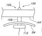
도 1은 팽창가능한 확장 풍선이 장착된 관형 지지부를 갖는 예시적인 확장 장치를 도시하는 측단면도이다.
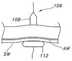
도 2는 장치의 팽창 이전에 위 루멘 벽 및 복벽을 통해 당겨지는 예시적인 확장 장치의 위치를 도시하는 측단면도이다.
도 3a 및 3b는 복벽에 대하여 루멘 벽을 안정화하는 팽창된 확장 풍선 및 팽창된 보유 풍선을 나타내는 예시적인 확장 장치에 대한 측단면도이다. 도 3a에서, 팽창가능한 확장 풍선은 루멘 내에서 보유 부분과 함께 위치되나, 루멘은 복부 벽의 내부에 대하여 맞지 않는다. 도 3b에서, 위 루멘은 복벽의 내부에 대하여 맞도록 당겨졌다.1 is a side cross-sectional view illustrating an exemplary expansion device having a tubular support mounted with an inflatable expansion balloon.
FIG. 2 is a side cross-sectional view showing the location of an exemplary dilation device pulled through the upper lumen wall and the abdominal wall prior to inflation of the device.
3A and 3B are side cross-sectional views of an exemplary dilation device showing inflated and inflatable retaining balloons that stabilize the lumen wall against the abdominal wall. In FIG. 3A, the inflatable expandable balloon is positioned with the retention portion in the lumen, but the lumen does not fit against the interior of the abdominal wall. In FIG. 3B, the gastric lumen was pulled to fit against the interior of the abdominal wall.
본 발명이 속하는 기술 분야에서 통상의 지식을 가진 자가 본 개시 내용을 실시하고 이용할 수 있게 하도록 본 개시 내용의 다양한 요소에 도면 부호가 주어지고 본 개시 내용이 논의되는 도면이 참조된다. 다음의 발명을 실시하기 위한 구체적인 내용은 본 개시 내용의 원리에 대한 예시일 뿐이며, 특허청구범위를 좁게 하는 것으로 검토되지 않는다는 것이 이해되어야 한다. 본 발명이 속하는 기술 분야에서 통상의 지식을 가진 자는 논의된 다양한 실시예에 대한 양태들이 본 개시 내용의 범위 및 기술적 사상으로부터 벗어나지 않으면서 상호 변경되거나 수정될 수 있다는 것을 이해할 것이다.
Reference is made to the various elements of the present disclosure and to the drawings in which the present disclosure is discussed, to enable those skilled in the art to make and use the present disclosure. It is to be understood that the following detailed description is only illustrative of the principles of the present disclosure and not to be construed as narrowing the scope of the claims. Those skilled in the art will appreciate that aspects of the various embodiments discussed can be interchanged or modified without departing from the scope and spirit of the disclosure.
위가 비도관 루멘의 일반적인 예이기 때문에, 본 개시 내용을 설명하기 위하여, "위 루멘" 또는 "위"라는 용어의 사용은, 달리 특정되지 않는다면, 모든 다른 비도관 루멘 또는 공간(예를 들어, 십이지장, 공장(jejunum), 회장(ileum), 복강 등)을 나타낸다.
Because the stomach is a general example of nasal conduit lumens, the use of the terms "stomach lumen" or "stomach" to describe the present disclosure, unless otherwise specified, includes any other nasal conduit lumen or space (eg, Duodenum, jejunum, ileum, abdominal cavity, etc.).
도면을 참조하면, 도 1에서 장치를 관통하는 적어도 하나의 연속 경로(104)를 형성하는 관형 지지부(102)를 포함하는 예시적인 스토마 확장 장치(100)의 측단면도가 도시된다. 연속 경로는 가이드 와이어를 수용하도록 구성된다.
Referring to the drawings, a cross-sectional side view of an exemplary
관형 지지부(102)는 길이, 폭 및 세로축(longitudinal axis(LA))을 가진다. 관형 지지부(102)는 유연하여야 하지만, 압력이 반경 방향으로 또는 축 방향으로 인가될 때 손쉽게 접히거나 꼬일 정도로 너무 유연하지는 않다. 관형 지지부의 폭은 내시경의 작업 채널 내에 피팅할 수 있도록 충분히 작아야만 한다. 예를 들어, 관형 지지부는 대략 0.2 내지 대략 2 mm의 폭을 가질 수 있다. 더욱 바람직하게는, 관형 지지부는 대략 0.5 내지 1.75 mm의 폭을 가질 수 있다. 관형 지지부는 다양한 적합 재료로 이루어질 수 있다. 예시적인 재료는 메사추세츠 윌밍턴의 루브리졸 어드밴스드 머테리얼즈 Inc, Thermedics™ 폴리머 제품으로부터 입수 가능한 TECOFLEX® 의약 등급 지방족 폴리에테르 폴리에탄과 같은 열가소성 폴리우레탄을 포함한다.
The
장치(100)는, 장치의 원위부(110)에서의 적어도 하나의 팽창가능한 확장부(108)와 장치(114)의 근위 단부 상에 위치되는 적어도 하나의 팽창가능한 보유 풍선 요소(112)(즉, 근위 보유 풍선 요소(112))를 갖는, 관형 지지부(102) 상에 위치되는 팽창가능한 확장 풍선(106)을 포함한다. 확장 장치(106)는 확장 풍선을 팽창 및 수축시키기 위한 적어도 하나의 확장 풍선 팽창 루멘(116)을 가진다. 바람직하게는, 팽창 루멘(116)은 관형 지지부(102)에 통합된다. 이러한 점에서, 관형 지지부(102)는 다중 루멘을 형성할 수 있다. 즉, 관형 지지부는 연속 경로(104)와, 확장 풍선(106)을 팽창 및 수축시키기 위한 적어도 하나의 확장 풍선 팽창 루멘(116)을 형성할 수 있다. 팽창 루멘들은 관형 지지부로부터 분리되고 파일럿 튜브 등의 형태를 가질 수 있다는 것이 고려된다.
The
본 개시 내용에 따르면, 근위 보유 풍선 요소(112)는, 도 1에 일반적으로 도시된 바와 같이, 팽창 시 확장부(108)의 가장 큰 단면 직경보다 더 큰, 완전하고 수축되지 않은 팽창 시의 유효 단면을 가지도록 구성된다. 풍선의 확장부(108)는 길이와, 특정 크기의 카테터 튜브 장치를 피팅하도록 완전 팽창 시 미리 정해진 직경을 갖는 원형 단면을 가진다. 이 대신에, 확장부(108)는 다양한 카테터 튜브를 피팅하기 위하여 각각 상이한 팽창 압력을 이용하여 다양한 유효 직경으로 확장될 수 있다. 비한정적인 예로서, 확장부(108)의 유효 팽창 직경은 대략 3 내지 대략 10 mm의 범위를 가질 수 있다. 다른 비한정적인 예로서, 확장부(108)의 유효 팽창 직경은 대략 2 내지 대략 8 mm의 범위를 가질 수 있다. 길이와, 길이에 따른 예를 들어 타원형이나 계란형의 비원형 단면을 갖는 팽창된 확장 풍선이 또한 고려된다.
According to the present disclosure, the proximal
확장 풍선(106)의 근위부(114)(비도관 루멘 내에 위치 설정되는 확장 풍선 부분)는 확장부(108)의 임의의 직경보다 실질적으로 더 큰 단면 또는 직경을 갖는 보유부("근위 보유 풍선 요소"라고도 함)을 포함한다. 일반적으로 말해서, 근위 보유 풍선 요소는 확장부(108)의 직경의 대략 1.5배 내지 대략 3배인 단면 또는 직경을 가질 수 있다. 이 근위 보유 풍선 요소(112)가 팽창되기만 하면, 이는 루멘의 벽을 안정화하고 그리고/또는 비도관 루멘(예를 들어, 위) 내에서 확장 장치의 보유를 제공한다.
The proximal portion 114 (the portion of the expansion balloon positioned within the non-conduit lumen) of the
근위 보유 풍선 요소(112)는 전술한 바와 같이 기능할 수 있는 한 원형 또는 비원형 단면을 가질 수 있다. 보유 풍선은 하나의 대칭축을 갖는 단면을 가질 수 있거나 가지지 않을 수 있다. 예를 들어, 근위 보유 풍선 요소(112)는, 정사각형, 직사각형, 삼각형, 타원형, 계란형 또는 다른 형상을 가질 수 있다. 대신에 그리고/또는 더하여, 근위 보유 풍선 요소(112)는 그 단면에 기여하는 로브(lobe), 핑거 또는 돌출부를 포함할 수 있어, 확장부(108)의 직경보다 더 크다. 바람직하게는, 확장 풍선(106)은 2개의 대향하는 개방 단부를 포함한다. 개방 단부는 관형 지지부에 부착될 수 있다. 확장 풍선(106)은 개방 단부(118, 120)를 가질 수 있다.
The proximal
확장 풍선은 풍선이 유연성, 반유연성 또는 비유연성이 되는 재료로 이루어질 수 있다. 즉, 풍선은 팽창 시 수축 및 팽창할 수 있도록 상대적으로 탄성(예를 들어, 유연성)을 가질 수 있다. 또한, 풍선은 팽창 시 팽창하지만 제한된 신축성을 갖도록 약간의 탄성(예를 들어, 반유연성)을 가질 수 있다. 풍선은 팽창 시 상당한 신축성 없이 팽창하도록 비탄성(예를 들어, 비유연성)을 가질 수 있다. 바람직하게는, 풍선은 루브리졸 어드밴스드 머테리얼즈 Inc, Thermedics™ 폴리머 제품으로부터 입수 가능한, Pellethane® 2363-90A로서 식별되는 폴리우레탄 재료로 이루어질 수 있다.
The inflatable balloon may be made of a material that makes the balloon flexible, semi-flexible or non-flexible. That is, the balloon can be relatively elastic (eg, flexible) so that it can contract and expand upon inflation. In addition, the balloon may have some elasticity (eg, semi-flexibility) to expand upon inflation but have limited elasticity. The balloon may be inelastic (eg, inflexible) to inflate without significant stretch upon inflation. Preferably, the balloon may be made of a polyurethane material identified as Pellethane® 2363-90A, available from Lubrizol Advanced Materials Inc, Thermedics ™ polymer product.
또한, 본 개시 내용은 스토마를 확장시키고 비도관 카테터 튜브를 삽입하기 위한 시스템을 포함하며, 본 시스템은 전술한 바와 같은 스토마 확장 장치를 포함한다. 또한, 본 시스템은 확장된 스토마 관을 통해 그리고 보유부(즉, 근위 보유 풍선 요소)에 의해 안정화된 비도관 루멘의 부분으로 완전히 또는 부분적으로 팽창된 확장 풍선(즉, 확장부(108))에 대하여 피팅하도록 구성된 비도관 카테터 튜브를 포함한다. 본 시스템에 따라, 스토마 확장 장치는 수축되도록 구성되고, 장치의 적어도 일부는 비도관 카테터 튜브를 통해 빠지도록 구성된다.
The present disclosure also includes a system for expanding the stroma and inserting a nasal conduit catheter tube, the system comprising a stroma expanding device as described above. In addition, the system includes an expansion balloon (ie, expansion 108) that has been fully or partially inflated through a portion of the nasal conduit lumen stabilized through an expanded stoma tube and by a retention (ie, proximal retention balloon element). A non-conduit catheter tube configured to fit against. In accordance with the present system, the stoma expansion device is configured to retract and at least a portion of the device is configured to exit through the nasal conduit catheter tube.
장치의 배치에 대한 예시적이고 비한정적인 설명에서, 내시경은 흡입되어 촉진(palpation)이 카테터 튜브 위치 사이트(예를 들어, PEG 위치 사이트)의 정확한 위치를 찾도록 비도관 루멘(예를 들어, 위) 내로 진행될 수 있다. 사이트의 정확한 위치가 찾아지기만 하면, 바늘은 복부를 통해 위 내로 삽입될 수 있고, 가이드 와이어는 바늘을 통해 위 내로 유입될 수 있다.
In an exemplary and non-limiting description of the placement of the device, the endoscope is aspirated so that palpation can locate the exact location of the catheter tube location site (eg PEG location site) (eg, stomach Can be proceeded to). Once the exact location of the site is found, the needle can be inserted into the stomach through the abdomen and the guide wire can enter the stomach through the needle.
표준 내시경 겸자, 내시경 올가미 또는 풍선 부착 고정구는 내시경의 작업 채널을 통해 삽입될 수 있다. 겸자, 올가미 또는 고정구는 가이드 와이어를 잡고, 가이드 와이어는 내시경의 작업 채널을 통해 그리고 환자의 입의 밖으로 당겨진다.
Standard endoscope forceps, endoscope nooses or balloon attachments can be inserted through the endoscope's working channel. A forceps, noose or fixture holds the guide wire, which is pulled through the endoscope's working channel and out of the patient's mouth.
자신의 부착된 팽창 루멘을 갖는 확장 장치는 가이드 와이어의 단부에 고정되어, 가이드 와이어를 이용하여 내시경의 작업 채널을 통해 그리고 위 내로 당겨진다. 확장 장치는 완전 팽창 시의 사전 결정된 부피와 직경을 갖는 원위에 위치된 확장부와, 확장부의 직경보다 더 큰 완전 팽창 시의 직경을 갖는 근위 보유 풍선 요소를 가질 수 있다. 이러한 풍선들이 폴딩되거나 타이트하게 래핑된 상태에 있는 경우에, 확장 장치는 내시경의 작업 채널 내에 피팅되는 직경을 가진다. 일반적으로, 직경은 대략 2 mm 이하의 범위에 있다.
An expansion device with its attached expansion lumen is fixed to the end of the guide wire and is drawn through the working channel of the endoscope and into the stomach using the guide wire. The expansion device may have a distally located extension having a predetermined volume and diameter upon full inflation and a proximal retaining balloon element having a diameter upon full inflation that is greater than the diameter of the extension. When these balloons are in a folded or tightly wrapped state, the expansion device has a diameter that fits into the working channel of the endoscope. Generally, the diameter is in the range of about 2 mm or less.
바늘 관 내에 가이드 와이어를 유지하는 동안, 바늘이 위로부터 제거된다. 확장 장치는, 도 2에 도시된 바와 같이, 복부 조직과 환자 외부에 있는 피부에 도달하도록 바늘 관으로 그리고 부분적으로 바늘관을 통해 당겨진다.
While holding the guide wire in the needle tube, the needle is removed from above. The dilation device is pulled into the needle tube and partially through the needle tube to reach the abdominal tissue and skin outside the patient, as shown in FIG. 2.
이제 도 3a 및 3b를 참조하면, 확장 장치(100)의 확장 풍선(106)은 풍선 내의 압력을 증가시키기 위하여 제어된 양의 유체(예를 들어, 액체 또는 기체)를 점진적으로 유입시켜 팽창되어, 이에 따라 확장부(108)는 바늘 관을 스토마 관으로 부드럽게 그리고 점진적으로 팽창시킨다. 또한, 확장 풍선(106)의 근위 보유 풍선 요소(112)는 확장부(108)가 팽창됨에 따라 팽창된다. 근위 보유 풍선 요소(108)가 확장부(108)보다 더 크게 되고 완전 팽창으로 팽창될 때, 도 3b에 도시된 바와 같이, 이는 위벽(SW(stomach wall))을 복부의 벽(AW(wall of abdomen)"에 대하여 위로 가져 가 이를 안정화시킨다. 본 개시 내용의 일 양태에 따르면, 풍선의 완전하게 팽창된 직경은 삽입될 카테터 튜브 장치(예를 들어, PEG 장치)의 직경에 매칭하는 범위로부터 선택될 수 있다. 도 1, 3a 및 3b에 도시된 바와 같이, 확장 풍선(106)은 확장부(108)가 적어도 하나의 직경(들)을 가질 수 있고, 보유부(112)(근위 보유 풍선 요소)는 확장부(108)보다 더 큰 적어도 하나의 직경을 가질 수 있다.
Referring now to FIGS. 3A and 3B, the
확장 장치가 자신의 고정된 풍선을 완전히 팽창되게 한 후에, 벗김용 시스(peal-away sheath)가 확장 장치의 최원위부 위에 배치된다(즉, 환자의 외부로부터). 확장 장치의 확장 풍선은 벗김용 시스가 확장 장치의 원위 단부 위로 그리고 스토마 관을 통해 위 내로 통과할 수 있게 하도록 단지 소량만큼 수축된다(예를 들어, 부분적으로 수축된다).
After the expansion device has fully inflated its fixed balloon, a peel-away sheath is placed over the distal most of the expansion device (ie, from the outside of the patient). The expansion balloon of the expansion device contracts only a small amount (e.g., partially contracted) to allow the peeling sheath to pass over the distal end of the expansion device and through the storm tube into the stomach.
그 다음, 카테터 튜브(예를 들어, PEG 장치)는 가이드 와이어에 대하여 스레딩되고(threaded), PEG 장치의 원위 단부가 벗김용 시스를 통해 삽입된다. PEG 장치의 원위 단부는 이제 복벽에 대하여 위 루멘을 유지하기 위한 위치에 있어, 확장 풍선은 완전히 수축되어 벗김용 시스를 통해 빼질 수 있다. 복벽을 통해 확장 장치를 빼기 위하여 팽창 루멘으로부터 주사기(syringe) 팽창 커넥터가 제거되어야 한다는 것에 주목하라. 그 다음, 벗김용 시스는 스토마 관으로부터 분리되어 제거된다. 임의의 다른 배치 도구가 제거되고, PEG 장치의 원위의 내재 단부 상의 보유기는 PEG 장치를 제자리에 유지한다.
The catheter tube (eg, PEG device) is then threaded relative to the guide wire and the distal end of the PEG device is inserted through the peeling sheath. The distal end of the PEG device is now in a position to hold the gastric lumen with respect to the abdominal wall so that the dilation balloon can be fully retracted and pulled out through the peeling sheath. Note that the syringe expansion connector must be removed from the expansion lumen in order to withdraw the expansion device through the abdominal wall. The stripping sheath is then separated from the steam tube and removed. Any other placement tool is removed and the retainer on the distal end of the PEG device holds the PEG device in place.
이 대신에, PEG 장치는 벗김용 시스의 사용 없이 수축된 확장 위로 삽입될 수 있다. 이는 복벽에 대하여 위 루멘을 유지하기 위한 위치에 PED 장치를 배치하여, 이에 따라 확장 풍선은 완전히 수축되어 PEG 장치를 통해 빼질 수 있다.
Instead, the PEG device can be inserted over the contracted expansion without the use of a peeling sheath. This places the PED device in a position to hold the stomach lumen with respect to the abdominal wall so that the expansion balloon can be fully retracted and pulled out through the PEG device.
또 다른 대체예에서, 확장 장치는, 자신의 풍선이 완전하게 수축되게 하기만 하면, 그리고 가이드 와이어에 여전히 부착되어 있는 동안, 내시경의 작업 채널을 통해 가이드 와이어를 빼는 것으로 내시경의 작업 채널을 통해 제거될 수 있다. 이는 전술한 방법으로 예를 들어, 확장 장치의 제거 전에 확장 풍선의 부분적인 수축 및 벗김용 시스틀 통한 PEG의 설치로, PEG가 설치되는 것을 필요로 한다.
In another alternative, the expansion device is removed through the endoscope's work channel by simply pulling the guide wire through the endoscope's work channel as long as it allows its balloon to fully retract and remain still attached to the guide wire. Can be. This requires the PEG to be installed in the manner described above, for example with the installation of the PEG through a sheath for partial contraction and peeling of the expansion balloon prior to removal of the expansion device.
본 개시 내용이 소정의 바람직한 실시예들과 관련하여 설명되었지만, 본 발명에 의해 포함되는 내용은 이러한 특정 실시예들에 한정되지 않는다는 것이 이해되어야 한다. 한편, 본 발명의 내용은 이어지는 특허청구범위의 기술적 사상 및 범위 내에서 포함될 수 있는 모든 대체물, 수정물 및 균등물을 포함하는 것으로 의도된다.Although the present disclosure has been described in connection with certain preferred embodiments, it should be understood that the content encompassed by the present invention is not limited to these specific embodiments. On the other hand, the content of the present invention is intended to include all alternatives, modifications and equivalents that may be included within the spirit and scope of the claims that follow.
Claims (11)
Translated fromKorean상기 스토마 확장 장치를 관통하는 연속 경로를 형성하는 관형 지지부;
상기 관형 지지부 상에 위치되고, 상기 스토마 확장 장치의 제1 부분을 형성하는 확장 영역과 상기 스토마 확장 장치의 제2 부분을 형성하는 보유 영역을 포함하는 팽창가능한 확장 풍선; 및
풍선 팽창 루멘
을 포함하고,
상기 보유 영역은, 팽창 시의 상기 확장 영역의 직경보다 더 큰, 완전하고 수축되지 않은 팽창 시의 직경을 가지도록 구성되는,
스토마 확장 장치.
In the story expansion device,
A tubular support forming a continuous path through the storm expanding device;
An inflatable balloon positioned on the tubular support, the inflatable balloon including an expansion region defining a first portion of the storm expansion device and a retention region defining a second portion of the storm expansion device; And
Inflatable inflating lumen
/ RTI >
Wherein the retention zone is configured to have a diameter upon expansion that is complete and unshrinkable that is greater than the diameter of the expansion zone at expansion.
Storm expansion unit.
상기 팽창가능한 풍선은 유연성(compliant) 재료, 반유연성(semi-compliant) 재료, 비유연성(non-compliant) 재료 또는 그 조합으로 이루어진,
스토마 확장 장치.
The method of claim 1,
The inflatable balloon consists of a compliant material, a semi-compliant material, a non-compliant material, or a combination thereof,
Storm expansion unit.
상기 팽창가능한 풍선의 확장 영역 및 보유 영역은 각각 상이한 재료로 이루어진,
스토마 확장 장치.
3. The method of claim 2,
The expansion zone and the retention zone of the inflatable balloon are each made of different materials,
Storm expansion unit.
길이, 폭 및 세로축을 가지며, 상기 스토마 확장 장치를 관통하는 연속 경로를 형성하는 관형 지지부;
상기 관형 지지부 상에서 축 방향으로 배향되고, 상기 스토마 확장 장치의 제1 부분을 형성하는 확장 영역과 상기 스토마 확장 장치의 제2 부분을 형성하는 보유 영역을 포함하는 팽창가능한 확장 풍선; 및
풍선 팽창 루멘
을 포함하고,
상기 보유 영역은, 팽창 시의 상기 확장 영역의 직경보다 더 큰, 완전하고 수축되지 않은 팽창 시의 직경을 가지도록 구성되는,
스토마 확장 장치.
In the story expansion device,
A tubular support having a length, a width, and a longitudinal axis, the tubular support forming a continuous path through the storm expanding device;
An inflatable balloon axially oriented on the tubular support, the inflatable balloon including an expansion region forming a first portion of the storm expansion device and a retention region forming a second portion of the storm expansion device; And
Inflatable inflating lumen
/ RTI >
Wherein the retention zone is configured to have a diameter upon expansion that is complete and unshrinkable that is greater than the diameter of the expansion zone at expansion.
Storm expansion unit.
상기 팽창가능한 풍선은 유연성 재료, 반유연성 재료, 비유연성 재료 또는 그 조합으로 이루어진,
스토마 확장 장치.
5. The method of claim 4,
The inflatable balloon consists of a flexible material, a semi-flexible material, a non-flexible material or a combination thereof,
Storm expansion unit.
상기 팽창가능한 풍선의 확장 영역 및 보유 영역은 각각 상이한 재료로 이루어진,
스토마 확장 장치.
The method of claim 5,
The expansion zone and the retention zone of the inflatable balloon are each made of different materials,
Storm expansion unit.
상기 시스템은,
스토마 확장 장치; 및
비도관 카테터 튜브
를 포함하고,
상기 스토마 확장 장치는,
길이, 폭 및 세로축을 가지며, 상기 스토마 확장 장치를 관통하는 연속 경로를 형성하는 관형 지지부;
상기 관형 지지부 상에서 축 방향으로 배향되고, 상기 스토마 확장 장치의 제1 부분을 형성하는 확장 영역과 상기 스토마 확장 장치의 제2 부분을 형성하는 보유 영역을 포함하는 팽창가능한 확장 풍선; 및
풍선 팽창 루멘
을 포함하고,
상기 보유 영역은, 팽창 시의 상기 확장 영역의 직경보다 더 큰, 완전하고 수축되지 않은 팽창 시의 직경을 가지도록 구성되고,
상기 비도관 카테터 튜브는, 확장된 상기 스토마 관을 통해 그리고 상기 보유 영역에 의해 안정화된 상기 비도관 루멘의 일부 내로 완전히 또는 부분적으로 팽창된 상기 확장 영역에 대하여 피팅되도록 구성되고,
상기 스토마 확장 장치는 수축되도록 구성되고, 상기 스토마 확장 장치의 적어도 일부는 상기 비도관 카테터 튜브를 통해 빼지는,
비도관 카테터 튜브 삽입 시스템.
In a system for expanding a stroma to insert a nasal catheter tube,
The system comprises:
Storm expansion device; And
Nasal Catheter Tube
Lt; / RTI >
The storm expansion device,
A tubular support having a length, a width, and a longitudinal axis, the tubular support forming a continuous path through the storm expanding device;
An inflatable balloon axially oriented on the tubular support, the inflatable balloon including an expansion region forming a first portion of the storm expansion device and a retention region forming a second portion of the storm expansion device; And
Inflatable inflating lumen
/ RTI >
The retention zone is configured to have a diameter upon expansion that is complete and unshrinkable that is larger than the diameter of the expansion zone at expansion;
The non-conduit catheter tube is configured to fit against the expanded region fully or partially expanded through the expanded stoma tube and into a portion of the non-conduit lumen stabilized by the retention region,
The stroma expanding device is configured to retract and at least a portion of the stroma expanding device is drawn through the nasal conduit catheter tube,
Nasal catheter tube insertion system.
상기 팽창가능한 풍선은 유연성 재료, 반유연성 재료, 비유연성 재료 또는 그 조합으로 이루어진,
비도관 카테터 튜브 삽입 시스템.
The method of claim 7, wherein
The inflatable balloon consists of a flexible material, a semi-flexible material, a non-flexible material or a combination thereof,
Nasal catheter tube insertion system.
상기 팽창가능한 풍선의 확장 영역 및 보유 영역은 각각 상이한 재료로 이루어진,
비도관 카테터 튜브 삽입 시스템.
9. The method of claim 8,
The expansion zone and the retention zone of the inflatable balloon are each made of different materials,
Nasal catheter tube insertion system.
상기 비도관 루멘을 통해 위 내로 바늘을 삽입하고, 상기 바늘을 통해 가이드 와이어를 유입시키는 단계;
표준 내시경 겸자, 내시경 올가미 또는 풍선 부착 고정구를 상기 내시경의 작업 채널을 통해 삽입하는 단계;
상기 가이드 와이어를 잡고, 상기 가이드 와이어를 상기 내시경을 통해 환자의 입 밖으로 당기는 단계;
부착된 팽창 루멘을 갖는 확장 장치를 상기 가이드 와이어의 단부에 고정하고, 상기 가이드 와이어를 이용하여 상기 내시경을 통해 위 내로 상기 확장 장치를 당기는 단계;
상기 가이드 와이어를 바늘 관 내에 유지하면서, 상기 루멘으로부터 상기 바늘을 제거하는 단계;
상기 확장 장치를 바늘 관 내로 그리고 상기 바늘관을 부분적으로 관통하여 당기는 단계;
상기 풍선 내의 압력을 증가시키기 위하여 제어된 양의 유체를 점진적으로 유입함으로써 상기 확장 장치의 풍선을 팽창시켜, 상기 루멘의 벽을 복벽에 대하여 제공하는 동안 상기 풍선이 상기 바늘 관을 스토마 관 내로 점진적으로 팽창시키는 단계;
상기 확장 장치 위로 벗김용 시스를 배치하고, 상기 벗김용 시스가 상기 확장 장치의 원위 단부 위로 그리고 상기 스토마 관을 통해 위 내로 통과하게 할 수 있도록 단지 소량만큼 상기 풍선을 수축시키는 단계;
PEG 장치를 가이드 와이어에 대하여 스레딩하고, 상기 벗김용 시스를 통해 삽입된 상기 PEG 장치를 삽입하는 단계;
상기 풍선을 완전히 수축시켜, 상기 벗김용 시스를 통해 상기 풍선을 빼는 단계; 및
상기 벗김용 시스를 분리하여 제거하는 단계
를 포함하는,
PEG 장치 배치 방법.
Advancing the endoscope into the nasal conduit lumen so that palpation locates the PEG tube location site;
Inserting a needle into the stomach through the nasal conduit lumen and introducing a guide wire through the needle;
Inserting a standard endoscope forceps, endoscope noose or balloon attachment fixture through the endoscope's working channel;
Holding the guide wire and pulling the guide wire out of the mouth of the patient through the endoscope;
Securing an expansion device having an attached expansion lumen to the end of the guide wire, and pulling the expansion device into the stomach through the endoscope using the guide wire;
Removing the needle from the lumen while maintaining the guide wire in the needle tube;
Pulling the expansion device partially into the needle tube and through the needle tube;
The balloon of the expansion device is inflated by gradually introducing a controlled amount of fluid to increase the pressure in the balloon, the balloon progressively introducing the needle tube into the stroma tube while providing the wall of the lumen against the abdominal wall. Inflating with;
Placing a peeling sheath over the expansion device and contracting the balloon by only a small amount such that the peeling sheath can pass over the distal end of the expansion device and through the storm tube into the stomach;
Threading a PEG device against a guide wire and inserting the PEG device inserted through the peeling sheath;
Completely deflating the balloon to remove the balloon through the peeling sheath; And
Separating and removing the peeling sheath
/ RTI >
PEG Device Placement Method.
상기 확장 장치는,
길이, 폭 및 세로축을 가지며, 상기 확장 장치를 관통하는 연속 경로를 형성하는 관형 지지부;
상기 관형 지지부 상에서 축 방향으로 배향되고, 상기 확장 장치의 제1 부분을 형성하는 확장 영역과 상기 확장 장치의 제2 부분을 형성하는 보유 영역을 포함하는 팽창가능한 확장 풍선; 및
풍선 팽창 루멘
을 포함하고,
상기 보유 영역은, 팽창 시의 상기 확장 영역의 직경보다 더 큰, 완전하고 수축되지 않은 팽창 시의 직경을 가지도록 구성되는,
PEG 장치 배치 방법.The method of claim 10,
The expansion device,
A tubular support having a length, a width, and a longitudinal axis, the tubular support forming a continuous path through the expansion device;
An inflatable balloon axially oriented on the tubular support, the inflatable balloon including an expansion region forming a first portion of the expansion device and a retention region forming a second portion of the expansion device; And
Inflatable inflating lumen
/ RTI >
Wherein the retention zone is configured to have a diameter upon expansion that is complete and unshrinkable that is greater than the diameter of the expansion zone at expansion.
PEG Device Placement Method.
Applications Claiming Priority (7)
| Application Number | Priority Date | Filing Date | Title |
|---|---|---|---|
| US38679310P | 2010-09-27 | 2010-09-27 | |
| US61/386,793 | 2010-09-27 | ||
| US201161446229P | 2011-02-24 | 2011-02-24 | |
| US61/446,229 | 2011-02-24 | ||
| US13/245,577US20120078039A1 (en) | 2010-09-27 | 2011-09-26 | Dilation Device for Placing Catheter Tubes |
| US13/245,577 | 2011-09-26 | ||
| PCT/IB2011/054254WO2012042476A1 (en) | 2010-09-27 | 2011-09-27 | Dilation device for placing catheter tubes |
Publications (1)
| Publication Number | Publication Date |
|---|---|
| KR20130138204Atrue KR20130138204A (en) | 2013-12-18 |
Family
ID=45871306
Family Applications (4)
| Application Number | Title | Priority Date | Filing Date |
|---|---|---|---|
| KR1020137007495AExpired - Fee RelatedKR101908933B1 (en) | 2010-09-27 | 2011-09-27 | Configurable percutaneous endoscopic gastrostomy tube |
| KR1020137006363AExpired - Fee RelatedKR101841949B1 (en) | 2010-09-27 | 2011-09-27 | Multi-balloon dilation device for placing catheter tubes |
| KR1020137006482ACeasedKR20130138204A (en) | 2010-09-27 | 2011-09-27 | Expansion device for placing catheter tubes |
| KR1020137007643AExpired - Fee RelatedKR101835381B1 (en) | 2010-09-27 | 2011-09-27 | Stoma length indicator assembly and positioning system |
Family Applications Before (2)
| Application Number | Title | Priority Date | Filing Date |
|---|---|---|---|
| KR1020137007495AExpired - Fee RelatedKR101908933B1 (en) | 2010-09-27 | 2011-09-27 | Configurable percutaneous endoscopic gastrostomy tube |
| KR1020137006363AExpired - Fee RelatedKR101841949B1 (en) | 2010-09-27 | 2011-09-27 | Multi-balloon dilation device for placing catheter tubes |
Family Applications After (1)
| Application Number | Title | Priority Date | Filing Date |
|---|---|---|---|
| KR1020137007643AExpired - Fee RelatedKR101835381B1 (en) | 2010-09-27 | 2011-09-27 | Stoma length indicator assembly and positioning system |
Country Status (11)
| Country | Link |
|---|---|
| US (5) | US9125800B2 (en) |
| EP (4) | EP2621378B1 (en) |
| JP (5) | JP5830103B2 (en) |
| KR (4) | KR101908933B1 (en) |
| CN (4) | CN103124533B (en) |
| AU (5) | AU2011309685B2 (en) |
| BR (4) | BR112013007168A2 (en) |
| CA (4) | CA2811308C (en) |
| MX (5) | MX2013003336A (en) |
| RU (5) | RU2604042C2 (en) |
| WO (4) | WO2012042473A1 (en) |
Cited By (2)
| Publication number | Priority date | Publication date | Assignee | Title |
|---|---|---|---|---|
| KR20230027712A (en) | 2021-08-20 | 2023-02-28 | 주식회사 파인메딕스 | Gastrostomy Tube Kit |
| WO2025170282A1 (en)* | 2024-02-05 | 2025-08-14 | 부산대학교 산학협력단 | Hemostatic endoscopic balloon device capable of ensuring front field of view of bronchoscope and suctioning blood when hemostatic balloon is inflated |
Families Citing this family (74)
| Publication number | Priority date | Publication date | Assignee | Title |
|---|---|---|---|---|
| MX350734B (en) | 2010-09-08 | 2017-09-15 | Covidien Lp | Catheter with imaging assembly. |
| US9125800B2 (en) | 2010-09-27 | 2015-09-08 | Avent, Inc. | Stoma length indicator assembly and positioning system |
| EP2744445B1 (en)* | 2011-08-20 | 2018-01-31 | Advanced Medical Balloons GmbH | Trans-anal inflow catheter for intermittently triggering a reflex-coordinated defecation |
| US20140066966A1 (en)* | 2012-08-30 | 2014-03-06 | Children's National Medical Center | Endopyloric tool and method to treat hypertropic pyloric stenosis |
| USD735343S1 (en) | 2012-09-07 | 2015-07-28 | Covidien Lp | Console |
| US9517184B2 (en) | 2012-09-07 | 2016-12-13 | Covidien Lp | Feeding tube with insufflation device and related methods therefor |
| USD717340S1 (en) | 2012-09-07 | 2014-11-11 | Covidien Lp | Display screen with enteral feeding icon |
| US9198835B2 (en) | 2012-09-07 | 2015-12-01 | Covidien Lp | Catheter with imaging assembly with placement aid and related methods therefor |
| USD716841S1 (en) | 2012-09-07 | 2014-11-04 | Covidien Lp | Display screen with annotate file icon |
| US9108024B2 (en)* | 2012-09-28 | 2015-08-18 | Avent, Inc. | Retention component for placement of enteral feeding tubes |
| US9492644B2 (en)* | 2012-12-21 | 2016-11-15 | Avent, Inc. | Dilation device for placing catheter tubes |
| US9522253B2 (en)* | 2013-03-13 | 2016-12-20 | Vascular Solutions, Inc. | Drainage or feeding catheter assembly |
| US9833350B2 (en) | 2013-03-15 | 2017-12-05 | Ez-Off Weightloss, Llc | Anchorable size-varying gastric balloons for weight loss |
| EP2967818B1 (en) | 2013-03-15 | 2018-05-16 | Ez Off Weightloss, LLC | System for gastric restriction and malabsorption |
| CN103263280A (en)* | 2013-06-04 | 2013-08-28 | 赵远思 | Intestine stoma fixing device |
| EP3030307B1 (en) | 2013-08-05 | 2019-12-11 | Endo-Tagss, LLC | Transabdominal gastric surgery system |
| US10219799B2 (en) | 2013-08-05 | 2019-03-05 | Endo-Tagss, Llc | Transabdominal gastric device and method |
| CN103405297A (en)* | 2013-08-27 | 2013-11-27 | 林建江 | Complete flow turning type intestinal fistulization tube |
| BR112016011279B1 (en)* | 2013-11-18 | 2022-09-06 | Halkey-Roberts Corporation | MEDICAL CONNECTOR ASSEMBLY |
| MX369672B (en) | 2013-12-17 | 2019-11-15 | Standard Bariatrics Inc | Resection line guide for a medical procedure and method of using same. |
| AU2015241193B2 (en) | 2014-03-29 | 2020-01-02 | Standard Bariatrics, Inc. | End effectors surgical stapling devices, and methods of using same |
| WO2015153324A1 (en) | 2014-03-29 | 2015-10-08 | Standard Bariatrics, Inc. | End effectors, surgical stapling devices, and methods of using same |
| WO2016037158A1 (en) | 2014-09-05 | 2016-03-10 | Standard Bariatrics, Inc. | Sleeve gastrectomy calibration tube and method of using same |
| US10206595B2 (en)* | 2014-11-17 | 2019-02-19 | 3VO Medical, Inc. | Intrauterine balloon apparatus, system, and method for augmenting uterine birthing forces during parturition |
| CN104606767B (en)* | 2014-11-26 | 2017-12-19 | 潘湘斌 | Foley's tube for ultrasound-guided percutaneous pulmonary valve balloon dilatation |
| WO2016097824A1 (en)* | 2014-12-18 | 2016-06-23 | Evoluzione S.R.L. | Medical device for performing ileostomies and/or jejunostomies |
| US10080874B2 (en) | 2015-04-09 | 2018-09-25 | Boston Scientific Scimed, Inc. | Trap balloon catheter with trap balloon retainer |
| CN107980007B (en)* | 2015-04-09 | 2020-12-11 | 波士顿科学国际有限公司 | Capture balloon catheter with capture balloon retainer |
| US9808282B2 (en)* | 2015-06-04 | 2017-11-07 | Medos International Sarl | Surgical cannula system and method of use |
| US10285837B1 (en) | 2015-09-16 | 2019-05-14 | Standard Bariatrics, Inc. | Systems and methods for measuring volume of potential sleeve in a sleeve gastrectomy |
| KR101725235B1 (en)* | 2015-12-01 | 2017-04-11 | 충남대학교산학협력단 | Surgical Trocar |
| CN106182730B (en)* | 2016-07-28 | 2018-10-16 | 七星电气股份有限公司 | A kind of expansion mold for cool condensing electric cable accessories |
| WO2018034658A1 (en)* | 2016-08-17 | 2018-02-22 | Avent, Inc. | Enteral feeding satiation device |
| WO2018067690A1 (en) | 2016-10-04 | 2018-04-12 | Ez-Off Weight Loss, Llc | Sleeve-anchorable gastric balloon for weight loss |
| CN109843152B (en)* | 2016-10-14 | 2022-02-25 | M·D·诺亚 | Balloon structure with anchoring portion for anchoring in body passage |
| WO2018150219A1 (en)* | 2017-02-16 | 2018-08-23 | N.V. Nutricia | Gastrostomy device with an improved retaining element |
| CN110678159A (en)* | 2017-02-16 | 2020-01-10 | 纽崔西亚公司 | Gastrostomy device with pressure monitoring |
| JP6995869B2 (en)* | 2017-02-23 | 2022-01-17 | ボストン サイエンティフィック サイムド,インコーポレイテッド | Mounting equipment used with medical equipment |
| CN107174315B (en)* | 2017-05-04 | 2019-10-11 | 温州市人民医院 | Peritoneo-puncture needle fixes device |
| CN107348976B (en)* | 2017-05-19 | 2019-11-19 | 薛运章 | A kind of mesenterium support device and method for supporting |
| US11338112B2 (en) | 2017-07-03 | 2022-05-24 | Cathaid, Inc. | Devices for monitoring movement of a secured catheter during a procedure |
| US10912562B2 (en) | 2017-08-14 | 2021-02-09 | Standard Bariatrics, Inc. | End effectors, surgical stapling devices, and methods of using same |
| RU2691924C1 (en)* | 2017-12-25 | 2019-06-18 | Арчил Зурабович Цулая | Method for gastrostomy using polypropylene mesh |
| KR101984878B1 (en) | 2018-01-10 | 2019-05-31 | 강석진 | Dry edible Materials pulverization machine |
| CN112930208A (en) | 2018-06-01 | 2021-06-08 | 恩多Rx有限责任公司 | Dilation device and method of use thereof |
| EP3856308B1 (en)* | 2018-09-27 | 2025-08-13 | Coloplast A/S | Tracheostoma device holder |
| US11412260B2 (en)* | 2018-10-29 | 2022-08-09 | Google Llc | Geometric transforms for image compression |
| CN114126516A (en)* | 2018-11-30 | 2022-03-01 | 快管医疗有限责任公司 | Method and device for the treatment of tension pneumothorax using a rapidly deployable thoracic port |
| CN109700525A (en)* | 2018-12-28 | 2019-05-03 | 先健科技(深圳)有限公司 | Stoma instrument |
| US11666696B2 (en) | 2019-03-25 | 2023-06-06 | Ellen McGrath | Enterostomy drainage methods and devices |
| USD896365S1 (en)* | 2019-06-24 | 2020-09-15 | Mark Sipe | Medical port disc |
| CN211856471U (en) | 2019-08-22 | 2020-11-03 | 贝克顿·迪金森公司 | Quantitative testing system for echogenicity of echogenic medical instrument |
| KR102049701B1 (en)* | 2019-08-22 | 2020-01-08 | 이지희 | A liquid medicine injection machine Of Balloon type |
| CN211884905U (en) | 2019-08-22 | 2020-11-10 | 贝克顿·迪金森公司 | Balloon dilatation catheter and balloon thereof |
| CN112401971B (en) | 2019-08-23 | 2025-09-09 | 贝克顿·迪金森公司 | Stone extraction for percutaneous nephroscope surgical design kit |
| CN110801312A (en)* | 2019-10-21 | 2020-02-18 | 复旦大学附属中山医院 | Intervene valve release stop device |
| BR112022008009A2 (en) | 2019-11-04 | 2022-07-12 | Standard Bariatrics Inc | SYSTEMS AND METHODS OF PERFORMING SURGERY USING LAPLACE'S LAW TENSION RETRACTION DURING SURGERY |
| US12274635B2 (en) | 2019-11-04 | 2025-04-15 | Standard Bariatrics, Inc. | Systems and methods of performing surgery using laplace's law tension retraction during surgery |
| CN111375121B (en)* | 2020-03-18 | 2022-05-17 | 南京鼓楼医院 | Nerve block sleeve assembly |
| JP2021154089A (en)* | 2020-03-30 | 2021-10-07 | テルモ株式会社 | Transfistula tube device |
| CN111450392B (en)* | 2020-05-11 | 2025-01-14 | 上海市东方医院(同济大学附属东方医院) | Multi-level fistula dilatation puncture drainage tube with balloon |
| CN115955943A (en) | 2020-06-30 | 2023-04-11 | 标准肥胖病研究公司 | Systems, devices, and methods for preventing or reducing insufflation loss during laparoscopic procedures |
| CN111840754A (en)* | 2020-08-17 | 2020-10-30 | 安卓医疗技术(山东)有限公司 | An internal fixation device for a pipe and a method of using the same |
| CN112245772B (en)* | 2020-10-19 | 2022-05-06 | 四川大学华西医院 | Monitoring regulation and control device of adjustable two sacs three chambeies intraductal air pressure |
| AU2022242751B2 (en) | 2021-03-23 | 2024-05-02 | Standard Bariatrics, Inc. | Systems and methods for preventing tissue migration in surgical staplers |
| CN113244502B (en)* | 2021-04-20 | 2025-04-29 | 青岛博泰医疗器械有限责任公司 | Visualized pressure regulating catheter |
| CN113576578B (en)* | 2021-07-29 | 2022-09-13 | 宿州微腾企业管理咨询服务有限公司 | Intracardiac branch of academic or vocational study hemostasis constriction device |
| CN113941074B (en)* | 2021-11-12 | 2024-05-24 | 北京大学深圳医院 | Pharyngeal expansion device for gastroscopy of elderly patients |
| CN114099912B (en)* | 2021-11-18 | 2024-03-22 | 南京脉创医疗科技有限公司 | Intracranial balloon dilation catheter |
| TWI773597B (en)* | 2021-11-25 | 2022-08-01 | 長庚學校財團法人長庚科技大學 | Guided operation and detection device for gastrostomy care |
| CN114082086B (en)* | 2021-12-23 | 2024-03-29 | 赛诺神畅医疗科技有限公司 | Balloon guiding catheter |
| CN114159009A (en)* | 2021-12-31 | 2022-03-11 | 上海博方医疗科技有限公司 | Capsule endoscope system and operation method |
| CN115814244B (en)* | 2022-12-30 | 2025-09-23 | 中国人民解放军总医院第八医学中心 | Coronary artery delivery catheter and delivery device for cardiac interventional therapy |
| CN116899036A (en)* | 2023-08-08 | 2023-10-20 | 首都医科大学附属北京朝阳医院 | Drainage device |
Family Cites Families (125)
| Publication number | Priority date | Publication date | Assignee | Title |
|---|---|---|---|---|
| US3397699A (en) | 1966-05-05 | 1968-08-20 | Gerald C. Kohl | Retaining catheter having resiliently biased wing flanges |
| US3633579A (en) | 1967-05-24 | 1972-01-11 | Sherwood Medical Ind Inc | Catheter placement device and method |
| US3915171A (en) | 1974-06-06 | 1975-10-28 | Dennis William Shermeta | Gastrostomy tube |
| US4315513A (en) | 1980-03-10 | 1982-02-16 | Nawash Michael S | Gastrostomy and other percutaneous transport tubes |
| US4393873A (en) | 1980-03-10 | 1983-07-19 | Nawash Michael S | Gastrostomy and other percutaneous transport tubes |
| US4531943A (en) | 1983-08-08 | 1985-07-30 | Angiomedics Corporation | Catheter with soft deformable tip |
| US4627838A (en) | 1983-12-09 | 1986-12-09 | Bard Limited | Stylet actuated winged catheter |
| US4758219A (en) | 1985-05-17 | 1988-07-19 | Microvasive, Inc. | Enteral feeding device |
| US4763654A (en)* | 1986-09-10 | 1988-08-16 | Jang G David | Tandem independently inflatable/deflatable multiple diameter balloon angioplasty catheter systems and method of use |
| US4850953A (en) | 1987-07-27 | 1989-07-25 | Habley Medical Technology Corporation | Gastrostomy valve |
| US4861334A (en) | 1988-06-24 | 1989-08-29 | Nawaz Arain | Self-retaining gastrostomy tube |
| US4944732A (en) | 1988-08-15 | 1990-07-31 | Sandoz Nutrition Corporation | Gastrostomy feeding port |
| JPH0249547U (en)* | 1988-09-30 | 1990-04-06 | ||
| US4972845A (en) | 1989-01-05 | 1990-11-27 | Abbott Laboratories | Stoma measuring device |
| US5073166A (en) | 1989-02-15 | 1991-12-17 | Medical Innovations Corporation | Method and apparatus for emplacement of a gastrostomy catheter |
| US5374254A (en) | 1990-11-29 | 1994-12-20 | Buma; Shelley J. | Catheters with adjustable external locking bolsters |
| US5092850A (en) | 1990-11-29 | 1992-03-03 | Buma Shelley J | Catheter with adjustable external locking bolster |
| US5112310A (en) | 1991-02-06 | 1992-05-12 | Grobe James L | Apparatus and methods for percutaneous endoscopic gastrostomy |
| DE69228257T2 (en) | 1991-11-06 | 1999-07-08 | Inbae M.D. Phoenix Yoon, Md. | HOLDER FOR SURGICAL INSTRUMENTS |
| US5356391A (en) | 1992-06-22 | 1994-10-18 | Medical Innovations Corp. | Flexible retainer flange for gastrostomy tube and the method of installing it |
| US5484420A (en) | 1992-07-09 | 1996-01-16 | Wilson-Cook Medical Inc. | Retention bolsters for percutaneous catheters |
| US5248302A (en) | 1992-08-05 | 1993-09-28 | Biosearch Medical Products Inc. | Percutaneous obturatable internal anchoring device |
| US5702365A (en)* | 1992-09-08 | 1997-12-30 | King; Toby St. John | Daul-lumen catheter |
| EP0683684B1 (en) | 1993-01-07 | 2001-08-08 | Medical Innovations Corporation | Gastrostomy catheter system |
| US5413565A (en) | 1993-01-15 | 1995-05-09 | Sandoz Nutrition Ltd. | Gastrostomy feeding port with elastic adjustable tip |
| US5336203A (en) | 1993-05-28 | 1994-08-09 | Abbott Laboratories | Low profile gastrostomy device with dome |
| US5505698A (en)* | 1993-10-29 | 1996-04-09 | Medtronic, Inc. | Cardioplegia catheter with elongated cuff |
| US5429598A (en) | 1994-04-19 | 1995-07-04 | Applied Medical Resources Corporation | Surgical access device and procedure |
| US5860952A (en) | 1996-01-11 | 1999-01-19 | C. R. Bard, Inc. | Corporeal access tube assembly and method |
| US6036673A (en)* | 1996-01-11 | 2000-03-14 | C. R. Bard, Inc. | Bolster for corporeal access tube assembly |
| DE69732810T2 (en)* | 1996-01-11 | 2006-04-06 | C.R. Bard, Inc. | Support pad for body access tube |
| US6019746A (en) | 1996-05-17 | 2000-02-01 | Applied Medical Technology, Inc. | Low profile balloon feeding device |
| DE19634116C2 (en) | 1996-08-23 | 1998-08-20 | Fresenius Ag | Catheter for percutaneous enteral nutrition |
| US6293924B1 (en)* | 1996-12-12 | 2001-09-25 | Advanced Cardiovascular Systems, Inc. | Balloon assembly with separately inflatable sections |
| US6494848B1 (en) | 1996-12-19 | 2002-12-17 | St. Jude Medical Puerto Rico B.V. | Measuring device for use with a hemostatic puncture closure device |
| NL1005068C2 (en) | 1997-01-23 | 1998-07-27 | Ct Rrn Academisch Ziekenhuis U | Catheter system and a catheter forming part thereof. |
| SE9700373L (en)* | 1997-02-04 | 1998-07-13 | Stig Bengmark | Probe for providing fluid communication with the small intestine |
| US5928260A (en) | 1997-07-10 | 1999-07-27 | Scimed Life Systems, Inc. | Removable occlusion system for aneurysm neck |
| US6077250A (en) | 1997-10-01 | 2000-06-20 | Boston Scientific Corporation | Apparatus and method for percutaneously placing gastrostomy tubes |
| US6186985B1 (en) | 1997-10-03 | 2001-02-13 | Boston Scientific Corporation | Gastro-intestinal tube with dissolvable support bolster |
| US6464686B1 (en)* | 1998-01-21 | 2002-10-15 | Abbott Laboratories | Polyurethane feeding tube and associated adaptors |
| US6364858B1 (en) | 1998-03-31 | 2002-04-02 | Applied Medical Research, Inc. | Collapsible internal bolster for gastrostomy device |
| US6039714A (en) | 1998-05-12 | 2000-03-21 | Novartis Nutrition Ag | Collapsible retention bolster for gastrostomy and other ostomy tubes |
| US6527748B1 (en) | 1998-08-17 | 2003-03-04 | Yutaka Suzuki | Method of gastrostomy, and an infection preventive cover, kit or catheter kit, and a gastrostomy catheter kit |
| EP1674125B1 (en)* | 1998-08-17 | 2009-04-29 | Yutaka Suzuki | A method of gastrostomy and an infection preventive cover and a gastrostomy catheter kit |
| US6030406A (en) | 1998-10-05 | 2000-02-29 | Origin Medsystems, Inc. | Method and apparatus for tissue dissection |
| US6030361A (en) | 1999-01-25 | 2000-02-29 | Miyashiro; Augusto M. | Gastrostomy apparatus |
| US6322538B1 (en) | 1999-02-18 | 2001-11-27 | Scimed Life Systems, Inc. | Gastro-intestinal tube placement device |
| US6231547B1 (en) | 1999-02-18 | 2001-05-15 | Abbott Laboratories | External retaining device for a catheter and catheter assembly and method using same |
| US20050085771A1 (en)* | 1999-04-16 | 2005-04-21 | Lyon Thomas R. | Clear view cannula |
| US6231549B1 (en) | 1999-08-17 | 2001-05-15 | Sherwood Services, Ag | Shim device for enteral feeding system |
| US6881420B2 (en) | 2000-06-23 | 2005-04-19 | Teva Pharmaceutical Industries Ltd. | Compositions and dosage forms for gastric delivery of irinotecan and methods of treatment that use it to inhibit cancer cell proliferation |
| ITBO20000511A1 (en) | 2000-09-05 | 2002-03-05 | Gianmario Monza | APPARATUS AND METHOD FOR THE CONSTRUCTION OF GASTROSTOMY |
| US6692458B2 (en)* | 2000-12-19 | 2004-02-17 | Edwards Lifesciences Corporation | Intra-pericardial drug delivery device with multiple balloons and method for angiogenesis |
| US6743207B2 (en)* | 2001-04-19 | 2004-06-01 | Scimed Life Systems, Inc. | Apparatus and method for the insertion of a medical device |
| WO2002103409A2 (en) | 2001-06-19 | 2002-12-27 | The Trustees Of The University Of Pennsylvania | Optical guidance system for invasive catheter placement |
| DE10139644B4 (en) | 2001-08-11 | 2004-04-01 | Fresenius Kabi Deutschland Gmbh | Mounting part for an adapter of a PEG probe and adapter for a PEG probe with such a mounting part |
| US7736336B2 (en) | 2001-09-13 | 2010-06-15 | Allegiance Corporation | Paracentesis device having multiple detachable components |
| US7137993B2 (en)* | 2001-12-03 | 2006-11-21 | Xtent, Inc. | Apparatus and methods for delivery of multiple distributed stents |
| US6896665B2 (en) | 2001-12-10 | 2005-05-24 | Applied Medical Research | Gastrostomy device package and method of assembly |
| US20030163114A1 (en) | 2002-02-26 | 2003-08-28 | Gershowitz Arthur D. | Retrograde cannula having manually retractable sealing member |
| JP4351458B2 (en) | 2002-03-18 | 2009-10-28 | オリンパス株式会社 | Endoscope insertion system |
| US6666853B2 (en) | 2002-03-27 | 2003-12-23 | Scimed Life Systems, Inc. | Low profile adaptor for use with a medical catheter |
| US7083595B2 (en) | 2002-05-01 | 2006-08-01 | Scimed Lipe Systems, Inc. | Medical catheter assembly and method of using the same |
| JP2004099584A (en) | 2002-05-02 | 2004-04-02 | Keio Gijuku | Anti-tumor agent using HSV |
| US6764453B2 (en) | 2002-05-08 | 2004-07-20 | Sherwood Services Ag | Stoma measuring device |
| US6878130B2 (en)* | 2002-05-28 | 2005-04-12 | Sherwood Services Ag | External inflation indicator for a low profile gastrostomy tube |
| US20030225392A1 (en) | 2002-05-31 | 2003-12-04 | Kimberly-Clark Worldwide, Inc. | Low profile transpyloric jejunostomy system and method to enable |
| AU2003258124A1 (en) | 2002-08-05 | 2004-02-23 | Miravant Medical Technologies | Light delivery catheter |
| US6942641B2 (en) | 2003-05-30 | 2005-09-13 | J. Michael Seddon | Catheter |
| WO2004112877A1 (en) | 2003-06-20 | 2004-12-29 | Ranier Limited | A medical device |
| WO2004112879A1 (en)* | 2003-06-20 | 2004-12-29 | Coloplast A/S | A medical device comprising a braided portion |
| US7220252B2 (en)* | 2003-07-18 | 2007-05-22 | Polyzen, Inc. | Inflatable dual balloon catheter |
| US20070203445A1 (en)* | 2004-02-26 | 2007-08-30 | V-Kardia Pty Ltd | Isolating cardiac circulation |
| US7462175B2 (en)* | 2004-04-21 | 2008-12-09 | Acclarent, Inc. | Devices, systems and methods for treating disorders of the ear, nose and throat |
| JP4588356B2 (en)* | 2004-04-30 | 2010-12-01 | 日本シャーウッド株式会社 | Gastrostomy tube extender |
| JP4623548B2 (en)* | 2004-04-30 | 2011-02-02 | 日本シャーウッド株式会社 | Gastrostomy tube extender |
| US7654980B2 (en) | 2004-05-14 | 2010-02-02 | Boston Scientific Scimed, Inc. | Method for percutaneously implanting a medical catheter and medical catheter implanting assembly |
| US20060030818A1 (en) | 2004-08-09 | 2006-02-09 | Mcvey Robert D | System and method for securing a medical access device |
| US7582072B2 (en) | 2004-09-09 | 2009-09-01 | Kimberly-Clark Worldwide, Inc. | Artificial stoma and method of use |
| US20080039715A1 (en) | 2004-11-04 | 2008-02-14 | Wilson David F | Three-dimensional optical guidance for catheter placement |
| JP4721329B2 (en) | 2005-04-21 | 2011-07-13 | 日本シャーウッド株式会社 | Indwelling device |
| US20060270989A1 (en)* | 2005-05-27 | 2006-11-30 | Mcmichael Donald J | Gastric fastening system |
| US7691089B2 (en)* | 2005-06-21 | 2010-04-06 | Tyco Healthcare Group Lp | Adjustable trocar washer |
| GB2428198A (en) | 2005-07-11 | 2007-01-24 | Stavros Michael Stivaros | A self-retaining surgical tube |
| US8954134B2 (en) | 2005-09-13 | 2015-02-10 | Children's Medical Center Corporation | Light-guided transluminal catheter |
| US8709018B2 (en) | 2005-09-16 | 2014-04-29 | Applied Medical Technology, Inc. | Non-balloon low profile feed device with insertion/removal tool |
| JP2007167082A (en)* | 2005-12-19 | 2007-07-05 | Michiaki Kudo | Guide instrument of in-vivo insertion tube via gastric fistula and instrument kit for changing percutaneous gastrostoma-tube to jejunum tube via gastric fistula |
| US7740609B2 (en)* | 2006-03-03 | 2010-06-22 | Boston Scientific Scimed, Inc. | Balloon catheter |
| US7771396B2 (en) | 2006-03-22 | 2010-08-10 | Ethicon Endo-Surgery, Inc. | Intubation device for enteral feeding |
| US20070255222A1 (en) | 2006-03-27 | 2007-11-01 | Changqing Li | Catheter assembly including internal bolster |
| US20070239171A1 (en) | 2006-03-30 | 2007-10-11 | Ethicon Endo-Surgery, Inc. | Medical snaring device |
| US20070233005A1 (en)* | 2006-04-03 | 2007-10-04 | Mcmichael Donald J | Surgical fastening tool |
| US8551043B2 (en)* | 2006-04-21 | 2013-10-08 | C. R. Bard, Inc. | Feeding device and bolster apparatus and method for making the same |
| JP2009542349A (en) | 2006-07-05 | 2009-12-03 | アスピレーション・メディカル・テクノロジー・エルエルシー | Short-circuit device to treat obesity by extracting food |
| FR2904531B1 (en) | 2006-08-02 | 2009-06-12 | Eleph Ent Technology | PERCUTANEOUS GASTROSTOMY PROBE |
| US8062285B2 (en) | 2006-08-03 | 2011-11-22 | Aspire Bariatrics, Llc | Systems and methods for removing ingested material from a stomach |
| US7547303B2 (en) | 2006-08-03 | 2009-06-16 | Boston Scientific Scimed, Inc. | Catheter assembly including foldable internal bolster |
| WO2008027375A2 (en) | 2006-08-31 | 2008-03-06 | Cook Incorporated | Rotationally actuated fixation mechanism |
| EP2080242A4 (en)* | 2006-09-25 | 2013-10-30 | Valentx Inc | Toposcopic access and delivery devices |
| JP2008099917A (en) | 2006-10-19 | 2008-05-01 | Goodman Co Ltd | Catheter indeflator |
| US8414611B2 (en)* | 2006-11-03 | 2013-04-09 | Boston Scientific Scimed, Inc. | Main vessel constraining side-branch access balloon |
| EP2120839A2 (en) | 2007-03-14 | 2009-11-25 | Kathryn A. Mckenzie Waitzman | Methods and systems for locating a feeding tube inside of a patient |
| US9078997B2 (en) | 2007-03-29 | 2015-07-14 | Boston Scientific Scimed, Inc. | Catheter assembly including coiled internal bolster |
| EP2155082B1 (en) | 2007-05-17 | 2012-06-20 | Boston Scientific Scimed, Inc. | Tissue securing and sealing apparatus |
| JP5474779B2 (en) | 2007-06-12 | 2014-04-16 | コンバテック・テクノロジーズ・インコーポレイテッド | Ostomy device |
| US8211136B2 (en)* | 2007-08-31 | 2012-07-03 | Kimberly-Clark Worldwide, Inc. | Stoma dilator |
| US20090281379A1 (en)* | 2008-05-12 | 2009-11-12 | Xlumena, Inc. | System and method for transluminal access |
| WO2009155537A1 (en) | 2008-06-19 | 2009-12-23 | Bristol-Myers Squibb Company | Ostomy appliances for directing effluent output |
| US20090318757A1 (en) | 2008-06-23 | 2009-12-24 | Percuvision, Llc | Flexible visually directed medical intubation instrument and method |
| WO2010008793A1 (en) | 2008-06-23 | 2010-01-21 | Percuvision, Llc | Flexible visually directed medical intubation instrument and method |
| US20090318873A1 (en) | 2008-06-24 | 2009-12-24 | Cook Incorporated | Medical malecot with magnets |
| US9107810B2 (en) | 2008-06-24 | 2015-08-18 | Cook Medical Technologies Llc | Gastric port system |
| DE102008037916A1 (en) | 2008-08-14 | 2010-02-18 | Fresenius Kabi Deutschland Gmbh | Device for creating a percutaneous endoscopic gastrostomy |
| BRPI0919195A2 (en)* | 2008-09-18 | 2019-09-24 | Acclarent Inc | Methods and Apparatus for the Treatment of Ear, Nose, and Throat Disorders |
| US20100087706A1 (en) | 2008-09-30 | 2010-04-08 | Intrapace, Inc. | Lead Access |
| US20100081991A1 (en)* | 2008-09-30 | 2010-04-01 | Tyco Healthcare Group Lp | Skin level device for use with gastrostomy tube |
| WO2010075032A2 (en) | 2008-12-22 | 2010-07-01 | Cook Incorporated | Low-profile external fitting |
| US8252057B2 (en)* | 2009-01-30 | 2012-08-28 | Ethicon Endo-Surgery, Inc. | Surgical access device |
| WO2010115102A1 (en) | 2009-04-02 | 2010-10-07 | Radius International Limited Partnership | Low profile and tension monitoring external peg bolster |
| EP2451512A1 (en)* | 2009-07-07 | 2012-05-16 | C.R. Bard Inc. | Extensible internal bolster for a medical device |
| WO2011053242A1 (en) | 2009-10-29 | 2011-05-05 | Ah San Pang | A spacer and a method of using the same |
| WO2011159590A2 (en) | 2010-06-14 | 2011-12-22 | The General Hospital Corporation | Stricture treatment and drainage catheter |
| US9125800B2 (en) | 2010-09-27 | 2015-09-08 | Avent, Inc. | Stoma length indicator assembly and positioning system |
| US9108024B2 (en)* | 2012-09-28 | 2015-08-18 | Avent, Inc. | Retention component for placement of enteral feeding tubes |
- 2011
- 2011-09-26USUS13/245,552patent/US9125800B2/enactiveActive
- 2011-09-26USUS13/245,562patent/US9339442B2/enactiveActive
- 2011-09-26USUS13/245,577patent/US20120078039A1/ennot_activeAbandoned
- 2011-09-26USUS13/245,542patent/US9211234B2/enactiveActive
- 2011-09-27AUAU2011309685Apatent/AU2011309685B2/enactiveActive
- 2011-09-27MXMX2013003336Apatent/MX2013003336A/enactiveIP Right Grant
- 2011-09-27CACA2811308Apatent/CA2811308C/enactiveActive
- 2011-09-27WOPCT/IB2011/054251patent/WO2012042473A1/enactiveApplication Filing
- 2011-09-27KRKR1020137007495Apatent/KR101908933B1/ennot_activeExpired - Fee Related
- 2011-09-27CACA2811425Apatent/CA2811425C/enactiveActive
- 2011-09-27CNCN201180046593.XApatent/CN103124533B/ennot_activeExpired - Fee Related
- 2011-09-27CNCN201180045973.1Apatent/CN103298421B/ennot_activeExpired - Fee Related
- 2011-09-27WOPCT/IB2011/054253patent/WO2012042475A1/enactiveApplication Filing
- 2011-09-27BRBR112013007168Apatent/BR112013007168A2/ennot_activeApplication Discontinuation
- 2011-09-27MXMX2013003485Apatent/MX2013003485A/ennot_activeApplication Discontinuation
- 2011-09-27RURU2013116380/14Apatent/RU2604042C2/ennot_activeIP Right Cessation
- 2011-09-27EPEP11776543.8Apatent/EP2621378B1/enactiveActive
- 2011-09-27JPJP2013529764Apatent/JP5830103B2/ennot_activeExpired - Fee Related
- 2011-09-27MXMX2013003487Apatent/MX342252B/enactiveIP Right Grant
- 2011-09-27KRKR1020137006363Apatent/KR101841949B1/ennot_activeExpired - Fee Related
- 2011-09-27JPJP2013529766Apatent/JP5834081B2/enactiveActive
- 2011-09-27BRBR112013007169Apatent/BR112013007169A2/ennot_activeApplication Discontinuation
- 2011-09-27CNCN201180046595.9Apatent/CN103189034B/ennot_activeExpired - Fee Related
- 2011-09-27RURU2013116382/15Apatent/RU2586254C2/ennot_activeIP Right Cessation
- 2011-09-27BRBR112013007171-0Apatent/BR112013007171A2/ennot_activeIP Right Cessation
- 2011-09-27EPEP11776887.9Apatent/EP2621379B1/ennot_activeNot-in-force
- 2011-09-27KRKR1020137006482Apatent/KR20130138204A/ennot_activeCeased
- 2011-09-27JPJP2013529767Apatent/JP6042336B2/enactiveActive
- 2011-09-27MXMX2016009389Apatent/MX353296B/enunknown
- 2011-09-27EPEP20110776542patent/EP2621454B1/ennot_activeNot-in-force
- 2011-09-27AUAU2011309683Apatent/AU2011309683B2/enactiveActive
- 2011-09-27WOPCT/IB2011/054254patent/WO2012042476A1/enactiveApplication Filing
- 2011-09-27RURU2013116381/14Apatent/RU2600278C2/ennot_activeIP Right Cessation
- 2011-09-27CACA2811246Apatent/CA2811246A1/ennot_activeAbandoned
- 2011-09-27BRBR112013007170Apatent/BR112013007170A2/ennot_activeApplication Discontinuation
- 2011-09-27MXMX2013003488Apatent/MX341777B/enactiveIP Right Grant
- 2011-09-27WOPCT/IB2011/054252patent/WO2012042474A1/enactiveApplication Filing
- 2011-09-27RURU2013116379/15Apatent/RU2589686C2/ennot_activeIP Right Cessation
- 2011-09-27CACA2812720Apatent/CA2812720C/enactiveActive
- 2011-09-27EPEP11776251.8Apatent/EP2621453B1/enactiveActive
- 2011-09-27CNCN201180046599.7Apatent/CN103124548B/ennot_activeExpired - Fee Related
- 2011-09-27KRKR1020137007643Apatent/KR101835381B1/ennot_activeExpired - Fee Related
- 2011-09-27JPJP2013529765Apatent/JP5977747B2/enactiveActive
- 2011-09-27AUAU2011309682Apatent/AU2011309682B2/ennot_activeCeased
- 2011-09-27AUAU2011309684Apatent/AU2011309684B2/ennot_activeCeased
- 2011-09-27RURU2016136325Apatent/RU2016136325A/ennot_activeApplication Discontinuation
- 2015
- 2015-12-03JPJP2015236225Apatent/JP6127118B2/ennot_activeExpired - Fee Related
- 2016
- 2016-02-03AUAU2016200661Apatent/AU2016200661B2/enactiveActive
- 2016-10-13USUS15/530,336patent/US10322067B2/enactiveActive
Cited By (2)
| Publication number | Priority date | Publication date | Assignee | Title |
|---|---|---|---|---|
| KR20230027712A (en) | 2021-08-20 | 2023-02-28 | 주식회사 파인메딕스 | Gastrostomy Tube Kit |
| WO2025170282A1 (en)* | 2024-02-05 | 2025-08-14 | 부산대학교 산학협력단 | Hemostatic endoscopic balloon device capable of ensuring front field of view of bronchoscope and suctioning blood when hemostatic balloon is inflated |
Also Published As
Similar Documents
| Publication | Publication Date | Title |
|---|---|---|
| US10322067B2 (en) | Dilation device for placing catheter tubes | |
| US10130559B2 (en) | Dilation device for placing catheter tubes | |
| CA3087783C (en) | Retention component for placement of enteral feeding tubes |
Legal Events
| Date | Code | Title | Description |
|---|---|---|---|
| PA0105 | International application | Patent event date:20130313 Patent event code:PA01051R01D Comment text:International Patent Application | |
| PG1501 | Laying open of application | ||
| N231 | Notification of change of applicant | ||
| PN2301 | Change of applicant | Patent event date:20150323 Comment text:Notification of Change of Applicant Patent event code:PN23011R01D | |
| A201 | Request for examination | ||
| PA0201 | Request for examination | Patent event code:PA02012R01D Patent event date:20160826 Comment text:Request for Examination of Application | |
| E902 | Notification of reason for refusal | ||
| PE0902 | Notice of grounds for rejection | Comment text:Notification of reason for refusal Patent event date:20180329 Patent event code:PE09021S01D | |
| AMND | Amendment | ||
| E601 | Decision to refuse application | ||
| PE0601 | Decision on rejection of patent | Patent event date:20181128 Comment text:Decision to Refuse Application Patent event code:PE06012S01D Patent event date:20180329 Comment text:Notification of reason for refusal Patent event code:PE06011S01I | |
| AMND | Amendment | ||
| PX0901 | Re-examination | Patent event code:PX09011S01I Patent event date:20181128 Comment text:Decision to Refuse Application Patent event code:PX09012R01I Patent event date:20180528 Comment text:Amendment to Specification, etc. | |
| PX0601 | Decision of rejection after re-examination | Comment text:Decision to Refuse Application Patent event code:PX06014S01D Patent event date:20190130 Comment text:Amendment to Specification, etc. Patent event code:PX06012R01I Patent event date:20181224 Comment text:Decision to Refuse Application Patent event code:PX06011S01I Patent event date:20181128 Comment text:Amendment to Specification, etc. Patent event code:PX06012R01I Patent event date:20180528 Comment text:Notification of reason for refusal Patent event code:PX06013S01I Patent event date:20180329 |