KR20120123331A - Endoprosthesis containing multi-phase ferrous steel - Google Patents
Endoprosthesis containing multi-phase ferrous steelDownload PDFInfo
- Publication number
- KR20120123331A KR20120123331AKR1020127018379AKR20127018379AKR20120123331AKR 20120123331 AKR20120123331 AKR 20120123331AKR 1020127018379 AKR1020127018379 AKR 1020127018379AKR 20127018379 AKR20127018379 AKR 20127018379AKR 20120123331 AKR20120123331 AKR 20120123331A
- Authority
- KR
- South Korea
- Prior art keywords
- steel
- endoprosthesis
- stent
- multiphase
- implant
- Prior art date
- Legal status (The legal status is an assumption and is not a legal conclusion. Google has not performed a legal analysis and makes no representation as to the accuracy of the status listed.)
- Granted
Links
- 229910000831SteelInorganic materials0.000titleclaimsabstractdescription72
- 239000010959steelSubstances0.000titleclaimsabstractdescription72
- CWYNVVGOOAEACU-UHFFFAOYSA-NFe2+Chemical compound[Fe+2]CWYNVVGOOAEACU-UHFFFAOYSA-N0.000title1
- 239000000463materialSubstances0.000claimsdescription27
- 238000000034methodMethods0.000claimsdescription18
- 238000004519manufacturing processMethods0.000claimsdescription14
- 229910052751metalInorganic materials0.000claimsdescription13
- 239000002184metalSubstances0.000claimsdescription13
- 239000007943implantSubstances0.000claimsdescription11
- 238000002788crimpingMethods0.000claimsdescription7
- 239000002131composite materialSubstances0.000claimsdescription6
- 230000002792vascularEffects0.000claimsdescription6
- 229920001343polytetrafluoroethylenePolymers0.000claimsdescription5
- 239000004810polytetrafluoroethyleneSubstances0.000claimsdescription5
- 238000007493shaping processMethods0.000claimsdescription5
- 230000000975bioactive effectEffects0.000claimsdescription2
- 239000004033plasticSubstances0.000claimsdescription2
- 230000003116impacting effectEffects0.000claims1
- 210000003709heart valveAnatomy0.000abstractdescription15
- XEEYBQQBJWHFJM-UHFFFAOYSA-NIronChemical compound[Fe]XEEYBQQBJWHFJM-UHFFFAOYSA-N0.000description31
- 229910001220stainless steelInorganic materials0.000description28
- 239000010935stainless steelSubstances0.000description26
- 229910000619316 stainless steelInorganic materials0.000description18
- 229910052742ironInorganic materials0.000description15
- 229910000859α-FeInorganic materials0.000description10
- 239000007769metal materialSubstances0.000description9
- 229910000794TRIP steelInorganic materials0.000description8
- 229910045601alloyInorganic materials0.000description7
- 239000000956alloySubstances0.000description7
- 210000001519tissueAnatomy0.000description7
- 230000000747cardiac effectEffects0.000description6
- 229920000295expanded polytetrafluoroethylenePolymers0.000description6
- 229910001566austeniteInorganic materials0.000description5
- 229910001039duplex stainless steelInorganic materials0.000description5
- 230000006872improvementEffects0.000description5
- 238000012545processingMethods0.000description5
- 230000008859changeEffects0.000description4
- 239000000788chromium alloySubstances0.000description4
- 238000000576coating methodMethods0.000description4
- 238000002513implantationMethods0.000description4
- 238000003698laser cuttingMethods0.000description4
- 238000002595magnetic resonance imagingMethods0.000description4
- 150000002739metalsChemical class0.000description4
- -1polytetrafluoroethylenePolymers0.000description4
- 238000009864tensile testMethods0.000description4
- 230000008901benefitEffects0.000description3
- 239000008280bloodSubstances0.000description3
- 210000004369bloodAnatomy0.000description3
- 238000009760electrical discharge machiningMethods0.000description3
- 230000005294ferromagnetic effectEffects0.000description3
- 238000010438heat treatmentMethods0.000description3
- 229910000734martensiteInorganic materials0.000description3
- 238000000465mouldingMethods0.000description3
- 230000008569processEffects0.000description3
- 239000002994raw materialSubstances0.000description3
- 229920004934Dacron®Polymers0.000description2
- RTZKZFJDLAIYFH-UHFFFAOYSA-NDiethyl etherChemical compoundCCOCCRTZKZFJDLAIYFH-UHFFFAOYSA-N0.000description2
- 239000004812Fluorinated ethylene propyleneSubstances0.000description2
- WAIPAZQMEIHHTJ-UHFFFAOYSA-N[Cr].[Co]Chemical class[Cr].[Co]WAIPAZQMEIHHTJ-UHFFFAOYSA-N0.000description2
- 210000003484anatomyAnatomy0.000description2
- 210000003445biliary tractAnatomy0.000description2
- 239000011248coating agentSubstances0.000description2
- ZGDWHDKHJKZZIQ-UHFFFAOYSA-Ncobalt nickelChemical compound[Co].[Ni].[Ni].[Ni]ZGDWHDKHJKZZIQ-UHFFFAOYSA-N0.000description2
- 230000007797corrosionEffects0.000description2
- 238000005260corrosionMethods0.000description2
- 238000005520cutting processMethods0.000description2
- 239000003814drugSubstances0.000description2
- 238000005259measurementMethods0.000description2
- 239000000203mixtureSubstances0.000description2
- 238000012986modificationMethods0.000description2
- 230000004048modificationEffects0.000description2
- 230000005298paramagnetic effectEffects0.000description2
- 229920009441perflouroethylene propylenePolymers0.000description2
- 230000000704physical effectEffects0.000description2
- 229920000642polymerPolymers0.000description2
- 238000005482strain hardeningMethods0.000description2
- 229940124597therapeutic agentDrugs0.000description2
- 241000283690Bos taurusSpecies0.000description1
- 229910000975Carbon steelInorganic materials0.000description1
- 229910000531Co alloyInorganic materials0.000description1
- RYGMFSIKBFXOCR-UHFFFAOYSA-NCopperChemical compound[Cu]RYGMFSIKBFXOCR-UHFFFAOYSA-N0.000description1
- PXGOKWXKJXAPGV-UHFFFAOYSA-NFluorineChemical classFFPXGOKWXKJXAPGV-UHFFFAOYSA-N0.000description1
- 241001465754MetazoaSpecies0.000description1
- 229910000990Ni alloyInorganic materials0.000description1
- 206010072170Skin woundDiseases0.000description1
- 241000282898Sus scrofaSpecies0.000description1
- 229910001069Ti alloyInorganic materials0.000description1
- RTAQQCXQSZGOHL-UHFFFAOYSA-NTitaniumChemical compound[Ti]RTAQQCXQSZGOHL-UHFFFAOYSA-N0.000description1
- 229910000771VitalliumInorganic materials0.000description1
- 238000002441X-ray diffractionMethods0.000description1
- 239000000853adhesiveSubstances0.000description1
- 230000001070adhesive effectEffects0.000description1
- 239000003570airSubstances0.000description1
- 230000000735allogeneic effectEffects0.000description1
- WYTGDNHDOZPMIW-RCBQFDQVSA-NalstonineNatural productsC1=CC2=C3C=CC=CC3=NC2=C2N1C[C@H]1[C@H](C)OC=C(C(=O)OC)[C@H]1C2WYTGDNHDOZPMIW-RCBQFDQVSA-N0.000description1
- 239000012080ambient airSubstances0.000description1
- 238000004458analytical methodMethods0.000description1
- 239000003242anti bacterial agentSubstances0.000description1
- 229940088710antibiotic agentDrugs0.000description1
- 210000001367arteryAnatomy0.000description1
- 229910001563bainiteInorganic materials0.000description1
- 239000012867bioactive agentSubstances0.000description1
- 239000000560biocompatible materialSubstances0.000description1
- 230000015572biosynthetic processEffects0.000description1
- 238000005422blastingMethods0.000description1
- 210000004556brainAnatomy0.000description1
- 239000010962carbon steelSubstances0.000description1
- 238000003486chemical etchingMethods0.000description1
- 239000011651chromiumSubstances0.000description1
- 230000000052comparative effectEffects0.000description1
- 230000001010compromised effectEffects0.000description1
- 229920001577copolymerPolymers0.000description1
- 229910052802copperInorganic materials0.000description1
- 239000010949copperSubstances0.000description1
- 238000002716delivery methodMethods0.000description1
- 230000001419dependent effectEffects0.000description1
- 238000013461designMethods0.000description1
- 229910003460diamondInorganic materials0.000description1
- 239000010432diamondSubstances0.000description1
- 230000003073embolic effectEffects0.000description1
- HQQADJVZYDDRJT-UHFFFAOYSA-Nethene;prop-1-eneChemical groupC=C.CC=CHQQADJVZYDDRJT-UHFFFAOYSA-N0.000description1
- 150000002170ethersChemical class0.000description1
- 238000011156evaluationMethods0.000description1
- 239000003302ferromagnetic materialSubstances0.000description1
- 230000002440hepatic effectEffects0.000description1
- 230000001771impaired effectEffects0.000description1
- 238000001727in vivoMethods0.000description1
- 210000003734kidneyAnatomy0.000description1
- 230000003902lesionEffects0.000description1
- 210000004072lungAnatomy0.000description1
- 230000005291magnetic effectEffects0.000description1
- 230000029052metamorphosisEffects0.000description1
- 229910052750molybdenumInorganic materials0.000description1
- 210000005036nerveAnatomy0.000description1
- PXHVJJICTQNCMI-UHFFFAOYSA-NnickelSubstances[Ni]PXHVJJICTQNCMI-UHFFFAOYSA-N0.000description1
- 230000000399orthopedic effectEffects0.000description1
- 239000002907paramagnetic materialSubstances0.000description1
- 230000002093peripheral effectEffects0.000description1
- 229920000728polyesterPolymers0.000description1
- 239000005020polyethylene terephthalateSubstances0.000description1
- 238000002360preparation methodMethods0.000description1
- 238000003825pressingMethods0.000description1
- 230000000717retained effectEffects0.000description1
- 238000000638solvent extractionMethods0.000description1
- 229910001256stainless steel alloyInorganic materials0.000description1
- 210000001562sternumAnatomy0.000description1
- 239000000126substanceSubstances0.000description1
- 238000012360testing methodMethods0.000description1
- BFKJFAAPBSQJPD-UHFFFAOYSA-NtetrafluoroetheneChemical groupFC(F)=C(F)FBFKJFAAPBSQJPD-UHFFFAOYSA-N0.000description1
- 210000000115thoracic cavityAnatomy0.000description1
- 239000010936titaniumSubstances0.000description1
- 229910052719titaniumInorganic materials0.000description1
- 230000009466transformationEffects0.000description1
- 210000005166vasculatureAnatomy0.000description1
- 210000001631vena cava inferiorAnatomy0.000description1
- 239000000602vitalliumSubstances0.000description1
- 238000004804windingMethods0.000description1
Images


Classifications
- A—HUMAN NECESSITIES
- A61—MEDICAL OR VETERINARY SCIENCE; HYGIENE
- A61C—DENTISTRY; APPARATUS OR METHODS FOR ORAL OR DENTAL HYGIENE
- A61C7/00—Orthodontics, i.e. obtaining or maintaining the desired position of teeth, e.g. by straightening, evening, regulating, separating, or by correcting malocclusions
- A61C7/12—Brackets; Arch wires; Combinations thereof; Accessories therefor
- A61C7/20—Arch wires
- A—HUMAN NECESSITIES
- A61—MEDICAL OR VETERINARY SCIENCE; HYGIENE
- A61F—FILTERS IMPLANTABLE INTO BLOOD VESSELS; PROSTHESES; DEVICES PROVIDING PATENCY TO, OR PREVENTING COLLAPSING OF, TUBULAR STRUCTURES OF THE BODY, e.g. STENTS; ORTHOPAEDIC, NURSING OR CONTRACEPTIVE DEVICES; FOMENTATION; TREATMENT OR PROTECTION OF EYES OR EARS; BANDAGES, DRESSINGS OR ABSORBENT PADS; FIRST-AID KITS
- A61F2/00—Filters implantable into blood vessels; Prostheses, i.e. artificial substitutes or replacements for parts of the body; Appliances for connecting them with the body; Devices providing patency to, or preventing collapsing of, tubular structures of the body, e.g. stents
- A61F2/02—Prostheses implantable into the body
- A61F2/04—Hollow or tubular parts of organs, e.g. bladders, tracheae, bronchi or bile ducts
- A61F2/06—Blood vessels
- A—HUMAN NECESSITIES
- A61—MEDICAL OR VETERINARY SCIENCE; HYGIENE
- A61L—METHODS OR APPARATUS FOR STERILISING MATERIALS OR OBJECTS IN GENERAL; DISINFECTION, STERILISATION OR DEODORISATION OF AIR; CHEMICAL ASPECTS OF BANDAGES, DRESSINGS, ABSORBENT PADS OR SURGICAL ARTICLES; MATERIALS FOR BANDAGES, DRESSINGS, ABSORBENT PADS OR SURGICAL ARTICLES
- A61L27/00—Materials for grafts or prostheses or for coating grafts or prostheses
- A61L27/02—Inorganic materials
- A61L27/04—Metals or alloys
- A61L27/042—Iron or iron alloys
- A—HUMAN NECESSITIES
- A61—MEDICAL OR VETERINARY SCIENCE; HYGIENE
- A61L—METHODS OR APPARATUS FOR STERILISING MATERIALS OR OBJECTS IN GENERAL; DISINFECTION, STERILISATION OR DEODORISATION OF AIR; CHEMICAL ASPECTS OF BANDAGES, DRESSINGS, ABSORBENT PADS OR SURGICAL ARTICLES; MATERIALS FOR BANDAGES, DRESSINGS, ABSORBENT PADS OR SURGICAL ARTICLES
- A61L29/00—Materials for catheters, medical tubing, cannulae, or endoscopes or for coating catheters
- A61L29/02—Inorganic materials
- A—HUMAN NECESSITIES
- A61—MEDICAL OR VETERINARY SCIENCE; HYGIENE
- A61L—METHODS OR APPARATUS FOR STERILISING MATERIALS OR OBJECTS IN GENERAL; DISINFECTION, STERILISATION OR DEODORISATION OF AIR; CHEMICAL ASPECTS OF BANDAGES, DRESSINGS, ABSORBENT PADS OR SURGICAL ARTICLES; MATERIALS FOR BANDAGES, DRESSINGS, ABSORBENT PADS OR SURGICAL ARTICLES
- A61L31/00—Materials for other surgical articles, e.g. stents, stent-grafts, shunts, surgical drapes, guide wires, materials for adhesion prevention, occluding devices, surgical gloves, tissue fixation devices
- A61L31/02—Inorganic materials
- A61L31/022—Metals or alloys
- C—CHEMISTRY; METALLURGY
- C21—METALLURGY OF IRON
- C21D—MODIFYING THE PHYSICAL STRUCTURE OF FERROUS METALS; GENERAL DEVICES FOR HEAT TREATMENT OF FERROUS OR NON-FERROUS METALS OR ALLOYS; MAKING METAL MALLEABLE, e.g. BY DECARBURISATION OR TEMPERING
- C21D6/00—Heat treatment of ferrous alloys
- C21D6/004—Heat treatment of ferrous alloys containing Cr and Ni
- C—CHEMISTRY; METALLURGY
- C21—METALLURGY OF IRON
- C21D—MODIFYING THE PHYSICAL STRUCTURE OF FERROUS METALS; GENERAL DEVICES FOR HEAT TREATMENT OF FERROUS OR NON-FERROUS METALS OR ALLOYS; MAKING METAL MALLEABLE, e.g. BY DECARBURISATION OR TEMPERING
- C21D9/00—Heat treatment, e.g. annealing, hardening, quenching or tempering, adapted for particular articles; Furnaces therefor
- C21D9/0068—Heat treatment, e.g. annealing, hardening, quenching or tempering, adapted for particular articles; Furnaces therefor for particular articles not mentioned below
- C—CHEMISTRY; METALLURGY
- C22—METALLURGY; FERROUS OR NON-FERROUS ALLOYS; TREATMENT OF ALLOYS OR NON-FERROUS METALS
- C22C—ALLOYS
- C22C38/00—Ferrous alloys, e.g. steel alloys
- C22C38/001—Ferrous alloys, e.g. steel alloys containing N
- C—CHEMISTRY; METALLURGY
- C22—METALLURGY; FERROUS OR NON-FERROUS ALLOYS; TREATMENT OF ALLOYS OR NON-FERROUS METALS
- C22C—ALLOYS
- C22C38/00—Ferrous alloys, e.g. steel alloys
- C22C38/18—Ferrous alloys, e.g. steel alloys containing chromium
- C22C38/40—Ferrous alloys, e.g. steel alloys containing chromium with nickel
- C22C38/44—Ferrous alloys, e.g. steel alloys containing chromium with nickel with molybdenum or tungsten
- A—HUMAN NECESSITIES
- A61—MEDICAL OR VETERINARY SCIENCE; HYGIENE
- A61F—FILTERS IMPLANTABLE INTO BLOOD VESSELS; PROSTHESES; DEVICES PROVIDING PATENCY TO, OR PREVENTING COLLAPSING OF, TUBULAR STRUCTURES OF THE BODY, e.g. STENTS; ORTHOPAEDIC, NURSING OR CONTRACEPTIVE DEVICES; FOMENTATION; TREATMENT OR PROTECTION OF EYES OR EARS; BANDAGES, DRESSINGS OR ABSORBENT PADS; FIRST-AID KITS
- A61F2/00—Filters implantable into blood vessels; Prostheses, i.e. artificial substitutes or replacements for parts of the body; Appliances for connecting them with the body; Devices providing patency to, or preventing collapsing of, tubular structures of the body, e.g. stents
- A61F2/01—Filters implantable into blood vessels
- A61F2/0105—Open ended, i.e. legs gathered only at one side
- A—HUMAN NECESSITIES
- A61—MEDICAL OR VETERINARY SCIENCE; HYGIENE
- A61F—FILTERS IMPLANTABLE INTO BLOOD VESSELS; PROSTHESES; DEVICES PROVIDING PATENCY TO, OR PREVENTING COLLAPSING OF, TUBULAR STRUCTURES OF THE BODY, e.g. STENTS; ORTHOPAEDIC, NURSING OR CONTRACEPTIVE DEVICES; FOMENTATION; TREATMENT OR PROTECTION OF EYES OR EARS; BANDAGES, DRESSINGS OR ABSORBENT PADS; FIRST-AID KITS
- A61F2/00—Filters implantable into blood vessels; Prostheses, i.e. artificial substitutes or replacements for parts of the body; Appliances for connecting them with the body; Devices providing patency to, or preventing collapsing of, tubular structures of the body, e.g. stents
- A61F2/02—Prostheses implantable into the body
- A61F2/04—Hollow or tubular parts of organs, e.g. bladders, tracheae, bronchi or bile ducts
- A61F2/06—Blood vessels
- A61F2/07—Stent-grafts
- A—HUMAN NECESSITIES
- A61—MEDICAL OR VETERINARY SCIENCE; HYGIENE
- A61F—FILTERS IMPLANTABLE INTO BLOOD VESSELS; PROSTHESES; DEVICES PROVIDING PATENCY TO, OR PREVENTING COLLAPSING OF, TUBULAR STRUCTURES OF THE BODY, e.g. STENTS; ORTHOPAEDIC, NURSING OR CONTRACEPTIVE DEVICES; FOMENTATION; TREATMENT OR PROTECTION OF EYES OR EARS; BANDAGES, DRESSINGS OR ABSORBENT PADS; FIRST-AID KITS
- A61F2/00—Filters implantable into blood vessels; Prostheses, i.e. artificial substitutes or replacements for parts of the body; Appliances for connecting them with the body; Devices providing patency to, or preventing collapsing of, tubular structures of the body, e.g. stents
- A61F2/82—Devices providing patency to, or preventing collapsing of, tubular structures of the body, e.g. stents
- C—CHEMISTRY; METALLURGY
- C21—METALLURGY OF IRON
- C21D—MODIFYING THE PHYSICAL STRUCTURE OF FERROUS METALS; GENERAL DEVICES FOR HEAT TREATMENT OF FERROUS OR NON-FERROUS METALS OR ALLOYS; MAKING METAL MALLEABLE, e.g. BY DECARBURISATION OR TEMPERING
- C21D2211/00—Microstructure comprising significant phases
- C21D2211/001—Austenite
- C—CHEMISTRY; METALLURGY
- C21—METALLURGY OF IRON
- C21D—MODIFYING THE PHYSICAL STRUCTURE OF FERROUS METALS; GENERAL DEVICES FOR HEAT TREATMENT OF FERROUS OR NON-FERROUS METALS OR ALLOYS; MAKING METAL MALLEABLE, e.g. BY DECARBURISATION OR TEMPERING
- C21D2211/00—Microstructure comprising significant phases
- C21D2211/005—Ferrite
- C—CHEMISTRY; METALLURGY
- C21—METALLURGY OF IRON
- C21D—MODIFYING THE PHYSICAL STRUCTURE OF FERROUS METALS; GENERAL DEVICES FOR HEAT TREATMENT OF FERROUS OR NON-FERROUS METALS OR ALLOYS; MAKING METAL MALLEABLE, e.g. BY DECARBURISATION OR TEMPERING
- C21D9/00—Heat treatment, e.g. annealing, hardening, quenching or tempering, adapted for particular articles; Furnaces therefor
- C21D9/08—Heat treatment, e.g. annealing, hardening, quenching or tempering, adapted for particular articles; Furnaces therefor for tubular bodies or pipes
Landscapes
- Chemical & Material Sciences (AREA)
- Health & Medical Sciences (AREA)
- Engineering & Computer Science (AREA)
- General Health & Medical Sciences (AREA)
- Life Sciences & Earth Sciences (AREA)
- Animal Behavior & Ethology (AREA)
- Public Health (AREA)
- Veterinary Medicine (AREA)
- Organic Chemistry (AREA)
- Metallurgy (AREA)
- Materials Engineering (AREA)
- Mechanical Engineering (AREA)
- Epidemiology (AREA)
- Inorganic Chemistry (AREA)
- Oral & Maxillofacial Surgery (AREA)
- Transplantation (AREA)
- Heart & Thoracic Surgery (AREA)
- Thermal Sciences (AREA)
- Crystallography & Structural Chemistry (AREA)
- Physics & Mathematics (AREA)
- Vascular Medicine (AREA)
- Biomedical Technology (AREA)
- Dermatology (AREA)
- Cardiology (AREA)
- Medicinal Chemistry (AREA)
- Surgery (AREA)
- Pulmonology (AREA)
- Gastroenterology & Hepatology (AREA)
- Dentistry (AREA)
- Prostheses (AREA)
- Materials For Medical Uses (AREA)
- Media Introduction/Drainage Providing Device (AREA)
Abstract
Translated fromKoreanDescription
Translated fromKorean본 발명은 관내 인공 삽입물(endoprosthesis) 분야에 관한 것으로, 보다 구체적으로는 직경 방향으로 확장 가능한 관내 인공 삽입물에 관한 것이다.TECHNICAL FIELD The present invention relates to the field of endoprosthesis, and more particularly to an endotracheal implant that is expandable in the radial direction.
과거에 다양한 금속 재료가 이식 의료 장치에 이용되어 왔다. 이식용 장치에 이용된 전형적인 금속으로는 316L 또는 316LVM 타입의 스테인리스강, 코발트-크롬 합금, 상업용 순수 티타늄, 및 티타늄 합금이 있다. 이식 환경 및 방법이 특정 생체적합성(biocompatibility) 및 재료 특성을 갖는 소정 원재료의 사용을 좌우한다. 그러한 재료는 통상 인장 강도, 내피로성, 탄성 반동(elastic recoil) 및 항복 강도와 같은 특정 용례를 위해 필요한 물리적 특성을 갖고 있다.Various metal materials have been used in implantable medical devices in the past. Typical metals used in implantable devices include stainless steel, cobalt-chromium alloys, commercial pure titanium, and titanium alloys of the 316L or 316LVM type. Implantation environments and methods govern the use of certain raw materials with specific biocompatibility and material properties. Such materials typically have the physical properties required for specific applications such as tensile strength, fatigue resistance, elastic recoil and yield strength.
이는 흔히 그 금속 재료를 인공 심장 판막, 스텐트(stent), 및 필터와 같은 복잡한 형상(직경 방향으로 확장 가능한 형상을 포함)으로 형성하는 데에 바람직할 수 있다. 그러한 타입의 용례는 통상 316L 또는 316LVM 스테인리스강에 근사한 강도 특성은 물론 316L 또는 316LVM 스테인리스강에 유사한 탄성 반동을 갖는 금속 재료를 요구할 것이다. 그러한 복잡한 형상이 크기가 확장되어(예를 들면, 풍선을 통해), 해부학적 기하 형상이나 장치에 의해 유발되는 기하 형상일 수 있는 소정 기하 형상에 정합 또는 일치하도록 될 것을 요구하는 용례가 종종 존재한다. 이들 용례에서, 선택되는 금속 재료는 용이하게 확장시킬 수 있도록 비교적 낮은 항복 강도를 가질 것이다. 그러한 장치들 몇몇(예를 들면, 심장 스텐트)의 이식을 위해 의도된 환경은 통상 비교적 높은 강도를 갖는 금속 재료를 필요로 한다.It may often be desirable to form the metal material into complex shapes (including shapes expandable in the radial direction) such as artificial heart valves, stents, and filters. Applications of that type will typically require metal materials having similar strength resilience to 316L or 316LVM stainless steel as well as strength properties close to 316L or 316LVM stainless steel. Applications often require that such complex shapes be enlarged in size (eg, through a balloon), to match or coincide with a given geometry, which may be an anatomical geometry or a geometry caused by a device. . In these applications, the metal material selected will have a relatively low yield strength so that it can be easily expanded. The environment intended for the implantation of some such devices (eg, cardiac stents) typically requires a metal material having a relatively high strength.
장치의 기하 형상, 전달 방법, 및 그 환경은 종종 4가지 중요한 물리적 특성 부분 중 하나에서, 즉 인장 강도, 내피로성, 탄성 반동 또는 항복 강도에서 타협한 금속 재료를 어쩔 수 없이 선택하게 한다. 이러한 이유로, 특정 용례를 위한 금속 재료의 선택은 종종 힘들고 타협을 요구하는 일이다.The geometry of the device, the method of delivery, and its environment often lead to the compelling choice of compromised metal materials in one of four important physical properties, namely tensile strength, fatigue resistance, elastic recoil or yield strength. For this reason, the choice of metal material for a particular application is often difficult and requires compromise.
기타 고급 고강도 강과 관련하여, 복합 조직 강(즉, 다상 강)은 주어진 강도 수준에서 보다 양호한 연성을 나타낸다. 복합 조직 강의 하나의 예로서 2상 강의 향상된 성형성은 원재료에 존재하는 페라이트와 마르텐사이트 조직의 조합에 기인하다. 2상 강은 높은 가공 경화율을 가져, 스탬핑 또는 성형 공정 중에 안정된 방식으로 거동할 수 있다. 2상 강은 AK Steel(미국 오하이오주 45069 웨스트 체스터 소재)과 같은 공급 업자로부터 구매할 수 있다.With respect to other high strength steels, composite tissue steels (ie, multiphase steels) exhibit better ductility at a given strength level. As an example of composite tissue steels, the improved formability of two-phase steels is due to the combination of ferrite and martensite structures present in the raw materials. Two-phase steels have a high work hardening rate and can behave in a stable manner during stamping or forming processes. Two-phase steel can be purchased from suppliers such as AK Steel (45069 West Chester, Ohio).
복합 조직 강의 다른 예로서 TRIP(Transformation Induced Plasticity : 변태 유기 소성강) 강은 그 향상된 성형성이 소성 변형 중에 잔류 오스테나이트(철의 연성의 고온 조직)의 마르텐사이트(인성을 갖는 비평형 조직)로의 변태에 기인한다. 그 향상된 성형성은 또한 그 금속이 스탬핑 또는 성형 공정 중에 안정된 방식으로 거동할 수 있게 하는 높은 가공 경화율로 인해 비롯된 것이다. 이러한 증가된 성형성으로 인해, TRIP강은 다른 고강도강보다 더 복잡한 형상을 제조하는 데에 이용될 수 있다. TRIP강은 US Steel(미국 펜실베니아주 피츠버그 소재) 또는 ArcelorMittal(브라질 소재)과 같은 공급 업자로부터 구매할 수 있다.As another example of composite tissue steels, TRIP (Transformation Induced Plasticity) steels have improved formability due to the presence of retained austenite (a soft, hot tissue of iron) into martensite (tough, non-equilibrium) during plastic deformation. Due to metamorphosis. The improved formability is also due to the high work hardening rate that allows the metal to behave in a stable manner during the stamping or forming process. Due to this increased formability, TRIP steel can be used to produce more complex shapes than other high strength steels. TRIP steel can be purchased from suppliers such as US Steel (Pittsburgh, Pennsylvania) or ArcelorMittal (Brazil).
4% Mo을 함유한 TRIP강은 316L 또는 316LVM 타입의 스테인리스강 및 주조 바이탈륨 합금(cast Vitallium alloy)에 대비해 정형외과 용례를 위한 이식 재료로서 이용할 잠재적인 재료로서 평가되었다. 그러한 용례에서 316L 스테인리스강에 대한 TRIP강의 생체내(in vivo) 평가의 결과에서는 TRIP강에 응력 부식 크랙이 발생하기 쉽고 틈새 부식(crevice corrosion)은 훨씬 더 발생하기 쉽다는 점을 보여주었다.TRIP steel containing 4% Mo has been evaluated as a potential material to be used as a graft material for orthopedic applications against 316L or 316LVM type stainless steel and cast Vitallium alloys. The in vivo evaluation of TRIP steels on 316L stainless steel in such applications showed that stress corrosion cracks are more likely to occur and crevice corrosion is much more likely to occur in TRIP steels.
본 발명은 관내 인공 삽입물 및 이의 제조 방법을 제공한다.The present invention provides an endoprosthesis and a method of making the same.
제1 실시예에서는 다상(복합 조직) 철계 스테인리스강으로 이루어진 관내 인공 삽입물(즉, 신체 내부에 배치되는 보철물)을 제공한다. 다상 철계 스테인리스강[Advanced High Strength Steel(고급 고강도강) 또는 AHSS로도 지칭함]은 미세조직 내에 하나보다 많은 상(예를 들면, 오스테나이트, 페라이트, 베이나이트 또는 마르텐사이트)이 존재하는 임의의 철강으로서 정의한다. 다상 철계 스테인리스강은 2상 강, 복합 상 강(미세조직 내에 2가지보다 많은 상이 존재), 듀플렉스 강, TRIP강, TWIP(Twinning Induced Plasticity: 쌍정 유기 소성강)강 및 Q&P(Quenched and Partitioned : 담금질 및 파티셔닝강)강과 같은 강을 포괄할 것이다.The first embodiment provides an endoprosthesis (ie, a prosthesis disposed inside the body) made of a multiphase (complex tissue) iron-based stainless steel. Multiphase iron-based stainless steel (also referred to as Advanced High Strength Steel or AHSS) is any steel with more than one phase (eg, austenite, ferrite, bainite or martensite) present in the microstructure. define. Multi-phase iron stainless steels are two-phase steel, composite phase steel (more than two phases in the microstructure), duplex steel, TRIP steel, twinning induced plasticity (TWIP) steel, and Q & P (Quenched and Partitioned). And partitioning steels).
제2 실시예에서는 관내 인공 삽입물을 제조하는 방법을 제공하며, 이 방법은, TRIP 스테인리스강과 같은 다상 강 재료를 원하는 형상으로 성형(예를 들면, 스탬핑, 와이어 와인딩, 또는 레이저 커팅)하는 단계; 그 원하는 형상을 관형 형태로 성형하고 이 관형 형태를 풍선식 혈관내 전달 시스템 상에 크림핑(예를 들면, 부착/고정)하는 단계; 원하는 형상을 치료 영역으로 전달하는 단계; 및 그 원하는 형상을 치료 영역에서 풍선의 팽창에 의해 확장시키는 단계를 포함한다.In a second embodiment there is provided a method of making an endoprosthesis, which method comprises molding (eg, stamping, wire winding, or laser cutting) a multiphase steel material such as TRIP stainless steel to a desired shape; Shaping the desired shape into a tubular form and crimping (eg, attaching / fixing) the tubular form onto an inflatable vascular delivery system; Delivering the desired shape to the treatment area; And expanding the desired shape by inflation of the balloon in the treatment area.
도 1은 L605, 316L 또는 316LVM, 2상 강, 및 TRIP강에 대한 응력-변형률 곡선을 도시한다.
도 2는 L605, 316L 또는 316LVM의 반동에서의 변화를 보여주는 그래프이다.
도 3a 및 도 3b는 다상 철계 스테인리스강제 관내 인공 삽입물의 하나의 실시예의 직경 확장 전과 후를 나타내는 사시도이다.
도 4는 통상의 풍선식 전달 시스템에 장착되어 그 시스템에 의해 직경 방향으로 확장된 다상 철계 스테인리스강제 관내 인공 삽입물의 종방향 단면도이다.
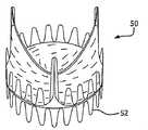
도 5는 혈관 내로 전달되고 풍선에 의해 확장될 수 있는 다상 철계 스테인리스강제 심장 판막의 하나의 실시예의 사시도이다.
도 6은 다상 철계 스테인리스강을 함유한 외과적으로 이식 가능한 심장 판막의 하나의 실시예의 사시도이다.
도 7은 이식 가능한 필터 장치의 측면도이다.
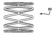
도 8은 다상 철계 스테인리스강으로 이루어진 풍선에 의해 확장 가능한 대안적인 스텐트의 측면도이다.
도 9는 다상 철계 스테인리스강의 시트로 이루어진 도 8의 스텐트를 나타내는 도면이다.
도 10은 도 8에 도시한 바와 같은 형태의 풍선에 의해 확장 가능한 스텐트 복수 개를 이용한 스텐트 이식편(stent-graft)을 나타내는 도면이다.1 shows stress-strain curves for L605, 316L or 316LVM, two-phase steels, and TRIP steels.
2 is a graph showing the change in recoil of L605, 316L or 316LVM.
3A and 3B are perspective views showing before and after diameter expansion of one embodiment of a multiphase iron-based stainless steel endoprosthesis;
4 is a longitudinal cross-sectional view of a multiphase iron-based stainless steel endoprosthesis inserted into a conventional inflatable delivery system and expanded radially by the system.
5 is a perspective view of one embodiment of a multiphase iron-based stainless steel heart valve that can be delivered into a vessel and expanded by a balloon.
6 is a perspective view of one embodiment of a surgically implantable heart valve containing polyphase iron-based stainless steel.
7 is a side view of the implantable filter device.
8 is a side view of an alternative stent expandable by a balloon made of polyphase iron-based stainless steel.
9 is a view showing the stent of FIG. 8 made of a sheet of polyphase iron-based stainless steel.
FIG. 10 is a view showing a stent graft (stent-graft) using a plurality of stents expandable by a balloon of the form shown in FIG. 8.
도 1에서는 L605 코발트-크롬 합금, 2상 강, TRIP강, 및 316L 또는 316LVM 스테인리스강의 전형적인 특성을 비교하는 응력-변형률 곡선을 도시하고 있다. 이 도면에 도시한 바와 같이 L605는 비교적 높은 항복 강도(YS)(100) 및 높은 극한 인장 강도(UTS)(108)를 갖는 반면, 316L 또는 316LVM은 낮은 항복 강도(106) 및 낮은 극한 인장 강도(114)를 갖는다. 2상 강(102) 및 TRIP강(104)은 통상 L605의 항복 강도(100)보다 낮은 항복 강도를 가져 성형성이 향상되고 확장이 용이하다. 2상 강(102) 및 TRIP강(104)의 극한 인장 강도는 316L 또는 316LVM의 극한 인장 강도(114)보다 높다는 점을 주목해야 할 것이다.1 shows a stress-strain curve comparing the typical properties of L605 cobalt-chromium alloy, two-phase steel, TRIP steel, and 316L or 316LVM stainless steel. As shown in this figure, L605 has a relatively high yield strength (YS) 100 and a high ultimate tensile strength (UTS) 108, while 316L or 316LVM has a
도 2에서는 관내 인공 삽입물에 이용되는 통상의 L605와 316L 또는 316LVM 강에 대해 탄성 반동의 변화를 나타내는 응력-변형률 곡선을 도시하고 있다. 316L 또는 316LVM강에서의 탄성 반동의 변화는 변형률 크기(200)로서 표시되어 있는 한편, L605에서의 탄성 반동의 변화는 변형률 크기(204)로 표시되어 있다. 변형률 크기(204)는 L605와 같은 고탄성률 고항복 강도의 금속에 있어서의 전형적인 탄성 반동량을 나타낸다. 변형률 크기(200)는 316L 또는 316LVM강과 같은 저탄성률 저항복 강도의 금속에 있어서의 전형적인 탄성 반동량을 나타낸다. 2상 강 또는 TRIP강에서의 탄성 반동 변화는 그러한 두 값 사이에 포함될 것이다. L605의 높은 극한 인장 강도 및 높은 탄성률을 유지하면서 316L 또는 316LVM 강에서와 같은 비교적 작은 반동량을 나타내는 재료를 관내 인공 삽입물로서 이용하는 데에 있어서의 이점들은 유익할 것이다.FIG. 2 shows stress-strain curves showing changes in elastic recoil for conventional L605 and 316L or 316LVM steels used in endoprosthesis. The change in elastic recoil in 316L or 316LVM steel is indicated as
MRI(자기 공명 영상) 적합성은 이식용 인공 삽입물을 위해 선택되는 임의의 재료에 있어서의 중요한 특성이다. 듀플렉스 스테인리스강은 상자성의 오스테나이트와 강자성의 페라이트로 이루어진 미세한 미세조직을 갖고 각 상의 미세조직 비는 통상 약 50%이다. 316LVM과 같은 스테인리스강은 100% 오스테나이트로 이루어진 미세조직을 가져 상자성을 갖기 때문에 MRI에 적합한 것으로 간주된다. 일반 탄소강과 같은 재료는 페라이트 미세조직을 가져 강자성을 갖는다. 강자성 재료는 자기장에 강하게 영향을 받는다는 점으로 인해 MRI에 안전하지 않은 것으로, 즉 MRI 적합성 갖지 않은 것으로 간주된다. 듀플렉스 스테인리스강에서 페라이트의 체적 비율은 열처리를 통해 감소시킬 수 있는 것으로 알려졌다. 예를 들면, 듀플렉스강 시편을 진공로에서 1300℃의 온도로 열처리한 후에 1000℃까지 서냉(노냉)시키고, 이어서 노로부터 제거한 후에 상온까지 공냉시킨다. 이러한 처리 기법은 임의의 취성의 2차 시그마상도 형성하지 않고 미세조직 내의 페라이트 체적 비율을 50%에서부터 약 11%로 분해시킨다. 이어서, 그러한 시편을 열자기 분석을 이용하여 테스트하였는데, 낮은 페라이트 함량으로 인해 매우 작은 강자성 신호를 갖는 것으로 드러났다. 열자기 곡선은 상자성 재료의 전형을 갖는 것으로 간주되었다.Magnetic resonance imaging (MRI) suitability is an important property for any material selected for implantable prostheses. Duplex stainless steel has a fine microstructure consisting of paramagnetic austenite and ferromagnetic ferrite, and the microstructure ratio of each phase is usually about 50%. Stainless steels, such as 316LVM, are considered suitable for MRI because they are paramagnetic with a microstructure of 100% austenite. Materials such as ordinary carbon steel have ferrite microstructure and are ferromagnetic. Ferromagnetic materials are considered unsafe for MRI due to their strong influence on the magnetic field, i.e. they are not MRI compatible. It is known that the volume fraction of ferrite in duplex stainless steel can be reduced through heat treatment. For example, the duplex steel specimen is heat-treated in a vacuum furnace at a temperature of 1300 ° C., and then slowly cooled to 1000 ° C., and then air cooled to room temperature after removal from the furnace. This treatment technique degrades the ferrite volume fraction in the microstructure from 50% to about 11% without forming any brittle secondary sigma phase. Such specimens were then tested using thermomagnetic analysis, which was found to have a very small ferromagnetic signal due to the low ferrite content. Thermomagnetic curves were considered to be typical of paramagnetic materials.
제1 실시예는 다상 철강으로 이루어진 관내 인공 삽입물을 제공한다. 이러한 다상 철계 관내 인공 삽입물은 종래의 재료로 이루어진 그러한 장치에 이용되는 것과 같은 공지의 수단(그 일부에 대해서는 아래에서 설명함)에 의해 제조될 수 있다. 그러한 관내 인공 삽입물의 일례는 심장 스텐트일 것이다. 심장 스텐트는 통상 후전개 강도(post deployment strength)를 위해서는 코발트-크롬 합금을 이용하여 제조하거나, 정합성, 추적성(trackability), 최소한의 탄성 반동 및 용이한 성형성을 위해서는 316L 또는 316LVM을 이용하여 제조한다. 임의의 그러한 금속으로 이루어진 스텐트는 주로 복잡한 기하학적 구조로 제조된다. 그 구조는 통상 각종 방법을 이용하여 형성된다. 일부 구조는 금속 와이어를 대체로 관형의 형상으로 만듦으로써 형성된다. 보다 복잡한 구조는 얇고 편평한 금속 시트를 절단하여 그 절단된 구조를 굴곡하여 튜브를 형성하거나, 얇은 관형 형태로부터 바로 절단할 수도 있다. 어느 방식이든 스텐트를 풍선식 카테터에 고정시킬 수 있도록 직경 방향으로 압착시킬 수 있다. 패턴의 절단은 방전 가공, 화학적 에칭, 스탬핑 또는 레이저 커팅을 비롯하여 이들에 한정되지 않은 당업계에 공지된 각종 수단에 의해 이루어질 수 있다. 제조 과정 중에 및 원하는 이식 자리로 스텐트를 전달하는 중에 심장 스텐트에 가해지는 특유의 기계적 응력으로 인해, 가장 널리 이용되는 금속 재료는 316L 또는 316LVM 스테인리스강이다. 통상적으로, 시판용 심장 스텐트를 제조하는 데에 이용되는 그러한 사전 절단 금속은 전개되는 장치에 부여되는 기계적 요건으로 인해 과도하게 두껍다. 다상 철강의 특성은 그러한 동일 장치를 얇은 벽을 가지면서도 여전히 양호한 강도 특성을 제공하도록 제조할 수 있게 할 것이다.The first embodiment provides an endoprosthesis made of multiphase steel. Such polyphase iron-based endoprosthesis can be made by known means, some of which are described below, such as those used in such devices made of conventional materials. One example of such an endoprosthesis would be a cardiac stent. Cardiac stents are typically manufactured using cobalt-chromium alloys for post deployment strength, or 316L or 316LVM for consistency, trackability, minimal recoil and easy formability. do. Stents made of any such metals are made predominantly of complex geometry. The structure is usually formed using various methods. Some structures are formed by making the metal wire into a generally tubular shape. More complex structures may cut thin and flat metal sheets and bend the cut structures to form tubes or cut directly from thin tubular shapes. Either way, the stent can be crimped in the radial direction to secure the stent to the inflatable catheter. Cutting of the pattern can be accomplished by a variety of means known in the art including, but not limited to, electrical discharge machining, chemical etching, stamping or laser cutting. Due to the inherent mechanical stresses applied to the cardiac stent during the manufacturing process and delivery of the stent to the desired implant site, the most widely used metal material is 316L or 316LVM stainless steel. Typically, such pre-cut metals used to make commercial heart stents are excessively thick due to the mechanical requirements imposed on the device being deployed. The properties of multiphase steels will enable such same devices to be manufactured to have thin walls but still provide good strength properties.
심장 스텐트는 통상 풍선식 카테터의 외측에 부착하여 원하는 이식 자리로 경피(percutaneous)적으로 전달된다. 스텐트를 운반하는 카테터는 흔히 복잡하고 구불구불한 환자의 혈관계(vasculature)를 통해 이동된다. 스텐트용으로 선택된 금속 재료가 코발트-크롬 합금과 같이 고강도 특성을 갖는 경우, 구불구불한 해부 구조를 성공적으로 지나가는 능력이 손상 받을 수 있고 그 전개시에 고유의 반동을 나타낼 것이다. 심장 스텐트의 이식 환경 및 기계적 요건이 주어지면, 다상 강의 이용이 형성, 전달 및 후전개 중에 스텐트 구조에 가해지는 요건을 보다 이상적으로 충족시켜, 전술한 타협안의 대부분을 바로잡게 될 것이다.Cardiac stents are typically attached to the outside of an inflatable catheter and delivered percutaneous to the desired implant site. Catheters carrying stents are often moved through the vasculature of complex and meticulous patients. If the metal material selected for the stent has high strength properties, such as cobalt-chromium alloys, the ability to successfully pass the serpentine anatomy may be impaired and will exhibit inherent recoil in its deployment. Given the implantation environment and mechanical requirements of the cardiac stent, the use of multiphase steels will more ideally meet the requirements placed on the stent structure during formation, delivery, and postdevelopment, thereby correcting most of the aforementioned compromises.
도 3a 및 도 3b에서는 다상 철계 스테인리스강제 관내 인공 삽입물(10)[예를 들면, 스텐트(12)]의 하나의 실시예의 직경 확장 전후를 나타내는 사시도를 도시하고 있으며, 각 도면에서 직경차가 d 및 d'로 표시되어 있다.3A and 3B show perspective views showing before and after diameter expansion of one embodiment of a multiphase iron-based stainless steel endoprosthesis 10 (eg, stent 12), with diameter differences d and d in each figure. 'Is marked.
도 4에서는 통상의 풍선식 전달 시스템(14)의 중요 부분인 팽창된 카테터 풍선(16)에 장착되어 그에 의해 직경 방향으로 확장된 다상 철계 스테인리스강제 관내 인공 삽입물[예를 들면, 스텐트(12)]의 종방향 단면도이다.In FIG. 4, a multiphase iron-based stainless steel endoprosthesis (eg, stent 12) mounted to an
관내 인공 삽입물의 다른 예로는 전술한 심장 스텐트와 동일한 방식 및 기본 형상으로 형성될 수 있는 신장 스텐트를 들 수 있다. 대부분의 신장 스텐트는 상이한 해부학적 요건에 부합하도록 2개의 별개의 섹션, 즉 구멍 병변 영역(ostial lesion region)과 원위 단부 영역으로 이루어진다. 신장 스텐트의 구멍 영역은 높은 반경 방향 강도 요건을 갖고, 통상 보다 두꺼운 벽 및 보다 많은 수의 종방향 커넥터로 이루어진다. 신장 스텐트의 원위 단부 영역은 구멍 영역보다 가요성을 갖는 것이 바람직하며, 통상 보다 얇은 벽 및 보다 적은 수의 커넥터로 이루어진다. 전체 스텐트는 최적의 추적성 및 정확한 배치를 낮은 프로파일을 갖는 것이 바람직하고, 오랜 시간 동안 동맥을 차단하지 않도록 신속 용이하게 확장시킬 수 있도록 설계되어야 한다. 이러한 상반되는 설계 요건은 재료 선택에 있어서의 탑협을 강요하게 된다. 대부분의 입수 가능한 신장 스텐트는 316L 또는 316LVM 스테인리스강으로 이루어진다. 전술한 기타 스텐트 및 프레임에 있어서와 마찬가지로, 316L 또는 316LVM은 보다 큰 추적성, 성형성 또는 최소의 탄성 반동을 가능하게 한다. 그러한 성능 목표를 달성하기 위해, 스텐트는 2개의 별개의 섹션으로 이루어져야 하며, 이는 제조의 어려움을 증가시킨다.Another example of an endoprosthesis is a kidney stent that can be formed in the same manner and in the same basic shape as the heart stent described above. Most renal stents consist of two distinct sections, the ostial lesion region and the distal end region, to meet different anatomical requirements. The hole area of the stretch stent has high radial strength requirements and usually consists of thicker walls and a greater number of longitudinal connectors. The distal end region of the elongate stent is preferably more flexible than the aperture region, and typically consists of thinner walls and fewer connectors. The entire stent should have a low profile for optimal traceability and accurate placement and should be designed to be able to quickly and easily expand to block the artery for a long time. This conflicting design requirement imposes top narrows on material selection. Most available stretch stents consist of 316L or 316LVM stainless steel. As with other stents and frames described above, 316L or 316LVM allows for greater traceability, formability or minimal recoil. To achieve such performance goals, the stent must consist of two separate sections, which increases the difficulty of manufacturing.
복합 조직 강이 이용되는 경우, 그 구조는 높은 반경 방향 강도, 가요성, 추적성 및 풍선에 의한 확장의 용이성과 같은 필요한 특성을 손상시키지 않고 균질하게 이루어질 수 있다. 그 구조는 전체에 걸쳐 얇은 벽과 적은 수의 커넥터를 갖도록 이루어질 수 있다.If a composite tissue steel is used, the structure can be made homogeneous without compromising the required properties such as high radial strength, flexibility, traceability and ease of expansion by the balloon. The structure can be made with thin walls and fewer connectors throughout.
직경 방향 확장 가능 스텐트의 다양한 형태는 그 제조에 다상 철강을 이용함으로써 이점을 얻을 수 있다. 이들에는 말초, 경동맥, 뇌(신경), 담도, 간관, 대동맥, 및 흉부 용례용 스텐트가 포함될 수 있다. 또한, 그 스텐트들은 공지의 방법으로 제조될 수 있다. 그러한 형태의 스텐트 장치들 중 임의의 또는 전부는, 데이크론(dacron) 또는 ePTFE(팽창 폴리테트라플루오로에틸렌)와 같은 인공 삽입 이식편 재료로 이루어진 부분 또는 전체 커버링(스텐트의 외면, 내면 또는 이들 두면 모두에 대해)이 제공된 스텐트 이식편으로서 마련될 수 있다.Various forms of radially expandable stents can benefit from the use of multiphase steel in their manufacture. These may include peripheral, carotid, brain (nerve), biliary tract, hepatic, aortic, and thoracic stents. In addition, the stents can be produced by known methods. Any or all of those types of stent devices may be part or full covering (outer, inner or both sides of the stent) made of artificial implant graft material such as dacron or ePTFE (expanded polytetrafluoroethylene). Can be provided as a stent graft provided.
관내 인공 삽입물의 다른 예로는 도 5 및 도 6에 도시한 바와 같은 경도관(transcatheter)식으로 전달되는 인공 삽입 심장 판막(50)을 들 수 있다. 경도관식으로 전달되는 인공 삽입 심장 판막은 통상 재료의 성형성, 추적성, 및 최소 탄성 반동에 대해 선택된 의료용 스테인리스강의 프레임으로 통상 이루어진다. 또한, 기계적 강도에 대해 선택된 코발트-니켈 또는 코발트-크롬 합금으로 제조할 수도 있다. 그러한 경도관식으로 전달되는 심장 판막은 기존의 기능 장애 심장 판막이 보이는 곳에 직접 전개되며, 이에 따라 혈액이 흐르는 데에 이용되었을 공간을 차지하게 된다. 심장 판막 프레임(52)을 제조하는 방법은 스텐트를 제조하는 데에 이용되는 방법과 유사하며, 이에 대해서는 앞서 설명하였다. 프레임(52)의 구조는 흔히 링 형상을 갖고 있으며, 지그재그형 또는 사인 곡선형 파형부의 열(도 5 참조) 및 이들 열 사이의 종방향 커넥터로 이루어진다. 대안적으로, 링을 형성하도록 함께 연결된 다이아몬드 형상 요소로서 이루어질 수도 있다. 경도관식으로 전달되는 심장 판막(50)의 프레임(52)에 대해 수많은 다른 형상이 고려될 수도 있다.Another example of an endotracheal prosthesis is an
프레임(52)에는 판막 재료가 부착되어 있다. 판막(54)을 위한 재료는 동종 이식편(도너 이식편), 자가 이식편[통상 로스(Ross) 시술을 통해], 이종 이식편(heterograft 또는 xenograft)(가장 통상적으로 소 또는 돼지 도너으로부터의 동물 조직 이식편), 또는 PTFE(폴리테트라플루오로에틸렌) 또는 ePTFE(팽창 폴리테트라플루오로에틸렌)과 같은 임의의 생체 적합성 재료로 이루어진 것일 수 있다. 이들 재료는 프레임에 직접 봉합하거나, 다른 재료(Dacron(등록 상표) 또는 폴리에스테르)의 가장자리에 봉합한 후에 프레임에 봉합하거나 화학적으로 접합하는 등의 당업계에 공지된 각종 방법으로 프레임에 부착된다.The
이들 심장 판막(50)은 풍선식 카테터에 의해 2가지 방식으로, 즉 트랜스에피컬(transapical)식 또는 경대퇴동맥(transfemoral)식으로 전개되며, 가장 통상적인 전달 루트는 경대퇴동맥식이다. 이러한 전달 방법은, 풍선식 카테터에 부착된 장치가 잠재적으로 구불구불한 해부 구조의 상당 길이를 따라가기에 충분한 가요성을 가질 능력을 요구한다. 이러한 추적성 요건은 심장 판막 프레임에 의료용 스테인리스강을 사용하게 한다.These
통상의 의료용 스테인리스강이 심장 판막(50)의 프레임(52)을 위해 선택된 경우, 그 프레임(52)은 코발트-크롬 합금 또는 코발트-니켈 합금과 같은 보다 강한 재료가 선택된 경우에 비해 다소 두꺼워야 한다. 심장 판막 프레임(52)에 부여되는 기계적 응력은 박동하는 심장의 환경에서는 상당할 수 있다. 코발트 합금의 사용은 그 장치의 통상적인 전달 방법을 방해함은 물론, 덜 정확하게 전개되게 할 것이다. 다시 말해, 보다 얇은 프레임이 혈액의 흐름 및 장치의 전달을 용이하게 하도록 바람직할 수 있지만, 그 프레임은 박동하는 심장에 의해 부여된 기계적 응력 하에서 오랫동안 유지되기에 충분하도록 강해야한다. 다상 강은 경도관식으로 전달되는 심장 판막의 특유의 기계적 요건을 충족시킬 것이다.If a conventional medical stainless steel is selected for the
수술용 스테이플 또는 흉골 봉합 장치도 역시 다상 철강으로 이루어지면 유리할 수 있다. 스테이플은 흔히 창자, 폐 및 피부 상처를 봉합하는 데에 이용된다.Surgical staples or sternum sutures may also be advantageous if they are also made of polyphase steel. Staples are often used to suture bowel, lung and skin wounds.
혈관(72) 내에 이식된 도 7에 도시한 바와 같은 이식 가능 필터(70)도 역시 본 명세서에서 개시하는 다상 철계 스테인리스강으로 제조하면 효과적일 수 있다. 그 필터들에는 하대정맥 필터 및 색전 필터가 포함될 수 있다. 그러한 용례를 위한 필터는 흔히 원하는 부위에서 후에 확장시키기 위해 체내 도관 내로 삽입시킬 수 있도록 직경 방향으로 확장 가능하게 이루어진다.The
듀플렉스 스테인리스강의 다른 의료 용례는 의료용 리드 분야일 수 있다. 의료용 조율 리드(pacing lead)는 전기 커넥터 요소를 구비하고, 이 커넥터 요소는 삽입되는 리드를 받아들이도록 확장되고 이어서 그 리드 주위에서 수축 또는 크림핑되어 리드에 대한 전기적 연결은 물론 스프링식 기계적 연결을 제공하도록 압축 가능부를 구비한다. 이상적으로는 그러한 크림핑 연결은 크림핑이 이루어질 때까지는 매우 얇고 가요성을 가져야 하지만, 이식 또는 외식(explantation) 중에 리드에 부여되는 큰 인장력에 견디기에 충분한 강도를 가져야 한다. 듀플렉스 스레인리스강과 같은 재료는 그러한 용례를 위한 최적의 선택일 수 있다.Another medical application of duplex stainless steel may be in the medical lead field. The medical paced lead has an electrical connector element, which extends to accept the lead being inserted, and then shrinks or crimps around the lead to provide a spring mechanical connection as well as an electrical connection to the lead. It is provided with a compressible portion. Ideally such a crimping connection should be very thin and flexible until crimping is made, but should be strong enough to withstand the large tensile forces applied to the leads during implantation or explantation. Materials such as duplex stainless steel may be the optimal choice for such applications.
가이드 와이어도 역시 다상 철강으로 제조될 수 있다.Guide wires can also be made of polyphase steel.
치아 교정용 보철물, 특히 궁상 와이어(arch wire)가 다상 철계 금속의 또 다른 용례이다. 궁상 와이어는 매우 작은 힘으로 성형될 수 있어야 하지만, 5% 이하의 변형률 범위에 걸쳐 일정한 힘(치아의 이동은 야기하기에 충분하지만 통증은 없도록 치과 의사에 의해 선택됨)을 가할 수 있어야 한다. 이러한 일정한 힘은 큰 반동 없이 유지되어야 한다. 하중이 기계적으로 가해질 수 있기 때문에, 강하면서도 쉽게 성형될 수 있는 재료가 바람직할 것이다.Orthodontic prostheses, particularly arch wires, are another application of polyphase ferrous metals. The curved wire should be able to be molded with very little force, but should be able to apply a constant force (selected by the dentist so as to cause tooth movement but no pain) over a strain range of 5% or less. This constant force must be maintained without great recoil. Since the load can be applied mechanically, a material that is strong and easily moldable would be desirable.
추가적인 처리 단계가 전술한 임의의 장치의 제조에 이용될 수 있다. 예를 들면, 피로 수명 개선 단계가 장치의 형상을 성형한 후에 추가될 수 있다. 이 단계는 성형된 장치의 선택된 부분에 예변형률(pre-strain)을 가하거나, 성형된 장치를 전해 연마하거나, 성형된 장치에 금속 블라스팅 처리를 하여, 금속 표면에 압축 잔류 응력을 부여하는 것을 수반할 수 있다. 다상 철계 금속이 어닐링된 표면을 가진 채로 공급되는 경우, 그러한 처리 단계는 장치의 성형 전에 수행될 수 있다. 다른 처리 단계가 또한 코팅 또는 커버 접착제의 접합 강도를 개선시키도록 추가될 수도 있다. 이 단계는 피로 수명의 개선과 유사하지만, 접합 수명의 개선을 가져온다. 피로 수명의 개선과 마찬가지로, 그 단계는 제공되는 원재료에 따라 장치의 성형 전에 또는 그 후에 수행될 수 있다.Additional processing steps may be used to manufacture any of the devices described above. For example, a fatigue life improvement step may be added after shaping the shape of the device. This step involves applying a pre-strain to selected portions of the molded device, electropolishing the molded device, or metal blasting the molded device to impart compressive residual stress to the metal surface. can do. If the polyphase iron-based metal is supplied with an annealed surface, such treatment step can be carried out before the molding of the device. Other processing steps may also be added to improve the bond strength of the coating or cover adhesive. This step is similar to the improvement in fatigue life, but results in an improvement in joint life. As with the improvement of the fatigue life, the step can be carried out before or after the molding of the device, depending on the raw materials provided.
전술한 바와 같은 관내 인공 삽입물에는 혈액 응고 방지제(blood thinner) 또는 항생 물질과 같은 각종 생리활성 물질(치료제)의 코팅이 마련될 수 있다. 이들 코팅은 원하는 생리활성제에 적합한 각종 공지의 방법에 의해 그러한 장치에 접합될 수 있다. 이들은 또한 특정 용례에 대해 요구되는 바에 따라 선택적으로 치료제를 함유한 각종 폴리머로 선택적으로 코팅될 수도 있다. 적절한 코팅으로는 불화 에틸렌 프로필렌(FEP), 폴리테트라플루오로에틸렌(PTFE), ePTFE, 및 테트라플루오로에틸렌과 폴리알킬메틸에테르와 같은 폴리알킬비닐에테르의 코폴리머(TFE/PMVE) 등의 플루오로폴리머가 포함될 수 있다.Endoprosthesis as described above may be provided with a coating of various bioactive substances (therapeutic agents), such as blood thinners or antibiotics. These coatings may be bonded to such devices by various known methods suitable for the desired bioactive agent. They may also be optionally coated with a variety of polymers, optionally containing a therapeutic agent, as required for a particular application. Suitable coatings include fluoro fluorides such as fluorinated ethylene propylene (FEP), polytetrafluoroethylene (PTFE), ePTFE, and copolymers of polyalkylvinyl ethers such as tetrafluoroethylene and polyalkylmethyl ether (TFE / PMVE). Polymers may be included.
예 1Example 1
도 8에서는 다상 철강으로 제조될 수 있는 예시적인 형태의 풍선 확장식 및 관형의 관내 인공 삽입물(80)을 도시하고 있다. 명료성을 위해, 도 8에서는 맨드릴 또는 기타 원통형 형상을 장치의 관형 형상의 내부에 삽입한 경우에 그 장치가 관찰자에게 전반적으로 보이는 바와 같이 장치에 있어서 관찰자에 근접한 쪽만을 도시하고, 그 관형 형태의 배면측(관찰자로부터 먼 쪽)은 생략하였다. 도 8은 카테터의 풍선에 의해 부분적으로 직경을 확장시킨 후인 것과 같은 관내 인공 삽입물(80)을 도시하고 있다. 이러한 형태의 장치는 6.35mm 두께의 강제 플레이트 형태의 듀플렉스 등급 S2205(미국 펜실베니아주에 소재한 Sandmeyer Steel Co.로부터 입수 가능)를 이용하여 제조하였다. 수령한 강제 플레이트는 다음과 같은 특성을 가졌다.FIG. 8 illustrates an exemplary form of inflatable expandable and
UTS : 845 MPaUTS: 845 MPa
0.2% YS : 644 MPa0.2% YS: 644 MPa
연신율(%) : 29Elongation (%): 29
조직간 체적 비율 : 오스테나이트 56.4% 및 페라이트 43.6%Inter-volume Volume Ratio: Austenitic 56.4% and Ferrite 43.6%
오스테나이트와 페라이트의 체적 비율은 구리 소스를 갖고 x선 회절 기법을 이용하여 측정하였다. 이들 측정은 플레이트의 단면이 취해지는 플레이트의 중심에서 이루어졌다.The volume ratio of austenite and ferrite was measured using an x-ray diffraction technique with a copper source. These measurements were made at the center of the plate where the cross section of the plate was taken.
강제 플레이트는 1300℃에서 열처리되고 1000℃로 노냉시켰다. 1000℃에 도달한 후에 그 플레이트를 주변 공기 중에서 상온으로 냉각시켰다. 열처리 후의 강제 플레이트는 다음과 같은 특성을 가졌다.The steel plate was heat treated at 1300 ° C. and furnace cooled to 1000 ° C. After reaching 1000 ° C., the plates were cooled to ambient temperature in ambient air. The steel plate after the heat treatment had the following characteristics.
UTS : 781 MPaUTS: 781 MPa
0.2% YS : 485 MPa0.2% YS: 485 MPa
연신율(%) : 34Elongation (%): 34
탄성률 : 216 GPaModulus of elasticity: 216 GPa
조직간 체적 비율 : 오스테나이트 41.4% 및 페라이트 58.6%Inter-volume Volume Ratio: Austenitic 41.4% and Ferrite 58.6%
ASTM E8에 따라 인장 시험을 수행하였다. 열처리된 스테인리스강 플레이트의 인장 샘플은 나사산형 인장 시험 바아로 가공되었다. 316LVM 및 L605 배관으로부터 레이저 커팅 인장 시험 스트립을 절단하여 역시 인장 시험을 행하였다. 316 LVM의 기계적 특성은 다음과 같다.Tensile tests were performed according to ASTM E8. Tensile samples of heat treated stainless steel plates were processed with threaded tensile test bars. Tensile tests were also performed by cutting laser cut tensile test strips from 316LVM and L605 tubing. The mechanical properties of the 316 LVM are:
UTS : 661 MPaUTS: 661 MPa
0.2% YS : 340 MPa0.2% YS: 340 MPa
연신율(%) : 53Elongation (%): 53
탄성률 : 126 GPaModulus of elasticity: 126 GPa
시험된 L605 비교 샘플의 기계적 특성은 다음과 같다.The mechanical properties of the L605 comparative sample tested were as follows.
UTS : 1079 MPaUTS: 1079 MPa
0.2% YS : 567 MPa0.2% YS: 567 MPa
연신율(%) : 56Elongation (%): 56
탄성률 : 235 GPaModulus of elasticity: 235 GPa
이들 시험에서는 열처리된 듀플렉스 스테인리스강이 두 합금들의 사이에 있는 탄성 계수, 항복 강도 및 극한 인장 강도를 갖지만 연신율은 316LVM 및 L605보다 작은 것으로 드러났다.These tests revealed that the heat treated duplex stainless steel had elastic modulus, yield strength and ultimate tensile strength between the two alloys, but elongation was less than 316LVM and L605.
열처리 후에, 강제 플레이트로부터 하이포튜브(hypotube)를 와이어 EDM(방전 가공 기계)(미츠비시 와이어 EDM 모델 FA205)으로 가공하였다.After the heat treatment, hypotubes were processed from a steel plate with a wire EDM (discharge processing machine) (Mitsubishi Wire EDM Model FA205).
이들 하이포튜브는 4.57mm의 외경 및 0.254mm 벽두께를 가졌다. EDM 튜브들은 레이저 커팅하기에는 그 길이가 너무 짧기 때문에, 스테인리스강 튜브 확장기를 만들어 하이포튜브들의 단부에 압입하였다. 이어서, 도 8에 도시한 형태의 스텐트 링을 하이포튜브로부터 레이저 커팅하였으며, 이 때에 그 직경 및 벽두께는 영향을 받지 않았다. 레이저 커팅이 관내 인공 삽입물의 확장된 직경(즉, 통상의 풍선에 의해 확장시킨 후에 장치가 갖게 되는 직경)에서 수행되어 그 외양이 대체로 도 8에 도시한 바와 같이 되도록 하였다. 326LVM 합금으로부터 동일한 형태 및 동일한 직경의 레이저 커팅 스텐트 링을 제조하였다. 듀플렉스 링 및 316LVM 합금 링은 1.5mm로 모의 크림핑시켰다. 이어서, 이들 링은 테이퍼 맨드릴을 이용하여 10mm로 반경 방향으로 확장시켜, Blockwise J-크림퍼(crimper)(미국 아리조나주 피닉스에 소재한 Blockwise Engineering LL의 모델 RJAT) 내에 삽입하였다. J-크림퍼는 Instron 인장 시험기(미국 매사추세츠주 노어우드에 소재한 Instron Corp.의 모델 5564)에 장착하였고, 링들은 개별적으로 그 기계식 아이리스(mechanical iris) 내에 배치하였다. 이어서, 그 링들을 개별적으로 아이리스 내에서 중간 크기(외경 1.65mm)로 직경 방향으로 가압하였고, 링들의 강도를 Instron Bluehill 소프트웨어로 결정하였다. 듀플렉스 링은 316LVM 링보다 약 20%로 강한 것으로 드러났다.These hypotubes had an outer diameter of 4.57 mm and a 0.254 mm wall thickness. Since the EDM tubes were too short to laser cut, a stainless steel tube expander was made and pressed into the ends of the hypotubes. Subsequently, the stent ring of the type shown in FIG. 8 was laser cut from the hypotube, at which time its diameter and wall thickness were not affected. Laser cutting was performed at the expanded diameter of the endoprosthesis (ie, the diameter the device would have after expanding with a conventional balloon) so that its appearance would be generally as shown in FIG. 8. Laser cut stent rings of the same shape and diameter were made from 326LVM alloy. The duplex ring and the 316LVM alloy ring were simulated crimped to 1.5 mm. These rings were then radially extended to 10 mm using a tapered mandrel and inserted into a Blockwise J-Crimper (Model RJAT of Blockwise Engineering LL, Phoenix, AZ). J-Crimper was mounted on an Instron tensile tester (Model 5564, Instron Corp., Norwood, Mass.), And the rings were individually placed in their mechanical iris. The rings were then individually pressed radially in medium size (outer diameter 1.65 mm) in the iris and the strength of the rings was determined by Instron Bluehill software. The duplex ring was found to be about 20% stronger than the 316LVM ring.
듀플렉스 레이저 커팅 링의 탄성 반동은 다음의 프로세스를 이용하여 측정하였다. 전술한 형태의 관내 인공 삽입 링(스텐트 링)이 전술한 바와 유사하게 열처리 듀플렉스 S2205 강은 물론 316LVM 및 L605 모두로 제조하였다. 이들 링은 최대 직경 12.80mm의 원통형 단부를 갖는 스테인리스강제 테이퍼 맨드릴을 이용하여 직경 방향으로 확장시켰다. 그 링들은 12.80mm의 내경으로 확장시킨 후 테이퍼 맨드릴로부터 제거하였다. 이들의 12.80mm 직경은 그러한 구조의 스텐트 링에 대해 기능적으로 관련이 있는 것으로 간주되었다. 직경을 확장시키고 테이퍼 맨드릴로부터 제거한 후에, 각각의 내경을 Nikon 비전 시스템(모델 VMR 3020 타입 3)을 이용하여 측정하였다. 각각의 링의 직경은 스텐트 내경 둘레에 균일한 간격을 두고 떨어진 10곳의 상이한 지점에서 측정하여 평균하였다. 그 측정치에서는 316LVM 스텐트 링이 0.051mm의 탄성 반동을 보이고, 듀플렉스강 스텐트 링은 0.152mm, 그리고 L605 스텐트링은 0.279mm의 탄성 반동을 보였다. 이들 데이터는 L605 링이 열처리 듀플렉스강에 비해 탄성 반동이 더 크고, 이에 따라 성형성이 더 작다는 점을 나타낸다.The elastic rebound of the duplex laser cutting ring was measured using the following process. An endoprosthetic ring (stent ring) of the type described above was made of both 316LVM and L605, as well as heat treated duplex S2205 steel, as described above. These rings were expanded in the radial direction using a stainless steel tapered mandrel having a cylindrical end with a maximum diameter of 12.80 mm. The rings were removed from the tapered mandrel after expanding to an inner diameter of 12.80 mm. Their 12.80 mm diameter was considered functionally relevant for stent rings of such structures. After the diameters were expanded and removed from the tapered mandrel, each inner diameter was measured using a Nikon vision system (Model VMR 3020 Type 3). The diameter of each ring was measured and averaged at 10 different points spaced evenly around the stent inner diameter. In that measurement, the 316 LVM stent ring showed a rebound of 0.051 mm, the duplex steel stent ring showed 0.152 mm, and the L605 stent ring showed a rebound of 0.279 mm. These data indicate that the L605 ring has a higher elastic recoil compared to the heat treated duplex steel and thus a smaller formability.
앞서 전반적으로 설명한 바와 같이 빌렛으로부터 가공되는 것 외에도, 도 8에 도시한 바와 같은 스텐트 링은 시트 재료로부터 가공될 수도 있다. 그러한 스텐트의 가공 패턴(90)이 도 9에 도시되어 있다. 시트의 가공 후에, 얻어진 편평한 형상(90)을 테이퍼 맨드릴을 이용하여 튜브로 성형하였다. 맨드릴의 소직경부는 편평한 형상(9)의 중앙 개구(92) 내로 삽입할 수 있어야 한다. 맨드릴은 부분적으로 확장된 스텐트 형상의 의도한 내경에 상응하는 최대 직경을 가지며, 이러한 최대 직경은 원통형 섹션에 인접한 부분의 직경과 동일한 직경을 포함한다. 편평한 형상(90)의 중앙 개구(92) 내로 맨드릴의 소직경 단부를 삽입하고 그 맨드릴을 눌러 편평한 형상(90)을 완전히 통과시키면 도 8에 도시한 바와 같은 관형 형상(80)이 얻어진다.In addition to being processed from billets as described above generally, the stent ring as shown in FIG. 8 may be processed from sheet material. A
열처리 듀플렉스 S2205강으로 제조된 전술한 바와 같은 스텐트 링 복수 개를 제조하였다. 8개의 링(80)을 ePTFE 튜브(102)와 같은 이식편 재료의 외면에 접합하여 도 10에 도시한 바와 같은 스텐트 이식편(100)을 생성하였다. 이러한 형태의 스텐트 이식편의 제조는 본 명세서에 참조로 인용되는 미국 특허 출원 공개 번호 제2008/0119943호에 기술되어 있다. 이와 같이 얻어진 대략 40mm 길이의 풍선 확장식 스텐트 이식편(100)은 후에 환자의 혈관 구조 내로 전달한 후 풍선에 의해 확장시키도록 풍선식 카테터 상에 탑재될 수 있다. 도 10에 도시한 스텐트 이식편은 단지 일례이고 다상 철강으로 이루어진 스텐트들을 포함하는 수많은 형태의 스텐트 이식편이 가능하다는 점을 이해할 것이다. 마찬가지로, 스텐트는 이식편 재료의 외면에 또는 이식편 재료의 관강 내면(luminal surface)에 접합되거나, 이식편 재료의 외면과 내면 사이에 끼일 수도 있다는 점을 이해할 것이다. 또한, 이식편 재료는 원하는 경우 담도 시술과 같은 특정 용례를 위해 천공부를 포함할 수 있다.A plurality of stent rings as described above made of heat-treated duplex S2205 steel were prepared. Eight rings 80 were bonded to the outer surface of the graft material, such as
듀플렉스 S2205 스테인리스강 합금은 특히 전술한 바와 같이 열처리된 경우에 풍선에 의해 확장 가능한 관내 인공 삽입물의 제조를 위해 양호한 강도 능력 및 양호한 성형 능력을 제공하는 것으로 드러났지만, 훨씬더 양호한 합금이 의료 장치, 특히 확장 가능 관내 인공 삽입물을 위해 가능한 것으로 여겨진다. 표 1에 그러한 합금 1종의 조성이 기재되어 있다. 그 조성에서의 작은 편차가 또한 듀플렉스 S2205 합금에 대해 얼마간의 개선을 제공할 수 있다는 점을 이해할 것이다.Although duplex S2205 stainless steel alloys have been found to provide good strength capability and good forming capability for the production of endotracheal implants expandable by balloons, especially when heat treated as described above, much better alloys are used in medical devices, especially It is considered possible for expandable endoprosthesis. Table 1 describes the composition of one such alloy. It will be appreciated that small deviations in its composition may also provide some improvement over the duplex S2205 alloy.
본 발명은, 앞서 설명하고 아래의 청구 범위에 기재된 바와 같은 실시예들 외에도, 앞서 설명하고 청구 범위에 기재된 바와 같은 특징들의 다양한 조합들을 갖는 실시예들도 포함한다. 따라서, 본 발명은 아래의 청구 범위에 기재된 바와 같은 종속항의 특징들의 임의의 가능한 다른 조합을 갖는 기타 실시예들도 역시 포함한다.The invention includes embodiments having various combinations of features described above and as described in the claims, in addition to the embodiments as described above and as described in the claims below. Accordingly, the present invention also encompasses other embodiments having any possible other combination of the features of the dependent claims as set forth in the claims below.
본 발명의 다수의 특징 및 이점은 본 발명의 구조 및 기능의 세부 사항과 더불어 바람직한 실시예는 물론 대안적인 실시예를 포함하는 전술한 상세한 설명에서 기술하였다. 본 개시는 단지 예시를 위한 것으로서 한정하고자 하는 것은 아니다. 당업자들에게는 다양한 수정이, 특히 구조, 재료, 요소, 부품, 형상, 크기 및 부품들의 배열의 측면에서 다양한 수정이 본 발명의 원리 내에서 첨부된 청구 범위에서 표현된 용어들의 광의의 포괄적 의미에 의해 정해지는 최대의 정도까지 이루어질 수 있다는 점을 이해할 것이다. 그러한 다양한 수정이 첨부된 청구의 범위의 사상 및 보호 범위로부터 벗어나지 않는 정도까지는 그 내에 포함될 것이다.Numerous features and advantages of the present invention have been described in the foregoing detailed description, including preferred embodiments as well as alternative embodiments, along with details of the structure and function of the present invention. The present disclosure is for illustrative purposes only and is not intended to be limiting. It will be appreciated by those skilled in the art that various modifications, in particular in terms of structure, materials, elements, components, shapes, sizes and arrangement of components, are made possible by the broader meaning of the terms expressed in the appended claims within the principles of the invention. It will be appreciated that it can be done to the maximum degree determined. Such various modifications will be included therein to the extent that they do not depart from the spirit and scope of the appended claims.
Claims (20)
Translated fromKorean다상 강의 편평한 시트를 원하는 형상으로 성형하는 단계;
상기 원하는 형상을 관형 형태로 성형하는 단계;
상기 관형 형태를 풍선식의 혈관내 전달 시스템 상에 크림핑하는 단계;
상기 원하는 형상을 치료 영역으로 전달하는 단계; 및
상기 원하는 형상을 치료 영역에서 확장시키는 단계
를 포함하는 관내 인공 삽입물의 제조 방법.As a method of making an endoprosthesis:
Forming a flat sheet of polyphase steel into a desired shape;
Shaping the desired shape into a tubular form;
Crimping the tubular form onto an inflatable vascular delivery system;
Delivering the desired shape to a treatment area; And
Expanding the desired shape in the treatment area
Method for producing an endoprosthesis comprising a.
다상 강의 관형 형태를 원하는 형상으로 성형하는 단계; 및
상기 관형 형태를 풍선식의 혈관내 전달 시스템 상에 크림핑하는 단계
를 포함하는 관내 인공 삽입물의 제조 방법.As a method of making an endoprosthesis:
Shaping the tubular form of the polyphase steel into a desired shape; And
Crimping the tubular form onto an inflatable vascular delivery system
Method for producing an endoprosthesis comprising a.
다상 강의 와이어를 원하는 관형 형상으로 성형하는 단계;
상기 원하는 관형 형상을 풍선식의 혈관내 전달 시스템 상에 크림핑하는 단계;
상기 원하는 관형 형상을 치료 영역으로 전달하는 단계; 및
상기 원하는 관형 형상을 치료 영역에서 확장시키는 단계
를 포함하는 관내 인공 삽입물의 제조 방법.As a method of making an endoprosthesis:
Shaping the wire of the polyphase steel into a desired tubular shape;
Crimping the desired tubular shape onto an inflatable vascular delivery system;
Delivering the desired tubular shape to a treatment area; And
Expanding the desired tubular shape in the treatment area
Method for producing an endoprosthesis comprising a.
Applications Claiming Priority (5)
| Application Number | Priority Date | Filing Date | Title |
|---|---|---|---|
| US29149709P | 2009-12-31 | 2009-12-31 | |
| US61/291,497 | 2009-12-31 | ||
| US12/981,102 | 2010-12-29 | ||
| US12/981,102US20110160838A1 (en) | 2009-12-31 | 2010-12-29 | Endoprosthesis containing multi-phase ferrous steel |
| PCT/US2010/062460WO2011082280A1 (en) | 2009-12-31 | 2010-12-30 | Endoprosthesis containing multi-phase ferrous steel |
Publications (2)
| Publication Number | Publication Date |
|---|---|
| KR20120123331Atrue KR20120123331A (en) | 2012-11-08 |
| KR101901382B1 KR101901382B1 (en) | 2018-09-27 |
Family
ID=44188459
Family Applications (1)
| Application Number | Title | Priority Date | Filing Date |
|---|---|---|---|
| KR1020127018379AActiveKR101901382B1 (en) | 2009-12-31 | 2010-12-30 | Endoprosthesis containing multi-phase ferrous steel |
Country Status (11)
| Country | Link |
|---|---|
| US (1) | US20110160838A1 (en) |
| EP (1) | EP2519268B1 (en) |
| JP (2) | JP2013516243A (en) |
| KR (1) | KR101901382B1 (en) |
| CN (1) | CN102695529B (en) |
| AU (1) | AU2010339451B2 (en) |
| BR (1) | BR112012015422A2 (en) |
| CA (1) | CA2783625C (en) |
| ES (1) | ES2731371T3 (en) |
| RU (1) | RU2012132648A (en) |
| WO (1) | WO2011082280A1 (en) |
Families Citing this family (49)
| Publication number | Priority date | Publication date | Assignee | Title |
|---|---|---|---|---|
| US20110160838A1 (en)* | 2009-12-31 | 2011-06-30 | Blanzy Jeffrey S | Endoprosthesis containing multi-phase ferrous steel |
| US8888838B2 (en) | 2009-12-31 | 2014-11-18 | W. L. Gore & Associates, Inc. | Endoprosthesis containing multi-phase ferrous steel |
| US8579964B2 (en) | 2010-05-05 | 2013-11-12 | Neovasc Inc. | Transcatheter mitral valve prosthesis |
| US9308087B2 (en) | 2011-04-28 | 2016-04-12 | Neovasc Tiara Inc. | Sequentially deployed transcatheter mitral valve prosthesis |
| US9554897B2 (en) | 2011-04-28 | 2017-01-31 | Neovasc Tiara Inc. | Methods and apparatus for engaging a valve prosthesis with tissue |
| US9345573B2 (en) | 2012-05-30 | 2016-05-24 | Neovasc Tiara Inc. | Methods and apparatus for loading a prosthesis onto a delivery system |
| DE102012010687B4 (en)* | 2012-05-30 | 2021-08-19 | ADMEDES GmbH | A method for producing a body implant, an assembly comprising a guide wire and a body implant, and a medical instrument |
| DE102013022603B4 (en) | 2013-03-13 | 2024-08-08 | Pascal Roman Schumacher | Retainer and process for its manufacture |
| US9254182B2 (en) | 2013-03-15 | 2016-02-09 | Ormco Corporation | Orthodontic archwire |
| US9572665B2 (en) | 2013-04-04 | 2017-02-21 | Neovasc Tiara Inc. | Methods and apparatus for delivering a prosthetic valve to a beating heart |
| CN103251460B (en)* | 2013-06-05 | 2015-10-28 | 上海上远齿科技术有限公司 | A kind of have pure titanium removable denture support tieing up his smart card ring and preparation method thereof |
| US20140378916A1 (en)* | 2013-06-24 | 2014-12-25 | Abbott Cardiovascular Systems, Inc. | Hypotube with enhanced strength and ductility |
| CN103445894B (en)* | 2013-09-11 | 2016-05-25 | 辽宁生物医学材料研发中心有限公司 | Medical stainless steel intravascular stent |
| DE102015204112B4 (en) | 2015-03-06 | 2021-07-29 | Leibniz-Institut Für Festkörper- Und Werkstoffforschung Dresden E.V. | Use of a biodegradable iron-based material |
| FR3033494B1 (en)* | 2015-03-10 | 2017-03-24 | Carmat | TISSUE STENT AND METHOD FOR PRODUCING THE SAME |
| CN106367714A (en) | 2015-07-24 | 2017-02-01 | 先健科技(深圳)有限公司 | Iron-based absorbent implanting medical instrument, prefabricated tube, and production methods of medical instrument and prefabricated tube |
| CA3007660A1 (en) | 2015-12-15 | 2017-06-22 | Neovasc Tiara Inc. | Transseptal delivery system |
| US10433952B2 (en) | 2016-01-29 | 2019-10-08 | Neovasc Tiara Inc. | Prosthetic valve for avoiding obstruction of outflow |
| CA3042588A1 (en) | 2016-11-21 | 2018-05-24 | Neovasc Tiara Inc. | Methods and systems for rapid retraction of a transcatheter heart valve delivery system |
| CA3073834A1 (en) | 2017-08-25 | 2019-02-28 | Neovasc Tiara Inc. | Sequentially deployed transcatheter mitral valve prosthesis |
| WO2019175888A1 (en)* | 2018-03-16 | 2019-09-19 | Meril Life Sciences Pvt Ltd | Expandable endo prosthesis with low crimp profile |
| WO2019195860A2 (en) | 2018-04-04 | 2019-10-10 | Vdyne, Llc | Devices and methods for anchoring transcatheter heart valve |
| US12186187B2 (en) | 2018-09-20 | 2025-01-07 | Vdyne, Inc. | Transcatheter deliverable prosthetic heart valves and methods of delivery |
| US10595994B1 (en) | 2018-09-20 | 2020-03-24 | Vdyne, Llc | Side-delivered transcatheter heart valve replacement |
| US11278437B2 (en) | 2018-12-08 | 2022-03-22 | Vdyne, Inc. | Compression capable annular frames for side delivery of transcatheter heart valve replacement |
| US11071627B2 (en) | 2018-10-18 | 2021-07-27 | Vdyne, Inc. | Orthogonally delivered transcatheter heart valve frame for valve in valve prosthesis |
| US11344413B2 (en) | 2018-09-20 | 2022-05-31 | Vdyne, Inc. | Transcatheter deliverable prosthetic heart valves and methods of delivery |
| US10321995B1 (en) | 2018-09-20 | 2019-06-18 | Vdyne, Llc | Orthogonally delivered transcatheter heart valve replacement |
| US11109969B2 (en) | 2018-10-22 | 2021-09-07 | Vdyne, Inc. | Guidewire delivery of transcatheter heart valve |
| CN113271890B (en) | 2018-11-08 | 2024-08-30 | 内奥瓦斯克迪亚拉公司 | Ventricular deployment of transcatheter mitral valve prosthesis |
| US11253359B2 (en) | 2018-12-20 | 2022-02-22 | Vdyne, Inc. | Proximal tab for side-delivered transcatheter heart valves and methods of delivery |
| WO2020146842A1 (en) | 2019-01-10 | 2020-07-16 | Vdyne, Llc | Anchor hook for side-delivery transcatheter heart valve prosthesis |
| US11273032B2 (en) | 2019-01-26 | 2022-03-15 | Vdyne, Inc. | Collapsible inner flow control component for side-deliverable transcatheter heart valve prosthesis |
| US11185409B2 (en) | 2019-01-26 | 2021-11-30 | Vdyne, Inc. | Collapsible inner flow control component for side-delivered transcatheter heart valve prosthesis |
| CN113382694A (en)* | 2019-01-31 | 2021-09-10 | 贝克顿·迪金森公司 | Hybrid frame endoluminal prosthesis and method |
| WO2020181154A2 (en) | 2019-03-05 | 2020-09-10 | Vdyne, Inc. | Tricuspid regurgitation control devices for orthogonal transcatheter heart valve prosthesis |
| CA3132873A1 (en) | 2019-03-08 | 2020-09-17 | Neovasc Tiara Inc. | Retrievable prosthesis delivery system |
| US11076956B2 (en) | 2019-03-14 | 2021-08-03 | Vdyne, Inc. | Proximal, distal, and anterior anchoring tabs for side-delivered transcatheter mitral valve prosthesis |
| US11173027B2 (en) | 2019-03-14 | 2021-11-16 | Vdyne, Inc. | Side-deliverable transcatheter prosthetic valves and methods for delivering and anchoring the same |
| CA3135753C (en) | 2019-04-01 | 2023-10-24 | Neovasc Tiara Inc. | Controllably deployable prosthetic valve |
| US11491006B2 (en) | 2019-04-10 | 2022-11-08 | Neovasc Tiara Inc. | Prosthetic valve with natural blood flow |
| CA3138875A1 (en) | 2019-05-04 | 2020-11-12 | Vdyne, Inc. | Cinch device and method for deployment of a side-delivered prosthetic heart valve in a native annulus |
| US11779742B2 (en) | 2019-05-20 | 2023-10-10 | Neovasc Tiara Inc. | Introducer with hemostasis mechanism |
| JP7520897B2 (en) | 2019-06-20 | 2024-07-23 | ニオバスク ティアラ インコーポレイテッド | Thin prosthetic mitral valve |
| EP4480458A3 (en) | 2019-08-20 | 2025-04-09 | Vdyne, Inc. | Delivery devices for side-deliverable transcatheter prosthetic valves |
| CN120531525A (en) | 2019-08-26 | 2025-08-26 | 维迪内股份有限公司 | Laterally deliverable transcatheter prosthetic valve and method for its delivery and anchoring |
| CN112662932B (en)* | 2019-10-15 | 2022-03-04 | 中国石油化工股份有限公司 | TWIP steel and preparation method thereof |
| EP4069327A1 (en)* | 2019-12-04 | 2022-10-12 | Cortronik GmbH | Swellable polymer hybrid fibres for a sleeve of an intraluminal endoprosthesis |
| US11234813B2 (en) | 2020-01-17 | 2022-02-01 | Vdyne, Inc. | Ventricular stability elements for side-deliverable prosthetic heart valves and methods of delivery |
Family Cites Families (10)
| Publication number | Priority date | Publication date | Assignee | Title |
|---|---|---|---|---|
| SE461191B (en)* | 1988-04-21 | 1990-01-22 | Sandvik Ab | APPLICATION OF A STAINLESS FERRIT-AUSTENITIC STEEL ALLOY AS IMPLANT IN PHYSIOLOGICAL ENVIRONMENT |
| JP2658210B2 (en)* | 1988-07-07 | 1997-09-30 | 株式会社クボタ | Heat treatment method of martensitic stainless steel |
| EP1011889B1 (en)* | 1996-01-30 | 2002-10-30 | Medtronic, Inc. | Articles for and methods of making stents |
| JP2002253683A (en)* | 2000-12-29 | 2002-09-10 | Shine Co Ltd | Self-exciting type exothermic implant to be inserted into human body |
| US6582652B2 (en)* | 2001-05-11 | 2003-06-24 | Scimed Life Systems, Inc. | Stainless steel alloy having lowered nickel-chromium toxicity and improved biocompatibility |
| KR100834595B1 (en)* | 2001-10-30 | 2008-06-02 | 에이티아이 프로퍼티즈, 인코퍼레이티드 | Duplex stainless steel |
| SE530711C2 (en)* | 2006-10-30 | 2008-08-19 | Sandvik Intellectual Property | Duplex stainless steel alloy and use of this alloy |
| US9622888B2 (en)* | 2006-11-16 | 2017-04-18 | W. L. Gore & Associates, Inc. | Stent having flexibly connected adjacent stent elements |
| CA2749194C (en)* | 2009-01-08 | 2017-05-30 | Bio Dg, Inc. | Implantable medical devices comprising bio-degradable alloys |
| US20110160838A1 (en)* | 2009-12-31 | 2011-06-30 | Blanzy Jeffrey S | Endoprosthesis containing multi-phase ferrous steel |
- 2010
- 2010-12-29USUS12/981,102patent/US20110160838A1/ennot_activeAbandoned
- 2010-12-30JPJP2012547288Apatent/JP2013516243A/ennot_activeWithdrawn
- 2010-12-30RURU2012132648/15Apatent/RU2012132648A/ennot_activeApplication Discontinuation
- 2010-12-30CACA2783625Apatent/CA2783625C/enactiveActive
- 2010-12-30WOPCT/US2010/062460patent/WO2011082280A1/enactiveApplication Filing
- 2010-12-30AUAU2010339451Apatent/AU2010339451B2/enactiveActive
- 2010-12-30KRKR1020127018379Apatent/KR101901382B1/enactiveActive
- 2010-12-30EPEP10800872.3Apatent/EP2519268B1/enactiveActive
- 2010-12-30ESES10800872Tpatent/ES2731371T3/enactiveActive
- 2010-12-30BRBR112012015422Apatent/BR112012015422A2/ennot_activeApplication Discontinuation
- 2010-12-30CNCN201080060102.2Apatent/CN102695529B/enactiveActive
- 2016
- 2016-05-06JPJP2016093451Apatent/JP6290972B2/enactiveActive
Also Published As
| Publication number | Publication date |
|---|---|
| EP2519268A1 (en) | 2012-11-07 |
| JP2017000729A (en) | 2017-01-05 |
| RU2012132648A (en) | 2014-02-10 |
| ES2731371T3 (en) | 2019-11-15 |
| EP2519268B1 (en) | 2019-04-17 |
| WO2011082280A1 (en) | 2011-07-07 |
| CN102695529A (en) | 2012-09-26 |
| JP6290972B2 (en) | 2018-03-07 |
| BR112012015422A2 (en) | 2017-05-23 |
| KR101901382B1 (en) | 2018-09-27 |
| US20110160838A1 (en) | 2011-06-30 |
| CA2783625C (en) | 2015-06-02 |
| JP2013516243A (en) | 2013-05-13 |
| AU2010339451A1 (en) | 2012-08-16 |
| CN102695529B (en) | 2015-11-25 |
| CA2783625A1 (en) | 2011-07-07 |
| AU2010339451B2 (en) | 2014-10-02 |
Similar Documents
| Publication | Publication Date | Title |
|---|---|---|
| JP6290972B2 (en) | Endoprosthesis with multi-phase steel | |
| US9987121B2 (en) | Method of making an endoprosthesis containing multi-phase stainless steel | |
| US9649210B2 (en) | Hybrid stent and method of making | |
| JP5078271B2 (en) | Stent for living body expansion and method for manufacturing the same | |
| EP3241530B1 (en) | Implants having high fatigue resistance and implant delivery systems | |
| JP2008515563A (en) | Medical device and manufacturing method thereof | |
| JP2008515563A6 (en) | Medical device and manufacturing method thereof | |
| US9427500B2 (en) | Stent made of a cobalt alloy | |
| US20130338756A1 (en) | Stent composed of an iron alloy | |
| EP1579886A1 (en) | Medical instrument for soft tissue and method for manufacture thereof | |
| US20050098241A1 (en) | Niobium-Zirconium Alloy for medical devices or their parts | |
| Sosnin et al. | Import substitution in medicine on the basis of Russian precision alloys | |
| HK1170424B (en) | Endoprosthesis containing multi-phase ferrous steel | |
| HK1170424A (en) | Endoprosthesis containing multi-phase ferrous steel | |
| US20130211537A1 (en) | Medical devices including duplex stainless steel |
Legal Events
| Date | Code | Title | Description |
|---|---|---|---|
| PA0105 | International application | Patent event date:20120713 Patent event code:PA01051R01D Comment text:International Patent Application | |
| PG1501 | Laying open of application | ||
| N231 | Notification of change of applicant | ||
| PN2301 | Change of applicant | Patent event date:20150803 Comment text:Notification of Change of Applicant Patent event code:PN23011R01D | |
| PA0201 | Request for examination | Patent event code:PA02012R01D Patent event date:20151229 Comment text:Request for Examination of Application | |
| E902 | Notification of reason for refusal | ||
| PE0902 | Notice of grounds for rejection | Comment text:Notification of reason for refusal Patent event date:20170925 Patent event code:PE09021S01D | |
| E90F | Notification of reason for final refusal | ||
| PE0902 | Notice of grounds for rejection | Comment text:Final Notice of Reason for Refusal Patent event date:20180412 Patent event code:PE09021S02D | |
| E701 | Decision to grant or registration of patent right | ||
| PE0701 | Decision of registration | Patent event code:PE07011S01D Comment text:Decision to Grant Registration Patent event date:20180615 | |
| GRNT | Written decision to grant | ||
| PR0701 | Registration of establishment | Comment text:Registration of Establishment Patent event date:20180917 Patent event code:PR07011E01D | |
| PR1002 | Payment of registration fee | Payment date:20180918 End annual number:3 Start annual number:1 | |
| PG1601 | Publication of registration | ||
| PR1001 | Payment of annual fee | Payment date:20210820 Start annual number:4 End annual number:4 | |
| PR1001 | Payment of annual fee | Payment date:20220822 Start annual number:5 End annual number:5 | |
| PR1001 | Payment of annual fee | Payment date:20230828 Start annual number:6 End annual number:6 | |
| PR1001 | Payment of annual fee | Payment date:20240826 Start annual number:7 End annual number:7 |