KR20100132486A - Pharmaceutical dissolution type catheter and its manufacturing method - Google Patents
Pharmaceutical dissolution type catheter and its manufacturing methodDownload PDFInfo
- Publication number
- KR20100132486A KR20100132486AKR1020107017482AKR20107017482AKR20100132486AKR 20100132486 AKR20100132486 AKR 20100132486AKR 1020107017482 AKR1020107017482 AKR 1020107017482AKR 20107017482 AKR20107017482 AKR 20107017482AKR 20100132486 AKR20100132486 AKR 20100132486A
- Authority
- KR
- South Korea
- Prior art keywords
- catheter
- acid
- nanoparticles
- drug
- balloon
- Prior art date
- Legal status (The legal status is an assumption and is not a legal conclusion. Google has not performed a legal analysis and makes no representation as to the accuracy of the status listed.)
- Ceased
Links
- 238000004519manufacturing processMethods0.000titleclaimsdescription27
- 238000004090dissolutionMethods0.000titleclaimsdescription20
- 239000002105nanoparticleSubstances0.000claimsabstractdescription232
- 230000000975bioactive effectEffects0.000claimsabstractdescription82
- 239000000463materialSubstances0.000claimsabstractdescription35
- 101000979342Homo sapiens Nuclear factor NF-kappa-B p105 subunitProteins0.000claimsdescription96
- 102100023050Nuclear factor NF-kappa-B p105 subunitHuman genes0.000claimsdescription96
- 238000000034methodMethods0.000claimsdescription87
- 239000000126substanceSubstances0.000claimsdescription62
- 239000003814drugSubstances0.000claimsdescription51
- 229940079593drugDrugs0.000claimsdescription49
- -1nucleic acid compoundChemical class0.000claimsdescription37
- 239000002245particleSubstances0.000claimsdescription34
- 229920002988biodegradable polymerPolymers0.000claimsdescription31
- 239000004621biodegradable polymerSubstances0.000claimsdescription31
- 229920001577copolymerPolymers0.000claimsdescription29
- 102000039446nucleic acidsHuman genes0.000claimsdescription29
- 108020004707nucleic acidsProteins0.000claimsdescription29
- 239000000243solutionSubstances0.000claimsdescription29
- 229920006317cationic polymerPolymers0.000claimsdescription26
- 239000004372Polyvinyl alcoholSubstances0.000claimsdescription25
- 229920002451polyvinyl alcoholPolymers0.000claimsdescription25
- 235000019422polyvinyl alcoholNutrition0.000claimsdescription25
- 229920000642polymerPolymers0.000claimsdescription24
- 108091034117OligonucleotideProteins0.000claimsdescription23
- 239000002253acidSubstances0.000claimsdescription22
- 229920000249biocompatible polymerPolymers0.000claimsdescription20
- 239000007864aqueous solutionSubstances0.000claimsdescription18
- 230000008569processEffects0.000claimsdescription16
- 239000000725suspensionSubstances0.000claimsdescription15
- 229920001661ChitosanPolymers0.000claimsdescription14
- 125000000129anionic groupChemical group0.000claimsdescription13
- 238000001035dryingMethods0.000claimsdescription13
- FPYJFEHAWHCUMM-UHFFFAOYSA-Nmaleic anhydrideChemical compoundO=C1OC(=O)C=C1FPYJFEHAWHCUMM-UHFFFAOYSA-N0.000claimsdescription12
- 108020004414DNAProteins0.000claimsdescription10
- 238000005470impregnationMethods0.000claimsdescription10
- 230000004048modificationEffects0.000claimsdescription9
- 238000012986modificationMethods0.000claimsdescription9
- 239000003960organic solventSubstances0.000claimsdescription9
- 108090000623proteins and genesProteins0.000claimsdescription9
- VZCYOOQTPOCHFL-UHFFFAOYSA-Ntrans-butenedioic acidNatural productsOC(=O)C=CC(O)=OVZCYOOQTPOCHFL-UHFFFAOYSA-N0.000claimsdescription9
- 239000003795chemical substances by applicationSubstances0.000claimsdescription8
- 230000002792vascularEffects0.000claimsdescription8
- SMZOUWXMTYCWNB-UHFFFAOYSA-N2-(2-methoxy-5-methylphenyl)ethanamineChemical compoundCOC1=CC=C(C)C=C1CCNSMZOUWXMTYCWNB-UHFFFAOYSA-N0.000claimsdescription7
- NIXOWILDQLNWCW-UHFFFAOYSA-N2-Propenoic acidNatural productsOC(=O)C=CNIXOWILDQLNWCW-UHFFFAOYSA-N0.000claimsdescription7
- CERQOIWHTDAKMF-UHFFFAOYSA-NMethacrylic acidChemical compoundCC(=C)C(O)=OCERQOIWHTDAKMF-UHFFFAOYSA-N0.000claimsdescription7
- 108091023037AptamerProteins0.000claimsdescription6
- VZCYOOQTPOCHFL-OWOJBTEDSA-NFumaric acidChemical compoundOC(=O)\C=C\C(O)=OVZCYOOQTPOCHFL-OWOJBTEDSA-N0.000claimsdescription6
- 229920000954PolyglycolidePolymers0.000claimsdescription6
- OFOBLEOULBTSOW-UHFFFAOYSA-NPropanedioic acidNatural productsOC(=O)CC(O)=OOFOBLEOULBTSOW-UHFFFAOYSA-N0.000claimsdescription6
- 239000001913celluloseSubstances0.000claimsdescription6
- 229920002678cellulosePolymers0.000claimsdescription6
- 235000010980celluloseNutrition0.000claimsdescription6
- 238000001727in vivoMethods0.000claimsdescription6
- VZCYOOQTPOCHFL-UPHRSURJSA-Nmaleic acidChemical compoundOC(=O)\C=C/C(O)=OVZCYOOQTPOCHFL-UPHRSURJSA-N0.000claimsdescription6
- 239000011976maleic acidSubstances0.000claimsdescription6
- 239000000203mixtureSubstances0.000claimsdescription6
- 239000013612plasmidSubstances0.000claimsdescription6
- 239000004633polyglycolic acidSubstances0.000claimsdescription6
- 238000002360preparation methodMethods0.000claimsdescription6
- 102000053642Catalytic RNAHuman genes0.000claimsdescription5
- 108090000994Catalytic RNAProteins0.000claimsdescription5
- 208000031481Pathologic ConstrictionDiseases0.000claimsdescription5
- 108020004459Small interfering RNAProteins0.000claimsdescription5
- 239000000074antisense oligonucleotideSubstances0.000claimsdescription5
- 238000012230antisense oligonucleotidesMethods0.000claimsdescription5
- 238000007598dipping methodMethods0.000claimsdescription5
- 239000004626polylactic acidSubstances0.000claimsdescription5
- 108091092562ribozymeProteins0.000claimsdescription5
- 229920002472StarchPolymers0.000claimsdescription4
- 150000008065acid anhydridesChemical class0.000claimsdescription4
- 150000002148estersChemical class0.000claimsdescription4
- 229920000747poly(lactic acid)Polymers0.000claimsdescription4
- 235000019698starchNutrition0.000claimsdescription4
- 239000008107starchSubstances0.000claimsdescription4
- 230000036262stenosisEffects0.000claimsdescription4
- 208000037804stenosisDiseases0.000claimsdescription4
- WHUUTDBJXJRKMK-UHFFFAOYSA-NGlutamic acidNatural productsOC(=O)C(N)CCC(O)=OWHUUTDBJXJRKMK-UHFFFAOYSA-N0.000claimsdescription3
- CKLJMWTZIZZHCS-REOHCLBHSA-NL-aspartic acidChemical compoundOC(=O)[C@@H](N)CC(O)=OCKLJMWTZIZZHCS-REOHCLBHSA-N0.000claimsdescription3
- WHUUTDBJXJRKMK-VKHMYHEASA-NL-glutamic acidChemical compoundOC(=O)[C@@H](N)CCC(O)=OWHUUTDBJXJRKMK-VKHMYHEASA-N0.000claimsdescription3
- 239000011149active materialSubstances0.000claimsdescription3
- 239000000783alginic acidSubstances0.000claimsdescription3
- 229920000615alginic acidPolymers0.000claimsdescription3
- 235000010443alginic acidNutrition0.000claimsdescription3
- 229960001126alginic acidDrugs0.000claimsdescription3
- 150000004781alginic acidsChemical class0.000claimsdescription3
- 235000003704aspartic acidNutrition0.000claimsdescription3
- OQFSQFPPLPISGP-UHFFFAOYSA-Nbeta-carboxyaspartic acidNatural productsOC(=O)C(N)C(C(O)=O)C(O)=OOQFSQFPPLPISGP-UHFFFAOYSA-N0.000claimsdescription3
- 125000002057carboxymethyl groupChemical group[H]OC(=O)C([H])([H])[*]0.000claimsdescription3
- YYXLGGIKSIZHSF-UHFFFAOYSA-Nethene;furan-2,5-dioneChemical groupC=C.O=C1OC(=O)C=C1YYXLGGIKSIZHSF-UHFFFAOYSA-N0.000claimsdescription3
- 239000001530fumaric acidSubstances0.000claimsdescription3
- 235000013922glutamic acidNutrition0.000claimsdescription3
- 239000004220glutamic acidSubstances0.000claimsdescription3
- 229920001277pectinPolymers0.000claimsdescription3
- 239000001814pectinSubstances0.000claimsdescription3
- 235000010987pectinNutrition0.000claimsdescription3
- FWMNVWWHGCHHJJ-SKKKGAJSSA-N4-amino-1-[(2r)-6-amino-2-[[(2r)-2-[[(2r)-2-[[(2r)-2-amino-3-phenylpropanoyl]amino]-3-phenylpropanoyl]amino]-4-methylpentanoyl]amino]hexanoyl]piperidine-4-carboxylic acidChemical compoundC([C@H](C(=O)N[C@H](CC(C)C)C(=O)N[C@H](CCCCN)C(=O)N1CCC(N)(CC1)C(O)=O)NC(=O)[C@H](N)CC=1C=CC=CC=1)C1=CC=CC=C1FWMNVWWHGCHHJJ-SKKKGAJSSA-N0.000claimsdescription2
- 229920001038ethylene copolymerPolymers0.000claimsdescription2
- 230000015556catabolic processEffects0.000claims1
- 238000006731degradation reactionMethods0.000claims1
- 238000000502dialysisMethods0.000claims1
- 238000010030laminatingMethods0.000claims1
- 229920005989resinPolymers0.000abstractdescription24
- 239000011347resinSubstances0.000abstractdescription24
- 239000010410layerSubstances0.000description88
- 239000002077nanosphereSubstances0.000description47
- 239000002904solventSubstances0.000description47
- WEVYAHXRMPXWCK-UHFFFAOYSA-NAcetonitrileChemical compoundCC#NWEVYAHXRMPXWCK-UHFFFAOYSA-N0.000description36
- 238000007654immersionMethods0.000description32
- CSCPPACGZOOCGX-UHFFFAOYSA-NAcetoneChemical compoundCC(C)=OCSCPPACGZOOCGX-UHFFFAOYSA-N0.000description30
- HEMHJVSKTPXQMS-UHFFFAOYSA-MSodium hydroxideChemical compound[OH-].[Na+]HEMHJVSKTPXQMS-UHFFFAOYSA-M0.000description27
- 238000002399angioplastyMethods0.000description21
- 239000002504physiological saline solutionSubstances0.000description21
- 210000004204blood vesselAnatomy0.000description19
- 239000013543active substanceSubstances0.000description18
- 210000004027cellAnatomy0.000description18
- 238000002073fluorescence micrographMethods0.000description17
- LFQSCWFLJHTTHZ-UHFFFAOYSA-NEthanolChemical classCCOLFQSCWFLJHTTHZ-UHFFFAOYSA-N0.000description15
- 230000027455bindingEffects0.000description15
- 238000012360testing methodMethods0.000description15
- FAPWRFPIFSIZLT-UHFFFAOYSA-MSodium chlorideChemical compound[Na+].[Cl-]FAPWRFPIFSIZLT-UHFFFAOYSA-M0.000description14
- JVTAAEKCZFNVCJ-UHFFFAOYSA-Nlactic acidChemical compoundCC(O)C(O)=OJVTAAEKCZFNVCJ-UHFFFAOYSA-N0.000description14
- 239000012528membraneSubstances0.000description14
- 241000283973Oryctolagus cuniculusSpecies0.000description12
- 210000000702aorta abdominalAnatomy0.000description12
- 208000037803restenosisDiseases0.000description12
- AEMRFAOFKBGASW-UHFFFAOYSA-NGlycolic acidNatural productsOCC(O)=OAEMRFAOFKBGASW-UHFFFAOYSA-N0.000description11
- 239000011248coating agentSubstances0.000description11
- 238000000576coating methodMethods0.000description11
- 238000010828elutionMethods0.000description11
- 150000007523nucleic acidsChemical class0.000description11
- 239000007788liquidSubstances0.000description10
- 239000002773nucleotideSubstances0.000description10
- 125000003729nucleotide groupChemical group0.000description10
- 230000015572biosynthetic processEffects0.000description9
- XLYOFNOQVPJJNP-UHFFFAOYSA-NwaterSubstancesOXLYOFNOQVPJJNP-UHFFFAOYSA-N0.000description9
- 125000002091cationic groupChemical group0.000description8
- 230000006378damageEffects0.000description8
- 230000000694effectsEffects0.000description8
- 238000009792diffusion processMethods0.000description7
- MHMNJMPURVTYEJ-UHFFFAOYSA-Nfluorescein-5-isothiocyanateChemical compoundO1C(=O)C2=CC(N=C=S)=CC=C2C21C1=CC=C(O)C=C1OC1=CC(O)=CC=C21MHMNJMPURVTYEJ-UHFFFAOYSA-N0.000description7
- 239000004310lactic acidSubstances0.000description7
- 235000014655lactic acidNutrition0.000description7
- 239000011780sodium chlorideSubstances0.000description7
- 238000007440spherical crystallizationMethods0.000description7
- ZWEHNKRNPOVVGH-UHFFFAOYSA-N2-ButanoneChemical compoundCCC(C)=OZWEHNKRNPOVVGH-UHFFFAOYSA-N0.000description6
- 108091023040Transcription factorProteins0.000description6
- 102000040945Transcription factorHuman genes0.000description6
- 238000000354decomposition reactionMethods0.000description6
- 239000006185dispersionSubstances0.000description6
- 239000000839emulsionSubstances0.000description6
- 210000003989endothelium vascularAnatomy0.000description6
- 210000001715carotid arteryAnatomy0.000description5
- 230000004663cell proliferationEffects0.000description5
- 210000002421cell wallAnatomy0.000description5
- 239000013078crystalSubstances0.000description5
- 230000007423decreaseEffects0.000description5
- 238000000799fluorescence microscopyMethods0.000description5
- 238000004108freeze dryingMethods0.000description5
- 230000004054inflammatory processEffects0.000description5
- 239000011259mixed solutionSubstances0.000description5
- 210000000329smooth muscle myocyteAnatomy0.000description5
- 102000004127CytokinesHuman genes0.000description4
- 108090000695CytokinesProteins0.000description4
- 239000004952PolyamideSubstances0.000description4
- JLCPHMBAVCMARE-UHFFFAOYSA-N[3-[[3-[[3-[[3-[[3-[[3-[[3-[[3-[[3-[[3-[[3-[[5-(2-amino-6-oxo-1H-purin-9-yl)-3-[[3-[[3-[[3-[[3-[[3-[[5-(2-amino-6-oxo-1H-purin-9-yl)-3-[[5-(2-amino-6-oxo-1H-purin-9-yl)-3-hydroxyoxolan-2-yl]methoxy-hydroxyphosphoryl]oxyoxolan-2-yl]methoxy-hydroxyphosphoryl]oxy-5-(5-methyl-2,4-dioxopyrimidin-1-yl)oxolan-2-yl]methoxy-hydroxyphosphoryl]oxy-5-(6-aminopurin-9-yl)oxolan-2-yl]methoxy-hydroxyphosphoryl]oxy-5-(6-aminopurin-9-yl)oxolan-2-yl]methoxy-hydroxyphosphoryl]oxy-5-(6-aminopurin-9-yl)oxolan-2-yl]methoxy-hydroxyphosphoryl]oxy-5-(6-aminopurin-9-yl)oxolan-2-yl]methoxy-hydroxyphosphoryl]oxyoxolan-2-yl]methoxy-hydroxyphosphoryl]oxy-5-(5-methyl-2,4-dioxopyrimidin-1-yl)oxolan-2-yl]methoxy-hydroxyphosphoryl]oxy-5-(4-amino-2-oxopyrimidin-1-yl)oxolan-2-yl]methoxy-hydroxyphosphoryl]oxy-5-(5-methyl-2,4-dioxopyrimidin-1-yl)oxolan-2-yl]methoxy-hydroxyphosphoryl]oxy-5-(5-methyl-2,4-dioxopyrimidin-1-yl)oxolan-2-yl]methoxy-hydroxyphosphoryl]oxy-5-(6-aminopurin-9-yl)oxolan-2-yl]methoxy-hydroxyphosphoryl]oxy-5-(6-aminopurin-9-yl)oxolan-2-yl]methoxy-hydroxyphosphoryl]oxy-5-(4-amino-2-oxopyrimidin-1-yl)oxolan-2-yl]methoxy-hydroxyphosphoryl]oxy-5-(4-amino-2-oxopyrimidin-1-yl)oxolan-2-yl]methoxy-hydroxyphosphoryl]oxy-5-(4-amino-2-oxopyrimidin-1-yl)oxolan-2-yl]methoxy-hydroxyphosphoryl]oxy-5-(6-aminopurin-9-yl)oxolan-2-yl]methoxy-hydroxyphosphoryl]oxy-5-(4-amino-2-oxopyrimidin-1-yl)oxolan-2-yl]methyl [5-(6-aminopurin-9-yl)-2-(hydroxymethyl)oxolan-3-yl] hydrogen phosphatePolymersCc1cn(C2CC(OP(O)(=O)OCC3OC(CC3OP(O)(=O)OCC3OC(CC3O)n3cnc4c3nc(N)[nH]c4=O)n3cnc4c3nc(N)[nH]c4=O)C(COP(O)(=O)OC3CC(OC3COP(O)(=O)OC3CC(OC3COP(O)(=O)OC3CC(OC3COP(O)(=O)OC3CC(OC3COP(O)(=O)OC3CC(OC3COP(O)(=O)OC3CC(OC3COP(O)(=O)OC3CC(OC3COP(O)(=O)OC3CC(OC3COP(O)(=O)OC3CC(OC3COP(O)(=O)OC3CC(OC3COP(O)(=O)OC3CC(OC3COP(O)(=O)OC3CC(OC3COP(O)(=O)OC3CC(OC3COP(O)(=O)OC3CC(OC3COP(O)(=O)OC3CC(OC3COP(O)(=O)OC3CC(OC3COP(O)(=O)OC3CC(OC3CO)n3cnc4c(N)ncnc34)n3ccc(N)nc3=O)n3cnc4c(N)ncnc34)n3ccc(N)nc3=O)n3ccc(N)nc3=O)n3ccc(N)nc3=O)n3cnc4c(N)ncnc34)n3cnc4c(N)ncnc34)n3cc(C)c(=O)[nH]c3=O)n3cc(C)c(=O)[nH]c3=O)n3ccc(N)nc3=O)n3cc(C)c(=O)[nH]c3=O)n3cnc4c3nc(N)[nH]c4=O)n3cnc4c(N)ncnc34)n3cnc4c(N)ncnc34)n3cnc4c(N)ncnc34)n3cnc4c(N)ncnc34)O2)c(=O)[nH]c1=OJLCPHMBAVCMARE-UHFFFAOYSA-N0.000description4
- 230000021164cell adhesionEffects0.000description4
- 230000000295complement effectEffects0.000description4
- 150000001875compoundsChemical class0.000description4
- 238000005538encapsulationMethods0.000description4
- 238000001914filtrationMethods0.000description4
- 230000002401inhibitory effectEffects0.000description4
- 230000005764inhibitory processEffects0.000description4
- 230000003902lesionEffects0.000description4
- 238000005259measurementMethods0.000description4
- 238000001000micrographMethods0.000description4
- 239000003595mistSubstances0.000description4
- 229920002647polyamidePolymers0.000description4
- 230000002829reductive effectEffects0.000description4
- 230000001988toxicityEffects0.000description4
- 231100000419toxicityToxicity0.000description4
- UHOVQNZJYSORNB-UHFFFAOYSA-NBenzeneChemical classC1=CC=CC=C1UHOVQNZJYSORNB-UHFFFAOYSA-N0.000description3
- RTZKZFJDLAIYFH-UHFFFAOYSA-NDiethyl etherChemical classCCOCCRTZKZFJDLAIYFH-UHFFFAOYSA-N0.000description3
- XEKOWRVHYACXOJ-UHFFFAOYSA-NEthyl acetateChemical classCCOC(C)=OXEKOWRVHYACXOJ-UHFFFAOYSA-N0.000description3
- OKKJLVBELUTLKV-UHFFFAOYSA-NMethanolChemical classOCOKKJLVBELUTLKV-UHFFFAOYSA-N0.000description3
- 239000002202Polyethylene glycolSubstances0.000description3
- YXFVVABEGXRONW-UHFFFAOYSA-NTolueneChemical classCC1=CC=CC=C1YXFVVABEGXRONW-UHFFFAOYSA-N0.000description3
- 208000027418Wounds and injuryDiseases0.000description3
- 230000003143atherosclerotic effectEffects0.000description3
- 238000010586diagramMethods0.000description3
- 201000010099diseaseDiseases0.000description3
- 208000037265diseases, disorders, signs and symptomsDiseases0.000description3
- 238000007922dissolution testMethods0.000description3
- 230000006698inductionEffects0.000description3
- 208000014674injuryDiseases0.000description3
- 238000003780insertionMethods0.000description3
- 230000037431insertionEffects0.000description3
- 230000003834intracellular effectEffects0.000description3
- 230000007794irritationEffects0.000description3
- 230000008692neointimal formationEffects0.000description3
- 229920001223polyethylene glycolPolymers0.000description3
- 239000007921spraySubstances0.000description3
- 238000005507sprayingMethods0.000description3
- 238000003756stirringMethods0.000description3
- 239000004094surface-active agentSubstances0.000description3
- 108091032973(ribonucleotides)n+mProteins0.000description2
- BYEAHWXPCBROCE-UHFFFAOYSA-N1,1,1,3,3,3-hexafluoropropan-2-olChemical compoundFC(F)(F)C(O)C(F)(F)FBYEAHWXPCBROCE-UHFFFAOYSA-N0.000description2
- XBBVURRQGJPTHH-UHFFFAOYSA-N2-hydroxyacetic acid;2-hydroxypropanoic acidChemical compoundOCC(O)=O.CC(O)C(O)=OXBBVURRQGJPTHH-UHFFFAOYSA-N0.000description2
- KABRXLINDSPGDF-UHFFFAOYSA-N7-bromoisoquinolineChemical compoundC1=CN=CC2=CC(Br)=CC=C21KABRXLINDSPGDF-UHFFFAOYSA-N0.000description2
- 0CCCCCCC1C(COC)(*C)C(*)C(C)C1CChemical compoundCCCCCCC1C(COC)(*C)C(*)C(C)C1C0.000description2
- VEXZGXHMUGYJMC-UHFFFAOYSA-MChloride anionChemical compound[Cl-]VEXZGXHMUGYJMC-UHFFFAOYSA-M0.000description2
- 102000008186CollagenHuman genes0.000description2
- 108010035532CollagenProteins0.000description2
- 241000238557DecapodaSpecies0.000description2
- 206010061218InflammationDiseases0.000description2
- 108091093037Peptide nucleic acidProteins0.000description2
- 239000004698PolyethyleneSubstances0.000description2
- QAOWNCQODCNURD-UHFFFAOYSA-NSulfuric acidChemical compoundOS(O)(=O)=OQAOWNCQODCNURD-UHFFFAOYSA-N0.000description2
- WYURNTSHIVDZCO-UHFFFAOYSA-NTetrahydrofuranChemical compoundC1CCOC1WYURNTSHIVDZCO-UHFFFAOYSA-N0.000description2
- 230000001154acute effectEffects0.000description2
- 150000001450anionsChemical class0.000description2
- 239000011230binding agentSubstances0.000description2
- 230000017531blood circulationEffects0.000description2
- 238000005119centrifugationMethods0.000description2
- 230000008859changeEffects0.000description2
- 229920001436collagenPolymers0.000description2
- 210000004351coronary vesselAnatomy0.000description2
- 238000013461designMethods0.000description2
- 230000002357endometrial effectEffects0.000description2
- 230000003511endothelial effectEffects0.000description2
- 238000001631haemodialysisMethods0.000description2
- 230000000322hemodialysisEffects0.000description2
- RRAMGCGOFNQTLD-UHFFFAOYSA-Nhexamethylene diisocyanateChemical compoundO=C=NCCCCCCN=C=ORRAMGCGOFNQTLD-UHFFFAOYSA-N0.000description2
- 230000002209hydrophobic effectEffects0.000description2
- 230000008595infiltrationEffects0.000description2
- 238000001764infiltrationMethods0.000description2
- 230000028709inflammatory responseEffects0.000description2
- 230000010534mechanism of actionEffects0.000description2
- 229910052751metalInorganic materials0.000description2
- 239000002184metalSubstances0.000description2
- 238000007069methylation reactionMethods0.000description2
- 239000002088nanocapsuleSubstances0.000description2
- 229920005615natural polymerPolymers0.000description2
- 210000000056organAnatomy0.000description2
- 230000035479physiological effects, processes and functionsEffects0.000description2
- 229920000573polyethylenePolymers0.000description2
- 239000005518polymer electrolyteSubstances0.000description2
- 239000002861polymer materialSubstances0.000description2
- 238000006116polymerization reactionMethods0.000description2
- 229920001343polytetrafluoroethylenePolymers0.000description2
- 239000004810polytetrafluoroethyleneSubstances0.000description2
- 239000000843powderSubstances0.000description2
- 125000001436propyl groupChemical group[H]C([*])([H])C([H])([H])C([H])([H])[H]0.000description2
- 239000008213purified waterSubstances0.000description2
- 239000002994raw materialSubstances0.000description2
- 150000003839saltsChemical group0.000description2
- 238000007127saponification reactionMethods0.000description2
- 238000001179sorption measurementMethods0.000description2
- 238000013268sustained releaseMethods0.000description2
- 239000012730sustained-release formSubstances0.000description2
- 229940126585therapeutic drugDrugs0.000description2
- RYYWUUFWQRZTIU-UHFFFAOYSA-KthiophosphateChemical compound[O-]P([O-])([O-])=SRYYWUUFWQRZTIU-UHFFFAOYSA-K0.000description2
- 238000000870ultraviolet spectroscopyMethods0.000description2
- 238000001291vacuum dryingMethods0.000description2
- 229960005212vindesine sulfateDrugs0.000description2
- HDTRYLNUVZCQOY-UHFFFAOYSA-Nα-D-glucopyranosyl-α-D-glucopyranosideNatural productsOC1C(O)C(O)C(CO)OC1OC1C(O)C(O)C(O)C(CO)O1HDTRYLNUVZCQOY-UHFFFAOYSA-N0.000description1
- OJRHUICOVVSGSY-RXMQYKEDSA-N(2s)-2-chloro-3-methylbutan-1-olChemical compoundCC(C)[C@H](Cl)COOJRHUICOVVSGSY-RXMQYKEDSA-N0.000description1
- IIZPXYDJLKNOIY-JXPKJXOSSA-N1-palmitoyl-2-arachidonoyl-sn-glycero-3-phosphocholineChemical compoundCCCCCCCCCCCCCCCC(=O)OC[C@H](COP([O-])(=O)OCC[N+](C)(C)C)OC(=O)CCC\C=C/C\C=C/C\C=C/C\C=C/CCCCCIIZPXYDJLKNOIY-JXPKJXOSSA-N0.000description1
- KSXTUUUQYQYKCR-LQDDAWAPSA-M2,3-bis[[(z)-octadec-9-enoyl]oxy]propyl-trimethylazanium;chlorideChemical compound[Cl-].CCCCCCCC\C=C/CCCCCCCC(=O)OCC(C[N+](C)(C)C)OC(=O)CCCCCCC\C=C/CCCCCCCCKSXTUUUQYQYKCR-LQDDAWAPSA-M0.000description1
- MSWZFWKMSRAUBD-IVMDWMLBSA-N2-amino-2-deoxy-D-glucopyranoseChemical compoundN[C@H]1C(O)O[C@H](CO)[C@@H](O)[C@@H]1OMSWZFWKMSRAUBD-IVMDWMLBSA-N0.000description1
- CKIDCAPNNGWMCC-UHFFFAOYSA-N2-methoxyethyl dihydrogen phosphateChemical compoundCOCCOP(O)(O)=OCKIDCAPNNGWMCC-UHFFFAOYSA-N0.000description1
- 239000005541ACE inhibitorSubstances0.000description1
- 229920000856AmylosePolymers0.000description1
- 206010002383Angina PectorisDiseases0.000description1
- BSYNRYMUTXBXSQ-UHFFFAOYSA-NAspirinChemical compoundCC(=O)OC1=CC=CC=C1C(O)=OBSYNRYMUTXBXSQ-UHFFFAOYSA-N0.000description1
- 229920002749Bacterial cellulosePolymers0.000description1
- BMTAFVWTTFSTOG-UHFFFAOYSA-NButylateChemical compoundCCSC(=O)N(CC(C)C)CC(C)CBMTAFVWTTFSTOG-UHFFFAOYSA-N0.000description1
- 229920002261Corn starchPolymers0.000description1
- XDTMQSROBMDMFD-UHFFFAOYSA-NCyclohexaneChemical classC1CCCCC1XDTMQSROBMDMFD-UHFFFAOYSA-N0.000description1
- CMSMOCZEIVJLDB-UHFFFAOYSA-NCyclophosphamideChemical compoundClCCN(CCCl)P1(=O)NCCCO1CMSMOCZEIVJLDB-UHFFFAOYSA-N0.000description1
- 102000012410DNA LigasesHuman genes0.000description1
- 108010061982DNA LigasesProteins0.000description1
- 230000006820DNA synthesisEffects0.000description1
- 239000004129EU approved improving agentSubstances0.000description1
- 206010014756Endometrial hypertrophyDiseases0.000description1
- VGGSQFUCUMXWEO-UHFFFAOYSA-NEtheneChemical compoundC=CVGGSQFUCUMXWEO-UHFFFAOYSA-N0.000description1
- 239000005977EthyleneSubstances0.000description1
- HTTJABKRGRZYRN-UHFFFAOYSA-NHeparinChemical compoundOC1C(NC(=O)C)C(O)OC(COS(O)(=O)=O)C1OC1C(OS(O)(=O)=O)C(O)C(OC2C(C(OS(O)(=O)=O)C(OC3C(C(O)C(O)C(O3)C(O)=O)OS(O)(=O)=O)C(CO)O2)NS(O)(=O)=O)C(C(O)=O)O1HTTJABKRGRZYRN-UHFFFAOYSA-N0.000description1
- 241000238631HexapodaSpecies0.000description1
- 241000282412HomoSpecies0.000description1
- 229920002153Hydroxypropyl cellulosePolymers0.000description1
- 102000015696InterleukinsHuman genes0.000description1
- 108010063738InterleukinsProteins0.000description1
- 229930192392MitomycinNatural products0.000description1
- PCZOHLXUXFIOCF-UHFFFAOYSA-NMonacolin XNatural productsC12C(OC(=O)C(C)CC)CC(C)C=C2C=CC(C)C1CCC1CC(O)CC(=O)O1PCZOHLXUXFIOCF-UHFFFAOYSA-N0.000description1
- NWIBSHFKIJFRCO-WUDYKRTCSA-NMytomycinChemical compoundC1N2C(C(C(C)=C(N)C3=O)=O)=C3[C@@H](COC(N)=O)[C@@]2(OC)[C@@H]2[C@H]1N2NWIBSHFKIJFRCO-WUDYKRTCSA-N0.000description1
- 108010057466NF-kappa BProteins0.000description1
- 102000003945NF-kappa BHuman genes0.000description1
- 229930012538PaclitaxelNatural products0.000description1
- 208000018262Peripheral vascular diseaseDiseases0.000description1
- 229920003171Poly (ethylene oxide)Polymers0.000description1
- 229920000805Polyaspartic acidPolymers0.000description1
- 229920002873PolyethyleniminePolymers0.000description1
- 108010039918PolylysineProteins0.000description1
- 239000004743PolypropyleneSubstances0.000description1
- 229940127361Receptor Tyrosine Kinase InhibitorsDrugs0.000description1
- 206010059053Shunt stenosisDiseases0.000description1
- UCKMPCXJQFINFW-UHFFFAOYSA-NSulphideChemical compound[S-2]UCKMPCXJQFINFW-UHFFFAOYSA-N0.000description1
- 102000005747Transcription Factor RelAHuman genes0.000description1
- 108010031154Transcription Factor RelAProteins0.000description1
- HDTRYLNUVZCQOY-WSWWMNSNSA-NTrehaloseNatural productsO[C@@H]1[C@@H](O)[C@@H](O)[C@@H](CO)O[C@@H]1O[C@@H]1[C@H](O)[C@@H](O)[C@@H](O)[C@@H](CO)O1HDTRYLNUVZCQOY-WSWWMNSNSA-N0.000description1
- 238000005411Van der Waals forceMethods0.000description1
- 206010057469Vascular stenosisDiseases0.000description1
- JXLYSJRDGCGARV-WWYNWVTFSA-NVinblastineNatural productsO=C(O[C@H]1[C@](O)(C(=O)OC)[C@@H]2N(C)c3c(cc(c(OC)c3)[C@]3(C(=O)OC)c4[nH]c5c(c4CCN4C[C@](O)(CC)C[C@H](C3)C4)cccc5)[C@@]32[C@H]2[C@@]1(CC)C=CCN2CC3)CJXLYSJRDGCGARV-WWYNWVTFSA-N0.000description1
- 238000002835absorbanceMethods0.000description1
- 229960001138acetylsalicylic acidDrugs0.000description1
- 229920006322acrylamide copolymerPolymers0.000description1
- 230000010398acute inflammatory responseEffects0.000description1
- 239000000853adhesiveSubstances0.000description1
- 230000001070adhesive effectEffects0.000description1
- 230000002776aggregationEffects0.000description1
- 238000004220aggregationMethods0.000description1
- 230000032683agingEffects0.000description1
- 150000001335aliphatic alkanesChemical class0.000description1
- HDTRYLNUVZCQOY-LIZSDCNHSA-Nalpha,alpha-trehaloseChemical compoundO[C@@H]1[C@@H](O)[C@H](O)[C@@H](CO)O[C@@H]1O[C@@H]1[C@H](O)[C@@H](O)[C@H](O)[C@@H](CO)O1HDTRYLNUVZCQOY-LIZSDCNHSA-N0.000description1
- 235000001014amino acidNutrition0.000description1
- 150000001413amino acidsChemical class0.000description1
- 125000003277amino groupChemical group0.000description1
- 230000003321amplificationEffects0.000description1
- 229940044094angiotensin-converting-enzyme inhibitorDrugs0.000description1
- 239000003242anti bacterial agentSubstances0.000description1
- 229940121363anti-inflammatory agentDrugs0.000description1
- 239000002260anti-inflammatory agentSubstances0.000description1
- 229940124599anti-inflammatory drugDrugs0.000description1
- 230000000845anti-microbial effectEffects0.000description1
- 230000000692anti-sense effectEffects0.000description1
- 229940088710antibiotic agentDrugs0.000description1
- 239000002246antineoplastic agentSubstances0.000description1
- 229940127218antiplatelet drugDrugs0.000description1
- 239000004019antithrombinSubstances0.000description1
- 210000000709aortaAnatomy0.000description1
- 238000013459approachMethods0.000description1
- 238000003149assay kitMethods0.000description1
- 229960001770atorvastatin calciumDrugs0.000description1
- 239000005016bacterial celluloseSubstances0.000description1
- 229920005601base polymerPolymers0.000description1
- MSWZFWKMSRAUBD-UHFFFAOYSA-Nbeta-D-galactosamineNatural productsNC1C(O)OC(CO)C(O)C1OMSWZFWKMSRAUBD-UHFFFAOYSA-N0.000description1
- 210000004369bloodAnatomy0.000description1
- 239000008280bloodSubstances0.000description1
- 238000009835boilingMethods0.000description1
- 238000011088calibration curveMethods0.000description1
- 239000002775capsuleSubstances0.000description1
- 125000003178carboxy groupChemical group[H]OC(*)=O0.000description1
- 239000003054catalystSubstances0.000description1
- 230000005779cell damageEffects0.000description1
- 208000037887cell injuryDiseases0.000description1
- 230000012292cell migrationEffects0.000description1
- 229920002301cellulose acetatePolymers0.000description1
- 238000006243chemical reactionMethods0.000description1
- 239000013599cloning vectorSubstances0.000description1
- 230000000536complexating effectEffects0.000description1
- 239000011246composite particleSubstances0.000description1
- 239000004020conductorSubstances0.000description1
- 239000002872contrast mediaSubstances0.000description1
- 239000008120corn starchSubstances0.000description1
- 238000007887coronary angioplastyMethods0.000description1
- 239000002537cosmeticSubstances0.000description1
- 238000002425crystallisationMethods0.000description1
- 230000008025crystallizationEffects0.000description1
- 230000001186cumulative effectEffects0.000description1
- 238000005520cutting processMethods0.000description1
- 229960004397cyclophosphamideDrugs0.000description1
- 229920006237degradable polymerPolymers0.000description1
- 239000005547deoxyribonucleotideSubstances0.000description1
- 125000002637deoxyribonucleotide groupChemical group0.000description1
- NAGJZTKCGNOGPW-UHFFFAOYSA-Kdioxido-sulfanylidene-sulfido-$l^{5}-phosphaneChemical compound[O-]P([O-])([S-])=SNAGJZTKCGNOGPW-UHFFFAOYSA-K0.000description1
- 238000004821distillationMethods0.000description1
- 229960003668docetaxelDrugs0.000description1
- XCDIRYDKECHIPE-QHEQPUDQSA-Ndocetaxel trihydrateChemical compoundO.O.O.O([C@H]1[C@H]2[C@@](C([C@H](O)C3=C(C)[C@@H](OC(=O)[C@H](O)[C@@H](NC(=O)OC(C)(C)C)C=4C=CC=CC=4)C[C@]1(O)C3(C)C)=O)(C)[C@@H](O)C[C@H]1OC[C@]12OC(=O)C)C(=O)C1=CC=CC=C1XCDIRYDKECHIPE-QHEQPUDQSA-N0.000description1
- 239000003596drug targetSubstances0.000description1
- 238000002296dynamic light scatteringMethods0.000description1
- 238000001962electrophoresisMethods0.000description1
- 230000009881electrostatic interactionEffects0.000description1
- 238000004945emulsificationMethods0.000description1
- 210000002889endothelial cellAnatomy0.000description1
- 230000007613environmental effectEffects0.000description1
- 239000005038ethylene vinyl acetateSubstances0.000description1
- 230000005284excitationEffects0.000description1
- 238000004299exfoliationMethods0.000description1
- 230000006355external stressEffects0.000description1
- 229940013317fish oilsDrugs0.000description1
- 239000012530fluidSubstances0.000description1
- 239000007850fluorescent dyeSubstances0.000description1
- 239000006260foamSubstances0.000description1
- 238000005187foamingMethods0.000description1
- 235000013305foodNutrition0.000description1
- 235000011087fumaric acidNutrition0.000description1
- 230000006870functionEffects0.000description1
- 125000000524functional groupChemical group0.000description1
- 238000001415gene therapyMethods0.000description1
- 238000010353genetic engineeringMethods0.000description1
- 239000011521glassSubstances0.000description1
- 229960002442glucosamineDrugs0.000description1
- 125000003055glycidyl groupChemical groupC(C1CO1)*0.000description1
- 230000012010growthEffects0.000description1
- 230000035876healingEffects0.000description1
- 230000002439hemostatic effectEffects0.000description1
- 229960002897heparinDrugs0.000description1
- 229920000669heparinPolymers0.000description1
- 239000000017hydrogelSubstances0.000description1
- 229920003063hydroxymethyl cellulosePolymers0.000description1
- 229940031574hydroxymethyl celluloseDrugs0.000description1
- 239000001863hydroxypropyl celluloseSubstances0.000description1
- 235000010977hydroxypropyl celluloseNutrition0.000description1
- 229940071676hydroxypropylcelluloseDrugs0.000description1
- 229960003444immunosuppressant agentDrugs0.000description1
- 239000003018immunosuppressive agentSubstances0.000description1
- 239000003112inhibitorSubstances0.000description1
- 239000007924injectionSubstances0.000description1
- 238000002347injectionMethods0.000description1
- 239000011229interlayerSubstances0.000description1
- 229940047122interleukinsDrugs0.000description1
- 150000002500ionsChemical class0.000description1
- 229960000779irinotecan hydrochlorideDrugs0.000description1
- GURKHSYORGJETM-WAQYZQTGSA-Nirinotecan hydrochloride (anhydrous)Chemical compoundCl.C1=C2C(CC)=C3CN(C(C4=C([C@@](C(=O)OC4)(O)CC)C=4)=O)C=4C3=NC2=CC=C1OC(=O)N(CC1)CCC1N1CCCCC1GURKHSYORGJETM-WAQYZQTGSA-N0.000description1
- 235000010445lecithinNutrition0.000description1
- 239000000787lecithinSubstances0.000description1
- 229940067606lecithinDrugs0.000description1
- 150000002632lipidsChemical class0.000description1
- 230000033001locomotionEffects0.000description1
- 230000007774longtermEffects0.000description1
- 229960004844lovastatinDrugs0.000description1
- PCZOHLXUXFIOCF-BXMDZJJMSA-NlovastatinChemical compoundC([C@H]1[C@@H](C)C=CC2=C[C@H](C)C[C@@H]([C@H]12)OC(=O)[C@@H](C)CC)C[C@@H]1C[C@@H](O)CC(=O)O1PCZOHLXUXFIOCF-BXMDZJJMSA-N0.000description1
- QLJODMDSTUBWDW-UHFFFAOYSA-Nlovastatin hydroxy acidNatural productsC1=CC(C)C(CCC(O)CC(O)CC(O)=O)C2C(OC(=O)C(C)CC)CC(C)C=C21QLJODMDSTUBWDW-UHFFFAOYSA-N0.000description1
- 239000003055low molecular weight heparinSubstances0.000description1
- 229940127215low-molecular weight heparinDrugs0.000description1
- 230000005291magnetic effectEffects0.000description1
- 239000000696magnetic materialSubstances0.000description1
- 230000014759maintenance of locationEffects0.000description1
- 230000007721medicinal effectEffects0.000description1
- CAAULPUQFIIOTL-UHFFFAOYSA-Nmethyl dihydrogen phosphateChemical compoundCOP(O)(O)=OCAAULPUQFIIOTL-UHFFFAOYSA-N0.000description1
- 125000002496methyl groupChemical group[H]C([H])([H])*0.000description1
- 230000000813microbial effectEffects0.000description1
- 239000003094microcapsuleSubstances0.000description1
- 230000005012migrationEffects0.000description1
- 238000013508migrationMethods0.000description1
- 229960004857mitomycinDrugs0.000description1
- 239000012046mixed solventSubstances0.000description1
- 238000002156mixingMethods0.000description1
- 210000001616monocyteAnatomy0.000description1
- 210000003205muscleAnatomy0.000description1
- 208000010125myocardial infarctionDiseases0.000description1
- 230000007935neutral effectEffects0.000description1
- 238000003199nucleic acid amplification methodMethods0.000description1
- 238000001668nucleic acid synthesisMethods0.000description1
- 229940046166oligodeoxynucleotideDrugs0.000description1
- 230000003287optical effectEffects0.000description1
- 229960001592paclitaxelDrugs0.000description1
- 239000002907paramagnetic materialSubstances0.000description1
- 230000001575pathological effectEffects0.000description1
- 230000035515penetrationEffects0.000description1
- 230000002688persistenceEffects0.000description1
- 239000000825pharmaceutical preparationSubstances0.000description1
- PTMHPRAIXMAOOB-UHFFFAOYSA-LphosphoramidateChemical compoundNP([O-])([O-])=OPTMHPRAIXMAOOB-UHFFFAOYSA-L0.000description1
- 230000000704physical effectEffects0.000description1
- 238000011197physicochemical methodMethods0.000description1
- 230000001766physiological effectEffects0.000description1
- 239000000106platelet aggregation inhibitorSubstances0.000description1
- 229920002006poly(N-vinylimidazole) polymerPolymers0.000description1
- 229920001308poly(aminoacid)Polymers0.000description1
- 229920001200poly(ethylene-vinyl acetate)Polymers0.000description1
- 229920001281polyalkylenePolymers0.000description1
- 229920000412polyarylenePolymers0.000description1
- 108010064470polyaspartateProteins0.000description1
- 229920000515polycarbonatePolymers0.000description1
- 239000004417polycarbonateSubstances0.000description1
- 229920000728polyesterPolymers0.000description1
- 229920000139polyethylene terephthalatePolymers0.000description1
- 239000005020polyethylene terephthalateSubstances0.000description1
- 229920000656polylysinePolymers0.000description1
- 102000040430polynucleotideHuman genes0.000description1
- 108091033319polynucleotideProteins0.000description1
- 239000002157polynucleotideSubstances0.000description1
- 108010055896polyornithineProteins0.000description1
- 229920002714polyornithinePolymers0.000description1
- 229920001155polypropylenePolymers0.000description1
- 229920001282polysaccharidePolymers0.000description1
- 239000005017polysaccharideSubstances0.000description1
- 150000004804polysaccharidesChemical class0.000description1
- 229920002635polyurethanePolymers0.000description1
- 239000004814polyurethaneSubstances0.000description1
- 229940068984polyvinyl alcoholDrugs0.000description1
- 239000004800polyvinyl chlorideSubstances0.000description1
- 229920000915polyvinyl chloridePolymers0.000description1
- 229920001290polyvinyl esterPolymers0.000description1
- 229920001289polyvinyl etherPolymers0.000description1
- 239000002244precipitateSubstances0.000description1
- 238000001556precipitationMethods0.000description1
- 238000003825pressingMethods0.000description1
- 230000002265preventionEffects0.000description1
- 108090000765processed proteins & peptidesProteins0.000description1
- 102000004196processed proteins & peptidesHuman genes0.000description1
- 230000002062proliferating effectEffects0.000description1
- 230000035755proliferationEffects0.000description1
- 235000018102proteinsNutrition0.000description1
- 102000004169proteins and genesHuman genes0.000description1
- 239000010453quartzSubstances0.000description1
- ZAHRKKWIAAJSAO-UHFFFAOYSA-NrapamycinNatural productsCOCC(O)C(=C/C(C)C(=O)CC(OC(=O)C1CCCCN1C(=O)C(=O)C2(O)OC(CC(OC)C(=CC=CC=CC(C)CC(C)C(=O)C)C)CCC2C)C(C)CC3CCC(O)C(C3)OC)CZAHRKKWIAAJSAO-UHFFFAOYSA-N0.000description1
- 238000011160researchMethods0.000description1
- 239000011342resin compositionSubstances0.000description1
- 230000000452restraining effectEffects0.000description1
- 108091008146restriction endonucleasesProteins0.000description1
- 230000000717retained effectEffects0.000description1
- 239000002342ribonucleosideSubstances0.000description1
- 238000006748scratchingMethods0.000description1
- 230000002393scratching effectEffects0.000description1
- 238000007789sealingMethods0.000description1
- 238000000926separation methodMethods0.000description1
- VYPSYNLAJGMNEJ-UHFFFAOYSA-Nsilicon dioxideInorganic materialsO=[Si]=OVYPSYNLAJGMNEJ-UHFFFAOYSA-N0.000description1
- 229960002930sirolimusDrugs0.000description1
- QFJCIRLUMZQUOT-HPLJOQBZSA-NsirolimusChemical compoundC1C[C@@H](O)[C@H](OC)C[C@@H]1C[C@@H](C)[C@H]1OC(=O)[C@@H]2CCCCN2C(=O)C(=O)[C@](O)(O2)[C@H](C)CC[C@H]2C[C@H](OC)/C(C)=C/C=C/C=C/[C@@H](C)C[C@@H](C)C(=O)[C@H](OC)[C@H](O)/C(C)=C/[C@@H](C)C(=O)C1QFJCIRLUMZQUOT-HPLJOQBZSA-N0.000description1
- 230000008477smooth muscle tissue growthEffects0.000description1
- 238000004611spectroscopical analysisMethods0.000description1
- 150000003431steroidsChemical class0.000description1
- 230000000638stimulationEffects0.000description1
- 208000023516stroke diseaseDiseases0.000description1
- 229940032330sulfuric acidDrugs0.000description1
- 230000001629suppressionEffects0.000description1
- 230000002459sustained effectEffects0.000description1
- 230000002195synergetic effectEffects0.000description1
- 238000003786synthesis reactionMethods0.000description1
- 229960001967tacrolimusDrugs0.000description1
- QJJXYPPXXYFBGM-SHYZHZOCSA-NtacrolimusNatural productsCO[C@H]1C[C@H](CC[C@@H]1O)C=C(C)[C@H]2OC(=O)[C@H]3CCCCN3C(=O)C(=O)[C@@]4(O)O[C@@H]([C@H](C[C@H]4C)OC)[C@@H](C[C@H](C)CC(=C[C@@H](CC=C)C(=O)C[C@H](O)[C@H]2C)C)OCQJJXYPPXXYFBGM-SHYZHZOCSA-N0.000description1
- RCINICONZNJXQF-MZXODVADSA-NtaxolChemical compoundO([C@@H]1[C@@]2(C[C@@H](C(C)=C(C2(C)C)[C@H](C([C@]2(C)[C@@H](O)C[C@H]3OC[C@]3([C@H]21)OC(C)=O)=O)OC(=O)C)OC(=O)[C@H](O)[C@@H](NC(=O)C=1C=CC=CC=1)C=1C=CC=CC=1)O)C(=O)C1=CC=CC=C1RCINICONZNJXQF-MZXODVADSA-N0.000description1
- YLQBMQCUIZJEEH-UHFFFAOYSA-NtetrahydrofuranNatural productsC=1C=COC=1YLQBMQCUIZJEEH-UHFFFAOYSA-N0.000description1
- 229920005992thermoplastic resinPolymers0.000description1
- 238000012546transferMethods0.000description1
- 230000007704transitionEffects0.000description1
- 229950000578vatalanibDrugs0.000description1
- YCOYDOIWSSHVCK-UHFFFAOYSA-NvatalanibChemical compoundC1=CC(Cl)=CC=C1NC(C1=CC=CC=C11)=NN=C1CC1=CC=NC=C1YCOYDOIWSSHVCK-UHFFFAOYSA-N0.000description1
- 108700026220vif GenesProteins0.000description1
- KDQAABAKXDWYSZ-PNYVAJAMSA-Nvinblastine sulfateChemical compoundOS(O)(=O)=O.C([C@H](C[C@]1(C(=O)OC)C=2C(=CC3=C([C@]45[C@H]([C@@]([C@H](OC(C)=O)[C@]6(CC)C=CCN([C@H]56)CC4)(O)C(=O)OC)N3C)C=2)OC)C[C@@](C2)(O)CC)N2CCC2=C1NC1=CC=CC=C21KDQAABAKXDWYSZ-PNYVAJAMSA-N0.000description1
- 229960004982vinblastine sulfateDrugs0.000description1
- AQTQHPDCURKLKT-JKDPCDLQSA-Nvincristine sulfateChemical compoundOS(O)(=O)=O.C([C@@H](C[C@]1(C(=O)OC)C=2C(=CC3=C([C@]45[C@H]([C@@]([C@H](OC(C)=O)[C@]6(CC)C=CCN([C@H]56)CC4)(O)C(=O)OC)N3C=O)C=2)OC)C[C@@](C2)(O)CC)N2CCC2=C1NC1=CC=CC=C21AQTQHPDCURKLKT-JKDPCDLQSA-N0.000description1
- 229960002110vincristine sulfateDrugs0.000description1
- 229920002554vinyl polymerPolymers0.000description1
Images


Classifications
- A—HUMAN NECESSITIES
- A61—MEDICAL OR VETERINARY SCIENCE; HYGIENE
- A61L—METHODS OR APPARATUS FOR STERILISING MATERIALS OR OBJECTS IN GENERAL; DISINFECTION, STERILISATION OR DEODORISATION OF AIR; CHEMICAL ASPECTS OF BANDAGES, DRESSINGS, ABSORBENT PADS OR SURGICAL ARTICLES; MATERIALS FOR BANDAGES, DRESSINGS, ABSORBENT PADS OR SURGICAL ARTICLES
- A61L29/00—Materials for catheters, medical tubing, cannulae, or endoscopes or for coating catheters
- A61L29/08—Materials for coatings
- A61L29/085—Macromolecular materials
- A—HUMAN NECESSITIES
- A61—MEDICAL OR VETERINARY SCIENCE; HYGIENE
- A61L—METHODS OR APPARATUS FOR STERILISING MATERIALS OR OBJECTS IN GENERAL; DISINFECTION, STERILISATION OR DEODORISATION OF AIR; CHEMICAL ASPECTS OF BANDAGES, DRESSINGS, ABSORBENT PADS OR SURGICAL ARTICLES; MATERIALS FOR BANDAGES, DRESSINGS, ABSORBENT PADS OR SURGICAL ARTICLES
- A61L29/00—Materials for catheters, medical tubing, cannulae, or endoscopes or for coating catheters
- A61L29/14—Materials characterised by their function or physical properties, e.g. lubricating compositions
- A61L29/16—Biologically active materials, e.g. therapeutic substances
- A—HUMAN NECESSITIES
- A61—MEDICAL OR VETERINARY SCIENCE; HYGIENE
- A61M—DEVICES FOR INTRODUCING MEDIA INTO, OR ONTO, THE BODY; DEVICES FOR TRANSDUCING BODY MEDIA OR FOR TAKING MEDIA FROM THE BODY; DEVICES FOR PRODUCING OR ENDING SLEEP OR STUPOR
- A61M25/00—Catheters; Hollow probes
- A61M25/0043—Catheters; Hollow probes characterised by structural features
- A61M25/0045—Catheters; Hollow probes characterised by structural features multi-layered, e.g. coated
- A—HUMAN NECESSITIES
- A61—MEDICAL OR VETERINARY SCIENCE; HYGIENE
- A61M—DEVICES FOR INTRODUCING MEDIA INTO, OR ONTO, THE BODY; DEVICES FOR TRANSDUCING BODY MEDIA OR FOR TAKING MEDIA FROM THE BODY; DEVICES FOR PRODUCING OR ENDING SLEEP OR STUPOR
- A61M25/00—Catheters; Hollow probes
- A61M25/10—Balloon catheters
- A61M25/1027—Making of balloon catheters
- A—HUMAN NECESSITIES
- A61—MEDICAL OR VETERINARY SCIENCE; HYGIENE
- A61M—DEVICES FOR INTRODUCING MEDIA INTO, OR ONTO, THE BODY; DEVICES FOR TRANSDUCING BODY MEDIA OR FOR TAKING MEDIA FROM THE BODY; DEVICES FOR PRODUCING OR ENDING SLEEP OR STUPOR
- A61M25/00—Catheters; Hollow probes
- A61M25/10—Balloon catheters
- A61M25/104—Balloon catheters used for angioplasty
- A—HUMAN NECESSITIES
- A61—MEDICAL OR VETERINARY SCIENCE; HYGIENE
- A61L—METHODS OR APPARATUS FOR STERILISING MATERIALS OR OBJECTS IN GENERAL; DISINFECTION, STERILISATION OR DEODORISATION OF AIR; CHEMICAL ASPECTS OF BANDAGES, DRESSINGS, ABSORBENT PADS OR SURGICAL ARTICLES; MATERIALS FOR BANDAGES, DRESSINGS, ABSORBENT PADS OR SURGICAL ARTICLES
- A61L2300/00—Biologically active materials used in bandages, wound dressings, absorbent pads or medical devices
- A61L2300/20—Biologically active materials used in bandages, wound dressings, absorbent pads or medical devices containing or releasing organic materials
- A61L2300/258—Genetic materials, DNA, RNA, genes, vectors, e.g. plasmids
- A—HUMAN NECESSITIES
- A61—MEDICAL OR VETERINARY SCIENCE; HYGIENE
- A61L—METHODS OR APPARATUS FOR STERILISING MATERIALS OR OBJECTS IN GENERAL; DISINFECTION, STERILISATION OR DEODORISATION OF AIR; CHEMICAL ASPECTS OF BANDAGES, DRESSINGS, ABSORBENT PADS OR SURGICAL ARTICLES; MATERIALS FOR BANDAGES, DRESSINGS, ABSORBENT PADS OR SURGICAL ARTICLES
- A61L2300/00—Biologically active materials used in bandages, wound dressings, absorbent pads or medical devices
- A61L2300/60—Biologically active materials used in bandages, wound dressings, absorbent pads or medical devices characterised by a special physical form
- A61L2300/62—Encapsulated active agents, e.g. emulsified droplets
- A61L2300/624—Nanocapsules
- A—HUMAN NECESSITIES
- A61—MEDICAL OR VETERINARY SCIENCE; HYGIENE
- A61L—METHODS OR APPARATUS FOR STERILISING MATERIALS OR OBJECTS IN GENERAL; DISINFECTION, STERILISATION OR DEODORISATION OF AIR; CHEMICAL ASPECTS OF BANDAGES, DRESSINGS, ABSORBENT PADS OR SURGICAL ARTICLES; MATERIALS FOR BANDAGES, DRESSINGS, ABSORBENT PADS OR SURGICAL ARTICLES
- A61L2400/00—Materials characterised by their function or physical properties
- A61L2400/12—Nanosized materials, e.g. nanofibres, nanoparticles, nanowires, nanotubes; Nanostructured surfaces
- A—HUMAN NECESSITIES
- A61—MEDICAL OR VETERINARY SCIENCE; HYGIENE
- A61M—DEVICES FOR INTRODUCING MEDIA INTO, OR ONTO, THE BODY; DEVICES FOR TRANSDUCING BODY MEDIA OR FOR TAKING MEDIA FROM THE BODY; DEVICES FOR PRODUCING OR ENDING SLEEP OR STUPOR
- A61M25/00—Catheters; Hollow probes
- A61M25/0043—Catheters; Hollow probes characterised by structural features
- A61M2025/0057—Catheters delivering medicament other than through a conventional lumen, e.g. porous walls or hydrogel coatings
- A—HUMAN NECESSITIES
- A61—MEDICAL OR VETERINARY SCIENCE; HYGIENE
- A61M—DEVICES FOR INTRODUCING MEDIA INTO, OR ONTO, THE BODY; DEVICES FOR TRANSDUCING BODY MEDIA OR FOR TAKING MEDIA FROM THE BODY; DEVICES FOR PRODUCING OR ENDING SLEEP OR STUPOR
- A61M25/00—Catheters; Hollow probes
- A61M25/10—Balloon catheters
- A61M25/1027—Making of balloon catheters
- A61M25/1029—Production methods of the balloon members, e.g. blow-moulding, extruding, deposition or by wrapping a plurality of layers of balloon material around a mandril
- A61M2025/1031—Surface processing of balloon members, e.g. coating or deposition; Mounting additional parts onto the balloon member's surface
- A—HUMAN NECESSITIES
- A61—MEDICAL OR VETERINARY SCIENCE; HYGIENE
- A61M—DEVICES FOR INTRODUCING MEDIA INTO, OR ONTO, THE BODY; DEVICES FOR TRANSDUCING BODY MEDIA OR FOR TAKING MEDIA FROM THE BODY; DEVICES FOR PRODUCING OR ENDING SLEEP OR STUPOR
- A61M25/00—Catheters; Hollow probes
- A61M25/10—Balloon catheters
- A61M2025/1043—Balloon catheters with special features or adapted for special applications
- A61M2025/105—Balloon catheters with special features or adapted for special applications having a balloon suitable for drug delivery, e.g. by using holes for delivery, drug coating or membranes
- A—HUMAN NECESSITIES
- A61—MEDICAL OR VETERINARY SCIENCE; HYGIENE
- A61M—DEVICES FOR INTRODUCING MEDIA INTO, OR ONTO, THE BODY; DEVICES FOR TRANSDUCING BODY MEDIA OR FOR TAKING MEDIA FROM THE BODY; DEVICES FOR PRODUCING OR ENDING SLEEP OR STUPOR
- A61M25/00—Catheters; Hollow probes
- A61M25/10—Balloon catheters
- A61M2025/1043—Balloon catheters with special features or adapted for special applications
- A61M2025/1075—Balloon catheters with special features or adapted for special applications having a balloon composed of several layers, e.g. by coating or embedding
- A—HUMAN NECESSITIES
- A61—MEDICAL OR VETERINARY SCIENCE; HYGIENE
- A61M—DEVICES FOR INTRODUCING MEDIA INTO, OR ONTO, THE BODY; DEVICES FOR TRANSDUCING BODY MEDIA OR FOR TAKING MEDIA FROM THE BODY; DEVICES FOR PRODUCING OR ENDING SLEEP OR STUPOR
- A61M25/00—Catheters; Hollow probes
- A61M25/10—Balloon catheters
- A61M2025/1043—Balloon catheters with special features or adapted for special applications
- A61M2025/1086—Balloon catheters with special features or adapted for special applications having a special balloon surface topography, e.g. pores, protuberances, spikes or grooves
Landscapes
- Health & Medical Sciences (AREA)
- Life Sciences & Earth Sciences (AREA)
- Heart & Thoracic Surgery (AREA)
- Veterinary Medicine (AREA)
- Public Health (AREA)
- General Health & Medical Sciences (AREA)
- Animal Behavior & Ethology (AREA)
- Biomedical Technology (AREA)
- Engineering & Computer Science (AREA)
- Hematology (AREA)
- Anesthesiology (AREA)
- Pulmonology (AREA)
- Biophysics (AREA)
- Child & Adolescent Psychology (AREA)
- Epidemiology (AREA)
- Vascular Medicine (AREA)
- Chemical & Material Sciences (AREA)
- Medicinal Chemistry (AREA)
- Molecular Biology (AREA)
- Materials For Medical Uses (AREA)
- Pharmaceuticals Containing Other Organic And Inorganic Compounds (AREA)
- Media Introduction/Drainage Providing Device (AREA)
Abstract
Translated fromKoreanDescription
Translated fromKorean본 발명은, 혈관 등의 생체 내의 관강에 삽입해서 협착부 혹은 폐색부를 개방 상태로 유지하는 확장형 카테터에 관한 것으로서, 특히 생리활성물질을 봉입한 생체적합성 나노입자가 코팅된 약제 용출형 카테터 및 그 제조방법에 관한 것이다.The present invention relates to an expandable catheter that is inserted into a lumen in a living body such as a blood vessel and keeps the constriction or occlusion open, and in particular, a drug-eluting catheter coated with biocompatible nanoparticles containing a bioactive substance and a method of manufacturing the same. It is about.
최근, 생활 습관의 서구화 및 고령화에 따라, 일본국에 있어서도 심근경색, 협심증, 뇌졸중, 말초혈관질환 등의 동맥 경화성 질환이 점점 증가하고 있다. 이러한 동맥 경화성 질환에 대한 확실한 치료법으로서, 예를 들어, 심장의 관상동맥에 있어서의 경피적 관동맥 형성술로 대표되는 바와 같은, 혈관의 협착부 또는 폐색부를 외과적으로 개대시키는 경피적 혈관형성술(Percutaneous Transluminal Angioplasty; 이하 "PTA"라 칭함)이 널리 이용되고 있다.In recent years, with the Westernization and aging of lifestyles, atherosclerotic diseases such as myocardial infarction, angina pectoris, stroke, and peripheral vascular diseases have gradually increased in Japan. As a reliable treatment for this atherosclerotic disease, Percutaneous Transluminal Angioplasty, which surgically enlarges the constriction or occlusion of blood vessels, as represented, for example, by percutaneous coronary angioplasty in the coronary arteries of the heart, hereinafter "PTA" is widely used.
PTA란, 선단에 벌룬(풍선)이 부착된 미세한 튜브(벌룬 카테터)를 혈관 내의 협착부에 통과시킨 후, 선단의 벌룬을 부풀려서, 협착된 혈관을 눌러 넓힘으로써 혈액 순환을 회복시키는 수법이다. 이것에 의해, 병변부의 혈관 내강은 확장되고, 그것에 의해 혈관 내강을 통과하는 혈류는 증가한다. 이 PTA는, 동맥 경화성 질환 외에, 혈액투석환자의 팔에 형성한 션트(shunt) 혈관의 협착치료 등에도 이용될 수 있다.PTA is a method of restoring blood circulation by passing a fine tube (balloon catheter) with a balloon (balloon) attached to the tip, passing the constriction in the blood vessel, inflating the balloon at the tip, and pushing the constricted blood vessel. As a result, the vascular lumen of the lesion is expanded, thereby increasing the blood flow through the vascular lumen. In addition to atherosclerotic disease, the PTA can be used for stenosis treatment of shunt blood vessels formed in the arm of hemodialysis patients.
일반적으로, PTA를 행한 혈관 부위는, 내피 세포의 박리 혹은 탄성판 손상 등의 상해를 받고 있어, 혈관벽의 치유 반응인 혈관 내막의 증식이 일어나, PTA에 의해 협착 병변부의 개대에 성공한 것 중 약 30 내지 40%에 재협착이 생긴다.In general, about 30 of the vascular sites subjected to PTA are injured, such as exfoliation of endothelial cells or damage to elastic plates, proliferation of the vascular lining, which is a healing reaction of the vascular wall, and successful expansion of the stenosis lesion by PTA. Restenosis occurs in between 40% and 40%.
보다 구체적으로는, 인간에 있어서의 재협착이 이루어지는 원인은, 주로 PTA의 1 내지 3일 후에 생기는 단구의 접착·침윤에서 볼 수 있는 염증과정과, 약 45일 후에 가장 증식성이 피크로 되는 평활근 세포에 의한 내막 비후 형성과정이 고려되고 있다. 재협착이 생긴 경우에는 재차 PTA를 행할 필요가 있기 때문에, 그 예방법, 치료법의 확립이 급선무로 되고 있다.More specifically, the cause of restenosis in humans is mainly due to the inflammatory process seen in the adhesion and infiltration of monocytes occurring after 1 to 3 days of PTA, and the smoothest muscle that becomes the most proliferative peak after about 45 days. The process of endometrial thickening by cells has been considered. When restenosis occurs, it is necessary to perform PTA again, and therefore, the prevention method and the establishment of a treatment method are urgently needed.
그래서, 금속이나 고분자 재료로 형성된 스텐트(stent)나 벌룬 카테터의 표면에, 항염증제나 평활근 세포의 증식 억제제를 담지시킨 약제 용출형 디바이스를 사용함으로써, 관강 내의 유치 부위에서 장기에 걸쳐서 국소적으로 약제를 방출시켜, 재협착률의 저감화를 도모하는 시도가 활발히 제안되어 있다. 예를 들어, 특허문헌 1, 2에는, 카테터의 확장 부분(벌룬)에 폴리머 코팅하고, 폴리머 코팅에 핵산 약제 등의 치료약을 편입시킨 약제 용출형의 카테터가 제안되어 있다.Therefore, by using a drug eluting device in which an anti-inflammatory agent or an inhibitor of smooth muscle cell proliferation is supported on the surface of a stent or balloon catheter formed of a metal or a polymer material, the drug is locally applied to the organ at the induction site in the lumen. Attempts to reduce the rate of restenosis and to reduce the restenosis rate have been actively proposed. For example,
여기서, 평활근 세포증식은 재협착의 주된 요인이기 때문에, 병리소견으로부터 내막에 평활근 세포의 증식이 확인되는 30일로부터 증식 피크를 맞이하는 45일 사이에서 평활근 세포의 증식 억제 처리를 행하는 것이 가장 효과적이라고 판단된다. 따라서, 적어도 염증과정을 억제하는 10일 이내와 평활근 세포증식을 억제하는 30 내지 60일의 양 기간에 약제의 방출량의 피크를 지니고, 각각 약효를 나타내는 데 필요한 양의 약제가 남김없이 방출되도록 설계하는 것이 가장 효과적이라고 여겨진다.Since smooth muscle cell proliferation is a main factor of restenosis, it is most effective to perform smooth muscle cell proliferation suppression treatment between the 30 days when the smooth muscle cell proliferation is confirmed from the pathological findings and the 45 day peak peak. Judging. Therefore, at least 10 days of inhibiting the inflammatory process and 30 to 60 days of inhibiting smooth muscle cell proliferation has a peak of the release amount of the drug, each of which is designed to be released without any amount of drug necessary to show the efficacy Is considered the most effective.
그러나, 특허문헌 1, 2의 방법에서는, 생체 내에서 폴리머층이 분해된 후에 약제가 방출되므로, 카테터의 유치 초기에 있어서의 약제 방출량이 충분하지 않고, 유치 후 1 내지 3일간에 생기는 염증과정을 효과적으로 억제할 수 없었다. 또, 특허문헌 1과 같이 하이드로겔 폴리머를 이용했을 경우, 디코이 올리고뉴클레오타이드와 같은 수용성 약제는 단시간에 용출되므로, 약제 방출 속도의 제어도 용이하지는 않았다.However, in the methods of
또, 특허문헌 3에는, 스텐트 표면에 형성된 폴리머층 중에 제1생리활성물질을 함유시키고, 또한 제2생리활성물질이 봉입된 생분해성 나노 또는 마이크로캡슐을 함유시킴으로써, 제1생리활성물질을 초기 방출시킨 후, 캡슐 내의 제2생리활성물질을 서서히 방출시키는 것이 가능한 약제 용출형 스텐트(Drug-Eluting Stent:이하, "DES"라 약칭함)가 개시되어 있다. 특허문헌 3의 방법에 의하면, 스텐트 본체에 나노입자의 현탁액을 분무 또는 도포하거나, 스텐트 본체를 나노입자의 현탁액에 침지하거나 함으로써, 스텐트 본체에 나노입자를 부착시키고 있지만, 이들 방법에서는 충분한 양의 나노입자를 균일하게 부착시키는 것이 곤란하였다.
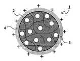
여기에서, 종래의 생체적합성 나노입자의 구조를 도 19에 나타낸다. 생체적합성 나노입자(이하, 단지 "나노입자"라 칭함)(1)의 표면은 폴리비닐알코올(2)로 피복되고, 내부에는 생리활성물질(3)이 봉입되어 있으며, 일반적으로 표면은 음으로 대전되어 있다. 그러나, 생체 내의 세포벽은 음으로 대전되어 있기 때문에, 도 19에 나타낸 바와 같은 나노입자에서는 전기적 반발력에 의해 세포접착성이 나빠진다고 하는 문제점이 있어, 봉입된 생리활성물질을 협착부 등의 병변부위에 국소적으로 또한 효율적으로 도입하기 위해서는, 나노입자의 세포 내로의 이행성을 더한층 높일 필요성이 있었다.Here, the structure of the conventional biocompatible nanoparticles is shown in FIG. The surface of the biocompatible nanoparticles (hereinafter referred to simply as "nanoparticles") 1 is covered with
또한, 일반적으로 생체적합성 폴리머는 소수성(지용성)이며, 나노입자 내에 높은 봉입률로 봉입될 수 있는 생리활성물질은 지용성인 것으로 한정되므로, 특허문헌 3의 방법에서는 핵산이나 유전자 등의 친수성(수용성) 생리활성물질을 스텐트 표면에 충분히 코팅하는 것이 곤란하였다.In general, biocompatible polymers are hydrophobic (lipid-soluble), and bioactive substances that can be encapsulated in nanoparticles with high encapsulation rate are limited to lipophilic, so the method of
상기 문제점을 해결하기 위해서, 특허문헌 4에는, 표면이 양전하 수식(修飾)된 생체적합성 나노입자를 스텐트 본체에 전기적으로 부착시킨 DES가 개시되어 있고, 전기영동법이나 초음파 미스트법 등을 이용해서 통전 상태의 스텐트 본체에 나노입자를 부착시키는 DES의 제조방법도 기재되어 있다. 또한, 특허문헌 5에는, 치료약, 자성 또는 상자성 재료 및 고분자 전해질 다층 외피로 이루어진 나노캡슐(나노입자)을 지닌 의료장치가 개시되어 있고, 의료장치의 예로서 카테터가 기재되어 있다.In order to solve the above problem,
그러나, 특허문헌 4의 방법에서는, 스텐트 본체를 금속 등의 도전성 재료로 형성해둘 필요가 있고, 확장가능부분(벌룬부)이 도전성이 작은 수지로 만들어진 벌룬 카테터에는 적용이 곤란하였다. 또, 특허문헌 5에서는, 단지 봉입된 약물의 방출을 제어하기 위하여 분해가능한 고분자 전해질을 이용해서 나노입자를 형성하는 취지가 기재되어 있을 뿐이고, 표면이 양전하 수식된 생체적합성 나노입자를 코팅한 약제 용출형 카테터의 실시예, 즉, 실제로 어떤 구성의 나노입자가 제조될지, 그리고 어느 정도의 세포접착성 혹은 세포 내로의 도입성이 확인될지에 대해서는 하등 기재되어 있지 않았다.However, in the method of
본 발명은, 상기 문제점을 감안하여, 지용성 또는 수용성의 생리활성물질이 높은 봉입률로 봉입되어 세포 이행성도 우수한 생체적합성 나노입자를 코팅함으로써, 생리활성물질을 세포 내에 효율적으로 도달시킬 수 있고, 취급성도 우수한 확장가능한 약제 용출형 카테터 및 그 간편하고도 저렴한 제조방법을 제공하는 것을 목적으로 한다.The present invention, in view of the above problems, by coating a biocompatible nanoparticles with a fat-soluble or water-soluble physiologically active material is sealed at a high sealing rate and excellent in cell migration, the bioactive material can be efficiently reached within the cell, handling It is an object of the present invention to provide a scalable pharmaceutical dissolution type catheter with excellent durability and a simple and inexpensive manufacturing method thereof.
상기 목적을 달성하기 위하여, 본 발명은, 생리활성물질이 봉입되고, 또한 표면이 정전하 수식된 생체적합성 나노입자를, 음전하 수식된 확장가능부분의 표면에 코팅한 확장가능한 약제 용출형 카테터이다.In order to achieve the above object, the present invention is an expandable drug-eluting catheter in which the biocompatible nanoparticles in which the bioactive material is encapsulated and whose surface is electrostatically modified are coated on the surface of the negatively charged expandable portion.
이 구성에 의하면, 수지로 만들어진 확장가능부분에도 표면이 양으로 대전된 나노입자를 부착시킬 수 있다. 또, 확장가능부분에 코팅되는 나노입자가 양으로 대전되어 있으므로, 음대전의 세포벽에 대한 나노입자의 세포접착성이 높아지고, 내부에 봉입된 생리활성물질의 세포 내로의 도달 효율을 향상시킨 약제 용출형 카테터로 된다. 또한, 예를 들어, 생체적합성 나노입자를 구성하는 폴리머 재료가 지용성인 경우, 지용성 생리활성물질의 봉입률이 높아지지만, 이것에 가해서 나노입자 표면이 양으로 대전되어 있기 때문에, 수용성 또한 음이온성의 생리활성물질을 높은 봉입률로 봉입할 수 있어, 확장가능부분에의 코팅이 가능한 생리활성물질의 선택 범위도 넓어진다.According to this constitution, the surface of the resin can be attached to the positively charged nanoparticles in the expandable portion made of the resin. In addition, since the nanoparticles coated on the expandable part are positively charged, the drug adhesion of the nanoparticles to the cell wall of the negative charge becomes high, and the drug eluting improves the efficiency of reaching the cells inside the bioactive substance encapsulated therein. It becomes a type catheter. For example, when the polymer material constituting the biocompatible nanoparticles is fat-soluble, the encapsulation rate of the fat-soluble physiologically active substance increases, but since the surface of the nanoparticles is positively charged, the water-soluble and anionic physiology is also increased. The active material can be encapsulated at a high encapsulation rate, thereby widening the selection of bioactive materials that can be coated on the expandable portion.
또, 본 발명은, 상기 구성의 약제 용출형 카테터에 있어서, 상기 확장가능부분이 폴리카복실산 혹은 폴리카복실산 유도체에 의해 음전하 수식되어 있는 것으로 하였다.In the present invention, in the drug-eluting catheter having the above configuration, the expandable portion is negatively charged by a polycarboxylic acid or a polycarboxylic acid derivative.
이 구성에 의하면, 확장가능부분의 표면을 용이하게 음대전시킬 수 있다.According to this configuration, the surface of the expandable portion can be easily negatively charged.
또한, 본 발명은, 상기 구성의 약제 용출형 카테터에 있어서, 상기 폴리카복실산이 아크릴산, 메타크릴산, 말레산, 푸마르산, 아스파르트산 혹은 글루탐산의 폴리머, 전분, 셀룰로스 혹은 폴리비닐알코올의 카복시메틸 유도체, 알긴산 및 펙틴으로부터 선택된 1종 이상인 것으로 하였다.In addition, the present invention is a pharmaceutical eluting catheter of the above configuration, wherein the polycarboxylic acid is a polymer of acrylic acid, methacrylic acid, maleic acid, fumaric acid, aspartic acid or glutamic acid, carboxymethyl derivative of starch, cellulose or polyvinyl alcohol, It is assumed that it is at least one selected from alginic acid and pectin.
이 구성에 의하면, 생체에의 영향이 적어, 안전면에 있어서 우수한 약제 용출형 카테터로 된다.According to this structure, it has little influence on a living body, and becomes the chemical | medical agent elution type catheter excellent in safety.
또, 본 발명은, 상기 구성의 약제 용출형 카테터에 있어서, 상기 폴리카복실산 유도체가 아크릴산, 메타크릴산, 말레산의 폴리머의 산무수물 유도체 혹은 에스터 유도체인 것으로 하였다.Moreover, in the chemical | medical agent eluting catheter of the said structure, it is assumed that the said polycarboxylic acid derivative is an acid anhydride derivative or ester derivative of the polymer of acrylic acid, methacrylic acid, and maleic acid.
이 구성에 의하면, 표면이 양으로 대전된 나노입자를 견고하게 부착시키는 동시에, 생체에의 자극이나 독성도 적은 음전하 수식이 가능해진다.This configuration makes it possible to firmly attach the nanoparticles whose surfaces are positively charged, and to modify the negative charges with little irritation and toxicity to the living body.
또한, 본 발명은, 상기 구성의 약제 용출형 카테터에 있어서, 상기 폴리카복실산 유도체가 무수 말레산의 코폴리머(즉, 공중합체)인 것으로 하였다.In the present invention, in the drug eluting catheter of the above constitution, the polycarboxylic acid derivative is a copolymer of maleic anhydride (that is, a copolymer).
이 구성에 의하면, 높은 안전성을 지닌 약제 용출형 카테터로 되어, 음전하 수식 시에 있어서의 취급도 용이해진다.According to this structure, it becomes a drug-eluting catheter with high safety | security, and handling at the time of negative charge modification becomes easy.
또, 본 발명은, 상기 구성의 약제 용출형 카테터에 있어서, 상기 무수 말레산의 공중합체가 무수 말레산-메틸비닐에터 공중합체, 무수 말레산-스타이렌 공중합체 및 무수 말레산-에틸렌 공중합체로부터 선택된 1종 이상인 것으로 하였다.In addition, the present invention is a pharmaceutical dissolution type catheter of the above configuration, wherein the copolymer of maleic anhydride is maleic anhydride-methylvinyl ether copolymer, maleic anhydride-styrene copolymer and maleic anhydride-ethylene air It is assumed that it is at least one selected from coalescing.
이 구성에 의하면, 특히 입수가 용이하고 취급성이 우수한 무수 말레산의 공중합체를 이용하여, 높은 안전성을 지닌 약제 용출형 카테터를 간단하고도 저비용으로 제조가능해진다.According to this structure, especially the drug dissolution type catheter which has high safety can be manufactured simply and at low cost using the copolymer of maleic anhydride which is easy to obtain and excellent in handleability.
또한, 본 발명은, 상기 구성의 약제 용출형 카테터에 있어서, 상기 생체적합성 나노입자는 표면에 양이온(cation)성 고분자를 부착시킴으로써 양전하 수식되어 있는 것으로 하였다.In the present invention, in the drug-eluting catheter of the above constitution, the biocompatible nanoparticles are positively modified by attaching a cationic polymer to the surface.
이 구성에 의하면, 나노입자 표면을 용이하게 양대전시킬 수 있다.According to this configuration, the surface of the nanoparticles can be easily charged.
또, 본 발명은, 상기 구성의 약제 용출형 카테터에 있어서, 상기 양이온성 고분자가 키토산인 것으로 하였다.Moreover, in this invention, in the chemical | medical agent eluting catheter of the said structure, it was assumed that the said cationic polymer is chitosan.
이 구성에 의하면, 생체에의 영향이 없어, 안정성이 높은 약제 용출형 카테터로 된다.According to this structure, there is no influence on a living body, and it becomes a chemically eluting catheter with high stability.
또한, 본 발명은, 상기 구성의 약제 용출형 카테터에 있어서, 상기 생체적합성 나노입자가 폴리락트산, 폴리글라이콜산, 락트산·글라이콜산 공중합체 혹은 락트산·아스파르트산 공중합체 중 어느 하나로 구성되는 것으로 하였다.In addition, the present invention, in the drug-eluting catheter of the above configuration, wherein the biocompatible nanoparticles are composed of any one of polylactic acid, polyglycolic acid, lactic acid-glycolic acid copolymer or lactic acid-aspartic acid copolymer It was.
이 구성에 의하면, 생체에의 자극·독성이 낮고, 또한 생체적합성 고분자의 분해에 의해 생리활성물질의 서방(徐放)이 가능한 약제 용출형 카테터로 된다.According to this structure, it becomes a drug-eluting catheter which has low irritation and toxicity to a living body, and is capable of sustained release of a bioactive substance by decomposition of a biocompatible polymer.
또, 본 발명은, 상기 구성의 약제 용출형 카테터에 있어서, 상기 생리활성물질이 핵산화합물인 것으로 하였다.In the present invention, in the drug-eluting catheter of the above constitution, the physiologically active substance is assumed to be a nucleic acid compound.
이 구성에 의하면, 병변부위에 안전하고도 효율적으로 핵산화합물을 도입하여 핵산 레벨에서 치료할 수 있고, 예를 들어, 혈관 중의 협착부에 적용할 경우에 재협착의 가능성이 적은 약제 용출형 카테터를 용이하게 제조할 수 있다.According to this constitution, a nucleic acid compound can be safely and efficiently introduced into the lesion site and treated at the nucleic acid level. For example, a drug-eluting catheter with a low possibility of restenosis when applied to a constriction in blood vessels is easily provided. It can manufacture.
또한, 본 발명은, 상기 구성의 약제 용출형 카테터에 있어서, 상기 핵산화합물이 플라스미드 DNA(plasmid DNA), 유전자, 디코이, siRNA, 올리고뉴클레오타이드, 안티센스 올리고뉴클레오타이드, 리보자임 및 압타머(aptamer)로부터 선택된 1종 이상인 것으로 하였다.In addition, the present invention, the drug eluting catheter of the above configuration, wherein the nucleic acid compound is selected from plasmid DNA (plasmid DNA), genes, decoy, siRNA, oligonucleotides, antisense oligonucleotides, ribozymes and aptamers (aptamers) It was set as 1 or more types.
이 구성에 의하면, 핵산화합물치료용 툴(tool)로서 특히 적합한 약제 용출형 카테터를 제공가능해진다.According to this configuration, it is possible to provide a drug eluting catheter that is particularly suitable as a tool for treating nucleic acid compounds.
또, 본 발명은, 상기 구성의 약제 용출형 카테터에 있어서, 상기 핵산화합물이 NFκB 디코이 올리고뉴클레오타이드인 것으로 하였다.In the present invention, in the drug-eluting catheter of the above constitution, the nucleic acid compound is an NFκB decoy oligonucleotide.
이 구성에 의하면, NFκB에 결합해서 염증을 야기하는 사이토카인 등의 생성을 저해하여, PTA 시행 시의 급성기 염증 반응을 억제해서 재협착을 효과적으로 방지할 수 있는 약제 용출형 카테터를 제공가능해진다.According to this configuration, it is possible to provide a drug-eluting catheter that can inhibit the production of cytokines and the like that bind to NFκB and cause inflammation, thereby inhibiting the acute phase inflammatory response during PTA and effectively preventing restenosis.
또한, 본 발명은, 상기 구성의 약제 용출형 카테터를 혈관내 카테터로서 이용하는 것으로 하였다.In addition, the present invention is to use the drug-eluting catheter of the above configuration as an endovascular catheter.
이 구성에 의하면, PTA를 행한 혈관 부위의 재협착방지에 우수한 효과를 발휘한다.According to this structure, it is excellent in preventing restenosis of the blood vessel site which performed PTA.
또, 본 발명은, 상기 구성의 약제 용출형 카테터에 있어서, 상기 확장가능부분으로서 벌룬을 가진 벌룬 카테터인 것으로 하였다.In addition, the present invention is intended to be a balloon catheter having a balloon as the expandable portion in the drug-eluting catheter of the above configuration.
이 구성에 의하면, 카테터를 혈관 내의 협착부까지 삽입한 후, 벌룬을 부풀려서 협착부를 용이하게 확장시킬 수 있다.According to this structure, after inserting a catheter to the constriction part in a blood vessel, a balloon can be inflated and a constriction part can be expanded easily.
또한, 본 발명은, 상기 구성의 약제 용출형 카테터에 있어서, 상기 벌룬의 표면에 오목부를 형성하는 것으로 하였다.In addition, in the chemical | medical agent eluting catheter of the said structure, it is supposed that this invention forms the recessed part in the surface of the said balloon.
이 구성에 의하면, 오목부 내에 생체적합성 나노입자를 다량으로 담지시키는 동시에, 벌룬의 팽창에 따라 오목부를 소실시켜서 오목부 내의 생체적합성 나노입자를 밀어내어, 협착부의 혈관벽에 효율적으로 부착시킬 수 있다.According to this structure, a large amount of biocompatible nanoparticles can be supported in the concave portion, the concave portion disappears as the balloon expands, and the biocompatible nanoparticles in the concave portion can be pushed out and attached to the blood vessel wall of the constriction portion efficiently.
또, 본 발명은, 상기 구성의 약제 용출형 카테터에 있어서, 상기 오목부가 원형 혹은 타원형인 것으로 하였다.Moreover, in the chemical | medical agent eluting catheter of the said structure, this invention was supposed that the said recessed part was circular or elliptical.
이 구성에 의하면, 벌룬을 팽창시켜서 오목부를 용이하게 변형, 소실시킬 수 있다.According to this structure, a balloon can be expanded and a recess can be easily deformed and lost.
또한, 본 발명은, 상기 구성의 약제 용출형 카테터를 이용한, 혈관 협착 또는 투석용 션트내 협착의 치료방법이다.In addition, the present invention is a method for treating vascular stenosis or intrashunt shunt stenosis using the drug-eluting catheter of the above configuration.
이 구성에 의하면, PTA를 행한 혈관 부위의 재협착이나, 혈액투석환자의 팔에 형성한 션트 혈관의 협착을 효과적으로 치료할 수 있다.According to this structure, the restenosis of the vascular part which performed PTA and the narrowing of the shunt blood vessel formed in the arm of a hemodialysis patient can be treated effectively.
또, 본 발명은, 적어도 양이온성 고분자를 용해시킨 수용액에, 적어도 생리활성물질의 용액과 생체적합성 고분자를 유기용매에 용해시킨 용액의 혼합액을 가해서, 상기 생리활성물질이 상기 생체적합성 고분자 중에 봉입되고, 또한 입자 표면이 양전하 수식된 생체적합성 나노입자의 현탁액을 생성하는 나노입자 형성공정과, 카테터 본체의 확장가능부분을 음전하 수식하는 음전하 수식공정과, 상기 생체적합성 나노입자를 음전하 수식된 상기 확장가능부분에 부착시켜서 나노입자층을 형성하는 나노입자 부착공정과, 상기 나노입자층을 건조시키는 건조 공정을 포함하는 약제 용출형 카테터의 제조방법이다.In the present invention, at least an aqueous solution in which a cationic polymer is dissolved is added to a mixed solution of at least a solution of a bioactive substance and a solution in which a biocompatible polymer is dissolved in an organic solvent, and the bioactive substance is encapsulated in the biocompatible polymer. In addition, the nanoparticles forming process of producing a suspension of the biocompatible nanoparticles with a positively charged particle surface, a negative charge modification process to negatively charge the expandable portion of the catheter body, and the expandable negatively modified the biocompatible nanoparticles A method of producing a drug eluting catheter comprising a nanoparticle attachment step of attaching to a portion to form a nanoparticle layer and a drying step of drying the nanoparticle layer.
이 방법에 의하면, 확장가능부분에 균일한 나노입자층이 견고하게 형성되므로, 생리활성물질을 세포 내에 효율적으로 송달가능해서 취급성도 우수한 확장가능한 약제 용출형 카테터를 간편하고도 저비용으로 제조할 수 있다.According to this method, since a uniform nanoparticle layer is firmly formed in the expandable portion, a scalable drug-dissolved catheter capable of efficiently delivering a physiologically active substance into a cell and excellent in handleability can be produced easily and at low cost.
또한, 본 발명은, 상기 구성의 약제 용출형 카테터의 제조방법에 있어서, 상기 음전하 수식공정은, 상기 확장가능부분의 폴리카복실산 혹은 폴리카복실산 유도체의 용액 중에의 디핑(침지)에 의해 행해지는 것으로 하였다.In the method for producing a drug-eluting catheter having the above-described configuration, the negative charge modification step is performed by dipping (immersion) in a solution of the polycarboxylic acid or polycarboxylic acid derivative of the expandable portion. .
이 방법에 의하면, 간편한 방법으로 균일한 음대전성의 수지층을 효율적으로 형성할 수 있다.According to this method, a uniform negatively chargeable resin layer can be efficiently formed by a simple method.
또, 본 발명은, 상기 구성의 약제 용출형 카테터의 제조방법에 있어서, 상기 생체적합성 나노입자의 현탁액에, 추가로 음이온성 생리활성물질을 첨가하는 것으로 하였다.Moreover, in this invention, in the manufacturing method of the chemical | medical agent eluting catheter of the said structure, it is supposed that the anionic bioactive substance is further added to the suspension of the said biocompatible nanoparticle.
이 방법에 의하면, 나노입자 표면의 정전하에 의해 음이온성 생리활성물질이 정전기적으로 담지된 상태에서 음전하 수식된 확장가능부분에 끌어 당겨져서 부착되므로, 코팅이 곤란했던 핵산, 유전자 등의 음이온성 생리활성물질을 확장가능부분에 고농도로 부착시킨 약제 용출형 카테터를 제조할 수 있다.According to this method, anionic physiological substances such as nucleic acids and genes, which have been difficult to coat, are attached by being attracted and attached to the negatively-charged expandable portion in the state in which the anionic bioactive material is electrostatically supported by the electrostatic charge on the surface of the nanoparticles. A drug eluting catheter can be prepared in which the active substance is attached to the expandable portion at a high concentration.
또한, 본 발명은, 상기 구성의 약제 용출형 카테터의 제조방법에 있어서, 상기 나노입자 부착공정을 복수회 반복함으로써, 상기 확장가능부분에 형성된 상기 나노입자층 위에 추가로 나노입자층을 적층하는 것으로 하였다.In the present invention, in the method for producing a drug-eluting catheter having the above configuration, the nanoparticle adhesion step is repeated a plurality of times, whereby a nanoparticle layer is further laminated on the nanoparticle layer formed on the expandable portion.
이 방법에 의하면, 코팅되는 나노입자량을 증대시키는 동시에, 확장가능부분의 나노입자층 전체를 균일하게 할 수 있다.According to this method, the amount of nanoparticles to be coated can be increased, and the entire nanoparticle layer of the expandable portion can be made uniform.
또, 본 발명은, 상기 구성의 약제 용출형 카테터의 제조방법에 있어서, 상기 나노입자 부착공정을 복수회 반복함으로써, 다른 생리활성물질이 봉입된 생체적합성 나노입자로 이루어진 상기 나노입자층을, 적층 형상 또는 모자이크 형상으로 형성하는 것으로 하였다.In addition, the present invention is a method of manufacturing a drug-eluting catheter of the above configuration, by repeating the nanoparticle attachment step a plurality of times, the nanoparticle layer made of biocompatible nanoparticles in which other physiologically active substances are encapsulated, stacked shape Or it was set as the mosaic shape.
이 방법에 의하면, 생체 내로의 유치 후 단시간에 용출시키는 생리활성물질이 봉입된 나노입자는 바깥층에, 장시간 경과 후에 용출시키는 생리활성물질이 봉입된 나노입자는 내층에 부착시켜 두면, 2종류 이상의 생리활성물질의 용출시간을 계획적으로 제어할 수 있는 약제 용출형 카테터를 제조가능해진다.According to this method, two or more types of physiology are allowed when nanoparticles containing a bioactive substance eluted in a short time after induction into the living body are attached to the inner layer and nanoparticles containing a bioactive substance eluted after a long time are attached to the inner layer. It is possible to prepare a drug eluting catheter capable of deliberately controlling the elution time of the active substance.
또한, 본 발명은, 상기 구성의 약제 용출형 카테터의 제조방법에 있어서, 상기 나노입자층에 생분해성 고분자의 용액을 함침시키는 함침공정을 포함하는 것으로 하였다.In addition, the present invention is intended to include an impregnation step of impregnating a solution of a biodegradable polymer in the nanoparticle layer in the method for producing a drug-eluting catheter having the above-described configuration.
이 방법에 의하면, 확장가능부분으로부터의 나노입자의 용출속도를 제어가능하게 되고, 또한 나노입자끼리 응집되어 불용성의 피막으로 되는 것을 방지할 수 있다.According to this method, it is possible to control the dissolution rate of the nanoparticles from the expandable portion and to prevent the nanoparticles from agglomerating to form an insoluble film.
또, 본 발명은, 상기 구성의 약제 용출형 카테터의 제조방법에 있어서, 상기 함침공정에 있어서, 상기 생분해성 고분자의 용액 중에 추가로 생리활성물질을 첨가하는 것으로 하였다.In addition, in the method for producing a drug-eluting catheter having the above-described configuration, in the impregnation step, a physiologically active substance is further added to the solution of the biodegradable polymer.
이 방법에 의하면, 나노입자 외부의 생분해성 고분자층 중에 봉입된 생리활성물질을 즉효적으로 작용시키는 동시에, 나노입자 내부에 봉입된 생리활성물질을 지효적(遲效的)이면서도 지속적으로 작용시킬 수 있다.According to this method, the bioactive substance encapsulated in the biodegradable polymer layer outside the nanoparticles can be acted immediately, and the bioactive substance encapsulated inside the nanoparticles can be operated effectively and continuously. have.
또한, 본 발명은, 상기 구성의 약제 용출형 카테터의 제조방법에 있어서, 상기 함침공정에 있어서 나노입자층에 함침시키는 생분해성 고분자는, 상기 생체적합성 나노입자를 형성하는 생체적합성 고분자보다 생체 내에서의 분해 속도가 빠른 것으로 하였다.In addition, the present invention provides a method for producing a drug-eluting catheter of the above configuration, wherein the biodegradable polymer to be impregnated into the nanoparticle layer in the impregnation step is more in vivo than the biocompatible polymer forming the biocompatible nanoparticles. The decomposition rate was set to be fast.
이 방법에 의하면, 생분해성 고분자의 분해에 의해 확장가능부분으로부터 나노입자가 용출되고, 세포 내로 이행한 후에 나노입자를 형성하는 생체적합성 고분자의 분해에 의해 생리활성물질이 서서히 방출되므로, 세포 내로의 생리활성물질의 도입 효율을 높이는 동시에, 생리활성물질의 도입 타이밍의 제어도 용이한 약제 용출형 카테터를 제조할 수 있다.According to this method, the nanoparticles are eluted from the expandable portion by the decomposition of the biodegradable polymer, and the bioactive material is gradually released by the decomposition of the biocompatible polymer which forms the nanoparticles after the transition into the cell, thereby allowing the intracellular into the cell. A drug-eluting catheter can be produced in which the introduction efficiency of the bioactive substance is increased and the control of the introduction timing of the bioactive substance is also easy.
도 1은 본 발명의 약제 용출형 카테터에 이용되는, 입자 표면이 양전하 수식되어 생리활성물질이 입자 내부에 봉입된 나노스피어(nanospere)의 구조를 나타낸 모식도;
도 2는 본 발명의 약제 용출형 카테터에 이용되는, 입자 표면이 양전하 수식되어 생리활성물질이 입자 내부에 봉입되고 입자 표면에도 담지된 나노스피어의 구조를 나타낸 모식도;
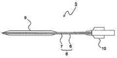
도 3은 본 발명의 약제 용출형 카테터에 이용되는 벌룬 카테터 본체의 일례를 나타낸 측면도;
도 4는 카테터 본체의 벌룬부에 나노입자층을 형성한 상태를 나타낸 단면확대도;
도 5는 나노입자층이 형성된 벌룬부에 생분해성 고분자층을 형성한 상태를 나타낸 단면확대도;
도 6A는 오목부가 형성된 벌룬부에 음대전성 수지층 및 나노입자층을 적층한 상태를 나타낸 단면확대도;
도 6B는 벌룬부를 팽창시킨 상태를 나타낸 단면확대도;
도 7A는 실시예 5에 있어서의 벌룬부의 절개 방법을 나타낸 도면;
도 7B는 실시예 5에 있어서의 벌룬부의 관찰 개소를 나타낸 도면;
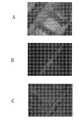
도 8A는 생리식염수에 침지하기 전의 벌룬부(도 7B의 L)의 형광 현미경 사진;
도 8B는 생리식염수에 침지하기 전의 벌룬부(도 7B의 C)의 형광 현미경 사진;
도 8C는 생리식염수에 침지하기 전의 벌룬부(도 7B의 R)의 형광 현미경 사진;
도 9A는 생리식염수에 60분간 침지한 후의 벌룬부(도 7B의 L)의 형광 현미경 사진;
도 9B는 생리식염수에 60분간 침지한 후의 벌룬부(도 7B의 C)의 형광 현미경 사진;
도 9C는 생리식염수에 60분간 침지한 후의 벌룬부(도 7B의 R)의 형광 현미경 사진;
도 10A는 생리식염수에 60분간 침지하고, 추가로 아세토나이트릴에 10분간 침지한 후의 벌룬부(도 7B의 L)의 형광 현미경 사진;
도 10B는 생리식염수에 60분간 침지하고, 추가로 아세토나이트릴에 10분간 침지한 후의 벌룬부(도 7B의 C)의 형광 현미경 사진;
도 10C는 생리식염수에 60분간 침지하고, 추가로 아세토나이트릴에 10분간 침지한 후의 벌룬부(도 7B의 R)의 형광 현미경 사진;
도 11A는 벌룬부(도 7B의 L)를 60분간 침지한 생리식염수를 여과한 멤브레인 필터의 형광 현미경 사진;
도 11B는 벌룬부(도 7B의 C)를 60분간 침지한 생리식염수를 여과한 멤브레인 필터의 형광 현미경 사진;
도 11C는 벌룬부(도 7B의 R)를 60분간 침지한 생리식염수를 여과한 멤브레인 필터의 형광 현미경 사진;
도 12A는 실시예 6에 이용한 표면에 오목부가 형성된 벌룬부의 측면도;
도 12B는 도 12A에 있어서의 벌룬부의 A-A' 단면도;
도 12C는 도 12A에 있어서의 벌룬부의 B-B' 단면도;
도 13A는 실시예 6에 있어서의 벌룬부의 가압 전의 형광 현미경 사진;
도 13B는 실시예 6에 있어서의 벌룬부의 10atm 가압 후의 형광 현미경 사진;
도 13C는 실시예 6에 있어서의 벌룬부의 20atm 가압 후의 형광 현미경 사진;
도 14는 실시예 7에 있어서의 혈관 내피를 손상시키지 않았던 토끼의 정상 복부 대동맥의 단면을 나타낸 현미경 사진(촬영 배율: 40배);
도 15는 실시예 7에 있어서의 NFκB 디코이 비함유 PLGA 나노스피어로 코팅한 PTA 벌룬 카테터로 혈관 내피를 찰과시킨, 토끼의 복부 대동맥의 단면[NFκB 디코이(-)]을 나타낸 현미경 사진(촬영 배율: 40배);
도 16은 실시예 7에 있어서의 NFκB 디코이 함유 PLGA 나노스피어로 코팅한 PTA 벌룬 카테터로 혈관 내피를 찰과시킨, 토끼의 복부 대동맥의 단면[NFκB 디코이(+)]을 나타낸 현미경 사진(촬영 배율: 40배);
도 17A는 실시예 7에 있어서의 NFκB 디코이(+)군 및 NFκB 디코이(-)군에서, 복부 대동맥의 내막 면적(㎟)을 비교한 그래프;
도 17B는 실시예 7에 있어서의 NFκB 디코이(+)군 및 NFκB 디코이(-)군에서, 복부 대동맥의 중간막 면적(㎟)을 비교한 그래프;
도 17C는 실시예 7에 있어서의 NFκB 디코이(+)군 및 NFκB 디코이(-)군에서, 복부 대동맥의 내막/중간막의 면적비(I/M)를 비교한 그래프;
도 18A는 실시예 8에 있어서의 대조군, NFκB 디코이(+)군 및 NFκB 디코이(-)군에서, 경동맥의 내막 면적(㎟)을 비교한 그래프;
도 18B는 실시예 8에 있어서의 대조군, NFκB 디코이(+)군 및 NFκB 디코이(-)군에서, 경동맥 중간막 면적(㎟)을 비교한 그래프;
도 18C는 실시예 8에 있어서의 대조군, NFκB 디코이(+)군 및 NFκB 디코이(-)군에서, 경동맥의 내막/중간막의 면적비(I/M)를 비교한 그래프;
도 19는 종래의 나노입자의 구조를 나타낸 모식도.1 is a schematic diagram showing the structure of a nanosphere (nanospere) is used in the drug eluting catheter of the present invention, the surface of the particle is modified with a positive charge encapsulated inside the particle;
Figure 2 is a schematic diagram showing the structure of the nanospheres used in the drug eluting catheter of the present invention, the surface of the particle is modified with a positive charge, the bioactive material is enclosed in the particle and also supported on the particle surface;
Figure 3 is a side view showing an example of the balloon catheter body used in the drug-eluting catheter of the present invention;
4 is an enlarged cross-sectional view showing a state in which a nanoparticle layer is formed on a balloon of a catheter body;
5 is an enlarged cross-sectional view showing a state in which a biodegradable polymer layer is formed on a balloon in which a nanoparticle layer is formed;
6A is an enlarged cross-sectional view showing a state in which a negatively charged resin layer and a nanoparticle layer are laminated on a balloon portion having a recessed portion;
6B is an enlarged cross-sectional view showing a state in which a balloon is inflated;
7A is a view showing a cutting method of a balloon part in Example 5;
FIG. 7B is a view showing observation points of a balloon section in Example 5; FIG.
8A is a fluorescence micrograph of a balloon portion (L in FIG. 7B) before immersion in physiological saline;
8B is a fluorescence micrograph of the balloon portion (C of FIG. 7B) before immersion in physiological saline;
8C is a fluorescence micrograph of the balloon portion (R in FIG. 7B) before immersion in physiological saline;
9A is a fluorescence micrograph of a balloon portion (L in FIG. 7B) after immersion in physiological saline for 60 minutes;
Fig. 9B is a fluorescence micrograph of the balloon portion (C in Fig. 7B) after immersion in physiological saline for 60 minutes;
9C is a fluorescence micrograph of a balloon portion (R in FIG. 7B) after immersion in physiological saline for 60 minutes;
10A is a fluorescence micrograph of a balloon portion (L in FIG. 7B) after immersion in physiological saline for 60 minutes and further immersion in acetonitrile for 10 minutes;
Fig. 10B is a fluorescence micrograph of the balloon portion (C in Fig. 7B) after immersion in physiological saline for 60 minutes and further immersion in acetonitrile for 10 minutes;
Fig. 10C is a fluorescence micrograph of the balloon portion (R in Fig. 7B) after immersion in physiological saline for 60 minutes and further immersion in acetonitrile for 10 minutes;
11A is a fluorescence micrograph of a membrane filter filtering physiological saline immersed in a balloon (L in FIG. 7B) for 60 minutes;
11B is a fluorescence micrograph of a membrane filter filtering physiological saline immersed in a balloon portion (C of FIG. 7B) for 60 minutes;
11C is a fluorescence micrograph of a membrane filter filtering physiological saline immersed in a balloon portion (R in FIG. 7B) for 60 minutes;
12A is a side view of a balloon portion having recesses formed on its surface used in Example 6;
FIG. 12B is a sectional view taken along AA ′ of the balloon portion in FIG. 12A; FIG.
FIG. 12C is a BB 'cross-sectional view of the balloon portion in FIG. 12A; FIG.
13A is a fluorescence micrograph before pressing of the balloon portion in Example 6;
13B is a fluorescence micrograph after 10atm pressurization of the balloon portion in Example 6;
13C is a fluorescence micrograph after 20atm pressurization of the balloon portion in Example 6;
FIG. 14 is a micrograph (photographing magnification: 40 times) showing a cross section of a normal abdominal aorta of a rabbit which did not damage the vascular endothelium in Example 7. FIG.
Fig. 15 is a micrograph (photographing magnification) showing a cross section [NFκB decoy (-)] of the abdominal aorta of a rabbit in which the vascular endothelium was scratched with a PTA balloon catheter coated with a NFκB decoy-free PLGA nanosphere in Example 7. : 40 times);
Fig. 16 is a micrograph (photographing magnification) showing a cross section [NFκB decoy (+)] of the abdominal aorta of a rabbit in which the vascular endothelium was scratched with a PTA balloon catheter coated with NFκB decoy-containing PLGA nanospheres in Example 7. 40 times);
17A is a graph comparing the intima area (mm 2) of the abdominal aorta in the NFκB decoy (+) group and the NFκB decoy (−) group in Example 7;
17B is a graph comparing the median membrane area (mm 2) of the abdominal aorta in the NFκB decoy (+) group and the NFκB decoy (−) group in Example 7;
17C is a graph comparing the area ratio (I / M) of the intima / membrane of the abdominal aorta in the NFκB decoy (+) group and the NFκB decoy (−) group in Example 7;
18A is a graph comparing the intima area (mm 2) of the carotid artery in the control group, NFκB decoy (+) group and NFκB decoy (−) group in Example 8;
18B is a graph comparing carotid interlayer area (mm 2) in the control group, NFκB decoy (+) group and NFκB decoy (−) group in Example 8; FIG.
18C is a graph comparing the area ratio (I / M) of the intima / membrane of the carotid artery in the control, NFκB decoy (+) group and NFκB decoy (−) group in Example 8;
19 is a schematic diagram showing the structure of a conventional nanoparticle.
이하, 본 발명의 실시예에 대해서 도면을 참조하면서 상세히 설명한다. 본 발명의 약제 용출형 카테터의 제조방법은, 지용성 혹은 친수성의 생리활성물질을 봉입하고, 또한 표면을 양으로 대전시킨(양전하 수식된) 생체적합성 나노입자를 형성하는 공정과, 카테터 본체의 확장가능부분을 음으로 대전시키는(음전하 수식하는) 음전하공정과, 나노입자를 음전하 수식된 확장가능부분에 부착시켜서 나노입자층을 형성하는 나노입자 부착공정과, 나노입자층을 건조시키는 건조공정을 포함하는 것이다.EMBODIMENT OF THE INVENTION Hereinafter, embodiment of this invention is described in detail, referring drawings. The method for producing a drug-eluting catheter of the present invention comprises the steps of encapsulating a fat-soluble or hydrophilic bioactive material and forming a biocompatible nanoparticle having positively charged (positive charge-modified) surface, and the catheter body can be expanded. A negative charge step of negatively charging the part (negative charge modification), a nanoparticle attachment step of attaching the nanoparticles to the negatively charged expandable part to form a nanoparticle layer, and a drying step of drying the nanoparticle layer.
일반적으로, 액체 중에 분산된 입자의 대부분은 양 또는 음으로 대전되어 있어, 반대의 전하를 가진 이온이 입자 표면에 강하게 끌어 당겨져서 고정된 층(고정층)과, 그 바깥쪽에 존재하는 층(확산층)으로, 소위 확산 전기 2중층이 형성되어 있어, 확산층의 안쪽의 일부와 고정층이 입자와 함께 이동하는 것으로 추정된다.In general, most of the particles dispersed in the liquid are positively or negatively charged, so that the ions having opposite charges are attracted strongly to the surface of the particles so that they are fixed (fixed layer) and those which exist outside of them (diffusion layer). Thus, a so-called diffusion electric double layer is formed, and it is estimated that a part of the inside of the diffusion layer and the fixed layer move with the particles.
제타 전위는, 입자로부터 충분히 떨어진 전기적으로 중성인 영역의 전위를 기준으로 한 경우의, 상기 이동이 생기는 면(미끄럼 면)의 전위이다. 제타 전위의 절대치가 증가하면, 입자 간의 반발력이 강해져서 입자의 안정성은 높아지고, 반대로 제타 전위가 0에 가까워짐에 따라서 입자는 응집을 일으키기 쉬워진다. 그 때문에, 제타 전위는 입자의 분산 상태의 지표로서 이용되고 있다.The zeta potential is a potential of the surface (sliding surface) where the movement occurs when the potential of the electrically neutral region sufficiently separated from the particles is referenced. When the absolute value of the zeta potential increases, the repulsive force between the particles becomes stronger, and the stability of the particles becomes high. On the contrary, as the zeta potential approaches zero, the particles tend to cause aggregation. Therefore, the zeta potential is used as an index of the dispersed state of the particles.
따라서, 음대전의 세포벽에 대한 접착성을 증대시켜서 나노입자를 세포 내로 효율적으로 이행시키기 위해서는, 나노입자 표면이 양의 제타 전위를 갖도록 대전시키는 것이 바람직하다. 본 발명에 있어서는, 나노입자 형성공정(후술)에 있어서 양이온성 고분자를 빈용매(poor solvent) 중에 첨가한다. 이것에 의해, 형성된 나노입자의 표면이 양이온성 고분자에 의해 수식(피복)되어, 입자 표면의 제타 전위가 양으로 된다.Therefore, in order to increase the adhesion of the negative charge to the cell wall and to efficiently transfer the nanoparticles into the cells, it is preferable to charge the nanoparticle surface so as to have a positive zeta potential. In the present invention, the cationic polymer is added in a poor solvent in the nanoparticle formation step (described later). As a result, the surface of the formed nanoparticles is modified (coated) by the cationic polymer, and the zeta potential on the surface of the particles becomes positive.
또, 나노입자 표면을 양으로 대전시킴으로써, 카테터 본체의 음전하 수식된 확장가능부분에 나노입자를 능동적으로 부착시킬 수 있어, 나노입자의 부착 효율을 높이는 동시에, 일단 부착된 나노입자는 확장가능부분에 강고하게 고착되기 때문에, 제조 공정 중이나 생체 내로의 삽입 및 확장 시에 있어서의 나노입자의 이탈을 방지할 수 있다. 이하, 나노입자 내부로의 생리활성물질의 봉입공정부터 확장가능부분에의 부착공정까지 차례로 설명한다.In addition, by positively charging the surface of the nanoparticles, the nanoparticles can be actively attached to the negatively charged expandable portion of the catheter body, thereby increasing the adhesion efficiency of the nanoparticles and at the same time the nanoparticles once attached to the expandable portion. Since it firmly adheres, it is possible to prevent separation of the nanoparticles during the manufacturing process or during insertion and expansion into the living body. Hereinafter, the process from the step of encapsulating the bioactive material into the nanoparticles to the step of attaching to the expandable part will be described.
(나노입자 형성공정)(Nanoparticle forming process)
본 발명에 이용되는 생체적합성 나노입자는, 생리활성물질 및 생체적합성 고분자를 1,000㎚ 미만의 평균 입자 직경을 지닌 나노 단위의 크기의 입자(나노스피어)로 가공할 수 있는 구형 정석법(球形晶析法)을 이용해서, 나노입자의 내부에 생리활성물질을 봉입함으로써 제조된다. 구형 정석법은 고용단력을 발생하지 않는 입자 조제법이기 때문에, 특히, 생리활성물질이 외부 응력에 약한 핵산화합물 등인 경우에도 적합하게 이용할 수 있다.The biocompatible nanoparticles used in the present invention are spherical crystallization methods that can process bioactive materials and biocompatible polymers into nano-sized particles (nanospheres) having an average particle diameter of less than 1,000 nm. The method is prepared by encapsulating a bioactive substance inside the nanoparticles. Since the spherical crystallization method is a particle preparation method that does not generate high solubility, it can be suitably used even when the bioactive substance is a nucleic acid compound or the like which is weak to external stress.
구형 정석법은, 화합물 합성의 최종 프로세스에 있어서의 결정의 생성·성장 프로세스를 제어함으로써 구형상의 결정 입자를 설계하고, 그 물성을 직접 제어해서 가공할 수 있는 방법이다. 이 구형 정석법의 하나로, 에멀젼 용매확산법(emulsion solvent diffusion method; ESD법)이 있다.Spherical crystallization is a method which can design a spherical crystal grain by controlling the formation | generation process and the growth process of the crystal | crystallization in the final process of compound synthesis, and can control and process the physical property directly. One of these spherical crystallization methods is an emulsion solvent diffusion method (ESD method).
ESD법은, 이하에 나타낸 바와 같은 원리에 의해서, 나노스피어를 제조하는 기술이다. 본 방법에는, 생리활성물질을 봉입하는 기제 폴리머로 되는 PLGA(락트산·글라이콜산 공중합체) 등을 용해시킬 수 있는 양용매(good solvent)와, 이것과는 반대로 PLGA를 용해시키지 않는 빈용매의 2종류의 용매가 이용된다. 이 양용매에는, PLGA를 용해시키고, 또한 빈용매에 혼화되는 아세톤 등의 유기용매를 이용한다. 그리고, 빈용매에는, 통상, 폴리비닐알코올 수용액 등을 이용한다.The ESD method is a technique for producing nanospheres based on the principle shown below. In this method, a good solvent capable of dissolving PLGA (lactic acid / glycolic acid copolymer) or the like as a base polymer encapsulating a bioactive substance, and a poor solvent that does not dissolve PLGA on the contrary Two kinds of solvents are used. In this good solvent, an organic solvent such as acetone which dissolves PLGA and is mixed with the poor solvent is used. And a polyvinyl alcohol aqueous solution etc. are used normally for a poor solvent.
조작 순서로서는, 우선, 양용매 중에 PLGA를 용해 후, 이 PLGA가 석출되지 않도록, 생리활성물질의 용해액을 양용매 중에 첨가 혼합한다. 이 PLGA와 생리활성물질의 혼합액을, 빈용매 중에 교반 하, 적하하면, 혼합액 중의 양용매(유기용매)가 빈용매 중에 급속하게 확산 이행한다. 그 결과, 빈용매 중에서 양용매의 자기유화가 일어나, 서브마이크론 크기의 양용매의 에멀젼 액적이 형성된다. 또한, 양용매와 빈용매의 상호 확산에 의해, 에멀젼 내로부터 유기용매가 빈용매에 계속적으로 확산되어 가므로, 에멀젼 액적 내의 PLGA 및 생리활성물질의 용해도가 저하하여, 최종적으로, 생리활성물질을 포함한 구형 결정 입자의 PLGA 나노스피어가 생성된다.As an operation procedure, first, after dissolving PLGA in a good solvent, the dissolution liquid of a physiologically active substance is added and mixed in a good solvent so that this PLGA may not precipitate. When the mixed solution of PLGA and the bioactive substance is added dropwise while stirring in a poor solvent, the good solvent (organic solvent) in the mixed solution rapidly diffuses into the poor solvent. As a result, self-emulsification of the good solvent occurs in the poor solvent, and emulsion droplets of the good solvent having a submicron size are formed. In addition, due to the mutual diffusion of the good solvent and the poor solvent, the organic solvent continuously diffuses from the emulsion into the poor solvent, so that the solubility of PLGA and the bioactive substance in the emulsion droplets decreases, and finally, the bioactive substance is removed. PLGA nanospheres of spherical crystal grains are produced.
상기 구형 정석법에서는, 물리화학적인 수법으로 나노입자를 형성할 수 있고, 게다가 얻어지는 나노입자가 대략 구형이기 때문에, 균질한 나노입자를, 촉매나 원료화합물의 잔류라고 하는 문제를 고려할 필요가 없이, 용이하게 형성할 수 있다. 또, 본 발명에 있어서는 빈용매 중에 양이온성 고분자를 첨가해서 나노입자 표면을 양이온성 고분자로 피복함으로써, 입자 표면을 양으로 대전시키고 있다. 이러한 나노입자의 구조를 도 1에 나타낸다. 나노입자(1)의 표면은 폴리비닐알코올(2)로 피복되고, 내부에 생리활성물질(3)이 내포되어 있다. 또한, 폴리비닐알코올(2)의 외표면은 양이온성 고분자(4)로 피복되어 있어, 양이온성 고분자(4)에 의해 양의 제타 전위를 지니고 있다.In the spherical crystallization method, nanoparticles can be formed by a physicochemical method, and since the obtained nanoparticles are approximately spherical, the homogeneous nanoparticles do not need to be considered as a residual of a catalyst or a raw material compound. It can be formed easily. In the present invention, the surface of the particles is positively charged by adding a cationic polymer to the poor solvent and coating the surface of the nanoparticles with the cationic polymer. The structure of such nanoparticles is shown in FIG. The surface of the
또한, 생체 내의 세포벽은 음으로 대전되어 있지만, 종래의 구형 정석법으로 제조된 나노입자의 표면은, 일반적으로 음의 제타 전위를 지니고 있기 때문에(도 19 참조), 전기적 반발력에 의해 나노입자의 세포접착성이 나빠진다고 하는 문제점이 있었다. 따라서, 본 발명과 같이 양이온성 고분자를 이용해서 나노입자 표면이 양의 제타 전위를 지니도록 대전시키는 것은, 음대전의 세포벽에 대한 나노입자의 접착성을 증대시켜, 생리활성물질의 세포내 이행성을 향상시키는 관점에서도 바람직하다.In addition, although the cell wall in the living body is negatively charged, the surface of the nanoparticles prepared by the conventional spherical crystallization method generally has a negative zeta potential (see FIG. 19), so that the cells of the nanoparticles are electrically repulsive. There existed a problem that adhesiveness worsened. Therefore, charging the nanoparticle surface with a positive zeta potential using a cationic polymer as in the present invention increases the adhesion of the nanoparticles to the cell walls of negative charges, leading to intracellular transferability of the bioactive substance. It is also preferable from a viewpoint of improving the.
양이온성 고분자로서는, 키토산 및 키토산 유도체, 셀룰로스에 복수의 양이온기를 결합시킨 양이온화 셀룰로스, 폴리에틸렌이민, 폴리비닐아민, 폴리아릴아민 등의 폴리아미노화합물, 폴리오르니틴, 폴리리신 등의 폴리아미노산, 폴리비닐이미다졸, 폴리비닐피리디늄클로라이드, 알킬아미노메타크릴레이트 4급염 중합물(DAM), 알킬아미노메타크릴레이트 4급염·아크릴아마이드 공중합물(DAA) 등을 들 수 있지만, 특히 키토산 혹은 그 유도체가 바람직하게 이용된다.Examples of the cationic polymer include polyamino compounds such as chitosan and chitosan derivatives, cationic cellulose having a plurality of cationic groups bonded to cellulose, polyethyleneimine, polyvinylamine and polyarylamine, polyamino acids such as polyornithine and polylysine, and poly Vinylimidazole, polyvinylpyridinium chloride, alkylaminomethacrylate quaternary salt polymer (DAM), alkylaminomethacrylate quaternary salt acrylamide copolymer (DAA), and the like. It is preferably used.
키토산은, 새우나 게, 곤충의 외피에 포함되는, 아미노기를 지닌 당의 일종인 글루코사민이 다수 결합된 양이온성의 천연고분자이며, 유화 안정성, 보형성, 생분해성, 생체적합성, 항균성 등의 특징을 지니므로, 화장품이나 식품, 의복, 의약품 등의 원료로서 널리 이용되고 있다. 이 키토산을 빈용매 중에 첨가함으로써, 생체에의 영향이 없어, 안정성이 높은 나노입자를 제조할 수 있다.Chitosan is a cationic natural polymer in which glucosamine, a sugar having amino groups, contained in the shell of shrimps, crabs, and insects is combined, and has characteristics such as emulsion stability, shape retention, biodegradability, biocompatibility, and antimicrobial properties. It is widely used as a raw material for cosmetics, food, clothing, and medicines. By adding this chitosan to the poor solvent, it is possible to produce nanoparticles with high stability without affecting the living body.
또, 양이온성 고분자 중에서도 양이온성이 보다 강한 것을 사용함으로써, 제타 전위가 보다 큰 양의 값으로 되므로, 후술하는 나노입자 부착공정에서의 전기적 흡착력이 증대하는 동시에, 입자 간의 반발력이 강해져서 현탁액 중에서의 입자의 안정성도 높아진다. 예를 들어, 원래 양이온성인 키토산의 일부를 제4급화함으로써, 더욱 양이온성을 높인 염화 N-[2-하이드록시-3-(트라이메틸암모니오)프로필]키토산 등의 키토산 유도체(양이온성 키토산)를 이용하는 것이 바람직하다.In addition, by using a stronger cationic polymer among the cationic polymers, the zeta potential becomes a larger positive value, so that the electrical adsorption force in the nanoparticle adhesion step described later increases, and the repulsive force between the particles becomes stronger, thereby increasing the The stability of the particles also increases. For example, chitosan derivatives (cationic chitosan) such as chloride N- [2-hydroxy-3- (trimethylammonio) propyl] chitosan, which are further cationic, by quaternizing a portion of the original cationic chitosan. It is preferable to use.
또한, 나노입자 내에 봉입되는 생리활성물질이 음이온성(수용액 중에서 음의 전하를 지니는 음이온 분자로서 존재함)일 때에는, 빈용매에 양이온성 고분자를 첨가하면, 나노입자 내부로의 생리활성물질의 봉입률을 높일 수 있다. 통상, 첨가되는 생리활성물질이 친수성(수용성)인 경우, 양용매 중에 분산혼합된 생리활성물질이 빈용매 중에 누출, 용해되어 버려, 나노입자를 형성하는 고분자만이 침적되므로, 생리활성물질이 대부분 봉입되지 않지만, 양이온성 고분자를 빈용매 중에 첨가한 경우에는, 나노입자 표면에 흡착된 양이온성 고분자가 에멀젼 액적 표면에 존재하는 음이온성의 생리활성물질과 상호작용하여, 빈용매 중으로의 생리활성물질의 누출을 억제할 수 있는 것으로 여겨진다.In addition, when the bioactive substance encapsulated in the nanoparticle is anionic (exists as an anion molecule having a negative charge in the aqueous solution), when the cationic polymer is added to the poor solvent, the bioactive substance is encapsulated into the nanoparticle. Can increase the rate. Usually, when the added bioactive substance is hydrophilic (water-soluble), the bioactive substance dispersed and mixed in the good solvent leaks and dissolves in the poor solvent, so that only the polymer forming the nanoparticles is deposited. Although not encapsulated, when a cationic polymer is added to the poor solvent, the cationic polymer adsorbed on the surface of the nanoparticles interacts with the anionic bioactive material present on the surface of the emulsion droplets, so that the bioactive material in the poor solvent It is believed that leakage can be suppressed.
또, 양용매 중에 있어서의 음이온성 생리활성물질의 친화성 및 분산 안정성을 향상시키기 위해서, 양용매 중에 DOTAP(N-[1-(2,3-다이올레일옥시)프로필]-N,N,N-트라이메틸암모늄염) 등의 양이온성 지질을 첨가하여, 음이온성 생리활성물질과 복합체를 형성시켜도 된다. 단, 세포 내에 있어서 방출된 양이온성 지질에 의해 세포 장해성을 나타낼 우려가 있기 때문에, 첨가량에는 주의가 필요하다.In addition, in order to improve the affinity and dispersion stability of the anionic bioactive substance in the good solvent, DOTAP (N- [1- (2,3-dioleyloxy) propyl] -N, N, A cationic lipid such as N-trimethylammonium salt) may be added to form a complex with an anionic bioactive substance. However, since the cationic lipid released in the cell may exhibit cell damage, care should be taken in the amount of addition.
또한, 생리활성물질이 음이온성일 경우, 동결건조에 의해 나노입자를 복합화할 때에 생리활성물질을 첨가함으로써, 수용액 중에서 음의 전하를 지니는 음이온 분자로 된 생리활성물질이 정전기적 상호 효과에 의해 나노입자 표면에 소정량 담지된다. 이러한 나노입자의 구조를 도 2에 나타낸다. 생체적합성 나노입자(1)의 표면은 폴리비닐알코올(2)로 피복되고, 또한 그 외표면이 양이온성 고분자(4)로 피복되어 있어, 양이온성 고분자(4)에 의해 양의 제타 전위를 지니고 있다. 생리활성물질(3)은, 나노입자(1)에 내포되는 동시에 나노입자(1)의 표면에도 정전기적으로 담지되어 있다.In addition, when the bioactive material is anionic, by adding the bioactive material when complexing the nanoparticles by lyophilization, the bioactive material made of anion molecules having a negative charge in the aqueous solution is subjected to the electrostatic interaction effect. A predetermined amount is supported on the surface. The structure of such nanoparticles is shown in FIG. The surface of the
따라서, 지용성(소수성) 나노입자 내부로의 봉입이 극히 곤란한 음이온성 생리활성물질에 대해서도, 나노입자 내부 및 표면을 합한 총 함유율을 높일 수 있다. 또, 투여 직후에 나노입자의 표면으로부터 용출되는 생리활성물질과는 별도로, 나노입자 내부로부터 서방적으로 방출되는 생리활성물질을 작용시킴으로써, 의약 제제에 즉효성과 지속성의 양쪽을 부여할 수 있다.Therefore, even the anionic bioactive substance which is extremely difficult to encapsulate into fat-soluble (hydrophobic) nanoparticles, the total content of the nanoparticles inside and the surface can be increased. In addition to the bioactive substances eluted from the surface of the nanoparticles immediately after administration, the bioactive substances released slowly from the inside of the nanoparticles can be acted on, thereby providing both immediate and sustained effects to the pharmaceutical preparation.
또한, 동결건조 전에, 원심분리 등에 의해 잉여의 폴리비닐알코올을 제거하는 제거공정을 마련할 경우에는, 폴리비닐알코올과 함께 입자 표면의 양이온성 고분자도 일부 제거되어버릴 가능성이 있다. 그 때문에, 제거 공정 후에 재차 나노입자를 양이온성 고분자 용액에 침지시키는 공정을 마련하는 것이 바람직하다.In addition, when providing a removal step for removing excess polyvinyl alcohol by centrifugation or the like before lyophilization, there is a possibility that a portion of the cationic polymer on the surface of the particle is also removed together with the polyvinyl alcohol. Therefore, it is preferable to provide a step of immersing the nanoparticles again in the cationic polymer solution after the removal step.
상기 구형 정석법에서 이용되는 양용매 및 빈용매의 종류는, 나노입자 내에 봉입되는 생리활성물질의 종류 등에 따라서 결정되는 것으로, 특별히 한정되는 것은 아니지만, 생체적합성 나노입자는, 인체 내에 삽입되는 약제 용출형 카테터의 재료로서 이용될 수 있으므로, 인체에 대해서 안전성이 높고, 또한 환경부하가 적은 것을 이용할 필요가 있다.The types of the good solvent and the poor solvent used in the spherical crystallization method are determined according to the type of the bioactive substance encapsulated in the nanoparticles and the like, but are not particularly limited. Biocompatible nanoparticles are eluted into the human body. Since it can be used as a material of a type catheter, it is necessary to use a thing with high safety with respect to a human body and a low environmental load.
이러한 빈용매로서는, 물, 혹은 계면활성제를 첨가한 물을 들 수 있지만, 예를 들어, 계면활성제로서 폴리비닐알코올을 첨가한 폴리비닐알코올 수용액이 바람직하게 이용된다. 폴리비닐알코올 이외의 계면활성제로서는, 레시틴, 하이드록시메틸셀룰로스, 하이드록시프로필셀룰로스 등을 들 수 있다.Examples of such a poor solvent include water or water added with a surfactant. For example, an aqueous polyvinyl alcohol solution containing polyvinyl alcohol as a surfactant is preferably used. As surfactant other than polyvinyl alcohol, lecithin, hydroxymethyl cellulose, hydroxypropyl cellulose, etc. are mentioned.
양용매로서는, 저비점의 유기용매인 할로겐화 알케인류, 아세톤, 메탄올, 에탄올, 에틸아세테이트, 디에틸에터, 사이클로헥세인, 벤젠, 톨루엔 등을 들 수 있지만, 예를 들어, 환경이나 인체에 대한 영향이 적은 아세톤, 혹은 아세톤과 에탄올의 혼합액이 바람직하게 이용된다.Examples of the good solvent include halogenated alkanes, acetone, methanol, ethanol, ethyl acetate, diethyl ether, cyclohexane, benzene, and toluene, which are low boiling organic solvents. This little acetone or a mixture of acetone and ethanol is preferably used.
폴리비닐알코올 수용액의 농도, 또는 아세톤과 에탄올의 혼합비나, 결정 석출 시의 조건도 특별히 한정되는 것은 아니고, 목적으로 되는 생리활성물질의 종류나, 구형 조립결정의 입자 직경(본 발명의 경우 나노 오더(order)) 등에 따라서 적절하게 결정하면 되지만, 폴리비닐알코올 수용액의 농도가 높을수록 나노입자 표면에의 폴리비닐알코올의 부착이 양호해지고, 건조 후의 물에의 재분산성이 향상되는 반면, 폴리비닐알코올수용액의 농도가 소정치 이상이 되면, 빈용매의 점도가 상승해서 양용매의 확산성에 영향을 준다.The concentration of the aqueous polyvinyl alcohol, the mixing ratio of acetone and ethanol, and the conditions at the time of crystal precipitation are not particularly limited, but the type of the target bioactive substance or the particle diameter of the spherical granulated crystal (in the case of the present invention, nano order). Although it may be appropriately determined according to the order, etc., the higher the concentration of the aqueous polyvinyl alcohol solution, the better the adhesion of polyvinyl alcohol to the surface of the nanoparticles, and the redispersibility to water after drying is improved. When the concentration of the aqueous solution is higher than or equal to the predetermined value, the viscosity of the poor solvent increases to affect the diffusibility of the good solvent.
그 때문에, 폴리비닐알코올의 중합도나 비누화도에 따라서도 다르지만, 나노입자형성 후에 유기용매를 증류 제거하고, 또한 동결건조 등에 의해 일단 분말화할 경우에는, 폴리비닐알코올 수용액의 농도로서 0.1중량% 이상 10중량% 이하가 바람직하고, 2% 정도가 보다 바람직하다. 또한, 나노입자형성 후의 현탁액으로부터 유기용매를 증류 제거하고, 그대로 나노입자 부착공정에 이용할 경우에는 0.5중량% 이하로 하는 것이 바람직하고, 0.1중량% 정도가 특히 바람직하다.Therefore, depending on the degree of polymerization or saponification of polyvinyl alcohol, when the organic solvent is distilled off after nanoparticle formation and once pulverized by lyophilization or the like, the concentration of the aqueous polyvinyl alcohol solution is 0.1% by weight or more and 10. The weight% or less is preferable and about 2% is more preferable. In addition, when distilling an organic solvent off from the suspension after nanoparticle formation and using for a nanoparticle adhesion process as it is, it is preferable to set it as 0.5 weight% or less, and about 0.1 weight% is especially preferable.
본 발명에 이용되는 생체적합성 고분자는, 생체에의 자극·독성이 낮고, 생체적합성으로, 투여 후 분해되어 대사되는 생체 내 분해성인 것이 바람직하다. 또, 내포하는 생리활성물질을 지속해서 서서히 방출하는 입자인 것이 바람직하다. 이러한 소재로서는, 특히 PLGA를 바람직하게 이용할 수 있다.It is preferable that the biocompatible polymer used in the present invention has low stimulation and toxicity to a living body, and is biocompatible, and is biodegradable which is decomposed and metabolized after administration. Moreover, it is preferable that it is the particle | grains which continuously and gradually release the containing bioactive substance. Especially as such a material, PLGA can be used preferably.
PLGA의 분자량은, 5,000 내지 200,000의 범위 내인 것이 바람직하고, 15,000 내지 25,000의 범위 내인 것이 보다 바람직하다. 락트산과 글라이콜산의 조성비는 1:99 내지 99:1이면 되지만, 락트산 1에 대해서 글라이콜산 0.333인 것이 바람직하다. 또한, 락트산 및 글라이콜산의 함유량이 25중량% 내지 65중량%의 범위 내인 PLGA는, 비정질이며, 또한 아세톤 등의 유기용매에 가용이기 때문에, 바람직하게 사용된다.It is preferable to exist in the range of 5,000-200,000, and, as for the molecular weight of PLGA, it is more preferable to exist in the range of 15,000-25,000. Although the composition ratio of lactic acid and glycolic acid should just be 1: 99-99: 1, it is preferable that it is 0.333 glycolic acid with respect to
생체 내 분해성의 생체적합성 고분자로서는, 그 밖에, 폴리글라이콜산(PGA), 폴리락트산(PLA), 폴리아스파르트산 등을 들 수 있다. 또, 이들 공중합체인 아스파르트산·락트산 공중합체(PAL)나 아스파르트산·락트산·글라이콜산 공중합체(PALG)를 이용해도 되고, 아미노산과 같은 하전기 혹은 작용기화할 수 있는 기를 지니고 있어도 된다.Examples of biodegradable biocompatible polymers include polyglycolic acid (PGA), polylactic acid (PLA), and polyaspartic acid. In addition, aspartic acid-lactic acid copolymer (PAL) and aspartic acid-lactic acid-glycolic acid copolymer (PALG) which are these copolymers may be used, and they may have a charged or functional group like an amino acid.
또, 봉입되는 생리활성물질이 친수성(수용성)인 경우, PLGA의 표면을 폴리에틸렌글라이콜(PEG)로 수식한 것을 이용하면, 친수성의 생리활성물질과 PLGA와의 친화성이 향상되어, 봉입이 용이해지기 때문에 바람직하다.In addition, when the bioactive substance to be encapsulated is hydrophilic (water-soluble), when the surface of PLGA is modified with polyethylene glycol (PEG), the affinity between the hydrophilic bioactive substance and PLGA is improved and encapsulation is easy. It is preferable because it becomes.
상기 이외의 생체적합성 고분자로서는, 폴리에틸렌, 폴리프로필렌과 같은 폴리알킬렌, 폴리비닐알코올, 폴리비닐에터 및 폴리비닐에스터와 같은 폴리비닐 화합물, 폴리아마이드, 폴리카보네이트, 폴리에틸렌글라이콜, 폴리에틸렌옥사이드, 폴리에틸렌테레프탈레이트, 아크릴산과 메타크릴산과의 폴리머, 셀룰로스 및 다른 다당류, 그리고 펩타이드 또는 단백질, 혹은 이들의 공중합체 또는 혼합물을 들 수 있다.Examples of biocompatible polymers other than the above include polyvinyl compounds such as polyethylene, polyalkylene such as polypropylene, polyvinyl alcohol, polyvinyl ether, and polyvinyl ester, polyamide, polycarbonate, polyethylene glycol, polyethylene oxide, Polyethylene terephthalate, polymers of acrylic acid and methacrylic acid, cellulose and other polysaccharides, and peptides or proteins, or copolymers or mixtures thereof.
그 후, 얻어진 나노입자의 현탁액을 그대로, 혹은 필요에 따라 양용매인 유기용매를 감압 증류 제거하고(용매증류 제거공정), 더욱 필요에 따라서 동결건조 등에 의해 나노입자를 일단 분말화시킨 후, 재차 물에 분산시켜서 다음의 나노입자 부착공정에 이용한다. 나노입자를 현탁액인 채로 다음 공정에 이용할 경우에는, 동결건조 등을 행할 필요가 없어져, 제조 공정이 간략화될 수 있는 동시에, 빈용매 중에의 폴리비닐알코올의 첨가량도 저감할 수 있으므로 바람직하다.Thereafter, the suspension of the obtained nanoparticles is distilled off under reduced pressure as necessary or, if necessary, a good solvent, and the solvent is distilled off (solvent distillation removal step). It is dispersed in, and used for the following nanoparticle adhesion process. In the case where the nanoparticles are used as the suspension in the next step, it is not necessary to perform lyophilization or the like, and the production process can be simplified, and the amount of polyvinyl alcohol added to the poor solvent can also be reduced.
또한, 나노입자를 일단 분말화할 경우, 결합제(예를 들어, 트레할로스 등)와 함께 재분산가능한 응집 입자로 복합화해서 복합 입자로 해두면, 사용 전까지는 나노입자가 응집된, 취급하기 쉬운 응집 입자로 되어 있어, 사용 시 수분에 접촉함으로써 결합제가 용해되어 재분산가능한 나노입자로 복원될 수 있으므로 바람직하다.In addition, once the nanoparticles are powdered, they can be combined with a binder (e.g. trehalose, etc.) into redispersible agglomerated particles to form composite particles. It is preferred because the binder can be dissolved and restored to redispersible nanoparticles by contact with moisture when in use.
본 발명에 이용되는 생체적합성 나노입자는, 1,000㎚ 미만의 평균 입자 직경을 지니는 것이면 특히 제한은 없지만, 카테터가 유치되는 협착부에 생리활성물질을 도입하기 위해서 나노입자를 세포 내에 도입시킬 필요가 있다. 표적세포 내로의 침투 효과를 높이기 위해서는, 나노입자의 평균 입자 직경을 500㎚ 이하로 하는 것이 바람직하다.The biocompatible nanoparticles used in the present invention are not particularly limited as long as they have an average particle diameter of less than 1,000 nm, but it is necessary to introduce the nanoparticles into cells in order to introduce a bioactive substance into the constriction in which the catheter is placed. In order to enhance the penetration effect into target cells, the average particle diameter of the nanoparticles is preferably 500 nm or less.
생체적합성 나노입자에 봉입되는 생리활성물질로서는, 아스피린, 다이피리미다몰, 헤파린, 항트롬빈 제제, 어유 등의 항혈소판약, 저분자 헤파린, 안지오텐신(angiotensin) 변환효소저해약 등의 평활근 증식 억제약, 황산 빈크리스틴(vincristine sulfate), 황산 빈블라스틴(vinblastine sulfate), 황산 빈데신(vindesine sulfate), 염산 이리노테칸, 파클리탁셀(paclitaxel), 도세탁셀 수화물, 메토트렉세이트(methotrexate), 사이클로포스파미드 등의 항암제, 미토마이신 C 등의 항생 물질, 시롤리무스(sirolimus), 타크롤리무스(tacrolimus) 수화물 등의 면역억제제, 스테로이드 등의 항염증약, 아토르바스타틴칼슘(atorvastatin calcium), 로바스타틴(lovastatin) 등의 지질 개선약, 플라스미드 DNA, 유전자, siRNA, 와형 핵산의약(디코이), 폴리뉴클레오티드, 올리고뉴클레오타이드, 안티센스 올리고뉴클레오타이드, 리보자임, 압타머, 인터루킨, 세포간 정보전달물질(사이토카인) 등의 핵산화합물, 글리펙이나 PTK787 등의 수용체 티로신키나제 저해약 등을 들 수 있지만, 이들 물질로 한정되는 것은 아니다. 또한, 상기 생리활성물질 중 어느 1종만을 봉입해도 되지만, 효능이나 작용기전이 다른 성분을 복수종 봉입해두면, 각 성분의 상승 효과에 의해 약효의 촉진을 기대할 수 있다.Examples of physiologically active substances encapsulated in biocompatible nanoparticles include antiplatelet drugs such as aspirin, dipyrimidamol, heparin, antithrombin preparations and fish oils, smooth muscle growth inhibitory agents such as low molecular weight heparin and angiotensin converting enzyme inhibitors, and sulfuric acid. Anticancer agents such as vincristine sulfate, vinblastine sulfate, vindesine sulfate, vindesine sulfate, irinotecan hydrochloride, paclitaxel, docetaxel hydrate, metotrexate, cyclophosphamide, mitomycin Antibiotics such as C, immunosuppressants such as sirolimus and tacrolimus hydrates, anti-inflammatory drugs such as steroids, lipid improving agents such as atorvastatin calcium and lovastatin, plasmid DNA , Gene, siRNA, vortex nucleic acid medicine (decoy), polynucleotide, oligonucleotide, antisense oligonucleotide They include OTA Id, ribozyme, aptamer, interleukins, intracellular information transmitter nucleic acid compounds such as (cytokines), glycidyl specification or PTK787, such as receptor tyrosine kinase inhibitors such as, but not limited to these materials. In addition, although only one kind of the above-mentioned physiologically active substance may be encapsulated, when plural kinds of components having different efficacy and mechanism of action are encapsulated, promotion of the medicinal effect can be expected by the synergistic effect of each component.
특히, 핵산화합물이 봉입된 나노입자를 부착시킨 경우, 카테터를 이용해서 협착부에 안전하고도 효율적으로 핵산화합물을 도입 가능해지므로, 협착부를 핵산 레벨에서 치료하는 재발 가능성이 적은 유효한 치료법으로 된다. 핵산화합물로서는, 플라스미드 DNA, 유전자, 디코이, siRNA, 올리고뉴클레오타이드, 안티센스 올리고뉴클레오타이드, 리보자임, 압타머가 특히 바람직하다. 나노입자 내부로의 생리활성물질의 봉입량은, 나노입자형성 시 첨가하는 생리활성물질의 양이나 양이온성 고분자의 종류 및 첨가량의 조정, 나노입자를 형성하는 생체적합성 고분자의 종류에 의해 조정가능하다.In particular, when the nanoparticles in which the nucleic acid compound is encapsulated are attached, the nucleic acid compound can be safely and efficiently introduced into the constriction portion by using a catheter, thereby making it an effective treatment method with less possibility of recurrence of treating the constriction portion at the nucleic acid level. As the nucleic acid compound, plasmid DNA, gene, decoy, siRNA, oligonucleotide, antisense oligonucleotide, ribozyme and aptamer are particularly preferable. The amount of the bioactive substance encapsulated into the nanoparticles can be adjusted by adjusting the amount of the bioactive substance added during the formation of the nanoparticles, the type and amount of the cationic polymer, and the type of the biocompatible polymer forming the nanoparticles. .
핵산화합물 중에서도, NFκB에 결합해서 염증을 야기하는 사이토카인 등의 생성을 억제하는 NFκB 디코이 올리고뉴클레오타이드(NFκB Decoy Oligodeoxynucleotides, 이하, "NFκB 디코이"라 칭함)를 나노입자 내부에 봉입함으로써, PTA 시행 시의 급성기 염증 반응을 억제해서 재협착을 효과적으로 방지할 수 있다.Among the nucleic acid compounds, NFκB Decoy Oligodeoxynucleotides (hereinafter referred to as "NFκB decoy") which bind to NFκB and inhibit the production of cytokines, which cause inflammation, are encapsulated inside the nanoparticles, thereby enforcing PTA. By restraining the acute inflammatory response, restenosis can be effectively prevented.
본 명세서 중에 있어서 「디코이」란, 원래 전사인자가 결합해야 할 게놈(genom) 상의 결합 영역에 유사한 구조를 가진, 소위 「미끼 분자」를 가리킨다. 디코이의 공존 하에서는, 전사 인자의 일부는, 원래 결합해야 할 게놈 상의 결합 영역에 결합하지 않고, 결합 영역의 「미끼」로서 기능하는 디코이에 결합한다. 이 때문에, 본래의 결합 영역에 결합하는 전사인자의 분자수가 감소하여, 전사인자의 활성이 저하하게 된다.In the present specification, "decoy" refers to a so-called "bait molecule" having a similar structure to a binding region on the genome to which a transcription factor should originally bind. Under the coexistence of decoy, a part of the transcription factor binds to the decoy which functions as a "bait" of the binding region without binding to the binding region on the genome to be originally bound. For this reason, the number of molecules of the transcription factor that binds to the original binding region decreases, and the activity of the transcription factor decreases.
디코이로서는, 결합 서열의 양단에 임의의 뉴클레오타이드가 연결된 올리고뉴클레오타이드가 일반적이다. 이 결합 서열 양단의 뉴클레오타이드 부분은 부가 서열이라고도 불리며, 1개 이상의 염기로 이루어지고, 바람직하게는 1 내지 20개의 뉴클레오타이드, 보다 바람직하게는 1 내지 10개의 뉴클레오타이드, 더욱더 바람직하게는 1 내지 7개의 뉴클레오타이드로 이루어진다. 또, 디코이의 전체 사슬 길이는 한정되지 않고, 통상은 15 내지 35개의 뉴클레오타이드이지만, 바람직하게는 16 내지 30개의 뉴클레오타이드, 더 바람직하게는 17 내지 25개의 뉴클레오타이드이다.As a decoy, oligonucleotides in which arbitrary nucleotides are linked at both ends of a binding sequence are common. The nucleotide portion across this binding sequence, also called an additional sequence, consists of one or more bases, preferably 1 to 20 nucleotides, more preferably 1 to 10 nucleotides, even more preferably 1 to 7 nucleotides. Is done. The total chain length of the decoy is not limited and is usually 15 to 35 nucleotides, but preferably 16 to 30 nucleotides, more preferably 17 to 25 nucleotides.
또, NFκB 디코이는, NFκB의 결합 서열을 1개 이상 함유하는 2본쇄 올리고뉴클레오타이드이며, 이 2본쇄는 완전히 상보적인 서열인 것이 바람직하다. 즉, 5'-5' 말단 부가 서열-결합 서열-3' 말단 부가 서열-3'라고 하는 구성의 센스 사슬 올리고뉴클레오타이드와, 그것에 상보적인 안티센스 사슬로 이루어진 2본쇄 올리고뉴클레오타이드를 전형적인 NFκB 디코이의 구성으로서 들 수 있다.Moreover, NFκB decoy is a double-stranded oligonucleotide containing one or more binding sequences of NFκB, and it is preferable that the double-stranded chain is a completely complementary sequence. That is, a double-stranded oligonucleotide consisting of a sense chain oligonucleotide composed of a 5'-5 'terminal addition sequence-binding sequence-3' terminal addition sequence-3 'and an antisense chain complementary thereto is a typical NFκB decoy configuration. Can be mentioned.
또한, 1 이상(통상은 1 또는 2조)의 비상보적인 염기쌍을 함유하고 있어도, NFκB와 결합할 수 있는 한 본 명세서 중의 NFκB 디코이에 포함된다. 또한, 5' 말단 부가 서열과 3' 말단 부가 서열 사이에 복수의 결합 서열이 탄뎀(tandem)하게 직접, 또는 1 내지 수개의 뉴클레오타이드를 삽입해서 연결되어 있는 복수의 전사인자결합부위를 가진 2본쇄 올리고뉴클레오타이드도, 다른 NFκB 디코이의 구성으로서 들 수 있다.Moreover, even if it contains one or more (usually 1 or 2 sets) non-complementary base pairs, it is contained in the NFκB decoy in this specification as long as it can couple | bond with NFκB. In addition, a double-stranded oligonucleotide having a plurality of transcription factor binding sites linked by a plurality of binding sequences directly tandem or by inserting 1 to several nucleotides between a 5 'terminal and a 3' terminal addition sequence. Nucleotides can also be mentioned as the constitution of other NFκB decoys.
또, 1본쇄의 올리고뉴클레오타이드더라도, 분자 내에 결합 서열과 그 상보적 서열을 지니고, 이들이 분자 내에서 2본쇄를 형성하고 있는 바와 같은, 소위 리본형 디코이 또는 스테이플형 디코이라고 불리는 것도, NFκB가 결합하는 한 본 명세서에서 말하는 NFκB 디코이에 포함된다.In addition, even if a single-stranded oligonucleotide has a binding sequence and its complementary sequence in a molecule | numerator, what is called ribbon type decoy or staple-type deco | coil which NFκB couple | bonds as these form the double strand in a molecule | numerator One of the NFκB decoys described herein is included.
여기서 NFκB 디코이의 구체적인 예로서는, 예를 들어, 문헌[Current Drug Targets. 2003, Nov.; 4(8): 603-8] 등의 기재에 의거해서 새롭게 분자설계할 수도 있고, 문헌[Circ Res. 2001, Nov. 9; 89(10): 899-906]에 개시되어 있는 서열 번호 1, 문헌[FASEB J. 2000. Apr.; 14(5): 815-22]에 개시되어 있는 서열 번호 2, 문헌[Journal Invest Dermatol. 2006 Aug.; 126(8): 1792-803]에 개시되어 있는 서열 번호 3 등의 공지의 NFκB 디코이를 이용할 수도 있다.Specific examples of NFκB decoy here include, for example, Current Drug Targets. 2003, Nov .; 4 (8): 603-8], etc. can also be newly designed molecular design, and described in Circ Res. 2001, Nov. 9; 89 (10): 899-906, SEQ ID NO: 1, FASEB J. 2000. Apr .; 14 (5): 815-22, SEQ ID NO: 2, Journal Invest Dermatol. 2006 Aug .; 126 (8): 1792-803, known NFκB decoy such as SEQ ID NO: 3 can also be used.
본 발명에서 이용되는 NFκB 디코이의 제조방법으로서는, 유전자 공학에서 일반적으로 이용할 수 있는 핵산합성법을 이용할 수 있고, 예를 들어, DNA 합성 장치를 이용해서 직접 합성해도 되고, 올리고뉴클레오타이드 또는 그 일부를 합성한 후, PCR법 또는 클로닝(cloning) 벡터법 등을 이용해서 증폭시켜도 된다. 또한, 이들 방법에 의해 얻어진 올리고뉴클레오타이드를, 제한 효소 등을 이용해서 절단하거나, 또는 DNA 리가제 등을 이용해서 결합 등을 행하여, NFκB 디코이를 제조해도 된다.As a method for producing the NFκB decoy used in the present invention, a nucleic acid synthesis method that can be generally used in genetic engineering can be used. For example, a DNA synthesis apparatus may be used to synthesize the oligonucleotide or a part thereof. After that, the amplification may be performed using a PCR method, a cloning vector method, or the like. In addition, the oligonucleotides obtained by these methods may be cleaved using a restriction enzyme or the like, or may be bound using a DNA ligase or the like to produce an NFκB decoy.
또, 본 발명에 이용되는 NFκB 디코이는 1개 이상의 수식된 결합이나 핵산을 함유하고 있어도 된다. 이러한 수식된 결합으로서는, 예를 들어, 포스포로티오에이트, 메틸포스포에이트, 포스포로다이티오에이트, 포스포로아미데이트, 보라노포스페이트, 메톡시에틸포스페이트, 모르폴리노포스포로아미데이트 등을, 수식된 핵산으로서는 펩타이드 핵산(peptide nucleic acid: PNA), 잠금 핵산(locked nucleic acid: LNA), 다이나이트로페닐(DNP)화 및 O-메틸화 등으로 수식된 염기를 지닌 핵산 등을 들 수 있다. 상기 결합 중에서도 포스포로티오에이트가 보다 바람직하다.In addition, the NFκB decoy used in the present invention may contain one or more modified bonds or nucleic acids. Examples of such modified bonds include phosphorothioate, methylphosphoate, phosphorodithioate, phosphoramidate, boranophosphate, methoxyethylphosphate, morpholinophosphoamidate, and the like. Examples of the modified nucleic acid include a nucleic acid having a base modified by peptide nucleic acid (PNA), locked nucleic acid (LNA), dynatrophenyl (DNP), O-methylation, and the like. Among the above bonds, phosphorothioate is more preferable.
DNP화 및 O-메틸화 등은, 통상은 리보뉴클레오사이드(RNA)에 대한 수식이지만, 본 발명에 있어서는, 올리고뉴클레오타이드 중의 수식하는 데옥시리보뉴클레오타이드(DNA)를, RNA의 경우와 마찬가지로 해서 올리고뉴클레오타이드를 합성하고, 해당 염기를 수식하는 것이 가능하다.DNPylation and O-methylation are usually modifications for ribonucleosides (RNA), but in the present invention, oligonucleotides are modified in the same manner as in the case of RNA by deoxyribonucleotides (DNA) modified in oligonucleotides. It is possible to synthesize and modify the base.
또한, 디코이 또는 디코이 후보로 되는 올리고뉴클레오타이드가 전사 인자에 결합하는지의 여부는 결합 활성 시험에 의해 확인할 수 있다. NFκB 디코이의 경우, NFκB에 대한 결합 활성 시험은, 예를 들어, TansAM NFκB p65 Transcription Factor Assay Kit(상품명, ACTIVE MOTIF사 제품)를 이용해서, 해당 키트에 첨부된 자료에 의거해서 또는 당업자가 일상적으로 행하는 정도의 프로토콜의 개변에 의해, 용이하게 실시할 수 있다.In addition, whether a decoy or an oligonucleotide as a decoy candidate binds to a transcription factor can be confirmed by a binding activity test. In the case of NFκB decoy, the binding activity test for NFκB can be routinely performed, for example, using the TansAM NFκB p65 Transcription Factor Assay Kit (trade name, available from ACTIVE MOTIF), or by one skilled in the art on a routine basis By changing the protocol of the degree to be performed, it can implement easily.
(음전하 수식공정)(Negative charge modification process)
다음에, 카테터 본체의 벌룬부를 음전하 수식하는 방법에 대해서 설명한다. 도 3은 본 발명에 이용되는 카테터 본체의 일례를 나타낸 측면도이다. 카테터 본체(5)는, 외부 튜브(6)와, 외부 튜브(6)에 내삽되는 내부 튜브(7)로 이루어진 가요성의 카테터 샤프트(8)와, 카테터 샤프트(8)의 일단에 부설된 벌룬부(확장가능부분)(9)로 구성되어 있다. 카테터 샤프트(8)의 타단부에는 혈액의 유출을 방지하는 지혈밸브를 구비한 카테터 허브(10)가 부설되어 있다.Next, a method of negatively modifying the balloon portion of the catheter body will be described. 3 is a side view showing an example of a catheter body used in the present invention. The
카테터 본체(5)는, 환자의 손이나 발에 천자된 관(시스)을 거쳐서 혈관 내에 도입되어, 카테터 허브(10)로부터 내부 튜브(7) 내에 삽입된 가이드 와이어(도시 생략)에 의해 혈관 내의 협착부까지 삽입된다. 그리고, 외부 튜브(6)와 내부 튜브(7)의 간극으로부터 소정의 압력으로 공기 또는 팽창액을 공급함으로써, 벌룬부(9)를 부풀려서 협착부를 정상 혈관 직경에 가깝게 한다.The
카테터 샤프트(8), 벌룬부(9) 및 카테터 허브(10)의 재질로서는, 폴리에틸렌, 폴리프로필렌, 에틸렌-프로필렌 공중합체, 에틸렌-아세트산 비닐 공중합체, 가교형 에틸렌-프로필렌 공중합체, 가교형 에틸렌-아세트산 비닐 공중합체, 폴리염화비닐 등의 열가소성 수지나, 폴리아마이드, 폴리우레탄, 폴리에스터, 폴리아릴렌설파이드 등을 이용할 수 있다. 그 중에서도, 성형이 용이하고 적절한 탄력성이 있어, 파손되기 어려운 폴리아마이드가 바람직하게 이용된다. 또, X선 투과성 소재로 형성된 벌룬부(9)에 조영제를 주입해서 부풀어 오르게 함으로써, 혈관 내에 있어서의 카테터 본체(5)의 위치나 벌룬부(9)의 팽창 상태를 모니터에 의해 확인가능해진다.As materials of the
앞서의 나노입자 형성공정에서 설명한 바와 같이, 본 발명에서 이용되는 나노입자 표면은 양으로 대전되어 있기 때문에, 벌룬부(9)를 미리 음전하 수식해두고, 나노입자를 전기적으로 부착시킴으로써, 벌룬부(9)에 나노입자를 강고하고도 균일하게 코팅가능해진다. 벌룬부(9)를 음전하 수식하는 방법으로서는, 벌룬부(9)의 표면에 폴리카복실산 혹은 폴리카복실산 유도체와 같은 음대전성의 수지를 이용해서 음대전성 수지층을 형성하는 방법이 바람직하다.As described in the nanoparticle forming step, the surface of the nanoparticles used in the present invention is positively charged, so that the
본 발명에 이용되는 폴리카복실산으로서는, 아크릴산, 메타크릴산, 말레산, 푸마르산, 아스파르트산 혹은 글루탐산의 폴리머, 전분, 셀룰로스 혹은 폴리비닐알코올의 카르복시메틸 유도체, 알긴산, 펙틴 등을 들 수 있고, 이들 중 1종 또는 2종 이상을 혼합해서 이용할 수 있다.Examples of the polycarboxylic acid used in the present invention include acrylic acid, methacrylic acid, maleic acid, fumaric acid, polymers of aspartic acid or glutamic acid, carboxymethyl derivatives of starch, cellulose or polyvinyl alcohol, alginic acid, pectin, and the like. It can use 1 type or in mixture of 2 or more types.
또한, 폴리카복실산 유도체로서는, 상기 폴리카복실산의 산무수물 혹은 에스터 유도체를 들 수 있다. 그 중에서도, 아크릴산, 메타크릴산, 말레산의 폴리머의 산무수물 유도체 혹은 에스터 유도체를 사용함으로써, 생체에의 자극이나 독성이 적은 음전하 수식이 가능해진다. 바람직한 폴리카복실산 유도체로서는, 입수가 용이하고 취급성도 우수한 무수 말레산-메틸비닐에터 공중합체, 무수 말레산-스타이렌 공중합체, 무수 말레산-에틸렌 공중합체와 같은 무수 말레산의 공중합체를 들 수 있고, 무수 말레산-메틸비닐에터 공중합체가 특히 바람직하게 이용된다.Moreover, as an polycarboxylic acid derivative, the acid anhydride or ester derivative of the said polycarboxylic acid is mentioned. Especially, the use of acid anhydride derivatives or ester derivatives of polymers of acrylic acid, methacrylic acid and maleic acid enables negative charge modification with less irritation to the living body and less toxicity. Preferred polycarboxylic acid derivatives include copolymers of maleic anhydride such as maleic anhydride-methylvinylether copolymer, maleic anhydride-styrene copolymer, and maleic anhydride-ethylene copolymer, which are easily available and have excellent handleability. Maleic anhydride-methylvinyl ether copolymers are particularly preferably used.
음대전성 수지층을 코팅하는 방법으로서는, 음대전성 수지의 용액 중에 카테터 본체(5)의 벌룬부(9)를 침지(디핑)하는 방법, 초음파 미스트법, 스프레이법, 에어브러시법 등에 의해 음대전성 수지용액의 미세한 액적을 벌룬부(9)의 표면에 내뿜는 방법, 와이핑(wiping)법에 의하여 음대전성 수지용액을 벌룬부(9)의 표면에 도포하는 방법 등을 들 수 있다.As a method of coating a negatively chargeable resin layer, the negatively chargeable resin is immersed by the method of immersing (dipping) the
(나노입자 부착공정)(Nanoparticle attachment process)
다음에, 생리활성물질이 봉입된 생체적합성 나노입자를 음전하 수식된 벌룬부에 부착시켜서 나노입자층을 형성하는 방법에 대해서 설명한다. 나노입자를 부착시키는 방법으로서는, 음대전성 수지층이 형성된 카테터 본체(5)의 벌룬부(9)를 나노입자의 현탁액 중에 침지(디핑)하는 방법이나, 초음파 미스트법, 스프레이법, 에어브러시법 등에 의해 벌룬부(9)에 나노입자 함유 액적을 부착시키는 방법 등을 들 수 있다.Next, a method of forming the nanoparticle layer by attaching the biocompatible nanoparticles containing the bioactive substance to the negatively charged balloon is formed. As a method for attaching the nanoparticles, a method of immersing (dipping) the
도 4는 카테터 본체의 벌룬부에 나노입자가 부착된 상태를 나타낸 단면확대도이다. 벌룬부(9)의 표면은 음대전성 수지층(11)에 의해서 음전하 수식되어 있어, 음대전성 수지층(11)의 표면은 양으로 대전된 나노입자(1)에 의해 완전히 피복되어, 나노입자층(12)이 형성되어 있다.Figure 4 is an enlarged cross-sectional view showing a state in which nanoparticles are attached to the balloon portion of the catheter body. The surface of the
그 때문에, 나중의 제조공정이나 생체기관 내로의 삽입 시 및 카테터 확장 시에 있어서의, 벌룬 부분으로부터의 나노입자층(12)의 박리를 방지할 수 있다. 나노입자층(12)의 음대전성 수지층(11)으로의 부착력이 증대하는 원인으로서는, 나노입자(1) 간에 작용하는 반데르 발스 힘 등에 의한 것으로 여겨진다.Therefore, peeling of the
카테터 본체의 형상으로서는, 도 3에 나타낸 것 외에, 종래 공지의 각종 형상을 이용할 수 있다. 또, 카테터 본체의 크기도 적용 장소에 따라서 적절하게 선택하면 된다. 예를 들어, 심장의 관상동맥에 이용하는 경우에는, 통상, 확장 전에 있어서의 외경은 1.0 내지 3.0㎜, 길이는 5.0 내지 50㎜ 정도가 바람직하다.As the shape of the catheter body, various conventionally known shapes can be used in addition to those shown in FIG. 3. Moreover, what is necessary is just to select the magnitude | size of a catheter main body suitably according to an application place. For example, when used for coronary arteries of the heart, the outer diameter before expansion is preferably 1.0 to 3.0 mm and the length is preferably about 5.0 to 50 mm.
또한, 나노입자 내에 봉입되는 생리활성물질이 음이온성일 경우, 음대전성 수지층(11)의 표면에 나노입자층(12)을 형성할 때에, 나노입자(1)의 현탁액 중에 생리활성물질을 추가로 첨가하면, 나노입자 표면의 양전하에 의해 생리활성물질이 정전기적으로 담지된 상태에서 음대전성 수지층(11)에 끌어 당겨져서 부착된다. 따라서, 벌룬부(9)에의 고농도의 코팅이 극히 곤란했던 핵산, 유전자 등의 음이온성 생리활성물질을 한층 효율적으로 부착시킬 수 있다.In addition, when the bioactive material encapsulated in the nanoparticles is anionic, when the
전술한 바와 같은 침지법이나 초음파 미스트법, 스프레이법, 에어브러시법 등을 복수회 반복함으로써, 나노입자층 위에 추가로 나노입자층을 적층시킬 수도 있다. 이것에 의해, 음대전성 수지층(11)을 거쳐서 벌룬부(9)의 표면에 형성된 균일한 나노입자층(12)에 따라서 새로운 나노입자층이 적층되므로, 단위 시간당의 나노입자 부착량을 많게 해도 원하는 층 두께를 지닌 나노입자층을 균일하고도 효율적으로 형성할 수 있다.The nanoparticle layer can be further laminated on the nanoparticle layer by repeating the immersion method, the ultrasonic mist method, the spray method, the airbrush method and the like as described above a plurality of times. As a result, new nanoparticle layers are laminated along the
또한, 봉입되는 생리활성물질의 종류가 다른 복수종의 나노입자를 제작하고, 각각의 나노입자를 층 형상으로 혹은 모자이크 형상으로 부착시켜도 된다. 이때, 카테터 유치 후 단시간에 용출시키고자 하는 생리활성물질이 봉입된 나노입자는 외층에, 외층의 붕괴 후에 용출시키고자 하는 생리활성물질이 봉입된 나노입자는 내층에 부착시켜 두면, 2종류 이상의 생리활성물질의 벌룬부에서의 용출시간을 계획적으로 제어할 수 있다. 또, 2종류 이상의 생리활성물질을, 생체적합성 고분자의 종류나 분자량이 다른 나노입자 중에 봉입시켜 용출시간을 제어해도 된다.In addition, a plurality of types of nanoparticles having different kinds of encapsulated bioactive substances may be produced, and the nanoparticles may be attached in a layered or mosaic form. At this time, if the nanoparticles containing the bioactive material to be eluted in a short time after the catheter attracted is attached to the inner layer, the nanoparticles containing the bioactive material to be eluted after the collapse of the outer layer is attached to the inner layer, The dissolution time of the active substance in the balloon part can be controlled intentionally. In addition, two or more kinds of physiologically active substances may be enclosed in nanoparticles having different types or molecular weights of biocompatible polymers to control the elution time.
또한, 벌룬부(9)의 표면에 형성된 나노입자층(12)은, 그 자체로는 생체 내에 유치된 후, 비교적 단시간에 용출되어 버려, 약효의 지속성의 제어가 곤란해질 경우도 고려된다. 또, 나노입자층(12)을 완전히 건조시키면, 나노입자(1) 끼리 점점 견고하게 응집되어 나노입자층(12)이 불용성의 피막으로 되어, 나노입자(1)가 벌룬부(9)의 표면으로부터 용출되지 않아 세포 내로 도입되지 않게 될 우려도 있다. 그래서, 상기 나노입자 부착공정에 의해 나노입자층(12)을 형성한 후, 나노입자층이 완전히 건조되기 전에, 필요에 따라서 생분해성 고분자의 용액을 함침시키고(함침공정), 그 후 생분해성 고분자를 건조시켜서 나노입자층(12)을 고화시키도록(건조 공정) 해도 된다.In addition, the
나노입자층이 형성된 벌룬부(도 4 참조)에 함침공정 및 건조공정에 의해 생분해성 고분자층을 형성한 상태를 도 5에 나타낸다. 음대전성 수지층(11)의 표면에 형성된 나노입자층(12)이 완전히 건조되기 전에 생분해성 고분자의 용액을 함침시키면, 나노입자층(12)을 형성하는 각 나노입자(1)의 간극에 생분해성 고분자의 용액이 침투한다. 그래서, 생분해성 고분자의 용해에 이용한 용매 및 나노입자층(12)에 잔존하고 있던 수분을 건조시킴으로써 생분해성 고분자층(13)이 형성된다. 이것에 의해, 개개의 나노입자(1)는 생분해성 고분자에 의해서 응집되는 일없이 보유되는 것으로 되어, 카테터 본체를 생체 내에 유치한 후, 생분해성 고분자층(13)의 분해에 의해 나노입자(1)가 서서히 용출되어, 예를 들어, 혈관벽 세포 내에 도입된다.The state in which the biodegradable polymer layer was formed in the balloon part (refer FIG. 4) in which the nanoparticle layer was formed by the impregnation process and the drying process is shown in FIG. If the solution of the biodegradable polymer is impregnated before the
생분해성 고분자로서는, 예를 들어, 폴리하이드록시뷰틸레이트, 폴리하이드록시바릴레이트 등의 미생물 생산계의 고분자나, 콜라겐, 아세트산셀룰로스, 박테리아 셀룰로스, 하이아밀로스 콘스타치, 전분, 키토산 등의 천연 고분자 등을 들 수 있다. 그 중에서도, 나노입자의 형성에 이용되는 PLGA 등의 생체적합성 고분자보다도 생체 내 분해 속도가 빠른 콜라겐 등을 이용하는 것이 바람직하다. 이들 생분해성 고분자의 종류나 분자량 등을 적절하게 선택함으로써, 벌룬부의 표면에 부착시킨 나노입자의 용출속도를 제어가능해진다. 또한, PGA, PLA, PLGA, PAL 등의, 나노입자의 형성에 이용되는 생체적합성 고분자를 이용해서 생분해성 고분자층을 형성하는 것도 가능하지만, 그 경우에는 나노입자의 분해 속도보다도 빨라지도록 분자량이 작은 것을 사용하면 된다.As a biodegradable polymer, For example, polymers of microbial production systems, such as polyhydroxy butylate and polyhydroxy barylate, natural polymers, such as collagen, cellulose acetate, bacterial cellulose, a amylose corn starch, starch, chitosan, etc. Can be mentioned. Especially, it is preferable to use collagen etc. which have a faster decomposition rate in vivo than biocompatible polymers, such as PLGA used for formation of nanoparticles. By appropriately selecting the types, molecular weights and the like of these biodegradable polymers, the dissolution rate of the nanoparticles attached to the surface of the balloon portion can be controlled. It is also possible to form a biodegradable polymer layer using biocompatible polymers used for the formation of nanoparticles, such as PGA, PLA, PLGA, and PAL, but in this case, the molecular weight is small so as to be faster than the decomposition rate of the nanoparticles. You can use
또한, 나노입자층에 함침시키는 생분해성 고분자의 용액 중에도 생리활성물질을 첨가함으로써, 나노입자 외부의 생분해성 고분자층 중에 봉입된 생리활성물질을 즉효적으로 작용시키는 동시에, 나노입자 내부에 봉입된 생리활성물질을 지효적이고도 지속적으로 작용시킬 수 있다. 생리활성물질의 종류 및 봉입량은, 생리활성물질의 작용기전이나, 요구되는 즉효성, 지속성의 정도 등에 의해 적절하게 설정할 수 있다.In addition, by adding a physiologically active substance to the solution of the biodegradable polymer impregnated in the nanoparticle layer, the physiologically active substance encapsulated in the biodegradable polymer layer outside the nanoparticles is acted immediately, and the physiological activity encapsulated inside the nanoparticles It can work effectively and continuously. The kind and the amount of the bioactive substance can be appropriately set depending on the mechanism of action of the bioactive substance and the required degree of immediateness and persistence.
즉, 투여 후 장기간에 걸친 효과의 지속성이 요구되는 생리활성물질의 경우에는 나노입자 내부에 봉입하면 되고, 투여 직후보다 효과의 발현이 요구되는 생리활성물질의 경우에는 나노입자 외부의 생분해성 고분자층 중에 봉입하면 된다. 생분해성 고분자층 중에 봉입되는 생리활성물질로서는, 나노입자 내부에 봉입되는 생리활성물질로서 예시한 각종 생리활성물질을 이용할 수 있다.That is, in the case of a physiologically active substance requiring long-term sustainability of the effect after administration, it may be encapsulated inside the nanoparticle, and in the case of a physiologically active substance requiring expression of the effect more than immediately after administration, the biodegradable polymer layer outside the nanoparticle. It is good to enclose in the middle. As the bioactive substance encapsulated in the biodegradable polymer layer, various bioactive substances exemplified as the bioactive substance encapsulated inside the nanoparticles can be used.
또, 벌룬부(9)(도 3 참조)의 표면에 미세한 오목부를 형성해두면, 벌룬부(9)의 팽창에 따라서 오목부가 서서히 얕아져, 벌룬부(9)가 완전히 팽창된 상태에서는 오목부가 소실되어 벌룬부(9)의 표면이 평탄한 상태로 된다. 그래서, 도 6A에 나타낸 바와 같이, 오목부(15)가 형성된 벌룬부(9)의 표면에 음대전성 수지층(11) 및 나노입자층(12)을 적층시켜 두면, 오목부(15) 내에는 다른 부분보다도 다량의 나노입자(1)가 담지된다. 음대전성 수지층(11) 및 나노입자층(12)의 형성 방법은 오목부(15)가 없는 벌룬부(9)와 마찬가지이다.In addition, if a fine recess is formed on the surface of the balloon 9 (see FIG. 3), the recess gradually becomes shallow as the
그리고, 카테터를 혈관 내의 협착부에 삽입한 후, 벌룬부(9)를 팽창시킴으로써 오목부(15)는 서서히 얕아져, 5 내지 30atm까지 가압해서 벌룬부(9)가 완전히 팽창된 상태에서는, 도 6B에 나타낸 바와 같이 오목부(15)는 소실되어, 오목부(15) 내의 나노입자(1)가 압출되어 협착부의 혈관벽을 누르게 된다. 이것에 의해, 나노입자(1)를 혈관벽에 다량으로 효율적으로 부착시킬 수 있다.Then, after the catheter is inserted into the constriction in the blood vessel, the
오목부(15)의 형상으로서는, 벌룬부(9)의 팽창에 의해 오목부가 변형, 소실되기 쉬운 원형 혹은 타원형이 바람직하다. 오목부의 크기는, 오목부가 원형인 경우에는 지름(즉, 직경) 0.5 내지 5㎜, 타원형인 경우에는 짧은 지름 0.5 내지 5㎜, 긴 지름/짧은 지름 = 1 내지 5가 바람직하다. 또, 오목부(15)의 깊이는 0.1 내지 1㎜, 오목부 간의 간격은 1 내지 5㎜로 해서, 벌룬부(9)에 10 내지 100개 정도 마련하는 것이 바람직하다.As the shape of the recessed
이상과 같이 해서 얻어진 약제 용출형 카테터는, 벌룬부에 부착되어 있는 나노입자의 표면이 양으로 대전되어 있기 때문에, 벌룬부의 표면에서 용출된 나노입자의 세포접착성이 증대한다. 이것에 의해, 종래의 약제 용출형 카테터에 비해서 카테터가 유치되는 협착부위세포에의 나노입자의 도입 효율을 높일 수 있다.Since the surface of the nanoparticle adhering to the balloon part is positively charged, the drug eluting catheter obtained as described above increases the cell adhesion of the nanoparticles eluted from the surface of the balloon part. Thereby, compared with the conventional drug-eluting catheter, the efficiency of introducing nanoparticles into the constriction site cells in which the catheter is attracted can be improved.
그 밖에, 본 발명은 전술한 각 실시예로 한정되는 것은 아니고, 각종 변경이 가능하며, 다른 실시예에 각각 개시된 기술적 수단을 적절하게 조합시켜서 얻어지는 실시예에 대해서도 본 발명의 기술적 범위에 포함된다. 또한, 상기 각 실시예에 있어서는, 혈관 내에 삽입해서 개방 상태를 유지하는 벌룬 카테터에 대해서만 설명했지만, 본 발명은, 생체 내에 있어서의 혈관 이외의 다른 관강에 삽입되는 확장형 카테터에도 마찬가지로 적용할 수 있는 것은 물론이다. 이하, NFκB 디코이를 봉입해서 표면을 양대전 수식한 PLGA 나노스피어의 조제 및 그것을 코팅한 약제 용출형 벌룬 카테터의 제작, 그리고, 벌룬 표면으로부터의 NFκB 디코이의 용출 거동에 대해서, 실시예에 따라서 구체적으로 설명한다.In addition, this invention is not limited to each Example mentioned above, A various change is possible and the Example obtained by combining suitably the technical means disclosed in each other is also included in the technical scope of this invention. In each of the above embodiments, only the balloon catheter inserted into the blood vessel and kept open is described. However, the present invention can be similarly applied to an expanded catheter inserted into another lumen other than the blood vessel in the living body. Of course. Hereinafter, the preparation of PLGA nanospheres in which NFκB decoy is encapsulated and modified on both surfaces, the preparation of a drug-eluting balloon catheter coated with the same, and the elution behavior of NFκB decoy from the balloon surface are specifically described according to Examples. Explain.
[[NFNFκB 디코이 함유Contains κB decoyPLGAPLGA나노스피어의Nanospheres 제조] Produce]
실시예Example 1 One
서열 번호 1로 표시되는 NFκB 디코이 50㎎을 정제수 6㎖에 용해시켰다. 생체적합성 고분자인 락트산·글라이콜산 공중합체(PLGA: 와코쥰야쿠코교(주) 제품 PLGA7520(상품명), 분자량 20,000, 락트산/글라이콜산 몰비 = 75/25) 1g을 그 양용매인 아세톤 43㎖에 용해시켜 폴리머 용액으로 한 후, 상기 NFκB 디코이의 수용액을 첨가, 혼합해서 혼합액으로 하였다. 또, 2중량%의 폴리비닐알코올(PVA: 쿠라레사 제품 포발 403(상품명), 중합도 약 300, 비누화도 약 80mol%)수용액 25㎖ 중에 2중량% 양이온성 키토산(모이스코트 PX(상품명), 카타쿠라치카린사(片倉チッカリン社) 제품) 수용액 5g을 혼합해서 빈용매로 하였다. 이 빈용매 중에 상기 혼합액을 40℃, 400rpm에서 교반 하, 일정 속도(4㎖/분)로 적하하여, 양용매의 빈용매 중에의 확산에 의해 PLGA 나노스피어의 현탁액을 얻었다.50 mg of NFκB decoy represented by SEQ ID NO: 1 was dissolved in 6 ml of purified water. 1 g of lactic acid / glycolic acid copolymer (PLGA: PLGA7520 (trade name) manufactured by Wako Pure Chemical Industries, Ltd., molecular weight 20,000, molar ratio of lactic acid / glycolic acid = 75/25) as a biocompatible polymer is added to 43 ml of acetone After melt | dissolving to make a polymer solution, the aqueous solution of the said NFκB decoy was added and mixed, and it was set as the mixed liquid. 2% by weight of cationic chitosan (Moiscote PX (trade name),
계속해서, 감압 하 아세톤을 증류 제거한 후, 원심분리조작(20,000rpm, 20분)에 의해 잉여의 폴리비닐알코올을 제거하여, -45℃에서 동결건조시켜 분말화하고, 물에의 재분산성이 양호한 NFκB 디코이 내포형 PLGA 나노스피어 분말을 얻었다.Subsequently, the acetone was distilled off under reduced pressure, then excess polyvinyl alcohol was removed by centrifugation (20,000 rpm, 20 minutes), lyophilized at -45 ° C and powdered to obtain good redispersibility in water. NFκB decoy-containing PLGA nanosphere powders were obtained.
실시예Example 2 2
서열 번호 1로 표시되는 NFκB 디코이 50㎎을 정제수 6㎖에 용해시켰다. 생체적합성 고분자인 락트산·글라이콜산 공중합체(PLGA: 와코쥰야쿠코교(주) 제품 PLGA7520(상품명)) 1g을 그 양용매인 아세톤 43㎖에 용해시켜 폴리머 용액으로 한 후, 상기 NFκB 디코이의 수용액을 첨가, 혼합하여 혼합액으로 하였다. 또, 2중량%의 폴리비닐알코올(PVA: 쿠라레사 제품 포발403(상품명)) 수용액 25㎖ 중에 2중량%양이온성 키토산(모이스코트 PX(상품명), 카타쿠라치카린사 제품) 수용액 5g을 혼합하여 빈용매로 하였다. 이 빈용매 중에 상기 혼합액을 40℃, 400rpm에서 교반 하, 일정 속도(4㎖/분)에서 적하하여, 양용매의 빈용매 중에의 확산에 의해서 PLGA 나노스피어의 현탁액을 얻었다.50 mg of NFκB decoy represented by SEQ ID NO: 1 was dissolved in 6 ml of purified water. 1 g of a lactic acid / glycolic acid copolymer (PLGA: PLGA7520 (trade name) manufactured by Wako Pure Chemical Industries, Ltd.), a biocompatible polymer, is dissolved in 43 ml of acetone, a good solvent, and then a polymer solution is used to prepare an aqueous solution of the NFκB decoy. It added and mixed and it was set as the liquid mixture. In addition, 5 g of a 2% by weight aqueous solution of cationic chitosan (Moiscote PX (trade name), manufactured by Katakura Chikari Co., Ltd.) was mixed in 25 ml of a 2% by weight aqueous solution of polyvinyl alcohol (PVA: foaming 403 (trade name) manufactured by Kuraresa). It was set as a poor solvent. The mixed solution was added dropwise into the poor solvent at 40 ° C. and 400 rpm at a constant speed (4 ml / min) to obtain a suspension of PLGA nanospheres by diffusion of the good solvent into the poor solvent.
계속해서, 감압 하 아세톤을 증류 제거한 후, 얻어진 나노스피어의 현탁액에 NFκB 디코이 20㎎을 추가로 첨가하고, -45℃에서 동결건조시켜 분말화하여, 나노스피어 표면에 NFκB 디코이가 담지되어, 나노스피어 내부에 NFκB 디코이가 봉입된 물에의 재분산성이 양호한 NFκB 디코이 내포/표면 담지형 PLGA 나노스피어 분말을 얻었다.Subsequently, after distilling off acetone under reduced pressure, 20 mg of NFκB decoy was further added to the suspension of the obtained nanospheres, lyophilized at -45 ° C to be powdered, and NFκB decoy was supported on the surface of the nanospheres. NFκB decoy-containing / surface-supported PLGA nanosphere powder having good redispersibility in water in which NFκB decoy was enclosed inside was obtained.
실시예 1 및 2에서 얻어진 PLGA 나노스피어를 물속에 재분산시켰을 때의 평균 입자직경을 동적광산란법(측정 장치: MICROTRAC UPA(상품명), HONEYWELL사 제품)에 의해 측정하였다. 또, 동결건조 후의 입자 표면의 제타 전위를 제타 전위계(ZETASIZER Nano-Z(상품명), Malvern Instruments사 제품)를 이용해서 측정하였다. 또한, 자외가시분광광도계(V-530(상품명), 일본분광사 제품, 측정 파장 260㎚)를 이용해서 입자 중의 NFκB 디코이 함유율(PLGA 나노스피어에 대한 NFκB 디코이의 중량비)을 정량하였다. 측정 결과를 표 1에 나타낸다. 또, 실시예 1 및 2에서 얻어진 나노스피어의 구조는 도 1 및 도 2에 표시되어 있다.The average particle diameter when the PLGA nanospheres obtained in Examples 1 and 2 were redispersed in water was measured by a dynamic light scattering method (measurement apparatus: MICROTRAC UPA (trade name), manufactured by HONEYWELL). In addition, the zeta potential of the particle surface after lyophilization was measured using a zeta electrometer (ZETASIZER Nano-Z (trade name), manufactured by Malvern Instruments). In addition, the content of NFκB decoy (weight ratio of NFκB decoy to PLGA nanospheres) in the particles was quantified using an ultraviolet visible spectrophotometer (V-530 (trade name), manufactured by Nippon Bunga Co., Ltd., measurement wavelength 260 nm). Table 1 shows the measurement results. In addition, the structures of the nanospheres obtained in Examples 1 and 2 are shown in Figs.
[㎚]Average particle diameter
[Nm]
[mV]Zeta lineman
[mV]
[%](이론치)※NFκB decoy content
[%] (Theoretical value) ※
(내포 + 표면 담지)로 나누어서 나타내었다.* Theoretical value (%) of the content rate = the amount of injection of NFκB decoy x 100 to PLGA nanospheres
It is shown by dividing by (inclusion + surface support).
[용출시험용[For dissolution test벌룬Balloon 카테터 검체의 제작] Preparation of Catheter Specimens]
실시예Example 3 3
(벌룬 카테터 본체의 조립)(Assembly of balloon catheter body)
폴리아마이드제 벌룬의 접착 부분에 헥사플루오로아이소프로판올(HFIP)을 도포해서 용융시키고, 카테터 샤프트의 선단에 접착시켰다. 그리고, 나노스피어 분산액의 침입을 방지기 위해서 카테터 선단으로부터 중심선을 삽입하고, 용융 밀봉시켜서 도 3에 나타낸 바와 같은 카테터 본체를 제작하였다.Hexafluoroisopropanol (HFIP) was applied to the adhesive portion of the polyamide balloon and melted, and adhered to the tip of the catheter shaft. And in order to prevent the infiltration of a nanosphere dispersion, the center line was inserted from the tip of a catheter, melt-sealed, and the catheter main body as shown in FIG. 3 was produced.
(벌룬부에의 무수 말레산 폴리머의 코팅)(Coating of maleic anhydride polymer to balloon part)
제작한 카테터 본체의 벌룬부를 에탄올(99.5%)에 5초간 침지시킨 후, 에탄올을 함침시킨 킴와이프(kimwipe)(상품명)로 표면을 닦아내고, 건조기 내에서 진공건조(55℃, 2시간)시켰다. 그 후, 코팅을 용이하게 하기 위한 전처리로서, 헥사메틸렌-1,6-다이아이소사이아네이트(HMDT)의 4중량% 메틸에틸케톤 용액에 1시간 침지시키고, 또한 건조기 내에서 진공건조(55℃, 2시간)시켰다.After immersing the balloon portion of the prepared catheter body in ethanol (99.5%) for 5 seconds, the surface was wiped with kimwipe (trade name) impregnated with ethanol, and vacuum dried (55 ° C, 2 hours) in a dryer. Thereafter, as a pretreatment for facilitating the coating, it was immersed in a 4% by weight methylethylketone solution of hexamethylene-1,6-diisocyanate (HMDT) for 1 hour, and further vacuum dried in a dryer (55 ° C). , 2 hours).
무수 말레산-메틸비닐에터 공중합체 90중량% 및 메타크릴산 메틸-CH2=C(CH3)COOCH2(CF2)6CF3-스타이렌-폴리우레탄-트라이메톡시릴 공중합체 10중량%로 이루어진 수지조성물 3g을, 테트라하이드로퓨란과 메틸에틸케톤의 혼합 용매(용량비 1:1) 100㎖에 용해시켜 무수 말레산 폴리머 코팅액을 조제하였다.90% by weight maleic anhydride-methylvinylether copolymer and 10% by weight methyl methacrylate-CH2 = C (CH3 ) COOCH2 (CF2 )6 CF3 -styrene-polyurethane-trimethoxyyl copolymer 3 g of a resin composition composed of% was dissolved in 100 ml of a mixed solvent of tetrahydrofuran and methyl ethyl ketone (volume ratio 1: 1) to prepare a maleic anhydride polymer coating liquid.
이 무수 말레산 폴리머 코팅액에 벌룬부를 5초간 침지시키고, 건조기 내에서 진공건조(55℃, 8시간)시켰다. 건조 후, 음전하를 가지는 카르복실기를 발현시키기 위해서 0.1N 수산화 나트륨 수용액에 20분간 침지시키고, 이온 교환수로 세정하여 잉여의 수산화 나트륨을 제거하였다. 건조기 내에서 진공건조(55℃, 3시간)시켜, 벌룬부에 무수 말레산 폴리머층(음대전성 수지층)이 코팅된 벌룬 카테터를 제작하였다.The balloon part was immersed in this maleic anhydride polymer coating liquid for 5 seconds, and it vacuum-dried (55 degreeC, 8 hours) in the dryer. After drying, in order to express a negatively charged carboxyl group, it was immersed in 0.1N sodium hydroxide aqueous solution for 20 minutes, washed with ion-exchanged water, and excess sodium hydroxide was removed. Vacuum drying (55 degreeC, 3 hours) was carried out in the dryer, and the balloon catheter by which the maleic anhydride polymer layer (negative electrostatic resin layer) was coated on the balloon part was produced.
(벌룬부에의 PLGA 나노스피어의 코팅)(Coating of PLGA nanosphere to balloon part)
무수 말레산 폴리머층이 코팅된 벌룬부의 데드 스페이스(dead space)(도 3에 있어서의 벌룬부 양단의 원추형 부분)를 미리 파라필름(상품명)으로 마스킹하였다. 다음에, 실시예 1 또는 실시예 2에서 얻어진 NFκB 디코이 함유 PLGA 나노스피어의 10중량% 분산액을 조제하고, 벌룬부를 10분간 침지한 후, 건조기 내에서 진공건조(40℃, 3시간)시켰다. 파라 필름(상품명)을 박리한 건조 후의 중량을 측정하고, 중량증가분으로부터 PLGA 나노스피어의 총부착량을 산출하고, 입자 중의 NFκB 디코이 함유율(표 1 참조)을 이용해서 NFκB 디코이의 총부착량을 산출하였다. NFκB 디코이의 총흡착량이 목표치(0.1㎎/개 이상)에 도달할 때까지 벌룬부의 침지 및 진공건조를 복수회(실시예 1의 나노스피어 분산액에서는 2회, 실시예 2의 나노스피어 분산액에서는 3회) 반복하여, 벌룬부에 나노입자층이 코팅된 용출시험용 벌룬 카테터 검체를 제작하였다. 또한, 실시예 1 또는 실시예 2의 PLGA 나노스피어의 나노스피어가 코팅된 것을 각 5 검체씩 제작하였다.The dead space (conical portions at both ends of the balloon portion in FIG. 3) of the balloon portion coated with the maleic anhydride polymer layer was previously masked with a parafilm (trade name). Next, a 10 wt% dispersion of the NFκB decoy-containing PLGA nanospheres obtained in Example 1 or 2 was prepared, the balloon portion was immersed for 10 minutes, and then vacuum dried (40 ° C., 3 hours) in a dryer. The weight after drying which peeled off the para film (brand name) was measured, the total weight of PLGA nanospheres was computed from the weight increase, and the total weight of NFκB decoy was computed using the NFκB decoy content rate (refer Table 1) in particle | grains. Multiple immersion and vacuum drying of the balloon portion were performed multiple times (2 times in the nanosphere dispersion of Example 1 and 3 times in the nanosphere dispersion of Example 2) until the total adsorption amount of NFκB decoy reached the target value (0.1 mg / piece or more). Repeatedly, a balloon catheter specimen for dissolution test in which a nanoparticle layer was coated on the balloon portion was prepared. In addition, the nanospheres coated with the PLGA nanospheres of Example 1 or Example 2 were prepared for each of five samples.
[[벌룬Balloon 카테터로부터의 From catheterNFNFκB 디코이 용출시험]κB decoy dissolution test]
실시예Example 4 4
직경 10㎜, 길이 90mm의 시험관을 5개(번호 1 내지 5) 준비하고, 각각 생리식염수(일본약방; pH 6.4)를 3㎖씩 넣었다. 실시예 3에서 제작한 벌룬 카테터 검체를, 시험관 1 내지 5의 생리식염수 내에 표 2에 나타낸 소정 시간씩 순차 침지시켰다.Five
선단에 멤브레인 필터(폴리테트라플루오로에틸렌제: 0.2㎛)를 접속한 시린지를 준비하고, 플런저를 빼서 시험관 1 내의 침지 후의 액을 배럴 내에 가하고, 플런저를 밀어넣음으로써 여과하였다. 플런저를 빼서 배럴 내에 아세토나이트릴 1.2㎖를 첨가하고, 필터를 통과한 액이 시린지 선단으로부터 배출되기 직전까지 플런저를 삽입해서 필터에 아세토나이트릴을 침윤시키고 나서 재차 플런저를 빼서 10분간 방치하였다. 10분 후, 재차 플런저를 삽입해서 시린지 내의 아세토나이트릴을 여과하여, 새로운 시험관(직경 15㎜, 길이 150㎜) 내에 회수하였다.The syringe which connected the membrane filter (made by polytetrafluoroethylene: 0.2 micrometer) to the front end was prepared, the plunger was removed, the liquid after immersion in the
다음에, 플런저를 빼서 배럴 내에 3.3M의 염화나트륨/수산화 나트륨 수용액(pH 12, 이하 "NaCl/NaOH 수용액"이라 약칭함) 4.8㎖를 가하고, 필터를 통과한 액이 시린지 선단으로부터 배출되기 직전까지 플런저를 삽입해서 필터에 NaCl/NaOH 수용액을 침윤시키고 나서 재차 플런저를 빼서 10분간 방치하였다. 10분 후, 재차 플런저를 삽입해서 시린지 내의 NaCl/NaOH 수용액을 여과하여, 아세토나이트릴을 회수한 것과 같은 시험관 내에 회수하였다. 그 후, 아세토나이트릴 및 NaCl/NaOH 수용액을 회수한 시험관을 미니셰이커에서 2분간 교반하였다. 이때, 시험관 내의 액이 시험관의 내벽면에 따라서 교반되도록 하였다. 또, 시험관 2 내지 5에 대해서도 마찬가지 조작을 행하였다.Next, the plunger was removed and 4.8 ml of 3.3 M aqueous sodium chloride / sodium hydroxide solution (
교반 후의 용액의 흡광도를, 자외가시분광광도계(V-530(상품명), 일본분광사 제품)에 의해 측정 파장 260㎚, 주사 속도 100㎚/분, 데이터 입수 간격 1㎚, 광로 길이 20㎚의 석영 셀을 이용해서 측정하고, 검량선법(아세토나이트릴: NaCl/NaOH 수용액 = 1:4의 혼합액을 용매로 한 것)에 의해 침지시간마다의 NFκB 디코이의 용출량을 정량하였다. 또, 실시예 3에서 산출한 NFκB 디코이의 총부착량에 의거해서 침지 2분 후에 있어서의 NFκB 디코이의 용출률을 산출하였다. 실시예 1의 PLGA 나노스피어를 부착시킨 경우의 결과를 표 3에, 실시예 2의 PLGA 나노스피어를 부착시킨 경우의 결과를 표 4에 각각 나타낸다.The absorbance of the solution after stirring was measured by an ultraviolet-visible spectrophotometer (V-530 (trade name), manufactured by Japan Spectroscopy) with a measurement wavelength of 260 nm, scanning speed of 100 nm / min, data acquisition interval of 1 nm, and optical path length of 20 nm. It measured using the quartz cell, and the elution amount of NFκB decoy for every immersion time was quantified by a calibration curve method (using acetonitrile: NaCl / NaOH aqueous solution = 1: 4 as a solvent). Moreover, the dissolution rate of NFκB decoy after 2 minutes of immersion was calculated based on the total amount of NFκB decoy calculated in Example 3. The result at the time of attaching the PLGA nanosphere of Example 1 is shown in Table 3, and the result at the time of attaching the PLGA nanosphere of Example 2 is shown in Table 4, respectively.
침지
시간
[분]accumulate
Immersion
time
[minute]
침지
시간
[분]accumulate
Immersion
time
[minute]
표 3 및 표 4로부터 명백한 바와 같이, 실시예 1의 나노스피어를 부착시킨 벌룬 카테터 검체에서는, 5 검체 모두에 있어서 생리식염수에 60분간 침지시켜도 NFκB 디코이의 용출은 확인되지 않았다. 또, 실시예 2의 나노스피어를 부착시킨 벌룬 카테터 검체에서는, 5 검체 중 2 검체에 있어서 생리식염수에의 침지 직후부터 NFκB 디코이의 용출이 확인되었지만, 침지 2분 후의 용출률은 3.0% 및 4.4%로, 20% 이하를 달성하고 있었다. 또한, 나머지 3 검체 중 1 검체에 있어서도 생리식염수에 10분간 침지 후부터 NFκB 디코이의 용출이 확인되었지만, 용출이 확인된 3 검체의 60분간 침지 후에 있어서의 용출량은 모두 15% 이하였다.As is apparent from Table 3 and Table 4, in the balloon catheter sample to which the nanospheres of Example 1 were attached, elution of NFκB decoy was not confirmed even when immersed in physiological saline for 60 minutes in all 5 samples. In addition, in the balloon catheter sample to which the nanospheres were attached, the elution of NFκB decoy was observed immediately after immersion in physiological saline in 2 out of 5 samples, but the dissolution rates after 2 minutes of immersion were 3.0% and 4.4%. , 20% or less was achieved. In addition, elution of NFκB decoy was observed after immersion in physiological saline for 10 minutes in one of the remaining three samples, but the amount of elution after 60 minutes immersion of the three samples in which elution was confirmed was less than 15%.
이 결과로부터, 실시예 1 및 2의 어느 쪽의 나노스피어를 부착시킨 경우에도, 카테터를 생체 내에 삽입 후, 즉시 NFκB 디코이가 방출된다고 하는 문제점은 해소되어, 벌룬부로부터 장시간에 걸쳐서 NFκB 디코이를 서서히 방출할 수 있는 것이 확인되었다. 또, 실시예 2의 나노스피어를 이용했을 경우에 NFκB 디코이의 약간의 용출이 확인된 이유로서는, 실시예 2의 나노스피어는 NFκB 디코이 내포/표면 담지형의 나노스피어이기 때문에, 나노스피어의 표면에 담지된 NFκB 디코이가 우선적으로 용출된 것으로 추정된다.From this result, even when either of the nanospheres of Examples 1 and 2 were attached, the problem that NFκB decoy was released immediately after insertion of the catheter into a living body was solved, and the NFκB decoy was gradually removed from the balloon for a long time. It was confirmed that it can release. In addition, as a reason for the slight dissolution of NFκB decoy when the nanospheres of Example 2 were used, since the nanospheres of Example 2 were NFκB decoy-containing / surface-supported nanospheres, the surface of the nanospheres It is assumed that the supported NFκB decoy eluted preferentially.
또한, 본 실시예에서는 단기간에 효과를 발현시키기 위하여, 생분해성 고분자의 용액을 함침시키는 함침공정을 마련하지 않고, 벌룬부에 무수 말레산 폴리머층을 거쳐서 나노입자를 부착 후, 그대로 건조시켜서 나노입자층을 형성한 카테터 검체를 이용해서 시험을 행했지만, 함침공정에 의해 나노입자층 상에 생분해성 고분자층을 형성한 경우에 대해서도 동등 이상의 용출억제결과를 기대할 수 있다. 또, 여기에서는 나노입자 내부(또는 표면)에 NFκB 디코이를 봉입(또는 담지)하고, 생체 내로의 용출효과에 대해서 조사했지만, NFκB 디코이 이외의 각종 생리활성물질을 봉입(또는 담지)했을 경우에 대해서도 마찬가지 결과를 얻을 수 있는 것으로 추정된다.In addition, in the present embodiment, in order to express the effect in a short time, without impregnation step of impregnating the solution of the biodegradable polymer, the nanoparticles are attached to the balloon via the maleic anhydride polymer layer and dried as it is, and then the nanoparticle layer is dried. Although the test was performed using the catheter sample in which the biodegradable polymer was formed, an equivalent dissolution inhibition result can be expected even when the biodegradable polymer layer is formed on the nanoparticle layer by the impregnation step. In addition, although NFκB decoy was encapsulated (or supported) inside the nanoparticles and the elution effect in vivo was examined, the case of encapsulating (or supporting) various bioactive substances other than NFκB decoy was also applied. It is estimated that the same result can be obtained.
[[벌룬부Balloon 표면의 형광 현미경에 의한 관찰] Observation by Surface Fluorescence Microscopy]
실시예Example 5 5
나노스피어의 현탁액에 NFκB 디코이 대신에 형광색소 FITC(fluorescein isothiocyanate) 결합 NFκB 디코이를 가하는 이외에는 실시예 2과 마찬가지 방법에 의해, 표면에 FITC가 결합된 NFκB 디코이 내포형 PLGA 나노스피어를 제작하였다. 이 FITC 결합 NFκB 디코이 내포형 PLGA 나노스피어의 10중량% 분산액을 조제하고, 실시예 3과 마찬가지 방법에 의해 벌룬 카테터 검체를 제작하였다.NFκB decoy-containing PLGA nanospheres in which FITC was bonded to the surface were prepared in the same manner as in Example 2, except that a fluorescent dye FITC-binding NFκB decoy was added to the suspension of the nanospheres instead of NFκB decoy. A 10 wt% dispersion of this FITC-bound NFκB decoy-containing PLGA nanosphere was prepared, and a balloon catheter sample was prepared in the same manner as in Example 3.
얻어진 검체의 벌룬부에, 도 7A에 나타낸 바와 같이 수평방향에서 보아서 H자 형상의 칼집을 넣어서 절개하고, 슬라이드 글라스에 끼워, 침지 전의 상태로서 도 7B에 나타낸 L(왼쪽), C(중앙), R(오른쪽)의 3 개소를 형광 현미경에 의해 관찰하였다. 또, 관찰 후의 검체를 생리식염수(일본 약국방; pH 6.4) 3㎖에 1시간 침지시킨 후, 건조기에 넣어서 진공건조시키고, 침지 후의 상태로서 L, C, R의 3 개소를 형광 현미경에 의해 관찰하였다. 또한, 이 검체를 아세토나이트릴 2㎖에 10분간 침지시키고, 나노스피어를 강제적으로 탈락시킨 상태로서 왼쪽, 중앙, 오른쪽의 3 개소를 형광 현미경에 의해 관찰하였다.As shown in FIG. 7A, the balloon portion of the obtained specimen was cut in a horizontal direction by inserting an H-shaped sheath, sandwiched in a slide glass, and shown in FIG. 7B as L (left), C (center), Three places of R (right) were observed with the fluorescence microscope. After the observation, the sample was immersed in 3 ml of physiological saline (Japan pharmacy; pH 6.4) for 1 hour, and then placed in a drier and dried in vacuo, and three parts of L, C, and R were observed by fluorescence microscopy after the immersion. . Furthermore, this sample was immersed in 2 ml of acetonitrile for 10 minutes, and three places on the left side, the center, and the right side were observed by fluorescence microscopy as the nanospheres were forcibly dropped.
한편, 침지 후의 생리식염수를 시린지에 접속된 멤브레인 필터(폴리테트라플루오로에틸렌제; 0.2㎛)로 여과하고, 또한 아세토나이트릴 1.2㎖를 배럴 내에 넣어서 필터 부분에 주입하고, 10분 후에 플런저를 완전히 밀어넣어 시험관에 회수하였다. 또한, 3.3M의 NaCl/NaOH 수용액 4.8㎖를 배럴 내에 넣어서 필터 부분에 주입하고, 10분 후에 플런저를 완전히 밀어넣어 아세토나이트릴과 같은 시험관에 회수하였다. 그리고, 멤브레인 필터를 파괴해서 필터 부분만을 꺼내어, L, C, R의 3 개소를 형광 현미경에 의해 관찰하였다.On the other hand, the physiological saline after immersion was filtered with a membrane filter (polytetrafluoroethylene; 0.2 µm) connected to a syringe, 1.2 ml of acetonitrile was put in the barrel and injected into the filter portion, and after 10 minutes, the plunger was completely removed. It was pushed in and recovered to the test tube. In addition, 4.8 ml of an aqueous 3.3 M NaCl / NaOH solution was placed in a barrel and injected into the filter portion. After 10 minutes, the plunger was completely pushed in and recovered in a test tube such as acetonitrile. Then, the membrane filter was broken to remove only the filter portion, and three locations of L, C, and R were observed by fluorescence microscopy.
또, 관찰용 기기로서, 도립형 연구용 현미경(올림푸스사 제품 IX70), 도립형 용사형광 관찰장치(올림푸스사 제품 IX-FLA) 및 카메라(올림푸스사 제품 C-5060-ADU)를 이용하였다. 관찰 조건은, 배율 40배, 여기 큐브 UMNIBA(470㎚ 내지 490㎚), 레이저 감쇠 필터 94%, 셔터 속도 2초로 하였다. 관찰 결과를 도 8 내지 도 11에 나타낸다.In addition, an inverted research microscope (IX Olympus Inc. IX70), an inverted thermal spray fluorescence observation apparatus (Olympus Inc. IX-FLA), and a camera (Olympus Inc. C-5060-ADU) were used. Observation conditions were 40 times the magnification, the excitation cube UMNIBA (470 nm-490 nm), the laser attenuation filter 94%, and shutter speed of 2 second. The observation results are shown in FIGS. 8 to 11.
도 8 내지 도 10은 침지 전, 침지 후, 나노스피어의 강제 탈락 후에 있어서의 벌룬부의 형광 현미경 사진, 도 11은 멤브레인 필터의 형광 현미경 사진이다. 또, 각 도면에 있어서, A는 도 7B의 L부분, B는 도 7B의 C부분, C는 도 7B의 R부분을 각각 나타내고 있다. 도 8과 도 10의 비교로부터 명백한 바와 같이, 침지 전의 벌룬부에는 FITC의 강한 형광(도면의 백색부분)이 확인되어, PLGA 나노스피어가 균일하게 부착되어 있는 것이 확인되었고, 도 8과 도 9의 비교로부터 명백한 바와 같이, 나노스피어는 생리식염수에 1시간 침지시킨 후에도 견고하게 부착되고 있는 것이 확인되었다. 한편, 도 11로부터 명백한 바와 같이, 검체 침지 후의 생리식염수를 여과한 필터에는 FITC의 형광이 거의 확인되지 않고, 벌룬부로부터 나노스피어가 이탈하고 있지 않은 것이 확인되었다.8 to 10 are fluorescence micrographs of the balloon portion before and after immersion and after forced dropping of the nanospheres, and Fig. 11 is a fluorescence micrograph of the membrane filter. In addition, in each figure, A has shown the L part of FIG. 7B, B has the C part of FIG. 7B, and C has shown the R part of FIG. 7B, respectively. As apparent from the comparison between FIG. 8 and FIG. 10, the strong fluorescence of the FITC (white portion in the figure) was confirmed in the balloon portion before immersion, and it was confirmed that the PLGA nanospheres were uniformly attached. As apparent from the comparison, it was confirmed that the nanospheres were firmly attached even after immersion in physiological saline for 1 hour. On the other hand, as is apparent from FIG. 11, the filtration of the physiological saline after the sample immersion was hardly confirmed by the fluorescence of the FITC, and it was confirmed that the nanospheres did not escape from the balloon portion.
실시예Example 6 6
유효 길이 20㎜, 직경 3㎜의 벌룬부의 표면에, 직경 2㎜, 깊이 0.3㎜의 오목부(15)가 3㎜ 간격으로 39개 형성된 카테터 본체를 제작하였다. 벌룬부의 구성을 도 12에 나타낸다. 도 12A는 벌룬부의 측면도, 도 12B는 벌룬부의 A-A' 단면도, 도 12C는 벌룬부의 B-B' 단면도이다. 이 카테터 본체를 이용해서, 실시예 5와 마찬가지 방법에 의해 벌룬 카테터 검체를 제작하였다. 얻어진 검체의 벌룬부에 압축공기를 도입해서 서서히 팽창시켜, 오목부의 형상의 변화를 형광 현미경에 의해 관찰하였다. 관찰용 기기 및 관찰 조건은 실시예 5과 마찬가지로 하였다. 관찰 결과를 도 13에 나타낸다.On the surface of the balloon part of 20 mm in diameter and 3 mm in diameter, the catheter main body in which 39 recessed
벌룬부에 압축공기를 도입하기 전, 즉, 대기압(≒ 1atm)과 균형을 이룬 상태에서는, 도 13A에 나타낸 바와 같이 오목부 내에 FTTC의 강한 형광이 관찰되어, PLGA 나노스피어가 다량으로 담지되어 있는 것이 확인되었다. 이 상태로부터 벌룬부를 10atm까지 가압하면, 도 13B에 나타낸 바와 같이 오목부가 얕아지는 동시에 FITC의 형광이 약해지고, 20atm까지 가압한 상태에서는, 도 13C에 나타낸 바와 같이 오목부가 소실되어, 벌룬부 표면은 평탄하게 되었다. 또한, FITC의 형광도 거의 관찰되지 않아, 오목부 내로부터 나노스피어가 밀려나온 것이 확인되었다.Before introducing compressed air into the balloon, that is, in a state where it is balanced with atmospheric pressure (≒ 1 atm), as shown in FIG. 13A, strong fluorescence of FTTC is observed in the concave portion, where a large amount of PLGA nanospheres are supported. It was confirmed. In this state, when the balloon is pressed to 10 atm, the recess becomes shallow as shown in Fig. 13B, and the fluorescence of the FITC is weakened. In the state where the press is pressed to 20 atm, the recess is lost as shown in Fig. 13C, and the surface of the balloon is flat. Was done. In addition, almost no fluorescence of FITC was observed, and it was confirmed that nanospheres were pushed out of the recesses.
[PTA 벌룬 카테터를 이용한 신생내막형성 억제시험][Inhibition of Neointimal Formation Using PTA Balloon Catheter]
실시예Example 7 7
수컷 토끼의 복부 대동맥을 찰과시킴으로써 혈관 내피를 손상시켜, 그 직후에 NFκB 디코이 함유 PLGA 나노스피어가 벌룬부에 코팅된 PTA 벌룬 카테터(이하, NFκB 디코이(+) 카테터) 또는 미코팅의 PTA 벌룬 카테터(이하, NFκB 디코이(-) 카테터)를 상해 부위에 삽입하고, 벌룬을 1분간 확장하였다(각 군에 대해서 n=6). PTA 벌룬 카테터 처치 4주간 후에 대동맥을 적출하고, 엘라스티카 반 기손(Elastica van Gieson: EVG) 염색 표본을 제작해서, 내막 면적(㎟), 중간막 면적(㎟)을 계측하고, 평균치(Mean), 표준편차(SD) 및 내막/중간막의 면적비(I/M)를 산출하였다. 결과를 표 5 및 도 17A 내지 도 17C에 나타낸다. 또, 혈관 내피를 손상시키지 않았던 토끼, NFκB 디코이(-) 카테터 군의 토끼 및 NFκB 디코이(+) 카테터 군의 토끼의 복부 대동맥의 단면을 나타낸 현미경 사진을 각각 도 14 내지 도 16에 나타낸다.PTA balloon catheter (hereinafter referred to as NFκB decoy (+) catheter) coated with NFκB decoy-containing PLGA nanospheres on the balloon portion by scratching the abdominal aorta of a male rabbit, immediately afterwards or uncoated PTA balloon catheter (Hereinafter, NFκB decoy catheter) was inserted into the injury site and the balloon was expanded for 1 minute (n = 6 for each group). Four weeks after the PTA balloon catheter treatment, the aorta was removed, an Elastica van Gieson (EVG) stained specimen was prepared, and the inner membrane area (mm 2) and the median area (mm 2) were measured, and the mean (standard) and standard. The deviation (SD) and the area ratio (I / M) of the inner film / middle film were calculated. The results are shown in Table 5 and FIGS. 17A-17C. In addition, micrographs showing cross sections of the abdominal aorta of rabbits which did not damage the vascular endothelium, rabbits of the NFκB decoy (-) catheter group, and rabbits of the NFκB decoy (+) catheter group are shown in FIGS. 14 to 16, respectively.

도 14 내지 도 16으로부터 명백한 바와 같이, 혈관 내피를 손상시키지 않았던 토끼의 복부 대동맥(도 14)에 비해서, NFκB 디코이(-) 카테터 군의 토끼의 복부 대동맥(도 15)은 내막 면적이 대폭 증가한 것에 대해서, NFκB 디코이(+) 카테터 군의 토끼의 복부 대동맥(도 16)은 내막 면적의 증가가 적었다. 또, 표 5 및 도 17A 내지 도 17C로부터 명백한 바와 같이, NFκB 디코이(+) 카테터 군에서는, NFκB 디코이(-) 카테터 군과 비교해서 복부 대동맥의 내막 면적 및 내막/중간막 면적비의 유의한 저하가 보여, 내막 상해 후의 신생내막형성의 유의한 억제가 확인되었다.As is apparent from FIGS. 14 to 16, the rabbit abdominal aorta (FIG. 15) of the rabbit of the NFκB decoy catheter group (FIG. 15) had a significant increase in the intima area compared to the rabbit abdominal aorta (FIG. 14), which did not damage the vascular endothelium. In contrast, the rabbit's abdominal aorta (FIG. 16) of the NFκB decoy catheter group had a small increase in the intima area. In addition, as is apparent from Table 5 and FIGS. 17A to 17C, in the NFκB decoy catheter group, a significant decrease in the intima area and the endothelial / membrane area ratio of the abdominal aorta was observed in comparison with the NFκB decoy catheter group. , Significant inhibition of neointimal formation after endometrial injury was confirmed.
실시예Example 8 8
수컷 토끼의 경동맥을 찰과시킴으로써 혈관 내피를 손상시켜, 4주간 후에 NFκB 디코이(+) 카테터 또는 NFκB 디코이(-) 카테터를 협착부위에 삽입하고, 벌룬을 1분간 확장하였다. 또, 대조구로서 PTA 벌룬 카테터를 삽입하지 않는 대조군을 마련하였다(각 군에 대해서 n=9). PTA 벌룬 카테터 처치 4주간 후에 경동맥을 적출하고, 엘라스티카 반 기손(EVG) 염색 표본을 제작해서, 내막 면적(㎟), 중간막 면적(㎟)을 계측하고, 평균치(Mean), 표준편차(SD) 및 내막/중간막의 면적비(I/M)를 산출하였다. 결과를 표 6 및 도 18A 내지 도 18C에 나타낸다.The carotid artery of the male rabbit was scratched to damage the vascular endothelium. After 4 weeks, an NFκB decoy catheter or NFκB decoy catheter was inserted into the stenosis and the balloon was expanded for 1 minute. As a control, a control group without inserting a PTA balloon catheter was prepared (n = 9 for each group). Four weeks after the PTA balloon catheter treatment, the carotid artery was removed, an Elasta van gison (EVG) stained specimen was prepared, and the inner membrane area (mm 2) and the intermediate film area (mm 2) were measured, and the mean (Mean) and standard deviation (SD) were measured. And the area ratio (I / M) of the inner film / intermediate film. The results are shown in Table 6 and FIGS. 18A-18C.

표 6 및 도 18A 내지 도 18C로부터 명백한 바와 같이, NFκB 디코이(+) 카테터 군에서는, NFκB 디코이(-) 카테터 군 및 대조군과 비교해서 경동맥의 내막면적 및 내막/중간막 면적비의 유의한 저하가 보여, 내막 상해 후의 신생내막형성의 유의한 억제가 확인되었다.As is evident from Table 6 and FIGS. 18A-18C, in the NFκB decoy catheter group, a significant decrease in the intima area and the endothelial / membrane area ratio of the carotid artery was seen, compared to the NFκB decoy catheter group and the control group. Significant inhibition of neointimal formation after endometrial injury was identified.
본 발명의 약제 용출형 카테터는, 양이온성 고분자에 의해 표면이 양전하 수식된 생체적합성 나노입자가 폴리카복실산 혹은 폴리카복실산 유도체에 의해 음전하 수식된 확장가능부분에 코팅되어 있으므로, 생체 내에서 용출되는 나노입자의 세포접착성이 높아져, 세포 내로의 이행성도 향상된다. 또, 양이온성 고분자로서 키토산을 이용하고, 또한, 생체적합성 고분자로서 폴리락트산, 폴리글라이콜산, PLGA 혹은 PAL 중 어느 하나를 사용함으로써, 안전성이 높고, 안정성, 서방성도 우수한 약제 용출형 카테터를 제공할 수 있다.The drug-eluting catheter of the present invention is a nanoparticle eluted in vivo because the biocompatible nanoparticles whose surface is positively-modified by a cationic polymer are coated on the negatively-modified expandable moiety by a polycarboxylic acid or a polycarboxylic acid derivative. Cell adhesion is enhanced, and the migration into cells is also improved. In addition, by using chitosan as a cationic polymer and using any one of polylactic acid, polyglycolic acid, PLGA, or PAL as a biocompatible polymer, a drug dissolution type catheter having high safety and excellent stability and sustained release is provided. can do.
또한, 나노입자 내부에 핵산화합물을 봉입함으로써, 병변부에 안전하고도 효율적으로 핵산화합물을 도입해서 유전자 레벨에서 치료하기 위한 약제 용출형 카테터로 된다. 핵산화합물로서 플라스미드 DNA, 유전자, 디코이, siRNA, 올리고뉴클레오타이드, 안티센스 올리고뉴클레오타이드, 리보자임, 압타머 등을 이용했을 경우, 특히 바람직한 유전자치료용 툴로 된다. 핵산화합물 중에서도, NFκB에 결합해서 염증을 일으키는 사이토카인 등의 생성을 억제하는 NFκB 디코이를 봉입함으로써, PTA 시행 시의 급성기 염증 반응을 억제해서 재협착을 효과적으로 방지할 수 있다.In addition, by enclosing the nucleic acid compound inside the nanoparticles, the nucleic acid compound is introduced safely and efficiently into the lesion portion, thereby becoming a drug eluting catheter for treating at the gene level. When a plasmid DNA, a gene, a decoy, siRNA, an oligonucleotide, an antisense oligonucleotide, a ribozyme, an aptamer, etc. are used as a nucleic acid compound, it becomes a particularly preferable gene therapy tool. Among the nucleic acid compounds, NFκB decoy, which binds to NFκB and inhibits the production of cytokines and the like, which is inflamed, is encapsulated, whereby the acute phase inflammatory response during PTA is suppressed and restenosis can be effectively prevented.
또, 본 발명의 약제 용출형 카테터는, 혈관 내 카테터로서 특히 뛰어난 효과를 발휘한다. 혈관 내 카테터로서는, 확장가능부분으로서 벌룬을 가진 벌룬 카테터가 바람직하게 이용된다. 이때, 벌룬 표면에 원형 혹은 타원형의 미세한 오목부를 형성해두면, 벌룬의 팽창에 의해서 나노입자를 능동적으로 방출가능한 약제 용출형 카테터로 된다.Moreover, the drug-eluting catheter of this invention exhibits the effect especially outstanding as an intravascular catheter. As the intravascular catheter, a balloon catheter having a balloon as an expandable portion is preferably used. At this time, by forming a circular or oval fine concave portion on the balloon surface, it becomes a drug eluting catheter capable of actively releasing nanoparticles by balloon expansion.
또한, 본 발명의 약제 용출형 카테터의 제조방법에 의하면, 표면이 양전하 수식된 나노입자를 음전하 수식된 카테터의 확장가능부분에 부착시킴으로써, 수지로 만들어진 확장가능부분에 부착시키는 것이 곤란하였던 지용성 및 수용성의 생리활성물질을 효율적으로 부착시킨 카테터를 간편하고도 저비용으로 제조할 수 있다. 또, 나노입자의 부착을 복수회 반복함으로써, 원하는 층 두께의 나노입자층을 균일하고도 효율적으로 형성할 수 있다.In addition, according to the method for producing a drug-eluting catheter of the present invention, by attaching the positively-modified nanoparticles to the expandable portion of the negatively-charged catheter, the fat-soluble and water-soluble was difficult to adhere to the expandable portion made of resin The catheter to which the physiologically active substance is efficiently attached can be manufactured simply and at low cost. In addition, by repeating the adhesion of the nanoparticles a plurality of times, the nanoparticle layer having a desired layer thickness can be formed uniformly and efficiently.
또한, 나노입자층에 생분해성 고분자를 함침시켜 건조시킴으로써, 나노입자층이 불용성 피막으로 되는 것을 방지하는 동시에, 생분해성 고분자의 분해에 따라 확장가능부분으로부터 나노입자가 서서히 방출되므로, 취급이 용이하고 생리활성물질의 방출 속도도 제어할 수 있는 약제 용출형 카테터의 간편하고도 저렴한 제조방법으로 된다.Furthermore, by impregnating and drying the biodegradable polymer in the nanoparticle layer, the nanoparticle layer is prevented from forming an insoluble coating, and nanoparticles are gradually released from the expandable portion as the biodegradable polymer is decomposed. It is a simple and inexpensive method for preparing a drug-eluting catheter that can control the release rate of a substance.
또, 다른 생리활성물질이 봉입된 나노입자층을 층 형상 또는 모자이크 형상으로 형성하거나, 나노입자층에 함침되는 생분해성 고분자층에 생리활성물질을 봉입하거나, 또는 요구되는 나노입자의 방출 속도에 따라서 생분해성 고분자의 종류를 선택함으로써, 생리활성물질의 계획적인 방출이 가능한 약제 용출형 카테터를 제조할 수 있다.In addition, the nanoparticle layer encapsulated with another bioactive material is formed in a layered or mosaic shape, or the bioactive material is encapsulated in a biodegradable polymer layer impregnated in the nanoparticle layer, or biodegradable depending on the required release rate of nanoparticles. By selecting the type of polymer, it is possible to produce a drug eluting catheter capable of the planned release of the bioactive material.
SEQUENCE LISTING<110> AnGes MG, Inc. MEDIKIT CO., LTD. Hosokawa Micron Corporation<120> DRUG ELUTION-TYPE CATHETER AND METHOD FOR MANUFACTURING THE DRUG ELUTIONTYPE CATHETER<150> JP2008-063143<151> 2008-03-12 <150> JP2008-249611<151> 2008-09-29 <160> 3 <170> KopatentIn 1.71<210> 1<211> 20<212> DNA<213> Artificial<220><223> NFkappaB Decoy<400> 1CCTTGAAGGGATTTCCCTCC<210> 2<211> 22<212> DNA<213> Artificial<220><223> NFkappaB Decoy<400> 2AGTTGAGGGGACTTTCCCAGGC<210> 3<211> 19<212> DNA<213> Artificial<220><223> NFkappaB Decoy<400> 3AGTTGAGGACTTTCCAGGC SEQUENCE LISTING<110> AnGes MG, Inc. MEDIKIT CO., LTD. Hosokawa Micron Corporation<120> DRUG ELUTION-TYPE CATHETER AND METHOD FOR MANUFACTURING THE DRUG ELUTIONTYPE CATHETER<150> JP2008-063143<151> 2008-03-12<150> JP2008-249611<151> 2008-09-29<160> 3<170> Kopatentin 1.71<210> 1<211> 20<212> DNA<213> Artificial<220><223> NFkappaB Decoy<400> 1CCTTGAAGGGATTTCCCTCC<210> 2<211> 22<212> DNA<213> Artificial<220><223> NFkappaB Decoy<400> 2AGTTGAGGGGACTTTCCCAGGC<210> 3<211> 19<212> DNA<213> Artificial<220><223> NFkappaB Decoy<400> 3AGTTGAGGACTTTCCAGGC
Claims (26)
Translated fromKorean카테터 본체의 확장가능부분을 음전하 수식하는 음전하 수식공정;
상기 생체적합성 나노입자를 음전하 수식된 상기 확장가능부분에 부착시켜서 나노입자층을 형성하는 나노입자 부착공정; 및
상기 나노입자층을 건조시키는 건조공정을 포함하는, 약제 용출형 카테터의 제조방법.To the aqueous solution in which at least the cationic polymer is dissolved, a mixture of at least a solution of the bioactive material and a solution in which the biocompatible polymer is dissolved in the organic solvent is added, and the bioactive material is enclosed in the biocompatible polymer, and the particle surface is A nanoparticle forming process for producing a suspension of positively charged modified biocompatible nanoparticles;
A negative charge modification step of negatively modifying the expandable portion of the catheter body;
Attaching the biocompatible nanoparticles to the expandable portion negatively modified to form a nanoparticle layer; And
A method for producing a drug-eluting catheter comprising a drying step of drying the nanoparticle layer.
Applications Claiming Priority (4)
| Application Number | Priority Date | Filing Date | Title |
|---|---|---|---|
| JP2008063143 | 2008-03-12 | ||
| JPJP-P-2008-063143 | 2008-03-12 | ||
| JP2008249611 | 2008-09-29 | ||
| JPJP-P-2008-249611 | 2008-09-29 |
Publications (1)
| Publication Number | Publication Date |
|---|---|
| KR20100132486Atrue KR20100132486A (en) | 2010-12-17 |
Family
ID=41065266
Family Applications (1)
| Application Number | Title | Priority Date | Filing Date |
|---|---|---|---|
| KR1020107017482ACeasedKR20100132486A (en) | 2008-03-12 | 2009-03-12 | Pharmaceutical dissolution type catheter and its manufacturing method |
Country Status (6)
| Country | Link |
|---|---|
| US (1) | US9186439B2 (en) |
| EP (1) | EP2251050A4 (en) |
| JP (1) | JP5591103B2 (en) |
| KR (1) | KR20100132486A (en) |
| CN (1) | CN102036696A (en) |
| WO (1) | WO2009113605A1 (en) |
Families Citing this family (55)
| Publication number | Priority date | Publication date | Assignee | Title |
|---|---|---|---|---|
| US20090062909A1 (en) | 2005-07-15 | 2009-03-05 | Micell Technologies, Inc. | Stent with polymer coating containing amorphous rapamycin |
| KR101198980B1 (en) | 2007-01-21 | 2012-11-07 | 헤모텍 아게 | Medical product for treating stenosis of body passages and for preventing threatening restenosis |
| US9192697B2 (en) | 2007-07-03 | 2015-11-24 | Hemoteq Ag | Balloon catheter for treating stenosis of body passages and for preventing threatening restenosis |
| MX350637B (en) | 2008-04-17 | 2017-09-11 | Micell Technologies Inc | Stents having bioabsorbable layers. |
| JP2011528275A (en) | 2008-07-17 | 2011-11-17 | ミセル テクノロジーズ,インク. | Drug delivery medical device |
| US9981072B2 (en) | 2009-04-01 | 2018-05-29 | Micell Technologies, Inc. | Coated stents |
| CN105214143A (en) | 2009-04-28 | 2016-01-06 | 苏尔莫迪克斯公司 | For sending the apparatus and method of bioactivator |
| US10369256B2 (en) | 2009-07-10 | 2019-08-06 | Boston Scientific Scimed, Inc. | Use of nanocrystals for drug delivery from a balloon |
| EP2453834A4 (en) | 2009-07-16 | 2014-04-16 | Micell Technologies Inc | Drug delivery medical device |
| EP2453938B1 (en) | 2009-07-17 | 2015-08-19 | Boston Scientific Scimed, Inc. | Nucleation of drug delivery balloons to provide improved crystal size and density |
| EP2531140B1 (en) | 2010-02-02 | 2017-11-01 | Micell Technologies, Inc. | Stent and stent delivery system with improved deliverability |
| WO2011133655A1 (en) | 2010-04-22 | 2011-10-27 | Micell Technologies, Inc. | Stents and other devices having extracellular matrix coating |
| EP2593039B1 (en) | 2010-07-16 | 2022-11-30 | Micell Technologies, Inc. | Drug delivery medical device |
| US8889211B2 (en) | 2010-09-02 | 2014-11-18 | Boston Scientific Scimed, Inc. | Coating process for drug delivery balloons using heat-induced rewrap memory |
| KR101333821B1 (en)* | 2010-10-08 | 2013-11-29 | 연세대학교 산학협력단 | Drug-eluting balloon catheter multilayer coating with drug-embeded nanoparticles and polymers and preparation method thereof |
| US10213529B2 (en) | 2011-05-20 | 2019-02-26 | Surmodics, Inc. | Delivery of coated hydrophobic active agent particles |
| US9861727B2 (en) | 2011-05-20 | 2018-01-09 | Surmodics, Inc. | Delivery of hydrophobic active agent particles |
| US9757497B2 (en)* | 2011-05-20 | 2017-09-12 | Surmodics, Inc. | Delivery of coated hydrophobic active agent particles |
| CN102258980B (en)* | 2011-05-27 | 2013-07-03 | 清华大学 | Method for preparing sulfured magnetic chitosan and method for treating waste water containing heavy metals by using sulfured magnetic chitosan |
| CN102258981B (en)* | 2011-05-27 | 2012-12-26 | 清华大学 | Method for preparing magnetic chitosan nano particles and processing heavy metal wastewater |
| US10117972B2 (en) | 2011-07-15 | 2018-11-06 | Micell Technologies, Inc. | Drug delivery medical device |
| US20130030406A1 (en)* | 2011-07-26 | 2013-01-31 | Medtronic Vascular, Inc. | Textured Dilatation Balloon and Methods |
| US8669360B2 (en) | 2011-08-05 | 2014-03-11 | Boston Scientific Scimed, Inc. | Methods of converting amorphous drug substance into crystalline form |
| US9056152B2 (en) | 2011-08-25 | 2015-06-16 | Boston Scientific Scimed, Inc. | Medical device with crystalline drug coating |
| EP2747831A4 (en)* | 2011-09-30 | 2015-08-12 | Sparkmed Res Llc | Systems, devices, and methods for embedding drug molecules into medical catheters or tubes |
| JP2014530062A (en)* | 2011-10-06 | 2014-11-17 | パーデュー・リサーチ・ファウンデーションPurdue Research Foundation | System and stent for repairing defects in blood vessels and methods of use |
| US10188772B2 (en) | 2011-10-18 | 2019-01-29 | Micell Technologies, Inc. | Drug delivery medical device |
| US20150134047A1 (en)* | 2011-11-07 | 2015-05-14 | Micell Technologies, Inc. | Safe drug eluting stent with absorbable coating |
| AU2013331003B2 (en)* | 2012-10-18 | 2016-11-24 | Micell Technologies, Inc. | Drug delivery medical device |
| WO2014071387A1 (en) | 2012-11-05 | 2014-05-08 | Surmodics, Inc. | Composition and method for delivery of hydrophobic active agents |
| US11246963B2 (en) | 2012-11-05 | 2022-02-15 | Surmodics, Inc. | Compositions and methods for delivery of hydrophobic active agents |
| JP6330024B2 (en) | 2013-03-12 | 2018-05-23 | マイセル・テクノロジーズ,インコーポレイテッド | Bioabsorbable biomedical implant |
| US9872940B2 (en)* | 2013-04-01 | 2018-01-23 | Terumo Kabushiki Kaisha | Drug coating layer |
| KR20180059584A (en) | 2013-05-15 | 2018-06-04 | 미셀 테크놀로지즈, 인코포레이티드 | Bioabsorbable biomedical implants |
| MX2015015589A (en)* | 2013-05-16 | 2016-07-05 | Surmodics Inc | Compositions and methods for delivery of hydrophobic active agents. |
| WO2015151878A1 (en) | 2014-04-01 | 2015-10-08 | テルモ株式会社 | Positioning method for balloon coating |
| WO2015151879A1 (en) | 2014-04-01 | 2015-10-08 | テルモ株式会社 | Positioning method for balloon coating |
| JP6866156B2 (en) | 2014-04-01 | 2021-04-28 | テルモ株式会社 | Balloon coating method, coat layer control method and balloon coating device |
| CN106132469B (en) | 2014-04-01 | 2019-11-15 | 泰尔茂株式会社 | Sacculus coating method |
| US10149925B2 (en) | 2014-05-16 | 2018-12-11 | Terumo Kabushiki Kaisha | Method of reducing the risk of embolization of peripheral blood vessels |
| US10188771B2 (en) | 2014-05-16 | 2019-01-29 | Terumo Kabushiki Kaisha | Method of treating peripheral artery diseases in lower limbs |
| US10143779B2 (en) | 2014-05-16 | 2018-12-04 | Terumo Kabushiki Kaisha | Method of inhibiting thickening of vascular intima |
| JP6533225B2 (en)* | 2014-06-18 | 2019-06-19 | 株式会社カネカ | Method of manufacturing elastic tubular body |
| US11406742B2 (en) | 2014-07-18 | 2022-08-09 | M.A. Med Alliance SA | Coating for intraluminal expandable catheter providing contact transfer of drug micro-reservoirs |
| US9492594B2 (en)* | 2014-07-18 | 2016-11-15 | M.A. Med Alliance SA | Coating for intraluminal expandable catheter providing contact transfer of drug micro-reservoirs |
| JP6723807B2 (en) | 2015-04-23 | 2020-07-15 | テルモ株式会社 | Balloon coating method, balloon rotation method and balloon coating apparatus |
| CN108778390B (en)* | 2015-10-30 | 2023-09-22 | 北京先瑞达医疗科技有限公司 | Drug-coated balloon catheter for arteriovenous short circuit |
| US10898446B2 (en) | 2016-12-20 | 2021-01-26 | Surmodics, Inc. | Delivery of hydrophobic active agents from hydrophilic polyether block amide copolymer surfaces |
| US20200392439A1 (en)* | 2017-12-23 | 2020-12-17 | The University Of Tokyo | Array and use thereof |
| US12226552B2 (en) | 2019-09-30 | 2025-02-18 | Surmodics, Inc. | Active agent depots formed in situ |
| US20210259976A1 (en)* | 2020-02-21 | 2021-08-26 | GIE Medical, Inc. | Polymer-encapsulated drug particles |
| JP2021137461A (en)* | 2020-03-09 | 2021-09-16 | テルモ株式会社 | catheter |
| CN113440652A (en)* | 2020-03-26 | 2021-09-28 | 中国科学院深圳先进技术研究院 | Artificial blood vessel and preparation method thereof |
| CN114504723A (en)* | 2021-12-22 | 2022-05-17 | 融冲(深圳)生物医疗科技有限责任公司 | Polymer balloon and preparation method thereof |
| CN115006605A (en)* | 2022-07-20 | 2022-09-06 | 苏州中天医疗器械科技有限公司 | Drug coating balloon and preparation method and application thereof |
Family Cites Families (24)
| Publication number | Priority date | Publication date | Assignee | Title |
|---|---|---|---|---|
| JPH01195863A (en) | 1988-12-16 | 1989-08-07 | Terumo Corp | Medical instrument and manufacture thereof |
| US5674192A (en) | 1990-12-28 | 1997-10-07 | Boston Scientific Corporation | Drug delivery |
| US5135516A (en) | 1989-12-15 | 1992-08-04 | Boston Scientific Corporation | Lubricious antithrombogenic catheters, guidewires and coatings |
| US5102402A (en)* | 1991-01-04 | 1992-04-07 | Medtronic, Inc. | Releasable coatings on balloon catheters |
| JP3742696B2 (en) | 1995-10-11 | 2006-02-08 | テルモ株式会社 | Catheter balloon and balloon catheter and vasodilator catheter |
| DE69636829T3 (en) | 1995-10-11 | 2016-07-21 | Terumo K.K. | Balloon for catheters and balloon catheters |
| JPH1189930A (en) | 1997-09-18 | 1999-04-06 | Mitsubishi Chemical Corp | Balloon catheter |
| US6369039B1 (en) | 1998-06-30 | 2002-04-09 | Scimed Life Sytems, Inc. | High efficiency local drug delivery |
| US6673053B2 (en) | 1999-05-07 | 2004-01-06 | Scimed Life Systems, Inc. | Hydrophilic lubricity coating for medical devices comprising an antiblock agent |
| US20040073294A1 (en) | 2002-09-20 | 2004-04-15 | Conor Medsystems, Inc. | Method and apparatus for loading a beneficial agent into an expandable medical device |
| US7008411B1 (en)* | 2002-09-30 | 2006-03-07 | Advanced Cardiovascular Systems, Inc. | Method and apparatus for treating vulnerable plaque |
| US20040180828A1 (en) | 2003-01-30 | 2004-09-16 | Yigong Shi | Caspase-9 : BIR domain of XIAP complexes and methods of use |
| US7767219B2 (en) | 2003-01-31 | 2010-08-03 | Boston Scientific Scimed, Inc. | Localized drug delivery using drug-loaded nanocapsules |
| US7851209B2 (en)* | 2003-04-03 | 2010-12-14 | Kimberly-Clark Worldwide, Inc. | Reduction of the hook effect in assay devices |
| JP4971580B2 (en) | 2003-06-05 | 2012-07-11 | テルモ株式会社 | Stent and method for manufacturing stent |
| US7758892B1 (en)* | 2004-05-20 | 2010-07-20 | Boston Scientific Scimed, Inc. | Medical devices having multiple layers |
| US20060212106A1 (en) | 2005-03-21 | 2006-09-21 | Jan Weber | Coatings for use on medical devices |
| JP2007099631A (en)* | 2005-09-30 | 2007-04-19 | Hosokawa Funtai Gijutsu Kenkyusho:Kk | Method for producing anionic drug-encapsulated nanoparticles and pharmaceutical preparation using the same |
| JP2007119396A (en)* | 2005-10-28 | 2007-05-17 | Hosokawa Funtai Gijutsu Kenkyusho:Kk | Pharmaceutical preparation for transpulmonary administration comprising nucleic acid compound-encapsulated nanoparticles |
| JP4297221B2 (en) | 2006-02-15 | 2009-07-15 | 株式会社ホソカワ粉体技術研究所 | Method for producing drug-eluting stent |
| JP4602298B2 (en)* | 2006-08-31 | 2010-12-22 | ホソカワミクロン株式会社 | Pharmaceutical formulation |
| US7972648B2 (en)* | 2006-10-24 | 2011-07-05 | Biomet 3I, Llc | Deposition of discrete nanoparticles on a nanostructured surface of an implant |
| JP5391082B2 (en)* | 2007-01-30 | 2014-01-15 | ヘモテック アーゲー | Biodegradable vascular support |
| CN101959542B (en)* | 2007-12-25 | 2014-11-12 | 国立大学法人山口大学 | Drug delivery system |
- 2009
- 2009-03-12USUS12/921,443patent/US9186439B2/ennot_activeExpired - Fee Related
- 2009-03-12WOPCT/JP2009/054720patent/WO2009113605A1/enactiveApplication Filing
- 2009-03-12EPEP20090720078patent/EP2251050A4/ennot_activeWithdrawn
- 2009-03-12CNCN2009801171955Apatent/CN102036696A/enactivePending
- 2009-03-12JPJP2010502866Apatent/JP5591103B2/enactiveActive
- 2009-03-12KRKR1020107017482Apatent/KR20100132486A/ennot_activeCeased
Also Published As
| Publication number | Publication date |
|---|---|
| US9186439B2 (en) | 2015-11-17 |
| JPWO2009113605A1 (en) | 2011-07-21 |
| EP2251050A4 (en) | 2013-08-14 |
| JP5591103B2 (en) | 2014-09-17 |
| WO2009113605A1 (en) | 2009-09-17 |
| CN102036696A (en) | 2011-04-27 |
| EP2251050A1 (en) | 2010-11-17 |
| US20110022027A1 (en) | 2011-01-27 |
Similar Documents
| Publication | Publication Date | Title |
|---|---|---|
| KR20100132486A (en) | Pharmaceutical dissolution type catheter and its manufacturing method | |
| JP4297221B2 (en) | Method for producing drug-eluting stent | |
| JP6714043B2 (en) | Insertable medical device having an elastic substrate on which microparticles are arranged, and drug delivery method | |
| EP1656083B1 (en) | Medical devices comprising drug-loaded capsules for localized drug delivery | |
| US7767219B2 (en) | Localized drug delivery using drug-loaded nanocapsules | |
| EP2471497A1 (en) | Medical device for placement into a lumen and manufacturing method thereof | |
| JP5474831B2 (en) | Bioactive substance intraluminal controlled drug delivery device and method for producing the same | |
| JP2009521270A (en) | Nanoparticle emitting medical device | |
| WO2008083069A2 (en) | Bioerodible nano-fibrous and nano-porous conductive composites | |
| Aytac et al. | Applications of core-shell nanofibers: Drug and biomolecules release and gene therapy | |
| JP4340744B2 (en) | Drug eluting stent | |
| WO2009132029A2 (en) | Method for biostable inclusion of a biobeneficial agent on an outermost surface of an implantable medical device | |
| Zhao et al. | Interfacing exogenous stents with human coronary artery by self-assembled coating: designs, functionalities and applications | |
| JP6068921B2 (en) | Manufacturing method of drug eluting device | |
| HK1146248A (en) | Drug elution-type catheter and method for manufacturing the drug elution-type catheter | |
| Jain | Nanocardiology | |
| HK1167587A (en) | Medical device for placement into a lumen and manufacturing method thereof |
Legal Events
| Date | Code | Title | Description |
|---|---|---|---|
| PA0105 | International application | Patent event date:20100805 Patent event code:PA01051R01D Comment text:International Patent Application | |
| PG1501 | Laying open of application | ||
| A201 | Request for examination | ||
| PA0201 | Request for examination | Patent event code:PA02012R01D Patent event date:20140122 Comment text:Request for Examination of Application | |
| E902 | Notification of reason for refusal | ||
| PE0902 | Notice of grounds for rejection | Comment text:Notification of reason for refusal Patent event date:20150526 Patent event code:PE09021S01D | |
| E90F | Notification of reason for final refusal | ||
| PE0902 | Notice of grounds for rejection | Comment text:Final Notice of Reason for Refusal Patent event date:20151229 Patent event code:PE09021S02D | |
| E601 | Decision to refuse application | ||
| PE0601 | Decision on rejection of patent | Patent event date:20160726 Comment text:Decision to Refuse Application Patent event code:PE06012S01D Patent event date:20151229 Comment text:Final Notice of Reason for Refusal Patent event code:PE06011S02I Patent event date:20150526 Comment text:Notification of reason for refusal Patent event code:PE06011S01I |