KR20080108906A - Endoscopic treatment tools and treatment methods - Google Patents
Endoscopic treatment tools and treatment methodsDownload PDFInfo
- Publication number
- KR20080108906A KR20080108906AKR1020080053433AKR20080053433AKR20080108906AKR 20080108906 AKR20080108906 AKR 20080108906AKR 1020080053433 AKR1020080053433 AKR 1020080053433AKR 20080053433 AKR20080053433 AKR 20080053433AKR 20080108906 AKR20080108906 AKR 20080108906A
- Authority
- KR
- South Korea
- Prior art keywords
- balloon
- sheath
- lumen
- tip
- endoscope
- Prior art date
- Legal status (The legal status is an assumption and is not a legal conclusion. Google has not performed a legal analysis and makes no representation as to the accuracy of the status listed.)
- Abandoned
Links
- 238000011282treatmentMethods0.000titleclaimsabstractdescription106
- 238000000034methodMethods0.000titleclaimsdescription37
- 238000012277endoscopic treatmentMethods0.000title1
- 239000012530fluidSubstances0.000claimsabstractdescription41
- 210000002445nippleAnatomy0.000claimsdescription84
- 239000004575stoneSubstances0.000claimsdescription64
- 239000013536elastomeric materialSubstances0.000claimsdescription10
- 238000007599dischargingMethods0.000claimsdescription3
- 210000000013bile ductAnatomy0.000description62
- 238000005520cutting processMethods0.000description23
- 238000003780insertionMethods0.000description14
- 230000037431insertionEffects0.000description14
- 239000000463materialSubstances0.000description12
- 230000005540biological transmissionEffects0.000description9
- 238000010586diagramMethods0.000description9
- 208000032843HemorrhageDiseases0.000description7
- 208000034158bleedingDiseases0.000description7
- 230000000740bleeding effectEffects0.000description7
- 239000002872contrast mediaSubstances0.000description6
- 210000000277pancreatic ductAnatomy0.000description6
- 239000002504physiological saline solutionSubstances0.000description6
- 239000007789gasSubstances0.000description5
- 229920001971elastomerPolymers0.000description4
- 239000000806elastomerSubstances0.000description4
- 229920000126latexPolymers0.000description4
- 239000004816latexSubstances0.000description4
- 239000012071phaseSubstances0.000description4
- 229920000139polyethylene terephthalatePolymers0.000description4
- 239000005020polyethylene terephthalateSubstances0.000description4
- 239000004952PolyamideSubstances0.000description3
- 229920002647polyamidePolymers0.000description3
- -1polyethylenePolymers0.000description3
- 229910001220stainless steelInorganic materials0.000description3
- 239000004698PolyethyleneSubstances0.000description2
- 210000004204blood vesselAnatomy0.000description2
- 238000004891communicationMethods0.000description2
- 230000006835compressionEffects0.000description2
- 238000007906compressionMethods0.000description2
- 238000002224dissectionMethods0.000description2
- 210000001198duodenumAnatomy0.000description2
- 230000000694effectsEffects0.000description2
- 239000007924injectionSubstances0.000description2
- 238000002347injectionMethods0.000description2
- 238000009434installationMethods0.000description2
- 238000004519manufacturing processMethods0.000description2
- 230000000149penetrating effectEffects0.000description2
- 229920000573polyethylenePolymers0.000description2
- 229920002635polyurethanePolymers0.000description2
- 239000004814polyurethaneSubstances0.000description2
- 229920002379silicone rubberPolymers0.000description2
- 239000004945silicone rubberSubstances0.000description2
- 210000005070sphincterAnatomy0.000description2
- 238000004804windingMethods0.000description2
- 230000037303wrinklesEffects0.000description2
- 206010061218InflammationDiseases0.000description1
- 206010033645PancreatitisDiseases0.000description1
- 229920006311Urethane elastomerPolymers0.000description1
- 238000007792additionMethods0.000description1
- 239000000853adhesiveSubstances0.000description1
- 230000001070adhesive effectEffects0.000description1
- 238000013459approachMethods0.000description1
- 230000004323axial lengthEffects0.000description1
- 230000000903blocking effectEffects0.000description1
- 230000008602contractionEffects0.000description1
- 238000013461designMethods0.000description1
- 239000013013elastic materialSubstances0.000description1
- 230000002349favourable effectEffects0.000description1
- 208000001130gallstonesDiseases0.000description1
- 239000007792gaseous phaseSubstances0.000description1
- 230000004054inflammatory processEffects0.000description1
- 238000009413insulationMethods0.000description1
- 230000009545invasionEffects0.000description1
- 239000007788liquidSubstances0.000description1
- 230000007246mechanismEffects0.000description1
- 238000000465mouldingMethods0.000description1
- 208000003154papillomaDiseases0.000description1
- 238000003825pressingMethods0.000description1
- 238000007790scrapingMethods0.000description1
- 238000007789sealingMethods0.000description1
- 230000035939shockEffects0.000description1
- 239000010935stainless steelSubstances0.000description1
- 238000006467substitution reactionMethods0.000description1
- 238000001356surgical procedureMethods0.000description1
- 238000011144upstream manufacturingMethods0.000description1
- 238000003466weldingMethods0.000description1
Images


Classifications
- A—HUMAN NECESSITIES
- A61—MEDICAL OR VETERINARY SCIENCE; HYGIENE
- A61B—DIAGNOSIS; SURGERY; IDENTIFICATION
- A61B18/00—Surgical instruments, devices or methods for transferring non-mechanical forms of energy to or from the body
- A61B18/04—Surgical instruments, devices or methods for transferring non-mechanical forms of energy to or from the body by heating
- A61B18/12—Surgical instruments, devices or methods for transferring non-mechanical forms of energy to or from the body by heating by passing a current through the tissue to be heated, e.g. high-frequency current
- A61B18/14—Probes or electrodes therefor
- A61B18/1485—Probes or electrodes therefor having a short rigid shaft for accessing the inner body through natural openings
- A—HUMAN NECESSITIES
- A61—MEDICAL OR VETERINARY SCIENCE; HYGIENE
- A61B—DIAGNOSIS; SURGERY; IDENTIFICATION
- A61B17/00—Surgical instruments, devices or methods
- A61B17/22—Implements for squeezing-off ulcers or the like on inner organs of the body; Implements for scraping-out cavities of body organs, e.g. bones; for invasive removal or destruction of calculus using mechanical vibrations; for removing obstructions in blood vessels, not otherwise provided for
- A—HUMAN NECESSITIES
- A61—MEDICAL OR VETERINARY SCIENCE; HYGIENE
- A61B—DIAGNOSIS; SURGERY; IDENTIFICATION
- A61B17/00—Surgical instruments, devices or methods
- A—HUMAN NECESSITIES
- A61—MEDICAL OR VETERINARY SCIENCE; HYGIENE
- A61B—DIAGNOSIS; SURGERY; IDENTIFICATION
- A61B18/00—Surgical instruments, devices or methods for transferring non-mechanical forms of energy to or from the body
- A61B18/04—Surgical instruments, devices or methods for transferring non-mechanical forms of energy to or from the body by heating
- A61B18/12—Surgical instruments, devices or methods for transferring non-mechanical forms of energy to or from the body by heating by passing a current through the tissue to be heated, e.g. high-frequency current
- A61B18/14—Probes or electrodes therefor
- A61B18/1492—Probes or electrodes therefor having a flexible, catheter-like structure, e.g. for heart ablation
- A—HUMAN NECESSITIES
- A61—MEDICAL OR VETERINARY SCIENCE; HYGIENE
- A61B—DIAGNOSIS; SURGERY; IDENTIFICATION
- A61B17/00—Surgical instruments, devices or methods
- A61B17/22—Implements for squeezing-off ulcers or the like on inner organs of the body; Implements for scraping-out cavities of body organs, e.g. bones; for invasive removal or destruction of calculus using mechanical vibrations; for removing obstructions in blood vessels, not otherwise provided for
- A61B17/221—Gripping devices in the form of loops or baskets for gripping calculi or similar types of obstructions
- A—HUMAN NECESSITIES
- A61—MEDICAL OR VETERINARY SCIENCE; HYGIENE
- A61B—DIAGNOSIS; SURGERY; IDENTIFICATION
- A61B17/00—Surgical instruments, devices or methods
- A61B17/22—Implements for squeezing-off ulcers or the like on inner organs of the body; Implements for scraping-out cavities of body organs, e.g. bones; for invasive removal or destruction of calculus using mechanical vibrations; for removing obstructions in blood vessels, not otherwise provided for
- A61B2017/22051—Implements for squeezing-off ulcers or the like on inner organs of the body; Implements for scraping-out cavities of body organs, e.g. bones; for invasive removal or destruction of calculus using mechanical vibrations; for removing obstructions in blood vessels, not otherwise provided for with an inflatable part, e.g. balloon, for positioning, blocking, or immobilisation
- A61B2017/22065—Functions of balloons
- A61B2017/22067—Blocking; Occlusion
- A—HUMAN NECESSITIES
- A61—MEDICAL OR VETERINARY SCIENCE; HYGIENE
- A61B—DIAGNOSIS; SURGERY; IDENTIFICATION
- A61B18/00—Surgical instruments, devices or methods for transferring non-mechanical forms of energy to or from the body
- A61B2018/00315—Surgical instruments, devices or methods for transferring non-mechanical forms of energy to or from the body for treatment of particular body parts
- A61B2018/00529—Liver
- A61B2018/00535—Biliary tract
- A—HUMAN NECESSITIES
- A61—MEDICAL OR VETERINARY SCIENCE; HYGIENE
- A61B—DIAGNOSIS; SURGERY; IDENTIFICATION
- A61B18/00—Surgical instruments, devices or methods for transferring non-mechanical forms of energy to or from the body
- A61B18/04—Surgical instruments, devices or methods for transferring non-mechanical forms of energy to or from the body by heating
- A61B18/12—Surgical instruments, devices or methods for transferring non-mechanical forms of energy to or from the body by heating by passing a current through the tissue to be heated, e.g. high-frequency current
- A61B18/14—Probes or electrodes therefor
- A61B2018/1405—Electrodes having a specific shape
- A61B2018/144—Wire
Landscapes
- Health & Medical Sciences (AREA)
- Surgery (AREA)
- Life Sciences & Earth Sciences (AREA)
- Engineering & Computer Science (AREA)
- Heart & Thoracic Surgery (AREA)
- Animal Behavior & Ethology (AREA)
- Veterinary Medicine (AREA)
- Public Health (AREA)
- General Health & Medical Sciences (AREA)
- Biomedical Technology (AREA)
- Nuclear Medicine, Radiotherapy & Molecular Imaging (AREA)
- Medical Informatics (AREA)
- Molecular Biology (AREA)
- Physics & Mathematics (AREA)
- Plasma & Fusion (AREA)
- Otolaryngology (AREA)
- Cardiology (AREA)
- Orthopedic Medicine & Surgery (AREA)
- Vascular Medicine (AREA)
- Surgical Instruments (AREA)
- Endoscopes (AREA)
- Media Introduction/Drainage Providing Device (AREA)
Abstract
Translated fromKoreanDescription
Translated fromKorean본 발명은 내시경에 통과시켜 사용하는 내시경용 처치구 및 처치 방법에 관한 것이다.The present invention relates to an endoscope treatment instrument and a treatment method for use by passing through an endoscope.
결석을 제거하는 시술이 내시경적인 시술로서 행해지는 경우가 있다. 이 경우, 담관의 출구인 유두부가 좁기 때문에 그 상태로는 결석을 배출할 수 없다. 그래서, 내시경에 통과시킨 파필로톰(papillotome)에 의해 유두 괄약근을 절개하여 담관의 출구를 확장한 후 결석을 인출하고 있다.A procedure for removing stones may be performed as an endoscopic procedure. In this case, since the teat part which is the exit of a bile duct is narrow, a stone cannot be discharged in that state. Therefore, the papilla sphincter is cut by a papillotome passed through the endoscope to expand the outlet of the bile duct, and then stones are taken out.
종래의 파필로톰은, 예를 들어 일본의 특허 출원에 있어서의 일본 특허 출원 공개 제2004-275785호 공보에 개시되어 있다. 이 파필로톰은 내시경의 처치용 채널을 통해 내시경 선단으로부터 돌출되고, 내시경의 삽입부의 비틀림, 만곡의 조정, 기상(起上) 장치의 상하 이동, 파필로톰 자체의 진퇴에 의해 선단부를 유두를 경유하여 담관에 삽입한다. 파필로톰의 루멘에는 필요에 따라서 가이드 와이어가 삽입된다. 파필로톰의 전방 조작부를 조작하여 나이프를 펴서 고주파 전류를 통전하면, 유두 괄약근이 절개되어 담관의 출구가 확장된다.A conventional papillotom is disclosed, for example, in Japanese Patent Application Laid-Open No. 2004-275785 in Japanese Patent Application. The papillotom protrudes from the endoscope through a treatment channel of the endoscope, and the tip of the papillotom is displaced by twisting the insertion portion of the endoscope, adjusting the curvature, moving up and down the meteorological device, and retreating the papillotom itself. Insert into bile duct via Guide wires are inserted into the lumen of the papillotom as necessary. When the front operation of the papillotom is operated to open a knife to energize a high frequency current, the papillary sphincter is dissected to expand the outlet of the bile duct.
가이드 와이어를 담관 내에 남겨둔 상태에서 파필로톰만 담관 및 내시경 채널로부터 제거한다. 다음에, 가이드 와이어를 통해 결석 회수용 바스켓이나 벌룬을 삽입한다. 바스켓이나 벌룬은 가이드 와이어를 따라 결석의 상류까지 유도된다. 바스켓이면, 전방 조작에 의해 바스켓을 개방한다. 벌룬이면, 전방으로부터 주사기에 의해 송기하여 팽창시킨다. 이 상태에서 바스켓 또는 벌룬을 담관 출구를 향해 인출하면, 결석은 바스켓 또는 벌룬에 걸려 함께 담관의 밖으로 배출된다.The papillotom alone is removed from the bile duct and endoscope channel with the guide wire left in the bile duct. Next, a stone collecting basket or balloon is inserted through the guide wire. A basket or balloon is led along the guide wire upstream of the stone. If it is a basket, the basket is opened by the forward operation. If it is a balloon, it expands by sending by a syringe from the front. When the basket or balloon is taken out toward the bile duct exit in this state, the stones are caught in the basket or the balloon and are discharged out of the bile duct.
이 시술에서는, 파필로톰으로 유두를 절개함으로써 담관 출구의 개구를 크게 하고 있지만, 절개 길이가 모자라거나 결석이 크면, 결석이 담관 출구에 막혀 배출할 수 없는 경우가 있었다. 이 경우, 외과 수술에 의해 결석을 회수하거나, ESWL(체외 충격파 담석 파쇄 치료)로 결석을 부수어 작게 한 후 내시경적으로 회수한다. 혹은, 내시경적으로 결석을 파괴하여 작게 한 후 상술한 바스켓이나 벌룬으로 회수한다.In this procedure, the opening of the bile duct outlet is enlarged by dissecting the nipple with papillotom. However, when the incision length is short or the stone is large, the stones may be blocked at the outlet of the bile duct and cannot be discharged. In this case, the stones are collected by surgery, or the stones are broken down by ESWL (extracorporeal shock wave gallstone crushing treatment) to make them smaller and then collected endoscopically. Alternatively, the stones are endoscopically broken down to make them smaller and then recovered into the basket or balloon described above.
또한, 파필로톰으로 유두를 크게 절개하면 결석을 취출하기 쉬워지지만, 유두 주변의 혈관까지 절단해 버리면 출혈된다. 통상, 유두의 입구측 융기의 상부 모서리까지 절개하는 것을 대절개, 그 2/3까지의 절개를 중절개, 1/3까지를 소절개라 한다. 융기의 상부 모서리에 근접할수록 혈관이 존재할 가능성이 증가하므로, 소절개보다 중절개, 중절개보다 대절개의 쪽이 출혈의 가능성이 높다. 일반적으로, 중절개로 약 5 ㎜ 정도의 개구가 된다. 개구는 다소의 신축성이 있기 때문에 10 ㎜ 정도의 돌까지 배출할 수 있다고 되어 있다. 출혈의 리스크와의 밸런스로부터, 현재에는 중절개가 주류가 되고 있다. 약 10 ㎜까지의 크기의 결석은 파괴없이 회수되고 있지만, 그 이상의 크기의 돌은 전술한 보다 복잡한 시술을 이용하여 회수한다.In addition, large papillary incisions with papilloma make it easier to remove stones, but bleeding is also possible by cutting blood vessels around the teat. In general, the incision up to the upper edge of the inlet ridge of the teat is called a large incision, the incision up to 2/3 is called a mid incision, and up to a third is called a small incision. The closer the upper edge of the ridge is, the greater the likelihood of the presence of blood vessels. In general, a mid incision results in an opening of about 5 mm. Since the opening has some elasticity, it is said that the opening can be discharged up to about 10 mm. From the balance with the risk of bleeding, the middle incision is now mainstream. Stones up to about 10 mm in size are recovered without breakage, but stones larger in size are recovered using the more complex procedures described above.
또한, 출혈 리스크가 적은 방법으로서, 파필로톰에 의한 절개 대신에, 내압을 갖는 다이레이션 벌룬으로 유두를 확장하는 방법도 있다.In addition, as a method of reducing the bleeding risk, there is also a method of expanding the nipple with a pressure balloon having internal pressure instead of the incision by papillotom.
본 발명의 제1 형태에 관한 내시경용 처치구는 내시경에 삽입 관통되어 사용되는 내시경용 처치구이며, 제1 루멘과 제2 루멘을 갖고, 가요성의 긴 시스와, 상기 제1 루멘에 삽입 관통되고 선단측의 일부가 처치부로서 상기 시스의 밖으로 노출되는 도전 와이어와, 상기 시스에 설치되고 상기 제2 루멘으로부터 유체가 공급되어 팽창 가능한 벌룬을 구비한다. 상기 벌룬은 팽창시에 있어서 축선 방향에 있어서의 치수가 직경 방향에 있어서의 치수보다도 길고, 상기 벌룬의 팽창시의 선단은 상기 시스로부터 노출된 상기 처치부보다도 기단측에 위치한다.The endoscope treatment instrument according to the first aspect of the present invention is an endoscope treatment instrument to be inserted into and used in an endoscope, and has a first lumen and a second lumen, a flexible long sheath, and a penetrating tip inserted into the first lumen. A portion of the side is provided with a conductive wire exposed out of the sheath as a treatment portion, and an inflatable balloon provided in the sheath and supplied with fluid from the second lumen. When the balloon is inflated, the dimension in the axial direction is longer than the dimension in the radial direction, and the tip when the balloon is inflated is located at the proximal side than the treatment portion exposed from the sheath.
본 발명의 제2 형태에 관한 처치 방법은, 가요성을 갖는 긴 시스를 내시경을 통해 체내에 도입하는 것과, 상기 시스의 선단의 노출시킨 도전 와이어에 통전하여 조직을 절개하여 관로의 직경을 넓히는 것과, 상기 관로에 상기 시스를 삽입하여 상기 시스의 외주에서 상기 도전 와이어로부터 기단측에 설치된 벌룬을 상기 관로 내에 진입시키는 것과, 상기 벌룬을 축선 방향의 길이가 직경 방향보다 길어지도록 팽창시켜, 상기 도전 와이어로 절개한 관로를 확장시키는 것을 포함한다.The treatment method according to the second aspect of the present invention includes introducing a flexible sheath into the body through an endoscope, energizing an exposed conductive wire at the tip of the sheath to cut the tissue to widen the diameter of the pipeline. And inserting the sheath into the conduit to enter a balloon provided at the proximal end from the conductive wire in the outer periphery of the sheath into the conduit, and expanding the balloon so that its length in the axial direction becomes longer than the radial direction, thereby providing the conductive wire. Expansion of the incision into the duct.
본 발명에 따른 내시경용 처치구 및 처치 방법에 따르면, 결석 제거시에 출혈 리스크가 적고, 결석의 크기가 크더라도 결석을 파괴하지 않고 회수할 수 있어, 시술이 간단해지고 환자의 부담도 경감된다.According to the endoscope treatment instrument and treatment method according to the present invention, the risk of bleeding is low at the time of removing the stone, and even if the size of the stone is large, the stone can be recovered without destroying the stone, thereby simplifying the procedure and reducing the burden on the patient.
본 발명의 실시 형태에 대해 설명한다. 또한, 각 실시 형태에 있어서 동일 한 구성 요소에는 동일 부호를 부여하고 있다. 또한, 중복되는 설명은 생략한다.Embodiment of this invention is described. In addition, the same code | symbol is attached | subjected to the same component in each embodiment. In addition, redundant description is abbreviate | omitted.
[제1 실시 형태][First Embodiment]
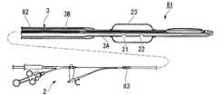
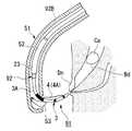
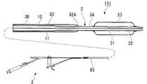
도1에 내시경용 처치구의 일례인 파필로톰의 구성을 도시한다. 파필로톰(1; papillotome)은 시술자가 조작하는 조작부(2)로부터 가요성을 갖는 긴 시스(3; sheath)가 연장되어 있다. 시스(3)의 선단측 측부에 처치부인 도전 와이어(4)가 인출되어 있다.Fig. 1 shows the structure of papillotom which is an example of an endoscope treatment instrument. The papillotome (1) is a long sheath (3) having flexibility extending from the operator (2) operated by the operator. The
시스(3)는 도전 와이어(4)가 인출되는 선단부(3A)가 다른 부분(3B)에 비해 세경화(細徑化)되어 있다. 예를 들어, 선단부(3A)가 1.8 내지 1.9 ㎜ 정도인 것에 반해, 나머지 부분(3B)은 2.4 내지 2.6 ㎜로 되어 있다. 선단을 세경화함으로써 유두로의 삽입성이 양호해진다.In the
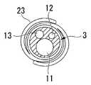
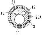
도1, 도2, 도3 및 도4에 도시한 바와 같이, 이 시스(3)에는 3개의 루멘(11, 12, 13)이 길이 방향으로 대략 평행하게 형성되어 있다. 제1 루멘(11)은 가장 직경이 크고 선단에 개방되어 있다. 이 루멘(11)은, 예를 들어 가이드 와이어의 삽입 관통이나, 조영제의 주입에 사용된다. 제2 루멘(12)은 가장 직경이 가늘고 선단이 밀봉되어 있다. 제2 루멘(12)의 선단측에는 시스(3)의 측부에 개방되는 2개의 구멍(14, 15)이 길이 방향으로 앞뒤로 형성되어 있다. 제2 루멘(12)에는 도전 와이어(4)가 통과되어 있다. 도전 와이어(4)는 선단측이 시스(3)의 세경화된 선단부(3A)에 더불어 가늘게 되어 있다. 그리고, 시스(3)의 선단부(3A)의 측부에 형성된 구멍(14)으로부터 시스(3)의 외측으로 인출되어, 또한 선단측에 마련된 구멍(15)으로부터 다시 제2 루멘(12) 내로 복귀되어 있다. 시스(3)의 외주에 인출되 어 노출된 부분이 절개 나이프부(4A)가 된다. 도전 와이어(4)의 선단은 팁(16)을 통해 시스(3)에 고정되어 있다.As shown in Figs. 1, 2, 3 and 4, three
제3 루멘(13)은 선단측이 밀봉되어 있다. 선단측에는 시스(3)의 측부에 개방되는 2개의 구멍(21, 22)이 길이 방향으로 앞뒤로 형성되어 있다. 이들 구멍(21, 22)은 제2 루멘(12)의 구멍(14, 15)보다 기단측에 배치되어 있다. 시스(3)의 선단부(3A)에는 이들 구멍(21, 22)을 덮도록 확장기(dilator)로서 벌룬(23)이 설치되어 있다.The front end side of the
벌룬(23)은 절개 나이프부(4A)[즉, 구멍(14)]로부터 10 ㎜ 이상, 더욱 바람직하게는 15 ㎜ 내지 20 ㎜ 정도 이격하여 배치되어 있다. 유두 절개시에 벌룬(23)이 내시경의 기상대(起上臺)에 걸리지 않도록 하기 위해서이다. 벌룬(23)의 선단부 및 기단부의 각각은 열 용착이나 접착제나 실을 사용하여 고리 형상으로 시스(3)에 고정되어 있다. 벌룬(23)은 초기 상태에는 시스(3)의 외부 주위에 밀착하고 있고, 그 외경은 시스(3)의 기단측 부분(3B)의 외경 이하로 되어 있다. 밀착시의 벌룬(23)의 길이 방향으로 평행한 길이를 약 3등분하는 위치에 구멍(21, 22)이 하나씩 배치되어 있다. 혹은, 2개의 구멍(21, 22) 대신에 2개의 구멍(21, 22)의 위치를 포함하는 1개의 긴 구멍이라도 좋다.The
벌룬(23)은 라텍스나 실리콘 고무 등의 신축성이 높은 엘라스토머계의 재료로 제조되어 있다. 가장 바람직한 재료는 신축성이 가장 높은 라텍스이다. 도5에 벌룬(23)을 팽창시켰을 때의 외관을 도시한다. 벌룬(23)의 최대 확장 직경은 16 내지 20 ㎜ 정도이고, 길이 방향의 길이는 최대 확장 직경의 1.5 내지 2배(구체적 으로는, 30 내지 40 ㎜)이다. 라텍스를 사용한 경우에 그 최대 신장은 800 % 내지 1000 % 정도이므로, 20 ㎜ 확장 직경을 얻기 위해서는 수축 전의 직경이 φ 2.0 내지 2.5 ㎜ 정도이다.The
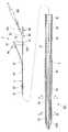
도1에 도시하는 조작부(2)는 시스(3)에 설치된 제1 분기부(31)를 갖는다. 제1 분기부(31)는 시스(3) 내의 제1 루멘(11)에 튜브(32)를 연통시키기 위해 이용된다. 튜브(32)는 가요성을 갖고, 단부에 가이드 와이어의 삽입 등이 가능한 삽입부(33)가 설치되어 있다. 삽입부(33)의 측부에는 링(34)이 형성되어 있다. 링(34)은 선단측이 개방된 대략 C자형을 갖는다. 이 링(34)을 내시경에 끼우면, 조작부(2)를 내시경에 대해 고정할 수 있다. 또한, 삽입부(33)의 측부에서, 링(34)의 연장 설치 방향의 대략 반대측에는 접속부(35)가 일체로 연장 설치되어 있다. 접속부(35)의 선단에는 오목부(35A)가 형성되어 있다.The
또한, 조작부(2)는 시스(3)의 기단부이며, 제1 분기부(31)를 지나 연장되는 단부에 고정되는 조작부 본체(36)를 갖는다. 조작부 본체(36)는 선단에 계지부(37)가 설치되어 있다. 계지부(37)는 상기한 접속부(35)의 오목부(35A)에 착탈 가능하게 되어 있다. 조작부 본체(36)는 계지부(37)로부터 제2 분기부(38)를 거쳐서 제1 조작 유닛(39)과 제2 조작 유닛(40)으로 분기되어 있다. 제1 조작 유닛(39)은 시스(3)와 대략 동축에 배치되어 있다. 제3 루멘(13)에 연통되어 있고, 단부에 주사기(41; syringe)가 착탈 가능하게 설치된다. 제2 조작 유닛(40)은 제1 조작 유닛(39)에 대해 경사져 배치되고, 슬라이더(42)가 진퇴 조작 가능하게 설치되어 있다. 슬라이더(42)에는 외부의 고주파 전원에 접속 가능한 단자(43)가 설치 되어 있고, 슬라이더(42)에 고정되는 도전 와이어(4)와 전기적으로 접속되어 있다.Moreover, the
다음에, 이 파필로톰(1)을 사용한 시술에 대해 설명한다.Next, the procedure using this
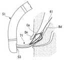
처음에 내시경을 환자의 자연 개구인 입으로부터 삽입하여 십이지장에 도입한다. 내시경에 설치한 관찰 디바이스에서 체내의 화상을 취득하여, 유두 부근으로 내시경 선단부를 안내한다. 도6에 도시한 바와 같이, 내시경(51)의 작업용 채널(52)에 파필로톰(1)을 삽입하고, 선단부(3A)를 내시경(51)으로부터 돌출시킨다. 내시경(51)의 선단에 설치된 기상대(53)를 전방측에서 조작하여 파필로톰(1)의 선단부를 유두(Dn)에 향하게 한다. 이 단계에서 벌룬(23)은 닫혀 있다. 또한, 내시경(51)에 측방에 관찰 시야를 갖는 측시 타입을 이용하면 시술이 용이해진다.Initially, the endoscope is inserted into the duodenum from the patient's natural opening, the mouth. An image of the body is acquired by the observation device installed in the endoscope, and the endoscope tip is guided near the nipple. As shown in Fig. 6, the
유두(Dn)를 커트할 때는, 도7에 도시한 바와 같이 시스(3)의 선단을 유두(Dn)에 삽입한다. 조작부(2)의 삽입부(33)로부터 가이드 와이어를 삽입하고, 가이드 와이어를 담관 내에 도입한다. 가이드 와이어는 반드시 사용하지 않아도 되지만, 가이드 와이어를 통과시켜 두면 유두 절개가 보다 안정되거나, 다른 처치구로 교환할 때에 편리하다. 또한, 제2 조작 유닛(40)의 슬라이더(42)의 단자(43)에 고주파 전원을 접속한다. 제2 조작 유닛(40)의 기단의 링(40A)과 슬라이더(42)에 손가락을 걸어 슬라이더(42)를 후퇴시켜 도전 와이어(4)를 인장한다. 도전 와이어(4)의 선단은 시스(3)의 선단부(3A)에 고정되어 있으므로, 시스(3)의 선단부(3A)가 만곡되고, 도전 와이어(4)에서 시스(3) 밖으로 노출되어 있는 절개 나이프부(4A)가 활 형상으로 팽팽해진다. 고주파 전원으로부터 단자(43)를 통해 도전 와이어(4)에 고주파 전류를 흐르게 하면서, 기상대(53)를 조작하여 시스(3)를 요동 동작시킨다. 도8에 도시한 바와 같이, 절개 나이프부(4A)에 접촉한 유두(Dn)가 절개된다. 유두(Dn)의 절개량은 출혈의 가능성이 적은 중절개나 소절개 정도로 한다. 이때 절개하는 길이나 방향은 이 기상대(53)에서 시스(3)를 밀어 올리는 조작으로 미세 조정하므로, 출혈이나 천공 없이 안전하게 절개하기 위해서는 시술자의 의도대로 정확하게 시스(3)를 밀어 올리는 것이 중요해진다.When cutting the teat Dn, the tip of the
그러나, 절개 나이프부(4A)의 바로 전방측에 벌룬(23)이 위치하고 있으면, 시스(3)보다 직경이 큰 벌룬(23)을 기상대(53)가 밀어 올리게 되어 절개 길이나 절개 방향의 조정이 어려워져, 출혈이나 천공의 가능성이 높아진다. 특히, 벌룬(23)이 비(非)엘라스토머계의 재료로 제작되어 있는 경우에는, 수축시의 형상이 엘라스토머계 재료와 같이 시스(3)에 밀착된 형상이 아닌, 도13에 도시하는 바와 같이 절첩된 형상이 되므로, 보다 외경이 크고, 또한 외표면의 형상도 주름에 의해 불균일해진다. 그로 인해, 기상대(53)에서의 절개 길이나 절개 방향의 조정이 더욱 어려워진다.However, when the
또한, 십이지장의 크기나 유두의 위치가 환자에 따라 미묘하게 다르기 때문에, 유두와 내시경의 거리는 언제나 일정하지 않고, 절개 나이프부(4A)를 유두에 위치시켜 절개할 때의 기상대(53)가 밀어 올리는 시스(3)의 위치도 미묘하게 다르다.In addition, since the size of the duodenum and the position of the nipple differ slightly depending on the patient, the distance between the nipple and the endoscope is not always constant, and the
따라서, 본원에서는 절개 나이프부(4A)의 전방단과 벌룬(23)의 선단 사이에 10 ㎜ 이상, 보다 바람직하게는 15 ㎜ 내지 20 ㎜의 간격을 마련하고 있으므로, 유두 절개시에는 벌룬(23)이 아닌 시스(3)를 기상대(53)에서 직접 밀어 올릴 수 있 다. 그로 인해, 절개 길이나 절개 방향의 정확한 조정이 가능해진다.Therefore, in this application, since the space | interval of 10 mm or more, More preferably, 15 mm-20 mm is provided between the front end of 4 A of incision knife parts, and the tip of
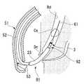
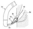
또한, 슬라이더(42)를 밀어 전진시키면, 도전 와이어(4)의 절개 나이프부(4A)를 직경 방향 외측을 향해 팽출시킬 수 있다. 고주파 전류의 통전을 정지시켜 슬라이더(42)를 복귀시킨 후, 가이드 와이어(61)를 따라 시스(3)를 담관(Bd) 내에 더욱 전진시킨다. 도9에 도시한 바와 같이, 파필로톰(1)은 벌룬(23)이 유두(Dn)로부터 담관(Bd) 내에 이를 때까지 진입시킨다. 보다 바람직하게는, 내시경 화상을 관찰하면서 벌룬(23)의 축선 방향의 대략 중앙 부분이나 유두(Dn)에 도달하도록 삽입량을 조정한다. 이와 같이 하면, 나중에 벌룬(23)을 팽창시켰을 때에, 유두(Dn) 및 그 주변이 확실하게 압박 확장된다.In addition, when the
여기서, 제1 조작 유닛(39)에 접속한 주사기(41)로부터 생리 식염수 또는 공기를 제3 루멘(13)에 주입한다. 생리 식염수는 제3 루멘(13)의 선단측 2개의 구멍(21, 22)의 각각으로부터 벌룬(23) 내에 들어가 벌룬(23)을 팽창시킨다. 주사기(41)와 제1 조작 유닛(39) 사이에 압력계를 배치하고, 벌룬(23)의 팽창압을 조정한다. 벌룬(23)은 가해지는 압력에 따라 직경이 변하므로, 원하는 직경으로 하기 위해 필요한 압력까지 가압한다. 팽창압은 유두(Dn)가 미리 절개되어 있으므로, 1 내지 2기압으로 낮게 해도 좋다. 도10에 도시한 바와 같이 벌룬(23)이 유두(Dn) 및 담관(Bd)의 직경을 더욱 압박 확장한다.Here, physiological saline or air is injected into the
이 후, 벌룬(23)에 주입한 생리 식염수 또는 공기를 주사기(41)로 흡출한다. 도11에 도시한 바와 같이 벌룬(23)이 수축한다. 유두(Dn) 및 담관(Bd)은 확장된 상태를 유지한다.Thereafter, physiological saline or air injected into the
가이드 와이어(61)를 담관(Bd) 내에 남겨둔 상태에서 파필로톰(1)을 체외로 제거한다. 대신에, 바스켓 겸자를 내시경(51)의 작업용 채널(52)에 통과시켜 유두(Dn)로부터 담관(Bd)에 삽입한다. 도12에 도시한 바와 같이, 바스켓 겸자(71)로 결석(Ca)을 포착하여, 유두(Dn)를 통해 담관(Bd)으로부터 취출한다. 미리 담관(Bd)의 출구측 및 유두(Dn)가 확장되어 있으므로, 용이하게 결석(Ca)을 취출할 수 있다.The
또한, 바스켓 겸자(71)는 가요성의 긴 시스의 선단에 다수의 와이어로 이루어지는 바스켓을 돌출 함몰 가능하게 설치한 구성을 갖는다. 다수의 와이어는 시스로부터 돌출되면 바구니 형상으로 확대되도록 미리 압박되어 있고, 선단이 팁으로 묶여 있다. 팁에는 가이드 와이어(61)를 통과시키는 관통 구멍이 형성되어 있고, 가이드 와이어(61)를 따라 담관(Bd)에 용이하게 삽입할 수 있다.In addition, the
결석(Ca)을 취출하면, 내시경(51)마다 체외로 인출한다.When the stones Ca are taken out, the
이 실시 형태에서는, 절개에 의한 개구의 확장과, 벌룬(23)에 의한 개구의 확장을 1개의 처치구로 실시할 수 있으므로, 처치구의 교환을 하지 않고 필요한 개구 직경을 얻을 수 있게 된다. 조직의 절개량을 적게 할 수 있으므로, 출혈의 가능성을 저감시킬 수 있다. 또한, 벌룬(23)에 의한 다이레이션(dilation)에 있어서도 조직의 압박을 최소한으로 멈추게 할 수 있다. 또한, 종래와 같이 벌룬에 의한 다이레이션만을 행할 때는, 유두에 개방되는 췌관의 출구 주위의 조직에도 강한 압박이 가해진다. 이 압박이 너무 강하면 췌관 주위의 조직이 염증에 의해 붓게 되어 췌관의 출구를 막아 버리므로 췌염이 될 가능성이 증가한다. 따라서, 췌관 입 구 주변의 조직에 강한 압박을 부여하면 안 되므로 다이레이션 벌룬으로 확장할 수 있는 크기는 한정되어 있고, 결석을 파괴하지 않고 회수할 수 있는 크기는 파필로톰으로 유두를 절개한 경우보다도 작다는 단점이 있었다. 예를 들어, 종래의 벌룬으로 확장할 수 있는 크기는 약 8 ㎜라고 하므로, 파괴하지 않고 회수할 수 있는 결석 사이즈도 8 ㎜ 정도까지로 한정되어 있었다. 그러나, 이 실시 형태에서는 유두를 확장하기 전에 파필로톰으로 절개함으로써, 담관 출구와 췌관 출구를 이격시킬 수 있고, 확장시에 췌관 출구 주위의 조직에의 압박을 적게 할 수 있다. 이로 인해, 매우 크게 확장하는 것이 가능해져 큰 결석이라도 회수할 수 있다.In this embodiment, since the opening by incision and the opening by the
벌룬(23)은 축 방향의 길이가 확장 직경보다 커지도록 되어 있으므로, 관로를 압박 확장할 때의 위치 어긋남이 억제되어 원하는 위치를 확실하게 확장할 수 있다.Since the length of the
또한, 종래에는, ESWL에 의한 파쇄가 필요한 사이즈였던 경우, 시술이 복잡해져 환자의 침습이 증가하거나, 치료 기간이나 시술 시간도 길어지거나, 치료비가 증가한다는 등의 단점이 있었다. 그러나, 이 실시 형태에서는, 비교적으로 큰 결석이라도 그대로 취출하는 것이 가능해지므로, 시술을 간략화할 수 있어 치료 기간이나 시술 시간을 짧게 할 수 있다.In addition, conventionally, when crushing by ESWL is required, there are disadvantages in that the procedure is complicated and the invasion of the patient is increased, the treatment period or the procedure time is long, and the treatment cost is increased. However, in this embodiment, even relatively large stones can be taken out as it is, so the procedure can be simplified and the treatment period and the procedure time can be shortened.
제3 루멘(13)에 앞뒤로 2개의 구멍(21, 22)을 마련하였으므로, 벌룬(23)을 조속히 팽창시키거나 수축시킬 수 있다. 특히, 벌룬(23)을 수축시키는 과정에서 한쪽 구멍(21, 22)에 벌룬(23)이 밀착한 경우에도, 다른 쪽 구멍(21, 22)으로부터 유체를 배출할 수 있으므로, 벌룬(23)을 확실하게 수축시킨다. 상기한 바와 같이 2개의 구멍(21, 22) 대신에 1개의 긴 구멍으로 한 경우도 마찬가지로 벌룬(23)의 팽창이나 수축이 계속된다. 벌룬(23)으로부터 유체를 제거할 때에도 긴 구멍의 타단부로부터 유체를 배출할 수 있으므로 확실하게 수축시킨다.Since two
여기서, 벌룬(23)은 비엘라스토머계 재료로 제조해도 좋다. 비엘라스토머계 재료로서는, 폴리우레탄, 폴리에틸렌, 폴리아미드, PET(폴리에틸렌테레프탈레이트) 등 신축성은 낮지만, 내압성이 높은 것이 사용된다. 이 경우, 벌룬(23)의 최대 확장 직경은 16 내지 20 ㎜ 정도이고, 길이 방향의 길이는 최대 확장 직경의 1.5 내지 2배(구체적으로는 30 내지 40 ㎜)이다.Here, the
비엘라스토머계 재료의 경우, 신축성이 적기 때문에, 벌룬(23)의 팽창 정도를 손끝에서의 팽창압의 조정에 의해 비교적 정밀도 좋게 조정할 수 있다. 예를 들어, 팽창압이 1기압일 때의 팽창 직경을 16 ㎜, 2기압에서 18 ㎜, 3기압에서 20 ㎜로 하는 것이 가능하다. 이에 의해, 결석이 그다지 크지 않을 때는 전방의 압력계로 팽창압을 조정하여 16 ㎜까지의 확장으로 하거나, 결석이 클 때는 20 ㎜까지 확장시키는 등과 같이, 1개의 파필로톰(1)에서 증상에 맞게 임의로 팽창 직경을 바꾸는 것이 가능하다.In the case of the non-elastomeric material, since the elasticity is small, the degree of expansion of the
또한, 팽창압과 팽창 직경의 관계는 비엘라스토머계의 재료라도 그 종류를 바꿈으로써 임의로 바꿀 수 있다. 예를 들어, 신축성이 보다 높은 재료로는 1기압에서 16 ㎜, 1.5기압에서 18 ㎜, 2기압에서 20 ㎜ 등의 작은 압력차로 크게 직경이 변화하도록 할 수 있다. 이 경우, 나사 삽입식이나 배력 기구가 달린 고가의 인프레이크를 사용할 필요가 없어지고, 구조가 심플하고 저렴한 주사기로 원하는 팽창 직경을 얻을 수 있어 비용을 삭감시킬 수 있다.The relationship between the expansion pressure and the expansion diameter can be arbitrarily changed even by changing the type of the non-elastomeric material. For example, as the material having higher elasticity, the diameter can be largely changed with a small pressure difference such as 16 mm at 1 atmosphere, 18 mm at 1.5 atmosphere, and 20 mm at 2 atmosphere. In this case, there is no need to use expensive inserts with screw inserts or power mechanisms, and a simple and inexpensive syringe can be used to achieve a desired expansion diameter, thereby reducing costs.
이와는 반대로, PET와 같은 신축성이 특히 작은 재료를 사용하면, 1기압이나, 2기압, 3기압에서도 대략 일정한 직경, 예를 들어 18 ㎜가 되는 등의 설계가 가능해진다. 넓은 압력 범위에서 팽창 직경을 일정하게 한 경우, 압력 조정을 엄밀하게 할 필요가 없어진다. 압력계를 사용하지 않아도 원하는 크기로 벌룬(23)을 팽창시키는 것이 가능해져 비용을 삭감시킬 수 있다.On the contrary, when a material having a particularly small elasticity, such as PET, is used, the design can be made to have a substantially constant diameter, for example, 18 mm, even at 1 atm, 2 atm, and 3 atm. If the expansion diameter is made constant over a wide pressure range, it is not necessary to strictly adjust the pressure. It is possible to inflate the
도13 및 도14에 도시한 바와 같이, 비엘라스토머계의 재료의 경우, 벌룬(23)은 수축한 상태에서는 시스(3)의 외부 주위에 권취함으로써 외경을 작게 하고 있다.As shown in Figs. 13 and 14, in the case of a non-elastomeric material, the outer diameter is reduced by winding the
또한, 절개 나이프부(4A)의 전방단과 벌룬(23)의 선단 사이에 10 ㎜ 이상, 더욱 바람직하게는 15 ㎜ 내지 20 ㎜의 간격을 마련하고 있으므로, 유두 절개시에는 벌룬(23)이 아닌 시스(3)를 기상대(53)로 직접 밀어 올릴 수 있다. 그로 인해, 절개 길이나 절개 방향의 정확한 조정이 가능해진다.Since a gap of 10 mm or more, more preferably 15 mm to 20 mm, is provided between the front end of the cutting
[제2 실시 형태]Second Embodiment
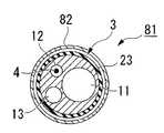
도15 및 도16에 내시경용 처치구의 일례인 파필로톰의 구성을 도시한다. 이 파필로톰(81)은 시스(3)의 외측에 커버 시스(82)를 진퇴 가능하게 씌우고 있는 것을 특징으로 한다. 시스(3)의 구성은 제1 실시 형태의 파필로톰(1)과 마찬가지이다.15 and 16 show the structure of papillotom which is an example of an endoscope treatment instrument. The
커버 시스(82)는 길고 가요성을 갖는다. 커버 시스(82)의 기단에서 체외로 인출되는 부분에는, 시술자가 잡기 쉽도록 손잡이(83)가 설치되어 있다. 커버 시 스(82)는 벌룬 수축시의 외경 및 시스(3)의 직경이 큰 부분(3B)보다 큰 내경을 갖는다. 그 선단은 초기 상태에서는 벌룬(23)의 전체를 덮는 한편 절개 나이프부(4A)를 노출시키는 위치에 배치된다. 또한, 커버 시스(82)의 선단부(82A)는 유두(Dn) 등에의 삽입이 용이해지도록 테이퍼 형상으로 직경 축소되어 있다.
벌룬(23)과 절개 나이프부(4A) 사이의 거리는 제1 실시 형태보다 작게 되어 있어, 양자가 상대적으로 근접하고 있다. 벌룬(23)은 제1 실시 형태에서 설명한 엘라스토머계 또는 비엘라스토머계의 재료로 제조된다.The distance between the
또한, 도17에 도시한 바와 같이, 손잡이(83)를 잡고 조작부(2)를 압입하여 커버 시스(82)로부터 벌룬(23)을 완전히 노출시킨 후 벌룬(23)을 팽창시키면, 벌룬(23)의 전체를 팽창시킬 수 있다. 벌룬(23)은 시스(3)의 길이 방향으로 가늘고 길어져, 축 방향의 길이가 확장 직경보다 커진다.In addition, as shown in FIG. 17, when the
이에 대해, 도18에 도시한 바와 같이 커버 시스(82)의 선단을 벌룬(23)의 길이 방향의 중앙 부근, 2개의 구멍(21, 22) 사이, 또는 2개의 구멍(21, 22)을 대신하는 1개의 긴 구멍의 중간에 배치한 후 벌룬(23)을 팽창시키면, 커버 시스(82)로부터 노출되는 선단 부분만을 팽창시킬 수 있다. 벌룬(23)의 기단 부분은 커버 시스(82)에 의해 팽창이 억제되므로, 커버 시스(82)보다 크게 팽창되지 않는다. 이때의 벌룬(23)은 시스(3)의 길이 방향이 짧게 팽창된다.On the other hand, as shown in FIG. 18, near the center of the longitudinal direction of the
다음에, 이 파필로톰(81)을 사용한 시술에 대해 설명한다.Next, the procedure using this
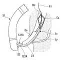
커버 시스(82)로 벌룬(23)을 덮은 상태에서 파필로톰(81)을 내시경에 통과시킨다. 도19에 도시한 바와 같이, 절개 나이프부(4A)를 활 형상으로 팽팽하게 하여 고주파 전류를 흐르게 하면서, 기상대(53)를 조작하여 유두(Dn)를 절개한다. 절개량은 상기와 마찬가지이다. 도20에 도시한 바와 같이, 시스(3)를 유두(Dn)로부터 담관(Bd)에 삽입한다. 이때, 전방측 손잡이(83)를 잡아 고정하고, 시스(3)를 압입하여 전진시킨다. 커버 시스(82)가 고정된 상태에서 시스(3)가 상대적으로 전진하므로, 벌룬(23)이 노출된다. 파필로톰(81)은 예를 들어 벌룬(23)의 축선 방향의 중앙이 유두(Dn)에 도달할 때까지 진입시킨다.The
도21에 도시한 바와 같이, 벌룬(23)의 전체를 팽창시켜 유두(Dn) 및 담관(Bd)의 출구측을 확장한다. 그 후, 벌룬(23)을 수축시킨다. 도22에 도시한 바와 같이, 커버 시스(82)를 전진시켜 벌룬(23)의 기단 부분[기단측 구멍(21)을 포함하는 부분, 또는 2개의 구멍(21, 22)을 대신하는 1개의 긴 구멍의 기단측을 포함하는 부분]을 커버 시스(82) 내에 수용시킨다. 또한, 이때, 벌룬(23)의 노출 부분이 결석(Ca)보다 안쪽이 될 때까지 파필로톰(81)을 전진시킨다.As shown in Fig. 21, the
도23에 도시한 바와 같이 벌룬(23)을 팽창시키면, 벌룬(23)이 결석(Ca)의 안쪽에서 담관(Bd)을 막도록 팽창된다. 벌룬(23)의 용적은 처음의 대략 절반 정도이다. 이 상태에서 커버 시스(82) 및 시스(3)를 함께 인장한다. 도24에 도시한 바와 같이, 결석(Ca)이 벌룬(23)에 긁어져 나오도록 하여 담관(Bd)으로부터 배출된다.As shown in FIG. 23, when the
이 실시 형태에서는 커버 시스(82)를 설치하였기 때문에, 벌룬(23)을 팽창시켰을 때의 형태를 변화시킬 수 있다. 벌룬(23)의 전체를 팽창시키면, 넓은 면적에서 조직을 압박하는 것이 가능해져, 관로의 원하는 위치를 어긋나지 않게 확실하게 확장할 수 있다. 이에 대해, 커버 시스(82)에서 팽창시키는 벌룬(23)의 축선 방향의 길이를 감소시키고, 선단부만을 팽창시켰을 때에는, 축 방향이 짧아져 담관(Bd)의 만곡 형상을 따라 이동하기 쉬워진다. 이로 인해, 벌룬(23)으로 결석(Ca)을 배출하기 쉬워진다. 파필로톰(81)에서 결석 배출용 처치구로 교환할 필요가 없어지기 때문에, 시술 시간을 단축할 수 있고, 환자의 부담도 경감시킬 수 있다.In this embodiment, since the
[제3 실시 형태][Third Embodiment]
도25에 내시경용 처치구의 일례인 파필로톰의 구성을 도시한다. 이 파필로톰(91)은 시스(3)의 외측에 씌운 커버 시스(92)에 벌룬(23)이 설치되어 있는 것을 특징으로 한다.Fig. 25 shows the structure of papillotom which is an example of an endoscope treatment instrument. The
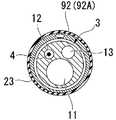
도26 및 도27에 도시한 바와 같이, 시스(3)는 3개의 루멘(11, 12, 13)을 갖는다. 제1 루멘(11)에는 가이드 와이어(61)가 통과된다. 제2 루멘(12)에는 도전 와이어(4)가 통과된다. 제2 루멘(12)과 도전 와이어(4)의 상세한 것은 제1 실시 형태와 마찬가지이다. 제3 루멘(13)은 선단에 개구를 형성하고 있다. 전방측 제1 조작 유닛(39)의 입구 부재에 주사기를 장착하면, 조영제를 시스(3)의 선단으로부터 분출시킨다.As shown in Figs. 26 and 27, the
커버 시스(92)는 시스(3)의 굵은 부분(3B)보다 큰 내경을 갖고, 선단에 벌룬(23)을 장착하는 벌룬 장착부(92A)가 형성되어 있다. 벌룬 장착부(92A)는 시스(3)의 직경의 변화에 맞게 다른 부분(92B)에 비해 가늘게 되어 있다. 커버 시스(92)에는 유체를 통과시키는 루멘(93)이 형성되어 있다. 루멘(93)은 2개의 구멍(94, 95)을 통해 측면에 개방되어 있다. 이들 구멍(94, 95)을 덮도록 벌룬(23) 이 커버 시스(92)에 설치되어 있다. 벌룬(23)의 재료나 형상은 상기한 실시 형태와 마찬가지이다. 2개의 구멍(94, 95)은 벌룬(23)의 축선 방향의 길이를 대략 3등분하는 위치의 각각에 1개씩 배치되어 있다. 루멘(93)은 커버 시스(92)의 기단부까지 연장되어, 손잡이(83)로부터 튜브(96)를 통과하여 주사기(97)에 접속되어 있다. 주사기(97)로부터 생리 식염수나 공기 등의 유체를 공급하면, 도28에 도시하는 바와 같이 벌룬(23)을 팽창시킬 수 있다.The
또한, 커버 시스(92)는 가장 전진시켰을 때에는 시스(3)의 선단과 커버 시스(92)의 선단이 일치하도록 길이가 설정되어 있다. 또한, 커버 시스(92)는 시스(3)를 직경 축소시킴으로써 형성되는 단차 부분(3C)에 직경 축소된 벌룬 장착부(92A)의 기단부(92C)가 맞닿을 때까지 후퇴시킬 수 있다. 시스(3)의 직경이 가늘어진 부분(3A)은, 유두 삽입이나 유두 절개시에 벌룬(23)이 기상대에 걸리지 않도록 전방측에 커버 시스(92)를 후퇴시킬 수 있는 위치까지 형성되어 있다.In addition, the length of the
다음에, 파필로톰(91)을 사용한 시술에 대해 설명한다.Next, the procedure using the
도29에 도시한 바와 같이, 커버 시스(92)를 후퇴시켜 시스(3)의 선단부(3A)를 노출시킨 상태에서, 시스(3)를 기상대(53)로 압박하여 유두(Dn)에 근접시킨다. 도30에 도시한 바와 같이, 활 형상으로 팽팽해진 도전 와이어(4)의 절개 나이프부(4A)로 유두(Dn)를 소정량 절개한다.As shown in Fig. 29, while the
벌룬(23)으로 유두(Dn) 및 담관(Bd)의 출구를 확장할 때에는, 도31에 도시한 바와 같이 커버 시스(92)만을 전진시킨다. 예를 들어, 벌룬(23)의 중앙이 유두(Dn)에 도달할 때까지 커버 시스(92)를 진입시킨다. 이때, 시스(3)는 전진시키 지 않는다. 전방측 주사기(97)로부터 생리 식염수 또는 공기를 주입하면, 커버 시스(92) 내의 루멘(93)을 통과하여 벌룬(23)이 팽창하여, 도32에 도시하는 바와 같이 유두(Dn) 및 담관(Bd)의 출구측이 압박 확장된다.When the outlets of the teat Dn and the bile duct Bd are expanded to the
이 후, 벌룬(23)을 수축시킨 후 가이드 와이어(61)를 남겨둔 상태에서 파필로톰(91)을 제거한다. 대신에 바스켓 겸자 등을 삽입하여 결석(Ca)을 회수한다.Thereafter, the
이 실시 형태에서는, 절개 나이프부(4A)를 보유 지지하는 시스(3)에 대해 진퇴하는 커버 시스(92)에 벌룬(23)을 설치하였다. 벌룬(23)이 절개 나이프부(4A)에 중첩되는 위치까지 전진시킬 수 있으므로, 파필로톰(91)을 담관(Bd)의 안쪽 깊숙이 진입시키지 않아도 유두(Dn) 및 담관(Bd)의 출구측을 확장할 수 있다.In this embodiment, the
[제4 실시 형태][4th Embodiment]
도33에 내시경용 처치구의 구성을 도시한다. 이 내시경용 처치구(101)는 상기한 실시 형태와 같은 도전 와이어를 갖지 않는다. 그 대신에 벌룬이 2개 장착되어 있다.Fig. 33 shows the configuration of the endoscope treatment instrument. This
내시경용 처치구(이하, 처치구라 함)(101)는 길고 가요성을 갖는 시스(3)를 갖고, 시스(3)의 기단에 조작부(2)가 설치되어 있다.The endoscope treatment instrument (hereinafter referred to as a treatment instrument) 101 has a long and
시스(3)는 가요성을 갖고, 직경 축소된 선단부(3A)에 제1 벌룬(23A)과 제2 벌룬(23B)이 축선 방향으로 앞뒤로 장착되어 있다. 도34 및 도35에 도시한 바와 같이, 시스(3)에 형성된 3개의 루멘(11 내지 13) 중, 제1 루멘(11)은 선단에 개방되어, 가이드 와이어를 통과시키거나 조영제를 주입할 때에 사용된다. 제2 루멘(12)은 직경이 가장 가늘고, 구멍(102)을 통해 선단측의 제2 벌룬(23B)에 연통되 어 있다. 구멍(102)은 제2 벌룬(23B)의 축선 방향의 양단의 고정부의 대략 중간 위치에 배치된다. 제3 루멘(13)은 제2 루멘(12)보다 굵고, 제1 벌룬(23A)에 연통되는 구멍(21, 22)이 축선 방향에 앞뒤로 2개 형성되어 있다. 구멍(21, 22)은 제1 벌룬(23A)의 축선 방향의 양단의 고정부 사이를 3등분하는 위치의 각각에 대략 상당하는 위치에 배치된다. 혹은, 2개의 구멍(21, 22) 대신에 2개의 구멍의 위치를 포함하는 1개의 긴 구멍을 마련해도 좋다. 제3 루멘(13)은 제2 루멘(12)보다 굵게 되어 있으므로, 유체가 통류하기 쉽다. 용적이 큰 제1 벌룬(23A)을 조속히 팽창시키거나, 수축시킬 수 있다.The
여기서, 도33 및 도36에 도시한 바와 같이, 확장기인 제1 벌룬(23A)의 형상 및 크기는 제1, 제2 실시 형태와 마찬가지이다. 제2 벌룬(23A)은 선단측에 배치되고, 제1 벌룬(23A)보다 축선 방향의 길이가 짧고, 예를 들어 대략 절반으로 되어 있는 소형의 벌룬이다. 각 벌룬(23A, 23B)의 재료는 상기한 실시 형태와 마찬가지이다. 제2 벌룬(23B)의 형상 및 크기는 제2 실시 형태에서 선단측만 팽창시켰을 때의 형상 및 크기에 대략 동등해지도록 되어 있다.33 and 36, the shape and size of the
조작부(2)는 제2 조작 유닛(105)이 입구 부재를 갖고, 밸브가 달린 주사기(106)가 장착 가능하게 되어 있다.As for the
다음에, 처치구(101)를 사용한 시술에 대해 설명한다.Next, the procedure using the
도시하지 않은 고주파 나이프로 유두(Dn)를 절개하고, 개구를 확장한 후 가이드 와이어(61)를 따라 처치구(101)를 유두(Dn)로부터 담관(Bd)에 도입한다. 처치구(101)는 제1 벌룬(23A)의 대략 중앙이 유두(Dn)에 걸릴 때까지 진입시킨다. 도38에 도시한 바와 같이, 제1 벌룬(23A)을 팽창시키면 유두(Dn) 및 담관(Bd)의 출구가 확장된다. 또한, 이때, 제1 조작 유닛(39)의 주사기(41)로부터만 유체를 공급한다. 제3 루멘(13)을 통해 2개의 구멍(21, 22)의 각각으로부터 제1 벌룬(23A)에 유체가 주입되어 팽창된다.The teat Dn is cut with a high frequency knife (not shown), the opening is expanded, and the
그 후, 제1 벌룬(23A)으로부터 유체를 제거하여 수축시킨다. 유체는 2개의 구멍(21, 22)을 통해 제거되므로, 한쪽 구멍(21, 22)에 벌룬(23A)이 밀착한 경우나, 긴 구멍의 일단부에 벌룬(23A)이 밀착한 경우에도 확실하게 유체를 제거하여 수축시킬 수 있다. 계속해서, 처치구(101)를 담관(Bd) 안쪽으로 더욱 진행시킨다. 처치구(101)는 제2 벌룬(23B)을 결석(Ca)으로부터 안쪽측까지 진행시킨다.Thereafter, the fluid is removed from the
그리고, 도39에 도시한 바와 같이 제2 벌룬(23B)을 팽창시킨다. 제2 벌룬(23B)은 제2 조작 유닛(105)의 입구 부재에 장착한 주사기(106)로부터 유체를 공급하여 팽창시킨다. 제2 벌룬(23B)이 담관(Bd)을 막도록 팽창되므로, 주사기(106)의 밸브를 폐쇄하여 제2 벌룬(23B)이 수축되지 않도록 한 후 처치구(101)를 후퇴시킨다. 제2 벌룬(23B)에 의해 결석(Ca)이 긁어져 나오도록 하여 담관(Bd)으로부터 배출된다.Then, as shown in Fig. 39, the
이 실시 형태에서는 다른 위장에 크기가 다른 2개의 벌룬(23A, 23B)을 설치하였으므로, 관로의 확장과 결석(Ca)의 배출을 확실하게 행할 수 있다. 제1 벌룬(23A)은 넓은 면적에서 조직을 압박하는 것이 가능하고, 관로의 원하는 위치를 어긋나지 않게 확실하게 확장할 수 있다. 제2 벌룬(23B)은 축 방향이 짧게 되어 담관(Bd)의 만곡 형상을 따라 이동하기 쉬워진다. 이로 인해, 결석(Ca)을 배출하 기 쉬워진다. 제2 벌룬(23B)은 선단측에 설치되어 있으므로, 처치구(101)의 삽입량이 적어진다. 시술 시간을 단축할 수 있고, 환자의 부담도 경감시킬 수 있다.In this embodiment, since two
[제5 실시 형태][Fifth Embodiment]
도40에 내시경용 처치구의 구성을 도시한다. 이 내시경용 처치구(111)는 도전 와이어를 갖지 않고, 1개의 벌룬과 커버 시스를 구비한다.40 shows the configuration of the endoscope treatment instrument. This
내시경용 처치구(이하, 처치구라 함)(111)는 길고 가요성을 갖는 시스(3)를 갖고, 시스(3)의 기단에 조작부(2)가 설치되어 있다.The endoscope treatment instrument (hereinafter referred to as a treatment instrument) 111 has a
도40, 도41 및 도42에 도시한 바와 같이, 시스(3)는 가요성을 갖고, 직경이 가늘어진 선단부(3A)에 벌룬(23)이 장착되어 있다. 벌룬(23)과 제2 루멘(12)을 연통시키는 구멍(21, 22)이 축선 방향으로 앞뒤로 2개 형성되어 있다. 구멍(21, 22)은 벌룬(23)의 축선 방향의 양단의 고정부 사이를 3등분하는 위치의 각각에 대략 상당하는 위치에 배치된다. 혹은, 2개의 구멍(21, 22) 대신에 2개의 구멍의 위치를 포함하는 1개의 긴 구멍을 마련해도 좋다. 조작부(2)는 제2 조작 유닛(105)에 입구 부재를 구비하고, 밸브가 달린 주사기(106)가 착탈 가능하게 되어 있다. 제2 조작 유닛(105)은 제2 루멘(12)에 연통하고 있다. 벌룬(23)의 형상 및 크기는 제1, 제2 실시 형태와 마찬가지이다.As shown in Figs. 40, 41 and 42, the
커버 시스(82)는 기단부에 손잡이(83)가 설치되고, 선단부(82A)가 직경 축소되어 있다. 선단부(82A)의 개구는 수축한 벌룬(23)의 외경보다 크다. 또한, 벌룬(23)을 팽창시킬 때는, 도43a에 도시하는 바와 같이 커버 시스(82)로부터 벌룬(23)을 노출시킨 후 유체를 주입한다. 또, 도43b에 도시한 바와 같이, 커버 시 스(82)를 벌룬(23)의 축선 방향의 대략 절반에 상당하는 위치에 배치한 후 벌룬(23)에 유체를 주입하면, 구멍(22) 또는 2개의 구멍(21, 22)을 대신하는 1개의 긴 구멍을 통해 벌룬(23)에 유체가 공급되어, 벌룬(23)이 노출되어 있는 부분만이 팽창된다.As for the
다음에, 처치구(111)를 사용한 시술에 대해 설명한다.Next, the procedure using the
도시하지 않은 고주파 나이프로 유두(Dn)를 절개한 후 가이드 와이어(61)를 따라 처치구(111)를 유두(Dn)로부터 담관(Bd)에 도입한다. 커버 시스(82)는 벌룬(23)이 완전히 노출되는 위치까지 미리 후퇴시켜 둔다. 처치구(111)는, 예를 들어 벌룬(23)의 중앙이 유두(Dn)에 도달할 때까지 진입시킨다. 도44에 도시한 바와 같이, 벌룬(23)을 팽창시키면 유두(Dn) 및 담관(Bd)의 출구측이 확장된다.After cutting the nipple Dn with a high frequency knife (not shown), the
그 후, 벌룬(23)으로부터 유체를 제거하여 수축시킨다. 유체는 2개의 구멍(21, 22)을 통해, 또는 2개의 구멍(21, 22)을 대신하는 1개의 긴 구멍의 일단부를 통해 제거되므로, 한쪽 구멍(21)에 벌룬(23)이 밀착한 경우에도 확실하게 수축시킬 수 있다. 계속해서, 시스(3)를 고정하고 커버 시스(82)를 전진시킨다. 커버 시스(82)는 선단이 직경이 축소되어 있으므로, 용이하게 담관(Bd)에 삽입할 수 있다. 벌룬(23)의 대략 절반을 커버 시스(82)로 덮으면, 처치구(111)를 담관(Bd)의 안쪽으로 더욱 진행시킨다. 도45에 도시한 바와 같이, 처치구(111)는 벌룬(23)이 노출된 부분을 결석(Ca)으로부터 안쪽측까지 진행시킨다.Thereafter, the fluid is removed from the
그리고, 도46에 도시한 바와 같이 벌룬(23)을 팽창시킨다. 벌룬(23)은 담관(Bd)을 막도록, 또한 축선 방향의 길이가 짧게 팽창된다. 벌룬(23)을 팽창시킨 상태에서 처치구(111)를 후퇴시키면, 벌룬(23)에 의해 결석(Ca)이 긁어져 나오도록 하여 담관(Bd)으로부터 배출된다.46, the
이 실시 형태에서는, 제2 실시 형태와 같은 효과를 얻을 수 있다.In this embodiment, the same effects as in the second embodiment can be obtained.
[제6 실시 형태][Sixth Embodiment]
이 실시 형태는, 도47에 도시한 바와 같이 절개 후에 삽입한 바스켓 겸자(71)로 결석(Ca)을 포착하였지만, 결석(Ca)이 커서 유두(Dn)로부터 발출할 수 없게 되었을 때에 실시된다. 이 경우, 도48에 도시한 바와 같은 내시경용 처치구(이하, 처치구라 함)(121)가 사용된다.This embodiment is carried out when the stones Ca are captured by the basket forceps 71 inserted after the incision as shown in Fig. 47, but the stones Ca are so large that they cannot be extracted from the teat Dn. In this case, an endoscope treatment instrument (hereinafter referred to as a treatment instrument) 121 as shown in FIG. 48 is used.
이 처치구(121)는 가요성을 갖는 긴 시스(122)를 갖는다. 시스(122)는 선단이 직경이 축소되는 동시에, 선단측의 외주에 벌룬(23)이 설치되어 있다. 시스(122) 내에는 바스켓 겸자(71)의 시스(72)를 삽입 관통 가능한 루멘과, 벌룬(23)에 공급하는 유체를 통과시키는 루멘이 형성되어 있다. 도시하지 않은 루멘의 기단부에는, 벌룬(23)에 유체를 공급하기 위한 입구 부재가 설치되어 있고, 주사기[예를 들어, 도40의 주사기(106)]가 접속된다.This
처치구(121)는 도시하지 않은 바스켓 겸자(71)의 전방 조작부를 제거(착탈)한 후, 벌룬(23)을 수축시킨 상태에서 바스켓 겸자(71)의 시스(72)를 가이드로 하여 도입된다. 벌룬(23)의 대략 절반이 유두(Dn)로부터 담관의 출구측으로 삽입되면, 주사기로부터 유체를 공급한다. 벌룬(23)이 팽창되어 유두(Dn) 및 담관(Bd)의 출구측이 압박 확장된다.The
그 후, 벌룬(23)을 수축시킨 후, 처치구(121)를 제거한다. 유두(Dn) 및 담 관(Bd)의 출구가 확장되어 있으므로, 도49에 도시한 바와 같이 바스켓 겸자(71)로 잡은 결석(Ca)을 담관(Bd)으로부터 취출할 수 있다.Thereafter, the
이 실시 형태에서는, 유두(Dn)를 절개하여 바스켓 겸자 등의 처치구를 삽입하였지만 결석(Ca)을 유두(Dn)로부터 배출할 수 없을 때에, 벌룬(23)을 구비하는 처치구(121)를 삽입하여 관로의 개구를 확장하도록 하였으므로, 비교적 큰 결석(Ca)이라도 담관(Bd) 내에서 파쇄하지 않고 취출할 수 있다. 시술이 간단해지고, 환자의 부담도 저감된다.In this embodiment, the
[제7 실시 형태][Seventh Embodiment]
이 실시 형태는 상기한 제6 실시 형태에 있어서 도47을 참조하여 설명한 바와 같이, 절개 후에 삽입한 바스켓 겸자(71)로 결석(Ca)을 포착하였지만, 결석(Ca)이 커서 바스켓 겸자(71)를 유두(Dn)로부터 발출할 수 없게 되었을 때에 실시된다.As described with reference to FIG. 47 in the sixth embodiment described above, this embodiment captures the stones Ca with the basket forceps 71 inserted after the incision, but the stones Ca are large and the
이 시술에 사용되는 내시경용 처치구를 도50에 도시한다. 내시경용 처치구(이하, 처치구라 함)(121A)는 가요성을 갖는 긴 시스(122A)를 갖고, 시스(122A)의 선단부에 벌룬(23)이 설치되어 있다. 벌룬(23)의 설치 방법이나 벌룬(23)의 재질, 형상, 유체의 공급 경로는 상기한 실시 형태와 마찬가지이다.50 shows an endoscope treatment instrument used in this procedure. The endoscope treatment instrument (hereinafter referred to as treatment instrument) 121A has a
처치구(121A)를 사용할 때는, 가이드 와이어(61)를 시스(122A)의 제1 루멘(11)에 통과시켜 가이드 와이어(61)를 따라 체내에 도입시킨다. 바스켓 겸자(71)와 대략 평행하게 처치구(121A)가 삽입 관통된다. 처치구(121A)는 벌룬(23)의 대략 절반이 유두(Dn)에 도달할 때까지 삽입되어, 주사기(106)로부터의 유체에 의해 벌룬(23)을 팽창시킨다. 도51에 도시한 바와 같이 벌룬(23)이 팽창되어 유 두(Dn) 및 담관(Bd)의 출구측이 압박 확장될 수 있다. 그 후, 벌룬(23)을 수축시킨 후, 처치구(121A)를 유두(Dn)로부터 제거한다. 유두(Dn) 및 담관(Bd)의 출구측이 확장되어 있으므로 바스켓 겸자(71)로 잡은 결석(Ca)을 담관(Bd)으로부터 취출할 수 있다.When using the
이 실시 형태에서는 제6 실시 형태와 같은 효과를 얻을 수 있다. 처치구(121A)에 바스켓 겸자(71)의 시스(72)를 통과시킬 필요가 없어지므로, 바스켓 겸자(71)의 전방 조작부를 제거하는 수고를 줄일 수 있는 동시에, 처치구(121A)의 시스(122A)를 세경화할 수 있다.In this embodiment, the same effects as in the sixth embodiment can be obtained. Since it is no longer necessary to pass the
[제8 실시 형태][Eighth Embodiment]
도52 및 도53에 도시한 바와 같이, 내시경용 처치구인 바스켓 겸자(131)는 가요성의 긴 시스(132)를 갖고, 시스(132)의 기단부에 조작부(133)가 설치되어 있다. 시스(132) 내에는 조작 와이어(134)가 진퇴 가능하게 통과되어 있고, 조작 와이어(134)의 선단에는 바구니 형상의 처치부(135)가 설치되어 있다. 처치부(135)는 조작 와이어(134)에 고정된 접속 부재(136)로 복수의 와이어(137)의 일단부를 묶는 동시에, 와이어(137)의 타단부를 팁(138)으로 묶는 구성으로 되어 있다. 와이어(137)는 시스(132) 내에 묶여 수용 가능하지만, 시스(132)로부터 돌출시키면 개방하도록 압박되어 있다. 팁(138)에는 가이드 와이어를 통과시키는 관통 구멍(138A)이 축선에 대해 비스듬하게 형성되어 있다.As shown in Figs. 52 and 53, the
시스(132)의 선단부의 외주에는 벌룬(23)이 설치되어 있다. 시스(132)에는 벌룬(23)에 유체를 공급하기 위한 루멘(141)이 형성되어 있다. 루멘(141)과 벌 룬(23)은 2개의 구멍(21, 22) 또는 2개의 구멍(21, 22)을 대신하는 1개의 긴 구멍으로 연통되어 있다. 벌룬(23) 및 구멍(21, 22)은 상기한 실시 형태와 마찬가지이다.The
조작부(133)는 시스(132)의 기단부에 고정된 손잡이(142)를 갖고, 손잡이(142)를 관통하여 조작 와이어(134)가 인출되어 있다. 조작 와이어(134)의 단부(143)를 시스(132)에 대해 밀고 당기면, 선단의 처치부(135)를 시스(132)로부터 돌출 함몰시킬 수 있다. 또한, 손잡이(142)로부터는 루멘(141)에 연통하는 튜브(144)가 연장되고, 튜브(144)에 주사기(145)가 설치되어 있다.The
이 바스켓 겸자(131)에서는, 절개 후의 유두를 벌룬(23)으로 확장하거나, 담관(Bd) 내에서 처치부(135)로 결석을 잡은 후에, 벌룬(23)으로 담관이나 유두를 확장시킬 수 있다.In this basket forceps 131, the papilla after the incision can be expanded into the
[제9 실시 형태][Ninth Embodiment]
도54에 내시경용 처치구의 일례인 파필로톰의 구성을 도시한다. 이 파필로톰(201)은 시스(3)의 외측에 커버 시스(202)를 씌우고 있다. 또한, 벌룬(23)의 전방측 단부가 커버 시스(202)의 선단에 접속되고, 벌룬(23)의 선단이 시스(3)의 외주에 접속되어 있는 것을 특징으로 한다.Fig. 54 shows the structure of papillotom which is an example of an endoscope treatment instrument. The
도55 및 도56에 도시한 바와 같이, 시스(3)에는 3개의 루멘(11, 12, 13)이 길이 방향으로 대략 병행으로 형성되어 있다. 제1 루멘(11)은 가장 직경이 크고 선단에 개방되어 있고, 예를 들어 가이드 와이어의 삽입 관통에 사용된다. 제2 루멘(12)은 가장 직경이 가늘고 선단이 밀봉되어 있고, 도전 와이어(4)가 통과된다. 제2 루멘(12)과 도전 와이어(4)의 상세한 것은 제1 실시 형태와 마찬가지이다. 제3 루멘(13)은 선단에 개방되어 있고, 전방측 제1 조작 유닛(39)의 입구 부재에 주사기를 장착하면, 조영제를 시스(3)의 선단으로부터 분출할 수 있다.As shown in Figs. 55 and 56, three
커버 시스(202)는 길고 가요성을 갖고, 선단에 벌룬(23)을 장착하는 벌룬 장착부(202A)가 형성되어 있다. 벌룬 장착부(202A)는 시스(3)의 직경의 변화에 맞게 다른 부분(202B)에 비해 가늘게 되어 있다. 커버 시스(202)의 전방측에는 손잡이(83)가 시스(3)에 대해 기밀하게 설치되어 있고, 손잡이(83)에는 튜브(96), 스톱 코크(203) 및 압력계(204)를 통해 주사기(97)가 접속되어 있다.The
주사기(97)로부터 시스(3)와 커버 시스(202)의 간극(205)을 통해 공기나 생리 식염수 등의 유체를 벌룬(23)에 공급하면, 도57에 도시한 바와 같은 벌룬(23)을 팽창시킬 수 있다.If a fluid such as air or physiological saline is supplied to the
벌룬(23)의 형상 및 크기, 절개 나이프부(4)와의 위치 관계, 재질은 제1, 제2 실시 형태와 마찬가지이다.The shape and size of the
이 파필로돔(201)을 이용한 시술은 제1 실시 형태와 같지만, 벌룬(23)에 의 유체의 공급로에 시스(3)와 커버 시스(202)의 간극(205)을 이용함으로써, 루멘(13)을 조영제의 주입에 이용할 수 있어 편리한데다가, 커버 시스(202)에 유체 공급용 루멘을 설치할 필요가 없어진 만큼 얇은 두께로 할 수 있어, 파필로톰(202)의 외경을 가늘게 만드는 것이 가능해진다. 이에 의해, 내시경이나 담관에의 삽입성이 보다 양호해진다.The procedure using the
또한, 커버 시스(202)와 벌룬(23)을 비엘라스토머계의 재료로 일체로 제작해 도 좋다. 이 경우, 커버 시스(202)와 벌룬(23)의 접속의 수고를 줄일 수 있으므로 제작 비용을 낮출 수 있는 장점이 있다.In addition, the
또한, 간극(205) 중의 시스(3)의 외주 상에 회전 토크 전달 부재(206)를 배치해도 좋다. 회전 토크 전달 부재(206)의 선단은 벌룬(23)의 선단측에서 시스(3)에 고정된다. 혹은, 벌룬(23)의 선단과 일치시킴으로써 벌룬(23)과 동시에 시스(3)에 고정된다. 회전 토크 전달 부재(206)의 후단은 손잡이(83)의 위치에서 시스 및 손잡이(83)에 고정된다. 이에 의해, 전방에서 손잡이(83)를 회전시키면 절개 나이프부(4A)의 방향을 원하는 방향으로 조정하는 것이 가능해진다.Moreover, you may arrange | position the rotational
회전 토크 전달 부재(206)의 상세 사양으로서는, 예를 들어 가는 스테인리스 선 복수개의 다발을 격자 형상으로 엮어 관 형상으로 한 것이나, 스테인리스 선 혹은 스테인리스 띠를 1개 혹은 여러 개의 코일 형상으로 권취하여 관 형상으로 한 것이나, 상기 1개 혹은 여러 개의 코일을 또한 권취 방향을 다르게 하여 다층으로 권취하여 관 형상으로 한 것으로 이루어진다.As the detailed specification of the rotary
벌룬(23)과 내시경(51)의 작업용 채널(52) 사이의 마찰은 커지는 경향이 있지만, 회전 토크 전달 부재(206)를 벌룬(23)의 선단측까지 신장시킴으로써 전방에서의 손잡이(83)의 회전을 확실하게 벌룬(23)의 선단측, 즉 절개 나이프부(4A)까지 전달할 수 있다. 또한, 회전 토크 전달 부재(206)를 벌룬(23)과 동시에 시스(3)에 고정함으로써 조립의 수고를 줄일 수 있다. 또한, 간극(205) 내에 회전 토크 전달 부재(206)를 배치함으로써, 커버 시스(202) 및 벌룬(23)에 회전 토크 전달 부재(206)의 절연의 역할 및 회전 토크 전달 부재(206)의 보호의 역할을 겸용시킬 수 있다. 또한, 파필로톰(201)의 외경도 최소한으로 억제하는 것도 가능해진다.The friction between the
[제10 실시 형태][Tenth Embodiment]
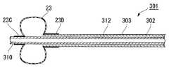
도58에 내시경용 처치구의 일례인 벌룬 카테터(301)의 구성을 도시한다. 이 벌룬 카테터(301)는 가요성을 갖는 긴 내부 시스(302)의 외측으로 외부 시스(303)를 진퇴 가능하게 씌우고 있다. 또한, 벌룬(23)의 전방측 단부가 외부 시스(303)의 선단에 접속되고, 벌룬(23)의 선단이 내부 시스(302)의 외주에 접속되어 있는 것을 특징으로 한다.58 shows the configuration of a
내부 시스(302)는 선단으로부터 기단에 관통하는 루멘(310)을 갖고, 기단부에는 루멘(310)에 연통하는 입구 부재(311)가 설치되어 있다. 입구 부재(311)로부터 가이드 와이어의 삽입 관통이나 조영제의 주입이 가능하다. 내부 시스(302)의 선단부의 외주에 벌룬(23)의 선단측 고정단(23C)이 고정되어 있다.The
벌룬(23)의 형상 및 크기는 제1, 제2 실시 형태와 마찬가지이다. 벌룬(23)의 기단측의 고정단(23D)은 외부 시스(303)의 선단 부분의 외주에 고정되어 있다.The shape and size of the
외부 시스(303)는 내부 시스(302)의 외경보다 큰 내경을 갖고, 양자 사이에 간극(312)이 형성되어 있다. 이 간극(312)이 벌룬(23)을 팽창시키는 유체를 통과시키는 공급로로 되어 있다. 외부 시스(303)의 기단은 조작부(321)에 고정되어 있다.The
조작부(321)는 관통 구멍(322)을 갖는다. 관통 구멍(322)에는 내부 시스(302)가 진퇴 가능하게 관통된다. 관통 구멍(322)의 직경은 내부 시스(302)의 외경보다 크기 때문에, 관통 구멍(322)은 간극(312)에 연통하고 있다. 조작 부(321)의 기단측에는 밀봉 부재(323)가 삽입되어 있고, 내부 시스(302)를 미끄럼 이동 가능하게 하면서, 수밀 구조 혹은 기밀 구조를 형성하고 있다. 또한, 관통 구멍(322)에 연통하는 입구 부재(324)가 직경 방향 외측으로 돌출 설치되어 있다. 입구 부재(324)에는 벌룬(23)을 팽창ㆍ수축시키는 주사기가 접속 가능하다.The
또한, 도58에서는, 벌룬(23)의 고정단(23C, 23D)의 거리가 벌룬(23)의 팽창 직경보다 커지도록, 외부 시스(303)의 위치가 설정되어 있다. 이때의 외부 시스(303)의 위치를 초기 위치로 한다.In addition, in FIG. 58, the position of the
다음에, 이 벌룬 카테터(301)를 이용한 시술에 대해 설명한다.Next, the procedure using this
처음에 도시하지 않은 파필로톰으로 유두를 절개한다. 가이드 와이어를 담관 내에 남겨둔 상태에서 파필로톰을 제거하고, 대신에 벌룬 카테터(301)를 가이드 와이어를 따라 체내에 도입하여 담관에 삽입한다. 이때, 벌룬 카테터(301)가 외부 시스(303)가 초기 위치, 즉 벌룬(23)의 고정단(23C, 23D) 사이의 거리가 벌룬(23)의 최대 팽창 직경보다 큰 거리로 한 상태에서 삽입된다.The papilla is initially dissected with a papillotom, not shown. The papillotom is removed while the guide wire is left in the bile duct, and instead, the
벌룬(23)의 축선 방향의 대략 중앙이 유두에 도달하면, 조작부(321)의 입구 부재(324)에 장착한 주사기로부터 기체 또는 액체인 유체를 간극(312)에 주입한다. 유체는 간극(312)의 선단으로부터 벌룬(23)에 공급되어, 도59에 도시한 바와 같이 벌룬(23)을 팽창시킨다. 벌룬(23)의 팽창량은 X선상이나 내시경 화상을 확인하면서 조정한다. 또한, 입구 부재(324)에 장착한 주사기 사이에 압력계를 설치하고 있는 경우에는, 압력계가 나타내는 압력을 확인하면서 조정한다.When approximately the center of the
벌룬(23)을 팽창시킴으로써 유두 및 담관의 출구측을 압박 확장하면, 벌 룬(23)에 공급한 유체를 배출한다. 도58에 도시한 바와 같이 벌룬(23)이 수축한다.When the
다음에, 전방측에서 조작부(321) 및 입구 부재(311)를 잡아 외부 시스(303)에 대해 내부 시스(302)를 후퇴시킨다. 외부 시스(303)가 내부 시스(302)에 대해 상대적으로 전진한다. 도60에 도시한 바와 같이, 벌룬(23)의 고정단(23C, 23D) 사이의 거리가 좁아진다. 이때의 고정단(23C, 23D) 사이의 거리는 벌룬(23)의 최대 팽창 직경보다 작게 한다. 양 시스(302, 303)의 상대적인 위치를 고정한 상태에서, 벌룬(23)의 후방측 고정단(23D)이 결석의 안쪽으로 갈 때까지 벌룬 카테터(301)를 압입한다.Next, on the front side, the
다시, 입구 부재(324)에 장착한 주사기로부터 유체를 공급하여 벌룬(23)을 팽창시킨다. 도61에 도시한 바와 같이, 벌룬(23)은 축선 방향보다 직경 방향이 커지도록 팽창된다. 벌룬(23)을 팽창시킨 상태에서 벌룬 카테터(301) 전체를 후퇴시키면, 벌룬(23)에 결석이 긁어져 나오도록 하여 담관으로부터 배출된다. 결석을 배출하면, 벌룬(23)을 수축시킨 후 벌룬 카테터(301)를 내시경으로부터 제거한다. 여기서, 벌룬(23)의 최대 팽창 정도를 20 ㎜로 한 경우, 벌룬(23)의 고정단(23C, 23D) 사이의 거리는 20 ㎜를 초과하는 거리로부터 20 ㎜보다 작은 거리로 변경 가능하다. 또한, 벌룬(23)은 팽창시에는 축 방향으로도 신장하므로, 팽창시의 벌룬(23)의 축 방향 길이는 고정단(23C, 23D) 사이의 거리보다도 길어진다. 따라서, 벌룬(23)의 축선 방향을 짧게 하여 담관의 구석구석까지 닿게 하여 결석 회수용으로 사용하기 쉽도록 하기 위해서는, 고정단(23C, 23D) 사이의 거리를 벌룬 최대 팽 창 직경의 1/2 이하로 하는 것이 바람직하다.Again, the fluid is inflated by supplying fluid from the syringe mounted to the
또한, 담관 출구의 확장용으로 벌룬(23)을 사용한다는 관점에서는, 관로의 확장부로부터 미끄러져 어긋나 버리는 것을 방지하기 위해 벌룬(23)에는 축선 방향으로 어느 정도의 길이가 필요해진다. 따라서, 고정단(23C, 23D) 사이의 거리는 벌룬 최대 팽창 직경의 1.5배 내지 2배 정도인 것이 더욱 요구된다.In addition, from the viewpoint of using the
벌룬(23)의 소재는 확장시의 강도를 중시하고자 하는 경우에는, 강도가 큰 폴리우레탄, 폴리에틸렌, PET, 폴리아미드 등의 신축성이 적은 비엘라스토머계의 재료가 적당하다. 담관 내에서의 진퇴의 용이함 등의 조작성을 중시하고자 하는 경우에는, 수축시에 소재도 줄어들어 고정단(23C, 23D)의 거리를 좁게 하였을 때의 주름이 최소한으로 억제되는 라텍스, 실리콘 고무, 우레탄 엘라스토머, 폴리아미드 엘라스토머 등의 신축성이 큰 엘라스토머계의 소재로 벌룬(23)을 제조하면 좋다.In the case where the material of the
이 실시 형태에서는 외부 시스(303)의 진퇴 조작으로 벌룬(23)의 팽창 방법을 변화시키도록 하였다. 고정단(23C, 23D)의 거리를 크게 한 후 벌룬(23)을 팽창시키면, 넓은 면적으로 조직을 압박하는 것이 가능해져, 관로의 원하는 위치를 어긋나지 않고 확실하게 확장할 수 있다. 이에 대해, 고정단(23C, 23C)의 거리를 작게 한 후 벌룬(23)을 팽창시키면, 축 방향이 짧아져 담관의 만곡 형상을 따라 이동하기 쉬워진다. 이로 인해, 벌룬(23)에 의해 결석을 배출하기 쉬워진다.In this embodiment, the expansion method of the
또한, 고정단(23C, 23D)의 거리의 조정을 보다 간편하게 하기 위해, 내부 시스(302)와 외부 시스(303)의 기단측에 양 시스(302, 303)의 상대 위치를 알 수 있는 마킹을 마련하거나, 조작부(321)에 클릭을 마련해도 좋다. 또한, 내부 시 스(302)를 파필로톰이나 바스켓, 멀티 루멘의 조형 튜브로서 사용해도 좋다.In addition, in order to make adjustment of the distance of the fixed ends 23C and 23D more simple, the marking which knows the relative position of both
이상, 본 발명의 바람직한 실시 형태를 설명하였지만, 본 발명은 상기한 실시 형태에 한정되는 것은 아니다. 본 발명의 취지를 일탈하지 않는 범위에서 구성의 부가, 생략, 치환 및 다른 교환이 가능하다. 본 발명은 상기한 설명에 의해 한정되는 것은 아니며, 첨부한 특허청구범위에 의해서만 한정된다.As mentioned above, although preferred embodiment of this invention was described, this invention is not limited to said embodiment. Additions, omissions, substitutions, and other exchanges are possible without departing from the spirit of the invention. The present invention is not limited by the above description, but only by the appended claims.
도1은 내시경용 처치구의 일례인 파필로톰의 구성을 도시하는 도면.BRIEF DESCRIPTION OF THE DRAWINGS The figure which shows the structure of the papillotom which is an example of the endoscope treatment instrument.
도2는 도1의 A-A선에 따른 단면도.2 is a cross-sectional view taken along the line A-A of FIG.
도3은 도1의 B-B선에 따른 단면도.3 is a cross-sectional view taken along the line B-B in FIG.
도4는 도1의 C-C선에 따른 단면도.4 is a cross-sectional view taken along the line C-C in FIG.
도5는 벌룬을 팽창시킨 상태를 도시하는 단면도.Fig. 5 is a sectional view showing a state in which the balloon is inflated.
도6은 내시경에 통과시킨 파필로톰을 유두 근방으로 안내한 도면.Figure 6 is a diagram guiding the papillotom passed through the endoscope in the vicinity of the nipple.
도7은 파필로톰의 선단을 유두에 삽입한 도면.Fig. 7 is a view of inserting the tip of the papillotom into the teat.
도8은 파필로톰의 절개 나이프부로 유두를 절개한 도면.Figure 8 is a view of the papillary cut in the papillotom knife portion.
도9는 절개 후에 파필로톰을 담관에 진입시킨 도면.Fig. 9 shows papillotom into bile duct after incision;
도10은 벌룬으로 유두를 압박 확장한 도면.10 is a view in which the nipple is expanded by a balloon;
도11은 도10 이후에 벌룬을 수축시킨 도면.Figure 11 is a view of shrinking the balloon after Figure 10;
도12는 바스켓 겸자로 결석을 배출하는 도면.12 is a view for discharging stones to the basket forceps.
도13은 비엘라스토머계 재료로 벌룬을 제조하였을 때의 외관도.Fig. 13 is an external view of a balloon made from a non-elastomeric material.
도14는 도13의 D-D선에 따른 단면도.14 is a cross-sectional view taken along the line D-D of FIG.
도15는 내시경형 처치구의 일례로, 커버 시스를 구비하는 파필로톰의 구성을 도시하는 도면.Fig. 15 is a view showing the configuration of a papillotom having a cover sheath as an example of an endoscope treatment instrument.
도16은 도15의 E-E선에 따른 단면도.16 is a cross-sectional view taken along the line E-E of FIG.
도17은 커버 시스를 후퇴시켜 벌룬을 팽창시킨 도면.Figure 17 is a view of inflating the balloon by retracting the cover sheath.
도18은 커버 시스를 전진시켜 벌룬의 선단 부분만을 팽창시킨 도면.Fig. 18 is a view in which only the tip portion of the balloon is inflated by advancing the cover sheath;
도19는 도17에 도시하는 파필로톰으로 유두를 절개한 도면.Fig. 19 is a view of the papillary cut into papillotom shown in Fig. 17;
도20은 노출시킨 벌룬을 유두까지 삽입한 도면.20 is a view inserting the exposed balloon to the nipple.
도21은 벌룬을 팽창시켜 유두를 압박 확장한 도면.Fig. 21 is a view in which the balloon is inflated and the nipple is expanded.
도22는 파필로톰을 전진시키는 동시에, 벌룬의 대략 절반이 가려질 때까지 커버 시스를 전진시킨 도면.Fig. 22 is an illustration of advancing the cover sheath while advancing the papillotom and approximately half of the balloon is covered;
도23은 커버 시스로부터 노출되는 벌룬을 팽창시킨 도면.Figure 23 is an inflated balloon exposed from the cover sheath.
도24는 벌룬으로 결석을 긁어낸 도면.Fig. 24 is a diagram scraping out stones with a balloon.
도25는 커버 시스에 벌룬을 설치한 파필로톰의 구성을 도시하는 도면.Fig. 25 is a diagram showing the configuration of papillotom provided with a balloon in a cover sheath;
도26은 도25의 F-F선에 따른 단면도.FIG. 26 is a cross-sectional view taken along the line F-F in FIG. 25;
도27은 도25의 G-G선에 따른 단면도.FIG. 27 is a sectional view taken along the line G-G in FIG. 25;
도28은 커버 시스를 전진시켜 벌룬을 팽창시킨 도면.Fig. 28 is a view of inflating the balloon by advancing the cover sheath;
도29는 도25의 파필로톰의 사용 형태를 설명하는 도면.FIG. 29 is a view explaining a usage pattern of the papillotom of FIG. 25;
도30은 절개 나이프부로 유두를 절개한 도면.30 is a view of the incision of the nipple by the incision knife;
도31은 커버 시스를 전진시켜 벌룬을 유두까지 삽입한 도면.Figure 31 is a view of advancing the cover sheath and inserting the balloon to the nipple.
도32는 벌룬을 팽창시켜 유두를 압박 확장한 도면.32 is a view in which the balloon is inflated and the nipple is expanded.
도33은 2개의 벌룬을 구비하는 내시경용 처치구의 구성을 도시하는 도면.Fig. 33 is a diagram showing the configuration of an endoscope treatment instrument having two balloons;
도34는 도33의 H-H선에 따른 단면도.FIG. 34 is a cross sectional view along line H-H in FIG. 33; FIG.
도35는 도33의 I-I선에 따른 단면도.FIG. 35 is a sectional view taken along line II of FIG. 33;
도36은 기단측의 벌룬을 팽창시킨 도면.36 is an inflated balloon at the proximal end;
도37은 선단측의 벌룬을 팽창시킨 도면.Fig. 37 is a view in which the balloon on the tip side is inflated;
도38은 기단측의 벌룬을 팽창시켜 유두를 압박 확장한 도면.Fig. 38 is a view in which the balloon at the proximal side is inflated to pressurize the teat;
도39는 결석의 안쪽측에서 선단측의 벌룬을 팽창시킨 도면.Fig. 39 is a view of inflating the balloon at the tip side from the inside of the stone;
도40은 1개의 벌룬과 커버 시스를 구비하는 내시경용 처치구의 구성을 도시하는 도면.40 is a diagram showing the configuration of an endoscope treatment instrument having one balloon and a cover sheath;
도41은 도40의 J-J선에 따른 단면도.FIG. 41 is a cross sectional view along line J-J in FIG. 40; FIG.
도42는 도40의 K-K선에 따른 단면도.FIG. 42 is a cross sectional view along line K-K in FIG. 40;
도43a는 벌룬의 전체를 팽창시킨 도면.43A is an inflated view of the balloon;
도43b는 벌룬의 선단 부분만을 팽창시킨 도면.Fig. 43B is a view inflating only the tip portion of the balloon;
도44는 벌룬을 팽창시켜 유두를 압박 확장한 도면.Fig. 44 is a view in which the balloon is inflated and the nipple is expanded.
도45는 내시경용 처치구를 전진시키는 동시에, 벌룬의 대략 절반이 가려질 때까지 커버 시스를 전진시킨 도면.Fig. 45 is a view of advancing the endoscope treatment instrument and advancing the cover sheath until approximately half of the balloon is covered;
도46은 벌룬의 선단측을 팽창시킨 도면.Fig. 46 is a view inflating the tip side of the balloon.
도47은 바스켓 겸자로 포착한 결석이 절개한 유두로부터 취출할 수 없는 상태를 설명하는 도면.Fig. 47 is a view for explaining a state in which stones taken out with a basket forceps cannot be taken out from the cut nipple;
도48은 바스켓 겸자와 동일한 경로로 내시경용 처치구를 통과시켜 벌룬으로 유두를 압박 확장한 도면.Fig. 48 is a view in which the nipple is expanded with a balloon by passing the endoscope treatment instrument in the same path as the basket forceps;
도49는 확장한 유두로부터 결석을 배출하는 도면.49 is a view showing the discharge of stones from the expanded teat.
도50은 카테터에 벌룬이 설치된 내시경용 처치구의 구성을 도시하는 도면.Fig. 50 is a diagram showing the configuration of an endoscope treatment instrument provided with a balloon at the catheter;
도51은 바스켓 겸자와 병렬로 내시경용 처치구를 삽입하여 벌룬으로 유두를 압박 확장한 도면.Fig. 51 is a diagram in which the nipple is pushed and expanded with a balloon by inserting an endoscope treatment instrument in parallel with a basket forceps;
도52는 벌룬을 구비하는 바스켓형 겸자의 구성을 도시하는 도면.Fig. 52 is a diagram showing the configuration of a basket-shaped forceps having a balloon.
도53은 벌룬을 팽창시킨 도면.53 is an inflated balloon.
도54는 내시경용 처치구의 일례인 파필로톰의 구성을 도시하는 도면.Fig. 54 shows the structure of a papillotom which is an example of an endoscope treatment instrument;
도55는 도54의 L-L선에 따른 단면도.Fig. 55 is a cross sectional view along line L-L in Fig. 54;
도56은 도54의 M-M선에 따른 단면도.Fig. 56 is a sectional view along the line M-M in Fig. 54;
도57은 벌룬을 팽창시킨 도면.Fig. 57 is a view of inflating the balloon;
도58은 내시경용 처치구의 일례인 벌룬 카테터의 구성을 도시하는 도면.Fig. 58 is a diagram showing the configuration of a balloon catheter as an example of an endoscope treatment instrument.
도59는 벌룬을 팽창시켰을 때의 선단 부분을 확대하여 도시하는 단면도.Fig. 59 is an enlarged cross sectional view of a distal end portion when the balloon is inflated;
도60은 벌룬을 수축시킨 상태에서 외부 시스를 상대적으로 전진시킨 도면.Fig. 60 is a view of relatively advanced external sheath with the balloon retracted;
도61은 도60의 상태로부터 벌룬을 팽창시킨 도면.FIG. 61 is an inflated balloon from the state of FIG. 60; FIG.
<도면의 주요 부분에 대한 부호의 설명><Explanation of symbols for the main parts of the drawings>
1 : 파필로톰1: papillotom
2 : 조작부2: control panel
3 : 시스3: sheath
4 : 도전 와이어4: conductive wire
23 : 벌룬23: balloon
32 : 튜브32: tube
33 : 삽입부33: insertion unit
34 : 링34: ring
35 : 접속부35 connection
36 : 조작부 본체36: operation unit main body
41 : 주사기41: syringe
42 : 슬라이더42: slider
Claims (10)
Translated fromKoreanApplications Claiming Priority (2)
| Application Number | Priority Date | Filing Date | Title |
|---|---|---|---|
| US93415107P | 2007-06-11 | 2007-06-11 | |
| US60/934,151 | 2007-06-11 |
Publications (1)
| Publication Number | Publication Date |
|---|---|
| KR20080108906Atrue KR20080108906A (en) | 2008-12-16 |
Family
ID=39689195
Family Applications (1)
| Application Number | Title | Priority Date | Filing Date |
|---|---|---|---|
| KR1020080053433AAbandonedKR20080108906A (en) | 2007-06-11 | 2008-06-09 | Endoscopic treatment tools and treatment methods |
Country Status (4)
| Country | Link |
|---|---|
| US (2) | US8562601B2 (en) |
| EP (3) | EP2002779A3 (en) |
| JP (1) | JP2008302226A (en) |
| KR (1) | KR20080108906A (en) |
Cited By (5)
| Publication number | Priority date | Publication date | Assignee | Title |
|---|---|---|---|---|
| KR20210149316A (en)* | 2020-06-02 | 2021-12-09 | 고려대학교 산학협력단 | Apparatus for removing calculus |
| KR20220014413A (en)* | 2020-07-27 | 2022-02-07 | 유펙스메드 주식회사 | Multi-lumen medical dispensing device for combination of multiple treatment tools |
| KR20220031201A (en)* | 2020-09-04 | 2022-03-11 | 계명대학교 산학협력단 | Biopsy Forceps |
| KR102642169B1 (en)* | 2023-01-09 | 2024-02-29 | 주형준 | Endoscopic device for tracheal intubation |
| KR20240111964A (en) | 2023-01-11 | 2024-07-18 | 유펙스메드 주식회사 | A medical discharge device with improved multi-lumen operation characteristics in which multiple treatment tools are used together |
Families Citing this family (22)
| Publication number | Priority date | Publication date | Assignee | Title |
|---|---|---|---|---|
| JP5095124B2 (en)* | 2006-05-17 | 2012-12-12 | 富士フイルム株式会社 | Endoscope |
| WO2009051698A2 (en)* | 2007-10-12 | 2009-04-23 | Beth Israel Deaconess Medical Center | Catheter guided endotracheal intubation |
| AU2008343141B2 (en)* | 2007-12-27 | 2013-10-24 | Cook Medical Technologies Llc | Two-part extraction balloon |
| US8361100B2 (en) | 2008-03-17 | 2013-01-29 | Ethicon, Inc. | Applicator instruments for the delivery, deployment, and tamponade of hemostats and methods therefor |
| US8366733B2 (en) | 2008-03-28 | 2013-02-05 | Ethicon, Inc. | Applicator instruments for controlling bleeding at surgical sites and methods therefor |
| JP5444840B2 (en)* | 2009-05-21 | 2014-03-19 | 東レ株式会社 | Ablation catheter with balloon and ablation catheter system with balloon |
| CN103654942B (en)* | 2012-09-25 | 2015-11-25 | 天津博朗科技发展有限公司 | With the disposable endoscope mirror sheath of surgical instrument angle adjustment device |
| US9358042B2 (en) | 2013-03-13 | 2016-06-07 | The Spectranetics Corporation | Expandable member for perforation occlusion |
| JP2015104485A (en)* | 2013-11-29 | 2015-06-08 | オリンパスメディカルシステムズ株式会社 | Catheter |
| EP3603547B1 (en)* | 2014-06-10 | 2021-07-21 | Fujifilm Medwork Gmbh | Papillotome for percutaneous endoscopic gastrostomy |
| JP6329101B2 (en) | 2015-04-15 | 2018-05-23 | オリンパス株式会社 | Endoscope treatment tool and method for manufacturing endoscope treatment tool |
| JP2017012670A (en)* | 2015-07-06 | 2017-01-19 | オリンパス株式会社 | Treatment instrument |
| US10499892B2 (en) | 2015-08-11 | 2019-12-10 | The Spectranetics Corporation | Temporary occlusion balloon devices and methods for preventing blood flow through a vascular perforation |
| US10449336B2 (en) | 2015-08-11 | 2019-10-22 | The Spectranetics Corporation | Temporary occlusions balloon devices and methods for preventing blood flow through a vascular perforation |
| KR101782250B1 (en)* | 2016-02-29 | 2017-09-27 | 가톨릭대학교 산학협력단 | stone removal device |
| US11166745B2 (en)* | 2017-09-12 | 2021-11-09 | Jessica Jameson | Multi-port epidural needle |
| CN107596539B (en)* | 2017-10-17 | 2020-01-03 | 上海英诺伟医疗器械有限公司 | Balloon dilatation catheter |
| JP6937091B2 (en) | 2017-10-17 | 2021-09-22 | 上海英諾偉医療器械有限公司 | Expansion balloon catheter |
| WO2021207229A1 (en)* | 2020-04-08 | 2021-10-14 | Boston Scientific Scimed, Inc. | Medical instrument having a balloon and an extraction member |
| JP2023529459A (en)* | 2020-06-11 | 2023-07-10 | ボストン サイエンティフィック リミテッド | MEDICAL SYSTEMS, DEVICES AND RELATED METHODS |
| US20220061866A1 (en)* | 2020-08-27 | 2022-03-03 | Boston Scientific Scimed, Inc. | Medical extraction assemblies and methods of using the same |
| JP7601933B2 (en) | 2023-03-31 | 2024-12-17 | 日本ライフライン株式会社 | Extensions |
Family Cites Families (16)
| Publication number | Priority date | Publication date | Assignee | Title |
|---|---|---|---|---|
| US4886496A (en)* | 1988-02-04 | 1989-12-12 | Conoscenti Craig S | Bronchoscopic balloon tipped catheter and method of making the same |
| US5628746A (en)* | 1989-01-18 | 1997-05-13 | Applied Medical Resources Corporation | Dilatation catheter assembly with cutting element and method of using the same |
| US5766151A (en) | 1991-07-16 | 1998-06-16 | Heartport, Inc. | Endovascular system for arresting the heart |
| JPH0663057A (en)* | 1992-08-18 | 1994-03-08 | Olympus Optical Co Ltd | High-frequency treatment means |
| US5547469A (en)* | 1994-05-13 | 1996-08-20 | Boston Scientific Corporation | Apparatus for performing diagnostic and therapeutic modalities in the biliary tree |
| US5549551A (en)* | 1994-12-22 | 1996-08-27 | Advanced Cardiovascular Systems, Inc. | Adjustable length balloon catheter |
| JP3618027B2 (en)* | 1996-11-20 | 2005-02-09 | 住友ベークライト株式会社 | Endoscopic dilatation balloon catheter |
| US6997885B2 (en)* | 1998-04-08 | 2006-02-14 | Senorx, Inc. | Dilation devices and methods for removing tissue specimens |
| FR2781379B1 (en) | 1998-07-22 | 2000-10-06 | Prodimed | BALLOON DEVICE WITH ADJUSTABLE NOMINAL DIAMETER |
| WO2001089624A1 (en) | 2000-05-19 | 2001-11-29 | C.R. Bard, Inc. | Steerable biliary catheter |
| US6579300B2 (en) | 2001-01-18 | 2003-06-17 | Scimed Life Systems, Inc. | Steerable sphincterotome and methods for cannulation, papillotomy and sphincterotomy |
| US20050209674A1 (en) | 2003-09-05 | 2005-09-22 | Kutscher Tuvia D | Balloon assembly (V) |
| JP4695428B2 (en) | 2004-05-13 | 2011-06-08 | オリンパス株式会社 | Endoscopic treatment tool |
| JP4043456B2 (en) | 2004-06-25 | 2008-02-06 | オリンパス株式会社 | High frequency incision tool |
| WO2006129726A1 (en) | 2005-05-31 | 2006-12-07 | Olympus Medical Systems Corp. | Device and method for mucosal detachment |
| JP2008194167A (en) | 2007-02-09 | 2008-08-28 | Nippon Zeon Co Ltd | Balloon catheter for stone removal |
- 2008
- 2008-06-05JPJP2008147994Apatent/JP2008302226A/enactivePending
- 2008-06-09KRKR1020080053433Apatent/KR20080108906A/ennot_activeAbandoned
- 2008-06-10USUS12/136,345patent/US8562601B2/enactiveActive
- 2008-06-11EPEP08010635Apatent/EP2002779A3/ennot_activeWithdrawn
- 2008-06-11EPEP11001050Apatent/EP2359763A1/ennot_activeWithdrawn
- 2008-06-11EPEP10000013Apatent/EP2172161B1/enactiveActive
- 2013
- 2013-09-12USUS14/025,131patent/US9827038B2/enactiveActive
Cited By (7)
| Publication number | Priority date | Publication date | Assignee | Title |
|---|---|---|---|---|
| KR20210149316A (en)* | 2020-06-02 | 2021-12-09 | 고려대학교 산학협력단 | Apparatus for removing calculus |
| WO2021246724A1 (en)* | 2020-06-02 | 2021-12-09 | 고려대학교 산학협력단 | Calculi removal device |
| KR20220014413A (en)* | 2020-07-27 | 2022-02-07 | 유펙스메드 주식회사 | Multi-lumen medical dispensing device for combination of multiple treatment tools |
| KR20220031201A (en)* | 2020-09-04 | 2022-03-11 | 계명대학교 산학협력단 | Biopsy Forceps |
| KR102642169B1 (en)* | 2023-01-09 | 2024-02-29 | 주형준 | Endoscopic device for tracheal intubation |
| WO2024150919A1 (en)* | 2023-01-09 | 2024-07-18 | 주형준 | Endoscope dedicated to tracheal intubation |
| KR20240111964A (en) | 2023-01-11 | 2024-07-18 | 유펙스메드 주식회사 | A medical discharge device with improved multi-lumen operation characteristics in which multiple treatment tools are used together |
Also Published As
| Publication number | Publication date |
|---|---|
| US20100298634A1 (en) | 2010-11-25 |
| US8562601B2 (en) | 2013-10-22 |
| JP2008302226A (en) | 2008-12-18 |
| EP2172161B1 (en) | 2012-08-22 |
| EP2172161A1 (en) | 2010-04-07 |
| EP2359763A1 (en) | 2011-08-24 |
| EP2002779A3 (en) | 2009-03-04 |
| US9827038B2 (en) | 2017-11-28 |
| US20140018800A1 (en) | 2014-01-16 |
| EP2172161A8 (en) | 2010-06-02 |
| EP2002779A2 (en) | 2008-12-17 |
Similar Documents
| Publication | Publication Date | Title |
|---|---|---|
| KR20080108906A (en) | Endoscopic treatment tools and treatment methods | |
| JP3722729B2 (en) | Endoscope treatment device | |
| US9848763B2 (en) | Access systems and methods of intra-abdominal surgery | |
| EP2816963B1 (en) | Expandable endoscopic hoods | |
| US7396329B2 (en) | Endoscopic retractor instrument and associated method | |
| JP4485110B2 (en) | Laparoscopic surgical access tool with gas seal | |
| US9289236B2 (en) | Mucosa separation apparatus, and method for mucosa separation | |
| US20070066869A1 (en) | Endoscopic assembly including cap and sheath | |
| JP4073965B2 (en) | Balloon incision device | |
| EP0912139A4 (en) | ||
| CN113038862B (en) | Devices used to retract tissue under endoscopy | |
| JP2015083069A (en) | Treatment instrument for endoscope | |
| WO2005079683A1 (en) | Endoscopic tissue stabilization device and related methods of use | |
| US20240389996A1 (en) | Systems, suture devices, and methods for tissue closure | |
| JP3645912B2 (en) | Balloon incision device |
Legal Events
| Date | Code | Title | Description |
|---|---|---|---|
| PA0109 | Patent application | Patent event code:PA01091R01D Comment text:Patent Application Patent event date:20080609 | |
| PG1501 | Laying open of application | ||
| A201 | Request for examination | ||
| PA0201 | Request for examination | Patent event code:PA02012R01D Patent event date:20130226 Comment text:Request for Examination of Application Patent event code:PA02011R01I Patent event date:20080609 Comment text:Patent Application | |
| E902 | Notification of reason for refusal | ||
| PE0902 | Notice of grounds for rejection | Comment text:Notification of reason for refusal Patent event date:20140313 Patent event code:PE09021S01D | |
| E902 | Notification of reason for refusal | ||
| PE0902 | Notice of grounds for rejection | Comment text:Notification of reason for refusal Patent event date:20140926 Patent event code:PE09021S01D | |
| E701 | Decision to grant or registration of patent right | ||
| PE0701 | Decision of registration | Patent event code:PE07011S01D Comment text:Decision to Grant Registration Patent event date:20150116 | |
| PC1904 | Unpaid initial registration fee |