KR20080053057A - Ultrasound Imaging System and Method for Forming and Displaying Mixed Image of Ultrasound Image and External Medical Image - Google Patents
Ultrasound Imaging System and Method for Forming and Displaying Mixed Image of Ultrasound Image and External Medical ImageDownload PDFInfo
- Publication number
- KR20080053057A KR20080053057AKR1020060124999AKR20060124999AKR20080053057AKR 20080053057 AKR20080053057 AKR 20080053057AKR 1020060124999 AKR1020060124999 AKR 1020060124999AKR 20060124999 AKR20060124999 AKR 20060124999AKR 20080053057 AKR20080053057 AKR 20080053057A
- Authority
- KR
- South Korea
- Prior art keywords
- image
- ultrasound
- mixed
- external medical
- ultrasound image
- Prior art date
- Legal status (The legal status is an assumption and is not a legal conclusion. Google has not performed a legal analysis and makes no representation as to the accuracy of the status listed.)
- Ceased
Links
Images


Classifications
- A—HUMAN NECESSITIES
- A61—MEDICAL OR VETERINARY SCIENCE; HYGIENE
- A61B—DIAGNOSIS; SURGERY; IDENTIFICATION
- A61B8/00—Diagnosis using ultrasonic, sonic or infrasonic waves
- A61B8/52—Devices using data or image processing specially adapted for diagnosis using ultrasonic, sonic or infrasonic waves
- A61B8/5215—Devices using data or image processing specially adapted for diagnosis using ultrasonic, sonic or infrasonic waves involving processing of medical diagnostic data
- A61B8/5238—Devices using data or image processing specially adapted for diagnosis using ultrasonic, sonic or infrasonic waves involving processing of medical diagnostic data for combining image data of patient, e.g. merging several images from different acquisition modes into one image
- G—PHYSICS
- G06—COMPUTING OR CALCULATING; COUNTING
- G06T—IMAGE DATA PROCESSING OR GENERATION, IN GENERAL
- G06T17/00—Three dimensional [3D] modelling, e.g. data description of 3D objects
- G—PHYSICS
- G16—INFORMATION AND COMMUNICATION TECHNOLOGY [ICT] SPECIALLY ADAPTED FOR SPECIFIC APPLICATION FIELDS
- G16H—HEALTHCARE INFORMATICS, i.e. INFORMATION AND COMMUNICATION TECHNOLOGY [ICT] SPECIALLY ADAPTED FOR THE HANDLING OR PROCESSING OF MEDICAL OR HEALTHCARE DATA
- G16H30/00—ICT specially adapted for the handling or processing of medical images
Landscapes
- Engineering & Computer Science (AREA)
- Health & Medical Sciences (AREA)
- Physics & Mathematics (AREA)
- Life Sciences & Earth Sciences (AREA)
- Nuclear Medicine, Radiotherapy & Molecular Imaging (AREA)
- Radiology & Medical Imaging (AREA)
- General Health & Medical Sciences (AREA)
- Medical Informatics (AREA)
- Public Health (AREA)
- Biophysics (AREA)
- Animal Behavior & Ethology (AREA)
- Primary Health Care (AREA)
- Epidemiology (AREA)
- Pathology (AREA)
- Biomedical Technology (AREA)
- Heart & Thoracic Surgery (AREA)
- Molecular Biology (AREA)
- Surgery (AREA)
- Computer Vision & Pattern Recognition (AREA)
- Veterinary Medicine (AREA)
- Computer Graphics (AREA)
- Geometry (AREA)
- Software Systems (AREA)
- General Physics & Mathematics (AREA)
- Theoretical Computer Science (AREA)
- Ultra Sonic Daignosis Equipment (AREA)
- Magnetic Resonance Imaging Apparatus (AREA)
Abstract
Translated fromKoreanDescription
Translated fromKorean도 1은 본 발명의 일실시예에 따른 초음파 영상 시스템의 구성을 보이는 블록도.1 is a block diagram showing the configuration of an ultrasonic imaging system according to an embodiment of the present invention.
도 2a는 본 발명의 실시예에 따른 초음파 영상 시스템의 구성을 보이는 블록도.Figure 2a is a block diagram showing the configuration of an ultrasonic imaging system according to an embodiment of the present invention.
도2b는 도 2a에 보인 초음파 영상 시스템에 구비되는 프로브 위치 제공부의 구성을 보이는 블록도.Figure 2b is a block diagram showing the configuration of the probe position providing unit provided in the ultrasonic imaging system shown in FIG.
도 3a는 다수의 실시간 2차원 초음파 영상을 보이는 예시도.Figure 3a is an exemplary view showing a plurality of real-time two-dimensional ultrasound image.
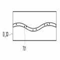
도 3b는 다수의 실시간 2차원 초음파 영상으로부터 형성된 2차원 파노라믹 초음파 영상의 예시도.3B is an illustration of a two-dimensional panoramic ultrasound image formed from multiple real-time two-dimensional ultrasound images.
도 4a는 2차원 외부 의료영상을 보이는 예시도.Figure 4a is an exemplary view showing a two-dimensional external medical image.
도 4b는 본 발명의 실시예에 따라 외부 의료영상 내의 대상체 크기를 2차원 파노라믹 초음파 영상 내의 대상체 크기와 일치시키는 예를 보이는 개략도.4B is a schematic diagram showing an example of matching an object size in an external medical image with an object size in a 2D panoramic ultrasound image according to an embodiment of the present invention.
도 5는 본 발명의 실시예에 따라 2차원 파노라믹 초음파 영상과 2차원 외부의료 영상의 혼합영상을 형성한 예를 보이는 개략도.Figure 5 is a schematic diagram showing an example of forming a mixed image of a two-dimensional panoramic ultrasound image and a two-dimensional external medical image in accordance with an embodiment of the present invention.
도 6은 본 발명의 실시예에 따라 다수의 3차원 초음파 영상으로부터 파노라믹 3차원 초음파 영상을 형성하는 예를 보이는 개략도.6 is a schematic view showing an example of forming a panoramic three-dimensional ultrasound image from a plurality of three-dimensional ultrasound image in accordance with an embodiment of the present invention.
도 7은 3차원 외부 의료영상의 예를 보이는 개략도.7 is a schematic diagram showing an example of a three-dimensional external medical image.
도 8은 파노라믹 3차원 초음파 영상과 3차원 외부 의료영상으로부터 혼합영상을 형성한 예를 보이는 개략도.8 is a schematic view showing an example in which a mixed image is formed from a panoramic three-dimensional ultrasound image and a three-dimensional external medical image.
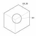
도 9a는 본 발명의 실시예에 따른 초음파 영상 시스템으로부터 얻어진 3차원 초음파 영상의 예를 보이는 개략도.9A is a schematic diagram showing an example of a three-dimensional ultrasound image obtained from an ultrasound imaging system according to an embodiment of the present invention.
도 9b는 2차원 외부 의료영상의 일예를 보이는 개략도.9B is a schematic diagram showing an example of a two-dimensional external medical image.
도 9c는 3차원 초음파 영상과 2차원 외부 의료영상으로부터 혼합영상을 형성한 예를 보이는 개략도.9C is a schematic view showing an example in which a mixed image is formed from a 3D ultrasound image and a 2D external medical image.
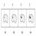
도 10는 이력 영상의 디스플레이 예를 보이는 개략도.10 is a schematic diagram showing a display example of a history image.
본 발명은 초음파 진단분야에 관한 것으로, 보다 상세하게는 초음파 영상과 외부 의료영상의 혼합영상을 형성하고 디스플레이하기 위한 초음파 영상 시스템 및 방법에 관한 것이다.The present invention relates to the field of ultrasound diagnostics, and more particularly, to an ultrasound imaging system and method for forming and displaying a mixed image of an ultrasound image and an external medical image.
전자 기기의 발달에 따라 다양한 원리로 인체 내부기관의 상태를 보이는 많은 의료 기기가 개발되었다. 의료기기의 일예인 초음파 영상 시스템은 초음파를 이용하여 비파괴, 비침습 방식으로 대상체의 내부 구조를 보인다. 또한, 초음파 영상 시스템은 CT(computerized tomography), MRI(magnetic resonance imager), PET(Positron Emission Tomography) 등과 같은 의료영상 장비 보다 실시간으로 영상을 얻기 쉽고, CT 등을 이용함에 따른 방사선 노출 위험도 없다. 이와 같이 초음파 영상 시스템은 인체에 무해하게 실시간으로 영상을 얻을 수 있다. 그러나, 초음파 영상 시스템으로부터 제공되는 영상은 그 고유한 특성에 의해 CT, MRI, PET 등으로부터 얻어지는 영상보다 해상도가 낮거나 변별력이 떨어져 병변을 정확한 위치에서 뚜렷하게 분별하기 어려운 단점이 있다. 이에 반해 CT, MRI, PET 등으로부터 얻어지는 영상 정보는 초음파에서 충분히 제공하지 못하는 대상체의 병변에 대한 형태와 위치 정보를 제공한다. 따라서 이러한 단점을 보완하고자 2차원 초음파 영상 정보와 CT, MRI, PET 등의 영상정보를 단순히 합성하는 기술이 알려져 있다. 하지만 이 역시 초음파의 좁은 시야 범위에 한정되고, 2차원 초음파 영상과의 합성에 국한되는 제약이 있다.BACKGROUND With the development of electronic devices, many medical devices have been developed that show the state of internal organs of the human body in various principles. An ultrasound imaging system, an example of a medical device, uses ultrasound to show the internal structure of an object in a non-destructive and non-invasive manner. In addition, the ultrasound imaging system is easier to obtain images in real time than medical imaging equipment such as computerized tomography (CT), magnetic resonance imager (MRI), positron emission tomography (PET), and the like, and there is no risk of radiation exposure by using CT. As such, the ultrasound imaging system may obtain an image in real time without harming the human body. However, the image provided from the ultrasound imaging system has a disadvantage in that it is difficult to clearly distinguish the lesion at the correct position because of its inherent characteristics, such that the resolution is lower than the image obtained from CT, MRI, PET, or the like. In contrast, image information obtained from CT, MRI, PET, and the like provides information on the shape and location of the lesion of the object that is not sufficiently provided by ultrasound. Therefore, a technique of simply synthesizing two-dimensional ultrasound image information and image information such as CT, MRI, PET, and the like is known to compensate for these disadvantages. However, this is also limited to the narrow field of view of the ultrasound, there is a limitation to the synthesis with the two-dimensional ultrasound image.
전술한 문제점을 해결하기 위한 본 발명은, 파노라믹 초음파 영상 또는 3차원 초음파 영상과 외부 의료영상을 혼합하여 디스플레이하기 위한 초음파 영상 시스템 및 방법을 제공하는데 그 목적이 있다.An object of the present invention is to provide an ultrasound imaging system and method for mixing and displaying a panoramic ultrasound image or a 3D ultrasound image and an external medical image.
본 발명의 실시예에 따른 초음파 영상 시스템은, 대상체에 초음파 신호를 송신하고 대상체로부터 반사되는 초음파 신호를 입력받아 실시간으로 초음파 영상신호를 제공하기 위한 초음파 진단부; 외부 영상신호 제공부; 상기 실시간으로 제공되는 초음파 영상신호에 기초하여 다수의 실시간 초음파 영상을 형성하고, 다수의 실시간 초음파 영상으로부터 파노라믹 초음파 영상 또는 3차원 초음파 영상을 형성하고, 상기 외부 영상신호에 기초하여 외부 의료영상을 형성하고, 상기 파노라믹 초음파 영상과 상기 외부 의료영상의 제1 혼합영상 및 상기 3차원 초음파 영상과 상기 외부 의료영상의 제2 혼합영상 중 적어도 하나를 형성하기 위한 영상처리부; 상기 실시간 초음파 영상, 상기 외부 의료영상, 상기 제1 혼합영상 및 상기 제2 혼합영상을 저장하기 위한 저장부; 상기 실시간 초음파 영상, 상기 외부 의료영상, 상기 제1 혼합영상 및 상기 제2 혼합영상 중 적어도 하나를 디스플레이하기 위한 디스플레이부를 포함한다.An ultrasound imaging system according to an embodiment of the present invention includes an ultrasound diagnosis unit for transmitting an ultrasound signal to an object and receiving an ultrasound signal reflected from the object to provide an ultrasound image signal in real time; An external video signal providing unit; A plurality of real time ultrasound images are formed based on the ultrasound image signals provided in real time, a panoramic ultrasound image or a 3D ultrasound image is formed from the plurality of real time ultrasound images, and an external medical image is based on the external image signals. An image processing unit for forming at least one of the first mixed image of the panoramic ultrasound image and the external medical image and the second mixed image of the three-dimensional ultrasound image and the external medical image; A storage unit for storing the real-time ultrasound image, the external medical image, the first mixed image, and the second mixed image; And a display unit for displaying at least one of the real-time ultrasound image, the external medical image, the first mixed image, and the second mixed image.
초음파 영상과 외부 의료영상의 혼합영상의 형성 및 디스플레이를 위한 본 발명의 실시예에 따르면, 실시간으로 제공되는 초음파 영상신호에 기초하여 다수의 실시간 초음파 영상을 형성하고, 다수의 실시간 초음파 영상으로부터 파노라믹 초음파 영상 또는 3차원 초음파 영상을 형성하고, 외부 영상신호에 기초하여 외부 의료영상을 형성하고, 상기 파노라믹 초음파 영상과 상기 외부 의료영상의 제1 혼합영상 또는 상기 3차원 초음파 영상과 상기 외부 의료영상의 제2 혼합영상을 형성하고, 상기 실시간 초음파 영상, 상기 외부 의료영상, 상기 제1 혼합영상 및 상기 제2 혼합영상 중 적어도 하나를 디스플레이한다.According to an embodiment of the present invention for forming and displaying a mixed image of an ultrasound image and an external medical image, a plurality of real-time ultrasound images are formed on the basis of an ultrasound image signal provided in real time, and a panorama from a plurality of real-time ultrasound images. A MIC ultrasound image or a 3D ultrasound image is formed, and an external medical image is formed based on an external image signal, and the first mixed image of the panoramic ultrasound image and the external medical image or the 3D ultrasound image and the external A second mixed image of the medical image is formed, and at least one of the real-time ultrasound image, the external medical image, the first mixed image, and the second mixed image is displayed.
이하, 첨부된 도면을 참조하여 본 발명의 실시예들을 상세하게 설명한다.Hereinafter, with reference to the accompanying drawings will be described embodiments of the present invention;
도 1에 보이는 바와 같이 본 발명의 일실시예에 따른 초음파 영상 시스템(100)은 대상체에 초음파 신호를 송신하고 대상체로부터 반사되는 초음파 신호를 입력받아 초음파 영상신호를 실시간으로 제공하는 초음파 진단부(10), 외부 영상신호 제공부(20), 사용자 입력부(30), 영상처리부(40), 저장부(50), 디스플레이부(60) 및 중앙처리부(70)를 포함한다.As shown in FIG. 1, the
외부 영상신호 제공부(20)는 초음파 진단부(10)가 아닌 외부에서 얻어진 2차원 또는 3차원 의료 영상신호(이하, 외부 영상신호라 함)를 제공한다. 외부 영상신호는 CT(computerized tomography), MRI(magnetic resonance imager) 또는 PET(Positron Emission Tomography) 등의 외부 의료영상장비에서 얻어진 신호이다. 외부 영상신호는 DICOM(Digital Imaging Communication in Medicine) 형식으로 표현된다. 이 외부 영상신호에는 특징점 위치 정보, 단위 복셀당 크기 정보, ECG(electrocadiogram) 등의 동기 신호, 신호 획득 프로토콜, 측정 조건 등의 부가 정보가 포함된다. 측정 조건은 조영제의 투입여부 등을 포함한다.The external image
사용자 입력부(30)는 마우스(mouse), 키보드(key board), 트랙볼(track ball) 등으로 구현되어, 사용자로부터 초음파 영상과 외부 의료영상의 혼합유형, 초음파 영상과 외부 의료영상의 디스플레이 모드, 측정 기준위치, 3차원 공간상의 실시간 초음파 프로브 위치 등을 입력받는다. 디스플레이 모드는 파노라믹 초음파 영상 디스플레이 모드, 3차원 초음파 영상 디스플레이 모드, 외부 의료영상 디스플레이모드, 초음파 영상과 외부 의료영상의 혼합영상 디스플레이 모드, 초음파 영상, 외부 의료영상 및 혼합영상 중 적어도 두 영상의 병렬 디스플레이 모드, 이력영상 디스플레이 모드 등을 포함한다. 혼합유형은 2차원 파노라믹 초음파 영상과 2차원 외부 의료영상의 혼합, 3차원 파노라믹 초음파 영상과 2차원 또는 3차원 외부 의료영상의 혼합, 3차원 초음파 영상과 2차원 또는 3차원 외부 의료영상의 혼합을 포함한다.The
영상처리부(40)는 실시간으로 제공되는 초음파 영상신호에 기초하여 다수의실시간 초음파 영상을 형성하고, 다수의 실시간 초음파 영상으로부터 파노라믹 초음파 영상 또는 3차원 초음파 영상을 형성하고, 외부 영상신호에 기초하여 외부 의료영상을 형성하고, 파노라믹 초음파 영상과 외부 의료 영상의 제1 혼합영상 및 3차원 초음파 영상과 외부 의료영상의 제2 혼합영상 중 적어도 하나를 형성한다. 파노라믹 초음파 영상은 2차원 파노라믹 초음파 영상과 3차원 파노라믹 초음파 영상을 포함하고, 외부 의료영상은 2차원 외부 의료영상 및 3차원 외부 의료영상을 포함한다. 본 명세서에서 의미하는 "실시간"은 초음파 진단이 진행되고 있는 시간을 의미한다. 외부 의료영상은 초음파 진단이 진행되기 이전에 얻어진 외부 영상신호로부터 형성되고, 실시간 초음파 영상은 초음파 진단이 진행되는 과정 동안에 얻어진 모든 초음파 영상을 의미한다. 또한, 영상처리부(40)는 사용자 입력부(30)를 통하여 사용자가 입력한 혼합유형에 따라 제1 혼합영상 또는 제2 혼합영상을 형성한다. 또한, 영상처리부(40)는 사용자가 선택한 디스플레이 모드에 따라 초음파 영상과 외부 의료영상을 다양한 디스플레이 형태로 편집한다. 또한, 영상처리부(40)는 사용자로부터 입력되는 측정 기준위치에 근거하여 대상체의 길이, 부피 등을 산출한다.The
저장부(50)는 영상처리부(40)에서 형성된 실시간 초음파 영상, 외부 의료영상 제1 혼합영상 및 제2 혼합영상을 저장한다. 각 영상은 환자별로, 대상체별로, 형성된 날짜별로 구분되어 저장된다. 또한, 저장부(50)는 혼합유형 항목, 디스플레이 모드 항목과 외부 영상신호에 포함된 특징점 위치 정보, 단위 복셀당 크기 정보, ECG 등의 동기 신호, 신호 획득 프로토콜, 측정 조건 등의 부가 정보를 저장한다.The
디스플레이부(60)는 초음파 영상, 외부 의료영상, 제1 혼합영상, 제2 혼합영상, 파노라마 영상, 디스플레이 모드 항목, 혼합유형 항목, 선택된 디스플레이 모드, 선택된 혼합유형, 사용자 지정 측정 기준위치 등을 디스플레이한다.The
중앙처리부(70)는 초음파 진단부(10) 및 외부 영상신호 제공부(20)로부터 각각 입력되는 초음파 영상신호와 외부 영상신호를 영상처리부(40)에 제공하고, 사용자 입력부로부터 입력되는 혼합유형, 디스플레이 모드, 측정 기준위치 정보를 영상처리부(40)에 제공하며, 영상처리부(40), 저장부(50) 및 디스플레이부(50) 동작을 제어한다.The
도 2a에 보인 바와 같이 본 발명의 실시예에 따른 초음파 영상 시스템(110)은 도 1의 초음파 영상 시스템(100)의 구성에 추가적으로 프로브 위치 정보 제공부(80)를 더 포함한다. 도 2b를 참조하면, 프로브 위치정보 제공부(80)는 프로브를 추적하기 위한 전자기장을 생성하는 필드 발생기(field generator)(81), 프로브 표면 또는 내부에 부착되어 필드 발생기(81)로부터 방사되는 전자기장에 반응하여 반응신호를 생성하는 감지기(82) 및 반응신호에 기초하여 프로브의 위치정보를 생성하는 위치정보 생성기(83)를 포함한다. 감지기(22)는 코일 센서(coil sensor)로 구현된다.As shown in FIG. 2A, the
도 1 및 도 2a에서 동일한 도면부호로써 표현된 각 구성을 동일한 기능을 수행한다. 도 2의 영상처리부(41)는 도 1의 영상처리부(40)의 기능을 기본적으로 수행할 뿐만 아니라, 프로브 위치 정보를 반영하여 초음파 영상을 편집하고, 파노라믹 영상을 형성한다. 도 2a 의 저장부(51)는 영상처리부(41)에서 형성된 실시간 초음파 영상, 외부 의료영상 및 혼합영상을 저장하고, 실시간 초음파 영상이 얻어진 상태의 프로브 위치 정보를 저장한다. 물론, 각 영상은 환자별로, 대상체별로, 형성된 날짜별로 구분되어 저장부(51)에 저장되며, 저장부(51)는 혼합유형 항목, 디스플레이 모드 항목 역시 저장한다. 또한, 도 2a의 중앙 처리부(71)는 도 1의 중앙 처리부(70)의 기능을 기본적으로 수행할 뿐만 아니라 필드 발생기(81)의 동작을 제어하고, 프로브 위치 정보 제공부(80)로부터 입력되는 프로브 위치를 영상처리부(41)에 제공한다.Each component represented by the same reference numerals in FIGS. 1 and 2A performs the same function. The image processor 41 of FIG. 2 basically performs the function of the
이하, 본 발명의 실시예에 따른 초음파 영상 시스템의 기능을 구체적으로 설명한다.Hereinafter, the function of the ultrasound imaging system according to the embodiment of the present invention will be described in detail.
본 발명의 실시예에 따른 초음파 영상 시스템(100)은 B-모드, M-모드, 도플러-모드의 초음파 영상을 형성, 편집 및 디스플레이한다. 이와 같은 기본적인 초음파 영상 시스템의 기능 설명은 생략한다.The
이하, 초음파 영상과 외부 의료영상으로부터 혼합영상을 형성하고 디스플레이하는 예를 설명한다.Hereinafter, an example of forming and displaying a mixed image from an ultrasound image and an external medical image will be described.
초음파 진단의 특성상 초음파 영상은 외부 의료영상 보다 상대적으로 관찰되는 영역이 좁다. 본 발명의 실시예에 따라, 도 1 및 도 2a에 보인 초음파 영상 시 스템(100, 110)의 영상 처리부(40, 41)는 실시간으로 제공되는 초음파 영상신호에 기초하여 도 3a에 보인 바와 같이, 대상체의 각 부분을 보이는 다수의 2차원 초음파 영상을 형성하고, 각 초음파 영역의 공통영역을 중첩하여 도 3b에 같이 대상체(200) 전체를 보이는 2차원 파노라믹 초음파 영상(PAN_2D)을 형성한다. 예를 들어, 도 3a에서 초음파 영상 331과 332의 공통부분 "P1"을 중첩하고, 초음파 영상 331과 333의 공통부분 "P2"를 중첩하여 2차원 파노라믹 초음파 영상(PAN_2D)을 형성한다. 한편, 본 발명의 실시예에 따라, 도 3a에 보인 2차원 초음파 영상들(331 내지 334)는 3차원 초음파 영상으로부터 얻은 단면영상이다.Due to the nature of ultrasound diagnosis, the ultrasound image is relatively narrower than the external medical image. According to the exemplary embodiment of the present invention, the
또한 본 발명의 실시예에서, 도 1 및 도 2a에 보인 초음파 영상 시스템(100, 110)의 영상 처리부(40, 41)는 외부 영상신호에 기초하여 도 4a와 같은 외부 의료영상(EIA)을 형성한다. 도 3b의 대상체(200)와 도 4a의 대상체(400)는 동일한 대상체이다. 도 3a와 도 4에 보인 바와 같이 동일한 대상체(200, 400)의 크기와 위치가 2차원 파노라믹 초음파 영상(PAN_2D)과 외부 의료영상(EIA)에서 다르게 표현될 수 있다. 2차원 파노라믹 초음파 영상(PAN_2D) 및 외부 의료영상(EIA) 중 적어도 하나의 영상 내의 대상체의 크기와 위치를 변화시켜 도 5에 보인 바와 같이 두 대상체(200, 400)의 외곽선을 일치시킨다. 예컨대, 도 4a의 외부 의료영상(EIA)의 대상체(400)의 위치와 크기를 변화시켜 도 4b에 보인 바와 같이 외부 의료영상(EIB)에 보인 대상체(401)의 외곽선을 2차원 파노라믹 초음파 영상(PAN_2D)의 대상체(200) 외곽선과 일치시킨다. 도 4a의 외부 의료영상(EIA)의 대상체(400)의 위치와 크기를 변화시키기 위해, 2차원 파노라믹 초음파 영상에 나타난 대상체(200)의 특징점(A1, B1, C1, D1)과 외부 의료영상(EIA)에 나타난 대상체(400)의 특징점(A2, B2, C2, D3)의 정보를 이용할 수 있다. 도 5에 보인 바와 같이 2차원 파노라믹 초음파 영상과 외부 의료영상의 혼합영상(FI)을 디스플레이함으로써 초음파 영상만으로 얻어질 수 없는 대상체의 정보를 보충할 수 있다. 아울러, 외부 의료영상만으로 얻어질 수 없는 실시간 정보를 디스플레이할 수 있다.Also, in the embodiment of the present invention, the
한편, 전술한 예에서는 구체적으로 설명하지 않았으나, 초음파 영상의 대상체 또는 외부 의료영상의 대상체의 위치를 이동하거나 회전시켜 두 영상의 대상체를 일치시키는 것도 본 발명의 기술적 사상에 포함됨은 자명하다.On the other hand, although not described in detail in the above-described example, it is obvious that the technical idea of the present invention to match the object of the two images by moving or rotating the position of the object of the ultrasound image or the object of the external medical image.
본 발명의 실시예에 따라, 도 1 및 도 2a에 보이는 초음파 영상 시스템(100, 110)은 다수의 3차원 초음파 영상(601 내지 605)에서 이웃하는 두 영상의 공통 부분들(A, B, C, D)을 중첩하고 다수의 영상을 이어서 3차원 파노라믹 초음파 영상(PAN_3D)을 형성한다. 또한, 영상 처리부(40, 41)는 외부 영상신호에 기초하여 도 7과 같은 3차원 외부 의료영상(EI_3D)을 형성한다. 또한, 영상 처리부(40, 41)는 특정점의 정보를 이용하여 3차원 파노라믹 초음파 영상의 대상체의 경계와 3차원 외부 의료영상의 대상체 경계를 중첩시켜 도 8와 같은 3차원 혼합영상(FI_3D)를 형성한다.According to an embodiment of the present invention, the
본 발명의 실시예에 따라, 도 1 및 도 2a에 보이는 초음파 영상 시스템(100, 110)의 영상처리부(40, 41)는 도 9a 내지 도 9c에 보인 바와 같이 3차원 초음파 영상(USI_3D)과 2차원 외부 의료영상(EI_2D)의 혼합영상을 형성할 수도 있다. 예컨대, 도 9a에 보이는 3차원 초음파 영상 (USI_3D) 내의 대상체(901) 경계와 3차원 외부 의료영상(EI_3D)의 대상체(901) 경계를 중첩하여 도 9c와 같은 3차원 혼합영상(F1)을 형성한다. 이를 위해, 초음파 영상(USI_3D))의 대상체(901)와 2차원 외부 의료영상(EI_2D)의 대상체(902)의 특징점을 추출하고, 두 영상에서 얻은 특징점을 일치시켜 두 대상체(901, 9602)를 중첩시킴으로써 혼합영상을 형성한다.According to an exemplary embodiment of the present invention, the
전술한 본 발명의 다양한 실시예에서, 도 1 및 도 2a의 초음파 영상 시스템(100, 110)의 영상처리부(40, 41)는 특징점 정보 뿐만 아니라, 3차원 초음파 영상에서의 복셀당 크기 정보, ECG 동기신호에 기초하여 다양한 유형의 혼합영상을 형성할 수 있다. 아울러, 도 2a의 초음파 영상 시스템(110)의 영상처리부(40, 41)는 프로브 위치 정보 제공부(80)로부터 제공되는 초음파 프로브의 위치 정보도 함께 반영하여 혼합영상을 형성할 수 있다.In various embodiments of the present invention described above, the
본 발명의 실시예에 따라, 도 1 및 도 2a에 보이는 초음파 영상 시스템(100, 110)은 사용자로부터 초음파 영상, 외부 의료영상 또는 혼합영상 상에 측정기준 위치를 지정받고, 지정된 위치를 기준으로 대상체의 길이, 부피 등을 산출한다. 예컨대, 도 9c에 보인 바와 같이 혼합영상 상에 사용자로부터 측정기준 위치(P1, P2)가 지정되면, 두 위치(P1, P2)를 양단으로하는 지름 등을 산출한다.According to an exemplary embodiment of the present invention, the
본 발명의 실시예에 따라, 도 1 및 도 2a에 보이는 초음파 영상 시스템(100)은 사용자로부터 이력영상 디스플레이 항목이 선택되면 환자별로 저장된 대상체의 초음파 영상, 외부 의료영상 또는 혼합영상을 추출하여 디스플레이한다. 예컨대, 도 10에 보인 바와 같이 각기 다른 시간에 형성된 영상들 1001 내지 1004를 추출하여 디스플레이한다. 사용자의 선택 모드에 따라 영상들 1001 내지 1004는 2차원 또 는 3차원 초음파 영상, 2차원 또는 3차원 파노라믹 초음파 영상, 외부 의료영상 또는 다양한 유형의 혼합영상일 수 있다. 이와 같이 이력영상을 디스플레이함에 따라 시간의 경과에 따른 대상체내 병변(D)의 형태 변화를 용이하게 관찰할 수 있다.According to an embodiment of the present invention, the
전술한 설명은 본 발명의 원리를 응용한 다양한 실시예의 일부를 나타낸 것에 지나지 않음을 이해하여야 한다. 본 기술 분야에서 통상의 지식을 가진 자는 본 발명의 본질로부터 벗어남이 없이 여러 가지 변형이 가능함을 명백히 알 수 있을 것이다.It is to be understood that the foregoing description is merely illustrative of some of the various embodiments employing the principles of the invention. It will be apparent to those skilled in the art that various modifications may be made without departing from the spirit of the invention.
전술한 바와 같이 이루어지는 본 발명에 따라 초음파 영상과 외부 의료영상의 혼합영상을 디스플레이 함으로써, 병변 부위를 보다 정확하게 파악할 수 있다. 또한, 초음파 영상, 외부 의료영상 또는 혼합영상의 이력영상을 디스플레이함으로써 대상체 상의 병변 변화과정을 초음파 영상 시스템 상에서 한눈에 파악할 수 있다. 아울러, 초음파 영상, 외부 의료영상 또는 혼합영상의 파노라마 영상을 초음파 영상 시스템 상에 디스플레이하여 넓은 영역에 펼쳐진 대상체의 병변, 형태적인 정보와 임상적 측정을 용이하게 파악할 수 있다.According to the present invention made as described above by displaying a mixed image of the ultrasound image and the external medical image, it is possible to determine the lesion area more accurately. In addition, by displaying a history image of an ultrasound image, an external medical image, or a mixed image, the process of lesion change on an object may be grasped at a glance on an ultrasound imaging system. In addition, an ultrasound image, an external medical image, or a panoramic image of a mixed image may be displayed on an ultrasound imaging system to easily identify lesions, morphological information, and clinical measurement of an object spread over a large area.
Claims (19)
Translated fromKoreanPriority Applications (1)
| Application Number | Priority Date | Filing Date | Title |
|---|---|---|---|
| KR1020060124999AKR20080053057A (en) | 2006-12-08 | 2006-12-08 | Ultrasound Imaging System and Method for Forming and Displaying Mixed Image of Ultrasound Image and External Medical Image |
Applications Claiming Priority (1)
| Application Number | Priority Date | Filing Date | Title |
|---|---|---|---|
| KR1020060124999AKR20080053057A (en) | 2006-12-08 | 2006-12-08 | Ultrasound Imaging System and Method for Forming and Displaying Mixed Image of Ultrasound Image and External Medical Image |
Publications (1)
| Publication Number | Publication Date |
|---|---|
| KR20080053057Atrue KR20080053057A (en) | 2008-06-12 |
Family
ID=39807558
Family Applications (1)
| Application Number | Title | Priority Date | Filing Date |
|---|---|---|---|
| KR1020060124999ACeasedKR20080053057A (en) | 2006-12-08 | 2006-12-08 | Ultrasound Imaging System and Method for Forming and Displaying Mixed Image of Ultrasound Image and External Medical Image |
Country Status (1)
| Country | Link |
|---|---|
| KR (1) | KR20080053057A (en) |
Cited By (60)
| Publication number | Priority date | Publication date | Assignee | Title |
|---|---|---|---|---|
| KR101121286B1 (en)* | 2009-07-31 | 2012-03-23 | 한국과학기술원 | Ultrasound system and method for performing calibration of sensor |
| KR101132536B1 (en)* | 2008-12-02 | 2012-04-02 | 삼성메디슨 주식회사 | System and method of perform image registration |
| US8369597B2 (en) | 2008-06-05 | 2013-02-05 | Medison Co., Ltd. | Non-rigid registration between CT images and ultrasound images |
| KR20130026041A (en)* | 2011-09-05 | 2013-03-13 | 삼성전자주식회사 | Method and apparatus for creating medical image using partial medical image |
| KR101256936B1 (en)* | 2009-12-11 | 2013-04-25 | 삼성메디슨 주식회사 | Ultrasound diagnostic system |
| US8447383B2 (en) | 2009-08-03 | 2013-05-21 | Medison Co., Ltd. | System and method for providing 2-dimensional computerized-tomography image corresponding to 2-dimensional ultrasound image |
| US8606045B2 (en) | 2008-12-02 | 2013-12-10 | Medison Co., Ltd. | Image based registration using transform and second images of a target object |
| US9058653B1 (en) | 2011-06-10 | 2015-06-16 | Flir Systems, Inc. | Alignment of visible light sources based on thermal images |
| KR20150087835A (en)* | 2014-01-15 | 2015-07-30 | 삼성전자주식회사 | Medical image processing apparatus and medical image processing method thereof |
| US9143703B2 (en) | 2011-06-10 | 2015-09-22 | Flir Systems, Inc. | Infrared camera calibration techniques |
| US9207708B2 (en) | 2010-04-23 | 2015-12-08 | Flir Systems, Inc. | Abnormal clock rate detection in imaging sensor arrays |
| US9208542B2 (en) | 2009-03-02 | 2015-12-08 | Flir Systems, Inc. | Pixel-wise noise reduction in thermal images |
| US9235023B2 (en) | 2011-06-10 | 2016-01-12 | Flir Systems, Inc. | Variable lens sleeve spacer |
| US9235876B2 (en) | 2009-03-02 | 2016-01-12 | Flir Systems, Inc. | Row and column noise reduction in thermal images |
| US9292909B2 (en) | 2009-06-03 | 2016-03-22 | Flir Systems, Inc. | Selective image correction for infrared imaging devices |
| USD765081S1 (en) | 2012-05-25 | 2016-08-30 | Flir Systems, Inc. | Mobile communications device attachment with camera |
| US9451183B2 (en) | 2009-03-02 | 2016-09-20 | Flir Systems, Inc. | Time spaced infrared image enhancement |
| US9473681B2 (en) | 2011-06-10 | 2016-10-18 | Flir Systems, Inc. | Infrared camera system housing with metalized surface |
| US9509924B2 (en) | 2011-06-10 | 2016-11-29 | Flir Systems, Inc. | Wearable apparatus with integrated infrared imaging module |
| US9517679B2 (en) | 2009-03-02 | 2016-12-13 | Flir Systems, Inc. | Systems and methods for monitoring vehicle occupants |
| US9521289B2 (en) | 2011-06-10 | 2016-12-13 | Flir Systems, Inc. | Line based image processing and flexible memory system |
| US9545242B2 (en) | 2009-07-31 | 2017-01-17 | Samsung Medison Co., Ltd. | Sensor coordinate calibration in an ultrasound system |
| US9582152B2 (en) | 2014-01-15 | 2017-02-28 | Samsung Electronics Co., Ltd. | Medical image providing apparatus and medical image processing method of the same |
| US9635285B2 (en) | 2009-03-02 | 2017-04-25 | Flir Systems, Inc. | Infrared imaging enhancement with fusion |
| US20170153801A1 (en)* | 2015-12-01 | 2017-06-01 | Samsung Electronics Co., Ltd. | Medical imaging apparatus and control method thereof |
| US9674458B2 (en) | 2009-06-03 | 2017-06-06 | Flir Systems, Inc. | Smart surveillance camera systems and methods |
| US9706139B2 (en) | 2011-06-10 | 2017-07-11 | Flir Systems, Inc. | Low power and small form factor infrared imaging |
| US9706138B2 (en) | 2010-04-23 | 2017-07-11 | Flir Systems, Inc. | Hybrid infrared sensor array having heterogeneous infrared sensors |
| US9706137B2 (en) | 2011-06-10 | 2017-07-11 | Flir Systems, Inc. | Electrical cabinet infrared monitor |
| US9716843B2 (en) | 2009-06-03 | 2017-07-25 | Flir Systems, Inc. | Measurement device for electrical installations and related methods |
| US9723227B2 (en) | 2011-06-10 | 2017-08-01 | Flir Systems, Inc. | Non-uniformity correction techniques for infrared imaging devices |
| US9756264B2 (en) | 2009-03-02 | 2017-09-05 | Flir Systems, Inc. | Anomalous pixel detection |
| US9756262B2 (en) | 2009-06-03 | 2017-09-05 | Flir Systems, Inc. | Systems and methods for monitoring power systems |
| US9807319B2 (en) | 2009-06-03 | 2017-10-31 | Flir Systems, Inc. | Wearable imaging devices, systems, and methods |
| US9811884B2 (en) | 2012-07-16 | 2017-11-07 | Flir Systems, Inc. | Methods and systems for suppressing atmospheric turbulence in images |
| US9819880B2 (en) | 2009-06-03 | 2017-11-14 | Flir Systems, Inc. | Systems and methods of suppressing sky regions in images |
| US9843742B2 (en) | 2009-03-02 | 2017-12-12 | Flir Systems, Inc. | Thermal image frame capture using de-aligned sensor array |
| US9842379B2 (en) | 2014-06-18 | 2017-12-12 | Samsung Electronics Co., Ltd. | Method and apparatus for registering medical images |
| US9848134B2 (en) | 2010-04-23 | 2017-12-19 | Flir Systems, Inc. | Infrared imager with integrated metal layers |
| US9900526B2 (en) | 2011-06-10 | 2018-02-20 | Flir Systems, Inc. | Techniques to compensate for calibration drifts in infrared imaging devices |
| US9948872B2 (en) | 2009-03-02 | 2018-04-17 | Flir Systems, Inc. | Monitor and control systems and methods for occupant safety and energy efficiency of structures |
| US9961277B2 (en) | 2011-06-10 | 2018-05-01 | Flir Systems, Inc. | Infrared focal plane array heat spreaders |
| US9973692B2 (en) | 2013-10-03 | 2018-05-15 | Flir Systems, Inc. | Situational awareness by compressed display of panoramic views |
| US9986175B2 (en) | 2009-03-02 | 2018-05-29 | Flir Systems, Inc. | Device attachment with infrared imaging sensor |
| US9998697B2 (en) | 2009-03-02 | 2018-06-12 | Flir Systems, Inc. | Systems and methods for monitoring vehicle occupants |
| US10051210B2 (en) | 2011-06-10 | 2018-08-14 | Flir Systems, Inc. | Infrared detector array with selectable pixel binning systems and methods |
| US10079982B2 (en) | 2011-06-10 | 2018-09-18 | Flir Systems, Inc. | Determination of an absolute radiometric value using blocked infrared sensors |
| US10091439B2 (en) | 2009-06-03 | 2018-10-02 | Flir Systems, Inc. | Imager with array of multiple infrared imaging modules |
| US10169666B2 (en) | 2011-06-10 | 2019-01-01 | Flir Systems, Inc. | Image-assisted remote control vehicle systems and methods |
| US10244190B2 (en) | 2009-03-02 | 2019-03-26 | Flir Systems, Inc. | Compact multi-spectrum imaging with fusion |
| US10389953B2 (en) | 2011-06-10 | 2019-08-20 | Flir Systems, Inc. | Infrared imaging device having a shutter |
| CN110403630A (en)* | 2018-04-27 | 2019-11-05 | 通用电气公司 | Ultrasound imaging systems and methods |
| WO2020050635A1 (en)* | 2018-09-05 | 2020-03-12 | 주식회사 실리콘사피엔스 | Method and system for automatically segmenting blood vessels in medical image by using machine learning and image processing algorithm |
| KR20200059910A (en)* | 2018-11-22 | 2020-05-29 | 삼성메디슨 주식회사 | Ultrasound imaging apparatus and control method for the same |
| US10757308B2 (en) | 2009-03-02 | 2020-08-25 | Flir Systems, Inc. | Techniques for device attachment with dual band imaging sensor |
| CN111599447A (en)* | 2020-05-18 | 2020-08-28 | 上海联影医疗科技有限公司 | Data processing method and device, electronic equipment and storage medium |
| US10841508B2 (en) | 2011-06-10 | 2020-11-17 | Flir Systems, Inc. | Electrical cabinet infrared monitor systems and methods |
| US11297264B2 (en) | 2014-01-05 | 2022-04-05 | Teledyne Fur, Llc | Device attachment with dual band imaging sensor |
| WO2025018587A1 (en)* | 2023-07-17 | 2025-01-23 | 사회복지법인 삼성생명공익재단 | Mixed reality-based ultrasound image output system and method |
| WO2025037763A1 (en)* | 2023-08-11 | 2025-02-20 | 고려대학교 산학협력단 | Apparatus, method, and system for displaying mixed reality-based ultrasound image |
- 2006
- 2006-12-08KRKR1020060124999Apatent/KR20080053057A/ennot_activeCeased
Cited By (80)
| Publication number | Priority date | Publication date | Assignee | Title |
|---|---|---|---|---|
| US8369597B2 (en) | 2008-06-05 | 2013-02-05 | Medison Co., Ltd. | Non-rigid registration between CT images and ultrasound images |
| US8606045B2 (en) | 2008-12-02 | 2013-12-10 | Medison Co., Ltd. | Image based registration using transform and second images of a target object |
| KR101132536B1 (en)* | 2008-12-02 | 2012-04-02 | 삼성메디슨 주식회사 | System and method of perform image registration |
| US9635285B2 (en) | 2009-03-02 | 2017-04-25 | Flir Systems, Inc. | Infrared imaging enhancement with fusion |
| US10244190B2 (en) | 2009-03-02 | 2019-03-26 | Flir Systems, Inc. | Compact multi-spectrum imaging with fusion |
| US9843742B2 (en) | 2009-03-02 | 2017-12-12 | Flir Systems, Inc. | Thermal image frame capture using de-aligned sensor array |
| US10757308B2 (en) | 2009-03-02 | 2020-08-25 | Flir Systems, Inc. | Techniques for device attachment with dual band imaging sensor |
| US9998697B2 (en) | 2009-03-02 | 2018-06-12 | Flir Systems, Inc. | Systems and methods for monitoring vehicle occupants |
| US9517679B2 (en) | 2009-03-02 | 2016-12-13 | Flir Systems, Inc. | Systems and methods for monitoring vehicle occupants |
| US10033944B2 (en) | 2009-03-02 | 2018-07-24 | Flir Systems, Inc. | Time spaced infrared image enhancement |
| US9756264B2 (en) | 2009-03-02 | 2017-09-05 | Flir Systems, Inc. | Anomalous pixel detection |
| US9948872B2 (en) | 2009-03-02 | 2018-04-17 | Flir Systems, Inc. | Monitor and control systems and methods for occupant safety and energy efficiency of structures |
| US9451183B2 (en) | 2009-03-02 | 2016-09-20 | Flir Systems, Inc. | Time spaced infrared image enhancement |
| US9208542B2 (en) | 2009-03-02 | 2015-12-08 | Flir Systems, Inc. | Pixel-wise noise reduction in thermal images |
| US9986175B2 (en) | 2009-03-02 | 2018-05-29 | Flir Systems, Inc. | Device attachment with infrared imaging sensor |
| US9235876B2 (en) | 2009-03-02 | 2016-01-12 | Flir Systems, Inc. | Row and column noise reduction in thermal images |
| US9292909B2 (en) | 2009-06-03 | 2016-03-22 | Flir Systems, Inc. | Selective image correction for infrared imaging devices |
| US10091439B2 (en) | 2009-06-03 | 2018-10-02 | Flir Systems, Inc. | Imager with array of multiple infrared imaging modules |
| US9756262B2 (en) | 2009-06-03 | 2017-09-05 | Flir Systems, Inc. | Systems and methods for monitoring power systems |
| US9674458B2 (en) | 2009-06-03 | 2017-06-06 | Flir Systems, Inc. | Smart surveillance camera systems and methods |
| US9819880B2 (en) | 2009-06-03 | 2017-11-14 | Flir Systems, Inc. | Systems and methods of suppressing sky regions in images |
| US9807319B2 (en) | 2009-06-03 | 2017-10-31 | Flir Systems, Inc. | Wearable imaging devices, systems, and methods |
| US9843743B2 (en) | 2009-06-03 | 2017-12-12 | Flir Systems, Inc. | Infant monitoring systems and methods using thermal imaging |
| US9716843B2 (en) | 2009-06-03 | 2017-07-25 | Flir Systems, Inc. | Measurement device for electrical installations and related methods |
| US9545242B2 (en) | 2009-07-31 | 2017-01-17 | Samsung Medison Co., Ltd. | Sensor coordinate calibration in an ultrasound system |
| US9468422B2 (en) | 2009-07-31 | 2016-10-18 | Samsung Medison Co., Ltd. | Sensor coordinate calibration in an ultrasound system |
| US9955951B2 (en) | 2009-07-31 | 2018-05-01 | Samsung Medison Co., Ltd. | Sensor coordinate calibration in an ultrasound system |
| US9782151B2 (en) | 2009-07-31 | 2017-10-10 | Samsung Medison Co., Ltd. | Sensor coordinate calibration in an ultrasound system |
| KR101121286B1 (en)* | 2009-07-31 | 2012-03-23 | 한국과학기술원 | Ultrasound system and method for performing calibration of sensor |
| US10271822B2 (en) | 2009-07-31 | 2019-04-30 | Samsung Medison Co., Ltd. | Sensor coordinate calibration in an ultrasound system |
| US10278663B2 (en) | 2009-07-31 | 2019-05-07 | Samsung Medison Co., Ltd. | Sensor coordinate calibration in an ultrasound system |
| US9082178B2 (en) | 2009-07-31 | 2015-07-14 | Samsung Medison Co., Ltd. | Sensor coordinate calibration in an ultrasound system |
| US10561403B2 (en) | 2009-07-31 | 2020-02-18 | Samsung Medison Co., Ltd. | Sensor coordinate calibration in an ultrasound system |
| US9186062B2 (en) | 2009-08-03 | 2015-11-17 | Samsung Medison Co., Ltd. | System and method for providing 2-dimensional computerized- tomography image corresponding to 2-dimensional ultrasound image |
| US8447383B2 (en) | 2009-08-03 | 2013-05-21 | Medison Co., Ltd. | System and method for providing 2-dimensional computerized-tomography image corresponding to 2-dimensional ultrasound image |
| KR101256936B1 (en)* | 2009-12-11 | 2013-04-25 | 삼성메디슨 주식회사 | Ultrasound diagnostic system |
| US9848134B2 (en) | 2010-04-23 | 2017-12-19 | Flir Systems, Inc. | Infrared imager with integrated metal layers |
| US9706138B2 (en) | 2010-04-23 | 2017-07-11 | Flir Systems, Inc. | Hybrid infrared sensor array having heterogeneous infrared sensors |
| US9207708B2 (en) | 2010-04-23 | 2015-12-08 | Flir Systems, Inc. | Abnormal clock rate detection in imaging sensor arrays |
| US9473681B2 (en) | 2011-06-10 | 2016-10-18 | Flir Systems, Inc. | Infrared camera system housing with metalized surface |
| US9235023B2 (en) | 2011-06-10 | 2016-01-12 | Flir Systems, Inc. | Variable lens sleeve spacer |
| US10841508B2 (en) | 2011-06-10 | 2020-11-17 | Flir Systems, Inc. | Electrical cabinet infrared monitor systems and methods |
| US9723228B2 (en) | 2011-06-10 | 2017-08-01 | Flir Systems, Inc. | Infrared camera system architectures |
| US9716844B2 (en) | 2011-06-10 | 2017-07-25 | Flir Systems, Inc. | Low power and small form factor infrared imaging |
| US9706137B2 (en) | 2011-06-10 | 2017-07-11 | Flir Systems, Inc. | Electrical cabinet infrared monitor |
| US9058653B1 (en) | 2011-06-10 | 2015-06-16 | Flir Systems, Inc. | Alignment of visible light sources based on thermal images |
| US9706139B2 (en) | 2011-06-10 | 2017-07-11 | Flir Systems, Inc. | Low power and small form factor infrared imaging |
| US9900526B2 (en) | 2011-06-10 | 2018-02-20 | Flir Systems, Inc. | Techniques to compensate for calibration drifts in infrared imaging devices |
| US10389953B2 (en) | 2011-06-10 | 2019-08-20 | Flir Systems, Inc. | Infrared imaging device having a shutter |
| US9143703B2 (en) | 2011-06-10 | 2015-09-22 | Flir Systems, Inc. | Infrared camera calibration techniques |
| US9961277B2 (en) | 2011-06-10 | 2018-05-01 | Flir Systems, Inc. | Infrared focal plane array heat spreaders |
| US10250822B2 (en) | 2011-06-10 | 2019-04-02 | Flir Systems, Inc. | Wearable apparatus with integrated infrared imaging module |
| US9538038B2 (en) | 2011-06-10 | 2017-01-03 | Flir Systems, Inc. | Flexible memory systems and methods |
| US9521289B2 (en) | 2011-06-10 | 2016-12-13 | Flir Systems, Inc. | Line based image processing and flexible memory system |
| US9509924B2 (en) | 2011-06-10 | 2016-11-29 | Flir Systems, Inc. | Wearable apparatus with integrated infrared imaging module |
| US10051210B2 (en) | 2011-06-10 | 2018-08-14 | Flir Systems, Inc. | Infrared detector array with selectable pixel binning systems and methods |
| US10079982B2 (en) | 2011-06-10 | 2018-09-18 | Flir Systems, Inc. | Determination of an absolute radiometric value using blocked infrared sensors |
| US9723227B2 (en) | 2011-06-10 | 2017-08-01 | Flir Systems, Inc. | Non-uniformity correction techniques for infrared imaging devices |
| US10169666B2 (en) | 2011-06-10 | 2019-01-01 | Flir Systems, Inc. | Image-assisted remote control vehicle systems and methods |
| US10230910B2 (en) | 2011-06-10 | 2019-03-12 | Flir Systems, Inc. | Infrared camera system architectures |
| KR20130026041A (en)* | 2011-09-05 | 2013-03-13 | 삼성전자주식회사 | Method and apparatus for creating medical image using partial medical image |
| USD765081S1 (en) | 2012-05-25 | 2016-08-30 | Flir Systems, Inc. | Mobile communications device attachment with camera |
| US9811884B2 (en) | 2012-07-16 | 2017-11-07 | Flir Systems, Inc. | Methods and systems for suppressing atmospheric turbulence in images |
| US9973692B2 (en) | 2013-10-03 | 2018-05-15 | Flir Systems, Inc. | Situational awareness by compressed display of panoramic views |
| US11297264B2 (en) | 2014-01-05 | 2022-04-05 | Teledyne Fur, Llc | Device attachment with dual band imaging sensor |
| US11157144B2 (en) | 2014-01-15 | 2021-10-26 | Samsung Electronics Co., Ltd. | Medical image providing apparatus and medical image processing method of the same |
| US11625151B2 (en) | 2014-01-15 | 2023-04-11 | Samsung Electronics Co., Ltd. | Medical image providing apparatus and medical image processing method of the same |
| US9582152B2 (en) | 2014-01-15 | 2017-02-28 | Samsung Electronics Co., Ltd. | Medical image providing apparatus and medical image processing method of the same |
| US10331298B2 (en) | 2014-01-15 | 2019-06-25 | Samsung Electronics Co., Ltd. | Medical image providing apparatus and medical image processing method of the same |
| KR20150087835A (en)* | 2014-01-15 | 2015-07-30 | 삼성전자주식회사 | Medical image processing apparatus and medical image processing method thereof |
| US9842379B2 (en) | 2014-06-18 | 2017-12-12 | Samsung Electronics Co., Ltd. | Method and apparatus for registering medical images |
| US20170153801A1 (en)* | 2015-12-01 | 2017-06-01 | Samsung Electronics Co., Ltd. | Medical imaging apparatus and control method thereof |
| CN110403630A (en)* | 2018-04-27 | 2019-11-05 | 通用电气公司 | Ultrasound imaging systems and methods |
| US11216950B2 (en) | 2018-09-05 | 2022-01-04 | Ai Medic Inc. | Method and system for automatically segmenting blood vessel in medical image by using machine learning and image processing algorithm |
| WO2020050635A1 (en)* | 2018-09-05 | 2020-03-12 | 주식회사 실리콘사피엔스 | Method and system for automatically segmenting blood vessels in medical image by using machine learning and image processing algorithm |
| KR20200059910A (en)* | 2018-11-22 | 2020-05-29 | 삼성메디슨 주식회사 | Ultrasound imaging apparatus and control method for the same |
| CN111599447A (en)* | 2020-05-18 | 2020-08-28 | 上海联影医疗科技有限公司 | Data processing method and device, electronic equipment and storage medium |
| CN111599447B (en)* | 2020-05-18 | 2023-10-10 | 上海联影医疗科技股份有限公司 | Data processing method and device, electronic equipment and storage medium |
| WO2025018587A1 (en)* | 2023-07-17 | 2025-01-23 | 사회복지법인 삼성생명공익재단 | Mixed reality-based ultrasound image output system and method |
| WO2025037763A1 (en)* | 2023-08-11 | 2025-02-20 | 고려대학교 산학협력단 | Apparatus, method, and system for displaying mixed reality-based ultrasound image |
Similar Documents
| Publication | Publication Date | Title |
|---|---|---|
| KR20080053057A (en) | Ultrasound Imaging System and Method for Forming and Displaying Mixed Image of Ultrasound Image and External Medical Image | |
| US10828010B2 (en) | Image diagnosis apparatus and method for dynamically focusing tracked ultrasound probe with multimodal imaging system | |
| US9011340B2 (en) | Enhanced ultrasound image display | |
| RU2654608C2 (en) | Ultrasound imaging system and method for image guidance procedure | |
| US5608849A (en) | Method of visual guidance for positioning images or data in three-dimensional space | |
| KR100971417B1 (en) | Ultrasound System for Displaying Medical Needle on Composite Image of Ultrasound Image and External Medical Image | |
| US9437036B2 (en) | Medical system, medical imaging apparatus, and method of providing three-dimensional marker | |
| EP2977012B1 (en) | Ultrasound imaging apparatus and controlling method thereof | |
| US10755453B2 (en) | Image processing apparatus, image processing method, and ultrasound imaging apparatus having image processing unit | |
| JP6018411B2 (en) | Ultrasound imaging system for image-guided maneuvers | |
| AU2006233220A1 (en) | Synchronization of ultrasound imaging data with electrical mapping | |
| EP2804147B1 (en) | Method and apparatus for image registration | |
| JP6833533B2 (en) | Ultrasonic diagnostic equipment and ultrasonic diagnostic support program | |
| JP2010233961A (en) | Image processing apparatus and image processing method | |
| US9460548B2 (en) | Method and apparatus for displaying medical image | |
| US20160030008A1 (en) | System and method for registering ultrasound information to an x-ray image | |
| KR20150120214A (en) | Medical image apparatus and operating method for the same | |
| KR102862350B1 (en) | Ultrasound diagnostic apparatus, and control method for same | |
| US20100324420A1 (en) | Method and System for Imaging | |
| CN107209924A (en) | Utilize the survey tool of the plane projection in rendered volume imagery | |
| KR20160071227A (en) | Ultrasonic diagnostic apparatus and operating method for the same | |
| CN118806323A (en) | Ultrasound imaging system | |
| KR101107478B1 (en) | Ultrasound system and method for forming a plurality of three-dimensional ultrasound images | |
| KR100875620B1 (en) | Ultrasound Imaging Systems and Methods | |
| EP2740409B1 (en) | Medical system of providing three-dimensional marker |
Legal Events
| Date | Code | Title | Description |
|---|---|---|---|
| PA0109 | Patent application | Patent event code:PA01091R01D Comment text:Patent Application Patent event date:20061208 | |
| A201 | Request for examination | ||
| PA0201 | Request for examination | Patent event code:PA02012R01D Patent event date:20070410 Comment text:Request for Examination of Application Patent event code:PA02011R01I Patent event date:20061208 Comment text:Patent Application | |
| E902 | Notification of reason for refusal | ||
| PE0902 | Notice of grounds for rejection | Comment text:Notification of reason for refusal Patent event date:20080327 Patent event code:PE09021S01D | |
| PG1501 | Laying open of application | ||
| E601 | Decision to refuse application | ||
| PE0601 | Decision on rejection of patent | Patent event date:20080929 Comment text:Decision to Refuse Application Patent event code:PE06012S01D Patent event date:20080327 Comment text:Notification of reason for refusal Patent event code:PE06011S01I | |
| J201 | Request for trial against refusal decision | ||
| PJ0201 | Trial against decision of rejection | Patent event date:20081030 Comment text:Request for Trial against Decision on Refusal Patent event code:PJ02012R01D Patent event date:20080929 Comment text:Decision to Refuse Application Patent event code:PJ02011S01I Appeal kind category:Appeal against decision to decline refusal Decision date:20100226 Appeal identifier:2008101011520 Request date:20081030 | |
| J301 | Trial decision | Free format text:TRIAL DECISION FOR APPEAL AGAINST DECISION TO DECLINE REFUSAL REQUESTED 20081030 Effective date:20100226 | |
| PJ1301 | Trial decision | Patent event code:PJ13011S01D Patent event date:20100226 Comment text:Trial Decision on Objection to Decision on Refusal Appeal kind category:Appeal against decision to decline refusal Request date:20081030 Decision date:20100226 Appeal identifier:2008101011520 |