KR20070120598A - Isolation of Multipotent Adult Cells by Tissue Nonspecific Alkaline Phosphatase - Google Patents
Isolation of Multipotent Adult Cells by Tissue Nonspecific Alkaline PhosphataseDownload PDFInfo
- Publication number
- KR20070120598A KR20070120598AKR1020077026272AKR20077026272AKR20070120598AKR 20070120598 AKR20070120598 AKR 20070120598AKR 1020077026272 AKR1020077026272 AKR 1020077026272AKR 20077026272 AKR20077026272 AKR 20077026272AKR 20070120598 AKR20070120598 AKR 20070120598A
- Authority
- KR
- South Korea
- Prior art keywords
- cells
- cell
- tnap
- stro
- tissue
- Prior art date
- Legal status (The legal status is an assumption and is not a legal conclusion. Google has not performed a legal analysis and makes no representation as to the accuracy of the status listed.)
- Granted
Links
- 102100025683Alkaline phosphatase, tissue-nonspecific isozymeHuman genes0.000titleclaimsabstractdescription163
- 101000574445Homo sapiens Alkaline phosphatase, tissue-nonspecific isozymeProteins0.000titleabstractdescription151
- 238000002955isolationMethods0.000titledescription8
- 238000000034methodMethods0.000claimsabstractdescription151
- 239000003550markerSubstances0.000claimsabstractdescription31
- 210000004027cellAnatomy0.000claimsdescription645
- 239000000203mixtureSubstances0.000claimsdescription76
- 210000001519tissueAnatomy0.000claimsdescription76
- 210000001185bone marrowAnatomy0.000claimsdescription65
- 210000000988bone and boneAnatomy0.000claimsdescription59
- 239000011230binding agentSubstances0.000claimsdescription48
- 108090000623proteins and genesProteins0.000claimsdescription42
- 102000004169proteins and genesHuman genes0.000claimsdescription26
- 238000000926separation methodMethods0.000claimsdescription26
- 230000027455bindingEffects0.000claimsdescription25
- 150000001875compoundsChemical class0.000claimsdescription24
- 210000004408hybridomaAnatomy0.000claimsdescription24
- 108010080379Fibrin Tissue AdhesiveProteins0.000claimsdescription23
- 230000004936stimulating effectEffects0.000claimsdescription20
- 101000935043Homo sapiens Integrin beta-1Proteins0.000claimsdescription16
- 102100025304Integrin beta-1Human genes0.000claimsdescription16
- 239000007787solidSubstances0.000claimsdescription16
- 238000004519manufacturing processMethods0.000claimsdescription15
- 210000002894multi-fate stem cellAnatomy0.000claimsdescription15
- 239000002245particleSubstances0.000claimsdescription15
- 210000000845cartilageAnatomy0.000claimsdescription14
- 210000003491skinAnatomy0.000claimsdescription13
- 108010038512Platelet-Derived Growth FactorProteins0.000claimsdescription12
- 102000010780Platelet-Derived Growth FactorHuman genes0.000claimsdescription12
- 102100031156Prohibitin-2Human genes0.000claimsdescription12
- 108060008682Tumor Necrosis FactorProteins0.000claimsdescription12
- 102000000852Tumor Necrosis Factor-alphaHuman genes0.000claimsdescription12
- 239000003814drugSubstances0.000claimsdescription12
- 210000004185liverAnatomy0.000claimsdescription12
- 108010029485Protein IsoformsProteins0.000claimsdescription10
- 102000001708Protein IsoformsHuman genes0.000claimsdescription10
- 239000000758substrateSubstances0.000claimsdescription10
- 238000012258culturingMethods0.000claimsdescription9
- 210000002216heartAnatomy0.000claimsdescription9
- 210000004165myocardiumAnatomy0.000claimsdescription9
- 230000008439repair processEffects0.000claimsdescription9
- 210000003734kidneyAnatomy0.000claimsdescription8
- LOGFVTREOLYCPF-KXNHARMFSA-N(2s,3r)-2-[[(2r)-1-[(2s)-2,6-diaminohexanoyl]pyrrolidine-2-carbonyl]amino]-3-hydroxybutanoic acidChemical compoundC[C@@H](O)[C@@H](C(O)=O)NC(=O)[C@H]1CCCN1C(=O)[C@@H](N)CCCCNLOGFVTREOLYCPF-KXNHARMFSA-N0.000claimsdescription6
- MZOFCQQQCNRIBI-VMXHOPILSA-N(3s)-4-[[(2s)-1-[[(2s)-1-[[(1s)-1-carboxy-2-hydroxyethyl]amino]-4-methyl-1-oxopentan-2-yl]amino]-5-(diaminomethylideneamino)-1-oxopentan-2-yl]amino]-3-[[2-[[(2s)-2,6-diaminohexanoyl]amino]acetyl]amino]-4-oxobutanoic acidChemical compoundOC[C@@H](C(O)=O)NC(=O)[C@H](CC(C)C)NC(=O)[C@H](CCCN=C(N)N)NC(=O)[C@H](CC(O)=O)NC(=O)CNC(=O)[C@@H](N)CCCCNMZOFCQQQCNRIBI-VMXHOPILSA-N0.000claimsdescription6
- 102100034798CCAAT/enhancer-binding protein betaHuman genes0.000claimsdescription6
- 102000006573Chemokine CXCL12Human genes0.000claimsdescription6
- 108010008951Chemokine CXCL12Proteins0.000claimsdescription6
- 102000003777Interleukin-1 betaHuman genes0.000claimsdescription6
- 108090000193Interleukin-1 betaProteins0.000claimsdescription6
- 102100024616Platelet endothelial cell adhesion moleculeHuman genes0.000claimsdescription6
- GMRQFYUYWCNGIN-NKMMMXOESA-NcalcitriolChemical groupC1(/[C@@H]2CC[C@@H]([C@]2(CCC1)C)[C@@H](CCCC(C)(C)O)C)=C\C=C1\C[C@@H](O)C[C@H](O)C1=CGMRQFYUYWCNGIN-NKMMMXOESA-N0.000claimsdescription6
- 239000011612calcitriolSubstances0.000claimsdescription6
- 210000004072lungAnatomy0.000claimsdescription6
- 210000003041ligamentAnatomy0.000claimsdescription5
- 102100022749Aminopeptidase NHuman genes0.000claimsdescription4
- 108010084313CD58 AntigensProteins0.000claimsdescription4
- 101000757160Homo sapiens Aminopeptidase NProteins0.000claimsdescription4
- 101001078158Homo sapiens Integrin alpha-1Proteins0.000claimsdescription4
- 101001078133Homo sapiens Integrin alpha-2Proteins0.000claimsdescription4
- 101000994378Homo sapiens Integrin alpha-3Proteins0.000claimsdescription4
- 101000994375Homo sapiens Integrin alpha-4Proteins0.000claimsdescription4
- 101000935040Homo sapiens Integrin beta-2Proteins0.000claimsdescription4
- 101001015004Homo sapiens Integrin beta-3Proteins0.000claimsdescription4
- 102100025323Integrin alpha-1Human genes0.000claimsdescription4
- 102100025305Integrin alpha-2Human genes0.000claimsdescription4
- 102100032819Integrin alpha-3Human genes0.000claimsdescription4
- 102100032818Integrin alpha-4Human genes0.000claimsdescription4
- 102100025390Integrin beta-2Human genes0.000claimsdescription4
- 102100032999Integrin beta-3Human genes0.000claimsdescription4
- 108010064593Intercellular Adhesion Molecule-1Proteins0.000claimsdescription4
- 102100037877Intercellular adhesion molecule 1Human genes0.000claimsdescription4
- 102100020880Kit ligandHuman genes0.000claimsdescription4
- 101710177504Kit ligandProteins0.000claimsdescription4
- 108010092694L-SelectinProteins0.000claimsdescription4
- 102100033467L-selectinHuman genes0.000claimsdescription4
- 102000003729NeprilysinHuman genes0.000claimsdescription4
- 108090000028NeprilysinProteins0.000claimsdescription4
- 108010035766P-SelectinProteins0.000claimsdescription4
- 102100023472P-selectinHuman genes0.000claimsdescription4
- 108091008606PDGF receptorsProteins0.000claimsdescription4
- 108010069381Platelet Endothelial Cell Adhesion Molecule-1Proteins0.000claimsdescription4
- 102000014128RANK LigandHuman genes0.000claimsdescription4
- 108010025832RANK LigandProteins0.000claimsdescription4
- 101150052863THY1 geneProteins0.000claimsdescription4
- 102100026966ThrombomodulinHuman genes0.000claimsdescription4
- 108010079274ThrombomodulinProteins0.000claimsdescription4
- 108010000134Vascular Cell Adhesion Molecule-1Proteins0.000claimsdescription4
- 102100023543Vascular cell adhesion protein 1Human genes0.000claimsdescription4
- 102000006495integrinsHuman genes0.000claimsdescription4
- 108010044426integrinsProteins0.000claimsdescription4
- 210000002460smooth muscleAnatomy0.000claimsdescription4
- 210000002435tendonAnatomy0.000claimsdescription4
- 210000004556brainAnatomy0.000claimsdescription3
- 230000003511endothelial effectEffects0.000claimsdescription3
- 239000007850fluorescent dyeSubstances0.000claimsdescription3
- 210000003780hair follicleAnatomy0.000claimsdescription3
- 210000005003heart tissueAnatomy0.000claimsdescription3
- 230000001537neural effectEffects0.000claimsdescription3
- 210000000496pancreasAnatomy0.000claimsdescription3
- 210000002027skeletal muscleAnatomy0.000claimsdescription3
- 210000000952spleenAnatomy0.000claimsdescription3
- 230000002792vascularEffects0.000claimsdescription3
- 108010049931Bone Morphogenetic Protein 2Proteins0.000claimsdescription2
- 108010049951Bone Morphogenetic Protein 3Proteins0.000claimsdescription2
- 108010049955Bone Morphogenetic Protein 4Proteins0.000claimsdescription2
- 108010049974Bone Morphogenetic Protein 6Proteins0.000claimsdescription2
- 108010049870Bone Morphogenetic Protein 7Proteins0.000claimsdescription2
- 102100024506Bone morphogenetic protein 2Human genes0.000claimsdescription2
- 102100024504Bone morphogenetic protein 3Human genes0.000claimsdescription2
- 102100024505Bone morphogenetic protein 4Human genes0.000claimsdescription2
- 102100022525Bone morphogenetic protein 6Human genes0.000claimsdescription2
- 102100022544Bone morphogenetic protein 7Human genes0.000claimsdescription2
- 210000000577adipose tissueAnatomy0.000claimsdescription2
- 238000001215fluorescent labellingMethods0.000claimsdescription2
- 210000000936intestineAnatomy0.000claimsdescription2
- 210000001165lymph nodeAnatomy0.000claimsdescription2
- 210000001525retinaAnatomy0.000claimsdescription2
- 210000001541thymus glandAnatomy0.000claimsdescription2
- JLTPSDHKZGWXTD-UHFFFAOYSA-N2-[6-(dicyanomethylidene)naphthalen-2-ylidene]propanedinitrileChemical compoundN#CC(C#N)=C1C=CC2=CC(=C(C#N)C#N)C=CC2=C1JLTPSDHKZGWXTD-UHFFFAOYSA-N0.000claims17
- 101710161969Alkaline phosphatase, tissue-nonspecific isozymeProteins0.000claims17
- 230000008929regenerationEffects0.000claims2
- 238000011069regeneration methodMethods0.000claims2
- 239000002923metal particleSubstances0.000claims1
- 230000001225therapeutic effectEffects0.000abstractdescription9
- 241000282414Homo sapiensSpecies0.000description64
- 239000013598vectorSubstances0.000description47
- 230000014509gene expressionEffects0.000description42
- 239000002609mediumSubstances0.000description42
- 239000000463materialSubstances0.000description36
- 239000000427antigenSubstances0.000description31
- 108091007433antigensProteins0.000description31
- 102000036639antigensHuman genes0.000description31
- 210000002901mesenchymal stem cellAnatomy0.000description29
- 210000001789adipocyteAnatomy0.000description28
- 108020004774Alkaline PhosphataseProteins0.000description27
- 239000000872bufferSubstances0.000description27
- 239000000243solutionSubstances0.000description25
- 102000002260Alkaline PhosphataseHuman genes0.000description24
- 238000002826magnetic-activated cell sortingMethods0.000description24
- 239000000523sampleSubstances0.000description24
- 239000011159matrix materialSubstances0.000description23
- 108010035532CollagenProteins0.000description21
- 102000008186CollagenHuman genes0.000description21
- 230000007547defectEffects0.000description21
- 150000001413amino acidsChemical group0.000description20
- 229920001436collagenPolymers0.000description20
- 239000002299complementary DNASubstances0.000description20
- 230000004069differentiationEffects0.000description20
- 239000012634fragmentSubstances0.000description20
- 210000000130stem cellAnatomy0.000description20
- 238000000338in vitroMethods0.000description19
- 229920000642polymerPolymers0.000description19
- 230000001177retroviral effectEffects0.000description19
- 108020004414DNAProteins0.000description18
- LFQSCWFLJHTTHZ-UHFFFAOYSA-NEthanolChemical compoundCCOLFQSCWFLJHTTHZ-UHFFFAOYSA-N0.000description18
- 241001465754MetazoaSpecies0.000description18
- 241000700605VirusesSpecies0.000description18
- 238000004458analytical methodMethods0.000description18
- 238000002360preparation methodMethods0.000description18
- -1STRO-1 brightProteins0.000description17
- 239000011575calciumSubstances0.000description17
- 238000001727in vivoMethods0.000description16
- 108090000765processed proteins & peptidesProteins0.000description16
- 108010049003FibrinogenProteins0.000description15
- 102000008946FibrinogenHuman genes0.000description15
- 229940012952fibrinogenDrugs0.000description15
- 238000001356surgical procedureMethods0.000description15
- 235000019731tricalcium phosphateNutrition0.000description15
- OYPRJOBELJOOCE-UHFFFAOYSA-NCalciumChemical compound[Ca]OYPRJOBELJOOCE-UHFFFAOYSA-N0.000description14
- 108010073385FibrinProteins0.000description14
- 102000009123FibrinHuman genes0.000description14
- 241001494479PecoraSpecies0.000description14
- 239000011324beadSubstances0.000description14
- 230000015572biosynthetic processEffects0.000description14
- 229910052791calciumInorganic materials0.000description14
- 239000003102growth factorSubstances0.000description14
- 238000003752polymerase chain reactionMethods0.000description14
- 239000002243precursorSubstances0.000description14
- 230000008569processEffects0.000description14
- BWGVNKXGVNDBDI-UHFFFAOYSA-NFibrin monomerChemical compoundCNC(=O)CNC(=O)CNBWGVNKXGVNDBDI-UHFFFAOYSA-N0.000description13
- 239000003153chemical reaction reagentSubstances0.000description13
- 229950003499fibrinDrugs0.000description13
- 238000002347injectionMethods0.000description13
- 239000007924injectionSubstances0.000description13
- 230000011164ossificationEffects0.000description13
- 102000004196processed proteins & peptidesHuman genes0.000description13
- 235000010384tocopherolNutrition0.000description13
- 238000002054transplantationMethods0.000description13
- 230000004927fusionEffects0.000description12
- 230000006698inductionEffects0.000description12
- 238000003199nucleic acid amplification methodMethods0.000description12
- 238000012360testing methodMethods0.000description12
- 210000002798bone marrow cellAnatomy0.000description11
- 238000005516engineering processMethods0.000description11
- 238000012423maintenanceMethods0.000description11
- 230000002188osteogenic effectEffects0.000description11
- 239000000126substanceSubstances0.000description11
- 238000011282treatmentMethods0.000description11
- QORWJWZARLRLPR-UHFFFAOYSA-Htricalcium bis(phosphate)Chemical compound[Ca+2].[Ca+2].[Ca+2].[O-]P([O-])([O-])=O.[O-]P([O-])([O-])=OQORWJWZARLRLPR-UHFFFAOYSA-H0.000description11
- XLYOFNOQVPJJNP-UHFFFAOYSA-NwaterSubstancesOXLYOFNOQVPJJNP-UHFFFAOYSA-N0.000description11
- 206010061216InfarctionDiseases0.000description10
- 241000283984RodentiaSpecies0.000description10
- 208000027418Wounds and injuryDiseases0.000description10
- 210000004369bloodAnatomy0.000description10
- 239000008280bloodSubstances0.000description10
- 230000000747cardiac effectEffects0.000description10
- 239000006285cell suspensionSubstances0.000description10
- 230000012010growthEffects0.000description10
- 239000000017hydrogelSubstances0.000description10
- 210000001616monocyteAnatomy0.000description10
- 239000000725suspensionSubstances0.000description10
- 108091032973(ribonucleotides)n+mProteins0.000description9
- PEDCQBHIVMGVHV-UHFFFAOYSA-NGlycerineChemical compoundOCC(O)COPEDCQBHIVMGVHV-UHFFFAOYSA-N0.000description9
- KFZMGEQAYNKOFK-UHFFFAOYSA-NIsopropanolChemical compoundCC(C)OKFZMGEQAYNKOFK-UHFFFAOYSA-N0.000description9
- 108090000190ThrombinProteins0.000description9
- 206010052428WoundDiseases0.000description9
- 238000003556assayMethods0.000description9
- 239000003937drug carrierSubstances0.000description9
- 238000000684flow cytometryMethods0.000description9
- 238000009472formulationMethods0.000description9
- 230000006870functionEffects0.000description9
- 239000001963growth mediumSubstances0.000description9
- 230000007574infarctionEffects0.000description9
- 239000003112inhibitorSubstances0.000description9
- 229920003023plasticPolymers0.000description9
- 239000004033plasticSubstances0.000description9
- 229920002627poly(phosphazenes)Polymers0.000description9
- 108091033319polynucleotideProteins0.000description9
- 102000040430polynucleotideHuman genes0.000description9
- 239000002157polynucleotideSubstances0.000description9
- 239000006228supernatantSubstances0.000description9
- 229960004072thrombinDrugs0.000description9
- IJGRMHOSHXDMSA-UHFFFAOYSA-NAtomic nitrogenChemical compoundN#NIJGRMHOSHXDMSA-UHFFFAOYSA-N0.000description8
- 108091003079Bovine Serum AlbuminProteins0.000description8
- 241000283707CapraSpecies0.000description8
- KCXVZYZYPLLWCC-UHFFFAOYSA-NEDTAChemical compoundOC(=O)CN(CC(O)=O)CCN(CC(O)=O)CC(O)=OKCXVZYZYPLLWCC-UHFFFAOYSA-N0.000description8
- 101000738771Homo sapiens Receptor-type tyrosine-protein phosphatase CProteins0.000description8
- 102100037422Receptor-type tyrosine-protein phosphatase CHuman genes0.000description8
- PXIPVTKHYLBLMZ-UHFFFAOYSA-NSodium azideChemical compound[Na+].[N-]=[N+]=[N-]PXIPVTKHYLBLMZ-UHFFFAOYSA-N0.000description8
- 210000001612chondrocyteAnatomy0.000description8
- 230000000694effectsEffects0.000description8
- 239000012091fetal bovine serumSubstances0.000description8
- 210000003958hematopoietic stem cellAnatomy0.000description8
- 239000002502liposomeSubstances0.000description8
- 210000000963osteoblastAnatomy0.000description8
- 239000008194pharmaceutical compositionSubstances0.000description8
- 239000000047productSubstances0.000description8
- 230000009257reactivityEffects0.000description8
- 210000002966serumAnatomy0.000description8
- 238000010186stainingMethods0.000description8
- 238000012546transferMethods0.000description8
- 241001430294unidentified retrovirusSpecies0.000description8
- 102000004190EnzymesHuman genes0.000description7
- 108090000790EnzymesProteins0.000description7
- 102100031573Hematopoietic progenitor cell antigen CD34Human genes0.000description7
- 101000777663Homo sapiens Hematopoietic progenitor cell antigen CD34Proteins0.000description7
- 229920000954PolyglycolidePolymers0.000description7
- 241000700159RattusSpecies0.000description7
- 108010090804StreptavidinProteins0.000description7
- 230000002378acidificating effectEffects0.000description7
- 230000003321amplificationEffects0.000description7
- 230000000903blocking effectEffects0.000description7
- 210000004271bone marrow stromal cellAnatomy0.000description7
- 239000001506calcium phosphateSubstances0.000description7
- 239000000969carrierSubstances0.000description7
- 150000001768cationsChemical class0.000description7
- 238000006243chemical reactionMethods0.000description7
- 230000008021depositionEffects0.000description7
- 229940079593drugDrugs0.000description7
- 229940088598enzymeDrugs0.000description7
- 229910052588hydroxylapatiteInorganic materials0.000description7
- 239000007943implantSubstances0.000description7
- 238000011534incubationMethods0.000description7
- XYJRXVWERLGGKC-UHFFFAOYSA-Dpentacalcium;hydroxide;triphosphateChemical compound[OH-].[Ca+2].[Ca+2].[Ca+2].[Ca+2].[Ca+2].[O-]P([O-])([O-])=O.[O-]P([O-])([O-])=O.[O-]P([O-])([O-])=OXYJRXVWERLGGKC-UHFFFAOYSA-D0.000description7
- 229920000747poly(lactic acid)Polymers0.000description7
- 239000004626polylactic acidSubstances0.000description7
- 229920001184polypeptidePolymers0.000description7
- 238000006467substitution reactionMethods0.000description7
- 102100021253AntileukoproteinaseHuman genes0.000description6
- IAZDPXIOMUYVGZ-UHFFFAOYSA-NDimethylsulphoxideChemical compoundCS(C)=OIAZDPXIOMUYVGZ-UHFFFAOYSA-N0.000description6
- HQRHFUYMGCHHJS-LURJTMIESA-NGly-Gly-ArgChemical compoundNCC(=O)NCC(=O)N[C@H](C(O)=O)CCCN=C(N)NHQRHFUYMGCHHJS-LURJTMIESA-N0.000description6
- MIJPAVRNWPDMOR-ZAFYKAAXSA-NL-ascorbic acid 2-phosphateChemical compoundOC[C@H](O)[C@H]1OC(=O)C(OP(O)(O)=O)=C1OMIJPAVRNWPDMOR-ZAFYKAAXSA-N0.000description6
- ZDXPYRJPNDTMRX-VKHMYHEASA-NL-glutamineChemical compoundOC(=O)[C@@H](N)CCC(N)=OZDXPYRJPNDTMRX-VKHMYHEASA-N0.000description6
- 108091028043Nucleic acid sequenceProteins0.000description6
- 239000004698PolyethyleneSubstances0.000description6
- 108010045350alanyl-tyrosyl-alanineProteins0.000description6
- 210000000038chestAnatomy0.000description6
- 238000000576coating methodMethods0.000description6
- 210000004351coronary vesselAnatomy0.000description6
- 230000001419dependent effectEffects0.000description6
- 229960003957dexamethasoneDrugs0.000description6
- UREBDLICKHMUKA-CXSFZGCWSA-NdexamethasoneChemical compoundC1CC2=CC(=O)C=C[C@]2(C)[C@]2(F)[C@@H]1[C@@H]1C[C@@H](C)[C@@](C(=O)CO)(O)[C@@]1(C)C[C@@H]2OUREBDLICKHMUKA-CXSFZGCWSA-N0.000description6
- 238000010790dilutionMethods0.000description6
- 239000012895dilutionSubstances0.000description6
- MHMNJMPURVTYEJ-UHFFFAOYSA-Nfluorescein-5-isothiocyanateChemical compoundO1C(=O)C2=CC(N=C=S)=CC=C2C21C1=CC=C(O)C=C1OC1=CC(O)=CC=C21MHMNJMPURVTYEJ-UHFFFAOYSA-N0.000description6
- 108010049041glutamylalanineProteins0.000description6
- 230000004217heart functionEffects0.000description6
- JYGXADMDTFJGBT-VWUMJDOOSA-NhydrocortisoneChemical compoundO=C1CC[C@]2(C)[C@H]3[C@@H](O)C[C@](C)([C@@](CC4)(O)C(=O)CO)[C@@H]4[C@@H]3CCC2=C1JYGXADMDTFJGBT-VWUMJDOOSA-N0.000description6
- CGIGDMFJXJATDK-UHFFFAOYSA-NindomethacinChemical compoundCC1=C(CC(O)=O)C2=CC(OC)=CC=C2N1C(=O)C1=CC=C(Cl)C=C1CGIGDMFJXJATDK-UHFFFAOYSA-N0.000description6
- 229910052816inorganic phosphateInorganic materials0.000description6
- 238000003780insertionMethods0.000description6
- 230000037431insertionEffects0.000description6
- 230000000670limiting effectEffects0.000description6
- 239000011325microbeadSubstances0.000description6
- 239000000178monomerSubstances0.000description6
- XEPXGZZWVKNRGS-GQYPCLOQSA-Nn-[(3r,4s,5s,6r)-3,4,5-trihydroxy-6-(hydroxymethyl)oxan-2-yl]octanamideChemical compoundCCCCCCCC(=O)NC1O[C@H](CO)[C@@H](O)[C@H](O)[C@H]1OXEPXGZZWVKNRGS-GQYPCLOQSA-N0.000description6
- 239000000843powderSubstances0.000description6
- 238000012163sequencing techniqueMethods0.000description6
- 239000007790solid phaseSubstances0.000description6
- 241000701161unidentified adenovirusSpecies0.000description6
- CPKVUHPKYQGHMW-UHFFFAOYSA-N1-ethenylpyrrolidin-2-one;molecular iodineChemical compoundII.C=CN1CCCC1=OCPKVUHPKYQGHMW-UHFFFAOYSA-N0.000description5
- LCSKNASZPVZHEG-UHFFFAOYSA-N3,6-dimethyl-1,4-dioxane-2,5-dione;1,4-dioxane-2,5-dioneChemical compoundO=C1COC(=O)CO1.CC1OC(=O)C(C)OC1=OLCSKNASZPVZHEG-UHFFFAOYSA-N0.000description5
- 229920000936AgarosePolymers0.000description5
- 102000010834Extracellular Matrix ProteinsHuman genes0.000description5
- 108010037362Extracellular Matrix ProteinsProteins0.000description5
- 229930182816L-glutamineNatural products0.000description5
- 208000006735PeriostitisDiseases0.000description5
- FAPWRFPIFSIZLT-UHFFFAOYSA-MSodium chlorideChemical compound[Na+].[Cl-]FAPWRFPIFSIZLT-UHFFFAOYSA-M0.000description5
- 235000010443alginic acidNutrition0.000description5
- 229920000615alginic acidPolymers0.000description5
- 230000000975bioactive effectEffects0.000description5
- 210000002449bone cellAnatomy0.000description5
- 229910002091carbon monoxideInorganic materials0.000description5
- 239000003054catalystSubstances0.000description5
- 238000004113cell cultureMethods0.000description5
- 230000008859changeEffects0.000description5
- 239000003795chemical substances by applicationSubstances0.000description5
- 229920001577copolymerPolymers0.000description5
- 238000002474experimental methodMethods0.000description5
- 210000002744extracellular matrixAnatomy0.000description5
- 239000000284extractSubstances0.000description5
- 239000012530fluidSubstances0.000description5
- OVBPIULPVIDEAO-LBPRGKRZSA-Nfolic acidChemical compoundC=1N=C2NC(N)=NC(=O)C2=NC=1CNC1=CC=C(C(=O)N[C@@H](CCC(O)=O)C(O)=O)C=C1OVBPIULPVIDEAO-LBPRGKRZSA-N0.000description5
- 238000012239gene modificationMethods0.000description5
- 230000005017genetic modificationEffects0.000description5
- 235000013617genetically modified foodNutrition0.000description5
- 102000045328human ALPLHuman genes0.000description5
- 230000007062hydrolysisEffects0.000description5
- 238000006460hydrolysis reactionMethods0.000description5
- 238000010166immunofluorescenceMethods0.000description5
- 238000002513implantationMethods0.000description5
- 238000011065in-situ storageMethods0.000description5
- 210000003127kneeAnatomy0.000description5
- 108010034529leucyl-lysineProteins0.000description5
- 239000011859microparticleSubstances0.000description5
- 238000002156mixingMethods0.000description5
- 230000002107myocardial effectEffects0.000description5
- 210000005036nerveAnatomy0.000description5
- 229910052757nitrogenInorganic materials0.000description5
- 210000003460periosteumAnatomy0.000description5
- 102000005962receptorsHuman genes0.000description5
- 108020003175receptorsProteins0.000description5
- 150000003839saltsChemical class0.000description5
- 238000012216screeningMethods0.000description5
- 238000007920subcutaneous administrationMethods0.000description5
- 238000003786synthesis reactionMethods0.000description5
- 230000008719thickeningEffects0.000description5
- 230000009772tissue formationEffects0.000description5
- 238000002604ultrasonographyMethods0.000description5
- 239000003981vehicleSubstances0.000description5
- 230000035899viabilityEffects0.000description5
- 230000003612virological effectEffects0.000description5
- CIWBSHSKHKDKBQ-JLAZNSOCSA-NAscorbic acidChemical compoundOC[C@H](O)[C@H]1OC(=O)C(O)=C1OCIWBSHSKHKDKBQ-JLAZNSOCSA-N0.000description4
- WZUVPPKBWHMQCE-UHFFFAOYSA-NHaematoxylinChemical compoundC12=CC(O)=C(O)C=C2CC2(O)C1C1=CC=C(O)C(O)=C1OC2WZUVPPKBWHMQCE-UHFFFAOYSA-N0.000description4
- HTTJABKRGRZYRN-UHFFFAOYSA-NHeparinChemical compoundOC1C(NC(=O)C)C(O)OC(COS(O)(=O)=O)C1OC1C(OS(O)(=O)=O)C(O)C(OC2C(C(OS(O)(=O)=O)C(OC3C(C(O)C(O)C(O3)C(O)=O)OS(O)(=O)=O)C(CO)O2)NS(O)(=O)=O)C(C(O)=O)O1HTTJABKRGRZYRN-UHFFFAOYSA-N0.000description4
- 241000699666Mus <mouse, genus>Species0.000description4
- 238000012408PCR amplificationMethods0.000description4
- 239000004793PolystyreneSubstances0.000description4
- 108090000631TrypsinProteins0.000description4
- 102000004142TrypsinHuman genes0.000description4
- 230000009471actionEffects0.000description4
- 238000007792additionMethods0.000description4
- 229940121363anti-inflammatory agentDrugs0.000description4
- 239000002260anti-inflammatory agentSubstances0.000description4
- 239000003146anticoagulant agentSubstances0.000description4
- 238000013459approachMethods0.000description4
- 108010068265aspartyltyrosineProteins0.000description4
- 238000005452bendingMethods0.000description4
- 239000012620biological materialSubstances0.000description4
- 229940098773bovine serum albuminDrugs0.000description4
- 229910000389calcium phosphateInorganic materials0.000description4
- 235000011010calcium phosphatesNutrition0.000description4
- OSGAYBCDTDRGGQ-UHFFFAOYSA-Lcalcium sulfateChemical compound[Ca+2].[O-]S([O-])(=O)=OOSGAYBCDTDRGGQ-UHFFFAOYSA-L0.000description4
- 230000024245cell differentiationEffects0.000description4
- 230000032823cell divisionEffects0.000description4
- 230000003833cell viabilityEffects0.000description4
- 238000005119centrifugationMethods0.000description4
- 238000012512characterization methodMethods0.000description4
- 238000010367cloningMethods0.000description4
- 230000004186co-expressionEffects0.000description4
- 230000001332colony forming effectEffects0.000description4
- 210000002808connective tissueAnatomy0.000description4
- 238000007796conventional methodMethods0.000description4
- 230000006378damageEffects0.000description4
- 230000002950deficientEffects0.000description4
- 238000011161developmentMethods0.000description4
- 230000018109developmental processEffects0.000description4
- 239000006185dispersionSubstances0.000description4
- 210000001174endocardiumAnatomy0.000description4
- ZMMJGEGLRURXTF-UHFFFAOYSA-Nethidium bromideChemical compound[Br-].C12=CC(N)=CC=C2C2=CC=C(N)C=C2[N+](CC)=C1C1=CC=CC=C1ZMMJGEGLRURXTF-UHFFFAOYSA-N0.000description4
- 210000002950fibroblastAnatomy0.000description4
- 238000001415gene therapyMethods0.000description4
- 238000002695general anesthesiaMethods0.000description4
- 239000011521glassSubstances0.000description4
- 108010078144glutaminyl-glycineProteins0.000description4
- 108010050848glycylleucineProteins0.000description4
- 230000003394haemopoietic effectEffects0.000description4
- 229920000669heparinPolymers0.000description4
- 229960002897heparinDrugs0.000description4
- 239000003018immunosuppressive agentSubstances0.000description4
- 230000001939inductive effectEffects0.000description4
- 208000015181infectious diseaseDiseases0.000description4
- 239000004615ingredientSubstances0.000description4
- NOESYZHRGYRDHS-UHFFFAOYSA-NinsulinChemical compoundN1C(=O)C(NC(=O)C(CCC(N)=O)NC(=O)C(CCC(O)=O)NC(=O)C(C(C)C)NC(=O)C(NC(=O)CN)C(C)CC)CSSCC(C(NC(CO)C(=O)NC(CC(C)C)C(=O)NC(CC=2C=CC(O)=CC=2)C(=O)NC(CCC(N)=O)C(=O)NC(CC(C)C)C(=O)NC(CCC(O)=O)C(=O)NC(CC(N)=O)C(=O)NC(CC=2C=CC(O)=CC=2)C(=O)NC(CSSCC(NC(=O)C(C(C)C)NC(=O)C(CC(C)C)NC(=O)C(CC=2C=CC(O)=CC=2)NC(=O)C(CC(C)C)NC(=O)C(C)NC(=O)C(CCC(O)=O)NC(=O)C(C(C)C)NC(=O)C(CC(C)C)NC(=O)C(CC=2NC=NC=2)NC(=O)C(CO)NC(=O)CNC2=O)C(=O)NCC(=O)NC(CCC(O)=O)C(=O)NC(CCCNC(N)=N)C(=O)NCC(=O)NC(CC=3C=CC=CC=3)C(=O)NC(CC=3C=CC=CC=3)C(=O)NC(CC=3C=CC(O)=CC=3)C(=O)NC(C(C)O)C(=O)N3C(CCC3)C(=O)NC(CCCCN)C(=O)NC(C)C(O)=O)C(=O)NC(CC(N)=O)C(O)=O)=O)NC(=O)C(C(C)CC)NC(=O)C(CO)NC(=O)C(C(C)O)NC(=O)C1CSSCC2NC(=O)C(CC(C)C)NC(=O)C(NC(=O)C(CCC(N)=O)NC(=O)C(CC(N)=O)NC(=O)C(NC(=O)C(N)CC=1C=CC=CC=1)C(C)C)CC1=CN=CN1NOESYZHRGYRDHS-UHFFFAOYSA-N0.000description4
- 239000010410layerSubstances0.000description4
- 150000002632lipidsChemical class0.000description4
- 239000007788liquidSubstances0.000description4
- 210000004705lumbosacral regionAnatomy0.000description4
- 230000001404mediated effectEffects0.000description4
- 239000012528membraneSubstances0.000description4
- 229910052751metalInorganic materials0.000description4
- 239000002184metalSubstances0.000description4
- 230000004048modificationEffects0.000description4
- 238000012986modificationMethods0.000description4
- 238000010369molecular cloningMethods0.000description4
- 210000003205muscleAnatomy0.000description4
- 150000007523nucleic acidsChemical class0.000description4
- 201000008968osteosarcomaDiseases0.000description4
- 238000004806packaging method and processMethods0.000description4
- 230000003169placental effectEffects0.000description4
- 210000002381plasmaAnatomy0.000description4
- 239000013612plasmidSubstances0.000description4
- 229920002223polystyrenePolymers0.000description4
- 230000002829reductive effectEffects0.000description4
- 230000001105regulatory effectEffects0.000description4
- 239000002904solventSubstances0.000description4
- 230000014616translationEffects0.000description4
- 239000012588trypsinSubstances0.000description4
- 230000002861ventricularEffects0.000description4
- QTBSBXVTEAMEQO-UHFFFAOYSA-NAcetic acidChemical compoundCC(O)=OQTBSBXVTEAMEQO-UHFFFAOYSA-N0.000description3
- 229920001817AgarPolymers0.000description3
- JBVSSSZFNTXJDX-YTLHQDLWSA-NAla-Ala-ThrChemical compoundC[C@@H](O)[C@@H](C(O)=O)NC(=O)[C@H](C)NC(=O)[C@H](C)NJBVSSSZFNTXJDX-YTLHQDLWSA-N0.000description3
- ZODMADSIQZZBSQ-FXQIFTODSA-NAla-Gln-GluChemical compound[H]N[C@@H](C)C(=O)N[C@@H](CCC(N)=O)C(=O)N[C@@H](CCC(O)=O)C(O)=OZODMADSIQZZBSQ-FXQIFTODSA-N0.000description3
- BLIMFWGRQKRCGT-YUMQZZPRSA-NAla-Gly-LysChemical compoundC[C@H](N)C(=O)NCC(=O)N[C@H](C(O)=O)CCCCNBLIMFWGRQKRCGT-YUMQZZPRSA-N0.000description3
- NBTGEURICRTMGL-WHFBIAKZSA-NAla-Gly-SerChemical compoundC[C@H](N)C(=O)NCC(=O)N[C@@H](CO)C(O)=ONBTGEURICRTMGL-WHFBIAKZSA-N0.000description3
- DVJSJDDYCYSMFR-ZKWXMUAHSA-NAla-Ile-GlyChemical compound[H]N[C@@H](C)C(=O)N[C@@H]([C@@H](C)CC)C(=O)NCC(O)=ODVJSJDDYCYSMFR-ZKWXMUAHSA-N0.000description3
- SDZRIBWEVVRDQI-CIUDSAMLSA-NAla-Lys-AspChemical compound[H]N[C@@H](C)C(=O)N[C@@H](CCCCN)C(=O)N[C@@H](CC(O)=O)C(O)=OSDZRIBWEVVRDQI-CIUDSAMLSA-N0.000description3
- NCQMBSJGJMYKCK-ZLUOBGJFSA-NAla-Ser-SerChemical compound[H]N[C@@H](C)C(=O)N[C@@H](CO)C(=O)N[C@@H](CO)C(O)=ONCQMBSJGJMYKCK-ZLUOBGJFSA-N0.000description3
- AENHOIXXHKNIQL-AUTRQRHGSA-NAla-Tyr-AlaChemical compound[O-]C(=O)[C@H](C)NC(=O)[C@@H](NC(=O)[C@@H]([NH3+])C)CC1=CC=C(O)C=C1AENHOIXXHKNIQL-AUTRQRHGSA-N0.000description3
- ZJLORAAXDAJLDC-CQDKDKBSSA-NAla-Tyr-LeuChemical compound[H]N[C@@H](C)C(=O)N[C@@H](CC1=CC=C(O)C=C1)C(=O)N[C@@H](CC(C)C)C(O)=OZJLORAAXDAJLDC-CQDKDKBSSA-N0.000description3
- USNSOPDIZILSJP-FXQIFTODSA-NArg-Asn-AsnChemical compoundNC(N)=NCCC[C@H](N)C(=O)N[C@@H](CC(N)=O)C(=O)N[C@@H](CC(N)=O)C(O)=OUSNSOPDIZILSJP-FXQIFTODSA-N0.000description3
- NAARDJBSSPUGCF-FXQIFTODSA-NArg-Cys-AsnChemical compoundC(C[C@@H](C(=O)N[C@@H](CS)C(=O)N[C@@H](CC(=O)N)C(=O)O)N)CN=C(N)NNAARDJBSSPUGCF-FXQIFTODSA-N0.000description3
- PNIGSVZJNVUVJA-BQBZGAKWSA-NArg-Gly-AsnChemical compoundNC(N)=NCCC[C@H](N)C(=O)NCC(=O)N[C@@H](CC(N)=O)C(O)=OPNIGSVZJNVUVJA-BQBZGAKWSA-N0.000description3
- OOIMKQRCPJBGPD-XUXIUFHCSA-NArg-Ile-LeuChemical compound[H]N[C@@H](CCCNC(N)=N)C(=O)N[C@@H]([C@@H](C)CC)C(=O)N[C@@H](CC(C)C)C(O)=OOOIMKQRCPJBGPD-XUXIUFHCSA-N0.000description3
- BVLIJXXSXBUGEC-SRVKXCTJSA-NAsn-Asn-TyrChemical compound[H]N[C@@H](CC(N)=O)C(=O)N[C@@H](CC(N)=O)C(=O)N[C@@H](CC1=CC=C(O)C=C1)C(O)=OBVLIJXXSXBUGEC-SRVKXCTJSA-N0.000description3
- DMLSCRJBWUEALP-LAEOZQHASA-NAsn-Glu-ValChemical compound[H]N[C@@H](CC(N)=O)C(=O)N[C@@H](CCC(O)=O)C(=O)N[C@@H](C(C)C)C(O)=ODMLSCRJBWUEALP-LAEOZQHASA-N0.000description3
- GJFYPBDMUGGLFR-NKWVEPMBSA-NAsn-Gly-ProChemical compoundC1C[C@@H](N(C1)C(=O)CNC(=O)[C@H](CC(=O)N)N)C(=O)OGJFYPBDMUGGLFR-NKWVEPMBSA-N0.000description3
- RAKKBBHMTJSXOY-XVYDVKMFSA-NAsn-His-AlaChemical compound[H]N[C@@H](CC(N)=O)C(=O)N[C@@H](CC1=CNC=N1)C(=O)N[C@@H](C)C(O)=ORAKKBBHMTJSXOY-XVYDVKMFSA-N0.000description3
- CBWCQCANJSGUOH-ZKWXMUAHSA-NAsn-Val-AlaChemical compound[H]N[C@@H](CC(N)=O)C(=O)N[C@@H](C(C)C)C(=O)N[C@@H](C)C(O)=OCBWCQCANJSGUOH-ZKWXMUAHSA-N0.000description3
- ZLGKHJHFYSRUBH-FXQIFTODSA-NAsp-Arg-AspChemical compound[H]N[C@@H](CC(O)=O)C(=O)N[C@@H](CCCNC(N)=N)C(=O)N[C@@H](CC(O)=O)C(O)=OZLGKHJHFYSRUBH-FXQIFTODSA-N0.000description3
- ZELQAFZSJOBEQS-ACZMJKKPSA-NAsp-Asn-GluChemical compound[H]N[C@@H](CC(O)=O)C(=O)N[C@@H](CC(N)=O)C(=O)N[C@@H](CCC(O)=O)C(O)=OZELQAFZSJOBEQS-ACZMJKKPSA-N0.000description3
- ILQCHXURSRRIRY-YUMQZZPRSA-NAsp-His-GlyChemical compoundC1=C(NC=N1)C[C@@H](C(=O)NCC(=O)O)NC(=O)[C@H](CC(=O)O)NILQCHXURSRRIRY-YUMQZZPRSA-N0.000description3
- ICZWAZVKLACMKR-CIUDSAMLSA-NAsp-His-SerChemical compoundOC(=O)C[C@H](N)C(=O)N[C@H](C(=O)N[C@@H](CO)C(O)=O)CC1=CN=CN1ICZWAZVKLACMKR-CIUDSAMLSA-N0.000description3
- YWLDTBBUHZJQHW-KKUMJFAQSA-NAsp-Lys-PheChemical compoundC1=CC=C(C=C1)C[C@@H](C(=O)O)NC(=O)[C@H](CCCCN)NC(=O)[C@H](CC(=O)O)NYWLDTBBUHZJQHW-KKUMJFAQSA-N0.000description3
- WMLFFCRUSPNENW-ZLUOBGJFSA-NAsp-Ser-AlaChemical compound[H]N[C@@H](CC(O)=O)C(=O)N[C@@H](CO)C(=O)N[C@@H](C)C(O)=OWMLFFCRUSPNENW-ZLUOBGJFSA-N0.000description3
- IWLZBRTUIVXZJD-OLHMAJIHSA-NAsp-Thr-AspChemical compound[H]N[C@@H](CC(O)=O)C(=O)N[C@@H]([C@@H](C)O)C(=O)N[C@@H](CC(O)=O)C(O)=OIWLZBRTUIVXZJD-OLHMAJIHSA-N0.000description3
- 206010008479Chest PainDiseases0.000description3
- UXIYYUMGFNSGBK-XPUUQOCRSA-NCys-Gly-ValChemical compound[H]N[C@@H](CS)C(=O)NCC(=O)N[C@@H](C(C)C)C(O)=OUXIYYUMGFNSGBK-XPUUQOCRSA-N0.000description3
- 102000004127CytokinesHuman genes0.000description3
- 108090000695CytokinesProteins0.000description3
- 241000702421DependoparvovirusSpecies0.000description3
- 238000012413Fluorescence activated cell sorting analysisMethods0.000description3
- 108010010803GelatinProteins0.000description3
- NNQHEEQNPQYPGL-FXQIFTODSA-NGln-Ala-GlnChemical compound[H]N[C@@H](CCC(N)=O)C(=O)N[C@@H](C)C(=O)N[C@@H](CCC(N)=O)C(O)=ONNQHEEQNPQYPGL-FXQIFTODSA-N0.000description3
- MGJMFSBEMSNYJL-AVGNSLFASA-NGln-Asn-TyrChemical compound[H]N[C@@H](CCC(N)=O)C(=O)N[C@@H](CC(N)=O)C(=O)N[C@@H](CC1=CC=C(O)C=C1)C(O)=OMGJMFSBEMSNYJL-AVGNSLFASA-N0.000description3
- VYOILACOFPPNQH-UMNHJUIQSA-NGln-Val-ProChemical compoundCC(C)[C@@H](C(=O)N1CCC[C@@H]1C(=O)O)NC(=O)[C@H](CCC(=O)N)NVYOILACOFPPNQH-UMNHJUIQSA-N0.000description3
- HPJLZFTUUJKWAJ-JHEQGTHGSA-NGlu-Gly-ThrChemical compound[H]N[C@@H](CCC(O)=O)C(=O)NCC(=O)N[C@@H]([C@@H](C)O)C(O)=OHPJLZFTUUJKWAJ-JHEQGTHGSA-N0.000description3
- VMKCPNBBPGGQBJ-GUBZILKMSA-NGlu-Leu-AsnChemical compoundCC(C)C[C@@H](C(=O)N[C@@H](CC(=O)N)C(=O)O)NC(=O)[C@H](CCC(=O)O)NVMKCPNBBPGGQBJ-GUBZILKMSA-N0.000description3
- GMAGZGCAYLQBKF-NHCYSSNCSA-NGlu-Met-ValChemical compound[H]N[C@@H](CCC(O)=O)C(=O)N[C@@H](CCSC)C(=O)N[C@@H](C(C)C)C(O)=OGMAGZGCAYLQBKF-NHCYSSNCSA-N0.000description3
- TWYSSILQABLLME-HJGDQZAQSA-NGlu-Thr-ArgChemical compound[H]N[C@@H](CCC(O)=O)C(=O)N[C@@H]([C@@H](C)O)C(=O)N[C@@H](CCCNC(N)=N)C(O)=OTWYSSILQABLLME-HJGDQZAQSA-N0.000description3
- DDXZHOHEABQXSE-NKIYYHGXSA-NGlu-Thr-HisChemical compoundC[C@H]([C@@H](C(=O)N[C@@H](CC1=CN=CN1)C(=O)O)NC(=O)[C@H](CCC(=O)O)N)ODDXZHOHEABQXSE-NKIYYHGXSA-N0.000description3
- GDOZQTNZPCUARW-YFKPBYRVSA-NGly-Gly-GluChemical compoundNCC(=O)NCC(=O)N[C@H](C(O)=O)CCC(O)=OGDOZQTNZPCUARW-YFKPBYRVSA-N0.000description3
- PAWIVEIWWYGBAM-YUMQZZPRSA-NGly-Leu-AlaChemical compoundNCC(=O)N[C@@H](CC(C)C)C(=O)N[C@@H](C)C(O)=OPAWIVEIWWYGBAM-YUMQZZPRSA-N0.000description3
- VBOBNHSVQKKTOT-YUMQZZPRSA-NGly-Lys-AlaChemical compound[H]NCC(=O)N[C@@H](CCCCN)C(=O)N[C@@H](C)C(O)=OVBOBNHSVQKKTOT-YUMQZZPRSA-N0.000description3
- OCPPBNKYGYSLOE-IUCAKERBSA-NGly-Pro-MetChemical compoundCSCC[C@@H](C(O)=O)NC(=O)[C@@H]1CCCN1C(=O)CNOCPPBNKYGYSLOE-IUCAKERBSA-N0.000description3
- YXTFLTJYLIAZQG-FJXKBIBVSA-NGly-Thr-ArgChemical compoundNCC(=O)N[C@@H]([C@H](O)C)C(=O)N[C@H](C(O)=O)CCCN=C(N)NYXTFLTJYLIAZQG-FJXKBIBVSA-N0.000description3
- PNUFMLXHOLFRLD-KBPBESRZSA-NGly-Tyr-LysChemical compoundNCCCC[C@@H](C(O)=O)NC(=O)[C@@H](NC(=O)CN)CC1=CC=C(O)C=C1PNUFMLXHOLFRLD-KBPBESRZSA-N0.000description3
- SBVMXEZQJVUARN-XPUUQOCRSA-NGly-Val-SerChemical compoundNCC(=O)N[C@@H](C(C)C)C(=O)N[C@@H](CO)C(O)=OSBVMXEZQJVUARN-XPUUQOCRSA-N0.000description3
- JBCLFWXMTIKCCB-UHFFFAOYSA-NH-Gly-Phe-OHNatural productsNCC(=O)NC(C(O)=O)CC1=CC=CC=C1JBCLFWXMTIKCCB-UHFFFAOYSA-N0.000description3
- CTGZVVQVIBSOBB-AVGNSLFASA-NHis-His-GluChemical compound[H]N[C@@H](CC1=CNC=N1)C(=O)N[C@@H](CC1=CNC=N1)C(=O)N[C@@H](CCC(O)=O)C(O)=OCTGZVVQVIBSOBB-AVGNSLFASA-N0.000description3
- KAXZXLSXFWSNNZ-XVYDVKMFSA-NHis-Ser-AlaChemical compound[H]N[C@@H](CC1=CNC=N1)C(=O)N[C@@H](CO)C(=O)N[C@@H](C)C(O)=OKAXZXLSXFWSNNZ-XVYDVKMFSA-N0.000description3
- GGXUJBKENKVYNV-ULQDDVLXSA-NHis-Val-PheChemical compoundCC(C)[C@@H](C(=O)N[C@@H](CC1=CC=CC=C1)C(=O)O)NC(=O)[C@H](CC2=CN=CN2)NGGXUJBKENKVYNV-ULQDDVLXSA-N0.000description3
- 108010001336Horseradish PeroxidaseProteins0.000description3
- LLZLRXBTOOFODM-QSFUFRPTSA-NIle-Asp-ValChemical compoundCC[C@H](C)[C@@H](C(=O)N[C@@H](CC(=O)O)C(=O)N[C@@H](C(C)C)C(=O)O)NLLZLRXBTOOFODM-QSFUFRPTSA-N0.000description3
- NZOCIWKZUVUNDW-ZKWXMUAHSA-NIle-Gly-AlaChemical compoundCC[C@H](C)[C@H](N)C(=O)NCC(=O)N[C@@H](C)C(O)=ONZOCIWKZUVUNDW-ZKWXMUAHSA-N0.000description3
- PWUMCBLVWPCKNO-MGHWNKPDSA-NIle-Leu-TyrChemical compoundCC[C@H](C)[C@H](N)C(=O)N[C@@H](CC(C)C)C(=O)N[C@H](C(O)=O)CC1=CC=C(O)C=C1PWUMCBLVWPCKNO-MGHWNKPDSA-N0.000description3
- FJWALBCCVIHZBS-QXEWZRGKSA-NIle-Met-GlyChemical compoundCC[C@H](C)[C@@H](C(=O)N[C@@H](CCSC)C(=O)NCC(=O)O)NFJWALBCCVIHZBS-QXEWZRGKSA-N0.000description3
- FADYJNXDPBKVCA-UHFFFAOYSA-NL-Phenylalanyl-L-lysinNatural productsNCCCCC(C(O)=O)NC(=O)C(N)CC1=CC=CC=C1FADYJNXDPBKVCA-UHFFFAOYSA-N0.000description3
- SITWEMZOJNKJCH-UHFFFAOYSA-NL-alanine-L-arginineNatural productsCC(N)C(=O)NC(C(O)=O)CCCNC(N)=NSITWEMZOJNKJCH-UHFFFAOYSA-N0.000description3
- FJUKMPUELVROGK-IHRRRGAJSA-NLeu-Arg-HisChemical compoundCC(C)C[C@@H](C(=O)N[C@@H](CCCN=C(N)N)C(=O)N[C@@H](CC1=CN=CN1)C(=O)O)NFJUKMPUELVROGK-IHRRRGAJSA-N0.000description3
- XYUBOFCTGPZFSA-WDSOQIARSA-NLeu-Arg-TrpChemical compoundC1=CC=C2C(C[C@H](NC(=O)[C@H](CCCN=C(N)N)NC(=O)[C@@H](N)CC(C)C)C(O)=O)=CNC2=C1XYUBOFCTGPZFSA-WDSOQIARSA-N0.000description3
- ULXYQAJWJGLCNR-YUMQZZPRSA-NLeu-Asp-GlyChemical compoundCC(C)C[C@H](N)C(=O)N[C@@H](CC(O)=O)C(=O)NCC(O)=OULXYQAJWJGLCNR-YUMQZZPRSA-N0.000description3
- DLCOFDAHNMMQPP-SRVKXCTJSA-NLeu-Asp-LeuChemical compoundCC(C)C[C@H](N)C(=O)N[C@@H](CC(O)=O)C(=O)N[C@@H](CC(C)C)C(O)=ODLCOFDAHNMMQPP-SRVKXCTJSA-N0.000description3
- PRZVBIAOPFGAQF-SRVKXCTJSA-NLeu-Glu-MetChemical compound[H]N[C@@H](CC(C)C)C(=O)N[C@@H](CCC(O)=O)C(=O)N[C@@H](CCSC)C(O)=OPRZVBIAOPFGAQF-SRVKXCTJSA-N0.000description3
- KXODZBLFVFSLAI-AVGNSLFASA-NLeu-His-GluChemical compoundOC(=O)CC[C@@H](C(O)=O)NC(=O)[C@@H](NC(=O)[C@@H](N)CC(C)C)CC1=CN=CN1KXODZBLFVFSLAI-AVGNSLFASA-N0.000description3
- CFZZDVMBRYFFNU-QWRGUYRKSA-NLeu-His-GlyChemical compound[H]N[C@@H](CC(C)C)C(=O)N[C@@H](CC1=CNC=N1)C(=O)NCC(O)=OCFZZDVMBRYFFNU-QWRGUYRKSA-N0.000description3
- AOFYPTOHESIBFZ-KKUMJFAQSA-NLeu-His-HisChemical compoundCC(C)C[C@H](N)C(=O)N[C@@H](Cc1cnc[nH]1)C(=O)N[C@@H](Cc1cnc[nH]1)C(O)=OAOFYPTOHESIBFZ-KKUMJFAQSA-N0.000description3
- AIRUUHAOKGVJAD-JYJNAYRXSA-NLeu-Phe-GluChemical compound[H]N[C@@H](CC(C)C)C(=O)N[C@@H](CC1=CC=CC=C1)C(=O)N[C@@H](CCC(O)=O)C(O)=OAIRUUHAOKGVJAD-JYJNAYRXSA-N0.000description3
- AMSSKPUHBUQBOQ-SRVKXCTJSA-NLeu-Ser-LysChemical compoundCC(C)C[C@@H](C(=O)N[C@@H](CO)C(=O)N[C@@H](CCCCN)C(=O)O)NAMSSKPUHBUQBOQ-SRVKXCTJSA-N0.000description3
- AIQWYVFNBNNOLU-RHYQMDGZSA-NLeu-Thr-ValChemical compound[H]N[C@@H](CC(C)C)C(=O)N[C@@H]([C@@H](C)O)C(=O)N[C@@H](C(C)C)C(O)=OAIQWYVFNBNNOLU-RHYQMDGZSA-N0.000description3
- AIMGJYMCTAABEN-GVXVVHGQSA-NLeu-Val-GluChemical compound[H]N[C@@H](CC(C)C)C(=O)N[C@@H](C(C)C)C(=O)N[C@@H](CCC(O)=O)C(O)=OAIMGJYMCTAABEN-GVXVVHGQSA-N0.000description3
- RVOMPSJXSRPFJT-DCAQKATOSA-NLys-Ala-ArgChemical compound[H]N[C@@H](CCCCN)C(=O)N[C@@H](C)C(=O)N[C@@H](CCCNC(N)=N)C(O)=ORVOMPSJXSRPFJT-DCAQKATOSA-N0.000description3
- MPOHDJKRBLVGCT-CIUDSAMLSA-NLys-Ala-AsnChemical compoundC[C@@H](C(=O)N[C@@H](CC(=O)N)C(=O)O)NC(=O)[C@H](CCCCN)NMPOHDJKRBLVGCT-CIUDSAMLSA-N0.000description3
- DFXQCCBKGUNYGG-GUBZILKMSA-NLys-Gln-AlaChemical compoundOC(=O)[C@H](C)NC(=O)[C@H](CCC(N)=O)NC(=O)[C@@H](N)CCCCNDFXQCCBKGUNYGG-GUBZILKMSA-N0.000description3
- XNKDCYABMBBEKN-IUCAKERBSA-NLys-Gly-GlnChemical compoundNCCCC[C@H](N)C(=O)NCC(=O)N[C@H](C(O)=O)CCC(N)=OXNKDCYABMBBEKN-IUCAKERBSA-N0.000description3
- NJNRBRKHOWSGMN-SRVKXCTJSA-NLys-Leu-AsnChemical compound[H]N[C@@H](CCCCN)C(=O)N[C@@H](CC(C)C)C(=O)N[C@@H](CC(N)=O)C(O)=ONJNRBRKHOWSGMN-SRVKXCTJSA-N0.000description3
- YXPJCVNIDDKGOE-MELADBBJSA-NLys-Lys-ProChemical compoundC1C[C@@H](N(C1)C(=O)[C@H](CCCCN)NC(=O)[C@H](CCCCN)N)C(=O)OYXPJCVNIDDKGOE-MELADBBJSA-N0.000description3
- AFLBTVGQCQLOFJ-AVGNSLFASA-NLys-Pro-ArgChemical compoundNCCCC[C@H](N)C(=O)N1CCC[C@H]1C(=O)N[C@@H](CCCN=C(N)N)C(O)=OAFLBTVGQCQLOFJ-AVGNSLFASA-N0.000description3
- SQXZLVXQXWILKW-KKUMJFAQSA-NLys-Ser-PheChemical compound[H]N[C@@H](CCCCN)C(=O)N[C@@H](CO)C(=O)N[C@@H](CC1=CC=CC=C1)C(O)=OSQXZLVXQXWILKW-KKUMJFAQSA-N0.000description3
- BVXXDMUMHMXFER-BPNCWPANSA-NMet-Ala-TyrChemical compound[H]N[C@@H](CCSC)C(=O)N[C@@H](C)C(=O)N[C@@H](CC1=CC=C(O)C=C1)C(O)=OBVXXDMUMHMXFER-BPNCWPANSA-N0.000description3
- QMIXOTQHYHOUJP-KKUMJFAQSA-NMet-Gln-TyrChemical compoundCSCC[C@@H](C(=O)N[C@@H](CCC(=O)N)C(=O)N[C@@H](CC1=CC=C(C=C1)O)C(=O)O)NQMIXOTQHYHOUJP-KKUMJFAQSA-N0.000description3
- FGAMAYQCWQCUNF-DCAQKATOSA-NMet-His-AsnChemical compoundCSCC[C@@H](C(=O)N[C@@H](CC1=CN=CN1)C(=O)N[C@@H](CC(=O)N)C(=O)O)NFGAMAYQCWQCUNF-DCAQKATOSA-N0.000description3
- OVTOTTGZBWXLFU-QXEWZRGKSA-NMet-Val-AspChemical compoundCSCC[C@H](N)C(=O)N[C@@H](C(C)C)C(=O)N[C@H](C(O)=O)CC(O)=OOVTOTTGZBWXLFU-QXEWZRGKSA-N0.000description3
- OKKJLVBELUTLKV-UHFFFAOYSA-NMethanolChemical compoundOCOKKJLVBELUTLKV-UHFFFAOYSA-N0.000description3
- 241000699670Mus sp.Species0.000description3
- 241000283973Oryctolagus cuniculusSpecies0.000description3
- 102000004067OsteocalcinHuman genes0.000description3
- 108090000573OsteocalcinProteins0.000description3
- 102000009890OsteonectinHuman genes0.000description3
- 108010077077OsteonectinProteins0.000description3
- NAXPHWZXEXNDIW-JTQLQIEISA-NPhe-Gly-GlyChemical compoundOC(=O)CNC(=O)CNC(=O)[C@@H](N)CC1=CC=CC=C1NAXPHWZXEXNDIW-JTQLQIEISA-N0.000description3
- AXIOGMQCDYVTNY-ACRUOGEOSA-NPhe-Phe-LeuChemical compoundC([C@@H](C(=O)N[C@@H](CC(C)C)C(O)=O)NC(=O)[C@@H](N)CC=1C=CC=CC=1)C1=CC=CC=C1AXIOGMQCDYVTNY-ACRUOGEOSA-N0.000description3
- GMWNQSGWWGKTSF-LFSVMHDDSA-NPhe-Thr-AlaChemical compound[H]N[C@@H](CC1=CC=CC=C1)C(=O)N[C@@H]([C@@H](C)O)C(=O)N[C@@H](C)C(O)=OGMWNQSGWWGKTSF-LFSVMHDDSA-N0.000description3
- 239000004743PolypropyleneSubstances0.000description3
- VOZIBWWZSBIXQN-SRVKXCTJSA-NPro-Glu-LysChemical compoundNCCCC[C@H](NC(=O)[C@H](CCC(O)=O)NC(=O)[C@@H]1CCCN1)C(O)=OVOZIBWWZSBIXQN-SRVKXCTJSA-N0.000description3
- ULIWFCCJIOEHMU-BQBZGAKWSA-NPro-Gly-AspChemical compoundOC(=O)C[C@@H](C(O)=O)NC(=O)CNC(=O)[C@@H]1CCCN1ULIWFCCJIOEHMU-BQBZGAKWSA-N0.000description3
- LPGSNRSLPHRNBW-AVGNSLFASA-NPro-His-ValChemical compoundC([C@@H](C(=O)N[C@@H](C(C)C)C([O-])=O)NC(=O)[C@H]1[NH2+]CCC1)C1=CN=CN1LPGSNRSLPHRNBW-AVGNSLFASA-N0.000description3
- SXMSEHDMNIUTSP-DCAQKATOSA-NPro-Lys-AsnChemical compound[H]N1CCC[C@H]1C(=O)N[C@@H](CCCCN)C(=O)N[C@@H](CC(N)=O)C(O)=OSXMSEHDMNIUTSP-DCAQKATOSA-N0.000description3
- WHNJMTHJGCEKGA-ULQDDVLXSA-NPro-Phe-LeuChemical compound[H]N1CCC[C@H]1C(=O)N[C@@H](CC1=CC=CC=C1)C(=O)N[C@@H](CC(C)C)C(O)=OWHNJMTHJGCEKGA-ULQDDVLXSA-N0.000description3
- XYAFCOJKICBRDU-JYJNAYRXSA-NPro-Phe-ValChemical compound[H]N1CCC[C@H]1C(=O)N[C@@H](CC1=CC=CC=C1)C(=O)N[C@@H](C(C)C)C(O)=OXYAFCOJKICBRDU-JYJNAYRXSA-N0.000description3
- DNIAPMSPPWPWGF-UHFFFAOYSA-NPropylene glycolChemical compoundCC(O)CODNIAPMSPPWPWGF-UHFFFAOYSA-N0.000description3
- 238000010240RT-PCR analysisMethods0.000description3
- LWMQRHDTXHQQOV-MXAVVETBSA-NSer-Ile-PheChemical compound[H]N[C@@H](CO)C(=O)N[C@@H]([C@@H](C)CC)C(=O)N[C@@H](CC1=CC=CC=C1)C(O)=OLWMQRHDTXHQQOV-MXAVVETBSA-N0.000description3
- YUJLIIRMIAGMCQ-CIUDSAMLSA-NSer-Leu-SerChemical compound[H]N[C@@H](CO)C(=O)N[C@@H](CC(C)C)C(=O)N[C@@H](CO)C(O)=OYUJLIIRMIAGMCQ-CIUDSAMLSA-N0.000description3
- BDMWLJLPPUCLNV-XGEHTFHBSA-NSer-Thr-ValChemical compound[H]N[C@@H](CO)C(=O)N[C@@H]([C@@H](C)O)C(=O)N[C@@H](C(C)C)C(O)=OBDMWLJLPPUCLNV-XGEHTFHBSA-N0.000description3
- 241000713880Spleen focus-forming virusSpecies0.000description3
- IGROJMCBGRFRGI-YTLHQDLWSA-NThr-Ala-AlaChemical compoundC[C@@H](O)[C@H](N)C(=O)N[C@@H](C)C(=O)N[C@@H](C)C(O)=OIGROJMCBGRFRGI-YTLHQDLWSA-N0.000description3
- CAJFZCICSVBOJK-SHGPDSBTSA-NThr-Ala-ThrChemical compoundC[C@@H](O)[C@H](N)C(=O)N[C@@H](C)C(=O)N[C@@H]([C@@H](C)O)C(O)=OCAJFZCICSVBOJK-SHGPDSBTSA-N0.000description3
- JNQZPAWOPBZGIX-RCWTZXSCSA-NThr-Arg-ValChemical compoundCC(C)[C@@H](C(O)=O)NC(=O)[C@@H](NC(=O)[C@@H](N)[C@@H](C)O)CCCN=C(N)NJNQZPAWOPBZGIX-RCWTZXSCSA-N0.000description3
- SWIKDOUVROTZCW-GCJQMDKQSA-NThr-Asn-AlaChemical compoundC[C@H]([C@@H](C(=O)N[C@@H](CC(=O)N)C(=O)N[C@@H](C)C(=O)O)N)OSWIKDOUVROTZCW-GCJQMDKQSA-N0.000description3
- JXKMXEBNZCKSDY-JIOCBJNQSA-NThr-Asp-ProChemical compoundC[C@H]([C@@H](C(=O)N[C@@H](CC(=O)O)C(=O)N1CCC[C@@H]1C(=O)O)N)OJXKMXEBNZCKSDY-JIOCBJNQSA-N0.000description3
- LHEZGZQRLDBSRR-WDCWCFNPSA-NThr-Glu-LeuChemical compound[H]N[C@@H]([C@@H](C)O)C(=O)N[C@@H](CCC(O)=O)C(=O)N[C@@H](CC(C)C)C(O)=OLHEZGZQRLDBSRR-WDCWCFNPSA-N0.000description3
- MECLEFZMPPOEAC-VOAKCMCISA-NThr-Leu-LysChemical compoundC[C@H]([C@@H](C(=O)N[C@@H](CC(C)C)C(=O)N[C@@H](CCCCN)C(=O)O)N)OMECLEFZMPPOEAC-VOAKCMCISA-N0.000description3
- KERCOYANYUPLHJ-XGEHTFHBSA-NThr-Pro-SerChemical compoundC[C@@H](O)[C@H](N)C(=O)N1CCC[C@H]1C(=O)N[C@@H](CO)C(O)=OKERCOYANYUPLHJ-XGEHTFHBSA-N0.000description3
- NQQMWWVVGIXUOX-SVSWQMSJSA-NThr-Ser-IleChemical compound[H]N[C@@H]([C@@H](C)O)C(=O)N[C@@H](CO)C(=O)N[C@@H]([C@@H](C)CC)C(O)=ONQQMWWVVGIXUOX-SVSWQMSJSA-N0.000description3
- MFMGPEKYBXFIRF-SUSMZKCASA-NThr-Thr-GlnChemical compound[H]N[C@@H]([C@@H](C)O)C(=O)N[C@@H]([C@@H](C)O)C(=O)N[C@@H](CCC(N)=O)C(O)=OMFMGPEKYBXFIRF-SUSMZKCASA-N0.000description3
- NJGMALCNYAMYCB-JRQIVUDYSA-NThr-Tyr-AsnChemical compound[H]N[C@@H]([C@@H](C)O)C(=O)N[C@@H](CC1=CC=C(O)C=C1)C(=O)N[C@@H](CC(N)=O)C(O)=ONJGMALCNYAMYCB-JRQIVUDYSA-N0.000description3
- ZJPSMXCFEKMZFE-IHPCNDPISA-NTrp-Tyr-SerChemical compound[H]N[C@@H](CC1=CNC2=C1C=CC=C2)C(=O)N[C@@H](CC1=CC=C(O)C=C1)C(=O)N[C@@H](CO)C(O)=OZJPSMXCFEKMZFE-IHPCNDPISA-N0.000description3
- SDNVRAKIJVKAGS-LKTVYLICSA-NTyr-Ala-HisChemical compoundC[C@@H](C(=O)N[C@@H](CC1=CN=CN1)C(=O)O)NC(=O)[C@H](CC2=CC=C(C=C2)O)NSDNVRAKIJVKAGS-LKTVYLICSA-N0.000description3
- KZOZXAYPVKKDIO-UFYCRDLUSA-NTyr-Met-TyrChemical compoundC([C@H](N)C(=O)N[C@@H](CCSC)C(=O)N[C@@H](CC=1C=CC(O)=CC=1)C(O)=O)C1=CC=C(O)C=C1KZOZXAYPVKKDIO-UFYCRDLUSA-N0.000description3
- AOIZTZRWMSPPAY-KAOXEZKKSA-NTyr-Thr-ProChemical compoundC[C@H]([C@@H](C(=O)N1CCC[C@@H]1C(=O)O)NC(=O)[C@H](CC2=CC=C(C=C2)O)N)OAOIZTZRWMSPPAY-KAOXEZKKSA-N0.000description3
- MHAHQDBEIDPFQS-NHCYSSNCSA-NVal-Glu-MetChemical compoundCSCC[C@@H](C(O)=O)NC(=O)[C@H](CCC(O)=O)NC(=O)[C@@H](N)C(C)CMHAHQDBEIDPFQS-NHCYSSNCSA-N0.000description3
- PMXBARDFIAPBGK-DZKIICNBSA-NVal-Glu-TyrChemical compoundCC(C)[C@H](N)C(=O)N[C@@H](CCC(O)=O)C(=O)N[C@H](C(O)=O)CC1=CC=C(O)C=C1PMXBARDFIAPBGK-DZKIICNBSA-N0.000description3
- PMDOQZFYGWZSTK-LSJOCFKGSA-NVal-Gly-IleChemical compoundCC[C@H](C)[C@@H](C(O)=O)NC(=O)CNC(=O)[C@@H](N)C(C)CPMDOQZFYGWZSTK-LSJOCFKGSA-N0.000description3
- RHYOAUJXSRWVJT-GVXVVHGQSA-NVal-His-GluChemical compoundCC(C)[C@@H](C(=O)N[C@@H](CC1=CN=CN1)C(=O)N[C@@H](CCC(=O)O)C(=O)O)NRHYOAUJXSRWVJT-GVXVVHGQSA-N0.000description3
- CEKSLIVSNNGOKH-KZVJFYERSA-NVal-Thr-AlaChemical compoundC[C@H]([C@@H](C(=O)N[C@@H](C)C(=O)O)NC(=O)[C@H](C(C)C)N)OCEKSLIVSNNGOKH-KZVJFYERSA-N0.000description3
- JAIZPWVHPQRYOU-ZJDVBMNYSA-NVal-Thr-ThrChemical compoundC[C@H]([C@@H](C(=O)N[C@@H]([C@@H](C)O)C(=O)O)NC(=O)[C@H](C(C)C)N)OJAIZPWVHPQRYOU-ZJDVBMNYSA-N0.000description3
- 230000001154acute effectEffects0.000description3
- 239000008272agarSubstances0.000description3
- 235000010419agarNutrition0.000description3
- 108010031014alanyl-histidyl-leucyl-leucineProteins0.000description3
- 108010044940alanylglutamineProteins0.000description3
- 108010047495alanylglycineProteins0.000description3
- 108010087924alanylprolineProteins0.000description3
- 125000000539amino acid groupChemical group0.000description3
- 239000007864aqueous solutionSubstances0.000description3
- 108010038633aspartylglutamateProteins0.000description3
- 108010092854aspartyllysineProteins0.000description3
- 239000012298atmosphereSubstances0.000description3
- 229940064804betadineDrugs0.000description3
- 229960002685biotinDrugs0.000description3
- 239000011616biotinSubstances0.000description3
- 230000010478bone regenerationEffects0.000description3
- 230000021164cell adhesionEffects0.000description3
- 239000013592cell lysateSubstances0.000description3
- 230000001413cellular effectEffects0.000description3
- 239000000919ceramicSubstances0.000description3
- 238000003501co-cultureMethods0.000description3
- 238000004132cross linkingMethods0.000description3
- 238000006731degradation reactionMethods0.000description3
- 238000012217deletionMethods0.000description3
- 230000037430deletionEffects0.000description3
- 201000010099diseaseDiseases0.000description3
- 208000037265diseases, disorders, signs and symptomsDiseases0.000description3
- 239000002612dispersion mediumSubstances0.000description3
- 231100000673dose–response relationshipToxicity0.000description3
- 239000000975dyeSubstances0.000description3
- 238000000605extractionMethods0.000description3
- 210000003414extremityAnatomy0.000description3
- 239000006260foamSubstances0.000description3
- 238000004108freeze dryingMethods0.000description3
- 108010006664gamma-glutamyl-glycyl-glycineProteins0.000description3
- 239000000499gelSubstances0.000description3
- 239000008273gelatinSubstances0.000description3
- 229920000159gelatinPolymers0.000description3
- 235000019322gelatineNutrition0.000description3
- 235000011852gelatine dessertsNutrition0.000description3
- 108010079547glutamylmethionineProteins0.000description3
- XBGGUPMXALFZOT-UHFFFAOYSA-Nglycyl-L-tyrosine hemihydrateNatural productsNCC(=O)NC(C(O)=O)CC1=CC=C(O)C=C1XBGGUPMXALFZOT-UHFFFAOYSA-N0.000description3
- 108010062266glycyl-glycyl-argininalProteins0.000description3
- XKUKSGPZAADMRA-UHFFFAOYSA-Nglycyl-glycyl-glycineNatural productsNCC(=O)NCC(=O)NCC(O)=OXKUKSGPZAADMRA-UHFFFAOYSA-N0.000description3
- 108010059898glycyl-tyrosyl-lysineProteins0.000description3
- 108010036413histidylglycineProteins0.000description3
- 108010018006histidylserineProteins0.000description3
- 229960000890hydrocortisoneDrugs0.000description3
- RAXXELZNTBOGNW-UHFFFAOYSA-NimidazoleNatural productsC1=CNC=N1RAXXELZNTBOGNW-UHFFFAOYSA-N0.000description3
- 125000001841imino groupChemical group[H]N=*0.000description3
- 238000003364immunohistochemistryMethods0.000description3
- 229960003444immunosuppressant agentDrugs0.000description3
- 229960000905indomethacinDrugs0.000description3
- 230000002458infectious effectEffects0.000description3
- 229910052500inorganic mineralInorganic materials0.000description3
- 238000007918intramuscular administrationMethods0.000description3
- 210000001503jointAnatomy0.000description3
- 239000004816latexSubstances0.000description3
- 229920000126latexPolymers0.000description3
- 231100000518lethalToxicity0.000description3
- 230000001665lethal effectEffects0.000description3
- 108010077158leucinyl-arginyl-tryptophanProteins0.000description3
- 230000033001locomotionEffects0.000description3
- 108010009298lysylglutamic acidProteins0.000description3
- 239000006249magnetic particleSubstances0.000description3
- 238000007885magnetic separationMethods0.000description3
- 238000013507mappingMethods0.000description3
- 238000005259measurementMethods0.000description3
- 210000004379membraneAnatomy0.000description3
- 239000002207metaboliteSubstances0.000description3
- 239000003094microcapsuleSubstances0.000description3
- 239000011707mineralSubstances0.000description3
- 208000010125myocardial infarctionDiseases0.000description3
- 230000004072osteoblast differentiationEffects0.000description3
- 210000004409osteocyteAnatomy0.000description3
- 230000009818osteogenic differentiationEffects0.000description3
- 238000012856packingMethods0.000description3
- 239000012188paraffin waxSubstances0.000description3
- 238000007911parenteral administrationMethods0.000description3
- 230000036961partial effectEffects0.000description3
- 239000008188pelletSubstances0.000description3
- 230000010412perfusionEffects0.000description3
- 108010070409phenylalanyl-glycyl-glycineProteins0.000description3
- 108010064486phenylalanyl-leucyl-valineProteins0.000description3
- 239000004633polyglycolic acidSubstances0.000description3
- 229920001155polypropylenePolymers0.000description3
- 229920001296polysiloxanePolymers0.000description3
- 230000001566pro-viral effectEffects0.000description3
- 108010004914prolylarginineProteins0.000description3
- 238000000746purificationMethods0.000description3
- 238000011084recoveryMethods0.000description3
- 238000011160researchMethods0.000description3
- 230000004044responseEffects0.000description3
- 239000012266salt solutionSubstances0.000description3
- 108010026333seryl-prolineProteins0.000description3
- YEENEYXBHNNNGV-XEHWZWQGSA-Msodium;3-acetamido-5-[acetyl(methyl)amino]-2,4,6-triiodobenzoate;(2r,3r,4s,5s,6r)-2-[(2r,3s,4s,5r)-3,4-dihydroxy-2,5-bis(hydroxymethyl)oxolan-2-yl]oxy-6-(hydroxymethyl)oxane-3,4,5-triolChemical compound[Na+].CC(=O)N(C)C1=C(I)C(NC(C)=O)=C(I)C(C([O-])=O)=C1I.O[C@H]1[C@H](O)[C@@H](CO)O[C@]1(CO)O[C@@H]1[C@H](O)[C@@H](O)[C@H](O)[C@@H](CO)O1YEENEYXBHNNNGV-XEHWZWQGSA-M0.000description3
- 210000004872soft tissueAnatomy0.000description3
- 238000010561standard procedureMethods0.000description3
- 210000002536stromal cellAnatomy0.000description3
- 230000008093supporting effectEffects0.000description3
- 230000004083survival effectEffects0.000description3
- 229920002994synthetic fiberPolymers0.000description3
- 210000002303tibiaAnatomy0.000description3
- 238000003325tomographyMethods0.000description3
- 238000010361transductionMethods0.000description3
- 230000026683transductionEffects0.000description3
- 238000001890transfectionMethods0.000description3
- 238000013519translationMethods0.000description3
- 229910000391tricalcium phosphateInorganic materials0.000description3
- 229940078499tricalcium phosphateDrugs0.000description3
- 108010020532tyrosyl-prolineProteins0.000description3
- 210000003934vacuoleAnatomy0.000description3
- 108010073969valyllysineProteins0.000description3
- 239000013603viral vectorSubstances0.000description3
- 238000005406washingMethods0.000description3
- NOEMEJJCNDUHJT-UHFFFAOYSA-N1,3-dimethyl-8-(2-methylpropyl)-7h-purine-2,6-dioneChemical compoundCN1C(=O)N(C)C(=O)C2=C1N=C(CC(C)C)N2NOEMEJJCNDUHJT-UHFFFAOYSA-N0.000description2
- SMZOUWXMTYCWNB-UHFFFAOYSA-N2-(2-methoxy-5-methylphenyl)ethanamineChemical compoundCOC1=CC=C(C)C=C1CCNSMZOUWXMTYCWNB-UHFFFAOYSA-N0.000description2
- FWMNVWWHGCHHJJ-SKKKGAJSSA-N4-amino-1-[(2r)-6-amino-2-[[(2r)-2-[[(2r)-2-[[(2r)-2-amino-3-phenylpropanoyl]amino]-3-phenylpropanoyl]amino]-4-methylpentanoyl]amino]hexanoyl]piperidine-4-carboxylic acidChemical compoundC([C@H](C(=O)N[C@H](CC(C)C)C(=O)N[C@H](CCCCN)C(=O)N1CCC(N)(CC1)C(O)=O)NC(=O)[C@H](N)CC=1C=CC=CC=1)C1=CC=CC=C1FWMNVWWHGCHHJJ-SKKKGAJSSA-N0.000description2
- FHVDTGUDJYJELY-UHFFFAOYSA-N6-{[2-carboxy-4,5-dihydroxy-6-(phosphanyloxy)oxan-3-yl]oxy}-4,5-dihydroxy-3-phosphanyloxane-2-carboxylic acidChemical compoundO1C(C(O)=O)C(P)C(O)C(O)C1OC1C(C(O)=O)OC(OP)C(O)C1OFHVDTGUDJYJELY-UHFFFAOYSA-N0.000description2
- SOBIAADAMRHGKH-CIUDSAMLSA-NAla-Leu-SerChemical compound[H]N[C@@H](C)C(=O)N[C@@H](CC(C)C)C(=O)N[C@@H](CO)C(O)=OSOBIAADAMRHGKH-CIUDSAMLSA-N0.000description2
- AUFACLFHBAGZEN-ZLUOBGJFSA-NAla-Ser-CysChemical compoundN[C@@H](C)C(=O)N[C@@H](CO)C(=O)N[C@@H](CS)C(=O)OAUFACLFHBAGZEN-ZLUOBGJFSA-N0.000description2
- BHFOJPDOQPWJRN-XDTLVQLUSA-NAla-Tyr-GlnChemical compoundC[C@H](N)C(=O)N[C@@H](Cc1ccc(O)cc1)C(=O)N[C@@H](CCC(N)=O)C(O)=OBHFOJPDOQPWJRN-XDTLVQLUSA-N0.000description2
- DHONNEYAZPNGSG-UBHSHLNASA-NAla-Val-PheChemical compoundC[C@H](N)C(=O)N[C@@H](C(C)C)C(=O)N[C@H](C(O)=O)CC1=CC=CC=C1DHONNEYAZPNGSG-UBHSHLNASA-N0.000description2
- GUBGYTABKSRVRQ-XLOQQCSPSA-NAlpha-LactoseChemical compoundO[C@@H]1[C@@H](O)[C@@H](O)[C@@H](CO)O[C@H]1O[C@@H]1[C@@H](CO)O[C@H](O)[C@H](O)[C@H]1OGUBGYTABKSRVRQ-XLOQQCSPSA-N0.000description2
- WCZXPVPHUMYLMS-VEVYYDQMSA-NArg-Thr-AspChemical compound[H]N[C@@H](CCCNC(N)=N)C(=O)N[C@@H]([C@@H](C)O)C(=O)N[C@@H](CC(O)=O)C(O)=OWCZXPVPHUMYLMS-VEVYYDQMSA-N0.000description2
- XYOVHPDDWCEUDY-CIUDSAMLSA-NAsn-Ala-LeuChemical compound[H]N[C@@H](CC(N)=O)C(=O)N[C@@H](C)C(=O)N[C@@H](CC(C)C)C(O)=OXYOVHPDDWCEUDY-CIUDSAMLSA-N0.000description2
- BXUHCIXDSWRSBS-CIUDSAMLSA-NAsn-Leu-AspChemical compound[H]N[C@@H](CC(N)=O)C(=O)N[C@@H](CC(C)C)C(=O)N[C@@H](CC(O)=O)C(O)=OBXUHCIXDSWRSBS-CIUDSAMLSA-N0.000description2
- PQKSVQSMTHPRIB-ZKWXMUAHSA-NAsn-Val-SerChemical compound[H]N[C@@H](CC(N)=O)C(=O)N[C@@H](C(C)C)C(=O)N[C@@H](CO)C(O)=OPQKSVQSMTHPRIB-ZKWXMUAHSA-N0.000description2
- NWAHPBGBDIFUFD-KKUMJFAQSA-NAsp-Tyr-LeuChemical compound[H]N[C@@H](CC(O)=O)C(=O)N[C@@H](CC1=CC=C(O)C=C1)C(=O)N[C@@H](CC(C)C)C(O)=ONWAHPBGBDIFUFD-KKUMJFAQSA-N0.000description2
- 102100024222B-lymphocyte antigen CD19Human genes0.000description2
- 102100026189Beta-galactosidaseHuman genes0.000description2
- 241000283690Bos taurusSpecies0.000description2
- VTYYLEPIZMXCLO-UHFFFAOYSA-LCalcium carbonateChemical compound[Ca+2].[O-]C([O-])=OVTYYLEPIZMXCLO-UHFFFAOYSA-L0.000description2
- UXVMQQNJUSDDNG-UHFFFAOYSA-LCalcium chlorideChemical compound[Cl-].[Cl-].[Ca+2]UXVMQQNJUSDDNG-UHFFFAOYSA-L0.000description2
- 241000282472Canis lupus familiarisSpecies0.000description2
- CURLTUGMZLYLDI-UHFFFAOYSA-NCarbon dioxideChemical compoundO=C=OCURLTUGMZLYLDI-UHFFFAOYSA-N0.000description2
- 206010007559Cardiac failure congestiveDiseases0.000description2
- 108010076667CaspasesProteins0.000description2
- 102000011727CaspasesHuman genes0.000description2
- 108091026890Coding regionProteins0.000description2
- 102000015775Core Binding Factor Alpha 1 SubunitHuman genes0.000description2
- 108010024682Core Binding Factor Alpha 1 SubunitProteins0.000description2
- OJQJUQUBJGTCRY-WFBYXXMGSA-NCys-Ala-TrpChemical compoundC[C@@H](C(=O)N[C@@H](CC1=CNC2=CC=CC=C21)C(=O)O)NC(=O)[C@H](CS)NOJQJUQUBJGTCRY-WFBYXXMGSA-N0.000description2
- FBPFZTCFMRRESA-FSIIMWSLSA-ND-GlucitolNatural productsOC[C@H](O)[C@H](O)[C@@H](O)[C@H](O)COFBPFZTCFMRRESA-FSIIMWSLSA-N0.000description2
- FBPFZTCFMRRESA-JGWLITMVSA-ND-glucitolChemical compoundOC[C@H](O)[C@@H](O)[C@H](O)[C@H](O)COFBPFZTCFMRRESA-JGWLITMVSA-N0.000description2
- 108010071289Factor XIIIProteins0.000description2
- WSFSSNUMVMOOMR-UHFFFAOYSA-NFormaldehydeChemical compoundO=CWSFSSNUMVMOOMR-UHFFFAOYSA-N0.000description2
- 241000713813Gibbon ape leukemia virusSpecies0.000description2
- HVQCEQTUSWWFOS-WDSKDSINSA-NGln-Gly-CysChemical compoundC(CC(=O)N)[C@@H](C(=O)NCC(=O)N[C@@H](CS)C(=O)O)NHVQCEQTUSWWFOS-WDSKDSINSA-N0.000description2
- 102000006395GlobulinsHuman genes0.000description2
- 108010044091GlobulinsProteins0.000description2
- CGYDXNKRIMJMLV-GUBZILKMSA-NGlu-Arg-GluChemical compound[H]N[C@@H](CCC(O)=O)C(=O)N[C@@H](CCCNC(N)=N)C(=O)N[C@@H](CCC(O)=O)C(O)=OCGYDXNKRIMJMLV-GUBZILKMSA-N0.000description2
- SRZLHYPAOXBBSB-HJGDQZAQSA-NGlu-Arg-ThrChemical compound[H]N[C@@H](CCC(O)=O)C(=O)N[C@@H](CCCNC(N)=N)C(=O)N[C@@H]([C@@H](C)O)C(O)=OSRZLHYPAOXBBSB-HJGDQZAQSA-N0.000description2
- PJBVXVBTTFZPHJ-GUBZILKMSA-NGlu-Leu-AspChemical compoundCC(C)C[C@@H](C(=O)N[C@@H](CC(=O)O)C(=O)O)NC(=O)[C@H](CCC(=O)O)NPJBVXVBTTFZPHJ-GUBZILKMSA-N0.000description2
- CUPSDFQZTVVTSK-GUBZILKMSA-NGlu-Lys-AspChemical compoundOC(=O)C[C@@H](C(O)=O)NC(=O)[C@H](CCCCN)NC(=O)[C@@H](N)CCC(O)=OCUPSDFQZTVVTSK-GUBZILKMSA-N0.000description2
- KIEICAOUSNYOLM-NRPADANISA-NGlu-Val-AlaChemical compound[H]N[C@@H](CCC(O)=O)C(=O)N[C@@H](C(C)C)C(=O)N[C@@H](C)C(O)=OKIEICAOUSNYOLM-NRPADANISA-N0.000description2
- WQZGKKKJIJFFOK-GASJEMHNSA-NGlucoseNatural productsOC[C@H]1OC(O)[C@H](O)[C@@H](O)[C@@H]1OWQZGKKKJIJFFOK-GASJEMHNSA-N0.000description2
- VSVZIEVNUYDAFR-YUMQZZPRSA-NGly-Ala-LeuChemical compoundCC(C)C[C@@H](C(O)=O)NC(=O)[C@H](C)NC(=O)CNVSVZIEVNUYDAFR-YUMQZZPRSA-N0.000description2
- HHRODZSXDXMUHS-LURJTMIESA-NGly-Met-GlyChemical compoundCSCC[C@H](NC(=O)C[NH3+])C(=O)NCC([O-])=OHHRODZSXDXMUHS-LURJTMIESA-N0.000description2
- DHCLVCXQIBBOPH-UHFFFAOYSA-NGlycerol 2-phosphateChemical compoundOCC(CO)OP(O)(O)=ODHCLVCXQIBBOPH-UHFFFAOYSA-N0.000description2
- AEMRFAOFKBGASW-UHFFFAOYSA-NGlycolic acidChemical compoundOCC(O)=OAEMRFAOFKBGASW-UHFFFAOYSA-N0.000description2
- 102000003886GlycoproteinsHuman genes0.000description2
- 108090000288GlycoproteinsProteins0.000description2
- 229920002683GlycosaminoglycanPolymers0.000description2
- 102000001398GranzymeHuman genes0.000description2
- 108060005986GranzymeProteins0.000description2
- 206010019280Heart failuresDiseases0.000description2
- 241000238631HexapodaSpecies0.000description2
- DEOQGJUXUQGUJN-KKUMJFAQSA-NHis-Lys-HisChemical compoundC1=C(NC=N1)C[C@@H](C(=O)N[C@@H](CCCCN)C(=O)N[C@@H](CC2=CN=CN2)C(=O)O)NDEOQGJUXUQGUJN-KKUMJFAQSA-N0.000description2
- 101000980825Homo sapiens B-lymphocyte antigen CD19Proteins0.000description2
- 101000946889Homo sapiens Monocyte differentiation antigen CD14Proteins0.000description2
- 101000914484Homo sapiens T-lymphocyte activation antigen CD80Proteins0.000description2
- 241000713772Human immunodeficiency virus 1Species0.000description2
- MHAJPDPJQMAIIY-UHFFFAOYSA-NHydrogen peroxideChemical compoundOOMHAJPDPJQMAIIY-UHFFFAOYSA-N0.000description2
- 206010020649HyperkeratosisDiseases0.000description2
- 206010049933HypophosphatasiaDiseases0.000description2
- PDTMWFVVNZYWTR-NHCYSSNCSA-NIle-Gly-LysChemical compoundCC[C@H](C)[C@H](N)C(=O)NCC(=O)N[C@@H](CCCCN)C(O)=OPDTMWFVVNZYWTR-NHCYSSNCSA-N0.000description2
- RMNMUUCYTMLWNA-ZPFDUUQYSA-NIle-Lys-AspChemical compoundCC[C@H](C)[C@@H](C(=O)N[C@@H](CCCCN)C(=O)N[C@@H](CC(=O)O)C(=O)O)NRMNMUUCYTMLWNA-ZPFDUUQYSA-N0.000description2
- DNKDIDZHXZAGRY-HJWJTTGWSA-NIle-Met-PheChemical compoundCC[C@H](C)[C@@H](C(=O)N[C@@H](CCSC)C(=O)N[C@@H](CC1=CC=CC=C1)C(=O)O)NDNKDIDZHXZAGRY-HJWJTTGWSA-N0.000description2
- AGGIYSLVUKVOPT-HTFCKZLJSA-NIle-Ser-IleChemical compoundCC[C@H](C)[C@@H](C(=O)N[C@@H](CO)C(=O)N[C@@H]([C@@H](C)CC)C(=O)O)NAGGIYSLVUKVOPT-HTFCKZLJSA-N0.000description2
- 108060003951ImmunoglobulinProteins0.000description2
- 108090001061InsulinProteins0.000description2
- 102100023915InsulinHuman genes0.000description2
- RCFDOSNHHZGBOY-UHFFFAOYSA-NL-isoleucyl-L-alanineNatural productsCCC(C)C(N)C(=O)NC(C)C(O)=ORCFDOSNHHZGBOY-UHFFFAOYSA-N0.000description2
- QIVBCDIJIAJPQS-VIFPVBQESA-NL-tryptophaneChemical compoundC1=CC=C2C(C[C@H](N)C(O)=O)=CNC2=C1QIVBCDIJIAJPQS-VIFPVBQESA-N0.000description2
- GUBGYTABKSRVRQ-QKKXKWKRSA-NLactoseNatural productsOC[C@H]1O[C@@H](O[C@H]2[C@H](O)[C@@H](O)C(O)O[C@@H]2CO)[C@H](O)[C@@H](O)[C@H]1OGUBGYTABKSRVRQ-QKKXKWKRSA-N0.000description2
- UILIPCLTHRPCRB-XUXIUFHCSA-NLeu-Arg-IleChemical compoundCC[C@H](C)[C@@H](C(=O)O)NC(=O)[C@H](CCCN=C(N)N)NC(=O)[C@H](CC(C)C)NUILIPCLTHRPCRB-XUXIUFHCSA-N0.000description2
- MMEDVBWCMGRKKC-GARJFASQSA-NLeu-Asp-ProChemical compoundCC(C)C[C@@H](C(=O)N[C@@H](CC(=O)O)C(=O)N1CCC[C@@H]1C(=O)O)NMMEDVBWCMGRKKC-GARJFASQSA-N0.000description2
- LAPSXOAUPNOINL-YUMQZZPRSA-NLeu-Gly-AspChemical compoundCC(C)C[C@H](N)C(=O)NCC(=O)N[C@H](C(O)=O)CC(O)=OLAPSXOAUPNOINL-YUMQZZPRSA-N0.000description2
- KWLWZYMNUZJKMZ-IHRRRGAJSA-NLeu-Pro-LeuChemical compoundCC(C)C[C@H](N)C(=O)N1CCC[C@H]1C(=O)N[C@@H](CC(C)C)C(O)=OKWLWZYMNUZJKMZ-IHRRRGAJSA-N0.000description2
- LZWNAOIMTLNMDW-NHCYSSNCSA-NLys-Asn-ValChemical compoundCC(C)[C@@H](C(=O)O)NC(=O)[C@H](CC(=O)N)NC(=O)[C@H](CCCCN)NLZWNAOIMTLNMDW-NHCYSSNCSA-N0.000description2
- WGCKDDHUFPQSMZ-ZPFDUUQYSA-NLys-Asp-IleChemical compoundCC[C@H](C)[C@@H](C(O)=O)NC(=O)[C@H](CC(O)=O)NC(=O)[C@@H](N)CCCCNWGCKDDHUFPQSMZ-ZPFDUUQYSA-N0.000description2
- KWUKZRFFKPLUPE-HJGDQZAQSA-NLys-Asp-ThrChemical compound[H]N[C@@H](CCCCN)C(=O)N[C@@H](CC(O)=O)C(=O)N[C@@H]([C@@H](C)O)C(O)=OKWUKZRFFKPLUPE-HJGDQZAQSA-N0.000description2
- ONPDTSFZAIWMDI-AVGNSLFASA-NLys-Leu-GlnChemical compound[H]N[C@@H](CCCCN)C(=O)N[C@@H](CC(C)C)C(=O)N[C@@H](CCC(N)=O)C(O)=OONPDTSFZAIWMDI-AVGNSLFASA-N0.000description2
- FWAHLGXNBLWIKB-NAKRPEOUSA-NMet-Ile-SerChemical compoundOC[C@@H](C(O)=O)NC(=O)[C@H]([C@@H](C)CC)NC(=O)[C@@H](N)CCSCFWAHLGXNBLWIKB-NAKRPEOUSA-N0.000description2
- GWADARYJIJDYRC-XGEHTFHBSA-NMet-Thr-SerChemical compoundCSCC[C@H](N)C(=O)N[C@@H]([C@@H](C)O)C(=O)N[C@@H](CO)C(O)=OGWADARYJIJDYRC-XGEHTFHBSA-N0.000description2
- LBSWWNKMVPAXOI-GUBZILKMSA-NMet-Val-SerChemical compoundCSCC[C@H](N)C(=O)N[C@@H](C(C)C)C(=O)N[C@@H](CO)C(O)=OLBSWWNKMVPAXOI-GUBZILKMSA-N0.000description2
- CERQOIWHTDAKMF-UHFFFAOYSA-NMethacrylic acidChemical compoundCC(=C)C(O)=OCERQOIWHTDAKMF-UHFFFAOYSA-N0.000description2
- 241000713862Moloney murine sarcoma virusSpecies0.000description2
- 102100035877Monocyte differentiation antigen CD14Human genes0.000description2
- KZNQNBZMBZJQJO-UHFFFAOYSA-NN-glycyl-L-prolineNatural productsNCC(=O)N1CCCC1C(O)=OKZNQNBZMBZJQJO-UHFFFAOYSA-N0.000description2
- 206010028980NeoplasmDiseases0.000description2
- 239000004677NylonSubstances0.000description2
- NPGIHFRTRXVWOY-UHFFFAOYSA-NOil red OChemical compoundCc1ccc(C)c(c1)N=Nc1cc(C)c(cc1C)N=Nc1c(O)ccc2ccccc12NPGIHFRTRXVWOY-UHFFFAOYSA-N0.000description2
- 229910019142PO4Inorganic materials0.000description2
- 102000057297Pepsin AHuman genes0.000description2
- 108090000284Pepsin AProteins0.000description2
- 102000003992PeroxidasesHuman genes0.000description2
- 229920002732PolyanhydridePolymers0.000description2
- 229920000153Povidone-iodinePolymers0.000description2
- ABSSTGUCBCDKMU-UWVGGRQHSA-NPro-Lys-GlyChemical compoundNCCCC[C@@H](C(=O)NCC(O)=O)NC(=O)[C@@H]1CCCN1ABSSTGUCBCDKMU-UWVGGRQHSA-N0.000description2
- RFWXYTJSVDUBBZ-DCAQKATOSA-NPro-Pro-GluChemical compoundOC(=O)CC[C@@H](C(O)=O)NC(=O)[C@@H]1CCCN1C(=O)[C@H]1NCCC1RFWXYTJSVDUBBZ-DCAQKATOSA-N0.000description2
- SNGZLPOXVRTNMB-LPEHRKFASA-NPro-Ser-ProChemical compoundC1C[C@H](NC1)C(=O)N[C@@H](CO)C(=O)N2CCC[C@@H]2C(=O)OSNGZLPOXVRTNMB-LPEHRKFASA-N0.000description2
- UGDMQJSXSSZUKL-IHRRRGAJSA-NPro-Ser-TyrChemical compoundC1C[C@H](NC1)C(=O)N[C@@H](CO)C(=O)N[C@@H](CC2=CC=C(C=C2)O)C(=O)OUGDMQJSXSSZUKL-IHRRRGAJSA-N0.000description2
- 108020004511Recombinant DNAProteins0.000description2
- 101150086605Runx2 geneProteins0.000description2
- HBZBPFLJNDXRAY-FXQIFTODSA-NSer-Ala-ValChemical compound[H]N[C@@H](CO)C(=O)N[C@@H](C)C(=O)N[C@@H](C(C)C)C(O)=OHBZBPFLJNDXRAY-FXQIFTODSA-N0.000description2
- HBOABDXGTMMDSE-GUBZILKMSA-NSer-Arg-ValChemical compound[H]N[C@@H](CO)C(=O)N[C@@H](CCCNC(N)=N)C(=O)N[C@@H](C(C)C)C(O)=OHBOABDXGTMMDSE-GUBZILKMSA-N0.000description2
- WEQAYODCJHZSJZ-KKUMJFAQSA-NSer-His-TyrChemical compoundC([C@H](NC(=O)[C@H](CO)N)C(=O)N[C@@H](CC=1C=CC(O)=CC=1)C(O)=O)C1=CN=CN1WEQAYODCJHZSJZ-KKUMJFAQSA-N0.000description2
- QMCDMHWAKMUGJE-IHRRRGAJSA-NSer-Phe-ValChemical compound[H]N[C@@H](CO)C(=O)N[C@@H](CC1=CC=CC=C1)C(=O)N[C@@H](C(C)C)C(O)=OQMCDMHWAKMUGJE-IHRRRGAJSA-N0.000description2
- 229920002472StarchPolymers0.000description2
- QAOWNCQODCNURD-UHFFFAOYSA-NSulfuric acidChemical compoundOS(O)(=O)=OQAOWNCQODCNURD-UHFFFAOYSA-N0.000description2
- 102100027222T-lymphocyte activation antigen CD80Human genes0.000description2
- 239000004809TeflonSubstances0.000description2
- 229920006362Teflon®Polymers0.000description2
- ASJDFGOPDCVXTG-KATARQTJSA-NThr-Cys-LeuChemical compound[H]N[C@@H]([C@@H](C)O)C(=O)N[C@@H](CS)C(=O)N[C@@H](CC(C)C)C(O)=OASJDFGOPDCVXTG-KATARQTJSA-N0.000description2
- AQAMPXBRJJWPNI-JHEQGTHGSA-NThr-Gly-GluChemical compound[H]N[C@@H]([C@@H](C)O)C(=O)NCC(=O)N[C@@H](CCC(O)=O)C(O)=OAQAMPXBRJJWPNI-JHEQGTHGSA-N0.000description2
- BDGBHYCAZJPLHX-HJGDQZAQSA-NThr-Lys-AsnChemical compound[H]N[C@@H]([C@@H](C)O)C(=O)N[C@@H](CCCCN)C(=O)N[C@@H](CC(N)=O)C(O)=OBDGBHYCAZJPLHX-HJGDQZAQSA-N0.000description2
- 102000056172Transforming growth factor beta-3Human genes0.000description2
- 108090000097Transforming growth factor beta-3Proteins0.000description2
- RSUXQZNWAOTBQF-XIRDDKMYSA-NTrp-Arg-GlnChemical compoundC1=CC=C2C(=C1)C(=CN2)C[C@@H](C(=O)N[C@@H](CCCN=C(N)N)C(=O)N[C@@H](CCC(=O)N)C(=O)O)NRSUXQZNWAOTBQF-XIRDDKMYSA-N0.000description2
- QHDXUYOYTPWCSK-RCOVLWMOSA-NVal-Asp-GlyChemical compoundCC(C)[C@@H](C(=O)N[C@@H](CC(=O)O)C(=O)NCC(=O)O)NQHDXUYOYTPWCSK-RCOVLWMOSA-N0.000description2
- LNWSJGJCLFUNTN-ZOBUZTSGSA-NVal-Trp-AsnChemical compoundCC(C)[C@@H](C(=O)N[C@@H](CC1=CNC2=CC=CC=C21)C(=O)N[C@@H](CC(=O)N)C(=O)O)NLNWSJGJCLFUNTN-ZOBUZTSGSA-N0.000description2
- 229920001938Vegetable gumPolymers0.000description2
- 238000002441X-ray diffractionMethods0.000description2
- 210000001015abdomenAnatomy0.000description2
- 238000005299abrasionMethods0.000description2
- 238000010521absorption reactionMethods0.000description2
- 230000001464adherent effectEffects0.000description2
- 239000000853adhesiveSubstances0.000description2
- 230000001070adhesive effectEffects0.000description2
- 239000002671adjuvantSubstances0.000description2
- 239000000443aerosolSubstances0.000description2
- 239000011543agarose gelSubstances0.000description2
- 238000000246agarose gel electrophoresisMethods0.000description2
- 108010024078alanyl-glycyl-serineProteins0.000description2
- 108010078114alanyl-tryptophyl-alanineProteins0.000description2
- 229940072056alginateDrugs0.000description2
- RGCKGOZRHPZPFP-UHFFFAOYSA-NalizarinChemical compoundC1=CC=C2C(=O)C3=C(O)C(O)=CC=C3C(=O)C2=C1RGCKGOZRHPZPFP-UHFFFAOYSA-N0.000description2
- KOSRFJWDECSPRO-UHFFFAOYSA-Nalpha-L-glutamyl-L-glutamic acidNatural productsOC(=O)CCC(N)C(=O)NC(CCC(O)=O)C(O)=OKOSRFJWDECSPRO-UHFFFAOYSA-N0.000description2
- 230000033115angiogenesisEffects0.000description2
- 238000010171animal modelMethods0.000description2
- 229940127219anticoagulant drugDrugs0.000description2
- 239000003963antioxidant agentSubstances0.000description2
- 235000006708antioxidantsNutrition0.000description2
- 239000004019antithrombinSubstances0.000description2
- 108010018691arginyl-threonyl-arginineProteins0.000description2
- 235000010323ascorbic acidNutrition0.000description2
- 239000011668ascorbic acidSubstances0.000description2
- 108010077245asparaginyl-prolineProteins0.000description2
- 108010047857aspartylglycineProteins0.000description2
- 239000007640basal mediumSubstances0.000description2
- 230000009286beneficial effectEffects0.000description2
- 108010005774beta-GalactosidaseProteins0.000description2
- 239000000560biocompatible materialSubstances0.000description2
- 239000005312bioglassSubstances0.000description2
- 230000004071biological effectEffects0.000description2
- 230000033558biomineral tissue developmentEffects0.000description2
- 230000008468bone growthEffects0.000description2
- 238000010322bone marrow transplantationMethods0.000description2
- 210000002805bone matrixAnatomy0.000description2
- 238000010804cDNA synthesisMethods0.000description2
- 239000002775capsuleSubstances0.000description2
- 125000002843carboxylic acid groupChemical group0.000description2
- 230000015556catabolic processEffects0.000description2
- 230000010261cell growthEffects0.000description2
- 210000000170cell membraneAnatomy0.000description2
- 230000012292cell migrationEffects0.000description2
- 238000002659cell therapyMethods0.000description2
- 229910010293ceramic materialInorganic materials0.000description2
- 238000002512chemotherapyMethods0.000description2
- 230000009816chondrogenic differentiationEffects0.000description2
- 230000015271coagulationEffects0.000description2
- 238000005345coagulationMethods0.000description2
- 239000011248coating agentSubstances0.000description2
- 239000003086colorantSubstances0.000description2
- 238000002591computed tomographyMethods0.000description2
- 239000012141concentrateSubstances0.000description2
- 239000003636conditioned culture mediumSubstances0.000description2
- 238000012790confirmationMethods0.000description2
- 230000021615conjugationEffects0.000description2
- 239000002537cosmeticSubstances0.000description2
- 238000007405data analysisMethods0.000description2
- 210000005258dental pulp stem cellAnatomy0.000description2
- 238000013461designMethods0.000description2
- 238000001514detection methodMethods0.000description2
- 238000003745diagnosisMethods0.000description2
- 235000014113dietary fatty acidsNutrition0.000description2
- 230000029087digestionEffects0.000description2
- 239000000539dimerSubstances0.000description2
- LIKFHECYJZWXFJ-UHFFFAOYSA-NdimethyldichlorosilaneChemical compoundC[Si](C)(Cl)ClLIKFHECYJZWXFJ-UHFFFAOYSA-N0.000description2
- KAKKHKRHCKCAGH-UHFFFAOYSA-Ldisodium;(4-nitrophenyl) phosphate;hexahydrateChemical compoundO.O.O.O.O.O.[Na+].[Na+].[O-][N+](=O)C1=CC=C(OP([O-])([O-])=O)C=C1KAKKHKRHCKCAGH-UHFFFAOYSA-L0.000description2
- 238000012377drug deliveryMethods0.000description2
- 238000002592echocardiographyMethods0.000description2
- 238000004520electroporationMethods0.000description2
- 210000002889endothelial cellAnatomy0.000description2
- 230000002708enhancing effectEffects0.000description2
- 230000002255enzymatic effectEffects0.000description2
- 210000002919epithelial cellAnatomy0.000description2
- 238000011124ex vivo cultureMethods0.000description2
- 230000005284excitationEffects0.000description2
- 238000001400expression cloningMethods0.000description2
- 239000013604expression vectorSubstances0.000description2
- 229940012444factor xiiiDrugs0.000description2
- 239000000194fatty acidSubstances0.000description2
- 229930195729fatty acidNatural products0.000description2
- 239000000835fiberSubstances0.000description2
- 239000010408filmSubstances0.000description2
- GNBHRKFJIUUOQI-UHFFFAOYSA-NfluoresceinChemical compoundO1C(=O)C2=CC=CC=C2C21C1=CC=C(O)C=C1OC1=CC(O)=CC=C21GNBHRKFJIUUOQI-UHFFFAOYSA-N0.000description2
- 238000012757fluorescence stainingMethods0.000description2
- 230000002518glial effectEffects0.000description2
- 108010055341glutamyl-glutamic acidProteins0.000description2
- RWSXRVCMGQZWBV-WDSKDSINSA-NglutathioneChemical compoundOC(=O)[C@@H](N)CCC(=O)N[C@@H](CS)C(=O)NCC(O)=ORWSXRVCMGQZWBV-WDSKDSINSA-N0.000description2
- 108010089804glycyl-threonineProteins0.000description2
- 210000004349growth plateAnatomy0.000description2
- 210000001624hipAnatomy0.000description2
- 239000012510hollow fiberSubstances0.000description2
- 210000005260human cellAnatomy0.000description2
- 229910052739hydrogenInorganic materials0.000description2
- 230000002209hydrophobic effectEffects0.000description2
- 125000002887hydroxy groupChemical group[H]O*0.000description2
- 125000002883imidazolyl groupChemical group0.000description2
- 230000001900immune effectEffects0.000description2
- 230000036039immunityEffects0.000description2
- 230000005847immunogenicityEffects0.000description2
- 102000018358immunoglobulinHuman genes0.000description2
- 230000001976improved effectEffects0.000description2
- 230000000977initiatory effectEffects0.000description2
- 208000014674injuryDiseases0.000description2
- 238000011081inoculationMethods0.000description2
- 229940125396insulinDrugs0.000description2
- 230000010354integrationEffects0.000description2
- 238000007917intracranial administrationMethods0.000description2
- 238000007912intraperitoneal administrationMethods0.000description2
- 238000001990intravenous administrationMethods0.000description2
- 238000011835investigationMethods0.000description2
- 230000000302ischemic effectEffects0.000description2
- 108010045069keyhole-limpet hemocyaninProteins0.000description2
- JVTAAEKCZFNVCJ-UHFFFAOYSA-Nlactic acidChemical compoundCC(O)C(O)=OJVTAAEKCZFNVCJ-UHFFFAOYSA-N0.000description2
- 239000008101lactoseSubstances0.000description2
- 238000002350laparotomyMethods0.000description2
- 210000005240left ventricleAnatomy0.000description2
- 210000000265leukocyteAnatomy0.000description2
- 239000007791liquid phaseSubstances0.000description2
- 239000003589local anesthetic agentSubstances0.000description2
- 239000000314lubricantSubstances0.000description2
- 229920002521macromoleculePolymers0.000description2
- 238000007898magnetic cell sortingMethods0.000description2
- 238000002595magnetic resonance imagingMethods0.000description2
- 230000000813microbial effectEffects0.000description2
- 210000005087mononuclear cellAnatomy0.000description2
- 230000003387muscularEffects0.000description2
- 239000000041non-steroidal anti-inflammatory agentSubstances0.000description2
- 229940021182non-steroidal anti-inflammatory drugDrugs0.000description2
- 102000039446nucleic acidsHuman genes0.000description2
- 108020004707nucleic acidsProteins0.000description2
- 235000003170nutritional factorsNutrition0.000description2
- 229920001778nylonPolymers0.000description2
- 210000004248oligodendrogliaAnatomy0.000description2
- 210000000056organAnatomy0.000description2
- 210000002997osteoclastAnatomy0.000description2
- 210000005009osteogenic cellAnatomy0.000description2
- 102000002574p38 Mitogen-Activated Protein KinasesHuman genes0.000description2
- 108010068338p38 Mitogen-Activated Protein KinasesProteins0.000description2
- 206010033675panniculitisDiseases0.000description2
- 210000003516pericardiumAnatomy0.000description2
- 210000005259peripheral bloodAnatomy0.000description2
- 239000011886peripheral bloodSubstances0.000description2
- 230000002572peristaltic effectEffects0.000description2
- 108040007629peroxidase activity proteinsProteins0.000description2
- 239000000546pharmaceutical excipientSubstances0.000description2
- 239000012071phaseSubstances0.000description2
- 108010074082phenylalanyl-alanyl-lysineProteins0.000description2
- 239000010452phosphateSubstances0.000description2
- 150000003014phosphoric acid estersChemical class0.000description2
- 125000004437phosphorous atomChemical group0.000description2
- 229910052698phosphorusInorganic materials0.000description2
- 229920003229poly(methyl methacrylate)Polymers0.000description2
- 229920001610polycaprolactonePolymers0.000description2
- 229950008885polyglycolic acidDrugs0.000description2
- 239000004926polymethyl methacrylateSubstances0.000description2
- 229920001343polytetrafluoroethylenePolymers0.000description2
- 239000004810polytetrafluoroethyleneSubstances0.000description2
- 239000011148porous materialSubstances0.000description2
- 238000004382pottingMethods0.000description2
- 229960001621povidone-iodineDrugs0.000description2
- 125000002924primary amino groupChemical group[H]N([H])*0.000description2
- 238000012545processingMethods0.000description2
- RXWNCPJZOCPEPQ-NVWDDTSBSA-NpuromycinChemical compoundC1=CC(OC)=CC=C1C[C@H](N)C(=O)N[C@H]1[C@@H](O)[C@H](N2C3=NC=NC(=C3N=C2)N(C)C)O[C@@H]1CORXWNCPJZOCPEPQ-NVWDDTSBSA-N0.000description2
- 238000004445quantitative analysisMethods0.000description2
- 101150036383rad16 geneProteins0.000description2
- 238000001959radiotherapyMethods0.000description2
- ZAHRKKWIAAJSAO-UHFFFAOYSA-NrapamycinNatural productsCOCC(O)C(=C/C(C)C(=O)CC(OC(=O)C1CCCCN1C(=O)C(=O)C2(O)OC(CC(OC)C(=CC=CC=CC(C)CC(C)C(=O)C)C)CCC2C)C(C)CC3CCC(O)C(C3)OC)CZAHRKKWIAAJSAO-UHFFFAOYSA-N0.000description2
- 230000006798recombinationEffects0.000description2
- 238000005215recombinationMethods0.000description2
- 208000012802recumbencyDiseases0.000description2
- 230000003248secreting effectEffects0.000description2
- 238000004904shorteningMethods0.000description2
- 229960002930sirolimusDrugs0.000description2
- QFJCIRLUMZQUOT-HPLJOQBZSA-NsirolimusChemical compoundC1C[C@@H](O)[C@H](OC)C[C@@H]1C[C@@H](C)[C@H]1OC(=O)[C@@H]2CCCCN2C(=O)C(=O)[C@](O)(O2)[C@H](C)CC[C@H]2C[C@H](OC)/C(C)=C/C=C/C=C/[C@@H](C)C[C@@H](C)C(=O)[C@H](OC)[C@H](O)/C(C)=C/[C@@H](C)C(=O)C1QFJCIRLUMZQUOT-HPLJOQBZSA-N0.000description2
- 210000000329smooth muscle myocyteAnatomy0.000description2
- 239000000344soapSubstances0.000description2
- 239000011780sodium chlorideSubstances0.000description2
- 239000000600sorbitolSubstances0.000description2
- 238000001179sorption measurementMethods0.000description2
- 125000006850spacer groupChemical group0.000description2
- 241000894007speciesSpecies0.000description2
- 239000003381stabilizerSubstances0.000description2
- 239000008107starchSubstances0.000description2
- 235000019698starchNutrition0.000description2
- 238000003860storageMethods0.000description2
- UCSJYZPVAKXKNQ-HZYVHMACSA-NstreptomycinChemical compoundCN[C@H]1[C@H](O)[C@@H](O)[C@H](CO)O[C@H]1O[C@@H]1[C@](C=O)(O)[C@H](C)O[C@H]1O[C@@H]1[C@@H](NC(N)=N)[C@H](O)[C@@H](NC(N)=N)[C@H](O)[C@H]1OUCSJYZPVAKXKNQ-HZYVHMACSA-N0.000description2
- 210000004304subcutaneous tissueAnatomy0.000description2
- 235000000346sugarNutrition0.000description2
- 125000000542sulfonic acid groupChemical group0.000description2
- BFKJFAAPBSQJPD-UHFFFAOYSA-NtetrafluoroetheneChemical compoundFC(F)=C(F)FBFKJFAAPBSQJPD-UHFFFAOYSA-N0.000description2
- 210000000115thoracic cavityAnatomy0.000description2
- 230000008467tissue growthEffects0.000description2
- 230000000699topical effectEffects0.000description2
- 230000009466transformationEffects0.000description2
- 229920002554vinyl polymerPolymers0.000description2
- DGVVWUTYPXICAM-UHFFFAOYSA-Nβ‐MercaptoethanolChemical compoundOCCSDGVVWUTYPXICAM-UHFFFAOYSA-N0.000description2
- OZFAFGSSMRRTDW-UHFFFAOYSA-N(2,4-dichlorophenyl) benzenesulfonateChemical compoundClC1=CC(Cl)=CC=C1OS(=O)(=O)C1=CC=CC=C1OZFAFGSSMRRTDW-UHFFFAOYSA-N0.000description1
- KIUKXJAPPMFGSW-DNGZLQJQSA-N(2S,3S,4S,5R,6R)-6-[(2S,3R,4R,5S,6R)-3-Acetamido-2-[(2S,3S,4R,5R,6R)-6-[(2R,3R,4R,5S,6R)-3-acetamido-2,5-dihydroxy-6-(hydroxymethyl)oxan-4-yl]oxy-2-carboxy-4,5-dihydroxyoxan-3-yl]oxy-5-hydroxy-6-(hydroxymethyl)oxan-4-yl]oxy-3,4,5-trihydroxyoxane-2-carboxylic acidChemical compoundCC(=O)N[C@H]1[C@H](O)O[C@H](CO)[C@@H](O)[C@@H]1O[C@H]1[C@H](O)[C@@H](O)[C@H](O[C@H]2[C@@H]([C@@H](O[C@H]3[C@@H]([C@@H](O)[C@H](O)[C@H](O3)C(O)=O)O)[C@H](O)[C@@H](CO)O2)NC(C)=O)[C@@H](C(O)=O)O1KIUKXJAPPMFGSW-DNGZLQJQSA-N0.000description1
- WYTZZXDRDKSJID-UHFFFAOYSA-N(3-aminopropyl)triethoxysilaneChemical compoundCCO[Si](OCC)(OCC)CCCNWYTZZXDRDKSJID-UHFFFAOYSA-N0.000description1
- 102000040650(ribonucleotides)n+mHuman genes0.000description1
- DDMOUSALMHHKOS-UHFFFAOYSA-N1,2-dichloro-1,1,2,2-tetrafluoroethaneChemical compoundFC(F)(Cl)C(F)(F)ClDDMOUSALMHHKOS-UHFFFAOYSA-N0.000description1
- IIZPXYDJLKNOIY-JXPKJXOSSA-N1-palmitoyl-2-arachidonoyl-sn-glycero-3-phosphocholineChemical compoundCCCCCCCCCCCCCCCC(=O)OC[C@H](COP([O-])(=O)OCC[N+](C)(C)C)OC(=O)CCC\C=C/C\C=C/C\C=C/C\C=C/CCCCCIIZPXYDJLKNOIY-JXPKJXOSSA-N0.000description1
- FPIPGXGPPPQFEQ-UHFFFAOYSA-N13-cis retinolNatural productsOCC=C(C)C=CC=C(C)C=CC1=C(C)CCCC1(C)CFPIPGXGPPPQFEQ-UHFFFAOYSA-N0.000description1
- VNDNKFJKUBLYQB-UHFFFAOYSA-N2-(4-amino-6-chloro-5-oxohexyl)guanidineChemical compoundClCC(=O)C(N)CCCN=C(N)NVNDNKFJKUBLYQB-UHFFFAOYSA-N0.000description1
- HWTAKVLMACWHLD-UHFFFAOYSA-N2-(9h-carbazol-1-yl)ethanamineChemical compoundC12=CC=CC=C2NC2=C1C=CC=C2CCNHWTAKVLMACWHLD-UHFFFAOYSA-N0.000description1
- NIXOWILDQLNWCW-UHFFFAOYSA-N2-Propenoic acidNatural productsOC(=O)C=CNIXOWILDQLNWCW-UHFFFAOYSA-N0.000description1
- JKMHFZQWWAIEOD-UHFFFAOYSA-N2-[4-(2-hydroxyethyl)piperazin-1-yl]ethanesulfonic acidChemical compoundOCC[NH+]1CCN(CCS([O-])(=O)=O)CC1JKMHFZQWWAIEOD-UHFFFAOYSA-N0.000description1
- PWKSKIMOESPYIA-UHFFFAOYSA-N2-acetamido-3-sulfanylpropanoic acidChemical compoundCC(=O)NC(CS)C(O)=OPWKSKIMOESPYIA-UHFFFAOYSA-N0.000description1
- KGIGUEBEKRSTEW-UHFFFAOYSA-N2-vinylpyridineChemical compoundC=CC1=CC=CC=N1KGIGUEBEKRSTEW-UHFFFAOYSA-N0.000description1
- 2380000112652D-echocardiographyMethods0.000description1
- LHYQAEFVHIZFLR-UHFFFAOYSA-L4-(4-diazonio-3-methoxyphenyl)-2-methoxybenzenediazonium;dichlorideChemical compound[Cl-].[Cl-].C1=C([N+]#N)C(OC)=CC(C=2C=C(OC)C([N+]#N)=CC=2)=C1LHYQAEFVHIZFLR-UHFFFAOYSA-L0.000description1
- QTQGHKVYLQBJLO-UHFFFAOYSA-N4-methylbenzenesulfonate;(4-methyl-1-oxo-1-phenylmethoxypentan-2-yl)azaniumChemical compoundCC1=CC=C(S(O)(=O)=O)C=C1.CC(C)CC(N)C(=O)OCC1=CC=CC=C1QTQGHKVYLQBJLO-UHFFFAOYSA-N0.000description1
- UWQJHXKARZWDIJ-ZLUOBGJFSA-NAla-Ala-CysChemical compoundC[C@H](N)C(=O)N[C@@H](C)C(=O)N[C@@H](CS)C(O)=OUWQJHXKARZWDIJ-ZLUOBGJFSA-N0.000description1
- RLMISHABBKUNFO-WHFBIAKZSA-NAla-Ala-GlyChemical compoundC[C@H](N)C(=O)N[C@@H](C)C(=O)NCC(O)=ORLMISHABBKUNFO-WHFBIAKZSA-N0.000description1
- ZVFVBBGVOILKPO-WHFBIAKZSA-NAla-Gly-AlaChemical compoundC[C@H](N)C(=O)NCC(=O)N[C@@H](C)C(O)=OZVFVBBGVOILKPO-WHFBIAKZSA-N0.000description1
- OBVSBEYOMDWLRJ-BFHQHQDPSA-NAla-Gly-ThrChemical compoundC[C@@H](O)[C@@H](C(O)=O)NC(=O)CNC(=O)[C@H](C)NOBVSBEYOMDWLRJ-BFHQHQDPSA-N0.000description1
- GSHKMNKPMLXSQW-KBIXCLLPSA-NAla-Ile-GlnChemical compoundCC[C@H](C)[C@@H](C(=O)N[C@@H](CCC(=O)N)C(=O)O)NC(=O)[C@H](C)NGSHKMNKPMLXSQW-KBIXCLLPSA-N0.000description1
- 108700028369AllelesProteins0.000description1
- QGZKDVFQNNGYKY-UHFFFAOYSA-OAmmoniumChemical compound[NH4+]QGZKDVFQNNGYKY-UHFFFAOYSA-O0.000description1
- 229920000856AmylosePolymers0.000description1
- 206010002091AnaesthesiaDiseases0.000description1
- 206010002329AneurysmDiseases0.000description1
- 108020000948Antisense OligonucleotidesProteins0.000description1
- SSZGOKWBHLOCHK-DCAQKATOSA-NArg-Lys-AsnChemical compoundNC(=O)C[C@@H](C(O)=O)NC(=O)[C@H](CCCCN)NC(=O)[C@@H](N)CCCN=C(N)NSSZGOKWBHLOCHK-DCAQKATOSA-N0.000description1
- XSPKAHFVDKRGRL-DCAQKATOSA-NArg-Pro-GluChemical compound[H]N[C@@H](CCCNC(N)=N)C(=O)N1CCC[C@H]1C(=O)N[C@@H](CCC(O)=O)C(O)=OXSPKAHFVDKRGRL-DCAQKATOSA-N0.000description1
- 206010003445AscitesDiseases0.000description1
- WIDVAWAQBRAKTI-YUMQZZPRSA-NAsn-Leu-GlyChemical compound[H]N[C@@H](CC(N)=O)C(=O)N[C@@H](CC(C)C)C(=O)NCC(O)=OWIDVAWAQBRAKTI-YUMQZZPRSA-N0.000description1
- JZLFYAAGGYMRIK-BYULHYEWSA-NAsn-Val-AspChemical compound[H]N[C@@H](CC(N)=O)C(=O)N[C@@H](C(C)C)C(=O)N[C@@H](CC(O)=O)C(O)=OJZLFYAAGGYMRIK-BYULHYEWSA-N0.000description1
- OERMIMJQPQUIPK-FXQIFTODSA-NAsp-Arg-AlaChemical compound[H]N[C@@H](CC(O)=O)C(=O)N[C@@H](CCCNC(N)=N)C(=O)N[C@@H](C)C(O)=OOERMIMJQPQUIPK-FXQIFTODSA-N0.000description1
- NYQHSUGFEWDWPD-ACZMJKKPSA-NAsp-Gln-AlaChemical compoundC[C@@H](C(=O)O)NC(=O)[C@H](CCC(=O)N)NC(=O)[C@H](CC(=O)O)NNYQHSUGFEWDWPD-ACZMJKKPSA-N0.000description1
- VAWNQIGQPUOPQW-ACZMJKKPSA-NAsp-Glu-AlaChemical compound[H]N[C@@H](CC(O)=O)C(=O)N[C@@H](CCC(O)=O)C(=O)N[C@@H](C)C(O)=OVAWNQIGQPUOPQW-ACZMJKKPSA-N0.000description1
- RQYMKRMRZWJGHC-BQBZGAKWSA-NAsp-Gly-MetChemical compoundCSCC[C@@H](C(=O)O)NC(=O)CNC(=O)[C@H](CC(=O)O)NRQYMKRMRZWJGHC-BQBZGAKWSA-N0.000description1
- KTTCQQNRRLCIBC-GHCJXIJMSA-NAsp-Ile-AlaChemical compound[H]N[C@@H](CC(O)=O)C(=O)N[C@@H]([C@@H](C)CC)C(=O)N[C@@H](C)C(O)=OKTTCQQNRRLCIBC-GHCJXIJMSA-N0.000description1
- QTIZKMMLNUMHHU-DCAQKATOSA-NAsp-Pro-HisChemical compoundC1C[C@H](N(C1)C(=O)[C@H](CC(=O)O)N)C(=O)N[C@@H](CC2=CN=CN2)C(=O)OQTIZKMMLNUMHHU-DCAQKATOSA-N0.000description1
- XWKBWZXGNXTDKY-ZKWXMUAHSA-NAsp-Val-AlaChemical compoundOC(=O)[C@H](C)NC(=O)[C@H](C(C)C)NC(=O)[C@@H](N)CC(O)=OXWKBWZXGNXTDKY-ZKWXMUAHSA-N0.000description1
- BSYNRYMUTXBXSQ-UHFFFAOYSA-NAspirinChemical compoundCC(=O)OC1=CC=CC=C1C(O)=OBSYNRYMUTXBXSQ-UHFFFAOYSA-N0.000description1
- 241000271566AvesSpecies0.000description1
- 210000002237B-cell of pancreatic isletAnatomy0.000description1
- 241000894006BacteriaSpecies0.000description1
- 108700003860Bacterial GenesProteins0.000description1
- 108010087504Beta-GlobulinsProteins0.000description1
- 208000020084Bone diseaseDiseases0.000description1
- 206010065687Bone lossDiseases0.000description1
- 102100035875C-C chemokine receptor type 5Human genes0.000description1
- 101710149870C-C chemokine receptor type 5Proteins0.000description1
- 108091008927CC chemokine receptorsProteins0.000description1
- 102000017420CD3 protein, epsilon/gamma/delta subunitHuman genes0.000description1
- 108050005493CD3 protein, epsilon/gamma/delta subunitProteins0.000description1
- 102100032912CD44 antigenHuman genes0.000description1
- 101100022187Caenorhabditis elegans mab-10 geneProteins0.000description1
- BHPQYMZQTOCNFJ-UHFFFAOYSA-NCalcium cationChemical compound[Ca+2]BHPQYMZQTOCNFJ-UHFFFAOYSA-N0.000description1
- 208000006017Cardiac TamponadeDiseases0.000description1
- 102000014914Carrier ProteinsHuman genes0.000description1
- 108010078791Carrier ProteinsProteins0.000description1
- 108090000994Catalytic RNAProteins0.000description1
- 102000053642Catalytic RNAHuman genes0.000description1
- 108010012236ChemokinesProteins0.000description1
- 241001227713ChironSpecies0.000description1
- 229920001287Chondroitin sulfatePolymers0.000description1
- 101100007328Cocos nucifera COS-1 geneProteins0.000description1
- 102000004266Collagen Type IVHuman genes0.000description1
- 108010042086Collagen Type IVProteins0.000description1
- 108020004635Complementary DNAProteins0.000description1
- 208000032170Congenital AbnormalitiesDiseases0.000description1
- 208000001778Coronary OcclusionDiseases0.000description1
- 206010011086Coronary artery occlusionDiseases0.000description1
- 241000699800CricetinaeSpecies0.000description1
- PMATZTZNYRCHOR-CGLBZJNRSA-NCyclosporin AChemical compoundCC[C@@H]1NC(=O)[C@H]([C@H](O)[C@H](C)C\C=C\C)N(C)C(=O)[C@H](C(C)C)N(C)C(=O)[C@H](CC(C)C)N(C)C(=O)[C@H](CC(C)C)N(C)C(=O)[C@@H](C)NC(=O)[C@H](C)NC(=O)[C@H](CC(C)C)N(C)C(=O)[C@H](C(C)C)NC(=O)[C@H](CC(C)C)N(C)C(=O)CN(C)C1=OPMATZTZNYRCHOR-CGLBZJNRSA-N0.000description1
- 229930105110Cyclosporin ANatural products0.000description1
- 108010036949CyclosporineProteins0.000description1
- PKNIZMPLMSKROD-BIIVOSGPSA-NCys-Ala-ProChemical compoundC[C@@H](C(=O)N1CCC[C@@H]1C(=O)O)NC(=O)[C@H](CS)NPKNIZMPLMSKROD-BIIVOSGPSA-N0.000description1
- 241000701022CytomegalovirusSpecies0.000description1
- FBPFZTCFMRRESA-KVTDHHQDSA-ND-MannitolChemical compoundOC[C@@H](O)[C@@H](O)[C@H](O)[C@H](O)COFBPFZTCFMRRESA-KVTDHHQDSA-N0.000description1
- 102000053602DNAHuman genes0.000description1
- 206010061619DeformityDiseases0.000description1
- 208000034423DeliveryDiseases0.000description1
- 108091027757DeoxyribozymeProteins0.000description1
- 229920000045Dermatan sulfatePolymers0.000description1
- 208000013558Developmental Bone diseaseDiseases0.000description1
- 229920002307DextranPolymers0.000description1
- 239000004338DichlorodifluoromethaneSubstances0.000description1
- 102100024746Dihydrofolate reductaseHuman genes0.000description1
- 206010059866Drug resistanceDiseases0.000description1
- 239000012591Dulbecco’s Phosphate Buffered SalineSubstances0.000description1
- LVGKNOAMLMIIKO-UHFFFAOYSA-NElaidinsaeure-aethylesterNatural productsCCCCCCCCC=CCCCCCCCC(=O)OCCLVGKNOAMLMIIKO-UHFFFAOYSA-N0.000description1
- YQYJSBFKSSDGFO-UHFFFAOYSA-NEpihygromycinNatural productsOC1C(O)C(C(=O)C)OC1OC(C(=C1)O)=CC=C1C=C(C)C(=O)NC1C(O)C(O)C2OCOC2C1OYQYJSBFKSSDGFO-UHFFFAOYSA-N0.000description1
- 241000283086EquidaeSpecies0.000description1
- 241000160765Erebia ligeaSpecies0.000description1
- 241001302160Escherichia coli str. K-12 substr. DH10BSpecies0.000description1
- 241000701959Escherichia virus LambdaSpecies0.000description1
- 206010015548EuthanasiaDiseases0.000description1
- HKVAMNSJSFKALM-GKUWKFKPSA-NEverolimusChemical compoundC1C[C@@H](OCCO)[C@H](OC)C[C@@H]1C[C@@H](C)[C@H]1OC(=O)[C@@H]2CCCCN2C(=O)C(=O)[C@](O)(O2)[C@H](C)CC[C@H]2C[C@H](OC)/C(C)=C/C=C/C=C/[C@@H](C)C[C@@H](C)C(=O)[C@H](OC)[C@H](O)/C(C)=C/[C@@H](C)C(=O)C1HKVAMNSJSFKALM-GKUWKFKPSA-N0.000description1
- 102100027286Fanconi anemia group C proteinHuman genes0.000description1
- 102000009109Fc receptorsHuman genes0.000description1
- 108010087819Fc receptorsProteins0.000description1
- 241000282326Felis catusSpecies0.000description1
- 241000233866FungiSpecies0.000description1
- 108700039691Genetic Promoter RegionsProteins0.000description1
- DIXKFOPPGWKZLY-CIUDSAMLSA-NGlu-Arg-AspChemical compound[H]N[C@@H](CCC(O)=O)C(=O)N[C@@H](CCCNC(N)=N)C(=O)N[C@@H](CC(O)=O)C(O)=ODIXKFOPPGWKZLY-CIUDSAMLSA-N0.000description1
- WOSRKEJQESVHGA-CIUDSAMLSA-NGlu-Arg-SerChemical compound[H]N[C@@H](CCC(O)=O)C(=O)N[C@@H](CCCNC(N)=N)C(=O)N[C@@H](CO)C(O)=OWOSRKEJQESVHGA-CIUDSAMLSA-N0.000description1
- PCBBLFVHTYNQGG-LAEOZQHASA-NGlu-Asn-ValChemical compoundCC(C)[C@@H](C(=O)O)NC(=O)[C@H](CC(=O)N)NC(=O)[C@H](CCC(=O)O)NPCBBLFVHTYNQGG-LAEOZQHASA-N0.000description1
- SBCYJMOOHUDWDA-NUMRIWBASA-NGlu-Asp-ThrChemical compound[H]N[C@@H](CCC(O)=O)C(=O)N[C@@H](CC(O)=O)C(=O)N[C@@H]([C@@H](C)O)C(O)=OSBCYJMOOHUDWDA-NUMRIWBASA-N0.000description1
- DNPCBMNFQVTHMA-DCAQKATOSA-NGlu-Leu-GlnChemical compound[H]N[C@@H](CCC(O)=O)C(=O)N[C@@H](CC(C)C)C(=O)N[C@@H](CCC(N)=O)C(O)=ODNPCBMNFQVTHMA-DCAQKATOSA-N0.000description1
- MRWYPDWDZSLWJM-ACZMJKKPSA-NGlu-Ser-AspChemical compound[H]N[C@@H](CCC(O)=O)C(=O)N[C@@H](CO)C(=O)N[C@@H](CC(O)=O)C(O)=OMRWYPDWDZSLWJM-ACZMJKKPSA-N0.000description1
- 108010024636GlutathioneProteins0.000description1
- LGQZOQRDEUIZJY-YUMQZZPRSA-NGly-Cys-LysChemical compoundNCCCC[C@H](NC(=O)[C@H](CS)NC(=O)CN)C(O)=OLGQZOQRDEUIZJY-YUMQZZPRSA-N0.000description1
- DHDOADIPGZTAHT-YUMQZZPRSA-NGly-Glu-ArgChemical compoundNCC(=O)N[C@@H](CCC(O)=O)C(=O)N[C@H](C(O)=O)CCCN=C(N)NDHDOADIPGZTAHT-YUMQZZPRSA-N0.000description1
- IRJWAYCXIYUHQE-WHFBIAKZSA-NGly-Ser-AlaChemical compoundOC(=O)[C@H](C)NC(=O)[C@H](CO)NC(=O)CNIRJWAYCXIYUHQE-WHFBIAKZSA-N0.000description1
- 102100031181Glyceraldehyde-3-phosphate dehydrogenaseHuman genes0.000description1
- 238000003794Gram stainingMethods0.000description1
- 108060003393GranulinProteins0.000description1
- 239000007995HEPES bufferSubstances0.000description1
- 208000031886HIV InfectionsDiseases0.000description1
- 229940121710HMGCoA reductase inhibitorDrugs0.000description1
- 206010018910HaemolysisDiseases0.000description1
- 239000012981Hank's balanced salt solutionSubstances0.000description1
- 108010011593HealosProteins0.000description1
- 229920002971Heparan sulfatePolymers0.000description1
- 102100021866Hepatocyte growth factorHuman genes0.000description1
- 102000007625HirudinsHuman genes0.000description1
- 108010007267HirudinsProteins0.000description1
- 241000282412HomoSpecies0.000description1
- 101000868273Homo sapiens CD44 antigenProteins0.000description1
- 101000898034Homo sapiens Hepatocyte growth factorProteins0.000description1
- 101001076408Homo sapiens Interleukin-6Proteins0.000description1
- 101000884271Homo sapiens Signal transducer CD24Proteins0.000description1
- 101000868152Homo sapiens Son of sevenless homolog 1Proteins0.000description1
- 241000701024Human betaherpesvirus 5Species0.000description1
- 241000725303Human immunodeficiency virusSpecies0.000description1
- 241000713340Human immunodeficiency virus 2Species0.000description1
- SACHLUOUHCVIKI-GMOBBJLQSA-NIle-Arg-AspChemical compoundCC[C@H](C)[C@@H](C(=O)N[C@@H](CCCN=C(N)N)C(=O)N[C@@H](CC(=O)O)C(=O)O)NSACHLUOUHCVIKI-GMOBBJLQSA-N0.000description1
- IGJWJGIHUFQANP-LAEOZQHASA-NIle-Gly-GlnChemical compoundCC[C@H](C)[C@@H](C(=O)NCC(=O)N[C@@H](CCC(=O)N)C(=O)O)NIGJWJGIHUFQANP-LAEOZQHASA-N0.000description1
- BZUOLKFQVVBTJY-SLBDDTMCSA-NIle-Trp-AsnChemical compoundCC[C@H](C)[C@@H](C(=O)N[C@@H](CC1=CNC2=CC=CC=C21)C(=O)N[C@@H](CC(=O)N)C(=O)O)NBZUOLKFQVVBTJY-SLBDDTMCSA-N0.000description1
- 206010061598ImmunodeficiencyDiseases0.000description1
- 108010021625Immunoglobulin FragmentsProteins0.000description1
- 102000008394Immunoglobulin FragmentsHuman genes0.000description1
- 108090000723Insulin-Like Growth Factor IProteins0.000description1
- 102000004218Insulin-Like Growth Factor IHuman genes0.000description1
- 102000048143Insulin-Like Growth Factor IIHuman genes0.000description1
- 108090001117Insulin-Like Growth Factor IIProteins0.000description1
- 102100034353IntegraseHuman genes0.000description1
- 108010002352Interleukin-1Proteins0.000description1
- 102000000589Interleukin-1Human genes0.000description1
- 108010002386Interleukin-3Proteins0.000description1
- 108090001005Interleukin-6Proteins0.000description1
- 108010044467IsoenzymesProteins0.000description1
- PIWKPBJCKXDKJR-UHFFFAOYSA-NIsofluraneChemical compoundFC(F)OC(Cl)C(F)(F)FPIWKPBJCKXDKJR-UHFFFAOYSA-N0.000description1
- 108010076876KeratinsProteins0.000description1
- 102000011782KeratinsHuman genes0.000description1
- PWKSKIMOESPYIA-BYPYZUCNSA-NL-N-acetyl-CysteineChemical compoundCC(=O)N[C@@H](CS)C(O)=OPWKSKIMOESPYIA-BYPYZUCNSA-N0.000description1
- ONIBWKKTOPOVIA-BYPYZUCNSA-NL-ProlineChemical compoundOC(=O)[C@@H]1CCCN1ONIBWKKTOPOVIA-BYPYZUCNSA-N0.000description1
- 241000446313LamellaSpecies0.000description1
- 241000713666LentivirusSpecies0.000description1
- STAVRDQLZOTNKJ-RHYQMDGZSA-NLeu-Arg-ThrChemical compound[H]N[C@@H](CC(C)C)C(=O)N[C@@H](CCCNC(N)=N)C(=O)N[C@@H]([C@@H](C)O)C(O)=OSTAVRDQLZOTNKJ-RHYQMDGZSA-N0.000description1
- LVTJJOJKDCVZGP-QWRGUYRKSA-NLeu-Lys-GlyChemical compound[H]N[C@@H](CC(C)C)C(=O)N[C@@H](CCCCN)C(=O)NCC(O)=OLVTJJOJKDCVZGP-QWRGUYRKSA-N0.000description1
- UHNQRAFSEBGZFZ-YESZJQIVSA-NLeu-Phe-ProChemical compoundCC(C)C[C@@H](C(=O)N[C@@H](CC1=CC=CC=C1)C(=O)N2CCC[C@@H]2C(=O)O)NUHNQRAFSEBGZFZ-YESZJQIVSA-N0.000description1
- PWPBLZXWFXJFHE-RHYQMDGZSA-NLeu-Pro-ThrChemical compoundCC(C)C[C@H](N)C(=O)N1CCC[C@H]1C(=O)N[C@@H]([C@@H](C)O)C(O)=OPWPBLZXWFXJFHE-RHYQMDGZSA-N0.000description1
- AKVBOOKXVAMKSS-GUBZILKMSA-NLeu-Ser-GlnChemical compound[H]N[C@@H](CC(C)C)C(=O)N[C@@H](CO)C(=O)N[C@@H](CCC(N)=O)C(O)=OAKVBOOKXVAMKSS-GUBZILKMSA-N0.000description1
- SVBJIZVVYJYGLA-DCAQKATOSA-NLeu-Ser-ValChemical compound[H]N[C@@H](CC(C)C)C(=O)N[C@@H](CO)C(=O)N[C@@H](C(C)C)C(O)=OSVBJIZVVYJYGLA-DCAQKATOSA-N0.000description1
- QWWPYKKLXWOITQ-VOAKCMCISA-NLeu-Thr-LeuChemical compoundCC(C)C[C@H](N)C(=O)N[C@@H]([C@@H](C)O)C(=O)N[C@H](C(O)=O)CC(C)CQWWPYKKLXWOITQ-VOAKCMCISA-N0.000description1
- ILDSIMPXNFWKLH-KATARQTJSA-NLeu-Thr-SerChemical compoundCC(C)C[C@H](N)C(=O)N[C@@H]([C@@H](C)O)C(=O)N[C@@H](CO)C(O)=OILDSIMPXNFWKLH-KATARQTJSA-N0.000description1
- AXVIGSRGTMNSJU-YESZJQIVSA-NLeu-Tyr-ProChemical compoundCC(C)C[C@@H](C(=O)N[C@@H](CC1=CC=C(C=C1)O)C(=O)N2CCC[C@@H]2C(=O)O)NAXVIGSRGTMNSJU-YESZJQIVSA-N0.000description1
- NNJVILVZKWQKPM-UHFFFAOYSA-NLidocaineChemical compoundCCN(CC)CC(=O)NC1=C(C)C=CC=C1CNNJVILVZKWQKPM-UHFFFAOYSA-N0.000description1
- DEFGUIIUYAUEDU-ZPFDUUQYSA-NLys-Asn-IleChemical compound[H]N[C@@H](CCCCN)C(=O)N[C@@H](CC(N)=O)C(=O)N[C@@H]([C@@H](C)CC)C(O)=ODEFGUIIUYAUEDU-ZPFDUUQYSA-N0.000description1
- GIKFNMZSGYAPEJ-HJGDQZAQSA-NLys-Thr-AspChemical compound[H]N[C@@H](CCCCN)C(=O)N[C@@H]([C@@H](C)O)C(=O)N[C@@H](CC(O)=O)C(O)=OGIKFNMZSGYAPEJ-HJGDQZAQSA-N0.000description1
- 241000124008MammaliaSpecies0.000description1
- 229930195725MannitolNatural products0.000description1
- 108010052285Membrane ProteinsProteins0.000description1
- 108090000301Membrane transport proteinsProteins0.000description1
- 102000003939Membrane transport proteinsHuman genes0.000description1
- 235000006679Mentha X verticillataNutrition0.000description1
- 235000002899Mentha suaveolensNutrition0.000description1
- 235000001636Mentha x rotundifoliaNutrition0.000description1
- WPTDJKDGICUFCP-XUXIUFHCSA-NMet-Ile-LeuChemical compoundCC[C@H](C)[C@@H](C(=O)N[C@@H](CC(C)C)C(=O)O)NC(=O)[C@H](CCSC)NWPTDJKDGICUFCP-XUXIUFHCSA-N0.000description1
- DBXMFHGGHMXYHY-DCAQKATOSA-NMet-Leu-SerChemical compoundCSCC[C@H](N)C(=O)N[C@@H](CC(C)C)C(=O)N[C@@H](CO)C(O)=ODBXMFHGGHMXYHY-DCAQKATOSA-N0.000description1
- OIFHHODAXVWKJN-ULQDDVLXSA-NMet-Phe-LeuChemical compoundCSCC[C@H](N)C(=O)N[C@H](C(=O)N[C@@H](CC(C)C)C(O)=O)CC1=CC=CC=C1OIFHHODAXVWKJN-ULQDDVLXSA-N0.000description1
- 208000029725Metabolic bone diseaseDiseases0.000description1
- 241000713869Moloney murine leukemia virusSpecies0.000description1
- 241001529936MurinaeSpecies0.000description1
- 241000699660Mus musculusSpecies0.000description1
- 101000574446Mus musculus Alkaline phosphatase, tissue-nonspecific isozymeProteins0.000description1
- OVBPIULPVIDEAO-UHFFFAOYSA-NN-Pteroyl-L-glutaminsaeureNatural productsC=1N=C2NC(N)=NC(=O)C2=NC=1CNC1=CC=C(C(=O)NC(CCC(O)=O)C(O)=O)C=C1OVBPIULPVIDEAO-UHFFFAOYSA-N0.000description1
- 230000004988N-glycosylationEffects0.000description1
- 229930193140NeomycinNatural products0.000description1
- 206010029113NeovascularisationDiseases0.000description1
- 239000000020NitrocelluloseSubstances0.000description1
- 206010067482No adverse eventDiseases0.000description1
- 230000004989O-glycosylationEffects0.000description1
- 206010031149OsteitisDiseases0.000description1
- 208000001132OsteoporosisDiseases0.000description1
- 238000010222PCR analysisMethods0.000description1
- 208000027067Paget disease of boneDiseases0.000description1
- 208000002193PainDiseases0.000description1
- 108090000526PapainProteins0.000description1
- 229930040373ParaformaldehydeNatural products0.000description1
- 229930182555PenicillinNatural products0.000description1
- JGSARLDLIJGVTE-MBNYWOFBSA-NPenicillin GChemical compoundN([C@H]1[C@H]2SC([C@@H](N2C1=O)C(O)=O)(C)C)C(=O)CC1=CC=CC=C1JGSARLDLIJGVTE-MBNYWOFBSA-N0.000description1
- 102000045595Phosphoprotein PhosphatasesHuman genes0.000description1
- 108700019535Phosphoprotein PhosphatasesProteins0.000description1
- OAICVXFJPJFONN-UHFFFAOYSA-NPhosphorusChemical group[P]OAICVXFJPJFONN-UHFFFAOYSA-N0.000description1
- 108010004729PhycoerythrinProteins0.000description1
- 206010035226Plasma cell myelomaDiseases0.000description1
- 241000276498Pollachius virensSpecies0.000description1
- 229920002845Poly(methacrylic acid)Polymers0.000description1
- 108010039918PolylysineProteins0.000description1
- 229920001710PolyorthoesterPolymers0.000description1
- 241000288906PrimatesSpecies0.000description1
- NOXSEHJOXCWRHK-DCAQKATOSA-NPro-Cys-LeuChemical compoundCC(C)C[C@@H](C(=O)O)NC(=O)[C@H](CS)NC(=O)[C@@H]1CCCN1NOXSEHJOXCWRHK-DCAQKATOSA-N0.000description1
- VYWNORHENYEQDW-YUMQZZPRSA-NPro-Gly-GluChemical compoundOC(=O)CC[C@@H](C(O)=O)NC(=O)CNC(=O)[C@@H]1CCCN1VYWNORHENYEQDW-YUMQZZPRSA-N0.000description1
- XYSXOCIWCPFOCG-IHRRRGAJSA-NPro-Leu-LeuChemical compound[H]N1CCC[C@H]1C(=O)N[C@@H](CC(C)C)C(=O)N[C@@H](CC(C)C)C(O)=OXYSXOCIWCPFOCG-IHRRRGAJSA-N0.000description1
- WCNVGGZRTNHOOS-ULQDDVLXSA-NPro-Lys-TyrChemical compoundC1C[C@H](NC1)C(=O)N[C@@H](CCCCN)C(=O)N[C@@H](CC2=CC=C(C=C2)O)C(=O)OWCNVGGZRTNHOOS-ULQDDVLXSA-N0.000description1
- 239000004792ProleneSubstances0.000description1
- ONIBWKKTOPOVIA-UHFFFAOYSA-NProlineNatural productsOC(=O)C1CCCN1ONIBWKKTOPOVIA-UHFFFAOYSA-N0.000description1
- 102000007327ProtaminesHuman genes0.000description1
- 108010007568ProtaminesProteins0.000description1
- 108010076504Protein Sorting SignalsProteins0.000description1
- 108010067787ProteoglycansProteins0.000description1
- 102000016611ProteoglycansHuman genes0.000description1
- 238000012181QIAquick gel extraction kitMethods0.000description1
- 241000700157Rattus norvegicusSpecies0.000description1
- 101000574447Rattus norvegicus Alkaline phosphatase, tissue-nonspecific isozymeProteins0.000description1
- 102000007056Recombinant Fusion ProteinsHuman genes0.000description1
- 108010008281Recombinant Fusion ProteinsProteins0.000description1
- 241001068263Replication competent virusesSpecies0.000description1
- 108700008625Reporter GenesProteins0.000description1
- 238000011579SCID mouse modelMethods0.000description1
- 229920002684SepharosePolymers0.000description1
- 238000012300Sequence AnalysisMethods0.000description1
- MOQDPPUMFSMYOM-KKUMJFAQSA-NSer-His-PheChemical compoundC1=CC=C(C=C1)C[C@@H](C(=O)O)NC(=O)[C@H](CC2=CN=CN2)NC(=O)[C@H](CO)NMOQDPPUMFSMYOM-KKUMJFAQSA-N0.000description1
- IXZHZUGGKLRHJD-DCAQKATOSA-NSer-Leu-ValChemical compound[H]N[C@@H](CO)C(=O)N[C@@H](CC(C)C)C(=O)N[C@@H](C(C)C)C(O)=OIXZHZUGGKLRHJD-DCAQKATOSA-N0.000description1
- CKDXFSPMIDSMGV-GUBZILKMSA-NSer-Pro-ValChemical compound[H]N[C@@H](CO)C(=O)N1CCC[C@H]1C(=O)N[C@@H](C(C)C)C(O)=OCKDXFSPMIDSMGV-GUBZILKMSA-N0.000description1
- 101710148833Sialomucin core protein 24Proteins0.000description1
- 102100034258Sialomucin core protein 24Human genes0.000description1
- 102100038081Signal transducer CD24Human genes0.000description1
- 241000700584SimplexvirusSpecies0.000description1
- 206010072610Skeletal dysplasiaDiseases0.000description1
- 229920002125Sokalan®Polymers0.000description1
- 241000282887SuidaeSpecies0.000description1
- QAOWNCQODCNURD-UHFFFAOYSA-LSulfateChemical compound[O-]S([O-])(=O)=OQAOWNCQODCNURD-UHFFFAOYSA-L0.000description1
- 210000001744T-lymphocyteAnatomy0.000description1
- QJJXYPPXXYFBGM-LFZNUXCKSA-NTacrolimusChemical compoundC1C[C@@H](O)[C@H](OC)C[C@@H]1\C=C(/C)[C@@H]1[C@H](C)[C@@H](O)CC(=O)[C@H](CC=C)/C=C(C)/C[C@H](C)C[C@H](OC)[C@H]([C@H](C[C@H]2C)OC)O[C@@]2(O)C(=O)C(=O)N2CCCC[C@H]2C(=O)O1QJJXYPPXXYFBGM-LFZNUXCKSA-N0.000description1
- 108010022394Threonine synthaseProteins0.000description1
- 102100027188Thyroid peroxidaseHuman genes0.000description1
- 101710113649Thyroid peroxidaseProteins0.000description1
- 229910001069Ti alloyInorganic materials0.000description1
- 102000004357TransferasesHuman genes0.000description1
- 108090000992TransferasesProteins0.000description1
- 102000004887Transforming Growth Factor betaHuman genes0.000description1
- 108090001012Transforming Growth Factor betaProteins0.000description1
- TZNNEYFZZAHLBL-BPUTZDHNSA-NTrp-Arg-AspChemical compound[H]N[C@@H](CC1=CNC2=C1C=CC=C2)C(=O)N[C@@H](CCCNC(N)=N)C(=O)N[C@@H](CC(O)=O)C(O)=OTZNNEYFZZAHLBL-BPUTZDHNSA-N0.000description1
- GLNADSQYFUSGOU-GPTZEZBUSA-JTrypan blueChemical compound[Na+].[Na+].[Na+].[Na+].C1=C(S([O-])(=O)=O)C=C2C=C(S([O-])(=O)=O)C(/N=N/C3=CC=C(C=C3C)C=3C=C(C(=CC=3)\N=N\C=3C(=CC4=CC(=CC(N)=C4C=3O)S([O-])(=O)=O)S([O-])(=O)=O)C)=C(O)C2=C1NGLNADSQYFUSGOU-GPTZEZBUSA-J0.000description1
- TVOGEPLDNYTAHD-CQDKDKBSSA-NTyr-Ala-LeuChemical compoundCC(C)C[C@@H](C(O)=O)NC(=O)[C@H](C)NC(=O)[C@@H](N)CC1=CC=C(O)C=C1TVOGEPLDNYTAHD-CQDKDKBSSA-N0.000description1
- TWAVEIJGFCBWCG-JYJNAYRXSA-NTyr-Gln-LeuChemical compoundCC(C)C[C@@H](C(=O)O)NC(=O)[C@H](CCC(=O)N)NC(=O)[C@H](CC1=CC=C(C=C1)O)NTWAVEIJGFCBWCG-JYJNAYRXSA-N0.000description1
- KHCSOLAHNLOXJR-BZSNNMDCSA-NTyr-Leu-LeuChemical compound[H]N[C@@H](CC1=CC=C(O)C=C1)C(=O)N[C@@H](CC(C)C)C(=O)N[C@@H](CC(C)C)C(O)=OKHCSOLAHNLOXJR-BZSNNMDCSA-N0.000description1
- MXFPBNFKVBHIRW-BZSNNMDCSA-NTyr-Lys-HisChemical compoundC1=CC(=CC=C1C[C@@H](C(=O)N[C@@H](CCCCN)C(=O)N[C@@H](CC2=CN=CN2)C(=O)O)N)OMXFPBNFKVBHIRW-BZSNNMDCSA-N0.000description1
- 102000003990Urokinase-type plasminogen activatorHuman genes0.000description1
- 108090000435Urokinase-type plasminogen activatorProteins0.000description1
- OVLIFGQSBSNGHY-KKHAAJSZSA-NVal-Asp-ThrChemical compoundC[C@H]([C@@H](C(=O)O)NC(=O)[C@H](CC(=O)O)NC(=O)[C@H](C(C)C)N)OOVLIFGQSBSNGHY-KKHAAJSZSA-N0.000description1
- BTWMICVCQLKKNR-DCAQKATOSA-NVal-Leu-SerChemical compoundCC(C)[C@H]([NH3+])C(=O)N[C@@H](CC(C)C)C(=O)N[C@@H](CO)C([O-])=OBTWMICVCQLKKNR-DCAQKATOSA-N0.000description1
- KISFXYYRKKNLOP-IHRRRGAJSA-NVal-Phe-SerChemical compoundCC(C)[C@@H](C(=O)N[C@@H](CC1=CC=CC=C1)C(=O)N[C@@H](CO)C(=O)O)NKISFXYYRKKNLOP-IHRRRGAJSA-N0.000description1
- AEFJNECXZCODJM-UWVGGRQHSA-NVal-Val-GlyChemical compoundCC(C)[C@H]([NH3+])C(=O)N[C@@H](C(C)C)C(=O)NCC([O-])=OAEFJNECXZCODJM-UWVGGRQHSA-N0.000description1
- 206010053648Vascular occlusionDiseases0.000description1
- 208000033774Ventricular RemodelingDiseases0.000description1
- QYKIQEUNHZKYBP-UHFFFAOYSA-NVinyl etherChemical compoundC=COC=CQYKIQEUNHZKYBP-UHFFFAOYSA-N0.000description1
- 108020005202Viral DNAProteins0.000description1
- FPIPGXGPPPQFEQ-BOOMUCAASA-NVitamin ANatural productsOC/C=C(/C)\C=C\C=C(\C)/C=C/C1=C(C)CCCC1(C)CFPIPGXGPPPQFEQ-BOOMUCAASA-N0.000description1
- 238000002679ablationMethods0.000description1
- 239000003070absorption delaying agentSubstances0.000description1
- DPXJVFZANSGRMM-UHFFFAOYSA-Nacetic acid;2,3,4,5,6-pentahydroxyhexanal;sodiumChemical compound[Na].CC(O)=O.OCC(O)C(O)C(O)C(O)C=ODPXJVFZANSGRMM-UHFFFAOYSA-N0.000description1
- 229960004308acetylcysteineDrugs0.000description1
- 229960001138acetylsalicylic acidDrugs0.000description1
- MKUXAQIIEYXACX-UHFFFAOYSA-NaciclovirChemical compoundN1C(N)=NC(=O)C2=C1N(COCCO)C=N2MKUXAQIIEYXACX-UHFFFAOYSA-N0.000description1
- 229960004150aciclovirDrugs0.000description1
- 239000002253acidSubstances0.000description1
- 239000004480active ingredientSubstances0.000description1
- 239000013543active substanceSubstances0.000description1
- 238000004115adherent cultureMethods0.000description1
- 230000009815adipogenic differentiationEffects0.000description1
- 230000011759adipose tissue developmentEffects0.000description1
- 239000003463adsorbentSubstances0.000description1
- 210000004504adult stem cellAnatomy0.000description1
- 238000001042affinity chromatographyMethods0.000description1
- 125000003158alcohol groupChemical group0.000description1
- 150000001298alcoholsChemical class0.000description1
- FPIPGXGPPPQFEQ-OVSJKPMPSA-Nall-trans-retinolChemical compoundOC\C=C(/C)\C=C\C=C(/C)\C=C\C1=C(C)CCCC1(C)CFPIPGXGPPPQFEQ-OVSJKPMPSA-N0.000description1
- 230000000735allogeneic effectEffects0.000description1
- AVJBPWGFOQAPRH-FWMKGIEWSA-Nalpha-L-IdopA-(1->3)-beta-D-GalpNAc4SChemical compoundCC(=O)N[C@H]1[C@H](O)O[C@H](CO)[C@H](OS(O)(=O)=O)[C@@H]1O[C@H]1[C@H](O)[C@@H](O)[C@H](O)[C@H](C(O)=O)O1AVJBPWGFOQAPRH-FWMKGIEWSA-N0.000description1
- 150000004645aluminatesChemical class0.000description1
- 210000004381amniotic fluidAnatomy0.000description1
- 230000037005anaesthesiaEffects0.000description1
- 230000003444anaesthetic effectEffects0.000description1
- 210000000648angioblastAnatomy0.000description1
- 239000002870angiogenesis inducing agentSubstances0.000description1
- 150000001450anionsChemical class0.000description1
- 230000002424anti-apoptotic effectEffects0.000description1
- 230000000844anti-bacterial effectEffects0.000description1
- 230000003466anti-cipated effectEffects0.000description1
- 230000000843anti-fungal effectEffects0.000description1
- 230000000781anti-lymphocytic effectEffects0.000description1
- 230000000702anti-platelet effectEffects0.000description1
- 230000001028anti-proliverative effectEffects0.000description1
- 230000001494anti-thymocyte effectEffects0.000description1
- 229940121375antifungal agentDrugs0.000description1
- 230000000890antigenic effectEffects0.000description1
- 239000000074antisense oligonucleotideSubstances0.000description1
- 238000012230antisense oligonucleotidesMethods0.000description1
- 229960004676antithrombotic agentDrugs0.000description1
- 239000012736aqueous mediumSubstances0.000description1
- 108010013835arginine glutamateProteins0.000description1
- 230000006793arrhythmiaEffects0.000description1
- 206010003119arrhythmiaDiseases0.000description1
- 210000001367arteryAnatomy0.000description1
- 229940072107ascorbateDrugs0.000description1
- 229960005070ascorbic acidDrugs0.000description1
- 108010093581aspartyl-prolineProteins0.000description1
- 210000001130astrocyteAnatomy0.000description1
- 230000003140astrocytic effectEffects0.000description1
- QVGXLLKOCUKJST-UHFFFAOYSA-Natomic oxygenChemical compound[O]QVGXLLKOCUKJST-UHFFFAOYSA-N0.000description1
- LMEKQMALGUDUQG-UHFFFAOYSA-NazathioprineChemical compoundCN1C=NC([N+]([O-])=O)=C1SC1=NC=NC2=C1NC=N2LMEKQMALGUDUQG-UHFFFAOYSA-N0.000description1
- 229960002170azathioprineDrugs0.000description1
- 210000003719b-lymphocyteAnatomy0.000description1
- 230000001580bacterial effectEffects0.000description1
- 229960004669basiliximabDrugs0.000description1
- 210000000013bile ductAnatomy0.000description1
- 239000000227bioadhesiveSubstances0.000description1
- 239000003462bioceramicSubstances0.000description1
- 230000003115biocidal effectEffects0.000description1
- 229920000249biocompatible polymerPolymers0.000description1
- 239000003124biologic agentSubstances0.000description1
- 239000003181biological factorSubstances0.000description1
- 230000008827biological functionEffects0.000description1
- 229920001400block copolymerPolymers0.000description1
- 230000017531blood circulationEffects0.000description1
- 230000036772blood pressureEffects0.000description1
- 210000004204blood vesselAnatomy0.000description1
- 210000001124body fluidAnatomy0.000description1
- 239000010839body fluidSubstances0.000description1
- 208000016738bone Paget diseaseDiseases0.000description1
- 230000004221bone functionEffects0.000description1
- 208000018339bone inflammation diseaseDiseases0.000description1
- 230000018678bone mineralizationEffects0.000description1
- 239000012267brineSubstances0.000description1
- DQXBYHZEEUGOBF-UHFFFAOYSA-Nbut-3-enoic acid;etheneChemical compoundC=C.OC(=O)CC=CDQXBYHZEEUGOBF-UHFFFAOYSA-N0.000description1
- 210000004899c-terminal regionAnatomy0.000description1
- 238000010805cDNA synthesis kitMethods0.000description1
- 230000002308calcificationEffects0.000description1
- 229940046731calcineurin inhibitorsDrugs0.000description1
- 229910000019calcium carbonateInorganic materials0.000description1
- 239000001110calcium chlorideSubstances0.000description1
- 229910001628calcium chlorideInorganic materials0.000description1
- 229960002713calcium chlorideDrugs0.000description1
- WUKWITHWXAAZEY-UHFFFAOYSA-Lcalcium difluorideChemical compound[F-].[F-].[Ca+2]WUKWITHWXAAZEY-UHFFFAOYSA-L0.000description1
- 229910001634calcium fluorideInorganic materials0.000description1
- 229910001424calcium ionInorganic materials0.000description1
- 102000021115calcium ion binding proteinsHuman genes0.000description1
- 108091011132calcium ion binding proteinsProteins0.000description1
- BRPQOXSCLDDYGP-UHFFFAOYSA-Ncalcium oxideChemical compound[O-2].[Ca+2]BRPQOXSCLDDYGP-UHFFFAOYSA-N0.000description1
- 239000000292calcium oxideSubstances0.000description1
- ODINCKMPIJJUCX-UHFFFAOYSA-Ncalcium oxideInorganic materials[Ca]=OODINCKMPIJJUCX-UHFFFAOYSA-N0.000description1
- 239000004068calcium phosphate ceramicSubstances0.000description1
- KMQAPZBMEMMKSS-UHFFFAOYSA-Kcalcium;magnesium;phosphateChemical compound[Mg+2].[Ca+2].[O-]P([O-])([O-])=OKMQAPZBMEMMKSS-UHFFFAOYSA-K0.000description1
- 150000001720carbohydratesChemical class0.000description1
- 235000014633carbohydratesNutrition0.000description1
- 239000001569carbon dioxideSubstances0.000description1
- 229910002092carbon dioxideInorganic materials0.000description1
- 229960004424carbon dioxideDrugs0.000description1
- 239000001768carboxy methyl celluloseSubstances0.000description1
- 150000001732carboxylic acid derivativesChemical class0.000description1
- 210000004413cardiac myocyteAnatomy0.000description1
- 238000009960cardingMethods0.000description1
- 239000012876carrier materialSubstances0.000description1
- 230000003197catalytic effectEffects0.000description1
- 239000003518causticsSubstances0.000description1
- FLKYBGKDCCEQQM-WYUVZMMLSA-Mcefazolin sodiumChemical compound[Na+].S1C(C)=NN=C1SCC1=C(C([O-])=O)N2C(=O)[C@@H](NC(=O)CN3N=NN=C3)[C@H]2SC1FLKYBGKDCCEQQM-WYUVZMMLSA-M0.000description1
- 239000006143cell culture mediumSubstances0.000description1
- 230000011712cell developmentEffects0.000description1
- 239000002771cell markerSubstances0.000description1
- 239000013553cell monolayerSubstances0.000description1
- 230000010307cell transformationEffects0.000description1
- 239000001913celluloseSubstances0.000description1
- 229920002678cellulosePolymers0.000description1
- QBWCMBCROVPCKQ-UHFFFAOYSA-Nchlorous acidChemical compoundOCl=OQBWCMBCROVPCKQ-UHFFFAOYSA-N0.000description1
- 230000002648chondrogenic effectEffects0.000description1
- 239000000788chromium alloySubstances0.000description1
- 229960001265ciclosporinDrugs0.000description1
- 238000000975co-precipitationMethods0.000description1
- 239000005515coenzymeSubstances0.000description1
- 229960005188collagenDrugs0.000description1
- 230000000295complement effectEffects0.000description1
- 239000002131composite materialSubstances0.000description1
- 238000010276constructionMethods0.000description1
- 230000008602contractionEffects0.000description1
- 238000013270controlled releaseMethods0.000description1
- 238000001816coolingMethods0.000description1
- 238000012937correctionMethods0.000description1
- 239000003246corticosteroidSubstances0.000description1
- 229960001334corticosteroidsDrugs0.000description1
- 230000008878couplingEffects0.000description1
- 238000010168coupling processMethods0.000description1
- 238000005859coupling reactionMethods0.000description1
- 238000005336crackingMethods0.000description1
- 238000002788crimpingMethods0.000description1
- 229920006037cross link polymerPolymers0.000description1
- 239000013078crystalSubstances0.000description1
- 210000004748cultured cellAnatomy0.000description1
- 238000005520cutting processMethods0.000description1
- 229930182912cyclosporinNatural products0.000description1
- 230000003013cytotoxicityEffects0.000description1
- 231100000135cytotoxicityToxicity0.000description1
- GVJHHUAWPYXKBD-UHFFFAOYSA-Nd-alpha-tocopherolNatural productsOC1=C(C)C(C)=C2OC(CCCC(C)CCCC(C)CCCC(C)C)(C)CCC2=C1CGVJHHUAWPYXKBD-UHFFFAOYSA-N0.000description1
- 229960002806daclizumabDrugs0.000description1
- 230000034994deathEffects0.000description1
- 230000007423decreaseEffects0.000description1
- 206010061428decreased appetiteDiseases0.000description1
- 230000007123defenseEffects0.000description1
- 230000007812deficiencyEffects0.000description1
- 230000001934delayEffects0.000description1
- 210000004443dendritic cellAnatomy0.000description1
- 210000004268dentinAnatomy0.000description1
- 229940051593dermatan sulfateDrugs0.000description1
- 210000004207dermisAnatomy0.000description1
- PXBRQCKWGAHEHS-UHFFFAOYSA-NdichlorodifluoromethaneChemical compoundFC(F)(Cl)ClPXBRQCKWGAHEHS-UHFFFAOYSA-N0.000description1
- 235000019404dichlorodifluoromethaneNutrition0.000description1
- 229940042935dichlorodifluoromethaneDrugs0.000description1
- 229940087091dichlorotetrafluoroethaneDrugs0.000description1
- XPPKVPWEQAFLFU-UHFFFAOYSA-Jdiphosphate(4-)Chemical compound[O-]P([O-])(=O)OP([O-])([O-])=OXPPKVPWEQAFLFU-UHFFFAOYSA-J0.000description1
- 235000011180diphosphatesNutrition0.000description1
- IZEKFCXSFNUWAM-UHFFFAOYSA-NdipyridamoleChemical compoundC=12N=C(N(CCO)CCO)N=C(N3CCCCC3)C2=NC(N(CCO)CCO)=NC=1N1CCCCC1IZEKFCXSFNUWAM-UHFFFAOYSA-N0.000description1
- 229960002768dipyridamoleDrugs0.000description1
- 238000006073displacement reactionMethods0.000description1
- 238000009826distributionMethods0.000description1
- 239000002552dosage formSubstances0.000description1
- 210000005069earsAnatomy0.000description1
- 230000002500effect on skinEffects0.000description1
- 210000004177elastic tissueAnatomy0.000description1
- 210000002257embryonic structureAnatomy0.000description1
- 239000003995emulsifying agentSubstances0.000description1
- 239000000839emulsionSubstances0.000description1
- 239000008393encapsulating agentSubstances0.000description1
- 238000005538encapsulationMethods0.000description1
- 239000002158endotoxinSubstances0.000description1
- 108010078428env Gene ProductsProteins0.000description1
- 230000006862enzymatic digestionEffects0.000description1
- 210000000981epitheliumAnatomy0.000description1
- 210000003238esophagusAnatomy0.000description1
- 235000019441ethanolNutrition0.000description1
- LVGKNOAMLMIIKO-QXMHVHEDSA-Nethyl oleateChemical compoundCCCCCCCC\C=C/CCCCCCCC(=O)OCCLVGKNOAMLMIIKO-QXMHVHEDSA-N0.000description1
- 229940093471ethyl oleateDrugs0.000description1
- 239000005038ethylene vinyl acetateSubstances0.000description1
- 210000003527eukaryotic cellAnatomy0.000description1
- 229960005167everolimusDrugs0.000description1
- 229920000295expanded polytetrafluoroethylenePolymers0.000description1
- 239000004744fabricSubstances0.000description1
- 230000001815facial effectEffects0.000description1
- 210000003195fasciaAnatomy0.000description1
- 239000010685fatty oilSubstances0.000description1
- 210000001105femoral arteryAnatomy0.000description1
- 210000004700fetal bloodAnatomy0.000description1
- 210000003754fetusAnatomy0.000description1
- 239000003527fibrinolytic agentSubstances0.000description1
- 238000011049fillingMethods0.000description1
- 229910052587fluorapatiteInorganic materials0.000description1
- 238000001943fluorescence-activated cell sortingMethods0.000description1
- 238000005187foamingMethods0.000description1
- 229960000304folic acidDrugs0.000description1
- 235000019152folic acidNutrition0.000description1
- 239000011724folic acidSubstances0.000description1
- 210000003953foreskinAnatomy0.000description1
- 238000007710freezingMethods0.000description1
- 230000008014freezingEffects0.000description1
- 239000012520frozen sampleSubstances0.000description1
- 108010027225gag-pol Fusion ProteinsProteins0.000description1
- 239000007789gasSubstances0.000description1
- 239000007903gelatin capsuleSubstances0.000description1
- 230000009368gene silencing by RNAEffects0.000description1
- 238000007429general methodMethods0.000description1
- 230000002068genetic effectEffects0.000description1
- 238000010353genetic engineeringMethods0.000description1
- 210000004602germ cellAnatomy0.000description1
- 239000003862glucocorticoidSubstances0.000description1
- 125000002791glucosyl groupChemical groupC1([C@H](O)[C@@H](O)[C@H](O)[C@H](O1)CO)*0.000description1
- 239000003292glueSubstances0.000description1
- 229960003180glutathioneDrugs0.000description1
- 150000004676glycansChemical class0.000description1
- 108020004445glyceraldehyde-3-phosphate dehydrogenaseProteins0.000description1
- 229930004094glycosylphosphatidylinositolNatural products0.000description1
- 108010000434glycyl-alanyl-leucineProteins0.000description1
- 108010082286glycyl-seryl-alanineProteins0.000description1
- 108010081551glycylphenylalanineProteins0.000description1
- 108010077515glycylprolineProteins0.000description1
- PCHJSUWPFVWCPO-UHFFFAOYSA-NgoldChemical compound[Au]PCHJSUWPFVWCPO-UHFFFAOYSA-N0.000description1
- 239000010931goldSubstances0.000description1
- 229910052737goldInorganic materials0.000description1
- 210000003714granulocyteAnatomy0.000description1
- 230000005484gravityEffects0.000description1
- 210000004209hairAnatomy0.000description1
- 210000003128headAnatomy0.000description1
- 230000035876healingEffects0.000description1
- 230000036541healthEffects0.000description1
- 210000000777hematopoietic systemAnatomy0.000description1
- 230000008588hemolysisEffects0.000description1
- 230000011132hemopoiesisEffects0.000description1
- 230000023597hemostasisEffects0.000description1
- 239000002628heparin derivativeSubstances0.000description1
- 230000002440hepatic effectEffects0.000description1
- 210000003494hepatocyteAnatomy0.000description1
- 210000001981hip boneAnatomy0.000description1
- 238000011540hip replacementMethods0.000description1
- WQPDUTSPKFMPDP-OUMQNGNKSA-NhirudinChemical compoundC([C@@H](C(=O)N[C@@H](CCC(O)=O)C(=O)N[C@@H](CCC(O)=O)C(=O)N[C@@H]([C@@H](C)CC)C(=O)N1[C@@H](CCC1)C(=O)N[C@@H](CCC(O)=O)C(=O)N[C@@H](CCC(O)=O)C(=O)N[C@@H](CC=1C=CC(OS(O)(=O)=O)=CC=1)C(=O)N[C@@H](CC(C)C)C(=O)N[C@@H](CCC(N)=O)C(O)=O)NC(=O)[C@H](CC(O)=O)NC(=O)CNC(=O)[C@H](CC(O)=O)NC(=O)[C@H](CC(N)=O)NC(=O)[C@H](CC=1NC=NC=1)NC(=O)[C@H](CO)NC(=O)[C@H](CCC(N)=O)NC(=O)[C@H]1N(CCC1)C(=O)[C@H](CCCCN)NC(=O)[C@H]1N(CCC1)C(=O)[C@@H](NC(=O)CNC(=O)[C@H](CCC(O)=O)NC(=O)CNC(=O)[C@@H](NC(=O)[C@@H](NC(=O)[C@H]1NC(=O)[C@H](CCC(N)=O)NC(=O)[C@H](CC(N)=O)NC(=O)[C@H](CCCCN)NC(=O)[C@H](CCC(O)=O)NC(=O)CNC(=O)[C@H](CC(O)=O)NC(=O)[C@H](CO)NC(=O)CNC(=O)[C@H](CC(C)C)NC(=O)[C@H]([C@@H](C)CC)NC(=O)[C@@H]2CSSC[C@@H](C(=O)N[C@@H](CCC(O)=O)C(=O)NCC(=O)N[C@@H](CO)C(=O)N[C@@H](CC(N)=O)C(=O)N[C@H](C(=O)N[C@H](C(NCC(=O)N[C@@H](CCC(N)=O)C(=O)NCC(=O)N[C@@H](CC(N)=O)C(=O)N[C@@H](CCCCN)C(=O)N2)=O)CSSC1)C(C)C)NC(=O)[C@H](CC(C)C)NC(=O)[C@H]1NC(=O)[C@H](CC(C)C)NC(=O)[C@H](CC(N)=O)NC(=O)[C@H](CCC(N)=O)NC(=O)CNC(=O)[C@H](CO)NC(=O)[C@H](CCC(O)=O)NC(=O)[C@H]([C@@H](C)O)NC(=O)[C@@H](NC(=O)[C@H](CC(O)=O)NC(=O)[C@@H](NC(=O)[C@H](CC=2C=CC(O)=CC=2)NC(=O)[C@@H](NC(=O)[C@@H](N)C(C)C)C(C)C)[C@@H](C)O)CSSC1)C(C)C)[C@@H](C)O)[C@@H](C)O)C1=CC=CC=C1WQPDUTSPKFMPDP-OUMQNGNKSA-N0.000description1
- 229940006607hirudinDrugs0.000description1
- 101150113423hisD geneProteins0.000description1
- 238000010562histological examinationMethods0.000description1
- 239000007970homogeneous dispersionSubstances0.000description1
- 229920002674hyaluronanPolymers0.000description1
- 229960003160hyaluronic acidDrugs0.000description1
- BHEPBYXIRTUNPN-UHFFFAOYSA-Nhydridophosphorus(.) (triplet)Chemical compound[PH]BHEPBYXIRTUNPN-UHFFFAOYSA-N0.000description1
- 229960001401hydrocortisone sodium succinateDrugs0.000description1
- 239000001257hydrogenSubstances0.000description1
- 125000004435hydrogen atomChemical group[H]*0.000description1
- 229920003063hydroxymethyl cellulosePolymers0.000description1
- 229940031574hydroxymethyl celluloseDrugs0.000description1
- 239000002471hydroxymethylglutaryl coenzyme A reductase inhibitorSubstances0.000description1
- 230000000774hypoallergenic effectEffects0.000description1
- 238000005286illuminationMethods0.000description1
- 150000002460imidazolesChemical class0.000description1
- 230000028993immune responseEffects0.000description1
- 238000003125immunofluorescent labelingMethods0.000description1
- 230000002163immunogenEffects0.000description1
- 230000016784immunoglobulin productionEffects0.000description1
- 229940125721immunosuppressive agentDrugs0.000description1
- 238000010348incorporationMethods0.000description1
- 238000001802infusionMethods0.000description1
- 230000002401inhibitory effectEffects0.000description1
- 239000007972injectable compositionSubstances0.000description1
- 229910010272inorganic materialInorganic materials0.000description1
- 239000011147inorganic materialSubstances0.000description1
- 229960000367inositolDrugs0.000description1
- CDAISMWEOUEBRE-GPIVLXJGSA-NinositolChemical compoundO[C@H]1[C@H](O)[C@@H](O)[C@H](O)[C@H](O)[C@@H]1OCDAISMWEOUEBRE-GPIVLXJGSA-N0.000description1
- 230000003993interactionEffects0.000description1
- 210000001937intercostal nerveAnatomy0.000description1
- 230000000968intestinal effectEffects0.000description1
- 238000001361intraarterial administrationMethods0.000description1
- 230000010039intracellular degradationEffects0.000description1
- 238000000185intracerebroventricular administrationMethods0.000description1
- 238000007914intraventricular administrationMethods0.000description1
- 150000002500ionsChemical class0.000description1
- 229960002725isofluraneDrugs0.000description1
- FZWBNHMXJMCXLU-BLAUPYHCSA-NisomaltotrioseChemical compoundO[C@@H]1[C@@H](O)[C@H](O)[C@@H](CO)O[C@@H]1OC[C@@H]1[C@@H](O)[C@H](O)[C@@H](O)[C@@H](OC[C@@H](O)[C@@H](O)[C@H](O)[C@@H](O)C=O)O1FZWBNHMXJMCXLU-BLAUPYHCSA-N0.000description1
- 230000008407joint functionEffects0.000description1
- 210000002510keratinocyteAnatomy0.000description1
- 229940043355kinase inhibitorDrugs0.000description1
- 238000013150knee replacementMethods0.000description1
- 239000004310lactic acidSubstances0.000description1
- 235000014655lactic acidNutrition0.000description1
- 230000002045lasting effectEffects0.000description1
- 239000000787lecithinSubstances0.000description1
- 235000010445lecithinNutrition0.000description1
- 229940067606lecithinDrugs0.000description1
- 108010047926leucyl-lysyl-tyrosineProteins0.000description1
- 108010057821leucylprolineProteins0.000description1
- 208000032839leukemiaDiseases0.000description1
- 229960004194lidocaineDrugs0.000description1
- 239000003446ligandSubstances0.000description1
- 238000009630liquid cultureMethods0.000description1
- 244000144972livestockSpecies0.000description1
- 229960005015local anestheticsDrugs0.000description1
- 230000007774longtermEffects0.000description1
- 210000003141lower extremityAnatomy0.000description1
- 108010003700lysyl aspartic acidProteins0.000description1
- 229940124302mTOR inhibitorDrugs0.000description1
- 230000031852maintenance of location in cellEffects0.000description1
- 239000003628mammalian target of rapamycin inhibitorSubstances0.000description1
- 239000000594mannitolSubstances0.000description1
- 235000010355mannitolNutrition0.000description1
- 230000035800maturationEffects0.000description1
- 108020004999messenger RNAProteins0.000description1
- 230000002503metabolic effectEffects0.000description1
- 230000004060metabolic processEffects0.000description1
- 108010005942methionylglycineProteins0.000description1
- 229920000609methyl cellulosePolymers0.000description1
- 239000001923methylcelluloseSubstances0.000description1
- 239000004530micro-emulsionSubstances0.000description1
- 238000002493microarrayMethods0.000description1
- 239000004005microsphereSubstances0.000description1
- 238000007392microtiter assayMethods0.000description1
- 238000013508migrationMethods0.000description1
- 230000000394mitotic effectEffects0.000description1
- 230000004879molecular functionEffects0.000description1
- 230000000921morphogenic effectEffects0.000description1
- 230000000877morphologic effectEffects0.000description1
- 229940126619mouse monoclonal antibodyDrugs0.000description1
- 230000035772mutationEffects0.000description1
- RTGDFNSFWBGLEC-SYZQJQIISA-Nmycophenolate mofetilChemical compoundCOC1=C(C)C=2COC(=O)C=2C(O)=C1C\C=C(/C)CCC(=O)OCCN1CCOCC1RTGDFNSFWBGLEC-SYZQJQIISA-N0.000description1
- 229960004866mycophenolate mofetilDrugs0.000description1
- 201000000050myeloid neoplasmDiseases0.000description1
- GKTNLYAAZKKMTQ-UHFFFAOYSA-Nn-[bis(dimethylamino)phosphinimyl]-n-methylmethanamineChemical compoundCN(C)P(=N)(N(C)C)N(C)CGKTNLYAAZKKMTQ-UHFFFAOYSA-N0.000description1
- 239000006199nebulizerSubstances0.000description1
- 239000013642negative controlSubstances0.000description1
- 229960004927neomycinDrugs0.000description1
- 230000007514neuronal growthEffects0.000description1
- 230000007935neutral effectEffects0.000description1
- 210000000440neutrophilAnatomy0.000description1
- 230000003448neutrophilic effectEffects0.000description1
- 229920001220nitrocellulosPolymers0.000description1
- 125000004433nitrogen atomChemical groupN*0.000description1
- 231100000957no side effectToxicity0.000description1
- 210000004967non-hematopoietic stem cellAnatomy0.000description1
- 230000009871nonspecific bindingEffects0.000description1
- 230000001453nonthrombogenic effectEffects0.000description1
- 239000002773nucleotideSubstances0.000description1
- 235000015097nutrientsNutrition0.000description1
- 210000004416odontoblastAnatomy0.000description1
- 239000003921oilSubstances0.000description1
- 235000019198oilsNutrition0.000description1
- 238000001543one-way ANOVAMethods0.000description1
- 230000003287optical effectEffects0.000description1
- 229920000620organic polymerPolymers0.000description1
- 230000003204osmotic effectEffects0.000description1
- 201000008482osteoarthritisDiseases0.000description1
- 230000003256osteocytic effectEffects0.000description1
- 208000005368osteomalaciaDiseases0.000description1
- 210000001672ovaryAnatomy0.000description1
- 239000001301oxygenSubstances0.000description1
- 229910052760oxygenInorganic materials0.000description1
- 230000036407painEffects0.000description1
- 238000007427paired t-testMethods0.000description1
- 238000004091panningMethods0.000description1
- 229920002866paraformaldehydePolymers0.000description1
- 210000000426patellar ligamentAnatomy0.000description1
- 230000037361pathwayEffects0.000description1
- 229960004439pemirolastDrugs0.000description1
- HIANJWSAHKJQTH-UHFFFAOYSA-NpemirolastChemical compoundCC1=CC=CN(C2=O)C1=NC=C2C=1N=NNN=1HIANJWSAHKJQTH-UHFFFAOYSA-N0.000description1
- 230000000149penetrating effectEffects0.000description1
- 229940049954penicillinDrugs0.000description1
- 229940111202pepsinDrugs0.000description1
- 210000003668pericyteAnatomy0.000description1
- 208000028169periodontal diseaseDiseases0.000description1
- 210000003200peritoneal cavityAnatomy0.000description1
- 239000000825pharmaceutical preparationSubstances0.000description1
- 150000002989phenolsChemical class0.000description1
- NBIIXXVUZAFLBC-UHFFFAOYSA-KphosphateChemical compound[O-]P([O-])([O-])=ONBIIXXVUZAFLBC-UHFFFAOYSA-K0.000description1
- 235000021317phosphateNutrition0.000description1
- 239000003757phosphotransferase inhibitorSubstances0.000description1
- 239000013600plasmid vectorSubstances0.000description1
- 239000000106platelet aggregation inhibitorSubstances0.000description1
- 210000004623platelet-rich plasmaAnatomy0.000description1
- 210000003281pleural cavityAnatomy0.000description1
- 229920002006poly(N-vinylimidazole) polymerPolymers0.000description1
- 229920001200poly(ethylene-vinyl acetate)Polymers0.000description1
- 229920000058polyacrylatePolymers0.000description1
- 229920000573polyethylenePolymers0.000description1
- 229920001223polyethylene glycolPolymers0.000description1
- 229920000656polylysinePolymers0.000description1
- 239000002861polymer materialSubstances0.000description1
- 229920000307polymer substratePolymers0.000description1
- 229920005862polyolPolymers0.000description1
- 150000003077polyolsChemical class0.000description1
- 229920001451polypropylene glycolPolymers0.000description1
- 229920001282polysaccharidePolymers0.000description1
- 239000005017polysaccharideSubstances0.000description1
- 229920003226polyurethane ureaPolymers0.000description1
- 239000011118polyvinyl acetateSubstances0.000description1
- 229920002689polyvinyl acetatePolymers0.000description1
- 238000002600positron emission tomographyMethods0.000description1
- 238000010149post-hoc-testMethods0.000description1
- 230000003334potential effectEffects0.000description1
- 230000003389potentiating effectEffects0.000description1
- 238000001556precipitationMethods0.000description1
- 229960005205prednisoloneDrugs0.000description1
- OIGNJSKKLXVSLS-VWUMJDOOSA-NprednisoloneChemical compoundO=C1C=C[C@]2(C)[C@H]3[C@@H](O)C[C@](C)([C@@](CC4)(O)C(=O)CO)[C@@H]4[C@@H]3CCC2=C1OIGNJSKKLXVSLS-VWUMJDOOSA-N0.000description1
- 239000003755preservative agentSubstances0.000description1
- 229960003912probucolDrugs0.000description1
- FYPMFJGVHOHGLL-UHFFFAOYSA-NprobucolChemical compoundC=1C(C(C)(C)C)=C(O)C(C(C)(C)C)=CC=1SC(C)(C)SC1=CC(C(C)(C)C)=C(O)C(C(C)(C)C)=C1FYPMFJGVHOHGLL-UHFFFAOYSA-N0.000description1
- 230000000750progressive effectEffects0.000description1
- 108010025826prolyl-leucyl-arginineProteins0.000description1
- 108010029020prolylglycineProteins0.000description1
- 230000001737promoting effectEffects0.000description1
- 230000000644propagated effectEffects0.000description1
- 239000003380propellantSubstances0.000description1
- 238000011321prophylaxisMethods0.000description1
- 239000002089prostaglandin antagonistSubstances0.000description1
- 229940048914protamineDrugs0.000description1
- 239000012460protein solutionSubstances0.000description1
- 230000029983protein stabilizationEffects0.000description1
- 230000017854proteolysisEffects0.000description1
- 229950010131puromycinDrugs0.000description1
- 238000003908quality control methodMethods0.000description1
- 238000011002quantificationMethods0.000description1
- 230000002285radioactive effectEffects0.000description1
- 239000000700radioactive tracerSubstances0.000description1
- 238000011867re-evaluationMethods0.000description1
- 239000000376reactantSubstances0.000description1
- 239000011541reaction mixtureSubstances0.000description1
- 239000002464receptor antagonistSubstances0.000description1
- 229940044551receptor antagonistDrugs0.000description1
- 238000010188recombinant methodMethods0.000description1
- 230000009467reductionEffects0.000description1
- 230000001172regenerating effectEffects0.000description1
- 230000003014reinforcing effectEffects0.000description1
- 229940116176remicadeDrugs0.000description1
- 238000009877renderingMethods0.000description1
- 108091008146restriction endonucleasesProteins0.000description1
- 210000000844retinal pigment epithelial cellAnatomy0.000description1
- 238000012552reviewMethods0.000description1
- 206010039073rheumatoid arthritisDiseases0.000description1
- 108091092562ribozymeProteins0.000description1
- 208000007442ricketsDiseases0.000description1
- 238000009738saturatingMethods0.000description1
- 231100000241scarToxicity0.000description1
- 206010039722scoliosisDiseases0.000description1
- CDAISMWEOUEBRE-UHFFFAOYSA-Nscyllo-inosotolNatural productsOC1C(O)C(O)C(O)C(O)C1OCDAISMWEOUEBRE-UHFFFAOYSA-N0.000description1
- 239000000565sealantSubstances0.000description1
- 239000008159sesame oilSubstances0.000description1
- 235000011803sesame oilNutrition0.000description1
- 210000002832shoulderAnatomy0.000description1
- 230000011664signalingEffects0.000description1
- 231100001055skeletal defectToxicity0.000description1
- 210000002363skeletal muscle cellAnatomy0.000description1
- 210000004927skin cellAnatomy0.000description1
- 235000019812sodium carboxymethyl celluloseNutrition0.000description1
- 229920001027sodium carboxymethylcellulosePolymers0.000description1
- HPALAKNZSZLMCH-UHFFFAOYSA-Msodium;chloride;hydrateChemical compoundO.[Na+].[Cl-]HPALAKNZSZLMCH-UHFFFAOYSA-M0.000description1
- 230000009870specific bindingEffects0.000description1
- 210000004988splenocyteAnatomy0.000description1
- 230000002269spontaneous effectEffects0.000description1
- 239000007921spraySubstances0.000description1
- 230000007480spreadingEffects0.000description1
- 238000003892spreadingMethods0.000description1
- 239000010935stainless steelSubstances0.000description1
- 229910001220stainless steelInorganic materials0.000description1
- 238000007619statistical methodMethods0.000description1
- 230000001954sterilising effectEffects0.000description1
- 238000004659sterilization and disinfectionMethods0.000description1
- 210000001562sternumAnatomy0.000description1
- 238000003756stirringMethods0.000description1
- 229960005322streptomycinDrugs0.000description1
- 125000001424substituent groupChemical group0.000description1
- 150000005846sugar alcoholsPolymers0.000description1
- 150000008163sugarsChemical class0.000description1
- 150000003467sulfuric acid derivativesChemical class0.000description1
- 239000013589supplementSubstances0.000description1
- MDKGKXOCJGEUJW-UHFFFAOYSA-NsuprofenChemical compoundC1=CC(C(C(O)=O)C)=CC=C1C(=O)C1=CC=CS1MDKGKXOCJGEUJW-UHFFFAOYSA-N0.000description1
- 229960004492suprofenDrugs0.000description1
- 239000004094surface-active agentSubstances0.000description1
- 239000003894surgical glueSubstances0.000description1
- 238000013268sustained releaseMethods0.000description1
- 239000012730sustained-release formSubstances0.000description1
- 208000024891symptomDiseases0.000description1
- 230000002195synergetic effectEffects0.000description1
- 210000001179synovial fluidAnatomy0.000description1
- 230000002194synthesizing effectEffects0.000description1
- 229920001059synthetic polymerPolymers0.000description1
- 230000009885systemic effectEffects0.000description1
- 239000003826tabletSubstances0.000description1
- 229960001967tacrolimusDrugs0.000description1
- QJJXYPPXXYFBGM-SHYZHZOCSA-NtacrolimusNatural productsCO[C@H]1C[C@H](CC[C@@H]1O)C=C(C)[C@H]2OC(=O)[C@H]3CCCCN3C(=O)C(=O)[C@@]4(O)O[C@@H]([C@H](C[C@H]4C)OC)[C@@H](C[C@H](C)CC(=C[C@@H](CC=C)C(=O)C[C@H](O)[C@H]2C)C)OCQJJXYPPXXYFBGM-SHYZHZOCSA-N0.000description1
- XYKWNRUXCOIMFZ-UHFFFAOYSA-NtepoxalinChemical compoundC1=CC(OC)=CC=C1N1C(C=2C=CC(Cl)=CC=2)=CC(CCC(=O)N(C)O)=N1XYKWNRUXCOIMFZ-UHFFFAOYSA-N0.000description1
- 229950009638tepoxalinDrugs0.000description1
- 230000002381testicularEffects0.000description1
- 229960000814tetanus toxoidDrugs0.000description1
- GBNXLQPMFAUCOI-UHFFFAOYSA-Htetracalcium;oxygen(2-);diphosphateChemical compound[O-2].[Ca+2].[Ca+2].[Ca+2].[Ca+2].[O-]P([O-])([O-])=O.[O-]P([O-])([O-])=OGBNXLQPMFAUCOI-UHFFFAOYSA-H0.000description1
- ZRKFYGHZFMAOKI-QMGMOQQFSA-NtgfbetaChemical compoundC([C@H](NC(=O)[C@H](C(C)C)NC(=O)CNC(=O)[C@H](CCC(O)=O)NC(=O)[C@H](CCCNC(N)=N)NC(=O)[C@H](CC(N)=O)NC(=O)[C@H](CC(C)C)NC(=O)[C@H]([C@@H](C)O)NC(=O)[C@H](CCC(O)=O)NC(=O)[C@H]([C@@H](C)O)NC(=O)[C@H](CC(C)C)NC(=O)CNC(=O)[C@H](C)NC(=O)[C@H](CO)NC(=O)[C@H](CCC(N)=O)NC(=O)[C@@H](NC(=O)[C@H](C)NC(=O)[C@H](C)NC(=O)[C@@H](NC(=O)[C@H](CC(C)C)NC(=O)[C@@H](N)CCSC)C(C)C)[C@@H](C)CC)C(=O)N[C@@H]([C@@H](C)O)C(=O)N[C@@H](C(C)C)C(=O)N[C@@H](CC=1C=CC=CC=1)C(=O)N[C@@H](C)C(=O)N1[C@@H](CCC1)C(=O)N[C@@H]([C@@H](C)O)C(=O)N[C@@H](CC(N)=O)C(=O)N[C@@H](CCC(O)=O)C(=O)N[C@@H](C)C(=O)N[C@@H](CC=1C=CC=CC=1)C(=O)N[C@@H](CCCNC(N)=N)C(=O)N[C@@H](C)C(=O)N[C@@H](CC(C)C)C(=O)N1[C@@H](CCC1)C(=O)N1[C@@H](CCC1)C(=O)N[C@@H](CCCNC(N)=N)C(=O)N[C@@H](CCC(O)=O)C(=O)N[C@@H](CCCNC(N)=N)C(=O)N[C@@H](CO)C(=O)N[C@@H](CCCNC(N)=N)C(=O)N[C@@H](CC(C)C)C(=O)N[C@@H](CC(C)C)C(O)=O)C1=CC=C(O)C=C1ZRKFYGHZFMAOKI-QMGMOQQFSA-N0.000description1
- 238000010257thawingMethods0.000description1
- 229940124597therapeutic agentDrugs0.000description1
- 238000002560therapeutic procedureMethods0.000description1
- 230000004797therapeutic responseEffects0.000description1
- 239000010409thin filmSubstances0.000description1
- 230000001732thrombotic effectEffects0.000description1
- 230000017423tissue regenerationEffects0.000description1
- 238000004448titrationMethods0.000description1
- 239000011732tocopherolSubstances0.000description1
- 229960001295tocopherolDrugs0.000description1
- 229930003799tocopherolNatural products0.000description1
- 229960001017tolmetinDrugs0.000description1
- UPSPUYADGBWSHF-UHFFFAOYSA-NtolmetinChemical compoundC1=CC(C)=CC=C1C(=O)C1=CC=C(CC(O)=O)N1CUPSPUYADGBWSHF-UHFFFAOYSA-N0.000description1
- 229960005342tranilastDrugs0.000description1
- NZHGWWWHIYHZNX-CSKARUKUSA-NtranilastChemical compoundC1=C(OC)C(OC)=CC=C1\C=C\C(=O)NC1=CC=CC=C1C(O)=ONZHGWWWHIYHZNX-CSKARUKUSA-N0.000description1
- 238000013518transcriptionMethods0.000description1
- 230000035897transcriptionEffects0.000description1
- 108091006106transcriptional activatorsProteins0.000description1
- 238000003146transient transfectionMethods0.000description1
- 230000008733traumaEffects0.000description1
- 230000000472traumatic effectEffects0.000description1
- 150000003626triacylglycerolsChemical class0.000description1
- CYRMSUTZVYGINF-UHFFFAOYSA-NtrichlorofluoromethaneChemical compoundFC(Cl)(Cl)ClCYRMSUTZVYGINF-UHFFFAOYSA-N0.000description1
- 229940029284trichlorofluoromethaneDrugs0.000description1
- 238000003211trypan blue cell stainingMethods0.000description1
- 210000001644umbilical arteryAnatomy0.000description1
- 241000701447unidentified baculovirusSpecies0.000description1
- 210000000689upper legAnatomy0.000description1
- 229960005356urokinaseDrugs0.000description1
- 238000001291vacuum dryingMethods0.000description1
- 210000003556vascular endothelial cellAnatomy0.000description1
- 208000021331vascular occlusion diseaseDiseases0.000description1
- 239000011345viscous materialSubstances0.000description1
- 238000012800visualizationMethods0.000description1
- 235000019155vitamin ANutrition0.000description1
- 239000011719vitamin ASubstances0.000description1
- 229940045997vitamin aDrugs0.000description1
- 239000011534wash bufferSubstances0.000description1
- 229920003169water-soluble polymerPolymers0.000description1
- 239000000080wetting agentSubstances0.000description1
- 210000002268woolAnatomy0.000description1
- 230000029663wound healingEffects0.000description1
- GVJHHUAWPYXKBD-IEOSBIPESA-Nα-tocopherolChemical compoundOC1=C(C)C(C)=C2O[C@@](CCC[C@H](C)CCC[C@H](C)CCCC(C)C)(C)CCC2=C1CGVJHHUAWPYXKBD-IEOSBIPESA-N0.000description1
Images


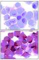


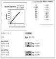

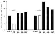


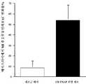

Classifications
- C—CHEMISTRY; METALLURGY
- C07—ORGANIC CHEMISTRY
- C07K—PEPTIDES
- C07K16/00—Immunoglobulins [IGs], e.g. monoclonal or polyclonal antibodies
- C07K16/40—Immunoglobulins [IGs], e.g. monoclonal or polyclonal antibodies against enzymes
- C—CHEMISTRY; METALLURGY
- C12—BIOCHEMISTRY; BEER; SPIRITS; WINE; VINEGAR; MICROBIOLOGY; ENZYMOLOGY; MUTATION OR GENETIC ENGINEERING
- C12N—MICROORGANISMS OR ENZYMES; COMPOSITIONS THEREOF; PROPAGATING, PRESERVING, OR MAINTAINING MICROORGANISMS; MUTATION OR GENETIC ENGINEERING; CULTURE MEDIA
- C12N5/00—Undifferentiated human, animal or plant cells, e.g. cell lines; Tissues; Cultivation or maintenance thereof; Culture media therefor
- C12N5/06—Animal cells or tissues; Human cells or tissues
- C12N5/0602—Vertebrate cells
- C12N5/0679—Cells of the gastro-intestinal tract
- C12N5/068—Stem cells; Progenitors
- A—HUMAN NECESSITIES
- A61—MEDICAL OR VETERINARY SCIENCE; HYGIENE
- A61P—SPECIFIC THERAPEUTIC ACTIVITY OF CHEMICAL COMPOUNDS OR MEDICINAL PREPARATIONS
- A61P19/00—Drugs for skeletal disorders
- A—HUMAN NECESSITIES
- A61—MEDICAL OR VETERINARY SCIENCE; HYGIENE
- A61P—SPECIFIC THERAPEUTIC ACTIVITY OF CHEMICAL COMPOUNDS OR MEDICINAL PREPARATIONS
- A61P19/00—Drugs for skeletal disorders
- A61P19/02—Drugs for skeletal disorders for joint disorders, e.g. arthritis, arthrosis
- A—HUMAN NECESSITIES
- A61—MEDICAL OR VETERINARY SCIENCE; HYGIENE
- A61P—SPECIFIC THERAPEUTIC ACTIVITY OF CHEMICAL COMPOUNDS OR MEDICINAL PREPARATIONS
- A61P19/00—Drugs for skeletal disorders
- A61P19/08—Drugs for skeletal disorders for bone diseases, e.g. rachitism, Paget's disease
- A—HUMAN NECESSITIES
- A61—MEDICAL OR VETERINARY SCIENCE; HYGIENE
- A61P—SPECIFIC THERAPEUTIC ACTIVITY OF CHEMICAL COMPOUNDS OR MEDICINAL PREPARATIONS
- A61P43/00—Drugs for specific purposes, not provided for in groups A61P1/00-A61P41/00
- A—HUMAN NECESSITIES
- A61—MEDICAL OR VETERINARY SCIENCE; HYGIENE
- A61P—SPECIFIC THERAPEUTIC ACTIVITY OF CHEMICAL COMPOUNDS OR MEDICINAL PREPARATIONS
- A61P9/00—Drugs for disorders of the cardiovascular system
- A—HUMAN NECESSITIES
- A61—MEDICAL OR VETERINARY SCIENCE; HYGIENE
- A61P—SPECIFIC THERAPEUTIC ACTIVITY OF CHEMICAL COMPOUNDS OR MEDICINAL PREPARATIONS
- A61P9/00—Drugs for disorders of the cardiovascular system
- A61P9/04—Inotropic agents, i.e. stimulants of cardiac contraction; Drugs for heart failure
- C—CHEMISTRY; METALLURGY
- C12—BIOCHEMISTRY; BEER; SPIRITS; WINE; VINEGAR; MICROBIOLOGY; ENZYMOLOGY; MUTATION OR GENETIC ENGINEERING
- C12N—MICROORGANISMS OR ENZYMES; COMPOSITIONS THEREOF; PROPAGATING, PRESERVING, OR MAINTAINING MICROORGANISMS; MUTATION OR GENETIC ENGINEERING; CULTURE MEDIA
- C12N5/00—Undifferentiated human, animal or plant cells, e.g. cell lines; Tissues; Cultivation or maintenance thereof; Culture media therefor
- C12N5/0081—Purging biological preparations of unwanted cells
- C—CHEMISTRY; METALLURGY
- C12—BIOCHEMISTRY; BEER; SPIRITS; WINE; VINEGAR; MICROBIOLOGY; ENZYMOLOGY; MUTATION OR GENETIC ENGINEERING
- C12N—MICROORGANISMS OR ENZYMES; COMPOSITIONS THEREOF; PROPAGATING, PRESERVING, OR MAINTAINING MICROORGANISMS; MUTATION OR GENETIC ENGINEERING; CULTURE MEDIA
- C12N5/00—Undifferentiated human, animal or plant cells, e.g. cell lines; Tissues; Cultivation or maintenance thereof; Culture media therefor
- C12N5/06—Animal cells or tissues; Human cells or tissues
- C12N5/0602—Vertebrate cells
- C12N5/0652—Cells of skeletal and connective tissues; Mesenchyme
- C12N5/0662—Stem cells
- C12N5/0663—Bone marrow mesenchymal stem cells (BM-MSC)
- C—CHEMISTRY; METALLURGY
- C12—BIOCHEMISTRY; BEER; SPIRITS; WINE; VINEGAR; MICROBIOLOGY; ENZYMOLOGY; MUTATION OR GENETIC ENGINEERING
- C12N—MICROORGANISMS OR ENZYMES; COMPOSITIONS THEREOF; PROPAGATING, PRESERVING, OR MAINTAINING MICROORGANISMS; MUTATION OR GENETIC ENGINEERING; CULTURE MEDIA
- C12N5/00—Undifferentiated human, animal or plant cells, e.g. cell lines; Tissues; Cultivation or maintenance thereof; Culture media therefor
- C12N5/10—Cells modified by introduction of foreign genetic material
- C12N5/12—Fused cells, e.g. hybridomas
- C12N5/16—Animal cells
- C12N5/163—Animal cells one of the fusion partners being a B or a T lymphocyte
- C—CHEMISTRY; METALLURGY
- C12—BIOCHEMISTRY; BEER; SPIRITS; WINE; VINEGAR; MICROBIOLOGY; ENZYMOLOGY; MUTATION OR GENETIC ENGINEERING
- C12Q—MEASURING OR TESTING PROCESSES INVOLVING ENZYMES, NUCLEIC ACIDS OR MICROORGANISMS; COMPOSITIONS OR TEST PAPERS THEREFOR; PROCESSES OF PREPARING SUCH COMPOSITIONS; CONDITION-RESPONSIVE CONTROL IN MICROBIOLOGICAL OR ENZYMOLOGICAL PROCESSES
- C12Q1/00—Measuring or testing processes involving enzymes, nucleic acids or microorganisms; Compositions therefor; Processes of preparing such compositions
- C12Q1/34—Measuring or testing processes involving enzymes, nucleic acids or microorganisms; Compositions therefor; Processes of preparing such compositions involving hydrolase
- C12Q1/42—Measuring or testing processes involving enzymes, nucleic acids or microorganisms; Compositions therefor; Processes of preparing such compositions involving hydrolase involving phosphatase
- G—PHYSICS
- G01—MEASURING; TESTING
- G01N—INVESTIGATING OR ANALYSING MATERIALS BY DETERMINING THEIR CHEMICAL OR PHYSICAL PROPERTIES
- G01N33/00—Investigating or analysing materials by specific methods not covered by groups G01N1/00 - G01N31/00
- G01N33/48—Biological material, e.g. blood, urine; Haemocytometers
- G01N33/50—Chemical analysis of biological material, e.g. blood, urine; Testing involving biospecific ligand binding methods; Immunological testing
- G01N33/53—Immunoassay; Biospecific binding assay; Materials therefor
- G01N33/577—Immunoassay; Biospecific binding assay; Materials therefor involving monoclonal antibodies binding reaction mechanisms characterised by the use of monoclonal antibodies; monoclonal antibodies per se are classified with their corresponding antigens
- G—PHYSICS
- G01—MEASURING; TESTING
- G01N—INVESTIGATING OR ANALYSING MATERIALS BY DETERMINING THEIR CHEMICAL OR PHYSICAL PROPERTIES
- G01N33/00—Investigating or analysing materials by specific methods not covered by groups G01N1/00 - G01N31/00
- G01N33/48—Biological material, e.g. blood, urine; Haemocytometers
- G01N33/50—Chemical analysis of biological material, e.g. blood, urine; Testing involving biospecific ligand binding methods; Immunological testing
- G01N33/68—Chemical analysis of biological material, e.g. blood, urine; Testing involving biospecific ligand binding methods; Immunological testing involving proteins, peptides or amino acids
- A—HUMAN NECESSITIES
- A61—MEDICAL OR VETERINARY SCIENCE; HYGIENE
- A61K—PREPARATIONS FOR MEDICAL, DENTAL OR TOILETRY PURPOSES
- A61K35/00—Medicinal preparations containing materials or reaction products thereof with undetermined constitution
- A61K35/12—Materials from mammals; Compositions comprising non-specified tissues or cells; Compositions comprising non-embryonic stem cells; Genetically modified cells
- A61K2035/124—Materials from mammals; Compositions comprising non-specified tissues or cells; Compositions comprising non-embryonic stem cells; Genetically modified cells the cells being hematopoietic, bone marrow derived or blood cells
- C—CHEMISTRY; METALLURGY
- C07—ORGANIC CHEMISTRY
- C07K—PEPTIDES
- C07K2317/00—Immunoglobulins specific features
- C07K2317/70—Immunoglobulins specific features characterized by effect upon binding to a cell or to an antigen
- C07K2317/76—Antagonist effect on antigen, e.g. neutralization or inhibition of binding
- C—CHEMISTRY; METALLURGY
- C12—BIOCHEMISTRY; BEER; SPIRITS; WINE; VINEGAR; MICROBIOLOGY; ENZYMOLOGY; MUTATION OR GENETIC ENGINEERING
- C12N—MICROORGANISMS OR ENZYMES; COMPOSITIONS THEREOF; PROPAGATING, PRESERVING, OR MAINTAINING MICROORGANISMS; MUTATION OR GENETIC ENGINEERING; CULTURE MEDIA
- C12N2533/00—Supports or coatings for cell culture, characterised by material
- C12N2533/50—Proteins
- C12N2533/56—Fibrin; Thrombin
- G—PHYSICS
- G01—MEASURING; TESTING
- G01N—INVESTIGATING OR ANALYSING MATERIALS BY DETERMINING THEIR CHEMICAL OR PHYSICAL PROPERTIES
- G01N2333/00—Assays involving biological materials from specific organisms or of a specific nature
- G01N2333/90—Enzymes; Proenzymes
- G01N2333/914—Hydrolases (3)
- G01N2333/916—Hydrolases (3) acting on ester bonds (3.1), e.g. phosphatases (3.1.3), phospholipases C or phospholipases D (3.1.4)
Landscapes
- Health & Medical Sciences (AREA)
- Life Sciences & Earth Sciences (AREA)
- Chemical & Material Sciences (AREA)
- Engineering & Computer Science (AREA)
- Organic Chemistry (AREA)
- Bioinformatics & Cheminformatics (AREA)
- Biomedical Technology (AREA)
- General Health & Medical Sciences (AREA)
- Genetics & Genomics (AREA)
- Immunology (AREA)
- Biotechnology (AREA)
- Zoology (AREA)
- Wood Science & Technology (AREA)
- Biochemistry (AREA)
- Medicinal Chemistry (AREA)
- Molecular Biology (AREA)
- Microbiology (AREA)
- Cell Biology (AREA)
- Proteomics, Peptides & Aminoacids (AREA)
- General Engineering & Computer Science (AREA)
- Chemical Kinetics & Catalysis (AREA)
- Hematology (AREA)
- Public Health (AREA)
- General Chemical & Material Sciences (AREA)
- Nuclear Medicine, Radiotherapy & Molecular Imaging (AREA)
- Pharmacology & Pharmacy (AREA)
- Veterinary Medicine (AREA)
- Animal Behavior & Ethology (AREA)
- Developmental Biology & Embryology (AREA)
- Urology & Nephrology (AREA)
- Physics & Mathematics (AREA)
- Analytical Chemistry (AREA)
- Biophysics (AREA)
- Rheumatology (AREA)
- Physical Education & Sports Medicine (AREA)
- Cardiology (AREA)
- Food Science & Technology (AREA)
- General Physics & Mathematics (AREA)
- Pathology (AREA)
- Heart & Thoracic Surgery (AREA)
Abstract
Translated fromKoreanDescription
Translated fromKorean본 발명은 다분화성 성체 세포를 동정 및/또는 분리함에 있어 마커로서의 조직 비특이적인 알카리 포스파타제(TNAP)의 용도에 관한 것이다. 또한, 본 발명은 본 발명의 방법에 의해 농화된 세포 집단 및 이들 세포의 치료학적 용도에 관한 것이다.The present invention relates to the use of tissue nonspecific alkaline phosphatase (TNAP) as a marker in identifying and / or isolating multipotent adult cells. The invention also relates to cell populations enriched by the methods of the invention and to the therapeutic use of these cells.
다분화성 성체 세포의 농화(enrichment)Enrichment of Multipotent Adult Cells
섬유아세포, 지방세포, 연골모세포, 평활근 세포, 조골세포 및 그외 골 세포성 요소를 포함하는 골수(BM)의 비-조혈모세포가, 골수강(bone marrow space)과 주변 결합 조직 도처에 있는 다분화성 골수 간엽 전구 세포(MPC) 집단으로부터 파생된다는 개념을 뒷받침하는 많은 연구들이 있다(Bianco et al., 2001; Gronthos and Simmons, 1996; Owen and Friedenstein, 1988; Prockop, 1997). 이러한 MPC는 표현형이나 기능적으로 동일한 더 많은 수의 세포(자기-재생 과정) 뿐만 아니라 분화된, 계통-예정된 간엽 전구체(lineage-committed mesenchymal progeny)를 산출하는 것으로 생각된다. 명확한 마커가 부족하여, 인간 MPC가 계통-예정된 전구체로 분 화 및 성숙하는 동안에 이루어지는, 발생에 따라 정확하게 조절되는 표현형 변화와 유전자 발현에 대해서는 거의 알려져 있지 않다. 골형성 과정을 조사한 연구를 통해, 이러한 초기 마커 1개, 즉 골형성 세포 계통으로 예정된 MPC를 동정할 수 있는 전사인자 CBFA1가 동정되었다(Ducy et al., 1997). 그러나, CBFA1와 같은 마커는 이질적인 세포 집단내에서 살아있는 세포를 분리하고 조작하는데에는 사용할 수 없다. 이러한 점은 주된 한계점으로, MPC 컴파트먼트에 고유한 또는 이에 한정되는 세포 표면 항체를 동정할 수 있는 단일클론 항체(mAb)의 결핍에 의해 더욱 심화된다.Non-hematopoietic stem cells of the bone marrow (BM), including fibroblasts, adipocytes, chondrocytes, smooth muscle cells, osteoblasts, and other osteocytic elements, are multipotent throughout bone marrow space and surrounding connective tissue. There are many studies supporting the notion that it is derived from a myeloid mesenchymal precursor cell (MPC) population (Bianco et al., 2001; Gronthos and Simmons, 1996; Owen and Friedenstein, 1988; Prockop, 1997). Such MPCs are believed to yield differentiated, lineage-committed mesenchymal progeny, as well as a larger number of cells (self-renewal processes) that are phenotype or functionally identical. Due to the lack of clear markers, little is known about phenotypic changes and gene expression that are precisely controlled as they occur during the differentiation and maturation of human MPCs into lineage-predicted precursors. Investigations of the bone formation process have identified a transcription factor CBFA1 capable of identifying one of these early markers, namely, the MPCs that have been destined for the osteogenic cell lineage (Ducy et al., 1997). However, markers such as CBFA1 cannot be used to isolate and manipulate live cells in heterogeneous cell populations. This is a major limitation, further exacerbated by the lack of monoclonal antibodies (mAbs) that can identify cell surface antibodies inherent or limited to the MPC compartment.
지금까지, STRO-1 단일클론 항체는 인간 골수 천자액(bone marrow aspirate) 유래의 모든 콜로니 형성 MPC(CFU-F: 콜로리 형성 단위-섬유아세포)와 면역반응성을 보이지만 조혈 줄기 세포와의 반응성은 결여된 단순 시약에 불과하다(Dennis et al., 2002; Gronthos et al., 2003; Simmons and Torok-Storb, 1991).To date, STRO-1 monoclonal antibodies have been immunoreactive with all colony forming MPCs (CFU-F) from human bone marrow aspirate, but not with hematopoietic stem cells. It is only a simple reagent lacking (Dennis et al., 2002; Gronthos et al., 2003; Simmons and Torok-Storb, 1991).
본 연구를 통해, 생체외에서 증폭시킨 인간 MPC가 혈청 존재하에서 신속하게 분화하며, 골형성 및 그외 세포 계통 결정과 관련있는 다수의 마커(Gronthos et al., 2003)를 발현하기 시작한다는 것을 확인하였다. 생체내에서 모든 MPC(CFU-F)를 식별하는 mAb STRO-1은 MPC의 생체외 배양 이후에는 하향 조절된다. 중요한 점은, 배양된 세포의 적은 비율은 생체외 증폭 이후에도 계속적으로 STRO-1을 발현하며, 이러한 세포는 미분화된 MPC의 특징이다(Gronthos et al., 1999; Stewart et al., 1999).This study confirmed that ex vivo amplified human MPCs rapidly differentiate in the presence of serum and begin to express a number of markers (Gronthos et al., 2003) related to bone formation and other cell lineage determinations. MAb STRO-1, which identifies all MPCs (CFU-F) in vivo, is down regulated after ex vivo culture of MPCs. Importantly, a small percentage of cultured cells continue to express STRO-1 even after ex vivo amplification, and these cells are characteristic of undifferentiated MPC (Gronthos et al., 1999; Stewart et al., 1999).
알카리 포스파타제Alkaline phosphatase
알카리 포스파타제(AP, EC 3.1.3.1)는 무기 포스페이트가 방출되는 알카리 조건하에서 포스포모노에스테르의 가수분해를 촉매하는 흔한 이량성 금속 함유 효소(metalloenzyme) 패밀리이다(McComb et al., 1979). 인간에서는, 이를 동질 효소 4종(isoenzyme)으로 구분할 수 있다: i) 태반 특이적인 AP, ii) 생식세포 특이적인 (태반) AP, iii) 장의 AP 및 iv) 조직 특이적인 AP(TNAP)(Harris, 1990). TNAP는 간(LAP), 신장(KAP) 및 뼈(BAP)에서 가장 많이 생성되며(Moss, 1992), AP 이성형(soform)은 혈청에 가장 풍부하다(Mulivor, et al., 1985). LAP, KAP 및 BAP간의 차이는 번역후 다양한 O-당화 패턴으로 인한 것으로(Miura, et al., 1994), 그것들의 아미노산 서열은 본질적으로 동일하지만 상이한 특이적인 활성을 나타낸다(Weiss et al., 1988). 또한, Nosjean et al.(1997)은 tns-AP의 N-당화가 효소 활성에 필수적이라는 것을 입증하였다. 따라서, 조직 비특이적인 AP는 당화된 여러 AP들의 혼합이다.Alkaline phosphatase (AP, EC 3.1.3.1) is a common dimer metalloenzyme family that catalyzes the hydrolysis of phosphomonoesters under alkaline conditions in which inorganic phosphates are released (McComb et al., 1979). In humans, it can be divided into four isoenzymes: i) placental specific AP, ii) germ cell specific (placental) AP, iii) intestinal AP and iv) tissue specific AP (TNAP) (Harris). , 1990). TNAP is most produced in liver (LAP), kidney (KAP) and bone (BAP) (Moss, 1992), and AP isoforms are most abundant in serum (Mulivor, et al., 1985). The difference between LAP, KAP and BAP is due to various O-glycosylation patterns after translation (Miura, et al., 1994), and their amino acid sequences are essentially identical but show different specific activities (Weiss et al., 1988 ). Nosjean et al. (1997) also demonstrated that N-glycosylation of tns-AP is essential for enzymatic activity. Thus, tissue nonspecific APs are a mixture of several glycosylated APs.
인간 TNAP 유전자는 1986년에 클로닝되었다(Weiss, et al., 1986). 이것은 524개의 아미노산과, 17개의 아미노산으로 이루어진 N-말단 신호 서열 및 그 단백질을 생체내에서 원형질막의 바깥측에 고정시키는 C-말단 GPI 고정 서열(anchor sequence)로 구성된, 단백질을 코딩한다(Hooper, 1997). COS-1(Fukushi, et al., 1998) 및 베큘로바이러스가 감염된 곤충 세포(Oda, et al., 1999)와 같은 진핵 세포에서, 생물학적으로 활성인 재조합 TNAP 효소의 발현이 보고되었다.The human TNAP gene was cloned in 1986 (Weiss, et al., 1986). It encodes a protein consisting of 524 amino acids, an N-terminal signal sequence consisting of 17 amino acids and a C-terminal GPI anchor sequence that anchors the protein to the outside of the plasma membrane in vivo (Hooper, 1997). In eukaryotic cells such as COS-1 (Fukushi, et al., 1998) and insect cells infected with baculovirus (Oda, et al., 1999), expression of biologically active recombinant TNAP enzymes has been reported.
골 및 골수 조직에서 TNAP의 정확한 분자 기능은 TNAP가 발견된지 70년이 지났음에도 불구하고 명확하지 않다. 포유류에서 TNAP의 생물학적 기능이 몇가지 제 안되어 있는데, 그 기능으로는 포스페이트 에스테르를 가수분해하여 비-포스페이트 모이어티(nonphosphate moiety) 제공, 포스페이트 에스테르 합성에 있어 트랜스퍼라제 기능, 무기 포스페이트 대사 조절, 포스포릴-대사산물의 농도를 정상 상태로 유지 및 포스포단백질 포스파타제로서의 작용을 포함한다(Whyte, 1994). 또한, B/L/K-TNAP는 무기 피로포스페이트와 같은 석회화(calcification) 저해자의 가수분해에 의한 골격 미네랄화(mineralization)에 있어 특이적인 기능을 가지고 있을 수 있으며, 고농도에서는 수산화인회석 결정(hydroxyapatite crystal)의 성장을 저해할 수도 있다(De Broe and Moss, 1992; Moss, 1992; Whyte, 1994). 대안적으로, TNAP는 칼슘 포스페이트의 침전을 자극하고 오스테오이드에 미네랄 침착으로 이르는, 무기 포스페이트, 세포외 칼슘 이온 결합성 단백질의 원형질막 수송체일 수 있는 것으로 시사되었다.The precise molecular function of TNAP in bone and bone marrow tissue is not clear despite 70 years of TNAP discovery. Several mammalian biological functions of TNAP have been proposed, including the hydrolysis of phosphate esters to provide a nonphosphate moiety, transferase function in the synthesis of phosphate esters, control of inorganic phosphate metabolism, phosphoryl- Maintaining the concentration of metabolites in steady state and acting as phosphoprotein phosphatase (Whyte, 1994). In addition, B / L / K-TNAP may have a specific function in skeletal mineralization by hydrolysis of calcification inhibitors such as inorganic pyrophosphate, and at high concentrations, hydroxyapatite crystals. May inhibit growth (De Broe and Moss, 1992; Moss, 1992; Whyte, 1994). Alternatively, it has been suggested that TNAP may be a plasma membrane transporter of inorganic phosphate, extracellular calcium ion binding protein, which stimulates precipitation of calcium phosphate and leads to mineral deposition on the osteoid.
TNAP는 조골세포 분화 마커로도 알려져 있다. 그러나, 본 발명자가 알기에는 미성숙 다분화성 세포에 의한 TNAP의 세포 표면 발현에 대해서는 보고된 바가 없다.TNAP is also known as osteoblast differentiation marker. However, to the best of our knowledge, no cell surface expression of TNAP by immature multipotent cells has been reported.
발명의 개요Summary of the Invention
본 발명자들은 최근에 미분획한 골수로부터 다분화성 성체 세포를 동정 및 분리하여, 생체내 및 시험관내에서 STRO-1 집단을 세분하는 능력을 가진, 신규한 mAb(STRO-3)을 제조하였다. 본 발명자들은 조직 비특이적인 알카리 포스파타제(TNAP)에 STRO-3이 결합함을 확인하였다. 또한, STRO-3은 성체 골수 천자액에 포함된 일부 세포와만 반응하며, 인간 성체 골수 천자액내 CD34 양성인 조혈 줄기세포와는 반응하지 않는다는 것을 확인하였다. 이는, TNAP가 다양한 조직 기원으로부터 다분화성 성체 세포를 단일 시약으로 농화하는데 이용가능한 마커임을 최초로 입증한다.We recently identified and separated multipotent adult cells from unfractionated bone marrow to produce a novel mAb (STRO-3) with the ability to subdivide the STRO-1 population in vivo and in vitro. We have confirmed that STRO-3 binds to tissue nonspecific alkaline phosphatase (TNAP). In addition, it was confirmed that STRO-3 only reacts with some cells contained in adult bone marrow puncture fluid and does not react with CD34 positive hematopoietic stem cells in human adult bone marrow puncture fluid. This demonstrates for the first time that TNAP is a marker available for enriching multipotent adult cells with a single reagent from various tissue origins.
따라서, 본 발명은 다분화성 성체 세포의 동정 및/또는 농화용 마커로서의 TNAP의 용도에 관한 것이다.Accordingly, the present invention relates to the use of TNAP as a marker for identification and / or enrichment of multipotent adult cells.
또한, 본 발명은 조직 유래의 세포 샘플을 준비하는 단계 및 TNAP 마커를 발현하는 세포를 농화하는 단계를 포함하는 다분화성 성체 세포의 농화 방법을 제공한다.The present invention also provides a method for enriching multipotent adult cells, comprising preparing a cell sample derived from a tissue and enriching a cell expressing a TNAP marker.
일 예로, 다분화성 성체 세포의 농화 방법은 TNAP가 TNAP 결합제에 결합가능한 조건하에서 상기 세포 샘플을 TNAP 결합제와 접촉시키는 단계 및 상기 TNAP 결합제에 결합된 세포를 분리하는 단계를 포함한다.In one embodiment, the method of enriching multipotent adult cells comprises contacting the cell sample with a TNAP binder under conditions such that TNAP can bind to the TNAP binder, and separating the cells bound to the TNAP binder.
또한, 본 발명은 TNAP 마커를 발현하는 샘플에서 세포를 동정하는 단계를 포함하는 세포 샘플내에서 다분화성 성체 세포의 존재를 동정하는 방법을 제공한다.The present invention also provides a method of identifying the presence of multipotent adult cells in a cell sample comprising identifying cells in a sample expressing a TNAP marker.
일 예로, 다분화성 성체 세포의 존재를 동정하는 방법은 TNAP가 TNAP 결합제에 결합가능한 조건하에서 상기 세포 샘플을 TNAP 결합제와 접촉시키는 단계 및 샘플에서 세포에 결합된 상기 TNAP 결합제의 존재를 검출하여, 다분화성 성체 세포의 존재가 TNAP 결합제에 결합하는 세포에 의해 표시되는 단계를 포함한다.In one embodiment, the method of identifying the presence of multipotent adult cells comprises contacting said cell sample with a TNAP binder under conditions in which TNAP can bind to a TNAP binding agent and detecting the presence of said TNAP binding agent bound to cells in said sample. And the presence of the adult adult cells is indicated by the cells binding to the TNAP binding agent.
본 발명에서, 세포 샘플은 지방 조직, 치아, 치수, 피부, 간, 신장, 심장, 망막, 뇌, 모낭, 장, 폐, 지라, 림프절, 흉선, 난소, 췌장, 뼈, 인대, 골수, 힘줄 및 골격근일 수 있다. 바람직하기로는, 상기 조직은 골수이다.In the present invention, the cell sample comprises adipose tissue, teeth, pulp, skin, liver, kidney, heart, retina, brain, hair follicle, intestine, lung, spleen, lymph node, thymus, ovary, pancreas, bone, ligament, bone marrow, tendon and May be skeletal muscle. Preferably, the tissue is bone marrow.
바람직한 세포 기원은 인간이지만, 소, 양, 돼지 등과 같은 농경 동물, 개 및 고양이와 같은 가축, 마우스, 랫, 햄스터 및 토끼와 같은 실험 동물, 말과 같이 스포츠에 이용되는 동물을 포함한 다른 동물에도 본 발명을 적용가능한 것으로 예상된다.Preferred cell origins are human, but are also found in other animals, including farm animals such as cattle, sheep, pigs, etc., livestock such as dogs and cats, experimental animals such as mice, rats, hamsters and rabbits, and other animals used in sports such as horses. It is anticipated that the invention is applicable.
또한, 본 발명은 1차 농화 단계를 수행하기 전에 다분화성 세포의 기원을 수득하는 단계를 포함할 수 있다. 예컨대, 이는 대상자로부터 조직을 외과적으로 적출하는 단계 및 조직의 세포를 분리하여 단일 세포 현탁물을 제조하는 단계를 포함할 수 있다. 이러한 분리는 물리적 또는 효소적 수단에 의해 달성될 수 있다. 본 발명의 일 예에서, 이러한 단계는 공지된 기술을 이용하여 골수 세포를 수득하는 단계를 포함한다.In addition, the invention may comprise the step of obtaining the origin of the multipotent cells prior to performing the first enrichment step. For example, this may include surgically removing the tissue from the subject and separating the cells of the tissue to produce a single cell suspension. Such separation can be accomplished by physical or enzymatic means. In one embodiment of the present invention, this step comprises obtaining bone marrow cells using known techniques.
본 발명의 방법에 사용되는 TNAP 결합제는 TNAP에 대한 결합 친화성을 가지는 것으로 동정된 모든 폴리펩티드 또는 화합물일 수 있다. 예컨대, TNAP 결합제는 항원 또는 콜라겐, 바람직하기로는 제1형 콜라겐일 수 있다.The TNAP binding agent used in the methods of the invention can be any polypeptide or compound identified as having binding affinity for TNAP. For example, the TNAP binding agent may be an antigen or collagen, preferably
TNAP 결합제는 TNAP의 LAP, KAP 또는 BAP 이소형 중 하나 이상에 결합할 수 있다. 바람직한 일 예로, TNAP 결합제는 BAP에 결합한다. 다른 바람직한 예로, TNAP 결합제는 BAP에 특이적으로 결합한다.TNAP binding agents may bind to one or more of the LAP, KAP or BAP isoforms of TNAP. In a preferred embodiment, the TNAP binding agent binds to BAP. In another preferred embodiment, the TNAP binding agent specifically binds to BAP.
"BAP에 특이적으로 결합한다"는 TNAP 결합제가 KAP 및 LAP와 같은 다른 물질이 과량으로 존재하는 조건에서 선택적인 양상으로, BAP를 발현하는 세포의 선택적인 농화에 유용한 툴을 제공하도록 충분히 강하게(즉, 매우 충분한 친화성을 가짐), BAP에 결합할 수 있음을 의미한다."Specificly binds to BAP" is a selective aspect of the TNAP binding agent in conditions where excess amounts of other substances such as KAP and LAP are present, and are sufficiently strong to provide a useful tool for selective enrichment of BAP expressing cells ( That is, they have very sufficient affinity), which means that they can bind to BAP.
바람직한 예로, TNAP 결합제는 항-TNAP 항체(임의 기원으로부터 자연적으로 형성되거나 또는 재조합됨)이다. 항-TNAP 항체는 다클론 항체 또는 단일클론 항체일 수 있다. 바람직한 예로, 항-TNAP 항체는 단일클론 항체이다.In a preferred embodiment, the TNAP binding agent is an anti-TNAP antibody (naturally formed or recombinant from any origin). The anti-TNAP antibody may be a polyclonal antibody or a monoclonal antibody. In a preferred embodiment, the anti-TNAP antibody is a monoclonal antibody.
본 발명의 사용에 적합한 항-TNAP 단일클론 항체의 예로 B4-78, 50 및 B4-50(Developmental Studies Hybridoma Bank, University of Iowa); ab17973 및 ab17989(Abcam Ltd, Cambridge, UK); 및 Magnusson et al. (2002)에 언급된 항-TNAP mAb가 있다.Examples of anti-TNAP monoclonal antibodies suitable for use in the present invention include B4-78, 50 and B4-50 (Developmental Studies Hybridoma Bank, University of Iowa); ab17973 and ab17989 from Abcam Ltd, Cambridge, UK; And Magnusson et al. (2002) mentioned anti-TNAP mAb.
본 발명의 특히 바람직한 예로, 항-TNAP 단일클론 항체는 부다페스트 조약의 조항에 따라 2005년 12월 19일자로 ATCC에 기탁번호 PTA-7282로 기탁된 하이브리도마 세포주에서 생산되는 STRO-3 항체 또는 STRO-3 항체와 동일한 TNAP 에피토프에 결합하는 mAb이다.In a particularly preferred embodiment of the invention, the anti-TNAP monoclonal antibody is a STRO-3 antibody or STRO produced from a hybridoma cell line deposited with ATCC on Dec. 19, 2005 according to the provisions of the Budapest Treaty. MAb that binds to the same TNAP epitope as the -3 antibody.
본 발명에 따른 다분화성 성체 세포의 농화 방법은 TNAP 마커의 단독 존재를 토대로 수행할 수 있다. 즉, 상기 농화 방법에는 단일 시약(즉, TNAP 결합제)이 사용될 수 있다.The method for enriching multipotent adult cells according to the present invention can be performed based on the presence of a TNAP marker alone. That is, a single reagent (ie, TNAP binder) may be used in the thickening method.
그러나, 본 발명은 TNAP 단독 발현에 의한 세포 농화로만 한정되지 않으며, 일부의 경우에서는 2, 3 또는 그 이상의 부가적인 마커와 조합하여 TNAP 발현을 토대로 다분화성 성체 세포를 농화하는 것이 바람직할 수 있다. 따라서, 다분화성 성체 세포의 농화 방법은 LFA-3, THY-1, VCAM-1, ICAM-1, PECAM-1, P-셀렉틴, L-셀렉틴, CD49a/CD49b/CD29, CD49c/CD29, CD49d/CD29, CD29, CD18, CD61, 인테그린 베타, 6-19, 트롬보모듈린, CD10, CD13, SCF, PDGF-R, EGF-R, IGF1-R, NGF-R, FGF-R, 렙틴-R, (STRO-2 = 렙틴-R), RANKL STRO-1(바람직하게는 STRO-1bri), 및 CD146 또는 이들 마커의 임의 조합으로 이루어진 군으로부터 선택된 1종 이상의 마커의 추가적인 존재를 근간으로 할 수 있다.However, the present invention is not limited to cell enrichment by expression of TNAP alone, and in some cases it may be desirable to enrich multipotent adult cells based on TNAP expression in combination with 2, 3 or more additional markers. Thus, methods for enriching multipotent adult cells include LFA-3, THY-1, VCAM-1, ICAM-1, PECAM-1, P-selectin, L-selectin, CD49a / CD49b / CD29, CD49c / CD29, CD49d / CD29, CD29, CD18, CD61, Integrin Beta, 6-19, Thrombomodulin, CD10, CD13, SCF, PDGF-R, EGF-R, IGF1-R, NGF-R, FGF-R, Leptin-R, (STRO-2 = leptin-R), RANKL STRO-1 (preferably STRO-1bri ), and CD146 or an additional combination of one or more markers selected from the group consisting of any combination of these markers. .
예컨대, 상기 방법은 다분화성 성체 세포에 특이적인 제1 마커의 발현 농화에 의해 부분적으로 농화된 1차 세포 풀(pool)을 제조하는 단계, 및 상기 부분적으로 농화된 세포 풀로부터 TNAP 발현을 농화하는 단계를 포함할 수 있다. 다른 예로, 상기 방법은 TNAP를 발현하는 세포 선별을 기본으로 하는 첫번째 농화 단계 및 여러가지 다분화성 성체 세포의 마커를 농화시키는 단계를 포함할 수 있다. 다른 예로, 상기 방법은 TNAP를 발현하는 세포와 하나 이상의 부가적인 다분화성 성체 세포에 특이적인 마커를 발현하는 세포를 동시에 선별하는 단계를 포함한다.For example, the method comprises preparing a primary enriched pool of cells partially enriched by expression enrichment of a first marker specific for multipotent adult cells, and enriching TNAP expression from the partially enriched cell pool. It may include a step. In another example, the method may comprise a first enrichment step based on cell selection expressing TNAP and enrichment of markers of various multipotent adult cells. In another embodiment, the method comprises simultaneously selecting cells expressing TNAP and cells expressing markers specific for one or more additional multipotent adult cells.
분리의 근거를 제공하는 TNAP를 갖는 세포의 인지는 다수의 여러가지 방법에 의해 수행할 수 있지만, 이러한 방법들 모두 어떤 시점에서의 세포와 TNAP 결합제간의 결합과, 결합 정도가 높거나 낮은 결합성을 보는 세포의 분리를 토대로 한다. 대부분의 통상적인 결합제는 이들의 특이성으로 인해 항체 또는 항체성 분자, 바람직하기로는 단일클론 항체 또는 단일클론 항체성 분자이다.Recognition of cells with TNAP, which provides the basis for isolation, can be accomplished by a number of different methods, but all of these methods show binding between the cell and the TNAP binding agent at some point in time, with high or low levels of binding. Based on the separation of cells. Most conventional binders are antibodies or antibody molecules, preferably monoclonal antibodies or monoclonal antibody molecules, because of their specificity.
TNAP 결합제를 고형 지지체에 부착하여, 조 분리(crude separation) 할 수 있다. 이러한 분리 기법은 바람직하기로는 수득되는 분획의 생존성을 최대로 유지시킨다. 여러가지 효과가 있는 다양한 기법을 사용하여 비교적 조 분리물을 수득할 수 있다. 사용되는 특정 기법은 분리력, 관련 세포독성, 실시 용이성 및 속도, 정교한 장치 및/또는 기술력의 필요성에 따라 결정될 것이다. 분리 공정은 항체에 코팅된 자기 비드를 이용한 자기 분리, 친화성 크로마토그래피 및 고형 매트릭스에 부착된 항체를 이용한 "패닝(panning)"을 포함할 수 있지만, 이로 한정되는 것은 아니다. 정확한 분리를 제공하는 기법으로는 MACS, Dynal 자기 비드 선별 및 FACS가 있지만, 이로 한정되는 것은 아니다.The TNAP binder can be attached to a solid support to allow crude separation. This separation technique preferably maintains the viability of the fraction obtained. A variety of techniques with different effects can be used to obtain relatively crude isolates. The particular technique used will depend on the ability of separation, related cytotoxicity, ease and speed of implementation, and the need for sophisticated devices and / or technologies. The separation process may include, but is not limited to, magnetic separation with magnetic beads coated on the antibody, affinity chromatography, and “panning” with the antibody attached to a solid matrix. Techniques that provide accurate separation include, but are not limited to, MACS, Dynal magnetic bead screening, and FACS.
본 발명의 일 예에서, TNAP 결합제는 표지된 것이다. 다른 예로, TNAP 결합제에 결합된 세포의 분리는 기계적인 세포 분리기에 의해 수행된다.In one embodiment of the invention, the TNAP binding agent is labeled. In another example, the separation of cells bound to the TNAP binding agent is performed by a mechanical cell separator.
본 발명의 다른 예로, TNAP 결합제는 형광 표지 화합물과 커플링된다. 이 경우, TNAP 결합제에 결합된 세포의 분리는 FACS(fluorescence-activated cell sorter)를 이용하여 수행하는 것이 바람직하다.In another embodiment of the invention, the TNAP binder is coupled with a fluorescent labeling compound. In this case, separation of cells bound to the TNAP binder is preferably performed using a fluorescence-activated cell sorter (FACS).
본 발명의 다른 예로, TNAP 결합제는 고형 입자에 연결된다. 바람직하기로는, 상기 고형 입자는 자기 입자이다. 본 발명의 이러한 예에 있어서, TNAP 결합제에 결합된 세포의 분리는 바람직하기로는 액체 상으로부터 입자 상을 분리함으로써 수행된다. 본 발명의 다른 예로, 분리 단계를 수행하기 전에, 세포 샘플은 고형 입자가 연결된 TNAP 결합제에 대한 항체와 접촉되며, TNAP 결합제에 결합된 세포의 분리는 액체 상으로부터 입자 상을 분리함으로써 수행된다.In another embodiment of the invention, the TNAP binder is linked to a solid particle. Preferably, the solid particles are magnetic particles. In this example of the invention, separation of the cells bound to the TNAP binder is preferably performed by separating the particle phase from the liquid phase. In another embodiment of the invention, prior to performing the separation step, the cell sample is contacted with an antibody against a TNAP binder to which solid particles are linked, and the separation of cells bound to the TNAP binder is performed by separating the particle phase from the liquid phase.
본 발명의 다른 예로, 세포 샘플의 세포는 고형 지지체상에서 배양한 유착성 세포이며, 비결합성 TNAP 결합제는 세정에 의해 제거된다.In another embodiment of the present invention, the cells of the cell sample are adherent cells cultured on a solid support, and the non-binding TNAP binding agent is removed by washing.
본 발명의 다른 예로, 세포 샘플의 세포는 현탁액으로 배양되며, 비결합성 TNAP 결합제는 세포 샘플의 원심분리와 형성된 상층물의 분리 제거에 의해 제거된다.In another embodiment of the invention, the cells of the cell sample are cultured in suspension, and the non-binding TNAP binding agent is removed by centrifugation of the cell sample and separation removal of the formed supernatant.
다른 예로, 세포 샘플은 하나 이상의 다른 다분화성 세포 마커를 발현하는 세포로부터 세포 집단을 농화 또는 줄이기 위한 추가적인 세포 분리 공정에 투입된다. 상기 다분화성 세포 마커는 LFA-3, THY-1, VCAM-1, ICAM-1, PECAM-1, P-셀렉틴, L-셀렉틴, CD49a/CD49b/CD29, CD49c/CD29, CD49d/CD29, CD29, CD18, CD61, 인테그린 베타, 6-19, 트롬보모듈린, CD10, CD13, SCF, PDGF-R, EGF-R, IGF1-R, NGF-R, FGF-R, 렙틴-R, (STRO-2 = 렙틴-R), RANKL, STRO-1(바람직하게는 STRO-1bri), 및 CD146 또는 이들 마커의 임의 조합으로 이루어진 군으로부터 선택된 하나 이상의 마커일 수 있다.In another example, the cell sample is subjected to an additional cell separation process to enrich or reduce the cell population from cells expressing one or more other multipotent cell markers. The multipotent cell markers are LFA-3, THY-1, VCAM-1, ICAM-1, PECAM-1, P-selectin, L-selectin, CD49a / CD49b / CD29, CD49c / CD29, CD49d / CD29, CD29, CD18, CD61, Integrin Beta, 6-19, Thrombomodulin, CD10, CD13, SCF, PDGF-R, EGF-R, IGF1-R, NGF-R, FGF-R, Leptin-R, (STRO-2 = Leptin-R), RANKL, STRO-1 (preferably STRO-1bri ), and CD146 or one or more markers selected from the group consisting of any combination of these markers.
또한, 본 발명은 본 발명에 따른 방법에 의해 수득되는 농화된 다분화성 성체 세포 집단을 제공한다.The invention also provides a concentrated multipotent adult cell population obtained by the method according to the invention.
또한, 본 발명은 농화된 TNAP+ 다분화성 성체 세포를 제공한다.The present invention also provides concentrated TNAP + multipotent adult cells.
본 발명의 바람직한 예로, 농화된 세포 집단 전체의 1%, 2%, 3%, 4%, 5%, 10%, 15%, 20%, 30%, 40%, 50%, 60%, 70%, 80%, 90% 또는 95% 이상은 TNAP+의 표현형을 가진 다분화성 성체 세포이다.In a preferred embodiment of the invention, 1%, 2%, 3%, 4%, 5%, 10%, 15%, 20%, 30%, 40%, 50%, 60%, 70% of the entire concentrated cell population At least 80%, 90% or 95% are multipotent adult cells with a phenotype of TNAP +.
일 예로, 본 발명의 농화 집단을 배양하면 마커 STRO-1을 이용하여 선별한 동일 조건에서 배양한 세포에 비해, STRO+인 세포의 비율이 더욱 많아진다. 바람직하기로는, 이러한 배양은 약 4 또는 약 6회의 계대배양이다. 바람직하기로는, 세포는 골수로부터 수득된다.For example, when the cultured population of the present invention is cultured, the proportion of cells that are STRO + is higher than that of cells cultured under the same conditions selected using the marker STRO-1. Preferably, this culture is about 4 or about 6 passages. Preferably, the cells are obtained from the bone marrow.
다른 예로, 본 발명의 농화 집단은 CD45+인 세포를 약 79% 내지 약 99%로, 더 바람직하기로는 약 84% 내지 약 94%로, 및 더더 바람직하기로는 약 89%로 포함한다.In another embodiment, the enriched population of the present invention comprises from about 79% to about 99%, more preferably from about 84% to about 94%, and even more preferably from about 89% of cells that are CD45 +.
바람직하기로는, 농화된 다분화성 성체 세포 집단은 본 발명에 따른 방법에 의해 수득된다.Preferably, the enriched multipotent adult cell population is obtained by the method according to the invention.
또한, 본 발명의 방법은 본 발명에 따른 농화된 다분화성 성체 세포를 배양함으로써 수득되는 증폭된 세포 집단을 제공한다.The method of the present invention also provides an amplified cell population obtained by culturing the enriched multipotent adult cells according to the present invention.
일 예로, 본 발명의 농화된 세포 집단 또는 본 발명의 증폭된 세포 집단은 유전적으로 변형된 세포를 적어도 일부분 포함한다.In one embodiment, the enriched cell population of the invention or the amplified cell population of the invention comprises at least a portion of a genetically modified cell.
또한, 본 발명은 본 발명의 다분화성 성체 세포 집단을 하나 이상의 자극 인자의 존재하에 배양하는 단계, 및The present invention also provides a method of culturing a multipotent adult cell population of the invention in the presence of one or more stimulating factors, and
상기 배양한 집단을 상기 다분화성 성체 세포를 특이적인 조직 타입으로 분화시키도록 편향된 조건에 두는 단계를 포함하는, 조직 특이적인 예정된 세포 집단의 제조 방법을 제공한다.And subjecting the cultured population to conditions biased to differentiate the multipotent adult cells into specific tissue types.
일 예로, 조직 타입은 심근, 혈관 조직, 뼈 조직, 연골 조직, 지방 조직, 신경 조직, 평활근 및 내피 조직으로 이루어진 군으로부터 선택된다.In one embodiment, the tissue type is selected from the group consisting of myocardium, vascular tissue, bone tissue, cartilage tissue, adipose tissue, neural tissue, smooth muscle and endothelial tissue.
또한, 본 발명은 본 발명의 농화된 다분화성 성체 세포 및/또는 본 발명의 증폭된 세포 집단을 포함하는 조성물에 관한 것으로 이해될 것이다.It will also be understood that the present invention relates to compositions comprising the enriched multipotent adult cells of the invention and / or the amplified cell population of the invention.
바람직한 예로, 상기 조성물은 자극 인자를 더 포함한다. 이러한 조성물은 치료학적으로 유리할 것이므로, 따라서 약학적으로 허용가능한 형태로 제조될 것이다.In a preferred embodiment, the composition further comprises a stimulating factor. Such compositions will be therapeutically advantageous and therefore will be prepared in pharmaceutically acceptable form.
상기 조성물에 존재되는 자극 인자(들)의 함량은 경험에 의해 결정될 수 있지만, 대부분의 경우에는 대략 수 ng 또는 10 ng/ml일 것이다.The amount of stimulating factor (s) present in the composition may be determined by experience, but in most cases will be approximately several ng or 10 ng / ml.
본 발명의 방법에 사용되고/사용되거나 본 발명의 조성물에 존재되는 자극 인자는 세포 분열 및/또는 분화를 촉진시킬 수 있는 적절한 모든 인자일 수 있다. 이러한 인자들은 당업계에 잘 알려져 있으며, 1α,25-디하이드록시비타민 D3 (1,25D), 혈소판 유래의 성장 인자(PDGF), 종양 괴사 인자 α(TNF-α), 인터루킨-1β(IL-1β) 및 기질 유래 인자 1α(SDF-1α)가 있으나, 이로 한정되는 것은 아니다.Stimulating factors used in the methods of the invention and / or present in the compositions of the invention may be any suitable factor capable of promoting cell division and / or differentiation. These factors are well known in the art and include 1α, 25-dihydroxyvitamin D3 (1,25D), platelet-derived growth factor (PDGF), tumor necrosis factor α (TNF-α), interleukin-1β (IL -1β) and substrate derived factor 1α (SDF-1α), but are not limited thereto.
다른 예로, 상기 조성물은 본 발명의 다분화성 성체 세포의 분화를 하나의 특이적인 조직 타입으로 편향시키는 인자를 더 포함한다. 바람직하기로는, 조직 타입은 심근, 혈관 조직, 골 조질, 연골 조직, 지방 조직, 신경 조직, 평활근 및 내피 조직으로 이루어진 군으로부터 선택된다.In another embodiment, the composition further comprises a factor that biases the differentiation of the multipotent adult cells of the invention into one specific tissue type. Preferably, the tissue type is selected from the group consisting of myocardium, vascular tissue, bone coarseness, cartilage tissue, adipose tissue, neural tissue, smooth muscle and endothelial tissue.
본 발명의 다분화성 성체 세포를 골 전구 세포 또는 골로 분화시키는 조건은, 예컨대, 10% FCS, 100 μM L-아스코르베이트-2-포스페이트, 덱사메타손 10-7 M 및 3 mM 무기 포스페이트가 보충된 α-MEM에서 배양하는 것을 포함할 수 있다. 이러한 조건이 시험관내에서 인간 골수 기질 세포를 미네랄화된(mineralized) 골 매트릭스로 발생 유도하는 것으로 확인되었다(Gronthos et al., 1994).Conditions of bone progenitor cells or differentiated to give the multi-Mars adult cell of the invention is, for example, 10% FCS, 100 μM L- ascorbate-2-phosphate,
본 발명의 다분화성 성체 세포를 조골세포로 분화시키는데 적합한 조건은, 제1형 콜라겐, 피브리노겐, 피브린, 폴리글리콜산, 폴리락트산, 오스테오칼신 또는 오스테오넥틴의 존재하에 세포를 배양하는 것을 포함할 수 있다. 특정 일 예에서, 세포는 제1형 콜라겐, 피브리노겐 및 피브린의 존재하에 배양된다. 다른 예로, 세포는 제1형 콜라겐, 피브리노겐, 피브린, 오스테오칼신 및 오스테오넥틴의 존재하에 배양된다. 이러한 방법에 있어서, 제1형 콜라겐, 피브리노겐, 피브린, 폴리글리콜산, 폴리락트산, 오스테오칼신 또는 오스테오넥틴은 단독으로 또는 성장 인자의 존재하에 사용될 수 있다. 본 단락의 전술한 화합물의 임의 조합도 본 발명에 포함되는 것으로 이해될 것이다.Conditions suitable for differentiating the multipotent adult cells of the present invention into osteoblasts may include culturing the cells in the presence of
다른 예로, 본 발명의 조성물은 피브린 글루를 더 포함한다.In another embodiment, the composition of the present invention further comprises fibrin glue.
또한, 본 발명은 본 발명의 농화된 또는 증폭된(expanded) 세포 집단을 대상자에게 투여하는 단계를 포함하는 대상자에서의 조직 생성 또는 복구 방법을 제공하다.The invention also provides a method of tissue generation or repair in a subject comprising administering to the subject a enriched or expanded cell population of the invention.
또한, 본 발명은 본 발명의 조성물을 대상자에게 투여하는 단계를 포함하는 대상자에서의 조직 생성 또는 복구 방법을 제공한다.The present invention also provides a method of tissue generation or repair in a subject comprising administering to the subject a composition of the invention.
본 발명에 따라 수득되는 농화된 또는 증폭된 다분화성 세포 집단은, 예컨대 골 형성 및 복구에 사용될 수 있으며, 이를 위해 다분화성 세포 조합 뿐만 아니라 적합한 지지체를 골 형성이 필요한 부위에 도입할 수 있다. 따라서, 예컨대, 골 손상이나 또는 종양이 있는 골 부위 제거로 인해 초래된 골격 결함은, 결함 부위에 배양 또는 증폭된 다분화성 성체 세포가 함유된 칼슘 포스페이트 세라믹 비히클을 이식함으로써 복구할 수 있다. 적정 방법과 기법은 Caplan 등의 미국 특허 5,226,914 및 5,837,539를 참조하며, 이들은 본 발명에 비해 보다 덜 정제된 줄기 세포 조제물을 이용한다.The enriched or amplified multipotent cell population obtained according to the present invention can be used, for example, for bone formation and repair, for which a multipotent cell combination as well as a suitable support can be introduced at the site where bone formation is needed. Thus, for example, skeletal defects caused by bone damage or removal of tumor-bearing bone sites can be repaired by implanting calcium phosphate ceramic vehicles containing multipotent adult cells cultured or amplified at the defect sites. Titration methods and techniques are described in US Pat. Nos. 5,226,914 and 5,837,539 to Caplan et al., Which utilize less purified stem cell preparations compared to the present invention.
또한, 농화된 세포 집단 또는 조성물은 인공기구의 고정 보조용으로 사용할 수 있다. 따라서, 엉덩이, 무릎 및 어깨 치환에 사용되는 것과 같은 인공기구의 표면을 이식하기 전에 농화된 다분화성 세포로 피복할 수 있다. 이후, 다분화성 세포는 골형성 세포로 분화되어, 인공기구의 체내 내부 성장 및 통합 과정을 가속화할 수 있다(Caplan 등의 미국 특허 5,226,914 및 5,837,539).In addition, the enriched cell population or composition can be used for fixation aids in artificial instruments. Thus, the surface of artificial instruments, such as those used for hip, knee and shoulder replacement, can be coated with concentrated multipotent cells prior to implantation. Multipotent cells can then be differentiated into osteogenic cells, accelerating the process of internal growth and integration of artificial instruments (Caplan et al., US Pat. Nos. 5,226,914 and 5,837,539).
또한, 농화 세포 집단 또는 조성물은 유전자 치료에 사용될 수 있으며, 따라서 예컨대 농화 집단은 이에 형질전환된 외인성 핵산을 가질 수 있으며, 이후 상기 집단은 환자의 체내에 도입하여 질환 또는 증상을 치료할 수 있다. 대안적으로, 치료제 방출에 사용할 수도 있다. 적절한 기법으로, Gerson 등의 미국 특허 5,591,625를 참조하며, 이는 본 발명에 비해 덜 정제된 줄기 세포 조제물을 이용한다.In addition, enriched cell populations or compositions may be used for gene therapy, such that, for example, enriched populations may have exogenous nucleic acids transformed thereto, which may then be introduced into the body of a patient to treat a disease or condition. Alternatively, it may be used for release of the therapeutic agent. As a suitable technique, see US Pat. No. 5,591,625 to Gerson et al., Which utilizes less purified stem cell preparations compared to the present invention.
대안적으로, 농화 집단 또는 조성물을 사용하여 골수 이식을 증대시킬 수 있으며, 농화된 다분화성 성체 세포를 포함하는 조성물은 전 골수의 도입전에 골수 이식을 받는 환자에 주입할 수 있다. 이러한 방식으로, 특히 방사선요법 또는 화학요법 이전에 조혈생성율을 증가시킬 수 있다. 또한, 상기 조성물은 다분화성 세포와 방사선요법이나 화학 요법에 유용할 수 있는 조혈 세포의 혼합물을 포함할 수도 있다.Alternatively, enriched populations or compositions can be used to enhance bone marrow transplantation, and compositions comprising enriched multipotent adult cells can be injected into patients undergoing bone marrow transplantation prior to introduction of whole bone marrow. In this way, hematopoietic production can be increased, especially before radiotherapy or chemotherapy. The composition may also comprise a mixture of multipotent cells and hematopoietic cells that may be useful for radiotherapy or chemotherapy.
또한, 본 발명은 대상자의 조직을 생성 또는 복구하는 약제의 제조에 있어서의 본 발명의 농화 또는 증폭시킨 세포 집단의 용도를 제공한다.The present invention also provides the use of the enriched or amplified cell population of the present invention in the manufacture of a medicament for producing or repairing tissue of a subject.
또한, 본 발명은 대상자의 조직을 생성 또는 복구하는 약제의 제조에 있어서의 본 발명의 조성물의 용도를 제공한다.The present invention also provides the use of a composition of the present invention in the manufacture of a medicament for producing or repairing tissue of a subject.
또한, 본 발명은 본 발명의 방법에 의해 수득되는 유전적으로 변형된 분리된 세포 또는 그의 후대를 제공한다.The invention also provides for genetically modified isolated cells or progeny thereof obtained by the methods of the invention.
바람직한 예로, 세포는 유전적으로 변형되어 이종 단백질을 발현한다. 이종 단백질은 목적한 모든 단백질일 수 있다. 예컨대, 이종 단백질은 1α,25-디하이드록시비타민 D3 (1,25D), 혈소판 유래의 성장 인자(PDGF), 종양 괴사 인자 α(TNF-α), 인터루킨-1β(IL-1β) 및 기질 유래 인자 1α(SDF-1α)와 같은 자극 인자일 수 있다.In a preferred embodiment, the cells are genetically modified to express heterologous proteins. The heterologous protein can be any protein of interest. For example, heterologous proteins include 1α, 25-dihydroxyvitamin D3 (1,25D), platelet-derived growth factor (PDGF), tumor necrosis factor α (TNF-α), interleukin-1β (IL-1β) and substrates It may be a stimulating factor such as derived factor 1α (SDF-1α).
다른 예로, 이종 단백질은 다분화성 성체 세포의 특이적인 조직 타입으로의 분화를 가속화하는 생체활성 인자이다. 생체활성 인자는 예컨대 덱사메타손과 같은 합성 글루코코르티코이드 또는 BMP-2, BMP-3, BMP-4, BMP-6 또는 BMP-7과 같은 골 형태형성 단백질일 수 있다.In another embodiment, the heterologous protein is a bioactive factor that accelerates the differentiation of multipotent adult cells into specific tissue types. The bioactive factor can be for example a synthetic glucocorticoid such as dexamethasone or a bone morphogenic protein such as BMP-2, BMP-3, BMP-4, BMP-6 or BMP-7.
또한, 본 발명은 부다페스트 조약의 조항에 따라 2005년 12월 19일자로 ATCC에 기탁번호 PTA-7282로 기탁된 [STRO-3]하이브리도마 세포주를 제공한다.The invention also provides a [STRO-3] hybridoma cell line deposited with ATCC on Dec. 19, 2005 in accordance with the provisions of the Budapest Treaty.
또한, 본 발명은 부다페스트 조약의 조항에 따라 2005년 12월 19일자로 ATCC에 기탁번호 PTA-7282로 기탁된 하이브리도마 세포주에서 생산되는 STRO-3 항체를 제공한다.The present invention also provides a STRO-3 antibody produced in a hybridoma cell line deposited with ATCC on Dec. 19, 2005 according to the provisions of the Budapest Treaty.
또한, 본 발명은 부다페스트 조약의 조항에 따라 2005년 12월 19일자로 ATCC에 기탁번호 PTA-7282로 기탁된 하이브리도마 세포주에서 생산되는 STRO-3 항체와 동일한 다분화성 세포의 에피토프에 결합하는 STRO-3 분리된 항체를 제공한다.In addition, the present invention provides an STRO that binds to the same epitope of multipotent cells as the STRO-3 antibody produced in a hybridoma cell line deposited with ATCC on Dec. 19, 2005 according to the provisions of the Budapest Treaty. -3 isolated antibody is provided.
또한, 본 발명은 본 발명의 항체를 포함하는 조성물을 제공한다. 바람직하기로는, 조성물은 하나 이상의 적합한 담체를 더 포함한다.The present invention also provides a composition comprising the antibody of the present invention. Preferably, the composition further comprises one or more suitable carriers.
또한, 본 발명은 본 발명의 농화된 세포 집단, 본 발명의 증폭시킨 세포 집단, 본 발명의 조성물, 본 발명의 분리된 세포, 본 발명의 하이브리도마 및/또는 본 발명의 항체를 포함하는 키트를 제공한다.The invention also provides kits comprising the enriched cell population of the invention, the amplified cell population of the invention, the composition of the invention, the isolated cells of the invention, the hybridomas of the invention and / or the antibodies of the invention. To provide.
명세서 전반에 걸쳐서, 용어 "포함한다(comrise)" 또는 "포함한다(comprises)" 또는 "포함하는(comprising)"과 같은 변형된 형태는 언급된 요소, 정수 또는 단계, 요소, 정수들 또는 단계들의 그룹을 포함하는 것을 내포하며, 다른 요소, 정수, 단계, 또는 요소, 정수 또는 단계의 그룹을 배제하는 것이 아닌 것으로 이해될 것이다.Throughout the specification, modified forms such as the term "comprise" or "comprises" or "comprising" are used to describe the elements, integers or steps, elements, integers or steps mentioned. It is to be understood that it encompasses including groups and does not exclude other elements, integers, steps, or groups of elements, integers or steps.
상기 각 섹션에 언급된 본 발명의 다양한 특징 및 구현예는 적절히 준용하여 다른 섹션에 적용된다. 따라서, 하나의 섹션에 명시된 특징은 다른 섹션에 명시된 특징과 적절히 조합될 수 있다.The various features and embodiments of the invention mentioned in each of the above sections apply to other sections with appropriate mutatis mutandis. Thus, features specified in one section can be combined as appropriate with features specified in another section.
발명의 상세한 설명Detailed description of the invention
미생물 기탁 사항Microbial Deposit Matters
STRO-3로 명명된 단일클론 항체를 생산하는 하이브리도마는 2005년 12월 19일자로 ATCC(American Type Culture Collection)에 기탁번호 PTA-7282로 기탁하였다.Hybridomas producing monoclonal antibodies designated STRO-3 were deposited on December 19, 2005 under the Accession No. PTA-7282 to the American Type Culture Collection (ATCC).
이러한 기탁은 특허 절차상 미생물 기탁의 국제적 승인에 관한 부다페스트 조약 및 하위 규정의 조항에 의거하여 이루어졌다. 이는 기탁일로부터 30년간 살아있는 배양물로 유지됨을 보장한다. 유기체는 해당 특허가 공개되었을때 대중의 배양물 후대의 영구적이고 무제한적인 이용가능성을 보장하는 부다페스트 조약에 의거하여 ATCC로부터 입수가능하다.This deposit was made in accordance with the provisions of the Budapest Treaty and Sub-Regulations on the International Approval of Microbial Deposits under the Patent Procedure. This ensures that the culture is kept alive for 30 years from the date of deposit. The organisms are available from the ATCC under the Budapest Treaty, which ensures the permanent and unlimited availability of later public cultures when the patent is published.
본 발명의 양수인은 적합한 조건하에서 배양하였을때 기탁한 배양물이 죽거나 없어지거나 또는 파괴된다면 동일한 배양물의 살아있는 표본으로 신속하게 교체하는 것에 동의하였다. 기탁된 균주의 입수가능성은 특허법에 따라 모든 정부 권력기관으로부터 부여받은 권리에 위반하여 본 발명을 실시하기 위한 라이센스로서 해석되는 것은 아니다.The assignee of the present invention agreed to promptly replace a living sample of the same culture if the deposited culture died, disappeared or destroyed when incubated under suitable conditions. The availability of deposited strains is not to be construed as a license to carry out the invention in violation of the rights granted by all governmental authorities under the Patent Act.
일반적인 기법Common techniques
특별히 언급되어 있지 않는 한, 본 명세서에 사용된 모든 기술적 및 과학적 용어는 당해분야(예, 세포 배양, 분자 유전학, 면역학, 면역조직화학, 단백질 화학 및 생화학)의 당업자가 통상적으로 이해하는 바와 동일한 의미를 가지는 것으로 취급되어야 한다Unless specifically stated, all technical and scientific terms used herein have the same meaning as commonly understood by one of ordinary skill in the art (eg, cell culture, molecular genetics, immunology, immunohistochemistry, protein chemistry and biochemistry). Should be treated as having
달리 언급되어 있지 않는 한, 본 발명에 사용되는 재조합 단백질, 세포 배양 및 면역학적 기법은 당업자에게 잘 알려져 있는 표준적인 과정이다. 이러한 기법들은 J. Perbal, A Practical Guide to Molecular Cloning, John Wiley and Sons (1984), J. Sambrook et al., Molecular Cloning: A Laboratory Manual, Cold Spring Harbour Laboratory Press (1989), T.A. Brown (editor), Essential Molecular Biology: A Practical Approach, Volumes 1 and 2, IRL Press (1991), D.M. Glover and B.D. Hames (editors), DNA Cloning: A Practical Approach, Volumes 1-4, IRL Press (1995 and 1996), and F.M. Ausubel et al. (editors), Current Protocols in Molecular Biology, Greene Pub. Associates and Wiley-Interscience (1988, including all updates until present), Ed Harlow and David Lane (editors) Antibodies: A Laboratory Manual, Cold Spring Harbour Laboratory, (1988), 및 J.E. Coligan et al. (editors) Current Protocols in Immunology, John Wiley & Sons (including all updates until present)와 같은 문헌에 전반적으로 언급 및 설명되어 있다.Unless stated otherwise, recombinant proteins, cell cultures and immunological techniques used in the present invention are standard procedures well known to those skilled in the art. These techniques are described in J. Perbal, A Practical Guide to Molecular Cloning, John Wiley and Sons (1984), J. Sambrook et al., Molecular Cloning: A Laboratory Manual, Cold Spring Harbor Laboratory Press (1989), T.A. Brown (editor), Essential Molecular Biology: A Practical Approach,
다분화성 성체 세포Multipotent adult cells
"다분화성 성체 세포"는 임의의 수종의 성숙형 세포 타입으로 생장할 수 성체 조직으로부터 유래된 세포를 의미한다. 본원에서, 이러한 표현은 간엽 전구체 세포(MPC) 및 상기 세포의 다분화성 후대와 같은 성체 줄기 세포 및 전구체 세포를 포함한다."Multipotent adult cell" means a cell derived from an adult tissue capable of growing into any of several mature cell types. As used herein, such expression includes adult stem cells and precursor cells such as mesenchymal precursor cells (MPC) and multipotent progeny of such cells.
간엽 전구체 세포(MPC)는 골수, 혈액, 진피 및 골막에서 발견되는 세포로, 지방세포, 골성, 연골성, 엘라스틱, 근육 및 섬유성 결합 조직을 포함하는 특정 타입의 간엽 또는 결합 조직으로 분화할 수 있다. 이들 세포가 착수하는 특정 계통-예정된 및 분화 경로는 기계적 작용 및/또는 성장 인자, 사이토카인 및/또는 숙주 조직에 의해 확립된 국소 미세환경 조건과 같은 내인성 생체활성 인자의 다양한 영향에 의존된다. 간엽 전구 세포는 분화가 종결되지 않았으며; 무제한적으로 분열할 수 있으며; 분열되어, 적당한때에 비가역적으로 분화하여 표현형 세포를 만드는 줄기 세포 또는 전구 세포인 딸 세포를 형성하는 세포로 정의된다. MPC는 많은 수의 다분화성 세포를 형성할 수 있는 비-조혈성 전구 세포이다.Mesenchymal precursor cells (MPCs) are cells found in the bone marrow, blood, dermis and periosteum that can differentiate into certain types of mesenchymal or connective tissue, including adipocytes, osteogenic, cartilaginous, elastic, muscular and fibrous connective tissue. have. The particular lineage-scheduled and differentiation pathways these cells undertake depend on the various effects of endogenous bioactive factors such as mechanical action and / or growth factors, cytokines and / or local microenvironmental conditions established by host tissue. Mesenchymal progenitor cells did not terminate differentiation; Can divide indefinitely; It is defined as cells that divide and form daughter cells, stem cells or progenitor cells that, when appropriate, irreversibly differentiate to form phenotypic cells. MPCs are non-hematopoietic progenitor cells capable of forming large numbers of multipotent cells.
용어 '농화된', '농화' 또는 변형 형태는 본원에서 특정 세포 타입의 비율 또는 특정 세포 타입의 수 비율이 무처리한 집단에 비해 증가된 세포 집단을 설명하기 위해 사용된다.The term 'enriched', 'enriched' or modified form is used herein to describe a cell population in which the proportion of specific cell types or the proportion of number of specific cell types is increased compared to an untreated population.
본 발명의 바람직한 예에서, 전체 농화 세포 집단의 적어도 1%, 2%, 3%, 4%, 5%, 10%, 15%, 20%, 30%, 40%, 50%, 60%, 70%, 80%, 90% 또는 95% 이상은 표현형 TNAP+을 가지는 다분화성 성체 세포이다.In a preferred embodiment of the invention, at least 1%, 2%, 3%, 4%, 5%, 10%, 15%, 20%, 30%, 40%, 50%, 60%, 70 of the total enriched cell population At least%, 80%, 90% or 95% are multipotent adult cells with phenotype TNAP +.
특히 바람직한 예로, 본 발명의 TNAP+ 세포는 부다페스트 조약의 조항에 따라 2005년 12월 19일자로 ATCC에 기탁번호 PTA-7282로 기탁된 하이브리도마 세포주에서 생산되는 STRO-3 항체에 결합할 수 있다.In a particularly preferred embodiment, the TNAP + cells of the invention can bind to STRO-3 antibodies produced in a hybridoma cell line deposited with ATCC on Dec. 19, 2005 according to the provisions of the Budapest Treaty.
일 예로, 본 발명의 농화 집단은 CD45+인 세포를 약 79% 내지 약 99%, 더 바람직하기로는 약 84% 내지 약 94%, 및 더더 바람직하기로는 약 89.2%로 포함한다.In one embodiment, the enrichment population of the invention comprises about 79% to about 99%, more preferably about 84% to about 94%, and even more preferably about 89.2% of cells that are CD45 +.
다른 예로, 본 발명의 농화 집단은 CD34+인 세포를 약 2% 이하, 더 바람직하기로는 약 1% 이하로 포함한다. 다른 예로, 농화 집단은 CD34+인 세포는 포함하지 않는다.In another embodiment, the enriched population of the present invention comprises up to about 2%, more preferably up to about 1%, of cells that are CD34 +. In another embodiment, the enrichment population does not include cells that are CD34 +.
다른 예로, 본 발명의 농화 집단은 CC9+인 세포를 약 6% 이하로, 더 바람직하기로는 약 3.5% 이하로 포함한다.In another embodiment, the enriched population of the present invention comprises up to about 6%, more preferably up to about 3.5%, of cells that are CC9 +.
다른 예로, 본 발명의 농화 집단은 3G5+인 세포를 약 23% 내지 약 3%, 더 바람직하기로는 약 8% 내지 약 18%, 및 더더 바람직하기로는 약 13.2%로 포함한다.In another embodiment, the enrichment population of the invention comprises from about 23% to about 3%, more preferably from about 8% to about 18%, and even more preferably from about 13.2% of cells that are 3G5 +.
또다른 예로, 본 발명의 농화 집단은 STRO-1+인 세포를 약 12% 내지 약 3%로, 더 바람직하기로는 약 10% 내지 약 6%로, 및 더더 바람직하기로는 약 7.8%로 포함한다.In another example, the enrichment population of the invention comprises from about 12% to about 3%, more preferably from about 10% to about 6%, and even more preferably from about 7.8% of cells that are STRO-1 + .
다른 예로, 본 발명의 농화 집단은 시험관내에서 배양하지 않은 것이다.In another embodiment, the enriched population of the present invention is not cultured in vitro.
또한, 바람직한 예로, 본 발명의 농화 집단은 클론 형성성 CFU-F로 자랄 수 있다.Also, in a preferred embodiment, the enrichment population of the present invention can be grown to clone-forming CFU-F.
일 예로, 본 발명의 농화 집단을 배양하면 STRO-1을 마커로 이용하여 선별하고 동일한 조건하에서 배양한 세포에 비해 STRO+인 세포의 비율이 보다 많아진다. 바람직하기로는, 이러한 배양은 약 4 내지 약 6회의 계대배양이다. 바람직하기로는, 세포는 골수로부터 수득된 것이다.For example, when the cultured population of the present invention is cultured, the proportion of cells that are STRO + is higher than that of cells selected using STRO-1 as a marker and cultured under the same conditions. Preferably, this culture is about 4 to about 6 passages. Preferably, the cells are obtained from bone marrow.
다른 예로, 본 발명의 농화 집단을 배양하면 2, 4 또는 6세대 계대 배양한 이후에 출발 세포 집단에 비해 STRO+인 자손 세포의 수적 증가를 발생시킨다. 이에 비해, STRO-1 농화 세포를 배양하면 4 또는 6세대 계대 이후에 출발(STRO-1 선별한) 세포 집단에 비해 STRO+인 자손 세포 수가 감소한다.In another embodiment, culturing the enriched population of the present invention results in an increase in the number of progeny cells that are STRO + relative to the starting cell population after 2, 4 or 6 generation passage passage. In comparison, culturing STRO-1 enriched cells reduces the number of STRO + progeny cells compared to the starting (STRO-1 selected) cell population after 4 or 6 generation passages.
다른 예로, 농화된 세포 집단은 TNAP+ 세포와 동질적이다.In another embodiment, the enriched cell population is homogeneous with TNAP + cells.
또한, 본 발명은 본 발명의 다분화성 성체 세포의 시험관내 배양물로부터 제조되는 자손 세포(증폭시킨(expanded) 세포라고도 함)에 관한 것이다. 본 발명의 증폭시킨 세포는 배양 조건(배양 배지내 자극 인자의 개수 및/또는 타입을 포함함), 계대 횟수 등에 따라 매우 다양한 표현형을 가질 수 있다.The invention also relates to progeny cells (also called expanded cells) prepared from in vitro cultures of multipotent adult cells of the invention. The amplified cells of the present invention can have a wide variety of phenotypes depending on the culture conditions (including the number and / or type of stimulating factors in the culture medium), the number of passages, and the like.
일 예로, 이러한 증폭된 세포(적어도 5회 계대 후)는 TNAP-, CC9+, HLA 클래스 I+, HLA 클래스 II-, CD14-, CD19-, CD3-, CD11a-c-, CD31-, CD86- 및/또는 CD80-일 수 있다. 그러나, 본원에 개시된 여러가지 배양 조건하에서 여러가지 마커 발현이 변경될 수 있다. 또한, 이러한 표현형의 세포는 증폭된 세포 집단에서 두드러질 수 있지만, 이러한 표현형(들)을 가지고 있지 않은 세포의 비율이 낮다는 것을 의미하는 것은 아니다(예, 증폭된 세포의 낮은 비율은 CC9-일 수 있음). 바람직한 일 예에서, 본 발명의 증폭된 세포는 여전히 여러가지 세포 타입으로 분화할 수 있는 능력을 가진다.In one embodiment, these amplified cells (after at least five passages) is TNAP-, CC9+, HLA class I+, HLA classII -, CD14 -, CD19 - , CD3 -, CD11a-c -, CD31 -, CD86 - and / or CD80- can be. However, various marker expressions can be altered under the various culture conditions disclosed herein. In addition, cells of this phenotype may be prominent in the amplified cell population, but this does not mean that the percentage of cells that do not have these phenotype (s) is low (eg, the low percentage of amplified cells is CC9-day). Can be). In a preferred embodiment, the amplified cells of the present invention still have the ability to differentiate into various cell types.
일 예로, 본 발명의 증폭된 세포 집단은 세포의 25% 이상이, 더 바람직하기로는 세포의 50% 이상이 CC9+인 세포를 포함한다.In one embodiment, the amplified cell population of the invention comprises cells wherein at least 25% of the cells, more preferably at least 50% of the cells are CC9 +.
다른 예로, 본 발명의 증폭된 세포 집단은 세포의 40% 이상이, 더 바람직하기로는 45% 이상이 STRO-1+인 세포를 포함한다.In another embodiment, the amplified cell population of the invention comprises cells wherein at least 40% of the cells, more preferably at least 45%, are STRO-1 +.
다른 예로, 본 발명의 농화 집단을 배양하면 LFA-3, THY-1, VCAM-1, ICAM-1, PECAM-1, P-셀렉틴, L-셀렉틴, CD49a/CD49b/CD29, CD49c/CD29, CD49d/CD29, CD29, CD18, CD61, 인테그린 베타, 6-19, 트롬보모듈린, CD10, CD13, SCF, PDGF-R, EGF-R, IGF1-R, NGF-R, FGF-R, 렙틴-R, (STRO-2 = 렙틴-R), RANKL, STRO-1bright, CD146 또는 이들 마커의 임의 조합으로 이루어진 군으로부터 선택된 마커도 발현할 수 있는 다분화성 성체 세포가 된다.In another embodiment, culturing the enrichment population of the present invention results in LFA-3, THY-1, VCAM-1, ICAM-1, PECAM-1, P-selectin, L-selectin, CD49a / CD49b / CD29, CD49c / CD29, CD49d / CD29, CD29, CD18, CD61, integrin beta, 6-19, thrombomodulin, CD10, CD13, SCF, PDGF-R, EGF-R, IGF1-R, NGF-R, FGF-R, Leptin-R And (STRO-2 = leptin-R), RANKL, STRO-1bright , CD146, or any combination of these markers, resulting in multipotent adult cells capable of expressing markers.
바람직하기로는, 유의한 비율의 다분화성 성체 세포가 2종 이상의 예정된 세포 타입으로 분화할 수 있다. 다분화성 성체 세포가 예정될 수 있는 계통의 비제한적인 예로는, 골 전구 세포; 담관 상피 세포와 간세포로 다능성인 간세포 전구체; 희소돌기아교세포 및 성상세포가 되는 아교세포 전구체를 생성할 수 있는 신경 한정 세포; 신경이 되는 신경 전구체; 심근(cardiac muscle) 및 심근세포(cardiomyocyte)의 전구체, 글루코스-반응성 인슐린 분비성 췌장 베타 세포주에 대한 전구체를 포함한다. 그외 계통으로는 상아질모포(odontoblast), 상아질-생산 세포 및 연골세포와 하기의 전구 세포가 있으나, 이로 한정되는 것은 아니다: 망막 색소 상피 세포, 섬유아세포, 각질세포와 같은 피부 세포, 수지상 세포, 모낭 세포, 신관 상피 세포, 평활근 및 골격근 세포, 고환 전구세포, 혈관내피 세포, 힘줄, 인대, 연골, 지방 세포, 섬유아세포, 골수 기질(marrow stroma), 심장 근육, 평활근, 골격근, 혈관주위 세포(pericyte), 혈관, 상피, 아교, 신경, 성상 세포 및 희소돌기아교 세포. 바람직한 예로, 다분화성 성체 세포는 적어도 시험관내 또는 생체내에서 지방 세포, 골세포 및/또는 연골세포를 생산할 수 있는 능력을 가진다.Preferably, a significant proportion of multipotent adult cells can differentiate into two or more predetermined cell types. Non-limiting examples of lineages into which multipotent adult cells may be intended include bone progenitor cells; Hepatocellular precursors that are pluripotent in bile duct epithelial cells and hepatocytes; Nerve-limiting cells capable of producing glial precursors that become oligodendrocytes and astrocytes; Nerve precursors that become nerves; Precursors of cardiac muscle and cardiomyocytes, precursors to glucose-reactive insulin secreting pancreatic beta cell lines. Other lineages include, but are not limited to, odontoblasts, dentin-producing cells, and chondrocytes and the following progenitor cells: retinal pigment epithelial cells, fibroblasts, skin cells such as keratinocytes, dendritic cells, hair follicles. Cells, Fuse Epithelial Cells, Smooth Muscle and Skeletal Muscle Cells, Testicular Progenitor Cells, Vascular Endothelial Cells, Tendons, Ligaments, Cartilage, Adipose Cells, Fibroblasts, Marrow Stroma, Heart Muscle, Smooth Muscle, Skeletal Muscle, Pericyte ), Blood vessels, epithelium, glial, nerve, astrocytic and oligodendrocyte cells. In a preferred embodiment, multipotent adult cells have the ability to produce adipocytes, osteocytes and / or chondrocytes at least in vitro or in vivo.
다른 예로, 본 발명의 "다분화성 성체 세포"는 배양시 조혈세포가 되는 능력이 없다.In another embodiment, "multipotent adult cells" of the present invention do not have the ability to become hematopoietic cells in culture.
용어 "성체"는 광의의 의미로 출생 이후의 대상자를 포함한다. 바람직한 예로, 용어 "성체"는 사춘기가 지난 개체를 의미한다. 용어 "성체"는 본원에서 암컷의 제대혈을 포함할 수 있다. 용어 "성체"는 배아 및/또는 태아로부터 수득되는 세포는 포함하지 않는다. 따라서, 본 발명의 "다분화성 성체 세포"는 "비-배아성 다분화성 세포"로 간주될 수 있다.The term "adult" includes subjects after birth in a broad sense. In a preferred embodiment, the term “adult” refers to an individual past puberty. The term “adult” may include the cord blood of a female herein. The term “adult” does not include cells obtained from embryos and / or fetuses. Thus, "multipotent adult cells" of the present invention may be considered "non-embryonic multipotent cells".
세포를 소정의 마커에 대해 "양성"이라고 언급할 때에는, 이는 마커가 세포 표면에 존재하는 정도에 따라 마커의 저발현(lo 또는 dim)이나 고발현(bright, bri) 중 어느 하나일 수 있으며, 상기 용어는 세포의 색 분류 과정에서 사용되는 형광 또는 다른 색의 강도와 관련있다. lo(또는 dim 또는 dull) 및 bri의 구별은 특정 세포 집단을 분류할때 사용되는 마커 측면에서 이해될 것이다. 소정의 마커에 대해 세포를 "음성"이라고 언급할때는, 상기 마커가 상기 세포에서 전혀 발현되지 않는다는 것을 의미하는 것은 아니다. 이는 상기 마커가 세포에서 비교적 매우 낮은 수준으로 발현되며, 마커를 검출가능하게 표지하였을때 매우 낮은 시그널을 형성함을 의미한다.When referring to a cell as "positive" for a given marker, it may be either low or dim or bright, bri, depending on the extent of the marker's presence on the cell surface, The term relates to the intensity of fluorescence or other colors used in the color sorting process of cells. The distinction between lo (or dim or dull) and bri will be understood in terms of the markers used to classify specific cell populations. When referring to a cell as "negative" for a given marker, it does not mean that the marker is not expressed at all in the cell. This means that the marker is expressed at a relatively very low level in the cell and forms a very low signal when the marker is detectably labeled.
본원에서, 용어 "bright"는 검출가능하게 표지하였을 때 비교적 강한 신호를 형성하는 세포 표면상의 마커를 의미한다. 이론으로 한정하고자 하는 것은 아니나, "bright" 세포는 샘플에서 다른 세포보다 표적 마커 단백질(예, STRO-1에 의해 인식된 항원)을 더 많이 발현하는 것으로 제시한다. 예컨대, STRO-1bri세포는 FACS 분석으로 결정된 바와 따라, FITC-접합된 STRO-1 항체로 표지하였을때, 비-bright 세포(STRO- ldull/dim) 보다 형광 신호를 더 강하게 형성한다. 바람직하게는, "bright" 세포는 출발 샘플에 함유된 가장 강하게(brightly) 표지된 골수 단핵구 세포 약 0.1% 이상으로 구성된다. 다른 예에서, "bright" 세포는 출발 샘플에 함유된 가장 강하게 표지된 골수 단핵구 세포를 약 0.1% 이상, 약 0.5% 이상, 약 1% 이상, 약 1.5% 이상 또는 약 2% 이상으로 포함한다. 바람직한 예로, STRO-1bright 세포는 STRO-1 표면 발현이 2 log 크기로 더 강하게 발현한다. 이는 "백그라운드", 즉 SRTO-1- 세포에 상대적으로 계산된다. 비교하면, STRO-1dim 및/또는 STRO-1intermediate 세포는 STRO-1 표면 발현이 2 log 크기 미만으로, 전형적으로 약 1 log 또는 "백그라운드" 보다 낮은 수준의 발현을 갖는다.As used herein, the term "bright" refers to a marker on the cell surface that forms a relatively strong signal when detectably labeled. Without wishing to be bound by theory, it is suggested that "bright" cells express more of the target marker protein (eg, the antigen recognized by STRO-1) than other cells in the sample. For example, STRO-1bri cells more strongly form fluorescent signals than non-bright cells (STRO-1dull / dim ) when labeled with FITC-conjugated STRO-1 antibodies, as determined by FACS analysis. Preferably, the "bright" cells are composed of at least about 0.1% of the most brightly labeled myeloid monocyte cells contained in the starting sample. In another example, the "bright" cells comprise at least about 0.1%, at least about 0.5%, at least about 1%, at least about 1.5% or at least about 2% of the most strongly labeled myeloid monocyte cells contained in the starting sample. In a preferred embodiment, STRO-1bright cells express stronger STRO-1 surface expression with 2 log size. This "background", that is SRTO-1- is calculated relative to the cells. In comparison, STRO-1dim and / or STRO-1intermediate cells have a STRO-1 surface expression of less than 2 log size, typically having a level of expression below about 1 log or “background”.
조직 비특이적인 알칼리 포스파타제(TNAP)Tissue Nonspecific Alkaline Phosphatase (TNAP)
본원에서 용어 "TNAP"는 단백질의 모든 이소형을 포함하는 것으로 의도된다. 예컨대, 상기 용어는 간 이소형(LAP), 골 이소형(BAP) 및 신장 이소형(KAP)을 포함한다. 바람직한 예로, TNAP는 BAP이다. 특히 바람직한 예에서, 본원에서 TNAP는 부다페스트 조약의 조항에 따라 2005년 12월 19일자로 ATCC에 기탁번호 PTA-7282로 기탁된 하이브리도마 세포주에서 생산되는 STRO-3 항체에 결합할 수 있는 분자를 의미한다.The term "TNAP" is intended herein to include all isotypes of proteins. For example, the term includes hepatic isoform (LAP), bone isoform (BAP) and kidney isoform (KAP). In a preferred embodiment, the TNAP is BAP. In a particularly preferred embodiment, TNAP herein refers to a molecule capable of binding to a STRO-3 antibody produced in a hybridoma cell line deposited with ATCC on Dec. 19, 2005 according to the provisions of the Budapest Treaty. it means.
본 발명에서, TNAP는 바람직하기로는 인간 TNAP이다. 예컨대, TNAP는 서열번호 1에 기재된 아미노산 서열을 포함하는 인간 TNAP일 수 있다.In the present invention, the TNAP is preferably human TNAP. For example, the TNAP can be human TNAP comprising the amino acid sequence set forth in SEQ ID NO: 1.
그러나, 용어 "TNAP"는 인간 서열에 한정되지 않으며, 임의 기원, 예컨대 상동한 특히 상동 유전자(orthologue)(즉, 인간이 아닌 다른 종으로부터 수득한 동질체), 대립유전자 변이체, 단편 및 후술된 바와 같은 합성 펩티드 또는 그것의 유도체로부터 수득되는 상동 서열을 포함하는 것으로 이해될 것이다.However, the term “TNAP” is not limited to human sequences, but is of any origin, such as homologous in particular orthologues (ie, homologues obtained from species other than human), allelic variants, fragments and as described below. It will be understood to include homologous sequences obtained from the same synthetic peptide or derivatives thereof.
다수의 TNAP 상동 유전자가 이미 알려져 있으며, 마우스 TNAP(서열번호: 2) 및 랫 TNAP(서열번호: 3)가 있다.Many TNAP homologous genes are already known and include mouse TNAP (SEQ ID NO: 2) and rat TNAP (SEQ ID NO: 3).
본 발명의 바람직한 예에서, 상동 서열은 서열번호: 1, 서열번호: 2 또는 서열번호: 3에 기재된 서열과, 적어도 20개, 바람직하기로는 50 내지 100개의 아미노산에서 아미노산이 적어도 70, 80 또는 90% 동일한, 바람직하기로는 적어도 95% 또는 98% 동일한 아미노산 서열이다.In a preferred embodiment of the invention, the homologous sequence comprises at least 70, 80 or 90 amino acids in the sequence set forth in SEQ ID NO: 1, SEQ ID NO: 2 or SEQ ID NO: 3 and at least 20, preferably 50 to 100 amino acids. % Amino acid sequence, preferably at least 95% or 98% identical.
상동성은 또한 당업계에서 유사성(similarity)(즉, 유사한 화학적 특성/기능을 가지는 아미노산 잔기) 측면에서도 고려될 수 있지만, 본 발명에서는 서열 동일성 측면에서 당업계에서도 상동성을 나타내는 것이 바람직하다. 폴리펩티드의 동일성%는 디폴트 세팅 및 질의 서열(query sequence) 50개 이상의 아미노산을 이용하여 FASTA(Pearson and Lipman, 1988) 분석(GCG program)으로 결정되며, FASTA 분석은 50개 이상의 아미노산으로 이루어진 영역으로 2개의 서열을 정렬한다. 더 바람직하기로는, FASTA 분석은 100개 이상의 아미노산으로 이루어진 영역으로 2개의 서열을 정렬한다. 더 바람직하기로는, FASTA 분석은 2개의 서열을 250개 이상의 아미노산에 대해 정렬한다. 더더 바람직하기로는, FASTA 분석은 2개의 서열을 350개 이상의 아미노산에 대해 정렬한다.Homology may also be considered in terms of similarity (ie, amino acid residues having similar chemical properties / functions) in the art, but in the present invention, homology is preferred in the art in terms of sequence identity. The percent identity of the polypeptide is determined by a FASTA (Pearson and Lipman, 1988) analysis (GCG program) using more than 50 amino acids by default setting and query sequence. Align the sequences of the dogs. More preferably, FASTA analysis aligns the two sequences into regions of at least 100 amino acids. More preferably, FASTA analysis aligns the two sequences for at least 250 amino acids. Even more preferably, FASTA analysis aligns the two sequences for at least 350 amino acids.
본 발명의 아미노산 서열과 관련되고/관련되거나 본 발명에서 이용함에 있어 용어 "변이체" 또는 "유사체"는, 제조되는 아미노산 서열이 바람직하기로는 천연 TNAP에 25 내지 50% 이상의, 더 바람직하기로는 적어도 실질적으로는 동일한 생물 활성을 갖는, 서열로부터 또는 서열에 대해 하나(또는 그 이상)의 아미노산 대체, 변이, 변형, 치환, 결손 또는 부가를 포함한다. 해당 생물 활성은 천연 TNAP 리간드에 결합하는 변이체 또는 유사체 능력을 포함한다.The term "variant" or "analogue" in connection with and / or in use with the amino acid sequence of the present invention is that the amino acid sequence to be produced is preferably 25-50% or more, more preferably at least substantially in native TNAP. Includes one (or more) amino acid substitutions, mutations, modifications, substitutions, deletions or additions from or to a sequence having the same biological activity. The biological activity of interest includes the ability of a variant or analog to bind to a native TNAP ligand.
일반적으로, 변이체 또는 유사체의 적어도 20%, 10% 또는 5%의 아미노산 잔기들은 천연 TNAP의 해당 부위에 비해 변이되어 있는 것이 바람직하며, %는 전형적으로 더 짧은 아미노산 서열보다 더 낮으며, 예컨대 20개 또는 그 미만의 아미노산으로 구성된 아미노산 서열의 경우 5% 미만이다.In general, at least 20%, 10% or 5% amino acid residues of the variant or analog are preferably mutated relative to the corresponding site of native TNAP, with% typically being lower than the shorter amino acid sequence, such as 20 Less than 5% for amino acid sequences consisting of or fewer amino acids.
또한, 용어 "TNAP"는 본원의 서열목록에 기재된 서열의 단편들을 포함하는, 전술한 전장 폴리펩티드 및 변이체의 단편을 포함한다. 바람직한 단편은 에피토프를 포함하는 것이다. 적정 단편의 길이는 적어도 약 6 또는 7개의 아미노산일 수 있으며, 예컨대 적어도 10, 12, 15 또는 20개의 아미노산 길이이다. 이의 길이는 200, 100 또는 50개의 아미노산 길이보다 적을 수 있다. 서열목록에 기재된 폴리펩티드의 폴리펩티드 단편들과 그것의 대립유전자 및 종 변이체는 보존적인 치환을 포함하여, 하나 이상의(예, 2, 3, 5, 또는 10) 치환, 결손 또는 삽입을 포함할 수 있다. 예컨대, 재조합 기술에 의해 치환, 결손 및/또는 삽입이 이루어지는 경우, 서열목록에 기재된 아미노산 잔기들이 20%, 10% 또는 5% 미만으로 변이되는 것이 바람직하다.The term "TNAP" also includes fragments of the full-length polypeptides and variants described above, including fragments of the sequences set forth in the Sequence Listing herein. Preferred fragments are those comprising epitopes. The appropriate fragment may be at least about 6 or 7 amino acids in length, such as at least 10, 12, 15 or 20 amino acids in length. Its length may be less than 200, 100 or 50 amino acids in length. Polypeptide fragments of the polypeptides listed in the Sequence Listing and alleles and species variants thereof may include one or more (eg, 2, 3, 5, or 10) substitutions, deletions or insertions, including conservative substitutions. For example, when substitutions, deletions and / or insertions are made by recombinant techniques, it is preferred that the amino acid residues listed in the sequence listing are mutated to less than 20%, 10% or 5%.
일 예로, 용어 TNAP는 태반의 AP는 포함하진 않는다.In one example, the term TNAP does not include the placental AP.
TNAP 결합제TNAP binder
본원에서, 표현 "TNAP 결합제"는 TNAP를 인지하거나/인지하고 결합하는 모이어티를 의미한다.As used herein, the expression “TNAP binding agent” means a moiety that recognizes and / or recognizes and binds TNAP.
바람직한 TNAP 결합제는 TNAP에 결합 친화성을 가지는 것으로 확인된 폴리펩티드 또는 화합물이다. 예컨대, TNAP는 콜라겐 결합성 루프를 가지는 것으로 특정화되었다(Mornet et al., 2001). 따라서, TNAP 제제는 콜라겐, 바람직하기로는 제1형 콜라겐일 수 있다.Preferred TNAP binding agents are polypeptides or compounds that have been found to have binding affinity for TNAP. For example, TNAP has been characterized as having a collagen binding loop (Mornet et al., 2001). Thus, the TNAP agent may be collagen, preferably
특히 바람직한 TNAP 결합제는 (임의 기원의 천연 또는 재조합) 항-TNAP 항체이다.Particularly preferred TNAP binding agents are (natural or recombinant) anti-TNAP antibodies of any origin.
용어 "항체"는 본원에서 완전한 분자(intact molecule) 뿐만 아니라 에피토프 결정원에 결합할 수 있는 Fab, F(ab')2, 및 Fv와 같은 그것의 단편을 포함한다. 이들 항체 단편은 그것의 항원이나 리셉터에 선택적으로 결합하는 능력을 일부 유지하고 있으며, 하기와 같이 설명된다:The term “antibody” herein includes fragments thereof, such as Fab, F (ab ′) 2, and Fv capable of binding to epitope determinants as well as intact molecules. These antibody fragments retain some of their ability to selectively bind their antigens or receptors, and are described as follows:
(1) 효소 파파인으로 전체 항체를 분해하여 완전한 경쇄와 하나의 중쇄의 일부분을 갖도록 제조할 수 있는, 항체 분자의 일가 항원-결합성 단편을 포함하는 단편인 Fab;(1) Fab, which is a fragment comprising a monovalent antigen-binding fragment of an antibody molecule, which can be prepared by digesting the entire antibody with enzyme papain to have a portion of the complete light chain and one heavy chain;
(2) 전체 항체를 펩신으로 처리한 다음 환원시켜 완전한 경쇄와 중쇄 일부분을 갖는 항체 분자의 단편인 Fab', 항체 분자 1개에서 2개의 Fab' 단편이 수득됨;(2) the entire antibody was treated with pepsin and then reduced to yield Fab ', a fragment of the antibody molecule with a complete light and heavy chain portion, from one antibody molecule to two Fab' fragments;
(3) 전체 항체를 효소 펩신으로 처리하고 이후 환원시키지 않고 수득할 수 있는 항체의 단편인 (Fab')2; F(ab)2는 2개의 이황화 결합에 의해 연결된 2개의 Fab' 단편의 이량체임;(3) (Fab ') 2, which is a fragment of an antibody that can be obtained without treating the entire antibody with the enzyme pepsin and then without reducing; F (ab) 2 is a dimer of two Fab 'fragments linked by two disulfide bonds;
(4) 2개의 체인으로 발현되는 경쇄의 가변부와 중쇄의 가변부를 포함하는 유전적으로 조작된 단편인 Fv; 및(4) Fv, a genetically engineered fragment comprising the variable portion of the light chain and the variable portion of the heavy chain expressed in two chains; And
(5) 유전적으로 융합된 단일 쇄 분자로서 적정 폴리펩티드 링커에 의해 연결된 경쇄의 가변부 및 중쇄의 가변부를 포함하는, 유전적으로 조작된 분자인 단쇄 항체("SCA").(5) A single chain antibody ("SCA"), which is a genetically engineered molecule comprising a variable portion of a light chain and a variable portion of a heavy chain, linked together by appropriate polypeptide linkers as a genetically fused single chain molecule.
이들 단편의 제조 방법은 당업계에 알려져 있다(예, Harlow and Lane, Antibodies: A Laboratory Manual, Cold Spring Harbour Laboratory, New York (1988)를 참조하며, 원용에 의해 본 명세서에 포함됨).Methods of making these fragments are known in the art (see, eg, Harlow and Lane, Antibodies: A Laboratory Manual, Cold Spring Harbor Laboratory, New York (1988), incorporated herein by reference).
본 발명의 항체는 TNAP, 전장 TNAP 또는 그 단편을 발현하는 세포를 면역성 항원으로 이용하여 제조할 수 있다. 동물을 면역화하는데 사용되는 펩티드는 cDNA 번역 또는 화학합성에 의해 파생될 수 있으며, 적절한 경우에 담체 단백질에 접합될 수 있다. 펩티드를 화학적으로 커플링시키는 일반적으로 사용되는 상기 담체로는 KLH(keyhole limpet hemocyanin), 타이로글로불린, 소 혈청 알부민(BSA), 및 파상풍 톡소이드가 있다. 커플링된 펩티드를 사용하여 동물(예, 마우스 또는 토끼)을 면역화시킬 수 있다.Antibodies of the invention can be prepared using cells expressing TNAP, full-length TNAP, or fragments thereof as an immunogenic antigen. Peptides used to immunize an animal can be derived by cDNA translation or chemical synthesis and, where appropriate, conjugated to a carrier protein. Commonly used carriers for chemically coupling peptides include keyhole limpet hemocyanin (KLH), tyroglobulin, bovine serum albumin (BSA), and tetanus toxoid. The coupled peptide can be used to immunize an animal (eg mouse or rabbit).
적절하게는, 다클론 항체는 예컨대 항체를 만드는 펩티드에 결합된 매트릭스에 결합시키고 용출시킴으로써 더욱 정제할 수 있다. 당업자는 다클론 항체 뿐만 아니라 단일클론 항체의 정제 및/또는 농축에 관한 면역학 분야에서 일반적으로 다양한 기술을 알고 있을 것이다(예, Coligan, et al., Unit 9, Current Protocols in Immunology, Wiley Interscience, 1991을 참조하며, 원용에 의해 본 명세서에 포함됨).Suitably, polyclonal antibodies can be further purified, for example, by binding to and eluting a matrix bound to the peptide from which the antibody is made. Those skilled in the art will generally know a variety of techniques in the field of immunology relating to the purification and / or enrichment of monoclonal antibodies as well as polyclonal antibodies (eg, Coligan, et al., Unit 9, Current Protocols in Immunology, Wiley Interscience, 1991). And incorporated herein by reference).
단일클론 항체는 예컨대 하이브리도마 기술, 인간 B-세포 하이브리도마 기술 및 EBV-하이브리도마 기술과 같이, 세포주의 연속 배양에 의해 항체 분자를 생산하는 모든 기술을 이용하여 제조할 수 있다(Kohler et al., 1975; Kozbor et al., 1985; Cote et al., 1983; Cole et al., 1984).Monoclonal antibodies can be prepared using any technique that produces antibody molecules by continuous culture of cell lines, such as, for example, hybridoma technology, human B-cell hybridoma technology, and EBV-hybridoma technology (Kohler). et al., 1975; Kozbor et al., 1985; Cote et al., 1983; Cole et al., 1984).
또한, 당해분야에 공지된 방법에 의해 항체 발현 라이브러리에서 동정 및 분리되는 항체는 TNAP에 대해 결합성을 보인다.In addition, antibodies identified and isolated in antibody expression libraries by methods known in the art show binding to TNAP.
mAb STRO-3의 에피토프 특이성과 동일하거나 또는 유사한 항체는 TNAP(예, MPC와 같이 TNAP를 가지고 있는 세포, 또는 분리된 TNAP 단백질이나 그것의 단편)에 결합하는 특정 mAb와 경쟁하는 그것의 능력에 의해 동정할 수 있다. 리셉터 키메라(Rucker et al., 1996) 또는 그외 당업자에게 공지된 방법을 이용하여, STRO-3 mAb의 결합 부위를 맵핑할 수 있다.Antibodies that are identical or similar to the epitope specificity of mAb STRO-3 are capable of competing with a particular mAb that binds to TNAP (eg, a cell with TNAP, such as MPC, or an isolated TNAP protein or fragment thereof). I can sympathize. The binding site of STRO-3 mAb can be mapped using receptor chimeras (Rucker et al., 1996) or other methods known to those skilled in the art.
또한, 과도한 실험을 수행하지 않고도 전자가 후자를 TNAP에 결합하지 못하게 하는지를 확인함으로써, 단일클론 항체가 STRO-3 mAb와 동일한 특이성을 가지는 지를 결정할 수 있다. 만일, 테스트한 단일클론 항체가 STRO-3 mAb에 의한 결합성 감소로 나타나는 바와 같이 STRO-3 mAb과 경쟁한다면, 2종의 단일클론 항체는 동일하거나 또는 밀접하게 에피토프에 결합한다.In addition, by checking whether the former prevents the latter from binding to TNAP without undue experimentation, one can determine whether the monoclonal antibody has the same specificity as the STRO-3 mAb. If the monoclonal antibody tested competes with STRO-3 mAb as indicated by reduced binding by STRO-3 mAb, the two monoclonal antibodies bind identically or closely to the epitope.
단일클론 항체가 STRO-3 mAb의 특이성을 가지는 지를 결정할 수 있는 또다른 방법은, TNAP로 테스트하는 단일클론 항체를 전-배양한 다음 STRO-3 mAb를 첨가하여 STRO-3 mAb의 TNAP 결합 능력이 저해되는지를 결정하는 것이다. 만일, STRO-3 mAb의 결합이 저해된다면, 테스트 중인 단일클론 항체는 STRO-3 mAb와 동일하거나 또는 기능적으로 동등한 에피토프 특이성을 가지고 있다.Another way to determine if a monoclonal antibody has the specificity of STRO-3 mAb is to pre-culture the monoclonal antibody tested with TNAP and then add STRO-3 mAb to reduce the TNAP binding capacity of the STRO-3 mAb. Is to determine if it is inhibited. If binding of STRO-3 mAb is inhibited, the monoclonal antibody under test has the same or functionally equivalent epitope specificity as STRO-3 mAb.
본 발명에 사용가능한 단일항체를 조작하여 항체의 이소타입을 변화시킬 수 있다. 예컨대, IgG2A 이소타입은 IgG1, IgG2B 또는 그외 이소타입으로 조작할 수 있다.Monoantibodies usable in the present invention can be engineered to change the isotype of the antibody. For example, the IgG2A isotype can be engineered to IgG1, IgG2B or other isotypes.
본 발명의 항체와 같은 TNAP 결합제는 예컨대 세포 분리, 치료적 또는 진단적인 활용에 유용한 화합물에 접합시킬 수 있음을 인지할 것이다. 일 예로, 본 발명의 항체는 표지물에 접합된다. 표지물은 쉽게 검출할 수 있는 모든 존재일 수 있다. 예컨대, 표지물은 May 등의 미국 특허 5,656,503에 구체적으로 기재된 것과 같은 직접 표지물일 수 있다. 직접 표지물은, 자연적인 상태에서 육안으로 또는 광학 필터 또는 형광 발광을 촉진시키기 위한 자극 적용, 예컨대 UV 광의 도움하에 쉽게 볼 수 있는 실체이다. 그 예로는, 방사성 화합물, 화학발광 화합물, 전기활성 화합물(예, 레독스 표지물)이 있다. 염료 졸, 금속성 졸(예, 금) 및 유색 라텍스 입자와 같은 구체적인 직접 표지물이 매우 적합하며, 형광성 화합물과 더불어 바람직하다. 이러한 선택사항 중에서, 유색 라텍스 입자 및 형광성 화합물이 가장 바람직하다. 소형 존이나 부피로 표지물을 집중시키면, 쉽게 검출가능한 신호, 예컨대 강하게 색을 띄는 부위가 나타나야 한다. 효소와 같은 간접 표지물, 예컨대 알카리 포스파타제 및 포르세라디시 페록시다제는 기시적인 신호를 검출하기 전에 기질과 같은 하나 이상의 현색 시약의 첨가가 통상적으로 필요하지만, 이들을 또한 사용할 수 있다.It will be appreciated that TNAP binding agents, such as the antibodies of the invention, may be conjugated to compounds useful for, for example, cell isolation, therapeutic or diagnostic applications. In one embodiment, the antibody of the invention is conjugated to a label. The label can be any entity that can be easily detected. For example, the label may be a direct label as specifically described in US Pat. No. 5,656,503 to May et al. Direct labels are entities that are readily visible in the natural state with the naked eye or with the aid of an stimulus application, such as UV light, to promote optical filters or fluorescence. Examples include radioactive compounds, chemiluminescent compounds, electroactive compounds (eg redox labels). Specific direct labels such as dye sols, metallic sols (eg gold) and colored latex particles are very suitable and are preferred with fluorescent compounds. Among these options, colored latex particles and fluorescent compounds are most preferred. If the label is concentrated in a small zone or volume, an easily detectable signal, such as a strongly colored site, should appear. Indirect labels, such as enzymes, such as alkaline phosphatase and forceradisy peroxidase, typically require the addition of one or more developing reagents, such as substrates, before detecting the signaling signal, but these may also be used.
표지물과 본 발명의 항체와 같은 결합제와의 접합은 공유 또는 비공유(소수성 포함) 결합 또는 흡착에 의한 것일 수 있다. 이러한 접합 기법은 당업계에 통용되는 것으로 사용하는 특정 시약에 쉽게 개작할 수 있다.Conjugation of the label with a binder such as the antibody of the invention may be by covalent or non-covalent (including hydrophobic) binding or adsorption. Such conjugation techniques can be readily adapted to the specific reagents used in the art.
본 발명의 항체와 같이 본 발명의 방법에 사용하기 위한 결합제는, 또한 고형 지지체에 코팅될 수도 있다. 예컨대, 항체는 합성 플라스틱 물질, 자기 입자, 마이크로타이터 분석 플레이트, 마이크로어레이 칩, 라텍스 비드, 셀룰로스 또는 합성 폴리머 물질로 구성된 필터, 유리 또는 플라스틱 슬라이드, 딥스틱(dipstick), 캐필러리 필 장치(capillary fill device) 등에 코팅될 수 있다.Binders for use in the methods of the invention, such as the antibodies of the invention, may also be coated on a solid support. For example, the antibody may be a synthetic plastic material, magnetic particles, microtiter assay plate, microarray chip, latex beads, filter composed of cellulose or synthetic polymer material, glass or plastic slide, dipstick, capillary fill device ( capillary fill device) or the like.
본 발명의 항체와 같이 본 발명에 사용되는 결합제는 또한 세포 분리용 장치에 통합될 수 있다. 예컨대, 상기 장치는 MACS 기술을 토대로하는 자동화된 세포 선별기일 수 있다. 이러한 장치는 밀폐된 멸균 시스템에서 대규모 자기 세포 선별을 행할 수 있다. 예컨대, 상기 장치는 내장된 컴퓨터, 자기 분리 단위, 연동식 펌프 및 다양한 핀치 밸브를 포함할 수 있다. 상기 내장된 컴퓨터는 바람직하기로는 장치의 모든 구성을 조절하고 표준적인 순서로 과정을 수행하도록 시스템을 관리한다. 자기 분리 단위는 바람직하기로는 이동성 연구 자석과 분리 컬럼용 홀더를 포함한다. 상기 연동식 펌프는 바람직하기로는 튜빙 세트를 통해 유속을 조절한다. 핀치 밸브를 사용하여 완충액과 세포 현탁물의 흐름을 조절할 수 있다. 선별하기 전에, 세포는 본 발명의 항체를 이용하여 자기적으로 표지한다. 이후, 분리 컬럼을 포함하여 일회용 튜빙 세트를 장치에 접촉시키고, 표지된 세포가 함유된 세포 준비 백을 상기 튜빙 세트에 연결시킬 수 있다. 선별 프로그램을 시작한 후, 시스템은 세포 샘플을 분리 컬럼에 인가하여 선택한 프로그램에 따라 일련의 세정 단계를 수행하고, 최종적으로 정제된 표적 세포를 용출한다.The binder used in the present invention, such as the antibody of the present invention, may also be incorporated into a device for cell separation. For example, the device may be an automated cell sorter based on MACS technology. Such devices are capable of large scale magnetic cell sorting in closed sterile systems. For example, the device may include a built-in computer, magnetic separation unit, peristaltic pump, and various pinch valves. The embedded computer preferably manages the system to control all configurations of the device and to carry out the procedures in a standard order. The magnetic separation unit preferably comprises a mobility study magnet and a holder for the separation column. The peristaltic pump preferably regulates the flow rate through the tubing set. Pinch valves can be used to regulate the flow of buffer and cell suspension. Prior to selection, the cells are magnetically labeled using the antibodies of the invention. A disposable tubing set can then be contacted with the device, including a separation column, and a cell preparation bag containing labeled cells can be connected to the tubing set. After starting the selection program, the system applies the cell sample to a separation column to perform a series of washing steps according to the program of choice and elute the final purified target cells.
세포 분리 기술Cell separation technology
항-TNAP 항체와 같은 TNAP 결합제를 이용하여 다분화성 성체 세포를 인지하는 능력은 조직 샘플에서 이들 세포의 동정 및 정량화를 가능하게 할 뿐만 아니라 이들의 분리 및 현탁액 중의 농화를 가능하게 한다. 이는 세포-항체 복합체 또는 항체에 접촉된 표지물과 관련된 특성을 참조하여 세포를 물리적으로 분리하는 다수의 세포 분리 기술에 의해 달성될 수 있다. 이러한 표지물은 자기 입자 또는 형광성 분자일 수 있다. 항체는 복수개의 세포 집단을 형성할 수 있도록 가교될 수 있으며, 이들은 밀도에 따라 분리가능하다. 대안적으로, 항체는 원하는 세포가 부착된 고정된 매트릭스에 부착될 수 있다.The ability to recognize multipotent adult cells using TNAP binding agents, such as anti-TNAP antibodies, allows for identification and quantification of these cells in tissue samples as well as their isolation and concentration in suspension. This can be accomplished by a number of cell separation techniques that physically separate cells with reference to properties associated with the label in contact with the cell-antibody complex or antibody. Such labels can be magnetic particles or fluorescent molecules. Antibodies can be crosslinked to form a plurality of cell populations, which are separable according to density. Alternatively, the antibody can be attached to an immobilized matrix to which the desired cells are attached.
비결합 세포로부터 항체-결합된 세포를 분리하는 여러가지 방법이 알려져 있다. 예컨대, 세포에 결합된 항체(또는 항-이소타입 항체)를 표지하고, 표지물의 존재를 검출하는 기계적인 세포 분리기로 세포를 분리할 수 있다. 형광-활성화된 세포 분리기가 당업계에 잘 알려져 있다. 일 예로, 항-TNAP 항체는 고형 지지체에 부착된다. 다양한 고형 지지체가 당업자들에게 알려져 있으며, 그 예로는 아가로스 비드, 폴리스티렌 비드, 중공사막(hollow fiber membrane), 폴리머 및 플라스틱 페트리 디쉬가 있으나, 이로 한정되는 것은 아니다. 항체에 의해 결합된 세포는 세포 현탁물에서 고형 지지체를 간단하게 물리적으로 분리함으로써 세포 현탁물로부터 제거할 수 있다.Various methods are known for separating antibody-bound cells from unbound cells. For example, the cells can be separated by a mechanical cell separator that labels the antibody (or anti-isotype antibody) bound to the cell and detects the presence of the label. Fluorescence-activated cell separators are well known in the art. In one embodiment, the anti-TNAP antibody is attached to a solid support. Various solid supports are known to those skilled in the art, and examples include, but are not limited to, agarose beads, polystyrene beads, hollow fiber membranes, polymers and plastic petri dishes. Cells bound by the antibody can be removed from the cell suspension by simply physically separating the solid support from the cell suspension.
초상자성 미세입자를 세포 분리에 사용할 수 있다. 예로, 미세입자를 항-TNAP 항체로 코팅할 수 있다. 이후 항체-테깅(tagged)된 초상자성 미세입자는 원하는 세포가 함유된 용액과 인큐베이션할 수 있다. 미세입자는 원하는 다분화성 성체 세포의 표면에 결합하여, 이들 세포는 자기장하에서 채집할 수 있다.Superparamagnetic microparticles can be used for cell separation. For example, the microparticles can be coated with anti-TNAP antibodies. Antibody-tagged superparamagnetic microparticles can then be incubated with a solution containing the desired cells. The microparticles bind to the surface of the desired multipotent adult cells so that these cells can be collected under magnetic field.
다른 예로, 세포 샘플은 물리적인 접촉, 예컨대 고상-연결된 항-TNAP 단일클론 항체와의 물리적인 접촉이 가능하다. 고상 연결은 예컨대 플라스틱, 니트로셀룰로스 또는 그외 표면에 항체 흡착시키는 것을 포함할 수 있다. 또한, 항체는 중공사막의 큰 구멍(세포의 플로우 쓰루(flow-through)를 허용할 정도로 충분히 큰)에 흡착될 수 있다. 대안적으로, 항체는 표면이나 Pharmacia Sepharose 6 MB 마이크로비드와 같은 비드에 공유 연결될 수 있다. 고상-연결된 항체와 다분화성 성체 세포 함유성 현탁물의 실제 인큐베이션 조건 및 기간은 사용하는 시스템에 특이적인 여러가지 인자에 따라 결정될 것이다. 그러나 적절한 조건 선택은 당업계의 당업자들에게 잘 알려져 있다.In another example, the cell sample is capable of physical contact, such as physical contact with a solid-phase linked anti-TNAP monoclonal antibody. Solid phase linking can include, for example, antibody adsorption on plastics, nitrocellulose or other surfaces. In addition, the antibody can be adsorbed to large pores of the hollow fiber membrane (large enough to allow flow-through of the cells). Alternatively, the antibody may be covalently linked to a surface or beads such as
결합되지 않은 세포는 이후 다분화성 성체 세포가 결합되기에 충분한 시간이 경과한 후 생리 완충액으로 용출시키거나 또는 세정하여 제거한다. 결합되지 않은 세포는 회수할 수 있으며, 원하는 분리가 이루어졌는지 확인하기 위해 적절한 테스트를 수행한 후 다른 목적으로 사용하거나 또는 폐기할 수 있다. 결합된 세포는 주로 고상 및 항체의 성질에 따라 임의의 적절한 방법에 의해 고상으로부터 분리한다. 예컨대, 결합된 세포는 왕성한 교반에 의해 플라스틱 페트리 디쉬로부터 용출시킬 수 있다. 대안적으로, 결합된 세포는 고상과 항체 사이의 효소-반응성 "스페이서" 서열을 효소적으로 "닉킹(nicking)" 또는 분해함으로써 용리시킬 수 있다. 아가로스 비드에 결합된 스페이서는 예컨대 Pharmacia로부터 구입가능하다.Unbound cells are then eluted with physiological buffer or washed out after sufficient time for the multipotent adult cells to bind. Unbound cells can be recovered and used for other purposes or discarded after appropriate tests have been performed to ensure that the desired separation has been achieved. The bound cells are separated from the solid phase by any suitable method mainly depending on the nature of the solid phase and the antibody. For example, bound cells can be eluted from plastic petri dishes by vigorous stirring. Alternatively, bound cells can be eluted by enzymatically "nicking" or digesting an enzyme-reactive "spacer" sequence between the solid phase and the antibody. Spacers bound to agarose beads are commercially available from Pharmacia, for example.
세포의 용리된 농화 분획은 이후 원심분리에 의해 완충액으로 세정하고, 농화 분획 또는 비결합 분획 중 어느 하나는 기존 기술에 따라 나중에 사용하기 위해 살아있는 상태로 동결보존하거나 또는 이식 수여체에 도입할 수 있다.The eluted enriched fraction of cells can then be washed with buffer by centrifugation and either the enriched fraction or the unbound fraction can be cryopreserved live or introduced into the transplant recipient for later use according to existing techniques. .
유전적으로 변형된 세포의 제조Preparation of Genetically Modified Cells
일 예로, 본 발명은 유전적으로 변형된 세포, 특히 본 발명의 유전적으로 변형된 다분화성 성체 세포에 관한 것이다. 바람직하기로는, 상기 세포는 이종의 단백질을 생산하도록 유전적으로 변형된다. 전형적으로, 세포는 이종 단백질이 세포로부터 방출되도록 유전적으로 변형될 것이다. 그러나, 일 예로, 세포는 dsRNA(전형적으로 RNA 침묵), 안티센스 올리고뉴클레오티드 또는 촉매성 핵산(예, 리보자임 또는 DNAzyme)과 같은 폴리뉴클레오티드를 코딩하는 기능적인 비-단백질을 발현하도록 변형될 수 있다.In one embodiment, the present invention relates to genetically modified cells, in particular genetically modified multipotent adult cells of the present invention. Preferably, the cell is genetically modified to produce heterologous proteins. Typically, cells will be genetically modified to release heterologous proteins from the cells. However, as an example, cells can be modified to express functional non-proteins encoding polynucleotides such as dsRNA (typically RNA silencing), antisense oligonucleotides or catalytic nucleic acids (eg ribozymes or DNAzyme).
유전적으로 변형된 세포는 상기 변형된 세포의 생육을 뒷받침하는 충분한 양의 한가지 이상의 사이토카인의 존재하에 배양할 수 있다. 이렇게 수득되는 유전적으로 변형된 세포는 바로 이용(예, 이식에)하여 배양 및 시험관내에서 증폭시키거나, 또는 나중에 이용하기 위해 보관할 수 있다. 변형된 세포는 예컨대 액체 질소에서 동결과 같이 당업계에 잘 알려져 있는 방법으로 저장할 수 있다.Genetically modified cells can be cultured in the presence of one or more cytokines in sufficient amounts to support the growth of said modified cells. The genetically modified cells thus obtained can be used directly (eg, for transplantation) to be amplified in culture and in vitro, or stored for later use. The modified cells can be stored by methods well known in the art, such as freezing in liquid nitrogen.
본원에서 유전적 변형은 외인성 또는 외래 폴리뉴클레오티드의 다분화성 성체 세포로의 도입, 또는 다분화성 성체 세포내 내인성 유전자의 변형을 포함하는, 모든 유전적 변형 방법을 포함한다. 유전적 변형은 형질전이(바이러스 매개의 숙주 또는 공여체 유래의 숙주 DNA의 수용체로의 시험관내 또는 생체내 전달), 형질감염(분리된 바이러스 DNA 게놈으로 세포 형질전환), 리포좀 매개의 전이, 전기천공(electroporation), 칼슘 포스페이트 형질감염 또는 공침전 등이 있으나, 이로 한정되는 것은 아니다. 형질전이 방법으로는 세포와 생산자 세포의 직접적인 공배양(Bregni et al., 1992) 또는 바이러스 상층물과 단독으로 또는 적절한 성장 인자 및 폴리양이온과의 배양(Xu et al., 1994)이 있다.Genetic modifications herein include all methods of genetic modification, including the introduction of exogenous or foreign polynucleotides into multipotent adult cells, or modification of endogenous genes in multipotent adult cells. Genetic modifications include transformation (in vitro or in vivo delivery of viral mediated host or host DNA from a donor to a receptor), transfection (cell transformation into an isolated viral DNA genome), liposome mediated transfer, electroporation. (electroporation), calcium phosphate transfection or co-precipitation, and the like, but is not limited thereto. Transduction methods include direct coculture of cells and producer cells (Bregni et al., 1992) or incubation with virus supernatants alone or with appropriate growth factors and polycations (Xu et al., 1994).
외인성 폴리뉴클레오티드는 바람직하기로는 벡터 형태로 숙주 세포에 도입된다. 벡터는 바람직하기로는 삽입된 코딩 서열의 전사 및 번역에 필수적인 요소를 포함한다. 이러한 벡터 구축에 사용되는 방법은 당업계에 잘 알려져 있다. 예컨대, 적절한 발현 벡터를 구축하는 기법은 Sambrook et al., Molecular Cloning: A Laboratory Manual, Cold Spring Harbor Press, N.Y. (3rd Ed., 2000); and Ausubel et al., Current Protocols in Molecular Biology, John Wiley & Sons, Inc., New York (1999)에 구체적으로 기재되어 있다.The exogenous polynucleotide is preferably introduced into the host cell in the form of a vector. The vector preferably comprises elements essential for the transcription and translation of the inserted coding sequence. Methods used to construct such vectors are well known in the art. For example, techniques for constructing suitable expression vectors are described in Sambrook et al., Molecular Cloning: A Laboratory Manual, Cold Spring Harbor Press, N.Y. (3rd Ed., 2000); and Ausubel et al., Current Protocols in Molecular Biology, John Wiley & Sons, Inc., New York (1999).
벡터는 레트로바이러스, 아데노바이러스, 아데노-부속 바이러스(adeno-associated virus) 및 헤르페스 심플렉스 바이러스와 같은 바이러스 벡터; 코스미드; 플라스미드 벡터; 합성 벡터; 및 당업계에서 전형적으로 사용되는 그외 재조합 비히클이 있으나, 이로 한정되는 것은 아니다. 프로모터 및 폴리뉴클레오티드가 작동가능하게 연결될 수 있는 클로닝 사이트 둘다를 포함하고 있는 벡터는 당업계에 잘 알려져 있다. 이러한 벡터는 시험관내 또는 생체내에서 RNA를 전사할 수 있으며, Stratagene (La Jolla, Calif.) 및 Promega Biotech (Madison, Wis.)과 같은 업체로부터 구입가능하다. 구체적인 예로는 Stratagene 사의 pSG, pSV2CAT, pXtl; 및 Pharmacia 사의 pMSG, pSVL, pBPV 및 pSVK3가 있다.Vectors include viral vectors such as retroviruses, adenoviruses, adeno-associated viruses and herpes simplex virus; Cosmid; Plasmid vectors; Synthetic vectors; And other recombinant vehicles typically used in the art. Vectors containing both a promoter and a cloning site to which the polynucleotide can be operably linked are well known in the art. Such vectors can transcrib RNA in vitro or in vivo and are available from companies such as Stratagene (La Jolla, Calif.) And Promega Biotech (Madison, Wis.). Specific examples include pSG, pSV2CAT, pXtl from Stratagene; And pMSG, pSVL, pBPV and pSVK3 from Pharmacia.
바람직한 벡터로는 레트로바이러스 벡터가 있다(참조, Coffin et al., "Retroviruses", Chapter 9 pp; 437-473, Cold Springs Harbor Laboratory Press, 1997). 본 발명에 사용가능한 벡터는 당업계에 잘 알려져 있는 과정에 의해 재조합으로 제조할 수 있다. 예로, WO94/29438, WO97/21824 및 WO97/21825에는 레트로 패키징 플라스미드 및 패킹 세포주의 구축이 언급되어 있다. 예시적인 벡터로는 pCMV6b 및 pCMV6c(Chiron Corp.)와 같은 pCMV 포유류 발현 벡터, pSFFV-Neo 및 pBluescript-Sk+가 있다. 이용가능한 레트로바이러스 벡터의 비제한적인 예는, 설치류, 조류 또는 영장류 레트로바이러스 유래의 것이다. 일반적인 레트로바이러스 벡터로는, 몰루니 설치류 백혈병 바이러스를 근간으로 한 벡터(MoMLV-vector)가 있다. 그외 MoMLV 유래 벡터로는 Lmily, LINGFER, MINGFR 및 MINT가 있다. 추가적인 벡터로는 GALV(Gibbon ape leukemia virus), MOMSV(Moloney murine sarcoma virus) 및 SFFV(spleen focus forming virus) 유래 벡터가 있다. MESV(murine stem cell virus)로부터 유래된 벡터로는 MESV-MiLy가 있다. 또한, 레트로바이러스 벡터는 렌티바이러스를 근간으로한 벡터를 포함하며, 비제한적인 예로 인간 면역결핍 바이러스(HIV-1 및 HIV-2)를 근간으로한 벡터가 있다.Preferred vectors are retroviral vectors (Coffin et al., "Retroviruses", Chapter 9 pp; 437-473, Cold Springs Harbor Laboratory Press, 1997). Vectors usable in the present invention can be produced recombinantly by procedures well known in the art. For example, WO94 / 29438, WO97 / 21824 and WO97 / 21825 refer to the construction of retro packaging plasmids and packing cell lines. Exemplary vectors include pCMV mammalian expression vectors such as pCMV6b and pCMV6c (Chiron Corp.), pSFFV-Neo and pBluescript-Sk +. Non-limiting examples of available retroviral vectors are from rodent, avian or primate retroviruses. A common retroviral vector is a MoMLV-vector based on the Molecular Rodent Leukemia Virus. Other MoMLV derived vectors include Lmily, LINGFER, MINGFR and MINT. Additional vectors include vectors derived from Gibbon ape leukemia virus (GALV), Moloney murine sarcoma virus (MOMSV) and spleen focus forming virus (SFFV). A vector derived from a murine stem cell virus (MESV) is MESV-MiLy. Retroviral vectors also include vectors based on lentiviruses, and non-limiting examples include vectors based on human immunodeficiency viruses (HIV-1 and HIV-2).
레트로바이러스 벡터 구조체 제조에 있어, 바이러스의 gag, pol 및 env 서열을 바이러스에서 제거하여 외래 DNA 서열을 삽입하기 위한 공간을 만들 수 있다. 외래 DNA에 의해 코딩되는 유전자는 일반적으로 LTR(long terminal repeat)에서 강력한 바이러스 프로모터의 통제하에 발현된다. 적절한 조절 서열의 선택은 사용되는 숙주 세포에 의존적이며, 선택은 당업자의 능력내이다. LTR의 프로모터 뿐만 아니라 수많은 프로모터들이 알려져 있다. 비제한적인 예로, 파지 람다 PL 프로모터, 인간 사이토메갈로바이러스(CMV) 조기 프로모터(immediate early promoter); 몰로니 설치류 사코마 바이러스(MMSV), 루이스 사코마 바이러스(RSV) 또는 비장 집중 형성 바이러스(SFFV)의 U3 영역 프로모터; Granzyme A 프로모터; 및 Granzyme B 프로모터가 있다. 부가적으로, 유도성 및 다수개의 조절 요소가 사용될 수 있다. 적합한 프로모터의 선택은 당업자들에게 명백할 것이다.In preparing retroviral vector constructs, the gag, pol and env sequences of the virus can be removed from the virus to make room for insertion of foreign DNA sequences. Genes encoded by foreign DNA are generally expressed under the control of strong viral promoters at long terminal repeats (LTRs). Selection of the appropriate regulatory sequence is dependent on the host cell used, and the selection is within the ability of those skilled in the art. Numerous promoters as well as promoters of LTR are known. Non-limiting examples include phage lambda PL promoter, human cytomegalovirus (CMV) early early promoter; U3 region promoters of moroni rodent sacoma virus (MMSV), Lewis sacoma virus (RSV) or spleen concentrating virus (SFFV); Granzyme A promoter; And Granzyme B promoter. In addition, inducible and multiple regulatory elements can be used. Selection of suitable promoters will be apparent to those skilled in the art.
이러한 구조체는 gag, pol 및 env 기능이 충진(packing) 세포주에 의해 트랜스로 제공된다면 효율적으로 바이러스 입자로 충진될 수 있다. 따라서, 벡터 구조체가 충진 세포로 도입되었을때, 세포에서 생산된 gag-pol 및 env 단백질이 벡터 RNA와 조합하여 배양 배지로 분비되는 감염성 바이런(viron)을 생산한다. 이렇게 제조된 바이러스는 감염되어 표적 세포의 DNA에 삽입될 수 있지만, 필수적인 충진 서열이 없어 감염성 바이러스 입자를 형성하진 못한다. 현재 사용되는 대부분의 충진 세포주는 각각이 필수 코딩 서열들 중 하나를 포함하고 있는 개별 플라스미드로 형질감염되므로, 여러번의 재조합이 필수적으로 이루어진 후, 복제 가능한(replication competent) 바이러스를 제조할 수 있다. 대안적으로, 충진 세포주는 프로바이러스를 가지고 있다. 프로바이러스는 결함이 있어, 감염성 바이러스 조합에 필요한 단백질을 모두 생산할 수 있지만 자신의 RNA는 바이러스로 충진시킬 수 없다. 대신 재조합 바이러스에서 생산된 RNA가 충진된다. 그러므로, 충진된 세포로부터 나온 바이러스 스톡은 오직 재조합 바이러스만 포함한다. 레트로바이러스 충진 주의 비제한적인 예로는 PA12, PA317, PE501, PG13, PSI.CRIP, RDI 14, GP7C-tTA-G10, ProPak-A (PPA-6), 및 PT67가 있다. Miller et al., 1986; Miller et al., 1989; Danos et al., 1988; Pear et al., 1993; and Finer et al., 1994를 참조한다.Such constructs can be efficiently filled with viral particles if gag, pol and env functions are provided trans in the packing cell line. Thus, when the vector construct is introduced into packed cells, the gag-pol and env proteins produced in the cells are combined with the vector RNA to produce an infectious viron that is secreted into the culture medium. The virus thus prepared can be infected and inserted into the DNA of the target cell, but it lacks the necessary packing sequence to form infectious viral particles. Most of the current used cell lines are transfected with individual plasmids, each of which contains one of the necessary coding sequences, so that after several recombinations have been made, a replication competent virus can be produced. Alternatively, the packed cell line has a provirus. Proviruses are defective and can produce all the proteins needed for infectious virus combinations, but their RNA cannot be filled with the virus. Instead, RNA produced by recombinant viruses is filled. Therefore, virus stocks from packed cells contain only recombinant viruses. Non-limiting examples of retroviral filling strains include PA12, PA317, PE501, PG13, PSI.CRIP, RDI 14, GP7C-tTA-G10, ProPak-A (PPA-6), and PT67. Miller et al., 1986; Miller et al., 1989; Danos et al., 1988; Pear et al., 1993; and Finer et al., 1994.
그외 적합한 벡터는 아데노바이러스 벡터(참조, Frey et al., 1998; and WO 95/27071) 및 아데노-부속 바이러스 벡터가 있다. 이들 벡터는 당업계, 예컨대 Chatterjee et al., 1996; and Stem Cell Biology and Gene Therapy, eds. Quesenberry et al., John Wiley & Sons, 1998; 및 미국 특허 제 5,693,531호 및 제 5,691,176호에서와 같이, 잘 알려져 있다. 아데노바이러스 유래 벡터의 사용은 이들이 비분열성 세포에는 감염할 수 없기 때문에 특정 조건에서는 유용할 수 있다. 레트로바이러스 DNA와는 달리, 아데노바이러스 DNA는 표적 세포의 게놈으로 삽입되지 않는다. 게다가, 외래 DNA를 수송하는 능력은 레트로바이러스 벡터 보다 아데노바이러스 벡터가 더 좋다. 아데노-부속 바이러스 벡터는 다른 유용한 전달 시스템이다. 이러한 바이러스의 DNA는 비분열성 세포에 삽입될 수 있으며, 아데노-부속 바이러스 벡터를 이용하여 다양한 세포 타입으로 다수의 폴리뉴클레오티드가 성공적으로 도입되었다.Other suitable vectors are adenovirus vectors (see Frey et al., 1998; and WO 95/27071) and adeno-associated virus vectors. These vectors are known in the art, such as Chatterjee et al., 1996; and Stem Cell Biology and Gene Therapy, eds. Quesenberry et al., John Wiley & Sons, 1998; And US Pat. Nos. 5,693,531 and 5,691,176. The use of adenovirus derived vectors may be useful in certain conditions because they cannot infect non-dividing cells. Unlike retroviral DNA, adenovirus DNA is not inserted into the genome of the target cell. In addition, the ability to transport foreign DNA is better for adenovirus vectors than for retroviral vectors. Adeno-associated viral vectors are another useful delivery system. DNA of such viruses can be inserted into non-dividing cells, and many polynucleotides have been successfully introduced into various cell types using adeno-associated viral vectors.
일부 예들에 있어서, 구조체 또는 벡터는 2 이상의 이종 폴리뉴클레오티드 서열을 포함할 것이다. 바람직하기로는, 추가적인 핵산 서열은 선별 마커, 구조 유전자, 치료 유전자 또는 사이토카인/케모카인 유전자를 코딩하는 폴리뉴클레오티드이다.In some examples, the construct or vector will comprise two or more heterologous polynucleotide sequences. Preferably, the additional nucleic acid sequence is a polynucleotide encoding a selection marker, structural gene, therapeutic gene or cytokine / chemokine gene.
선별 마커는 성공적인 유전적 변형을 모니터링하기 위해, 그리고 DNA가 삽입된 세포 선별을 위해 구조체 또는 벡터에 포함될 수 있다. 비제한적인 예로는 G148 또는 히그로마이신과 같은 약물 내성 마커가 있다. 부가적으로, 예컨대 마커가 HSV-tk 유전자인 경우에 음성 선별을 이용할 수 있다. 이 유전자는 세포가 아시클로버 및 간시클로버와 같은 제제에 민감하게 만든다. NeoR(네오마이신/G148 내성) 유전자가 통상적으로 사용되지만, 그 유전자 서열이 사용가능한 표적 세포에 기존재하지 않는 모든 기존의 마커 유전자를 사용할 수 있다. 다른 비제한적인 예로는, 저-친화성 신경 성장 인자(NGFR), 보강된 형광 그린 단백질(EFGP), 디하이드로폴레이트 리덕타제 유전자(DHFR), 박테리아 hisD 유전자, 설치류 CD24(HSA), 설치류 CD8a(lyt), 푸로마이신 또는 플레오마이신에 대해 내성을 부여하는 박테리아 유전자 및 베타-갈락토시다제가 있다.Selection markers may be included in the construct or vector to monitor for successful genetic modifications and for screening for cells into which DNA has been inserted. Non-limiting examples include drug resistance markers such as G148 or hygromycin. Additionally, negative selection can be used, such as when the marker is the HSV-tk gene. This gene makes cells sensitive to agents such as acyclovir and livercyclovir. NeoR (neomycin / G148 resistance) gene is commonly used, but any existing marker gene that does not exist in the target cell for which the gene sequence is available may be used. Other non-limiting examples include low-affinity neuronal growth factor (NGFR), enhanced fluorescent green protein (EFGP), dihydrofolate reductase gene (DHFR), bacterial hisD gene, rodent CD24 (HSA), rodent CD8a (lyt), there are bacterial genes and beta-galactosidase that confer resistance to puromycin or pleomycin.
추가적인 폴리뉴클레오티드 서열(들)은 동일한 벡터상에서 숙주 세포로 도입될 수 있으며, 또는 제2의 벡터상에서 숙주 세포로 도입될 수 있다. 바람직한 예에서, 선별 마커는 폴리뉴클레오티드로서 동일한 벡터상에 포함될 것이다.Additional polynucleotide sequence (s) can be introduced into the host cell on the same vector, or can be introduced into the host cell on a second vector. In a preferred example, the selection marker will be included on the same vector as the polynucleotide.
또한, 본 발명은 내인성 유전자의 발현이 천연형 다분화성 성체 세포에 비해 코딩 단백질 생산을 증가시키는 상향-조절되도록, 내인성 유전자의 프로모터 영역을 유전적으로 변형시키는 것을 포함한다.The present invention also includes genetically modifying the promoter region of the endogenous gene such that expression of the endogenous gene is up-regulated to increase coding protein production relative to native multipotent adult cells.
자극 인자의 투여Administration of Stimulating Factors
본 발명의 방법은 하나 이상의 자극 인자의 사용을 포함할 수 있다. 또한, 본 발명의 조성물은 하나 이상의 자극 인자를 포함할 수 있다.The methods of the present invention may include the use of one or more stimulating factors. In addition, the compositions of the present invention may comprise one or more stimulating factors.
일 예로, 본 발명의 방법은 1α,25-디하이드록시비타민 D3 (1,25D), 혈소판 유래의 성장 인자(PDGF), 종양 괴사 인자 α(TNF-α), 인터루킨-1β(IL-1β) 및 기질 유래 인자 1α(SDF-1α)와 같은 하나 이상의 자극 인자를 국부, 전신 또는 임플란트 또는 기기내와 같이 국소로 투여하는 단계를 포함할 수 있다.In one embodiment, the method of the present invention is 1α, 25-dihydroxyvitamin D3 (1,25D), platelet-derived growth factor (PDGF), tumor necrosis factor α (TNF-α), interleukin-1β (IL-1β And topically, one or more stimulating factors such as substrate derived factor 1α (SDF-1α), such as locally, systemically or in an implant or device.
특정 예에서, 자극 인자의 바람직한 범위는 0.1 nM-0.1 M, 0.1 nM-0.05 M, 0.05 nM-15 μM 또는 0.01 nM-10 μM이다. 투여량은 완화되는 증상의 중증도에 따라 변경될 수 있음을 주지하여야 한다. 임의의 특정 대상자의 경우, 조성물의 투여를 관리하고 감독하는 사람의 전문적인 판단과 개개인의 요구에 따라 구체적인 투약 요법이 시간 경과에 따라 조절될 것이다. 본원애 언급된 투여량 범위는 단지 예에 불과하며 의료 종사자에 의해 선정될 수 있는 투여량 범위로 한정되지 않는다.In certain instances, the preferred range of stimulating factors is 0.1 nM-0.1 M, 0.1 nM-0.05 M, 0.05 nM-15 μM or 0.01 nM-10 μM. It should be noted that the dosage may vary depending on the severity of the condition to be alleviated. For any particular subject, specific dosage regimens will be adjusted over time according to the professional judgment of the person managing and supervising the administration of the composition and the needs of the individual. The dosage ranges mentioned herein are examples only and are not limited to dosage ranges that may be selected by a healthcare practitioner.
조성물내 자극 인자의 양은 개체의 질환 상태, 연령, 성별 및 체중과 같은 인자들에 따라 변경될 수 있다. 투약 요법은 최적 치료 반응을 제공하도록 조절될 수 있다. 예컨대, 하나의 볼루스(bolus)가 투여될 수 있으며, 수개로 나눈 투여량을 시간에 따라 투여할 수 있으며, 또는 치료 상태의 절박성에 비례하여 투여량을 낮추거나 또는 증가시킬 수 있다. 투여 용이성 및 투약 균일성에 있어 투약 단위 형태로 비경구 조성물을 제형화하는 것이 이로울 수 있다. "투약 단위 형태"는 본원에서 치료되는 대상체에게 단일 투약으로서 적합한 물리적으로 분리된 단위를 의미하며, 각 단위는 필요한 약학적 담체와 조합하여 원하는 치료 효과를 형성하도록 계산된 활성 화합물의 미리 결정된 함량을 포함한다.The amount of stimulating factor in the composition may vary depending on factors such as the disease state, age, sex and weight of the individual. Dosage regimens may be adjusted to provide the optimum therapeutic response. For example, one bolus may be administered, several divided doses may be administered over time, or the dose may be lowered or increased in proportion to the urgency of the treatment state. It may be beneficial to formulate parenteral compositions in dosage unit form for ease of administration and uniformity of dosage. "Dosage unit form" means a physically discrete unit suitable as a single dose to a subject to be treated herein, each unit having a predetermined content of active compound calculated to form the desired therapeutic effect in combination with the required pharmaceutical carrier. Include.
자극 인자는 약학적으로 허용가능한 담체 또는 부형제를 포함하는 조성물의 형태로 투여될 수 있음을 이해할 것이다.It will be appreciated that the stimulating factor may be administered in the form of a composition comprising a pharmaceutically acceptable carrier or excipient.
본원에서, "약학적으로 허용가능한 담체" 또는 "부형제"는 생리적으로 혼용가능한, 임의의 모든 용매, 분산 매질, 코팅제, 항세균제, 항진균제, 등장제, 흡수 지연제 등을 포함한다. 일 예로, 상기 담체는 비경구 투여에 적합하다. 대안적으로, 담체는 정맥내, 복막내, 근육내, 설하 또는 경구 투여에 적합할 수 있다. 약학적으로 허용가능한 담체는 멸균 수용액이나 분산물, 및 멸균성 주사용 용액 또는 분산물의 즉석 제조용 멸균 분말을 포함한다. 약학적으로 활성 물질에 대한 상기 매질 및 물질의 사용은 당업계에 잘 알려져 있다. 임의의 종래 매질 또는 물질이 활성 화합물과 혼용가능하지 않는 경우를 제외하고는, 본 발명의 약학 조성물내 이들의 사용이 포함된다. 또한, 보충적인 활성 화합물도 상기 조성물에 혼입할 수 있다.As used herein, "pharmaceutically acceptable carrier" or "excipient" includes any and all solvents, dispersion media, coatings, antibacterial, antifungal, isotonic, absorption delaying agents, and the like that are physiologically compatible. In one embodiment, the carrier is suitable for parenteral administration. Alternatively, the carrier may be suitable for intravenous, intraperitoneal, intramuscular, sublingual or oral administration. Pharmaceutically acceptable carriers include sterile aqueous solutions or dispersions, and sterile powders for the instant preparation of sterile injectable solutions or dispersions. The use of such media and materials for pharmaceutically active substances is well known in the art. Except insofar as any conventional media or material is incompatible with the active compound, use thereof in the pharmaceutical compositions of the present invention is included. Supplementary active compounds can also be incorporated into the compositions.
비경구 투여용 약학 제형은 리포좀을 포함할 수 있다. 리포좀 및 에멀젼은 소수성 약물에 특히 이용가능한 전달 비히클 또는 담체로 잘 알려진 예이다. 치료 반응제의 생물학적 안정성에 따라, 단백질 안정화를 위한 추가적인 전략을 채택할 수 있다. 또한, 표적화된 약물 전달 시스템, 예컨대 표적 특이적인 항체로 코팅된 리포좀 형태로 약물을 투여할 수 있다. 리포좀은 표적 단백질에 결합할 것이며, 표적 단백질을 발현하는 세포에 의해 선택적으로 흡수될 것이다.Pharmaceutical formulations for parenteral administration may include liposomes. Liposomes and emulsions are well known examples of delivery vehicles or carriers that are particularly available for hydrophobic drugs. Depending on the biological stability of the therapeutic reactant, additional strategies for protein stabilization may be adopted. The drug can also be administered in the form of a targeted drug delivery system, such as liposomes coated with a target specific antibody. Liposomes will bind to the target protein and will be selectively taken up by cells expressing the target protein.
치료 조성물은 전형적으로 제조 및 저장 조건에서 멸균 상태이고 안정적이어야 한다. 상기 조성물은 용액, 마이크로에멀젼, 리포좀 또는 약물 고농도에 적합한 그외 고도화된 구조로서 제형화될 수 있다. 담체는 예컨대 물, 에탄올, 폴리올(예, 글리세롤, 프로필렌 글리콜 및 액상 폴리에틸렌 글리콜 등) 및 이들의 적정 혼합물을 함유하고 있는 용매 또는 분산 매질일 수 있다. 예컨대 렉시틴과 같은 코팅제의 사용에 의해, 분산의 경우에 필수 입자 크기의 유지에 의해, 및 계면활성제의 사용에 의해, 적절한 유동성이 유지될 수 있다. 많은 경우들에서, 등장성 물질, 예컨대, 당, 만니톨과 같은 폴리알코올, 소르비톨 또는 소듐 클로라이드를 조성물에 포함시키는 것이 바람직할 것이다. 주사용 조성물의 흡수 지연은, 흡수를 지연시키는 물질, 예컨대 모노스테아레이트 염 및 젤라틴을 조성물에 포함시킴으로써 이룰 수 있다. 또한, 자극 인자는 시간 방출 제형, 예컨대 서방형 폴리머를 포함하는 조성물로 투여할 수 있다. 활성 화합물은 방출 조절성 제형과 같이 화합물이 신속하게 방출되지 않도록 보호하는 담체와 제조될 수 있으며, 임플란트 및 미세캡슐 전달 시스템을 포함한다. 생분해성, 생체적합한 폴리머, 예컨대 에틸렌 비닐 아세테이트, 폴리안하이드라이드, 폴리글리콜산, 콜라겐, 폴리오르소에스테르, 폴리락트산 및 폴리락틱, 폴리글리콜릭 공중합체(PLG)를 사용할 수 있다. 이러한 제형의 제조하기 위한 다수의 방법들이 특허되어 있으며, 당업자들에게 일반적으로 공지되어 있다.Therapeutic compositions typically must be sterile and stable under the conditions of manufacture and storage. The composition may be formulated as a solution, microemulsion, liposome or other advanced structure suitable for high drug concentrations. The carrier can be, for example, a solvent or dispersion medium containing water, ethanol, polyols (eg glycerol, propylene glycol and liquid polyethylene glycols, etc.) and appropriate mixtures thereof. Proper fluidity can be maintained, for example by the use of coatings such as lecithin, by the maintenance of the required particle size in the case of dispersion, and by the use of surfactants. In many cases, it will be desirable to include isotonic materials, such as sugars, polyalcohols such as mannitol, sorbitol or sodium chloride in the composition. Delay of absorption of an injectable composition can be achieved by including in the composition a substance that delays absorption, such as a monostearate salt and gelatin. In addition, the stimulating factor may be administered in a time release formulation, such as a composition comprising a sustained release polymer. The active compound can be prepared with a carrier that protects the compound from being released quickly, such as a controlled release formulation, and includes implants and microcapsule delivery systems. Biodegradable, biocompatible polymers can be used, such as ethylene vinyl acetate, polyanhydrides, polyglycolic acid, collagen, polyorthoesters, polylactic acid and polylactic, polyglycolic copolymers (PLG). Many methods for the preparation of such formulations are patented and generally known to those skilled in the art.
또한, 자극 인자의 현탁물은 주사에 적합한 오일성 현탁물로서 제조할 수 있다. 적합한 친지질성 용매 또는 비히클로는 참기름과 같은 지방 오일, 에틸 올레이트 또는 트리글리세라이드와 같은 합성 지방산 에스테르, 또는 리포좀을 포함한다. 또한, 주사로 사용되는 현탁물은 소듐 카르복시메틸 셀룰로스, 소르비톨 또는 덱스트란과 같이 현탁물의 점성을 증가시키는 물질을 포함할 수 있다. 선택적으로, 현탁물은 또한 고도로 농축화된 용액의 제조를 허용하기 위해 화합물의 용해성을 증가시키는 물질이나 적합한 안정화제를 포함할 수 있다.In addition, suspensions of stimulating factors may be prepared as oily suspensions suitable for injection. Suitable lipophilic solvents or vehicles include fatty oils such as sesame oil, synthetic fatty acid esters such as ethyl oleate or triglycerides, or liposomes. Suspensions used for injection may also include substances which increase the viscosity of the suspension, such as sodium carboxymethyl cellulose, sorbitol or dextran. Optionally, the suspension may also contain materials or suitable stabilizers that increase the solubility of the compound to allow for the preparation of highly concentrated solutions.
멸균 주사용 용액은 적정 용매중에 필수량의 활성 화합물을 필요에 따라 상기 수많은 성분들 중 하나 또는 그 조합과 혼합하고, 이후 여과 멸균함으로써 제조할 수 있다. 일반적으로, 분산물은 활성 화합물을, 기본 분산 매질과 상기 다수 성분들 이외의 다른 필수 성분을 함유하는 멸균 비히클과 혼합함으로써 제조된다. 멸균 주사용 용액 제조를 위한 멸균 분말의 경우에, 바람직한 제조 방법은 이미 멸균-여과한 용액으로부터 활성 성분과 원하는 부가적인 성분의 분말을 도출하는, 진공 건조 및 동결 건조이다. 본 발명의 다른 측면에 있어서, 자극 인자는 그것의 용해도를 증가시키는 하나 이상의 부가적인 화합물과 함께 제형화될 수 있다.Sterile injectable solutions can be prepared by mixing the required amount of the active compound in a suitable solvent with one or a combination of these numerous ingredients as needed, followed by filtered sterilization. Generally, dispersions are prepared by mixing the active compound with a sterile vehicle that contains a basic dispersion medium and other essential ingredients other than the plurality of components. In the case of sterile powders for the preparation of sterile injectable solutions, preferred preparation methods are vacuum drying and lyophilization, which derive the powder of the active ingredient and the desired additional ingredients from already sterile-filtered solutions. In another aspect of the invention, the stimulating factor may be formulated with one or more additional compounds that increase its solubility.
본 발명의 화합물을 흡입에 의해 투여하는 경우, 이는 적합한 분사제, 예컨대 디클로로디플루오로메탄, 트리클로로플루오로메탄, 디클로로테트라플루오로에탄, 이산화탄소 또는 그외 적정 가스를 함께 사용하여, 가압 팩 또는 분무기로부터 에어로졸 스프레이 프리젠테이션(spray presentation) 형태로 일반적으로 전달할 수 있다. 가압 에어로졸제의ㅇ 경우, 투약 단위는 측정된 양을 전달하기 위한 밸브를 장착함으로써 결정할 수 있다. 흡입기에 사용하기 위한, 전분 또는 락토스와 같은 적정 분말 베이스와 화합물의 분말 믹스를 포함하는 젤라틴 캡슐 및 카트리지를 제형화할 수 있다.When the compound of the present invention is administered by inhalation, it is a pressurized pack or nebulizer, using a suitable propellant such as dichlorodifluoromethane, trichlorofluoromethane, dichlorotetrafluoroethane, carbon dioxide or other suitable gas together It can generally be delivered in the form of an aerosol spray presentation from. In the case of a pressurized aerosol, the dosage unit can be determined by mounting a valve to deliver the measured amount. For use in an inhaler, gelatin capsules and cartridges may be formulated containing a powder mix of a compound and a suitable powder base such as starch or lactose.
세포 조성물Cell composition
다분화성 성체 세포를 포함하는 본 발명의 세포 조성물은 골, 연골, 힘줄, 인대, 근육, 피부 및 그외 연결 조직 뿐만 아니라, 신경, 심장, 간, 폐, 신장, 췌장, 뇌 및 그외 다른 기관 조직을 포함하여, 다양한 종류의 조직 재생에 유용할 수 있다.Cell compositions of the present invention comprising multipotent adult cells can be used to treat bone, cartilage, tendons, ligaments, muscles, skin and other connective tissues, as well as nerve, heart, liver, lung, kidney, pancreas, brain and other organ tissues. It may be useful for regenerating various kinds of tissues.
일부 예에서, 본 발명의 조성물은 예컨대 세포를 지지하고, 골, 연골, 근육, 신경, 상피 및/또는 그외 결한 조직 성장을 위한 표면을 제공하기 위하여, 적절한 매트릭스와 조합하여 투여할 수 있다. 상기 매트릭스는 전통적인 매트릭스 생체물질의 형태일 수 있다. 매트릭스는 세포의 느린 방출 및/또는 그것의 존재를 위한 적합한 환경을 제공할 수 있다. 일부 예에서, 다양한 콜라겐성 및 비-콜라겐성 단백질은 상향 조절되어 세포로부터 방출되는 것으로 예상된다. 이러한 현상은 매트릭스 침착(deposition)을 증강함으로써 조직 재생을 가속화시킨다. 또한, 매트릭스 단백질은 유전적으로 조작된 세포에서 발현시킬 수 있으며, 이식 부위로 형질전환된 세포의 생착 및 부착을 증강시킬 수 있다. In some instances, the compositions of the present invention can be administered in combination with an appropriate matrix, for example to support cells and provide a surface for bone, cartilage, muscle, nerve, epithelial and / or other tissue growth. The matrix may be in the form of a traditional matrix biomaterial. The matrix may provide a suitable environment for slow release and / or presence of cells. In some instances, various collagen and non-collagen proteins are expected to be upregulated and released from cells. This phenomenon accelerates tissue regeneration by enhancing matrix deposition. In addition, matrix proteins can be expressed in genetically engineered cells and can enhance the engraftment and adhesion of cells transformed to the transplant site.
매트릭스 재료의 선택은 생체적합성, 생분해성, 기계적 특성, 미용적 외양(cosmetic appearance) 및 인터페이스 특징을 기초로 한다. 세포성 조성물의 특정한 적용은 적합한 제형을 한정할 것이다. 조성물에 대해 사용가능한 매트릭스는 생분해성이며, 화학적으로 칼슘 설페이트, 트리칼슘 포스페이트, 하이드록시아파티트, 폴리락트산 및 폴리안하이드라이드로 규정될 수 있다. 그외 가능성 있는 물질은 생분해성이며, 골 또는 진피 콜라겐과 같이 생물학적으로 잘 규정된 것이다. 다른 매트릭스는 순수한 단백질 또는 세포외 매트릭스 성분으로 구성되어 있다. 그외 가능성 있는 매트릭스는 비-생분해성이며, 소결된 하이드록시아파티트, 바이오글라스, 알루미네이트 또는 다른 세라믹류와 같이 화학적으로 규정된다. 매트릭스는 폴리락트산 및 하이드록시아파티트 또는 콜라겐 및 트리칼슘 포스페이트와 같이, 전술한 타입의 물질의 임의 조합으로 구성될 수 있다. 바이오세라믹류는 칼슘-알루미네이트-포스페이트와 같은 조성물중에서 변형될 수 있으며, 이를 가공하여 기공 크기, 입자 크기, 입자 형태 및 생분해성을 변경시킬 수 있다.The choice of matrix material is based on biocompatibility, biodegradability, mechanical properties, cosmetic appearance and interface features. Specific application of the cellular composition will define a suitable formulation. The matrices usable for the composition are biodegradable and can be chemically defined as calcium sulfate, tricalcium phosphate, hydroxyapatite, polylactic acid and polyanhydride. Other possible substances are biodegradable and biologically well defined, such as bone or dermal collagen. The other matrix consists of pure protein or extracellular matrix components. Other possible matrices are non-biodegradable and chemically defined, such as sintered hydroxyapatite, bioglass, aluminate or other ceramics. The matrix may be composed of any combination of the above types of materials, such as polylactic acid and hydroxyapatite or collagen and tricalcium phosphate. Bioceramics can be modified in compositions such as calcium-aluminate-phosphate and can be processed to alter pore size, particle size, particle morphology and biodegradability.
본 발명의 세포 조성물을 사용하여 질병이나 외상, 또는 조직의 정상적인 발생 부전으로 유발되는 연골 또는 골조직의 복구 또는 치환이 필요한 환자를 치료할 수 있으며, 또는 안면이나 신체의 다른 부분을 늘리기 위한 미용 기능을 제공할 수 있다. 치료는 본 발명의 세포를 새로운 연골 조직이나 골 조직을 형성하는 용도를 수반할 수 있다. 예로, 미분화된 또는 연골형성 분화-유발된 전구세포를 포함하는 조성물을 사용하여 연골 질환, 예컨대 류마티스 관절염 또는 골관절염이나, 외상성 또는 수술로 인한 연골 상처를 치료할 수 있다. 다른 예로서, 골 전구세포를 포함하는 조성물을 사용하여, 대사성 및 비-대사성 골 질환을 포함한 골 질환들을 치료할 수 있다. 골 증상의 예로는 반월상 연골 손상(meniscal tear), 척추골 유합, 척추 디스크 제거, 척추 재건, 골절, 골/척추 변형, 골육종, 골수종, 골 형성장애(bone dysplasia), 척추 만곡(scoliosis), 골다공증, 치주질환, 치아 골 감소(dental bone loss), 골연화증, 구루병, 섬유성 골염(fibrous osteitis), 신장 골 부전(renal bone dystophy), 및 파제트 골 질환을 포함한다.The cell composition of the present invention can be used to treat patients in need of repair or replacement of cartilage or bone tissue caused by disease or trauma or normal insufficiency of tissue, or to provide cosmetic functions to increase facial or other parts of the body. can do. Treatment may involve the use of cells of the invention to form new cartilage tissue or bone tissue. For example, compositions comprising undifferentiated or chondrogenic differentiation-induced progenitor cells can be used to treat cartilage wounds such as rheumatoid arthritis or osteoarthritis or traumatic or surgical cartilage wounds. As another example, a composition comprising bone progenitor cells may be used to treat bone diseases, including metabolic and non-metabolic bone diseases. Examples of bone symptoms include meniscal tear, vertebral union, vertebral disc removal, spinal reconstruction, fracture, bone / vertebral deformity, osteosarcoma, myeloma, bone dysplasia, scoliosis, osteoporosis , Periodontal disease, dental bone loss, osteomalacia, rickets, fibrous osteitis, renal bone dystophy, and Paget's bone disease.
본 발명의 세포 조성물은 단독으로 또는 다른 세포와 혼합물로서 투여할 수 있다. 본 발명의 조성물과 함께 투여할 수 있는 세포는, 그외 다능성 또는 다분화성 세포, 연골세포, 연골모세포, 골세포, 조골세포, 파골세포, 골 라이닝 세포, 줄기세포 또는 골수세포가 있으나, 이로 한정되는 것은 아니다. 여러가지 타입의 세포는 투여하기 바로 전 또는 곧 본 발명의 조성물과 혼합할 수 있으며, 또는 이는 투여하기전에 일정기간 함께 배양할 수 있다.The cell composition of the present invention may be administered alone or in admixture with other cells. Cells that can be administered with the composition of the present invention include, but are not limited to, pluripotent or multipotent cells, chondrocytes, chondrocytes, osteocytes, osteoblasts, osteoclasts, bone lining cells, stem cells or bone marrow cells, but are not limited thereto. It doesn't happen. The various types of cells may be mixed with the compositions of the present invention immediately or shortly before administration, or they may be incubated together for a period of time prior to administration.
본 발명의 세포 조성물은 다른 유익한 약물이나 생물 분자(성장 인자, 영양 인자)와 함께 투여할 수 있다. 다분화성 성체 세포를 다른 물질와 함께 투여하였을때, 이들은 단일 약학 조성물로 다른 물질과 함께 또는 개별적인 약학 조성물로, 다른 물질과 동시에, 또는 연속적으로(다른 물질의 투여 전 또는 후) 투여할 수 있다. 함께 투여할 수 있는 생체활성 인자로는 항-세포사멸제(예, EPO, EPO 항체모방체(mimetibody), TPO, IGF-I 및 IGF-II, HGF, 카스파제 저해제); 항-염증제(예, p38 MAPK 저해제, TGF-beta 저해제, 스타틴, IL-6 및 IL-1 저해제, PEMIROLAST, TRANILAST, REMICADE, SIROLIMUS, 및 NSAID(비스테로이드성 항-염증제; 예, TEPOXALIN, TOLMETIN, SUPROFEN); 면역억제제/면역조절제(예, 사이클로스포린, 타크롤리무스와 같은 칼시뉴린(calcineurin) 저해제; mTOR 저해제(예, SIROLIMUS, EVEROLIMUS); 항-증식제(예, 아자티오프린(azathioprine), 미코페놀레이트 모페틸(mycophenolate mofetil)); 코르티코스테로이드(예, 프레드니솔론(prednisolone), 하이드로코르티손); 단일클론 항-IL-2Rα 리셉터 항체와 같은 항체(예, 바실릭시맵(basiliximab), 다클리주맵(daclizumab)), 다클론성 항-T- 세포 항체(예, 항-흉선 세포 글로불린(ATG); 항-림프구 글로불린(ALG); 단일클론 항-T 세포 항체 OKT3)); 항-혈전제(anti-thrombogenic agents)(예, 헤파린, 헤파린 유도체, 유로키나제, PPack(덱스트로페닐알라닌 프롤린 아르기닌 클로로메틸케톤), 항트롬빈 화합물, 혈소판 리셉터 길항제, 항-트롬빈 항체, 항-혈소판 리셉터 항체, 아스피린, 디피리다몰, 프로타민, 히루딘, 프로스타글란딘 저해제 및 혈소판 저해제); 및 항-산화제(예, 프로부콜(probucol), 비타민(vitamin) A, 아스코르브산, 토코페롤, 코엔자임 Q-1O, 글루타티온, L-시스테인, N-아세틸시스테인) 뿐만 아니라 국소 마취제를 포함한다. 다른 예로서, 세포는 미국 특허 제 5,827,735호에 언급된 바와 같이, 흉터 저해 인자와 함께 공동 투여할 수 있으며, 상기 문헌은 원용에 의해 본 명세서에 포함된다.The cell composition of the present invention can be administered with other beneficial drugs or biomolecules (growth factors, nutritional factors). When multipotent adult cells are administered together with other substances, they may be administered in a single pharmaceutical composition together with the other substances or in separate pharmaceutical compositions, simultaneously with the other substances or continuously (before or after administration of the other substances). Bioactive factors that can be administered together include anti-cell death agents (eg, EPO, EPO mimetibody, TPO, IGF-I and IGF-II, HGF, caspase inhibitors); Anti-inflammatory agents (eg, p38 MAPK inhibitors, TGF-beta inhibitors, statins, IL-6 and IL-1 inhibitors, PEMIROLAST, TRANILAST, REMICADE, SIROLIMUS, and NSAIDs (nonsteroidal anti-inflammatory agents; eg TEPOXALIN, TOLMETIN, SUPROFEN); immunosuppressants / immunosuppressants (e.g., calcineurin inhibitors such as cyclosporin, tacrolimus; mTOR inhibitors (e.g. SIROLIMUS, EVEROLIMUS); anti-proliferatives (e.g. azathioprine, myco Phenolate mofetil; corticosteroids (e.g. prednisolone, hydrocortisone); antibodies such as monoclonal anti-IL-2Rα receptor antibodies (e.g., basiliximab, daclizumap (daclizumab)), polyclonal anti-T-cell antibodies (eg, anti-thymocyte globulin (ATG); anti-lymphocyte globulin (ALG); monoclonal anti-T cell antibody OKT3)); anti-thrombogenic agents (e.g. heparin, heparin derivatives, urokinase, PPack (dextrophenylalanine) Proline arginine chloromethylketone), antithrombin compounds, platelet receptor antagonists, anti-thrombin antibodies, anti-platelet receptor antibodies, aspirin, dipyridamole, protamine, hirudin, prostaglandin inhibitors and platelet inhibitors); and anti-oxidants (eg , Probucol, vitamin A, ascorbic acid, tocopherol, coenzyme Q-1O, glutathione, L-cysteine, N-acetylcysteine), as well as local anesthetics. As mentioned in US Pat. No. 5,827,735, it may be coadministered with scar inhibitors, which is incorporated herein by reference.
일 예에서, 본 발명의 세포 조성물은 미분화된 세포로서, 즉 배양 배지내 배양한 것으로서 투여된다. 대안적으로, 세포 조성물은 원하는 표현형, 예컨대 골형성 표현형으로 분화를 자극하는 조건으로 배양물을 노출시킨 다음 투여할 수 있다.In one embodiment, the cell composition of the invention is administered as undifferentiated cells, ie cultured in culture medium. Alternatively, the cell composition can be administered following exposure of the culture to conditions that stimulate differentiation to a desired phenotype, such as a osteogenic phenotype.
본 발명의 세포 조성물은 복구 또는 확대가 필요한 부위에, 수술을 통해 이식, 주사, 전달(예, 카테터나 또는 시린지를 이용하여)하거나, 그렇지 않다면 직접 또는 간접적으로 투여할 수 있다. 세포는 매트릭스에 의해 투여될 수 있다(예, 3차원의 골격(scaffold)). 세포는 기존의 약학적으로 허용가능한 담체와 함께 투여될 수 있다. 본 발명의 세포, 조성물 또는 성분(예, ECM, 세포 융혈물, 조건 형성된 배지(conditioned medium))의 투여 경로는 근육내, 눈, 비경구(정맥내 포함), 동맥내, 피하, 경구 및 코 투여를 포함한다. 비경구 투여의 특정 경로로는 근육내, 피하, 복막내, 뇌내, 뇌실내(intraventricular), 대뇌뇌실내(intracerebroventricular), 경막내, 수조내(intracisternal), 척추내 및/또는 말초-척추 투여 경로가 있으나, 이로 한정되는 것은 아니다.The cell composition of the present invention may be implanted, injected, delivered (eg, using a catheter or syringe), or otherwise directly or indirectly, to a site in need of repair or enlargement. Cells can be administered by a matrix (eg, three-dimensional scaffold). The cells can be administered with an existing pharmaceutically acceptable carrier. The route of administration of the cells, compositions or components of the invention (e.g., ECM, cell lysate, conditioned medium) is intramuscular, eye, parenteral (including intravenous), intraarterial, subcutaneous, oral and nasal Administration. Specific routes of parenteral administration include intramuscular, subcutaneous, intraperitoneal, intracranial, intraventricular, intracerebroventricular, intradural, intracranial, spinal and / or peripheral-vertebral routes of administration. There is, but is not limited to this.
세포를 반고형 또는 고형 장치내에 투여하는 경우에는, 신체에 정확한 위치에 외과 이식하는 것이 전형적으로 적합한 투여 방식이다. 그러나, 액체 또는 유체성의 약학 조성물은 이들이 예컨대 화학적 신호에 대한 반응으로 특정 위치로 이동하는 1곳 이상의 일반적인 위치(예, 확산시키는 부위를 통해)에 투여할 수 있다.When cells are administered in semi-solid or solid devices, surgical implantation in the correct location in the body is typically a suitable mode of administration. However, liquid or fluid pharmaceutical compositions may be administered at one or more general locations (eg, via a diffuse site) where they move to a particular location, for example in response to a chemical signal.
다른 예는 세포 조성물(예, 세포 융혈물 또는 그 성분) 또는 산물(예, 유전자 변형을 통해 생산된 세포외 매트릭스, 영양인자 및 그외 생물학적 인자)를 포함하는 약학 조성물의 투여에 의한 치료 방법을 포함한다.Other examples include methods of treatment by administration of a pharmaceutical composition comprising a cell composition (eg, a cell lysate or component thereof) or a product (eg, an extracellular matrix, nutritional factors and other biological factors produced through genetic modification). do.
본원에 개시된 세포 조성물을 투여하기 위한 투약 형태 및 요법은 각 환자의 상태, 예로 치료중인 증상의 특성 및 정도, 연령, 성별, 체중 및 일반적인 의학적인 상태와, 의료 실무자들에게 공지되어 있는 그외 인자를 고려하여, 우수한 의학 실무에 따라 개발된다. 따라서, 환자에게 투여되는 약학 조성물의 유효량은 당업계에 공지된 바와 같이 이러한 고려사항에 따라 결정된다.Dosage forms and therapies for administering the cell compositions disclosed herein may be based on the condition of each patient, such as the nature and extent of the condition being treated, age, sex, weight and general medical condition, and other factors known to medical practitioners. In consideration, it is developed in accordance with good medical practice. Thus, the effective amount of pharmaceutical composition administered to a patient is determined in accordance with these considerations as is known in the art.
본 발명의 일부 예에서, 본 발명의 세포 조성물을 이용한 치료 개시 전에, 환자의 면역을 억제하는 것이 필수적이거나 또는 바람직하지 않을 수 있다. 따라서, 동종이형 또는 이종의(xenogenoic) 다분화성 성체 세포 이식은 일부 경우에서는 허용될 수 있다.In some embodiments of the invention, prior to initiation of treatment with the cell compositions of the invention, it may be necessary or undesirable to suppress the patient's immunity. Thus, allogeneic or xenogenoic multipotent adult cell transplantation may be acceptable in some cases.
그러나, 다른 경우에서는, 세포 요법을 개시하기 전에 환자의 면역을 약물학적으로 억제하는 것이 바람직하거나 적절할 수 있다. 이는 전신성 또는 국소 면역억제제를 이용하여 달성할 수 있으며, 또는 캡슐화된 장치를 통해 세포를 전달함으로써 수행할 수 있다. 다분화성 성체 세포는 세포가 필요로하는 영양분 및 산소와 치료 인자가 투과가능하지만, 면역 체액성 인자 및 세포에는 비투과성인 캡슐에 캡슐화될 수 있다. 바람직하게는, 캡슐화제는 저자극성이며, 표적 조직에 용이하고 안정적으로 위치시킬 수 있으며, 이식된 구조물에 대한 방어를 제공한다. 이식된 세포에 대한 면역반응을 감소 또는 없애기 위한 이러한 및 다른 방법은 당업계에 공지되어 있다. 대안적으로, 다분화성 성체 세포는 유전자 변형시켜 그들의 면역원성을 감소시킬 수 있다.However, in other cases, it may be desirable or appropriate to pharmacologically inhibit the patient's immunity prior to initiating cell therapy. This can be accomplished using systemic or local immunosuppressive agents, or by delivering cells through an encapsulated device. Multipotent adult cells can be encapsulated in capsules that are permeable to the nutrients and oxygen and therapeutic factors needed by the cells, but are impermeable to immune humoral factors and cells. Preferably, the encapsulating agent is hypoallergenic, can be easily and stably positioned in the target tissue, and provides defense against the implanted construct. Such and other methods for reducing or eliminating an immune response against transplanted cells are known in the art. Alternatively, multipotent adult cells can be genetically modified to reduce their immunogenicity.
이식된 세포의 살아있는 환자에서의 생존성은 CAT(computerized axial tomography) 또는 CT 스캔, MRI(magnetic resonance imaging), 또는 PET(positron emission tomography) 스캔과 같이 다양한 스캐닝 기법을 이용하여 결정할 수 있다. 또한, 이식물의 생존 측정은 사후 표적 조직을 적출하여 이를 가시화하거나 또는 현미경으로 검경하여 실시할 수 있다. 대안적으로는, 세포를 특정 계통의 세포에 특이적인 염료로 처리할 수 있다. 이식된 세포는 또한 로다민- 또는 플루오레세인-표지된 미세구, 패스트 블루(fast blue), 비스벤즈아미드, 페릭 미세입자와 같은 트레이서 염색제의 전처리나, 또는 β-갈락토시다제 또는 β-글루코로니다제와 같이 유전자 도입된 리포터 유전자 산물에 의해 동정할 수 있다.The viability of the transplanted cells in living patients can be determined using various scanning techniques, such as computerized axial tomography (CAT) or CT scans, magnetic resonance imaging (MRI), or positron emission tomography (PET) scans. In addition, the measurement of the survival of the implant can be carried out by post-extraction target tissue and visualizing it or by microscopic examination. Alternatively, the cells can be treated with a dye specific for cells of a particular lineage. Transplanted cells may also be pretreated with tracer stains such as rhodamine- or fluorescein-labeled microspheres, fast blue, bisbenzamide, ferric microparticles, or β-galactosidase or β- It can be identified by the reporter gene product transduced, such as glucoronidase.
대상체에 이식된 세포의 기능적 통합(functional integration)은 손상 또는 질병에 걸린 기능의 회복, 예컨대 관절이나 골 기능 회복 또는 기능 증대를 조사함으로써 평가할 수 있다.Functional integration of cells transplanted into a subject can be assessed by investigating restoration of damage or diseased function, such as joint or bone function recovery or function enhancement.
본 발명의 세포 조성물은 하나 이상의 생체활성 인자를 포함할 수 있으며, 그 예로는 성장 인자, 분화-유도 인자, 카스파제 저해제와 같은 새포 생존 인자, p38 키나제 저해제와 같은 항-염증제가 있으나, 이로 한정되는 것은 아니다.Cell compositions of the invention may include one or more bioactive factors, including but not limited to growth factors, differentiation-inducing factors, cell viability factors such as caspase inhibitors, and anti-inflammatory agents such as p38 kinase inhibitors. It doesn't happen.
대안적으로 이식되는 세포는 성장 인자, 항산화제, 항- 세포자살제, 또는 항-염증제를 발현하기 위해 유전자 변형될 수 있다.Alternatively, the transplanted cells may be genetically modified to express growth factors, antioxidants, anti-apoptotics, or anti-inflammatory agents.
본 발명의 약학적 조성물은 약학적으로 허용가능한 담체내에 동질적인 또는 이종적인 세포 집단, 그의 세포외 기질이나 세포 융혈물, 또는 조건형성된 매질을 포함할 수 있다. 본 발명의 세포에 대한 약학적으로 허용가능한 담체로는 본 발명의 세포, 그 조성물 또는 성분과 유해하게 반응하지 않는, 적합한 유기 또는 유기 담체 물질을 포함한다. 생체친화적인 범위로, 약학적으로 허용가능한 적합한 담체로는 물, 염 용액(링거액), 알코올, 오일, 젤라틴 및 락토스, 아밀로스 또는 전분과 같은 탄수화물, 지방산 에스테르, 하이드록시메틸셀룰로스 및 폴리비닐 피롤리돈이 있다. 이러한 조제물들은 멸균할 수 있으며, 적절한 경우에 윤활제, 보존제, 안정화제, 습윤제, 에멀젼화제, 삼투압을 위한 염, 완충액 및 발색제와 같은 보조제와 혼합할 수 있다. 본 발명에 이용하기 적합한 약학 담체는 당업계에 공지되어 있으며, 예로 Pharmaceutical Sciences(17.sup.th Ed., Mack Pub. Co., Easton, Pa.) 및 WO 96/05309에 개시되어 있다.The pharmaceutical compositions of the present invention may comprise homogeneous or heterogeneous cell populations, extracellular matrix or cell lysates thereof, or conditioned media in a pharmaceutically acceptable carrier. Pharmaceutically acceptable carriers for the cells of the invention include suitable organic or organic carrier materials that do not deleteriously react with the cells, compositions or components of the invention. In the biocompatible range, suitable pharmaceutically acceptable carriers include water, salt solutions (Ringer's solution), alcohols, oils, gelatin and lactose, carbohydrates such as amylose or starch, fatty acid esters, hydroxymethylcellulose and polyvinyl pyrroli I have money. Such preparations may be sterilized and, where appropriate, mixed with lubricants, preservatives, stabilizers, wetting agents, emulsifiers, salts for osmotic pressure, buffers and coloring agents. Pharmaceutical carriers suitable for use in the present invention are known in the art and are described, for example, in Pharmaceutical Sciences (17.sup.th Ed., Mack Pub. Co., Easton, Pa.) And WO 96/05309.
그외 하나 이상의 성분을 이식된 세포에 첨가할 수 있으며, 상기 성분은 당업계에 공지된 한가지 타입 이상의 콜라겐과 같은 선택된 세포외 기질 성분, 및/또는 성장 인자, 혈소판이 풍부한 혈장 및 약물을 포함한다. 대안적으로, 본 발명의 세포는 성장 인자 발현 및 생산을 위해 유전자 조작될 수 있다. 본 발명의 세포의 유전자 조작에 대한 구체적인 내용은 본원에 기재되어 있다.One or more other components may be added to the transplanted cell, which components include one or more types of extracellular matrix components, such as collagen, and / or growth factors, platelet-rich plasma and drugs known in the art. Alternatively, cells of the invention can be genetically engineered for growth factor expression and production. Details of the genetic manipulation of the cells of the invention are described herein.
비제한적인 예에 있어서, 본 발명의 세포를 포함하는 제형은 골 조직과 같이 새로운 조직 형성을 원하는 부위에 직접 투여하기 위해 제조된다. 예컨대, 비제한적으로, 세포는 주사용 하이드로겔 용액에 현탁할 수 있다. 본 발명에 이용하기에 적합한 하이드로겐의 예로는, 자가-조립성 펩티드, 예컨대 RAD16이 있다. 대안적으로, 세포를 포함하는 하이드로겔 용액은 예컨대 몰드에서 경화되어, 이식하기 전에 세포가 분산되어 있는 매트릭스를 제조할 수 있다. 또한, 매트릭스가 경화된 후, 세포 형성물은 이식전에 세포가 유사분열하도록 배양할 수 있다. 하이드로겔은 공유, 이온 또는 수소 결합을 통해 가교되어 물 분자를 포획하여 젤을 형성하는, 3차원의 개방형 격자 구조를 형성하는, 유기 폴리머(천연 또는 합성)이다. 하이드로겔 형성에 사용할 수 있는 물질의 예로는 알기네이트 및 그의 염과 같은 다당류, 펩티드, 폴리포스파진 및 각각 온도 또는 pH에 의해 가교된 폴리에틸렌 옥사이드-폴리프로필렌 글리콜 블럭 공중합체와 같은 이온 가교된 폴리머 또 블록 폴리머인 폴리아크릴레이트가 있다. 일부 예에서, 세포 지지체는 생분해성이다.In a non-limiting example, formulations comprising cells of the present invention are prepared for direct administration of new tissue formation, such as bone tissue, to a desired site. For example, but not limited to, cells may be suspended in hydrogel solutions for injection. Examples of suitable hydrogens for use in the present invention are self-assembled peptides such as RAD16. Alternatively, the hydrogel solution comprising the cells can be cured, for example in a mold, to produce a matrix in which the cells are dispersed prior to implantation. In addition, after the matrix is cured, the cell formation can be cultured to mitotic the cells before transplantation. Hydrogels are organic polymers (natural or synthetic) that form three-dimensional, open lattice structures that crosslink through covalent, ionic or hydrogen bonds to trap water molecules to form a gel. Examples of materials that can be used to form hydrogels include polysaccharides such as alginates and salts thereof, peptides, polyphosphazines and ion crosslinked polymers such as polyethylene oxide-polypropylene glycol block copolymers crosslinked by temperature or pH, respectively; Polyacrylate, which is a block polymer. In some instances, the cell support is biodegradable.
본 발명의 일부 예에 있어서, 제형은 미국 특허 특허 공개번호 2002/0022676; Anseth et al, 2002; Wang et al., 2003에 기술된 바와 같이, 정 위치(in situ) 중합성 겔(polymerizable gel)을 포함한다.In some examples of the invention, the formulation is disclosed in US Patent Publication No. 2002/0022676; Anseth et al, 2002; As described in Wang et al., 2003, it includes an in situ polymerizable gel.
일부 예들에서, 폴리머는 하전된 측기 또는 그의 1가 이온성 염을 가지는 물, 완충화된 염 용액 또는 수계 알코올 용액과 같은 수용액 중에 적어도 일부 가용적이다. 양이온과 반응할 수 있는 산성 측기를 가지는 폴리머의 예로는 폴리(포스파젠), 폴리(아크릴산), 폴리(메타크릴산), 아크릴산과 메타크릴산의 공중합체, 폴리(비닐 아세테이트) 및 설폰화된 폴리스티렌과 같은 설폰화된 폴리머가 있다. 아크릴 또는 메타크릴산과 비닐 에테르 모노머 또는 폴리머의 반응에 의해 형성된 산성 측기를 가지는 공중합체도 사용할 수 있다. 산성기의 예로는 카르복시산기, 설폰산기, 할로겐화된(바람직하기로는 플루오르화된) 알코올기, 페놀의 OH기, 및 산성 OH기가 있다.In some examples, the polymer is at least partially soluble in an aqueous solution, such as water, buffered salt solution, or aqueous alcohol solution with charged side groups or monovalent ionic salts thereof. Examples of polymers having acidic side groups capable of reacting with cations include poly (phosphazene), poly (acrylic acid), poly (methacrylic acid), copolymers of acrylic acid and methacrylic acid, poly (vinyl acetate) and sulfonated Sulfonated polymers such as polystyrene. Copolymers having acidic side groups formed by the reaction of acrylic or methacrylic acid with vinyl ether monomers or polymers can also be used. Examples of acidic groups are carboxylic acid groups, sulfonic acid groups, halogenated (preferably fluorinated) alcohol groups, OH groups of phenols, and acidic OH groups.
음이온과 반응할 수 있는 염기 측기를 가지는 폴리머의 예는, 폴리(비닐 아민), 폴리(비닐 피리딘), 폴리(비닐 이미다졸) 및 일부 이미노 치환된 폴리포스파젠이다. 폴리머의 암모늄 또는 4급 염은 벡본 니트로겐 또는 달린 이미노기로부터 형성할 수 있다. 염기 측기의 예는 아미노 및 이미노기이다.Examples of polymers having base side groups capable of reacting with anions are poly (vinyl amine), poly (vinyl pyridine), poly (vinyl imidazole) and some imino substituted polyphosphazenes. Ammonium or quaternary salts of the polymer may be formed from backbone nitrogen or runn imino groups. Examples of base side groups are amino and imino groups.
알기네이트는 이가 양이온들과, 수중에서, 실온에서 이온 가교하여, 하이드로겔 매트릭스를 형성할 수 있다. 이러한 온화한 조건으로 인해, 알기네이트는 Lim의 미국 특허 제 4,352,883호에 개시된 바와 같이, 하이브리도마 세포의 캡슐화에 가장 일반적으로 사용되는 폴리머이다. 상기 방법으로, 캡슐화되는 생물 물질을 포함하고 있는 수용액을 수용성 폴리머 용액 중에 현탁하고, 현탁물을 소적(droplet)으로 만든 다음, 다가 양이온과의 접촉에 의해 개별적인 미세캡슐로 변환시켜, 이후 미세캡슐 표면을 폴리아미노산과 가교시켜 캡슐화된 물질 주변의 반투과성 막을 형성시킨다.Alginates can be ion-crosslinked with divalent cations in water at room temperature to form a hydrogel matrix. Due to these mild conditions, alginate is the most commonly used polymer for encapsulation of hybridoma cells, as disclosed in US Pat. No. 4,352,883 to Lim. In this way, an aqueous solution containing the encapsulated biological material is suspended in a water-soluble polymer solution, the suspension is made into droplets, and then converted into individual microcapsules by contact with a polyvalent cation, followed by a microcapsule surface. Is crosslinked with a polyamino acid to form a semipermeable membrane around the encapsulated material.
폴리포스파젠은 교대로 있는 단일 결합 및 이중 결합에 의해 떨어져 있는 니트로겐 및 포스포로스로 구성된 백본을 가지는 폴리머이다. 각 포스포러스 원자는 2개의 측쇄와 공유 결합되어 있다.Polyphosphazenes are polymers having a backbone composed of nitrogen and phosphorous separated by alternating single bonds and double bonds. Each phosphorus atom is covalently bonded to two side chains.
가교에 적합한 폴리포스파젠은 2가 또는 3가 양이온과 염 브릿지를 형성할 수 있으며 산성인 측쇄기 대부분을 가지고 있다. 바람직한 산성 측기의 예는 카르복시산기 및 설폰산기이다. 가수분해적으로 안정적인 폴리포스파젠은 Ca2+ 또는 Al3+과 같은 2가 또는 3가 양이온에 의해 가교된 카르복시산 측기를 가지고 있는 모노머들로 형성된다. 가수분해로 분해되는 폴리머는 이미다졸, 아미노산 에스테르 또는 글리세롤 측기를 가지는 모노머들의 통합에 의해 합성할 수 있다. 예컨대, 폴리양이온성 폴리[비스(카르복실아토페녹시)]포스파젠(PCPP)을 합성할 수 있으며, 이는 실온 또는 실온 보다 낮은 온도에서 수계 매질중에 용해된 다가 양이온과 가교하여, 하이드로겔 매트릭스를 형성한다.Polyphosphazenes suitable for crosslinking can form salt bridges with divalent or trivalent cations and have most of the acidic side chain groups. Examples of preferred acidic side groups are carboxylic acid groups and sulfonic acid groups. Hydrolytically stable polyphosphazenes are formed of monomers having carboxylic acid side groups crosslinked by divalent or trivalent cations such as Ca2+ or Al3+ . Polymers that are hydrolyzed can be synthesized by incorporation of monomers having imidazole, amino acid ester or glycerol side groups. For example, polycationic poly [bis (carboxyatophenoxy)] phosphazene (PCPP) can be synthesized, which crosslinks with a polyvalent cation dissolved in an aqueous medium at or below room temperature to form a hydrogel matrix. Form.
생분해성 폴리포스파젠은 2 이상의 상이한 타입의 측쇄, 다가 양이온과 염 브릿지를 형성할 수 있는 산성 측기 및 생체내 조건에서 예컨대 이미다졸 기, 아미노산 에스테르, 글리세롤 및 글루코실을 가수 분해하는 측기를 가지고 있다.Biodegradable polyphosphazenes have two or more different types of side chains, acidic side groups capable of forming salt bridges with polyvalent cations and side groups that hydrolyze, for example, imidazole groups, amino acid esters, glycerol and glucosyl in vivo conditions. .
측쇄의 가수 분해로 폴리머는 마멸(erosion)된다. 가수 분해성 측쇄의 예는 비치환된 및 치환된 이미디졸, 및 기가 아미노 연결을 통해 포스포러스 원자에 결합되어 있는 아미노산 에스테르이다(R 기 둘다가 이러한 방식으로 부착된 폴리포스파젠 폴리머는 폴리아미노포스파젠이라함). 폴리이미다졸포스파젠의 경우, 폴리포스파젠 벡본상의 "R" 기들중 일부는 고리의 질소 원자를 통해 벡터의 포스포러스에 부착되어 있는 이미다졸 고리이다. 그외 "R"기는 메틸 페녹시기 또는 Allcock et al, Macromolecule 10:824(1977)의 과학 논문에 기재된 다른 기와 같이, 가수 분해에 관여하지 않는 유기 잔기일 수 있다. 하이드로겔 물질의 합성 방법과 이러한 하이드로겔 제조 방법은 당업계에 공지되어 있다.Hydrolysis of the side chains leads to aerosion of the polymer. Examples of hydrolyzable side chains are unsubstituted and substituted imidazoles, and amino acid esters in which the group is bonded to the phosphorus atom via an amino linkage (polyphosphazene polymers in which both R groups are attached in this way are polyaminophosphazenes). This). In the case of polyimidazolephosphazene, some of the "R" groups on the polyphosphazene backbone are imidazole rings attached to the phosphor of the vector via the nitrogen atom of the ring. Other "R" groups may be organic residues that do not participate in hydrolysis, such as methyl phenoxy group or other groups described in the scientific paper by Allcock et al, Macromolecule 10: 824 (1977). Methods of synthesizing hydrogel materials and methods of making such hydrogels are known in the art.
또한, 제형에 다른 성분들이 포함될 수 있으며, 비제한적으로, (1) 적합한 pH 및 등장성을 제공하기 위한 완충제; (2) 윤활제; (3) 투여 부위나 근처에서 세포를 보유하기 위한, 예컨대 알기네이트, 아가 및 식물성 검을 포함한 점성 물질; 및 (4) 투여 부위에 원하는 효과, 예컨대 조직 형성 또는 물리화학적 특징의 증강 또는 변형, 세포의 생존 능력 지지로서, 또는 감염 또는 거부 반응 저해를 형성할 수 있는 다른 타입의 세포들 중 임의의 것을 포함한다. 세포는 상기 부위에서 이탈되는 것을 방지하기 위한, 적합한 상처 커버링으로 피복될 수 있다. 상기 상처 커버링은 당업계에게 공지되어 있다.In addition, other ingredients may be included in the formulation, including but not limited to: (1) buffers to provide suitable pH and isotonicity; (2) lubricants; (3) viscous substances, such as alginates, agar and vegetable gums, for retaining cells at or near the site of administration; And (4) any of the other types of cells capable of forming a desired effect at the site of administration, such as enhancing or modifying tissue formation or physicochemical characteristics, supporting the viability of the cell, or inhibiting infection or rejection response. do. The cells may be coated with a suitable wound covering to prevent them from leaving the site. Such wound coverings are known in the art.
피브린 글루(Fibrin glue)Fibrin glue
피브린 글루는 다양한 임상 조건에서 사용되어온 외과용 봉합제(surgical sealant) 중 하나이다. 당업자가 알고 있는 바와 같이, 본 발명의 조성물에 다수의 봉합제를 사용할 수 있다. 그러나, 본 발명의 바람직한 예는 본 발명의 세포를 이용하는 피브린 글루의 용도에 관한 것이다.Fibrin glue is one of the surgical sealants that have been used in various clinical conditions. As will be appreciated by those skilled in the art, multiple sealants can be used in the compositions of the present invention. However, preferred examples of the invention relate to the use of fibrin glue using the cells of the invention.
본원에서, 용어 "피브린 글루"는 칼슘 이온의 존재시에 피브린 폴리머의 가교에 의해 형성되는 불용성 매트릭스를 나타낸다. 피브린 글루는 피브리노겐 또는 그 유사체나 대사산물, 피브린(가용성 단량체 또는 폴리머) 및/또는 피브린 매트릭스를 형성하는 생물 조직 또는 체액으로부터 유래된 그것의 복합체로부터 형성될 수 있다. 대안적으로, 피브린 글루는 재조합 DNA 기술로 제조한 피브리노겐, 그 유사체, 그 대사산물 또는 피브린으로 형성될 수 있다.As used herein, the term “fibrin glue” refers to an insoluble matrix formed by the crosslinking of the fibrin polymer in the presence of calcium ions. Fibrin glue may be formed from fibrinogen or its analogs or metabolites, fibrin (soluble monomers or polymers), and / or complexes thereof derived from biological tissues or body fluids that form a fibrin matrix. Alternatively, the fibrin glue may be formed from fibrinogen, analogs thereof, metabolites or fibrins made by recombinant DNA technology.
또한, 피브린 글루는 피브리노겐과 피브린 글루 형성 촉매제(예, 트로빈 및/또는 팩터 XIII)의 상호작용에 의해 형성될 수 있다. 당업자에게 이해되는 바와 같이, 피브리노겐은 (트롬빈과 같은) 촉매제의 존재시에 단백질 분해에 의해 절단되어, 피브린 단량체로 전환된다. 피브린 단량체는 다시 가교되어 피브린 글루 매트릭스를 형성할 수 있는 폴리머를 형성할 수 있다. 피브린 폴리머의 가교는 팩터 XIII과 같은 촉매의 존재에 의해 증강될 수 있다. 피브린 글루 형성의 촉매는 혈장, 동결침전물 또는 피브리노겐이나 트롬빈을 함유하고 있는 그외 혈장 분획물로부터 유래될 수 있다. 대안적으로, 촉매는 재조합 DNA 기술에 의해 제조할 수 있다.Fibrin glue can also be formed by the interaction of fibrinogen with fibrin glue forming catalysts (eg, trobines and / or factor XIII). As will be appreciated by those skilled in the art, fibrinogen is cleaved by proteolysis in the presence of a catalyst (such as thrombin) and converted to fibrin monomers. Fibrin monomers can again crosslink to form a polymer that can form a fibrin glue matrix. Crosslinking of the fibrin polymer can be enhanced by the presence of a catalyst such as factor XIII. The catalyst for fibrin glue formation may be derived from plasma, cryoprecipitates or other plasma fractions containing fibrinogen or thrombin. Alternatively, the catalyst can be prepared by recombinant DNA technology.
응고 속도는 피브리노겐이 혼합된 트롬빈의 농도에 의존적이다. 효소 의존적인 반응이므로, 온도가 높을수록(최대 37 ℃), 응고 속도는 빨라진다. 응고물의 장력 강도는 사용한 피브리노겐의 농도에 의존적이다.The rate of coagulation is dependent on the concentration of thrombin mixed with fibrinogen. As it is an enzyme dependent reaction, the higher the temperature (up to 37 ° C.), the faster the rate of coagulation. The tensile strength of the coagulum depends on the concentration of fibrinogen used.
피브린 글루의 용도 및 그것의 제조 및 이용 방법은 Hirsh 등의 미국 특허 제 5,643,192호에 기재되어 있다. Hirsh는 하나의 공여체로부터 피브리노겐 및 트로빈 성분 추출과 피브린 글루로서 이용하기 위한 이들 성분들만의 조합을 언급하였다. Marx의 미국 특허 제 5,651,982호에는 다른 조제물 및 피브린 글루의 이용 방법이 기재되어 있다. Marx는 포유류 국소 봉합제로서 사용하기 위한 리포좀을 이용한 피브린 글루를 제시한다. 조제물 및 국소 피브리노겐 복합체(TFC)의 상처 치유에 있어서의 용도는 당해분야에 공지되어 있다. The American Red Cross의 PCT 출원번호 PCT/US95/15876, PCT 공개번호 WO96/17633에는 예컨대 PCT 공개번호 WO96/17633의 16-18페이지에 피브리노겐, 트롬빈 및 칼슘 클로라이드를 함유하는 TFC 조제물이 논의되어 있다.The use of fibrin glue and methods of making and using it are described in US Pat. No. 5,643,192 to Hirsh et al. Hirsh mentioned a combination of these components only for extracting fibrinogen and trobin components from one donor and using them as fibrin glue. Marx US Pat. No. 5,651,982 describes other preparations and methods of using fibrin glue. Marx presents fibrin glue with liposomes for use as mammalian topical sutures. The use of formulations and topical fibrinogen complexes (TFCs) in wound healing is known in the art. PCT Application No. PCT / US95 / 15876, PCT Publication No. WO96 / 17633 to The American Red Cross, discusses TFC preparations containing fibrinogen, thrombin and calcium chloride, for example on pages 16-18 of PCT Publication No. WO96 / 17633. .
수개의 공개문헌들에도 치료제 전달에 있어 피브린 글루의 용도가 언급되어 있다. 예컨대, 미국 특허 제 4,983,393호에는 아가로스, 아가, 염수 용액 글리코사미노글리칸, 콜라겐, 피브린 및 효소를 포함하는 질내 삽입체로서 사용하기 위한 조성물이 개시되어 있다. 또한, 미국 특허 제 3,089,815호에는 피브리노겐 및 트롬빈으로 구성된 주사용 약학 조제물이, 미국 특허 제 6,468,527호에는 체내 특정 부위에 다양한 생물학적 제제 및 비-생물학적 제제의 전달을 용이하게 하는 피브린 글루가 개시되어 있다.Several publications also mention the use of fibrin glue in therapeutic delivery. For example, US Pat. No. 4,983,393 discloses compositions for use as intravaginal inserts comprising agarose, agar, saline solution glycosaminoglycans, collagen, fibrin and enzymes. In addition, US Pat. No. 3,089,815 discloses injectable pharmaceutical preparations consisting of fibrinogen and thrombin, and US Pat. No. 6,468,527 discloses fibrin glue that facilitates the delivery of various biological and non-biological agents to specific sites in the body. .
골 조직 패치의 제형Formulation of Bone Tissue Patch
미리 모양을 잡아둔 웰에서 본 발명에 따른 세포의 배양 또는 공동 배양으로 미리 결정된 두께 및 체적의 조직 패치를 제조할 수 있다. 제조되는 조직 패치의 체적은 웰의 체적과 다분화성 성체 세포의 수에 의존적이다. 미리 결정된 최적 체적을 갖춘 조직은 전술한 파라미터중 하나 또는 둘다를 변형시켜, 일반적인 실험으로 제조할 수 있다.Culture patches or co-culture of cells according to the invention in pre-shaped wells can produce tissue patches of predetermined thickness and volume. The volume of tissue patch produced is dependent on the volume of the well and the number of multipotent adult cells. Tissues having a predetermined optimal volume can be prepared by general experiments by modifying one or both of the aforementioned parameters.
세포가 접촉하는 웰 표면에는 다분화성 성체 세포의 세포 접촉성 표면에 세포 부착을 방해하는 분자를 코팅할 수 있다. 바람직한 코팅제는 실리콘 기반의 시약, 즉 디클로로디메틸실란 또는 폴리테트라플루오로에틸렌 기반의 시약, 즉 TEFLON을 포함한다. 실리콘 기반의 시약, 특히 디클로로디메틸실란으로 물질을 코팅하는 과정은 당업계에 잘 알려져 있다. 예컨대, Sambrook et al.(1989) "Molecular Cloning A Laboratory Manual", Cold Spring Harbor Laboratory Press를 참조하며, 이는 원용에 의해 본 명세서에 포함된다. 그외 세포가 웰 표면에 부착되는 것을 예방하는 생체적합한 시약을 본 발명의 실시에 사용할 수 있음을 이해한다.The well surface to which the cell contacts may be coated with a molecule that interferes with cell adhesion to the cell contact surface of the multipotent adult cell. Preferred coatings include silicone based reagents, ie dichlorodimethylsilane or polytetrafluoroethylene based reagents, ie TEFLON. The process of coating materials with silicone based reagents, in particular dichlorodimethylsilane, is well known in the art. See, eg, Sambrook et al. (1989) "Molecular Cloning A Laboratory Manual", Cold Spring Harbor Laboratory Press, which is incorporated herein by reference. It is understood that other biocompatible reagents can be used in the practice of the present invention to prevent cells from adhering to the well surface.
대안적으로, 웰은 그 자체가 세포의 부착을 허용하지 않는 유연한 또는 형태를 만들 수 있는 생체적합한 물질로부터 만들 수 있다. 이러한 세포 부착을 방지하는 바람직한 물질은 아가로스, 유리, 무처리 세포 배양 플라스틱, 및 폴리테트라플루오로에틸렌, 즉 TEFLON을 포함하나, 이로 한정되는 것은 아니다. 무처리 세포 배양 플라스틱, 즉 전정기적 전하를 띄지 않는 물질로 처리 또는 만들어지지 않은 플라스틱을 시중에서 구입가능하며, 예컨대 Falcon Labware, Becton-Dickinson, Lincoln Park, NJ에서 구입할 수 있다. 그러나, 전술한 물질로 한정되는 것은 아니다. 다분화성 성체 세포의 부착을 본래 방해하는 모든 그외 유연한 또는 형태를 만들 수 있는 생체적합한 임의 물질이 본 발명의 실시에 이용할 수 있는 것으로 이해된다.Alternatively, the wells can be made from a biocompatible material that can form a pliable or morphological form that does not itself allow attachment of cells. Preferred materials for preventing such cell adhesion include, but are not limited to, agarose, glass, untreated cell culture plastics, and polytetrafluoroethylene, ie TEFLON. Untreated cell culture plastics, ie, plastics that have not been treated or made with materials that exhibit no electrostatic charge, are commercially available, such as Falcon Labware, Becton-Dickinson, Lincoln Park, NJ. However, it is not limited to the material mentioned above. It is understood that any other compatible or biocompatible material that can form any other flexible or morphology that inherently interferes with the attachment of multipotent adult cells.
본 발명의 세포는 현탁액 중에서, 미리 형태를 만들어 둔 웰에 접종하여 배양하였다. 세포는 웰 배양시 연골형성 또는 골형성 표현형으로 분화하도록 유도될 수 있으며, 또는 웰에 접종하기 전에 분화를 유도할 수 있다. 세포는 mL 당 약 1 x 10⁵ 내지 1 x 10⁹세포 밀도로 배양 배지에 첨가함으로써 희석할 수 있다.The cells of the present invention were inoculated and cultured in wells that had been previously formed in suspension. The cells may be induced to differentiate into a chondrogenic or osteogenic phenotype in well cultures, or may induce differentiation prior to inoculation into the wells. Cells can be diluted by adding to the culture medium at a density of about 1 × 10⁵ to 1 × 10⁹ cells per mL.
세포는 세포의 점착성 플러그(cohesive plug)를 형성할 수 있다. 세포의 점착 플로그를 웰에서 취하여, 조직 결함부로 수술에 의해 이식할 수 있다. 본 발명의 다분화성 성체 세포는 정위치에서 분화하여 생체내에서 조직을 형성할 수 있을 것으로 생각된다.The cells may form a cohesive plug of the cells. Sticky plugs of cells can be taken from the wells and surgically implanted into tissue defects. It is contemplated that the multipotent adult cells of the present invention can differentiate in situ to form tissue in vivo.
골 결손은 CAT(computer aided tomography) 스캐닝: X-레이 시험, MRI(magnetic resonance imaging), 활액(synovial fluid) 또는 혈청 마커 분석, 또는 당업계에 공지된 임의의 다른 공정을 이용하여 추론으로 동정할 수 있다. 또한, 포유류에서 결함은 관절경 검사 또는 관절의 개복 수술 중에 시각적으로 쉽게 식별가능하다. 결함 치료는 본 명세서에 개시된 방법 및 조성물을 이용하여 관절경 검사 또는 개복 수술 중에 수행될 수 있다.Bone defects can be identified by inference using computer aided tomography (CAT) scanning: X-ray testing, magnetic resonance imaging (MRI), synovial fluid or serum marker analysis, or any other process known in the art. Can be. In mammals, defects are also readily visually identifiable during arthroscopy or open surgery of the joints. Defect treatment can be performed during arthroscopy or laparotomy using the methods and compositions disclosed herein.
따라서, 결함이 동정되면, 결함은 (1) 본 명세서에 기재된 방법으로 제조된 조직 패치를 미리 결정한 부위에 수술로 이식하는 단계; 및 (2) 상기 조직 패치를 미리 결정한 부위에 삽입하는 단계로, 치료될 수 있다.Thus, if a defect is identified, the defect may comprise (1) surgically implanting a tissue patch made by the method described herein at a predetermined site; And (2) inserting the tissue patch at a predetermined site, and can be treated.
조직 패치는 선택적으로 패치가 결함부에 이식되었을때 이식된 조직의 가장자리는 결함부의 가장자리와 직접 접촉하는, 크기와 모양을 갖추고 있다. 또한, 조직 패치는 수술 중에 바른 위치에 고정할 수 있다. 이는 패치를 생분해성 봉합사로 수술로 고정하거나 및/또는 생점착제를 패치와 결함부가 접촉하는 부위에 적용함으로써, 달성할 수 있다.The tissue patch is optionally sized and shaped such that the edge of the implanted tissue is in direct contact with the edge of the defect when the patch is implanted in the defect. In addition, the tissue patch can be fixed in the correct position during surgery. This can be accomplished by surgically fixing the patch with a biodegradable suture and / or applying a bioadhesive to the site where the patch and the defect contact.
일부 예에서, 손상된 조직은 조직 패치를 이식하기 전에 외과적으로 적출할 수 있다.In some instances, the damaged tissue may be surgically removed prior to implanting the tissue patch.
스캐폴드를 이용한 세포 이식Cell Transplantation Using Scaffolds
본 발명의 세포 조성물 또는 그의 공동-배양물은 3차 구조의 스캐폴드 상에 또는 내에 접종하여, 생체내 이식하고, 접종된 세포를 프레임워크상에서 증식하여 환자의 세포와 공동으로 골 조직과 같은 대체 조직을 생체내에서 형성하게 될 것이다.Cell compositions of the invention or co-cultures thereof are seeded on or in a scaffold of tertiary structure, implanted in vivo, and propagated inoculated cells on a framework to replace bone tissue such as bone tissue jointly with cells of a patient. The tissue will form in vivo.
예컨대, 스캐폴드는 스캐폴드 구조가 (1) 이후 분해되지 않고 접종된 세포를 유지하고; (2) 조직 이식물이 숙주 세포 조직에 의해 리모델링될 때까지 접종후 세포를 지탱하고; (3) 접종된 세포가 스캐폴드가 분해된 시점에 시험관내에서 스스로를 지탱하는데 충분한 기계적 완전성(mechanical integrity)을 가지는 조직 구조로 부착, 증식 및 발생하도록 설계될 수 있다. 이러한 스캐폴드 설계의 재검토는 Hutmacher, J. Biomat. Sci. Polymer Edn., 12(l):107-124(2001)에 의해 이루어졌다.For example, a scaffold maintains inoculated cells without the scaffold structure being degraded after (1); (2) supporting the cells after inoculation until the tissue implant is remodeled by host cell tissue; (3) Inoculated cells can be designed to attach, proliferate, and develop into tissue structures that have sufficient mechanical integrity to sustain themselves in vitro at the time the scaffold is disassembled. A review of this scaffold design can be found in Hutmacher, J. Biomat. Sci. Polymer Edn., 12 (l): 107-124 (2001).
본 발명의 스캐폴드는 조직 형성을 자극하거나 그렇지 않으면 본 발명의 실시를 증강 또는 개선시키는, 하나 이상의 성장 인자, 세포, 예컨대 줄기 세포, 골수세포, 연골세포, 연골모세포, 골세포, 조골세포, 파골세포, 골 라이닝 세포 또는 이들의 전구체, 약물 또는 본원에 개시된 다른 성분들과 병용 투여할 수 있다. 스캐폴드상에 접종되는 본 발명의 세포는 성장 인자나 약물을 발현하도록 유전자 변형될 수 있다.The scaffolds of the present invention may comprise one or more growth factors, cells such as stem cells, bone marrow cells, chondrocytes, chondrocytes, bone cells, osteoblasts, osteoclasts, which stimulate tissue formation or otherwise enhance or improve the practice of the present invention. It may be administered in combination with cells, bone lining cells or their precursors, drugs or other components disclosed herein. Cells of the invention seeded on a scaffold may be genetically modified to express growth factors or drugs.
본 발명의 세포를 사용하여 시험관내에서 새로운 조직을 제조한 다음, 조직 회복, 대체 또는 확대를 원하는 부위에 임플란트(implant), 이식(transplant) 또는 삽입할 수 있다.The cells of the invention can be used to prepare new tissues in vitro and then implanted, implanted or inserted into the site where tissue is desired to be repaired, replaced or expanded.
비제한적인 예에 있어서, 본 발명의 세포를 사용하여 시험관내에서 3차 구조의 조직 구조체를 제조한 다음, 생체내에 이식한다. 3차 구조의 조직 구조체의 제조 예로 미국 특허 제 4,963,489호를 참조하며, 이는 원용에 의해 본 명세서에 포함된다. 예로, 본 발명의 세포는 3차 구조의 프래임워크나 스캐폴드에 분주 또는 접종하여 시험관내에서 증식 또는 성장시켜, 생체내 이식할 수 있는 살아있는 조직을 형성할 수 있다.In a non-limiting example, cells of the invention are used to prepare a tissue construct of tertiary structure in vitro and then implanted in vivo. See, for example, US Pat. No. 4,963,489 for the preparation of tertiary structured tissue constructs, which is incorporated herein by reference. For example, the cells of the present invention can be dispensed or inoculated into a framework or scaffold of tertiary structure to proliferate or grow in vitro to form living tissue that can be transplanted in vivo.
본 발명의 세포는 서브-컨플루언스 또는 컨플루언스로 배양 용기에서 자유롭게 성장시키고, 배양물에서 떠서 3차 구조의 프래임워크상에 분주할 수 있다. 3차 구조 프래임워크에 세포를 고농도로, 예컨대 mL 당 약 106 내지 5 x 10⁷세포수로 분주함으로써, 비교적 짧은 기간에 3차 구조 지지체를 만들 수 있을 것이다.Cells of the invention can be grown freely in a culture vessel with sub-confluence or confluence, floated in culture and dispensed onto a framework of tertiary structure. Dispensing cells into the tertiary structure framework at high concentrations, such as about 106 to 5 x 10⁷ cells per mL, will allow for tertiary structure scaffolds in a relatively short period of time.
본 발명에서 사용할 수 있는 스캐폴드의 예는, 부직포 매트, 다공성 폼 또는 자가 조립성 펩티드를 포함한다. 부직포 매트는 예컨대 상표명 VICRYL(Ethicon, Inc., Somerville, N. J.)로 판매되고 있는 글리콜산 및 락트산(PGA/PLA)의 흡착성 합성 공중합체로 구성된 섬유를 이용하여 제조할 수 있다. 예컨대 미국 특허 제 6,355,699호에서 논의된 바와 같이, 냉동-건조 또는 동결건조와 같은 공정으로 제조한 폴리(엡실론-카ㅡ롤락톤)/폴리(글리콜산)(PCL/PGA) 공중합체로 구성된 폼 역시 가능한 스캐폴드이다. 자가-조립성 펩티드(예, RAD16)와 같은 하이드로겔을 또한 사용할 수 있다. 이들 물질은 조직 생장의 지지체로서 흔히 사용된다.Examples of scaffolds that can be used in the present invention include nonwoven mats, porous foams or self-assembling peptides. Nonwoven mats can be made, for example, using fibers composed of adsorbent synthetic copolymers of glycolic acid and lactic acid (PGA / PLA) sold under the trade name VICRYL (Ethicon, Inc., Somerville, N. J.). As discussed, for example, in US Pat. No. 6,355,699, foams composed of poly (epsilon-ca-laclactone) / poly (glycolic acid) (PCL / PGA) copolymers prepared by processes such as freeze-drying or lyophilization Possible scaffolds. Hydrogels such as self-assembled peptides (eg RAD16) can also be used. These materials are often used as a support for tissue growth.
3차 구조의 프래임워크는 모노-, 디-, 트리-, α-트리-, β-트리-, 및 테트라-칼슘 포스페이트, 하이드록시아파티트, 플루오로아파티트, 칼슘 설페이트, 칼슘 플루오라이드, 칼슘 옥사이드, 칼슘 카르보네이트, 마그네슘 칼슘 포스페이트, BIOGLASS(University of Florida, Gainesville, FIa.)와 같은 생물학적으로 활성인 유리, 및 이들의 조합을 비제한적으로 포함하는, 세라믹 물질로 제조할 수 있다. 현재, 상품명 SURGIBON(Unilab Surgibone, Inc., Canada), ENDOBON(Merck Biomaterial France, France), CEROS(Mathys, A. G., Bettlach, Switzerland), 및 INTERPORE(Interpore, Irvine, Calif., United States)으로 시중에서 구입할 수 있는 적정 다공성 생체적합성 세리믹 물질과, HEALOS(Orquest, Inc., Mountain View, Calif.) 및 VITOSS와 같은 미네랄화된 콜리겐 골 이식 제품, RHAKOSS, 및 CORTOSS(Orthovita, Malvern, Pa.)가 다수 있다. 프래임워크는 천연 및/또는 합성 물질의 혼합물, 브랜드 또는 복합물(composite)일 수 있다. 일부 예에서, 스캐폴드는 케이지(cage) 형태이다. 바람직한 예에서, 스캐폴드는 콜라겐으로 코팅된다.The framework of the tertiary structure includes mono-, di-, tri-, α-tri-, β-tri-, and tetra-calcium phosphate, hydroxyapatite, fluoroapatite, calcium sulfate, calcium fluoride, It can be made of a ceramic material, including but not limited to calcium oxide, calcium carbonate, magnesium calcium phosphate, biologically active glass such as BIOGLASS (University of Florida, Gainesville, Fia.), And combinations thereof. Currently available under the trade names SURGIBON (Unilab Surgibone, Inc., Canada), ENDOBON (Merck Biomaterial France, France), CEROS (Mathys, AG, Bettlach, Switzerland), and INTERPORE (Interpore, Irvine, Calif., United States). Commercially available porous biocompatible ceramic materials and mineralized collagen bone graft products such as HEALOS (Orquest, Inc., Mountain View, Calif.) And VITOSS, RHAKOSS, and CORTOSS (Orthovita, Malvern, Pa.) There are a large number. The framework may be a mixture, brand or composite of natural and / or synthetic materials. In some examples, the scaffold is in cage form. In a preferred example, the scaffold is coated with collagen.
바람직한 예에 있어서, 프래임워크는 펠트(felt)이며, 생체흡수성 물질, 예컨대 PGA, PLA, PCL 공중합체 또는 브랜드, 또는 히알루론산으로 제조된 다중필라멘트 얀으로 구성될 수 있다. 얀은 크림핑(crimping), 커팅(cutting), 카딩(carding) 및 꿰매기(needling)로 이루어진 표준적인 직물 가공 기법을 이용하여 펠트로 제조된다.In a preferred example, the framework is felt and may be composed of a bioabsorbable material such as PGA, PLA, PCL copolymer or brand, or multifilament yarn made of hyaluronic acid. Yarns are made of felt using standard fabric processing techniques consisting of crimping, cutting, carding, and needling.
본 발명의 다른 바람직한 예에 있어서, 본 발명의 세포는 복합 구조일 수 있는 폼 스캐폴드상에 접종된다. 또한, 3차 구조 프래임워크는 귀, 골, 관절 또는 그외 복구, 대체 또는 확대가 필요한 신체 특정 구조의 외부 구조와 같이 유용한 모양으로 몰딩할 수 있다.In another preferred embodiment of the invention, the cells of the invention are seeded on a foam scaffold which may be a complex structure. In addition, the tertiary structure framework may be molded into useful shapes such as ears, bones, joints or other structures of the body-specific structure that require repair, replacement or enlargement.
다른 바람직한 예에서, 세포는 미국 특허 제 6,200,606호에 개시된 바와 같이, 환자 이식용 인공 기구를 포함하는 프래임워크상에 접종되며, 상기 문헌은 원용에 의해 본 명세서에 포함된다. 언급되어 있는 바와 같이, 골 및 연골 이식 과정에 이용하기 위한 임상적으로 유용한 다양한 인공 기구가 개발되었다(예, Bone Grafts and Bone Substitutions. Ed. M. B. Habal & A. H. Reddi, W. B. Saunders Co., 1992). 예컨대, 유효한 무릎 및 엉덩이 대체 장치는 계속적으로 임상 환경에 널리 사용되고 있다. 신체에서 안전하게 기능하는 다수의 이들 장치들은 면역원성이 낮은 다양한 무기 물질을 이용하여, 제조된다. 시도되고 입증된 합성 물질의 예로는 티타늄 합금, 칼슘 포스페이트, 세라믹 하이드록시아파티트, 및 다양한 스테인레스 스틸 및 코발트-크롬 합금이 있다. 이러한 물질은 구조 지지체를 제공하며, 숙주의 가시화 및 세포 이동을 발생시킬 수 있는 스캐폴딩을 형성할 수 있다.In another preferred embodiment, the cells are seeded on a framework comprising artificial instruments for implantation of patients, as disclosed in US Pat. No. 6,200,606, which is incorporated herein by reference. As mentioned, various clinically useful artificial instruments have been developed for use in the bone and cartilage transplantation process (eg, Bone Grafts and Bone Substitutions. Ed. M. B. Habal & A. H. Reddi, W. B. Saunders Co., 1992). For example, effective knee and hip replacement devices continue to be widely used in clinical settings. Many of these devices that function safely in the body are manufactured using a variety of inorganic materials with low immunogenicity. Examples of tried and proven synthetic materials are titanium alloys, calcium phosphate, ceramic hydroxyapatite, and various stainless steel and cobalt-chromium alloys. Such materials provide structural support and can form scaffolding that can cause visualization and cell migration of the host.
프래임워크는 본 발명의 세포 분주전에 세포 부착을 증강시키기 위해 처리될 수 있다. 예로, 본 발명의 세포 분주 전에 나일론 매트릭스를 0.1 몰 아세트산으로 처리하고, 폴리라이신, PBS 및/또는 콜라겐 중에 배양하여 나일론을 코팅할 수 있다. 폴리스티렌은 황산을 이용하여 이와 유사하게 처리할 수 있다.The framework can be treated to enhance cell adhesion prior to cell division of the present invention. For example, the nylon matrix may be treated with 0.1 mole acetic acid and cultured in polylysine, PBS and / or collagen prior to cell division of the invention to coat the nylon. Polystyrene can be similarly treated with sulfuric acid.
또한, 3차 구조 프래임워크의 외표면을, 프래임워크의 플라즈마 코팅 또는 하나 이상의 단백질(예, 콜라겐, 탄력 섬유, 아교 섬유), 당단백질, 글리코사미노글리칸(예, 헤파린 설페이트, 콘드로이틴-4-설페이트, 콘드로이틴-6-설페이트, 더마탄 설페이트, 케라틴 설페이트), 세포 매트릭스 및/또는 비제한적으로 젤라틴, 알기네이트, 아가, 아가로스 및 식물성 검과 같은 다른 물질의 첨가에 의해, 세포의 부착 또는 성장 및 조직 분화를 개선시키기 위해 변형할 수 있다.In addition, the outer surface of the tertiary structure framework may be a plasma coating of the framework or one or more proteins (eg collagen, elastic fibers, glue fibers), glycoproteins, glycosaminoglycans (eg heparin sulfate, chondroitin-4). Attachment of cells, or by addition of other substances such as, but not limited to, gelatin, alginate, agar, agarose and vegetable gums, such as sulfates, chondroitin-6-sulfate, dermatan sulfate, keratin sulfate) Modifications can be made to improve growth and tissue differentiation.
일부 예에서, 스캐폴드는 이에 비-혈전 형성력을 부여하는 물질로 구성되거나, 또는 처리된다. 또한, 이러한 처리제 및 물질은 내피 세포 성장, 이동 및 세포외 기질 침착을 촉진 및 유지할 수 있다. 이러한 물질 및 처리제의 예로는, 기저 막 단백질, 예컨대 라미닌 및 IV형 콜라겐과 같은 천연 물질, ePTFE와 같은 합성 물질, 및 PURSPAN(The Polymer Technology Group, Inc., Berkeley, Calif.)과 같은 분할된 폴리유레탄유레아 실리콘(segmented polyurethaneurea silicone)이 있으나, 이로 한정되는 것은 아니다. 이들 물질은 스캐폴드를 비-혈전형성형으성을 부여하기 위해 추가적으로 처리될 수 있다. 이러한 처리제는 헤파린과 같은 항-혈전제와, 플라즈마 코팅과 같은 물질의 표면 전하를 바꾸는 처리제를 포함한다.In some instances, the scaffold is composed of or treated with a material that imparts non-thrombogenic force thereto. In addition, such treatments and materials can promote and maintain endothelial cell growth, migration and extracellular matrix deposition. Examples of such materials and treatments include base membrane proteins such as natural materials such as laminin and type IV collagen, synthetic materials such as ePTFE, and divided polys such as PURSPAN (The Polymer Technology Group, Inc., Berkeley, Calif.). There is, but is not limited to, segmented polyurethaneurea silicone. These materials may be further treated to confer the scaffolds non-thrombotic. Such treatments include anti-thrombotic agents such as heparin and those that alter the surface charge of materials such as plasma coatings.
일부 예에서, 스캐폴드의 표면은 짜서 만든다(textured). 예컨대, 본 발명의 일부 측면에서, 스캐폴드는 그로브(groove) 및 리지(ridge) 패턴으로 제공된다. 그로브는 바람직하기로는 약 500 ㎛ 미만이고, 더 바람직하기로는 약 100 ㎛ 미만이고, 가장 바람직하기로는 약 10 nm 내지 10 ㎛이다. 이러한 "마이크로그로브"는 세포가 표면 그로브에 의해 정렬 및/또는 이동되게 한다.In some examples, the surface of the scaffold is textured. For example, in some aspects of the invention, the scaffolds are provided in a grove and ridge pattern. The grove is preferably less than about 500 μm, more preferably less than about 100 μm, most preferably about 10 nm to 10 μm. Such “micrologs” allow cells to be aligned and / or moved by surface groves.
일부 예에서, 본 발명의 세포를 생체내 이식 또는 시험관내에서 사용하기 전까지 성장하는 정도는 바뀔 수 있어, 생체내에서 나타나는 세포의 미세환경을 배양시에 재창출하는 것이 중요하다. 또한, 성장 인자, 연골 분화 유도제, 골 유도제 및 혈관신생 인자를, 세포의 분주 전에, 도중에 또는 이후에 배양 배지에 첨가하여, 세포 또는 그 후대에 의한 분화 및 조직 형성을 촉발시킬 수 있다.In some instances, the extent of growth of the cells of the invention prior to transplantation or use in vitro may vary, so it is important to recreate the microenvironment of the cells appearing in vivo in culture. In addition, growth factors, cartilage differentiation inducing agents, bone inducing agents, and angiogenic factors may be added to the culture medium before, during or after cell division, to trigger differentiation and tissue formation by the cells or their later.
3차 구조의 프래임워크를 변형하여, 세포의 성장 및 그로부터 조직 형성을 증강시키거나, 또는 임플란트의 거부 위험성을 낮출 수 있다. 따라서, 비제한적으로, 항-염증제, 면역억제제, 또는 성장 인자를 포함하는 하나 이상의 생물학적으로 활성인 화합물을 프래임워크에 첨가할 수 있다.The framework of the tertiary structure can be modified to enhance cell growth and tissue formation therefrom, or lower the risk of rejection of the implant. Thus, without limitation, one or more biologically active compounds including anti-inflammatory agents, immunosuppressants, or growth factors can be added to the framework.
도 1. STRO-3-선별한 BAF-3 세포의 유세포 분석Flow cytometry analysis of STRO-3-selected BAF-3 cells
STRO-3 표면 Ag를 발현하는 BAF-3의 점진적인 농화. 자기 비드/mAb 포획 및 농화 과정에 의해 선택적으로 분리한 BAF-3 세포는 STRO-3 mAb로 면역표지하였고, 1회 선별(A), 2(B) 및 3(C)회 선별한 후 유세포 측정기로 분석하였다. 자기 비드 선별 및 농화는 Ag 발현 동질성이 확보될때가지 수행하였다.Progressive thickening of BAF-3 expressing STRO-3 surface Ag. BAF-3 cells selectively isolated by magnetic bead / mAb capture and enrichment process were immunolabeled with STRO-3 mAb, flow cytometry after 1 selection (A), 2 (B) and 3 (C) selection Analyzed. Magnetic bead selection and thickening were performed until Ag expression homogeneity was secured.
도 2. STRO-3 표면 Ag를 발현하는 BAF-3 세포로부터 분리한 게놈 DNA로부터 프로바이러스 cDNA의 PCR 회수Figure 2. PCR recovery of proviral cDNA from genomic DNA isolated from BAF-3 cells expressing STRO-3 surface Ag
넓은 범위의 PCR을 실시하여 STRO-3 표면 Ag를 발현하는 BAF-3 세포에서 분리한 cDNA 삽입체를 회수하였다. 사용한 PCR 프라이머는 레트로바이러스 벡터에서 다중 클로닝 부위에 인접한 서열에 상보적이다. 방법에 기재된 바와 같이 증폭을 수행하고, PCR 산물은 1.0% 아가로스젤에서 전개시키고 EtBr 염색으로 가시화하였다.A wide range of PCR was performed to recover cDNA inserts isolated from BAF-3 cells expressing STRO-3 surface Ag. The PCR primers used are complementary to sequences adjacent to multiple cloning sites in retroviral vectors. Amplification was performed as described in the method and PCR products were run on 1.0% agarose gel and visualized by EtBr staining.
도 3. STRO-3 항원 유래의 PCR 산물의 FASTA 정렬 분석Figure 3. FASTA alignment analysis of PCR products derived from STRO-3 antigen
부분적인 서열분석을 수행한 다음, 수득되는 뉴클레오티드 서열을 표준적인 "FASTA 정렬 분석"을 통해 Genbank/EMBL 조합 데이타베이스에 입력된 서열과 비교하고, ALP 상보적인 DNA 서열의 BLK 이소형(Genbank accession # H37944)과 100% 상동함으로 확인하였다.After partial sequencing, the resulting nucleotide sequences are compared with the sequences entered into the Genbank / EMBL combinatorial database via standard “FASTA alignment analysis”, and the BLK isotype of the ALP complementary DNA sequence (Genbank accession # H37944) and 100% homology.
도 4. BAF-3 형질감염체에서 ALP의 BLK 이소형을 STRO-3 mAb가 인지한다.4. STRO-3 mAb recognizes the BLK isoform of ALP in BAF-3 transfectants.
BLK-ALP cDNA(5' 및 3' 비코딩 부위와 전체 코딩 서열을 가지고 있음)의 1.7 kbBamHI-XhoI 제한효소 단편을 pRUF.neo 벡터에 서브클로닝하고, 레트로바이러스 형질전이에 의해 BAF-3 세포에 도입하였다(재료 및 방법 참조). 제조된 G418-내성 세포 집단을 간접 면역형광으로 염색하고 유세포 측정기로 분석하였다. 데이타는 리스트 모드 데이타 IgG1 대조군(얇은 검정 선)으로서 수집한 단일-파라미터 형광(FITC) 히스토그램, 1 x 10⁴ 광산란 현상(light-scatter gated event)으로서 나타낸다; (A) mAb STRO-3; (B) mAb B4-78; (C) mAb B4-50 및 (d) mAb 8B6.1.7 kbBam HI-Xho I restriction enzyme fragment of BLK-ALP cDNA (having 5 'and 3' noncoding sites and full coding sequence) was pRUF.Subcloned intoneo vector and introduced into BAF-3 cells by retroviral transformation (see Materials and Methods). The prepared G418-resistant cell populations were stained with indirect immunofluorescence and analyzed by flow cytometry. Data are shown as single-parameter fluorescence (FITC) histograms, 1 × 10⁴ light-scatter gated events collected as list mode data IgG1 control (thin black line); (A) mAb STRO-3; (B) mAb B4-78; (C) mAb B4-50 and (d) mAb 8B6.
도 5. STRO-3 mAb는 ALP의 효소학적 활성형을 식별한다.STRO-3 mAb identifies the enzymatically active form of ALP.
비-형질감염(A) 및 STRO-3 양성(B) BAF-3 세포의 사이토스핀(cytospin) 조제물을 유리 슬라이드상에 두고 70% 에탄올로 고정하였다. 이후, 슬라이드는 시그마 알카리 포스파타제 기질 키트(AM0100)을 이용하여 제조사의 권고안에 따라 알카리 포스파타제 기질과 함께 인큐베이션하였다. 그 결과, STRO-3을 발현하는 BAF-3 세포(B)는 알카리 포스파타제 효소의 활성형을 함유하고 있는 것으로 나타났다(보라/빨강). 세포는 헤마톡실린(블루)로 대조 염색하였다.Cytospin preparations of non-transfected (A) and STRO-3 positive (B) BAF-3 cells were placed on glass slides and fixed with 70% ethanol. The slides were then incubated with alkaline phosphatase substrates using the Sigma Alkaline Phosphatase Substrate Kit (AM0100). As a result, BAF-3 cells expressing STRO-3 (B) were found to contain an active form of the alkaline phosphatase enzyme (purple / red). Cells were control stained with hematoxylin (blue).
도 6. ALP 특이적인 PCRFigure 6. ALP specific PCR
방법에 기재된 바와 같이 RT-PCR을 사용하여 STRO-3 세포 표면 Ag를 발현하는 BAF-3 세포로부터 분리한 총 RNA를 이용하여 cDNA에 의해 코딩되는 알카리 포스파타제 이소형을 동정하였다. 사용한 PCR 프라이머는 Sato 등(1994) (Sato et al., 1994)이 개시한 바와 같이 (L) 간(216 bp) 또는 (B) 골(215 bp)의 알카리 포스파타제 이소형 중 어느 하나에 특이적인 서열을 식별하였다. PCR 증폭을 수행한 후, 산물은 1.5% 아가로스 젤상에 전개시키고 EtBr로 염색하였다. 그 결과, STRO-3 항원을 발현하는 BAF-3 세포는 골 특이적인(B) 알카리 포스파타제 이소형에 해당되는 전사체만을 발현하는 것으로 나타났다.As described in the method, total RNA isolated from BAF-3 cells expressing STRO-3 cell surface Ag using RT-PCR was used to identify alkaline phosphatase isotypes encoded by cDNA. The PCR primers used were specific for either the (L) liver (216 bp) or (B) alkaline phosphatase isoforms of bone (215 bp), as disclosed by Sato et al. (1994) (Sato et al., 1994). The sequence was identified. After performing PCR amplification, the product was run on 1.5% agarose gel and stained with EtBr. As a result, BAF-3 cells expressing STRO-3 antigen were found to express only transcripts corresponding to the bone specific (B) alkaline phosphatase isotype.
도 7. 인간 골 조직에서의 STRO-3 항원 발현7. STRO-3 antigen expression in human bone tissue
또한, 방법에 기재된 바와 같이 면역조직화학을 이용하여 발생중인 골수의 섹션에서 STRO-3 mAb의 면역반응성을 조사하였다. 파라핀 포매한 55일된 인간 사지의 5 ㎛ 섹션을 방법에 개시된 바와 같이 STRO-3 mAb로 염색하였다. STRO-3 항원(TNAP)의 발현은 골수 스페이스(BM)의 간엽 세포, 혈관주위(PV) 및 성장판 부위의 경계에서 명확하였지만, 골막(P) 또는 연골(C)에서는 염색이 관찰되지 않았다.In addition, immunohistochemistry was used to investigate the immunoreactivity of STRO-3 mAb in sections of developing bone marrow as described in the method. 5 μm sections of paraffin embedded 55 day old human limbs were stained with STRO-3 mAb as described in the method. Expression of STRO-3 antigen (TNAP) was evident at the borders of mesenchymal cells, perivascular (PV) and growth plate regions of the bone marrow space (BM), but no staining was observed in the periosteum (P) or cartilage (C).
도 8. 클론형성 세포는 인간 BM의 STRO-3 mAb 양성 분획에만 제한적이다.8. Clonal cells are limited to the STRO-3 mAb positive fraction of human BM.
미분획화한 BM(Pre)의 단일 세포 현탁액과 MACD 선별한 TNAP 양성(TNAP +) 및 TNAP 음성(TNAP-) 인간 BM을 정해진 성장 배지(Gronthos et al., 2003)에 접종하여, 각 세포 분획에서 유착성 콜로니 형성 세포 발생을 조사하였다. 배양한지 12일 경과 후, 방법에 언급된 바와 같이 콜로니(50개 이상의 세포의 응집체)를 염색하고 계수하였다. 막대 그래프는 2번의 개별 실험을 평균한 접종한 105 세포 당 각 세포 분획의 클론형성 콜로니 수를 나타낸다. 데이타는 CFU-F가 BM의 TNAP 양성 분획으로만 제한됨을 보였다.Single cell suspensions of unfractionated BM (Pre) and MACD-selected TNAP positive (TNAP +) and TNAP negative (TNAP-) human BM were seeded in defined growth medium (Gronthos et al., 2003), and in each cell fraction Adherent colony forming cell development was investigated. After 12 days of culture, colonies (aggregates of 50 or more cells) were stained and counted as mentioned in the method. The bar graph shows the number of clonal colonies of each cell fraction per 105 cells inoculated average of two separate experiments. The data showed that CFU-F was limited only to the TNAP positive fraction of BM.
도 9. 성인 BMMNC에 의한 TNAP 및 간엽 전구세포 마커, STRO-1의 공동-발현9. Co-expression of TNAP and mesenchymal progenitor marker, STRO-1 by adult BMMNC
STRO-1 MACS-선별된 BMMNC와 간접 표지된 FITC가 커플링된 염소 항-설치류 IgM 항체(X 축) 및 PE가 커플링된 염소 항-설치류 IgG로 간접 표지된 STRO-3 mAb(설치류 IgG1)(Y 축)을 인큐베이션하여, 2색 면역형광 및 유세포 측정을 수행하였다. 돗 플롯 히스토그램은 리스트모드 데이타로 수득한 5 x 10⁴ 건을 나타낸다. 수직선 및 수평선은, 동일한 조건하에서 처리한 이소타입이 부합되는 대조군 항체들, 1B5 (IgG) 및 1A6.12 (IgM)을 이용하여 수득한 <1.0% 평균 형광의 반응성 수준으로 설정되었다. 그 결과는, STRO-1 bright 세포의 소수 집단은 TNAP를 공동-발현하지만(사분면 상위 우측), 나머지 STRO-1+ 세포는 STRO-3 mAb와 반응하지 못함을 나타낸다. 사분면 전체에서 FACS에 의해 분리한 세포는 CFU-F 발생에 대해 분석하였다(표 2).STRO-3 mAb (rodent IgG1) indirectly labeled with goat anti-rodent IgM antibody (X axis) coupled with STRO-1 MACS-selected BMMNC and indirectly labeled FITC and goat anti-rodent IgG coupled with PE (Y axis) was incubated to perform two-color immunofluorescence and flow cytometry. The dot plot histogram represents 5 × 10⁴ cases obtained with listmod data. Vertical and horizontal lines were set to the reactivity level of <1.0% average fluorescence obtained using matched control antibodies, 1B5 (IgG) and 1A6.12 (IgM), treated under the same conditions. The results indicate that a small population of STRO-1 bright cells co-express TNAP (upper quadrant), but the remaining STRO-1 + cells do not react with STRO-3 mAb. Cells isolated by FACS throughout quadrant were analyzed for CFU-F development (Table 2).
도 10. STRO-3 mAb 농화 세포에 의한 CD45, CD34, 3G%, CC9 및 STRO-1과 TNAP의 공동 발현10. Co-expression of CD45, CD34, 3G%, CC9 and STRO-1 with TNAP by STRO-3 mAb enriched cells
도 11. STRO-3 mAb로 선별한 세포는 여러번의 계대 이후에 STRO-1 발현을 높은 수준으로 유지한다.11. Cells selected with STRO-3 mAb maintain high levels of STRO-1 expression after multiple passages.
도 12. 결합하는 항체를 이용하여 선별한 골수 유래 세포에서의 STRO-1 발현.12. STRO-1 expression in bone marrow-derived cells selected using binding antibodies.
도 13. STRO-3 mAb 선별한, 배양 증폭시킨 다분화성 세포의 초기(P2) 및 후기(P5) 계대의 표현형 특징13. Phenotypic features of early (P2) and late (P5) passages of STRO-3 mAb-selected, culture-amplified multipotent cells
STRO-3로 선별한 다분화성 성체 세포(P1)는 높은 수준의 CC9, STRO-1 및 STRO-3 항원 발현을 특징으로하는 표면 표현형을 가진 집단이다. 5계대 배양 후, 세포 집단(P5)은 STRO-1 발현의 현저한 정체를 보인다.Multipotent adult cells (P1) selected with STRO-3 are a population with surface phenotypes characterized by high levels of CC9, STRO-1 and STRO-3 antigen expression. After five passages, the cell population (P5) shows marked congestion of STRO-1 expression.
도 14. STRO mAb로 선별한 세포의 지방세포로의 분화Figure 14. Differentiation of cells selected with STRO mAb into adipocytes
STRO-3 mAb로 선별한 세포 2 로트(lot) 2242A 및 2070C를 분화 능력에 대해 조사하였다. 그래프는 지방세포 유도 배지로 유도한 세포의 상대적 형광 단위 평균(Avg RFU) 대 유도하지 않은 대조군 세포를 나타낸다.
도 15. STRO-3 mAb로 선별한 세포의 골세포로의 분화15. Differentiation of cells selected with STRO-3 mAb into osteoblasts
칼슘 농도를 알고 있는 샘플을 OD 550nm에서 측정하여 표준 곡선을 구하였다. STRO-3 mAb로 선별한 세포의 2 로트, 2242A 및 2070C를 골형성 유도 배지로 유도하였다. 유도 및 비유도 세포의 세포 추출물을 준비하여 OD 550nm에서 측정하였다.Samples of known calcium concentration were measured at OD 550 nm to obtain a standard curve. Two lots, 2242A and 2070C of cells selected with STRO-3 mAb were induced with osteogenic induction medium. Cell extracts of induced and non-induced cells were prepared and measured at OD 550 nm.
도 16. STRO-3 mAb로 선별한 세포의 기능적 조골세포로의 분화16. Differentiation of cells selected with STRO-3 mAb into functional osteoblasts
A - STRO-3 mAb로 선별한 다분화성 성체 세포를 10% FCS, 100 μM L-아스코 르베이트-2-포스페이트, 덱사메타손 10-7 M 및 3 mM 무기 포스페이트가 보강된 α-MEM 배지에서 3주간 배양하고, Alizarin Red로 미네랄 침착을 염색하였다. B - STRO-3 mAb로 선별한 다분화성 성체 세포를 0.5 mM 메틸이소부틸메틸크산틴, 0.5 μM 하이드로코르티손, 및 60 μM 인도메타신의 존재시에 배양한 후, 세포를 오일 레드 O로 염색하였다. C - 프로테오글리칸 합성을 확인하기 위해 세포 배양물에 10 ng/ml TGF-β3을 처리하고 Alcian Blue로 염색하였다. D - 이식한 후 STRO-3 mAb로 선별한 배양 증폭시킨 다분화성 성체 세포의 조직 조사.A - a multi-Mars-magnetic cell sorting with STRO-3
도 17. STRO-3 mAb로 선별한 배양 증폭시킨 세포 투여 후 척추 유합율.17. Spinal fusion rate after administration of culture-amplified cells selected with STRO-3 mAb.
도 18. STRO-3 mAb로 선별한 배양 증폭시킨 세포를 처리한 양에서의 양호한 척추 유합.18. Good spinal fusion in sheep treated with culture amplified cells selected with STRO-3 mAb.
도 19. 양의 척추경 스크류 고정 모델에서 STRO-3 mAb로 선별한 배양 증폭시킨 동계 다분화성 성체 세포.19. Culture amplified syngeneic multipotent adult cells selected with STRO-3 mAb in a positive pedicle screw fixation model.
도 20. 임계 크기의 양의 경골 결함부(segmental tibial defect)에서, STRO-3 mAb로 선별한 배양 증폭시킨 동계 세포에 의한 투여량-의존적인 골 성장.20. Dose-dependent bone growth by culture amplified syngeneic cells selected with STRO-3 mAb at critical sized amount of segmental tibial defects.
도 21. 임계 크기의 경골 결함이 있는, STRO-3 mAb로 선별한 배양 증폭시킨 세포를 처리한 그룹에서의 우수한 유합율(rate of union).21. Excellent rate of union in groups treated with culture amplified cells selected with STRO-3 mAb with critical sized tibial defects.
도 22. STRO-3 mAb로 선별한 배양 증폭시킨 세포는 랫의 심근 경색 발생이후의 2주째에 심장 기능을 현저하게 개선시킨다.22. Culture-amplified cells selected with STRO-3 mAb markedly improve cardiac function two weeks after myocardial infarction in rats.
도 23. 양쪽 사선 관상 동맥의 급성 결찰을 수행한 후 즉시 양의 심장에 직 접 주사한 동계 양의 STRO-3 mAb로 선별한 배양 증폭시킨 세포(5계대)의 효과23. Effect of culture-amplified cells (5 passages) selected with syngeneic sheep STRO-3 mAb injected directly into sheep heart immediately after acute ligation of both oblique coronary arteries
A - 방출비율%, B - 이완 체적 변화%, 및 C - 수축 체적 변화%.A-percent release, B-percent relaxation volume change, and C-percent volume change.
도 24. 피브리 접착제 중에 전달시 염수 용액에 비해 증가된 세포 생존율.24. Increased cell viability compared to saline solution upon delivery in fibrid adhesive.
본 발명은 하기 비제한적인 예를 들어 보다 상세하게 설명된다.The invention is illustrated in more detail by the following non-limiting examples.
실시예 1: mAb STRO-3 생산Example 1: mAb STRO-3 Production
재료 및 방법Materials and methods
인간 MPC 배양물Human MPC Culture
기존에 개시된 바를 기초로 기질 배양물을 확립하였다(Gronthos et al., 2003). 이러한 연구들에서의 정상 골수(BM) 세포의 사용은 Royal Adelaide Hospital, Australia의 인간 윤리 위원회로부터 승인받았다. "HHF(H20mM HEPES 및 5% FCS으로 보충된 anks buffered Salt Solution (HBSS))" 에서 3번 세정한 후, 1-5 x 10⁷의 골수 단핵구 세포(BMMNC)를 엽산(0.01 mg/ml), myo-이노시톨(0.4 mg/ml)(Sigma Chemical Co. St. Louis, MO), 50 mM/L 2-머캅토에탄올, 1 mM/L 하이드로코르티손 소듐 숙시네이트(Sigma), 12.5% FCS 및 12.5% 말 혈청(CSL, Melbourne, Australia)이 보충된 이글스 배지(α-MEM: Flow Laboratories, Irvine, Scotland)의 알파-변형 배지 10 ml에 재현탁하고, 25 cm2의 플라스크(Becton Dickinson Labware, Franklin Lakes, NJ)에서 배양하였다. 컨플루언트(confluent) 기질 층이 형성되면, PBS(Gibco) 중의 0.05% (w/v) 트립신-EDTA를 이용하여 세포를 탈착한 다음, 2 x 75 cm2의 조직 배양 플라스크(Becton Dickinson Labware, Franklin Lakes, NJ)에 약 1-2 x 10⁴ 세포/cm2로 동일 배지중에 접종하였다.Substrate cultures were established based on what was previously disclosed (Gronthos et al., 2003). The use of normal bone marrow (BM) cells in these studies was approved by the Human Ethics Committee of Royal Adelaide Hospital, Australia. After washing three times in "HHF (anks buffered Salt Solution (HBSS) supplemented with H20mM HEPES and 5% FCS)", 1-5
결과 및 고찰Results and Discussion
배양한 인간 BM 기질 세포와로 면역화한 마우스 유래의 비장 세포를 융합하여, 인간 MPC에 반응하는 마우스 단일클론 항체 패널을 제조하였다. 정상적인 인간 골 세포(NHBC) 및 MPC와는 반응하지만 대부분의 BMMNC와는 제한된 반응성을 보이는 mAb를 동정하기 위해 예비 스크린을 설계하였다. 여러가지 세포주와의 독특한 반응성 패턴으로 인한 추가적인 분석을 위해, 먼저 mAb, STRO-3을 선별하였다. 96웰 플레이트에서 한계 희석법으로 STRO-3 하이브리도마 제3 클론을 분리하였고, 상기와 같이 스크리닝하였다. 다양한 기질 세포 타입의 STRO-3 분포 패턴을 표 1에 종합하였다. mAb STRO-3은 높은 비율로 NHBC 및 MPC에 대해 반응성을 보였지만, BMMNC는 낮은 비율로만 반응성을 보였다. 제3의 하이브리도마 상층액의 STRO-3 면역글로불린 이소타입dmf 이소타입 검출 키트(Roche)를 이용하여 IgG1인 것으로 결정하였다.Splenocytes derived from mice immunized with cultured human BM stromal cells were fused to prepare a panel of mouse monoclonal antibodies that responded to human MPC. Preliminary screens were designed to identify mAbs that react with normal human bone cells (NHBC) and MPC but have limited reactivity with most BMMNCs. For further analysis due to unique reactivity patterns with various cell lines, mAb, STRO-3, was first selected. STRO-3 hybridoma tertiary clones were isolated in 96-well plates by limiting dilution and screened as above. STRO-3 distribution patterns of various stromal cell types are summarized in Table 1. mAb STRO-3 showed high reactivity to NHBC and MPC, while BMMNC showed only low reactivity. The STRO-3 immunoglobulin isotype dmf isotype detection kit (Roche) of the third hybridoma supernatant was determined to be IgG1 .
표 1: 상기 방법에 개시된 정위치 면역형광을 이용한 여러가지 세포 타입에 대한 3차 클로닝한 하이브리도마 유래의 STRO-3 상층액의 면역반응성.Table 1: Immunoreactivity of STRO-3 supernatants from tertiary cloned hybridomas against various cell types using the in situ immunofluorescence described in the above methods.
(+) 모든 세포에서 형광 염색이 약함(+) Weak fluorescence staining in all cells
(++) 모든 세포에서 형광 염색이 강함(++) Strong fluorescence staining in all cells
(NS) 세포에 형광 염색 안됨No fluorescent staining on (NS) cells
(+/-), (++/-) 세포의 아형(subpopulation)이 형광 염색됨Subpopulation of (+/-), (++ /-) Cells Fluorescently Stained
실시예 2: STRO-3 항원의 분자 특징Example 2: Molecular Features of STRO-3 Antigen
재료 및 방법Materials and methods
STRO-3 항원을 코딩하는 cDNA의 발현 클로닝Expression cloning of cDNA encoding STRO-3 antigen
mAb STRO-3으로 동정된 세포 표면 항원을 코딩하는 cDNA를 이전 방법(Zannettino et al., 1996)에 따라 인간 골수 기질 세포 cDNA 라이브러리로부터 레트로바이러스 벡터, pRUFneo로 분리하였다. 간략하게는, HBMSC 배양물의 mRNA로부터 합성한 cDNA를 레트로바이러스 벡터 pRUFneo에 클로닝하였다. 라이브러리로부터 수득한 플라스미드 DNA를 사용하여 바이러스 패키징 세포주(PA317)에 형질감염하였다. 이들 세포 유래의 바이러스 함유 상층액을 사용하여 패키징 세포주 ψ2를 감염시키고, 이후 설치류 인자-의존적인 세포주 BAF-3를 감염시켰다. 감염된 세포는 G418 내성으로 선별하였고, STRO-3 항체로 표지한 다음 면역자기 비드 선별 에 의해 세포에 특이적으로 결합하는 항체를 분리하였다(Dynabead, Dynal, Oslo, Sweden). 선별한 최초 세포를 배양물로 증폭시킨 다음, 면역자기 비드 선별을 2번 더 수행하였다. STRO-3 항체에 특이적인 결합성(약 60%)을 보인 BAF-3 세포를 FACS(fluoresence-activated cell sorting)로 정제하고, 기존 방법과 같이 반고형 배지에서 배양하여 클론을 분리하였다(Zannettino et al., 1996). STRO-3 항원에 해당하는 프로바이러스 cDNA 삽입체를 회수하기 위해, 레트로바이러스 특이적인 프라이머를 이용한 중합효소 연쇄 반응(PCR)을 3종의 STRO-3 발현성 BAF 클론으로부터 준비한 게놈 DNA를 대상으로 기존 방법과 같이 수행하였다(Zannettino et al., 1996).The cDNA encoding cell surface antigens identified with mAb STRO-3 was isolated from the human bone marrow stromal cell cDNA library into a retroviral vector, pRUFneo according to the previous method (Zannettino et al., 1996). Briefly, cDNA synthesized from mRNA of HBMSC culture was cloned into retroviral vector pRUFneo. Plasmid DNA obtained from the library was used to transfect the viral packaging cell line (PA317). Virus-containing supernatants from these cells were used to infect the packaging cell line ψ2 and then to the rodent factor-dependent cell line BAF-3. Infected cells were screened for G418 resistance, labeled with STRO-3 antibody and isolated for binding to specific cells by immunomagnetic bead selection (Dynabead, Dynal, Oslo, Sweden). The first selected cells were amplified with culture, followed by two more immunomagnetic bead selections. BAF-3 cells showing specific binding to STRO-3 antibodies (about 60%) were purified by fluoresence-activated cell sorting (FACS), and clones were isolated by culturing in semi-solid medium as in the conventional method (Zannettino et. al., 1996). In order to recover the proviral cDNA insert corresponding to the STRO-3 antigen, polymerase chain reaction (PCR) using a retrovirus-specific primer was performed on genomic DNA prepared from three STRO-3 expressing BAF clones. The procedure was carried out as described (Zannettino et al., 1996).
PCR-회수한 cDNA 클론의 부분 서열분석 및 컴퓨터 분석:Partial Sequencing and Computer Analysis of PCR-Recovered cDNA Clones:
기존에 개시된 바와 같이(Zannettino et al., 1996), PCR로 제조한 cDNA 클론을 정제하여 제조사의 권고안에 따라 pGEM-T 벡터(Promega, Madison, WI)에 서브클로닝하였다. 표준 알카리 세포용혈법인 "미니-프랩" 방법(Qiagen miniprep Kit)으로 이중가닥 DNA를 제조하였고, 서열분석 반응 당 1-2 ㎍을 사용하였다. 반응은 제조사의 권고안에 따라 PRISM™ 레디 반응 사이클 서열분석 키트(Applied Biosystem, Foster City, CA)를 이용하여 준비하였다. 2개의 cDNA 가닥 분석 반응은 Applied Biosystems 373 자동 서열 분석기로 수행하였고, 각 클론마다 5' 및 3'의 500-600 bp의 서열 데이타가 통상적으로 수득되었다. 서열 데이타는 이후 NCBI(National Centre for Biotechnological Information)의 Genbank 및 European Molecular Biology laboratory (EMBL) 데이타베이스에 입력하여 분석하였다.As previously disclosed (Zannettino et al., 1996), cDNA clones prepared by PCR were purified and subcloned into pGEM-T vectors (Promega, Madison, Wis.) According to the manufacturer's recommendations. Double stranded DNA was prepared by the standard alkaline cell hemolysis "Qiagen miniprep Kit" and 1-2 μg was used per sequencing reaction. Reactions were prepared using the PRISM ™ Ready Response Cycle Sequencing Kit (Applied Biosystem, Foster City, CA) according to the manufacturer's recommendations. Two cDNA strand analysis reactions were performed with an Applied Biosystems 373 automated sequence analyzer, with 5 'and 3' of 500-600 bp sequence data typically obtained for each clone. Sequence data was then analyzed by input into the Genbank and European Molecular Biology laboratory (EMBL) databases of the National Center for Biotechnological Information (NCBI).
STRO-3 항원 cDNA 클론의 pRUFneo로의 재클로닝 및 표면 항원 발현 확인Recloning of STRO-3 Antigen cDNA Clones into pRUFneo and Confirmation of Surface Antigen Expression
게놈 DNA로부터 프로바이러스 cDNA 삽입체를 PCR로 회수한 다음, 5' 및 3' 양 끝에 각각 존재하는 고유한 BamHI 및 XhoI 제한효소 부위를 이용하여, cDNA를 레트로바이러스 벡터 pRUFneo에 "재클로닝"하였다.E. coli DH10B 세포를 형질전환시키고, 제조사의 권고안에 따라 Qiagen-tip 100 컬럼(Qiagen, Victoria, Australia)을 이용하여 플라스미드 DNA를 분리하였다. 칼슘 포스페이트 형질감염에 의해 안정적인 G418 내성의 ψ2 바이러스-생산 세포주를 제조하였으며, 이를 사용하여 기존 방법에 따라 공동-배양에 의해 BAF-3 세포를 감염시켰다(Zannettino et al., 1996). 이후 G418 내성 FDC-P1 세포를 간접 면역형광ㅂ법 및 유세포 측정기에 의해 항원 발현을 분석하였다.Proviral cDNA inserts from the genomic DNA were recovered by PCR and then "recloned" into the retroviral vector pRUFneo using the unique BamHI and XhoI restriction sites present at both ends of the 5 'and 3', respectively.E. coli DH10B cells were transformed and plasmid DNA was isolated using a Qiagen-
RT-PCR(Reverse Transcriptase Polymerase Chain Reaction) 분석Reverse Transcriptase Polymerase Chain Reaction (RT-PCR) Analysis
제조사의 권고에 따라, RNAzolB 추출 방법(Biotecx Lab. Inc., Houston, TX)을 이용하여 STRO-3 항원 세포, BAF-3 세포로부터 전체 세포 RNA를 준비하였다. 각 서브집단으로부터 분리한 RNA를 cDNA 합성에 주형으로 사용하였고, 제1 가닥 cDNA 합성 키트(Pharmacia Biotech, Uppsala, Sweden)로 제조하였다. ALP 전사체의 골 및 간/신장 이소형 발현은 기존의 표준 방법을 이용한 PCR 증폭으로 분석하였다(Gronthos et al., 1999). 본 실험에 사용한 알카리 포스파타제 프라이머 세트는 기존에 개시된 것이다(Sato et al., 1994). 증폭시킨 후, 각 반응 혼합물을 1.5% 아가로스 젤 전기영동에 의해 분석하고, EtBr 염색으로 가시화하였다. RNA 완전성(integrity)은 GAPDH 발현(Gronthos et al., 1999)으로 평가하였다.Whole cell RNA was prepared from STRO-3 antigen cells, BAF-3 cells using the RNAzolB extraction method (Biotecx Lab. Inc., Houston, TX) according to the manufacturer's recommendations. RNA isolated from each subgroup was used as a template for cDNA synthesis and prepared with a first strand cDNA synthesis kit (Pharmacia Biotech, Uppsala, Sweden). Bone and liver / kidney isoform expression of ALP transcripts was analyzed by PCR amplification using existing standard methods (Gronthos et al., 1999). Alkaline phosphatase primer sets used in this experiment were previously disclosed (Sato et al., 1994). After amplification, each reaction mixture was analyzed by 1.5% agarose gel electrophoresis and visualized by EtBr staining. RNA integrity was assessed by GAPDH expression (Gronthos et al., 1999).
결과 및 고찰Results and Discussion
본 실험실에서 개척한 레트로바이러스 발현 라이브러리 전략을 이용하여, STRO-3 mAb에 의해 동정된 단백질 코딩 서열을 확인하였다(Zannettino et al., 1996). 간략하게는, 설치류 세포주 BAF-3에서 배양한 인간 MPC 유래의 cDNA 라이브러리로부터 구축한 레트로바이러스 입자를 감염시켰다. STRO-3에 반응성인 BAF-3 클론은 양 항-마우스 IgG 자기 비드를 이용한 면역자기 비드 선별에 의해 분리하였다.Using the retroviral expression library strategy pioneered in this laboratory, protein coding sequences identified by STRO-3 mAb were identified (Zannettino et al., 1996). Briefly, retrovirus particles constructed from cDNA libraries derived from human MPC cultured in rodent cell line BAF-3 were infected. BAF-3 clones reactive to STRO-3 were isolated by immunomagnetic bead selection using both anti-mouse IgG magnetic beads.
도 1에 나타낸 바와 같이, 비드 선별의 7회 라운드 이후에 회수된 세포의 유세포 분석으로 인지가능한 수준으로 STRO-3 항원을 발현함을 확인하였다. STRO-3-발현성 BAF-3 세포의 클론은 이후 전술한 방법과 같이 반고형 메틸셀룰로스에 저밀도로 면역자기 비드-선별한 세포 풀을 접종하여 준비하였다. 이후, 무작위로 BAF-3 클론을 선별 및 분리하고, 설치류의 IL-3 및 G418이 보충된 액체 배양에서 증폭시켰다, STRO-3에 높은 반응성을 보이는 선별한 클론을 배양으로 증폭시키고, 전술한 방법과 같이 게놈 DNA를 제조하였다. 이후, cDNA 삽입체를 기존 방법(Zannettino et al., 1996)과 원거리 PCR 증폭에 의해 프로바이러스로부터 회수하였다.As shown in FIG. 1, flow cytometry of cells recovered after 7 rounds of bead selection confirmed that the STRO-3 antigen was expressed at an appreciable level. Clones of STRO-3-expressing BAF-3 cells were then prepared by inoculating semi-solid methylcellulose with low density immunomagnetic bead-selected cell pools as described above. Thereafter, BAF-3 clones were randomly selected and isolated, and amplified in liquid culture supplemented with rodent IL-3 and G418. Selected clones highly reactive to STRO-3 were cultured and cultured as described above. Genomic DNA was prepared as follows. The cDNA insert was then recovered from the provirus by conventional methods (Zannettino et al., 1996) and by remote PCR amplification.
STRO-3에 대한 대표적인 클론의 PCR 증폭은 도 2에 나타낸다. 아가로스젤 전기영동 및 EtBr 염색한 후, 3종의 상이한 클론의 해당 PCR 산물을 제조사의 권고안에 따라 QIAquick 젤 추출 키트(QIAGEN Inc. Chatsworth, CA, USA)를 이용하여 젤에서 정제하고, pGEM-T 벡터에 클로닝하였다. PCR 산물의 뉴클레오티드 서열은 PRISMTMReady Reaction DyeDeoxyTMTerminator Cycle 서열분석 키트(Applied Biosystems Inc., Foster City, California, USA)로 제조사의 설명서에 따라 서열분석하여 유추하였다. 반응은 Applied Biosystems 373 자동 서열 분석기로 수행하였고, 각 클론마다 수백 bp의 핵산 서열 데이타를 수득하였다. 수득되는 부분-뉴클레오티드 서열을 표준적인 FASTA 정렬 분석을 통해 Genbank/EMBL 데이타베이스에 입력된 것과 비교하였다. 2개의 항원에 대한 부분적인 DNA 서열을 확인하였다(도 3). EMBL/Genbank 데이타베이스 조합으로 서열을 분석한 결과, STRO-3 항원이 골/간/ 신장형 알카리 포스파타제, 즉 TNAP에 상응하는 것으로 확인되었다.PCR amplification of representative clones for STRO-3 is shown in FIG. 2. After agarose gel electrophoresis and EtBr staining, the corresponding PCR products of three different clones were purified on gels using the QIAquick gel extraction kit (QIAGEN Inc. Chatsworth, CA, USA) according to the manufacturer's recommendations and pGEM- Cloned into T vector. Nucleotide sequences of the PCR products were inferred by sequencing with the PRISM™ Ready Reaction DyeDeoxy™ Terminator Cycle Sequencing Kit (Applied Biosystems Inc., Foster City, California, USA) according to the manufacturer's instructions. Reactions were performed with an Applied Biosystems 373 automated sequence analyzer, obtaining hundreds of bp of nucleic acid sequence data for each clone. The resulting partial-nucleotide sequences were compared with those entered into the Genbank / EMBL database via standard FASTA alignment analysis. Partial DNA sequences for the two antigens were identified (FIG. 3). Sequence analysis with the EMBL / Genbank database combination confirmed that the STRO-3 antigen corresponds to bone / liver / renal alkaline phosphatase, ie TNAP.
각각의 골, 간 및 신장 ALP 이소형 간에 보존적인 에피토프를 인지하는 알카리 포스파타제 특이 항체, B4-78, 50(Developmental Studies Hybridoma Bank, University of Iowa)을 이용하여 TNAP 발현성 BAF-3 클론의 면역형광 및 유세포 분석을 수행한 다음, STRO-3 mAb의 특이성을 독립적으로 확인하였다(도 4).Immunofluorescence of TNAP expressing BAF-3 clones using alkaline phosphatase specific antibodies, B4-78, 50 (Developmental Studies Hybridoma Bank, University of Iowa), which recognize conservative epitopes between each bone, liver and kidney ALP isotype And flow cytometry, followed by independent confirmation of the specificity of STRO-3 mAb (FIG. 4).
또한, 골 AP 효소에 특이적인 것으로 기존에 확인된 mAb B4-50(Developmental Studies Hybridoma Bank, University of Iowa)은 TNAP 형질감염체와 면역반응성을 보였다. 대조적으로, TNAP 형질감염체를 인간 태반 AP 항체에만 존재하는 에피토프를 동정하는 mAb 8B6(DAKO)으로 염색하였을 때에는, 반응성이 관찰되지 않았다. 게다가, TNAP-BAF-3 cDNA 삽입체로 재-형질감염시킨 BAF-3 세포에서는, 알카리 포스파타제 기질의 존재시 양성 반응성으로 나타난 바와 같이, 알 카리 포스파타제의 활성형을 발현하는 것으로 확인되었다(도 5). 골 및 간의 알카리 포스파타제 형태에 대한 특이적인 프라이머를 이용한 PCR 분석(Sato et al., 1994)으로, 전사체가 골-특이적인 것으로 확인되었다(도 6).In addition, mAb B4-50 (Developmental Studies Hybridoma Bank, University of Iowa), previously identified as specific for bone AP enzyme, showed immunoreactivity with TNAP transfectants. In contrast, when TNAP transfectants were stained with mAb 8B6 (DAKO), which identified epitopes present only in human placental AP antibodies, no reactivity was observed. In addition, BAF-3 cells re-transfected with TNAP-BAF-3 cDNA insert were found to express the active form of alkaline phosphatase, as shown by positive reactivity in the presence of an alkaline phosphatase substrate (FIG. 5). . PCR analysis using primers specific for the alkaline phosphatase form of bone and liver (Sato et al., 1994) confirmed that the transcript was bone-specific (FIG. 6).
실시예 3: BM 트레파인(Trephine) 섹션내 STRO-3 mAb에 의한 면역조직화학적Example 3: Immunohistochemistry with STRO-3 mAb in BM Trephine SectionTNAP 검출TNAP detection
재료 및 방법Materials and methods
파라핀 포매한 정상적인 출생 이후의 골 섹션 5 ㎛을 3-아미노프로필-트리에톡시실란이 코팅된 슬라이드 위에서 자르고, 3% H2O2/메탄올로 인큐베이션하여 내인성 페록시다제 활성을 차단시켰다. 1 mM EDTA, pH 8.0 완충액의 존재하에 마이크로파 항원 복원을 수행하였다. 슬라이드를 40 ℃로 냉각시키고, 실온에서 한시간동안 3% 정상적인 말 혈청과 함께 섹션을 인큐베이션하여 비특이적인 결합을 차단하였다. 이후, 슬라이드는 이소타입이 일치되는 비-결합성 대조군 mAb (1B5, IgG1) 또는 STRO-3 mAb 중 어느 하나와 함께 철야 인큐베이션하였다. 결합된 항체는 슬라이드를 (a) 친화성-정제한 HRP-접합된 염소 항-마우스 항체(Dako, Botany, NSW, Australia), (b) 친화성-정제한 호르세라디시 페록시다제(HRP)-접합된 돼지 항-염소 면역글로불린(Tago, Burlingame, CA, USA), 및 (c) 기질로서 하이드로겐 페록사이드 및 염료로서 아미노 에틸카르바졸(AEC, Sigma, St Louis, MO)와 순차적으로 인큐베이션하는 3단계 면역페록시다제 방법(Gronthos et al., 2000; Gronthos et al., 2003)으로 확인하였다. 슬라이드를 헤마톡실린 용액으로 간단하게 대조염색 하여, Gurr Aquamount(BDH, Poole, UK)에 두었다.5 μm of bone sections after paraffin embedded normal birth were cut on 3-aminopropyl-triethoxysilane-coated slides and incubated with 3% H2 O2 / methanol to block endogenous peroxidase activity. Microwave antigen reconstruction was performed in the presence of 1 mM EDTA, pH 8.0 buffer. Slides were cooled to 40 ° C. and sections were incubated with 3% normal horse serum for one hour at room temperature to block nonspecific binding. The slides were then incubated overnight with either isotype matched non-binding control mAb (1B5, IgG1 ) or STRO-3 mAb. The bound antibody was prepared by (a) affinity-purified HRP-conjugated goat anti-mouse antibody (Dako, Botany, NSW, Australia), (b) affinity-purified horseradish peroxidase (HRP) Incubation sequentially with conjugated porcine anti-goat immunoglobulin (Tago, Burlingame, CA, USA), and (c) hydrogen peroxide as substrate and amino ethylcarbazole as dye (AEC, Sigma, St Louis, MO) It was confirmed by a three-step immunoperoxidase method (Gronthos et al., 2000; Gronthos et al., 2003). Slides were simply counterstained with hematoxylin solution and placed in Gurr Aquamount (BDH, Poole, UK).
결과 및 고찰Results and Discussion
55일된 인간 사지 유래의 발생성 골수 섹션에서 STRO-3 mAb의 면역반응성을 평가하였다. 골막 또는 연골에서 염색은 관찰되지 않았다(도 7). 그러나, 골수강의 간엽 세포, 혈관주위 영역 및 성장판 부위의 인터페이스에서 TNAP 발현이이 현저하였다.The immunoreactivity of STRO-3 mAb was evaluated in developing bone marrow sections from 55 day old human limbs. No staining was observed in the periosteum or cartilage (FIG. 7). However, TNAP expression was prominent at the interface between the mesenchymal cells, the perivascular region and the growth plate region of the bone marrow cavity.
실시예 4: STRO-3 mAb를 이용한 인간 골수 세포의 분리Example 4: Isolation of Human Bone Marrow Cells Using STRO-3 mAb
Royal Adelaide Hospital의 윤리 위원회로부터 승인받은 절차에 따라, 골수를 건강한 정상 성인 지원자(20-35세)으로부터 수득하였다. 간략하게는, BM 40 ml을 후장골능(posterior iliac crest)으로 리튬-헤파린 항응혈-함유성 튜브로 뽑아내었다. BMMNC는 기존의 방법(Zannettino et al., 1998)에 따라 Lymphoprep™(Nycomed Pharma, Oslo, Norway)를 이용하여 밀도 구배 분리로 준비하였다. 30분간 400 xg로 4 ℃에서 원심분리한 다음, 연층(buffy layer)을 트랜스퍼 파이펫으로 제거하고, 5% 소태아 혈청(FCS, CSL Limited, Victoria, Australia)이 포함된 HBSS(Hank's balanced salt solution; Life Technologies, Gaithersburg, MD)로 구성된 "HHF"로 3번 세정하였다.Bone marrow was obtained from healthy normal adult volunteers (ages 20-35) following procedures approved by the Royal Adelaide Hospital ethics committee. Briefly, 40 ml of BM were drawn into a lithium-heparin anticoagulant-containing tube with posterior iliac crest. BMMNC was prepared by density gradient separation using Lymphoprep ™ (Nycomed Pharma, Oslo, Norway) according to a conventional method (Zannettino et al., 1998). Centrifuge at 4 ° C. at 400 × g for 30 min, remove the buffer layer with transfer pipette, and Hank's balanced salt solution with 5% fetal bovine serum (FCS, CSL Limited, Victoria, Australia) 3 times with "HHF" consisting of Life Technologies, Gaithersburg, MD).
다음으로, 기존의 방법에 따라 자기 활성화된 세포 분리법으로 TNAP+를 분리하였다(Gronthos et al., 2003; Gronthos et al., 1995). 간단하게는, 약 1-3 x 10⁸ BMMNC를 HHF 중에 10%(v/v) 정상적인 토끼 혈청으로 구성된 차단 완충액 중에 20분간 얼음 위에서 인큐베이션하였다. 세포는 얼음 위에서 한시간동안 차단 완충액 중에 10 ㎍/ml STRO-3 mAb 용액 200 ㎕와 함께 인큐베이션하였다. 이후, 세포는 400 x g에서 원심분리에 의해 HHF 중에 2회 세정하였다. 염소 항-마우스 γ-바이오틴(Southern Biotechnology Associates, Birmingham, UK)의 HHF 완충액 중의 1/50 희석물을 첨가하여, 세포를 얼음 위에서 한시간 인큐베이션하였다. 전술한 바와 같이 세포를 MACS 완충액(Ca2+- 및 Mn2+- 결핍성 PBS, 1% BSA, 5 mM EDTA 및 0.01% 소듐 아자이드 보충됨)으로 2번 세정하고, MACS 완충액에 최종 부피 0.9 ml로 재현탁하였다.Next, TNAP + was isolated by self-activated cell separation according to the conventional method (Gronthos et al., 2003; Gronthos et al., 1995). Briefly, about 1-3 × 10⁸ BMMNC was incubated on ice for 20 minutes in blocking buffer consisting of 10% (v / v) normal rabbit serum in HHF. Cells were incubated with 200 μl of 10 μg / ml STRO-3 mAb solution in blocking buffer for 1 hour on ice. Cells were then washed twice in HHF by centrifugation at 400 × g. Cells were incubated for one hour on ice by adding 1/50 dilution in HHF buffer of goat anti-mouse γ-biotin (Southern Biotechnology Associates, Birmingham, UK). Cells were washed twice with MACS buffer (supplemented with Ca2+ − and Mn2+ − deficient PBS, 1% BSA, 5 mM EDTA and 0.01% sodium azide) and the final volume 0.9 in MACS buffer. Resuspend in ml.
스트렙타비딘 마이크로비드(Miltenyi Biotec; Bergisch Gladbach, Germany) 100 ㎕을 세포 현탁물에 첨가하여 15분간 얼음 위에서 인큐베이션하였다. 세포 현탁물을 2번 세정하고, MACS 완충액 0.5 ml에 재현탁한 다음, 미니 MACS 컬럼(MS Columns, Miltenyi Biotec)에 주입하고 MACS 완충액 0.5 ml로 3번 세정하여 STRO-3 mAb에 결합하지 않는 세포를 회수하였다. 1 ml의 MACS 완충액을 첨가한 후, 컬럼을 자석으로부터 제거하고 TNAP-양성 세포를 양압(positive pressure)으로 분리하였다. 각 분획의 세포 분획물은 스트렙타비딘-FITC로 염색할 수 있으며, 유세포 측정기로 순도를 평가하였다. STRO-3 mAb가 BMMNC의 소수의 서브집단(<1%)을 동정하는 것으로 확인되었다.100 μl of streptavidin microbead (Miltenyi Biotec; Bergisch Gladbach, Germany) was added to the cell suspension and incubated on ice for 15 minutes. The cell suspension is washed twice, resuspended in 0.5 ml of MACS buffer, injected into mini MACS columns (MS Columns, Miltenyi Biotec) and washed three times with 0.5 ml of MACS buffer to remove cells that do not bind to STRO-3 mAb. Recovered. After addition of 1 ml of MACS buffer, the column was removed from the magnet and TNAP-positive cells were separated by positive pressure. Cell fractions of each fraction can be stained with streptavidin-FITC and purity was assessed by flow cytometry. STRO-3 mAb was identified to identify a small subgroup (<1%) of BMMNC.
기존에 개시된 바와 같이(Gronthos et al., 1995), 20% 소 태아 혈청, 2 mM L-글루타민 및 100 μM L-아스코르베이트-2-포스페이트가 보충된 α-MEM에 도말함 으로써 MACS 분리된 TNAP+ 세포로부터 일차 배양물을 확립하였다.As previously disclosed (Gronthos et al., 1995), MACS was isolated by smearing α-MEM supplemented with 20% fetal bovine serum, 2 mM L-glutamine and 100 μM L-ascorbate-2-phosphate. Primary cultures were established from TNAP + cells.
실시예 5: STRO-3 mAb에 의해 선별된 인간 골수 세포는 CFU-F가 된다.Example 5 Human Bone Marrow Cells Selected by STRO-3 mAb Become CFU-F
재료 및 방법Materials and methods
자기 활성화된 세포 분리(MACS)Self Activated Cell Separation (MACS)
TNAP+ 세포의 CFU-F 성장 가능성을 평가하기 위해, MACS 분류를 수행하여 골수에서 TNAP+ 및 TNAP- 세포를 분리하였다. 일반적으로 기존 방법(Gronthos and Simmons, 1995; Gronthos et al., 2003)에 따라 수행하였지만 STRO-3 mAb를 이용하였다. 간략하게, 정상적인 인간 골수 단핵세포 약 1-3 x 10⁸을 STRO-3 상층액, 항-IgG-바이오틴, 스트렙타비딘 마이크로비드 및 마지막으로 스트렙타비딘 FITC(Caltag Laboratories, Burlingame, CA)와 인큐베이션한 다음, 제조사의 설명서에 따라 미니 MACS 자기 컬럼(Miltenyi Biotec Inc., Auburn, CA)상에서 분리하였다.To assess the CFU-F growth potential of TNAP + cells, MACS sorting was performed to separate TNAP + and TNAP- cells from bone marrow. Generally, according to the existing method (Gronthos and Simmons, 1995; Gronthos et al., 2003), but using the STRO-3 mAb. Briefly, incubating approximately 1-3 × 10⁸ normal human bone marrow mononuclear cells with STRO-3 supernatant, anti-IgG-biotin, streptavidin microbeads and finally streptavidin FITC (Caltag Laboratories, Burlingame, Calif.) Then, they were separated on a mini MACS magnetic column (Miltenyi Biotec Inc., Auburn, Calif.) According to the manufacturer's instructions.
FACS(Fluorescence-Activated Cell Sorting)Fluorescence-Activated Cell Sorting (FACS)
CFU-F 성장 능력 역시 STRO-1의 양성 및 음성 발현을 기준으로 분류한 STRO-3 선택 세포를 대상으로 조사하였다.CFU-F growth ability was also investigated in STRO-3 selective cells sorted on the basis of positive and negative expression of STRO-1.
기존에 개시된 바에 따라(Gronthos and Simmons, 1995; Gronthos et al., 2003), 수행하였다. 간력하게는, 정상적인 인간 골수 단핵구 세포 약 1-3 x 10⁸을 STRO-1 상층액, 항-IgM-바이오틴, 스트렙타비딘 마이크로비드 및 마지막으로 스트렙타비딘 FITC(Caltag Laboratories, Burlingame, CA)와 인큐베이션한 다음, 제조 사의 설명서에 따라, 미니 MACS 자기 컬럼(Miltenyi Biotec Inc., Auburn, CA)으로 분리하였다.As previously disclosed (Gronthos and Simmons, 1995; Gronthos et al., 2003). Briefly, normal human bone marrow monocytes, about 1-3 × 10⁸, were treated with STRO-1 supernatant, anti-IgM-biotin, streptavidin microbeads, and finally streptavidin FITC (Caltag Laboratories, Burlingame, CA). After incubation, it was separated by mini MACS magnetic column (Miltenyi Biotec Inc., Auburn, CA) according to the manufacturer's instructions.
STRO-1+ MACS 분리한 세포를 FITC 접합된 스트렙타비딘으로 표지하고, 정제된 STRO-3 mAb이나 이소타입의 대조군 1B5(10 ㎍/ml) 중 어느 하나와 얼음 위에서 30분간 인큐베이션하고, 세정한 다음, PE(phycoerythrin)가 접합된 염소 항-마우스 IgG 항체(1/50; Southern Biotechnology Associates, Birmingham, AL)와 얼음 위에서 20분간 더 인큐베이션하였다. 세포는 FACStarPLUS 유세포 측정기(Becton Dickinson, Sunnyvale, CA)를 이용하여 분류하였다. STRO-1bri/STRO-3+ 또는 STRO-1bri/STRO-3- 세포를 20% 소 태아 혈청, L-글루타민 2 mM, 아스코르베이트-2-포스페이트(100 μM)가 보충된 Eagle's 배지의 알파-변형 배지 중에 배양하여, 5% CO2, 37 ℃의 습윤한 대기하에서 일차 배양을 개시하였다.Cells isolated from STRO-1+ MACS were labeled with FITC conjugated streptavidin, incubated for 30 minutes on ice with either purified STRO-3 mAb or isotype control 1B5 (10 μg / ml) and washed. Next, PE (phycoerythrin) conjugated goat anti-mouse IgG antibody (1/50; Southern Biotechnology Associates, Birmingham, AL) and further incubated on ice for 20 minutes. Cells were sorted using a FACStarPLUS flow cytometer (Becton Dickinson, Sunnyvale, Calif.).STRO-1 bri / STRO-3 + orSTRO-1 bri / STRO-3 - cells, 20% fetal bovine serum, in the supplemented Eagle's medium 2 mM L- glutamine, ascorbate-2-phosphate (100 μM) Incubation in alpha-modified medium initiated primary culture in a humidified atmosphere of 5% CO2 , 37 ° C.
결과 및 고찰Results and Discussion
CFU-F 성장용 세포를 분리하기 위한 단일 시약으로서 STRO-3 mAb 이용 가능성을 조사하기 위한 실험을 설계하였다(도 8). TNAP 발현을 토대로한 MACS 분리로, 클론형성성 CFU-F가 오직 TNAP+ BMMNC 분획에서만 검출되는 것으로 확인되었다.Experiments were designed to investigate the availability of STRO-3 mAb as a single reagent for isolating CFU-F growth cells (FIG. 8). MACS isolation based on TNAP expression confirmed that clonal CFU-F was detected only in the TNAP+ BMMNC fraction.
STRO-3(IgG1)의 에피토프가 STRO-1(IgM)의 에피토프와 다르다는 것을 감안하여, STRO-3의 클론형성성 CFU-F를 동정하는 능력을 MACS 방법을 이용하여 분리한 STRO-1+ 세포와의 공동-발현을 토대로한 2색 FACS 분석에 의해 평가하였다(도 9). STRO-3 mAb는 독특한 결합 패턴, 높은 수준으로 STRO-1 항원을 발현하는 STRO-1+ BMMNC 분획(STRO-1bright 분획)의 서브세트와 반응하고, 효율적으로 분리되며 MPC 집단을 농화하는 패턴을 보였다(표 2). 더욱이, mAb STRO-3은 인간 성인의 골수 천자액의 CD34 양성인 조혈 줄기세포와 반응하지 못하였다(데이타 미기재).Given that the epitope of STRO-3 (IgG1 ) is different from the epitope of STRO-1 (IgM), STRO-1+ isolated from the MACS method to identify the ability to identify the clonal CFU-F of STRO-3. Evaluated by two-color FACS analysis based on co-expression with cells (FIG. 9). STRO-3 mAb reacts with a unique binding pattern, a subset of the STRO-1+ BMMNC fraction (STRO-1bright fraction) that expresses high levels of the STRO-1 antigen, and efficiently isolates and enriches the MPC population. (Table 2). Moreover, mAb STRO-3 failed to react with the CD34 positive hematopoietic stem cells of the bone marrow puncture in human adults (data not shown).
표 2: 세포 표면 마커 STRO-1 및 TNAP의 공동-발현을 토대로한 2색 FACS 분석에 의한 인간 골수 세포의 농화(도 9 참조). FACS로 분류한 세포는 20% FCS가 보충된 알파-MEM에서 표준적인 클론 형성 조건하에서 배양하였다. 데이타는 접종 세포 105 당 14일째 콜로니 형성 세포(CFU-F)의 평균 갯수+ SE(n=3 상이한 골수 흡입물)로 나타낸다. 이들 데이타는, 인간 MPC가 오직 STRO-1 항원을 강하게(brightly) 공동-발현하는 BM의 TNAP 양성 분획으로만 한정됨을 시사하다.Table 2: Thickening of human bone marrow cells by two-color FACS analysis based on co-expression of cell surface markers STRO-1 and TNAP (see FIG. 9). Cells classified as FACS were cultured under standard cloning conditions in alpha-MEM supplemented with 20% FCS. Data is shown as mean number of colony forming cells (CFU-F)plus SE (n = 3 different bone marrow aspirates) per
실시예Example 6: 배양 증폭(culture expansion) 이전 및 이후의 6: before and after culture expansionSTROSTRO-3-3mAbmAb 선별 세포의 표현형 Phenotype of Selected Cells
재료 및 방법Materials and methods
근본적으로 Gronthos et al. (1999)에 기재된 바와 같이, 단색 유세포 측정 을 수행하였다. 간략하게, 각 계대의 세포 배양물을 트립신/EDTA로 처리하고 차단 완충액 중에서 30분간 얼음 위에서 인큐베이션하였다. 약 1 x 10⁵의 세포를 상기와 같이 세정하여 일차 항체 또는 항체들 200 ㎕에 한시간동안 얼음 위에서 재현탁하였다. 마우스 IgM 단일클론 항체 STRO-1 및/또는 인간 CC9 및 STRO-3에 대한 마우스 IgG 단일클론 항체를 포화 농도로 포함하는 일차 항체를 사용하였다. 사용한 다른 항체는 CD45, CD34 및 3G5에 결합하는 mAb를 포함한다.Essentially Gronthos et al. As described in (1999), monochromatic flow cytometry was performed. Briefly, cell passages of each passage were treated with trypsin / EDTA and incubated on ice for 30 minutes in blocking buffer. About 1 × 10⁵ cells were washed as above and resuspended in 200 μl of the primary antibody or antibodies for one hour on ice. Mouse IgM monoclonal antibody STRO-1 and / or primary antibodies comprising saturating concentrations of mouse IgG monoclonal antibodies against human CC9 and STRO-3 were used. Other antibodies used include mAbs that bind CD45, CD34 and 3G5.
마우스 이소타입의 IgM 및 IgG 음성 대조군 mAbs를 동일한 조건하에 처리하였다. 세포를 세정한 다음, 염소 항-마우스 IgM μ-체인 특이적인-FITC (1/50 희석)과, 염소 항-마우스 IgG γ-특이적인-PE (1/50 희석) 또는 항-토끼 Ig-특이적인-PE(1/50 희석)(Southern Biotechnology Associates) 중 어느 하나로 이루어진 최종 부피 100 ㎕에 이차 표지물(들)을 첨가하였다. 세포를 얼음 위에서 45분간 인큐베이션하고, 2번 세정한 다음, FACs FIX(PBS, 1%(v/v), 2%(w/v) D-글루코스, 0.01% 소듐 아자이드 첨가됨) 중에 고정하였다. 이후, Epics®-XL-MCL 유세포 측정기(Beckman Coulter, Hialeah, FL)에서 세포를 분석하였다. 돗 플롯 히스토그램은 리스트모드(listmode) 데이타로서 수집된 5 x 10⁴건을 나타낸다.IgM and IgG negative control mAbs of the mouse isotype were treated under the same conditions. Cells were washed and then goat anti-mouse IgM μ-chain specific-FITC (1/50 dilution) and goat anti-mouse IgG γ-specific-PE (1/50 dilution) or anti-rabbit Ig-specific Secondary label (s) was added to 100 μl of the final volume consisting of either Red-PE (1/50 dilution) (Southern Biotechnology Associates). Cells were incubated for 45 minutes on ice, washed twice and fixed in FACs FIX (PBS, 1% (v / v), 2% (w / v) D-glucose, added 0.01% sodium azide) . The cells were then analyzed on an Epics® -XL-MCL flow cytometer (Beckman Coulter, Hialeah, FL). The dot plot histogram represents 5 × 10⁴ events collected as listmode data.
결과 및 고찰Results and Discussion
도 10에서 알 수 있는 바와 같이, 자기 표지gks STRO-3 mAb를 이용한 인간 골수 단핵구 세포의 면역선별로, 강한 TNAP 및 CD45 표면 항원 발현을 특징으로 하는 세포 집단을 분리하였고, TNAP+ 세포의 약 90%는 CD45를 공동-발현하였다(도 10). 반면, STRO3 mAb를 이용하여 분리한 TNAP+ 농화된 세포의 1% 미만이 조혈 줄기세포 마커인 CD34를 발현하였다. 또한, 본 발명의 방법으로 농화시킨 TNAP+ 세포의 5% 이하는 STRO-1, CC9/CD146, 및 3G5를 포함하여 MPC를 분리하기 위해 기존에 사용한 임의 마커를 공동-발현하였다.As can be seen in FIG. 10, cell lines characterized by strong TNAP and CD45 surface antigen expression were isolated by immune line of human bone marrow monocytes using self-labeled gks STRO-3 mAb and approximately 90% of TNAP + cells. Co-expressed CD45 (FIG. 10). On the other hand, less than 1% of TNAP + enriched cells isolated using STRO3 mAb expressed CD34, a hematopoietic stem cell marker. In addition, up to 5% of TNAP + cells enriched by the method of the present invention co-expressed any markers previously used to isolate MPC, including STRO-1, CC9 / CD146, and 3G5.
배양-증폭 이후에, 본 발명의 방법으로 농화한 TNAP+ 세포는 농화된 새롭게 분리한 세포와는 다른 안정적인 표현형을 보였다(도 11). 초기(2 계대) 및 후기(5 계대)로 배양 증폭시킨 STRO-3 선별 세포는 CC9/CD146 및 HLA 클래스 I 분자를 높은 수준으로 동질적으로 발현하지만, CD45, HLA 클래스 II, CD14, CD19, CD3, CD11a-c, CD31, CD86 및 CD80에 대해서는 한결같이 음성인 것으로 확인되었다. STRO-3 mAb에 의해 검출된 TNAP의 표면 발현은 초기 배양 계대(2 계대)에서 전반적으로 양성이었지만, 후기 배양 계대(5 계대)에서는 음성이었다.After culture-amplification, TNAP + cells enriched by the method of the present invention showed a different stable phenotype from newly enriched cells enriched (FIG. 11). STRO-3 selective cells cultured and amplified early (2nd passage) and late (5th passage) express high levels of CC9 / CD146 and HLA class I molecules, but CD45, HLA class II, CD14, CD19, CD3 , CD11a-c, CD31, CD86 and CD80 were consistently negative. Surface expression of TNAP detected by STRO-3 mAb was generally positive in the early culture passages (2 passages), but negative in the late culture passages (5 passages).
배양 증폭시킨 후 STRO-3 반응성이 점진적으로 소실되는 거와는 반대로, STRO-3 선별에 의해 농화한 TNAP+ 세포에서는 STRO-1 항원의 표면 발현의 점진적인 증가가 나타났다(도 11 및 13). 2 계대까지, 세포의 약 84%는 IgM 이소타입 대조군에 비해 STRO-1 양성이었으며, 약 절반(52%)은 STRO-1을 (STRO-1 음성 세포보다 STRO-1 표면 발현이 2 log 크기로 더욱 높은 것으로 정의된 바와 같이) 강하게 발현하였다. 5계대까지, 대부분의 세포는 STRO-1 양성(약 69%)을 유지하였지만, 낮은 비율의 세포는 STRO-1을 강하게 발현하였다(약 21%). 이는, STRO-3로 선별한 TNAP+ 세포의 초기 배양시에는 자발적인 분화없이 증폭함으로 반영하는 것으로 추측되는 STRO-1 항원이 상향 조절되지만, 배양을 계속하면 STRO-1 항원 강도가 강 한(bright)에서 중간 발현으로 하향 조절됨을 나타낸다.Contrary to the gradual loss of STRO-3 reactivity after culture amplification, TNAP + cells enriched by STRO-3 screening showed a gradual increase in surface expression of STRO-1 antigen (FIGS. 11 and 13). By
특히 대조적으로, 인간 골수 단핵수 세포의 자기 표지한 STRO-1 mAb를 이용한 면역선별로, 높은(약 50%) STRO-1 발현 및 CD45 무발현을 특징으로 하는 세포 집단이 분리된다(도 12, 및 WO/2004/085630). 초기 STRO-1 발현 수준은 높지만, STRO-1로 선별한 세포의 배양 증폭시 4 계대에서 6계대까지 STRO-1 발현이 점진적으로 감소된다. 배양-증폭 이후의 STRO-1 수준 감소는, 출발 집단과 동일한 4 내지 6 계대까지 STRO-4 선별에 의해 농축한 배양-증폭한 TNAP+ 세포 모두에 비해 현저하게 감소되었다(도 11).In particular, by immune lines using self-labeled STRO-1 mAbs of human bone marrow mononuclear cells, cell populations characterized by high (about 50%) STRO-1 expression and CD45 no expression were isolated (FIG. 12, And WO / 2004/085630). Although the initial STRO-1 expression level is high, STRO-1 expression gradually decreases from
종합하면, 이러한 결과는, 처음에 새롭게 분리하였을때와 배양-증폭한 이후에 표현형 특징 측면에서, STRO-3 선별에 의한 TNAP+ 농화 집단은 STRO-1+ 농화 집단과 별개임을 나타낸다.Taken together, these results indicate that the TNAP + enrichment population by STRO-3 screening is distinct from the STRO-1 + enrichment population in terms of phenotypic characteristics when initially isolated and after culture-amplification.
실시예 7: TNAP+ 세포의 분화 - 지방세포 형성Example 7: Differentiation of TNAP + Cells-Adipocyte Formation
재료 및 방법Materials and methods
지방세포형성 분석 절차Adipocyte Formation Procedure
지방세포형성 유도 배지의 제조: 세포가 100% 컨플루언트(약 5-13일)로 되었을때 지방세포형성 유도 배지를 사용하여야 한다. 세포가 컨플루언트가 되기 전에 배지를 준비한다:Preparation of Adipocyte Induction Medium: Adipocyte induction medium should be used when cells are 100% confluent (about 5-13 days). Prepare the medium before the cells become confluent:
1. 70% v/v 에탄올 또는 이소프로판올로 지방세포형성 유도 배지(PT-3102B) 및 하기 SingleQuots®의 외표면을 소독한다:1. Disinfect the external surface of adipocyte induction medium (PT-3102B) and SingleQuots® with 70% v / v ethanol or isopropanol:
a. h-인슐린(재조합) a. h-insulin (recombinant)
b. L-글루타민 b. L-Glutamine
c. MCGS c. MCGS
d. 덱사메타손 d. Dexamethasone
e. 인도메타신 e. Indomethacin
f. IBMX (3-이소부티-1-메틸-크산틴) f. IBMX (3-isobuty-1-methyl-xanthine)
g. Pen/Strep g. Pen / Strep
2. 무균적으로 상기 SingleQuots를 열고, 내용물을 지방세포형성 유도 배지 175 ml에 첨가한다 2. Aseptically open the SingleQuots and add the contents to 175 ml of adipocyte induction medium.
3. 각 SingleQuot 바이알을 배지로 헹군다. 3. Rinse each SingleQuot vial with medium.
4. 오직 세포의 지방세포형성 유도를 위해 보강된 배지를 이용한다. 사용하기 전까지 암조건으로 2 ℃ 내지 8 ℃에 보관한다. 4. Use enriched medium only to induce fat cell formation of cells. Store at 2 ° C to 8 ° C under dark conditions until use.
하기와 같이 지방세포형성 유지 배지를 제조한다:An adipocyte maintenance maintenance medium is prepared as follows:
1. 70% v/v 에탄올 또는 이소프로판올로 지방세포형성 유지 배지(PT-3102A) 및 하기 SingleQuots의 외표면을 소독한다: 1. Disinfect the external surface of adipocyte maintenance medium (PT-3102A) with 70% v / v ethanol or isopropanol and the following SingleQuots:
a. h-인슐린(재조합) a. h-insulin (recombinant)
b. L-글루타민 b. L-Glutamine
c. MCGS c. MCGS
d. Pen/Strep d. Pen / Strep
2. 무균적으로 상기 SingleQuots를 열고, 내용물을 지방세포형성 유지 배지 175 ml에 첨가한다 2. Aseptically open the SingleQuots and add the contents to 175 ml of adipocyte maintenance medium.
3. 각 SingleQuot 바이알을 배지로 헹군다. 3. Rinse each SingleQuot vial with medium.
4. 보강된 지방세포형성 유지 배지는 사용하기 전까지 암조건으로 2 ℃ 내지 8 ℃에 보관한다. 4. Reinforced adipocyte-forming maintenance medium is stored at 2 ℃ to 8 ℃ under dark conditions until use.
지방세포형성 배양 프로토콜:Adipogenesis Culture Protocol:
1. 조직 배양 표면적 cm2 당 MSCGM 0.2 내지 0.3 ml 중에 STRO-3 mAb 선별 세포 2.1 x 10⁴를 접종한다. 예: 6웰 플레이트의 9.6 cm2웰 당 배지 2 ml 중에 2 x 10⁵ 세포. 세포를 5% CO2의 습윤 대기에서 37 ℃에서 배양한다.1. Inoculate STRO-3 mAb selective cells 2.1 × 10⁴ in 0.2-0.3 ml of MSCGM per cm2 of tissue culture surface area. Example: 2 × 10⁵ cells in 2 ml of medium per 9.6 cm2 well of a 6 well plate. Cells are incubated at 37 ° C. in a humid atmosphere of 5% CO2 .
2. 배양물이 컨플루언스가 될때까지 배지를 새로운 MSCGM로 2-3일마다 전체 교체하여 세포에 공급한다(5-13일). 최적 지방세포 분화를 위해서는 세포는 컨플루언트이거나 또는 컨플루언트가 지나야 한다.2. Feed the cells with complete replacement of medium every 2-3 days with fresh MSCGM until culture is confluent (5-13 days). For optimal adipocyte differentiation, the cells must be confluent or confluent.
3. 100% 컨플루언스에서, 3회 주기의 유도/유지는 최적의 지방세포형성 분화를 자극할 것이다. 각 사이클은 보강된 지방세포형성 유도 배지를 세포에 공급하고 3일간 배양(37 ℃, 5% CO2)한 다음, 보강된 지방세포형성 유지 배지에서 1-3일간 배양하는 단계로 이루어져 있다. 비-유도성 대조군 세포에는 동일한 스케줄로 보강된 지방세포형성 유지 배지만을 공급한다. 지방세포형성 세포는 다루기 힘들기 때문에 세포내 수많은 지질 액포의 파괴를 방지하기 위한 주의가 요구된다. 배지를 교체할때 세포를 건조 상태로 두면 안된다.3. At 100% confluence, three cycles of induction / maintenance will stimulate optimal adipocyte differentiation. Each cycle consists of supplying the cells with enhanced adipocyte induction medium and incubating for 3 days (37 ° C., 5% CO2 ), followed by incubation for 1-3 days in the adjuvant adipocyte maintenance medium. Non-induced control cells are fed only adjuvant adipocyte maintenance medium on the same schedule. Since adipocyte-forming cells are difficult to handle, care must be taken to prevent the destruction of numerous lipid vacuoles within the cell. Cells should not be left dry when changing medium.
4. 3번의 유도/유지 주기를 완료한 후, 세포를 보강된 지방세포형성 유지 배지에서 7일 이상 배양하며 배지는 2-3일마다 교체한다.4. After completing three induction / maintenance cycles, cells are incubated for at least 7 days in supplemented adipocyte maintenance medium and medium is changed every 2-3 days.
5. 지방세포 분화(Adipogenic differentiation) 정도는 유도된 세포내에서 현미경으로 관찰되는 지질 액포에 의해 알 수 있을 것이다. 지방세포 분화를 입증하기 위해, 배양물을 AdipoRed로 염색할 수 있다. 비-유도성 세포에는 지질 액포가 있다 하더라도 적을 것이다.5. The degree of adipogenic differentiation may be indicated by lipid vacuoles observed under a microscope in the induced cells. To demonstrate adipocyte differentiation, the culture can be stained with AdipoRed. Non-induced cells will have fewer if they have lipid vacuoles.
6. 비고정된 세포의 배양물은 지방세포에 필요한 분석에 이용될 수 있다.6. Cultures of unfixed cells can be used for assays required for adipocytes.
시험관내 지방세포형성에 대한 AdipoRed™ 분석AdipoRed ™ Assay for Adipocyte Formation in Vitro
6-, 12-, 24- 및 48-웰 플레이트에 대한 프로토콜:Protocols for 6-, 12-, 24- and 48-well plates:
1. 세포를 30,000/cm2로 접종 및 배양하고, 전술한 바와 같이 적정 부피의 세포배양 배지를 이용하여 세포를 분화시킨다.1. Inoculate and incubate the cells at 30,000 / cm2 and differentiate the cells using an appropriate volume of cell culture medium as described above.
2. 분석하기 바로 전에, 각 플레이트를 PBS로 헹구고 표 3의 부피로 AdipoRed를 첨가한다.2. Immediately before analysis, rinse each plate with PBS and add AdipoRed to the volume in Table 3.
표 3:Table 3:AdipoRed™ 분석 부피AdipoRed ™ Assay Volume
3. AdipoRed를 첨가한 후, 각 웰의 내용물 50%를 파이펫으로 2회 상하 파이펫팅하여 시약을 잘 혼합한다(6웰 플레이트의 경우에는 3번). 블루 AdipoRed 시약의 동질적인 분산물을 얻는 것이 중요하다. 세포 단일층에 파이펫의 팁이 접촉하지 않도록 또는 웰 표면에서 세포를 적출하지 않도록 매우 조심하여야 한다.3. After adding AdipoRed, pipette 50% of the contents of each well into the pipette up and down twice to mix the reagents well (3 times for 6 well plates). It is important to obtain homogeneous dispersions of blue AdipoRed reagents. Great care should be taken not to contact the tip of the pipette with the cell monolayer or to extract the cells from the well surface.
4. 10분 후에, 플레이트를 형광분석기(fluorimeter)에 두어 여기 485 nm 및 방출 572 nm에서 형광을 측정한다. 형광분석기에 적절한 필터가 장착되어 있지 않다면, 일반적인 형광발색단(fluorophore) 플루오레세인(여기 485 nm; 방출 535 nm) 세팅을 사용할 수 있다.4. After 10 minutes, place the plate on a fluorimeter to measure fluorescence at excitation 485 nm and emission 572 nm. If the fluorometer is not equipped with an appropriate filter, a typical fluorophore fluorescein (excitation 485 nm; emission 535 nm) setting can be used.
결과 및 고찰Results and Discussion
2 로트(lot)의 세포로 분화력을 분석하였다. 세포 앰플에 표시하였다:Differentiation capacity was analyzed with 2 lots of cells. Marked in cell ampoules:
2242A 2070C
P.2o P.2o P.2o P.2o
~30E6 세포 ~30E6 세포 ~ 30E6 Cells ~ 30E6 Cells
02NOV2005 08NOV2005 02NOV2005 08NOV2005
그 결과는 도 14에 나타내며, STRO-3 mAb로 선별한 세포는 지방세포로 분화할 수 있음을 나타낸다.The results are shown in Figure 14, showing that cells selected with STRO-3 mAb can differentiate into adipocytes.
실시예 8: TNAP+ 세포의 분화 - 골형성Example 8: Differentiation of TNAP + Cells-Bone Formation
재료 및 방법Materials and methods
골형성 분석 절차Bone Formation Analysis Procedure
골형성 유도 배지를 하기와 같이 준비한다:Prepare osteogenic induction medium as follows:
1. 70% v/v 에탄올 또는 이소프로판올로 골형성 분화 기본 배지와 하기 SingleQuots의 외표면을 소독한다:1. Disinfect the external surface of the osteogenic differentiation basal medium and the following SingleQuots with 70% v / v ethanol or isopropanol:
a. 덱사메타손 a. Dexamethasone
b. L-글루타민 b. L-Glutamine
c. 아스코르베이트 c. Ascorbate
d. Pen/Strep d. Pen / Strep
e. MCGS e. MCGS
f. 베타-글리세로포스페이트 f. Beta-glycerophosphate
2. 무균적으로 상기 SingleQuots를 열고, 내용물을 골형성 기본 배지 185 ml에 첨가한다.2. Aseptically open the SingleQuots and add the contents to 185 ml of osteogenic basal medium.
3. 각 SingleQuot 바이알을 배지로 헹군다.3. Rinse each SingleQuot vial with medium.
4. 보강된 골형성 분화 배지는 사용하기 전까지 암조건으로 2 ℃ 내지 8 ℃에 보관한다.4. The reinforced osteogenic differentiation medium is stored at 2 ° C. to 8 ° C. under dark conditions until use.
골형성 배양 프로토콜:Osteogenesis Culture Protocol:
1. 조직 배양 표면적 cm2 당 MSCGM 0.2 내지 0.3 ml 중에 STRO-3 mAb 선별 세포3.1 x 10³을 접종한다. 예: 6웰 플레이트의 9.6 cm2웰 당 배지 2 ml 중에 3 x 10⁴ 세포.1. STRO-3 mAb selective cells in 0.2-0.3 ml of MSCGM per cm2 of tissue culture surface area Inoculate 3.1 x 10³ . Example: 3 × 10⁴ cells in 2 ml of medium per 9.6 cm2 well of a 6 well plate.
2. 5% CO2의 습윤 대기 및 37 ℃에서 MSCGM 중에 4 내지 24시간동안 세포가 배양 표면에 유착되게 한다.2. Allow cells to adhere to the culture surface for 4 to 24 hours in a humid atmosphere of 5% CO2 and MSCGM at 37 ° C.
3. MSCGM을 골형성 유도 배지로 교체하여 골형성을 유도한다.3. Induce bone formation by replacing MSCGM with osteogenic induction medium.
4. 배지를 새로운 골형성 유도 배지로 완전히 교체함으로써 2-3주일간 3-4일마다 유도된 세포에 공급한다. 비-유도성 대조군 세포에는 동일한 스케줄로 MSCGM을 공급한다.4. Feed the induced cells every 3-4 days for 2-3 weeks by completely replacing the medium with fresh osteogenic induction medium. Non-induced control cells are fed MSCGM on the same schedule.
5. 골형성 유도된 세포는 분화하여 미네랄화됨에 따라 방추형에서 입방형으로 세포 형태에 변화가 있을 것이다. 컨플루언트가 지난 세포에서는 갭이 형성될 수 있는데, 세포의 배양물 표면이 얇은 판으로 갈라지기 시작할 수 있다. 이러한 갈라짐이 관찰된다면, 이는 칼슘 침착을 나타내므로 즉시 골형성 분화 분석을 수행하거나 또는 그외 골세포에 사용되는 분석에 유도된 세포를 이용한다.5. Osteoblast-induced cells will change in cell morphology from spindle-like to cubic as they differentiate and mineralize. In confluent cells, gaps may form, causing the culture surface of the cells to begin to crack into thin plates. If such cracking is observed, it indicates calcium deposition and therefore either performs osteogenic differentiation assays immediately or uses cells derived for the assays used for other bone cells.
6. 칼슘 침착 분석의 경우, 칼슘 무첨가 PBS로 헹구어 세포를 수득하고, 0.5 M HCl 존재하에 배양 표면으로부터 세포를 긁어모운다. 칼슘 함량에 대해 골형성 유도된 세포의 추출물을 분석하고 비-유도된 대조군 세포 추출물과 비교한다.6. For calcium deposition assays, rinse with calcium free PBS to obtain cells and scrape cells from the culture surface in the presence of 0.5 M HCl. Extracts of osteogenic induced cells are analyzed for calcium content and compared to non-derived control cell extracts.
시험관내 골형성에 있어 칼슘 침착 분석Calcium Deposition Assay in In Vitro Bone Formation
재료:material:
DPBS, 칼슘 또는 마그네슘 무첨가 - Cambrex catalog # 17-516QNo DPBS, Calcium or Magnesium-Cambrex catalog # 17-516Q
- 0.5N HCl0.5N HCl
- 칼슘(CPC) Liquicolor 키트 - Stanbio Laboratory catalog # 0150-250-Calcium (CPC) Liquicolor Kit-Stanbio Laboratory catalog # 0150-250
- 유도된 골형성 배양물Induced bone formation culture
- 플레이트 판독기 또는 스펙트로포토미터-Plate reader or spectrophotometer
절차:step:
- 테스트 대상인 유도된 또는 대조군 세포를 포함하고 있는 6웰 배양 플레이트의 각 웰에서 모든 배양 배지를 흡입한다.Aspirate all culture medium from each well of a 6 well culture plate containing the induced or control cells under test.
- 각 웰의 측면에 PBS 1 ml을 넣어 세포를 제거하지 않도록 조심하면서 플레이트에 있는 세포를 헹군다.Add 1 ml of PBS to the side of each well and rinse the cells on the plate, taking care not to remove the cells.
- PBS를 흡입 제거하고 상기와 같이 다시 헹군다.Aspirate off PBS and rinse again as above.
- 2차 헹굼액을 흡입하고, 각 웰에 0.5 N HCl 0.5 ml을 첨가한다.Aspirate the secondary rinse and add 0.5 ml of 0.5 N HCl to each well.
- 세포 리프터를 이용하여 표면으로부터 세포를 긁어 벗겨 세포와 HCl을 폴리프로필렌 튜브(1.5 ml의 에펜도르프 튜브 또는 꽉 맞는 뚜껑이 있는 2-5 ml의 임의 폴리프로필렌 튜브)로 이동시킨다.Scrape the cells off the surface using a cell lifter to transfer the cells and HCl into a polypropylene tube (1.5 ml Eppendorf tube or 2-5 ml any polypropylene tube with tight lid).
- 각 웰에 0.5N HCl 0.5 ml을 첨가하여 웰에 남아있는 모든 세포를 회수하여 이를 적당한 튜브로 이동시킨다.Add 0.5 ml of 0.5N HCl to each well to recover all remaining cells in the wells and transfer them to the appropriate tubes.
- 이를 즉시 테스트에 사용하지 않는다면 단단히 닫아 -20 ℃에 보관한다.-If it is not used for testing immediately, close it tightly and store at -20 ° C.
- 4 ℃에서 3-24시간동안 회전 진탕기(orbital shaker)에서 튜브를 진탕하여 세포로부터 칼슘을 추출한다. 냉동한 샘플을 사용한다면, 얼마간동안 샘플을 해동시킨다.Extract calcium from the cells by shaking the tube in an orbital shaker at 4 ° C. for 3 to 24 hours. If frozen samples are used, thaw the samples for some time.
- 2분간 500 g로 샘플을 원심분리한다.Centrifuge the sample at 500 g for 2 minutes.
- 펠렛을 건드리지 않고 추출된 칼슘이 있는 상층액을 조심스럽게 모아 새 튜브에 옮긴다.Carefully collect the supernatant with extracted calcium without touching the pellet and transfer to a new tube.
- Stanbio Laboratory Calcium (CPC) Liquicolor 키트에 제공된 설명서에 따 라 칼슘 표준물에 대한 표준 곡선을 구한 다음, 각 대조군 및 골-유도한 샘플내 칼슘 양을 결정한다.-Calculate the standard curve for calcium standards according to the instructions provided in the Stanbio Laboratory Calcium (CPC) Liquicolor Kit, and determine the amount of calcium in each control and bone-derived sample.
- 샘플과 분석 시약의 부피는 마이크로타이터 플레이트(200 ㎕)나 스펙트로포토미터 큐벳(2 ml)에 맞게 조절할 수 있다.The volume of sample and assay reagents can be adjusted to fit microtiter plates (200 μl) or spectrophotometer cuvettes (2 ml).
- 10 ㎕ - 100 ㎕의 샘플을 각 칼슘 양 결정에 사용한다. 사용하지 않은 샘플 추출물은 추후 재-분석을 위해 다시-냉동시킬 수 있다.-10 [mu] l-100 [mu] l samples are used for determination of each calcium amount. Unused sample extracts can be re-frozen for later re-analysis.
결과 및 고찰Results and Discussion
결과는 도 15에 나타내며, 이는 STRO-3 mAb로 선별한 세포가 골세포로 분화할 수 있음을 나타낸다.The results are shown in Figure 15, indicating that cells selected with STRO-3 mAb can differentiate into osteocytes.
실시예 9: TNAP+ 세포의 분화 - 연골세포형성, 지방세포형성 및 골형성Example 9: Differentiation of TNAP + Cells-Chondrocyte Formation, Adipocyte Formation and Osteogenesis
연골세포성 분화는 기존에 개시된 바에 따라(Gronthos et al., 2003) 10 ng/ml TGF-β3으로 처리한 응집 배양물을 대상으로 조사하였다. Alcian Blue는 STRO-3 mAb로 선별한 세포가 프로테오글리칸을 생산할 수 있으며(도 16C), 따라서 연골세포가 될 수 있는 것으로 나타내었다.Chondrogenic differentiation was investigated in aggregate cultures treated with 10 ng / ml TGF-β3 as previously described (Gronthos et al., 2003). Alcian Blue has shown that cells selected with STRO-3 mAb can produce proteoglycans (FIG. 16C), thus becoming chondrocytes.
또한, STRO-3 mAb로 선별한 세포는 10% FCS, 100 μM L-아스코르베이트-2-포스페이트, 덱사메타손 10-7 M 및 3 mM 무기 포스페이트가 보강된 αMEM에서 3주간 배양한 후 기능적인 조골세포로 분화되었다. 미네랄 침착은 Alizarin Red 양성 염색으로 동정하였다(도 16A).In addition, a screening by STRO-3
유사하게, 기존에 개시된 바와 같이, 지방세포 형성은 0.5 mM 메틸이소부틸 메틸크산틴, 0.5 μM 하이드로코르티손, 및 60 μM 인도메타신의 존재시에 유도되었다(도 16B). 오일 Red O 염색으로, 지질-적재된 지방 세포의 존재를 입증하였다.Similarly, as previously disclosed, adipocyte formation was induced in the presence of 0.5 mM methylisobutyl methylxanthine, 0.5 μM hydrocortisone, and 60 μM indomethacin (FIG. 16B). Oil Red O staining demonstrated the presence of lipid-loaded adipocytes.
실시예 10: STRO-3 mAb로 선별한 증폭된 인간 세포의 이식은 생체내에서 새로운 골 형성을 유도한다.Example 10 Transplantation of Amplified Human Cells Selected with STRO-3 mAb Induces New Bone Formation in Vivo
STRO-3 mAb로 선별한 골수 세포로부터 파생된 생체외에서 증폭시킨 세포 약 5.0 x 10⁶을 40 mg의 하이드록시아파티트/트리칼슘 포스페이트(HA/TCP) 세라믹 분말(Zimmer Inc, Warsaw, IN)과 혼합한 다음, 기존에 개시된 바와 같이(Gronthos et al., 2003), 6주령의 면역결함(immunocompromised) NOD/SCID 마우스(ARC, Perth, WA, Australia)의 등 표면에 8주간 이를 피하 이식하였다. 이러한 절차는 승인된 동물 프로토콜의 상세 사항에 따라 수행하였다(University of Adelaide Ethics Number M19/2005). 수확한 임플란트를 4% 파라포름알데하이드에서 고정하고, 10 EDTA 용액으로 탈석회화한 다음, 파라핀으로 포매하였다. 8주된 이식편의 횡단면 섹션의 H & E 염색이 확인되었다. 조직학적 조사 결과, 새로운 골 형성이 입증되었다(도 16D).40 mg of hydroxyapatite / tricalcium phosphate (HA / TCP) ceramic powder (Zimmer Inc, Warsaw, In.) Were obtained from approximately 5.0 × 10⁶ cells ex vivo amplified from bone marrow cells selected with STRO-3 mAb. And then subcutaneously implanted on the dorsal surface of 6-week-old immunocompromised NOD / SCID mice (ARC, Perth, WA, Australia) as previously disclosed (Gronthos et al., 2003). . This procedure was performed in accordance with the details of the approved animal protocol (University of Adelaide Ethics Number M19 / 2005). Harvested implants were fixed in 4% paraformaldehyde, demineralized with 10 EDTA solution, and embedded in paraffin. H & E staining of the cross-sectional sections of the 8 week graft was confirmed. Histological examination demonstrated new bone formation (FIG. 16D).
실시예 11: STRO-3 mAb로 선별한 세포는 골 복구에 이용가능하다Example 11: Cells selected with STRO-3 mAb are available for bone repair
재료 및 방법Materials and methods
경골의 임계 크기 결함Critical Size Defects of the Tibia
양을 앙와위(dorsal recumbency)로 두고, 좌측 뒷다리(대퇴부 중간)에서 발 쪽으로 위에서 아래로 양털(wool)과 체모(hair)를 제거하였다. 경골위 피부를 포비돈-요오드(베타딘) 및 알코올로 번갈아 문질러 무균 수술을 준비하였다. 무균 수술을 위해 사지를 싸두었다. 수술 전후의 항생제 예방법(Ancef; 수술전 1 gm, 수술중에 1 gm, 그리고 상처 봉합 후 1 gm, 정맥내)을 이 시기에 시작하였다. 경골의 중앙 골간에 골막을 지나 연장되는 6-cm의 피부 절개를 만들었다. 골막과 덮혀져있는 연조직을 주위로 그대로 들어올렸다. 염수로 일정하게 냉각시키면서 진동 톱을 이용한 2번의 골절제 시술로 중간-골간에 5 cm 단편의 결함부를 만들었다.The sheep were placed in the dorsal recumbency and the wool and hair were removed from the top of the left hind limb (middle of the thigh) from top to bottom. Tibial skin was rubbed alternately with povidone-iodine (betadine) and alcohol to prepare for sterile surgery. The limbs were wrapped for aseptic surgery. Antibiotic prophylaxis before and after surgery (Ancef; 1 gm before surgery, 1 gm during surgery, and 1 gm after wound closure, intravenously) was started at this time. A 6-cm skin incision was made that extends past the periosteum in the midbone of the tibia. The periosteum and the covered soft tissue were lifted intact. Two fractures using a vibrating saw with constant cooling with brine resulted in 5 cm fragment defects in the mid-bone.
결함은 로킹 골수강내 금속정(locking intermedullary nail)을 이용하여 회복시켰다. 금속정 삽입을 위해, 구부러지는 위치에 있는 무릎과 좌측 무릎(뒷무릎) 관절위에 정중선에 거의 가운데에 길이 방향으로 절개하였다. 관절낭이 좌우로 수축된 슬개건 및 관절내에 지방 조직이 없이 절개된 경골 고평부(tibial plateau)의 중앙으로 갈라졌다. 드릴로 6 mm의 도입 간문(entry portal)을 만들고, 금속정이 압입 프레스-피트(inserted press-fit)일 수 있을때까지 핸드 리머로 경골의 골간을 크게 하였다. 필요하다면, 금속정의 원위부를 수동적으로 삽입할 수 있도록 말단 경골 골간단(metaphysis)을 8 mm 드릴로 넓혔다. 금속정의 근위 끝에 연결된 삽입 핸들 및 드라이빙 헤드를 사용하여 금속정을 삽입하였다. 근위 및 원위 교합은 근위 및 원위 조준 장치로 수행하였다.The defect was repaired using a locking intermedullary nail. For the insertion of the nail, a longitudinal incision was made approximately in the midline over the knee and left knee (back knee) joints in the bent position. The articular sac was split into the middle of the tibial plateau in which the patellar tendon contracted from side to side and the incision without fat tissue in the joint. A 6 mm entry portal was made with a drill and the tibial bone between the tibias was enlarged with a hand reamer until the metal tablet could be inserted press-fit. If necessary, the distal tibial metaphysis was widened with an 8 mm drill to allow manual insertion of the distal portion of the metal well. The metal well was inserted using an insertion handle and a driving head connected to the proximal end of the metal well. Proximal and distal occlusion was performed with proximal and distal aiming devices.
STRO-3 mAb로 선별한 배양 증폭시킨 세포 및 HA/TCP 담체의 양 경골 임계 크기의 결함부에 투여Administration of cultured amplified cells selected with STRO-3 mAb and defects of both tibial critical sizes of HA / TCP carriers
결함부에 HA/TCP 담체 또는 HA/TCP+ STRO-3 mAb 선별한 배양 증폭시킨 세포 를 채우고, OR 세팅으로 간단하게 인큐베이션한 다음 처리군(25M, 75M 또는 225M)에 따라 농도를 변경시키면서 테스트하였다. 이후, 결함부의 연조직을 봉합하여 담체와 세포 유출을 방지하였다.The defects were filled with HA / TCP carrier or HA / TCP + STRO-3 mAb-selected culture amplified cells, simply incubated in OR setting and tested with varying concentrations according to treatment groups (25M, 75M or 225M). Thereafter, the soft tissue of the defect part was closed to prevent carrier and cell leakage.
동물은 일반적인 마취하에 측면 및 cranio-caudal(AP)로 평면 단순촬영(plain radiograph)하였다. 촬영은 하기 시점에 수행하였다: 0일(수술) 및 1달, 2달 및 3달.Animals were plain radiographed with lateral and cranio-caudal (AP) under general anesthesia. Photography was performed at the following time points: 0 days (surgery) and 1 month, 2 months and 3 months.
방사선 사진은 하기 기준으로 해석하였다:Radiographs were interpreted based on the following criteria:
캘러스 가교(callus bridge)%는결함부로 확장된 미네랄화된 조직의 총 측정양이며, 이를 전체 결함부의 길이로 나누었다. 유합은 하기 점수 시스템으로 특정화하였다:% Callus bridge is the total measured amount of mineralized tissue expanded into defects, divided by the total defect length. Unions were characterized by the following scoring system:
0 (유합 없음);0 (no union);
1 (중간정도의 유합);1 (medium fusion);
2 (양호하게 상호연결된 유합).2 (good interconnected union).
척추 유합술 절차Spinal Fusion Procedure
양을 마취시키고, 양의 등쪽 요추 부위 털을 제거하고 수술대에 흉골을 아래로(sternal recumbency) 위치시켰다. 요추 부위를 이소프로필 알코올과 포비돈-요오드로 교대로 여러번 문질러 무균 수술을 준비하였다. 이 부분을 놓고 국소 마취제(Lidocaine)를 L4 및 L5에서 횡돌기(spinous processe)에 대하 후방 접근을 따라 주입하였다.The sheep were anesthetized, the sheep's dorsal lumbar spine was removed and the sternum was placed on the operating table down to the internal recumbency. The lumbar region was rubbed several times with isopropyl alcohol and povidone-iodine alternately to prepare for sterile surgery. This section was placed and local anesthetic (Lidocaine) was injected following the rear approach to the spinous processe at L4 and L5.
횡돌기(transverse process) 접근: 20 cm로 피부에 상처를 내면, 척추 주위 근육에서 횡돌기 및 박막(laminae)이 절제될 것이다. 후관절과 L3 및 L4 사이의 횡돌기가 노출될 것이다.Transverse process approach: Wounding the skin at 20 cm will result in ablation of the transverse and laminae in the muscles around the spine. The diaphragm between the posterior joint and L3 and L4 will be exposed.
계측장비 및 척추 유합 기술: L3 및 L4의 횡돌기를 양방향으로 제거하였다. HA/TCP 그래프트 치환체(담체) 또는 담체 + 동계 세포 또는 자가 이식편을 횡돌기 사이에 두었다. 이 시기에, 양에 스크류 및 로드(rod)를 이용하여 척추경 나사못 고정을 수행하였다(Medtronic-Sofamor-Danek; CD-Horozon, M8 fixed screw head system). 수술 부위는 통상적으로 봉합하였다.Instrumentation and spinal fusion technique: The traverses of L3 and L4 were removed in both directions. HA / TCP graft substituents (carriers) or carriers + syngeneic cells or autologous grafts were placed between the spinnerets. At this time, pedicle screw fixation was performed using screws and rods on the sheep (Medtronic-Sofamor-Danek; CD-Horozon, M8 fixed screw head system). The surgical site was usually sutured.
4개월 후, 모든 양을 안락사시키고, 커넥팅 로드를 제거한 후 바로 연장된 척추를 CT 스캔, 단순촬용 및 팩시트론(faxitron) 분석을 수행하였다.Four months later, all sheep were euthanized, and extended scans of the vertebrae were performed immediately after CT scans, tomography, and factrontron analysis.
척추의 기계적 테스트Mechanical testing of the spine
안락사시킨 후 즉시, 무손상 요추(lumbar spine)를 수득하여 바로 기계적 테스트를 준비하였다. 다친(유합된) 추골 2개와 유합 위 및 아래에 있는 다른 척추골로 구성된 4개의 척추골 구조체를 요추에서 분리하였다. 이들 분리한 표본에서 인대와 소낭(facet capsule) 구조가 보존되도록 조심스럽게 척추 주변 연조직을 제거하였다. 최상부 척추골(cephalad vertebra)의 상위 종판에 스크류 몇개를 박고, 하부 척추골( caudad vertebrae)의 하위 종판을, 폴리메틸메타크릴레이트(PMMA)을 이용하여 고정시킨 스크류-척추골 구조체로, 금속 포팅 고정물(metal potting fixture)과 커플링시켰다. 표본은 전체 조작 및 테스트 과정 동안에 습윤한 상태로 두었다.Immediately after euthanasia, an intact lumbar spine was obtained to prepare a mechanical test immediately. Four vertebral structures consisting of two injured (unified) vertebrae and other vertebrae above and below the union were separated from the lumbar spine. Soft tissues around the spine were carefully removed to preserve the ligaments and facet capsule structures in these isolated samples. A screw-vertebral structure in which the upper end of the upper cephalad vertebra is fixed and the lower end of the lower caudad vertebrae is fixed using polymethyl methacrylate (PMMA). potting fixture). The specimens were left wet during the entire manipulation and testing process.
역학적 분석 - 로드(Load) 적용 및 운동성 결정 범위Epidemiological Analysis-Range of Load Application and Mobility Determination
개별 설계한 척추 테스트 고정물(custom-designed spinal testing fixture)에 표본을 부착하였다. 테스트 고정물을 표준 서보하이드로릭스 테스트 프래임(standard servohydraulic testing frame (MTS))과 커플링하였고, 도르레 및 팽팽한 와이어(tensioned wire) 시스템을 이용하여 굽히기/펴기, 좌우 횡 굽힘 및 좌우 축 회전으로 표본에 순 모멘트(pure moment)를 적용하였다. 하중은 최대 5 N-m로 가하였다. 표본은 3번의 사이클동안 미리-알맞은 상태로 조절하였고, 4번째 사이클에서 데이타를 수집하였다.Samples were attached to a custom-designed spinal testing fixture. The test fixture was coupled with a standard servohydraulic testing frame (MTS), followed by bending / unfolding, lateral bending and left and right axial rotation using pulleys and tensioned wire systems. The moment was applied. The load was applied at a maximum of 5 N-m. Samples were adjusted pre-suited for three cycles and data collected at the fourth cycle.
하중-의존적인 3차원 변위를 입체사진측량으로 계산하였다. 비-공선상의 마커 3종이 유합에 관여한 하부 및 상부 포팅(potting) 고정물 양쪽에 부착될 것이다. 3개의 고행상도 카메라(Vicon Peak, Centennial, CO, USA)를 이용하여 이들 마커에 의해 반사되는 빛을 검출하였고, 수집한 데이타는 맞춤 설계한 소프트웨어(Spinal Flexibility Testing Software, MFLEX)로 처리하여 유합 메스(fusion mass)를 관통하는 적정 추간각(intervertebral angle)을 결정하였다. 얻어지는 데이타는 중립적인 존과 관여한 수위와 3개의 굽힘 판(bending plane) 모두와 인접한 섹션을 교차하는 모션 데이타 범위를 제공한다.Load-dependent three-dimensional displacements were calculated by stereogrammetry. Three non-collinear markers will be attached to both the lower and upper potting fixtures involved in the union. Three high-resolution cameras (Vicon Peak, Centennial, CO, USA) were used to detect the light reflected by these markers, and the collected data was processed by a custom designed software (Spinal Flexibility Testing Software, MFLEX). Proper intervertebral angle through the fusion mass was determined. The data obtained provides a range of motion data that intersects the neutral zone and associated water level and sections adjacent to all three bending planes.
통계 분석Statistical analysis
처리군들 간의 전술한 파라미터의 통계적인 유의성은 다중 비교로 최소유의차 PLSDpost hoc 테스트(StatView, SAS Institute Inc. Cary, NC, USA)를 이용하여 표준적인 일원식 ANOVA로 수행하였다. p < 0.05는 통계적으로 유의한 것으로 간주될 것이다.Statistical significance of the aforementioned parameters between treatment groups was performed with standard one-way ANOVA using the least significant PLSDpost hoc test (StatView, SAS Institute Inc. Cary, NC, USA) in multiple comparisons. p <0.05 will be considered statistically significant.
동물은 일반적인 마취하에 측면 및 cranio-caudal(AP)로 평면 단순촬영(plain radiograph)하였다. 촬영은 하기 시점에 수행하였다: 0일(수술) 및 1달, 2달, 3달 및 4달. 또한, 팩시트론 분석은 마모그래피 필름을 이용하여 치사 시간에 수행하였다.Animals were plain radiographed with lateral and cranio-caudal (AP) under general anesthesia. Photography was performed at the following time points: 0 days (surgery) and 1 month, 2 months, 3 months and 4 months. Pacitron analysis was also performed at mortality time using the abrasion film.
방사선 사진은 하기 기준으로 해석하였다:Radiographs were interpreted based on the following criteria:
0 (유합 없음);0 (no union);
1 (중간정도의 유합);1 (medium fusion);
2 (양호하게 상호연결된 유합)2 (good interconnected union)
결과 및 고찰Results and Discussion
도 17은 STRO-3 mAb로 선별한 배양 증폭시킨 동계 다분화성 성체 세포가 X-레이 분석에 의하면 양의 척추경 스크류 고정 척추 모델(transpedicular screw fixation spine model)에 HA/TCP 담체를 투여한 후 대조군 단독 또는 자가이식편에 비해 유의하게 척추 유합을 형성함을 나타낸 것이다. 유의한 척추 유합은 빠르면 3달째에 관찰되었고, 4달째에 계속 증가하였다. 이러한 작용은 가장 낮은 투여량을 사용하였을 때, 대조군 담체 및 자가이식편에 비해, 유의하게 척추 유합이 이루어지지만, 투여량 의존적인 것으로는 나타나지 않았다.17 is a control group after administration of a HA / TCP carrier to a positive transpedicular screw fixation spine model according to X-ray analysis of cultured amplified syngeneic adult cells selected by STRO-3 mAb. Compared to single graft or autograft, it shows significant spinal fusion. Significant spinal fusion was observed as early as 3 months and continued to increase at 4 months. This action resulted in significant spinal fusion, but did not appear to be dose dependent when using the lowest dose, compared to control carriers and autografts.
도 18은 척추경 스크류 고정 척추 양 모델(transpedicular screw fixation spine model)에 HA/TCP 담체와 함께 투여한 STRO-3 mAb로 선별한 배양 증폭시킨 동계 다분화성 성체 세포가, 계측장비를 철수시킨 치사 시기에 담체 대조군에 비해 척추 유합이 양호하다는, 마모그래피 필름을 이용한 팩시트론 분석에 의해 평가한 영역을 나타낸 것이다. 다능성 세포는 모든 투여량에서 자가이식편의 표준적인 취급시에 해당되는 척수 유합 밀도를 보인다.FIG. 18 is a lethal time when culture-amplified syngeneic multipotent adult cells selected with STRO-3 mAb administered with HA / TCP carrier in a transpedicular screw fixation spine model withdrew instrumentation The region evaluated by the Pacitron analysis using abrasion film shows that spinal union is better than that of the carrier control. Pluripotent cells exhibit spinal fusion densities at all doses that correspond to standard handling of autografts.
도 19는 척추경 스크류 고정 척추 양 모델(transpedicular screw fixation spine model)에 HA/TCP 담체와 함께 투여한 STRO-3 mAb로 선별한 배양 증폭시킨 동계 다분화성 성체 세포가, 자가이식편 대조군과 기계적으로 유사한 유합을 보임을 나타낸 것이다. 다능성 세포는 모든 투여량에서 유합된 골의 운동 기계적 하중 특징의 굽힘, 옆으로 펴기 및 비틀림 범위를 보였다.19 shows that culture-amplified syngeneic multipotent adult cells selected with STRO-3 mAb administered with HA / TCP carrier in a transpedicular screw fixation spine model are mechanically similar to autograft controls. It shows the union. Pluripotent cells showed a range of bending, lateral spreading and torsion of the kinetic mechanical load characteristics of the coalesced bone at all doses.
도 20은 양의 임계 크기의 5 cm 결함 경골 모델에 HA/TCP 담체와 함께 투여한 STRO-3 mAb로 선택한 배양 증폭시킨 동계 다분화성 성체 세포가 투여량 의존적인 양상으로 초기에 골 성장을 보임을 나타낸 것이다. HA/TCP 담체와 병용한 225M 세포는 상처 발생 후 빠르면 3개월째에 60%을 웃도는 캘러스 골 형성을 보였다.FIG. 20 shows initial growth of bone growth in a dose-dependent manner in culture amplified syngeneic adult cells selected from STRO-3 mAb administered with HA / TCP carrier in a 5 cm defective tibial model of positive critical size. It is shown. 225M cells in combination with HA / TCP carrier showed callus bone formation of over 60% at 3 months after wound development.
도 21은 양의 임계 크기의 5 cm 결함 경골 모델에 HA/TCP 담체와 함께 투여한 STRO-3 mAb로 선택한 배양 증폭시킨 동계 다분화성 성체 세포가 담체 단독 대조군에 비해 유합율(union rate)이 증가함을 나타낸 것이다. 75M 및 225M 세포 단독 투여시에는 3개월깨에 X-레이 분석으로 확인한 바와 같이 유합을 형성하였다.Figure 21 shows that the amplification of synergistic multi-differentiated adult cells selected with STRO-3 mAb administered with HA / TCP carrier in a 5 cm defective tibial model of positive critical size increases the union rate compared to the carrier alone control. It is shown. When 75M and 225M cells were administered alone, fusions were formed at 3 months as confirmed by X-ray analysis.
이러한 결과는 본 발명의 증폭시킨 세포가 골 재생을 증강시킬 수 있음을 나타낸다.These results indicate that the amplified cells of the present invention can enhance bone regeneration.
실시예 12: STRO-3 mAb로 선별한 다분화성 성체 세포는 심장 기능을 개선시킨다Example 12 Multipotent Adult Cells Selected with STRO-3 mAb Improves Cardiac Function
재료 및 방법Materials and methods
흉강 절개술 절차Thoracotomy procedure
양의 흉곽과 상복부를 잘라 비누와 물로 준비하고, 건조 가능한 베타딘 용액을 칠하였다. 수술 부위에 멸균 드레이프를 덮고 수술대 주변의 모든 사람들은 전부 가운, 마스크, 장갑 및 모자를 착장하였다. 모든 흉부 수술은 좌측 흉부로 행한다. 가능한 최소한으로 절개하였고, 3차, 4차 또는 5차 인터스페이스를 도입하였다. 피부 절개는 스케펠(scapel)로 수행하였다. 피하 조직과 근육은 지혈 개선을 위해 소작 도구로 통상적으로 나누었다. 심막을 열고, 심장은 심막 크래들(pericardial cradle)로 지지하였다. 심외막 에코카디오그램( epicardial echocardiogram)을 멸균 기법하에 수행하였다. 폴리프로필렌(#0) 봉합사를 사용하여 적합한 관상 동맥과 결찰하였다. 이 동물 모델에서 속발성 심근경색증(anteroapical infarction)이 기존에 잘 확립되어 있다. 간단하게, 좌측 전하행동맥(LAD)의 1/3 말단의 봉합사 결찰 및 제2 관상 동맥 대각지(diagonal coronary artery branch) 결찰은, 일정하게 좌심실의 약 20-25%를 차지하는 속발성 심근경색증을 형성할 것이며; 이 기법은 시간에 따라 심실 리모델링과 울혈성 심부전(CHF)이 되는 상해를 계속적으로 확실하게 형성한다. 흉부 상처를 런닝 3-0 Vicryl 봉합자로 봉합하였다. 피부는 3-0 Vicryl 의 피하 봉합사로 봉합하였다. 흉부를 덮기 전에, 수술 부위에서 늑간 신경 차단을 bupivicaine(5 cc의 0.25% 용액)를 이용하여 수행하였다. 흉관 좌측 흉막강(pleural space)에 위치시켰고, 동물에서 발관될때까지 20 cm의 체액 흡입 배액 상태로 두었다.Sheep's rib cage and upper abdomen were cut and prepared with soap and water, and dried with a betadine solution. The surgical site was covered with a sterile drape and everyone around the operating table wore a gown, mask, gloves and hat. All chest surgery is done to the left chest. The incision was made to the minimum possible, and tertiary, quaternary or fifth order interspaces were introduced. Skin incisions were performed with escapel. Subcutaneous tissue and muscles were commonly divided into cauterization tools to improve hemostasis. The pericardium was opened and the heart was supported by a pericardial cradle. Epicardial echocardiogram was performed under sterile technique. Polypropylene (# 0) sutures were used to ligation with a suitable coronary artery. Secondary myocardial infarction is well established in this animal model. Briefly, suture ligation at the 1/3 end of the left charge aneurysm (LAD) and second coronary artery branch ligation consistently form secondary myocardial infarction, which accounts for about 20-25% of the left ventricle. Will; This technique continuously and reliably forms injuries that lead to ventricular remodeling and congestive heart failure (CHF) over time. Chest wounds were closed with running 3-0 Vicryl sutures. The skin was closed with a subcutaneous suture of 3-0 Vicryl. Before covering the chest, intercostal nerve block at the surgical site was performed using bupivicaine (0.25% solution of 5 cc). The chest tube was placed in the left pleural space and left 20 cm of fluid inhalation drainage until extubation in the animal.
수술하는 동안에 마취하에서 계속적으로 동물을 모니터링하였다(예, BP- arterial line, Cardiac output- Swan-Ganz/conductance catheter). 완전히 깨어나 일어설때까지 발관한 후 수시간동안 본 실험실에서 동물을 주의깊게 주시하였다. 부정맥 또는 심장 입력 신호가 낮으면 이후 6-24시간동안 근접 관찰하고, 통증 대조군, 체액 체류(fluid retention) 또는 필요에 따라 식욕 감소에 대해 모니터링하고 처리하였다.Animals were continuously monitored under anesthesia during surgery (eg BP-arterial line, Cardiac output-Swan-Ganz / conductance catheter). The animals were carefully watched for several hours after extubation until complete awakening. Low arrhythmia or cardiac input signals were then closely monitored for 6-24 hours and monitored and treated for pain control, fluid retention, or decreased appetite as needed.
누드 랫에서 다른 개흉술을 수행하였다. 좌전하행 동맥 결찰을 일반적인 마취하에 수행하였고, 이후 동물의 경색부 주위에 세포를 주사하고, 심막과 상처를 봉합하고, 치사시키는 2주일째까지 생존시켰다.Another thoracotomy was performed in nude rats. Left anterior descending artery ligation was performed under general anesthesia and then survived until 2 weeks of injection of cells around the infarct of the animal, suturing the pericardial and wounds, and lethal.
다분화성 성체 세포의 투여Administration of Multipotent Adult Cells
37℃ 수조에서, 양의 시험관내에서 증폭시킨 STRO-3 mAb로 선별한 골수 세포 또는 대조군 Profreeze 배지를 해동하였다. 이후, 4 ml 바이얼을 알코올로 축인 후, 혈관내(angiocath) 바늘 시린지를 사용하여 1 ml을 뽑아내어 41 ml의 시린지로 옮겼다. 총 4 ml 중 3.5 ml을 회수하였다. 각각 1 ml의 시린지에 27 게이지 바늘을 꽂고 약 16-20회의 주사를 통해 경색부 주위 경계존 부위에 0.2 ml을 주사하였다.In a 37 ° C. water bath, bone marrow cells or control Profreeze medium selected with positive in vitro amplified STRO-3 mAb were thawed. The 4 ml vial was then retracted with alcohol, then 1 ml was extracted using an angiocath needle syringe and transferred to 41 ml syringe. 3.5 ml of a total of 4 ml were recovered. A 27 gauge needle was inserted into each 1 ml syringe and 0.2 ml was injected into the border zone around the infarct through approximately 16-20 injections.
1,000,000개의 세포가 있는 배지 0.2 ml을 경색부 주위 경계존에 27 게이지 바늘로 랫에 주사하였다.0.2 ml of media with 1,000,000 cells were injected into the rat with a 27 gauge needle in the border zone around the infarct.
초음파심장진단술(Echocardiography)Echocardiography
경횡격막의 정량적인 초음파심장진단술을 위한 개복술Laparotomy for Quantitative Ultrasound Cardiac Diagnosis of Transverse Diaphragm
양의 경우, 경횡격막의 정량적인 초음파심장진단술을 수행하기 위해, 경색된 지 4주 후에 즉시 베이스라인으로 각 동물을 일반적인 전신 마취(이소플루란)하에 개복하였다. 양에서 정량 분석에 적절한 흉강을 통한 또는 식도를 통한 초음파심장진단 이미지를 얻기가 불가능하므로, 개복술이 필요하다. 이 동물은 심장 전체를 싸고 있는 거대한 폐를 가지고 있다. 폐 속 공기는 실질적으로 이미지의 해상도를 저하시킨다. 본 연구를 수행하는 동안에 혈압, 심박율 및 심박출량과 같은 혈액동역학을 주의깊게 모니터링하였다.In the case of sheep, each animal was opened under normal general anesthesia (isoflurane) at baseline immediately after 4 weeks of infarction, in order to perform quantitative echocardiography of the diaphragm. Surgery is necessary because it is impossible to obtain an echocardiographic image through the chest cavity or through the esophagus that is appropriate for quantitative analysis in sheep. This animal has a huge lung covering the entire heart. Air in the lungs substantially reduces the resolution of the image. During the study, blood dynamics such as blood pressure, heart rate and cardiac output were carefully monitored.
구체적으로, 상복부를 자르고 비누와 물로 준비시킨 후 건조되는 베타딘 용액을 칠하였다. 수술 부위에 멸균 드레이프를 덮고 수술대 주변의 모든 사람들은 전부 가운, 마스크, 장갑 및 모자를 착장하였다. 폐의 중첩으로 인해, 초음파심장진단 사진을 횡경막하 시야로 취하였다. 정중선에 스칼펠로 최초로 절개하고, 소작 도구로 피하와 근육층을 뚫었다. 복막강을 열었다. 멸균 플라스틱 백내에서 에코 프로브를 횡격막 아래에 도입하고, 멸균 조건하에서 모든 실험을 수행하였다. 심막과 뒷쪽 붕대(posterior fascia)에 O-프롤렌 봉합사를 간단하게 개입시켜, 상처를 봉하였다. 피하 조직은 3-0 running Vicryl로 봉합하였다. 피부는 3-0 피하 Vicryl로 봉합하였다.Specifically, the upper abdomen was cut, prepared with soap and water, and then dried with betadine solution. The surgical site was covered with a sterile drape and everyone around the operating table wore a gown, mask, gloves and hat. Due to the overlap of the lungs, echocardiograms were taken with a diaphragmatic field of view. The first incision was made in the midline with a scalpel, and a cautery tool was used to pierce the subcutaneous and muscular layers. Opened the peritoneal cavity. Eco probes were introduced under the diaphragm in sterile plastic bags and all experiments were performed under sterile conditions. The wound was closed by brief intervention of O-prolene sutures into the pericardium and the posterior fascia. Subcutaneous tissue was closed with 3-0 running Vicryl. The skin was closed with 3-0 subcutaneous Vicryl.
랫의 경우, 심장 수축성 및 이완성 체적 파라미터를 측정하기 위해 2D 초음파심장진단술을 사용하였다.In rats, 2D echocardiography was used to measure cardiac contractility and relaxation volume parameters.
데이타 분석Data analysis
모든 이미지는 오프-라인으로 분석하였다. LV 강 면적이 가장 작은 프래임으로 동정된 마지막 심장 수축기에 모두 측정하였다. 3차원 렌더링을 보이는 모든 플롯은 Tecplot(Version 10; Amtec Engineering, Bellevue, Washington)을 이용하여 구하였다. 스플라인 표면 핏(Spline surface fit)과 가우스 곡률은 Matlab에서 계산하였다. 모든 측정값은 평균+ SD로 나타낸다. 쌍을 이룬t테스트로 베이스라인과 경색후를 비교하였다. 장축 및 단축 모두에서 2DCE 데이타에 대해 수행한 이미지 처리 및 데이타 분석은 기존에 언급되어 있다. 간략하게, 심내막곡률(endocardialcurvature)(K)과 심실벽 두께(h)는 경색 전 그리고 경색 후 1시간이 경과한 시점에 경계존에서 측정하였다. 모든 3DCE 회전식 횡단멱 이미지를 다음과 같이 분석하였다. 심내막 및 심외막의 윤곽은 본 발명의 가설을 모르는 초음파심장진단술 기술자가 추적하였다(UTHSCSA ImageTool; Department of Dental Diagnostic Science, University of Texas Health Science Center, San Antonio, Texas).All images were analyzed off-line. All measurements were taken at the last systole identified as the smallest LV cavity area. All plots showing three-dimensional rendering were obtained using Tecplot (
심내막 및 심외막은 각각의 회전식 횡단면 모두를 각가 추적함으로써 최종 수축기에 재구성되었다. 각 심내막 및 심외막 위치에서, 심근 관류의 존재 또는 부재를 결정하였고; 그 결과, 관류 상태는 정확하고 명확한 경계존 심근 결정이 가능한 LV 형태(LV geometry)로 등록하였다. 횡단면 데이타를 조합하여 관류 상태가 색인된 LV 심내막 및 심외막 표면의 3차원 도면을 다시 만들었다. 심내막 및 심외막 표면은 부드러운 박막 스플라인(x)를 이용하여 딱 맞게 하였고, 공간 조명하는 가우스 곡률을 포함한 표면의 특징을 계산하였다.The endocardium and epicardium were reconstructed in the final systole by angular tracking of each of the rotary cross sections. At each endocardial and epicardial location, the presence or absence of myocardial perfusion was determined; As a result, the perfusion state was registered with LV geometry to enable accurate and clear boundary zone myocardial determination. The cross-sectional data was combined to recreate three-dimensional drawings of LV endocardial and epicardial surfaces indexed through perfusion. The endocardial and epicardial surfaces were fitted with soft thin film splines (x) and the surface features including the spatial illumination Gaussian curvature were calculated.
결과 및 고찰Results and Discussion
도 22는 좌 하행 관상 동맥의 급성 결찰을 수행한지 24시간 후에 마우스 5마 리의 심장에 직접 5회 계대배양물을 주사한 다음, 인간 STRO-3 mAb로 선별한 세포의 작용을 나타낸 것이다. 2주째에, 세포는 대조군 배지 단독 주사시에 비해 50%의 높은 분획 면적 변화율(fractional area change)을 유도하였다.FIG. 22 shows the action of cells selected by human STRO-3 mAb following direct passage of five passages to the heart of five mice 24 hours after acute ligation of the left descending coronary artery. At 2 weeks, cells induced a 50% higher fractional area change compared to control media alone injection.
도 23은 양쪽 사선 관상 동맥(diagonal coronary artery)의 급성 결찰을 수행한 후 즉시 양의 심장에 5대 계대 배양한 것을 직접 주사한 다음 동계 양의 STRO-3 mAb로 선별한 세포의 작용을 나타낸 것이다. 4주 및 8주째에, STRO-3으로 선별한 세포를 처리한 동물에서는 대조군 배지 단독 처리한 동물에 비해, 방출비율(ejection fraction)(A)이 현저하게 증가하였고, 이완(B) 체적과 수축(C) 체적이 현저하게 낮았다.Figure 23 shows the action of cells selected by syngeneic sheep STRO-3 mAb after direct injection of five passages into the sheep heart immediately after acute ligation of both diagonal coronary artery. . At 4 and 8 weeks, the animals treated with STRO-3 cells showed a marked increase in the emission fraction (A) and relaxation (B) volume and contraction compared to animals treated with control medium alone. (C) the volume was significantly lower.
이러한 결과는, 본 발명의 증폭시킨 세포가 심장 기능을 개선시킬 수 있음을 보이는 것이다.These results show that the amplified cells of the present invention can improve heart function.
실시예 13: (1) 골 재생 또는 (2) 심장 기능 회복/혈관 형성 증가가 필요한 인간 환자에 대한 인간 STRO3-선별 및 배양-확장시킨 TNAP 농화 세포의 이용Example 13: Use of Human STRO3-Selected and Culture-Expanded TNAP Enriched Cells for Human Patients Requiring (1) Bone Regeneration or (2) Increased Cardiac Function / Increase Angiogenesis
재료 및 방법Materials and methods
Cell Therapies Pty Ltd (affiliated with Peter MacCullum Institute of Cancer Research Melbourne, Australia)의 표준 작동 프로토콜을 골 재생 또는 심장 기능/혈관 형성이 요구되는 환자에서 이를 생체내에서 이용할 수 있는, 배양 증폭시키고 STRO-3로 면역선별한 인간 BM 유래의 TNAP+ 농화 세포에 사용하였다.The standard operating protocol of Cell Therapies Pty Ltd (affiliated with Peter MacCullum Institute of Cancer Research Melbourne, Australia) was amplified into STRO-3 and cultured for use in vivo in patients requiring bone regeneration or cardiac function / angiogenesis. Immunoselected TNAP + enriched cells derived from human BM were used.
골수 흡입 과정Bone marrow suction process
1. 골수(BM)는 통상적으로 장골능(엉덩이뼈) 뒷쪽에서 약 1/2 내지 1 cm 떨 어져 있는 2곳 이상의 부위에서 취한다.1. Bone marrow (BM) is usually taken at two or more sites about 1/2 to 1 cm away from the back of the iliac crest (hip bone).
2. 피부를 마취하기 위해 엉덩이 피부에 국소 마취제를 주사한다. 피부를 조금 절개하여 바늘을 골 내부에 넣는다.2. Inject anesthetic to the hip skin to anesthetize the skin. Make a small incision in the skin and insert the needle into the bone.
3. 골수 5 - 20 ml을 천자(aspirate)하고, 바늘을 뺀 다음, 동일한 피부 절개를 통해 기존에 천자한 부위에서 떨어진 뼈의 다른 부위에 골수 40 ml이 적출될때까지 재삽입한다.3. Aspirate 5-20 ml of bone marrow, remove the needle, and reinsert through the same skin incision until 40 ml of bone marrow is removed from the other area of the bone that has been removed from the previously punctured area.
4. 다른 항-응고제도 가능하지만, 통상적으로 리튬-헤파린이 함유된 튜브로 BM을 뽑아낸다. 골수 천자물은 적출후 1시간 이내에 하기와 같이 처리되는 것이 바람직하다.4. Other anti-coagulants are also possible, but usually BM is drawn off via a tube containing lithium-heparin. Bone marrow punctures are preferably treated as follows within 1 hour after extraction.
골수 단핵구 준비 - 밀도 구배 분리Bone Marrow Monocyte Preparation-Density Gradient Isolation
모든 기법은 Biological Safety Cabinet Class II에서 수행하였다.All techniques were performed in Biological Safety Cabinet Class II.
1. BM 천자물 40 ml은 보통 4개의 튜브(약 10 ml/튜브)에 수용될 것이다.1. 40 ml of BM puncture will usually be housed in four tubes (about 10 ml / tube).
2. 균등하게 혼합하기 위해 모든 BM 분획을 50 ml의 튜브(Falcon, Becton Dickinson)에 모운다. BM을 동량으로 2개의 50 ml 튜브에 나눈다. 동량의 차단 완충액을 첨가한다.2. Collect all BM fractions into 50 ml tubes (Falcon, Becton Dickinson) for even mixing. Divide BM equally into two 50 ml tubes. Equal amount of blocking buffer is added.
3. WCF(white cell fluid)를 이용하여 백혈구를 계산한다. 세포 수를 평가한다(처리전 갯수).3. Calculate white blood cells using white cell fluid (WCF). The cell number is evaluated (number before treatment).
4. 70 mm의 세포 분리기(cell strainer)(Falcon, Becton Dickinson)를 이용하여 희석한 BM을 2개의 50 ml 원심분리 튜브로 여과하여, 작은 응고물과 골 단편물 모두를 제거한다.4. Filter the diluted BM with two 50 ml centrifuge tubes using a 70 mm cell strainer (Falcon, Becton Dickinson) to remove both small coagulum and bone fragments.
5. "둥근 바닥"의 14 ml 폴리스티렌 튜브(Falcon, Becton Dickinson)에 Ficoll-Hypaque (Lymphoprep) 용액 3 ml을 넣는다.5. Add 3 ml of Ficoll-Hypaque (Lymphoprep) solution to a "round bottom" 14 ml polystyrene tube (Falcon, Becton Dickinson).
6. 여기에 BM 7.5 ml의 lymphoprep을 조심스럽게 적층한다.6. Carefully stack 7.5 ml of BM lymphoprep here.
7. 실온에서 30분간 400 x g (1400 rpm)로 원심분리한다. 원심분리 브레이크는 작동시키지 않는다.7. Centrifuge at 400 xg (1400 rpm) for 30 minutes at room temperature.Centrifugal brakes are not activated.
8. 멸균 캐뉼러로, 백혈구 밴드(연층, buffy coat)로부터 약 _cm 위가 될때까지 배지를 진공하에 뽑아낸다. 일회용 플라스틱 파스테르 파이펫으로 단핵구 층을 조심스럽게 취하여 50 ml 튜브에 모운다.8. With a sterile cannula, remove the medium under vacuum until it is about _cm above the leukocyte band (buffy coat). Carefully take a mononuclear layer with a disposable plastic Paster pipette and collect it in a 50 ml tube.
9. 세정 완충액으로 세포를 40 ml로 희석하고, 강한 브레이크를 작동시키고 10분간 400 x g (1400 rpm)로 원심분리한다.9. Dilute cells to 40 ml with wash buffer, apply strong brakes and centrifuge at 400 x g (1400 rpm) for 10 minutes.
10. 세포 펠렛 바로 위까지 완충액을 뽑아 제거한다. 튜브를 볼텍스하고 HHF 50 ml을 첨가한다. 단계 9를 반복한다.10. Remove the buffer by drawing it just above the cell pellet. Vortex the tube and add 50 ml of HHF. Repeat step 9.
TNAP 양성 세포의Of TNAP positive cellsMACS(Magnetic Activated Cell Sorting)Magnetic Activated Cell Sorting (MACS)
1. 면역표지하기 전에, BMMNC(약 1 - 2 x 10⁸ 세포)를 차단 완충액 0.5 ml에 재현탁하고 얼음 위에서 30분간 인큐베이션하여, 항체의 Fc 리셉터-매개 결합 가능성을 차단한다.1. Prior to immunolabeling, BMMNC (about 1-2 × 10⁸ cells) is resuspended in 0.5 ml of blocking buffer and incubated for 30 minutes on ice to block the possibility of antibody Fc receptor-mediated binding.
2. 차단 완충액에 10 mg/ml로 미리 희석한 STRO-3 mAb 500 ㎕를 BMMNC에 첨가하여 서서히 혼합하면서 4fC에서 60분간 인큐베이션한다.2. Add 500 μl of STRO-3 mAb, previously diluted to 10 mg / ml in blocking buffer, to the BMMNC and incubate for 60 minutes at 4f C with gentle mixing.
3. BMMNC는 HHF로 2회 세정하고, 바이오틸화된 염소 항-마우스 IgG(g-chain specific, Southern Biotechnology Associates, Birmingham, UK)를 1/50 희석으로 포함하고 있는 0.5 ml의 HHF에 재현탁하여 45분간 4fC에에서 인큐베이션한다.3. BMMNC was washed twice with HHF and resuspended in 0.5 ml HHF containing 1/50 dilution of biotylated goat anti-mouse IgG (g-chain specific, Southern Biotechnology Associates, Birmingham, UK). Incubate at 4f C for 45 min.
4. BMMNC를 MACS 완충액(Ca2+ - 및 Mn2+ - 무첨가성 PBS, 1% BSA, 5 mM EDTA 및 0.01% 소듐 아자이드 첨가됨)으로 3번 세정하고, 50 ㎕의 스트렙타비딘 마이크로비드(Miltenyi Biotec; Bergisch Gladbach, Germany)가 첨가(90 ㎕의 MACS 완충액 중의 10 ㎕의 마이크로비드/107 세포)된 MACS 완충액 450 ㎕에 재현탁한다.4. Wash BMMNC three times with MACS buffer (added Ca2+ -and Mn2+ -additive-free PBS, 1% BSA, 5 mM EDTA and 0.01% sodium azide), 50 μl of streptavidin microbeads (Miltenyi Biotec; Bergisch Gladbach, Germany) is resuspended in 450 μl of added MACS buffer (10 μl microbead / 107 cells in 90 μl MACS buffer).
5. 정제 과정을 모니터링하기 위해(옵션), 스트렙타비딘-PE 접합체(1/50) (Caltag Laboratories, San Francisco, CA)을 5분간 세포 현탁액에 직접 첨가한다.5. To monitor the purification process (optional), streptavidin-PE conjugate (1/50) (Caltag Laboratories, San Francisco, Calif.) Is added directly to the cell suspension for 5 minutes.
6. 빙냉한 MACS 완충액으로 1번 세정한 다음, 세포 소량(약 200K)을 취하여 유세포분석(pre sample)한다. 나머지 세포는 미니 MACS 컬럼(컬럼 용량: 108 세포, Miltenyi Biotec, MS 컬럼)에 이동시킨다. TNAP-세포(음성 분획)은 컬럼내에 남지 않으며 중력하에 유출물로 통과하여 나오지만, TNAP+ 세포는 자기화된 매트릭스에 부착되어 남게된다.6. Wash once with ice-cold MACS buffer, then take a small amount of cells (approx. 200K) and pre-sample. The remaining cells are transferred to mini MACS columns (column capacity: 108 cells, Miltenyi Biotec, MS column). TNAP-cells (negative fraction) do not remain in the column and pass through the effluent under gravity, while TNAP + cells remain attached to the magnetized matrix.
7. 비특이적으로 결합된 TNAP- 세포를 제거하기 위해 컬럼을 0.5 ml MACs 완충액으로 3회 세정한다.7. Wash the column three times with 0.5 ml MACs buffer to remove nonspecifically bound TNAP- cells.
8. 컬럼에 자기장을 철회한 후, MACS 완충액으로 컬럼을 수세하여 TNAP+ 세포를 회수한다. 각 조제물, 양성 및 음성 분획의 샘플 소량을 취하여 FACS Fix(PBS 중의 1%(v/v) 포르말린, 0.1 M D-글루코스, 0.02% 소듐 아자이드) 중에 고정하여 순도 및 회수 정도를 조사하기 위해 유세포 측정기로 분석하였다.8. After withdrawing the magnetic field from the column, wash the column with MACS buffer to recover TNAP + cells. Samples of each preparation, positive and negative fractions were taken and fixed in FACS Fix (1% (v / v) formalin in PBS, 0.1 M D-glucose, 0.02% sodium azide) to investigate purity and recovery. Analyzed with flow cytometer.
STRO-3 mAb로 선별한 세포의 확립 및 생체외(Establishment and in vitro of cells selected with STRO-3 mAbEx VivoEx Vivo) 배양물Culture
1. TNAP+ 농화 집단(5 x 10⁴/cm2)를 20% 소 태아 혈청, 100 mM L-아스코르베이트-2-포스페이트, 2mM L-글루타민, 50 U/ml 페니실린 및 50 mg/ml 스트렙토마이신(CSL)이 보충된 이글스 배지의 알파 변형 배지가 있는 조직 배양 플라스크 또는 플레이트에서 2주간, 37℃로 4% CO2하에서 배양한다.1.TNAP + enriched population (5 × 10⁴ / cm2 ) was treated with 20% fetal bovine serum, 100 mM L-ascorbate-2-phosphate, 2 mM L-glutamine, 50 U / ml penicillin and 50 mg / ml streptomycin. Incubate in tissue culture flasks or plates with alpha modified medium of Eagles medium supplemented with (CSL) for 2 weeks at 37 ° C. under 4% CO2 .
2. 배양이 80-90% 컨플루언시가 되었을때 일차 세포 집단을 계대(passage)하여야 한다. 부착성 배양물은 무혈청 HBSS로 1번 세정하고, 2 ml의 0.5% 트립신/EDTA 용액(JRH)/T75 플라스크로 5-10분간 37℃에서 효소 분해하여 세포를 탈착시켜야 한다. 세포 현탁물을 모아 10% FBS가 보충된 α-MEM 배양 배지에 재접종한다.2. Passage primary cell populations when culture reaches 80-90% confluence. Adherent cultures should be washed once with serum free HBSS and desorbed by enzymatic digestion at 37 ° C. for 5-10 minutes with 2 ml of 0.5% trypsin / EDTA solution (JRH) / T75 flask. Cell suspensions are collected and re-inoculated in α-MEM culture medium supplemented with 10% FBS.
3. 일상적으로, 배양 증폭한 세포의 단일세포 현탁액은 전술한 바와 같이 트립신/EDTA 분해에 의해 제조된다. 이후, 세포를 희석하고 차가운 HFF로 세정한다. 원심분리한 다음, 세포 펠렛을 FBS에 5 x 10⁶/ ml로 현탁하여 얼음 위에 유지시킨다. 세포를 서서히 혼합하면서 동일 부피의 동결믹스(freeze mix)(차가운 FBS 중의 20% DMSO)를 최종 농도 2.5 x 10⁶/ml(10% DMSO/FBS)가 되도록 서서히 첨가한다. 이중 1 ml을 얼음 위에 둔 1.8 ml 냉동바이얼(NUNC)에 넣고, 즉 튜브당 1 ml, 속도 조절 냉동기를 이용하여 분당 -1℃의 속도로 동결한다. 동결된 바이얼을 장기 보관을 위해 액체 질소로 이동시킨다. 동결된 스톡은 37℃의 수조에서 세포를 신속 하게 해동시킴으로써 회수한다. 이후 세포는 차가운 HFF에 현탁하여 10분간 280 x g로 회전시킨다. 세포 생존성을 평가하기 위해, 0.4% 트립판 블루/PBS 중에 1:5 희석하고, 혈구계수기로 세포 수를 결정한다. 전형적으로, 이러한 과정의 생존성은 80-90%이다.3. Routinely, single cell suspensions of culture amplified cells are prepared by trypsin / EDTA digestion as described above. The cells are then diluted and washed with cold HFF. After centrifugation, the cell pellet is suspended in FBS at 5 x 10⁶ / ml and kept on ice. While mixing cells slowly add an equal volume of freeze mix (20% DMSO in cold FBS) to a final concentration of 2.5 × 10⁶ / ml (10% DMSO / FBS). 1 ml of this is placed in a 1.8 ml frozen vial (NUNC) placed on ice, ie 1 ml per tube, frozen at a rate of -1 ° C. per minute using a speed controlled freezer. Frozen vials are transferred to liquid nitrogen for long term storage. Frozen stock is recovered by thawing cells rapidly in a 37 ° C. water bath. The cells are then suspended in cold HFF and spun at 280 xg for 10 minutes. To assess cell viability, dilute 1: 5 in 0.4% trypan blue / PBS and determine the cell number with a hemocytometer. Typically, the viability of this process is 80-90%.
세포 조제물의 품질 관리Quality Control of Cell Preparations
1. BMSSC 배양물은 트립신/EDTA 분해에 의해 준비한 다음 30분간 차단 완충액에 현탁한다.1. BMSSC cultures are prepared by trypsin / EDTA digestion and then suspended in blocking buffer for 30 minutes.
2. 최종적인 세포 품질은 이하를 포함한다: 그람 염색 음성, 14일일 배양시 세균 및 진균 없음, 내독소 테스트 음성 및 트립판-블루 염료 배제법(trypan-blue dye exclusion)에서 70% 생존성.2. Final cell quality includes: Gram staining negative, no bacteria and fungi in 14 day culture, endotoxin test negative and 70% viability in trypan-blue dye exclusion.
3. 세포는 면역표현형 STRO-1+, TNAP+, CD146+, CD44+, CD3-,CD14-, 콜로니 형성 분석(Colony Forming Units-Fibroblasts(CFU-F) 및 유도성 조골세포 분화)에 의해 특정화된다.3. Cells are characterized by immunophenotypes STRO-1 +, TNAP +, CD146 +, CD44 +, CD3-, CD14-, Colony Forming Units-Fibroblasts (CFU-F) and Induced Osteoblast Differentiation.
자가 배양-증폭시킨 세포는 얼음 위에 둔 상태로 골 재생이 요구되는 환자에 이식하기 위해 호주 멜버른의 로얄 멜버른 병원으로, 그리고 심장의 기능 회복 및/또는 신생 혈관 형성이 요구되는 환자에 심근내 이식하기 위해 호주 뉴캐슬의 존 헌터 병원에 이송되었다.Self-cultured-amplified cells are placed on ice and implanted into patients at Royal Melbourne Hospital in Melbourne, Australia to implant bone in patients, and implanted in myocardium in patients requiring cardiac function and / or neovascularization. Was transferred to John Hunter Hospital in Newcastle, Australia.
경피적인 NOGA-유도(Guided) 골수 세포 이식 절차Percutaneous NOGA-Guided Bone Marrow Cell Transplantation Procedure
NOGA(Biosense) 좌심실 전기기계적 맵핑 시스템은 우대퇴동맥을 통해 경피로 도입되어 좌심실로 삽입된 8fr 심내막-맵핑 카테터 조종하기 위한 자기 기법을 이 용한다. 카테터는 국소 R파 전위(local R wave electrical potential) 포착하며 심내막 표면에 놓여있다. 전기 신호는 표면 ECG 전극에 도달하고, 그로인해 국소 심실벽의 운동(국소 선형 단축 스코어)에 대한 정보를 제공한다. 허혈성이나 생명력이 있는 심근 영역은 보존된 R 파 전위가 아닌 LLS(local linear shortening) 부위로서 NOGA에 의해 검출될 것이다. 사전에 정해진 정상 심근의 NOGA 파라미터는 전기 활성 > 5 mV 및 LLS > 12%인 영역이다. 경색된 심근은 전기 활성 < 5 mV이고 LLS가 <4%인 영역을 가질 것이다. 허혈성이나 생명력이 있는 심근의 전위 활성은 5 mV 이상이고 LLS는 4-12%일 것이다. NOGA는 Cardiac Catheterisation Laboratory에서 온라인으로 심근 생체활성을 평가하는 툴로서 널리 검증되었다. 바이오센스 Myo-Star TM 인젝션 카테터는 공간에 위치가능한 그것의 팁에 자기 센서가 있다는 점에서 맵핑 카테터와 유사하다. 이것은 넣을 수 있는 바늘(retractable needle)이 포함되어 있으며, 이는 표적 부위로 골수 세포를 정확하게 주입하는데 사용할 수 있다. 이 시스템은 심내막 표면에 접촉함으로 골수 세포를 심내막으로 정확하고 안전하게 주입가능하다.The NOGA (Biosense) left ventricular electromechanical mapping system utilizes magnetic techniques to control an 8fr endocardial-mapping catheter introduced transdermally through the right femoral artery into the left ventricle. The catheter captures local R wave electrical potential and lies on the endocardial surface. The electrical signal reaches the surface ECG electrode, thereby providing information about the movement of the local ventricular wall (local linear shortening score). Ischemic or vital myocardial regions will be detected by NOGA as a local linear shortening (LLS) site rather than a conserved R wave potential. The predefined NOGA parameters for normal myocardium are regions with electrical activity> 5 mV and LLS> 12%. Infarcted myocardium will have areas with electroactivity <5 mV and LLS <4%. The potential activity of the ischemic or vital myocardium will be above 5 mV and the LLS will be 4-12%. NOGA has been widely validated as a tool for assessing myocardial bioactivity online in the Cardiac Catheterisation Laboratory. The Biosense Myo-Star ™ injection catheter is similar to a mapping catheter in that there is a magnetic sensor at its tip that can be placed in space. It contains a retractable needle, which can be used to accurately inject bone marrow cells into the target site. This system allows accurate and safe injection of bone marrow cells into the endocardium by contacting the endocardium surface.
결과 및 고찰Results and Discussion
STRO3-선별 및 배양-증폭시킨 TNAP-농화 세포의 대퇴부 불유합 골절( Non-Union Fracture) 환자에 이식Transplantation of STRO3-Selected and Culture-Amplified TNAP-Enriched Cells into Femoral Non-Union Fracture
모토사이클 사고로 9개월 전에 대퇴골 결절되었으며 로드 및 스크류의 수술 이식에도 불구하고 치유되지 않은 로얄 멜버른 병원에 입원중인 19세 남성. 지속적이고 비-치유성 5 cm 결합이 지속됨A 19-year-old man who was admitted to the Royal Melbourne Hospital nine months ago in a motorcycle accident and had not healed despite the surgical implantation of rods and screws. Lasting, non-healing 5 cm bond lasts
환자의 동의를 구한 후, 환자에게 골수 천자하여, 상기와 같이 골수 단핵구 세포의 STRO-3 mAb 선별 및 이들 세포의 배양-증폭을 수행하였다. 약 6주간의 배양후 200,000,000-225,000,000개의 세포가 수득되었으며, 수술로 주입하기 위해 준비하였다.After obtaining the patient's consent, the patient was subjected to bone marrow puncture and STRO-3 mAb selection and culture-amplification of the bone marrow monocyte cells as described above. After about 6 weeks of culture 200,000,000-225,000,000 cells were obtained and prepared for surgical infusion.
주입시, 세포는 멸균한 염수/plasmalyte에 5-10 ml로 현탁하고, 소의 콜라겐(MastergraftTM Matrix)이 함유된 인공 합성 골 매트릭스(HA/TCP)와 혼합하였다.Upon injection, cells were suspended in sterile saline / plasmalyte at 5-10 ml and mixed with artificial synthetic bone matrix (HA / TCP) containing bovine collagen (Mastergraft™ Matrix).
시술은 순조롭게 진행되었고, 부작용도 없었으며, 결함부는 완전하게 봉합되었다.The procedure proceeded smoothly, had no side effects, and the defect was completely closed.
다발성 관상동맥 혈관 폐색 및 난치성 흉통 환자 2명에 STRO3-선별 및 배양-증폭시킨 TNAP-농화 세포의 이식Transplantation of STRO3-selected and culture-amplified TNAP-enriched cells in two patients with multiple coronary vascular occlusion and refractory chest pain
40-65세의 존 헌터 병원에 입원중인 남성 환자 2명은 의학 또는 수술 치료로 다루기 어려운 활동시 흉통과 다혈관 관상동맥 폐색을 가지고 있다.Two male patients admitted to John Hunter Hospital, aged 40-65, have chest pain and multi-vascular coronary occlusion for activities that are difficult to handle with medical or surgical treatment.
환자의 동의를 구한 후, 환자에게 골수 천자하여, 상기와 같이 골수 단핵구 세포의 STRO-3 mAb 선별 및 이들 세포의 배양-증폭을 수행하였다. 약 6주간의 배양후 각 환자로부터 100,000,000-120,000,000개의 세포가 수득되었으며, NOGA (Biosense) 심장 카테터에 의해 심근내 주입하기 위해 준비하였다.After obtaining the patient's consent, the patient was subjected to bone marrow puncture and STRO-3 mAb selection and culture-amplification of the bone marrow monocyte cells as described above. After about six weeks of incubation, 100,000,000-120,000,000 cells were obtained from each patient and prepared for intramyocardial injection by a NOGA (Biosense) cardiac catheter.
각 환자에 대해 NOGA 카테터-유도성 심근내 배양 세포 주입을 수행하였다. 각 표적 부위에 0.2 m 함유성 세포를 10-12회 주사하였다.NOGA catheter-induced intramyocardial cell injection was performed for each patient. Each target site was injected 10-12 times with 0.2 m containing cells.
심실벽이 관통되어 심장압전(pericardial tamponade)이 발생되지 않도록 시 술하는 동안과 시술한 후 바로 초음파심장진단을 수행하였다. 시술후, 관동맥질환 집중 치료실(Coronary Care Unit)에서 환자를 24시간 관찰하였다. 초음파심장진단사진과 심장 효소 농도를 관동맥질환 집중 치료실에 있는 동안 8시간마다 조사하였다. 이식 시술 후 24시간동안 초음파심장진단사진을 수득하였다.Ultrasound cardiac diagnosis was performed during and immediately after the procedure to prevent pericardial tamponade from penetrating the ventricular wall. After the procedure, the patient was observed 24 hours in a Coronary Care Unit. Ultrasonography and cardiac enzyme concentrations were examined every 8 hours while in the intensive care unit. Ultrasonography pictures were obtained for 24 hours after transplantation.
수술시나 수술후에도 부작용은 관찰되지 않았다. 각 환자를 2달동안 지켜보았다. 이 기간동안, 각 환자에서는 흉통 발생 빈도 및 중증도 감소가 나타났고, 힘든 노동에 대한 관용성(exertional tolerance)도 증가되었다.No adverse effects were observed at or after surgery. Each patient was watched for 2 months. During this period, the incidence and severity of chest pain was reduced in each patient, and the tolerance to hard work increased.
이들 데이타는, 증폭시킨 세포의 이식은 폐색된 관상혈관에 의해 공급받는 손상된 및"위험한" 심근 영역으로의 혈류 증가를 발생시킴을 나타낸다.These data indicate that transplantation of amplified cells results in increased blood flow to damaged and “dangerous” myocardial regions supplied by occluded coronary vessels.
실시예 14: 피브린 글루로 전달하였을때 세포 생존율 증가Example 14 Increased Cell Viability When Delivered to Fibrin Glue
재료 및 방법Materials and methods
피브린 글루 제조Fibrin glue manufacturing
피브린 글루(Tisseal VH, Baxter)를 제조사의 설명에 따라 제조하였다. 간략하게, 동결-건조된 Sealer Protein Concentrate, 피브린분해 저해 용액 및 트롬빈이 함유된 바이얼을 수조에서 37℃로 승온시켰다. 피브린분해 저해 용액을 동결-건조된 Sealer Protein Concentrate이 있는 바이얼로 DUPLOJECT 제조 및 적용 시스템에 구비된 멸균 재구성 성분을 이용하여 이동시켰다. 바이알은 1분간 37℃에 정치한 다음, 원운동으로 단순하고도 왕성하게 빙빙 돌린(과도한 거품 발생은 금지) 후 15분동안 수조에 두었다. 칼슘 클로라이드 용액을 따뜻하게 되면 트롬빈 용액으로 이동시켯다.Fibrin glue (Tisseal VH, Baxter) was prepared according to the manufacturer's instructions. Briefly, the vials containing the freeze-dried Sealer Protein Concentrate, fibrin degradation inhibitor solution and thrombin were raised to 37 ° C. in a water bath. Fibrin degradation inhibitor solution was transferred to a vial with freeze-dried Sealer Protein Concentrate using sterile reconstituted components provided in the DUPLOJECT preparation and application system. The vial was allowed to stand at 37 ° C. for 1 minute and then placed in a bath for 15 minutes after a simple, vigorous round of circular motion (prohibit excessive foaming). When the calcium chloride solution warmed up, it was transferred to the thrombin solution.
세포의 재현탁 및 주입Resuspension and Injection of Cells
PBS 중의 5,000,000개의 배양 증폭시킨 면역선별한 인간 MPC를 희석한 트롬빈 용액으로 옮겼다. 트롬빈/세포 용액과 재구성된 Sealer 단백질 용액을 무침의 1cc 인슐린 시린지(Beckton Dickinson) 및 27G 5/8" 바늘 (Beckton Dickinson)이 적재된 변형된 DUPLOJECT Application system에 주입하였다.5,000,000 cultures in PBS amplified immunoselective human MPCs were transferred to diluted thrombin solution. Thrombin / cell solution and reconstituted Sealer protein solution were injected into a modified DUPLOJECT Application system loaded with a 1cc insulin syringe (Beckton Dickinson) and a
누드 랫의 LAD 결찰 및 경색 발생 후 48시간째에, 좌측 흉강 절개부를 다시 개복하고 조심스럽제 유착을 용리(lysis)시켰다. 경색부를 확인하고, 총 부피 0.33 cc(1:5로 희석한 피브린 글루 중에 1,000,000개의 세포를 함유하고 있음)를 경색부 주변에 동량으로 분할하여 3번 주사하였다. 층층이 절개를 봉합하고, 동물을 회복시켰다. 48시간 후에 동물을 죽여, 심장 조직과 총 DNA를 표준 방법에 따라 적출하였다.48 hours after LAD ligation and infarction of nude rats, the left thoracic incision was reopened and the caustic adhesions were eluted. Infarcts were identified and a total volume of 0.33 cc (containing 1,000,000 cells in fibrin glue diluted to 1: 5) was injected three times in equal volumes around the infarct. The lamella sutured the incision and restored the animal. After 48 hours the animals were killed and heart tissue and total DNA were extracted according to standard methods.
인간의 베타-글로불린 유전자의 PCR을 랫에서 적출한 심장 조직 DNA를 대상으로 수행하여 표준 곡선으로부터 인간 세포의 생존율을 추산하였다.PCR of human beta-globulin gene was performed on cardiac tissue DNA extracted from rats to estimate the survival rate of human cells from a standard curve.
결과 및 고찰Results and Discussion
도 24는 본 발명의 배양 증폭시킨 MPC가 피브린 글루 중에 전달되었을때 생체내에서 조직내 생존율이 증가됨을 나타낸 것이다.Figure 24 shows that the tissue survival rate in vivo is increased when the culture amplified MPC of the present invention is delivered in fibrin glue.
당업자라면 가장 넓은 본 발명의 사상 또는 범위를 이탈하지 않고, 구체적인 예에 나타낸 바와 같이 본 발명에 대한 여러가지 변형 및/또는 수정을 가할 수 있다. 따라서, 본 발명의 예는 예시된 모든 측면으로 고려되며, 한정되지 않는다.Those skilled in the art can make various changes and / or modifications to the present invention as shown in the specific examples without departing from the broadest spirit or scope of the invention. Accordingly, examples of the invention are to be considered in all respects as being illustrated, and are not limited.
상기에서 논의된 모든 공개문헌은 그 전체가 본원에 포함된다.All publications discussed above are incorporated herein in their entirety.
본 명세서에 언급된 모든 문헌, 작용, 물질, 장치, 기사 등은 단독으로 본 발명의 내용을 제시한다. 이건 출원의 각 청구항의 우선일 이전에 존재된 바와 같이, 이러한 임의 또는 모든 내용이 종래 분야의 일부를 형성하거나, 또는 본 발명이 관련된 분야에서 주지된 것으로 인정되지 않는다.All documents, functions, materials, devices, articles, etc., mentioned in this specification, alone present the contents of the present invention. It is not admitted that any or all such content forms part of the prior art, or is known in the art to which the invention relates, as it existed prior to the priority date of each claim in the application.
참고 문헌references
Allcock et al. (1977) Synthesis of Poly[(amino acid alkyl ester)phosphazenes]Macromolecule10, 824-830.Allcock et al. (1977) Synthesis of Poly [(amino acid alkyl ester) phosphazenes]
Anseth et al. (2002) In situ forming degradable networks and their application in tissue engineering and drug delivery.J Control Release78, 199-209.Anseth et al. (2002) In situ forming degradable networks and their application in tissue engineering and drug delivery.J Control Release78 , 199-209.
Bianco et al. (2001). Bone marrow stromal stem cells: nature, biology, and potential applications.Stem Cells19, 180-92.Bianco et al. (2001). Bone marrow stromal stem cells: nature, biology, and potential applications.Stem Cells19 , 180-92.
Bregni et al. (1992). Human peripheral blood hematopoietic progenitors are optimal targets of retroviral-mediated gene transfer. Blood80, 1418-22.Bregni et al. (1992). Human peripheral blood hematopoietic progenitors are optimal targets of retroviral-mediated gene transfer.
Chatterjee et al. (1996) Adeno-associated virus vectors for gene therapy of the hematopoietic system.Curr Top Microbiol Immunol218, 61-73.Chatterjee et al. (1996) Adeno-associated virus vectors for gene therapy of the hematopoietic system.Curr Top Microbiol Immunol218 , 61-73.
Cole et al. (1984) Human monoclonal antibodies.Mol. Cell Biochem.62, 109-20.Cole et al. (1984) Human monoclonal antibodies.Mol. Cell Biochem.62 , 109-20.
Cote et al. (1983) Generation of human monoclonal antibodies reactive with cellular antigens.Proc. Natl. Acad. Sci. USA80, 2026-30.Cote et al. (1983) Generation of human monoclonal antibodies reactive with cellular antigens.Proc. Natl.
Danos et al. (1988) Safe and efficient generation of recombinant retroviruses with amphotropic and ecotropic host ranges.Proc. Natl. Acad. Sci. USA.85, 6460-4Danos et al. (1988) Safe and efficient generation of recombinant retroviruses with amphotropic and ecotropic host ranges.Proc. Natl. Acad. Sci. USA .85 , 6460-4
De Broe et al. (1992). Introduction: recent developments in alkaline phosphatase research.Clin Chem38, 2485.De Broe et al. (1992). Introduction: recent developments in alkaline phosphatase research.Clin Chem38 , 2485.
Dennis et al. (2002). The STRO-1+ marrow cell population is multipotential.Cells Tissues Organs170, 73-82.Dennis et al. (2002). The STRO-1 + marrow cell population is multipotential.Cells Tissues Organs170 , 73-82.
Ducy et al. (1997). Osf2/Cbfa1: a transcriptional activator of osteoblast differentiation.Cell89, 747-54.Ducy et al. (1997). Osf2 / Cbfa1: a transcriptional activator of osteoblast differentiation.Cell89 , 747-54.
Finer et al. (1994) kat: a high-efficiency retroviral transduction system for primary human T lymphocytes.Blood.83, 43-50.Finer et al. (1994) kat: a high-efficiency retroviral transduction system for primary human T lymphocytes.Blood .83 , 43-50.
Frey et al. (1998) High-efficiency gene transfer into ex vivo expanded human hematopoietic progenitors and precursor cells by adenovirus vectors.Blood91, 2781-92.Frey et al. (1998) High-efficiency gene transfer into ex vivo expanded human hematopoietic progenitors and precursor cells by adenovirus vectors.Blood91 , 2781-92.
Fukushi et al. (1998). Intracellular retention and degradation of tissue-nonspecific alkaline phosphatase with a Gly317-->Asp substitution associated with lethal hypophosphatasia.Biochem. Biophys. Res. Commun.246, 613-8.Fukushi et al. (1998). Intracellular retention and degradation of tissue-nonspecific alkaline phosphatase with a Gly317-> Asp substitution associated with lethal hypophosphatasia.Biochem. Biophys. Res. Commun.246 , 613-8.
Gronthos et al. (1994). The STRO-1+ fraction of adult human bone marrow contains the osteogenic precursors. Blood84, 4164-73.Gronthos et al. (1994). The STRO-1 + fraction of adult human bone marrow contains the osteogenic precursors.Blood84 , 4164-73.
Gronthos et al. (2000). Postnatal human dental pulp stem cells (DPSCs) in vitro and invivo.Proc Natl Acad Sci U S A97, 13625-30.Gronthos et al. (2000). Postnatal human dental pulp stem cells (DPSCs) in vitro and invivo.Proc Natl Acad Sci USA97 , 13625-30.
Gronthos et al. (1995). The growth factor requirements of STRO-1-positive human bone marrow stromal precursors under serum-deprived conditions in vitro.Blood85, 929-40.Gronthos et al. (1995). The growth factor requirements of STRO-1-positive human bone marrow stromal precursors under serum-deprived conditions in vitro.
Gronthos et al. (1996). The biology and application of human bone marrow stromal cell precursors.J Hematother5, 15-23.Gronthos et al. (1996). The biology and application of human bone marrow stromal cell precursors.
Gronthos et al. (1999). Differential cell surface expression of the STRO-1 and alkaline phosphatase antigens on discrete developmental stages in primary cultures of human bone cells.J Bone Miner Res14, 47-56.Gronthos et al. (1999). Differential cell surface expression of the STRO-1 and alkaline phosphatase antigens on discrete developmental stages in primary cultures of human bone cells.J Bone Miner Res14 , 47-56.
Gronthos et al. (2003). Molecular and cellular characterisation of highly purified stromal stem cells derived from human bone marrow.J Cell Sci116, 1827-35.Gronthos et al. (2003). Molecular and cellular characterization of highly purified stromal stem cells derived from human bone marrow.J Cell Sci116 , 1827-35.
Harris (1990) The human alkaline phosphatases: what we know and what we don't know.Clin. Chim. Acta186, 133-50.Harris (1990) The human alkaline phosphatases: what we know and what we don't know.Clin. Chim. Acta186 , 133-50.
Hooper (1997). Glycosyl-phosphatidylinositol anchored membrane enzymes.Clin. Chim. Acta266, 3-12.Hooper (1997). Glycosyl-phosphatidylinositol anchored membrane enzymes.Clin. Chim.
Hutmacher et al. (2001) Scaffold design and fabrication technologies for engineering tissues--state of the art and future perspectives.J Biomater Sci Polym Ed.12, 107-124.Hutmacher et al. (2001) Scaffold design and fabrication technologies for engineering tissues--state of the art and future perspectives.J Biomater Sci Polym Ed.12 , 107-124.
Kohler et al. (1975). Continuous cultures of fused cells secreting antibody of predefined specificity.Nature256, 495-7.Kohler et al. (1975). Continuous cultures of fused cells secreting antibody of predefined specificity.Nature256 , 495-7.
Kozbor et al. (1985). Specific immunoglobulin production and enhanced tumorigenicity following ascites growth of human hybridomas. J. Immunol. Methods81, 31-42.Kozbor et al. (1985). Specific immunoglobulin production and enhanced tumorigenicity following ascites growth of human hybridomas. J. Immunol. Methods81 , 31-42.
Magnusson et al. (2002). Monoclonal antibodies against tissue-nonspecific alkaline phosphatase. Report of the ISOBM TD9 workshop.Tumour Biol23, 228-48.Magnusson et al. (2002). Monoclonal antibodies against tissue-nonspecific alkaline phosphatase. Report of the ISOBM TD9 workshop. TumorBiol23 , 228-48.
McComb et al. (1979). Alkaline Phosphatases, Plenum Press, New York.McComb et al. (1979). Alkaline Phosphatases, Plenum Press, New York.
Miller et al. (1986) Redesign of retrovirus packaging cell lines to avoid recombination leading to helper virus production.Mol Cell Biol.6, 2895-902.Miller et al. (1986) Redesign of retrovirus packaging cell lines to avoid recombination leading to helper virus production.Mol Cell Biol .6 , 2895-902.
Miller et al. (1989) Improved retroviral vectors for gene transfer and expression.Biotechniques.7, 980-82, 984-86, 989-990.Miller et al. (1989) Improved retroviral vectors for gene transfer and expression.Biotechniques .7 , 980-82, 984-86, 989-990.
Miura et al. (1994) Differences between the sugar moieties of liver- and bone-type alkaline phosphatases: a re-evaluation.Ann. Clin. Biochem.31, 25-30.Miura et al. (1994) Differences between the sugar moieties of liver- and bone-type alkaline phosphatases: a re-evaluation.Ann. Clin. Biochem.31 , 25-30.
Mornet et al. (2001). Structural evidence for a functional role of human tissue nonspecific alkaline phosphatase in bone mineralization.J. Biol. Chem.276, 31171-8.Mornet et al. (2001). Structural evidence for a functional role of human tissue nonspecific alkaline phosphatase in bone mineralization.J. Biol. Chem.276 , 31171-8.
Moss (1992). Perspectives in alkaline phosphatase research.Clin Chem38, 2486-92.Moss (1992). Perspectives in alkaline phosphatase research.Clin Chem38 , 2486-92.
Mulivor et al. (1985). Quantitative analysis of alkaline phosphatases in serum and amniotic fluid: comparison of biochemical and immunologic assays. J. Lab. Clin. Med.105, 342-8.Mulivor et al. (1985). Quantitative analysis of alkaline phosphatases in serum and amniotic fluid: comparison of biochemical and immunologic assays.J. Lab. Clin. Med.105 , 342-8.
Nosjean et al. (1997). Human tissue non-specific alkaline phosphatases: sugar-moiety-induced enzymic and antigenic modulations and genetic aspects.Biochem. J.321, 297-303.Nosjean et al. (1997). Human tissue non-specific alkaline phosphatases: sugar-moiety-induced enzymic and antigenic modulations and genetic aspects.Biochem. J.321 , 297-303.
Oda et al. (1999). A general method for rapid purification of soluble versions of glycosylphosphatidylinositol-anchored proteins expressed in insect cells: an application for human tissue-nonspecific alkaline phosphatase. J. Biochem. (Tokyo)126, 694-9.Oda et al. (1999). A general method for rapid purification of soluble versions of glycosylphosphatidylinositol-anchored proteins expressed in insect cells: an application for human tissue-nonspecific alkaline phosphatase.J. Biochem. (Tokyo)126 , 694-9.
Owen et al. (1988). Stromal stem cells: marrow-derived osteogenic precursors.Ciba Found Symp136, 42-60.Owen et al. (1988). Stromal stem cells: marrow-derived osteogenic precursors.Ciba Found Symp136 , 42-60.
Pear et al. (1993) Production of High-Titer Helper-Free Retroviruses by Transient Transfection.Proc Natl Acad Sci USA.90, 8392-8396.Pear et al. (1993) Production of High-Titer Helper-Free Retroviruses by Transient Transfection.Proc Natl Acad Sci USA .90 , 8392-8396.
Pearson et al. (1988). Improved tools for biological sequence comparison.Proc. Natl. Acad. Sci. USA85, 24444-8.Pearson et al. (1988). Improved tools for biological sequence comparison.Proc. Natl.
Prockop (1997). Marrow stromal cells as stem cells for nonhematopoietic tissues.Science276, 71-4.Prockop (1997). Marrow stromal cells as stem cells for nonhematopoietic tissues.Science276 , 71-4.
Quesenbery et al. (eds) (1998) Stem Cell Biology and Gene Therapy, John Wiley & Sons.Quesenbery et al. (eds) (1998) Stem Cell Biology and Gene Therapy, John Wiley & Sons.
Rucker et al. (1996) Regions in 베타-chemokine receptors CCR5 and CCR2b that determine HIV-1 cofactor specificity.Cell87, 437-46 .Rucker et al. (1996) Regions in Beta-chemokine receptors CCR5 and CCR2b that determine HIV-1 cofactor specificity.Cell87 , 437-46.
Sato et al. (1994). Preferential usage of the bone-type leader sequence for the transcripts of liver/bone/kidney-type alkaline phosphatase gene in neutrophilic granulocytes.Blood83, 1093-101.Sato et al. (1994). Preferential usage of the bone-type leader sequence for the transcripts of liver / bone / kidney-type alkaline phosphatase gene in neutrophilic granulocytes.Blood83 , 1093-101.
Simmons et al. (1991). Identification of stromal cell precursors in human bone marrow by a novel monoclonal antibody, STRO-1.Blood78, 55-62.Simmons et al. (1991). Identification of stromal cell precursors in human bone marrow by a novel monoclonal antibody, STRO-1.Blood78 , 55-62.
Stewart et al. (1999). Further characterization of cells expressing STRO-1 in cultures of adult human bone marrow stromal cells.J Bone Miner Res14, 1345-56.Stewart et al. (1999). Further characterization of cells expressing STRO-1 in cultures of adult human bone marrow stromal cells.J Bone Miner Res14 , 1345-56.
Wang et al. (2003) Synthesis and characterization of a novel degradable 포스페이트-containing hydrogel.Biomaterials24, 3969-3980.Wang et al. (2003) Synthesis and characterization of a novel degradable phosphate-containing hydrogel.Biomaterials24 , 3969-3980.
Weiss et al. (1986). Isolation and characterization of a cDNA encoding a human liver/bone/kidney-type alkaline phosphatase.Proc. Natl. Acad. Sci. USA83, 7182-6.Weiss et al. (1986). Isolation and characterization of a cDNA encoding a human liver / bone / kidney-type alkaline phosphatase.Proc. Natl. Acad. Sci. USA83 , 7182-6.
Weiss et al. (1988). Structure of the human liver/bone/kidney alkaline phosphatase gene.J Biol Chem263, 12002-10.Weiss et al. (1988). Structure of the human liver / bone / kidney alkaline phosphatase gene.
Whyte (1994). Hypophosphatasia and the role of alkaline phosphatase in skeletal mineralization.Endocr Rev15, 439-61.Whyte (1994). Hypophosphatasia and the role of alkaline phosphatase in skeletal mineralization.Endocr Rev15 , 439-61.
Xu et al. (1994). Correction of the enzyme deficiency in hematopoietic cells of Gaucher patients using a clinically acceptable retroviral 상층액 transduction protocol.Exp. Hemat.22, 223-30.Xu et al. (1994). Correction of the enzyme deficiency in hematopoietic cells of Gaucher patients using a clinically acceptable retroviral supernatant transduction protocol.Exp. Hemat.22 , 223-30.
Zannettino et al. (1996). A powerful new technique for isolating genes encoding cell surface antigens using retroviral expression cloning.J Immunol156, 611-20.Zannettino et al. (1996). A powerful new technique for isolating genes encoding cell surface antigens using retroviral expression cloning.J Immunol156 , 611-20.
Zannettino et al. (1998) The sialomucin CD164 (MGC-24v) is an adhesive glycoprotein expressed by human hematopoietic progenitors and bone marrow stromal cells that serves as a potent negative regulator of hematopoiesis.Blood92, 2613-28.Zannettino et al. (1998) The sialomucin CD164 (MGC-24v) is an adhesive glycoprotein expressed by human hematopoietic progenitors and bone marrow stromal cells that serves as a potent negative regulator of hematopoiesis.Blood92, 2613-28.
SEQUENCE LISTING<110> Angioblast Systems, Inc.<120> Marker for enrichment of adult multipotential cells<130> 504292<150> US 60/670,250<151> 2005-04-12<160> 3 <170> PatentIn version 3.1<210> 1<211> 524<212> PRT<213> Homo sapiens<400> 1Met Ile Ser Pro Phe Leu Val Leu Ala Ile Gly Thr Cys Leu Thr Asn 1 5 10 15 Ser Leu Val Pro Glu Lys Glu Lys Asp Pro Lys Tyr Trp Arg Asp Gln 20 25 30 Ala Gln Glu Thr Leu Lys Tyr Ala Leu Glu Leu Gln Lys Leu Asn Thr 35 40 45 Asn Val Ala Lys Asn Val Ile Met Phe Leu Gly Asp Gly Met Gly Val 50 55 60 Ser Thr Val Thr Ala Ala Arg Ile Leu Lys Gly Gln Leu His His Asn 65 70 75 80 Pro Gly Glu Glu Thr Arg Leu Glu Met Asp Lys Phe Pro Phe Val Ala 85 90 95 Leu Ser Lys Thr Tyr Asn Thr Asn Ala Gln Val Pro Asp Ser Ala Gly 100 105 110 Thr Ala Thr Ala Tyr Leu Cys Gly Val Lys Ala Asn Glu Gly Thr Val 115 120 125 Gly Val Ser Ala Ala Thr Glu Arg Ser Arg Cys Asn Thr Thr Gln Gly 130 135 140 Asn Glu Val Thr Ser Ile Leu Arg Trp Ala Lys Asp Ala Gly Lys Ser 145 150 155 160 Val Gly Ile Val Thr Thr Thr Arg Val Asn His Ala Thr Pro Ser Ala 165 170 175 Ala Tyr Ala His Ser Ala Asp Arg Asp Trp Tyr Ser Asp Asn Glu Met 180 185 190 Pro Pro Glu Ala Leu Ser Gln Gly Cys Lys Asp Ile Ala Tyr Gln Leu 195 200 205 Met His Asn Ile Arg Asp Ile Asp Val Ile Met Gly Gly Gly Arg Lys 210 215 220 Tyr Met Tyr Pro Lys Asn Lys Thr Asp Val Glu Tyr Glu Ser Asp Glu 225 230 235 240 Lys Ala Arg Gly Thr Arg Leu Asp Gly Leu Asp Leu Val Asp Thr Trp 245 250 255 Lys Ser Phe Lys Pro Arg Tyr Lys His Ser His Phe Ile Trp Asn Arg 260 265 270 Thr Glu Leu Leu Thr Leu Asp Pro His Asn Val Asp Tyr Leu Leu Gly 275 280 285 Leu Phe Glu Pro Gly Asp Met Gln Tyr Glu Leu Asn Arg Asn Asn Val 290 295 300 Thr Asp Pro Ser Leu Ser Glu Met Val Val Val Ala Ile Gln Ile Leu 305 310 315 320 Arg Lys Asn Pro Lys Gly Phe Phe Leu Leu Val Glu Gly Gly Arg Ile 325 330 335 Asp His Gly His His Glu Gly Lys Ala Lys Gln Ala Leu His Glu Ala 340 345 350 Val Glu Met Asp Arg Ala Ile Gly Gln Ala Gly Ser Leu Thr Ser Ser 355 360 365 Glu Asp Thr Leu Thr Val Val Thr Ala Asp His Ser His Val Phe Thr 370 375 380 Phe Gly Gly Tyr Thr Pro Arg Gly Asn Ser Ile Phe Gly Leu Ala Pro 385 390 395 400 Met Leu Ser Asp Thr Asp Lys Lys Pro Phe Thr Ala Ile Leu Tyr Gly 405 410 415 Asn Gly Pro Gly Tyr Lys Val Val Gly Gly Glu Arg Glu Asn Val Ser 420 425 430 Met Val Asp Tyr Ala His Asn Asn Tyr Gln Ala Gln Ser Pro Val Pro 435 440 445 Leu Arg His Glu Thr His Gly Gly Glu Asp Val Ala Val Phe Ser Lys 450 455 460 Gly Pro Met Ala His Leu Leu His Gly Val His Glu Gln Asn Tyr Val 465 470 475 480 Pro His Val Met Ala Tyr Ala Ala Cys Ile Gly Ala Asn Leu Gly His 485 490 495 Cys Ala Pro Ala Ser Ser Ala Gly Ser Leu Ala Ala Gly Pro Leu Leu 500 505 510 Leu Ala Leu Ala Leu Tyr Pro Leu Ser Val Leu Phe 515 520 <210> 2<211> 524<212> PRT<213> Mus musculus<400> 2Met Ile Ser Pro Phe Leu Val Leu Ala Ile Gly Thr Cys Leu Thr Asn 1 5 10 15 Ser Phe Val Pro Glu Lys Glu Arg Asp Pro Ser Tyr Trp Arg Gln Gln 20 25 30 Ala Gln Glu Thr Leu Lys Asn Ala Leu Lys Leu Gln Lys Leu Asn Thr 35 40 45 Asn Val Ala Lys Asn Val Ile Met Phe Leu Gly Asp Gly Met Gly Val 50 55 60 Ser Thr Val Thr Ala Ala Arg Ile Leu Lys Gly Gln Leu His His Asn 65 70 75 80 Thr Gly Glu Glu Thr Arg Leu Glu Met Asp Lys Phe Pro Phe Val Ala 85 90 95 Leu Ser Lys Thr Tyr Asn Thr Asn Ala Gln Val Pro Asp Ser Ala Gly 100 105 110 Thr Ala Thr Ala Tyr Leu Cys Gly Val Lys Ala Asn Glu Gly Thr Val 115 120 125 Gly Val Ser Ala Ala Thr Glu Arg Thr Arg Cys Asn Thr Thr Gln Gly 130 135 140 Asn Glu Val Thr Ser Ile Leu Arg Trp Ala Lys Asp Ala Gly Lys Ser 145 150 155 160 Val Gly Ile Val Thr Thr Thr Arg Val Asn His Ala Thr Pro Ser Ala 165 170 175 Ala Tyr Ala His Ser Ala Asp Arg Asp Trp Tyr Ser Asp Asn Glu Met 180 185 190 Pro Pro Glu Ala Leu Ser Gln Gly Cys Lys Asp Ile Ala Tyr Gln Leu 195 200 205 Met His Asn Ile Lys Asp Ile Asp Val Ile Met Gly Gly Gly Arg Lys 210 215 220 Tyr Met Tyr Pro Lys Asn Arg Thr Asp Val Glu Tyr Glu Leu Asp Glu 225 230 235 240 Lys Ala Arg Gly Thr Arg Leu Asp Gly Leu Asp Leu Ile Ser Ile Trp 245 250 255 Lys Ser Phe Lys Pro Arg His Lys His Ser His Tyr Val Trp Asn Arg 260 265 270 Thr Glu Leu Leu Ala Leu Asp Pro Ser Arg Val Asp Tyr Leu Leu Gly 275 280 285 Leu Phe Glu Pro Gly Asp Met Gln Tyr Glu Leu Asn Arg Asn Asn Leu 290 295 300 Thr Asp Pro Ser Leu Ser Glu Met Val Glu Val Ala Leu Arg Ile Leu 305 310 315 320 Thr Lys Asn Leu Lys Gly Phe Phe Leu Leu Val Glu Gly Gly Arg Ile 325 330 335 Asp His Gly His His Glu Gly Lys Ala Lys Gln Ala Leu His Glu Ala 340 345 350 Val Glu Met Asp Gln Ala Ile Gly Lys Ala Gly Ala Met Thr Ser Gln 355 360 365 Lys Asp Thr Leu Thr Val Val Thr Ala Asp His Ser His Val Phe Thr 370 375 380 Phe Gly Gly Tyr Thr Pro Arg Gly Asn Ser Ile Phe Gly Leu Ala Pro 385 390 395 400 Met Val Ser Asp Thr Asp Lys Lys Pro Phe Thr Ala Ile Leu Tyr Gly 405 410 415 Asn Gly Pro Gly Tyr Lys Val Val Asp Gly Glu Arg Glu Asn Val Ser 420 425 430 Met Val Asp Tyr Ala His Asn Asn Tyr Gln Ala Gln Ser Ala Val Pro 435 440 445 Leu Arg His Glu Thr His Gly Gly Glu Asp Val Ala Val Phe Ala Lys 450 455 460 Gly Pro Met Ala His Leu Leu His Gly Val His Glu Gln Asn Tyr Ile 465 470 475 480 Pro His Val Met Ala Tyr Ala Ser Cys Ile Gly Ala Asn Leu Asp His 485 490 495 Cys Ala Trp Ala Gly Ser Gly Ser Ala Pro Ser Pro Gly Ala Leu Leu 500 505 510 Leu Pro Leu Ala Val Leu Ser Leu Pro Thr Leu Phe 515 520 <210> 3<211> 524<212> PRT<213> Rattus norvegicus<400> 3Met Ile Leu Pro Phe Leu Val Leu Ala Ile Gly Pro Cys Leu Thr Asn 1 5 10 15 Ser Phe Val Pro Glu Lys Glu Lys Asp Pro Ser Tyr Trp Arg Gln Gln 20 25 30 Ala Gln Glu Thr Leu Lys Asn Ala Leu Lys Leu Gln Lys Leu Asn Thr 35 40 45 Asn Val Ala Lys Asn Ile Ile Met Phe Leu Gly Asp Gly Met Gly Val 50 55 60 Ser Thr Val Thr Ala Ala Arg Ile Leu Lys Gly Gln Leu His His Asn 65 70 75 80 Thr Gly Glu Glu Thr Arg Leu Glu Met Asp Lys Phe Pro Phe Val Ala 85 90 95 Leu Ser Lys Thr Tyr Asn Thr Asn Ala Gln Val Pro Asp Ser Ala Gly 100 105 110 Thr Ala Thr Ala Tyr Leu Cys Gly Val Lys Ala Asn Glu Gly Thr Val 115 120 125 Gly Val Ser Ala Ala Thr Glu Arg Thr Arg Cys Asn Thr Thr Gln Gly 130 135 140 Asn Glu Val Thr Ser Ile Leu Arg Trp Ala Lys Asp Ala Gly Lys Ser 145 150 155 160 Val Gly Ile Val Thr Thr Thr Arg Val Asn His Ala Thr Pro Ser Ala 165 170 175 Ala Tyr Ala His Ser Ala Asp Arg Asp Trp Tyr Ser Asp Asn Glu Met 180 185 190 Arg Pro Glu Ala Leu Ser Gln Gly Cys Lys Asp Ile Ala Tyr Gln Leu 195 200 205 Met His Asn Ile Lys Asp Ile Asp Val Ile Met Gly Gly Gly Arg Lys 210 215 220 Tyr Met Tyr Pro Lys Asn Arg Thr Asp Val Glu Tyr Glu Leu Asp Glu 225 230 235 240 Lys Ala Arg Gly Thr Arg Leu Asp Gly Leu Asp Leu Ile Ser Ile Trp 245 250 255 Lys Ser Phe Lys Pro Arg His Lys His Ser His Tyr Val Trp Asn Arg 260 265 270 Thr Glu Leu Leu Ala Leu Asp Pro Ser Arg Val Asp Tyr Leu Leu Gly 275 280 285 Leu Phe Glu Pro Gly Asp Met Gln Tyr Glu Leu Asn Arg Asn Asn Leu 290 295 300 Thr Asp Pro Ser Leu Ser Glu Met Val Glu Val Ala Leu Arg Ile Leu 305 310 315 320 Thr Lys Asn Pro Lys Gly Phe Phe Leu Leu Val Glu Gly Gly Arg Ile 325 330 335 Asp His Gly His His Glu Gly Lys Ala Lys Gln Ala Leu His Glu Ala 340 345 350 Val Glu Met Asp Glu Ala Ile Gly Lys Ala Gly Thr Met Thr Ser Gln 355 360 365 Lys Asp Thr Leu Thr Val Val Thr Ala Asp His Ser His Val Phe Thr 370 375 380 Phe Gly Gly Tyr Thr Pro Arg Gly Asn Ser Ile Phe Gly Leu Ala Pro 385 390 395 400 Met Val Ser Asp Thr Asp Lys Lys Pro Phe Thr Ala Ile Leu Tyr Gly 405 410 415 Asn Gly Pro Gly Tyr Lys Val Val Asp Gly Glu Arg Glu Asn Val Ser 420 425 430 Met Val Asp Tyr Ala His Asn Asn Tyr Gln Ala Gln Ser Ala Val Pro 435 440 445 Leu Arg His Glu Thr His Gly Gly Glu Asp Val Ala Val Phe Ala Lys 450 455 460 Gly Pro Met Ala His Leu Leu His Gly Val His Glu Gln Asn Tyr Ile 465 470 475 480 Pro His Val Met Ala Tyr Ala Ser Cys Ile Gly Ala Asn Leu Asp His 485 490 495 Cys Ala Trp Ala Ser Ser Ala Ser Ser Pro Ser Pro Gly Ala Leu Leu 500 505 510 Leu Pro Leu Ala Leu Phe Pro Leu Arg Thr Leu Phe 515 520 SEQUENCE LISTING<110> Angioblast Systems, Inc.<120> Marker for enrichment of adult multipotential cells<130> 504292<150> US 60 / 670,250<151> 2005-04-12<160> 3<170> PatentIn version 3.1<210> 1<211> 524<212> PRT<213> Homo sapiens<400> 1Met Ile Ser Pro Phe Leu Val Leu Ala Ile Gly Thr Cys Leu Thr Asn1 5 10 15Ser Leu Val Pro Glu Lys Glu Lys Asp Pro Lys Tyr Trp Arg Asp Gln 20 25 30Ala Gln Glu Thr Leu Lys Tyr Ala Leu Glu Leu Gln Lys Leu Asn Thr 35 40 45Asn Val Ala Lys Asn Val Ile Met Phe Leu Gly Asp Gly Met Gly Val 50 55 60Ser Thr Val Thr Ala Ala Arg Ile Leu Lys Gly Gln Leu His His Asn65 70 75 80Pro Gly Glu Glu Thr Arg Leu Glu Met Asp Lys Phe Pro Phe Val Ala 85 90 95Leu Ser Lys Thr Tyr Asn Thr Asn Ala Gln Val Pro Asp Ser Ala Gly 100 105 110Thr Ala Thr Ala Tyr Leu Cys Gly Val Lys Ala Asn Glu Gly Thr Val 115 120 125Gly Val Ser Ala Ala Thr Glu Arg Ser Arg Cys Asn Thr Thr Gln Gly 130 135 140Asn Glu Val Thr Ser Ile Leu Arg Trp Ala Lys Asp Ala Gly Lys Ser145 150 155 160Val Gly Ile Val Thr Thr Thr Arg Val Asn His Ala Thr Pro Ser Ala 165 170 175Ala Tyr Ala His Ser Ala Asp Arg Asp Trp Tyr Ser Asp Asn Glu Met 180 185 190Pro Pro Glu Ala Leu Ser Gln Gly Cys Lys Asp Ile Ala Tyr Gln Leu 195 200 205Met His Asn Ile Arg Asp Ile Asp Val Ile Met Gly Gly Gly Arg Lys 210 215 220Tyr Met Tyr Pro Lys Asn Lys Thr Asp Val Glu Tyr Glu Ser Asp Glu225 230 235 240Lys Ala Arg Gly Thr Arg Leu Asp Gly Leu Asp Leu Val Asp Thr Trp 245 250 255Lys Ser Phe Lys Pro Arg Tyr Lys His Ser His Phe Ile Trp Asn Arg 260 265 270Thr Glu Leu Leu Thr Leu Asp Pro His Asn Val Asp Tyr Leu Leu Gly 275 280 285Leu Phe Glu Pro Gly Asp Met Gln Tyr Glu Leu Asn Arg Asn Asn Val 290 295 300Thr Asp Pro Ser Leu Ser Glu Met Val Val Val Ala Ile Gln Ile Leu305 310 315 320Arg Lys Asn Pro Lys Gly Phe Phe Leu Leu Val Glu Gly Gly Arg Ile 325 330 335Asp His Gly His His Glu Gly Lys Ala Lys Gln Ala Leu His Glu Ala 340 345 350Val Glu Met Asp Arg Ala Ile Gly Gln Ala Gly Ser Leu Thr Ser Ser 355 360 365Glu Asp Thr Leu Thr Val Val Thr Ala Asp His Ser His Val Phe Thr 370 375 380Phe Gly Gly Tyr Thr Pro Arg Gly Asn Ser Ile Phe Gly Leu Ala Pro385 390 395 400Met Leu Ser Asp Thr Asp Lys Lys Pro Phe Thr Ala Ile Leu Tyr Gly 405 410 415Asn Gly Pro Gly Tyr Lys Val Val Gly Gly Glu Arg Glu Asn Val Ser 420 425 430Met Val Asp Tyr Ala His Asn Asn Tyr Gln Ala Gln Ser Pro Val Pro 435 440 445Leu Arg His Glu Thr His Gly Gly Glu Asp Val Ala Val Phe Ser Lys 450 455 460Gly Pro Met Ala His Leu Leu His Gly Val His Glu Gln Asn Tyr Val465 470 475 480Pro His Val Met Ala Tyr Ala Ala Cys Ile Gly Ala Asn Leu Gly His 485 490 495Cys Ala Pro Ala Ser Ser Ala Gly Ser Leu Ala Ala Gly Pro Leu Leu 500 505 510Leu Ala Leu Ala Leu Tyr Pro Leu Ser Val Leu Phe 515 520<210> 2<211> 524<212> PRT<213> Mus musculus<400> 2Met Ile Ser Pro Phe Leu Val Leu Ala Ile Gly Thr Cys Leu Thr Asn1 5 10 15Ser Phe Val Pro Glu Lys Glu Arg Asp Pro Ser Tyr Trp Arg Gln Gln 20 25 30Ala Gln Glu Thr Leu Lys Asn Ala Leu Lys Leu Gln Lys Leu Asn Thr 35 40 45Asn Val Ala Lys Asn Val Ile Met Phe Leu Gly Asp Gly Met Gly Val 50 55 60Ser Thr Val Thr Ala Ala Arg Ile Leu Lys Gly Gln Leu His His Asn65 70 75 80Thr Gly Glu Glu Thr Arg Leu Glu Met Asp Lys Phe Pro Phe Val Ala 85 90 95Leu Ser Lys Thr Tyr Asn Thr Asn Ala Gln Val Pro Asp Ser Ala Gly 100 105 110Thr Ala Thr Ala Tyr Leu Cys Gly Val Lys Ala Asn Glu Gly Thr Val 115 120 125Gly Val Ser Ala Ala Thr Glu Arg Thr Arg Cys Asn Thr Thr Gln Gly 130 135 140Asn Glu Val Thr Ser Ile Leu Arg Trp Ala Lys Asp Ala Gly Lys Ser145 150 155 160Val Gly Ile Val Thr Thr Thr Arg Val Asn His Ala Thr Pro Ser Ala 165 170 175Ala Tyr Ala His Ser Ala Asp Arg Asp Trp Tyr Ser Asp Asn Glu Met 180 185 190Pro Pro Glu Ala Leu Ser Gln Gly Cys Lys Asp Ile Ala Tyr Gln Leu 195 200 205Met His Asn Ile Lys Asp Ile Asp Val Ile Met Gly Gly Gly Arg Lys 210 215 220Tyr Met Tyr Pro Lys Asn Arg Thr Asp Val Glu Tyr Glu Leu Asp Glu225 230 235 240Lys Ala Arg Gly Thr Arg Leu Asp Gly Leu Asp Leu Ile Ser Ile Trp 245 250 255Lys Ser Phe Lys Pro Arg His Lys His Ser His Tyr Val Trp Asn Arg 260 265 270Thr Glu Leu Leu Ala Leu Asp Pro Ser Arg Val Asp Tyr Leu Leu Gly 275 280 285Leu Phe Glu Pro Gly Asp Met Gln Tyr Glu Leu Asn Arg Asn Asn Leu 290 295 300Thr Asp Pro Ser Leu Ser Glu Met Val Glu Val Ala Leu Arg Ile Leu305 310 315 320Thr Lys Asn Leu Lys Gly Phe Phe Leu Leu Val Glu Gly Gly Arg Ile 325 330 335Asp His Gly His His Glu Gly Lys Ala Lys Gln Ala Leu His Glu Ala 340 345 350Val Glu Met Asp Gln Ala Ile Gly Lys Ala Gly Ala Met Thr Ser Gln 355 360 365Lys Asp Thr Leu Thr Val Val Thr Ala Asp His Ser His Val Phe Thr 370 375 380Phe Gly Gly Tyr Thr Pro Arg Gly Asn Ser Ile Phe Gly Leu Ala Pro385 390 395 400Met Val Ser Asp Thr Asp Lys Lys Pro Phe Thr Ala Ile Leu Tyr Gly 405 410 415Asn Gly Pro Gly Tyr Lys Val Val Asp Gly Glu Arg Glu Asn Val Ser 420 425 430Met Val Asp Tyr Ala His Asn Asn Tyr Gln Ala Gln Ser Ala Val Pro 435 440 445Leu Arg His Glu Thr His Gly Gly Glu Asp Val Ala Val Phe Ala Lys 450 455 460Gly Pro Met Ala His Leu Leu His Gly Val His Glu Gln Asn Tyr Ile465 470 475 480Pro His Val Met Ala Tyr Ala Ser Cys Ile Gly Ala Asn Leu Asp His 485 490 495Cys Ala Trp Ala Gly Ser Gly Ser Ala Pro Ser Pro Gly Ala Leu Leu 500 505 510Leu Pro Leu Ala Val Leu Ser Leu Pro Thr Leu Phe 515 520<210> 3<211> 524<212> PRT<213> Rattus norvegicus<400> 3Met Ile Leu Pro Phe Leu Val Leu Ala Ile Gly Pro Cys Leu Thr Asn1 5 10 15Ser Phe Val Pro Glu Lys Glu Lys Asp Pro Ser Tyr Trp Arg Gln Gln 20 25 30Ala Gln Glu Thr Leu Lys Asn Ala Leu Lys Leu Gln Lys Leu Asn Thr 35 40 45Asn Val Ala Lys Asn Ile Met Phe Leu Gly Asp Gly Met Gly Val 50 55 60Ser Thr Val Thr Ala Ala Arg Ile Leu Lys Gly Gln Leu His His Asn65 70 75 80Thr Gly Glu Glu Thr Arg Leu Glu Met Asp Lys Phe Pro Phe Val Ala 85 90 95Leu Ser Lys Thr Tyr Asn Thr Asn Ala Gln Val Pro Asp Ser Ala Gly 100 105 110Thr Ala Thr Ala Tyr Leu Cys Gly Val Lys Ala Asn Glu Gly Thr Val 115 120 125Gly Val Ser Ala Ala Thr Glu Arg Thr Arg Cys Asn Thr Thr Gln Gly 130 135 140Asn Glu Val Thr Ser Ile Leu Arg Trp Ala Lys Asp Ala Gly Lys Ser145 150 155 160Val Gly Ile Val Thr Thr Thr Arg Val Asn His Ala Thr Pro Ser Ala 165 170 175Ala Tyr Ala His Ser Ala Asp Arg Asp Trp Tyr Ser Asp Asn Glu Met 180 185 190Arg Pro Glu Ala Leu Ser Gln Gly Cys Lys Asp Ile Ala Tyr Gln Leu 195 200 205Met His Asn Ile Lys Asp Ile Asp Val Ile Met Gly Gly Gly Arg Lys 210 215 220Tyr Met Tyr Pro Lys Asn Arg Thr Asp Val Glu Tyr Glu Leu Asp Glu225 230 235 240Lys Ala Arg Gly Thr Arg Leu Asp Gly Leu Asp Leu Ile Ser Ile Trp 245 250 255Lys Ser Phe Lys Pro Arg His Lys His Ser His Tyr Val Trp Asn Arg 260 265 270Thr Glu Leu Leu Ala Leu Asp Pro Ser Arg Val Asp Tyr Leu Leu Gly 275 280 285Leu Phe Glu Pro Gly Asp Met Gln Tyr Glu Leu Asn Arg Asn Asn Leu 290 295 300Thr Asp Pro Ser Leu Ser Glu Met Val Glu Val Ala Leu Arg Ile Leu305 310 315 320Thr Lys Asn Pro Lys Gly Phe Phe Leu Leu Val Glu Gly Gly Arg Ile 325 330 335Asp His Gly His His Glu Gly Lys Ala Lys Gln Ala Leu His Glu Ala 340 345 350Val Glu Met Asp Glu Ala Ile Gly Lys Ala Gly Thr Met Thr Ser Gln 355 360 365Lys Asp Thr Leu Thr Val Val Thr Ala Asp His Ser His Val Phe Thr 370 375 380Phe Gly Gly Tyr Thr Pro Arg Gly Asn Ser Ile Phe Gly Leu Ala Pro385 390 395 400Met Val Ser Asp Thr Asp Lys Lys Pro Phe Thr Ala Ile Leu Tyr Gly 405 410 415Asn Gly Pro Gly Tyr Lys Val Val Asp Gly Glu Arg Glu Asn Val Ser 420 425 430Met Val Asp Tyr Ala His Asn Asn Tyr Gln Ala Gln Ser Ala Val Pro 435 440 445Leu Arg His Glu Thr His Gly Gly Glu Asp Val Ala Val Phe Ala Lys 450 455 460Gly Pro Met Ala His Leu Leu His Gly Val His Glu Gln Asn Tyr Ile465 470 475 480Pro His Val Met Ala Tyr Ala Ser Cys Ile Gly Ala Asn Leu Asp His 485 490 495Cys Ala Trp Ala Ser Ser Ala Ser Ser Pro Ser Pro Gly Ala Leu Leu 500 505 510Leu Pro Leu Ala Leu Phe Pro Leu Arg Thr Leu Phe 515 520
Claims (45)
Translated fromKoreanApplications Claiming Priority (3)
| Application Number | Priority Date | Filing Date | Title |
|---|---|---|---|
| US67025005P | 2005-04-12 | 2005-04-12 | |
| US60/670,250 | 2005-04-12 | ||
| PCT/AU2006/000494WO2006108229A1 (en) | 2005-04-12 | 2006-04-12 | Isolation of adult multipotential cells by tissue non-specific alkaline phosphatase |
Related Child Applications (1)
| Application Number | Title | Priority Date | Filing Date |
|---|---|---|---|
| KR1020147017212ADivisionKR101617319B1 (en) | 2005-04-12 | 2006-04-12 | Isolation of adult multipotential cells by tissue non-specific alkaline phosphatase |
Publications (2)
| Publication Number | Publication Date |
|---|---|
| KR20070120598Atrue KR20070120598A (en) | 2007-12-24 |
| KR101446634B1 KR101446634B1 (en) | 2014-10-16 |
Family
ID=37086526
Family Applications (3)
| Application Number | Title | Priority Date | Filing Date |
|---|---|---|---|
| KR1020147017212AActiveKR101617319B1 (en) | 2005-04-12 | 2006-04-12 | Isolation of adult multipotential cells by tissue non-specific alkaline phosphatase |
| KR1020167010888AActiveKR101682046B1 (en) | 2005-04-12 | 2006-04-12 | Isolation of adult multipotential cells by tissue non-specific alkaline phosphatase |
| KR1020077026272AActiveKR101446634B1 (en) | 2005-04-12 | 2006-04-12 | Isolation of adult multipotential cells by tissue non-specific alkaline phosphatase |
Family Applications Before (2)
| Application Number | Title | Priority Date | Filing Date |
|---|---|---|---|
| KR1020147017212AActiveKR101617319B1 (en) | 2005-04-12 | 2006-04-12 | Isolation of adult multipotential cells by tissue non-specific alkaline phosphatase |
| KR1020167010888AActiveKR101682046B1 (en) | 2005-04-12 | 2006-04-12 | Isolation of adult multipotential cells by tissue non-specific alkaline phosphatase |
Country Status (10)
| Country | Link |
|---|---|
| US (6) | US8367405B2 (en) |
| EP (3) | EP3327116A1 (en) |
| JP (2) | JP5462483B2 (en) |
| KR (3) | KR101617319B1 (en) |
| CN (1) | CN101248171B (en) |
| AU (2) | AU2006235211B2 (en) |
| CA (1) | CA2604493C (en) |
| DK (1) | DK1869165T3 (en) |
| ES (1) | ES2654428T3 (en) |
| WO (1) | WO2006108229A1 (en) |
Families Citing this family (91)
| Publication number | Priority date | Publication date | Assignee | Title |
|---|---|---|---|---|
| ITRM20030376A1 (en) | 2003-07-31 | 2005-02-01 | Univ Roma | PROCEDURE FOR THE ISOLATION AND EXPANSION OF CARDIOC STAMIN CELLS FROM BIOPSIA. |
| EP1788986A1 (en) | 2004-08-30 | 2007-05-30 | Spineovations, Inc. | Method of treating spinal internal disk derangement |
| US11660317B2 (en) | 2004-11-08 | 2023-05-30 | The Johns Hopkins University | Compositions comprising cardiosphere-derived cells for use in cell therapy |
| EP2546333A3 (en)* | 2004-11-08 | 2013-04-10 | Johns Hopkins University | Cardiac stem cells |
| CA2604493C (en)* | 2005-04-12 | 2015-12-22 | Angioblast Systems, Inc. | Isolation of adult multipotential cells by tissue non-specific alkaline phosphatase |
| WO2007079183A2 (en) | 2005-12-29 | 2007-07-12 | Anthrogenesis Corporation | Placental stem cell populations |
| AU2007272313B2 (en)* | 2006-07-12 | 2013-11-21 | Mesoblast, Inc. | Treatment of excessive neovascularization |
| KR101240487B1 (en)* | 2006-11-09 | 2013-03-08 | 더 존스 홉킨스 유니버시티 | Dedifferentiation of adult mammalian cardiomyocytes into cardiac stem cells |
| KR102099285B1 (en)* | 2007-08-06 | 2020-04-10 | 메소블라스트, 아이엔씨. | Methods of generating, repairing and/or maintaining connective tissue in vivo |
| WO2009042201A1 (en)* | 2007-09-26 | 2009-04-02 | Celgene Cellular Therapeutics | Angiogenic cells from human placental perfusate |
| US20100010549A1 (en)* | 2008-03-05 | 2010-01-14 | Neville Alleyne | device and method of minimally invasive extracapsular ligamentous augmentation for canine stifle ligament injuries |
| US8469961B2 (en)* | 2008-03-05 | 2013-06-25 | Neville Alleyne | Methods and compositions for minimally invasive capsular augmentation of canine coxofemoral joints |
| TR201815802T4 (en) | 2008-06-25 | 2018-11-21 | Mesoblast Inc | Repair and / or reconstruction of invertabral discs. |
| KR20110036101A (en) | 2008-06-30 | 2011-04-06 | 안지오블라스트 시스템스 인코퍼레이티드 | Treatment of Ocular Diseases and Excessive Angiogenesis with Combination Therapy |
| DE102008032236A1 (en)* | 2008-06-30 | 2010-04-01 | Eberhard-Karls-Universität Tübingen | Isolation and / or identification of stem cells with adipocytic, chondrocytic and pancreatic differentiation potential |
| DK2330889T3 (en) | 2008-08-20 | 2017-01-30 | Anthrogenesis Corp | Improved cell composition and process for making the same |
| KR20110050688A (en) | 2008-08-22 | 2011-05-16 | 안트로제네시스 코포레이션 | Bone Defect Treatment Method and Bone Defect Therapeutic Composition Using Placental Cell Population |
| EP2367932B1 (en) | 2008-11-19 | 2019-06-12 | Celularity, Inc. | Amnion derived adherent cells |
| CN104546912B (en) | 2008-11-20 | 2019-11-08 | 中胚有限公司 | Methods for treating pancreatic dysfunction |
| US8309343B2 (en) | 2008-12-01 | 2012-11-13 | Baxter International Inc. | Apparatus and method for processing biological material |
| WO2011031301A2 (en)* | 2009-08-28 | 2011-03-17 | Mount Sinai School Of Medicine Of New York University | Intrapericardial delivery of periostin |
| NZ602798A (en) | 2010-04-08 | 2014-10-31 | Anthrogenesis Corp | Treatment of sarcoidosis using placental stem cells |
| US9845457B2 (en) | 2010-04-30 | 2017-12-19 | Cedars-Sinai Medical Center | Maintenance of genomic stability in cultured stem cells |
| US9249392B2 (en) | 2010-04-30 | 2016-02-02 | Cedars-Sinai Medical Center | Methods and compositions for maintaining genomic stability in cultured stem cells |
| US9352003B1 (en) | 2010-05-14 | 2016-05-31 | Musculoskeletal Transplant Foundation | Tissue-derived tissuegenic implants, and methods of fabricating and using same |
| US10130736B1 (en) | 2010-05-14 | 2018-11-20 | Musculoskeletal Transplant Foundation | Tissue-derived tissuegenic implants, and methods of fabricating and using same |
| US8883210B1 (en) | 2010-05-14 | 2014-11-11 | Musculoskeletal Transplant Foundation | Tissue-derived tissuegenic implants, and methods of fabricating and using same |
| CA2803639C (en) | 2010-07-02 | 2019-01-08 | Mesoblast, Inc. | Method of treating graft versus host disease |
| WO2012048093A2 (en) | 2010-10-08 | 2012-04-12 | Osiris Therapeutics, Inc. | Enhanced msc preparations |
| EP2625263B1 (en) | 2010-10-08 | 2020-03-11 | Terumo BCT, Inc. | Configurable methods and systems of growing and harvesting cells in a hollow fiber bioreactor system |
| AR093183A1 (en) | 2010-12-31 | 2015-05-27 | Anthrogenesis Corp | INCREASE IN THE POWER OF PLACENTA MOTHER CELLS USING MODULATING RNA MOLECULES |
| US8834928B1 (en) | 2011-05-16 | 2014-09-16 | Musculoskeletal Transplant Foundation | Tissue-derived tissugenic implants, and methods of fabricating and using same |
| JP6091492B2 (en)* | 2011-05-19 | 2017-03-08 | メゾブラスト,インコーポレーテッド | Methods for treating obesity and / or metabolic syndrome |
| CN113559126A (en) | 2011-06-01 | 2021-10-29 | 人类起源公司 | Treating pain with placental stem cells |
| SG195127A1 (en)* | 2011-06-03 | 2013-12-30 | Mesoblast Inc | Methods of treating or preventing neurological diseases |
| CA2837898C (en) | 2011-06-03 | 2020-06-30 | Mesoblast, Inc. | Method of treating the effects of stroke |
| CA2839341C (en) | 2011-07-04 | 2020-12-15 | Mesoblast, Inc. | Methods of treating or preventing rheumatic disease |
| AU2012279995C1 (en) | 2011-07-06 | 2019-10-24 | Cell Therapy Limited | Progenitor cells of mesodermal lineage |
| AU2012307086B2 (en)* | 2011-09-09 | 2016-05-26 | Mesoblast, Inc. | Methods for increasing osteoblastic function |
| EP2861238A4 (en) | 2012-06-05 | 2016-03-16 | Capricor Inc | Optimized methods for generation of cardiac stem cells from cardiac tissue and their use in cardiac therapy |
| EP2882445B1 (en) | 2012-08-13 | 2019-04-24 | Cedars-Sinai Medical Center | Exosomes and micro-ribonucleic acids for tissue regeneration |
| CA2893951C (en)* | 2012-12-12 | 2022-05-03 | Mesoblast, Inc. | Methods of treating or preventing respiratory conditions |
| KR102217962B1 (en)* | 2012-12-12 | 2021-02-22 | 메소블라스트, 아이엔씨. | Treatment of diseases of endothelial dysfunction and inflammation |
| GB2516196B (en)* | 2013-01-25 | 2015-09-09 | Xcell Biosciences Inc | Methods, compositions, kits, and systems for selective enrichment of target cells |
| EP3019598A1 (en)* | 2013-07-11 | 2016-05-18 | INSERM - Institut National de la Santé et de la Recherche Médicale | Method for obtaining a population of isolated brite adipocytes |
| WO2015017500A1 (en) | 2013-07-30 | 2015-02-05 | Musculoskeletal Transplant Foundation | Acellular soft tissue-derived matrices and methods for preparing same |
| EP3030194B1 (en)* | 2013-08-09 | 2019-03-13 | Merit Medical Systems, Inc. | Vascular filter delivery systems |
| WO2015073913A1 (en) | 2013-11-16 | 2015-05-21 | Terumo Bct, Inc. | Expanding cells in a bioreactor |
| US9901478B2 (en) | 2014-03-11 | 2018-02-27 | Chine, Llc | Methods and apparatuses for the external distraction in the assistance of spinal deformities |
| CN106232800B (en) | 2014-03-25 | 2020-07-03 | 泰尔茂比司特公司 | Passive replacement of media |
| SG11201607925SA (en) | 2014-04-07 | 2016-10-28 | Mesoblast Internat Sarl | Improved stem cell composition |
| WO2015189063A1 (en) | 2014-06-10 | 2015-12-17 | Mesoblast International Sàrl | Treatment of immune disorders |
| JP6830059B2 (en) | 2014-09-26 | 2021-02-17 | テルモ ビーシーティー、インコーポレーテッド | Scheduled cell feeding |
| JP6878274B2 (en) | 2014-10-03 | 2021-05-26 | シーダーズ−サイナイ・メディカル・センターCedars−Sinai Medical Center | Myocardial cell-derived cells and exosomes secreted by such cells in the treatment of muscular dystrophy |
| CN107075301B (en) | 2014-11-07 | 2020-04-21 | 阿科玛法国公司 | Removable coating system and method |
| CN107427604B (en) | 2015-01-16 | 2021-07-09 | 脊柱康健公司 | Methods of treating intervertebral discs |
| JP6797833B2 (en) | 2015-05-05 | 2020-12-09 | メゾブラスト・インターナショナル・エスアーエールエル | Efficacy test |
| CA2986702C (en) | 2015-05-21 | 2023-04-04 | David Wang | Modified demineralized cortical bone fibers |
| WO2017004592A1 (en) | 2015-07-02 | 2017-01-05 | Terumo Bct, Inc. | Cell growth with mechanical stimuli |
| US10912864B2 (en) | 2015-07-24 | 2021-02-09 | Musculoskeletal Transplant Foundation | Acellular soft tissue-derived matrices and methods for preparing same |
| US11052175B2 (en) | 2015-08-19 | 2021-07-06 | Musculoskeletal Transplant Foundation | Cartilage-derived implants and methods of making and using same |
| EP3402543B1 (en) | 2016-01-11 | 2021-09-08 | Cedars-Sinai Medical Center | Cardiosphere-derived cells and exosomes secreted by such cells in the treatment of heart failure with preserved ejection fraction |
| WO2017205667A1 (en) | 2016-05-25 | 2017-11-30 | Terumo Bct, Inc. | Cell expansion |
| WO2017210652A1 (en) | 2016-06-03 | 2017-12-07 | Cedars-Sinai Medical Center | Cdc-derived exosomes for treatment of ventricular tachyarrythmias |
| US11685883B2 (en) | 2016-06-07 | 2023-06-27 | Terumo Bct, Inc. | Methods and systems for coating a cell growth surface |
| US11104874B2 (en) | 2016-06-07 | 2021-08-31 | Terumo Bct, Inc. | Coating a bioreactor |
| EP3515459A4 (en) | 2016-09-20 | 2020-08-05 | Cedars-Sinai Medical Center | CARDIOSPHERIC CELLS AND THEIR EXTRACELLULAR VESICLES TO DELAY OR REVERSE THE AGING PROCESS AND AGE-RELATED DISEASES |
| EP3601521A2 (en) | 2017-03-31 | 2020-02-05 | Terumo BCT, Inc. | Cell expansion |
| US12234441B2 (en) | 2017-03-31 | 2025-02-25 | Terumo Bct, Inc. | Cell expansion |
| US11624046B2 (en) | 2017-03-31 | 2023-04-11 | Terumo Bct, Inc. | Cell expansion |
| AU2018255346B2 (en) | 2017-04-19 | 2024-05-02 | Capricor, Inc. | Methods and compositions for treating skeletal muscular dystrophy |
| KR20240056790A (en) | 2017-05-04 | 2024-04-30 | 메조블라스트 인터내셔널 에스에이알엘 | Mesenchymal lineage precursor or stem cells with enhanced immunosuppression |
| CN107475098B (en)* | 2017-08-15 | 2023-08-15 | 中国人民解放军第二军医大学第二附属医院 | Device and method for rapid homogenization and packaging of infrapatellar fat pad under aseptic conditions |
| TWI656215B (en) | 2017-11-28 | 2019-04-11 | 宏齊科技股份有限公司 | Method for preparing mesenchymal stem cell population from peripheral blood and use thereof |
| WO2019126068A1 (en) | 2017-12-20 | 2019-06-27 | Cedars-Sinai Medical Center | Engineered extracellular vesicles for enhanced tissue delivery |
| WO2019152549A1 (en) | 2018-02-05 | 2019-08-08 | Cedars-Sinai Medical Center | Methods for therapeutic use of exosomes and y-rnas |
| EP3874026A1 (en) | 2018-10-31 | 2021-09-08 | Mesoblast International Sàrl | Expansion of hematopoietic stem cells |
| CN113631173A (en) | 2019-01-02 | 2021-11-09 | 迈索布拉斯特国际有限公司 | Method for treating lumbago |
| CN113573720A (en) | 2019-01-03 | 2021-10-29 | 迈索布拉斯特国际有限公司 | Method for improving vision |
| WO2020157660A1 (en) | 2019-01-28 | 2020-08-06 | Mesoblast International Sárl | Method for treating or preventing gastrointestinal bleeding |
| GB2604400A (en)* | 2021-03-05 | 2022-09-07 | Qv Bioelectronics Ltd | Biphasic injectable electrode |
| EP4314244B1 (en) | 2021-03-23 | 2025-07-23 | Terumo BCT, Inc. | Cell capture and expansion |
| CN114085911B (en)* | 2022-01-19 | 2022-05-17 | 珠海圣美生物诊断技术有限公司 | Method for detecting tumor cells or tumor cell fragments |
| US12152699B2 (en) | 2022-02-28 | 2024-11-26 | Terumo Bct, Inc. | Multiple-tube pinch valve assembly |
| WO2024030976A2 (en)* | 2022-08-03 | 2024-02-08 | Voyager Therapeutics, Inc. | Compositions and methods for crossing the blood brain barrier |
| CN115554467B (en)* | 2022-11-10 | 2024-01-30 | 奥精医疗科技股份有限公司 | Skull repairing material and preparation method thereof |
| CN120513094A (en) | 2022-12-09 | 2025-08-19 | 迈索布拉斯特国际有限公司 | Methods of treating heart failure in a subject suffering from persistent inflammation |
| CN120603930A (en) | 2022-12-09 | 2025-09-05 | 迈索布拉斯特国际有限公司 | Conditioned medium and its use |
| WO2024121820A1 (en) | 2022-12-09 | 2024-06-13 | Mesoblast International Sarl | Pre-licensed composition and cell culture methods |
| KR20250130613A (en) | 2022-12-09 | 2025-09-02 | 메조블라스트 인터내셔널 에스에이알엘 | Method for treating inflammation using cell compositions |
| WO2024250012A2 (en) | 2023-06-02 | 2024-12-05 | 33 Medical, Inc. | Compositions for treatment of discogenic pain, and processes for making and using the same |
Family Cites Families (33)
| Publication number | Priority date | Publication date | Assignee | Title |
|---|---|---|---|---|
| US5622871A (en) | 1987-04-27 | 1997-04-22 | Unilever Patent Holdings B.V. | Capillary immunoassay and device therefor comprising mobilizable particulate labelled reagents |
| US3089815A (en) | 1951-10-11 | 1963-05-14 | Lieb Hans | Injectable pharmaceutical preparation, and a method of making same |
| US4352883A (en) | 1979-03-28 | 1982-10-05 | Damon Corporation | Encapsulation of biological material |
| US4963489A (en) | 1987-04-14 | 1990-10-16 | Marrow-Tech, Inc. | Three-dimensional cell and tissue culture system |
| US4983393A (en) | 1987-07-21 | 1991-01-08 | Maximed Corporation | Intra-vaginal device and method for sustained drug release |
| US5173414A (en) | 1990-10-30 | 1992-12-22 | Applied Immune Sciences, Inc. | Production of recombinant adeno-associated virus vectors |
| US5837539A (en) | 1990-11-16 | 1998-11-17 | Osiris Therapeutics, Inc. | Monoclonal antibodies for human mesenchymal stem cells |
| US5226914A (en) | 1990-11-16 | 1993-07-13 | Caplan Arnold I | Method for treating connective tissue disorders |
| AU4543193A (en) | 1992-06-22 | 1994-01-24 | Henry E. Young | Scar inhibitory factor and use thereof |
| US5834256A (en) | 1993-06-11 | 1998-11-10 | Cell Genesys, Inc. | Method for production of high titer virus and high efficiency retroviral mediated transduction of mammalian cells |
| US5591625A (en)* | 1993-11-24 | 1997-01-07 | Case Western Reserve University | Transduced mesenchymal stem cells |
| US5693531A (en) | 1993-11-24 | 1997-12-02 | The United States Of America As Represented By The Department Of Health And Human Services | Vector systems for the generation of adeno-associated virus particles |
| EP0748215B1 (en) | 1994-02-17 | 2003-05-28 | New York Blood Center, Inc. | Biologic bioadhesive compositions containing fibrin glue and liposomes, methods of preparation and use |
| US7252989B1 (en) | 1994-04-04 | 2007-08-07 | Board Of Regents, The University Of Texas System | Adenovirus supervector system |
| US6309853B1 (en) | 1994-08-17 | 2001-10-30 | The Rockfeller University | Modulators of body weight, corresponding nucleic acids and proteins, and diagnostic and therapeutic uses thereof |
| AU717906B2 (en) | 1994-12-07 | 2000-04-06 | American National Red Cross, The | Supplemented and unsupplemented tissue sealants, methods of their production and use |
| US5736396A (en)* | 1995-01-24 | 1998-04-07 | Case Western Reserve University | Lineage-directed induction of human mesenchymal stem cell differentiation |
| US5643192A (en) | 1995-04-06 | 1997-07-01 | Hamilton Civic Hospitals Research Development, Inc. | Autologous fibrin glue and methods for its preparation and use |
| US5910434A (en) | 1995-12-15 | 1999-06-08 | Systemix, Inc. | Method for obtaining retroviral packaging cell lines producing high transducing efficiency retroviral supernatant |
| WO1997021824A2 (en) | 1995-12-15 | 1997-06-19 | Systemix, Inc. | Method for obtaining retroviral vector supernatant having high transduction efficiency |
| US6200606B1 (en) | 1996-01-16 | 2001-03-13 | Depuy Orthopaedics, Inc. | Isolation of precursor cells from hematopoietic and nonhematopoietic tissues and their use in vivo bone and cartilage regeneration |
| US5824084A (en)* | 1996-07-03 | 1998-10-20 | The Cleveland Clinic Foundation | Method of preparing a composite bone graft |
| JP4644374B2 (en)* | 1999-04-16 | 2011-03-02 | ウィリアム・マーシュ・ライス・ユニバーシティ | Poly (propylene fumarate) crosslinked with poly (ethylene glycol) |
| US6355699B1 (en)* | 1999-06-30 | 2002-03-12 | Ethicon, Inc. | Process for manufacturing biomedical foams |
| AU2003901668A0 (en) | 2003-03-28 | 2003-05-01 | Medvet Science Pty. Ltd. | Non-haemopoietic precursor cells |
| JP2004513676A (en) | 2000-06-22 | 2004-05-13 | オースティン,サム,エル | Bioadhesive composition and its production and use |
| WO2002016553A2 (en) | 2000-08-18 | 2002-02-28 | Proskelia | Regulator gene and system useful for the diagnosis and therapy of osteoporosis |
| IL159578A0 (en) | 2001-07-06 | 2004-06-01 | Geron Corp | Messenchymal cells and osteoblasts from human embryonic stem cell |
| JP2003052365A (en)* | 2001-08-20 | 2003-02-25 | Japan Science & Technology Corp | Isolation of mesenchymal stem cells from mammals and methods for using the same |
| DE10242338A1 (en)* | 2002-09-09 | 2004-03-18 | Eberhard-Karls-Universität Tübingen Universitätsklinikum | New monoclonal antibodies, for identifying and isolating neuronal mesenchymal stem cells, useful in transplantation to regenerate e.g. bone, cartilage or muscle |
| US7470538B2 (en)* | 2002-12-05 | 2008-12-30 | Case Western Reserve University | Cell-based therapies for ischemia |
| CA2604493C (en) | 2005-04-12 | 2015-12-22 | Angioblast Systems, Inc. | Isolation of adult multipotential cells by tissue non-specific alkaline phosphatase |
| EP2979196B1 (en) | 2013-03-28 | 2017-08-16 | Level 3 Communications, LLC | System and method for network provisioning |
- 2006
- 2006-04-12CACA2604493Apatent/CA2604493C/enactiveActive
- 2006-04-12WOPCT/AU2006/000494patent/WO2006108229A1/enactiveApplication Filing
- 2006-04-12KRKR1020147017212Apatent/KR101617319B1/enactiveActive
- 2006-04-12AUAU2006235211Apatent/AU2006235211B2/enactiveActive
- 2006-04-12CNCN200680020870.9Apatent/CN101248171B/enactiveActive
- 2006-04-12JPJP2008505693Apatent/JP5462483B2/enactiveActive
- 2006-04-12EPEP17193231.2Apatent/EP3327116A1/ennot_activeWithdrawn
- 2006-04-12KRKR1020167010888Apatent/KR101682046B1/enactiveActive
- 2006-04-12ESES11075042.9Tpatent/ES2654428T3/enactiveActive
- 2006-04-12KRKR1020077026272Apatent/KR101446634B1/enactiveActive
- 2006-04-12EPEP06721376.9Apatent/EP1869165B1/enactiveActive
- 2006-04-12EPEP11075042.9Apatent/EP2399991B1/enactiveActive
- 2006-04-12DKDK06721376.9Tpatent/DK1869165T3/enactive
- 2006-04-12USUS11/918,593patent/US8367405B2/enactiveActive
- 2010
- 2010-12-21AUAU2010257342Apatent/AU2010257342A1/ennot_activeAbandoned
- 2013
- 2013-01-08USUS13/736,790patent/US20130209417A1/ennot_activeAbandoned
- 2013-11-26JPJP2013243672Apatent/JP5972851B2/enactiveActive
- 2015
- 2015-02-13USUS14/621,470patent/US20150267171A1/ennot_activeAbandoned
- 2016
- 2016-05-27USUS15/167,934patent/US9783617B2/enactiveActive
- 2017
- 2017-07-17USUS15/651,469patent/US20180009905A1/ennot_activeAbandoned
- 2020
- 2020-05-05USUS16/867,252patent/US12275801B2/enactiveActive
Also Published As
| Publication number | Publication date |
|---|---|
| US8367405B2 (en) | 2013-02-05 |
| HK1123071A1 (en) | 2009-06-05 |
| EP2399991A1 (en) | 2011-12-28 |
| KR20140094640A (en) | 2014-07-30 |
| JP5462483B2 (en) | 2014-04-02 |
| US12275801B2 (en) | 2025-04-15 |
| CN101248171B (en) | 2015-11-25 |
| KR101682046B1 (en) | 2016-12-02 |
| DK1869165T3 (en) | 2016-02-01 |
| EP1869165B1 (en) | 2015-10-21 |
| EP1869165A1 (en) | 2007-12-26 |
| KR101617319B1 (en) | 2016-05-02 |
| WO2006108229A1 (en) | 2006-10-19 |
| CN101248171A (en) | 2008-08-20 |
| JP2014076055A (en) | 2014-05-01 |
| US20130209417A1 (en) | 2013-08-15 |
| ES2654428T3 (en) | 2018-02-13 |
| EP1869165A4 (en) | 2008-12-31 |
| KR20160052779A (en) | 2016-05-12 |
| KR101446634B1 (en) | 2014-10-16 |
| AU2006235211B2 (en) | 2010-10-14 |
| EP3327116A1 (en) | 2018-05-30 |
| US20150267171A1 (en) | 2015-09-24 |
| CA2604493A1 (en) | 2006-10-19 |
| US20200332023A1 (en) | 2020-10-22 |
| EP2399991B1 (en) | 2017-09-27 |
| US20160264683A1 (en) | 2016-09-15 |
| US9783617B2 (en) | 2017-10-10 |
| JP5972851B2 (en) | 2016-08-17 |
| CA2604493C (en) | 2015-12-22 |
| AU2006235211A1 (en) | 2006-10-19 |
| JP2008538495A (en) | 2008-10-30 |
| US20180009905A1 (en) | 2018-01-11 |
| AU2010257342A1 (en) | 2011-01-20 |
| US20090074728A1 (en) | 2009-03-19 |
Similar Documents
| Publication | Publication Date | Title |
|---|---|---|
| US12275801B2 (en) | Isolation of adult multipotential cells by tissue non-specific alkaline phosphatase | |
| JP5927080B2 (en) | Pluripotent proliferating mesenchymal progenitor progeny (MEMP) and uses thereof | |
| KR20130100225A (en) | Method of enhancing proliferation and/or survival of mesenchymal precursor cells (mpc) | |
| AU2013202303A1 (en) | Isolation of adult multipotential cells by tissue non-specific alkaline phosphatase | |
| AU2020201937B2 (en) | Isolation of adult multipotential cells by tissue non-specific alkaline phosphatase | |
| HK1123071B (en) | Isolation of adult multipotential cells by tissue non-specific alkaline phosphatase |
Legal Events
| Date | Code | Title | Description |
|---|---|---|---|
| PA0105 | International application | Patent event date:20071112 Patent event code:PA01051R01D Comment text:International Patent Application | |
| PG1501 | Laying open of application | ||
| A201 | Request for examination | ||
| PA0201 | Request for examination | Patent event code:PA02012R01D Patent event date:20110329 Comment text:Request for Examination of Application | |
| E902 | Notification of reason for refusal | ||
| PE0902 | Notice of grounds for rejection | Comment text:Notification of reason for refusal Patent event date:20130227 Patent event code:PE09021S01D | |
| E902 | Notification of reason for refusal | ||
| PE0902 | Notice of grounds for rejection | Comment text:Notification of reason for refusal Patent event date:20131223 Patent event code:PE09021S01D | |
| A107 | Divisional application of patent | ||
| PA0104 | Divisional application for international application | Comment text:Divisional Application for International Patent Patent event code:PA01041R01D Patent event date:20140623 | |
| E701 | Decision to grant or registration of patent right | ||
| PE0701 | Decision of registration | Patent event code:PE07011S01D Comment text:Decision to Grant Registration Patent event date:20140819 | |
| GRNT | Written decision to grant | ||
| PR0701 | Registration of establishment | Comment text:Registration of Establishment Patent event date:20140925 Patent event code:PR07011E01D | |
| PR1002 | Payment of registration fee | Payment date:20140925 End annual number:3 Start annual number:1 | |
| PG1601 | Publication of registration | ||
| FPAY | Annual fee payment | Payment date:20180903 Year of fee payment:5 | |
| PR1001 | Payment of annual fee | Payment date:20180903 Start annual number:5 End annual number:5 | |
| FPAY | Annual fee payment | Payment date:20190829 Year of fee payment:6 | |
| PR1001 | Payment of annual fee | Payment date:20190829 Start annual number:6 End annual number:6 | |
| PR1001 | Payment of annual fee | Payment date:20200917 Start annual number:7 End annual number:7 | |
| PR1001 | Payment of annual fee | Payment date:20220818 Start annual number:9 End annual number:9 | |
| PR1001 | Payment of annual fee | Payment date:20230822 Start annual number:10 End annual number:10 | |
| PR1001 | Payment of annual fee | Payment date:20240823 Start annual number:11 End annual number:11 |