KR20060082792A - Ophthalmic Drug Delivery Device - Google Patents
Ophthalmic Drug Delivery DeviceDownload PDFInfo
- Publication number
- KR20060082792A KR20060082792AKR1020057024755AKR20057024755AKR20060082792AKR 20060082792 AKR20060082792 AKR 20060082792AKR 1020057024755 AKR1020057024755 AKR 1020057024755AKR 20057024755 AKR20057024755 AKR 20057024755AKR 20060082792 AKR20060082792 AKR 20060082792A
- Authority
- KR
- South Korea
- Prior art keywords
- sclera
- drug delivery
- delivery device
- muscle
- pharmaceutically active
- Prior art date
- Legal status (The legal status is an assumption and is not a legal conclusion. Google has not performed a legal analysis and makes no representation as to the accuracy of the status listed.)
- Ceased
Links
- 238000012377drug deliveryMethods0.000titleclaimsabstractdescription48
- 239000003732agents acting on the eyeSubstances0.000titleclaimsabstractdescription12
- 229940023490ophthalmic productDrugs0.000titleclaimsabstractdescription12
- 239000013543active substanceSubstances0.000claimsabstractdescription59
- 210000003786scleraAnatomy0.000claimsdescription94
- 210000003205muscleAnatomy0.000claimsdescription67
- 210000001508eyeAnatomy0.000claimsdescription39
- 206010025421MaculeDiseases0.000claimsdescription21
- 238000003780insertionMethods0.000claimsdescription19
- 230000037431insertionEffects0.000claimsdescription19
- 238000000034methodMethods0.000claimsdescription18
- 210000005252bulbus oculiAnatomy0.000claimsdescription10
- 239000000227bioadhesiveSubstances0.000claimsdescription4
- 238000000576coating methodMethods0.000claimsdescription4
- 238000004873anchoringMethods0.000claimsdescription3
- 239000011248coating agentSubstances0.000claimsdescription3
- 239000008177pharmaceutical agentSubstances0.000claimsdescription2
- 210000001760tenon capsuleAnatomy0.000claims2
- 208000002780macular degenerationDiseases0.000description17
- 206010064930age-related macular degenerationDiseases0.000description16
- 239000007943implantSubstances0.000description15
- 208000005590Choroidal NeovascularizationDiseases0.000description14
- 206010060823Choroidal neovascularisationDiseases0.000description14
- 229940079593drugDrugs0.000description10
- 239000003814drugSubstances0.000description10
- 210000001525retinaAnatomy0.000description10
- 230000000699topical effectEffects0.000description8
- 206010038910RetinitisDiseases0.000description7
- 210000000981epitheliumAnatomy0.000description7
- 208000017442Retinal diseaseDiseases0.000description6
- 206010038923RetinopathyDiseases0.000description6
- 206010046851UveitisDiseases0.000description6
- 210000000795conjunctivaAnatomy0.000description6
- 150000003431steroidsChemical class0.000description6
- 239000003826tabletSubstances0.000description6
- 208000010412GlaucomaDiseases0.000description5
- 208000001344Macular EdemaDiseases0.000description5
- 206010025415Macular oedemaDiseases0.000description5
- 201000010230macular retinal edemaDiseases0.000description5
- 239000012528membraneSubstances0.000description5
- 210000001519tissueAnatomy0.000description5
- 201000004569BlindnessDiseases0.000description4
- 230000001194anti-hemostatic effectEffects0.000description4
- 210000003161choroidAnatomy0.000description4
- 239000000499gelSubstances0.000description4
- 201000001119neuropathyDiseases0.000description4
- 230000007823neuropathyEffects0.000description4
- 210000001328optic nerveAnatomy0.000description4
- 208000033808peripheral neuropathyDiseases0.000description4
- 229920000642polymerPolymers0.000description4
- 150000001875compoundsChemical class0.000description3
- 238000010586diagramMethods0.000description3
- 238000009792diffusion processMethods0.000description3
- 230000004438eyesightEffects0.000description3
- 238000004519manufacturing processMethods0.000description3
- 239000000463materialSubstances0.000description3
- 239000000203mixtureSubstances0.000description3
- 230000000649photocoagulationEffects0.000description3
- 239000000843powderSubstances0.000description3
- 239000000243solutionSubstances0.000description3
- 239000000725suspensionSubstances0.000description3
- 238000002560therapeutic procedureMethods0.000description3
- 241000701022CytomegalovirusSpecies0.000description2
- 206010012689Diabetic retinopathyDiseases0.000description2
- 206010038848Retinal detachmentDiseases0.000description2
- 229940121369angiogenesis inhibitorDrugs0.000description2
- 239000004037angiogenesis inhibitorSubstances0.000description2
- 230000001772anti-angiogenic effectEffects0.000description2
- 239000003146anticoagulant agentSubstances0.000description2
- 229940127219anticoagulant drugDrugs0.000description2
- 229920002988biodegradable polymerPolymers0.000description2
- 239000004621biodegradable polymerSubstances0.000description2
- 210000004087corneaAnatomy0.000description2
- 239000012530fluidSubstances0.000description2
- 239000003862glucocorticoidSubstances0.000description2
- JYGXADMDTFJGBT-VWUMJDOOSA-NhydrocortisoneChemical compoundO=C1CC[C@]2(C)[C@H]3[C@@H](O)C[C@](C)([C@@](CC4)(O)C(=O)CO)[C@@H]4[C@@H]3CCC2=C1JYGXADMDTFJGBT-VWUMJDOOSA-N0.000description2
- 239000000017hydrogelSubstances0.000description2
- 238000001746injection mouldingMethods0.000description2
- 239000011159matrix materialSubstances0.000description2
- 230000035699permeabilityEffects0.000description2
- 239000000546pharmaceutical excipientSubstances0.000description2
- 238000002428photodynamic therapyMethods0.000description2
- 230000000704physical effectEffects0.000description2
- 230000002207retinal effectEffects0.000description2
- 238000001356surgical procedureMethods0.000description2
- 240000008564Boehmeria niveaSpecies0.000description1
- 206010008786Choroidal haemorrhageDiseases0.000description1
- PMATZTZNYRCHOR-CGLBZJNRSA-NCyclosporin AChemical compoundCC[C@@H]1NC(=O)[C@H]([C@H](O)[C@H](C)C\C=C\C)N(C)C(=O)[C@H](C(C)C)N(C)C(=O)[C@H](CC(C)C)N(C)C(=O)[C@H](CC(C)C)N(C)C(=O)[C@@H](C)NC(=O)[C@H](C)NC(=O)[C@H](CC(C)C)N(C)C(=O)[C@H](C(C)C)NC(=O)[C@H](CC(C)C)N(C)C(=O)CN(C)C1=OPMATZTZNYRCHOR-CGLBZJNRSA-N0.000description1
- 229930105110Cyclosporin ANatural products0.000description1
- 108010036949CyclosporineProteins0.000description1
- 241000282412HomoSpecies0.000description1
- 241000446313LamellaSpecies0.000description1
- 241001465754MetazoaSpecies0.000description1
- 208000022873Ocular diseaseDiseases0.000description1
- 208000002847Surgical WoundDiseases0.000description1
- 208000036866VitreoretinopathyDiseases0.000description1
- 208000000208Wet Macular DegenerationDiseases0.000description1
- 238000009825accumulationMethods0.000description1
- 230000033115angiogenesisEffects0.000description1
- 238000013459approachMethods0.000description1
- 229960002537betamethasoneDrugs0.000description1
- UREBDLICKHMUKA-DVTGEIKXSA-NbetamethasoneChemical compoundC1CC2=CC(=O)C=C[C@]2(C)[C@]2(F)[C@@H]1[C@@H]1C[C@H](C)[C@@](C(=O)CO)(O)[C@@]1(C)C[C@@H]2OUREBDLICKHMUKA-DVTGEIKXSA-N0.000description1
- 230000003115biocidal effectEffects0.000description1
- 238000006065biodegradation reactionMethods0.000description1
- 230000000740bleeding effectEffects0.000description1
- 230000017531blood circulationEffects0.000description1
- 239000003795chemical substances by applicationSubstances0.000description1
- 229960001265ciclosporinDrugs0.000description1
- 230000004087circulationEffects0.000description1
- 238000000748compression mouldingMethods0.000description1
- 210000002808connective tissueAnatomy0.000description1
- 229930182912cyclosporinNatural products0.000description1
- 238000013461designMethods0.000description1
- 229960003957dexamethasoneDrugs0.000description1
- UREBDLICKHMUKA-CXSFZGCWSA-NdexamethasoneChemical compoundC1CC2=CC(=O)C=C[C@]2(C)[C@]2(F)[C@@H]1[C@@H]1C[C@@H](C)[C@@](C(=O)CO)(O)[C@@]1(C)C[C@@H]2OUREBDLICKHMUKA-CXSFZGCWSA-N0.000description1
- 201000010099diseaseDiseases0.000description1
- 208000037765diseases and disordersDiseases0.000description1
- 208000037265diseases, disorders, signs and symptomsDiseases0.000description1
- 238000002224dissectionMethods0.000description1
- 238000004090dissolutionMethods0.000description1
- 206010014801endophthalmitisDiseases0.000description1
- 230000007515enzymatic degradationEffects0.000description1
- 238000001125extrusionMethods0.000description1
- 210000000744eyelidAnatomy0.000description1
- 239000000945fillerSubstances0.000description1
- 238000009472formulationMethods0.000description1
- 229960000890hydrocortisoneDrugs0.000description1
- 230000007062hydrolysisEffects0.000description1
- 238000006460hydrolysis reactionMethods0.000description1
- 238000002347injectionMethods0.000description1
- 239000007924injectionSubstances0.000description1
- 230000007774longtermEffects0.000description1
- 239000000314lubricantSubstances0.000description1
- 230000002503metabolic effectEffects0.000description1
- 239000002674ointmentSubstances0.000description1
- 238000005192partitionMethods0.000description1
- 239000006072pasteSubstances0.000description1
- 230000002093peripheral effectEffects0.000description1
- 229920005597polymer membranePolymers0.000description1
- 238000010094polymer processingMethods0.000description1
- 229920001296polysiloxanePolymers0.000description1
- 229960005205prednisoloneDrugs0.000description1
- OIGNJSKKLXVSLS-VWUMJDOOSA-NprednisoloneChemical compoundO=C1C=C[C@]2(C)[C@H]3[C@@H](O)C[C@](C)([C@@](CC4)(O)C(=O)CO)[C@@H]4[C@@H]3CCC2=C1OIGNJSKKLXVSLS-VWUMJDOOSA-N0.000description1
- 229960004618prednisoneDrugs0.000description1
- XOFYZVNMUHMLCC-ZPOLXVRWSA-NprednisoneChemical compoundO=C1C=C[C@]2(C)[C@H]3C(=O)C[C@](C)([C@@](CC4)(O)C(=O)CO)[C@@H]4[C@@H]3CCC2=C1XOFYZVNMUHMLCC-ZPOLXVRWSA-N0.000description1
- 230000002265preventionEffects0.000description1
- 230000000750progressive effectEffects0.000description1
- 230000006785proliferative vitreoretinopathyEffects0.000description1
- 230000002685pulmonary effectEffects0.000description1
- 210000004879pulmonary tissueAnatomy0.000description1
- 230000000306recurrent effectEffects0.000description1
- 230000004264retinal detachmentEffects0.000description1
- 150000003839saltsChemical class0.000description1
- 239000008299semisolid dosage formSubstances0.000description1
- 210000004872soft tissueAnatomy0.000description1
- 210000002301subretinal fluidAnatomy0.000description1
- 239000006228supernatantSubstances0.000description1
- 238000007910systemic administrationMethods0.000description1
- 230000009885systemic effectEffects0.000description1
- 229940037128systemic glucocorticoidsDrugs0.000description1
- 230000002123temporal effectEffects0.000description1
- 229940126585therapeutic drugDrugs0.000description1
- 230000001225therapeutic effectEffects0.000description1
- 238000012546transferMethods0.000description1
- 238000001721transfer mouldingMethods0.000description1
- 229960005294triamcinoloneDrugs0.000description1
- GFNANZIMVAIWHM-OBYCQNJPSA-NtriamcinoloneChemical compoundO=C1C=C[C@]2(C)[C@@]3(F)[C@@H](O)C[C@](C)([C@@]([C@H](O)C4)(O)C(=O)CO)[C@@H]4[C@@H]3CCC2=C1GFNANZIMVAIWHM-OBYCQNJPSA-N0.000description1
- 229960002117triamcinolone acetonideDrugs0.000description1
- YNDXUCZADRHECN-JNQJZLCISA-Ntriamcinolone acetonideChemical compoundC1CC2=CC(=O)C=C[C@]2(C)[C@]2(F)[C@@H]1[C@@H]1C[C@H]3OC(C)(C)O[C@@]3(C(=O)CO)[C@@]1(C)C[C@@H]2OYNDXUCZADRHECN-JNQJZLCISA-N0.000description1
- 230000004304visual acuityEffects0.000description1
Images


Classifications
- A—HUMAN NECESSITIES
- A61—MEDICAL OR VETERINARY SCIENCE; HYGIENE
- A61F—FILTERS IMPLANTABLE INTO BLOOD VESSELS; PROSTHESES; DEVICES PROVIDING PATENCY TO, OR PREVENTING COLLAPSING OF, TUBULAR STRUCTURES OF THE BODY, e.g. STENTS; ORTHOPAEDIC, NURSING OR CONTRACEPTIVE DEVICES; FOMENTATION; TREATMENT OR PROTECTION OF EYES OR EARS; BANDAGES, DRESSINGS OR ABSORBENT PADS; FIRST-AID KITS
- A61F9/00—Methods or devices for treatment of the eyes; Devices for putting in contact-lenses; Devices to correct squinting; Apparatus to guide the blind; Protective devices for the eyes, carried on the body or in the hand
- A61F9/0008—Introducing ophthalmic products into the ocular cavity or retaining products therein
- A61F9/0017—Introducing ophthalmic products into the ocular cavity or retaining products therein implantable in, or in contact with, the eye, e.g. ocular inserts
- A—HUMAN NECESSITIES
- A61—MEDICAL OR VETERINARY SCIENCE; HYGIENE
- A61F—FILTERS IMPLANTABLE INTO BLOOD VESSELS; PROSTHESES; DEVICES PROVIDING PATENCY TO, OR PREVENTING COLLAPSING OF, TUBULAR STRUCTURES OF THE BODY, e.g. STENTS; ORTHOPAEDIC, NURSING OR CONTRACEPTIVE DEVICES; FOMENTATION; TREATMENT OR PROTECTION OF EYES OR EARS; BANDAGES, DRESSINGS OR ABSORBENT PADS; FIRST-AID KITS
- A61F2250/00—Special features of prostheses classified in groups A61F2/00 - A61F2/26 or A61F2/82 or A61F9/00 or A61F11/00 or subgroups thereof
- A61F2250/0058—Additional features; Implant or prostheses properties not otherwise provided for
- A61F2250/0067—Means for introducing or releasing pharmaceutical products into the body
Landscapes
- Health & Medical Sciences (AREA)
- Life Sciences & Earth Sciences (AREA)
- General Health & Medical Sciences (AREA)
- Biomedical Technology (AREA)
- Heart & Thoracic Surgery (AREA)
- Vascular Medicine (AREA)
- Ophthalmology & Optometry (AREA)
- Animal Behavior & Ethology (AREA)
- Engineering & Computer Science (AREA)
- Public Health (AREA)
- Veterinary Medicine (AREA)
- Medicinal Preparation (AREA)
- Prostheses (AREA)
- Pharmaceuticals Containing Other Organic And Inorganic Compounds (AREA)
- Medicines That Contain Protein Lipid Enzymes And Other Medicines (AREA)
Abstract
Translated fromKoreanDescription
Translated fromKorean본 발명은 일반적으로 약제학적 활성 제제를 신체 조직으로 국소 전달하기 위한 생체적합성 임플란트에 관한 것이다. 보다 구체적으로는, 그러나 이에 한정되지 않는 것으로, 본 발명은 약제학적 활성 제제를 안구 후안부로 국소 전달하기 위한 생체적합성 임플란트에 관한 것이다.The present invention generally relates to biocompatible implants for topical delivery of a pharmaceutically active agent to body tissue. More specifically, but not limited to, the present invention relates to biocompatible implants for topical delivery of a pharmaceutically active agent to the posterior eyeball.
안구 후안부의 일부 질병 및 질환은 시력에 위협적이다. 노인성 황반변성 (ARMD), 맥락막 혈관신생 (CNV), 망막병증 (예를들어, 당뇨병성 망막병증, 유리체 망막병증), 망막염 (예를들어, 거대세포바이러스(CMV) 망막염), 포도막염, 황반부종, 녹내장, 신경병증이 그 일부 예이다.Some diseases and disorders of the posterior eyeball are threat to vision. Age-related macular degeneration (ARMD), choroidal neovascularization (CNV), retinopathy (eg diabetic retinopathy, vitreoretinopathy), retinitis (eg, cytomegalovirus (CMV) retinitis), uveitis, macular edema , Glaucoma and neuropathy are some examples.
노인성 황반변성 (ARMD)은 노인에게 나타나는 실명의 주된 원인이다. ARMD는 시력의 중심부를 공격하여 혼탁하게 하고, 독서, 운전 및 기타 섬세함을 요하는 작업을 어렵거나 불가능하게 한다. ARMD는 매년 미국에서만 약 200,000건이 새로이 발생하고 있다. 최근 보고는 75세 이상 인구 중 약 40%, 및 60세 이상 인구 중 약 20%가 어느 정도의 황반 변성 질환을 앓고 있다고 추산하고 있다. '습성' ARMD는 가장 빈번하게 실명을 유발하는 ARMD 형태이다. 습성 ARMD에서, 새로이 형성된 맥락막 혈관 (맥락막 혈관신생 (CNV))은 수액을 누출시켜 망막에 진행성 손상을 유 발한다.Age-related macular degeneration (ARMD) is the leading cause of blindness in the elderly. ARMD attacks the center of vision, clouding, and making tasks that require reading, driving, and other delicacies difficult or impossible. About 200,000 new ARMDs occur each year in the United States alone. Recent reports estimate that about 40% of people over 75 and about 20% of people over 60 have some macular degeneration. 'Wet' ARMD is the most common form of blindness that causes blindness. In wet ARMD, newly formed choroidal vessels (choroidal neovascularization (CNV)) leak fluid and cause progressive damage to the retina.
ARMD에서 CNV의 특별한 경우에, 세 가지 주요 치료법이 개발되었는데, 이는 (a) 광응고, (b) 혈관형성 억제제의 사용, 및 (c) 광역학 치료법이다. 광응고는 가장 일반적인 CNV의 치료 양태이다. 그러나, 광응고는 망막에 손상을 줄 수 있고 CNV가 와(fovea) 부근에서 발생했을 때 실용적이지 않다. 더욱이, 시간이 경과함에 따라, ARMD는 종종 재발성 CNV의 원인이 된다. 또한, 항혈관형성 화합물의 경구 또는 비경구 (비-안구) 투여가 ARMD의 전신적 치료법으로서 시험되고 있다. 그러나, 약물-특이적 대사 제한으로 인해, 전신적 투여는 종종 안구에 치료가능한 약물 수준 이하를 제공한다. 따라서, 유효한 안구내 약물 농도를 달성하기 위해서, 허용치 이상의 높은 투여량 또는 종래 투여량으로 수회 투여를 필요로 한다. 상기 화합물을 눈 주위에 주사하는 경우, 종종 눈 주변의 맥관계 및 연부조직을 통해 일반 순환계로 약물이 빨리 씻겨 나가거나 안구로부터 제거되는 결과를 초래한다. 종래 투여량을 수회 투여하는 경우, 종종 실명, 망막 박리 및 안내염과 같은 합병증의 심각한 결과를 초래한다. 광역학 치료법은 장기 효능이 아직 널리 알려지지 않은 새로운 기술이다.In the special case of CNV in ARMD, three major therapies have been developed: (a) photocoagulation, (b) use of angiogenesis inhibitors, and (c) photodynamic therapy. Photocoagulation is the most common therapeutic aspect of CNV. However, photocoagulation can damage the retina and is not practical when CNV occurs near the fovea. Moreover, over time, ARMD often causes recurrent CNV. In addition, oral or parenteral (non-ocular) administration of antiangiogenic compounds is being tested as a systemic treatment of ARMD. However, due to drug-specific metabolic limitations, systemic administration often gives the eye below the therapeutic drug level. Thus, in order to achieve an effective intraocular drug concentration, several doses are required, either at high or conventional doses, above the acceptable levels. Injection of the compound around the eye often results in the drug being quickly washed away from the eye or removed from the eye through the pulmonary and soft tissues around the eye into the general circulation. Multiple administrations of conventional dosages often lead to serious consequences of complications such as blindness, retinal detachment and endophthalmitis. Photodynamic therapy is a new technique for which long-term efficacy is not yet widely known.
상기 기술된 치료법과 관련된 합병증을 예방하고 보다 나은 안구 치료법을 제공하기 위하여, 연구자들은 항혈관신생 화합물을 안구로 국소 전달하기 위한 다양한 임플란트를 제시하고 있다. 미국특허 제5,824,072호 (Wong)에서는 약제학적 활성 제제가 그 안에 배치된 비생분해성 중합체 임플란트를 기술하고 있다. 약제학적 활성 제제는 임플란트의 중합체 본체를 통해 표적 조직으로 확산된다. 약제 학적 활성 제제는 황반 변성 및 당뇨병성 망막병증의 치료를 위한 약물을 포함할 수 있다. 임플란트는 실질적으로 무혈관성 부위에 있는 안구 외부 표면 위의 누액내에 정위되고, 결막 또는 공막; 무혈관성 부위에 있는 공막외부 또는 공막내부; 실질적으로 편평부 또는 외과적으로 유도된 무혈관성 부위와 같은 무혈관성 부위에 있는 맥락막 상층 공간 내에; 또는 유리체와 직접 연결되어 고정된다.In order to prevent the complications associated with the therapies described above and to provide better ocular therapies, researchers are presenting a variety of implants for topical delivery of antiangiogenic compounds to the eye. U.S. Patent 5,824,072 (Wong) describes a non-biodegradable polymer implant in which a pharmaceutically active agent is disposed. The pharmaceutically active agent diffuses through the polymeric body of the implant into the target tissue. Pharmaceutically active agents can include drugs for the treatment of macular degeneration and diabetic retinopathy. The implant is positioned in the tear fluid on the outer surface of the eye at a substantially avascular site, the conjunctiva or sclera; Outside the sclera or inside the sclera at the avascular site; In the choroid supernatant space in an avascular region, such as a substantially flat or surgically induced avascular region; Or is directly connected to the vitreous and fixed.
미국특허 제5,476,511호 (Gwon et al)에서는 안구의 결막 하부에 정위된 중합체 임플란트를 기술하고 있다. 임플란트는 ARMD 치료를 위한 혈관신생 억제제 및 망막병증 및 망막염의 치료를 위한 약물을 전달하기 위하여 이용될 수 있다. 약제학적 활성 제제는 임플란트의 중합체 본체를 통하여 확산된다.U.S. Patent 5,476,511 (Gwon et al) describes a polymeric implant positioned under the conjunctiva of the eye. Implants can be used to deliver angiogenesis inhibitors for the treatment of ARMD and drugs for the treatment of retinopathy and retinitis. The pharmaceutically active agent diffuses through the polymeric body of the implant.
미국특허 제5,773,019호 (Ashton et al)에서는 포도막염의 치료를 위한 항지혈제 스테로이드 및 시클로스포린과 같은 약물을 포함하는 특정 약물을 전달하는 비생분해성 중합체 임플란트를 기술하고 있다. 또한, 약제학적 활성 제제는 임플란트의 중합체 본체를 통해 확산된다.Ashton et al, US Pat. No. 5,773,019, describe non-biodegradable polymeric implants that deliver certain drugs, including drugs such as anti-hemostatic steroids and cyclosporin for the treatment of uveitis. In addition, the pharmaceutically active agent diffuses through the polymeric body of the implant.
상술된 임플란트는 모두 치료를 필요로 하는 부위로의 중합체 본체 (예: 매트릭스 장치) 또는 중합체 막 (예: 저장 장치)을 통한 약제학적 활성 제제의 조절 확산을 가능하게 하도록 면밀한 설계 및 제조를 필요로 한다. 이러한 장치로부터의 약물 방출은 매트릭스 또는 막, 각각의 다공성 또는 확산 특성에 따라 다르다. 이러한 변수는 상기 장치를 이용하는 각 약물 성분에 따라 조절되어야 한다. 결과적으로, 이러한 요건은 전체적으로 상기 임플란트의 비용 및 복잡성을 증가시킨다.All of the aforementioned implants require careful design and manufacture to enable controlled diffusion of the pharmaceutically active agent through the polymer body (eg matrix device) or polymer membrane (eg storage device) to the site in need of treatment. do. Drug release from such devices depends on the matrix or membrane, the porosity or diffusion properties, respectively. These parameters should be adjusted for each drug component using the device. As a result, this requirement increases the cost and complexity of the implant as a whole.
미국특허 제5,824,073호 (Peyman)에서는 안구에서 정위시키기 위한 인덴터 (indentor)를 기술하고 있다. 인덴터는 안구의 황반 부위 위쪽의 공막에 만입하거나 압력을 가하는데 사용되는 돌출된 부분을 가진다. 상기 특허에서는 이러한 압력이 망막하 혈관신생 막을 통한 맥락막 출혈 및 혈류량을 감소시키고, 이는 결과적으로 출혈 및 망막하 액의 축적을 감소시킴을 기술하고 있다.US Patent No. 5,824,073 to Peyman describes an indentor for positioning in the eye. The indenter has a protruding portion that is used to indent or pressurize the sclera above the macular region of the eye. The patent describes that this pressure reduces choroidal bleeding and blood flow through the subretinal angiogenesis membrane, which in turn reduces bleeding and accumulation of subretinal fluid.
따라서, 생체적합성 임플란트 분야에서, 다양한 약제학적 활성 제제의 안전하고, 효과적인, 율속-조절의, 국소적 전달이 가능하고, 외과적으로 삽입가능한 안과용 약물 전달 장치가 필요하다. 이러한 장치를 삽입하는 외과적 과정은 안전하고, 간편하며, 신속하고 외래에서 수행가능해야 한다. 이상적으로, 이러한 장치를 간편하고 경제적으로 제조할 수 있어야 한다. 더욱이, 다양한 약제학적 활성 제제를 전달할 수 있는 능력 및 융통성 때문에 상기 임플란트는 환자의 특이적 물리적 상태를 유발하는 다양한 제제를 전달하기 위한 안과 임상 연구에서 이용될 수 있어야 한다. 특히, 상기 안과용 약물 전달 장치는 ARMD, CNV, 망막병증, 망막염, 포도막염, 황반부종, 녹내장 및 신경병증을 치료하기 위한, 약제학적 활성 제제의 안구 후안부로의 국소 전달을 위하여 필요하다.Thus, in the field of biocompatible implants, there is a need for an ophthalmic drug delivery device that is safe, effective, rate-controlled, locally available and surgically insertable for a variety of pharmaceutically active agents. Surgical procedures for inserting such devices should be safe, simple, fast and outpatient. Ideally, it should be possible to manufacture such a device simply and economically. Moreover, because of the ability and flexibility to deliver a variety of pharmaceutically active agents, the implant should be able to be used in ophthalmic clinical studies to deliver a variety of agents that cause a patient's specific physical condition. In particular, the ophthalmic drug delivery device is required for topical delivery of a pharmaceutically active agent to the posterior ocular area for the treatment of ARMD, CNV, retinopathy, retinitis, uveitis, macular edema, glaucoma and neuropathy.
발명의 개요Summary of the Invention
본 발명의 한 양태는 안구용 약물 전달 장치이다. 안구는 공막, 황반, 및 외안근을 갖는다. 장치는 약제학적 제제 및 외안근을 조절하기 위한 연장부를 갖는 본체를 포함한다. 연장부가 외안근을 조절하도록 장치가 공막의 외부 표면 상에 배치되는 경우, 약제학적 활성 제제는 황반에 근접하게 배치된다.One aspect of the invention is an ocular drug delivery device. The eye has sclera, macula, and extraocular muscles. The device includes a body having a pharmaceutical agent and extensions for controlling the extraocular muscles. When the device is placed on the outer surface of the sclera so that the extension controls the extraocular muscle, the pharmaceutically active agent is placed proximate to the macula.
본 발명의 다른 양태는 약제학적 활성 제제 및 외안근을 조절하기 위한 연장부를 갖는 본체를 갖는 안구용 약물 전달 장치이다. 연장부가 외안근을 조절하도록 장치가 공막의 외부 표면 상에 배치되는 경우, 연장부는 장치를 고정시키고 이의 이동을 방지하는데 도움을 준다.Another aspect of the invention is an ocular drug delivery device having a pharmaceutically active agent and a body having an extension for controlling the extraocular muscle. When the device is placed on the outer surface of the sclera so that the extension controls the extraocular muscles, the extension helps to secure the device and prevent its movement.
본 발명의 다른 양태는 약제학적 활성 제제 및 외안근을 조절하기 위한 연장부를 갖는 본체를 갖는 안구용 약물 전달 장치이다. 연장부는 외안근을 조절하기 위해 제 1의 위치에서 본체로부터 연장될 수 있다. 또한 연장부는 장치의 삽입을 용이하게 하기 위해 제 2의 위치에서 본체의 위쪽 또는 아래쪽으로 접힐 수 있다.Another aspect of the invention is an ocular drug delivery device having a pharmaceutically active agent and a body having an extension for controlling the extraocular muscle. The extension may extend from the body in a first position to adjust the extraocular muscle. The extension can also be folded up or down the body in the second position to facilitate insertion of the device.
본 발명의 다른 양태는 약제학적 활성 제제 및 공막과 접촉시키기 위한 공막 표면을 갖는 본체를 갖는 안구용 약물 전달 장치이다. 고정 구조물은 공막 표면 상에 배치된다.Another aspect of the invention is an ocular drug delivery device having a body having a sclera surface for contact with a pharmaceutically active agent and the sclera. The fixation structure is disposed on the sclera surface.
본 발명의 다른 양태는 안구용 약물 전달 장치이다. 장치는 공막 표면, 공막 표면에 개구부를 가지는 웰, 및 공막의 외부 표면 상에, 상직근과 외직근 사이에, 외직근의 아래쪽에, 황반에 근접하게 배치된 웰을 가지는 장치의 삽입을 용이하게 하는 기하학적 형태를 포함하는 본체를 갖는다. 또힌 장치는 웰에 배치되고 약제학적 활성 제제를 포함하는 내부 코어를 포함한다.Another aspect of the invention is an ocular drug delivery device. The device facilitates insertion of the device having a sclera surface, a well having an opening at the sclera surface, and a well disposed on the outer surface of the sclera, between the superior and lateral rectus, below the lateral rectus muscle, close to the macula. It has a body comprising a geometric form. The device also includes an inner core disposed in the well and containing the pharmaceutically active agent.
본 발명의 다른 양태는 약제학적 활성 제제를 안구에 전달하는 방법이다. 약제학적 활성 제제, 및 공막의 외부 표면 상에, 상직근과 외직근 사이에, 외직근의 아래쪽에, 황반에 근접하게 배치된 약제학적 활성 제제를 가지는 장치의 삽입을 용이하게 하는 기하학적 형태를 포함하는 약물 전달 장치가 제공된다. 이후 장치는 공막의 외부 표면 상에, 상직근과 외직근 사이에, 외직근의 아래쪽에, 황반에 근접하게 배치된 약제학적 활성 제제와 함께 배치된다.Another aspect of the invention is a method of delivering a pharmaceutically active agent to the eye. A pharmaceutically active agent, and a geometry that facilitates the insertion of a device having a pharmaceutically active agent disposed on the outer surface of the sclera, between the superior and lateral muscles, below the lateral muscles, proximal to the macula. A drug delivery device is provided. The device is then placed with the pharmaceutically active agent disposed on the outer surface of the sclera, between the superior and lateral rectus muscles, below the lateral rectus muscles, close to the macula.
도면의 간단한 설명Brief description of the drawings
본 발명 및 이의 목적 및 잇점을 보다 더 잘 이해하기 위하여, 하기에 해당 도면과 관련하여 도면의 간단한 설명을 기술하였다:BRIEF DESCRIPTION OF DRAWINGS To better understand the present invention and its objects and advantages, the following briefly describes the drawings in conjunction with the drawings.
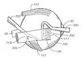
도 1은 인간 안구 및 본 발명에 따른 안구 후안부에 삽입된 안과용 약물 전달 장치를 도식적으로 나타낸 측단면도이다.1 is a side cross-sectional view schematically showing an ophthalmic drug delivery device inserted into a human eye and a posterior eyeball according to the present invention.
도 2는 도 1의 2-2선을 따라 절단한 안구의 상세 횡단면도이다.FIG. 2 is a detailed cross-sectional view of the eye taken along the line 2-2 of FIG. 1. FIG.
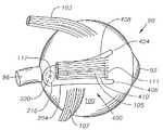
도 3은 인간 안구의 외안근 국소적 구조의 측면 개요도이다.Figure 3 is a schematic side view of the extraocular muscle local structure of the human eye.
도 4는 외직근(미도시됨)의 부분을 가지는 인간 안구의 외안근 국소적 구조의 후측면도이다.4 is a rear side view of the extraocular muscle local structure of the human eye with portions of the lateral rectus muscle (not shown).
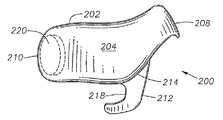
도 5는 본 발명의 제 1의 바람직한 구체예에 따른 안구용 약물 전달 장치의 안와부 표면의 투시도이다.5 is a perspective view of an orbital surface of an ocular drug delivery device according to a first preferred embodiment of the present invention.
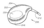
도 6은 도 5의 안구용 약물 전달 장치의 공막 표면의 투시도이다.6 is a perspective view of the sclera surface of the ocular drug delivery device of FIG. 5.
도 7은 도 5의 안구용 약물 전달 장치의 제 1 면의 투시도이다.7 is a perspective view of a first side of the ocular drug delivery device of FIG. 5.
도 8은 도 5의 안구용 약물 전달 장치의 제 2 면의 투시도이다.8 is a perspective view of a second side of the ocular drug delivery device of FIG. 5.
도 9는 도 5의 안구용 약물 전달 장치의 원위 말단의 투시도이다.9 is a perspective view of the distal end of the ocular drug delivery device of FIG. 5.
도 10은 도 5의 안구용 약물 전달 장치의 근위 말단의 투시도이다.10 is a perspective view of the proximal end of the ocular drug delivery device of FIG. 5.
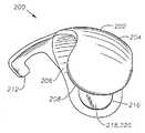
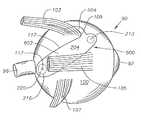
도 11은 인간 안구에서 상피내 도 5의 안구용 약물 전달 장치의 개략도이다.FIG. 11 is a schematic of the ocular drug delivery device of FIG. 5 in human epithelium.
도 12a 내지 12e는 본 발명의 바람직한 방법에 따른 인간 안구에서 도 5의 안구용 약물 전달 장치의 삽입을 도식적으로 나타낸 것이다.12A-12E schematically illustrate the insertion of the ocular drug delivery device of FIG. 5 in a human eye according to a preferred method of the present invention.
도 13은 인간 안구에서 상피내 본 발명의 제 2의 바람직한 구체예에 따른 안구용 약물 전달 장치의 개략도이다.13 is a schematic diagram of an ophthalmic drug delivery device according to a second preferred embodiment of the present invention in human epithelium in the epithelium.
도 14는 인간 안구에서 상피내 본 발명의 제 3의 바람직한 구체예에 따른 안구용 약물 전달 장치의 개략도이다.14 is a schematic diagram of an ophthalmic drug delivery device according to a third preferred embodiment of the present invention in human epithelium in the epithelium.
도 15는 인간 안구에서 상피내 본 발명의 제 4의 바람직한 구체예에 따른 안구용 약물 전달 장치의 개략도이다.15 is a schematic diagram of an ophthalmic drug delivery device according to a fourth preferred embodiment of the present invention in human epithelium in the epithelium.
바람직한 구체예의 상세한 설명Detailed Description of the Preferred Embodiments
본 발명의 바람직한 구체예 및 이의 잇점은 도면의 해당 부분을 나타내는데 사용된 숫자와 도 1 내지 15를 참조하여 가장 잘 이해될 수 있다.Preferred embodiments of the invention and their advantages can be best understood with reference to the figures used to represent the corresponding parts of the drawings and FIGS. 1 to 15.
도 1 내지 4는 본 발명의 완벽한 이해를 위해 중요한 인간 안구의 다양한 부분을 나타낸다. 먼저 도 1은 인간 안구(90)를 도식적으로 나타낸다. 안구(90)는 각막(92), 수정체(93), 유리체(95), 공막(100), 맥락막(99), 망막(97) 및 시신경(96)을 가진다. 안구(90)는 일반적으로 전안부(89) 및 후안부(88)로 나누어진다. 안구(90)의 전안부(89)는 일반적으로 거상연(11) 앞쪽의 안구(90) 부분을 포함한다. 안구(90)의 후안부(88)는 일반적으로 거상연(11) 뒤쪽의 안구(90) 부분을 포함한다. 망막(97)은 에워싸는 형태로 편평부(13)에 근접한 맥락막(99)에 물리적으로 접해 있고 뒤쪽으로는 시신경 유두(19)에 접해 있다. 망막(97)은 시신경 유두(19)에 다소 측면에 위치한 황반(98)을 가진다. 안과 분야에서 널리 공지된 바와 같이, 황반(98)은 망막 추체로 주로 구성되고 망막(97)에서 최대 시력을 보이는 부위이다. 황반(98)의 중심에는 와(117)가 존재한다. 테논 낭 또는 테논 막(101)은 공막(100) 위에 배치된다. 결막(94)은 윤부(115) 뒤쪽의 안구(90)의 짧은 면 (안구 결막)을 차지하고, 이것은 상하로 (각각 상부 결막낭, 하부 결망낭) 접혀 있어 각각 윗눈꺼플(78) 및 아래눈꺼플(79)의 안쪽 면을 차지한다. 안구 결막(94)은 테논낭(101)의 꼭대기 상에 배치된다.1-4 show various parts of the human eye that are important for a complete understanding of the present invention. 1 schematically shows a
도 1 및 도 2에서 나타내고 하기에 상세히 기술한 바와 같이, 바람직하게는, 대부분의 후안부 질환 또는 상태를 치료하기 위하여 안구용 약물 전달 장치(200)는 테논낭(101) 아래쪽 및 공막(100)의 외부 표면 바로 위에 배치된다. 또한, 바람직하게는, 인간의 ARMD 및 CNV의 치료를 위하여 장치(200)는 테논낭(101) 아래쪽 및 공막(100)의 외부 표면 바로 위에 배치되고, 장치(200)의 내부 중심이 황반(98)에 근접하여 위치한다. 장치(200)가 특히 인간에 사용되도록 설계되는 한, 동물에도 사용될 수 있다.As shown in FIGS. 1 and 2 and described in detail below, the ophthalmic
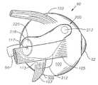
도 3은 각막(92), 시신경(96), 황반(98), 공막(100), 상직근(103), 외직근(105), 하사근(107), 및 와(117)를 가지는 인간 우안(90)의 국소 측면도를 도식적으로 나타낸 것이다. 상직근(103)은 공막(100) 내에 삽입물(109)을 갖는다. 외직근(105)은 공막(100)내에 삽입물(111)을 갖는다. 하사근(107)은 공막(100)내에 삽입물(113)을 갖는다. 도 4는 근육에 의해 숨겨진 공막(100)의 부분에서 시력을 허용하도록 절단된 외직근(105)의 부분을 가지는 인간 우안(90)의 국소적 후측면도를 도식적으로 나타낸 것이다.3 shows the human right eye with
도 5 내지 10은 본 발명의 첫번째 바람직한 구체예에 따른 인간 우안을 위한 안과용 약물 전달 장치(200)를 도식적으로 나타낸다. 장치(200)의 거울상인 안과용 약물 전달 장치는 인간 좌안에 대해 사용될 수 있다. 장치(200)는 약제학적 활성 제제의 눈으로의 국소 전달이 필요한 임의의 경우에서 사용될 수 있다. 특히, 장치(200)는 안구 후안부로의 약제학적 활성 제제의 국소 전달을 위해 유용하다. 장치(200)의 바람직한 용도는, ARMD, 맥락막 혈관신생 (CNV), 망막병증, 망막염, 포도막염, 황반 부종, 녹내장 및 신경병증을 치료하기 위한 황반에 근접한 망막으로의 약제학적 활성 제제의 전달이다.5-10 diagrammatically show an ophthalmic
장치(200)는 일반적으로 볼록한, 돔-형태의 안와 표면(204) 및 오목한 돔-형태의 공막 표면(206)을 가지는 본체(202)를 포함한다. 공막 표면(206)은 공막(100)과 직접적인 접촉을 용이하게 하는 곡률반경으로 설계된다. 가장 바람직하게는, 공막 표면(206)은 평균 인간 안구(90)의 곡률반경(91)과 같은 곡률반경으로 설계된다(도 1 참조). 바람직하게는 안와 표면(204)은 테논낭(101) 아래쪽에 삽입을 용이하게 하는 곡률반경으로 설계되었다. 장치(200)는 근위 말단(208), 원위 말단(210), 본체(202)로부터 연장되는 연장부(212), 및 공막 표면(206) 상의 고정 구조물(213)을 갖는다. 바람직하게는 연장부(212)는 본체(202)에 통합적으로 형성된다. 바람직하게는 연장부(212)는 라인(214)을 따라 접힐 수 있어 전체 연장부(212)가 장치(200)의 아래쪽으로 접힐 수 있도록 한다. 대안적으로, 장치(200)는 전체 연장부(212)가 본체(202) 위쪽으로 접힐 수 있도록 설계될 수 있다. 하기에서 더욱 상세히 기술된 바와 같이, 연장부(212)는 삽입 동안 하사근(107)의 삽입물(113)을 조절하도록 설계된다. 고정 구조물(213)은 바람직하게는 흡인컵이며, 바람직하게는 공막 표면(206) 상에 통합적으로 형성된다. 대안적으로, 고정 구조물(213)은 요망되는 경우, 생접착 코팅 또는 하나 이상의 날까로운 갈퀴(prong)의 부위가 될 수 있다. 하기에서 더욱 상세히 기술된 바와 같이, 고정 구조물(213)은 공막(100)과 맞붙게 하여 삽입 후 장치(200)의 이동을 방지하는데 도움이 된다. 또다른 대안으로, 장치(200)는 공막(100)에, 바람직하게는 이의 근위 말단(208) 가까이에 봉합되어 장치를 고정시킬 수 있으며, 삽입 후 이동을 방지하는데 도움이 된다.The
장치(200)는 또한 공막 표면(206)에 개구부(218)를 가지는 웰 또는 공동(cavity, 216)을 갖는다. 내부 코어(220)는 바람직하게는 웰(216)에 배치된다. 도 5 내지 10에 나타낸 바와 같이, 내부 코어(220)는 바람직하게는 하나 이상의 약제학적 활성 제제를 포함하는 타블렛(tablet)이다. 대안적으로는, 내부 코어(220)는 이에 배치된 하나 이상의 약제학적 활성 제제를 갖는 통상적인 수화겔, 겔, 페이스트 또는 다른 반고형물 투여 형태를 포함할 수 있다. 도 5 내지 10에서 도시되지 않았으나, 내부 코어(220)는 대안적으로 하나 이상의 약제학적 활성 제제를 포함하는, 현탁액, 용액제, 분말제, 또는 이의 조합물을 포함한다. 이러한 구체예에서, 공막 표면(206)은 개구부(218) 없이 형성되며, 현탁액, 용액제, 분말제, 또는 이의 조합물은 공막 표면(206)의 비교적 얇은 연장부 또는 내부 코어(220)의 아래쪽의 다른 막을 통하여 확산된다. 다른 대안으로는, 장치(200)는 웰(216) 또는 내부 코어(220) 없이 형성될 수 있으며, 현탁액, 용액제, 분말제, 또는 이의 조합물의 형태인 약제학적 활성 제제는 장치(200)의 본체(202)를 통하여 분산될 수 있다. 이러한 구체예에서, 약제학적 활성 제제는 본체(202)를 통하여 목표 조직 내로 분산된다. 웰(216) 및 내부 코어(220)의 구조는 미국특허 제6,413,540호에 더욱 충분하게 기술되어 있으며, 이는 본원에 참고문헌으로서 전체적으로 포함된다.The
장치(200)의 기하학적 형태나 디멘션은 내부 코어(220)의 약제학적 활성 제제와 공막 표면(206) 아래쪽에 놓여진 조직 사이의 전달을 극대화한다. 바람직하게는 공막 표면(206)은 물리적으로 공막(100)의 외부 표면과 접한다. 대안적으로는, 공막 표면(206)은 공막(100)의 외부 표면에 근접하여 배치될 수 있다. 예를 들어, 장치(200)는 공막(100)의 외부 표면의 바로 위쪽에 또는 공막(100)내의 층판내에 배치될 수 있다.The geometry or dimension of the
바람직하게는, 본체(202)는 생체적합성, 비-생분해성 물질을 포함한다. 보다 더 바람직하게는, 본체(202)는 생체적합성, 비-생분해성 중합체 조성물을 포함한다. 가장 바람직하게는, 상기 중합체 조성물은 실리콘을 포함한다. 물론, 상기 중합체는 다공성, 굴곡비, 투과도, 강성률, 경도 및 평활도를 포함하나 이에 한정되지 않는 물리적 특성에 영향을 미치는 다른 종래 물질을 또한 포함한다. 바람직하게는, 본체(202)는 내부 코어(220)의 약제학적 활성 제제를 투과시키지 않는다. 본체(202)에 적합한, 중합체 조성물 및 이의 물리적 특성에 영향을 미치는 종래 물질은 미국특허 제6,416,777호에 보다 충분히 기술되어 있으며, 상기 문헌은 본원에 참고문헌으로 전체적으로 포함된다.Preferably,
내부 코어(220)는 국소 전달에 적합한 임의의 안과용으로 허용되는 약제학적 활성 제제를 포함할 수 있다. 내부 코어(220)에 적합한 약제학적 활성 제제의 예는 미국특허 제6,416,777호에 기술되어 있다. 한 바람직한 약제학적 활성 제제는 ARMD, CNV, 망막병증, 망막염, 포도막염, 황반 부종 및 녹내장을 포함하나 이에 한정되지 않는 안구 후안부의 질병 또는 상태의 예방 또는 치료를 위한 항지혈제 스테로이드이다. 이러한 항지혈제 스테로이드는 미국특허 제5,679,666호 및 제 5,770,592호에 보다 더 완전하게 공지되었고 본원에 참고문헌으로 전체적으로 포함된다. 이러한 항지혈제 스테로이드의 바람직한 예는 4,9(11)-프레그나디엔-17α,21-디올-3,20-디온 및 4,9(11)-프레그나디엔-17α,21-디올-3,20-디온-21-아세테이트를 포함한다. 또한, 내부 코어(220)는 약제학적 활성 제제로서 글루코코르티코이드와 항지혈제 스테로이드의 조합을 포함할 수 있다. 이러한 조합을 위하여, 바람직한 글루코코르티코이드는 덱사메타손, 플루오로메탈론, 메드리손, 베타메타손, 트리암시놀론, 트리암시놀론 아세토나이드, 프레드니손, 프레드니솔론, 히드로코르티손, 리멕솔론, 및 이의 약제학적으로 허용되는 염을 포함하며, 바람직한 항지혈제 스테로이드는 4,9(11)-프레그나디엔-17α,21-디올-3,20-디온 및 4,9(11)-프레그나디엔-17α,21-디올-3,20-디온-21-아세테이트를 포함한다. 또한 내부 코어(220)는 활성 제제 또는 약물 코어의 안정성, 용해도, 투과도 또는 다른 특성을 개선시키기 위해 종래의 비활성 부형제를 포함할 수 있다. 내부 코어(220)가 타블렛인 경우, 충전제 및 윤활제와 같은, 타블렛팅(tableting)을 위해 필요한 종래 부형제를 추가로 포함할 수 있다. 상기 타블렛은 종래 타블렛팅 방법을 이용하여 제조될 수 있다. 바람직하게는, 약제학적 활성 제제는 타블렛 전체에 균등하게 분산되어 있다. 종래 타블렛에 더하여, 내부 코어(220)는 약제학적 활성 제제를 방출하며 조절 율속으로 생분해되는 특수 타블렛을 포함할 수 있다. 예를 들어, 이러한 생분해는 가수분해 또는 효소분해를 통해 일어날 수 있다. 내부 코어(220)가 수화겔 또는 다른 겔인 경우, 이러한 겔은 약제학적 활성 제제를 방출하며 조절 율속으로 생분해될 수 있다. 대안적으로는, 이러한 겔은 비생분해성이나 약제학적 활성 제제를 확산시킬 수 있다.
장치(200)는 사출 성형, 압출 성형, 이송 성형, 압축 성형을 포함하나 이에 한정되지 않는 종래 중합체 가공 방법에 의해 만들어질 수 있다. 바람직하게는, 장치(200)는 종래 사출 성형 기술을 이용하여 형성된다. 바람직하게는, 내부 코어(220)는 장치(200)의 본체(202)를 형성한 후 웰(216)에 배치된다.
도 11에 나타낸 바와 같이, 바람직하게는 장치(200)는 테논낭(101) 아래와 공막(100)의 외부 표면 바로 위에 외과적으로 배치되며, 황반(98) 위쪽의 공막(100) 구역 바로 위쪽에 웰(216) 및 내부 코어(220)을 가진다. 더욱 바람직하게는, 내부 코어(220)는 황반(98)의 중심인 와(117) 위쪽의 공막(100)의 구역 바로 위쪽에 존재한다. 연장부(212)는 공막(100)의 외부 표면 상과 하사근(107) 아래쪽에, 하사근(107)의 삽입물(109)에 근접하거나 접하게 배치된다. 장치(200)의 기하학적 형태로 인해, 이러한 방식으로 하사근(107)에 연장부(212)의 고정은 자동적으로 황반(98) 및 와(117) 위에 내부 코어(220)를 정위시킨다. 이러한 방식으로 하사근(107)에 연장부(212)의 고정은 또한 삽입 후 장치(200)를 고정시키고 이의 이동을 방지하는데 도움이 된다. 흡인컵(213)은 또한 공막(100)에 조심스럽게 적용되며, 삽입 후에 장치(200)를 고정시키고 이의 이동을 방지하는데 추가로 도움이 된다.As shown in FIG. 11, the
일반적으로 도 12a 내지 도 12e와 관련하여, 하기 기술은 외래환자 상황에서 수행될 수 있으며, 바람직하게는 도 11에 나타낸 위치에 장치(200)를 삽입하는데 활용된다. 외과의사는 먼저 안구(90)의 1/4에서 주위결막절개를 수행한다. 바람직하게는 외과의사는 안구(90) 테두리(115)에 대해 약 3mm 뒤쪽의 측두상의 1/4에서 결막절개를 수행한다. 이러한 절개를 수행하자마자, 외과의사는 비절개박리를 수행하여 공막(100)으로부터 테논낭(101)을 분리한다. 가위 및 비절개박리를 사용하여, 정면 터널이 공막(100)의 외부 표면 및 이후 외직근 위쪽 가장자리(306)(도 3 참조)를 따라 형성된다. 외직근(105) 및 이후 하사근(107)은 자미손 근 후크(300, 302)에 각각 걸리고, 도 12a에 나타낸 바와 같이 조작된다. 후크(300, 302)는 또한 근육(105,107)과 공막(100) 사이에 추가로 장치(200)에 대한 터널로 규정되는 연결 조직을 조심스럽게 끊기 위해 사용된다. 후크(300)를 제거한 후, 외과의사는 도 12b에 나타낸 바와 같이, 누게트(Nuggett) 핀셋(304)으로 장치(200)를 잡는다. 바람직하게는 연장부(212)는 본체(202) 아래쪽으로 접히고 이러한 위치에서 핀셋(304)으로 잡는다. 라인(214)을 따라 접힌 연장부(212)를 가지는 장치(200)의 사용은 장치(200)에 대해 요구되는 결막절개 및 터널의 크기를 최소화시킨다. 대안적으로, 접히지 않는 연장부(212)를 가지는 장치(200)는 요망되는 경우, 사용될 수 있다. 외과의사는 공막(100) 및 원위 말단(210)과 접촉하는 공막 표면(206)을 제거하고, 도 12c 내지 12e에 나타낸 바와 같이, 일반적으로 원 운동을 이용하여 터널에 장치(200)를 삽입한다. 외과의사는 접힌 연장부(212)가 외직근(105)의 이전 삽입물(111)을 이동시키는 경우, 핀셋(304)를 느슨하게 한다. 이러한 핀셋(304)을 느슨하게 함은 연장부(212)를 풀어주고 도 12d에 나타낸 바와 같이 접히지 않게 한다. 이후 외과의사는 도 12e에 나타낸 바와 같이, 하사근(107) 아래쪽에 연장부(212)를 걸때까지 터널 내에 일반적으로 원형 방식으로 장치(200)를 계속 이동시킨다. 바람직하게는 연장부(212)의 앞쪽 모시리(218)(도 5)는 하사근(107)의 앞쪽 가장자리(도 3) 및/또는 삽입물(113)을 접촉시킨다. 접히지 않는 연장부(212)를 사용하는 경우, 연장부(212)는 유사한 방식으로 하사근(107)의 앞쪽 가장자리(308) 아래쪽으로 간단히 이동된다. 도 12e에 나타내지 않았으나, 연장부(212)는 또한 하사근(107)의 후크(302)와 삽입물(113) 사이에 배치될 수 있다. 이후 외과의사는 후크(302)를 제거한다. 이후 장치(200)는 도 11에 나타낸 위치에 배치된다. 외과의사는 핀셋(304)을 사용하여 근위 말단(208) 근처의 안구 표면(204)을 조심스럽게 가압하여, 흡인컵(213)을 공막(100)에 확보되도록 한다. 대안적으로, 외과의사는 핀셋(304)을 사용하여 근위 말단(208) 근처의 안구 표면(204)를 조심스럽게 가압하여 생접착 코팅(213) 또는 하나 이상의 날까로운 갈퀴(213)의 부위를 공막(100)에 확보되도록 한다. 또다른 대안으로, 외과의사는 장치(200)의 근위 말단(208)을 공막(100)에 봉합시킬 수 있다. 이후 외과의사는 테논낭(101) 및 결막(94)을 공막(100)에 봉합시키므로써 결막절개를 종료한다. 종료한 후, 외과의사는 수술 상처 상에 항생제 연고의 스트립을 놓는다.In general with respect to FIGS. 12A-12E, the following technique may be performed in an outpatient situation and is preferably utilized to insert the
장치(200)의 본체(202)의 기하학적 형태는, 공막 표면(206)의 오목 부분; 연장부(212), 웰(216), 개구부(218), 내부 코어(220)의 형태 및 위치; 흡인컵, 생접착 코팅 또는 날까로운 갈퀴(203)의 영역의 존재, 및 연장부(212)의 접히는 특성을 포함하여, 모두 내부 코어(220)로부터 공막(100), 맥락막(99)을 통과하여, 망막(97)으로, 보다 더 바람직하게는 황반(98) 및 와(117)로 약제학적 활성 제제의 약제학적 유효량의 전달을 용이하게 한다. 또한, 내부 코어(220) 및 공막(100) 사이의 중합체 층이나 막의 부재는 활성 제제의 망막(97)으로의 전달을 향상시키고 간편하게 한다.The geometry of the
장치(200)는 수년동안 약제학적 활성 제제의 약제학적 유효량을 망막(97)으로 전달하는데 이용될 수 있고, 적용되는 약제학적 활성 제제의 개개의 물리화학적 특성에 따라 다르게 이용될 수 있다고 생각된다. 중요한 물리화학적 특성은 소수성, 용해도, 용해율, 확산계수, 분배계수, 조직친화도를 포함한다. 내부 코어(220)에 활성 제제가 더이상 존재하지 않게 된 후에, 외과의사는 용이하게 장치(200)를 제거할 수 있다. 또한, '사전-형성'된 터널은 구장치(200)를 신장치(200)로 교체하는 것을 용이하게 한다.It is contemplated that the
도 13 내지 도 15는 인간 안구 내에서 각각 본 발명의 제 2, 제 3 및 제 4의 바람직한 구체예에 따른 안구용 약물 전달 장치(400, 500, 600)를 도식적으로 나타낸 것이다. 장치(400, 500, 600) 각각은 장치의 본체가 안와 표면에서 볼 때, 상이한 기하학적 형태를 갖는 것을 제외하고 장치(200)의 구조, 작동 및 용도가 실질적으로 유사하며, 수개의 장치는 장치(200)의 연장부(212) 이외에 상이한 외안근을 조절하도록 고안된 상이한 연장부를 갖는다.13-15 diagrammatically show ocular
도 13에 나타낸 바와 같이, 바람직하게는 장치(400)는 바람직하게는 테논낭(101) 아래와 공막(100)의 외부 표면 바로 위에 외과적으로 배치되며, 황반(98) 위쪽의 공막(100)의 구역 바로 위쪽에 웰(216) 및 내부 코어(220)를 가진다. 가장 바람직하게는, 내부 코어(220)는 와(117)의 위쪽에 공막(100)의 구역 바로 위쪽에 존재한다. 장치(400)는 안와 표면(204)에서 볼 때, 대체적으로 사다리꼴 기하학적 형태를 갖는다. 장치(400)는 또한 외직근(105) 삽입물(111)의 상반 한계(408) 및 하반 한계(410)를 조절하기 위하여 고안된 제 1 연장부(404) 및 제 2 연장부(406)를 갖는다. 장치(400)의 기하학적 형태로 인해, 이러한 방식으로 외직근(105)의 삽입물(111)에 연장부(404, 406)의 고정은 황반(98) 및 와(117) 위쪽에 내부 코어(220)을 자동적으로 위치시킨다. 이러한 방식으로 외직근(105)의 삽입물(111)에 연장부(404, 406)의 고정은 또한 삽입 후에 장치(400)를 고정시키고 이의 이동을 방지하는데 도움이 된다.As shown in FIG. 13, the
도 14에 나타낸 바와 같이, 바람직하게는 장치(500)는 테논낭(101) 아래, 공막(100)의 외부 표면 바로 위에 외과적으로 배치되며, 황반(98) 위쪽의 공막(100)의 구역 바로 위쪽에 웰(216) 및 내부 코어(220)를 가진다. 가장 바람직하게는, 내부 코어(220)는 와(117) 위쪽의 공막(100)의 구역 바로 위쪽에 존재한다. 장치(500)는 안와 표면(204)에서 볼 때, 대체적으로 클럽(club) 모양 또는 아크(arc) 모양의 기하학적 형태를 가지며, 이는 상직근(103)의 삽입물(109)과 외직근(105)의 상반 하녜(502) 사이에 삽입을 용이하도록 고안된다.As shown in FIG. 14, the
도 15에 나타낸 바와 같이, 바람직하게는 장치(600)는 테논낭(101) 아래, 공막(100)의 외부 표면 바로 위에 외과적으로 배치되며, 황반(98) 위쪽의 공막(100)의 구역 바로 위쪽에 웰(216) 및 내부 코어(220)을 가진다. 가장 바람직하게는, 내부 코어(220)는 와(117) 위쪽의 공막(100)의 구역 바로 위쪽에 존재한다. 장치(600)는 안와 표면(204)에서 볼 때, 대체적으로 타원 또는 직사각형 기하학적 형태를 갖는다. 장치(600)는 또한 본체(602)로부터 연장하는 연장부(604)를 갖는다. 연장부(604)는 공막(100)의 외부 표면 위, 상직근(103)의 아래에, 상직근(103)의 삽입물(109)에 근접하거나 접촉하도록 배치된다. 장치(600)의 기하학적 형태로 인해, 이러한 방식으로 상직근(103) 삽입물(109)에 연장부(604)의 고정은 황반(98) 및 와(117) 위쪽에 내부 코어(220)을 자동적으로 위치시킨다. 이러한 방식으로 상직근(103)의 삽입물(109)에 연장부(604)의 고정은 또한 삽입 후에 장치(600)를 고정시키고 이의 이동을 방지하는데 도움이 된다.As shown in FIG. 15, the
상기로부터, 본 발명은, ARMD, CNV, 망막병증, 망막염, 포도막염, 황반 부종, 녹내장 및 신경병증을 치료하기 위하여, 다양한 약제학적 활성 제제의 안구로의, 특히 안구 후안부로의 안전하고, 효과적이고, 율속 조절의 국소 전달을 위한 개선된 장치 및 방법을 제공한다고 이해된다. 상기 장치를 삽입하기 위한 외과적 과정은 안전하고, 간단하고, 신속하고, 외래환자에게 수행될 수 있다. 상기 장치는 제조하기 용이하고 경제적이다. 더우기, 다양한 약제학적 활성 제제를 전달할 수 있기 때문에, 상기 장치는 환자에게 특이적 물리적 상태를 유발하기 위한 다양한 안과용 제제를 전달하기 위한 임상 연구에 유용하다.From the above, the present invention provides a safe, effective and effective method for the treatment of ARMD, CNV, retinopathy, retinitis, uveitis, macular edema, glaucoma and neuropathy, in particular to the eye, in particular to the posterior ocular region. It is understood that the present invention provides an improved apparatus and method for topical delivery of rate control. Surgical procedures for inserting the device can be safe, simple, rapid, and outpatient. The device is easy and economical to manufacture. Moreover, because of the ability to deliver a variety of pharmaceutically active agents, the devices are useful in clinical studies to deliver a variety of ophthalmic formulations to cause specific physical conditions to patients.
본 발명의 조작 및 구조는 상기로부터 자명하게 될 것으로 사료된다. 상기에 나타내거나 기술한 장치 및 방법은 바람직하게 특징되고, 하기 특허청구범위에 한정된 발명의 정신 및 범위에서 벗어나지 않고 이 안에서 다양한 변화 및 개량이 이루어질 수 있다.It is believed that the operation and structure of the present invention will be apparent from the above. The devices and methods shown or described above are preferably characterized and various changes and improvements can be made therein without departing from the spirit and scope of the invention as defined in the following claims.
Claims (30)
Translated fromKoreanApplications Claiming Priority (2)
| Application Number | Priority Date | Filing Date | Title |
|---|---|---|---|
| US48599503P | 2003-07-10 | 2003-07-10 | |
| US60/485,995 | 2003-07-10 |
Publications (1)
| Publication Number | Publication Date |
|---|---|
| KR20060082792Atrue KR20060082792A (en) | 2006-07-19 |
Family
ID=34102671
Family Applications (1)
| Application Number | Title | Priority Date | Filing Date |
|---|---|---|---|
| KR1020057024755ACeasedKR20060082792A (en) | 2003-07-10 | 2004-06-23 | Ophthalmic Drug Delivery Device |
Country Status (13)
| Country | Link |
|---|---|
| US (1) | US20060039952A1 (en) |
| EP (1) | EP1684669A4 (en) |
| JP (1) | JP2007526019A (en) |
| KR (1) | KR20060082792A (en) |
| CN (1) | CN1805719A (en) |
| AR (1) | AR045039A1 (en) |
| AU (1) | AU2004258857A1 (en) |
| BR (1) | BRPI0410542A (en) |
| CA (1) | CA2522592A1 (en) |
| MX (1) | MXPA05011517A (en) |
| TW (1) | TW200505402A (en) |
| WO (1) | WO2005009297A1 (en) |
| ZA (1) | ZA200508654B (en) |
Cited By (1)
| Publication number | Priority date | Publication date | Assignee | Title |
|---|---|---|---|---|
| US10302964B2 (en) | 2016-11-22 | 2019-05-28 | Korea Institute Of Science And Technology | Contact lens for collecting tear or delivering drug, and method for manufacturing the contact lens |
Families Citing this family (45)
| Publication number | Priority date | Publication date | Assignee | Title |
|---|---|---|---|---|
| US7431710B2 (en) | 2002-04-08 | 2008-10-07 | Glaukos Corporation | Ocular implants with anchors and methods thereof |
| US8246974B2 (en)* | 2003-05-02 | 2012-08-21 | Surmodics, Inc. | Medical devices and methods for producing the same |
| US20070212397A1 (en)* | 2005-09-15 | 2007-09-13 | Roth Daniel B | Pharmaceutical delivery device and method for providing ocular treatment |
| US8652090B2 (en)* | 2006-05-18 | 2014-02-18 | Cannuflow, Inc. | Anti-extravasation surgical portal plug |
| EP2219620B1 (en) | 2007-11-13 | 2017-07-19 | Surmodics, Inc. | Viscous terpolymers as drug delivery platform |
| US8974808B2 (en)* | 2008-12-23 | 2015-03-10 | Surmodics, Inc. | Elastic implantable composites and implants comprising same |
| US20100158978A1 (en)* | 2008-12-23 | 2010-06-24 | Peter Markland | Bioactive spray coating compositions and methods of making and uses thereof |
| US9415197B2 (en)* | 2008-12-23 | 2016-08-16 | Surmodics, Inc. | Implantable suction cup composites and implants comprising same |
| US20100168807A1 (en)* | 2008-12-23 | 2010-07-01 | Burton Kevin W | Bioactive terpolymer compositions and methods of making and using same |
| US8623395B2 (en) | 2010-01-29 | 2014-01-07 | Forsight Vision4, Inc. | Implantable therapeutic device |
| CN104887389B (en) | 2009-01-29 | 2017-06-23 | 弗赛特影像4股份有限公司 | Posterior segment drug delivery |
| US10206813B2 (en) | 2009-05-18 | 2019-02-19 | Dose Medical Corporation | Implants with controlled drug delivery features and methods of using same |
| WO2010135369A1 (en) | 2009-05-18 | 2010-11-25 | Dose Medical Corporation | Drug eluting ocular implant |
| WO2012071476A2 (en) | 2010-11-24 | 2012-05-31 | David Haffner | Drug eluting ocular implant |
| US20110105990A1 (en)* | 2009-11-04 | 2011-05-05 | Silvestrini Thomas A | Zonal drug delivery device and method |
| US8529492B2 (en)* | 2009-12-23 | 2013-09-10 | Trascend Medical, Inc. | Drug delivery devices and methods |
| US10166142B2 (en) | 2010-01-29 | 2019-01-01 | Forsight Vision4, Inc. | Small molecule delivery with implantable therapeutic device |
| US10617557B2 (en) | 2010-08-05 | 2020-04-14 | Forsight Vision4, Inc. | Combined drug delivery methods and apparatus |
| PT2600812T (en) | 2010-08-05 | 2021-11-09 | Forsight Vision4 Inc | Implantable therapeutic device |
| LT2600930T (en) | 2010-08-05 | 2021-04-12 | Forsight Vision4, Inc. | Injector apparatus for drug delivery |
| US9416221B2 (en) | 2010-08-30 | 2016-08-16 | Surmodics, Inc. | Biodegradable terpolymers and terpolymer blends as pressure-sensitive adhesives |
| WO2012068549A2 (en) | 2010-11-19 | 2012-05-24 | Forsight Vision4, Inc. | Therapeutic agent formulations for implanted devices |
| US10245178B1 (en) | 2011-06-07 | 2019-04-02 | Glaukos Corporation | Anterior chamber drug-eluting ocular implant |
| US10398592B2 (en) | 2011-06-28 | 2019-09-03 | Forsight Vision4, Inc. | Diagnostic methods and apparatus |
| HRP20210676T1 (en) | 2011-09-16 | 2021-05-28 | Forsight Vision4, Inc. | Fluid exchange apparatus |
| WO2013116061A1 (en) | 2012-02-03 | 2013-08-08 | Forsight Vision4, Inc. | Insertion and removal methods and apparatus for therapeutic devices |
| US9827401B2 (en) | 2012-06-01 | 2017-11-28 | Surmodics, Inc. | Apparatus and methods for coating medical devices |
| JP6549482B2 (en) | 2012-06-01 | 2019-07-24 | サーモディクス,インコーポレイテッド | Device and method for coating a balloon catheter |
| US11090468B2 (en) | 2012-10-25 | 2021-08-17 | Surmodics, Inc. | Apparatus and methods for coating medical devices |
| WO2014152959A1 (en) | 2013-03-14 | 2014-09-25 | Forsight Vision4, Inc. | Systems for sustained intraocular delivery of low solubility compounds from a port delivery system implant |
| ES2972168T3 (en) | 2013-03-28 | 2024-06-11 | Forsight Vision4 Inc | Ophthalmic implant for administration of therapeutic substances |
| EP3677229A1 (en) | 2014-05-29 | 2020-07-08 | Glaukos Corporation | Implants with controlled drug delivery features |
| ES2803102T3 (en) | 2014-07-15 | 2021-01-22 | Forsight Vision4 Inc | Eye implant delivery device |
| WO2016022750A1 (en) | 2014-08-08 | 2016-02-11 | Forsight Vision4, Inc. | Stable and soluble formulations of receptor tyrosine kinase inhibitors, and methods of preparation thereof |
| WO2016077371A1 (en) | 2014-11-10 | 2016-05-19 | Forsight Vision4, Inc. | Expandable drug delivery devices and method of use |
| US11925578B2 (en) | 2015-09-02 | 2024-03-12 | Glaukos Corporation | Drug delivery implants with bi-directional delivery capacity |
| US11564833B2 (en) | 2015-09-25 | 2023-01-31 | Glaukos Corporation | Punctal implants with controlled drug delivery features and methods of using same |
| US11432959B2 (en) | 2015-11-20 | 2022-09-06 | Forsight Vision4, Inc. | Porous structures for extended release drug delivery devices |
| MX2018012021A (en) | 2016-04-05 | 2019-01-24 | Forsight Vision4 Inc | Implantable ocular drug delivery devices. |
| CN109937025B (en) | 2016-04-20 | 2022-07-29 | 多斯医学公司 | Delivery device for bioabsorbable ocular drugs |
| AU2018372806B2 (en) | 2017-11-21 | 2024-05-30 | Forsight Vision4, Inc. | Fluid exchange apparatus for expandable port delivery system and methods of use |
| US11628466B2 (en) | 2018-11-29 | 2023-04-18 | Surmodics, Inc. | Apparatus and methods for coating medical devices |
| US11819590B2 (en) | 2019-05-13 | 2023-11-21 | Surmodics, Inc. | Apparatus and methods for coating medical devices |
| EP4125748A4 (en) | 2020-03-23 | 2024-06-26 | Opticgon LLC | MACULAR DENTAL GATE AND METHODS OF USE AND MANUFACTURE THEREOF |
| USD1033637S1 (en) | 2022-01-24 | 2024-07-02 | Forsight Vision4, Inc. | Fluid exchange device |
Family Cites Families (49)
| Publication number | Priority date | Publication date | Assignee | Title |
|---|---|---|---|---|
| US3416530A (en)* | 1966-03-02 | 1968-12-17 | Richard A. Ness | Eyeball medication dispensing tablet |
| US3828777A (en)* | 1971-11-08 | 1974-08-13 | Alza Corp | Microporous ocular device |
| US4014335A (en)* | 1975-04-21 | 1977-03-29 | Alza Corporation | Ocular drug delivery device |
| US4256108A (en)* | 1977-04-07 | 1981-03-17 | Alza Corporation | Microporous-semipermeable laminated osmotic system |
| US4300557A (en)* | 1980-01-07 | 1981-11-17 | The United States Of America As Represented By The Secretary Of The Department Of Health And Human Services | Method for treating intraocular malignancies |
| US4327725A (en)* | 1980-11-25 | 1982-05-04 | Alza Corporation | Osmotic device with hydrogel driving member |
| US4624848A (en)* | 1984-05-10 | 1986-11-25 | Ciba-Geigy Corporation | Active agent containing hydrogel devices wherein the active agent concentration profile contains a sigmoidal concentration gradient for improved constant release, their manufacture and use |
| US5322691A (en)* | 1986-10-02 | 1994-06-21 | Sohrab Darougar | Ocular insert with anchoring protrusions |
| US5147647A (en)* | 1986-10-02 | 1992-09-15 | Sohrab Darougar | Ocular insert for the fornix |
| US4997652A (en)* | 1987-12-22 | 1991-03-05 | Visionex | Biodegradable ocular implants |
| US4853224A (en)* | 1987-12-22 | 1989-08-01 | Visionex | Biodegradable ocular implants |
| DE3905050A1 (en)* | 1989-02-18 | 1990-08-30 | Lohmann Therapie Syst Lts | THERAPEUTIC SYSTEM FOR DELAYED AND CONTROLLED TRANSDERMAL OR TRANSMUCOSAL ADMINISTRATION OF ACTIVE SUBSTANCES (II) |
| US4946450A (en)* | 1989-04-18 | 1990-08-07 | Biosource Genetics Corporation | Glucan/collagen therapeutic eye shields |
| US5164188A (en)* | 1989-11-22 | 1992-11-17 | Visionex, Inc. | Biodegradable ocular implants |
| US5290892A (en)* | 1990-11-07 | 1994-03-01 | Nestle S.A. | Flexible intraocular lenses made from high refractive index polymers |
| US5378475A (en)* | 1991-02-21 | 1995-01-03 | University Of Kentucky Research Foundation | Sustained release drug delivery devices |
| US5770592A (en)* | 1991-11-22 | 1998-06-23 | Alcon Laboratories, Inc. | Prevention and treatment of ocular neovascularization using angiostatic steroids |
| US5679666A (en)* | 1991-11-22 | 1997-10-21 | Alcon Laboratories, Inc. | Prevention and treatment of ocular neovascularization by treatment with angiostatic steroids |
| US5178635A (en)* | 1992-05-04 | 1993-01-12 | Allergan, Inc. | Method for determining amount of medication in an implantable device |
| WO1995003009A1 (en)* | 1993-07-22 | 1995-02-02 | Oculex Pharmaceuticals, Inc. | Method of treatment of macular degeneration |
| US5443505A (en)* | 1993-11-15 | 1995-08-22 | Oculex Pharmaceuticals, Inc. | Biocompatible ocular implants |
| US5516522A (en)* | 1994-03-14 | 1996-05-14 | Board Of Supervisors Of Louisiana State University | Biodegradable porous device for long-term drug delivery with constant rate release and method of making the same |
| US5466233A (en)* | 1994-04-25 | 1995-11-14 | Escalon Ophthalmics, Inc. | Tack for intraocular drug delivery and method for inserting and removing same |
| US5710165A (en)* | 1994-07-06 | 1998-01-20 | Synthelabo | Use of polyamine antagonists for the treatment of glaucoma |
| AUPM897594A0 (en)* | 1994-10-25 | 1994-11-17 | Daratech Pty Ltd | Controlled release container |
| ATE232089T1 (en)* | 1994-11-10 | 2003-02-15 | Univ Kentucky Res Found | CONTROLLED RELEASE IMPLANTABLE REFILLABLE DEVICE FOR ADMINISTERING DRUGS IMMEDIATELY TO AN INTERNAL PART OF THE BODY |
| AUPM982694A0 (en)* | 1994-12-02 | 1995-01-05 | University Of Queensland, The | Iontophoresis method and apparatus |
| US5725493A (en)* | 1994-12-12 | 1998-03-10 | Avery; Robert Logan | Intravitreal medicine delivery |
| US5773019A (en)* | 1995-09-27 | 1998-06-30 | The University Of Kentucky Research Foundation | Implantable controlled release device to deliver drugs directly to an internal portion of the body |
| US5743274A (en)* | 1996-03-18 | 1998-04-28 | Peyman; Gholam A. | Macular bandage for use in the treatment of subretinal neovascular members |
| US5824073A (en)* | 1996-03-18 | 1998-10-20 | Peyman; Gholam A. | Macular indentor for use in the treatment of subretinal neovascular membranes |
| US5904144A (en)* | 1996-03-22 | 1999-05-18 | Cytotherapeutics, Inc. | Method for treating ophthalmic diseases |
| US5797898A (en)* | 1996-07-02 | 1998-08-25 | Massachusetts Institute Of Technology | Microchip drug delivery devices |
| US5860994A (en)* | 1996-07-30 | 1999-01-19 | Yaacobi; Yoseph | Remotely operable intraocular surgical instrument for automated capsulectomies |
| US6120460A (en)* | 1996-09-04 | 2000-09-19 | Abreu; Marcio Marc | Method and apparatus for signal acquisition, processing and transmission for evaluation of bodily functions |
| DE69827138T2 (en)* | 1997-08-11 | 2006-02-23 | Allergan, Inc., Irvine | Sterile retinoid-containing biodegradable implant with improved biocompatibility and process for its preparation |
| US5902598A (en)* | 1997-08-28 | 1999-05-11 | Control Delivery Systems, Inc. | Sustained release drug delivery devices |
| US6378526B1 (en)* | 1998-08-03 | 2002-04-30 | Insite Vision, Incorporated | Methods of ophthalmic administration |
| US6146366A (en)* | 1998-11-03 | 2000-11-14 | Ras Holding Corp | Device for the treatment of macular degeneration and other eye disorders |
| US6217895B1 (en)* | 1999-03-22 | 2001-04-17 | Control Delivery Systems | Method for treating and/or preventing retinal diseases with sustained release corticosteroids |
| HK1049438B (en)* | 1999-10-21 | 2005-08-26 | Alcon, Inc. | Sub-tenon drug delivery |
| US6416777B1 (en)* | 1999-10-21 | 2002-07-09 | Alcon Universal Ltd. | Ophthalmic drug delivery device |
| TR200201047T2 (en)* | 1999-10-21 | 2002-07-22 | Alcon, Inc. | Medication delivery means. |
| US6375972B1 (en)* | 2000-04-26 | 2002-04-23 | Control Delivery Systems, Inc. | Sustained release drug delivery devices, methods of use, and methods of manufacturing thereof |
| DE60135352D1 (en)* | 2000-08-30 | 2008-09-25 | Univ Johns Hopkins | DEVICE FOR INTRA-OCCULAR ACTIVE AGGREGATION |
| WO2002053130A2 (en)* | 2001-01-03 | 2002-07-11 | Bausch & Lomb Incorporated | Sustained release drug delivery devices with coated drug cores |
| US6713081B2 (en)* | 2001-03-15 | 2004-03-30 | The United States Of America As Represented By The Department Of Health And Human Services | Ocular therapeutic agent delivery devices and methods for making and using such devices |
| US6582422B2 (en)* | 2001-10-03 | 2003-06-24 | Bausch & Lomb Incorporated | Ophthalmic delivery device |
| US7785578B2 (en)* | 2002-10-11 | 2010-08-31 | Aciont, Inc. | Non-invasive ocular drug delivery |
- 2004
- 2004-06-23AUAU2004258857Apatent/AU2004258857A1/ennot_activeAbandoned
- 2004-06-23WOPCT/US2004/020087patent/WO2005009297A1/enactiveApplication Filing
- 2004-06-23BRBRPI0410542-7Apatent/BRPI0410542A/ennot_activeIP Right Cessation
- 2004-06-23JPJP2006518670Apatent/JP2007526019A/enactivePending
- 2004-06-23CNCN200480016609.2Apatent/CN1805719A/enactivePending
- 2004-06-23EPEP04755919Apatent/EP1684669A4/ennot_activeWithdrawn
- 2004-06-23ZAZA200508654Apatent/ZA200508654B/enunknown
- 2004-06-23KRKR1020057024755Apatent/KR20060082792A/ennot_activeCeased
- 2004-06-23MXMXPA05011517Apatent/MXPA05011517A/enunknown
- 2004-06-23CACA002522592Apatent/CA2522592A1/ennot_activeAbandoned
- 2004-06-29TWTW093119041Apatent/TW200505402A/enunknown
- 2004-07-07ARARP040102393Apatent/AR045039A1/enunknown
- 2005
- 2005-10-12USUS11/248,727patent/US20060039952A1/ennot_activeAbandoned
Cited By (1)
| Publication number | Priority date | Publication date | Assignee | Title |
|---|---|---|---|---|
| US10302964B2 (en) | 2016-11-22 | 2019-05-28 | Korea Institute Of Science And Technology | Contact lens for collecting tear or delivering drug, and method for manufacturing the contact lens |
Also Published As
| Publication number | Publication date |
|---|---|
| TW200505402A (en) | 2005-02-16 |
| US20060039952A1 (en) | 2006-02-23 |
| WO2005009297A1 (en) | 2005-02-03 |
| AU2004258857A1 (en) | 2005-02-03 |
| JP2007526019A (en) | 2007-09-13 |
| ZA200508654B (en) | 2007-01-31 |
| EP1684669A1 (en) | 2006-08-02 |
| CA2522592A1 (en) | 2005-02-03 |
| BRPI0410542A (en) | 2006-06-20 |
| MXPA05011517A (en) | 2005-12-12 |
| AR045039A1 (en) | 2005-10-12 |
| EP1684669A4 (en) | 2008-01-02 |
| CN1805719A (en) | 2006-07-19 |
Similar Documents
| Publication | Publication Date | Title |
|---|---|---|
| KR20060082792A (en) | Ophthalmic Drug Delivery Device | |
| KR100732262B1 (en) | Drug delivery devices | |
| KR100752821B1 (en) | Ophthalmic Drug Delivery Device | |
| US7943162B2 (en) | Drug delivery device | |
| JP4261343B2 (en) | Ophthalmic drug administration device | |
| HK1072177B (en) | Drug delivery device |
Legal Events
| Date | Code | Title | Description |
|---|---|---|---|
| PA0105 | International application | Patent event date:20051223 Patent event code:PA01051R01D Comment text:International Patent Application | |
| PG1501 | Laying open of application | ||
| A201 | Request for examination | ||
| PA0201 | Request for examination | Patent event code:PA02012R01D Patent event date:20090623 Comment text:Request for Examination of Application | |
| E902 | Notification of reason for refusal | ||
| PE0902 | Notice of grounds for rejection | Comment text:Notification of reason for refusal Patent event date:20110414 Patent event code:PE09021S01D | |
| E601 | Decision to refuse application | ||
| PE0601 | Decision on rejection of patent | Patent event date:20110620 Comment text:Decision to Refuse Application Patent event code:PE06012S01D Patent event date:20110414 Comment text:Notification of reason for refusal Patent event code:PE06011S01I |