KR102790039B1 - Cell marker classification method - Google Patents
Cell marker classification methodDownload PDFInfo
- Publication number
- KR102790039B1 KR102790039B1KR1020197015856AKR20197015856AKR102790039B1KR 102790039 B1KR102790039 B1KR 102790039B1KR 1020197015856 AKR1020197015856 AKR 1020197015856AKR 20197015856 AKR20197015856 AKR 20197015856AKR 102790039 B1KR102790039 B1KR 102790039B1
- Authority
- KR
- South Korea
- Prior art keywords
- cell
- delete delete
- signatures
- molecular
- signature
- Prior art date
- Legal status (The legal status is an assumption and is not a legal conclusion. Google has not performed a legal analysis and makes no representation as to the accuracy of the status listed.)
- Active
Links
Images


Classifications
- G—PHYSICS
- G06—COMPUTING OR CALCULATING; COUNTING
- G06F—ELECTRIC DIGITAL DATA PROCESSING
- G06F18/00—Pattern recognition
- G06F18/20—Analysing
- G06F18/24—Classification techniques
- C—CHEMISTRY; METALLURGY
- C12—BIOCHEMISTRY; BEER; SPIRITS; WINE; VINEGAR; MICROBIOLOGY; ENZYMOLOGY; MUTATION OR GENETIC ENGINEERING
- C12Q—MEASURING OR TESTING PROCESSES INVOLVING ENZYMES, NUCLEIC ACIDS OR MICROORGANISMS; COMPOSITIONS OR TEST PAPERS THEREFOR; PROCESSES OF PREPARING SUCH COMPOSITIONS; CONDITION-RESPONSIVE CONTROL IN MICROBIOLOGICAL OR ENZYMOLOGICAL PROCESSES
- C12Q1/00—Measuring or testing processes involving enzymes, nucleic acids or microorganisms; Compositions therefor; Processes of preparing such compositions
- C12Q1/68—Measuring or testing processes involving enzymes, nucleic acids or microorganisms; Compositions therefor; Processes of preparing such compositions involving nucleic acids
- C12Q1/6844—Nucleic acid amplification reactions
- C12Q1/6851—Quantitative amplification
- C—CHEMISTRY; METALLURGY
- C12—BIOCHEMISTRY; BEER; SPIRITS; WINE; VINEGAR; MICROBIOLOGY; ENZYMOLOGY; MUTATION OR GENETIC ENGINEERING
- C12N—MICROORGANISMS OR ENZYMES; COMPOSITIONS THEREOF; PROPAGATING, PRESERVING, OR MAINTAINING MICROORGANISMS; MUTATION OR GENETIC ENGINEERING; CULTURE MEDIA
- C12N15/00—Mutation or genetic engineering; DNA or RNA concerning genetic engineering, vectors, e.g. plasmids, or their isolation, preparation or purification; Use of hosts therefor
- C12N15/09—Recombinant DNA-technology
- C12N15/10—Processes for the isolation, preparation or purification of DNA or RNA
- C12N15/1034—Isolating an individual clone by screening libraries
- C12N15/1065—Preparation or screening of tagged libraries, e.g. tagged microorganisms by STM-mutagenesis, tagged polynucleotides, gene tags
- C—CHEMISTRY; METALLURGY
- C12—BIOCHEMISTRY; BEER; SPIRITS; WINE; VINEGAR; MICROBIOLOGY; ENZYMOLOGY; MUTATION OR GENETIC ENGINEERING
- C12Q—MEASURING OR TESTING PROCESSES INVOLVING ENZYMES, NUCLEIC ACIDS OR MICROORGANISMS; COMPOSITIONS OR TEST PAPERS THEREFOR; PROCESSES OF PREPARING SUCH COMPOSITIONS; CONDITION-RESPONSIVE CONTROL IN MICROBIOLOGICAL OR ENZYMOLOGICAL PROCESSES
- C12Q1/00—Measuring or testing processes involving enzymes, nucleic acids or microorganisms; Compositions therefor; Processes of preparing such compositions
- C12Q1/68—Measuring or testing processes involving enzymes, nucleic acids or microorganisms; Compositions therefor; Processes of preparing such compositions involving nucleic acids
- C12Q1/6869—Methods for sequencing
- G—PHYSICS
- G06—COMPUTING OR CALCULATING; COUNTING
- G06F—ELECTRIC DIGITAL DATA PROCESSING
- G06F18/00—Pattern recognition
- G06F18/20—Analysing
- G06F18/23—Clustering techniques
- G—PHYSICS
- G16—INFORMATION AND COMMUNICATION TECHNOLOGY [ICT] SPECIALLY ADAPTED FOR SPECIFIC APPLICATION FIELDS
- G16B—BIOINFORMATICS, i.e. INFORMATION AND COMMUNICATION TECHNOLOGY [ICT] SPECIALLY ADAPTED FOR GENETIC OR PROTEIN-RELATED DATA PROCESSING IN COMPUTATIONAL MOLECULAR BIOLOGY
- G16B20/00—ICT specially adapted for functional genomics or proteomics, e.g. genotype-phenotype associations
- G—PHYSICS
- G16—INFORMATION AND COMMUNICATION TECHNOLOGY [ICT] SPECIALLY ADAPTED FOR SPECIFIC APPLICATION FIELDS
- G16B—BIOINFORMATICS, i.e. INFORMATION AND COMMUNICATION TECHNOLOGY [ICT] SPECIALLY ADAPTED FOR GENETIC OR PROTEIN-RELATED DATA PROCESSING IN COMPUTATIONAL MOLECULAR BIOLOGY
- G16B30/00—ICT specially adapted for sequence analysis involving nucleotides or amino acids
- G16B30/10—Sequence alignment; Homology search
- G—PHYSICS
- G16—INFORMATION AND COMMUNICATION TECHNOLOGY [ICT] SPECIALLY ADAPTED FOR SPECIFIC APPLICATION FIELDS
- G16B—BIOINFORMATICS, i.e. INFORMATION AND COMMUNICATION TECHNOLOGY [ICT] SPECIALLY ADAPTED FOR GENETIC OR PROTEIN-RELATED DATA PROCESSING IN COMPUTATIONAL MOLECULAR BIOLOGY
- G16B45/00—ICT specially adapted for bioinformatics-related data visualisation, e.g. displaying of maps or networks
- C—CHEMISTRY; METALLURGY
- C12—BIOCHEMISTRY; BEER; SPIRITS; WINE; VINEGAR; MICROBIOLOGY; ENZYMOLOGY; MUTATION OR GENETIC ENGINEERING
- C12Q—MEASURING OR TESTING PROCESSES INVOLVING ENZYMES, NUCLEIC ACIDS OR MICROORGANISMS; COMPOSITIONS OR TEST PAPERS THEREFOR; PROCESSES OF PREPARING SUCH COMPOSITIONS; CONDITION-RESPONSIVE CONTROL IN MICROBIOLOGICAL OR ENZYMOLOGICAL PROCESSES
- C12Q2525/00—Reactions involving modified oligonucleotides, nucleic acids, or nucleotides
- C12Q2525/10—Modifications characterised by
- C12Q2525/161—Modifications characterised by incorporating target specific and non-target specific sites
- C—CHEMISTRY; METALLURGY
- C12—BIOCHEMISTRY; BEER; SPIRITS; WINE; VINEGAR; MICROBIOLOGY; ENZYMOLOGY; MUTATION OR GENETIC ENGINEERING
- C12Q—MEASURING OR TESTING PROCESSES INVOLVING ENZYMES, NUCLEIC ACIDS OR MICROORGANISMS; COMPOSITIONS OR TEST PAPERS THEREFOR; PROCESSES OF PREPARING SUCH COMPOSITIONS; CONDITION-RESPONSIVE CONTROL IN MICROBIOLOGICAL OR ENZYMOLOGICAL PROCESSES
- C12Q2535/00—Reactions characterised by the assay type for determining the identity of a nucleotide base or a sequence of oligonucleotides
- C12Q2535/122—Massive parallel sequencing
Landscapes
- Life Sciences & Earth Sciences (AREA)
- Engineering & Computer Science (AREA)
- Chemical & Material Sciences (AREA)
- Health & Medical Sciences (AREA)
- Organic Chemistry (AREA)
- Physics & Mathematics (AREA)
- Bioinformatics & Cheminformatics (AREA)
- Proteomics, Peptides & Aminoacids (AREA)
- Genetics & Genomics (AREA)
- Biotechnology (AREA)
- Zoology (AREA)
- Wood Science & Technology (AREA)
- Biophysics (AREA)
- General Health & Medical Sciences (AREA)
- General Engineering & Computer Science (AREA)
- Bioinformatics & Computational Biology (AREA)
- Theoretical Computer Science (AREA)
- Molecular Biology (AREA)
- Data Mining & Analysis (AREA)
- Microbiology (AREA)
- Biochemistry (AREA)
- Analytical Chemistry (AREA)
- Evolutionary Biology (AREA)
- Spectroscopy & Molecular Physics (AREA)
- Medical Informatics (AREA)
- Immunology (AREA)
- Biomedical Technology (AREA)
- Crystallography & Structural Chemistry (AREA)
- Plant Pathology (AREA)
- Artificial Intelligence (AREA)
- Computer Vision & Pattern Recognition (AREA)
- Evolutionary Computation (AREA)
- General Physics & Mathematics (AREA)
- Chemical Kinetics & Catalysis (AREA)
- Measuring Or Testing Involving Enzymes Or Micro-Organisms (AREA)
- Investigating Or Analysing Biological Materials (AREA)
- Information Retrieval, Db Structures And Fs Structures Therefor (AREA)
Abstract
Translated fromKoreanDescription
Translated fromKorean관련 출원Related Applications
본 출원은 2016년 11월 8일 출원된 미국 가출원 제62/419194호; 및 2017년 1월 12일 출원된 미국 가출원 제62/445546호에 대한 우선권을 주장한다. 이들 관련 출원의 각각의 내용은 본원에 그의 전문이 명시적으로 참조로 포함된다.This application claims the benefit of U.S. Provisional Application No. 62/419194, filed November 8, 2016; and U.S. Provisional Application No. 62/445546, filed January 12, 2017. The contents of each of these related applications are expressly incorporated herein by reference in their entirety.
기술분야Technical field
본 발명은 일반적으로 분자 바코딩, 특히 노이즈 세포 표지의 확인 및 정정의 분야에 관한 것이다.The present invention relates generally to the field of molecular barcoding, and more particularly to the identification and correction of noisy cell markers.
확률적 바코딩과 같은 방법 및 기법은 예를 들어, 역전사, 중합효소 연쇄 반응(PCR) 증폭 및 차세대 시퀀싱(NGS)을 사용하여, 세포의 상태를 결정하기 위한 세포 분석, 특히 유전자 발현 프로파일의 판독에 유용하다. 그러나, 이들 방법 및 기법은 오류를 도입할 수 있으며, 정정되지 않으면, 과대평가된 세포 계수를 초래할 수 있다.Methods and techniques such as probabilistic barcoding are useful for cell analysis, particularly for deciphering gene expression profiles, to determine cell status, using, for example, reverse transcription, polymerase chain reaction (PCR) amplification, and next-generation sequencing (NGS). However, these methods and techniques can introduce errors, which, if not corrected, can result in overestimated cell counts.
신호 세포 표지의 확인 방법이 본원에 개시된다. 일부 실시형태에서, 상기 방법은 (a) 복수의 바코드(예를 들어, 확률적 바코드)를 사용하여 세포의 샘플 내의 복수의 표적을 바코딩(예를 들어, 확률적으로 바코딩)하여, 복수의 바코딩된 표적(예를 들어, 확률적으로 바코딩된 표적)을 생성하는 단계로서, 복수의 바코드의 각각이 세포 표지 및 분자 표지를 포함하는 단계; (b) 복수의 바코딩된 표적의 시퀀싱 데이터를 수득하는 단계; (c) 복수의 바코드의 세포 표지의 각각과 연관된 별개의 서열을 갖는 분자 표지의 수를 결정하는 단계; (d) 세포 표지의 각각과 연관된 별개의 서열을 갖는 분자 표지의 수에 기초하여 복수의 바코드의 세포 표지의 각각의 순위를 결정하는 단계; (e) (c)에서 결정된 세포 표지의 각각과 연관된 별개의 서열을 갖는 분자 표지의 수 및 (d)에서 결정된 세포 표지의 각각의 순위에 기초하여, 누적합 플롯을 생성하는 단계; (f) 누적합 플롯의 이차 도함수 플롯을 생성하는 단계; (g) 누적합 플롯의 이차 도함수 플롯의 최소값을 결정하는 단계로서, 상기 이차 도함수 플롯의 최소값이 세포 표지 임계값에 상응하는 단계; 및 (h) (c)에서 결정된 세포 표지의 각각과 연관된 별개의 서열을 갖는 분자 표지의 수 및 (g)에서 결정된 세포 표지 임계값에 기초하여 세포 표지의 각각을 신호 세포 표지 또는 노이즈 세포 표지로서 확인하는 단계를 포함한다.Disclosed herein are methods for identifying signal cell signatures. In some embodiments, the method comprises: (a) barcoding (e.g., probabilistically barcoding) a plurality of targets in a sample of cells using a plurality of barcodes (e.g., probabilistically barcoding), thereby generating a plurality of barcoded targets (e.g., probabilistically barcoded targets), wherein each of the plurality of barcodes comprises a cell signature and a molecular signature; (b) obtaining sequencing data of the plurality of barcoded targets; (c) determining a number of molecular signatures having a distinct sequence associated with each of the cell signatures of the plurality of barcodes; (d) determining a rank of each of the cell signatures of the plurality of barcodes based on the number of molecular signatures having a distinct sequence associated with each of the cell signatures; (e) generating a cumulative sum plot based on the number of molecular signatures having a distinct sequence associated with each of the cell signatures determined in (c) and the rank of each of the cell signatures determined in (d); (f) generating a second derivative plot of the cumulative sum plot; (g) a step of determining a minimum of a second derivative plot of a cumulative sum plot, wherein the minimum of the second derivative plot corresponds to a cell signature threshold; and (h) a step of identifying each of the cell signatures as a signal cell signature or a noise cell signature based on the number of molecular signatures having a distinct sequence associated with each of the cell signatures determined in (c) and the cell signature threshold determined in (g).
일부 실시형태에서, 상기 방법은 (h)에서 복수의 바코드의 세포 표지가 노이즈 세포 표지로서 확인되면, 확인된 세포 표지와 연관된 시퀀싱 정보를 (b)에서 수득된 시퀀싱 데이터로부터 제거하는 단계를 포함한다. 상기 방법은 복수의 표적 중 하나의 표적과 연관된 별개의 서열을 갖는 분자 표지의 수가 분자 표지 발생 임계값 초과이면, 복수의 표적 중 하나의 표적과 연관된 별개의 서열을 갖는 분자 표지와 연관된 시퀀싱 정보를 (b)에서 수득되는 시퀀싱 데이터로부터 제거하는 단계를 포함할 수 있다.In some embodiments, the method comprises, if a cell signature of the plurality of barcodes in (h) is identified as a noise cell signature, removing sequencing information associated with the identified cell signature from the sequencing data obtained in (b). The method may comprise, if the number of molecular signatures having a distinct sequence associated with one of the plurality of targets exceeds a molecular signature occurrence threshold, removing sequencing information associated with a molecular signature having a distinct sequence associated with one of the plurality of targets from the sequencing data obtained in (b).
일부 실시형태에서, (c)에서 세포 표지의 각각과 연관된 별개의 서열을 갖는 분자 표지의 수를 결정하는 단계는 세포 표지의 각각과 연관된 비-독특한 분자 표지와 연관된 시퀀싱 정보를 시퀀싱 데이터로부터 제거하는 것을 포함한다. 누적합 플롯은 log-log 플롯일 수 있다. log-log 플롯은 log10-log10 플롯일 수 있다.In some embodiments, the step of determining the number of molecular signatures having a distinct sequence associated with each of the cell signatures in (c) comprises removing from the sequencing data sequencing information associated with non-unique molecular signatures associated with each of the cell signatures. The cumulative sum plot can be a log-log plot. The log-log plot can be a log10-log10 plot.
일부 실시형태에서, (c)에서 결정된 세포 표지의 각각과 연관된 별개의 서열을 갖는 분자 표지의 수 및 (d)에서 결정된 세포 표지의 각각의 순위에 기초하여, 누적합 플롯을 생성하는 단계는 세포 표지의 각각의 순위에 대한 누적합을 결정하는 것을 포함하며, 순위에 대한 누적합은 더 낮은 순위를 갖는 세포 표지의 각각과 연관된 별개의 서열을 갖는 분자 표지의 수의 합산을 포함한다. 누적합 플롯의 이차 도함수 플롯을 생성하는 단계는 제1 순위와 제2 순위 사이의 차이에 비하여, 제1 순위의 세포 표지의 누적합과 제2 순위의 세포 표지의 누적합 사이의 차이를 결정하는 것을 포함할 수 있다. 제1 순위와 제2 순위 사이의 차이는 1일 수 있다.In some embodiments, based on the number of molecular signatures having a distinct sequence associated with each of the cell signatures determined in (c) and the ranks of the cell signatures determined in (d), the step of generating the cumulative sum plot comprises determining a cumulative sum for each rank of the cell signatures, wherein the cumulative sum for a rank comprises a summation of the number of molecular signatures having a distinct sequence associated with each of the cell signatures having a lower rank. The step of generating a second derivative plot of the cumulative sum plot can comprise determining a difference between the cumulative sum of the cell signature of the first rank and the cumulative sum of the cell signature of the second rank, relative to the difference between the first rank and the second rank. The difference between the first rank and the second rank can be 1.
일부 실시형태에서, 최소값은 전역 최소값이다. 이차 도함수 플롯의 최소값을 결정하는 단계는 세포 표지의 각각과 연관된 분자 표지의 최소 수의 임계값을 초과하는 이차 도함수 플롯의 최소값을 결정하는 것을 포함한다.In some embodiments, the minimum is a global minimum. The step of determining the minimum of the second derivative plot comprises determining the minimum of the second derivative plot that exceeds a threshold value of a minimum number of molecular signatures associated with each of the cell signatures.
일부 실시형태에서, 세포 표지의 각각과 연관된 분자 표지의 최소 수의 임계값은 백분위수 임계값이다. 세포 표지의 각각과 연관된 분자 표지의 최소 수의 임계값은 백분위수 임계값이다. 세포 표지의 각각과 연관된 분자 표지의 최수의 수의 임계값은 세포의 샘플 내의 세포의 수에 기초하여 결정된다.In some embodiments, the threshold for the minimum number of molecular signatures associated with each of the cell signatures is a percentile threshold. The threshold for the minimum number of molecular signatures associated with each of the cell signatures is a percentile threshold. The threshold for the maximum number of molecular signatures associated with each of the cell signatures is determined based on the number of cells in the sample of cells.
일부 실시형태에서, 이차 도함수 플롯의 최소값을 결정하는 단계는 세포 표지의 각각과 연관된 분자 표지의 최대 수의 임계값 미만인 이차 도함수 플롯의 최소값을 결정하는 것을 포함한다. 세포 표지의 각각과 연관된 분자 표지의 최대 수의 임계값은 백분위수 임계값일 수 있다. 세포 표지의 각각과 연관된 분자 표지의 최대 수의 임계값은 세포의 샘플 중 세포의 수에 기초하여 결정될 수 있다.In some embodiments, the step of determining a minimum of the second derivative plot comprises determining a minimum of the second derivative plot that is less than a threshold value of a maximum number of molecular signatures associated with each of the cell signatures. The threshold value of the maximum number of molecular signatures associated with each of the cell signatures can be a percentile threshold value. The threshold value of the maximum number of molecular signatures associated with each of the cell signatures can be determined based on a number of cells in the sample of cells.
일부 실시형태에서, (c)에서 결정된 세포 표지의 각각과 연관된 별개의 서열을 갖는 분자 표지의 수가 세포 표지 임계값보다 크면, 세포 표지의 각각은 신호 세포 표지로서 확인된다. (c)에서 결정된 세포 표지의 각각과 연관된 별개의 서열을 갖는 분자 표지의 수가 세포 표지 임계값보다 크지 않으면, 세포 표지의 각각은 노이즈 세포 표지로서 확인될 수 있다.In some embodiments, if the number of molecular signatures having a distinct sequence associated with each of the cell signatures determined in (c) is greater than the cell signature threshold, each of the cell signatures is identified as a signal cell signature. If the number of molecular signatures having a distinct sequence associated with each of the cell signatures determined in (c) is not greater than the cell signature threshold, each of the cell signatures can be identified as a noise cell signature.
일부 실시형태에서, 상기 방법은 (i) 복수의 표적 중 하나 이상에 있어서: (1) 시퀀싱 데이터에서 표적과 연관된 별개의 서열을 갖는 분자 표지의 수를 계수하는 단계; 및 (2) (1)에서 계수된 시퀀싱 데이터에서의 표적과 연관된 별개의 서열을 갖는 분자 표지의 수에 기초하여 표적의 수를 추정하는 단계를 포함한다.In some embodiments, the method comprises: (i) counting, for one or more of the plurality of targets, a number of molecular signatures having a distinct sequence associated with the target in the sequencing data; and (2) estimating a number of targets based on the number of molecular signatures having a distinct sequence associated with the target in the sequencing data counted in (1).
신호 세포 표지의 결정 방법이 본원에 개시된다. 일부 실시형태에서, 상기 방법은 (a) 복수의 바코딩된 표적(예를 들어, 확률적으로 바코딩된 표적)의 시퀀싱 데이터를 수득하는 단계로서, 복수의 바코딩된 표적이 복수의 바코드(예를 들어, 확률적 바코드)를 사용하여 바코딩된(예를 들어, 확률적으로 바코딩된) 세포의 샘플 내의 복수의 표적으로부터 생성되고, 복수의 바코드의 각각이 세포 표지 및 분자 표지를 포함하는 단계; (b) 복수의 바코딩된 표적(또는 바코드)의 세포 표지의 각각과 연관된 별개의 서열을 갖는 분자 표지의 수에 기초하여 복수의 바코딩된 표적(또는 바코드)의 세포 표지의 각각의 순위를 결정하는 단계; (c) 세포 표지의 각각과 연관된 별개의 서열을 갖는 분자 표지의 수 및 (b)에서 결정된 복수의 바코딩된 표적(또는 바코드)의 세포 표지의 각각의 순위에 기초하여, 세포 표지 임계값을 결정하는 단계; 및 세포 표지의 각각과 연관된 별개의 서열을 갖는 분자 표지의 수 및 (c)에서 결정된 세포 표지 임계값에 기초하여 세포 표지의 각각을 신호 세포 표지 또는 노이즈 세포 표지로서 확인하는 단계를 포함한다.Disclosed herein are methods of determining a signal cell signature. In some embodiments, the method comprises: (a) obtaining sequencing data of a plurality of barcoded targets (e.g., probabilistically barcoded targets), wherein the plurality of barcoded targets are generated from a plurality of targets in a sample of cells that have been barcoded (e.g., probabilistically barcoded) using a plurality of barcodes (e.g., probabilistically barcoded), wherein each of the plurality of barcodes comprises a cell signature and a molecular signature; (b) determining a rank of each of the cell signatures of the plurality of barcoded targets (or barcodes) based on a number of molecular signatures having a distinct sequence associated with each of the cell signatures of the plurality of barcoded targets (or barcodes); (c) determining a cell signature threshold based on the number of molecular signatures having a distinct sequence associated with each of the cell signatures and the ranks of the cell signatures of the plurality of barcoded targets (or barcodes) determined in (b); and (c) a step of identifying each of the cell markers as a signal cell marker or a noise cell marker based on the number of molecular markers having a distinct sequence associated with each of the cell markers and the cell marker threshold determined in (d).
일부 실시형태에서, 상기 방법은 세포 표지의 각각과 연관된 별개의 서열을 갖는 분자 표지의 수를 결정하는 단계를 포함한다. 세포 표지의 각각과 연관된 별개의 서열을 갖는 분자 표지의 수를 결정하는 단계는 세포 표지의 각각과 연관된 비-독특한 분자 표지와 연관된 시퀀싱 정보를 시퀀싱 데이터로부터 제거하는 것을 포함할 수 있다.In some embodiments, the method comprises determining a number of molecular signatures having a distinct sequence associated with each of the cell signatures. The step of determining a number of molecular signatures having a distinct sequence associated with each of the cell signatures can comprise removing sequencing information associated with non-unique molecular signatures associated with each of the cell signatures from the sequencing data.
일부 실시형태에서, 복수의 바코딩된 표적의 세포 표지의 각각과 연관된 별개의 서열을 갖는 분자 표지의 수에 기초하여 세포 표지 임계값을 결정하는 단계는 순위 n을 갖는 세포 표지에 대한 누적합과 다음 순위 n+1을 갖는 세포 표지에 대한 누적합의 가장 큰 변화를 갖는 세포 표지를 결정하는 것을 포함하며, 세포 표지와 연관된 별개의 서열을 갖는 분자 표지의 수는 세포 표지 임계값에 상응한다.In some embodiments, the step of determining a cell signature threshold based on a number of molecular signatures having a distinct sequence associated with each of the cell signatures of the plurality of barcoded targets comprises determining a cell signature having a largest change between a cumulative sum for the cell signature having rank n and a cumulative sum for the cell signature having a next rank n+1, wherein the number of molecular signatures having a distinct sequence associated with the cell signature corresponds to the cell signature threshold.
일부 실시형태에서, 복수의 바코딩된 표적의 세포 표지의 각각과 연관된 별개의 서열을 갖는 분자 표지의 수, 및 (b)에서 결정된 복수의 바코딩된 표적의 세포 표지의 각각의 순위에 기초하여 세포 표지 임계값을 결정하는 단계는 세포 표지의 각각의 순위에 대하여 누적합을 결정하는 것으로서, 순위에 대한 누적합은 더 낮은 순위를 갖는 세포 표지의 각각과 연관된 별개의 서열을 갖는 분자 표지의 수의 합산을 포함하는 것; 및 순위 n에 대한 누적합 및 다음 순위 n+1에 대한 누적합의 가장 큰 변화를 갖는 세포 표지의 순위 n을 결정하는 것으로서, 순위 n에 대한 누적합 및 다음 순위 n+1에 대한 누적합의 가장 큰 변화를 갖는 세포 표지의 순위 n이 세포 표지 임계값에 상응하는 것을 포함한다.In some embodiments, the step of determining a cell-marking threshold based on the number of molecular signatures having a distinct sequence associated with each of the cell signatures of the plurality of barcoded targets, and the ranks of each of the cell signatures of the plurality of barcoded targets determined in (b) comprises determining a cumulative sum for each rank of the cell signatures, wherein the cumulative sum for the rank comprises a sum of the number of molecular signatures having a distinct sequence associated with each of the cell signatures having a lower rank; and determining a rank n of the cell signature having the largest change in the cumulative sum for rank n and the cumulative sum for the next rank n+1, wherein the rank n of the cell signature having the largest change in the cumulative sum for rank n and the cumulative sum for the next rank n+1 corresponds to the cell-marking threshold.
일부 실시형태에서, 복수의 바코딩된 표적의 세포 표지의 각각과 연관된 별개의 서열을 갖는 분자 표지의 수 및 (b)에서 결정된 복수의 바코딩된 표적의 세포 표지의 각각의 순위에 기초하여, 세포 표지 임계값을 결정하는 단계는 세포 표지의 각각과 연관된 별개의 서열을 갖는 분자 표지의 수 및 (b)에서 결정된 세포 표지의 각각의 순위에 기초하여 누적합 플롯을 생성하는 것; 누적합 플롯의 이차 도함수 플롯을 생성하는 것; 및 누적합 플롯의 이차 도함수 플롯의 최소값을 결정하는 것으로서, 이차 도함수 플롯의 최소값이 세포 표지 임계값에 상응하는 것을 포함한다. 세포 표지의 각각과 연관된 별개의 서열을 갖는 분자 표지의 수 및 (b)에서 결정된 세포 표지의 각각의 순위에 기초하여 누적합 플롯을 생성하는 단계는 세포 표지의 각각의 순위에 대한 누적합을 결정하는 것을 포함할 수 있으며, 순위에 대한 누적합은 더 낮은 순위를 갖는 세포 표지의 각각과 연관된 별개의 서열을 갖는 분자 표지의 수의 합산을 포함한다. 누적합 플롯의 이차 도함수 플롯을 생성하는 단계는 제1 순위 및 제2 순위 사이의 차이에 비하여 제1 순위의 세포 표지의 누적합과 제2 순위의 세포 표지의 누적합 사이의 차이를 결정하는 것을 포함할 수 있다.In some embodiments, the step of determining a cell signature threshold based on the number of molecular signatures having a distinct sequence associated with each of the cell signatures of the plurality of barcoded targets and the respective ranks of the cell signatures of the plurality of barcoded targets determined in (b) comprises: generating a cumulative sum plot based on the number of molecular signatures having a distinct sequence associated with each of the cell signatures and the respective ranks of the cell signatures determined in (b); generating a second derivative plot of the cumulative sum plot; and determining a minimum of the second derivative plot of the cumulative sum plot, wherein the minimum of the second derivative plot corresponds to the cell signature threshold. The step of generating the cumulative sum plot based on the number of molecular signatures having a distinct sequence associated with each of the cell signatures and the respective ranks of the cell signatures determined in (b) can comprise determining a cumulative sum for each rank of the cell signatures, wherein the cumulative sum for a rank comprises a summation of the number of molecular signatures having a distinct sequence associated with each of the cell signatures having a lower rank. The step of generating a second derivative plot of the cumulative sum plot may include determining a difference between the cumulative sum of the cell signature of the first rank and the cumulative sum of the cell signature of the second rank relative to the difference between the first rank and the second rank.
일부 실시형태에서, 제1 순위와 제2 순위 사이의 차이는 1이다. 일부 실시형태에서, 상기 방법은 (d)에서 복수의 바코딩된 표적의 세포 표지가 노이즈 세포 표지로서 확인되면, 확인된 세포 표지와 연관된 시퀀싱 정보를 (a)에서 수득된 시퀀싱 데이터로부터 제거하는 단계를 포함한다. 상기 방법은 복수의 표적 중 하나의 표적과 연관된 별개의 서열을 갖는 분자 표지의 수가 분자 표지 발생 임계값 초과이면, 복수의 표적 중 하나의 표적과 연관된 별개의 서열을 갖는 분자 표지와 연관된 시퀀싱 정보를 (a)에서 수득되는 시퀀싱 데이터로부터 제거하는 단계를 포함할 수 있다. 누적합 플롯은 log-log 플롯일 수 있다. log-log 플롯은 log10-log10 플롯일 수 있다.In some embodiments, the difference between the first rank and the second rank is 1. In some embodiments, the method comprises, if a cell signature of the plurality of barcoded targets in (d) is identified as a noise cell signature, removing sequencing information associated with the identified cell signature from the sequencing data obtained in (a). The method may comprise, if the number of molecular signatures having a distinct sequence associated with one of the plurality of targets exceeds a molecular signature occurrence threshold, removing sequencing information associated with a molecular signature having a distinct sequence associated with one of the plurality of targets from the sequencing data obtained in (a). The cumulative sum plot may be a log-log plot. The log-log plot may be a log10-log10 plot.
일부 실시형태에서, 최소값은 전역 최소값이다. 이차 도함수 플롯의 최소값을 결정하는 단계는 세포 표지의 각각과 연관된 분자 표지의 최소 수의 임계값 초과의 이차 도함수 플롯의 최소값을 결정하는 것을 포함할 수 있다. 세포 표지의 각각과 연관된 분자 표지의 최소 수의 임계값은 백분위수 임계값일 수 있다. 세포 표지의 각각과 연관된 분자 표지의 최소 수의 임계값은 세포의 샘플 내의 세포의 수에 기초하여 결정될 수 있다.In some embodiments, the minimum is a global minimum. The step of determining the minimum of the second derivative plot can include determining the minimum of the second derivative plot that exceeds a threshold value of a minimum number of molecular signatures associated with each of the cell signatures. The threshold value of the minimum number of molecular signatures associated with each of the cell signatures can be a percentile threshold value. The threshold value of the minimum number of molecular signatures associated with each of the cell signatures can be determined based on a number of cells in the sample of cells.
일부 실시형태에서, 이차 도함수 플롯의 최소값을 결정하는 단계는 세포 표지의 각각과 연관된 분자 표지의 최대 수의 임계값 미만인 이차 도함수 플롯의 최소값을 결정하는 것을 포함한다. 세포 표지의 각각과 연관된 분자 표지의 최대 수의 임계값은 백분위수 임계값일 수 있다. 세포 표지의 각각과 연관된 분자 표지의 최대 수의 임계값은 세포의 샘플 내의 세포의 수에 기초하여 결정될 수 있다.In some embodiments, the step of determining a minimum of the second derivative plot comprises determining a minimum of the second derivative plot that is less than a threshold value of a maximum number of molecular signatures associated with each of the cell signatures. The threshold value of the maximum number of molecular signatures associated with each of the cell signatures can be a percentile threshold value. The threshold value of the maximum number of molecular signatures associated with each of the cell signatures can be determined based on a number of cells in the sample of cells.
일부 실시형태에서, (c)에서 결정된 세포 표지의 각각과 연관된 별개의 서열을 갖는 분자 표지의 수가 세포 표지 임계값보다 더 크면, 세포 표지의 각각은 신호 세포 표지로서 확인된다. (c)에서 결정된 세포 표지의 각각과 연관된 별개의 서열을 갖는 분자 표지의 수가 세포 표지 암계값보다 더 크지 않으면, 세포 표지의 각각은 노이즈 세포 표지로서 확인될 수 있다.In some embodiments, if the number of molecular signatures having a distinct sequence associated with each of the cell signatures determined in (c) is greater than a cell signature threshold, each of the cell signatures is identified as a signal cell signature. If the number of molecular signatures having a distinct sequence associated with each of the cell signatures determined in (c) is not greater than a cell signature threshold, each of the cell signatures can be identified as a noise cell signature.
일부 실시형태에서, 방법은 (e) 복수의 표적 중 하나 이상에 있어서: (1) 시퀀싱 데이터에서 표적과 연관된 별개의 서열을 갖는 분자 표지의 수를 계수하는 단계; 및 (2) (1)에서 계수된 시퀀싱 데이터에서의 표적과 연관된 별개의 서열을 갖는 분자 표지의 수에 기초하여 표적의 수를 추정하는 단계를 포함한다.In some embodiments, the method comprises: (e) counting, for one or more of the plurality of targets, a number of molecular signatures having a distinct sequence associated with the target in the sequencing data; and (2) estimating a number of targets based on the number of molecular signatures having a distinct sequence associated with the target in the sequencing data counted in (1).
신호 세포 표지를 확인하기 위한 방법의 실시형태가 본원에 개시된다. 일부 실시형태에서, 상기 방법은 (a) 세포의 복수의 표적의 시퀀싱 데이터를 수득하는 단계로서, 각각의 표적이 복수의 세포 표지 중 각각의 세포 표지와 연관된 별개의 서열을 갖는 분자 표지의 수와 연관되는 단계; (b) 세포 표지의 각각과 연관된 별개의 서열을 갖는 분자 표지의 수에 기초하여 세포 표지 임계값을 결정하는 단계; 및 (c) 세포 표지의 각각과 연관된 별개의 서열을 갖는 분자 표지의 수 및 세포 표지 임계값에 기초하여 세포 표지의 각각을 신호 세포 표지 또는 노이즈 세포 표지로서 확인하는 단계를 포함한다.Embodiments of methods for identifying a signal cell signature are disclosed herein. In some embodiments, the methods comprise the steps of: (a) obtaining sequencing data of a plurality of targets of a cell, each target being associated with a number of molecular signatures having a distinct sequence associated with each of the plurality of cell signatures; (b) determining a cell signature threshold based on the number of molecular signatures having a distinct sequence associated with each of the cell signatures; and (c) identifying each of the cell signatures as a signal cell signature or a noise cell signature based on the number of molecular signatures having a distinct sequence associated with each of the cell signatures and the cell signature threshold.
일부 실시형태에서, 시퀀싱 데이터를 수득하는 단계는 복수의 바코드를 사용하여 세포의 복수의 표적을 바코딩하여, 복수의 바코딩된 표적을 생성하는 단계로서, 복수의 바코드의 각각이 복수의 세포 표지 중 하나의 세포 표지 및 하나의 분자 표지를 포함하는 단계; 및 복수의 바코드의 세포 표지의 각각과 연관된 별개의 서열을 갖는 분자 표지의 수를 결정하는 단계를 포함한다. 일부 실시형태에서, 상기 방법은 복수의 표적 중 하나 이상에 있어서: (1) 시퀀싱 데이터에서 표적과 연관된 별개의 서열을 갖는 분자 표지의 수를 계수하는 단계; 및 (2) (1)에서 계수된 시퀀싱 데이터에서의 표적과 연관된 별개의 서열을 갖는 분자 표지의 수에 기초하여 표적의 수를 추정하는 단계를 포함한다. 상기 방법은 복수의 바코드의 세포 표지가 노이즈 세포 표지로서 확인되면: 확인된 세포 표지와 연관된 시퀀싱 정보를 시퀀싱 데이터로부터 제거하는 단계를 포함할 수 있다. 상기 방법은In some embodiments, the step of obtaining the sequencing data comprises: barcoding a plurality of targets of cells using a plurality of barcodes to generate a plurality of barcoded targets, each of the plurality of barcodes comprising one cell marker of the plurality of cell markers and one molecular marker; and determining a number of molecular markers having a distinct sequence associated with each of the cell markers of the plurality of barcodes. In some embodiments, the method comprises: for one or more of the plurality of targets: (1) counting a number of molecular markers having a distinct sequence associated with the target in the sequencing data; and (2) estimating a number of targets based on the number of molecular markers having a distinct sequence associated with the target in the sequencing data counted in (1). The method may comprise: if a cell marker of the plurality of barcodes is identified as a noise cell marker: removing sequencing information associated with the identified cell marker from the sequencing data. The method
복수의 표적 중 하나의 표적과 연관된 별개의 서열을 갖는 분자 표지의 수가 분자 표지 발생 임계값 초과이면, 복수의 표적 중 하나의 표적과 연관된 별개의 서열을 갖는 분자 표지와 연관된 시퀀싱 정보를 시퀀싱 데이터로부터 제거하는 단계를 포함할 수 있다. 일부 실시형태에서, (c)에서 세포 표지의 각각과 연관된 별개의 서열을 갖는 분자 표지의 수를 결정하는 단계는 세포 표지의 각각과 연관된 비-독특한 분자 표지와 연관된 시퀀싱 정보를 시퀀싱 데이터로부터 제거하는 것을 포함한다.If the number of molecular signatures having a distinct sequence associated with one of the plurality of targets exceeds a molecular signature occurrence threshold, the step of removing sequencing information associated with molecular signatures having a distinct sequence associated with one of the plurality of targets from the sequencing data may include. In some embodiments, the step of determining the number of molecular signatures having a distinct sequence associated with each of the cell signatures in (c) includes removing sequencing information associated with non-unique molecular signatures associated with each of the cell signatures from the sequencing data.
일부 실시형태에서, 세포 표지 임계값을 결정하는 단계는 누적합 플롯의 변곡점을 결정하는 것을 포함하며, 누적합 플롯은 복수의 세포 표지의 각각과 연관된 별개의 서열을 갖는 분자 표지의 수 및 세포 표지의 각각의 순위에 기초하며, 변곡점은 세포 표지 임계값에 상응한다. 누적합 플롯의 변곡점을 결정하는 것은 복수의 세포 표지의 각각과 연관된 별개의 서열을 갖는 분자 표지의 수 및 세포 표지의 각각의 순위에 기초하여 누적합 플롯을 생성하는 것; 누적합 플롯의 이차 도함수 플롯을 생성하는 것; 및 누적합 플롯의 이차 도함수 플롯의 최소값을 결정하는 것을 포함할 수 있으며, 이차 도함수 플롯의 최소값은 세포 표지 임계값에 상응한다. 세포 표지 임계값을 결정하는 것은 세포 표지의 각각과 연관된 별개의 서열을 갖는 분자 표지의 수에 기초하여 복수의 세포 표지의 각각의 순위를 결정하는 것을 포함할 수 있다. 누적합 플롯은 log-log 플롯, 예컨대 log10-log10 플롯일 수 있다.In some embodiments, the step of determining the cell signature threshold comprises determining an inflection point of a cumulative sum plot, wherein the cumulative sum plot is based on the number of molecular signatures having a distinct sequence associated with each of the plurality of cell signatures and the rank of each of the cell signatures, wherein the inflection point corresponds to the cell signature threshold. Determining the inflection point of the cumulative sum plot can comprise generating the cumulative sum plot based on the number of molecular signatures having a distinct sequence associated with each of the plurality of cell signatures and the rank of each of the cell signatures; generating a second derivative plot of the cumulative sum plot; and determining a minimum of the second derivative plot of the cumulative sum plot, wherein the minimum of the second derivative plot corresponds to the cell signature threshold. Determining the cell signature threshold can comprise determining a rank of each of the plurality of cell signatures based on the number of molecular signatures having a distinct sequence associated with each of the cell signatures. The cumulative sum plot can be a log-log plot, such as a log10-log10 plot.
일부 실시형태에서, 세포 표지의 각각과 연관된 별개의 서열을 갖는 분자 표지의 수 및 세포 표지의 각각의 순위에 기초하여 누적합 플롯을 생성하는 것은 세포 표지의 각각의 순위에 대한 누적합을 결정하는 것을 포함하며, 순위에 대한 누적합은 더 낮은 순위를 갖는 세포 표지의 각각과 연관된 별개의 서열을 갖는 분자 표지의 수의 합산을 포함한다. 누적합 플롯의 이차 도함수 플롯을 생성하는 것은 제1 순위와 제2 순위 사이의 차이에 비하여, 제1 순위의 세포 표지의 누적합과 제2 순위의 세포 표지의 누적합 사이의 차이를 결정하는 것을 포함할 수 있다. 제1 순위 및 제2 순위 사이의 차이는 1일 수 있다. 최소값은 전역 최소값일 수 있다. 이차 도함수 플롯의 최소값을 결정하는 것은 세포 표지의 각각과 연관된 분자 표지의 최소 수의 임계값 초과인 이차 도함수 플롯의 최소값을 결정하는 것을 포함할 수 있다. 세포 표지의 각각과 연관된 분자 표지의 최소 수의 임계값은 백분위수 임계값일 수 있다. 세포 표지의 각각과 연관된 분자 표지의 최소 수의 임계값은 복수의 세포의 수에 기초하여 결정될 수 있다.In some embodiments, generating a cumulative sum plot based on the number of molecular signatures having distinct sequences associated with each of the cell signatures and the ranks of each of the cell signatures comprises determining a cumulative sum for each rank of the cell signatures, wherein the cumulative sum for a rank comprises a sum of the number of molecular signatures having distinct sequences associated with each of the cell signatures having a lower rank. Generating a second derivative plot of the cumulative sum plot can comprise determining a difference between the cumulative sum of the cell signature of the first rank and the cumulative sum of the cell signature of the second rank, relative to the difference between the first rank and the second rank. The difference between the first rank and the second rank can be 1. The minimum can be a global minimum. Determining the minimum of the second derivative plot can comprise determining a minimum of the second derivative plot that is greater than a threshold value of a minimum number of molecular signatures associated with each of the cell signatures. The threshold value of the minimum number of molecular signatures associated with each of the cell signatures can be a percentile threshold. A threshold value for the minimum number of molecular signatures associated with each of the cell signatures can be determined based on the number of multiple cells.
일부 실시형태에서, 이차 도함수 플롯의 최소값을 결정하는 것은 세포 표지의 각각과 연관된 분자 표지의 최대 수의 임계값 미만인 이차 도함수 플롯의 최소값을 결정하는 것을 포함한다. 세포 표지의 각각과 연관된 분자 표지의 최대 수의 임계값은 백분위수 임계값일 수 있다. 세포 표지의 각각과 연관된 분자 표지의 최대 수의 임계값은 복수의 세포의 수에 기초하여 결정될 수 있다.In some embodiments, determining a minimum of the second derivative plot comprises determining a minimum of the second derivative plot that is less than a threshold value of a maximum number of molecular signatures associated with each of the cell signatures. The threshold value of the maximum number of molecular signatures associated with each of the cell signatures can be a percentile threshold value. The threshold value of the maximum number of molecular signatures associated with each of the cell signatures can be determined based on a number of the plurality of cells.
일부 실시형태에서, 세포 표지의 각각과 연관된 별개의 서열을 갖는 분자 표지의 수가 세포 표지 임계값보다 더 크면, 세포 표지의 각각은 신호 세포 표지로서 확인될 수 있다. 세포 표지의 각각과 연관된 별개의 서열을 갖는 분자 표지의 수가 세포 표지 임계값보다 더 크지 않으면, 세포 표지의 각각은 노이즈 세포 표지로서 확인될 수 있다.In some embodiments, if the number of molecular signatures having distinct sequences associated with each of the cell signatures is greater than a cell signature threshold, each of the cell signatures can be identified as a signal cell signature. If the number of molecular signatures having distinct sequences associated with each of the cell signatures is not greater than the cell signature threshold, each of the cell signatures can be identified as a noise cell signature.
신호 세포 표지의 확인 방법이 본원에 개시된다. 일부 실시형태에서, 상기 방법은 (a) 복수의 바코드(예를 들어, 확률적 바코드)를 사용하여 세포의 샘플 내의 복수의 표적을 바코딩(예를 들어, 확률적으로 바코딩)하여, 복수의 바코딩된 표적(예를 들어, 확률적으로 바코딩된 표적)을 생성하는 단계로서, 복수의 바코드의 각각이 세포 표지 및 분자 표지를 포함하며, 복수의 세포 중 상이한 세포의 표적으로부터 생성된 바코딩된 표적이 상이한 세포 표지를 가지며, 복수의 세포 중 동일한 세포의 표적으로부터 생성된 바코딩된 표적이 상이한 분자 표지를 갖는 단계; (b) 복수의 바코딩된 표적의 시퀀싱 데이터를 수득하는 단계; (c) 복수의 바코드(또는 바코딩된 표적)의 각각의 세포 표지의 특징 벡터를 결정하는 단계로서, 특징 벡터가 각각의 세포 표지와 연관된 별개의 서열을 갖는 분자 표지의 수를 포함하는 단계; (d) 특징 벡터에 기초하여 복수의 바코드(또는 바코딩된 표적)의 각각의 세포 표지에 대한 클러스터를 결정하는 단계; 및 (e) 클러스터 내의 세포 표지의 수 및 클러스터 크기 임계값에 기초하여, 복수의 확률적 바코드(또는 바코딩된 표적)의 각각의 세포 표지를 신호 세포 표지 또는 노이즈 세포 표지로서 확인하는 단계를 포함한다.Disclosed herein are methods for identifying a signal cell signature. In some embodiments, the method comprises: (a) barcoding (e.g., probabilistically barcoding) a plurality of targets in a sample of cells using a plurality of barcodes (e.g., probabilistic barcodes), thereby generating a plurality of barcoded targets (e.g., probabilistically barcoded targets), wherein each of the plurality of barcodes comprises a cell signature and a molecular signature, wherein barcoded targets generated from targets of different cells of the plurality of cells have different cell signatures and wherein barcoded targets generated from targets of the same cell of the plurality of cells have different molecular signatures; (b) obtaining sequencing data of the plurality of barcoded targets; (c) determining a feature vector for each cell signature of the plurality of barcodes (or barcoded targets), wherein the feature vector comprises a number of molecular signatures having distinct sequences associated with each cell signature; (d) determining a cluster for each cell signature of the plurality of barcodes (or barcoded targets) based on the feature vectors; and (e) identifying each cell signature of the plurality of probabilistic barcodes (or barcoded targets) as a signal cell signature or a noise cell signature based on the number of cell signatures in the cluster and the cluster size threshold.
일부 실시형태에서, 특징 벡터에 기초하여 복수의 바코딩된 표적의 각각의 세포 표지에 대한 클러스터를 결정하는 단계는 특징 벡터 공간에서 클러스터에 대한 특징 벡터의 거리에 기초하여 복수의 바코딩된 표적의 각각의 세포 표지를 클러스터로 클러스터링하는 것을 포함한다. 특징 벡터에 기초하여 복수의 바코딩된 표적의 각각의 세포 표지에 대한 클러스터를 결정하는 것은 특징 벡터 공간으로부터 더 낮은 차원의 공간으로 특징 벡터를 투사하는 것; 및 더 낮은 차원의 공간 내의 클러스터에 대한 특징 벡터의 거리에 기초하여 각각의 세포 표지를 클러스터로 클러스터링하는 것을 포함할 수 있다.In some embodiments, determining a cluster for each cell signature of the plurality of barcoded targets based on the feature vectors comprises clustering each cell signature of the plurality of barcoded targets into clusters based on distances of the feature vectors to the clusters in feature vector space. Determining a cluster for each cell signature of the plurality of barcoded targets based on the feature vectors may comprise projecting the feature vectors from the feature vector space into a lower dimensional space; and clustering each cell signature into clusters based on distances of the feature vectors to the clusters in the lower dimensional space.
일부 실시형태에서, 더 낮은 차원의 공간은 2차원 공간이다. 특징 벡터 공간으로부터 더 낮은 차원의 공간으로 특징 벡터를 투사하는 것은 t-분포 확률적 이웃 임베딩(tSNE) 방법을 사용하여 특징 벡터 공간으로부터 더 낮은 차원의 공간으로 특징 벡터를 투사하는 것을 포함할 수 있다. 각각의 세포 표지를 더 낮은 차원의 공간 내의 클러스터에 대한 특징 벡터의 거리에 기초하여 클러스터로 클러스터링하는 것은 밀도-기반의 방법을 사용하여 더 낮은 차원의 공간 내의 클러스터에 대한 특징 벡터의 거리에 기초하여, 각각의 세포 표지를 클러스터로 클러스터링하는 것을 포함할 수 있다. 밀도-기반의 방법은 DBSCAN(density-based spatial clustering of applications with noise) 방법을 포함할 수 있다.In some embodiments, the lower dimensional space is a two-dimensional space. Projecting the feature vectors from the feature vector space to the lower dimensional space can include projecting the feature vectors from the feature vector space to the lower dimensional space using a t-distributed stochastic neighbor embedding (tSNE) method. Clustering each of the cell signatures into clusters based on distances of the feature vectors to clusters in the lower dimensional space can include clustering each of the cell signatures into clusters based on distances of the feature vectors to clusters in the lower dimensional space using a density-based method. The density-based method can include a density-based spatial clustering of applications with noise (DBSCAN) method.
일부 실시형태에서, 클러스터 내의 세포 표지의 수가 클러스터 크기 임계값 미만이면, 세포 표지는 신호 세포 표지로서 확인된다. 클러스터 내의 세포 표지의 수가 클러스터 크기 임계값 미만이 아니면, 세포 표지는 노이즈 세포 표지로서 확인될 수 있다. 상기 방법은 (f) 복수의 표적 중 하나 이상에 있어서 (1) 시퀀싱 데이터에서 표적과 연관된 별개의 서열을 갖는 분자 표지의 수를 계수하는 단계; 및 (2) (1)에서 계수된 시퀀싱 데이터에서의 표적과 연관된 별개의 서열을 갖는 분자 표지의 수에 기초하여 표적의 수를 추정하는 단계를 포함할 수 있다.In some embodiments, if the number of cell signatures in a cluster is less than a cluster size threshold, the cell signature is identified as a signal cell signature. If the number of cell signatures in a cluster is not less than the cluster size threshold, the cell signature can be identified as a noise cell signature. The method can include (f) the steps of: (1) counting the number of molecular signatures having a distinct sequence associated with the target in the sequencing data, for one or more of the plurality of targets; and (2) estimating the number of targets based on the number of molecular signatures having a distinct sequence associated with the target in the sequencing data counted in (1).
일부 실시형태에서, 상기 방법은 복수의 바코딩된 표적의 세포 표지의 수에 기초하여, 클러스터 크기 임계값을 결정하는 단계를 포함한다. 클러스터 크기 임계값은 복수의 바코딩된 표적의 세포 표지의 수의 백분율일 수 있다. 일부 실시형태에서, 상기 방법은 복수의 바코드의 세포 표지의 수에 기초하여 클러스터 크기 임계값을 결정하는 단계를 포함한다. 클러스터 크기 임계값은 복수의 바코드의 세포 표지의 수의 백분율이다. 일부 실시형태에서, 상기 방법은 복수의 바코드의 각각의 세포 표지와 연관된 별개의 서열을 갖는 분자 표지의 수에 기초하여 클러스터 크기 임계값을 결정하는 단계를 포함한다.In some embodiments, the method comprises determining a cluster size threshold based on a number of cell signatures of the plurality of barcoded targets. The cluster size threshold can be a percentage of the number of cell signatures of the plurality of barcoded targets. In some embodiments, the method comprises determining a cluster size threshold based on a number of cell signatures of the plurality of barcodes. The cluster size threshold is a percentage of the number of cell signatures of the plurality of barcodes. In some embodiments, the method comprises determining a cluster size threshold based on a number of molecular signatures having a distinct sequence associated with each cell signature of the plurality of barcodes.
신호 세포 표지의 확인 방법이 본원에 개시된다. 일부 실시형태에서, 상기 방법은 (a) 복수의 바코딩된 표적(예를 들어, 확률적으로 바코딩된 표적)의 시퀀싱 데이터를 수득하는 단계로서, 복수의 바코딩된 표적이 복수의 바코드(예를 들어, 확률적 바코드)를 사용하여 바코딩된(예를 들어, 확률적으로 바코딩된) 세포의 샘플 내의 복수의 표적으로부터 생성되며, 복수의 바코드의 각각이 세포 표지 및 분자 표지를 포함하며, 복수의 세포 중 상이한 세포의 표적으로부터 생성된 바코딩된 표적이 상이한 세포 표지를 가지며, 복수의 세포 중 동일한 세포의 표적으로부터 생성된 바코딩된 표적이 상이한 분자 표지를 갖는 단계; (b) 복수의 바코딩된 표적의 각각의 세포 표지의 특징 벡터를 결정하는 단계로서, 특징 벡터가 각각의 세포 표지와 연관된 별개의 서열을 갖는 분자 표지의 수를 포함하는 단계; (c) 특징 벡터에 기초하여 복수의 바코딩된 표적의 각각의 세포 표지에 대한 클러스터를 결정하는 단계; 및 (d) 클러스터 내의 세포 표지의 수 및 클러스터 크기 임계값에 기초하여, 복수의 바코딩된 표적의 각각의 세포 표지를 신호 세포 표지 또는 노이즈 세포 표지로서 확인하는 단계를 포함한다.Disclosed herein are methods of identifying a signal cell signature. In some embodiments, the method comprises: (a) obtaining sequence data of a plurality of barcoded targets (e.g., probabilistically barcoded targets), wherein the plurality of barcoded targets are generated from a plurality of targets in a sample of cells that have been barcoded (e.g., probabilistically barcoded) using a plurality of barcodes (e.g., probabilistically barcoded), each of the plurality of barcodes comprising a cell signature and a molecular signature, wherein barcoded targets generated from targets of different cells of the plurality of cells have different cell signatures and barcoded targets generated from targets of the same cell of the plurality of cells have different molecular signatures; (b) determining a feature vector for each cell signature of the plurality of barcoded targets, wherein the feature vector comprises a number of molecular signatures having distinct sequences associated with each cell signature; (c) determining a cluster for each cell signature of the plurality of barcoded targets based on the feature vector; and (d) a step of identifying each cell signature of the plurality of barcoded targets as a signal cell signature or a noise cell signature based on the number of cell signatures in the cluster and the cluster size threshold.
일부 실시형태에서, 특징 벡터에 기초하여 복수의 바코딩된 표적의 각각의 세포 표지에 대한 클러스터를 결정하는 단계는 특징 벡터 공간에서 클러스터에 대한 특징 벡터의 거리에 기초하여, 복수의 바코딩된 표적의 각각의 세포 표지를 클러스터로 클러스터링하는 것을 포함한다. 특징 벡터에 기초하여 복수의 바코딩된 표적의 각각의 세포 표지에 대한 클러스터를 결정하는 단계는 특징 벡터 공간으로부터 더 낮은 차원의 공간으로 특징 벡터를 투사하는 것; 및 더 낮은 차원의 공간에서 클러스터에 대한 특징 벡터의 거리에 기초하여, 각각의 세포 표지를 클러스터로 클러스터링하는 것을 포함한다. 더 낮은 차원의 공간은 2차원 공간일 수 있다.In some embodiments, determining a cluster for each cell signature of the plurality of barcoded targets based on the feature vectors comprises clustering each cell signature of the plurality of barcoded targets into a cluster based on distances of the feature vectors to the clusters in the feature vector space. Determining a cluster for each cell signature of the plurality of barcoded targets based on the feature vectors comprises projecting the feature vectors from the feature vector space into a lower dimensional space; and clustering each cell signature into a cluster based on distances of the feature vectors to the clusters in the lower dimensional space. The lower dimensional space can be a two-dimensional space.
일부 실시형태에서, 특징 벡터 공간으로부터 더 낮은 차원의 공간으로 특징 벡터를 투사하는 것은 t-분포 확률적 이웃 임베딩(tSNE) 방법을 사용하여 특징 벡터 공간으로부터 더 낮은 차원의 공간으로 특징 벡터를 투사하는 것을 포함한다. 더 낮은 차원의 공간에서 클러스터에 대한 특징 벡터의 거리에 기초하여, 각각의 세포 표지를 클러스터로 클러스터링하는 것은 밀도-기반의 방법을 사용하여 더 낮은 차원의 공간에서 클러스터에 대한 특징 벡터의 거리에 기초하여, 각각의 세포 표지를 클러스터로 클러스터링하는 것을 포함할 수 있다. 밀도-기반의 방법은 DBSCAN 방법을 포함할 수 있다.In some embodiments, projecting the feature vectors from the feature vector space into the lower dimensional space comprises projecting the feature vectors from the feature vector space into the lower dimensional space using a t-distributed stochastic neighbor embedding (tSNE) method. Clustering each of the cell signatures into clusters based on distances of the feature vectors to the clusters in the lower dimensional space can comprise clustering each of the cell signatures into clusters based on distances of the feature vectors to the clusters in the lower dimensional space using a density-based method. The density-based method can comprise a DBSCAN method.
일부 실시형태에서, 클러스터 내의 세포 표지의 수가 클러스터 크기 임계값 미만이면, 세포 표지는 신호 세포 표지로서 확인될 수 있다. 클러스터 내의 세포 표지의 수가 클러스터 크기 임계값 미만이 아니면, 세포 표지는 노이즈 세포 표지로서 확인될 수 있다.In some embodiments, if the number of cell signatures within a cluster is less than a cluster size threshold, the cell signature may be identified as a signal cell signature. If the number of cell signatures within a cluster is not less than the cluster size threshold, the cell signature may be identified as a noise cell signature.
일부 실시형태에서, 상기 방법은 복수의 바코딩된 표적의 세포 표지의 수에 기초하여, 클러스터 크기 임계값을 결정하는 단계를 포함한다. 클러스터 크기 임계값은 복수의 바코딩된 표적의 세포 표지의 수의 백분율일 수 있다. 일부 실시형태에서, 복수의 바코딩된 표적의 세포 표지의 수에 기초하여, 클러스터 크기 임계값을 결정하는 단계가 포함된다. 클러스터 크기 임계값은 복수의 바코드의 세포 표지의 수의 백분율일 수 있다. 일부 실시형태에서, 상기 방법은 복수의 바코드의 각각의 세포 표지와 연관된 별개의 서열을 갖는 분자 표지의 수에 기초하여 클러스터 크기 임계값을 결정하는 단계를 포함한다.In some embodiments, the method comprises determining a cluster size threshold based on a number of cell signatures of the plurality of barcoded targets. The cluster size threshold can be a percentage of the number of cell signatures of the plurality of barcoded targets. In some embodiments, the method comprises determining a cluster size threshold based on a number of cell signatures of the plurality of barcoded targets. The cluster size threshold can be a percentage of the number of cell signatures of the plurality of barcodes. In some embodiments, the method comprises determining a cluster size threshold based on a number of molecular signatures having a distinct sequence associated with each cell signature of the plurality of barcodes.
일부 실시형태에서, 상기 방법은 (e) 복수의 표적 중 하나 이상에 있어서: (1) 시퀀싱 데이터에서 표적과 연관된 별개의 서열을 갖는 분자 표지의 수를 계수하는 단계; 및 (2) (1)에서 계수된 시퀀싱 데이터에서의 표적과 연관된 별개의 서열을 갖는 분자 표지의 수에 기초하여 표적의 수를 추정하는 단계를 포함한다.In some embodiments, the method comprises: (e) counting, for one or more of the plurality of targets, a number of molecular signatures having a distinct sequence associated with the target in the sequencing data; and (2) estimating a number of targets based on the number of molecular signatures having a distinct sequence associated with the target in the sequencing data counted in (1).
신호 세포 표지의 확인 방법의 실시형태가 본원에 개시된다. 일부 실시형태에서, 상기 방법은 (a) 세포의 복수의 제1 표적의 시퀀싱 데이터를 수득하는 단계로서, 각각의 제1 표적이 복수의 세포 표지 중 각각의 세포 표지와 연관된 별개의 서열을 갖는 분자 표지의 수와 연관되는 단계; (b) 세포 표지의 각각과 연관된 별개의 서열을 갖는 분자 표지의 수 및 확인 임계값에 기초하여 세포 표지의 각각을 신호 세포 표지 또는 노이즈 세포 표지로서 확인하는 단계; 및 (c) (b)에서 노이즈 세포 표지로서 확인된 복수의 세포 표지 중 적어도 하나를 신호 세포 표지로서 재-확인하는 단계 또는 (b)에서 신호 세포 표지로서 확인된 세포 표지 중 적어도 하나를 노이즈 세포 표지로서 재-확인하는 단계를 포함한다. 세포 표지의 각각을 확인하는 단계, 복수의 세포 표지 중 적어도 하나를 신호 세포 표지로서 재-확인하는 단계 또는 복수의 세포 표지 중 적어도 하나를 노이즈 세포 표지로서 재-확인하는 단계는 본 발명의 동일한 세포 표지 확인 방법 또는 상이한 세포 표지 확인 방법에 기초할 수 있다. 확인 임계값은 세포 표지 임계값, 클러스터 크기 임계값 또는 그의 임의의 조합을 포함할 수 있다. 상기 방법은 분자 표지의 수의 임계값 미만인, 각각이 별개의 서열을 갖는 분자 표지의 수와 연관된 복수의 세포 표지 중 하나 이상의 세포 표지를 제거하는 단계를 포함할 수 있다.Embodiments of a method for identifying a signal cell signature are disclosed herein. In some embodiments, the method comprises the steps of: (a) obtaining sequencing data of a plurality of first targets of a cell, each of the first targets being associated with a number of molecular signatures having a distinct sequence associated with each of the cell signatures among the plurality of cell signatures; (b) identifying each of the cell signatures as a signal cell signature or a noise cell signature based on the number of molecular signatures having a distinct sequence associated with each of the cell signatures and a identification threshold; and (c) re-identifying at least one of the plurality of cell signatures identified as a noise cell signature in (b) as a signal cell signature or re-identifying at least one of the cell signatures identified as a signal cell signature in (b) as a noise cell signature. Identifying each of the cell signatures, re-identifying at least one of the plurality of cell signatures as a signal cell signature or re-identifying at least one of the plurality of cell signatures as a noise cell signature can be based on the same cell signature identification method of the present invention or on different cell signature identification methods. The identification threshold can include a cell signature threshold, a cluster size threshold, or any combination thereof. The method may comprise the step of removing one or more cell signatures of a plurality of cell signatures associated with a number of molecular signatures, each of which has a distinct sequence, less than a threshold number of molecular signatures.
일부 실시형태에서, (b)에서 노이즈 세포 표지로서 확인된 복수의 세포 표지 중 적어도 하나를 신호 세포 표지로서 재-확인하는 단계는 복수의 제1 표적 중에, 각각이 변산도 임계값을 초과하는 하나 이상의 변산도 표시를 갖는 복수의 제1 표적 중 복수의 제2 표적을 결정하는 것; 및 (b)에서 노이즈 세포 신호로서 확인된 복수의 세포 표지 중 적어도 하나를 복수의 세포 표지의 각각에 있어서, 복수의 제2 표지와 연관된 별개의 서열을 갖는 분자 표지의 수 및 확인 임계값에 기초하여 신호 세포 표지로서 재확인하는 것을 포함한다.In some embodiments, the step of re-affirming at least one of the plurality of cell signatures identified as noise cell signatures in (b) as a signal cell signature comprises: determining, among the plurality of first targets, a plurality of second targets, each of the plurality of first targets having at least one variability indication exceeding a variability threshold; and re-affirming at least one of the plurality of cell signatures identified as noise cell signatures in (b) as a signal cell signature based on a number of molecular signatures having a distinct sequence associated with the plurality of second signatures and the identification threshold for each of the plurality of cell signatures.
제2 표적의 하나 이상의 변산도 표시는 시퀀싱 데이터에서 복수의 세포 표지의 세포 표지 및 제2 표적과 연관된 별개의 서열을 갖는 분자 표지의 수의 평균, 최대값, 중간값, 최소값, 산포도 또는 그의 임의의 조합을 포함할 수 있다. 제2 표적의 하나 이상의 변산도 표시는 표준 편차, 정규화된 산포도 또는 그의 임의의 조합, 복수의 제2 표적의 하위세트의 변산도 표시를 포함할 수 있다. 변산도 임계값은 복수의 제2 표적의 하위세트의 크기 이하일 수 있다.The one or more variability representations of the second target can include a mean, a maximum, a median, a minimum, a spread or any combination thereof of the number of cell signatures of the plurality of cell signatures and molecular signatures having distinct sequences associated with the second target in the sequencing data. The one or more variability representations of the second target can include a standard deviation, a normalized spread or any combination thereof, a variability representation of a subset of the plurality of second targets. The variability threshold can be less than or equal to a size of the subset of the plurality of second targets.
일부 실시형태에서, (b)에서 신호 세포 표지로서 확인된 복수의 세포 표지 중 적어도 하나를 노이즈 세포 표지로 재확인하는 것은 연관 임계값을 초과하는, 각각이 (c)에서 노이즈 세포 표지로서 확인된 세포 표지와 연관을 갖는 복수의 제1 표적 중 복수의 제3 표적을 결정하는 것; 및 복수의 세포 표지의 각각에 있어서, 복수의 제3 표적과 연관된 별개의 서열을 갖는 분자 표지의 수 및 확인 임계값에 기초하여 (b)에서 신호 세포 표지로서 확인된 세포 표지 중 적어도 하나를 노이즈 세포 표지로서 재확인하는 것을 포함한다. 연관 임계값을 초과하는, 각각이 (c)에서 노이즈 세포 표지로서 확인된 세포 표지와의 연관을 갖는 복수의 제1 표적 중 복수의 제3 표적을 결정하는 것은 (b)에서 노이즈 세포 표지로서 확인된 세포 표지 중 적어도 하나를 신호 세포 표지로서 재확인한 후에, 신호 세포 표지로서 확인된 복수의 남아 있는 세포 표지를 결정하는 것; 복수의 세포 표지의 각각에 있어서, 복수의 표적과 연관된 별개의 서열을 갖는 분자 표지의 수, 및 복수의 남아 있는 세포 표지의 각각에 있어서, 복수의 표적과 연관된 별개의 서열을 갖는 분자 표지의 수에 기초하여 복수의 제3 표적을 결정하는 것을 포함할 수 있다.In some embodiments, re-identifying at least one of the plurality of cell signatures identified as the signal cell signature in (b) as a noise cell signature comprises: determining a plurality of third targets among the plurality of first targets, each of which has an association with the cell signature identified as the noise cell signature in (c) that exceeds an association threshold; and, for each of the plurality of cell signatures, re-identifying at least one of the cell signatures identified as the signal cell signature in (b) as a noise cell signature based on a number of molecular signatures having a distinct sequence associated with the plurality of third targets and the identification threshold. Determining the plurality of third targets among the plurality of first targets, each of which has an association with the cell signature identified as the noise cell signature in (c) that exceeds the association threshold, comprises, after re-identifying at least one of the cell signatures identified as the noise cell signature in (b) as a signal cell signature, determining the plurality of remaining cell signatures identified as the signal cell signature; For each of the plurality of cell signatures, determining the plurality of third targets based on the number of molecular signatures having a distinct sequence associated with the plurality of targets, and for each of the plurality of remaining cell signatures, the number of molecular signatures having a distinct sequence associated with the plurality of targets.
신호 세포 표지의 확인 시스템이 본원에 개시된다. 일부 실시형태에서, 시스템은 하드웨어 프로세서; 및 명령어가 저장된 비-일시적 메모리로서, 하드웨어 프로세서에 의해 실행되는 경우에 프로세서가 본원에 개시된 방법 중 임의의 것을 수행하게 하는 비-일시적 메모리를 포함한다. 신호 세포 표지를 확인하기 위한 컴퓨터 판독 가능한 매체가 본원에 개시된다. 일부 실시형태에서, 컴퓨터 판독 가능한 매체는 본원에 개시된 방법 중 임의의 것을 수행하기 위한 코드를 포함한다.A system for identifying a signal cell signature is disclosed herein. In some embodiments, the system comprises a hardware processor; and a non-transitory memory having instructions stored thereon, the non-transitory memory causing the processor to perform any of the methods disclosed herein when executed by the hardware processor. A computer-readable medium for identifying a signal cell signature is disclosed herein. In some embodiments, the computer-readable medium comprises code for performing any of the methods disclosed herein.
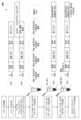
도 1은 비제한적인 예시적 바코드(예를 들어, 확률적 바코드)를 예시한 것이다.
도 2는 바코딩 및 디지털 계수(예를 들어, 확률적 바코딩 및 디지털 계수)의 비제한적인 예시적 작업흐름을 보여준다.
도 3은 복수의 표적으로부터 바코딩된 표적(예를 들어, 확률적으로 바코딩된 표적)의 인덱싱된 라이브러리를 생성하기 위한 비제한적인 예시적 과정을 보여주는 개략도이다.
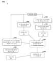
도 4는 세포를 신호 세포 표지 또는 노이즈 세포 표지로서 확인하는 비제한적인 예시적 방법을 보여주는 흐름도이다.
도 5는 세포를 신호 세포 표지 또는 노이즈 세포 표지로서 확인하는 또 다른 비제한적인 예시적 방법을 보여주는 흐름도이다.
도 6a는 노이즈 세포로부터 실제 세포와 연관된 표지를 구별하기 위한 비제한적인 예시적 방법을 보여주는 흐름도이다. 도 6b는 노이즈 세포로부터 실제 세포와 연관된 표지를 구별하기 위한 또 다른 비제한적인 예시적 방법을 보여주는 흐름도이다.
도 7은 가장 가변적인 유전자의 확인을 보여주는 비제한적인 예시적 개략도이다. 노이즈 세포로부터 실제 세포와 연관된 표지를 구별하기 위한 방법(예를 들어, 실시예 4에 예시된 도 6a를 참조하여 기재된 방법(600a))은 가장 가변적인 유전자의 확인을 포함할 수 있다.
도 8a 및 도 8b는 각각의 유전자에 대하여 연관된 별개의 서열을 갖는 분자 표지의 수의 가장 큰 소실을 갖는 유전자의 확인을 보여주는 비제한적인 예시적 플롯이다. 노이즈 세포로부터 실제 세포와 연관된 표지를 구별하기 위한 방법(예를 들어, 실시예 4에 예시된 도 6a를 참조하여 기재된 방법(600a))은 각각의 유전자에 있어서 별개의 서열과 연관된 분자 표지의 수의 가장 큰 소실을 갖는 유전자의 확인을 포함할 수 있다.
도 9는 본 발명의 방법을 구현하도록 구성된 예시적인 컴퓨팅 시스템의 블록 다이어그램이다.
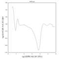
도 10은 비제한적인 예시적 누적합 플롯을 보여준다.
도 11은 도 10의 누적합 플롯의 비제한적인 이차 도함수 플롯을 보여준다.
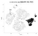
도 12는 신호 또는 노이즈 세포 표지의 비제한적인 tSNE 플롯을 보여준다.
도 13a 및 도 13b는 3가지의 별개의 유방암 세포주 및 공여자 단리된 PBMC를 갖는 BD™ 유방암 유전자 패널을 사용하여 처리된 샘플에 있어서 도 4(도 13a)를 참조하여 예시된 방법(400) 및 도 6a(도 13b)를 참조하여 예시된 방법(600a)에 의해 확인된 세포의 비교를 보여주는 비제한적인 예시적 플롯이다. 도 13a 및 도 13b 둘 모두에서 청색으로 표지된 점은 두 방법 모두에 의해 검출된 공통 세포이다. 도 13a에 적색으로 표지된 점은 방법(600a)에 의해 노이즈로 확인된 세포이다. 도 13b에 적색으로 표지된 점은 방법(600a)에 의해 확인된 추가의 실제 세포이다.
도 14a는 방법(600a)에 의해 확인된 세포를 보여주는 비제한적인 예시적 플롯이며, 적색으로 표지된 세포는 (도 4를 참조로 예시된 방법(400)에 의해 확인된 세포에 비하여) 확인된 추가의 세포이다. 세포를 PBMC, 예컨대 B 세포(도 14b), NK 세포(도 14c) 및 T 세포(도 14d)의 발현에 의해 채색한다. 도 14b 내지 도 14d는 방법(600a)에 의해 확인된 추가의 세포가 사실상 실제 세포인 것을 보여준다.
도 15a 및 도 15b는 건강한 공여자 단리된 PBMC를 갖는 BD™ 혈액 유전자 패널을 사용하여 처리된 샘플에 있어서 도 4(도 15a)를 참조로 예시된 방법(400) 및 도 6a(도 15b)를 참조로 예시된 방법(600a)에 의해 확인된 세포의 비교를 보여주는 비제한적인 예시적 플롯이다. 도 15a 및 도 15b 둘 모두에서 청색으로 표지된 점은 두 방법 모두에 의해 검출되는 공통 세포이다. 도 15a에 적색으로 표지된 점은 방법(600a)에 의해 노이즈로 확인된 세포이다. 도 15b에 적색으로 표지된 점은 방법(600a)에 의해 확인되는 추가의 세포이다.
도 16a 및 도 16b는 방법(400)에 의해 확인되는 세포를 보여주는 비제한적인 예시적 플롯이다. 도 16a에서, 적색으로 표지된 세포는 방법(600a)에 의해 노이즈로 확인된 세포이다. 도 16b에서, 세포는 단핵구 마커 유전자, 예컨대 CD14 및 S100A6의 군의 발현에 의해 채색된다. 개선된 알고리즘에 의해 확인된 "노이즈" 세포는 주로 단핵구의 낮은 발현자이다.
도 17a는 방법(600a)에 의해 확인되는 세포를 보여주는 비제한적인 예시적 플롯이며, 표지된 세포는 확인된 추가의 세포이다. 세포는 T 세포의 발현(도 17b), 중요한 유전자 LAT(도 17c) 및 IL7R(도 17d)의 발현에 의해 채색된다.Figure 1 illustrates a non-limiting exemplary barcode (e.g., a probabilistic barcode).
Figure 2 shows a non-limiting exemplary workflow for barcoding and digital counting (e.g., probabilistic barcoding and digital counting).
Figure 3 is a schematic diagram showing a non-limiting exemplary process for generating an indexed library of barcoded targets (e.g., probabilistically barcoded targets) from multiple targets.
Figure 4 is a flow chart showing a non-limiting exemplary method for identifying a cell as a signal cell marker or a noise cell marker.
Figure 5 is a flow chart showing another non-limiting exemplary method for identifying cells as signal cell markers or noise cell markers.
Figure 6a is a flow chart showing a non-limiting exemplary method for distinguishing labels associated with real cells from noise cells. Figure 6b is a flow chart showing another non-limiting exemplary method for distinguishing labels associated with real cells from noise cells.
Figure 7 is a non-limiting exemplary schematic diagram illustrating identification of the most variable gene. A method for distinguishing a marker associated with a real cell from a noise cell (e.g., method (600a) described with reference to Figure 6a as exemplified in Example 4) can include identification of the most variable gene.
Figures 8a and 8b are non-limiting exemplary plots showing, for each gene, the identification of the gene having the greatest loss of a number of molecular signatures associated with a distinct sequence. A method for distinguishing a signature associated with a live cell from a noise cell (e.g., method (600a) described with reference to Figure 6a as exemplified in Example 4) can include identifying, for each gene, the identification of the gene having the greatest loss of a number of molecular signatures associated with a distinct sequence.
FIG. 9 is a block diagram of an exemplary computing system configured to implement the method of the present invention.
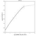
Figure 10 shows a non-limiting exemplary cumulative sum plot.
Figure 11 shows an unrestricted second derivative plot of the cumulative sum plot of Figure 10.
Figure 12 shows an unrestricted tSNE plot of signal or noise cell signatures.
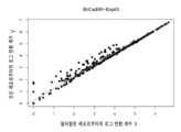
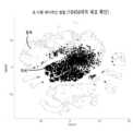
FIGS. 13A and 13B are non-limiting exemplary plots showing a comparison of cells identified by the method (400) illustrated with reference to FIG. 4 (FIG. 13A) and the method (600a) illustrated with reference to FIG. 6A (FIG. 13B) in samples processed using the BD™ Breast Cancer Gene Panel having three distinct breast cancer cell lines and donor isolated PBMCs. The dots colored in blue in both FIGS. 13A and 13B are common cells detected by both methods. The dots colored in red in FIG. 13A are cells identified as noise by the method (600a). The dots colored in red in FIG. 13B are additional actual cells identified by the method (600a).
FIG. 14a is a non-limiting exemplary plot showing cells identified by method (600a), where cells colored red are additional cells identified (compared to the cells identified by method (400) as exemplified in reference to FIG. 4). Cells are colored by expression of PBMCs, such as B cells ( FIG. 14b ), NK cells ( FIG. 14c ), and T cells ( FIG. 14d ). FIGS. 14b-14d demonstrate that the additional cells identified by method (600a) are in fact real cells.
FIGS. 15A and 15B are non-limiting exemplary plots showing a comparison of cells identified by the method (400) illustrated with reference to FIG. 4 (FIG. 15A) and the method (600a) illustrated with reference to FIG. 6A (FIG. 15B) in a sample processed using a BD™ blood genetic panel having isolated PBMCs from healthy donors. The dots colored in blue in both FIGS. 15A and 15B are common cells detected by both methods. The dots colored in red in FIG. 15A are cells identified as noise by the method (600a). The dots colored in red in FIG. 15B are additional cells identified by the method (600a).
Figures 16a and 16b are non-limiting exemplary plots showing cells identified by the method (400). In Figure 16a, cells labeled in red are cells identified as noise by the method (600a). In Figure 16b, cells are colored by expression of a group of monocyte marker genes, such as CD14 and S100A6. The “noise” cells identified by the improved algorithm are primarily low expressers of monocytes.
Figure 17a is a non-limiting exemplary plot showing cells identified by method (600a), with the labeled cells being additional cells identified. The cells are colored by expression of T cells (Figure 17b), key genes LAT (Figure 17c) and IL7R (Figure 17d).
하기의 상세한 설명에서, 본 명세서의 일부를 형성하는 첨부된 도면을 참조한다. 도면에서, 문맥상 달리 지시하지 않는 한, 유사한 기호는 전형적으로 유사한 구성요소를 나타낸다. 상세한 설명, 도면, 및 청구범위에 기재된 예시적인 실시형태는 한정하고자 하는 것이 아니다. 본 명세서에 제시된 주제의 사상 또는 범주를 벗어나지 않고 다른 실시형태가 이용될 수 있으며 다른 변화가 이루어질 수 있다. 본 명세서에 일반적으로 기재되고 도면에 도시된 바와 같은 본 발명의 양태가 매우 다양한 여러 가지 구성으로 배열되고, 치환되고, 조합되고, 분리되고, 설계될 수 있고, 이들 모두는 명백하게 본 명세서에서 고려되며, 본 명세서의 일부를 이룬다는 것이 용이하게 이해될 것이다.In the following detailed description, reference is made to the accompanying drawings which form a part hereof. In the drawings, similar symbols typically represent similar elements, unless the context dictates otherwise. The exemplary embodiments described in the detailed description, drawings, and claims are not intended to be limiting. Other embodiments may be utilized and other changes may be made without departing from the spirit or scope of the subject matter presented herein. It will be readily understood that the embodiments of the present invention, as generally described herein and illustrated in the drawings, may be arranged, substituted, combined, separated, and designed in a wide variety of configurations, all of which are expressly contemplated by and form a part of this specification.
모든 특허, 공개된 특허 출원, 다른 간행물 및 진뱅크(GenBank)로부터의 서열 및 본원에 언급된 다른 데이터베이스는 관련 기술에 관하여 그들 전체가 참조로 포함된다.All patents, published patent applications, other publications, and sequences from GenBank and other databases mentioned herein are incorporated by reference in their entirety with respect to the relevant art.
소량의 핵산 또는 표적, 예를 들어, 메신저 리보핵산(mRNA) 분자의 정량화는 예를 들어, 상이한 발생 단계에 또는 상이한 환경 조건 하에 세포 내에서 발현되는 유전자를 결정하는데 임상적으로 중요하다. 그러나, 특히 분자의 수가 매우 적은 경우, 핵산 분자(예를 들어, mRNA 분자)의 절대 개수를 결정하는 것은 매우 어려울 수 있다. 샘플 내의 분자의 절대 개수를 결정하기 위한 하나의 방법은 디지털 중합효소 연쇄 반응(PCR)이다. 이상적으로, PCR은 각 사이클에서 동일한 카피의 분자를 생성한다. 그러나, PCR은 각각의 분자가 확률적 가능성으로 복제되며, 이러한 가능성이 PCR 사이클 및 유전자 서열에 의해 달라져서, 증폭 편향 및 부정확한 유전자 발현 측정이 초래되는 단점을 가질 수 있다.Quantification of small amounts of nucleic acid or target, e.g., messenger ribonucleic acid (mRNA) molecules, is clinically important, for example, in determining which genes are expressed in cells at different stages of development or under different environmental conditions. However, determining the absolute number of nucleic acid molecules (e.g., mRNA molecules) can be very difficult, especially when the number of molecules is very small. One method for determining the absolute number of molecules in a sample is digital polymerase chain reaction (PCR). Ideally, PCR produces identical copies of the molecule in each cycle. However, PCR can have the disadvantage that each molecule is replicated with a probabilistic probability, which varies depending on the PCR cycle and the gene sequence, resulting in amplification bias and inaccurate measurements of gene expression.
독특한 분자 표지(ML, 분자 인덱스(MI)로도 지칭됨)를 갖는 바코드(예를 들어, 확률적 바코드)를 사용하여 분자의 수를 계수할 수 있다. 각 세포 표지에 대하여 독특한 분자 표지를 갖는 바코드를 사용하여 각 세포 내의 분자의 수를 계수할 수 있다. 비제한적인 예시적 바코딩 검정은 프리사이스(Precise)™ 검정(셀룰러 리서치, 인크.(Cellular Research, Inc.)(미국 캘리포니아주 팔로 알토 소재)), 리졸브(Resolve)™ 검정(셀룰러 리서치, 인크.(미국 캘리포니아주 팔로 알토 소재)) 또는 랩소디(Rhapsody)™ 검정(셀룰러 리서치, 인크.(미국 캘리포니아주 팔로 알토 소재))을 포함한다. 그러나, 이들 방법 및 기법은 오류를 도입할 수 있으며, 정정되지 않으면, 세포 계수의 과대추정을 초래할 수 있다.Molecules can be counted using barcodes (e.g., probabilistic barcodes) having unique molecular signatures (MLs, also referred to as molecular indices (MIs)). Barcodes having unique molecular signatures for each cell signature can be used to count the number of molecules in each cell. Non-limiting exemplary barcoding assays include the Precise™ assay (Cellular Research, Inc., Palo Alto, CA), the Resolve™ assay (Cellular Research, Inc., Palo Alto, CA), or the Rhapsody™ assay (Cellular Research, Inc., Palo Alto, CA). However, these methods and techniques can introduce errors that, if not corrected, can lead to overestimation of cell counts.
랩소디™ 검정은 RT 단계 동안 샘플 내의 모든 폴리(A)-mRNA에 혼성화하기 위하여 다수의, 예를 들어, 6561 내지 65536개의 폴리(T) 올리고뉴클레오티드 상의 독특한 분자 표지가 있는 바코드(예를 들어, 확률적 바코드)의 비-소모성 풀을 사용할 수 있다. 분자 표지에 더하여, 바코드의 세포 표지를 사용하여 마이크로웰 플레이트의 각 웰 내의 각각의 단일 세포를 확인할 수 있다. 바코드는 범용 PCR 프라이밍 부위를 포함할 수 있다. RT 동안 표적 유전자 분자는 무작위로 바코드와 반응한다. 각각의 표적 분자는 바코드(예를 들어, 확률적 바코드)에 혼성화하여, 바코딩된 상보적 리보핵산(cDNA) 분자(예를 들어, 확률적으로 바코딩된 cDNA 분자)의 생성을 초래할 수 있다. 표지 후에, 마이크로웰 플레이트의 마이크로웰로부터 바코딩된 cDNA 분자를 PCR 증폭 및 시퀀싱을 위하여 단일의 튜브 내로 풀링할 수 있다. 미가공 시퀀싱 데이터를 분석하여, 독특한 분자 표지를 갖는 바코드의 수를 생성할 수 있다.The Rhapsody™ assay can use a non-exhaustive pool of barcodes (e.g., stochastic barcodes) with unique molecular markers on a plurality of, for example, 6561 to 65536, poly(T) oligonucleotides to hybridize to all poly(A)-mRNA in the sample during the RT step. In addition to the molecular markers, the cell markers of the barcodes can be used to identify each single cell in each well of the microwell plate. The barcodes can include universal PCR priming sites. During RT, target gene molecules react randomly with the barcodes. Each target molecule can hybridize to the barcodes (e.g., stochastic barcodes), resulting in the generation of barcoded complementary ribonucleic acid (cDNA) molecules (e.g., stochastically barcoded cDNA molecules). After labeling, the barcoded cDNA molecules from the microwells of the microwell plate can be pooled into a single tube for PCR amplification and sequencing. By analyzing raw sequencing data, a number of barcodes with unique molecular signatures can be generated.
신호 세포 표지의 확인을 위한 방법 및 시스템이 본원에 개시된다. 일부 실시형태에서, 상기 방법은 (a) 복수의 바코드(예를 들어, 확률적 바코드)를 사용하여 세포의 샘플 내의 복수의 표적을 바코딩(예를 들어, 확률적으로 바코딩)하여, 복수의 바코딩된 표적(예를 들어, 확률적으로 바코딩된 표적)을 생성하는 단계로서, 복수의 바코드의 각각이 세포 표지 및 분자 표지를 포함하는 단계; (b) 복수의 바코딩된 표적의 시퀀싱 데이터를 수득하는 단계; (c) 복수의 바코드의 세포 표지의 각각과 연관된 별개의 서열을 갖는 분자 표지의 수를 결정하는 단계; (d) 세포 표지의 각각과 연관된 별개의 서열을 갖는 분자 표지의 수에 기초하여, 복수의 바코드의 세포 표지의 각각의 순위를 결정하는 단계; (e) (c)에서 결정된 세포 표지의 각각과 연관된 별개의 서열을 갖는 분자 표지의 수 및 (d)에서 결정된 세포 표지의 각각의 순위에 기초하여, 누적합 플롯을 생성하는 단계; (f) 누적합 플롯의 이차 도함수 플롯을 생성하는 단계; (g) 누적합 플롯의 이차 도함수 플롯의 최소값을 결정하는 단계로서, 이차 도함수 플롯의 최소값이 세포 표지 임계값에 상응하는 단계; 및 (h) (c)에서 결정된 세포 표지와 연관된 별개의 서열을 갖는 분자 표지의 수 및 세포 표지 임계값에 기초하여 세포 표지를 신호 세포 표지 또는 노이즈 세포 표지로서 확인하는 단계를 포함한다.Methods and systems for identifying signal cell signatures are disclosed herein. In some embodiments, the methods comprise: (a) barcoding (e.g., probabilistically barcoding) a plurality of targets in a sample of cells using a plurality of barcodes (e.g., probabilistic barcodes), thereby generating a plurality of barcoded targets (e.g., probabilistically barcoded targets), wherein each of the plurality of barcodes comprises a cell signature and a molecular signature; (b) obtaining sequencing data of the plurality of barcoded targets; (c) determining a number of molecular signatures having a distinct sequence associated with each of the cell signatures of the plurality of barcodes; (d) determining a rank of each of the cell signatures of the plurality of barcodes based on the number of molecular signatures having a distinct sequence associated with each of the cell signatures; (e) generating a cumulative sum plot based on the number of molecular signatures having a distinct sequence associated with each of the cell signatures determined in (c) and the rank of each of the cell signatures determined in (d); (f) generating a second derivative plot of the cumulative sum plot; (g) a step of determining a minimum of a second derivative plot of a cumulative sum plot, wherein the minimum of the second derivative plot corresponds to a cell signature threshold; and (h) a step of identifying a cell signature as a signal cell signature or a noise cell signature based on the number of molecular signatures having a distinct sequence associated with the cell signature determined in (c) and the cell signature threshold.
일부 실시형태에서, 상기 방법은 (a) 복수의 바코딩된 표적(예를 들어, 확률적으로 바코딩된 표적)의 시퀀싱 데이터를 수득하는 단계로서, 복수의 바코딩된 표적의 시퀀싱 데이터가 복수의 바코드(예를 들어, 확률적 바코드)를 사용하여 바코딩된(예를 들어, 확률적으로 바코딩된) 세포의 샘플 내의 복수의 표적으로부터 유래되어, 복수의 바코딩된 표적(예를 들어, 확률적으로 바코딩된 표적)을 생성하고, 복수의 바코드의 각각이 세포 표지 및 분자 표지를 포함하는 단계; (b) 세포 표지의 각각과 연관된 별개의 서열을 갖는 분자 표지의 수에 기초하여 복수의 바코드의 세포 표지의 각각의 순위를 결정하는 단계; (c) 누적합 플롯의 이차 도함수 플롯의 최소값을 결정하는 단계로서, 누적합 플롯이 세포 표지의 각각과 연관된 별개의 서열을 갖는 분자 표지의 수 및 (b)에서 결정된 세포 표지의 각각의 순위에 기초하고, 이차 도함수 플롯의 최소값이 세포 표지 임계값에 상응하는 단계; 및 (d) 세표 표지와 연관된 별개의 서열을 갖는 분자 표지의 수 및 세포 표지 임계값에 기초하여 세포 표지를 신호 세포 표지(세포와 연관) 또는 노이즈 세포 표지(세포와 연관되지 않음)로서 확인하는 단계를 포함한다.In some embodiments, the method comprises the steps of: (a) obtaining sequence data of a plurality of barcoded targets (e.g., probabilistically barcoded targets), wherein the sequence data of the plurality of barcoded targets are derived from a plurality of targets in a sample of cells that have been barcoded (e.g., probabilistically barcoded) using the plurality of barcodes (e.g., probabilistically barcoded) to generate the plurality of barcoded targets (e.g., probabilistically barcoded targets), wherein each of the plurality of barcodes comprises a cell signature and a molecular signature; (b) determining a rank of each of the cell signatures of the plurality of barcodes based on a number of molecular signatures having a distinct sequence associated with each of the cell signatures; (c) determining a minimum of a second derivative plot of a cumulative sum plot, wherein the cumulative sum plot is based on the number of molecular signatures having a distinct sequence associated with each of the cell signatures and the rank of each of the cell signatures determined in (b), and wherein the minimum of the second derivative plot corresponds to a cell signature threshold; and (d) identifying the cell signature as a signal cell signature (associated with the cell) or a noise cell signature (not associated with the cell) based on the number of molecular signatures having distinct sequences associated with the cell signature and a cell signature threshold value.
신호 세포 표지의 확인 방법이 본원에 개시된다. 일부 실시형태에서, 상기 방법은 (a) 복수의 바코드(예를 들어, 확률적 바코드)를 사용하여 세포의 샘플 내의 복수의 표적을 바코딩(예를 들어, 확률적으로 바코딩)하여, 복수의 바코딩된 표적(예를 들어, 확률적으로 바코딩된 표적)을 생성하는 단계로서, 복수의 바코드의 각각이 세포 표지 및 분자 표지를 포함하며, 상이한 세포의 표적으로부터 생성된 바코딩된 표적이 상이한 세포 표지를 가지며, 복수의 세포 중 하나의 세포의 표적으로부터 생성된 바코딩된 표적이 상이한 분자 표지를 갖는 단계; (b) 바코딩된 표적의 시퀀싱 데이터를 수득하는 단계; (c) 세포 표지의 특징 벡터를 결정하는 단계로서, 특징 벡터가 세포 표지와 연관된 별개의 서열을 갖는 분자 표지의 수를 포함하는 단계; (d) 특징 벡터에 기초하여 세포 표지에 대한 클러스터를 결정하는 단계; 및 (e) 클러스터 내의 세포의 수 및 클러스터 크기 임계값에 기초하여 세포 표지를 신호 세포 표지 또는 노이즈 세포 표지로서 확인하는 단계를 포함한다.Disclosed herein are methods for identifying a signal cell signature. In some embodiments, the method comprises: (a) barcoding (e.g., probabilistically barcoding) a plurality of targets in a sample of cells using a plurality of barcodes (e.g., probabilistic barcodes) to generate a plurality of barcoded targets (e.g., probabilistically barcoded targets), wherein each of the plurality of barcodes comprises a cell signature and a molecular signature, wherein barcoded targets generated from targets of different cells have different cell signatures, and wherein barcoded targets generated from targets of one cell of the plurality of cells have different molecular signatures; (b) obtaining sequencing data of the barcoded targets; (c) determining a feature vector of the cell signature, wherein the feature vector comprises a number of molecular signatures having distinct sequences associated with the cell signature; (d) determining a cluster for the cell signature based on the feature vector; and (e) identifying the cell signature as a signal cell signature or a noise cell signature based on the number of cells in the cluster and a cluster size threshold.
신호 세포 표지의 확인을 위한 시스템이 본원에 개시된다. 일부 실시형태에서, 시스템은 하드웨어 프로세서; 및 명령어가 저장된 비-일시적 메모리로서, 하드웨어 프로세서에 의해 실행되는 경우 프로세서가 본원에 개시된 임의의 방법을 수행하게 하는 비-일시적 메모리를 포함한다. 신호 세포 표지의 확인을 위한 컴퓨터 판독 가능한 매체가 본원에 개시된다. 일부 실시형태에서, 컴퓨터 판독 가능한 매체는 본원에 개시된 방법 중 임의의 것을 수행하기 위한 코드를 포함한다.A system for identifying a signal cell signature is disclosed herein. In some embodiments, the system comprises a hardware processor; and a non-transitory memory having instructions stored thereon, the non-transitory memory causing the processor to perform any of the methods disclosed herein. A computer-readable medium for identifying a signal cell signature is disclosed herein. In some embodiments, the computer-readable medium comprises code for performing any of the methods disclosed herein.
정의definition
달리 정의되지 않는 한, 본원에 사용된 모든 기술 및 과학 용어는 본 발명이 속하는 분야의 숙련자에 의해 일반적으로 이해되는 것과 동일한 의미를 갖는다. 예를 들어, 문헌[Singleton et al., Dictionary of Microbiology and Molecular Biology 2nd ed., J. Wiley & Sons (New York, NY 1994)]; 문헌[Sambrook et al., Molecular Cloning, A Laboratory Manual, Cold Springs Harbor Press (Cold Springs Harbor, NY 1989)]을 참조한다. 본 발명의 목적을 위하여, 하기의 용어가 하기에 정의된다.Unless defined otherwise, all technical and scientific terms used herein have the same meaning as commonly understood by one of ordinary skill in the art to which this invention belongs. See, e.g., Singleton et al., Dictionary of Microbiology and Molecular Biology 2nd ed., J. Wiley & Sons (New York, NY 1994); Sambrook et al., Molecular Cloning, A Laboratory Manual, Cold Springs Harbor Press (Cold Springs Harbor, NY 1989). For the purposes of this invention, the following terms are defined below.
본원에 사용되는 용어 "어댑터"는 연관된 핵산의 증폭 또는 시퀀싱을 용이하게 하기 위한 서열을 의미할 수 있다. 연관된 핵산은 표적 핵산을 포함할 수 있다. 연관된 핵산은 공간 표지, 표적 표지, 샘플 표지, 인덱싱 표지, 바코드, 확률적 바코드 또는 분자 표지 중 하나 이상을 포함할 수 있다. 어댑터는 선형일 수 있다. 어댑터는 사전-아데닐화된 어댑터일 수 있다. 어댑터는 이중- 또는 단일-가닥일 수 있다. 하나 이상의 어댑터는 핵산의 5' 또는 3' 말단에 위치할 수 있다. 어댑터가 5' 및 3' 말단에 공지된 서열을 포함하는 경우, 공지된 서열은 동일한 또는 상이한 서열일 수 있다. 폴리뉴클레오티드의 5' 및/또는 3' 말단에 위치한 어댑터는 표면 상에 고정된 하나 이상의 올리고뉴클레오티드에 혼성화할 수 있다. 어댑터는 일부 실시형태에서, 범용 서열을 포함할 수 있다. 범용 서열은 둘 이상의 핵산 분자에 공통인 뉴클레오티드 서열의 영역일 수 있다. 둘 이상의 핵산 분자는 상이한 서열의 영역을 가질 수 있다. 따라서, 예를 들어, 5' 어댑터는 동일한 및/또는 범용 핵산 서열을 포함할 수 있으며, 3' 어댑터는 동일한 및/또는 범용 서열을 포함할 수 있다. 복수의 핵산 분자의 상이한 구성원에 존재할 수 있는 범용 서열은 범용 서열에 상보적인 단일의 범용 프라이머를 사용하여 다수의 상이한 서열의 복제 또는 증폭을 가능하게 할 수 있다. 유사하게, 핵산 분자의 집합물의 상이한 구성원에 존재할 수 있는 적어도 1, 2개(예를 들어, 한 쌍) 이상의 범용 서열은 범용 서열에 상보적인 적어도 1, 2개(예를 들어, 한쌍) 이상의 단일의 범용 프라이머를 사용하여 다수의 상이한 서열의 복제 또는 증폭을 가능하게 할 수 있다. 따라서, 범용 프라이머는 이러한 범용 서열에 혼성화할 수 있는 서열을 포함한다. 표적 핵산 서열-함유 분자는 범용 어댑터(예를 들어, 비-표적 핵산 서열)를 상이한 표적 핵산 서열 중 하나의 또는 양 말단에 부착시키도록 변형될 수 있다. 표적 핵산에 부착되는 하나 이상의 범용 프라이머는 범용 프라이머의 혼성화를 위한 부위를 제공할 수 있다. 표적 핵산에 부착되는 하나 이상의 범용 프라이머는 서로 동일하거나 상이할 수 있다.The term "adapter" as used herein may mean a sequence that facilitates amplification or sequencing of an associated nucleic acid. The associated nucleic acid may comprise a target nucleic acid. The associated nucleic acid may comprise one or more of a spatial marker, a target marker, a sample marker, an indexing marker, a barcode, a probabilistic barcode, or a molecular marker. The adapter may be linear. The adapter may be a pre-adenylated adapter. The adapter may be double- or single-stranded. One or more adapters may be located at the 5' or 3' end of the nucleic acid. When the adapter comprises a known sequence at the 5' and 3' ends, the known sequences may be the same or different sequences. The adapters located at the 5' and/or 3' ends of the polynucleotide may hybridize to one or more oligonucleotides immobilized on the surface. The adapter may, in some embodiments, comprise a universal sequence. The universal sequence may be a region of nucleotide sequence that is common to two or more nucleic acid molecules. The two or more nucleic acid molecules may have regions of different sequences. Thus, for example, the 5' adapter can comprise the same and/or universal nucleic acid sequence, and the 3' adapter can comprise the same and/or universal sequence. A universal sequence that can be present on different members of a plurality of nucleic acid molecules can enable replication or amplification of a plurality of different sequences using a single universal primer that is complementary to the universal sequence. Similarly, at least one, two (e.g., a pair) or more universal sequences that can be present on different members of a collection of nucleic acid molecules can enable replication or amplification of a plurality of different sequences using at least one, two (e.g., a pair) or more single universal primers that are complementary to the universal sequences. Thus, a universal primer comprises a sequence that can hybridize to such a universal sequence. A target nucleic acid sequence-containing molecule can be modified to attach a universal adapter (e.g., a non-target nucleic acid sequence) to one or both ends of the different target nucleic acid sequences. One or more of the universal primers that are attached to the target nucleic acids can provide a site for hybridization of the universal primer. The one or more universal primers attached to the target nucleic acid may be identical or different.
본 명세서에 사용되는 바와 같이, 용어 "연관된" 또는 "~와 연관된"은 2개 이상의 종이 소정의 시점에 동시-위치하는 것으로 확인될 수 있음을 의미할 수 있다. 연관은 2개 이상의 종이 유사한 용기 내에 존재하거나, 존재하였음을 의미할 수 있다. 연관은, 예를 들어 2개 이상의 종에 관한 디지털 정보가 저장되어 있으며, 1개 이상의 종이 소정의 시점에 동시-위치한 것을 결정하기 위해 사용될 수 있는 정보과학 연관일 수 있다. 연관은 또한 물리적 연관일 수 있다. 일부 실시형태에서, 2개 이상의 연관된 종은 서로 또는 공통의 고체 또는 반고체 표면에 "테더링되거나", "부착되거나", "고정된다". 연관은 비드와 같은 고체 또는 반고체 지지체에 표지를 부착하기 위한 공유 또는 비공유 수단을 지칭할 수 있다. 연관은 표적과 표지 사이의 공유 결합일 수 있다.As used herein, the terms "associated" or "associated with" can mean that two or more species can be identified as being co-located at a given point in time. Association can mean that two or more species are or were present in a similar container. Association can be an informatics association, for example, where digital information about two or more species is stored and can be used to determine that one or more species were co-located at a given point in time. Association can also be a physical association. In some embodiments, two or more associated species are "tethered," "attached," or "immobilized" to each other or to a common solid or semi-solid surface. Association can refer to a covalent or non-covalent means for attaching a label to a solid or semi-solid support, such as a bead. Association can be a covalent bond between a target and a label.
본 명세서에 사용되는 바와 같이, 용어 "상보성"은 2개의 뉴클레오티드 사이의 정확한 쌍형성을 위한 능력을 지칭할 수 있다. 예를 들어, 핵산의 주어진 위치의 뉴클레오티드가 또 다른 핵산의 뉴클레오티드와 수소 결합할 수 있는 경우, 2개의 핵산은 그 위치에서 서로 상보성인 것으로 간주된다. 2개의 단일 가닥 핵산 분자 사이의 상보성은, 단지 일부의 뉴클레오티드만이 결합하는 경우 "부분적"일 수 있거나, 단일 가닥 분자 사이에 완전한 상보성이 존재하는 경우, 완전할 수 있다. 제1 뉴클레오티드 서열이 제2 뉴클레오티드 서열에 상보성인 경우, 제1 뉴클레오티드 서열은 제2 서열의 "상보체"라고 할 수 있다. 제1 뉴클레오티드 서열이 제2 서열의 역방향(즉, 뉴클레오티드의 순서가 역방향인) 서열에 상보성인 경우, 제1 뉴클레오티드 서열은 제2 서열의 "역 상보체"라고 할 수 있다. 본 명세서에 사용되는 바와 같이, 용어 "상보체", "상보성" 및 "역 상보체"는 상호 교환 가능하게 사용될 수 있다. 분자가 또 다른 분자와 혼성화할 수 있는 경우, 이는 혼성화되는 분자의 상보체일 수 있는 것이 본 발명으로부터 이해된다.As used herein, the term "complementarity" may refer to the ability for exact pairing between two nucleotides. For example, if a nucleotide at a given position in a nucleic acid is capable of hydrogen bonding with a nucleotide in another nucleic acid, the two nucleic acids are considered to be complementary to each other at that position. Complementarity between two single-stranded nucleic acid molecules may be "partial," where only some of the nucleotides bind, or complete, where complete complementarity exists between the single-stranded molecules. When a first nucleotide sequence is complementary to a second nucleotide sequence, the first nucleotide sequence may be said to be the "complement" of the second sequence. When a first nucleotide sequence is complementary to the reverse (i.e., reverse order of nucleotides) sequence of the second sequence, the first nucleotide sequence may be said to be the "reverse complement" of the second sequence. As used herein, the terms "complement," "complementarity," and "reverse complement" may be used interchangeably. It is understood from the invention that where a molecule is capable of hybridizing with another molecule, that molecule may be the complement of the molecule being hybridized.
본 명세서에 사용되는 바와 같이, 용어 "디지털 계수"는 샘플에서 표적 분자의 수를 추정하는 방법을 지칭할 수 있다. 디지털 계수는 샘플에서 표적과 연관된 고유 표지의 수를 결정하는 단계를 포함할 수 있다. 이러한 확률적 방법론은, 분자 계수 문제를 동일한 분자의 위치지정 및 확인 중 하나로부터 소정의 표지 세트의 검출에 관한 일련의 예/아니오 디지털 질문으로 변환한다.As used herein, the term "digital counting" may refer to a method of estimating the number of target molecules in a sample. Digital counting may include determining the number of unique markers associated with a target in a sample. This probabilistic methodology transforms the molecule counting problem into a series of yes/no digital questions regarding the detection of a given set of markers from one of the location and identification of identical molecules.
본 명세서에 사용되는 바와 같이, 용어 "표지" 또는 "표지들"은 샘플 내의 표적과 연관된 핵산 코드를 지칭할 수 있다. 표지는, 예를 들어, 핵산 표지일 수 있다. 표지는 전체적으로 또는 부분적으로 증폭 가능한 표지일 수 있다. 표지는 전적으로 또는 부분적으로 시퀀싱 가능한 표지일 수 있다. 표지는 별개의 것으로 확인 가능한 고유 핵산의 일부일 수 있다. 표지는 공지 서열일 수 있다. 표지는 핵산 서열의 접합부, 예를 들어 고유 및 비-고유 서열의 접합부를 포함할 수 있다. 본 명세서에 사용되는 바와 같이, 용어 "표지"는 용어 "인덱스", "태그" 또는 "표지-태그"와 상호 교환 가능하게 사용할 수 있다. 표지는 정보를 전달할 수 있다. 예를 들어, 다양한 실시형태에서, 표지는 샘플의 정체성, 샘플의 공급원, 세포의 정체성 및/또는 표적을 결정하기 위해 사용될 수 있다.As used herein, the term "label" or "labels" can refer to a nucleic acid code associated with a target in a sample. The label can be, for example, a nucleic acid label. The label can be a fully or partially amplifiable label. The label can be a fully or partially sequenceable label. The label can be a portion of a unique nucleic acid that is identifiable as being distinct. The label can be a known sequence. The label can comprise a junction of nucleic acid sequences, for example, a junction of a unique and non-unique sequence. As used herein, the term "label" can be used interchangeably with the terms "index," "tag," or "label-tag." A label can convey information. For example, in various embodiments, a label can be used to determine the identity of a sample, the source of a sample, the identity of a cell, and/or a target.
본 명세서에 사용되는 바와 같이, 용어 "비-소모성 저장소"는 많은 상이한 표지로 이루어진 확률적 바코드의 풀(pool)을 지칭할 수 있다. 비-소모성 저장소는 다수의 상이한 확률적 바코드를 포함하여, 비-소모성 저장소가 표적의 풀과 연관되는 경우, 각각의 표적은 독특한 확률적 바코드와 연관될 수 있다. 각각의 표지된 표적 분자의 독특성은 무작위 선택 통계에 의해 결정될 수 있으며, 표지의 다양성과 비교하여, 집합물 내의 동일한 표적 분자의 카피 수에 좌우된다. 표지된 표적 분자의 생성 세트의 크기는 바코드화 과정의 확률적 특성에 의해 결정될 수 있으며, 그 후, 검출된 확률적 바코드 수의 분석에 의해, 원래의 집합물 또는 샘플에 존재하는 표적 분자의 수를 계산할 수 있다. 존재하는 표적 분자의 카피 수 대 독특한 확률적 바코드 수의 비가 낮은 경우, 표지된 표적 분자는 매우 독특하다(즉, 주어진 표지로 하나 초과의 표적 분자가 표지될 확률이 매우 낮음).As used herein, the term "non-consumable reservoir" may refer to a pool of probabilistic barcodes comprised of many different markers. The non-consumable reservoir may contain a number of different probabilistic barcodes, such that when the non-consumable reservoir is associated with a pool of targets, each target may be associated with a unique probabilistic barcode. The uniqueness of each labeled target molecule may be determined by random selection statistics and depends on the number of copies of the same target molecule in the pool, relative to the diversity of the markers. The size of the generated set of labeled target molecules may be determined by the probabilistic nature of the barcoding process, and then the number of target molecules present in the original pool or sample may be calculated by analysis of the number of detected probabilistic barcodes. If the ratio of the number of copies of target molecules present to the number of unique probabilistic barcodes is low, the labeled target molecules are highly unique (i.e., the probability that more than one target molecule will be labeled with a given marker is very low).
본 명세서에 사용되는 바와 같이, 용어 "핵산"은 폴리뉴클레오티드 서열 또는 그의 단편을 지칭한다. 핵산은 뉴클레오티드를 포함할 수 있다. 핵산은 세포에 대해 외인성이거나 내인성일 수 있다. 핵산은 무 세포 환경에서 존재할 수 있다. 핵산은 유전자 또는 그의 단편일 수 있다. 핵산은 DNA일 수 있다. 핵산은 RNA일 수 있다. 핵산은 하나 이상의 유사체(예를 들어, 변경된 백본, 당 또는 핵염기)를 포함할 수 있다. 유사체의 일부 비제한적 예는 다음을 포함한다: 5-브로모우라실, 펩티드 핵산, 이종 핵산, 모르폴리노, 잠금 핵산(locked nucleic acid), 글리콜 핵산, 트레오스 핵산, 디데옥시뉴클레오티드, 코디세핀, 7-데자아-GTP, 형광단(예를 들어, 당에 연결된 로다민 또는 플루오레세인), 티올 함유 뉴클레오티드, 비오틴 연결 뉴클레오티드, 형광 염기 유사체, CpG 섬(island), 메틸-7-구아노신, 메틸화된 뉴클레오티드, 이노신, 티오우리딘, 슈도우리딘, 디하이드로우리딘, 케오신(queuosine) 및 와이오신(wyosine). "핵산", "폴리뉴클레오티드", "표적 폴리뉴클레오티드" 및 "표적 핵산"은 상호 교환 가능하게 사용될 수 있다.As used herein, the term "nucleic acid" refers to a polynucleotide sequence or a fragment thereof. A nucleic acid can comprise nucleotides. A nucleic acid can be exogenous or endogenous to a cell. A nucleic acid can exist in a cell-free environment. A nucleic acid can be a gene or a fragment thereof. A nucleic acid can be DNA. A nucleic acid can be RNA. A nucleic acid can include one or more analogues (e.g., a modified backbone, sugar, or nucleobase). Some non-limiting examples of analogues include: 5-bromouracil, peptide nucleic acids, heterologous nucleic acids, morpholinos, locked nucleic acids, glycol nucleic acids, threose nucleic acids, dideoxynucleotides, cordycepin, 7-deacetyl-GTP, fluorophores (e.g., rhodamine or fluorescein linked to a sugar), thiol-containing nucleotides, biotin-linked nucleotides, fluorescent base analogues, CpG islands, methyl-7-guanosine, methylated nucleotides, inosine, thiouridine, pseudouridine, dihydrouridine, queuosine, and wyosine. "Nucleic acid", "polynucleotide", "target polynucleotide" and "target nucleic acid" may be used interchangeably.
핵산은, 핵산에 새로운 또는 증진된 특징(예를 들어, 개선된 안정성)을 제공하는 하나 이상의 변형(예를 들어, 염기 변형, 백본 변형)을 포함할 수 있다. 핵산은 핵산 친화도 태그를 포함할 수 있다. 뉴클레오시드는 염기-당 조합일 수 있다. 뉴클레오시드의 염기 부분은 헤테로사이클릭 염기일 수 있다. 이러한 헤테로사이클릭 염기의 가장 흔한 2가지 부류는 퓨린 및 피리미딘이다. 뉴클레오티드는 뉴클레오시드의 당 부분에 공유적으로 연결된 포스페이트 기를 추가로 포함하는 뉴클레오시드일 수 있다. 펜토푸라노실 당을 포함하는 뉴클레오시드에 있어서, 포스페이트 기는 당의 2', 3' 또는 5' 하이드록실 모이어티에 연결될 수 있다. 핵산을 형성하는 경우, 포스페이트 기는 인접한 뉴클레오시드를 서로 공유적으로 연결시켜, 선형 중합체 화합물을 형성할 수 있다. 차례로, 이러한 선형 중합체 화합물의 각각의 말단을 추가로 연결하여, 원형 화합물을 형성할 수 있으나; 선형 화합물이 일반적으로 적합하다. 또한, 선형 화합물은 내부 뉴클레오티드 염기 상보성을 가질 수 있고, 따라서 완전한 또는 부분적인 이중 가닥 화합물을 생성하는 방식으로 폴딩될 수 있다. 핵산 내에서, 포스페이트 기는 통상적으로 핵산의 뉴클레오시드 간 백본을 형성하는 것으로 지칭될 수 있다. 연결 또는 백본은 3'에서 5' 포스포디에스테르 연결일 수 있다.A nucleic acid may include one or more modifications (e.g., base modifications, backbone modifications) that provide the nucleic acid with new or improved characteristics (e.g., improved stability). A nucleic acid may include a nucleic acid affinity tag. A nucleoside may be a base-sugar combination. The base portion of a nucleoside may be a heterocyclic base. The two most common classes of such heterocyclic bases are purines and pyrimidines. A nucleotide may be a nucleoside that additionally includes a phosphate group covalently linked to the sugar portion of the nucleoside. For nucleosides that include a pentofuranosyl sugar, the phosphate group may be linked to the 2', 3', or 5' hydroxyl moiety of the sugar. When forming a nucleic acid, the phosphate groups may covalently link adjacent nucleosides to each other to form a linear polymeric compound. In turn, the individual ends of such linear polymeric compounds may be further linked to form a circular compound; however, linear compounds are generally preferred. Additionally, linear compounds may have internal nucleotide base complementarity and thus may fold in a manner that produces a complete or partial double-stranded compound. Within nucleic acids, the phosphate groups may be commonly referred to as forming the internucleoside backbone of the nucleic acid. The linkage or backbone may be a 3' to 5' phosphodiester linkage.
핵산은 변형된 백본 및/또는 변형된 뉴클레오시드 간 연결을 포함할 수 있다. 변형된 백본은, 백본에 인 원자를 보유하는 것 및 백본에 인 원자를 갖지 않는 것을 포함할 수 있다. 내부에 인 원자를 함유하는 적합한 변형된 핵산 백본은, 예를 들어, 포스포로티오에이트, 키랄 포스포로티오에이트, 포스포로디티오에이트, 포스포트리에스테르, 아미노알킬 포스포트리에스테르, 메틸 및 다른 알킬 포스포네이트, 예컨대 3'-알킬렌 포스포네이트, 5'-알킬렌 포스포네이트, 키랄 포스포네이트, 포스피네이트, 3'-아미노 포스포르아미데이트 및 아미노알킬 포스포르아미데이트를 포함하는 포스포르아미데이트, 포스포로디아미데이트, 티오노포스포르아미데이트, 티오노알킬포스포네이트, 티오노알킬포스포트리에스테르, 셀레노포스페이트 및 일반적인 3'-5' 연결을 갖는 보라노포스페이트, 2'-5' 연결된 유사체, 반대 극성을 갖는 것을 포함할 수 있으며, 여기서, 하나 이상의 뉴클레오티드 간 연결은 3'에서 3', 5'에서 5' 또는 2'에서 2' 연결이다.The nucleic acids may comprise a modified backbone and/or modified internucleoside linkages. Modified backbones may include those having a phosphorus atom in the backbone and those without a phosphorus atom in the backbone. Suitable modified nucleic acid backbones containing a phosphorus atom therein may include, for example, phosphorothioates, chiral phosphorothioates, phosphorodithioates, phosphotriesters, aminoalkyl phosphotriesters, methyl and other alkyl phosphonates, such as 3'-alkylene phosphonates, 5'-alkylene phosphonates, chiral phosphonates, phosphinates, phosphoramidates including 3'-amino phosphoramidate and aminoalkyl phosphoramidate, phosphorodiamidates, thionophosphoramidates, thionoalkylphosphonates, thionoalkylphosphotriesters, selenophosphates and boranophosphates having a common 3'-5' linkage, 2'-5' linked analogues, those having the opposite polarity, wherein one or more of the internucleotide linkages are 3' to 3', 5' to 5' or It's a 2' to 2' connection.
핵산은 단쇄 알킬 또는 사이클로알킬 뉴클레오시드 간 연결, 혼합 헤테로원자 및 알킬 또는 사이클로알킬 뉴클레오시드 간 연결, 또는 하나 이상의 단쇄 헤테로원자 또는 헤테로사이클릭 뉴클레오시드 간 연결에 의해 형성된 폴리뉴클레오티드 백본을 포함할 수 있다. 이들은 모르폴리노 연결(뉴클레오시드의 당 부분으로부터 부분적으로 형성됨)을 갖는 것; 실록산 백본; 술피드, 술폭시드 및 술폰 백본; 포름아세틸 및 티오포름아세틸 백본; 메틸렌 포름아세틸 및 티오포름아세틸 백본; 리보아세틸 백본; 알켄 함유 백본; 술파메이트 백본; 메틸렌이미노 및 메틸렌하이드라지노 백본; 술포네이트 및 술폰아미드 백본; 아미드 백본; 및 N, O, S 및 CH2 성분 부분이 혼합된 다른 것을 포함할 수 있다.The nucleic acids can comprise polynucleotide backbones formed by short chain alkyl or cycloalkyl nucleoside linkages, mixed heteroatoms and alkyl or cycloalkyl nucleoside linkages, or one or more short chain heteroatom or heterocyclic nucleoside linkages. These can include those having morpholino linkages (formed in part from the sugar moiety of the nucleoside); siloxane backbones; sulfide, sulfoxide and sulfone backbones; formacetyl and thioformacetyl backbones; methylene foracetyl and thioformacetyl backbones; riboacetyl backbones; alkene containing backbones; sulfamate backbones; methyleneimino and methylenehydrazino backbones; sulfonate and sulfonamide backbones; amide backbones; and others having mixed N, O, S and CH2 moieties.
핵산은 핵산 모방체를 포함할 수 있다. 용어 "모방체"는, 단지 푸라노스 고리만이 또는 푸라노스 고리 및 뉴클레오티드 간 연결 둘 모두가 비-푸라노스 기로 대체된 폴리뉴클레오티드를 포함하는 것으로 의도될 수 있으며, 푸라노스 고리만의 대체는 당 대용물로 지칭될 수 있다. 헤테로사이클릭 염기 모이어티 또는 변형된 헤테로사이클릭 염기 모이어티는 적절한 표적 핵산과의 혼성화를 위해 유지될 수 있다. 이러한 핵산 중 하나는 펩티드 핵산(PNA)일 수 있다. PNA에서, 폴리뉴클레오티드의 당-백본은 아미드 함유 백본, 특히 아미노에틸글리신 백본으로 대체될 수 있다. 뉴클레오티드는 유지될 수 있으며, 백본의 아미드 부분의 아자 질소 원자에 직접적으로 또는 간접적으로 결합된다. PNA 화합물 내의 백본은 2개 이상의 연결된 아미노에틸글리신 단위를 포함할 수 있으며, 이는 PNA에 아미드 함유 백본을 제공한다. 헤테로사이클릭 염기 모이어티는 백본의 아미드 부분의 아자 질소 원자에 직접적으로 또는 간접적으로 결합될 수 있다.The nucleic acid may comprise a nucleic acid mimetic. The term "mimetic" may be intended to include a polynucleotide in which only the furanose ring or both the furanose ring and the internucleotide linkage are replaced with non-furanose groups, and replacement of only the furanose ring may be referred to as a sugar surrogate. The heterocyclic base moiety or modified heterocyclic base moiety may be maintained for hybridization with a suitable target nucleic acid. One such nucleic acid may be a peptide nucleic acid (PNA). In a PNA, the sugar backbone of the polynucleotide may be replaced with an amide containing backbone, particularly an aminoethylglycine backbone. The nucleotides may be maintained and are bonded directly or indirectly to the aza nitrogen atoms of the amide portion of the backbone. The backbone in a PNA compound may comprise two or more linked aminoethylglycine units, which provide the PNA with an amide containing backbone. The heterocyclic base moiety may be bonded directly or indirectly to the aza nitrogen atom of the amide portion of the backbone.
핵산은 모르폴리노 백본 구조를 포함할 수 있다. 예를 들어, 핵산은 리보스 고리 대신 6-원 모르폴리노 고리를 포함할 수 있다. 이들 실시형태의 일부에서, 포스포로디아미데이트 또는 다른 비-포스포디에스테르 뉴클레오시드 간 연결은 포스포디에스테르 연결을 대체할 수 있다.The nucleic acid may comprise a morpholino backbone structure. For example, the nucleic acid may comprise a 6-membered morpholino ring instead of a ribose ring. In some of these embodiments, a phosphorodiamidate or other non-phosphodiester internucleoside linkage may replace the phosphodiester linkage.
핵산은 모르폴리노 고리에 부착된 헤테로사이클릭 염기를 갖는 연결된 모르폴리노 단위(즉, 모르폴리노 핵산)를 포함할 수 있다. 연결 기는 모르폴리노 핵산에서 모르폴리노 단량체 단위를 연결할 수 있다. 비이온성 모르폴리노계 올리고머 화합물은 세포 단백질과의 바람직하지 않은 상호작용이 더 적을 수 있다. 모르폴리노계 폴리뉴클레오티드는 핵산의 비이온성 모방체일 수 있다. 모르폴리노 부류 내의 다양한 화합물은 상이한 연결 기를 사용하여 연결될 수 있다. 폴리뉴클레오티드 모방체의 추가의 부류는 사이클로헥세닐 핵산(CeNA)으로 지칭될 수 있다. 핵산 분자에 보통 존재하는 푸라노스 고리는 사이클로헥세닐 고리로 대체될 수 있다. CeNA DMT 보호된 포스포르아미디트 단량체가 제조될 수 있으며, 포스포르아미디트 화학을 사용하는 올리고머 화합물 합성에 사용될 수 있다. 핵산 사슬로의 CeNA 단량체의 혼입은 DNA/RNA 혼성체의 안정성을 증가시킬 수 있다. CeNA 올리고아데닐레이트는 고유 복합체와 유사한 안정성으로 핵산 상보체와 복합체를 형성할 수 있다. 추가의 변형은, 2'-하이드록실 기가 당 고리의 4' 탄소 원자에 연결되어, 2'-C, 4'-C-옥시메틸렌 연결을 형성함으로써, 바이사이클릭 당 모이어티를 형성하는 잠금 핵산(LNA)을 포함할 수 있다. 연결은 2' 산소 원자 및 4' 탄소 원자를 가교 연결하는 메틸렌(-CH2-) 기일 수 있으며, 여기서 n은 1 또는 2이다. LNA 및 LNA 유사체는 상보성 핵산과 매우 높은 듀플렉스 열 안정성(Tm = +3 내지 +10℃), 3'-엑소핵산절단(exonucleolytic) 분해에 대한 안정성 및 우수한 용해도 특성을 나타낼 수 있다.The nucleic acid may comprise linked morpholino units (i.e., morpholino nucleic acids) having a heterocyclic base attached to a morpholino ring. The linking group may link the morpholino monomer units in the morpholino nucleic acid. Nonionic morpholino-based oligomeric compounds may have fewer undesirable interactions with cellular proteins. Morpholino-based polynucleotides may be nonionic mimetics of nucleic acids. Various compounds within the morpholino class may be linked using different linking groups. A further class of polynucleotide mimetics may be referred to as cyclohexenyl nucleic acids (CeNA). The furanose ring normally present in a nucleic acid molecule may be replaced by a cyclohexenyl ring. CeNA DMT protected phosphoramidite monomers may be prepared and used in the synthesis of oligomeric compounds using phosphoramidite chemistry. Incorporation of CeNA monomers into a nucleic acid chain may increase the stability of the DNA/RNA hybrid. CeNA oligoadenylates can form complexes with complementary nucleic acids with stability similar to the native complex. Additional modifications can include locked nucleic acids (LNAs), in which the 2'-hydroxyl group is linked to the 4' carbon atom of the sugar ring, forming a 2'-C, 4'-C-oxymethylene linkage, thereby forming a bicyclic sugar moiety. The linkage can be a methylene (-CH2-) group bridging the 2' oxygen atom and the 4' carbon atom, where n is 1 or 2. LNA and LNA analogs can exhibit very high duplex thermal stability (Tm = +3 to +10°C) with complementary nucleic acids, stability against 3'-exonucleolytic cleavage, and excellent solubility properties.
핵산은 또한 핵염기(종종 단순하게 "염기"로 지칭됨) 변형 또는 치환을 포함할 수 있다. 본 명세서에 사용되는 바와 같이, "비변형된" 또는 "천연" 핵염기는 퓨린 염기(예를 들어, 아데닌(A) 및 구아닌(G)) 및 피리미딘 염기(예를 들어, 티민(T), 시토신(C) 및 우라실(U))를 포함할 수 있다. 변형된 핵염기는 다른 합성 및 천연 핵염기, 예컨대 5-메틸시토신(5-me-C), 5-하이드록시메틸 시토신, 잔틴, 하이포잔틴, 2-아미노아데닌, 아데닌 및 구아닌의 6-메틸 및 다른 알킬 유도체, 아데닌 및 구아닌의 2-프로필 및 다른 알킬 유도체, 2-티오우라실, 2-티오티민 및 2-티오시토신, 5-할로우라실 및 시토신, 5-프로피닐(-C=C-CH3) 우라실 및 시토신 및 피리미딘 염기의 다른 알키닐 유도체, 6-아조 우라실, 시토신 및 티민, 5-우라실(슈도우라실), 4-티오우라실, 8-할로, 8-아미노, 8-티올, 8-티오알킬, 8-하이드록실 및 다른 8-치환된 아데닌 및 구아닌, 5-할로, 특히 5-브로모, 5-트리플루오로메틸 및 다른 5-치환된 우라실 및 시토신, 7-메틸구아닌 및 7-메틸아데닌, 2-F-아데닌, 2-아미노아데닌, 8-아자구아닌 및 8-아자아데닌, 7-데아자구아닌 및 7-데아자아데닌 및 3-데아자구아닌 및 3-데아자아데닌을 포함할 수 있다. 변형된 핵 염기는 트리사이클릭 피리미딘, 예컨대 페녹사진 시티딘(1H-피리미도(5,4-b)(1,4)벤족사진-2(3H)-온), 페노티아진 시티딘(1H-피리미도(5,4-b)(1,4)벤조티아진-2(3H)-온), G-클램프, 예컨대 치환된 페녹사진 시티딘(예를 들어, 9-(2-아미노에톡시)-H-피리미도(5,4-(b)(1,4)벤족사진-2(3H)-온), 페노티아진 시티딘(1H-피리미도(5,4-b)(1,4)벤조티아진-2(3H)-온), G-클램프, 예컨대 치환된 페녹사진 시티딘(예를 들어, 9-(2-아미노에톡시)-H-피리미도(5,4-(b) (1,4)벤족사진-2(3H)-온), 카바졸 시티딘(2H-피리미도(4,5-b)인돌-2-온), 피리도인돌 시티딘(H-피리도(3',2':4,5)피롤로[2,3-d]피리미딘-2-온)을 포함할 수 있다.Nucleic acids may also include nucleobase (often referred to simply as "bases") modifications or substitutions. As used herein, "unmodified" or "natural" nucleobases may include purine bases (e.g., adenine (A) and guanine (G)) and pyrimidine bases (e.g., thymine (T), cytosine (C), and uracil (U)). Modified nucleobases include other synthetic and natural nucleobases, such as 5-methylcytosine (5-me-C), 5-hydroxymethyl cytosine, xanthine, hypoxanthine, 2-aminoadenine, 6-methyl and other alkyl derivatives of adenine and guanine, 2-propyl and other alkyl derivatives of adenine and guanine, 2-thiouracil, 2-thiothymine and 2-thiocytosine, 5-halouracil and cytosine, 5-propynyl (-C=C-CH3) uracil and cytosine and other alkynyl derivatives of the pyrimidine bases, 6-azo uracil, cytosine and thymine, 5-uracil (pseudouracil), 4-thiouracil, 8-halo, 8-amino, 8-thiol, 8-thioalkyl, 8-hydroxyl and other 8-substituted adenines. and guanine, 5-halo, especially 5-bromo, 5-trifluoromethyl and other 5-substituted uracils and cytosines, 7-methylguanine and 7-methyladenine, 2-F-adenine, 2-aminoadenine, 8-azaguanine and 8-azaadenine, 7-deazaguanine and 7-deazaadenine and 3-deazaguanine and 3-deazaadenine. Modified nucleobases include tricyclic pyrimidines, such as phenoxazine cytidine (1H-pyrimido(5,4-b)(1,4)benzoxazin-2(3H)-one), phenothiazine cytidine (1H-pyrimido(5,4-b)(1,4)benzothiazin-2(3H)-one), G-clamps, such as substituted phenoxazine cytidines (e.g., 9-(2-aminoethoxy)-H-pyrimido(5,4-(b)(1,4)benzoxazin-2(3H)-one), phenothiazine cytidine (1H-pyrimido(5,4-b)(1,4)benzothiazin-2(3H)-one), G-clamps, such as substituted phenoxazine cytidines (e.g., 9-(2-aminoethoxy)-H-pyrimido(5,4-(b)(1,4)benzothiazin-2(3H)-one). (1,4)benzoxazine-2(3H)-one), carbazole cytidine (2H-pyrimido(4,5-b)indol-2-one), and pyridoindole cytidine (H-pyrido(3',2':4,5)pyrrolo[2,3-d]pyrimidin-2-one).
본 명세서에 사용되는 바와 같이, 용어 "샘플"은 표적을 포함하는 조성물을 지칭할 수 있다. 개시된 방법, 장치 및 시스템에 의한 분석에 적합한 샘플은 세포, 조직, 기관 또는 유기체를 포함한다.As used herein, the term "sample" may refer to a composition comprising a target. Samples suitable for analysis by the disclosed methods, devices, and systems include cells, tissues, organs, or organisms.
본 명세서에 사용되는 바와 같이, 용어 "샘플링 장치" 또는 "장치"는, 샘플의 섹션을 취할 수 있고/거나, 기판 상에 섹션을 배치할 수 있는 장치를 지칭할 수 있다. 샘플 장치는, 예를 들어 형광 활성화된 세포 분류(FACS)기, 세포 분류기, 생검 바늘, 생검 장치, 조직 섹션화 장치, 미세유체 장치, 블레이드 그리드(blade grid) 및/또는 마이크로톰(microtome)을 지칭할 수 있다.As used herein, the term "sampling device" or "device" may refer to a device capable of taking a section of a sample and/or placing a section on a substrate. A sampling device may refer to, for example, a fluorescence activated cell sorting (FACS) machine, a cell sorter, a biopsy needle, a biopsy device, a tissue sectioning device, a microfluidic device, a blade grid, and/or a microtome.
본 명세서에 사용되는 바와 같이, 용어 "고체 지지체"는 복수의 확률적 바코드가 부착될 수 있는 별개의 고체 또는 반고체 표면을 지칭할 수 있다. 고체 지지체는, 핵산이 (예를 들어, 공유적으로 또는 비공유적으로) 고정될 수 있는, 플라스틱, 세라믹, 금속 또는 중합체 재료(예를 들어, 하이드로겔)로 구성된 임의의 유형의 고체, 다공성 또는 중공 구, 볼, 베어링, 실린더 또는 다른 유사한 형태를 포괄할 수 있다. 고체 지지체는, 구형(예를 들어, 미소구형)일 수 있거나, 비-구형 또는 불규칙한 형상, 예컨대 입체, 입방체, 피라미드형, 원통형, 원추형, 직사각형 또는 디스크형 등을 가질 수 있는 별개의 입자를 포함할 수 있다. 어레이에서 이격된 복수의 고체 지지체는 기판을 포함하지 않을 수 있다. 고체 지지체는 용어 "비드"와 상호 교환 가능하게 사용될 수 있다.As used herein, the term "solid support" can refer to a discrete solid or semi-solid surface to which a plurality of probabilistic barcodes can be attached. A solid support can encompass any type of solid, porous or hollow sphere, ball, bearing, cylinder or other similar shape comprised of a plastic, ceramic, metal or polymeric material (e.g., a hydrogel) to which nucleic acids can be (e.g., covalently or non-covalently) immobilized. A solid support can comprise discrete particles that can be spherical (e.g., microspheres) or can have a non-spherical or irregular shape, such as a cube, cube, pyramid, cylinder, cone, rectangle or disk. A plurality of solid supports spaced apart in an array can be free of a substrate. A solid support can be used interchangeably with the term "bead".
고체 지지체는 "기판"을 지칭할 수 있다. 기판은 고체 지지체의 하나의 유형일 수 있다. 기판은 본 발명의 방법이 수행될 수 있는 연속 고체 또는 반-고체 표면을 지칭할 수 있다. 기판은 예를 들어, 어레이, 카트리지, 칩, 장치 및 슬라이드를 지칭할 수 있다.The solid support may refer to a "substrate." The substrate may be one type of solid support. The substrate may refer to a continuous solid or semi-solid surface on which the method of the present invention may be performed. The substrate may refer to, for example, an array, a cartridge, a chip, a device, and a slide.
본 명세서에 사용되는 바와 같이, 용어 "공간 표지"는 공간 내의 소정의 위치와 연관될 수 있는 표지를 지칭할 수 있다.As used herein, the term "spatial marker" may refer to a marker that can be associated with a given location within space.
본 명세서에 사용되는 바와 같이, 용어 "확률적 바코드"는 표지를 포함하는 폴리뉴클레오티드 서열을 지칭할 수 있다. 확률적 바코드는, 확률적 바코딩에 사용될 수 있는 폴리뉴클레오티드 서열일 수 있다. 확률적 바코드는 샘플 내의 표적을 정량화하기 위해 사용될 수 있다. 확률적 바코드는, 표지가 표적과 연관된 후 발생할 수 있는 오류를 제어하기 위해 사용될 수 있다. 예를 들어, 확률적 바코드는 증폭 또는 시퀀싱 오류를 평가하기 위해 사용할 수 있다. 표적과 연관된 확률적 바코드는 확률적 바코드-표적 또는 확률적 바코드-태그-표적으로 일컬어질 수 있다.As used herein, the term "probabilistic barcode" may refer to a polynucleotide sequence comprising a label. A probabilistic barcode may be a polynucleotide sequence that can be used for probabilistic barcoding. A probabilistic barcode may be used to quantify a target in a sample. A probabilistic barcode may be used to control for errors that may occur after a label is associated with a target. For example, a probabilistic barcode may be used to assess amplification or sequencing errors. A probabilistic barcode associated with a target may be referred to as a probabilistic barcode-target or a probabilistic barcode-tag-target.
본 명세서에 사용되는 바와 같이, 용어 "유전자-특이적 확률적 바코드"는 표지 및 유전자-특이적인 표적-결합 영역을 포함하는 폴리뉴클레오티드 서열을 지칭할 수 있다. 확률적 바코드는, 확률적 바코딩에 사용될 수 있는 폴리뉴클레오티드 서열일 수 있다. 확률적 바코드는 샘플 내의 표적을 정량화하기 위해 사용될 수 있다. 확률적 바코드는, 표지가 표적과 연관된 후 발생할 수 있는 오류를 제어하기 위해 사용될 수 있다. 예를 들어, 확률적 바코드는 증폭 또는 시퀀싱 오류를 평가하기 위해 사용할 수 있다. 표적과 연관된 확률적 바코드는 확률적 바코드-표적 또는 확률적 바코드-태그-표적으로 일컬어질 수 있다.As used herein, the term "gene-specific probabilistic barcode" can refer to a polynucleotide sequence comprising a label and a gene-specific target-binding region. A probabilistic barcode can be a polynucleotide sequence that can be used for probabilistic barcoding. A probabilistic barcode can be used to quantify a target in a sample. A probabilistic barcode can be used to control for errors that may occur after a label is associated with a target. For example, a probabilistic barcode can be used to assess amplification or sequencing errors. A probabilistic barcode associated with a target can be referred to as a probabilistic barcode-target or a probabilistic barcode-tag-target.
본 명세서에 사용되는 바와 같이, 용어 "확률적 바코딩"은 핵산의 무작위 표지화(예를 들어, 바코딩)를 지칭할 수 있다. 확률적 바코딩은 재귀 푸아송 전략(recursive Poisson strategy)을 사용하여, 표적과 연관시키고, 표적과 연관된 표지를 정량화할 수 있다. 본 명세서에 사용되는 바와 같이, 용어 "확률적 바코딩"은 "유전자-특이적 확률적 바코딩"과 상호 교환 가능하게 사용될 수 있다.As used herein, the term "probabilistic barcoding" may refer to random labeling of nucleic acids (e.g., barcoding). Probabilistic barcoding may use a recursive Poisson strategy to associate a target and quantify the label associated with the target. As used herein, the term "probabilistic barcoding" may be used interchangeably with "gene-specific probabilistic barcoding."
본 명세서에 사용되는 바와 같이, 용어 "표적"은 확률적 바코드와 연관될 수 있는 조성물을 지칭할 수 있다. 개시된 방법, 장치 및 시스템에 의한 분석에 적합한 예시적인 표적은 올리고뉴클레오티드, DNA, RNA, mRNA, 마이크로RNA, tRNA 등을 포함한다. 표적은 단일 또는 이중 가닥일 수 있다. 일부 실시형태에서, 표적은 단백질일 수 있다. 일부 실시형태에서, 표적은 지질이다.As used herein, the term "target" may refer to a composition that can be associated with a probabilistic barcode. Exemplary targets suitable for analysis by the disclosed methods, devices and systems include oligonucleotides, DNA, RNA, mRNA, microRNA, tRNA, and the like. The target may be single or double stranded. In some embodiments, the target may be a protein. In some embodiments, the target is a lipid.
본 명세서에 사용되는 바와 같이, 용어 "역전사효소"는 역전사효소 활성을 갖는(즉, RNA 주형으로부터의 DNA 합성을 촉매작용시키는) 효소 군을 지칭할 수 있다. 일반적으로, 이러한 효소는 레트로바이러스 역전사효소, 레트로트랜스포존 역전사효소, 레트로플라스미드 역전사효소, 레트론(retron) 역전사효소, 박테리아 역전사효소, II군 인트론-유래 역전사효소, 및 그의 돌연변이체, 변이체 또는 유도체를 포함하지만, 이에 제한되지 않는다. 비-레트로바이러스 역전사효소는 비-LTR 레트로트랜스포존 역전사효소, 레트로플라스미드 역전사효소, 레트론 역전사효소 및 II군 인트론 역전사효소를 포함한다. II군 인트론 역전사효소의 예는 락토코커스 락티스 LI.LtrB 인트론 역전사효소, 싸모사이네코코커스 엘롱가투스(Thermosynechococcus elongatus) TeI4c 인트론 역전사효소 또는 게오바실러스 스테아로써모필러스(Geobacillus stearothermophilus) GsI-IIC 인트론 역전사효소를 포함한다. 다른 부류의 역전사효소는 많은 부류의 비-레트로바이러스 역전사효소(즉, 특히 레트론, II군 인트론 및 다양성 생성 레트로요소)를 포함할 수 있다.As used herein, the term "reverse transcriptase" may refer to a group of enzymes having reverse transcriptase activity (i.e., catalyzing DNA synthesis from an RNA template). Generally, such enzymes include, but are not limited to, retroviral reverse transcriptases, retrotransposon reverse transcriptases, retroplasmid reverse transcriptases, retron reverse transcriptases, bacterial reverse transcriptases, group II intron-derived reverse transcriptases, and mutants, variants or derivatives thereof. Non-retroviral reverse transcriptases include non-LTR retrotransposon reverse transcriptases, retroplasmid reverse transcriptases, retron reverse transcriptases and group II intron reverse transcriptases. Examples of group II intron reverse transcriptases include Lactococcus lactis LI.LtrB intron reverse transcriptase,Thermosynechococcus elongatus TeI4c intron reverse transcriptase, orGeobacillus stearothermophilus GsI-IIC intron reverse transcriptase. Other classes of reverse transcriptases can include many classes of non-retroviral reverse transcriptases (i.e., particularly retrons, group II introns, and diversity-generating retroelements).
신호 세포 표지의 확인을 위한 시스템 및 방법이 본원에 개시된다. 일부 실시형태에서, 상기 방법은 (a) 복수의 확률적 바코드를 사용하여 세포의 샘플 내의 복수의 표적을 확률적으로 바코딩하여, 복수의 확률적으로 바코딩된 표적을 생성하는 단계로서, 복수의 확률적 바코드의 각각이 세포 표지 및 분자 표지를 포함하는 단계; (b) 복수의 확률적으로 바코딩된 표적의 시퀀싱 데이터를 수득하는 단계; (c) 복수의 확률적 바코드의 세포 표지의 각각과 연관된 별개의 서열을 갖는 분자 표지의 수를 결정하는 단계; (d) 세포 표지의 각각과 연관된 별개의 서열을 갖는 분자 표지의 수에 기초하여 복수의 확률적 바코드의 세포 표지의 각각의 순위를 결정하는 단계; (e) (c)에서 결정된 세포 표지의 각각과 연관된 별개의 서열을 갖는 분자 표지의 수 및 (d)에서 결정된 세포 표지의 각각의 순위에 기초하여, 누적합 플롯을 생성하는 단계; (f) 누적합 플롯의 이차 도함수 플롯을 생성하는 단계; (g) 누적합 플롯의 이차 도함수 플롯의 최소값을 결정하는 단계로서, 상기 이차 도함수 플롯의 최소값이 세포 표지 임계값에 상응하는 단계; 및 (h) (c)에서 결정된 세포 표지의 각각과 연관된 별개의 서열을 갖는 분자 표지의 수 및 (g)에서 결정된 세포 표지 임계값에 기초하여 세포 표지의 각각을 신호 세포 표지 또는 노이즈 세포 표지로서 확인하는 단계를 포함한다.Systems and methods for identifying signal cell signatures are disclosed herein. In some embodiments, the methods comprise: (a) probabilistically barcoding a plurality of targets in a sample of cells using a plurality of probabilistic barcodes to generate a plurality of probabilistically barcoded targets, each of the plurality of probabilistic barcodes comprising a cell signature and a molecular signature; (b) obtaining sequencing data of the plurality of probabilistically barcoded targets; (c) determining a number of molecular signatures having a distinct sequence associated with each of the cell signatures of the plurality of probabilistic barcodes; (d) determining a rank of each of the cell signatures of the plurality of probabilistic barcodes based on the number of molecular signatures having a distinct sequence associated with each of the cell signatures; (e) generating a cumulative sum plot based on the number of molecular signatures having a distinct sequence associated with each of the cell signatures determined in (c) and the rank of each of the cell signatures determined in (d); (f) generating a second derivative plot of the cumulative sum plot; (g) a step of determining a minimum of a second derivative plot of a cumulative sum plot, wherein the minimum of the second derivative plot corresponds to a cell signature threshold; and (h) a step of identifying each of the cell signatures as a signal cell signature or a noise cell signature based on the number of molecular signatures having a distinct sequence associated with each of the cell signatures determined in (c) and the cell signature threshold determined in (g).
바코드Barcode
바코딩, 예컨대 확률적 바코딩은 예를 들어, US20150299784호, WO2015031691호 및 문헌[Fu et al, Proc Natl Acad Sci U.S.A. 2011 May 31;108(22):9026-31] 및 문헌[Fan et al.,Science (2015) 347(6222):1258367]에 기재되어 있으며; 이들 공보의 내용은 그의 전문이 본원에 포함된다. 일부 실시형태에서, 본원에 개시된 바코드는 표적을 확률적으로 표지(예를 들어, 바코딩, 태깅)하기 위하여 사용될 수 있는 폴리뉴클레오티드 서열일 수 있는 확률적 바코드일 수 있다. 바코드는 확률적 바코드의 상이한 바코드 서열의 수 대 표지될 표적 중 임의의 것의 출현 횟수의 비가 1:1, 2:1, 3:1, 4:1, 5:1, 6:1, 7:1, 8:1, 9:1, 10:1, 11:1, 12:1, 13:1, 14:1, 15:1, 16:1, 17:1, 18:1, 19:1, 20:1, 30:1, 40:1, 50:1, 60:1, 70:1, 80:1, 90:1, 100:1, 또는 약 1:1, 2:1, 3:1, 4:1, 5:1, 6:1, 7:1, 8:1, 9:1, 10:1, 11:1, 12:1, 13:1, 14:1, 15:1, 16:1, 17:1, 18:1, 19:1, 20:1, 30:1, 40:1, 50:1, 60:1, 70:1, 80:1, 90:1, 100:1 또는 이들 값 중 임의의 2개 사이의 수 또는 범위일 수 있으면, 확률적 바코드로 지칭될 수 있다. 표적은 예를 들어, 동일한 또는 거의 동일한 서열을 갖는 mRNA 분자를 포함하는 mRNA 종일 수 있다. 바코드는 확률적 바코드의 상이한 바코드 서열의 수 대 표지될 표적 중 임의의 것의 출현 횟수의 비가 적어도 또는 최대 1:1, 2:1, 3:1, 4:1, 5:1, 6:1, 7:1, 8:1, 9:1, 10:1, 11:1, 12:1, 13:1, 14:1, 15:1, 16:1, 17:1, 18:1, 19:1, 20:1, 30:1, 40:1, 50:1, 60:1, 70:1, 80:1, 90:1 또는 100:1이면, 확률적 바코드로 지칭될 수 있다. 확률적 바코드의 바코드 서열은 분자 표지로 지칭될 수 있다.Barcoding, such as probabilistic barcoding, is described, for example, in US20150299784, WO2015031691 and in Fu et al, Proc Natl Acad Sci USA 2011 May 31;108(22):9026-31 and Fan et al.,Science (2015) 347(6222):1258367; the contents of which are incorporated herein in their entireties. In some embodiments, the barcodes disclosed herein can be probabilistic barcodes, which can be polynucleotide sequences that can be used to probabilistically label (e.g., barcode, tag) a target. The barcode has a ratio of the number of different barcode sequences of the probabilistic barcode to the number of occurrences of any of the targets to be labeled being 1:1, 2:1, 3:1, 4:1, 5:1, 6:1, 7:1, 8:1, 9:1, 10:1, 11:1, 12:1, 13:1, 14:1, 15:1, 16:1, 17:1, 18:1, 19:1, 20:1, 30:1, 40:1, 50:1, 60:1, 70:1, 80:1, 90:1, 100:1, or about 1:1, 2:1, 3:1, 4:1, 5:1, 6:1, 7:1, 8:1, 9:1, 10:1, 11:1, 12:1, 13:1, 14:1, 15:1, 16:1, 17:1, 18:1, 19:1, 20:1, 30:1, 40:1, 50:1, 60:1, 70:1, 80:1, 90:1, 100:1 or any two of these values or a range between them can be referred to as a probabilistic barcode. The target can be, for example, an mRNA species comprising mRNA molecules having identical or nearly identical sequences. A barcode may be referred to as a probabilistic barcode if the ratio of the number of different barcode sequences of the probabilistic barcode to the number of occurrences of any of the targets to be labeled is at least or at most 1:1, 2:1, 3:1, 4:1, 5:1, 6:1, 7:1, 8:1, 9:1, 10:1, 11:1, 12:1, 13:1, 14:1, 15:1, 16:1, 17:1, 18:1, 19:1, 20:1, 30:1, 40:1, 50:1, 60:1, 70:1, 80:1, 90:1 or 100:1. The barcode sequence of the probabilistic barcode may be referred to as a molecular beacon.
바코드, 예를 들어, 확률적 바코드는 하나 이상의 표지를 포함할 수 있다. 예시적인 표지는 범용 표지, 세포 표지, 바코드 서열(예를 들어, 분자 표지), 샘플 표지, 플레이트 표지, 공간 표지 및/또는 사전-공간 표지를 포함할 수 있다. 도 1은 공간 표지를 갖는 예시적인 바코드(104)를 예시한 것이다. 바코드(104)는 바코드를 고체 지지체(105)에 연결시킬 수 있는 5' 아민을 포함할 수 있다. 바코드는 범용 표지, 차원 표지, 공간 표지, 세포 표지 및/또는 분자 표지를 포함할 수 있다. 바코드 내의 상이한 표지(범용 표지, 차원 표지, 공간 표지, 세포 표지 및 분자 표지를 포함하나 이들에 한정되지 않음)의 순서는 달라질 수 있다. 예를 들어, 도 1에 나타낸 바와 같이, 범용 표지는 가장 5'의 표지일 수 있으며, 분자 표지는 가장 3'인 표지일 수 있다. 공간 표지, 차원 표지 및 세포 표지는 임의의 순서로 존재할 수 있다. 일부 실시형태에서, 범용 표지, 공간 표지, 차원 표지, 세포 표지 및 분자 표지는 임의의 순서로 존재한다. 바코드는 표적-결합 영역을 포함할 수 있다. 표적-결합 영역은 샘플 내의 표적(예를 들어, 표적 핵산, RNA, mRNA, DNA)과 상호작용할 수 있다. 예를 들어, 표적-결합 영역은 mRNA의 폴리(A) 테일과 상호작용할 수 있는 올리고(dT) 서열을 포함할 수 있다. 일부 경우에, 바코드의 표지(예를 들어, 범용 표지, 차원 표지, 공간 표지, 세포 표지 및 바코드 서열)는 1, 2, 3, 4, 5, 6, 7, 8, 9, 10, 11, 12, 13, 14, 15, 16, 17, 18, 19 또는 20개 이상의 뉴클레오티드에 의해 분리될 수 있다.A barcode, e.g., a probabilistic barcode, can include one or more markers. Exemplary markers can include universal markers, cell markers, barcode sequences (e.g., molecular markers), sample markers, plate markers, spatial markers, and/or pre-spatial markers. FIG. 1 illustrates an exemplary barcode (104) having spatial markers. The barcode (104) can include a 5' amine that can link the barcode to a solid support (105). The barcode can include universal markers, dimensional markers, spatial markers, cell markers, and/or molecular markers. The order of the different markers (including but not limited to, universal markers, dimensional markers, spatial markers, cell markers, and molecular markers) within the barcode can vary. For example, as shown in FIG. 1 , the universal marker can be the 5'-most marker, and the molecular marker can be the 3'-most marker. The spatial markers, dimensional markers, and cell markers can be present in any order. In some embodiments, the universal marker, spatial marker, dimensional marker, cell marker, and molecular marker are present in any order. The barcode can include a target-binding region. The target-binding region can interact with a target (e.g., a target nucleic acid, RNA, mRNA, DNA) in the sample. For example, the target-binding region can include an oligo(dT) sequence that can interact with a poly(A) tail of an mRNA. In some cases, the labels (e.g., the universal marker, dimensional marker, spatial marker, cell marker, and barcode sequence) of the barcode can be separated by 1, 2, 3, 4, 5, 6, 7, 8, 9, 10, 11, 12, 13, 14, 15, 16, 17, 18, 19, or 20 or more nucleotides.
표지, 예를 들어, 세포 표지는 오류 교정 능력을 제공하도록 설계될 수 있는, 한정된 길이, 예를 들어 각각 7개 뉴클레오티드(일부 해밍 오류 교정 코드(Hamming error correction code)에 사용된 비트의 수와 동등함)의 독특한 핵산 하위서열 세트를 포함할 수 있다. 오류 교정 하위서열 세트는, 세트 내의 서열의 임의의 쌍별 조합이 정의된 "유전적 거리"(또는 미스매칭된 염기의 수)를 나타내도록 설계될 수 있는 7개의 뉴클레오티드 서열을 포함하며, 예를 들어, 오류 교정 하위서열 세트는 3개의 뉴클레오티드의 유전적 거리를 나타내도록 설계될 수 있다. 이러한 경우에, 표지된 표적 핵산 분자에 대한 서열 데이터의 세트 내의 오류 교정 서열의 검토(하기에 더욱 충분히 설명됨)는 증폭 또는 시퀀싱 오류를 검출하거나 교정하게 할 수 있다. 일부 실시형태에서, 오류 교정 코드의 생성을 위해 사용되는 핵산 하위서열의 길이는 다양할 수 있으며, 예를 들어 1, 2, 3, 4, 5, 6, 7, 8, 9, 10, 15, 20, 30, 31, 40, 50개 또는 약 1, 2, 3, 4, 5, 6, 7, 8, 9, 10, 15, 20, 30, 31, 40, 50개 또는 이들 값 중 임의의 2개 사이의 수 또는 범위의 뉴클레오티드 길이일 수 있다. 일부 실시형태에서, 다른 길이의 핵산 하위서열이 오류 교정 코드를 생성하기 위하여 사용될 수 있다.A marker, e.g., a cell marker, can comprise a set of unique nucleic acid subsequences of a defined length, e.g., 7 nucleotides each (equivalent to the number of bits used in some Hamming error correction codes), which can be designed to provide error correction capabilities. The set of error correction subsequences comprises 7 nucleotide sequences that can be designed such that any pairwise combination of the sequences in the set represents a defined "genetic distance" (or number of mismatched bases), e.g., the set of error correction subsequences can be designed to represent a genetic distance of 3 nucleotides. In such cases, examination of the error correction sequences in the set of sequence data for the labeled target nucleic acid molecule (as more fully described below) can enable detection or correction of amplification or sequencing errors. In some embodiments, the length of the nucleic acid subsequence used for generating the error correction code can vary, and can be, for example, 1, 2, 3, 4, 5, 6, 7, 8, 9, 10, 15, 20, 30, 31, 40, 50 or about 1, 2, 3, 4, 5, 6, 7, 8, 9, 10, 15, 20, 30, 31, 40, 50 or a number or range of nucleotides in length between any two of these values. In some embodiments, nucleic acid subsequences of different lengths can be used to generate the error correction code.
바코드는 표적-결합 영역을 포함할 수 있다. 표적-결합 영역은 샘플 내의 표적과 상호작용할 수 있다. 표적은 리보핵산(RNA), 메신저 RNA(mRNA), 마이크로RNA, 작은 간섭 RNA(siRNA), RNA 분해 산물, 각각 폴리(A) 테일을 포함하는 RNA, 또는 이들의 임의의 조합일 수 있거나, 이를 포함할 수 있다. 일부 실시형태에서, 복수의 표적은 데옥시리보핵산(DNA)을 포함할 수 있다.The barcode can include a target-binding region. The target-binding region can interact with a target within the sample. The target can be or include ribonucleic acid (RNA), messenger RNA (mRNA), microRNA, small interfering RNA (siRNA), RNA degradation products, RNA each comprising a poly(A) tail, or any combination thereof. In some embodiments, the plurality of targets can include deoxyribonucleic acid (DNA).
일부 실시형태에서, 표적-결합 영역은 mRNA의 폴리(A) 테일과 상호작용할 수 있는 올리고(dT) 서열을 포함할 수 있다. 바코드의 표지(예를 들어, 범용 표지, 차원 표지, 공간 표지, 세포 표지 및 바코드 서열(예를 들어, 분자 표지)) 중 하나 이상은 바코드의 나머지 표지 중 또 다른 하나 또는 둘로부터 스페이서에 의해 분리될 수 있다. 스페이서는 예를 들어, 1, 2, 3, 4, 5, 6, 7, 8, 9, 10, 11, 12, 13, 14, 15, 16, 17, 18, 19 또는 20개 이상의 뉴클레오티드일 수 있다. 일부 실시형태에서, 바코드의 표지 중 어느 것도 스페이서에 의해 분리되지 않는다.In some embodiments, the target-binding region can comprise an oligo(dT) sequence capable of interacting with a poly(A) tail of an mRNA. One or more of the markers of the barcode (e.g., a universal marker, a dimensional marker, a spatial marker, a cell marker, and a barcode sequence (e.g., a molecular marker)) can be separated from another one or two of the remaining markers of the barcode by a spacer. The spacer can be, for example, 1, 2, 3, 4, 5, 6, 7, 8, 9, 10, 11, 12, 13, 14, 15, 16, 17, 18, 19, or 20 or more nucleotides. In some embodiments, none of the markers of the barcode are separated by a spacer.
범용 표지Universal sign
바코드는 하나 이상의 범용 표지를 포함할 수 있다. 일부 실시형태에서, 하나 이상의 범용 표지는 제공된 고체 지지체에 부착된 바코드의 세트 내의 모든 바코드에 대하여 동일할 수 있다. 일부 실시형태에서, 하나 이상의 범용 표지는 복수의 비드에 부착된 모든 바코드에 대해 동일할 수 있다. 일부 실시형태에서, 범용 표지는, 시퀀싱 프라이머에 혼성화할 수 있는 핵산 서열을 포함할 수 있다. 시퀀싱 프라이머는 범용 표지를 포함하는 바코드를 시퀀싱하기 위해 사용될 수 있다. 시퀀싱 프라이머(예를 들어, 범용 시퀀싱 프라이머)는 고처리량 시퀀싱 플랫폼과 연관된 시퀀싱 프라이머를 포함할 수 있다. 일부 실시형태에서, 범용 표지는, PCR 프라이머에 혼성화할 수 있는 핵산 서열을 포함할 수 있다. 일부 실시형태에서, 범용 표지는, 시퀀싱 프라이머 및 PCR 프라이머에 혼성화할 수 있는 핵산 서열을 포함할 수 있다. 시퀀싱 프라이머 또는 PCR 프라이머에 혼성화할 수 있는 범용 표지의 핵산 서열은 프라이머 결합 부위로서 지칭될 수 있다. 범용 표지는 바코드의 전사를 개시시키기 위해 사용될 수 있는 서열을 포함할 수 있다. 범용 표지는, 바코드 또는 바코드 내의 영역의 연장을 위해 사용될 수 있는 서열을 포함할 수 있다. 범용 표지는 1, 2, 3, 4, 5, 10, 15, 20, 25, 30, 35, 40, 45, 50개 또는 약 1, 2, 3, 4, 5, 10, 15, 20, 25, 30, 35, 40, 45, 50개 또는 이들 값 중 임의의 2개 사이의 수 또는 범위의 뉴클레오티드 길이일 수 있다. 예를 들어, 범용 표지는 적어도 약 10개의 뉴클레오티드를 포함할 수 있다. 범용 표지는 적어도 또는 최대 1, 2, 3, 4, 5, 10, 15, 20, 25, 30, 35, 40, 45, 50, 100, 200 또는 300개의 뉴클레오티드 길이일 수 있다. 일부 실시형태에서, 바코드가 지지체로부터 절단될 수 있게 하기 위해, 절단 가능한 링커 또는 변형된 뉴클레오티드가 범용 표지 서열의 일부일 수 있다.The barcode may include one or more universal markers. In some embodiments, the one or more universal markers may be the same for all barcodes in the set of barcodes attached to the provided solid support. In some embodiments, the one or more universal markers may be the same for all barcodes attached to the plurality of beads. In some embodiments, the universal marker may comprise a nucleic acid sequence capable of hybridizing to a sequencing primer. The sequencing primer may be used to sequence a barcode comprising the universal marker. The sequencing primer (e.g., the universal sequencing primer) may comprise a sequencing primer associated with a high-throughput sequencing platform. In some embodiments, the universal marker may comprise a nucleic acid sequence capable of hybridizing to a PCR primer. In some embodiments, the universal marker may comprise a nucleic acid sequence capable of hybridizing to a sequencing primer and a PCR primer. The nucleic acid sequence of the universal marker that is capable of hybridizing to a sequencing primer or a PCR primer may be referred to as a primer binding site. The universal marker can comprise a sequence that can be used to initiate transcription of the barcode. The universal marker can comprise a sequence that can be used to extend the barcode or a region within the barcode. The universal marker can be 1, 2, 3, 4, 5, 10, 15, 20, 25, 30, 35, 40, 45, 50 or about 1, 2, 3, 4, 5, 10, 15, 20, 25, 30, 35, 40, 45, 50 or a number or range of nucleotides in length between any two of these values. For example, the universal marker can comprise at least about 10 nucleotides. The universal marker can be at least or at most 1, 2, 3, 4, 5, 10, 15, 20, 25, 30, 35, 40, 45, 50, 100, 200 or 300 nucleotides in length. In some embodiments, a cleavable linker or modified nucleotide can be part of the universal marker sequence to enable the barcode to be cleaved from the support.
차원 표지Dimension Marker
바코드는 하나 이상의 차원 표지를 포함할 수 있다. 일부 실시형태에서, 차원 표지는, 표지(예를 들어, 확률적 표지)가 발생하는 차원에 관한 정보를 제공하는 핵산 서열을 포함할 수 있다. 예를 들어, 차원 표지는, 표적이 확률적으로 바코딩된 시간에 대한 정보를 제공할 수 있다. 차원 표지는 샘플의 바코딩(예를 들어, 확률적 바코딩) 시간과 연관될 수 있다. 차원 표지는 표지 시간에 활성화될 수 있다. 상이한 차원 표지는 상이한 시간에 활성화될 수 있다. 차원 표지는, 표적, 표적의 군 및/또는 샘플이 확률적으로 바코딩되는 순서에 관한 정보를 제공한다. 예를 들어, 세포 집단은 세포 주기의 G0기에 확률적으로 바코딩될 수 있다. 세포는 세포 주기의 G1기에 바코드(예를 들어, 확률적 바코드)에 의해 다시 펄싱(pulsing)될 수 있다. 마찬가지로, 세포는 세포 주기의 S기에 바코드에 의해 다시 펄싱될 수 있다. 각각의 펄스(예를 들어, 세포 주기의 각 기)에서의 바코드는 상이한 차원 표지를 포함할 수 있다. 이러한 방식으로, 차원 표지는, 어느 단계의 세포 주기에 어떤 표적이 표지되었는지에 관한 정보를 제공한다. 차원 표지는 많은 상이한 생물학적 시간의 정보를 얻을 수 있다. 예시적인 생물학적 시간은 세포 주기, 전사(예를 들어, 전사 개시) 및 전사물 분해를 포함할 수 있지만, 이에 제한되지 않는다. 또 다른 예에서, 샘플(예를 들어, 세포, 세포 집단)은 약물 및/또는 치료법에 의한 처리 이전 및/또는 이후에 확률적으로 표지될 수 있다. 별개의 표적의 카피 수의 변화는 약물 및/또는 치료법에 대한 샘플의 반응을 나타낼 수 있다.A barcode can include one or more dimension markers. In some embodiments, a dimension marker can include a nucleic acid sequence that provides information about the dimension along which the marker (e.g., probabilistic marker) occurs. For example, a dimension marker can provide information about the time at which a target is probabilistically barcoded. A dimension marker can be associated with the time of barcoding (e.g., probabilistic barcoding) of a sample. A dimension marker can be activated at the time of barcoding. Different dimension markers can be activated at different times. A dimension marker provides information about the order in which a target, a group of targets, and/or a sample is probabilistically barcoded. For example, a population of cells can be probabilistically barcoded in the G0 phase of the cell cycle. The cells can be pulsed again in the G1 phase of the cell cycle by a barcode (e.g., a probabilistic barcode). Similarly, the cells can be pulsed again in the S phase of the cell cycle by a barcode. The barcodes in each pulse (e.g., each phase of the cell cycle) can include different dimension markers. In this way, the dimension marker provides information about which target is labeled in which phase of the cell cycle. The dimension marker can obtain information about many different biological times. Exemplary biological times can include, but are not limited to, the cell cycle, transcription (e.g., transcription initiation), and transcript degradation. In another example, a sample (e.g., a cell, a cell population) can be probabilistically labeled before and/or after treatment with a drug and/or therapy. Changes in the copy number of distinct targets can be indicative of the response of the sample to the drug and/or therapy.
차원 표지는 활성화 가능할 수 있다. 활성화 가능한 차원 표지는 특정 시점에 활성화될 수 있다. 활성화 가능한 표지는 예를 들어, 구성적으로 활성화될 수 있다(예를 들어, 턴 오프되지 않음). 활성화 가능한 차원 표지는 예를 들어, 가역적으로 활성화될 수 있다(예를 들어, 활성화 가능한 차원 표지는 턴 온되고, 턴 오프될 수 있다). 차원 표지는 예를 들어, 적어도 1, 2, 3, 4, 5, 6, 7, 8, 9, 또는 10회 이상 가역적으로 활성화 가능할 수 있다. 차원 표지는 예를 들어, 적어도 1, 2, 3, 4, 5, 6, 7, 8, 9, 또는 10회 이상 가역적으로 활성화 가능할 수 있다. 일부 실시형태에서, 차원 표지는 형광, 광, 화학적 사건(예를 들어, 절단, 또 다른 분자의 라이게이션, 변형의 부가(예를 들어, 페길화, 수모화(sumoylation), 아세틸화, 메틸화, 탈아세틸화, 탈메틸화)), 광화학적 사건(예를 들어, 포토케이징(photocaging)), 및 비천연 뉴클레오티드의 도입에 의해 활성화될 수 있다.The dimensional marker can be activatable. An activatable dimensional marker can be activated at a particular point in time. An activatable marker can be, for example, constitutively activatable (e.g., not turned off). An activatable dimensional marker can be, for example, reversibly activatable (e.g., an activatable dimensional marker can be turned on and turned off). A dimensional marker can be reversibly activatable, for example, at least 1, 2, 3, 4, 5, 6, 7, 8, 9, or 10 times. A dimensional marker can be reversibly activatable, for example, at least 1, 2, 3, 4, 5, 6, 7, 8, 9, or 10 times. In some embodiments, the dimensional label can be activated by fluorescence, light, a chemical event (e.g., cleavage, ligation of another molecule, addition of a modification (e.g., pegylation, sumoylation, acetylation, methylation, deacetylation, demethylation)), a photochemical event (e.g., photocaging), and introduction of a non-natural nucleotide.
일부 실시형태에서, 차원 표지는 주어진 고체 지지체(예를 들어, 비드)에 부착된 모든 바코드(예를 들어, 확률적 바코드)에 대해 동일할 수 있지만, 상이한 고체 지지체(예를 들어, 비드)에 대해 상이할 수 있다. 일부 실시형태에서, 동일한 고체 지지체 상의 바코드 중 적어도 60%, 70%, 80%, 85%, 90%, 95%, 97%, 99% 또는 100%가 동일한 차원 표지를 포함할 수 있다. 일부 실시형태에서, 동일한 고체 지지체 상의 바코드 중 적어도 60%가 동일한 차원 표지를 포함할 수 있다. 일부 실시형태에서, 동일한 고체 지지체 상의 바코드 중 적어도 95%가 동일한 차원 표지를 포함할 수 있다.In some embodiments, the dimensional signature can be the same for all barcodes (e.g., probabilistic barcodes) attached to a given solid support (e.g., a bead), but can be different for different solid supports (e.g., beads). In some embodiments, at least 60%, 70%, 80%, 85%, 90%, 95%, 97%, 99%, or 100% of the barcodes on the same solid support can comprise the same dimensional signature. In some embodiments, at least 60% of the barcodes on the same solid support can comprise the same dimensional signature. In some embodiments, at least 95% of the barcodes on the same solid support can comprise the same dimensional signature.
복수의 고체 지지체(예를 들어, 비드)에 106개 이상의 독특한 차원 표지 서열이 나타날 수 있다. 차원 표지는 1, 2, 3, 4, 5, 10, 15, 20, 25, 30, 35, 40, 45, 50개 또는 약 1, 2, 3, 4, 5, 10, 15, 20, 25, 30, 35, 40, 45, 50개 또는 이들 값 중 임의의 2개 사이의 수 또는 범위의 뉴클레오티드 길이일 수 있다. 차원 표지는 적어도 또는 최대 1, 2, 3, 4, 5, 10, 15, 20, 25, 30, 35, 40, 45, 50, 100, 200 또는 300개 뉴클레오티드 길이일 수 있다. 차원 표지는 약 5 내지 약 200개의 뉴클레오티드를 포함할 수 있다. 차원 표지는 약 10 내지 약 150개의 뉴클레오티드를 포함할 수 있다. 차원 표지는 약 20 내지 약 125개의 뉴클레오티드 길이를 포함할 수 있다.A plurality of solid supports (e.g., beads) can have at least 106 unique dimensional marker sequences. The dimensional markers can be 1, 2, 3, 4, 5, 10, 15, 20, 25, 30, 35, 40, 45, 50 or about 1, 2, 3, 4, 5, 10, 15, 20, 25, 30, 35, 40, 45, 50 or a number or range between any two of these values. The dimensional markers can be at least or at most 1, 2, 3, 4, 5, 10, 15, 20, 25, 30, 35, 40, 45, 50, 100, 200 or 300 nucleotides in length. The dimension label can comprise from about 5 to about 200 nucleotides. The dimension label can comprise from about 10 to about 150 nucleotides. The dimension label can comprise a length of from about 20 to about 125 nucleotides.
공간 표지space marker
바코드는 하나 이상의 공간 표지를 포함할 수 있다. 일부 실시형태에서, 공간 표지는, 바코드와 연관된 표적 분자의 공간 배향에 관한 정보를 제공하는 핵산 서열을 포함할 수 있다. 공간 표지는 샘플의 좌표와 연관될 수 있다. 좌표는 고정 좌표일 수 있다. 예를 들어, 좌표는 기판을 기준으로 고정될 수 있다. 공간 표지는 2차원 또는 3차원 그리드를 기준으로 존재할 수 있다. 좌표는 랜드마크를 기준으로 고정될 수 있다. 랜드마크는 공간에서 확인 가능할 수 있다. 랜드마크는 이미지화될 수 있는 구조일 수 있다. 랜드마크는 생물학적 구조, 예를 들어 해부학적 랜드마크일 수 있다. 랜드마크는 세포 랜드마크, 예를 들어 세포소기관일 수 있다. 랜드마크는 비천연 랜드마크, 예컨대 색상 코드, 바코드, 자성, 형광, 방사능 또는 독특한 크기 또는 형상과 같은 확인 가능한 확인자를 갖는 구조일 수 있다. 공간 표지는 물리적 구획(예를 들어, 웰, 용기 또는 액적)과 연관될 수 있다. 일부 실시형태에서, 다수의 공간 표지를 함께 사용하여, 공간 내의 하나 이상의 위치를 인코딩한다.A barcode may include one or more spatial markers. In some embodiments, a spatial marker may include a nucleic acid sequence that provides information about the spatial orientation of a target molecule associated with the barcode. A spatial marker may be associated with a coordinate of a sample. The coordinates may be fixed coordinates. For example, the coordinates may be fixed relative to the substrate. The spatial marker may be relative to a two-dimensional or three-dimensional grid. The coordinates may be fixed relative to a landmark. The landmark may be identifiable in space. The landmark may be a structure that can be imaged. The landmark may be a biological structure, such as an anatomical landmark. The landmark may be a cellular landmark, such as an organelle. The landmark may be a non-natural landmark, such as a structure having an identifiable identifier, such as a color code, a barcode, magnetic, fluorescent, radioactive, or a unique size or shape. A spatial marker may be associated with a physical compartment (e.g., a well, a vessel, or a droplet). In some embodiments, multiple spatial markers are used together to encode one or more locations in space.
공간 표지는 제공된 고체 지지체(예를 들어, 비드)에 부착된 모든 바코드에 대해 동일할 수 있지만, 상이한 고체 지지체(예를 들어, 비드)에 대해 상이할 수 있다. 일부 실시형태에서, 동일한 공간 표지를 포함하는 동일한 고체 지지체 상의 바코드의 백분율은 60%, 70%, 80%, 85%, 90%, 95%, 97%, 99%, 100% 또는 약 60%, 70%, 80%, 85%, 90%, 95%, 97%, 99%, 100% 또는 이들 값 중 임의의 2개 사이의 수 또는 범위일 수 있다. 일부 실시형태에서, 동일한 공간 표지를 포함하는 동일한 고체 지지체 상의 바코드의 백분율은 적어도 또는 최대 60%, 70%, 80%, 85%, 90%, 95%, 97%, 99% 또는 100%일 수 있다. 일부 실시형태에서, 동일한 고체 지지체 상의 바코드 중 적어도 60%가 동일한 공간 표지를 포함할 수 있다. 일부 실시형태에서, 동일한 고체 지지체 상의 바코드 중 적어도 95%가 동일한 공간 표지를 포함할 수 있다.The spatial markers can be the same for all barcodes attached to a given solid support (e.g., a bead), but can be different for different solid supports (e.g., beads). In some embodiments, the percentage of barcodes on the same solid support that comprise the same spatial marker can be about 60%, 70%, 80%, 85%, 90%, 95%, 97%, 99%, 100% or a number or range between any two of these values. In some embodiments, the percentage of barcodes on the same solid support that comprise the same spatial marker can be at least or at most 60%, 70%, 80%, 85%, 90%, 95%, 97%, 99% or 100%. In some embodiments, at least 60% of the barcodes on the same solid support can comprise the same spatial marker. In some embodiments, at least 95% of the barcodes on the same solid support can comprise the same spatial marker.
복수의 고체 지지체(예를 들어, 비드)에 106개 이상의 독특한 공간 표지 서열이 나타날 수 있다. 공간 표지는 1, 2, 3, 4, 5, 10, 15, 20, 25, 30, 35, 40, 45, 50개 또는 약 1, 2, 3, 4, 5, 10, 15, 20, 25, 30, 35, 40, 45, 50개 또는 이들 값 중 임의의 2개 사이의 수 또는 범위의 뉴클레오티드 길이일 수 있다. 공간 표지는 적어도 또는 최대 1, 2, 3, 4, 5, 10, 15, 20, 25, 30, 35, 40, 45, 50, 100, 200 또는 300개 뉴클레오티드 길이일 수 있다. 공간 표지는 약 5 내지 약 200개의 뉴클레오티드를 포함할 수 있다. 공간 표지는 약 10 내지 약 150개의 뉴클레오티드를 포함할 수 있다. 공간 표지는 약 20 내지 약 125개의 뉴클레오티드 길이를 포함할 수 있다.A plurality of solid supports (e.g., beads) can have at least 106 unique spatial marker sequences. The spatial markers can be 1, 2, 3, 4, 5, 10, 15, 20, 25, 30, 35, 40, 45, 50 or about 1, 2, 3, 4, 5, 10, 15, 20, 25, 30, 35, 40, 45, 50 or a number or range between any two of these values. The spatial markers can be at least or at most 1, 2, 3, 4, 5, 10, 15, 20, 25, 30, 35, 40, 45, 50, 100, 200 or 300 nucleotides in length. The space marker can comprise about 5 to about 200 nucleotides. The space marker can comprise about 10 to about 150 nucleotides. The space marker can comprise a length of about 20 to about 125 nucleotides.
세포 표지Cell markers
바코드는 하나 이상의 세포 표지를 포함할 수 있다. 일부 실시형태에서, 세포 표지는 어떤 표적 핵산이 어떤 세포로부터 유래된 것인지를 결정하기 위한 정보를 제공하는 핵산 서열을 포함할 수 있다. 일부 실시형태에서, 세포 표지는 제공된 고체 지지체(예를 들어, 비드)에 부착된 모든 바코드에 대해 동일하지만, 상이한 고체 지지체(예를 들어, 비드)에 대해 상이하다. 일부 실시형태에서, 동일한 세포 표지를 포함하는 동일한 고체 지지체 상의 바코드의 백분율은 60%, 70%, 80%, 85%, 90%, 95%, 97%, 99%, 100% 또는 약 60%, 70%, 80%, 85%, 90%, 95%, 97%, 99%, 100% 또는 이들 값 중 임의의 2개 사이의 수 또는 범위일 수 있다. 일부 실시형태에서, 동일한 세포 표지를 포함하는 동일한 고체 지지체 상의 바코드의 백분율은 60%, 70%, 80%, 85%, 90%, 95%, 97%, 99% 또는 100% 또는 약 60%, 70%, 80%, 85%, 90%, 95%, 97%, 99% 또는 100%일 수 있다. 예를 들어, 동일한 고체 지지체 상의 바코드 중 적어도 60%는 동일한 세포 표지를 포함할 수 있다. 또 다른 예로서, 동일한 고체 지지체 상의 바코드 중 적어도 95%는 동일한 세포 표지를 포함할 수 있다.A barcode can include one or more cell markers. In some embodiments, a cell marker can include a nucleic acid sequence that provides information for determining which target nucleic acid is derived from which cell. In some embodiments, the cell marker is the same for all barcodes attached to a provided solid support (e.g., a bead), but is different for different solid supports (e.g., beads). In some embodiments, the percentage of barcodes on the same solid support that comprise the same cell marker can be about 60%, 70%, 80%, 85%, 90%, 95%, 97%, 99%, 100% or a number or range between about 60%, 70%, 80%, 85%, 90%, 95%, 97%, 99%, 100% or any two of these values. In some embodiments, the percentage of barcodes on the same solid support comprising the same cell marker can be 60%, 70%, 80%, 85%, 90%, 95%, 97%, 99% or 100% or about 60%, 70%, 80%, 85%, 90%, 95%, 97%, 99% or 100%. For example, at least 60% of the barcodes on the same solid support can comprise the same cell marker. As another example, at least 95% of the barcodes on the same solid support can comprise the same cell marker.
복수의 고체 지지체(예를 들어, 비드)에 106개 이상의 독특한 세포 표지 서열이 나타날 수 있다. 세포 표지는 1, 2, 3, 4, 5, 10, 15, 20, 25, 30, 35, 40, 45, 50개 또는 약 1, 2, 3, 4, 5, 10, 15, 20, 25, 30, 35, 40, 45, 50개 또는 이들 값 중 임의의 2개 사이의 수 또는 범위의 뉴클레오티드 길이일 수 있다. 세포 표지는 적어도 또는 최대 1, 2, 3, 4, 5, 10, 15, 20, 25, 30, 35, 40, 45, 50, 100, 200 또는 300개 뉴클레오티드 길이일 수 있다. 예를 들어, 세포 표지는 약 5 내지 약 200개의 뉴클레오티드를 포함할 수 있다. 또 다른 예로서, 세포 표지는 약 10 내지 약 150개의 뉴클레오티드를 포함할 수 있다. 또 다른 예로서, 세포 표지는 약 20 내지 약 125개의 뉴클레오티드 길이를 포함할 수 있다.A plurality of solid supports (e.g., beads) can display more than 106 unique cell marker sequences. The cell markers can be 1, 2, 3, 4, 5, 10, 15, 20, 25, 30, 35, 40, 45, 50 or about 1, 2, 3, 4, 5, 10, 15, 20, 25, 30, 35, 40, 45, 50 or a number or range between any two of these values. The cell markers can be at least or at most 1, 2, 3, 4, 5, 10, 15, 20, 25, 30, 35, 40, 45, 50, 100, 200 or 300 nucleotides in length. For example, the cell marker can comprise from about 5 to about 200 nucleotides. As another example, the cell marker can comprise from about 10 to about 150 nucleotides. As another example, the cell marker can comprise from about 20 to about 125 nucleotides in length.
바코드 서열barcode sequence
바코드는 하나 이상의 바코드 서열을 포함할 수 있다. 일부 실시형태에서, 바코드 서열은 바코드에 혼성화되는 표적 핵산 종의 구체적인 유형에 대한 확인 정보를 제공하는 핵산 서열을 포함할 수 있다. 바코드 서열은 바코드(예를 들어, 표적-결합 영역)에 혼성화되는 표적 핵산 종의 구체적인 발생에 대한 계수기를 제공하는(예를 들어, 대략적인 근사치를 제공하는) 핵산 서열을 포함할 수 있다.A barcode can include one or more barcode sequences. In some embodiments, the barcode sequence can include a nucleic acid sequence that provides identifying information about a specific type of target nucleic acid species that hybridizes to the barcode. The barcode sequence can include a nucleic acid sequence that provides a counter (e.g., provides a rough approximation) of a specific occurrence of a target nucleic acid species that hybridizes to the barcode (e.g., a target-binding region).
일부 실시형태에서, 다양한 바코드 서열의 세트가 제공된 고체 지지체(예를 들어, 비드)에 부착된다. 일부 실시형태에서, 102, 103,104, 105, 106, 107, 108, 109개 또는 약 102, 103,104, 105, 106, 107, 108, 109개 또는 이들 값 중 임의의 2개 사이의 수 또는 범위의 독특한 분자 표지 서열이 존재할 수 있다. 예를 들어, 복수의 바코드는 별개의 서열을 갖는 약 6561개의 바코드 서열을 포함할 수 있다. 또 다른 예로서, 복수의 바코드는 별개의 서열을 갖는 약 65536개의 바코드 서열을 포함할 수 있다. 일부 실시형태에서, 적어도 또는 최대 102, 103,104, 105, 106, 107, 108 또는 109개의 독특한 바코드 서열이 존재할 수 있다. 독특한 분자 표지 서열은 제공된 고체 지지체(예를 들어, 비드)에 부착될 수 있다.In some embodiments, a set of different barcode sequences are attached to a provided solid support( e.g., abead) . In some embodiments, there can be about 102 , 103, 104, 105 ,106 , 107,10 8,10 9or a number or range betweenany two of these values, of unique molecular beacon sequences. For example, the plurality of barcodes can comprise about 6561 barcode sequences having distinct sequences. As another example, the plurality of barcodes can comprise about 65536 barcode sequences having distinct sequences. In some embodiments, there may be at least or at most 102 , 103, 104 , 105 , 106 , 107 , 108 or 109 unique barcode sequences. The unique molecular beacon sequences may be attached to a provided solid support (e.g., a bead).
바코드는 1, 2, 3, 4, 5, 10, 15, 20, 25, 30, 35, 40, 45, 50개, 또는 약 1, 2, 3, 4, 5, 10, 15, 20, 25, 30, 35, 40, 45, 50개 또는 이들 값 중 임의의 2개 사이의 수 또는 범위의 뉴클레오티드 길이일 수 있다. 바코드는 적어도 또는 최대 1, 2, 3, 4, 5, 10, 15, 20, 25, 30, 35, 40, 45, 50, 100, 200 또는 300개 뉴클레오티드 길이일 수 있다.A barcode can be 1, 2, 3, 4, 5, 10, 15, 20, 25, 30, 35, 40, 45, 50, or about 1, 2, 3, 4, 5, 10, 15, 20, 25, 30, 35, 40, 45, 50, or a number or range between any two of these values. A barcode can be at least or at most 1, 2, 3, 4, 5, 10, 15, 20, 25, 30, 35, 40, 45, 50, 100, 200 or 300 nucleotides in length.
분자 표지molecular marker
확률적 바코드는 하나 이상의 분자 표지를 포함할 수 있다. 분자 표지는 바코드 서열을 포함할 수 있다. 일부 실시형태에서, 분자 표지는 확률적 바코드에 혼성화되는 표적 핵산 종의 구체적인 유형에 관한 확인 정보를 제공하는 핵산 서열을 포함할 수 있다. 분자 표지는 확률적 바코드(예를 들어, 표적-결합 영역)에 혼성화되는 표적 핵산 종의 구체적인 발생에 대한 계수기를 제공하는 핵산 서열을 포함할 수 있다.A probabilistic barcode can include one or more molecular markers. The molecular marker can include a barcode sequence. In some embodiments, the molecular marker can include a nucleic acid sequence that provides identifying information about a specific type of target nucleic acid species that hybridizes to the probabilistic barcode. The molecular marker can include a nucleic acid sequence that provides a counter for a specific occurrence of a target nucleic acid species that hybridizes to the probabilistic barcode (e.g., a target-binding region).
일부 실시형태에서, 다양한 분자 표지의 세트가 제공된 고체 지지체(예를 들어, 비드)에 부착된다. 일부 실시형태에서, 102, 103,104, 105, 106, 107, 108, 109개 또는 약 102, 103,104, 105, 106, 107, 108, 109개 또는 수 또는 범위의 독특한 분자 표지 서열이 존재할 수 있다. 예를 들어, 복수의 확률적 바코드는 별개의 서열을 갖는 약 6561개의 분자 표지를 포함할 수 있다. 또 다른 예로서, 복수의 확률적 바코드는 별개의 서열을 갖는 약 65536개의 분자 표지를 포함할 수 있다. 일부 실시형태에서, 적어도 또는 최대 102, 103,104, 105, 106, 107, 108 또는 109개의 독특한 분자 표지 서열이 존재할 수 있다. 독특한 분자 표지 서열을 갖는 확률적 바코드가 제공된 고체 지지체(예를 들어, 비드)에 부착될 수 있다.In some embodiments, a set of different molecular markers are attached to a provided solid support (e.g., beads). In some embodiments, 102 , 103 ,104 , 105 , 106 , 107 , 108 , 109 or about 102 , 103 ,There may be at least or at most 104 , 105 , 106 , 107 , 108 , 109 or a number or range of unique molecular marker sequences. For example, the plurality of probabilistic barcodes may comprise about 6561 molecular markers having distinct sequences. As another example, the plurality of probabilistic barcodes may comprise about 65536 molecular markers having distinct sequences. In some embodiments, at least or at most 102 , 103 ,There may be 104 , 105 , 106 , 107 , 108 or 109 unique molecular beacon sequences. A probabilistic barcode having the unique molecular beacon sequences can be attached to a provided solid support (e.g., a bead).
복수의 확률적 바코드를 사용한 확률적 바코딩을 위하여, 상이한 분자 표지 서열의 수 대 표적 중 임의의 것의 출현 횟수의 비는 1:1, 2:1, 3:1, 4:1, 5:1, 6:1, 7:1, 8:1, 9:1, 10:1, 11:1, 12:1, 13:1, 14:1, 15:1, 16:1, 17:1, 18:1, 19:1, 20:1, 30:1, 40:1, 50:1, 60:1, 70:1, 80:1, 90:1, 100:1 또는 약 1:1, 2:1, 3:1, 4:1, 5:1, 6:1, 7:1, 8:1, 9:1, 10:1, 11:1, 12:1, 13:1, 14:1, 15:1, 16:1, 17:1, 18:1, 19:1, 20:1, 30:1, 40:1, 50:1, 60:1, 70:1, 80:1, 90:1, 100:1, 또는 이들 값 중 임의의 2개 사이의 수 또는 범위일 수 있다. 표적은 동일한 또는 거의 동일한 서열을 갖는 mRNA 분자를 포함하는 mRNA 종일 수 있다. 일부 실시형태에서, 상이한 분자 표지 서열의 수 대 표적 중 임의의 것의 출현 횟수의 비는 적어도 또는 최대 1:1, 2:1, 3:1, 4:1, 5:1, 6:1, 7:1, 8:1, 9:1, 10:1, 11:1, 12:1, 13:1, 14:1, 15:1, 16:1, 17:1, 18:1, 19:1, 20:1, 30:1, 40:1, 50:1, 60:1, 70:1, 80:1, 90:1 또는 100:1이다.For probabilistic barcoding using multiple probabilistic barcodes, the ratio of the number of different molecular beacon sequences to the number of occurrences of any of the targets is 1:1, 2:1, 3:1, 4:1, 5:1, 6:1, 7:1, 8:1, 9:1, 10:1, 11:1, 12:1, 13:1, 14:1, 15:1, 16:1, 17:1, 18:1, 19:1, 20:1, 30:1, 40:1, 50:1, 60:1, 70:1, 80:1, 90:1, 100:1 or about 1:1, 2:1, 3:1, 4:1, 5:1, 6:1, 7:1, 8:1, 9:1, 10:1, 11:1, 12:1, 13:1, 14:1, 15:1, 16:1, 17:1, 18:1, 19:1, 20:1, 30:1, 40:1, 50:1, 60:1, 70:1, 80:1, 90:1, 100:1, or a number or range between any two of these values. The target can be an mRNA species comprising mRNA molecules having the same or nearly the same sequence. In some embodiments, the ratio of the number of different molecular signature sequences to the number of occurrences of any of the targets is at least or at most 1:1, 2:1, 3:1, 4:1, 5:1, 6:1, 7:1, 8:1, 9:1, 10:1, 11:1, 12:1, 13:1, 14:1, 15:1, 16:1, 17:1, 18:1, 19:1, 20:1, 30:1, 40:1, 50:1, 60:1, 70:1, 80:1, 90:1 or 100:1.
분자 표지는 1, 2, 3, 4, 5, 10, 15, 20, 25, 30, 35, 40, 45, 50개 또는 약 1, 2, 3, 4, 5, 10, 15, 20, 25, 30, 35, 40, 45, 50개 또는 이들 값 중 임의의 2개 사이의 수 또는 범위의 뉴클레오티드 길이일 수 있다. 분자 표지는 적어도 또는 최대 1, 2, 3, 4, 5, 10, 15, 20, 25, 30, 35, 40, 45, 50, 100, 200 또는 300개 뉴클레오티드 길이일 수 있다.The molecular signature can be 1, 2, 3, 4, 5, 10, 15, 20, 25, 30, 35, 40, 45, 50 or about 1, 2, 3, 4, 5, 10, 15, 20, 25, 30, 35, 40, 45, 50 or a number or range between any two of these values. The molecular signature can be at least or at most 1, 2, 3, 4, 5, 10, 15, 20, 25, 30, 35, 40, 45, 50, 100, 200 or 300 nucleotides in length.
표적-결합 영역Target-binding region
바코드는 하나 이상의 표적 결합 영역, 예컨대 포획 프로브를 포함할 수 있다. 일부 실시형태에서, 표적-결합 영역은 관심 표적과 혼성화할 수 있다. 일부 실시형태에서, 표적 결합 영역은 표적(예를 들어, 분석될 표적 핵산, 표적 분자, 예를 들어, 세포 핵산)에, 예를 들어, 특정 유전자 서열에 특이적으로 혼성화하는 핵산 서열을 포함할 수 있다. 일부 실시형태에서, 표적 결합 영역은 특정 표적 핵산의 특정 위치에 부착(예를 들어, 혼성화)할 수 있는 핵산 서열을 포함할 수 있다. 일부 실시형태에서, 표적 결합 영역은 제한 효소 부위 오버행(예를 들어, EcoRI 점착성-말단 오버행)에 특이적으로 혼성화할 수 있는 핵산 서열을 포함할 수 있다. 이어서, 바코드는 제한 효소 오버행에 상보적인 서열을 포함하는 임의의 핵산 분자에 라이게이션될 수 있다.The barcode can include one or more target binding regions, such as capture probes. In some embodiments, the target-binding region can hybridize to a target of interest. In some embodiments, the target binding region can include a nucleic acid sequence that specifically hybridizes to a target (e.g., a target nucleic acid to be analyzed, a target molecule, e.g., a cellular nucleic acid), e.g., to a particular genetic sequence. In some embodiments, the target binding region can include a nucleic acid sequence that can attach to (e.g., hybridize to) a particular location on a particular target nucleic acid. In some embodiments, the target binding region can include a nucleic acid sequence that can specifically hybridize to a restriction enzyme site overhang (e.g., an EcoRI sticky-end overhang). The barcode can then be ligated to any nucleic acid molecule that includes a sequence complementary to the restriction enzyme overhang.
일부 실시형태에서, 표적 결합 영역은 비-특이적인 표적 핵산 서열을 포함할 수 있다. 비-특이적인 표적 핵산 서열은 표적 핵산의 특정 서열과 관계 없이, 다수의 표적 핵산에 결합할 수 있는 서열을 지칭할 수 있다. 예를 들어, 표적 결합 영역은 무작위 다량체 서열 또는 mRNA 분자 상의 폴리(A) 테일에 혼성화하는 올리고(dT) 서열을 포함할 수 있다. 무작위 다량체 서열은 예를 들어, 임의의 길이의 무작위 2량체, 3량체, 4량체, 5량체, 6량체, 7량체, 8량체, 9량체, 10량체 또는 그이상의 다량체 서열일 수 있다. 일부 실시형태에서, 표적 결합 영역은 제공된 비드에 부착된 모든 바코드에 대해 동일하다. 일부 실시형태에서, 주어진 비드에 부착된 복수의 바코드에 대한 표적 결합 영역은 2개 이상의 상이한 표적 결합 서열을 포함할 수 있다. 표적 결합 영역은 5, 10, 15, 20, 25, 30, 35, 40, 45, 50개 또는 약 5, 10, 15, 20, 25, 30, 35, 40, 45, 50개 또는 이들 값 중 임의의 2개 사이의 수 또는 범위의 뉴클레오티드 길이일 수 있다. 표적 결합 영역은 최대 약 5, 10, 15, 20, 25, 30, 35, 40, 45, 50개 이상의 뉴클레오티드 길이일 수 있다.In some embodiments, the target binding region can comprise a non-specific target nucleic acid sequence. A non-specific target nucleic acid sequence can refer to a sequence that can bind to a plurality of target nucleic acids, regardless of the particular sequence of the target nucleic acids. For example, the target binding region can comprise a random multimer sequence or an oligo(dT) sequence that hybridizes to a poly(A) tail on an mRNA molecule. The random multimer sequence can be, for example, a random dimer, trimer, tetramer, pentamer, hexamer, heptamer, octamer, nonamer, decamer, or more multimer sequences of any length. In some embodiments, the target binding region is the same for all barcodes attached to a given bead. In some embodiments, the target binding region for a plurality of barcodes attached to a given bead can comprise two or more different target binding sequences. The target binding region can be 5, 10, 15, 20, 25, 30, 35, 40, 45, 50 nucleotides in length or about 5, 10, 15, 20, 25, 30, 35, 40, 45, 50 or a number or range between any two of these values. The target binding region can be at most about 5, 10, 15, 20, 25, 30, 35, 40, 45, 50 nucleotides in length.
일부 실시형태에서, 표적-결합 영역은 폴리아데닐화 말단을 포함하는 mRNA와 혼성화할 수 있는 올리고(dT)를 포함할 수 있다. 표적-결합 영역은 유전자-특이적일 수 있다. 예를 들어, 표적-결합 영역은 표적의 특정 영역에 혼성화하도록 구성될 수 있다. 표적-결합 영역은 1, 2, 3, 4, 5, 6, 7, 8, 9, 10, 11, 12, 13, 14, 15, 16, 17, 18, 19, 20, 21, 22, 23, 24, 25, 26, 27, 28, 29, 30개 또는 약 1, 2, 3, 4, 5, 6, 7, 8, 9, 10, 11, 12, 13, 14, 15, 16, 17, 18, 19, 20, 21, 22, 23, 24, 25, 26, 27, 28, 29, 30개 또는 이들 값 중 임의의 2개 사이의 수 또는 범위의 뉴클레오티드 길이일 수 있다. 표적-결합 영역은 적어도 또는 최대 1, 2, 3, 4, 5, 6, 7, 8, 9, 10, 11, 12, 13, 14, 15, 16, 17, 18, 19, 20, 21, 22, 23, 24, 25, 26, 27, 28, 29 또는 30개 뉴클레오티드 길이일 수 있다. 표적-결합 영역은 약 5 내지 30개 뉴클레오티드 길이일 수 있다. 바코드가 유전자-특이적 표적-결합 영역을 포함하는 경우, 바코드는 본원에 유전자-특이적 바코드로 지칭될 수 있다.In some embodiments, the target-binding region can comprise an oligo(dT) that can hybridize to an mRNA comprising a polyadenylated terminus. The target-binding region can be gene-specific. For example, the target-binding region can be configured to hybridize to a specific region of the target. The target-binding region comprises 1, 2, 3, 4, 5, 6, 7, 8, 9, 10, 11, 12, 13, 14, 15, 16, 17, 18, 19, 20, 21, 22, 23, 24, 25, 26, 27, 28, 29, 30 or about 1, 2, 3, 4, 5, 6, 7, 8, 9, 10, 11, 12, 13, 14, 15, 16, 17, 18, 19, 20, 21, 22, 23, 24, 25, 26, 27, 28, 29, 30 or a number or range between any two of these values. The target-binding region can be at least or at most 1, 2, 3, 4, 5, 6, 7, 8, 9, 10, 11, 12, 13, 14, 15, 16, 17, 18, 19, 20, 21, 22, 23, 24, 25, 26, 27, 28, 29 or 30 nucleotides in length. The target-binding region can be about 5 to 30 nucleotides in length. When the barcode comprises a gene-specific target-binding region, the barcode can be referred to herein as a gene-specific barcode.
배향 특성Orientation characteristics
바코드는 바코드를 배향(예를 들어, 정렬)하는 데 사용될 수 있는 하나 이상의 배향 특성을 포함할 수 있다. 바코드는 등전점 포커싱(isoelectric focusing)을 위한 모이어티를 포함할 수 있다. 상이한 바코드는 상이한 등전점 포커싱 점을 포함할 수 있다. 이들 바코드가 샘플에 도입되는 경우, 샘플을 등전점 포커싱에 적용하여, 바코드를 공지된 방식으로 배향할 수 있다. 이러한 방식으로, 배향 특성은 샘플 내의 공지된 바코드 맵(map)을 개발하기 위해 사용될 수 있다. 예시적인 배향 특성은 (예를 들어, 바코드의 크기에 기초한) 전기영동 이동성, 등전점, 스핀, 전도성 및/또는 자가-조립을 포함할 수 있다. 예를 들어, 자가-조립의 배향 특성을 갖는 바코드는 활성화시 특정 배향(예를 들어, 핵산 나노구조)으로 자가-조립될 수 있다.The barcodes can include one or more orientation characteristics that can be used to orient (e.g., align) the barcodes. The barcodes can include moieties for isoelectric focusing. Different barcodes can include different isoelectric focusing points. When these barcodes are introduced into a sample, the sample can be subjected to isoelectric focusing to orient the barcodes in a known manner. In this manner, the orientation characteristics can be used to develop a map of the known barcodes within the sample. Exemplary orientation characteristics can include electrophoretic mobility (e.g., based on the size of the barcode), isoelectric point, spin, conductivity, and/or self-assembly. For example, a barcode having an orientation characteristic of self-assembly can self-assemble into a specific orientation (e.g., into a nucleic acid nanostructure) upon activation.
친화도 특성Affinity trait
바코드는 하나 이상의 친화도 특성을 포함할 수 있다. 예를 들어, 공간 표지는 친화도 특성을 포함할 수 있다. 친화도 특성은, 또 다른 엔티티(예를 들어, 세포 수용체)로의 바코드의 결합을 용이하게 할 수 있는 화학적 및/또는 생물학적 모이어티를 포함할 수 있다. 예를 들어, 친화도 특성은 항체, 예를 들어 샘플 상의 특정 모이어티(예를 들어, 수용체)에 특이적인 항체를 포함할 수 있다. 일부 실시형태에서, 항체는 바코드를 특정 세포 유형 또는 분자에 안내할 수 있다. 특정 세포 유형 또는 분자에서 및/또는 그 근처에서 표적이 확률적으로 표지될 수 있다. 친화도 특성은 일부 실시형태에서, 항체가 바코드를 특정 위치로 안내할 수 있기 때문에, 공간 표지의 뉴클레오티드 서열에 더하여 공간 정보를 제공할 수 있다. 항체는 치료용 항체, 예를 들어, 모노클로널 항체 또는 폴리클로널 항체일 수 있다. 항체는 인간화 또는 키메라일 수 있다. 항체는 네이키드 항체 또는 융합 항체일 수 있다.A barcode can include one or more affinity characteristics. For example, a spatial marker can include an affinity characteristic. An affinity characteristic can include a chemical and/or biological moiety that can facilitate binding of the barcode to another entity (e.g., a cellular receptor). For example, an affinity characteristic can include an antibody, e.g., an antibody that is specific for a particular moiety (e.g., a receptor) on a sample. In some embodiments, the antibody can direct the barcode to a particular cell type or molecule. The target can be stochastically labeled at and/or near a particular cell type or molecule. The affinity characteristic can provide spatial information in addition to the nucleotide sequence of the spatial marker, since in some embodiments, the antibody can direct the barcode to a particular location. The antibody can be a therapeutic antibody, e.g., a monoclonal antibody or a polyclonal antibody. The antibody can be humanized or chimeric. The antibody can be a naked antibody or a fusion antibody.
항체는 전장(즉, 천연 발생이거나, 보통 면역글로불린 유전자 단편 재조합 과정에 의해 형성된) 면역글로불린 분자(예를 들어, IgG 항체) 또는 항체 단편과 같은 면역글로불린 분자의 면역학적으로 활성인(즉, 특이적으로 결합하는) 부분일 수 있다.An antibody may be a full-length (i.e., naturally occurring or, usually, formed by recombinant processes from immunoglobulin gene fragments) immunoglobulin molecule (e.g., an IgG antibody) or an immunologically active (i.e., specifically binding) portion of an immunoglobulin molecule, such as an antibody fragment.
항체 단편은 예를 들어, F(ab')2, Fab', Fab, Fv, sFv 등과 같은 항체의 부분일 수 있다. 일부 실시형태에서, 항체 단편은 전장 항체에 의해 인식되는 동일한 항원과 결합할 수 있다. 항체 단편은, 항체의 가변 영역으로 이루어진 단리된 단편, 예컨대, 중쇄 및 경쇄의 가변 영역으로 이루어진 "Fv" 단편 및, 경쇄 및 중쇄 가변 영역이 펩티드 링커에 의해 연결된 재조합 단일 사슬 폴리펩티드 분자("scFv 단백질")를 포함할 수 있다. 예시적인 항체는 암 세포에 대한 항체, 바이러스에 대한 항체, 세포 표면 수용체(CD8, CD34, CD45)에 결합하는 항체 및 치료용 항체를 포함할 수 있지만, 이에 제한되지 않는다.Antibody fragments can be portions of antibodies, such as, for example, F(ab')2, Fab', Fab, Fv, sFv, etc. In some embodiments, the antibody fragments can bind the same antigen recognized by a full-length antibody. Antibody fragments can include isolated fragments comprising the variable regions of an antibody, such as an "Fv" fragment comprising the variable regions of the heavy and light chains, and recombinant single chain polypeptide molecules ("scFv proteins") in which the light and heavy chain variable regions are linked by a peptide linker. Exemplary antibodies can include, but are not limited to, antibodies to cancer cells, antibodies to viruses, antibodies that bind to cell surface receptors (CD8, CD34, CD45), and therapeutic antibodies.
범용 어댑터 프라이머Universal adapter primer
바코드는 하나 이상의 범용 어댑터 프라이머를 포함할 수 있다. 예를 들어, 유전자-특이적 바코드, 예컨대 유전자-특이적 확률적 바코드는 범용 어댑터 프라이머를 포함할 수 있다. 범용 어댑터 프라이머는 모든 바코드에 걸쳐 범용인 뉴클레오티드 서열을 지칭할 수 있다. 범용 어댑터 프라이머는 유전자-특이적 바코드를 구축하는데 사용될 수 있다. 범용 어댑터 프라이머는 1, 2, 3, 4, 5, 6, 7, 8, 9, 10, 11, 12, 13, 14, 15, 16, 17, 18, 19, 20, 21, 22, 23, 24, 25, 26, 27, 28, 29, 30개 또는 약 1, 2, 3, 4, 5, 6, 7, 8, 9, 10, 11, 12, 13, 14, 15, 16, 17, 18, 19, 20, 21, 22, 23, 24, 25, 26, 27, 28, 29, 30개 또는 이들 뉴클레오티드 길이 중 임의의 2개 사이의 수 또는 범위일 수 있다. 범용 어댑터 프라이머는 적어도 또는 최대 1, 2, 3, 4, 5, 6, 7, 8, 9, 10, 11, 12, 13, 14, 15, 16, 17, 18, 19, 20, 21, 22, 23, 24, 25, 26, 27, 28, 29 또는 30개 뉴클레오티드 길이일 수 있다. 범용 어댑터 프라이머는 5 내지 30개 뉴클레오티드 길이일 수 있다.A barcode may include one or more universal adapter primers. For example, a gene-specific barcode, such as a gene-specific probabilistic barcode, may include a universal adapter primer. A universal adapter primer may refer to a nucleotide sequence that is universal across all barcodes. A universal adapter primer may be used to construct a gene-specific barcode. A universal adapter primer is 1, 2, 3, 4, 5, 6, 7, 8, 9, 10, 11, 12, 13, 14, 15, 16, 17, 18, 19, 20, 21, 22, 23, 24, 25, 26, 27, 28, 29, 30 or about 1, 2, 3, 4, 5, 6, 7, 8, 9, 10, 11, 12, 13, 14, 15, 16, 17, 18, 19, 20, 21, 22, 23, 24, 25, 26, 27, 28, 29, 30 or a number between any two of these nucleotides in length or The universal adapter primer can be at least or at most 1, 2, 3, 4, 5, 6, 7, 8, 9, 10, 11, 12, 13, 14, 15, 16, 17, 18, 19, 20, 21, 22, 23, 24, 25, 26, 27, 28, 29 or 30 nucleotides in length. The universal adapter primer can be from 5 to 30 nucleotides in length.
링커Linker
바코드가 1가지 유형의 표지를 1개 초과로(예를 들어, 1개 초과의 세포 표지 또는 1개 초과의 바코드 서열, 예컨대 1개의 분자 표지) 포함하는 경우, 표지에는 링커 표지 서열이 산재될 수 있다. 링커 표지 서열은 적어도 약 5, 10, 15, 20, 25, 30, 35, 40, 45, 50개 이상의 뉴클레오티드 길이일 수 있다. 링커 표지 서열은 최대 약 5, 10, 15, 20, 25, 30, 35, 40, 45, 50개 이상의 뉴클레오티드 길이일 수 있다. 일부 경우에, 링커 표지 서열은 12개 뉴클레오티드 길이이다. 링커 표지 서열은 바코드의 합성을 용이하게 하기 위해 사용될 수 있다. 링커 표지는 오류-교정(예를 들어, 해밍) 코드를 포함할 수 있다.When a barcode comprises more than one type of marker (e.g., more than one cellular marker or more than one barcode sequence, such as a molecular marker), the markers can be interspersed with linker marker sequences. The linker marker sequence can be at least about 5, 10, 15, 20, 25, 30, 35, 40, 45, 50, or more nucleotides in length. The linker marker sequence can be at most about 5, 10, 15, 20, 25, 30, 35, 40, 45, 50, or more nucleotides in length. In some cases, the linker marker sequence is 12 nucleotides in length. The linker marker sequence can be used to facilitate synthesis of the barcode. The linker marker can include an error-correcting (e.g., Hamming) code.
고체 지지체solid support
본원에 개시된 바코드, 예컨대 확률적 바코드는 일부 실시형태에서, 고체 지지체와 연관될 수 있다. 고체 지지체는 예를 들어, 합성 입자일 수 있다. 일부 실시형태에서, 고체 지지체 상의 복수의 바코드(예를 들어, 제1 복수의 바코드)의 확률적 바코드(예를 들어, 제1 바코드 서열)를 위한 바코드 서열, 예컨대 분자 표지의 일부 또는 전부는 적어도 하나의 뉴클레오티드가 상이하다. 동일한 고체 지지체 상의 바코드의 세포 표지는 동일할 수 있다. 상이한 고체 지지체 상의 바코드의 세포 표지는 적어도 하나의 뉴클레오티드가 상이할 수 있다. 예를 들어, 제1 고체 지지체 상의 제1의 복수의 바코드의 제1 세포 표지는 동일한 서열을 가질 수 있으며, 제2 고체 지지체 상의 제2의 복수의 바코드의 제2 세포 표지는 동일한 서열을 가질 수 있다. 제1 고체 지지체 상의 제1의 복수의 바코드의 제1 세포 표지 및 제2 고체 지지체 상의 제2의 복수의 바코드의 제2 세포 표지는 적어도 하나의 뉴클레오티드가 상이할 수 있다. 세포 표지는 예를 들어, 약 5 내지 20개 뉴클레오티드 길이일 수 있다. 바코드 서열은 예를 들어, 약 5 내지 20개 뉴클레오티드 길이일 수 있다. 합성 입자는 예를 들어, 비드일 수 있다.The barcodes disclosed herein, e.g., probabilistic barcodes, can, in some embodiments, be associated with a solid support. The solid support can be, for example, a synthetic particle. In some embodiments, some or all of the barcode sequences, e.g., molecular markers, for the probabilistic barcodes (e.g., the first barcode sequence) of a plurality of barcodes (e.g., a first plurality of barcodes) on the solid support differ by at least one nucleotide. The cell markers of the barcodes on the same solid support can be identical. The cell markers of the barcodes on different solid supports can differ by at least one nucleotide. For example, the first cell markers of the first plurality of barcodes on the first solid support can have the same sequence, and the second cell markers of the second plurality of barcodes on the second solid support can have the same sequence. The first cell markers of the first plurality of barcodes on the first solid support and the second cell markers of the second plurality of barcodes on the second solid support can differ by at least one nucleotide. The cell marker can be, for example, about 5 to 20 nucleotides in length. The barcode sequence can be, for example, about 5 to 20 nucleotides in length. The synthetic particle can be, for example, a bead.
비드는 예를 들어, 실리카 겔 비드, 조절 포어 유리 비드, 자성 비드, 다이나비드(Dynabead), 세파덱스(Sephadex)/세파로스(Sepharose) 비드, 셀룰로스 비드, 폴리스티렌 비드 또는 그의 임의의 조합일 수 있다. 비드는 폴리디메틸실록산(PDMS), 폴리스티렌, 유리, 폴리프로필렌, 아가로스, 젤라틴, 하이드로겔, 상자성, 세라믹, 플라스틱, 유리, 메틸스티렌, 아크릴계 중합체, 티타늄, 라텍스, 세파로스, 셀룰로스, 나일론, 실리콘 또는 그의 임의의 조합과 같은 물질을 포함할 수 있다.The beads can be, for example, silica gel beads, controlled pore glass beads, magnetic beads, Dynabeads, Sephadex/Sepharose beads, cellulose beads, polystyrene beads or any combination thereof. The beads can comprise materials such as polydimethylsiloxane (PDMS), polystyrene, glass, polypropylene, agarose, gelatin, hydrogel, paramagnetic, ceramic, plastic, glass, methylstyrene, acrylic polymer, titanium, latex, Sepharose, cellulose, nylon, silicone or any combination thereof.
일부 실시형태에서, 비드는 중합체 비드, 예를 들어, 바코드 또는 확률적 바코드로 작용화된 변형 가능한 비드 또는 겔 비드(예컨대 10X 게노믹스(10X Genomics)(미국 캘리포니아주 샌 프란시스코 소재)의 겔 비드)일 수 있다. 일부 구현예에서, 겔 비드는 중합체 기반의 겔을 포함할 수 있다. 겔 비드는 예를 들어, 하나 이상의 중합체 전구체를 액적으로 캡슐화시킴으로써 생성될 수 있다. 가속제(예를 들어, 테트라메틸에틸렌디아민(TEMED))에 중합체 전구체가 노출되면 겔 비드가 생성될 수 있다.In some embodiments, the beads can be polymeric beads, for example, deformable beads or gel beads functionalized with barcodes or probabilistic barcodes (e.g., gel beads from 10X Genomics, San Francisco, Calif.). In some embodiments, the gel beads can comprise a polymer-based gel. The gel beads can be produced, for example, by encapsulating one or more polymer precursors into droplets. The gel beads can be produced by exposing the polymer precursors to an accelerant (e.g., tetramethylethylenediamine (TEMED)).
일부 실시형태에서, 입자는 분해 가능할 수 있다. 예를 들어, 중합체 비드는 예를 들어, 요망되는 조건 하에서 용해, 용융 또는 분해될 수 있다. 요망되는 조건은 환경 조건을 포함할 수 있다. 요망되는 조건은 제어된 방식으로 중합체 비드 용해, 용융 또는 분해를 초래할 수 있다. 겔 비드는 화학적 자극, 물리적 자극, 생물학적 자극, 열 자극, 자성 자극, 전기 자극, 광 자극 또는 그의 임의의 조합으로 인하여 용해, 용융 또는 분해될 수 있다.In some embodiments, the particles can be degradable. For example, the polymer beads can be dissolved, melted, or degraded, for example, under desired conditions. The desired conditions can include environmental conditions. The desired conditions can cause the polymer beads to dissolve, melt, or degrade in a controlled manner. The gel beads can be dissolved, melted, or degraded due to chemical stimulation, physical stimulation, biological stimulation, thermal stimulation, magnetic stimulation, electrical stimulation, optical stimulation, or any combination thereof.
분석물 및/또는 시약, 예컨대 올리고뉴클레오티드 바코드는 예를 들어, 겔 비드의 내면(예를 들어, 올리고뉴클레오티드 바코드 및/또는 올리고뉴클레오티드 바코드를 생성하기 위해 사용되는 물질의 확산을 통해 접근 가능한 내부) 및/또는 겔 비드의 외면 또는 본원에 기재된 임의의 다른 마이크로캡슐에 커플링/고정될 수 있다. 커플링/고정은 임의의 형태의 화학적 결합(예를 들어, 공유 결합, 이온 결합) 또는 물리적 현상(예를 들어, 반데르발스힘, 쌍극자-쌍극자 상호작용 등)을 통해 이루어질 수 있다. 일부 실시형태에서, 겔 비드 또는 본원에 기재된 임의의 다른 마이크로캡슐로의 시약의 커플링/고정은, 예컨대 불안정성 모이어티를 통해(예를 들어, 본원에 기재된 화학적 가교제를 포함하는 화학적 가교제를 통해) 가역적일 수 있다. 자극의 적용시에, 불안정성 모이어티는 절단될 수 있고, 고정된 시약은 유리된다. 일부 실시형태에서, 불안정성 모이어티는 이황화 결합이다. 예를 들어, 올리고뉴클레오티드 바코드가 이황화 결합을 통해 겔 비드에 고정되는 경우에, 이황화 결합이 환원제에 노출됨으로써, 이황화 결합이 절단되고, 비드로부터 올리고뉴클레오티드 바코드가 유리될 수 있다. 불안정성 모이어티는 겔 비드 또는 마이크로캡슐의 부분으로서, 시약 또는 분석물을 겔 비드 또는 마이크로캡슐에 연결하는 화학적 링커의 부분으로서 및/또는 시약 또는 분석물의 부분으로서 포함될 수 있다. 일부 실시형태에서, 복수의 바코드 중 적어도 하나의 바코드는 입자 상에 고정되거나, 입자 상에 부분적으로 고정되거나, 입자 내에 봉입되거나, 입자 내에 부분적으로 봉입되거나, 그의 임의의 조합일 수 있다.The analytes and/or reagents, e.g., oligonucleotide barcodes, can be coupled/immobilized, for example, to the interior of the gel bead (e.g., an interior accessible via diffusion of the oligonucleotide barcode and/or agent used to generate the oligonucleotide barcodes) and/or to the exterior of the gel bead or any other microcapsule described herein. The coupling/immobilization can be via any form of chemical bonding (e.g., covalent bonding, ionic bonding) or physical phenomenon (e.g., van der Waals forces, dipole-dipole interactions, etc.). In some embodiments, the coupling/immobilization of the reagents to the gel bead or any other microcapsule described herein can be reversible, e.g., via a labile moiety (e.g., via a chemical cross-linker, including a chemical cross-linker described herein). Upon application of a stimulus, the labile moiety can be cleaved, releasing the immobilized reagent. In some embodiments, the labile moiety is a disulfide bond. For example, when an oligonucleotide barcode is immobilized to a gel bead via a disulfide bond, exposure of the disulfide bond to a reducing agent can cause cleavage of the disulfide bond, releasing the oligonucleotide barcode from the bead. The labile moiety can be included as part of the gel bead or microcapsule, as part of a chemical linker that links the reagent or analyte to the gel bead or microcapsule, and/or as part of the reagent or analyte. In some embodiments, at least one of the plurality of barcodes can be immobilized on a particle, partially immobilized on a particle, encapsulated within a particle, partially encapsulated within a particle, or any combination thereof.
일부 실시형태에서, 겔 비드는 중합체, 감열성 중합체, 감광성 중합체, 자성 중합체, pH 감수성 중합체, 염 감수성 중합체, 화학적 감수성 중합체, 다가 전해질, 다당류, 펩티드, 단백질 및/또는 플라스틱을 포함하나 이에 제한되지 않는 매우 다양한 상이한 중합체를 포함할 수 있다. 중합체는 폴리(N-이소프로필아크릴아미드)(PNIPAAm), 폴리(스티렌 술포네이트)(PSS), 폴리(알릴 아민)(PAAm), 폴리(아크릴산)(PAA), 폴리(에틸렌 이민)(PEI), 폴리(디알릴디메틸-암모늄 클로라이드)(PDADMAC), 폴리(피롤)(PPy), 폴리(비닐피롤리돈)(PVPON), 폴리(비닐 피리딘)(PVP), 폴리(메타크릴산)(PMAA), 폴리(메틸 메타크릴레이트)(PMMA), 폴리스티렌(PS), 폴리(테트라하이드로푸란)(PTHF), 폴리(프탈알데히드)(PTHF), 폴리(헥실 비올로겐)(PHV), 폴리(L-라이신)(PLL), 폴리(L-아르기닌)(PARG), 폴리(락틱-코-글리콜산)(PLGA)과 같은 물질을 포함할 수 있지만 이에 제한되지 않는다.In some embodiments, the gel beads can comprise a wide variety of different polymers, including but not limited to polymers, thermosensitive polymers, photosensitive polymers, magnetic polymers, pH-sensitive polymers, salt-sensitive polymers, chemically sensitive polymers, polyelectrolytes, polysaccharides, peptides, proteins, and/or plastics. The polymer may include, but is not limited to, materials such as poly(N-isopropylacrylamide) (PNIPAAm), poly(styrene sulfonate) (PSS), poly(allyl amine) (PAAm), poly(acrylic acid) (PAA), poly(ethylene imine) (PEI), poly(diallyldimethyl-ammonium chloride) (PDADMAC), poly(pyrrole) (PPy), poly(vinylpyrrolidone) (PVPON), poly(vinyl pyridine) (PVP), poly(methacrylic acid) (PMAA), poly(methyl methacrylate) (PMMA), polystyrene (PS), poly(tetrahydrofuran) (PTHF), poly(phthalaldehyde) (PTHF), poly(hexyl viologen) (PHV), poly(L-lysine) (PLL), poly(L-arginine) (PARG), and poly(lactic-co-glycolic acid) (PLGA).
수많은 화학적 자극을 사용하여 비드의 파괴, 용해 또는 분해를 촉발시킬 수 있다. 이들 화학적 변화의 예는 비드 벽에 대한 pH-매개의 변화, 가교 결합의 화학적 절단을 통한 비드 벽의 붕괴, 비드 벽의 촉발된 해중합 및 비드 벽 전환 반응을 포함할 수 있지만, 이에 제한되지 않는다. 또한, 벌크 변화를 사용하여, 비드의 파괴를 촉발시킬 수 있다.A number of chemical stimuli can be used to trigger the destruction, dissolution or degradation of the beads. Examples of these chemical changes include, but are not limited to, changes in pH parameters of the bead wall, disruption of the bead wall through chemical cleavage of cross-links, triggered depolymerization of the bead wall, and bead wall switching reactions. Additionally, bulk changes can be used to trigger the destruction of the beads.
다양한 자극을 통한 마이크로캡슐에 대한 벌크 또는 물리적 변화는 또한 시약을 방출하기 위한 캡슐을 설계하는데 많은 이점을 제공한다. 벌크 또는 물리적 변화는 거시적인 규모로 발생하며, 비드 파열은 자극에 의해 유도되는 기계-물리적 힘의 결과이다. 이들 과정은 압력 유도 파열, 비드 벽 용융 또는 비드 벽의 다공성의 변화를 포함할 수 있지만, 이에 제한되지 않는다.Bulk or physical changes to microcapsules through various stimuli also provide many advantages in designing capsules for releasing agents. Bulk or physical changes occur on a macroscopic scale, and bead rupture is a result of mechanical-physical forces induced by the stimuli. These processes may include, but are not limited to, pressure-induced rupture, bead wall melting, or changes in the porosity of the bead wall.
또한, 생물학적 자극을 사용하여 비드의 파괴, 용해 또는 분해를 촉발시킬 수 있다. 일반적으로, 생물학적 트리거는 화학적 트리거와 비슷하지만, 많은 예는 생물분자 또는 생명계에서 흔히 관찰되는 분자, 예컨대 효소, 펩티드, 당류, 지방산, 핵산 등을 사용한다. 예를 들어, 비드는 특이적인 프로테아제에 의한 절단에 감수성인 펩티드 가교 결합을 갖는 중합체를 포함할 수 있다. 더욱 구체적으로, 하나의 예는 GFLGK 펩티드 가교 결합을 포함하는 마이크로캡슐을 포함할 수 있다. 생물학적 트리거, 예컨대 프로테아제 카텝신 B의 첨가 시에, 쉘(shell) 웰의 펩티드 가교 결합이 절단되며, 비드의 내용물이 방출된다. 다른 경우에, 프로테아제는 열-활성화될 수 있다. 또 다른 예에서, 비드는 셀룰로스를 포함하는 쉘 벽을 포함한다. 가수분해 효소 키토산의 첨가는 셀룰로스 결합의 절단, 쉘 벽의 해중합 및 그의 내부 내용물의 방출을 위한 생물학적 트리거로서의 역할을 한다.Additionally, biological stimuli can be used to trigger the destruction, dissolution, or degradation of the beads. In general, biological triggers are similar to chemical triggers, but many examples utilize biomolecules or molecules commonly observed in living systems, such as enzymes, peptides, sugars, fatty acids, nucleic acids, etc. For example, the beads can comprise a polymer having peptide cross-links that are susceptible to cleavage by a specific protease. More specifically, one example can comprise a microcapsule comprising a GFLGK peptide cross-link. Upon addition of a biological trigger, such as the protease cathepsin B, the peptide cross-links in the shell well are cleaved, releasing the contents of the bead. In other cases, the protease can be heat-activated. In another example, the beads comprise a shell wall comprising cellulose. Addition of the hydrolytic enzyme chitosan serves as a biological trigger for cleavage of the cellulose bonds, depolymerization of the shell wall, and release of its internal contents.
또한, 비드는 열 자극의 적용시에 그들의 내용물을 방출하도록 유도될 수 있다. 온도의 변화는 비드에 다양한 변화를 야기할 수 있다. 열의 변화는 비드의 용융을 야기하여, 비드 벽이 붕괴되게 할 수 있다. 다른 경우에, 열은 비드의 내부 성분의 내압을 증가시켜, 비드가 파열되거나 폭발되게 할 수 있다. 또 다른 경우에, 열은 비드를 수축되고 탈수된 상태로 변환시킬 수 있다. 또한, 열은 비드의 벽 내의 감열성 중합체에 작용하여 비드의 파괴를 야기할 수 있다.Additionally, the beads can be induced to release their contents upon application of a thermal stimulus. A change in temperature can cause a variety of changes to the beads. The change in heat can cause the bead to melt, causing the bead wall to collapse. In other cases, the heat can increase the internal pressure of the internal components of the bead, causing the bead to rupture or explode. In yet other cases, the heat can cause the bead to contract and transform into a dehydrated state. Additionally, the heat can act on the thermosensitive polymer within the bead wall, causing the bead to break.
마이크로캡슐의 비드 벽에 자성 나노입자를 포함시키면 비드의 파열이 촉발되게 할 뿐 아니라 비드를 어레이로 안내할 수 있다. 본 발명의 장치는 어느 하나의 목적을 위하여 자성 비드를 포함할 수 있다. 일 예에서, 다가 전해질 함유 비드로의 Fe3O4 나노입자를 혼입시킴으로써, 진동 자기장 자극의 존재 하에 파열이 촉발된다.Incorporating magnetic nanoparticles into the bead walls of the microcapsules can not only trigger rupture of the beads, but also guide the beads into an array. The devices of the present invention can include magnetic beads for either purpose. In one example, by incorporating Fe3 O4 nanoparticles into a multi-electrolyte-containing bead, rupture is triggered in the presence of an oscillating magnetic field stimulus.
또한, 비드는 전기 자극의 결과로서 파괴되거나, 용해되거나, 분해될 수 있다. 이전 섹션에 기재된 자성 입자와 유사하게, 전기 민감성 비드는 비드의 파열 촉발, 및 다른 기능, 예컨대 전기장에서의 정렬, 전기 전도도 또는 산화환원 반응을 가능하게 할 수 있다. 일 예에서, 전기 민감성 물질을 함유하는 비드는 내부 시약의 방출이 제어될 수 있도록 전기장에서 정렬된다. 다른 예에서, 전기장은 비드 벽 그 자체 내에서 산화환원 반응을 유도할 수 있으며, 이는 다공성을 증가시킬 수 있다.Additionally, the beads may be destroyed, dissolved, or decomposed as a result of electrical stimulation. Similar to the magnetic particles described in the previous section, the electrically sensitive beads may enable bead rupture triggering, and other functions, such as alignment in an electric field, electrical conductivity, or redox reactions. In one example, beads containing electrically sensitive materials are aligned in an electric field such that the release of internal reagents can be controlled. In another example, the electric field may induce redox reactions within the bead wall itself, which may increase porosity.
또한, 광 자극을 사용하여 비드를 파괴할 수 있다. 수많은 광 트리거가 가능하며, 특정 범위의 파장의 광자를 흡수할 수 있는 다양한 분자, 예컨대 나노입자 및 발색단을 사용하는 시스템을 포함할 수 있다. 예를 들어, 산화금속 코팅은 캡슐 트리거로서 사용될 수 있다. SiO2로 코팅된 다가 전해질 캡슐의 UV 조사는 비드 벽의 붕괴를 초래할 수 있다. 또 다른 예에서, 광 전환 가능(photo switchable) 물질, 예컨대 아조벤젠기가 비드 벽에 혼입될 수 있다. UV 또는 가시광의 적용 시에, 이들과 같은 화학물질은 광자의 흡수시 가역적인 시스-트랜스 이성질화를 겪는다. 이러한 양태에서, 광자 스위치의 혼입은 비드 벽이 광 트리거의 적용 시에 붕괴되거나 더욱 다공성이 될 수 있게 한다.Additionally, light stimulation can be used to destroy the beads. A number of phototriggers are possible, including systems that utilize a variety of molecules, such as nanoparticles and chromophores, that can absorb photons of a specific range of wavelengths. For example, a metal oxide coating can be used as a capsule trigger. UV irradiation of a multi-electrolyte capsule coated with SiO2 can cause collapse of the bead walls. In another example, a photo switchable agent, such as an azobenzene group, can be incorporated into the bead walls. Upon application of UV or visible light, such chemicals undergo a reversible cis-trans isomerization upon absorption of the photon. In this embodiment, the incorporation of the photo switch causes the bead walls to collapse or become more porous upon application of the phototrigger.
예를 들어, 도 2에 예시된 바코딩(예를 들어, 확률적 바코딩)의 비제한적인 예에서, 블록 208에서 마이크로웰 어레이의 복수의 마이크로웰 상으로의 세포, 예컨대 단일 세포의 도입 후에, 블록 212에서 비드는 마이크로웰 어레이의 복수의 마이크로웰 상으로 도입될 수 있다. 각각의 마이크로웰은 하나의 비드를 포함할 수 있다. 비드는 복수의 바코드를 포함할 수 있다. 바코드는 비드에 부착된 5' 아민 영역을 포함할 수 있다. 바코드는 범용 표지, 바코드 서열(예를 들어, 분자 표지), 표적-결합 영역 또는 그의 임의의 조합을 포함할 수 있다.For example, in a non-limiting example of barcoding (e.g., probabilistic barcoding) illustrated in FIG. 2, after introduction of cells, e.g., single cells, into a plurality of microwells of a microwell array at block 208, beads can be introduced into a plurality of microwells of the microwell array at block 212. Each microwell can comprise a single bead. The beads can comprise a plurality of barcodes. The barcode can comprise a 5' amine region attached to the bead. The barcode can comprise a universal bead, a barcode sequence (e.g., a molecular bead), a target-binding region, or any combination thereof.
본원에 개시된 바코드는 고체 지지체(예를 들어, 비드)와 연관(예를 들어, 이에 부착)될 수 있다. 고체 지지체와 연관된 바코드는 각각 독특한 서열을 갖는 적어도 100 또는 1000개의 바코드 서열을 포함하는 군으로부터 선택되는 바코드 서열을 포함할 수 있다. 일부 실시형태에서, 고체 지지체와 연관된 상이한 바코드는 상이한 서열의 바코드 서열을 포함할 수 있다. 일부 실시형태에서, 고체 지지체와 연관된 소정의 백분율의 바코드는 동일한 세포 표지를 포함한다. 예를 들어, 백분율은 60%, 70%, 80%, 85%, 90%, 95%, 97%, 99%, 100% 또는 약 60%, 70%, 80%, 85%, 90%, 95%, 97%, 99%, 100%, 또는 이들 값 중 임의의 2개 사이의 수 또는 범위일 수 있다. 또 다른 예로서, 백분율은 적어도 또는 최대 60%, 70%, 80%, 85%, 90%, 95%, 97%, 99% 또는 100%일 수 있다. 일부 실시형태에서, 고체 지지체와 연관된 바코드는 동일한 세포 표지를 가질 수 있다. 상이한 고체 지지체와 연관된 바코드는 독특한 서열을 갖는 적어도 100 또는 1000개의 세포 표지를 포함하는 군으로부터 선택되는 상이한 세포 표지를 가질 수 있다.The barcodes disclosed herein can be associated with (e.g., attached to) a solid support (e.g., a bead). The barcodes associated with the solid support can comprise a barcode sequence selected from a group comprising at least 100 or 1000 barcode sequences, each having a unique sequence. In some embodiments, different barcodes associated with the solid support can comprise barcode sequences of different sequences. In some embodiments, a given percentage of the barcodes associated with the solid support comprise the same cell marker. For example, the percentage can be about 60%, 70%, 80%, 85%, 90%, 95%, 97%, 99%, 100%, or a number or range between any two of these values. As another example, the percentage can be at least or at most 60%, 70%, 80%, 85%, 90%, 95%, 97%, 99% or 100%. In some embodiments, the barcodes associated with the solid supports can have the same cell marker. The barcodes associated with different solid supports can have different cell markers selected from a group comprising at least 100 or 1000 cell markers having unique sequences.
본원에 개시된 바코드는 고체 지지체(예를 들어, 비드)에 연관(예를 들어, 이에 부착)될 수 있다. 일부 실시형태에서, 샘플 내의 복수의 표적의 확률적 바코딩은 복수의 바코드와 연관된 복수의 합성 입자를 포함하는 고체 지지체를 사용하여 수행될 수 있다. 일부 실시형태에서, 고체 지지체는 복수의 바코드와 연관된 복수의 합성 입자를 포함할 수 있다. 상이한 고체 지지체 상의 복수의 바코드의 공간 표지는 적어도 하나의 뉴클레오티드가 상이할 수 있다. 고체 지지체는 예를 들어, 2차원 또는 3차원에서 복수의 바코드를 포함할 수 있다. 합성 입자는 비드일 수 있다. 비드는 실리카 겔 비드, 조절 포어 유리 비드, 자성 비드, 다이나비드, 세파덱스/세파로스 비드, 셀룰로스 비드, 폴리스티렌 비드 또는 그의 임의의 조합일 수 있다. 고체 지지체는 중합체, 매트릭스, 하이드로겔, 니들 어레이 디바이스, 항체 또는 그의 임의의 조합을 포함할 수 있다. 일부 실시형태에서, 고체 지지체는 자유 부유할 수 있다. 일부 실시형태에서, 고체 지지체는 반고체 또는 고체 어레이에 매립될 수 있다. 바코드는 고체 지지체와 연관되지 않을 수 있다. 바코드는 개별 뉴클레오티드일 수 있다. 바코드는 기판과 연관될 수 있다.The barcodes disclosed herein can be associated with (e.g., attached to) a solid support (e.g., a bead). In some embodiments, probabilistic barcoding of a plurality of targets in a sample can be performed using a solid support comprising a plurality of synthetic particles associated with a plurality of barcodes. In some embodiments, the solid support can comprise a plurality of synthetic particles associated with a plurality of barcodes. The spatial representations of the plurality of barcodes on different solid supports can differ by at least one nucleotide. The solid support can comprise the plurality of barcodes, for example, in two dimensions or three dimensions. The synthetic particles can be beads. The beads can be silica gel beads, controlled pore glass beads, magnetic beads, Dynabeads, Sephadex/Sepharose beads, cellulose beads, polystyrene beads, or any combination thereof. The solid support can comprise a polymer, a matrix, a hydrogel, a needle array device, an antibody, or any combination thereof. In some embodiments, the solid support can be free-floating. In some embodiments, the solid support can be embedded in a semi-solid or solid array. The barcode can be unassociated with the solid support. The barcode can be an individual nucleotide. The barcode can be associated with the substrate.
본 명세서에 사용되는 바와 같이, 용어 "테더링된", "부착된" 및 "고정된"은 상호 교환 가능하게 사용되며, 바코드를 고체 지지체에 부착시키기 위한 공유적 또는 비-공유적 수단을 지칭할 수 있다. 임의의 다양한 여러 가지 고체 지지체는 바코드의 동소 고체상 합성을 위해 또는 사전 합성된 바코드의 부착을 위해 고체 지지체로서 사용될 수 있다.As used herein, the terms "tethered," "attached," and "immobilized" are used interchangeably and may refer to covalent or non-covalent means for attaching a barcode to a solid support. Any of a variety of different solid supports can be used as a solid support for in situ solid-phase synthesis of barcodes or for attachment of pre-synthesized barcodes.
일부 실시형태에서, 고체 지지체는 비드이다. 비드는, 핵산이 (예를 들어, 공유적으로 또는 비-공유적으로) 고정될 수 있는, 중실, 다공성 또는 중공 구, 볼, 베어링, 실린더 또는 다른 유사한 형상 중 하나 이상의 유형을 포함할 수 있다. 비드는 예를 들어, 플라스틱, 세라믹, 금속, 중합체 물질 또는 그의 임의의 조합으로 구성될 수 있다. 비드는 구형(예를 들어, 미소구)이거나, 정육면체, 직육면체, 피라미드형, 원통형, 원추형, 직사각형 또는 디스크형 등과 같이, 비-구형 또는 불규칙한 형상을 갖는 개별 입자일 수 있거나, 이를 포함할 수 있다. 일부 실시형태에서, 비드는 비-구형 형상일 수 있다.In some embodiments, the solid support is a bead. The bead can include one or more types of solid, porous or hollow spheres, balls, bearings, cylinders or other similar shapes to which nucleic acids can be immobilized (e.g., covalently or non-covalently). The beads can be composed of, for example, plastics, ceramics, metals, polymeric materials or any combination thereof. The beads can be spherical (e.g., microspheres) or can be or include individual particles having a non-spherical or irregular shape, such as a cube, a cuboid, a pyramid, a cylinder, a cone, a rectangle or a disk. In some embodiments, the beads can have a non-spherical shape.
비드는 상자성 물질(예를 들어, 마그네슘, 몰리브덴, 리튬 및 탄탈), 초상자성 물질(예를 들어, 페라이트(Fe3O4; 자철석) 나노입자), 강자성 물질(예를 들어, 철, 니켈, 코발트, 그의 일부 합금 및 일부 희토류 금속 화합물), 세라믹, 플라스틱, 유리, 폴리스티렌, 실리카, 메틸스티렌, 아크릴 중합체, 티타늄, 라텍스, 세파로스, 아가로스, 하이드로겔, 중합체, 셀룰로스, 나일론 또는 그의 임의의 조합을 포함하지만, 이에 제한되지 않는 다양한 물질을 포함할 수 있다.The beads can comprise a variety of materials, including but not limited to paramagnetic materials (e.g., magnesium, molybdenum, lithium, and tantalum), superparamagnetic materials (e.g., ferrite (Fe3 O4 ; magnetite) nanoparticles), ferromagnetic materials (e.g., iron, nickel, cobalt, some alloys thereof, and some rare earth metal compounds), ceramics, plastics, glasses, polystyrene, silica, methylstyrene, acrylic polymers, titanium, latex, sepharose, agarose, hydrogels, polymers, cellulose, nylon, or any combination thereof.
일부 실시형태에서, 비드(예를 들어, 표지가 부착된 비드)는 하이드로겔 비드이다. 일부 실시형태에서, 비드는 하이드로겔을 포함한다.In some embodiments, the bead (e.g., the labeled bead) is a hydrogel bead. In some embodiments, the bead comprises a hydrogel.
본원에 개시된 일부 실시형태는 하나 이상의 입자(예를 들어, 비드)를 포함한다. 입자의 각각은 복수의 올리고뉴클레오티드(예를 들어, 바코드)를 포함할 수 있다. 복수의 올리고뉴클레오티드의 각각은 바코드 서열(예를 들어, 분자 표지), 세포 표지 및 표적-결합 영역(예를 들어, 올리고(dT) 서열, 유전자-특이적 서열, 무작위 다량체 또는 그의 조합)을 포함할 수 있다. 복수의 올리고뉴클레오티드의 각각의 세포 표지 서열은 동일할 수 있다. 상이한 입자 상의 올리고뉴클레오티드의 세포 표지 서열은 상이하여, 상이한 입자 상의 올리고뉴클레오티드가 확인될 수 있도록 할 수 있다. 상이한 세포 표지 서열의 수는 상이한 구현예에서 상이할 수 있다. 일부 실시형태에서, 세포 표지 서열의 수는 10, 100, 200, 300, 400, 500, 600, 700, 800, 900, 1000, 2000, 3000, 4000, 5000, 6000, 7000, 8000, 9000, 10000, 20000, 30000, 40000, 50000, 60000, 70000, 80000, 90000, 100000, 106, 107, 108, 109개 또는 약 10, 100, 200, 300, 400, 500, 600, 700, 800, 900, 1000, 2000, 3000, 4000, 5000, 6000, 7000, 8000, 9000, 10000, 20000, 30000, 40000, 50000, 60000, 70000, 80000, 90000, 100000, 106, 107, 108, 109개 또는 이들 값 중 임의의 2개 사이의 수 또는 범위 이상일 수 있다. 일부 실시형태에서, 세포 표지 서열의 수는 적어도 또는 최대 10, 100, 200, 300, 400, 500, 600, 700, 800, 900, 1000, 2000, 3000, 4000, 5000, 6000, 7000, 8000, 9000, 10000, 20000, 30000, 40000, 50000, 60000, 70000, 80000, 90000, 100000, 106, 107, 108 또는 109개일 수 있다. 일부 실시형태에서, 복수의 입자 중 1, 2, 3, 4, 5, 6, 7, 8, 9, 10, 20, 30, 40, 50, 60, 70, 80, 90, 100, 200, 300, 400, 500, 600, 700, 800, 900, 1000개 이하, 또는 그 이상이 동일한 세포 서열을 갖는 올리고뉴클레오티드를 포함한다. 일부 실시형태에서, 동일한 세포 서열을 갖는 올리고뉴클레오티드를 포함하는 복수의 입자는 최대 0.1%, 0.2%, 0.3%, 0.4%, 0.5%, 0.6%, 0.7%, 0.8%, 0.9%, 1%, 2%, 3%, 4%, 5%, 6%, 7%, 8%, 9%, 10% 이상일 수 있다. 일부 실시형태에서, 복수의 입자 중 어느 것도 동일한 세포 표지 서열을 갖지 않는다.Some embodiments disclosed herein comprise one or more particles (e.g., beads). Each of the particles can comprise a plurality of oligonucleotides (e.g., barcodes). Each of the plurality of oligonucleotides can comprise a barcode sequence (e.g., a molecular beacon), a cell beacon, and a target-binding region (e.g., an oligo(dT) sequence, a gene-specific sequence, a random multimer, or a combination thereof). The cell beacon sequences of each of the plurality of oligonucleotides can be identical. The cell beacon sequences of the oligonucleotides on different particles can be different, such that the oligonucleotides on different particles can be identified. The number of different cell beacon sequences can be different in different embodiments. In some embodiments, the number of cell marker sequences is about 10, 100, 200, 300, 400, 500, 600, 700, 800, 900, 1000, 2000, 3000, 4000, 5000, 6000, 7000, 8000, 9000, 10000, 20000, 30000, 40000, 50000, 60000, 70000, 80000, 90000, 100000, 106 , 107 , 108 , 109 or about 10, 100, 200, 300, 400, 500, 600, 700, 800, 900, 1000, 2000, 3000, 4000, 5000, 6000, 7000, 8000, 9000, 10000, 20000, 30000, 40000, 50000, 60000, 70000, 80000,90000 , 100000,106 ,107, 108 ,109 or a number or range between any two of these values. In some embodiments, the number of cell marker sequences can be at least or at most 10, 100, 200, 300, 400, 500, 600, 700, 800, 900, 1000, 2000, 3000, 4000, 5000, 6000, 7000, 8000, 9000, 10000, 20000, 30000, 40000, 50000, 60000, 70000, 80000, 90000, 100000, 106 , 107 , 108 or 109 . In some embodiments, the plurality of particles comprise 1, 2, 3, 4, 5, 6, 7, 8, 9, 10, 20, 30, 40, 50, 60, 70, 80, 90, 100, 200, 300, 400, 500, 600, 700, 800, 900, 1000, or more oligonucleotides having the same cell sequence. In some embodiments, the plurality of particles comprising oligonucleotides having the same cell sequence can be at most 0.1%, 0.2%, 0.3%, 0.4%, 0.5%, 0.6%, 0.7%, 0.8%, 0.9%, 1%, 2%, 3%, 4%, 5%, 6%, 7%, 8%, 9%, 10% or more. In some embodiments, none of the plurality of particles have the same cell marker sequence.
각각의 입자 상의 복수의 올리고뉴클레오티드는 상이한 바코드 서열(예를 들어, 분자 표지)을 포함할 수 있다. 일부 실시형태에서, 바코드 서열의 수는 10, 100, 200, 300, 400, 500, 600, 700, 800, 900, 1000, 2000, 3000, 4000, 5000, 6000, 7000, 8000, 9000, 10000, 20000, 30000, 40000, 50000, 60000, 70000, 80000, 90000, 100000, 106, 107, 108, 109개 또는 약 10, 100, 200, 300, 400, 500, 600, 700, 800, 900, 1000, 2000, 3000, 4000, 5000, 6000, 7000, 8000, 9000, 10000, 20000, 30000, 40000, 50000, 60000, 70000, 80000, 90000, 100000, 106, 107, 108, 109개, 또는 이들 값 중 임의의 2개 사이의 수 또는 범위일 수 있다. 일부 실시형태에서, 바코드 서열의 수는 적어도 또는 최대 10, 100, 200, 300, 400, 500, 600, 700, 800, 900, 1000, 2000, 3000, 4000, 5000, 6000, 7000, 8000, 9000, 10000, 20000, 30000, 40000, 50000, 60000, 70000, 80000, 90000, 100000, 106, 107, 108 또는 109개일 수 있다. 예를 들어, 복수의 올리고뉴클레오티드 중 적어도 100개는 상이한 바코드 서열을 포함한다. 또 다른 예로서, 단일의 입자에서, 복수의 올리고뉴클레오티드 중 적어도 100, 500, 1000, 5000, 10000, 15000, 20000, 50000개, 이들 값 중 임의의 2개 사이의 수 또는 범위 이상은 상이한 바코드 서열을 포함한다. 일부 실시형태는 바코드를 포함하는 복수의 입자를 제공한다. 일부 실시형태에서, 표지될 표적의 존재(또는 카피 또는 개수) 대 상이한 바코드 서열의 비는 적어도 1:1, 1:2, 1:3, 1:4, 1:5, 1:6, 1:7, 1:8, 1:9, 1:10, 1:11, 1:12, 1:13, 1:14, 1:15, 1:16, 1:17, 1:18, 1:19, 1:20, 1:30, 1:40, 1:50, 1:60, 1:70, 1:80, 1:90 이상일 수 있다. 일부 실시형태에서, 복수의 올리고뉴클레오티드의 각각은 샘플 표지, 범용 표지 또는 둘 모두를 추가로 포함한다. 입자는 예를 들어, 나노입자 또는 마이크로입자일 수 있다.The multiple oligonucleotides on each particle can comprise different barcode sequences (e.g., molecular labels). In some embodiments, the number of barcode sequences is about 10, 100, 200, 300, 400, 500, 600, 700, 800, 900, 1000, 2000, 3000, 4000, 5000, 6000, 7000, 8000, 9000, 10000, 20000, 30000, 40000, 50000, 60000, 70000, 80000, 90000, 100000, 106 , 107 , 108 , 109 or about 10, 100, 200, 300, 400, 500, 600, 700, 800, 900, 1000, 2000, 3000, 4000, 5000, 6000, 7000, 8000, 9000, 10000, 20000, 30000, 40000, 50000, 60000, 70000, 80000,90000 , 100000,106 ,107 , 108,109 , or a number or range between any two of these values. In some embodiments, the number of barcode sequences can be at least or at most 10, 100, 200, 300, 400, 500, 600, 700, 800, 900, 1000, 2000, 3000, 4000, 5000, 6000, 7000, 8000, 9000, 10000, 20000, 30000, 40000, 50000, 60000, 70000, 80000, 90000, 100000,106 , 10 7 , 108 or 109 . For example, at least 100 of the plurality of oligonucleotides comprise different barcode sequences. As another example, in a single particle, at least 100, 500, 1000, 5000, 10000, 15000, 20000, 50000, a number or range between any two of these values, or more of the plurality of oligonucleotides comprise different barcode sequences. Some embodiments provide a plurality of particles comprising barcodes. In some embodiments, the ratio of the presence (or copies or number) of the target to be labeled to the different barcode sequences can be at least 1:1, 1:2, 1:3, 1:4, 1:5, 1:6, 1:7, 1:8, 1:9, 1:10, 1:11, 1:12, 1:13, 1:14, 1:15, 1:16, 1:17, 1:18, 1:19, 1:20, 1:30, 1:40, 1:50, 1:60, 1:70, 1:80, 1:90 or more. In some embodiments, each of the plurality of oligonucleotides further comprises a sample label, a universal label, or both. The particles can be, for example, nanoparticles or microparticles.
비드의 크기는 달라질 수 있다. 예를 들어, 비드의 직경은 0.1 마이크로미터 내지 50 마이크로미터의 범위일 수 있다. 일부 실시형태에서, 비드의 직경은 0.1, 0.5, 1, 2, 3, 4, 5, 6, 7, 8, 9, 10, 20, 30, 40, 50 마이크로미터 또는 약 0.1, 0.5, 1, 2, 3, 4, 5, 6, 7, 8, 9, 10, 20, 30, 40, 50 마이크로미터, 또는 이들 값 중 임의의 2개 사이의 수 또는 범위일 수 있다.The size of the beads can vary. For example, the diameter of the beads can range from 0.1 micrometers to 50 micrometers. In some embodiments, the diameter of the beads can be about 0.1, 0.5, 1, 2, 3, 4, 5, 6, 7, 8, 9, 10, 20, 30, 40, 50 micrometers or about 0.1, 0.5, 1, 2, 3, 4, 5, 6, 7, 8, 9, 10, 20, 30, 40, 50 micrometers, or a number or range between any two of these values.
비드의 직경은 기판의 웰의 직경과 관련될 수 있다. 일부 실시형태에서, 비드의 직경은 웰의 직경보다 10%, 20%, 30%, 40%, 50%, 60%, 70%, 80%, 90%, 100% 또는 약 10%, 20%, 30%, 40%, 50%, 60%, 70%, 80%, 90%, 100% 또는 이들 값 중 임의의 2개 사이의 수 또는 범위 만큼 더 길거나 더 짧을 수 있다. 비드의 직경은 세포(예를 들어, 기판의 웰에 의해 포획되는 단일 세포)의 직경과 관련될 수 있다. 일부 실시형태에서, 비드의 직경은 웰의 직경보다 적어도 또는 최대 10%, 20%, 30%, 40%, 50%, 60%, 70%, 80%, 90% 또는 100% 더 길거나 더 짧을 수 있다. 비드의 직경은 세포(예를 들어, 기판의 웰에 의해 포획되는 단일 세포)의 직경과 관련될 수 있다. 일부 실시형태에서, 비드의 직경은 세포의 직경보다 10%, 20%, 30%, 40%, 50%, 60%, 70%, 80%, 90%, 100%, 150%, 200%, 250%, 300% 또는 약 10%, 20%, 30%, 40%, 50%, 60%, 70%, 80%, 90%, 100%, 150%, 200%, 250%, 300%, 또는 이들 값 중 임의의 2개 사이의 수 또는 범위 만큼 더 길거나 더 짧을 수 있다. 일부 실시형태에서, 비드의 직경은 세포의 직경보다 적어도 또는 최대 10%, 20%, 30%, 40%, 50%, 60%, 70%, 80%, 90%, 100%, 150%, 200%, 250% 또는 300% 더 길거나 더 짧을 수 있다.The diameter of the bead can be related to the diameter of the well of the substrate. In some embodiments, the diameter of the bead can be longer or shorter than the diameter of the well by 10%, 20%, 30%, 40%, 50%, 60%, 70%, 80%, 90%, 100% or about 10%, 20%, 30%, 40%, 50%, 60%, 70%, 80%, 90%, 100% or a number or range between any two of these values. The diameter of the bead can be related to the diameter of a cell (e.g., a single cell captured by the well of the substrate). In some embodiments, the diameter of the bead can be at least or at most 10%, 20%, 30%, 40%, 50%, 60%, 70%, 80%, 90% or 100% longer or shorter than the diameter of the well. The diameter of the bead can be related to the diameter of a cell (e.g., a single cell captured by the well of the substrate). In some embodiments, the diameter of the bead can be longer or shorter than the diameter of the cell by 10%, 20%, 30%, 40%, 50%, 60%, 70%, 80%, 90%, 100%, 150%, 200%, 250%, 300% or about 10%, 20%, 30%, 40%, 50%, 60%, 70%, 80%, 90%, 100%, 150%, 200%, 250%, 300%, or a number or range between any two of these values. In some embodiments, the diameter of the bead can be at least or at most 10%, 20%, 30%, 40%, 50%, 60%, 70%, 80%, 90%, 100%, 150%, 200%, 250% or 300% longer or shorter than the diameter of the cell.
비드는 기판에 부착되고/거나, 매립될 수 있다. 비드는 겔, 하이드로겔, 중합체 및/또는 매트릭스에 부착되고/거나, 매립될 수 있다. 기판(예를 들어, 겔, 매트릭스, 스캐폴드 또는 중합체) 내의 비드의 공간적 위치는, 위치 주소로서 기능할 수 있는 비드 상의 바코드 상에 존재하는 공간 표지를 사용하여 확인될 수 있다.The beads can be attached to and/or embedded in a substrate. The beads can be attached to and/or embedded in a gel, hydrogel, polymer, and/or matrix. The spatial location of the beads within a substrate (e.g., a gel, matrix, scaffold, or polymer) can be identified using spatial markers present on the barcode on the bead that can serve as location addresses.
비드의 예는 스트렙트아비딘 비드, 아가로스 비드, 자성 비드, 다이나비드®, MACS® 마이크로비드, 항체 컨쥬게이트된 비드(예를 들어, 항-면역글로불린 마이크로비드), 단백질 A 컨쥬게이트된 비드, 단백질 G 컨쥬게이트된 비드, 단백질 A/G 컨쥬게이트된 비드, 단백질 L 컨쥬게이트된 비드, 올리고(dT) 컨쥬게이트된 비드, 실리카 비드, 실리카-유사 비드, 항-비오틴 마이크로비드, 항-형광색소 마이크로비드 및 BcMag™ 카복실-말단 자성 비드를 포함할 수 있지만, 이에 제한되지 않는다.Examples of beads may include, but are not limited to, streptavidin beads, agarose beads, magnetic beads, Dynabeads®, MACS® microbeads, antibody conjugated beads (e.g., anti-immunoglobulin microbeads), protein A conjugated beads, protein G conjugated beads, protein A/G conjugated beads, protein L conjugated beads, oligo(dT) conjugated beads, silica beads, silica-like beads, anti-biotin microbeads, anti-fluorochrome microbeads, and BcMag™ carboxyl-terminated magnetic beads.
비드는 하나의 형광 광학 채널 또는 다중 광학 채널에서 형광이 되도록 양자점 또는 형광 염료와 연관(예를 들어, 이로 함침)될 수 있다. 비드는 산화철 또는 산화크롬과 연관되어, 상자성 또는 강자성이 될 수 있다. 비드는 확인 가능할 수 있다. 예를 들어, 비드는 카메라를 사용하여 이미지화할 수 있다. 비드는 비드와 연관된 검출 가능한 코드를 가질 수 있다. 예를 들어, 비드는 바코드를 포함할 수 있다. 비드는, 예를 들어 유기 또는 무기 용액에서의 팽윤으로 인해, 크기가 변할 수 있다. 비드는 소수성일 수 있다. 비드는 친수성일 수 있다. 비드는 생체적합성일 수 있다.The beads can be associated with (e.g., impregnated with) quantum dots or fluorescent dyes to fluoresce in one fluorescent optical channel or multiple optical channels. The beads can be associated with iron oxide or chromium oxide, to be paramagnetic or ferromagnetic. The beads can be identifiable. For example, the beads can be imaged using a camera. The beads can have a detectable code associated with the beads. For example, the beads can include a barcode. The beads can change size, for example, due to swelling in organic or inorganic solutions. The beads can be hydrophobic. The beads can be hydrophilic. The beads can be biocompatible.
고체 지지체(예를 들어, 비드)는 시각화될 수 있다. 고체 지지체는 시각화 태그(예를 들어, 형광 염료)를 포함할 수 있다. 고체 지지체(예를 들어, 비드)는 확인자(예컨대, 숫자)로 에칭될 수 있다. 확인자는 비드를 이미지화함으로써 시각화될 수 있다.The solid support (e.g., a bead) can be visualized. The solid support can include a visualization tag (e.g., a fluorescent dye). The solid support (e.g., a bead) can be etched with an identifier (e.g., a number). The identifier can be visualized by imaging the bead.
고체 지지체는 불용성, 반-용해성 또는 불용성 물질을 포함할 수 있다. 고체 지지체는, 그에 부착된 링커, 스캐폴드, 빌딩 블록 또는 다른 반응성 모이어티를 포함하는 경우, "작용화된(functionalized)" 것으로 지칭될 수 있는 한편, 고체 지지체는, 그에 부착된 이러한 반응성 모이어티가 결여된 경우, "비작용화된" 것일 수 있다. 고체 지지체는 용액, 예컨대 마이크로타이터 웰 형식; 관류 형식, 예컨대 컬럼; 또는 딥스틱(dipstick)에서 자유롭게 사용될 수 있다.The solid support may comprise an insoluble, semi-soluble or insoluble material. The solid support may be referred to as "functionalized" if it comprises a linker, scaffold, building block or other reactive moiety attached thereto, while the solid support may be "unfunctionalized" if it lacks such reactive moieties attached thereto. The solid support may be freely used in solution, e.g., in a microtiter well format; in a flow-through format, e.g., in a column; or in a dipstick.
고체 지지체는 막, 종이, 플라스틱, 코팅 표면, 평평한 표면, 유리, 슬라이드, 칩 또는 그의 임의의 조합을 포함할 수 있다. 고체 지지체는 수지, 겔, 미소구 또는 다른 기하학적 형상의 형태를 취할 수 있다. 고체 지지체는 실리카 칩, 마이크로입자, 나노입자, 플레이트, 어레이, 모세관, 평평한 지지체, 예컨대 유리 섬유 필터, 유리 표면, 금속 표면(강철, 금은, 알루미늄, 실리콘 및 구리), 유리 지지체, 플라스틱 지지체, 실리콘 지지체, 칩, 필터, 막, 마이크로웰 플레이트, 슬라이드, 멀티웰 플레이트 또는 막을 포함한 플라스틱 물질(예를 들어, 폴리에틸렌, 폴리프로필렌, 폴리아미드, 폴리비닐리덴디플루오라이드로 형성됨) 및/또는 웨이퍼, 빗, 핀 또는 니들(예를 들어, 조합 합성 또는 분석에 적합한 핀의 어레이) 또는 웨이퍼(예를 들어, 실리콘 웨이퍼), 필터 바닥이 존재하거나, 부재하는 피트가 구비된 웨이퍼와 같은 평평한 표면의 피트 또는 나노리터 웰의 어레이 내의 비드를 포함할 수 있다.The solid support can include a membrane, paper, plastic, a coated surface, a flat surface, glass, a slide, a chip or any combination thereof. The solid support can take the form of a resin, a gel, microspheres or other geometrical shapes. The solid support can include beads in pits or arrays of nanoliter wells in a flat surface such as a silica chip, microparticle, nanoparticle, plate, array, capillary, flat support such as a glass fiber filter, a glass surface, a metal surface (steel, gold, silver, aluminum, silicon and copper), a glass support, a plastic support, a silicon support, a chip, a filter, a membrane, a microwell plate, a slide, a multiwell plate or a plastic material including a membrane (e.g. formed from polyethylene, polypropylene, polyamide, polyvinylidene difluoride) and/or a wafer, a comb, a pin or needle (e.g. an array of pins suitable for combinatorial synthesis or analysis) or a wafer (e.g. a silicon wafer), a wafer having pits with or without a filter bottom, or a wafer having pits.
고체 지지체는 중합체 매트릭스(예를 들어, 겔, 하이드로겔)를 포함할 수 있다. 중합체 매트릭스는 세포 내 공간(예를 들면, 세포소기관 주위)을 투과할 수 있다. 중합체 매트릭스는 순환계를 통해 펌핑될 수 있다.The solid support may comprise a polymer matrix (e.g., a gel, a hydrogel). The polymer matrix may be permeable to the intracellular space (e.g., around organelles). The polymer matrix may be pumped through the circulatory system.
고체 지지체는 생물학적 분자일 수 있다. 예를 들어, 고체 지지체는 핵산, 단백질, 항체, 히스톤, 세포 구획, 지질, 탄수화물 등일 수 있다. 생물학적 분자인 고체 지지체는 증폭, 번역, 전사, 분해, 및/또는 변형(예를 들어, 페길화, 수모화, 아세틸화, 메틸화)될 수 있다. 생물학적 분자인 고체 지지체는, 생물학적 분자에 부착된 공간 표지에 더하여, 공간 및 시간 정보를 제공할 수 있다. 예를 들어, 생물학적 분자는, 변형되지 않은 경우, 제1 확인을 포함할 수 있지만, 변형된 경우, 제2 확인으로 변할 수 있다. 상이한 형태는 본 발명의 바코드(예를 들어, 확률적 바코드)를 표적에 노출시킬 수 있다. 예를 들어, 생물학적 분자는, 생물학적 분자의 폴딩으로 인해 접근 가능하지 않은 바코드를 포함할 수 있다. 생물학적 분자의 변형(예를 들어, 아세틸화) 시에, 생물학적 분자는 형태를 변화시켜, 바코드를 노출시킬 수 있다. 변형의 시기는 본 발명의 바코딩 방법에 또 다른 시간 차원을 제공할 수 있다.The solid support can be a biological molecule. For example, the solid support can be a nucleic acid, protein, antibody, histone, cell compartment, lipid, carbohydrate, etc. The biological molecule solid support can be amplified, translated, transcribed, degraded, and/or modified (e.g., pegylated, sumoylated, acetylated, methylated). The biological molecule solid support can provide spatial and temporal information in addition to the spatial marker attached to the biological molecule. For example, the biological molecule can comprise a first identity when unmodified, but can change to a second identity when modified. The different conformations can expose the barcode of the present invention (e.g., a probabilistic barcode) to the target. For example, the biological molecule can comprise a barcode that is not accessible due to folding of the biological molecule. Upon modification (e.g., acetylation) of the biological molecule, the biological molecule can change conformation, exposing the barcode. The timing of the modification can provide another temporal dimension to the barcoding method of the present invention.
일부 실시형태에서, 본 발명의 바코드 시약을 포함하는 생물학적 분자는 세포의 세포질에 위치할 수 있다. 활성화 시에, 생물학적 분자는 핵으로 이동할 수 있으며, 이때 바코딩이 이루어질 수 있다. 이러한 방식으로, 생물학적 분자의 변형은 바코드에 의해 확인되는 표적에 대한 추가적인 시공간 정보를 인코딩할 수 있다.In some embodiments, the biological molecule comprising the barcode reagent of the present invention may be located in the cytoplasm of a cell. Upon activation, the biological molecule may translocate to the nucleus, where barcoding may occur. In this manner, modifications of the biological molecule may encode additional spatiotemporal information about the target identified by the barcode.
기판 및 마이크로웰 어레이Substrates and microwell arrays
본원에 사용되는 바와 같이, 기판은 하나의 유형의 고체 지지체를 지칭할 수 있다. 기판은 본 발명의 바코드 및 확률적 바코드를 포함할 수 있는 고체 지지체를 지칭할 수 있다. 기판은 예를 들어, 복수의 마이크로웰을 포함할 수 있다. 예를 들어, 기판은 2개 이상의 마이크로웰을 포함하는 웰 어레이일 수 있다. 일부 실시형태에서, 마이크로웰은 정의된 부피의 작은 반응 챔버를 포함할 수 있다. 일부 실시형태에서, 마이크로웰은 하나 이상의 세포를 포획할 수 있다. 일부 실시형태에서, 마이크로웰은 오직 하나의 세포만을 포획할 수 있다. 일부 실시형태에서, 마이크로웰은 하나 이상의 고체 지지체를 포획할 수 있다. 일부 실시형태에서, 마이크로웰은 오직 하나의 고체 지지체만을 포획할 수 있다. 일부 실시형태에서, 마이크로웰은 단일 세포 및 단일 고체 지지체(예를 들어, 비드)를 포획한다. 마이크로웰은 본 발명의 조합적 바코드 시약을 포함할 수 있다.As used herein, a substrate can refer to a type of solid support. The substrate can refer to a solid support that can include the barcode and probabilistic barcode of the present invention. The substrate can include, for example, a plurality of microwells. For example, the substrate can be a well array comprising two or more microwells. In some embodiments, the microwell can include a small reaction chamber of a defined volume. In some embodiments, the microwell can capture one or more cells. In some embodiments, the microwell can capture only one cell. In some embodiments, the microwell can capture more than one solid support. In some embodiments, the microwell can capture only one solid support. In some embodiments, the microwell captures a single cell and a single solid support (e.g., a bead). The microwell can include a combinatorial barcode reagent of the present invention.
바코딩 방법Barcoding method
본 발명은 신체 샘플(예를 들어, 조직, 기관, 종양, 세포) 내의 별개의 위치에서의 별개의 표적의 수를 추정하기 위한 방법을 제공한다. 상기 방법은 바코드(예를 들어, 확률적 바코드)를 샘플과 매우 근접하게 배치하는 단계, 샘플을 용해시키는 단계, 별개의 표적을 바코드와 연관시키는 단계, 표적을 증폭시키는 단계 및/또는 표적을 디지털 계수하는 단계를 포함할 수 있다. 상기 방법은 바코드 상의 공간 표지로부터 수득되는 정보를 분석 및/또는 시각화하는 단계를 추가로 포함할 수 있다. 일부 실시형태에서, 방법은 샘플 내의 복수의 표적을 시각화시키는 단계를 포함한다. 샘플의 맵 상으로 복수의 표적을 맵핑하는 것은 샘플의 2차원 맵 또는 3차원 맵을 생성하는 것을 포함할 수 있다. 2차원 맵 및 3차원 맵은 샘플 내의 복수의 표적의 바코딩(예를 들어, 확률적 바코딩) 이전에 또는 그 이후에 생성될 수 있다. 샘플 내의 복수의 표적의 시각화는 샘플의 맵 상으로 복수의 표적을 맵핑하는 것을 포함할 수 있다. 샘플의 맵 상으로 복수의 표적을 맵핑하는 것은 샘플의 2차원 맵 또는 3차원 맵을 생성하는 것을 포함할 수 있다. 2차원 맵 및 3차원 맵은 샘플 내의 복수의 표적의 바코딩 이전에 또는 그 이후에 생성될 수 있다. 일부 실시형태에서, 2차원 맵 및 3차원 맵은 샘플을 용해시키기 이전 또는 이후에 생성될 수 있다. 2차원 맵 또는 3차원 맵을 생성하기 이전 또는 이후의 샘플의 용해는 샘플의 가열, 샘플과 세제의 접촉, 샘플의 pH의 변화 또는 그의 임의의 조합을 포함할 수 있다.The present invention provides a method for estimating the number of distinct targets at distinct locations within a body sample (e.g., tissues, organs, tumors, cells). The method can include placing a barcode (e.g., a probabilistic barcode) in close proximity to the sample, lysing the sample, associating the distinct targets with the barcode, amplifying the targets, and/or digitally counting the targets. The method can further include analyzing and/or visualizing information obtained from the spatial markers on the barcodes. In some embodiments, the method includes visualizing a plurality of targets within the sample. Mapping the plurality of targets onto a map of the sample can include generating a two-dimensional map or a three-dimensional map of the sample. The two-dimensional map and the three-dimensional map can be generated prior to or subsequent to barcoding (e.g., probabilistic barcoding) the plurality of targets within the sample. Visualizing the plurality of targets within the sample can include mapping the plurality of targets onto a map of the sample. Mapping the plurality of targets onto a map of the sample can comprise generating a two-dimensional map or a three-dimensional map of the sample. The two-dimensional map and the three-dimensional map can be generated prior to or after barcoding the plurality of targets within the sample. In some embodiments, the two-dimensional map and the three-dimensional map can be generated prior to or after lysing the sample. Lysing the sample prior to or after generating the two-dimensional map or the three-dimensional map can comprise heating the sample, contacting the sample with a detergent, changing the pH of the sample, or any combination thereof.
일부 실시형태에서, 복수의 표적의 바코딩은 복수의 바코드를 복수의 표적과 혼성화시켜, 바코딩된 표적(예를 들어, 확률적으로 바코딩된 표적)을 생성하는 것을 포함한다. 복수의 표적의 바코딩은 바코딩된 표적의 인덱싱된 라이브러리의 생성을 포함할 수 있다. 바코딩된 표적의 인덱싱된 라이브러리의 생성은 복수의 바코드(예를 들어, 확률적 바코드)를 포함하는 고체 지지체로 수행될 수 있다.In some embodiments, barcoding a plurality of targets comprises hybridizing a plurality of barcodes with the plurality of targets to generate barcoded targets (e.g., probabilistically barcoded targets). Barcoding a plurality of targets can comprise generating an indexed library of barcoded targets. Generating an indexed library of barcoded targets can be performed with a solid support comprising a plurality of barcodes (e.g., probabilistically barcoded targets).
샘플과 바코드의 접촉Contact between sample and barcode
본 발명은 샘플(예를 들어, 세포)을 본 발명의 기판과 접촉시키기 위한 방법을 제공한다. 예를 들어, 세포, 기관 또는 조직의 얇은 섹션을 포함하는 샘플을 바코드(예를 들어, 확률적 바코드)와 접촉시킬 수 있다. 세포를 예를 들어, 중력 유동에 의해 접촉시킬 수 있으며, 여기서, 세포는 침강하고 단층을 생성할 수 있다. 샘플은 조직 얇은 섹션일 수 있다. 얇은 섹션은 기판 상에 배치될 수 있다. 샘플은 1-차원일 수 있다(예를 들어, 평면을 형성한다). 샘플(예를 들어, 세포)은 예를 들어, 기판 상에서 세포를 성장/배양함으로써 기판의 도처에 확산될 수 있다.The present invention provides a method for contacting a sample (e.g., cells) with a substrate of the present invention. For example, a sample comprising a thin section of cells, organs or tissues can be contacted with a barcode (e.g., a probabilistic barcode). The cells can be contacted, for example, by gravitational flow, where the cells can sediment and form a monolayer. The sample can be a tissue thin section. The thin section can be placed on a substrate. The sample can be one-dimensional (e.g., forms a plane). The sample (e.g., cells) can be spread throughout the substrate, for example, by growing/culturing the cells on the substrate.
바코드가 표적과 매우 근접한 경우, 표적은 바코드에 혼성화할 수 있다. 바코드는 각각의 별개의 표적이 본 발명의 별개의 바코드와 연관될 수 있도록 비-고갈 가능한 비로 접촉될 수 있다. 표적과 바코드 사이의 효율적인 연관을 보장하기 위하여, 표적은 바코드에 가교결합될 수 있다.When the barcode is in close proximity to the target, the target can hybridize to the barcode. The barcode can be contacted in a non-depletable manner so that each distinct target can be associated with a distinct barcode of the present invention. To ensure efficient association between the target and the barcode, the target can be cross-linked to the barcode.
세포 용해Cell lysis
세포 및 바코드의 분포 후에, 세포를 용해시켜, 표적 분자를 유리시킬 수 있다. 세포 용해는 다양한 수단 중 임의의 것에 의해, 예를 들어 화학적 또는 생화학적 수단에 의해, 삼투압 충격에 의해, 또는 열 용해, 기계적 용해 또는 광학적 용해에 의해 달성될 수 있다. 세포는 세제(예를 들어, SDS, Li 도데실 설페이트, 트리톤 X-100, 트윈-20 또는 NP-40), 유기 용매(예를 들어, 메탄올 또는 아세톤), 또는 분해 효소(예를 들어, 프로테이나제 K, 펩신, 또는 트립신), 또는 그의 임의의 조합을 포함하는 세포 용해 완충액의 첨가에 의해 용해될 수 있다. 표적과 바코드의 연관을 증가시키기 위해, 표적 분자의 확산 속도는, 예를 들어, 온도를 감소시키고/거나, 용해물의 점도를 증가시킴으로써, 변경될 수 있다.After distribution of the cells and barcodes, the cells can be lysed to release the target molecules. Cell lysis can be accomplished by any of a variety of means, such as by chemical or biochemical means, by osmotic shock, or by thermal, mechanical or optical lysis. The cells can be lysed by the addition of a cell lysis buffer comprising a detergent (e.g., SDS, Li dodecyl sulfate, Triton X-100, Tween-20 or NP-40), an organic solvent (e.g., methanol or acetone), or a lytic enzyme (e.g., proteinase K, pepsin, or trypsin), or any combination thereof. To increase the association of the target with the barcode, the diffusion rate of the target molecule can be altered, for example, by decreasing the temperature and/or increasing the viscosity of the lysate.
일부 실시형태에서, 샘플은 여과지를 사용하여 용해될 수 있다. 여과지는 여과지의 상측에서 용해 완충액으로 함침될 수 있다. 여과지는 샘플의 용해 및 기판에 대한 샘플의 표적의 혼성화를 용이하게 할 수 있는 압력으로 샘플에 적용될 수 있다.In some embodiments, the sample can be dissolved using a filter paper. The filter paper can be impregnated with a dissolution buffer on the upper side of the filter paper. The filter paper can be applied to the sample under pressure that facilitates dissolution of the sample and hybridization of the target of the sample to the substrate.
일부 실시형태에서, 용해는 기계적 용해, 열 용해, 광학적 용해 및/또는 화학적 용해에 의해 수행될 수 있다. 화학적 용해는 소화 효소, 예컨대 프로테이나제 K, 펩신 및 트립신의 이용을 포함할 수 있다. 용해는 기판에 용해 완충액을 첨가함으로써 수행될 수 있다. 용해 완충액은 트리스 HCl을 포함할 수 있다. 용해 완충액은 적어도 약 0.01, 0.05, 0.1, 0.5 또는 1M 이상의 트리스 HCl을 포함할 수 있다. 용해 완충액은 최대 약 0.01, 0.05, 0.1, 0.5 또는 1M 이상의 트리스 HCL을 포함할 수 있다. 용해 완충액은 약 0.1M의 트리스 HCl을 포함할 수 있다. 용해 완충액의 pH는 적어도 약 1, 2, 3, 4, 5, 6, 7, 8, 9 또는 10 이상일 수 있다. 용해 완충액의 pH는 최대 약 1, 2, 3, 4, 5, 6, 7, 8, 9 또는 10 이상일 수 있다. 일부 실시형태에서, 용해 완충액의 pH는 약 7.5이다. 용해 완충액은 염(예를 들어, LiCl)을 포함할 수 있다. 용해 완충액 중 염의 농도는 적어도 약 0.1, 0.5 또는 1M 이상일 수 있다. 용해 완충액 중 염의 농도는 최대 약 0.1, 0.5 또는 1 M 이상일 수 있다. 일부 실시형태에서, 용해 완충액 중 염의 농도는 약 0.5M이다. 용해 완충액은 세제(예를 들어, SDS, Li 도데실 설페이트, 트리톤 X, 트윈(tween), NP-40)를 포함할 수 있다. 용해 완충액 중 세제의 농도는 적어도 약 0.0001%, 0.0005%, 0.001%, 0.005%, 0.01%, 0.05%, 0.1%, 0.5%, 1%, 2%, 3%, 4%, 5%, 6% 또는 7% 이상일 수 있다. 용해 완충액 중 세제의 농도는 최대 약 0.0001%, 0.0005%, 0.001%, 0.005%, 0.01%, 0.05%, 0.1%, 0.5%, 1%, 2%, 3%, 4%, 5%, 6% 또는 7% 이상일 수 있다. 일부 실시형태에서, 용해 완충액 중 세제의 농도는 약 1% Li 도데실 설페이트이다. 용해 방법에 사용되는 시간은 사용되는 세제의 양에 좌우될 수 있다. 일부 실시형태에서, 사용되는 세제가 더 많을수록, 용해에 필요한 시간은 더 적다. 용해 완충액은 킬레이트화제(예를 들어, EDTA, EGTA)를 포함할 수 있다. 용해 완충액 중의 킬레이트화제의 농도는 적어도 약 1, 5, 10, 15, 20, 25 또는 30 mM 이상일 수 있다. 용해 완충액 중 킬레이트화제의 농도는 최대 약 1, 5, 10, 15, 20, 25 또는 30 mM 이상일 수 있다. 일부 실시형태에서, 용해 완충액 중 킬레이트화제의 농도는 약 10 mM이다. 용해 완충액은 환원제(예를 들어, 베타-머캅토에탄올, DTT)를 포함할 수 있다. 용해 완충액 중 환원제의 농도는 적어도 약 1, 5, 10, 15 또는 20 mM 이상일 수 있다. 용해 완충액 중 환원제의 농도는 최대 약 1, 5, 10, 15 또는 20 mM 이상일 수 있다. 일부 실시형태에서, 용해 완충액 중 환원제의 농도는 약 5 mM이다. 일부 실시형태에서, 용해 완충액은 약 0.1 M 트리스 HCl, 약 pH 7.5, 약 0.5 M LiCl, 약 1% 리튬 도데실 설페이트, 약 10 mM EDTA 및 약 5 mM DTT를 포함할 수 있다.In some embodiments, dissolution can be accomplished by mechanical dissolution, thermal dissolution, optical dissolution, and/or chemical dissolution. Chemical dissolution can include the use of digestive enzymes, such as proteinase K, pepsin, and trypsin. Dissolution can be accomplished by adding a dissolution buffer to the substrate. The dissolution buffer can comprise Tris HCl. The dissolution buffer can comprise at least about 0.01, 0.05, 0.1, 0.5, or 1 M Tris HCl. The dissolution buffer can comprise up to about 0.01, 0.05, 0.1, 0.5, or 1 M Tris HCl. The dissolution buffer can comprise about 0.1 M Tris HCl. The pH of the dissolution buffer can be at least about 1, 2, 3, 4, 5, 6, 7, 8, 9, or 10. The pH of the lysis buffer can be at least about 1, 2, 3, 4, 5, 6, 7, 8, 9, or 10. In some embodiments, the pH of the lysis buffer is about 7.5. The lysis buffer can comprise a salt (e.g., LiCl). The concentration of the salt in the lysis buffer can be at least about 0.1, 0.5, or 1 M. The concentration of the salt in the lysis buffer can be at least about 0.1, 0.5, or 1 M. In some embodiments, the concentration of the salt in the lysis buffer is about 0.5 M. The lysis buffer can comprise a detergent (e.g., SDS, Li dodecyl sulfate, Triton X, Tween, NP-40). The concentration of the detergent in the lysis buffer can be at least about 0.0001%, 0.0005%, 0.001%, 0.005%, 0.01%, 0.05%, 0.1%, 0.5%, 1%, 2%, 3%, 4%, 5%, 6%, or 7% or more. The concentration of the detergent in the lysis buffer can be at most about 0.0001%, 0.0005%, 0.001%, 0.005%, 0.01%, 0.05%, 0.1%, 0.5%, 1%, 2%, 3%, 4%, 5%, 6%, or 7% or more. In some embodiments, the concentration of the detergent in the lysis buffer is about 1% Li dodecyl sulfate. The time used in the lysis method can depend on the amount of detergent used. In some embodiments, the more detergent used, the less time is required for dissolution. The lysis buffer can include a chelating agent (e.g., EDTA, EGTA). The concentration of the chelating agent in the lysis buffer can be at least about 1, 5, 10, 15, 20, 25, or 30 mM or greater. The concentration of the chelating agent in the lysis buffer can be at most about 1, 5, 10, 15, 20, 25, or 30 mM or greater. In some embodiments, the concentration of the chelating agent in the lysis buffer is about 10 mM. The lysis buffer can include a reducing agent (e.g., beta-mercaptoethanol, DTT). The concentration of the reducing agent in the lysis buffer can be at least about 1, 5, 10, 15, or 20 mM or greater. The concentration of the reducing agent in the lysis buffer can be at most about 1, 5, 10, 15, or 20 mM or greater. In some embodiments, the concentration of the reducing agent in the lysis buffer is about 5 mM. In some embodiments, the lysis buffer can comprise about 0.1 M Tris HCl, about pH 7.5, about 0.5 M LiCl, about 1% lithium dodecyl sulfate, about 10 mM EDTA, and about 5 mM DTT.
용해는 약 4, 10, 15, 20, 25 또는 30℃의 온도에서 수행될 수 있다. 용해는 약 1, 5, 10, 15 또는 20분 이상 동안 수행될 수 있다. 용해된 세포는 적어도 약 100000, 200000, 300000, 400000, 500000, 600000 또는 700000개 이상의 표적 핵산 분자를 포함할 수 있다. 용해된 세포는 최대 약 100000, 200000, 300000, 400000, 500000, 600000 또는 700000개 이상의 표적 핵산 분자를 포함할 수 있다.The lysis can be performed at a temperature of about 4, 10, 15, 20, 25 or 30° C. The lysis can be performed for about 1, 5, 10, 15 or 20 minutes or more. The lysed cells can comprise at least about 100,000, 200,000, 300,000, 400,000, 500,000, 600,000 or 700,000 target nucleic acid molecules or more. The lysed cells can comprise at most about 100,000, 200,000, 300,000, 400,000, 500,000, 600,000 or 700,000 target nucleic acid molecules or more.
표적 핵산 분자로의 바코드의 부착Attachment of barcode to target nucleic acid molecule
세포의 용해 및 그로부터의 핵산 분자의 방출 후에, 핵산 분자는 공동-위치된 고체 지지체의 바코드와 무작위로 연관될 수 있다. 연관은 표적 핵산 분자의 상보성 부분으로의 바코드의 표적 인식 영역의 혼성화를 포함할 수 있다(예를 들어, 바코드의 올리고(dT)가 표적의 폴리(A) 테일과 상호작용할 수 있다). 혼성화에 사용되는 검정 조건(예를 들어, 완충액 pH, 이온 강도, 온도 등)은 특이적인 안정한 혼성물의 형성을 촉진하도록 선택될 수 있다. 일부 실시형태에서, 용해된 세포로부터 방출되는 핵산 분자는 기판 상의 복수의 프로브와 연관(예를 들어, 기판 상의 프로브와 혼성화)될 수 있다. 프로브가 올리고(dT)를 포함하는 경우, mRNA 분자는 프로브에 혼성화되고 역전사될 수 있다. 올리고뉴클레오티드의 올리고(dT) 부분은 cDNA 분자의 제1 가닥 합성을 위한 프라이머로서 작용할 수 있다. 예를 들어, 도 2에 예시된 비제한적인 바코딩의 예에서, 블록 216에서, mRNA 분자는 비드 상의 바코드에 혼성화할 수 있다. 예를 들어, 단일-가닥 뉴클레오티드 단편은 바코드의 표적-결합 영역에 혼성화할 수 있다.After lysis of the cells and release of the nucleic acid molecules therefrom, the nucleic acid molecules can be randomly associated with the barcodes on the co-located solid support. Association can involve hybridization of the target recognition region of the barcode to a complementary portion of the target nucleic acid molecule (e.g., the oligo(dT) of the barcode can interact with the poly(A) tail of the target). The assay conditions used for hybridization (e.g., buffer pH, ionic strength, temperature, etc.) can be selected to promote the formation of specific stable hybrids. In some embodiments, the nucleic acid molecules released from the lysed cells can be associated with (e.g., hybridized to probes on the substrate) a plurality of probes on the substrate. When the probes comprise oligo(dT), the mRNA molecules can hybridize to the probes and be reverse transcribed. The oligo(dT) portion of the oligonucleotide can act as a primer for the synthesis of the first strand of the cDNA molecule. For example, in the non-limiting barcoding example illustrated in FIG. 2, at block 216, an mRNA molecule can hybridize to a barcode on a bead. For example, a single-stranded nucleotide fragment can hybridize to a target-binding region of the barcode.
부착은 바코드의 표적 인식 영역과 표적 핵산 분자의 일부의 라이게이션을 추가로 포함할 수 있다. 예를 들어, 표적 결합 영역은, 제한 부위 오버행(예를 들어, EcoRI 점착성-말단 오버행)에 특이적으로 혼성화할 수 있는 핵산 서열을 포함할 수 있다. 검정 절차는 제한 효소(예를 들어, EcoRI)로 표적 핵산을 처리하여, 제한 부위 오버행을 생성하는 단계를 추가로 포함할 수 있다. 이어서, 바코드는 제한 부위 오버행에 상보적인 서열을 포함하는 임의의 핵산 분자에 라이게이션될 수 있다. 리가아제(예를 들어, T4 DNA 리가아제)를 사용하여, 2개의 단편을 결합시킬 수 있다.Attachment may further comprise ligation of a target recognition region of the barcode with a portion of a target nucleic acid molecule. For example, the target binding region may comprise a nucleic acid sequence that can specifically hybridize to a restriction site overhang (e.g., an EcoRI sticky-end overhang). The assay procedure may further comprise the step of treating the target nucleic acid with a restriction enzyme (e.g., EcoRI) to generate the restriction site overhang. The barcode may then be ligated to any nucleic acid molecule that comprises a sequence complementary to the restriction site overhang. A ligase (e.g., T4 DNA ligase) may be used to join the two fragments.
예를 들어, 도 2에 예시된 비제한적인 바코딩의 예에서, 블록 220에서, 복수의 세포(또는 복수의 샘플)로부터의 표지된 표적(예를 들어, 표적-바코드 분자)이 이후에 예를 들어, 튜브 내로 풀링될 수 있다. 표지된 표적은, 예를 들어, 표적-바코드 분자가 부착된 비드 및/또는 바코드를 회수함으로써, 풀링될 수 있다.For example, in the non-limiting barcoding example illustrated in FIG. 2, at block 220, labeled targets (e.g., target-barcode molecules) from a plurality of cells (or a plurality of samples) can then be pooled, for example, into a tube. The labeled targets can be pooled, for example, by recovering beads and/or barcodes to which the target-barcode molecules are attached.
부착된 표적-바코드 분자의 고체 지지체-기반의 집합물의 회수는 자성 비드 및 외부 인가 자기장의 사용에 의해 구현될 수 있다. 표적-바코드 분자가 풀링되면, 모든 추가의 처리는 단일 반응 용기에서 진행될 수 있다. 추가 처리는, 예를 들어 역전사 반응, 증폭 반응, 절단 반응, 해리 반응 및/또는 핵산 연장 반응을 포함할 수 있다. 추가 처리 반응은 마이크로웰 내에서, 즉, 복수의 세포로부터 표지된 표적 핵산 분자를 먼저 풀링하지 않고, 수행될 수 있다.Recovery of a solid support-based collection of attached target-barcode molecules can be accomplished using magnetic beads and an externally applied magnetic field. Once the target-barcode molecules are pooled, all further processing can be performed in a single reaction vessel. The further processing can include, for example, a reverse transcription reaction, an amplification reaction, a cleavage reaction, a dissociation reaction, and/or a nucleic acid extension reaction. The further processing reactions can be performed within the microwell, i.e., without first pooling the labeled target nucleic acid molecules from a plurality of cells.
역전사Reverse Warrior
본 발명은 역전사를 사용하여 표적-바코드 컨쥬게이트를 생성하는 방법을 제공한다(예를 들어, 도 2의 블록 224에서). 표적-바코드 컨쥬게이트는 바코드 및 표적 핵산의 전부 또는 일부의 상보성 서열을 포함할 수 있다(즉, 바코딩된 cDNA 분자, 예컨대 확률적으로 바코딩된 cDNA 분자). 연관된 RNA 분자의 역전사는 역전사 효소와 함께 역전사 프라이머의 첨가에 의해 일어날 수 있다. 역전사 프라이머는 올리고(dT) 프라이머, 무작위 헥사뉴클레오티드 프라이머 또는 표적-특이적 올리고뉴클레오티드 프라이머일 수 있다. 올리고(dT) 프라이머는 12 내지 18개 또는 약 12 내지 18개의 뉴클레오티드 길이일 수 있으며, 포유류 mRNA의 3' 말단에서 내인성 폴리(A) 테일에 결합할 수 있다. 무작위 헥사뉴클레오티드 프라이머는 다양한 상보성 부위에서 mRNA에 결합할 수 있다. 표적-특이적 올리고뉴클레오티드 프라이머는 전형적으로 관심 mRNA를 선택적으로 프라이밍한다.The present invention provides a method for generating a target-barcode conjugate using reverse transcription (e.g., at block 224 of FIG. 2). The target-barcode conjugate can comprise a barcode and complementary sequences of all or a portion of a target nucleic acid (i.e., a barcoded cDNA molecule, such as a probabilistically barcoded cDNA molecule). Reverse transcription of the associated RNA molecule can occur by addition of a reverse transcription primer together with a reverse transcriptase. The reverse transcription primer can be an oligo(dT) primer, a random hexanucleotide primer, or a target-specific oligonucleotide primer. The oligo(dT) primer can be 12 to 18 or about 12 to 18 nucleotides in length and can bind to an endogenous poly(A) tail at the 3' end of a mammalian mRNA. The random hexanucleotide primer can bind to the mRNA at a variety of complementary sites. The target-specific oligonucleotide primer typically selectively primes the mRNA of interest.
일부 실시형태에서, 표지된-RNA 분자의 역전사는 역전사 프라이머의 첨가에 의해 발생할 수 있다. 일부 실시형태에서, 역전사 프라이머는 올리고(dT) 프라이머, 무작위 헥사뉴클레오티드 프라이머 또는 표적-특이적 올리고뉴클레오티드 프라이머이다. 일반적으로, 올리고(dT) 프라이머는 12 내지 18개 뉴클레오티드 길이이며, 포유동물 mRNA의 3' 말단의 내인성 폴리(A)+ 테일에 결합한다. 무작위 헥사뉴클레오티드 프라이머는 다양한 상보성 부위에서 mRNA에 결합할 수 있다. 표적-특이적 올리고뉴클레오티드 프라이머는 전형적으로 관심 mRNA를 선택적으로 프라이밍한다.In some embodiments, reverse transcription of the labeled-RNA molecule can occur by addition of a reverse transcription primer. In some embodiments, the reverse transcription primer is an oligo(dT) primer, a random hexanucleotide primer, or a target-specific oligonucleotide primer. Typically, the oligo(dT) primer is 12 to 18 nucleotides in length and binds to the endogenous poly(A)+ tail at the 3' end of the mammalian mRNA. The random hexanucleotide primer can bind to the mRNA at a variety of complementary sites. The target-specific oligonucleotide primer typically selectively primes the mRNA of interest.
역전사는 반복적으로 발생하여, 다중의 표지된-cDNA 분자를 생성할 수 있다. 본원에 개시된 방법은 적어도 약 1, 2, 3, 4, 5, 6, 7, 8, 9, 10, 11, 12, 13, 14, 15, 16, 17, 18, 19 또는 20회의 역전사 반응을 행하는 것을 포함할 수 있다. 상기 방법은 적어도 약 25, 30, 35, 40, 45, 50, 55, 60, 65, 70, 75, 80, 85, 90, 95 또는 100회의 역전사 반응을 행하는 것을 포함할 수 있다.Reverse transcription can occur repeatedly to generate multiple labeled-cDNA molecules. The methods disclosed herein can comprise performing at least about 1, 2, 3, 4, 5, 6, 7, 8, 9, 10, 11, 12, 13, 14, 15, 16, 17, 18, 19 or 20 reverse transcription reactions. The methods can comprise performing at least about 25, 30, 35, 40, 45, 50, 55, 60, 65, 70, 75, 80, 85, 90, 95 or 100 reverse transcription reactions.
증폭Amplification
(예를 들어, 도 2의 블록 228에서) 1회 이상의 핵산 증폭 반응을 수행하여, 표지된 표적 핵산 분자의 다수의 카피를 생성할 수 있다. 증폭은 다중화된 방식으로 수행될 수 있으며, 다중 표적 핵산 서열은 동시에 증폭된다. 증폭 반응을 사용하여 핵산 분자에 시퀀싱 어댑터를 부가할 수 있다. 증폭 반응은 존재한다면 샘플 표지의 적어도 일부를 증폭시키는 것을 포함할 수 있다. 증폭 반응은 세포 표지 및/또는 바코드 서열(예를 들어, 분자 표지)의 적어도 일부를 증폭시키는 것을 포함할 수 있다. 증폭 반응은 샘플 태그, 세포 표지, 공간 표지, 바코드(예를 들어, 분자 표지), 표적 핵산 또는 그의 조합의 적어도 일부를 증폭시키는 것을 포함할 수 있다. 증폭 반응은 복수의 핵산의 0.5%, 1%, 2%, 3%, 4%, 5%, 6%, 7%, 8%, 9%, 10%, 15%, 20%, 25%, 30%, 35%, 40%, 45%, 50%, 55%, 60%, 65%, 70%, 75%, 80%, 85%, 90%, 95%, 97%, 100% 또는 이들 값 중 임의의 2개 사이의 범위 또는 수를 증폭시키는 것을 포함할 수 있다. 상기 방법은 하나 이상의 cDNA 합성 반응을 행하여, 샘플 표지, 세포 표지, 공간 표지 및/또는 바코드 서열(예를 들어, 분자 표지)을 포함하는 표적-바코드 분자의 하나 이상의 cDNA 카피를 생성하는 것을 추가로 포함할 수 있다.(e.g., at block 228 of FIG. 2) One or more nucleic acid amplification reactions can be performed to generate multiple copies of a labeled target nucleic acid molecule. The amplification can be performed in a multiplexed manner, such that multiple target nucleic acid sequences are amplified simultaneously. The amplification reaction can be used to add sequencing adapters to the nucleic acid molecule. The amplification reaction can include amplifying at least a portion of the sample label, if present. The amplification reaction can include amplifying at least a portion of the cell label and/or barcode sequence (e.g., a molecular label). The amplification reaction can include amplifying at least a portion of the sample tag, the cell label, the spatial label, the barcode (e.g., a molecular label), the target nucleic acid, or a combination thereof. The amplification reaction can comprise amplifying 0.5%, 1%, 2%, 3%, 4%, 5%, 6%, 7%, 8%, 9%, 10%, 15%, 20%, 25%, 30%, 35%, 40%, 45%, 50%, 55%, 60%, 65%, 70%, 75%, 80%, 85%, 90%, 95%, 97%, 100% or a range or number between any two of these values. The method can further comprise performing one or more cDNA synthesis reactions to generate one or more cDNA copies of a target-barcode molecule comprising a sample beacon, a cell beacon, a spatial beacon and/or a barcode sequence (e.g., a molecular beacon).
일부 실시형태에서, 증폭은 중합효소 연쇄 반응(PCR)을 사용하여 수행될 수 있다. 본원에 사용되는 바와 같이, PCR은 DNA의 상보성 가닥의 동시의 프라이머 연장에 의한 특이적인 DNA 서열의 시험관내 증폭을 위한 반응을 지칭할 수 있다. 본원에 사용되는 바와 같이, PCR은 RT-PCR, 리얼-타임(real-time) PCR, 네스티드 PCR, 정량적 PCR, 다중화 PCR, 디지털 PCR 및 어셈블리 PCR을 포함하나 이에 제한되지 않는 유도 형태의 반응을 포함할 수 있다.In some embodiments, the amplification can be performed using polymerase chain reaction (PCR). As used herein, PCR can refer to a reaction for in vitro amplification of a specific DNA sequence by simultaneous primer extension of complementary strands of DNA. As used herein, PCR can include directed forms of the reaction, including but not limited to RT-PCR, real-time PCR, nested PCR, quantitative PCR, multiplexed PCR, digital PCR, and assembly PCR.
표지된 핵산의 증폭은 비-PCR 기반의 방법을 포함할 수 있다. 비-PCR 기반의 방법의 예에는 다중 변위 증폭(MDA), 전사-매개의 증폭(TMA), 핵산 서열-기반의 증폭(NASBA), 가닥 변위 증폭(SDA), 리얼-타임 SDA, 회전환 증폭 또는 환-대-환(circle-to-circle) 증폭이 포함되나 이에 제한되지 않는다. 다른 비-PCR-기반의 증폭 방법은 DNA 또는 RNA 표적을 증폭시키기 위한 다수의 사이클의 DNA-의존성 RNA 중합효소-유도 RNA 전사 증폭 또는 RNA-유도된 DNA 합성 및 전사, 리가제 연쇄 반응(LCR) 및 Qβ 복제효소(Qβ) 방법, 회문식 프로브의 이용, 가닥 변위 증폭, 제한 엔도뉴클레아제를 사용한 올리고뉴클레오티드-유도 증폭, 프라이머가 핵산 서열에 혼성화되고, 생성된 듀플렉스가 연장 반응 및 증폭 이전에 절단되는 증폭 방법, 5' 엑소뉴클레아제 활성이 결여된 핵산 중합효소를 사용한 가닥 변위 증폭, 회전환 증폭 및 분기 연장 증폭(ramification extension amplification; RAM)을 포함한다. 일부 실시형태에서, 증폭은 고리화된 전사물을 생성하지 않는다.Amplification of the labeled nucleic acid may include non-PCR based methods. Examples of non-PCR based methods include, but are not limited to, multiple displacement amplification (MDA), transcription-mediated amplification (TMA), nucleic acid sequence-based amplification (NASBA), strand displacement amplification (SDA), real-time SDA, rolling circle amplification, or circle-to-circle amplification. Other non-PCR-based amplification methods include multiple cycles of DNA-dependent RNA polymerase-directed RNA transcription amplification or RNA-directed DNA synthesis and transcription to amplify a DNA or RNA target, ligase chain reaction (LCR) and Qβ replicase (Qβ) methods, use of palindromic probes, strand displacement amplification, oligonucleotide-directed amplification using restriction endonucleases, amplification methods in which primers hybridize to a nucleic acid sequence and the resulting duplex is cleaved prior to the extension reaction and amplification, strand displacement amplification using a nucleic acid polymerase lacking 5' exonuclease activity, rolling circle amplification and ramification extension amplification (RAM). In some embodiments, the amplification does not produce circularized transcripts.
일부 실시형태에서, 본원에 개시된 방법은 표지된 핵산(예를 들어, 표지된-RNA, 표지된-DNA, 표지된-cDNA) 상에서 중합효소 연쇄 반응을 행하여, 표지된-앰플리콘(예를 들어, 확률적으로 표지된-앰플리콘)을 생성하는 것을 추가로 포함한다. 표지된-앰플리콘은 이중-가닥 분자일 수 있다. 이중-가닥 분자는 이중-가닥 RNA 분자, 이중-가닥 DNA 분자 또는 DNA 분자에 혼성화된 RNA 분자를 포함할 수 있다. 이중-가닥 분자의 하나의 또는 둘 모두의 가닥은 샘플 표지, 공간 표지, 세포 표지 및/또는 바코드 서열(예를 들어, 분자 표지)을 포함할 수 있다. 표지된-앰플리콘은 단일-가닥 분자일 수 있다. 단일-가닥 분자는 DNA, RNA 또는 그의 조합을 포함할 수 있다. 본 발명의 핵산은 합성 또는 변경된 핵산을 포함할 수 있다.In some embodiments, the methods disclosed herein further comprise performing a polymerase chain reaction on a labeled nucleic acid (e.g., labeled-RNA, labeled-DNA, labeled-cDNA) to generate a labeled-amplicon (e.g., a stochastically labeled-amplicon). The labeled-amplicon can be a double-stranded molecule. The double-stranded molecule can comprise a double-stranded RNA molecule, a double-stranded DNA molecule, or an RNA molecule hybridized to a DNA molecule. One or both strands of the double-stranded molecule can comprise a sample label, a spatial label, a cell label, and/or a barcode sequence (e.g., a molecular label). The labeled-amplicon can be a single-stranded molecule. The single-stranded molecule can comprise DNA, RNA, or a combination thereof. The nucleic acids of the invention can comprise synthetic or modified nucleic acids.
증폭은 하나 이상의 비-천연 뉴클레오티드의 이용을 포함할 수 있다. 비-천연 뉴클레오티드는 광불안정성 또는 촉발성 뉴클레오티드를 포함할 수 있다. 비-천연 뉴클레오티드의 예는 펩티드 핵산(PNA), 모르폴리노 및 잠금 핵산(LNA), 및 글리콜 핵산(GNA) 및 트레오스 핵산(TNA)을 포함할 수 있지만, 이에 제한되지 않는다. 비-천연 뉴클레오티드는 증폭 반응의 하나 이상의 사이클에 첨가될 수 있다. 비-천연 뉴클레오티드의 첨가는 증폭 반응에서 특정 사이클 또는 시점으로서, 산물을 확인하기 위해, 사용될 수 있다.Amplification can include the use of one or more non-natural nucleotides. The non-natural nucleotides can include photolabile or triggerable nucleotides. Examples of non-natural nucleotides include, but are not limited to, peptide nucleic acids (PNAs), morpholino and locked nucleic acids (LNAs), and glycol nucleic acids (GNAs) and threose nucleic acids (TNAs). The non-natural nucleotides can be added to one or more cycles of the amplification reaction. The addition of the non-natural nucleotides can be used to identify products as specific cycles or time points in the amplification reaction.
하나 이상의 증폭 반응의 수행은 하나 이상의 프라이머의 사용을 포함할 수 있다. 하나 이상의 프라이머는 예를 들어, 1, 2, 3, 4, 5, 6, 7, 8, 9, 10, 11, 12, 13, 14, 또는 15개 이상의 뉴클레오티드를 포함할 수 있다. 하나 이상의 프라이머는 적어도 1, 2, 3, 4, 5, 6, 7, 8, 9, 10, 11, 12, 13, 14, 또는 15개 이상의 뉴클레오티드를 포함할 수 있다. 하나 이상의 프라이머는 12 내지 15개 미만의 뉴클레오티드를 포함할 수 있다. 하나 이상의 프라이머는 복수의 표지된 표적(예를 들어, 확률적으로 표지된 표적)의 적어도 일부에 어닐링할 수 있다. 하나 이상의 프라이머는 복수의 표지된 표적의 3' 말단 또는 5' 말단에 어닐링할 수 있다. 하나 이상의 프라이머는 복수의 표지된 표적의 내부 영역에 어닐링할 수 있다. 내부 영역은 복수의 표지된 표적의 3' 말단으로부터 적어도 약 50, 100, 150, 200, 220, 230, 240, 250, 260, 270, 280, 290, 300, 310, 320, 330, 340, 350, 360, 370, 380, 390, 400, 410, 420, 430, 440, 450, 460, 470, 480, 490, 500, 510, 520, 530, 540, 550, 560, 570, 580, 590, 600, 650, 700, 750, 800, 850, 900 또는 1000개의 뉴클레오티드일 수 있다. 하나 이상의 프라이머는 고정된 프라이머 패널을 포함할 수 있다. 하나 이상의 프라이머는 적어도 하나 이상의 맞춤형 프라이머를 포함할 수 있다. 하나 이상의 프라이머는 적어도 하나 이상의 제어 프라이머를 포함할 수 있다. 하나 이상의 프라이머는 적어도 하나 이상의 유전자-특이적 프라이머를 포함할 수 있다.The performance of one or more amplification reactions can comprise the use of one or more primers. The one or more primers can comprise, for example, 1, 2, 3, 4, 5, 6, 7, 8, 9, 10, 11, 12, 13, 14, or 15 or more nucleotides. The one or more primers can comprise at least 1, 2, 3, 4, 5, 6, 7, 8, 9, 10, 11, 12, 13, 14, or 15 or more nucleotides. The one or more primers can comprise less than 12 to 15 nucleotides. The one or more primers can anneal to at least a portion of the plurality of labeled targets (e.g., stochastically labeled targets). The one or more primers can anneal to the 3' end or the 5' end of the plurality of labeled targets. One or more primers can anneal to internal regions of multiple labeled targets. The internal region is at least about 50, 100, 150, 200, 220, 230, 240, 250, 260, 270, 280, 290, 300, 310, 320, 330, 340, 350, 360, 370, 380, 390, 400, 410, 420, 430, 440, 450, 460, 470, 480, 490, 500, 510, 520, 530, 540, 550, 560, 570, 580, 590, 600, 650, 700, The one or more primers can be 750, 800, 850, 900 or 1000 nucleotides. The one or more primers can comprise a fixed primer panel. The one or more primers can comprise at least one custom primer. The one or more primers can comprise at least one control primer. The one or more primers can comprise at least one gene-specific primer.
하나 이상의 프라이머는 범용 프라이머를 포함할 수 있다. 범용 프라이머는 범용 프라이머 결합 부위에 어닐링할 수 있다. 하나 이상의 맞춤형 프라이머는 제1 샘플 표지, 제2 샘플 표지, 공간 표지, 세포 표지, 바코드 서열(예를 들어, 분자 표지), 표적 또는 그의 임의의 조합에 어닐링할 수 있다. 하나 이상의 프라이머는 범용 프라이머 및 맞춤형 프라이머를 포함할 수 있다. 맞춤형 프라이머는 하나 이상의 표적을 증폭시키도록 설계될 수 있다. 표적은 하나 이상의 샘플 내의 전체 핵산의 하위세트를 포함할 수 있다. 표적은 하나 이상의 샘플 내의 전체 표지된 표적의 하위세트를 포함할 수 있다. 하나 이상의 프라이머는 적어도 96개 이상의 맞춤형 프라이머를 포함할 수 있다. 하나 이상의 프라이머는 적어도 960개 이상의 맞춤형 프라이머를 포함할 수 있다. 하나 이상의 프라이머는 적어도 9600개 이상의 맞춤형 프라이머를 포함할 수 있다. 하나 이상의 맞춤형 프라이머는 2개 이상의 상이한 표지된 핵산에 어닐링할 수 있다. 2개 이상의 상이한 표지된 핵산은 하나 이상의 유전자에 상응할 수 있다.The one or more primers can comprise a universal primer. The universal primer can anneal to a universal primer binding site. The one or more custom primers can anneal to a first sample label, a second sample label, a spatial label, a cell label, a barcode sequence (e.g., a molecular label), a target, or any combination thereof. The one or more primers can comprise a universal primer and a custom primer. The custom primers can be designed to amplify one or more targets. The targets can comprise a subset of the total nucleic acids in the one or more samples. The targets can comprise a subset of the total labeled targets in the one or more samples. The one or more primers can comprise at least 96 or more custom primers. The one or more primers can comprise at least 960 or more custom primers. The one or more primers can comprise at least 9600 or more custom primers. The one or more custom primers can anneal to two or more different labeled nucleic acids. The two or more different labeled nucleic acids can correspond to one or more genes.
임의의 증폭 체계가 본 발명의 방법에 사용될 수 있다. 예를 들어, 하나의 체계에서, 제1차 PCR은 유전자 특이적 프라이머 및 범용 일루미나(Illumina) 시퀀싱 프라이머 1 서열에 대한 프라이머를 사용하여, 비드에 부착된 분자를 증폭시킬 수 있다. 제2차 PCR은 일루미나 시퀀싱 프라이머 2 서열이 플랭킹된 네스티드 유전자 특이적 프라이머 및 범용 일루미나 시퀀싱 프라이머 1 서열에 대한 프라이머를 사용하여, 제1 PCR 산물을 증폭시킬 수 있다. 제3차 PCR은 P5 및 P7과 샘플 인덱스를 첨가함으로써, PCR 산물을 일루미나 시퀀싱 라이브러리로 전환한다. 150 bp x 2 시퀀싱을 사용한 시퀀싱에 의해, 판독 1에서 세포 표지 및 바코드 서열(예를 들어, 분자 표지), 판독 2에서 유전자 및 인덱스 1 판독에서 샘플 인덱스가 드러날 수 있다.Any amplification scheme can be used in the methods of the present invention. For example, in one scheme, a first PCR can amplify molecules attached to the beads using primers for gene-specific primers and a universal Illumina sequencing primer 1 sequence. A second PCR can amplify the first PCR product using primers for the universal Illumina sequencing primer 1 sequence and a nested gene-specific primer flanked by an
일부 실시형태에서, 핵산은 화학적 절단을 사용하여 기판으로부터 제거될 수 있다. 예를 들어, 핵산에 존재하는 화학 기 또는 변형된 염기를 사용하여 고체 지지체로부터의 그의 제거를 용이하게 할 수 있다. 예를 들어, 효소를 사용하여 기판으로부터 핵산을 제거할 수 있다. 예를 들어, 핵산은 제한 엔도뉴클레아제 분해를 통해 기판으로부터 제거될 수 있다. 예를 들어, dUTP 또는 ddUTP를 함유하는 핵산의 우라실-d-글리코실라제(UDG)로의 처리를 사용하여, 핵산을 기판으로부터 제거할 수 있다. 예를 들어, 핵산은 뉴클레오티드 절제를 수행하는 효소, 예컨대 염기 절제 수복 효소, 예컨대 무푸린/무피리미딘(apurinic/apyrimidinic; AP) 엔도뉴클레아제를 사용하여 기판으로부터 제거될 수 있다. 일부 실시형태에서, 핵산은 광 절단 가능한 기 및 광을 사용하여 기판으로부터 제거될 수 있다. 일부 실시형태에서, 절단 가능한 링커를 사용하여 핵산을 기판으로부터 제거할 수 있다. 예를 들어, 절단 가능한 링커는 비오틴/아비딘, 비오틴/스트렙트아비딘, 비오틴/뉴트라비딘, Ig-단백질 A, 광-불안정성 링커, 산 또는 염기 불안정성 링커 기 또는 압타머 중 적어도 하나를 포함할 수 있다.In some embodiments, the nucleic acid can be removed from the substrate using chemical cleavage. For example, a chemical group or modified base present in the nucleic acid can be used to facilitate its removal from the solid support. For example, the nucleic acid can be removed from the substrate using an enzyme. For example, the nucleic acid can be removed from the substrate via restriction endonuclease digestion. For example, the nucleic acid can be removed from the substrate using treatment of a nucleic acid containing dUTP or ddUTP with uracil-d-glycosylase (UDG). For example, the nucleic acid can be removed from the substrate using an enzyme that performs nucleotide excision, such as a base excision repair enzyme, such as an apurinic/apyrimidinic (AP) endonuclease. In some embodiments, the nucleic acid can be removed from the substrate using a photocleavable group and light. In some embodiments, the nucleic acid can be removed from the substrate using a cleavable linker. For example, the cleavable linker can include at least one of biotin/avidin, biotin/streptavidin, biotin/neutravidin, Ig-protein A, a photo-labile linker, an acid or base labile linker group, or an aptamer.
프로브가 유전자-특이적인 경우, 분자는 프로브에 혼성화하고, 역전사되고/거나 증폭될 수 있다. 일부 실시형태에서, 핵산이 합성된(예를 들어, 역전사된) 후에, 증폭될 수 있다. 증폭은 다중 방식으로 수행될 수 있으며, 다중의 표적 핵산 서열이 동시에 증폭된다. 증폭은 시퀀싱 어댑터를 핵산에 부가할 수 있다.If the probe is gene-specific, the molecule can hybridize to the probe, be reverse transcribed, and/or be amplified. In some embodiments, the nucleic acid can be synthesized (e.g., reverse transcribed) and then amplified. Amplification can be performed in a multiplexed manner, with multiple target nucleic acid sequences being amplified simultaneously. Amplification can add sequencing adapters to the nucleic acid.
일부 실시형태에서, 증폭은 예를 들어, 브릿지(bridge) 증폭을 사용하여 기판 상에서 수행될 수 있다. cDNA는 기판 상의 올리고(dT) 프로브를 사용하는 브릿지 증폭을 위한 양립성 말단을 생성하기 위해 맞춤화된 동종중합체일 수 있다. 브릿지 증폭에서, 주형 핵산의 3' 말단에 상보적인 프라이머는 고체 입자에 공유적으로 부착된 각각의 쌍의 제1 프라이머일 수 있다. 주형 핵산을 함유하는 샘플이 입자와 접촉하고, 단일의 열 사이클이 수행되는 경우에, 주형 분자는 제1 프라이머에 어닐링될 수 있으며, 제1 프라이머는 뉴클레오티드의 첨가에 의해 정방향으로 신장되어, 주형 분자 및 주형에 상보적인 새로 형성된 DNA 가닥으로 이루어진 듀플렉스 분자를 형성한다. 다음 사이클의 가열 단계에서, 듀플렉스 분자는 변성되어, 입자로부터 주형 분자를 방출시키고, 제1 프라이머를 통해 입자에 부착된 상보적 DNA 가닥을 남길 수 있다. 이어지는 어닐링 및 신장 단계 중 어닐링 단계에서, 상보성 가닥은 제1 프라이머로부터 제거된 위치에서 상보성 가닥의 세그먼트에 상보성인 제2 프라이머에 혼성화할 수 있다. 이러한 혼성화는 상보성 가닥이 제1 및 제2 프라이머 사이에 브릿지를 형성하게 할 수 있으며, 이는 공유 결합에 의해 제1 프라이머에 고정되고 혼성화에 의해 제2 프라이머에 고정된다. 신장 단계에서, 제2 프라이머는 동일한 반응 혼합물에서 뉴클레오티드의 첨가에 의해 역방향으로 신장되어, 그에 의해, 브릿지가 이중-가닥 브릿지로 전환될 수 있다. 이어서, 다음 사이클이 시작되고, 이중-가닥 브릿지를 변성시켜, 2개의 단일-가닥 핵산 분자를 제공할 수 있으며, 각각은 하나의 말단이 제1 및 제2 프라이머를 통해 입자 표면에 부착되고, 각각의 다른 말단은 부착되지 않는다. 이러한 제2 사이클의 어닐링 및 신장 단계에서, 각각의 가닥은 동일한 입자 상에서 이전에 사용되지 않은 추가의 상보성 프라이머에 혼성화하여, 새로운 단일-가닥 브릿지를 형성할 수 있다. 이제 혼성화된, 이전에 사용되지 않은 2개의 프라이머가 신장되어, 2개의 새로운 브릿지가 이중-가닥 브릿지로 전환된다.In some embodiments, amplification can be performed on the substrate, for example, using bridge amplification. The cDNA can be a homopolymer tailored to generate compatible ends for bridge amplification using oligo(dT) probes on the substrate. In bridge amplification, the primers complementary to the 3' end of the template nucleic acid can be the first primer of each pair covalently attached to the solid particle. When a sample containing the template nucleic acid is contacted with the particle and a single thermal cycle is performed, the template molecule can anneal to the first primer, and the first primer can be extended in the forward direction by the addition of nucleotides to form a duplex molecule comprised of the template molecule and a newly formed DNA strand complementary to the template. In the next heating step of the cycle, the duplex molecule can be denatured, releasing the template molecule from the particle and leaving the complementary DNA strand attached to the particle via the first primer. In the annealing step of the subsequent annealing and elongation steps, the complementary strand can hybridize to a second primer that is complementary to a segment of the complementary strand at a position removed from the first primer. This hybridization can cause the complementary strand to form a bridge between the first and second primers, which is covalently anchored to the first primer and hybridized to the second primer. In the elongation step, the second primer can be reversely elongated by addition of nucleotides in the same reaction mixture, thereby converting the bridge to a double-stranded bridge. The next cycle can then begin, denaturing the double-stranded bridge to provide two single-stranded nucleic acid molecules, each of which has one end attached to the particle surface via the first and second primers, and each of which has the other end unattached. In this second cycle of annealing and elongation steps, each strand can hybridize to additional, previously unused complementary primers on the same particle, forming a new single-stranded bridge. The two now hybridized, previously unused primers are elongated, converting the two new bridges into double-stranded bridges.
증폭 반응은 복수의 핵산 중 적어도 1%, 2%, 3%, 4%, 5%, 6%, 7%, 8%, 9%, 10%, 15%, 20%, 25%, 30%, 35%, 40%, 45%, 50%, 55%, 60%, 65%, 70%, 75%, 80%, 85%, 90%, 95%, 97% 또는 100%를 증폭시키는 것을 포함할 수 있다.The amplification reaction can comprise amplifying at least 1%, 2%, 3%, 4%, 5%, 6%, 7%, 8%, 9%, 10%, 15%, 20%, 25%, 30%, 35%, 40%, 45%, 50%, 55%, 60%, 65%, 70%, 75%, 80%, 85%, 90%, 95%, 97% or 100% of the plurality of nucleic acids.
표지된 핵산의 증폭은 PCR-기반의 방법 또는 비-PCR 기반의 방법을 포함할 수 있다. 표지된 핵산의 증폭은 표지된 핵산의 지수적 증폭을 포함할 수 있다. 표지된 핵산의 증폭은 표지된 핵산의 선형 증폭을 포함할 수 있다. 증폭은 중합효소 연쇄 반응(PCR)에 의해 수행될 수 있다. PCR은 DNA의 상보성 가닥의 동시의 프라이머 연장에 의한 특이적인 DNA 서열의 시험관내 증폭을 위한 반응을 지칭할 수 있다. PCR은 RT-PCR, 리얼-타임 PCR, 네스티드 PCR, 정량적 PCR, 다중화 PCR, 디지털 PCR, 억제 PCR, 반-억제 PCR 및 어셈블리 PCR를 포함하나 이에 제한되지 않는 유도 형태의 반응을 포함할 수 있다.Amplification of the labeled nucleic acid can include PCR-based methods or non-PCR based methods. Amplification of the labeled nucleic acid can include exponential amplification of the labeled nucleic acid. Amplification of the labeled nucleic acid can include linear amplification of the labeled nucleic acid. Amplification can be performed by polymerase chain reaction (PCR). PCR can refer to a reaction for in vitro amplification of a specific DNA sequence by simultaneous primer extension of complementary strands of DNA. PCR can include directed forms of the reaction including but not limited to RT-PCR, real-time PCR, nested PCR, quantitative PCR, multiplex PCR, digital PCR, suppression PCR, semi-suppression PCR, and assembly PCR.
일부 실시형태에서, 표지된 핵산의 증폭은 비-PCR 기반의 방법을 포함한다. 비-PCR 기반의 방법의 예는 다중 변위 증폭(MDA), 전사-매개의 증폭(TMA), 핵산 서열-기반의 증폭(NASBA), 가닥 변위 증폭(SDA), 리얼-타임 SDA, 회전환 증폭 또는 환-대-환 증폭을 포함하나 이에 제한되지 않는다. 다른 비-PCR-기반의 증폭 방법은 DNA 또는 RNA 표적을 증폭시키기 위한 다수의 사이클의 DNA-의존성 RNA 중합효소-유도 RNA 전사 증폭 또는 RNA-유도 DNA 합성 및 전사, 리가제 연쇄 반응(LCR) 및 Qβ 복제효소(Qβ), 회문식 프로브의 이용, 가닥 변위 증폭, 제한 엔도뉴클레아제를 사용한 올리고뉴클레오티드-유도 증폭, 프라이머가 핵산 서열에 혼성화되고, 생성된 듀플렉스가 연장 반응 및 증폭 이전에 절단되는 증폭 방법, 5' 엑소뉴클레아제 활성이 결여된 핵산 중합효소를 사용한 가닥 변위 증폭, 회전환 증폭 및/또는 분기 연장 증폭(RAM)을 포함한다.In some embodiments, the amplification of the labeled nucleic acid comprises a non-PCR based method. Examples of non-PCR based methods include, but are not limited to, multiple displacement amplification (MDA), transcription-mediated amplification (TMA), nucleic acid sequence-based amplification (NASBA), strand displacement amplification (SDA), real-time SDA, rolling circle amplification, or rolling-over-roll amplification. Other non-PCR-based amplification methods include multiple cycles of DNA-dependent RNA polymerase-directed RNA transcription amplification or RNA-directed DNA synthesis and transcription to amplify a DNA or RNA target, ligase chain reaction (LCR) and Qβ replicase (Qβ), use of palindromic probes, strand displacement amplification, oligonucleotide-directed amplification using restriction endonucleases, amplification methods in which primers hybridize to a nucleic acid sequence and the resulting duplex is cleaved prior to the extension reaction and amplification, strand displacement amplification using a nucleic acid polymerase lacking 5' exonuclease activity, rolling circle amplification, and/or branch extension amplification (RAM).
일부 실시형태에서, 본원에 개시된 방법은 증폭된 앰플리콘(예를 들어, 표적) 상에서 네스티드 중합효소 연쇄 반응을 행하는 것을 추가로 포함한다. 앰플리콘은 이중-가닥 분자일 수 있다. 이중-가닥 분자는 이중-가닥 RNA 분자, 이중-가닥 DNA 분자 또는 DNA 분자에 혼성화된 RNA 분자를 포함할 수 있다. 이중-가닥 분자의 하나의 또는 둘 모두의 가닥은 샘플 태그 또는 분자 확인자 표지를 포함할 수 있다. 대안적으로, 앰플리콘은 단일-가닥 분자일 수 있다. 단일-가닥 분자는 DNA, RNA 또는 그의 조합을 포함할 수 있다. 본 발명의 핵산은 합성 또는 변경된 핵산을 포함할 수 있다.In some embodiments, the methods disclosed herein further comprise performing a nested polymerase chain reaction on the amplified amplicon (e.g., the target). The amplicon can be a double-stranded molecule. The double-stranded molecule can comprise a double-stranded RNA molecule, a double-stranded DNA molecule, or an RNA molecule hybridized to a DNA molecule. One or both strands of the double-stranded molecule can comprise a sample tag or a molecular identifier label. Alternatively, the amplicon can be a single-stranded molecule. The single-stranded molecule can comprise DNA, RNA, or a combination thereof. The nucleic acids of the invention can comprise synthetic or modified nucleic acids.
일부 실시형태에서, 상기 방법은 표지된 핵산을 반복적으로 증폭시켜, 다중 앰플리콘을 생성하는 단계를 포함한다. 본원에 개시된 방법은 적어도 약 1, 2, 3, 4, 5, 6, 7, 8, 9, 10, 11, 12, 13, 14, 15, 16, 17, 18, 19 또는 20회의 증폭 반응을 행하는 것을 포함할 수 있다. 대안적으로, 상기 방법은 적어도 약 25, 30, 35, 40, 45, 50, 55, 60, 65, 70, 75, 80, 85, 90, 95 또는 100회의 증폭 반응을 행하는 것을 포함한다.In some embodiments, the method comprises repeatedly amplifying the labeled nucleic acid to generate multiple amplicons. The methods disclosed herein can comprise performing at least about 1, 2, 3, 4, 5, 6, 7, 8, 9, 10, 11, 12, 13, 14, 15, 16, 17, 18, 19, or 20 amplification reactions. Alternatively, the method comprises performing at least about 25, 30, 35, 40, 45, 50, 55, 60, 65, 70, 75, 80, 85, 90, 95, or 100 amplification reactions.
증폭은 하나 이상의 대조군 핵산을 복수의 핵산을 포함하는 하나 이상의 샘플에 첨가하는 것을 추가로 포함할 수 있다. 증폭은 하나 이상의 대조군 핵산을 복수의 핵산에 첨가하는 것을 추가로 포함할 수 있다. 대조군 핵산은 대조군 표지를 포함할 수 있다.The amplification may further comprise adding one or more control nucleic acids to the one or more samples comprising the plurality of nucleic acids. The amplification may further comprise adding one or more control nucleic acids to the plurality of nucleic acids. The control nucleic acids may comprise a control label.
증폭은 하나 이상의 비-천연 뉴클레오티드의 이용을 포함할 수 있다. 비-천연 뉴클레오티드는 광불안정성 및/또는 촉발성 뉴클레오티드를 포함할 수 있다. 비-천연 뉴클레오티드의 예는 펩티드 핵산(PNA), 모르폴리노 및 잠금 핵산(LNA), 및 글리콜 핵산(GNA) 및 트레오스 핵산(TNA)을 포함하지만, 이에 제한되지 않는다. 비-천연 뉴클레오티드는 증폭 반응의 하나 이상의 사이클에 첨가될 수 있다. 비-천연 뉴클레오티드의 첨가는 증폭 반응에서 특정 사이클 또는 시점으로서, 산물을 확인하기 위해, 사용될 수 있다.Amplification can include the use of one or more non-natural nucleotides. The non-natural nucleotides can include photolabile and/or triggerable nucleotides. Examples of non-natural nucleotides include, but are not limited to, peptide nucleic acids (PNAs), morpholino and locked nucleic acids (LNAs), and glycol nucleic acids (GNAs) and threose nucleic acids (TNAs). The non-natural nucleotides can be added to one or more cycles of the amplification reaction. The addition of the non-natural nucleotides can be used to identify products as specific cycles or time points in the amplification reaction.
하나 이상의 증폭 반응의 수행은 하나 이상의 프라이머의 사용을 포함할 수 있다. 하나 이상의 프라이머는 하나 이상의 올리고뉴클레오티드를 포함할 수 있다. 하나 이상의 올리고뉴클레오티드는 적어도 약 7 내지 9개의 뉴클레오티드를 포함할 수 있다. 하나 이상의 올리고뉴클레오티드는 12 내지 15개 미만의 뉴클레오티드를 포함할 수 있다. 하나 이상의 프라이머는 복수의 표지된 핵산의 적어도 일부에 어닐링될 수 있다. 하나 이상의 프라이머는 복수의 표지된 핵산의 3' 말단 및/또는 5' 말단에 어닐링될 수 있다. 하나 이상의 프라이머는 복수의 표지된 핵산의 내부 영역에 어닐링될 수 있다. 내부 영역은 복수의 표지된 핵산의 3' 말단으로부터 적어도 약 50, 100, 150, 200, 220, 230, 240, 250, 260, 270, 280, 290, 300, 310, 320, 330, 340, 350, 360, 370, 380, 390, 400, 410, 420, 430, 440, 450, 460, 470, 480, 490, 500, 510, 520, 530, 540, 550, 560, 570, 580, 590, 600, 650, 700, 750, 800, 850, 900 또는 1000개 뉴클레오티드일 수 있다. 하나 이상의 프라이머는 고정된 패널의 프라이머를 포함할 수 있다. 하나 이상의 프라이머는 적어도 하나 이상의 맞춤형 프라이머를 포함할 수 있다. 하나 이상의 프라이머는 적어도 하나 이상의 대조군 프라이머를 포함할 수 있다. 하나 이상의 프라이머는 적어도 하나 이상의 하우스키핑 유전자 프라이머를 포함할 수 있다. 하나 이상의 프라이머는 범용 프라이머를 포함할 수 있다. 범용 프라이머는 범용 프라이머 결합 부위에 어닐링될 수 있다. 하나 이상의 맞춤형 프라이머는 제1 샘플 태그, 제2 샘플 태그, 분자 확인자 표지, 핵산 또는 그의 산물에 어닐링될 수 있다. 하나 이상의 프라이머는 범용 프라이머 및 맞춤형 프라이머를 포함할 수 있다. 맞춤형 프라이머는 하나 이상의 표적 핵산을 증폭시키도록 설계될 수 있다. 표적 핵산은 하나 이상의 샘플 내의 전체 핵산의 하위세트를 포함할 수 있다. 일부 실시형태에서, 프라이머는 본 발명의 어레이에 부착된 프로브이다.The performance of one or more amplification reactions can comprise the use of one or more primers. The one or more primers can comprise one or more oligonucleotides. The one or more oligonucleotides can comprise at least about 7 to 9 nucleotides. The one or more oligonucleotides can comprise less than about 12 to 15 nucleotides. The one or more primers can anneal to at least a portion of the plurality of labeled nucleic acids. The one or more primers can anneal to the 3' terminus and/or the 5' terminus of the plurality of labeled nucleic acids. The one or more primers can anneal to an internal region of the plurality of labeled nucleic acids. The internal region is at least about 50, 100, 150, 200, 220, 230, 240, 250, 260, 270, 280, 290, 300, 310, 320, 330, 340, 350, 360, 370, 380, 390, 400, 410, 420, 430, 440, 450, 460, 470, 480, 490, 500, 510, 520, 530, 540, 550, 560, 570, 580, 590, 600, 650, 700, 750, 800, 850, 900 or 1000 nucleotides. The one or more primers can comprise a fixed panel of primers. The one or more primers can comprise at least one custom primer. The one or more primers can comprise at least one control primer. The one or more primers can comprise at least one housekeeping gene primer. The one or more primers can comprise a universal primer. The universal primer can anneal to a universal primer binding site. The one or more custom primers can anneal to a first sample tag, a second sample tag, a molecular identifier beacon, a nucleic acid or a product thereof. The one or more primers can comprise a universal primer and a custom primer. The custom primers can be designed to amplify one or more target nucleic acids. The target nucleic acids can comprise a subset of the total nucleic acids in the one or more samples. In some embodiments, the primers are probes attached to an array of the invention.
일부 실시형태에서, 샘플 내의 복수의 표적의 바코딩(예를 들어, 확률적 바코딩)은 바코딩된 단편의 인덱싱된 라이브러리를 생성하는 것을 추가로 포함한다. 상이한 바코드의 바코드 서열(예를 들어, 상이한 확률적 바코드의 분자 표지)은 서로 상이할 수 있다. 바코딩된 표적(예를 들어, 확률적으로 바코딩된 표적)의 인덱싱된 라이브러리를 생성하는 것은 샘플 내의 복수의 표적으로부터 복수의 인덱싱된 폴리뉴클레오티드를 생성하는 것을 포함한다. 예를 들어, 제1 인덱싱된 표적 및 제2 인덱싱된 표적을 포함하는 바코딩된 표적의 인덱싱된 라이브러리에 있어서, 제1 인덱싱된 폴리뉴클레오티드의 표지 영역은 제2 인덱싱된 폴리뉴클레오티드의 표지 영역과 1, 2, 3, 4, 5, 6, 7, 8, 9, 10, 20, 30, 40, 50개, 약, 적어도 또는 최대 1, 2, 3, 4, 5, 6, 7, 8, 9, 10, 20, 30, 40, 50개 또는 이들 값 중 임의의 2개 사이의 수 또는 범위의 뉴클레오티드가 상이할 수 있다. 일부 실시형태에서, 바코딩된 표적의 인덱싱된 라이브러리를 생성하는 것은 복수의 표적, 예를 들어, mRNA 분자와 폴리(T) 영역 및 표지 영역을 포함하는 복수의 올리고뉴클레오티드를 접촉시키고; 제1 가닥 합성을 역전사효소를 사용하여 행하여, 각각이 cDNA 영역 및 표지 영역을 포함하는 단일-가닥의 표지된 cDNA 분자를 생성하는 것을 포함하며, 여기서, 복수의 표적은 상이한 서열의 적어도 2개의 mRNA 분자를 포함하며, 복수의 올리고뉴클레오티드는 상이한 서열의 적어도 2개의 올리고뉴클레오티드를 포함한다. 바코딩된 표적의 인덱싱된 라이브러리를 생성하는 것은 단일-가닥의 표지된 cDNA 분자를 증폭시켜, 이중-가닥의 표지된 cDNA 분자를 생성하고; 이중-가닥의 표지된 cDNA 분자 상에서 네스티드 PCR을 행하여, 표지된 앰플리콘을 생성하는 것을 추가로 포함할 수 있다. 일부 실시형태에서, 상기 방법은 어댑터-표지된 앰플리콘을 생성하는 것을 포함할 수 있다.In some embodiments, barcoding (e.g., probabilistic barcoding) of a plurality of targets within a sample further comprises generating an indexed library of barcoded fragments. The barcode sequences of the different barcodes (e.g., molecular markers of the different probabilistic barcodes) can be different from one another. Generating an indexed library of barcoded targets (e.g., probabilistically barcoded targets) comprises generating a plurality of indexed polynucleotides from the plurality of targets within the sample. For example, in an indexed library of barcoded targets comprising a first indexed target and a second indexed target, the label region of the first indexed polynucleotide can differ from the label region of the second indexed polynucleotide by 1, 2, 3, 4, 5, 6, 7, 8, 9, 10, 20, 30, 40, 50, about, at least, or at most 1, 2, 3, 4, 5, 6, 7, 8, 9, 10, 20, 30, 40, 50 or a number or range between any two of these values. In some embodiments, generating an indexed library of barcoded targets comprises contacting a plurality of targets, e.g., mRNA molecules, with a plurality of oligonucleotides comprising a poly(T) region and a label region; Performing first strand synthesis using reverse transcriptase to generate single-stranded labeled cDNA molecules, each of the single-stranded labeled cDNA molecules comprising a cDNA region and a labeled region, wherein the plurality of targets comprises at least two mRNA molecules of different sequences, and the plurality of oligonucleotides comprises at least two oligonucleotides of different sequences. Generating an indexed library of barcoded targets can further comprise amplifying the single-stranded labeled cDNA molecules to generate double-stranded labeled cDNA molecules; and performing nested PCR on the double-stranded labeled cDNA molecules to generate labeled amplicons. In some embodiments, the method can comprise generating adapter-labeled amplicons.
확률적 바코딩은 핵산 바코드 또는 태그를 사용하여 개별 핵산(예를 들어, DNA 또는 RNA) 분자를 표지할 수 있다. 일부 실시형태에서, 그것은 그들이 mRNA로부터 생성되기 때문에, DNA 바코드 또는 태그를 cDNA 분자에 부가하는 것을 포함한다. 네스티드 PCR을 수행하여, PCR 증폭 편향을 최소화시킬 수 있다. 어댑터는 예를 들어, 차세대 시퀀싱(NGS)을 사용한 시퀀싱을 위해 부가될 수 있다. 시퀀싱 결과를 사용하여, 예를 들어, 도 2의 블록 232에서 세포 표지, 바코드 서열(예를 들어, 분자 표지) 및 표적의 하나 이상의 카피의 뉴클레오티드 단편의 서열을 결정할 수 있다.Probabilistic barcoding can use nucleic acid barcodes or tags to label individual nucleic acid (e.g., DNA or RNA) molecules. In some embodiments, it involves adding DNA barcodes or tags to cDNA molecules as they are generated from mRNA. Nested PCR can be performed to minimize PCR amplification bias. Adapters can be added, for example, for sequencing using next generation sequencing (NGS). The sequencing results can be used to determine the sequence of nucleotide fragments of the cell label, the barcode sequence (e.g., the molecular label), and one or more copies of the target, for example, at block 232 of FIG. 2 .
도 3은 바코딩된 표적(예를 들어, 확률적으로 바코딩된 표적), 예를 들어, mRNA의 인덱싱된 라이브러리를 생성하는 비제한적인 예시적 과정을 보여주는 개략도이다. 단계 1에 나타낸 바와 같이, 역전사 과정은 독특한 바코드 서열(예를 들어, 분자 표지), 세포 표지 및 범용 PCR 부위를 갖는 각각의 mRNA 분자를 인코딩할 수 있다. 예를 들어, RNA 분자(302)를 역전사시켜, RNA 분자(302)의 폴리(A) 테일 영역(308)으로의 바코드(예를 들어, 확률적 바코드)(310)의 세트의 혼성화(예를 들어, 확률적 혼성화)에 의해 cDNA 영역(306)을 포함하는 표지된 cDNA 분자(304)를 생성할 수 있다. 바코드(310)의 각각은 표적-결합 영역, 예를 들어, 폴리(dT) 영역(312), 바코드 서열 또는 분자 표지(314) 및 범용 PCR 영역(316)을 포함할 수 있다.FIG. 3 is a schematic diagram illustrating a non-limiting exemplary process for generating an indexed library of barcoded targets (e.g., probabilistically barcoded targets), for example, mRNA. As illustrated in step 1, the reverse transcription process can encode each mRNA molecule with a unique barcode sequence (e.g., a molecular beacon), a cell beacon, and a universal PCR region. For example, an RNA molecule (302) can be reverse transcribed to generate a labeled cDNA molecule (304) comprising a cDNA region (306) by hybridization (e.g., probabilistic hybridization) of a set of barcodes (e.g., probabilistic barcodes) (310) to the poly(A) tail region (308) of the RNA molecule (302). Each of the barcodes (310) can include a target-binding region, for example, a poly(dT) region (312), a barcode sequence or molecular beacon (314), and a universal PCR region (316).
일부 실시형태에서, 세포 표지는 3 내지 20개의 뉴클레오티드를 포함할 수 있다. 일부 실시형태에서, 바코드 서열(예를 들어, 분자 표지)은 3 내지 20개의 뉴클레오티드를 포함할 수 있다. 일부 실시형태에서, 복수의 확률적 바코드의 각각은 범용 표지 및 세포 표지 중 하나 이상을 추가로 포함하며, 범용 표지는 고체 지지체 상의 복수의 확률적 바코드에 대하여 동일하며, 세포 표지는 고체 지지체 상의 복수의 확률적 바코드에 대하여 동일하다. 일부 실시형태에서, 범용 표지는 3 내지 20개의 뉴클레오티드를 포함할 수 있다. 일부 실시형태에서, 세포 표지는 3 내지 20개의 뉴클레오티드를 포함한다.In some embodiments, the cell marker can comprise 3 to 20 nucleotides. In some embodiments, the barcode sequence (e.g., the molecular marker) can comprise 3 to 20 nucleotides. In some embodiments, each of the plurality of probabilistic barcodes further comprises one or more of a universal marker and a cell marker, wherein the universal marker is the same for the plurality of probabilistic barcodes on the solid support, and the cell marker is the same for the plurality of probabilistic barcodes on the solid support. In some embodiments, the universal marker can comprise 3 to 20 nucleotides. In some embodiments, the cell marker comprises 3 to 20 nucleotides.
일부 실시형태에서, 표지 영역(314)은 바코드 서열 또는 분자 표지(318) 및 세포 표지(320)를 포함할 수 있다. 일부 실시형태에서, 표지 영역(314)은 범용 표지, 차원 표지 및 세포 표지 중 하나 이상을 포함할 수 있다. 바코드 서열 또는 분자 표지(318)는 1, 2, 3, 4, 5, 6, 7, 8, 9, 10, 20, 30, 40, 50, 60, 70, 80, 90, 100개, 약, 적어도 또는 최대 1, 2, 3, 4, 5, 6, 7, 8, 9, 10, 20, 30, 40, 50, 60, 70, 80, 90, 100개 또는 이들 값 중 임의의 것 사이의 수 또는 범위의 뉴클레오티드 길이일 수 있다. 세포 표지(320)는 1, 2, 3, 4, 5, 6, 7, 8, 9, 10, 20, 30, 40, 50, 60, 70, 80, 90, 100개, 약, 적어도 또는 최대 1, 2, 3, 4, 5, 6, 7, 8, 9, 10, 20, 30, 40, 50, 60, 70, 80, 90, 100개, 또는 이들 값 중 임의의 것 사이의 수 또는 범위의 뉴클레오티드 길이일 수 있다. 범용 표지는 1, 2, 3, 4, 5, 6, 7, 8, 9, 10, 20, 30, 40, 50, 60, 70, 80, 90, 100개, 약, 적어도 또는 최대 1, 2, 3, 4, 5, 6, 7, 8, 9, 10, 20, 30, 40, 50, 60, 70, 80, 90, 100개 또는 이들 값 중 임의의 것 사이의 수 또는 범위의 뉴클레오티드 길이일 수 있다. 범용 표지는 고체 지지체 상의 복수의 확률적 바코드에 대하여 동일할 수 있으며, 세포 표지는 고체 지지체 상의 복수의 확률적 바코드에 대하여 동일하다. 차원 표지는 1, 2, 3, 4, 5, 6, 7, 8, 9, 10, 20, 30, 40, 50, 60, 70, 80, 90, 100개, 약, 적어도 또는 최대 1, 2, 3, 4, 5, 6, 7, 8, 9, 10, 20, 30, 40, 50, 60, 70, 80, 90, 100개, 또는 이들 값 중 임의의 것 사이의 수 또는 범위의 뉴클레오티드 길이일 수 있다.In some embodiments, the label region (314) can include a barcode sequence or molecular beacon (318) and a cell beacon (320). In some embodiments, the label region (314) can include one or more of a universal beacon, a dimensional beacon, and a cell beacon. The barcode sequence or molecular beacon (318) can be 1, 2, 3, 4, 5, 6, 7, 8, 9, 10, 20, 30, 40, 50, 60, 70, 80, 90, 100, about, at least, or at most 1, 2, 3, 4, 5, 6, 7, 8, 9, 10, 20, 30, 40, 50, 60, 70, 80, 90, 100, or a number or range of nucleotides in length between any of these values. The cell marker (320) can be 1, 2, 3, 4, 5, 6, 7, 8, 9, 10, 20, 30, 40, 50, 60, 70, 80, 90, 100, about, at least or at most 1, 2, 3, 4, 5, 6, 7, 8, 9, 10, 20, 30, 40, 50, 60, 70, 80, 90, 100, or a number or range between any of these values. A universal marker can be a nucleotide length of 1, 2, 3, 4, 5, 6, 7, 8, 9, 10, 20, 30, 40, 50, 60, 70, 80, 90, 100, about, at least or at most 1, 2, 3, 4, 5, 6, 7, 8, 9, 10, 20, 30, 40, 50, 60, 70, 80, 90, 100 or a number or range between any of these values. The universal marker can be the same for a plurality of probabilistic barcodes on the solid support, and the cell marker is the same for a plurality of probabilistic barcodes on the solid support. The dimension marker can be a nucleotide length of about, at least or at most 1, 2, 3, 4, 5, 6, 7, 8, 9, 10, 20, 30, 40, 50, 60, 70, 80, 90, 100, or a number or range between any of these values.
일부 실시형태에서, 표지 영역(314)은 1, 2, 3, 4, 5, 6, 7, 8, 9, 10, 20, 30, 40, 50, 60, 70, 80, 90, 100, 200, 300, 400, 500, 600, 700, 800, 900, 1000개, 약, 적어도 또는 최대 1, 2, 3, 4, 5, 6, 7, 8, 9, 10, 20, 30, 40, 50, 60, 70, 80, 90, 100, 200, 300, 400, 500, 600, 700, 800, 900, 1000개, 또는 이들 값 중 임의의 것 사이의 수 또는 범위의 상이한 표지, 예컨대 바코드 서열 또는 분자 표지(318) 및 세포 표지(320)를 포함할 수 있다. 각각의 표지는 1, 2, 3, 4, 5, 6, 7, 8, 9, 10, 20, 30, 40, 50, 60, 70, 80, 90, 100개, 약, 적어도 또는 최대 1, 2, 3, 4, 5, 6, 7, 8, 9, 10, 20, 30, 40, 50, 60, 70, 80, 90, 100개, 또는 이들 값 중 임의의 것 사이의 수 또는 범위의 뉴클레오티드 길이일 수 있다. 바코드 또는 확률적 바코드(310)의 세트는 10, 20, 40, 50, 70, 80, 90, 102, 103, 104, 105, 106, 107, 108, 109, 1010, 1011, 1012, 1013, 1014, 1015, 1020개, 약, 적어도 또는 최대 10, 20, 40, 50, 70, 80, 90, 102, 103, 104, 105, 106, 107, 108, 109, 1010, 1011, 1012, 1013, 1014, 1015, 1020개, 또는 이들 값 중 임의의 것 사이의 수 또는 범위의 바코드 또는 확률적 바코드(310)를 함유할 수 있다. 그리고 바코드 또는 확률적 바코드(310)의 세트는 예를 들어, 각각 독특한 표지 영역(314)을 함유할 수 있다. 표지된 cDNA 분자(304)를 정제하여, 과잉의 바코드 또는 확률적 바코드(310)를 제거할 수 있다. 정제는 앰퓨어(Ampure) 비드 정제를 포함할 수 있다.In some embodiments, the cover area (314) comprises at least or at most 1, 2, 3, 4, 5, 6, 7, 8, 9, 10, 20, 30, 40, 50, 60, 70, 80, 90, 100, 200, 300, 400, 500, 600, 700, 800, 900, 1000, about, at least or at most 1, 2, 3, 4, 5, 6, 7, 8, 9, 10, 20, 30, 40, 50, 60, 70, 80, 90, 100, 200, 300, 400, 500, 600, 700, 800, 900, 1000, or a number or range between any of these values, and different markers, such as barcode sequences or molecular markers (318) and cell markers (320). Each marker can be of a length of 1, 2, 3, 4, 5, 6, 7, 8, 9, 10, 20, 30, 40, 50, 60, 70, 80, 90, 100, about, at least or at most 1, 2, 3, 4, 5, 6, 7, 8, 9, 10, 20, 30, 40, 50, 60, 70, 80, 90, 100, or a number or range between any of these values. A set of barcodes or probabilistic barcodes (310) comprises about, at least or at most 10, 20, 40, 50, 70, 80, 90, 102 , 103 , 104 , 105 , 106 , 107 , 108 ,10 9, 1010 , 1011 , 1012 , 1013 , 10 14 , 1015 , 1020 , about, at least or at most 10, 20, 40, 50, 70, 80, 90, 102 , 103 , 104 , 105 , 106 , 107 , 108 , 109 , 1010 , 1011 , 1012 , 1013 , 1014 , 1015 , 1020 , or a number or range between any of these values, and the set of barcodes or probabilistic barcodes (310) can each, for example, contain a unique label region (314). The labeled cDNA molecules (304) can be purified to remove excess barcodes or probabilistic barcodes (310). Purification can include Ampure bead purification.
단계 2에서 나타낸 바와 같이, 단계 1에서 역전사 과정으로부터의 산물은 1개 튜브 내로 풀링될 수 있으며, 제1 PCR 프라이머 풀 및 제1 범용 PCR 프라이머로 PCR 증폭될 수 있다. 풀링은 독특한 표지 영역(314) 때문에 가능하다. 특히, 표지된 cDNA 분자(304)를 증폭시켜, 네스티드 PCR 표지된 앰플리콘(322)을 생성할 수 있다. 증폭은 다중 PCR 증폭을 포함할 수 있다. 증폭은 단일의 반응 부피에서 96가지의 다중 프라이머를 사용한 다중 PCR 증폭을 포함할 수 있다. 일부 실시형태에서, 다중 PCR 증폭은 단일 반응 부피에서 10, 20, 40, 50, 70, 80, 90, 102, 103, 104, 105, 106, 107, 108, 109, 1010, 1011, 1012, 1013, 1014, 1015, 1020가지, 약, 적어도 또는 최대 10, 20, 40, 50, 70, 80, 90, 102, 103, 104, 105, 106, 107, 108, 109, 1010, 1011, 1012, 1013, 1014, 1015, 1020가지 또는 이들 값 중 임의의 것 사이의 수 또는 범위의 다중 프라이머를 사용할 수 있다. 증폭은 특이적인 유전자를 표적화하는 맞춤형 프라이머(326A-C)의 제1 PCR 프라이머 풀(324) 및 범용 프라이머(328)를 포함할 수 있다. 맞춤형 프라이머(326)는 표지된 cDNA 분자(304)의 cDNA 부분(306') 내의 영역에 혼성화할 수 있다. 범용 프라이머(328)는 표지된 cDNA 분자(304)의 범용 PCR 영역(316)에 혼성화할 수 있다.As shown in
도 3의 단계 3에 나타낸 바와 같이, 단계 2에서 PCR 증폭으로부터의 산물을 네스티드 PCR 프라이머 풀 및 제2 범용 PCR 프라이머로 증폭시킬 수 있다. 네스티드 PCR은 PCR 증폭 편향을 최소화시킬 수 있다. 예를 들어, 네스티드 PCR 표지된 앰플리콘(322)을 네스티드 PCR에 의해 추가로 증폭시킬 수 있다. 네스티드 PCR은 단일의 반응 부피에서 네스티드 PCR 프라이머(332a-c)의 네스티드 PCR 프라이머 풀(330) 및 제2 범용 PCR 프라이머(328')를 사용하는 다중 PCR을 포함할 수 있다. 네스티드 PCR 프라이머 풀(328)은 1, 2, 3, 4, 5, 6, 7, 8, 9, 10, 20, 30, 40, 50, 60, 70, 80, 90, 100, 200, 300, 400, 500, 600, 700, 800, 900, 1000가지, 약, 적어도 또는 최대 1, 2, 3, 4, 5, 6, 7, 8, 9, 10, 20, 30, 40, 50, 60, 70, 80, 90, 100, 200, 300, 400, 500, 600, 700, 800, 900, 1000가지 또는 이들 값 중 임의의 것 사이의 수 또는 범위의 상이한 네스티드 PCR 프라이머(330)를 함유할 수 있다. 네스티드 PCR 프라이머(332)는 어댑터(334)를 함유하고, 표지된 앰플리콘(322)의 cDNA 부분(306'') 내의 영역에 혼성화할 수 있다. 범용 프라이머(328')는 어댑터(336)를 함유하고, 표지된 앰플리콘(322)의 범용 PCR 영역(316)에 혼성화할 수 있다. 따라서, 단계 3은 어댑터-표지된 앰플리콘(338)을 생성한다. 일부 실시형태에서, 네스티드 PCR 프라이머(332) 및 제2 범용 PCR 프라이머(328')는 어댑터(334 및 336)를 함유하지 않을 수 있다. 대신에 어댑터(334 및 336)를 네스티드 PCR의 산물에 라이게이션시켜, 어댑터-표지된 앰플리콘(338)을 생성할 수 있다.As shown in step 3 of FIG. 3, the product from the PCR amplification in
단계 4에 나타낸 바와 같이, 단계 3으로부터의 PCR 산물은 라이브러리 증폭 프라이머를 사용하여 시퀀싱을 위해 PCR 증폭될 수 있다. 특히, 어댑터(334 및 336)를 사용하여, 어댑터-표지된 앰플리콘(338) 상에서 하나 이상의 추가의 검정을 행할 수 있다. 어댑터(334 및 336)는 프라이머(340 및 342)에 혼성화될 수 있다. 하나 이상의 프라이머(340 및 342)는 PCR 증폭 프라이머일 수 있다. 하나 이상의 프라이머(340 및 342)는 시퀀싱 프라이머일 수 있다. 하나 이상의 어댑터(334 및 336)는 어댑터-표지된 앰플리콘(338)의 추가의 증폭을 위해 사용될 수 있다. 하나 이상의 어댑터(334 및 336)는 어댑터-표지된 앰플리콘(338)의 시퀀싱을 위해 사용될 수 있다. 프라이머(342)는 플레이트 인덱스(344)를 포함하여, 동일한 세트의 바코드 또는 확률적 바코드(310)를 사용하여 생성되는 앰플리콘이 하나의 시퀀싱 반응에서 차세대 시퀀싱(NGS)을 사용하여 시퀀싱될 수 있게 할 수 있다.As shown in step 4, the PCR product from step 3 can be PCR amplified for sequencing using library amplification primers. In particular, one or more additional assays can be performed on the adapter-labeled amplicon (338) using adapters (334 and 336). Adapters (334 and 336) can hybridize to primers (340 and 342). One or more of the primers (340 and 342) can be PCR amplification primers. One or more of the primers (340 and 342) can be sequencing primers. One or more of the adapters (334 and 336) can be used for further amplification of the adapter-labeled amplicon (338). One or more of the adapters (334 and 336) can be used for sequencing of the adapter-labeled amplicon (338). Primers (342) can include plate indexes (344) to enable amplicons generated using the same set of barcodes or probabilistic barcodes (310) to be sequenced using next generation sequencing (NGS) in a single sequencing reaction.
세포 표지 확인의 오류Error in cell marker identification
바코딩, 예컨대 확률적 바코딩, 예를 들어, 랩소디™ 검정(셀룰러 리서치, 인크.(미국 캘리포니아주 팔로 알토 소재))은 비드에 기초할 수 있다. 분자 또는 표적, 예컨대 상이한 세포로부터의 mRNA는 상이한 비드 상의 바코드(예를 들어, 확률적 바코드)에 혼성화할 수 있다. 상이한 비드 상의 바코드는 상이한 세포 표지를 가질 수 있으며, 동일한 비드 상의 바코드는 세포 표지를 가질 수 있다. 예를 들어, 단일의 세포 및 단일의 비드를 마이크로웰 플레이트의 마이크로웰에 첨가하여, 앞서 하나의 비드가 하나의 세포와 쌍을 형성하도록 할 수 있다. 따라서, 세포 표지는 비드 상의 모든 올리고뉴클레오티드에 대하여 동일하지만, 상이한 비드 사이에서는 상이하여, 하나의 세포로부터의 모든 분자가 시퀀싱 데이터에서 동일한 세포 표지로서 확인될 수 있게 한다. 일부 실시형태에서, 바코딩(예를 들어, 확률적 바코딩)으로부터의 미가공 시퀀싱 데이터는 실험의 세포 입력 수보다 더 많은 수의 세포 표지를 포함할 수 있다. 예를 들어, 1,000개 세포 중 일부 분자가 바코딩(예를 들어, 확률적으로 바코딩)될 수 있지만; 미가공 시퀀싱 데이터는 20000 내지 200000개의 세포 표지를 나타낼 수 있다.Barcoding, such as probabilistic barcoding, e.g., Rhapsody™ assay (Cellular Research, Inc., Palo Alto, CA) can be bead-based. Molecules or targets, e.g., mRNA from different cells, can hybridize to barcodes (e.g., probabilistic barcodes) on different beads. The barcodes on different beads can have different cell markers, and the barcodes on the same bead can have cell markers. For example, a single cell and a single bead can be added to a microwell of a microwell plate, such that one bead is paired with one cell. Thus, the cell marker is the same for all oligonucleotides on the beads, but different between different beads, such that all molecules from one cell can be identified as the same cell marker in the sequencing data. In some embodiments, raw sequencing data from barcoding (e.g., probabilistic barcoding) can include a greater number of cell markers than the number of cell inputs in the experiment. For example, while some molecules in 1,000 cells may be barcoded (e.g., probabilistically barcoded); the raw sequencing data may represent markers from 20,000 to 200,000 cells.
더 많은 수의 세포 표지의 공급원은 상이한 구현예에서 상이할 수 있다. 특정 이론에 결부시키지 않고, 일부 실시형태에서, 비드와 쌍을 형성하지 않은 세포는 용해될 수 있으며, 그들의 핵산 내용물은 확산되고 임의의 세포와 쌍을 형성하지 않은 비드와 연관되어, 거짓 세포 표지 신호를 초래할 수 있는 것으로 여겨진다. 일부 실시형태에서, 비드의 제조 과정 동안, 세포 표지는 그들 내에 하나의 세포 표지를 또 다른 세포 표지로 전환시키는 돌연변이를 가질 수 있다. 이러한 경우에, 동일한 세포로부터의 분자가 2개의 상이한 세포로부터 유래된 것으로(예를 들어, 세포 표지가 돌연변이되기 때문에, 그들이 2개의 상이한 비드로부터 유래된 것처럼) 보일 수 있다. 또한, 세포 표지의 치환 오류 및 비-치환 오류가 시퀀싱 이전에 PCR 증폭 동안 발생할 수 있다. 일부 실시형태에서, 엑소뉴클레아제 처리(예를 들어, 도 2의 단계 216)는 유효하지 않아서, 비드 상의 단일 가닥 DNA가 혼성화하고, PCR 과정 동안 PCR 키메라를 형성하게 할 수 있다.The source of the greater number of cell markers may vary in different implementations. Without being bound by a particular theory, in some embodiments, it is believed that cells that are not paired with a bead may be lysed, their nucleic acid content may diffuse and associate with any cell-unpaired bead, resulting in a false cell marker signal. In some embodiments, during the manufacturing process of the beads, the cell markers may have mutations within them that convert one cell marker into another cell marker. In such cases, molecules from the same cell may appear to be derived from two different cells (e.g., as if they were derived from two different beads, because the cell markers are mutated). Additionally, substitution errors and non-substitution errors in the cell markers may occur during PCR amplification prior to sequencing. In some embodiments, the exonuclease treatment (e.g., step 216 of FIG. 2 ) may be ineffective, allowing single-stranded DNA on the beads to hybridize and form PCR chimeras during the PCR process.
정정되지 않으면, 미가공 시퀀싱 데이터에서 과잉의 수의 세포 표지가 세포 표지의 과대추정을 초래할 수 있다. 본원에 개시된 방법은 신호 세포 표지(실제 세포 표지로도 지칭됨)를 노이즈 세포 표지로부터 분리하거나 구별할 수 있다.If left uncorrected, an excess number of cell signatures in raw sequencing data can lead to overestimation of cell signatures. The methods disclosed herein can separate or distinguish signal cell signatures (also referred to as true cell signatures) from noise cell signatures.
이차 도함수에 기초한 신호 세포 표지 또는 노이즈 세포 표지로서의 세포 표지의 확인Identification of cell signatures as signal cell signatures or noise cell signatures based on second derivatives
신호 세포 표지의 확인 방법이 본원에 개시된다. 일부 실시형태에서, 상기 방법은 (a) 복수의 확률적 바코드를 사용하여 세포의 샘플 내의 복수의 표적을 확률적으로 바코딩하여, 복수의 확률적으로 바코딩된 표적을 생성하는 단계로서, 복수의 확률적 바코드의 각각이 세포 표지 및 분자 표지를 포함하는 단계; (b) 복수의 확률적으로 바코딩된 표적의 시퀀싱 데이터를 수득하는 단계; (c) 복수의 확률적 바코드의 세포 표지의 각각과 연관된 별개의 서열을 갖는 분자 표지의 수를 결정하는 단계; (d) 세포 표지의 각각과 연관된 별개의 서열을 갖는 분자 표지의 수에 기초하여, 복수의 확률적 바코드의 세포 표지의 각각의 순위를 결정하는 단계; (e) (c)에서 결정된 세포 표지의 각각과 연관된 별개의 서열을 갖는 분자 표지의 수 및 (d)에서 결정된 세포 표지의 각각의 순위에 기초하여, 누적합 플롯을 생성하는 단계; (f) 누적합 플롯의 이차 도함수 플롯을 생성하는 단계; (g) 누적합 플롯의 이차 도함수 플롯의 최소값을 결정하는 단계로서, 이차 도함수 플롯의 최소값이 세포 표지 임계값에 상응하는 단계; 및 (h) (c)에서 결정된 세포 표지의 각각과 연관된 별개의 서열을 갖는 분자 표지의 수 및 (g)에서 결정된 세포 표지 임계값에 기초하여 세포 표지를 신호 세포 표지(세포와 연관됨) 또는 노이즈 세포 표지(세포와 연관되지 않음)로서 확인하는 단계를 포함한다.Disclosed herein are methods for identifying signal cell signatures. In some embodiments, the method comprises: (a) probabilistically barcoding a plurality of targets in a sample of cells using a plurality of probabilistic barcodes to generate a plurality of probabilistically barcoded targets, each of the plurality of probabilistic barcodes comprising a cell signature and a molecular signature; (b) obtaining sequencing data of the plurality of probabilistically barcoded targets; (c) determining a number of molecular signatures having a distinct sequence associated with each of the cell signatures of the plurality of probabilistic barcodes; (d) determining a rank of each of the cell signatures of the plurality of probabilistic barcodes based on the number of molecular signatures having a distinct sequence associated with each of the cell signatures; (e) generating a cumulative sum plot based on the number of molecular signatures having a distinct sequence associated with each of the cell signatures determined in (c) and the rank of each of the cell signatures determined in (d); (f) generating a second derivative plot of the cumulative sum plot; (g) a step of determining a minimum of a second derivative plot of a cumulative sum plot, wherein the minimum of the second derivative plot corresponds to a cell signature threshold; and (h) a step of identifying a cell signature as a signal cell signature (associated with a cell) or a noise cell signature (not associated with a cell) based on the number of molecular signatures having a distinct sequence associated with each of the cell signatures determined in (c) and the cell signature threshold determined in (g).
노이즈 세포 표지의 원인은 상이한 구현예에서 상이할 수 있다. 일부 실시형태에서, 노이즈 세포 표지는 하나 이상의 PCR 또는 시퀀싱 오류로부터 발생할 수 있다. 일부 실시형태에서, 노이즈 세포 표지는 죽은 세포로부터 방출되는 RNA 분자로부터 발생할 수 있다. 일부 실시형태에서, 노이즈 세포 표지는 세포와 연관되지 않은 비드에 부착하는, 비드와 연관되지 않은 세포로부터 방출되는 mRNA 분자로부터 발생할 수 있다.The source of the noisy cell marker may vary in different implementations. In some embodiments, the noisy cell marker may arise from one or more PCR or sequencing errors. In some embodiments, the noisy cell marker may arise from RNA molecules released from dead cells. In some embodiments, the noisy cell marker may arise from mRNA molecules released from cells that are not associated with a bead, which bind to a bead that is not associated with a cell.
일부 실시형태에서, 상기 방법은 (a) 복수의 바코딩된 표적(예를 들어, 확률적으로 바코딩된 표적)의 시퀀싱 데이터를 수득하는 단계로서, 복수의 바코딩된 표적이 복수의 바코드(예를 들어, 확률적 바코드)를 사용하여 바코딩된(예를 들어, 확률적으로 바코딩된) 세포의 샘플 내의 복수의 표적으로부터 생성되고, 복수의 바코드의 각각이 세포 표지 및 분자 표지를 포함하는 단계; (b) 복수의 바코딩된 표적(또는 바코드)의 세포 표지의 각각과 연관된 별개의 서열을 갖는 분자 표지의 수에 기초하여 복수의 바코딩된 표적(또는 바코드)의 세포 표지의 각각의 순위를 결정하는 단계; (c) 세포 표지의 각각과 연관된 별개의 서열을 갖는 분자 표지의 수 및 (b)에서 결정된 복수의 바코딩된 표적(또는 바코드)의 세포 표지의 각각의 순위에 기초하여, 세포 표지 임계값을 결정하는 단계; 및 세포 표지의 각각과 연관된 별개의 서열을 갖는 분자 표지의 수 및 (c)에서 결정된 세포 표지 임계값에 기초하여 세포 표지의 각각을 신호 세포 표지 또는 노이즈 세포 표지로서 확인하는 단계를 포함한다.In some embodiments, the method comprises: (a) obtaining sequence data of a plurality of barcoded targets (e.g., probabilistically barcoded targets), wherein the plurality of barcoded targets are generated from a plurality of targets in a sample of cells that have been barcoded (e.g., probabilistically barcoded) using a plurality of barcodes (e.g., probabilistic barcodes), wherein each of the plurality of barcodes comprises a cell signature and a molecular signature; (b) determining a rank of each of the cell signatures of the plurality of barcoded targets (or barcodes) based on a number of molecular signatures having a distinct sequence associated with each of the cell signatures of the plurality of barcoded targets (or barcodes); (c) determining a cell signature threshold based on the number of molecular signatures having a distinct sequence associated with each of the cell signatures and the rank of each of the cell signatures of the plurality of barcoded targets (or barcodes) determined in (b); and identifying each of the cell signatures as a signal cell signature or a noise cell signature based on the number of molecular signatures having a distinct sequence associated with each of the cell signatures and the cell signature threshold determined in (c).
도 4는 세포를 신호 세포 표지 또는 노이즈 세포 표지로서 확인하는 비제한적인 예시적 방법(400)을 보여주는 흐름도이다. 블록 404에서, 방법(400)은 도 2 및 도 3을 참조로 기재된 바와 같이 바코드(예를 들어, 확률적 바코드)를 사용하여 세포 내의 표적을 선택적으로 바코딩(예를 들어, 확률적으로 바코딩)하여, 바코딩된 표적(예를 들어, 확률적으로 바코딩된 표적)을 생성할 수 있다. 바코드의 각각은 세포 표지 및 분자 표지를 포함할 수 있다. 복수의 세포 중 상이한 세포의 표적으로부터 생성된 바코딩된 표적은 상이한 세포 표지를 가질 수 있다. 복수의 세포 중 동일한 세포의 표적으로부터 생성된 바코딩된 표적은 상이한 분자 표지를 가질 수 있다.FIG. 4 is a flow diagram illustrating a non-limiting exemplary method (400) for identifying a cell as a signal cell marker or a noise cell marker. At block 404, the method (400) can selectively barcode (e.g., probabilistically barcode) targets within a cell using barcodes (e.g., probabilistic barcodes) as described with reference to FIGS. 2 and 3 to generate barcoded targets (e.g., probabilistically barcoded targets). Each of the barcodes can include a cell marker and a molecular marker. Barcoded targets generated from targets of different cells among the plurality of cells can have different cell markers. Barcoded targets generated from targets of the same cell among the plurality of cells can have different molecular markers.
블록 408에서, 방법(400)은 제목이 시퀀싱인 섹션에서 본원에 기재된 바와 같이 바코딩된 표적(예를 들어, 확률적으로 바코딩된 표적)의 시퀀싱 데이터를 수득할 수 있다. 블록 412에서, 방법(400)은 바코드(또는 바코딩된 표적)의 세포 표지의 각각과 연관된 별개의 서열을 갖는 분자 표지의 수를 선택적으로 결정할 수 있다. 바코드(또는 바코딩된 표적)의 세포 표지의 각각과 연관된 별개의 서열을 갖는 분자 표지의 수를 결정하는 단계는 (1) 시퀀싱 데이터에서 표적과 연관된 별개의 서열을 갖는 분자 표지의 수를 계수하는 것; 및 (2) (1)에서 계수된 시퀀싱 데이터에서의 표적과 연관된 별개의 서열을 갖는 분자 표지의 수에 기초하여 표적의 수를 추정하는 것을 포함할 수 있다. 일부 실시형태에서, 블록 408에서 수득된 시퀀싱 데이터는 바코드(또는 바코딩된 표적)의 세포 표지의 각각과 연관된 별개의 서열을 갖는 분자 표지의 수를 포함한다.At block 408, the method (400) can obtain sequencing data of barcoded targets (e.g., probabilistically barcoded targets) as described herein in the section entitled Sequencing. At block 412, the method (400) can optionally determine a number of molecular signatures having a distinct sequence associated with each of the cell signatures of the barcode (or barcoded target). Determining the number of molecular signatures having a distinct sequence associated with each of the cell signatures of the barcode (or barcoded target) can include (1) counting the number of molecular signatures having a distinct sequence associated with the targets in the sequencing data; and (2) estimating the number of targets based on the number of molecular signatures having a distinct sequence associated with the targets in the sequencing data counted in (1). In some embodiments, the sequencing data obtained at block 408 includes the number of molecular signatures having a distinct sequence associated with each of the cell signatures of the barcode (or barcoded target).
일부 실시형태에서, 방법은 복수의 표적 중 하나의 표적과 연관된 별개의 서열을 갖는 분자 표지의 수가 분자 표지 발생 임계값 초과 또는 미만이면, 복수의 표적 중 하나의 표적과 연관된 별개의 서열을 갖는 분자 표지와 연관된 시퀀싱 정보를 블록 408에서 수득된 시퀀싱 데이터로부터 제거하는 단계를 포함할 수 있다. 분자 표지 발생 임계값은 상이한 구현예에서 상이할 수 있다. 일부 실시형태에서, 분자 표지 발생 임계값은 1000, 2000, 3000, 4000, 5000, 6000, 7000, 8000, 9000, 10000, 20000, 30000, 40000, 50000, 60000, 70000, 80000, 90000, 100000 또는 약 1000, 2000, 3000, 4000, 5000, 6000, 7000, 8000, 9000, 10000, 20000, 30000, 40000, 50000, 60000, 70000, 80000, 90000, 100000. 또는 이들 값 중 임의의 2개 사이의 수 또는 범위일 수 있다. 일부 실시형태에서, 분자 표지 발생 임계값은 적어도 또는 최대 1000, 2000, 3000, 4000, 5000, 6000, 7000, 8000, 9000, 10000, 20000, 30000, 40000, 50000, 60000, 70000, 80000, 90000 또는 100000일 수 있다. 일부 실시형태에서, 분자 표지 발생 임계값은 1%, 2%, 3%, 4%, 5%, 6%, 8%, 9%, 10%, 20%, 30%, 40%, 50%, 60%, 80%, 90%, 91%, 92%, 93%, 94%, 95%, 96%, 97%, 98%, 99% 또는 약 1%, 2%, 3%, 4%, 5%, 6%, 8%, 9%, 10%, 20%, 30%, 40%, 50%, 60%, 80%, 90%, 91%, 92%, 93%, 94%, 95%, 96%, 97%, 98%, 99%, 또는 이들 값 중 임의의 2개 사이의 수 또는 범위일 수 있다. 일부 실시형태에서, 분자 표지 발생 임계값은 적어도 또는 최대 10%, 20%, 30%, 40%, 50%, 60%, 80%, 90%, 91%, 92%, 93%, 94%, 95%, 96%, 97%, 98% 또는 99%일 수 있다.In some embodiments, the method can include removing sequencing information associated with molecular signatures having a distinct sequence associated with one of the plurality of targets from the sequencing data obtained in block 408 if the number of molecular signatures having a distinct sequence associated with one of the plurality of targets is greater than or less than a molecular signature occurrence threshold. The molecular signature occurrence threshold can be different in different implementations. In some embodiments, the molecular signature occurrence threshold is 1000, 2000, 3000, 4000, 5000, 6000, 7000, 8000, 9000, 10000, 20000, 30000, 40000, 50000, 60000, 70000, 80000, 90000, 100000 or about 1000, 2000, 3000, 4000, 5000, 6000, 70000, 8000, 9000, 10000, 20000, 30000, 40000, 50000, 60000, 70000, 80000, 90000, 100000. Or a number or range between any two of these values. In some embodiments, the molecular signature occurrence threshold can be at least or at most 1000, 2000, 3000, 4000, 5000, 6000, 7000, 8000, 9000, 10000, 20000, 30000, 40000, 50000, 60000, 70000, 80000, 90000 or 100000. In some embodiments, the molecular signature occurrence threshold is 1%, 2%, 3%, 4%, 5%, 6%, 8%, 9%, 10%, 20%, 30%, 40%, 50%, 60%, 80%, 90%, 91%, 92%, 93%, 94%, 95%, 96%, 97%, 98%, 99% or about 1%, 2%, 3%, 4%, 5%, 6%, 8%, 9%, 10%, 20%, 30%, 40%, 50%, 60%, 80%, 90%, 91%, 92%, 93%, 94%, 95%, 96%, 97%, 98%, 99%, or any number or range between any two of these values. In some embodiments, the molecular signature occurrence threshold can be at least or at most 10%, 20%, 30%, 40%, 50%, 60%, 80%, 90%, 91%, 92%, 93%, 94%, 95%, 96%, 97%, 98% or 99%.
블록 416에서, 방법(400)은 바코드(또는 바코딩된 표적)의 세포 표지의 각각의 순위를 결정할 수 있다. 바코드(또는 바코딩된 표적)의 세포 표지의 각각의 순위는 복수의 바코드(또는 바코딩된 표적)의 세포 표지의 각각과 연관된 별개의 서열을 갖는 분자 표지의 수에 기초할 수 있다.At block 416, the method (400) can determine a rank of each of the cell signatures of the barcode (or barcoded target). The rank of each of the cell signatures of the barcode (or barcoded target) can be based on a number of molecular signatures having a distinct sequence associated with each of the cell signatures of the plurality of barcodes (or barcoded targets).
블록 420에서, 방법(400)은 복수의 바코드(또는 바코딩된 표적)의 세포 표지의 각각과 연관된 세포 표지 임계값을 결정할 수 있으며, 복수의 바코드(또는 바코딩된 표적)의 세포 표지의 각각의 순위는 블록 416에서 결정된다. 일부 실시형태에서, 복수의 바코드(또는 바코딩된 표적)의 세포 표지의 각각과 연관된 별개의 서열을 갖는 분자 표지의 수에 기초하여 세포 표지 임계값을 결정하는 단계는 순위 n을 갖는 세포 표지에 대한 누적합과 다음 순위 n+1을 갖는 세포 표지에 대한 누적합의 가장 큰 변화를 갖는 세포 표지를 결정하는 것을 포함하며, 세포 표지와 연관된 별개의 서열을 갖는 분자 표지의 수는 세포 표지 임계값에 상응한다.At block 420, the method (400) can determine a cell signature threshold associated with each of the cell signatures of the plurality of barcodes (or barcoded targets), wherein a rank of each of the cell signatures of the plurality of barcodes (or barcoded targets) is determined at block 416. In some embodiments, determining the cell signature threshold based on a number of molecular signatures having a distinct sequence associated with each of the cell signatures of the plurality of barcodes (or barcoded targets) comprises determining a cell signature having a largest change between a cumulative sum for the cell signature having rank n and a cumulative sum for the cell signature having a next rank n+1, wherein the number of molecular signatures having a distinct sequence associated with the cell signature corresponds to the cell signature threshold.
일부 실시형태에서, 복수의 바코드(또는 바코딩된 표적)의 세포 표지의 각각과 연관된 별개의 서열을 갖는 분자 표지의 수, 및 블록 416에서 결정된 복수의 바코드(또는 바코딩된 표적)의 세포 표지의 각각의 순위에 기초하여 세포 표지 임계값을 결정하는 단계는 세포 표지의 각각의 순위에 대하여 누적합을 결정하는 것으로서, 순위에 대한 누적합이 더 낮은 순위를 갖는 세포 표지의 각각과 연관된 별개의 서열을 갖는 분자 표지의 수의 합산을 포함하는 것; 및 순위 n에 대한 누적합 및 다음 순위 n+1에 대한 누적합의 가장 큰 변화를 갖는 세포 표지의 순위 n을 결정하는 것으로서, 순위 n에 대한 누적합 및 다음 순위 n+1에 대한 누적합의 가장 큰 변화를 갖는 세포 표지의 순위 n이 세포 표지 임계값에 상응하는 것을 포함한다.In some embodiments, determining a cell signature threshold based on a number of molecular signatures having a distinct sequence associated with each of the cell signatures of the plurality of barcodes (or barcoded targets), and a rank of each of the cell signatures of the plurality of barcodes (or barcoded targets) determined in block 416 comprises determining a cumulative sum for each rank of the cell signatures, wherein the cumulative sum for that rank comprises summing the number of molecular signatures having a distinct sequence associated with each of the cell signatures having a lower rank; and determining a rank n of the cell signature having the largest change in the cumulative sum for rank n and the cumulative sum for the next rank n+1, wherein the rank n of the cell signature having the largest change in the cumulative sum for rank n and the cumulative sum for the next rank n+1 corresponds to the cell signature threshold.
일부 실시형태에서, 세포 표지 임계값을 결정하는 단계는 세포 표지의 각각과 연관된 별개의 서열을 갖는 분자 표지의 수 및 416에서 결정된 세포 표지의 각각의 순위에 기초하여 누적합 플롯을 생성하는 것을 포함할 수 있다. 세포 표지 임계값을 결정하는 단계는 누적합 플롯의 이차 도함수 플롯을 생성하고 누적합 플롯의 이차 도함수 플롯의 최소값을 결정하는 것을 추가로 포함할 수 있다. 이차 도함수 플롯의 최소값은 세포 표지 임계값에 상응할 수 있다.In some embodiments, the step of determining the cell signature threshold can comprise generating a cumulative sum plot based on a number of molecular signatures having distinct sequences associated with each of the cell signatures and a rank of each of the cell signatures determined in 416. The step of determining the cell signature threshold can further comprise generating a second derivative plot of the cumulative sum plot and determining a minimum of the second derivative plot of the cumulative sum plot. The minimum of the second derivative plot can correspond to the cell signature threshold.
일부 실시형태에서, 세포 표지의 각각과 연관된 별개의 서열을 갖는 분자 표지의 수, 및 블록 416에서 결정된 세포 표지의 각각의 순위에 기초하여 누적합 플롯을 생성하는 것은 세포 표지의 각각의 순위에 대한 누적합을 결정하는 것을 포함할 수 있으며, 순위에 대한 누적합은 더 낮은 순위를 갖는 세포 표지의 각각과 연관된 별개의 서열을 갖는 분자 표지의 수의 합산을 포함한다. 누적합 플롯의 이차 도함수 플롯을 생성하는 것은 제1 순위와 제2 순위 사이의 차이에 비하여 제1 순위의 세포 표지의 누적합과 제2 순위의 세포 표지의 누적합 사이의 차이를 결정하는 것을 포함할 수 있다. 일부 실시형태에서, 제1 순위와 제2 순위 사이의 차이는 1이다. 누적합 플롯은 log-log 플롯일 수 있다. log-log 플롯은 log10-log10 플롯일 수 있다.In some embodiments, generating the cumulative sum plot based on the number of molecular signatures having distinct sequences associated with each of the cell signatures, and the ranks of the cell signatures determined in block 416 can include determining a cumulative sum for each rank of the cell signatures, wherein the cumulative sum for a rank comprises a sum of the number of molecular signatures having distinct sequences associated with each of the cell signatures having lower ranks. Generating a second derivative plot of the cumulative sum plot can include determining a difference between the cumulative sum of the cell signature of the first rank and the cumulative sum of the cell signature of the second rank relative to the difference between the first rank and the second rank. In some embodiments, the difference between the first rank and the second rank is 1. The cumulative sum plot can be a log-log plot. The log-log plot can be a log10-log10 plot.
일부 실시형태에서, 최소값은 전역 최소값이다. 이차 도함수 플롯의 최소값을 결정하는 것은 세포 표지의 각각과 연관된 분자 표지의 최소 수의 임계값을 초과하는 이차 도함수 플롯의 최소값을 결정하는 것을 포함할 수 있다. 세포 표지의 각각과 연관된 분자 표지의 최소 수의 임계값은 백분위수 임계값일 수 있다. 세포 표지의 각각과 연관된 분자 표지의 최소 수의 임계값은 세포의 샘플 내의 세포의 수에 기초하여 결정될 수 있다. 예를 들어, 세포의 샘플 내의 세포의 수가 더 크다면, 세포 표지의 각각과 연관된 분자 표지의 최소 수의 임계값이 더 클 수 있다.In some embodiments, the minimum is a global minimum. Determining the minimum of the second derivative plot can include determining a minimum of the second derivative plot that exceeds a threshold value of a minimum number of molecular markers associated with each of the cell signatures. The threshold value of the minimum number of molecular markers associated with each of the cell signatures can be a percentile threshold value. The threshold value of the minimum number of molecular markers associated with each of the cell signatures can be determined based on the number of cells in the sample of cells. For example, if the number of cells in the sample of cells is greater, the threshold value of the minimum number of molecular markers associated with each of the cell signatures can be greater.
세포 표지의 각각과 연관된 분자 표지의 최소 수의 임계값은 상이한 구현예에서 상이할 수 있다. 일부 실시형태에서, 세포 표지의 각각과 연관된 분자 표지의 최소 수의 임계값은 1000, 2000, 3000, 4000, 5000, 6000, 7000, 8000, 9000, 10000, 20000, 30000, 40000, 50000, 60000, 70000, 80000, 90000, 100000 또는 약 1000, 2000, 3000, 4000, 5000, 6000, 7000, 8000, 9000, 10000, 20000, 30000, 40000, 50000, 60000, 70000, 80000, 90000, 100000, 또는 이들 값 중 임의의 2개 사이의 수 또는 범위일 수 있다. 일부 실시형태에서, 세포 표지의 각각과 연관된 분자 표지의 최소 수의 임계값은 적어도 또는 최대 1000, 2000, 3000, 4000, 5000, 6000, 7000, 8000, 9000, 10000, 20000, 30000, 40000, 50000, 60000, 70000, 80000, 90000 또는 100000일 수 있다. 일부 실시형태에서, 세포 표지의 각각과 연관된 분자 표지의 최소 수의 임계값은 1%, 10%, 20%, 30%, 40%, 45%, 50%, 60%, 80%, 90% 또는 약 1%, 10%, 20%, 30%, 40%, 45%, 50%, 60%, 80%, 90%, 또는 이들 값 중 임의의 2개 사이의 수 또는 범위일 수 있다. 일부 실시형태에서, 분자 표지 발생 임계값은 적어도 또는 최대 1%, 10%, 20%, 30%, 40%, 45%, 50%, 60%, 80% 또는 90%일 수 있다.The threshold for the minimum number of molecular signatures associated with each of the cell signatures may vary in different implementations. In some embodiments, the threshold minimum number of molecular signatures associated with each of the cell signatures is 1000, 2000, 3000, 4000, 5000, 6000, 7000, 8000, 9000, 10000, 20000, 30000, 40000, 50000, 60000, 70000, 80000, 90000, 100000 or about 1000, 2000, 3000, 4000, 5000, 6000, 7000, 8000, 9000, 10000, 20000, 30000, 40000, 50000, 60000, 70000, 80000, 90000, 100000, or a number or range between any two of these values. In some embodiments, the threshold of the minimum number of molecular signatures associated with each of the cell signatures can be at least or at most 1000, 2000, 3000, 4000, 5000, 6000, 7000, 8000, 9000, 10000, 20000, 30000, 40000, 50000, 60000, 70000, 80000, 90000 or 100000. In some embodiments, the threshold for the minimum number of molecular signatures associated with each of the cell signatures can be 1%, 10%, 20%, 30%, 40%, 45%, 50%, 60%, 80%, 90% or about 1%, 10%, 20%, 30%, 40%, 45%, 50%, 60%, 80%, 90%, or a number or range between any two of these values. In some embodiments, the molecular signature occurrence threshold can be at least or at most 1%, 10%, 20%, 30%, 40%, 45%, 50%, 60%, 80% or 90%.
일부 실시형태에서, 이차 도함수 플롯의 최소값을 결정하는 것은 세포 표지의 각각과 연관된 분자 표지의 최대 수의 임계값 미만인 이차 도함수 플롯의 최소값을 결정하는 것을 포함한다. 세포 표지의 각각과 연관된 분자 표지의 최대 수의 임계값은 백분위수 임계값일 수 있다. 세포 표지의 각각과 연관된 분자 표지의 최대 수의 임계값은 세포의 샘플 내의 세포의 수에 기초하여 결정될 수 있다. 예를 들어, 세포의 샘플 내의 세포의 수가 더 크면, 세포 표지의 각각과 연관된 분자 표지의 최대 수의 임계값이 더 클 수 있다.In some embodiments, determining a minimum of the second derivative plot comprises determining a minimum of the second derivative plot that is less than a threshold value of a maximum number of molecular signatures associated with each of the cell signatures. The threshold value of the maximum number of molecular signatures associated with each of the cell signatures can be a percentile threshold value. The threshold value of the maximum number of molecular signatures associated with each of the cell signatures can be determined based on a number of cells in the sample of cells. For example, if the number of cells in the sample of cells is greater, the threshold value of the maximum number of molecular signatures associated with each of the cell signatures can be greater.
세포 표지의 각각과 연관된 분자 표지의 최대 수의 임계값은 상이한 구현예에서 상이할 수 있다. 일부 실시형태에서, 세포 표지의 각각과 연관된 분자 표지의 최대 수의 임계값은 1000, 2000, 3000, 4000, 5000, 6000, 7000, 8000, 9000, 10000, 20000, 30000, 40000, 50000, 60000, 70000, 80000, 90000, 100000 또는 약 1000, 2000, 3000, 4000, 5000, 6000, 7000, 8000, 9000, 10000, 20000, 30000, 40000, 50000, 60000, 70000, 80000, 90000, 100000 또는 이들 값 중 임의의 2개 사이의 수 또는 범위일 수 있다. 일부 실시형태에서, 세포 표지의 각각과 연관된 분자 표지의 최대 수의 임계값은 적어도 또는 최대 1000, 2000, 3000, 4000, 5000, 6000, 7000, 8000, 9000, 10000, 20000, 30000, 40000, 50000, 60000, 70000, 80000, 90000 또는 100000일 수 있다. 일부 실시형태에서, 세포 표지의 각각과 연관된 분자 표지의 최대 수의 임계값은 10%, 20%, 30%, 40%, 45%, 50%, 60%, 80%, 90%, 91%, 92%, 93%, 94%, 95%, 96%, 97%, 98%, 99% 또는 약 10%, 20%, 30%, 40%, 45%, 50%, 60%, 80%, 90%, 91%, 92%, 93%, 94%, 95%, 96%, 97%, 98%, 99% 또는 이들 값 중 임의의 2개 사이의 수 또는 범위일 수 있다. 일부 실시형태에서, 분자 표지 발생 임계값은 적어도 또는 최대 10%, 20%, 30%, 40%, 45%, 50%, 60%, 80%, 90%, 91%, 92%, 93%, 94%, 95%, 96%, 97%, 98% 또는 99%일 수 있다.The threshold for the maximum number of molecular signatures associated with each of the cell signatures may be different in different implementations. In some embodiments, the threshold for the maximum number of molecular signatures associated with each of the cell signatures is 1000, 2000, 3000, 4000, 5000, 6000, 7000, 8000, 9000, 10000, 20000, 30000, 40000, 50000, 60000, 70000, 80000, 90000, 100000 or about 1000, 2000, 3000, 4000, 5000, 6000, 7000, 8000, 9000, 10000, 20000, 30000, 40000, 50000, 60000, 70000, 80000, 90000, 100000, or a number or range between any two of these values. In some embodiments, the threshold value of the maximum number of molecular signatures associated with each of the cell signatures can be at least or at most 1000, 2000, 3000, 4000, 5000, 6000, 7000, 8000, 9000, 10000, 20000, 30000, 40000, 50000, 60000, 70000, 80000, 90000 or 100000. In some embodiments, the threshold for the maximum number of molecular signatures associated with each of the cell signatures can be 10%, 20%, 30%, 40%, 45%, 50%, 60%, 80%, 90%, 91%, 92%, 93%, 94%, 95%, 96%, 97%, 98%, 99% or about 10%, 20%, 30%, 40%, 45%, 50%, 60%, 80%, 90%, 91%, 92%, 93%, 94%, 95%, 96%, 97%, 98%, 99% or a number or range between any two of these values. In some embodiments, the molecular signature occurrence threshold can be at least or at most 10%, 20%, 30%, 40%, 45%, 50%, 60%, 80%, 90%, 91%, 92%, 93%, 94%, 95%, 96%, 97%, 98% or 99%.
블록 432에서, 방법(400)은 세포 표지와 연관된 별개의 서열을 갖는 분자 표지의 수 및 세포 표지 임계값에 기초하여 세포 표지를 신호 세포 표지 또는 노이즈 신호 표지로서 확인할 수 있다. (c)에서 결정된 세포 표지의 각각과 연관된 별개의 서열을 갖는 분자 표지의 수가 세포 표지 임계값보다 더 크면, 세포 표지의 각각은 신호 세포 표지로서 확인된다. (c)에서 결정된 세포 표지의 각각과 연관된 별개의 서열을 갖는 분자 표지의 수가 세포 표지 임계값보다 더 크지 않으면, 세포 표지의 각각은 노이즈 세포 표지로서 확인될 수 있다. 일부 실시형태에서, 상기 방법은 복수의 바코드(또는 바코딩된 표적)의 세포 표지가 432에서 노이즈 세포 표지로서 확인되면, 확인된 세포 표지와 연관된 시퀀싱 정보를 블록 408에서 수득된 시퀀싱 데이터로부터 제거하는 단계를 포함한다.At block 432, the method (400) can identify the cell signatures as signal cell signatures or noise signal signatures based on the number of molecular signatures having distinct sequences associated with the cell signatures and the cell signature threshold. If the number of molecular signatures having distinct sequences associated with each of the cell signatures determined in (c) is greater than the cell signature threshold, each of the cell signatures is identified as a signal cell signature. If the number of molecular signatures having distinct sequences associated with each of the cell signatures determined in (c) is not greater than the cell signature threshold, each of the cell signatures can be identified as noise cell signatures. In some embodiments, the method comprises removing sequencing information associated with the identified cell signatures from the sequencing data obtained in block 408 if cell signatures of the plurality of barcodes (or barcoded targets) are identified as noise cell signatures at 432.
클러스터링에 기초하여 신호 세포 표지 또는 노이즈 세포 표지로서의 세포 표지의 확인Identification of cell signatures as signal cell signatures or noise cell signatures based on clustering
신호 세포 표지의 확인 방법이 본원에 개시된다. 일부 실시형태에서, 상기 방법은 (a) 복수의 바코드(예를 들어, 확률적 바코드)를 사용하여 세포의 샘플 내의 복수의 표적을 바코딩(예를 들어, 확률적으로 바코딩)하여, 복수의 바코딩된 표적(예를 들어, 확률적으로 바코딩된 표적)을 생성하는 단계로서, 복수의 바코드의 각각이 세포 표지 및 분자 표지를 포함하며, 복수의 세포 중 상이한 세포의 표적으로부터 생성된 바코딩된 표적이 상이한 세포 표지를 가지며, 복수의 세포 중 동일한 세포의 표적으로부터 생성된 바코딩된 표적이 상이한 분자 표지를 갖는 단계; (b) 복수의 바코딩된 표적의 시퀀싱 데이터를 수득하는 단계; (c) 복수의 바코드(또는 바코딩된 표적)의 각각의 세포 표지의 특징 벡터를 결정하는 단계로서, 특징 벡터가 각각의 세포 표지와 연관된 별개의 서열을 갖는 분자 표지의 수를 포함하는 단계; (d) 특징 벡터에 기초하여 복수의 바코드(또는 바코딩된 표적)의 각각의 세포 표지에 대한 클러스터를 결정하는 단계; 및 (e) 클러스터 내의 세포 표지의 수 및 클러스터 크기 임계값에 기초하여, 복수의 바코드(또는 바코딩된 표적)의 각각의 세포 표지를 신호 세포 표지 또는 노이즈 세포 표지로서 확인하는 단계를 포함한다.Disclosed herein are methods for identifying a signal cell signature. In some embodiments, the method comprises: (a) barcoding (e.g., probabilistically barcoding) a plurality of targets in a sample of cells using a plurality of barcodes (e.g., probabilistic barcodes), thereby generating a plurality of barcoded targets (e.g., probabilistically barcoded targets), wherein each of the plurality of barcodes comprises a cell signature and a molecular signature, wherein barcoded targets generated from targets of different cells of the plurality of cells have different cell signatures and wherein barcoded targets generated from targets of the same cell of the plurality of cells have different molecular signatures; (b) obtaining sequencing data of the plurality of barcoded targets; (c) determining a feature vector for each cell signature of the plurality of barcodes (or barcoded targets), wherein the feature vector comprises a number of molecular signatures having distinct sequences associated with each cell signature; (d) determining a cluster for each cell signature of the plurality of barcodes (or barcoded targets) based on the feature vectors; and (e) a step of identifying each cell signature of the plurality of barcodes (or barcoded targets) as a signal cell signature or a noise cell signature based on the number of cell signatures in the cluster and the cluster size threshold.
신호 세포 표지의 확인 방법이 본원에 개시된다. 일부 실시형태에서, 상기 방법은 (a) 복수의 바코딩된 표적(예를 들어, 확률적으로 바코딩된 표적)의 시퀀싱 데이터를 수득하는 단계로서, 복수의 바코딩된 표적(예를 들어, 확률적으로 바코딩된 표적)이 복수의 바코드(예를 들어, 확률적 바코드)를 사용하여 바코딩된(예를 들어, 확률적으로 바코딩된) 세포의 샘플 내의 복수의 표적으로부터 생성되며, 복수의 바코드의 각각이 세포 표지 및 분자 표지를 포함하며, 복수의 세포 중 상이한 세포의 표적으로부터 생성된 바코딩된 표적이 상이한 세포 표지를 가지며, 복수의 세포 중 동일한 세포의 표적으로부터 생성된 바코딩된 표적이 상이한 분자 표지를 갖는 단계; (b) 복수의 바코딩된 표적의 각각의 세포 표지의 특징 벡터를 결정하는 단계로서, 특징 벡터가 각각의 세포 표지와 연관된 별개의 서열을 갖는 분자 표지의 수를 포함하는 단계; (c) 특징 벡터에 기초하여 복수의 바코딩된 표적의 각각의 세포 표지에 대한 클러스터를 결정하는 단계; 및 (d) 클러스터 내의 세포 표지의 수 및 클러스터 크기 임계값에 기초하여, 복수의 바코딩된 표적의 각각의 세포 표지를 신호 세포 표지 또는 노이즈 세포 표지로서 확인하는 단계를 포함한다.Disclosed herein are methods of identifying a signal cell signature. In some embodiments, the method comprises: (a) obtaining sequencing data of a plurality of barcoded targets (e.g., probabilistically barcoded targets), wherein the plurality of barcoded targets (e.g., probabilistically barcoded targets) are generated from a plurality of targets in a sample of cells that have been barcoded (e.g., probabilistically barcoded) using a plurality of barcodes (e.g., probabilistic barcodes), each of the plurality of barcodes comprising a cell signature and a molecular signature, wherein barcoded targets generated from targets of different cells of the plurality of cells have different cell signatures and barcoded targets generated from targets of the same cell of the plurality of cells have different molecular signatures; (b) determining a feature vector for each cell signature of the plurality of barcoded targets, wherein the feature vector comprises a number of molecular signatures having distinct sequences associated with each cell signature; (c) determining a cluster for each cell signature of the plurality of barcoded targets based on the feature vector; and (d) a step of identifying each cell signature of the plurality of barcoded targets as a signal cell signature or a noise cell signature based on the number of cell signatures in the cluster and the cluster size threshold.
도 5는 세포를 신호 세포 표지 또는 노이즈 세포 표지로서 확인하는 또 다른 비제한적인 예시적 방법을 보여주는 흐름도이다. 블록 504에서, 방법(500)은 도 2 및 도 3을 참조로 기재된 바와 같이 확률적 바코드를 사용하여 세포 내의 표적을 선택적으로 바코딩(예를 들어, 확률적으로 바코딩)하여, 바코딩된 표적(예를 들어, 확률적으로 바코딩된 표적)을 생성할 수 있다. 바코드의 각각은 세포 표지 및 분자 표지를 포함한다. 복수의 세포 중 상이한 세포의 표적으로부터 생성된 바코딩된 표적은 상이한 세포 표지를 가질 수 있다. 복수의 세포 중 동일한 세포의 표적으로부터 생성된 바코딩된 표적은 상이한 분자 표지를 가질 수 있다.FIG. 5 is a flow diagram illustrating another non-limiting exemplary method for identifying a cell as a signal cell marker or a noise cell marker. At
블록 508에서, 방법(500)은 바코딩된 표적의 시퀀싱 데이터를 수득할 수 있다. 블록 508에서, 방법(500)은 바코드(또는 바코딩된 표적)의 세포 표지의 각각과 연관된 별개의 서열을 갖는 분자 표지의 수를 선택적으로 결정할 수 있다. 바코드(또는 바코딩된 표적)의 세포 표지의 각각과 연관된 별개의 서열을 갖는 분자 표지의 수를 결정하는 단계는 (1) 시퀀싱 데이터에서 표적과 연관된 별개의 서열을 갖는 분자 표지의 수를 계수하는 것; 및 (2) (1)에서 계수된 시퀀싱 데이터에서의 표적과 연관된 별개의 서열을 갖는 분자 표지의 수에 기초하여 표적의 수를 추정하는 것을 포함할 수 있다. 일부 실시형태에서, 블록 508에서 수득된 시퀀싱 데이터는 바코드(또는 바코딩된 표적)의 세포 표지의 각각과 연관된 별개의 서열을 갖는 분자 표지의 수를 포함한다.At
블록 512에서, 방법(500)은 세포 표지의 특징 벡터를 결정할 수 있다. 특징 벡터는 세포 표지와 연관된 별개의 서열을 갖는 분자 표지의 수를 포함할 수 있다. 예를 들어, 특징 벡터의 각각의 요소는 세포 표지와 연관된 분자 표지의 수를 포함할 수 있다. 또 다른 예로서, 특징 벡터의 하나의 요소는 세포 표지와 연관된 분자 표지의 수를 포함할 수 있으며, 특징 벡터의 또 다른 요소는 세포 표지와 연관된 또 다른 분자 표지의 수를 포함할 수 있다.At
블록 516에서, 방법(500)은 특징 벡터에 기초하여 바코드(또는 바코딩된 표적)의 각각의 세포 표지에 대한 클러스터를 결정할 수 있다. 일부 실시형태에서, 특징 벡터에 기초하여 복수의 바코딩된 표적의 각각의 세포 표지에 대한 클러스터를 결정하는 단계는 특징 벡터 공간에서 클러스터에 대한 특징 벡터의 거리에 기초하여 바코드(또는 바코딩된 표적)의 각각의 세포 표지를 클러스터로 클러스터링하는 것을 포함한다. 특징 벡터에 기초하여 복수의 바코딩된 표적의 각각의 세포 표지에 대한 클러스터를 결정하는 것은 특징 벡터 공간으로부터 더 낮은 차원의 공간으로 특징 벡터를 투사하고; 더 낮은 차원의 공간에서 클러스터에 대한 특징 벡터의 거리에 기초하여 각각의 세포 표지를 클러스터로 클러스터링하는 것을 포함한다.At
일부 실시형태에서, 특징 벡터 공간으로부터 더 낮은 차원의 공간으로 특징 벡터를 투사하는 것은 특징 벡터를 것은 t-분포 확률적 이웃 임베딩(tSNE) 방법을 사용하여 특징 벡터 공간으로부터 더 낮은 차원의 공간으로 투사하는 것을 포함한다. 각각의 세포 표지를 더 낮은 차원의 공간에서 클러스터에 대한 특징 벡터의 거리에 기초하여 클러스터로 클러스터링하는 것은 밀도-기반의 방법을 사용하여 더 낮은 차원의 공간 내의 클러스터에 대한 특징 벡터의 거리에 기초하여, 각각의 세포 표지를 클러스터로 클러스터링하는 것을 포함할 수 있다. 밀도-기반의 방법은 DBSCAN(density-based spatial clustering of applications with noise) 방법을 포함할 수 있다.In some embodiments, projecting the feature vectors from the feature vector space into the lower dimensional space comprises projecting the feature vectors from the feature vector space into the lower dimensional space using a t-distributed stochastic neighbor embedding (tSNE) method. Clustering each of the cell signatures into clusters based on distances of the feature vectors to the clusters in the lower dimensional space can comprise clustering each of the cell signatures into clusters based on distances of the feature vectors to the clusters in the lower dimensional space using a density-based method. The density-based method can include a density-based spatial clustering of applications with noise (DBSCAN) method.
블록 520에서, 방법(500)은 세포 표지를 클러스터 내의 세포의 수 및 클러스터 크기 임계값에 기초하여 신호 세포 표지 또는 노이즈 세포 표지로서 확인할 수 있다. 일부 실시형태에서, 클러스터 내의 세포 표지의 수가 클러스터 크기 임계값 미만이면, 세포 표지는 신호 세포 표지로서 확인될 수 있다. 클러스터 내의 세포 표지의 수가 클러스터 크기 임계값 미만이 아니면, 세포 표지는 노이즈 세포 표지로서 확인될 수 있다.At
일부 실시형태에서, 상기 방법은 복수의 바코드(또는 바코딩된 표적)의 세포 표지의 수에 기초하여 클러스터 크기 임계값을 결정하는 단계를 포함한다. 클러스터 크기 임계값은 복수의 바코딩된 표적의 세포 표지의 수의 백분율일 수 있다. 일부 실시형태에서, 복수의 바코드(또는 바코딩된 표적)의 세포 표지의 수에 기초하여, 클러스터 크기 임계값을 결정하는 단계가 포함된다. 클러스터 크기 임계값은 복수의 바코드(또는 바코딩된 표적)의 세포 표지의 수의 백분율일 수 있다. 일부 실시형태에서, 상기 방법은 복수의 바코드(또는 바코딩된 표적)의 각각의 세포 표지와 연관된 별개의 서열을 갖는 분자 표지의 수에 기초하여 클러스터 크기 임계값을 결정하는 단계를 포함한다.In some embodiments, the method comprises determining a cluster size threshold based on a number of cellular signatures of the plurality of barcodes (or barcoded targets). The cluster size threshold can be a percentage of the number of cellular signatures of the plurality of barcodes. In some embodiments, the method comprises determining a cluster size threshold based on a number of cellular signatures of the plurality of barcodes (or barcoded targets). The cluster size threshold can be a percentage of the number of cellular signatures of the plurality of barcodes (or barcoded targets). In some embodiments, the method comprises determining a cluster size threshold based on a number of molecular signatures having a distinct sequence associated with each cellular signature of the plurality of barcodes (or barcoded targets).
노이즈 세포로부터 실제 세포와 연관된 세포 표지의 구별Distinguishing cell markers associated with real cells from noise cells
실제 세포 및 노이즈 세포와 연관된 표지(예를 들어, 세포 표지)를 신뢰성 있게 구별하는 방법의 실시형태가 본원에 개시된다. 실제 세포와 연관된 세포 표지는 본원에서 신호 세포 표지로서 지칭된다. 노이즈 세포는 본원에서 노이즈 세포 표지로서 지칭된다. 상기 방법은 일부 실시형태에서, 상이한 세포 유형/클러스터에 상응하는 대부분의 실제 세포(또는 신호 세포 표지)를 검출하거나 확인할 수 있다. 상기 방법은 특정 세포 유형, 예컨대 단핵구 및 혈장 내에서 낮은 발현자인 노이즈 세포를 자동적으로 제거할 수 있다.Embodiments of a method for reliably distinguishing between a marker (e.g., a cell marker) associated with a real cell and a noise cell are disclosed herein. The cell marker associated with a real cell is referred to herein as a signal cell marker. The noise cell is referred to herein as a noise cell marker. The method can, in some embodiments, detect or identify a majority of the real cells (or signal cell markers) corresponding to different cell types/clusters. The method can automatically remove noise cells that are low expressers in certain cell types, such as monocytes and plasma.
도 6a는 노이즈 세포로부터 실제 세포와 연관된 표지를 구별하기 위한 비제한적인 예시적 방법(600a)을 보여주는 흐름도이다. 방법(600a)은 하나 이상의 세포 표지 확인 또는 분류 방법(예를 들어, 도 4 또는 도 5를 참조로 기재된 방법(400 또는 500))에 기초할 수 있다. 방법(600a)은 일부 실시형태에서 이들 세포 표지 확인 방법을 개선시킬 수 있다. 상기 방법은 랩소디™ 파이프라인에서 세포 표지를 분류하기 위해 사용될 수 있다.FIG. 6a is a flow diagram illustrating a non-limiting exemplary method (600a) for distinguishing a marker associated with a real cell from a noise cell. The method (600a) may be based on one or more cell marker identification or classification methods (e.g., method (400 or 500) described with reference to FIG. 4 or FIG. 5 ). The method (600a) may, in some embodiments, improve upon these cell marker identification methods. The method may be used to classify cell markers in the Rhapsody™ pipeline.
방법(600a)은 다수의 단계 또는 동작을 포함한다. 불록 604에서, 방법(600a)은 세포 표지 확인 방법(예를 들어, 도 4 또는 도 5를 참조로 기재된 방법(400 또는 500))을 수행(또는 시행)하여, 복수의 실제 세포(또는 도 6에서 필터링된 세포(A)로 지칭되는 신호 세포 표지)를 결정하는 단계를 포함한다. 예를 들어, 세포 표지 확인 방법은 log10-변환된 누적 판독물 곡선에 기초할 수 있다. 세포 표지 확인 방법을 사용하여 곡선이 평평해지기 시작하는 변곡점을 결정할 수 있다. 예를 들어, 주요 변곡점은 실제 세포와 노이즈 세포 사이의 분리일 수 있다.The method (600a) comprises a number of steps or operations. At
방법(600a)은 예를 들어, 대다수의 세포(예를 들어, 모든 세포)에 걸쳐 고도로 가변적인(예를 들어, 가장 가변적인) 유전자에 제한하고, 세포 표지 확인 방법을 수행함에 의한 노이즈 세포의 제거 단계를 포함할 수 있다. 예를 들어, 방법(600a)은 모든 세포에 걸쳐 가장 가변적인 유전자에서 블록 604에서 시행되는 세포 표지 확인 방법을 재시행하는 단계를 포함할 수 있다. 방법(600a)은 블록 608에서 대다수의 세포(예를 들어, 모든 세포)에 걸쳐 가장 가변적인 유전자를 확인하는 단계를 포함할 수 있다. 블록 608에서 확인된 가장 가변적인 유전자에서 세포 표지 확인 방법을 블록 612에서 수행하여, 하나 이상의 실제 세포(또는 도 6에서 노이즈 세포(B)로서 지칭되는 신호 세포 표지)를 결정할 수 있다. 고도로 가변적인 유전자를 확인하기 위하여, 방법(600a)은 각각의 세포 내의 각각의 유전자의 판독 계수(예를 들어, 각각의 세포 표지에 대한 각각의 유전자와 연관된 별개의 서열을 갖는 분자 표지의 수)를 로그-변환하여, 유전자 발현을 결정하는 단계를 선택적으로 포함할 수 있다. 예를 들어, 판독 계수는 하기의 식 [1]을 사용하여 로그-변환될 수 있다.The method (600a) may include, for example, a step of removing noise cells by restricting the gene to be highly variable (e.g., most variable) across a majority of cells (e.g., all cells) and performing a cell signature identification method. For example, the method (600a) may include a step of re-performing the cell signature identification method performed at

블록 608에서, 방법(600a)은 각각의 유전자의 발현의 하나 이상의 척도 또는 표시, 예컨대 평균 발현(또는 최대, 중간값 또는 최소 발현) 및 산포도(예를 들어, 분산/평균)를 결정하는 단계를 포함할 수 있다. 방법(600a)은 각각의 유전자(또는 각각의 유전자의 발현 프로파일)를 복수의 빈 중 하나에 할당하는 단계를 포함할 수 있다. 예를 들어, 유전자는 각각의 유전자의 평균(또는 최대, 중간값 또는 최소) 발현에 기초하여 20개의 빈으로 할당될 수 있다. 빈의 수는 상이한 구현예에서 상이할 수 있다. 일부 실시형태에서, 빈의 수는 2, 3, 4, 5, 6, 7, 8, 9, 10, 11, 12, 13, 14, 15, 16, 17, 18, 19, 20, 21, 22, 23, 24, 25, 26, 27, 28, 29, 30, 40, 50, 60, 70, 80, 90, 100, 110, 120, 130, 140, 150, 160, 170, 180, 190, 200, 300, 400, 500, 600, 700, 800, 900, 1000개 또는 약 2, 3, 4, 5, 6, 7, 8, 9, 10, 11, 12, 13, 14, 15, 16, 17, 18, 19, 20, 21, 22, 23, 24, 25, 26, 27, 28, 29, 30, 40, 50, 60, 70, 80, 90, 100, 110, 120, 130, 140, 150, 160, 170, 180, 190, 200, 300, 400, 500, 600, 700, 800, 900, 1000개, 또는 이들 값 중 임의의 2개 사이의 수 또는 범위일 수 있다. 일부 실시형태에서, 빈의 수는 적어도 또는 최대 2, 3, 4, 5, 6, 7, 8, 9, 10, 11, 12, 13, 14, 15, 16, 17, 18, 19, 20, 21, 22, 23, 24, 25, 26, 27, 28, 29, 30, 40, 50, 60, 70, 80, 90, 100, 110, 120, 130, 140, 150, 160, 170, 180, 190, 200, 300, 400, 500, 600, 700, 800, 900 또는 1000일 수 있다.At
블록 608에서, 방법(600a)은 각각의 빈 내에서, 모든 유전자의 산포도 측정치의 하나 이상의 척도 또는 표시를 결정하는 단계를 포함할 수 있다. 예를 들어, 모든 유전자의 산포도 측정치의 평균 및 표준 편차(STD)가 결정될 수 있다. 방법(600a)은 예를 들어, 식 [2]를 사용하여 각각의 유전자의 정규화된 산포도 척도를 결정하는 단계를 포함할 수 있다.At

블록 608에서, 방법(600a)은 하나 이상의 상이한 컷오프 값을 정규화된 산포도에 적용하여, 심지어 유사한 평균 발현을 갖는 유전자와 비교하는 경우에도 발현 값이 고도로 가변적인(예를 들어, 임계값 초과의 변산도를 갖는) 유전자를 확인하는 단계를 포함할 수 있다. 컷오프 값의 수는 상이한 구현예에서 상이할 수 있다. 일부 실시형태에서, 컷오프 값의 수는 2, 3, 4, 5, 6, 7, 8, 9, 10, 11, 12, 13, 14, 15, 16, 17, 18, 19, 20, 21, 22, 23, 24, 25, 26, 27, 28, 29, 30, 40, 50, 60, 70, 80, 90, 100, 110, 120, 130, 140, 150, 160, 170, 180, 190, 200, 300, 400, 500, 600, 700, 800, 900, 1000 또는 약 2, 3, 4, 5, 6, 7, 8, 9, 10, 11, 12, 13, 14, 15, 16, 17, 18, 19, 20, 21, 22, 23, 24, 25, 26, 27, 28, 29, 30, 40, 50, 60, 70, 80, 90, 100, 110, 120, 130, 140, 150, 160, 170, 180, 190, 200, 300, 400, 500, 600, 700, 800, 900, 1000, 또는 이들 값 중 임의의 2개 사이의 수 또는 범위일 수 있다. 일부 실시형태에서, 컷오프 값의 수는 적어도 또는 최대 2, 3, 4, 5, 6, 7, 8, 9, 10, 11, 12, 13, 14, 15, 16, 17, 18, 19, 20, 21, 22, 23, 24, 25, 26, 27, 28, 29, 30, 40, 50, 60, 70, 80, 90, 100, 110, 120, 130, 140, 150, 160, 170, 180, 190, 200, 300, 400, 500, 600, 700, 800, 900 또는 1000일 수 있다.At
일부 실시형태에서, 방법(600a)은 세포가 세포가 컷오프 값의 임계값 수 또는 모든 컷오프 값(예를 들어, 소수, 다수 또는 모든 컷오프 값)의 임계값 백분율에서 노이즈 세포로 확인되는 경우에 또는 그 경우에만 세포를 노이즈 세포(또는 세포 표지 또는 노이즈 세포 표지)로서 결정할 수 있다. 일부 실시형태에서, 컷오프 값의 임계값 수는 2, 3, 4, 5, 6, 7, 8, 9, 10, 11, 12, 13, 14, 15, 16, 17, 18, 19, 20, 21, 22, 23, 24, 25, 26, 27, 28, 29, 30, 40, 50, 60, 70, 80, 90, 100, 110, 120, 130, 140, 150, 160, 170, 180, 190, 200, 300, 400, 500, 600, 700, 800, 900, 1000 또는 약 2, 3, 4, 5, 6, 7, 8, 9, 10, 11, 12, 13, 14, 15, 16, 17, 18, 19, 20, 21, 22, 23, 24, 25, 26, 27, 28, 29, 30, 40, 50, 60, 70, 80, 90, 100, 110, 120, 130, 140, 150, 160, 170, 180, 190, 200, 300, 400, 500, 600, 700, 800, 900, 1000, 또는 이들 값 중 임의의 2개 사이의 수 또는 범위일 수 있다. 일부 실시형태에서, 컷오프 값의 임계값 수는 적어도 또는 최대 2, 3, 4, 5, 6, 7, 8, 9, 10, 11, 12, 13, 14, 15, 16, 17, 18, 19, 20, 21, 22, 23, 24, 25, 26, 27, 28, 29, 30, 40, 50, 60, 70, 80, 90, 100, 110, 120, 130, 140, 150, 160, 170, 180, 190, 200, 300, 400, 500, 600, 700, 800, 900 또는 1000일 수 있다. 일부 실시형태에서, 모든 컷오프 값의 임계값 백분율은 1%, 2%, 3%, 4%, 5%, 6%, 7%, 8%, 9%, 10%, 11%, 12%, 13%, 14%, 15%, 16%, 17%, 18%, 19%, 20%, 21%, 22%, 23%, 24%, 25%, 26%, 27%, 28%, 29%, 30%, 31%, 32%, 33%, 34%, 35%, 36%, 37%, 38%, 39%, 40%, 41%, 42%, 43%, 44%, 45%, 46%, 47%, 48%, 49%, 50%, 51%, 52%, 53%, 54%, 55%, 56%, 57%, 58%, 59%, 60%, 61%, 62%, 63%, 64%, 65%, 66%, 67%, 68%, 69%, 70%, 71%, 72%, 73%, 74%, 75%, 76%, 77%, 78%, 79%, 80%, 81%, 82%, 83%, 84%, 85%, 86%, 87%, 88%, 89%, 90%, 91%, 92%, 93%, 94%, 95%, 96%, 97%, 98%, 99%, 99.9%, 100% 또는 약 1%, 2%, 3%, 4%, 5%, 6%, 7%, 8%, 9%, 10%, 11%, 12%, 13%, 14%, 15%, 16%, 17%, 18%, 19%, 20%, 21%, 22%, 23%, 24%, 25%, 26%, 27%, 28%, 29%, 30%, 31%, 32%, 33%, 34%, 35%, 36%, 37%, 38%, 39%, 40%, 41%, 42%, 43%, 44%, 45%, 46%, 47%, 48%, 49%, 50%, 51%, 52%, 53%, 54%, 55%, 56%, 57%, 58%, 59%, 60%, 61%, 62%, 63%, 64%, 65%, 66%, 67%, 68%, 69%, 70%, 71%, 72%, 73%, 74%, 75%, 76%, 77%, 78%, 79%, 80%, 81%, 82%, 83%, 84%, 85%, 86%, 87%, 88%, 89%, 90%, 91%, 92%, 93%, 94%, 95%, 96%, 97%, 98%, 99%, 99.9%, 100%, 또는 이들 값 중 임의의 2개 사이의 수 또는 범위일 수 있다. 일부 실시형태에서, 모든 컷오프 값의 임계값 백분율은 적어도 또는 최대 1%, 2%, 3%, 4%, 5%, 6%, 7%, 8%, 9%, 10%, 11%, 12%, 13%, 14%, 15%, 16%, 17%, 18%, 19%, 20%, 21%, 22%, 23%, 24%, 25%, 26%, 27%, 28%, 29%, 30%, 31%, 32%, 33%, 34%, 35%, 36%, 37%, 38%, 39%, 40%, 41%, 42%, 43%, 44%, 45%, 46%, 47%, 48%, 49%, 50%, 51%, 52%, 53%, 54%, 55%, 56%, 57%, 58%, 59%, 60%, 61%, 62%, 63%, 64%, 65%, 66%, 67%, 68%, 69%, 70%, 71%, 72%, 73%, 74%, 75%, 76%, 77%, 78%, 79%, 80%, 81%, 82%, 83%, 84%, 85%, 86%, 87%, 88%, 89%, 90%, 91%, 92%, 93%, 94%, 95%, 96%, 97%, 98%, 99%, 99.9% 또는 100%일 수 있다. 일부 실시형태에서, 이러한 노이즈 세포 확인은 확인된 노이즈 세포의 정확도를 개선시킬 수 있다(예를 들어, 실제 세포를 노이즈 세포로 확인할 가능성을 감소시킨다). 도 7은 가장 가변적인 유전자의 확인을 보여주는 비제한적인 예시적 개략도이다.In some embodiments, the method (600a) can determine a cell as a noise cell (or cell signature or noise cell signature) if or only if the cell is identified as a noise cell at a threshold number of cutoff values or a threshold percentage of all cutoff values (e.g., a minority, a majority, or all cutoff values). In some embodiments, the threshold number of cutoff values is 2, 3, 4, 5, 6, 7, 8, 9, 10, 11, 12, 13, 14, 15, 16, 17, 18, 19, 20, 21, 22, 23, 24, 25, 26, 27, 28, 29, 30, 40, 50, 60, 70, 80, 90, 100, 110, 120, 130, 140, 150, 160, 170, 180, 190, 200, 300, 400, 500, 600, 700, 800, 900, 1000 or about 2, 3, 4, 5, 6, 7, 8, 9, 10, 11, 12, 13, 14, 15, 16, 17, 18, 19, 20, 21, 22, 23, 24, 25, 26, 27, 28, 29, 30, 40, 50, 60, 70, 80, 90, 100, 110, 120, 130, 140, 150, 160, 170, 180, 190, 200, 300, 400, 500, 600, 700, 800, 900, 1000, or a number or range between any two of these values. In some embodiments, the threshold number of cutoff values can be at least or at most 2, 3, 4, 5, 6, 7, 8, 9, 10, 11, 12, 13, 14, 15, 16, 17, 18, 19, 20, 21, 22, 23, 24, 25, 26, 27, 28, 29, 30, 40, 50, 60, 70, 80, 90, 100, 110, 120, 130, 140, 150, 160, 170, 180, 190, 200, 300, 400, 500, 600, 700, 800, 900 or 1000. In some embodiments, the threshold percentages of all cutoff values are 1%, 2%, 3%, 4%, 5%, 6%, 7%, 8%, 9%, 10%, 11%, 12%, 13%, 14%, 15%, 16%, 17%, 18%, 19%, 20%, 21%, 22%, 23%, 24%, 25%, 26%, 27%, 28%, 29%, 30%, 31%, 32%, 33%, 34%, 35%, 36%, 37%, 38%, 39%, 40%, 41%, 42%, 43%, 44%, 45%, 46%, 47%, 48%, 49%, 50%, 51%, 52%, 53%, 54%, 55%, 56%, 57%, 58%, 59%, 60%, 61%, 62%, 63%, 64%, 65%, 66%, 67%, 68%, 69%, 70%, 71%, 72%, 73%, 74%, 75%, 76%, 77%, 78%, 79%, 80%, 81%, 82%, 83%, 84%, 85%, 86%, 87%, 88%, 89%, 90%, 91%, 92%, 93%, 94%, 95%, 96%, 97%, 98%, 99%, 99.9%, 100% or about 1%, 2%, 3%, 4%, 5%, 6%, 7%, 8%, 9%, 10%, 11%, 12%, 13%, 14%, 15%, 16%, 17%, 18%, 19%, 20%, 21%, 22%, 23%, 24%, 25%, 26%, 27%, 28%, 29%, 30%, 31%, 32%, 33%, 34%, 35%, 36%, 37%, 38%, 39%, 40%, 41%, 42%, 43%, 44%, 45%, 46%, 47%, 48%, 49%, 50%, 51%, 52%, 53%, 54%, 55%, 56%, 57%, 58%, 59%, 60%, 61%, 62%, 63%, 64%, 65%, 66%, 67%, 68%, 69%, 70%, 71%, 72%, 73%, 74%, 75%, 76%, 77%, 78%, 79%, 80%, 81%, 82%, 83%, 84%, 85%, 86%, 87%, 88%, 89%, 90%, 91%, 92%, 93%, 94%, 95%, It can be 96%, 97%, 98%, 99%, 99.9%, 100%, or a number or range between any two of these values. In some embodiments, the threshold percentage of all cutoff values is at least or at most 1%, 2%, 3%, 4%, 5%, 6%, 7%, 8%, 9%, 10%, 11%, 12%, 13%, 14%, 15%, 16%, 17%, 18%, 19%, 20%, 21%, 22%, 23%, 24%, 25%, 26%, 27%, 28%, 29%, 30%, 31%, 32%, 33%, 34%, 35%, 36%, 37%, 38%, 39%, 40%, 41%, 42%, 43%, 44%, 45%, 46%, 47%, 48%, 49%, 50%, 51%, 52%, 53%, 54%, 55%, 56%, 57%, 58%, 59%, 60%, 61%, 62%, 63%, 64%, 65%, 66%, 67%, 68%, 69%, 70%, 71%, 72%, 73%, 74%, 75%, 76%, 77%, 78%, 79%, 80%, 81%, 82%, 83%, 84%, 85%, 86%, 87%, 88%, 89%, 90%, 91%, 92%, 93%, 94%, 95%, 96%, can be 97%, 98%, 99%, 99.9% or 100%. In some embodiments, such noise cell identification can improve the accuracy of identified noise cells (e.g., reduce the likelihood of identifying a real cell as a noise cell). Figure 7 is a non-limiting exemplary schematic diagram showing the identification of the most variable genes.
도 6a를 참조하여, 블록 616에서, 방법(600a)은 예를 들어, 임의의 드롭아웃 유전자가 존재하는지를 결정함으로써 블록 604에서 잘못-결정될 수 있는(예를 들어, 확인되지 않은) 실제 세포(또는 신호 세포 표지)를 결정하거나 확인하는 단계를 포함할 수 있다. 그렇다면, 방법(600a)은 블록 620에서 세포 표지 확인 방법(예를 들어, 블록 604 또는 612에서 사용되는 세포 표지 확인 방법)을 시행하거나 재시행하여, 블롯 604에서 확인되지 않은 하나 이상의 소실된 실제 세포(또는 소실된 신호 세포 표지)를 결정하는 단계를 포함할 수 있다. 블록 620에서 결정된 소실된 실제 세포는 도 6a에서 소실된 세포(D)로 지칭된다. 드롭아웃 유전자를 확인하는 단계는 각각의 유전자에 있어서, 모든 세포로부터 뿐만 아니라 블록 625에서 결정된 클린-업 세포로부터 총 판독 계수를 결정하는 것을 포함할 수 있다. 클린-업 세포는 식 [3a] 또는 식 [3b]를 사용하여 결정될 수 있으며, 여기서, C는 클린-업 세포를 나타내고, A는 블록 604에서 결정된 필터링된 또는 실제 세포를 내며, B는 ㅂ블록 612에서 결정된 소실된 세포를 나타낸다.
Referring to FIG. 6a , at
삭제delete
드롭아웃 유전자를 확인하는 단계는 모든 세포로부터의 계수에 비하여 클린-업 세포로부터의 계수에서 큰 소실(예를 들어, 가장 큰 소실)을 갖는 유전자를 확인하는 것을 포함할 수 있다. 예를 들어, 가장 큰 소실을 갖는 유전자는 총 계수를 플롯팅하고, 큰 잔차(예를 들어, 가장 큰 잔차), 예컨대 모든 유전자의 잔차의 중간값으로부터 멀리 있는 적어도 하나의 임계값 수를 갖는 유전자를 결정하기 위한 최적선을 찾음으로써 결정될 수 있다(도 8a 및 도 8b 참조). 일부 실시형태에서 이상점의 영향을 최소화하기 위하여 평균 대신에 중간값이 사용될 수 있다. 표준 편차의 임계값 수는 상이한 구현예에서 상이할 수 있다. 일부 실시형태에서, 표준 편차의 임계값 수는 0.5, 0.6, 0.7, 0.8, 0.9, 1, 1.1, 1.2, 1.3, 1.4, 1.5, 1.6, 1.7, 1.8, 1.9, 2, 2.1, 2.2, 2.3, 2.4, 2.5, 2.6, 2.7, 2.8, 2.9, 3, 3.5, 4, 4.5, 5, 5.5, 6, 6.5, 7, 7.5, 8, 8.5, 9, 9.5, 10 또는 약 0.5, 0.6, 0.7, 0.8, 0.9, 1, 1.1, 1.2, 1.3, 1.4, 1.5, 1.6, 1.7, 1.8, 1.9, 2, 2.1, 2.2, 2.3, 2.4, 2.5, 2.6, 2.7, 2.8, 2.9, 3, 3.5, 4, 4.5, 5, 5.5, 6, 6.5, 7, 7.5, 8, 8.5, 9, 9.5, 10, 또는 이들 값 중 임의의 2개 사이의 수 또는 범위일 수 있다. 일부 실시형태에서, 표준 편차의 임계값 수는 적어도 또는 최대 0.5, 0.6, 0.7, 0.8, 0.9, 1, 1.1, 1.2, 1.3, 1.4, 1.5, 1.6, 1.7, 1.8, 1.9, 2, 2.1, 2.2, 2.3, 2.4, 2.5, 2.6, 2.7, 2.8, 2.9, 3, 3.5, 4, 4.5, 5, 5.5, 6, 6.5, 7, 7.5, 8, 8.5, 9, 9.5 또는 10일 수 있다.The step of identifying dropout genes may include identifying genes that have a large loss (e.g., the largest loss) in counts from clean-up cells relative to counts from all cells. For example, the gene with the largest loss may be determined by plotting the total counts and finding a best-fit line to determine genes that have a large residual (e.g., the largest residual), such as at least one threshold number away from the median of the residuals of all genes (see FIGS. 8A and 8B ). In some embodiments, the median may be used instead of the mean to minimize the influence of outliers. The threshold number of standard deviations may be different in different implementations. In some embodiments, the threshold number of standard deviations is 0.5, 0.6, 0.7, 0.8, 0.9, 1, 1.1, 1.2, 1.3, 1.4, 1.5, 1.6, 1.7, 1.8, 1.9, 2, 2.1, 2.2, 2.3, 2.4, 2.5, 2.6, 2.7, 2.8, 2.9, 3, 3.5, 4, 4.5, 5, 5.5, 6, 6.5, 7, 7.5, 8, 8.5, 9, 9.5, 10 or about 0.5, 0.6, 0.7, 0.8, 0.9, 1, 1.1, 1.2, 1.3, 1.4, 1.5, 1.6, 1.7, 1.8, 1.9, 2, 2.1, 2.2, 2.3, 2.4, 2.5, 2.6, 2.7, 2.8, 2.9, 3, 3.5, 4, 4.5, 5, 5.5, 6, 6.5, 7, 7.5, 8, 8.5, 9, 9.5, 10, or a number or range between any two of these values. In some embodiments, the threshold number of standard deviations can be at least or at most 0.5, 0.6, 0.7, 0.8, 0.9, 1, 1.1, 1.2, 1.3, 1.4, 1.5, 1.6, 1.7, 1.8, 1.9, 2, 2.1, 2.2, 2.3, 2.4, 2.5, 2.6, 2.7, 2.8, 2.9, 3, 3.5, 4, 4.5, 5, 5.5, 6, 6.5, 7, 7.5, 8, 8.5, 9, 9.5 or 10.
블록 624에서, 방법(600a)은 블록 620 및 블록 624에서 확인된 세포(또는 세포 표지)를 조합하여, 실제 세포(도 6에서 필터링된 세포로 지칭됨)의 최종 세트를 결정하는 단계를 포함할 수 있다.At
도 6b는 노이즈 세포로부터 실제 세포와 연관된 표지를 구별하기 위한 또 다른 비제한적인 예시적 방법(600b)을 보여주는 흐름도이다. 도 6b의 블록 604 내지 628에서 수행되는 동작은 도 6a를 참조로 기재된 방법(600a)의 상응하는 블록에서 수행되는 동작과 유사할 수 있다. 방법(600b)은 블록 604에서 log10-변환된 누적 판독 곡선에 기초하여 알고리즘을 시행하고, 곡선이 평평하기 시작하는 변곡점을 찾는 단계를 포함할 수 있다. 주요 변곡점은 세포와 노이즈 사이의 분리이다. 방법(600b)은 하기의 단계 중 하나 이상을 포함할 수 있다: 모든 세포에서 시작하여, 유전자의 산포도 측정치의 z-점수 컷오프를 사용하여 가장 가변적인 유전자를 얻는다. 오직 가장 가변적인 유전자에 집중하고, 현재의 알고리즘을 시행하여, 실제 세포를 추론하고, 이러한 세트를 B로 표기한다. 패널 내의 모든 유전자를 사용하여 다른 세포 표지 확인 방법에 의해 검출되지만 오직 가장 가변적인 유전자만을 사용하는 알고리즘에 의해 검출되지 않는 세포를 결정하며, 다시 말하면, setdiff(A, B)는 노이즈 세포로 결정된다. 일부 실시형태에서, 더욱 보수적이기 위하여, 다중 산포도 z-컷오프 값을 시도하고, 세포가 일부, 대다수 또는 모든 컷오프 값에 대하여 노이즈로 분류되는 경우에만 세포를 노이즈로서 결정한다. 상기 식 [3a] 또는 [3b]을 사용하여 세트 A로부터 노이즈 세포를 제거하고, 갱신된 세포 세트를 얻는다.FIG. 6b is a flowchart illustrating another non-limiting exemplary method (600b) for distinguishing markers associated with real cells from noise cells. The operations performed in
방법(600b)은 블록 608에서 모든 세포에 걸쳐 가장 가변적이거나 고도로 가변적인 유전자에 제한하고, 블록 612에서 알고리즘을 재시행(예를 들어, 블록 604에서 시행)함으로써 노이즈 세포를 제거하는 단계를 포함한다. 예를 들어, 상기 방법은 하기의 단계 중 하나 이상을 포함할 수 있다. 실제 세포를 검색하는 단계. 각각의 유전자에 있어서, 모든 세포, 및 세트 C 내의 세포로부터 총 판독 계수를 계산하는 단계. 세트 C에서 가장 드롭아웃되는 유전자를 찾는 단계. 드롭아웃된 유전자에 집중하고, 블록 604에서 시행되는 방법을 통해 시행하여, 소실될 수 있는 임의의 실제 세포를 검색하고, 이러한 단계에서 확인된 세포를 D로 표기하는 단계.The method (600b) includes removing noise cells by restricting to the most variable or highly variable genes across all cells at
방법(600b)은 블록 616에서 임의의 드롭아웃 유전자가 존재하는지를 점검함으로써 블록 604에서 잘못-검출되거나 잘못-결정될 수 있는 실제 세포를 회수하는 단계를 포함할 수 있다. 그렇다면, 방법(600b)은 드롭아웃된 유전자(적게 표시된 유전자로도 지칭됨)에 제한하고 알고리즘을 재시행하여, 블록 620에서 소실된 실제 세포를 얻는 단계를 포함할 수 있다. 세포의 최종 목록, F는 식 [4]를 사용하여 결정될 수 있다.The method (600b) may include a step of recovering real cells that may have been falsely-detected or falsely-determined in

일부 실시형태에서, 블록 632에서 블록 628로부터의 충분히 많은 수의 분자를 지니지 않는 세포를 제거함으로써 세포를 클린 업하거나, 폴리싱할 수 있다. 예를 들어, 분자 계수의 최소 임계값은 하기의 규칙에 의해 결정될 수 있다. 단계 (a) 하위 1/4에 놓이는 세포의 총 분자 계수에서 큰 갭(예를 들어, 가장 큰 갭, 두번째로 큰 갭, 세번째로 큰 갭 등)을 찾고, 갭의 값으로서 컷오프를 결정한다. 단계 (b) 단계 (a)에서 결정된 컷오프 미만의 분자 계수를 갖는 세포를 찾고, 선택적으로 낮은 분자 계수로 인해 제거된 세포의 백분율을 계산한다. 단계 (c) 하기의 2가지 조건 중 하나 또는 둘 모두 하에서, 상기 결정된 적응형 컷오프를 사용하지 않고, 오히려 예를 들어, 20개 분자의 고정형 컷오프를 사용한다: 조건 (i) 낮은 분자 계수로 인해 제거되는 세포의 백분율은 임계값 백분율(예를 들어, 20%)보다 더 크거나, 그 이상이고/거나 갭은 임계값 수(예를 들어, 500) 미만임; 조건 (ii) 모든 세포의 총 분자 계수의 가장 큰 갭이 예를 들어, 1임. 클린-업 이후 세포는 방법(600b)에 의해 검출되는 필터링된 세포의 최종 세트의 부분이다.In some embodiments, cells can be cleaned up, or polished, by removing cells from
단계 (c)에서 고정형 컷오프는 상이한 구현예에서 상이할 수 있다. 일부 실시형태에서, 컷오프는 5, 6, 7, 8, 9, 10, 11, 12, 13, 14, 15, 16, 17, 18, 19, 20, 21, 22, 23, 24, 25, 26, 27, 28, 29, 30, 40, 50, 60, 70, 80, 90, 100 또는 약 5, 6, 7, 8, 9, 10, 11, 12, 13, 14, 15, 16, 17, 18, 19, 20, 21, 22, 23, 24, 25, 26, 27, 28, 29, 30, 40, 50, 60, 70, 80, 90, 100, 또는 이들 값 중 임의의 2개 사이의 수 또는 범위일 수 있다. 일부 실시형태에서, 컷오프는 적어도 또는 최대 5, 6, 7, 8, 9, 10, 11, 12, 13, 14, 15, 16, 17, 18, 19, 20, 21, 22, 23, 24, 25, 26, 27, 28, 29, 30, 40, 50, 60, 70, 80, 90 또는 100일 수 있다. 조건 (i)에서 임계값 백분율은 상이한 구현예에서 상이할 수 있다. 일부 실시형태에서, 임계값 백분율은 5%, 6%, 7%, 8%, 9%, 10%, 11%, 12%, 13%, 14%, 15%, 16%, 17%, 18%, 19%, 20%, 21%, 22%, 23%, 24%, 25%, 26%, 27%, 28%, 29%, 30%, 31%, 32%, 33%, 34%, 35%, 36%, 37%, 38%, 39%, 40%, 41%, 42%, 43%, 44%, 45%, 46%, 47%, 48%, 49%, 50% 또는 약 5%, 6%, 7%, 8%, 9%, 10%, 11%, 12%, 13%, 14%, 15%, 16%, 17%, 18%, 19%, 20%, 21%, 22%, 23%, 24%, 25%, 26%, 27%, 28%, 29%, 30%, 31%, 32%, 33%, 34%, 35%, 36%, 37%, 38%, 39%, 40%, 41%, 42%, 43%, 44%, 45%, 46%, 47%, 48%, 49%, 50%, 또는 이들 값 중 임의의 2개 사이의 수 또는 범위일 수 있다. 일부 실시형태에서, 임계값 백분율은 적어도 또는 최대 5%, 6%, 7%, 8%, 9%, 10%, 11%, 12%, 13%, 14%, 15%, 16%, 17%, 18%, 19%, 20%, 21%, 22%, 23%, 24%, 25%, 26%, 27%, 28%, 29%, 30%, 31%, 32%, 33%, 34%, 35%, 36%, 37%, 38%, 39%, 40%, 41%, 42%, 43%, 44%, 45%, 46%, 47%, 48%, 49% 또는 50%일 수 있다. 조건 (i)에서 갭의 임계값 수는 상이한 구현예에서 상이할 수 있다. 일부 실시형태에서, 갭의 임계값 수는 50, 100, 150, 200, 250, 300, 350, 400, 450, 500, 550, 600, 650, 700, 750, 800, 850, 900, 950, 1000 또는 약 50, 100, 150, 200, 250, 300, 350, 400, 450, 500, 550, 600, 650, 700, 750, 800, 850, 900, 950, 1000, 또는 이들 값 중 임의의 2개 사이의 수 또는 범위일 수 있다. 일부 실시형태에서, 갭의 임계값 수는 적어도 또는 최대 50, 100, 150, 200, 250, 300, 350, 400, 450, 500, 550, 600, 650, 700, 750, 800, 850, 900, 950, 1000 또는 이들 값 중 임의의 2개 사이의 수 또는 범위일 수 있다. 조건 (ii)에서 가장 큰 갭은 상이한 구현예에서 상이할 수 있다. 일부 실시형태에서, 가장 큰 갭은 1, 2, 3, 4, 5, 6, 7, 8, 9, 10, 20, 30, 40, 50, 60, 70, 80, 90, 100 또는 약 1, 2, 3, 4, 5, 6, 7, 8, 9, 10, 20, 30, 40, 50, 60, 70, 80, 90, 100, 또는 이들 값 중 임의의 2개 사이의 수 또는 범위일 수 있다. 일부 실시형태에서, 가장 큰 갭은 적어도 또는 최대 1, 2, 3, 4, 5, 6, 7, 8, 9, 10, 20, 30, 40, 50, 60, 70, 80, 90 또는 100일 수 있다.The fixed cutoff in step (c) may be different in different implementations. In some embodiments, the cutoff is 5, 6, 7, 8, 9, 10, 11, 12, 13, 14, 15, 16, 17, 18, 19, 20, 21, 22, 23, 24, 25, 26, 27, 28, 29, 30, 40, 50, 60, 70, 80, 90, 100 or about 5, 6, 7, 8, 9, 10, 11, 12, 13, 14, 15, 16, 17, 18, 19, 20, 21, 22, 23, 24, 25, 26, 27, 28, 29, 30, 40, 50, 60, 70, 80, 90, 100, or a number or range between any two of these values. In some embodiments, the cutoff can be at least or at most 5, 6, 7, 8, 9, 10, 11, 12, 13, 14, 15, 16, 17, 18, 19, 20, 21, 22, 23, 24, 25, 26, 27, 28, 29, 30, 40, 50, 60, 70, 80, 90 or 100. The threshold percentage in condition (i) can be different in different implementations. In some embodiments, the threshold percentage is 5%, 6%, 7%, 8%, 9%, 10%, 11%, 12%, 13%, 14%, 15%, 16%, 17%, 18%, 19%, 20%, 21%, 22%, 23%, 24%, 25%, 26%, 27%, 28%, 29%, 30%, 31%, 32%, 33%, 34%, 35%, 36%, 37%, 38%, 39%, 40%, 41%, 42%, 43%, 44%, 45%, 46%, 47%, 48%, 49%, 50% or about 5%, 6%, 7%, 8%, 9%, 10%, 11%, 12%, 13%, 14%, 15%, 16%, 17%, 18%, 19%, 20%, 21%, 22%, 23%, 24%, 25%, 26%, 27%, 28%, 29%, 30%, 31%, 32%, 33%, 34%, 35%, 36%, 37%, 38%, 39%, 40%, 41%, 42%, 43%, 44%, 45%, 46%, 47%, 48%, 49%, 50%, or a number or range between any two of these values. In some embodiments, the threshold percentage can be at least or at most 5%, 6%, 7%, 8%, 9%, 10%, 11%, 12%, 13%, 14%, 15%, 16%, 17%, 18%, 19%, 20%, 21%, 22%, 23%, 24%, 25%, 26%, 27%, 28%, 29%, 30%, 31%, 32%, 33%, 34%, 35%, 36%, 37%, 38%, 39%, 40%, 41%, 42%, 43%, 44%, 45%, 46%, 47%, 48%, 49% or 50%. The threshold number of gaps in condition (i) can be different in different implementations. In some embodiments, the threshold number of gaps can be about 50, 100, 150, 200, 250, 300, 350, 400, 450, 500, 550, 600, 650, 700, 750, 800, 850, 900, 950, 1000 or about 50, 100, 150, 200, 250, 300, 350, 400, 450, 500, 550, 600, 650, 700, 750, 800, 850, 900, 950, 1000, or a number or range between any two of these values. In some embodiments, the threshold number of gaps can be at least or at most 50, 100, 150, 200, 250, 300, 350, 400, 450, 500, 550, 600, 650, 700, 750, 800, 850, 900, 950, 1000 or a number or range between any two of these values. The largest gap in condition (ii) can be different in different implementations. In some embodiments, the largest gap can be 1, 2, 3, 4, 5, 6, 7, 8, 9, 10, 20, 30, 40, 50, 60, 70, 80, 90, 100 or about 1, 2, 3, 4, 5, 6, 7, 8, 9, 10, 20, 30, 40, 50, 60, 70, 80, 90, 100, or a number or range between any two of these values. In some embodiments, the largest gap can be at least or at most 1, 2, 3, 4, 5, 6, 7, 8, 9, 10, 20, 30, 40, 50, 60, 70, 80, 90 or 100.
신호 세포 표지의 확인 방법의 실시형태가 본원에 개시된다. 일부 실시형태에서, 상기 방법은 (a) 세포의 복수의 제1 표적의 시퀀싱 데이터를 수득하는 단계로서, 각각의 제1 표적이 복수의 세포 표지 중 각각의 세포 표지와 연관되는 별개의 서열을 갖는 분자 표지의 수와 연관되는 단계; (b) 상기 방법(400 또는 500)을 사용하여, 방법(600a 또는 600b)의 블록 604에서와 같이 세포 표지의 각각과 연관되는 별개의 서열을 갖는 분자 표지의 수 및 확인 임계값에 기초하여, 세포 표지의 각각을 신호 세포 표지 또는 노이즈 세포 표지로서 확인하는 단계; 및 (c) 상기 방법(400 또는 500)을 사용하여 (b), 예컨대 방법(600a 또는 600b)의 블록(608, 612)에서 노이즈 세포 표지로서 확인된 복수의 세포 표지 중 적어도 하나를 신호 세포 표지로서 재확인하는 단계 또는 상기 방법(400 또는 500)을 사용하여 (b), 예컨대 방법(600a 또는 600b)의 블록(616, 620)에서 신호 세포 표지로서 확인된 세포 표지 중 적어도 하나를 노이즈 세포 표지로서 재확인하는 단계를 포함한다. 세포 표지의 각각을 확인하는 단계, 복수의 세포 표지 중 적어도 하나를 신호 세포 표지로서 재확인하는 단계 또는 복수의 세포 표지 중 적어도 하나를 노이즈 세포 표지로서 재확인하는 단계는 본 발명의 동일한 세포 표지 확인 방법 또는 상이한 세포 표지 확인 방법(예컨대 도 4 또는 도 5를 참조하여 기재되는 방법(400 또는 500))에 기초할 수 있다. 확인 임계값은 세포 표지 임계값, 클러스터 크기 임계값 또는 그의 임의의 조합을 포함할 수 있다. 상기 방법은 예컨대 도 6a를 참조로 기재된 방법(600b)의 블록 628에서 분자 표지의 수의 임계값 미만인, 각각이 별개의 서열을 갖는 분자 표지의 수와 연관된 복수의 세포 표지 중 하나 이상의 세포 표지를 제거하는 단계를 포함할 수 있다.Embodiments of methods for identifying a signal cell signature are disclosed herein. In some embodiments, the method comprises the steps of: (a) obtaining sequencing data of a plurality of first targets of a cell, each of the first targets being associated with a number of molecular signatures having a distinct sequence associated with each of the plurality of cell signatures; (b) identifying each of the cell signatures as a signal cell signature or a noise cell signature based on the number of molecular signatures having a distinct sequence associated with each of the cell signatures and an identification threshold value, as in
일부 실시형태에서, (b)에서 노이즈 세포 표지로서 확인된 복수의 세포 표지 중 적어도 하나를 신호 세포 표지로서 재확인하는 단계는 예컨대 방법(600a 또는 600b)의 블록 608에서 복수의 제1 표적 중에, 변산도 임계값 초과인, 각각이 하나 이상의 변산도 표기를 갖는 복수의 제1 표적 중 복수의 제2 표적을 결정하고; 예컨대 방법(600a 또는 600b)의 블록 612에서, 복수의 세포 표지의 각각에 있어서, 복수의 제2 표적과 연관된 별개의 서열을 갖는 분자 표지의 수 및 확인 임계값에 기초하여, (b)에서 노이즈 세포 표지로서 확인된 복수의 세포 중 적어도 하나를 신호 세포 표지로 재확인하는 것을 포함한다. 제2 표적의 하나 이상의 변산도 표기는 시퀀싱 데이터에서 복수의 세포 표지 중 세포 표지 및 제2 표적과 연관된 별개의 서열을 갖는 분자 표지의 수의 평균, 최대값, 중간값, 최소값, 산포도 또는 그의 임의의 조합을 포함할 수 있다. 제2 표적의 하나 이상의 변산도 표기는 표준 편차, 정규화된 산포도 또는 그의 임의의 조합, 복수의 제2 표적의 하위세트의 변산도 표기를 포함할 수 있다. 변산도 임계값은 복수의 제2 표적의 하위세트의 크기 이하일 수 있다.In some embodiments, the step of reaffirming at least one of the plurality of cell signatures identified as noise cell signatures in (b) as a signal cell signature comprises, for example, at
일부 실시형태에서, (b)에서 신호 세포 표지로서 확인된 복수의 세포 표지 중 적어도 하나를 노이즈 세포 표지로 재확인하는 단계는 예컨대 방법(600a 또는 600b)의 블록 616에서 연관 임계값 초과인, (c)에서 각각이 노이즈 세포 표지로서 확인된 세포 표지와 연관을 갖는 복수의 제1 표적 중 복수의 제3 표적을 결정하고; 예컨대 방법(600a 또는 600b)의 블록 620에서 복수의 세포 표지의 각각에 있어서, 복수의 제3 표적과 연관된 별개의 서열을 갖는 분자 표지의 수, 및 확인 임계값에 기초하여, (b)에서 신호 세포 표지로서 확인된 세포 표지 중 적어도 하나를 노이즈 세포 표지로 재확인하는 것을 포함한다. 연관 임계값 초과인, (c)에서 각각이 노이즈 세포 표지로서 확인된 세포 표지와 연관을 갖는 복수의 제1 표지 중 복수의 제3 표지를 결정하는 것은 (b)에서 노이즈 세포 표지로서 확인된 세포 표지 중 적어도 하나를 신호 세포 표지로 재확인한 후에 신호 세포 표지로서 확인된 복수의 남아 있는 세포 표지를 결정하고; 복수의 세포 표지의 각각에 있어서, 복수의 표적과 연관된 별개의 서열을 갖는 분자 표지의 수 및 복수의 남아 있는 세포 표지의 각각에 있어서, 복수의 표적과 연관된 별개의 서열을 갖는 분자 표지의 수에 기초하여, 복수의 제3 표적을 결정하는 것을 포함할 수 있다.In some embodiments, the step of re-identifying at least one of the plurality of cell signatures identified as signal cell signatures in (b) as a noise cell signature comprises: determining a plurality of third targets among the plurality of first targets, each of which has an association with the cell signature identified as a noise cell signature in (c), wherein the association threshold is greater than, for example, block 616 of method (600a or 600b); and re-identifying at least one of the cell signatures identified as a signal cell signature in (b) as a noise cell signature, based on a number of molecular signatures having distinct sequences associated with the plurality of third targets, and a identification threshold, for each of the plurality of cell signatures in
시퀀싱Sequencing
일부 실시형태에서, 상이한 바코딩된 표적(예를 들어, 확률적으로 바코딩된 표적)의 수를 추정하는 단계는 표지된 표적, 공간 표지, 분자 표지, 샘플 표지, 세포 표지 또는 그의 임의의 산물(예를 들어, 표지된-앰플리콘 또는 표지된-cDNA 분자)의 서열을 결정하는 것을 포함할 수 있다. 증폭된 표적은 시퀀싱으로 처리될 수 있다. 바코딩된 표적(예를 들어, 확률적으로 바코딩된 표적) 또는 그의 임의의 산물의 서열을 결정하는 것은 시퀀싱 반응을 행하여, 샘플 표지, 공간 표지, 세포 표지, 분자 표지의 적어도 일부, 표지된 표적(예를 들어, 확률적으로 표지된 표적)의 적어도 일부, 그의 상보물, 그의 역 상보물 또는 그의 임의의 조합의 서열을 결정하는 것을 포함할 수 있다.In some embodiments, the step of estimating the number of different barcoded targets (e.g., probabilistically barcoded targets) can comprise determining the sequence of a labeled target, a spatial label, a molecular label, a sample label, a cell label, or any product thereof (e.g., a labeled-amplicon or a labeled-cDNA molecule). The amplified targets can be subjected to sequencing. Determining the sequence of the barcoded target (e.g., a probabilistically barcoded target) or any product thereof can comprise performing a sequencing reaction to determine the sequence of at least a portion of the sample label, the spatial label, the cell label, the molecular label, at least a portion of the labeled target (e.g., a probabilistically barcoded target), its complement, its reverse complement, or any combination thereof.
바코딩된 표적 또는 확률적으로 바코딩된 표적(예를 들어, 증폭된 핵산, 표지된 핵산, 표지된 핵산의 cDNA 카피 등)의 서열의 결정은 혼성화에 의한 시퀀싱(SBH), 라이게이션에 의한 시퀀싱(SBL), 정량적 증분 형광 뉴클레오티드 첨가 시퀀싱(QIFNAS), 단계적 라이게이션 및 절단, 형광 공명 에너지 전달(FRET), 분자 비콘, TaqMan 리포터 프로브 분해, 파이로시퀀싱(pyrosequencing), 형광 동소 시퀀싱(fluorescent in situ sequencing)(FISSEQ), FISSEQ 비드, 동요 시퀀싱(wobble sequencing), 다중 시퀀싱, 중합 콜로니(POLONY) 시퀀싱; 나노그리드 회전환 시퀀싱(nanogrid rolling circle sequencing)(ROLONY), 대립유전자-특이적 올리고 라이게이션 검정(예를 들어, 올리고 라이게이션 검정(OLA), 라이게이션된 선형 프로브 및 회전환 증폭(RCA) 판독물, 라이게이션된 패드락 프로브(ligated padlock probe)를 사용하는 단일 주형 분자 OLA, 또는 라이게이션된 환형 패드락 프로브 및 회전환 증폭(RCA) 판독물을 사용하는 단일 주형 분자 OLA) 등을 포함하나 이에 제한되지 않는 다양한 시퀀싱 방법을 사용하여 수행될 수 있다.Determination of the sequence of a barcoded target or a probabilistically barcoded target (e.g., an amplified nucleic acid, a labeled nucleic acid, a cDNA copy of a labeled nucleic acid, etc.) can be accomplished by using one or more of the following techniques: sequencing-by-hybridization (SBH), sequencing-by-ligation (SBL), quantitative incremental fluorescent nucleotide addition sequencing (QIFNAS), stepwise ligation and cleavage, fluorescence resonance energy transfer (FRET), molecular beacons, TaqMan reporter probe digestion, pyrosequencing, fluorescent in situ sequencing (FISSEQ), FISSEQ beads, wobble sequencing, multiplex sequencing, polymerization colony (POLONY) sequencing; can be performed using a variety of sequencing methods, including but not limited to, nanogrid rolling circle sequencing (ROLONY), allele-specific oligo ligation assays (e.g., oligo ligation assay (OLA), single template molecule OLA using ligated linear probes and rolling circle amplification (RCA) reads, ligated padlock probes, or single template molecule OLA using ligated circular padlock probes and rolling circle amplification (RCA) reads).
일부 실시형태에서, 바코딩된 표적(예를 들어, 확률적으로 바코딩된 표적) 또는 그의 임의의 산물의 서열의 결정은 쌍형성-말단 시퀀싱, 나노포어 시퀀싱(nanopore sequencing), 고처리량 시퀀싱, 샷건 시퀀싱(shotgun sequencing), 염료 종결인자 시퀀싱, 다중 프라이머 DNA 시퀀싱, 프라이머 이동(walking), 생어 디데옥시 시퀀싱(Sanger dideoxy sequencing), 맥심-길버트(Maxim-Gilbert) 시퀀싱, 파이로시퀀싱, 실제 단일 분자 시퀀싱(true single molecule sequencing) 또는 그의 임의의 조합을 포함한다. 대안적으로, 바코딩된 표적 또는 그의 임의의 산물의 서열은 전자 현미경 또는 화학-민감성 전계 효과 트랜지스터(chemical-sensitive field effect transistor)(chemFET) 어레이에 의해 결정될 수 있다.In some embodiments, determining the sequence of the barcoded target (e.g., a probabilistically barcoded target) or any product thereof comprises paired-end sequencing, nanopore sequencing, high-throughput sequencing, shotgun sequencing, dye terminator sequencing, multiple primer DNA sequencing, primer walking, Sanger dideoxy sequencing, Maxim-Gilbert sequencing, pyrosequencing, true single molecule sequencing, or any combination thereof. Alternatively, the sequence of the barcoded target or any product thereof can be determined by electron microscopy or a chemical-sensitive field effect transistor (chemFET) array.
로슈(Roche) 454, 일루미나 솔렉사(Illumina Solexa), ABI-SOLiD, 이온 토렌트(ION Torrent), 컴플리트 게노믹스(Complete Genomics), 퍼시픽 바이오사이언스(Pacific Bioscience), 헬리코스(Helicos) 또는 폴로나토르(Polonator) 플랫폼과 같은 플랫폼을 사용하는 환형 어레이 시퀀싱과 같은 고처리량 시퀀싱 방법이 사용될 수 있다. 일부 실시형태에서, 시퀀싱은 MiSeq 시퀀싱을 포함할 수 있다. 일부 실시형태에서, 시퀀싱은 HiSeq 시퀀싱을 포함할 수 있다.High-throughput sequencing methods, such as circular array sequencing using platforms such as the Roche 454, Illumina Solexa, ABI-SOLiD, ION Torrent, Complete Genomics, Pacific Bioscience, Helicos, or Polonator platforms, can be used. In some embodiments, the sequencing can comprise MiSeq sequencing. In some embodiments, the sequencing can comprise HiSeq sequencing.
표지된 표적(예를 들어, 확률적으로 표지된 표적)은 유기체 게놈의 약 0.01%의 유전자 내지 유기체 게놈의 약 100%의 유전자를 나타내는 핵산을 포함할 수 있다. 예를 들어, 유게체 게놈의 약 0.01%의 유전자 내지 유기체 게놈의 약 100%의 유전자가, 샘플로부터의 상보성 서열을 함유하는 유전자의 포획에 의해, 복수의 다량체를 포함하는 표적 상보성 영역을 사용하여 시퀀싱될 수 있다. 일부 실시형태에서, 바코딩된 표적은 유기체의 전사체의 약 0.01%의 전사물 내지 유기체의 전사체의 약 100%의 전사물을 나타내는 핵산을 포함한다. 예를 들어, 유기체의 전사체의 약 0.501%의 전사물 내지 유기체의 전사체의 약 100%의 전사물은 샘플로부터의 mRNA의 포획에 의해, 폴리(T) 테일을 포함하는 표적 상보성 영역을 사용하여 시퀀싱될 수 있다.A labeled target (e.g., a probabilistically labeled target) can comprise nucleic acids representing from about 0.01% of the genes in an organism's genome to about 100% of the genes in an organism's genome. For example, from about 0.01% of the genes in an organism's genome to about 100% of the genes in an organism's genome can be sequenced using a target complementary region comprising a plurality of multimers by capturing genes containing complementary sequences from a sample. In some embodiments, a barcoded target comprises nucleic acids representing from about 0.01% of the transcripts in an organism's transcriptome to about 100% of the transcripts in an organism's transcriptome. For example, from about 0.501% of the transcripts in an organism's transcriptome to about 100% of the transcripts in an organism can be sequenced using a target complementary region comprising a poly(T) tail by capturing mRNA from a sample.
복수의 바코드(예를 들어, 확률적 바코드)의 공간 표지 및 분자 표지의 서열의 결정은 0.00001%, 0.0001%, 0.001%, 0.01%, 0.1%, 1%, 2%, 3%, 4%, 5%, 6%, 7%, 8%, 9%, 10%, 20%, 30%, 40%, 50%, 60%, 70%, 80%, 90%, 99%, 100%, 또는 이들 값 중 임의의 2개 사이의 수 또는 범위의 복수의 바코드를 시퀀싱하는 것을 포함할 수 있다. 복수의 바코드의 표지, 예를 들어, 샘플 표지, 공간 표지 및 분자 표지의 서열의 결정은 1, 10, 20, 30, 40, 50, 60, 70, 80, 90, 100, 103, 104, 105, 106, 107, 108, 109, 1010, 1011, 1012, 1013, 1014, 1015, 1016, 1017, 1018, 1019, 1020, 또는 이들 값 중 임의의 2개 사이의 수 또는 범위의 복수의 바코드를 시퀀싱하는 것을 포함할 수 있다. 복수의 바코드 중 일부 또는 전부의 시퀀싱은 10, 20, 30, 40, 50, 60, 70, 80, 90, 100, 200, 300, 400, 500, 600, 700, 800, 900, 1000, 2000, 3000, 4000, 5000, 6000, 7000, 8000, 9000, 10000개, 약, 적어도 또는 최대 10, 20, 30, 40, 50, 60, 70, 80, 90, 100, 200, 300, 400, 500, 600, 700, 800, 900, 1000, 2000, 3000, 4000, 5000, 6000, 7000, 8000, 9000, 10000개, 이들 값 중 임의의 2개 사이의 수 또는 범위의 뉴클레오티드 또는 염기의 판독물 길이를 갖는 서열을 생성하는 것을 포함할 수 있다.Determining the sequence of spatial markers and molecular markers of a plurality of barcodes (e.g., probabilistic barcodes) can comprise sequencing the plurality of barcodes at a rate or range of 0.00001%, 0.0001%, 0.001%, 0.01%, 0.1%, 1%, 2%, 3%, 4%, 5%, 6%, 7%, 8%, 9%, 10%, 20%, 30%, 40%, 50%, 60%, 70%, 80%, 90%, 99%, 100%, or a number or range between any two of these values. Determining the sequence of multiple barcode markers, e.g., sample markers, spatial markers and molecular markers, can comprise sequencing a plurality of barcodes, wherein the plurality of barcodes is 1, 10, 20, 30, 40, 50, 60, 70,80 ,90 , 100, 103 , 104 , 105 , 106 , 107 , 108 ,109 , 1010 , 1011 , 1012 , 1013 , 1014 , 10 15 , 10 16 , 1017 , 10 18 , 1019 , 1020 , or a number or range between any two of these values. Sequencing some or all of the multiple barcodes is performed at least about, at least or at most about 10, 20, 30, 40, 50, 60, 70, 80, 90, 100, 200, 300, 400, 500, 600, 700, 800, 900, 1000, 2000, 3000, 4000, 5000, 6000, 7000, 8000, 9000, 10000, It may include generating a sequence having a read length of nucleotides or bases of 1000, 2000, 3000, 4000, 5000, 6000, 7000, 8000, 9000, 10000, or a number or range between any two of these values.
시퀀싱은 바코딩된 표적의 적어도 또는 적어도 약 10, 20, 30, 40, 50, 60, 70, 80, 90, 100개 이상의 뉴클레오티드 또는 염기쌍을 시퀀싱하는 것을 포함할 수 있다. 예를 들어, 시퀀싱은 복수의 바코딩된 표적에서 중합효소 연쇄 반응(PCR) 증폭을 수행함으로써 50, 75 또는 100개 이상의 뉴클레오티드의 판독물 길이를 갖는 서열로 시퀀싱 데이터를 생성하는 것을 포함할 수 있다. 시퀀싱은 바코딩된 표적의 적어도 또는 적어도 약 200, 300, 400, 500, 600, 700, 800, 900, 1,000개 이상의 뉴클레오티드 또는 염기쌍을 시퀀싱하는 것을 포함할 수 있다. 시퀀싱은 바코딩된 표적의 적어도 또는 적어도 약 1500, 2000, 3000, 4000, 5000, 6000, 7000, 8000, 9000 또는 10000개 이상의 뉴클레오티드 또는 염기쌍을 시퀀싱하는 것을 포함할 수 있다.The sequencing can comprise sequencing at least or at least about 10, 20, 30, 40, 50, 60, 70, 80, 90, 100, or more nucleotides or base pairs of the barcoded target. For example, the sequencing can comprise performing polymerase chain reaction (PCR) amplification on the plurality of barcoded targets to generate sequence data having a read length of at least 50, 75, or 100 nucleotides. The sequencing can comprise sequencing at least or at least about 200, 300, 400, 500, 600, 700, 800, 900, 1,000, or more nucleotides or base pairs of the barcoded target. Sequencing can comprise sequencing at least or about 1500, 2000, 3000, 4000, 5000, 6000, 7000, 8000, 9000, or 10000 nucleotides or base pairs of the barcoded target.
시퀀싱은 시행당 적어도 약 200, 300, 400, 500, 600, 700, 800, 900, 1,000개 이상의 시퀀싱 판독물을 포함할 수 있다. 일부 실시형태에서, 시퀀싱은 시행당 적어도 또는 적어도 약 1500, 2000, 3000, 4000, 5000, 6000, 7000, 8000, 9000 또는 10000개 이상의 시퀀싱 판독물을 시퀀싱하는 것을 포함한다. 시퀀싱은 시행당 약 1,600,000,000개 이하의 시퀀싱 판독물을 포함할 수 있다. 시퀀싱은 시행당 약 200,000,000개 이하의 판독물을 포함할 수 있다.The sequencing can include at least about 200, 300, 400, 500, 600, 700, 800, 900, 1000 or more sequencing reads per run. In some embodiments, the sequencing includes sequencing at least or at least about 1500, 2000, 3000, 4000, 5000, 6000, 7000, 8000, 9000 or 10000 or more sequencing reads per run. The sequencing can include no more than about 1600,000,000 sequencing reads per run. The sequencing can include no more than about 200,000,000 reads per run.
샘플Sample
일부 실시형태에서, 복수의 표적은 하나 이상의 샘플에 포함될 수 있다. 샘플은 하나 이상의 세포 또는 하나 이상의 세포로부터의 핵산을 포함할 수 있다. 샘플은 단일의 세포 또는 단일의 세포로부터의 핵산일 수 있다. 하나 이상의 세포는 하나 이상의 세포 유형의 것일 수 있다. 하나 이상의 세포 유형 중 적어도 하나는 뇌 세포, 심장 세포, 암 세포, 순환 종양 세포, 기관 세포, 상피 세포, 전이 세포, 양성 세포, 일차 세포, 순환 세포 또는 그의 임의의 조합일 수 있다.In some embodiments, the plurality of targets can be included in one or more samples. The sample can include one or more cells or nucleic acids from one or more cells. The sample can be a single cell or nucleic acids from a single cell. The one or more cells can be of one or more cell types. At least one of the one or more cell types can be a brain cell, a heart cell, a cancer cell, a circulating tumor cell, an organ cell, an epithelial cell, a metastatic cell, a benign cell, a primary cell, a circulating cell, or any combination thereof.
본 발명의 방법에 사용하기 위한 샘플은 하나 이상의 세포를 포함할 수 있다. 샘플은 하나 이상의 세포를 지칭할 수 있다. 일부 실시형태에서, 복수의 세포는 하나 이상의 세포 유형을 포함할 수 있다. 하나 이상의 세포 유형 중 적어도 하나는 뇌 세포, 심장 세포, 암 세포, 순환 종양 세포, 기관 세포, 상피 세포, 전이 세포, 양성 세포, 일차 세포, 순환 세포 또는 그의 임의의 조합일 수 있다. 일부 실시형태에서, 세포는 암 조직, 예를 들어, 유방암, 폐암, 결장암, 전립선암, 난소암, 췌장암, 뇌암, 흑색종 및 비-흑색종 피부암 등으로부터 절제된 암 세포이다. 일부 실시형태에서, 세포는 암으로부터 유래되지만 체액(예를 들어, 순환 종양 세포)으로부터 수집된다. 암의 비제한적인 예는 선종, 선암종, 편평 세포 암종, 기저 세포 암종, 소세포 암종, 거대 세포 미분화 암종, 연골육종 및 섬유육종을 포함할 수 있다. 샘플은 조직, 세포 단층, 고정된 세포, 조직 섹션 또는 그의 임의의 조합을 포함할 수 있다. 샘플은 생물학적 샘플, 임상적 샘플, 환경 샘플, 대상체로부터의 생물학적 유체, 조직 또는 세포를 포함할 수 있다. 샘플은 인간, 포유동물, 개, 랫트, 마우스, 어류, 파리, 벌레, 식물, 진균, 박테리아, 바이러스, 척추동물 또는 무척추동물로부터 수득될 수 있다.A sample for use in the methods of the present invention can include one or more cells. A sample can refer to one or more cells. In some embodiments, the plurality of cells can include one or more cell types. At least one of the one or more cell types can be a brain cell, a heart cell, a cancer cell, a circulating tumor cell, an organ cell, an epithelial cell, a metastatic cell, a benign cell, a primary cell, a circulating cell, or any combination thereof. In some embodiments, the cells are cancer cells resected from cancer tissue, such as breast cancer, lung cancer, colon cancer, prostate cancer, ovarian cancer, pancreatic cancer, brain cancer, melanoma, and non-melanoma skin cancer. In some embodiments, the cells are derived from a cancer but collected from a body fluid (e.g., circulating tumor cells). Non-limiting examples of cancers can include adenoma, adenocarcinoma, squamous cell carcinoma, basal cell carcinoma, small cell carcinoma, large cell undifferentiated carcinoma, chondrosarcoma, and fibrosarcoma. A sample can include a tissue, a cell monolayer, fixed cells, a tissue section, or any combination thereof. The sample may include a biological sample, a clinical sample, an environmental sample, a biological fluid, tissue or cells from a subject. The sample may be obtained from a human, a mammal, a dog, a rat, a mouse, a fish, a fly, a worm, a plant, a fungus, a bacteria, a virus, a vertebrate or an invertebrate.
일부 실시형태에서, 세포는 바이러스로 감염되고, 바이러스 올리고뉴클레오티드를 함유하는 세포이다. 일부 실시형태에서, 바이러스 감염은 바이러스, 예컨대 단일-가닥(+ 가닥 또는 "센스") DNA 바이러스(예를 들어, 파보바이러스(parvovirus)) 또는 이중-가닥 RNA 바이러스(예를 들어, 레오바이러스(reovirus))에 의해 야기될 수 있다. 일부 실시형태에서, 세포는 박테리아이다. 이들은 그람-양성 또는 그람-음성 박테리아 중 어느 하나를 포함할 수 있다. 일부 실시형태에서, 세포는 진균이다. 일부 실시형태에서, 세포는 원생동물 또는 다른 기생충이다.In some embodiments, the cell is a cell infected with a virus, and contains a viral oligonucleotide. In some embodiments, the viral infection can be caused by a virus, such as a single-stranded (plus strand or "sense") DNA virus (e.g., parvovirus) or a double-stranded RNA virus (e.g., reovirus). In some embodiments, the cell is a bacterium. This can include either gram-positive or gram-negative bacteria. In some embodiments, the cell is a fungus. In some embodiments, the cell is a protozoan or other parasite.
본원에 사용되는 용어 "세포"는 하나 이상의 세포를 지칭할 수 있다. 일부 실시형태에서, 세포는 정상 세포, 예를 들어 상이한 발생 단계의 인간 세포 또는 상이한 기관 또는 조직 유형으로부터의 인간 세포이다. 일부 실시형태에서, 세포는 비-인간 세포, 예를 들어, 다른 유형의 포유동물 세포(예를 들어, 마우스, 랫트, 돼지, 개, 소 또는 말)이다. 일부 실시형태에서, 세포는 다른 유형의 동물 또는 식물 세포이다. 다른 실시형태에서, 세포는 임의의 원핵 또는 진핵 세포일 수 있다.The term "cell" as used herein can refer to one or more cells. In some embodiments, the cell is a normal cell, e.g., a human cell at a different stage of development or a human cell from a different organ or tissue type. In some embodiments, the cell is a non-human cell, e.g., another type of mammalian cell (e.g., a mouse, rat, pig, dog, cow, or horse). In some embodiments, the cell is another type of animal or plant cell. In other embodiments, the cell can be any prokaryotic or eukaryotic cell.
일부 실시형태에서, 세포는 세포를 비드와 연관시키기 전에 분류된다. 예를 들어, 세포는 형광-활성화된 세포 분류 또는 자성-활성화된 세포 분류, 또는, 더욱 일반적으로 유세포계수법에 의해 분류될 수 있다. 세포는 크기별로 여과될 수 있다. 일부 실시형태에서, 잔류물은 비드와 연관될 세포를 함유한다. 일부 실시형태에서, 통과액은 비드와 연관될 세포를 함유한다.In some embodiments, the cells are sorted prior to associating the cells with the beads. For example, the cells can be sorted by fluorescence-activated cell sorting or magnetic-activated cell sorting, or, more generally, by flow cytometry. The cells can be size filtered. In some embodiments, the retentate contains the cells to be associated with the beads. In some embodiments, the flow-through contains the cells to be associated with the beads.
샘플은 복수의 세포를 지칭할 수 있다. 샘플은 단층의 세포를 지칭할 수 있다. 샘플은 박편(예를 들어, 조직 박편)을 지칭할 수 있다. 샘플은 어레이 상에 1차원으로 배치될 수 있는 세포의 고체 또는 반-고체 집합물을 지칭할 수 있다.A sample can refer to a plurality of cells. A sample can refer to a single layer of cells. A sample can refer to a thin section (e.g., a tissue section). A sample can refer to a solid or semi-solid collection of cells that can be arranged one-dimensionally on an array.
실행 환경Execution environment
본 발명은 본 발명의 방법(예를 들어, 도 4, 도 5, 도 6a 및 도 6b를 참조하여 기재된 방법(400), 방법(500), 방법(600a) 또는 방법(600b))을 구현하기 위해 프로그래밍되는 컴퓨터 시스템을 제공한다. 도 9는 프로그래밍되거나, 그렇지 않으면 본원에 개시된 방법 중 임의의 것을 구현하도록 구성된 컴퓨터 시스템(900)을 보여준다. 컴퓨터 시스템(900)은 사용자의 전자 장치이거나 또는 전자 장치에 대하여 원위에 위치한 컴퓨터 시스템일 수 있다. 전자 장치는 모바일 전자 장치일 수 있다.The present invention provides a computer system programmed to implement a method of the present invention (e.g., method (400), method (500), method (600a), or method (600b) described with reference to FIGS. 4, 5, 6a, and 6b). FIG. 9 illustrates a computer system (900) programmed or otherwise configured to implement any of the methods disclosed herein. The computer system (900) can be an electronic device of a user or a computer system located distal to the electronic device. The electronic device can be a mobile electronic device.
컴퓨터 시스템(900)은 단일 코어 또는 다중 코어 프로세서, 또는 병렬 처리를 위한 복수의 프로세서일 수 있는 중앙 처리 장치(CPU, 본원에서 또한 "프로세서" 및 "컴퓨터 프로세서")(905)를 포함한다. 컴퓨터 시스템(900)은 또한 메모리 또는 메모리 위치(910)(예를 들어, 랜덤-액세스 메모리, 읽기-전용 메모리, 플래시 메모리), 전자 저장 장치(915)(예를 들어, 하드 디스크), 하나 이상의 다른 시스템과 통신하기 위한 통신 인터페이스(920)(예를 들어, 네트워크 어댑터), 및 주변 장치(925), 예를 들어, 캐시, 기타 메모리, 데이터 저장 및/또는 전자 디스플레이 어댑터를 포함한다. 메모리(910), 저장 장치(915), 인터페이스(920) 및 주변 장치(925)는 마더보드와 같은 통신 버스(실선)를 통해 CPU(905)와 통신한다. 저장 장치(915)는 데이터를 저장하기 위한 데이터 저장 장치(또는 데이터 저장소)일 수 있다. 컴퓨터 시스템(900)은 통신 인터페이스(920)의 도움으로 컴퓨터 네트워크("네트워크")(930)에 작동 가능하게 연결될 수 있다. 네트워크(930)는 인터넷, 인터넷 및/또는 엑스트라넷, 또는 인터넷과 통신하는 인트라넷 및/또는 엑스트라넷일 수 있다. 네트워크(930)는 일부 경우에 원거리통신 및/또는 데이터 네트워크이다. 네트워크(930)는 클라우드 컴퓨팅과 같은 분산 컴퓨팅을 가능하게 할 수 있는 하나 이상의 컴퓨터 서버를 포함할 수 있다. 일부 경우에 컴퓨터 시스템(900)의 도움으로 네트워크(930)는 피어-투-피어 네트워크를 구현할 수 있으며, 이는 컴퓨터 시스템(900)에 결합된 장치가 클라이언트 또는 서버로 거동하는 것을 가능하게 할 수 있다.The computer system (900) includes a central processing unit (CPU, also referred to herein as “processor” and “computer processor”) (905), which may be a single-core or multi-core processor, or multiple processors for parallel processing. The computer system (900) also includes memory or memory location (910) (e.g., random-access memory, read-only memory, flash memory), electronic storage (915) (e.g., a hard disk), a communication interface (920) (e.g., a network adapter) for communicating with one or more other systems, and peripheral devices (925), such as cache, other memory, data storage, and/or electronic display adapters. The memory (910), storage (915), interface (920), and peripheral devices (925) communicate with the CPU (905) via a communication bus (solid line), such as a motherboard. The storage device (915) may be a data storage device (or data repository) for storing data. The computer system (900) may be operatively connected to a computer network (“network”) (930) with the aid of a communication interface (920). The network (930) may be the Internet, an Internet and/or an extranet, or an intranet and/or an extranet communicating with the Internet. The network (930) is in some cases a telecommunications and/or data network. The network (930) may include one or more computer servers that may enable distributed computing, such as cloud computing. In some cases, with the aid of the computer system (900), the network (930) may implement a peer-to-peer network, which may enable devices coupled to the computer system (900) to behave as either clients or servers.
CPU(905)는 프로그램 또는 소프트웨어로 구현될 수 있는 일련의 기계-판독 가능한 명령어를 실행할 수 있다. 명령어는 메모리(910)와 같은 메모리 위치에 저장될 수 있다. 명령어는 CPU(905)에 지시될 수 있으며, 이는 이후에 본 발명의 방법을 구현하도록 CPU(905)를 프로그래밍하거나 달리 구성할 수 있다. CPU(905)에 의해 수행되는 작업의 예는 페치(fetch), 디코드(decode), 실행 및 라이트백(writeback)을 포함할 수 있다. CPU(905)는 집적 회로와 같은 회로의 일부일 수 있다. 시스템(900)의 하나 이상의 다른 구성요소가 회로에 포함될 수 있다. 일부 경우에, 회로는 주문형 집적 회로(ASIC)이다.The CPU (905) can execute a series of machine-readable instructions that may be implemented as a program or software. The instructions may be stored in a memory location, such as memory (910). The instructions may be directed to the CPU (905), which may then program or otherwise configure the CPU (905) to implement the methods of the present invention. Examples of operations performed by the CPU (905) may include fetch, decode, execute, and writeback. The CPU (905) may be part of a circuit, such as an integrated circuit. One or more other components of the system (900) may be included in the circuit. In some cases, the circuit is an application specific integrated circuit (ASIC).
저장 장치(915)는 드라이버, 라이브러리 및 저장된 프로그램과 같은 파일을 저장할 수 있다. 저장 장치(915)는, 예를 들어, 사용자 선호도 및 사용자 프로그램과 같은 사용자 데이터를 저장할 수 있다. 컴퓨터 시스템(900)은 일부 경우에 인트라넷 또는 인터넷을 통해 컴퓨터 시스템(900)과 통신하는 원격 서버에 위치하는 것과 같이 컴퓨터 시스템(900)의 외부에 있는 하나 이상의 추가의 데이터 저장 장치를 포함할 수 있다.The storage device (915) can store files such as drivers, libraries, and stored programs. The storage device (915) can store user data such as user preferences and user programs, for example. The computer system (900) may include one or more additional data storage devices external to the computer system (900), such as located on a remote server that communicates with the computer system (900) via an intranet or the Internet, in some cases.
컴퓨터 시스템(900)은 네트워크(930)를 통해 하나 이상의 원격 컴퓨터 시스템과 통신할 수 있다. 예를 들어, 컴퓨터 시스템(900)은 사용자(예를 들어, 미생물학자)의 원격 컴퓨터 시스템과 통신할 수 있다. 원격 컴퓨터 시스템의 예는 개인 컴퓨터(예를 들어, 휴대용 PC), 슬레이트 또는 태블릿 PC(예를 들어, 애플(Apple)® 아이패드(iPad), 삼성(Samsung)® 갤럭시 탭(Galaxy Tab)), 전화기, 스마트폰(예를 들어, 애플® 아이폰(iPhone), 안드로이드(Android)-지원 장치, 블랙베리(Blackberry)®), 또는 개인 디지털 보조장치를 포함한다. 사용자는 네트워크(930)를 통해 컴퓨터 시스템(900)에 액세스할 수 있다.The computer system (900) can communicate with one or more remote computer systems via the network (930). For example, the computer system (900) can communicate with a remote computer system of a user (e.g., a microbiologist). Examples of remote computer systems include a personal computer (e.g., a portable PC), a slate or tablet PC (e.g., an Apple® iPad, a Samsung® Galaxy Tab), a telephone, a smart phone (e.g., an Apple® iPhone, an Android-enabled device, a Blackberry®), or a personal digital assistant. The user can access the computer system (900) via the network (930).
컴퓨터 시스템(900)은, 예를 들어, 스트링으로 나타낸 바와 같은 미생물의 복수의 분류군의 상호작용 또는 스트링 동시-발생을 나타내는 출력을 제공하기 위한 사용자 인터페이스(UI)(940)를 포함하는 전자 디스플레이(935)를 포함하거나, 이와 통신할 수 있다. UI의 예는 그래픽 사용자 인터페이스(GUI) 및 웹-기반 사용자 인터페이스를 포함하나, 이에 제한되지는 않는다.The computer system (900) may include, or be in communication with, an electronic display (935) including a user interface (UI) (940) for providing output representing, for example, interactions of multiple taxa of microorganisms, such as represented by strings, or co-occurrence of strings. Examples of UIs include, but are not limited to, graphical user interfaces (GUIs) and web-based user interfaces.
본원에 기재된 바와 같은 방법은, 예를 들어, 메모리(910) 또는 전자 저장 장치(915)와 같은 컴퓨터 시스템(900)의 전자 저장 위치에 저장된 기계(예를 들어, 컴퓨터 프로세서) 실행 가능한 코드에 의해 구현될 수 있다. 기계 실행 가능하거나 기계 판독 가능한 코드는 소프트웨어의 형태로 제공될 수 있다. 사용 동안, 코드는 프로세서(905)에 의해 실행될 수 있다. 일부 경우에, 코드는 저장 장치(915)로부터 검색될 수 있고, 프로세서(905)에 의한 액세스 준비를 위해 메모리(910)에 저장될 수 있다. 일부 상황에서, 전자 저장 장치(915)는 배제될 수 있고, 기계-실행 가능한 명령어는 메모리(910)에 저장된다.The methods described herein may be implemented by machine (e.g., a computer processor) executable code stored in an electronic storage location of a computer system (900), such as, for example, a memory (910) or an electronic storage device (915). The machine-executable or machine-readable code may be provided in the form of software. During use, the code may be executed by the processor (905). In some cases, the code may be retrieved from the storage device (915) and stored in the memory (910) for access by the processor (905). In some situations, the electronic storage device (915) may be omitted, and the machine-executable instructions are stored in the memory (910).
코드는 사전 컴파일링되고, 코드를 실행하도록 이루어진 프로세서를 갖는 기계와 함께 사용하도록 구성될 수 있거나, 런타임 동안 컴파일링될 수 있다. 코드는 사전 컴파일링된 또는 컴파일링된 방식으로 코드를 실행할 수 있도록 선택될 수 있는 프로그래밍 언어로 제공될 수 있다.The code may be precompiled and configured for use with a machine having a processor configured to execute the code, or it may be compiled during runtime. The code may be provided in a programming language that can be selected to execute the code in a precompiled or compiled manner.
컴퓨터 시스템(900)과 같은 본원에 제공된 시스템 및 방법의 양태는 프로그래밍으로 구현될 수 있다. 상기 기술의 다양한 양태는 통상적으로 기계 판독 가능한 매체의 유형으로 수행되거나 구현되는 기계(또는 프로세서) 실행 가능한 코드 및/또는 관련 데이터의 형태인 "제품" 또는 "제조 물품"으로 생각될 수 있다. 기계-실행 가능한 코드는 메모리(예를 들어, 읽기-전용 메모리, 랜덤-액세스 메모리, 플래시 메모리) 또는 하드디스크와 같은 전자 저장 장치에 저장될 수 있다. "저장" 유형 매체는 소프트웨어 프로그래밍을 위해 언제든지 비-일시적인 저장을 제공할 수 있는 컴퓨터, 프로세서 등, 또는 그의 관련 모듈의 유형 메모리 일부 또는 전부, 예컨대, 다양한 반도체 메모리, 테이프 드라이브, 디스크 드라이브 등을 포함할 수 있다. 소프트웨어의 전부 또는 일부는 때때로 인터넷 또는 다양한 다른 원격통신 네트워크를 통해 통신할 수 있다. 예를 들어, 이러한 통신은 하나의 컴퓨터 또는 프로세서로부터 또 다른 컴퓨터 또는 프로세서로, 예를 들어, 관리 서버 또는 호스트 컴퓨터로부터 애플리케이션 서버의 컴퓨터 플랫폼으로 소프트웨어의 로딩을 가능하게 할 수 있다. 따라서, 소프트웨어 요소를 가질 수 있는 또 다른 유형의 매체는 유선 및 광학 육상 통신선 네트워크 및 다양한 에어-링크(air-link)를 통해 로컬 장치 사이의 물리적 인터페이스에 걸쳐 사용되는 것과 같은 광학, 전기 및 전자기파를 포함한다. 유선 또는 무선 링크, 광학 링크 등과 같은 상기 파를 운반하는 물리적 요소는 또한 소프트웨어를 갖는 매체로 간주될 수 있다. 비-일시적인 유형 "저장" 매체로 제한되지 않는 한, 본원에서 사용되는 컴퓨터 또는 기계 "판독 가능한 매체"와 같은 용어는 실행을 위해 프로세서에 명령어를 제공하는데 참여하는 임의의 매체를 나타낸다.Aspects of the systems and methods provided herein, such as the computer system (900), may be implemented in programming. Various aspects of the technology may be thought of as "products" or "articles of manufacture" in the form of machine (or processor) executable code and/or associated data, typically implemented or embodied in a type of machine-readable medium. The machine-executable code may be stored in a memory (e.g., read-only memory, random-access memory, flash memory) or an electronic storage device, such as a hard disk. The "storage" type medium may include any or all of the tangible memory of a computer, processor, etc., or associated modules thereof, which can provide non-transitory storage at any time for software programming, such as various semiconductor memories, tape drives, disk drives, etc. All or part of the software may at times be communicated via the Internet or various other telecommunications networks. For example, such communication may enable the loading of the software from one computer or processor to another, such as from a management server or host computer to a computer platform of an application server. Thus, another type of media that may have software elements include optical, electrical and electromagnetic waves, such as those used across physical interfaces between local devices over wired and optical landline networks and various air-links. The physical elements that carry such waves, such as wired or wireless links, optical links, etc., may also be considered media having software. The terms computer or machine "readable media" as used herein, unless limited to non-transitory tangible "storage" media, refer to any medium that participates in providing instructions to a processor for execution.
그러므로, 컴퓨터-실행 가능한 코드와 같은 기계 판독 가능한 매체는 유형 저장 매체, 반송파 매체 또는 물리적 전송 매체를 포함하나 이에 제한되지는 않는 많은 형태를 취할 수 있다. 비-휘발성 저장 매체는, 예를 들어, 도면에 제시된 바와 같은 데이터베이스 등을 구현하는데 사용될 수 있는 것과 같은 임의의 컴퓨터(들) 등의 저장 장치 중 임의의 저장 장치와 같은 광학 또는 자기 디스크를 포함한다. 휘발성 저장 매체는 이러한 컴퓨터 플랫폼의 메인 메모리와 같은 동적 메모리를 포함한다. 유형 전송 매체는 동축 케이블; 컴퓨터 시스템 내의 버스를 포함하는 와이어를 포함하는 구리 와이어 및 광섬유를 포함한다. 반송파 전송 매체는 전기 또는 전자기 신호, 또는 무선 주파수(RF) 및 적외선(IR) 데이터 통신 동안 생성되는 것과 같은 음파 또는 광파의 형태를 취할 수 있다. 따라서, 컴퓨터-판독 가능한 매체의 일반적인 형태는, 예를 들어, 플로피 디스크, 플렉시블 디스크, 하드디스크, 자기 테이프, 임의의 다른 자기 매체, CD-ROM, DVD 또는 DVD-ROM, 임의의 다른 광학 매체, 펀치 카드 종이 테이프, 구멍의 패턴을 갖는 임의의 다른 물리적 저장 매체, RAM, ROM, PROM 및 EPROM, FLASH-EPROM, 임의의 다른 메모리 칩 또는 카트리지, 데이터 또는 명령어를 전송하는 반송파, 이러한 반송파를 전송하는 케이블 또는 링크, 또는 컴퓨터가 프로그래밍 코드 및/또는 데이터를 판독할 수 있는 임의의 다른 매체를 포함한다. 이들 형태의 컴퓨터 판독 가능한 매체 중 많은 것이 실행을 위해 프로세서에 하나 이상의 명령어의 하나 이상의 시퀀스를 전달하는 것에 수반될 수 있다.Therefore, a machine-readable medium, such as a computer-executable code, may take many forms, including but not limited to, a tangible storage medium, a carrier medium, or a physical transmission medium. Non-volatile storage media include, for example, optical or magnetic disks, such as any of the storage devices of any computer(s), such as those that may be used to implement a database, as illustrated in the drawings. Volatile storage media include dynamic memory, such as the main memory of such a computer platform. Tangible transmission media include coaxial cables; copper wire, including wires that comprise a bus within a computer system; and optical fiber. Carrier transmission media may take the form of electrical or electromagnetic signals, or acoustic or light waves, such as those generated during radio frequency (RF) and infrared (IR) data communications. Thus, common forms of computer-readable media include, for example, a floppy disk, a flexible disk, a hard disk, magnetic tape, any other magnetic medium, a CD-ROM, a DVD or DVD-ROM, any other optical medium, punch card paper tape, any other physical storage medium having a pattern of holes, a RAM, a ROM, a PROM, and an EPROM, a FLASH-EPROM, any other memory chip or cartridge, a carrier wave carrying data or instructions, cables or links carrying such carrier waves, or any other medium from which a computer can read programming code and/or data. Many of these forms of computer-readable media can be involved in carrying one or more sequences of one or more instructions to a processor for execution.
일부 실시형태에서, 컴퓨터 시스템(900)의 분석 기능의 일부 또는 전부는 단일의 소프트웨어 패키지에 패키징될 수 있다. 일부 실시형태에서, 데이터 분석 능력의 완전한 세트는 소프트웨어 패키지의 모음을 포함할 수 있다. 일부 실시형태에서, 데이터 분석 소프트웨어는 검정 기기 시스템과 독립적으로, 사용자가 이용할 수 있게 만들어진 자립형 패키지일 수 있다. 일부 실시형태에서, 소프트웨어는 웹-기반일 수 있으며, 사용자가 데이터를 공유하게 할 수 있다. 일부 실시형태에서, 상업적으로 입수 가능한 소프트웨어를 사용하여 데이터 분석의 전부 또는 일부를 수행할 수 있으며, 예를 들어, 세븐 브릿지즈(Seven Bridges)(https://www.sbgenomics.com/) 소프트웨어를 사용하여 전체 세포의 수집물에 대하여 각 세포 내에서 발생하는 하나 이상의 유전자의 카피의 수의 표를 컴파일링할 수 있다.In some embodiments, some or all of the analysis functionality of the computer system (900) may be packaged in a single software package. In some embodiments, a complete set of data analysis capabilities may comprise a collection of software packages. In some embodiments, the data analysis software may be a stand-alone package that is made available to users, independent of the assay instrument system. In some embodiments, the software may be web-based and may allow users to share data. In some embodiments, commercially available software may be used to perform all or part of the data analysis, for example, Seven Bridges (https://www.sbgenomics.com/) software may be used to compile a table of the number of copies of one or more genes occurring in each cell for a collection of whole cells.
본 발명의 방법 및 시스템은 하나 이상의 알고리즘 또는 방법에 의해 구현될 수 있다. 방법은 중앙 처리 장치(905)에 의한 실행 시에 소프트웨어에 의해 구현될 수 있다. 소프트웨어에 의해 구현되는 알고리즘 또는 방법의 예시적인 응용은 서열 판독 처리(예를 들어, 병합, 필터링, 트리밍, 클러스터링), 정렬 및 호출 및 스트링 데이터 및 광학 밀도 데이터(예를 들어, 가장 유망한 수 및 배양 가능한 존재비 결정)의 처리를 위한 바이오인포매틱스 방법을 포함한다.The methods and systems of the present invention can be implemented by one or more algorithms or methods. The methods can be implemented by software when executed by the central processing unit (905). Exemplary applications of algorithms or methods implemented by software include bioinformatics methods for processing sequence reads (e.g., merging, filtering, trimming, clustering), alignment and calling, and processing string data and optical density data (e.g., determining most probable counts and culturable abundances).
예시적인 실시형태에서, 컴퓨터 시스템(900)은 단일 세포, 확률적 바코딩 검정을 수행함으로써 생성되는 서열 데이터세트의 데이터 분석을 수행할 수 있다. 데이터 분석 기능의 예는 (i) 샘플 표지, 세포 표지, 공간 표지 및 분자 표지, 및 검정의 시행에서 생성된 확률적 바코드 라이브러리를 시퀀싱함으로써 제공되는 표적 서열 데이터의 디코딩/역다중화를 위한 알고리즘, (ii) 세포당 유전자당 판독물의 수 및 데이터에 기초하여 세포당 유전자당 독특한 전사물 분자의 수를 결정하고, 요약 표를 생성하기 위한 알고리즘, (iii) 예를 들어, 유전자 발현 데이터에 의한 세포의 클러스터링을 위한, 또는 세포당 유전자당 전사물 분자의 수의 결정을 위하여 신뢰성 구간의 예측 등을 위한 서열 데이터의 통계적 분석, (iv) 예를 들어, 주성분 분석, 계층적 클러스터링, k-평균 클러스터링, 자기-조직화 맵, 신경망 등을 사용하여 희귀 세포의 하위-집단을 확인하기 위한 알고리즘, (v) 유전자 서열 데이터를 알려져 있는 참조 서열과 정렬하고, 돌연변이, 다형성 마커 및 스플라이스 변이체를 검출하기 위한 서열 정렬 능력, 및 (vi) 증폭 또는 시퀀싱 오류를 보상하기 위한 분자 표지의 자동화된 클러스터링을 포함하나 이들에 제한되지 않는다. 일부 실시형태에서, 컴퓨터 시스템(900)은 세포의 집합물 중 각각의 세포에서 발생하는 하나 이상의 유전자의 카피의 수를 나타내는 유용한 그래픽 형식, 예를 들어, 열지도로 시퀀싱 결과를 출력할 수 있다. 일부 실시형태에서, 컴퓨터 시스템(900)은 예를 들어, 세포의 집합물 중 각각의 세포에서 발생하는 하나 이상의 유전자의 카피의 수를 세포의 유형, 희귀 세포의 유형 또는 특정 질환 또는 병증을 갖는 대상체로부터 유래된 세포와 상호관련시킴으로써 시퀀싱 결과로부터 생물학적 의미를 추출하기 위한 알고리즘을 실행할 수 있다. 일부 실시형태에서, 컴퓨터 시스템(900)은 상이한 생물학적 샘플에 걸쳐 세포의 모집단을 비교하기 위한 알고리즘을 실행할 수 있다.In an exemplary embodiment, the computer system (900) can perform data analysis of a sequence dataset generated by performing a single cell, probabilistic barcoding assay. Examples of data analysis capabilities include, but are not limited to, (i) algorithms for decoding/demultiplexing target sequence data provided by sequencing sample markers, cell markers, spatial markers, and molecular markers, and probabilistic barcode libraries generated from the execution of the assay, (ii) algorithms for determining the number of reads per gene per cell and the number of unique transcript molecules per gene per cell based on the data, and for generating summary tables, (iii) statistical analysis of the sequence data, e.g., for clustering cells by gene expression data, or for predicting confidence intervals for determining the number of transcript molecules per gene per cell, (iv) algorithms for identifying sub-populations of rare cells, e.g., using principal component analysis, hierarchical clustering, k-means clustering, self-organizing maps, neural networks, etc., (v) sequence alignment capabilities for aligning gene sequence data to known reference sequences and detecting mutations, polymorphic markers, and splice variants, and (vi) automated clustering of molecular markers to compensate for amplification or sequencing errors. In some embodiments, the computer system (900) can output the sequencing results in a useful graphical format, such as a heat map, representing the number of copies of one or more genes occurring in each cell of the collection of cells. In some embodiments, the computer system (900) can execute an algorithm to extract biological meaning from the sequencing results, for example, by correlating the number of copies of one or more genes occurring in each cell of the collection of cells with the type of cell, the type of rare cell, or the cell derived from a subject having a particular disease or condition. In some embodiments, the computer system (900) can execute an algorithm to compare populations of cells across different biological samples.
실시예Example
상기 논의된 실시형태의 일부 양태는 어떠한 방식이든 본 발명의 범주를 제한하고자 하지 않는 하기의 실시예에 추가로 상세히 개시되어 있다.Certain aspects of the embodiments discussed above are further disclosed in detail in the following examples, which are not intended to limit the scope of the present invention in any way.
실시예 1Example 1
신호 세포 표지와 노이즈 세포 표지의 분리 - 이차 도함수Separation of signal cell markers from noise cell markers - Second derivative
이러한 실시예는 세포 표지와 연관된 판독물(또는 분자)의 수에 기초하여 신호 세포 표지(실제 세포 표지로도 지칭됨)와 노이즈 세포 표지를 분리하는 것을 설명한다.These examples illustrate separating signal cell signatures (also referred to as true cell signatures) from noise cell signatures based on the number of readouts (or molecules) associated with the cell signature.
일부 경우에, 노이즈 세포 표지는 신호 세포 표지보다 그들과 연관된 판독물(또는 분자)이 더 적을 수 있다. 예를 들어, 노이즈 세포 표지는 용해되어 비드와 쌍을 형성하지 않고, 그들의 핵산 내용물이 확산되어, 임의의 세포와 쌍을 형성하지 않은 비드와 회합되는 세포에 의해 야기될 수 있다. 이러한 유형의 노이즈 세포 표지는 세포의 전체 핵산 내용물의 일부를 포함할 수 있다. 따라서, 동일한 세포로부터의 분자가 2가지의 상이한 세포로부터 유래된 것으로(예를 들어, 세포 표지가 돌연변이되기 때문에 그들이 2가지의 상이한 비드로부터 유래되는 것처럼) 나타날 수 있다.In some cases, noisy cell markers may have fewer readouts (or molecules) associated with them than signal cell markers. For example, noisy cell markers may be caused by cells that have lysed and not paired with beads, and their nucleic acid content diffuses and associates with beads that have not been paired with any cells. This type of noisy cell marker may comprise a portion of the total nucleic acid content of the cell. Thus, molecules from the same cell may appear to originate from two different cells (e.g., as if they originated from two different beads because the cell marker is mutated).
또 다른 예로서, 노이즈 세포 표지는 비드의 제조 과정 동안 돌연변이에 의해 야기될 수 있다. 또한, 노이즈 세포 표지는 PCR 과정 동안 비드 상의 단일 가닥 DNA가 혼성화하고, PCR 키메라를 형성하게 하는 불충분한 엑소뉴클레아제 처리에 의해 야기될 수도 있다(예를 들어, 도 2의 단계 216에서). 이들 2가지 유형의 노이즈 세포 표지는 무작위로 그리고 드물게 발생할 수 있다.As another example, noisy cell markers can be caused by mutations during the manufacturing process of the beads. Noisy cell markers can also be caused by insufficient exonuclease treatment during the PCR process, allowing single-stranded DNA on the beads to hybridize and form PCR chimeras (e.g., at step 216 of FIG. 2 ). These two types of noisy cell markers can occur randomly and infrequently.
도 10은 비제한적인 예시적 누적합 플롯을 보여준다. 분류된 세포 표지에 대한 판독물의 누적 수는 log-log 스케일에서 인덱싱된다. 적색 선은 실제 세포 표지와 노이즈 세포 표지 사이의 컷오프를 보여준다. 도 10에서, 판독물의 수에 기초하여 모든 세포 표지를 분류하는 경우 판독물(또는 분자)의 누적 수의 급격한 기울기 변화가 관찰되었다. 실제 세포 표지와 노이즈 세포 표지 간의 컷오프를 찾기 위하여, log-log 플롯의 이차 도함수를 계산하였다. 도 11은 도 10의 누적합 플롯의 비제한적인 이차 도함수 플롯을 보여준다. log10-변환된 분류 세포 표지에 대하여 log10-변환된 누적 수의 이차 도함수를 인덱싱한다. 전역 최소값을 실제 세포 표지와 노이즈 세포 표지 사이의 컷오프로서 추론하였다.Figure 10 shows an unbounded exemplary cumulative sum plot. The cumulative count of reads for classified cell signatures is indexed on log-log scale. The red line shows the cutoff between real and noisy cell signatures. In Figure 10 , a sharp slope change in the cumulative count of reads (or molecules) is observed when all cell signatures are classified based on the number of reads. In order to find the cutoff between real and noisy cell signatures, the second derivative of the log-log plot was computed. Figure 11 shows an unbounded second derivative plot of the cumulative sum plot of Figure 10 . The second derivative of the log10-transformed cumulative count is indexed against the log10-transformed classified cell signatures. The global minimum is inferred as the cutoff between real and noisy cell signatures.
일부 실시형태에서, 추론된 세포 수는 영상 분석에서 관찰되는 세포 수 및 세포 수 입력과 일치하지 않을 수 있다. 대신에, 도 11을 사용하여 결정된 컷오프가 고 발현 수준과 저 발현 수준의 신호 세포의 분리 또는 상이한 유형의 노이즈 표지 사이의 분리를 반영할 수 있다. 이들 경우에 세포 수를 정확하게 추론하기 위하여, 단일 세포 표지에서 판독물(또는 분자)의 백분율의 제약을 실험 데이터에 기초하여 45% 내지 92%의 범위로 설정한다. 영상 분석으로부터 관찰되는 세포의 수는 선택적으로 이러한 값이 이용 가능한 경우에 제약으로서 설정될 수 있다.In some embodiments, the inferred cell count may not match the cell count observed in the image analysis and the cell count input. Instead, the cutoff determined using FIG. 11 may reflect the separation of signal cells with high and low expression levels or the separation between different types of noise markers. In these cases, to accurately infer cell count, a constraint on the percentage of reads (or molecules) in a single cell marker is set to a range of 45% to 92% based on experimental data. The number of cells observed from the image analysis may optionally be set as a constraint if such a value is available.
전체적으로, 이들 데이터는 실제 세포 표지(신호 세포 표지로도 지칭됨)와 노이즈 세포 표지의 확인이, 실제 세포 표지와 노이즈 세포 표지를 구별하기 위한 세포 표지 임계값에 상응하는 이차 도함수 플롯의 최소값을 결정함으로써 달성될 수 있음을 보여준다.Overall, these data demonstrate that the identification of real cell signatures (also referred to as signal cell signatures) and noisy cell signatures can be achieved by determining the minimum of the second derivative plot corresponding to the cell signature threshold for distinguishing real and noisy cell signatures.
실시예 2Example 2
신호 세포 표지 및 노이즈 세포 표지의 분리 - 클러스터링Separation of signal cell markers and noise cell markers - clustering
이러한 실시예는 그들의 발현 패턴(특징 벡터로도 지칭됨)에 기초하여 신호 세포 표지(실제 세포 표지로도 지칭됨)와 노이즈 세포 표지를 분리하는 것을 설명한다.These examples illustrate separating signal cell signatures (also referred to as real cell signatures) from noise cell signatures based on their expression patterns (also referred to as feature vectors).
일부 실시형태에서, 확률적 바코딩 실험을 위한 샘플은 넓은 범위의 발현 수준을 갖는 세포 유형을 함유할 수 있다. 이러한 실험에서, 일부 세포 유형은 노이즈 세포 표지와 매우 유사한 분자의 수를 가질 수 있다. 연관된 분자의 수가 구별 가능하지 않은 경우에, 노이즈 세포 표지로부터 실제 세포 표지를 분리하기 위하여, 클러스터링 기법을 사용하여 노이즈 세포 표지와, 낮은 발현 수준을 갖는 각각의 세포 유형을 분류할 수 있다. 상기 방법은 동일한 세포 유형 내의 세포 표지가 상이한 세포 유형 사이의 세포 표지보다 더욱 유사한 발현 패턴을 가질 것이고, 노이즈 세포 표지도 또한 실제 세포 표지에 대해서보다 서로 더욱 유사한 특징 벡터를 가질 것이라는 가정에 기초할 수 있다.In some embodiments, a sample for a probabilistic barcoding experiment may contain cell types having a wide range of expression levels. In such an experiment, some cell types may have a number of molecules that are very similar to the noisy cell signature. In cases where the number of associated molecules is indistinguishable, a clustering technique may be used to classify the noisy cell signature and each cell type having a low expression level, in order to separate the real cell signature from the noisy cell signature. The method may be based on the assumption that cell signatures within the same cell type will have more similar expression patterns than cell signatures between different cell types, and that the noisy cell signatures will also have feature vectors that are more similar to each other than to the real cell signatures.
도 12는 신호 또는 노이즈 세포 표지의 비제한적인 tSNE 플롯을 보여준다. PBMC 세포를 확률적으로 바코딩하였다. 도 12에서 5450개의 세포 표지는 240개의 낮은 발현 수준을 갖는 실제 세포 표지 및 5210개의 노이즈 세포 표지를 함유하였다. 특히, 먼저 t-분포 확률적 이웃 임베딩(tSNE)을 사용하여 발현 벡터를 2차원(2D) 공간에 투사하고, DBScan 방법에 의해 2D 좌표를 클러스터링함으로써 분류를 행하였다. 5450개의 세포 표지의 대부분이 노이즈 세포 표지라는 지식과 함께, 우세한 클러스터가 노이즈 표지 클러스터이고, 다른 3가지의 소형의 클러스터가 3가지의 상이한 세포 유형의 실제 세포 표지인 것으로 결론지었다.Figure 12 shows an unrestricted tSNE plot of signal or noise cell signatures. PBMC cells were probabilistically barcoded. In Figure 12, 5450 cell signatures contained 240 real cell signatures with low expression levels and 5210 noisy cell signatures. In particular, the expression vectors were first projected into two-dimensional (2D) space using t-distributed stochastic neighbor embedding (tSNE), and classification was performed by clustering the 2D coordinates by the DBScan method. With the knowledge that most of the 5450 cell signatures are noisy cell signatures, we concluded that the dominant cluster is the noise signature cluster, and the other three small clusters are real cell signatures of three different cell types.
전체적으로, 이들 데이터는 실제 세포 표지와 노이즈 세포 표지의 확인이 세포 표지와 연관된 발현 페턴의 클러스터링에 의해 달성될 수 있음을 보여준다.Overall, these data demonstrate that the identification of real and noisy cell signatures can be achieved by clustering expression patterns associated with cell signatures.
실시예 3Example 3
실제 세포와 노이즈 세포 표지의 확인 - 이차 도함수Identification of real and noise cell labels - second derivative
이러한 실시예는 세포(또는 세포 표지)와 연관된 판독물(또는 분자)의 수에 기초하여 실제 세포(신호 세포 표지 또는 실제 세포 표지로도 지칭됨)와 노이즈(노이즈 세포 또는 노이즈 세포 표지로도 지칭됨)를 분리하는 것을 설명한다.These embodiments describe separating real cells (also referred to as signal cell signatures or real cell signatures) from noise (also referred to as noise cells or noise cell signatures) based on the number of readouts (or molecules) associated with the cells (or cell signatures).
예시적인 데이터 세트 1.이러한 데이터세트를 3가지의 별개의 유방암 세포주 및 공여자 단리된 PBMC(말초 혈액 단핵구)를 갖는 BD™ 유방암 유전자 패널(BrCa400)을 사용하여 처리하였다. 도 4를 참조하여 기재된 방법(400b)에서 8017개의 세포가 확인되었으며, 이 중 186개의 세포가 도 6을 참조하여 기재된 방법(600a)에 의해 노이즈 세포로서 확인되었다. 방법(600a)에서 추가 1263개의 세포가 검출되었으며, 이들은 대부분 PBMC인 것으로 확인되었으며, 도 13a, 도 13b, 도 14a 내지 도 14d를 참조한다. 도 13a 및 13b는 도 4를 참조하여 예시된 방법(400)에 의해 확인된 세포의 비교를 예시한 비제한적인 예시적 플롯이며(도 13a), 3가지의 별개의 유방암 세포주 및 공여자 단리된 PBMC(말초 혈액 단핵구)를 갖는 BD™ 유방암 유전자 패널을 사용하여 처리된 샘플에 대한 도 6a를 참조하여 예시된 방법(600a)이다(도 13b). 도 13a 및 도 13b 둘 모두에 청색으로 표지된 점은 두 방법 모두에 의해 검출되는 공통의 세포이다. 도 13a에 적색으로 표지된 점은 방법(600a)에 의해 노이즈로 확인된 세포이다. 도 13b에 적색으로 표지된 점은 방법(600a)에 의해 확인된 추가의 실제 세포이다. 도 14a는 방법(600a)에 의해 확인된 세포를 보여주는 비제한적인 예시적 플롯이며, 적색으로 표지된 세포는 (도 4를 참조하여 예시된 방법(400)에 의해 확인된 세포에 비하여) 확인된 추가의 세포이다. 세포를 PBMC, 예컨대 B 세포(도 14b), NK 세포(도 14c) 및 T 세포(도 14d)의 발현에 의해 채색한다. 도 14b 내지 도 14d는 방법(600a)에 의해 확인된 추가의 세포가 실제로 실제 세포임을 보여준다.Example Data Set 1. This dataset was processed using the BD™ Breast Cancer Gene Panel (BrCa400) with three distinct breast cancer cell lines and donor isolated PBMCs (peripheral blood mononuclear cells). 8017 cells were identified by the method (400b) described with reference to FIG. 4, of which 186 cells were identified as noise cells by the method (600a) described with reference to FIG. 6. An additional 1263 cells were detected by the method (600a), most of which were identified as PBMCs, see FIGS. 13a, 13b, 14a-14d. FIGS. 13A and 13B are non-limiting exemplary plots illustrating a comparison of cells identified by the method (400) illustrated with reference to FIG. 4 (FIG. 13A) and the method (600A) illustrated with reference to FIG. 6A for samples processed using the BD™ Breast Cancer Gene Panel having three distinct breast cancer cell lines and donor isolated PBMCs (peripheral blood mononuclear cells) (FIG. 13B). The dots colored in blue in both FIGS. 13A and 13B are common cells detected by both methods. The dots colored in red in FIG. 13A are cells identified as noise by the method (600A). The dots colored in red in FIG. 13B are additional actual cells identified by the method (600A). Figure 14a is a non-limiting exemplary plot showing cells identified by the method (600a), where cells colored red are additional cells identified (compared to the cells identified by the method (400) illustrated with reference to Figure 4). The cells are colored by expression of PBMCs, such as B cells (Figure 14b), NK cells (Figure 14c) and T cells (Figure 14d). Figures 14b-14d demonstrate that the additional cells identified by the method (600a) are in fact actual cells.
예시적인 데이터세트 2. 이러한 데이터세트를 건강한 공여자 단리된 PBMC를 갖는 BD™ 혈액 유전자 패널(Blood500)을 사용하여 처리하였다. 도 4를 참조하여 기재된 방법(400b)에서 13,950개의 세포가 확인되었으며, 이 중 1,333개의 세포가 도 6을 참조하여 기재된 방법(600a)에 의해 노이즈 세포로서 확인되었다. 방법(600a)에서 추가 3,842개의 세포가 검출되었으며, 이들은 대부분 T 세포인 것으로 확인되었으며, 중요한 유전자, 예컨대 LAT(T 세포의 활성화를 위한 링커) 및 IL7R(인터류킨 7 수용체)에서 발현되었고, 도 15a, 도 15b, 도 16a, 도 16b 및 도 17a 내지 도 17d를 참조한다. 도 15a 및 15b는 도 4를 참조하여 예시된 방법(400)에 의해 확인된 세포의 비교를 예시한 비제한적인 예시적 플롯이며(도 15a), 건강한 공여자 단리된 PBMC를 갖는 BD™ 혈액 유전자 패널을 사용하여 처리된 샘플에 대한 도 6a를 참조하여 예시된 방법(600a)이다(도 15b). 도 15a 및 도 15b 둘 모두에 청색으로 표지된 점은 두 방법 모두에 의해 검출되는 공통의 세포이다. 도 15a에 적색으로 표지된 점은 방법(600a)에 의해 노이즈로 확인된 세포이다. 도 15b에 적색으로 표지된 점은 방법(600a)에 의해 확인된 추가의 세포이다. 도 16a 및 도 16b는 방법(400)에 의해 확인된 세포를 보여주는 비제한적인 예시적 플롯이다. 도 16a에서, 적색으로 표지된 세포는 방법(600a)에 의해 노이즈로 확인된 세포이다. 도 16b에서, 세포는 단핵구 마커 유전자, 예컨대 CD14 및 S100A6의 군의 발현에 의해 채색되어 있다. 개선된 알고리즘에 의해 확인되는 "노이즈" 세포는 대부분 단핵구의 낮은 발현자였다. 도 17a는 방법(600a)에 의해 확인된 세포를 보여주는 비제한적인 예시적 플롯이며, 적색으로 표지된 점은 확인된 추가의 세포이다. 세포는 T 세포의 발현(도 17b), 중요한 유전자 LAT(도 17c) 및 IL7R(도 17d)의 발현에 의해 채색되어 있다.
전체적으로, 신호 세포 표지 또는 실제 세포의 확인 방법의 상이한 실시형태가 상이한 성능을 가지며, 서로 상호보완적일 수 있을 수 있음을 나타낸다.Overall, the results indicate that different embodiments of signal cell labeling or actual cell identification methods have different performances and may be complementary to each other.
전술된 실시형태 중 적어도 일부에서, 일 실시형태에서 사용되는 하나 이상의 요소는, 이러한 대체가 기술적으로 실현 불가능한 것이 아닌 한, 또 다른 실시형태에서 상호 교환 가능하게 사용될 수 있다. 당업자는, 청구되는 본 발명의 대상의 범위를 벗어남이 없이, 상기 기재된 방법 및 구조에 다양한 다른 생략, 첨가 및 변형이 이루어질 수 있음을 이해할 것이다. 이러한 모든 변형 및 변화는, 첨부된 청구범위에 의해 정의된 바와 같은 본 발명의 대상의 범위 내에 속하는 것으로 본다.In at least some of the embodiments described above, one or more of the elements used in one embodiment may be used interchangeably in another embodiment, unless such substitution is technically impracticable. Those skilled in the art will appreciate that various other omissions, additions, and modifications may be made to the methods and structures described above without departing from the scope of the claimed subject matter. All such modifications and variations are intended to be within the scope of the present subject matter as defined by the appended claims.
본원에서 실질적으로 임의의 복수형 및/또는 단수형 용어의 사용과 관련하여, 당업자는 맥락 및/또는 적용상 적절한 것으로서, 복수형을 단수형으로 번역하고/거나, 단수형을 복수형으로 번역할 수 있다. 다양한 단수형/복수형 교체는 명료성을 위해, 본원에 명백하게 제시될 수 있다. 본 명세서 및 첨부된 청구범위에 사용되는 바와 같이, 단수형("a", "an" 및 "the")은 문맥에서 명백하게 달리 나타내지 않는 한, 복수의 언급대상을 포함한다. 본원에서 "또는"에 대한 임의의 언급은 달리 나타내지 않는 한, "및/또는"을 포함하는 것으로 의도된다.With respect to the use of substantially any plural and/or singular terms herein, those skilled in the art can translate plural terms into singular terms and/or singular terms into plural terms, as may be appropriate to the context and/or application. Various singular/plural permutations may be expressly set forth herein for clarity. As used in this specification and the appended claims, the singular forms "a," "an," and "the" include plural referents unless the context clearly indicates otherwise. Any reference to "or" herein is intended to include "and/or" unless otherwise indicated.
일반적으로, 본원, 특히 첨부된 청구범위(예를 들어, 첨부된 청구범위의 바디)에 사용되는 용어가 일반적으로 "개방형" 용어(예를 들어, 용어 "~를 포함하는"은 "~를 포함하지만, 이에 제한되지 않는"으로 해석되어야 하고, 용어 "~를 갖는"은 "적어도 ~를 갖는"으로 해석되어야 하며, 용어 "~를 포함한다"는 "~를 포함하지만, 이에 제한되지 않는다"로 해석되는 등의 방식)로 의도됨이 당업자에 의해 이해될 것이다. 특정 수의 도입된 청구범위 열거대상이 의도된 경우, 이러한 의도는 청구범위에 명시적으로 열거될 것이며, 이러한 열거가 존재하지 않는 경우, 이러한 의도는 존재하지 않는다는 것이 당업자에 의해 추가로 이해될 것이다. 예를 들어, 이해를 돕고자 하는 것으로서, 하기에 첨부된 청구범위는 청구되는 열거대상을 도입하기 위해, 도입구 "적어도 하나의" 및 "하나 이상의"의 사용을 포함할 수 있다. 그러나, 이러한 어구의 사용은, 청구되는 열거대상에 부정관사 단수형("a" 또는 "an")이 도입된 것이, 심지어, 동일한 청구항이 도입구 "하나 이상의" 또는 "적어도 하나의" 및 부정관사, 예컨대 단수형("a" 또는 "an")을 포함하는 경우에도, 청구되는 이러한 도입 열거대상을 함유하는 임의의 특정 청구항을, 이러한 열거대상을 단지 하나만 함유하는 실시형태로 제한하는 것을 함축하는 것으로 이해되어서는 안되며(예를 들어, 단수형("a" 및/또는 "an")은 "적어도 하나의" 또는 "하나 이상의"를 의미하는 것으로 해석되어야 함); 이는 청구되는 열거대상을 도입하기 위해 정관사가 사용된 경우에도 마찬가지이다. 이에 더하여, 심지어, 특정 수의 청구되는 도입 열거대상이 분명하게 열거된 경우에도, 당업자는, 이러한 열거대상이 적어도 열거된 수만큼임을 의미하는 것으로 해석되어야 함을 인식할 것이다(예를 들어, 다른 수식어가 없이, "2개의 열거대상"이 다른 어구 없이 열거된 경우, 이는 적어도 2개의 열거대상 또는 2개 이상의 열거대상을 의미함). 추가로, "A, B, 및 C 중 적어도 하나 등"과 유사한 항목이 사용된 예에서, 일반적으로 이러한 구성은, 당업자가 관례를 이해하는 개념으로 의도된다(예를 들어, A, B, 및 C 중 적어도 하나를 갖는 시스템"은, A만, B만, C만, A 및 B 모두, A 및 C 모두, B 및 C 모두 및/또는 A, B 및 C 모두를 갖는 시스템 등을 포함하지만, 이에 제한되지 않음). "A, B, 또는 C 중 적어도 하나 등"과 유사한 관례가 사용된 예에서, 일반적으로 이러한 구성은, 당업자가 이러한 관례를 이해하는 개념으로 의도된다(예를 들어, A, B, 또는 C 중 적어도 하나를 갖는 시스템"은, A만, B만, C만, A 및 B 모두, A 및 C 모두, B 및 C 모두 및/또는 A, B 및 C 모두를 갖는 시스템 등을 포함하지만, 이에 제한되지 않음). 사실상, 2개 이상의 대체 용어를 제시하는 임의의 이원적 단어 및/또는 어구는, 본 기재내용, 청구범위 또는 도면에의 존재 여부에 상관없이, 이러한 용어 중 하나, 이러한 용어 중 어느 하나 또는 두 용어 모두를 포함할 가능성을 고려하는 것으로 이해되어야 함이 당업자에 의해 추가로 이해될 것이다. 예를 들어, 어구 "A 또는 B"는 "A" 또는 "B" 또는 "A 및 B"의 가능성을 포함하는 것으로 이해될 것이다.In general, it will be appreciated by those skilled in the art that terminology used herein, and particularly in the appended claims (e.g., the body of the appended claims), is generally intended to be "open" terminology (e.g., the term "comprising" should be interpreted as "including, but not limited to," the term "having" should be interpreted as "having at least," the term "includes" should be interpreted as "including, but not limited to," etc.). It will be further appreciated by those skilled in the art that where a specific number of introduced claim subject matter is intended, such an intent will be expressly recited in the claim, and that where no such recitation is present, no such intent is present. For example, as an aid to understanding, the appended claims below may contain use of the introductory phrases "at least one" and "one or more" to introduce claimed subject matter. However, the use of these phrases should not be construed as implying that the introduction of the indefinite article singular (“a” or “an”) into a claimed subject matter limits any particular claim containing such introduced subject matter to only one such introduced subject matter (e.g., the singular (“a” and/or “an”) should be interpreted to mean “at least one” or “one or more”); this is also true even if the definite article is used to introduce the claimed subject matter. Additionally, even if a particular number of claimed introduced subject matter is explicitly enumerated, those skilled in the art will recognize that this should be interpreted to mean at least that many of such subject matter are enumerated (e.g., a list of “two subjects” without other modifiers, without other phrases, would mean at least two subjects or more than two subjects). Additionally, in instances where a phrase similar to "at least one of A, B, and C, etc." is used, generally such a construction is intended to be a concept that a person skilled in the art would understand in the art to be consistent with that convention (e.g., a "system having at least one of A, B, and C" would include, but is not limited to, a system having A only, B only, C only, both A and B, both A and C, both B and C, and/or all A, B, and C, etc.). In instances where a phrase similar to "at least one of A, B, or C, etc." is used, generally such a construction is intended to be a concept that a person skilled in the art would understand in the art to be consistent with that convention (e.g., a "system having at least one of A, B, or C" would include, but is not limited to, a system having A only, B only, C only, both A and B, both A and C, both B and C, and/or all A, B, and C, etc.). In fact, it will be further understood by those skilled in the art that any binary word and/or phrase suggesting two or more alternative terms, whether or not present in the present description, claims or drawings, should be understood to contemplate the possibility of including either one of these terms, either one of these terms, or both of these terms. For example, the phrase "A or B" would be understood to encompass the possibility of "A" or "B" or "A and B".
이에 더하여, 본 발명의 특성 또는 양태가 마쿠쉬 그룹(Markush group)의 관점으로 기재된 경우, 당업자는, 본 발명이 또한 이에 의해, 마쿠쉬 그룹의 임의의 개별 구성원 또는 구성원의 하위그룹의 관점으로 기재됨을 인식할 것이다.In addition, where features or aspects of the invention are described in terms of Markush groups, those skilled in the art will recognize that the invention is thereby also described in terms of any individual member or subgroup of members of the Markush group.
당업자가 이해하는 바와 같이, 예를 들어, 작성된 기재내용을 제공하는 관점에서, 모든 목적을 위해, 본원에 개시된 모든 범위는 또한, 모든 가능한 하위범위 및 그의 하위범위의 조합을 포함한다. 열거된 임의의 범위는, 적어도 동일 범위의 2분의 1, 3분의 1, 4분의 1, 5분의 1, 10분의 1 등으로의 동일 범위의 세분을 충분히 기재하고, 이를 가능하게 하는 것으로서, 용이하게 인식될 수 있다. 비제한적인 예로서, 본원에 논의된 각각의 범위는 하위 3분의 1, 중위 3분의 1 및 상위 3분의 1 등으로 용이하게 세분될 수 있다. 또한, 당업자가 이해하는 바와 같이, "최대", "적어도", "초과", "미만" 등과 같은 모든 용어는 열거된 수를 포함하며, 이어서, 상기에서 논의된 바와 같이, 하위범위로 세분될 수 있는 범위를 지칭한다. 마지막으로, 당업자가 이해하는 바와 같이, 범위는 각각의 개별 구성원을 포함한다. 따라서, 예를 들어, 1 내지 3개의 항목을 갖는 그룹은 1, 2 또는 3개의 항목을 갖는 그룹을 지칭한다. 마찬가지로, 1 내지 5개의 항목을 갖는 그룹은 1, 2, 3, 4, 또는 5개의 항목 등을 갖는 그룹을 지칭한다.As will be appreciated by those skilled in the art, for example, in the context of providing a written description, for all purposes, all ranges disclosed herein also include all possible subranges and combinations of subranges thereof. It will be readily appreciated that any range recited sufficiently describes and enables subdivision of the same range into at least one-half, one-third, one-quarter, one-fifth, one-tenth, etc. of the same range. As a non-limiting example, each range discussed herein can be readily subdivided into a lower third, a middle third, and an upper third, etc. Furthermore, as will be appreciated by those skilled in the art, all terms such as "up to," "at least," "greater than," "less than," and the like are inclusive of the recited number, and refer to a range that can be subdivided into subranges, as discussed above. Finally, as will be appreciated by those skilled in the art, a range includes each individual member thereof. Thus, for example, a group having 1 to 3 items refers to a group having 1, 2, or 3 items. Similarly, a group having 1 to 5 items refers to a group having 1, 2, 3, 4, or 5 items, etc.
다양한 양태 및 실시형태가 본원에 개시되지만, 다른 양태 및 실시형태가 당업자에게 명백할 것이다. 본원에 개시된 다양한 양태 및 실시형태는 예시의 목적을 위한 것이며, 제한하는 것으로 의도되지 않으며, 실제 범주와 목적은 하기의 청구범위에 의해 나타난다.While various aspects and embodiments are disclosed herein, other aspects and embodiments will be apparent to those skilled in the art. The various aspects and embodiments disclosed herein are for illustrative purposes only and are not intended to be limiting, with the true scope and intent being indicated by the following claims.
Claims (106)
Translated fromKorean(b) 상기 복수의 바코드의 세포 표지의 각각과 연관된 별개의 서열을 갖는 분자 표지의 수에 기초하여 복수의 바코드의 세포 표지의 각각의 순위를 결정하는 단계;
(c) 상기 세포 표지의 각각과 연관된 별개의 서열을 갖는 분자 표지의 수 및 (b)에서 결정된 복수의 바코드의 세포 표지의 각각의 순위에 기초하여 세포 표지 임계값을 결정하는 단계; 및
(d) 상기 세포 표지의 각각과 연관된 별개의 서열을 갖는 분자 표지의 수 및 (c)에서 결정된 세포 표지 임계값에 기초하여 상기 세포 표지의 각각을 신호 세포 표지 또는 노이즈 세포 표지로서 확인하는 단계를 포함하고,
상기 세포 표지 임계값을 결정하는 단계는 세포 표지의 각각의 순위에 대한 누적합을 결정하는 것을 포함하고, 상기 순위에 대한 누적합은 더 낮은 순위를 갖는 세포 표지의 각각과 연관된 별개의 서열을 갖는 분자 표지의 수의 합산을 포함하는,
신호 세포 표지의 결정 방법.(a) a step of obtaining sequence data of a plurality of barcoded targets, wherein the plurality of barcoded targets are generated from a plurality of targets in a plurality of cells barcoded using a plurality of barcodes, each of the plurality of barcodes comprising a cell marker and a molecular marker;
(b) determining a rank of each of the cell signatures of the plurality of barcodes based on the number of molecular signatures having a distinct sequence associated with each of the cell signatures of the plurality of barcodes;
(c) determining a cell labeling threshold based on the number of molecular markers having a distinct sequence associated with each of the cell labels and the rank of each of the cell labels of the plurality of barcodes determined in (b); and
(d) a step of identifying each of the cell signatures as a signal cell signature or a noise cell signature based on the number of molecular signatures having a distinct sequence associated with each of the cell signatures and the cell signature threshold determined in (c);
The step of determining the cell signature threshold comprises determining a cumulative sum for each rank of the cell signatures, wherein the cumulative sum for the rank comprises a sum of the number of molecular signatures having a distinct sequence associated with each of the cell signatures having a lower rank.
Method for determining signaling cell markers.
순위 n에 대한 누적합과 다음 순위 n+1에 대한 누적합의 가장 큰 변화를 갖는 세포 표지의 순위 n을 결정하는 것을 포함하며, 순위 n에 대한 누적합 및 다음 순위 n+1에 대한 누적합의 가장 큰 변화를 갖는 세포 표지의 순위 n이 세포 표지 임계값에 상응하는 방법.In any one of claims 1 to 3, the step of determining the cell labeling threshold value is
A method comprising determining a rank n of a cell signature having the largest change in the cumulative sum for rank n and the cumulative sum for the next rank n+1, wherein the rank n of the cell signature having the largest change in the cumulative sum for rank n and the cumulative sum for the next rank n+1 corresponds to a cell signature threshold.
순위 n을 갖는 세포 표지에 대한 누적합과 다음 순위 n+1을 갖는 세포 표지에 대한 누적합의 가장 큰 변화를 갖는 세포 표지를 결정하는 것을 포함하며, 상기 세포 표지와 연관된 별개의 서열을 갖는 분자 표지의 수가 세포 표지 임계값에 상응하는 방법.In any one of claims 1 to 3, the step of determining the cell labeling threshold value is
A method comprising determining a cell signature having the largest change between a cumulative sum for a cell signature having rank n and a cumulative sum for a cell signature having a next rank n+1, wherein a number of molecular signatures having a distinct sequence associated with said cell signature corresponds to a cell signature threshold.
상기 세포 표지의 각각과 연관된 별개의 서열을 갖는 분자 표지의 수 및 (b)에서 결정된 세포 표지의 각각의 순위에 기초하여 누적합 플롯을 생성하고;
상기 누적합 플롯의 이차 도함수 플롯을 생성하고;
상기 누적합 플롯의 이차 도함수 플롯의 최소값을 결정하는 것을 포함하며, 상기 이차 도함수 플롯의 최소값이 상기 세포 표지 임계값에 상응하는 방법.In any one of claims 1 to 3, the step of determining the cell labeling threshold value is
Generating a cumulative sum plot based on the number of molecular signatures having a distinct sequence associated with each of the above cell signatures and the rank of each of the cell signatures determined in (b);
Generate a second derivative plot of the above cumulative sum plot;
A method comprising determining a minimum of a second derivative plot of the above cumulative sum plot, wherein the minimum of the second derivative plot corresponds to the cell labeling threshold value.
(e) 상기 복수의 표적 중 하나 이상에 있어서:
(1) 상기 시퀀싱 데이터에서 표적과 연관된 별개의 서열을 갖는 분자 표지의 수를 계수하는 단계; 및
(2) (1)에서 계수된 시퀀싱 데이터에서의 표적과 연관된 별개의 서열을 갖는 분자 표지의 수에 기초하여 표적의 수를 추정하는 단계를 포함하는 방법.In any one of claims 1 to 3,
(e) for one or more of the above multiple targets:
(1) a step of counting the number of molecular markers having a distinct sequence associated with the target in the above sequencing data; and
(2) A method comprising a step of estimating the number of targets based on the number of molecular markers having a distinct sequence associated with the target in the sequencing data counted in (1).
명령어가 저장된 비-일시적 메모리로서, 상기 하드웨어 프로세서에 의해 실행되는 경우에 상기 프로세서가 제1항 내지 제3항 중 어느 한 항의 방법을 수행하게 하는 비-일시적 메모리
를 포함하는, 표적의 수의 결정을 위한 컴퓨터 시스템.hardware processor; and
A non-transitory memory in which instructions are stored, wherein when executed by the hardware processor, the non-transitory memory causes the processor to perform the method of any one of claims 1 to 3.
A computer system for determining the number of targets, including:
Applications Claiming Priority (5)
| Application Number | Priority Date | Filing Date | Title |
|---|---|---|---|
| US201662419194P | 2016-11-08 | 2016-11-08 | |
| US62/419,194 | 2016-11-08 | ||
| US201762445546P | 2017-01-12 | 2017-01-12 | |
| US62/445,546 | 2017-01-12 | ||
| PCT/US2017/060447WO2018089377A1 (en) | 2016-11-08 | 2017-11-07 | Methods for cell label classification |
Publications (2)
| Publication Number | Publication Date |
|---|---|
| KR20190077061A KR20190077061A (en) | 2019-07-02 |
| KR102790039B1true KR102790039B1 (en) | 2025-04-04 |
Family
ID=60480421
Family Applications (1)
| Application Number | Title | Priority Date | Filing Date |
|---|---|---|---|
| KR1020197015856AActiveKR102790039B1 (en) | 2016-11-08 | 2017-11-07 | Cell marker classification method |
Country Status (8)
| Country | Link |
|---|---|
| US (2) | US11608497B2 (en) |
| EP (2) | EP3538672B1 (en) |
| JP (2) | JP7228510B2 (en) |
| KR (1) | KR102790039B1 (en) |
| CN (2) | CN109906274B (en) |
| AU (1) | AU2017359047C1 (en) |
| SG (1) | SG11201903158RA (en) |
| WO (1) | WO2018089377A1 (en) |
Families Citing this family (59)
| Publication number | Priority date | Publication date | Assignee | Title |
|---|---|---|---|---|
| US11111544B2 (en) | 2005-07-29 | 2021-09-07 | Natera, Inc. | System and method for cleaning noisy genetic data and determining chromosome copy number |
| US11111543B2 (en) | 2005-07-29 | 2021-09-07 | Natera, Inc. | System and method for cleaning noisy genetic data and determining chromosome copy number |
| US9424392B2 (en) | 2005-11-26 | 2016-08-23 | Natera, Inc. | System and method for cleaning noisy genetic data from target individuals using genetic data from genetically related individuals |
| US10017812B2 (en) | 2010-05-18 | 2018-07-10 | Natera, Inc. | Methods for non-invasive prenatal ploidy calling |
| US20190010543A1 (en) | 2010-05-18 | 2019-01-10 | Natera, Inc. | Methods for simultaneous amplification of target loci |
| US10316362B2 (en) | 2010-05-18 | 2019-06-11 | Natera, Inc. | Methods for simultaneous amplification of target loci |
| US11332793B2 (en) | 2010-05-18 | 2022-05-17 | Natera, Inc. | Methods for simultaneous amplification of target loci |
| US11339429B2 (en) | 2010-05-18 | 2022-05-24 | Natera, Inc. | Methods for non-invasive prenatal ploidy calling |
| US9677118B2 (en) | 2014-04-21 | 2017-06-13 | Natera, Inc. | Methods for simultaneous amplification of target loci |
| US12152275B2 (en) | 2010-05-18 | 2024-11-26 | Natera, Inc. | Methods for non-invasive prenatal ploidy calling |
| US12221653B2 (en) | 2010-05-18 | 2025-02-11 | Natera, Inc. | Methods for simultaneous amplification of target loci |
| EP2854057B1 (en) | 2010-05-18 | 2018-03-07 | Natera, Inc. | Methods for non-invasive pre-natal ploidy calling |
| US11408031B2 (en) | 2010-05-18 | 2022-08-09 | Natera, Inc. | Methods for non-invasive prenatal paternity testing |
| US11326208B2 (en) | 2010-05-18 | 2022-05-10 | Natera, Inc. | Methods for nested PCR amplification of cell-free DNA |
| US11332785B2 (en) | 2010-05-18 | 2022-05-17 | Natera, Inc. | Methods for non-invasive prenatal ploidy calling |
| US11939634B2 (en) | 2010-05-18 | 2024-03-26 | Natera, Inc. | Methods for simultaneous amplification of target loci |
| US11322224B2 (en) | 2010-05-18 | 2022-05-03 | Natera, Inc. | Methods for non-invasive prenatal ploidy calling |
| AU2011348100B2 (en) | 2010-12-22 | 2016-08-25 | Natera, Inc. | Methods for non-invasive prenatal paternity testing |
| EP2739587B1 (en) | 2011-08-01 | 2020-05-27 | Denovo Sciences | Cell capture system |
| US10466160B2 (en) | 2011-08-01 | 2019-11-05 | Celsee Diagnostics, Inc. | System and method for retrieving and analyzing particles |
| US9404864B2 (en) | 2013-03-13 | 2016-08-02 | Denovo Sciences, Inc. | System for imaging captured cells |
| US20140100126A1 (en) | 2012-08-17 | 2014-04-10 | Natera, Inc. | Method for Non-Invasive Prenatal Testing Using Parental Mosaicism Data |
| US9752181B2 (en) | 2013-01-26 | 2017-09-05 | Denovo Sciences, Inc. | System and method for capturing and analyzing cells |
| US9707562B2 (en) | 2013-03-13 | 2017-07-18 | Denovo Sciences, Inc. | System for capturing and analyzing cells |
| US10391490B2 (en) | 2013-05-31 | 2019-08-27 | Celsee Diagnostics, Inc. | System and method for isolating and analyzing cells |
| US9856535B2 (en) | 2013-05-31 | 2018-01-02 | Denovo Sciences, Inc. | System for isolating cells |
| JP6659575B2 (en) | 2014-04-21 | 2020-03-04 | ナテラ, インコーポレイテッド | Mutation detection and chromosomal segment ploidy |
| US20180173845A1 (en) | 2014-06-05 | 2018-06-21 | Natera, Inc. | Systems and Methods for Detection of Aneuploidy |
| DK3294906T3 (en) | 2015-05-11 | 2024-08-05 | Natera Inc | Methods for determining ploidy |
| US11124823B2 (en) | 2015-06-01 | 2021-09-21 | Becton, Dickinson And Company | Methods for RNA quantification |
| EP3443119B8 (en) | 2016-04-15 | 2022-04-06 | Natera, Inc. | Methods for lung cancer detection |
| JP7046007B2 (en) | 2016-05-26 | 2022-04-01 | ベクトン・ディキンソン・アンド・カンパニー | How to adjust the molecular label count |
| WO2018067517A1 (en) | 2016-10-04 | 2018-04-12 | Natera, Inc. | Methods for characterizing copy number variation using proximity-litigation sequencing |
| US10011870B2 (en) | 2016-12-07 | 2018-07-03 | Natera, Inc. | Compositions and methods for identifying nucleic acid molecules |
| US10391492B2 (en) | 2017-08-29 | 2019-08-27 | Celsee Diagnostics, Inc. | System and method for isolating and analyzing cells |
| CA3085933A1 (en) | 2017-12-14 | 2019-06-20 | Tai Diagnostics, Inc. | Assessing graft suitability for transplantation |
| US11946095B2 (en) | 2017-12-19 | 2024-04-02 | Becton, Dickinson And Company | Particles associated with oligonucleotides |
| WO2019200228A1 (en) | 2018-04-14 | 2019-10-17 | Natera, Inc. | Methods for cancer detection and monitoring by means of personalized detection of circulating tumor dna |
| US12234509B2 (en) | 2018-07-03 | 2025-02-25 | Natera, Inc. | Methods for detection of donor-derived cell-free DNA |
| EP3844482B1 (en)* | 2018-08-30 | 2025-03-12 | Becton, Dickinson and Company | Characterization and sorting for particle analyzers |
| US20220112539A1 (en)* | 2018-12-28 | 2022-04-14 | National University Of Singapore | Methods For Targeted Complementary DNA Enrichment |
| US11371076B2 (en) | 2019-01-16 | 2022-06-28 | Becton, Dickinson And Company | Polymerase chain reaction normalization through primer titration |
| US10633693B1 (en) | 2019-04-16 | 2020-04-28 | Celsee Diagnostics, Inc. | System and method for leakage control in a particle capture system |
| US11273439B2 (en) | 2019-05-07 | 2022-03-15 | Bio-Rad Laboratories, Inc. | System and method for target material retrieval from microwells |
| EP3966307B1 (en) | 2019-05-07 | 2024-11-20 | Bio-Rad Laboratories, Inc. | System and method for automated single cell processing |
| US12305235B2 (en) | 2019-06-06 | 2025-05-20 | Natera, Inc. | Methods for detecting immune cell DNA and monitoring immune system |
| JP7356519B2 (en) | 2019-06-14 | 2023-10-04 | バイオ-ラッド ラボラトリーズ インコーポレイテッド | Systems and methods for automated single cell processing and analysis |
| CN111160161B (en)* | 2019-12-18 | 2022-03-15 | 电子科技大学 | Self-learning face age estimation method based on noise elimination |
| US11643649B2 (en)* | 2020-01-10 | 2023-05-09 | Thinkcyte, Inc. | Cellular phenotype screening method |
| WO2021178440A1 (en)* | 2020-03-03 | 2021-09-10 | The University Of North Carolina At Chapel Hill | Methods, systems, and computer readable media for dynamic cluster-based search and retrieval |
| US11504719B2 (en) | 2020-03-12 | 2022-11-22 | Bio-Rad Laboratories, Inc. | System and method for receiving and delivering a fluid for sample processing |
| US11739327B2 (en) | 2020-04-11 | 2023-08-29 | International Business Machines Corporation | Positional delivery and encoding by oligonucleotides of biological cells for single cell sequencing (POS SEQ) |
| WO2022020728A1 (en)* | 2020-07-23 | 2022-01-27 | 10X Genomics, Inc. | Systems and methods for detecting and removing aggregates for calling cell-associated barcodes |
| AU2021329812A1 (en)* | 2020-08-19 | 2023-03-09 | Encodia, Inc. | Sequential encoding methods and related kits |
| CN112863599B (en)* | 2021-03-12 | 2022-10-14 | 南开大学 | A kind of automatic analysis method and system of virus sequencing sequence |
| WO2023188896A1 (en)* | 2022-03-29 | 2023-10-05 | ソニーグループ株式会社 | Bioparticle analysis system, information processing device, and bioparticle analysis method |
| CN115019890B (en)* | 2022-06-01 | 2024-10-22 | 中南大学 | Rare cell type detection method, system and device based on topological characteristics |
| CN115181789B (en)* | 2022-09-14 | 2023-03-14 | 广州华银医学检验中心有限公司 | Single cell detection method based on full-automatic digital PCR all-in-one machine |
| CN116486403B (en)* | 2023-06-20 | 2023-09-08 | 珠海横琴圣澳云智科技有限公司 | Cell discrimination model construction method, device, electronic equipment and storage medium |
Citations (2)
| Publication number | Priority date | Publication date | Assignee | Title |
|---|---|---|---|---|
| WO2016118915A1 (en)* | 2015-01-22 | 2016-07-28 | Becton, Dickinson And Company | Devices and systems for molecular barcoding of nucleic acid targets in single cells |
| WO2016149418A1 (en) | 2015-03-18 | 2016-09-22 | Cellular Research, Inc. | Methods and compositions for labeling targets and haplotype phasing |
Family Cites Families (248)
| Publication number | Priority date | Publication date | Assignee | Title |
|---|---|---|---|---|
| US4510244A (en) | 1980-04-17 | 1985-04-09 | The Board Of Trustees Of The Leland Stanford Jr. University | Cell labeling with antigen-coupled microspheres |
| US4725536A (en) | 1985-09-19 | 1988-02-16 | Genetics Institute, Inc. | Reagent polynucleotide complex with multiple target binding regions, and kit and methods |
| US6150517A (en) | 1986-11-24 | 2000-11-21 | Gen-Probe | Methods for making oligonucleotide probes for the detection and/or quantitation of non-viral organisms |
| IE72468B1 (en) | 1987-07-31 | 1997-04-09 | Univ Leland Stanford Junior | Selective amplification of target polynucleotide sequences |
| US5149625A (en) | 1987-08-11 | 1992-09-22 | President And Fellows Of Harvard College | Multiplex analysis of DNA |
| US5656731A (en) | 1987-10-15 | 1997-08-12 | Chiron Corporation | Nucleic acid-amplified immunoassay probes |
| US5124246A (en) | 1987-10-15 | 1992-06-23 | Chiron Corporation | Nucleic acid multimers and amplified nucleic acid hybridization assays using same |
| WO1990001065A1 (en) | 1988-07-26 | 1990-02-08 | Genelabs Incorporated | Rna and dna amplification techniques |
| US6551784B2 (en) | 1989-06-07 | 2003-04-22 | Affymetrix Inc | Method of comparing nucleic acid sequences |
| US5744101A (en) | 1989-06-07 | 1998-04-28 | Affymax Technologies N.V. | Photolabile nucleoside protecting groups |
| US6309822B1 (en) | 1989-06-07 | 2001-10-30 | Affymetrix, Inc. | Method for comparing copy number of nucleic acid sequences |
| US5871928A (en) | 1989-06-07 | 1999-02-16 | Fodor; Stephen P. A. | Methods for nucleic acid analysis |
| US5925525A (en) | 1989-06-07 | 1999-07-20 | Affymetrix, Inc. | Method of identifying nucleotide differences |
| US5143854A (en) | 1989-06-07 | 1992-09-01 | Affymax Technologies N.V. | Large scale photolithographic solid phase synthesis of polypeptides and receptor binding screening thereof |
| US6040138A (en) | 1995-09-15 | 2000-03-21 | Affymetrix, Inc. | Expression monitoring by hybridization to high density oligonucleotide arrays |
| US5800992A (en) | 1989-06-07 | 1998-09-01 | Fodor; Stephen P.A. | Method of detecting nucleic acids |
| US5424186A (en) | 1989-06-07 | 1995-06-13 | Affymax Technologies N.V. | Very large scale immobilized polymer synthesis |
| US5200314A (en) | 1990-03-23 | 1993-04-06 | Chiron Corporation | Polynucleotide capture assay employing in vitro amplification |
| US5119316A (en)* | 1990-06-29 | 1992-06-02 | E. I. Du Pont De Nemours And Company | Method for determining dna sequences |
| AU1248292A (en) | 1990-12-06 | 1992-07-08 | Affymax Technologies N.V. | Sequencing by hybridization of a target nucleic acid to a matrix of defined oligonucleotides |
| US5981179A (en) | 1991-11-14 | 1999-11-09 | Digene Diagnostics, Inc. | Continuous amplification reaction |
| US5424413A (en) | 1992-01-22 | 1995-06-13 | Gen-Probe Incorporated | Branched nucleic acid probes |
| US5573905A (en) | 1992-03-30 | 1996-11-12 | The Scripps Research Institute | Encoded combinatorial chemical libraries |
| US5981176A (en) | 1992-06-17 | 1999-11-09 | City Of Hope | Method of detecting and discriminating between nucleic acid sequences |
| JP3954092B2 (en) | 1993-06-25 | 2007-08-08 | アフィメトリックス インコーポレイテッド | Nucleic acid sequence hybridization and sequencing |
| US5500356A (en) | 1993-08-10 | 1996-03-19 | Life Technologies, Inc. | Method of nucleic acid sequence selection |
| US6309823B1 (en) | 1993-10-26 | 2001-10-30 | Affymetrix, Inc. | Arrays of nucleic acid probes for analyzing biotransformation genes and methods of using the same |
| US5681697A (en) | 1993-12-08 | 1997-10-28 | Chiron Corporation | Solution phase nucleic acid sandwich assays having reduced background noise and kits therefor |
| US5714330A (en) | 1994-04-04 | 1998-02-03 | Lynx Therapeutics, Inc. | DNA sequencing by stepwise ligation and cleavage |
| US5604097A (en) | 1994-10-13 | 1997-02-18 | Spectragen, Inc. | Methods for sorting polynucleotides using oligonucleotide tags |
| US6013445A (en) | 1996-06-06 | 2000-01-11 | Lynx Therapeutics, Inc. | Massively parallel signature sequencing by ligation of encoded adaptors |
| US5846719A (en) | 1994-10-13 | 1998-12-08 | Lynx Therapeutics, Inc. | Oligonucleotide tags for sorting and identification |
| US5695934A (en) | 1994-10-13 | 1997-12-09 | Lynx Therapeutics, Inc. | Massively parallel sequencing of sorted polynucleotides |
| US6600996B2 (en) | 1994-10-21 | 2003-07-29 | Affymetrix, Inc. | Computer-aided techniques for analyzing biological sequences |
| DE69535240T2 (en) | 1994-10-28 | 2007-06-06 | Gen-Probe Inc., San Diego | Compositions and methods for the simultaneous detection and quantification of a majority of specific nucleic acid sequences |
| KR19990008052A (en) | 1995-04-25 | 1999-01-25 | 마이클 피 노바 | Remotely programmable matrix with storage device and uses thereof |
| US5648245A (en) | 1995-05-09 | 1997-07-15 | Carnegie Institution Of Washington | Method for constructing an oligonucleotide concatamer library by rolling circle replication |
| US5968740A (en) | 1995-07-24 | 1999-10-19 | Affymetrix, Inc. | Method of Identifying a Base in a Nucleic Acid |
| US5763175A (en) | 1995-11-17 | 1998-06-09 | Lynx Therapeutics, Inc. | Simultaneous sequencing of tagged polynucleotides |
| US5854033A (en) | 1995-11-21 | 1998-12-29 | Yale University | Rolling circle replication reporter systems |
| US6458530B1 (en) | 1996-04-04 | 2002-10-01 | Affymetrix Inc. | Selecting tag nucleic acids |
| US5935793A (en) | 1996-09-27 | 1999-08-10 | The Chinese University Of Hong Kong | Parallel polynucleotide sequencing method using tagged primers |
| US6124092A (en) | 1996-10-04 | 2000-09-26 | The Perkin-Elmer Corporation | Multiplex polynucleotide capture methods and compositions |
| US6117631A (en) | 1996-10-29 | 2000-09-12 | Polyprobe, Inc. | Detection of antigens via oligonucleotide antibody conjugates |
| US6046005A (en) | 1997-01-15 | 2000-04-04 | Incyte Pharmaceuticals, Inc. | Nucleic acid sequencing with solid phase capturable terminators comprising a cleavable linking group |
| AU736321B2 (en) | 1997-05-23 | 2001-07-26 | Lynx Therapeutics, Inc. | System and apparatus for sequential processing of analytes |
| US6399334B1 (en) | 1997-09-24 | 2002-06-04 | Invitrogen Corporation | Normalized nucleic acid libraries and methods of production thereof |
| WO1999028505A1 (en) | 1997-12-03 | 1999-06-10 | Curagen Corporation | Methods and devices for measuring differential gene expression |
| US6653077B1 (en) | 1998-09-04 | 2003-11-25 | Lynx Therapeutics, Inc. | Method of screening for genetic polymorphism |
| US6629040B1 (en) | 1999-03-19 | 2003-09-30 | University Of Washington | Isotope distribution encoded tags for protein identification |
| JP2002539849A (en) | 1999-03-26 | 2002-11-26 | ホワイトヘッド インスチチュート フォアー バイオメディカル リサーチ | Universal array |
| US6355431B1 (en) | 1999-04-20 | 2002-03-12 | Illumina, Inc. | Detection of nucleic acid amplification reactions using bead arrays |
| CA2644688C (en) | 1999-04-20 | 2012-11-13 | Kankyo Engineering Co., Ltd. | Method for determining a concentration of target nucleic acid molecules, nucleic acid probes for the method, and method for analysing data obtained by the method |
| US6326148B1 (en) | 1999-07-12 | 2001-12-04 | The Regents Of The University Of California | Detection of copy number changes in colon cancer |
| US6440706B1 (en) | 1999-08-02 | 2002-08-27 | Johns Hopkins University | Digital amplification |
| SE9903988D0 (en) | 1999-11-03 | 1999-11-03 | Amersham Pharm Biotech Ab | Method of analysis |
| US6489114B2 (en) | 1999-12-17 | 2002-12-03 | Bio Merieux | Process for labeling a ribonucleic acid, and labeled RNA fragments which are obtained thereby |
| DK1259643T3 (en) | 2000-02-07 | 2009-02-23 | Illumina Inc | Method for Detecting Nucleic Acid Using Universal Priming |
| US20020072058A1 (en) | 2000-03-24 | 2002-06-13 | Voelker Leroy L. | Method for amplifying quinolone-resistance-determining-regions and identifying polymorphic variants thereof |
| US20030207300A1 (en) | 2000-04-28 | 2003-11-06 | Matray Tracy J. | Multiplex analytical platform using molecular tags |
| CN100462433C (en) | 2000-07-07 | 2009-02-18 | 维西根生物技术公司 | real-time sequencing |
| AU2002246612B2 (en) | 2000-10-24 | 2007-11-01 | The Board Of Trustees Of The Leland Stanford Junior University | Direct multiplex characterization of genomic DNA |
| US20020142345A1 (en) | 2000-12-22 | 2002-10-03 | Nelsen Anita J. | Methods for encoding and decoding complex mixtures in arrayed assays |
| US20030049616A1 (en) | 2001-01-08 | 2003-03-13 | Sydney Brenner | Enzymatic synthesis of oligonucleotide tags |
| WO2002070684A2 (en) | 2001-01-11 | 2002-09-12 | Lion Bioscience Ag | Gene library for screening methods |
| US7027932B2 (en) | 2001-03-21 | 2006-04-11 | Olympus Optical Co., Ltd. | Biochemical examination method |
| US6849404B2 (en) | 2001-05-07 | 2005-02-01 | Bioneer Corporation | Polymerase chain reaction of DNA of which base sequence is completely unidentified |
| US6905827B2 (en) | 2001-06-08 | 2005-06-14 | Expression Diagnostics, Inc. | Methods and compositions for diagnosing or monitoring auto immune and chronic inflammatory diseases |
| US6830931B2 (en) | 2001-07-12 | 2004-12-14 | Automated Cell, Inc. | Method and apparatus for monitoring of proteins and cells |
| US20060141493A1 (en) | 2001-11-09 | 2006-06-29 | Duke University Office Of Science And Technology | Atherosclerotic phenotype determinative genes and methods for using the same |
| JP2005515792A (en) | 2002-01-29 | 2005-06-02 | グローバル ジェノミクス アクティエボラーグ | Methods and means for manipulating nucleic acids |
| US20030186251A1 (en) | 2002-04-01 | 2003-10-02 | Brookhaven Science Associates, Llc | Genome sequence tags |
| US20070178478A1 (en) | 2002-05-08 | 2007-08-02 | Dhallan Ravinder S | Methods for detection of genetic disorders |
| US7955800B2 (en) | 2002-06-25 | 2011-06-07 | Advpharma Inc. | Metastasis-associated gene profiling for identification of tumor tissue, subtyping, and prediction of prognosis of patients |
| US20050019776A1 (en) | 2002-06-28 | 2005-01-27 | Callow Matthew James | Universal selective genome amplification and universal genotyping system |
| JP4678516B2 (en) | 2002-08-16 | 2011-04-27 | アヴァントラ バイオサイエンスィズ コーポレーション | Substrates for material separation, reaction, and microscopic analysis |
| US7361821B2 (en) | 2002-09-20 | 2008-04-22 | Intel Corporation | Controlled alignment of nanobarcodes encoding specific information for scanning probe microscopy (SPM) reading |
| US7822555B2 (en) | 2002-11-11 | 2010-10-26 | Affymetrix, Inc. | Methods for identifying DNA copy number changes |
| JP2006519977A (en) | 2002-11-11 | 2006-08-31 | アフィメトリックス インコーポレイテッド | Method for identifying DNA copy number changes |
| US7704687B2 (en) | 2002-11-15 | 2010-04-27 | The Johns Hopkins University | Digital karyotyping |
| WO2006102264A1 (en) | 2005-03-18 | 2006-09-28 | Fluidigm Corporation | Thermal reaction device and method for using the same |
| US7269518B2 (en) | 2003-04-30 | 2007-09-11 | Agilent Technologies, Inc. | Chemical array reading |
| US20060078941A1 (en) | 2003-06-09 | 2006-04-13 | Santin Alessandro D | Gene expression profiling in primary ovarian serous papillary tumors and normal ovarian epithelium |
| US20040259118A1 (en) | 2003-06-23 | 2004-12-23 | Macevicz Stephen C. | Methods and compositions for nucleic acid sequence analysis |
| CA2531105C (en) | 2003-07-05 | 2015-03-17 | The Johns Hopkins University | Method and compositions for detection and enumeration of genetic variations |
| US20050048498A1 (en) | 2003-08-29 | 2005-03-03 | Applera Corporation | Compositions, methods, and kits for assembling probes |
| CA2536565A1 (en) | 2003-09-10 | 2005-05-12 | Althea Technologies, Inc. | Expression profiling using microarrays |
| JP2007524410A (en) | 2004-01-23 | 2007-08-30 | リングヴィテ エーエス | Improved polynucleotide ligation reaction |
| US7217522B2 (en) | 2004-02-12 | 2007-05-15 | Campass Genetics Llc | Genetic analysis by sequence-specific sorting |
| US20100216153A1 (en) | 2004-02-27 | 2010-08-26 | Helicos Biosciences Corporation | Methods for detecting fetal nucleic acids and diagnosing fetal abnormalities |
| EP2383350B1 (en) | 2004-05-07 | 2018-07-11 | Henry M. Jackson Foundation For The Advancement Of Military Medicine, Inc. | Methods of diagnosing or treating prostate cancer using the erg gene, alone or in combination with other over or under expressed genes in prostate cancer |
| US20050250147A1 (en) | 2004-05-10 | 2005-11-10 | Macevicz Stephen C | Digital profiling of polynucleotide populations |
| AU2005252242A1 (en) | 2004-06-07 | 2005-12-22 | Irm Llc | Dispensing systems, software, and related methods |
| US20060035258A1 (en) | 2004-08-06 | 2006-02-16 | Affymetrix, Inc. | Methods for identifying DNA copy number changes |
| US20060041385A1 (en) | 2004-08-18 | 2006-02-23 | Bauer Kenneth D | Method of quantitating proteins and genes in cells using a combination of immunohistochemistry and in situ hybridization |
| US20060073506A1 (en) | 2004-09-17 | 2006-04-06 | Affymetrix, Inc. | Methods for identifying biological samples |
| US20080003602A1 (en) | 2004-12-23 | 2008-01-03 | Ge Healthcare Bio-Sciences Corp. | Ligation-Based Rna Amplification |
| JP5165383B2 (en) | 2004-12-23 | 2013-03-21 | アイ−スタツト・コーポレイシヨン | Molecular diagnostic system and method |
| US7393665B2 (en) | 2005-02-10 | 2008-07-01 | Population Genetics Technologies Ltd | Methods and compositions for tagging and identifying polynucleotides |
| EP1845160A4 (en) | 2005-02-10 | 2009-07-01 | Riken | METHOD FOR AMPLIFYING A NUCLEOTIDE SEQUENCE |
| US20060211030A1 (en) | 2005-03-16 | 2006-09-21 | Sydney Brenner | Methods and compositions for assay readouts on multiple analytical platforms |
| US20060263789A1 (en) | 2005-05-19 | 2006-11-23 | Robert Kincaid | Unique identifiers for indicating properties associated with entities to which they are attached, and methods for using |
| US7695886B2 (en) | 2005-05-19 | 2010-04-13 | Fuji Xerox Co., Ltd. | Process for producing resin particle liquid dispersion for electrostatic image developing toner, electrostatic image developing toner and production process thereof |
| US20070020640A1 (en) | 2005-07-21 | 2007-01-25 | Mccloskey Megan L | Molecular encoding of nucleic acid templates for PCR and other forms of sequence analysis |
| US7375211B2 (en) | 2005-11-18 | 2008-05-20 | Kou Zhong C | Method for detection and quantification of T-cell receptor Vβ repertoire |
| US20080070303A1 (en) | 2005-11-21 | 2008-03-20 | West Michael D | Methods to accelerate the isolation of novel cell strains from pluripotent stem cells and cells obtained thereby |
| US7544473B2 (en) | 2006-01-23 | 2009-06-09 | Population Genetics Technologies Ltd. | Nucleic acid analysis using sequence tokens |
| WO2007087312A2 (en) | 2006-01-23 | 2007-08-02 | Population Genetics Technologies Ltd. | Molecular counting |
| US7752233B2 (en) | 2006-03-29 | 2010-07-06 | Massachusetts Institute Of Technology | Techniques for clustering a set of objects |
| US7880142B2 (en) | 2006-04-04 | 2011-02-01 | Sidec Technologies Ab | Extended electron tomography |
| US20080194414A1 (en) | 2006-04-24 | 2008-08-14 | Albert Thomas J | Enrichment and sequence analysis of genomic regions |
| WO2007136874A2 (en) | 2006-05-18 | 2007-11-29 | President And Fellows Of Harvard College | Genomic library construction |
| CA2958994C (en) | 2006-11-15 | 2019-05-07 | Biospherex Llc | Kit for multiplex sequencing and ecogenomics analysis |
| CA2672315A1 (en) | 2006-12-14 | 2008-06-26 | Ion Torrent Systems Incorporated | Methods and apparatus for measuring analytes using large scale fet arrays |
| WO2008087869A1 (en) | 2007-01-16 | 2008-07-24 | Olympus Corporation | Fluorescent signal analyzing apparatus and fluorescent signal analyzing method |
| US9063133B2 (en) | 2007-01-30 | 2015-06-23 | The Regents Of The University Of California | Methods and devices for biomolecular arrays |
| US20080269068A1 (en) | 2007-02-06 | 2008-10-30 | President And Fellows Of Harvard College | Multiplex decoding of sequence tags in barcodes |
| WO2008096318A2 (en) | 2007-02-09 | 2008-08-14 | Koninklijke Philips Electronics N.V. | Identification system |
| KR100882711B1 (en) | 2007-03-12 | 2009-02-06 | 성균관대학교산학협력단 | Uracil-DNA Glycosylase from Cyclobacter Spis HB147 Strains and Uses thereof |
| US20080274458A1 (en) | 2007-05-01 | 2008-11-06 | Latham Gary J | Nucleic acid quantitation methods |
| US20090061513A1 (en) | 2007-05-15 | 2009-03-05 | Picovitro Ab | Cell sorting and cell cultivation methods |
| EP2164985A4 (en) | 2007-06-01 | 2014-05-14 | 454 Life Sciences Corp | SYSTEM AND METHOD FOR IDENTIFYING INDIVIDUAL SAMPLES FROM A MULTIPLEX MIXTURE |
| WO2009031054A2 (en) | 2007-06-29 | 2009-03-12 | Population Genetics Technologies Ltd. | Methods and compositions for isolating nucleic acid sequence variants |
| WO2009105670A2 (en) | 2008-02-21 | 2009-08-27 | Gentel Biosciences, Inc. | Substrates for multiplexed assays and uses thereof |
| US20090298709A1 (en) | 2008-05-28 | 2009-12-03 | Affymetrix, Inc. | Assays for determining telomere length and repeated sequence copy number |
| DE102008025656B4 (en) | 2008-05-28 | 2016-07-28 | Genxpro Gmbh | Method for the quantitative analysis of nucleic acids, markers therefor and their use |
| WO2009148560A2 (en) | 2008-05-30 | 2009-12-10 | Board Of Regents, The Universtiy Of Texas System | Methods and compositions for nucleic acid sequencing |
| DE112009001667T5 (en) | 2008-07-17 | 2011-09-29 | Basf Plant Science Gmbh | Plants with enhanced yield-related traits and methods of making the same |
| WO2010021936A1 (en) | 2008-08-16 | 2010-02-25 | The Board Of Trustees Of The Leland Stanford Junior University | Digital pcr calibration for high throughput sequencing |
| US8476013B2 (en) | 2008-09-16 | 2013-07-02 | Sequenom, Inc. | Processes and compositions for methylation-based acid enrichment of fetal nucleic acid from a maternal sample useful for non-invasive prenatal diagnoses |
| JP5632382B2 (en) | 2008-10-31 | 2014-11-26 | アッヴィ・インコーポレイテッド | Genomic classification of non-small cell lung cancer based on gene copy number change patterns |
| ES2560209T3 (en) | 2009-01-20 | 2016-02-17 | The Board Of Trustees Of The Leland Stanford Junior University | Genetic expression in individual cells for the diagnosis, prognosis and identification of pharmacological targets |
| US20100323348A1 (en) | 2009-01-31 | 2010-12-23 | The Regents Of The University Of Colorado, A Body Corporate | Methods and Compositions for Using Error-Detecting and/or Error-Correcting Barcodes in Nucleic Acid Amplification Process |
| AU2010219705A1 (en) | 2009-03-05 | 2011-07-21 | Daiichi Sankyo Company, Limited | Pyridine derivative |
| EP3998346A1 (en) | 2009-03-30 | 2022-05-18 | Illumina, Inc. | Gene expression analysis in single cells |
| CN102439177B (en) | 2009-04-02 | 2014-10-01 | 弗卢伊蒂格姆公司 | Multi-primer amplification method for barcoding target nucleic acids |
| WO2010127186A1 (en) | 2009-04-30 | 2010-11-04 | Prognosys Biosciences, Inc. | Nucleic acid constructs and methods of use |
| GB0909923D0 (en) | 2009-06-09 | 2009-07-22 | Oxford Gene Tech Ip Ltd | Picowell capture devices for analysing single cells or other particles |
| US8330957B2 (en) | 2009-06-29 | 2012-12-11 | Hager Enviromental and Atmospheric Technologies, LLC | Device and method for quantification of gases in plumes by remote sensing |
| WO2011008530A2 (en) | 2009-06-29 | 2011-01-20 | Luminex Corporation | Chimeric primers with hairpin conformations and methods of using same |
| ES2562159T3 (en) | 2009-08-20 | 2016-03-02 | Population Genetics Technologies Ltd. | Compositions and methods for the rearrangement of molecular nucleic acid |
| WO2011028818A2 (en) | 2009-09-01 | 2011-03-10 | Trustees Of Boston University | High throughput multichannel reader and uses thereof |
| US9625454B2 (en) | 2009-09-04 | 2017-04-18 | The Research Foundation For The State University Of New York | Rapid and continuous analyte processing in droplet microfluidic devices |
| WO2011036589A1 (en) | 2009-09-28 | 2011-03-31 | Koninklijke Philips Electronics N.V. | Method for characterization of biological bonds |
| US9488656B2 (en) | 2009-09-30 | 2016-11-08 | Quest Diagnostics Investments Incorporated | BCR-ABL truncation mutations |
| US9315857B2 (en) | 2009-12-15 | 2016-04-19 | Cellular Research, Inc. | Digital counting of individual molecules by stochastic attachment of diverse label-tags |
| US8835358B2 (en) | 2009-12-15 | 2014-09-16 | Cellular Research, Inc. | Digital counting of individual molecules by stochastic attachment of diverse labels |
| CA2786564A1 (en) | 2010-01-19 | 2011-07-28 | Verinata Health, Inc. | Identification of polymorphic sequences in mixtures of genomic dna by whole genome sequencing |
| PL3492601T3 (en) | 2010-01-19 | 2022-05-23 | Verinata Health, Inc. | Novel protocol for preparing sequencing libraries |
| CA2789425C (en) | 2010-02-12 | 2020-04-28 | Raindance Technologies, Inc. | Digital analyte analysis with polymerase error correction |
| WO2011123246A2 (en) | 2010-04-01 | 2011-10-06 | Illumina, Inc. | Solid-phase clonal amplification and related methods |
| US20140227691A1 (en) | 2010-05-14 | 2014-08-14 | Fluidigm, Inc. | Nucleic acid isolation methods |
| US8828688B2 (en) | 2010-05-27 | 2014-09-09 | Affymetrix, Inc. | Multiplex amplification methods |
| CN102933721B (en) | 2010-06-09 | 2015-12-02 | 凯津公司 | Combinatorial sequence barcoding for high-throughput screening |
| EP2407242A1 (en) | 2010-07-13 | 2012-01-18 | Dublin City University | Direct clone analysis and selection technology |
| US20120040843A1 (en) | 2010-08-12 | 2012-02-16 | Dublin City University | Centrifugal capture system |
| EP3115468B1 (en) | 2010-09-21 | 2018-07-25 | Agilent Technologies, Inc. | Increasing confidence of allele calls with molecular counting |
| WO2012042374A2 (en) | 2010-10-01 | 2012-04-05 | Anssi Jussi Nikolai Taipale | Method of determining number or concentration of molecules |
| US20120088691A1 (en) | 2010-10-08 | 2012-04-12 | Gao Chen | Highly multiplexed real-time pcr using encoded microbeads |
| WO2012048341A1 (en) | 2010-10-08 | 2012-04-12 | President And Fellows Of Harvard College | High-throughput single cell barcoding |
| WO2012075028A1 (en) | 2010-11-29 | 2012-06-07 | Dako Denmark A/S | Methods and systems for analyzing images of specimens processed by a programmable quantitative assay |
| EP2652155B1 (en) | 2010-12-16 | 2016-11-16 | Gigagen, Inc. | Methods for massively parallel analysis of nucleic acids in single cells |
| WO2012092515A2 (en) | 2010-12-30 | 2012-07-05 | Life Technologies Corporation | Methods, systems, and computer readable media for nucleic acid sequencing |
| WO2012103031A2 (en) | 2011-01-25 | 2012-08-02 | Ariosa Diagnostics, Inc. | Detection of genetic abnormalities |
| WO2012108864A1 (en) | 2011-02-08 | 2012-08-16 | Illumina, Inc. | Selective enrichment of nucleic acids |
| EP2675819B1 (en) | 2011-02-18 | 2020-04-08 | Bio-Rad Laboratories, Inc. | Compositions and methods for molecular labeling |
| WO2012129363A2 (en) | 2011-03-24 | 2012-09-27 | President And Fellows Of Harvard College | Single cell nucleic acid detection and analysis |
| KR20120110431A (en) | 2011-03-29 | 2012-10-10 | 에스케이하이닉스 주식회사 | A semiconductor memory apparatus |
| GB201106254D0 (en) | 2011-04-13 | 2011-05-25 | Frisen Jonas | Method and product |
| CN110016499B (en) | 2011-04-15 | 2023-11-14 | 约翰·霍普金斯大学 | Safe sequencing system |
| EP3395957B1 (en) | 2011-04-25 | 2020-08-12 | Bio-Rad Laboratories, Inc. | Methods and compositions for nucleic acid analysis |
| CN103890245B (en) | 2011-05-20 | 2020-11-17 | 富鲁达公司 | Nucleic acid encoding reactions |
| HUE046152T2 (en) | 2011-05-24 | 2020-02-28 | Biontech Rna Pharmaceuticals Gmbh | Individualized vaccines for cancer |
| WO2012162621A1 (en) | 2011-05-26 | 2012-11-29 | Brandeis University | Methods for suppression pcr |
| US8841071B2 (en) | 2011-06-02 | 2014-09-23 | Raindance Technologies, Inc. | Sample multiplexing |
| US9752176B2 (en) | 2011-06-15 | 2017-09-05 | Ginkgo Bioworks, Inc. | Methods for preparative in vitro cloning |
| KR101454886B1 (en) | 2011-08-01 | 2014-11-03 | 주식회사 셀레믹스 | Method for synthesizing nucleic acid molecules |
| KR101337094B1 (en) | 2011-11-30 | 2013-12-05 | 삼성에스디에스 주식회사 | Sequence Alignment Device and Method |
| WO2013117595A2 (en) | 2012-02-07 | 2013-08-15 | Illumina Cambridge Limited | Targeted enrichment and amplification of nucleic acids on a support |
| EP2823303A4 (en) | 2012-02-10 | 2015-09-30 | Raindance Technologies Inc | MOLECULAR DIAGNOSTIC SCREEN TYPE ASSAY |
| EP3309262B1 (en) | 2012-02-24 | 2019-09-25 | Bio-Rad Laboratories, Inc. | Labeling and sample preparation for sequencing |
| CN104364392B (en) | 2012-02-27 | 2018-05-25 | 赛卢拉研究公司 | For the composition and kit of numerator counts |
| ES2776673T3 (en) | 2012-02-27 | 2020-07-31 | Univ North Carolina Chapel Hill | Methods and uses for molecular tags |
| US20150132754A1 (en) | 2012-05-14 | 2015-05-14 | Cb Biotechnologies, Inc. | Method for increasing accuracy in quantitative detection of polynucleotides |
| SG11201407901PA (en) | 2012-05-21 | 2015-01-29 | Fluidigm Corp | Single-particle analysis of particle populations |
| WO2013176767A1 (en) | 2012-05-25 | 2013-11-28 | The University Of North Carolina At Chapel Hill | Microfluidic devices, solid supports for reagents and related methods |
| AU2013273953B2 (en) | 2012-06-15 | 2018-12-13 | The Board Of Regents Of The University Of Texas System | High throughput sequencing of multiple transcripts of a single cell |
| CA2876209A1 (en) | 2012-06-15 | 2013-12-19 | Adaptive Biotechnologies Corporation | Uniquely tagged rearranged adaptive immune receptor genes in a complex gene set |
| EP2861787B1 (en) | 2012-06-18 | 2017-09-20 | Nugen Technologies, Inc. | Compositions and methods for negative selection of non-desired nucleic acid sequences |
| JP6285929B2 (en) | 2012-07-17 | 2018-02-28 | カウンシル,インコーポレーテッド | System and method for detecting genetic variation |
| EP2875131B1 (en) | 2012-07-18 | 2018-03-14 | Siemens Healthcare Diagnostics Inc. | A method of normalizing biological samples |
| SG11201500313YA (en) | 2012-07-24 | 2015-02-27 | Sequenta Inc | Single cell analysis using sequence tags |
| US20140378345A1 (en) | 2012-08-14 | 2014-12-25 | 10X Technologies, Inc. | Compositions and methods for sample processing |
| US9951386B2 (en) | 2014-06-26 | 2018-04-24 | 10X Genomics, Inc. | Methods and systems for processing polynucleotides |
| US20150005199A1 (en) | 2012-08-14 | 2015-01-01 | 10X Technologies, Inc. | Compositions and methods for sample processing |
| US10221442B2 (en) | 2012-08-14 | 2019-03-05 | 10X Genomics, Inc. | Compositions and methods for sample processing |
| US20140378322A1 (en) | 2012-08-14 | 2014-12-25 | 10X Technologies, Inc. | Compositions and methods for sample processing |
| CN113528634A (en) | 2012-08-14 | 2021-10-22 | 10X基因组学有限公司 | Microcapsule compositions and methods |
| US20140378349A1 (en) | 2012-08-14 | 2014-12-25 | 10X Technologies, Inc. | Compositions and methods for sample processing |
| US20150005200A1 (en) | 2012-08-14 | 2015-01-01 | 10X Technologies, Inc. | Compositions and methods for sample processing |
| CA2889862C (en) | 2012-11-05 | 2021-02-16 | Rubicon Genomics, Inc. | Barcoding nucleic acids |
| US9489627B2 (en) | 2012-11-19 | 2016-11-08 | Bottomline Technologies (De), Inc. | Hybrid clustering for data analytics |
| EP2931919B1 (en) | 2012-12-14 | 2019-02-20 | 10X Genomics, Inc. | Methods and systems for processing polynucleotides |
| WO2014108850A2 (en) | 2013-01-09 | 2014-07-17 | Yeda Research And Development Co. Ltd. | High throughput transcriptome analysis |
| WO2014116729A2 (en) | 2013-01-22 | 2014-07-31 | The Board Of Trustees Of The Leland Stanford Junior University | Haplotying of hla loci with ultra-deep shotgun sequencing |
| US10190963B2 (en) | 2013-02-01 | 2019-01-29 | Becton, Dickinson And Company | Methods and systems for assessing sample behavior in a flow cytometer |
| AU2014214682B2 (en) | 2013-02-08 | 2018-07-26 | 10X Genomics, Inc. | Polynucleotide barcode generation |
| WO2014126937A1 (en) | 2013-02-14 | 2014-08-21 | The Board Of Trustees Of The Leland Stanford Junior University | Suspension arrays and multiplexed assays based thereon |
| US20140274811A1 (en) | 2013-03-14 | 2014-09-18 | Lyle J. Arnold | Methods for Amplifying a Complete Genome or Transcriptome |
| CN116218963A (en) | 2013-03-15 | 2023-06-06 | 伯乐生命医学产品有限公司 | Digital measurement of associated targets |
| WO2014144495A1 (en) | 2013-03-15 | 2014-09-18 | Abvitro, Inc. | Single cell bar-coding for antibody discovery |
| WO2014143729A1 (en) | 2013-03-15 | 2014-09-18 | Affinnova, Inc. | Method and apparatus for interactive evolutionary optimization of concepts |
| US20140303005A1 (en) | 2013-04-05 | 2014-10-09 | Raindance Technologies, Inc. | Rare cell analysis after negative selection |
| EP2805769A1 (en) | 2013-05-24 | 2014-11-26 | European Molecular Biology Laboratory | Methods for nano-scale single cell analysis |
| WO2014201273A1 (en) | 2013-06-12 | 2014-12-18 | The Broad Institute, Inc. | High-throughput rna-seq |
| AU2014302277A1 (en) | 2013-06-27 | 2015-12-24 | 10X Genomics, Inc. | Compositions and methods for sample processing |
| US9708657B2 (en) | 2013-07-01 | 2017-07-18 | Adaptive Biotechnologies Corp. | Method for generating clonotype profiles using sequence tags |
| GB2525104B (en) | 2013-08-28 | 2016-09-28 | Cellular Res Inc | Massively Parallel Single Cell Nucleic Acid Analysis |
| US10395758B2 (en) | 2013-08-30 | 2019-08-27 | 10X Genomics, Inc. | Sequencing methods |
| CA2922926A1 (en) | 2013-09-04 | 2015-03-12 | Fluidigm Corporation | Proximity assays for detecting nucleic acids and proteins in a single cell |
| GB201317301D0 (en) | 2013-09-30 | 2013-11-13 | Linnarsson Sten | Method for capturing and encoding nucleic acid from a plurality of single cells |
| DK3052658T3 (en) | 2013-09-30 | 2021-06-07 | Vesicode Ab | METHODS FOR PROFILING MOLECULE COMPLEXES USING PROCESS-DEPENDENT BAR CODING |
| EP3055676A1 (en) | 2013-10-07 | 2016-08-17 | Cellular Research, Inc. | Methods and systems for digitally counting features on arrays |
| ES2784450T3 (en) | 2013-12-28 | 2020-09-25 | Guardant Health Inc | Methods and systems to detect genetic variants |
| US9580736B2 (en) | 2013-12-30 | 2017-02-28 | Atreca, Inc. | Analysis of nucleic acids associated with single cells using nucleic acid barcodes |
| AU2015227054A1 (en) | 2014-03-05 | 2016-09-22 | Adaptive Biotechnologies Corporation | Methods using randomer-containing synthetic molecules |
| WO2015157970A1 (en)* | 2014-04-17 | 2015-10-22 | 深圳迈瑞生物医疗电子股份有限公司 | Flow cytometer and multidimensional data classification method and apparatus thereof |
| US20150298091A1 (en) | 2014-04-21 | 2015-10-22 | President And Fellows Of Harvard College | Systems and methods for barcoding nucleic acids |
| GB201409282D0 (en) | 2014-05-23 | 2014-07-09 | Univ Sydney Tech | Sequencing process |
| AU2015279617A1 (en) | 2014-06-26 | 2017-01-12 | 10X Genomics, Inc. | Analysis of nucleic acid sequences |
| KR102755843B1 (en) | 2014-06-26 | 2025-01-15 | 10엑스 제노믹스, 인크. | Methods of analyzing nucleic acids from individual cells or cell populations |
| JP6125731B2 (en) | 2014-07-02 | 2017-05-10 | 株式会社Dnaチップ研究所 | Nucleic acid molecule counting method |
| CN107873054B (en) | 2014-09-09 | 2022-07-12 | 博德研究所 | Droplet-based method and device for complex single-cell nucleic acid analysis |
| EP3913066A1 (en) | 2014-09-09 | 2021-11-24 | Igenomx International Genomics Corporation | Compositions for rapid nucleic acid library preparation |
| EP3766988B1 (en) | 2015-02-19 | 2024-02-14 | Becton, Dickinson and Company | High-throughput single-cell analysis combining proteomic and genomic information |
| CN107208157B (en) | 2015-02-27 | 2022-04-05 | 贝克顿迪金森公司 | Methods and compositions for bar-encoded nucleic acids for sequencing |
| JP7508191B2 (en) | 2015-03-30 | 2024-07-01 | ベクトン・ディキンソン・アンド・カンパニー | Methods and compositions for combinatorial barcoding |
| CA2982146A1 (en) | 2015-04-10 | 2016-10-13 | Spatial Transcriptomics Ab | Spatially distinguished, multiplex nucleic acid analysis of biological specimens |
| CN107580632B (en) | 2015-04-23 | 2021-12-28 | 贝克顿迪金森公司 | Methods and compositions for whole transcriptome amplification |
| US9618871B2 (en) | 2015-04-28 | 2017-04-11 | Kyocera Document Solutions Inc. | Image forming apparatus |
| KR102395450B1 (en) | 2015-09-11 | 2022-05-09 | 셀룰러 리서치, 인크. | Methods and Compositions for Normalizing Nucleic Acid Libraries |
| WO2017164936A1 (en) | 2016-03-21 | 2017-09-28 | The Broad Institute, Inc. | Methods for determining spatial and temporal gene expression dynamics in single cells |
| JP7046007B2 (en) | 2016-05-26 | 2022-04-01 | ベクトン・ディキンソン・アンド・カンパニー | How to adjust the molecular label count |
| TWI600309B (en) | 2016-05-28 | 2017-09-21 | Hon Hai Prec Ind Co Ltd | Angle adjusting mechanism |
| CN117594126A (en) | 2016-11-08 | 2024-02-23 | 贝克顿迪金森公司 | Method for classifying expression profiles |
| CN110446789A (en) | 2017-03-24 | 2019-11-12 | 赛卢拉研究公司 | The synthesis multiplet determined for multiplet |
| US10676779B2 (en) | 2017-06-05 | 2020-06-09 | Becton, Dickinson And Company | Sample indexing for single cells |
| CN111247589B (en) | 2017-09-25 | 2025-06-03 | 贝克顿迪金森公司 | Immune receptor barcoding error correction |
- 2017
- 2017-11-07KRKR1020197015856Apatent/KR102790039B1/enactiveActive
- 2017-11-07SGSG11201903158RApatent/SG11201903158RA/enunknown
- 2017-11-07EPEP17805040.7Apatent/EP3538672B1/enactiveActive
- 2017-11-07CNCN201780068299.6Apatent/CN109906274B/enactiveActive
- 2017-11-07CNCN202310996650.5Apatent/CN117056774A/enactivePending
- 2017-11-07WOPCT/US2017/060447patent/WO2018089377A1/ennot_activeCeased
- 2017-11-07USUS15/806,174patent/US11608497B2/enactiveActive
- 2017-11-07JPJP2019523874Apatent/JP7228510B2/enactiveActive
- 2017-11-07EPEP25181470.3Apatent/EP4628595A2/enactivePending
- 2017-11-07AUAU2017359047Apatent/AU2017359047C1/enactiveActive
- 2023
- 2023-02-13JPJP2023020218Apatent/JP7706485B2/enactiveActive
- 2023-03-06USUS18/179,144patent/US20230295609A1/enactivePending
Patent Citations (2)
| Publication number | Priority date | Publication date | Assignee | Title |
|---|---|---|---|---|
| WO2016118915A1 (en)* | 2015-01-22 | 2016-07-28 | Becton, Dickinson And Company | Devices and systems for molecular barcoding of nucleic acid targets in single cells |
| WO2016149418A1 (en) | 2015-03-18 | 2016-09-22 | Cellular Research, Inc. | Methods and compositions for labeling targets and haplotype phasing |
Also Published As
| Publication number | Publication date |
|---|---|
| US20180127744A1 (en) | 2018-05-10 |
| SG11201903158RA (en) | 2019-05-30 |
| US20230295609A1 (en) | 2023-09-21 |
| AU2017359047C1 (en) | 2024-10-24 |
| EP3538672B1 (en) | 2025-07-16 |
| EP3538672A1 (en) | 2019-09-18 |
| AU2017359047B2 (en) | 2024-06-13 |
| JP2023071726A (en) | 2023-05-23 |
| KR20190077061A (en) | 2019-07-02 |
| JP2020500017A (en) | 2020-01-09 |
| EP4628595A2 (en) | 2025-10-08 |
| WO2018089377A1 (en) | 2018-05-17 |
| JP7706485B2 (en) | 2025-07-11 |
| CN109906274A (en) | 2019-06-18 |
| CN109906274B (en) | 2023-08-25 |
| CN117056774A (en) | 2023-11-14 |
| JP7228510B2 (en) | 2023-02-24 |
| AU2017359047A1 (en) | 2019-05-02 |
| US11608497B2 (en) | 2023-03-21 |
Similar Documents
| Publication | Publication Date | Title |
|---|---|---|
| JP7706485B2 (en) | Methods for cell labeling and classification | |
| JP7615109B2 (en) | Composite multiplets for determining multiplets | |
| US20230065324A1 (en) | Molecular label counting adjustment methods | |
| JP7637169B2 (en) | Methods for classifying expression profiles | |
| US20190095578A1 (en) | Immune receptor-barcode error correction | |
| US20220315988A1 (en) | Polymerase chain reaction normalization through primer titration |
Legal Events
| Date | Code | Title | Description |
|---|---|---|---|
| PA0105 | International application | St.27 status event code:A-0-1-A10-A15-nap-PA0105 | |
| PG1501 | Laying open of application | St.27 status event code:A-1-1-Q10-Q12-nap-PG1501 | |
| E13-X000 | Pre-grant limitation requested | St.27 status event code:A-2-3-E10-E13-lim-X000 | |
| P11-X000 | Amendment of application requested | St.27 status event code:A-2-2-P10-P11-nap-X000 | |
| P13-X000 | Application amended | St.27 status event code:A-2-2-P10-P13-nap-X000 | |
| PA0201 | Request for examination | St.27 status event code:A-1-2-D10-D11-exm-PA0201 | |
| PN2301 | Change of applicant | St.27 status event code:A-3-3-R10-R13-asn-PN2301 St.27 status event code:A-3-3-R10-R11-asn-PN2301 | |
| E902 | Notification of reason for refusal | ||
| PE0902 | Notice of grounds for rejection | St.27 status event code:A-1-2-D10-D21-exm-PE0902 | |
| E13-X000 | Pre-grant limitation requested | St.27 status event code:A-2-3-E10-E13-lim-X000 | |
| P11-X000 | Amendment of application requested | St.27 status event code:A-2-2-P10-P11-nap-X000 | |
| P13-X000 | Application amended | St.27 status event code:A-2-2-P10-P13-nap-X000 | |
| E902 | Notification of reason for refusal | ||
| PE0902 | Notice of grounds for rejection | St.27 status event code:A-1-2-D10-D21-exm-PE0902 | |
| P11-X000 | Amendment of application requested | St.27 status event code:A-2-2-P10-P11-nap-X000 | |
| P13-X000 | Application amended | St.27 status event code:A-2-2-P10-P13-nap-X000 | |
| E701 | Decision to grant or registration of patent right | ||
| PE0701 | Decision of registration | St.27 status event code:A-1-2-D10-D22-exm-PE0701 | |
| PR1002 | Payment of registration fee | St.27 status event code:A-2-2-U10-U12-oth-PR1002 Fee payment year number:1 | |
| PG1601 | Publication of registration | St.27 status event code:A-4-4-Q10-Q13-nap-PG1601 | |
| PR1102 | Extension of patent period due to registration delay | St.27 status event code:A-4-4-G10-G17-tad-PR1102 Protection beyond ip right term event data comment text:Claim Total Quantity : 0, Period Limitation Text : 302 |