KR102576682B1 - Plaque Detection System and Method - Google Patents
Plaque Detection System and MethodDownload PDFInfo
- Publication number
- KR102576682B1 KR102576682B1KR1020160014696AKR20160014696AKR102576682B1KR 102576682 B1KR102576682 B1KR 102576682B1KR 1020160014696 AKR1020160014696 AKR 1020160014696AKR 20160014696 AKR20160014696 AKR 20160014696AKR 102576682 B1KR102576682 B1KR 102576682B1
- Authority
- KR
- South Korea
- Prior art keywords
- image
- ultrasound
- photoacoustic
- laser
- laser pulse
- Prior art date
- Legal status (The legal status is an assumption and is not a legal conclusion. Google has not performed a legal analysis and makes no representation as to the accuracy of the status listed.)
- Active
Links
- 238000001514detection methodMethods0.000titleclaimsabstractdescription42
- 238000000034methodMethods0.000titleclaimsabstractdescription14
- 208000007536ThrombosisDiseases0.000claimsabstractdescription69
- 238000002604ultrasonographyMethods0.000claimsabstractdescription59
- 210000004204blood vesselAnatomy0.000claimsabstractdescription22
- 238000000605extractionMethods0.000claimsabstractdescription22
- 210000001715carotid arteryAnatomy0.000claimsdescription32
- 206010007688Carotid artery thrombosisDiseases0.000claimsdescription11
- 230000001678irradiating effectEffects0.000abstractdescription4
- 230000003287optical effectEffects0.000abstract1
- 239000000523sampleSubstances0.000description7
- 238000010586diagramMethods0.000description6
- 150000002632lipidsChemical class0.000description3
- 208000024172Cardiovascular diseaseDiseases0.000description2
- 238000010521absorption reactionMethods0.000description2
- 230000002526effect on cardiovascular systemEffects0.000description2
- 238000012986modificationMethods0.000description2
- 230000004048modificationEffects0.000description2
- 239000013307optical fiberSubstances0.000description2
- 208000019553vascular diseaseDiseases0.000description2
- 241001465754MetazoaSpecies0.000description1
- 241000700159RattusSpecies0.000description1
- 206010000891acute myocardial infarctionDiseases0.000description1
- 201000010099diseaseDiseases0.000description1
- 208000037265diseases, disorders, signs and symptomsDiseases0.000description1
- 238000013399early diagnosisMethods0.000description1
- 210000003238esophagusAnatomy0.000description1
- 238000003384imaging methodMethods0.000description1
Images

Classifications
- A—HUMAN NECESSITIES
- A61—MEDICAL OR VETERINARY SCIENCE; HYGIENE
- A61B—DIAGNOSIS; SURGERY; IDENTIFICATION
- A61B8/00—Diagnosis using ultrasonic, sonic or infrasonic waves
- A61B8/52—Devices using data or image processing specially adapted for diagnosis using ultrasonic, sonic or infrasonic waves
- A61B8/5215—Devices using data or image processing specially adapted for diagnosis using ultrasonic, sonic or infrasonic waves involving processing of medical diagnostic data
- A61B8/5238—Devices using data or image processing specially adapted for diagnosis using ultrasonic, sonic or infrasonic waves involving processing of medical diagnostic data for combining image data of patient, e.g. merging several images from different acquisition modes into one image
- A61B8/5261—Devices using data or image processing specially adapted for diagnosis using ultrasonic, sonic or infrasonic waves involving processing of medical diagnostic data for combining image data of patient, e.g. merging several images from different acquisition modes into one image combining images from different diagnostic modalities, e.g. ultrasound and X-ray
- A—HUMAN NECESSITIES
- A61—MEDICAL OR VETERINARY SCIENCE; HYGIENE
- A61B—DIAGNOSIS; SURGERY; IDENTIFICATION
- A61B5/00—Measuring for diagnostic purposes; Identification of persons
- A—HUMAN NECESSITIES
- A61—MEDICAL OR VETERINARY SCIENCE; HYGIENE
- A61B—DIAGNOSIS; SURGERY; IDENTIFICATION
- A61B5/00—Measuring for diagnostic purposes; Identification of persons
- A61B5/0093—Detecting, measuring or recording by applying one single type of energy and measuring its conversion into another type of energy
- A61B5/0095—Detecting, measuring or recording by applying one single type of energy and measuring its conversion into another type of energy by applying light and detecting acoustic waves, i.e. photoacoustic measurements
- A—HUMAN NECESSITIES
- A61—MEDICAL OR VETERINARY SCIENCE; HYGIENE
- A61B—DIAGNOSIS; SURGERY; IDENTIFICATION
- A61B5/00—Measuring for diagnostic purposes; Identification of persons
- A61B5/02—Detecting, measuring or recording for evaluating the cardiovascular system, e.g. pulse, heart rate, blood pressure or blood flow
- A61B5/02007—Evaluating blood vessel condition, e.g. elasticity, compliance
- A—HUMAN NECESSITIES
- A61—MEDICAL OR VETERINARY SCIENCE; HYGIENE
- A61B—DIAGNOSIS; SURGERY; IDENTIFICATION
- A61B8/00—Diagnosis using ultrasonic, sonic or infrasonic waves
- A—HUMAN NECESSITIES
- A61—MEDICAL OR VETERINARY SCIENCE; HYGIENE
- A61B—DIAGNOSIS; SURGERY; IDENTIFICATION
- A61B8/00—Diagnosis using ultrasonic, sonic or infrasonic waves
- A61B8/08—Clinical applications
- A—HUMAN NECESSITIES
- A61—MEDICAL OR VETERINARY SCIENCE; HYGIENE
- A61B—DIAGNOSIS; SURGERY; IDENTIFICATION
- A61B8/00—Diagnosis using ultrasonic, sonic or infrasonic waves
- A61B8/08—Clinical applications
- A61B8/0891—Clinical applications for diagnosis of blood vessels
- A—HUMAN NECESSITIES
- A61—MEDICAL OR VETERINARY SCIENCE; HYGIENE
- A61B—DIAGNOSIS; SURGERY; IDENTIFICATION
- A61B8/00—Diagnosis using ultrasonic, sonic or infrasonic waves
- A61B8/44—Constructional features of the ultrasonic, sonic or infrasonic diagnostic device
- A61B8/4444—Constructional features of the ultrasonic, sonic or infrasonic diagnostic device related to the probe
Landscapes
- Health & Medical Sciences (AREA)
- Life Sciences & Earth Sciences (AREA)
- Physics & Mathematics (AREA)
- Engineering & Computer Science (AREA)
- Molecular Biology (AREA)
- Surgery (AREA)
- Biophysics (AREA)
- Pathology (AREA)
- Veterinary Medicine (AREA)
- Biomedical Technology (AREA)
- Heart & Thoracic Surgery (AREA)
- Medical Informatics (AREA)
- Public Health (AREA)
- General Health & Medical Sciences (AREA)
- Animal Behavior & Ethology (AREA)
- Nuclear Medicine, Radiotherapy & Molecular Imaging (AREA)
- Radiology & Medical Imaging (AREA)
- Physiology (AREA)
- Cardiology (AREA)
- Vascular Medicine (AREA)
- Ultra Sonic Daignosis Equipment (AREA)
- Computer Vision & Pattern Recognition (AREA)
- Acoustics & Sound (AREA)
- Investigating Or Analyzing Materials By The Use Of Ultrasonic Waves (AREA)
Abstract
Translated fromKoreanDescription
Translated fromKorean본 발명은 혈전 탐지 기술에 관한 것으로, 더욱 상세하게는 혈관에 생성된 혈전을 탐지하기 위한 시스템 및 방법에 관한 것이다.The present invention relates to thrombus detection technology and, more particularly, to a system and method for detecting a thrombus formed in a blood vessel.
국내의 경우 심·혈관 질환으로 사망하는 환자 수가 전체 질병의 2위이고, 미국의 경우 1위이다. 이에 따라 심·혈관 질환에서 혈전을 조기에 판별하는 연구가 활발하다.In Korea, the number of patients who die from cardiovascular and vascular diseases ranks second among all diseases, and in the United States, it ranks first. Accordingly, studies on early detection of blood clots in cardiovascular and vascular diseases are active.
도 1에는 혈관 내에 형성된 혈전(Plaque)을 나타내었는데, 혈전은 혈관 폐쇄를 야기하여 급성 심근경색 등 심각한 상황을 초래하며, 특히 경동맥의 경우 혈전이 발생하면, 뇌졸중, 뇌경색 등을 유발할 수 있어 경동맥 혈전의 조기 판별이 더욱 시급한 실정이다.1 shows a blood clot (plaque) formed in a blood vessel, and a blood clot causes blood vessel occlusion, resulting in serious situations such as acute myocardial infarction. Early detection is more urgent.
이에, 혈전, 특히 불안정한 혈전의 조기 진단을 위한 방안의 모색이 요청되며, 특히 비침습적으로 혈전을 탐지하기 위한 영상화 기술이 매우 요원한 상황이다.Accordingly, it is required to find a method for early diagnosis of thrombi, particularly unstable thrombi, and in particular, imaging technology for non-invasively detecting thrombi is very far away.
본 발명은 상기와 같은 문제점을 해결하기 위하여 안출된 것으로서, 본 발명의 목적은, 경동맥 혈전 탐지를 위한 방안으로, 초음파 영상과 광음향 영상을 결합한 혈전 탐지 시스템 및 방법을 제공함에 있다.The present invention has been devised to solve the above problems, and an object of the present invention is to provide a system and method for detecting a thrombus by combining an ultrasound image and an optoacoustic image as a method for detecting a thrombus in a carotid artery.
또한, 본 발명의 다른 목적은, 초음파와 레이저가 일체화된 경식도 프로브를 이용하는 혈전 탐지 시스템 및 방법을 제공함에 있다.Another object of the present invention is to provide a blood clot detection system and method using a transesophageal probe in which ultrasound and laser are integrated.
상기 목적을 달성하기 위한 본 발명의 일 실시예에 따른, 혈전 탐지 시스템은, 레이저 펄스를 생성하여 혈관에 조사하는 레이저; 상기 레이저 펄스가 조사된 혈관에서 출사되는 광음향을 감지하는 센서; 상기 센서의 감지 결과를 이용하여, 광음향 영상을 생성하는 광음향 생성부; 상기 혈관에 대한 초음파 영상을 생성하는 초음파 영상 생성부; 및 상기 광음향 영상과 상기 초음파 영상을 이용하여 혈전을 추출하는 추출부;를 포함한다.According to one embodiment of the present invention for achieving the above object, a blood clot detection system includes a laser generating and irradiating a laser pulse to a blood vessel; a sensor for detecting photoacoustic sound emitted from the blood vessel irradiated with the laser pulse; an optoacoustic generator generating an optoacoustic image by using a detection result of the sensor; an ultrasound image generating unit generating an ultrasound image of the blood vessel; and an extraction unit extracting the thrombus using the photoacoustic image and the ultrasound image.
그리고, 상기 추출부는, 상기 광음향 영상과 상기 초음파 영상의 차 영상으로부터 상기 혈전을 추출할 수 있다.The extraction unit may extract the thrombus from a difference image between the photoacoustic image and the ultrasound image.
또한, 본 발명의 일 실시예에 따른 혈전 탐지 시스템은, 상기 광음향 영상과 상기 초음파 영상을 조합하는 조합부;를 더 포함할 수 있다.In addition, the blood clot detection system according to an embodiment of the present invention may further include a combination unit that combines the photoacoustic image and the ultrasound image.
그리고, 본 발명의 일 실시예에 따른 혈전 탐지 시스템은, 상기 조합부에 의한 조합 결과와 상기 추출부에 의해 추출 결과를 표시하는 디스플레이;를 더 포함할 수 있다.The blood clot detection system according to an embodiment of the present invention may further include a display displaying a combination result by the combination unit and an extraction result by the extraction unit.
또한, 상기 레이저 펄스는, 상기 혈전의 주성분에 대한 열적 팽창이 가장 큰 파장의 레이저 펄스일 수 있다.In addition, the laser pulse may be a laser pulse having the largest thermal expansion of the main component of the thrombus.
그리고, 상기 레이저 펄스는, 1210nm 파장의 레이저 펄스일 수 있다.Also, the laser pulse may be a laser pulse having a wavelength of 1210 nm.
한편, 본 발명의 다른 실시예에 따른, 혈전 탐지 시스템은, 레이저 펄스를 생성하여 혈관에 조사하는 레이저; 상기 혈관에 초음파를 출력하는 초음파 생성부; 상기 레이저 펄스가 조사된 혈관에서 출사되는 광음향을 감지하고, 상기 초음파 생성부에서 조사되어 상기 혈관에서 반사된 초음파를 감지하는 센서; 상기 센서의 감지 결과를 이용하여, 광음향 영상과 초음파 영상을 생성하는 영상 생성부; 및 상기 광음향 영상과 상기 초음파 영상을 이용하여 혈전을 추출하는 추출부;를 포함한다.Meanwhile, according to another embodiment of the present invention, a blood clot detection system includes a laser generating and irradiating a laser pulse to a blood vessel; an ultrasound generator outputting ultrasound to the blood vessel; a sensor for detecting photoacoustic sound emitted from the blood vessel irradiated with the laser pulse and detecting the ultrasonic wave emitted from the ultrasound generator and reflected from the blood vessel; an image generator for generating an optoacoustic image and an ultrasound image using a detection result of the sensor; and an extraction unit extracting the thrombus using the photoacoustic image and the ultrasound image.
그리고, 본 발명의 다른 실시예에 따른, 혈전 탐지 시스템은, 상기 레이저에서의 레이저 펄스 생성과 상기 초음파 생성부에서 초음파 생성이 번차례로 이루어지도록 상기 레이저와 상기 초음파 생성부를 제어하는 제어부;를 더 포함할 수 있다.And, according to another embodiment of the present invention, the blood clot detection system further includes a control unit controlling the laser and the ultrasound generator so that the laser pulse is generated from the laser and the ultrasound generator is generated in turn. can do.
또한, 상기 레이저 펄스는, 상기 혈전의 주성분에 대한 열적 팽창이 가장 큰 파장의 레이저 펄스일 수 있다.In addition, the laser pulse may be a laser pulse having the largest thermal expansion of the main component of the thrombus.
이상 설명한 바와 같이, 본 발명의 실시예들에 따르면, 초음파 영상과 광음향 영상을 결합하여, 보다 효과적으로 경동맥 혈전 탐지를 할 수 있게 된다. 뿐만 아니라, 초음파와 레이저가 일체화된 경식도 프로브를 이용하는 보다 정확하고 빠르게 혈전 탐지가 가능해진다.As described above, according to embodiments of the present invention, it is possible to more effectively detect a carotid artery thrombus by combining an ultrasound image and an optoacoustic image. In addition, it is possible to more accurately and quickly detect blood clots using a transesophageal probe in which ultrasound and laser are integrated.
도 1에는 혈관 내에 형성된 혈전을 예시한 도면,
도 2는 본 발명의 일 실시예에 따른 경동맥 혈전 탐지 시스템의 블럭도,
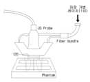
도 3은 경동맥 프로브의 구조를 나타낸 도면,
도 4는 레이저 흡수도의 설명에 제공되는 도면,
도 5는 실험 결과를 나타낸 도면,
도 6은 본 발명의 다른 실시예에 따른 경식도 혈전 탐지 시스템의 블럭도,
도 7은 경식도 프로브의 구조를 나타낸 도면, 그리고,
도 8은 본 발명의 또 다른 실시예에 따른 혈전 탐지 방법의 설명에 제공되는 흐름도이다.1 is a diagram illustrating a blood clot formed in a blood vessel;
2 is a block diagram of a carotid artery thrombus detection system according to an embodiment of the present invention;
3 is a view showing the structure of a carotid artery probe;
4 is a diagram provided for explanation of laser absorption;
5 is a view showing the experimental results;
6 is a block diagram of a transesophageal thrombus detection system according to another embodiment of the present invention;
7 is a view showing the structure of a transesophageal probe, and
8 is a flowchart provided to explain a method for detecting a blood clot according to another embodiment of the present invention.
이하에서는 도면을 참조하여 본 발명을 보다 상세하게 설명한다.Hereinafter, the present invention will be described in more detail with reference to the drawings.
도 2는 본 발명의 일 실시예에 따른 경동맥 혈전 탐지 시스템의 블럭도이다. 본 발명의 실시예에 따른 경동맥 혈전 탐지 시스템은, 광음향 영상과 초음파 영상을 결합하여 경동맥 혈전을 탐지한다.2 is a block diagram of a carotid artery thrombus detection system according to an embodiment of the present invention. A carotid artery thrombosis detection system according to an embodiment of the present invention detects carotid artery thrombosis by combining an photoacoustic image and an ultrasound image.
이와 같은 기능을 수행하는 경동맥 혈전 탐지 시스템은, 도 2에 도시된 바와 같이, 파장 가변 레이저(110), 초음파 센서(120), 광음향 영상 생성부(130), 초음파 영상 생성부(140), 영상 조합부(150), 혈전 추출부(160), 디스플레이(170) 및 제어부(180)를 포함한다.As shown in FIG. 2 , the carotid artery thrombosis detection system performing this function includes a tunable laser 110, an
파장 가변 레이저(110)는 다양한 파장의 레이저 펄스를 생성하여 경동맥에 조사할 수 있다. 파장 가변 레이저(110)에 의해 생성되는 레이저 펄스의 파장 및 생성 시점/주기는 제어부(180)에 의해 제어된다.The tunable laser 110 may generate laser pulses of various wavelengths and irradiate the carotid artery. The wavelength and generation time/period of the laser pulse generated by the tunable laser 110 are controlled by the controller 180 .
경동맥은 파장 가변 레이저(110)에서 조사되는 레이저 펄스를 흡수하여 열 팽창하고, 이 열팽창에 의해 경동맥에서는 광음향이 출사된다.The carotid artery absorbs the laser pulse irradiated from the tunable laser 110 and thermally expands, and photoacoustic sound is emitted from the carotid artery by this thermal expansion.
초음파 센서(120)는 경동맥에서 출사되는 광음향을 감지하기 위한 센서이다. 도 3에는 파장 가변 레이저(110)에서 생성되는 레이저 펄스를 경동맥으로 조사하기 위한 광 파이버를 포함하고, 경동맥으로부터 출사되는 광음향을 감지하는 초음파 센서(120)가 마련된 경동맥 프로브 구조를 도시하였다.The
초음파 센서(120)에 의한 광음향 감지 결과는 광음향 영상 생성부(130)로 인가된다.The photoacoustic detection result by the
광음향 영상 생성부(130)는 초음파 센서(120)의 광음향 감지 결과로부터 광음향 영상을 생성한다. 광음향 영상 생성부(130)에 의해 생성된 영상은 영상 조합부(150)로 인가된다.The photoacoustic image generator 130 generates an photoacoustic image from the photoacoustic detection result of the
초음파 영상 생성부(140)는 경동맥에 초음파를 출력하고 경동맥에서 반사되는 초음파를 감지하여, 초음파 영상을 생성한다. 초음파 영상 생성부(140)에 의해 생성된 초음파 영상은 영상 조합부(150)로 인가된다.The ultrasound image generating unit 140 generates an ultrasound image by outputting ultrasound to the carotid artery and detecting ultrasound reflected from the carotid artery. The ultrasound image generated by the ultrasound image generator 140 is applied to the image combiner 150 .
영상 조합부(150)는 광음향 영상 생성부(130)에 의해 생성된 광음향 영상과 초음파 영상 생성부(140)에 의해 생성된 초음파 영상을 결합한다. 또한, 혈전 추출부(160)는 광음향 영상과 초음파 영상을 이용하여 경동맥 혈전을 추출할 수 있다.The image combination unit 150 combines the photoacoustic image generated by the photoacoustic image generator 130 and the ultrasound image generated by the ultrasound image generator 140 . Also, the thrombus extraction unit 160 may extract a carotid artery thrombus using an photoacoustic image and an ultrasound image.
디스플레이(170)는 영상 조합부(150)에 의해 생성되는 결합 영상과 혈전 추출부(160)에 의한 경동맥 혈전 추출 결과를 표시하여, 의료진에게 제공한다.The display 170 displays the combined image generated by the image combination unit 150 and the carotid artery thrombus extraction result by the thrombus extraction unit 160, and provides the result to the medical staff.
이하에서는, 광음향 영상과 초음파 영상을 이용하여 경동맥 혈전을 탐지하는 원리 및 방법에 대해 상세히 설명한다.Hereinafter, a principle and method for detecting a carotid artery thrombosis using an photoacoustic image and an ultrasound image will be described in detail.
초음파 영상은 경동맥의 구조를 잘 나타내지만, 경동맥 혈전과 다른 조직들을 잘 구분하여 주지 못한다.Ultrasound images show the structure of the carotid artery well, but do not distinguish well between carotid artery thrombosis and other tissues.
혈전은 주로 지질로 이루어져 있는데, 특정 파장의 레이저 펄스가 조사된 지질에서 출사되는 광음향은 다른 조직 보다 훨씬 커서 대조도가 크다. 특히, 도 4에 도시된 바와 같이, 지질은 1210nm 파장의 레이저에 대한 흡수도가 높아, 가장 큰 광음향을 방사하게 된다.Blood clots are mainly composed of lipids, and the photoacoustic sound emitted from lipids irradiated with a laser pulse of a specific wavelength is much larger than that of other tissues, resulting in high contrast. In particular, as shown in FIG. 4 , lipids have high absorption of a laser having a wavelength of 1210 nm, so that the largest photoacoustic emission is emitted.
이에, 본 발명의 실시예에서는 초음파 영상의 구조적 이미지와 1210nm 파장의 레이저 펄스로 생성한 광음향 영상의 기능적 이미지를 결합하여, 경동맥 혈전을 탐색한다.Accordingly, in an embodiment of the present invention, a carotid artery thrombus is searched for by combining a structural image of an ultrasound image and a functional image of an optoacoustic image generated with a laser pulse having a wavelength of 1210 nm.
동물(실험용 쥐)을 통한 실험 결과가 도 5에 제시되어 있다. 도 5에 도시된 바에 따르면, 1210nm 파장의 레이저 펄스로 혈전이 명확하게 나타난 광음향 영상을 얻을 수 있음이 실험적으로 확인되었다.Experimental results with animals (laboratory rats) are presented in FIG. 5 . As shown in FIG. 5 , it was experimentally confirmed that an optoacoustic image in which a blood clot was clearly displayed could be obtained with a laser pulse having a wavelength of 1210 nm.
영상 조합부(150)는 초음파 영상과 광음향 영상을 합성하여, 혈전 부분의 대조도가 높은 경동맥 영상을 생성할 수 있고, 혈전 추출부(160)는 광음향 영상과 초음파 영상의 차 영상을 생성하여, 경동맥 혈전을 추출할 수 있다.The image combination unit 150 may synthesize an ultrasound image and an optoacoustic image to generate a carotid artery image having a high contrast of the thrombus portion, and the thrombus extractor 160 may generate a difference image between the photoacoustic image and the ultrasound image Thus, carotid artery thrombi can be extracted.
도 6은 본 발명의 다른 실시예에 따른 경식도 혈전 탐지 시스템의 블럭도이다. 본 발명의 실시예에 따른 경식도 혈전 탐지 시스템도, 광음향 영상과 초음파 영상을 결합하여 경동맥 혈전을 탐지한다는 점에서, 도 2에 도시된 혈전 탐지 시스템과 동일하다.6 is a block diagram of a transesophageal thrombus detection system according to another embodiment of the present invention. The transesophageal thrombus detection system according to an embodiment of the present invention is the same as the thrombus detection system shown in FIG. 2 in that a carotid artery thrombus is detected by combining an photoacoustic image and an ultrasound image.
하지만, 도 6에 도시된 경식도 혈전 탐지 시스템은, 도 7에 도시된 경식도 프로브를 식도에 삽입하여 광음향과 초음파를 감지한다는 점에서, 도 2에 도시된 혈전 탐지 시스템과 차이가 있다.However, the transesophageal thrombus detection system shown in FIG. 6 differs from the thrombus detection system shown in FIG. 2 in that the transesophageal probe shown in FIG. 7 is inserted into the esophagus to detect photoacoustic and ultrasonic waves.
또한, 도 6에 도시된 경식도 혈전 탐지 시스템은, 하나의 초음파 센서(230)를 이용하며, 광음향과 초음파 영상을 동시에 생성할 수 있다는 점에서도, 도 2에 도시된 혈전 탐지 시스템과 차이가 있다.In addition, the transesophageal thrombus detection system shown in FIG. 6 is different from the thrombus detection system shown in FIG. 2 in that it uses one
이와 같은 특징을 갖는 본 발명의 실시예에 따른 경식도 혈전 탐지 시스템은, 도 6에 도시된 바와 같이, 파장 가변 레이저(210), 초음파 생성부(220), 초음파 센서(230), 광음향/초음파 영상 생성부(240), 영상 조합부(250), 혈전 추출부(260) 및 디스플레이(270)를 포함한다.As shown in FIG. 6, the transesophageal thrombosis detection system according to an embodiment of the present invention having the above characteristics includes a tunable laser 210, an
파장 가변 레이저(210)는 다양한 파장의 레이저 펄스를 생성하여 경동맥에 조사할 수 있다. 파장 가변 레이저(210)에 의해 생성되는 레이저 펄스의 파장 및 생성 시점/주기는 제어부(280)에 의해 제어된다.The tunable laser 210 may generate laser pulses of various wavelengths and irradiate the carotid artery. The wavelength and generation time/cycle of the laser pulse generated by the tunable laser 210 are controlled by the controller 280 .
초음파 생성부(220)는 초음파를 생성하여 경동맥에 출력한다. 초음파 생성부(220)는 초음파의 생성 시점/주기는 제어부(280)에 의해 제어된다.The
제어부(280)는, 파장 가변 레이저(210)에 의한 레이저 펄스 생성/조사와 초음파 생성부(220)에 초음파 생성/출력이, 번차례로 이루어지도록 파장 가변 레이저(210)와 초음파 생성부(220)를 제어한다.The controller 280 controls the tunable laser 210 and the
이에, 초음파 센서(230)에는 파장 가변 레이저(210)에 의해 조사된 레이저 펄스에 의해 경동맥에서 출사되는 광음향과, 초음파 생성부(220)에 의해 출력되어 경동맥에서 반사된 초음파가, 번차례로 감지된다.Accordingly, the
도 7에는 파장 가변 레이저(110)에서 생성되는 레이저를 경동맥으로 조사하기 위한 광 파이버를 포함하고, 초음파 생성부(220)와 초음파 센서(230)가 마련되어 있는 경식도 프로브 구조를 도시하였다.7 shows a structure of a transesophageal probe including an optical fiber for radiating laser generated from the tunable laser 110 to the carotid artery and having an
광음향/초음파 영상 생성부(240)는, 초음파 센서(230)로부터 감지된 광음향 신호를 이용하여 광음향 영상을 생성하는 한편, 초음파 센서(230)로부터 감지된 초음파 신호를 이용하여 초음파 영상을 생성한다.The photoacoustic/ultrasound image generator 240 generates an photoacoustic image using the photoacoustic signal detected by the
광음향/초음파 영상 생성부(240)에 의해 생성된 광음향 영상과 초음파 영상은 영상 조합부(250)로 인가된다.The photoacoustic image and ultrasound image generated by the photoacoustic/ultrasonic image generator 240 are applied to the image combination unit 250 .
영상 조합부(250)는 광음향 영상과 초음파 영상을 결합한다. 또한, 혈전 추출부(260)는 광음향 영상과 초음파 영상을 이용하여 경동맥 혈전을 추출할 수 있다.The image combiner 250 combines an optoacoustic image and an ultrasound image. Also, the thrombus extraction unit 260 may extract a carotid artery thrombus using an photoacoustic image and an ultrasound image.
디스플레이(270)는 영상 조합부(250)에 의해 생성되는 결합 영상과 혈전 추출부(260)에 의한 경동맥 혈전 추출 결과를 표시하여, 의료진에게 제공한다.The display 270 displays the combined image generated by the image combination unit 250 and the carotid artery thrombus extraction result by the thrombus extraction unit 260, and provides the result to the medical staff.
도 8은 본 발명의 또 다른 실시예에 따른 혈전 탐지 방법의 설명에 제공되는 흐름도이다.8 is a flowchart provided to explain a method for detecting a blood clot according to another embodiment of the present invention.
도 8에 도시된 바와 같이, 먼저 레이저 펄스를 이용하여 광음향 영상을 생성하고(S310), 초음파를 이용하여 초음파 영상을 생성한다(S320).As shown in FIG. 8 , first, an optoacoustic image is generated using laser pulses (S310), and an ultrasound image is generated using ultrasonic waves (S320).
다음, S310단계와 S320단계에서 생성된 광음향 영상과 초음파 영상을 조합하고(S330), 경동맥 혈전을 추출한다(S340).Next, the photoacoustic image and the ultrasound image generated in steps S310 and S320 are combined (S330), and the carotid artery thrombosis is extracted (S340).
그리고, 조합된 영상과 경동맥 혈전 추출 결과를 표시하여, 의료진에게 제공한다(S350).Then, the combined image and carotid artery thrombus extraction result are displayed and provided to the medical staff (S350).
지금까지, 경동맥 혈전 탐지 시스템과 경식도 혈전 탐지 시스템 및 이들을 이용한 혈전 탐지 방법에 대해 바람직한 실시예들을 들어 상세히 설명하였다.So far, a carotid artery thrombus detection system, a transesophageal thrombus detection system, and a thrombus detection method using the same have been described in detail with reference to preferred embodiments.
위 실시예에서 제시한 경동맥 혈전 탐지는 예시적인 것에 불과하다. 경동맥 이외의 다른 혈관에서 혈전을 탐지하는 경우에도 본 발명의 기술적 사상이 적용될 수 있다.Carotid artery thrombosis detection presented in the above embodiment is only exemplary. The technical idea of the present invention can also be applied to detecting a thrombus in a blood vessel other than the carotid artery.
또한, 이상에서는 본 발명의 바람직한 실시예에 대하여 도시하고 설명하였지만, 본 발명은 상술한 특정의 실시예에 한정되지 아니하며, 청구범위에서 청구하는 본 발명의 요지를 벗어남이 없이 당해 발명이 속하는 기술분야에서 통상의 지식을 가진자에 의해 다양한 변형실시가 가능한 것은 물론이고, 이러한 변형실시들은 본 발명의 기술적 사상이나 전망으로부터 개별적으로 이해되어져서는 안될 것이다.In addition, although the preferred embodiments of the present invention have been shown and described above, the present invention is not limited to the specific embodiments described above, and the technical field to which the present invention belongs without departing from the gist of the present invention claimed in the claims. Of course, various modifications are possible by those skilled in the art, and these modifications should not be individually understood from the technical spirit or perspective of the present invention.
110 : 파장 가변 레이저
120 : 초음파 센서
130 : 광음향 영상 생성부
140 : 초음파 영상 생성부
150 : 영상 조합부
160 : 혈전 추출부
170 : 디스플레이110: tunable laser
120: ultrasonic sensor
130: photoacoustic image generator
140: ultrasound image generator
150: video combination unit
160: blood clot extraction unit
170: display
Claims (9)
Translated fromKorean상기 레이저 펄스가 조사된 혈관에서 출사되는 광음향을 감지하는 센서;
상기 센서의 감지 결과를 이용하여, 광음향 영상을 생성하는 광음향 생성부;
상기 혈관에 대한 초음파 영상을 생성하는 초음파 영상 생성부;
상기 광음향 영상과 상기 초음파 영상을 조합하여, 혈전 부분의 대조도가 일정치 이상인 경동맥 영상을 생성하는 조합부; 및
상기 광음향 영상과 상기 초음파 영상을 이용하여 경동맥 혈전을 추출하는 추출부;를 포함하되,
상기 추출부는,
상기 광음향 영상과 상기 초음파 영상의 차 영상을 생성하고, 상기 차 영상으로부터 상기 경동맥 혈전을 추출하는
것을 특징으로 하는 혈전 탐지 시스템.
a laser that generates laser pulses and irradiates them to blood vessels;
a sensor for detecting photoacoustic sound emitted from the blood vessel irradiated with the laser pulse;
an optoacoustic generator generating an optoacoustic image by using a detection result of the sensor;
an ultrasound image generating unit generating an ultrasound image of the blood vessel;
a combination unit combining the photoacoustic image and the ultrasound image to generate a carotid artery image having a contrast of a thrombus at or above a predetermined value; and
An extraction unit for extracting a carotid artery thrombosis using the photoacoustic image and the ultrasound image;
The extraction part,
generating a difference image between the photoacoustic image and the ultrasound image, and extracting the carotid artery thrombosis from the difference image
Thrombus detection system, characterized in that.
상기 조합부에 의한 조합 결과와 상기 추출부에 의해 추출 결과를 표시하는 디스플레이;를 더 포함하는 것을 특징으로 하는 혈전 탐지 시스템.
The method of claim 1,
The blood clot detection system further comprising a; a display displaying a combination result by the combination unit and an extraction result by the extraction unit.
상기 레이저 펄스는,
상기 혈전의 주성분에 대한 열적 팽창이 가장 큰 파장의 레이저 펄스인 것을 특징으로 하는 혈전 탐지 시스템.
The method of claim 1,
The laser pulse,
The blood clot detection system, characterized in that the laser pulse of the largest thermal expansion of the main component of the blood clot.
상기 레이저 펄스는,
1210nm 파장의 레이저 펄스인 것을 특징으로 하는 혈전 탐지 시스템.
The method of claim 5,
The laser pulse,
Thrombus detection system, characterized in that the laser pulse of 1210nm wavelength.
상기 혈관에 초음파를 출력하는 초음파 생성부;
상기 레이저 펄스가 조사된 혈관에서 출사되는 광음향을 감지하고, 상기 초음파 생성부에서 조사되어 상기 혈관에서 반사된 초음파를 감지하는 센서;
상기 센서의 감지 결과를 이용하여, 광음향 영상과 초음파 영상을 생성하는 영상 생성부;
상기 광음향 영상과 상기 초음파 영상을 이용하여 경동맥 혈전을 추출하는 추출부; 및
상기 레이저에서의 레이저 펄스 생성과 상기 초음파 생성부에서 초음파 생성이 번차례로 이루어지도록 상기 레이저와 상기 초음파 생성부를 제어하는 제어부;를 더 포함하되,
상기 추출부는,
상기 광음향 영상과 상기 초음파 영상의 차 영상을 생성하고, 상기 차 영상으로부터 상기 경동맥 혈전을 추출하는
것을 특징으로 하는 혈전 탐지 시스템.
a laser that generates laser pulses and irradiates them to blood vessels;
an ultrasound generator outputting ultrasound to the blood vessel;
a sensor for detecting photoacoustic sound emitted from the blood vessel to which the laser pulse is irradiated, and detecting the ultrasonic wave emitted from the ultrasound generator and reflected from the blood vessel;
an image generator for generating an optoacoustic image and an ultrasound image using a detection result of the sensor;
an extraction unit extracting a carotid artery thrombosis using the photoacoustic image and the ultrasound image; and
A controller for controlling the laser and the ultrasonic generator so that the laser pulse is generated from the laser and the ultrasonic generator is sequentially generated,
The extraction part,
generating a difference image between the photoacoustic image and the ultrasound image, and extracting the carotid artery thrombosis from the difference image
Thrombus detection system, characterized in that.
상기 레이저 펄스는,
상기 혈전의 주성분에 대한 열적 팽창이 가장 큰 파장의 레이저 펄스인 것을 특징으로 하는 혈전 탐지 시스템.
The method of claim 7,
The laser pulse,
The blood clot detection system, characterized in that the laser pulse of the largest thermal expansion of the main component of the blood clot.
Priority Applications (2)
| Application Number | Priority Date | Filing Date | Title |
|---|---|---|---|
| KR1020160014696AKR102576682B1 (en) | 2016-02-05 | 2016-02-05 | Plaque Detection System and Method |
| PCT/KR2017/001045WO2017135658A1 (en) | 2016-02-05 | 2017-02-01 | Plaque detection system and method |
Applications Claiming Priority (1)
| Application Number | Priority Date | Filing Date | Title |
|---|---|---|---|
| KR1020160014696AKR102576682B1 (en) | 2016-02-05 | 2016-02-05 | Plaque Detection System and Method |
Publications (2)
| Publication Number | Publication Date |
|---|---|
| KR20170093378A KR20170093378A (en) | 2017-08-16 |
| KR102576682B1true KR102576682B1 (en) | 2023-09-07 |
Family
ID=59500881
Family Applications (1)
| Application Number | Title | Priority Date | Filing Date |
|---|---|---|---|
| KR1020160014696AActiveKR102576682B1 (en) | 2016-02-05 | 2016-02-05 | Plaque Detection System and Method |
Country Status (2)
| Country | Link |
|---|---|
| KR (1) | KR102576682B1 (en) |
| WO (1) | WO2017135658A1 (en) |
Families Citing this family (4)
| Publication number | Priority date | Publication date | Assignee | Title |
|---|---|---|---|---|
| CN107684440A (en)* | 2017-11-01 | 2018-02-13 | 绍兴市中医院 | The detection method and its device of a kind of thrombus |
| KR102146958B1 (en) | 2018-11-28 | 2020-08-21 | 전북대학교산학협력단 | Photoacoustic scan imaging apparatus |
| KR102180436B1 (en) | 2018-11-28 | 2020-11-18 | 전북대학교산학협력단 | 3D Photoacoustic imaging apparatus |
| KR102808984B1 (en)* | 2022-11-14 | 2025-05-15 | 국립부경대학교 산학협력단 | Device for Acquiring Photoacoustic Image and Ultrasonic Image and method thereof |
Citations (4)
| Publication number | Priority date | Publication date | Assignee | Title |
|---|---|---|---|---|
| JP2010516304A (en) | 2007-01-19 | 2010-05-20 | サニーブルック・ヘルス・サイエンシズ・センター | Imaging probe having imaging means combining ultrasound and optics |
| US20120243760A1 (en) | 2011-03-24 | 2012-09-27 | Takahiro Manabe | Plaque region extracting method and apparatus therefor |
| US20120253180A1 (en)* | 2010-10-19 | 2012-10-04 | Stanislav Emelianov | Combined ultrasound and photoacoustic imaging of metal objects |
| JP2013070704A (en)* | 2011-09-26 | 2013-04-22 | Osaka Prefecture Univ | Blood vessel plaque image diagnostic apparatus |
Family Cites Families (3)
| Publication number | Priority date | Publication date | Assignee | Title |
|---|---|---|---|---|
| ATE526882T1 (en)* | 2006-12-19 | 2011-10-15 | Koninkl Philips Electronics Nv | COMBINED PHOTOACOUSTIC AND ULTRASONIC DISPLAY SYSTEM |
| KR20130081067A (en)* | 2012-01-06 | 2013-07-16 | 삼성메디슨 주식회사 | Ultrasound diagnosis apparatus and method |
| KR102581189B1 (en)* | 2016-02-05 | 2023-09-20 | 전북대학교산학협력단 | Fluorescence Imaging Device for Plaque Monitoring and Mult-Imaging System using the same |
- 2016
- 2016-02-05KRKR1020160014696Apatent/KR102576682B1/enactiveActive
- 2017
- 2017-02-01WOPCT/KR2017/001045patent/WO2017135658A1/ennot_activeCeased
Patent Citations (4)
| Publication number | Priority date | Publication date | Assignee | Title |
|---|---|---|---|---|
| JP2010516304A (en) | 2007-01-19 | 2010-05-20 | サニーブルック・ヘルス・サイエンシズ・センター | Imaging probe having imaging means combining ultrasound and optics |
| US20120253180A1 (en)* | 2010-10-19 | 2012-10-04 | Stanislav Emelianov | Combined ultrasound and photoacoustic imaging of metal objects |
| US20120243760A1 (en) | 2011-03-24 | 2012-09-27 | Takahiro Manabe | Plaque region extracting method and apparatus therefor |
| JP2013070704A (en)* | 2011-09-26 | 2013-04-22 | Osaka Prefecture Univ | Blood vessel plaque image diagnostic apparatus |
Non-Patent Citations (2)
| Title |
|---|
| Bio-Medical Materials and Engineering 26 (2015) S1223-S1230 |
| The Transactions of the Korean Institute of Electrical Engineers Vol. 61, No. 6, pp. 879~884, 2012 |
Also Published As
| Publication number | Publication date |
|---|---|
| WO2017135658A1 (en) | 2017-08-10 |
| KR20170093378A (en) | 2017-08-16 |
Similar Documents
| Publication | Publication Date | Title |
|---|---|---|
| JP5655021B2 (en) | Photoacoustic imaging method and apparatus | |
| KR102576682B1 (en) | Plaque Detection System and Method | |
| JP5653882B2 (en) | Photoacoustic imaging apparatus and method of operating the same | |
| JP5762995B2 (en) | Photoacoustic image generation apparatus and method | |
| WO2012114729A1 (en) | Acousto-optical image generating device and method | |
| JP5777394B2 (en) | Photoacoustic imaging method and apparatus | |
| WO2018008439A1 (en) | Apparatus, method and program for displaying ultrasound image and photoacoustic image | |
| JP5681141B2 (en) | Tomographic image generating apparatus, method, and program | |
| US20180008235A1 (en) | Apparatus, method, and program for obtaining information derived from ultrasonic waves and photoacoustic waves | |
| WO2016101280A1 (en) | Intravascular imaging system and method | |
| JP2013027481A (en) | Photoacoustic imaging system and apparatus, and probe unit used therefor | |
| JP2016101393A (en) | Subject information acquisition apparatus and control method therefor | |
| KR102581189B1 (en) | Fluorescence Imaging Device for Plaque Monitoring and Mult-Imaging System using the same | |
| JP5936559B2 (en) | Photoacoustic image generation apparatus and photoacoustic image generation method | |
| US20190183347A1 (en) | Photoacoustic apparatus and object information acquiring method | |
| JP6328778B2 (en) | Method of operating photoacoustic image generation apparatus and photoacoustic image generation apparatus | |
| US20160150990A1 (en) | Photoacoustic apparatus, subject information acquisition method, and program | |
| US20180368696A1 (en) | Object information acquiring apparatus and object information acquiring method | |
| JP6836664B2 (en) | Control method of ultrasonic diagnostic equipment and ultrasonic diagnostic equipment | |
| JP6614910B2 (en) | Photoacoustic device | |
| JP2016101419A (en) | Photoacoustic apparatus, subject information acquisition method, and program | |
| JP2008523881A (en) | Apparatus and method for investigating body structure | |
| JP2012231879A (en) | Photoacoustic imaging method and device | |
| JP5946230B2 (en) | Photoacoustic imaging method and apparatus | |
| US10617319B2 (en) | Photoacoustic apparatus |
Legal Events
| Date | Code | Title | Description |
|---|---|---|---|
| PA0109 | Patent application | Patent event code:PA01091R01D Comment text:Patent Application Patent event date:20160205 | |
| PG1501 | Laying open of application | ||
| A201 | Request for examination | ||
| PA0201 | Request for examination | Patent event code:PA02012R01D Patent event date:20210112 Comment text:Request for Examination of Application Patent event code:PA02011R01I Patent event date:20160205 Comment text:Patent Application | |
| E902 | Notification of reason for refusal | ||
| PE0902 | Notice of grounds for rejection | Comment text:Notification of reason for refusal Patent event date:20230206 Patent event code:PE09021S01D | |
| E701 | Decision to grant or registration of patent right | ||
| PE0701 | Decision of registration | Patent event code:PE07011S01D Comment text:Decision to Grant Registration Patent event date:20230828 | |
| GRNT | Written decision to grant | ||
| PR0701 | Registration of establishment | Comment text:Registration of Establishment Patent event date:20230905 Patent event code:PR07011E01D | |
| PR1002 | Payment of registration fee | Payment date:20230905 End annual number:3 Start annual number:1 | |
| PG1601 | Publication of registration |