KR102536732B1 - Device and method for the computer-assisted simulation of surgical interventions - Google Patents
Device and method for the computer-assisted simulation of surgical interventionsDownload PDFInfo
- Publication number
- KR102536732B1 KR102536732B1KR1020160057015AKR20160057015AKR102536732B1KR 102536732 B1KR102536732 B1KR 102536732B1KR 1020160057015 AKR1020160057015 AKR 1020160057015AKR 20160057015 AKR20160057015 AKR 20160057015AKR 102536732 B1KR102536732 B1KR 102536732B1
- Authority
- KR
- South Korea
- Prior art keywords
- bms
- structural model
- computer
- module
- biomechanical
- Prior art date
- Legal status (The legal status is an assumption and is not a legal conclusion. Google has not performed a legal analysis and makes no representation as to the accuracy of the status listed.)
- Active
Links
Images


Classifications
- A—HUMAN NECESSITIES
- A61—MEDICAL OR VETERINARY SCIENCE; HYGIENE
- A61B—DIAGNOSIS; SURGERY; IDENTIFICATION
- A61B34/00—Computer-aided surgery; Manipulators or robots specially adapted for use in surgery
- A61B34/10—Computer-aided planning, simulation or modelling of surgical operations
- G—PHYSICS
- G06—COMPUTING OR CALCULATING; COUNTING
- G06F—ELECTRIC DIGITAL DATA PROCESSING
- G06F30/00—Computer-aided design [CAD]
- G06F30/20—Design optimisation, verification or simulation
- G06F30/23—Design optimisation, verification or simulation using finite element methods [FEM] or finite difference methods [FDM]
- A—HUMAN NECESSITIES
- A61—MEDICAL OR VETERINARY SCIENCE; HYGIENE
- A61B—DIAGNOSIS; SURGERY; IDENTIFICATION
- A61B5/00—Measuring for diagnostic purposes; Identification of persons
- A61B5/103—Measuring devices for testing the shape, pattern, colour, size or movement of the body or parts thereof, for diagnostic purposes
- A61B5/11—Measuring movement of the entire body or parts thereof, e.g. head or hand tremor or mobility of a limb
- A61B5/1126—Measuring movement of the entire body or parts thereof, e.g. head or hand tremor or mobility of a limb using a particular sensing technique
- A61B5/1128—Measuring movement of the entire body or parts thereof, e.g. head or hand tremor or mobility of a limb using a particular sensing technique using image analysis
- A—HUMAN NECESSITIES
- A61—MEDICAL OR VETERINARY SCIENCE; HYGIENE
- A61B—DIAGNOSIS; SURGERY; IDENTIFICATION
- A61B5/00—Measuring for diagnostic purposes; Identification of persons
- A61B5/74—Details of notification to user or communication with user or patient; User input means
- A61B5/742—Details of notification to user or communication with user or patient; User input means using visual displays
- A—HUMAN NECESSITIES
- A61—MEDICAL OR VETERINARY SCIENCE; HYGIENE
- A61B—DIAGNOSIS; SURGERY; IDENTIFICATION
- A61B5/00—Measuring for diagnostic purposes; Identification of persons
- A61B5/74—Details of notification to user or communication with user or patient; User input means
- A61B5/7475—User input or interface means, e.g. keyboard, pointing device, joystick
- A—HUMAN NECESSITIES
- A61—MEDICAL OR VETERINARY SCIENCE; HYGIENE
- A61B—DIAGNOSIS; SURGERY; IDENTIFICATION
- A61B90/00—Instruments, implements or accessories specially adapted for surgery or diagnosis and not covered by any of the groups A61B1/00 - A61B50/00, e.g. for luxation treatment or for protecting wound edges
- A61B90/36—Image-producing devices or illumination devices not otherwise provided for
- A61B90/37—Surgical systems with images on a monitor during operation
- G—PHYSICS
- G06—COMPUTING OR CALCULATING; COUNTING
- G06F—ELECTRIC DIGITAL DATA PROCESSING
- G06F3/00—Input arrangements for transferring data to be processed into a form capable of being handled by the computer; Output arrangements for transferring data from processing unit to output unit, e.g. interface arrangements
- G06F3/01—Input arrangements or combined input and output arrangements for interaction between user and computer
- G06F3/016—Input arrangements with force or tactile feedback as computer generated output to the user
- G—PHYSICS
- G06—COMPUTING OR CALCULATING; COUNTING
- G06F—ELECTRIC DIGITAL DATA PROCESSING
- G06F3/00—Input arrangements for transferring data to be processed into a form capable of being handled by the computer; Output arrangements for transferring data from processing unit to output unit, e.g. interface arrangements
- G06F3/01—Input arrangements or combined input and output arrangements for interaction between user and computer
- G06F3/017—Gesture based interaction, e.g. based on a set of recognized hand gestures
- G—PHYSICS
- G06—COMPUTING OR CALCULATING; COUNTING
- G06F—ELECTRIC DIGITAL DATA PROCESSING
- G06F3/00—Input arrangements for transferring data to be processed into a form capable of being handled by the computer; Output arrangements for transferring data from processing unit to output unit, e.g. interface arrangements
- G06F3/01—Input arrangements or combined input and output arrangements for interaction between user and computer
- G06F3/03—Arrangements for converting the position or the displacement of a member into a coded form
- G06F3/0304—Detection arrangements using opto-electronic means
- G—PHYSICS
- G06—COMPUTING OR CALCULATING; COUNTING
- G06V—IMAGE OR VIDEO RECOGNITION OR UNDERSTANDING
- G06V20/00—Scenes; Scene-specific elements
- G06V20/20—Scenes; Scene-specific elements in augmented reality scenes
- G—PHYSICS
- G06—COMPUTING OR CALCULATING; COUNTING
- G06V—IMAGE OR VIDEO RECOGNITION OR UNDERSTANDING
- G06V40/00—Recognition of biometric, human-related or animal-related patterns in image or video data
- G06V40/20—Movements or behaviour, e.g. gesture recognition
- G06V40/28—Recognition of hand or arm movements, e.g. recognition of deaf sign language
- G—PHYSICS
- G09—EDUCATION; CRYPTOGRAPHY; DISPLAY; ADVERTISING; SEALS
- G09B—EDUCATIONAL OR DEMONSTRATION APPLIANCES; APPLIANCES FOR TEACHING, OR COMMUNICATING WITH, THE BLIND, DEAF OR MUTE; MODELS; PLANETARIA; GLOBES; MAPS; DIAGRAMS
- G09B23/00—Models for scientific, medical, or mathematical purposes, e.g. full-sized devices for demonstration purposes
- G09B23/28—Models for scientific, medical, or mathematical purposes, e.g. full-sized devices for demonstration purposes for medicine
- G—PHYSICS
- G09—EDUCATION; CRYPTOGRAPHY; DISPLAY; ADVERTISING; SEALS
- G09B—EDUCATIONAL OR DEMONSTRATION APPLIANCES; APPLIANCES FOR TEACHING, OR COMMUNICATING WITH, THE BLIND, DEAF OR MUTE; MODELS; PLANETARIA; GLOBES; MAPS; DIAGRAMS
- G09B9/00—Simulators for teaching or training purposes
- G—PHYSICS
- G16—INFORMATION AND COMMUNICATION TECHNOLOGY [ICT] SPECIALLY ADAPTED FOR SPECIFIC APPLICATION FIELDS
- G16H—HEALTHCARE INFORMATICS, i.e. INFORMATION AND COMMUNICATION TECHNOLOGY [ICT] SPECIALLY ADAPTED FOR THE HANDLING OR PROCESSING OF MEDICAL OR HEALTHCARE DATA
- G16H40/00—ICT specially adapted for the management or administration of healthcare resources or facilities; ICT specially adapted for the management or operation of medical equipment or devices
- G16H40/60—ICT specially adapted for the management or administration of healthcare resources or facilities; ICT specially adapted for the management or operation of medical equipment or devices for the operation of medical equipment or devices
- G16H40/63—ICT specially adapted for the management or administration of healthcare resources or facilities; ICT specially adapted for the management or operation of medical equipment or devices for the operation of medical equipment or devices for local operation
- G—PHYSICS
- G16—INFORMATION AND COMMUNICATION TECHNOLOGY [ICT] SPECIALLY ADAPTED FOR SPECIFIC APPLICATION FIELDS
- G16H—HEALTHCARE INFORMATICS, i.e. INFORMATION AND COMMUNICATION TECHNOLOGY [ICT] SPECIALLY ADAPTED FOR THE HANDLING OR PROCESSING OF MEDICAL OR HEALTHCARE DATA
- G16H50/00—ICT specially adapted for medical diagnosis, medical simulation or medical data mining; ICT specially adapted for detecting, monitoring or modelling epidemics or pandemics
- G16H50/50—ICT specially adapted for medical diagnosis, medical simulation or medical data mining; ICT specially adapted for detecting, monitoring or modelling epidemics or pandemics for simulation or modelling of medical disorders
- G—PHYSICS
- G16—INFORMATION AND COMMUNICATION TECHNOLOGY [ICT] SPECIALLY ADAPTED FOR SPECIFIC APPLICATION FIELDS
- G16Z—INFORMATION AND COMMUNICATION TECHNOLOGY [ICT] SPECIALLY ADAPTED FOR SPECIFIC APPLICATION FIELDS, NOT OTHERWISE PROVIDED FOR
- G16Z99/00—Subject matter not provided for in other main groups of this subclass
- A—HUMAN NECESSITIES
- A61—MEDICAL OR VETERINARY SCIENCE; HYGIENE
- A61B—DIAGNOSIS; SURGERY; IDENTIFICATION
- A61B17/00—Surgical instruments, devices or methods
- A61B2017/00017—Electrical control of surgical instruments
- A61B2017/00207—Electrical control of surgical instruments with hand gesture control or hand gesture recognition
- A—HUMAN NECESSITIES
- A61—MEDICAL OR VETERINARY SCIENCE; HYGIENE
- A61B—DIAGNOSIS; SURGERY; IDENTIFICATION
- A61B34/00—Computer-aided surgery; Manipulators or robots specially adapted for use in surgery
- A61B34/10—Computer-aided planning, simulation or modelling of surgical operations
- A61B2034/101—Computer-aided simulation of surgical operations
- A61B2034/102—Modelling of surgical devices, implants or prosthesis
- A61B2034/104—Modelling the effect of the tool, e.g. the effect of an implanted prosthesis or for predicting the effect of ablation or burring
- A—HUMAN NECESSITIES
- A61—MEDICAL OR VETERINARY SCIENCE; HYGIENE
- A61B—DIAGNOSIS; SURGERY; IDENTIFICATION
- A61B34/00—Computer-aided surgery; Manipulators or robots specially adapted for use in surgery
- A61B34/10—Computer-aided planning, simulation or modelling of surgical operations
- A61B2034/101—Computer-aided simulation of surgical operations
- A61B2034/105—Modelling of the patient, e.g. for ligaments or bones
- A—HUMAN NECESSITIES
- A61—MEDICAL OR VETERINARY SCIENCE; HYGIENE
- A61B—DIAGNOSIS; SURGERY; IDENTIFICATION
- A61B34/00—Computer-aided surgery; Manipulators or robots specially adapted for use in surgery
- A61B34/20—Surgical navigation systems; Devices for tracking or guiding surgical instruments, e.g. for frameless stereotaxis
- A61B2034/2046—Tracking techniques
- A61B2034/2055—Optical tracking systems
- A61B2034/2057—Details of tracking cameras
- A—HUMAN NECESSITIES
- A61—MEDICAL OR VETERINARY SCIENCE; HYGIENE
- A61B—DIAGNOSIS; SURGERY; IDENTIFICATION
- A61B34/00—Computer-aided surgery; Manipulators or robots specially adapted for use in surgery
- A61B34/20—Surgical navigation systems; Devices for tracking or guiding surgical instruments, e.g. for frameless stereotaxis
- A61B2034/2046—Tracking techniques
- A61B2034/2065—Tracking using image or pattern recognition
- A—HUMAN NECESSITIES
- A61—MEDICAL OR VETERINARY SCIENCE; HYGIENE
- A61B—DIAGNOSIS; SURGERY; IDENTIFICATION
- A61B90/00—Instruments, implements or accessories specially adapted for surgery or diagnosis and not covered by any of the groups A61B1/00 - A61B50/00, e.g. for luxation treatment or for protecting wound edges
- A61B90/36—Image-producing devices or illumination devices not otherwise provided for
- A61B2090/364—Correlation of different images or relation of image positions in respect to the body
- A61B2090/365—Correlation of different images or relation of image positions in respect to the body augmented reality, i.e. correlating a live optical image with another image
- A—HUMAN NECESSITIES
- A61—MEDICAL OR VETERINARY SCIENCE; HYGIENE
- A61B—DIAGNOSIS; SURGERY; IDENTIFICATION
- A61B2576/00—Medical imaging apparatus involving image processing or analysis
- A—HUMAN NECESSITIES
- A61—MEDICAL OR VETERINARY SCIENCE; HYGIENE
- A61B—DIAGNOSIS; SURGERY; IDENTIFICATION
- A61B34/00—Computer-aided surgery; Manipulators or robots specially adapted for use in surgery
- A61B34/70—Manipulators specially adapted for use in surgery
- A61B34/76—Manipulators having means for providing feel, e.g. force or tactile feedback
- A—HUMAN NECESSITIES
- A61—MEDICAL OR VETERINARY SCIENCE; HYGIENE
- A61B—DIAGNOSIS; SURGERY; IDENTIFICATION
- A61B5/00—Measuring for diagnostic purposes; Identification of persons
- A61B5/0033—Features or image-related aspects of imaging apparatus, e.g. for MRI, optical tomography or impedance tomography apparatus; Arrangements of imaging apparatus in a room
- A61B5/0037—Performing a preliminary scan, e.g. a prescan for identifying a region of interest
- A—HUMAN NECESSITIES
- A61—MEDICAL OR VETERINARY SCIENCE; HYGIENE
- A61B—DIAGNOSIS; SURGERY; IDENTIFICATION
- A61B5/00—Measuring for diagnostic purposes; Identification of persons
- A61B5/0033—Features or image-related aspects of imaging apparatus, e.g. for MRI, optical tomography or impedance tomography apparatus; Arrangements of imaging apparatus in a room
- A61B5/004—Features or image-related aspects of imaging apparatus, e.g. for MRI, optical tomography or impedance tomography apparatus; Arrangements of imaging apparatus in a room adapted for image acquisition of a particular organ or body part
- A61B5/0044—Features or image-related aspects of imaging apparatus, e.g. for MRI, optical tomography or impedance tomography apparatus; Arrangements of imaging apparatus in a room adapted for image acquisition of a particular organ or body part for the heart
- A—HUMAN NECESSITIES
- A61—MEDICAL OR VETERINARY SCIENCE; HYGIENE
- A61B—DIAGNOSIS; SURGERY; IDENTIFICATION
- A61B5/00—Measuring for diagnostic purposes; Identification of persons
- A61B5/0059—Measuring for diagnostic purposes; Identification of persons using light, e.g. diagnosis by transillumination, diascopy, fluorescence
- A61B5/0073—Measuring for diagnostic purposes; Identification of persons using light, e.g. diagnosis by transillumination, diascopy, fluorescence by tomography, i.e. reconstruction of 3D images from 2D projections
- A—HUMAN NECESSITIES
- A61—MEDICAL OR VETERINARY SCIENCE; HYGIENE
- A61B—DIAGNOSIS; SURGERY; IDENTIFICATION
- A61B5/00—Measuring for diagnostic purposes; Identification of persons
- A61B5/05—Detecting, measuring or recording for diagnosis by means of electric currents or magnetic fields; Measuring using microwaves or radio waves
- A61B5/055—Detecting, measuring or recording for diagnosis by means of electric currents or magnetic fields; Measuring using microwaves or radio waves involving electronic [EMR] or nuclear [NMR] magnetic resonance, e.g. magnetic resonance imaging
- G—PHYSICS
- G06—COMPUTING OR CALCULATING; COUNTING
- G06F—ELECTRIC DIGITAL DATA PROCESSING
- G06F18/00—Pattern recognition
- G06F18/20—Analysing
- G06F18/21—Design or setup of recognition systems or techniques; Extraction of features in feature space; Blind source separation
- G06F18/214—Generating training patterns; Bootstrap methods, e.g. bagging or boosting
- G06F18/2148—Generating training patterns; Bootstrap methods, e.g. bagging or boosting characterised by the process organisation or structure, e.g. boosting cascade
- G—PHYSICS
- G06—COMPUTING OR CALCULATING; COUNTING
- G06F—ELECTRIC DIGITAL DATA PROCESSING
- G06F18/00—Pattern recognition
- G06F18/20—Analysing
- G06F18/24—Classification techniques
- G06F18/241—Classification techniques relating to the classification model, e.g. parametric or non-parametric approaches
- G06F18/2413—Classification techniques relating to the classification model, e.g. parametric or non-parametric approaches based on distances to training or reference patterns
- G—PHYSICS
- G06—COMPUTING OR CALCULATING; COUNTING
- G06V—IMAGE OR VIDEO RECOGNITION OR UNDERSTANDING
- G06V2201/00—Indexing scheme relating to image or video recognition or understanding
- G06V2201/03—Recognition of patterns in medical or anatomical images
Landscapes
- Engineering & Computer Science (AREA)
- Health & Medical Sciences (AREA)
- Theoretical Computer Science (AREA)
- Life Sciences & Earth Sciences (AREA)
- Physics & Mathematics (AREA)
- General Engineering & Computer Science (AREA)
- General Physics & Mathematics (AREA)
- Medical Informatics (AREA)
- General Health & Medical Sciences (AREA)
- Public Health (AREA)
- Surgery (AREA)
- Biomedical Technology (AREA)
- Human Computer Interaction (AREA)
- Heart & Thoracic Surgery (AREA)
- Molecular Biology (AREA)
- Animal Behavior & Ethology (AREA)
- Veterinary Medicine (AREA)
- Nuclear Medicine, Radiotherapy & Molecular Imaging (AREA)
- Pathology (AREA)
- Biophysics (AREA)
- Computer Vision & Pattern Recognition (AREA)
- Multimedia (AREA)
- Business, Economics & Management (AREA)
- Robotics (AREA)
- Oral & Maxillofacial Surgery (AREA)
- Radiology & Medical Imaging (AREA)
- Primary Health Care (AREA)
- Epidemiology (AREA)
- Educational Administration (AREA)
- Educational Technology (AREA)
- Physiology (AREA)
- Databases & Information Systems (AREA)
- Data Mining & Analysis (AREA)
- Dentistry (AREA)
- Psychiatry (AREA)
- Social Psychology (AREA)
- Chemical & Material Sciences (AREA)
- Pure & Applied Mathematics (AREA)
- Mathematical Optimization (AREA)
- Mathematical Physics (AREA)
Abstract
Translated fromKoreanDescription
Translated fromKorean임상 실시(clinical practice)에서, 외과 수술(surgical intervention)들은 일반적으로, 임상 가이드라인(clinical guideline)들로부터의 권고들과 연계하여 의료 이미지 기록들에 기초하여 계획된다. 그러나, 수술의 계획 및 성공의 상당 부분은, 수행하는 외과 의사 또는 외과 의사들의 경험에 비례할 수 있다. 많은 수술들, 이를테면, 예컨대 심장 판막 성형술들은 특히, 우수한 계획, 경험 및 수술 기술을 요구한다. 그러므로, 추구될 가치가 있는 목표는, 수술들을 계획 및 트레이닝할 때, 최상의 가능한 정도로 외과 의사를 지원하는 것이다.In clinical practice, surgical interventions are generally planned based on medical image records in conjunction with recommendations from clinical guidelines. However, much of the planning and success of a surgery can be proportional to the experience of the surgeon or surgeons performing it. Many surgeries, such as eg heart valve angioplasty, in particular require good planning, experience and surgical skill. Therefore, a goal worth pursuing is to assist the surgeon to the best possible extent when planning and training surgeries.
외과 수술들을 계획하기 위해 결정 지원 시스템들을 이용한 실시가 알려져 있으며, 그 결정 지원 시스템들은 생체역학적 모델에 의한, 이를테면, 예컨대 심장 판막의 생리학적 구조들 및 기능들의 시뮬레이션에 기초한다. 예로서, 심장 판막들의 이러한 생체역학적 시뮬레이션들은 문헌들 US 2010/0240996 A1 및 US 2012/0232386 A1로부터 알려져 있다. 이러한 시뮬레이션들을 통해, 외과 수술의 효과를 추정하는 것이 가능하며, 이는 수술을 계획하기 위해 이용될 수 있다. 그러나, 이러한 시뮬레이션 도구들의 사용은 일반적으로, 그 시뮬레이션 도구들의 복잡성으로 인해 복잡한 전수(induction)를 요구한다.The practice of using decision support systems to plan surgical operations is known, which decision support systems are based on a biomechanical model, such as simulation of the physiological structures and functions of, for example, a heart valve. As an example, such biomechanical simulations of heart valves are known from documents US 2010/0240996 A1 and US 2012/0232386 A1. Through these simulations, it is possible to estimate the effectiveness of a surgical operation, which can be used to plan a surgery. However, the use of these simulation tools generally requires complex induction due to the complexity of the simulation tools.
게다가, 외과 수술들의 트레이닝의 목적들을 위해 외과 의사의 머리에 고정된 카메라를 통해 실제 외과 수술을 기록하는 것 및 결과적인 비디오 스트림을 관찰자들에게 송신하는 것의 실시가, 예컨대 서저브리(Surgevry)에 의한 웹사이트인 http://www.surgevry.com로부터 알려져 있으며, 그 관찰자들은 그러므로, 수행하는 외과 의사를 관찰하는 것에 의해 그 수술을 따라할 수 있다. 그러나, 이는 단지 수술의 수동적 트레이닝(passive training)만을 관찰자들에게 허용한다.Furthermore, for the purposes of training of surgical procedures, the practice of recording actual surgical operations through a camera fixed to the surgeon's head and transmitting the resulting video stream to observers, such as by Surgevry It is known from the website http://www.surgevry.com, and its observers can therefore follow the surgery by observing the surgeon performing it. However, this only allows observers passive training of the surgery.
본 발명의 목적은, 외과 수술들의 컴퓨터-지원 시뮬레이션(computer-assisted simulation)을 위한 디바이스 및 방법을 명시하는 것이며, 그 디바이스 및 방법은 개선된 외과 의사 지원을 허용한다.It is an object of the present invention to specify a device and method for computer-assisted simulation of surgical operations, which device and method allow improved surgeon assistance.
이러한 목적은 특허 청구항 제1항의 특징들을 가진 디바이스에 의해, 특허 청구항 제12항의 특징들을 가진 방법에 의해, 특허 청구항 제13항의 특징들을 가진 컴퓨터 프로그램 물건에 의해, 그리고 특허 청구항 제14항의 특징들을 가진 컴퓨터-판독가능 저장 매체에 의해 달성된다.This object is achieved by a device having the features of patent claim 1, by a method having the features of patent claim 12, by a computer program product having the features of patent claim 13 and having the features of patent claim 14. Accomplished by a computer-readable storage medium.
외과 수술들의 컴퓨터-지원 시뮬레이션을 위한 본 발명에 따른 디바이스에서, 의료 이미징 방법에 의해 획득된 해부학적 영역의 이미지 데이터를 판독하기 위한 제1 인터페이스에 대한 제공이 이루어진다. 이러한 경우, 이미지 데이터는, 예컨대 초음파(sonographic), 방사선(radiological), 혈관조영술(angiographic) 또는 단층촬영(tomographic) 이미지 기록들로부터 비롯될 수 있다. 해부학적 영역은, 예컨대 장기(organ), 장기 영역 또는 상이한 조직 또는 몸체 구조일 수 있다. 모델링 모듈은, 이미지 데이터에 기초하여 해부학적 영역의 입체적 생체역학적 구조 모델(volumetric biomechanical structure model)을 확립하는 역할을 한다. 더욱이, 사용자의 공간적 제스처들의 비디오-기반 등록을 위해 카메라와 커플링가능한 추적 모듈에 대한 제공이 이루어진다. 게다가, 시뮬레이션 모듈은, 생체역학적 구조 모델에 기초하여, 각각의 경우에서 등록된 제스처를 해부학적 영역에 대해 시뮬레이팅된 역학적 영향에 할당하고, 생체역학적 구조 모델에 기초하여, 시뮬레이팅된 역학적 영향에 대한 해부학적 영역의 역학적 반응을 시뮬레이팅하고, 그리고 시뮬레이팅된 역학적 반응에 따라 생체역학적 구조 모델을 수정하는 역할을 한다. 더욱이, 생체역학적 구조 모델의 입체적 시각화(volumetric visualization)를 위한 시각화 모듈에 대한 제공이 이루어진다. 시각화 모듈은 바람직하게, 특히 해부학적 영역의 역학적 반응 및/또는 수정된 생체역학적 구조 모델을 시각화한다.In the device according to the invention for computer-aided simulation of surgical operations, provision is made for a first interface for reading image data of an anatomical region obtained by a medical imaging method. In this case, the image data may originate, for example, from sonographic, radiological, angiographic or tomographic image records. An anatomical region can be, for example, an organ, an organ region or a different tissue or body structure. The modeling module serves to establish a volumetric biomechanical structure model of an anatomical region based on image data. Moreover, provision is made for a tracking module coupleable with a camera for video-based registration of the user's spatial gestures. Furthermore, the simulation module assigns, in each case, the registered gesture to the simulated mechanical effect for the anatomical region, based on the biomechanical structural model, to the simulated mechanical effect, based on the biomechanical structural model. It plays a role in simulating the mechanical response of an anatomical region and modifying the biomechanical structural model according to the simulated mechanical response. Furthermore, provision is made for a visualization module for volumetric visualization of the biomechanical structural model. The visualization module preferably visualizes, in particular, the mechanical response of an anatomical region and/or a modified biomechanical structural model.
앞서 설명된 디바이스에 의해 수행될 방법 단계들은 외과 수술들의 컴퓨터-지원 시뮬레이션을 위한 본 발명에 따른 방법의 청구 대상이다.The method steps to be performed by the device described above are the subject matter of the method according to the invention for computer-aided simulation of surgical operations.
본 발명의 실질적 이점은, 사용자, 예컨대 외과 의사가 외과 수술을, 외과 수술의 수동적 수행(manual performance)뿐만 아니라 시각화에 대해서도 비교적 사실적인 방식으로 시뮬레이팅할 수 있다는 사실에서 고려될 수 있다. 특히, 사용자는, 사용자가 실제 수술 동안 할 수 있는 조작들과 실질적으로 동일한 조작들을 생체역학적 구조 모델에 할 수 있다. 이러한 방식으로, 외과 의사는 다양한 치료 선택사항들을 테스트하고 그에 따라 이상적인 절차를 확립할 수 있다. 그러므로, 본 발명은, 수술들을 계획할 때와 트레이닝할 때 양쪽 모두에서, 직관적인 방식으로 외과 의사에 대한 지원을 허용한다.A substantial advantage of the present invention may be considered in the fact that a user, such as a surgeon, can simulate a surgical operation in a relatively realistic manner for visualization as well as for the manual performance of the surgical operation. In particular, the user may perform manipulations substantially the same as manipulations the user may perform during an actual operation on the biomechanical structural model. In this way, the surgeon can test various treatment options and establish the ideal procedure accordingly. Therefore, the present invention allows assistance to the surgeon in an intuitive manner both when planning surgeries and when training.
본 발명의 유리한 실시예들 및 발전들은 종속 청구항들에서 명시된다.Advantageous embodiments and developments of the invention are specified in the dependent claims.
본 발명의 유리한 실시예에 따르면, 추적 모듈은 수술 기구(surgical instrument), 예컨대 수술칼(scalpel)을 이용하여 수행되는 사용자의 제스처들을 등록하도록 구성될 수 있다. 특히, 수술 기구의 하나 이상의 특정 부분들, 예컨대 수술칼의 날(blade) 및/또는 손잡이의 움직임들을 등록하는 것이 가능하다. 이는, 사용자가 실제 수술 동안에서와 동일한 방식으로 시뮬레이션의 범위 내에서 수술 기구를 다룰 수 있을 경우, 유리하다.According to an advantageous embodiment of the invention, the tracking module can be configured to register gestures of the user performed using a surgical instrument, for example a scalpel. In particular, it is possible to register movements of one or more specific parts of a surgical instrument, such as the blade and/or handle of a surgical knife. This is advantageous if the user can handle surgical instruments within the confines of the simulation in the same way as during a real operation.
게다가, 사용자에게로의 시뮬레이팅된 역학적 반응의 햅틱 출력(haptic output)을 위한 햅틱 인터페이스(haptic interface)에 대한 제공이 이루어질 수 있다. 이는, 시뮬레이팅된 역학적 영향에 대한 해부학적 영역의 시뮬레이팅된 역학적 반응에 관한 피드백을 허용하며, 그 피드백은 사용자에 의해 즉각적으로 그리고 사실적인 방식으로 인지가능하다.Furthermore, provision can be made for a haptic interface for haptic output of a simulated mechanical response to a user. This allows feedback regarding the simulated mechanical response of the anatomical region to the simulated mechanical influence, the feedback being perceptible by the user immediately and in a realistic manner.
본 발명의 유리한 발전에 따르면, 시뮬레이션 모듈은, 생체역학적 구조 모델에 기초하여 해부학적 영역의 해부학적 기능에 대한 외과 수술의 영향을 시뮬레이팅하도록 구성될 수 있다. 여기서, 시뮬레이팅된 영향을 디스플레이하기 위한 출력 인터페이스에 대한 제공이 이루어질 수 있다. 이러한 방식으로, 수술의 결과들을 예측 또는 적어도 추정하는 것이 가능하다. 결과적으로, 사용자는 다양한 치료 선택사항들을 테스트하고 그 결과들에 대해 이상적인 절차를 확립할 수 있다.According to an advantageous development of the invention, the simulation module can be configured to simulate the effect of a surgical operation on an anatomical function of an anatomical region on the basis of a biomechanical structural model. Here, provision for an output interface for displaying the simulated impact may be made. In this way, it is possible to predict or at least estimate the outcomes of surgery. As a result, the user can test various treatment options and establish an ideal procedure for the results.
유리하게, 생체역학적 구조 모델은 환자에 대해 특정적일 수 있다. 이는, 수술의 환자-특정적 계획 및 환자-특정적 트레이닝을 허용한다.Advantageously, the biomechanical structural model can be patient-specific. This allows for patient-specific planning and patient-specific training of surgery.
유리한 실시예에 따르면, 생체역학적 구조 모델은 유한 엘리먼트 모델(finite element model)을 포함할 수 있다. 이러한 유한 엘리먼트 모델들의 효율적인 생성 및 계산의 목적들을 위해, 다수의 이용가능하고 잘-조작된 소프트웨어 도구들을 이용하는 것이 가능하다.According to an advantageous embodiment, the biomechanical structural model may comprise a finite element model. For the purposes of efficient creation and calculation of such finite element models, it is possible to use a number of available and well-crafted software tools.
더욱이, 모델링 모듈은 연속적으로 판독되는 이미지 데이터에 기초하여 생체역학적 구조 모델을 동적으로 적응시키도록 구성될 수 있다.Furthermore, the modeling module may be configured to dynamically adapt the biomechanical structural model based on the image data that is successively read out.
게다가, 제1 인터페이스는 시간-분해 방식(time-resolved manner)으로 이미지 데이터를 판독하도록 구성될 수 있다. 따라서, 모델링 모듈은, 시간-분해된 이미지 데이터에 기초하여 해부학적 영역의 부분구조의 운동 역학(movement dynamics)을 식별하고 그리고 식별된 운동 역학에 기초하여 부분구조의 물리적 속성(physical property)을 유도하여 그 물리적 속성을 생체역학적 구조 모델에 맵핑(map) 하도록 구성될 수 있다. 이러한 방식으로, 해부학적 영역 및 역학적 영향들에 대한 해부학적 영역의 역학적 반응들의 비교적 정확한 물리적 시뮬레이션을 획득하는 것이 가능하다.Additionally, the first interface may be configured to read image data in a time-resolved manner. Accordingly, the modeling module identifies movement dynamics of the substructures of an anatomical region based on the time-resolved image data and derives physical properties of the substructures based on the identified movement dynamics. and map its physical properties to a biomechanical structural model. In this way, it is possible to obtain a relatively accurate physical simulation of the anatomical region and the mechanical responses of the anatomical region to mechanical influences.
특히, 모델링 모듈은, 부분구조의 운동 역학을 식별하고, 부분구조의 물리적 속성을 유도하고, 그리고/또는 물리적 속성을 생체역학적 구조 모델에 맵핑하도록 구성된 기계 학습(machine learning)을 위한 모듈을 포함할 수 있다. 기계 학습을 위한 이러한 모듈은, 예컨대 인공 신경망 및/또는 이른바 확률적 부스팅 트리(probabilistic boosting tree)를 포함할 수 있다.In particular, the modeling module may include a module for machine learning configured to identify kinematics of the substructure, derive physical properties of the substructure, and/or map the physical properties to the biomechanical structural model. can Such modules for machine learning may include, for example, artificial neural networks and/or so-called probabilistic boosting trees.
더욱이, 시각화 모듈은 가상 및/또는 증강 현실을 디스플레이하기 위한 몰입형 시스템으로서 설계될 수 있다. 대안적으로 또는 추가적으로, 시각화 모듈은 또한, 홀로그래픽(holographic) 및/또는 스테레오그래픽(stereographic) 시각화 시스템으로서 설계될 수 있다. 이러한 시각화 모듈은 시뮬레이션의 범위 내에서 사용자의 현실감을 증가시킨다.Moreover, the visualization module can be designed as an immersive system for displaying virtual and/or augmented reality. Alternatively or additionally, the visualization module can also be designed as a holographic and/or stereographic visualization system. This visualization module increases the user's sense of realism within the scope of the simulation.
도면에 기초하여 본 발명의 예시적 실시예가 아래에서 더 상세하게 설명된다. 여기서, 각각의 경우에서, 개략도로,
도 1은 본 발명에 따른 시뮬레이션 디바이스를 도시하고,
도 2는 본 발명에 따른 방법의 프로세스들을 설명하기 위한 흐름도를 도시하고, 그리고
도 3은 해부학적 영역과의 가상적 상호작용의 시각화를 도시한다.An exemplary embodiment of the present invention is described in more detail below on the basis of the drawings. Here, in each case, schematically,
1 shows a simulation device according to the invention;
2 shows a flowchart for explaining the processes of the method according to the invention, and
Figure 3 shows a visualization of virtual interaction with an anatomical region.
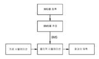
도 1은 외과 수술들을 시뮬레이팅하기 위한 본 발명에 따른 시뮬레이션 디바이스(SIMS)를 개략적으로 도시한다. 시뮬레이션 디바이스(SIMS)는 프로세서(PROC) 및/또는 본 발명에 따른 방법을 실행하기 위한 하나 이상의 다른 컴퓨터 장치들을 포함한다. 의료 이미지 기록 장치(US)는 제1 인터페이스(I1)를 통해 시뮬레이션 디바이스(SIMS)에 커플링된다. 예로서, 이미지 기록 디바이스(US)는 초음파 기록 장치, x-레이 튜브(x-ray tube), 자기 공명 이미징(MRI) 스캐너, 또는 해부학적 영역들, 예컨대 장기들, 장기 영역들 또는 다른 몸체 및/또는 조직 구조들의 이미지 기록들을 공급하는 임의의 다른 의료 기록 디바이스일 수 있다. 이미지 기록 장치(US)는 각각의 해부학적 영역의 시간-분해된 이미지 기록들, 바람직하게는 입체적(volumetric) 이미지 기록들을 연속적으로 기록하고, 이들을 시간-분해된 이미지 데이터(IMG), 바람직하게는 입체적 이미지 데이터(IMG)로서 제1 인터페이스(I1)를 통해 시뮬레이션 디바이스(SIMS)에 연속적으로 전달하도록 구성된다.1 schematically shows a simulation device (SIMS) according to the invention for simulating surgical operations. The simulation device SIMS comprises a processor PROC and/or one or more other computer devices for executing the method according to the invention. The medical image recording apparatus US is coupled to the simulation device SIMS through a first interface I1. By way of example, the image recording device US may be an ultrasound recording device, an x-ray tube, a magnetic resonance imaging (MRI) scanner, or anatomical regions such as organs, organ regions or other body and /or any other medical recording device that supplies image recordings of tissue structures. The image recording device US successively records time-resolved image records, preferably volumetric image records, of each anatomical region and converts them into time-resolved image data (IMG), preferably volumetric image records. It is configured to continuously transmit as stereoscopic image data IMG to the simulation device SIMS through the first interface I1.
본 예시적인 실시예의 경우, 심장 판막이, 심장 판막의 해부학적 주변부들에서 그리고/또는 심장 판막의 해부학적 기능의 맥락에서, 이미지 기록 장치(US)를 통해 해부학적 영역(AR)으로서 기록된다고 가정한다. 해부학적 영역(AR), 즉, 심장 판막의 이미지 데이터(IMG)는, 예컨대 비디오 데이터 스트림(video data으로서, 이미지 기록 장치(US)로부터 시뮬레이션 디바이스(SIMS)의 모델링 모듈(MM)에 전달된다.For this exemplary embodiment, it is assumed that the heart valve is recorded as an anatomical region (AR) by means of an image recording device (US) at the anatomical peripheries of the heart valve and/or in the context of the anatomical function of the heart valve. do. The image data IMG of the anatomical region AR, that is, the heart valve, is transferred from the image recording apparatus US to the modeling module MM of the simulation device SIMS, for example as a video data stream.
모델링 모듈(MM)은 이미지 데이터(IMG)에 기초하여 해부학적 영역(AR)의 입체적 생체역학적 구조 모델(BMS; biomechanical structure model)을 확립하는 역할을 한다. 생체역학적 구조 모델(BMS)이 환자의 해부학적 영역(AR)의 이미지 데이터(IMG)에 기초하여 확립되면, 생체역학적 구조 모델(BMS)은 환자에 대해 특정적이다. 생체역학적 구조 모델(BMS)은 바람직하게, 유한 엘리먼트 모델을 포함한다.The modeling module (MM) serves to establish a three-dimensional biomechanical structure model (BMS) of the anatomical region (AR) based on the image data (IMG). If the biomechanical structural model (BMS) is established based on the image data (IMG) of the patient's anatomical region (AR), the biomechanical structural model (BMS) is specific to the patient. The biomechanical structural model (BMS) preferably includes a finite element model.
생체역학적 구조 모델(BMS)을 확립하기 위해, 시간-분해된 이미지 데이터(IMG)가 모델링 모듈(MM)에 의해 분석된다. 여기서, 예컨대 알려진 패턴 인식 방법들에 의해, 해부학적 영역(AR)의 하나 이상의 부분구조들이 식별되며, 이러한 부분구조들의 운동 역학이 확립된다. 생체역학적 구조 모델(BMS) 또는 가능하게는 기본적인 유한 엘리먼트 모델은, 식별된 운동 역학을 그 모델이 재현할 때까지, 식별된 운동 역학에 기초하여, 예컨대 수치적 최적화 방법에 의해 수정된다. 대안적으로 또는 추가적으로, 생체역학적 구조 모델(BMS)의 파라미터들은, 학습-기반 회귀분석법(regression method)들에 기초하여 식별된 운동 역학으로부터 추정될 수 있으며, 수치적 최적화 방법은 학습 단계 동안 이용된다. 이를 이용시, 식별된 운동 역학에 기초하여, 부분구조들의 물리적 속성들이 유도될 수 있으며, 상기 물리적 속성들은 생체역학적 구조 모델(BMS)에 맵핑될 수 있다. 여기서, 특히 또한 도플러 효과(Doppler Effect)를 이용함으로써, 예컨대 도플러 초음파 기기를 이용함으로써, 예컨대 혈액 흐름이 이러한 방식으로 측정되는 덕분에, 운동 역학이 확립될 수 있다. 예로서, 부분구조들의 탄력성, 강성, 밀도 또는 다른 조직 파라미터들이 부분구조들의 물리적 속성들로서 확립될 수 있다. 바람직하게, 생체역학적 구조 모델(BMS)은 판독된 이미지 데이터(IMG)에 기초하여 모델링 모듈(MM)에 의해 계속해서 동적으로 적응된다.To establish a biomechanical structural model (BMS), the time-resolved image data (IMG) is analyzed by a modeling module (MM). Here, one or more substructures of the anatomical region AR are identified, for example by known pattern recognition methods, and the kinematics of these substructures are established. The biomechanical structural model (BMS) or possibly the underlying finite element model is modified based on the identified kinematics until the model reproduces the identified kinematics, eg by numerical optimization methods. Alternatively or additionally, the parameters of the biomechanical structural model (BMS) can be estimated from the identified kinematics based on learning-based regression methods, where a numerical optimization method is used during the learning phase. . Using this, based on the identified kinematics, physical properties of the substructures can be derived, and the physical properties can be mapped to a biomechanical structural model (BMS). Here, kinematics can be established, in particular also by using the Doppler Effect, eg by using a Doppler ultrasound machine, eg thanks to the blood flow being measured in this way. For example, elasticity, stiffness, density or other tissue parameters of the substructures may be established as physical properties of the substructures. Preferably, the biomechanical structural model (BMS) is continuously and dynamically adapted by the modeling module (MM) based on the read image data (IMG).
본 예시적 실시예에서, 모델링 모듈(MM)은, 부분구조들의 운동 역학을 식별하고, 부분구조들의 물리적 속성들을 유도하고, 그리고/또는 물리적 속성들을 생체역학적 구조 모델(BMS)에 맵핑하기 위해, 기계 학습을 위한 모듈(ML)을 포함한다. 여기서, 특히, 기계 학습은 해부학적 영역(AR)의 식별된 부분구조들과 다수의 알려진 해부학적 구조들의, 그들의 운동 역학 및/또는 그들의 물리적 속성들의 비교에 기초할 수 있다. 이를 위해, 모델링 모듈(MM)은 데이터베이스(여기서 도시되지 않음)에 커플링될 수 있으며, 그 데이터베이스에는, 다수의 알려진 구조들이 그들의 운동 역학 및 알려진 물리적 속성들에 추가하여 저장되며, 모델링 모듈(MM)은 학습 단계 동안 상기 데이터베이스를 이용할 수 있다. 데이터베이스에 저장된 물리적 속성들은 생체역학적 구조 모델(BMS)에 기초하여 최적화 방법에 의해 대략적으로 확립될 수 있다.In this exemplary embodiment, the modeling module (MM) is configured to identify the kinematics of the substructures, derive the physical properties of the substructures, and/or map the physical properties to the biomechanical structural model (BMS): Includes modules for machine learning (ML). Here, in particular, machine learning may be based on a comparison of the identified substructures of the anatomical region AR with a number of known anatomical structures, their kinematics and/or their physical properties. To this end, the modeling module MM can be coupled to a database (not shown here) in which a number of known structures are stored in addition to their kinetics and known physical properties, and the modeling module (MM ) may use the database during the learning phase. The physical properties stored in the database can be roughly established by an optimization method based on a biomechanical structural model (BMS).
카메라(C)는 시뮬레이션 디바이스(SIMS)의 제2 인터페이스(I2)를 통해 연결된다. 카메라(C)는 사용자, 예컨대 외과 의사의 공간적 제스처들을 비디오-기반으로 기록하는 역할을 한다. 바람직하게, 미리 결정된 또는 조절가능한 공간 영역 내에서 수술 기구, 예컨대 수술칼(scalpel)(S)을 이용하여 수행되는 외과 의사의 손(H)의 움직임들이 제스처들로서 기록된다. 결과적인 비디오 데이터 스트림은 시뮬레이션 디바이스(SIMS)의 추적 모듈(TM)에 공급되고 이에 의해 평가된다. 손(H) 및 수술칼(S)에 의해 수행되는 제스처들은 추적 모듈(TM)에 의해 식별되어 파라미터화된다. 이러한 경우에서, 특히, 수술 기구, 즉, 수술칼(S) 및/또는 기능 유닛들 또는 그들의 부분들, 이를테면, 수술칼(S)의 날 및/또는 손잡이의 움직임들이 등록 및 추적된다. 수술 기구(S)의 등록된 제스처들 및 움직임들은 추적 모듈(TM)을 통해 추적 정보(TI)로서 표현된다.The camera C is connected via a second interface I2 of the simulation device SIMS. The camera C is responsible for video-based recording of the spatial gestures of the user, eg the surgeon. Preferably, movements of the surgeon's hand H performed using a surgical instrument, for example a scalpel S, within a predetermined or adjustable spatial area are recorded as gestures. The resulting video data stream is fed to and evaluated by the tracking module (TM) of the simulation device (SIMS). Gestures performed by hand H and knife S are identified and parameterized by tracking module TM. In this case, in particular, movements of the surgical instrument, ie the knife S and/or functional units or parts thereof, such as the blade and/or handle of the knife S, are registered and tracked. The registered gestures and movements of the surgical instrument S are expressed as tracking information TI through the tracking module TM.
예로서, 추적 정보(TI)는, 바람직하게는 각각의 경우에서, 수술칼(S)의 다수의 특정 포인트(point)들에 대한, 예컨대 수술칼(S)의 날 및/또는 손잡이에 대한 그리고 손(H)의 특정 포인트들에 대한, 예컨대 상이한 손가락들, 손가락뼈들, 손가락 관절들 및/또는 손가락 끝들에 대한, 손(H)의 그리고 수술칼(S)의 위치, 배향, 움직임, 움직임 방향, 속도 및/또는 회전을 포함한다.By way of example, the tracking information TI is preferably in each case for a number of specific points of the knife S, for example for the blade and/or handle of the knife S and The position, orientation, movement, movement of the hand H and of the knife S relative to specific points of the hand H, for example relative to different fingers, phalanges, knuckles and/or fingertips. Include direction, speed and/or rotation.
시뮬레이션 디바이스(SIMS)는 생체역학적 구조 모델(BMS)에 기초하여 해부학적 영역(AR)을 시뮬레이팅하기 위한 시뮬레이션 모듈(SM)을 더 포함한다. 생체역학적 구조 모델(BMS)은 모델링 모듈(MM)로부터 시뮬레이션 모듈(SM)로 전달된다. 게다가, 추적 정보(TI)가 추적 모듈(TM)로부터 시뮬레이션 모듈(SM)로 전달된다. 전달되는 추적 정보(TI)에 기초하여, 시뮬레이션 모듈(SM)은, 손(H) 및/또는 수술칼(S)의 각각의 등록된 제스처 또는 각각의 움직임을, 생체역학적 구조 모델(BMS)에 기초하여 시뮬레이팅된, 해부학적 영역(AR)에 대한 역학적 영향에 할당한다. 따라서, 예컨대 특정 방향으로의 손(H)의 손가락 끝의 움직임이, 해부학적 영역(AR)의 특정하게 할당된 위치에 대한 역학적 압력 영향에 할당될 수 있다. 따라서, 수술칼(S)의 날의 등록된 움직임은 해부학적 영역(AR)의 특정 위치에서의 시뮬레이팅된 절개(cut)에 할당될 수 있다.The simulation device SIMS further comprises a simulation module SM for simulating the anatomical region AR based on the biomechanical structural model BMS. The biomechanical structural model (BMS) is transferred from the modeling module (MM) to the simulation module (SM). Additionally, tracking information TI is passed from the tracking module TM to the simulation module SM. Based on the delivered tracking information (TI), the simulation module (SM) converts each registered gesture or each movement of the hand (H) and/or surgical knife (S) into the biomechanical structural model (BMS). assigned to the mechanical effect on the anatomical region (AR), simulated based on the Thus, for example, movement of the fingertips of the hand H in a specific direction can be assigned to a mechanical pressure effect on a specific assigned position of the anatomical region AR. Accordingly, the registered movement of the blade of the surgical knife S may be assigned to a simulated cut at a specific location in the anatomical region AR.
시뮬레이션 모듈(SM)은 생체역학적 구조 모델(BMS)에 기초하여 시뮬레이팅된 역학적 영향에 대한 해부학적 영역(AR)의 역학적 반응을 시뮬레이팅한다. 여기서, 특히, 발생하는 힘들 및 변형들 및 가역적 변화들, 예컨대 탄성 변형들 및 불가역적 변화들, 예컨대 조직 절개, 봉합(suture) 및/또는 임플란트(implant)가 동적으로 시뮬레이팅된다. 생체역학적 구조 모델은 시뮬레이팅된 역학적 반응에 따라 시뮬레이션 모듈(SM)에 의해 수정된다. 예로서, 불가역적 가상 조직 절개의 경우, 조직 절개에 의해 서로 분리된 조직 부분들은 생체역학적 구조 모델(BMS)에서 가상으로 서로 분리되어서, 분리된 절개 표면들이 시뮬레이션에서 더 이상 서로에 대해 견인력들(pulling force)을 가할 수 없고, 마찰-기반 전단력들만을 계속 가할 수 있다. 수정된 생체역학적 구조 모델(modified biomechanical structure model)은 도 1에서 MBMS로 지정된다. MBMS는 외과 의사의 계속해서 등록되는 제스처들 및 그들로부터 유도된 해부학적 영역(AR)의 역학적 반응들에 의해, 시뮬레이션 모듈(SM)에 의해 계속해서 수정되고, 이는 동작의 가상의 과정에 따라 어느 정도까지 업데이트된다.The simulation module (SM) simulates the mechanical response of the anatomical region (AR) to the simulated mechanical influence based on the biomechanical structural model (BMS). Here, in particular, forces and strains occurring and reversible changes, such as elastic deformations and irreversible changes, such as tissue dissection, suture and/or implant, are dynamically simulated. The biomechanical structural model is modified by the simulation module (SM) according to the simulated mechanical response. As an example, in the case of irreversible virtual tissue dissection, tissue parts separated from each other by tissue dissection are virtually separated from each other in the biomechanical structural model (BMS) so that the separated incision surfaces no longer have traction forces relative to each other in the simulation ( pulling force) cannot be applied, and only friction-based shear forces can be continuously applied. The modified biomechanical structure model is designated as MBMS in FIG. 1 . The MBMS is continuously modified by the simulation module (SM) by the surgeon's continuously registered gestures and the resulting dynamic responses of the anatomical region (AR), which according to the virtual course of operation updated to the extent
더욱이, 시뮬레이션 모듈(SM)은 수정된 생체역학적 구조 모델(MBMS)에 기초하여 해부학적 영역(AR)의 해부학적 기능에 대한 가상의 외과 수술의 하나 이상의 영향들을 시뮬레이팅한다. 이러한 방식으로, 수술의 성공 또는 결과들이 예측될 수 있거나 또는 적어도 추정될 수 있다. 특히, 이러한 예측은, 수정된 생체역학적 구조 모델(MBMS)을 데이터베이스에 저장된 다수의 알려진 경우들과 비교함으로써 수행될 수 있다. 이를 위해, 기계 학습을 위한 모듈(ML) 또는 기계 학습을 위한 추가의 모듈의 사용이 바람직하게 이루어질 수 있다.Furthermore, the simulation module SM simulates one or more effects of the virtual surgical operation on the anatomical function of the anatomical region AR based on the modified biomechanical structural model MBMS. In this way, the success or outcomes of surgery may be predicted or at least estimated. In particular, this prediction can be performed by comparing a modified biomechanical structural model (MBMS) with a number of known cases stored in a database. For this purpose, the use of a module for machine learning (ML) or an additional module for machine learning can advantageously be made.
시뮬레이션 디바이스(SIMS)는 생체역학적 구조 모델(BMS) 및/또는 수정된 생체역학적 구조 모델(MBMS)의 입체적 시각화를 위한 시각화 모듈(VM)을 더 포함한다. 이를 위해, 본 예시적 실시예에서, 수정된 생체역학적 구조 모델(MBMS)은 시뮬레이션 모듈(SM)로부터 시각화 모듈(VM)로 적어도 부분적으로 전달된다. 시각화 모듈(VM)은 수정된 생체역학적 구조 모델(MBMS)의 입체적 시각화를 계산하여, 입체적 시각화 데이터(VIS)를 스크린 단말(T)에 출력한다. 스크린 단말(T)은 제3 인터페이스(I3)를 통해 시뮬레이션 디바이스(SIMS)에 커플링된다. 특히, 시각화 모듈(VM)은 시뮬레이팅된 역학적 영향들에 대한 해부학적 영역(AR)의 역학적 반응들을 시각화한다. 이러한 방식으로, 사용자는, 시뮬레이팅되는 외과 수술과 함께 수행되는 자신의 제스처들이 해부학적 영역(AR)에 대해 어떤 시뮬레이팅되는 영향들을 갖는지를 즉각적으로 식별할 수 있다.The simulation device (SIMS) further includes a visualization module (VM) for three-dimensional visualization of the biomechanical structural model (BMS) and/or the modified biomechanical structural model (MBMS). To this end, in the present exemplary embodiment, the modified biomechanical structural model (MBMS) is at least partially transferred from the simulation module (SM) to the visualization module (VM). The visualization module (VM) calculates the three-dimensional visualization of the modified biomechanical structural model (MBMS) and outputs the three-dimensional visualization data (VIS) to the screen terminal (T). The screen terminal (T) is coupled to the simulation device (SIMS) via a third interface (I3). In particular, the visualization module VM visualizes the mechanical responses of the anatomical region AR to the simulated mechanical influences. In this way, the user can immediately identify which simulated effects his/her gestures performed in conjunction with the simulated surgery have on the anatomical region AR.
바람직하게, 가상 및/또는 증강 현실을 디스플레이하기 위한 몰입형 시스템이 스크린 단말(T)로서 이용될 수 있다. 대안적으로 또는 추가적으로, 홀로그래픽 및/또는 스테레오그래픽 시각화 시스템의 사용이 이루어질 수 있다. 더욱이, 제3 인터페이스(I3)는 가상 수술의 시뮬레이팅된 영향을 스크린 단말(T) 상에 디스플레이하기 위한 출력 인터페이스의 역할을 할 수 있다.Preferably, an immersive system for displaying virtual and/or augmented reality may be used as the screen terminal T. Alternatively or additionally, the use of holographic and/or stereographic visualization systems may be made. Moreover, the third interface I3 can serve as an output interface for displaying the simulated effects of the virtual surgery on the screen terminal T.
추가적으로, 시뮬레이션 디바이스(SIMS)는 사용자에게로의 시뮬레이팅된 역학적 반응의 햅틱 출력을 위한 햅틱 인터페이스(여기에 도시되지 않음)를 가질 수 있다. 예로서, 이러한 햅틱 출력은 이른바 로봇식 장갑(robotic glove) 또는 능동형 장갑(active glove)에 의해 제공될 수 있다.Additionally, the simulation device (SIMS) may have a haptic interface (not shown here) for haptic output of simulated mechanical responses to the user. As an example, such haptic output may be provided by a so-called robotic glove or active glove.
도 2는 본 발명에 따른 방법의 프로세스들을 설명하기 위한 흐름도를 개략도로 도시한다. 본 발명에 따른 방법의 범위 내에서, 해부학적 영역(AR)의 이미지 데이터(IMG)가 초기에 기록 및 등록된다. 생체역학적 구조 모델(BMS)은, 등록된 이미지 데이터(IMG)에 기초하여, 예컨대 추정함으로써 확립된다. 그 다음으로, 해부학적 영역(AR)의 물리적 시뮬레이션이 생체역학적 구조 모델(BMS)에 의해 수행될 수 있다. 그 다음으로, 예컨대 외과 수술을 시뮬레이팅하는 것에 의한 치료 시뮬레이션은, 치료 또는 외과 수술의 영향을 예측하기 위해 해부학적 영역(AR)의 물리적 시뮬레이션에 의존할 수 있다.Fig. 2 schematically shows a flowchart for explaining the processes of the method according to the present invention. Within the scope of the method according to the invention, the image data IMG of the anatomical region AR is initially recorded and registered. The biomechanical structural model (BMS) is established based on the registered image data (IMG), for example by estimation. Next, a physical simulation of the anatomical region (AR) can be performed by the biomechanical structural model (BMS). Treatment simulation, for example by simulating surgery, may then rely on physical simulation of the anatomical region (AR) to predict the impact of treatment or surgery.
도 3은 해부학적 영역(AR)과 시뮬레이션 디바이스(SIMS)의 사용자의 가상적 상호작용의 시각화를 설명한다. 시각화는, 추적 모듈(TM)에 의해 등록된 손들 또는 손들의 제스처들의 시각화(VH) 및 해부학적 영역(AR)의 시각화(VAR)를 포함한다. 여기서, 다수의 손가락 뼈들의 등록된 포지션(position)들 및 배향들은 시각화(VAR)에서의 시각화(VH)의 가상 포지션들 및 배향들에 할당된다. 시각화(VAR)의 포지션들로의 손가락 뼈들의 포지션들 및 배향들의 할당은 해부학적 영역(AR)의 생체역학적 구조 모델(BMS)에 기초하여 수행된다.Figure 3 describes a visualization of the user's virtual interaction of an anatomical region (AR) and a simulation device (SIMS). The visualization includes visualization (VH) of the hands or gestures of hands registered by the tracking module (TM) and visualization (VAR) of the anatomical region (AR). Here, registered positions and orientations of multiple finger bones are assigned to virtual positions and orientations of visualization VH in visualization VAR. Assignment of the positions and orientations of the finger bones to the positions of the visualization (VAR) is performed based on the biomechanical structural model (BMS) of the anatomical region (AR).
본 발명에 의해, 바람직하게는, 예컨대 심장 초음파 검사(echocardiography)에 의해 생체 내에서(in vivo) 획득되는 이미지 데이터(IMG)가 컴퓨터 상에 해부학적 영역(AR)의 디스플레이를 생성하기 위해 이용된다. 이러한 디스플레이의 기하학적 구성 및 다른 파라미터들은 바람직하게, 유한 엘리먼트 모델로 변환되며, 이에 의해, 해부학적 영역(AR)의 생리학적 및/또는 물리적 거동이 동적으로 시뮬레이팅된다. 생체역학적 구조 모델(BMS)의 파라미터들은 해부학적 영역(AR)에서의 운동 역학 또는 운동 흐름에 기초하여 환자-특정적 방식으로 확립 또는 추정되며, 이들은 이미지 데이터(IMG)로부터 유도가능하다.By means of the present invention, preferably image data (IMG) acquired in vivo, for example by echocardiography, is used to create a display of an anatomical region (AR) on a computer. . The geometric configuration and other parameters of this display are preferably converted into a finite element model, whereby the physiological and/or physical behavior of the anatomical region AR is dynamically simulated. The parameters of the biomechanical structural model (BMS) are established or estimated in a patient-specific manner based on kinematics or movement flow in the anatomical region (AR), which are derivable from image data (IMG).
외과 수술을 위한 계획 단계 동안, 외과 의사는 상이한 절차들을 시뮬레이션 디바이스(SIMS)에 의해 테스트하고 그리고 해부학적 영역(AR)의 해부학적 기능에 대한 그 상이한 절차들의 각각의 영향을 시뮬레이팅 및 추정할 수 있다. 자연 방식으로 다루어지는 실제 수술 기구들의 포함 및 추적에 의해 그리고 바람직하게는 몰입형 시각화 기술들의 이용에 의해, 시뮬레이션의 인식되는 사실성(realism)이 상당히 증가될 수 있다. 이러한 몰입형 시뮬레이션 시스템의 사용은, 외과 의사로 하여금, 실제 수술의 경우에서 자신이 할 조작들과 동일한 조작들을 생체역학적 구조 모델(BMS) 상에서 가상으로 하도록 허용한다. 이러한 방식으로, 외과 의사는 실제 수술이 발생하기 전에 이상적인 절차를 그에 따라 확립하기 위해, 상이한 치료 선택사항들을 테스트 및/또는 결합할 수 있다. 외과 수술들의 개선된 계획에 추가하여, 본 발명은 또한, 외과 의사들의 개선된 트레이닝을 허용한다.During the planning phase for a surgical operation, the surgeon can test different procedures by means of a simulation device (SIMS) and simulate and estimate the influence of each of those different procedures on the anatomical function of the anatomical region (AR). there is. By the inclusion and tracking of real surgical instruments handled in a natural way, and preferably by the use of immersive visualization techniques, the perceived realism of the simulation can be significantly increased. The use of such an immersive simulation system allows the surgeon to virtually perform the same manipulations on the biomechanical structural model (BMS) as he would do in the case of a real surgery. In this way, the surgeon can test and/or combine different treatment options to accordingly establish an ideal procedure before actual surgery takes place. In addition to improved planning of surgical operations, the present invention also allows for improved training of surgeons.
Claims (14)
Translated fromKoreana) 의료 이미징 방법에 의해 획득된 해부학적 영역(AR)의 이미지 데이터(IMG)를 판독하기 위한 제1 인터페이스(I1) ― 상기 제1 인터페이스(I1)는 시간-분해 방식(time-resolved manner)으로 상기 이미지 데이터(IMG)를 판독하도록 구성됨 ―;
b) 상기 이미지 데이터(IMG)에 기초하여 상기 해부학적 영역(AR)의 입체적(volumetric) 생체역학적 구조 모델(BMS)을 확립하기 위한 모델링 모듈(MM) ― 상기 모델링 모듈(MM)은, 시간-분해된 이미지 데이터(IMG)에 기초하여 상기 해부학적 영역(AR)의 부분구조의 운동 역학을 식별하고, 식별된 운동 역학에 기초하여 상기 부분구조의 물리적 속성(physical property)을 유도하고, 그리고 상기 부분구조의 물리적 속성을 상기 생체역학적 구조 모델(BMS)에 맵핑(map)하도록 구성됨 ―;
c) 사용자의 공간적 제스처들의 비디오-기반 등록을 위해 카메라(C)와 커플링가능한 추적 모듈(TM);
d) 상기 생체역학적 구조 모델(BMS)에 기초하여, 각각의 경우에서 등록된 제스처를 상기 해부학적 영역(AR)에 대해 시뮬레이팅된 역학적 영향에 할당하고,
상기 생체역학적 구조 모델(BMS)에 기초하여, 상기 시뮬레이팅된 역학적 영향에 대한 상기 해부학적 영역(AR)의 역학적 반응을 시뮬레이팅하고, 그리고
시뮬레이팅된 역학적 반응에 따라 상기 생체역학적 구조 모델(BMS)을 수정하기 위한 시뮬레이션 모듈(SM); 및
e) 상기 생체역학적 구조 모델(BMS)의 입체적 시각화를 위한 시각화 모듈(VM)
을 포함하는,
외과 수술들의 컴퓨터-지원 시뮬레이션을 위한 디바이스(SIMS).As a device for computer-aided simulation of surgical operations (SIMS),
a) a first interface (I1) for reading image data (IMG) of an anatomical region (AR) acquired by a medical imaging method - said first interface (I1) in a time-resolved manner; configured to read the image data (IMG) with -;
b) a modeling module (MM) for establishing a volumetric biomechanical structural model (BMS) of the anatomical region (AR) based on the image data (IMG) - the modeling module (MM) is time- Identifying kinematics of a substructure of the anatomical region AR based on decomposed image data IMG, deriving physical properties of the substructure based on the identified kinematics, and configured to map physical properties of substructures to the biomechanical structural model (BMS);
c) a tracking module (TM) coupleable with the camera (C) for video-based registration of the user's spatial gestures;
d) based on the biomechanical structural model (BMS), assigning in each case a registered gesture to a simulated mechanical effect on the anatomical region (AR);
based on the biomechanical structural model (BMS), simulating a mechanical response of the anatomical region (AR) to the simulated mechanical effect; and
a simulation module (SM) for modifying the biomechanical structural model (BMS) according to the simulated mechanical response; and
e) Visualization module (VM) for three-dimensional visualization of the biomechanical structural model (BMS)
including,
Device for Computer-Aided Simulation of Surgical Operations (SIMS).
상기 추적 모듈은 수술 기구를 이용하여 수행되는 사용자의 제스처들을 등록하도록 구성되는,
외과 수술들의 컴퓨터-지원 시뮬레이션을 위한 디바이스(SIMS).According to claim 1,
The tracking module is configured to register gestures of a user performed using a surgical instrument,
Device for Computer-Aided Simulation of Surgical Operations (SIMS).
상기 사용자에게로의 상기 시뮬레이팅된 역학적 반응의 햅틱 출력(haptic output)을 위한 햅틱 인터페이스(haptic interface)를 특징으로 하는,
외과 수술들의 컴퓨터-지원 시뮬레이션을 위한 디바이스(SIMS).According to claim 1 or 2,
Characterized by a haptic interface for haptic output of the simulated mechanical response to the user.
Device for Computer-Aided Simulation of Surgical Operations (SIMS).
상기 시뮬레이션 모듈(SM)은, 상기 생체역학적 구조 모델(BMS)에 기초하여 상기 해부학적 영역(AR)의 해부학적 기능에 대한 상기 외과 수술의 영향을 시뮬레이팅하도록 구성되고, 그리고
시뮬레이팅된 영향을 디스플레이하기 위한 출력 인터페이스(I3)가 제공되는,
외과 수술들의 컴퓨터-지원 시뮬레이션을 위한 디바이스(SIMS).According to claim 1 or 2,
the simulation module (SM) is configured to simulate the effect of the surgical operation on the anatomical function of the anatomical region (AR) based on the biomechanical structural model (BMS); and
provided with an output interface (I3) for displaying the simulated impact;
Device for Computer-Aided Simulation of Surgical Operations (SIMS).
상기 생체역학적 구조 모델(BMS)은 환자에 대해 특정적인,
외과 수술들의 컴퓨터-지원 시뮬레이션을 위한 디바이스(SIMS).According to claim 1 or 2,
The biomechanical structural model (BMS) is patient-specific,
Device for Computer-Aided Simulation of Surgical Operations (SIMS).
상기 생체역학적 구조 모델(BMS)은 유한 엘리먼트 모델(finite element model)을 포함하는,
외과 수술들의 컴퓨터-지원 시뮬레이션을 위한 디바이스(SIMS).According to claim 1 or 2,
The biomechanical structural model (BMS) includes a finite element model,
Device for Computer-Aided Simulation of Surgical Operations (SIMS).
상기 모델링 모듈(MM)은 연속적으로 판독되는 이미지 데이터(IMG)에 기초하여 상기 생체역학적 구조 모델(BMS)을 동적으로 적응시키도록 구성되는,
외과 수술들의 컴퓨터-지원 시뮬레이션을 위한 디바이스(SIMS).According to claim 1 or 2,
wherein the modeling module (MM) is configured to dynamically adapt the biomechanical structural model (BMS) based on continuously read image data (IMG);
Device for Computer-Aided Simulation of Surgical Operations (SIMS).
상기 모델링 모듈(MM)은, 상기 부분구조의 운동 역학을 식별하는 것, 상기 부분구조의 물리적 속성을 유도하는 것, 또는 상기 물리적 속성을 상기 생체역학적 구조 모델(BMS)에 맵핑하는 것 중 적어도 하나를 수행하도록 구성된 기계 학습을 위한 모듈(ML)을 포함하는,
외과 수술들의 컴퓨터-지원 시뮬레이션을 위한 디바이스(SIMS).According to claim 1,
The modeling module (MM) is configured to at least one of identify kinematics of the substructure, derive physical properties of the substructure, or map the physical properties to the biomechanical structural model (BMS). Comprising a module (ML) for machine learning configured to perform
Device for Computer-Aided Simulation of Surgical Operations (SIMS).
상기 시각화 모듈(VM)은 가상 및/또는 증강 현실을 디스플레이하기 위한 몰입형 시스템으로서 설계되는,
외과 수술들의 컴퓨터-지원 시뮬레이션을 위한 디바이스(SIMS).According to claim 1 or 2,
wherein the visualization module (VM) is designed as an immersive system for displaying virtual and/or augmented reality;
Device for Computer-Aided Simulation of Surgical Operations (SIMS).
상기 시각화 모듈(VM)은 홀로그래픽(holographic) 및/또는 스테레오그래픽(stereographic) 시각화 시스템으로서 설계되는,
외과 수술들의 컴퓨터-지원 시뮬레이션을 위한 디바이스(SIMS).According to claim 1 or 2,
The visualization module (VM) is designed as a holographic and/or stereographic visualization system,
Device for Computer-Aided Simulation of Surgical Operations (SIMS).
a) 의료 이미징 방법에 의해 획득된 해부학적 영역(AR)의 이미지 데이터(IMG)를 시간-분해 방식으로 판독하는 단계,
b) 상기 이미지 데이터(IMG)에 기초하여, 상기 해부학적 영역(AR)의 입체적 생체역학적 구조 모델(BMS)을 확립하는 단계 ― 상기 생체역학적 구조 모델을 확립하는 단계는, 시간-분해된 이미지 데이터(IMG)에 기초하여 상기 해부학적 영역(AR)의 부분구조의 운동 역학을 식별하고, 식별된 운동 역학에 기초하여 상기 부분구조의 물리적 속성을 유도하고, 그리고 상기 부분구조의 물리적 속성을 상기 생체역학적 구조 모델(BMS)에 맵핑(map)하는 것을 포함함 ―,
c) 사용자의 공간적 제스처들을 비디오-기반 방식으로 등록하는 단계,
d) 시뮬레이션 모듈(SM)에 의해,
상기 생체역학적 구조 모델(BMS)에 기초하여, 각각의 경우에서 등록된 제스처를 상기 해부학적 영역(AR)에 대해 시뮬레이팅된(simulated) 역학적 영향에 할당하고,
상기 생체역학적 구조 모델(BMS)에 기초하여, 상기 시뮬레이팅된 역학적 영향에 대한 상기 해부학적 영역(AR)의 역학적 반응을 시뮬레이팅하고, 그리고
시뮬레이팅된 역학적 반응에 따라 상기 생체역학적 구조 모델(BMS)을 수정하는 단계, 및
e) 상기 생체역학적 구조 모델(BMS)을 입체적으로(volumetrically) 시각화하는 단계를 포함하는,
외과 수술들의 컴퓨터-지원 시뮬레이션을 위한 방법.A method for computer-aided simulation of surgical procedures, performed by one or more processors in a device for computer-aided simulation of surgical procedures, comprising:
a) reading the image data (IMG) of the anatomical region (AR) acquired by the medical imaging method in a time-resolved manner;
b) establishing a three-dimensional biomechanical structural model (BMS) of the anatomical region AR based on the image data IMG - the step of establishing the biomechanical structural model includes time-resolved image data (IMG) to identify the kinematics of a substructure of the anatomical region (AR), to derive a physical property of the substructure based on the identified kinematics, and to determine the physical property of the substructure to the living body. including mapping to a mechanical structural model (BMS);
c) registering the user's spatial gestures in a video-based manner;
d) by the simulation module (SM);
Based on the biomechanical structural model (BMS), assigning in each case a registered gesture to a simulated mechanical effect on the anatomical region (AR);
based on the biomechanical structural model (BMS), simulating a mechanical response of the anatomical region (AR) to the simulated mechanical effect; and
modifying the biomechanical structural model (BMS) according to the simulated mechanical response; and
e) visualizing the biomechanical structural model (BMS) volumetrically,
A method for computer-aided simulation of surgical operations.
Applications Claiming Priority (2)
| Application Number | Priority Date | Filing Date | Title |
|---|---|---|---|
| DE102015208804.9 | 2015-05-12 | ||
| DE102015208804.9ADE102015208804A1 (en) | 2015-05-12 | 2015-05-12 | Apparatus and method for computer-aided simulation of surgical procedures |
Publications (2)
| Publication Number | Publication Date |
|---|---|
| KR20160133367A KR20160133367A (en) | 2016-11-22 |
| KR102536732B1true KR102536732B1 (en) | 2023-05-24 |
Family
ID=57208953
Family Applications (1)
| Application Number | Title | Priority Date | Filing Date |
|---|---|---|---|
| KR1020160057015AActiveKR102536732B1 (en) | 2015-05-12 | 2016-05-10 | Device and method for the computer-assisted simulation of surgical interventions |
Country Status (4)
| Country | Link |
|---|---|
| US (1) | US10172676B2 (en) |
| KR (1) | KR102536732B1 (en) |
| CN (1) | CN106156398A (en) |
| DE (1) | DE102015208804A1 (en) |
Families Citing this family (9)
| Publication number | Priority date | Publication date | Assignee | Title |
|---|---|---|---|---|
| DE102015226669B4 (en)* | 2015-12-23 | 2022-07-28 | Siemens Healthcare Gmbh | Method and system for outputting augmented reality information |
| US10729507B2 (en) | 2017-01-12 | 2020-08-04 | Warsaw Orthopedic, Inc. | Surgical draping system and method for using same |
| US10678338B2 (en) | 2017-06-09 | 2020-06-09 | At&T Intellectual Property I, L.P. | Determining and evaluating data representing an action to be performed by a robot |
| IT201900000583A1 (en)* | 2019-01-14 | 2020-07-14 | Upsurgeon S R L | Medical learning device based on the integration of physical and virtual reality aimed at the study and simulation of surgical approaches to anatomical districts |
| WO2020210972A1 (en)* | 2019-04-16 | 2020-10-22 | 孙永年 | Wearable image display device for surgery and surgical information real-time presentation system |
| DE102019123826A1 (en)* | 2019-09-05 | 2021-03-11 | Volume Graphics Gmbh | Computer-implemented method for creating a network to simulate an object |
| US12340909B2 (en) | 2021-05-21 | 2025-06-24 | Cilag Gmbh International | Simulation-based surgical analysis system |
| WO2023136616A1 (en)* | 2022-01-12 | 2023-07-20 | (주)휴톰 | Apparatus and method for providing virtual reality-based surgical environment for each surgical situation |
| KR102500234B1 (en)* | 2022-07-25 | 2023-02-16 | 주식회사 안심엘피씨 | AR(augmented reality) /VR(virtual reality)-based haptic system for meat deboning education |
Citations (1)
| Publication number | Priority date | Publication date | Assignee | Title |
|---|---|---|---|---|
| CN101320526A (en)* | 2008-07-11 | 2008-12-10 | 深圳先进技术研究院 | A device and method for surgical prediction and training |
Family Cites Families (16)
| Publication number | Priority date | Publication date | Assignee | Title |
|---|---|---|---|---|
| US20020036617A1 (en)* | 1998-08-21 | 2002-03-28 | Timothy R. Pryor | Novel man machine interfaces and applications |
| AU2003232063A1 (en)* | 2002-05-06 | 2003-11-11 | Institute For Infocomm Research | Simulation system for medical procedures |
| SE0202864D0 (en)* | 2002-09-30 | 2002-09-30 | Goeteborgs University Surgical | Device and method for generating a virtual anatomic environment |
| US20050267353A1 (en)* | 2004-02-04 | 2005-12-01 | Joel Marquart | Computer-assisted knee replacement apparatus and method |
| US20100167253A1 (en)* | 2008-12-31 | 2010-07-01 | Haptica Ltd. | Surgical training simulator |
| US8771189B2 (en) | 2009-03-18 | 2014-07-08 | Siemens Medical Solutions Usa, Inc. | Valve assessment from medical diagnostic imaging data |
| US20110046935A1 (en)* | 2009-06-09 | 2011-02-24 | Kiminobu Sugaya | Virtual surgical table |
| US9542001B2 (en)* | 2010-01-14 | 2017-01-10 | Brainlab Ag | Controlling a surgical navigation system |
| AU2011239570A1 (en)* | 2010-04-14 | 2012-11-01 | Smith & Nephew, Inc. | Systems and methods for patient- based computer assisted surgical procedures |
| US8920322B2 (en) | 2011-03-09 | 2014-12-30 | Siemens Aktiengesellschaft | Valve treatment simulation from medical diagnostic imaging data |
| CN103764061B (en)* | 2011-06-27 | 2017-03-08 | 内布拉斯加大学评议会 | On Tool Tracking System and Computer Assisted Surgery Method |
| US8724906B2 (en)* | 2011-11-18 | 2014-05-13 | Microsoft Corporation | Computing pose and/or shape of modifiable entities |
| CN102871686B (en)* | 2012-03-05 | 2015-08-19 | 杭州弘恩医疗科技有限公司 | Device and method for measuring physiological parameters based on 3D medical images |
| US8548778B1 (en)* | 2012-05-14 | 2013-10-01 | Heartflow, Inc. | Method and system for providing information from a patient-specific model of blood flow |
| CN103280144B (en)* | 2013-04-07 | 2015-06-17 | 浙江工业大学 | Analogue operation training system |
| CN103714322A (en)* | 2013-12-26 | 2014-04-09 | 四川虹欧显示器件有限公司 | Real-time gesture recognition method and device |
- 2015
- 2015-05-12DEDE102015208804.9Apatent/DE102015208804A1/enactivePending
- 2016
- 2016-05-10KRKR1020160057015Apatent/KR102536732B1/enactiveActive
- 2016-05-10USUS15/150,607patent/US10172676B2/enactiveActive
- 2016-05-12CNCN201610313194.XApatent/CN106156398A/enactivePending
Patent Citations (1)
| Publication number | Priority date | Publication date | Assignee | Title |
|---|---|---|---|---|
| CN101320526A (en)* | 2008-07-11 | 2008-12-10 | 深圳先进技术研究院 | A device and method for surgical prediction and training |
Also Published As
| Publication number | Publication date |
|---|---|
| US20160331464A1 (en) | 2016-11-17 |
| US10172676B2 (en) | 2019-01-08 |
| DE102015208804A1 (en) | 2016-11-17 |
| CN106156398A (en) | 2016-11-23 |
| KR20160133367A (en) | 2016-11-22 |
Similar Documents
| Publication | Publication Date | Title |
|---|---|---|
| KR102536732B1 (en) | Device and method for the computer-assisted simulation of surgical interventions | |
| JP7662716B2 (en) | Surgical system with training or assistance function | |
| US12283196B2 (en) | Surgical simulator providing labeled data | |
| Basdogan et al. | VR-based simulators for training in minimally invasive surgery | |
| Basdogan et al. | Haptics in minimally invasive surgical simulation and training | |
| US9601030B2 (en) | System and method for performing virtual surgery | |
| CN101320526B (en) | Apparatus and method for operation estimation and training | |
| Long et al. | Integrating artificial intelligence and augmented reality in robotic surgery: An initial dvrk study using a surgical education scenario | |
| Ferraguti et al. | Augmented reality and robotic-assistance for percutaneous nephrolithotomy | |
| Luboz et al. | ImaGiNe Seldinger: first simulator for Seldinger technique and angiography training | |
| WO2019226440A1 (en) | A system for simulation of soft bodies | |
| TW202038867A (en) | Optical tracking system and training system for medical equipment | |
| CN119867928B (en) | Virtual reality-based minimally invasive surgery simulation method and system | |
| Behringer et al. | Some usability issues of augmented and mixed reality for e-health applications in the medical domain | |
| Mangalote et al. | A comprehensive study to learn the impact of augmented reality and haptic interaction in ultrasound-guided percutaneous liver biopsy training and education | |
| KR20200096155A (en) | Method for analysis and recognition of medical image | |
| Kabuye et al. | A mixed reality system combining augmented reality, 3D bio-printed physical environments and inertial measurement unit sensors for task planning | |
| CN115454254A (en) | Endoscopic guided transnasal virtual simulated surgery method and device | |
| Shibuya et al. | Proposal of simulation-based surgical navigation and development of laparoscopic surgical simulator that reflects motion of surgical instruments in real-world | |
| Kazemipour et al. | A usability analysis of augmented reality and haptics for surgical planning | |
| CN115762255A (en) | VR-based experiment teaching system | |
| Müller-Wittig | Virtual reality in medicine | |
| US20250014295A1 (en) | Systems and methods for determining material property values for a three-dimensional virtual model of an anatomical object | |
| Wang et al. | A simulation and training system of robot assisted surgery based on virtual reality | |
| De Paolis | An augmented reality platform for preoperative surgical planning |
Legal Events
| Date | Code | Title | Description |
|---|---|---|---|
| PA0109 | Patent application | Patent event code:PA01091R01D Comment text:Patent Application Patent event date:20160510 | |
| PG1501 | Laying open of application | ||
| A201 | Request for examination | ||
| PA0201 | Request for examination | Patent event code:PA02012R01D Patent event date:20210324 Comment text:Request for Examination of Application Patent event code:PA02011R01I Patent event date:20160510 Comment text:Patent Application | |
| E902 | Notification of reason for refusal | ||
| PE0902 | Notice of grounds for rejection | Comment text:Notification of reason for refusal Patent event date:20220411 Patent event code:PE09021S01D | |
| AMND | Amendment | ||
| E601 | Decision to refuse application | ||
| PE0601 | Decision on rejection of patent | Patent event date:20221025 Comment text:Decision to Refuse Application Patent event code:PE06012S01D Patent event date:20220411 Comment text:Notification of reason for refusal Patent event code:PE06011S01I | |
| X091 | Application refused [patent] | ||
| AMND | Amendment | ||
| PX0901 | Re-examination | Patent event code:PX09011S01I Patent event date:20221025 Comment text:Decision to Refuse Application Patent event code:PX09012R01I Patent event date:20220613 Comment text:Amendment to Specification, etc. | |
| PX0701 | Decision of registration after re-examination | Patent event date:20230220 Comment text:Decision to Grant Registration Patent event code:PX07013S01D Patent event date:20230126 Comment text:Amendment to Specification, etc. Patent event code:PX07012R01I Patent event date:20221025 Comment text:Decision to Refuse Application Patent event code:PX07011S01I Patent event date:20220613 Comment text:Amendment to Specification, etc. Patent event code:PX07012R01I | |
| X701 | Decision to grant (after re-examination) | ||
| GRNT | Written decision to grant | ||
| PR0701 | Registration of establishment | Comment text:Registration of Establishment Patent event date:20230522 Patent event code:PR07011E01D | |
| PR1002 | Payment of registration fee | Payment date:20230522 End annual number:3 Start annual number:1 | |
| PG1601 | Publication of registration |