KR102530914B1 - Selective delivery of material to cells - Google Patents
Selective delivery of material to cellsDownload PDFInfo
- Publication number
- KR102530914B1 KR102530914B1KR1020227011724AKR20227011724AKR102530914B1KR 102530914 B1KR102530914 B1KR 102530914B1KR 1020227011724 AKR1020227011724 AKR 1020227011724AKR 20227011724 AKR20227011724 AKR 20227011724AKR 102530914 B1KR102530914 B1KR 102530914B1
- Authority
- KR
- South Korea
- Prior art keywords
- cells
- microfluidic system
- cell population
- constriction
- microfluidic
- Prior art date
- Legal status (The legal status is an assumption and is not a legal conclusion. Google has not performed a legal analysis and makes no representation as to the accuracy of the status listed.)
- Active
Links
Images


Classifications
- G—PHYSICS
- G01—MEASURING; TESTING
- G01N—INVESTIGATING OR ANALYSING MATERIALS BY DETERMINING THEIR CHEMICAL OR PHYSICAL PROPERTIES
- G01N15/00—Investigating characteristics of particles; Investigating permeability, pore-volume or surface-area of porous materials
- G01N15/10—Investigating individual particles
- G01N15/14—Optical investigation techniques, e.g. flow cytometry
- G01N15/1456—Optical investigation techniques, e.g. flow cytometry without spatial resolution of the texture or inner structure of the particle, e.g. processing of pulse signals
- G01N15/1459—Optical investigation techniques, e.g. flow cytometry without spatial resolution of the texture or inner structure of the particle, e.g. processing of pulse signals the analysis being performed on a sample stream
- B—PERFORMING OPERATIONS; TRANSPORTING
- B01—PHYSICAL OR CHEMICAL PROCESSES OR APPARATUS IN GENERAL
- B01L—CHEMICAL OR PHYSICAL LABORATORY APPARATUS FOR GENERAL USE
- B01L3/00—Containers or dishes for laboratory use, e.g. laboratory glassware; Droppers
- B01L3/50—Containers for the purpose of retaining a material to be analysed, e.g. test tubes
- B01L3/502—Containers for the purpose of retaining a material to be analysed, e.g. test tubes with fluid transport, e.g. in multi-compartment structures
- B01L3/5027—Containers for the purpose of retaining a material to be analysed, e.g. test tubes with fluid transport, e.g. in multi-compartment structures by integrated microfluidic structures, i.e. dimensions of channels and chambers are such that surface tension forces are important, e.g. lab-on-a-chip
- B01L3/502761—Containers for the purpose of retaining a material to be analysed, e.g. test tubes with fluid transport, e.g. in multi-compartment structures by integrated microfluidic structures, i.e. dimensions of channels and chambers are such that surface tension forces are important, e.g. lab-on-a-chip specially adapted for handling suspended solids or molecules independently from the bulk fluid flow, e.g. for trapping or sorting beads, for physically stretching molecules
- B—PERFORMING OPERATIONS; TRANSPORTING
- B82—NANOTECHNOLOGY
- B82Y—SPECIFIC USES OR APPLICATIONS OF NANOSTRUCTURES; MEASUREMENT OR ANALYSIS OF NANOSTRUCTURES; MANUFACTURE OR TREATMENT OF NANOSTRUCTURES
- B82Y30/00—Nanotechnology for materials or surface science, e.g. nanocomposites
- C—CHEMISTRY; METALLURGY
- C12—BIOCHEMISTRY; BEER; SPIRITS; WINE; VINEGAR; MICROBIOLOGY; ENZYMOLOGY; MUTATION OR GENETIC ENGINEERING
- C12N—MICROORGANISMS OR ENZYMES; COMPOSITIONS THEREOF; PROPAGATING, PRESERVING, OR MAINTAINING MICROORGANISMS; MUTATION OR GENETIC ENGINEERING; CULTURE MEDIA
- C12N15/00—Mutation or genetic engineering; DNA or RNA concerning genetic engineering, vectors, e.g. plasmids, or their isolation, preparation or purification; Use of hosts therefor
- C12N15/09—Recombinant DNA-technology
- C12N15/87—Introduction of foreign genetic material using processes not otherwise provided for, e.g. co-transformation
- C—CHEMISTRY; METALLURGY
- C12—BIOCHEMISTRY; BEER; SPIRITS; WINE; VINEGAR; MICROBIOLOGY; ENZYMOLOGY; MUTATION OR GENETIC ENGINEERING
- C12N—MICROORGANISMS OR ENZYMES; COMPOSITIONS THEREOF; PROPAGATING, PRESERVING, OR MAINTAINING MICROORGANISMS; MUTATION OR GENETIC ENGINEERING; CULTURE MEDIA
- C12N5/00—Undifferentiated human, animal or plant cells, e.g. cell lines; Tissues; Cultivation or maintenance thereof; Culture media therefor
- C12N5/06—Animal cells or tissues; Human cells or tissues
- C12N5/0602—Vertebrate cells
- C12N5/0634—Cells from the blood or the immune system
- C—CHEMISTRY; METALLURGY
- C12—BIOCHEMISTRY; BEER; SPIRITS; WINE; VINEGAR; MICROBIOLOGY; ENZYMOLOGY; MUTATION OR GENETIC ENGINEERING
- C12N—MICROORGANISMS OR ENZYMES; COMPOSITIONS THEREOF; PROPAGATING, PRESERVING, OR MAINTAINING MICROORGANISMS; MUTATION OR GENETIC ENGINEERING; CULTURE MEDIA
- C12N5/00—Undifferentiated human, animal or plant cells, e.g. cell lines; Tissues; Cultivation or maintenance thereof; Culture media therefor
- C12N5/06—Animal cells or tissues; Human cells or tissues
- C12N5/0602—Vertebrate cells
- C12N5/0693—Tumour cells; Cancer cells
- C—CHEMISTRY; METALLURGY
- C12—BIOCHEMISTRY; BEER; SPIRITS; WINE; VINEGAR; MICROBIOLOGY; ENZYMOLOGY; MUTATION OR GENETIC ENGINEERING
- C12Q—MEASURING OR TESTING PROCESSES INVOLVING ENZYMES, NUCLEIC ACIDS OR MICROORGANISMS; COMPOSITIONS OR TEST PAPERS THEREFOR; PROCESSES OF PREPARING SUCH COMPOSITIONS; CONDITION-RESPONSIVE CONTROL IN MICROBIOLOGICAL OR ENZYMOLOGICAL PROCESSES
- C12Q1/00—Measuring or testing processes involving enzymes, nucleic acids or microorganisms; Compositions therefor; Processes of preparing such compositions
- C12Q1/02—Measuring or testing processes involving enzymes, nucleic acids or microorganisms; Compositions therefor; Processes of preparing such compositions involving viable microorganisms
- C12Q1/04—Determining presence or kind of microorganism; Use of selective media for testing antibiotics or bacteriocides; Compositions containing a chemical indicator therefor
- G—PHYSICS
- G01—MEASURING; TESTING
- G01N—INVESTIGATING OR ANALYSING MATERIALS BY DETERMINING THEIR CHEMICAL OR PHYSICAL PROPERTIES
- G01N1/00—Sampling; Preparing specimens for investigation
- G01N1/28—Preparing specimens for investigation including physical details of (bio-)chemical methods covered elsewhere, e.g. G01N33/50, C12Q
- G01N1/30—Staining; Impregnating ; Fixation; Dehydration; Multistep processes for preparing samples of tissue, cell or nucleic acid material and the like for analysis
- G—PHYSICS
- G01—MEASURING; TESTING
- G01N—INVESTIGATING OR ANALYSING MATERIALS BY DETERMINING THEIR CHEMICAL OR PHYSICAL PROPERTIES
- G01N15/00—Investigating characteristics of particles; Investigating permeability, pore-volume or surface-area of porous materials
- G01N15/10—Investigating individual particles
- G01N15/14—Optical investigation techniques, e.g. flow cytometry
- G01N15/1484—Optical investigation techniques, e.g. flow cytometry microstructural devices
- G—PHYSICS
- G01—MEASURING; TESTING
- G01N—INVESTIGATING OR ANALYSING MATERIALS BY DETERMINING THEIR CHEMICAL OR PHYSICAL PROPERTIES
- G01N33/00—Investigating or analysing materials by specific methods not covered by groups G01N1/00 - G01N31/00
- G01N33/48—Biological material, e.g. blood, urine; Haemocytometers
- G01N33/50—Chemical analysis of biological material, e.g. blood, urine; Testing involving biospecific ligand binding methods; Immunological testing
- G01N33/53—Immunoassay; Biospecific binding assay; Materials therefor
- G01N33/574—Immunoassay; Biospecific binding assay; Materials therefor for cancer
- B—PERFORMING OPERATIONS; TRANSPORTING
- B01—PHYSICAL OR CHEMICAL PROCESSES OR APPARATUS IN GENERAL
- B01L—CHEMICAL OR PHYSICAL LABORATORY APPARATUS FOR GENERAL USE
- B01L2200/00—Solutions for specific problems relating to chemical or physical laboratory apparatus
- B01L2200/06—Fluid handling related problems
- B01L2200/0647—Handling flowable solids, e.g. microscopic beads, cells, particles
- B01L2200/0652—Sorting or classification of particles or molecules
- B—PERFORMING OPERATIONS; TRANSPORTING
- B01—PHYSICAL OR CHEMICAL PROCESSES OR APPARATUS IN GENERAL
- B01L—CHEMICAL OR PHYSICAL LABORATORY APPARATUS FOR GENERAL USE
- B01L2300/00—Additional constructional details
- B01L2300/08—Geometry, shape and general structure
- B—PERFORMING OPERATIONS; TRANSPORTING
- B01—PHYSICAL OR CHEMICAL PROCESSES OR APPARATUS IN GENERAL
- B01L—CHEMICAL OR PHYSICAL LABORATORY APPARATUS FOR GENERAL USE
- B01L2400/00—Moving or stopping fluids
- B01L2400/04—Moving fluids with specific forces or mechanical means
- B01L2400/0475—Moving fluids with specific forces or mechanical means specific mechanical means and fluid pressure
- B01L2400/0487—Moving fluids with specific forces or mechanical means specific mechanical means and fluid pressure fluid pressure, pneumatics
- C—CHEMISTRY; METALLURGY
- C12—BIOCHEMISTRY; BEER; SPIRITS; WINE; VINEGAR; MICROBIOLOGY; ENZYMOLOGY; MUTATION OR GENETIC ENGINEERING
- C12M—APPARATUS FOR ENZYMOLOGY OR MICROBIOLOGY; APPARATUS FOR CULTURING MICROORGANISMS FOR PRODUCING BIOMASS, FOR GROWING CELLS OR FOR OBTAINING FERMENTATION OR METABOLIC PRODUCTS, i.e. BIOREACTORS OR FERMENTERS
- C12M23/00—Constructional details, e.g. recesses, hinges
- C12M23/02—Form or structure of the vessel
- C12M23/16—Microfluidic devices; Capillary tubes
- G—PHYSICS
- G01—MEASURING; TESTING
- G01N—INVESTIGATING OR ANALYSING MATERIALS BY DETERMINING THEIR CHEMICAL OR PHYSICAL PROPERTIES
- G01N15/00—Investigating characteristics of particles; Investigating permeability, pore-volume or surface-area of porous materials
- G01N15/01—Investigating characteristics of particles; Investigating permeability, pore-volume or surface-area of porous materials specially adapted for biological cells, e.g. blood cells
- G01N2015/0065—
- G—PHYSICS
- G01—MEASURING; TESTING
- G01N—INVESTIGATING OR ANALYSING MATERIALS BY DETERMINING THEIR CHEMICAL OR PHYSICAL PROPERTIES
- G01N15/00—Investigating characteristics of particles; Investigating permeability, pore-volume or surface-area of porous materials
- G01N15/10—Investigating individual particles
- G01N2015/1006—Investigating individual particles for cytology
- G—PHYSICS
- G01—MEASURING; TESTING
- G01N—INVESTIGATING OR ANALYSING MATERIALS BY DETERMINING THEIR CHEMICAL OR PHYSICAL PROPERTIES
- G01N15/00—Investigating characteristics of particles; Investigating permeability, pore-volume or surface-area of porous materials
- G01N15/10—Investigating individual particles
- G01N2015/1028—Sorting particles
- G01N2015/1081—
Landscapes
- Health & Medical Sciences (AREA)
- Chemical & Material Sciences (AREA)
- Life Sciences & Earth Sciences (AREA)
- Engineering & Computer Science (AREA)
- Biomedical Technology (AREA)
- General Health & Medical Sciences (AREA)
- Immunology (AREA)
- Organic Chemistry (AREA)
- Biochemistry (AREA)
- Biotechnology (AREA)
- Wood Science & Technology (AREA)
- Zoology (AREA)
- Physics & Mathematics (AREA)
- Genetics & Genomics (AREA)
- Bioinformatics & Cheminformatics (AREA)
- Analytical Chemistry (AREA)
- General Physics & Mathematics (AREA)
- Microbiology (AREA)
- Hematology (AREA)
- Pathology (AREA)
- Molecular Biology (AREA)
- General Engineering & Computer Science (AREA)
- Dispersion Chemistry (AREA)
- Cell Biology (AREA)
- Proteomics, Peptides & Aminoacids (AREA)
- Urology & Nephrology (AREA)
- Oncology (AREA)
- Biophysics (AREA)
- Nanotechnology (AREA)
- Chemical Kinetics & Catalysis (AREA)
- Clinical Laboratory Science (AREA)
- Food Science & Technology (AREA)
- Medicinal Chemistry (AREA)
- Hospice & Palliative Care (AREA)
- Fluid Mechanics (AREA)
- Toxicology (AREA)
- Composite Materials (AREA)
- Condensed Matter Physics & Semiconductors (AREA)
- Materials Engineering (AREA)
- Crystallography & Structural Chemistry (AREA)
Abstract
Translated fromKoreanDescription
Translated fromKorean본 발명은 세포로의 크기-선택적 물질 전달에 관한 것이다.The present invention relates to size-selective delivery of substances into cells.
물질의 세포 내 전달은 도전이다. 나노입자, 전장 (electrical fields), 기공-형성 화합물 등에 의존한 현재 사용되는 기술은 특정 세포 유형에 일부 물질을 전달하는 것이 가능하지만 종종 타겟 세포의 물리적 성질에 대한 구분없는 방식으로 물질을 전달할 수 있다. 표적 세포의 물리적 성질에 의존한 선택적 전달 방법의 개발에 의하여, 연구, 진단 또는 치료적 적용을 위해 전달 활성에서 더욱 잘 조절 (control) 할 수 있다. 예를 들어, 순환 종양 세포 (Circulating tumor cells, CTCs)는 혈류에서 발견되고, 종양의 전이 (metastasis) 또는 신체에서 먼 부위로 암의 확산을 가능하게 한다고 여겨지는 종양세포이다. 암에 의하여 사망한 사람의 거의 90%는 전이 (metastasis) 때문이다. CTC의 식별 및 특징화 (characterization)는 전이성 암을 이해, 치료 또는 예방하는데 열쇠가 될 수 있다. 게다가 이러한 세포는 주위의 혈액 세포와 비교하여 상이한 물리적 성질을 갖는 것으로 알려져 있다.The intracellular delivery of substances is a challenge. Currently used technologies that rely on nanoparticles, electrical fields, pore-forming compounds, etc. are capable of delivering some substances to specific cell types, but often in a way that is insensitive to the physical properties of the target cells. . By developing selective delivery methods that depend on the physical properties of target cells, it is possible to better control the delivery activity for research, diagnostic or therapeutic applications. For example, circulating tumor cells (CTCs) are tumor cells found in the bloodstream and thought to enable tumor metastasis or spread of cancer to distant sites in the body. Nearly 90% of deaths from cancer are due to metastasis. Identification and characterization of CTCs may be key to understanding, treating or preventing metastatic cancer. Moreover, these cells are known to have different physical properties compared to surrounding blood cells.
관련 출원related application
본 출원은 2013년 8월 16일 제출된 미국 예비출원 61/866,972호에 대한 우선권을 주장하고, 그 모든 내용은 참고문헌으로 본 특허출원에 명확하게 포함된다.This application claims priority to US Provisional Application No. 61/866,972, filed on August 16, 2013, all contents of which are expressly incorporated herein by reference.
정부 지원government support
본 발명은 국립보건원 (National Institutes of Health)에 의하여 수여된 R01GM101420-01A1, 국립보건원 (National Institutes of Health)에 의하여 수여된 RC1 EB011187-02, 국립 암 연구소 (National Cancer Institute)에 의하여 수여된 P30-CA-14051, 및 국립 과학 재단 (the National Science Foundation)에 의하여 수여된 GRFP 최초 수여 번호 #1122374에 의한 정부 지원으로 만들어졌다. 정부는 본 발명에 특정 권리를 갖는다.The present invention is based on R01GM101420-01A1 awarded by the National Institutes of Health, RC1 EB011187-02 awarded by the National Institutes of Health, and P30-02 awarded by the National Cancer Institute. CA-14051, and GRFP Original Award No. #1122374 awarded by the National Science Foundation. The government has certain rights in this invention.
발명의 요약Summary of Invention
본 발명은 크기, 부피, 직경, 세포질 점성 (cytosol viscosity) 또는 막 강성과 같은 세포의 물리적 성질을 기반으로 하나 이상의 세포에 물질을 선택적으로 전달하기 위한 장치, 시스템 및 방법을 제공한다. 예를 들어, 물질은 세포 크기 의존적인 방법으로 전달될 수 있다. 상이한 크기의 세포를 포함하는 세포 현탁액은 타겟 전달 물질 (예를 들어, 염료, 단백질, 핵산 등)의 존재 하에서 장치를 따라 지나가게 될 수 있고, 이러한 물질은 집단 내에서 더 큰 세포로 선택적으로 전달될 수 있다. 상기 정보에서 전달 기작은 더 큰 세포의 세포막의 선택적 손상에 따르는데, 더 큰 세포는 채널 협착부에서 변형되는 반면 작은 세포는 막 손상을 일으킬 정도로 변형되지 않기 때문이다.The present invention provides devices, systems and methods for selectively delivering a substance to one or more cells based on a physical property of the cell such as size, volume, diameter, cytosol viscosity or membrane stiffness. For example, substances can be delivered in a cell size dependent manner. A cell suspension comprising cells of different sizes can be passed through the device in the presence of targeted delivery substances (eg, dyes, proteins, nucleic acids, etc.), which are selectively delivered to the larger cells within the population. It can be. In this information, the transfer mechanism relies on selective damage to the cell membrane of larger cells, since larger cells are deformed at the channel constriction, whereas smaller cells are not deformed enough to cause membrane damage.
일 구현예에서, 비-종양 세포와 구분하여 종양 세포를 라벨링하는 것이 달성될 수 있다. 세포는 형광 염료 또는 다른 검출 가능한 마커를 이용한 크기 선택적 태깅을 위해 장치를 따라서 흘려보내 진다. 혹은, 이러한 세포는 암세포와 혈구 (대부분의 혈구는 CD45+)를 더욱 대조시키기 위하여 항체 (예를 들어 종양 세포 선택적 항체, 예를 들어 CD45에 대한 항체)를 이용하여 염색된다. 시료는 예를 들어 표준 형광-활성 세포 분류기 (fluorescence-activated cell sorter, FACS)와 같은 세포분류기를 통하여 흘려보내진다.In one embodiment, labeling of tumor cells as distinct from non-tumor cells can be achieved. Cells are flowed through the device for size selective tagging with a fluorescent dye or other detectable marker. Alternatively, these cells are stained with an antibody (eg a tumor cell specific antibody, eg an antibody to CD45) to further contrast cancer cells and blood cells (most blood cells are CD45+). The sample is run through a cell sorter, such as, for example, a standard fluorescence-activated cell sorter (FACS).
일 구현예에서, 세포 사이클 기반 세포의 라벨링은 집단 내에서 분열 상태에 가까운 세포가 분열 중인 세포보다 크기 때문에 성취될 수 있다. 집단 내에서 더 큰 세포 (bigger cell)로의 염료의 전달은 세포 사이클의 마지막 단계에 있는 개별 세포를 식별하는데 사용될 수 있다.In one embodiment, cell cycle-based labeling of cells can be achieved because cells in a population that are near division are larger than cells that are in division. Delivery of dye to larger cells within a population can be used to identify individual cells in the final stages of the cell cycle.
일 구현예에서, 림프종 세포는 주위의 혈구 (blood cell)보다 종종 더 커서 따라서 세포 내 독소 (intercellular toxin)가 주위의 건강한 혈구가 아닌 림프종 세포로 전달될 수 있기 때문에 혈액암 (예, 림프종)을 위한 치료법은 성취될 수 있고. 이는 병에 걸린 세포의 선택적 사멸을 유도할 수 있다.In one embodiment, blood cancer (e.g., lymphoma) is caused because lymphoma cells are often larger than surrounding blood cells, so intercellular toxins can be transferred to lymphoma cells rather than surrounding healthy blood cells. A cure for this can be achieved. This can lead to selective death of diseased cells.
태그 된 세포는 형광 또는 자성 정제 기술을 이용하여 분리될 수 있다. 유세포분석기 (flow cytometry) 또는 로봇 매니퓰레이터 (robotic manipulator)를 이용한 마이크로어레이는 형광을 기반으로 세포를 선택하는데 사용될 수 있지만, 자성 컬럼 (magnetic column), 미세유체 자성 분리 시스템 (microfluidic magnetic separation system) 또는 자성 스위퍼 (magnetic sweeper)는 자성적으로 태그된 입자를 분리하는데 사용될 수 있다.Tagged cells can be isolated using fluorescent or magnetic purification techniques. Microarrays using flow cytometry or robotic manipulators can be used to select cells based on fluorescence, but magnetic columns, microfluidic magnetic separation systems or magnetic A magnetic sweeper can be used to separate magnetically tagged particles.
세포는 상대적인 크기 또는 직경을 기반으로 식별될 수 있다. 따라서, 상대적으로 더 큰 세포 (larger cell)는 선택적으로 또는 우선적으로 마커를 차지한다. 그 이유는, 더 큰 세포 (bigger cell)에서 세포막 손상의 정도가 상대적으로 더 심하고, 즉, 더 큰 세포는 더 작은 세포 (smaller cell)와 비교해서 더 심한 정도로 변형되기 때문이다. 더 큰 세포의 막 손상의 정도가 더 심하기 때문에, 적어도 10%, 25%, 50%, 2-폴드 (fold), 5-폴드 (fold), 10-폴드 (fold), 100-폴드 (fold) 또는 그 이상의 페이로드 분자는 더 작은 세포와 비교해서 더 큰 세포의 내부 (세포질)로 접근한다. 이러한 방법으로 검출 가능한 마커의 흡수 및 그 다음 마커의 흡수 마커에 근거한 분류의 결과로서, 종양세포의 순도 (purity) 는 말초혈액에서 순도의 수준 (level)과 비교해서 100배; 1,000배; 그리고 10,000배 또는 그 이상까지 향상된다. 순도는 종양 세포에 의하여 발현/과발현된다고 알려진 마커를(에) 표적으로 하는/결합하는 항체에 의하여 측정된다. 혹은, 혈구에 의하여 발현/과발현되지만 종양세포에 의해서는 발현되지 않는 마커에 대한 항체 (CD45가 그 예 이다.). 어느 하나의 처리방법은 관심 있는 세포를 선별하기 위하여 증가된 대비를 제공하는데 도움을 준다.Cells can be identified based on their relative size or diameter. Thus, relatively larger cells selectively or preferentially occupy the markers. The reason is that the degree of cell membrane damage is relatively severe in larger cells, that is, larger cells are deformed to a greater degree than smaller cells. Because the degree of membrane damage is more severe in larger cells, at least 10%, 25%, 50%, 2-fold, 5-fold, 10-fold, 100-fold Or more payload molecules access the interior (cytoplasm) of larger cells compared to smaller cells. As a result of uptake of markers detectable in this way and sorting based on uptake of subsequent markers, the purity of tumor cells is 100-fold compared to the level of purity in peripheral blood; 1,000 times; and up to 10,000 times or more. Purity is measured by antibodies targeting/binding to markers known to be expressed/overexpressed by tumor cells. Or, an antibody against a marker expressed/overexpressed by blood cells but not expressed by tumor cells (CD45 is an example). Either treatment method helps provide increased contrast for selecting cells of interest.
높은 크기-태그 형광과 낮은 CD45 형광을 갖는 시료는 후보/ 잠재적 CTC로서 캡쳐된다. FACS 결과값은 본질적으로 상대적이다. "높은 (high)" 신호는 기선 대조 신호 (baseline control signal) 위로 최소 1 디케이드 (10배 더 높은 수준)의 형광 강도이고, 그리고 "낮은 (low)"은 양성 대조 집단 (positive control )아래로 1 디케이드 (decade) 이다.Samples with high size-tag fluorescence and low CD45 fluorescence are captured as candidate/potential CTCs. FACS results are relative in nature. A "high" signal is the fluorescence intensity of at least 1 decade (10-fold higher level) above the baseline control signal, and a "low" is below the positive control. It is 1 decade.
본 발명의 장치와 방법은 환자-유래 혈액 시료에서 1-1,000만 개의 백혈구 당 약 1 또는 그 이상 (2, 5, 10, 100, 1,000 또는 그 이상)의 CTC를 식별 및/또는 분리하는 방법의 오랫동안 지속된 문제에 대한 해결책을 제공한다. 예를 들어, 암 환자에서 혈액 1 ml 당 1 CTC는 임상적으로 유의미하다. 따라서, 순환 종양 세포를 분리 또는 식별하기 위한 방법은 세포 현탁액을 제공하는 단계; 협착부를 포함하는 미세 유체 채널을 통하여 상기 용액을 통과시키고, 상기 협착부는 백혈구와 비교하여 순환 종양 세포를 더 우선적으로 변형시키기 위해 크기 조절되는 것인 단계; 상기 협착부를 따라서 상기 세포 현탁액을 통과시키는 단계; 및 검출 가능한 마커를 갖는 용액과 세포 현탁액을 접촉시키는 단계를 포함한다. 상기 현탁액은 협착부를 포함하는 미세 유체 채널을 따라서 통과될 수 있고, 상기 협착부는 다른 그룹의 세포보다 상대적으로 상이한 물리적 성질을 갖는 그룹의 세포에 화합물을 우선적으로 전달하기 위하여 크기 조절된 것이다. 상기 물리적 성질은 세포 크기, 직경, 세포질 점성, 및/또는 막 강성 (예를 들어, 더 강한 성질의 세포는 덜 강한 성질의 세포보다 더욱 느리게 특수 미세채널 (micro channel)을 통과하는 전송 시간 분석에 의하여 측정되는 것, 예를 들어, Sharei et al., 2012, Anal. Chem. 84(15):6438-6443; Cross et al., 2007, Nature Nanotechnology 2:780-783에 기재된 바와 같이). 상기 접촉 (contact)은 변형 처리 후에 일어날 수 있다. 또는 물질은 변형 처리 전에 세포와 함께 사전 혼합될 수 있다. CTC와 백혈구는 모두 변형된다; 그러나 더 큰 세포는 더 심한 정도로 변형되고, 그러므로 분자는 이러한 세포에 선택적으로 전달되고, 그렇게 함으로써 그들을 치료 또는 태깅한다.Devices and methods of the present invention are methods for identifying and/or isolating about 1 or more (2, 5, 10, 100, 1,000 or more) CTCs per 1-10 million leukocytes in a patient-derived blood sample. It provides solutions to long-standing problems. For example, 1 CTC per 1 ml of blood in cancer patients is clinically significant. Accordingly, a method for isolating or identifying circulating tumor cells includes providing a cell suspension; passing the solution through a microfluidic channel comprising a constriction, wherein the constriction is sized to preferentially transform circulating tumor cells compared to leukocytes; passing the cell suspension along the constriction; and contacting the cell suspension with a solution having a detectable marker. The suspension can be passed along a microfluidic channel that includes a constriction, and the constriction is sized to preferentially deliver a compound to cells of a group having relatively different physical properties than cells of other groups. The physical property may be determined by cell size, diameter, cytoplasmic viscosity, and/or membrane stiffness (e.g., transit time analysis in which cells of a more robust nature pass through specialized microchannels more slowly than cells of a less robust nature). (e.g., as described in Sharei et al., 2012, Anal. Chem. 84(15):6438-6443; Cross et al., 2007, Nature Nanotechnology 2:780-783). The contact may occur after the deformation treatment. Alternatively, the material may be pre-mixed with the cells prior to the modification treatment. Both CTCs and leukocytes are altered; However, larger cells are modified to a greater extent, and therefore molecules are selectively delivered to these cells, thereby treating or tagging them.
예를 들어, 상기 마커는 검출 가능하게 표지된, 예를 들어 형광성 또는 자기 (자성)로 표지된 물질, 염료 또는 입자와 같은 물질을 포함한다. 상기 염료 또는 입자는 종양 특이적일 필요는 없다. 혹은, 그들은 종양 세포에 구분하여 결합 (예를 들어, 비-종양 세포와 비교해서 적어도 20%, 50%, 2배, 5배, 또는 그 이상) 한다. 그러나 본 방법의 특징 (specificity)은 종양세포는 백혈구보다 약간 더 크고 본 장치가 매우 크기 선택적이라는 발견에 근거한다. 이러한 크기 차이는 종양 유형 (tumor type)에 따라 결정된다. 예를 들어, 종양 세포는 백혈구보다 일반적으로 50% - 400% 더 큰다. 그러므로 전달 물질은 세포의 크기-특이적 변형을 통해 태그 되기 충분한 큰 세포로 우선적으로 들어간다. 전달된 태그는 그 다음 차례로 CTC를 식별하기 위하여 검출된다.For example, the marker includes substances such as substances, dyes or particles that are detectably labeled, eg fluorescently or magnetically (magnetically) labeled. The dye or particle need not be tumor specific. Alternatively, they bind differentially to tumor cells (eg, at least 20%, 50%, 2-fold, 5-fold, or more as compared to non-tumor cells). However, the specificity of the method is based on the finding that tumor cells are slightly larger than leukocytes and the device is highly size selective. These size differences depend on the tumor type. For example, tumor cells are generally 50% - 400% larger than white blood cells. Transmitters therefore preferentially enter cells that are large enough to be tagged through size-specific modification of the cells. The delivered tag is then detected to identify the CTC in turn.
일 예로, 상기 현탁액은 전혈 (whole blood)을 포함한다. 혹은, 상기 세포 현탁액은 혈액 외의 생리 식염수 (physiological saline solution)에서 세포의 혼합물이다. 특히, 상기 세포 현탁액은 종양을 포함한다고 진단되거나 그러한 위험이 있는 대상체 (subject)의 전혈을 포함한다. 예를 들어, 상기 환자는 흑색종 (melanoma), 대장 (colon), 전립선 (prostate), 유방 (breast), 간 (liver), 폐 (lung), 췌장 (pancreatic), 뇌 (brain) 또는 혈액 (blood)의 전이성 질환 (metastatic disease)을 갖는다고 의심되는, 갖는 것으로 진단된, 또는 갖는다고 의심되거나 진단된다. CTC는 환자에서 전이성 질환이 발생하기 전에 존재할 수 있다. 그러므로 CTC의 조기 검출은 전이성 질환 발생으로 진행 가능한 환자를 조기에 식별하는 것을 나타내기 때문에, 이러한 조기 검출은 임상적으로 중요하다.In one example, the suspension includes whole blood. Alternatively, the cell suspension is a mixture of cells in a physiological saline solution other than blood. In particular, the cell suspension comprises whole blood of a subject diagnosed as having or at risk of having a tumor. For example, the patient has melanoma, colon, prostate, breast, liver, lung, pancreatic, brain or blood ( Suspected of having, diagnosed with, or suspected of having or diagnosed with, a metastatic disease of the blood. CTCs may be present prior to the development of metastatic disease in a patient. Therefore, early detection of CTCs is clinically important because it represents early identification of patients capable of progressing to metastatic disease development.
혹은, 장치를 통해 세포를 통과시키기 전에 전처리 단계로 적혈구 용해가 수행된다.Alternatively, red cell lysis is performed as a pretreatment step prior to passing the cells through the device.
상기 장치는 비-종양 세포 (예를 들어, 정상 적혈구 또는 백혈구)로부터 종양 세포를 구분하는 물리적 파라미터 (physical parameter)에 의하여 특징 된다. 예를 들어, 상기 협착부는 너비 (width) 4 ㎛ - 10 ㎛, 길이 1 ㎛ - 100 ㎛, 및 일련의 (in series) 1 - 10 협착부를 포함한다. 세포의 예상 속도는 10 mm/s에서 10 m/s 일 수 있다. 장치를 통하여 세포 현탁액을 밀거나 나아가게 하기 위해, 상기 방법은 세포에 압력을 가하는 것을 더 포함할 수 있다. 압력은 장치를 따라서 세포 현탁액이 진행되게 하기 위해 사용되며, 그리고 협착 지점을 따른 통과는 세포를 변형시키고, 막 손상을 일으키며, 그렇게 하여 전달하는 것이다.The device is characterized by a physical parameter that distinguishes tumor cells from non-tumor cells (eg, normal red blood cells or white blood cells). For example, the constriction includes a width of 4 μm to 10 μm, a length of 1 μm to 100 μm, and 1 to 10 constrictions in series. The expected speed of the cells can be from 10 mm/s to 10 m/s. To push or propel the cell suspension through the device, the method may further comprise applying pressure to the cells. Pressure is used to propel the cell suspension along the device, and passage along the point of constriction deforms the cells, causing membrane damage, and thus delivery.
상기 방법은 검출 가능한 화합물을 종양으로 도입하는 것과 관련 있다. 그러므로, 상기 세포 현탁액은 페이로드 (payload)를 포함하고 또는 상기 방법은 상기 세포 현탁액이 협착부를 통과한 다음에 일정 시간 동안 페이로드를 포함하는 용액에서 상기 세포 현탁액을 인큐베이션하는 단계를 더 포함한다. 예를 들어, 상기 페이로드는 나노입자와 같은 자성 입자 (magnetic particle), 양자점 (quantum dot)과 같은 형광 입자 (fluorescent particle) 또는 탄소 나노튜브 (carbon nanotube), 또는 형광 염료 (fluorescent dye) 또는 단백질 (protein), 또는 형광 단백질을 코드하는 유전 물질 (DNA 또는 RNA) 또는 검출 가능하게 하는 다른 화합물 (예를 들어, 발광 효소 (luciferase))을 포함한다. 혹은 검출 및 세포의 동시 조작을 가능하게 하기 위해서 앞서 언급된 물질의 조합이 전달될 수 있다. 예를 들어, 분류를 가능하게 하기 위해 형광 입자가 전달되고, 그리고 후속 종양 세포 (subsequent tumor cell) 생존을 가능하게 하고, 이의 성장과 배양된 전이성 세포의 추가적인 연구를 가능하게 하는 증식 후 분류를 활성화하기 위해 DNA, RNA 또는 단백질이 동시 전달될 수 있다.The method involves introducing a detectable compound into a tumor. Therefore, the cell suspension comprises a payload or the method further comprises incubating the cell suspension in a solution comprising the payload for a period of time after the cell suspension has passed through the constriction. For example, the payload may be magnetic particles such as nanoparticles, fluorescent particles such as quantum dots or carbon nanotubes, or fluorescent dyes or proteins. (protein), or genetic material (DNA or RNA) that encodes a fluorescent protein or other compound that makes it detectable (eg, luciferase). Alternatively, a combination of the foregoing substances may be delivered to enable detection and simultaneous manipulation of cells. For example, fluorescent particles are delivered to enable sorting, and activation of sorting after proliferation to enable survival of subsequent tumor cells, their growth and further study of metastatic cells in culture. DNA, RNA or protein may be co-delivered to accomplish this.
또한, 본 발명은 비-종양 세포로부터 종양 세포를 구분하는 것, 루멘 (lumen) 모양을 형성 (define)하는 미세 유체 채널을 포함하는 것, 및 완충액에 현탁된 종양 세포가 그것을 따라서 통과할 수 있고, 비-종양 세포와 비교하여 협착 되도록 구성된 미세 유체 시스템이다. 비 종양 세포는 어느 정도 변형될 수 있다; 그러나, 중요한 것은 비-종양 세포는 더 작은 그들의 상대적인 크기 때문에, 막 손상 (membrane disruption)이 초래되기 충분한 정도로 변성되지 않은 반면에 종양 세포는 세포막 손상을 일으키기 충분하게 변형된다는 것이다. 더 작은 세포의 세포막은 손상되지 않거나, 또는 더 큰 세포보다 덜 손상되었는데, 예를 들어, 어떤 경우, 더 작은 세포와 더 큰 세포 모두 교란 (disrupted) 되지만, 더 작은 세포는 더 큰 세포보다 더 적은 물질을 받는다. 상기 미세 유체 채널은 세포-변형하는 협착부를 포함하고, 상기 협착부의 직경 (diameter)은 세포의 직경 (diameter)의 함수이다. 상기 협착부는 비-종양 세포와 비교해서 종양 세포를 우선적으로 변형하기 위하여 크기가 조절된다. 이러한 우선적 변형은 선택적으로 비-종양 세포에 대비해서 종양 세포로 타겟 물질의 전달이 가능하게 하기 위하여 설계된다. 선택적 전달은 유세포 분석기 (FACS), 미세조작 (micromanipulation), 자성 분류 (magnetic separation), 세포 배양 (cell culture)과 같은 분류/농축 방법 (sorting/enrichment method)을 통하여 목적하는 종양 세포 집단을 모으는 것을 가능하게 한다.In addition, the present invention distinguishes tumor cells from non-tumor cells, includes a microfluidic channel that defines the shape of a lumen, and allows tumor cells suspended in a buffer to pass through it , a microfluidic system configured to be constricted compared to non-tumor cells. Non-tumor cells can be transformed to some extent; Importantly, however, because of their smaller relative size, tumor cells are deformed enough to cause membrane disruption while non-tumor cells are not sufficiently degenerated to result in membrane disruption. The cell membranes of smaller cells are either intact, or less damaged than larger cells, for example, in some cases both smaller and larger cells are disrupted, but smaller cells are less damaged than larger cells. receive material. The microfluidic channel includes a cell-modifying constriction, and the diameter of the constriction is a function of the diameter of the cell. The constriction is sized to preferentially deform tumor cells compared to non-tumor cells. These preferential modifications are designed to enable delivery of the target agent to tumor cells as opposed to selectively non-tumor cells. Selective delivery involves collecting the desired tumor cell population through sorting/enrichment methods such as flow cytometry (FACS), micromanipulation, magnetic separation, and cell culture. make it possible
상기 방법은 생리학적 온도 (예를 들어, 상온 37 ℃, 예를 들어 20 ℃, 또는 혹은, 0-4 ℃)에서 수행된다. 경우에 따라, 막의 회복을 지연시킴으로써 더 나은 전달 성능을 얻을 수 있고, 세포의 세포 내 이입 활성 (endocytotic activity)을 감소시킴으로써 엔도시토시스로부터 환경요인을 최소화하기 때문에 후자의 온도가 바람직하다. 위에서 기재한 바와 같이, 상기 세포 현탁액은 전혈 또는 인산 완충 염 (phosphate buffers saline, PBS) 또는 전달 버퍼로서 조직 배양 배지와 같은 생리학적 완충액 내의 어느 포유류 세포 현탁액이다. 일 예로, 조직 배양 배지에서 Ca 또는 Mg의 감소 효과로 인해 PBS가 바람직하다.The method is performed at physiological temperature (eg, room temperature 37 °C, eg 20 °C, or, alternatively, 0-4 °C). In some cases, the latter temperature is preferred because better delivery performance can be obtained by delaying the recovery of the membrane, and environmental factors from endocytosis are minimized by reducing the endocytotic activity of the cells. As described above, the cell suspension is either whole blood or a suspension of mammalian cells in a physiological buffer such as phosphate buffers saline (PBS) or tissue culture medium as a transfer buffer. For example, PBS is preferred due to its Ca or Mg reducing effect in tissue culture media.
일 측면에서, 세포의 물리적 성질을 기반으로 세포를 분리 또는 식별하는 것은 세포 현탁액을 제공하는 것; 협착부를 포함하는 미세 유체 채널을 통해서 상기 현탁액을 통과시키는 것; 상기 협착부를 통하여 상기 세포 현탁액을 통과시키는 것; 및 상기 세포 현탁 용액을 화합물과 접촉하는 것을 포함한다. 상기 협착부는 상대적으로 더 작은 세포와 비교해서 상대적으로 더 큰 세포를 우선적으로 변형하기 위해 크기가 조절 될 수 있다.In one aspect, isolating or identifying cells based on their physical properties includes providing a cell suspension; passing the suspension through a microfluidic channel comprising a constriction; passing the cell suspension through the constriction; and contacting the cell suspension solution with a compound. The constriction can be sized to preferentially deform relatively larger cells compared to smaller cells.
다른 측면에서, 비-종양 세포와 종양세포를 구별하기 위한 미세 유체 시스템은 루멘을 형성 (define)하고, 그리고 완충액에 현탁된 종양세포가 미세 유체 채널을 따라서 통과될 수 있고, 비-종양 세포에 비해서 협착 되도록 구성된 미세 유체 채널을 포함할 수 있다. 상기 미세 유체 채널은 세포 변형 협착부를 포함할 수 있다. 협착부의 직경은 세포의 직경의 함수 일 수 있다.In another aspect, a microfluidic system for distinguishing tumor cells from non-tumor cells defines a lumen, and tumor cells suspended in a buffer can pass along the microfluidic channel, allowing non-tumor cells to pass through. It may include a microfluidic channel configured to be constricted in comparison. The microfluidic channel may include a cell-modified constriction. The diameter of the constriction can be a function of the diameter of the cell.
다음의 특징들 중 하나 이상이 포함될 수 있다. 예를 들어, 물리적 성질은 크기 및 직경 중 하나 이상 일 수 있다. 상기 세포 현탁액은 다음의 하나 이상을 포함할 수 있다: 말초 혈액 세포 (peripheral blood cell); 및 상이한 물리적 성질을 갖는 적어도 두 개의 상이한 세포 타입. 상기 세포 현탁액은 적혈구-감소된 말초 혈액 세포의 집단 (erythrocyte-depleted population of peripheral blood cell)을 포함할 수 있다. 상기 더 큰 세포 (larger cell)은 순환 종양 세포를 포함할 수 있고, 더 작은 세포 (smaller cell)는 백혈구 (leukocyte)를 포함할 수 있다. 상기 화합물은 0.5 kDA 내지 5 MDa의 분자량 (molecular mass)을 포함할 수 있다. 상기 화합물은 3 kDA 내지 10 kDa의 분자량을 포함할 수 있다. 상기 화합물은 검출 가능한 마커 (예를 들어, 양자점, 시아닌(cyanine), 플루오레세인 (fluorescein), 로다민 (rhodamine) 및 플루오레세인이소티오시안산염 (fluorescein isothiocyanate, FITC) 또는 테트라메틸로다민 이소티오시안산염 (Tetramethylrhodamine isothiocyanate, TRITC) 또는 NHS-로다민 (NHS-Rhodamine), 플루오레세인-5-말레인이미드 (fluorescein-5-maleimide) 및 알렉사 플로오르 (Alexa Fluors)와 같은 말레인이미드 활성화 형광단 (maleimide activated fluorophore)) 활성 바이오분자 (active biomolecule), 및/또는 타겟 세포를 선택적으로 죽이기 위한 독소 (예를 들어, 슈도모나스 외독소 (Pseudomonas exotoxin), 디프테리아 독소 (Diphtheria toxin), 및 리신 (ricin), 카스파제 단백질 (caspase protein), 필수적인 세포 기능을 방해하는 항체 (예를 들어, 튜불린 (tubulin)에 대한 항체))를 포함할 수 있다. 상기 화합물은 세포 기능 (예를 들어, 전사 인자, siRNA, DNA, mRNA, 항체, 저분자 약물 (small molecule drug)) 에 영향을 줄 수 있고, 및/또는 세포 사멸을 유도할 수 있다. 상기 화합물은 세포가 상기 협착부를 따라서 통과한 후에 상기 세포로 들어갈 수 있다. 상기 현탁액은 전혈을 포함할 수 있다. 상기 현탁액은 종양을 포함한다고 진단된 또는 그런 위험이 있는 대상체의 전혈을 포함할 수 있다. 상기 종양은 흑색종 (melanoma), 대장암 (colon cancer), 전립선암 (prostate cancer), 폐암 (lung cancer), 췌장암 (pancreatic cancer), 유방암 (breast cancer), 간암 (liver cancer), 뇌종양 (brain cancer) 또는 혈액암 (blood cancer)을 포함할 수 있다. 상기 협착부는 너비 (width) 4 ㎛ - 10 ㎛, 길이 1 ㎛ - 100 ㎛, 일련의 1-10 협착부를 포함할 수 있다. 협착부를 통과 (traverse)하는 세포의 속도는 10 mm/s 내지 10 m/s 을 포함할 수 있다. 상기 미세 유체 채널의 협착부를 따라서 세포를 보내기 위해 세포 현탁액에 압력이 적용될 수 있다.One or more of the following features may be included. For example, the physical property can be one or more of size and diameter. The cell suspension may include one or more of the following: peripheral blood cells; and at least two different cell types with different physical properties. The cell suspension may include an erythrocyte-depleted population of peripheral blood cells. The larger cells may include circulating tumor cells, and the smaller cells may include leukocytes. The compound may have a molecular weight of 0.5 kDA to 5 MDa. The compound may have a molecular weight of 3 kDa to 10 kDa. The compound is a detectable marker (e.g., quantum dots, cyanine, fluorescein, rhodamine and fluorescein isothiocyanate (FITC) or tetramethylrhodamine iso Maleates such as Tetramethylrhodamine isothiocyanate (TRITC) or NHS-Rhodamine, fluorescein-5-maleimide and Alexa Fluors maleimide activated fluorophores, active biomolecules, and/or toxins (e.g., Pseudomonas exotoxin, Diphtheria toxin, and lysine) to selectively kill target cells. (ricin), caspase proteins, and antibodies that interfere with essential cellular functions (eg, antibodies to tubulin). The compounds can affect cellular function (eg transcription factors, siRNA, DNA, mRNA, antibodies, small molecule drugs) and/or can induce cell death. The compound can enter cells after they pass along the constriction. The suspension may include whole blood. The suspension may include whole blood of a subject diagnosed with or at risk of having a tumor. The tumor is melanoma, colon cancer, prostate cancer, lung cancer, pancreatic cancer, breast cancer, liver cancer, brain tumor cancer) or blood cancer. The constriction may include a series of 1-10 constrictions having a width of 4 μm to 10 μm and a length of 1 μm to 100 μm. The speed of cells traversing the constriction may include 10 mm/s to 10 m/s. Pressure may be applied to the cell suspension to force the cells along the constriction of the microfluidic channel.
상기 세포 현탁액은 페이로드를 포함할 수 있고, 또는 상기 세포 현탁액은 협착부를 따라 통과한 후에 일정 시간 동안 페이로드를 포함하는 용액에서 인큐베이트 될 수 있다. 상기 페이로드는 자성 입자 (magnetic particle), 양자점 (quantum dot)과 같은 형광 입자 (fluorescent particle) 또는 탄소 나노튜브 (carbon nanotube), 또는 형광 염료 (fluorescent dye) 또는 단백질 (protein), 또는 형광 단백질을 코드하는 유전 물질 (DNA 또는 RNA), 검출을 가능하게 하는 다른 화합물 (예를 들어 발광효소 (luciferase))을 포함할 수 있다. 상기 협착부는 비-종양 세포보다 종양 세포를 우선적으로 변형하기 위하여 크기가 조절될 수 있다.The cell suspension may include a payload, or the cell suspension may be incubated in a solution containing the payload for a period of time after passage along the constriction. The payload includes a magnetic particle, a fluorescent particle such as a quantum dot, or a carbon nanotube, or a fluorescent dye or protein, or a fluorescent protein. It may contain the genetic material (DNA or RNA) that encodes it, and other compounds that enable detection (eg luciferase). The constriction can be sized to preferentially deform tumor cells over non-tumor cells.
본 발명의 이러한 및 기타 능력은, 본 발명 그 자체와 마찬가지로, 다음의 도면, 상세한 설명 및 특허청구범위의 검토 후 더욱 잘 이해될 것이다.These and other capabilities of the present invention, as well as the invention itself, will be better understood after a review of the following drawings, detailed description and claims.
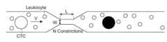
도 1은 빠른 기계적 변형 (rapid mechanical deformation)에 의한 순환 종양 세포 (CTCs)의 크기 선택적 태깅에 대한 시스템의 도이다.
도 2는 미세 유체 플랫폼의 크기 선택적 전달과 CD45 항체 염색법의 결합은 독립적으로 각각 독립적인 기술보다 더 나은 정도 이상의 샘플 농축 인자 (enrichment factor)를 만들어 낸다는 것을 보여주는 막대 그래프이다.
도 3a는 세포 라벨링 (cell labeling)의 개략도이다. 적혈구 (RBCs)는 eBioscience RBC 용해 완충액 (RBC lysis buffer, Cat. No. 00-4333)과 같은 표준 적혈구 용해 시약 (standard erythrocyte lysis reagent)을 이용한 적혈구 용해에 의하여 전혈로부터 제거되었다. 그 결과 협착 채널 미세 유체 장치 (constriction channel microfluidics device)를 따라서 흘려보내진 현탁액 결과물은 형광 염료 (및 혹은 다른 화합물)와 함께 인큐베이션 되었다. 상기 현탁액은 그 다음 CD45 표지되었고, 형광 염료로 표지된 비-CD45+ 세포를 모으기 위해 형광-활성 세포 분류 (fluorescence-activated cell sorter, FACS) 기기로 처리되었다.
도 3b는 PBMC (50psi에서 30-6 칩 (chip)), HT-29 (50psi에서 30-6 칩 (chip)), SK-MEL-5 (50psi에서 10-7 칩 (chip)), 및 PANC-1 (50psi에서 10-7 칩 (chip))에 대한 세포스퀴즈 (CellSqueez) 장치에 의하여 전달된 3 kDa 덱스트란 컨쥬게이트된 케스케이드 블루의 일련의 유세포분석 플롯 (flow cytometry plot) 이다.
도 3c는 일련의 Panc-1 종양 세포 (Panc-1 tumor cell)와 혈구 (blood cell)의 투과광 (transmitted light) 및 형광 현미경 사진이다. pre-전달 세포는 주위의 세포 내 섭취 (background endocytosis)를 교정하기 위하여 염료의 존재 하에서 인큐베이션 되었다. post-전달 사진은 염료의 지속 (retention)과 전달한 다음에 세포의 부착능과 성장능을 입증하기 위하여 전달 24시간 후에 촬영되었다. 큰 혈구 또한 프로세스 동안 라벨화 될 수 있음에도 불구하고, 이러한 데이터는 종양 세포의 선택적 라벨링을 보여준다.
도 4는 림프종 세포 vs. 건강한 PBMC에 대한 염료의 선택적 전달을 보여주는PBMC와 림프종 혼합물 (lymphoma mixture)에서 백분률 PBMC 대 PBMC 전달의 플롯이다. 현탁액이 수치상 99.9% 건강한 PBMC일 때에도, 일부 실시에서 8배까지 전달의 특이성이 성취될 수 있다. 다른 실시에서 더 큰 특이성이 달성될 수 있다.
도 5a는 전혈 (40 세포/ml) 속에 섞이고 CD45 카운터 스테인 (CD45 counter stain, APC)로 처리된 테트라메틸로다민 덱스트란-표지된 Panc-1-GFP 세포의 플롯이다.
도 5b는 PANC-1 GFP 태깅이 GFP 형광 기반으로 독립적으로 확인될 수 있는 방법을 보여주는 GFP vs. CD45의 FACS 플롯이다. P5 게이트는 후보 CTC를 분류하기 위한 기초로 사용될 수 있고, P4는 PANC-1 GFP 세포의 정체 (identity)를 확인하는데 사용된다. 녹색 점은 정확한 점이고 (P4 & P5), 붉은색 점은 거짓양성 (P5 만), 파란색 점은 놓친 것 (P4 만) 이다.
도 5c는 췌장 관 선암종 (pancreatic ductal adenocarcinoma)을 확인한 HTB1760의 초기 종양(HTB1760's primary tumor)의 조직병리학 사진이다.1 is a diagram of a system for size selective tagging of circulating tumor cells (CTCs) by rapid mechanical deformation.
Figure 2 is a bar graph showing that the combination of size-selective delivery of the microfluidic platform and CD45 antibody staining independently produces a sample enrichment factor that is more than an order of magnitude better than each of the independent techniques.
3A is a schematic diagram of cell labeling. Red blood cells (RBCs) were removed from whole blood by red blood cell lysis using a standard erythrocyte lysis reagent such as eBioscience RBC lysis buffer (Cat. No. 00-4333). As a result, the resulting suspension flowed along the constriction channel microfluidics device was incubated with a fluorescent dye (and/or other compound). The suspension was then labeled with CD45 and processed with a fluorescence-activated cell sorter (FACS) machine to collect non-CD45+ cells labeled with the fluorescent dye.
3B shows PBMC (30-6 chips at 50 psi), HT-29 (30-6 chips at 50 psi), SK-MEL-5 (10-7 chips at 50 psi), and PANC A series of flow cytometry plots of 3 kDa dextran conjugated Cascade Blue delivered by a CellSqueez device for -1 (10 −7 chips at 50 psi).
3C is a transmitted light and fluorescence micrograph of a series of Panc-1 tumor cells and blood cells. Pre-transduced cells were incubated in the presence of dye to correct for background endocytosis. Post-delivery pictures were taken 24 h after delivery to demonstrate the retention of the dye and the ability of cells to adhere and grow following delivery. Although large blood cells can also be labeled during the process, these data show selective labeling of tumor cells.
4 shows lymphoma cells vs. Plot of percentage PBMC versus PBMC delivery in PBMC and lymphoma mixture showing selective delivery of dye to healthy PBMC. Even when the suspension is numerically 99.9% healthy PBMCs, up to 8-fold specificity of delivery can be achieved in some implementations. Greater specificity may be achieved in other implementations.
5A is a plot of tetramethylrhodamine dextran-labeled Panc-1-GFP cells spiked into whole blood (40 cells/ml) and treated with CD45 counter stain (APC).
Figure 5b shows GFP vs. GFP tagging, showing how PANC-1 GFP tagging can be independently confirmed based on GFP fluorescence. FACS plot of CD45. The P5 gate can be used as a basis for sorting candidate CTCs, and the P4 is used to confirm the identity of PANC-1 GFP cells. Green dots are correct points (P4 & P5), red dots are false positives (P5 only), and blue dots are missed (P4 only).
5C is a histopathology photograph of HTB1760's primary tumor confirmed to have pancreatic ductal adenocarcinoma.
순환 종양 세포 (CTC)는 혈류에서 발견되는 종양 세포이고, 멀리 떨어진 기관 (organ)으로의 암 전파 (dissemination)에 원인이 되는 것으로 여겨진다. CTC는 암환자에 대한 최소 침습의 (minimally-invasive), "액상 생체검사 (liquid biopsy)"로 여겨지고, 환자 결과와 치료 효과에 대한 예후 지표로서 유용하다. 이러한 단일 세포의 종합적인 특성은 전이성 전파, 치료 내성 (treatment resistance), 및 종양 생물학 (tumor biology)의 더 나은 이해를 제공한다.Circulating tumor cells (CTCs) are tumor cells found in the bloodstream and are believed to be responsible for cancer dissemination to distant organs. CTC is considered a minimally-invasive, "liquid biopsy" for cancer patients, and is useful as a prognostic indicator for patient outcome and treatment effectiveness. The comprehensive characterization of these single cells provides a better understanding of metastatic spread, treatment resistance, and tumor biology.
전형적인 인간 적혈구 (erythrocyte)는 약 6.2-8.2 ㎛의 원판 직경, 가장 두꺼운 부분에서 두께 2-2.5 ㎛, 그리고 중심에서 최소 두께 0.8-1 ㎛을 갖고, 대부분의 다른 인간 세포보다 훨씬 작다. 백혈구 (하얀 혈구, white blood cell)는 호중구 (neutrophil, 직경 12-14 ㎛), 호산구 (eosinophil, 직경 12-17 ㎛), 호염기구 (basophils, 직경 14-16 ㎛), 림프구 (lymphocytes, 휴지기 동안 평균 직경 6-9 ㎛, 활성화 동안 직경 10-14 ㎛), 및 단핵구 (monocytes, 직경이 20 ㎛까지 일 수 있는 백혈구 중 가장 큰 타입)를 포함한다. 도 1에 나타낸 바와 같이, CTC와 혈액 세포 (hematologic cell) 사이의 크기차이는 일반적으로 순환하고 있는 혈액 (CTC~9-20 ㎛; RBC~8 ㎛ 원반모양(discoid); 백혈구~7-12 ㎛) 안에서 CTC를 다른 세포로부터 구별하는 것을 가능하게 한다. 도 1을 보라. 항체 (또는 이들의 세포-특이적 단편) 또는 다른 종양 세포 특이적 리간드를 이용한 다음의 종양 세포 특이적 라벨링은 본 방법의 선택성 (selectivity)을 증가시켰다.A typical human erythrocyte is much smaller than most other human cells, with a disc diameter of about 6.2-8.2 μm, a thickness of 2-2.5 μm at the thickest point, and a minimum thickness of 0.8-1 μm at the center. White blood cells (white blood cells) include neutrophils (12-14 ㎛ in diameter), eosinophils (12-17 ㎛ in diameter), basophils (14-16 ㎛ in diameter), and lymphocytes (during resting phase). 6-9 μm in average diameter, 10-14 μm in diameter during activation), and monocytes (the largest type of white blood cells that can be up to 20 μm in diameter). As shown in Figure 1, the size difference between CTCs and hematologic cells is generally circulating blood (CTC ~ 9-20 ㎛; RBC ~ 8 ㎛ discoid; leukocytes ~ 7-12 ㎛ ) in which it is possible to distinguish CTCs from other cells. See Figure 1. Subsequent tumor cell specific labeling with antibodies (or cell-specific fragments thereof) or other tumor cell specific ligands increased the selectivity of the method.
CTC는 혈류에 106-107 단핵세포 (mononuclear cell) 중에서 하나 존재하기 때문에, 고감도 농축 기술 (high-sensitivity enrichment technique)은 혈구와 CTC의 면역학적 또는 형태학적 차이점에 의존하여 사용된다. 면역학적 접근법은 종종 상피 세포 표면 마커 (epithelial cell surface marker, EpCAM 같은)와 종양-특이적 단백질 (tumor-specific proteins, Her2-neu, MUC I/MUC2, CEA (carcinoembryonic antigen ), mammaglobulin, 및 alpha-fetoprotein과 같은)을 타겟으로 하거나, 또는 CD45+ 세포를 감소시키는 것을 목적으로 한다. 미세필터 (microfilter), 밀도-차등 분리 (density-gradient separations) 그리고 미세 유체 플랫폼 (microfluidics platform)은 형태학 기반 방법 (morphology-based method)의 예이다. 이러한 접근방법 모두 내재하는 편향 (inhereny bias)을 가지고, 낮은 농축 효과로 어려움을 겪으며, 그리고 상당한 수의 CTC는 표면 항원을 하향조절 하고 다양한 형태적 특징을 보일 것이다. CTC의 일부가 전이에 책임이 있고, 또는 신뢰할 만한 예후 마커라는 것이 여전히 만이 알려지지 않았기 때문에 기술분야에서 이러한 편향은 상당한 이의 (challenge)를 갖는다. 따라서, 대부분의 임상적 후보를 검사하기 위하여 모든 후보 CTC 아형 (sub-type)의 고순도 분리를 보장할 수 있는 기술을 개발하는 것이 중요하다. 여기에 기재된 장치 및 방법은 관심있는 CTC 아형의 분리 및 열거 (enumeration)를 가능하게 한다.Since CTCs are present in the bloodstream as one out of 106 -107 mononuclear cells, a high-sensitivity enrichment technique is used depending on immunological or morphological differences between blood cells and CTCs. Immunological approaches often use epithelial cell surface markers (such as EpCAM) and tumor-specific proteins (Her2-neu, MUC I/MUC2, carcinoembryonic antigen (CEA), mammaglobulin, and alpha- fetoprotein), or to reduce CD45+ cells. Microfilters, density-gradient separations and microfluidics platforms are examples of morphology-based methods. All of these approaches have inherent biases, suffer from low enrichment effects, and a significant number of CTCs will downregulate surface antigens and display diverse morphological features. This bias has considerable challenge in the art as it is still only unknown which part of CTC is responsible for metastasis, or is a reliable prognostic marker. Therefore, it is important to develop techniques that can ensure high-purity isolation of all candidate CTC sub-types in order to screen most clinical candidates. The devices and methods described herein allow for the isolation and enumeration of CTC subtypes of interest.
결합된 농축 방법은 더 적은 편향 (bias)과 조정 가능한 파라미터를 기반으로 순수한 CTC를 태그하거나 분리하기 위하여 면역학적 및 형태학적-기반의 처리방법 모두를 통합한다. 상기 방법은 미세 유체 세포 내 전달 (도 1)과 전혈로부터 순환 종양 세포의 강력한, 고 민감성 정제 (high sensitivity purification)를 달성하기 위한 항체 염색 (도 2)을 결합하고, 너비 (width) 4 ㎛-10 ㎛, 길이 1 ㎛-100 ㎛, 그리고 일련의 1-10 협착부를 포함한다. 세포의 추정 속도는 10 mm/s - 10 m/s 일 수 있다. 상기 특정 장비 파라미터 선택은 표적 종양 세포 타입에 의하여 결정, 예를 들어 흑색종 환자 vs. 대장암 환자를 위한 CTC를 선택하는데는 다른 장치 설계가 사용, 된다. 종양 세포 크기/직경의 예로; 흑색종 ~15㎛, 대장암 ~ 11 ㎛, 및 췌장암 ~ 15㎛.The combined enrichment method integrates both immunological and morphology-based approaches to tag or isolate pure CTCs based on less bias and tunable parameters. The method combines microfluidic intracellular delivery (Fig. 1) with antibody staining to achieve robust, high sensitivity purification of circulating tumor cells from whole blood (Fig. 2), and is 4 μm-wide. 10 μm, 1 μm-100 μm in length, and contains a series of 1-10 constrictions. The estimated velocity of the cell may be 10 mm/s - 10 m/s. The selection of specific equipment parameters is determined by the target tumor cell type, eg melanoma patients vs. Different device designs are used to select CTCs for colorectal cancer patients. Examples of tumor cell size/diameter; Melanoma ~ 15 μm, colorectal cancer ~ 11 μm, and pancreatic cancer ~ 15 μm.
이러한 처리방법에서, 빠른 기계적 변형 전달 시스템은 종양 세포로 형광, 자성 (magnetic) 및/또는 다른 구별되는 물질 (distinguishing material)을 선택적으로 전달하기 위하여 많은 CTC와 그 주변 혈구의 내재적 크기 차이를 이용한다. 상기 과정에서, 항체-기반 형광 및 자성 태깅은 후보 CTC와 주변 혈구의 대비를 향상시키기 위하여 사용된다. CTC 분리를 위해 크기-기반 및 면역적 접근을 특유의 방법으로 조합함으로써, 이 기술은 분석을 위해 환자 샘플에서 후보 종양 세포의 비-편향된 분리 (non-biased isolation)를 위한 유용성 (utility)을 증명하였다. 일 실시예에서, 더 작은 그리고 더 큰 세포 모두 변형되지만, 더 작은 세포의 막은 막이 손상되는 지점까지 변형되지 않는다. 예를 들어, 전혈 (여기서 가장 건강한 백혈구 세포는 ~8 ㎛ 크기)에서 15 ㎛ 종양 세포에 선택적으로 전달하기 위해, 너비 6 ㎛의 협착부가 사용될 수 있다. 이러한 협착부는 두 세포 유형 모두 변형할 것이지만, 8 ㎛ 혈액 세포가 아니라 15 ㎛ 종양세포의 세포막을 매우 우선적으로 교란 (disrupt)할 것이다.In this approach, the rapid mechanical strain delivery system exploits the intrinsic size difference of many CTCs and their surrounding blood cells to selectively deliver fluorescent, magnetic and/or other distinguishing materials to the tumor cells. In this process, antibody-based fluorescence and magnetic tagging is used to improve the contrast between candidate CTCs and surrounding blood cells. By uniquely combining size-based and immunological approaches for CTC isolation, this technology demonstrates utility for non-biased isolation of candidate tumor cells from patient samples for analysis. did In one embodiment, both the smaller and larger cells are deformed, but the membranes of the smaller cells are not deformed to the point where the membranes are damaged. For example, for selective delivery of 15 μm tumor cells in whole blood (where the most healthy white blood cells are ˜8 μm in size), a 6 μm wide constriction can be used. These constrictions will deform both cell types, but will very preferentially disrupt the cell membrane of 15 μm tumor cells rather than 8 μm blood cells.
임상적/번역 관련성 (Clinical/Translation Relevance)Clinical/Translation Relevance
CTC는 저항의 기작 (mechanism)을 이해하고, 표적 치료의 선택을 안내하기 위하여 종양 생검 (tumor biopsy)을 위한 대안 (surrogate)으로 분석되고 있다. 치료 전 및 후에 CTC의 수 (number)와 조성 (composition)의 측정은 치료 효과와 예후 (prognosis)를 가리킨다. 이러한 처리방법은 세포 크기와 표면 항원을 기반으로 CTC의 태깅을 위하여 튼튼한, 높은-처리율의, 일회용 장치를 활용한다. 게다가, 다양한 거대분자를 전달하는 능력은 또한 분자 프로브 (항체, 양자점, 탄소나노튜브, 및 분자 비콘 (molecular beacon)과 같은)를 전달할 수 있게 하고, 이러한 분자 프로브는 세포 내 환경에 반응하고 이렇게 하여 표적 세포의 세포 내 성질에 대한 더 많은 정보를 제공한다. 이러한 복합적 처리방법은 오로지 면역학적 또는 형태학적 분리에만 의존한 다른 방법에 의하여 놓치고 있었던 CTC 집단을 농축할 수 있는 강력한 플랫폼을 제공한다. 이러한 기술은 환자의 CTC를 분리하는데 유용하다.CTC is being analyzed as a surrogate for tumor biopsy to understand the mechanism of resistance and guide the selection of targeted therapies. Measurement of the number and composition of CTCs before and after treatment indicates treatment effectiveness and prognosis. This processing method utilizes a robust, high-throughput, disposable device for tagging of CTCs based on cell size and surface antigen. In addition, the ability to deliver a variety of macromolecules also allows for the delivery of molecular probes (such as antibodies, quantum dots, carbon nanotubes, and molecular beacons) that respond to the intracellular environment and thereby It provides more information about the intracellular properties of target cells. This multiplex processing method provides a powerful platform to enrich CTC populations that have been missed by other methods that rely solely on immunological or morphological isolation. This technique is useful for isolating CTCs from patients.
실시예 1Example 1
전혈 또는 다른 세포 현탁액은 비표지된 및/또는 항체-코팅된 자성 비드 모두를 이용하여 처리된다. 이러한 세포는 그 다음 희귀 세포 (rare cell)를 위한 고성능 (high-fidelity), 자성의 농축 시스템을 이용하여 분리된다. 나노웰 (nanowell) 기술은 또한 고순도 분리를 달성하기 위해 84,672 서브나노리터 웰의 엘라스토머릭 어레이 (elastomeric array)로부터 관심있는 단일 세포를 자동기계 장치적으로-검색하고 이미지화 하는 방법으로 사용될 것이다.Whole blood or other cell suspensions are processed using both unlabeled and/or antibody-coated magnetic beads. These cells are then isolated using a high-fidelity, magnetic enrichment system for rare cells. Nanowell technology will also be used as a method to automechanically-retrieve and image single cells of interest from an elastomeric array of 84,672 sub-nanoliter wells to achieve high-purity separation.
다양한 표현형의 단일의, 살아있는, 순종의 (pure), 온전한 (intact) CTC를 얻는 것은 즉각적인 임상적 그리고 번역적 관련성에 대하여 유전적 수준에서 기능적 수준까지 숙주의 특성화를 가능하게 한다. 이러한 방법은 감소된 편향 (bias)과 함께, 다양한 CTC의 매우 민감하고 특이적인 농축을 허용한다.Obtaining single, live, pure, intact CTCs of diverse phenotypes allows characterization of the host from the genetic level to the functional level with immediate clinical and translational relevance. This method allows highly sensitive and specific enrichment of various CTCs, with reduced bias.
실시예 2Example 2
자성 나노입자는 종양 세포주 & PBMC에 전달된다. EpCAM-발현 상피 암세포주 (예를 들어, HT-29, LNCaP, 및 SK-BR-3)에 나노입자 전달은 인간 혈액 유래 대량의 말초 혈액 단핵 세포 (PBMC) 현탁액과 비교된다.Magnetic nanoparticles are delivered to tumor cell lines & PBMCs. Nanoparticle delivery to EpCAM-expressing epithelial cancer cell lines (eg, HT-29, LNCaP, and SK-BR-3) is compared to bulk peripheral blood mononuclear cell (PBMC) suspensions derived from human blood.
폴리에틸렌 글리콜 (polyethylene glycol, PEG)로 표면 코팅된 10 nm 산화철 나노입자 (iron-oxide nanoparticle)는 전혈과 혼합된 종양 세포로 전달되고, 상기에 기재된 세포 분리 시스템을 이용하여 태그된 세포의 혼합물이 처리된다. 예를 들어, 미세 유체 전달 시스템은 세포막에서 일시적인 기공을 형성하기 위해 급성 기계적 세포의 변형을 유도하는데 사용되었다 (도 1). 상기 접근방법은 다양한 유형의 세포로 단백질, RNA, DNA 및 나노입자를 포함하는 물질의 범위를 전달하는 능력을 입증하고, 전혈, 종종 미세 유체 시스템에 대하여 문제를 일으키는 배지를 대상으로 한다.10 nm iron-oxide nanoparticles surface-coated with polyethylene glycol (PEG) are delivered to the tumor cells mixed with whole blood, and the mixture of tagged cells is processed using the cell separation system described above. do. For example, microfluidic delivery systems have been used to induce acute mechanical cell deformation to form transient pores in cell membranes (Fig. 1). This approach demonstrates the ability to deliver a range of substances including proteins, RNA, DNA and nanoparticles to cells of various types, targeting whole blood, a medium that is often problematic for microfluidic systems.
모델 분자로서 예시의 태깅 분자, 예를 들어, 3 kDA 및 70 kDA, 형광-표지된 덱스트란 고분자는 크기에만 기반하여 PBMC와 두 개의 다른 암 세포주를 구별하는데 사용되었다. 그 결과는 또한 전혈에서 종양 세포에 자성 입자의 선택적인 전달을 위한 시스템의 유용성을 나타낸다. PEG 코팅된 산화철 입자는 대장암 (예를 들어, 세포주 HT-29에 의하여 예시된 바와 같이)을 자성적으로 태그하는데 사용된다. 또한, 농축은 나노 입자 흡수를 직접적으로 측정하기 위하여 산화철 나노입자 표면에 FITC의 접합을 이용하여 수행된다.Exemplary tagging molecules as model molecules, eg, 3 kDA and 70 kDA, fluorescently-labeled dextran polymers, were used to distinguish PBMCs from two other cancer cell lines based only on size. The results also indicate the utility of the system for selective delivery of magnetic particles to tumor cells in whole blood. PEG coated iron oxide particles are used to magnetically tag colorectal cancer (eg, as exemplified by the cell line HT-29). Concentration is also performed using conjugation of FITC to the surface of iron oxide nanoparticles to directly measure nanoparticle uptake.
PEG 코팅된 10 nm 산화철 나노입자는 세포 현탁액으로 전달되고, 상기 세포 현탁액은 개별적으로 전혈과 혼합되고 CTC (예를 들어, 환자-유래 혈액 시료) 또는 세포주 HT-29, LNCaP, 및 SK-BR-3 세포를 포함한다고 의심되는 또는 포함하는 것으로 알려진 것이다. 그 다음 예를 들면 높은 정확도 자성 분리기를 이용하여 태그된 세포의 혼합물의 결과물이 정제된다. 상기 분리기는 높은 산화철 함량을 갖는 모ㄷ델 CTC와 덜 효과적으로 표지된 PBMC를 정확하게 구별한다. 선택적으로, 적혈구는 처리, 증가 된 나노 입자 농도, 변경된 그들의 크기 또는 여러 번의 처리 단계를 포함하는 것의 사전에 용해된다.PEG-coated 10 nm iron oxide nanoparticles are delivered as a cell suspension, which is individually mixed with whole blood and cultured in CTCs (eg, patient-derived blood samples) or cell lines HT-29, LNCaP, and SK-BR- 3 are suspected to contain or are known to contain cells. The resulting mixture of tagged cells is then purified, for example using a high accuracy magnetic separator. The separator accurately discriminates between model CTCs with high iron oxide content and less effectively labeled PBMCs. Optionally, the red blood cells are lysed prior to processing, including increased nanoparticle concentration, altered their size, or multiple processing steps.
실시예 3Example 3
결합된 면역학 및 형태 기반 방법은 다음에 따라 수행된다. 장치를 이용한 세포 크기-기반 처리 후에, 세포는 항체 또는 형광 표지된 항-CD45 항체와 같은 다른 종양세포 특이적 리간드로 처리된다. 세 가지 분리 방법의 민감도 (sensitivity)와 특이도 (specifity)는 비교되었다:: 1) 장치 만 2) 항-CD45 항체만 3) 장치+항-CD45 항체. 형태학적 태깅 (장치) + 면역학적 태깅 (예를 들어, 항-CD45 항체)은 각각의 개별 기술에 비해 우수한 민감도 (및 특이도)를 보여주는 것이 확인되었다 (도 2). 예를 들어, 항-CD45 항체에 비해서 민감도에서 2-5x 증가 및/또는 특이도에서 2-5x 증가가 관찰된다. 정도 (order of magnitude) 이상의 농축인자가 관찰되었다 (도 2).The combined immunology and morphology-based method is performed according to the following. After cell size-based treatment with the device, the cells are treated with an antibody or other tumor cell specific ligand such as a fluorescently labeled anti-CD45 antibody. The sensitivity and specificity of the three separation methods were compared: 1) device only 2) anti-CD45 antibody only 3) device + anti-CD45 antibody. It was found that morphological tagging (device) plus immunological tagging (eg anti-CD45 antibody) showed superior sensitivity (and specificity) compared to each individual technique ( FIG. 2 ). For example, a 2-5x increase in sensitivity and/or a 2-5x increase in specificity is observed relative to an anti-CD45 antibody. An enrichment factor of more than an order of magnitude was observed (FIG. 2).
실시예 4Example 4
일 실시예 에서, 장치는 실리콘 및 유리로 부터 제조된다. 혹은, 상기 장치는 실리콘 (silicone), PDMS, 폴리카보네이트 (polycarbonate), 아크릴 (acrylic), 폴리프로필렌 (polypropylene), 폴리스티렌 (polystyrene)과 같은 고분자로 제조된다. 어느 장치는 멸균 (열 또는 감마선)되고, 일회용이다. 장치의 성능은 물질 및 파라미터를 이용하여 다양한 세포 유형에 대하여 검증된다. 예를 들어, PEG 코팅된 양자점 (범위 10-50 nm 크기)을 이용한 유체 속도 (100 mm/s-10,000 mm/s)의 범위에서 성능은 나노입자의 전달 효과 및 세포 활성여부를 결정하기 위하여 사용된다. 장치의 예시는 본 발명에서 인용문헌으로 포함되는 PCT/US2012/060646에 기재되어 있다.In one embodiment, the device is made from silicon and glass. Alternatively, the device is made of a polymer such as silicone, PDMS, polycarbonate, acrylic, polypropylene, or polystyrene. Either device is sterile (heat or gamma radiation) and is disposable. The performance of the device is validated for various cell types using materials and parameters. For example, the performance in the range of fluid velocity (100 mm/s-10,000 mm/s) using PEG-coated quantum dots (size range 10-50 nm) is used to determine the delivery effect and cell activity of nanoparticles. do. Examples of devices are described in PCT/US2012/060646, which is incorporated herein by reference.
장점 (Advantage)Advantage
종래의 처리방법과 비교했을 때 본 방법은 다음의 장점이 있다. 항체-기반 방법에 비해서, 본 처리방법은 대부분의 암 유형에 대해 일반화할 수 있고 어느 특정 세포 표면 마커에 독립적인 비-편향 분리 방법을 제공한다. 상기 장치 및 방법은 기존의 마커에 의하여 분리될 수 없는 CTC의 식별을 수행함으로써, 현저한 진단 및 예후적 의미를 갖는다.Compared with conventional treatment methods, this method has the following advantages. Compared to antibody-based methods, this treatment method provides a non-biased separation method that is generalizable to most cancer types and independent of any particular cell surface marker. The device and method have significant diagnostic and prognostic significance by performing the identification of CTCs that cannot be separated by existing markers.
기존의 크기-기반 분리방법에 비하여, 본 명세서에 기재된 상기 장치 및 방법은 매우 높은 처리량 (throughput)을 제공하고, 특정 크기 범위의 CTC를 포착하기 위하여 "W"를 변화하여 조정한다 (도 1). 예를 들어, 너비 (width) 6 ㎛ 협착부는 대장암 세포를 캡쳐하는데 적합한 반면, 너비 (width) 7 ㎛ 및 8 ㎛는 췌장암 및 흑생종 세포를 각각 캡쳐하는데 적합하다. 게다가, 기존 기술과는 달리, 이 시스템은 분리 민감도를 향상시키기 위해 및/또는 CTC의 일부의 멀티-파라미터 분리를 가능하게하기 위해 항체-기반 기술과 결합 된다 (예를 들어, 특정 크기의 CTC + 표면 마커를 분리함으로써).Compared to existing size-based separation methods, the device and method described herein provide very high throughput and are tuned by changing "W" to capture CTCs in a specific size range (Fig. 1). . For example, a 6 μm wide constriction is suitable for capturing colorectal cancer cells, while 7 μm and 8 μm widths are suitable for capturing pancreatic cancer and melanoma cells, respectively. Moreover, unlike existing techniques, this system is coupled with antibody-based techniques to improve separation sensitivity and/or to enable multi-parameter separation of a fraction of CTCs (e.g., CTCs of a certain size + by isolating surface markers).
다양한 암 유형에서 CTC의 효과적이고, 강력한 분리를 가능하게 함으로써 이 기술은 암과의 싸움에서 중요한 플랫폼이 될 것이다. 이 기술의 예후적 및 진단적 잠재력은 암의 진행에서 중요한 새로운 유전자의 식별을 가능하게 하고, 따라서 신규한 치료제의 개발을 가능하게 할 수 있다. 또한, 더욱 정확한 환자의 예상 수명의 예측 및 치료 효과를 제공할 수있다.By enabling effective and robust isolation of CTCs from various cancer types, this technology will become an important platform in the fight against cancer. The prognostic and diagnostic potential of this technology may enable the identification of new genes important in cancer progression and thus the development of novel therapeutics. In addition, it can provide a more accurate prediction of the patient's life expectancy and treatment effect.
본 명세서에 기재된 CTC 분리 방법은 고 농축 계수 (factor)/회수율 및 조절가능한 바이어스 (특정 마커 vs. 특정 크기)를 수득하기 위해 면역 및 크기 기반 분리를 결합한다.The CTC isolation method described herein combines immunological and size-based separation to obtain a high enrichment factor/recovery and adjustable bias (marker specific vs. size specific).
상기 설명에서 약간의 변형에 대하여 기재했음에도 불구하고, 다른 변형은 가능하다. 예를 들어, 상기의 구현예는 개시된 특징의 다양한 조합 및 서브조합 및/또는 상기 개시된 여러 특징의 조합 및 서브조합이 될 수 있다. 또한, 본 명세서에서 기재된 로직의 흐름은 목적하는 결과를 달성하기 위하여 설명된 특별한 순서 또는 순차적인 순서가 요구되지 않는다. 다른 측면은 다음의 청구범위 내에 있다.Although slight variations have been described in the above description, other variations are possible. For example, embodiments of the above may be various combinations and subcombinations of the disclosed features and/or combinations and subcombinations of the various features disclosed above. In addition, the flow of logic described herein does not require any particular order or sequential order described to achieve a desired result. Other aspects are within the scope of the following claims.
Claims (23)
Translated fromKoreanApplications Claiming Priority (4)
| Application Number | Priority Date | Filing Date | Title |
|---|---|---|---|
| US201361866972P | 2013-08-16 | 2013-08-16 | |
| US61/866,972 | 2013-08-16 | ||
| PCT/US2014/051343WO2015023982A1 (en) | 2013-08-16 | 2014-08-15 | Selective delivery of material to cells |
| KR1020217028921AKR102386122B1 (en) | 2013-08-16 | 2014-08-15 | Selective delivery of material to cells |
Related Parent Applications (1)
| Application Number | Title | Priority Date | Filing Date |
|---|---|---|---|
| KR1020217028921ADivisionKR102386122B1 (en) | 2013-08-16 | 2014-08-15 | Selective delivery of material to cells |
Publications (2)
| Publication Number | Publication Date |
|---|---|
| KR20220049050A KR20220049050A (en) | 2022-04-20 |
| KR102530914B1true KR102530914B1 (en) | 2023-05-11 |
Family
ID=52468732
Family Applications (4)
| Application Number | Title | Priority Date | Filing Date |
|---|---|---|---|
| KR1020227011724AActiveKR102530914B1 (en) | 2013-08-16 | 2014-08-15 | Selective delivery of material to cells |
| KR1020217028921AActiveKR102386122B1 (en) | 2013-08-16 | 2014-08-15 | Selective delivery of material to cells |
| KR1020217010816AActiveKR102304167B1 (en) | 2013-08-16 | 2014-08-15 | Selective delivery of material to cells |
| KR1020167006938AActiveKR102243597B1 (en) | 2013-08-16 | 2014-08-15 | Selective delivery of material to cells |
Family Applications After (3)
| Application Number | Title | Priority Date | Filing Date |
|---|---|---|---|
| KR1020217028921AActiveKR102386122B1 (en) | 2013-08-16 | 2014-08-15 | Selective delivery of material to cells |
| KR1020217010816AActiveKR102304167B1 (en) | 2013-08-16 | 2014-08-15 | Selective delivery of material to cells |
| KR1020167006938AActiveKR102243597B1 (en) | 2013-08-16 | 2014-08-15 | Selective delivery of material to cells |
Country Status (13)
| Country | Link |
|---|---|
| US (3) | US10124336B2 (en) |
| EP (2) | EP3848695A1 (en) |
| JP (1) | JP6502940B2 (en) |
| KR (4) | KR102530914B1 (en) |
| CN (1) | CN105848793B (en) |
| AU (2) | AU2014306423B2 (en) |
| CA (1) | CA2921579C (en) |
| ES (1) | ES2865107T3 (en) |
| IL (1) | IL244113B (en) |
| MX (2) | MX382654B (en) |
| PL (1) | PL3033184T3 (en) |
| SG (2) | SG11201601927SA (en) |
| WO (1) | WO2015023982A1 (en) |
Families Citing this family (52)
| Publication number | Priority date | Publication date | Assignee | Title |
|---|---|---|---|---|
| CN107058101B (en)* | 2011-10-17 | 2021-06-01 | 麻省理工学院 | intracellular delivery |
| KR102530914B1 (en) | 2013-08-16 | 2023-05-11 | 메사추세츠 인스티튜트 오브 테크놀로지 | Selective delivery of material to cells |
| JP7523203B2 (en)* | 2014-10-31 | 2024-07-26 | マサチューセッツ インスティテュート オブ テクノロジー | Delivery of biomolecules to immune cells |
| CA2964138C (en) | 2014-11-14 | 2023-11-14 | Massachusetts Institute Of Technology | Disruption and field enabled delivery of compounds and compositions into cells |
| WO2016115179A1 (en) | 2015-01-12 | 2016-07-21 | Massachusetts Institute Of Technology | Gene editing through microfluidic delivery |
| EP4257675A3 (en) | 2015-07-09 | 2024-01-03 | Massachusetts Institute of Technology | Delivery of materials to anucleate cells |
| CN108138118B (en) | 2015-09-04 | 2023-01-06 | Sqz生物技术公司 | Intracellular delivery of biomolecules mediated by porous surfaces |
| US11613759B2 (en) | 2015-09-04 | 2023-03-28 | Sqz Biotechnologies Company | Intracellular delivery of biomolecules to cells comprising a cell wall |
| EP3365269A4 (en)* | 2015-10-19 | 2019-06-19 | The Methodist Hospital | DISTRIBUTION, BY MEMBRANE DEFORMATION, FROM CRISPR-CAS9 TO DIFFICULT CELLS TO BE TRANSFERRED |
| EA201891532A1 (en) | 2015-12-28 | 2019-01-31 | Новартис Аг | COMPOSITIONS AND METHODS OF TREATMENT OF HEMOGLOBINOPATHY |
| JP7033535B2 (en) | 2016-01-12 | 2022-03-10 | スクイーズ バイオテクノロジーズ カンパニー | Intracellular delivery of complex |
| US20190275520A1 (en) | 2016-03-31 | 2019-09-12 | Massachusetts Institute Of Technology | Flow-through microfluidic methods and devices featuring membrane-perturbing surface interactions for intracellular delivery |
| AU2017259987B2 (en) | 2016-05-03 | 2023-10-19 | Sqz Biotechnologies Company | Intracellular delivery of biomolecules to induce tolerance |
| CN109475577A (en) | 2016-05-03 | 2019-03-15 | Sqz生物技术公司 | Intracellular delivery of biomolecules to induce tolerance |
| CN106378213B (en)* | 2016-05-05 | 2018-08-17 | 海南大学 | Deformable microparticle separating chips based on dielectrophoresis |
| EP3500662B1 (en) | 2016-08-20 | 2022-10-05 | The Regents Of The University Of California | High-throughput system and method for the temporary permeabilization of cells |
| WO2018089497A1 (en)* | 2016-11-08 | 2018-05-17 | Georgia Tech Research Corporation | Methods for convectively-driven intracellular delivery |
| TW201839136A (en) | 2017-02-06 | 2018-11-01 | 瑞士商諾華公司 | Composition and method for treating hemochromatosis |
| US11383241B2 (en)* | 2017-10-11 | 2022-07-12 | The Regents Of The University Of California | Mechano-node pore sensing |
| WO2019089034A1 (en)* | 2017-11-02 | 2019-05-09 | The Methodist Hospital | Crispr-cas9 delivery to hard-to-transfect cells via membrane deformation |
| WO2019113125A1 (en) | 2017-12-05 | 2019-06-13 | Sqz Biotechnologies Company | Intracellular delivery of biomolecules to modulate antibody production |
| US11365390B2 (en) | 2017-12-19 | 2022-06-21 | Xcell Biosciences, Inc. | Methods of modulating cell phenotype by way of regulating the gaseous environment |
| IL320767A (en) | 2017-12-20 | 2025-07-01 | Stemcell Technologies Canada Inc | Cargo transfer system into a compartment |
| JP2019154314A (en)* | 2018-03-13 | 2019-09-19 | 国立大学法人九州大学 | Capillary chip and device comprising the same and introduction method of foreign material to cell |
| EP3556845A1 (en) | 2018-04-20 | 2019-10-23 | Cellix Limited | A method and device for transfecting cells |
| CN109351370B (en)* | 2018-11-21 | 2020-05-05 | 晶准生物医学(深圳)有限公司 | Microfluidic chip and cell screening method |
| KR20210121106A (en) | 2019-01-25 | 2021-10-07 | 에스큐지 바이오테크놀로지스 컴퍼니 | Non-nucleated cell-derived vaccines |
| MX2021010320A (en) | 2019-02-28 | 2021-11-12 | Sqz Biotechnologies Co | ADMINISTRATION OF BIOMOLECULES TO PERIPHERAL BLOOD MONONUCLEAR CELLS (PBMC) TO MODIFY AN IMMUNE RESPONSE. |
| DE102019108155B3 (en) | 2019-03-29 | 2020-06-04 | Leibniz-Institut Für Photonische Technologien E.V. | Micro drop retention assembly |
| WO2020210162A1 (en) | 2019-04-08 | 2020-10-15 | Sqz Biotechnologies Company | Cartridge for use in a system for delivery of a payload into a cell |
| WO2020207456A1 (en)* | 2019-04-11 | 2020-10-15 | The Chinese University Of Hong Kong | Systems and methods for controlled membrane disruption |
| US20220213422A1 (en)* | 2019-05-15 | 2022-07-07 | Cellfe, Inc. | Methods and systems for intracellular delivery and products thereof |
| US20230279394A1 (en) | 2019-12-18 | 2023-09-07 | Novartis Ag | Compositions and methods for the treatment of hemoglobinopathies |
| CN111413257B (en)* | 2020-01-21 | 2021-05-14 | 中国科学院电子学研究所 | Cell nucleus electrical property detection device and method |
| CR20220576A (en) | 2020-05-11 | 2022-12-07 | Hoffmann La Roche | COMMON THERAPY WITH MODIFIED PBMC AND AN IMMUNOCONJUGATE |
| CN116348583A (en)* | 2020-05-26 | 2023-06-27 | 川赛托斯有限责任公司 | Device and method for transfection |
| JP2023535982A (en) | 2020-07-29 | 2023-08-22 | スクイーズ バイオテクノロジーズ カンパニー | Methods for stimulating an immune response against mutant Ras using anucleate-free cells |
| EP4188428A1 (en) | 2020-07-29 | 2023-06-07 | SQZ Biotechnologies Company | Methods to stimulate immune responses to mutant ras using nucleated cells |
| CN117015596B (en) | 2020-11-18 | 2024-02-09 | 塞尔菲公司 | Methods and systems for mechanically perforated payload delivery to biological cells |
| US12227729B2 (en) | 2020-12-24 | 2025-02-18 | Cellfe, Inc. | Methods and systems for high-throughput cell processing |
| JP2024502791A (en) | 2020-12-29 | 2024-01-23 | スクイーズ バイオテクノロジーズ カンパニー | High-throughput microfluidic chip with parallelized constrictions for perturbing cell membranes |
| TW202241466A (en) | 2020-12-29 | 2022-11-01 | 美商Sqz生物科技公司 | Methods for treating cancers with modified pbmcs |
| WO2022147017A1 (en) | 2020-12-29 | 2022-07-07 | Sqz Biotechnologies Company | Formulations for cryopreservation of pbmcs |
| CN117042796A (en) | 2020-12-29 | 2023-11-10 | Sqz生物技术公司 | Method of treating cancer with activating antigen carrier |
| US20220203368A1 (en) | 2020-12-29 | 2022-06-30 | Sqz Biotechnologies Company | Microfluidic chip having increased throughput for use in a system for delivery of a payload into a cell |
| US20220233676A1 (en) | 2020-12-29 | 2022-07-28 | Sqz Biotechnologies Company | Formulations of activating antigen carriers |
| US20250127809A1 (en) | 2021-06-23 | 2025-04-24 | Novartis Ag | Compositions and methods for the treatment of hemoglobinopathies |
| WO2023010090A1 (en) | 2021-07-29 | 2023-02-02 | Sqz Biotechnologies Company | Methods to generate enhanced tumor infiltrating lymphocytes through microfluidic delivery |
| EP4430168A1 (en) | 2021-11-11 | 2024-09-18 | Stemcell Technologies Canada Inc. | Methods to generate enhanced tumor infiltrating lymphocytes through microfluidic delivery |
| US20250290018A1 (en) | 2022-05-27 | 2025-09-18 | Stemcell Technologies Canada Inc. | Cartridges and devices for use in a system for delivery of a payload into a cell |
| EP4561620A1 (en) | 2022-07-28 | 2025-06-04 | Stemcell Technologies Canada Inc. | Methods for treating cancer with enhanced antigen presenting cells |
| JP2025527190A (en) | 2022-07-28 | 2025-08-20 | ステムセル テクノロジーズ カナダ インコーポレイテッド | Enhanced antigen-presenting cell preparation |
Citations (5)
| Publication number | Priority date | Publication date | Assignee | Title |
|---|---|---|---|---|
| US20080318324A1 (en) | 2007-06-20 | 2008-12-25 | University Of Washington | Biochip for High-Throughput Screening of Circulating Tumor Cells |
| JP2010227011A (en)* | 2009-03-27 | 2010-10-14 | Seiko Epson Corp | Cell treatment device, cell treatment cartridge, and body fluid treatment system |
| JP2011163830A (en) | 2010-02-05 | 2011-08-25 | Tokyo Univ Of Agriculture & Technology | Detection of circulation tumor cells using size-selective micro cavity array |
| WO2012097450A1 (en) | 2011-01-19 | 2012-07-26 | The University Of British Columbia | Apparatus and method for particle separation |
| WO2013059343A1 (en)* | 2011-10-17 | 2013-04-25 | Massachusetts Institute Of Technology | Intracellular delivery |
Family Cites Families (118)
| Publication number | Priority date | Publication date | Assignee | Title |
|---|---|---|---|---|
| DE2502621C3 (en) | 1975-01-23 | 1978-09-14 | Kernforschungsanlage Juelich Gmbh, 5170 Juelich | Measurement of elastic and dielectric properties of the membrane of living cells |
| US4376634A (en) | 1980-05-30 | 1983-03-15 | Mallinckrodt, Inc. | Assay kit having syringe, dilution device and reagents within sealed container |
| US4478824A (en) | 1983-08-08 | 1984-10-23 | Franco Robert S | Method for altering red blood cell function and survival |
| FR2569477B1 (en) | 1984-08-24 | 1987-01-02 | Descartes Universite Rene | APPARATUS AND METHOD FOR DETERMINING THE DEFORMABILITY OF RED BLOOD CELLS |
| JP2720161B2 (en) | 1988-02-01 | 1998-02-25 | 株式会社アドバンス | Cell deformability measuring device |
| JP2685544B2 (en) | 1988-11-11 | 1997-12-03 | 株式会社日立製作所 | Blood filter, blood test method, and blood test apparatus |
| JP2532707B2 (en) | 1990-03-08 | 1996-09-11 | 佑二 菊池 | Blood circuit, blood measuring apparatus and blood measuring method using the same |
| HK1004060A1 (en) | 1990-04-24 | 1998-11-13 | Flustat Pty Ltd | Oral vaccine comprising antigen surface-associated with red blood cells |
| US5658892A (en) | 1993-01-15 | 1997-08-19 | The General Hospital Corporation | Compound delivery using high-pressure impulse transients |
| US6218166B1 (en) | 1994-12-09 | 2001-04-17 | John Wayne Cancer Institute | Adjuvant incorporation into antigen carrying cells: compositions and methods |
| US6461867B1 (en) | 1995-03-08 | 2002-10-08 | The Scripps Research Institute | Synthetic antigen presenting matrix |
| US6051409A (en) | 1995-09-25 | 2000-04-18 | Novartis Finance Corporation | Method for achieving integration of exogenous DNA delivered by non-biological means to plant cells |
| US6133503A (en) | 1995-10-31 | 2000-10-17 | The Regents Of The University Of California | Mammalian artificial chromosomes and methods of using same |
| WO1997020570A1 (en) | 1995-12-04 | 1997-06-12 | Puget Sound Blood Center | Non-immunogenic and toleragenic platelet and red blood cell compositions |
| BR9708387A (en) | 1996-03-28 | 2000-01-04 | Genitrix Llc | Process for vaccination of a mammal to a selected antigen, host and pathogenic cells, vaccine composition, nucleic acid, and engineered opsonin. |
| US5885470A (en) | 1997-04-14 | 1999-03-23 | Caliper Technologies Corporation | Controlled fluid transport in microfabricated polymeric substrates |
| DE69732225T2 (en) | 1997-05-05 | 2005-06-23 | Dideco S.R.L., Mirandola | Process for the encapsulation of biologically active substances in erythrocytes and apparatus therefor |
| US5842787A (en) | 1997-10-09 | 1998-12-01 | Caliper Technologies Corporation | Microfluidic systems incorporating varied channel dimensions |
| SE9704076D0 (en) | 1997-11-06 | 1997-11-06 | Holdingbolaget Vid Goeteborgs | Method for permeabilization of cell structures and use thereof |
| US20050026283A1 (en) | 2001-11-27 | 2005-02-03 | Owe Ormar | Method for combined parallel agent delivery and electroporation for cell structures and thereof |
| GB9816583D0 (en) | 1998-07-31 | 1998-09-30 | Univ Ulster | Nucleic acid carrier |
| US6562616B1 (en) | 1999-06-21 | 2003-05-13 | The General Hospital Corporation | Methods and devices for cell culturing and organ assist systems |
| JP2002325572A (en) | 2000-12-25 | 2002-11-12 | Univ Osaka | How to introduce foreign substances |
| AU2002252073A1 (en) | 2001-02-22 | 2002-09-12 | The Scepens Eye Research Institute, Inc. | Tolerogenic antigen presenting cells and in treating immune-inflammatory conditions |
| WO2003020039A1 (en) | 2001-08-28 | 2003-03-13 | Rush-Presbyterian-St. Luke's Medical Center | Immune tolerance to predetermined antigens |
| US20030133922A1 (en) | 2002-01-15 | 2003-07-17 | Kasha John R. | Oral tolerance using allogeneic platelets in ITP |
| US7501278B2 (en) | 2002-06-05 | 2009-03-10 | Panasonic Corporation | Extracellular potential measuring device and method for fabricating the same |
| GB0214528D0 (en) | 2002-06-24 | 2002-08-07 | Univ Aberdeen | Materials and methods for induction of immune tolerance |
| US20040176282A1 (en) | 2003-01-09 | 2004-09-09 | Brian Dalby | Cellular delivery and activation of polypeptide-nucleic acid complexes |
| US20060134067A1 (en) | 2003-02-18 | 2006-06-22 | Maxcyte, Inc. | Loading of cells with antigens by electroporation |
| WO2005123905A1 (en) | 2004-06-17 | 2005-12-29 | Ken Nakata | Cell culturing method by biomechanical stimulus load and its device |
| WO2006010521A1 (en) | 2004-07-27 | 2006-02-02 | Dsm Ip Assets B.V. | Process for making a carbon nanotubes / ultra-high molar mass polyethylene composite fibre |
| FR2873925B1 (en) | 2004-08-05 | 2006-10-13 | Erytech Pharma Soc Par Actions | METHOD AND DEVICE FOR LYSE-RESCALING FOR THE INCORPORATION OF ACTIVE PRINCIPLE, IN PARTICULAR ASPARAGINASE OR INOSITOL HEXAPHOSPHATE, IN ERYTHROCYTES |
| CN100591761C (en) | 2004-08-19 | 2010-02-24 | 加的夫大学学院咨询有限公司 | Preparation of antigen-presenting human γδT cells and its use in immunotherapy |
| US20060134772A1 (en) | 2004-11-18 | 2006-06-22 | The Regents Of The University Of California | System for locating cells and for cellular analysis |
| WO2006095330A2 (en) | 2005-03-10 | 2006-09-14 | Yeda Research And Development Co. Ltd. | Methods and immunogenic cell preparations for treating antigen-associated diseases |
| US7704743B2 (en) | 2005-03-30 | 2010-04-27 | Georgia Tech Research Corporation | Electrosonic cell manipulation device and method of use thereof |
| KR100760309B1 (en)* | 2005-07-06 | 2007-10-05 | 한국과학기술원 | Microparticle Deformation Analyzer using Micro Filter |
| US7993821B2 (en) | 2005-08-11 | 2011-08-09 | University Of Washington | Methods and apparatus for the isolation and enrichment of circulating tumor cells |
| US9005974B2 (en) | 2005-12-09 | 2015-04-14 | Academish Medisch Centrum Bij De Universiteit Van Amsterdam | Means and methods for influencing the stability of cells |
| WO2007097934A2 (en) | 2006-02-17 | 2007-08-30 | Elusys Therapeutics, Inc. | Methods and compositions for using erythrocytes as carriers for delivery of drugs |
| US8293524B2 (en) | 2006-03-31 | 2012-10-23 | Fluxion Biosciences Inc. | Methods and apparatus for the manipulation of particle suspensions and testing thereof |
| US20070249038A1 (en) | 2006-04-21 | 2007-10-25 | Andrea Adamo | Microfluidic device for single cell targeted operations |
| JP2010500921A (en) | 2006-08-17 | 2010-01-14 | マサチューセッツ・インスティテュート・オブ・テクノロジー | Method and apparatus for microfluidic injection |
| WO2008098094A1 (en) | 2007-02-06 | 2008-08-14 | Network Biosystems, Inc. | Devices and methods for the performance of miniaturized in vitro assays |
| US20080311140A1 (en) | 2007-05-29 | 2008-12-18 | Baylor College Of Medicine | Antigen specific immunosuppression by dendritic cell therapy |
| CN101688218B (en) | 2007-06-21 | 2012-10-31 | 国立大学法人名古屋大学 | Method for introducing foreign substances into cells having cell walls |
| FR2919804B1 (en) | 2007-08-08 | 2010-08-27 | Erytech Pharma | COMPOSITION AND ANTI-TUMOR THERAPEUTIC VACCINE |
| GB0718160D0 (en) | 2007-09-18 | 2007-10-24 | Medical Res Council | Methods |
| EP2057998A1 (en) | 2007-10-31 | 2009-05-13 | Universitätsklinikum Hamburg-Eppendorf | Use of modified cells for the treatment of multiple sclerosis |
| KR100891487B1 (en) | 2007-11-01 | 2009-04-01 | 한국과학기술원 | Nuclear elimination system of oocytes or eggs based on microfluidic control technology |
| US20100249621A1 (en) | 2007-11-28 | 2010-09-30 | Konica Minolta Opto, Inc. | Blood fluidity measurement apparatus and blood fluidity measurement method |
| US20090280518A1 (en) | 2008-05-12 | 2009-11-12 | Massachusetts Institute Of Technology | System for high throughput measurement of mechanical properties of cells |
| JP5137129B2 (en) | 2008-07-23 | 2013-02-06 | 国立大学法人山口大学 | Measuring device for mechanical properties of blood cells |
| US9115340B2 (en) | 2008-08-08 | 2015-08-25 | Agency For Science Technology & Research | Microfluidic continuous flow device |
| US8211656B2 (en) | 2008-08-13 | 2012-07-03 | The Invention Science Fund I, Llc | Biological targeting compositions and methods of using the same |
| US20110091973A1 (en) | 2008-12-09 | 2011-04-21 | Glaser Larry F | Modified and fusion enhanced erythrocytes, cells and uses thereof |
| US9157550B2 (en) | 2009-01-05 | 2015-10-13 | The Board Of Trustees Of The University Of Illinois | Microfluidic systems and methods |
| US9017991B2 (en) | 2009-03-13 | 2015-04-28 | Tufts University | Methods tip assemblies and kits for introducing material into cells |
| EP2405959A4 (en) | 2009-03-13 | 2013-10-16 | Univ Tufts | METHODS, APPARATUSES AND KITS FOR INTRODUCING GENETIC MATERIAL INTO LIVING CELLS |
| US20120107925A1 (en) | 2009-03-20 | 2012-05-03 | Mo Huang Li | Devices for separating cells and methods of using them |
| WO2010129671A2 (en) | 2009-05-08 | 2010-11-11 | Chauncey Sayre | Method of and apparatus for changing one type of cell into another type of cell |
| GB0909754D0 (en) | 2009-06-05 | 2009-07-22 | Magnani Mauro | Drug delivery systems |
| JP5798116B2 (en) | 2009-06-30 | 2015-10-21 | サンガモ バイオサイエンシーズ, インコーポレイテッド | Rapid screening of biologically active nucleases and isolation of nuclease modified cells |
| US20110300205A1 (en) | 2009-07-06 | 2011-12-08 | Novartis Ag | Self replicating rna molecules and uses thereof |
| KR101125060B1 (en)* | 2009-07-22 | 2012-03-21 | 한국과학기술원 | Microfluidic device of capturing particles and method of capturing particles using it |
| US9364831B2 (en) | 2009-08-08 | 2016-06-14 | The Regents Of The University Of California | Pulsed laser triggered high speed microfluidic switch and applications in fluorescent activated cell sorting |
| HRP20161223T1 (en) | 2009-10-27 | 2016-11-18 | Erytech Pharma | PREPARATION FOR INDUCTIVE SPECIFIC IMMUNE TOLERANCE |
| CA2783551A1 (en) | 2009-12-21 | 2011-06-30 | Keygene N.V. | Improved techniques for transfecting protoplasts |
| JP5624629B2 (en) | 2009-12-23 | 2014-11-12 | サイトベラ,インコーポレイテッド | System and method for filtering particles |
| WO2011084882A2 (en) | 2010-01-05 | 2011-07-14 | Contrafect Corporation | Methods and compositions for enhanced immunological therapy and targeting of gram-positive bacteria |
| US8844570B2 (en) | 2010-02-01 | 2014-09-30 | California Institute Of Technology | Generating binary states using a microfluidic channel |
| EP2542661B1 (en)* | 2010-03-04 | 2020-04-22 | National University of Singapore | Detection of circulating tumor cells in a microfluidic sorter |
| US20110287948A1 (en) | 2010-03-22 | 2011-11-24 | Massachusetts Institute Of Technology | Measurement of material properties and related methods and compositions based on cytoadherence |
| AU2011337139B2 (en) | 2010-08-30 | 2015-11-12 | President And Fellows Of Harvard College | Shear controlled release for stenotic lesions and thrombolytic therapies |
| WO2015061458A1 (en) | 2013-10-22 | 2015-04-30 | Cellanyx Diagnostics, Inc. | Systems, devices and methods for microfluidic culturing, manipulation and analysis of tissues and cells |
| WO2012069568A2 (en) | 2010-11-25 | 2012-05-31 | Imnate Sarl | Immunogenic peptides for use in the prevention and/or treatment of infectious diseases, autoimmune diseases, immune responses to allofactors, allergic diseases, tumors, graft rejection and immune responses against viral vectors used for gene therapy or gene vaccination |
| MX363013B (en) | 2011-02-25 | 2019-03-04 | Recombinetics Inc | Genetically modified animals and methods for making the same. |
| CA2828110C (en) | 2011-02-28 | 2020-03-31 | President And Fellows Of Harvard College | Cell culture system |
| WO2012162779A1 (en) | 2011-05-27 | 2012-12-06 | The University Of British Columbia | Microfluidic cell trap and assay apparatus for high-throughput analysis |
| MX2013013243A (en) | 2011-07-05 | 2014-05-30 | Sotio As | Means and methods for active cellular immunotherapy of cancer by using tumor cells killed by high hydrostatic pressure and dendritic cells. |
| US20130065314A1 (en) | 2011-08-25 | 2013-03-14 | Phytocentric Biotech Inc. | Algal transformation systems, compositions and methods |
| WO2013185032A1 (en) | 2012-06-07 | 2013-12-12 | President And Fellows Of Harvard College | Nanotherapeutics for drug targeting |
| US9255245B2 (en) | 2012-07-03 | 2016-02-09 | Agilent Technologies, Inc. | Sample probes and methods for sampling intracellular material |
| EP4397760A3 (en) | 2012-10-23 | 2024-10-09 | Toolgen Incorporated | Composition for cleaving a target dna comprising a guide rna specific for the target dna and cas protein-encoding nucleic acid or cas protein, and use thereof |
| CN102925337B (en) | 2012-11-08 | 2014-06-18 | 武汉友芝友生物制药有限公司 | Microfluid cell capturing chip and manufacture method thereof |
| US8697359B1 (en) | 2012-12-12 | 2014-04-15 | The Broad Institute, Inc. | CRISPR-Cas systems and methods for altering expression of gene products |
| GB201300049D0 (en) | 2013-01-03 | 2013-02-20 | Transimmune Ag | Method for obtaining immuno-stimulatory dendritic cells |
| GB201300052D0 (en) | 2013-01-03 | 2013-02-20 | Transimmune Ag | Method for obtaining immuno-suppressive dendritic cells |
| WO2014120956A1 (en) | 2013-01-30 | 2014-08-07 | Asta Fluidic Technologies, Inc. | Microfluidic-based fetal red blood cell detection |
| US9725709B2 (en) | 2013-03-12 | 2017-08-08 | OpenCell Technologies, Inc. | Intracellular delivery and transfection methods and devices |
| EP2971011B1 (en) | 2013-03-15 | 2021-11-10 | The Regents of the University of California | High-throughput cargo delivery into live cells using photothermal platforms |
| KR20140115560A (en) | 2013-03-21 | 2014-10-01 | 인하대학교 산학협력단 | Method of modifying characteristics of cells by mechanical stress and device for the same |
| WO2014165707A2 (en) | 2013-04-03 | 2014-10-09 | Memorial Sloan-Kettering Cancer Center | Effective generation of tumor-targeted t-cells derived from pluripotent stem cells |
| KR101508974B1 (en)* | 2013-05-14 | 2015-04-07 | 한국과학기술연구원 | Device for detection of tumor cells and detecting method thereof |
| KR102530914B1 (en) | 2013-08-16 | 2023-05-11 | 메사추세츠 인스티튜트 오브 테크놀로지 | Selective delivery of material to cells |
| US10137673B2 (en) | 2013-12-31 | 2018-11-27 | Canon U.S. Life Sciences, Inc. | Methods and systems for continuous flow cell lysis in a microfluidic device |
| US10869898B2 (en) | 2014-04-01 | 2020-12-22 | Rubius Therapeutics, Inc. | Methods and compositions for immunomodulation |
| JP2017513485A (en) | 2014-04-18 | 2017-06-01 | エディタス・メディシン,インコーポレイテッド | CRISPR-CAS related methods, compositions and components for cancer immunotherapy |
| EP3164482B1 (en) | 2014-07-03 | 2024-05-22 | Massachusetts Institute of Technology | Apparatus and method for optimization of cell electroporation |
| JP7523203B2 (en) | 2014-10-31 | 2024-07-26 | マサチューセッツ インスティテュート オブ テクノロジー | Delivery of biomolecules to immune cells |
| CA2964138C (en) | 2014-11-14 | 2023-11-14 | Massachusetts Institute Of Technology | Disruption and field enabled delivery of compounds and compositions into cells |
| US9744533B2 (en) | 2014-12-10 | 2017-08-29 | Berkeley Lights, Inc. | Movement and selection of micro-objects in a microfluidic apparatus |
| CN107429262A (en) | 2015-01-07 | 2017-12-01 | 英迪公司 | A method and device for mechanical and hydrodynamic microfluidic transfection |
| WO2016115179A1 (en) | 2015-01-12 | 2016-07-21 | Massachusetts Institute Of Technology | Gene editing through microfluidic delivery |
| WO2016183482A1 (en) | 2015-05-13 | 2016-11-17 | Rubius Therapeutics, Inc. | Membrane-receiver complex therapeutics |
| GB201513278D0 (en) | 2015-07-03 | 2015-09-09 | Transimmune Ag And Yale University | Device and method for obtaining immunostimulatory antigen-presenting cells |
| EP4257675A3 (en) | 2015-07-09 | 2024-01-03 | Massachusetts Institute of Technology | Delivery of materials to anucleate cells |
| CN108138118B (en) | 2015-09-04 | 2023-01-06 | Sqz生物技术公司 | Intracellular delivery of biomolecules mediated by porous surfaces |
| US11613759B2 (en) | 2015-09-04 | 2023-03-28 | Sqz Biotechnologies Company | Intracellular delivery of biomolecules to cells comprising a cell wall |
| CN108883133A (en) | 2015-12-22 | 2018-11-23 | 善威生物私人有限公司 | Use the treatment method of red blood cell |
| KR20180095713A (en) | 2016-01-11 | 2018-08-27 | 루비우스 테라퓨틱스, 아이엔씨. | Compositions and methods related to multimodal therapeutic cell systems for immunological indications |
| JP7033535B2 (en) | 2016-01-12 | 2022-03-10 | スクイーズ バイオテクノロジーズ カンパニー | Intracellular delivery of complex |
| AU2017259987B2 (en) | 2016-05-03 | 2023-10-19 | Sqz Biotechnologies Company | Intracellular delivery of biomolecules to induce tolerance |
| CN109475577A (en) | 2016-05-03 | 2019-03-15 | Sqz生物技术公司 | Intracellular delivery of biomolecules to induce tolerance |
| US20170326213A1 (en) | 2016-05-16 | 2017-11-16 | Augusta University Research Institute, Inc. | Protein-Coupled Red Blood Cell Compositions and Methods of Their Use |
| CN106244543A (en) | 2016-07-29 | 2016-12-21 | 北京时合生物科技有限公司 | A kind of method that PBMC Differentiation Induction in vitro becomes dendritic cell |
| WO2018089497A1 (en) | 2016-11-08 | 2018-05-17 | Georgia Tech Research Corporation | Methods for convectively-driven intracellular delivery |
- 2014
- 2014-08-15KRKR1020227011724Apatent/KR102530914B1/enactiveActive
- 2014-08-15MXMX2016002058Apatent/MX382654B/enunknown
- 2014-08-15AUAU2014306423Apatent/AU2014306423B2/enactiveActive
- 2014-08-15EPEP21158382.8Apatent/EP3848695A1/ennot_activeWithdrawn
- 2014-08-15JPJP2016534877Apatent/JP6502940B2/enactiveActive
- 2014-08-15KRKR1020217028921Apatent/KR102386122B1/enactiveActive
- 2014-08-15USUS14/912,001patent/US10124336B2/enactiveActive
- 2014-08-15CNCN201480056295.2Apatent/CN105848793B/enactiveActive
- 2014-08-15SGSG11201601927SApatent/SG11201601927SA/enunknown
- 2014-08-15EPEP14836593.5Apatent/EP3033184B1/enactiveActive
- 2014-08-15KRKR1020217010816Apatent/KR102304167B1/enactiveActive
- 2014-08-15ESES14836593Tpatent/ES2865107T3/enactiveActive
- 2014-08-15SGSG10201801188XApatent/SG10201801188XA/enunknown
- 2014-08-15WOPCT/US2014/051343patent/WO2015023982A1/enactiveApplication Filing
- 2014-08-15KRKR1020167006938Apatent/KR102243597B1/enactiveActive
- 2014-08-15CACA2921579Apatent/CA2921579C/enactiveActive
- 2014-08-15PLPL14836593Tpatent/PL3033184T3/enunknown
- 2016
- 2016-02-14ILIL244113Apatent/IL244113B/enunknown
- 2016-02-16MXMX2021005777Apatent/MX2021005777A/enunknown
- 2018
- 2018-09-28USUS16/145,865patent/US10870112B2/enactiveActive
- 2019
- 2019-06-25AUAU2019204480Apatent/AU2019204480A1/ennot_activeWithdrawn
- 2020
- 2020-10-20USUS17/075,116patent/US11806714B2/enactiveActive
Patent Citations (5)
| Publication number | Priority date | Publication date | Assignee | Title |
|---|---|---|---|---|
| US20080318324A1 (en) | 2007-06-20 | 2008-12-25 | University Of Washington | Biochip for High-Throughput Screening of Circulating Tumor Cells |
| JP2010227011A (en)* | 2009-03-27 | 2010-10-14 | Seiko Epson Corp | Cell treatment device, cell treatment cartridge, and body fluid treatment system |
| JP2011163830A (en) | 2010-02-05 | 2011-08-25 | Tokyo Univ Of Agriculture & Technology | Detection of circulation tumor cells using size-selective micro cavity array |
| WO2012097450A1 (en) | 2011-01-19 | 2012-07-26 | The University Of British Columbia | Apparatus and method for particle separation |
| WO2013059343A1 (en)* | 2011-10-17 | 2013-04-25 | Massachusetts Institute Of Technology | Intracellular delivery |
Non-Patent Citations (1)
| Title |
|---|
| Highly selective biomechanical separation of cancer...(Bill K. Line, BIOMICROFLUIDICS 7, 034114, 2013). |
Also Published As
Similar Documents
| Publication | Publication Date | Title |
|---|---|---|
| US11806714B2 (en) | Selective delivery of material to cells | |
| Liu et al. | λ-DNA-and aptamer-mediated sorting and analysis of extracellular vesicles | |
| Shen et al. | Current detection technologies for circulating tumor cells | |
| Deng et al. | Circulating tumor cell isolation for cancer diagnosis and prognosis | |
| Millner et al. | Circulating tumor cells: a review of present methods and the need to identify heterogeneous phenotypes | |
| Tadimety et al. | Liquid biopsy on chip: a paradigm shift towards the understanding of cancer metastasis | |
| Yin et al. | Detection of circulating tumor cells by fluorescence microspheres-mediated amplification | |
| CN109863398B (en) | Methods and related devices for detecting and/or characterizing tumor cells | |
| Onukwugha et al. | Emerging micro-nanotechnologies for extracellular vesicles in immuno-oncology: from target specific isolations to immunomodulation | |
| Suzuki et al. | Mechanical low-pass filtering of cells for detection of circulating tumor cells in whole blood | |
| Chen et al. | Development of new techniques and clinical applications of liquid biopsy in lung cancer management | |
| Baird et al. | Tumor cell detection by mass spectrometry using signal ion emission reactive release amplification | |
| Zhu et al. | Microfluidic innovations for enhanced extracellular vesicle isolation and analysis: a comprehensive review | |
| Khoo et al. | Advancing techniques and insights in circulating tumor cell (ctc) research | |
| Ho et al. | Quantification techniques for circulating tumor cells | |
| HK40056517A (en) | Selective delivery of material to cells | |
| Kumari et al. | Microfluidic Platforms for Single Cell Analysis: Applications in Cellular Manipulation Optical Biosensing, Chemosensors 2023, Vol. 11, No. 2, 2023 | |
| Pittie et al. | Microfluidic Isolation and Molecular Characterization of Circulating Tumor Cells in Prostate Cancer | |
| Iyer et al. | Magnetic‐Assisted Manipulation of Rare Blood Cells for Diagnosis: A Systematic Review | |
| Ghosh | How far we have come in Circulating Tumor Cells detection? | |
| Sun | Centrifugal Microfluidics for Label-free Isolation of Leukocytes Subpopulations from Whole Blood | |
| Jones et al. | Microfluidics for High-Throughput Cellular Isolation and Analysis in Biomedicine |
Legal Events
| Date | Code | Title | Description |
|---|---|---|---|
| A107 | Divisional application of patent | ||
| PA0104 | Divisional application for international application | St.27 status event code:A-0-1-A10-A16-div-PA0104 St.27 status event code:A-0-1-A10-A18-div-PA0104 | |
| PG1501 | Laying open of application | St.27 status event code:A-1-1-Q10-Q12-nap-PG1501 | |
| A201 | Request for examination | ||
| PA0201 | Request for examination | St.27 status event code:A-1-2-D10-D11-exm-PA0201 | |
| P11-X000 | Amendment of application requested | St.27 status event code:A-2-2-P10-P11-nap-X000 | |
| P13-X000 | Application amended | St.27 status event code:A-2-2-P10-P13-nap-X000 | |
| E902 | Notification of reason for refusal | ||
| PE0902 | Notice of grounds for rejection | St.27 status event code:A-1-2-D10-D21-exm-PE0902 | |
| T11-X000 | Administrative time limit extension requested | St.27 status event code:U-3-3-T10-T11-oth-X000 | |
| E13-X000 | Pre-grant limitation requested | St.27 status event code:A-2-3-E10-E13-lim-X000 | |
| P11-X000 | Amendment of application requested | St.27 status event code:A-2-2-P10-P11-nap-X000 | |
| P13-X000 | Application amended | St.27 status event code:A-2-2-P10-P13-nap-X000 | |
| E701 | Decision to grant or registration of patent right | ||
| PE0701 | Decision of registration | St.27 status event code:A-1-2-D10-D22-exm-PE0701 | |
| PR0701 | Registration of establishment | St.27 status event code:A-2-4-F10-F11-exm-PR0701 | |
| PR1002 | Payment of registration fee | Fee payment year number:1 St.27 status event code:A-2-2-U10-U12-oth-PR1002 | |
| PG1601 | Publication of registration | St.27 status event code:A-4-4-Q10-Q13-nap-PG1601 | |
| P22-X000 | Classification modified | St.27 status event code:A-4-4-P10-P22-nap-X000 | |
| P22-X000 | Classification modified | St.27 status event code:A-4-4-P10-P22-nap-X000 |