KR102513866B1 - Biomimetic microfluid device for capturing circulating tumor cells - Google Patents
Biomimetic microfluid device for capturing circulating tumor cellsDownload PDFInfo
- Publication number
- KR102513866B1 KR102513866B1KR1020167027567AKR20167027567AKR102513866B1KR 102513866 B1KR102513866 B1KR 102513866B1KR 1020167027567 AKR1020167027567 AKR 1020167027567AKR 20167027567 AKR20167027567 AKR 20167027567AKR 102513866 B1KR102513866 B1KR 102513866B1
- Authority
- KR
- South Korea
- Prior art keywords
- cell
- agent
- microfluidic device
- rolling
- capture
- Prior art date
- Legal status (The legal status is an assumption and is not a legal conclusion. Google has not performed a legal analysis and makes no representation as to the accuracy of the status listed.)
- Active
Links
- 208000005443Circulating Neoplastic CellsDiseases0.000titleclaimsabstractdescription124
- 230000003592biomimetic effectEffects0.000titledescription12
- 210000004027cellAnatomy0.000claimsabstractdescription167
- 238000005096rolling processMethods0.000claimsabstractdescription86
- 238000000034methodMethods0.000claimsabstractdescription58
- 230000001939inductive effectEffects0.000claimsabstractdescription57
- 206010028980NeoplasmDiseases0.000claimsabstractdescription31
- 201000011510cancerDiseases0.000claimsabstractdescription26
- 239000003795chemical substances by applicationSubstances0.000claimsdescription153
- -1poly(L-lysine)Polymers0.000claimsdescription55
- 239000000412dendrimerSubstances0.000claimsdescription43
- 229920000962poly(amidoamine)Polymers0.000claimsdescription36
- 229920000736dendritic polymerPolymers0.000claimsdescription34
- OVBPIULPVIDEAO-LBPRGKRZSA-Nfolic acidChemical compoundC=1N=C2NC(N)=NC(=O)C2=NC=1CNC1=CC=C(C(=O)N[C@@H](CCC(O)=O)C(O)=O)C=C1OVBPIULPVIDEAO-LBPRGKRZSA-N0.000claimsdescription26
- 108010024212E-SelectinProteins0.000claimsdescription25
- 230000027455bindingEffects0.000claimsdescription24
- 235000019152folic acidNutrition0.000claimsdescription16
- 239000011724folic acidSubstances0.000claimsdescription16
- 229920001223polyethylene glycolPolymers0.000claimsdescription15
- 108090000765processed proteins & peptidesProteins0.000claimsdescription15
- 239000002202Polyethylene glycolSubstances0.000claimsdescription13
- 102000004338TransferrinHuman genes0.000claimsdescription12
- 108090000901TransferrinProteins0.000claimsdescription12
- 239000012581transferrinSubstances0.000claimsdescription12
- 102000001301EGF receptorHuman genes0.000claimsdescription11
- 108060006698EGF receptorProteins0.000claimsdescription11
- OVBPIULPVIDEAO-UHFFFAOYSA-NN-Pteroyl-L-glutaminsaeureNatural productsC=1N=C2NC(N)=NC(=O)C2=NC=1CNC1=CC=C(C(=O)NC(CCC(O)=O)C(O)=O)C=C1OVBPIULPVIDEAO-UHFFFAOYSA-N0.000claimsdescription11
- 229960000304folic acidDrugs0.000claimsdescription11
- 210000000130stem cellAnatomy0.000claimsdescription11
- IYMAXBFPHPZYIK-BQBZGAKWSA-NArg-Gly-AspChemical groupNC(N)=NCCC[C@H](N)C(=O)NCC(=O)N[C@@H](CC(O)=O)C(O)=OIYMAXBFPHPZYIK-BQBZGAKWSA-N0.000claimsdescription10
- 102000012406Carcinoembryonic AntigenHuman genes0.000claimsdescription10
- 108010022366Carcinoembryonic AntigenProteins0.000claimsdescription10
- 101001012157Homo sapiens Receptor tyrosine-protein kinase erbB-2Proteins0.000claimsdescription10
- 102100030086Receptor tyrosine-protein kinase erbB-2Human genes0.000claimsdescription10
- 108091023037AptamerProteins0.000claimsdescription9
- 102000007066Prostate-Specific AntigenHuman genes0.000claimsdescription9
- 108010072866Prostate-Specific AntigenProteins0.000claimsdescription9
- 239000012634fragmentSubstances0.000claimsdescription9
- 108010021625Immunoglobulin FragmentsProteins0.000claimsdescription8
- 102000008394Immunoglobulin FragmentsHuman genes0.000claimsdescription8
- 102000005962receptorsHuman genes0.000claimsdescription7
- 108020003175receptorsProteins0.000claimsdescription7
- 239000000126substanceSubstances0.000claimsdescription7
- 102100032912CD44 antigenHuman genes0.000claimsdescription6
- 101000868273Homo sapiens CD44 antigenProteins0.000claimsdescription6
- 210000001822immobilized cellAnatomy0.000claimsdescription6
- 102100036166C-X-C chemokine receptor type 1Human genes0.000claimsdescription5
- 102000000905CadherinHuman genes0.000claimsdescription5
- 108050007957CadherinProteins0.000claimsdescription5
- 229920002307DextranPolymers0.000claimsdescription5
- 102000018651Epithelial Cell Adhesion MoleculeHuman genes0.000claimsdescription5
- 108010066687Epithelial Cell Adhesion MoleculeProteins0.000claimsdescription5
- 101000947174Homo sapiens C-X-C chemokine receptor type 1Proteins0.000claimsdescription5
- 101000610551Homo sapiens Prominin-1Proteins0.000claimsdescription5
- 101000884271Homo sapiens Signal transducer CD24Proteins0.000claimsdescription5
- 108050000637N-cadherinProteins0.000claimsdescription5
- 102000003729NeprilysinHuman genes0.000claimsdescription5
- 108090000028NeprilysinProteins0.000claimsdescription5
- 229920002873PolyethyleniminePolymers0.000claimsdescription5
- 229920000954PolyglycolidePolymers0.000claimsdescription5
- 239000004372Polyvinyl alcoholSubstances0.000claimsdescription5
- 102100040120Prominin-1Human genes0.000claimsdescription5
- 102100038081Signal transducer CD24Human genes0.000claimsdescription5
- 125000003172aldehyde groupChemical group0.000claimsdescription5
- 125000002355alkine groupChemical group0.000claimsdescription5
- 125000003277amino groupChemical group0.000claimsdescription5
- 108010072041arginyl-glycyl-aspartic acidProteins0.000claimsdescription5
- IVRMZWNICZWHMI-UHFFFAOYSA-Nazide groupChemical group[N-]=[N+]=[N-]IVRMZWNICZWHMI-UHFFFAOYSA-N0.000claimsdescription5
- 125000003178carboxy groupChemical group[H]OC(*)=O0.000claimsdescription5
- 125000003700epoxy groupChemical group0.000claimsdescription5
- 229940014144folateDrugs0.000claimsdescription5
- 125000002887hydroxy groupChemical group[H]O*0.000claimsdescription5
- 239000000411inducerSubstances0.000claimsdescription5
- 125000005439maleimidyl groupChemical groupC1(C=CC(N1*)=O)=O0.000claimsdescription5
- 229920000729poly(L-lysine) polymerPolymers0.000claimsdescription5
- 229920000747poly(lactic acid)Polymers0.000claimsdescription5
- 229920002643polyglutamic acidPolymers0.000claimsdescription5
- 229920002451polyvinyl alcoholPolymers0.000claimsdescription5
- 125000003396thiol groupChemical group[H]S*0.000claimsdescription5
- 101001043809Homo sapiens Interleukin-7 receptor subunit alphaProteins0.000claimsdescription4
- 102100021593Interleukin-7 receptor subunit alphaHuman genes0.000claimsdescription4
- 239000012530fluidSubstances0.000claimsdescription4
- 102000016289Cell Adhesion MoleculesHuman genes0.000claimsdescription3
- 108010067225Cell Adhesion MoleculesProteins0.000claimsdescription3
- 210000001339epidermal cellAnatomy0.000claimsdescription2
- 102000015689E-SelectinHuman genes0.000claims4
- 102100023471E-selectinHuman genes0.000description22
- 210000004369bloodAnatomy0.000description22
- 239000008280bloodSubstances0.000description22
- 238000001514detection methodMethods0.000description17
- 102000003800SelectinsHuman genes0.000description14
- 108090000184SelectinsProteins0.000description14
- 239000000758substrateSubstances0.000description14
- 229920000642polymerPolymers0.000description13
- 210000004881tumor cellAnatomy0.000description12
- 230000000694effectsEffects0.000description11
- AAEVYOVXGOFMJO-UHFFFAOYSA-NprometrynChemical groupCSC1=NC(NC(C)C)=NC(NC(C)C)=N1AAEVYOVXGOFMJO-UHFFFAOYSA-N0.000description11
- 230000035945sensitivityEffects0.000description10
- 229920000587hyperbranched polymerPolymers0.000description9
- 230000004048modificationEffects0.000description8
- 238000012986modificationMethods0.000description8
- 206010027476MetastasesDiseases0.000description7
- 239000003446ligandSubstances0.000description6
- 229920002120photoresistant polymerPolymers0.000description6
- 229920000435poly(dimethylsiloxane)Polymers0.000description6
- 238000011830transgenic mouse modelMethods0.000description6
- 241000699660Mus musculusSpecies0.000description5
- 239000000427antigenSubstances0.000description5
- 108091007433antigensProteins0.000description5
- 102000036639antigensHuman genes0.000description5
- 210000001185bone marrowAnatomy0.000description5
- 238000013461designMethods0.000description5
- 230000003993interactionEffects0.000description5
- 230000009401metastasisEffects0.000description5
- 208000002154non-small cell lung carcinomaDiseases0.000description5
- 230000008569processEffects0.000description5
- 102000004196processed proteins & peptidesHuman genes0.000description5
- 102000004169proteins and genesHuman genes0.000description5
- 108090000623proteins and genesProteins0.000description5
- 208000029729tumor suppressor gene on chromosome 11Diseases0.000description5
- FWBHETKCLVMNFS-UHFFFAOYSA-N4',6-Diamino-2-phenylindolChemical compoundC1=CC(C(=N)N)=CC=C1C1=CC2=CC=C(C(N)=N)C=C2N1FWBHETKCLVMNFS-UHFFFAOYSA-N0.000description4
- 102000011782KeratinsHuman genes0.000description4
- 108010076876KeratinsProteins0.000description4
- 108010092694L-SelectinProteins0.000description4
- 102000016551L-selectinHuman genes0.000description4
- 241000699670Mus sp.Species0.000description4
- 108010035766P-SelectinProteins0.000description4
- 102100023472P-selectinHuman genes0.000description4
- 210000000601blood cellAnatomy0.000description4
- 238000001727in vivoMethods0.000description4
- 210000000265leukocyteAnatomy0.000description4
- 235000018102proteinsNutrition0.000description4
- 101000738771Homo sapiens Receptor-type tyrosine-protein phosphatase CProteins0.000description3
- 102100037422Receptor-type tyrosine-protein phosphatase CHuman genes0.000description3
- 230000015572biosynthetic processEffects0.000description3
- 230000006698inductionEffects0.000description3
- 238000002955isolationMethods0.000description3
- 238000002372labellingMethods0.000description3
- 239000000203mixtureSubstances0.000description3
- 150000007523nucleic acidsChemical class0.000description3
- 210000002307prostateAnatomy0.000description3
- 238000002198surface plasmon resonance spectroscopyMethods0.000description3
- 238000006557surface reactionMethods0.000description3
- JFLSOKIMYBSASW-UHFFFAOYSA-N1-chloro-2-[chloro(diphenyl)methyl]benzeneChemical compoundClC1=CC=CC=C1C(Cl)(C=1C=CC=CC=1)C1=CC=CC=C1JFLSOKIMYBSASW-UHFFFAOYSA-N0.000description2
- RTQWWZBSTRGEAV-PKHIMPSTSA-N2-[[(2s)-2-[bis(carboxymethyl)amino]-3-[4-(methylcarbamoylamino)phenyl]propyl]-[2-[bis(carboxymethyl)amino]propyl]amino]acetic acidChemical compoundCNC(=O)NC1=CC=C(C[C@@H](CN(CC(C)N(CC(O)=O)CC(O)=O)CC(O)=O)N(CC(O)=O)CC(O)=O)C=C1RTQWWZBSTRGEAV-PKHIMPSTSA-N0.000description2
- 108010037362Extracellular Matrix ProteinsProteins0.000description2
- 102000010834Extracellular Matrix ProteinsHuman genes0.000description2
- 206010061218InflammationDiseases0.000description2
- XEEYBQQBJWHFJM-UHFFFAOYSA-NIronChemical compound[Fe]XEEYBQQBJWHFJM-UHFFFAOYSA-N0.000description2
- 102100034925P-selectin glycoprotein ligand 1Human genes0.000description2
- 108010054395P-selectin ligand proteinProteins0.000description2
- 108010033576Transferrin ReceptorsProteins0.000description2
- 102100026144Transferrin receptor protein 1Human genes0.000description2
- 102000004887Transforming Growth Factor betaHuman genes0.000description2
- 108090001012Transforming Growth Factor betaProteins0.000description2
- 108060008682Tumor Necrosis FactorProteins0.000description2
- 102000000852Tumor Necrosis Factor-alphaHuman genes0.000description2
- 208000009956adenocarcinomaDiseases0.000description2
- 239000012491analyteSubstances0.000description2
- 238000013459approachMethods0.000description2
- 238000003556assayMethods0.000description2
- 210000000481breastAnatomy0.000description2
- 108010045325cyclic arginine-glycine-aspartic acid peptideProteins0.000description2
- 238000003745diagnosisMethods0.000description2
- 238000010586diagramMethods0.000description2
- 238000009792diffusion processMethods0.000description2
- 201000010099diseaseDiseases0.000description2
- 208000037265diseases, disorders, signs and symptomsDiseases0.000description2
- 210000002889endothelial cellAnatomy0.000description2
- 230000003511endothelial effectEffects0.000description2
- 238000005516engineering processMethods0.000description2
- 230000002708enhancing effectEffects0.000description2
- 238000002474experimental methodMethods0.000description2
- 210000002744extracellular matrixAnatomy0.000description2
- 108020005243folate receptorProteins0.000description2
- 102000006815folate receptorHuman genes0.000description2
- 125000000524functional groupChemical group0.000description2
- 239000011521glassSubstances0.000description2
- 210000003958hematopoietic stem cellAnatomy0.000description2
- 238000013537high throughput screeningMethods0.000description2
- 229960001001ibritumomab tiuxetanDrugs0.000description2
- 230000003100immobilizing effectEffects0.000description2
- 229940127121immunoconjugateDrugs0.000description2
- 238000012744immunostainingMethods0.000description2
- 238000000338in vitroMethods0.000description2
- 238000011065in-situ storageMethods0.000description2
- 230000004054inflammatory processEffects0.000description2
- 238000004519manufacturing processMethods0.000description2
- 239000003550markerSubstances0.000description2
- 238000005259measurementMethods0.000description2
- 230000001404mediated effectEffects0.000description2
- 239000002609mediumSubstances0.000description2
- 208000037819metastatic cancerDiseases0.000description2
- 208000011575metastatic malignant neoplasmDiseases0.000description2
- 210000005087mononuclear cellAnatomy0.000description2
- 108020004707nucleic acidsProteins0.000description2
- 102000039446nucleic acidsHuman genes0.000description2
- 238000000059patterningMethods0.000description2
- 230000035790physiological processes and functionsEffects0.000description2
- 210000002381plasmaAnatomy0.000description2
- 229920000333poly(propyleneimine)Polymers0.000description2
- 238000004393prognosisMethods0.000description2
- 208000011581secondary neoplasmDiseases0.000description2
- 239000000243solutionSubstances0.000description2
- 230000009870specific bindingEffects0.000description2
- 230000008685targetingEffects0.000description2
- ZRKFYGHZFMAOKI-QMGMOQQFSA-NtgfbetaChemical compoundC([C@H](NC(=O)[C@H](C(C)C)NC(=O)CNC(=O)[C@H](CCC(O)=O)NC(=O)[C@H](CCCNC(N)=N)NC(=O)[C@H](CC(N)=O)NC(=O)[C@H](CC(C)C)NC(=O)[C@H]([C@@H](C)O)NC(=O)[C@H](CCC(O)=O)NC(=O)[C@H]([C@@H](C)O)NC(=O)[C@H](CC(C)C)NC(=O)CNC(=O)[C@H](C)NC(=O)[C@H](CO)NC(=O)[C@H](CCC(N)=O)NC(=O)[C@@H](NC(=O)[C@H](C)NC(=O)[C@H](C)NC(=O)[C@@H](NC(=O)[C@H](CC(C)C)NC(=O)[C@@H](N)CCSC)C(C)C)[C@@H](C)CC)C(=O)N[C@@H]([C@@H](C)O)C(=O)N[C@@H](C(C)C)C(=O)N[C@@H](CC=1C=CC=CC=1)C(=O)N[C@@H](C)C(=O)N1[C@@H](CCC1)C(=O)N[C@@H]([C@@H](C)O)C(=O)N[C@@H](CC(N)=O)C(=O)N[C@@H](CCC(O)=O)C(=O)N[C@@H](C)C(=O)N[C@@H](CC=1C=CC=CC=1)C(=O)N[C@@H](CCCNC(N)=N)C(=O)N[C@@H](C)C(=O)N[C@@H](CC(C)C)C(=O)N1[C@@H](CCC1)C(=O)N1[C@@H](CCC1)C(=O)N[C@@H](CCCNC(N)=N)C(=O)N[C@@H](CCC(O)=O)C(=O)N[C@@H](CCCNC(N)=N)C(=O)N[C@@H](CO)C(=O)N[C@@H](CCCNC(N)=N)C(=O)N[C@@H](CC(C)C)C(=O)N[C@@H](CC(C)C)C(O)=O)C1=CC=C(O)C=C1ZRKFYGHZFMAOKI-QMGMOQQFSA-N0.000description2
- 230000001225therapeutic effectEffects0.000description2
- 230000005740tumor formationEffects0.000description2
- LOGFVTREOLYCPF-KXNHARMFSA-N(2s,3r)-2-[[(2r)-1-[(2s)-2,6-diaminohexanoyl]pyrrolidine-2-carbonyl]amino]-3-hydroxybutanoic acidChemical compoundC[C@@H](O)[C@@H](C(O)=O)NC(=O)[C@H]1CCCN1C(=O)[C@@H](N)CCCCNLOGFVTREOLYCPF-KXNHARMFSA-N0.000description1
- MZOFCQQQCNRIBI-VMXHOPILSA-N(3s)-4-[[(2s)-1-[[(2s)-1-[[(1s)-1-carboxy-2-hydroxyethyl]amino]-4-methyl-1-oxopentan-2-yl]amino]-5-(diaminomethylideneamino)-1-oxopentan-2-yl]amino]-3-[[2-[[(2s)-2,6-diaminohexanoyl]amino]acetyl]amino]-4-oxobutanoic acidChemical compoundOC[C@@H](C(O)=O)NC(=O)[C@H](CC(C)C)NC(=O)[C@H](CCCN=C(N)N)NC(=O)[C@H](CC(O)=O)NC(=O)CNC(=O)[C@@H](N)CCCCNMZOFCQQQCNRIBI-VMXHOPILSA-N0.000description1
- 108010032595Antibody Binding SitesProteins0.000description1
- BVKZGUZCCUSVTD-UHFFFAOYSA-MBicarbonateChemical compoundOC([O-])=OBVKZGUZCCUSVTD-UHFFFAOYSA-M0.000description1
- 206010006187Breast cancerDiseases0.000description1
- 208000026310Breast neoplasmDiseases0.000description1
- 102100027207CD27 antigenHuman genes0.000description1
- 201000009030CarcinomaDiseases0.000description1
- 208000037051Chromosomal InstabilityDiseases0.000description1
- 206010009944Colon cancerDiseases0.000description1
- 108050006400CyclinProteins0.000description1
- 102000016736CyclinHuman genes0.000description1
- 102000004127CytokinesHuman genes0.000description1
- 108090000695CytokinesProteins0.000description1
- 206010051392DiapedesisDiseases0.000description1
- 239000006144Dulbecco’s modified Eagle's mediumSubstances0.000description1
- 206010058314DysplasiaDiseases0.000description1
- KCXVZYZYPLLWCC-UHFFFAOYSA-NEDTAChemical compoundOC(=O)CN(CC(O)=O)CCN(CC(O)=O)CC(O)=OKCXVZYZYPLLWCC-UHFFFAOYSA-N0.000description1
- 206010015866ExtravasationDiseases0.000description1
- VTLYFUHAOXGGBS-UHFFFAOYSA-NFe3+Chemical compound[Fe+3]VTLYFUHAOXGGBS-UHFFFAOYSA-N0.000description1
- 229920001917FicollPolymers0.000description1
- 101000914511Homo sapiens CD27 antigenProteins0.000description1
- 101000622123Homo sapiens E-selectinProteins0.000description1
- 108060003951ImmunoglobulinProteins0.000description1
- 108010054477Immunoglobulin Fab FragmentsProteins0.000description1
- 102000001706Immunoglobulin Fab FragmentsHuman genes0.000description1
- 108010067060Immunoglobulin Variable RegionProteins0.000description1
- 102000017727Immunoglobulin Variable RegionHuman genes0.000description1
- 102000003777Interleukin-1 betaHuman genes0.000description1
- 108090000193Interleukin-1 betaProteins0.000description1
- 102000008133Iron-Binding ProteinsHuman genes0.000description1
- 108010035210Iron-Binding ProteinsProteins0.000description1
- 108010052285Membrane ProteinsProteins0.000description1
- 241001529936MurinaeSpecies0.000description1
- 241000204031MycoplasmaSpecies0.000description1
- 208000037273Pathologic ProcessesDiseases0.000description1
- 239000004793PolystyreneSubstances0.000description1
- 206010060862Prostate cancerDiseases0.000description1
- 229920001486SU-8 photoresistPolymers0.000description1
- 102000004142TrypsinHuman genes0.000description1
- 108090000631TrypsinProteins0.000description1
- 241000700605VirusesSpecies0.000description1
- 238000004833X-ray photoelectron spectroscopyMethods0.000description1
- 238000002679ablationMethods0.000description1
- 238000010521absorption reactionMethods0.000description1
- 230000004913activationEffects0.000description1
- 230000002411adverseEffects0.000description1
- 238000004458analytical methodMethods0.000description1
- 150000001450anionsChemical class0.000description1
- 229940120638avastinDrugs0.000description1
- 244000052616bacterial pathogenSpecies0.000description1
- 230000008901benefitEffects0.000description1
- 229960000397bevacizumabDrugs0.000description1
- 230000006287biotinylationEffects0.000description1
- 238000007413biotinylationMethods0.000description1
- 210000001772blood plateletAnatomy0.000description1
- 201000008275breast carcinomaDiseases0.000description1
- 230000015556catabolic processEffects0.000description1
- 230000034196cell chemotaxisEffects0.000description1
- 230000022131cell cycleEffects0.000description1
- 230000024245cell differentiationEffects0.000description1
- 230000008614cellular interactionEffects0.000description1
- 230000005754cellular signalingEffects0.000description1
- 210000001175cerebrospinal fluidAnatomy0.000description1
- 238000012512characterization methodMethods0.000description1
- 238000006243chemical reactionMethods0.000description1
- 239000011248coating agentSubstances0.000description1
- 238000000576coating methodMethods0.000description1
- 230000000052comparative effectEffects0.000description1
- 230000008878couplingEffects0.000description1
- 238000010168coupling processMethods0.000description1
- 238000005859coupling reactionMethods0.000description1
- 230000003436cytoskeletal effectEffects0.000description1
- 238000006731degradation reactionMethods0.000description1
- 238000003795desorptionMethods0.000description1
- 238000011161developmentMethods0.000description1
- 230000018109developmental processEffects0.000description1
- 238000002405diagnostic procedureMethods0.000description1
- 230000004069differentiationEffects0.000description1
- 238000010494dissociation reactionMethods0.000description1
- 230000005593dissociationsEffects0.000description1
- 230000009977dual effectEffects0.000description1
- 230000008846dynamic interplayEffects0.000description1
- 210000003038endotheliumAnatomy0.000description1
- 210000003989endothelium vascularAnatomy0.000description1
- 210000003743erythrocyteAnatomy0.000description1
- 238000000605extractionMethods0.000description1
- 230000036251extravasationEffects0.000description1
- 239000012997ficoll-paqueSubstances0.000description1
- MHMNJMPURVTYEJ-UHFFFAOYSA-Nfluorescein-5-isothiocyanateChemical compoundO1C(=O)C2=CC(N=C=S)=CC=C2C21C1=CC=C(O)C=C1OC1=CC(O)=CC=C21MHMNJMPURVTYEJ-UHFFFAOYSA-N0.000description1
- 238000002073fluorescence micrographMethods0.000description1
- 125000003929folic acid groupChemical group0.000description1
- 229960003297gemtuzumab ozogamicinDrugs0.000description1
- 150000003278haemChemical class0.000description1
- 210000003128headAnatomy0.000description1
- 201000010536head and neck cancerDiseases0.000description1
- 208000014829head and neck neoplasmDiseases0.000description1
- 230000035876healingEffects0.000description1
- 230000002440hepatic effectEffects0.000description1
- 229940022353herceptinDrugs0.000description1
- 229940116978human epidermal growth factorDrugs0.000description1
- 239000000017hydrogelSubstances0.000description1
- 206010020718hyperplasiaDiseases0.000description1
- 102000018358immunoglobulinHuman genes0.000description1
- 230000002757inflammatory effectEffects0.000description1
- 239000003112inhibitorSubstances0.000description1
- 238000010253intravenous injectionMethods0.000description1
- 229910052742ironInorganic materials0.000description1
- JEIPFZHSYJVQDO-UHFFFAOYSA-Niron(III) oxideInorganic materialsO=[Fe]O[Fe]=OJEIPFZHSYJVQDO-UHFFFAOYSA-N0.000description1
- 238000001459lithographyMethods0.000description1
- 210000004072lungAnatomy0.000description1
- 210000004880lymph fluidAnatomy0.000description1
- 239000000463materialSubstances0.000description1
- 108010082117matrigelProteins0.000description1
- 230000007246mechanismEffects0.000description1
- 210000002901mesenchymal stem cellAnatomy0.000description1
- 239000004005microsphereSubstances0.000description1
- 238000010172mouse modelMethods0.000description1
- SENLDUJVTGGYIH-UHFFFAOYSA-Nn-(2-aminoethyl)-3-[[3-(2-aminoethylamino)-3-oxopropyl]-[2-[bis[3-(2-aminoethylamino)-3-oxopropyl]amino]ethyl]amino]propanamideChemical compoundNCCNC(=O)CCN(CCC(=O)NCCN)CCN(CCC(=O)NCCN)CCC(=O)NCCNSENLDUJVTGGYIH-UHFFFAOYSA-N0.000description1
- 210000003739neckAnatomy0.000description1
- 210000000440neutrophilAnatomy0.000description1
- 238000005457optimizationMethods0.000description1
- 244000045947parasiteSpecies0.000description1
- 230000009054pathological processEffects0.000description1
- 238000010647peptide synthesis reactionMethods0.000description1
- 239000000816peptidomimeticSubstances0.000description1
- 210000005259peripheral bloodAnatomy0.000description1
- 239000011886peripheral bloodSubstances0.000description1
- 238000000206photolithographyMethods0.000description1
- 238000004375physisorptionMethods0.000description1
- 229920003023plasticPolymers0.000description1
- 239000004033plasticSubstances0.000description1
- 229920004010poly(benzyl ether)Polymers0.000description1
- 229920000728polyesterPolymers0.000description1
- 229920002223polystyrenePolymers0.000description1
- 201000001514prostate carcinomaDiseases0.000description1
- 230000002685pulmonary effectEffects0.000description1
- 230000007115recruitmentEffects0.000description1
- 238000011160researchMethods0.000description1
- 230000004044responseEffects0.000description1
- 238000005070samplingMethods0.000description1
- 238000000926separation methodMethods0.000description1
- 238000002174soft lithographyMethods0.000description1
- 239000002689soilSubstances0.000description1
- 238000001179sorption measurementMethods0.000description1
- 238000009987spinningMethods0.000description1
- 238000005507sprayingMethods0.000description1
- 238000010186stainingMethods0.000description1
- 238000003860storageMethods0.000description1
- 238000003786synthesis reactionMethods0.000description1
- 238000012360testing methodMethods0.000description1
- 238000002560therapeutic procedureMethods0.000description1
- 229960005267tositumomabDrugs0.000description1
- 230000009466transformationEffects0.000description1
- 230000001052transient effectEffects0.000description1
- 238000013519translationMethods0.000description1
- 229960000575trastuzumabDrugs0.000description1
- 239000012588trypsinSubstances0.000description1
- 210000005102tumor initiating cellAnatomy0.000description1
- 230000000381tumorigenic effectEffects0.000description1
- 230000007306turnoverEffects0.000description1
- 238000010200validation analysisMethods0.000description1
- 210000003556vascular endothelial cellAnatomy0.000description1
- 125000000391vinyl groupChemical group[H]C([*])=C([H])[H]0.000description1
- 238000005406washingMethods0.000description1
- 238000001262western blotMethods0.000description1
Images


Classifications
- B—PERFORMING OPERATIONS; TRANSPORTING
- B01—PHYSICAL OR CHEMICAL PROCESSES OR APPARATUS IN GENERAL
- B01L—CHEMICAL OR PHYSICAL LABORATORY APPARATUS FOR GENERAL USE
- B01L3/00—Containers or dishes for laboratory use, e.g. laboratory glassware; Droppers
- B01L3/50—Containers for the purpose of retaining a material to be analysed, e.g. test tubes
- B01L3/502—Containers for the purpose of retaining a material to be analysed, e.g. test tubes with fluid transport, e.g. in multi-compartment structures
- B01L3/5027—Containers for the purpose of retaining a material to be analysed, e.g. test tubes with fluid transport, e.g. in multi-compartment structures by integrated microfluidic structures, i.e. dimensions of channels and chambers are such that surface tension forces are important, e.g. lab-on-a-chip
- B01L3/502761—Containers for the purpose of retaining a material to be analysed, e.g. test tubes with fluid transport, e.g. in multi-compartment structures by integrated microfluidic structures, i.e. dimensions of channels and chambers are such that surface tension forces are important, e.g. lab-on-a-chip specially adapted for handling suspended solids or molecules independently from the bulk fluid flow, e.g. for trapping or sorting beads, for physically stretching molecules
- G—PHYSICS
- G01—MEASURING; TESTING
- G01N—INVESTIGATING OR ANALYSING MATERIALS BY DETERMINING THEIR CHEMICAL OR PHYSICAL PROPERTIES
- G01N33/00—Investigating or analysing materials by specific methods not covered by groups G01N1/00 - G01N31/00
- G01N33/48—Biological material, e.g. blood, urine; Haemocytometers
- G01N33/50—Chemical analysis of biological material, e.g. blood, urine; Testing involving biospecific ligand binding methods; Immunological testing
- G01N33/53—Immunoassay; Biospecific binding assay; Materials therefor
- G01N33/543—Immunoassay; Biospecific binding assay; Materials therefor with an insoluble carrier for immobilising immunochemicals
- B—PERFORMING OPERATIONS; TRANSPORTING
- B01—PHYSICAL OR CHEMICAL PROCESSES OR APPARATUS IN GENERAL
- B01L—CHEMICAL OR PHYSICAL LABORATORY APPARATUS FOR GENERAL USE
- B01L2200/00—Solutions for specific problems relating to chemical or physical laboratory apparatus
- B01L2200/06—Fluid handling related problems
- B01L2200/0647—Handling flowable solids, e.g. microscopic beads, cells, particles
- B01L2200/0668—Trapping microscopic beads
- B—PERFORMING OPERATIONS; TRANSPORTING
- B01—PHYSICAL OR CHEMICAL PROCESSES OR APPARATUS IN GENERAL
- B01L—CHEMICAL OR PHYSICAL LABORATORY APPARATUS FOR GENERAL USE
- B01L2300/00—Additional constructional details
- B01L2300/06—Auxiliary integrated devices, integrated components
- B01L2300/0627—Sensor or part of a sensor is integrated
- B01L2300/0636—Integrated biosensor, microarrays
- B—PERFORMING OPERATIONS; TRANSPORTING
- B01—PHYSICAL OR CHEMICAL PROCESSES OR APPARATUS IN GENERAL
- B01L—CHEMICAL OR PHYSICAL LABORATORY APPARATUS FOR GENERAL USE
- B01L2300/00—Additional constructional details
- B01L2300/06—Auxiliary integrated devices, integrated components
- B01L2300/0627—Sensor or part of a sensor is integrated
- B01L2300/0654—Lenses; Optical fibres
- B—PERFORMING OPERATIONS; TRANSPORTING
- B01—PHYSICAL OR CHEMICAL PROCESSES OR APPARATUS IN GENERAL
- B01L—CHEMICAL OR PHYSICAL LABORATORY APPARATUS FOR GENERAL USE
- B01L2300/00—Additional constructional details
- B01L2300/08—Geometry, shape and general structure
- B01L2300/0809—Geometry, shape and general structure rectangular shaped
- B01L2300/0816—Cards, e.g. flat sample carriers usually with flow in two horizontal directions
- B—PERFORMING OPERATIONS; TRANSPORTING
- B01—PHYSICAL OR CHEMICAL PROCESSES OR APPARATUS IN GENERAL
- B01L—CHEMICAL OR PHYSICAL LABORATORY APPARATUS FOR GENERAL USE
- B01L2300/00—Additional constructional details
- B01L2300/08—Geometry, shape and general structure
- B01L2300/0861—Configuration of multiple channels and/or chambers in a single devices
- B01L2300/0877—Flow chambers
- G—PHYSICS
- G01—MEASURING; TESTING
- G01N—INVESTIGATING OR ANALYSING MATERIALS BY DETERMINING THEIR CHEMICAL OR PHYSICAL PROPERTIES
- G01N33/00—Investigating or analysing materials by specific methods not covered by groups G01N1/00 - G01N31/00
- G01N33/48—Biological material, e.g. blood, urine; Haemocytometers
- G01N33/50—Chemical analysis of biological material, e.g. blood, urine; Testing involving biospecific ligand binding methods; Immunological testing
- G01N33/53—Immunoassay; Biospecific binding assay; Materials therefor
- G01N33/574—Immunoassay; Biospecific binding assay; Materials therefor for cancer
- G01N33/57484—Immunoassay; Biospecific binding assay; Materials therefor for cancer involving compounds serving as markers for tumor, cancer, neoplasia, e.g. cellular determinants, receptors, heat shock/stress proteins, A-protein, oligosaccharides, metabolites
- G01N33/57492—Immunoassay; Biospecific binding assay; Materials therefor for cancer involving compounds serving as markers for tumor, cancer, neoplasia, e.g. cellular determinants, receptors, heat shock/stress proteins, A-protein, oligosaccharides, metabolites involving compounds localized on the membrane of tumor or cancer cells
Landscapes
- Health & Medical Sciences (AREA)
- Chemical & Material Sciences (AREA)
- Life Sciences & Earth Sciences (AREA)
- Immunology (AREA)
- Hematology (AREA)
- Engineering & Computer Science (AREA)
- Cell Biology (AREA)
- General Health & Medical Sciences (AREA)
- Analytical Chemistry (AREA)
- Molecular Biology (AREA)
- Urology & Nephrology (AREA)
- Biomedical Technology (AREA)
- Oncology (AREA)
- Physics & Mathematics (AREA)
- Dispersion Chemistry (AREA)
- Chemical Kinetics & Catalysis (AREA)
- Clinical Laboratory Science (AREA)
- General Physics & Mathematics (AREA)
- Medicinal Chemistry (AREA)
- Pathology (AREA)
- Biotechnology (AREA)
- Microbiology (AREA)
- Biochemistry (AREA)
- Food Science & Technology (AREA)
- Hospice & Palliative Care (AREA)
- Fluid Mechanics (AREA)
- Apparatus Associated With Microorganisms And Enzymes (AREA)
- Investigating Or Analysing Biological Materials (AREA)
- Micro-Organisms Or Cultivation Processes Thereof (AREA)
- Peptides Or Proteins (AREA)
Abstract
Translated fromKorean
Description
Translated fromKorean정부 지원에 관한 언급Statement on government support
본 발명은 CBET-0931472 하에 국립 과학 재단(National Science Foundation; NSF)으로부터 기여를 받아 정부 지원으로 이루어졌다. 정부는 본 발명에 소정의 권리를 가진다.This invention was made with government support and a contribution from the National Science Foundation (NSF) under CBET-0931472. The government has certain rights in this invention.
관련related출원에 대한 교차 참조Cross reference to application
본 출원은 2014년 3월 7일에 출원된 미국 가특허 출원 61/949,472를 우선권으로 주장하며, 이의 개시내용은 그 전체가 원용에 의해 본 명세서에 포함된다.This application claims priority to US provisional patent application Ser. No. 61/949,472, filed March 7, 2014, the disclosure of which is incorporated herein by reference in its entirety.
기술분야technology field
본 발명은 검체로부터 순환 종양 세포(Circulating Tumor Cell; CTC) 및 순환 암 줄기세포(Circulating Cancer Stem Cell; CSC)를 포획하는 방법, 및 이러한 방법을 수행하기 위한 다채널 미세유체 디바이스(multi-channel microfluidic device)에 관한 것이다.The present invention provides a method for capturing circulating tumor cells (CTC) and circulating cancer stem cells (CSC) from a specimen, and a multi-channel microfluidic device for performing the method. device).
암은 세계에서 가장 파괴적인 질환들 중 하나이며, 매년 1,000만이 넘는 새로운 사례들이 나타나고 있다. 원발성 종양을 치료하기 위한 진단 및 치료 방법에서의 최근의 진보들이 지난 2년 동안 암의 사망률을 감소시키긴 하였지만, 환자가 종종 재발함에 따라 암의 전이는 여전히 큰 도전과제를 제기한다. 파종성(disseminated) 및 순환 종양 세포(각각 DTC 및 CTC)는 원발성 종양으로부터 원거리 부위에서 전이로 알려져 있는 속발성 종양 형성을 유도하는 것으로 알려져 있다. 암 전이에 대해 기술하고 있는 2가지 주요 이론인, 종자와 토양 가설(seed and soil hypothesis) 및 기계적 트래핑 이론(mechanical trapping theory)이 이용가능하며, 각각에 대한 유출(extravasation) 과정은 유사하며, 3개의 순차적인 단계들로 이루어져 있다. 전이 메커니즘은 백혈구를 염증 부위로 모집하는 데 이용되는 자연 발생적인 과정인 세포 롤링에 의해 개시되는 것으로 알려져 있다. 제2 단계에서, 세포는 내피 세포에 단단하게 부착된다. 제3 단계에서, 세포는 내피를 통해 이전되어(누출(diapedesis)), 속발성 종양 형성을 초래한다.Cancer is one of the world's most devastating diseases, with over 10 million new cases emerging each year. Although recent advances in diagnostic and therapeutic methods for treating primary tumors have reduced cancer mortality over the past two years, cancer metastasis still poses a great challenge as patients often relapse. Disseminated and circulating tumor cells (DTC and CTC, respectively) are known to induce secondary tumor formation known as metastases at sites distant from the primary tumor. Two main theories describing cancer metastasis are available, the seed and soil hypothesis and the mechanical trapping theory, and the extravasation process for each is similar, 3 It consists of two sequential steps. The mechanism of metastasis is known to be initiated by cell rolling, a naturally occurring process used to recruit leukocytes to sites of inflammation. In the second step, cells are tightly attached to endothelial cells. In the third step, cells migrate through the endothelium (diapedesis), resulting in secondary tumor formation.
전이성 암의 진단 및 예후에 대한 연구 노력들은 골수(BM)에서 DTC의 검출 및 혈액에서 CTC의 검출에 집중해 왔다. DTC의 검출은 BM의 흡인(aspiration)을 필요로 하며, 이러한 흡인은 침습적이고, 시간-소모적이며, 종종 환자에게 통증을 유발하여, 치료적 치료와 더불어 예후 연구에 필요한 반복된 검체 추출(sampling)을 할 수 없게 하는 과정이다. 결과적으로, 암 환자의 말초 혈액에서의 효과적인 CTC 검출은, 이의 최소의 침습성 및 용이한 검체 추출(즉, 채혈)로 인해 대안으로서 유망하다. 그러나, CTC의 임상적 사용은 일상적인 임상 시험에는 아직까지 실시된 적이 없다. 사실상, 환자의 혈액에서 CTC의 임상적 유의미성은 BM에서의 DTC보다 덜 명확하다. Ficoll-기재 분석법 또는 OncoQuick 접근법 및 다른 면역 자기성 농화 절차(immunomagnetic enrichment procedure)를 사용하여 농화시키기가 상대적으로 용이한 BM에서의 DTC와는 달리, CTC는 극히 희박하며(106개 내지 109개의 정상 혈액 세포 백그라운드 당 1개의 종양 세포의 범위에 있는 것으로 추정됨), CTC의 효율적이며 임상적으로 유의미한 검출을 위한 상당한 도전과제를 제시한다.Research efforts on the diagnosis and prognosis of metastatic cancer have focused on the detection of DTCs in the bone marrow (BM) and the detection of CTCs in the blood. Detection of DTCs requires aspiration of the BM, which is invasive, time-consuming, and often painful to the patient, requiring repeated sampling for prognostic studies in addition to therapeutic treatment. It is a process that makes it impossible to Consequently, effective CTC detection in the peripheral blood of cancer patients is promising as an alternative due to its minimally invasiveness and easy sample extraction (i.e., blood collection). However, the clinical use of CTCs has not yet been conducted in routine clinical trials. In fact, the clinical significance of CTCs in patients' blood is less clear than DTCs in BM. Unlike DTCs in BM, which are relatively easy to enrich using Ficoll-based assays or the OncoQuick approach and other immunomagnetic enrichment procedures, CTCs are extremely rare (106 to 109 normal estimated to be in the range of 1 tumor cell per blood cell background), presenting a significant challenge for efficient and clinically meaningful detection of CTCs.
종양은 종양 개시 세포 또는 순환 암 줄기세포를 포함하는 것으로 알려져 있다. 순환 암 줄기세포는 자가-재생하고 분화된 자손을 생성하여, 계층적 세포 시스템을 정상 줄기세포와 유사한 방식으로 구조화할 수 있다. 이들 세포는 면역결핍 마우스를 사용한 이종 이식에서 확연한 종양원성 활성을 나타내며, 이는 암 발생에 있어서 세포의 중요한 역할을 가리킨다. 부가적으로는, CSC가 치료 및 전이 후 재발에 중요한 역할을 할 수 있다는 증거가 늘어나고 있다(문헌[Trumpp & Wiestler,Nat.Clin.Pract.Oncol. 5:337-47, 2008]).Tumors are known to contain tumor initiating cells or circulating cancer stem cells. Circulating cancer stem cells can self-renew and produce differentiated progeny, organizing hierarchical cell systems in a manner similar to normal stem cells. These cells exhibit pronounced tumorigenic activity in xenografts using immunodeficient mice, indicating an important role for the cells in cancer development. Additionally, there is growing evidence that CSCs may play an important role in relapse after treatment and metastasis (Trumpp & Wiestler,Nat.Clin.Pract.Oncol. 5:337-47, 2008).
따라서, 암의 진단 및 예후에 일조하기 위해 민감성 및 특이성이 증강된, 순환 종양 세포 및 순환 줄기세포를 효율적으로 단리하기 위한 디바이스 및 방법이 요망되고 있다.Accordingly, there is a need for a device and method for efficiently isolating circulating tumor cells and circulating stem cells with enhanced sensitivity and specificity to aid in the diagnosis and prognosis of cancer.
본 발명의 생체모방 플랫폼은 하나의 플랫폼 상에 다채널을 가질 수 있으며, 이는 순환 종양 세포(CTC) 및 암 줄기세포(CSC)를 표적으로 할 수 있다. 제1 채널(도 1에서 영역 'i')은 상피 세포 접착 분자(EpCAM), 인간 표피 성장 인자 수용체-2(HER-2), 표피 성장 인자 수용체(EGFR), 암배 항원(carcinoembryonic antigen; CEA), 전립선 특이 항원(PSA), CD24 및 폴레이트 결합 수용체(folate binding receptor; FAR)와 같은 CTC 마커에의 특이적인 결합을 기재로, 암 환자의 혈액으로부터 CTC를 포획할 수 있다. 포획된 CTC는 트립신 또는 다른 EDTA-기재 탈착 용액을 사용하여 영역 'i'로부터 탈착될 수 있다. 그런 다음, 포획된 CTC 중에서, 보다 줄기-유사한 CTC, 잠재적으로는 CSC가 CSC에 특이적인 다른 채널(도 1의 영역 'ii')을 사용하여 분화될 수 있다. 세포 분화에 있어서, CD44, CXCR1, N-캐드헤린 및 CD133과 같은 줄기세포 마커가 제2 채널 상에서 이용될 수 있다. 인 시튜(in situ)에서 CTC 및 CSC를 포획하고 분화시키는 이러한 개념은 다채널 시스템으로까지 확장될 수 있다. 다채널 시스템의 예들 중 하나인 3-채널 디바이스가 도 1b에 도시되어 있다. 또한, 이러한 3-채널 유동 챔버는 다른 목적, 예를 들어, CTC 및 CSC의 고속 대량(high-throughput) 스크리닝 및 포획을 위해 상이한 암 환자들의 3개의 혈액 표본으로부터 CTC를 동시에 포획하는 것에 사용될 수 있다. 2채널 유동 챔버 및 3-채널 유동 챔버 둘 모두에 대한 예시적인 치수는 도 2에 나타나 있다. CTC 검출 효율을 최대화하기 위해, 표면 관능화에 대한 파라미터, 즉, E-셀렉틴 및 항체의 폭, 및 항체 스트라이프(stripe)의 각도는 도 3에 도시된 바와 같이 변할 수 있다.The biomimetic platform of the present invention can have multiple channels on one platform, which can target circulating tumor cells (CTC) and cancer stem cells (CSC). The first channel (region 'i' in FIG. 1) is epidermal cell adhesion molecule (EpCAM), human epidermal growth factor receptor-2 (HER-2), epidermal growth factor receptor (EGFR), carcinoembryonic antigen (CEA) , CTCs can be captured from the blood of cancer patients based on their specific binding to CTC markers such as prostate specific antigen (PSA), CD24 and folate binding receptor (FAR). Captured CTCs can be detached from region 'i' using trypsin or other EDTA-based desorption solutions. Then, among the captured CTCs, more stem-like CTCs, potentially CSCs, can be differentiated using other channels specific to CSCs (region 'ii' in Fig. 1). For cell differentiation, stem cell markers such as CD44, CXCR1, N-cadherin and CD133 can be used on the second channel. This concept of capturing and differentiating CTCs and CSCs in situ can be extended to multichannel systems. A 3-channel device, one of examples of a multi-channel system, is shown in FIG. 1B. In addition, this three-channel flow chamber can be used for other purposes, such as simultaneous capture of CTCs from three blood samples from different cancer patients for high-throughput screening and capture of CTCs and CSCs. . Exemplary dimensions for both the two-channel flow chamber and the three-channel flow chamber are shown in FIG. 2 . To maximize the efficiency of CTC detection, the parameters for surface functionalization, ie the width of E-selectin and antibody, and the angle of the antibody stripe can be varied as shown in FIG. 3 .
본 발명의 디바이스는 CTC 및 CSC의 단리를 위해 민감성 및 특이성을 개선한다. 우선, E-셀렉틴과 같은 세포 롤링-유도제의 고정화는 적혈구 세포 및 혈장으로부터 CTC 및 CSC의 단리를 개선한다. 중요하게는, 자연 발생 세포 롤링 현상은 빠르게 유동하는 세포를 포획 표면에 모집하는 가장 효율적인 방식들 중 하나이며, 종종 막힘 및 비-특이적인 포획 문제를 초래하는 유동 변동(flow fluctuation)에 대한 필요성을 없앤다. 둘째로, 덴드리머 나노링커(dendrimer nanolinker)를 통한 종양 세포-특이적이고/거나 줄기세포-특이적인 포획제, 예컨대 항체의 고정화는 강한 다가 결합을 달성하여, 포획 효율을 증강시킨다. 셋째로, 본 발명의 디바이스의 모듈러 성질(modular nature)은 임의의 마커를 포획제로서 사용할 수 있게 하며, 이는 고도로 불균질한 표현형을 가진 CTC 및 CSC의 효과적인 검출에 이상적으로 적합화되어 있다. 또한, 본 발명의 최적화된 다채널 디바이스는 CSC뿐만 아니라 CTC의 고도로 효율적인 포획을 고속 대량으로 달성한다. 예를 들어, 단일 채널 디자인과 동일한 0.4 dyn/cm2 또는 50 μL/min의 유속에서, 3-채널 디자인은 20분 대신에 7분 이내에 혈액 1 mL을 분석할 것이다.The device of the present invention improves sensitivity and specificity for the isolation of CTCs and CSCs. First of all, immobilization of cell rolling-inducing agents such as E-selectin improves the isolation of CTCs and CSCs from red blood cells and plasma. Importantly, the naturally occurring cell rolling phenomenon is one of the most efficient ways to recruit rapidly flowing cells to the capture surface, eliminating the need for flow fluctuations that often lead to clogging and non-specific capture problems. get rid of Second, immobilization of tumor cell-specific and/or stem cell-specific capture agents, such as antibodies, via dendrimer nanolinkers achieves strong multivalent binding, enhancing capture efficiency. Thirdly, the modular nature of the device of the present invention allows the use of any marker as a capture agent, which is ideally suited for effective detection of CTCs and CSCs with highly heterogeneous phenotypes. In addition, the optimized multi-channel device of the present invention achieves highly efficient capture of CSCs as well as CTCs at high speed and volume. For example, at the same flow rate of 0.4 dyn/cm2 or 50 μL/min as the single channel design, the 3-channel design will analyze 1 mL of blood in 7 minutes instead of 20 minutes.
본 발명은 검체를 유동-기재 디바이스 내로 도입하는 단계를 포함하는, 검체로부터 CTC 및 CSC를 포획하는 방법을 제공하며, 여기서, 디바이스는 적어도 2개의 챔버를 포함하며, 제1 챔버는 고정화된 세포 롤링-유도제 및 고정화된 CTC-특이적인 포획제를 포함하며, 제2 챔버는 고정화된 세포 롤링-유도제 및 고정화된 CSC-특이적인 포획제를 포함하고, 상기 검체는, CTC 및 CSC가 세포 롤링-유도제 및 포획제에 결합할 수 있는 조건 하에 디바이스 내로 도입된다.The present invention provides a method for capturing CTCs and CSCs from a specimen comprising introducing the specimen into a flow-based device, wherein the device includes at least two chambers, a first chamber comprising rolling immobilized cells. -includes an inducing agent and an immobilized CTC-specific capture agent, the second chamber contains an immobilized cell rolling-inducing agent and an immobilized CSC-specific capture agent, and the sample is prepared by using CTCs and CSCs as a cell rolling-inducing agent and is introduced into the device under conditions capable of binding to the capture agent.
검체는 혈액, 림프액 또는 뇌척수액과 같이 CTC 및/또는 CSC를 포함하는 임의의 유체이다.The specimen is any fluid containing CTCs and/or CSCs, such as blood, lymph or cerebrospinal fluid.
본 발명의 방법에서, CTC 및/또는 CSC를 포획하기 위한 포획제는 CTC 또는 CSC에 특이적으로 결합하는 임의의 작용제, 예컨대 항체, 항체 단편, 조작된 항체, 엽산, 트랜스페린, 펩타이드 및 앱타머(aptamer)이다.In the method of the present invention, the capture agent for capturing CTCs and / or CSCs is any agent that specifically binds to CTCs or CSCs, such as antibodies, antibody fragments, engineered antibodies, folic acid, transferrin, peptides and aptamers ( aptamers).
본 발명의 임의의 방법에서, 항체, 항체 단편, 조작된 항체, 엽산, 트랜스페린, 펩타이드 및 앱타머와 같은 CTC 특이적인 포획제는 CTC 표면 상의 모이어티에 특이적으로 결합한다. 예를 들어, 포획제는 몇 가지 예를 들자면, 상피 세포 접착 분자(EpCAM), 인간 표피 성장 인자 수용체-2(HER-2), 표피 성장 인자 수용체(EGFR), 암배 항원(CEA), 전립선 특이 항원(PSA), CD24 또는 폴레이트 결합 수용체(FAR)에 결합한다. 또한, CTC 특이적인 포획제는 RGD 펩타이드와 같은 펩타이드이다.In any of the methods of the invention, CTC-specific capture agents such as antibodies, antibody fragments, engineered antibodies, folic acid, transferrin, peptides and aptamers specifically bind to moieties on the CTC surface. For example, capture agents include epithelial cell adhesion molecule (EpCAM), human epidermal growth factor receptor-2 (HER-2), epidermal growth factor receptor (EGFR), carcinoembryonic antigen (CEA), prostate specific, to name a few. Antigen (PSA), CD24 or Folate Binding Receptor (FAR). Also, the CTC-specific capture agent is a peptide such as the RGD peptide.
본 발명의 임의의 방법에서, 항체, 항체 단편, 조작된 항체, 엽산, 트랜스페린, 펩타이드 또는 앱타머와 같은 CSC 특이적인 포획제는 CSC 표면 상의 모이어티에 특이적으로 결합한다. 예를 들어, CSC 특이적인 포획제는 몇 가지 예를 들자면, CD44, CXCR1, N-캐드헤린, CD10, CD127 또는 CD133에 결합한다.In any method of the invention, a CSC-specific capture agent such as an antibody, antibody fragment, engineered antibody, folic acid, transferrin, peptide or aptamer specifically binds to a moiety on the CSC surface. For example, CSC-specific capture agents bind CD44, CXCR1, N-cadherin, CD10, CD127 or CD133, to name a few.
본 발명의 임의의 방법에서, CTC 및/또는 CSC를 포획하기 위한 포획제는 디바이스의 표면에 부착됨으로써 고정화되거나, 이러한 작용제는 링커에 부착됨으로써 고정화되며, 상기 링커는 디바이스의 표면에 부착된다.In any method of the present invention, the capture agent for capturing CTCs and/or CSCs is immobilized by being attached to the surface of the device, or such agent is immobilized by being attached to a linker, and the linker is attached to the surface of the device.
본 발명의 방법에 사용되는 디바이스의 표면에 포획제를 부착시키는 링커는, 폴리에틸렌 글리콜에 공유 부착된 변형된 폴리(아미도아민) 덴드리머와 같은 중합체성 나노링커이다. 일 양태에서, 변형된 폴리(아미도아민) 덴드리머는 제3 세대, 제4 세대, 제5 세대, 제6 세대, 제7 세대, 제8 세대 및 제9 세대의 변형된 폴리(아미도아민) 덴드리머로 이루어진 군으로부터 선택된다. 또 다른 양태에서, 중합체성 나노링커는 폴리에틸렌 글리콜에 공유 부착된 폴리에스테르-n-카르복실레이트-1-알킨 덴드론(dendron)을 포함하며, 여기서, n은 8, 16, 32, 64 또는 128이다.The linker that attaches the capture agent to the surface of the device used in the method of the present invention is a polymeric nanolinker, such as a modified poly(amidoamine) dendrimer covalently attached to polyethylene glycol. In one aspect, the modified poly(amidoamine) dendrimer is a modified poly(amidoamine) of the third, fourth, fifth, sixth, seventh, eighth, and ninth generations. It is selected from the group consisting of dendrimers. In another embodiment, the polymeric nanolinker comprises a polyester-n-carboxylate-1-alkyne dendron covalently attached to polyethylene glycol, where n is 8, 16, 32, 64 or 128 am.
본 발명은 디바이스 내로 도입되는 검체에 0.05 dyn/cm2 내지 10 dyn/cm2의 전단 응력을 적용하는 단계를 추가로 포함하는 본 발명의 임의의 방법을 제공한다. 본 방법의 또 다른 양태에서, 전단 응력은 0.1 dyn/cm2내지 2 dyn/cm2이다. 본 방법의 보다 다른 양태에서, 전단 응력은 약 0.16 dyn/cm2이다.The present invention provides any method of the present invention further comprising the step of applying a shear stress of 0.05 dyn/cm2 to 10 dyn/cm2 to a specimen introduced into the device. In another aspect of the method, the shear stress is 0.1 dyn/cm2to 2 dyn/cm2 . In yet another aspect of the method, the shear stress is about 0.16 dyn/cm2 .
본 발명의 임의의 방법에서, 세포 롤링-유도제는 셀렉틴, 또는 셀렉틴의 CTC 또는 CSC 결합 단편이다. 예를 들어, 셀렉틴은 E-셀렉틴, P-셀렉틴 및 L-셀렉틴으로 이루어진 군으로부터 선택된다.In any method of the invention, the cell rolling-inducing agent is a selectin or a CTC or CSC binding fragment of a selectin. For example, the selectin is selected from the group consisting of E-selectin, P-selectin and L-selectin.
본 방법의 또 다른 양태에서, 고정화된 세포 롤링-유도제 및 고정화된 포획제는 실질적으로 균일한 방식으로 배열된다.In another aspect of the method, the immobilized cell rolling-inducing agent and the immobilized trapping agent are arranged in a substantially uniform manner.
본 방법의 보다 다른 양태에서, 고정화된 세포 롤링-유도제 및 고정화된 포획제는 패턴으로 배열된다.In yet another embodiment of the method, the immobilized cell rolling-inducing agent and the immobilized capture agent are arranged in a pattern.
본 발명의 임의의 방법에서, 세포 롤링-유도제는 디바이스의 표면에 공유 부착된다. 예를 들어, 세포-롤링 유도제는 에폭시기, 카르복실기, 티올기, 알킨기, 아자이드기, 말레이미드기, 하이드록실기, 아민기, 알데하이드기 및 이들의 조합으로 이루어진 군으로부터 선택되는 화학적 모이어티를 통해 표면에 공유 부착된다.In any method of the present invention, the cell rolling-inducing agent is covalently attached to the surface of the device. For example, the cell-rolling inducing agent comprises a chemical moiety selected from the group consisting of an epoxy group, a carboxyl group, a thiol group, an alkyne group, an azide group, a maleimide group, a hydroxyl group, an amine group, an aldehyde group, and combinations thereof. It is covalently attached to the surface via
본 발명의 임의의 방법의 보다 다른 양태에서, 세포 롤링-유도제는 링커를 통해 디바이스의 표면에 고정화된다. 예를 들어, 링커는 덴드리머, 덴드론, 덱스트란, 폴리에틸렌 글리콜, 폴리(L-라이신), 폴리(L-글루탐산), 폴리비닐 알코올, 폴리에틸렌이민, 폴리(락트산), 폴리(글리콜산) 및 이들의 조합으로 이루어진 군으로부터 선택된다.In yet another aspect of any method of the present invention, the cell rolling-inducing agent is immobilized to the surface of the device via a linker. For example, linkers include dendrimers, dendrons, dextran, polyethylene glycol, poly(L-lysine), poly(L-glutamic acid), polyvinyl alcohol, polyethylenimine, poly(lactic acid), poly(glycolic acid) and these It is selected from the group consisting of combinations of.
또한, 본 발명은 (a) 세포 포획 표면 및 유동 동원(flow mobilization) 표면을 포함하는 제1 채널로서, CTC-특이적인 포획제 및 세포 롤링-유도제가 세포 포획 표면에 고정화된 채널, 및 (b) 세포 포획 표면 및 유동 변형 표면을 포함하는 제2 채널로서, CSC-특이적인 포획제 및 세포 롤링-유도제가 세포 포획 표면에 고정화된 채널을 포함하는, 검체로부터 순환 종양 세포(CTC) 및 순환 암 줄기세포(CSC)를 포획하기 위한 유동-기재 다채널 디바이스를 제공한다. 용어 "다채널"은 2개 이상의 채널을 지칭한다. 본 발명의 디바이스는 2개 채널, 3개 채널, 4개 채널, 5개 채널, 6개 채널, 7개 채널, 8개 채널, 9개 채널 또는 10개 이상의 채널을 가질 수 있다.In addition, the present invention provides (a) a first channel comprising a cell trapping surface and a flow mobilization surface, wherein a CTC-specific trapping agent and a cell rolling-inducing agent are immobilized on the cell trapping surface, and (b) ) Circulating tumor cells (CTCs) and circulating cancer cells from a specimen comprising a second channel comprising a cell trapping surface and a flow modifying surface, wherein a CSC-specific trapping agent and a cell rolling-inducing agent are immobilized on the cell trapping surface. A flow-based multi-channel device for capturing stem cells (CSCs) is provided. The term "multichannel" refers to two or more channels. A device of the present invention may have 2 channels, 3 channels, 4 channels, 5 channels, 6 channels, 7 channels, 8 channels, 9 channels or more than 10 channels.
디바이스의 일 양태에서, 디바이스 내의 다채널들은 연결되어 있으며, 검체가 채널들 사이를 유동할 것이다. 또 다른 양태에서, 디바이스는 다수의 투입구 및 다수의 배출구를 가지며, 예를 들어, 각각의 채널은 그 자체의 투입구 및 배출구를 가지고 채널들은 연결되지 않거나, 각각의 채널은 그 자체의 투입구를 가지고 채널들은 단일 배출구 또는 다수의 배출구에 의해 연결된다.In one aspect of the device, multiple channels within the device are connected and the analyte will flow between the channels. In another aspect, the device has multiple inlets and multiple outlets, e.g., each channel has its own inlet and outlet and the channels are not connected, or each channel has its own inlet and channels They are connected by a single outlet or multiple outlets.
본 발명의 임의의 디바이스에서, 세포 롤링-유도제는 셀렉틴, 또는 셀렉틴의 CTC 또는 CSC 결합 단편이다. 디바이스의 또 다른 양태에서, 셀렉틴은 E-셀렉틴, P-셀렉틴 또는 L-셀렉틴이다.In any device of the present invention, the cell rolling-inducing agent is a selectin or a CTC or CSC binding fragment of a selectin. In another aspect of the device, the selectin is an E-selectin, P-selectin or L-selectin.
본 발명의 임의의 디바이스에서, 항체, 항체 단편, 조작된 항체, 엽산, 트랜스페린, 펩타이드 또는 앱타머와 같은 CTC 특이적인 포획제는 CTC 표면 상의 모이어티에 특이적으로 결합한다. 예를 들어, 포획제는 상피 세포 접착 분자(EpCAM), 인간 표피 성장 인자 수용체-2(HER-2), 표피 성장 인자 수용체(EGFR), 암배 항원(CEA), 전립선 특이 항원(PSA), CD24 또는 폴레이트 결합 수용체(FAR)에 결합한다. 또한, CTC 특이적인 포획제는 RGD 펩타이드와 같은 펩타이드이다.In any device of the present invention, a CTC-specific capture agent such as an antibody, antibody fragment, engineered antibody, folic acid, transferrin, peptide or aptamer binds specifically to a moiety on the CTC surface. For example, capture agents include epithelial cell adhesion molecule (EpCAM), human epidermal growth factor receptor-2 (HER-2), epidermal growth factor receptor (EGFR), carcinoembryonic antigen (CEA), prostate specific antigen (PSA), CD24 or to the folate binding receptor (FAR). Also, the CTC-specific capture agent is a peptide such as the RGD peptide.
본 발명의 임의의 디바이스에서, 항체, 항체 단편, 조작된 항체, 엽산, 트랜스페린, 펩타이드 및 앱타머와 같은 CSC 포획제는 CSC 표면 상의 모이어티에 특이적으로 결합한다. 예를 들어, 포획제는 CD44, CXCR1, N-캐드헤린, CD10, CD127 또는 CD133에 결합한다.In any device of the present invention, CSC capture agents such as antibodies, antibody fragments, engineered antibodies, folic acid, transferrin, peptides and aptamers specifically bind to moieties on the CSC surface. For example, the capture agent binds to CD44, CXCR1, N-cadherin, CD10, CD127 or CD133.
본 발명의 임의의 디바이스에서, CTC 및/또는 CSC를 포획하기 위한 포획제는 디바이스의 표면에 부착됨으로써 고정화되거나, 이러한 작용제는 링커에 부착됨으로써 고정화되며, 상기 링커는 디바이스의 표면에 부착된다.In any device of the present invention, a capture agent for capturing CTCs and/or CSCs is immobilized by being attached to the surface of the device, or such an agent is immobilized by being attached to a linker, and the linker is attached to the surface of the device.
본 발명의 임의의 디바이스의 표면에 포획제를 부착시키는 링커는 폴리에틸렌 글리콜에 공유 부착된 변형된 폴리(아미도아민) 덴드리머와 같은 중합체성 나노링커이다. 일 양태에서, 변형된 폴리(아미도아민) 덴드리머는 제3 세대, 제4 세대, 제5 세대, 제6 세대, 제7 세대, 제8 세대 또는 제9 세대의 변형된 폴리(아미도아민) 덴드리머로 이루어진 군으로부터 선택된다. 또 다른 양태에서, 중합체성 나노링커는 폴리에틸렌 글리콜에 공유 부착된 폴리에스테르-n-카르복실레이트-1-알킨 덴드론을 포함하며, 여기서, n은 8, 16, 32, 64 또는 128이다.The linker attaching the capture agent to the surface of any device of the present invention is a polymeric nanolinker such as a modified poly(amidoamine) dendrimer covalently attached to polyethylene glycol. In one aspect, the modified poly(amidoamine) dendrimer is a modified poly(amidoamine) of the third, fourth, fifth, sixth, seventh, eighth, or ninth generation. It is selected from the group consisting of dendrimers. In another embodiment, the polymeric nanolinker comprises a polyester-n-carboxylate-1-alkyne dendron covalently attached to polyethylene glycol, where n is 8, 16, 32, 64 or 128.
본 발명의 임의의 디바이스의 또 다른 양태에서, 세포 롤링-유도제 및 포획제는 실질적으로 균일한 방식으로 배열된다.In another aspect of any device of the present invention, the cell rolling-inducing agent and trapping agent are arranged in a substantially uniform manner.
본 발명의 임의의 디바이스의 또 다른 양태에서, 세포 포획 표면은 제1 영역 및 제2 영역의 패턴을 포함하며, 제1 영역은 세포 롤링-유도제를 포함하고, 제2 영역은 포획제를 포함한다. 일 양태에서, 제1 영역은 포획제를 추가로 포함한다.In another aspect of any device of the present invention, the cell trapping surface comprises a pattern of first and second regions, the first region comprising a cell rolling-inducing agent and the second region comprising a trapping agent . In one aspect, the first region further comprises a capture agent.
본 발명의 임의의 디바이스의 또 다른 양태에서, 제1 영역 및 제2 영역은 교대 패턴(alternating pattern)으로 배열된다.In another aspect of any device of the present invention, the first region and the second region are arranged in an alternating pattern.
본 발명의 임의의 디바이스에서, 세포 롤링-유도제는 세포 포획 표면에 공유 부착된다. 예를 들어, 공유 부착은 에폭시기, 카르복실기, 티올기, 알킨기, 아자이드기, 말레이미드기, 하이드록실기, 아민기, 알데하이드기 또는 이들의 조합과 같은 화학적 모이어티를 통한 것이다.In any device of the present invention, the cell rolling-inducing agent is covalently attached to the cell capture surface. For example, covalent attachment is through a chemical moiety such as an epoxy group, a carboxyl group, a thiol group, an alkyne group, an azide group, a maleimide group, a hydroxyl group, an amine group, an aldehyde group, or a combination thereof.
본 발명의 임의의 디바이스에서, 세포 롤링-유도제는 링커를 통해 미세유체 디바이스의 표면에 고정화된다. 예를 들어, 링커는 덱스트란, 덴드리머, 폴리에틸렌 글리콜, 폴리(L-라이신), 폴리(L-글루탐산), 폴리비닐 알코올, 폴리에틸렌이민, 폴리(락트산), 폴리(글리콜산) 및 이들의 조합이다.In any device of the present invention, the cell rolling-inducing agent is immobilized to the surface of the microfluidic device via a linker. For example, linkers are dextran, dendrimers, polyethylene glycol, poly(L-lysine), poly(L-glutamic acid), polyvinyl alcohol, polyethylenimine, poly(lactic acid), poly(glycolic acid), and combinations thereof. .
본 개시내용의 다른 특징 및 이점들은 하기 상세한 설명으로부터 명백해질 것이다. 그러나, 상세한 설명 및 구체적인 실시예가 본 개시내용의 바람직한 구현예를 가리키기는 하지만, 당업자가 본 발명의 상세한 설명으로부터 본 개시내용의 사상 및 범위 내에서 다양한 변화 및 변형들을 명백히 알게 될 것이기 때문에, 단지 예시로서 제공될 뿐임을 이해해야 한다.Other features and advantages of the present disclosure will become apparent from the detailed description that follows. However, only because, although the detailed description and specific examples indicate preferred embodiments of the present disclosure, various changes and modifications within the spirit and scope of the present disclosure will become apparent to those skilled in the art from the detailed description of the present disclosure. It should be understood that they are provided as examples only.
도 1은 CTC 및 CSC 포획 및 분화에 대한 생체모방 플랫폼 모델을 제공한다. 패널 A에서: 암 환자 혈액으로부터 영역 'i' 상에서 포획된 CTC 중에서, 줄기-유사 암 세포는 영역 'ii'를 사용하여 계속 분화 및 단리될 수 있다. 인 시튜에서 CTC 및 CSC를 포획하고 분화시키는 이러한 개념은 패널 B에 도시된 바와 같이 3-채널 디바이스로서 추가로 적용될 수 있었다. 3-채널 디바이스가 패널 C에 도시된 바와 같이 다수의 혈액 표본들의 고속 대량 스크리닝에 사용될 수 있었다.
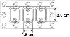
도 2는 본 발명의 다채널 유동-기재 디바이스에 대한 유동 챔버의 원형(prototype)을 제공한다. 예를 들어, 2-채널 또는 3-채널 유동 챔버가 지시된 치수로 제작될 수 있다.
도 3은 본 발명의 다채널 유동-기재 디바이스의 도식도를 제공한다. 최고의 CTC 검출 민감성 및 특이성을 수득하기 위해, 표면 관능화 파라미터, 예컨대 E-셀렉틴 패턴의 폭(a), 항체 패턴의 폭(b) 및 항체 패턴의 각도가 변형될 수 있다.
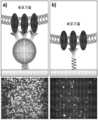
도 4는 동일한 조건 하에서, b) 선형 중합체-고정화된 표면과 비교하여 a) 덴드리머-코팅된 표면 상에서의 실질적으로 증강된 종양 세포 포획을 보여준다.
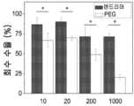
도 5는 유동 하에서의 다가 결합과 세포 롤링의 조합에 의한 증강된 세포 포획 및 효율을 보여준다. 패널 a)는 E-셀렉틴과 함께 덴드리머-코팅된 표면에서 포획 효율이 약 7배 증가한 것을 보여준다. 패널 b)는 MDA-MB-231 세포(10개 내지 1,000개)를 107개 HL-60 세포 내로 스파이킹(spiking)하였을 때, 덴드리머 표면에서 종양 세포 포획이 75% 초과로 달성된 것을 보여준다. 오차 막대: 표준 오차(n=3). *는 p가 0.05 미만임을 가리킨다.
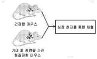
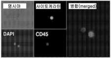
도 6은 aEGFR과 함께 단일-채널 유동-기재 디바이스를 사용하여 건강한 마우스 및 형질전환 마우스로부터의 CTC 검출을 보여준다. 패널 a)는 건강한 FVB/N 마우스 및 CEO 형질전환 마우스를 사용한 실험의 예시를 보여준다. 패널 b)는 건강한 마우스와 형질전환 마우스 사이에서 혈액 100 μL 당 포획된 CTC 수의 비교를 보여준다. 패널 c)는 포획된 세포의 면역염색 도식을 제공하고, 패널 d)는 DAPI, 사이토케라틴 및 CD45 염색 후, 포획된 CTC의 실제 형광 이미지를 보여준다.
도 7은 단일-채널 유동-기재 디바이스를 사용하여 수득된 임상 데이터를 제공한다.패널 a)는 CellSearch™* 및 유동-기재 디바이스 상에서 포획된 환자 혈액 7.5 mL 당 CTC의 수의 비교를 제공한다. 패널 b)는 E-셀렉틴이 없는 동일한 표면과 비교하여, 세포 롤링용 E-셀렉틴이 있는 유동-기재 디바이스를 사용하여 포획된 세포들 사이에서 CTC 순도가 약 25배만큼 급격하게 증가된 것을 보여준다. *값은 문헌으로부터 수득되었다.
도 8은 다수의 항체 및 덴드리머를 사용한 표면 관능화의 도식도를 제공한다.
도 9는 다양한 덴드리머-항체 컨쥬게이트를 사용한, EMT-후 세포의 효과적인 포획을 보여준다. a) TGF-β 처리에 의한 EMT 시, 3개의 BCA 세포의 수용체 발현 변화. b) EMT 전 및 EMT 후, 덴드리머-코팅된 표면을 사용한 MDA-MB-231 세포의 표면 포획의 배수 증강. 오차 막대: 표준 오차(n=3). *는 p가 0.05 미만임을 가리킨다.
도 10은 유동 챔버 내로 조립된 다관능성 표면의 제조에 대한 도식도를 보여준다: a) 다채널 PDMS 스텐실을 적용하고, 표면 고정화용 항체를 살포하며; b) 스텐실을 제거하고, E-셀렉틴을 다시 충전하고; c) 유동 챔버와 함께 조립한다.
도 11은 다양한 채널들이 구비된 맞춤형(customized) 유동 챔버를 보여준다. 패널 a)는 2-채널-유동 챔버 디바이스를 제공한다. 패널 b)는 형광 현미경 상에 마운팅된 관능화된 포획 표면과 함께 조립된 다채널 유동-기재 디바이스를 보여준다. 패널 c)는 3-채널이 구비된 유동-기재 디바이스의 부가적인 디자인을 제공한다.Figure 1 provides a biomimetic platform model for CTC and CSC capture and differentiation. In panel A: Among CTCs captured on region 'i' from cancer patient blood, stem-like cancer cells can continue to differentiate and isolate using region 'ii'. This concept of capturing and differentiating CTCs and CSCs in situ could be further applied as a 3-channel device as shown in panel B. The 3-channel device could be used for high-throughput screening of multiple blood samples as shown in panel C.
2 provides a prototype of the flow chamber for the multichannel flow-based device of the present invention. For example, a two-channel or three-channel flow chamber can be manufactured to the dimensions indicated.
3 provides a schematic diagram of the multi-channel flow-based device of the present invention. To obtain the highest CTC detection sensitivity and specificity, surface functionalization parameters such as width of E-selectin pattern (a), width of antibody pattern (b) and angle of antibody pattern can be modified.
Figure 4 shows substantially enhanced tumor cell entrapment on a) dendrimer-coated surfaces compared to b) linear polymer-immobilized surfaces under the same conditions.
Figure 5 shows enhanced cell entrapment and efficiency by the combination of multivalent binding and cell rolling under flow. Panel a) shows an approximately 7-fold increase in capture efficiency on dendrimer-coated surfaces with E-selectin. Panel b) shows that when MDA-MB-231 cells (10-1,000) were spiked into 107 HL-60 cells, greater than 75% tumor cell entrapment was achieved at the dendrimer surface. Error bars: standard error (n=3). * indicates that p is less than 0.05.
6 shows CTC detection from healthy and transgenic mice using a single-channel flow-based device with aEGFR. Panel a) shows examples of experiments using healthy FVB/N mice and CEO transgenic mice. Panel b) shows a comparison of the number of CTCs captured per 100 μL of blood between healthy and transgenic mice. Panel c) provides a schematic of immunostaining of captured cells, and panel d) shows actual fluorescence images of captured CTCs after DAPI, cytokeratin and CD45 staining.
7 presents clinical data obtained using a single-channel flow-based device.Panel a) provides a comparison of the number of CTCs per 7.5 mL of patient blood captured on CellSearch™* and flow-based devices. Panel b) shows a dramatic increase in CTC purity by about 25-fold among cells captured using a flow-based device with E-selectin for cell rolling, compared to the same surface without E-selectin. *Values were obtained from the literature.
8 provides a schematic of surface functionalization using multiple antibodies and dendrimers.
9 shows effective entrapment of post-EMT cells using various dendrimer-antibody conjugates. a) Receptor expression changes in three BCA cells upon EMT by TGF-β treatment. b) Fold enhancement of surface capture of MDA-MB-231 cells using dendrimer-coated surfaces before and after EMT. Error bars: standard error (n=3). * indicates that p is less than 0.05.
Figure 10 shows a schematic diagram of the fabrication of the multifunctional surface assembled into the flow chamber: a) applying a multichannel PDMS stencil and spraying the antibody for surface immobilization; b) remove the stencil and reload the E-selectin; c) Assemble with flow chamber.
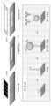
11 shows a customized flow chamber equipped with various channels. Panel a) presents a two-channel-flow chamber device. Panel b) shows an assembled multi-channel flow-based device with a functionalized capture surface mounted on a fluorescence microscope. Panel c) presents an additional design of a flow-based device with 3-channels.
본원 및 첨부된 청구항에서 사용된 바와 같이, 단수형("a," "and" 및 "the")은 문맥상 명백하게 다르게 지시하지 않는 한, 복수형을 포함함을 주지해야 한다.It should be noted that, as used herein and in the appended claims, the singular forms "a," "and" and "the" include the plural unless the context clearly dictates otherwise.
일 양태에서, 검체로부터 CTC 및 CSC를 포획하기 위한 디바이스는 적어도 2개의 채널을 포함하며, 각각의 채널은 세포 포획 표면 및 유동 변형 표면을 포함한다. 예를 들어, 하나의 채널에서, 세포 포획 표면은 세포 롤링-유도제 및 CTC 특이적인 포획제를 포함하고, 또 다른 채널에서, 세포 포획 표면은 세포 롤링-유도제 및 CSC 특이적인 포획제를 포함한다. 일 양태에서, 검체로부터 CTC 및 CSC를 포획하는 방법은, CTC 및 CSC가 세포-롤링-유도제 및 포획제에 결합할 수 있는 조건 하에, 검체를 디바이스 내로 도입하는 단계를 포함한다.In one aspect, a device for capturing CTCs and CSCs from a specimen includes at least two channels, each channel comprising a cell capture surface and a flow modification surface. For example, in one channel the cell trapping surface comprises a cell rolling-inducing agent and a CTC specific trapping agent, and in another channel the cell trapping surface comprises a cell rolling-inducing agent and a CSC specific trapping agent. In one aspect, a method of capturing CTCs and CSCs from a specimen includes introducing the specimen into a device under conditions in which the CTCs and CSCs can bind to the cell-rolling-inducing agent and the capture agent.
선택적으로, 유동 변형 표면은, 채널을 통해 유동하는 검체에서 회전 유동을 유도하기 위해 배열된 하나 이상의 구조물을 포함한다. 유동 변형 표면은 검체에서 회전 유동을 유도하며, 이러한 회전 유동은 세포와 세포 포획 표면과의 접촉을 증강시킬 수 있으며, 따라서, 보다 효율적인 CTC 및 CSC 포획을 가능하게 할 수 있다.Optionally, the flow modifying surface comprises one or more structures arranged to induce rotational flow in a specimen flowing through the channel. The flow modification surface induces rotational flow in the specimen, and this rotational flow can enhance the contact between the cells and the cell capture surface, thus enabling more efficient CTC and CSC capture.
본 발명의 방법은 약 200 μL/min 내지 500 μL/min의 생리학적 유속 범위 내에서 생물학적 검체의 고속 대량 분리를 제공한다. 일부 구현예에서, 0.05 dyn/cm2내지 10 dyn/cm2의 전단 응력이 미세유체 디바이스(10) 내로 도입된 검체에 적용된다. 일부 구현예에서, 0.1 dyn/cm2내지 2 dyn/cm2의 전단 응력이 미세유체 디바이스(10) 내로 도입된 검체에 적용된다. 일부 구현예에서, 전단 응력은 약 0.05, 약 0.10, 약 0.15, 약 0.20, 약 0.25, 약 0.30, 약 0.35, 약 0.40, 약 0.45, 약 0.50, 약 0.55, 약 0.60, 약 0.65, 약 0.70, 약 0.75, 약 0.80, 약 0.85, 약 0.90, 약 0.95, 약 1.0, 약 1.1, 약 1.2, 약 1.3, 약 1.4, 약 1.5, 약 1.6, 약 1.7, 약 1.8, 약 1.9, 약 2.0, 약 2.1, 약 2.2, 약 2.3, 약 2.4, 약 2.5, 약 2.6, 약 2.7, 약 2.8, 약 2.9, 약 3.0, 약 3.1, 약 3.2, 약 3.3, 약 3.4, 약 3.5, 약 3.6, 약 3.7, 약 3.8, 약 3.9, 약 4.0, 약 4.1, 약 4.2, 약 4.3, 약 4.4, 약 4.5, 약 4.6, 약 4.7, 약 4.8, 약 4.9, 약 5.0, 약 5.1, 약 5.2, 약 5.3, 약 5.4, 약 5.5, 약 5.6, 약 5.7, 약 5.8, 약 5.9, 약 6.0, 약 6.1, 약 6.2, 약 6.3, 약 6.4, 약 6.5, 약 6.6, 약 6.7, 약 6.8, 약 6.9, 약 7.0, 약 7.1, 약 7.2, 약 7.3, 약 7.4, 약 7.5, 약 7.6, 약 7.7, 약 7.8, 약 7.9, 약 8.0, 약 8.1, 약 8.2, 약 8.3, 약 8.4, 약 8.5, 약 8.6, 약 8.7, 약 8.8, 약 8.9, 약 9.0, 약 9.1, 약 9.2, 약 9.3, 약 9.4, 약 9.5, 약 9.6, 약 9.7, 약 9.8, 약 9.9, 약 10.0 dyn/cm2이다. 일부 구현예에서, 전단 응력은 약 0.16 dyn/cm2이다.The method of the present invention provides high-throughput separation of biological specimens within a physiological flow rate range of about 200 μL/min to 500 μL/min. In some embodiments, a shear stress of 0.05 dyn/cm2 to 10 dyn/cm2 is applied to the specimen introduced into the
I. 미세유체I. Microfluidics디바이스device
본 발명의 디바이스는 세포 포획 표면 및 유동 변형 표면을 가진 적어도 2개의 채널을 포함한다. 세포 포획 표면은 유동 변형 표면의 반대쪽에 배치될 수 있다. 예를 들어, 세포 포획 표면은 채널의 하부 표면 상에 배치될 수 있고, 유동 변형 표면은 세포 포획 표면과 반대쪽인, 채널의 상부 표면 상에 배치될 수 있다. 대안적으로, 유동 변형은 세포 포획 표면에 인접하여 배치될 수 있다. 보다 다른 구현예에서, 세포 포획 표면 및 유동 변형 표면은 단일 표면으로 혼입될 수 있다. 채널은 예를 들어, 4개의 벽을 가진 폐쇄 채널일 수 있다. 세포 포획 표면 및/또는 유동 변형 표면은 채널의 다수의 벽들 상에 배치될 수 있다.The device of the present invention includes at least two channels having a cell trapping surface and a flow modifying surface. A cell capture surface may be disposed opposite the flow modification surface. For example, a cell trapping surface can be disposed on the lower surface of the channel and a flow modifying surface can be disposed on the upper surface of the channel, opposite the cell trapping surface. Alternatively, the flow strain may be placed adjacent to the cell capture surface. In still other embodiments, the cell capture surface and the flow modifying surface may be incorporated into a single surface. The channel can be, for example, a closed channel with four walls. A cell capture surface and/or flow modifying surface can be disposed on multiple walls of the channel.
디바이스 내의 다채널들은 연결되어 있을 수 있고, 검체는 채널들 사이에서 유동할 것이다. 또 다른 양태에서, 디바이스는 다수의 투입구 및 다수의 배출구를 가질 수 있으며, 예를 들어, 각각의 채널은 그 자체의 투입구 및 배출구를 가지고 채널들은 연결되지 않거나, 각각의 채널은 그 자체의 투입구를 가지고 채널들은 단일 배출구 또는 다수의 배출구에 의해 연결된다. 다채널들은 균일한 모양 및/또는 크기를 가질 수 있다. 대안적으로, 다채널들은 단일 디바이스 내에서 상이한 모양 및 크기들을 가질 수 있다.Multiple channels within the device may be connected, and the analyte will flow between the channels. In another aspect, the device can have multiple inlets and multiple outlets, eg each channel has its own inlet and outlet and the channels are not connected, or each channel has its own inlet. The channels are connected by a single outlet or multiple outlets. Multiple channels may have a uniform shape and/or size. Alternatively, multiple channels may have different shapes and sizes within a single device.
채널은 임의의 적합한 단면 모양을 가질 수 있다. 예를 들어, 채널은 직사각형, 삼각형, 원형 또는 타원형일 수 있다. 미세유체 디바이스의 치수는, 하기 방정식을 사용하여 유체 저항성을 최소화하면서도 유체 회전율을 최대화하기 위해 최적화될 수 있다:The channels may have any suitable cross-sectional shape. For example, the channels can be rectangular, triangular, circular or oval. The dimensions of a microfluidic device can be optimized to maximize fluid turnover while minimizing fluid resistance using the equation:

상기 방정식에서, μ는 동적 점도(kinematic viscosity)이며, L은 채널 길이이며, w는 채널 폭이고, h는 채널 높이이다.In the above equation, μ is the kinematic viscosity, L is the channel length, w is the channel width, and h is the channel height.
예를 들어, 채널은 약 50 ㎛ 내지 약 600 ㎛, 약 100 ㎛ 내지 약 500 ㎛, 약 200 ㎛ 내지 약 400 ㎛의 높이를 가질 수 있다. 다른 적합한 높이는, 예를 들어, 약 50 ㎛, 100 ㎛, 150 ㎛, 200 ㎛, 250 ㎛, 300 ㎛, 350 ㎛, 400 ㎛, 450 ㎛, 500 ㎛, 550 ㎛ 또는 600 ㎛를 포함한다. 채널의 폭은 약 200 ㎛ 내지 약 2000 ㎛, 약 400 ㎛ 내지 약 1500 ㎛, 약 500 ㎛ 내지 약 1000 ㎛, 또는 약 600 ㎛ 내지 약 800 ㎛의 폭을 가질 수 있다. 다른 적합한 폭은, 예를 들어, 약 200 ㎛, 300 ㎛, 400 ㎛, 500 ㎛, 600 ㎛, 700 ㎛, 800 ㎛, 900 ㎛, 1000 ㎛, 1100 ㎛, 1200 ㎛, 1500 ㎛, 1600 ㎛, 1700 ㎛, 1800 ㎛, 1900 ㎛ 또는 2000 ㎛를 포함한다. 채널은 약 200 ㎛ 내지 약 5000 ㎛, 약 400 ㎛ 내지 약 4000 ㎛, 약 600 ㎛ 내지 약 2000 ㎛, 또는 약 800 ㎛ 내지 약 1000 ㎛의 길이를 가질 수 있다. 다른 적합한 길이로는, 예를 들어, 약 200 ㎛, 300 ㎛, 400 ㎛, 500 ㎛, 600 ㎛, 700 ㎛, 800 ㎛, 900 ㎛, 1000 ㎛, 1500 ㎛, 2000 ㎛, 2500 ㎛, 3000 ㎛, 3500 ㎛, 4000 ㎛, 4500 ㎛ 또는 5000 ㎛를 포함한다.For example, the channels may have a height of about 50 μm to about 600 μm, about 100 μm to about 500 μm, or about 200 μm to about 400 μm. Other suitable heights include, for example, about 50 μm, 100 μm, 150 μm, 200 μm, 250 μm, 300 μm, 350 μm, 400 μm, 450 μm, 500 μm, 550 μm or 600 μm. The channel may have a width of about 200 μm to about 2000 μm, about 400 μm to about 1500 μm, about 500 μm to about 1000 μm, or about 600 μm to about 800 μm. Other suitable widths are, for example, about 200 μm, 300 μm, 400 μm, 500 μm, 600 μm, 700 μm, 800 μm, 900 μm, 1000 μm, 1100 μm, 1200 μm, 1500 μm, 1600 μm, 1700 μm. μm, 1800 μm, 1900 μm or 2000 μm. The channels can have a length of about 200 μm to about 5000 μm, about 400 μm to about 4000 μm, about 600 μm to about 2000 μm, or about 800 μm to about 1000 μm. Other suitable lengths include, for example, about 200 μm, 300 μm, 400 μm, 500 μm, 600 μm, 700 μm, 800 μm, 900 μm, 1000 μm, 1500 μm, 2000 μm, 2500 μm, 3000 μm, including 3500 μm, 4000 μm, 4500 μm or 5000 μm.
세포 포획 표면은 기판에 부착된 세포 롤링-유도제 및 CTC 및/또는 CSC 특이적인 포획제를 포함한다. 기판은 예를 들어, 유리, 플라스틱(또는 중합체-코팅된), 하이드로겔, 매트리겔(matrigel) 또는 세포외 기질(ECM)-코팅된 기판일 수 있다. 세포 롤링-유도제 및 포획제는 직접적으로 또는 예를 들어 링커를 사용하여 간접적으로 기판 상에 고정화될 수 있다. 세포 롤링-유도제 및 포획제는 세포 포획 표면을 가로질러 균일하게 배열될 수 있다. 예를 들어, 세포 포획 표면은 세포 롤링-유도제 및 포획제를 가진 교대 영역을 포함할 수 있다. 교대 영역은 실질적으로 동일한 폭을 가질 수 있거나, 폭들은 영역들 사이에서 다양할 수 있다. 세포 롤링-유도제 및 포획제를 포함하는 영역은 예를 들어 채널을 통한 유동 방향에 대해 평행하게 배열되거나 각도를 이루어 배열될 수 있다. 예를 들어, 영역은 채널을 통한 유동 방향에 대해 접선으로(tangentially) 배열될 수 있다.The cell capture surface includes a cell rolling-inducing agent attached to the substrate and a capture agent specific for CTCs and/or CSCs. The substrate may be, for example, glass, plastic (or polymer-coated), hydrogel, matrigel or extracellular matrix (ECM)-coated substrate. Cell rolling-inducing agents and trapping agents can be immobilized on the substrate either directly or indirectly, for example using a linker. The cell rolling-inducing agent and trapping agent may be uniformly arranged across the cell trapping surface. For example, a cell trapping surface can include alternating regions with a cell rolling-inducing agent and a trapping agent. Alternating regions can have substantially the same width, or widths can vary between regions. The regions comprising the cell rolling-inducing agent and trapping agent may be arranged parallel or at an angle to the direction of flow through the channel, for example. For example, the regions may be arranged tangentially to the direction of flow through the channel.
유동 변형 표면은 당업계에 잘 알려져 있다. 임의의 알려진 유동 변형 표면이 사용될 수 있다. 예를 들어, 유동 변형 표면은 선택적으로, 표면으로부터 채널 내로 연장되어 있는 하나 이상의 융기부(ridge)를 포함할 수 있다. 융기부는 채널을 통해 유동하는 검체에서 회전 유동을 유도하기 위해 형상화, 크기화 및 배향된다. 세포 포획 표면 및 유동 변형 표면은 예를 들어, 유동 변형 표면을 세포 롤링-유도제 및 포획제로 코팅함으로써 디바이스의 단일 표면 상에 포함될 수 있다. 예를 들어, 융기부는 세포 롤링-유도제 및 포획제로 코팅될 수 있다. 융기부 중 모두 또는 일부가 코팅될 수 있다. 예를 들어, 융기부의 측벽이 세포 롤링-유도제 및 포획제로 코팅될 수 있다. 검체에서 회전 유동의 유도는 세포 포획 효율을 증강시킬 수 있다. 확산율이 낮은 세포들은 이들이 들어가는 채널 영역에 머무르는 경향을 가질 것이다. 예를 들어, 혈액 세포들은 이들의 큰 직경으로 인해 선천적으로 낮은 확산율을 가진다. 이는, 세포가 세포 포획 표면으로부터 떨어져 있는 채널에 들어갈 때, 세포 포획 과정에 악영향을 미친다. 예를 들어, 혈액 세포가 상부 근처의 미세유체 채널에 들어가면, 세포는 미세채널을 따라 수 cm 이동함에 따라 상부 근처에 머무르는 경향이 있을 것이며, 이는 채널의 하부에 위치한 생체관능화된(biofunctionalized) 기판과 세포의 상호작용을 제한한다. 검체에서 회전 유동의 유도는 세포를 세포 포획 표면 쪽으로 강제 이동시킬 것이며, 이로써, 세포와 세포 포획 표면 사이의 접촉을 증강시킬 것이다.Flow deformation surfaces are well known in the art. Any known flow modifying surface may be used. For example, the flow modifying surface may optionally include one or more ridges extending from the surface into the channel. The ridges are shaped, sized and oriented to induce rotational flow in the specimen flowing through the channels. The cell trapping surface and the flow modifying surface can be included on a single surface of the device, for example by coating the flow modifying surface with a cell rolling-inducing agent and a trapping agent. For example, the ridges can be coated with a cell rolling-inducing agent and entrapping agent. All or some of the ridges may be coated. For example, the side walls of the ridges can be coated with a cell rolling-inducing agent and entrapment agent. Induction of rotational flow in the specimen can enhance cell capture efficiency. Cells with a low diffusion rate will tend to stay in the channel region they enter. For example, blood cells have an inherently low diffusion rate due to their large diameter. This adversely affects the cell trapping process when cells enter the channel away from the cell trapping surface. For example, if blood cells enter a microfluidic channel near the top, the cells will tend to stay near the top as they travel several cm along the microchannel, which is a biofunctionalized substrate located at the bottom of the channel. and limit cell interactions. The induction of rotational flow in the specimen will force the cells towards the cell trapping surface, thereby enhancing the contact between the cells and the cell trapping surface.
융기부는 예를 들어 직사각형, 원형, 타원형 또는 삼각형과 같은 임의의 적합한 단면 모양을 가질 수 있다. 융기부는 약 10 ㎛ 내지 약 300 ㎛, 약 50 ㎛ 내지 약 300 ㎛, 약 100 ㎛ 내지 약 250 ㎛, 또는 약 150 ㎛ 내지 약 200 ㎛의 두께를 가질 수 있다. 다른 적합한 두께(t)는, 약 10 ㎛, 15 ㎛, 20 ㎛, 25 ㎛, 30 ㎛, 35 ㎛, 40 ㎛, 45 ㎛, 50 ㎛, 55 ㎛, 60 ㎛, 65 ㎛, 70 ㎛, 75 ㎛, 80 ㎛, 85 ㎛, 90 ㎛, 95 ㎛, 100 ㎛, 125 ㎛, 150 ㎛, 175 ㎛, 200 ㎛, 225 ㎛, 250 ㎛, 275 ㎛ 또는 300 ㎛ 등이 있다. 융기부는 약 50 ㎛ 내지 약 300 ㎛, 약 100 ㎛ 내지 약 250 ㎛, 또는 약 150 ㎛ 내지 약 200 ㎛의 폭을 가질 수 있다. 다른 적합한 폭은, 약 50 ㎛, 75 ㎛, 100 ㎛, 125 ㎛, 150 ㎛, 175 ㎛, 200 ㎛, 225 ㎛, 250 ㎛, 275 ㎛ 또는 300 ㎛을 포함한다. 인접한 융기부들 사이의 거리는 약 50 ㎛ 내지 약 500 ㎛, 약 100 ㎛ 내지 약 400 ㎛, 또는 약 200 ㎛ 내지 약 300 ㎛일 수 있다. 다른 적합한 거리는 약 50 ㎛, 100 ㎛, 150 ㎛, 200 ㎛, 250 ㎛, 300 ㎛, 350 ㎛, 400 ㎛, 450 ㎛ 또는 500 ㎛을 포함한다. 인접한 융기부들 사이의 거리는 유동 변형 표면을 가로질러 실질적으로 균일할 수 있거나, 다양할 수 있다.The elevations may have any suitable cross-sectional shape, such as for example rectangular, circular, oval or triangular. The elevations can have a thickness of about 10 μm to about 300 μm, about 50 μm to about 300 μm, about 100 μm to about 250 μm, or about 150 μm to about 200 μm. Other suitable thicknesses (t) are about 10 μm, 15 μm, 20 μm, 25 μm, 30 μm, 35 μm, 40 μm, 45 μm, 50 μm, 55 μm, 60 μm, 65 μm, 70 μm, 75 μm. , 80 μm, 85 μm, 90 μm, 95 μm, 100 μm, 125 μm, 150 μm, 175 μm, 200 μm, 225 μm, 250 μm, 275 μm or 300 μm. The elevations may have a width of about 50 μm to about 300 μm, about 100 μm to about 250 μm, or about 150 μm to about 200 μm. Other suitable widths include about 50 μm, 75 μm, 100 μm, 125 μm, 150 μm, 175 μm, 200 μm, 225 μm, 250 μm, 275 μm or 300 μm. The distance between adjacent ridges may be about 50 μm to about 500 μm, about 100 μm to about 400 μm, or about 200 μm to about 300 μm. Other suitable distances include about 50 μm, 100 μm, 150 μm, 200 μm, 250 μm, 300 μm, 350 μm, 400 μm, 450 μm or 500 μm. The distance between adjacent ridges may be substantially uniform across the flow deformation surface or may vary.
융기부는 예를 들어, 유동에 대해 수직인 방향에서 실질적으로 선형으로 연장되어 있을 수 있다. 융기부는 헤링본 구조를 가질 수 있다. 융기부는 유동 방향에 대해 각이 져 있을 수 있다. 예를 들어, 융기부는 유동 방향에 대해 수직으로 또는 비스듬하게 각이 져 있을 수 있다. 일 구현예에서, 융기부는 유동(F) 방향에 대해 45°각도에 있다. 다른 적합한 각도는, 약 45°, 50°, 55°, 60°, 65°, 70°, 75°, 80°, 85°, 90°, 100°, 110°, 120°, 130°, 140°, 150°, 160° 및 170°을 포함한다. 하나 이상의 융기부는 균일하게 각이 져 있을 수 있다. 대안적으로, 융기부의 각도는 채널을 가로질러 다양해서, 채널을 통해 유동하는 검체에 상이한 회전 특성을 유도할 수 있다.The ridge may extend substantially linearly, for example in a direction perpendicular to the flow. The raised portion may have a herringbone structure. The ridges may be angled with respect to the flow direction. For example, the ridges may be angled perpendicular or oblique to the flow direction. In one embodiment, the elevation is at a 45° angle to the flow (F) direction. Other suitable angles are about 45°, 50°, 55°, 60°, 65°, 70°, 75°, 80°, 85°, 90°, 100°, 110°, 120°, 130°, 140° , including 150°, 160° and 170°. One or more ridges may be uniformly angled. Alternatively, the angle of the ridge may be varied across the channel, inducing different rotational characteristics to the specimen flowing through the channel.
하나 이상의 융기부는 패턴으로 배열될 수 있다. 예를 들어, 유동 변형 표면은 하나 이상의 융기부를 가진 제1 영역, 및 융기부를 갖지 않는 제2 영역을 포함할 수 있다. 대안적으로, 제2 영역은 제1 영역의 융기부와 상이하게 배향, 크기화 및/또는 형상화된 융기부를 포함할 수 있다. 유동 변형 표면은 융기부를 완전히 갖지 않는 제3 영역을 추가로 포함할 수 있다. 상이하게 크기화, 배향 및/또는 형상화된 융기부를 가진 임의의 적합한 수의 영역이 유동 변형 표면 상에 포함될 수 있다. 제1 영역 및 제2 영역은 예를 들어, 유동 변형 표면에 가로질러 균일하게 교대될 수 있다.One or more ridges may be arranged in a pattern. For example, the flow modifying surface can include a first region with one or more ridges, and a second region without ridges. Alternatively, the second region may include raised portions that are differently oriented, sized and/or shaped than the raised portions of the first region. The flow modification surface may further include a third region completely free of ridges. Any suitable number of regions with differently sized, oriented and/or shaped ridges may be included on the flow modifying surface. The first region and the second region may for example alternate uniformly across the flow deformation surface.
미세유체 디바이스는 세포 롤링-유도제를 가진 영역 및 포획제를 가진 영역을 포함하는 세포 포획 표면을 제조함으로써 제작될 수 있다. 그런 다음, 유동 변형 표면을 가진 미세유체 채널이 세포 포획 표면에 부착될 수 있다.A microfluidic device can be fabricated by fabricating a cell trapping surface comprising an area with a cell rolling-inducing agent and an area with an entrapment agent. A microfluidic channel with a flow modifying surface can then be attached to the cell capture surface.
세포 포획 표면은 세포 롤링-유도제 및 포획제를 기판, 예를 들어 유리 슬라이드 상에 패턴화함으로써 형성될 수 있다. 세포 롤링-유도제 및 포획제의 영역을 형성하는 임의의 알려진 방법이 사용되어, 세포 포획 표면을 형성할 수 있다. 세포 롤링-유도제 및 포획제는 예를 들어, PDMS 스텐실과 같은 중합체 스텐실을 사용하여 패턴화될 수 있다. 스텐실은 당업계에 알려진 바와 같이 예를 들어 포토리소그래피(photolithography)를 사용하여 형성될 수 있다. 포토레지스트가 웨이퍼 상에 코팅되고, 포토마스크를 사용하여 선택적으로 노출될 수 있다. 그런 다음, 포토레지스트가 현상(develop)되어, 포토레지스트의 비노출된 부분이 제거되고, 이로써, 스텐실용 네거티브 몰드(negative mold)가 형성되는 것을 초래한다. 그런 다음, PDMS와 같은 중합체가 네거티브 몰드 상에 부어지고 경화될 수 있으며, 이로써 스텐실이 형성된다. 스텐실은 제1 표면으로부터 돌출된 하나 이상의 구조물을 포함한다. 돌출 구조물의 크기, 배향 및 모양은 세포 롤링-유도제 및 포획제의 요망되는 영역의 크기, 배향 및 모양과 실질적으로 동일하다. 돌출 구조물이 기판 상에 존재할 때, 이러한 구조물은 기판의 일부를 가리는(mask) 작용을 한다. 세포 롤링-유도제 또는 포획제는 기판의 가려지지 않은 부위에 부착될 수 있다. 그런 다음, 스텐실이 제거될 수 있고, 기판의 노출된 부위가 세포 롤링-유도제 또는 포획제로 충전될 수 있으며, 이로써 세포 포획 표면이 형성된다.A cell trapping surface can be formed by patterning a cell rolling-inducing agent and a trapping agent onto a substrate, such as a glass slide. Any known method of forming regions of the cell rolling-inducing agent and trapping agent may be used to form the cell trapping surface. Cell rolling-inducing and entrapping agents can be patterned using, for example, polymer stencils such as PDMS stencils. The stencil may be formed using, for example, photolithography as is known in the art. A photoresist is coated on the wafer and can be selectively exposed using a photomask. The photoresist is then developed, removing the unexposed portions of the photoresist, resulting in the formation of a negative mold for the stencil. A polymer such as PDMS can then be poured onto the negative mold and cured, thereby forming a stencil. The stencil includes one or more structures protruding from the first surface. The size, orientation, and shape of the protruding structures are substantially the same as the size, orientation, and shape of the desired regions of the cell rolling-inducing agent and trapping agent. When protruding structures are present on a substrate, these structures act to mask portions of the substrate. The cell rolling-inducing agent or trapping agent can be attached to the uncovered portion of the substrate. The stencil may then be removed, and the exposed areas of the substrate may be filled with a cell rolling-inducing agent or trapping agent, thereby forming a cell trapping surface.
세포 롤링-유도제 및 포획제는 예를 들어, 물리 흡착(physisorption) 또는 혈장 제거(plasma ablation)를 사용하여 기판에 부착될 수 있다. 예를 들어, 작용제는 미세유체 흡착을 사용하여 부착될 수 있으며, 여기서, 요망되는 작용제가 가용성 매질에 위치하고, 기판 상에 위치한 미세유체 채널을 통해 주입된다. 용액은 수시간에 동안 표면에 흡착될 수 있다. 이러한 기술은 요망되는 작용제가 열에 민감하거나 열에 의해 손상될 때, 유리하다.Cell rolling-inducing agents and trapping agents can be attached to the substrate using, for example, physisorption or plasma ablation. For example, an agent can be attached using microfluidic adsorption, wherein the desired agent is placed in a soluble medium and injected through a microfluidic channel located on the substrate. The solution can be adsorbed on a surface for several hours. This technique is advantageous when the desired agent is heat sensitive or damaged by heat.
공유 결합을 위한 적합한 기(group)들의 상이한 밀도 및 패턴으로 표면을 제조하는 방법들이 당업계에 잘 알려져 있다(예, 문헌[Rusmini et al.,Biomacromolecules8: 1775-89 (June 2007)] 및 [Leckband et al.,Biotechnology and Bioengineering 37: 227-237 (1991)]을 참조하며, 이들 두 문헌 모두의 전체 내용은 원용에 의해 본 명세서에 포함됨). 일부 구현예에서, 포획제 및/또는 세포 롤링-유도제의 밀도는 약 10 ng/cm2 내지 약 600 ng/cm2의 범위이다. 일부 구현예에서, 포획제 및/또는 세포 롤링-유도제의 밀도는 약 30 ng/cm2 초과이다. 예를 들어, 일부 구현예에서, 포획제 및/또는 세포 롤링-유도제의 밀도는 약 30 ng/cm2 내지 약 360 ng/cm2의 범위이다. 일부 구현예에서, 포획제 및/또는 세포 롤링-유도제의 밀도는 약 50 ng/cm2 내지 약 300 ng/cm2의 범위이다. 일부 구현예에서, 포획제 및/또는 세포 롤링-유도제의 밀도는 약 100 ng/cm2 내지 약 200 ng/cm2의 범위이다.Methods for preparing surfaces with different densities and patterns of suitable groups for covalent bonding are well known in the art (e.g., Rusmini et al.,Biomacromolecules 8: 1775-89 (June 2007)) and [ Leckband et al.,Biotechnology and Bioengineering 37: 227-237 (1991), the entire contents of both are incorporated herein by reference). In some embodiments, the density of the capture agent and/or cell rolling-inducing agent ranges from about 10 ng/cm2 to about 600 ng/cm2 . In some embodiments, the density of the capture agent and/or cell rolling-inducing agent is greater than about 30 ng/cm2 . For example, in some embodiments, the density of the capture agent and/or cell rolling-inducing agent ranges from about 30 ng/cm2 to about 360 ng/cm2 . In some embodiments, the density of the capture agent and/or cell rolling-inducing agent ranges from about 50 ng/cm2 to about 300 ng/cm2 . In some embodiments, the density of the capture agent and/or cell rolling-inducing agent ranges from about 100 ng/cm2 to about 200 ng/cm2 .
채널 및 유동 변형 표면은 당업계에 알려진 바와 같이 형성될 수 있다. 문헌[Stroock et al., Science 295: 647-51]을 참조하며, 이의 개시내용은 그 전체가 원용에 의해 본 명세서에 포함된다. 유동 변형 표면은 소프트-리소그래피를 사용하여 형성될 수 있다. 예를 들어, 유동 변형 표면은 듀얼 높이 SU-8 포토레지스트 몰드와 같은 포토레지스트를 사용하여 형성될 수 있다. 몰드는 우선, 미세유체 채널을 스피닝(spinning)하고 패턴화함으로써 제조된다. 채널 패턴을 현상하기 전에, 제2 포토레지스트 층이 몰드 상으로 스피닝되어, 유동 변형 표면의 융기부에 대한 패턴이 발생된다. 얼라인 마커(alignment marker)가 첨가되어, 제2 포토레지스트 층의 적절한 배향을 촉진할 수 있다. 그런 다음, 몰드가 노출되고, 하드 베이킹되고(hard baked), 현상됨으로써, 유동 변형 표면의 융기부를 캐스트(cast)할 구조를 가진 채널을 함유하는 몰드가 형성된다. 그런 다음, 생성된 몰드가 PDMS와 같은 중합체로 코팅되어, 채널 및 유동 변형 표면이 형성된다.Channels and flow modifying surfaces can be formed as known in the art. See Stroock et al., Science 295: 647-51, the disclosure of which is incorporated herein by reference in its entirety. The flow deformation surface may be formed using soft-lithography. For example, the flow modifying surface can be formed using a photoresist such as a dual height SU-8 photoresist mold. The mold is first made by spinning and patterning the microfluidic channels. Prior to developing the channel pattern, a second photoresist layer is spun onto the mold to create a pattern for the ridges of the flow modification surface. An alignment marker may be added to promote proper orientation of the second photoresist layer. The mold is then exposed, hard baked, and developed to form a mold containing channels with structures that will cast the ridges of the flow modifying surface. The resulting mold is then coated with a polymer such as PDMS to form channels and flow modifying surfaces.
II. 세포 롤링II. cell rolling
본 발명의 미세유체 디바이스의 챔버는 고정화된 세포 롤링-유도제를 포함한다. 일시적인 리간드-수용체 상호작용의 형성은 혈액에서 유동하는 세포와 혈관 내피 사이에서 보편적으로 발생하며; 이러한 생리학적 과정은 세포 롤링으로 알려져 있다. 세포 롤링은 염증 부위로의 백혈구의 모집, 정맥내 주사 후 조혈 전구 세포의 귀소(homing) 및 CTC-유도 전이와 같은 생물학적으로 중요한 과정들에서 중요한 역할을 하는 것으로 알려져 있다. 이러한 거동은 전형적으로, 혈관 내피 세포 표면 상의 셀렉틴(예, E-셀렉틴, P-셀렉틴, L-셀렉틴)과 P-셀렉틴 당단백질 리간드-1(PSGL-1)을 포함하는 막 단백질 사이의 동적인 상호작용에 의해 매개된다. 당업자는, CTC 또는 CSC가 세포 롤링을 수행하도록 유도할 수 있는 임의의 분자가 개시된 방법을 실시하고 개시된 디바이스를 제조하는 데 사용될 수 있음을 인지할 것이다.The chamber of the microfluidic device of the present invention contains an immobilized cell rolling-inducing agent. Formation of transient ligand-receptor interactions occurs universally between cells flowing in the blood and the vascular endothelium; This physiological process is known as cell rolling. Cell rolling is known to play an important role in biologically important processes such as the recruitment of leukocytes to sites of inflammation, homing of hematopoietic progenitor cells after intravenous injection and CTC-induced metastasis. This behavior is typically associated with dynamic interactions between selectins (e.g., E-selectin, P-selectin, L-selectin) on the surface of vascular endothelial cells and membrane proteins, including P-selectin glycoprotein ligand-1 (PSGL-1). mediated by interactions. One skilled in the art will appreciate that any molecule capable of inducing CTCs or CSCs to undergo cell rolling can be used to practice the disclosed methods and fabricate the disclosed devices.
본 개시내용의 일부 구현예에서, 세포 롤링-유도제는 셀렉틴이다. 다른 구현예에서, 셀렉틴은 내피 (E)-셀렉틴이다. 보다 다른 구현예에서, 셀렉틴은 P-셀렉틴이다. 다른 구현예에서, 셀렉틴은 L-셀렉틴이다. 더욱이, CTC에 결합하는 능력을 보유한 셀렉틴의 단편은 구체적으로는 본 개시내용의 범위 내에 포함된다.In some embodiments of the present disclosure, the cell rolling-inducing agent is a selectin. In another embodiment, the selectin is an endothelial (E)-selectin. In yet another embodiment, the selectin is a P-selectin. In another embodiment, the selectin is L-selectin. Moreover, fragments of selectins that retain the ability to bind CTC are specifically included within the scope of the present disclosure.
E-셀렉틴(CD62E)은 특히, 활성화된 내피 상 및 뼈-피부 미세혈관 라이닝(lining) 상에서의 이의 발현 및 세포 롤링, 세포 신호전달 및 화학주성에 있어서의 이의 역할로 인한 질환들에서 주목할 만하다. 많은 연구들은 전립선, 유방 및 결장 암종 세포와 같은 다양한 유형의 암세포들의 접착 및 귀소에 관여하는 E-셀렉틴에 의해 운용되는 주요 역할에 초점을 맞추고 있다. E-셀렉틴은 인터루킨-1β (IL-1β) 및 종양 괴사 인자-α(TNF-α)와 같은 염증 사이토카인에 반응하여 내피 세포에 의해 드 노브(de novo)에서 합성된다. 따라서, 세포 롤링 거동을 기재로 하는 세포 분리는, 이것이 생리학적 과정을 모방하고, 다른 면역-표지화 검출 방법들에서 필수적인 표지화 및 표지 제거 단계를 배제함으로써 이용되고 있다. 그러나, 백혈구, 혈소판, 중성구, 중간엽 줄기세포, 조혈 줄기세포 및 전이성 암세포를 포함하는 큰 부류의 세포들이 셀렉틴 상에서 롤링을 나타내는 것을 고려하면, 세포 혼합물 또는 전혈로부터 특정 세포 유형에 대한 롤링-기재 검출은 충분한 특이성을 달성하는 데 제한을 가지며, 이는 임상적으로 유의미한 디바이스로의 기술의 변환(translation)을 방해한다. 본 개시내용의 방법 및 디바이스는 세포 롤링-유도제를 고정화된 포획제와 커플링함으로써 이러한 제한을 극복한다. 임의의 특정 이론으로 결부시키고자 하는 것은 아니지만, 세포 롤링-유도제는 순환 종양 세포(CTC)로 하여금 미세유체 디바이스의 표면 상에서 (전술한) "롤링" 거동을 나타내게 하는 것으로 여겨진다. 그런 다음, 롤링 CTC는 고정화된 포획제와 접촉하고, 이로써 디바이스에 의해 포획(즉, 고정화)된다.E-selectin (CD62E) is particularly noteworthy in diseases due to its expression on activated endothelial phases and bone-dermal microvascular linings and its role in cell rolling, cell signaling and chemotaxis. Many studies have focused on the key role played by E-selectin in the adhesion and homing of various types of cancer cells, such as prostate, breast and colon carcinoma cells. E-selectin is synthesized de novo by endothelial cells in response to inflammatory cytokines such as interleukin-1β (IL-1β) and tumor necrosis factor-α (TNF-α). Thus, cell isolation based on cell rolling behavior has been exploited as it mimics the physiological process and eliminates the labeling and labeling steps essential in other immuno-labeling detection methods. However, considering that a large class of cells including leukocytes, platelets, neutrophils, mesenchymal stem cells, hematopoietic stem cells and metastatic cancer cells exhibit rolling on selectins, rolling-based detection for specific cell types from cell mixtures or whole blood has limitations in achieving sufficient specificity, which hinders translation of the technology into clinically meaningful devices. The methods and devices of the present disclosure overcome this limitation by coupling a cell rolling-inducing agent with an immobilized capture agent. Without wishing to be bound by any particular theory, it is believed that cell rolling-inducing agents cause circulating tumor cells (CTCs) to exhibit a "rolling" behavior (described above) on the surface of a microfluidic device. The rolling CTC is then contacted with the immobilized capture agent and thereby captured (ie, immobilized) by the device.
임의의 공유 화학은 세포 롤링-유도제를 미세유체 디바이스의 표면에 고정화하는 데 사용될 수 있다. 일부 구현예에서, 세포 롤링-유도제는 하나 이상의 화학적 모이어티를 통해 표면에 부착된다. 일반적으로, 화학적 모이어티와 표면 사이의 결합은 공유 결합이다. 비제한적으로, 일부 구현예에서, 화학적 모이어티는 에폭시기, 카르복실기, 티올기, 알킨기, 아자이드기, 말레이미드기, 비닐기, 하이드록실기, 아민기, 알데하이드기 및 이들의 조합을 포함한다.Any covalent chemistry can be used to immobilize the cell rolling-inducing agent to the surface of the microfluidic device. In some embodiments, the cell rolling-inducing agent is attached to the surface via one or more chemical moieties. Generally, the bond between the chemical moiety and the surface is a covalent bond. Without limitation, in some embodiments, chemical moieties include epoxy groups, carboxyl groups, thiol groups, alkyne groups, azide groups, maleimide groups, vinyl groups, hydroxyl groups, amine groups, aldehyde groups, and combinations thereof. .
일부 구현예에서, 세포-롤링 유도제는 링커를 통해 미세유체 디바이스의 표면에 고정화된다. 일부 구현예에서, 링커는 덴드리머, 덴드론, 덱스트란, 폴리에틸렌 글리콜, 폴리(L-라이신), 폴리(L-글루탐산), 폴리비닐 알코올, 폴리에틸렌이민, 폴리(락트산), 폴리(글리콜산) 및 이들의 조합으로 이루어진 군으로부터 선택된다.In some embodiments, the cell-rolling inducer is immobilized to the surface of the microfluidic device via a linker. In some embodiments, the linker is a dendrimer, dendron, dextran, polyethylene glycol, poly(L-lysine), poly(L-glutamic acid), polyvinyl alcohol, polyethyleneimine, poly(lactic acid), poly(glycolic acid) and It is selected from the group consisting of combinations thereof.
본 발명의 디바이스 및 방법에 사용되는 CTC를 포획하기 위한 롤링 효과는 PCT 공개 WO2010/124227 및 미국 특허 공개 20120077246에 상세히 기술되어 있으며, 이들은 그 전체가 원용에 의해 본 명세서에 포함된다.The rolling effect for trapping CTCs used in the devices and methods of the present invention is described in detail in PCT Publication WO2010/124227 and US Patent Publication 20120077246, which are incorporated herein by reference in their entirety.
III. 세포III. cell 포획 capture
A.A.포획제capture agent
본 발명의 미세유체 디바이스의 채널은 고정화된 포획제를 포함한다. 당업자는, 순환 종양 세포에 선택적으로 결합할 수 있는 임의의 분자가 포획제로서 유용할 것임을 알 것이다. 순환 종양 세포에 선택적인 결합을 나타내는 이러한 분자의 구체적인 예로는, 항체(또는 항체 단편), 엽산, 트랜스페린, 소정의 펩타이드 및 앱타머를 포함한다. 항체의 예로는, 트라스투주맙(Trastuzumab)(허셉틴(Herceptin)), 베바시주맙(Bevacizumab)(아바스틴(Avastin)), 항-CD33 항체(마일로타그(Mylotarg)), 항-CD20 항체(제발린(Zevalin) 및 벡싸(Bexxar)) 및 이들의 단편 및 조작된 형태들(예, 다이아바디(diabody), 아비머(avimer) 등)을 포함하나, 이들로 한정되는 것은 아니다. 펩타이드의 예로는, RGD 및 NGR를 포함하나, 이들로 한정되는 것은 아니다.The channel of the microfluidic device of the present invention contains an immobilized capture agent. One skilled in the art will recognize that any molecule capable of selectively binding to circulating tumor cells will be useful as a capture agent. Specific examples of such molecules that exhibit selective binding to circulating tumor cells include antibodies (or antibody fragments), folic acid, transferrin, certain peptides and aptamers. Examples of antibodies include Trastuzumab (Herceptin), Bevacizumab (Avastin), anti-CD33 antibody (Mylotarg), anti-CD20 antibody (Zevalin). (Zevalin and Bexxar) and their fragments and engineered forms (eg, diabodies, avimers, etc.). Examples of peptides include, but are not limited to, RGD and NGR.
상피 세포 접착 분자(EpCAM)는 빈번하게는 폐, 결장직장, 유방, 전립선, 두경부 및 간 기원와 같은 다양한 암종들에 의해 과발현되지만, 혈액 세포에는 존재하지 않는다. 따라서, 일부 구현예에서, 비-CTC 세포의 결합을 피하면서도 CTC를 특이적으로 결합(즉, "포획")할 수 있기 위해서, 포획제는 항-EpCAM 항체이다. 항-EpCAM 항체는 예를 들어, R&D Systems, Abcam 및 Millipore를 포함하는 몇몇 공급원들로부터 상업적으로 입수가능하다. 대안적으로, 본 개시내용의 방법을 실시하거나 본 개시내용의 디바이스를 제조하는 데 유용한 항-EpCAM 항체는 당업계에 알려져 있는 임의의 방법에 의해 생성될 수 있다.Epithelial cell adhesion molecule (EpCAM) is frequently overexpressed by various carcinomas of lung, colorectal, breast, prostate, head and neck and hepatic origin, but is absent from blood cells. Thus, in some embodiments, to be able to specifically bind (ie, "capture") CTCs while avoiding binding of non-CTC cells, the capture agent is an anti-EpCAM antibody. Anti-EpCAM antibodies are commercially available from several sources including, for example, R&D Systems, Abcam and Millipore. Alternatively, anti-EpCAM antibodies useful for practicing the methods of the present disclosure or making the devices of the present disclosure can be generated by any method known in the art.
본원에 사용된 바와 같이, 용어 "항체" 및 "면역글로불린"은 (i) 온전한(intact) 항체(예, 모노클로날 항체 또는 폴리클로날 항체), (ii) 예를 들어, Fab 단편, Fab' 단편, (Fab')2 단편, Fv 단편, 단일 사슬 항체 결합 부위, sFv를 포함하는 이의 항원 결합 부위, (iii) 이중-특이적인 항체 및 이의 항원 결합 부위, 및 (iv) 다중-특이적인 항체 및 이의 항원 결합 부위를 의미하는 것으로 이해된다.As used herein, the terms “antibody” and “immunoglobulin” refer to (i) an intact antibody (eg, a monoclonal or polyclonal antibody), (ii) a Fab fragment, Fab ' fragment, (Fab')two fragments, Fv fragments, single chain antibody binding sites, their antigen binding sites including sFv, (iii) bi-specific antibodies and their antigen binding sites, and (iv) multi-specific antibodies. It is understood to mean an antibody and its antigen-binding portion.
본원에 사용된 바와 같이, 용어 "~에 특이적으로 결합하다," "특이적으로 ~에 결합하다" 및 "특이적인 결합"은, 항체가 특정 항원에 대해 적어도 약 106 M-1, 보다 바람직하게는 적어도 약 107 M-1, 보다 바람직하게는 적어도 약 108 M-1, 가장 바람직하게는 적어도 약 1010 M-1의 선택적인 결합 친화성을 가짐을 의미하는 것으로 이해된다. 적절한 조절은 "특이적인" 결합과 "비-특이적인" 결합 사이를 구별하는 데 사용될 수 있다.As used herein, the terms “specifically binds to,” “specifically binds to” and “specific binding” mean that an antibody is at least about 106 M−1 , more than preferably at least about 107 M-1 , more preferably at least about 108 M-1 , most preferably at least about 1010 M-1 . Appropriate controls can be used to distinguish between "specific" and "non-specific" binding.
본원에 사용된 바와 같이, 용어 "CTC 특이적인 포획제"는, CTC에 특이적으로 결합하는 작용제, 예를 들어 CTC의 표면 상의 모이어티에 특이적으로 결합하는 작용제를 의미하는 것으로 이해된다. 또한, 용어 "CSC 특이적인 포획제"는, CSC에 특이적으로 결합하는 작용제, 예를 들어 CSC의 표면 상의 모이어티에 특이적으로 결합하는 작용제를 의미하는 것으로 이해된다.As used herein, the term "CTC specific capture agent" is understood to mean an agent that specifically binds to CTCs, eg, an agent that specifically binds to a moiety on the surface of CTCs. Also, the term "CSC-specific capture agent" is understood to mean an agent that specifically binds to CSCs, eg, an agent that specifically binds to a moiety on the surface of CSCs.
일부 구현예에서, 포획제는 트랜스페린이다. 트랜스페린은 철 결합 수송 단백질이며, 이는 바이카르보네이트와 같은 음이온의 결합과 연관된 3가철(ferric iron)의 2개의 원자들에 결합할 수 있다. 트랜스페린은 철을 흡수 및 헴 분해 부위로부터 저장 및 이용 부위로 수송하는 데 관여한다. 트랜스페린 수용체(TfR)는 광범위한 암에서 과발현되는 것으로 알려져 있으며, 트랜스페린을 포획제로서 유용하게 만든다.In some embodiments, the capture agent is transferrin. Transferrin is an iron-binding transport protein, which can bind two atoms of ferric iron associated with the binding of anions such as bicarbonate. Transferrin is involved in the transport of iron from sites of absorption and heme degradation to sites of storage and utilization. The transferrin receptor (TfR) is known to be overexpressed in a wide range of cancers, making transferrin useful as a capture agent.
일부 구현예에서, 포획제는 RGD 펩타이드, cRGD 펩타이드, RGD 모방체, RGD 서열을 함유하는 펩타이드 또는 단백질, 이들의 구조적 또는 기능적 등가물 또는 이들의 조합이다. 본원에 기술된 RGD 또는 RGD 모방체는 사이클릭 Arg-Gly-Asp 펩타이드의 변형으로 인한 임의의 펩타이드 또는 펩타이드 모방체를 포함한다. 변형은 펩타이드의 펜던트 기 및/또는 백본 상에 존재할 수 있다. 펩타이드 모방체의 합성을 포함하는 펩타이드 합성은 잘 문서화되어 있으며, 예를 들어 조합 화학(combinatorial chemistry)을 통해 쉽게 달성될 수 있다.In some embodiments, the capture agent is an RGD peptide, a cRGD peptide, an RGD mimetic, a peptide or protein containing an RGD sequence, a structural or functional equivalent thereof, or a combination thereof. RGD or RGD mimics described herein include any peptide or peptide mimetic resulting from modification of the cyclic Arg-Gly-Asp peptide. Modifications can be present on pendant groups and/or on the backbone of the peptide. Peptide synthesis, including synthesis of peptidomimetics, is well documented and can be easily achieved, for example, through combinatorial chemistry.
일부 구현예에서, 포획제는 엽산이다. 엽산은 폴레이트 수용체(FR)로서 알려져 있는 종양-연관 항원에 결합하는 것으로 알려져 있으며, 엽산을 포획제로서 유용하게 만든다.In some embodiments, the trapping agent is folic acid. Folic acid is known to bind tumor-associated antigens known as folate receptors (FRs), making folic acid useful as a trapping agent.
B. 다가 효과B. Multivalent effect
생물학적 시스템에서 다수의 리간드가 다수의 수용체에 동시적으로 결합하는 사건인 다가 상호작용은 특이적인 세포 유형의 표적화를 촉진하기 위해 광범위하게 조사되어 왔다. 이들 활성은 또한, 바이러스, 기생충, 마이코플라즈마 및 박테리아 병원체의 부착을 포함한 다수의 병리학적 과정들에 중심이 된다. 생물학적 다가 저해제를 이용한 연구는 결합력(binding avidity)의 정량적 측정을 제공하였으며, 증가는 1자릿수 내지 9자릿수이다.Multivalent interactions, events in which multiple ligands simultaneously bind to multiple receptors in biological systems, have been extensively investigated to facilitate targeting of specific cell types. These activities are also central to a number of pathological processes including the attachment of viruses, parasites, mycoplasma and bacterial pathogens. Studies with multivalent biologic inhibitors have provided quantitative measures of binding avidity, with increases ranging from 1 to 9 orders of magnitude.
본 개시내용의 일부 양태에서, 다가 효과는 포획제를 링커에 부착시켜 세포 포획 표면의 기판 상에 고정화함으로써 달성되며, 링커는 세포 포획 표면의 기판에 직접 부착된다. 일부 구현예에서, 링커는 중합체성 나노링커이다. 일부 구현예에서, 중합체성 나노링커는 변형된 폴리(아미도아민)(PAMAM) 덴드리머이다.In some aspects of the present disclosure, the multivalent effect is achieved by attaching a capture agent to a linker and immobilizing it on a substrate of the cell capture surface, wherein the linker is directly attached to the substrate of the cell capture surface. In some embodiments, a linker is a polymeric nanolinker. In some embodiments, the polymeric nanolinker is a modified poly(amidoamine) (PAMAM) dendrimer.
나노링커는 수지상(dendritic) 중합체일 수 있다. 예를 들어, 덴드리머, 텍토-덴드리머(tecto-dendrimer), 규칙적인 덴드론, 덴드리그래프트(dendrigraft) 및 과분지화된 중합체를 포함하는 임의의 알려진 수지상 구조물이 사용될 수 있다. 핵(nucleus)으로부터 나오는 복수의 팔(arm)을 가진 수지상 별-분지화된 중합체가 또한 사용될 수 있다. 따라서, 본원에 사용된 바와 같이, 수지상 중합체는 다수의 말단 반응성 기를 가진 과밀하게 분지화된 구조를 가진 중합체이다. 수지상 중합체는 몇몇 층 또는 반복 단위 세대(generation)를 포함하며, 이는 통상 분지 세포로 지칭되며, 이들은 모두 하나 이상의 분지점을 함유한다. 덴드리머 및 과분지화된 중합체를 포함하는 수지상 중합체는 2개 이상의 반응성 기를 가진 단량체성 단위 또는 단량체성 단위들의 조합의 반응에 의해 제조되며, 여기서, 적어도 하나의 단량체성 단위는 적어도 3개의 반응성 기를 가진다. 사용될 수 있는 덴드리머는, 단일 원자 또는 원자들의 그룹일 수 있는 공통 코어(common core)로부터 나오는 복수의 덴드론으로 이루어진 것들을 포함한다. 각각의 덴드론은 일반적으로, 말단 표면 기, 2개 이상의 분지화 관능기를 가진 내부 분지 연접부(interior branch juncture), 및 이웃하는 분지화 연접부들을 공유 연결하는 2가 연결부로 이루어진다.Nanolinkers can be dendritic polymers. Any known dendritic structure may be used, including, for example, dendrimers, tecto-dendrimers, regular dendrons, dendrigrafts and hyperbranched polymers. Dendritic star-branched polymers with multiple arms emerging from a nucleus may also be used. Thus, as used herein, a dendritic polymer is a polymer having a densely branched structure with a plurality of terminal reactive groups. Dendritic polymers contain several layers or generations of repeating units, commonly referred to as branching cells, all of which contain one or more branching points. Dendritic polymers, including dendrimers and hyperbranched polymers, are prepared by the reaction of a monomeric unit or combination of monomeric units having two or more reactive groups, wherein at least one monomeric unit has at least three reactive groups. Dendrimers that may be used include those consisting of a plurality of dendrons emanating from a common core, which may be a single atom or a group of atoms. Each dendron generally consists of a terminal surface group, an interior branch junction with two or more branching functional groups, and a bivalent junction covalently linking neighboring branching junctions.
덴드리머, 덴드론, 과분지화된 중합체, 별-분지화된 중합체, 과밀한 별-분지화된 중합체 및 하이퍼콤(hypercomb)-분지화된 중합체를 제조하고 특징화하는 방법은 당업계에 잘 알려져 있으며 문헌에 철저하게 기술되어 있다. 덴드론들은 규칙적인-분지화된 중합체성 분자이며, 이들의 구조는 분자 수준에서 정확하게 조절될 수 있으며, 이들은 독특한 특성을 가진다. 덴드론은 쐐기-모양이며, 분지가 기원하는 중심점(focal point)을 포함한다. 상이한 덴드론들은 각각의 분지로부터 연장되는 상이한 수의 분지들 및 상이한 수의 층들을 가질 수 있다. 본 개시내용의 일부 구현예에서, 중합체성 나노링커는 폴리에틸렌 글리콜에 공유 부착된 폴리에스테르-n-카르복실레이트-1-알킨 덴드론을 포함하며, 여기서, n은 8, 16, 32, 64 또는 128이다.Methods for preparing and characterizing dendrimers, dendrons, hyperbranched polymers, star-branched polymers, dense star-branched polymers and hypercomb-branched polymers are well known in the art. It is thoroughly described in the literature. Dendrons are regularly-branched polymeric molecules, their structure can be precisely controlled at the molecular level, and they have unique properties. The dendrons are wedge-shaped and contain a focal point from which branches originate. Different dendrons may have a different number of branches and a different number of layers extending from each branch. In some embodiments of the present disclosure, the polymeric nanolinker comprises polyester-n-carboxylate-1-alkyne dendrons covalently attached to polyethylene glycol, where n is 8, 16, 32, 64 or It is 128.
사용될 수 있는 수지상 중합체의 구체적인 예는, 폴리(아미도아민)(PAMAM) 덴드리머, 덴드리그래프트 및 과분지화된 중합체; 폴리(벤질에테르) 덴드리머, 덴드리그래프트 및 과분지화된 중합체; 폴리에스테르 덴드리머 및 과분지화된 중합체; 폴리(프로필렌이민)(PPI) 덴드리머, 덴드리그래프트 및 과분지화된 중합체; 유기규소-함유 덴드리머, 덴드리그래프트 및 과분지화된 중합체, 폴리스티렌 나무 모양(arborescent) 중합체를 포함한다.Specific examples of dendritic polymers that can be used include poly(amidoamine) (PAMAM) dendrimers, dendrigrafts and hyperbranched polymers; poly(benzylether) dendrimers, dendrigrafts and hyperbranched polymers; polyester dendrimers and hyperbranched polymers; poly(propyleneimine) (PPI) dendrimers, dendrigrafts and hyperbranched polymers; organosilicon-containing dendrimers, dendrigrafts and hyperbranched polymers, polystyrene arborescent polymers.
PAMAM 덴드리머는, 리간드를 유사한 분자량의 선형 중합체에 공액시키는 경우 수득되는 것과 비교하여 덴드리머의 기하학적 구조가 리간드를 공간의 작은 영역으로 예비구조화하기 때문에, 촉진된 다가 효과를 위한 우수한 매개자인 것으로 보고되어 있다. 따라서, 이는 리간드를 국소화하기 위해 엔트로피 페널티를 "선불(prepaid)"하였다. 둘째로, 덴드리머 구조는 모든 리간드들을 세포 표면에 어드레스(address)시킬 수 있다. 이는 본질적으로는, 엉켜지거나 가교된 사슬들이 필요한 리간드 배향을 방지할 수 있는 유사한 분자량의 과분지화된 중합체에 대한 경우가 아니다. PAMAM 덴드리머는 상당히 유연성이 있으며, 표면과의 상호작용 시, 등방성 매질(isotropic media)에 채택된 구형 모양으로부터 디스크-유사 구조로 쉽게 변형된다. 예비구조화(preorganization), 중합체 백본 위상기하학 및 용이한 변형성(deformability)의 이러한 조합은 PAMAM 덴드리머를 세포 표면에의 다가 결합을 달성하기에 효과적인 물질로 만든다. 더욱이, 다가 효과는 표적 단백질 또는 세포의 검출의 특이성 및 민감성을 상당히 증가시킬 수 있다. 항-EpCAM과 같은 암세포 특이적인 마커와 공액된 PAMAM 덴드리머를 고정화함으로써, 표면의 특이성 및 민감성은 실질적으로 다가 효과에 의해 증가된다.PAMAM dendrimers are reported to be excellent mediators for the promoted multivalent effect, as the geometry of the dendrimer prestructures the ligand into small regions of space compared to that obtained when the ligand is conjugated to a linear polymer of similar molecular weight. . Thus, it "prepaid" the entropy penalty for localizing the ligand. Second, the dendrimer structure can address all ligands to the cell surface. This is not inherently the case for hyperbranched polymers of similar molecular weight where tangled or cross-linked chains can prevent the required ligand orientation. PAMAM dendrimers are highly flexible and readily transform from a spherical shape adopted in isotropic media to a disc-like structure upon interaction with a surface. This combination of preorganization, polymer backbone topology and facile deformability makes PAMAM dendrimers effective materials for achieving multivalent binding to cell surfaces. Moreover, multivalent effects can significantly increase the specificity and sensitivity of detection of target proteins or cells. By immobilizing PAMAM dendrimers conjugated with cancer cell specific markers such as anti-EpCAM, the specificity and sensitivity of the surface is substantially increased by multivalent effects.
일부 구현예에서, PAMAM 덴드리머는 폴리에틸렌 글리콜에 공유 부착된다.In some embodiments, PAMAM dendrimers are covalently attached to polyethylene glycol.
일부 구현예에서, PAMAM 덴드리머는 제3 세대 PAMAM 덴드리머, 제4 세대 PAMAM 덴드리머, 제5 세대 PAMAM 덴드리머, 제6 세대 PAMAM 덴드리머, 제7 세대 PAMAM 덴드리머, 제8 세대 PAMAM 덴드리머 및 제9 세대 PAMAM 덴드리머로 이루어진 군으로부터 선택된다.In some embodiments, the PAMAM dendrimers are 3rd generation PAMAM dendrimers, 4th generation PAMAM dendrimers, 5th generation PAMAM dendrimers, 6th generation PAMAM dendrimers, 7th generation PAMAM dendrimers, 8th generation PAMAM dendrimers, and 9th generation PAMAM dendrimers. selected from the group consisting of
본 발명의 디바이스 및 방법에 사용되는 덴드리머의 다가 효과는 PCT 공개 WO2010/124227 및 미국 특허 공개 20120077246에 상세히 기술되어 있으며, 이들은 그 전체가 원용에 의해 본 명세서에 포함된다.The multivalent effect of the dendrimers used in the devices and methods of the present invention is described in detail in PCT Publication WO2010/124227 and US Patent Publication 20120077246, which are incorporated herein by reference in their entirety.
실시예Example
하기 실시예는 예시를 위해 제공되며, 본 발명의 범위를 어떠한 식으로든 제한하려는 것이 아니다.The following examples are provided for illustrative purposes and are not intended to limit the scope of the present invention in any way.
실시예Example 1: 생체모방 표면은 세포 포획을 1: Biomimetic surfaces allow cell entrapment촉진한다promote
신규 생체관능성 표면을 고정화된 셀렉틴 및 aEpCAM을 사용하여 제조하여, 문헌[Myunget al.,Langmuir 26(11): 8589-96, 2010]에 기술된 바와 같이 이들 표면 상에서의 종양 세포의 롤링 및 정지성 결합(stationary binding)을 유도하였으며, 이러한 문헌은 그 전체가 원용에 의해 본 명세서에 포함된다. 세포 롤링의 유도는 포획 효율을 개선하며, 이는 문헌[Myunget al.,Langmuir 26(11): 8589-96, 2010]에 기술된 바와 같이 이러한 CTC 디바이스의 보다 높은 민감성으로 해석된다. 이러한 디바이스의 또 다른 독특한 특징은 나노기술을 세포 포획에 적용하는 것이다. 덴드리머-매개 다가 효과를 통한 암세포 표적화에 있어서 증강된 결합력(문헌[Honget al. J.ChemBiol. 14(1):107-15, 2006])은 희박한 CTC의 검출을 상당히 개선할 수 있다. 다가 효과를 이용하는 고도로 민감한 표면을 형성하기 위해, 제7 세대(G7) 폴리(아미도아민)(PAMAM) 덴드리머 및 aEpCAM을 도 4에 예시된 바와 같이 사용하였다. G7 PAMAM 덴드리머들을 이들의 적절한 크기(직경이 8 nm 내지 10 nm), 및 덴드리머 1개 당 다수의 EpCAM(Fc 영역의 직경에서 약 5.5 nm)을 가지기 위한 표면 관능기의 수(이론학적으로는 512개)에 따라 선택함으로써, 다가 결합을 가능하게 하였다. BIAcore X를 사용한 표면 플라즈몬 공명(SPR) 분석법은, 약 5개의 aEpCAM 분자에 공액된 덴드리머가 유리(free) aEpCAM보다 100만배만큼 급격하게 더 낮은 KD 값(더 강한 결합)을 나타냄을 보여주었다.A novel biofunctional surface was prepared using immobilized selectin and aEpCAM, as described by Myunget al. ,Langmuir 26(11): 8589-96, 2010], which induces rolling and stationary binding of tumor cells on these surfaces, which is incorporated herein by reference in its entirety. do. Induction of cell rolling improves entrapment efficiency, as described by Myunget al. ,Langmuir 26(11): 8589-96, 2010] translates into the higher sensitivity of these CTC devices. Another unique feature of these devices is the application of nanotechnology to entrap cells. Enhanced avidity in cancer cell targeting through dendrimer-mediated multivalent effects (Honget al. J.ChemBiol. 14(1):107-15, 2006]) can significantly improve the detection of sparse CTCs. To form highly sensitive surfaces that exploit multivalent effects, seventh generation (G7) poly(amidoamine) (PAMAM) dendrimers and aEpCAM were used as illustrated in FIG. 4 . The G7 PAMAM dendrimers were selected for their appropriate size (8 nm to 10 nm in diameter) and the number of surface functional groups (theoretically 512) to have multiple EpCAMs (about 5.5 nm in diameter of the Fc region) per dendrimer. ), allowing multivalent linkages. Surface plasmon resonance (SPR) analysis using BIAcore X showed that the dendrimer conjugated to about 5 aEpCAM molecules exhibited dramatically lower KD values (stronger binding) by a factor of 1 million than free aEpCAM.
그런 다음, 덴드리머를 통한 강한 다가 결합의 효과를 문헌[Myunget al., AgnewChemInt EdEng 50(49):11769-72, 2011]에 기술된 바와 같이 MDA-MB-361, MCF-7 및 MDA-MB-231 세포와 같은 다수의 암세포주를 이용하여 종양 세포 포획 표면 상에서 시험하였다. 도 5a에 도시된 바와 같이, 덴드리머-고정화된 표면은 3개 세포주 모두에 대해 선형 중합체(폴리(에틸렌 글리콜)(PEG))-코팅된 것보다 약 7배만큼 실질적으로 더 많은 세포를 포획하였다. 예비 연구로서, 본 발명자들은 또한, 3개의 암세포주를, 107개 HL-60 세포 당 10개 내지 1,000개 종양 세포로 HL-60 세포를 함유하는 PBS 완충액에 스파이킹하여, 생체내 상황을 시뮬레이션하기 위해 스파이킹하였다. E-셀렉틴을 가진 덴드리머-aEpCAM-코팅된 표면의 포획 효율은 시험된 혼합물 비율 전체에서 75% 넘게 유지되었으며, 한편, PEG화된 표면은 포획 효율을 25% 미만까지 하락시켰다(도 5b). 본 발명자들이 이전에 언급한 바와 같이, EGTA-보충된 PBS 완충제를 사용하여 3분 동안 세척함으로써, 포획된 세포가 아닌 롤링 세포(HL-60)가 쉽게 탈착될 수 있으며 표면으로부터 제거될 수 있음을 주지한다(문헌[Myunget al. AnalyticalChem. 86(12):6088-94, 2014]).Then, the effect of strong multivalent binding through the dendrimer was investigated by Myunget al., AgnewChem.IntEdEng 50(49):11769-72, 2011] using multiple cancer cell lines such as MDA-MB-361, MCF-7 and MDA-MB-231 cells as described on tumor cell capture surfaces did As shown in Figure 5A, the dendrimer-immobilized surface entrapped substantially more cells than the linear polymer (poly(ethylene glycol) (PEG))-coated one by about 7-fold for all three cell lines. As a preliminary study, we also spiked three cancer cell lines into PBS buffer containing HL-60 cells at 10 to 1,000 tumor cells per 107 HL-60 cells, simulating the in vivo situation spiked to do it. The capture efficiency of the dendrimer-aEpCAM-coated surfaces with E-selectin remained above 75% across all mixture proportions tested, while the PEGylated surface reduced the capture efficiency to less than 25% (FIG. 5B). As we previously noted, by washing for 3 min with EGTA-supplemented PBS buffer, we found that rolling cells (HL-60), but not entrapped cells, could be easily detached and removed from the surface. Note (Myunget al. AnalyticalChem. 86(12):6088-94, 2014).
실시예Example 2: 생체모방 표면은 2: The biomimetic surface isCTC의CTC's 민감성 및 검출을 Sensitivity and detection개선한다improve
비소세포폐암(non-small cell lung cancer; NSCLC)용 형질전환 마우스 모델 유래의 혈액 검체를 사용하여, 생체모방 표면의 민감성을 조사하였다. NSCLC의 사이클린 E-과발현(CEO) 뮤린(murine) 형질전환 모델을 문헌[Maet al.,Proc.Natl.Acad.Sci. 104(10):4089-96, 2013]에 기술된 바와 같이 세포-주기 조절자 CEO를 과발현하도록 조작하였다. 이러한 마우스 모델은 NSCLC 환자에서 발견되는 유의미한 특징들, 예컨대, 염색체 불안정성, 폐 이형성증(pulmonary dysplasia) 및 과형성증(hyperplasia), 헷지호그-경로 활성화(hedgehog-pathway activation), 단일 선암종 및 다수의 선암종, 및 중요하게는 전이를 나타낸다. 건강한 마우스 및 형질전환 마우스 유래의 aEGFR로 관능화된 표면 상에 포획된 CTC의 수는 도 6b에 나타나 있으며, 여기서, 형질전환 마우스(100 μL 당 75.3 ± 14.9 CTC, n=7)는 CTC 수의 측면에서 건강한 대조군(100 μL 당 4.4 ± 1.2 CTC, n=6, p=0.0011)과 유의미한 차이를 보여주었다(평균 ± S.D., **p<0.005). CTC는 문헌[Nagrath et al., Nature 450:1235-9, 2007]에 기술된 바와 같이 표준 면역염색 방법(도 7c 및 도 7d)을 사용하여 확인하였음을 주지한다. 포획된 세포들 중에서, 사이토케라틴(CK) 항체에 결합하며(CK+), DAPI에 의해 염색되었으나(DAPI+) 여전히 CD45에 대해서는 음성(CD45-)인 세포를 CTC로서 확인하였다. CK-/CD45+/DAPI+에 의해 정의된 백혈구 및 핵을 갖지 않는 자가-형광 세포는 계수하지 않았다.The sensitivity of the biomimetic surface was investigated using blood samples derived from a transgenic mouse model for non-small cell lung cancer (NSCLC). A cyclin E-overexpressing (CEO) murine transformation model of NSCLC was prepared by Maet al.,Proc.Natl.Acad.Sci. 104(10):4089-96, 2013] was engineered to overexpress the cell-cycle regulator CEO. This mouse model exhibits significant features found in NSCLC patients, such as chromosomal instability, pulmonary dysplasia and hyperplasia, hedgehog-pathway activation, single adenocarcinoma and multiple adenocarcinomas, and, importantly, metastases. The number of CTCs captured on surfaces functionalized with aEGFR from healthy and transgenic mice is shown in Figure 6B, where transgenic mice (75.3 ± 14.9 CTCs per 100 μL, n = 7) were On the side, it showed a significant difference from the healthy control (4.4 ± 1.2 CTC per 100 μL, n = 6, p = 0.0011) (mean ± SD, **p < 0.005). Note that CTCs were identified using standard immunostaining methods (FIGS. 7C and 7D) as described by Nagrath et al., Nature 450:1235-9, 2007. Among the captured cells, cells that bind to cytokeratin (CK) antibody (CK+) and were stained with DAPI (DAPI+) but still negative for CD45 (CD45-) were identified as CTCs. Leukocytes defined by CK-/CD45+/DAPI+ and auto-fluorescent cells without nuclei were not counted.
생체모방 표면을 또한, 치료 개입의 결과인 CTC 수의 변화를 모니터링하는 능력에 대해 시험하였다. CEO 마우스를 치료하지 않았으며(대조군), 비-특이적인 잠금 핵산(lock nucleic acid)(M47), 및 NSCLC 세포를 살해하는 데 효과적인 것으로 알려져 있는 항-miR-31 잠금 핵산(M53)으로 치료하였다. 그룹 M53(11.3 ± 9.2 CTC, n=7,p=0.039)은 M47 그룹(40.5 ± 29.7 CTC, n=6) 및 치료를 받지 않은 대조군 CEO 마우스(25.4 ± 26.9 CTC, n=6)와 비교하여 상당히 더 적은 CTC 수를 보여주었다. 또한, 도 7b에 도시된 데이터는, 시험관내 종양 세포주(ED-1 및 ED1-SC)와 생체내 CTC(대조군 M47, 및 M53) 사이에 상당한 크기 차이가 있음을 가리키며, 이는, 오로지 크기만 기재로 하는 CTC 검출 방법이 충분히 효과적일 수는 없음을 나타낸다. 부가적으로는, 포획된 "살아있는" 생체내 CTC는 도 7c에 도시된 바와 같이 배양 증식(culture expand)될 수 있었다.Biomimetic surfaces were also tested for their ability to monitor changes in CTC numbers as a result of therapeutic intervention. CEO mice were not treated (control) and were treated with a non-specific lock nucleic acid (M47), and an anti-miR-31 lock nucleic acid (M53) known to be effective in killing NSCLC cells . Group M53 (11.3 ± 9.2 CTC, n=7,p=0.039 ) compared to group M47 (40.5 ± 29.7 CTC, n=6) and untreated control CEO mice (25.4 ± 26.9 CTC, n=6). showed a significantly lower number of CTCs. In addition, the data shown in FIG. 7B indicate that there is a significant size difference between the tumor cell lines in vitro (ED-1 and ED1-SC) and CTCs in vivo (controls M47, and M53), which only describes size. This indicates that the CTC detection method with ? cannot be sufficiently effective. Additionally, captured “live” in vivo CTCs were able to expand in culture as shown in FIG. 7C.
실시예Example 3: 생체모방 표면의 임상적 입증 3: Clinical validation of biomimetic surfaces
임상 연구는 20명의 암 환자로부터 수집한 혈액 표본을 사용하여 수행하였다. 헤파린-처리된 튜브(BD Vacutainer™)에서 수집된 채혈 혈액을 실온에서 유지시켰다. 채혈 후 24시간 이내에 CTC 포획을 단일 챔버 유동-기재 디바이스(도 7에 도시되어 있음)를 사용하여 수행하였다. 우선, 혈액 내의 CTC를 포함하는 단핵 세포를 분리하고, Ficoll-Paque Plus를 판매회사의 프로토콜에 따라 사용하여 회수하였다. 회수된 단핵 세포를 함유하는 연막(buffy coat)을 완전 DMEM 배지에 재현탁시키고, 문헌[Myunget al., AnalyticalChem. 86(12):6088-94, 2014]에 기술된 조건 하에 CTC 포획용 표면 상에 고정화된 aEpCAM, aEGFR 및 aHER2를 가진 단일 챔버 디바이스 내로 주입하였다. CTC를 전술한 것과 동일한 방법을 사용하여 확인하였다.A clinical study was conducted using blood samples collected from 20 cancer patients. Blood collected in heparinized tubes (BD Vacutainer™) was maintained at room temperature. Within 24 hours of blood collection, CTC capture was performed using a single chamber flow-based device (shown in FIG. 7 ). First, mononuclear cells containing CTCs in the blood were isolated and recovered using Ficoll-Paque Plus according to the vendor's protocol. The buffy coat containing the recovered mononuclear cells was resuspended in complete DMEM medium and described in Myunget al., AnalyticalChem. 86(12):6088-94, 2014] into a single chamber device with aEpCAM, aEGFR and aHER2 immobilized on a surface for CTC capture. CTCs were identified using the same method as described above.
도 7에 도시된 바와 같이, 생체모방 표면은 모든 환자들의 임상 CTC에 대해 큰 민감성을 나타내었다. Cellearch™은 H&N 암 환자 유래의 CTC를 오로지 0개 내지 46개만 포획하는 것으로 보고된 한편, 단일 챔버 디바이스는 동일 부피(7.5 mL)의 혈액 당 평균 2,048개의CTC를 포획하였다(도 7a). 또한, E-셀렉틴을 갖는 포획 표면은 E-셀렉틴을 갖지 않는 동일한 표면과 비교하여, 포획 세포들 사이에서 약 25배만큼 상당히 증강된 CTC 순도를 나타내었으며, 이는, 본 발명자들의 디바이스에서의 세포 롤링의 중요성이 포획 순도 및 특이성을 결정함을 가리킨다(도 7b). 요약하면, 생체내 및 임상 연구는, 생체모방 표면이, 배양 증식에 대한 잠재성을 가진 "살아있는" 임상 CTC를 효과적으로 포획하는 고도로 민감성이며, 특이적이고, 비용-효율적인 CTC 검출 시스템으로서 개발될 잠재성이 큼을 가리킨다.As shown in Figure 7, the biomimetic surface showed great sensitivity to clinical CTCs of all patients. Cellsearch™ has been reported to capture only 0 to 46 CTCs from H&N cancer patients, while the single chamber device averaged 2,048 CTCs per equivalent volume (7.5 mL) of blood.CTCs were captured (FIG. 7A). In addition, the capture surface with E-selectin showed a significantly enhanced CTC purity by about 25-fold among the capture cells compared to the same surface without E-selectin, indicating cell rolling in our device. indicates that the importance of determines capture purity and specificity (FIG. 7B). In summary, in vivo and clinical studies demonstrate the potential for biomimetic surfaces to be developed as highly sensitive, specific, and cost-effective CTC detection systems that effectively capture “live” clinical CTCs with potential for culture propagation. refers to this
실시예Example 4: 다채널 4: multi-channel디바이스의of the device 제조 및 manufacturing and특징화characterization
이러한 연구에서, 다중-챔버 디바이스는, 다중-챔버 디바이스의 포획 표면 상에서 패턴으로 고정화된, CTC에 특이적인 다수의 항체 및 다수의 cCSC-특이적인 항체를 이용하였다. 패턴은 롤링을 위해 E-셀렉틴, 및 다가 포획을 위해 aCD44, aCD10, aCD271과 더불어 항-상피 세포-접착-분자(aEpCAM) 및 항-인간 표피 성장 인자 수용체 2(aHER2)를 가진 G7 PAMAM 덴드리머를 함유하였다. 더욱이, 포획 CTC 중에서 CSC를 효과적으로 단리하기 위해, CSC 마커, 예컨대 CD44, CD10 및 CD27에 특이적인 고정화된 항체를 또한, 포획 표면 상에 고정화하였다.In this study, a multi-chamber device utilized multiple antibodies specific to CTCs and multiple cCSC-specific antibodies immobilized in a pattern on the capture surface of the multi-chamber device. The pattern contained G7 PAMAM dendrimers with anti-epithelial cell-adhesion-molecule (aEpCAM) and anti-human epidermal growth factor receptor 2 (aHER2), along with E-selectin for rolling and aCD44, aCD10, and aCD271 for multivalent capture. contained. Moreover, to effectively isolate CSCs from the captured CTCs, immobilized antibodies specific for CSC markers such as CD44, CD10 and CD27 were also immobilized on the capture surface.
다관능성 표면을 층-바이-층(layer-by-layer) 접근법으로 제조하였다. 도 8에 도시된 바와 같이, 에폭시-관능화된 표면을, 폴리(다이메틸실록산)(PDMS)으로 제조된 다채널 스텐실을 사용하여 소포트 리소그래피에 의해 다양한 항체들로 미세패턴화하였다. 그런 다음, 다중-패턴화된 표면을 E-셀렉틴으로 처리하여, 비어 있는 공간을 다시 충전하고, 후속해서 다양한 세포들의 롤링을 유도하였다. aEpCAM, aHER2 및 aEGFR의 고정화를 패턴화된 방식으로 G7 PAMAM 덴드리머-코팅된 표면의 상부 상에서 수행하였으며, E-셀렉틴을 첨가하고, 유동 챔버 내로 조립하여, 문헌[Myunget al.AnalyticalChem. 86(12):6088-94, 2014]에 기술된 바와 같이 유사한 고정화 프로토콜을 사용하여 생체모방 표면을 형성하였다. 단백질 고정화를 E-셀렉틴 항체/FITC 컨쥬게이트 및 형광 태깅된(tagged) 항체와 같은 형광 항체를 사용하여 확인하였다. 단백질이 공유 고정화되었는지 보장하기 위해, 문헌[Honget al., Langmuir 23(24)12261p-8, 2007]에 기술된 바와 같이 이온 강도가 높은 완충제를 사용한 철저한 헹굼 단계 후에 표면을 재-이미지화하였다. 부가적으로는, X-선 광전자 분광학을 사용하여, 표면의 원소 조성을 분석하였다.Multifunctional surfaces were prepared in a layer-by-layer approach. As shown in FIG. 8 , the epoxy-functionalized surface was micropatterned with various antibodies by Sopot lithography using a multichannel stencil made of poly(dimethylsiloxane) (PDMS). The multi-patterned surface was then treated with E-selectin to refill the vacant space and subsequently induce rolling of the various cells. Immobilization of aEpCAM, aHER2 and aEGFR was performed on top of a G7 PAMAM dendrimer-coated surface in a patterned manner, E-selectin was added and assembled into a flow chamber, as described in Myunget al.AnalyticalChem. 86(12):6088-94, 2014] to form a biomimetic surface using a similar immobilization protocol. Protein immobilization was confirmed using fluorescent antibodies such as E-selectin antibody/FITC conjugates and fluorescently tagged antibodies. To ensure that the protein was covalently immobilized, the surface was re-imaged after a thorough rinse step with a high ionic strength buffer as described in Honget al., Langmuir 23(24)12261p-8, 2007. Additionally, X-ray photoelectron spectroscopy was used to analyze the elemental composition of the surface.
생체모방 표면이 고도로 불균질한 표현형을 가진 종양 세포를 효과적으로 포획하였는지 확인하기 위해, 5개의 세포주를 EMT를 수행하도록 유도하고, 디바이스 내로 살포하였다. 간략하게는, 두경부암 세포를 10 ng/mL 내지 20 ng/mL 농도의 TGF-β로 72시간 동안 처리하였다. EMT를 웨스턴-블로팅에 의해 확인할 뿐만 아니라, 문헌[Leeet al.,117(31): 9233-40, 2013]에 기술된 바와 같이 피코-투-나노-규모 AFM 힘 측정 및 세포-수준 상처 치유 분석법 둘 모두를 사용하여, 증강된 세포골격 활성을 관찰함으로써 확인하였다. EMT-후 세포의 포획 효율 및 표면 거동을 문헌[Myunget al., AgnewChemInt EdEng 50(49):11769-72, 2011]에 기술된 유사한 방법을 사용하여 EMT-전 세포와 비교하였다(도 9 참조).To confirm that the biomimetic surface effectively captured tumor cells with a highly heterogeneous phenotype, five cell lines were induced to undergo EMT and seeded into the device. Briefly, head and neck cancer cells were treated with TGF-β at concentrations of 10 ng/mL to 20 ng/mL for 72 hours. In addition to confirming EMT by Western-blotting, pico-to-nano-scale AFM force measurement and cell-level wounding as described by Leeet al., 117(31): 9233-40, 2013 Using both healing assays, enhanced cytoskeletal activity was observed and confirmed. The entrapment efficiency and surface behavior of cells after EMT were determined by Myunget al., AgnewChem .IntEdEng 50(49):11769-72, 2011] were used to compare pre-EMT cells (see Figure 9).
5개 세포주를 사용하여 시험관내 유동 챔버 시험을 수행하여, 패턴 치수를 최적화한다. 단백질 패턴의 길이 및 각도를 최적화하여, 롤링을 안정하게 유도하고 종양 세포-특이적인 포획을 최대화한다. E-셀렉틴 및 덴드리머-항체 도메인의 길이를, 2개의 도메인이 각각 0.5 dyn/cm2내지 4.0 dyn/cm2의 다양한 전단 응력에서 세포의 안정한 롤링(1 ㎛/s 내지 10 ㎛/s)을 유도하고 표면 영역 당 포획된 세포의 수를 최대화하는 조건으로 최적화한다. 동일한 최적화 과정 또한, 건강한 공여자로부터 수득된 인간 혈액 내로 스파이킹된 후의 세포를 사용하여 수행한다.An in vitro flow chamber test is performed using 5 cell lines to optimize pattern dimensions. Optimize the length and angle of the protein pattern to stably induce rolling and maximize tumor cell-specific capture. The length of the E-selectin and dendrimer-antibody domains, each of the two domains was 0.5 dyn/cm2Optimized for conditions that induce stable rolling of cells (1 μm/s to 10 μm/s) at various shear stresses from 4.0 dyn/cm2 and maximize the number of entrapped cells per surface area. The same optimization procedure is also performed using cells after spiked into human blood obtained from healthy donors.
해리 상수(KD)의 정량적 측정을, 문헌[Myunget al. AnalyticalChem.83(3):1078-83, 2011]에 기술된 바와 같이 SPR을 사용하여, 표면-고정화된 덴드리머-항체 컨쥬게이트와 세포-모방 미세구와의 상호작용에 대해 수득한다. 예를 들어, EpCAM 단편을 비오티닐화 키트(biotinylation kit)를 사용하여 비오티닐화하고, 후속해서 마이크로비드-스트렙타비딘 컨쥬게이트와 공액시킨다. 동력학적 파라미터(kinetic parameter)를 기재로, 1개 덴드리머 당 표면-고정화된 항체의 최적 수를 확인한다. 최적화된 디바이스를, 일련의 CTC를 포획하는 데 있어서의 이의 효율을 입증하고, 검사하였다.A quantitative measurement of the dissociation constant (KD ) was performed according to Myunget al. AnalyticalChem. 83(3):1078-83, 2011], the interaction of surface-immobilized dendrimer-antibody conjugates with cell-mimicking microspheres is obtained using SPR as described. For example, the EpCAM fragment is biotinylated using a biotinylation kit and subsequently conjugated with a microbead-streptavidin conjugate. Based on the kinetic parameters, the optimal number of surface-immobilized antibodies per dendrimer is identified. The optimized device was demonstrated and tested for its efficiency in capturing a series of CTCs.
실시예Example 6: 고속 대량 및 효율적인 포획을 위한 다채널화된 6: Multi-channelized for high-speed mass and efficient capture디바이스의of the device 제작 produce
전술한 최적화된 패턴화 표면을 다채널 디바이스로 변환시켰다. 2개 내지 3개의 채널을 가진 일련의 맞춤-디자인된 유동 챔버를 채널 길이를 더 길게 하여 제작하였으며, 이는 CTC 및 CSC의 포획 효율, 처리량 및 시차 검출(differential detection)을 증가시킨다. 다채널 유동 챔버(일부 채널 디자인 및 현미경을 이용한 셋업은 도 11에 도시되어 있음)는 상업적으로 입수가능한 단일-채널 유동 챔버(Glycotech)보다 더 큰 표면 패턴 유연성(flexibility) 및 증강된 민감성을 제공하며, 현재 실험실에서 사용되고 있는 시스템과 구조적인 주요 변화는 없다.The optimized patterned surface described above was converted into a multi-channel device. A series of custom-designed flow chambers with two to three channels were fabricated with longer channel lengths, which increase the capture efficiency, throughput and differential detection of CTCs and CSCs. Multi-channel flow chambers (some channel designs and microscopic setups are shown in Figure 11) provide greater surface pattern flexibility and enhanced sensitivity than commercially available single-channel flow chambers (Glycotech); , there are no major structural changes to the system currently used in the laboratory.
원형 디자인들은, 건강한 공여자 유래의 인간 혈액 내로 스파이킹된 다양한 종양 세포를 사용하여 이들의 포획 효율을 비교한다. 비교 실험을 농도(인간 혈액 1 mL 당 스파이킹된 20개 내지 2,000개의 종양 세포)에서 동일한 포획 시간(10분) 동안 수행한다.Prototype designs use various tumor cells spiked into human blood from healthy donors and compare their capture efficiencies. Comparative experiments are performed at concentrations (between 20 and 2,000 tumor cells spiked per mL of human blood) and for the same capture time (10 minutes).
Claims (46)
Translated fromKorean디바이스는 적어도 2개의 채널을 포함하며,
제1 채널은 고정화된 세포-롤링제(rolling agent) 및 고정화된 CTC-특이적 또는 CSC-특이적인 포획제(capturing agent)를 포함하며,
제2 채널은 고정화된 세포 롤링제 및 고정화된 CTC-특이적 또는 CSC-특이적인 포획제를 포함하고,
포획제는 중합체성 나노링커에 부착됨으로써 고정화되고, 상기 중합체성 나노링커는 변형된 폴리(아미도아민) 덴드리머(dendrimer)를 포함하고, 세포 롤링-유도제는 E-셀렉틴, 또는 E-셀렉틴의 CTC 결합 단편이고,
상기 검체는 CTC 및 CSC가 세포 롤링제 및 포획제에 결합할 수 있는 조건 하에 디바이스 내로 도입되는, 방법.A method for capturing circulating tumor cells (CTC) and circulating cancer stem cells (CSC) from a specimen, comprising introducing the specimen into a flow-based device. ,
The device includes at least two channels,
The first channel includes an immobilized cell-rolling agent and an immobilized CTC-specific or CSC-specific capturing agent,
the second channel comprises an immobilized cell rolling agent and an immobilized CTC-specific or CSC-specific capture agent;
The capture agent is immobilized by being attached to a polymeric nanolinker, the polymeric nanolinker includes a modified poly(amidoamine) dendrimer, and the cell rolling-inducing agent is E-selectin or the CTC of E-selectin. a binding fragment,
The method of claim 1, wherein the specimen is introduced into the device under conditions in which CTCs and CSCs can bind to the cell rolling agent and trapping agent.
포획제가 항체, 항체 단편, 조작된 항체, 엽산, 트랜스페린, 펩타이드, 또는 앱타머(aptamer)인, 방법.According to claim 1,
The method of claim 1, wherein the capture agent is an antibody, antibody fragment, engineered antibody, folic acid, transferrin, peptide, or aptamer.
CTC에 특이적인 포획제가 CTC 표면 상의 모이어티(moiety)에 특이적으로 결합하는, 방법.According to claim 1 or 2,
A method in which a capture agent specific to CTCs specifically binds to a moiety on the surface of CTCs.
포획제가 상피 세포 접착 분자(EpCAM), 인간 표피 성장 인자 수용체-2(HER-2), 표피 성장 인자 수용체(EGFR), 암배 항원(carcinoembryonic antigen; CEA), 전립선 특이 항원(PSA), CD24, 또는 폴레이트 결합 수용체(folate binding receptor; FAR)에 결합하는, 방법.According to claim 3,
The capture agent is epidermal cell adhesion molecule (EpCAM), human epidermal growth factor receptor-2 (HER-2), epidermal growth factor receptor (EGFR), carcinoembryonic antigen (CEA), prostate specific antigen (PSA), CD24, or A method that binds to a folate binding receptor (FAR).
포획제가 RGD 펩타이드인, 방법.According to claim 3,
The method of claim 1, wherein the capture agent is an RGD peptide.
CSC에 특이적인 포획제가 CSC 표면 상의 모이어티에 특이적으로 결합하는, 방법.According to claim 1 or 2,
A method wherein the CSC-specific capture agent specifically binds to a moiety on the surface of the CSC.
포획제가 CD44, CXCR1, N-캐드헤린(Cadherin), CD10, CD127, 또는 CD133에 결합하는, 방법.According to claim 6,
wherein the capture agent binds to CD44, CXCR1, N-Cadherin, CD10, CD127, or CD133.
중합체성 나노링커가, 폴리에틸렌 글리콜에 공유 부착된 변형된 폴리(아미도아민) 덴드리머(dendrimer)를 포함하는 방법.According to claim 1 or 2,
A method wherein the polymeric nanolinker comprises a modified poly(amidoamine) dendrimer covalently attached to polyethylene glycol.
변형된 폴리(아미도아민) 덴드리머가 제3 세대, 제4 세대, 제5 세대, 제6 세대, 제7 세대, 제8 세대, 및 제9 세대의 변형된 폴리(아미도아민) 덴드리머로 이루어진 군으로부터 선택되는, 방법.According to claim 1,
The modified poly(amidoamine) dendrimers consist of modified poly(amidoamine) dendrimers of the third, fourth, fifth, sixth, seventh, eighth, and ninth generations. A method selected from the group.
디바이스 내로 도입된 검체 상에 0.05 dyn/cm2내지 10 dyn/cm2의 전단 응력(shear stress)을 적용하는 단계를 추가로 포함하는, 방법.According to claim 1 or 2,
The method further comprises applying a shear stress of 0.05 dyn/cm2 to 10 dyn/cm2 on the specimen introduced into the device.
세포-롤링 유도제가 미세유체 디바이스(microfluidic device)의 표면에 공유 부착되는, 방법.According to claim 1 or 2,
A method in which a cell-rolling inducer is covalently attached to the surface of a microfluidic device.
세포-롤링 유도제가 에폭시기, 카르복실기, 티올기, 알킨기, 아자이드기, 말레이미드기, 하이드록실기, 아민기, 알데하이드기, 및 이들의 조합으로 이루어진 군으로부터 선택되는 화학적 모이어티를 통해 표면에 공유 부착되는, 방법.According to claim 11,
The cell-rolling inducing agent is applied to the surface through a chemical moiety selected from the group consisting of an epoxy group, a carboxyl group, a thiol group, an alkyne group, an azide group, a maleimide group, a hydroxyl group, an amine group, an aldehyde group, and combinations thereof A method of co-attachment.
세포-롤링 유도제가 링커를 통해 미세유체 디바이스의 표면에 고정화되는, 방법.According to claim 1 or 2,
wherein the cell-rolling inducer is immobilized to the surface of the microfluidic device via a linker.
링커가 덴드리머, 덴드론, 덱스트란, 폴리에틸렌 글리콜, 폴리(L-라이신), 폴리(L-글루탐산), 폴리비닐 알코올, 폴리에틸렌이민, 폴리(락트산), 폴리(글리콜산), 및 이들의 조합으로 이루어진 군으로부터 선택되는, 방법.According to claim 13,
Linkers can be dendrimers, dendrons, dextran, polyethylene glycol, poly(L-lysine), poly(L-glutamic acid), polyvinyl alcohol, polyethylenimine, poly(lactic acid), poly(glycolic acid), and combinations thereof. A method selected from the group consisting of:
(b) 세포 포획 표면 및 유동 변형 표면을 포함하는 제2 채널로서, CTC-특이적 또는 CSC-특이적인 포획제 및 세포 롤링-유도제가 세포 포획 표면에 고정화된 제2 채널
을 포함하고,
포획제는 중합체성 나노링커에 부착됨으로써 고정화되고, 상기 중합체성 나노링커는 변형된 폴리(아미도아민) 덴드리머(dendrimer)를 포함하고, 세포 롤링-유도제는 E-셀렉틴, 또는 E-셀렉틴의 CTC 결합 단편인 것인,
검체로부터 순환 종양 세포(CTC) 및 순환 암 줄기세포(CSC)를 포획하기 위한 미세유체 디바이스.(a) a first channel comprising a cell capture surface and a flow mobilization surface, wherein a CTC-specific or CSC-specific capture agent and a cell rolling-inducing agent are immobilized on the cell capture surface;
(b) a second channel comprising a cell trapping surface and a flow modifying surface, wherein a CTC-specific or CSC-specific trapping agent and a cell rolling-inducing agent are immobilized on the cell trapping surface
including,
The capture agent is immobilized by being attached to a polymeric nanolinker, the polymeric nanolinker includes a modified poly(amidoamine) dendrimer, and the cell rolling-inducing agent is E-selectin or the CTC of E-selectin. which is a binding fragment,
A microfluidic device for capturing circulating tumor cells (CTCs) and circulating cancer stem cells (CSCs) from a specimen.
제1 채널 및 제2 채널이 연결되어 있는, 미세유체 디바이스.According to claim 15,
A microfluidic device in which a first channel and a second channel are connected.
각각의 채널이 투입구 및 배출구를 가지는, 미세유체 디바이스.According to claim 15,
A microfluidic device, wherein each channel has an inlet and an outlet.
포획제가 항체, 항체 단편, 조작된 항체, 엽산, 트랜스페린, 펩타이드, 또는 앱타머인, 미세유체 디바이스.According to any one of claims 15 to 17,
A microfluidic device, wherein the capture agent is an antibody, antibody fragment, engineered antibody, folic acid, transferrin, peptide, or aptamer.
CTC에 특이적인 포획제가 CTC 표면 상의 모이어티에 특이적으로 결합하는, 미세유체 디바이스.According to any one of claims 15 to 17,
A microfluidic device, wherein a capture agent specific for CTCs specifically binds to a moiety on the surface of the CTCs.
포획제가 상피 세포 접착 분자(EpCAM), 인간 표피 성장 인자 수용체-2(HER-2), 표피 성장 인자 수용체(EGFR), 암배 항원(CEA), 전립선 특이 항원(PSA), CD24, 또는 폴레이트 결합 수용체(FAR)에 결합하는, 미세유체 디바이스.According to claim 19,
The capture agent binds epithelial cell adhesion molecule (EpCAM), human epidermal growth factor receptor-2 (HER-2), epidermal growth factor receptor (EGFR), carcinoembryonic antigen (CEA), prostate specific antigen (PSA), CD24, or folate. A microfluidic device that binds to a receptor (FAR).
포획제가 RGD 펩타이드인, 미세유체 디바이스.According to claim 19,
A microfluidic device, wherein the capture agent is an RGD peptide.
CSC에 특이적인 포획제가 CSC 표면 상의 모이어티에 특이적으로 결합하는, 미세유체 디바이스.According to any one of claims 15 to 17,
A microfluidic device, wherein a capture agent specific for CSCs specifically binds to moieties on the surface of CSCs.
포획제가 CD44, CXCR1, N-캐드헤린, CD10, CD127, 또는 CD133에 결합하는, 미세유체 디바이스.The method of claim 22,
A microfluidic device, wherein the capture agent binds to CD44, CXCR1, N-cadherin, CD10, CD127, or CD133.
중합체성 나노링커가, 폴리에틸렌 글리콜에 공유 부착된 변형된 폴리(아미도아민) 덴드리머를 포함하는, 미세유체 디바이스.According to any one of claims 15 to 17,
A microfluidic device, wherein the polymeric nanolinker comprises a modified poly(amidoamine) dendrimer covalently attached to polyethylene glycol.
변형된 폴리(아미도아민) 덴드리머가 제3 세대, 제4 세대, 제5 세대, 제6 세대, 제7 세대, 제8 세대, 및 제9 세대의 변형된 폴리(아미도아민) 덴드리머로 이루어진 군으로부터 선택되는, 미세유체 디바이스.According to any one of claims 15 to 17,
The modified poly(amidoamine) dendrimers consist of modified poly(amidoamine) dendrimers of the third, fourth, fifth, sixth, seventh, eighth, and ninth generations. A microfluidic device selected from the group.
세포 롤링-유도제 및 포획제가 균일한 방식으로 배열된, 미세유체 디바이스.According to any one of claims 15 to 17,
A microfluidic device wherein a cell rolling-inducing agent and a trapping agent are arranged in a uniform manner.
세포 포획 표면이 제1 영역 및 제2 영역의 패턴을 포함하며,
제1 영역이 세포 롤링-유도제를 포함하고,
제2 영역이 포획제를 포함하는, 미세유체 디바이스.The method of claim 26,
the cell capture surface comprises a pattern of first and second areas;
the first region comprises a cell rolling-inducing agent;
A microfluidic device, wherein the second region comprises a capture agent.
제1 영역이 포획제를 추가로 포함하는, 미세유체 디바이스.The method of claim 27,
The microfluidic device of claim 1 , wherein the first region further comprises a capture agent.
제1 영역 및 제2 영역이 교대 패턴(alternating pattern)으로 배열된, 미세유체 디바이스.The method of claim 27,
A microfluidic device, wherein the first region and the second region are arranged in an alternating pattern.
세포-롤링 유도제가 세포 포획 표면에 공유 부착되어 있는, 미세유체 디바이스.According to any one of claims 15 to 17,
A microfluidic device wherein a cell-rolling inducer is covalently attached to a cell capture surface.
공유 부착이 에폭시기, 카르복실기, 티올기, 알킨기, 아자이드기, 말레이미드기, 하이드록실기, 아민기, 알데하이드기, 및 이들의 조합으로 이루어진 군으로부터 선택되는 화학적 모이어티를 통한 것인, 미세유체 디바이스.31. The method of claim 30,
wherein the covalent attachment is via a chemical moiety selected from the group consisting of epoxy groups, carboxyl groups, thiol groups, alkyne groups, azide groups, maleimide groups, hydroxyl groups, amine groups, aldehyde groups, and combinations thereof. fluid device.
세포-롤링 유도제가 링커를 통해 미세유체 디바이스의 표면에 고정화된, 미세유체 디바이스.According to any one of claims 15 to 17,
A microfluidic device, wherein the cell-rolling inducer is immobilized to the surface of the microfluidic device via a linker.
링커가 덱스트란, 덴드리머, 폴리에틸렌 글리콜, 폴리(L-라이신), 폴리(L-글루탐산), 폴리비닐 알코올, 폴리에틸렌이민, 폴리(락트산), 폴리(글리콜산), 및 이들의 조합으로 이루어진 군으로부터 선택되는, 미세유체 디바이스.33. The method of claim 32,
wherein the linker is selected from the group consisting of dextran, dendrimers, polyethylene glycol, poly(L-lysine), poly(L-glutamic acid), polyvinyl alcohol, polyethyleneimine, poly(lactic acid), poly(glycolic acid), and combinations thereof Selected microfluidic device.
Applications Claiming Priority (3)
| Application Number | Priority Date | Filing Date | Title |
|---|---|---|---|
| US201461949472P | 2014-03-07 | 2014-03-07 | |
| US61/949,472 | 2014-03-07 | ||
| PCT/US2015/019466WO2015134972A2 (en) | 2014-03-07 | 2015-03-09 | Biomimetic microfluid device for capturing circulating tumor cells |
Publications (2)
| Publication Number | Publication Date |
|---|---|
| KR20160142833A KR20160142833A (en) | 2016-12-13 |
| KR102513866B1true KR102513866B1 (en) | 2023-03-23 |
Family
ID=54056010
Family Applications (1)
| Application Number | Title | Priority Date | Filing Date |
|---|---|---|---|
| KR1020167027567AActiveKR102513866B1 (en) | 2014-03-07 | 2015-03-09 | Biomimetic microfluid device for capturing circulating tumor cells |
Country Status (6)
| Country | Link |
|---|---|
| US (1) | US10900969B2 (en) |
| EP (1) | EP3114484A4 (en) |
| JP (1) | JP6736480B2 (en) |
| KR (1) | KR102513866B1 (en) |
| CN (2) | CN115254208A (en) |
| WO (1) | WO2015134972A2 (en) |
Families Citing this family (12)
| Publication number | Priority date | Publication date | Assignee | Title |
|---|---|---|---|---|
| JP6736480B2 (en) | 2014-03-07 | 2020-08-05 | ザ ボード オブ トラスティーズ オブ ザ ユニバーシティ オブ イリノイ | Biomimetic microfluidic device for capturing peripherally circulating tumor cells |
| KR102080107B1 (en)* | 2015-05-14 | 2020-02-21 | 더 보오드 오브 트러스티스 오브 더 유니버시티 오브 일리노이즈 | Method for monitoring efficacy of a cancer therapy using circulating tumor cells as a biomarker |
| CN105950469B (en)* | 2016-06-08 | 2018-03-06 | 牛海涛 | Cell screening chip and micro-fluidic chip joint |
| CN107603848B (en)* | 2017-08-18 | 2020-11-13 | 东华大学 | Preparation method of microfluidic chip embedded with zwitterion functionalized nanofiber membrane |
| WO2019067604A1 (en) | 2017-09-26 | 2019-04-04 | Saint-Gobain Performance Plastics Corporation | Photocurable compositions and methods for 3d printing using them |
| CN109112107A (en)* | 2018-09-11 | 2019-01-01 | 上海浦美医学科技有限公司 | A method of separation CTC is captured based on rgd peptide |
| CN109975554A (en)* | 2019-03-04 | 2019-07-05 | 宁波美晶医疗技术有限公司 | The detection method and its special agent of cell PD-L1 protein expression in a kind of body fluid sample |
| CN112300993B (en)* | 2019-07-24 | 2023-09-05 | 中国科学院苏州纳米技术与纳米仿生研究所 | TiO-based 2 Nanofiber CTC capturing and separating substrate and preparation method and application thereof |
| CN113101900A (en)* | 2021-03-15 | 2021-07-13 | 中国药科大学 | Hemoperfusion adsorbent for removing circulating tumor cells and preparation method thereof |
| CN113281514B (en)* | 2021-04-26 | 2023-10-20 | 重庆医科大学 | SPRi biosensor for detecting liver cancer cell exosomes and preparation and application thereof |
| CN114940973B (en)* | 2021-08-27 | 2025-04-25 | 韩欣 | Microfluidic cell sorting device, tumor stem cell sorting method and anticancer drug screening method |
| CN116410848B (en)* | 2023-06-09 | 2023-08-11 | 四川大学 | A label-free capture and culture chip for highly invasive circulating tumor cells |
Citations (4)
| Publication number | Priority date | Publication date | Assignee | Title |
|---|---|---|---|---|
| WO2008089270A2 (en) | 2007-01-16 | 2008-07-24 | University Of Rochester | Flow chamber device for neutralization of cancer cells |
| WO2010124227A2 (en)* | 2009-04-24 | 2010-10-28 | The Board Of Trustees Of The University Of Illinois | Methods and devices for capturing circulating tumor cells |
| JP2010539949A (en)* | 2007-09-27 | 2010-12-24 | マサチューセッツ インスティテュート オブ テクノロジー | Cell rolling separation |
| US20130309707A1 (en) | 2012-01-31 | 2013-11-21 | The Regents Of The University Of Michigan | Circulating Tumor Cell Capturing Techniques and Devices |
Family Cites Families (19)
| Publication number | Priority date | Publication date | Assignee | Title |
|---|---|---|---|---|
| US5395609A (en) | 1989-06-19 | 1995-03-07 | Antisoma Limited | Synthetic peptides for use in tumor detection |
| US5460945A (en) | 1991-05-30 | 1995-10-24 | Center For Blood Research, Inc. | Device and method for analysis of blood components and identifying inhibitors and promoters of the inflammatory response |
| US5939404A (en) | 1995-12-27 | 1999-08-17 | Ngk Insulators, Ltd. | Cancer metastasis inhibitor containing a streptococcus agalactiae Ia type or Ib type surface polysaccharide as a main ingredient |
| US6551843B1 (en) | 1999-01-29 | 2003-04-22 | Immunivest Corporation | Methods for enhancing binding interactions between members of specific binding pairs |
| US6773928B1 (en) | 1999-09-22 | 2004-08-10 | The United States Of America As Represented By The Secretary Of The Army | Compositions and methods for enhancing bioassay performance |
| US20030087292A1 (en) | 2001-10-04 | 2003-05-08 | Shiping Chen | Methods and systems for promoting interactions between probes and target molecules in fluid in microarrays |
| US20080199362A1 (en) | 2005-02-15 | 2008-08-21 | Agency For Science, Technology And Research | Microfluidics Package and Method of Fabricating the Same |
| BRPI0606335A2 (en) | 2005-03-23 | 2009-09-29 | Velocys Inc | surface features in microprocessor technology |
| US20070026417A1 (en) | 2005-07-29 | 2007-02-01 | Martin Fuchs | Devices and methods for enrichment and alteration of circulating tumor cells and other particles |
| US20070041934A1 (en) | 2005-08-12 | 2007-02-22 | Regents Of The University Of Michigan | Dendrimer based compositions and methods of using the same |
| US20070178084A1 (en) | 2005-12-02 | 2007-08-02 | King Michael R | Continuous flow chamber device for separation, concentration, and/or purfication of cells |
| WO2007147018A1 (en) | 2006-06-14 | 2007-12-21 | Cellpoint Diagnostics, Inc. | Analysis of rare cell-enriched samples |
| CN101583722A (en) | 2006-07-14 | 2009-11-18 | 阿维瓦生物科学股份有限公司 | Methods and compositions for detecting rare cells from a biological sample |
| WO2008131301A1 (en) | 2007-04-18 | 2008-10-30 | Massachusetts Institute Of Technology | Surfaces, methods and devices employing cell rolling |
| JP5759443B2 (en) | 2009-03-18 | 2015-08-05 | ザ リージェンツ オブ ザ ユニバーシティ オブ カリフォルニア | Device for capturing circulating cells |
| CN110470835A (en)* | 2009-03-24 | 2019-11-19 | 生物概念股份有限公司 | The device and method of cell capture and analysis |
| US9034658B2 (en)* | 2009-11-23 | 2015-05-19 | The General Hospital Corporation | Microfluidic devices for the capture of biological sample components |
| EP2594942A1 (en) | 2011-11-16 | 2013-05-22 | Koninklijke Philips Electronics N.V. | Long rigid spacers to enhance binding kinetics in immunoassays |
| JP6736480B2 (en) | 2014-03-07 | 2020-08-05 | ザ ボード オブ トラスティーズ オブ ザ ユニバーシティ オブ イリノイ | Biomimetic microfluidic device for capturing peripherally circulating tumor cells |
- 2015
- 2015-03-09JPJP2016573720Apatent/JP6736480B2/enactiveActive
- 2015-03-09EPEP15759126.4Apatent/EP3114484A4/ennot_activeWithdrawn
- 2015-03-09CNCN202210188940.2Apatent/CN115254208A/enactivePending
- 2015-03-09CNCN201580022705.6Apatent/CN106461668A/enactivePending
- 2015-03-09KRKR1020167027567Apatent/KR102513866B1/enactiveActive
- 2015-03-09USUS15/123,156patent/US10900969B2/enactiveActive
- 2015-03-09WOPCT/US2015/019466patent/WO2015134972A2/enactiveApplication Filing
Patent Citations (4)
| Publication number | Priority date | Publication date | Assignee | Title |
|---|---|---|---|---|
| WO2008089270A2 (en) | 2007-01-16 | 2008-07-24 | University Of Rochester | Flow chamber device for neutralization of cancer cells |
| JP2010539949A (en)* | 2007-09-27 | 2010-12-24 | マサチューセッツ インスティテュート オブ テクノロジー | Cell rolling separation |
| WO2010124227A2 (en)* | 2009-04-24 | 2010-10-28 | The Board Of Trustees Of The University Of Illinois | Methods and devices for capturing circulating tumor cells |
| US20130309707A1 (en) | 2012-01-31 | 2013-11-21 | The Regents Of The University Of Michigan | Circulating Tumor Cell Capturing Techniques and Devices |
Also Published As
| Publication number | Publication date |
|---|---|
| CN106461668A (en) | 2017-02-22 |
| CN115254208A (en) | 2022-11-01 |
| EP3114484A2 (en) | 2017-01-11 |
| WO2015134972A2 (en) | 2015-09-11 |
| US10900969B2 (en) | 2021-01-26 |
| KR20160142833A (en) | 2016-12-13 |
| JP2017512314A (en) | 2017-05-18 |
| EP3114484A4 (en) | 2017-08-30 |
| JP6736480B2 (en) | 2020-08-05 |
| US20170168063A1 (en) | 2017-06-15 |
| WO2015134972A3 (en) | 2015-12-03 |
Similar Documents
| Publication | Publication Date | Title |
|---|---|---|
| KR102513866B1 (en) | Biomimetic microfluid device for capturing circulating tumor cells | |
| Shen et al. | Current detection technologies for circulating tumor cells | |
| US9964541B2 (en) | Method and devices for capturing circulating tumor | |
| CN107315086B (en) | Capture, Purification and Release of Biological Substances Using Surface Coatings | |
| US10011817B2 (en) | Cell rolling separation | |
| US20140154703A1 (en) | Circulating tumor cell capture on a microfluidic chip incorporating both affinity and size | |
| CN103266050B (en) | For micro-fluidic chip of sorting and application thereof | |
| CN102414562B (en) | Apparatus and method for cell capture and analysis | |
| Turetta et al. | Emerging technologies for cancer research: Towards personalized medicine with microfluidic platforms and 3D tumor models | |
| JP7154276B2 (en) | Flow cells for selective enrichment of target particles or cells | |
| Xiao et al. | Design of functional electrospun nanofibers for cancer cell capture applications | |
| KR102029156B1 (en) | Microfluidic chip for detection of circulating tumor cells of cancer cells | |
| CN103642755B (en) | The method separating circulating tumor cell from blood | |
| Shen et al. | Exosome isolation and detection: from microfluidic chips to nanoplasmonic biosensor | |
| Kang et al. | Epithelial and mesenchymal circulating tumor cell isolation and discrimination using dual-immunopatterned device with newly-developed anti-63B6 and anti-EpCAM | |
| Hui et al. | Self-sterilizing and regeneratable microchip for the precise capture and recovery of viable circulating tumor cells from patients with cancer | |
| Thierry et al. | Herceptin functionalized microfluidic polydimethylsiloxane devices for the capture of human epidermal growth factor receptor 2 positive circulating breast cancer cells | |
| EP3256572B1 (en) | Immobilization of cells or virus particles on protein structures using a microfluidic chamber | |
| HK1231171A1 (en) | Biomimetic microfluid device for capturing circulating tumor cells | |
| Xie et al. | Effective Separation of Cancer‐Derived Exosomes in Biological Samples for Liquid Biopsy: Classic Strategies and Innovative Development | |
| Zhao | Extracellular Vesicle Profiling Towards Disease Detection by Using Micro/Nano-fluidic Devices | |
| Myung | Multivalent binding, cell rolling, and micropatterning for enhanced detection of circulating tumor cells | |
| Kamande | Polymer Microsystems for the Enrichment of Circulating Tumor Cells and their Clinical Demonstration | |
| Wang et al. | 25 Surface Chemistry for Cell Capture in Microfluidic Systems |
Legal Events
| Date | Code | Title | Description |
|---|---|---|---|
| PA0105 | International application | Patent event date:20161005 Patent event code:PA01051R01D Comment text:International Patent Application | |
| PG1501 | Laying open of application | ||
| PA0201 | Request for examination | Patent event code:PA02012R01D Patent event date:20200306 Comment text:Request for Examination of Application | |
| E902 | Notification of reason for refusal | ||
| PE0902 | Notice of grounds for rejection | Comment text:Notification of reason for refusal Patent event date:20220118 Patent event code:PE09021S01D | |
| E701 | Decision to grant or registration of patent right | ||
| PE0701 | Decision of registration | Patent event code:PE07011S01D Comment text:Decision to Grant Registration Patent event date:20221221 | |
| GRNT | Written decision to grant | ||
| PR0701 | Registration of establishment | Comment text:Registration of Establishment Patent event date:20230321 Patent event code:PR07011E01D | |
| PR1002 | Payment of registration fee | Payment date:20230321 End annual number:3 Start annual number:1 | |
| PG1601 | Publication of registration |