KR102454358B1 - Composition for prevention or treatment of disease caused by muscle loss comprising expression inhibitors of PHF20 - Google Patents
Composition for prevention or treatment of disease caused by muscle loss comprising expression inhibitors of PHF20Download PDFInfo
- Publication number
- KR102454358B1 KR102454358B1KR1020200070987AKR20200070987AKR102454358B1KR 102454358 B1KR102454358 B1KR 102454358B1KR 1020200070987 AKR1020200070987 AKR 1020200070987AKR 20200070987 AKR20200070987 AKR 20200070987AKR 102454358 B1KR102454358 B1KR 102454358B1
- Authority
- KR
- South Korea
- Prior art keywords
- phf20
- muscle
- expression
- cells
- gene expression
- Prior art date
- Legal status (The legal status is an assumption and is not a legal conclusion. Google has not performed a legal analysis and makes no representation as to the accuracy of the status listed.)
- Active
Links
Images


Classifications
- A—HUMAN NECESSITIES
- A61—MEDICAL OR VETERINARY SCIENCE; HYGIENE
- A61K—PREPARATIONS FOR MEDICAL, DENTAL OR TOILETRY PURPOSES
- A61K48/00—Medicinal preparations containing genetic material which is inserted into cells of the living body to treat genetic diseases; Gene therapy
- C—CHEMISTRY; METALLURGY
- C12—BIOCHEMISTRY; BEER; SPIRITS; WINE; VINEGAR; MICROBIOLOGY; ENZYMOLOGY; MUTATION OR GENETIC ENGINEERING
- C12N—MICROORGANISMS OR ENZYMES; COMPOSITIONS THEREOF; PROPAGATING, PRESERVING, OR MAINTAINING MICROORGANISMS; MUTATION OR GENETIC ENGINEERING; CULTURE MEDIA
- C12N15/00—Mutation or genetic engineering; DNA or RNA concerning genetic engineering, vectors, e.g. plasmids, or their isolation, preparation or purification; Use of hosts therefor
- C12N15/09—Recombinant DNA-technology
- C12N15/11—DNA or RNA fragments; Modified forms thereof; Non-coding nucleic acids having a biological activity
- C12N15/113—Non-coding nucleic acids modulating the expression of genes, e.g. antisense oligonucleotides; Antisense DNA or RNA; Triplex- forming oligonucleotides; Catalytic nucleic acids, e.g. ribozymes; Nucleic acids used in co-suppression or gene silencing
- C12N15/1138—Non-coding nucleic acids modulating the expression of genes, e.g. antisense oligonucleotides; Antisense DNA or RNA; Triplex- forming oligonucleotides; Catalytic nucleic acids, e.g. ribozymes; Nucleic acids used in co-suppression or gene silencing against receptors or cell surface proteins
- A—HUMAN NECESSITIES
- A23—FOODS OR FOODSTUFFS; TREATMENT THEREOF, NOT COVERED BY OTHER CLASSES
- A23K—FODDER
- A23K20/00—Accessory food factors for animal feeding-stuffs
- A23K20/10—Organic substances
- A23K20/153—Nucleic acids; Hydrolysis products or derivatives thereof
- A—HUMAN NECESSITIES
- A23—FOODS OR FOODSTUFFS; TREATMENT THEREOF, NOT COVERED BY OTHER CLASSES
- A23L—FOODS, FOODSTUFFS OR NON-ALCOHOLIC BEVERAGES, NOT OTHERWISE PROVIDED FOR; PREPARATION OR TREATMENT THEREOF
- A23L33/00—Modifying nutritive qualities of foods; Dietetic products; Preparation or treatment thereof
- A23L33/10—Modifying nutritive qualities of foods; Dietetic products; Preparation or treatment thereof using additives
- A23L33/13—Nucleic acids or derivatives thereof
- A—HUMAN NECESSITIES
- A23—FOODS OR FOODSTUFFS; TREATMENT THEREOF, NOT COVERED BY OTHER CLASSES
- A23L—FOODS, FOODSTUFFS OR NON-ALCOHOLIC BEVERAGES, NOT OTHERWISE PROVIDED FOR; PREPARATION OR TREATMENT THEREOF
- A23L33/00—Modifying nutritive qualities of foods; Dietetic products; Preparation or treatment thereof
- A23L33/40—Complete food formulations for specific consumer groups or specific purposes, e.g. infant formula
- A—HUMAN NECESSITIES
- A61—MEDICAL OR VETERINARY SCIENCE; HYGIENE
- A61K—PREPARATIONS FOR MEDICAL, DENTAL OR TOILETRY PURPOSES
- A61K31/00—Medicinal preparations containing organic active ingredients
- A61K31/70—Carbohydrates; Sugars; Derivatives thereof
- A61K31/7088—Compounds having three or more nucleosides or nucleotides
- A61K31/7105—Natural ribonucleic acids, i.e. containing only riboses attached to adenine, guanine, cytosine or uracil and having 3'-5' phosphodiester links
- A—HUMAN NECESSITIES
- A61—MEDICAL OR VETERINARY SCIENCE; HYGIENE
- A61K—PREPARATIONS FOR MEDICAL, DENTAL OR TOILETRY PURPOSES
- A61K45/00—Medicinal preparations containing active ingredients not provided for in groups A61K31/00 - A61K41/00
- A61K45/06—Mixtures of active ingredients without chemical characterisation, e.g. antiphlogistics and cardiaca
- A—HUMAN NECESSITIES
- A61—MEDICAL OR VETERINARY SCIENCE; HYGIENE
- A61K—PREPARATIONS FOR MEDICAL, DENTAL OR TOILETRY PURPOSES
- A61K9/00—Medicinal preparations characterised by special physical form
- A61K9/0012—Galenical forms characterised by the site of application
- A61K9/0019—Injectable compositions; Intramuscular, intravenous, arterial, subcutaneous administration; Compositions to be administered through the skin in an invasive manner
- A—HUMAN NECESSITIES
- A61—MEDICAL OR VETERINARY SCIENCE; HYGIENE
- A61K—PREPARATIONS FOR MEDICAL, DENTAL OR TOILETRY PURPOSES
- A61K9/00—Medicinal preparations characterised by special physical form
- A61K9/0012—Galenical forms characterised by the site of application
- A61K9/0053—Mouth and digestive tract, i.e. intraoral and peroral administration
- A61K9/0056—Mouth soluble or dispersible forms; Suckable, eatable, chewable coherent forms; Forms rapidly disintegrating in the mouth; Lozenges; Lollipops; Bite capsules; Baked products; Baits or other oral forms for animals
- A—HUMAN NECESSITIES
- A61—MEDICAL OR VETERINARY SCIENCE; HYGIENE
- A61K—PREPARATIONS FOR MEDICAL, DENTAL OR TOILETRY PURPOSES
- A61K9/00—Medicinal preparations characterised by special physical form
- A61K9/0087—Galenical forms not covered by A61K9/02 - A61K9/7023
- A61K9/0095—Drinks; Beverages; Syrups; Compositions for reconstitution thereof, e.g. powders or tablets to be dispersed in a glass of water; Veterinary drenches
- A—HUMAN NECESSITIES
- A61—MEDICAL OR VETERINARY SCIENCE; HYGIENE
- A61K—PREPARATIONS FOR MEDICAL, DENTAL OR TOILETRY PURPOSES
- A61K9/00—Medicinal preparations characterised by special physical form
- A61K9/14—Particulate form, e.g. powders, Processes for size reducing of pure drugs or the resulting products, Pure drug nanoparticles
- A61K9/16—Agglomerates; Granulates; Microbeadlets ; Microspheres; Pellets; Solid products obtained by spray drying, spray freeze drying, spray congealing,(multiple) emulsion solvent evaporation or extraction
- A61K9/1605—Excipients; Inactive ingredients
- A61K9/1617—Organic compounds, e.g. phospholipids, fats
- A61K9/1623—Sugars or sugar alcohols, e.g. lactose; Derivatives thereof; Homeopathic globules
- A—HUMAN NECESSITIES
- A61—MEDICAL OR VETERINARY SCIENCE; HYGIENE
- A61K—PREPARATIONS FOR MEDICAL, DENTAL OR TOILETRY PURPOSES
- A61K9/00—Medicinal preparations characterised by special physical form
- A61K9/20—Pills, tablets, discs, rods
- A61K9/2004—Excipients; Inactive ingredients
- A61K9/2009—Inorganic compounds
- A—HUMAN NECESSITIES
- A61—MEDICAL OR VETERINARY SCIENCE; HYGIENE
- A61K—PREPARATIONS FOR MEDICAL, DENTAL OR TOILETRY PURPOSES
- A61K9/00—Medicinal preparations characterised by special physical form
- A61K9/20—Pills, tablets, discs, rods
- A61K9/2004—Excipients; Inactive ingredients
- A61K9/2013—Organic compounds, e.g. phospholipids, fats
- A61K9/2018—Sugars, or sugar alcohols, e.g. lactose, mannitol; Derivatives thereof, e.g. polysorbates
- A—HUMAN NECESSITIES
- A61—MEDICAL OR VETERINARY SCIENCE; HYGIENE
- A61K—PREPARATIONS FOR MEDICAL, DENTAL OR TOILETRY PURPOSES
- A61K9/00—Medicinal preparations characterised by special physical form
- A61K9/20—Pills, tablets, discs, rods
- A61K9/2004—Excipients; Inactive ingredients
- A61K9/2022—Organic macromolecular compounds
- A61K9/205—Polysaccharides, e.g. alginate, gums; Cyclodextrin
- A—HUMAN NECESSITIES
- A61—MEDICAL OR VETERINARY SCIENCE; HYGIENE
- A61K—PREPARATIONS FOR MEDICAL, DENTAL OR TOILETRY PURPOSES
- A61K9/00—Medicinal preparations characterised by special physical form
- A61K9/48—Preparations in capsules, e.g. of gelatin, of chocolate
- A61K9/4816—Wall or shell material
- A61K9/4825—Proteins, e.g. gelatin
- A—HUMAN NECESSITIES
- A61—MEDICAL OR VETERINARY SCIENCE; HYGIENE
- A61K—PREPARATIONS FOR MEDICAL, DENTAL OR TOILETRY PURPOSES
- A61K9/00—Medicinal preparations characterised by special physical form
- A61K9/48—Preparations in capsules, e.g. of gelatin, of chocolate
- A61K9/4841—Filling excipients; Inactive ingredients
- A61K9/4858—Organic compounds
- A—HUMAN NECESSITIES
- A61—MEDICAL OR VETERINARY SCIENCE; HYGIENE
- A61K—PREPARATIONS FOR MEDICAL, DENTAL OR TOILETRY PURPOSES
- A61K9/00—Medicinal preparations characterised by special physical form
- A61K9/48—Preparations in capsules, e.g. of gelatin, of chocolate
- A61K9/4841—Filling excipients; Inactive ingredients
- A61K9/4866—Organic macromolecular compounds
- A—HUMAN NECESSITIES
- A61—MEDICAL OR VETERINARY SCIENCE; HYGIENE
- A61P—SPECIFIC THERAPEUTIC ACTIVITY OF CHEMICAL COMPOUNDS OR MEDICINAL PREPARATIONS
- A61P21/00—Drugs for disorders of the muscular or neuromuscular system
- A—HUMAN NECESSITIES
- A23—FOODS OR FOODSTUFFS; TREATMENT THEREOF, NOT COVERED BY OTHER CLASSES
- A23V—INDEXING SCHEME RELATING TO FOODS, FOODSTUFFS OR NON-ALCOHOLIC BEVERAGES AND LACTIC OR PROPIONIC ACID BACTERIA USED IN FOODSTUFFS OR FOOD PREPARATION
- A23V2002/00—Food compositions, function of food ingredients or processes for food or foodstuffs
- A—HUMAN NECESSITIES
- A23—FOODS OR FOODSTUFFS; TREATMENT THEREOF, NOT COVERED BY OTHER CLASSES
- A23V—INDEXING SCHEME RELATING TO FOODS, FOODSTUFFS OR NON-ALCOHOLIC BEVERAGES AND LACTIC OR PROPIONIC ACID BACTERIA USED IN FOODSTUFFS OR FOOD PREPARATION
- A23V2200/00—Function of food ingredients
- A23V2200/30—Foods, ingredients or supplements having a functional effect on health
- A23V2200/316—Foods, ingredients or supplements having a functional effect on health having an effect on regeneration or building of ligaments or muscles
- C—CHEMISTRY; METALLURGY
- C12—BIOCHEMISTRY; BEER; SPIRITS; WINE; VINEGAR; MICROBIOLOGY; ENZYMOLOGY; MUTATION OR GENETIC ENGINEERING
- C12N—MICROORGANISMS OR ENZYMES; COMPOSITIONS THEREOF; PROPAGATING, PRESERVING, OR MAINTAINING MICROORGANISMS; MUTATION OR GENETIC ENGINEERING; CULTURE MEDIA
- C12N2310/00—Structure or type of the nucleic acid
- C12N2310/10—Type of nucleic acid
- C12N2310/11—Antisense
- C—CHEMISTRY; METALLURGY
- C12—BIOCHEMISTRY; BEER; SPIRITS; WINE; VINEGAR; MICROBIOLOGY; ENZYMOLOGY; MUTATION OR GENETIC ENGINEERING
- C12N—MICROORGANISMS OR ENZYMES; COMPOSITIONS THEREOF; PROPAGATING, PRESERVING, OR MAINTAINING MICROORGANISMS; MUTATION OR GENETIC ENGINEERING; CULTURE MEDIA
- C12N2310/00—Structure or type of the nucleic acid
- C12N2310/10—Type of nucleic acid
- C12N2310/12—Type of nucleic acid catalytic nucleic acids, e.g. ribozymes
- C—CHEMISTRY; METALLURGY
- C12—BIOCHEMISTRY; BEER; SPIRITS; WINE; VINEGAR; MICROBIOLOGY; ENZYMOLOGY; MUTATION OR GENETIC ENGINEERING
- C12N—MICROORGANISMS OR ENZYMES; COMPOSITIONS THEREOF; PROPAGATING, PRESERVING, OR MAINTAINING MICROORGANISMS; MUTATION OR GENETIC ENGINEERING; CULTURE MEDIA
- C12N2310/00—Structure or type of the nucleic acid
- C12N2310/10—Type of nucleic acid
- C12N2310/14—Type of nucleic acid interfering nucleic acids [NA]
- C—CHEMISTRY; METALLURGY
- C12—BIOCHEMISTRY; BEER; SPIRITS; WINE; VINEGAR; MICROBIOLOGY; ENZYMOLOGY; MUTATION OR GENETIC ENGINEERING
- C12N—MICROORGANISMS OR ENZYMES; COMPOSITIONS THEREOF; PROPAGATING, PRESERVING, OR MAINTAINING MICROORGANISMS; MUTATION OR GENETIC ENGINEERING; CULTURE MEDIA
- C12N2310/00—Structure or type of the nucleic acid
- C12N2310/50—Physical structure
- C12N2310/53—Physical structure partially self-complementary or closed
- C12N2310/531—Stem-loop; Hairpin
Landscapes
- Health & Medical Sciences (AREA)
- Life Sciences & Earth Sciences (AREA)
- Chemical & Material Sciences (AREA)
- Veterinary Medicine (AREA)
- General Health & Medical Sciences (AREA)
- Medicinal Chemistry (AREA)
- Pharmacology & Pharmacy (AREA)
- Animal Behavior & Ethology (AREA)
- Public Health (AREA)
- Engineering & Computer Science (AREA)
- Epidemiology (AREA)
- Molecular Biology (AREA)
- Polymers & Plastics (AREA)
- Biochemistry (AREA)
- Food Science & Technology (AREA)
- Zoology (AREA)
- Bioinformatics & Cheminformatics (AREA)
- Genetics & Genomics (AREA)
- Nutrition Science (AREA)
- Organic Chemistry (AREA)
- Mycology (AREA)
- Biotechnology (AREA)
- Animal Husbandry (AREA)
- Biomedical Technology (AREA)
- Biophysics (AREA)
- Physical Education & Sports Medicine (AREA)
- General Engineering & Computer Science (AREA)
- General Chemical & Material Sciences (AREA)
- Chemical Kinetics & Catalysis (AREA)
- Orthopedic Medicine & Surgery (AREA)
- Neurology (AREA)
- Wood Science & Technology (AREA)
- Nuclear Medicine, Radiotherapy & Molecular Imaging (AREA)
- Plant Pathology (AREA)
- Physics & Mathematics (AREA)
- Microbiology (AREA)
- Physiology (AREA)
- Pediatric Medicine (AREA)
- Inorganic Chemistry (AREA)
- Dermatology (AREA)
Abstract
Translated fromKoreanDescription
Translated fromKorean본 발명은 근육 분화 메커니즘에서 PHF20(PHD finger protein 20)의 기능에 관한 것으로, 보다 상세하게는 PHF20 유전자 발현 억제제 또는 PHF20 단백질 활성 억제제의 용도에 관한 것이다.The present invention relates to the function of PHF20 (PHD finger protein 20) in a muscle differentiation mechanism, and more particularly, to the use of a PHF20 gene expression inhibitor or a PHF20 protein activity inhibitor.
골격근의 질량은 인체 체중의 40 내지 50%로, 신체에서 질량이 가장 큰 조직이다. 골격근의 발달은 myogenic lineage commitment, 근원세포의 증식 및 말단 분화를 포함하는 메커니즘이 요구된다. 근육 발생(myogenesis)의 과정은 신호 캐스케이드의 터미널 이펙터 역할을 하고, 적절한 발달 단계-특이적 전사체에 의해 조절된다. 분화 단계는 MyoD(myogenic differentiation marker) 패밀리, MEF2(myocyte enhancer factor-2) 패밀리 및 다른 전사인자를 포함하는 근육-특이적 전사 인자의 복잡한 네트워크에 의해 제어된다.The mass of skeletal muscle is 40 to 50% of the human body weight, and is the largest tissue in the body. The development of skeletal muscle requires mechanisms that include myogenic lineage commitment, myoblast proliferation, and terminal differentiation. The process of myogenesis serves as a terminal effector of the signaling cascade and is regulated by appropriate developmental stage-specific transcripts. Differentiation stages are controlled by a complex network of muscle-specific transcription factors, including the myogenic differentiation marker (MyoD) family, the myocyte enhancer factor-2 (MEF2) family, and other transcription factors.
근육의 감소로 인해 발생되는 근육감소증(sarcopenia)은 주로 노화에 의한 근육의 감소로 인해 발병된다. 현재 우리나라뿐만 아니라 전세계적으로 고령화가 중요한 문제로 대두되고 있기 때문에, 근감소증에 대한 사회적 관심은 더욱 커지고 있다. 이를 반영하듯, 미국은 2016년 10월에 세계보건기구(WHO)의 질병분류체계에서 노인성 근감소증에 질병코드(M62.84)를 부여하였다. 하지만 현재까지 승인된 치료제는 전무하고, 개발 중인 약물도 타 질환에 비해 매우 적다. 특히, 미오스타틴-액티빈-폴리스타틴(myostatin-activin-follistatin) 시스템 및 안드로젠 수용체(androgen receptor)를 표적하는 대부분의 약물은 치료 효과가 크지 않거나 치명적인 부작용이 존재한다는 한계가 있는바, 근감소증의 치료 약물에 대한 연구가 필요한 실정이다.Sarcopenia, which is caused by a decrease in muscle, is mainly caused by a decrease in muscle due to aging. Since aging is emerging as an important problem not only in Korea but also around the world, social interest in sarcopenia is growing. As reflected in this, the United States assigned a disease code (M62.84) to senile sarcopenia in the World Health Organization (WHO) disease classification system in October 2016. However, there are no approved treatments so far, and there are very few drugs under development compared to other diseases. In particular, most drugs targeting the myostatin-activin-follistatin system and androgen receptor have limited therapeutic effects or fatal side effects. There is a need for research on therapeutic drugs.
이에 본 발명자들은 근육 분화에서 PHF20의 메커니즘을 밝힘으로써, 본 발명을 완성하였다.Accordingly, the present inventors completed the present invention by elucidating the mechanism of PHF20 in muscle differentiation.
본 발명의 목적은, PHF20(PHD finger protein 20) 유전자 발현 억제제 또는 PHF20 단백질 활성 억제제를 유효성분으로 포함하는, 근육 감소로 인한 질환의 예방 또는 치료용 약학적 조성물을 제공하는 것이다.It is an object of the present invention to provide a pharmaceutical composition for preventing or treating diseases caused by muscle loss, comprising a PHF20 (PHD finger protein 20) gene expression inhibitor or PHF20 protein activity inhibitor as an active ingredient.
본 발명의 다른 목적은, PHF20 유전자 발현 억제제 또는 PHF20 단백질 활성 억제제; 또는 YY1 프로모터 활성 억제제;를 포함하는, 근육 감소로 인한 질환의 예방 또는 치료용 약학적 조성물을 제공하는 것이다.Another object of the present invention, PHF20 gene expression inhibitor or PHF20 protein activity inhibitor; Or YY1 promoter activity inhibitor; to provide a pharmaceutical composition for the prevention or treatment of diseases caused by muscle loss, including.
본 발명의 또 다른 목적은, PHF20 유전자 발현 억제제 또는 PHF20 단백질 활성 억제제를 유효성분으로 포함하는, 근육 증대용 또는 근육 손실 저해용 조성물을 제공하는 것이다.Another object of the present invention is to provide a composition for increasing muscle or inhibiting muscle loss, comprising a PHF20 gene expression inhibitor or a PHF20 protein activity inhibitor as an active ingredient.
상기 목적을 달성하기 위하여, 본 발명은 PHF20(PHD finger protein 20) 유전자 발현 억제제 또는 PHF20 단백질 활성 억제제를 유효성분으로 포함하는 근육 감소로 인한 질환의 예방 또는 치료용 약학적 조성물을 제공한다.In order to achieve the above object, the present invention provides a pharmaceutical composition for preventing or treating a disease caused by muscle loss, comprising a PHF20 (PHD finger protein 20) gene expression inhibitor or PHF20 protein activity inhibitor as an active ingredient.
또한 본 발명은 PHF20 유전자 발현 억제제 또는 PHF20 단백질 활성 억제제; 또는 YY1 프로모터 활성 억제제;를 포함하는, 근육 감소로 인한 질환의 예방 또는 치료용 약학적 조성물을 제공한다.In addition, the present invention is a PHF20 gene expression inhibitor or PHF20 protein activity inhibitor; Or YY1 promoter activity inhibitor; provides a pharmaceutical composition for the prevention or treatment of diseases caused by muscle loss, including.
또한 본 발명은 PHF20 유전자 발현 억제제 또는 PHF20 단백질 활성 억제제를 유효성분으로 포함하는 근육 증대용 또는 근육 손실 저해용 건강기능식품 조성물을 제공한다.In addition, the present invention provides a health functional food composition for increasing muscle or inhibiting muscle loss comprising a PHF20 gene expression inhibitor or a PHF20 protein activity inhibitor as an active ingredient.
또한 본 발명은 PHF20 유전자 발현 억제제 또는 PHF20 단백질 활성 억제제를 유효성분으로 포함하는 근육 증대용 또는 근육 손실 저해용 사료 첨가제 조성물을 제공한다.In addition, the present invention provides a feed additive composition for increasing muscle or inhibiting muscle loss comprising a PHF20 gene expression inhibitor or a PHF20 protein activity inhibitor as an active ingredient.
본 발명에 따른 PHF20 유전자 발현 억제제 또는 PHF20 단백질 활성 억제제는 시험관 내(in vitro) 및 생체 내(in vivo)에서 근육의 분화를 촉진시키는바, 근육 감소로 인한 질환의 예방, 개선 또는 치료 분야에서 다양하게 활용될 수 있다.The PHF20 gene expression inhibitor or the PHF20 protein activity inhibitor according to the present invention promotes muscle differentiation in vitro and in vivo, and thus is diverse in the field of prevention, improvement or treatment of diseases caused by muscle loss. can be utilized.
도 1은 qRT-PCR을 통해 C2C12 세포의 근육 분화 동안 PHF20의 발현을 확인한 결과를 나타낸 도이다(Pre, 전근세포(premyocyte); D1-D5, 분화된 일 수에 따른 세포).
도 2는 웨스턴 블롯팅을 통해 C2C12 세포의 근육 분화 동안 PHF20 및 MyoD의 발현을 확인한 결과를 나타낸 도이다.
도 3은 qRT-PCR을 통해 C2C12 세포의 근육 분화 동안 YY1의 발현을 확인한 결과를 나타낸 도이다.
도 4는 웨스턴 블롯팅을 통해 C2C12 세포의 근육 분화 동안 YY1 및 MyoD의 발현을 확인한 결과를 나타낸 도이다.
도 5는 PHF20 발현이 C2C12 세포의 분화에 미치는 영향을 확인한 결과를 나타낸 도이다(-Doxy, 독시사이클린 미처리 세포; +Doxy, 독시사이클린 처리 세포). 보다 상세하게는, 도 5A는 C2C12 세포에서 독시사이클린 처리 여부에 따른 PHF20, YY1 및 MyoD의 단백질 발현을 분석한 결과이며, 도 5B는 C2C12 세포에서 독시사이클린 처리 여부에 따른 PHF20, YY1 및 MyoD의 mRNA 발현을 분석한 결과이다.
도 6은 면역화학염색 분석을 통해 PHF20의 발현이 근관 형성에 미치는 영향을 확인한 결과를 나타낸 도이다.
도 7은 웨스턴 블롯팅을 통해 PHF20의 발현 억제가 YY1 및 MyoD의 발현에 미치는 영향을 확인한 결과를 나타낸 도이다. 보다 상세하게는, 도 7A는 분화 전 C2C12 세포(Pre)의 결과이며, 도 7B는 1일 동안 분화된 근섬유의 결과이다.
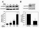
도 8A는 웨스턴 블롯팅을 통해 독시사이클린의 처리농도에 따른 PHF20의 발현을 확인한 결과(상부 패널) 및 루시퍼라제 리포터 유전자 분석을 통해 상기 PHF20의 발현에 따른 YY1 프로모터의 활성을 확인한 결과(하부 패널)를 나타낸 도이다.
도 8B는 웨스턴 블롯팅을 통해 shRNA 형질감염에 따른 PHF20의 발현을 확인한 결과(상부 패널) 및 루시퍼라제 리포터 유전자 분석을 통해 상기 PHF20의 발현에 따른 YY1 프로모터의 활성을 확인한 결과(하부 패널)를 나타낸 도이다.
도 9A는 독시사이클린을 처리하지 않은 C2C12 세포에서 분화 기간에 따른 YY1 프로모터의 활성(상부 패널) 및 PHF2, YY1 및 MyoD의 발현(하부 패널)을 확인한 결과를 나타낸 도이다.
도 9B는 독시사이클린을 처리한 C2C12 세포에서 분화 기간에 따른 YY1 프로모터의 활성(상부 패널) 및 PHF2, YY1 및 MyoD의 발현(하부 패널)을 확인한 결과를 나타낸 도이다.
도 10은 염색질 면역침전 분석을 통해 PHF20이 YY1 프로모터를 조절하는 방식을 확인한 결과를 나타낸 도이다.
도 11은 웨스턴 블롯팅을 통해 C2C12 세포(Pre) 및 3일 동안 분화된 세포에서 YY1의 발현이 MyoD의 발현에 미치는 영향을 확인한 결과를 나타낸 도이다.
도 12는 면역화학염색 분석을 통해 1일 동안 분화된 C2C12 세포에서 YY1의 발현이 근관 형성에 미치는 영향을 확인한 결과를 나타낸 도이다.
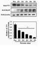
도 13은 PHF20 트랜스제닉 마우스에서 PHF20, YY1 및 MHC의 발현을 웨스턴 블롯팅을 통해 확인한 결과를 나타낸 도이다(WT, 대조군; PHF20-TG, PHF20 트랜스제닉 마우스).
도 14는 PHF20 트랜스제닉 마우스의 체중 측정 결과(A) 및 대퇴근을 관찰한 결과(B)를 나타낸 도이다.
도 15는 조직학적 분석을 통해 PHF20 트랜스제닉 마우스의 비복근 조직을 관찰한 결과를 나타낸 도이다. 보다 상세하게는, 도 15A는 상기 비복근 조직의 횡단면을 관찰한 것이며, 도 15B는 종단면을 관찰한 결과이다.
도 16은 조직학적 분석을 통해 트랜스제닉 마우스의 비복근 조직의 횡단면을 추가 관찰한 결과를 나타낸 도이다.
도 17은 면역화학염색 분석을 통해 PHF20 트랜스제닉 마우스의 비복근 조직을 관찰한 결과를 나타낸 도이다. 보다 상세하게는, 도 17A는 상기 비복근 조직의 종단면을 관찰한 것이며, 도 17B는 횡단면을 관찰한 결과이다.
도 18은 PHF20의 발현이 근육 분화에 미치는 메커니즘을 나타낸 도이다.1 is a diagram showing the results of confirming the expression of PHF20 during muscle differentiation of C2 C12 cells through qRT-PCR (Pre, premyocytes; D1-D5, cells according to the number of differentiated days).
2 is a diagram showing the results of confirming the expression of PHF20 and MyoD during muscle differentiation of C2 C12 cells through Western blotting.
3 is a diagram showing the result of confirming the expression of YY1 during muscle differentiation of C2 C12 cells through qRT-PCR.
4 is a diagram showing the results of confirming the expression of YY1 and MyoD during muscle differentiation of C2 C12 cells through Western blotting.
5 is a diagram showing the results of confirming the effect of PHF20 expression on the differentiation of C2 C12 cells (-Doxy, doxycycline untreated cells; +Doxy, doxycycline treated cells). In more detail, FIG. 5A is the result of analyzing the protein expression of PHF20, YY1 and MyoD in C2 C12 cells according to doxycycline treatment, and FIG. 5B is PHF20, YY1 in C2 C12 cells according to doxycycline treatment or not. and the result of analyzing the mRNA expression of MyoD.
6 is a view showing the results of confirming the effect of the expression of PHF20 on the formation of the root canal through immunochemical staining analysis.
7 is a view showing the results of confirming the effect of inhibition of PHF20 expression on the expression of YY1 and MyoD through Western blotting. In more detail, Fig. 7A is the result of C2 C12 cells (Pre) before differentiation, and Fig. 7B is the result of differentiated myofibers for 1 day.
8A shows the result of confirming the expression of PHF20 according to the concentration of doxycycline treatment through Western blotting (upper panel) and the result of confirming the activity of the YY1 promoter according to the expression of the PHF20 through luciferase reporter gene analysis (lower panel). is the diagram shown.
Figure 8B shows the result of confirming the expression of PHF20 according to shRNA transfection through Western blotting (upper panel) and the result of confirming the activity of the YY1 promoter according to the expression of PHF20 through luciferase reporter gene analysis (lower panel). it is do
9A is a diagram showing the results of confirming the activity of the YY1 promoter (upper panel) and the expression of PHF2, YY1 and MyoD (lower panel) according to the differentiation period in C2 C12 cells not treated with doxycycline.
9B is a diagram showing the results of confirming the activity of the YY1 promoter (upper panel) and the expression of PHF2, YY1 and MyoD (lower panel) according to the differentiation period in C2 C12 cells treated with doxycycline.
10 is a view showing the results of confirming the way PHF20 regulates the YY1 promoter through chromatin immunoprecipitation analysis.
11 is a diagram showing the results of confirming the effect of the expression of YY1 on the expression of MyoD in C2 C12 cells (Pre) and cells differentiated for 3 days through Western blotting.
12 is a diagram showing the results of confirming the effect of YY1 expression on myotube formation in C2 C12 cells differentiated for 1 day through immunochemical staining analysis.
13 is a diagram showing the results of confirming the expression of PHF20, YY1 and MHC in PHF20 transgenic mice through Western blotting (WT, control; PHF20-TG, PHF20 transgenic mice).
14 is a diagram showing the weight measurement results (A) and the results of observing the thigh muscles (B) of the PHF20 transgenic mice.
15 is a diagram showing the results of observing gastrocnemius tissues of PHF20 transgenic mice through histological analysis. In more detail, FIG. 15A is a cross-section of the gastrocnemius tissue, and FIG. 15B is a result of observing a longitudinal cross-section.
16 is a view showing the result of additional observation of a cross-section of gastrocnemius tissue of a transgenic mouse through histological analysis.
17 is a diagram showing the results of observing gastrocnemius tissues of PHF20 transgenic mice through immunochemical staining analysis. In more detail, FIG. 17A is a longitudinal cross-section of the gastrocnemius tissue, and FIG. 17B is a cross-sectional view of the result.
18 is a diagram illustrating a mechanism by which expression of PHF20 affects muscle differentiation.
이하, 본 발명을 상세히 설명한다.Hereinafter, the present invention will be described in detail.
본 발명의 양태에 따르면, 본 발명은 PHF20(PHD finger protein 20) 유전자 발현 억제제 또는 PHF20 단백질 활성 억제제를 유효성분으로 포함하는 근육 감소로 인한 질환의 예방 또는 치료용 약학적 조성물을 제공한다.According to an aspect of the present invention, the present invention provides a pharmaceutical composition for preventing or treating a disease caused by muscle loss comprising a PHF20 (PHD finger protein 20) gene expression inhibitor or PHF20 protein activity inhibitor as an active ingredient.
본 발명에 있어서, PHF20(plant homeodomain finger protein 20, PHD finger protein 20)은 히스톤 H4을 아세틸화하는 리신 아세틸트랜스퍼라제 복합체(lysine acetyltransferase complex)의 다중 도메인 단백질 및 서브 유닛이다. PHF20은 전사인자이며, 신경교종 환자에서 처음으로 확인되었다. PHF20 녹아웃 마우스는 출생 직후 사망하며, 골격 및 조혈 시스템 내에서 다양한 표현형을 나타낸다. 그러나 PHF20 녹아웃 마우스에서 이들 표현형의 상세한 메커니즘은 보고되지 않았다.In the present invention, PHF20 (plant
본 발명의 실시예에서, 상기 PFH20은 근육 분화에서 도 18과 같은 메커니즘을 통해 근육 분화에 영향을 미친다는 것을 확인하였다. 보다 상세하게는, PHF20의 발현 억제는 시험관 내(in vitro) 및 생체 내(in vivo)에서 YY1 프로모터에 결합된 PHF20을 분리시켜 MyoD의 발현을 증가시키고, 상기 MyoD(myogenic differentiation marker)의 발현 증가는 근육 분화를 촉진한다.In an embodiment of the present invention, it was confirmed that the PFH20 affects muscle differentiation through the mechanism shown in FIG. 18 in muscle differentiation. More specifically, inhibition of the expression of PHF20 increases the expression of MyoD by isolating PHF20 bound to the YY1 promoter in vitro and in vivo, and increases the expression of the myogenic differentiation marker (MyoD). promotes muscle differentiation.
본 발명의 구체예에서, 상기 PHF20 유전자 발현 억제제는 YY1 프로모터 활성 억제제인 것이 바람직하다.In an embodiment of the present invention, the PHF20 gene expression inhibitor is preferably a YY1 promoter activity inhibitor.
본 발명의 구체예에서, 상기 PHF20 유전자 발현 억제제는 MyoD의 발현을 증가시켜 근육의 분화를 촉진시키는 것일 수 있다.In an embodiment of the present invention, the PHF20 gene expression inhibitor may promote muscle differentiation by increasing the expression of MyoD.
본 발명에 있어서, YY1(Yin Yang 1)은 4 개의 C-말단 징크 핑거 도메인을 통해 DNA에 결합하는 전사 억제 단백질을 의미한다. 골격근에서, mTOR(mammalian target of rapamycin)의 억제제인 라파마이신의 치료는 mitochondrial transcriptional regulator의 유전자 발현을 증가시켜 미토콘드리아 유전자 발현 및 산소의 소비를 줄인다. mTOR 인산화 및 이의 하류의 표적인 YY1 및 PGC-1α(Peroxisome proliferator-activated receptor gamma coactivator 1-alpha)는 C2C12 근원세포에서 FGF21(fibroblast growth factor 21)의 처리에 의해 증가된다. 근원세포에서 FGF21에 의한 mTOR-YY1-PGC1α 경로의 활성화는 세포 내 ATP 합성, 산소 소비율, 시트르산 합성효소(citrate synthase)의 활성, 당분해, 미토콘드리아 DNA 카피 수 및 주요 에너지의 발현의 현저한 증가에 의해 나타난 에너지 항상성을 조절한다.In the present invention, YY1 (Yin Yang 1) refers to a transcriptional repression protein that binds to DNA through four C-terminal zinc finger domains. In skeletal muscle, treatment with rapamycin, an inhibitor of the mammalian target of rapamycin (mTOR), increases mitochondrial transcriptional regulator gene expression, thereby reducing mitochondrial gene expression and oxygen consumption. mTOR phosphorylation and its downstream targets YY1 and Peroxisome proliferator-activated receptor gamma coactivator 1-alpha (PGC-1α) are increased by FGF21 (fibroblast growth factor 21) treatment in C2 C12 myoblasts. Activation of the mTOR-YY1-PGC1α pathway by FGF21 in myoblasts is caused by significant increases in intracellular ATP synthesis, oxygen consumption rate, citrate synthase activity, glycolysis, mitochondrial DNA copy number, and expression of key energy. Regulates the manifested energy homeostasis.
본 발명의 구체예에서, 상기 근육 감소로 인한 질환은 근육감소증(sarcopenia), 긴장감퇴증(atony), 근위축증(muscular atrophy), 근이영양증(muscular dystrophy), 근육 퇴화, 근경직증(myotonic dystrophy), 근위축성 축삭경화증(amyotrophic lateral sclerosis), 근무력증(myasthenia) 및 악액질(cachexia)로 이루어진군에서 선택되는 것이 바람직하나, 이에 제한되지 않는다.In an embodiment of the present invention, the disease caused by muscle loss is sarcopenia, atony, muscular atrophy, muscular dystrophy, muscle degeneration, myotonic dystrophy, muscle Atrophic axonal sclerosis (amyotrophic lateral sclerosis), myasthenia (myasthenia) and cachexia (cachexia) is preferably selected from the group consisting of, but is not limited thereto.
본 발명에 있어서, 예방은 본 발명에 따른 약학적 조성물의 투여로 근육 감소로 인한 질환의 발병을 억제 또는 지연시키는 모든 행위를 말한다. 또한 본 발명에서 치료는 본 발명에 따른 약학적 조성물의 투여로 근육 감소로 인한 질환의 증세가 호전되거나 이롭게 변경하는 모든 행위를 의미한다.In the present invention, prevention refers to any action that inhibits or delays the onset of a disease due to muscle loss by administration of the pharmaceutical composition according to the present invention. In addition, in the present invention, treatment refers to any action that improves or beneficially changes the symptoms of a disease due to muscle loss by administration of the pharmaceutical composition according to the present invention.
본 발명의 구체예에서, 상기 PHF20 유전자 발현 억제제는 PHF20 유전자에 상보적으로 결합하는 shRNA, siRNA, 리보자임, 및 안티센스 뉴클레오티드로 이루어진 군으로부터 선택된 1종 이상일 수 있다.In an embodiment of the present invention, the PHF20 gene expression inhibitor may be at least one selected from the group consisting of shRNA, siRNA, ribozyme, and antisense nucleotides complementary to the PHF20 gene.
본 발명의 바람직한 구체예에서, 상기 PHF20 유전자 발현 억제제는 서열번호 1의 염기서열로 표시되는 shRNA일 수 있으며, 서열번호 1로 표시되는 염기서열의 변이체 또한 본 발명의 범위 내에 포함된다. 본 발명의 서열번호 1로 표시되는 shRNA의 작용성 등가물, 예를 들어, 서열번호 1로 표시되는 서열 중 일부가 결실(deletion), 치환(substitution) 또는 삽입(insertion)에 의해 변형되었지만, 이를 통해 서열번호 1의 염기서열로 이루어진 shRNA와 기능적으로 동일한 작용을 할 수 있는 변이체(variants)를 포함하는 개념이다.In a preferred embodiment of the present invention, the PHF20 gene expression inhibitor may be an shRNA represented by the nucleotide sequence of SEQ ID NO: 1, and variants of the nucleotide sequence represented by SEQ ID NO: 1 are also included within the scope of the present invention. A functional equivalent of the shRNA represented by SEQ ID NO: 1 of the present invention, for example, a part of the sequence represented by SEQ ID NO: 1 was modified by deletion, substitution or insertion, but through this It is a concept including variants that can function the same as shRNA consisting of the nucleotide sequence of SEQ ID NO: 1.
구체적으로, shRNA는 각 서열번호 1의 염기서열과 각각 70% 이상, 더욱 바람직하게는 80% 이상, 더 더욱 바람직하게는 90% 이상, 가장 바람직하게는 95% 이상의 서열 상동성을 가지는 서열을 포함할 수 있다. 폴리뉴클레오티드 또는 아미노산에 대한 "서열 상동성의 %"는 두 개의 최적으로 배열된 서열과 비교 영역을 비교함으로써 확인된다.Specifically, the shRNA contains a sequence having at least 70%, more preferably at least 80%, even more preferably at least 90%, and most preferably at least 95% sequence homology to the base sequence of each SEQ ID NO: 1, respectively. can do. The "% sequence homology" for a polynucleotide or amino acid is determined by comparing the comparison region with two optimally aligned sequences.
상기 siRNA는 PHF20 유전자의 mRNA의 염기서열 내에서 선택되는 15 내지 30머(mer)의 센스서열 및 상기 센스 서열에 상보적으로 결합하는 안티센스 서열로 구성되며, 이때, 상기 센스 서열은 특별히 이에 제한되는 것은 아니다.The siRNA is composed of a sense sequence of 15 to 30 mers selected from the base sequence of the mRNA of the PHF20 gene and an antisense sequence complementary to the sense sequence, wherein the sense sequence is particularly limited thereto it is not
본 발명의 구체예에서, 상기 PHF20 단백질 활성 억제제는 PHF20 단백질에 특이적으로 결합하는 화합물, 펩티드 및 항체로 이루어진 군으로부터 선택된 1종 이상일 수 있다.In an embodiment of the present invention, the PHF20 protein activity inhibitor may be at least one selected from the group consisting of compounds, peptides and antibodies that specifically bind to PHF20 protein.
상기 화합물은 PHF20 단백질에 특이적으로 결합하여 이의 활성을 억제할 수 있는 임의의 화합물을 모두 포함한다.The compound includes any compound capable of specifically binding to and inhibiting the activity of the PHF20 protein.
상기 항체는 PHF20 단백질에 특이적이고 직접적으로 결합하여 PHF20 단백질의 활성을 효과적으로 억제할 수 있다. 상기 PHF20 단백질에 특이적으로 결합하는 항체로는 폴리클로날(polyclonal) 항체 또는 모노클로날(monoclonal) 항체를 사용하는 것이 바람직하며, 상기 항체는 당업자에게 알려진 공지의 방법으로 제작할 수도 있으며, 상업적으로 알려진 항체를 구입하여 사용할 수 있다.The antibody can specifically and directly bind to the PHF20 protein and effectively inhibit the activity of the PHF20 protein. It is preferable to use a polyclonal antibody or a monoclonal antibody as the antibody that specifically binds to the PHF20 protein, and the antibody may be prepared by a known method known to those skilled in the art, and commercially available. Known antibodies can be purchased and used.
본 발명의 약학적 조성물은 약학적으로 허용 가능한 첨가제를 더 포함할 수 있으며, 이때 약제적으로 허용 가능한 첨가제로는 전분, 젤라틴화 전분, 미결정셀룰로오스, 유당, 포비돈, 콜로이달실리콘디옥사이드, 인산수소칼슘, 락토스, 만니톨, 엿, 아라비아고무, 전호화전분, 옥수수전분, 분말셀룰로오스, 히드록시프로필셀룰로오스, 오파드라이, 전분글리콜산나트륨, 카르나우바납, 합성규산알루미늄, 스테아린산, 스테아린산마그네슘, 스테아린산알루미늄, 스테아린산칼슘, 백당 등이 사용될 수 있다. 본 발명에 따른 약제적으로 허용 가능한 첨가제는 상기 조성물에 대해 0.1 내지 90 중량부 포함되는 것이 바람직하나 이에 한정되는 것은 아니다.The pharmaceutical composition of the present invention may further include a pharmaceutically acceptable additive, wherein the pharmaceutically acceptable additive includes starch, gelatinized starch, microcrystalline cellulose, lactose, povidone, colloidal silicon dioxide, calcium hydrogen phosphate. , lactose, mannitol, syrup, gum arabic, pregelatinized starch, corn starch, powdered cellulose, hydroxypropyl cellulose, Opadry, sodium starch glycolate, carnauba wax, synthetic aluminum silicate, stearic acid, magnesium stearate, aluminum stearate, stearic acid Calcium, sucrose, etc. may be used. The pharmaceutically acceptable additive according to the present invention is preferably included in an amount of 0.1 to 90 parts by weight based on the composition, but is not limited thereto.
또한 본 발명의 약학적 조성물은 실제 임상투여시에 경구 또는 비경구의 여러 가지 제형으로 투여될 수 있는데, 제제화할 경우에는 보통 사용하는 충진제, 증량제, 결합제, 습윤제, 붕해제, 계면활성제 등의 희석제 또는 부형제를 사용하여 조제할 수 있으며, 당해 기술 분야에 알려진 적합한 제제를 이용하는 것이 바람직하다. 상기 조성물에 포함될 수 있는 담체, 부형제 및 희석제로는 락토즈, 덱스트로즈, 수크로스, 올리고당, 솔비톨, 만니톨, 자일리톨, 에리스리톨, 말티톨, 전분, 아카시아 고무, 알지네이트, 젤라틴, 칼슘 포스페이트, 칼슘 실리케이트, 셀룰로오스, 메틸 셀룰로오스, 미정질 셀룰로오스, 폴리비닐 피롤리돈, 물, 메틸히드록시 벤조에이트, 프로필히드록시 벤조에이트, 탈크, 마그네슘 스테아레이트, 광물유 등이 있다.In addition, the pharmaceutical composition of the present invention may be administered in various oral or parenteral dosage forms during actual clinical administration. It can be prepared using excipients, and it is preferable to use suitable formulations known in the art. Carriers, excipients and diluents that may be included in the composition include lactose, dextrose, sucrose, oligosaccharides, sorbitol, mannitol, xylitol, erythritol, maltitol, starch, acacia gum, alginate, gelatin, calcium phosphate, calcium silicate, Cellulose, methyl cellulose, microcrystalline cellulose, polyvinyl pyrrolidone, water, methyl hydroxy benzoate, propyl hydroxy benzoate, talc, magnesium stearate, mineral oil, and the like.
상기 경구투여를 위한 고형제제에는 정제, 환제, 산제, 과립제, 캡슐제 등이 포함되며, 이러한 고형제제는 적어도 하나 이상의 부형제 예를 들면, 전분, 칼슘 카보네이트(Calcium carbonate), 수크로스(Sucrose) 또는 락토오스(Lactose), 젤라틴 등을 섞어 조제된다. 또한 단순한 부형제 이외에 마그네슘 스티레이트 탈크 같은 윤활제들도 사용된다. 또한, 상기 경구투여를 위한 액상제제로는 현탁제, 내용액제, 유제, 시럽제 등이 해당되는데 흔히 사용되는 단순희석제인 물, 리퀴드 파라핀 이외에 여러 가지 부형제, 예를 들면 습윤제, 감미제, 방향제, 보존제 등이 포함될 수 있다.The solid preparation for oral administration includes tablets, pills, powders, granules, capsules, etc., and these solid preparations include at least one excipient, for example, starch, calcium carbonate, sucrose, or It is prepared by mixing lactose and gelatin. In addition to simple excipients, lubricants such as magnesium stearate talc are also used. In addition, the liquid formulations for oral administration include suspensions, internal solutions, emulsions, syrups, etc. In addition to water and liquid paraffin, which are commonly used simple diluents, various excipients, for example, wetting agents, sweeteners, fragrances, preservatives, etc. may be included.
상기 비경구투여를 위한 제제에는 멸균된 수용액, 비수성용제, 현탁제, 유제, 동결건조제제, 좌제가 포함된다. 비수성용제, 현탁용제로는 프로필렌글리콜(Propylene glycol), 폴리에틸렌 글리콜, 올리브 오일과 같은 식물성 기름, 에틸올레이트와 같은 주사 가능한 에스테르 등이 사용될 수 있다. 좌제의 기제로는 위텝솔(witepsol), 마크로골, 트윈(tween) 61, 카카오지, 라우린지, 글리세로제라틴 등이 사용될 수 있다. 상기 비경구투여는 피부 외용 또는 복강 내 주사, 직장 내 주사, 피하주사, 정맥주사, 근육 내 주사 또는 흉부 내 주사 주입방식을 사용하여 이루어질 수 있다.The formulations for parenteral administration include sterile aqueous solutions, non-aqueous solutions, suspensions, emulsions, lyophilized formulations, and suppositories. Non-aqueous solvents and suspensions may include propylene glycol, polyethylene glycol, vegetable oils such as olive oil, and injectable esters such as ethyl oleate. As a base of the suppository, witepsol, macrogol, tween 61, cacao butter, laurin, glycerogelatin, and the like can be used. The parenteral administration can be made by using an injection method for external application to the skin or intraperitoneal injection, intrarectal injection, subcutaneous injection, intravenous injection, intramuscular injection, or intrathoracic injection.
본 발명의 약학적 조성물의 투여량은 환자의 체중, 연령, 성별, 건강상태, 식이, 투여시간, 투여방법, 배설율 및 질환의 중증도에 따라 그 범위가 다양하며, 하루에 한 번 투여할 수도 있고, 수 회 나누어 투여할 수도 있다.The dosage of the pharmaceutical composition of the present invention varies depending on the patient's weight, age, sex, health status, diet, administration time, administration method, excretion rate and severity of disease, and may be administered once a day. Or, it can be administered in several divided doses.
본 발명의 약학적 조성물은 개체에게 다양한 경로로 투여될 수 있다.The pharmaceutical composition of the present invention may be administered to an individual by various routes.
본 발명의 약학적 조성물은 근육 감소로 인한 질환의 예방 또는 치료를 위하여 단독으로, 또는 수술, 방사선 치료, 호르몬 치료, 화학 치료 및 생물학적 반응 조절제를 사용하는 방법들과 병용하여 사용할 수 있다.The pharmaceutical composition of the present invention can be used alone or in combination with methods using surgery, radiation therapy, hormone therapy, chemotherapy, and biological response modifiers for the prevention or treatment of diseases caused by muscle loss.
본 발명의 다른 양태에 따르면, 본 발명은 PHF20 유전자 발현 억제제 또는 PHF20 단백질 활성 억제제; 또는 YY1 프로모터 활성 억제제;를 포함하는, 근육 감소로 인한 질환의 예방 또는 치료용 약학적 조성물을 제공한다.According to another aspect of the present invention, the present invention provides a PHF20 gene expression inhibitor or a PHF20 protein activity inhibitor; Or YY1 promoter activity inhibitor; provides a pharmaceutical composition for the prevention or treatment of diseases caused by muscle loss, including.
본 발명의 구체예에서, 상기 YY1 프로모터 활성 억제제는 siRNA일 수 있으며, 서열번호 2의 염기서열로 표시되는 것이 바람직하다.In an embodiment of the present invention, the YY1 promoter activity inhibitor may be siRNA, and is preferably represented by the nucleotide sequence of SEQ ID NO: 2.
본 발명의 또 다른 양태에 따르면, 본 발명은 PHF20 유전자 발현 억제제 또는 PHF20 단백질 활성 억제제를 유효성분으로 포함하는 근육 증대용 또는 근육 손실 저해용 조성물을 제공한다.According to another aspect of the present invention, the present invention provides a composition for increasing muscle or inhibiting muscle loss comprising a PHF20 gene expression inhibitor or a PHF20 protein activity inhibitor as an active ingredient.
상기 조성물은 건강기능식품 조성물 또는 사료 첨가제 조성물인 것이 바람직하나, 이에 제한되지 않는다.The composition is preferably a health functional food composition or a feed additive composition, but is not limited thereto.
본 발명에 있어서, 사료 첨가제는 영양적 또는 특정 목적을 위하여 사료에 미량으로 첨가되는 물질을 의미한다.In the present invention, a feed additive refers to a substance added in a trace amount to a feed for nutritional or specific purposes.
상기 근육 증대용 또는 근육 손실 저해용 조성물이 건강기능식품 조성물인 경우, PHF20 유전자 발현 억제제 또는 PHF20 단백질 활성 억제제를 그대로 첨가하거나 다른 식품 또는 식품 성분과 함께 사용될 수 있고, 통상적인 방법에 따라 적절하게 사용될 수 있다. 유효성분의 혼합양은 사용 목적(예방, 건강 또는 치료적 처치)에 따라 적합하게 결정될 수 있다. 일반적으로, 식품 또는 음료의 제조 시에 본 발명의 PHF20 유전자 발현 억제제 또는 PHF20 단백질 활성 억제제는 전체 조성물에 대하여 15중량 % 이하, 바람직하게는 10 중량 % 이하의 양으로 첨가된다. 그러나, 건강 및 위생을 목적으로 하거나 또는 건강 조절을 목적으로 하는 장기간의 섭취의 경우에는 상기 범위 이하일 수 있으며, 안전성 면에서 아무런 문제가 없기 때문에 유효성분은 상기 범위 이상의 양으로도 사용될 수 있다.When the composition for muscle increase or muscle loss inhibition is a health functional food composition, the PHF20 gene expression inhibitor or the PHF20 protein activity inhibitor may be added as it is, or it may be used together with other foods or food ingredients, and used appropriately according to a conventional method. can The mixed amount of the active ingredient may be appropriately determined according to the purpose of use (prevention, health or therapeutic treatment). In general, in the production of food or beverage, the PHF20 gene expression inhibitor or PHF20 protein activity inhibitor of the present invention is added in an amount of 15% by weight or less, preferably 10% by weight or less, based on the total composition. However, in the case of long-term intake for health and hygiene or health control, it may be less than the above range, and since there is no problem in terms of safety, the active ingredient may be used in an amount above the above range.
상기 건강기능식품의 종류에는 특별한 제한은 없다. 상기 건강기능식품의 예로는 캔디류, 스낵류, 껌류, 음료수, 차, 드링크제, 알코올 음료 및 비타민 복합제 등이 있으며, 통상적인 의미에서의 건강기능식품을 모두 포함한다.There is no particular limitation on the type of the health functional food. Examples of the health functional food include candies, snacks, gums, beverages, tea, drinks, alcoholic beverages and vitamin complexes, and include all health functional foods in a conventional sense.
상기 근육 증대용 또는 근육 손실 저해용 조성물이 사료 첨가제 조성물인 경우, 본 발명의 사료 첨가제 조성물은 사료 원료에 적절하게 배합하여 사료로 제공되는데, 상기 사료 원료로 곡물류, 조강류, 식물성 유박류, 동물성 사료 원료, 기타 사료 원료, 정제품 등이 사용되고 있으나, 이에 한정되지 않는다.When the composition for increasing muscle or inhibiting muscle loss is a feed additive composition, the feed additive composition of the present invention is provided as a feed by appropriately mixing it with feed raw materials. Feed raw materials, other feed raw materials, refined products, etc. are used, but are not limited thereto.
이하, 실시예를 통하여 본 발명을 더욱 상세히 설명하고자 한다. 이들 실시예는 오로지 본 발명을 예시하기 위한 것으로서, 본 발명의 범위가 이들 실시예에 의해 제한되는 것으로 해석되지는 않는 것은 당업계에서 통상의 지식을 가진 자에게 있어서 자명할 것이다.Hereinafter, the present invention will be described in more detail through examples. These examples are only for illustrating the present invention, and it will be apparent to those of ordinary skill in the art that the scope of the present invention is not to be construed as being limited by these examples.
실험 재료experimental material
항-PHF20 항체는 Cell Signaling Technology(Beverly, MA)로부터 구입 하였다. 항-β-액틴 항체는 Sigma Aldrich(St. Louis, USA)로부터 얻었다. 항-YY1, 항-MyoD 및 항-미오신 중쇄 항체는 Santa Cruz Biotechnology(Santa Cruz, CA)로부터 구입하였다. 항-미오신(MF20) 항체는 아이오와 대학의 DSHB로부터 얻었다. HRP가 결합된 항-마우스, 항-토끼 및 항-염소 IgG 항체는 Koma biotech(Seoul, Korea)로부터 입수하였다. pYY-루시퍼라제는 Panomics(California, USA)에서 구입하였다. Luciferase assay system kit는 Promega(Madison, USA)에서 구입하였다. Tet-on advanced inducible gene expression system kit는 Clontech(Takara Bio Company, USA)로부터 입수하였다.Anti-PHF20 antibody was purchased from Cell Signaling Technology (Beverly, MA). Anti-β-actin antibody was obtained from Sigma Aldrich (St. Louis, USA). Anti-YY1, anti-MyoD and anti-myosin heavy chain antibodies were purchased from Santa Cruz Biotechnology (Santa Cruz, CA). Anti-myosin (MF20) antibody was obtained from DSHB, University of Iowa. HRP-conjugated anti-mouse, anti-rabbit and anti-goat IgG antibodies were obtained from Koma biotech (Seoul, Korea). pYY-luciferase was purchased from Panomics (California, USA). Luciferase assay system kit was purchased from Promega (Madison, USA). Tet-on advanced inducible gene expression system kit was obtained from Clontech (Takara Bio Company, USA).
실험예 1. 세포배양Experimental Example 1. Cell culture
1-1. C1-1. C22CC1212 세포 배양 및 분화 Cell culture and differentiation
전근세포(premyocyte)인 C2C12 세포는 DMEM 배지를 사용하여 37℃ 및 5% CO2 조건에서 배양하였다. 상기 DMEM(WELGENE, Gyeongsan, Korea) 배지는 10% FBS(fetal bovine serum)(HyClone, Logan, USA) 및 1% Antibiotic-Antimycotic(Gibco, Detroit, USA)을 포함한다.Premyocytes, C2 C12 cells, were cultured at 37° C. and 5% CO2 conditions using DMEM medium. The DMEM (WELGENE, Gyeongsan, Korea) medium contains 10% FBS (fetal bovine serum) (HyClone, Logan, USA) and 1% Antibiotic-Antimycotic (Gibco, Detroit, USA).
세포 분화를 위해 배지를 분화 배지(differentiation medium, DM)로 교체한 후 배양하였으며, 배지는 매 2일 마다 교체하였다. 상기 분화 배지는 2% 말 혈청(horse serum) 및 1% Antibiotic-Antimycotic(Gibco, Detroit, USA)를 포함한다. 상기 세포 분화는 배지 교체 24시간 후 시작되었다.For cell differentiation, the medium was replaced with a differentiation medium (DM) and then cultured, and the medium was replaced every 2 days. The differentiation medium contains 2% horse serum and 1% Antibiotic-Antimycotic (Gibco, Detroit, USA). The cell differentiation was initiated 24 hours after medium change.
1-2. C1-2. C22CC1212/Tet-On 유도성 PHF20 세포/Tet-On Inducible PHF20 Cells
C2C12 세포를 이용하여 C2C12/Tet-On 유도성 PHF20 세포를 제조하였다. 구체적으로, C2C12-TET3G 근원세포를 제조하기 위해, C2C12 세포에 렌티바이러스-TET3G을 처리하고, G418(1.2 mg/ml)로 선택하여 C2C12-TET3G 근원세포를 수득하였다. 상기 C2C12-TET3G 세포를 pLVX-TRE3G-PHF20으로 일시적 형질감염시켰다. 퓨로마이신(3 μg/ml)을 이용하여 일시적 형질감염된 세포 중 Tet-On 유도성 PHF20 세포를 선택하였다. 선택된 세포(C2C12/Tet-On 유도성 PHF20 세포)는 독시사이클린(doxycycline, Doxy)이 포함된 배지에서 배양할 경우, PHF20의 발현이 유도된다.C2 C12 /Tet-On induced PHF20 cells were prepared using C2 C12 cells. Specifically, to prepare C2 C12 -TET3G myoblasts, C2 C12 cells were treated with lentivirus-TET3G, and selected with G418 (1.2 mg/ml) to obtain C2 C12 -TET3G myoblasts. did. The C2 C12 -TET3G cells were transiently transfected with pLVX-TRE3G-PHF20. Tet-On inducible PHF20 cells were selected among transiently transfected cells with puromycin (3 μg/ml). When the selected cells (C2 C12 /Tet-On induced PHF20 cells) are cultured in a medium containing doxycycline (Doxy), expression of PHF20 is induced.
후술되는 실험에서 C2C12 세포에서 PHF20의 발현을 유도하기 위해 독시사이클린을 100 ng/ml의 농도로 포함하는 배지로 배양하였다.In the experiment to be described later, in order to induce the expression of PHF20 in C2 C12 cells, it was cultured in a medium containing doxycycline at a concentration of 100 ng/ml.
실험예 2. 웨스턴 블로팅Experimental Example 2. Western blotting
준비된 세포를 얼음 위에 두었으며, 상기 세포에 용해 버퍼를 처리하여 세포 용해물을 제조하였다. 상기 용해 버퍼는 50 mM Tris-HCl(pH7.5), 1% v/v Nonidet P-40, 120 mM NaCl, 25 mM sodium fluoride, 40 mM β-glycerol phosphate, 0.1 mM sodium orthovanadate, 1 mM phenylmethylsulfonyl fluoride, 1 mM benzamidine 및 2 μM microcystin-LR을 포함한다. 세포 용해물을 13000 rpm에서 30 분 동안 원심분리하여 단백질을 수득하였다. 상기 단백질을 10.0 내지 7.5% 겔을 이용한 SDS-PAGE로 전개하였다. 전개된 단백질을 Immobilon-P 멤브레인(Millipore)으로 옮겼다. 그 후 멤브레인을 5% 탈지유 및 0.2% Tween-20을 함유하는 1X TBS(tri-buffered saline buffer; 140mM NaCl, 2.7mM KCl, 250mM Tris HCl, pH7.4)을 처리하여 1시간 동안 블로킹하였다. 그 후 1000배 희석된 각 단백질에 대한 1차 항체를 처리한 후 4℃에서 밤새도록 배양하였다. 반응 후 TBS로 멤브레인을 세척하였다. 세척된 멤브레인에 2000배 희석된 2차 항체(horseradish peroxidase-conjugated anti-mouse IgG 또는 anti-rabbit IgG(Komabiotech, Seoul, Korea))를 처리한 후 배양하였다. 배양 후 멤브레인을 TBS로 세척하였으며, 제조사의 매뉴얼에 따라 단백질을 검출하였다.The prepared cells were placed on ice, and the cells were treated with a lysis buffer to prepare a cell lysate. The lysis buffer was 50 mM Tris-HCl (pH7.5), 1% v/v Nonidet P-40, 120 mM NaCl, 25 mM sodium fluoride, 40 mM β-glycerol phosphate, 0.1 mM sodium orthovanadate, 1 mM phenylmethylsulfonyl fluoride , containing 1 mM benzamidine and 2 μM microcystin-LR. Cell lysates were centrifuged at 13000 rpm for 30 minutes to obtain proteins. The protein was developed by SDS-PAGE using a 10.0 to 7.5% gel. The developed protein was transferred to an Immobilon-P membrane (Millipore). Thereafter, the membrane was treated with 1X TBS (tri-buffered saline buffer; 140 mM NaCl, 2.7 mM KCl, 250 mM Tris HCl, pH 7.4) containing 5% skim milk and 0.2% Tween-20 and blocked for 1 hour. Thereafter, the primary antibody for each protein diluted 1000-fold was treated and incubated overnight at 4°C. After the reaction, the membrane was washed with TBS. The washed membrane was treated with a 2000-fold diluted secondary antibody (horseradish peroxidase-conjugated anti-mouse IgG or anti-rabbit IgG (Komabiotech, Seoul, Korea)) and then cultured. After incubation, the membrane was washed with TBS, and protein was detected according to the manufacturer's manual.
실험예 3. 실시간 정량적 역전사 중합효소 연쇄반응(Real-time quantitative reverse transcription-polymerase chain reaction, qRT-PCR)Experimental Example 3. Real-time quantitative reverse transcription-polymerase chain reaction (qRT-PCR)
C2C12/Tet-On 유도성 PHF20 세포를 6웰 플레이트에 시딩하여 24시간 동안 배양하였다. PHF20의 발현을 유도하기 위하여, 상기 C2C12/Tet-On 유도성 PHF20 세포에 독시사이클린을 처리하였다. 독시사이클린을 처리한 다음날, QuickGene RNA kit(Fujifilm's Life Science System, Tokyo, Japan)을 사용하여 세포로부터 전체 RNA를 추출하였다. 또한 SuPrimeScript RT Premix(GeNet Bio, Cheonan, Korea)를 사용하여, 추출된 전체 RNA로부터 cDNA를 합성하였다. cDNA 증폭은 Prime Q-Mastermix(GeNet Bio, Cheonan, Korea) 및 CFX96T Real-time System(Bio-Rad, Hercules, CA, USA)으로 수행하였다. 각각의 cDNA의 qRT-PCR 분석은 SYBR 녹색 염료 및 StepOne Plus 실시간 PCR 시스템(Invitrogen, Carlsbad, CA)을 이용해 이중 DNA(duplex DNA) 형성을 측정하였다.C2 C12 /Tet-On induced PHF20 cells were seeded in 6-well plates and cultured for 24 hours. In order to induce the expression of PHF20, the C2 C12 /Tet-On induced PHF20 cells were treated with doxycycline. The day after doxycycline treatment, total RNA was extracted from the cells using a QuickGene RNA kit (Fujifilm's Life Science System, Tokyo, Japan). In addition, cDNA was synthesized from the extracted total RNA using SuPrimeScript RT Premix (GeNet Bio, Cheonan, Korea). cDNA amplification was performed with Prime Q-Mastermix (GeNet Bio, Cheonan, Korea) and CFX96T Real-time System (Bio-Rad, Hercules, CA, USA). qRT-PCR analysis of each cDNA measured duplex DNA formation using SYBR green dye and the StepOne Plus real-time PCR system (Invitrogen, Carlsbad, CA).
본 실험에서 사용한 프라이머는 표 1에 나타내었다.The primers used in this experiment are shown in Table 1.
실험예 4. 면역화학염색 분석을 통한 미오신 중쇄(myosin heavy chain, MHC)의 검출Experimental Example 4. Detection of myosin heavy chain (MHC) through immunochemical staining analysis
C2C12/Tet-On 유도성 PHF20 세포를 1x10⁵ cells/well의 농도로 멸균된 커버슬립을 포함하는 12웰 플레이트에 시딩하였으며, 37℃ 및 5% CO2 조건에서 배양하였다. 독시사이클린 미처리군(-Doxy)은 세포에 독시사이클린을 처리하지 않고 24시간 동안 배양하였으며, 독시사이클린 처리군(+Doxy)은 배지를 독시사이클린을 포함한 배지로 교체한 후 24시간 동안 배양하였다. 배양된 각 세포의 배지를 분화 배지로 교체한 후 1일(D1) 또는 3일(D3) 동안 배양하여 분화시켰다. 그 후 세포를 37℃인 PBS로 세척하였다. 세척된 세포를 4% 파라포름알데하이드에서 15분 동안 배양하여 커버슬립에 고정하였으며, PBS로 2회 세척하였다. 블로킹을 위하여, 커버슬립에 1% BSA(bovine serum albumin)를 처리한 후 실온에서 1시간 동안 진탕배양하였다. 또한 anti-myosin(MF-20) 항체를 1:200의 비율로 희석하여 세포에 첨가하였고, 4℃에서 밤새도록 진탕배양하였다. 진탕배양 후 커버슬립을 PBS로 3회 세척하였다. FITC 2차 항체(1:1000)(in 5% skim milk)를 첨가한 후 실온의 암실에서 6시간 동안 진탕배양하였다. 진탕배양 후 커버슬립을 PBS로 3회 세척하였다. 세척된 커버슬립의 배지를 DAPI VECTASHIELD(St. Louis, USA)를 포함하는 마운팅 배지로 교체한 후 슬라이드에 결합시켜 형광을 분석하였다.C2 C12 /Tet-On induced PHF20 cells were seeded in a 12-well plate containing sterile coverslips at a concentration of 1x10⁵ cells/well, and cultured at 37° C. and 5% CO2 conditions. The doxycycline untreated group (-Doxy) cells were cultured for 24 hours without treatment with doxycycline, and the doxycycline-treated group (+Doxy) was cultured for 24 hours after replacing the medium with doxycycline-containing medium. After replacing the medium of each cultured cell with a differentiation medium, the cells were differentiated by culturing for 1 day (D1) or 3 days (D3). The cells were then washed with PBS at 37°C. The washed cells were incubated in 4% paraformaldehyde for 15 minutes, fixed on coverslips, and washed twice with PBS. For blocking, coverslips were treated with 1% bovine serum albumin (BSA) and then cultured with shaking at room temperature for 1 hour. In addition, an anti-myosin (MF-20) antibody was diluted at a ratio of 1:200 and added to the cells, and incubated with shaking at 4°C overnight. After shaking incubation, the coverslips were washed 3 times with PBS. FITC secondary antibody (1:1000) (in 5% skim milk) was added and then cultured with shaking in the dark at room temperature for 6 hours. After shaking incubation, the coverslips were washed 3 times with PBS. After replacing the medium of the washed coverslip with a mounting medium containing DAPI VECTASHIELD (St. Louis, USA), it was bound to the slide and analyzed for fluorescence.
실험예 5. 루시퍼라제 리포터 유전자 분석Experimental Example 5. Luciferase reporter gene analysis
C2C12/Tet-On 유도성 PHF20 세포를 3x10⁵ cells/ml의 농도로 6웰 플레이트에 시딩하였다. 시딩된 세포에 YY1-루시퍼라제를 포함하는 플라스미드 1 μg을 제조사의 매뉴얼에 따라 형질감염시켰다. 형질감염된 세포는 24시간 후 실험을 위해 사용하였다.C2 C12 /Tet-On induced PHF20 cells were seeded in a 6-well plate at a concentration of 3×10 5 cells/ml. The seeded cells were transfected with 1 μg of a plasmid containing YY1-luciferase according to the manufacturer's manual. Transfected cells were used for experiments after 24 hours.
형질감염된 세포에 리포터 용해 버퍼를 처리하여 세포를 용해시켰다. 상기 리포터 용해 버퍼는 25 mM Tris-phosphate, 2 mM DTT, 2 mM trans-1,2-cyclohexanediamine-N,N,N’,N’-tetraacetic acid, 10%(v/v) glycerol 및 1%(v/v) Triton x-100을 포함한다. 루시퍼라제의 활성은 세포 용해물 20 μg에 luciferase assay substrate(Promega)을 첨가하여 배양하였고, 10초 동안의 발광을 luminometer(Duo Lumat LB 9507)를 이용하여 측정하였다.Transfected cells were treated with reporter lysis buffer to lyse the cells. The reporter lysis buffer is 25 mM Tris-phosphate, 2 mM DTT, 2 mM trans-1,2-cyclohexanediamine-N,N,N',N'-tetraacetic acid, 10% (v/v) glycerol and 1% ( v/v) Triton x-100. Luciferase activity was incubated by adding luciferase assay substrate (Promega) to 20 μg of cell lysate, and luminescence for 10 seconds was measured using a luminometer (Duo Lumat LB 9507).
실험예 6. 염색질 면역침전(Chromatin-immunoprecipitation, ChiP) 분석Experimental Example 6. Chromatin-immunoprecipitation (ChP) analysis
염색질 면역침전 분석은 제조사의 매뉴얼(Upstate Biotechnology, Lake Placid, NY, USA)에 따라 수행하였다. 구체적으로, C2C12 세포는 3일 동안 분화시켰으며, 포름알데히드를 7.5분 동안 처리하였다. 그 후 프로테아제 억제제를 포함하는 PBS로 세포를 2회 세척한 후 얼음 위로 옮겼다. 얼음 상의 세포에 SDS-용해 버퍼를 처리한 후 10분 동안 배양하였으며, 용해물을 수확하였다. 상기 용해 버퍼는 50 mM Tris-HCl(pH 8.0), 1% SDS, 10 mM EDTA, 1 mM PMSF, 1 μg/ml aprotinin 및 2 μg/ml pepstatin을 포함한다. 상기 수확된 용해물은 1차 항체 또는 대조군 IgG를 처리한 후 4℃에서 밤새도록 배양하였다. 그 후 배양물에 단백질 A 비드(500 μg/ml BSA 및 200 μg/ml 연어 정자 DNA를 포함) 30 μl을 함께 4℃에서 3시간 동안 배양하였다. 배양 후 DNA 시료는 QIA quick PCR purification kit(Qiagen, Hilden, Germany)를 사용하여 정제하였고, qRT-PCT로 정량하였다. qRT-PCT 결과는 입력된 값의 백분율로 표시하였다. 본 실험에 사용한 프로모터 특이적 프라이머인 mouse YY1(-200/-55)의 서열은 다음과 같다.Chromatin immunoprecipitation analysis was performed according to the manufacturer's manual (Upstate Biotechnology, Lake Placid, NY, USA). Specifically, C2 C12 cells were differentiated for 3 days and treated with formaldehyde for 7.5 minutes. Thereafter, the cells were washed twice with PBS containing a protease inhibitor and then transferred onto ice. Cells on ice were treated with SDS-lysis buffer and incubated for 10 minutes, and the lysate was harvested. The lysis buffer contains 50 mM Tris-HCl (pH 8.0), 1% SDS, 10 mM EDTA, 1 mM PMSF, 1 μg/ml aprotinin and 2 μg/ml pepstatin. The harvested lysates were incubated overnight at 4°C after treatment with primary antibody or control IgG. Thereafter, 30 μl of protein A beads (containing 500 μg/ml BSA and 200 μg/ml salmon sperm DNA) were incubated together at 4° C. for 3 hours in the culture. After incubation, the DNA sample was purified using a QIA quick PCR purification kit (Qiagen, Hilden, Germany), and quantified by qRT-PCT. qRT-PCT results were expressed as a percentage of the entered value. The sequence of mouse YY1 (-200/-55), a promoter-specific primer used in this experiment, is as follows.
- mouse YY1(-200/-55) : 5’-CGCTGCCTTCCTCCCTCT-3’(forward), 5’-CGTCCGTGGCGATGTAGA-3’(reverse)- mouse YY1(-200/-55) : 5'-CGCTGCCTTCCTCCCTCT-3'(forward), 5'-CGTCCGTGGCGATGTAGA-3'(reverse)
실험예 7. PHF20 트랜스제닉 마우스 제조Experimental Example 7. Preparation of PHF20 transgenic mice
PHF20 트랜스제닉 마우스를 제조하기 위해, pcDNA3 벡터의 CMV 프로모터의 하류에 인간 PHF20을 코딩하는 cDNA를 서브클로닝(subcloning)하였고, WT C57BL/6 마우스를 상기 벡터로 형진질전환시켰다. 대조군은 WT C57BL/6 마우스를 이용하였고, WT로 표시하였다. 마우스 PHF20-TG 및 WT는 12h/12h 장일주기, 습도 50 내지 60% 및 주변 온도 22℃인 환경에서 사육하였다. 또한 모든 동물 실험은 기관 지침에 따라 동물 시설에서 수행되었다. 실험 프로토콜은 충남대학교(CNU-00890) 검토위원회의 승인을 받았다.To prepare PHF20 transgenic mice, cDNA encoding human PHF20 was subcloned downstream of the CMV promoter of pcDNA3 vector, and WT C57BL/6 mice were transformed with the vector. As a control group, WT C57BL/6 mice were used and denoted as WT. Mice PHF20-TG and WT were reared in an environment of 12h/12h long cycle, 50-60% humidity, and 22°C ambient temperature. In addition, all animal experiments were performed in animal facilities in accordance with institutional guidelines. The experimental protocol was approved by the review committee of Chungnam National University (CNU-00890).
실험예 8. 조직학적 분석 및 면역화학염색 분석Experimental Example 8. Histological analysis and immunochemical staining analysis
마우스에서 채취된 근육은 10% 포르말린을 처리한 후 4℃에서 밤새도록 배양하여 고정하였다. 고정된 근육 시료는 파라핀 블록에 포매하였다. 파라핀 블록을 잘라 5 μm 두께의 박편을 준비하였다. 인접 부위의 박편을 추가로 준비하여, 순서대로 슬라이드를 제작하였다.Muscles collected from mice were treated with 10% formalin and then cultured overnight at 4° C. and fixed. The fixed muscle samples were embedded in paraffin blocks. A 5 μm-thick section was prepared by cutting the paraffin block. Slices from adjacent sites were additionally prepared, and slides were prepared sequentially.
일반적인 형태학적 분석을 위하여, 제작된 슬라이드를 헤마톡실린 및 에오신(hematoxylin 및 eosin, H&E)으로 염색하였다.For general morphological analysis, the prepared slides were stained with hematoxylin and eosin (H&E).
또한 면역화학염색 분석을 위하여, 슬라이드 상의 박편을 자일렌으로 탈파라핀화시켰으며, 에탄올을 이용하여 재수화시켰다. 그 후, 슬라이드를 과산화효소 퀀칭 용액(peroxidase quenching solution)에 담가 10분 동안 반응시켰다. 반응후 슬라이드를 PBS로 5분씩 2회 세척하였고, 블로킹을 위해 슬라이드에 시약 A를 2 방울 첨가한 후 30분 동안 배양하였다. 배양 후 슬라이드를 PBS로 2회 세척하였다. 세척된 슬라이드에 1차 항체(anti-MHC)를 처리한 후 4℃에서 밤새도록 배양하였다. 배양된 슬라이드를 PBS로 세척한 후 비오티닐화된(biotinylated) 2차 항체인 시약 B를 처리하였으며, 실온에서 1시간 동안 배양하였다. 배양된 슬라이드를 PBS로 세척한 후 효소가 결합된 시약 C를 슬라이드에 떨어뜨렸으며, PBS로 세척하였다. PBS로 세척한 후 DAB 발색체; 및 시약 D1, D2 및 D3의 혼합물;을 슬라이드에 떨어뜨렸으며, 형광 현미경으로 신호를 관찰하였다. 그리고 증류수로 반응을 멈춘 후, 형광 현미경으로 슬라이드 사진을 찍었다.In addition, for immunochemical staining analysis, the slices on the slide were deparaffinized with xylene and rehydrated with ethanol. Thereafter, the slides were immersed in a peroxidase quenching solution and reacted for 10 minutes. After the reaction, the slides were washed twice with PBS for 5 minutes each, and 2 drops of reagent A were added to the slides for blocking, and then incubated for 30 minutes. After incubation, the slides were washed twice with PBS. The washed slides were treated with a primary antibody (anti-MHC) and then incubated overnight at 4°C. The cultured slides were washed with PBS and then treated with reagent B, a biotinylated secondary antibody, and incubated at room temperature for 1 hour. After the cultured slides were washed with PBS, enzyme-conjugated reagent C was dropped on the slides and washed with PBS. DAB chromophore after washing with PBS; and a mixture of reagents D1, D2 and D3; were dropped on the slide, and the signal was observed under a fluorescence microscope. And after stopping the reaction with distilled water, slide pictures were taken with a fluorescence microscope.
실험예 9. 통계학적 분석Experimental Example 9. Statistical Analysis
데이터는 평균 ± S.E.로 표시하였다. 적어도 3회의 실험에서 그룹 간 차이는 Student's t-test를 사용하여 분석하였고, P<0.05는 통계적으로 유의한 것으로 간주하였다. 또한 결과의 정량 분석에는 Image J 소프트웨어(버전 1.47)를 사용하였다.Data are expressed as mean±S.E. Differences between groups in at least 3 experiments were analyzed using Student's t-test, and P<0.05 was considered statistically significant. Image J software (version 1.47) was also used for quantitative analysis of the results.
실시예 1. 근육 분화 동안 YY1 및 PHF20의 발현 분석Example 1. Analysis of expression of YY1 and PHF20 during muscle differentiation
근육 분화에서 PHF20(PHD finger protein 20)의 변화를 평가하기 위하여, 근육 분화에서의 YY1(Yin Yang 1) 및 PHF20 mRNA 및 단백질 발현을 qRT-PCR 및 웨스턴 블롯팅으로 분석하였다.To evaluate changes in PHF20 (PHD finger protein 20) in muscle differentiation, YY1 (Yin Yang 1) and PHF20 mRNA and protein expression in muscle differentiation were analyzed by qRT-PCR and Western blotting.
1-1. PHF201-1. PHF20
C2C12 세포를 시딩한지 24시간 후 2% 말 혈청을 포함하는 분화 배지(DM)로 교체하여 근육 분화를 유도하였다. 세포의 PHF20 mRNA 발현은 qRT-PCR을 통해 근육 분화 기간 동안 매일 측정하였다. 또한 상기 근육 분화 기간 동안 PHF20 및 myoD의 단백질 발현은 웨스턴 블롯팅을 통해 매일 확인하였다. PHF20 mRNA 및 단백질 발현을 분석한 결과는 각각 도 1 및 2에 나타내었다.C2 C12 cells were replaced with differentiation medium (DM) containing 2% horse serum 24 hours after seeding to induce muscle differentiation. Cellular PHF20 mRNA expression was measured daily during muscle differentiation by qRT-PCR. In addition, the protein expression of PHF20 and myoD during the muscle differentiation period was checked daily through western blotting. The results of analyzing PHF20 mRNA and protein expression are shown in FIGS. 1 and 2 , respectively.
도 1 및 2에 나타낸 바와 같이, 근육 분화가 진행될수록 PHF20은 mRNA 및 단백질의 발현이 감소되는 것을 확인하였다. 반면에, MyoD는 근육의 분화가 진행될수록 단백질의 발현이 증가되는 것을 확인하였다. 상기 결과는 C2C12 세포에서 전사인자인 PHF20가 근육 분화에 관여할 수 있음을 시사한다.As shown in FIGS. 1 and 2 , it was confirmed that the expression of PHF20 mRNA and protein decreased as muscle differentiation progressed. On the other hand, it was confirmed that MyoD protein expression increased as muscle differentiation progressed. These results suggest that PHF20, a transcription factor, may be involved in muscle differentiation in C2 C12 cells.
1-2. YY11-2. YY1
YY1은 선행 연구를 통해 PHF20이 과발현되는 세포에서 발현이 증가된다는 것을 확인한바 있다. 이에, YY1의 mRNA 및 단백질 발현을 상기 실시예 1-1과 같은 방법으로 분석하였다. YY1의 mRNA 및 단백질 발현을 분석한 결과는 각각 도 3 및 4에 나타내었다.It has been confirmed that YY1 expression is increased in cells overexpressing PHF20 through previous studies. Accordingly, mRNA and protein expression of YY1 were analyzed in the same manner as in Example 1-1. The results of analyzing the mRNA and protein expression of YY1 are shown in FIGS. 3 and 4 , respectively.
도 3 및 4에 나타낸 바와 같이, YY1는 근육 분화가 진행될수록 mRNA의 발현이 감소하는 것을 확인하였다. 또한 근육 분화가 진행될수록 MyoD의 단백질 발현은 증가하였으나, YY1의 단백질 발현은 감소한 것을 확인하였다.As shown in FIGS. 3 and 4 , it was confirmed that mRNA expression of YY1 decreased as muscle differentiation progressed. In addition, as muscle differentiation progressed, the protein expression of MyoD increased, but it was confirmed that the protein expression of YY1 decreased.
실시예 2. PHF20 발현이 CExample 2. PHF20 expression is C22CC1212 세포의 분화에 미치는 영향 조사 Investigation of effects on cell differentiation
PHF20의 발현이 근육 분화에 미치는 영향을 추가로 평가하였다.The effect of expression of PHF20 on muscle differentiation was further evaluated.
2-1. PHF20 발현이 myoD 단백질 발현에 미치는 영향2-1. Effect of PHF20 expression on myoD protein expression
C2C12 세포(Pre)를 준비한 후 독시사이클린 미처리군(-Doxy) 및 처리군(+Doxy)으로 나누었다. 그 후 독시사이클린 미처리군(-Doxy)은 준비된 세포에 독시사이클린을 처리하지 않고 24시간 동안 배양하였으며, 독시사이클린 처리군(+Doxy)은 독시사이클린을 포함한 배지로 교체한 후 준비된 세포를 24시간 동안 배양하였다. 독시사이클린이 미처리 또는 처리된 C2C12 세포는 실험에 사용하였고, Pre로 표시하였다.After preparing C2 C12 cells (Pre), they were divided into a doxycycline untreated group (-Doxy) and a treated group (+Doxy). Thereafter, the doxycycline-untreated group (-Doxy) was cultured for 24 hours without treatment with doxycycline on the prepared cells, and the doxycycline-treated group (+Doxy) was replaced with a medium containing doxycycline and the prepared cells were cultured for 24 hours. C2 C12 cells untreated or treated with doxycycline were used in the experiment, and marked as Pre.
분화된 근섬유(D1, D3 및 D5)를 제조하기 위해, 상기 독시사이클린이 미처리 또는 처리된 C2C12 세포의 배지를 분화 배지로 교체한 후 배양하였다. 배양된 세포는 분화 일수에 따라 D1, D3 및 D5로 각각 표시하였다.To prepare differentiated muscle fibers (D1, D3, and D5), the medium of C2 C12 cells untreated or treated with doxycycline was replaced with a differentiation medium and then cultured. Cultured cells were denoted as D1, D3, and D5, respectively, according to the number of days of differentiation.
준비된 세포들에 용해 버퍼를 처리하여 각 세포 용해물을 제조하였으며, PHF20, YY1 및 MyoD의 단백질 발현을 웨스턴 블롯팅을 통해 분석하였다. PHF20, YY1 및 MyoD의 단백질 발현을 분석한 결과는 도 5A에 나타내었다.Each cell lysate was prepared by treating the prepared cells with a lysis buffer, and protein expression of PHF20, YY1 and MyoD was analyzed by Western blotting. The results of analyzing the protein expression of PHF20, YY1 and MyoD are shown in FIG. 5A.
도 5A에 나타낸 바와 같이, 대조군인 독시사이클린 미처리군(왼쪽 패널)은 근육 분화 동안 PHF20 및 YY1의 단백질 발현은 감소하고, MyoD의 발현은 증가하는 경향을 보였다. 반면에 독시사이클린 처리군(오른쪽 패널)은 PHF20의 발현이 유도된 것으로, MyoD의 발현이 대조군에 비해 낮은 것을 확인하였다.As shown in FIG. 5A , in the control group untreated with doxycycline (left panel), the protein expression of PHF20 and YY1 decreased, and the expression of MyoD showed a tendency to increase during muscle differentiation. On the other hand, in the doxycycline-treated group (right panel), the expression of PHF20 was induced, and it was confirmed that the expression of MyoD was lower than that of the control group.
2-2. PHF20 발현이 myoD mRNA 발현에 미치는 영향2-2. Effect of PHF20 expression on myoD mRNA expression
상기 실시예 2-1의 독시사이클린 처리(+Doxy) 또는 미처리(-Doxy)된 C2C12 세포(Pre) 및 분화된 근섬유(D5)의 전체 RNA를 추출한 후 qRT-PCR을 통해 PHF20, YY1 및 MyoD의 mRNA 발현을 분석하였다. 상기 mRNA의 발현을 분석한 결과는 도 5B에 나타내었다.After extracting the total RNA of the doxycycline-treated (+Doxy) or untreated (-Doxy) C2 C12 cells (Pre) and differentiated muscle fibers (D5) of Example 2-1, PHF20, YY1 and mRNA expression of MyoD was analyzed. The results of analyzing the mRNA expression are shown in FIG. 5B.
도 5B에 나타낸 바와 같이, 근육 분화 동안 PHF20, YY1 및 MyoD의 발현은 단백질 발현과 같은 경향성, 즉, PHF20 발현이 유도된 세포는 MyoD의 발현이 감소된 것을 확인하였다.As shown in FIG. 5B , it was confirmed that the expression of PHF20, YY1 and MyoD during muscle differentiation had the same tendency as the protein expression, that is, the cells in which PHF20 expression was induced had reduced MyoD expression.
2-3. PHF20의 발현이 근관 형성에 미치는 영향2-3. Effect of PHF20 expression on myotube formation
PHF20이 근관(myotube) 형성에 미치는 영향을 확인하기 위하여, 면역화학염색 분석을 통해 미오신 중쇄의 발현을 조사하였다. 구체적으로, 독시사이클린이 처리(+Doxy) 또는 미처리(-Doxy)된 C2C12 세포(Pre) 및 분화된 근섬유(D1 및 D3)를 준비하였다. 준비된 세포를 이용하여, 항-MF 항체(녹색)를 사용한 면역화학염색 분석을 수행하였다. 미오신 중쇄는 항-MF 항체로 염색되어 녹색으로 표시되며, 세포 핵은 4′,6-diamidino-2-phenylindole(DAPI)로 염색되어 파란색으로 표시된다. 면역화학염색 분석 결과는 도 6에 나타내었다.In order to confirm the effect of PHF20 on myotube formation, the expression of myosin heavy chain was investigated through immunochemical staining analysis. Specifically, doxycycline-treated (+Doxy) or untreated (-Doxy) C2 C12 cells (Pre) and differentiated muscle fibers (D1 and D3) were prepared. Using the prepared cells, immunochemical staining analysis using an anti-MF antibody (green) was performed. The myosin heavy chain was stained with anti-MF antibody and displayed in green, and the cell nucleus was stained with 4',6-diamidino-2-phenylindole (DAPI) and displayed in blue. The results of the immunochemical staining analysis are shown in FIG. 6 .
도 6에 나타낸 바와 같이, 독시사이클린에 의해 PHF20의 발현이 유도된 세포(+Doxy, D1 및 D3)는 근육 분화 동안 근관 형성이 억제된 것을 확인하였다. 반면에, 대조군인 독시사이클린 미처리군(-Doxy, D1 및 D3)은 C2C12 세포가 근관으로 분화된 것을 확인하였다.As shown in FIG. 6 , in cells (+Doxy, D1 and D3) induced by doxycycline expression of PHF20, it was confirmed that myotube formation was suppressed during muscle differentiation. On the other hand, in the control group untreated with doxycycline (-Doxy, D1 and D3), it was confirmed that C2 C12 cells were differentiated into myotubes.
2-4. PHF20의 발현 억제가 MyoD의 발현에 미치는 영향2-4. Effect of suppression of expression of PHF20 on expression of MyoD
PHF20의 발현 억제가 myoD의 발현에 미치는 영향을 확인하였다. 이를 위해 C2C12 세포(Pre) 및 분화된 근섬유(D1)를 준비하였다. 준비된 세포에 서열번호 1의 염기서열로 표시되는 PHF20 서열에 대한 shRNA(shPHF20)를 발현하는 렌티바이러스를 형질감염시켰다. 대조군은 서열번호 3의 염기서열로 표시되는 스크렘블드 서열에 대한 shRNA를 사용하였다. 형질감염된 세포는 24시간 동안 배양하였다. 배양 후 세포를 용해시켰으며, 세포 용해물을 사용하여 웨스턴 블롯팅을 수행하였다. 분화 전인 C2C12 세포(Pre)의 결과는 도 7A에 나타내었으며, 분화된 근섬유(D1)의 결과는 도 7B에 나타내었다.The effect of inhibition of the expression of PHF20 on the expression of myoD was confirmed. For this, C2 C12 cells (Pre) and differentiated muscle fibers (D1) were prepared. The prepared cells were transfected with a lentivirus expressing shRNA (shPHF20) for the PHF20 sequence represented by the nucleotide sequence of SEQ ID NO: 1. As a control group, shRNA for the scrambled sequence represented by the nucleotide sequence of SEQ ID NO: 3 was used. Transfected cells were cultured for 24 hours. Cells were lysed after incubation, and Western blotting was performed using the cell lysate. The results of pre-differentiated C2 C12 cells (Pre) are shown in FIG. 7A, and the results of differentiated myofibers (D1) are shown in FIG. 7B.
도 7에 나타낸 바와 같이, 분화 전 C2C12 세포(Pre)에서, shRNA에 의한 PHF20의 발현 억제는 YY1의 발현 억제를 유도하였고, MyoD의 발현 증가를 야기하였다. 위와 같은 현상은 분화 1일차인 근섬유(D1)에서 더욱 두드러지는 것을 확인하였다.As shown in FIG. 7 , in C2 C12 cells (Pre) before differentiation, suppression of expression of PHF20 by shRNA induced suppression of expression of YY1 and increased expression of MyoD. It was confirmed that the above phenomenon was more pronounced in the muscle fiber (D1), which is the first day of differentiation.
실시예 3. PHF20 발현이 YY1 프로모터의 발현에 미치는 영향Example 3. Effect of PHF20 expression on expression of YY1 promoter
상기 실시예 1 및 2를 통해 근육 분화 동안 PHF20의 발현 증가는 YY1의 발현을 증가시키고, MyoD의 발현을 감소시키는 것을 확인하였다. 이에, PHF20의 발현이 근육 분화에 미치는 영향을 YY1 프로모터 분석을 통해 추가적으로 평가하였다.Through Examples 1 and 2, it was confirmed that the increase in the expression of PHF20 during muscle differentiation increased the expression of YY1 and decreased the expression of MyoD. Accordingly, the effect of the expression of PHF20 on muscle differentiation was further evaluated through YY1 promoter analysis.
3-1. 독시사이클린의 처리농도에 따른 PHF20 및 YY1 프로모터의 발현 조사3-1. Investigation of expression of PHF20 and YY1 promoters according to the treatment concentration of doxycycline
독시사이클린의 처리농도에 따른 PHF20 및 YY1 프로모터의 발현을 조사하였다. C2C12 세포(Pre)에 YY1-루시퍼라제 유전자를 형질감염시켰다. 형질감염된 세포에 독시사이클린을 농도별(0, 50, 100 및 500 ng/ml)로 처리한 후 24시간 동안 배양하였다. 배양 후 C2C12 세포를 웨스턴 블롯팅 및 루시퍼라제 리포터 유전자 분석을 실시하였다. 상기 웨스턴 블롯팅은 PHF20의 발현을 확인하기 위한 것이며, 루시퍼라제 리포터 유전자 분석은 YY1 프로모터의 활성을 확인하기 위한 것이다. 웨스턴 블롯팅 결과 및 루시퍼라제 리포터 유전자 분석 결과는 도 8A에 나타내었다.Expression of the PHF20 and YY1 promoters according to the concentration of doxycycline was investigated. C2 C12 cells (Pre) were transfected with the YY1-luciferase gene. The transfected cells were treated with doxycycline at different concentrations (0, 50, 100 and 500 ng/ml) and then cultured for 24 hours. After culture, C2 C12 cells were subjected to Western blotting and luciferase reporter gene analysis. The Western blotting is to confirm the expression of PHF20, and the luciferase reporter gene analysis is to confirm the activity of the YY1 promoter. The results of Western blotting and analysis of the luciferase reporter gene are shown in FIG. 8A.
도 8A에 나타낸 바와 같이, 독시사이클린은 PHF20의 발현을 유도하는데, 상기 PHF20의 발현은 독시사이클린의 농도에 의존적인 것을 확인하였다. 또한 YY1 프로모터의 활성은 PHF20의 발현량에 의존하는 것을 확인하였다.As shown in FIG. 8A , doxycycline induces the expression of PHF20, and it was confirmed that the expression of PHF20 was dependent on the concentration of doxycycline. In addition, it was confirmed that the activity of the YY1 promoter depends on the expression level of PHF20.
3-2. shRNA의 형질감염에 따른 PHF20 및 YY1 프로모터의 발현 조사3-2. Investigation of expression of PHF20 and YY1 promoters following shRNA transfection
shRNA 형질감염에 따른 PHF20 및 YY1 프로모터의 발현을 조사하였다. 구체적으로, C2C12 세포(Pre)를 준비하였고, 준비된 세포에 스크렘블드 서열(shRNA) 또는 PHF20 서열(shPHF20)에 대한 shRNA를 발현하는 렌티바이러스를 형질감염시켰다. 형질감염된 세포를 24시간 동안 배양한 후 YY1-루시퍼라제 유전자를 추가 형질감염시켰다. 그 후 세포를 용해시켰으며, 세포 용해물을 사용하여 웨스턴 블롯팅 및 루시퍼라제 리포터 유전자 분석을 수행하였으며, 그 결과는 도 8B에 나타내었다.Expression of PHF20 and YY1 promoters following shRNA transfection was investigated. Specifically, C2 C12 cells (Pre) were prepared, and the prepared cells were transfected with a lentivirus expressing shRNA against a scrambled sequence (shRNA) or a PHF20 sequence (shPHF20). After culturing the transfected cells for 24 hours, the YY1-luciferase gene was further transfected. Thereafter, the cells were lysed, and Western blotting and luciferase reporter gene analysis were performed using the cell lysate, and the results are shown in FIG. 8B .
도 8B에 나타낸 바와 같이, shRNA로 인한 PHF20 발현 감소는 YY1 프로모터의 활성을 억제하는 것을 확인하였다. 이는 PHF20이 전근세포의 YY1 발현을 전사체 수준에서 양성적으로 조절한다는 것을 의미한다.As shown in FIG. 8B , it was confirmed that the decrease in PHF20 expression caused by shRNA suppressed the activity of the YY1 promoter. This means that PHF20 positively regulates YY1 expression in progenitor cells at the transcript level.
3-3. 근육 분화 기간에 따른 PHF20 및 YY1 프로모터의 발현 조사3-3. Investigation of expression of PHF20 and YY1 promoters according to the period of muscle differentiation
근육 분화 기간에 따른 PHF20의 발현 억제가 YY1 프로모터의 발현에 미치는 영향을 분석하였다. 이를 위한 YY1 프로모터 분석은 전근세포인 C2C12 세포를 사용하여 수행하였다. 구체적으로, 독시사이클린을 처리하지 않은 C2C12 세포(Pre)를 분화 배지에서 배양하여 분화시켰으며, 각 분화 시점(D1 및 D3)에서 YY1-루시퍼라제 유전자를 형질감염시켰다. 웨스턴 블롯팅을 통해 형질감염된 세포의 PHF2, YY1 및 MyoD의 발현을 분석하였다. 또한 루시퍼라제 리포터 유전자 분석을 통해 YY1 프로모터의 활성을 확인하였다. 독시사이클린을 처리한 C2C12 세포(Pre)를 이용하여 위와 같은 방법으로 실험하였다. 상기 독시사이클린을 처리하지 않은 C2C12 세포를 이용한 실험 결과는 도 9A에 나타내었으며, 독시사이클린을 처리한 C2C12 세포를 이용한 실험 결과는 도 9B에 나타내었다.The effect of inhibition of PHF20 expression on the expression of YY1 promoter according to the muscle differentiation period was analyzed. For this purpose, YY1 promoter analysis was performed using C2 C12 cells, which are progenitor cells. Specifically, C2 C12 cells (Pre) not treated with doxycycline were differentiated by culturing in a differentiation medium, and YY1-luciferase gene was transfected at each differentiation time point (D1 and D3). Expressions of PHF2, YY1 and MyoD in the transfected cells were analyzed by Western blotting. In addition, the activity of the YY1 promoter was confirmed through luciferase reporter gene analysis. Doxycycline-treated C2 C12 cells (Pre) were used in the same manner as above. The experimental results using the C2 C12 cells not treated with doxycycline are shown in FIG. 9A, and the experimental results using the C2 C12 cells treated with doxycycline are shown in FIG. 9B.
도 9A에 나타낸 바와 같이, C2C12 세포의 근육 분화가 진행될수록 YY1 프로모터 활성이 감소함에 따라 PHF20 및 YY1의 발현은 감소하였고, MyoD 발현은 증가하였다.As shown in FIG. 9A , as the muscle differentiation of C2 C12 cells progressed, as the YY1 promoter activity decreased, the expression of PHF20 and YY1 decreased, and the expression of MyoD increased.
도 9B에 나타낸 바와 같이, 독시사이클린에 의해 유도된 PHF20 발현은 근육 분화 동안 YY1 프로모터 활성을 유도하였고, 이에 따라 MyoD의 발현은 감소된 것을 확인하였다.As shown in FIG. 9B , PHF20 expression induced by doxycycline induced YY1 promoter activity during muscle differentiation, and thus it was confirmed that the expression of MyoD was reduced.
상기 결과는 PHF20 발현량은 YY1 및 YY1 프로모터를 통한 근육 분화의 조절에 중요하다는 것을 의미한다.These results suggest that the expression level of PHF20 is important for the regulation of muscle differentiation through the YY1 and YY1 promoters.
3-4. PHF20에 의한 YY1 프로모터의 조절 방식 확인3-4. Confirmation of regulation of YY1 promoter by PHF20
염색질 면역침전(Chromatin-immunoprecipitation, ChiP) 분석을 통해 PHF20이 YY1 프로모터를 어떤 방식으로 조절하는지 확인하였다. 상기 염색질 면역침전 분석은 C2C12 세포(Pre) 및 분화된 근섬유(D1 및 D3)를 이용하였고, 프라이머 mouse YY1(-200/-55)을 사용하였다. 대조군으로는 IgG를 사용하였다. 염색질 면역침전 분석 결과는 도 10에 나타내었다.It was confirmed how PHF20 regulates the YY1 promoter through chromatin-immunoprecipitation (ChP) analysis. For the chromatin immunoprecipitation analysis, C2 C12 cells (Pre) and differentiated myofibrils (D1 and D3) were used, and primer mouse YY1 (-200/-55) was used. As a control, IgG was used. The results of chromatin immunoprecipitation analysis are shown in FIG. 10 .
도 10에 나타낸 바와 같이, C2C12 세포에서 PHF20은 근육 분화 동안 YY1 프로모터(-200/-55)에 결합하는 것이 감소하였다. 상기 결과는 PHF20이 근육 분화를 조절하기 위한 전사인자로써, YY1 프로모터에 직접 결합한다는 것을 시사한다.As shown in FIG. 10 , the binding of PHF20 to the YY1 promoter (-200/-55) was decreased in C2 C12 cells during muscle differentiation. These results suggest that PHF20 is a transcription factor for regulating muscle differentiation and directly binds to the YY1 promoter.
실시예 4. YY1의 발현이 MyoD의 발현에 미치는 영향Example 4. Effect of Expression of YY1 on Expression of MyoD
상기 실시예 3을 통해 PHF20이 YY1을 직접 조절한다는 것을 확인하였다. 이에 본 실시예에서는 YY1 프로모터의 발현이 하류 유전자인 MyoD에 미치는 영향을 조사하였다.Through Example 3, it was confirmed that PHF20 directly regulates YY1. Therefore, in this example, the effect of the expression of the YY1 promoter on the downstream gene MyoD was investigated.
4-1. 웨스턴 블롯팅4-1. Western blotting
YY1 프로모터의 발현이 하류 유전자인 MyoD에 미치는 영향을 조사하기 위하여, 독시사이클린(100 ng/ml)이 처리(+Doxy) 또는 미처리(-Doxy)된 C2C12 세포(Pre)를 준비하였다. 상기 C2C12 세포를 분화 배지로 배양하여 근육으로 분화시켰다. 분화 3일차(D3)에서 siRNA 또는 siYY1(서열번호 2)을 형질감염시킨 후 24시간 동안 배양하였다. 배양 후 세포 용해물을 제조하였고, 웨스턴 블롯팅을 통해 PHF20, YY1 및 MyoD의 발현을 분석하였다. 대조군은 독시사이클린(100 ng/ml)이 처리(+Doxy) 또는 미처리(-Doxy)된 C2C12 세포(Pre)를 이용하였다. 웨스턴 블롯팅 결과는 도 11에 나타내었다.To investigate the effect of expression of the YY1 promoter on MyoD, a downstream gene, C2 C12 cells (Pre) treated with (+Doxy) or untreated (-Doxy) with doxycycline (100 ng/ml) were prepared. The C2 C12 cells were cultured in a differentiation medium to differentiate them into muscles. On the third day of differentiation (D3), siRNA or siYY1 (SEQ ID NO: 2) was transfected and cultured for 24 hours. Cell lysates were prepared after incubation, and the expression of PHF20, YY1 and MyoD was analyzed by Western blotting. As a control group, C2 C12 cells (Pre) treated with doxycycline (100 ng/ml) (+Doxy) or untreated (-Doxy) were used. Western blotting results are shown in FIG. 11 .
도 11에 나타낸 바와 같이, PHF20의 과발현은 C2C12 세포(Pre)에서 MyoD의 발현 감소를 유도하였고, 분화 3일차(D3)에는 근육 분화 뿐만 아니라 전근세포에서 YY1 발현을 유도하였다(각 조건에 대한 처음 2 개의 레인). 또한 shRNA에 의한 YY1의 발현 억제는 MyoD의 발현을 증가시켰다(각 조건에 대한 마지막 2 개 레인).As shown in FIG. 11 , overexpression of PHF20 induced a decrease in MyoD expression in C2 C12 cells (Pre), and on the third day of differentiation (D3), YY1 expression was induced not only in muscle differentiation but also in pre-muscular cells (each condition). for the first 2 lanes). In addition, suppression of YY1 expression by shRNA increased MyoD expression (last two lanes for each condition).
4-2. 면역화학염색 분석4-2. Immunochemical staining analysis
YY1이 근관(myotube) 형성에 미치는 영향을 확인하기 위하여, 면역화학염색 분석을 통해 미오신 중쇄의 발현을 조사하였다. 구체적으로, 독시사이클린이 처리(+Doxy) 또는 미처리(-Doxy)된 분화된 근섬유(D1)를 준비하였다. 준비된 세포에 siRNA 또는 siYY1을 형질감염시킨 후 24시간 동안 배양하였다. 배양된 세포를 이용하여, 항-미오신(MF20) 항체를 사용한 면역화학염색 분석을 수행하였다. 미오신 중쇄는 항-미오신(MF20) 항체로 염색되어 녹색으로 표시되며, 세포 핵은 4′,6-diamidino-2-phenylindole(DAPI)로 염색되어 파란색으로 표시된다. 면역화학염색 분석 결과는 도 12에 나타내었다.In order to confirm the effect of YY1 on myotube formation, the expression of myosin heavy chain was investigated through immunochemical staining analysis. Specifically, differentiated muscle fibers (D1) treated with doxycycline (+Doxy) or untreated (-Doxy) were prepared. Prepared cells were transfected with siRNA or siYY1 and cultured for 24 hours. Immunochemical staining analysis using anti-myosin (MF20) antibody was performed using the cultured cells. The myosin heavy chain is stained with an anti-myosin (MF20) antibody and displayed in green, and the cell nucleus is stained with 4',6-diamidino-2-phenylindole (DAPI) and displayed in blue. The results of the immunochemical staining analysis are shown in FIG. 12 .
도 12에 나타낸 바와 같이, shRNA에 의한 YY1의 발현 감소는 근관 형성을 촉진하는 것을 확인하였다(+siYY1, -Doxy). 반면에 독시사이클린에 의해 PHF20의 발현이 유도될 경우 근관 형성이 억제되는 것을 확인하였다(-siYY1, +Doxy). 특히, PHF20의 과발현에도 불구하고 YY1의 발현이 억제될 때 분화가 회복된 것을 확인하였다(+siYY1, +Doxy). 상기 결과는 PHF20이 YY1을 통해 MyoD와 같은 분화 조절 인자를 조절한다는 것을 의미한다.As shown in FIG. 12 , it was confirmed that reduction in expression of YY1 by shRNA promotes myotube formation (+siYY1, -Doxy). On the other hand, it was confirmed that when the expression of PHF20 was induced by doxycycline, myotube formation was inhibited (-siYY1, +Doxy). In particular, it was confirmed that differentiation was restored when the expression of YY1 was suppressed despite overexpression of PHF20 (+siYY1, +Doxy). These results suggest that PHF20 regulates differentiation regulators such as MyoD through YY1.
실시예 5. PHF20 트랜스제닉 마우스에서 PHF20 발현이 근육의 분화에 미치는 영향 조사Example 5. Investigation of the effect of PHF20 expression on muscle differentiation in PHF20 transgenic mice
5-1. PHF20 트랜스제닉 마우스에서 PHF20, YY1 및 MHC 발현 분석 및 표현형 확인5-1. PHF20, YY1 and MHC expression analysis and phenotype confirmation in PHF20 transgenic mice
PHF20 트랜스제닉 마우스로부터 골격근 조직을 분리하였다. 분리된 골격근 조직을 용해하여 세포 용해물을 제조하였다. 상기 세포 용해물을 제조하였고, 웨스턴 블롯팅을 통해 PHF20, YY1 및 미오신 중쇄(myosin heavy chain, MHC)의 발현을 분석하였다. 상기 미오신 중쇄는 근육의 분화 마커로, 분화가 진행될수록 발현이 증가한다. 대조군은 5개월령 수컷 WT 마우스를 이용하였다. 웨스턴 블롯팅 결과는 도 13에 나타내었다.Skeletal muscle tissue was isolated from PHF20 transgenic mice. Cell lysates were prepared by lysing the isolated skeletal muscle tissue. The cell lysate was prepared, and the expression of PHF20, YY1 and myosin heavy chain (MHC) was analyzed through Western blotting. The myosin heavy chain is a muscle differentiation marker, and its expression increases as differentiation progresses. As a control group, 5-month-old male WT mice were used. Western blotting results are shown in FIG. 13 .
도 13에 나타낸 바와 같이, PHF20 트랜스제닉 마우스는 PHF20 및 YY1의 발현이 대조군(WT)에 비해 유의하게 증가하였다. 그러나 상기 PHF20 트랜스제닉 마우스는 근육 분화의 마커인 미오신 중쇄(MHC)의 발현은 대조군에 비해 감소한 것을 확인하였다.As shown in FIG. 13 , the expression of PHF20 and YY1 was significantly increased in the PHF20 transgenic mice compared to the control group (WT). However, in the PHF20 transgenic mice, it was confirmed that the expression of myosin heavy chain (MHC), a marker of muscle differentiation, was reduced compared to the control group.
PHF20 트랜스제닉 마우스를 다음 실험을 위해 희생시키기 전에 체중을 측정하였다. 그 후 PHF 트랜스제닉 마우스를 해부한 후 대퇴근 및 근육을 관찰하였다. PHF20 트랜스제닉 마우스의 체중 측정 결과는 도 14A에 나타내었고, 대퇴근을 관찰한 결과는 도 14B에 나타내었다.PHF20 transgenic mice were weighed before sacrifice for the next experiment. Thereafter, the PHF transgenic mice were dissected and the quadriceps and muscles were observed. The weight measurement results of the PHF20 transgenic mice are shown in FIG. 14A, and the results of observing the thigh muscles are shown in FIG. 14B.
도 14A에 나타낸 바와 같이, PHF20 트랜스제닉 마우스는 대조군(WT)에 비해 체중이 유의하게 증가한 것을 확인하였다.As shown in FIG. 14A , it was confirmed that the PHF20 transgenic mice significantly increased body weight compared to the control group (WT).
도 14B에 나타낸 바와 같이, 해부된 PHF20 트랜스제닉 마우스의 근육은 근육의 색이 대조군(WT)에 비해 옅은 것을 확인하였으며, 이는 대조군보다 근육은 적고, 지방 축적량이 많다는 것을 의미한다.As shown in FIG. 14B , it was confirmed that the muscle of the dissected PHF20 transgenic mouse had a lighter muscle color than that of the control group (WT), which means that the muscle had less muscle than the control group and the amount of fat was greater.
상기 결과로부터 PHF20 트랜스제닉 마우스는 지방 축적으로 인해 체중이 증가한 것임을 알 수 있다.From the above results, it can be seen that the PHF20 transgenic mice gained weight due to fat accumulation.
5-2. 조직학적 분석 및 면역화학염색 분석5-2. Histological analysis and immunochemical staining analysis
PHF20 트랜스제닉 마우스(PHF20-TG) 및 대조군(WT)의 비복근(gastrocnemius) 조직을 횡방향(Cross section) 또는 종방향(Longitudinal section)으로 슬라이스하여 슬라이드를 제작하였다.A slide was prepared by slicing the gastrocnemius tissue of the PHF20 transgenic mouse (PHF20-TG) and the control group (WT) in the transverse direction (Cross section) or longitudinal direction (Longitudinal section).
상기 제작된 횡방향 또는 종방향 슬라이드의 조직을 헤마톡실린 및 에오신(H&E)으로 염색하였다. 비복근 조직의 횡단면을 관찰한 결과는 도 15A에 나타내었으며, 종단면을 관찰한 결과는 도 15B에 나타내었다.Tissues of the prepared transverse or longitudinal slides were stained with hematoxylin and eosin (H&E). The result of observing the cross-section of the gastrocnemius tissue is shown in Fig. 15A, and the result of observing the longitudinal section is shown in Fig. 15B.
도 15A에 나타낸 바와 같이, PHF20 트랜스제닉 마우스는 대조군보다 근섬유가 적은 것을 확인하였다. 또한 PHF20 트랜스제닉 마우스는 대조군보다 근섬유가 둥글고, 근섬유 사이에 다수의 빈 공간(흰 색)이 있는 것을 확인하였다.As shown in FIG. 15A , it was confirmed that the PHF20 transgenic mice had fewer muscle fibers than the control group. In addition, it was confirmed that the PHF20 transgenic mice had more round muscle fibers than the control group, and that there were many empty spaces (white color) between the muscle fibers.
도 15B에 나타낸 바와 같이, 근육의 종단면을 관찰한 결과, PHF20 트랜스제닉 마우스는 대조군에 비해 근섬유 압축이 덜 된 것을 확인하였으며, 이는 근육의 완전성이 낮다는 것을 의미한다.As shown in FIG. 15B , as a result of observing the longitudinal cross-section of the muscle, it was confirmed that the PHF20 transgenic mice had less muscle fiber compression than the control group, which means that the muscle integrity was low.
또한 상기 헤마톡실린 및 에오신으로 염색된 횡단면 슬라이드를 추가 관찰하였으며, 그 결과는 도 16에 나타내었다.In addition, the cross-sectional slides stained with hematoxylin and eosin were additionally observed, and the results are shown in FIG. 16 .
도 16에 나타낸 바와 같이, 대조군(WT)은 근육 사이에 지방 조직이 거의없는 반면, PHF20 트랜스제닉 마우스는 근육 사이에서 지방 조직이 관찰되었다.As shown in FIG. 16 , the control group (WT) had almost no adipose tissue between the muscles, whereas the PHF20 transgenic mice had adipose tissue between the muscles.
면역화학염색 분석을 통해 상기 제작된 횡방향 또는 종방향 슬라이드의 비복근 조직을 관찰하였다. 비복근 조직의 종단면을 관찰한 결과는 도 17A에 나타내었으며, 횡단면을 관찰한 결과는 도 17B에 나타내었다.The gastrocnemius tissue of the prepared transverse or longitudinal slide was observed through immunochemical staining analysis. The results of observing the longitudinal cross-section of the gastrocnemius tissue are shown in FIG. 17A, and the results of observing the cross-sections are shown in FIG. 17B.
도 17A에 나타낸 바와 같이, PHF20 트랜스제닉 마우스는 대조군(WT)에 비해 근섬유가 적고, 길이가 짧은 것을 확인하였다. 또한 PHF20 트랜스제닉 마우스는 MHC의 발현이 대조군(WT)에 비해 감소된 것을 확인하였다.As shown in FIG. 17A , it was confirmed that the PHF20 transgenic mice had fewer muscle fibers and a shorter length than the control group (WT). In addition, it was confirmed that the expression of MHC in the PHF20 transgenic mice was decreased compared to the control group (WT).
도 17B에 나타낸 바와 같이, PHF20 트랜스제닉 마우스는 대조군(WT에 비해 근섬유 사이에 빈공간이 발견되었으며, 이는 근섬유의 압축 정도가 낮은 것을 의미한다.As shown in FIG. 17B , in the PHF20 transgenic mice, voids were found between the muscle fibers compared to the control group (WT, which means that the degree of compression of the muscle fibers was low.
상기 결과를 통해 PHF20이 생체 내에서 근육 발달에 부정적인 영향을 미친다는 것을 확인하였다.Through the above results, it was confirmed that PHF20 had a negative effect on muscle development in vivo.
종합적으로 상기 실험 결과를 통해 PHF20의 발현은 도 18과 같은 메커니즘을 통해 근육 분화에 영향을 미친다는 것을 확인하였다. 보다 상세하게는, PHF20 유전자 녹아웃, 즉 PHF20의 발현 억제는 시험관 내(in vitro) 및 생체 내(in vivo)에서 YY1 프로모터에 결합된 PHF20을 분리시켜 MyoD의 발현을 증가시키고, 상기 MyoD의 발현 증가는 근육 분화를 촉진한다는 것을 확인하였다. 이는 PHF20 억제제가 근육 감소로 인한 질환의 예방 또는 치료 분야에서 다양하게 활용될 수 있다.Overall, through the above experimental results, it was confirmed that the expression of PHF20 affects muscle differentiation through the mechanism shown in FIG. 18 . More specifically, PHF20 gene knockout, that is, suppression of the expression of PHF20, increases the expression of MyoD by isolating PHF20 bound to the YY1 promoter in vitro and in vivo, and increases the expression of MyoD was confirmed to promote muscle differentiation. This PHF20 inhibitor can be used in various ways in the field of prevention or treatment of diseases caused by muscle loss.
이하, 제제예를 통하여 본 발명을 더욱 상세히 설명하고자 한다. 제제예는 오로지 본 발명을 예시하기 위한 것으로서, 본 발명의 범위가 제제예에 의해 제한되는 것으로 해석되지 않는다.Hereinafter, the present invention will be described in more detail through formulation examples. The formulation examples are only for illustrating the present invention, and the scope of the present invention is not to be construed as being limited by the formulation examples.
제제예 1. 약학적 조성물의 제조Formulation Example 1. Preparation of a pharmaceutical composition
1-1. 산제의 제조1-1. Preparation of powders
PHF20 유전자 발현 억제제 또는 PHF20 단백질 활성 억제제 20 μg20 μg of PHF20 gene expression inhibitor or PHF20 protein activity inhibitor
유당 100 mg
탈크 10 mg
상기의 성분들을 혼합하고 기밀포에 충진하여 산제를 제조한다.The above ingredients are mixed and filled in an airtight bag to prepare a powder.
1-2. 정제의 제조1-2. manufacture of tablets
PHF20 유전자 발현 억제제 또는 PHF20 단백질 활성 억제제 20 μg20 μg of PHF20 gene expression inhibitor or PHF20 protein activity inhibitor
옥수수전분 100 mg100 mg cornstarch
유당 100 mg
스테아린산 마그네슘 2 mg2 mg magnesium stearate
상기의 성분들을 혼합한 후 통상의 정제의 제조방법에 따라서 타정하여 정제를 제조한다.After mixing the above ingredients, tablets are prepared by tableting according to a conventional manufacturing method of tablets.
1-3. 캡슐제의 제조1-3. manufacture of capsules
PHF20 유전자 발현 억제제 또는 PHF20 단백질 활성 억제제 20 μg20 μg of PHF20 gene expression inhibitor or PHF20 protein activity inhibitor
결정성 셀룰로오스 3 mg3 mg of crystalline cellulose
락토오스 14.8 mgLactose 14.8 mg
마그네슘 스테아레이트 0.2 mg0.2 mg magnesium stearate
통상의 캡슐제 제조방법에 따라 상기의 성분을 혼합하고 젤라틴 캡슐에 충전하여 캡슐제를 제조한다.According to a conventional capsule preparation method, the above ingredients are mixed and filled in a gelatin capsule to prepare a capsule.
1-4. 주사제의 제조1-4. manufacture of injections
PHF20 유전자 발현 억제제 또는 PHF20 단백질 활성 억제제 10 μg10 μg of PHF20 gene expression inhibitor or PHF20 protein activity inhibitor
만니톨 180 mgmannitol 180 mg
주사용 멸균 증류수 2974 mg2974 mg of sterile distilled water for injection
인산이나트륨 이수화물 26 mg26 mg of disodium phosphate dihydrate
통상의 주사제의 제조방법에 따라 1 앰플당 (2 ml) 상기의 성분 함량으로 제조한다.According to a conventional method for preparing injections, the content of the above ingredients per 1 ampoule (2 ml) is prepared.
1-5. 액제의 제조1-5. Preparation of liquids
PHF20 유전자 발현 억제제 또는 PHF20 단백질 활성 억제제 10 μg10 μg of PHF20 gene expression inhibitor or PHF20 protein activity inhibitor
이성화당 10 g10 g isomerized sugar
만니톨 5 g5 g of mannitol
정제수 적량Purified water appropriate amount
통상의 액제의 제조방법에 따라 정제수에 각각의 성분을 가하여 용해시키고 레몬향을 적량 가한 다음 상기의 성분을 혼합한 다음 정제수를 가하여 전체를 정제수를 가하여 전체 100ml로 조절한 후 갈색병에 충진하여 멸균시켜 액제를 제조한다.According to a conventional liquid preparation method, each component is added to purified water to dissolve, an appropriate amount of lemon flavor is added, the above components are mixed, purified water is added, the whole is adjusted to 100ml by adding purified water, and then filled in a brown bottle and sterilized to prepare a liquid.
제제예 2 식품 제제의 제조Formulation Example 2 Preparation of food formulations
2-1. 건강식품의 제조2-1. Manufacturing of health food
PHF20 유전자 발현 억제제 또는 PHF20 단백질 활성 억제제 10 μg10 μg of PHF20 gene expression inhibitor or PHF20 protein activity inhibitor
비타민 혼합물 적량appropriate amount of vitamin mixture
비타민 A 아세테이트 70 g70 g vitamin A acetate
비타민 E 1.0 mgVitamin E 1.0 mg
비타민 B1 0.13 mgVitamin B1 0.13 mg
비타민 B2 0.15 mgVitamin B2 0.15 mg
비타민 B6 0.5 mg0.5 mg of vitamin B6
비타민 B12 0.2 g0.2 g of vitamin B12
비타민 C 10 mg
비오틴 10 g10 g biotin
니코틴산아미드 1.7 mg1.7 mg of nicotinic acid amide
엽산 50 g50 g folic acid
판토텐산 칼슘 0.5 mgCalcium pantothenate 0.5 mg
무기질 혼합물 적량Mineral mixture appropriate amount
황산제1철 1.75 mgferrous sulfate 1.75 mg
산화아연 0.82 mgZinc Oxide 0.82 mg
탄산마그네슘 25.3 mgMagnesium carbonate 25.3 mg
제1인산칼륨 15 mgpotassium phosphate monobasic 15 mg
제2인산칼슘 55 mg
구연산칼륨 90 mgPotassium citrate 90 mg
탄산칼슘 100 mg100 mg calcium carbonate
염화마그네슘 24.8 mgMagnesium chloride 24.8 mg
상기의 비타민 및 미네랄 혼합물의 조성비는 비교적 건강식품에 적합한 성분을 바람직한 실시예로 혼합 조성하였지만, 그 배합비를 임의로 변형 실시하여도 무방하며, 통상의 건강식품 제조방법에 따라 상기의 성분을 혼합한 다음, 과립을 제조하고, 통상의 방법에 따라 건강식품 조성물 제조에 사용할 수 있다.The composition ratio of the vitamin and mineral mixture is a composition that is relatively suitable for health food in a preferred embodiment, but the mixing ratio may be arbitrarily modified. , to prepare granules, and can be used in the manufacture of health food compositions according to a conventional method.
2-2. 건강음료의 제조2-2. Manufacturing of health drinks
PHF20 유전자 발현 억제제 또는 PHF20 단백질 활성 억제제 10 μg10 μg of PHF20 gene expression inhibitor or PHF20 protein activity inhibitor
비타민 C 15 g15 g vitamin C
비타민 E(분말) 100 g100 g vitamin E (powder)
젖산철 19.75 g19.75 g of iron lactate
산화아연 3.5 g3.5 g zinc oxide
니코틴산아미드 3.5 g3.5 g of nicotinic acid amide
비타민 A 0.2 g0.2 g vitamin A
비타민 B1 0.25 g0.25 g of vitamin B1
비타민 B2 0.3 g0.3 g of vitamin B2
물 정량water metering
통상의 건강음료 제조방법에 따라 상기의 성분을 혼합한 다음, 약 1 시간 동안 85 ℃ 에서 교반 가열한 후, 만들어진 용액을 여과하여 멸균된 2 L 용기에 취득하여 밀봉 멸균한 뒤 냉장 보관한 다음 본 발명의 건강음료 조성물 제조에 사용한다.After mixing the above ingredients according to a conventional health drink manufacturing method, after stirring and heating at 85 ° C for about 1 hour, the resulting solution is filtered and obtained in a sterilized 2 L container, sealed and sterilized, then refrigerated. It is used to prepare the health drink composition of the invention.
상기 조성비는 비교적 기호음료에 적합한 성분을 바람직한 실시예로 혼합 조성하였지만 수요계층이나, 수요국가, 사용용도 등 지역적, 민족적 기호도에 따라서 그 배합비를 임의로 변형 실시하여도 무방하다.Although the composition ratio is prepared by mixing ingredients suitable for relatively favorite beverages in a preferred embodiment, the mixing ratio may be arbitrarily modified according to regional and national preferences such as the demanding class, the demanding country, and the use.
제제예 3. 사료 첨가제의 제조Formulation Example 3. Preparation of feed additives
PHF20 유전자 발현 억제제 또는 PHF20 단백질 활성 억제제 2.0%PHF20 gene expression inhibitor or PHF20 protein activity inhibitor 2.0%
글루코스 2.0 %glucose 2.0%
펩톤 1.0 %Peptone 1.0%
효모추출물 1.0 %Yeast Extract 1.0%
제이인산 0.2 %Diphosphate 0.2%
황산마그네슘 0.05 %Magnesium sulfate 0.05 %
시스테인 0.05 %Cysteine 0.05%
정제수 to 100 %Purified water to 100 %
부형제 탈지강 적량Excipient Degreasing Steel Appropriate amount
이상, 본 발명내용의 특정한 부분을 상세히 기술하였는바, 당업계의 통상의 지식을 가진 자에게 있어서, 이러한 구체적인 기술은 단지 바람직한 실시양태일 뿐이며, 이에 의해 본 발명의 범위가 제한되는 것이 아닌 점은 명백할 것이다. 따라서 본 발명의 실질적인 범위는 첨부된 청구항들과 그것들의 등가물에 의해 정의된다고 할 것이다.Above, a specific part of the present invention has been described in detail, for those of ordinary skill in the art, it is clear that this specific description is only a preferred embodiment, and the scope of the present invention is not limited thereby. something to do. Accordingly, it is intended that the substantial scope of the present invention be defined by the appended claims and their equivalents.
<110> The Industry & Academic Cooperation in Chungnam National University (IAC)<120> Composition for prevention or treatment of disease caused by muscle loss comprising expression inhibitors of PHF20<130> 1-72<160> 3<170> KoPatentIn 3.0<210> 1<211> 28<212> RNA<213> Artificial Sequence<220><223> shPHF20<400> 1ccggcagtgt cagagtgtgg taaagctc 28<210> 2<211> 19<212> RNA<213> Artificial Sequence<220><223> siYY1<400> 2ccaaacaacu ggcagaauu 19<210> 3<211> 21<212> RNA<213> Artificial Sequence<220><223> control shRNA<400> 3uucuccgaac gugucacgut t 21<110> The Industry & Academic Cooperation in Chungnam National University (IAC)<120> Composition for prevention or treatment of disease caused by muscle loss comprising expression inhibitors of PHF20<130> 1-72<160> 3<170> KoPatentIn 3.0<210> 1<211> 28<212> RNA<213> Artificial Sequence<220><223> shPHF20<400> 1ccggcagtgt cagagtgtgg taaagctc 28<210> 2<211> 19<212> RNA<213> Artificial Sequence<220><223> siYY1<400> 2ccaaacaacu ggcagaauu 19<210> 3<211> 21<212> RNA<213> Artificial Sequence<220><223> control shRNA<400> 3uucuccgaac gugucacgut t 21
Claims (10)
Translated fromKorean근육감소증(sarcopenia), 긴장감퇴증(atony), 근위축증(muscular atrophy), 근이영양증(muscular dystrophy), 근육 퇴화, 근경직증(myotonic dystrophy), 근위축성 축삭경화증(amyotrophic lateral sclerosis), 근무력증(myasthenia) 및 악액질(cachexia)로 이루어진 군에서 선택되는 1종 이상의 근육 감소로 인한 질환의 예방 또는 치료용 약학적 조성물.Including a PHF20 (PHD finger protein 20) gene expression inhibitor represented by the nucleotide sequence of SEQ ID NO: 1 as an active ingredient,
Sarcopenia, atony, muscular atrophy, muscular dystrophy, muscle degeneration, myotonic dystrophy, amyotrophic lateral sclerosis, myasthenia and A pharmaceutical composition for the prevention or treatment of diseases caused by loss of one or more muscles selected from the group consisting of cachexia.
상기 PHF20 유전자 발현 억제제는 YY1(Yin Yang 1) 프로모터 활성 억제제인, 근육 감소로 인한 질환의 예방 또는 치료용 약학적 조성물.The method of claim 1,
The PHF20 gene expression inhibitor is a YY1 (Yin Yang 1) promoter activity inhibitor, a pharmaceutical composition for preventing or treating diseases caused by muscle loss.
상기 PHF20 유전자 발현 억제제는 MyoD(myogenic differentiation marker)의 발현을 증가시켜 근육의 분화를 촉진시키는 것인, 근육 감소로 인한 질환의 예방 또는 치료용 약학적 조성물.The method of claim 1,
The PHF20 gene expression inhibitor increases the expression of MyoD (myogenic differentiation marker) to promote muscle differentiation, a pharmaceutical composition for the prevention or treatment of diseases caused by muscle reduction.
서열번호 2의 염기서열로 표시되는 YY1 프로모터 활성 억제제;를 포함하는,
근육감소증(sarcopenia), 긴장감퇴증(atony), 근위축증(muscular atrophy), 근이영양증(muscular dystrophy), 근육 퇴화, 근경직증(myotonic dystrophy), 근위축성 축삭경화증(amyotrophic lateral sclerosis), 근무력증(myasthenia) 및 악액질(cachexia)로 이루어진 군에서 선택되는 1종 이상의 근육 감소로 인한 질환의 예방 또는 치료용 약학적 조성물.PHF20 gene expression inhibitor represented by the nucleotide sequence of SEQ ID NO: 1; and
YY1 promoter activity inhibitor represented by the nucleotide sequence of SEQ ID NO: 2; including,
Sarcopenia, atony, muscular atrophy, muscular dystrophy, muscle degeneration, myotonic dystrophy, amyotrophic lateral sclerosis, myasthenia and A pharmaceutical composition for the prevention or treatment of diseases caused by loss of one or more muscles selected from the group consisting of cachexia.
A feed additive composition for increasing muscle or inhibiting muscle loss, comprising the PHF20 gene expression inhibitor represented by the nucleotide sequence of SEQ ID NO: 1 as an active ingredient.
Priority Applications (6)
| Application Number | Priority Date | Filing Date | Title |
|---|---|---|---|
| KR1020200070987AKR102454358B1 (en) | 2020-06-11 | 2020-06-11 | Composition for prevention or treatment of disease caused by muscle loss comprising expression inhibitors of PHF20 |
| CN202180007800.4ACN114929290A (en) | 2020-06-11 | 2021-04-01 | Composition for preventing or treating diseases caused by sarcopenia comprising an agent inhibiting PHF20 |
| US17/791,764US20230193283A1 (en) | 2020-06-11 | 2021-04-01 | Composition for preventing or treating disease caused by muscle loss, comprising agent inhibiting phf20 |
| EP21821480.7AEP4070821A4 (en) | 2020-06-11 | 2021-04-01 | COMPOSITION FOR THE PREVENTION OR TREATMENT OF A DISEASE CAUSED BY MUSCLE LOSS, COMPRISING A PHF20 INHIBITOR AGENT |
| PCT/KR2021/004056WO2021251602A1 (en) | 2020-06-11 | 2021-04-01 | Composition for preventing or treating disease caused by muscle loss, comprising agent inhibiting phf20 |
| JP2022539268AJP2023508689A (en) | 2020-06-11 | 2021-04-01 | A composition for prevention or treatment of diseases caused by muscle loss, containing a preparation that inhibits PHF20 |
Applications Claiming Priority (1)
| Application Number | Priority Date | Filing Date | Title |
|---|---|---|---|
| KR1020200070987AKR102454358B1 (en) | 2020-06-11 | 2020-06-11 | Composition for prevention or treatment of disease caused by muscle loss comprising expression inhibitors of PHF20 |
Publications (2)
| Publication Number | Publication Date |
|---|---|
| KR20210153967A KR20210153967A (en) | 2021-12-20 |
| KR102454358B1true KR102454358B1 (en) | 2022-10-14 |
Family
ID=78845709
Family Applications (1)
| Application Number | Title | Priority Date | Filing Date |
|---|---|---|---|
| KR1020200070987AActiveKR102454358B1 (en) | 2020-06-11 | 2020-06-11 | Composition for prevention or treatment of disease caused by muscle loss comprising expression inhibitors of PHF20 |
Country Status (6)
| Country | Link |
|---|---|
| US (1) | US20230193283A1 (en) |
| EP (1) | EP4070821A4 (en) |
| JP (1) | JP2023508689A (en) |
| KR (1) | KR102454358B1 (en) |
| CN (1) | CN114929290A (en) |
| WO (1) | WO2021251602A1 (en) |
Citations (1)
| Publication number | Priority date | Publication date | Assignee | Title |
|---|---|---|---|---|
| US20160017011A1 (en) | 2013-02-26 | 2016-01-21 | Rongfu Wang | Phf20 and jmjd3 compositions and methods of use in cancer immunotherapy |
Family Cites Families (6)
| Publication number | Priority date | Publication date | Assignee | Title |
|---|---|---|---|---|
| WO2008150491A2 (en)* | 2007-05-31 | 2008-12-11 | Abbott Laboratories | BIOMARKERS PREDICTIVE OF THE RESPONSIVENESS TO TNFα INHIBITORS IN AUTOIMMUNE DISORDERS |
| US20090192114A1 (en)* | 2007-12-21 | 2009-07-30 | Dmitriy Ovcharenko | miR-10 Regulated Genes and Pathways as Targets for Therapeutic Intervention |
| KR101700913B1 (en)* | 2013-07-31 | 2017-01-31 | 한국생명공학연구원 | Composition for inducing myogenic differentiation comprising MyoD protein |
| KR101847853B1 (en)* | 2016-04-15 | 2018-04-11 | 충남대학교 산학협력단 | Animal model having obesity and uses thereof |
| KR101949309B1 (en)* | 2017-06-16 | 2019-02-18 | 충남대학교산학협력단 | A composition comprising PHF20 inhibitor for preventing or treating fatty liver disease |
| CN107937526B (en)* | 2017-12-26 | 2020-10-09 | 徐州维康生物科技有限公司 | Tumor marker related to neuroblastoma and application thereof |
- 2020
- 2020-06-11KRKR1020200070987Apatent/KR102454358B1/enactiveActive
- 2021
- 2021-04-01EPEP21821480.7Apatent/EP4070821A4/ennot_activeWithdrawn
- 2021-04-01WOPCT/KR2021/004056patent/WO2021251602A1/ennot_activeCeased
- 2021-04-01USUS17/791,764patent/US20230193283A1/enactivePending
- 2021-04-01CNCN202180007800.4Apatent/CN114929290A/enactivePending
- 2021-04-01JPJP2022539268Apatent/JP2023508689A/enactivePending
Patent Citations (1)
| Publication number | Priority date | Publication date | Assignee | Title |
|---|---|---|---|---|
| US20160017011A1 (en) | 2013-02-26 | 2016-01-21 | Rongfu Wang | Phf20 and jmjd3 compositions and methods of use in cancer immunotherapy |
Non-Patent Citations (1)
| Title |
|---|
| Nature Communications, 6:10026/1-16, 2015. |
Also Published As
| Publication number | Publication date |
|---|---|
| EP4070821A4 (en) | 2024-01-17 |
| KR20210153967A (en) | 2021-12-20 |
| CN114929290A (en) | 2022-08-19 |
| JP2023508689A (en) | 2023-03-03 |
| WO2021251602A1 (en) | 2021-12-16 |
| US20230193283A1 (en) | 2023-06-22 |
| EP4070821A1 (en) | 2022-10-12 |
Similar Documents
| Publication | Publication Date | Title |
|---|---|---|
| JP5688659B2 (en) | Pharmaceutical composition, food and drink, and methods related thereto | |
| KR102265501B1 (en) | Composition for preventing or treating neurological disorders related to copper metabolism comprising multicopper oxidase peptide | |
| US20200384001A1 (en) | Composition for prevention or treatment of neuroinflammatory disease, containing protein tyrosine phosphatase | |
| KR102454358B1 (en) | Composition for prevention or treatment of disease caused by muscle loss comprising expression inhibitors of PHF20 | |
| KR102095749B1 (en) | Composition for prevention or treatment of a disease occured by autophagy disorder in mitochondria comprising expression inhibitors of S6K1 gene or activity inhibitors of S6K1 protein | |
| KR101232872B1 (en) | Pharmaceutical composition for preventing and treating obesity comprising sphingosine-1-phosphate | |
| KR102282391B1 (en) | Composition for preventing or treating non-alcoholic fatty liver disease comprising loganin | |
| KR102213878B1 (en) | Composition for prevention, improving or treatment of chronic pain comprising PAT4 | |
| KR102137531B1 (en) | Pharmaceutical composition for preventing or treating age related sarcopenia comprising inhibitors of PLD2 protein as an active ingredient | |
| KR102240763B1 (en) | Use of TAZ to control blood sugar by controlling PDX1 activity | |
| KR101799793B1 (en) | Novel Isonitramine Compound and Composition for Preventing or Treating Metabolic Disease Comprising the Same | |
| KR101996490B1 (en) | Peptide and composition containing the same for treating or preventing of liver disease | |
| KR102063962B1 (en) | Pharmaceutical composition for use in preventing or treating osteoporosis containing camphene as an active ingredient | |
| KR101578155B1 (en) | A Pharmaceutical composition comprising inhibitors of heme oxygenase-1 for the prevention and treatment of muscle atrophy | |
| KR101639198B1 (en) | Composition comprising AEG-1 for preventing or treating Parkinson's disease | |
| KR20180103442A (en) | Composition for prevention or treatment of a disease occured by autophagy disorder in mitochondria comprising expression inhibitors of S6K1 gene or activity inhibitors of S6K1 protein | |
| US20250161410A1 (en) | Composition for preventing or treating muscle disease containing cxcl 14 as an active ingredient | |
| KR101844816B1 (en) | Composition For Improving Or Treating Anorexia Including Guanabenz And Method Using Thereof | |
| AU2010235362B2 (en) | Method of preparing a gender-specific serum and biomarker using the serum | |
| KR20240072318A (en) | Method to promote myoblast differentiation through regulation of Cdo signaling pathway and Wnt signaling pathway | |
| KR101812951B1 (en) | Screening method of agent for treating muscle disease | |
| KR20240114598A (en) | Pharmaceutical composition for preventing or treating of ovarian failure comprising cyclophilin A | |
| KR20250023259A (en) | Mealworm protein hydrolyzed peptide composition having muscle loss inhibitory effect | |
| KR20200122080A (en) | Composition for preventing or treating refractory cancer comprising Labetalol | |
| KR20200034911A (en) | Pharmaceutical composition for preventing or treating infertility or abortion comprising inhibitors of DEPTOR protein or mSIN1 protein as an active ingredient |
Legal Events
| Date | Code | Title | Description |
|---|---|---|---|
| PA0109 | Patent application | St.27 status event code:A-0-1-A10-A12-nap-PA0109 | |
| PA0201 | Request for examination | St.27 status event code:A-1-2-D10-D11-exm-PA0201 | |
| R18-X000 | Changes to party contact information recorded | St.27 status event code:A-3-3-R10-R18-oth-X000 | |
| D13-X000 | Search requested | St.27 status event code:A-1-2-D10-D13-srh-X000 | |
| N231 | Notification of change of applicant | ||
| PN2301 | Change of applicant | St.27 status event code:A-3-3-R10-R13-asn-PN2301 St.27 status event code:A-3-3-R10-R11-asn-PN2301 | |
| PG1501 | Laying open of application | St.27 status event code:A-1-1-Q10-Q12-nap-PG1501 | |
| R18-X000 | Changes to party contact information recorded | St.27 status event code:A-3-3-R10-R18-oth-X000 | |
| D14-X000 | Search report completed | St.27 status event code:A-1-2-D10-D14-srh-X000 | |
| E902 | Notification of reason for refusal | ||
| PE0902 | Notice of grounds for rejection | St.27 status event code:A-1-2-D10-D21-exm-PE0902 | |
| E13-X000 | Pre-grant limitation requested | St.27 status event code:A-2-3-E10-E13-lim-X000 | |
| P11-X000 | Amendment of application requested | St.27 status event code:A-2-2-P10-P11-nap-X000 | |
| P13-X000 | Application amended | St.27 status event code:A-2-2-P10-P13-nap-X000 | |
| R17-X000 | Change to representative recorded | St.27 status event code:A-3-3-R10-R17-oth-X000 | |
| E701 | Decision to grant or registration of patent right | ||
| PE0701 | Decision of registration | St.27 status event code:A-1-2-D10-D22-exm-PE0701 | |
| PR0701 | Registration of establishment | St.27 status event code:A-2-4-F10-F11-exm-PR0701 | |
| PR1002 | Payment of registration fee | St.27 status event code:A-2-2-U10-U11-oth-PR1002 Fee payment year number:1 | |
| PG1601 | Publication of registration | St.27 status event code:A-4-4-Q10-Q13-nap-PG1601 | |
| R18-X000 | Changes to party contact information recorded | St.27 status event code:A-5-5-R10-R18-oth-X000 | |
| R18-X000 | Changes to party contact information recorded | St.27 status event code:A-5-5-R10-R18-oth-X000 | |
| PR1001 | Payment of annual fee | St.27 status event code:A-4-4-U10-U11-oth-PR1001 Fee payment year number:4 |