KR102433702B1 - Method for detecting dental caries, detecting program and X-ray imaging apparatus - Google Patents
Method for detecting dental caries, detecting program and X-ray imaging apparatusDownload PDFInfo
- Publication number
- KR102433702B1 KR102433702B1KR1020150081296AKR20150081296AKR102433702B1KR 102433702 B1KR102433702 B1KR 102433702B1KR 1020150081296 AKR1020150081296 AKR 1020150081296AKR 20150081296 AKR20150081296 AKR 20150081296AKR 102433702 B1KR102433702 B1KR 102433702B1
- Authority
- KR
- South Korea
- Prior art keywords
- dental
- caries
- dental caries
- ray
- image
- Prior art date
- Legal status (The legal status is an assumption and is not a legal conclusion. Google has not performed a legal analysis and makes no representation as to the accuracy of the status listed.)
- Active
Links
Images


Classifications
- A61B6/145—
- A—HUMAN NECESSITIES
- A61—MEDICAL OR VETERINARY SCIENCE; HYGIENE
- A61B—DIAGNOSIS; SURGERY; IDENTIFICATION
- A61B6/00—Apparatus or devices for radiation diagnosis; Apparatus or devices for radiation diagnosis combined with radiation therapy equipment
- A61B6/52—Devices using data or image processing specially adapted for radiation diagnosis
- A61B6/5211—Devices using data or image processing specially adapted for radiation diagnosis involving processing of medical diagnostic data
- A61B6/5217—Devices using data or image processing specially adapted for radiation diagnosis involving processing of medical diagnostic data extracting a diagnostic or physiological parameter from medical diagnostic data
- A—HUMAN NECESSITIES
- A61—MEDICAL OR VETERINARY SCIENCE; HYGIENE
- A61B—DIAGNOSIS; SURGERY; IDENTIFICATION
- A61B5/00—Measuring for diagnostic purposes; Identification of persons
- A61B5/74—Details of notification to user or communication with user or patient; User input means
- A—HUMAN NECESSITIES
- A61—MEDICAL OR VETERINARY SCIENCE; HYGIENE
- A61B—DIAGNOSIS; SURGERY; IDENTIFICATION
- A61B5/00—Measuring for diagnostic purposes; Identification of persons
- A61B5/74—Details of notification to user or communication with user or patient; User input means
- A61B5/7405—Details of notification to user or communication with user or patient; User input means using sound
- A—HUMAN NECESSITIES
- A61—MEDICAL OR VETERINARY SCIENCE; HYGIENE
- A61B—DIAGNOSIS; SURGERY; IDENTIFICATION
- A61B5/00—Measuring for diagnostic purposes; Identification of persons
- A61B5/74—Details of notification to user or communication with user or patient; User input means
- A61B5/742—Details of notification to user or communication with user or patient; User input means using visual displays
- A—HUMAN NECESSITIES
- A61—MEDICAL OR VETERINARY SCIENCE; HYGIENE
- A61B—DIAGNOSIS; SURGERY; IDENTIFICATION
- A61B6/00—Apparatus or devices for radiation diagnosis; Apparatus or devices for radiation diagnosis combined with radiation therapy equipment
- A61B6/50—Apparatus or devices for radiation diagnosis; Apparatus or devices for radiation diagnosis combined with radiation therapy equipment specially adapted for specific body parts; specially adapted for specific clinical applications
- A61B6/51—Apparatus or devices for radiation diagnosis; Apparatus or devices for radiation diagnosis combined with radiation therapy equipment specially adapted for specific body parts; specially adapted for specific clinical applications for dentistry
- A61B6/512—Intraoral means
- G—PHYSICS
- G16—INFORMATION AND COMMUNICATION TECHNOLOGY [ICT] SPECIALLY ADAPTED FOR SPECIFIC APPLICATION FIELDS
- G16H—HEALTHCARE INFORMATICS, i.e. INFORMATION AND COMMUNICATION TECHNOLOGY [ICT] SPECIALLY ADAPTED FOR THE HANDLING OR PROCESSING OF MEDICAL OR HEALTHCARE DATA
- G16H50/00—ICT specially adapted for medical diagnosis, medical simulation or medical data mining; ICT specially adapted for detecting, monitoring or modelling epidemics or pandemics
- G16H50/30—ICT specially adapted for medical diagnosis, medical simulation or medical data mining; ICT specially adapted for detecting, monitoring or modelling epidemics or pandemics for calculating health indices; for individual health risk assessment
Landscapes
- Health & Medical Sciences (AREA)
- Life Sciences & Earth Sciences (AREA)
- Engineering & Computer Science (AREA)
- Medical Informatics (AREA)
- Public Health (AREA)
- General Health & Medical Sciences (AREA)
- Pathology (AREA)
- Biomedical Technology (AREA)
- Biophysics (AREA)
- Physics & Mathematics (AREA)
- Veterinary Medicine (AREA)
- Molecular Biology (AREA)
- Animal Behavior & Ethology (AREA)
- Surgery (AREA)
- Heart & Thoracic Surgery (AREA)
- High Energy & Nuclear Physics (AREA)
- Radiology & Medical Imaging (AREA)
- Optics & Photonics (AREA)
- Nuclear Medicine, Radiotherapy & Molecular Imaging (AREA)
- Computer Vision & Pattern Recognition (AREA)
- Physiology (AREA)
- Dentistry (AREA)
- Oral & Maxillofacial Surgery (AREA)
- Data Mining & Analysis (AREA)
- Databases & Information Systems (AREA)
- Epidemiology (AREA)
- Primary Health Care (AREA)
- Apparatus For Radiation Diagnosis (AREA)
Abstract
Translated fromKoreanDescription
Translated fromKorean본 발명은 치아 우식증 검출 방법, 치아 우식증 검출 프로그램 및 엑스선 영상 촬영장치에 관한 것으로, 보다 구체적으로는 치과 엑스선 영상으로부터 우식증의 발생 및 진행정도를 영상처리를 통해 자동으로 검출할 수 있는 치아 우식증 검출 방법, 치아 우식증 검출 프로그램 및 엑스선 영상 촬영장치에 관한 것이다.
The present invention relates to a dental caries detection method, a dental caries detection program, and an X-ray imaging apparatus, and more particularly, a dental caries detection method capable of automatically detecting the occurrence and progression of caries from a dental X-ray image through image processing. , to a dental caries detection program and an X-ray imaging apparatus.
우식증이란 치아 경조직의 괴사로 인한 질병으로 병인은 화학세균설이 일반적으로 지지되고 있다.Caries is a disease caused by necrosis of hard tissues of the teeth, and the etiology is generally supported by the chemobacterial theory.
우식증은 국민의 80%가 걸리고 있어 국민병으로 일컬어지기도 하며, 임상적으로는 진행과정을 4도로 분류한다. 우식1도는 법랑질에 국한되고, 우식2도는 상아질까지 달할때, 우식 3도는 치수까지 진행될 때, 우식4도는 치관이 붕괴된 잔근상태만 존재할 때를 의미한다.Caries affects 80% of the population, so it is also called a national disease, and clinically, the progression is classified as 4 degrees. 1st degree caries is limited to enamel, 2nd degree caries reaches to dentin, 3rd degree caries progresses to pulp, and 4th degree caries means when there is only residual root state with collapsed crown.
일반적으로 우식증의 검출은 의사가 육안으로 환자의 구강을 들여보거나, 치과 엑스선 영상을 촬영하여 의사가 영상을 보면서 판단한다.In general, the detection of caries is determined by a doctor looking into the patient's mouth with the naked eye or taking a dental X-ray image and the doctor watching the image.
이러한 종래의 우식증 검출 방법은 의사가 육안으로 보며 판단하는 것이므로 판단하는데 시간이 오래 걸리고, 객관적인 판단이 이루어지지 못할 수 있으므로 판단에 오류가 발생할 수 있는 문제점이 있다.Since this conventional method of detecting caries is determined by a doctor looking at it with the naked eye, it takes a long time to determine, and since an objective determination may not be made, there is a problem that an error may occur in the determination.
또한, 종래의 우식증 검출 방법은 우식증이 발생하지 않은 경우에도 의사가 일일이 육안으로 보며 판단하여야 하므로 치과 진료에 어려움이 있다.
In addition, the conventional method of detecting caries has difficulty in dental treatment because the doctor must visually determine each one even when caries does not occur.
본 발명은 상술한 문제점을 해결하기 위해 안출된 것으로 본 발명의 목적은 치과 엑스선 영상으로부터 영상처리를 통해 치아 우식증의 발생을 자동으로 검출할 수 있는 치아 우식증 검출 방법, 치아 우식증 검출 프로그램 및 엑스선 영상 촬영장치를 제공하는 것이다.The present invention has been devised to solve the above problems, and an object of the present invention is to automatically detect the occurrence of dental caries through image processing from a dental X-ray image, a dental caries detection method, a dental caries detection program, and X-ray imaging to provide the device.
또한, 본 발명의 목적은 치아 어느 부분에서 우식이 발생하였는지 여부를 검출하여 우식증의 진행정도를 자동으로 판단할 수 있는 치아 우식증 검출 방법, 치아 우식증 검출 프로그램 및 엑스선 영상 촬영장치를 제공하는 것이다.Another object of the present invention is to provide a dental caries detection method, a dental caries detection program, and an X-ray imaging apparatus capable of automatically determining the progress of caries by detecting whether or not caries has occurred in which part of the tooth.
또한, 본 발명의 목적은 환자에게 우식증이 발생하였을 경우, 알람을 출력하여 진료의 시간을 단축할 수 있는 치아 우식증 검출 방법, 치아 우식증 검출 프로그램 및 엑스선 영상 촬영장치를 제공하는 것이다.
Another object of the present invention is to provide a dental caries detection method, a dental caries detection program, and an X-ray imaging apparatus capable of shortening the treatment time by outputting an alarm when caries occurs in a patient.
본 발명의 목적들은 이상에서 언급한 목적들로 제한되지 않으며, 언급되지 않은 또 다른 목적들은 아래의 기재로부터 당업자에게 명확하게 이해될 수 있을 것이다.
Objects of the present invention are not limited to the objects mentioned above, and other objects not mentioned will be clearly understood by those skilled in the art from the following description.
상기의 목적을 달성하기 위하여 본 발명은 치과 엑스선 영상으로부터 영상처리를 통해 치아 우식증의 발생여부를 검출하는 치아 우식증 검출방법으로서, 상기 치과 엑스선 영상 중의 각 치아를 분할(segmentation)하는 단계; 및 분할된 각 치아의 영상 값을 이용하여 치아 우식증이 발생하였는지 판단하는 단계;를 포함하는 것을 특징으로 하는 치아 우식증 검출방법을 제공한다.In order to achieve the above object, the present invention provides a dental caries detection method for detecting the occurrence of dental caries through image processing from a dental X-ray image, comprising: segmenting each tooth in the dental X-ray image; and determining whether dental caries has occurred by using the divided image values of each tooth.
바람직한 실시예에 있어서, 상기 치과 엑스선 영상이 상악 치열과 하악 치열이 포함된 파노라마 엑스선 영상일 경우, 상기 치아를 분할하는 단계는; 상기 상악 치열과 상기 하악 치열 사이의 교합면을 검출하는 단계; 및 상기 교합면에 수직인 법선백터를 이용하여 상기 상악 치열 및 상기 하악 치열의 각 치아를 분할하는 단계;를 더 포함한다.In a preferred embodiment, when the dental X-ray image is a panoramic X-ray image including maxillary and mandibular teeth, segmenting the teeth includes; detecting an occlusal surface between the maxillary dentition and the mandibular dentition; and dividing each tooth of the maxillary and mandibular teeth using a normal vector perpendicular to the occlusal surface.
바람직한 실시예에 있어서, 상기 분할하는 단계 이후에, 분할된 치아 중의 법랑질과 상아질을 분할하는 단계;를 더 포함하고, 상기 치아 우식증이 발생하였는지 판단하는 단계는, 상기 법랑질과 상기 상아질의 영상 값을 이용하여 상기 법랑질과 상기 상아질 중 어느 부분에 치아 우식증이 발생하였지 판단하는 과정을 포함한다.In a preferred embodiment, after the dividing step, the step of dividing the enamel and dentin of the divided tooth further comprises; and the step of determining whether dental caries has occurred comprises: the image values of the enamel and the dentin It includes the process of determining which part of the enamel and the dentin using dental caries has occurred.
바람직한 실시예에 있어서, 상기 치아 우식증이 발생하였는지 판단하는 단계 이후에, 상기 법랑질, 상기 상아질 또는 상기 법랑질 및 상기 상아질에 치아 우식증 발생여부에 따라 우식증의 진행정도를 판단하는 단계;를 더 포함한다.In a preferred embodiment, after determining whether or not dental caries has occurred, determining the degree of progression of caries according to whether dental caries occurs in the enamel, the dentin or the enamel and the dentin; further comprises.
바람직한 실시예에 있어서, 상기 치아 우식증이 발생하였는지 판단하는 단계 이후에, 상기 치아 우식증이 발생한 것으로 판단될 경우, 음성 또는 영상의 치아 우식증 발생알람을 출력하는 단계;를 더 포함한다.In a preferred embodiment, after determining whether dental caries has occurred, when it is determined that dental caries has occurred, outputting an audio or image dental caries occurrence alarm; further comprising.
또한, 본 발명은 컴퓨터를 기능시켜 상기 치아 우식증 검출방법을 수행하기 위한 매체에 저장된 치아 우식증 검출프로그램을 더 제공한다.In addition, the present invention further provides a dental caries detection program stored in a medium for performing the method for detecting dental caries by functioning a computer.
또한, 본 발명은 상기 치아 우식증 검출프로그램이 저장되고, 상기 치아 우식증 검출프로그램을 통신망을 통해 클라이언트 시스템으로 전송할 수 있는 서버 시스템을 더 제공한다.In addition, the present invention further provides a server system in which the dental caries detection program is stored and capable of transmitting the dental caries detection program to a client system through a communication network.
또한, 본 발명은 구강을 포함하는 환자의 두부로 엑스선을 조사하는 엑스선 광원; 상기 엑스선 광원과 대향하여 구비되며, 상기 구강을 통과한 엑스선을 수광하여 환자의 치과 엑스선 영상을 획득하는 엑스선 센서; 및 상기 치과 엑스선 영상 중의 각 치아를 분할(segmentation)하고, 분할된 각 치아의 영상 값을 이용하여 치아 우식증이 발생하였는지 판단하는 영상처리수단;을 포함하는 것을 특징으로 하는 엑스선 영상 촬영장치를 더 제공한다.In addition, the present invention is an X-ray light source for irradiating X-rays to the head of the patient, including the oral cavity; an X-ray sensor provided opposite the X-ray light source and configured to receive an X-ray that has passed through the oral cavity to obtain a dental X-ray image of a patient; and image processing means for segmenting each tooth in the dental X-ray image and determining whether dental caries has occurred by using the image value of each segmented tooth. do.
또한, 본 발명은 환자의 구강을 향해 엑스선을 조사하는 엑스선 광원; 상기 구강 내부에 삽입되어 상기 구강 내 구조물에 대한 치과 엑스선 영상을 획득하는 인트라 오랄(Intra oral) 센서; 및 상기 치과 엑스선 영상 중의 각 치아를 분할(segmentation)하고, 분할된 각 치아의 영상 값을 이용하여 치아 우식증이 발생하였는지 판단하는 영상처리수단;을 포함하는 것을 특징으로 하는 구강 내 엑스선 영상 촬영장치를 더 제공한다.
In addition, the present invention is an X-ray light source for irradiating X-rays toward the oral cavity of the patient; an intra oral sensor inserted into the oral cavity to obtain a dental X-ray image of the intraoral structure; and image processing means for segmenting each tooth in the dental X-ray image and determining whether dental caries has occurred using the image value of each segmented tooth; provide more
본 발명은 다음과 같은 우수한 효과를 가진다.The present invention has the following excellent effects.
먼저, 본 발명의 치아 우식증 검출 방법, 치아 우식증 검출 프로그램 및 엑스선 영상 촬영장치에 의하면, 치과 엑스선 영상으로부터 영상처리를 통해 치아 우식증의 발생을 자동으로 검출할 수 있으므로 우식증 검출에 걸리는 시간을 매우 단축할 수 있고, 객관적인 우식증 검출 결과를 도출할 수 있다.First, according to the dental caries detection method of the present invention, the dental caries detection program and the X-ray imaging apparatus, the occurrence of dental caries can be automatically detected through image processing from the dental X-ray image, so that the time taken for caries detection can be greatly reduced. and can derive objective caries detection results.
또한, 본 발명의 치아 우식증 검출 방법, 치아 우식증 검출 프로그램 및 엑스선 영상 촬영장치에 의하면, 법랑질과 상아질 중, 어느 부분에서 우식이 발생하였는지 여부를 검출하여 우식 발생 위치에 따라 우식증의 진행정도를 자동으로 판단할 수 있는 장점이 있다.In addition, according to the dental caries detection method, the dental caries detection program, and the X-ray imaging apparatus of the present invention, it is detected whether caries occurred in which part of enamel and dentin, and the degree of progression of caries is automatically determined according to the location of the caries occurrence. There are merits that can be judged.
또한, 본 발명의 치아 우식증 검출 방법, 치아 우식증 검출 프로그램 및 엑스선 영상 촬영장치에 의하면, 우식증이 발생하였을 경우, 알람을 출력하여 진료 시간을 단축할 수 있고, 더불어 알람의 소리나 영상을 서로 달리하여 우식증의 진행 정도까지 알려주어 불필요한 진료행위를 방지할 수 있는 효과가 있다.
In addition, according to the dental caries detection method of the present invention, the dental caries detection program and the X-ray imaging device, when caries occurs, it is possible to output an alarm to shorten the treatment time, and to change the sound or image of the alarm to each other. It is effective in preventing unnecessary treatment by notifying the progress of caries.
도 1은 본 발명의 일 실시예에 따른 엑스선 영상 촬영장치를 보여주는 도면,
도 2는 본 발명의 일 실시예에 따른 구강 내 엑스선 영상 촬영장치를 보여주는 도면,
도 3은 본 발명의 일 실시예에 따른 치아 우식증 검출방법의 흐름도이다.1 is a view showing an X-ray imaging apparatus according to an embodiment of the present invention;
2 is a view showing an intraoral X-ray imaging apparatus according to an embodiment of the present invention;
3 is a flowchart of a method for detecting dental caries according to an embodiment of the present invention.
본 발명에서 사용되는 용어는 가능한 현재 널리 사용되는 일반적인 용어를 선택하였으나, 특정한 경우는 출원인이 임의로 선정한 용어도 있는데 이 경우에는 단순한 용어의 명칭이 아닌 발명의 상세한 설명 부분에 기재되거나 사용된 의미를 고려하여 그 의미가 파악되어야 할 것이다.As for the terms used in the present invention, general terms that are currently widely used as possible have been selected, but in certain cases, there are also terms arbitrarily selected by the applicant. So the meaning should be understood.
이하, 첨부한 도면에 도시된 바람직한 실시예들을 참조하여 본 발명의 기술적 구성을 상세하게 설명한다.Hereinafter, the technical configuration of the present invention will be described in detail with reference to preferred embodiments shown in the accompanying drawings.
그러나, 본 발명은 여기서 설명되는 실시예에 한정되지 않고 다른 형태로 구체화 될 수도 있다. 명세서 전체에 걸쳐 동일한 참조번호는 동일한 구성요소를 나타낸다.
However, the present invention is not limited to the embodiments described herein and may be embodied in other forms. Like reference numerals refer to like elements throughout.
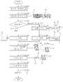
도 1을 참조하면, 본 발명의 일 실시예에 따른 엑스선 영상 촬영장치(100)는 환자의 두부로 엑스선(X-ray)를 조사하는 엑스선 광원(110), 상기 엑스선 광원(110)과 대향하여 구비되고, 환자의 두부를 통과한 엑스선을 수광하여 치과 엑스선 영상을 획득하는 엑스선 센서(120) 및 상기 엑스선 센서(120)로부터 치과 엑스선 영상을 수신하여 영상처리를 통해 환자의 치아 우식증을 검출하는 영상처리수단(130)을 포함하여 이루어진다.Referring to FIG. 1 , an
또한, 도시하지는 않았으나, 본 발명의 일 실시예에 따른 엑스선 영상 촬영장치(100)은 상기 엑스선 광원(110)과 상기 엑스선 센서(120)를 지지하기 위한 암과 상기 암을 바닥이나 벽체에 지지하기 위한 지지대를 더 포함할 수 있다.In addition, although not shown, the
또한, 상기 암은 상기 엑스선 광원(110)과 상기 엑스선 센서(120)가 환자의 두부를 중심으로 회전할 수 있도록 회전암으로 구비될 수 있다.In addition, the arm may be provided as a rotating arm so that the
또한, 상기 영상처리수단(130)은 상기 엑스선 센서(120)와 유선 또는 무선 통신망을 통해 연결되어 정보를 수신할 수 있으며, 일반적인 컴퓨터 이외에 스마트 기기 또는 본 발명의 엑스선 영상 촬영장치(100)를 위해 특별히 제작된 임베디드 시스템(embedded system)을 포함하는 광의의 영상처리장치이다.In addition, the image processing means 130 may be connected to the
또한, 상기 영상처리수단(130)에는 상기 치과 엑스선 영상을 영상처리하여 치아 우식증을 검출하기 위한 치아 우식증 검출프로그램(131)이 임베디드될 수 있다.Also, a dental
또한, 상기 치아 우식증 검출프로그램(131)은 본 발명의 엑스선 영상 촬영장치와는 별도로 기록 매체에 저장되어 제공될 수 있으며, 상기 기록매체는 본 발명을 위하여 특별히 설계되어 구성된 것들이거나 컴퓨터 소프트웨어 분야에서 통상의 지식을 가진 자에서 공지되어 사용 가능할 것일 수 있으며, 예를 들면, 하드 디스크, 플로피 디스크 및 자기 테이프와 같은 자기 매체, CD, DVD와 같은 광 기록 매체, 자기 및 광 기록을 겸할 수 있는 자기-광 기록 매체, 롬, 램, 플래시 메모리 등 단독 또는 조합에 의해 프로그램 명령을 저장하고 수행하도록 특별히 구성된 하드웨어 장치일 수 있다.In addition, the dental
또한, 상기 치아 우식증 검출프로그램(131)은 프로그램 명령, 로컬 데이터 파일, 로컬 데이터 구조 등이 단독 또는 조합으로 구성된 프로그램일 수 있고, 컴파일러에 의해 만들어지는 것과 같은 기계어 코드뿐만 아니라, 인터프리터 등을 사용하여 컴퓨터에 의해 실행될 수 있는 고급 언어 코드로 짜여진 프로그램일 수 있다.In addition, the dental
또한, 본 발명은 상기 치아 우식증 검출프로그램(131)을 저장하고 통신망을 통해 상기 영상처리수단(130)과 같은 클라이언트 시스템으로 상기 치아 우식증 검출프로그램(131)을 전송해 줄 수 있는 서버 시스템을 더 제공할 수 있다.In addition, the present invention further provides a server system capable of storing the dental
즉, 상기 영상처리수단(130)은 상기 서버 시스템으로부터 치아 우식증 검출프로그램(131)을 다운로드받아 설치할 수 있다.
That is, the image processing means 130 may download and install the dental
도 2는 본 발명의 일 실시예에 따른 구강 내 엑스선 영상 촬영장치(200)를 보여주는 것으로 도 1에 도시한 엑스선 영상 촬영장치(100)와 비교하여 상기 엑스선 광원(110)이 포터블 엑스선 발생장치 등 엑스선 센서와 실질적으로 분리된 별도의 구성을 갖고, 상기 엑스선 센서(120)가 환자 구강 내부에 삽입하여 구강 엑스선 영상을 촬영하는 인트라 오랄(Intra oral) 센서(210)로 대체되며, 그 외 구성요소들은 실질적으로 동일하다.FIG. 2 shows an intraoral
이때, 상기 엑스선 광원(110)은 촬영자가 직접 들거나 벽체 등에 암으로 거치된 형태일 수 있고, 상기 인트라 오랄 센서(210)는 구강 외부의 상기 엑스선 광원(110)으로부터 엑스선을 수광하여 엑스선 광원(100)과 인트라 오럴 센서(210) 사이의 구강 내 구조물에 대한 엑스선 영상을 획득할 수 있다.In this case, the
한편, 상기 도 1의 엑스선 광원(110)과 엑스선 센서(120) 그리고 도 2의 엑스선 광원(110)과 인트라 오랄 센서(210)는 획득하는 치과 엑스선 영상의 종류에 따라 다양하게 변형될 수 있다.Meanwhile, the
예를 들면, 상기 도 1의 엑스선 광원(110)과 엑스선 센서(120)는 파노라마 엑스선 영상을 촬영할 수 있고, 상기 도 2의 엑스선 광원(110)과 인트라 오랄 센서(210)는 구강 내 국부적인 치아(2개 내지 3개 정도)의 구강 엑스선 영상을 획득할 수 있다.
For example, the
이하에서는 도 1 내지 도 3을 참조하여, 본 발명의 일 실시예에 따른 치아 우식증 검출방법을 상세히 설명한다.Hereinafter, a method for detecting dental caries according to an embodiment of the present invention will be described in detail with reference to FIGS. 1 to 3 .
도 3을 참조하면, 본 발명의 치아 우식증 검출방법은 본 발명의 실시예에 따른 엑스선 영상 촬영장치(100) 또는 구강 내 엑스선 영상 촬영장치(200)에 의해 수행된다.Referring to FIG. 3 , the method for detecting dental caries of the present invention is performed by the
더욱 자세하게는, 본 발명의 치아 우식증 검출방법은 상기 우식증 검출 프로그램(131)이 상기 영상처리수단(130)을 통해 엑스선 영상 촬영장치(100) 또는 구강 내 엑스선 영상 촬영장치(200)를 기능시켜 수행된다.More specifically, in the method for detecting dental caries of the present invention, the
먼저, 상기 엑스선 센서(120) 또는 상기 인트라 오랄 센서(210)에 의해 환자의 치과 엑스선 영상이 촬영되고(S1000), 상기 영상처리수단(130)은 촬영된 치과 엑스선 영상(10,20)을 수신한다(S2000).First, a dental X-ray image of a patient is captured by the
또한, 상기 영상처리수단(130)이 수신하는 치과 엑스선 영상은 파노라마 엑스선 영상(10) 또는 구강 엑스선 영상(20)일 수 있다.Also, the dental X-ray image received by the
다음, 상기 영상처리수단(130)이 상기 치과 엑스선 영상 내의 각 치아를 분할(segmentation)한다.Next, the image processing means 130 segments each tooth in the dental X-ray image.
또한, 상기 치과 엑스선 영상 내에서 치아를 분할하는 방법은 이진화 알고리즘인 임계기법(Thresholding methods), 히스토그램 기반 기법(histogram-based methods)과 캐니 에지 검출(canny edge detection), 프리윗 마스크(Prewitt mask), 소벨 마스크(Sobel mask) 등의 공지된 에지 검출 기법을 이용할 수 있으며, 특정 방법에 한정되는 것은 아니다.In addition, the method for segmenting teeth in the dental X-ray image is a binarization algorithm, such as thresholding methods, histogram-based methods, canny edge detection, and Prewitt mask. , a known edge detection technique, such as a Sobel mask, may be used, but is not limited to a specific method.
한편, 상기 치과 엑스선 영상이 구강 엑스선 영상(20)일 경우 영상 내에 치아의 개수가 두 개 내지 세 개 정도이므로 엑스선 영상의 컨트라스트(contrast) 차이를 이용한 공지의 경계면 검출 알고리즘 등을 사용하여 치아를 분할하는데 큰 어려움이 없으나 파노라마 엑스선 영상(10)의 경우, 영상 내에 상악 치열(11)과 하악 치열(12)을 포함한 모든 치아의 영상이 포함되어 있으므로 치아 분할에 어려움이 있다.On the other hand, when the dental X-ray image is the
본 발명에서는 상기 치과 엑스선 영상이 파노라마 엑스선 영상(10)일 경우 먼저, 컨트라스트(contrast) 차이를 이용한 공지의 경계면 검출 알고리즘 등으로 상악 치열(11)과 하악 치열(12)의 교합면(13)을 검출하고(S4100), 다음, 상기 교합면(13)에 수직인 법선 벡터(14)와 공지의 경계면 검출 알고리즘을 이용하여 각 치열(11,12)의 치아들을 분할하였다(S4200). 이 경우, 계산량을 매우 줄일 수 있어 실시간으로 치아 분할이 가능한 장점이 있다.In the present invention, when the dental X-ray image is the
또한, 도시하지는 않았으나 분할된 각 치아에 식별자를 부여하는 단계가 수행될 수 있다. 이는 분할된 하나의 치아가 전치부인지 구치부인지 또는 상악의 치아인지 하악의 치아인지 또는 몇 번째 치아인지를 의사로 하여금 쉽게 식별할 수 있게 하기 위함이다.Also, although not shown, a step of assigning an identifier to each divided tooth may be performed. This is to enable a doctor to easily identify whether one divided tooth is an anterior tooth, a posterior tooth, a maxillary tooth, a mandibular tooth, or the number of teeth.
다음, 분할된 각 치아(30)에 대해 법랑질(31)과 상아질(32)을 서로 분리한다(S5000).Next, the
이 역시 법랑질(31)과 상아질(32)의 컨트라스트(contrast) 차이를 이용한 공지의 경계면 검출 알고리즘 및 공지의 영상 분할방법을 이용할 수 있으며 분할방법에는 제한이 없다.Again, a known boundary surface detection algorithm using a contrast difference between the
다음, 분할된 법랑질(31)과 상아질(32) 각각에 우식(40)이 존재하는지 검출하고, 우식증의 발생여부를 판단한다.Next, it is detected whether
또한, 우식부위는 정상부위보다 영상 내에서 어둡게 표현되므로, 치아의 영상 값(밝기 값)을 이용하여 우식의 존재여부를 판단할 수 있다.In addition, since the carious portion is expressed darker in the image than the normal portion, the presence or absence of caries can be determined using the image value (brightness value) of the tooth.
예를 들어, 법랑질(31)이나 상아질(32)의 평균 밝기가 임계 밝기 미만이거나, 법랑질(31)이나 상아질(32)을 복수 개의 부분 영역으로 구획하고 구획된 부분 영역 중, 어느 하나 이상의 평균 밝기가 임계 밝기 미만일 경우 우식이 발생한 것으로 판단할 수 있다.For example, the average brightness of the
또한, 법랑질(31)이나 상아질(32)의 영상을 복수 개의 부분 영역으로 구획하여 우식 발생여부를 판단할 경우 우식(40)이 발생한 위치까지 검출할 수 있는 장점이 있다.In addition, if the image of the
즉, 의사가 육안으로 우식발생 여부를 판단하지 않고도 우식증 발생 여부를 쉽고 빠르게 검출할 수 있는 장점이 있다.That is, there is an advantage that a doctor can easily and quickly detect whether or not caries has occurred without visually determining whether or not caries has occurred.
다음, 우식이 발생한 부위에 따라 우식증의 진행 정도를 더 판단할 수 있다(S7000).Next, the degree of progression of caries can be further determined according to the site where the caries has occurred (S7000).
예를 들어, 법랑질(31)에만 우식이 검출된 경우 우식1도, 법랑질(31)과 상아질(32)에 동시에 우식이 검출된 경우 우식2도로 판단할 수 있다.For example, when caries is detected only in the
또한, 법랑질(31)에는 우식이 검출되지 않고, 상아질(32)에만 우식이 검출될 수 있으며, 법랑질(31)이 없는 치아 하부에서 우식이 발생한 것으로 판단할 수 있다.In addition, caries is not detected in the
또한, 본 발명에서는 법랑질(31)과 상아질(32)의 우식을 검출하는 것을 예시하였으나, 치수나 치관의 우식을 더 검출할 수 있고, 이 경우 우식증의 진행 정도를 세분화하여 판단할 수 있다.In addition, although the present invention exemplifies the detection of caries of
다음, 음성 또는 영상으로 치아 우식증 발생 알람을 출력하여 의사로 하여금 우식증이 발생을 인지하도록 한다(S8000).Next, a dental caries occurrence alarm is output by voice or image so that the doctor recognizes the occurrence of caries (S8000).
또한, 본 발명의 엑스선 영상 촬영장치(100)와 구강 내 엑스선 영상 촬영 장치(200)에는 치아 우식증 알람 출력을 위한 디스플레이 또는 스피커가 더 포함될 수 있다.In addition, the
또한, 상기 치아 우식증 발생 알람은 우식증의 진행 정도에 따라 서로 다른 음성 또는 영상으로 출력될 수 있으며, 음성의 경우 멜로디나 볼륨, 영상의 경우 문자, 컬러, 캐릭터, 기호, 이모티콘 등을 서로 달리함으로써 우식증의 진행 정도가 구분되게 할 수 있다.In addition, the dental caries occurrence alarm may be output in different voices or images depending on the progress of caries, and in the case of a voice, a melody or volume, and in the case of an image, text, color, character, symbol, emoticon, etc. progress can be differentiated.
따라서, 의사는 우식증의 발생여부뿐만 아니라 우식증의 진행여부까지 간편하고 빠르게 인지할 수 있는 장점이 있다.
Therefore, there is an advantage that the doctor can easily and quickly recognize not only the occurrence of caries, but also the progress of caries.
이상에서 살펴본 바와 같이 본 발명은 바람직한 실시예를 들어 도시하고 설명하였으나, 상기한 실시예에 한정되지 아니하며 본 발명의 정신을 벗어나지 않는 범위 내에서 당해 발명이 속하는 기술분야에서 통상의 지식을 가진 자에 의해 다양한 변경과 수정이 가능할 것이다.
As described above, the present invention has been illustrated and described with reference to preferred embodiments, but it is not limited to the above-described embodiments, and those of ordinary skill in the art to which the present invention pertains within the scope not departing from the spirit of the present invention Various changes and modifications will be possible.
100:엑스선 영상 촬영장치 110:엑스선 광원
120:엑스선 센서 130:영상 처리 수단
131:우식증 검출 프로그램 200:구강 내 엑스선 영상 촬영장치
210:인트라 오랄 센서100: X-ray imaging device 110: X-ray light source
120: X-ray sensor 130: image processing means
131: caries detection program 200: intraoral X-ray imaging device
210: intra oral sensor
Claims (9)
Translated fromKorean상기 치과 엑스선 영상에서 상기 상악 치열과 상기 하악 치열 사이의 교합면을 검출하는 단계;
상기 교합면에 수직인 법선벡터를 이용하여 상기 치과 엑스선 영상에서 상기 상악 치열과 상기 하악 치열의 각 치아를 분할하는 단계;
상기 치과 엑스선 영상에서 상기 분할된 각 치아의 법랑질과 상아질을 분할하는 단계; 및
상기 분할된 법랑질과 상아질의 영상 값으로 치아 우식증 발생여부를 검출하는 단계를 포함하는 치아 우식증 검출방법.
A dental caries detection method for detecting the occurrence of dental caries through image processing from dental X-ray images including maxillary and mandibular dentition,
detecting an occlusal surface between the maxillary dentition and the mandibular dentition in the dental X-ray image;
dividing each tooth of the maxillary and mandibular teeth in the dental X-ray image by using a normal vector perpendicular to the occlusal surface;
dividing the enamel and dentin of each of the divided teeth in the dental X-ray image; and
Method for detecting dental caries comprising the step of detecting whether or not dental caries has occurred using the image values of the divided enamel and dentin.
상기 영상 값은 상기 법랑질과 상아질의 평균 밝기값이고,
상기 치아 우식증 발생여부를 검출하는 단계는, 상기 평균 밝기값와 임계 밝기값을 비교해서 상기 평균 밝기값이 상기 임계 밝기값 미만이면 치아 우식증으로 판단하는 치아 우식증 검출방법.
The method of claim 1,
The image value is the average brightness value of the enamel and dentin,
The step of detecting whether or not dental caries has occurred may include comparing the average brightness value with a threshold brightness value and determining if the average brightness value is less than the threshold brightness value as dental caries.
상기 치아 우식증 발생여부를 검출하는 단계 후, 상기 법랑질 또는 상기 상아질의 치아 우식증 발생여부에 따라 해당 치아의 우식증 진행정도를 판단하는 단계를 더 포함하는 치아 우식증 검출방법.
The method of claim 1,
After the step of detecting whether or not dental caries occurs, the method for detecting dental caries further comprising the step of determining the degree of caries progression of the corresponding tooth according to the occurrence of dental caries in the enamel or the dentin.
상기 치아 우식증 발생여부를 검출하는 단계 후, 상기 치아 우식증이 발생한 것으로 판단될 경우, 음성 또는 영상의 치아 우식증 발생알람을 출력하는 단계;를 더 포함하는 것을 특징으로 하는 치아 우식증 검출방법.
4. The method of claim 3,
After detecting whether or not dental caries has occurred, when it is determined that the dental caries has occurred, outputting an audio or image dental caries occurrence alarm; Dental caries detection method further comprising a.
상기 치과 엑스선 영상은, 파노라마 엑스선 영상인 치아 우식증 검출방법.
The method of claim 1,
The dental X-ray image is a panoramic X-ray image, dental caries detection method.
상기 치과 엑스선 영상은, 구강 내 엑스선 영상인 치아 우식증 검출방법.The method of claim 1,
The dental X-ray image is a dental caries detection method that is an intraoral X-ray image.
Priority Applications (2)
| Application Number | Priority Date | Filing Date | Title |
|---|---|---|---|
| KR1020150081296AKR102433702B1 (en) | 2015-06-09 | 2015-06-09 | Method for detecting dental caries, detecting program and X-ray imaging apparatus |
| US15/177,363US9801603B2 (en) | 2015-06-09 | 2016-06-09 | Method and apparatus for detecting dental caries and X-ray imaging apparatus |
Applications Claiming Priority (1)
| Application Number | Priority Date | Filing Date | Title |
|---|---|---|---|
| KR1020150081296AKR102433702B1 (en) | 2015-06-09 | 2015-06-09 | Method for detecting dental caries, detecting program and X-ray imaging apparatus |
Publications (2)
| Publication Number | Publication Date |
|---|---|
| KR20160144748A KR20160144748A (en) | 2016-12-19 |
| KR102433702B1true KR102433702B1 (en) | 2022-08-19 |
Family
ID=57516424
Family Applications (1)
| Application Number | Title | Priority Date | Filing Date |
|---|---|---|---|
| KR1020150081296AActiveKR102433702B1 (en) | 2015-06-09 | 2015-06-09 | Method for detecting dental caries, detecting program and X-ray imaging apparatus |
Country Status (2)
| Country | Link |
|---|---|
| US (1) | US9801603B2 (en) |
| KR (1) | KR102433702B1 (en) |
Families Citing this family (5)
| Publication number | Priority date | Publication date | Assignee | Title |
|---|---|---|---|---|
| US11185300B2 (en)* | 2017-09-02 | 2021-11-30 | Seung Bum Ryu, SR. | Dual exposure buttons controlled by a switch or an audio guide |
| WO2019222135A1 (en)* | 2018-05-16 | 2019-11-21 | Benevis Informatics, Llc | Systems and methods for review of computer-aided detection of pathology in images |
| US11224387B2 (en)* | 2018-10-16 | 2022-01-18 | Shayda Cullen | Digital dental x-ray sensor device having a rounded housing |
| US11653838B2 (en)* | 2020-03-11 | 2023-05-23 | Alireza Moheb | System and method for classification of dental health based on digital imagery |
| CN119679356B (en)* | 2025-01-17 | 2025-06-13 | 中国医学科学院北京协和医院 | Diagnostic system for dental caries |
Citations (4)
| Publication number | Priority date | Publication date | Assignee | Title |
|---|---|---|---|---|
| US20090075228A1 (en)* | 2007-09-18 | 2009-03-19 | Olympus Corporation | Dental observation apparatus |
| US20090095732A1 (en) | 2007-05-08 | 2009-04-16 | Kuochu Li | Multi-Functional Cooker |
| US20120148986A1 (en)* | 2010-12-13 | 2012-06-14 | Jiayong Yan | Method for identification of dental caries in polychromatic images |
| US20120328071A1 (en)* | 2009-08-05 | 2012-12-27 | Telesystems Co., Ltd. | Radiation imaging apparatus and imaging method using radiation |
Family Cites Families (3)
| Publication number | Priority date | Publication date | Assignee | Title |
|---|---|---|---|---|
| US7955076B2 (en)* | 2003-04-03 | 2011-06-07 | Kao Corporation | Carious tooth detection device |
| KR100707792B1 (en)* | 2005-08-31 | 2007-04-13 | 주식회사바텍 | X-ray imaging method, system and x-ray imaging processing unit |
| ES2731900T3 (en)* | 2009-03-20 | 2019-11-19 | 3Shape As | System for planning, visualization and optimization of dental restorations |
- 2015
- 2015-06-09KRKR1020150081296Apatent/KR102433702B1/enactiveActive
- 2016
- 2016-06-09USUS15/177,363patent/US9801603B2/enactiveActive
Patent Citations (4)
| Publication number | Priority date | Publication date | Assignee | Title |
|---|---|---|---|---|
| US20090095732A1 (en) | 2007-05-08 | 2009-04-16 | Kuochu Li | Multi-Functional Cooker |
| US20090075228A1 (en)* | 2007-09-18 | 2009-03-19 | Olympus Corporation | Dental observation apparatus |
| US20120328071A1 (en)* | 2009-08-05 | 2012-12-27 | Telesystems Co., Ltd. | Radiation imaging apparatus and imaging method using radiation |
| US20120148986A1 (en)* | 2010-12-13 | 2012-06-14 | Jiayong Yan | Method for identification of dental caries in polychromatic images |
Also Published As
| Publication number | Publication date |
|---|---|
| KR20160144748A (en) | 2016-12-19 |
| US9801603B2 (en) | 2017-10-31 |
| US20160361037A1 (en) | 2016-12-15 |
Similar Documents
| Publication | Publication Date | Title |
|---|---|---|
| KR102433702B1 (en) | Method for detecting dental caries, detecting program and X-ray imaging apparatus | |
| US20200372301A1 (en) | Adversarial Defense Platform For Automated Dental Image Classification | |
| US11348237B2 (en) | Artificial intelligence architecture for identification of periodontal features | |
| CN103442645B (en) | Tooth Surface Classification Method | |
| US11217350B2 (en) | Systems and method for artificial-intelligence-based dental image to text generation | |
| US11276151B2 (en) | Inpainting dental images with missing anatomy | |
| US20200364624A1 (en) | Privacy Preserving Artificial Intelligence System For Dental Data From Disparate Sources | |
| JP6423449B2 (en) | Method of operating a system for intraoral scanning | |
| US20210118132A1 (en) | Artificial Intelligence System For Orthodontic Measurement, Treatment Planning, And Risk Assessment | |
| US20200405242A1 (en) | System And Methods For Restorative Dentistry Treatment Planning Using Adversarial Learning | |
| US20200387829A1 (en) | Systems And Methods For Dental Treatment Prediction From Cross- Institutional Time-Series Information | |
| US20200411167A1 (en) | Automated Dental Patient Identification And Duplicate Content Extraction Using Adversarial Learning | |
| JP7260115B2 (en) | Mobile devices and photography assistance programs | |
| US20230225832A1 (en) | Photo-based dental attachment detection | |
| KR102171396B1 (en) | Method for diagnosing dental lesion and apparatus thereof | |
| KR20200134036A (en) | Method for numbering teeth number and image processing apparatus for performing the method | |
| CN107370961A (en) | Image exposure processing method and device and terminal equipment | |
| Dibeh et al. | A novel approach for dental panoramic radiograph segmentation | |
| KR102072052B1 (en) | Image processing method and system for dental ct imaging, computer readable recording medium and computer program | |
| Bhan et al. | Detection and grading severity of caries in dental X-ray images | |
| KR102703446B1 (en) | Method and Apparatus for Detecting a Plurality of Lesions in a Dental X-ray Image | |
| JP7586826B2 (en) | Method and apparatus for training an automated dental charting system - Patents.com | |
| KR102155745B1 (en) | Method and apparatus for fmx automatic construction | |
| KR102777067B1 (en) | Method for analyzing odontopathy using artificial intelligence | |
| KR101822908B1 (en) | Method and Apparatus for Obtaining Information of Radiographic Images |
Legal Events
| Date | Code | Title | Description |
|---|---|---|---|
| PA0109 | Patent application | Patent event code:PA01091R01D Comment text:Patent Application Patent event date:20150609 | |
| PG1501 | Laying open of application | ||
| PA0201 | Request for examination | Patent event code:PA02012R01D Patent event date:20200604 Comment text:Request for Examination of Application Patent event code:PA02011R01I Patent event date:20150609 Comment text:Patent Application | |
| E902 | Notification of reason for refusal | ||
| PE0902 | Notice of grounds for rejection | Comment text:Notification of reason for refusal Patent event date:20211211 Patent event code:PE09021S01D | |
| E701 | Decision to grant or registration of patent right | ||
| PE0701 | Decision of registration | Patent event code:PE07011S01D Comment text:Decision to Grant Registration Patent event date:20220708 | |
| PR0701 | Registration of establishment | Comment text:Registration of Establishment Patent event date:20220812 Patent event code:PR07011E01D | |
| PR1002 | Payment of registration fee | Payment date:20220816 End annual number:3 Start annual number:1 | |
| PG1601 | Publication of registration |