KR102406052B1 - Materials and methods for repairing nerves with nerve grafts of animal origin - Google Patents
Materials and methods for repairing nerves with nerve grafts of animal originDownload PDFInfo
- Publication number
- KR102406052B1 KR102406052B1KR1020217014775AKR20217014775AKR102406052B1KR 102406052 B1KR102406052 B1KR 102406052B1KR 1020217014775 AKR1020217014775 AKR 1020217014775AKR 20217014775 AKR20217014775 AKR 20217014775AKR 102406052 B1KR102406052 B1KR 102406052B1
- Authority
- KR
- South Korea
- Prior art keywords
- nerve
- grafts
- intercostal
- graft
- nerves
- Prior art date
- Legal status (The legal status is an assumption and is not a legal conclusion. Google has not performed a legal analysis and makes no representation as to the accuracy of the status listed.)
- Active
Links
Images


Classifications
- A—HUMAN NECESSITIES
- A61—MEDICAL OR VETERINARY SCIENCE; HYGIENE
- A61L—METHODS OR APPARATUS FOR STERILISING MATERIALS OR OBJECTS IN GENERAL; DISINFECTION, STERILISATION OR DEODORISATION OF AIR; CHEMICAL ASPECTS OF BANDAGES, DRESSINGS, ABSORBENT PADS OR SURGICAL ARTICLES; MATERIALS FOR BANDAGES, DRESSINGS, ABSORBENT PADS OR SURGICAL ARTICLES
- A61L27/00—Materials for grafts or prostheses or for coating grafts or prostheses
- A61L27/36—Materials for grafts or prostheses or for coating grafts or prostheses containing ingredients of undetermined constitution or reaction products thereof, e.g. transplant tissue, natural bone, extracellular matrix
- A61L27/3604—Materials for grafts or prostheses or for coating grafts or prostheses containing ingredients of undetermined constitution or reaction products thereof, e.g. transplant tissue, natural bone, extracellular matrix characterised by the human or animal origin of the biological material, e.g. hair, fascia, fish scales, silk, shellac, pericardium, pleura, renal tissue, amniotic membrane, parenchymal tissue, fetal tissue, muscle tissue, fat tissue, enamel
- A—HUMAN NECESSITIES
- A61—MEDICAL OR VETERINARY SCIENCE; HYGIENE
- A61L—METHODS OR APPARATUS FOR STERILISING MATERIALS OR OBJECTS IN GENERAL; DISINFECTION, STERILISATION OR DEODORISATION OF AIR; CHEMICAL ASPECTS OF BANDAGES, DRESSINGS, ABSORBENT PADS OR SURGICAL ARTICLES; MATERIALS FOR BANDAGES, DRESSINGS, ABSORBENT PADS OR SURGICAL ARTICLES
- A61L27/00—Materials for grafts or prostheses or for coating grafts or prostheses
- A61L27/36—Materials for grafts or prostheses or for coating grafts or prostheses containing ingredients of undetermined constitution or reaction products thereof, e.g. transplant tissue, natural bone, extracellular matrix
- A61L27/3641—Materials for grafts or prostheses or for coating grafts or prostheses containing ingredients of undetermined constitution or reaction products thereof, e.g. transplant tissue, natural bone, extracellular matrix characterised by the site of application in the body
- A61L27/3675—Nerve tissue, e.g. brain, spinal cord, nerves, dura mater
- A—HUMAN NECESSITIES
- A61—MEDICAL OR VETERINARY SCIENCE; HYGIENE
- A61L—METHODS OR APPARATUS FOR STERILISING MATERIALS OR OBJECTS IN GENERAL; DISINFECTION, STERILISATION OR DEODORISATION OF AIR; CHEMICAL ASPECTS OF BANDAGES, DRESSINGS, ABSORBENT PADS OR SURGICAL ARTICLES; MATERIALS FOR BANDAGES, DRESSINGS, ABSORBENT PADS OR SURGICAL ARTICLES
- A61L2430/00—Materials or treatment for tissue regeneration
- A61L2430/32—Materials or treatment for tissue regeneration for nerve reconstruction
Landscapes
- Health & Medical Sciences (AREA)
- Life Sciences & Earth Sciences (AREA)
- Chemical & Material Sciences (AREA)
- Engineering & Computer Science (AREA)
- Biomedical Technology (AREA)
- Public Health (AREA)
- Transplantation (AREA)
- Veterinary Medicine (AREA)
- General Health & Medical Sciences (AREA)
- Botany (AREA)
- Chemical Kinetics & Catalysis (AREA)
- Dermatology (AREA)
- Medicinal Chemistry (AREA)
- Oral & Maxillofacial Surgery (AREA)
- Animal Behavior & Ethology (AREA)
- Epidemiology (AREA)
- Vascular Medicine (AREA)
- Neurosurgery (AREA)
- Neurology (AREA)
- Orthopedic Medicine & Surgery (AREA)
- Molecular Biology (AREA)
- Urology & Nephrology (AREA)
- Zoology (AREA)
- Prostheses (AREA)
- Materials For Medical Uses (AREA)
Abstract
Translated fromKoreanDescription
Translated fromKorean유방암에 대한 최초의 설명은 기원전 480년으로 거슬러 올라가 고대 그리스인과 이집트인의 초창기 의학 문헌에서 찾을 수 있다. 그러나 Halsted가 1880년대에 최초의 유방 절제술을 시행하기 전까지 현대적인 치료는 시작되지 않았다. 그 이후로 유방암에 대한 이해와 치료에 유의미한 발전이 있어왔다. 오늘날 유방 절제술 이후 유방의 재건은 유방암 치료의 기본 요소이다. 임플란트 기반 유방 재건이 오늘날 더 일반적인 재건 방식이지만, 자기유래 재건이 수행되고부터 장기간 동안의 환자 만족도는 더 높은 것으로 보고되고 있다. 따라서 많은 환자가 이 재건 방식을 선택하는 것은 놀라운 일이 아니다. 공여부 이환율(donor-site morbidity)을 최소화하는 천공지 기반 피판의 개발과 같이, 자기유래 재건에서 이루어진 유의미한 기술 발전에도 불구하고, 복벽 약화 및 공여부 탈장은 여전히 심각한 합병증으로 남아 있다.The earliest descriptions of breast cancer can be found in the earliest medical texts of the ancient Greeks and Egyptians, dating back to 480 BC. However, modern treatment did not begin until Halsted performed the first mastectomy in the 1880s. Since then, significant advances have been made in the understanding and treatment of breast cancer. Today, reconstruction of the breast after mastectomy is a fundamental component of breast cancer treatment. Although implant-based breast reconstruction is a more common reconstruction method today, higher patient satisfaction has been reported over the long term after autologous reconstruction is performed. Therefore, it is not surprising that many patients choose this reconstruction method. Despite significant technological advances in autologous reconstruction, such as the development of perforated flaps that minimize donor-site morbidity, abdominal wall weakness and donor hernia remain serious complications.
흥미롭게도 공여부 이환율에 초점을 맞춘 수많은 보고서는, 단지 피판 생존율을 넘어선 수여부(recipient-site) 결과에 초점을 맞춘 연구가 부족한 것과는 대조를 이룬다. 유방 감각의 중요성은 수술 후 삶의 질에 엄청난 영향을 미치기 때문에 아무리 강조해도 지나치지 않다. 사실 유방 절제술 후 감각 상실 문제는 최근 주류 언론에서도 두드러지게 다루어졌다. 따라서 환자들은 유방을 재건 할뿐만 아니라 감각을 회복시키는 방식에 대해 점점 더 많이 문의하고 있다. 이와 관련하여 많은 논쟁이 되고 있는 주제는, 이식시 피판 재신경지배(reinnervation)/신경재생(neurotization)으로 인한 유방 신경재생이다.Interestingly, the numerous reports focusing on donor-site morbidity contrast with the lack of studies focusing on recipient-site outcomes beyond just flap survival. The importance of breast sensation cannot be overemphasized because it has a tremendous impact on the quality of life after surgery. In fact, the problem of sensory loss after mastectomy has been featured prominently in the mainstream media in recent years. Therefore, patients are increasingly asking how to not only reconstruct the breast, but also restore sensation. A topic of much debate in this regard is breast nerve regeneration due to flap reinnervation/neurotization during transplantation.
유방 신경재생은 새로운 주제가 아니고, 1990년대 초부터 문헌에서 논의되어 왔다. 그러나 유방 재건용 유감각 피판이 도입된 이후로, 논쟁은 신경 접합이 피판 감각의 회복을 위해 필요한 것인지, 아니면 의미있는 감각을 위해 충분한 신경 섬유의 부수적인 내적 성장인지에 집중되었다. 이용 가능한 증거는 감각 회복이 중요하다는 것을 시사한다. 환자에 의해 평가되어 얻어진 삶의 질 지표에 부정적인 영향을 미치는, 열 및 기계에 의한 비자발적 손상 사례가 보고되었다. 다수의 연구에 따르면, 공여 피판 신경에 의한 유방 신경재생술이 더 신속하고 개선된 감각의 회복, 환자 만족도 개선 및 환자에 의해 보고되는 삶의 질 개선을 달성하였음을 보여주었다. 반면에 몇몇 연구는 이러한 결과의 차이를 입증하지 못했다. 또 다른 논쟁점은 신경 접합후 달성 가능한 결과가 추가로 소요되는 수술 시간을 정당화하는지 여부이다. 자발적 재신경지배는 다양한 규모로 발생하지만, 신경지배 피판의 감각 회복은 비 신경지배 피판의 감각 회복에 비해 월등하고, 더 일찍 시작되며, 시간이 지남에 따라 점차적으로 개선되어, 정상 감각에 가까워질 가능성이 더 높다.Breast regeneration is not a new topic and has been discussed in the literature since the early 1990s. However, since the introduction of sensory flaps for breast reconstruction, the debate has focused on whether neural junctions are necessary for the restoration of flap sensation, or whether the concomitant intrinsic growth of nerve fibers sufficient for meaningful sensations. Available evidence suggests that sensory recovery is important. Cases of involuntary thermal and mechanical injuries have been reported that negatively affect the quality of life indicators obtained as assessed by patients. Numerous studies have shown that breast rejuvenation with donor flap nerves achieved faster and improved sensory recovery, improved patient satisfaction, and improved quality of life reported by patients. On the other hand, some studies have failed to substantiate the difference in these results. Another issue is whether the achievable results after nerve junctions justify the additional surgical time required. Spontaneous reinnervation occurs at various scales, but sensory recovery in innervated flaps is superior to that in noninnervated flaps, begins earlier, and improves gradually over time, approaching normal sensation. More likely.
특히, 여러 연구들간에 큰 이질성이 존재하고, 표준화가 되어 있지 않아서 유방 수술 결과에 대한 메타 분석(meta analysis)이 방해된다. 관심있는 특정 영역은 일차 신경 수복으로부터 신경 도관 사용에 이르기까지 다양한, 신경재생 절차 자체의 표준화가 되어있지 않다는 것이다. Spiegel외 다수는, 자기유래 피판의 감각 회복능을 평가하고, 신경 도관 및 직접 신경 접합이 사용되었을 때(즉 자발적 재신경지배의 경우)와 비교하였다. 상기 저자들은, 피판 신경재생이 자발적 신경지배보다 월등하고, 신경재생 절차가 수술 시간을 유의미하게 연장하지 않았으며, 신경 도관이 사용되면 직접 접합의 경우보다 감각 회복을 유의미하게 개선시켰다고 결론을 내렸다.In particular, large heterogeneity between studies exists and there is no standardization, which hinders meta-analysis of breast surgery results. A particular area of interest is the lack of standardization of the nerve regeneration procedures themselves, which vary from primary nerve repair to the use of nerve conduits. Spiegel et al. evaluated the sensory recovery capacity of autologous flaps and compared them with those when nerve conduits and direct nerve junctions were used (ie spontaneous reinnervation). The authors concluded that flap nerve regeneration was superior to spontaneous innervation, the nerve regeneration procedure did not significantly prolong the operation time, and the use of a nerve conduit significantly improved sensory recovery over direct splicing.
임의의 종래 수술에서도 감각의 회복이 관찰되었지만, 도관 신경재생 피판의 경우 감각 회복은 반대쪽 비 수술 유방 피부의 감각 회복의 절반에 불과한 것으로 나타났으며, 직접 접합의 경우 피판에서 감각 회복에 도달하는데에는 4배나 더 많은 압박감을 겪어야 했다. 감각은 회복되었지만, 유방 재건후 의미있는 감각 회복은 여전히 요망되는 실정이다.Although sensory recovery was observed in any conventional surgery, sensory recovery was only half that of the contralateral non-surgical breast skin in the case of the conduit nerve regeneration flap. I had to go through four times as much pressure. Although sensory has been restored, meaningful sensory recovery after breast reconstruction is still desired.
말초 신경 수술후 감각에 대한 성과를 평가하기 위해 일반적으로 사용되는 측정 항목중 하나는 의학연구위원회(Medical Research Council; MRC) 척도이다. 이 척도는 S0에서 S4까지 운영되고 있는데, 여기서 S0는 감각이 회복되지 않은 상태, SI는 심부 피부 통증이 회복된 상태, S2는 얕은 피부 통증과 촉각이 일부 회복된 상태, S3는 과도한 반응 없이 얕은 피부 통증과 촉각이 회복된 상태, S3+는 2점 차별 회복(2-points discrimination recovery)을 동반하며 얕은 피부 통증 및 촉각이 회복된 상태, 그리고 S4는 감각이 완전히 회복된 상태이다. 이 척도는 몇몇 연구에서 S3 이상으로 정의되었던 의미있는 회복을 측정하기 위해 사용되었다. 두 번째, 신경재생술은 10분 내지 15분의 추가 수술 시간만을 필요로 한다는 점이 주목된다. 그러나 무감각 피판에 대한 이전의 우려가 고려될 때, 재건된 유방에 감각을 회복시켜줄 기회는 잠재적 사례 연장과 관련된 우려보다 중요할 것이다. 마지막으로, 저자들은 사용된 신경 도관이 40 mm인 신경 도관이라고 진술하고 있다. Lohmeyer 외 다수는, 다양한 길이의 신경 도관을 사용하여 수행한 디지털 신경 재건후의 감각에 대한 문헌 검토를 실시하였다(2014, J Reconstr Microsurg., 30(4):227-34). 저자들은, 5 mm ~ 25 mm 사이의 신경 도관으로 재건을 수행한 후 2점 차별 검사와 모노필라멘트 검사를 수행하여 결과를 측정하였으며, 도관 갭(gap) 길이가 6 mm일 때부터 감각이 감소하기 시작함을 발견하였다. 모노필라멘트 검사는 12 mm일 때부터 감각이 유의미하게 더 나빠졌으며, 15 mm일 때부터는 감각이 약화되었고, 문헌 검토 대상 환자들중 20%를 초과하는 환자들은 무감각 상태로 되돌아갔다. 이러한 종류의 검토에 비추어 볼 때, 유방 신경재생 또는 갭이 6 mm보다 길 때의 임의의 기타 신경 수술을 위해 관상 도관을 사용하는 것은 권장되지 않는다.One of the metrics commonly used to evaluate sensory performance after peripheral nerve surgery is the Medical Research Council (MRC) scale. This scale is operated from S0 to S4, where S0 is a state in which sensation is not restored, SI is a state in which deep skin pain has recovered, S2 is a state in which light skin pain and tactile sensation are partially restored, and S3 is a state in which the sensation is not restored and is not overreacted. Skin pain and tactile sense are restored, S3+ is accompanied by 2-points discrimination recovery, and shallow skin pain and tactile sense are restored, and S4 is fully restored sense. This scale was used in some studies to measure meaningful recovery, which was defined as S3 or higher. Second, it is noted that nerve regeneration only requires an additional operating time of 10 to 15 minutes. However, given previous concerns about anesthetized flaps, the opportunity to restore sensation to a reconstructed breast will outweigh concerns related to potential case extension. Finally, the authors state that the nerve conduit used is a 40 mm nerve conduit. Lohmeyer et al. conducted a literature review of sensation after digital nerve reconstruction performed using nerve conduits of various lengths (2014, J Reconstr Microsurg., 30(4):227-34). The authors measured the results by performing a two-point differential test and a monofilament test after performing reconstruction with a nerve conduit between 5 mm and 25 mm. found to start. In the monofilament test, the sensation was significantly worse from 12 mm, and the sensation was weakened from 15 mm, and more than 20% of the patients reviewed in the literature returned to numbness. In light of this kind of review, the use of coronary catheters for mammary nerve regeneration or any other nerve surgery when the gap is longer than 6 mm is not recommended.
유방 신경재생은 통상적으로 자가이식편 수득물로 수행된다. 자가이식편 수득은 복부에서 자가 신경을 수득하고, 이 신경을 수여 환자의 유방 조직에 이식하는 단계를 포함한다. 자가이식편 수득 기반 유방 신경재생은 수술실에서의 더 긴 절차를 필요로 하고, 근육의 탈신경 위험 증가를 감수해야 한다. 근육의 탈신경은 이완, 근육 긴장도 상실, 심미적 결과 저하, 절개시 탈장 위험 증가를 유발한다. 자가이식편 수득은 또한 신경 지름의 절반이 근육으로 옮겨가게 됨에 따라, 막 다른 지경에 이르게 하므로, 재생 능력의 상실을 수반한다. 따라서 자가이식편 수득물 기반 유방 신경재생과 연관된 문제들을 막아주는 수술 방법이 요망된다.Breast nerve regeneration is usually performed with autograft harvests. Autograft harvesting involves obtaining autologous nerves in the abdomen and implanting the nerves into breast tissue of a recipient patient. Breast nerve regeneration based on autograft harvest requires a longer procedure in the operating room and carries an increased risk of muscle denervation. Muscular denervation causes relaxation, loss of muscle tone, decreased aesthetic results, and increased risk of hernia during incision. Autograft harvesting also entails a loss of regenerative capacity, as half of the nerve diameter is transferred to the muscle, reaching a dead end. Therefore, there is a need for a surgical method that prevents problems related to breast nerve regeneration based on autograft harvests.
이러한 단점들을 극복하기 위해, 본 발명은 피판의 신경재생을 허용하면서 복직근에 대한 신경지배 보존을 허용하는, 표준화되고 재현 가능한 수술 절차 및 관련 재료를 제공한다. 임의의 구현예들에서, 직접 접합, 도관 또는 자가이식편 적용의 단점들을 극복하고, 신경 수복을 가속화하는 커넥터 지원 신경 접합에 반향을 일으킨 유방 신경재생에서, 신경 동종이식편은 신규한 가교재(bridging material)로서 사용된다.In order to overcome these drawbacks, the present invention provides a standardized and reproducible surgical procedure and related materials that allow preservation of innervation for the rectus abdominis muscle while allowing nerve regeneration of the flap. In certain embodiments, nerve allografts are a novel bridging material in mammary nerve regeneration that overcomes the shortcomings of direct junction, catheter or autograft application and resonates with connector-assisted nerve junctions accelerating nerve repair. ) is used as
본 발명은 유방 재건 수술과 같은 유방 수술에서 신경 이식편으로 유방 신경재생을 수행하기 위한 재료 및 방법을 제공한다. 본 발명의 방법은 종래의 수술 방법의 위험을 경감시키고, 대안적인 접근법과 경감 계획을 제공한다.The present invention provides materials and methods for performing breast nerve regeneration with nerve grafts in breast surgery, such as breast reconstruction surgery. The method of the present invention mitigates the risk of conventional surgical methods and provides an alternative approach and mitigation plan.
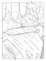
도 1은, DIEP 피판 유방 신경재생에 핵심인 해부학적 랜드마크를 보여주는 것이다. DIEP 복부 피판과 유방 절제술후 흉벽 결손 개관. 필수 신경들(ICN1, ICN2, ICN3, ICN10, ICN11, ICN12), 혈관 구조(내측 및 외측 DIEA, 내유 동맥 및 정맥), 그리고 골 랜드마크(늑골 I, II, III)이 보인다.
도 2a는, DIEP 피판 절개 상태를 (표준적인 외측 → 내측 방식으로) 보여주는 것이다. 도해는, 각각의 늑간 신경의 감각 요소 원위 말단과, 늑간 신경 외측 직근 초(rectus sheath)의 예상 절개부의 전형적 위치를 도시하고 있다.
도 2b는, 수술 중 DIEP 피판 절개부를, 외측 미처리(raw) 천공지 및 외측 직근 경계를 강조하여 보여주는 것이다.
도 3a는, 전방 직근 초가 절개되고, 세로 직근 섬유가 펼쳐진 후에 노출된 ICN들을 보여주는 것이다. 근육내 감각-운동 Y 접합부에 운동 요소(녹색)가 접합될 때까지 이루어진, 늑간 신경 감각 요소(황색)의 역행 절개를 도해로 나타낸 것이다. 만일 내측 열(row) 천공지가 주요 피판 공급수단으로 사용되면, 외측 전방 직근 근막초 개방 및 직근 펼침은 감각 ICN 수득을 허용하는 데에만 한정될 수 있다.
도 3b는, 절개된 감각 ICN 요소(핀셋 끝부분이 가리키는 부분)에 대한 수술중 상태를 도시한 도면을 보여주는 것이다.
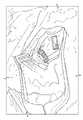
도 4a는, 운동 요소가 보존된 Y-접합부의 원위에 있는, ICN11의 감각 요소를 따로 골라내어 보여주는 것이다. 도해는, 직근이 세로로 절개되었을 때 보존된 운동 요소(녹색)와 수득된 감각 신경 줄기(황색)를 보여준다.
도 4b는, 신경재생에 사용될 것으로서, 수득된 감각 신경 줄기를 보여주는, 수술중 사진을 보여주는 것이다.
도 5는, 3번 늑연골에 대한 절개 접근법을 보여주는 것이다. 도해는 유방 절제술후 초래된 결손을 보여주는 것으로서, 제거를 위해 3번 늑연골을 준비함에 있어 대흉근은 세로로 펼쳐져 있고, 연골막은 절개되어 주변부로부터 분리되어 있다. 세로 방향 점선은 흉골이다.
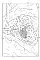
도 6a는, 연골 제거후의 내유 동맥 및 정맥을 보여주는, 도식적 도해를 보여주는 것이다. ICN3은 3번 늑연골과 연골막으로부터 조심스럽게 분리된 다음에 사용될 수 있다.
도 6b는, 3번 늑연골의 하방을 따라서 존재하는 위치로써 ICN3을 식별시키는, 해부학적 표본 절개 상태를 보여주는 것이다.
도 6c는, 연골막과, (ICN3과의 이중 신경지배가 요망된다면) 2번 늑연골의 하방 경계를 조심스럽게 절개하여 노출시킨 ICN2를 보여주는 도해를 보여주는 것이다.
도 6d는, 위치로써 ICN2를 식별시키는 표본 절개 상태를 보여주는 것이다.
도 7a는, 내유 동맥 및 정맥에 피판 DIEA/DIEV를 혈관 문합하였을 때의 상태를 보여주는 것이다. 도 7a는 내유 동맥 및 정맥이 하방으로 절개되어 분리된 다음, DIEP 피판 천공지에 문합되었을 때를 보여주는 도해이다. 황색 표시된 피판 가용 공여 신경은 감각 ICN11 및 ICN12이고, ICN2 및 ICN3은 수여 흉부 신경이다.
도 7b는, 신경 재건을 위한 준비에 있어 연결된 피판과 흉부 혈관 및 절개된 ICN3을 보여주는, 수술중 상황을 보여주는 것이다.
도 8a는, 처리된 인간 신경 동종이식편으로 수여 신경에 공여 신경을 가교시킨 상황을 보여주는 것이다. 도해는, 신경 봉합이 반투명한 돼지 장 점막하 신경 커넥터(직접 봉합의 대안)에 의해 가속화되며, ICN11 및 ICN3이 사용되는 무긴장 단일 신경 신경재생술을 보여준다.
도 8b는, 단일 신경 유방 신경재생술 표본을 도시한 것을 보여주는 것이다.
도 8c는, ICN2 및 ICN3에 ICN11 및 ICN12가 각각 연결되는, 무긴장 이중 신경 신경재생술을 보여주는 도해를 보여주는 것이다.
도 8d는, 이중 신경 유방 신경재생술 표본을 도시한 것을 보여주는 것이다.
도 9a는, 공여 늑간 신경의 전통적 방식의 절개 상태와, 이 공여 늑간 신경을 따로 골라내어 보여주는 것이다. 도해는, 복직근으로부터 절개되어 나온 감각 요소(황색) 및 운동 요소(녹색) 둘 다로 이루어진 공여 줄기를 보여주는 도해이다(줄기의 원래 위치는 황색 점선으로 도시됨).
도 9b는, 감각 요소와 운동 요소 둘 다를 함유하는 공여 늑간 신경의 전통적 절개 방식을 보여주는, 수술중 사진을 보여주는 것이다.1 shows the anatomical landmarks that are key to DIEP flap breast nerve regeneration. An overview of the DIEP abdominal flap and chest wall defect after mastectomy. Essential nerves (ICN1, ICN2, ICN3, ICN10, ICN11, ICN12), vasculature (medial and lateral DIEA, inner mammary arteries and veins), and bone landmarks (ribs I, II, III) are visible.
Figure 2a shows the state of DIEP flap incision (standard lateral → medial mode). The diagram shows the distal end of the sensory element of each intercostal nerve and the typical location of the expected incision of the lateral rectus sheath of the intercostal nerve.
FIG. 2B shows an intraoperative DIEP flap incision highlighting the lateral raw perforation and lateral rectus muscle boundaries.
Figure 3a shows the exposed ICNs after the anterior rectus sheath has been dissected and the longitudinal rectus muscle fiber is unfolded. A retrograde incision of the intercostal neurosensory element (yellow) is diagrammatically made until the junction of the motor element (green) at the intramuscular sensorimotor Y junction. If medial row perforation paper is used as the primary flap supply, then lateral anterior rectus fascia opening and rectus extension can be limited only to allow for sensory ICN acquisition.
Fig. 3b shows a view showing the intraoperative condition of the incised sensory ICN element (the portion pointed to by the tip of the tweezers).
Figure 4a shows the isolated sensory component of ICN11 distal to the Y-junction where the motor component is preserved. The diagram shows the motor components preserved (green) and the sensory nerve trunks obtained (yellow) when the rectus muscle was dissected longitudinally.
Figure 4b shows an intraoperative photograph showing the sensory nerve stem obtained, which will be used for nerve regeneration.
Fig. 5 shows an incision approach for costal 3rd costal cartilage. The diagram shows the defect caused after mastectomy. In preparing the 3rd costal cartilage for removal, the pectoralis major muscle is extended vertically, and the periosteum is incised and separated from the surrounding area. The vertical dotted line is the sternum.
6A shows a schematic diagram showing the inner mammary artery and vein after cartilage removal. ICN3 can be used after careful isolation from the 3rd costal cartilage and perichondrium.
Figure 6b shows the state of the anatomical specimen incision, identifying ICN3 as a location along the inferior side of the 3rd costal cartilage.
Figure 6c shows a schematic showing the perichondrium and ICN2 exposed by careful incision at the inferior border of the 2nd costal cartilage (if dual innervation with ICN3 is desired).
6D shows the specimen dissection state identifying ICN2 by location.
Fig. 7a shows the state when the flap DIEA/DIEV was anastomosed to the inner mammary artery and vein. Figure 7a is a diagram showing when the inner mammary artery and vein are dissected downward and separated, and then anastomized with a DIEP flap perforation paper. Yellow marked flap soluble donor nerves are sensory ICN11 and ICN12, and ICN2 and ICN3 are donor thoracic nerves.
FIG. 7B shows an intraoperative situation showing the connected flap and thoracic vessels and dissected ICN3 in preparation for nerve reconstruction.
Figure 8a shows the cross-linking of donor nerves to donor nerves with treated human nerve allografts. The diagram shows a tension-free single nerve regeneration procedure in which nerve sutures are accelerated by translucent porcine intestinal submucosal nerve connectors (an alternative to direct sutures), and ICN11 and ICN3 are used.
Figure 8b shows a single nerve mammary revascularization specimen.
Figure 8c shows a schematic diagram showing a tension-free double nerve regeneration in which ICN11 and ICN12 are connected to ICN2 and ICN3, respectively.
Fig. 8D shows a double nerve mammary revascularization specimen.
Fig. 9a shows the state of incision in the traditional way of the donor intercostal nerve, and the donor intercostal nerve is isolated and shown. The diagram is a diagram showing a donor stem made up of both sensory (yellow) and motor components (green) dissected from the rectus abdominis muscle (the original location of the stem is shown by the yellow dashed line).
FIG. 9B shows intraoperative photographs showing a traditional dissection of a donor intercostal nerve containing both sensory and motor components.
본 발명은 자가 또는 동종이계 신경을 환자의 유방에 이식하기 위한 수술 방법을 제공한다. 이러한 이식은 유방 수술, 예컨대 유방 절제술 또는 유방 재건술을 실시하였거나 실시중인 환자에서 유방 조직의 유방 신경재생을 유도한다.The present invention provides a surgical method for implanting autologous or allogeneic nerves into a patient's breast. Such transplantation induces mammary nerve regeneration of breast tissue in patients who have undergone or are undergoing breast surgery, such as mastectomy or breast reconstruction.
바람직한 구현예들에서, 본원에 제공된 방법은 자가 또는 동종이계 신경 이식편을 통한 전체 유방 조직 피편의 신경재생을 허용한다. 본 방법은, 합성 신경관을 포함한 신경관, 동종이계 신경 또는 자가 신경을 유방 피판에 이식하는 단계를 포함한다.In preferred embodiments, the methods provided herein allow for nerve regeneration of whole breast tissue fragments via autologous or allogeneic nerve grafts. The method comprises implanting a neural tube, including a synthetic neural tube, an allogeneic nerve, or an autologous nerve into a breast flap.
심하복벽천공지(DIEP) 피판 유방 재건술은 수여부 감각 회복에 제한이 따르는 것으로 공지되어 있으며, 공여부에서 잠재적으로 복부 팽대와 복벽의 약화를 유발한다. 재건된 유방 피판의 유방 신경재생 또는 재신경지배는 기계적 손상 또는 열에 의한 손상에 대해 보호 효과를 발휘할 뿐 아니라, 환자의 삶의 질에도 긍정적인 효과를 가져오는 것으로 확인되었다. 그러나 피판 구역의 자발적 유방 신경재생은 유방 절제술 후 유방 재건술 절차에 있어 표준화된 요소가 아직 아니다. 게다가, 유방 신경재생에 관한 현재의 임상 데이터는 표준화된 수술적 접근법 및 표준적인 신경 갭 가교 매체의 부재, 그리고 감각 회복에 관한 임상 결과의 균일한 데이터 부족을 가리키고 있다.Deep abdominal wall puncture (DIEP) flap breast reconstruction is known to be limited in sensory recovery at the recipient site, and potentially causes abdominal distension and weakening of the abdominal wall at the donor site. Breast nerve regeneration or reinnervation of the reconstructed breast flap not only exerts a protective effect against mechanical or thermal damage, but also has a positive effect on the patient's quality of life. However, spontaneous breast nerve regeneration in the flap area is not yet a standardized factor in breast reconstruction procedures after mastectomy. Moreover, current clinical data on mammary nerve regeneration point to a standardized surgical approach and the absence of standard nerve gap bridging media, and the lack of uniform data on clinical outcomes regarding sensory recovery.
이러한 문제점들을 염두에 두었을 때, 본 발명의 임의의 구현예들은 복벽의 이환율을 최소화하는 술기를 제공하고, 표준화된 유방 신경재생 기술을 제공하며, 처리된 인간 신경 동종이식편을 바람직한 신경 갭 가교 재료로서 사용함으로써 의미있는 감각 회복의 기회를 늘려준다. 이러한 술기는 복직근 운동 신경지배는 보존하면서, 유방 신경재생을 위해 신경의 동종이식편을 사용하고, 피판의 감각 요소만을 선택적으로 사용한다는 점에서 독특하다.With these issues in mind, certain embodiments of the present invention provide techniques to minimize morbidity of the abdominal wall, provide standardized mammary rejuvenation techniques, and use the treated human nerve allograft as a preferred nerve gap bridging material. It increases the chance of meaningful sensory recovery by using it as This technique is unique in that it uses an allograft of the nerve for breast nerve regeneration while preserving the motor innervation of the rectus abdominis muscle, and selectively uses only the sensory elements of the flap.
처리된 신경 동종이식편은 임상 연구에서 길이 70 mm 이하인 갭을 가교하는데 유효하고, 속이 빈 관상 신경 도관 사용시에 비해 월등하며, 수술에 의한 추가 이환율은 보이지 않고 신경 자가이식편 사용시와 거의 동일한, 의미있는 감각 회복 결과를 동반함이 확인되었다. 본 발명의 수술 방법은 단일 또는 이중 신경 유방 신경재생이 가능하도록 맞춤 설계될 수 있으며, 이러한 신규 접근법은 도관이나 자가이식편에 의한 신경재생술에 비해 유리한 결과를 달성한다. 본 발명의 재료와 방법은 외과 의사가 표준화되고 재현 가능한 유방 신경재생술을 적용할 수 있도록 만들고, 더 나아가 의미있는 감각 회복의 기회를 최적화한다.Treated nerve allografts are effective in bridging gaps up to 70 mm in length in clinical studies, and are superior to the use of hollow coronary nerve conduits, with no additional surgical morbidity and almost the same sensation as when using nerve autografts. It was confirmed that it was accompanied by a recovery result. The surgical method of the present invention can be custom designed to enable single or double nerve mammary nerve regeneration, and this novel approach achieves advantageous results compared to nerve regeneration by catheter or autograft. The materials and methods of the present invention enable surgeons to apply standardized and reproducible breast neurogenesis, further optimizing the chances of meaningful sensory recovery.
유방 신경재생은 유방 재건에 있어 중요한 요소이다. 본 발명은 공여부의 감각 분지만을 취하고, 운동 분지는 보존하는 것에 관한 중요성을 입증한다. 이러한 선택성은 수여 감각 신경의 보이지 않는 절단후 운동요소 잔여부(blind motor stump)로의 비정상적 신경 재생을 방지하므로, 감각에 관한 결과가 최적화된다. 이는 또한 자가이식편으로 신경재생된 유방에서 감각 회복이 왜 기대 이하인지에 대한 해부학적 정당화를 제공한다.Breast nerve regeneration is an important factor in breast reconstruction. The present invention demonstrates the importance of taking only the sensory branches of the donor site and preserving the motor branches. This selectivity prevents abnormal nerve regeneration into the blind motor stump after invisible amputation of the donor sensory nerve, thus optimizing sensory outcomes. This also provides an anatomical justification for why sensory recovery is below expectations in autograft-renewed breasts.
또한, 내측 열 천공지를 선택적으로 사용함과 아울러, ICN11 및/또는 ICN12의 감각 요소만을 선택적으로 절개하여 추출함에 있어 기술적 양상은 복직근의 탈신경화 위험 및 이와 연관된 이환율을 최소화한다.In addition, in addition to selectively using the medial thermal perforation paper, the technical aspect in selectively excising and extracting only the sensory elements of ICN11 and/or ICN12 minimizes the risk of denervation of the rectus abdominis muscle and its associated morbidity.
신뢰할 수 있고 예측 가능한 랜드마크의 식별 및 활용은, 본 발명의 수술 방법이 일관되게 반복될 수 있도록 허용한다.Reliable and predictable identification and utilization of landmarks allows the surgical method of the present invention to be consistently repeated.
속이 빈 관상의 신경 도관 단독은 유방 신경재생에 적합하지 않으며, 비 유방 신경재생 연구에 기반을 둔, 인간의 처리된 신경 동종이식편은 이상적이며 가장 전도유망한 가교 매체일 것이다. 더욱이, 동종이식편 신경 재건술에 의한 결과는 신경 자가이식편에 의한 결과보다 유리할 뿐아니라 추가의 공여부 연관 이환율도 보이지 않는다. 그러므로 신경 동종이식편은 이 기술의 핵심 요소이다.Hollow tubular nerve conduits alone are not suitable for mammary nerve regeneration, and human treated nerve allografts, based on non-mammary nerve regeneration studies, are ideal and would be the most promising bridging media. Moreover, the results with allograft nerve reconstruction are more favorable than those with nerve autografts, and no additional donor-related morbidity is observed. Therefore, neural allografts are a key element of this technique.
마지막으로, 커넥터 지원 신경 접합의 활용은 오정렬의 위험을 없애준다. 본 발명은, 처리된 신경 동종이식편을 수술 방법에 통합시킴으로써 유방 재건술중 표준화된 유방 신경재생술을 제공하고, 복벽 관련 이환율을 최소화하며, 의미있는 감각의 회복을 개선함으로써, 유방 재건술을 받은 환자의 삶의 질을 향상시킨다.Finally, the utilization of connector-supported neural junctions eliminates the risk of misalignment. The present invention provides standardized breast neurogenesis during breast reconstruction by integrating the treated nerve allograft into the surgical method, minimizing abdominal wall-related morbidity, and improving the recovery of meaningful sensations, thereby improving the lives of patients undergoing breast reconstruction. improve the quality of
임의의 구현예들에서, 본 발명은 유방 신경재생을 위한 수술 방법을 제공한다. 본 방법은, 동종이계 또는 자기유래 신경을 환자의 유방 피판에 이식하는 단계를 포함한다. 몇몇 구현예들에서, 본 동종이계 또는 자기유래 신경은 늑간 신경(ICN), 구체적으로 ICN10, ICN11 또는 ICN12로부터 수득된다.In certain embodiments, the present invention provides a surgical method for mammary nerve regeneration. The method comprises implanting an allogeneic or autologous nerve into a breast flap of a patient. In some embodiments, the present allogeneic or autologous nerve is obtained from an intercostal nerve (ICN), specifically ICN10, ICN11 or ICN12.
단일 동종이식편에서, 동종이계 또는 자기유래 ICN10, ICN11 또는 ICN12가 수득되어, 환자의 ICN2 또는 ICN3에 이식된다. 예를 들어 ICN10, ICN11 및 ICN12로부터 동종이계 또는 자기유래 신경이 수득되어, 환자의 ICN2 또는 ICN3중 하나에 이식된다. 예를 들어 ICN10 또는 ICN11은 수득되어 ICN2 또는 ICN3에 이식될 수 있다. 대안적으로 ICN11 또는 ICN12는 수득되어 ICN2 또는 ICN3에 이식될 수 있다. 이러한 임의의 구현예들은 도 12와 도 13에 도시되어 있다.In a single allograft, allogeneic or autologous ICN10, ICN11 or ICN12 is obtained and transplanted into the patient's ICN2 or ICN3. For example, allogeneic or autologous nerves are obtained from ICN10, ICN11 and ICN12 and transplanted into either the patient's ICN2 or ICN3. For example ICN10 or ICN11 can be obtained and transplanted into ICN2 or ICN3. Alternatively ICN11 or ICN12 can be obtained and transplanted into ICN2 or ICN3. Some of these implementations are shown in FIGS. 12 and 13 .
이중 이식편에 있어 ICN10, ICN11 또는 ICN12로부터 신경 2개가 수득되어 환자의 ICN2 및 ICN3에 이식된다. 예를 들어 ICN10, ICN11 및 ICN12로부터 신경 2개가 수득되고, 이것들 각각은 환자의 ICN2 및 ICN3중 하나에 이식된다. 대안적으로 ICN10 및 ICN11은 수득되어 ICN2 및 ICN3에 각각 이식될 수 있다. 마찬가지로, ICN11 및 ICN12는 수득되어 ICN2 및 ICN3에 각각 이식될 수 있다. 이러한 임의의 구현예들은 도 12 및 도 13에 도시되어 있다.For double grafts, two nerves are obtained from ICN10, ICN11 or ICN12 and transplanted into the patient's ICN2 and ICN3. For example, two nerves are obtained from ICN10, ICN11 and ICN12, each of which is implanted in one of the patient's ICN2 and ICN3. Alternatively, ICN10 and ICN11 can be obtained and transplanted into ICN2 and ICN3 respectively. Likewise, ICN11 and ICN12 can be obtained and transplanted into ICN2 and ICN3, respectively. Some of these implementations are shown in FIGS. 12 and 13 .
임의의 구현예들에서, 신경 ICN10, ICN11 또는 ICN12의 감각부만이 수득되어 신경 ICN2 또는 ICN3의 감각부에 이식된다. 이러한 임의의 구현예들에서, 신경 ICN10, ICN11 및 ICN12중 2개의 감각부만이 수득되고, 이것들 각각은 신경 ICN2 또는 ICN3의 감각부에 이식된다.In certain embodiments, only the sensory part of the nerve ICN10, ICN11 or ICN12 is obtained and implanted in the sensory part of the nerve ICN2 or ICN3. In any of these embodiments, only two sensory parts of the nerve ICN10, ICN11 and ICN12 are obtained, each of which is implanted in the sensory part of the nerve ICN2 or ICN3.
ICN10, ICN11 또는 ICN12로부터 유래하는 신경의 감각부만이 복직근에서 운동 신경지배를 유지한다. 외측 직근에 대한 외측 늑골 신경의 운동 요소를 보존함으로써 복벽 이환율은 최소화된다.Only sensory parts of nerves originating from ICN10, ICN11 or ICN12 maintain motor innervation in the rectus abdominis muscle. Abdominal wall morbidity is minimized by preserving the motor component of the lateral costal nerve to the lateral rectus muscle.
추가의 구현예들에서, 처리된 신경 동종이식편은 공여 신경 이식에서 가교재로서 사용된다. 대안적으로, 신경관은 공여 신경 이식에서 가교재로서 사용될 수 있다.In further embodiments, the treated nerve allograft is used as a crosslinking material in a donor nerve transplant. Alternatively, the neural tube may be used as a crosslinking material in a donor nerve transplant.
본 발명의 임의의 구현예에 사용된 신경관 또는 처리된 신경 동종이식편은 신경 재생을 촉진하는 신경영양 성장 인자를 함유할 수 있다. 이러한 성장 인자가 포함되면, 피판 조직의 신경지배가 가속화된다. 이러한 성장 인자로서는 뇌 유래 신경영양 인자(BDNF), 신경아교세포 유래 신경영양 인자(GDNF), 신경영양 인자(NGF), 뉴트로핀-3(NT-3), 섬모 신경영양 인자(CNTF) 및 백혈병 억제 인자(LIF)를 포함한다.The neural tube or treated nerve allograft used in any embodiment of the present invention may contain a neurotrophic growth factor that promotes nerve regeneration. When these growth factors are included, the innervation of the flap tissue is accelerated. Examples of such growth factors include brain-derived neurotrophic factor (BDNF), glial cell-derived neurotrophic factor (GDNF), neurotrophic factor (NGF), neurotrophin-3 (NT-3), ciliary neurotrophic factor (CNTF) and leukemia inhibition. factor (LIF).
신경 재생관에 관한 임의의 예들은 미국 특허 제9,687,592호; 동 제9,108,042호; 동 제9,017,714호; 동 제8,741,328호; 동 제8,632,844호; 동 제8,603,512호; 동 제7,842,304호; 동 제7,615,063호; 동 제7,135,040호; 동 제6,589,257호; 동 제6,090,117호; 동 제5,656,605호; 및 동 제4,778,467호에 기재되어 있다. 이들 특허는 각각 본원에 전체로서 참고문헌으로 첨부되어 있다.Certain examples of nerve regeneration tracts are described in US Pat. Nos. 9,687,592; 9,108,042; 9,017,714; 8,741,328; 8,632,844; 8,603,512; 7,842,304; 7,615,063; 7,135,040; 6,589,257; 6,090,117; 5,656,605; and 4,778,467. Each of these patents is incorporated herein by reference in its entirety.
본 발명의 추가의 구현예들은 ICN10, ICN11 및 ICN12로부터 제조된 신경 이식편 적어도 2개를 포함하는 신경 이식편 세트를 제공한다. 몇몇 구현예들에서, 이 세트는 하나의 공여개체로부터 수득된 ICN10, ICN11 및 ICN12로부터 제조된 신경 이식편 적어도 2개를 포함한다. 다른 구현예들에서, 이 세트는 상이한 공여개체로부터 수득된 ICN10, ICN11 및 ICN12로부터 제조된 신경 이식편 적어도 2개를 포함한다. 본원에 개시된 신경 이식편 세트는 적합한 수술, 예컨대 본원에 기재된 유방 신경재생술에 사용될 수 있다.Further embodiments of the present invention provide a set of nerve grafts comprising at least two nerve grafts prepared from ICN10, ICN11 and ICN12. In some embodiments, the set comprises at least two nerve grafts prepared from ICN10, ICN11 and ICN12 obtained from a single donor. In other embodiments, the set comprises at least two nerve grafts prepared from ICN10, ICN11 and ICN12 obtained from different donors. The set of nerve grafts disclosed herein may be used in any suitable surgery, such as the mammary rejuvenation described herein.
몇몇 구현예들에서, 신경 이식편 세트는, 하나 이상의 동물 기원으로부터 수득된 하나 이상의 늑간 신경으로부터 제조될 수 있다. 동물 기원의 예로서는, 일반적으로 반추류, 예컨대 양, 소, 말, 돼지, 염소 등에 더하여, 비 반추류를 포함할 수 있으나, 이에 한정되는 것은 아니다. 적당하다면, 기타 동물이 인간이나 동물에 사용될 신경 이식편 세트의 기원일 수 있음이 이해될 것이다. 하나 이상의 동물 기원으로부터 수득된 하나 이상의 늑간 신경으로 제조된 신경 이식편이 본원에 기재된 술기와 매우 유사한 술기에 사용될 수 있음도 또한 이해될 것이다. 예를 들어 당업자는 이식편의 기원(예컨대 인간, 동물 등) 및 이식편 수여개체(예컨대 인간, 동물 등)에 따라서 본원에 기재된 술기에 대한 변형예가 활용될 수 있음을 인지할 것이다. 이러한 동물 기원의 이식편은 본원에 기재된 인간 대상 유방 신경재생술에서 이종이식편으로 사용될 수 있거나, 또는 동물 대상 늑간 신경 결손 재건에 (이종이식편, 동종이식편 또는 자가이식편으로서) 사용될 수 있음도 또한 이해될 것이다. 몇몇 구현예들에 있어서, 단일 동물 기원으로부터 수득된 상이한 ICN 적어도 2개로부터 신경 이식편 적어도 2개가 제조될 수 있다. 단일 동물 기원은 동물 한 마리(예컨대 양 한마리) 또는 동물 여러 마리(예컨대 양 여러 마리)를 포함할 수 있다. 몇몇 구현예들에서, 상이한 동물 기원으로부터 수득된 상이한 ICN 적어도 2개로부터 신경 이식편 적어도 2개가 제조될 수 있다. 상이한 동물 기원은 상이한 동물 여러 마리(예컨대 양과 돼지, 또는 상이한 종의 양 두 마리)를 포함할 수 있다.In some embodiments, a set of nerve grafts may be prepared from one or more intercostal nerves obtained from one or more animal sources. Examples of animal origin may include, but are not limited to, non-ruminants, in addition to generally ruminants such as sheep, cattle, horses, pigs, goats, and the like. It will be appreciated that other animals may be the origin of the set of nerve grafts to be used in humans or animals, where appropriate. It will also be appreciated that nerve grafts made from one or more intercostal nerves obtained from one or more animal origins may be used in procedures very similar to those described herein. For example, one of ordinary skill in the art will recognize that variations on the techniques described herein may be utilized depending on the origin of the graft (eg, human, animal, etc.) and the recipient of the graft (eg, human, animal, etc.). It will also be understood that such grafts of animal origin may be used as xenografts in breast neurogenesis in the human subjects described herein, or may be used (as xenografts, allografts or autografts) in the reconstruction of intercostal nerve defects in animal subjects. In some embodiments, at least two nerve grafts can be prepared from at least two different ICNs obtained from a single animal origin. A single animal origin may include an animal (eg, a sheep) or multiple animals (eg, several sheep). In some embodiments, at least two nerve grafts can be prepared from at least two different ICNs obtained from different animal origins. Different animal origins may include several different animals (eg, a sheep and a pig, or two sheep of different species).
또 다른 추가의 구현예들에서, 전술된 동물 기원들을 포함하는, 하나 이상의 동물 기원으로부터 수득된 하나 이상의 신경으로부터 신경 이식편 세트가 제조될 수 있다. 이러한 이식편은 인간에서의 신경 결손을 제건함에 있어 이종이식편으로서 사용될 수 있거나, 또는 동물에서의 신경 결손을 재건함에 있어 이종이식편, 동종이식편 또는 자가이식편으로서 사용될 수 있다.In still further embodiments, a set of nerve grafts may be prepared from one or more nerves obtained from one or more animal origins, including those described above. Such grafts may be used as xenografts in reconstructing nerve defects in humans, or as xenografts, allografts or autografts in reconstructing nerve defects in animals.
본 발명의 신경 이식편 세트중 신경 이식편 각각은 수여개체로의 이식에 적합한 신경 이식편으로 제조되도록 처리될 수 있다. 신경 이식편을 제조하기 위해 신경을 처리하는 임의의 기술은 미국 특허 제9,572,911호; 동 제9,402,868호; 동 제7,851,447호; 및 동 제6,972,168호에 기재되어 있다. 이러한 특허 각각은 본원에 전체로서 참고문헌으로 첨부되어 있다.Each of the nerve grafts in the set of nerve grafts of the present invention can be processed to produce a nerve graft suitable for transplantation into several recipients. Any technique for processing nerves to make nerve grafts is described in U.S. Patent Nos. 9,572,911; 9,402,868; 7,851,447; and 6,972,168. Each of these patents is incorporated herein by reference in its entirety.
정의:Justice:
자기유래 이식편은 어느 대상체의 제2 부위로의 이식을 위한, 해당 대상체의 제1 부위로부터 수득된 장기, 조직 또는 이것들의 일부이다.An autologous graft is an organ, tissue, or part thereof obtained from a first site in a subject for transplantation into a second site in a subject.
동종이식편은 제1 개체와 동일한 종의 제2 개체로의 이식을 위한, 제1 개체로부터 수득된 장기, 조직 또는 이것들의 일부이다.An allograft is an organ, tissue, or part thereof obtained from a first individual for transplantation into a second individual of the same species as the first individual.
신경재생이란, 신경의 수복 불가한 손상을 통해 자체의 신경지배능을 상실한 신체 일부에서 일어나는 신경의 재신경지배를 지칭한다. 신경재생은 신경지배능을 상실한 신체 일부의 감각과, 감각성 또는 운동성의 완전한 회복을 요구하지 않는다.Nerve regeneration refers to the reinnervation of nerves that occurs in a part of the body that has lost its ability to control itself through irreparable damage to the nerve. Nerve regeneration does not require complete restoration of sensation, sensuality, or mobility of the part of the body that has lost nerve control.
실시예 1 - 본 발명의 수술 방법Example 1 - Surgical method of the present invention
대기 상태의 환자에게 수술전 마킹을 실시하였다. 이후 환자를 수술실로 이동시킨 다음, 양팔을 외전시킨 채 반듯이 눕혔다. 복부 피판(도 1)을 표준적 방식인 외측에서 내측으로 절개한 다음, 외측 열 천공지 및 연관된 늑간 신경을 노출시켰다(도 2a 및 도 2b). 전방 직근 초를 두개 꼬리부터 외측 열 천공지를 따라 절개하여 복직근, 외측 천공지 혈관 및 늑간 신경(ICN11 및 ICN12)을 노출시켰다(도 3a 및 도 3b).Preoperative marking was performed on the waiting patient. After that, the patient was moved to the operating room, and then laid flat with both arms abducted. The abdominal flap (FIG. 1) was incised from the outside to the inside in a standard way, then the lateral thermal puncture and associated intercostal nerves were exposed (FIGS. 2A and 2B). The rectus anterior muscle sheath was incised from the cranial tail along the lateral row perforation to expose the rectus abdominis muscle, lateral perforator vessels, and intercostal nerves (ICN11 and ICN12) ( FIGS. 3A and 3B ).
혈관 천공지 외측 열 다음에 있는 ICN11 및/또는 ICN12를 식별한 다음, 늑간 신경 ICN11 및/또는 ICN12의 감각 분지 표준 역행 절개선을 따라가 감각-운동 Y 접합부와 만나게 하였다. 이는 근내 또는 근후에서 볼 수 있었던 반면에, 노출은 직근 섬유를 가로로 방향이 아닌 세로 방향으로 펼침으로써 이루어진 관계로, 일체성이 보존되었다.ICN11 and/or ICN12 next to the lateral row of vascular perforations were identified, followed by a standard retrograde incision of the sensory branch of the intercostal nerve ICN11 and/or ICN12 to meet the sensorimotor Y junction. Whereas this could be seen intramuscularly or posteriorly, the integrity was preserved as the exposure was achieved by stretching the rectus muscle fibers longitudinally rather than transversely.
외측 열 혈관 천공지를 DIEP 피판에 합체할 계획이 있는 경우, 이 외측 열 혈관 천공지를 보호하는데 심혈을 기울여야 한다. 그러나 만일 내측 열 혈관 천공지가 피판으로의 주된 혈관 제공을 위해 사용되면, 이 기술이 사용될 때 외측 천공지와 직근 펼침부를 따라 난 직근 근막의 수직 전방 절개틈은 최소화되어, 광범위한 근막의 개방 또는 절개를 수행하지 않고서도 오로지 ICN 감각 이식편 수득이 가능하도록 제한될 수 있다.If it is planned to incorporate the lateral thermal vascular perforation into the DIEP flap, great care should be taken to protect this lateral thermal vascular perforation. However, if the medial thermal vascular perforator is used to provide primary blood vessels to the flap, when this technique is used, the vertical anterior incision gap of the lateral perforator and the rectus fascia along the rectus extension is minimized, resulting in extensive fascial opening or incision. It can be limited so that it is possible to obtain only ICN sensory grafts without performing
복직근의 탈신경을 방지하기 위해서 감각 ICN11 및/또는 ICN12 분지의 역행 절개에 더하여 운동 요소를 보존하였다. 운동 보존은 외측 천공지가 주요 혈관 공급수단으로 선택될 때조차도 수행되었다. 이는, 외측 복직근에 대한 운동 신경지배는 원래대로 남겨두면서, 감각-운동 Y-접합부로부터 단지 원위에 있는 감각 요소를 수득함으로써 달성된다(도 4a 및 도 4b).To prevent denervation of the rectus abdominis, motor components were preserved in addition to retrograde dissection of the sensory ICN11 and/or ICN12 branches. Motor conservation was performed even when lateral perforation was selected as the primary vascular supply. This is achieved by obtaining sensory elements only distal from the sensorimotor Y-junction, while leaving motor innervation to the lateral rectus abdominis intact ( FIGS. 4A and 4B ).
ICN 1개 또는 2개를 포함시킬지 여부는, 피판의 단일 신경지배가 요망되는지, 아니면 이중 신경지배가 요망되는지의 여부에 따른다. 일단 감각 ICN 분지(들)를 절개하여 분할하고 나서, 흉부 절개가 종료될 때까지 피판을 관류된 상태로 남겨둔 채 DIEP 피판 혈관 절개 나머지 과정을 종료하였다.Whether to include 1 or 2 ICNs depends on whether single innervation of the flap or double innervation of the flap is desired. Once the sensory ICN branch(s) were dissected and segmented, the remainder of the DIEP flap vascular transection was terminated leaving the flap perfused until the thoracic incision was complete.
유방 절제술 후, 3번 늑연골위 대흉근 섬유를 세로로 절개하여 3번 늑골의 연골막을 노출시켰다. 연골막을 절개하고 나서, 연골막하 절개를 수행한 다음, 3번 늑연골을 제거하였디. 그 다음, 후방 연골막을 조심스럽게 절개하고 나서, 내유 혈관이 보일 때까지 외측 → 내측 절개를 수행하였다(도 5).After mastectomy, the pectoralis major muscle fibers above the 3rd costal cartilage were made longitudinally to expose the periosteum of the 3rd rib. After incision of the perichondrium, a subperichondral incision was performed, and then the 3rd costal cartilage was removed. Then, the posterior periosteum was carefully incised, and then, an lateral to medial incision was performed until the inner mammary vessels were visible (FIG. 5).
ICN3이 3번 늑골의 하방 경계를 따라 뻗어있음을 인지하는 것은 중요하다(도 6a 및 도 6b). 일단 연골막 아래와 하방 늑골 경계를 따라 존재하는 ICN3을 식별하면, 이를 보존시킨 채 내측으로 따라 들여보낸 후 분할하고 나서, 외측으로 빗나가게 만들었다. 만일 이중 신경지배가 요망되면, ICN2는 수술장 상단 폴(upper pole) 내부, 연골막 아래, 그리고 2번 늑골 경계 하방 및 이를 따라서 발견될 수 있었다(도 6c 및 도 6d).It is important to note that ICN3 extends along the inferior border of the 3rd rib ( FIGS. 6A and 6B ). Once ICN3 was identified below the perichondrium and along the inferior costal border, it was preserved and traversed medially, then segmented and deflected outward. If double innervation was desired, ICN2 could be found inside the upper pole of the operating room, below the perichondrium, and below and along the 2nd costal boundary ( FIGS. 6c and 6d ).
그 다음, 공여부로부터 피판을 잘라내어 흉부로 가져갔다. 미세수술 동맥 및 정맥 문합을 표준 방식으로 수행하였다(도 7a 및 도 7b). 피판을 끼워넣는데 필요한 피판의 전체 회전 아치를 보존하고, 무긴장 신경 수복을 보장하기 위해, 처리한 인간 신경 동종이식편(1 mm ~ 2 mm, 50개 또는 1 mm ~ 2 mm, 70개)을 사용하여 신경 접합을 수행함으로써(Avance ® Nerve Graft, AxoGen, Alachua FL), 갭을 가교시켰다. 그 다음, 끼워넣은 신경 동종이식편을 미세 수술로 흉부 수여 신경 말단 및 피판 공여 신경 말단에 연결시켰으며(직접 봉합), 대안적으로는 근위 및 원위 접합을 반투명의 다공성 돼지 장 점막하 신경 커넥터(AxoGuard Nerve Connector, AxoGen, Alachua FL)로 가속화할 수 있었다(도 8a 및 도 8b). 그 다음, 피판을 끼워넣고, 복부에 있는 공여부를 표준적 방식으로 봉합한 후, 신경재생된 DIEP 피판 유방 재건술을 마쳤다.The flap was then removed from the donor site and brought to the chest. Microsurgical arterial and venous anastomosis was performed in a standard manner ( FIGS. 7A and 7B ). Processed human nerve allografts (1 mm to 2 mm, 50 or 1 mm to 2 mm, 70) are used to preserve the entire rotational arch of the flap required to implant the flap and to ensure tonic nerve repair. by performing neural junctions (Avance ® Nerve Graft, AxoGen, Alachua FL), bridging the gap. The embedded nerve allografts were then microsurgically connected to the thoracic donor nerve terminal and the flap donor nerve terminal (direct suture), alternatively the proximal and distal junctions were connected with a translucent porous porcine intestinal submucosal nerve connector (AxoGuard Nerve). Connector, AxoGen, Alachua FL) could be accelerated ( FIGS. 8a and 8b ). Then, the flap was inserted, and the donor site in the abdomen was sutured in a standard way, and then the regenerated DIEP flap breast reconstruction was completed.
실시예 2 - 본 발명의 수술 방법에 의해 제공되는 이점Example 2 - Advantages provided by the surgical method of the present invention
수술적 접근법의 동질성은 절차의 효능 또는 절차상의 개념, 예컨대 유방 신경재생을 확실하게 언급하는데 중요하다. 그러므로 표준화된 술기를 확립하는 것은 장래의 동질적인 비교 분석을 가속화하는데 중요하다. 신경 수술의 원리를 명확히 이해하는 것뿐 아니라, 신경 도관, 자가이식편 및 처리한 신경 동종이식편과 같이 가용 재건술상 선택에 관한 특징과 관련된 전문 지식은 제안된 이 절차의 성공적인 수행을 위해 중요하다.The homogeneity of the surgical approach is important to clearly address the efficacy of the procedure or procedural concepts, such as mammary nerve regeneration. Therefore, establishing standardized skills is important to accelerate future homogenous comparative analyzes. A clear understanding of the principles of neurosurgery, as well as expertise regarding the features of available reconstructive options, such as nerve conduits, autografts, and treated nerve allografts, are important for the successful implementation of this proposed procedure.
신경 손상의 표준적인 치료는, 가능하다면 언제든지 수행할 수 있는 무긴장 1차 수복으로 이루어져 있다. 그러나 만일 1차 수복이 불가능하면, 신경 자가이식편, 관상 도관 및 처리한 신경 동종이식편을 포함하는 가교재를 사용하게 된다. 유방 신경재생에서 마주하게 되는 신경 갭은, 통상적으로 50 mm ~ 70 mm(신경 도관이 사용되는 재건술에 권장되는 길이를 훨씬 초과함)로 측정된다. 말단 신경 결손을 재건할 때 전통적으로 신경 자가이식편이 선호되어 왔지만, 이 신경 자가이식편은 공여부 합병증, 예컨대 추가 절개, 상처 치유의 문제, 통증을 동반하는 신경종의 형성, 또는 (만일 직근이 탈신경화되면) 팽대/절개창 탈장과 연관된다.Standard treatment for nerve damage consists of a tonic primary repair, which can be performed whenever possible. However, if primary repair is not possible, cross-linking materials including nerve autografts, coronary catheters, and treated nerve allografts are used. Nerve gaps encountered in mammary neurogenesis typically measure between 50 mm and 70 mm (which far exceeds the recommended length for reconstructive procedures in which nerve conduits are used). Although nerve autografts have traditionally been the preferred choice for reconstructing terminal nerve defects, these nerve autografts can be used for donor site complications such as further dissection, problems with wound healing, painful neuroma formation, or (if the rectus muscle is denervated). ) associated with ampulla/incision hernia.
처리한 신경 동종이식편을 사용함으로써 신경 자가이식편 수득물과 연관된 공여부 합병증을 피할 수 있다. 처리한 신경 동종이식편으로서는 공여한 인간의 말초 신경 조직으로부터 형성되어, 탈세포화, 전 탈세포화 및 멸균된 세포외 기질(ECM) 스캐폴드로서, 신경 조직의 단백질 조성을 가지는 무세포 미세구조 아키텍쳐를 이루는 세포외 기질(ECM) 스캐폴드가 있다. 동종이식편의 탈세포화는 면역 거부 반응의 위험을 유의미하게 감소시키므로, 면역억제 치료의 필요를 없앤다. 얻어진 동종이식편은, 원래 신경의 작은 다발에 함유된 신경내막 미세소관 다발과, 안내 재성장(guided regrowth)에 대한 천연 축삭 성장 신호를 제공하는 ECM 단백질(라미닌, 피브로넥틴 및 글리코사미노글리칸)로 구성되지만, 속이 빈 관상 도관에서는 확인되지 않는 신경외막 스캐폴드로 이루어져 있다.Donor site complications associated with neural autograft harvests can be avoided by using treated nerve allografts. As a treated neuronal allograft, it is formed from a donor human peripheral nervous tissue, and is a decellularized, pre-decellularized, and sterilized extracellular matrix (ECM) scaffold, which forms a cell-free microstructural architecture with the protein composition of the neural tissue. There is an extracellular matrix (ECM) scaffold. Decellularization of allografts significantly reduces the risk of immune rejection, thus eliminating the need for immunosuppressive therapy. The resulting allograft was composed of an endothelial microtubule bundle originally contained in a small bundle of nerves, and ECM proteins (laminin, fibronectin and glycosaminoglycan) that provide a native axon growth signal for guided regrowth. However, it consists of an epicardial scaffold that is not found in hollow coronary ducts.
공여부 절개의 첫 번째로 중요한 요소는 공여 늑간 신경의 식별과 보존에 의존한다. 시신 연구는 복직근이, DIEA의 최 외측 분지와 평행하게 뻗어 나가다가 동맥 천공지와 함께 복직근과 전방 복벽으로 뻗어 들어가는 직근 초의 총으로부터 유래하는 신경에 의해 신경지배됨을 발견하였다. 그러므로 DIEA 외측 분지와 외측 열 천공지는 복직근을 신경지배하는 늑간 신경과 긴밀하게 관련되어 있고, DIEP 피판 수득시 이러한 구조에 발생하는 임의의 손상은 전술한 복벽 약화, 복부 팽대 또는 탈장에 따른 공여부 이환율의 원인이 될 것이다. DIEP 피판은 TRAM(복직횡근; Transversus Rectus Abdominis Muscle) 피판의 단점을 극복하는 것을 목적으로 하지만, DIEP 피판 이후 보고된 복부 팽대 또는 절개창 탈장의 발생률은 여전히 3% ~ 5%에 달한다. 외측 직근에 대한 외측 늑간 신경의 운동 요소를 보존함으로써 복벽 이환율은 더욱더 최소화될 것이다.The first important factor in donor incision relies on the identification and preservation of the donor intercostal nerve. The cadaveric study found that the rectus abdominis muscle is innervated by nerves originating from the plexus of the rectus abdominis muscle extending parallel to the outermost branch of the DIEA and extending into the anterior abdominal wall along with the arterial puncture. Therefore, the lateral branch of DIEA and the lateral fissure perforation are closely related to the intercostal nerve innervating the rectus abdominis muscle, and any damage to these structures upon obtaining the DIEP flap is a factor in the morbidity of the donor site due to the aforementioned weakening of the abdominal wall, enlarged abdomen or hernia. will be the cause Although the DIEP flap aims to overcome the disadvantages of the Transversus Rectus Abdominis Muscle (TRAM) flap, the reported incidence of ampulla or incision hernia after the DIEP flap is still 3% to 5%. By preserving the motor component of the lateral intercostal nerve to the lateral rectus muscle, abdominal wall morbidity will be further minimized.
공여부 절개에 있어 동일하게 중요한 요소는 감각 신경을 노출시켜 수득하는 방법이다. 일상적으로 피판 절개 및/또는 자가이식편 수득시 운동 분지는 종종 감각 요소와 함께 희생되어 취하여진다. 합하여진 감각운동 신경을 이용함(도 9a 및 도 9b)에 있어 이 접근법은 추출한 신경의 길이를 약 10 cm ~ 12 cm까지 연장시키지만, 여기에는 복직근의 탈신경 발생 위험이 있다는 점 이외에도 일반적으로 간과되는 또 다른 위험이 있다. 그 위험은, 수여 신경이 원위에서 재생하기 시작하여 공여 신경과 연결될 때, 감각 분지는 재생되어 클립으로 고정된 운동 요소와 만날 수 있지만, 그 경우에 섬유의 50% 이하만이 감각 분지를 공급한다는 점이다. 이 점은 감각 회복도를 감소시킬 것으로 예상된다. 이 위험을 해결하기 위해 ICN11 및/또는 ICN12의 감각 요소만을 사용하는 것이 제안된다. 운동 분지는 보존하면서 감각 요소만이 추출되려면, 그 뒤 근위에는 피부 감각 신경이 Y-접합부(감각 요소가 운동 요소와 만난 후, 혼합 신경으로서 근위로 계속 진행하는 곳)에 대해 역행하는 방식으로 따르게될 것이다. 감각 요소는 Y-접합부에서 수득되며, 이 경우 외측 복직근을 향하는 운동 분지는 온전히 보존된다. 순수한 감각 신경 줄기는 비교적 짧으므로, 필요할 경우 처리한 신경 동종이식편이 갭을 가교하는데 사용될 수 있다(도 8). 이 접근법은 신경재생 및 의미있는 수복의 기회를 최적화하는 동시에 복직근 신경지배를 온전히 보존하는 것을 목표로 하는, 적절한 해부학적 플랫폼을 제공하기 위해 제안된다.An equally important factor in donor incision is the method obtained by exposing sensory nerves. In routine flap dissection and/or autograft harvesting, motor branches are often sacrificed and taken along with sensory elements. In using the combined sensorimotor nerves ( FIGS. 9A and 9B ), this approach extends the length of the extracted nerves to about 10 cm to 12 cm, but this approach, in addition to the risk of denervation of the rectus abdominis muscle, is a commonly overlooked There is another danger. The risk is that when the donor nerve begins to regenerate distally and connects with the donor nerve, the sensory branch may regenerate and encounter clipped motor elements, but in that case only less than 50% of the fibers supply the sensory branch. point. This point is expected to decrease the degree of sensory recovery. To address this risk, it is proposed to use only the sensory components of ICN11 and/or ICN12. If only sensory components are to be extracted while preserving motor branches, then proximally the cutaneous sensory nerves follow in a retrograde fashion to the Y-junction (where sensory components encounter motor components and then continue proximally as mixed nerves). will be The sensory component is obtained at the Y-junction, in which case the motor branch towards the lateral rectus abdominis is intact. Since pure sensory nerve stems are relatively short, treated nerve allografts can be used to bridge the gap if necessary ( FIG. 8 ). This approach is proposed to provide an appropriate anatomical platform, which aims to optimize the chance of nerve regeneration and meaningful repair while simultaneously preserving the rectus abdominis innervation intact.
뿐만 아니라, ICN2 및/또는 ICN3의 신중한 절개와 식별에 의존하는, 수여부에서의 중요 요소도 또한 중요하다. 선택된 수여 신경은 ICN3이지만, ICN2도 또한 동일한 수술장내 전방 흉부에서 확실히 발견될 수 있었다.In addition, critical factors in the recipient site, which depend on careful excision and identification of ICN2 and/or ICN3, are also important. The donor nerve selected was ICN3, but ICN2 could also be reliably found in the anterior thorax within the same operating field.
처리한 신경 동종 이식편은 DIEP 피판으로부터 유래하는 짧은 신경 줄기라는 점을 극복하고, 무긴장 신경 접합을 허용한다. 대안적으로, 만일 흉부 혈관계를 혈관 공급수단으로 선택하면, 전방 액와선을 따라 외측 ICN4를 사용할 수 있다.The treated nerve allograft overcomes the fact that it is a short nerve stem originating from a DIEP flap and allows for tonic nerve junctions. Alternatively, if the thoracic vasculature is chosen as the vascular supply, lateral ICN4 along the anterior axillary line can be used.
그렇지 않을 경우, ICN4를 내유 혈관계와 함께 사용할 수 있는데, 그 이유는 두 개의 피봇 중심(pivoting point), 즉 내측 혈관인 하나와 외측 신경인 또다른 하나가 있어서, 피판 회전 및 끼워 넣어지는 범위에 영향을 미칠 수 있기 때문이다. 또한, 성감대인 유두/유륜의 감각 신경지배는 주로 ICN4의 외측 분지에 의해 전달되며, 이때 유방 신경재생을 위해 이러한 기능을 가지는 신경을 사용하는 것에 내포된 의미는 잘 알려져 있지 않다.Alternatively, ICN4 can be used with the inner mammary vasculature because it has two pivoting points, one for the medial vessel and the other for the lateral nerve, which affects flap rotation and fit. because it can affect In addition, sensory innervation of the nipple/areola, which is the erogenous zone, is mainly transmitted by the lateral branch of ICN4, and the implications of using a nerve having this function for breast nerve regeneration are not well known.
따라서 만일 ICN4가 전체 유방 피판을 신경재생하는데 사용되면, 과자극과 관련된 후유증이 있을 수 있다. 이를 고려하였을 때뿐 아니라, ICN4의 외측 분지가 일반적으로 유방 절제술 과정에서 흉벽 근육계 수준으로 횡단 절개된다는 사실을 고려하였을 때, 이 분지는 유방 절제술 전이나 도중에 특별히 절개하여 보존하지 않는 한, 대부분 신경재생에 사용할 수 없다.Therefore, if ICN4 is used to innervate the entire breast flap, there may be sequelae related to hyperstimulation. Considering this and the fact that the lateral branch of ICN4 is generally transversely incised to the chest wall musculature level during mastectomy, most of this branch is nerve regeneration before or during mastectomy, unless specifically excised and preserved. cannot be used for
ICN4와 관련하여 전술한 우려로 말미암아, ICN4는 유방 신경재생에 대한 주요 수여부로 바람직하지 않을 수 있다.Due to the concerns discussed above with respect to ICN4, ICN4 may be undesirable as a major avenue for mammary nerve regeneration.
마지막으로, 표준적 직접 봉합 동종이식편 신경 접합에 대한 대안으로서, 피판 공여 신경과 흉부 수여 신경 사이에 삽입한 신경 동종이식편의 커넥터 지원 미세 수술 접합은 근막 오정렬 또는 과도한 축삭 탈출을 유발시키지 않고 접합부 너머로 성장을 가속화할 수 있다.Finally, as an alternative to the standard direct suture allograft nerve junction, the connector-assisted microsurgical junction of a nerve allograft inserted between the flap donor nerve and the thoracic donor nerve grows beyond the junction without causing myofascial misalignment or excessive axonal prolapse. can accelerate
본 발명의 수술 방법은 신뢰할 수 있고, 재현 가능하며, 효과적인 신경재생 절차를 제공함으로써 유방 재건술에 혁명을 일으킬 것이다.The surgical method of the present invention will revolutionize breast reconstruction by providing a reliable, reproducible and effective nerve regeneration procedure.
Claims (19)
Translated fromKorean적어도 2개의 신경 이식편, 여기서 상기 적어도 2개의 신경 이식편은 하나 이상의 동물 기원으로부터 얻어진 수확된 늑간 신경(Intercostal nerves, ICNs)들의 가공된 세그먼트들을 포함하고,
상기 적어도 2개의 신경 이식편의 제1 신경 이식편은 제1 늑간 신경의 운동 요소가 아닌 감각 요소의 가공된 세그먼트이고,
상기 적어도 2개의 신경 이식편의 제2 신경 이식편은 제2 늑간 신경의 운동 요소가 아닌 감각 요소의 가공된 세그먼트이며,
상기 제1 늑간 신경 및 상기 제2 늑간 신경은 다르게 넘버링된 늑간 신경들이고,
상기 신경 이식편 세트는 50mm 내지 70mm의 길이를 갖는 신경 갭(Nerve gap)을 연결하도록 구성된 신경 이식편 세트.A set of nerve grafts for use in the neuralization of a breast of a subject, comprising:
at least two nerve grafts, wherein the at least two nerve grafts comprise engineered segments of harvested intercostal nerves (ICNs) obtained from one or more animal origin;
wherein the first nerve graft of the at least two nerve grafts is a machined segment of a sensory component that is not a motor component of a first intercostal nerve;
wherein the second nerve graft of the at least two nerve grafts is a machined segment of a sensory component that is not a motor component of a second intercostal nerve;
wherein the first intercostal nerve and the second intercostal nerve are differently numbered intercostal nerves;
The nerve graft set is a nerve graft set configured to connect a nerve gap having a length of 50 mm to 70 mm.
상기 제1 신경 이식편 및 상기 제2 신경 이식편은 하나의 동물 기원으로부터 수득된 신경 이식편 세트.According to claim 1,
wherein said first nerve graft and said second nerve graft are obtained from a single animal origin.
상기 제1 신경 이식편 및 상기 제2 신경 이식편은 상이한 동물 기원들로부터 수득된 신경 이식편 세트.According to claim 1,
A set of nerve grafts, wherein said first nerve graft and said second nerve graft are obtained from different animal origins.
상기 제1 신경 이식편 및 상기 제2 신경 이식편은 반추류(Ruminant)로부터 수득된 신경 이식편 세트.According to claim 1,
The first nerve graft and the second nerve graft are a set of nerve grafts obtained from Ruminant.
상기 반추류는 양, 소, 말, 돼지 또는 염소인 신경 이식편 세트.5. The method of claim 4,
The set of nerve grafts wherein the ruminant is sheep, cattle, horses, pigs or goats.
상기 제1 신경 이식편 또는 상기 제2 신경 이식편 중 적어도 하나는 제3 늑간 신경에 대한 이식을 위해 구성되고,
상기 제3 늑간 신경은 상기 제1 늑간 신경 및 상기 제2 늑간 신경과 다르게 넘버링된 늑간 신경인 신경 이식편 세트.According to claim 1,
at least one of said first nerve graft or said second nerve graft is configured for implantation into a third intercostal nerve;
wherein the third intercostal nerve is an intercostal nerve numbered differently from the first intercostal nerve and the second intercostal nerve.
상기 제1 신경 이식편 또는 상기 제2 신경 이식편 중 적어도 하나는 상기 피험체의 유방의 신경화에 사용하기 위해 유방과 관련된 제3 늑간 신경의 감각 부분에 이식을 위해 구성된 것인 신경 이식편 세트.According to claim 1,
and wherein at least one of said first nerve graft or said second nerve graft is configured for implantation in the sensory portion of a third intercostal nerve associated with the breast for use in the neuralization of the breast of the subject.
상기 신경 이식편 세트는 50mm 내지 70mm의 길이를 갖는 신경 갭(Nerve gap)을 연결하도록 구성된 신경 이식편 세트.at least two nerve grafts obtained from one or more animal origins, wherein the at least two nerve grafts are engineered segments of one or more harvested intercostal nerves comprising sensory components that are not motor components of one or more intercostal nerves (ICNs). including,
The nerve graft set is a nerve graft set configured to connect a nerve gap having a length of 50 mm to 70 mm.
상기 하나 이상의 늑간 신경은 적어도 2개의 다르게 넘버링된 늑간 신경들을 포함하는 신경 이식편 세트.9. The method of claim 8,
wherein the one or more intercostal nerves comprises at least two differently numbered intercostal nerves.
상기 하나 이상의 늑간 신경은 반추류(Ruminant)로부터 수득된 신경 이식편 세트.9. The method of claim 8,
The one or more intercostal nerves is a set of nerve grafts obtained from Ruminant.
상기 반추류는 양, 소, 말, 돼지 또는 염소인 신경 이식편 세트.11. The method of claim 10,
The set of nerve grafts wherein the ruminant is sheep, cattle, horses, pigs or goats.
상기 적어도 2개의 신경 이식편은 피험체의 유방의 신경화에 사용되도록 구성된 신경 이식편 세트.9. The method of claim 8,
A set of nerve grafts, wherein the at least two nerve grafts are configured for use in the neuralization of a breast of a subject.
상기 적어도 2개의 신경 이식편 각각은 길이가 50mm 또는 70mm인 신경 이식편 세트.According to claim 1,
A set of nerve grafts, wherein each of said at least two nerve grafts is 50 mm or 70 mm in length.
상기 적어도 2개의 신경 이식편 각각은 길이가 50mm 또는 70mm인 신경 이식편 세트.9. The method of claim 8,
A set of nerve grafts, wherein each of said at least two nerve grafts is 50 mm or 70 mm in length.
사람을 제외한 하나 이상의 동물 기원으로부터 적어도 2개의 늑간 신경(Intercostal nerves, ICNs)들을 수확하는 단계; 및
상기 신경 이식편 세트를 형성하기 위해 상기 적어도 2개의 늑간 신경들을 처리하는 단계를 포함하고,
상기 신경 이식편 세트는 50mm 내지 70mm의 길이를 갖는 신경 갭(Nerve gap)을 연결하도록 구성되고,
상기 하나 이상의 늑간 신경의 세그먼트들은 상기 하나 이상의 늑간 신경의 운동 요소가 아닌 감각 요소를 포함하는 방법.A method of preparing a set of nerve grafts for mammary neurization, the method comprising:
harvesting at least two intercostal nerves (ICNs) from one or more animal origins other than humans; and
processing the at least two intercostal nerves to form the set of nerve grafts;
The nerve graft set is configured to connect a nerve gap having a length of 50 mm to 70 mm,
wherein the segments of the one or more intercostal nerves include a sensory component that is not a motor component of the one or more intercostal nerves.
상기 적어도 2개의 신경 이식편 각각의 길이는 50mm 또는 70mm인 방법.17. The method of claim 16,
The length of each of the at least two nerve grafts is 50 mm or 70 mm.
상기 적어도 2개의 신경 이식편의 제1 신경 이식편은 제1 늑간 신경의 가공된 세그먼트이고,
상기 적어도 2개의 신경 이식편의 제2 신경 이식편은 제2 늑간 신경의 가공된 세그먼트이며,
상기 제1 늑간 신경 및 상기 제2 늑간 신경은 다르게 넘버링된 늑간 신경인 방법.17. The method of claim 16,
wherein the first nerve graft of the at least two nerve grafts is a machined segment of a first intercostal nerve;
wherein the second nerve graft of the at least two nerve grafts is a machined segment of a second intercostal nerve;
wherein the first intercostal nerve and the second intercostal nerve are differently numbered intercostal nerves.
상기 적어도 2개의 신경 이식편의 제1 신경 이식편 및 제2 신경 이식편은 동일한 번호의 늑간 신경의 가공된 세그먼트들인 방법.17. The method of claim 16,
wherein the first nerve graft and the second nerve graft of the at least two nerve grafts are engineered segments of an intercostal nerve of the same number.
Applications Claiming Priority (1)
| Application Number | Priority Date | Filing Date | Title |
|---|---|---|---|
| PCT/US2018/061309WO2020101689A1 (en) | 2018-11-15 | 2018-11-15 | Materials and methods for nerve repair with animal-sourced nerve grafts |
Publications (2)
| Publication Number | Publication Date |
|---|---|
| KR20210105332A KR20210105332A (en) | 2021-08-26 |
| KR102406052B1true KR102406052B1 (en) | 2022-06-07 |
Family
ID=70731770
Family Applications (1)
| Application Number | Title | Priority Date | Filing Date |
|---|---|---|---|
| KR1020217014775AActiveKR102406052B1 (en) | 2018-11-15 | 2018-11-15 | Materials and methods for repairing nerves with nerve grafts of animal origin |
Country Status (9)
| Country | Link |
|---|---|
| EP (1) | EP3880262A4 (en) |
| JP (1) | JP2022507364A (en) |
| KR (1) | KR102406052B1 (en) |
| CN (1) | CN113056294A (en) |
| AU (1) | AU2018449641B2 (en) |
| CA (1) | CA3119782A1 (en) |
| IL (1) | IL283079B2 (en) |
| NZ (1) | NZ775934A (en) |
| WO (1) | WO2020101689A1 (en) |
Citations (5)
| Publication number | Priority date | Publication date | Assignee | Title |
|---|---|---|---|---|
| JP2000517206A (en) | 1996-07-26 | 2000-12-26 | カロリンスカ イノベイションズ アクチボラゲット | Medical devices for treating central nervous system gaps or defects |
| JP2005500375A (en) | 2001-08-13 | 2005-01-06 | ユニバーシティ・オブ・フロリダ・リサーチ・ファンデーション・インコーポレーテッド | Materials and methods for promoting neural tissue repair |
| JP2012520136A (en) | 2009-03-10 | 2012-09-06 | ザ ジョーンズ ホプキンズ ユニバーシティ | Biological tissue bonding and repair device and method of use thereof |
| US20170128624A1 (en) | 2013-03-15 | 2017-05-11 | University Of Florida Research Foundation, Inc. | Method for Decellularization of Tissue Grafts |
| JP2018521685A (en) | 2015-05-28 | 2018-08-09 | アクソジェン コーポレーション | Nerve culture system |
Family Cites Families (9)
| Publication number | Priority date | Publication date | Assignee | Title |
|---|---|---|---|---|
| US4662884A (en)* | 1984-04-25 | 1987-05-05 | University Of Utah Research Foundation | Prostheses and methods for promoting nerve regeneration |
| EP0968724A1 (en)* | 1998-07-03 | 2000-01-05 | Koninklijke Nederlandse Akademie van Wetenschappen | Use of viral vectors for treatment of the injured peripheral and central nervous system |
| PL2488194T3 (en)* | 2009-10-14 | 2016-11-30 | Use of a neuregulin to treat cavernous nerve injury | |
| EP2594295A1 (en)* | 2011-11-16 | 2013-05-22 | Servicio Andaluz De Salud | Nerve implants based on a compacted biomaterial containing cells |
| MD891Z (en)* | 2014-12-29 | 2015-10-31 | Корнелиу УРЕКЕ | Method for breast reconstruction with rectus abdominis musculocutaneous unipedicled flap after mastectomy |
| US20180296729A1 (en)* | 2015-05-26 | 2018-10-18 | Mayo Foundation For Medical Education And Research | Decellularized nerve allografts |
| CN105769396A (en)* | 2016-04-08 | 2016-07-20 | 遵义医学院 | Method for reconstructing sensory nerve functions of amputation upper limbs and method for sensory nerve and artificial limb interfaces |
| CN105920671B (en)* | 2016-06-22 | 2020-02-11 | 浙江元太生物科技有限公司 | Preparation method of nerve graft and product thereof |
| US10813643B2 (en)* | 2017-10-19 | 2020-10-27 | Axogen Corporation | Materials and methods for breast neurotization with nerve grafts |
- 2018
- 2018-11-15KRKR1020217014775Apatent/KR102406052B1/enactiveActive
- 2018-11-15ILIL283079Apatent/IL283079B2/enunknown
- 2018-11-15JPJP2021526233Apatent/JP2022507364A/enactivePending
- 2018-11-15EPEP18939894.4Apatent/EP3880262A4/enactivePending
- 2018-11-15CACA3119782Apatent/CA3119782A1/enactivePending
- 2018-11-15CNCN201880099569.4Apatent/CN113056294A/enactivePending
- 2018-11-15AUAU2018449641Apatent/AU2018449641B2/enactiveActive
- 2018-11-15NZNZ775934Apatent/NZ775934A/enunknown
- 2018-11-15WOPCT/US2018/061309patent/WO2020101689A1/ennot_activeCeased
Patent Citations (5)
| Publication number | Priority date | Publication date | Assignee | Title |
|---|---|---|---|---|
| JP2000517206A (en) | 1996-07-26 | 2000-12-26 | カロリンスカ イノベイションズ アクチボラゲット | Medical devices for treating central nervous system gaps or defects |
| JP2005500375A (en) | 2001-08-13 | 2005-01-06 | ユニバーシティ・オブ・フロリダ・リサーチ・ファンデーション・インコーポレーテッド | Materials and methods for promoting neural tissue repair |
| JP2012520136A (en) | 2009-03-10 | 2012-09-06 | ザ ジョーンズ ホプキンズ ユニバーシティ | Biological tissue bonding and repair device and method of use thereof |
| US20170128624A1 (en) | 2013-03-15 | 2017-05-11 | University Of Florida Research Foundation, Inc. | Method for Decellularization of Tissue Grafts |
| JP2018521685A (en) | 2015-05-28 | 2018-08-09 | アクソジェン コーポレーション | Nerve culture system |
Also Published As
| Publication number | Publication date |
|---|---|
| EP3880262A1 (en) | 2021-09-22 |
| IL283079B1 (en) | 2023-09-01 |
| KR20210105332A (en) | 2021-08-26 |
| AU2018449641A1 (en) | 2021-06-03 |
| NZ775934A (en) | 2025-07-25 |
| CN113056294A (en) | 2021-06-29 |
| CA3119782A1 (en) | 2020-05-22 |
| IL283079B2 (en) | 2024-01-01 |
| JP2022507364A (en) | 2022-01-18 |
| IL283079A (en) | 2021-06-30 |
| WO2020101689A1 (en) | 2020-05-22 |
| EP3880262A4 (en) | 2022-07-20 |
| AU2018449641B2 (en) | 2023-11-30 |
Similar Documents
| Publication | Publication Date | Title |
|---|---|---|
| US10813643B2 (en) | Materials and methods for breast neurotization with nerve grafts | |
| Brent et al. | Secondary ear reconstruction with cartilage grafts covered by axial, random, and free flaps of temporoparietal fascia | |
| Koyanagi et al. | One-stage repair of hypospadias: is there no simple method universally applicable to all types of hypospadias? | |
| Campbell et al. | Frozen-irradiated homografts shielded with microfilter sheaths in peripheral nerve surgery | |
| Blondeel et al. | Surgical-technical aspects of the free DIEP flap for breast reconstruction | |
| Prasad et al. | Zone of traction injury of the common peroneal nerve | |
| US11147558B2 (en) | Materials and methods for nerve repair with animal-sourced grafts | |
| KR102406052B1 (en) | Materials and methods for repairing nerves with nerve grafts of animal origin | |
| Ghanem et al. | Corporeal counter incisions: a simplified approach to penile prosthesis implantation in fibrotic cases | |
| KR20020073007A (en) | Method for magnifying penis and method for using endoscope for the same | |
| Frey | Smile reconstruction using the gracilis muscle | |
| Chretien et al. | Extended shoulder flap and its use in reconstruction of defects of the head and neck | |
| RU2170550C2 (en) | Method for concurrently performing complex vertebral column and spinal cord reconstruction | |
| HK40057588A (en) | Materials and methods for nerve repair with animal-sourced nerve grafts | |
| CN108114319B (en) | Acellular allogenic dermal matrix and application thereof in penis dorsal nerve isolation | |
| Gunderson et al. | Technical refinements to the minithyrotomy procedure | |
| Landman et al. | Initial experience with processed human cadaveric allograft skin for reconstruction of the corpus cavernosum in repair of distal extrusion of a penile prosthesis | |
| RU2784357C1 (en) | Method for eliminating paralysis of mimic muscles | |
| US20250312516A1 (en) | Nerve graft systems, devices, and methods | |
| Yanase | Micronerve suture and graft in the rat | |
| Bhat et al. | Islanded supraclavicular artery flap: A versatile flap for oral reconstruction | |
| Vialle et al. | Anatomical feasibility of using the ninth, 10th, and 11th intercostal nerves for the treatment of neurological deficits after damage to the spinal cord | |
| Valka et al. | Implantation of blood vessels into a free bone graft to promote its revascularization | |
| Rahman et al. | Snodgrass Procedure (TIP), game changing procedure: A single Surgeon’s Experience | |
| SU1690722A1 (en) | Method for formation of brachia stump |
Legal Events
| Date | Code | Title | Description |
|---|---|---|---|
| PA0105 | International application | Patent event date:20210517 Patent event code:PA01051R01D Comment text:International Patent Application | |
| PG1501 | Laying open of application | ||
| A201 | Request for examination | ||
| PA0201 | Request for examination | Patent event code:PA02012R01D Patent event date:20211103 Comment text:Request for Examination of Application | |
| PA0302 | Request for accelerated examination | Patent event date:20211103 Patent event code:PA03022R01D Comment text:Request for Accelerated Examination | |
| E902 | Notification of reason for refusal | ||
| PE0902 | Notice of grounds for rejection | Comment text:Notification of reason for refusal Patent event date:20211129 Patent event code:PE09021S01D | |
| E701 | Decision to grant or registration of patent right | ||
| PE0701 | Decision of registration | Patent event code:PE07011S01D Comment text:Decision to Grant Registration Patent event date:20220412 | |
| GRNT | Written decision to grant | ||
| PR0701 | Registration of establishment | Comment text:Registration of Establishment Patent event date:20220602 Patent event code:PR07011E01D | |
| PR1002 | Payment of registration fee | Payment date:20220602 End annual number:3 Start annual number:1 | |
| PG1601 | Publication of registration | ||
| PR1001 | Payment of annual fee | Payment date:20250529 Start annual number:4 End annual number:4 |