KR102069907B1 - Apparatus for the generation of an energy field for the treatment of cancer in body cavities and parts that are cavity-like - Google Patents
Apparatus for the generation of an energy field for the treatment of cancer in body cavities and parts that are cavity-likeDownload PDFInfo
- Publication number
- KR102069907B1 KR102069907B1KR1020147007939AKR20147007939AKR102069907B1KR 102069907 B1KR102069907 B1KR 102069907B1KR 1020147007939 AKR1020147007939 AKR 1020147007939AKR 20147007939 AKR20147007939 AKR 20147007939AKR 102069907 B1KR102069907 B1KR 102069907B1
- Authority
- KR
- South Korea
- Prior art keywords
- cavity
- temperature
- magnetic field
- bladder
- nanoparticles
- Prior art date
- Legal status (The legal status is an assumption and is not a legal conclusion. Google has not performed a legal analysis and makes no representation as to the accuracy of the status listed.)
- Active
Links
Images


Classifications
- A—HUMAN NECESSITIES
- A61—MEDICAL OR VETERINARY SCIENCE; HYGIENE
- A61B—DIAGNOSIS; SURGERY; IDENTIFICATION
- A61B18/00—Surgical instruments, devices or methods for transferring non-mechanical forms of energy to or from the body
- A61B18/18—Surgical instruments, devices or methods for transferring non-mechanical forms of energy to or from the body by applying electromagnetic radiation, e.g. microwaves
- A—HUMAN NECESSITIES
- A61—MEDICAL OR VETERINARY SCIENCE; HYGIENE
- A61N—ELECTROTHERAPY; MAGNETOTHERAPY; RADIATION THERAPY; ULTRASOUND THERAPY
- A61N1/00—Electrotherapy; Circuits therefor
- A61N1/40—Applying electric fields by inductive or capacitive coupling ; Applying radio-frequency signals
- A61N1/403—Applying electric fields by inductive or capacitive coupling ; Applying radio-frequency signals for thermotherapy, e.g. hyperthermia
- A61N1/406—Applying electric fields by inductive or capacitive coupling ; Applying radio-frequency signals for thermotherapy, e.g. hyperthermia using implantable thermoseeds or injected particles for localized hyperthermia
- A—HUMAN NECESSITIES
- A61—MEDICAL OR VETERINARY SCIENCE; HYGIENE
- A61N—ELECTROTHERAPY; MAGNETOTHERAPY; RADIATION THERAPY; ULTRASOUND THERAPY
- A61N2/00—Magnetotherapy
- A61N2/004—Magnetotherapy specially adapted for a specific therapy
- A—HUMAN NECESSITIES
- A61—MEDICAL OR VETERINARY SCIENCE; HYGIENE
- A61N—ELECTROTHERAPY; MAGNETOTHERAPY; RADIATION THERAPY; ULTRASOUND THERAPY
- A61N2/00—Magnetotherapy
- A61N2/02—Magnetotherapy using magnetic fields produced by coils, including single turn loops or electromagnets
- A—HUMAN NECESSITIES
- A61—MEDICAL OR VETERINARY SCIENCE; HYGIENE
- A61B—DIAGNOSIS; SURGERY; IDENTIFICATION
- A61B18/00—Surgical instruments, devices or methods for transferring non-mechanical forms of energy to or from the body
- A61B18/04—Surgical instruments, devices or methods for transferring non-mechanical forms of energy to or from the body by heating
- A61B18/12—Surgical instruments, devices or methods for transferring non-mechanical forms of energy to or from the body by heating by passing a current through the tissue to be heated, e.g. high-frequency current
- A61B18/1206—Generators therefor
- A61B2018/1246—Generators therefor characterised by the output polarity
- A—HUMAN NECESSITIES
- A61—MEDICAL OR VETERINARY SCIENCE; HYGIENE
- A61M—DEVICES FOR INTRODUCING MEDIA INTO, OR ONTO, THE BODY; DEVICES FOR TRANSDUCING BODY MEDIA OR FOR TAKING MEDIA FROM THE BODY; DEVICES FOR PRODUCING OR ENDING SLEEP OR STUPOR
- A61M25/00—Catheters; Hollow probes
- A61M25/10—Balloon catheters
- A61M2025/1043—Balloon catheters with special features or adapted for special applications
- A61M2025/105—Balloon catheters with special features or adapted for special applications having a balloon suitable for drug delivery, e.g. by using holes for delivery, drug coating or membranes
- A—HUMAN NECESSITIES
- A61—MEDICAL OR VETERINARY SCIENCE; HYGIENE
- A61M—DEVICES FOR INTRODUCING MEDIA INTO, OR ONTO, THE BODY; DEVICES FOR TRANSDUCING BODY MEDIA OR FOR TAKING MEDIA FROM THE BODY; DEVICES FOR PRODUCING OR ENDING SLEEP OR STUPOR
- A61M37/00—Other apparatus for introducing media into the body; Percutany, i.e. introducing medicines into the body by diffusion through the skin
- A61M2037/0007—Other apparatus for introducing media into the body; Percutany, i.e. introducing medicines into the body by diffusion through the skin having means for enhancing the permeation of substances through the epidermis, e.g. using suction or depression, electric or magnetic fields, sound waves or chemical agents
- A—HUMAN NECESSITIES
- A61—MEDICAL OR VETERINARY SCIENCE; HYGIENE
- A61M—DEVICES FOR INTRODUCING MEDIA INTO, OR ONTO, THE BODY; DEVICES FOR TRANSDUCING BODY MEDIA OR FOR TAKING MEDIA FROM THE BODY; DEVICES FOR PRODUCING OR ENDING SLEEP OR STUPOR
- A61M2205/00—General characteristics of the apparatus
- A61M2205/33—Controlling, regulating or measuring
- A61M2205/3368—Temperature
- A—HUMAN NECESSITIES
- A61—MEDICAL OR VETERINARY SCIENCE; HYGIENE
- A61M—DEVICES FOR INTRODUCING MEDIA INTO, OR ONTO, THE BODY; DEVICES FOR TRANSDUCING BODY MEDIA OR FOR TAKING MEDIA FROM THE BODY; DEVICES FOR PRODUCING OR ENDING SLEEP OR STUPOR
- A61M25/00—Catheters; Hollow probes
- A61M25/10—Balloon catheters
- A—HUMAN NECESSITIES
- A61—MEDICAL OR VETERINARY SCIENCE; HYGIENE
- A61M—DEVICES FOR INTRODUCING MEDIA INTO, OR ONTO, THE BODY; DEVICES FOR TRANSDUCING BODY MEDIA OR FOR TAKING MEDIA FROM THE BODY; DEVICES FOR PRODUCING OR ENDING SLEEP OR STUPOR
- A61M37/00—Other apparatus for introducing media into the body; Percutany, i.e. introducing medicines into the body by diffusion through the skin
- A—HUMAN NECESSITIES
- A61—MEDICAL OR VETERINARY SCIENCE; HYGIENE
- A61N—ELECTROTHERAPY; MAGNETOTHERAPY; RADIATION THERAPY; ULTRASOUND THERAPY
- A61N2/00—Magnetotherapy
- A61N2/002—Magnetotherapy in combination with another treatment
- A—HUMAN NECESSITIES
- A61—MEDICAL OR VETERINARY SCIENCE; HYGIENE
- A61N—ELECTROTHERAPY; MAGNETOTHERAPY; RADIATION THERAPY; ULTRASOUND THERAPY
- A61N5/00—Radiation therapy
- A61N5/10—X-ray therapy; Gamma-ray therapy; Particle-irradiation therapy
Landscapes
- Health & Medical Sciences (AREA)
- Life Sciences & Earth Sciences (AREA)
- Engineering & Computer Science (AREA)
- Biomedical Technology (AREA)
- Animal Behavior & Ethology (AREA)
- General Health & Medical Sciences (AREA)
- Public Health (AREA)
- Veterinary Medicine (AREA)
- Nuclear Medicine, Radiotherapy & Molecular Imaging (AREA)
- Radiology & Medical Imaging (AREA)
- Heart & Thoracic Surgery (AREA)
- Anesthesiology (AREA)
- Hematology (AREA)
- Medical Informatics (AREA)
- Surgery (AREA)
- Biophysics (AREA)
- Pulmonology (AREA)
- Child & Adolescent Psychology (AREA)
- Dermatology (AREA)
- Otolaryngology (AREA)
- Electromagnetism (AREA)
- Physics & Mathematics (AREA)
- Molecular Biology (AREA)
- Thermotherapy And Cooling Therapy Devices (AREA)
- Medicines That Contain Protein Lipid Enzymes And Other Medicines (AREA)
- Magnetic Treatment Devices (AREA)
- Medicinal Preparation (AREA)
- Pharmaceuticals Containing Other Organic And Inorganic Compounds (AREA)
- Radiation-Therapy Devices (AREA)
Abstract
Translated fromKorean
Description
Translated fromKorean본 발명은 일반적으로 인체와 같은 생물체에서 침윤성 물질, 예컨대, 병원균과 암을 치료하는 분야에 관한 것으로, 보다 구체적으로는, 생물체에 주입된 나노 입자를 활성화시키기 위해 생물체에 인가되는 에너지 필드를 발생시키는 시스템에 관한 것이다.BACKGROUND OF THE INVENTION Field of the Invention The present invention generally relates to the field of treating invasive substances, such as pathogens and cancer, in organisms such as the human body, and more particularly, to generate an energy field applied to an organism to activate nanoparticles injected into the organism. It is about the system.
방사선 및/또는 화학 요법으로 암을 치료하고 있는 신체와 같은 생물체에 어느 때든 저온의 열이 추가되면, 암 치료의 효능이 상당히 증가한다. 이 과정에서의 어려움은 치료하고 있는 암에 걸린 영역에만 정밀하게 제어된 방식으로 "열을 추가하는 것"이었다.When cold heat is added at any time to an organism such as the body that is treating cancer with radiation and / or chemotherapy, the efficacy of cancer treatment is significantly increased. The difficulty in this process was to "add heat" in a precisely controlled manner only to the area of cancer being treated.
하나의 종래의 암 치료 방법은 생물체 전체를 고온수 랩으로 감싸고자 하고 있으나, 환자의 체온 조절이 정확하지 않기 때문에, 이 방법은 사망을 포함한 심각한 부작용을 종종 초래한다. 이러한 암 치료 방법은, 생물체가 안전한 체온을 유지하기 위해 가해진 열을 적절하게 제거할 수 없기 때문에, 냉방병이나 열사병과 유사한 상태를 흔히 초래한다.One conventional cancer treatment method seeks to wrap the entire organism in a hot water wrap, but since the patient's thermoregulation is inaccurate, this method often results in serious side effects including death. This method of treating cancer often results in a condition similar to cooling disease or heat stroke, since the organism cannot adequately remove the heat applied to maintain a safe body temperature.
소위 "지역 온열 요법"이라 하는 다른 암 치료 방법은 조직을 가열하기 위해 외부 소오스로부터 생물체에 인가되는 마이크로파 에너지를 사용한다. 이 방법은 조직이 대부분 물로 구성되어 있고, 사실상 이극성이며, 물 분자가 인가된 교류 자기장과 협력하여 "물리적으로 반전(flip)"될 때 뜨거워진다는 사실에 의존한다. 이러한 "반전"은 분자 마찰을 초래하며, 그러한 이유로, 열을 발생시킨다. 그러나, 조직의 마이크로파 가열은 (전자레인지에서와 같이) 열점과 화상을 초래한다. 또한, 관심있는 조직만 가열하기 위해 마이크로파 에너지를 전달하는 것이 사실상 불가능하고; 이에 따라, 주변의 암화되지 않은 조직도 가끔은 화상 수준으로 가열된다. 연구 결과에 따르면, 마이크로파 가열 방식에서 환자가 2도 및 3도 화상을 입을 수 있는 것으로 나타났다.Another method of treating cancer, called "local warming therapy," uses microwave energy applied to the organism from an external source to heat the tissue. This method relies on the fact that the tissue is mostly composed of water, is actually bipolar, and heats up when the water molecules "flip" in cooperation with the applied alternating magnetic field. This "inversion" results in molecular friction and, for that reason, generates heat. However, microwave heating of tissue results in hot spots and burns (as in a microwave oven). In addition, it is virtually impossible to deliver microwave energy to heat only tissue of interest; As a result, the surrounding non-darkened tissue is also sometimes heated to the burn level. Studies show that microwave heating can cause patients to suffer second and third degree burns.
세 번째 암 치료 방법은 가열되는 체강 내부에 카테터를 통해 삽입되는 모노폴(monopole)과 같은 "안테나"를 사용한다. 이 역시, 앞에서와 같이, 의도하지 않은 손상 효과를 가진 (마이크로파 주파수로) 전자기장의 불균일한 인가로 인해 심각한 열점과 화상이 발생할 수 있다.The third cancer treatment method uses an “antenna” such as a monopole inserted through a catheter inside the heated body cavity. Again, as before, severe hot spots and burns can occur due to uneven application of electromagnetic fields (at microwave frequencies) with unintended damage effects.
이러한 종래 기술에서 구현된 모든 암 치료 방법들은 환자의 안전, 치료 효능 및 비용 면에서 상당한 결함이 있다. 또한, 미국에서, 인간의 방광암 치료에 대해 유일하게 승인된 방법은 남아 있는 암세포에 스트레스를 가하여 죽이는 것을 돕기 위해, 방광 조직이나 화학 요법 약품을 전혀 가열하지 않는, 순전히 화학 요법에 기반을 둔 접근법이다. 체외에 배치된 소오스로부터 체내로 인가되는 마이크로파 가열을 사용하는 것과 같은 다른 접근법은 실험적인 전임상 연구에만 국한되고 있다. 카테터 기반 접근법은 특정 유럽 국가에서만 사용이 승인된다.All cancer treatment methods implemented in this prior art have significant deficiencies in terms of patient safety, treatment efficacy and cost. Also, in the United States, the only approved method for treating bladder cancer in humans is a purely chemotherapy-based approach that does not heat bladder tissue or chemotherapy drugs at all to help stress and kill the remaining cancer cells. . Other approaches, such as using microwave heating applied to the body from sources placed in vitro, are limited to experimental preclinical studies. Catheter-based approaches are approved for use only in certain European countries.
따라서, 현재의 일련의 방광암 치료 방법들은 다음과 같은 특징지어질 수 있다.Thus, the current series of bladder cancer treatment methods can be characterized as follows.
온열 요법 없는 화학 요법-최소의 효과.Chemotherapy without thermotherapy-minimal effect.
온열 요법 없는 방사선-최소의 효과.Radiation-minimal effect without thermotherapy.
방광 조직을 마이크로파로 가열하는 화학 요법은 화상, 불균일한 가열, 열점, 냉점, 환자의 고통, 환자의 불편을 초래하고, 비(非) 방광 조직을 우연히 가열한다.Chemotherapy of heating bladder tissue with microwaves causes burns, uneven heating, hot spots, cold spots, patient pain, patient discomfort, and accidentally heats non-bladder tissue.
소형 안테나를 통해 방광 공간 내부를 카테터 기반 무선 주파수로 가열하는 화학 요법은 화상, 불균일한 가열, 열점, 냉점, 환자의 고통, 환자의 불편을 초래한다.Chemotherapy, heating a catheter-based radio frequency inside a bladder space through a small antenna, causes burns, uneven heating, hot spots, cold spots, patient pain, and patient discomfort.
요도의 물리적 크기, ("차가운" 유체를 제거할 수 있을 뿐만 아니라 이를 "따뜻한" 유체로 대체할 수 있는) 불균일한 열역학, 요도를 통해 재순환하는 화학 요법 제제가 부식성이고 매우 유해하며, 삽입되는 큰 물리적 개체에 의해 요도가 쉽게 손상될 수 있고, 마지막으로, (미토마이신- C와 같은) 화학 요법 제제가 매우 고가이기 때문에, 카테터 기반 시스템 없이 화학 요법 유체를 순환시키는 것은 실효성이 없을 것이다. 이러한 모든 것들이 유체의 전체 순환량에서 화학 요법 제제의 농도를 균일하게 보장하기 위해 필요한 미토마이신-C의 양을 증대시킨다(순환 유체가 사용될 경우, 미토마이신-C의 공칭량의 4 내지 5배 이상이 필요하다).The physical size of the urethra, non-uniform thermodynamics (which can not only remove "cold" fluids, but also replace them with "warm" fluids), chemotherapeutic agents that recirculate through the urethra are corrosive, very harmful, and large Since the urethra can be easily damaged by the physical entity, and finally, chemotherapeutic agents (such as mitomycin-C) are very expensive, circulating chemotherapy fluids without catheter based systems will be ineffective. All of these increase the amount of mitomycin-C needed to ensure uniform concentrations of chemotherapy agents in the total circulation of the fluid (if a circulating fluid is used, four to five times more than the nominal amount of mitomycin-C need).
체강 및 공동형(cavity-like) 부위에 있는 암을 치료하기 위한 본 발명의 에너지 필드 발생 장치(본원에서는 "체강 암 치료 장치"라 함)는 이온화 방사선 및/또는 화학 요법과 함께 체강 내에 "저온 온열(low Temperature Hyperthermia)" 상태를 생성하는 공정을 구현함으로써, 기존의 암 치료 시스템의 약점과 결함을 제거한다. 이러한 치료 프로토콜의 조합은 필요한 방사선 또는 화학 요법 약품의 수준을 낮추면서도, 암 치료 효과를 장기적으로 적어도 2 내지 4배 향상시킬 수 있는 가능성이 있다. 체강 암 치료 장치가 암세포를 사멸 온도(46℃ 이상)로 가열하기 위해 사용될 수도 있지만, 신체의 주변 온도보다 5℃ 내지 6℃ 높은 온도로 암세포를 가열하는 것(저온 온열 요법)이 더 높은 세포 사멸 온도로 가열할 위험을 초래하지 않고 상당한 이점을 실현하는 것으로 생각된다. 다른 암 치료 시스템과는 달리, 체강 암 치료 장치는 사멸 온도로 암세포를 직접 죽이거나 제거하지 않는 대신, 상기 체강 암 치료 장치는 치료하고 있는 의사에 의해 설정된 온도와 프로토콜에 따라 어떤 기간 동안, 예컨대, 30 내지 60분 동안, 암과 암 줄기 세포를 42℃ 내지 43℃의 공칭 온도로 유지하는 온열 요법을 사용하여, 암과 암 줄기 세포에 스트레스를 준다.The energy field generating device of the present invention (herein referred to as "body cavity cancer treatment device") for treating cancer in body cavity and cavity-like areas is "cold" in the body cavity with ionizing radiation and / or chemotherapy. By implementing a process that creates a "low temperature hyperthermia" condition, the weaknesses and deficiencies of existing cancer treatment systems are eliminated. Combinations of these treatment protocols have the potential to improve cancer treatment effects at least two to four times in the long term, while lowering the level of radiation or chemotherapy drugs required. Although celiac cancer treatment devices may be used to heat cancer cells to a death temperature (above 46 ° C.), heating the cancer cells to a temperature between 5 ° C. and 6 ° C. higher than the body's ambient temperature (cold thermotherapy) results in higher cell death. It is believed to realize significant advantages without incurring the risk of heating to temperature. Unlike other cancer treatment systems, the celiac cancer treatment device does not directly kill or remove the cancer cells at a killing temperature, but the celiac cancer treatment device is used for a period of time, e.g., according to the temperature and protocol set by the treating physician. For 30 to 60 minutes, the cancer and cancer stem cells are stressed using a thermotherapy that maintains the cancer and cancer stem cells at a nominal temperature of 42 ° C to 43 ° C.
체강 암 치료 장치는 체강을 둘러싼 조직 내부에서 매우 균일한 온도를 달성하는 시스템 수준의 암 치료 접근법을 제공함으로써, 환자에게 상해나 고통을 주지 않으면서도 최적의 효능을 실현한다. 이는, 체강 암 치료 장치가 나노 입자의 활성화를 통해 체강의 주변 조직과 화학 요법 제제를 가열시키는 에너지 필드를 외부에서 발생시킬 수 있도록, 화학 요법 제제와 함께 체강 내에 나노 입자와 같은 "타깃 입자"를 주입함으로써 달성된다. 인가되는 에너지 필드의 특성을 적절하게 선택하면, 나노 입자의 운동으로 인해 발생하는 열을 정밀하게 제어할 수 있다. 체강 암 치료 장치는 정밀하게 규정된 전자기장과 협력하여, 이 경우에서는 주로 자기장과 협력하여, 주어진 물질 조성과 일련의 물성을 가진 정확하게 일치하거나 짝지어진 나노 입자들을 사용한다. 어떤 특성과 사양을 가진 자기장을 사용함으로써, 나노 입자를 포함하고 있는 암세포의 영역을 둘러싼 건강한 조직은 뜨거워지지 않는 반면, 나노 입자만 뜨거워진다.Celiac cancer treatment devices provide a system-level cancer treatment approach that achieves a very uniform temperature inside the tissue surrounding the body cavity, thereby realizing optimal efficacy without causing injury or pain to the patient. This results in "target particles" such as nanoparticles in the body cavity with the chemotherapy agent, such that the celiac cancer treatment device can externally generate an energy field that heats the surrounding tissues of the body cavity and the chemotherapy agent through activation of the nanoparticles. Is achieved by injection. By properly selecting the properties of the applied energy field, it is possible to precisely control the heat generated by the movement of the nanoparticles. Celiac cancer treatment devices use precisely matched or paired nanoparticles with a given material composition and a set of physical properties, in cooperation with a precisely defined electromagnetic field, in this case primarily with a magnetic field. By using a magnetic field with certain characteristics and specifications, only the nanoparticles become hot, while healthy tissue surrounding the area of cancer cells that contain them is not hot.
전술한 절차의 대안은 방광에 화학 요법 제제를 주입하고 방광에 "풍선"을 삽입하는 것이다. 풍선이 방광의 정확한 형상에 밀착됨으로써, 용액에 섞인 나노 입자가 풍선 속으로 투입되고, 풍선이 팽창하여 화학 요법 제제를 방광벽과 풍선 사이의 공간으로 가압하게 된다. 조명 에너지 필드의 인가를 통해, 풍선 속의 나노 입자 용액이 가열된다. 발생된 열은 방광벽과 화학 요법 제제 모두로 전달된다. 상술한 바와 같은 절차의 나머지 부분의 끝에서, 나노 입자가 풍선으로부터 제거된 후, 화학 요법 제제는 그대로 있는 상태에서, 방광 내부로부터 풍선이 제거된다. 대안적으로, 방광 내에서 화학 요법 제제의 온도를 유지하기 위해, 나노 입자를 사용하지 않고, 유체 용액이 풍선을 통해 순환될 수 있다.An alternative to the aforementioned procedure is to inject a chemotherapy agent into the bladder and insert a "balloon" into the bladder. As the balloon adheres to the bladder's exact shape, the nanoparticles mixed into the solution are introduced into the balloon, causing the balloon to expand and pressurize the chemotherapy agent into the space between the bladder wall and the balloon. Through the application of an illumination energy field, the nanoparticle solution in the balloon is heated. The heat generated is transferred to both the bladder wall and the chemotherapy agent. At the end of the remainder of the procedure as described above, after the nanoparticles have been removed from the balloon, the balloon is removed from the interior of the bladder with the chemotherapy agent intact. Alternatively, the fluid solution may be circulated through the balloon without using nanoparticles to maintain the temperature of the chemotherapy agent in the bladder.
또한, 연관된 나노 입자 전달 공정은 비침윤적이라 할 수 있는데, 이는 유체에 포함된 나노 입자가 체강 속으로 삽입된 다음 절차 후 제거된다는 것을 의미한다. 어떤 유형의 암에 있어서, 이러한 전달 공정은 다음과 같은 많은 장점을 수반한다. (A) 나노 입자가 혈류로 유입되지 않고, (B) (프로토콜이 순수 이온화 방사선이 아니면) 통상적으로 용액 속에 화학 요법 물질을 포함하고 있는 복합 유체에서 나노 입자의 정확한 농도를 제어하며, (C) 나노 입자의 농도를 인지함으로써, 훨씬 더 정밀한 가열 조명 프로토콜이 가능하고, (D) 절차가 완료된 후 나노 입자가 제거되어 체내에 남지 않으며, (E) 나노 입자 용액과 화학 요법 제제를 용이하게 미리 혼합할 수 있다.In addition, the associated nanoparticle delivery process may be noninvasive, meaning that the nanoparticles contained in the fluid are inserted into the body cavity and then removed after the procedure. In some types of cancer, this delivery process involves a number of advantages, such as: (A) Nanoparticles do not enter the bloodstream, (B) control the exact concentration of nanoparticles in complex fluids that typically contain chemotherapy materials in solution (unless the protocol is pure ionizing radiation), and (C) By recognizing the concentration of nanoparticles, a much more precise heating illumination protocol is possible, (D) the nanoparticles are removed and remain in the body after the procedure is completed, and (E) easy premixing of the nanoparticle solution with the chemotherapy agent can do.
본원에 개시된 바람직한 실시예는 방광암 치료 프로토콜을 구현하기 위해 체강 암 치료 장치를 사용하지만, 본원에 개시된 장치는 다른 "공동형" 기관 또는 신체 구조를 위해서도 사용될 수 있다. 원래 공동이거나 봉쇄되어 임시 공동을 형성할 수 있는 대장, 자궁, 질, 자궁 경부, 식도, 위 등과 같은 신체 기관이 이와 같이 안전하고 효과적인 치료 프로토콜이 실행가능한 신체 부위이다. 관형 구조의 암에 걸린 영역의 어느 한 단부에 카테타 기반 풍선이 배치되어 "관"의 그 부분만을 치료할 수 있다. 다른 신체 치료 영역은, 뇌에서 종양을 제거하는 것과 같이, 외과 의사에 의해 조직 공극을 남기도록 만들어진 공동이며, 여기서의 절차는 공극을 나노 입자와 화학 요법 제제로 충진한 다음, 외부에서 발생된 자기장을 인가함으로써 조직과 화학 요법 제제를 가열하는 것이다. 유방에서 종양을 제거하는 것과 같이, 공극을 만드는 다른 수술 절차는 이 방법을 사용하여 치료할 수 있다.While the preferred embodiments disclosed herein use a celiac cancer treatment device to implement a bladder cancer treatment protocol, the device disclosed herein can also be used for other “joint” organs or body structures. Body organs, such as the large intestine, uterus, vagina, cervix, esophagus, stomach, etc., which can be originally blocked or blocked to form temporary cavities, are the body parts where such a safe and effective treatment protocol is feasible. A catheter-based balloon can be placed at either end of the region of the tubular cancer to treat only that portion of the “tube”. Another body treatment area is a cavity created by a surgeon to leave tissue voids, such as removing a tumor from the brain, where the procedure is followed by filling the voids with nanoparticles and chemotherapy agents, followed by an externally generated magnetic field. By heating the tissue and the chemotherapy agent. Other surgical procedures that create voids, such as removing a tumor from the breast, can be treated using this method.
본원에 개시된 치료 방법과 프로토콜을 사용할 경우 발생하는 많은 장점이 있다.There are many advantages that arise when using the treatment methods and protocols disclosed herein.
입자 오염에 대해 시스템이 봉쇄되어 있다.The system is sealed against particle contamination.
입자가 전신에 유입되지 않는다.The particles do not enter the whole body.
치료 효능이 상당히 증대된다.The efficacy of treatment is significantly increased.
효능 증가가 2 내지 4배 이상이며, 어떤 경우에는 상당히 더 높을 수 있다.The increase in efficacy is at least 2-4 times, and in some cases may be significantly higher.
이 치료는 기존의 화학 요법 및/또는 방사선 치료 프로토콜 및 약물을 새롭고 신규한 방법으로 재사용한다.This treatment reuses existing chemotherapy and / or radiation therapy protocols and drugs in new and novel ways.
이 치료는 화상, 열점, 냉점, 또는 우연한 조직 가열의 가능성을 현저히 줄인다.This treatment significantly reduces the likelihood of burns, hot spots, cold spots, or accidental tissue heating.
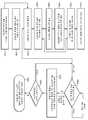
도 1a 및 도 1b는 체강 암 치료 장치를 실행시키기 위해 사용되는 프로토콜의 다양한 단계들과, 이 공정을 방광암 치료에 대해 구현하는 예를 각각 도시한 흐름도이다.
도 2 내지 도 5는 외부에서 발생된 자기장으로 환자를 조명하기 위해 사용되는 장치를 도시하고 있다.
도 6a 및 도 6b는 체강 암 치료 장치의 블록도를 도시하고 있다.
도 7은 인간의 방광과 그 주요 구성 요소를 도시하고 있는 단면도이다.
도 8은 컴퓨터 모델링 시스템에 의해 생성된, 헬름홀츠 코일 시스템의 전체적인 "이득"의 추이를 도시한 그래프이다.
도 9a 및 도 9b는 주파수의 함수로서 헬름홀츠 코일의 실험 저항의 추이를 도시한 그래프이다.
도 10은 입자 농도에 대한 필드 강도의 함수로서 나노 입자의 예상 자기 가열의 추이를 도시한 그래프이다.
도 11은 치료 프로토콜 중의 시간에 대한 섭씨 단위의 전형적인 방광 내부 온도의 측정값의 추이를 도시한 그래프이다.
도 12는 치료 프로토콜 중의 시간에 대한 암페어/미터 단위의 전형적인 필드 강도의 측정값의 추이를 도시한 그래프이다.
도 13은 치료 프로토콜 중의 시간에 대한 밀리리터 단위의 전형적인 방광 유체 체적의 측정값의 추이를 도시한 그래프이다.
도 14는 치료 프로토콜 중의 시간에 대한 밀리그램/밀리리터 단위의 전형적인 입자 농도의 측정값의 추이를 도시한 그래프이다.
도 15는 치료 프로토콜 중의 시간에 대한 밀리리터 단위의 전형적인 방광 유체 체적과 치료 프로토콜 중의 밀리그램/밀리리터 단위의 전형적인 입자 농도의 측정값의 추이를 중첩하여 도시한 그래프이다.
도 16은 방광 체적에 대한 방광 혈류의 추이를 도시한 그래프이다.
도 17a 및 도 17b는 인간 방광 내의 카테터와, 연관된 카테터 및 인체 해부학에 대한 설명을 도시하고 있다.
도 18은 방광으로 입자를 주입하기 위해 카테터를 사용하지만, 방광 자체로부터 입자 격리를 유지하는 공정을 도시한 흐름도이다.1A and 1B are flow diagrams illustrating various steps of the protocol used to implement a celiac cancer treatment device, and an example of implementing this process for bladder cancer treatment.
2 to 5 show a device used to illuminate a patient with an externally generated magnetic field.
6A and 6B show a block diagram of a body cavity cancer treatment device.
7 is a cross-sectional view showing the human bladder and its major components.
8 is a graph showing the transition of the overall "gain" of the Helmholtz coil system, produced by a computer modeling system.
9A and 9B are graphs showing the transition of experimental resistance of a Helmholtz coil as a function of frequency.
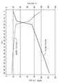
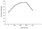
FIG. 10 is a graph showing the transition of expected magnetic heating of nanoparticles as a function of field strength versus particle concentration.
FIG. 11 is a graph showing the evolution of measurements of typical bladder internal temperature in degrees Celsius versus time during a treatment protocol.
FIG. 12 is a graph showing the evolution of measurements of typical field strength in amperes / meters versus time during the treatment protocol.
FIG. 13 is a graph showing the evolution of measurements of typical bladder fluid volume in milliliters versus time during the treatment protocol.
FIG. 14 is a graph depicting the evolution of measurements of typical particle concentrations in milligrams / milliliters versus time during the treatment protocol.
FIG. 15 is a graph superimposed on a trend of measurements of typical bladder fluid volume in milliliters versus time in a treatment protocol and typical particle concentrations in milligrams / milliliters in a treatment protocol.
16 is a graph showing the change of bladder blood flow to bladder volume.
17A and 17B show a description of a catheter in the human bladder, associated catheter and human anatomy.
18 is a flow chart illustrating a process of using a catheter to inject particles into the bladder but maintaining particle isolation from the bladder itself.
저온 온열 요법Cryothermal therapy
이온화 방사선 및/또는 화학 요법과 저온 온열 요법의 조합은, 전술한 바와 같이, 필요한 방사선 또는 화학 요법 약품의 수준을 낮추면서도, 암 치료 효과를 2 내지 4배 향상시킬 수 있는 가능성이 있다. 저온 온열 요법의 하나의 추가적인 장점은 종양성 영역에서 산소 수준이 크게 증가하는 재산소화이다. 재산소화는 암과, 특히, 저산소 환경을 가장 확실하게 선호하는 암 줄기 세포에게 상당한 스트레스가 된다. 암이 저온 온열 요법 상태로 유지될 때, 다른 중요한 생물학적 장점이 발생한다; 급성 산성화 및 열 충격 단백질(HSP) 방출 감소. 이온화 방사선과 저온 온열 요법이 세포 생식 과정에서 서로 다른 시기(M, S)에 각각 영향을 미치기 때문에, 다른 장점이 발생한다.The combination of ionizing radiation and / or chemotherapy with cryothermal therapy has the potential to improve the cancer treatment effect two to four times, as described above, while lowering the level of radiation or chemotherapy drug required. One additional advantage of cryothermal therapy is reoxidation, which results in a significant increase in oxygen levels in the neoplastic area. Reproductive digestion is a significant stress for cancer and, in particular, cancer stem cells that most certainly prefer a hypoxic environment. When cancer stays in cryothermal therapy, another important biological advantage arises; Acute acidification and reduced heat shock protein (HSP) release. Different benefits arise because ionizing radiation and cryothermal therapy affect different times (M, S) during cell reproduction.
37℃의 신체 주변 온도에서 42℃ 내지 43℃ 사이의 타깃 온도까지, 37℃ 이상의 모든 온도 상승은 화학 요법 약물의 효과를 증진시킨다. 이러한 화학 요법 제제의 효능 향상은, 방광암과 같은 특정 암에 대하여, 나노 입자 기반 온열 요법이 수반되지 않을 경우에 15% 내지 20%인 10년 완치율을, 나노 입자 기반 온열 요법을 수반할 경우에 50% 내지 60% 이상으로 치료 성과를 바꿀 수 있다. 이러한 방광암 완치 결과의 개선은 극적인 것이며, 이 기술이 다른 암과 심지어 다른 질병에 적용될 경우, 유사한 효능과 치료율이 확실할 것으로 예상된다.All temperature rises above 37 ° C. enhance the effectiveness of chemotherapy drugs, from the body's ambient temperature of 37 ° C. to target temperatures between 42 ° C. and 43 ° C. The enhanced efficacy of these chemotherapy agents, for certain cancers, such as bladder cancer, has a 10-year cure rate of 15% to 20% when no nanoparticle-based thermotherapy is involved, and 50 when the nanoparticle-based thermotherapy is accompanied. The treatment outcome can be altered by more than 60%. The improvement in bladder cancer cure results is dramatic, and if the technique is applied to other cancers and even other diseases, similar efficacy and treatment rates are expected to be evident.
PARP 억제제와 같은 약품은 주어진 암세포에서 손상된 DNA를 자가 회복시키는 암 세포의 능력을 억제한다. 따라서, 주어진 암세포에서 DNA가 의도적으로 손상되면, PARP 억제제가 암세포의 DNA "자체 수리"를 억제함으로써, 암세포가 죽게될 것이다. 그러나, PARP 억제제는, 주변 온도가 42℃ 내지 43℃ 범위로 상승하지 않으면, 효과가 별로 없다. 온열 요법은 세포의 DNA 복제를 억제하는데도 매우 효과적이다. 따라서, 암세포가 의도적으로 손상된 자신의 DNA를 자가 회복시키는 것을 멈추도록 하는데 있어서 PARP 억제제가 유효하도록 하기 위해서는, 암에 걸린 영역의 주변 온도를 37℃에서 42℃ 내지 43℃로 상승시킬 수 있어야만 한다. PARP 억제제와 저온 온열 요법 프로토콜은 모두, 개별적으로 그리고 협력하여, 손상된 암세포 DNA를 회복시키는 암세포의 능력에 영향을 미치고/억제한다. 지금은, 암에 걸린 영역의 동시 가열이 가장 유리한 프로토콜일 것이라고 믿어지지만, 방사선 또는 화학 요법의 타이밍에 대해 상대적으로 사전 또는 사후의 가열 프로토콜이 선호되는 이유가 있을 수 있다.Drugs such as PARP inhibitors inhibit the cancer cell's ability to self repair damaged DNA in a given cancer cell. Thus, if DNA is intentionally damaged in a given cancer cell, the PARP inhibitor will inhibit the cancer cell's DNA "self repair" and the cancer cell will die. However, PARP inhibitors have little effect unless the ambient temperature rises in the range from 42 ° C to 43 ° C. Thermotherapy is also very effective in inhibiting DNA replication in cells. Thus, in order for PARP inhibitors to be effective in stopping cancer cells from self-repairing their damaged DNA, the ambient temperature of the cancerous area must be able to rise from 37 ° C to 42 ° C to 43 ° C. Both PARP inhibitors and cryothermal therapy protocols, individually and in concert, influence / inhibit cancer cells' ability to repair damaged cancer cell DNA. Although it is now believed that simultaneous heating of cancerous areas is the most advantageous protocol, there may be a reason why a pre or post heating protocol is preferred relative to the timing of radiation or chemotherapy.
나노 입자는 선택된 효과를 발생시키는데 필요한 최소 에너지로 나노 입자의 조명을 제공하기 위해 정밀하게 조작된 에너지 필드를 발생시키는 체강 암 치료 장치에 의해 활성화된다. 에너지 필드 특징은 치료중인 타깃 생물체의 부위에서 선택된 방식으로 나노 입자에 에너지를 제공하기 위해 필요한 필드 타입, 주파수, 필드 강도, 지속 기간, 필드 변조, 반복 주파수, 빔의 크기 및 초점을 포함한 에너지 필드의 특징들로부터 선택된다. 또한, 에너지 필드의 특징들의 맵핑은 큰 유연성을 제공하며, 다양한 유형의 나노 입자들의 동시 사용을 가능하게 한다.The nanoparticles are activated by a celiac cancer treatment device that generates a precisely engineered energy field to provide illumination of the nanoparticles with the minimum energy needed to produce the selected effect. The energy field feature is characterized by the energy field including the field type, frequency, field intensity, duration, field modulation, repetition frequency, beam size, and focus required to provide energy to the nanoparticles in a chosen manner at the site of the target organism being treated. Selected from the features. In addition, the mapping of the features of the energy field provides great flexibility and allows the simultaneous use of various types of nanoparticles.
체강 암 치료 장치에 의한 나노 입자의 활성화가 매우 결정론적이라는 것을 이해하는 것이 중요하며, 이는 미리 정해진 특징을 가진 주어진 에너지 필드에 의해 주어진 입자가 최적하게 활성화되거나 여기된다는 것을 의미한다. 포괄적이거나 임의적인 필드 여기는 주어진 입자를 최적하게 여기시키지 않는다. 나노 입자의 필드 여기는 시스템의 "입력 에너지" 또는 "입력 구동 기능"으로 간주된다. 일반적으로, "입력 에너지"는 나노 입자에 의해 열 출력인 "출력 에너지"로 변환된다.It is important to understand that activation of nanoparticles by celiac cancer treatment devices is very deterministic, which means that a given particle is optimally activated or excited by a given energy field with predetermined characteristics. Comprehensive or arbitrary field excitation does not optimally excite a given particle. Field excitation of nanoparticles is considered the "input energy" or "input drive function" of the system. In general, "input energy" is converted by the nanoparticles into "output energy" which is a heat output.
체강 암 치료 장치의 작동Operation of Celiac Cancer Treatment Device
도 1a는 본 발명의 체강 암 치료 장치(40)의 전형적인 작동을 흐름도 형태로 도시하고 있고, 도 1b는 방광암 치료 프로토콜에서 구현되는 본 발명의 체강 암 치료 장치(40)의 전형적인 작동을 흐름도 형태로 도시하고 있다. 본원에 개시된 바와 같이, 본 발명의 체강 암 치료 장치(40)는 이 치료 프로토콜들에서 사용되는 자기장을 발생시키기 위해 사용된다.FIG. 1A illustrates a typical operation of the celiac
도 1a의 단계(101A)에서는, 의료인이 사용하기에 적합한 임의의 기술을 사용하여, 타깃 체강 속에 나노 입자의 용액을 삽입한다. 단계(102A)에서는, 도 2 내지 도 5, 도 6a 및 도 6b의 체강 암 치료 장치(40)에 의해 발생된 자기장과 같이 외부에서 발생된 에너지 필드를 인가하여 체강을 조명한다. 체강 암 치료 장치(40)로 에너지 필드를 유지하여, 단계(103A)에서는 체강을 미리 정해진 온도로 서서히 가열한다. 단계(104A)에서는, 체강에 하나 이상의 화학 요법 제제를 추가하되, 상기 화학 요법 제제는 단계(103B)에서 미리 정해진 원하는 온도로 선택적으로 예열될 수 있다. 단계(105A)에서는, 제어 컴퓨터(409), 파형 소오스(403, 601), 증폭기(404) 및 전류 감지 회로(614)를 통해 인가된 에너지 필드를 조절하는 에너지 제어기(62)를 이용하여, 체강 암 치료 장치(40)가 체강 및/또는 화학 요법 제제에 일정한 선택된 온도를 확립한다. 그 다음, 공정은 단계(106A 내지 108A)들로 진행하며, 이 단계들에서는, 체강 암 치료 장치(40)의 에너지 제어기(62)가 온도 센서(616 또는 617)와 제어 컴퓨터(409)를 통해 체강/화학 요법 제제의 온도가 미리 정해진 한계치 이내에 있는지의 여부를 결정하기 위해 시험하고, 한계치 이내에 없다면, 원하는 온도를 달성하기 위해 자기장의 강도를 조절한다. 원하는 온도를 유지하는 이 과정은, 단계(108A)에서 체강 암 치료 장치(40)의 에너지 제어기(62)가 미리 정해진 시간이 경과하였는지를 연산할 때까지, 미리 정해진 시간 동안 계속되며, 미리 정해진 시간이 경과한 시점에서, 공정은 단계(109A)로 진행하며, 이 단계에서는, 자기장을 제거하고, 체강과 화학 요법 제제가 냉각되도록 허용하며, 통상적으로 나노 입자 용액과 화학 요법 제제를 체강으로부터 제거한다.In
대안적으로, 단계(110A)에서는, 나노 입자 용액과 하나 이상의 화학 요법 제제의 혼합물을 체강에 추가하되, 혼합물은 미리 정해진 원하는 온도로 선택적으로 예열될 수 있다. 단계(111A)에서는, 단계(103B)에서 도 2 내지 도 5, 도 6a 및 도 6b의 장치에 의해 발생된 자기장과 같이 외부에서 발생된 에너지 필드를 인가하여 체강을 조명한다. 그 다음, 공정은 단계(105A)로 진행하며, 이 단계에서는, 체강 암 치료 시스템의 에너지 제어기(62)가 체강 및/또는 화학 요법 제제에 일정한 선택된 온도를 확립하고, 전술한 바와 같이 단계(106A 내지 109A)들이 실행된다.Alternatively, in
치료 프로토콜은 시간 및 온도 파라미터를 선택하는 의사에 의해 규정된다. 또한, 나노 입자 용액과 하나 이상의 화학 요법 제제가 조합되고, 예열된 다음, 체강 속에 삽입될 수 있다. 이는 오직 1회의 삽입 단계를 실시함으로써, 치료 시간을 줄이고 공정을 단순화한다.The treatment protocol is defined by the physician who selects time and temperature parameters. In addition, the nanoparticle solution and one or more chemotherapy agents can be combined, preheated, and then inserted into the body cavity. This reduces the treatment time and simplifies the process by performing only one insertion step.
방광암의 치료Bladder Cancer Treatment
방금 설명한 공정은 전술한 바와 같이 다양한 체강에 대해 실시될 수 있으며, 도 1b는 이 절차가 특정 체강과 암 유형에 대해 어떻게 맞춰질 수 있는지를 나타내기 위해 도 1a의 흐름도에 추가적인 세부 사항을 제공한다. 특히, 도 7은 인간의 방광과 그 주요 구성 요소의 단면도를 도시하고 있다. 배뇨근은 나선형, 종 방향 및 원형 번들로 배치된 평활근 섬유로 만들어진 방광벽의 한 층이다. 방광은 측면 제대 인대와 중앙 제대 인대에 의해 복부의 제 위치에 유지된다. 방광은 요관을 통해 요관구를 거쳐 뇨를 수용하고, 요도를 통해 뇨를 방출한다. 도 7에 도시된 방광암의 한 형태를, 방광 내부의 표면에 위치하고 통상적으로 점막을 침투한 깊이가 500μ 미만인 "비근육 침윤성 방광암"이라 한다. 따라서, 방광 속에 화학 요법 제제를 삽입하면, 화학 요법 제제가 암과 반드시 접촉하게 된다.The process just described can be performed for various body cavities as described above, and FIG. 1B provides additional details in the flow chart of FIG. 1A to show how this procedure can be tailored for a particular body cavity and cancer type. In particular, FIG. 7 shows a cross-sectional view of the human bladder and its major components. The detrusor muscle is a layer of the bladder wall made of smooth muscle fibers arranged in spiral, longitudinal and circular bundles. The bladder is held in place in the abdomen by the lateral umbilical ligament and the central umbilical ligament. The bladder receives urine through the ureters, through the ureters, and releases urine through the urethra. One form of bladder cancer shown in FIG. 7 is called " non-muscular invasive bladder cancer " that is located on the surface of the bladder interior and typically has a depth of less than 500 microns penetrating the mucous membrane. Thus, when a chemotherapy agent is inserted into the bladder, the chemotherapy agent is in contact with the cancer.
방광이 팽창하면, 배뇨근을 수축시키도록 부교감 신경계에 신호를 보낸다. 이는 방광을 자극하여 전립선을 통과하는 요도를 통해 뇨를 방출하도록 한다. 뇨가 방광을 빠져나가기 위해서는, 자율적으로 통제되는 내요도 괄약근과 자발적으로 통제되는 외요도 괄약근이 모두 개방되어야만 한다. 이 근육들에 문제가 생기면 요실금을 초래할 수 있다. 뇨량이 방광 용량의 100%에 도달하면, 자발적 괄약근이 비자발적으로 되어, 뇨가 즉시 배출된다. 방광은 일반적으로 300 내지 350㎖의 뇨를 보관한다. 뇨가 축적되면, 방광벽이 팽창하면서 얇아짐으로써, 방광은 내부 압력이 현저히 증가하지 않고 다량의 뇨를 저장할 수 있다.When the bladder swells, it signals the parasympathetic nervous system to constrict the detrusor muscle. This stimulates the bladder to release urine through the urethra through the prostate. In order for urine to leave the bladder, both the autonomous and internally controlled sphincter, which is controlled autonomously, must be opened. Problems with these muscles can lead to incontinence. When urine volume reaches 100% of bladder capacity, spontaneous sphincter becomes involuntary and urine is excreted immediately. Bladder generally stores between 300 and 350 ml of urine. As urine accumulates, the bladder wall expands and becomes thinner, thereby allowing the bladder to store large amounts of urine without significantly increasing internal pressure.
방광이 작동 체적(working volume)의 약 25%에 도달하면, 배뇨 욕구가 시작된다. 이 단계에서는, 피시험자가, 원한다면, 배뇨 욕구를 쉽게 참을 수 있다. 방광이 계속 채워지면, 배뇨 욕구가 강해지고 무시하기가 어려워진다. 결국, 배뇨 욕구가 불가항력적이게 되는 지점까지 방광이 채워질 것이고, 피시험자는 이를 더 이상 무시할 수 없을 것이다.When the bladder reaches about 25% of the working volume, the desire to urinate begins. At this stage, the subject can easily tolerate the desire to urinate, if desired. If the bladder continues to fill, the desire to urinate becomes stronger and harder to ignore. Eventually, the bladder will be filled to the point where the desire to urinate becomes irresistible, and the subject can no longer ignore it.
방금 설명한 공정은 전술한 바와 같이 다양한 체강에 대해 실시될 수 있으며, 도 1b는 이 절차가 특정 체강과 암에 대해 어떻게 맞춰질 수 있는지를 나타내기 위해 도 1a의 흐름도에 추가적인 세부 사항을 제공한다. 이러한 맞춤화는 임의의 특정 체강과 암 유형에 대해 확실하게 효과적일 수 있다.The process just described can be performed for various body cavities as described above, and FIG. 1B provides additional details in the flow chart of FIG. 1A to show how this procedure can be tailored for a particular body cavity and cancer. Such customization can certainly be effective for any particular body cavity and cancer type.
도 1b의 단계(101B)에서는, 요도를 통해 카테터를 통과시킴으로써 방광 속에 나노 입자의 용액을 삽입하되, 치료 기간 중에 일반적으로 생산되는 뇨와 화학 요법 제제를 위한 공간을 남기고 방광을 채우지 않도록 유체의 양을 선택한다. 단계(102B)에서는, 도 2 내지 도 5, 도 6a 및 도 6b의 체강 암 치료 장치(40)에 의해 발생된 자기장과 같이 외부에서 발생된 에너지 필드를 인가하여 방광을 조명한다. 화학 요법 제제를 추가하기 전에, 단계(103B)에서는, 나노 입자의 조명을 통해, 에너지 필드를 유지하여, 통상적으로 42℃ 내지 43℃인 미리 정해진 온도로 방광을 서서히 가열한다. 단계(104B)에서는, 방광에 미토마이신-C와 같은 하나 이상의 화학 요법 제제를 추가하되, 상기 화학 요법 제제는 통상적으로 42℃인 미리 정해진 원하는 온도로 선택적으로 예열될 수 있다. 단계(105B)에서는, 체강 암 치료 장치(40)의 에너지 제어기(62)가 미리 정해진 시간 동안 방광과 주변 조직에 배치된 유체에 통상적으로 42℃ 내지 43℃인 일정한 선택된 온도를 확립한다. 증폭기(404)에 인가되는 피드백 제어 신호(602)를 통해 인가되는 필드 강도를 관리 및 조정하기 위하여, 컴퓨터 제어 알고리즘(409)과 함께 광섬유 열 센서(617)를 사용할 수 있다. 그 다음, 공정은 단계(106B 내지 108B)들로 진행하며, 이 단계들에서는, 체강 암 치료 장치(40)의 에너지 제어기(62)가 방광/화학 요법 제제의 온도가 미리 정해진 한계치 이내에 있는지의 여부를 결정하기 위해 시험하고, 한계치 이내에 없다면, 원하는 온도를 달성하기 위해 자기장의 강도를 조절한다. 원하는 온도를 유지하는 이 과정은, 단계(108B)에서 체강 암 치료 장치(40)의 에너지 제어기(62)가 미리 정해진 시간이 경과하였는지를 연산할 때까지, 통상적으로 60분인 미리 정해진 시간 동안 계속되며, 미리 정해진 시간이 경과한 시점에서, 공정은 단계(109B)로 진행하며, 이 단계에서는, 자기장을 제거하고, 방광과 화학 요법 제제가 냉각되도록 허용하며, 통상적으로 나노 입자 용액과 화학 요법 제제를 배뇨 또는 플러싱으로 방광으로부터 제거한다.In
어떤 상황에서는, 나노 입자가 방광 내벽(점막)을 포함하여 인체 조직에 닿거나 접촉하지 않도록 하는 것이 바람직할 수 있다. 이와 동시에, 유해한 암과 암세포를 죽이도록 의도된 화학 요법 제제 또는 방사선 중 어느 하나 또는 모두의 효과를 향상시키기 위해, 방광의 내부(또는 인체 조직)를 가열하는 것이 여전히 바람직할 수 있다. 방광암의 경우, 미토마이신-C(MMC)와 같은 화학 요법 제제의 효능을 향상시키는 것이 바람직하다. 상술한 절차의 대안은 방광으로 화학 요법 제제를 주입하는 것과, 방광으로 "풍선"을 삽입하는 것이다. 풍선 기반 카테터 조립체를 사용함으로써, 나노 입자를 가열하면서도, 인체 조직, 즉 방광 내벽으로부터 나노 입자의 물리적 격리를 유지할 수 있다. 풍선이 방광의 정확한 형상에 밀착됨으로써, 용액에 섞인 나노 입자가 풍선 속으로 투입되고, 풍선이 팽창하여 화학 요법 제제를 방광벽과 풍선 사이의 공간으로 가압하게 된다. 조명 필드의 인가를 통해, 풍선 속의 나노 입자 용액이 가열된다. 대안적으로, 방광 내에서 화학 요법 제제의 온도를 유지하기 위해, 나노 입자를 사용하지 않고, 유체 용액이 카테터를 통해 풍선 내외로 순환될 수 있다. 발생된 열은 방광벽과 화학 요법 제제 모두로 전달된다. 상술한 바와 같은 절차의 나머지 부분의 끝에서, 나노 입자가 풍선으로부터 제거된 후, 화학 요법 제제는 그대로 있는 상태에서, 방광 내부로부터 풍선이 제거된다.In some situations, it may be desirable to ensure that the nanoparticles do not touch or come into contact with human tissue, including the bladder inner wall (mucosa). At the same time, it may still be desirable to heat the interior (or human tissue) of the bladder to enhance the effectiveness of either or both of chemotherapy agents or radiation intended to kill harmful cancers and cancer cells. In the case of bladder cancer, it is desirable to enhance the efficacy of chemotherapy agents such as mitomycin-C (MMC). Alternatives to the above-described procedures are injecting a chemotherapy agent into the bladder and inserting a "balloon" into the bladder. By using a balloon-based catheter assembly, the nanoparticles can be heated while maintaining physical isolation of the nanoparticles from human tissue, ie, the bladder inner wall. As the balloon adheres to the bladder's exact shape, the nanoparticles mixed into the solution are introduced into the balloon, causing the balloon to expand and pressurize the chemotherapy agent into the space between the bladder wall and the balloon. Through the application of the illumination field, the nanoparticle solution in the balloon is heated. Alternatively, the fluid solution may be circulated through the catheter into and out of the balloon, without using nanoparticles, to maintain the temperature of the chemotherapy agent in the bladder. The heat generated is transferred to both the bladder wall and the chemotherapy agent. At the end of the remainder of the procedure as described above, after the nanoparticles have been removed from the balloon, the balloon is removed from the interior of the bladder with the chemotherapy agent intact.
풍선 카테터 공정의 세부 사항Details of the balloon catheter process
도 17은 방광(1401) 속에 카테터(1430)가 이미 삽입되어 있는 인간의 방광(1401)을 도시하고 있다. 카테터 조립체(1430, 1432, 1434, 1436)는 방광(1401) 속에 삽입되며, 방광은 신장에 연결되어 있고(뇨가 요관(1402A, 1402B)을 통해 방광(1402)으로 유입되며), 요도(1406)는 (배뇨를 통해) 방광(1401)을 비우기 위한 수단일 뿐만 아니라 카테터 조립체를 삽입하기 위한 통로이다. 카테터(1430)는 그 길이를 따라 "루멘"이라 하는 홀 또는 튜브를 갖는다. 이 루멘들은 카테터(1430)의 관형 부분의 단면에 배치되어 있다. 이 특수한 카테터는 4세트의 루멘을 갖고 있으며; 루멘의 개수는 일반적으로 루멘을 수용하고 있는 "튜브"의 크기에 의해서만 제한된다. "튜브"의 크기는 "프렌치"라는 단위로 측정되며, 인간의 방광에 사용되는 카테터는 통상적으로 18 내지 24 프렌치이다. 프렌치 수가 크다는 것은 "튜브"의 직경이 크다는 것을 의미한다.17 shows a
카테터는 압출된 실리콘이나 라텍스 물질로 흔히 구성된다(샤프트(1430, 1436)에는 루멘(1420, 1422, 1424, 1426)이 장착되어 있다). 풍선(1432, 1434)은 흔히 "취입" 방식으로 제조된다. 압출된 샤프트(1430)와 풍선(1432, 1434)이 합쳐져 전체 카테터 조립체를 만들도록 구성된다. 자기장의 강도를 관리하는 에너지 제어기(62)에 대한 온도 제어 피드백 메커니즘이 가능하도록 열전대(1440, 1442, 1444)들이 추가되며, 상기 제어기는 나노 입자가 따뜻해지는 방법을 제어한다. 예컨대, 입력 루멘(1426)이 출력(1436)에 연결되며, 상기 출력은 큰 풍선(1434) 위에 있는 카테터(1430)의 팁에 배치되어 있다. 이 특수한 루멘 조립체는 방광(1401) 내외로 유체를 유입 또는 유출시키기 위해 사용된다. 절차 시작시, 루멘 쌍(1426)이 과다한 뇨를 제거하기 위해 사용될 수도 있으며, 절차 시작 전에, 방광(1401)으로 화학 요법 제제인 미토마이신-C를 삽입하기 위해 사용될 수 있다.Catheter is often composed of extruded silicone or latex material (
열과 화학 요법 제제 모두의 조합은 (비뇨기과 종양 전문의에 의해 현재 실시되고 있는 바와 같이) 화학 요법 제제만을 사용하는 것보다 효능이 상당히 더 높은 치료 프로토콜의 기반이 된다. 1시간의 공칭 치료 기간 동안 방광 조직과 암에 열을 추가하면, 방광암을 치료하는 MMC의 효능이 매우 극적으로 되며, 10년 완치율이 15%에서 53% 이상으로 증가한다.The combination of both heat and chemotherapy agents is the basis for a treatment protocol that is significantly more potent than using only chemotherapy agents (as currently being practiced by urologists oncologists). Adding heat to bladder tissue and cancer during a one-hour nominal treatment dramatically increases the effectiveness of MMCs in treating bladder cancer and increases the 10-year cure rate from 15% to over 53%.
풍선(1434)으로 나노 입자를 주입하기 위해 "1434(큰 풍선(1434)으로의 출력)"에 대한 "1424(입력)"의 루멘 접합이 사용된다. 나노 입자를 수용한 풍선을 사용하는 한 가지 장점은 요관(1402B, 1402A)이 나노 입자의 농도를 신장으로부터 나온 뇨로 희석하지 않는다는 것이다. 이에 따라, 나노 입자의 농도가 일정하여, 가열 제어 알고리즘이 단순화된다.A lumen junction of "1424 (input)" to "1434 (output to large balloon 1434)" is used to inject nanoparticles into
MMC(화학 요법 제제)는 루멘(1426)과 출력(1436)을 통해 방광(1401)으로 직접 주입된다. 루멘(1422)은 통상적으로는 공기로 작은 풍선(1432)을 팽창시키기 위해 사용되며, 이 작은 풍선(1432)의 목적은 치료 기간 중에 방광(1401)에 안착된 상태로 카테터(1430)를 유지하는 것이다. 큰 풍선(1434)은 열원이기 때문에, 큰 풍선(1434)을 방광벽으로부터 이격시켜 유지하는 것이 바람직하고, 화상이나 과다한 가열을 방지하기 위해 따뜻한 풍선 표면이 방광벽에 닿지 않도록 하는 것이 바람직하다.MMC (chemotherapy agent) is injected directly into the
이 예에서는, 3개의 서로 다른 위치의 온도를 감지하는 3개의 열전대(1440, 1442, 1444)를 에너지 제어기(62)에 연결하기 위해, 루멘(1420)이 사용된다. 열전대(1440)는 방광(1401) 내의 유체(약간의 뇨와 MMC)의 온도를 감지하는 반면, 열전대(1442)는 (루멘(1420)을 통해 공급된) 나노 입자를 수용하고 있는 풍선(1434)의 중심 온도를 감지한다. 풍선(1434)의 중심 온도를 확인하는 것이 중요한데, 그 이유는 풍선의 중심 온도를 확인하면 에너지 제어기(62)가 최고 온도가 무엇이고 풍선(1434)을 가로지르는 열 구배가 어떠한지를 알 수 있기 때문이다. 열전대( 1444)는 풍선(1434)의 외부 에지에 배치되며, 방광벽에 닿을 경우에도 외부 풍선 온도가 방광(1401)에 대해 안전한지를 보장하기 위해 사용된다. 수학적으로, 열전대(1442, 1444)들 간의 온도차는 구배로서 결정될 수 있으며, 이 계산값은 열전대들이 자신들의 데이터를 잘못 보고하지 않는다는 것을 보장하기 위한 에러 체크로서 측정된 온도와 비교될 수 있다. 통상적으로, 열전대(1444)는 방광벽에서의 온도보다 2℃ 내지 4℃ 더 높게 측정한다. 사용된 열전대는 주어진 온도에 대해 주어진 주파수로 진동하는 갈륨 비소화물(GaAs) 결정을 사용하는, 통상적으로 광섬유 기반 열전대이다. 이 진동 주파수는 감지된 다음, 온도 측정값으로 변환되어, 온도 대 자기장 강도 피드백 루프 내의 시스템 전자 장치에 보고된다. 광섬유 센서는, 브라운 운동으로 입자를 여기시킴으로써 마찰 기반 가열을 유발하기 위해 사용되는 자기장의 존재에 의해 영향을 받지 않기 때문에, 중요하다.In this example,
도 18은 나노 입자를 가열하기 위한 하나의 공정을 흐름도 형태로 도시하고 있다. 단계(1801)에서는, 카테터(1430)를 방광(1401) 속으로 삽입하고, 단계(1802)에서는, 카테터가 방광(1401) 내의 정확한 위치에 배치되었는지를 결정한다. 단계(1803)에서는, 작은 풍선(1432)을 (통상적으로 공기로) 팽창시키며; 이 작은 풍선(1432)은 카테터(1430)를 적절하게 장착된 상태로 유지할 뿐만 아니라, 가열된 나노 입자를 수용하고 있는 큰 풍선(1434)이 방광 내벽에 닿지 않도록 돕는다.18 shows, in flowchart form, one process for heating nanoparticles. In
이 치료는 주로, 폴립형이며 방광(1401)의 방광벽 속으로 성장하고 일반적으로 줄기형 구조를 가진 비근육 침윤성 방광암(NMIBC)을 위한 것이다. 현재의 열 투사는 Ta 및 T1 NMIBC 타입의 방광암을 치료하기 위해 방광벽 속으로 0.5㎜만 침투하는 열 치료가 필요하다는 것을 나타낸다. 단계(1404)에서는, 루멘(1424)을 통해 나노 입자를 큰 풍선(1434) 속에 삽입한다. 단계(1805)에서는, 미토마이신-C(MMC)와 같은 화학 요법 제제를 루멘(1426)을 통해 개구(1436)까지 방광(1401) 속으로 직접 삽입한다. 그 다음, 본원에서 전술한 바와 같이 자기장을 인가한다. 단계(1806)에서는, 나노 입자를 소정의 방식으로 풍선(1434) 내에서 가열한다. 단계(1807)에서는, 열전대(1440, 1442, 1444)를 통해 에너지 제어기(62)로 전체 시스템 온도를 모니터링한다. 전술한 바와 같이 카테터(1430)를 통해 삽입하기 전에, MMC와 나노 입자 중 어느 하나 또는 모두를 37℃의 공칭 체온까지 예열할 수 있다. 이와 같이 두 물질을 예열하면, 이들이 삽입될 때 체온이 되기 때문에, 전체 절차의 기간을 단축시킬 수 있다.This treatment is primarily for non-muscular invasive bladder cancer (NMIBC) that is polyp-shaped and grows into the bladder wall of
단계(1808)에서는, 열전대들이 적당한 작동 온도가 되도록 보장하기 위해 그들 중 하나 이상을 에너지 제어기(62)가 확인하여, 너무 따뜻하면, 코일(601, 602)로 전류를 공급하는 증폭기(404)에 대한 피드백이 저하되고, 이에 따라 발생되는 자기장이 더 낮아짐으로써 가열 속도가 감소된다. 원하는 작동 온도가 되면, 단계(1809)에서는, 저온 비박피성 요법의 경우 통상적으로 42℃ 내지 43℃인 소정의 온도로 나노 입자를 유지하도록 자기장을 관리한다.In
단계(1810)에서는, 가열된 나노 입자와 화학 요법 제제를 사용하여, 의사가 지정한 기간 동안 치료 프로토콜을 실시하지만, 통상적으로는 치료 온도에서 1시간 동안 실시한다. 온도가 1℃ 증가하면, 치료 기간을 반으로 줄일 수 있고, 온도가 2℃ 증가하면, 가열 기간을 ¼ 감소하는 것으로 여겨진다. 그러나, 시간과 온도의 조합이 결정되어야 하고, 궁극적으로 치료하는 의사의 책임이다.In
단계(1811)에서는, 치료를 완료하고, 이제 "반전" 공정을 실시한다. 단계(1812)에서는, 루멘(1424)을 통해 풍선(1434) 밖으로 나노 입자를 배출한다. 그리고, 단계(1813)에서는, 루멘(1426)을 통해 화학 요법 제제를 배출할 수 있거나, 카테터(1430)를 뽑고 환자가 화학 요법 제제를 배뇨할 수 있다. 이것이 오늘날 화학 요법 제제를 제거하는 방법이다. 요도와 그 내벽에 대해 화학 요법 제제가 얼마나 부식성이 있는지에 따라, 카테터(1430)를 통해 화학 요법 제제를 제거하는 장점이 있다. 단계(1814)에서는, 카테터(1430)를 제거하고, 단계(1815)에서는, 절차를 완료한다.In
암 세포와 온열 요법Cancer Cells and Thermotherapy
분열하고 있는 세포에 있어서, 4개의 시기, 즉, M기, G1기, S기 및 G2기가 존재하며, 방사선과 온열 요법은 각각 서로 다른 시기에 영향을 미친다. 온열 요법은 DNA 복제기인 S기의 후반에 가장 민감하다. 온열 요법이 영향을 미치는 다음 세포기는 세포 분열기인 M기이다. 그러나, 방사선 감도는 M기(세포 분열기)에서는 높지만, S기(DNA 복제기)에서는 낮다. 따라서, 온열 요법은, 특히, DNA 복제기인 S기에서, 방사선과 상보적이다. 이것이 저온 온열 요법(LTH)이 방사선과 조합될 때 매우 효과적인 이유이다. 전술한 바와 같이, 온열 요법이 작용하는 경우와 마찬가지로, PARP 억제제는 DNA 회복 단계에 영향을 미치며, 온열 요법은 신체 주변 이상의 온도마다 효과 배가 속도로 화학 요법의 효과를 향상시킨다.In dividing cells, there are four phases, namely phases M, G1 , S and G2 , and radiation and thermotherapy affect different periods of time. Thermotherapy is most sensitive to the second half of phase S, the DNA replicator. The next cell group affected by thermotherapy is stage M, the cell division. However, radiation sensitivity is high in phase M (cell divider) but low in phase S (DNA replicator). Thus, thermotherapy is complementary to radiation, especially in S phase, which is a DNA replicator. This is why cryothermal therapy (LTH) is so effective when combined with radiation. As mentioned above, as with thermotherapy, PARP inhibitors affect the stage of DNA repair, and thermotherapy enhances the effect of chemotherapy at an effect doubling rate at temperatures above and around the body.
자기장에서 뜨거워지는 나노 입자는 자성을 나타내어야 하며, 성질이 일반적으로 강자성이다. 자철광(Fe3O4) 및 마그헤마이트(Fe2O3)와 같은 물질은, 나노미터 크기로 제조될 경우, 시변성의 자기장에서 뜨거워질 것이다. 이러한 AC 또는 교류 자기장은 통상적으로 ㎑ 주파수 범위에 있지만, ㎒ 범위에 있을 수도 있다. 선호되는 브라운 가열 모드의 경우, 최적의 주파수 범위는 30,000 내지 100,000 ㎐(30 내지 100 ㎑)이다. 입자 크기는 주로 단자구로 특징지어질 정도로 충분히 직경이 작다.Nanoparticles that heat up in a magnetic field must exhibit magnetic properties and are generally ferromagnetic. Materials such as magnetite (Fe3 O4 ) and maghemite (Fe2 O3 ), when made in nanometer size, will heat up in a time-varying magnetic field. Such an AC or alternating magnetic field is typically in the Hz frequency range, but may also be in the MHz range. For the preferred brown heating mode, the optimum frequency range is 30,000 to 100,000 Hz (30 to 100 Hz). The particle size is small enough to be mainly characterized by a terminal opening.
자기 여자는 교류(AC)를 통해 구동되며, 교류에서는 양에서 음으로 다시 양으로 (등등) 가는 파형의 위상 변화가 나노 입자에서 자기 정렬의 변화를 초래하고, 이는 가열을 유발한다. 자기 정렬의 변화는 유도된 에너지의 일부가 (나노 입자에 의해) 열로 변환되도록 한다. 자기 가열의 두 가지 형태는, 첫째, 세포질에 대한 나노 입자의 운동에 의해 생성되는 마찰 기반 가열(예컨대, 브라운 모드)과, 둘째, 나노 입자는 정지하고 있고 나노 입자 내의 자구(magnetic domain)가 변하는 자구 기반 가열(닐 모드)을 포함한다. 여기 주파수에 대한 입자 크기에 따라, 가열은 브라운 모드와 닐 모드를 모두 포함할 수 있다.The magnetic excitation is driven by alternating current (AC), in which alternating phase changes in the waveform from positive to negative (equivalent) lead to a change in the magnetic alignment in the nanoparticles, which causes heating. The change in magnetic alignment causes some of the induced energy to be converted (by nanoparticles) into heat. Two forms of magnetic heating are: first, friction-based heating (e.g., brown mode) produced by the movement of the nanoparticles to the cytoplasm, and second, the nanoparticles are stationary and the magnetic domains within the nanoparticles change. Magnetic domain based heating (Neil mode). Depending on the particle size relative to the excitation frequency, the heating can include both brown and neil modes.
제 1 마찰 기반 가열을 브라운 가열이라 하며, 나노 입자가 물리적으로 회전하여, 기계적 마찰 기반 가열을 유발한다. 나노 입자가 물리적으로 회전하고 있기 때문에, 최대 나노 입자 가열에 적합한 완화 시간이 존재하고, 상기 완화 시간은 나노 입자의 크기 및 여기 주파수 모두와 관련된다. 이와 같이 독특한 나노 입자 크기와 주파수의 조합은 최적의 가열을 유발한다. 이 경우에, 코어에 코팅을 더하여 이루어지는 나노 입자 크기를 유체역학적 직경이라 하며, 이는 브라운 가열에 중요한 복합 크기이다. 또한, 자화와 이방성과 같은 물성은 물질이 어디서 어느 정도로 잘 뜨거워지는지에 대해 영향을 미친다.The first friction based heating is called Brown heating, and the nanoparticles are physically rotated, causing mechanical friction based heating. Since the nanoparticles are physically rotating, there is a relaxation time suitable for maximum nanoparticle heating, which relaxation time is related to both the size and excitation frequency of the nanoparticles. This unique combination of nanoparticle size and frequency results in optimal heating. In this case, the nanoparticle size obtained by adding a coating to the core is called a hydrodynamic diameter, which is an important composite size for Brown heating. In addition, properties such as magnetization and anisotropy affect where and how well the material heats up.
자구만 변하는 제 2 방법을 닐 가열이라 한다. 이 경우에, 매우 좁은 크기와 해당 주파수의 조합이 가열을 가능하게 하고, 그 파라미터들의 어떤 약간의 변화로 인해 나노 입자가 전혀 뜨거워지지 않게 될 수 있다. 이러한 민감도 때문에, 닐 가열은 덜 선호되는 방식이 된다.The second method of changing only the magnetic domain is called neil heating. In this case, a combination of very narrow sizes and corresponding frequencies allows for heating, and some slight change in the parameters may cause the nanoparticles not to get hot at all. Because of this sensitivity, Neil heating is a less preferred way.
자성 나노 입자 가열의 다른 모드는 히스테리시스와 레일리를 포함하며, 이 모드들은 일반적으로 상당히 큰 입자, 즉, 크기가 50㎚를 초과하는 입자를 위한 것이다. 일반적으로, 나노 입자가 일반적으로 50㎚ 미만으로 작고 브라운 및/또는 닐 가열을 가진 단자구 모드가 선호된다. 현재 선호되는 자기장 발생 메커니즘은 종양성 영역으로 자기장을 투사하는 코일 세트이며, 상기 자기장은 암이 있는 영역에 균일한 체적을 생성한다. 열점을 최소화하기 위해서는 종양을 가로지르는 비교적 균일한 필드가 중요하다(본 발명자들은 나노 입자의 활용이 종양성 영역을 가로질러 비교적 균일하다고 가정한다).Other modes of magnetic nanoparticle heating include hysteresis and Rayleigh, which are typically for very large particles, that is, particles larger than 50 nm in size. In general, a terminal sphere mode where nanoparticles are generally smaller than 50 nm and with Brown and / or Neil heating is preferred. The presently preferred magnetic field generating mechanism is a set of coils that project a magnetic field into a tumorous region, which produces a uniform volume in the cancerous region. To minimize hot spots, a relatively uniform field across the tumor is important (we assume that the utilization of nanoparticles is relatively uniform across the tumorous area).
치료 테이블/기계Therapy table / machine
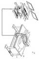
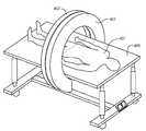
도 2 내지 도 4, 도 6a 및 도 6b는 외부에서 발생된 자기장으로 환자를 조명하기 위해 사용되는 체강 암 치료 장치(40)를 도시하고 있다. 환자(407)의 위아래에 배치될 수 있는 2개의 코일(401, 402)은 환자(407)의 신체를 무해하게 통과하는 자기장을 2개의 코일(401, 402)들 사이에 생성한다. 이 자기장은 환자(407)의 방광 공동으로 삽입된 20㎚의 공칭 크기를 가진 자철광(Fe3O4) 나노 입자를 여기시킴으로써, 이 나노 입자들이 주로 브라운 여기를 통해 뜨거워지도록 한다. 브라운 가열은 여기 주파수의 속도로, 이 경우에서는 40㎑로, 입자들이 물리적으로 회전한 결과이다. 나노 입자의 가열 수준은 소정 레벨의 주어진 자기장 강도를 생성하는 코일(401, 402)에서의 전류 레벨에 기초한다.2-4, 6A and 6B illustrate a celiac
도 3 및 도 4는 코어레스 코일 링이 추가적인 치료 프로토콜을 위해 이온화 엑스레이 방사선(408)을 어떻게 통과시킬 수 있는지를 도시하고 있다. 역시, 체강 암 치료 장치(40)의 바람직한 실시예는 자기장 민감 나노 입자를 조명하기 위한 자기장의 발생이다. 그러나, 전기장 입자 또는 물질과 함께 전기장이 사용될 수도 있다. 예컨대, MMC는 이극성이며, 전기장에서 뜨거워질 수 있다. 그렇다면, 이는 나노 입자가 필요하지 않을 것이다.3 and 4 illustrate how the coreless coil ring can pass
도 2 내지 도 4는 테이블(405) 위에 누워 있는 환자(생물체)(407)와, 조명되는 환자의 신체 영역 위에 코일 쌍(401, 402)을 최적으로 정렬시키기 위해 신체(407) 위에서 활주하는 체강 암 치료 장치(40)의 코일 조립체(401, 402)를 도시하고 있다. 도 2의 사시도에서 바로 볼 수는 없지만, 체강 암 치료 장치의 하부 코일(402)은 환자(401) 위에서 지나가는 상부 코일(401)과 협력하여 활주하는 테이블 아래에 배치되어 있다. 도 6b는 체강 암 치료 장치의 단면도를 도시하고 있으며, 이 단면도는 2개의 코일과 아울러, 테이블 위에 있는 환자에서 자기장이 집중되는 타깃 영역과 아울러, 자기장이 감소된 주변 영역과 주변 버퍼 영역을 도시하고 있다. 체강 암 치료 장치(40)의 이러한 개념화는 코일 직경이 60㎝ 또는 23.6인치인 환형 코일을 사용한다. 실제로, 코일(401, 402)은 임의의 크기 또는 사각형과 같은 평평한 형상을 가질 수 있다. 균일한 가열 영역의 크기를 증대시키기 위해, 도 4 및 도 5에 도시된 바와 같은 직교 평면에 다른 코일(401, 402)들이 (단독 코일로서) 추가될 수 있다. 이 개념에서, "제 1 권선, 상부 코일"에서 "제 1 권선, 하부 코일"까지의 간격은 30㎝이다(이 간격은 더 큰 사람을 수용하기 위해 증대될 수 있다). 코일(401, 402)의 간격이 증대된다는 것은, 동일한 크기의 에너지 필드를 생성하기 위해 (간격을 증대시킴으로써 줄어들거나 "늘어난" 필드를 보상하기 위해) 기존의 코일 직경에 대해 구동 전류가 더 많아지거나, 코일의 직경이 더 커진다는 것을 의미한다. 대안적으로, 목표 온도에 도달하기 위해 약간 더 많은 시간이 필요하다는 경고와 함께, 더 낮은 필드 강도가 사용될 수 있다.2-4 show a
도 2는, 도 6a 및 도 6b에도 개략적인 형태로 도시되어 있는, 신호 소오스(403), 신호 증폭기(404), 소프트웨어를 구비한 제어 컴퓨터(409), GUI 터치 스크린을 구비한 사용자 입력 키보드(410) 및 광섬유 온도 측정 시스템(617)을 수납하는 전자 장비 랙을 가진 체강 암 치료 장치(40)의 구현예를 또한 도시하고 있다. 전압과 전류 사이의 위상을 감지하고 공진 구동 주파수로 다시 돌아가기 위해 새로운 여기 주파수를 선택함으로써, 에너지 제어기(62)를 다시 공진시키기 위해 AFC 회로(619)가 또한 제공된다. 제어 컴퓨터(409)는 전술한 바와 같이 발생된 에너지 필드의 특성을 선택하기 위해, 공동으로 삽입된 나노 입자들의 특성을 일치시키기 위해, 그리고, 치료 프로토콜, 즉, 온도, 지속 기간 및 가열 프로파일을 규정하기 위해, 의사에 의해 사용된다. 대안적으로, 침윤성 온도 센서(616)는 환자(407)의 신체 상의 센서들을 가질 것이다. 이와는 별도로, 나노 입자와 미토마이신-C(MMC)와 같은 화학 요법 제제를 모두 주입하기 위해 폴리 카테터를 사용하는 방광 가열 조립체의 경우, 방광 내에 있는 유체의 치료 온도를 측정하기 위해 광섬유 온도 프로브가 방광 공동 속으로 삽입될 수 있다. 마지막으로, 공동 내에서 에너지 필드를 측정하기 위해 자기장 프로브(618)가 사용될 수 있다.FIG. 2 shows a
준균일한 필드의 자기 체적 영역은 35㎝의 신체 폭×35㎝의 신체 길이로 이루어진 면적×대략 30㎝의 신체 두께 치수이며, 이는 13.8인치의 신체 폭×13.8인치의 신체 길이×11.8인치의 신체 두께이다. 전반적으로, 이는 "균일한 필드" 체적의 36,750㎤이거나, "균일한 필드" 체적의 2,247 세제곱 인치이다. 이러한 균일한 필드 체적은 전이되지 않고 지역적으로 위치하고 있는 거의 모든 유형의 암에 대해 충분할 것으로 생각된다. 이 균일한 필드 영역은, 컴퓨터 시뮬레이션이 예상 자기장 강도를 나타내고 있는 도 8, 도 9a 및 도 9b에서 더 볼 수 있다.The magnetic volume area of the semi-uniform field is an area consisting of 35 cm body width x 35 cm body length x approximately 30 cm body thickness dimension, which is 13.8 inches body width x 13.8 inches body length x 11.8 inches body. Thickness. Overall, this is 36,750
체강 암 치료 장치(40)의 코일(401, 402)들은 이들이 주어진 증폭기 디자인과 안전하고 효율적으로 작동할 수 있도록 하기 위해 다른 수동 부품을 필요로 한다. 대부분의 증폭기(404)는 코일 부하에 의해 제공된 입력 임피던스의 관점에서 "실제" 입력을 선호한다. 체강 암 치료 장치(40)에서 "실제" 임피던스를 실현하기 위해서는, 코일의 유도 리액턴스가 반응 전압이 소거되도록 등가 직렬 접속 커패시터(615)와 일치하여야 한다. 이는 증폭기의 작동 요건을 준수하는 것이다. 도 6a에 도시된 바와 같이, 직렬로 접속된 코일(401, 402)과 커패시터(615)는 원하는 조명 주파수에서 공진하는 직렬 LC 회로를 실현한다. 직렬 LC 회로는 공진 조명 주파수에서 제로 리액턴스와, 커패시터(615)의 ESR(등가 직렬 저항)과 코일(401, 402)의 AC 저항만을 갖는다.The
공진시, 코일에 남아있는 것은 AC 저항 손실이다. 커패시터(615)는 주파수에 의존하는 등가 직렬 저항을 갖고; 등가 직렬 저항을 낮추기 위해, (커패시터가 시스템 입력에 있으면) 다수의 커패시터가 병렬 구조로 투입되어야 하거나; 대안적으로, 도 6b에 도시된 바와 같이, 커패시터가 서브-코일의 권선에 분산된다. 그리고, "정합 회로"는, 공진시 반응 전압을 "제로"로 낮추기 위하여, 코일 조립체의 유도 리액턴스를 소거하기 위해 커패시터를 사용한다. 또한, 코일 당, 또는 서브-코일로 분할된 경우에는 서브-코일 당 적어도 하나의 커패시터는, 동일한 주파수에서 모든 코일들이 공진하도록 하기 위해, 가변 커패시터가 되어야 한다. 다양한 이유로, 체강 암 치료 장치(40)의 선택된 작동 주파수는 통상적으로 40,000㎐(40㎑)이다.At resonance, what remains in the coil is the AC resistance loss.
코일이 권취된 방법과 와이어가 서로 병치된 방법은 AC 저항 또는 등가 직렬 저항(ESR)에 상당한 영향을 미친다. 또한, 이는 코일 권선에 주어지는 전류를 위해 발생된 필드 강도에도 영향을 미친다. 축방향 권선들 사이에 0.6 내지 0.75인치(대략 1㎝)의 갭이 배치되는 경우, ESR이 현저하게 감소될 수 있다. 현재, 구동 전류가 77A RMS일 때, 코일의 AC 저항은 40㎑에서 대략 0.3 옴이다. 반경 방향으로, 와이어(또는 와이어의 절연체)가 사람의 피부에 많은 영향을 미치지 않고 닿아있을 수 있다.The way in which the coil is wound and the way in which the wires are juxtaposed with each other have a significant effect on the AC resistance or equivalent series resistance (ESR). This also affects the field strength generated for the current given to the coil windings. If a gap of 0.6 to 0.75 inches (approximately 1 cm) is disposed between the axial windings, the ESR can be significantly reduced. Currently, when the drive current is 77 A RMS, the coil's AC resistance is approximately 0.3 ohms at 40 mA. In the radial direction, the wire (or insulator of the wire) may be touching without much effect on the skin of the person.
주어진 공기 압력 및 온도에서 코로나 인셉션이 불가능하도록 보장하는 것과 같이, 코일과 관련된 다른 문제들이 관리되어야 한다. 코로나 인셉션은 전압 구배 또는 필드 강도가 충분한 레벨인 경우, 즉, 6,000 피트 고도와 40℃에서 24.1Kv/㎝인 경우이며, 와이어 절연체의 외부 에지에서 또는 2개의 와이어들의 절연체의 에지들 사이에서 전압 구배가 24.1Kv/㎝보다 크면, 코로나 인셉션이 가능하다. 코로나는 본질적으로 공극의 분해이며, 자주색 또는 오렌지색의 빛, 스타카토와 같은 소리, 그리고 결국 전압 아크에 의해 입증된다.Other problems associated with the coil must be managed, such as ensuring that corona inception is not possible at a given air pressure and temperature. Corona inception is when the voltage gradient or field strength is at a sufficient level, i.e., 24.1 Kv / cm at 6,000 feet altitude and 40 ° C, and at the outer edge of the wire insulator or between the edges of the insulator of the two wires. If the gradient is greater than 24.1 Kv / cm, corona inception is possible. Corona is essentially the decomposition of voids, evidenced by purple or orange light, staccato-like sounds, and eventually voltage arcs.
절연체, 간격, 권선수, 코일 권취 방법 등에 대한 선택은 모두 코로나의 가능성 또는 위험에 영향을 미친다. 전압 구배의 레벨을 낮추는 한 가지 중요한 방법은 축 방향(도 5에서 인체(407)의 방향)으로 와이어들 사이에 공극을 추가하고, 코일을 공기와 플라스틱 유전체 모두에 의해 분리된 2개의 코일들로 분해하는 것이다. 이 2개의 서브-코일들은 후술한 헬름홀츠 조건을 얻을만큼 충분히 이격되지 않는다.The choice of insulator, spacing, number of turns, coil winding method, etc. all affects the potential or risk of corona. One important way to lower the level of the voltage gradient is to add voids between the wires in the axial direction (in the direction of the
B 필드와 H 필드는 벡터적으로 인간(407)에 대해 평행하다. 공칭 치료 체적은 대략 10㎝의 반경과 약 20㎝의 길이를 가진 원통이다. 균일한 필드 체적의 길이는 2개의 서브-코일들이 얼마나 멀리 이격되어 있는지에 따라 좌우된다(다시 말하면, 헬름홀츠 조건에서는 이격되지 않는다). 이 필드 체적은 치료 체적(입자 풍선 체적)을 위해 균일한 필드를 가질 정도로 충분히 크고, 환자에게 이 영역을 집중시키는데 곤란을 초래하지 않을 정도로 충분히 크다. 도 5에 도시된 바와 같은 코일 신체 관계에 대해 어떤 와전류 가열 장점이 있을 수 있다. 이는 신체에 의해 포획되는 필드 라인의 체적 기반 적분-2개의 코일들이 더 멀리 이격되어 더 많은 자속선이 신체(407)에 의해 포획되도록 하는 헬름홀츠 디자인-에 기인함으로써, 의도하지 않은 더 높은 와전류를 갖는다. 헬름홀츠 조건이 유일하게 생성하는 것은 어떤 소정의 체적에 대한 균일한 필드이다. 이와 같은 맥스웰, 메리트 등의 다른 코일 구조가 있으며, 어떤 구조는 2개의 코일을 갖고, 메리트는 3개의 코일을 가지며, 다른 것들은 4개의 코일을 갖는다. 대부분 축 방향이고, MRI와 같이 코일 내부에 신체가 위치된다. 헬름홀츠 코일의 구성은 나란하지만, 팔이나 머리/목 암을 위해 축 방향일 수 있다.The B and H fields are vectorally parallel to
도 5에 도시된 시스템은 와이어 대 와이어 및 서브-코일 대 서브-코일의 유도 전압과 필드 구배를 공기 기반 코로나 인셉션과 와이어 절연체 고장 아래의 레벨로 관리하고자 하는 매우 특수한 설계상 이유로 헬름홀츠 버전보다 더 긴밀하게 이격된 2개의 코일을 갖는다. 코일들을 전기적으로 이격시키면, 회로의 관점에서 "2개"의 코일이 생성되며, 모든 전압이 하나의 코일을 가로지르는 대신, 각각의 코일이 이제 각각의 코일을 가로지르는 절반의 전압을 갖게 된다. 이러한 구조는 시스템을 공진시키고 커패시터와 개별 코일 모두에 대한 전압을 낮추기 위해, 하나의 코일, 제 2 코일을 구비한 하나의 커패시터, 및 제 2 커패시터를 갖는다.The system shown in FIG. 5 is better than the Helmholtz version for very specific design reasons, which attempt to manage the induced voltage and field gradients of wire-to-wire and sub-coil-to-sub-coil to levels below air-based corona inception and wire insulator failure. It has two closely spaced coils. Electrically separating the coils produces "two" coils from the circuit's point of view, with each coil now having half the voltage across each coil, instead of all the voltage across one coil. This structure has one coil, one capacitor with a second coil, and a second capacitor, in order to resonate the system and lower the voltage for both the capacitor and the individual coils.
당신이 헬름홀츠 코일 쌍을 가진 경우, 코일들 사이의 간격은 코일 직경의 절반이 된다. 방광암용으로 구성된 경우, 큰 사람들을 위한 최소 간격으로 인해, 코일들은 (옆으로) 매우 커지게 된다. 코일들이 매우 커지면, 신체의 더 큰 부분에 자속선을 제공함으로써, 시스템이 건강한 조직을 더 높은 와전류로 가열하게 된다. 이것이 헬름홀츠 디자인을 가진 나란한 코일들이 방광암에 대한 선호되지 않는 다른 설계상 이유이다. 도 5에 도시된 바와 같은 축 방향 코일은 건강한 조직을 더 낮은 와전류로 가열한다.If you have a Helmholtz coil pair, the spacing between the coils is half the coil diameter. When configured for bladder cancer, the coils become very large (side to side) due to the minimum spacing for large people. When the coils become very large, by providing a flux line to a larger part of the body, the system heats up the healthy tissue with higher eddy currents. This is another design reason that side-by-side coils with Helmholtz designs are not preferred for bladder cancer. An axial coil as shown in FIG. 5 heats healthy tissue to lower eddy currents.
리츠 와이어Litz wire
AC 전류를 운반하는 와이어에 있어서, 소위 "스킨 효과"가 발생하며, 이는 고체 와이어의 좁은 외부 코어만 전류를 운반한다는 것을 의미한다. 따라서, 전류를 운반하는 단면적이 전체 와이어 면적에서 높이가 낮은 도넛으로 현저히 감소되며; 이에 따라, AC 저항이 DC 저항보다 상당히 높을 수 있다. 이 효과를 최소화하기 위해 체강 암 치료 장치에는 특수한 와이어가 사용된다.For wires carrying AC current, a so-called "skin effect" occurs, which means that only the narrow outer core of the solid wire carries current. Thus, the cross-sectional area carrying currents is significantly reduced with low donuts over the entire wire area; Accordingly, the AC resistance can be significantly higher than the DC resistance. To minimize this effect, special wires are used in the celiac cancer treatment device.
또한, 전류를 운반하는 와이어가 서로 근접하여 배치되는 경우, 소위 "근접 효과"라 하는 제 2 효과가 발생한다. 와이어들은 서로 효과적으로 커플링됨으로써, 와이어에서 전류가 운반되는 물리적인 영역을 축소시킨다. 이러한 물리적 효과는 코일 조립체의 AC 저항을 증가시킨다. 또한, "스킨 효과"의 경우와 마찬가지로, "근접 효과"의 문제를 관리하기 위해 특수한 와이어가 사용된다.In addition, when the wires carrying the electric current are arranged in close proximity to each other, a second effect called a "proximity effect" occurs. The wires are effectively coupled to each other, thereby reducing the physical area in which the current is carried in the wire. This physical effect increases the AC resistance of the coil assembly. In addition, as in the case of the "skin effect", a special wire is used to manage the problem of the "proximity effect".
"스킨 효과", "근접 효과" 및 권선에서의 I2R 손실에 의해 유발되는, 체강 암 치료 장치에 코일을 구현하기 위해 사용되는 와이어의 AC 저항 손실은 권선을 가로지르는 전압을 생성한다. 이 저항과 그에 따른 전압을 최소화하기 위해, 리츠 와이어라 하는 특수한 와이어가 사용된다. 리츠 와이어는 선택된 주파수와 사용되는 최대 전류에 따라 1,000개 이상의 서로 감기고 서로 짜여진 에나멜로 절연된 개별 전도체를 갖는다. 30,000㎐에서, 낮은 AC 주파수에서 리츠 와이어의 손실은 DC 손실과 실질적으로 동일하며, 이에 따라, 본 발명자들은 스킨 효과와 근접 효과의 부정적인 문제점을 극복하였다. 적당히 높은 AC 주파수에서, 리츠 와이어를 사용하지 않는 경우에서보다 손실이 매우 낮고 상당히 관리가능하다.The AC resistance loss of the wire used to implement the coil in the body cavity cancer treatment device, caused by the "skin effect", the "proximity effect" and the I2 R loss in the winding, creates a voltage across the winding. To minimize this resistance and hence the voltage, a special wire called a litz wire is used. Litz wire has more than 1,000 individual wound and woven enamel insulated conductors, depending on the selected frequency and the maximum current used. At 30,000 Hz, the loss of the litz wire at low AC frequency is substantially the same as the DC loss, so we overcome the negative problems of skin effect and proximity effect. At moderately high AC frequencies, the loss is much lower and considerably manageable than without litz wire.
사용되는 리츠 와이어는 에나멜로 절연된 36 게이지의 와이어 가닥 2,600개를 가지며, 각각의 가닥은 서로 짜여지고, 서로 짜여진 블록들은 더 짜여진다. 이러한 와이어의 간삽(interleaving)은 어떤 연장된 길이에서 2개의 와이어들이 서로 가까워지지 않도록 보장한다. 각각의 가닥이 모든 다른 가닥과 같이 끈의 중앙에서 동일한 시간을 소모하는 구조로 와이어를 꼬는 것은 스킨 효과와 근접 효과를 모두를 최소화하는데 도움이 된다. 가닥의 게이지, 가닥의 개수 및 다른 인자들을 선택하는 것은 AC 손실, 비용, 가용성 등을 최적화하는 설계 최적화 과정이다.The litz wire used has 2,600 enameled 36 gauge wire strands, each strand being woven together and the other woven blocks being woven together. Interleaving of these wires ensures that the two wires do not come close to each other in any extended length. Twisting the wire in such a way that each strand spends the same time in the center of the string like all other strands helps to minimize both skin and proximity effects. Choosing a gauge of strands, number of strands, and other factors is a design optimization process that optimizes AC losses, cost, availability, and so on.
그러나, 체강 암 치료 장치의 코일에 있는 와이어의 길이가 충분히 길어서(60㎝의 직경으로 400 내지 420 턴), 40㎑로 코일을 가로지르는 AC 전압이 더 높은 구동 전류에 대해 상당히 높고, 수십 ㎸ 이상에 가깝다. 따라서, 코일 권선이 Z-방향 또는 X-Y 방향 중 어느 한 방향으로 "서브-코일"로 분해되어야 한다. 코일을 서브-코일로 분해함으로써, 유도 리액턴스가 저하되고, 이에 따라, 40㎑에서 AC 전압이 저하된다. 또한, 단일의 코일 권선 길이를 (서브-코일을 이용하여) 더 짧은 와이어 길이들로 분해함으로써, 서브-코일 저항들이 병렬이 되고, 병렬로 구동되면, 병렬의 저항들이 자신들의 고유의 개별 값보다 낮아진다. 도 5에서, 2개의 코일 하프들을 보다 쉽게 볼 수 있으며, 이 구조에서, 테이블의 물질은 자기적으로 투명하여야 한다(즉, 자화되는 금속이 아니어야 한다). 목재나 어떤 플라스틱은 자기적으로 투명한 표면을 실현하는 작용을 한다. 코일이 권취될 때, 와이어 직경은 0.476㎝이고 절연체의 두께는 0.85㎝이며, 전체 와이어 두께(가닥과 외부 절연체의 합)는 1인치가 약간 안된다. 권선의 중간 지점으로부터, ±2인치이다. 이러한 물리적 두께는 코일 조립체의 이상적인 필드 발생을 변화시키며, 코일 필드의 불완전한 물리적 실현으로 인해(즉, 와이어가 무한히 얇지 않다), 이론적 코일 필드의 10 내지 15% 이상이 "손실"된다.However, the length of the wire in the coil of the body cavity cancer treatment device is sufficiently long (400 to 420 turns with a diameter of 60 cm) so that the AC voltage across the coil at 40 kW is significantly higher for higher drive currents and more than a few tens of kPa Close to Therefore, the coil windings must be broken down into "sub-coils" in either the Z- or X-Y direction. By decomposing the coil into sub-coils, the inductive reactance is lowered, thereby lowering the AC voltage at 40 kV. In addition, by decomposing a single coil winding length into shorter wire lengths (using a sub-coil), when the sub-coil resistors are paralleled and driven in parallel, the parallel resistances are less than their own individual values. Lowers. In FIG. 5, two coil halves are more readily visible, in which the material of the table must be magnetically transparent (ie, not metal to be magnetized). Wood and some plastics work to realize magnetically transparent surfaces. When the coil is wound, the wire diameter is 0.476 cm, the thickness of the insulator is 0.85 cm, and the total wire thickness (sum of the strands and the outer insulator) is slightly less than 1 inch. From the midpoint of the winding, it is ± 2 inches. This physical thickness changes the ideal field generation of the coil assembly and, due to incomplete physical realization of the coil field (ie, the wire is not infinitely thin), more than 10-15% of the theoretical coil field is "lossed".
코일 설계 방정식Coil design equation
코일을 설계하기 위해 사용되는 방정식은 이상적이다. 와이어의 단면이 더 이상 무한히 얇은 전류 소오스가 아닌 경우, 코일은 순수 이론보다 덜 효과적이 된다. 또한, 코일들이 자신들의 공칭 반경(R) 분리보다 더 많이 분리되면, 추가적인 "손실"이 발생한다. 이러한 현상은 코일 시스템의 전체적인 "이득"에 영향을 미치며, 다행스럽게도, 유한 요소 모델링 또는 FEM을 이용한 컴퓨터 모델링으로 예측할 수 있다.The equation used to design the coil is ideal. If the cross section of the wire is no longer an infinitely thin current source, the coil becomes less effective than pure theory. Also, if the coils are separated more than their nominal radius R separation, an additional "loss" occurs. This phenomenon affects the overall "gain" of the coil system and, fortunately, can be predicted by finite element modeling or computer modeling using FEM.
비오-사바르 법칙은 AC 전류에 의해 생성되는 자기장을 설명한다. 자기장은 A/m 또는 테슬라 단위로 변수 "B"에 의해 주어진다. 이중 코일 시스템은 "2개"의 단일 비오-사바르 방정식을 사용하여 생성된다.Bio-Savar's law describes the magnetic field produced by AC current. The magnetic field is given by the variable "B" in A / m or Tesla. Dual coil systems are created using "two" single bio-savar equations.

또는
여기서, (u0)는 자유 공간의 투자율이고, 1.26E-6 T*m/A이며,Where (u0 ) is the permeability of free space, 1.26E-6 T * m / A,
(A/m을 출력으로서 원하는 경우, u0를 단순히 제외하라)(If you want A / m as output, simply exclude u0 )
(R)은 미터 단위의 반경이고,(R) is the radius in meters,
(I)는 A 단위의 전류이며,(I) is the current in A,
(n)은 권선의 수이고,(n) is the number of windings,
(x)는 (실제 코일 이격 거리를 2로 나눈, 즉, "x") 간격의 절반이다.(x) is half the interval (the actual coil separation divided by two, ie, "x").
전류가 1A이고 권선수가 1이며, 반경이 0.1미터이고 0.1미터 이격된 코일 시스템에 있어서, 필드 강도는 7.155 A/m로서 계산된다. 이는 단일 코일 시스템의 "이득"으로 간주된다. 반면에, 0.1미터인 최적의 이격 거리가 0.3미터로 변경되면, "이득"은 1.71 A/m로 떨어진다. 이 값들이 도 5의 그래프에 도시되어 있으며: 0.1미터의 반경과 0.1미터의 간격; 0.1미터의 반경과 0.3미터 간격. 따라서, 최대 필드 강도에서 상당한 "손실"이 없도록 하기 위해서는 코일 반경의 값에 가깝게 코일 이격 거리를 유지하는 것이 매우 중요하다.For coil systems with 1 A current, 1 turns, 0.1 meter radius and 0.1 meter spaced, the field strength is calculated as 7.155 A / m. This is considered the "gain" of a single coil system. On the other hand, if the optimal separation distance of 0.1 meters is changed to 0.3 meters, the "gain" drops to 1.71 A / m. These values are shown in the graph of FIG. 5: radius of 0.1 meters and spacing of 0.1 meters; 0.1 meters radius and 0.3 meters interval. Therefore, it is very important to keep the coil separation distance close to the value of the coil radius so that there is no significant "loss" in the maximum field strength.
코일의 반경이 코일들 사이의 거리와 동일한 이 "완벽한" 간격 조건을 "헬름홀츠 관계"라 정의한다. 2개의 코일들의 간격이 항상 코일(들)의 반경과 동일하면, 비오-사바르 법칙이 헬름홀츠 관계로 더 감소될 수 있다. 헬름홀츠 코일 방정식을 얻기 위해서는, 전술한 비오-사바르 방정식에서 단순히 x를 R/2로 치환하고, 그 결과가 A/m 필드 강도에서 이 방정식이다.This "perfect" spacing condition, in which the radius of the coils is equal to the distance between the coils, is defined as a "Helmholtz relationship". If the spacing of two coils is always equal to the radius of the coil (s), the Bio-Savar's law can be further reduced in the Helmholtz relationship. To obtain the Helmholtz coil equation, simply replace x with R / 2 in the Bio-Savar equation described above, and the result is this equation in A / m field strength.
헬름홀츠 방정식은, The Helmholtz equation is

또는,
근접 효과Melee effect
근접 효과는 와이어들이 서로 옆에 배치되어 나란히 권취될 때 발생하는 AC 주파수에 민감한 문제이다. 매우 근접한 이 와이어 쌍들의 전류는 와이어들이 "닿는" 근방에서 "급증"하는 경향이 있다. 이는 전류를 운반하는 와이어의 이용가능한 단면을 감소시킨다. 단일 가닥의 전도체인 경우, 스킨 깊이와 관련된 그 크기에 따라, 전도체 영역의 작은 부분에 집중되는 전류로 인해 이러한 추가적인 손실은 상당할 수 있다.Proximity effect is a problem that is sensitive to AC frequency that occurs when the wires are placed next to each other and wound side by side. The current of these wire pairs in close proximity tends to "proliferate" in the vicinity of which the wires "touch". This reduces the available cross section of the wire carrying the current. In the case of a single stranded conductor, this additional loss can be significant due to the current concentrated in a small portion of the conductor region, depending on its size in relation to the skin depth.
와이어들이 나란히 배치됨과 아울러 서로의 위에 층으로 쌓일 때, 근접 효과는 더 현저해진다-즉, 층들이 많을수록, 근접 효과는 더 커진다. 관련 변수에 따라, 근접 효과로 인한 AC에서 DC로의 저항 변화는 DC 저항보다 유효한 AC 저항에서 50 내지 100 이상 증가할 수 있다. 다행히, 이러한 모든 것들은 서브-와이어 크기, 단일 와이어에 사용되는 서브-와이어의 개수, 권선을 구성하는 와이어들이 서로 짜여진 방식 등의 적절한 선택에 의해 관리될 수 있다.As the wires are laid side by side and stacked on top of each other, the proximity effect becomes more pronounced—that is, the more layers, the greater the proximity effect. Depending on the relevant variables, the resistance change from AC to DC due to the proximity effect may increase by 50-100 or more in the effective AC resistance than the DC resistance. Fortunately, all of these can be managed by appropriate choices, such as the size of the sub-wires, the number of sub-wires used for a single wire, the way the wires making up the windings are woven together, and the like.
서로 짜여진 수십, 수백, 수천개의 와이어 가닥을 가진 리츠 와이어가 사용되면, 특히, 스킨 깊이에 대한 와이어 크기의 비율이 작다는 것을 의미하는, 서브-와이어 가닥 크기가 상당히 작을 경우, 스킨 효과와 근접 효과가 조합된 효과는 덜 문제가 된다. 리츠 와이어는 고주파수의 자기 코일 또는 변압기 어플리케이션을 위해 특별히 만들어진 와이어 조립체이다. 작은 게이지는 주파수 스킨 효과 문제를 해결하는 데 도움이 되고, 많은 가닥은 단위 길이 당 전체 손실 문제를 해결하는 데 도움이 된다.If Litz wire with tens, hundreds or thousands of wire strands interwoven together is used, the skin effect and proximity effect, especially when the sub-wire strand size is quite small, which means that the ratio of wire size to skin depth is small. The combined effect is less problematic. Litz wire is a wire assembly specifically designed for high frequency magnetic coil or transformer applications. Small gauges help solve the frequency skin effect problem, and many strands help solve the total loss problem per unit length.
이해하여야 하는 중요한 관계는 주어진 주파수에 대한 스킨 효과와 전도체의 크기 사이의 상대적인 차이이다. 스킨 효과는 와이어의 단면의 외부 "도넛"으로 대부분의 전류를 운반하는 전도체의 속성이다. 따라서, 와이어의 단면이 하나의 스킨 깊이보다 작으면, 전류는 주어진 가닥의 전체 단면에 의해 반듯이 운반된다. 그리고, 매우 많은 가닥을 가진 리츠 와이어를 사용함으로써, 와이어 길이로 인한 전체적인 손실이 사용가능한 개수로 다시 관리될 수 있다.An important relationship to understand is the relative difference between the skin effect and the size of the conductor for a given frequency. Skin effect is a property of a conductor that carries most of the current to the outer "donut" of the cross section of the wire. Thus, if the cross section of the wire is less than one skin depth, current is carried by the entire cross section of the given strand. And by using a litz wire with very many strands, the overall loss due to the wire length can be managed back to the available number.
전류의 흐름이 전도체의 표면에서의 전류의 흐름보다 1/e배 떨어지는 경우, 전도체 속으로의 반경 방향 깊이에서 50㎑의 스킨 효과 깊이가 결정된다. 100℃(보수적인 값)에서, Ur이 1이고 저항률이 p=2.3E-8 옴-미터인, 구리 와이어의 경우, 스킨 효과 방정식은,If the flow of current is 1 / e times lower than the flow of current at the surface of the conductor, a skin effect depth of 50 Hz at the radial depth into the conductor is determined. For a copper wire, at 100 ° C. (conservative value), where Ur is 1 and resistivity is p = 2.3E-8 ohm-meter, the skin effect equation is

50㎑에서, 스킨 깊이는 0.034㎝이다.At 50
하나의 리츠 와이어 구조는 그 서브-와이어 가닥을 38AWG로서 가지며, 이는 0.003965인치의 직경 또는 대략 4mils이거나 0.01007㎝(센티미터)의 직경을 갖는다. 이 리츠 와이어에서, 스킨 깊이 대 와이어 크기, 두 값들을 비교하면, 모든 전류가 와이어의 단면에 의해 운반됨을 알 수 있다. 스킨 깊이로 나눈 와이어 반경은 0.15이다. 따라서, 리츠 와이어의 서브-와이어 가닥 내의 모든 전류는 리츠 와이어 조립체에서 서브-와이어의 전체 단면으로 운반된다.One litz wire structure has its sub-wire strands as 38 AWG, which has a diameter of 0.003965 inches or approximately 4 mils or 0.01007 cm (cm). In this litz wire, comparing the two values, skin depth versus wire size, it can be seen that all current is carried by the cross section of the wire. The wire radius divided by skin depth is 0.15. Thus, all currents in the sub-wire strands of the litz wire are carried in the cross section of the sub-wires in the litz wire assembly.
도 9a 및 도 9b는 주파수의 함수로서 헬름홀츠 코일의 실험 저항의 추이를 그래프 형태로 도시하고 있다. 도 9a는 50 가닥의 44 AWG 와이어를 가진 리츠 와이어를 도시하고 있으며, 도 9b는 130 가닥의 48 AWG 와이어를 가진 리츠 와이어를 도시하고 있다. 그래프에서 전체 측정된 저항 계수를 "X"로 표시하고, 그래프에서 리츠 와이어의 직선 부분의 측정된 스킨 효과를 "+"로 표시하며, 그래프에서 근접 효과 손실과 동일한 차이를 "O"로 표시하였다. 양 도면에 있어서, 50㎑에서, DC 손실과 AC 손실 사이에 거의 차이가 없음을 알 수 있다. 따라서, 이는 이 문제가 리츠 와이어의 사양을 적절하게 선택함으로써 관리될 수 있음을 결정적으로 보여준다. 실제로, 측정값들은, 적절한 리츠 와이어를 사용할 경우, 코일에 권취된 후 50㎑의 AC 주파수에서 측정된 DC로 규정된 저항에 대한 관계가 4배 승수보다 나쁘지 않고, 단일 가닥 와이어를 사용할 경우보다 상당히 좋다는 것을 보여준다.9a and 9b show in graphical form the trend of experimental resistance of a Helmholtz coil as a function of frequency. FIG. 9A shows a litz wire with 50 strands of 44 AWG wire, and FIG. 9B shows a litz wire with 130 strands of 48 AWG wire. In the graph, the total measured resistance coefficient is indicated by "X", the measured skin effect of the straight portion of the Litz wire in the graph is indicated by "+", and the same difference as the proximity effect loss is indicated by "O" in the graph. . In both figures, at 50 Hz, it can be seen that there is little difference between DC loss and AC loss. Thus, this crucially shows that this problem can be managed by appropriately selecting the specifications of the Litz wire. In fact, the measurements show that, with the proper litz wire, the relationship to the DC-defined resistance measured at an AC frequency of 50 kHz after being wound on the coil is no worse than a four-fold multiplier, and considerably higher than with a single stranded wire. Shows that it is good.
접근법approach
시스템의 관점에서, 체강 암 치료 장치의 디자인에 내재된 다수의 기술적 문제가 있다. 먼저, 조명 주파수는 너무 낮아질 수 없으며, 그렇지 않으면, 신체의 신경계가 여기될 수 있다; 이는 2,000 내지 3,000㎐ 범위 내에 있는 것으로 알려져 있지만, 실질적으로는 10,000㎐ 미만이다. 안전 조치로서, 본 발명의 체강 암 치료 장치는 40㎑ 미만의 주파수의 자기장을 발생시키지 않는다. 다음으로, 여기되는 나노 입자는 이 주파수에 대해 주로 브라운 가열을 나타낸다. 브라운 가열은, 나노 입자를 여기시키기 위해 AC(교류)가 사용될 때, 나노 입자가 회전 또는 부분 회전한다는 의미에서 물리적 운동하도록 할 수 있는 장점이 있다. 대안적으로, 닐 가열은 회전하는 자구를 가지며, 물리적 나노 입자는 움직이지 않고 유지된다. 대략 254㎑에서, 나노 입자의 자기 상태는 반 브라운 반 닐이다. 254㎑ 미만에서, 브라운 가열이 우세해지기 시작하고; 254㎑를 초과하면, 닐 가열이 우세해지기 시작한다. 나노 입자의 크기(반경)는 가열 영역과 상관 관계가 높다. 상대적인 의미에서, 브라운은 큰 나노 입자를 사용하는 반면, 닐은 훨씬 더 작은 나노 입자를 사용한다. 따라서, 물리적인 관점에서, 40㎑에서, 최적의 나노 입자 크기는 20㎚ 범위 이내이다. 최적의 나노 입자 크기는, 생물학적 관점에서, 15 내지 30㎚의 직경 범위 이내이다. 크기 범위가 7㎚ 이내인 작은 나노 입자는 건강한 조직에 "포획"되는 경향이 있다. 100㎚를 초과하는 큰 나노 입자는 백혈구에 의해 "공격"을 받고 신체로부터 신속하게 제거되는 경향이 있다. 예컨대, 공동 또는 방광 속에 수용된 유체 내에 나노 입자를 사용하는 시스템의 경우, 이것이 생물학적으로는 문제가 되지는 않지만, 정립(sizing)은 가열의 관점(브라운 가열)에서 중요하다.In view of the system, there are a number of technical problems inherent in the design of celiac cancer treatment devices. First, the illumination frequency cannot be too low, otherwise the nervous system of the body can be excited; It is known to be in the range of 2,000 to 3,000 Hz, but is substantially less than 10,000 Hz. As a safety measure, the body cavity cancer treatment device of the present invention does not generate a magnetic field of frequency below 40 Hz. Next, the nanoparticles that are excited show mainly brown heating for this frequency. Brown heating has the advantage of allowing physical movement in the sense that the nanoparticles rotate or partially rotate when AC (AC) is used to excite the nanoparticles. Alternatively, Neil heating has a rotating domain, and the physical nanoparticles remain stationary. At approximately 254 Hz, the magnetic state of the nanoparticles is half brown half nil. Below 254 kPa, brown heating begins to prevail; If it exceeds 254 kPa, the neil heating starts to prevail. The size (radius) of the nanoparticles is highly correlated with the heating zone. In relative terms, Brown uses large nanoparticles, while Neal uses much smaller nanoparticles. Thus, from a physical point of view, at 40 Hz, the optimal nanoparticle size is within the 20 nm range. The optimal nanoparticle size is, within biological terms, within a diameter range of 15 to 30 nm. Small nanoparticles within the size range of 7 nm tend to "trap" into healthy tissue. Large nanoparticles larger than 100 nm tend to be "attacked" by white blood cells and quickly removed from the body. For example, in the case of systems using nanoparticles in a fluid contained in a cavity or bladder, this is not a biological problem, but sizing is important in terms of heating (brown heating).
또한, 확산을 향상시키기 위해 회전 운동하는 나노 입자를 갖는 것은 장점이 있다. IV 기반 나노 입자 전달의 경우, 움직이는 나노 입자를 갖는 것도 종양의 새는 맥관 구조를 통해 확산을 향상시킨다. 방광암의 경우, 움직이는 나노 입자를 갖는 것은 균일한 가열이 발생하도록 보장한다. 공교롭게도, 20㎚의 직경을 가진 나노 입자는 (주파수가 254㎑ 미만인) 브라운 가열을 위한 최적의 크기이다.It is also advantageous to have nanoparticles that rotate in order to improve diffusion. In the case of IV-based nanoparticle delivery, having moving nanoparticles also enhances diffusion through the tumor's leaky vasculature. For bladder cancer, having moving nanoparticles ensures that uniform heating occurs. Unfortunately, nanoparticles with a diameter of 20 nm are the optimal size for Brown heating (frequency less than 254 Hz).
f*H의 주어진 값에 대해 가열을 최적화하기 위해서는, 낮은 주파수가 최선이다. 이 경우, 고려되는 주파수는 40 내지 75,000㎐이다. 브레조비치라는 이름의 연구원은, f*H의 곱이 대략 4.85×10⁸이면, 연구원이 따뜻함을 느끼기 시작할 것이며, 그 레벨로 1시간 조명된 후에는 불편하지 않다는 것을 경험적으로 습득하였다. 4.85 E8의 이 공칭 값을 본원에서는 "1 브레조비치 한계값"이라 한다.In order to optimize the heating for a given value of f * H, low frequencies are best. In this case, the frequency considered is 40 to 75,000 kHz. The researcher named Brezovic learned empirically that if the product of f * H was approximately 4.85 × 10⁸ , the researchers would begin to feel warm and not uncomfortable after 1 hour of illumination at that level. This nominal value of 4.85 E8 is referred to herein as the "1 brezovich limit."
이러한 가열은, 조직 내에 와전류를 유발하는, 자기장에 의해 발생된 에피 성분에 기인한다. 조직은 실제 전도율 값을 갖기 때문에, 조직은 와전류의 결과로서 뜨거워진다. 매우 보수적으로 1 브레조비치 한계값보다 크지 않은 f*H의 곱을 가지면, 시스템 디자인은 와전류로부터 건강한 조직을 우연히 가열하지 않는다.This heating is due to the epitaxial component generated by the magnetic field, causing eddy currents in the tissue. Because the tissue has a true conductivity value, the tissue becomes hot as a result of the eddy currents. If you have a product of f * H that is not very conservatively greater than the 1 Brezovitch threshold, the system design will not accidentally heat healthy tissue from eddy currents.
가정home
시작 농도:Fe3O4의 100㎎/㎖Starting concentration: 100 mg / ml of Fe3 O4
시작 체적:Fe3O4의 20㎖Starting volume: 20 ml of Fe3 O4
미토마이신-C: (원하는 농도로) 40㎖Mitomycin-C: 40 ml (at desired concentration)
MMC 추가 시간: 15 예열 기간 이후MMC additional time: after 15 warm-up periods
예열 시간:15분Warm up time: 15 minutes
치료 시간: 60분Treatment time: 60 minutes
냉각 시간: 15분Cooling time: 15 minutes
주파수: 50,000㎐Frequency: 50,000㎐
필드 강도: 2,000 내지 5,000 A/m에서 가변Field Strength: Variable from 2,000 to 5,000 A / m
입자 조성: 자철광 1, Fe3O4Particle Composition:
입자 크기: 18㎚에 2㎚의 코팅 추가, 22㎚에서 유체역학적 체적Particle Size: Added 2 nm coating to 18 nm, hydrodynamic volume at 22 nm
점도: 방광 유체는 물에 가깝다고 가정함Viscosity: assume bladder fluid is close to water
방광 시작 체적: 0㎖Bladder Start Volume: 0ml
방광 충진 속도: 신장으로부터 시간당 40㎖Bladder filling rate: 40 ml per hour from kidney
최대 방광 용량: 300㎖(350㎖ 이상일 수 있음)Maximum bladder capacity: 300 ml (may be greater than 350 ml)
배뇨 욕구 시점: 용량의 25% 또는 75㎖Need for urination: 25% or 75 ml of dose
방광의 열 손실률: 초당 0.02도, 공칭Bladder heat loss rate: 0.02 degrees per second, nominal
각각의 연구에서, 공칭 방광 열 손실률은 초당 0.01 내지 0.02도이고, 최대 열 손실률은 초당 0.05도이다(근육은 -0.03도/초이고; 신장은 -0.365도/초이며; 비장은 -0.131도/초이고; 간은 -0.124도/초이다).In each study, the nominal bladder heat loss rate is 0.01 to 0.02 degrees per second and the maximum heat loss rate is 0.05 degrees per second (muscle is -0.03 degrees / sec; elongation is -0.365 degrees / sec; spleen is -0.131 degrees / Seconds; liver is -0.124 degrees / second).
방광과 같은 공동을 위한 입자 농도와 가열 속도Particle concentration and heating rate for cavities such as bladder
도 1b와 관련하여 전술한 방광 가열 프로토콜은 100㎎/㎖의 전형적인 시작 농도를 가진 자철광 나노 입자를 사용한다. 그리고, 치료 프로토콜 중에, 신장은 열을 제거하는 방광과 함께 나노 입자의 농도를 더 희석하는데, 이는 치료 프로토콜 중에 공칭 치료 온도를 42℃로 유지하기 위해 체강 암 치료 장치에 의해 자기장이 점진적으로 증대될 필요가 있다는 것을 의미한다.The bladder heating protocol described above in connection with FIG. 1B uses magnetite nanoparticles with a typical starting concentration of 100 mg / ml. And, during the treatment protocol, the kidney further dilutes the concentration of nanoparticles with the bladder to remove heat, which would gradually increase the magnetic field by the celiac cancer treatment device to maintain the nominal treatment temperature at 42 ° C. during the treatment protocol. It means there is a need.
프로토콜:protocol:
(1) 프로토콜은 100㎎/㎖의 농도를 가진 나노 입자 자철광 20㎖를 삽입하는 단계를 포함한다; 이는 철 2,000㎎이다.(1) the protocol comprises inserting 20 ml of nanoparticle magnetite with a concentration of 100 mg / ml; This is 2,000 mg of iron.
(2) 폴리 카테터를 통해 방광에 Fe3O4 자철광 용액을 추가한다.(2) Add Fe3 O4 magnetite solution to the bladder via Foley catheter.
(3) 5분마다 1.8℃씩 높이면서 15분 동안 방광을 예열한다.(3) Preheat bladder for 15 minutes, increasing 1.8 ℃ every 5 minutes.
(4) 방광과 방광 내의 나노 입자 용액은 이제 공칭 42℃ 내지 43℃이다.(4) The bladder and the nanoparticle solution in the bladder are now nominal 42 ° C to 43 ° C.
(5) 폴리 카테터를 통해 방광에 추가하기 전에; MMC를 42℃로 예열한다.(5) before adding to the bladder through a foley catheter; Preheat MMC to 42 ° C.
(6) 그 다음, 기술된 농도로 MMC 40㎖를 추가한다.(6) Then add 40 ml of MMC to the concentrations described.
(7) 60분 동안 신체 주변보다 5℃ 높은 정체 온도를 유지한다; 이는 공칭 42℃ 내지 43℃이다.(7) maintain a stagnant temperature of 5 ° C. higher than the body surrounding for 60 minutes; It is nominally 42 ° C. to 43 ° C.
(8) 전체 절차 중에, 비유동식을 섭취하였던 환자의 경우, 신장은 시간당 40㎖의 속도로 방광을 충진한다(비유동식을 섭취하고 있는 환자의 경우, 시간당 20 내지 40㎖).(8) During the entire procedure, for patients who had taken a non-fed diet, the kidneys filled the bladder at a rate of 40 ml per hour (20-40 ml per hour for patients who were taking a non-fed diet).
(9) 37℃로 신체 주변까지 냉각시킨다; 이는 탑재된 MMC에 열이 가해진지 1시간 이후이다.(9) cooled to around 37 ° C. to the periphery of the body; This is one hour after the heat is applied to the mounted MMC.
(10) 42℃ 내지 43℃에서 37℃로 급격하게 낮추는 대신 15분에 걸쳐 냉각시킨다.(10) Cool over 15 minutes instead of rapidly lowering from 42 ° C to 43 ° C to 37 ° C.
(11) 적어도 2시간의 화학 요법 치료가 전개될 때까지 탑재된 MMC를 유지한다; 열이 가해지는 1시간, 열이 없는 제 2 시간.(11) maintain the loaded MMC until at least 2 hours of chemotherapy treatment has developed; 1 hour heat applied, 2nd time without heat.
데이터:data:
도 11 내지 도 15는, 입자 농도가 희석되고 필드 레이트가 신체 주변 온도보다 약 5℃ 상승한 정체 온도를 유지하도록 조절되었을 때, 체강 암 치료 장치의 예상 성능을 도시하고 있다. 도 11은 치료 프로토콜 중의 시간에 대한 섭씨 단위의 전형적인 방광 내부 온도의 측정값의 추이를 그래프 형태로 도시하고 있다. 5분마다 1.8℃의 결정된 상승 속도로 15분 동안 방광을 예열한다. 15분에 42.5℃의 정체 온도에 도달한다. 절차에 진입하여 15분이 되면, 이 도면에 나타낸 바와 같이 방광은 42℃ 내지 43℃가 된다. Y축 또는 수직축은 섭씨이고, X축 또는 수평축은 경과한 분이다. 절차에 진입하여 15분이 되면, (42℃로 예열된) 40㎖의 MMC(미토마이신-C)을 추가한다; 온도를 유지하기 위해, 필드 강도를 2,500A/m로 높인다. 시간당 40㎖로 뇨를 생성하는 신장에 의해 입자 농도가 더 희석된다. 따라서, 필드 강도가 주어진 속도로, 2,500A/m에서 3,000A/m보다 약간 높게, 증대되어야만 한다. 최대 필드 강도는 3,000A/m보다 약간 더 크다. 이 강도는 우연히 조직을 가열하는 와전류를 피하기 위한 공칭 1 브레조비치 한계값의 0.3배이다. 브레조비치 한계값은 4.85E8의 상수로 나눈 주파수와 필드 강도의 곱이다. 자기 브라운 여기를 통해 입자만 뜨거워진 다음, 유체 대류를 통해, 방광 조직이 뜨거워지며; 기존의 종래 기술의 치료 방법에서 발생하는 것과 같이 다른 조직이 우연히 뜨거워지지는 않는다.11-15 show the expected performance of a celiac cancer treatment device when the particle concentration is diluted and the field rate is adjusted to maintain a stagnant temperature about 5 ° C. above the body's ambient temperature. FIG. 11 shows in graphical form the trend of measurements of typical bladder internal temperature in degrees Celsius versus time during a treatment protocol. Preheat the bladder for 15 minutes at a determined rate of rise of 1.8 ° C. every 5 minutes. A retention temperature of 42.5 ° C. is reached in 15 minutes. At 15 minutes into the procedure, the bladder is between 42 ° C. and 43 ° C. as shown in this figure. Y or vertical axis is Celsius, X or horizontal axis is elapsed minutes. At 15 minutes into the procedure, add 40 ml of MMC (mitomycin-C) (preheated to 42 ° C.); To maintain the temperature, the field strength is increased to 2,500 A / m. The particle concentration is further diluted by the kidney producing urine at 40 ml per hour. Therefore, the field strength must be increased at a given rate, slightly higher than 2,500 A / m to 3,000 A / m. Maximum field strength is slightly greater than 3,000 A / m. This strength is 0.3 times the nominal 1 brezovich limit to avoid eddy currents that inadvertently heat tissue. The Brezovic threshold is the product of the field strength and the frequency divided by a constant of 4.85E8. Only particles heat up through magnetic brown excitation, and then through fluid convection, the bladder tissue heats up; Other tissues do not heat up accidentally as occurs with existing prior art methods of treatment.
브라운 여기시, 나노 입자는 입자를 수용하고 있는 유체의 점도와 조명 주파수에 기초하여 실제로 물리적으로 회전한다. 이러한 특정 입자 크기, 주파수 및 유체 점도에 대하여, 닐 가열로부터의 매우 작은 기여가 있지만, 미소하기 때문에, 고려되지는 않는다. 닐 가열은 자구들이 정렬 및 비정렬될 때 나타나며, 이 과정은 열을 발생시킨다. 닐 가열시, 입자는 물리적으로 운동하지 않는다.In Brown excitation, the nanoparticles actually rotate physically based on the viscosity and illumination frequency of the fluid containing the particles. For this particular particle size, frequency, and fluid viscosity, there is a very small contribution from Neil heating, but because it is minute, it is not considered. Neil heating occurs when the domains are aligned and unaligned, and this process generates heat. Upon Neil heating, the particles do not physically move.
도 12는 치료 프로토콜 중의 시간에 대한 암페어/미터 단위의 전형적인 필드 강도(수직축 또는 X축)의 측정값의 추이를 그래프 형태로 도시하고 있으며, 수평축 또는 Y축은 분 단위의 시간이다. 도 13은 치료 프로토콜 중의 밀리리터 단위의 전형적인 방광 유체 체적의 측정값을 그래프 형태로 도시하고 있다. 처음에, 100 ㎎/㎖의 농도를 가진 Fe3O4인 MMC 20㎖를 추가한다. 절차에 진입하여 15분이 되면, MMC 40㎖를 추가한다. 제로 시간으로부터 과정 전체에 걸쳐, 신장은 시간당 40㎖의 뇨를 추가하고 있다. 전체 방광 체적의 25%, 즉, 300㎖(또는 75 ㎖'s)가 되면, 환자는 방광을 비우고 싶은 욕구를 느끼기 시작한다. 체적이 120m가 되면, 방광을 비우고자 하는 욕구가 약간 커졌지만, 연구와 조사에 기초하면 "환자가 참을만 할 것이다". 도 13은 치료 프로토콜 중의 시간에 대한 밀리리터 단위의 전형적인 방광 유체 체적의 측정값의 추이를 그래프 형태로 도시하고 있다. Y축은 밀리리터 단위이며, X축은 시간인 분 단위이다. 치료 사이클이 종료되는 부근에서 입자 농도가 20 ㎎/㎖ 아래로 떨어진다. 그 이유는 절차가 100 ㎎/㎖의 입자 농도에서 시작되기 때문이며, 추가된 미토마이신-C로 인한 희석과 신장의 기여 때문에, 종료시 최종 나노 입자 농도는 20 ㎎/㎖ 약간 아래가 된다.FIG. 12 graphically depicts the trend of measurements of typical field strength (vertical or X-axis) over amperes / meters versus time in the treatment protocol, where the horizontal or Y-axis is time in minutes. FIG. 13 shows in graphical form the measurement of a typical bladder fluid volume in milliliters during a treatment protocol. Initially, 20 ml of MMC, Fe3 O4 , with a concentration of 100 mg / ml is added. At 15 minutes into the procedure, add 40 ml of MMC. From zero time through the course, the kidneys are adding 40 ml of urine per hour. At 25% of the total bladder volume, 300 ml (or 75 ml's), the patient begins to feel the desire to empty the bladder. At 120 meters in volume, the desire to empty the bladder grew slightly, but based on research and research it would be "patient patient." FIG. 13 shows in graphical form the trend of measurements of typical bladder fluid volume in milliliters versus time during the treatment protocol. The Y axis is in milliliters, and the X axis is in minutes, which is time. The particle concentration drops below 20 mg / ml near the end of the treatment cycle. The reason is that the procedure starts at a particle concentration of 100 mg / ml, and due to the dilution and stretching contribution due to the added mitomycin-C, the final nanoparticle concentration at the end is slightly below 20 mg / ml.
시스템이 10,000 A/m의 설계 최대치 부근에 있지 않기 때문에, 심지어 더 낮은 농도를 가열하기에 충분한 필드 강도의 여유가 있다. 도 8은 컴퓨터 모델링 시스템에 의해 생성된, 헬름홀츠 코일 시스템의 이론적인 전체 "이득"의 추이를 그래프 형태로 도시하고 있다. ㎎/㎖ 단위로 나타낸 최소 농도는 대략 5 ㎎/㎖이며, 그 레벨보다 낮은 농도는 가열하기 어렵다. 5 ㎎/㎖ 필드는 8,571 A/m이었으며, 이는 건강한 조직의 가열을 최소화하기 위해 적절한 수준으로 와전류를 유지하기에는 너무 높은 것으로 생각되므로, 더 높은 농도가 바람직하다. 더 많은 유체를 추가하는 다른 절차들은 더 높은 나노 입자 농도로 시작되어야 할 것이다. 수계 용액 내의 자철광(Fe3O4)의 경우, 실제 사용할 수 없을 정도로 걸쭉해지고 끈적이기 시작하기 전에, 최고 농도는 250 내지 300 ㎎/㎖ 범위 이내인 것으로 생각된다.Since the system is not near the design maximum of 10,000 A / m, there is sufficient field strength to heat even lower concentrations. FIG. 8 shows in graphical form the trend of the theoretical overall "gain" of the Helmholtz coil system, produced by a computer modeling system. The minimum concentration in mg / ml is approximately 5 mg / ml, and concentrations below that level are difficult to heat. The 5 mg / ml field was 8,571 A / m, which is thought to be too high to maintain eddy currents at an appropriate level to minimize heating of healthy tissues, so higher concentrations are preferred. Other procedures for adding more fluid will have to begin with higher nanoparticle concentrations. For magnetite (Fe3 O4 ) in aqueous solutions, it is believed that the highest concentration is within the range of 250 to 300 mg / ml before it begins to become thick and sticky beyond practical use.
도 14는 치료 프로토콜 중의 시간에 대한 밀리그램/밀리리터 단위의 전형적인 입자 농도의 측정값의 추이를 그래프 형태로 도시하고 있다. 이는 100 ㎎/㎖에서 시작한 다음, 다양한 희석 단계를 거치고, 19 ㎎/㎖ 범위에서 종료된다. 15분이 되면, 미토마이신-C을 (40 ㎖'s) 첨가한 다음, 비유동식을 섭취하였던 환자의 유속으로 신장 기반 희석한다. 도 15는 치료 프로토콜 중의 시간에 대한 밀리리터 단위의 전형적인 방광 유체 체적의 추이와 치료 프로토콜 중의 시간에 대한 밀리그램/밀리리터 단위의 전형적인 입자 농도의 측정값의 추이를 중첩하여 그래프 형태로 도시하고 있다.FIG. 14 shows in graphical form the trend of measurements of typical particle concentrations in milligrams / milliliters versus time during the treatment protocol. It starts at 100 mg / ml, then goes through various dilution steps and ends in the 19 mg / ml range. At 15 minutes, mitomycin-C (40 mL's) is added, followed by kidney-based dilution to the flow rate of the patient who has taken a non-flow formula. FIG. 15 illustrates in graphical form a superposition of a typical bladder fluid volume in milliliters versus time in a treatment protocol and a measurement of typical particle concentrations in milligrams / milliliter versus time in a treatment protocol.
악티움 상태Actium Status
악티움 상태는 의도하지 않은 와전류를 통한 조직 가열의 가능성이 최소화되면서 나노 입자의 가열이 최적화되는 상태입니다. 브레조비치는, 4.85E8의 상수에 대한 f*H의 조건을 만들었을 때, 나노 입자 가열의 맥락에서 만들지 않았다. 그는 나노 입자가 없는 조직을 가열하는 와전류의 의도하지 않은 생성에만 관심이 있었으며, 이는 실제로 f*H2인 것으로 측정된다. 나노 입자 가열 사항이 고정값인 f*H2에 추가되면, 주파수에 대한 최적의 작동점이 실현된다. 입자 자기 포화 이전의 입자 가열은 필드 강도 제곱의 함수이기 때문에, 주파수에 대한 필드 강도를 최대화하는 것이 바람직하다. 이는 주어진 필드 강도에서 더 낮은 주파수가, 나노 입자가 아닌 조직을 가열하는 의도하지 않은 와전류를 생성하지 않고, 나노 입자 가열 속도를 상당히 더 높인다는 것을 의미한다.Actium is a state where the heating of nanoparticles is optimized while minimizing the possibility of tissue heating through unintended eddy currents. Brezovic was not made in the context of nanoparticle heating when f * H conditions were made for a constant of 4.85E8. He was only interested in the unintended generation of eddy currents that heat tissues free of nanoparticles, which is actually measured as f * H2 . When nanoparticle heating matter is added to the fixed value f * H2 , an optimal operating point for frequency is realized. Since particle heating before particle magnetic saturation is a function of the field strength squared, it is desirable to maximize field strength over frequency. This means that a lower frequency at a given field intensity significantly increases the nanoparticle heating rate without creating an unintended eddy current to heat tissue that is not nanoparticles.
신경/근육의 여기를 피하기 위해서는 주파수가 적어도 10 ㎑ 이상이 되어야 하기 때문에, 신경이 여기되는 주파수보다도 높은 40 ㎑에서 나노 입자를 가열함으로써 안전 구역이 생성된다. f*H가 두 번째 제약 조건이기 때문에, 1 브레조비치 한계값(악티움에서 유래된 용어)에 대한 필드 강도는 9,700 A/m이다. 이 연구는 약 3,100 A/m의 최대 필드 강도를 나타내고 있기 때문에, 체강 암 치료 시스템은 이제 0.3 브레조비치 한계값의 레벨에서 작동함으로써, 의도하지 않은 와전류의 가능성이 최소화되면서 입자의 가열이 최적화된다.Since the frequency must be at least 10 Hz to avoid nerve / muscle excitation, a safe zone is created by heating the nanoparticles at 40 Hz above the frequency at which the nerve is excited. Since f * H is the second constraint, the field strength for 1 Brezovic threshold (term derived from Actium) is 9,700 A / m. Since this study shows a maximum field strength of about 3,100 A / m, the celiac cancer treatment system now operates at the level of 0.3 Brezovic thresholds, thereby optimizing particle heating while minimizing the possibility of unintended eddy currents. .
헬름홀츠 코일 자기장Helmholtz Coil Magnetic Field
헬름홀츠 코일의 구성은 다음과 같다.The configuration of the Helmholtz coil is as follows.
· 코일 간격은 30㎝이다.Coil spacing is 30 cm.
· 코일 직경은 60㎝이다.Coil diameter is 60 cm.
· 코일 당 420개의 턴이 있다.There are 420 turns per coil.
· 권취된 와이어의 두께는 10㎝이다.The thickness of the wound wire is 10 cm.
· 구리 중량만 대략 207 파운드이다.Only the copper weight is approximately 207 pounds.
· Z 방향으로 21개의 턴이 있고, X-Y 방향으로 20개의 턴이 있다.There are 21 turns in the Z direction and 20 turns in the X-Y direction.
· 와이어들 사이의 간격은 5.08㎜이고, 대략 0.2 인치이다.The spacing between the wires is 5.08 mm, approximately 0.2 inches.
· 각 코일(각 와이어)에서의 전류는 10 암페어이다.Current in each coil (each wire) is 10 amps.
균일한 필드 체적은 2,246 입방 인치에 해당하는 11.8 인치의 높이×13.8 인치의 신체 폭×13.8 인치의 신체 길이이다.The uniform field volume is 11.8 inches high by 13.8 inches body width by 13.8 inches body length, corresponding to 2,246 cubic inches.
전술한 바와 같이, 하나의 무한히 얇은 와이어에서 4인치의 수백개의 와이어들로 와이어 두께가 변할 때, 헬름홀츠 방정식을 사용하여 이론적으로 예측된 값으로부터 필드 강도가 16% 손실된다. 이 코일이 실제로 만들어지면, Z-방향의 서브-코일들은 기계의 선택된 부품들의 사양 내에 있는 저항, 전압 및 전류를 얻기 위해 사용될 것이다. 이 서브-코일들은 전술한 바와 같이 420개의 권선들이 최소 4개로 분할된 것일 것이다.As mentioned above, when the wire thickness varies from one infinitely thin wire to hundreds of 4 inches, the field strength is lost 16% from the theoretically predicted value using the Helmholtz equation. If this coil is actually made, the sub-coils in the Z-direction will be used to obtain the resistance, voltage and current that are within the specifications of the selected parts of the machine. These sub-coils would be divided into at least four 420 windings as described above.
50㎑의 주파수에서, 4 브레조비치 한계값들에 대한 4개의 필드 강도는 다음과 같다:At a frequency of 50 Hz, the four field intensities for the four Brezovic thresholds are as follows:
1 브레조비치 한계값9,700 A/m1 Brezovic threshold 9,700 A / m
2 브레조비치 한계값19,400 A/m2 Brezzovic thresholds 19,400 A / m
3 브레조비치 한계값29,100 A/m3 Brezzovic thresholds 29,100 A / m
4 브레조비치 한계값38,800 A/m4 Brezzovic thresholds 38,800 A / m
이는 다음과 같은 브레조비치 한계값에 대응한다; 최저 농도에서 42℃로 가열하는 동안에만 본 발명자들은 심지어 1 브레조비치 한계값에 접근한다. 나머지 시간, 특히 정상 상태 가열 기간에는, 브레조비치 한계값 레벨이 매우 낮다. 본 발명자들은, 2분 25초 동안 5 ㎎/㎖의 입자 농도를 42℃로 가열할 때, 본 발명자들의 최대 작동 레벨에서 0.88 브레조비치 한계값에 도달하였다. 본 발명자들의 가열 프로토콜의 모든 다른 지점들은 상당히 낮은 레벨이다.This corresponds to the following Brezovic thresholds; Only during heating to 42 ° C. at the lowest concentration we even approach the 1 brezovich limit. During the rest of the time, especially during the steady state heating period, the Brezovic threshold level is very low. We reached the 0.88 brezovich limit at our maximum operating level when heating a particle concentration of 5 mg / ml to 42 ° C. for 2
그리고, 1 브레조비치 한계값에서, 심지어 근육에서도, 거의 제로인 의도하지 않은 조직 가열이 발생한다. 위의 표는, 정상 작동시, 본 발명자들이 통상적으로 0.3 브레조비치 한계값 레벨 이하에 있다는 것을 보여주고 있다. 이는 MRI용으로 기술된 SAR 최대값을 훨씬 덜 초과하는 자기장에서 (입자 없이) 조직을 가열하는 것이 사실상 불가능하다는 것을 의미한다.And, at one Brezovic threshold, even in muscle, unintentional tissue heating occurs, which is almost zero. The table above shows that in normal operation we are typically below the 0.3 Brezovic threshold level. This means that it is virtually impossible to heat tissue (without particles) in a magnetic field far below the SAR maximum described for MRI.
와전류에 의한 방광 가열 모델Bladder Heating Model by Eddy Current
이와는 별도로, 와전류로부터 가열되는 신체가 무엇인지를 예측하기 위해 페네스 바이오 히트 방정식과 같은 생물학적 열 제거 모델을 이용하는 제 2 시뮬레이션 단계를 사용하여 전신 모델을 완성하였다. 열 모델은 체온 분석을 완성하였으며, 이는, 방광의 예에서 이미 나타낸 2,500 내지 3,000 A/m 조명 프로토콜의 경우, 와전류로부터의 평균 조직 온도가 0.4℃ 부근이여서 거의 구분할 수 없다는 것을 보여준다. 와전류로부터의 피크 온도는 매우 작은 조직 영역에 대해 대략 0.9℃인 것으로 보여졌다. f*H의 곱을 낮게 유지함과 아울러, 자기장 강도(H)를 낮게 유지하면, 와전류와 의도하지 않은 조직 가열을 관리하기 위한 많은 다른 방법들이 툴박스에 존재한다.Separately, the whole body model was completed using a second simulation step that uses a biological heat removal model, such as the Penneth Bioheat equation, to predict what body is heated from eddy currents. The thermal model completed the body temperature analysis, which shows that for the 2,500 to 3,000 A / m illumination protocol already shown in the bladder example, the average tissue temperature from the eddy current is near 0.4 ° C., which is almost indistinguishable. Peak temperature from eddy currents was shown to be approximately 0.9 ° C. for very small tissue areas. In addition to keeping the product of f * H low, as well as keeping the magnetic field strength (H) low, there are many other methods in the toolbox for managing eddy currents and unintended tissue heating.
본원에 제공된 예들은 단지 좋게만 가열하기 때문에, 의도하지 않은 조직 가열의 가능성과 레벨을 낮추기 위해, 다음에 설명하는 방법들이 필수적이거나 필요한 것은 아님을 이해하는 것이 중요하다. 이들은 문제가 되더라도 사용하기 위해 창안된 아이디어와 개념에 불과하다. 의도하지 않은 조직 가열을 관리하기 위한 방법은 다음과 같다:Since the examples provided herein only heat up nicely, it is important to understand that the methods described below are not necessary or necessary to lower the likelihood and level of unintended tissue heating. These are just ideas and concepts created for use, even if they matter. The methods for managing unintended tissue heating are as follows:
1. 신체의 특정 영역을 차폐하기 위해 뮤메탈을 사용한다.1. Use mumetal to shield specific areas of the body.
2. 능동 자기장 소거를 사용한다(후술함).2. Use active magnetic field cancellation (described below).
3. 높은 자성 나노 입자 농도를 상용한다-방광의 예는 크기가 20㎚인 Fe3O4100 ㎎/㎖를 사용하였다. 시작 나노 입자 유체 농도가 300 ㎎/㎖ 이상이 될 수 있다고 생각되며; 이는 필드 강도가 감소된 와전류 레벨을 필요로 하고 향상됨을 의미한다.3. High magnetic nanoparticle concentrations are used—An example of bladder used Fe3 O4 100 mg / ml in
4. 나노 입자와 아울러 적용되는 화학 요법 제제 모두에 대해 가능한 최저 유체 레벨을 사용한다. 낮은 희석은 낮은 자기장 강도로 높은 가열을 의미한다.4. Use the lowest possible fluid level for both the nanoparticles and the chemotherapy agent applied. Low dilution means high heating with low magnetic field strength.
5. 30 내지 40 ㎑로 여기 주파수를 약간 낮춘다. 낮은 주파수는 생성된 와전류와는 대조적으로 나노 입자를 더 빠르게 가열하는 높은 필드 강도가 사용될 수 있다는 것을 의미한다.5. Slightly lower the excitation frequency to 30 to 40 Hz. Lower frequencies mean that higher field strengths that heat nanoparticles faster can be used, in contrast to the resulting eddy currents.
6. 직경이 공칭 20㎚인 원하는 크기(유체역학적 크기)의 나노 입자만을 갖도록 나노 입자 크기 분포를 최적화한다.6. Optimize the nanoparticle size distribution to have only nanoparticles of the desired size (hydrodynamic size) with a nominal 20 nm diameter.
7. 나노 입자의 자화를 증대시킨다. 나노 입자의 자화를 증대시킴으로써, 나노 입자는 주어진 인가된 자기장에 대해 상당히 높은 속도로 가열한다.7. Increase the magnetization of nanoparticles. By increasing the magnetization of the nanoparticles, the nanoparticles heat up at a fairly high rate for a given applied magnetic field.
8. 제로 AC 자기장이 필요한 영역에 DC 자기장을 인가한다; DC 자기장은 AC 자기장의 크기에 대응하거나 감소시키는 경향이 있다.8. Apply a DC magnetic field to the area where a zero AC magnetic field is needed; DC magnetic fields tend to correspond to or reduce the magnitude of AC magnetic fields.
9. 신체의 표면 전류를 단락시키기 위해 접지 스트립을 사용한다.9. Use a ground strip to short the body's surface current.
10. 와전류의 형성을 최소화하는 위치로 헬름홀츠 코일(등)의 위치를 변경한다.10. Change the position of the Helmholtz coil (etc.) to a position that minimizes the formation of eddy currents.
11. 자기장으로 조명되지 않는 신체 부위에 탄소가 내재된 담요를 사용한다. 와전류는 B, H 필드가 존재하는 영역을 넘어서 존재할 수 있으며, 따라서, 이 영역들은 "흡수"될 것이다.11. Use carbon embedded blankets on areas of the body that are not illuminated by magnetic fields. Eddy currents may exist beyond the areas where the B and H fields are present and, therefore, these areas will be "absorbed".
주요 장기 차단 또는 차폐Major organ blocking or shielding
나노 입자가 IV 또는 정맥 튜브를 통해 전달될 때, 암에 의해 흡수되지 않은 나노 입자는 신체의 여과 장기에 의해 결국 제거된다. 이는 방광과 같은 "공동"에 대한 방법에는 문제가 되지 않는다. 신체로부터 이물질의 여과를 담당하는 어떤 주요 장기는 신장, 비장 및 간을 포함한다. 이러한 주요 장기들은 암에 걸린 영역에서 흡수되지 않은 나노 입자를 신체로부터 제거한다. 나노 입자들이 암에 걸린 영역에 의도적으로 잔류하는 암에 대한 자기 조명 프로토콜 중에, 이 장기들이 그들 내에 남아 있는 나노 입자를 가질 수 있을 것으로 생각할 수 있다. 이 주요 장기들을 초기 설계 목표로서 적어도 한 자릿수의 레벨로 자기장의 조명으로부터 차단 또는 차폐하는 것이 바람직하다. 한 자릿수의 필드 감소는 (이하에 열거한 가정 하에) 통상적으로 가열 속도를 50배 감소시킨다.When nanoparticles are delivered through an IV or intravenous tube, nanoparticles that are not absorbed by cancer are eventually removed by the filtration organs of the body. This is not a problem with methods for "cavity" such as bladder. Some major organs responsible for the filtration of foreign bodies from the body include the kidneys, spleen and liver. These major organs remove nanoparticles from the body that are not absorbed in cancerous areas. It is conceivable that during the self-illumination protocol for cancer where nanoparticles intentionally remain in the cancerous region, these organs may have nanoparticles remaining within them. It is desirable to block or shield these major organs from the illumination of the magnetic field at a level of at least one digit as an initial design goal. One-digit field reduction typically reduces the heating rate by 50 times (under the assumptions listed below).
가열이 자기장의 제곱의 함수임을 감안하면, 10배의 자기장 감소는 50배의 가열 속도 감소로 이어진다(브라운 가열, 물 2배의 점도, 40 ㎑, 50 ㎎/㎖, 20 ㎚ 직경의 입자). 860 A/m에서의 가열 속도는 0.0082 °K/초인 반면, 8,600 A/m에서의 가열 속도는 대략 50배의 가열 속도인 0.4073 °K/초이다(또한, 이들은 모두 20 ㎚ 직경의 입자에 대한 것이다). 8,600 A/m의 필드 강도는 37℃에서 42℃로 2 내지 3분의 가열 단계 중에서만 사용된다.Given that heating is a function of the square of the magnetic field, a 10-fold reduction in magnetic field leads to a 50-fold reduction in heating rate (brown heating, twice the viscosity of water, 40 kPa, 50 mg / ml, 20 nm diameter particles). The heating rate at 860 A / m is 0.0082 ° K / sec, while the heating rate at 8,600 A / m is 0.4073 ° K / sec, which is approximately 50 times the heating rate (also, these are all for 20 nm diameter particles). will be). A field strength of 8,600 A / m is used only during a 2-3 minute heating step from 37 ° C. to 42 ° C.
실제로, 42℃일 때, 42℃를 유지하기 위해서는 열 입력 속도가 열 손실 속도와 일치하기만 하면 된다. 평균 종양 열 손실 속도는 0.0075 ℃/초이다. 그러나, 건강한 조직에서 보다 조직적이고 보다 효율적으로 작동하는 혈류로 인하여, 건강한 조직은 상당히 높은 평균 열 손실 속도를 갖는다. 암에 걸린 사람에서, 간/신장/비장은 암세포체를 제거하기 위해 과로하여 힘들 수 있으나, 주요 장기는 혈류가 더 좋아야 하고 암보다 훨씬 더 빠른 속도로 열을 제거할 수 있어야 한다.In fact, at 42 ° C., to maintain 42 ° C., the heat input rate only needs to match the heat loss rate. Average tumor heat loss rate is 0.0075 ° C./sec. However, due to the more organized and more efficient blood flow in healthy tissues, healthy tissues have significantly higher average heat loss rates. In people with cancer, the liver / kidney / spleen may be overworked to remove cancer cell bodies, but the major organs should have better blood flow and be able to remove heat at a much faster rate than cancer.
42℃의 연속 온도를 유지하는 정상 상태 단계에서, 50 ㎎/㎖에서의 필드 강도는 2,722 A/m이다. 중요 장기에 대한 본 발명자들의 목표 필드 강도는 272 A/m 이하(암에 걸린 영역에서 입사 필드 강도의 1/10)이다. 272 A/m에서, 필드 강도는 (스프레드시트 입력의 경우) 0.0003427 테슬라이고; 입자 농도는 주요 장기에서 50 ㎎/㎖인 것으로 가정한다. 브라운 가열용 컴퓨터 모델에 이 테슬라 값을 입력하여, 본 발명자들은 50 ㎎.㎖의 나노 입자를 포함하고 있는 주요 장기에 추가되는 플러스 0.0008 °K/초를 얻었다.In a steady state step maintaining a continuous temperature of 42 ° C., the field strength at 50 mg / ml is 2,722 A / m. Our target field strength for critical organs is 272 A / m or less (1/10 of the incident field strength in the cancerous area). At 272 A / m, the field strength is 0.0003427 Tesla (for spreadsheet input); The particle concentration is assumed to be 50 mg / ml in the major organs. By inputting this Tesla value into the Brown heating computer model, we obtained a plus 0.0008 ° K / sec added to the major organs containing 50 mg.ml of nanoparticles.
혈류가 매우 불량한 지방도 마이너스 0.003 °K/초의 열 손실 속도를 가지며; 이는 지방도 이와 같이 매우 낮게 추가된 열을 쉽게 제거할 것이며, 순온도가 증가하지 않는다는 것을 의미한다. 혈류가 향상된 주요 장기는, 암환자에서 손상된 경우에도, 추가된 열의 이러한 레벨을 쉽게 제거할 것이다(플러스 0.0008 °K/초). 따라서, 주요 장기의 경우 필드 강도의 10배 감소는 본 발명자들의 차단 또는 차폐 알고리즘의 설계 목적에 대한 좋은 출발점이 될 것으로 보인다.Fats with very poor blood flow also have a heat loss rate of minus 0.003 ° K / sec; This means that fat will also easily remove this very low added heat, and the net temperature will not increase. Major organs with improved blood flow will easily remove this level of added heat, even if damaged in cancer patients (plus 0.0008 ° K / sec). Thus, a 10-fold reduction in field strength for major organs seems to be a good starting point for our design objectives of blocking or shielding algorithms.
건강한 "주요 장기"의 열 손실 속도는 혈류의 대량 공급으로 인해 다른 유형의 조직보다 확실히 더 높다. 신장은 초당 -0.365도이며; 간은 초당 -0.124도이고; 비장은 초당 -0.131도이다. 이러한 열 손실 속도는 필드 강도의 10배 감소가 적용된 후 (50 mg/㎖, 272 A/m 이하) +0.0008도/초 가열 속도를 압도한다.The rate of heat loss in healthy "major organs" is definitely higher than other types of tissue due to the large supply of blood flow. Elongation is -0.365 degrees per second; The liver is -0.124 degrees per second; The spleen is -0.131 degrees per second. This heat loss rate overwhelms the +0.0008 degree / second heating rate after a 10-fold reduction in field strength has been applied (50 mg / ml, 272 A / m or less).
따라서, 주요 장기의 체적 영역에서 필드 강도의 10배 감소는 충분하다(암성 체적 영역에서 치료 필드 강도보다 10배 더 낮다). 이러한 장기들의 자연 열 손실 속도는, 이 장기들이 나노 입자를 제거하고 있을 때, 이 장기들이 낮은 레벨로 인가된 필드와 협력하여 전혀 뜨거워지지 않도록 더 보장한다.Thus, a 10-fold reduction in field strength in the volume area of the major organs is sufficient (10 times lower than the therapeutic field strength in the cancerous volume area). The natural heat loss rate of these organs further ensures that when they are removing nanoparticles, they do not get hot at all in cooperation with the applied field at a low level.
42℃가 최적이 아니거나, 주어진 암을 가진 주어진 사람에게 최적이 아닐 수 있다. 본원에서 어느 것도 이 개념을 42℃의 고정된 온도로 제한하지 않는다. 시스템은 임의의 새로운 온도, 즉 44℃를 실현하기 위해 조정될 수 있다. 예컨대, 몇몇 연구는 44℃에서의 15분은 생물학적 이점과 효과의 측면에서 42℃에서의 1시간에 해당한다고 지적하였다.42 ° C. may not be optimal or may not be optimal for a given person with a given cancer. Nothing herein limits this concept to a fixed temperature of 42 ° C. The system can be adjusted to realize any new temperature, 44 ° C. For example, some studies have indicated that 15 minutes at 44 ° C corresponds to 1 hour at 42 ° C in terms of biological benefits and effects.
1 브레조비치 한계값 미만에서, 체강 암 치료 장치는 입자가 없는 조직에 와전류를 통해 실질적으로 의도하지 않은 가열을 초래하지 않는다. MRI의 최대 SAR(특정 흡수 비율) 한계값과 비교하면, 악티움 시스템은 기술된 MRI 가열 최대값보다 훨씬 작다(MRI는 높은 주파수의 자기장을 사용한다). 3,000 A/m로 방광암 가열예에 대해 전신 모델을 실시하는 경우, 와전류로 인한 평균 온도는 신체 주변보다 +0.4℃이고, 거의 제로이다. 방광 내의 유체는, 자기장에 의해 가열에 대해 민감해질 수 있는 방광 내의 자철광 나노 입자 유체를 사용하여 1시간 이상 공칭 42℃로 가열된다. 나노 입자가 IV를 통해 전달되면, 나노 입자를 여과할 수 있는 장기를 보호하는 것이 중요하다. 나노 입자를 흡수할 수 있는 장기는 비장, 간, 신장을 포함한다. 첫 번째 방법은 수동적이며, "필드"를 차단하기 위해 매우 높은 상대 투자율(80,000 내지 100,000의 Ur)을 가진 물질을 사용한다. 이 물질은 주요 장기의 영역에 있는 신체의 위 아래에 사용될 것이다. 두 번째 방법은 "능동적"이며, 더 큰 60㎝ 코일 내부에 더 작은 여기 코일의 사용을 포함한다. 더 작은 "차단" 코일의 구동 전류의 크기와 위상을 모두 변화시키거나 조정함으로써, 주요 장기의 영역에서 필드를 "소거"할 수 있다. 수동적이거나 능동적인 모든 방법들에서, 에너지는 "파괴"되지 않으며, 이는 자기장이 "파괴"되지 않음을 의미하고; 나노 입자가 IV를 통해 전달될 때, 필드는 주요 장기로부터 재배향되거나 재성형된다.Below the 1 Brezovitch threshold, the celiac cancer treatment device does not result in substantially unintended heating through eddy currents in the tissue without particles. Compared to the maximum SAR (Specific Absorption Rate) limit of the MRI, the Actium system is much smaller than the described MRI heating maximum (MRI uses a high frequency magnetic field). When the whole body model is conducted on the bladder cancer heating example at 3,000 A / m, the average temperature due to the eddy current is + 0.4 ° C. and almost zero than the surrounding body. The fluid in the bladder is heated to nominal 42 ° C. for at least one hour using magnetite nanoparticle fluid in the bladder that may be sensitive to heating by a magnetic field. Once the nanoparticles are delivered through the IV, it is important to protect the organs that can filter the nanoparticles. Organs that can absorb nanoparticles include the spleen, liver and kidneys. The first method is passive and uses materials with very high relative permeability (Ur between 80,000 and 100,000) to block the "field". This material will be used above and below the body in areas of major organs. The second method is "active" and involves the use of smaller excitation coils inside a larger 60 cm coil. By changing or adjusting both the magnitude and phase of the drive current of the smaller "breaking" coil, it is possible to "clear" the field in the region of the main organ. In all passive or active methods, energy is not "destroyed", meaning that the magnetic field is not "destroyed"; When nanoparticles are delivered through the IV, the field is redirected or reshaped from the major organs.
매우 낮은 농도의 나노 입자를 가열하기에 충분한 필드 강도는 쉽게 달성된다. 여기 주파수와 필드 강도의 곱은 브라운 자성 영역에서 나노 입자의 가열을 최적화함과 동시에, 의도하지 않은 조직 가열을 유발하지 않도록 충분히 낮다. 마지막으로, 나노 입자를 포함하고 있는 신체의 여과 장기는 암 치료 프로토콜 중에 뜨거워지지 않도록 차폐될 수 있다. 툴박스에서 이용가능한 툴은 다용도이고 다수이며, 해결할 수 없는 것은 없다.Sufficient field strength to heat very low concentrations of nanoparticles is easily achieved. The product of the excitation frequency and the field strength is low enough to optimize the heating of the nanoparticles in the Brown magnetic region while not causing unintended tissue heating. Finally, filtration organs of the body containing nanoparticles can be shielded from becoming hot during a cancer treatment protocol. The tools available in the toolbox are versatile and numerous, and nothing can't be solved.
신체의 자연 공동을 사용하거나 임시 공동을 생성하는 개념은 나노 입자의 매우 정밀한 제어를 가능하게 하고, 조명 공정은 매우 간단하게 구현된다. 또한, 가열 프로토콜이 완료된 후, 나노 입자가 완전히 또는 거의 완전히 제거된다. 이는 나노 입자가 IV를 통해 전달되는 경우에 발생하는 많은 문제와 작동의 문제를 제거한다.The concept of using the body's natural cavities or creating temporary cavities allows for very precise control of the nanoparticles and the lighting process is very simple. In addition, after the heating protocol is completed, the nanoparticles are removed completely or almost completely. This eliminates many of the problems and operational problems that occur when nanoparticles are delivered through IV.
요약summary
체강 암 치료 장치는 조합된 "저온 온열 요법"과 이온화 방사선 및/또는 화학 요법 암 치료 프로토콜에서 사용하기 위한 자기장을 발생시킨다. 다른 경쟁 시스템들과는 달리, 체강 암 치료 장치는 사멸 온도로 암세포를 직접 죽이거나 제거하지 않는 대신, 상기 체강 암 치료 장치는 발생된 자기장을 사용하여 방광 속에 주입된 나노 입자의 가열을 통해, 어떤 기간 동안, 암과 암 줄기 세포를 공칭 42℃의 온도로 유지하여, 암과 암 줄기 세포에 스트레스를 준다.The celiac cancer treatment device generates a magnetic field for use in a combination "cold thermotherapy" and ionizing radiation and / or chemotherapy cancer treatment protocol. Unlike other competing systems, the celiac cancer treatment device does not directly kill or remove cancer cells at a killing temperature, but the celiac cancer treatment device uses the generated magnetic field to heat the nanoparticles injected into the bladder for a period of time. To maintain cancer and cancer stem cells at a nominal temperature of 42 ° C., stress cancer and cancer stem cells.
Claims (22)
Translated fromKorean상기 공동의 조직 표면과 접촉하며, 상기 공동 내로 직접 도입되는 자철광 및 마그헤마이트 중 하나 이상을 포함하는 산화철(iron oxide) 나노입자들의 용액;
상기 공동 내로 직접 도입을 위한 화학 요법 제제;
상기 공동을 포함하는 신체를 위치시키는 테이블;
상기 테이블 상에 위치한 신체의 공동을 통해 연장하는 치료 AC 자기장을 생성하도록 위치하는 코어레스(coreless) 코일;
상기 공동에서 상기 용액의 온도를 감지하여 감지된 용액 온도를 생성하는 적어도 2개의 온도 측정 프로브;
자동 주파수 제어(Automatic Frequency Control; AFC) 회로와 통신하며, 상기 코어레스 코일에 구동 전류를 인가하여 자기장 강도를 갖는 상기 치료 AC 자기장을 생성하고, 상기 공동 내의 용액의 감지된 온도에 응답하여 상기 코어레스 코일로의 상기 구동 전류를 조절하여 상기 공동의 온도를 미리 정해진 레이트로 상승시키며, 상기 공동을 미리 정해진 시간 동안 미리 정해진 온도로 유지하는 제어 컴퓨터
를 포함하고,
상기 자기장은 30㎑ 내지 100㎑의 주파수를 갖고,
자기장의 주파수에 자기장 강도를 곱한 값은 초기 가열 동안 0.88 브레조비치 한계값 이하이고 정상 상태 치료 동안 0.3 브레조비치 한계값 이하이고,
상기 공동은 방광, 유방, 자궁경부, 결장, 자궁, 질, 식도, 위 및 뇌를 포함하는 군에서 선택된 장기에 배치되고, 그 안에 상기 공동이 자연적으로 발생하거나 생성되는, 시스템.A system for treating cancer located in a cavity having a cavity temperature and a tissue surface of organs in the body,
A solution of iron oxide nanoparticles in contact with the tissue surface of the cavity and including at least one of magnetite and maghemite introduced directly into the cavity;
Chemotherapy agents for direct introduction into the cavity;
A table for positioning a body including the cavity;
A coreless coil positioned to generate a therapeutic AC magnetic field extending through the cavity of the body located on the table;
At least two temperature measuring probes sensing the temperature of the solution in the cavity to produce a sensed solution temperature;
In communication with an Automatic Frequency Control (AFC) circuit, a driving current is applied to the coreless coil to generate the therapeutic AC magnetic field with magnetic field strength, and in response to the sensed temperature of the solution in the cavity. A control computer that adjusts the drive current to theless coil to raise the temperature of the cavity at a predetermined rate and maintain the cavity at a predetermined temperature for a predetermined time
Including,
The magnetic field has a frequency of 30 Hz to 100 Hz,
The frequency of the magnetic field multiplied by the magnetic field strength is less than or equal to the 0.88 Brezovic threshold during initial heating and less than or equal to 0.3 Brezovic threshold during steady state treatment,
And the cavity is disposed in an organ selected from the group comprising the bladder, breast, cervix, colon, uterus, vagina, esophagus, stomach and brain, in which the cavity naturally occurs or is produced.
공동을 통해 연장하고 자기장 강도를 갖는 치료 AC 자기장을 생성하는 코어레스(coreless) 코일을 위치시키는 단계; 및
제어 컴퓨터를 이용하여, 상기 코어레스 코일에 구동 전류를 인가하여 상기 자기장을 생성하고, 상기 제어 컴퓨터를 이용하여 상기 공동의 온도에 응답하여 상기 코어레스 코일로의 상기 구동 전류를 조절하여 상기 공동의 온도를 미리 정해진 레이트로 상승시키며, 상기 공동을 미리 정해진 시간 동안 미리 정해진 온도로 유지하는 단계
를 포함하고,
상기 치료 AC 자기장의 주파수는 40㎑ 내지 75㎑이고,
자기장의 주파수에 자기장 강도를 곱한 값은 초기 가열 동안 0.88 브레조비치 한계값 이하이고 정상 상태 치료 동안 0.3 브레조비치 한계값 이하인 방법.A method of controlling the operation of the system of claim 1,
Positioning a coreless coil extending through the cavity and producing a therapeutic AC magnetic field having magnetic field strength; And
Using a control computer, a drive current is applied to the coreless coil to generate the magnetic field, and using the control computer to adjust the drive current to the coreless coil in response to the temperature of the cavity to Raising the temperature at a predetermined rate, and maintaining the cavity at the predetermined temperature for a predetermined time
Including,
The frequency of the therapeutic AC magnetic field is 40 Hz to 75 Hz,
The frequency of the magnetic field multiplied by the magnetic field strength is less than or equal to 0.88 Brezovic threshold during initial heating and less than or equal to 0.3 Brezovic threshold during steady state treatment.
Applications Claiming Priority (5)
| Application Number | Priority Date | Filing Date | Title |
|---|---|---|---|
| US201161527928P | 2011-08-26 | 2011-08-26 | |
| US201161527973P | 2011-08-26 | 2011-08-26 | |
| US61/527,973 | 2011-08-26 | ||
| US61/527,928 | 2011-08-26 | ||
| PCT/US2012/051763WO2013032792A1 (en) | 2011-08-26 | 2012-08-21 | Apparatus for the generation of an energy field for the treatment of cancer in body cavities and parts that are cavity-like |
Publications (2)
| Publication Number | Publication Date |
|---|---|
| KR20140074909A KR20140074909A (en) | 2014-06-18 |
| KR102069907B1true KR102069907B1 (en) | 2020-01-23 |
Family
ID=47744623
Family Applications (1)
| Application Number | Title | Priority Date | Filing Date |
|---|---|---|---|
| KR1020147007939AActiveKR102069907B1 (en) | 2011-08-26 | 2012-08-21 | Apparatus for the generation of an energy field for the treatment of cancer in body cavities and parts that are cavity-like |
Country Status (9)
| Country | Link |
|---|---|
| US (3) | US9687668B2 (en) |
| EP (1) | EP2747838B8 (en) |
| JP (3) | JP6472133B2 (en) |
| KR (1) | KR102069907B1 (en) |
| CN (2) | CN105079971B (en) |
| AU (2) | AU2012300534B2 (en) |
| CA (1) | CA2846094C (en) |
| RU (1) | RU2635653C2 (en) |
| WO (2) | WO2013032793A1 (en) |
Families Citing this family (75)
| Publication number | Priority date | Publication date | Assignee | Title |
|---|---|---|---|---|
| EP1251707A3 (en)* | 2001-04-20 | 2003-09-24 | Matsushita Electric Industrial Co., Ltd. | Vacant channel searching method |
| US8585567B2 (en)* | 2007-12-11 | 2013-11-19 | Tokitae Llc | Systems, devices, and methods including paramagnetic oscillation, rotation and translation of hemozoin asymmetric nanoparticles in response to multi-harmonic optical detection of the presence of hemozoin |
| US8385997B2 (en) | 2007-12-11 | 2013-02-26 | Tokitae Llc | Spectroscopic detection of malaria via the eye |
| US9044141B2 (en)* | 2010-02-10 | 2015-06-02 | Tokitae Llc | Systems, devices, and methods including a dark-field reflected-illumination apparatus |
| US8781184B2 (en)* | 2010-02-10 | 2014-07-15 | Tokitae Llc | Systems, devices, and methods for detection of malaria |
| US20120190979A1 (en) | 2011-01-24 | 2012-07-26 | Actium BioSystems, LLC | System for automatically amending energy field characteristics in the application of an energy field to a living organism for treatment of invasive agents |
| US8968171B2 (en) | 2011-01-24 | 2015-03-03 | Endomagnetics Limited | System for correlating energy field characteristics with target particle characteristics in the application of an energy field to a living organism for imaging and treatment of invasive agents |
| US8757166B2 (en) | 2011-01-24 | 2014-06-24 | Actium BioSystems, LLC | System for defining energy field characteristics to illuminate nano-particles used to treat invasive agents |
| US20120190911A1 (en)* | 2011-01-24 | 2012-07-26 | Actium BioSystems, LLC | Low temperature hyperthermia system for therapeutic treatment of invasive agents |
| RU2635653C2 (en) | 2011-08-26 | 2017-11-14 | Эндомагнетикс Лтд | Device for energy field generation for treatment of body cavities cancer and cavity organs cancer |
| DE102012013534B3 (en) | 2012-07-05 | 2013-09-19 | Tobias Sokolowski | Apparatus for repetitive nerve stimulation for the degradation of adipose tissue by means of inductive magnetic fields |
| EP2968967A4 (en)* | 2013-03-15 | 2016-08-17 | Nativis Inc | Controller and flexible coils for administering therapy, such as for cancer therapy |
| CN103750902A (en)* | 2014-01-17 | 2014-04-30 | 上海华源热疗技术有限公司 | Magnetic induction therapy apparatus |
| WO2016032910A1 (en) | 2014-08-24 | 2016-03-03 | Health Beacons, Inc. | Probe for determining magnetic marker locations |
| CN104535610A (en)* | 2015-01-16 | 2015-04-22 | 哈尔滨工业大学 | Adjustable magnetic field generating device |
| SE538851C2 (en)* | 2015-02-26 | 2017-01-03 | Prostalund Ab | Device for supply of heat to body tissue |
| EP3265005A4 (en) | 2015-03-02 | 2018-07-25 | Kaio Therapy, LLC | Systems and methods for providing alternating magnetic field therapy |
| US11491342B2 (en) | 2015-07-01 | 2022-11-08 | Btl Medical Solutions A.S. | Magnetic stimulation methods and devices for therapeutic treatments |
| US10695576B2 (en) | 2015-07-01 | 2020-06-30 | Btl Medical Technologies S.R.O. | Aesthetic method of biological structure treatment by magnetic field |
| US10821295B1 (en) | 2015-07-01 | 2020-11-03 | Btl Medical Technologies S.R.O. | Aesthetic method of biological structure treatment by magnetic field |
| US11266850B2 (en) | 2015-07-01 | 2022-03-08 | Btl Healthcare Technologies A.S. | High power time varying magnetic field therapy |
| US10709894B2 (en) | 2015-07-01 | 2020-07-14 | Btl Medical Technologies S.R.O. | Aesthetic method of biological structure treatment by magnetic field |
| US10478633B2 (en) | 2015-07-01 | 2019-11-19 | Btl Medical Technologies S.R.O. | Aesthetic method of biological structure treatment by magnetic field |
| US10569095B1 (en) | 2015-07-01 | 2020-02-25 | Btl Medical Technologies S.R.O. | Aesthetic method of biological structure treatment by magnetic field |
| US10569094B2 (en) | 2015-07-01 | 2020-02-25 | Btl Medical Technologies S.R.O. | Aesthetic method of biological structure treatment by magnetic field |
| US10549109B2 (en) | 2015-07-01 | 2020-02-04 | Btl Medical Technologies S.R.O. | Aesthetic method of biological structure treatment by magnetic field |
| US10549110B1 (en) | 2015-07-01 | 2020-02-04 | Btl Medical Technologies S.R.O. | Aesthetic method of biological structure treatment by magnetic field |
| US10478634B2 (en) | 2015-07-01 | 2019-11-19 | Btl Medical Technologies S.R.O. | Aesthetic method of biological structure treatment by magnetic field |
| US20180001107A1 (en) | 2016-07-01 | 2018-01-04 | Btl Holdings Limited | Aesthetic method of biological structure treatment by magnetic field |
| US10493293B2 (en) | 2015-07-01 | 2019-12-03 | Btl Medical Technologies S.R.O. | Aesthetic method of biological structure treatment by magnetic field |
| US10471269B1 (en) | 2015-07-01 | 2019-11-12 | Btl Medical Technologies S.R.O. | Aesthetic method of biological structure treatment by magnetic field |
| US10695575B1 (en) | 2016-05-10 | 2020-06-30 | Btl Medical Technologies S.R.O. | Aesthetic method of biological structure treatment by magnetic field |
| US11253717B2 (en) | 2015-10-29 | 2022-02-22 | Btl Healthcare Technologies A.S. | Aesthetic method of biological structure treatment by magnetic field |
| KR101753684B1 (en)* | 2016-03-23 | 2017-07-04 | 서울대학교산학협력단 | System and method for effecting hyperthermic treatment using applicator |
| EP3954434A3 (en)* | 2016-03-31 | 2022-05-18 | Thomas Jefferson University | Tumor bed implant for multimodality treatment of at risk tissue surrounding a resection cavity |
| US11464993B2 (en) | 2016-05-03 | 2022-10-11 | Btl Healthcare Technologies A.S. | Device including RF source of energy and vacuum system |
| US11247039B2 (en) | 2016-05-03 | 2022-02-15 | Btl Healthcare Technologies A.S. | Device including RF source of energy and vacuum system |
| US11534619B2 (en) | 2016-05-10 | 2022-12-27 | Btl Medical Solutions A.S. | Aesthetic method of biological structure treatment by magnetic field |
| US10709895B2 (en) | 2016-05-10 | 2020-07-14 | Btl Medical Technologies S.R.O. | Aesthetic method of biological structure treatment by magnetic field |
| US10583287B2 (en) | 2016-05-23 | 2020-03-10 | Btl Medical Technologies S.R.O. | Systems and methods for tissue treatment |
| US10556122B1 (en) | 2016-07-01 | 2020-02-11 | Btl Medical Technologies S.R.O. | Aesthetic method of biological structure treatment by magnetic field |
| JP7097879B2 (en) | 2016-07-12 | 2022-07-08 | マグネティック・インサイト・インコーポレイテッド | Magnetic particle imaging |
| CN109963505A (en)* | 2016-07-18 | 2019-07-02 | 新热辐肿瘤治疗股份有限公司 | System and method for targeted deep hyperthermia with a time-sharing RF induction applicator |
| CN106228016A (en)* | 2016-07-29 | 2016-12-14 | 上海市质子重离子医院有限公司 | A kind of flow process using high energy particle beam treatment system diagnosis and treatment and method |
| EP4134991B1 (en)* | 2016-08-10 | 2024-07-10 | IUCF-HYU (Industry-University Cooperation Foundation Hanyang University) | Magnetic tube system |
| US11141219B1 (en) | 2016-08-16 | 2021-10-12 | BTL Healthcare Technologies, a.s. | Self-operating belt |
| EP3363496A1 (en)* | 2017-02-16 | 2018-08-22 | Nanobacterie | Magnetic field oscillating at several frequencies for improving efficacy and/or reducing toxicity of magnetic hyperthermia |
| GB201708081D0 (en)* | 2017-05-19 | 2017-07-05 | Alesi Surgical Ltd | Surgical assembly and system |
| JP6942340B2 (en)* | 2017-08-04 | 2021-09-29 | 国立大学法人広島大学 | Magnetic field induction device |
| CN108031008A (en)* | 2017-11-30 | 2018-05-15 | 重庆科技学院 | A hysteresis heating device with precise temperature control for local treatment of diseased tissue |
| EP3727579B1 (en) | 2017-12-19 | 2021-10-06 | Medical Development Technologies S.A. | Heatable implant device for tumor treatment |
| GB2582123B (en)* | 2018-01-25 | 2021-04-28 | Endomagnetics Ltd | Systems and methods for detecting magnetic markers for surgical guidance |
| AU2019200986A1 (en) | 2018-02-22 | 2019-09-05 | Robert E. Sandstrom | Magnetic Field Enhancement of Chemotherapy for Tumor Treatment |
| CN108606856B (en)* | 2018-05-28 | 2020-11-27 | 中国医学科学院生物医学工程研究所 | Transcranial magnetic stimulation coil fixing device and method for mouse range movement |
| KR102274982B1 (en)* | 2018-05-28 | 2021-07-09 | 전남대학교산학협력단 | A Guide-Wired Helical Microrobot for Mechanical Thrombectomy |
| US20210299462A1 (en)* | 2018-06-18 | 2021-09-30 | National Institutes For Quantum And Radiological Science And Technology | Particle beam irradiation system, particle beam irradiation method, irradiatiion planning program, irradiation planning device, electromagnetic field generator, and irradiation device |
| CN109363826B (en)* | 2018-09-26 | 2020-10-16 | 杭州电子科技大学温州研究院有限公司 | Tumor thermotherapy device based on implanted coil and parameter optimization method thereof |
| CN113692302A (en) | 2019-02-07 | 2021-11-23 | V·K·沙玛 | System and method for treating cancer cells with alternating polarity magnetic fields |
| US11344740B2 (en) | 2019-02-07 | 2022-05-31 | Asha Medical, Inc. | System and methods for treating cancer cells with alternating polarity magnetic fields |
| US11027143B2 (en) | 2020-02-06 | 2021-06-08 | Vivek K. Sharma | System and methods for treating cancer cells with alternating polarity magnetic fields |
| US11890488B2 (en) | 2019-03-13 | 2024-02-06 | Magnetic Insight, Inc. | Magnetic particle actuation |
| ES2926904T3 (en) | 2019-04-11 | 2022-10-31 | Btl Medical Solutions A S | Device for the aesthetic treatment of biological structures using radiofrequency and magnetic energy |
| US12156689B2 (en) | 2019-04-11 | 2024-12-03 | Btl Medical Solutions A.S. | Methods and devices for aesthetic treatment of biological structures by radiofrequency and magnetic energy |
| CN110064128B (en)* | 2019-05-31 | 2023-09-22 | 重庆科技学院 | Magnetic nanoparticle's regulation and control focusing device |
| KR102428203B1 (en)* | 2020-02-25 | 2022-08-04 | 서울대학교산학협력단 | Alternating Magnetic Field Generating Apparatus and System for Treatment |
| CN111420287A (en)* | 2020-04-13 | 2020-07-17 | 孙志强 | Nuclear magnetic cell therapy device |
| US11878167B2 (en) | 2020-05-04 | 2024-01-23 | Btl Healthcare Technologies A.S. | Device and method for unattended treatment of a patient |
| WO2021224678A1 (en) | 2020-05-04 | 2021-11-11 | Btl Medical Technologies S.R.O. | Device and method for unattended treatment of a patient |
| EP4043071A1 (en)* | 2021-02-12 | 2022-08-17 | Scientia Biotech S.L. | Non-invasive cancer treatment |
| WO2022178218A1 (en) | 2021-02-19 | 2022-08-25 | Mevion Medical Systems, Inc. | Gantry for a particle therapy system |
| CN113546321A (en)* | 2021-07-14 | 2021-10-26 | 上海理工大学 | Low-frequency magnetic field generating device for mechanical therapy and thermal therapy of tumors and using method thereof |
| EP4415812A1 (en) | 2021-10-13 | 2024-08-21 | BTL Medical Solutions a.s. | Devices for aesthetic treatment of biological structures by radiofrequency and magnetic energy |
| US11896816B2 (en) | 2021-11-03 | 2024-02-13 | Btl Healthcare Technologies A.S. | Device and method for unattended treatment of a patient |
| WO2023210639A1 (en)* | 2022-04-28 | 2023-11-02 | Ricoh Company, Ltd. | Cancer treatment system |
| CN118615090B (en)* | 2024-07-22 | 2024-11-26 | 中国人民解放军总医院第八医学中心 | Cooling equipment for nursing |
Citations (7)
| Publication number | Priority date | Publication date | Assignee | Title |
|---|---|---|---|---|
| US20030032995A1 (en) | 2001-07-25 | 2003-02-13 | Triton Biosystems, Inc. | Thermotherapy via targeted delivery of nanoscale magnetic particles |
| US20050234532A1 (en) | 2002-01-18 | 2005-10-20 | Eggers Philip E | System method and apparatus for localized heating of tissue |
| JP2007500060A (en)* | 2003-05-22 | 2007-01-11 | アトリウム メディカル コーポレイション | Therapeutic drug delivery system |
| JP2007521109A (en)* | 2003-10-28 | 2007-08-02 | トリトン バイオシステムズ、インク. | Treatment by targeted delivery of nanoscale particles |
| US20100052668A1 (en) | 2006-12-20 | 2010-03-04 | Koninklijke Philips Electronics N.V. | Arrangement and method for influencing and/or detecting magnetic particles in a region of action |
| US20100074539A1 (en) | 2005-01-27 | 2010-03-25 | Tandent Vision Science, Inc. | Differentiation of illumination and reflection boundaries |
| WO2011156471A2 (en) | 2010-06-08 | 2011-12-15 | Northwestern University | Compositions and methods for thermoradiotherapy |
Family Cites Families (68)
| Publication number | Priority date | Publication date | Assignee | Title |
|---|---|---|---|---|
| US4346715A (en) | 1978-07-12 | 1982-08-31 | The United States Of America As Represented By The Administrator Of The National Aeronautics And Space Administration | Hyperthermia heating apparatus |
| US5099756A (en)* | 1989-06-01 | 1992-03-31 | Harry H. Leveen | Radio frequency thermotherapy |
| JPH04276263A (en) | 1991-02-27 | 1992-10-01 | Lti Imd Usa Inc | Shielded electromagnetic type transducer |
| JPH04348765A (en) | 1991-05-27 | 1992-12-03 | Riken Corp | Magnetic induction coil for thermotherapy |
| JPH05245217A (en) | 1992-03-03 | 1993-09-24 | Omron Corp | High frequency hyperthermia device |
| US5365927A (en)* | 1993-11-02 | 1994-11-22 | General Electric Company | Magnetic resonance imaging system with pointing device |
| US5621188A (en)* | 1994-05-06 | 1997-04-15 | Lee; Sang C. | Air permeable electromagnetic shielding medium |
| US6165440A (en)* | 1997-07-09 | 2000-12-26 | Board Of Regents, The University Of Texas System | Radiation and nanoparticles for enhancement of drug delivery in solid tumors |
| US6238421B1 (en) | 1997-08-15 | 2001-05-29 | GüNTHER ROLF. W. | Induction heating device and method for metallic implants in living beings |
| AUPP008197A0 (en) | 1997-10-29 | 1997-11-20 | Paragon Medical Limited | Improved targeted hysteresis hyperthermia as a method for treating diseased tissue |
| US6733434B2 (en)* | 1997-12-08 | 2004-05-11 | Jerry I. Jacobson | Method and apparatus for electromagnetically restructuring ingestible substances for organismic consumption |
| AU750313B2 (en) | 1998-07-27 | 2002-07-18 | St. Jude Pharmaceuticals, Inc. | Chemically induced intracellular hyperthermia |
| AU2716400A (en) | 1998-12-31 | 2000-07-31 | Ball Semiconductor Inc. | Injectable thermal balls for tumor ablation |
| DE19937492C2 (en) | 1999-08-07 | 2001-08-23 | Mfh Hyperthermiesysteme Gmbh | Magnetic field applicator for heating magnetic or magnetizable substances or solids in biological tissue |
| JP4495894B2 (en) | 2000-01-25 | 2010-07-07 | パロマー・メディカル・テクノロジーズ・インコーポレーテッド | Device for medical treatment using long-term electromagnetic radiation |
| US6530944B2 (en) | 2000-02-08 | 2003-03-11 | Rice University | Optically-active nanoparticles for use in therapeutic and diagnostic methods |
| US6725095B2 (en) | 2000-04-13 | 2004-04-20 | Celsion Corporation | Thermotherapy method for treatment and prevention of cancer in male and female patients and cosmetic ablation of tissue |
| US6821274B2 (en) | 2001-03-07 | 2004-11-23 | Gendel Ltd. | Ultrasound therapy for selective cell ablation |
| CA2349506C (en) | 2001-06-14 | 2009-12-08 | Duke University | A method for selective expression of therapeutic genes by hyperthermia |
| US7951061B2 (en)* | 2001-07-25 | 2011-05-31 | Allan Foreman | Devices for targeted delivery of thermotherapy, and methods related thereto |
| EP1436594A1 (en)* | 2001-10-01 | 2004-07-14 | Georgia Tech Research Corporation | High-throughput chiral detector and methods for using same |
| US6819210B2 (en)* | 2001-11-13 | 2004-11-16 | Kci Licensing, Inc. | Static magnetic field, method of creation, and resting surface therein |
| AU2002360670A1 (en) | 2001-12-19 | 2003-07-09 | Wilk Patent Development Corporation | Method and related composition employing nanostructures |
| US7041047B2 (en) | 2002-10-04 | 2006-05-09 | Boston Scientific Scimed, Inc. | Method and apparatus for the delivery of brachytherapy |
| US7790762B2 (en) | 2002-10-31 | 2010-09-07 | National Jewish Health | Compounds and methods for thiol-containing compound efflux and cancer treatment |
| US7623908B2 (en) | 2003-01-24 | 2009-11-24 | The Board Of Trustees Of The University Of Illinois | Nonlinear interferometric vibrational imaging |
| DE10331439B3 (en) | 2003-07-10 | 2005-02-03 | Micromod Partikeltechnologie Gmbh | Magnetic nanoparticles with improved magnetic properties |
| US20070010702A1 (en) | 2003-04-08 | 2007-01-11 | Xingwu Wang | Medical device with low magnetic susceptibility |
| US20060041182A1 (en)* | 2003-04-16 | 2006-02-23 | Forbes Zachary G | Magnetically-controllable delivery system for therapeutic agents |
| US20050015049A1 (en) | 2003-07-16 | 2005-01-20 | Rioux Robert F. | Temporary tissue spacer and pretreatment balloon |
| CN1522664A (en)* | 2003-09-04 | 2004-08-25 | 高春平 | Immune magnetic microsphere tridimensional positioning therapeutic method and device |
| US6961620B2 (en) | 2003-09-16 | 2005-11-01 | Boston Scientific Scimed, Inc. | Apparatus and methods for assisting ablation of tissue using magnetic beads |
| US7610074B2 (en) | 2004-01-08 | 2009-10-27 | The Board Of Trustees Of The University Of Illinois | Multi-functional plasmon-resonant contrast agents for optical coherence tomography |
| US8052591B2 (en)* | 2006-05-05 | 2011-11-08 | The Board Of Trustees Of The Leland Stanford Junior University | Trajectory-based deep-brain stereotactic transcranial magnetic stimulation |
| US8062204B2 (en) | 2004-04-23 | 2011-11-22 | Kanazawa University | Coil device and magnetic field generating device |
| US7842281B2 (en) | 2004-05-10 | 2010-11-30 | The Florida State University Research Foundation | Magnetic particle composition for therapeutic hyperthermia |
| JP2006116083A (en)* | 2004-10-21 | 2006-05-11 | National Institutes Of Natural Sciences | Thermotherapy device, microcapsule and injection |
| WO2006116021A2 (en) | 2005-04-22 | 2006-11-02 | Intematix Corporation | Mri technique based on electron spin resonance and endohedral contrast agent |
| GB0512402D0 (en)* | 2005-06-17 | 2005-07-27 | Oxford Instr Molecular Biotool | Method of providing magnetised particles at a location |
| CN1772303A (en) | 2005-10-25 | 2006-05-17 | 朱宏 | Nanometer magnetic powder-antibody targeting medicine for magneto thermical threrapy of malignant tumor |
| WO2008048288A2 (en) | 2005-11-09 | 2008-04-24 | Montana State University | Novel nanoparticles and use thereof |
| EP1996320A2 (en) | 2006-03-10 | 2008-12-03 | The President and Fellows of Harvard College | Methods and apparatus for near field irradiation |
| US9220917B2 (en) | 2006-04-12 | 2015-12-29 | The Invention Science Fund I, Llc | Systems for autofluorescent imaging and target ablation |
| CN102008782B (en) | 2006-11-08 | 2014-11-26 | 株式会社P·曼德 | Neurotrophic factor production promoting device |
| US20100310636A1 (en)* | 2007-05-09 | 2010-12-09 | Gita Sharma | Stearically stabilized unilamilar vesicles, process for preparation thereof and use thereof |
| US20080300571A1 (en)* | 2007-05-30 | 2008-12-04 | Lepivert Patrick | Process and device for selectively treating interstitial tissue |
| US20080299177A1 (en) | 2007-06-06 | 2008-12-04 | Biovaluation & Analysis, Inc. | Supramolecular Complexes for Use in Acoustically Mediated Intracellular Drug Delivery in vivo |
| US20100185042A1 (en)* | 2007-08-05 | 2010-07-22 | Schneider M Bret | Control and coordination of transcranial magnetic stimulation electromagnets for modulation of deep brain targets |
| US7819835B2 (en) | 2007-08-07 | 2010-10-26 | Belmont Instrument Corporation | Hyperthermia, system, method and components |
| WO2009023680A1 (en)* | 2007-08-13 | 2009-02-19 | Neostim, Inc. | Gantry and switches for position-based triggering of tms pulses in moving coils |
| JP2009051752A (en) | 2007-08-24 | 2009-03-12 | Hitachi Ltd | Magnetic particle-containing drug carrier and therapeutic device using the same |
| US20090076502A1 (en) | 2007-09-14 | 2009-03-19 | Lazure Technologies, Llc. | Prostate cancer ablation |
| US20090157069A1 (en) | 2007-12-06 | 2009-06-18 | Curtis Tom | Systems and methods for thermal treatment of body tissue |
| US8585567B2 (en) | 2007-12-11 | 2013-11-19 | Tokitae Llc | Systems, devices, and methods including paramagnetic oscillation, rotation and translation of hemozoin asymmetric nanoparticles in response to multi-harmonic optical detection of the presence of hemozoin |
| EP2268311A4 (en) | 2008-04-04 | 2014-08-27 | Immunolight Llc | Non-invasive systems and methods for in-situ photobiomodulation |
| US8579787B2 (en)* | 2008-05-19 | 2013-11-12 | University Of Maryland College Park | Methods and systems for using therapeutic, diagnostic or prophylactic magnetic agents |
| DE102008064036B4 (en) | 2008-12-22 | 2012-06-06 | Heraeus Medical Gmbh | Polymethyl methacrylate bone cement composition for controlled hyperthermia treatment and use thereof |
| US20100292564A1 (en)* | 2009-05-18 | 2010-11-18 | Cantillon Murphy Padraig J | System and Method For Magnetic-Nanoparticle, Hyperthermia Cancer Therapy |
| CN101601607A (en)* | 2009-05-22 | 2009-12-16 | 东南大学 | A method for simultaneous magnetic induction heating, imaging and temperature measurement of tumor cells |
| WO2010139386A1 (en) | 2009-06-06 | 2010-12-09 | Merck Patent Gmbh | Process for aligning nanoparticles |
| EP2275137A1 (en)* | 2009-07-10 | 2011-01-19 | Nanobiotix | Metallic nanoparticles, preparation and uses thereof |
| JP2011032238A (en) | 2009-08-04 | 2011-02-17 | Tokyo Institute Of Technology | Heating element for magnetic thermotherapy and manufacturing method of the same |
| US8517962B2 (en)* | 2009-10-12 | 2013-08-27 | Kona Medical, Inc. | Energetic modulation of nerves |
| EP2493551A4 (en)* | 2009-10-26 | 2013-04-17 | Emkinetics Inc | Method and apparatus for electromagnetic stimulation of nerve, muscle, and body tissues |
| US8565892B2 (en)* | 2009-10-31 | 2013-10-22 | Qteris, Inc. | Nanoparticle-sized magnetic absorption enhancers having three-dimensional geometries adapted for improved diagnostics and hyperthermic treatment |
| US20130211249A1 (en)* | 2010-07-22 | 2013-08-15 | The Johns Hopkins University | Drug eluting hydrogels for catheter delivery |
| US20120259154A1 (en) | 2011-04-05 | 2012-10-11 | IVDiagnostics, Inc. | In Vivo Immunomagnetic Hyperthermia Platform for Any Cell or Virus Having a Target Surface Receptor |
| RU2635653C2 (en) | 2011-08-26 | 2017-11-14 | Эндомагнетикс Лтд | Device for energy field generation for treatment of body cavities cancer and cavity organs cancer |
- 2012
- 2012-08-21RURU2014107815Apatent/RU2635653C2/enactive
- 2012-08-21KRKR1020147007939Apatent/KR102069907B1/enactiveActive
- 2012-08-21AUAU2012300534Apatent/AU2012300534B2/enactiveActive
- 2012-08-21WOPCT/US2012/051765patent/WO2013032793A1/enactiveApplication Filing
- 2012-08-21WOPCT/US2012/051763patent/WO2013032792A1/enunknown
- 2012-08-21USUS13/590,515patent/US9687668B2/enactiveActive
- 2012-08-21USUS13/590,737patent/US20130053620A1/ennot_activeAbandoned
- 2012-08-21CNCN201510362395.4Apatent/CN105079971B/enactiveActive
- 2012-08-21CNCN201280052111.6Apatent/CN104010693A/enactivePending
- 2012-08-21EPEP12828402.3Apatent/EP2747838B8/enactiveActive
- 2012-08-21JPJP2014527245Apatent/JP6472133B2/enactiveActive
- 2012-08-21CACA2846094Apatent/CA2846094C/enactiveActive
- 2015
- 2015-07-13USUS14/797,526patent/US9682247B2/enactiveActive
- 2016
- 2016-10-11AUAU2016244200Apatent/AU2016244200B2/enactiveActive
- 2016-10-25JPJP2016208543Apatent/JP2017035533A/ennot_activeWithdrawn
- 2018
- 2018-07-06JPJP2018129015Apatent/JP2018171492A/ennot_activeWithdrawn
Patent Citations (7)
| Publication number | Priority date | Publication date | Assignee | Title |
|---|---|---|---|---|
| US20030032995A1 (en) | 2001-07-25 | 2003-02-13 | Triton Biosystems, Inc. | Thermotherapy via targeted delivery of nanoscale magnetic particles |
| US20050234532A1 (en) | 2002-01-18 | 2005-10-20 | Eggers Philip E | System method and apparatus for localized heating of tissue |
| JP2007500060A (en)* | 2003-05-22 | 2007-01-11 | アトリウム メディカル コーポレイション | Therapeutic drug delivery system |
| JP2007521109A (en)* | 2003-10-28 | 2007-08-02 | トリトン バイオシステムズ、インク. | Treatment by targeted delivery of nanoscale particles |
| US20100074539A1 (en) | 2005-01-27 | 2010-03-25 | Tandent Vision Science, Inc. | Differentiation of illumination and reflection boundaries |
| US20100052668A1 (en) | 2006-12-20 | 2010-03-04 | Koninklijke Philips Electronics N.V. | Arrangement and method for influencing and/or detecting magnetic particles in a region of action |
| WO2011156471A2 (en) | 2010-06-08 | 2011-12-15 | Northwestern University | Compositions and methods for thermoradiotherapy |
Also Published As
| Publication number | Publication date |
|---|---|
| AU2016244200A1 (en) | 2016-11-03 |
| US9687668B2 (en) | 2017-06-27 |
| US9682247B2 (en) | 2017-06-20 |
| WO2013032793A1 (en) | 2013-03-07 |
| CA2846094A1 (en) | 2013-03-07 |
| CN104010693A (en) | 2014-08-27 |
| JP2018171492A (en) | 2018-11-08 |
| EP2747838A4 (en) | 2015-03-25 |
| RU2014107815A (en) | 2015-10-27 |
| CN105079971B (en) | 2018-09-28 |
| CA2846094C (en) | 2017-06-20 |
| AU2012300534A1 (en) | 2014-03-13 |
| JP2017035533A (en) | 2017-02-16 |
| KR20140074909A (en) | 2014-06-18 |
| US20130053620A1 (en) | 2013-02-28 |
| WO2013032792A1 (en) | 2013-03-07 |
| AU2016244200B2 (en) | 2018-01-18 |
| EP2747838B8 (en) | 2019-07-17 |
| CN105079971A (en) | 2015-11-25 |
| AU2012300534B2 (en) | 2016-12-01 |
| JP2014525290A (en) | 2014-09-29 |
| JP6472133B2 (en) | 2019-02-20 |
| US20150367140A1 (en) | 2015-12-24 |
| EP2747838A1 (en) | 2014-07-02 |
| US20130053619A1 (en) | 2013-02-28 |
| EP2747838B1 (en) | 2019-06-05 |
| RU2635653C2 (en) | 2017-11-14 |
Similar Documents
| Publication | Publication Date | Title |
|---|---|---|
| KR102069907B1 (en) | Apparatus for the generation of an energy field for the treatment of cancer in body cavities and parts that are cavity-like | |
| US5220927A (en) | Urethral inserted applicator for prostate hyperthermia | |
| Strohbehn et al. | Hyperthermia and cancer therapy: A review of biomedical engineering contributions and challenges | |
| US9005151B2 (en) | Thermal apparatus | |
| Taylor | Implantable radiators for cancer therapy by microwave hyperthermia | |
| EP0236285A2 (en) | Device for the hyperthermal treatment of tumours | |
| WO1993008876A1 (en) | Urethral inserted applicator for prostate hyperthermia | |
| KR20170115951A (en) | Device for Alternating Current Magnetic Field-induced Hyperthermia | |
| US20170119575A1 (en) | Method and apparatus utilizing magnetic nanoparticles for performing hyperthermal therapies | |
| Castro-Torres et al. | Development of handheld induction heaters for magnetic fluid hyperthermia applications and in-vitro evaluation on ovarian and prostate cancer cell lines | |
| BR112020016392A2 (en) | ENERGY DISTRIBUTION DEVICES | |
| AU2019394796B2 (en) | Method and system for applying pulsed electric fields with high uniformity using magnetic cores | |
| US20130090545A1 (en) | Methods and apparatuses for the localization and treatment of cancer | |
| JP2005253813A (en) | Mri treatment system | |
| BR112014004332B1 (en) | Device and method for raising the temperature of an iron oxide nanoparticle solution in a biological tissue cavity | |
| Huang et al. | Electromagnetic thermotherapy system with needle arrays: A practical tool for the removal of cancerous tumors | |
| Huang et al. | An electromagnetic thermotherapy system with a deep penetration depth for percutaneous thermal ablation | |
| US10736684B1 (en) | Miniature inductive heating device | |
| WO2016010977A1 (en) | Method and apparatus utilizing magnetic nanoparticles for performing hyperthermal therapies | |
| Chakraborty et al. | Switching frequency optimization in Hyperthermia treatment using BSD 2000 | |
| Kunisaki et al. | A Possibility of Hyperthermia Treatment using MRI Equipment | |
| Takemura | Resonant circuit as magnetic device for cancer therapy |
Legal Events
| Date | Code | Title | Description |
|---|---|---|---|
| PA0105 | International application | Patent event date:20140326 Patent event code:PA01051R01D Comment text:International Patent Application | |
| PG1501 | Laying open of application | ||
| AMND | Amendment | ||
| N231 | Notification of change of applicant | ||
| PN2301 | Change of applicant | Patent event date:20150710 Comment text:Notification of Change of Applicant Patent event code:PN23011R01D | |
| A201 | Request for examination | ||
| AMND | Amendment | ||
| PA0201 | Request for examination | Patent event code:PA02012R01D Patent event date:20150810 Comment text:Request for Examination of Application | |
| E902 | Notification of reason for refusal | ||
| PE0902 | Notice of grounds for rejection | Comment text:Notification of reason for refusal Patent event date:20160523 Patent event code:PE09021S01D | |
| AMND | Amendment | ||
| E90F | Notification of reason for final refusal | ||
| PE0902 | Notice of grounds for rejection | Comment text:Final Notice of Reason for Refusal Patent event date:20161216 Patent event code:PE09021S02D | |
| AMND | Amendment | ||
| E601 | Decision to refuse application | ||
| PE0601 | Decision on rejection of patent | Patent event date:20170613 Comment text:Decision to Refuse Application Patent event code:PE06012S01D Patent event date:20161216 Comment text:Final Notice of Reason for Refusal Patent event code:PE06011S02I Patent event date:20160523 Comment text:Notification of reason for refusal Patent event code:PE06011S01I | |
| AMND | Amendment | ||
| PX0901 | Re-examination | Patent event code:PX09011S01I Patent event date:20170613 Comment text:Decision to Refuse Application Patent event code:PX09012R01I Patent event date:20170216 Comment text:Amendment to Specification, etc. Patent event code:PX09012R01I Patent event date:20160822 Comment text:Amendment to Specification, etc. Patent event code:PX09012R01I Patent event date:20150810 Comment text:Amendment to Specification, etc. Patent event code:PX09012R01I Patent event date:20150709 Comment text:Amendment to Specification, etc. | |
| PX0601 | Decision of rejection after re-examination | Comment text:Decision to Refuse Application Patent event code:PX06014S01D Patent event date:20170920 Comment text:Amendment to Specification, etc. Patent event code:PX06012R01I Patent event date:20170831 Comment text:Decision to Refuse Application Patent event code:PX06011S01I Patent event date:20170613 Comment text:Amendment to Specification, etc. Patent event code:PX06012R01I Patent event date:20170216 Comment text:Final Notice of Reason for Refusal Patent event code:PX06013S02I Patent event date:20161216 Comment text:Amendment to Specification, etc. Patent event code:PX06012R01I Patent event date:20160822 Comment text:Notification of reason for refusal Patent event code:PX06013S01I Patent event date:20160523 Comment text:Amendment to Specification, etc. Patent event code:PX06012R01I Patent event date:20150810 Comment text:Amendment to Specification, etc. Patent event code:PX06012R01I Patent event date:20150709 | |
| PJ0201 | Trial against decision of rejection | Patent event date:20170920 Comment text:Decision to Refuse Application Patent event code:PJ02011S01I Patent event date:20170613 Comment text:Decision to Refuse Application Patent event code:PJ02011S01I Appeal kind category:Appeal against decision to decline refusal Decision date:20190528 Appeal identifier:2017101005321 Request date:20171031 Patent event date:20171031 Comment text:Request for Trial against Decision on Refusal Patent event code:PJ02012R01D Appeal kind category:Appeal against decision to decline refusal Appeal identifier:2017101005316 Request date:20171031 | |
| J501 | Disposition of invalidation of trial | ||
| PJ0501 | Disposition of invalidation of trial | Comment text:Request for Amendment Patent event date:20140403 Patent event code:PJ05011S01I Appeal kind category:Appeal against decision to decline refusal Request date:20171031 Appeal identifier:2017101005316 | |
| J301 | Trial decision | Free format text:TRIAL NUMBER: 2017101005321; TRIAL DECISION FOR APPEAL AGAINST DECISION TO DECLINE REFUSAL REQUESTED 20171031 Effective date:20190528 | |
| PJ1301 | Trial decision | Patent event code:PJ13011S01D Patent event date:20190528 Comment text:Trial Decision on Objection to Decision on Refusal Appeal kind category:Appeal against decision to decline refusal Request date:20171031 Decision date:20190528 Appeal identifier:2017101005321 | |
| PS0901 | Examination by remand of revocation | ||
| S901 | Examination by remand of revocation | ||
| E902 | Notification of reason for refusal | ||
| PE0902 | Notice of grounds for rejection | Comment text:Notification of reason for refusal Patent event date:20190701 Patent event code:PE09021S01D | |
| GRNO | Decision to grant (after opposition) | ||
| PS0701 | Decision of registration after remand of revocation | Patent event date:20191024 Patent event code:PS07012S01D Comment text:Decision to Grant Registration Patent event date:20190529 Patent event code:PS07011S01I Comment text:Notice of Trial Decision (Remand of Revocation) | |
| PR0701 | Registration of establishment | Comment text:Registration of Establishment Patent event date:20200117 Patent event code:PR07011E01D | |
| PR1002 | Payment of registration fee | Payment date:20200117 End annual number:3 Start annual number:1 | |
| PG1601 | Publication of registration | ||
| PA0102 | Application to register extension of term of patent right following delayed registration | ||
| PR1001 | Payment of annual fee | Payment date:20240110 Start annual number:5 End annual number:5 | |
| PR1001 | Payment of annual fee | Payment date:20250108 Start annual number:6 End annual number:6 |