KR101954272B1 - Hemodynamics simulation method by considering fluid-structure interaction in occlusive lesion - Google Patents
Hemodynamics simulation method by considering fluid-structure interaction in occlusive lesionDownload PDFInfo
- Publication number
- KR101954272B1 KR101954272B1KR1020160010941AKR20160010941AKR101954272B1KR 101954272 B1KR101954272 B1KR 101954272B1KR 1020160010941 AKR1020160010941 AKR 1020160010941AKR 20160010941 AKR20160010941 AKR 20160010941AKR 101954272 B1KR101954272 B1KR 101954272B1
- Authority
- KR
- South Korea
- Prior art keywords
- lesion
- blood vessel
- lesion area
- simulation
- structure interaction
- Prior art date
- Legal status (The legal status is an assumption and is not a legal conclusion. Google has not performed a legal analysis and makes no representation as to the accuracy of the status listed.)
- Active
Links
Images


Classifications
- G—PHYSICS
- G16—INFORMATION AND COMMUNICATION TECHNOLOGY [ICT] SPECIALLY ADAPTED FOR SPECIFIC APPLICATION FIELDS
- G16H—HEALTHCARE INFORMATICS, i.e. INFORMATION AND COMMUNICATION TECHNOLOGY [ICT] SPECIALLY ADAPTED FOR THE HANDLING OR PROCESSING OF MEDICAL OR HEALTHCARE DATA
- G16H50/00—ICT specially adapted for medical diagnosis, medical simulation or medical data mining; ICT specially adapted for detecting, monitoring or modelling epidemics or pandemics
- G16H50/50—ICT specially adapted for medical diagnosis, medical simulation or medical data mining; ICT specially adapted for detecting, monitoring or modelling epidemics or pandemics for simulation or modelling of medical disorders
- A—HUMAN NECESSITIES
- A61—MEDICAL OR VETERINARY SCIENCE; HYGIENE
- A61B—DIAGNOSIS; SURGERY; IDENTIFICATION
- A61B5/00—Measuring for diagnostic purposes; Identification of persons
- A61B5/0033—Features or image-related aspects of imaging apparatus, e.g. for MRI, optical tomography or impedance tomography apparatus; Arrangements of imaging apparatus in a room
- A61B5/004—Features or image-related aspects of imaging apparatus, e.g. for MRI, optical tomography or impedance tomography apparatus; Arrangements of imaging apparatus in a room adapted for image acquisition of a particular organ or body part
- A61B5/0044—Features or image-related aspects of imaging apparatus, e.g. for MRI, optical tomography or impedance tomography apparatus; Arrangements of imaging apparatus in a room adapted for image acquisition of a particular organ or body part for the heart
- A—HUMAN NECESSITIES
- A61—MEDICAL OR VETERINARY SCIENCE; HYGIENE
- A61B—DIAGNOSIS; SURGERY; IDENTIFICATION
- A61B5/00—Measuring for diagnostic purposes; Identification of persons
- A61B5/02—Detecting, measuring or recording for evaluating the cardiovascular system, e.g. pulse, heart rate, blood pressure or blood flow
- A61B5/02007—Evaluating blood vessel condition, e.g. elasticity, compliance
- A—HUMAN NECESSITIES
- A61—MEDICAL OR VETERINARY SCIENCE; HYGIENE
- A61B—DIAGNOSIS; SURGERY; IDENTIFICATION
- A61B5/00—Measuring for diagnostic purposes; Identification of persons
- A61B5/02—Detecting, measuring or recording for evaluating the cardiovascular system, e.g. pulse, heart rate, blood pressure or blood flow
- A61B5/026—Measuring blood flow
- A—HUMAN NECESSITIES
- A61—MEDICAL OR VETERINARY SCIENCE; HYGIENE
- A61B—DIAGNOSIS; SURGERY; IDENTIFICATION
- A61B5/00—Measuring for diagnostic purposes; Identification of persons
- A61B5/103—Measuring devices for testing the shape, pattern, colour, size or movement of the body or parts thereof, for diagnostic purposes
- A61B5/11—Measuring movement of the entire body or parts thereof, e.g. head or hand tremor or mobility of a limb
- A61B5/1126—Measuring movement of the entire body or parts thereof, e.g. head or hand tremor or mobility of a limb using a particular sensing technique
- A61B5/1128—Measuring movement of the entire body or parts thereof, e.g. head or hand tremor or mobility of a limb using a particular sensing technique using image analysis
- A—HUMAN NECESSITIES
- A61—MEDICAL OR VETERINARY SCIENCE; HYGIENE
- A61B—DIAGNOSIS; SURGERY; IDENTIFICATION
- A61B5/00—Measuring for diagnostic purposes; Identification of persons
- A61B5/72—Signal processing specially adapted for physiological signals or for diagnostic purposes
- A61B5/7271—Specific aspects of physiological measurement analysis
- A—HUMAN NECESSITIES
- A61—MEDICAL OR VETERINARY SCIENCE; HYGIENE
- A61B—DIAGNOSIS; SURGERY; IDENTIFICATION
- A61B6/00—Apparatus or devices for radiation diagnosis; Apparatus or devices for radiation diagnosis combined with radiation therapy equipment
- A61B6/12—Arrangements for detecting or locating foreign bodies
- A—HUMAN NECESSITIES
- A61—MEDICAL OR VETERINARY SCIENCE; HYGIENE
- A61B—DIAGNOSIS; SURGERY; IDENTIFICATION
- A61B6/00—Apparatus or devices for radiation diagnosis; Apparatus or devices for radiation diagnosis combined with radiation therapy equipment
- A61B6/50—Apparatus or devices for radiation diagnosis; Apparatus or devices for radiation diagnosis combined with radiation therapy equipment specially adapted for specific body parts; specially adapted for specific clinical applications
- A61B6/504—Apparatus or devices for radiation diagnosis; Apparatus or devices for radiation diagnosis combined with radiation therapy equipment specially adapted for specific body parts; specially adapted for specific clinical applications for diagnosis of blood vessels, e.g. by angiography
- G—PHYSICS
- G16—INFORMATION AND COMMUNICATION TECHNOLOGY [ICT] SPECIALLY ADAPTED FOR SPECIFIC APPLICATION FIELDS
- G16B—BIOINFORMATICS, i.e. INFORMATION AND COMMUNICATION TECHNOLOGY [ICT] SPECIALLY ADAPTED FOR GENETIC OR PROTEIN-RELATED DATA PROCESSING IN COMPUTATIONAL MOLECULAR BIOLOGY
- G16B5/00—ICT specially adapted for modelling or simulations in systems biology, e.g. gene-regulatory networks, protein interaction networks or metabolic networks
- G—PHYSICS
- G16—INFORMATION AND COMMUNICATION TECHNOLOGY [ICT] SPECIALLY ADAPTED FOR SPECIFIC APPLICATION FIELDS
- G16H—HEALTHCARE INFORMATICS, i.e. INFORMATION AND COMMUNICATION TECHNOLOGY [ICT] SPECIALLY ADAPTED FOR THE HANDLING OR PROCESSING OF MEDICAL OR HEALTHCARE DATA
- G16H50/00—ICT specially adapted for medical diagnosis, medical simulation or medical data mining; ICT specially adapted for detecting, monitoring or modelling epidemics or pandemics
- G16H50/30—ICT specially adapted for medical diagnosis, medical simulation or medical data mining; ICT specially adapted for detecting, monitoring or modelling epidemics or pandemics for calculating health indices; for individual health risk assessment
Landscapes
- Health & Medical Sciences (AREA)
- Life Sciences & Earth Sciences (AREA)
- Engineering & Computer Science (AREA)
- Medical Informatics (AREA)
- Public Health (AREA)
- General Health & Medical Sciences (AREA)
- Biomedical Technology (AREA)
- Physics & Mathematics (AREA)
- Pathology (AREA)
- Molecular Biology (AREA)
- Biophysics (AREA)
- Heart & Thoracic Surgery (AREA)
- Surgery (AREA)
- Veterinary Medicine (AREA)
- Animal Behavior & Ethology (AREA)
- Nuclear Medicine, Radiotherapy & Molecular Imaging (AREA)
- Radiology & Medical Imaging (AREA)
- Physiology (AREA)
- Cardiology (AREA)
- Oral & Maxillofacial Surgery (AREA)
- Computer Vision & Pattern Recognition (AREA)
- Dentistry (AREA)
- Optics & Photonics (AREA)
- Vascular Medicine (AREA)
- High Energy & Nuclear Physics (AREA)
- Psychiatry (AREA)
- Bioinformatics & Cheminformatics (AREA)
- Primary Health Care (AREA)
- Artificial Intelligence (AREA)
- Databases & Information Systems (AREA)
- Data Mining & Analysis (AREA)
- Signal Processing (AREA)
- Epidemiology (AREA)
- Hematology (AREA)
- Magnetic Resonance Imaging Apparatus (AREA)
- Theoretical Computer Science (AREA)
- Biotechnology (AREA)
- Bioinformatics & Computational Biology (AREA)
- Evolutionary Biology (AREA)
- Spectroscopy & Molecular Physics (AREA)
Abstract
Translated fromKoreanDescription
Translated fromKorean본 발명은 유체-구조 상호작용을 고려한 협착 병변 영역의 혈류역학 시뮬레이션 방법에 관한 것으로서, 보다 상세하게는 협착 병변 영역에서 혈류-혈관 상호작용에 의한 유동장의 변화를 고려하되 협착 병변 영역 이외에서는 고정된 혈관벽 조건으로 혈류 역학 해석을 수행하는 유체-구조 상호작용을 고려한 협착 병변 영역의 혈류역학 시뮬레이션 방법에 관한 것이다.The present invention relates to a method for simulating a stenotic lesion region in consideration of fluid-structure interaction, and more particularly, to a method for simulating a stenotic lesion region in a stenotic lesion region by considering blood flow- The present invention relates to a method for simulating a hemodynamic parameter in a stricture lesion area in consideration of a fluid-structure interaction for performing a hemodynamic analysis on a vessel wall condition.
죽상동맥경화증(atherosclerosis)은 혈관의 내피에 콜레스테롤 등에 의해 침착이 발생하고, 혈관 내피세포의 증식이 일어나 혈관 내경이 좁아지거나 막히게 되어 혈류 장애를 일으키는 질환이다.Atherosclerosis is a disease caused by deposition of cholesterol on the endothelium of blood vessels and proliferation of vascular endothelial cells, resulting in narrowing or clogging of blood vessels and causing blood flow disorder.
이러한 죽상동맥경화증이 발전하면, 혈관 내경이 좁아지거나 막힘으로써 혈류 유동에 부정적인 영향을 미치게 되며, 결국 심장이나 뇌에 산소 및 영양분의 공급이 저하되어 협심증 등의 심혈관 질환을 초래한다.When such atherosclerosis develops, the inside diameter of the blood vessel narrows or becomes clogged, thereby adversely affecting the flow of blood flow. As a result, the supply of oxygen and nutrients to the heart or brain is decreased, resulting in cardiovascular diseases such as angina.
상술한 심혈관 질환은 환자의 급작스런 사망을 초래할 수 있어 주의가 요구되며, 죽상반의 파열위험도 등을 예측하여 미리 대비할 필요가 있다.The above-mentioned cardiovascular disease may cause sudden death of the patient, so it is necessary to pay attention and predict the risk of rupture of the atheroma.
현재, 임상에서 사용되는 진단법으로는 혈관 내경의 협착 정도나 혈관벽의 두께를 컴퓨터 단층촬영 혈관조영술(CTA)을 통해 진단하는 해부학적인 측정법이 일반적이나 이를 통해서는 죽상반의 기능적 특성을 파악하고 파열 가능성을 예측하기가 곤란하다.Currently, the diagnostic methods used in clinical trials are anatomical measurement methods to diagnose the degree of vessel stenosis or thickness of the vessel wall through computed tomography angiography (CTA). However, It is difficult to predict.
상기와 같은 문제점을 해결하기 위해, 방사성의약품을 활용하여 영상진단하는 새로운 방법이 개발되고 있으나, 환자가 방사능에 노출되는 문제가 존재한다.In order to solve the above-mentioned problems, a new method for imaging by utilizing radiopharmaceuticals has been developed, but there is a problem that the patient is exposed to radioactivity.
따라서, 본 발명의 목적은 이와 같은 종래의 문제점을 해결하기 위한 것으로서, 병변 영역 상에서는 혈관벽의 운동을 고려함으로써 정확한 혈류 모델링을 수행할 수 있는 유체-구조 상호작용을 고려한 협착 병변 영역의 혈류역학 시뮬레이션 방법을 제공함에 있다.SUMMARY OF THE INVENTION Accordingly, it is an object of the present invention to provide a hemodynamic simulation method of a stenotic lesion area considering fluid-structure interaction capable of performing accurate blood flow modeling by considering motion of a blood vessel wall on a lesion area .
본 발명의 일 측면에 따르면, 협착 병변이 형성된 혈관에 대한 혈관 영상을 획득하는 촬영단계; 상기 혈관 영상에 대하여 영역화를 수행하는 제1 영역화 단계; 상기 혈관 영상에서 협착 병변이 형성되는 병변 영역을 설정하는 병변 영역 설정단계; 상기 병변 영역에 대하여 영역화를 재수행하는 제2 영역화 단계; 상기 제1 영역화단계를 통해 영역화된 혈관 형상에 대하여 상기 제2 영역화단계를 통해 영역화된 병변 영역을 결합하여 혈류 모델링을 수행하는 모델링 단계; 상기 혈류 모델링 정보를 이용하되 적어도 상기 병변 영역에 대해서는 혈관벽의 운동을 고려하여 혈류역학 시뮬레이션을 수행하는 시뮬레이션 단계;를 포함하는 유체-구조 상호작용을 고려한 협착 병변 영역의 혈류역학 시뮬레이션 방법이 제공될 수 있다.According to an aspect of the present invention, there is provided a method for acquiring a blood vessel image, A first segmentation step of performing segmentation on the blood vessel image; A lesion area setting step of setting a lesion area where a stenosis lesion is formed in the blood vessel image; A second segmentation step of re-segmenting the lesion area; A modeling step of performing blood flow modeling by combining the regionized lesion regions through the second segmentation step with respect to the segmented blood vessel shape through the first segmentation step; And a simulation step of performing hemodynamic simulation using at least the blood flow modeling information in consideration of the motion of the blood vessel wall for at least the lesion area, and a method of simulating a hemodynamic simulation of a stricture lesion area considering fluid- have.
여기서, 상기 병변 영역 설정단계에서 상기 병변 영역은 병변에 구비되는 죽상반의 전후로 혈관 직경의 1배 내지 2배 연장되는 영역으로 설정되는 것이 바람직하다.Here, in the lesion area setting step, it is preferable that the lesion area is set to an area extending one to two times the diameter of the blood vessel before and after the atheroma provided in the lesion.
또한, 상기 모델링 단계에서 상기 병변 영역은 병변에 구비되는 죽상반의 구성 성분, 분포 및 상기 죽상반 구성성분 각각의 역학적 물성치를 적용하여 설정되는 것이 바람직하다.In addition, in the modeling step, the lesion area is preferably set by applying a constituent component, a distribution, and an epidemiological property value of each of the proximal components of the islet zone provided in the lesion.
또한, 상기 시뮬레이션 단계에서 상기 병변 영역 이외의 영역에서는 혈관벽의 운동을 배제하여 혈류 모델링을 수행하는 것이 바람직하다.In the simulation, it is preferable to perform blood flow modeling by excluding the movement of the blood vessel wall in a region other than the lesion region.
또한, 상기 시뮬레이션 단계에서 상기 병변 영역은 유체-구조 상호작용 기법에 의하여 수행되는 것이 바람직하다.Also, in the simulation step, the lesion area is preferably performed by a fluid-structure interaction technique.
또한, 상기 시뮬레이션 단계는 상기 병변 영역에 대한 혈관벽의 구조 응력 분포 및 변형 거동 데이터를 더 획득하는 것이 바람직하다.Further, it is preferable that the simulation step further obtains the structural stress distribution and deformation behavior data of the blood vessel wall with respect to the lesion area.
또한, 상기 시뮬레이션 단계는 상기 병변에 구비되는 죽상반의 구조적 파열 위험도에 대한 예측 정보를 더 획득하는 것이 바람직하다.Further, it is preferable that the simulation step further obtains prediction information on the structural rupture risk of the athlete's bed provided in the lesion.
본 발명의 다른 측면에 따르면, 제1항 내지 제7항 중 어느 한 항의 방법을 컴퓨터에서 실행시키기 위한 프로그램을 저장한 컴퓨터로 읽을 수 있는 기록매체가 제공될 수 있다.According to another aspect of the present invention, there is provided a computer-readable recording medium storing a program for causing a computer to execute the method according to any one of claims 1 to 7.
본 발명에 따르면, 협착 병변이 형성된 부위에는 압력파에 의한 혈관벽 운동을 고려함으로써 더 정확한 혈류 데이터를 추출할 수 있다.According to the present invention, it is possible to extract more accurate blood flow data by considering the movement of the blood vessel wall due to the pressure wave in the region where the stenosis lesion is formed.
또한, 혈류 유동장 정보와 함께 협착 병변 영역에서의 혈관벽의 구조 응력 분포 및 변형 거동에 대한 정보를 동시에 획득할 수 있다.In addition, information on the structural stress distribution and deformation behavior of the blood vessel wall in the stenotic lesion region can be obtained simultaneously with the blood flow field information.
또한, 협착 병변에 압력파에 의한 혈관벽 운동을 고려함으로써 죽상반의 파열 위험도를 추가적으로 예측할 수 있다.In addition, the risk of rupture of the atheroma can be further predicted by considering the motion of the vessel wall due to the pressure waves in the stenotic lesion.
또한, 단순히 병변 영역에 대해서만 혈관벽 유연성 경계조건을 적용하여 시뮬레이션을 수행하는 것이 아니라, 협착 병변 주위의 혈관벽 유연성 조건을 적용할 영역과 혈관벽 고정 경계조건을 적용할 영역을 유기적으로 결합하여 상대적으로 광범위한 환자 특정 혈관 형상에 의한 혈류유동장 영향을 고려한 역학 시뮬레이션을 수행할 수 있어 보다 정확한 데이터를 추출할 수 있다.In addition, rather than simply performing the simulation by applying the vessel wall flexible boundary condition only to the lesion area, the region to which the vessel wall flexibility condition around the stenosis lesion is applied and the region to which the vessel wall fixed boundary condition is applied, It is possible to perform dynamic simulation taking into account the influence of the blood flow field due to the shape of a specific blood vessel, so that more accurate data can be extracted.
도 1은 본 발명의 일실시예에 따른 유체-구조 상호작용을 고려한 협착 병변 영역의 혈류역학 시뮬레이션 방법을 개략적으로 도시한 순서도이고,
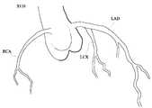
도 2는 도 1에 따른 유체-구조 상호작용을 고려한 협착 병변 영역의 혈류역학 시뮬레이션 방법에서 촬영단계를 통해 획득하는 혈관 영상을 개략적으로 도시한 도면이고,
도 3은 도 1에 따른 유체-구조 상호작용을 고려한 협착 병변 영역의 혈류역학 시뮬레이션 방법에서 제1 영역화단계를 수행한 모습을 개략적으로 도시한 도면이고,
도 4는 도 1에 따른 유체-구조 상호작용을 고려한 협착 병변 영역의 혈류역학 시뮬레이션 방법에서 병변 영역 설정단계를 수행한 모습을 개략적으로 도시한 도면이고,
도 5는 도 1에 따른 유체-구조 상호작용을 고려한 협착 병변 영역의 혈류역학 시뮬레이션 방법에서 제2 영역화단계를 수행한 모습을 개략적으로 도시한 도면이고,
도 6은 도 1에 따른 유체-구조 상호작용을 고려한 협착 병변 영역의 혈류역학 시뮬레이션 방법에서 모델링 단계를 수행한 모습을 개략적으로 도시한 도면이다.FIG. 1 is a flowchart schematically illustrating a method of hemodynamic simulation of a stricture lesion area considering fluid-structure interaction according to an embodiment of the present invention,
FIG. 2 is a view schematically showing a blood vessel image acquired through an imaging step in a hemodynamic simulation method of a stricture lesion area considering fluid-structure interaction according to FIG. 1,
FIG. 3 is a view schematically showing a first regionization step performed in a hemodynamic simulation method of a stricture lesion region considering fluid-structure interaction according to FIG. 1, and FIG.
FIG. 4 is a view schematically showing a lesion area setting step performed in a hemodynamic simulation method of a stricture lesion area considering fluid-structure interaction according to FIG. 1, and FIG.
FIG. 5 is a view schematically showing a second regionization step performed in a hemodynamic simulation method of a stricture lesion region considering fluid-structure interaction according to FIG. 1, and FIG.
FIG. 6 is a view schematically showing a modeling step performed in a hemodynamic simulation method of a stricture lesion area considering fluid-structure interaction according to FIG. 1. FIG.
이하, 첨부한 도면을 참조하여 본 발명의 일실시예에 따른 유체-구조 상호작용을 고려한 협착 병변 영역의 혈류역학 시뮬레이션 방법에 대하여 상세하게 설명한다. 각 도면에 제시된 동일한 참조부호는 동일한 부재를 나타낸다.Hereinafter, a method for simulating a hemodynamics in a stricture lesion area considering fluid-structure interaction according to an embodiment of the present invention will be described in detail with reference to the accompanying drawings. Like reference symbols in the drawings denote like elements.
도 1은 본 발명의 일실시예에 따른 유체-구조 상호작용을 고려한 협착 병변 영역의 혈류역학 시뮬레이션 방법을 개략적으로 도시한 순서도이다.1 is a flowchart schematically illustrating a method of hemodynamic simulation of a stricture lesion area in consideration of fluid-structure interaction according to an embodiment of the present invention.
도 1을 참조하면, 본 발명의 일실시예에 따른 유체-구조 상호작용을 고려한 협착 병변 영역의 혈류역학 시뮬레이션 방법(S100)은 적어도 병변이 형성된 영역에서는 혈관벽 운동을 고려하여 혈류역학 시뮬레이션을 수행할 수 있는 것으로서, 촬영단계(S110)와 제1 영역화단계(S120)와 병변 영역 설정단계(S130)와 제2 영역화단계(S140)와 모델링 단계(S150)와 시뮬레이션 단계(S160)를 포함한다.Referring to FIG. 1, a hemodynamic simulation method (S100) of a stricture lesion region considering fluid-structure interaction according to an embodiment of the present invention performs hemodynamic simulation considering at least a lesion in a lesion region A first segmentation step S120, a lesion area setting step S130, a second segmentation step S140, a modeling step S150, and a simulation step S160 .
도 2는 도 1에 따른 유체-구조 상호작용을 고려한 협착 병변 영역의 혈류역학 시뮬레이션 방법에서 촬영단계를 통해 획득하는 혈관 영상을 개략적으로 도시한 도면이다.FIG. 2 is a view schematically showing a blood vessel image acquired through an imaging step in a hemodynamic simulation method of a stricture lesion area considering fluid-structure interaction according to FIG. 1. FIG.
도 2를 참조하면, 상기 촬영단계(S110)는 협착 병변(L)이 형성된 혈관에 대한 혈관 영상을 획득하는 단계이다.Referring to FIG. 2, the capturing step S110 is a step of acquiring a blood vessel image for a blood vessel in which a stenosis lesion L is formed.
한편, 본 발명의 일실시예에 따르면 촬영단계(S110)는 컴퓨터 단층촬영(Computed Tomography:CT), 선택적 컴퓨터 단층촬영(Selective Computed Tomography) 또는 자기공명영상법(magnetic resonance imaging:MRI)를 이용하여 수행될 수 있다.According to an exemplary embodiment of the present invention, the imaging step S110 may be performed using a computed tomography (CT), a selective computed tomography or a magnetic resonance imaging (MRI) .
가령, 촬영단계(S110)는 컴퓨터 단층촬영(Computed Tomography:CT)으로 수행될 수 있으며, 더 나아가 촬영된 이미지에 대하여 볼륨 렌더링 가시화를 수행할 수 있다.For example, the imaging step S110 may be performed by computed tomography (CT), and further, volume rendering visualization may be performed on the imaged image.
상술한 방법들은 주지한 방법들이므로, 여기서는 촬영단계(S110)에 대한 자세한 설명은 생략한다.Since the above-described methods are well-known methods, detailed description of the shooting step S110 is omitted here.
한편, 상술한 방식에 제한되는 것은 아니며, 혈관초음파(US)와 같은 비침습적 이미징법 또는 디지털 감산 혈관 조영술(DSA), 광 간섭성 단층촬영기술(Optical Coherence Tomography:OCT), 혈관 내 초음파(IntraVascular UltraSound:IVUS)과 같은 침습적 이미징법을 이용하여 심장 및 관상동맥에서의 영상을 획득할 수 있음은 당연하다.However, the present invention is not limited to the above-described methods. For example, non-invasive imaging methods such as vascular ultrasound (US) or digital subtraction angiography (DSA), optical coherence tomography (OCT) UltraSound: IVUS) can be used to acquire images from the heart and coronary arteries.
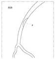
도 3은 도 1에 따른 유체-구조 상호작용을 고려한 협착 병변 영역의 혈류역학 시뮬레이션 방법에서 제1 영역화단계를 수행한 모습을 개략적으로 도시한 도면이다.FIG. 3 is a view schematically showing a first scrambling step performed in a hemodynamic simulation method of a stricture lesion area considering fluid-structure interaction according to FIG. 1; FIG.
도 3을 참조하면, 상기 제1 영역화단계(S120)는 촬영단계(S110)로부터 획득한 혈관 영상에 대하여 영역화를 수행하는 단계이다. 여기서, 영역화라 함은 촬영 영상으로부터 필요한 부분만을 분리하는 과정을 의미한다.Referring to FIG. 3, the first segmentation step S120 is a step of performing segmentation on the blood vessel image acquired from the imaging step S110. Here, the term " regionization " refers to a process of separating only necessary portions from the photographed image.
한편, 제1 영역화단계(S120)는 촬영 영상 중에서 협착 병변을 포함하는 영역에 대하여 영역화를 수행하는 것이 바람직하다.Meanwhile, in the first scrambling step (S120), it is preferable to perform segmentation on the region including the stenosis lesion in the photographed image.
도 4는 도 1에 따른 유체-구조 상호작용을 고려한 협착 병변 영역의 혈류역학 시뮬레이션 방법에서 병변 영역 설정단계를 수행한 모습을 개략적으로 도시한 도면이다.FIG. 4 is a view schematically showing a lesion area setting step in a hemodynamic simulation method of a stricture lesion area considering fluid-structure interaction according to FIG. 1. FIG.
도 4를 참조하면, 상기 병변 영역 설정단계(S130)는 혈관 영상 중에서 병변이 형성되는 병변 영역을 설정하는 단계이다.Referring to FIG. 4, the lesion area setting step S 130 is a step of setting a lesion area where a lesion is formed in the blood vessel image.
여기서 병변 영역은 추후 모델링 단계(S150)에서 혈관벽의 운동을 고려하여 모델링되는 영역으로, 죽상반(P)이 형성된 영역을 포함한다.Herein, the lesion area is a modeling area in consideration of the movement of the blood vessel wall in the later modeling step (S150), and includes a region in which the lesion P is formed.
즉, 협착 병변 부위 중에서 죽상반(P)이 형성되는 영역을 병변 영역으로 정의할 수 있다.That is, a lesion region can be defined as a region in which a lesion (P) is formed in a stricture lesion region.
본 발명의 일실시예에 따르면, 병변 영역은 죽상반(P)을 전후로 혈관의 직경의 1배 내지 2배 더 연장되는 영역으로 설정될 수 있다.According to one embodiment of the present invention, the lesion area may be set to an area extending one to two times longer than the diameter of the blood vessel before and after the porcine upper half (P).
더 자세히 설명하면, 병변 영역은 실질적으로 원기둥 형상으로 마련되며, 직경은 혈관의 직경과 실질적으로 동일하다. 병변 영역의 길이는 죽상반(P)의 전단부로부터 혈관의 직경의 1배 내지 2배 더 연장되고, 죽상반(P)의 후단부로부터 혈관의 직경의 1배 내지 2배 더 연장된다.More specifically, the lesion area is provided in a substantially cylindrical shape, and the diameter is substantially the same as the diameter of the blood vessel. The length of the lesion area is extended by one to two times the diameter of the blood vessel from the front end of the porcine upper half P and is extended by one to two times the diameter of the blood vessel from the rear end of the porcine upper half P.
본 발명의 일실시예에 따르면, 병변 영역은 병변의 전후 ±혈관의 직경의 1배로 마련될 수 있다.According to one embodiment of the present invention, the lesion area may be provided at a time of the diameter of the blood vessel before and after the lesion.
물론, 필요에 따라 병변 영역의 길이는 변경될 수 있으며, 상기와 같은 수치로 제한되는 것은 아니다.Of course, the length of the lesion area may be varied as needed, and is not limited to the above values.
도 5는 도 1에 따른 유체-구조 상호작용을 고려한 협착 병변 영역의 혈류역학 시뮬레이션 방법에서 제2 영역화단계를 수행한 모습을 개략적으로 도시한 도면이다.FIG. 5 is a view schematically showing a second regionization step performed in a hemodynamic simulation method of a stricture lesion region considering fluid-structure interaction according to FIG. 1. FIG.
도 5를 참조하면, 상기 제2 영역화단계(S140)는 병변 영역 설정단계(S130)를 통해 설정된 병변 영역에 대하여 영역화를 재수행하는 단계이다.Referring to FIG. 5, the second segmentation step S140 is a step of re-segmenting the lesion area set through the lesion area setting step S130.
자세히 설명하면, 후술할 모델링 단계(S150)에서 혈관벽의 운동을 고려하여 모델링되는 병변 영역에 대해서 영역화를 재수행하게 된다.In detail, in the modeling step (S150) to be described later, the lesion area modeled considering the motion of the blood vessel wall is re-searched.
본 발명의 일실시예에 따르면, 혈관 영상에 대하여 2번의 영역화를 수행하게 되는데, 이는 제1 영역화단계(S120)에서 설정되는 영역과 제2 영역화단계(S140)에서 설정되는 영역에 대한 해석방법이 달라지기 때문이다. 이에 대해서는 후술한다.According to an embodiment of the present invention, two sagittalization is performed on a blood vessel image, which is performed on the area set in the first sagization step S120 and the area set in the second sagization step S140 This is because the interpretation method is different. This will be described later.
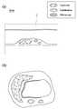
도 6은 도 1에 따른 유체-구조 상호작용을 고려한 협착 병변 영역의 혈류역학 시뮬레이션 방법에서 모델링 단계를 수행한 모습을 개략적으로 도시한 도면이다.FIG. 6 is a view schematically showing a modeling step performed in a hemodynamic simulation method of a stricture lesion area considering fluid-structure interaction according to FIG. 1. FIG.
도 6을 참조하면, 상기 모델링단계(S150)는 혈관 영상을 이용하여 혈류 모델링을 수행하는 단계로서, 적어도 병변 영역에 대해서는 혈관벽의 운동을 고려하여 혈류 모델링을 수행한다.Referring to FIG. 6, the modeling step S150 is a step of performing blood flow modeling using a blood vessel image, and blood flow modeling is performed in consideration of motion of a blood vessel wall at least in a lesion area.
본 발명의 일실시예에 따르면, 모델링단계(S150)는 제1 영역화단계(S120)를 통해 영역화된 혈관 형상에 대하여 제2 영역화단계(S140)를 통해 영역화된 병변 영역을 결합하여 혈류 모델링을 수행한다.According to an embodiment of the present invention, the modeling step S150 may include combining the regioned lesion regions through the second segmentation step S140 with respect to the segmented vessel shape through the first segmentation step S120 Blood flow modeling is performed.
여기서, 제2 영역화단계(S140)를 통해 얻어진 병변 영역에 대해서는 죽상반(P)의 성분, 분포, 및 역학적 물성치에 대한 데이터를 적용할 수 있다.Here, for the lesion area obtained through the second scrambling step (S140), data on the components, distribution, and mechanical property values of the pelvis P can be applied.
다시 설명하면, 동맥 경화 죽상반은 일반적으로 지질중심부(lipid core), 석회화영역(calcification), 섬유성막(fibrous cap) 등으로 구성되며, 각 성분은 비선형적 재료 변형 특성을 가진다. 이러한 죽상반의 역학적 물성치는 크게 밀도, 비선형 응력/변형도 관계식, 포아송비(Poisson's ration), 영률(Young's modulus) 등을 포함한다.In other words, the arteriosclerotic plaque generally consists of lipid core, calcification, and fibrous cap, and each component has nonlinear material deformation characteristics. The mechanical properties of these islands include density, nonlinear stress / strain relations, Poisson's ratio, Young's modulus, and so on.
여기서, 비선형 응력/변형도 관계식은 Mooney-Rivlin 모델을 적용할 수 있으며, 그 외의 다른 비선형 재료 변형 모델을 적용할 수 있다.Here, the nonlinear stress / strain relationship can be applied to the Mooney-Rivlin model, and other nonlinear material deformation models can be applied.
기존의 연구에서 상대적으로 큰 지질중심부(lipid core)나 얇은 섬유성막을 가지고 있는 죽상반 병변의 경우 파열 위험도가 상대적으로 높은 것으로 알려져 있다.Previous studies have shown that the risk of rupture is relatively high for lesions with relatively large lipid cores or thin fibrous membranes.
즉, 병변 영역에 대하여 죽상반(P)의 구성 성분, 분포, 및 죽상반(P)의 구성 성분 각각의 역학적 물성치에 대한 데이터를 적용하여 추후 시뮬레이션 단계(S160)에서 병변 영역에 대하여 혈관벽의 운동을 고려할 수 있도록 한다.That is, the data on the mechanical property values of each component of the porphyry P, the distribution, and the constituent components of the porphyry P are applied to the lesion area to determine the motion of the vessel wall in the lesion area in the subsequent simulation step S160 .
상기 시뮬레이션 단계(S160)는 상술한 모델링단계(S150)에서 모델링된 혈류 모델링 정보를 이용하되 적어도 병변 영역에 대해서는 혈관벽의 운동을 고려하여 혈류역학 시뮬레이션을 수행하는 단계이다.In the simulation step S160, blood flow modeling information modeled in the modeling step S150 described above is used, and hemodynamic simulation is performed in consideration of motion of the blood vessel wall at least in the lesion area.
더 자세히 설명하면, 제2 영역화단계(S140)를 통해 얻은 병변 영역에 대해서는 상술한 혈관벽 유연성 및 혈관벽 운동을 허용하는 조건을 적용하고, 제1 영역화단계(S120)를 통해 얻은 영상은 병변 영역을 제외한 영역에 대해서는 혈관벽의 운동이 없는 것으로 가정하여 시뮬레이션을 수행한다.More specifically, a condition allowing the blood vessel wall flexibility and the motion of the blood vessel wall is applied to the lesion area obtained through the second scription step (S140), and the image obtained through the first scription step (S120) The simulation is performed on the assumption that there is no motion of the vessel wall.
여기서, 병변 영역은 유체-구조 상호작용(Fluid-Structure Interaction Analysis:FSI) 해석에 의하여 수행되는 것이 바람직하다.Here, the lesion region is preferably performed by a fluid-structure interaction (FSI) analysis.
한편, 본 발명의 일실시예에 따르면, 시뮬레이션 단계(S160)에서는 적어도 병변 영역에 대해서는 혈관벽 운동을 고려하므로 병변 영역에서의 혈관벽의 구조 응력 분포 및 변형 거동 데이터를 더 획득할 수 있다.According to an embodiment of the present invention, in the simulation step (S160), considering the blood vessel wall motion for at least the lesion area, the structural stress distribution and deformation behavior data of the blood vessel wall in the lesion area can be further obtained.
더 나아가, 죽상반(P)의 구조적 파열 위험도에 대한 예측 정보까지 추가적으로 획득할 수 있다.Furthermore, predictive information on the risk of structural rupture of the inconsistent part (P) can be additionally obtained.
종래에는 모든 혈관에 대하여 혈관벽 운동 경계조건을 적용하여 시뮬레이션을 수행하기도 하였으나, 과다한 컴퓨터 계산량의 요구로 혈관벽 고정 경계조건을 적용하여 시뮬레이션을 수행하는 것이 일반적이었다.Conventionally, all the blood vessels have been subjected to simulation by applying the boundary conditions of the vessel wall motion, but it has been common to perform the simulation by applying the vessel wall fixed boundary conditions as the demand of the excessive computational complexity.
이에, 죽상반의 파열 위험도 등을 예측하기 위해서 매우 제한된 범위, 가령 협착 병변이 형성된 영역에 대해서만 유체-구조 상호작용 시뮬레이션 등을 수행하여 필요한 데이터를 추출하였다.In order to predict the risk of rupture of the isthmus, a fluid-structure interaction simulation was performed for a limited range, for example, only the region where the stenosis lesion was formed.
이와 달리, 본 발명의 일실시예에서는 단순히 병변 영역에 대해서만 혈관벽 운동 경계조건을 적용하여 시뮬레이션을 수행하는 것이 아니라, 협착 병변 주위의 혈관벽 유연성 조건을 적용할 영역과 혈관벽 고정 경계조건을 적용할 영역을 유기적으로 결합하여 상대적으로 광범위한 환자 특정 혈관 형상의 영향을 고려한 혈류역학 시뮬레이션을 수행할 수 있어 보다 정확한 데이터를 추출할 수 있다.In contrast, in an embodiment of the present invention, instead of simply performing the simulation by applying the vessel wall motion boundary condition only to the lesion area, the region to which the vessel wall flexibility conditions around the stenotic lesion are applied and the region to which the vessel wall fixed boundary condition is applied It is possible to carry out a hemodynamic simulation taking into account the influence of a relatively wide variety of patient-specific blood vessel shapes by organically binding to extract more accurate data.
한편, 상술한 본 발명의 일실시예는 컴퓨터에서 실행될 수 있는 프로그램으로 작성가능하고, 컴퓨터로 판독가능한 기록매체를 이용하여 상기 프로그램을 동작시키는 범용 디지털 컴퓨터에서 구현될 수 있다.Meanwhile, one embodiment of the present invention described above can be realized in a general-purpose digital computer that can be created as a program that can be executed in a computer and operates the program using a computer-readable recording medium.
컴퓨터 판독 가능 매체는 컴퓨터에 의해 액세스될 수 있는 임의의 가용 매체일 수 있고, 휘발성 및 비휘발성 매체, 분리형 및 비분리형 매체를 모두 포함한다. 또한, 컴퓨터 판독가능 매체는 컴퓨터 저장 매체 및 통신 매체를 모두 포함할 수 있다. 컴퓨터 저장 매체는 컴퓨터 판독가능 명령어, 데이터 구조, 프로그램 모듈 또는 기타 데이터와 같은 정보의 저장을 위한 임의의 방법 또는 기술로 구현된 휘발성 및 비휘발성, 분리형 및 비분리형 매체를 모두 포함한다. 통신 매체는 전형적으로 컴퓨터 판독가능 명령어, 데이터 구조, 프로그램 모듈, 또는 기타 전송 메커니즘을 포함하며, 임의의 정보 전달 매체를 포함할 수 있다.Computer readable media can be any available media that can be accessed by a computer and includes both volatile and nonvolatile media, removable and non-removable media. In addition, the computer-readable medium may include both computer storage media and communication media. Computer storage media includes both volatile and nonvolatile, removable and non-removable media implemented in any method or technology for storage of information such as computer readable instructions, data structures, program modules or other data. Communication media typically includes computer-readable instructions, data structures, program modules, or other transport mechanisms, and may include any information delivery media.
본 발명의 권리범위는 상술한 실시예에 한정되는 것이 아니고, 본 발명의 사상 및 범위를 벗어나지 않고 다양하게 수정 또는 변형할 수 있고, 그러한 수정례 또는 변형례들도 본 발명의 권리범위에 속한다고 볼 것이다.It is to be understood that the scope of the present invention is not limited to the above embodiments and that various changes and modifications may be made without departing from the spirit and scope of the present invention, I will see.
S100: 유체-구조 상호작용을 고려한 협착 병변 영역의 혈류역학 시뮬레이션 방법
S110: 촬영단계S120: 제1 영역화 단계
S130: 병변 영역 설정단계S140: 제2 영역화 단계
S150: 모델링 단계S160: 시뮬레이션 단계S100: Hemodynamic simulation of stenotic lesion area considering fluid-structure interaction
S110: photographing step S120: first regioning step
S130: lesion area setting step S140: second regionization step
S150: Modeling step S160: Simulation step
Claims (8)
Translated fromKorean상기 혈관 영상에 대하여 영역화를 수행하는 제1 영역화 단계;
상기 혈관 영상에서 협착 병변이 형성되는 병변 영역을 설정하는 병변 영역 설정단계;
상기 병변 영역에 대하여 영역화를 재수행하는 제2 영역화 단계;
상기 제1 영역화단계를 통해 영역화된 혈관 형상에 대하여 상기 제2 영역화단계를 통해 영역화된 병변 영역을 결합하여 혈류 모델링을 수행하는 모델링 단계;
상기 혈류 모델링 정보를 이용하되 적어도 상기 병변 영역에 대해서는 혈관벽의 운동을 고려하여 혈류역학 시뮬레이션을 수행하는 시뮬레이션 단계를 포함하고,
상기 시뮬레이션 단계에 있어서,
상기 제2 영역화단계를 통해 얻은 병변 영역에 대해서는 혈관벽 유연성 및 혈관벽 운동을 허용하는 조건을 적용하고, 상기 제1 영역화 단계를 통해 얻은 영상은 병변 영역을 제외한 영역에 대해서는 혈관벽의 운동이 없는 것으로 가정하여 시뮬레이션을 수행하고,
상기 협착 병변이 형성되는 병변 영역주위의 혈관벽 유연성 조건을 적용할 영역과 혈관벽 고정 경계조건을 적용할 영역을 유기적으로 결합하여 환자 특정 혈관 형상의 영향을 고려한 혈류 역학 시뮬레이션을 수행하는 것을 특징으로 하는유체-구조 상호작용을 고려한 협착 병변 영역의 혈류역학 시뮬레이션 방법.An imaging step of acquiring a blood vessel image for a blood vessel in which a stenotic lesion is formed;
A first segmentation step of performing segmentation on the blood vessel image;
A lesion area setting step of setting a lesion area where a stenosis lesion is formed in the blood vessel image;
A second segmentation step of re-segmenting the lesion area;
A modeling step of performing blood flow modeling by combining the regionized lesion regions through the second segmentation step with respect to the segmented blood vessel shape through the first segmentation step;
And a simulation step of performing hemodynamic simulation using at least the blood flow modeling information in consideration of motion of the blood vessel wall at least in the lesion area,
In the simulation step,
The condition for permitting blood vessel wall flexibility and blood vessel wall motion is applied to the lesion area obtained through the second segmentation step and the image obtained through the first segmentation step has no motion of the blood vessel wall for the region excluding the lesion area Assuming that the simulation is performed,
Wherein blood flow simulation is performed considering the influence of the patient-specific blood vessel shape by organically coupling the region to be applied with the blood vessel wall flexibility condition around the lesion region where the stenotic lesion is formed to the region to which the blood vessel wall fixed boundary condition is to be applied. - Method of hemodynamic simulation of stenotic lesion area considering structure interaction.
상기 병변 영역 설정단계에서 상기 병변 영역은 병변에 구비되는 죽상반의 전후로 혈관 직경의 1배 내지 2배 연장되는 영역으로 설정되는 유체-구조 상호작용을 고려한 협착 병변 영역의 혈류역학 시뮬레이션 방법.The method according to claim 1,
Wherein the lesion area is set in a region where the lesion area is extended by one to two times the diameter of the blood vessel before and after the atheroma provided in the lesion, in consideration of the fluid-structure interaction.
상기 모델링 단계에서 상기 병변 영역은 병변에 구비되는 죽상반의 구성 성분, 분포 및 상기 죽상반 구성성분 각각의 역학적 물성치를 적용하여 설정되는 유체-구조 상호작용을 고려한 협착 병변 영역의 혈류역학 시뮬레이션 방법.The method according to claim 1,
Wherein the lesion area in the modeling step includes a fluid-structure interaction set by applying a constituent component, a distribution, and an epidemiological property value of each of the proximal components of the islet zone provided in the lesion.
상기 시뮬레이션 단계에서 상기 병변 영역 이외의 영역에서는 혈관벽의 운동을 배제하여 혈류 모델링을 수행하는 유체-구조 상호작용을 고려한 협착 병변 영역의 혈류역학 시뮬레이션 방법.The method according to claim 1,
Wherein the simulated blood flow modeling is performed by excluding the movement of the blood vessel wall in a region other than the lesion region in the simulation step, considering the fluid-structure interaction.
상기 시뮬레이션 단계에서 상기 병변 영역은 유체-구조 상호작용 기법에 의하여 수행되는 유체-구조 상호작용을 고려한 협착 병변 영역의 혈류역학 시뮬레이션 방법.5. The method of claim 4,
Wherein the lesion area in the simulation step is a fluid-structure interaction interaction performed by a fluid-structure interaction technique.
상기 시뮬레이션 단계는 상기 병변 영역에 대한 혈관벽의 구조 응력 분포 및 변형 거동 데이터를 더 획득하는 유체-구조 상호작용을 고려한 협착 병변 영역의 혈류역학 시뮬레이션 방법.6. The method of claim 5,
Wherein the simulation step further includes fluid-structure interaction to obtain structural stress distribution and deformation behavior data of the vessel wall over the lesion area.
상기 시뮬레이션 단계는 상기 병변에 구비되는 죽상반의 구조적 파열 위험도에 대한 예측 정보를 더 획득하는 유체-구조 상호작용을 고려한 협착 병변 영역의 혈류역학 시뮬레이션 방법.The method according to claim 6,
Wherein the simulation step further includes obtaining fluid-structure interaction prediction information on the structural rupture risk of the atheroma provided in the lesion.
Priority Applications (1)
| Application Number | Priority Date | Filing Date | Title |
|---|---|---|---|
| KR1020160010941AKR101954272B1 (en) | 2016-01-28 | 2016-01-28 | Hemodynamics simulation method by considering fluid-structure interaction in occlusive lesion |
Applications Claiming Priority (1)
| Application Number | Priority Date | Filing Date | Title |
|---|---|---|---|
| KR1020160010941AKR101954272B1 (en) | 2016-01-28 | 2016-01-28 | Hemodynamics simulation method by considering fluid-structure interaction in occlusive lesion |
Publications (2)
| Publication Number | Publication Date |
|---|---|
| KR20170090286A KR20170090286A (en) | 2017-08-07 |
| KR101954272B1true KR101954272B1 (en) | 2019-03-05 |
Family
ID=59653833
Family Applications (1)
| Application Number | Title | Priority Date | Filing Date |
|---|---|---|---|
| KR1020160010941AActiveKR101954272B1 (en) | 2016-01-28 | 2016-01-28 | Hemodynamics simulation method by considering fluid-structure interaction in occlusive lesion |
Country Status (1)
| Country | Link |
|---|---|
| KR (1) | KR101954272B1 (en) |
Families Citing this family (1)
| Publication number | Priority date | Publication date | Assignee | Title |
|---|---|---|---|---|
| WO2022168315A1 (en)* | 2021-02-08 | 2022-08-11 | 朝日インテック株式会社 | Vascular lesion model |
Family Cites Families (3)
| Publication number | Priority date | Publication date | Assignee | Title |
|---|---|---|---|---|
| US8315812B2 (en) | 2010-08-12 | 2012-11-20 | Heartflow, Inc. | Method and system for patient-specific modeling of blood flow |
| US10162932B2 (en)* | 2011-11-10 | 2018-12-25 | Siemens Healthcare Gmbh | Method and system for multi-scale anatomical and functional modeling of coronary circulation |
| KR101530352B1 (en)* | 2013-04-02 | 2015-06-22 | 재단법인 아산사회복지재단 | Cfd modeling and analysis method based on material properties |
- 2016
- 2016-01-28KRKR1020160010941Apatent/KR101954272B1/enactiveActive
Also Published As
| Publication number | Publication date |
|---|---|
| KR20170090286A (en) | 2017-08-07 |
Similar Documents
| Publication | Publication Date | Title |
|---|---|---|
| US10748451B2 (en) | Methods and systems for generating fluid simulation models | |
| JP6829262B2 (en) | Systems and methods for identifying and modeling unresolved vessels in an image-based patient-specific hemodynamic model | |
| JP6832920B2 (en) | Systems and methods for diagnosing and assessing cardiovascular disease by comparing arterial supply capacity with terminal organ requirements | |
| EP3277169B1 (en) | Systems and methods for estimating virtual perfusion images | |
| JP6667999B2 (en) | Image processing apparatus, image processing method, and program | |
| Steinman | Image-based computational fluid dynamics modeling in realistic arterial geometries | |
| EP3937186B1 (en) | System and method for vascular tree generation using joint prior information | |
| US10803995B2 (en) | Method and system for non-invasive functional assessment of coronary artery stenosis using flow computations in diseased and hypothetical normal anatomical models | |
| Marlevi et al. | Estimation of cardiovascular relative pressure using virtual work-energy | |
| CN106063734B (en) | For creating the method and calculating and print unit of stent graft | |
| EP2925216B1 (en) | Stenosis therapy planning | |
| KR102620470B1 (en) | Patient-specific modeling of coronary hemodynamic parameters | |
| De Wilde et al. | Assessment of shear stress related parameters in the carotid bifurcation using mouse-specific FSI simulations | |
| JP6873981B2 (en) | Mobile FFR simulation | |
| CN108140430A (en) | Estimation of flow, resistance, or pressure from pressure or flow measurements and angiography | |
| Karmonik et al. | Computational hemodynamics in the human aorta: a computational fluid dynamics study of three cases with patient-specific geometries and inflow rates | |
| CN110786842B (en) | Method, device, system and storage medium for measuring diastolic blood flow velocity | |
| Auricchio et al. | A clinically applicable stochastic approach for noninvasive estimation of aortic stiffness using computed tomography data | |
| Celi et al. | On the role and effects of uncertainties in cardiovascular in silico analyses | |
| Celi et al. | An image-based approach for the estimation of arterial local stiffness in vivo | |
| KR101530352B1 (en) | Cfd modeling and analysis method based on material properties | |
| KR101954272B1 (en) | Hemodynamics simulation method by considering fluid-structure interaction in occlusive lesion | |
| KR102078622B1 (en) | Method for determining cardiovascular information using mathematical modeling of stenosed lesion | |
| Beier et al. | Vascular hemodynamics with computational modeling and experimental studies | |
| KR20180008134A (en) | A method for predicting a fractional flow reserve |
Legal Events
| Date | Code | Title | Description |
|---|---|---|---|
| A201 | Request for examination | ||
| PA0109 | Patent application | Patent event code:PA01091R01D Comment text:Patent Application Patent event date:20160128 | |
| PA0201 | Request for examination | ||
| E902 | Notification of reason for refusal | ||
| PE0902 | Notice of grounds for rejection | Comment text:Notification of reason for refusal Patent event date:20170618 Patent event code:PE09021S01D | |
| PG1501 | Laying open of application | ||
| E90F | Notification of reason for final refusal | ||
| PE0902 | Notice of grounds for rejection | Comment text:Final Notice of Reason for Refusal Patent event date:20180131 Patent event code:PE09021S02D | |
| E701 | Decision to grant or registration of patent right | ||
| PE0701 | Decision of registration | Patent event code:PE07011S01D Comment text:Decision to Grant Registration Patent event date:20181130 | |
| GRNT | Written decision to grant | ||
| PR0701 | Registration of establishment | Comment text:Registration of Establishment Patent event date:20190226 Patent event code:PR07011E01D | |
| PR1002 | Payment of registration fee | Payment date:20190226 End annual number:3 Start annual number:1 | |
| PG1601 | Publication of registration | ||
| PR1001 | Payment of annual fee | Payment date:20220203 Start annual number:4 End annual number:4 | |
| PR1001 | Payment of annual fee | Payment date:20240201 Start annual number:6 End annual number:6 |