KR101945938B1 - A preparation method of injectable thermosensitive chitosan/tempo based-oxidized cellulose hydrogel - Google Patents
A preparation method of injectable thermosensitive chitosan/tempo based-oxidized cellulose hydrogelDownload PDFInfo
- Publication number
- KR101945938B1 KR101945938B1KR1020170088565AKR20170088565AKR101945938B1KR 101945938 B1KR101945938 B1KR 101945938B1KR 1020170088565 AKR1020170088565 AKR 1020170088565AKR 20170088565 AKR20170088565 AKR 20170088565AKR 101945938 B1KR101945938 B1KR 101945938B1
- Authority
- KR
- South Korea
- Prior art keywords
- tocnf
- chitosan
- tempo
- hydrogel
- oxidized cellulose
- Prior art date
- Legal status (The legal status is an assumption and is not a legal conclusion. Google has not performed a legal analysis and makes no representation as to the accuracy of the status listed.)
- Active
Links
- 229920001661ChitosanPolymers0.000titleclaimsabstractdescription253
- 239000000017hydrogelSubstances0.000titleclaimsabstractdescription179
- 229920002201Oxidized cellulosePolymers0.000titleclaimsabstractdescription70
- 229940107304oxidized celluloseDrugs0.000titleclaimsabstractdescription70
- 238000002360preparation methodMethods0.000title1
- 239000002121nanofiberSubstances0.000claimsabstractdescription71
- 239000000499gelSubstances0.000claimsabstractdescription18
- 230000010478bone regenerationEffects0.000claimsabstractdescription14
- 239000000945fillerSubstances0.000claimsabstractdescription6
- 239000000243solutionSubstances0.000claimsdescription39
- 238000000034methodMethods0.000claimsdescription27
- XYZZKVRWGOWVGO-UHFFFAOYSA-NGlycerol-phosphateChemical compoundOP(O)(O)=O.OCC(O)COXYZZKVRWGOWVGO-UHFFFAOYSA-N0.000claimsdescription25
- JVTAAEKCZFNVCJ-UHFFFAOYSA-Nlactic acidChemical compoundCC(O)C(O)=OJVTAAEKCZFNVCJ-UHFFFAOYSA-N0.000claimsdescription18
- 238000001727in vivoMethods0.000claimsdescription15
- 239000011259mixed solutionSubstances0.000claimsdescription12
- 239000004310lactic acidSubstances0.000claimsdescription9
- 235000014655lactic acidNutrition0.000claimsdescription9
- VEXZGXHMUGYJMC-UHFFFAOYSA-NHydrochloric acidChemical compoundClVEXZGXHMUGYJMC-UHFFFAOYSA-N0.000claimsdescription8
- 239000000725suspensionSubstances0.000claimsdescription6
- XLYOFNOQVPJJNP-UHFFFAOYSA-NwaterChemical compoundOXLYOFNOQVPJJNP-UHFFFAOYSA-N0.000claimsdescription6
- 239000007864aqueous solutionSubstances0.000claimsdescription5
- 238000004519manufacturing processMethods0.000claimsdescription5
- 238000002156mixingMethods0.000claimsdescription5
- 239000012153distilled waterSubstances0.000claimsdescription4
- 229920002678cellulosePolymers0.000claimsdescription3
- 239000001913celluloseSubstances0.000claimsdescription3
- VUZNLSBZRVZGIK-UHFFFAOYSA-N2,2,6,6-Tetramethyl-1-piperidinolChemical groupCC1(C)CCCC(C)(C)N1OVUZNLSBZRVZGIK-UHFFFAOYSA-N0.000claimsdescription2
- 238000001879gelationMethods0.000abstractdescription21
- 238000002054transplantationMethods0.000abstractdescription7
- 230000000694effectsEffects0.000abstractdescription4
- 230000004663cell proliferationEffects0.000abstractdescription3
- 230000008611intercellular interactionEffects0.000abstractdescription3
- 230000035945sensitivityEffects0.000abstractdescription2
- 230000001939inductive effectEffects0.000abstract1
- 210000004027cellAnatomy0.000description37
- 210000002540macrophageAnatomy0.000description14
- 210000001519tissueAnatomy0.000description11
- 238000004458analytical methodMethods0.000description10
- 238000002347injectionMethods0.000description10
- 239000007924injectionSubstances0.000description10
- 230000015572biosynthetic processEffects0.000description9
- 238000000338in vitroMethods0.000description9
- 239000002105nanoparticleSubstances0.000description9
- 239000012620biological materialSubstances0.000description7
- 239000000463materialSubstances0.000description7
- 239000000835fiberSubstances0.000description6
- 238000005755formation reactionMethods0.000description6
- 238000001228spectrumMethods0.000description6
- 238000005033Fourier transform infrared spectroscopyMethods0.000description5
- 102000016943MuramidaseHuman genes0.000description5
- 108010014251MuramidaseProteins0.000description5
- 108010062010N-Acetylmuramoyl-L-alanine AmidaseProteins0.000description5
- 229920001046NanocellulosePolymers0.000description5
- 238000002441X-ray diffractionMethods0.000description5
- 230000003247decreasing effectEffects0.000description5
- 230000012010growthEffects0.000description5
- 239000007788liquidSubstances0.000description5
- GDOPTJXRTPNYNR-UHFFFAOYSA-Nmethyl-cyclopentaneNatural productsCC1CCCC1GDOPTJXRTPNYNR-UHFFFAOYSA-N0.000description5
- 230000008569processEffects0.000description5
- 239000000126substanceSubstances0.000description5
- 238000012360testing methodMethods0.000description5
- WZUVPPKBWHMQCE-UHFFFAOYSA-NHaematoxylinChemical compoundC12=CC(O)=C(O)C=C2CC2(O)C1C1=CC=C(O)C(O)=C1OC2WZUVPPKBWHMQCE-UHFFFAOYSA-N0.000description4
- 230000036760body temperatureEffects0.000description4
- 230000015556catabolic processEffects0.000description4
- 231100000135cytotoxicityToxicity0.000description4
- 230000003013cytotoxicityEffects0.000description4
- 238000006731degradation reactionMethods0.000description4
- 239000003814drugSubstances0.000description4
- 210000002950fibroblastAnatomy0.000description4
- 229960000274lysozymeDrugs0.000description4
- 235000010335lysozymeNutrition0.000description4
- 239000004325lysozymeSubstances0.000description4
- 239000011159matrix materialSubstances0.000description4
- 210000000963osteoblastAnatomy0.000description4
- 239000002994raw materialSubstances0.000description4
- 231100000002MTT assayToxicity0.000description3
- 238000000134MTT assayMethods0.000description3
- 150000001408amidesChemical class0.000description3
- 230000008859changeEffects0.000description3
- 230000007423decreaseEffects0.000description3
- 238000009792diffusion processMethods0.000description3
- 230000028709inflammatory responseEffects0.000description3
- 229920000642polymerPolymers0.000description3
- 238000012453sprague-dawley rat modelMethods0.000description3
- 238000010254subcutaneous injectionMethods0.000description3
- 239000007929subcutaneous injectionSubstances0.000description3
- 238000003786synthesis reactionMethods0.000description3
- 230000007704transitionEffects0.000description3
- 206010061218InflammationDiseases0.000description2
- QYTDEUPAUMOIOP-UHFFFAOYSA-NTEMPOChemical groupCC1(C)CCCC(C)(C)N1[O]QYTDEUPAUMOIOP-UHFFFAOYSA-N0.000description2
- 230000002378acidificating effectEffects0.000description2
- DHCLVCXQIBBOPH-UHFFFAOYSA-Nbeta-glycerol phosphateNatural productsOCC(CO)OP(O)(O)=ODHCLVCXQIBBOPH-UHFFFAOYSA-N0.000description2
- GHRQXJHBXKYCLZ-UHFFFAOYSA-Lbeta-glycerolphosphateChemical compound[Na+].[Na+].CC(CO)OOP([O-])([O-])=OGHRQXJHBXKYCLZ-UHFFFAOYSA-L0.000description2
- 230000005540biological transmissionEffects0.000description2
- 230000010261cell growthEffects0.000description2
- 239000013078crystalSubstances0.000description2
- 238000000354decomposition reactionMethods0.000description2
- 230000007547defectEffects0.000description2
- 238000007865dilutingMethods0.000description2
- 229940079593drugDrugs0.000description2
- YQGOJNYOYNNSMM-UHFFFAOYSA-NeosinChemical compound[Na+].OC(=O)C1=CC=CC=C1C1=C2C=C(Br)C(=O)C(Br)=C2OC2=C(Br)C(O)=C(Br)C=C21YQGOJNYOYNNSMM-UHFFFAOYSA-N0.000description2
- 238000011156evaluationMethods0.000description2
- 239000013029homogenous suspensionSubstances0.000description2
- 229910052739hydrogenInorganic materials0.000description2
- 238000003125immunofluorescent labelingMethods0.000description2
- 238000002513implantationMethods0.000description2
- 230000008595infiltrationEffects0.000description2
- 238000001764infiltrationMethods0.000description2
- 230000004054inflammatory processEffects0.000description2
- 230000001404mediated effectEffects0.000description2
- 210000002901mesenchymal stem cellAnatomy0.000description2
- 229920005615natural polymerPolymers0.000description2
- 230000003287optical effectEffects0.000description2
- 230000003647oxidationEffects0.000description2
- 238000007254oxidation reactionMethods0.000description2
- 230000001737promoting effectEffects0.000description2
- 230000001172regenerating effectEffects0.000description2
- 239000002904solventSubstances0.000description2
- 230000029663wound healingEffects0.000description2
- 241000238421ArthropodaSpecies0.000description1
- 239000002028BiomassSubstances0.000description1
- 229920003043Cellulose fiberPolymers0.000description1
- 238000001157Fourier transform infrared spectrumMethods0.000description1
- 229920002683GlycosaminoglycanPolymers0.000description1
- 206010020751HypersensitivityDiseases0.000description1
- 231100000416LDH assayToxicity0.000description1
- 210000004322M2 macrophageAnatomy0.000description1
- 241000699670Mus sp.Species0.000description1
- OVRNDRQMDRJTHS-FMDGEEDCSA-NN-acetyl-beta-D-glucosamineChemical groupCC(=O)N[C@H]1[C@H](O)O[C@H](CO)[C@@H](O)[C@@H]1OOVRNDRQMDRJTHS-FMDGEEDCSA-N0.000description1
- 241000700159RattusSpecies0.000description1
- 208000033809SuppurationDiseases0.000description1
- 208000026935allergic diseaseDiseases0.000description1
- 230000007815allergyEffects0.000description1
- 125000003368amide groupChemical group0.000description1
- 125000003277amino groupChemical group0.000description1
- 230000003305autocrineEffects0.000description1
- 230000004071biological effectEffects0.000description1
- 230000031018biological processes and functionsEffects0.000description1
- 210000004204blood vesselAnatomy0.000description1
- 210000000988bone and boneAnatomy0.000description1
- 239000000969carrierSubstances0.000description1
- 210000000845cartilageAnatomy0.000description1
- 239000003054catalystSubstances0.000description1
- 125000002091cationic groupChemical group0.000description1
- 229920006317cationic polymerPolymers0.000description1
- 230000024245cell differentiationEffects0.000description1
- 230000003833cell viabilityEffects0.000description1
- 230000036755cellular responseEffects0.000description1
- 238000006243chemical reactionMethods0.000description1
- 210000003690classically activated macrophageAnatomy0.000description1
- 239000002131composite materialSubstances0.000description1
- 238000004624confocal microscopyMethods0.000description1
- 210000002808connective tissueAnatomy0.000description1
- 239000000470constituentSubstances0.000description1
- 238000004132cross linkingMethods0.000description1
- 238000003381deacetylation reactionMethods0.000description1
- 230000034994deathEffects0.000description1
- 230000001419dependent effectEffects0.000description1
- 230000001627detrimental effectEffects0.000description1
- 238000010586diagramMethods0.000description1
- 238000012377drug deliveryMethods0.000description1
- 238000005538encapsulationMethods0.000description1
- 230000001747exhibiting effectEffects0.000description1
- 238000002474experimental methodMethods0.000description1
- 239000000284extractSubstances0.000description1
- 238000011049fillingMethods0.000description1
- 239000010419fine particleSubstances0.000description1
- 125000000524functional groupChemical group0.000description1
- MSWZFWKMSRAUBD-IVMDWMLBSA-Nglucosamine groupChemical groupOC1[C@H](N)[C@@H](O)[C@H](O)[C@H](O1)COMSWZFWKMSRAUBD-IVMDWMLBSA-N0.000description1
- 230000036449good healthEffects0.000description1
- 239000001257hydrogenSubstances0.000description1
- 229920001477hydrophilic polymerPolymers0.000description1
- 230000002209hydrophobic effectEffects0.000description1
- 238000007654immersionMethods0.000description1
- 230000028993immune responseEffects0.000description1
- 230000003053immunizationEffects0.000description1
- 238000002649immunizationMethods0.000description1
- 238000010166immunofluorescenceMethods0.000description1
- 239000007943implantSubstances0.000description1
- 238000011534incubationMethods0.000description1
- 230000002757inflammatory effectEffects0.000description1
- 238000011081inoculationMethods0.000description1
- 238000002843lactate dehydrogenase assayMethods0.000description1
- 210000003041ligamentAnatomy0.000description1
- 239000006193liquid solutionSubstances0.000description1
- 231100000053low toxicityToxicity0.000description1
- 230000007246mechanismEffects0.000description1
- 239000012528membraneSubstances0.000description1
- 230000005499meniscusEffects0.000description1
- 210000003632microfilamentAnatomy0.000description1
- 238000002324minimally invasive surgeryMethods0.000description1
- 239000000203mixtureSubstances0.000description1
- 238000012986modificationMethods0.000description1
- 230000004048modificationEffects0.000description1
- 239000002114nanocompositeSubstances0.000description1
- 239000002086nanomaterialSubstances0.000description1
- 231100001083no cytotoxicityToxicity0.000description1
- 231100000956nontoxicityToxicity0.000description1
- 230000001582osteoblastic effectEffects0.000description1
- 230000003076paracrineEffects0.000description1
- 230000000704physical effectEffects0.000description1
- 229920000867polyelectrolytePolymers0.000description1
- 239000005518polymer electrolyteSubstances0.000description1
- 230000003389potentiating effectEffects0.000description1
- 238000007634remodelingMethods0.000description1
- 230000004044responseEffects0.000description1
- 230000007781signaling eventEffects0.000description1
- 239000008279solSubstances0.000description1
- 239000007787solidSubstances0.000description1
- 230000003595spectral effectEffects0.000description1
- 238000010186stainingMethods0.000description1
- 230000004083survival effectEffects0.000description1
- 230000008961swellingEffects0.000description1
- 208000024891symptomDiseases0.000description1
- 229920001059synthetic polymerPolymers0.000description1
- 210000002435tendonAnatomy0.000description1
- 238000001248thermal gelationMethods0.000description1
- 230000017423tissue regenerationEffects0.000description1
- 239000002023woodSubstances0.000description1
Images


Classifications
- A—HUMAN NECESSITIES
- A61—MEDICAL OR VETERINARY SCIENCE; HYGIENE
- A61L—METHODS OR APPARATUS FOR STERILISING MATERIALS OR OBJECTS IN GENERAL; DISINFECTION, STERILISATION OR DEODORISATION OF AIR; CHEMICAL ASPECTS OF BANDAGES, DRESSINGS, ABSORBENT PADS OR SURGICAL ARTICLES; MATERIALS FOR BANDAGES, DRESSINGS, ABSORBENT PADS OR SURGICAL ARTICLES
- A61L27/00—Materials for grafts or prostheses or for coating grafts or prostheses
- A61L27/50—Materials characterised by their function or physical properties, e.g. injectable or lubricating compositions, shape-memory materials, surface modified materials
- A61L27/52—Hydrogels or hydrocolloids
- A—HUMAN NECESSITIES
- A61—MEDICAL OR VETERINARY SCIENCE; HYGIENE
- A61L—METHODS OR APPARATUS FOR STERILISING MATERIALS OR OBJECTS IN GENERAL; DISINFECTION, STERILISATION OR DEODORISATION OF AIR; CHEMICAL ASPECTS OF BANDAGES, DRESSINGS, ABSORBENT PADS OR SURGICAL ARTICLES; MATERIALS FOR BANDAGES, DRESSINGS, ABSORBENT PADS OR SURGICAL ARTICLES
- A61L27/00—Materials for grafts or prostheses or for coating grafts or prostheses
- A61L27/14—Macromolecular materials
- A61L27/20—Polysaccharides
- A—HUMAN NECESSITIES
- A61—MEDICAL OR VETERINARY SCIENCE; HYGIENE
- A61L—METHODS OR APPARATUS FOR STERILISING MATERIALS OR OBJECTS IN GENERAL; DISINFECTION, STERILISATION OR DEODORISATION OF AIR; CHEMICAL ASPECTS OF BANDAGES, DRESSINGS, ABSORBENT PADS OR SURGICAL ARTICLES; MATERIALS FOR BANDAGES, DRESSINGS, ABSORBENT PADS OR SURGICAL ARTICLES
- A61L27/00—Materials for grafts or prostheses or for coating grafts or prostheses
- A61L27/14—Macromolecular materials
- A61L27/26—Mixtures of macromolecular compounds
- A—HUMAN NECESSITIES
- A61—MEDICAL OR VETERINARY SCIENCE; HYGIENE
- A61L—METHODS OR APPARATUS FOR STERILISING MATERIALS OR OBJECTS IN GENERAL; DISINFECTION, STERILISATION OR DEODORISATION OF AIR; CHEMICAL ASPECTS OF BANDAGES, DRESSINGS, ABSORBENT PADS OR SURGICAL ARTICLES; MATERIALS FOR BANDAGES, DRESSINGS, ABSORBENT PADS OR SURGICAL ARTICLES
- A61L27/00—Materials for grafts or prostheses or for coating grafts or prostheses
- A61L27/50—Materials characterised by their function or physical properties, e.g. injectable or lubricating compositions, shape-memory materials, surface modified materials
- A61L27/54—Biologically active materials, e.g. therapeutic substances
- A—HUMAN NECESSITIES
- A61—MEDICAL OR VETERINARY SCIENCE; HYGIENE
- A61L—METHODS OR APPARATUS FOR STERILISING MATERIALS OR OBJECTS IN GENERAL; DISINFECTION, STERILISATION OR DEODORISATION OF AIR; CHEMICAL ASPECTS OF BANDAGES, DRESSINGS, ABSORBENT PADS OR SURGICAL ARTICLES; MATERIALS FOR BANDAGES, DRESSINGS, ABSORBENT PADS OR SURGICAL ARTICLES
- A61L27/00—Materials for grafts or prostheses or for coating grafts or prostheses
- A61L27/50—Materials characterised by their function or physical properties, e.g. injectable or lubricating compositions, shape-memory materials, surface modified materials
- A61L27/56—Porous materials, e.g. foams or sponges
- A—HUMAN NECESSITIES
- A61—MEDICAL OR VETERINARY SCIENCE; HYGIENE
- A61L—METHODS OR APPARATUS FOR STERILISING MATERIALS OR OBJECTS IN GENERAL; DISINFECTION, STERILISATION OR DEODORISATION OF AIR; CHEMICAL ASPECTS OF BANDAGES, DRESSINGS, ABSORBENT PADS OR SURGICAL ARTICLES; MATERIALS FOR BANDAGES, DRESSINGS, ABSORBENT PADS OR SURGICAL ARTICLES
- A61L27/00—Materials for grafts or prostheses or for coating grafts or prostheses
- A61L27/50—Materials characterised by their function or physical properties, e.g. injectable or lubricating compositions, shape-memory materials, surface modified materials
- A61L27/58—Materials at least partially resorbable by the body
- C—CHEMISTRY; METALLURGY
- C08—ORGANIC MACROMOLECULAR COMPOUNDS; THEIR PREPARATION OR CHEMICAL WORKING-UP; COMPOSITIONS BASED THEREON
- C08J—WORKING-UP; GENERAL PROCESSES OF COMPOUNDING; AFTER-TREATMENT NOT COVERED BY SUBCLASSES C08B, C08C, C08F, C08G or C08H
- C08J3/00—Processes of treating or compounding macromolecular substances
- C08J3/02—Making solutions, dispersions, lattices or gels by other methods than by solution, emulsion or suspension polymerisation techniques
- C08J3/03—Making solutions, dispersions, lattices or gels by other methods than by solution, emulsion or suspension polymerisation techniques in aqueous media
- C08J3/075—Macromolecular gels
- C—CHEMISTRY; METALLURGY
- C08—ORGANIC MACROMOLECULAR COMPOUNDS; THEIR PREPARATION OR CHEMICAL WORKING-UP; COMPOSITIONS BASED THEREON
- C08K—Use of inorganic or non-macromolecular organic substances as compounding ingredients
- C08K5/00—Use of organic ingredients
- C08K5/49—Phosphorus-containing compounds
- C08K5/51—Phosphorus bound to oxygen
- C08K5/53—Phosphorus bound to oxygen bound to oxygen and to carbon only
- C08K5/5317—Phosphonic compounds, e.g. R—P(:O)(OR')2
- C08K5/5333—Esters of phosphonic acids
- C—CHEMISTRY; METALLURGY
- C08—ORGANIC MACROMOLECULAR COMPOUNDS; THEIR PREPARATION OR CHEMICAL WORKING-UP; COMPOSITIONS BASED THEREON
- C08L—COMPOSITIONS OF MACROMOLECULAR COMPOUNDS
- C08L1/00—Compositions of cellulose, modified cellulose or cellulose derivatives
- C08L1/02—Cellulose; Modified cellulose
- C08L1/04—Oxycellulose; Hydrocellulose, e.g. microcrystalline cellulose
- C—CHEMISTRY; METALLURGY
- C08—ORGANIC MACROMOLECULAR COMPOUNDS; THEIR PREPARATION OR CHEMICAL WORKING-UP; COMPOSITIONS BASED THEREON
- C08L—COMPOSITIONS OF MACROMOLECULAR COMPOUNDS
- C08L5/00—Compositions of polysaccharides or of their derivatives not provided for in groups C08L1/00 or C08L3/00
- C08L5/08—Chitin; Chondroitin sulfate; Hyaluronic acid; Derivatives thereof
- A—HUMAN NECESSITIES
- A61—MEDICAL OR VETERINARY SCIENCE; HYGIENE
- A61L—METHODS OR APPARATUS FOR STERILISING MATERIALS OR OBJECTS IN GENERAL; DISINFECTION, STERILISATION OR DEODORISATION OF AIR; CHEMICAL ASPECTS OF BANDAGES, DRESSINGS, ABSORBENT PADS OR SURGICAL ARTICLES; MATERIALS FOR BANDAGES, DRESSINGS, ABSORBENT PADS OR SURGICAL ARTICLES
- A61L2300/00—Biologically active materials used in bandages, wound dressings, absorbent pads or medical devices
- A61L2300/40—Biologically active materials used in bandages, wound dressings, absorbent pads or medical devices characterised by a specific therapeutic activity or mode of action
- A61L2300/412—Tissue-regenerating or healing or proliferative agents
- A—HUMAN NECESSITIES
- A61—MEDICAL OR VETERINARY SCIENCE; HYGIENE
- A61L—METHODS OR APPARATUS FOR STERILISING MATERIALS OR OBJECTS IN GENERAL; DISINFECTION, STERILISATION OR DEODORISATION OF AIR; CHEMICAL ASPECTS OF BANDAGES, DRESSINGS, ABSORBENT PADS OR SURGICAL ARTICLES; MATERIALS FOR BANDAGES, DRESSINGS, ABSORBENT PADS OR SURGICAL ARTICLES
- A61L2300/00—Biologically active materials used in bandages, wound dressings, absorbent pads or medical devices
- A61L2300/60—Biologically active materials used in bandages, wound dressings, absorbent pads or medical devices characterised by a special physical form
- A61L2300/602—Type of release, e.g. controlled, sustained, slow
- A61L2300/604—Biodegradation
- A—HUMAN NECESSITIES
- A61—MEDICAL OR VETERINARY SCIENCE; HYGIENE
- A61L—METHODS OR APPARATUS FOR STERILISING MATERIALS OR OBJECTS IN GENERAL; DISINFECTION, STERILISATION OR DEODORISATION OF AIR; CHEMICAL ASPECTS OF BANDAGES, DRESSINGS, ABSORBENT PADS OR SURGICAL ARTICLES; MATERIALS FOR BANDAGES, DRESSINGS, ABSORBENT PADS OR SURGICAL ARTICLES
- A61L2400/00—Materials characterised by their function or physical properties
- A61L2400/06—Flowable or injectable implant compositions
- A—HUMAN NECESSITIES
- A61—MEDICAL OR VETERINARY SCIENCE; HYGIENE
- A61L—METHODS OR APPARATUS FOR STERILISING MATERIALS OR OBJECTS IN GENERAL; DISINFECTION, STERILISATION OR DEODORISATION OF AIR; CHEMICAL ASPECTS OF BANDAGES, DRESSINGS, ABSORBENT PADS OR SURGICAL ARTICLES; MATERIALS FOR BANDAGES, DRESSINGS, ABSORBENT PADS OR SURGICAL ARTICLES
- A61L2400/00—Materials characterised by their function or physical properties
- A61L2400/12—Nanosized materials, e.g. nanofibres, nanoparticles, nanowires, nanotubes; Nanostructured surfaces
- A—HUMAN NECESSITIES
- A61—MEDICAL OR VETERINARY SCIENCE; HYGIENE
- A61L—METHODS OR APPARATUS FOR STERILISING MATERIALS OR OBJECTS IN GENERAL; DISINFECTION, STERILISATION OR DEODORISATION OF AIR; CHEMICAL ASPECTS OF BANDAGES, DRESSINGS, ABSORBENT PADS OR SURGICAL ARTICLES; MATERIALS FOR BANDAGES, DRESSINGS, ABSORBENT PADS OR SURGICAL ARTICLES
- A61L2430/00—Materials or treatment for tissue regeneration
- A61L2430/02—Materials or treatment for tissue regeneration for reconstruction of bones; weight-bearing implants
- A—HUMAN NECESSITIES
- A61—MEDICAL OR VETERINARY SCIENCE; HYGIENE
- A61L—METHODS OR APPARATUS FOR STERILISING MATERIALS OR OBJECTS IN GENERAL; DISINFECTION, STERILISATION OR DEODORISATION OF AIR; CHEMICAL ASPECTS OF BANDAGES, DRESSINGS, ABSORBENT PADS OR SURGICAL ARTICLES; MATERIALS FOR BANDAGES, DRESSINGS, ABSORBENT PADS OR SURGICAL ARTICLES
- A61L2430/00—Materials or treatment for tissue regeneration
- A61L2430/34—Materials or treatment for tissue regeneration for soft tissue reconstruction
- C—CHEMISTRY; METALLURGY
- C08—ORGANIC MACROMOLECULAR COMPOUNDS; THEIR PREPARATION OR CHEMICAL WORKING-UP; COMPOSITIONS BASED THEREON
- C08L—COMPOSITIONS OF MACROMOLECULAR COMPOUNDS
- C08L2203/00—Applications
- C08L2203/02—Applications for biomedical use
Landscapes
- Health & Medical Sciences (AREA)
- Chemical & Material Sciences (AREA)
- Medicinal Chemistry (AREA)
- Life Sciences & Earth Sciences (AREA)
- Epidemiology (AREA)
- Public Health (AREA)
- Veterinary Medicine (AREA)
- Dermatology (AREA)
- Oral & Maxillofacial Surgery (AREA)
- Transplantation (AREA)
- General Health & Medical Sciences (AREA)
- Animal Behavior & Ethology (AREA)
- Dispersion Chemistry (AREA)
- Chemical Kinetics & Catalysis (AREA)
- Polymers & Plastics (AREA)
- Organic Chemistry (AREA)
- Engineering & Computer Science (AREA)
- Biomedical Technology (AREA)
- Molecular Biology (AREA)
- Medicinal Preparation (AREA)
- Materials For Medical Uses (AREA)
Abstract
Translated fromKoreanDescription
Translated fromKorean본 발명은 키토산/ TEMPO-산화 셀룰로오스 나노섬유 (TOCNF) 혼합 용액에 글리세롤포스페이트를 첨가하여, 주입형 온도감응성 키토산/Tempo-산화 셀룰로오스 나노섬유 하이드로겔을 제조하는 단계를 포함하는, 주입형 온도감응성 키토산/Tempo-산화 셀룰로오스 나노섬유 하이드로겔의 제조방법에 관한 것이다.The present invention relates to an injection-type thermosensitive chitosan (CHO) nanocomposite comprising a step of adding glycerol phosphate to a mixed solution of chitosan / TEMPO-oxidized cellulose nanofiber (TOCNF) to prepare an injection-type thermosensitive chitosan / Tempo- oxidized cellulose nanofiber hydrogel / Tempo-oxidized cellulose nanofiber hydrogel.
하이드로겔은 다양한 친수성 고분자 사슬을 가교하여 형성한 3차원 구조 시스템이다. 하이드로겔은 팽윤(swelling), 구성 성분의 제조와 변형의 유연성, 생체적합성 및 생분해성과 같은 우수한 특성을 갖는다. 그러나, 종래의 프리-폼드 (pre-formed)된 하이드로겔은 다양한 결함 모델과 용도에 사용하기에 특성(versatility)이 부족하다. 그 결과로서, 최소 침습 과정을 통해 대부분의 결함 부위를 충진할 수 있는 우수한 성형성을 갖는 주입형 하이드로겔 (Injectable Hydrogel)이 도입되어 통상적인 외과 이식에 관련되는 기존의 단점을 해결하였다.Hydrogels are three-dimensional structural systems formed by crosslinking various hydrophilic polymer chains. Hydrogels have excellent properties such as swelling, flexibility of manufacturing and modification of constituents, biocompatibility and biodegradability. However, conventional pre-formed hydrogels lack versatility for use in various defect models and applications. As a result, an injectable hydrogel having excellent moldability capable of filling most defects through a minimally invasive procedure has been introduced to solve the conventional drawbacks associated with conventional surgical implantation.
한편, 재생 의학 분야에서 천연 고분자는 저렴한 비용과 이들의 진화된 생화학 특성을 통한 생물학적 과정의 동적 제어와 세포에서 파라클린이나 오토클린(para or autocrine)을 모방하는 신호전달(signaling) 활성을 발휘할 수 있는 내재 성분이 없기 때문에 합성 고분자보다 새로운 조직의 형성을 촉진하는 생체 재료로서 우선되는 물질이다.On the other hand, in the field of regenerative medicine, natural polymers can exhibit dynamic control of biological processes through their low cost and their evolved biochemical properties, and signaling activity mimicking paracrine or autocrine in cells Is a preferred biomaterial for promoting the formation of a new tissue than a synthetic polymer because it has no internal component.
키토산(Chitosan, CS)은 절지 동물의 껍질에서 발견되는 점액 다당류인 키토닌의 N-탈아세틸화에서 얻어지는 일종의 천연 고분자이다. 키토산은 β (1-4) 글리코시드 결합에 의해 연결되는 무작위 글루코사민 단위와 아세틸글루코사민 단위로 구성된다. 키토산은 비독성, 생체 적합성 및 생분해성과 같은 매우 우수한 특성 덕분에 인해 제약 및 생의학 분야에서 널리 사용되는 하이드로겔 물질이다.Chitosan (CS) is a natural polymer obtained by N-deacetylation of chitinin, a mucopolysaccharide found in arthropod shells. Chitosan consists of randomized glucosamine units and acetylglucosamine units linked by a? (1-4) glycosidic linkage. Chitosan is a hydrogel material widely used in pharmaceutical and biomedical applications due to its excellent properties such as non-toxicity, biocompatibility and biodegradability.
또한, 글리세롤포스페이트(GP)는 세포독성이 없으며, 체내에 존재하는 성분으로서 미국 식품 의약청 (FDA)의 승인을 받았으며, 인간 중간엽 줄기 세포 (MSC)를 골모세포 계통으로 분화하는 성능을 갖는다. 키토산(CS)과 글리세롤포스페이트(GP)를 조합하여 액체 혼합물로 존재하면서, 37ºC (체온)에서 겔을 형성하는 일종의 주입형 감열성(thermosensitive) 하이드로겔의 제조가 가능하다. 이에 의해, 주입용 CS/GP 하이드로겔은 조직 공학이나 약물 전달 시스템에서 유망한 세포 담체용 생체 물질로서 연구되어 왔다.In addition, glycerol phosphate (GP) has no cytotoxicity, has been approved by the US Food and Drug Administration (FDA) as a component present in the body, and has the ability to differentiate human mesenchymal stem cells (MSC) into osteoblastic lineages. It is possible to prepare a kind of injection-type thermosensitive hydrogel in which the chitosan (CS) and the glycerol phosphate (GP) are combined to form a gel at 37 ° C (body temperature) while being present as a liquid mixture. As a result, CS / GP hydrogels for injection have been studied as promising biomaterials for cell carriers in tissue engineering or drug delivery systems.
그러나, CS/GP는 하이드로겔 시스템에서 생체 적합성 수준과 열 겔화성의 균형에 문제가 있었다. 구체적으로, CS와 GP의 농도를 높이면 겔화가 촉진되고 겔화 온도가 감소하여 주입 부위에서 용액의 확산이 제한되거나 높은 초기 약물 방출을 피할 수 있지만, 이와 동시에 높은 점도 때문에 주입하기 어려우며 생체적합성이 낮아진다. 따라서, 이러한 CS/GP 기반의 주입형 감열성 하이드로겔 시스템은 생체적합성과 겔화성의 개선이 필요하다.However, CS / GP had a problem in balance of biocompatibility and thermal gelation in hydrogel systems. Specifically, increasing the concentration of CS and GP accelerates gelation and decreases gelation temperature, thereby limiting the diffusion of the solution at the injection site or avoiding a high initial drug release, but at the same time it is difficult to inject due to the high viscosity and low biocompatibility. Therefore, such a CS / GP based injection-type thermosensitive hydrogel system needs to be improved in biocompatibility and gelation properties.
이에, 본 발명에서는 상기 키토산(CS)/글리세롤포스페이트(GP) 시스템에 다른 고분자를 첨가하거나 CS를 화학적으로 개질하거나 다른 겔화 인자로 GP를 대체하여 이러한 감열성 하이드로겔 시스템을 개선하고자 예의 노력한 결과, 나노셀룰로오스의 TEMPO 매개 산화에 의해 얻은 Tempo-산화 셀룰로오스 나노섬유(Tempo-oxidized cellulose nanofibrils, TOCNF)가 겔화 특성과 세포 적합성을 향상시키는 것을 확인하여, 본 발명을 완성하였다.Accordingly, in the present invention, it has been attempted to improve the thermosensitive hydrogel system by adding other polymer to the chitosan (CS) / glycerol phosphate (GP) system, chemically modifying CS or replacing GP with another gelling factor. As a result, TEMPO-mediated oxidation of nanocellulose to TEMPO-oxidized cellulose nanofibrils (TOCNF) to improve gelation characteristics and cell compatibility. Thus, the present invention has been completed.
본 발명의 목적은 키토산/ TEMPO-산화 셀룰로오스 나노섬유 (TOCNF) 혼합 용액에 글리세롤포스페이트를 첨가하여, 주입형 온도감응성 키토산/Tempo-산화 셀룰로오스 나노섬유 하이드로겔을 제조하는 단계를 포함하는, 주입형 온도감응성 키토산/Tempo-산화 셀룰로오스 나노섬유 하이드로겔을 제공하는 것이다.It is an object of the present invention to provide a method of preparing an injection-type thermosensitive chitosan / Tempo-oxidized cellulose nanofiber hydrogel by adding glycerol phosphate to a chitosan / TEMPO-oxidized cellulose nanofiber (TOCNF) mixed solution, Sensitive chitosan / Tempo-oxidized cellulose nanofiber hydrogel.
본 발명자는 TEMPO-산화 셀룰로오스 나노섬유 (TOCNF)를 함유하는 TOCNF 용액에 키토산 용액을 혼합하여 제조된 키토산/TOCNF 혼합 용액에 글리세롤포스페이트를 첨가하여, 주입형 온도감응성 키토산/Tempo-산화 셀룰로오스 나노섬유 하이드로겔을 제조하였으며, 상기 주입형 온도감응성 키토산/Tempo-산화 셀룰로오스 나노섬유 하이드로겔이 생체적합성이 우수하고 세포간 상호작용을 통해 세포 증식 및 골 재생 효과가 우수한 것을 확인하였으며, 온도에 따라 솔-겔의 형태로 변화되는 온도 감응성으로 생체내에서 겔화되어 골 충진제로 사용할 수 있음을 확인하고 본 발명을 완성하였다.The inventors of the present invention have found that by adding glycerol phosphate to a chitosan / TOCNF mixed solution prepared by mixing a TOCNF solution containing TEMPO-oxidized cellulose nanofibers (TOCNF) with a chitosan solution to form an injection-type thermosensitive chitosan / Tempo- oxidized cellulose nanofiber hydrogel Gel was prepared. It was confirmed that the injection type thermosensitive chitosan / Tempo- oxidized cellulose nanofiber hydrogel was excellent in biocompatibility and was superior in cell proliferation and bone regeneration effect through intercellular interactions. According to the temperature, And can be used as a bone filler. The present invention has been completed based on this finding.
상기 목적을 달성하기 위한 하나의 양태로서,In one aspect of the present invention,
본 발명은 증류수에 2,2,6,6-테트라메틸-피페리딘-1-옥실(2,2,6,6-tetramethyl-piperidin-1-oxyl, TEMPO)-산화 셀룰로오스 나노섬유(TOCNF)의 균질 현탁액을 희석하고, 염산 수용액을 첨가하여 TEMPO-산화 셀룰로오스 나노섬유 (TOCNF)를 함유하는 TOCNF 용액을 제조하는 단계;The present invention relates to a process for preparing 2,2,6,6-tetramethyl-piperidin-1-oxyl (TEMPO) -oxidized cellulose nanofibers (TOCNF) Diluting the homogeneous suspension of the TOCNF solution and adding an aqueous hydrochloric acid solution to prepare a TOCNF solution containing TEMPO-oxidized cellulose nanofibers (TOCNF);
젖산 수용액에 키토산을 용해하여 키토산 용액을 제조하는 단계;Dissolving chitosan in an aqueous solution of lactic acid to prepare a chitosan solution;
상기 TOCNF 용액에 키토산 용액을 혼합하여 키토산/TOCNF 혼합 용액을 제조하는 단계; 및Preparing a chitosan / TOCNF mixed solution by mixing the TOCNF solution with a chitosan solution; And
상기 키토산/TOCNF 혼합 용액에 글리세롤포스페이트를 첨가하여, 주입형 온도감응성 키토산/Tempo-산화 셀룰로오스 나노섬유 하이드로겔을 제조하는 단계를 포함하는,And adding glycerol phosphate to the chitosan / TOCNF mixed solution to prepare an injection-type thermosensitive chitosan / Tempo- oxidized cellulose nanofiber hydrogel.
주입형 온도감응성 키토산/Tempo-산화 셀룰로오스 나노섬유 하이드로겔의 제조방법을 제공한다.There is provided a method for producing an injection-type thermosensitive chitosan / Tempo- oxidized cellulose nanofiber hydrogel.
본 발명의 주입형 온도감응성 키토산/Tempo-산화 셀룰로오스 나노섬유 하이드로겔의 제조방법은 증류수에 TEMPO-산화 셀룰로오스 나노섬유(TOCNF)의 균질 현탁액을 희석하고, 염산 수용액을 첨가하여 TEMPO-산화 셀룰로오스 나노섬유 (TOCNF)를 함유하는 TOCNF 용액을 제조하는 단계를 포함한다.The method of preparing the injection-type thermosensitive chitosan / Tempo- oxidized cellulose nanofiber hydrogel according to the present invention comprises diluting a homogeneous suspension of TEMPO-oxidized cellulose nanofibers (TOCNF) in distilled water, adding an aqueous hydrochloric acid solution to the TEMPO-oxidized cellulose nanofibers (TOCNF). ≪ / RTI >
나노셀룰로오스는 생체물질 후보이다. 나노셀룰로오스는 조직 강화 반월판(tissue engineered meniscus), 혈관, 인대 및 힘줄 대체물과 같은 스캐폴드 같은 재생 의학과 상처 치유 응용 분야에서 이용될 수 있는데, 이는 나노셀룰로오스의 상당한 물리적 특성과 특수 표면 화학과 생분해성, 생체적합성 그리고 낮은 독성과 같은 우수한 생물학적 특성 때문이다.Nanocellulose is a biomaterial candidate. Nanocellulose can be used in regenerative medicine and wound healing applications, such as tissue engineered meniscus, scaffolds such as blood vessels, ligaments and tendon replacements, because of the significant physical properties of the nanocellulose, special surface chemistry and biodegradability, Due to their excellent biological properties such as compatibility and low toxicity.
셀룰로오스 나노섬유를 만들기 위해서는 목재의 셀룰로오스 섬유를 분리시키는 것이 필요하다. 셀룰로오스 섬유는 섬유와 섬유가 강하게 결합되어 있기 때문에 높은 효율로 분리하는 것이 곤란했으나, 기능성 촉매인 2,2,6,6-테트라메틸-피페리딘-1-옥실(2,2,6,6-tetramethyl-piperidin-1-oxyl, TEMPO)를 이용해 산화시키면 셀룰로오스 섬유가 균일한 셀룰로오스 나노섬유가 될 수 있다. 이러한 TEMPO-산화 셀룰로오스 나노섬유(TEMPO-oxidized cellulose nanofibril, TOCNF)는 목질 바이오매스 유래로 생분해성을 가진다. 또한, 결정성이 높고 내열성이 뛰어나며, 높은 투명성을 가지는 등 다양한 뛰어난 성질을 가지고 있기 때문에, 고분자 복합재료, 의공학 소재 및 멤브레인에 이용이 가능하다.To make cellulose nanofibers, it is necessary to separate the cellulose fibers of the wood. The cellulosic fibers were difficult to separate with high efficiency because the fibers and fibers were strongly bonded, but the functional catalyst, 2,2,6,6-tetramethyl-piperidine-1-oxyl (2,2,6,6,6 -tetramethyl-piperidin-1-oxyl, TEMPO), cellulosic fibers can become homogeneous cellulose nanofibers. Such TEMPO-oxidized cellulose nanofibrils (TOCNF) are biodegradable from woody biomass. In addition, since it has various excellent properties such as high crystallinity, excellent heat resistance, and high transparency, it can be used for polymer composite materials, biomaterials, and membranes.
나노셀룰로오스의 TEMPO 매개 산화에 의해 얻은 Tempo-산화 셀룰로오스 나노섬유는 새로운 유력한 생체 기반 나노 물질이다. TOCNF는 세포 성장과 분화가 가능한 3D 환경을 형성하기 위해 하이드로겔을 형성할 수 있는 높은 결정성과 수용성을 갖는다.TEMPO-oxidized cellulose nanofibers obtained by TEMPO-mediated oxidation of nanocelluloses are a new potent bio-based nanomaterial. TOCNF has high crystallinity and water solubility to form hydrogels to form a 3D environment capable of cell growth and differentiation.
본 발명에서, 상기 TOCNF 용액에 함유된 Tempo-산화 셀룰로오스 나노섬유 (TOCNF)는 0.1% (w/v) 내지 0.9% (w/v)일 수 있다.In the present invention, the Tempo- oxidized cellulose nanofiber (TOCNF) contained in the TOCNF solution may be 0.1% (w / v) to 0.9% (w / v).
본 발명의 일실시예에서는 증류수에 1% TOCNF 균질 현탁액(homogenous suspension)을 희석하고, 30분간 초음파 처리하면서, 1% (v/v) 염산 수용액을 첨가하여 pH를 2로 조정하여 다양한 농도 (0.2, 0.4, 0.6, 0.8% w/v)의 Tempo-산화 셀룰로오스 나노섬유 (TOCNF)를 함유하는 TOCNF 용액을 제조하였다.In one embodiment of the present invention, a 1% TOCNF homogenous suspension was diluted in distilled water and sonicated for 30 minutes to adjust the pH to 2 by adding a 1% (v / v) , 0.4, 0.6, 0.8% w / v) of TEM-oxidized cellulose nanofibers (TOCNF).
본 발명에서, 용어 "현탁액"은 액체에 고체 미세입자가 분산한 부유계를 서스펜션 또는 현탁액이라고 부른다.In the present invention, the term " suspension " refers to a suspended system in which solid fine particles are dispersed in a liquid, as a suspension or suspension.
본 발명에서, 용어 "균질"은 어떤 일정 상태에서 하나의 물질 내부에서 어느 부분을 취(取)해도, 다른 부분과 똑같은 물리적, 화학적 성질을 가지고 있는 것을 말하며 균일이라고도 한다.In the present invention, the term " homogeneous " refers to a substance having the same physical and chemical properties as other parts, regardless of which part is taken in a certain state, and is also referred to as homogeneous.
본 발명의 주입형 온도감응성 키토산/Tempo-산화 셀룰로오스 나노섬유 하이드로겔의 제조방법은 젖산 수용액에 키토산을 용해하여 키토산 용액을 제조하는 단계를 포함한다.The method of preparing the injection-type thermosensitive chitosan / Tempo- oxidized cellulose nanofiber hydrogel of the present invention comprises the step of dissolving chitosan in an aqueous solution of lactic acid to prepare a chitosan solution.
본 발명에서, 상기 TOCNF 용액과 키토산 용액의 혼합 비율(v/v)은 2~4:1일 수 있으며, 보다 구체적으로, 3:1일 수 있다.In the present invention, the mixing ratio (v / v) of the TOCNF solution and the chitosan solution may be 2 to 4: 1, and more specifically 3: 1.
본 발명의 일실시예에서는, 1% (v/v) 젖산 수용액에 키토산을 용해하여 3% (w/v) 키토산 (CS) 용액을 제조하였으며, 상기 TOCNF 용액과 상기 키토산 용액을 3:1(3 CS: 1 TOCNF) (v:v)의 비로 실온에서 혼합하여 키토산/TOCNF 혼합 용액을 제조하였다. 본 발명에서는 상기 키토산(CS)는 약 6.5의 pKa를 가지며 산성 환경에서만 용해되는 양이온성 고분자 전해질이다. 키토산(CS)의 용매로서 젖산을 사용하여, 젖산에 균질하게 용해했다.In one embodiment of the present invention, a 3% (w / v) chitosan (CS) solution was prepared by dissolving chitosan in a 1% (v / v) lactic acid aqueous solution and the TOCNF solution and the chitosan solution were mixed in a 3: 1 3 CS: 1 TOCNF) (v: v) at room temperature to prepare a chitosan / TOCNF mixed solution. In the present invention, the chitosan (CS) is a cationic polymer electrolyte having a pKa of about 6.5 and dissolved only in an acidic environment. Using lactic acid as a solvent for chitosan (CS), it was homogeneously dissolved in lactic acid.
본 발명의 주입형 온도감응성 키토산/Tempo-산화 셀룰로오스 나노섬유 하이드로겔의 제조방법은 상기 키토산/TOCNF 혼합 용액에 글리세롤포스페이트를 첨가하여, 주입형 온도감응성 키토산/Tempo-산화 셀룰로오스 나노섬유 하이드로겔을 제조하는 단계를 포함한다.The method of preparing the injection-type thermosensitive chitosan / Tempo- oxidized cellulose nanofiber hydrogel according to the present invention comprises adding glycerol phosphate to the chitosan / TOCNF mixed solution to prepare an injection-type thermosensitive chitosan / Tempo- oxidized cellulose nanofiber hydrogel .
본 발명에서, 상기 주입형 온도감응성 키토산/Tempo-산화 셀룰로오스 나노섬유 하이드로겔에 함유된 글리세롤포스페이트 함량은 15% (w/v) 내지 25% (w/v)일 수 있으며, 보다 구체적으로, 20% (w/v)일 수 있다.In the present invention, the content of glycerol phosphate contained in the injection type thermosensitive chitosan / Tempo- oxidized cellulose nanofiber hydrogel may be 15% (w / v) to 25% (w / v) % (w / v).
본 발명에서, 상기 주입형 온도감응성 키토산/Tempo-산화 셀룰로오스 나노섬유 하이드로겔에 함유된 키토산 함량은 1.5% (w/v) 내지 3% (w/v)일 수 있으며, 보다 구체적으로, 2.25% (w/v)일 수 있다.In the present invention, the content of chitosan contained in the injection type thermosensitive chitosan / Tempo- oxidized cellulose nanofiber hydrogel may be 1.5% (w / v) to 3% (w / v), more specifically 2.25% (w / v).
본 발명의 일실시예에서는, 상기 CS/TOCNF 혼합용액에 β-글리세롤포스페이트(GP) 용액 60% (w/v)을 한방울씩 떨어뜨려, 주입형 온도감응성 키토산 (CS)/Tempo-산화 셀룰로오스 나노섬유 (TOCNF) 하이드로겔(이하에서는 CS/TOCNF 하이드로겔로 약칭함)을 제조하였다.In one embodiment of the present invention, 60% (w / v) of a solution of β-glycerol phosphate (GP) was dropwise added to the CS / TOCNF mixed solution to prepare injection type thermosensitive chitosan (CS) / Tempo- Fiber (TOCNF) hydrogel (hereinafter abbreviated as CS / TOCNF hydrogel).
본 발명에서, 최종적으로 제조된 주입형 온도감응성 키토산 (CS)/Tempo-산화 셀룰로오스 나노섬유 (TOCNF) 하이드로겔에 함유된 CS와 GP의 최종 농도는 각각 2.25% (w/v)와 20% (w/v)였다. 상기 주입형 온도감응성 키토산 (CS)/Tempo-산화 셀룰로오스 나노섬유 (TOCNF) 하이드로겔은 4℃에서 액상으로 보관하였으며, 37℃에서 겔화되는 것을 확인하였다.In the present invention, the final concentrations of CS and GP contained in the finally prepared injection-type thermosensitive chitosan (CS) / Tempo- oxidized cellulose nanofiber (TOCNF) hydrogel were 2.25% (w / v) and 20% w / v). The injection-type thermosensitive chitosan (CS) / Tempo- oxidized cellulose nanofiber (TOCNF) hydrogel was stored at 4 ° C in a liquid state and confirmed to gel at 37 ° C.
본 발명에 따른 주입형 온도감응성 키토산/Tempo-산화 셀룰로오스 나노섬유 하이드로겔은 온도에 따라 솔-겔의 형태로 변화되는 온도 감응성으로, 생체 재료로서 체내에 적용 시 생체 온도에서 겔화되는 특성을 보이는 바, 생체 내에서 겔화될 수 있다.The injection-type thermosensitive chitosan / Tempo- oxidized cellulose nanofiber hydrogel according to the present invention is a thermosensitive material which changes in the form of a sol-gel according to temperature, and exhibits gelation at a living body temperature when applied to the body as a biomaterial. , And can be gelled in vivo.
또한, 상기 주입형 온도감응성 키토산/Tempo-산화 셀룰로오스 나노섬유 하이드로겔은 생분해성 물질이고, 온도변화에 따라서 솔-젤 거동을 보이는 온도 감응성을 갖기 때문에, 솔루션의 상태로 체내에 쉽게 주입이 가능하고 체온에 의하여 빠른 시간 내에 3차원의 겔을 형성항 수 있다.In addition, the injection type thermosensitive chitosan / Tempo- oxidized cellulose nanofiber hydrogel is a biodegradable material and has a temperature sensitivity showing sol-gel behavior according to temperature change, so that it can be easily injected into the body in a solution state It can form three dimensional gel in a short time by body temperature.
또한, 기존의 솔-겔 거동을 보이는 온도 감응성 고분자보다 우수한 강도를 가지기 때문에 임플란트 재료, 인공 연골과 같은 조직공학 재료 등 강도를 요하는 여러 가지 다양한 용도로 활용될 수 있으며, 실제로 신체에 적용 가능이 가능하다.In addition, since it has higher strength than thermosensitive polymer exhibiting sol-gel behavior, it can be used for various purposes requiring strength such as implant materials and tissue engineering materials such as artificial cartilage. It is possible.
상기와 같이 제조된 주입형 온도감응성 키토산/Tempo-산화 셀룰로오스 나노섬유 하이드로겔은 30℃~37℃에서는 겔(gel)의 형태를 지닐 수 있으며, 보다 구체적으로 37℃에서 겔의 형태를 지닐 수 있다.The injection-type thermosensitive chitosan / Tempo- oxidized cellulose nanofiber hydrogel prepared as described above may have a gel form at 30 ° C to 37 ° C, more specifically, a gel form at 37 ° C .
본 발명의 일실시예에서는 주입형 온도감응성 키토산/Tempo-산화 셀룰로오스 나노섬유 하이드로겔(CS/TOCNF 하이드로겔)은 4℃에서 투명한 액체 상태의 솔 형태 였으며, 온도를 37℃로 올렸을 때 탁해지고 고체 상태의 겔 형태로 변화하는 것을 확인하여, 생체 내에서 생체 온도인 37℃에서 효과적으로 겔화가 될 수 있는 것을 확인하였다. 또한, 상기 하이드로겔 중 TOCNF의 농도가 높을수록, 겔화 속도는 빨랐다.In one embodiment of the present invention, the injection-type thermosensitive chitosan / Tempo-oxidized cellulose nanofiber hydrogel (CS / TOCNF hydrogel) was in the form of a transparent liquid state at 4 DEG C and became turbid when the temperature was raised to 37 DEG C , It was confirmed that gelation can be effectively performed at 37 캜, which is the living body temperature in vivo. Also, the higher the concentration of TOCNF in the hydrogel, the faster the gelation rate.
또한, 본 발명에서 제조된 주입형 온도감응성 키토산/Tempo-산화 셀룰로오스 나노섬유 하이드로겔(CS/TOCNF 하이드로겔)은 생분해성을 가질 수 있다.In addition, the injection type thermosensitive chitosan / Tempo- oxidized cellulose nanofiber hydrogel (CS / TOCNF hydrogel) prepared in the present invention may have biodegradability.
본 발명의 일실시예에서는, TOCNF 첨가시 리소자임(lysozyme)에 의해 CS 하이드로겔의 분해가 빨라지는 것을 확인하여, 생체 내 환경과 비슷한 조건에서 분해 가능한 것으로 보아 생체 내에 이식 후에도 효과적으로 분해될 수 있는 것을 알 수 있었다.In one embodiment of the present invention, it has been confirmed that decomposition of CS hydrogels is accelerated by lysozyme upon addition of TOCNF, and it is possible to decompose CS hydrogels even after transplantation in vivo, Could know.
또한, 주입형 온도감응성 키토산/Tempo-산화 셀룰로오스 나노섬유 하이드로겔(CS/TOCNF 하이드로겔)은 다공성 구조를 지닌다.In addition, the injection-type thermosensitive chitosan / Tempo- oxidized cellulose nanofiber hydrogel (CS / TOCNF hydrogel) has a porous structure.
본 발명의 일실시예에서는, CS/TOCNF 하이드로겔에 함유되는 TOCNF 농도가 높을수록, 표면은 성기고 다공성이 높은 것을 확인하였다.In one embodiment of the present invention, it was confirmed that the higher the concentration of TOCNF contained in the CS / TOCNF hydrogel, the higher the surface texture and the higher the porosity.
본 발명의 일실시예에서는, 상기 CS/TOCNF를 주사현미경으로 관찰한 결과, Tempo-산화 셀룰로오스 나노섬유 (TOCNF)를 0.4% (w/v) 함유하는 TOCNF 용액을 포함하는 CS/TOCNF 0.4 하이드로겔의 경우, 나노입자의 크기는 거의 균일하였으며, 이들 나노입자는 서로 달라붙지 않았으며, 다른 농도의 CS/TOCNF 하이드로겔과는 달리 표면에 균일하게 분포되어 가장 적합한 하이드로겔 형태를 보유하는 것을 확인하였다.In one embodiment of the present invention, the CS / TOCNF was observed with a scanning microscope. As a result, CS / TOCNF 0.4 hydrogel containing TOCNF solution containing 0.4% (w / v) of Tempo- oxidized cellulose nanofibers (TOCNF) , The nanoparticles were almost uniform in size, and these nanoparticles did not adhere to each other. Unlike the CS / TOCNF hydrogels at different concentrations, they were uniformly distributed on the surface and thus had the most suitable hydrogel form .
또한, 본 발명의 일실시예에서는, TOCNF가 첨가된 CS 하이드로겔의 생체 적합성이 향상되는 것을 in vitro에서 확인하였으며, Tempo-산화 셀룰로오스 나노섬유 (TOCNF)를 0.4% (w/v) 함유하는 TOCNF 용액을 포함하는 CS/TOCNF 0.4 하이드로겔의 경우, 세포가 성장, 부착 및 확산하는 데 가장 적합하였음을 확인하였다.In addition, in one embodiment of the present invention, the biocompatibility of the CS hydrogel to which TOCNF was added was improved in vitro, and TOCNF containing 0.4% (w / v) of Tempo- oxidized cellulose nanofiber (TOCNF) In the case of CS / TOCNF 0.4 hydrogel containing solution, it was confirmed that the cells were most suitable for growth, adhesion and diffusion.
또한, 본 발명의 일실시예에서는 in vivo에서 CS 하이드로겔 보다 CS/TOCNF 에서 침윤된 세포수와 생성된 세포 매트릭가 증가하는 것을 확인하였으며, 특히, CS/TOCNF 0.4 하이드로겔이 조직 적합성이 가장 우수한 것을 확인하였다.In addition, in one embodiment of the present invention, it was confirmed that the number of cells infiltrated in CS / TOCNF and the resulting cell matrix increased in vivo compared to CS hydrogel, and in particular, CS / TOCNF 0.4 hydrogel was most excellent in tissue compatibility Respectively.
따라서, 주입형 온도감응성 키토산/Tempo-산화 셀룰로오스 나노섬유 하이드로겔은 생체적합성이 우수하고 세포간 상호작용을 통해 세포 증식 및 골 재생 효과가 우수하여, 효과적인 골 재생용 충진제로 사용될 수 있다. 또한, 다공성이 우수하고 상호연결된 구조를 가지며 열 겔화성(thermogelling)을 가져서, 생체내에 이식시 신속한 겔화를 유도하고 골 재생을 촉진할 수 있다. Therefore, the injection-type thermosensitive chitosan / Tempo- oxidized cellulose nanofiber hydrogel is excellent in biocompatibility and can be used as an effective filler for bone regeneration because of its excellent cell proliferation and bone regeneration effect through intercellular interactions. In addition, it has excellent porosity, interconnected structure, and thermogelling, which can induce rapid gelation upon implantation in vivo and promote bone regeneration.
상기 목적을 달성하기 위한 다른 하나의 양태로서, 상기 제조방법에 의해 제조된 주입형 온도감응성 키토산/Tempo-산화 셀룰로오스 나노섬유 하이드로겔을 포함하는 골 재생용 충진제를 제공한다.According to another aspect of the present invention, there is provided a filler for bone regeneration comprising the injection-type thermosensitive chitosan / Tempo- oxidized cellulose nanofiber hydrogel prepared by the above-described method.
전술한 바와 같이, 주입형 온도감응성 키토산/Tempo-산화 셀룰로오스 나노섬유 하이드로겔은 골 재생 촉진 효과를 가지는 바, 골 재생 충진제로 이용이 가능하다.As described above, the injection-type thermosensitive chitosan / Tempo- oxidized cellulose nanofiber hydrogel has an effect of promoting bone regeneration and thus can be used as a bone regeneration filler.
본 발명은 주입형 온도감응성 키토산/Tempo-산화 셀룰로오스 나노섬유 하이드로겔의 제조방법에 관한 것으로, TOCNF의 첨가에 의해, 솔/겔 전이를 보다 빠르게 촉진하였고, 하이드로겔 표면이 더욱 성글고 다공도가 높게 하고, 생체 내 세포 침윤(infiltration)에 적합하였다. CS 하이드로겔에 비해, CS/TOCNF 하이드로겔에서 섬유아세포와 조골세포의 성장이 잘 되고, 잘 부착되는 것을 확인하였으며, 0.4%(w/v)의 TOCNF 용액을 함유하는 CS/TOCNF 하이드로겔에서 세포 부착과 성장이 가장 잘되는 것을 확인하였다. 상기 하이드로겔을 스프라그-다울리 (Sprague Dawley) 쥐에 주입하면 염증 반응을 유발하였지만, 2주 후 대체 활성(alternatively activated) 대식세포의 존재와 대식세포수의 감소를 통해, CS/TOCNF 0.4가 조직 재생에 가장 적합한 것을 확인하였다.The present invention relates to a method for preparing an injection-type thermosensitive chitosan / Tempo- oxidized cellulose nanofiber hydrogel, wherein the addition of TOCNF accelerates the sol / gel transition more rapidly, and the hydrogel surface is further improved in porosity And was suitable for in vivo cell infiltration. CS / TOCNF hydrogel, fibroblast and osteoblast growth was well established and well adhered. In CS / TOCNF hydrogel containing 0.4% (w / v) TOCNF solution, cells It was confirmed that adhesion and growth were the best. Injection of the hydrogel into Sprague Dawley rats resulted in an inflammatory response, but CS / TOCNF 0.4 was found through the presence of alternatively activated macrophages and a decrease in macrophage count two weeks later The most suitable for tissue regeneration was confirmed.
도 1. CS/TOCNF 하이드로겔 합성의 모식도를 나타낸다.
도 2. (A) 감열성(온도감응성) CS 하이드로겔의 광학 사진, (B) 다양한 농도의 CS/TOCNF 하이드로겔에 대한 37oC에서의 CS의 겔화 시간. (C) 리소자임 효소를 포함하는 PBS(1 mg/ml)에 침지된 하이드로겔의 분해 프로파일을 나타낸다.
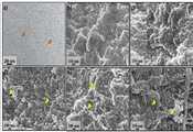
도 3. (a) TEMPO-산화 셀룰로오스 나노섬유 (TOCNF)의 투과 전자 현미경(TEM) 사진이다. (b) CS; (c) CS/TOCNF 0.2; (d) CS/TOCNF 0.4; (e) CS/TOCNF 0.6; (f) CS/TOCNF 0.8 즉, 키토산 (CS)과 다른 농도의 TOCNF를 포함하는 CS/TOCNF 하이드로겔의 주사 전자 현미경 (SEM) 사진이다. 오렌지색 화살표는 TEMPO-산화 셀룰로오스 나노섬유를 나타내고 노란색 화살표는 겔화 과정을 통해 형성된 나노입자를 나타낸다.
도 4. (A) TEMPO-산화 셀룰로오스 나노섬유 (TOCNF)와 키토산 (CS) 원료 물질, CS 및 다른 농도의 TOCNF를 포함하는 CS/TOCNF 하이드로겔의 푸리에 변환 적외선 (FT-IR) 스펙트럼을 나타낸다. (B) CS 및 CS/TOCNF 하이드로겔의 X선 회절 (XRD) 스펙트럼을 나타낸다.
도 5. MTT 분석 및 LDH 세포독성 분석에 의한 시험관 내 생체 적합성 분석과 공초점 현미경을 통해 관찰한 37oC, 5% CO2에서 7일간 배양된 CS와 CS/OCNF 하이드로겔에서의 조골세포 MC3T3-E1 (A, B, C)와 섬유아세포 L929 (D, E, F) 세포주의 세포 부착 거동을 나타낸다. 표시한 값은 SD의 6개 값의 평균이다.
도 6. 이식 7일과 14일 후 하이드로겔 형상과 이식 샘플에 대한 염증 반응을 보여 주는 스프라그-다울리 (Sprague Dawley) 쥐에 피하 주사된 CS와 CS/TOCNF의 헤마톡실린과 에오신(H&E) 염색된 조직 절편을 나타낸다.
도 7. 세포가 증식(cell infiltration)된 H & E 염색된 CS와 CS/TOCNF 0.4 조직 절편과 하이드로겔 샘플에서 관찰되는 세포 기질 형성물(화살표로 나타냄)의 고배율 사진을 나타낸다.
도 8. 이식 7일과 14일 후 이식된 CS와 CS/TOCNF 0.4 샘플을 둘러싸는 M2형 대식세포의 존재를 나타내는 면역형광 염색 사진을 나타낸다. pan 대식 세포, M0 (청색), M1 (적색) 및 M2 (녹색) 유형의 대식세포. 하이드로 겔 샘플 영역 -H 및 계면 -I가 표시되어 있다.1. Schematic illustration of CS / TOCNF hydrogel synthesis.
2. (A) Optical photographs of a thermosensitive (thermosensitive) CS hydrogel, (B) CS gelation time at 37 o C for various concentrations of CS / TOCNF hydrogel. (C) The degradation profile of the hydrogel immersed in PBS (1 mg / ml) containing lysozyme.
3. (a) Transmission electron microscope (TEM) photograph of TEMPO-oxidized cellulose nanofiber (TOCNF). (b) CS; (c) CS / TOCNF 0.2; (d) CS / TOCNF 0.4; (e) CS / TOCNF 0.6; (f) CS / TOCNF 0.8. That is, a scanning electron microscope (SEM) photograph of a CS / TOCNF hydrogel containing chitosan (CS) and TOCNF at different concentrations. The orange arrows represent the TEMPO-oxidized cellulose nanofibers and the yellow arrows represent the nanoparticles formed through the gelling process.
4. (A) Fourier transform infrared (FT-IR) spectra of CS / TOCNF hydrogels containing TEMPO-oxidized cellulose nanofibers (TOCNF) and chitosan (CS) raw materials, CS and TOCNF at different concentrations. (B) X-ray diffraction (XRD) spectra of CS and CS / TOCNF hydrogels.
Figure 5. In vitro biocompatibility analysis by MTT assay and LDH cytotoxicity analysis, and osteoblast MC3T3-E1 in CS and CS / OCNF hydrogels cultured for 7 days at 37 ° C, 5% CO2 observed through confocal microscopy A, B and C) and fibroblast L929 (D, E, F) cell lines. The displayed value is the average of the six values of SD.
Figure 6. Hematoxylin and eosin (H & E) of CS and CS / TOCNF subcutaneously injected into Sprague Dawley rats showing the inflammatory response to hydrogel shape and graft samples after 7 and 14 days of transplantation. Dissected tissue sections.
Figure 7. High magnification photographs of H & E stained CS and CS / TOCNF 0.4 tissue sections and cell matrix formations (indicated by arrows) observed in cell infiltrated cell samples.
Figure 8 shows immunofluorescence photographs showing the presence of type M2 macrophages surrounding CS and CS / TOCNF 0.4 samples transplanted at 7 and 14 days after transplantation. Pan macrophages, macrophages of the M0 (blue), M1 (red) and M2 (green) types. Hydrogel sample region-H and interface-I are shown.
이하, 본 발명이 속하는 기술 분야에서 통상의 지식을 가진 자가 용이하게 실시할 수 있도록 본 발명의 실시예에 대하여 첨부한 도면을 참고로 하여 상세히 설명한다. 그러나 본 발명은 여러 가지 상이한 형태로 구현될 수 있으며 여기에서 설명하는 실시예에 한정되지 않는다.Hereinafter, embodiments of the present invention will be described in detail with reference to the accompanying drawings so that those skilled in the art can easily carry out the present invention. The present invention may, however, be embodied in many different forms and should not be construed as limited to the embodiments set forth herein.
실시예Example 1: CS/ 1: CS /TOCNFTOCNF하이드로겔의Hydrogel 제조 Produce
본 발명에서는 도 1에 기재된 CS/TOCNF 하이드로겔 합성의 모식도에 따라, 다음과 같은 과정으로 키토산 (CS)/ 2,2,6,6-테트라메틸-피페리딘-1-옥실(2,2,6,6-tetramethyl-piperidin-1-oxyl, TEMPO)-산화 셀룰로오스 나노섬유 (TOCNF) 하이드로겔을 제조하였다.According to the schematic diagram of the synthesis of CS / TOCNF hydrogel described in Fig. 1, chitosan (CS) / 2,2,6,6-tetramethyl-piperidine-1-oxyl , 6,6-tetramethyl-piperidin-1-oxyl, TEMPO) -oxidized cellulose nanofiber (TOCNF) hydrogel.
구체적으로, 증류수에 1% TOCNF 균질 현탁액(homogenous suspension)을 희석하고, 30분간 초음파 처리하면서, 1% (v/v) 염산 수용액을 첨가하여 pH를 2로 조정하여 다양한 농도 (0.2, 0.4, 0.6, 0.8% w/v)의 Tempo-산화 셀룰로오스 나노섬유 (TOCNF)를 함유하는 TOCNF 용액을 제조하였다.Specifically, a 1% TOCNF homogenous suspension was diluted with distilled water and sonicated for 30 minutes to adjust the pH to 2 by adding a 1% (v / v) aqueous hydrochloric acid solution at various concentrations (0.2, 0.4, 0.6 , 0.8% w / v) of Temp-oxidized cellulose nanofibers (TOCNF).
그 후, 1% (v/v) 젖산 수용액에 키토산을 용해하여 3% (w/v) 키토산 (CS) 용액을 제조하였으며, 상기 TOCNF 용액과 상기 키토산 용액을 3:1(3 CS: 1 TOCNF) (v:v)의 비로 실온에서 혼합하여 키토산/TOCNF 혼합 용액을 제조하였다. 키토산은 약 6.5의 pKa를 가지며 산성 환경에서만 용해되는 양이온성 고분자 전해질이다. 따라서, 키토산(CS)의 용매로서 젖산을 사용하여, 젖산에 균질하게 용해했다.Thereafter, 3% (w / v) chitosan (CS) solution was prepared by dissolving chitosan in a 1% (v / v) lactic acid aqueous solution. The TOCNF solution and the chitosan solution were mixed in a 3: 1 (3 CS: 1 TOCNF ) (v: v) at room temperature to prepare a chitosan / TOCNF mixed solution. Chitosan is a cationic polyelectrolyte that has a pKa of about 6.5 and is soluble only in an acidic environment. Therefore, lactic acid was used as a solvent for the chitosan (CS), and it was homogeneously dissolved in lactic acid.
마지막으로, 약 20분 동안 냉탕(ice bath)에서 4℃에서 연속 교반하면서 냉각된 CS/TOCNF 혼합용액에 β-글리세롤포스페이트(GP) 용액 60% (w/v)을 한방울씩 떨어뜨려, 키토산 (CS)/Tempo-산화 셀룰로오스 나노섬유 (TOCNF) 하이드로겔을 제조하였다.Finally, 60% (w / v) of a solution of β-glycerol phosphate (GP) was dropwise added to the cooled CS / TOCNF mixed solution while being continuously stirred at 4 ° C. in an ice bath for about 20 minutes, CS) / Tempo-oxidized cellulose nanofiber (TOCNF) hydrogel.
최종적으로 제조된 CS/TOCNF 하이드로겔에 함유된 CS와 GP의 최종 농도는 각각 2.25% (w/v)와 20% (w/v)였다. 상기 CS/TOCNF 하이드로겔은 4℃에서 액상으로 보관하였다. 또한, 상기 제조된 CS/TOCNF 하이드로겔은 37℃에서 겔화되는 것을 확인하였다.The final concentrations of CS and GP contained in the finally prepared CS / TOCNF hydrogel were 2.25% (w / v) and 20% (w / v), respectively. The CS / TOCNF hydrogel was stored in a liquid state at 4 占 폚. Also, it was confirmed that the CS / TOCNF hydrogel prepared above was gelled at 37 ° C.
실시예Example 2: CS/ 2: CS /TOCNF의Of TOCNF하이드로겔의Hydrogel겔화Gelling 분석 analysis
도 2(A)는 감열성(온도감음성) CS 하이드로겔의 광학 사진을 나타내며, (B)는 다양한 농도의 CS/TOCNF 하이드로겔의 37oC에서 겔화되는 시간을 나타낸다. (C) 리소자임 효소를 포함하는 PBS(1 mg/ml)에 침지된 하이드로겔의 분해 프로파일을 나타낸다.2 (A) shows an optical photograph of a thermosensitive (temperature-sensitive) CS hydrogel, and (B) shows the time of gelation at 37 ° C of various concentrations of CS / TOCNF hydrogel. (C) The degradation profile of the hydrogel immersed in PBS (1 mg / ml) containing lysozyme.
구체적으로, 도 2A에 제시된 바와 같이, 4℃에서 CS/TOCNF 하이드로겔은 관(tube)을 기울였을 때 쉽게 흐를 수 있는 투명한 액체 용액이지만, 온도를 37℃로 올렸을 때 탁해지고 흐름을 멈추었다. 상기와 같은 하이드로겔의 혼탁도 변화는 겔화 과정에서 하이드로겔의 솔-겔 전이에 대한 척도이다. 본 발명의 실시예 1에서 제조된 모든 하이드로겔은 겔화 진행에서 동일한 솔-겔 상태 변화를 보였다.Specifically, as shown in Figure 2A, the CS / TOCNF hydrogel at 4 占 폚 was a clear liquid solution that could easily flow when the tube was tilted, but became turbid and stopped flow when the temperature was raised to 37 占 폚. The turbidity change of the hydrogel as described above is a measure for the sol-gel transition of the hydrogel in the gelation process. All of the hydrogels prepared in Example 1 of the present invention exhibited the same sol-gel state change in the progress of gelation.
하이드로겔의 겔화 시간은 생리 온도 (37℃)에서 측정되었다. 도 2B에 나타낸 바와 같이, CS/TOCNF 하이드로겔 중 TOCNF의 농도가 0%에서 0.8% (0, 0.2, 0.4, 0.6 및 0.8 w/v %)로 증가하면, 하이드로겔의 겔화 시간은 점차 감소하였다. 이 중에, TOCNF를 첨가하지 않은 CS 하이드로겔의 겔화 시간이 가장 길었고, 0.8%의 TOCNF를 첨가한 CS/TOCNF 하이드로겔의 겔화 시간이 가장 짧았다. 이러한 결과는 CS 하이드로겔에 TOCNF를 첨가하면 하이드로겔의 겔화 속도가 증가되는 것을 입증한다. 즉, 하이드로겔 중 TOCNF의 농도가 높을수록, 겔화 속도는 빨랐다.The gelation time of the hydrogel was measured at physiological temperature (37 ° C). As shown in FIG. 2B, when the concentration of TOCNF in the CS / TOCNF hydrogel was increased from 0% to 0.8% (0, 0.2, 0.4, 0.6 and 0.8 w / v%), the gelation time of the hydrogel gradually decreased . Among them, the gelation time of the CS hydrogel not containing TOCNF was the longest, and the gelation time of the CS / TOCNF hydrogel containing 0.8% TOCNF was the shortest. These results demonstrate that the addition of TOCNF to the CS hydrogel results in an increase in the gelation rate of the hydrogel. That is, the higher the concentration of TOCNF in the hydrogel, the faster the gelation rate.
또한, 겔화 시간은 적절한 주입에 대한 허용 한계 내에서 단축되었다. 하이드로겔의 점도가 갑자기 높아지지 않았으며, 겔 형성이 지나치게 빠르지는 않아서 생체 내 시험 과정 중 바늘 막힘 현상이 발생하지 않았다. 그러나, TOCNF 함량이 높을수록 용액이 조밀해지므로(denser), 주입되는 겔은 더 단단해졌다. 또한, 본 발명자는 TOCNF과 CS를 조합했을 때 용액의 점성이 증가하여 더 빨리 겔이 형성되는 것을 관찰하였다. 즉, CS 하이드로겔에 TOCNF를 첨가하면 용액의 점도가 증가하여 겔화 공정이 가속되었다.In addition, the gelling time was shortened within the tolerance limits for proper injection. The viscosity of the hydrogel did not suddenly increase, and gel formation was not too fast, so needle clogging did not occur during in vivo testing. However, the higher the TOCNF content, the denser the solution became, and the injected gel became harder. In addition, the present inventors observed that when the TOCNF and CS were combined, the viscosity of the solution was increased and the gel was formed sooner. That is, the addition of TOCNF to the CS hydrogel accelerated the gelation process by increasing the viscosity of the solution.
실시예Example 3: CS/ 3: CS /TOCNF의Of TOCNF하이드로겔의Hydrogel 분해 프로파일 분석 Analysis of decomposition profile
도 2(C)는 리소자임 효소를 포함하는 PBS(1 mg/ml)에 침지된 하이드로겔의 분해 프로파일을 나타낸 것이다. 하이드로겔에 포함된 TOCNF의 함량을 높이면 키토산 결정 구조가 약해져서 CS/TOCNF 하이드로겔 표면은 CS 하이드로겔에 비해서 성기게 되고(looser), 다공성이 높아지고, 시험관 내 배양 중에 쉽게 분해된다. 리소자임을 포함하는 PBS에 침지한 후 7, 14, 및 35일에 하이드로겔의 형태와 크기의 변화를 도 2C에 나타내었다. 대부분의 CS/TOCNF 하이드로겔은 35일의 배양 후에 분해되고 소량만이 남았다. 이와 반대로, 35일간의 배양 후에 CS 하이드로겔은 절반으로 분해되었다. 이러한 결과를 통해, TOCNF 첨가가 CS 하이드로겔의 빠른 분해에 기여하여, 생체 내 환경과 비슷한 조건에서 분해 가능한 것으로 보아 생체내에 이식 후에도 효과적으로 분해될 수 있는 것을 알 수 있었다.2 (C) shows the degradation profile of the hydrogel immersed in PBS (1 mg / ml) containing the lysozyme enzyme. When the content of TOCNF contained in the hydrogel is increased, the crystal structure of chitosan is weakened and the surface of the CS / TOCNF hydrogel is looser, the porosity is higher than that of the CS hydrogel, and the CS / TOCNF hydrogel surface is easily decomposed during in vitro culture. Changes in the shape and size of the hydrogel at 7, 14, and 35 days after immersion in PBS containing lysozyme are shown in Figure 2C. Most CS / TOCNF hydrogels degraded after 35 days of incubation and only a small amount remained. Conversely, after 35 days of culture, the CS hydrogel was degraded in half. These results indicate that TOCNF addition contributes to the rapid degradation of CS hydrogels and can be decomposed under similar conditions to in vivo environment, and thus it can be effectively decomposed even after transplantation in vivo.
실시예Example 4: CS/ 4: CS /TOCNF의Of TOCNF하이드로겔의Hydrogel 형태 평가 Form evaluation
도 3(a)는 TEMPO-산화 셀룰로오스 나노섬유 (TOCNF)의 투과 전자 현미경(TEM) 사진이다. (b)는 CS; (c) CS/TOCNF 0.2; (d) CS/TOCNF 0.4; (e) CS/TOCNF 0.6; (f) CS/TOCNF 0.8 즉, 키토산 (CS)과 다른 농도의 TOCNF를 포함하는 CS/TOCNF 하이드로겔의 주사 전자 현미경 (SEM) 사진이다. 오렌지색 화살표는 TEMPO-산화 셀룰로오스 나노섬유를 나타내고 노란색 화살표는 겔화 과정을 통해 형성된 나노입자를 나타낸다.3 (a) is a transmission electron microscope (TEM) photograph of TEMPO-oxidized cellulose nanofiber (TOCNF). (b) CS; (c) CS / TOCNF 0.2; (d) CS / TOCNF 0.4; (e) CS / TOCNF 0.6; (f) CS / TOCNF 0.8. That is, a scanning electron microscope (SEM) photograph of a CS / TOCNF hydrogel containing chitosan (CS) and TOCNF at different concentrations. The orange arrows represent the TEMPO-oxidized cellulose nanofibers and the yellow arrows represent the nanoparticles formed through the gelling process.
도 3을 참고하면, CS 하이드로겔에 TOCNF를 첨가시 하이드로겔 형상에 상당한 영향을 미쳤다. 하이드로겔의 SEM 사진에서 하이드로겔 형상이 CS 하이드로겔에 첨가된 TOCNF의 농도에 따라 변했다(도 3b - 3f). 구체적으로, TOCNF이 첨가되지 않은 CS 하이드로겔의 표면은 거칠고 조밀하였다. 이는 다양한 거대한 다발 형태였다. 그러나, CS/TOCNF 하이드로겔의 표면은 TOCNF 농도에 비례하여 더욱 성기고(looser) 다공성이 높아졌다.(TOCNF 농도가 높을수록, 표면은 성기고 다공성이 높아졌다). 겔화 메카니즘으로부터 합성 된 나노 입자가 점차적으로 증가하는 것을 분명하게 알 수 있었다. 이러한 나노입자의 크기는 다양하였다. 이들의 형태는 TOCNF 섬유의 세로 방향으로 변하였다. CS/TOCNF 0.4 하이드로겔의 경우, 나노입자의 크기는 거의 균일하였으며, 이들 나노입자는 서로 달라붙지 않았다. 그 대신, 이들 나노입자는 다른 농도의 CS/TOCNF 하이드로겔과는 달리 표면에 균일하게 분포하였다.Referring to FIG. 3, addition of TOCNF to the CS hydrogel significantly affected the shape of the hydrogel. In the SEM photograph of the hydrogel, the shape of the hydrogel was changed according to the concentration of TOCNF added to the CS hydrogel (FIGS. 3B-3F). Specifically, the surface of the CS hydrogel not containing TOCNF was rough and dense. This was in the form of various huge bundles. However, the surface of the CS / TOCNF hydrogel increased in porosity with increasing proportion of TOCNF concentration. (The higher the TOCNF concentration, the higher the porosity and porosity of the surface). It was evident that the nanoparticles synthesized from the gelation mechanism gradually increased. The size of these nanoparticles varied. Their morphology changed in the longitudinal direction of the TOCNF fiber. In the case of the CS / TOCNF 0.4 hydrogel, the nanoparticles were almost uniform in size, and these nanoparticles did not stick together. Instead, these nanoparticles were uniformly distributed on the surface, unlike other concentrations of CS / TOCNF hydrogels.
실시예Example 5: CS/ 5: CS /TOCNF의Of TOCNF하이드로겔의HydrogelFTIRFTIR 및 AndXRDXRD 분석 analysis
또한, CS/TOCNF 하이드로겔 형성을 FTIR 시험을 통해 분석하였다. 도 4(A)는 TEMPO-산화 셀룰로오스 나노섬유 (TOCNF)와 키토산 (CS) 원료 물질, CS 및 다른 농도의 TOCNF를 포함하는 CS/TOCNF 하이드로겔의 푸리에 변환 적외선 (FT-IR) 스펙트럼을 나타낸다. (B) CS 및 CS/TOCNF 하이드로겔의 X선 회절 (XRD) 스펙트럼을 나타낸다.In addition, CS / TOCNF hydrogel formation was analyzed by FTIR test. 4 (A) shows a Fourier transform infrared (FT-IR) spectrum of a CS / TOCNF hydrogel containing TEMPO-oxidized cellulose nanofiber (TOCNF) and chitosan (CS) raw material, CS and TOCNF at different concentrations. (B) X-ray diffraction (XRD) spectra of CS and CS / TOCNF hydrogels.
키토산과 TOCNF와 같은 원료(raw materials)와 모든 CS/TOCNF 하이드로겔의 FTIR 스펙트럼은 도 4A에 나타내었다. 하이드로겔의 스텍트럼은 서로 유사하였다. 이들은 원료의 스펙트럼과 미미한 차이가 있었다. 이는 CS/TOCNF 하이드로겔이 새로운 구조를 형성했지만, 두 원료의 구조적인 특징이 남아있음을 의미한다. 하이드로겔의 2000 cm-1 내지 3950 cm-1의 스펙트럼 영역에서, N-H stretching band (3363 cm-1)와 아미노기 CONHR (2875 cm-1)의 C=O 밴드를 관찰하였다. O-H stretch 밴드 (3361 cm-1)는 약간 감소하였다. 다른 스펙트럼 영역 (680 cm-1?2000 cm-1)에서, 하이드로겔 중 프로톤 아미드기의 C=O 밴드 (1633 cm-1)는 원료 키토산에 비해서 강하였다. 그러나, 이는 원료 TOCNF보다 약했다. 아미드기 (아미드 II, 아미드 III, 및 아미드 VI)를 나타내는 680 cm-1 내지 1600 cm-1 범위에서 원료 키토산의 밴드의 기(group)는 하이드로겔의 스펙트럼에서 현저하게 감소하였다. 이는 CS의 C=O와 GP의 O-H 사이와 CS와 CS의 N-H 간의 접합(junction), CS의 N-H와 O-H간의 접합 사이에 수소 결합이 형성될 수 있음을 의미한다. 이는 CS 용해도의 감소와 친수성과 소수성 도메인의 형성을 초래하였다. 이에 의해 솔-겔 전이를 유발하였을 것으로 추측된다.The FTIR spectra of raw materials such as chitosan and TOCNF and all CS / TOCNF hydrogels are shown in Figure 4A. The hydrogels' spectra were similar to each other. These were slightly different from the spectrum of the raw materials. This means that although the CS / TOCNF hydrogel has formed a new structure, the structural characteristics of the two materials remain. The C = O band of the N-H stretching band (3363 cm -1) and the amino group CONHR (2875 cm -1) was observed in the spectrum region of the hydrogel from 2000 cm -1 to 3950 cm -1. The O-H stretch band (3361 cm -1) decreased slightly. In the other spectral region (680 cm-1? 2000 cm-1), the C = O band (1633 cm -1) of the protonamide groups in the hydrogel was stronger than the raw chitosan. However, this was weaker than the raw TOCNF. Groups of bands of raw chitosan in the range of 680 cm -1 to 1600 cm -1 representing amide groups (amide II, amide III, and amide VI) were markedly reduced in the hydrogel spectra. This means that a hydrogen bond can be formed between the junction of C = O of CS and O-H of GP, the junction of CS and N-H of CS, and the junction of N-H and O-H of CS. This resulted in a decrease in CS solubility and formation of hydrophilic and hydrophobic domains. Thereby leading to a sol-gel transition.
FTIR은 CS/TOCNF와 CS/GP 하이드로겔의 차이를 밝혀내지 못했으므로, X선 회절 연구를 수행하여 CS 하이드로겔과 CS/TOCNF 하이드로겔의 결정 구조 차이를 분석하였다. CS 하이드로겔은 XRD 패턴에서 대략 30o의 2θ에서 하나의 피크만을 나타냈지만 (도 4B), CS/TOCNF 하이드로겔은 두 개의 현저한 피크를 나타냈다. 하나의 피크는 CS 하이드로겔의 피크와 유사하였다. 그러나, 이는 더 낮은 강도와 약간 낮은 2θ를 지녔다. 다른 피크는 약 40?의 2θ에서 상당히 약하게 나타났는데, 이는 TOCNF에서만 나타나는 화학 관능기를 나타낸다. 가장 강도가 높은 CS/TOCNF 0.6을 제외하고 CS 하이드로겔 내부의 TOCNF 함량이 증가함에 따라 이들 CS/TOCNF 하이드로겔 피크의 강도는 증가하였다.Since the FTIR could not reveal the difference between CS / TOCNF and CS / GP hydrogels, X-ray diffraction studies were conducted to analyze the crystal structure differences between CS hydrogel and CS / TOCNF hydrogels. The CS hydrogel showed only one peak at about 30o 2 &thetas; in the XRD pattern (Fig. 4B), but the CS / TOCNF hydrogel showed two notable peaks. One peak was similar to the peak of the CS hydrogel. However, it has lower strength and slightly lower 2?. The other peaks were considerably weak at about 40? 2 ?, indicating that the chemical functional groups appeared only in TOCNF. The strength of these CS / TOCNF hydrogel peaks increased as the TOCNF content in CS hydrogels increased, except for the most intense CS / TOCNF 0.6.
실시예Example 6: in vitro 생체 적합성 실험 6: In vitro biocompatibility test
생체 재료를 제조하는 과정에서 신체에 이식된 외부 물질이 해로운 변화 없이 조직과 조화를 이룰 수 있는지를 결정하기 위해서는 생체 적합성을 평가하는 것이 필요하다. 본 발명에서는 먼저, 생체 적합성을 시험관 내(in vitro)에서 평가하였다. L929 섬유아세포와 조골세포 MC3T3 세포는 상처 치유와 골 재생을 위해 널리 연구되는 두 가지 인기 있는 세포주이다. 이들 세포는 CS/TOCNF 하이드로겔의 생체 적합성을 평가하기 위해 본 실험에 사용되었다.Biocompatibility needs to be assessed to determine whether external substances implanted in the body during the manufacture of biomaterials can be harmonized with the tissue without detrimental changes. In the present invention, biocompatibility was first evaluated in vitro. L929 fibroblasts and osteoblast MC3T3 cells are two popular cell lines that are widely studied for wound healing and bone regeneration. These cells were used in this experiment to assess the biocompatibility of CS / TOCNF hydrogels.
도 5는 MTT 분석 및 LDH 세포독성 분석에 의한 시험관 내 생체 적합성 분석 과 공초점 현미경을 통해 관찰한 37oC, 5% CO2에서 7일간 배양된 CS와 CS/OCNF 하이드로겔에서의 조골세포 MC3T3-E1 (A, B, C)과 섬유아세포 L929 (D, E, F) 세포주의 세포 부착 거동을 나타낸다. 표시한 값은 SD의 6개 값의 평균이다.Figure 5 shows the results of MTT assay and LDH cytotoxicity in vitro biocompatibility analysis and osteoblast MC3T3-E1 in CS and CS / OCNF hydrogels cultured for 7 days at 37 ° C, 5% CO2, A, B, C) and fibroblast L929 (D, E, F) cell lines. The displayed value is the average of the six values of SD.
세포 생존율은 MTT 분석 (생존 세포 속도)과 LDH 분석 (사멸 세포 속도)을 통해 평가되었다. MTT 분석의 결과는 전반적으로 모든 종류의 추출된 희석액에서 CS 하이드로겔에 TOCNF가 포함되는 경우, MC3T3-E1 또는 L929 세포가 더욱 잘 성장하였다. 이들 두 종류의 세포의 사멸 비율은 LDH 분석에 근거하여 낮은 것을 확인하였다 (<20 %).Cell viability was assessed by MTT assay (survival cell rate) and LDH assay (death cell rate). The results of MTT analysis showed that MC3T3-E1 or L929 cells grew better when TOCNF was included in CS hydrogels in all diluted extracts of all types. The mortality rate of these two types of cells was found to be low (<20%) based on LDH analysis.
MC3T3-E1 세포의 경우, CS/TOCNF 0.4%와 CS/TOCNF 0.6%에서 세포독성이 낮은 것을 LDH 분석을 통해 확인하였다. L929 세포의 경우도 0.2% 에서 0.6%(w/v)의 TOCNF가 포함된 하이드로겔에서 세포독성이 낮고 세포의 성장이 높은 것을 LDH 분석을 통해 확인하였다. L929 세포의 MTT 분석 (도 5D)과 LDH 분석 (도 5E)은 유사한 결과를 보여주었다.In the case of MC3T3-E1 cells, low cytotoxicity was confirmed by LDH analysis in CS / TOCNF 0.4% and CS / TOCNF 0.6%. L929 cells also showed low cytotoxicity and high cell growth in hydrogel containing 0.2% to 0.6% (w / v) TOCNF. The MTT analysis (FIG. 5D) and the LDH analysis (FIG. 5E) of L929 cells showed similar results.
L929 및 MC3T3 세포의 부착 거동은 하이드로겔 표면에 세포를 접종한 후 7 일째에 검토하였다. 도 5C (MC3T3 세포) 및 도 5F (L929 세포)는 이들 세포가 이러한 하이드로겔에 매우 잘 부착됨을 보여주었다. 이들 세포는 형광성 팔로톡신으로 염색된 액틴 필라멘트가 존재하는 CS/TOCNF 0.4 및 CS/TOCNF 0.8 표면에서 편평하게 퍼졌다. 그러나, 이들 세포는 CS 하이드로겔, CS/TOCNF 0.2, 및 CS/TOCNF 0.6 표면에서 중첩되면서 성장하여 콜로니를 형성하였다. 결론적으로, TOCNF가 첨가된 CS 하이드로겔의 생체 적합성은 향상되었으며, 이러한 양상은 세포의 종류와 CS 하이드로겔에 첨가된 TOCNF의 수준에 따라 달랐다. 하이드로겔에 대한 다양한 세포 반응은 부분적으로 양전하를 띤 CS와 음전하를 띠는 TOCNF의 조합에 기인한다. 시험관 내에서 합성된 결과로부터 0.4%의 TOCNF 농도가 두 종류의 세포가 성장, 부착 및 확산하는 데 가장 적합하였음을 확인하였다.Adhesion behavior of L929 and MC3T3 cells was examined at 7 days after inoculation of the cells on the hydrogel surface. Figure 5C (MC3T3 cells) and Figure 5F (L929 cells) showed that these cells adhere very well to these hydrogels. These cells spread flat on the surface of CS / TOCNF 0.4 and CS / TOCNF 0.8 where actin filaments stained with fluorescent palotoxin were present. However, these cells grew to form colonies while overlapping on CS hydrogel, CS / TOCNF 0.2, and CS / TOCNF 0.6 surfaces. In conclusion, the biocompatibility of TOCNF-added CS hydrogels was improved, and this aspect was dependent on the type of cells and the level of TOCNF added to the CS hydrogel. The various cellular responses to hydrogels are due to the combination of partially positively charged CS and negatively charged TOCNF. From the result of in vitro synthesis, it was confirmed that 0.4% TOCNF concentration was most suitable for growth, adhesion and diffusion of two kinds of cells.
실시예Example 7: in 7: invivovivo 생체 적합성 실험 Biocompatibility test
다음으로, 상기 제조된 CS/TOCNF 생체자료를 이용하여 생체내(in vivo)에서 생체 적합성 시험을 수행하였다. 이는 시험관 내 평가를 수행한 후에 재료에 대한 신체의 정확한 반응을 조사하는 마지막 단계이다.Next, a biocompatibility test was performed in vivo using the CS / TOCNF biomaterial prepared above. This is the final step in examining the body's exact response to the material after in vitro evaluation.
도 6은 상기 하이드로겔을 이식한 후, 이식 7일과 14일 후 하이드로겔 형상과 이식 샘플에 대한 염증 반응을 보여 주는 스프라그-다울리 (Sprague Dawley) 쥐에 피하 주사된 CS와 CS/TOCNF의 헤마톡실린과 에오신(H&E) 염색된 조직 절편을 나타낸다.FIG. 6 is a graph showing the results of immunization of CS and CS / TOCNF subcutaneously injected into Sprague Dawley rats showing the inflammatory response to the hydrogel shape and graft sample after 7 days and 14 days after transplantation of the hydrogel. Hematoxylin and eosin (H & E) stained tissue sections.
H & E 염색에서 이들 하이드로겔의 형태는 달랐다. 도 6에 나타낸 바와 같이, 이들 하이드로겔은 앞선 시험관 내 결과와 마찬가지로 2주 내에 완전히 분해되지 않았다. 쥐에 하이드로겔을 피하 주사한 후, 1주 이내에 염증 반응의 첫 번째 단계가 일어나고, 모든 하이드로겔은 세포층에 의해 캡슐화되었다. 캡슐화된 영역은 진보라색을 나타냈다. 이는 CS 하이드로겔과 CS/TOCNF 0.2 하이드로겔에서 가장 어두웠다.The shape of these hydrogels in H & E staining was different. As shown in Fig. 6, these hydrogels did not completely decompose within two weeks, as in the previous in vitro results. After subcutaneous injection of hydrogels into rats, the first step of the inflammatory reaction occurred within one week, and all the hydrogels were encapsulated by the cell layer. The encapsulated region exhibited a purplish color. It was darkest in CS hydrogel and CS / TOCNF 0.2 hydrogel.
도 7은 세포가 증식(cell infiltration)된 H & E 염색된 CS와 CS/TOCNF 0.4 조직 절편과 하이드로겔 샘플에서 관찰되는 세포 기질 형성물(화살표로 나타냄)의 고배율 사진을 나타낸다.Figure 7 shows high magnification photographs of cell matrix formations (indicated by arrows) observed in H & E stained CS and CS / TOCNF 0.4 tissue sections and hydrogel samples with cell infiltration.
2주후에는 결합 조직이 배열되면서 하이드로겔의 캡슐화가 두드러진 성장을 보였다. 캡슐화된 영역의 색이 밝아졌다. CS/TOCNF 0.4(선택 샘플)과 CS 하이드로겔의 중심부를 관찰하면, 침윤된 세포수와 생성된 세포 매트릭스는 두 하이드로겔에서 1주부터 2주까지 크게 증가했으며, 특히 CS/TOCNF 0.4에서 CS 하이드로겔 보다 더 증가하였다 (도 7).After 2 weeks, encapsulation of the hydrogel showed prominent growth as the connective tissue was arranged. The color of the encapsulated area is brightened. When CS / TOCNF 0.4 (selected sample) and the central portion of the CS hydrogel were observed, the number of infiltrated cells and the resulting cell matrix increased greatly from one week to two weeks in the two hydrogels, especially CS / TOCNF 0.4, CS hydro Gel (Fig. 7).
다음으로, 조직 절편을 면역 형광 염색하여 이식된 하이드로겔을 둘러싸는 활성 대식 세포의 유형을 확인하였다. 도 8은 이식 7일과 14일 후 이식된 CS와 CS/TOCNF 0.4 샘플을 둘러싸는 M2형 대식세포의 존재를 나타내는 면역형광 염색 사진을 나타낸다.Next, tissue sections were immunofluorescently stained to identify the type of active macrophage surrounding the transplanted hydrogel. Figure 8 shows immunofluorescence staining photographs showing the presence of M2 type macrophages surrounding CS and CS / TOCNF 0.4 samples transplanted at 7 and 14 days after transplantation.
도 8에 나타낸 바와 같이, pan 대식세포(M0 청색)의 양은 1주에서 2주까지 감소하는 것으로 나타났다. 대체 활성(alternatively activated) 대식세포 (M2 녹색 - 회복)는 2주째에 CS 하이드로겔과 CS/TOCNF 0.4 하이드로겔에 증가하였다. 반대로, 일반 활성 (classically activated) 대식세포 (M1 적색 - 전염증성)는 감소되었다. CS/TOCNF 0.4에서 대체 활성 대식세포는 CS 하이드로겔에서 보다 CS/TOCNF 0.4에서 훨씬 많이 존재하였다 (도 8?merge). 또한, 면역 형광 염색 사진에서 2주째에 CS 하이드로겔에 비해 CS/TOCNF 0.4에서 대식세포 양이 감소되었다.As shown in Fig. 8, the amount of pan macrophages (M0 blue) decreased from 1 week to 2 weeks. The alternatively activated macrophages (M2 green-recovered) increased to CS hydrogels and CS / TOCNF 0.4 hydrogels at 2 weeks. In contrast, the classically activated macrophages (M1 red-proinflammatory) were reduced. At CS / TOCNF 0.4, the alternate active macrophages were much more abundant at CS / TOCNF 0.4 than in CS hydrogels (Fig. 8? Merge). In immunofluorescent staining, the amount of macrophages was decreased at CS / TOCNF 0.4 compared to CS hydrogel at 2 weeks.
생체 내 피하 주사 결과, 대식세포의 존재를 통해 하이드로겔이 체내에서 염증 반응을 유발하는 것을 알 수 있었다. 이는 신체에 주입된 이물질에 대해 반응하는 과정으로, 피하 주사 후 초기 단계에서는 정상적인 현상이다. 대식세포의 존재는 염증성 면역 반응과 조직 재형성(remodeling) 능력을 의미한다. 2주 후에 대식세포의 양이 감소되었고, 고름이나 알러지와 같은 증상 없이 대체 활성 대식세포의 양은 증가하였는데, 이는 쥐의 건강 상태가 양호함을 의미한다. 이러한 결과를 통해 생체 내(in vivo)에서 CS/TOCNF 0.4 하이드로겔이 수용될 수 있는 조직 적합성이 있음을 알 수 있었다.As a result of in vivo subcutaneous injection, it was found that hydrogels induce inflammation reaction in the body through the presence of macrophages. This is a process that reacts to foreign substances injected into the body and is a normal phenomenon at the initial stage after subcutaneous injection. The presence of macrophages implies an inflammatory immune response and remodeling ability. After two weeks, the amount of macrophages was decreased and the amount of alternative active macrophages increased without symptoms such as pus or allergy, which means that the mice are in good health. These results indicate that CS / TOCNF 0.4 hydrogels can be accommodated in vivo.
이상에서 본 발명의 바람직한 실시예에 대하여 상세하게 설명하였지만 본 발명의 권리범위는 이에 한정되는 것은 아니고 다음의 청구범위에서 정의하고 있는 본 발명의 기본 개념을 이용한 당업자의 여러 변형 및 개량 형태 또한 본 발명의 권리범위에 속하는 것이다.While the present invention has been particularly shown and described with reference to exemplary embodiments thereof, it is to be understood that the invention is not limited to the disclosed exemplary embodiments, Of the right.
Claims (11)
Translated fromKorean젖산 수용액에 키토산을 용해하여 키토산 용액을 제조하는 단계;
상기 TOCNF 용액에 키토산 용액을 혼합하여 키토산/TOCNF 혼합 용액을 제조하는 단계; 및
상기 키토산/TOCNF 혼합 용액에 글리세롤포스페이트를 첨가하여, 주입형 온도감응성 키토산/Tempo-산화 셀룰로오스 나노섬유 하이드로겔을 제조하는 단계를 포함하는,
주입형 온도감응성 키토산/Tempo-산화 셀룰로오스 나노섬유 하이드로겔의 제조방법.A homogeneous suspension of 2,2,6,6-tetramethyl-piperidin-1-oxyl, TEMPO-oxidized cellulose nanofibers (TOCNF) in distilled water And adding an aqueous hydrochloric acid solution to prepare a TOCNF solution containing TEMPO-oxidized cellulose nanofibers (TOCNF);
Dissolving chitosan in an aqueous solution of lactic acid to prepare a chitosan solution;
Preparing a chitosan / TOCNF mixed solution by mixing the TOCNF solution with a chitosan solution; And
And adding glycerol phosphate to the chitosan / TOCNF mixed solution to prepare an injection-type thermosensitive chitosan / Tempo- oxidized cellulose nanofiber hydrogel.
(Method for preparing thermosensitive thermosensitive chitosan / Tempo- oxidized cellulose nanofiber hydrogel).
Priority Applications (2)
| Application Number | Priority Date | Filing Date | Title |
|---|---|---|---|
| KR1020170088565AKR101945938B1 (en) | 2017-07-12 | 2017-07-12 | A preparation method of injectable thermosensitive chitosan/tempo based-oxidized cellulose hydrogel |
| US15/986,985US11173229B2 (en) | 2017-07-12 | 2018-05-23 | Preparation method of injectable thermosensitive chitosan/tempo based-oxidized cellulose hydrogel |
Applications Claiming Priority (1)
| Application Number | Priority Date | Filing Date | Title |
|---|---|---|---|
| KR1020170088565AKR101945938B1 (en) | 2017-07-12 | 2017-07-12 | A preparation method of injectable thermosensitive chitosan/tempo based-oxidized cellulose hydrogel |
Publications (2)
| Publication Number | Publication Date |
|---|---|
| KR20190007562A KR20190007562A (en) | 2019-01-23 |
| KR101945938B1true KR101945938B1 (en) | 2019-02-12 |
Family
ID=65000773
Family Applications (1)
| Application Number | Title | Priority Date | Filing Date |
|---|---|---|---|
| KR1020170088565AActiveKR101945938B1 (en) | 2017-07-12 | 2017-07-12 | A preparation method of injectable thermosensitive chitosan/tempo based-oxidized cellulose hydrogel |
Country Status (2)
| Country | Link |
|---|---|
| US (1) | US11173229B2 (en) |
| KR (1) | KR101945938B1 (en) |
Cited By (1)
| Publication number | Priority date | Publication date | Assignee | Title |
|---|---|---|---|---|
| KR20220037562A (en) | 2020-09-18 | 2022-03-25 | 주식회사 씨피알에스앤티 | Edible antibacterial composite beads containing chitosan and nanocellulose and method for manufacturing the same |
Families Citing this family (13)
| Publication number | Priority date | Publication date | Assignee | Title |
|---|---|---|---|---|
| EP3539576A1 (en)* | 2018-03-13 | 2019-09-18 | Albert-Ludwigs-Universität Freiburg | Hydrogel composites comprising chitosan and cellulose nanofibers |
| US12377149B2 (en)* | 2018-06-01 | 2025-08-05 | The University Of North Carolina At Chapel Hill | Injectable thermoresponsive hydrogels as a combinatory modality for controlled drug delivery, biomaterial implant and 3D printing bioink |
| JP7319793B2 (en)* | 2019-03-05 | 2023-08-02 | 第一工業製薬株式会社 | Subcutaneous or Submucosal Bulking Agent |
| KR102612490B1 (en)* | 2019-03-21 | 2023-12-08 | 순천향대학교 산학협력단 | Heat sensitive hydrogel containing alpha-tocopherol and method for preparing the same |
| CN111180704A (en)* | 2020-01-07 | 2020-05-19 | 湖南工程学院 | Sodium-ion battery positive electrode material and preparation method and application thereof |
| CN111410753A (en)* | 2020-03-27 | 2020-07-14 | 西南大学 | Novel chitosan hydrogel and preparation method and application thereof |
| CN113150324B (en)* | 2021-04-13 | 2023-02-14 | 华南理工大学 | TEMPO oxidized Millettia speciosa champ cellulose/gellan gum pH response type hydrogel and preparation method and application thereof |
| CN113244453B (en)* | 2021-04-16 | 2022-04-19 | 北京科技大学 | Preparation method and application of controllable multistage cross-linked injectable thermally induced phase change hydrogel |
| CN113941026A (en)* | 2021-10-25 | 2022-01-18 | 浙江中医药大学 | Bioactive glass-coated chitosan cellulose derivative-based injectable hydrogel dressing and preparation method thereof |
| CN114452436B (en)* | 2022-03-15 | 2022-08-23 | 四川大学 | Collagen-based injectable self-repairing hydrogel and preparation method thereof |
| CN114848583A (en)* | 2022-05-03 | 2022-08-05 | 安徽大学 | Preparation method and application of water-soluble chitosan nanoparticle-temperature-sensitive hydrogel composite |
| CN115850733B (en)* | 2022-12-06 | 2023-10-20 | 成都市第三人民医院 | An injectable nanoclay hydrogel and its preparation method and application |
| CN115845148A (en)* | 2022-12-07 | 2023-03-28 | 中国医学科学院北京协和医院 | Thermosensitive injectable chitosan/silk fibroin/hydrotalcite composite hydrogel and preparation method and application thereof |
Citations (6)
| Publication number | Priority date | Publication date | Assignee | Title |
|---|---|---|---|---|
| JP4593277B2 (en) | 2002-09-18 | 2010-12-08 | ジョンソン・アンド・ジョンソン・メディカル・リミテッド | Wound dressing composition containing chitosan and oxidized cellulose |
| EP2308382A1 (en) | 2009-10-08 | 2011-04-13 | Tyco Healthcare Group LP | Wound closure device |
| KR101091028B1 (en) | 2009-07-02 | 2011-12-09 | 아주대학교산학협력단 | In situ forming hydrogel and biomedical use thereof |
| JP5024262B2 (en) | 2008-10-16 | 2012-09-12 | 凸版印刷株式会社 | Material and biocompatible material coated or impregnated with oxidized polysaccharide material |
| KR101293618B1 (en) | 2010-02-25 | 2013-08-13 | 경북대학교병원 | Cell injectable hydrogel copolymerized with biodegradable synthetic polymers and the preparation method thereof |
| KR101492051B1 (en) | 2013-07-08 | 2015-02-16 | 아주대학교산학협력단 | hydrogel prepared by electrostatic attraction between cationic material and anionic material, and method for preparing the same |
Family Cites Families (2)
| Publication number | Priority date | Publication date | Assignee | Title |
|---|---|---|---|---|
| KR101103423B1 (en) | 2009-09-04 | 2012-01-06 | 아주대학교산학협력단 | Bio-injectable tissue adhesive hydrogels and their biomedical uses |
| US9782430B2 (en)* | 2013-03-15 | 2017-10-10 | Covidien Lp | Resorbable oxidized cellulose embolization solution |
- 2017
- 2017-07-12KRKR1020170088565Apatent/KR101945938B1/enactiveActive
- 2018
- 2018-05-23USUS15/986,985patent/US11173229B2/enactiveActive
Patent Citations (6)
| Publication number | Priority date | Publication date | Assignee | Title |
|---|---|---|---|---|
| JP4593277B2 (en) | 2002-09-18 | 2010-12-08 | ジョンソン・アンド・ジョンソン・メディカル・リミテッド | Wound dressing composition containing chitosan and oxidized cellulose |
| JP5024262B2 (en) | 2008-10-16 | 2012-09-12 | 凸版印刷株式会社 | Material and biocompatible material coated or impregnated with oxidized polysaccharide material |
| KR101091028B1 (en) | 2009-07-02 | 2011-12-09 | 아주대학교산학협력단 | In situ forming hydrogel and biomedical use thereof |
| EP2308382A1 (en) | 2009-10-08 | 2011-04-13 | Tyco Healthcare Group LP | Wound closure device |
| KR101293618B1 (en) | 2010-02-25 | 2013-08-13 | 경북대학교병원 | Cell injectable hydrogel copolymerized with biodegradable synthetic polymers and the preparation method thereof |
| KR101492051B1 (en) | 2013-07-08 | 2015-02-16 | 아주대학교산학협력단 | hydrogel prepared by electrostatic attraction between cationic material and anionic material, and method for preparing the same |
Non-Patent Citations (1)
| Title |
|---|
| De France, K. et al., CHEM. MATER.(2017.4.16) Vol.29, pp.4609-4631 |
Cited By (1)
| Publication number | Priority date | Publication date | Assignee | Title |
|---|---|---|---|---|
| KR20220037562A (en) | 2020-09-18 | 2022-03-25 | 주식회사 씨피알에스앤티 | Edible antibacterial composite beads containing chitosan and nanocellulose and method for manufacturing the same |
Also Published As
| Publication number | Publication date |
|---|---|
| US20190015550A1 (en) | 2019-01-17 |
| US11173229B2 (en) | 2021-11-16 |
| KR20190007562A (en) | 2019-01-23 |
Similar Documents
| Publication | Publication Date | Title |
|---|---|---|
| KR101945938B1 (en) | A preparation method of injectable thermosensitive chitosan/tempo based-oxidized cellulose hydrogel | |
| Cui et al. | An injectable and self-healing cellulose nanofiber-reinforced alginate hydrogel for bone repair | |
| Dutta et al. | 3D-printed bioactive and biodegradable hydrogel scaffolds of alginate/gelatin/cellulose nanocrystals for tissue engineering | |
| Nguyen et al. | In vitro and in vivo acute response towards injectable thermosensitive chitosan/TEMPO-oxidized cellulose nanofiber hydrogel | |
| Pan et al. | Preparation and characterization of electrospun PLCL/poloxamer nanofibers and dextran/gelatin hydrogels for skin tissue engineering | |
| Yang et al. | Bioinspired poly (γ-glutamic acid) hydrogels for enhanced chondrogenesis of bone marrow-derived mesenchymal stem cells | |
| Wang et al. | The functional response of alginate-gelatin-nanocrystalline cellulose injectable hydrogels toward delivery of cells and bioactive molecules | |
| Yan et al. | Injectable in situ forming poly (l-glutamic acid) hydrogels for cartilage tissue engineering | |
| Nezhad-Mokhtari et al. | Reinforcement of hydrogel scaffold using oxidized-guar gum incorporated with curcumin-loaded zein nanoparticles to improve biological performance | |
| Chen et al. | Novel thermally sensitive pH-dependent chitosan/carboxymethyl cellulose hydrogels | |
| Yan et al. | Biocompatibility evaluation of chitosan-based injectable hydrogels for the culturing mice mesenchymal stem cells in vitro | |
| Zhao et al. | Reactive electrospinning of composite nanofibers of carboxymethyl chitosan cross-linked by alginate dialdehyde with the aid of polyethylene oxide | |
| Zhang et al. | A highly transparent, elastic, injectable sericin hydrogel induced by ultrasound | |
| US20210402065A1 (en) | Functionally Gradient Material for Guided Periodontal Hard and Soft Tissue Regeneration and A Preparation Method Thereof | |
| Pan et al. | Fabrication of modified dextran–gelatin in situ forming hydrogel and application in cartilage tissue engineering | |
| KR20230059749A (en) | Hydrophilic injection type skin filling composition, preparation method and application thereof | |
| Yu et al. | Double network microcrystalline cellulose hydrogels with high mechanical strength and biocompatibility for cartilage tissue engineering scaffold | |
| CN110760103B (en) | Viscoelastic hydrogel and preparation method and application thereof | |
| CN101632841A (en) | Tissue engineering scaffold containing alginate, glycosaminoglycan and collagen and preparation method thereof | |
| Shi et al. | Effective delivery of mitomycin‐C and meloxicam by double‐layer electrospun membranes for the prevention of epidural adhesions | |
| Wu et al. | Mechanical properties and permeability of porous chitosan–poly (p-dioxanone)/silk fibroin conduits used for peripheral nerve repair | |
| Li et al. | Novel multifunctional PB and PBH hydrogels as soft filler for tissue engineering | |
| CN109970998A (en) | A kind of method and application of preparing GelMA macroporous hydrogel by Pickering emulsion method | |
| Baek et al. | Evaluation of cartilage regeneration in gellan gum/agar blended hydrogel with improved injectability | |
| Pan et al. | One-step cross-linked injectable hydrogels with tunable properties for space-filling scaffolds in tissue engineering |
Legal Events
| Date | Code | Title | Description |
|---|---|---|---|
| A201 | Request for examination | ||
| PA0109 | Patent application | Patent event code:PA01091R01D Comment text:Patent Application Patent event date:20170712 | |
| PA0201 | Request for examination | ||
| N231 | Notification of change of applicant | ||
| PN2301 | Change of applicant | Patent event date:20171215 Comment text:Notification of Change of Applicant Patent event code:PN23011R01D | |
| E701 | Decision to grant or registration of patent right | ||
| PE0701 | Decision of registration | Patent event code:PE07011S01D Comment text:Decision to Grant Registration Patent event date:20181101 | |
| PG1501 | Laying open of application | ||
| GRNT | Written decision to grant | ||
| PR0701 | Registration of establishment | Comment text:Registration of Establishment Patent event date:20190130 Patent event code:PR07011E01D | |
| PR1002 | Payment of registration fee | Payment date:20190131 End annual number:3 Start annual number:1 | |
| PG1601 | Publication of registration | ||
| PR1001 | Payment of annual fee | Payment date:20220118 Start annual number:4 End annual number:4 | |
| PR1001 | Payment of annual fee | Payment date:20230102 Start annual number:5 End annual number:5 | |
| PR1001 | Payment of annual fee | Payment date:20231227 Start annual number:6 End annual number:6 | |
| PR1001 | Payment of annual fee | Payment date:20241224 Start annual number:7 End annual number:7 |