KR101733564B1 - Pharmaceutical composition for preventing or treating of systemic inflammatory disease comprising diketopiperazine as an active ingredient - Google Patents
Pharmaceutical composition for preventing or treating of systemic inflammatory disease comprising diketopiperazine as an active ingredientDownload PDFInfo
- Publication number
- KR101733564B1 KR101733564B1KR1020160030680AKR20160030680AKR101733564B1KR 101733564 B1KR101733564 B1KR 101733564B1KR 1020160030680 AKR1020160030680 AKR 1020160030680AKR 20160030680 AKR20160030680 AKR 20160030680AKR 101733564 B1KR101733564 B1KR 101733564B1
- Authority
- KR
- South Korea
- Prior art keywords
- hmgb1
- diketopiperazine
- hours
- clp
- cells
- Prior art date
- Legal status (The legal status is an assumption and is not a legal conclusion. Google has not performed a legal analysis and makes no representation as to the accuracy of the status listed.)
- Active
Links
- BXRNXXXXHLBUKK-UHFFFAOYSA-Npiperazine-2,5-dioneChemical compoundO=C1CNC(=O)CN1BXRNXXXXHLBUKK-UHFFFAOYSA-N0.000titleclaimsabstractdescription78
- 208000027866inflammatory diseaseDiseases0.000titleclaimsabstractdescription23
- 230000009885systemic effectEffects0.000titleclaimsabstractdescription23
- 239000008194pharmaceutical compositionSubstances0.000titleclaimsabstractdescription20
- 239000004480active ingredientSubstances0.000titleclaimsabstractdescription13
- 102100037907High mobility group protein B1Human genes0.000claimsabstractdescription116
- 101001025337Homo sapiens High mobility group protein B1Proteins0.000claimsabstractdescription116
- 238000011282treatmentMethods0.000claimsabstractdescription24
- 238000000034methodMethods0.000claimsabstractdescription14
- 239000000126substanceSubstances0.000claimsabstractdescription8
- 230000001404mediated effectEffects0.000claimsdescription25
- 206010040047SepsisDiseases0.000claimsdescription15
- 208000010718Multiple Organ FailureDiseases0.000claimsdescription12
- 208000029744multiple organ dysfunction syndromeDiseases0.000claimsdescription12
- 206010051379Systemic Inflammatory Response SyndromeDiseases0.000claimsdescription6
- 208000009304Acute Kidney InjuryDiseases0.000claimsdescription5
- 208000033626Renal failure acuteDiseases0.000claimsdescription5
- 201000011040acute kidney failureDiseases0.000claimsdescription5
- 206010069351acute lung injuryDiseases0.000claimsdescription4
- 206010040070Septic ShockDiseases0.000claimsdescription2
- 230000036303septic shockEffects0.000claimsdescription2
- 238000011321prophylaxisMethods0.000claims1
- 230000028327secretionEffects0.000abstractdescription19
- 230000028709inflammatory responseEffects0.000abstractdescription9
- 230000008741proinflammatory signaling processEffects0.000abstractdescription5
- 230000002792vascularEffects0.000abstractdescription5
- 230000002265preventionEffects0.000abstractdescription3
- 210000004027cellAnatomy0.000description69
- 241000699670Mus sp.Species0.000description25
- 230000000694effectsEffects0.000description25
- 239000002158endotoxinSubstances0.000description24
- 229920006008lipopolysaccharidePolymers0.000description22
- 241000699666Mus <mouse, genus>Species0.000description19
- 238000002474experimental methodMethods0.000description18
- 210000002889endothelial cellAnatomy0.000description17
- OKKJLVBELUTLKV-UHFFFAOYSA-NMethanolChemical compoundOCOKKJLVBELUTLKV-UHFFFAOYSA-N0.000description15
- 210000000265leukocyteAnatomy0.000description15
- 230000001965increasing effectEffects0.000description14
- 210000004072lungAnatomy0.000description14
- 230000005012migrationEffects0.000description12
- 238000013508migrationMethods0.000description12
- 102000016289Cell Adhesion MoleculesHuman genes0.000description11
- 108010067225Cell Adhesion MoleculesProteins0.000description11
- 238000002965ELISAMethods0.000description11
- 108060008682Tumor Necrosis FactorProteins0.000description11
- 108010000134Vascular Cell Adhesion Molecule-1Proteins0.000description10
- 102100023543Vascular cell adhesion protein 1Human genes0.000description10
- 208000015181infectious diseaseDiseases0.000description10
- 230000004054inflammatory processEffects0.000description10
- 239000000203mixtureSubstances0.000description10
- 210000000440neutrophilAnatomy0.000description10
- 206010061218InflammationDiseases0.000description9
- 108010064593Intercellular Adhesion Molecule-1Proteins0.000description9
- 102000015271Intercellular Adhesion Molecule-1Human genes0.000description9
- 108010057466NF-kappa BProteins0.000description9
- 102000003945NF-kappa BHuman genes0.000description9
- 102100040247Tumor necrosis factorHuman genes0.000description9
- 230000035699permeabilityEffects0.000description9
- 210000001519tissueAnatomy0.000description9
- 102000007469ActinsHuman genes0.000description8
- 108010085238ActinsProteins0.000description8
- 208000034486Multi-organ failureDiseases0.000description8
- 230000004913activationEffects0.000description8
- 238000001727in vivoMethods0.000description8
- COXVTLYNGOIATD-HVMBLDELSA-NCC1=C(C=CC(=C1)C1=CC(C)=C(C=C1)\N=N\C1=C(O)C2=C(N)C(=CC(=C2C=C1)S(O)(=O)=O)S(O)(=O)=O)\N=N\C1=CC=C2C(=CC(=C(N)C2=C1O)S(O)(=O)=O)S(O)(=O)=OChemical compoundCC1=C(C=CC(=C1)C1=CC(C)=C(C=C1)\N=N\C1=C(O)C2=C(N)C(=CC(=C2C=C1)S(O)(=O)=O)S(O)(=O)=O)\N=N\C1=CC=C2C(=CC(=C(N)C2=C1O)S(O)(=O)=O)S(O)(=O)=OCOXVTLYNGOIATD-HVMBLDELSA-N0.000description7
- 238000010171animal modelMethods0.000description7
- 229960003699evans blueDrugs0.000description7
- 230000002757inflammatory effectEffects0.000description7
- 238000002360preparation methodMethods0.000description7
- 239000000243solutionSubstances0.000description7
- 230000004083survival effectEffects0.000description7
- MZOFCQQQCNRIBI-VMXHOPILSA-N(3s)-4-[[(2s)-1-[[(2s)-1-[[(1s)-1-carboxy-2-hydroxyethyl]amino]-4-methyl-1-oxopentan-2-yl]amino]-5-(diaminomethylideneamino)-1-oxopentan-2-yl]amino]-3-[[2-[[(2s)-2,6-diaminohexanoyl]amino]acetyl]amino]-4-oxobutanoic acidChemical compoundOC[C@@H](C(O)=O)NC(=O)[C@H](CC(C)C)NC(=O)[C@H](CCCN=C(N)N)NC(=O)[C@H](CC(O)=O)NC(=O)CNC(=O)[C@@H](N)CCCCNMZOFCQQQCNRIBI-VMXHOPILSA-N0.000description6
- CURLTUGMZLYLDI-UHFFFAOYSA-NCarbon dioxideChemical compoundO=C=OCURLTUGMZLYLDI-UHFFFAOYSA-N0.000description6
- 102000004889Interleukin-6Human genes0.000description6
- 108090001005Interleukin-6Proteins0.000description6
- FAPWRFPIFSIZLT-UHFFFAOYSA-MSodium chlorideChemical compound[Na+].[Cl-]FAPWRFPIFSIZLT-UHFFFAOYSA-M0.000description6
- 230000004888barrier functionEffects0.000description6
- 230000003247decreasing effectEffects0.000description6
- 102000002574p38 Mitogen-Activated Protein KinasesHuman genes0.000description6
- 108010068338p38 Mitogen-Activated Protein KinasesProteins0.000description6
- 230000004044responseEffects0.000description6
- 239000002356single layerSubstances0.000description6
- 238000001356surgical procedureMethods0.000description6
- 102000015689E-SelectinHuman genes0.000description5
- 108010024212E-SelectinProteins0.000description5
- LYCAIKOWRPUZTN-UHFFFAOYSA-NEthylene glycolChemical compoundOCCOLYCAIKOWRPUZTN-UHFFFAOYSA-N0.000description5
- 101000831567Homo sapiens Toll-like receptor 2Proteins0.000description5
- 101000669447Homo sapiens Toll-like receptor 4Proteins0.000description5
- 208000004852Lung InjuryDiseases0.000description5
- 102100024333Toll-like receptor 2Human genes0.000description5
- 102100039360Toll-like receptor 4Human genes0.000description5
- 206010069363Traumatic lung injuryDiseases0.000description5
- 238000003556assayMethods0.000description5
- 230000015572biosynthetic processEffects0.000description5
- 210000004369bloodAnatomy0.000description5
- 239000008280bloodSubstances0.000description5
- 239000001045blue dyeSubstances0.000description5
- 230000008595infiltrationEffects0.000description5
- 238000001764infiltrationMethods0.000description5
- 231100000515lung injuryToxicity0.000description5
- 238000004519manufacturing processMethods0.000description5
- 238000007911parenteral administrationMethods0.000description5
- 230000007112pro inflammatory responseEffects0.000description5
- 241000894006BacteriaSpecies0.000description4
- 102000004127CytokinesHuman genes0.000description4
- 108090000695CytokinesProteins0.000description4
- IAZDPXIOMUYVGZ-UHFFFAOYSA-NDimethylsulphoxideChemical compoundCS(C)=OIAZDPXIOMUYVGZ-UHFFFAOYSA-N0.000description4
- WZUVPPKBWHMQCE-UHFFFAOYSA-NHaematoxylinChemical compoundC12=CC(O)=C(O)C=C2CC2(O)C1C1=CC=C(O)C(O)=C1OC2WZUVPPKBWHMQCE-UHFFFAOYSA-N0.000description4
- 102000019149MAP kinase activity proteinsHuman genes0.000description4
- 108040008097MAP kinase activity proteinsProteins0.000description4
- KPKZJLCSROULON-QKGLWVMZSA-NPhalloidinChemical compoundN1C(=O)[C@@H]([C@@H](O)C)NC(=O)[C@H](C)NC(=O)[C@H](C[C@@](C)(O)CO)NC(=O)[C@H](C2)NC(=O)[C@H](C)NC(=O)[C@@H]3C[C@H](O)CN3C(=O)[C@@H]1CSC1=C2C2=CC=CC=C2N1KPKZJLCSROULON-QKGLWVMZSA-N0.000description4
- 210000004534cecumAnatomy0.000description4
- 230000006378damageEffects0.000description4
- 238000000338in vitroMethods0.000description4
- 102000005962receptorsHuman genes0.000description4
- 108020003175receptorsProteins0.000description4
- 239000003981vehicleSubstances0.000description4
- QTBSBXVTEAMEQO-UHFFFAOYSA-NAcetic acidChemical compoundCC(O)=OQTBSBXVTEAMEQO-UHFFFAOYSA-N0.000description3
- 241000194110Bacillus sp. (in: Bacteria)Species0.000description3
- 108091003079Bovine Serum AlbuminProteins0.000description3
- 208000035473Communicable diseaseDiseases0.000description3
- 238000008157ELISA kitMethods0.000description3
- LFQSCWFLJHTTHZ-UHFFFAOYSA-NEthanolChemical classCCOLFQSCWFLJHTTHZ-UHFFFAOYSA-N0.000description3
- XEKOWRVHYACXOJ-UHFFFAOYSA-NEthyl acetateChemical compoundCCOC(C)=OXEKOWRVHYACXOJ-UHFFFAOYSA-N0.000description3
- 206010015548EuthanasiaDiseases0.000description3
- PEDCQBHIVMGVHV-UHFFFAOYSA-NGlycerineChemical compoundOCC(O)COPEDCQBHIVMGVHV-UHFFFAOYSA-N0.000description3
- 241001465754MetazoaSpecies0.000description3
- 238000005481NMR spectroscopyMethods0.000description3
- 206010030113OedemaDiseases0.000description3
- 206010053159Organ failureDiseases0.000description3
- 241000283984RodentiaSpecies0.000description3
- VYPSYNLAJGMNEJ-UHFFFAOYSA-NSilicium dioxideChemical compoundO=[Si]=OVYPSYNLAJGMNEJ-UHFFFAOYSA-N0.000description3
- 229920002472StarchChemical class0.000description3
- 102000002689Toll-like receptorHuman genes0.000description3
- 108020000411Toll-like receptorProteins0.000description3
- 230000002159abnormal effectEffects0.000description3
- 238000002835absorbanceMethods0.000description3
- 229910002092carbon dioxideInorganic materials0.000description3
- 230000003833cell viabilityEffects0.000description3
- 238000006243chemical reactionMethods0.000description3
- 239000013078crystalSubstances0.000description3
- 230000003436cytoskeletal effectEffects0.000description3
- 230000001419dependent effectEffects0.000description3
- 201000010099diseaseDiseases0.000description3
- 208000037265diseases, disorders, signs and symptomsDiseases0.000description3
- 239000003937drug carrierSubstances0.000description3
- 239000000975dyeSubstances0.000description3
- 239000000284extractSubstances0.000description3
- 239000012091fetal bovine serumSubstances0.000description3
- 238000009472formulationMethods0.000description3
- 230000002401inhibitory effectEffects0.000description3
- 208000014674injuryDiseases0.000description3
- 210000002540macrophageAnatomy0.000description3
- 210000001616monocyteAnatomy0.000description3
- 210000004493neutrocyteAnatomy0.000description3
- 230000003287optical effectEffects0.000description3
- 210000003200peritoneal cavityAnatomy0.000description3
- 230000026731phosphorylationEffects0.000description3
- 238000006366phosphorylation reactionMethods0.000description3
- 230000001681protective effectEffects0.000description3
- 230000035939shockEffects0.000description3
- 239000002904solventSubstances0.000description3
- 238000010186stainingMethods0.000description3
- 230000000638stimulationEffects0.000description3
- 230000008733traumaEffects0.000description3
- 230000008728vascular permeabilityEffects0.000description3
- XLYOFNOQVPJJNP-UHFFFAOYSA-NwaterSubstancesOXLYOFNOQVPJJNP-UHFFFAOYSA-N0.000description3
- GEYOCULIXLDCMW-UHFFFAOYSA-N1,2-phenylenediamineChemical compoundNC1=CC=CC=C1NGEYOCULIXLDCMW-UHFFFAOYSA-N0.000description2
- 208000026326Adult-onset Still diseaseDiseases0.000description2
- 108010088751AlbuminsProteins0.000description2
- 102000009027AlbuminsHuman genes0.000description2
- 206010002091AnaesthesiaDiseases0.000description2
- CIWBSHSKHKDKBQ-JLAZNSOCSA-NAscorbic acidChemical compoundOC[C@H](O)[C@H]1OC(=O)C(O)=C1OCIWBSHSKHKDKBQ-JLAZNSOCSA-N0.000description2
- IJGRMHOSHXDMSA-UHFFFAOYSA-NAtomic nitrogenChemical compoundN#NIJGRMHOSHXDMSA-UHFFFAOYSA-N0.000description2
- WSFSSNUMVMOOMR-UHFFFAOYSA-NFormaldehydeChemical compoundO=CWSFSSNUMVMOOMR-UHFFFAOYSA-N0.000description2
- WQZGKKKJIJFFOK-GASJEMHNSA-NGlucoseNatural productsOC[C@H]1OC(O)[C@H](O)[C@@H](O)[C@@H]1OWQZGKKKJIJFFOK-GASJEMHNSA-N0.000description2
- DHMQDGOQFOQNFH-UHFFFAOYSA-NGlycineChemical compoundNCC(O)=ODHMQDGOQFOQNFH-UHFFFAOYSA-N0.000description2
- 241000282412HomoSpecies0.000description2
- 206010020772HypertensionDiseases0.000description2
- 208000001953HypotensionDiseases0.000description2
- 108010002352Interleukin-1Proteins0.000description2
- PIWKPBJCKXDKJR-UHFFFAOYSA-NIsofluraneChemical compoundFC(F)OC(Cl)C(F)(F)FPIWKPBJCKXDKJR-UHFFFAOYSA-N0.000description2
- 238000010824Kaplan-Meier survival analysisMethods0.000description2
- ROHFNLRQFUQHCH-YFKPBYRVSA-NL-leucineChemical compoundCC(C)C[C@H](N)C(O)=OROHFNLRQFUQHCH-YFKPBYRVSA-N0.000description2
- OUYCCCASQSFEME-QMMMGPOBSA-NL-tyrosineChemical compoundOC(=O)[C@@H](N)CC1=CC=C(O)C=C1OUYCCCASQSFEME-QMMMGPOBSA-N0.000description2
- GUBGYTABKSRVRQ-QKKXKWKRSA-NLactoseNatural productsOC[C@H]1O[C@@H](O[C@H]2[C@H](O)[C@@H](O)C(O)O[C@@H]2CO)[C@H](O)[C@@H](O)[C@H]1OGUBGYTABKSRVRQ-QKKXKWKRSA-N0.000description2
- 238000000134MTT assayMethods0.000description2
- 231100000002MTT assayToxicity0.000description2
- 238000003820Medium-pressure liquid chromatographyMethods0.000description2
- JGSARLDLIJGVTE-MBNYWOFBSA-NPenicillin GChemical compoundN([C@H]1[C@H]2SC([C@@H](N2C1=O)C(O)=O)(C)C)C(=O)CC1=CC=CC=C1JGSARLDLIJGVTE-MBNYWOFBSA-N0.000description2
- 108010009711PhalloidineProteins0.000description2
- 108091000080PhosphotransferaseProteins0.000description2
- ONIBWKKTOPOVIA-UHFFFAOYSA-NProlineNatural productsOC(=O)C1CCCN1ONIBWKKTOPOVIA-UHFFFAOYSA-N0.000description2
- 102000000852Tumor Necrosis Factor-alphaHuman genes0.000description2
- 208000035868Vascular inflammationsDiseases0.000description2
- 206010047115VasculitisDiseases0.000description2
- 206010047700VomitingDiseases0.000description2
- 239000002253acidSubstances0.000description2
- 208000012998acute renal failureDiseases0.000description2
- 230000037005anaesthesiaEffects0.000description2
- 238000004458analytical methodMethods0.000description2
- 230000003110anti-inflammatory effectEffects0.000description2
- QVGXLLKOCUKJST-UHFFFAOYSA-Natomic oxygenChemical compound[O]QVGXLLKOCUKJST-UHFFFAOYSA-N0.000description2
- WQZGKKKJIJFFOK-VFUOTHLCSA-Nbeta-D-glucoseChemical compoundOC[C@H]1O[C@@H](O)[C@H](O)[C@@H](O)[C@@H]1OWQZGKKKJIJFFOK-VFUOTHLCSA-N0.000description2
- 230000037396body weightEffects0.000description2
- 238000004113cell cultureMethods0.000description2
- 239000006143cell culture mediumSubstances0.000description2
- 210000000170cell membraneAnatomy0.000description2
- 239000013553cell monolayerSubstances0.000description2
- 239000001913celluloseChemical class0.000description2
- 229920002678celluloseChemical class0.000description2
- OSASVXMJTNOKOY-UHFFFAOYSA-NchlorobutanolChemical compoundCC(C)(O)C(Cl)(Cl)ClOSASVXMJTNOKOY-UHFFFAOYSA-N0.000description2
- 238000003776cleavage reactionMethods0.000description2
- 230000034994deathEffects0.000description2
- 239000003995emulsifying agentSubstances0.000description2
- 230000003511endothelial effectEffects0.000description2
- YQGOJNYOYNNSMM-UHFFFAOYSA-NeosinChemical compound[Na+].OC(=O)C1=CC=CC=C1C1=C2C=C(Br)C(=O)C(Br)=C2OC2=C(Br)C(O)=C(Br)C=C21YQGOJNYOYNNSMM-UHFFFAOYSA-N0.000description2
- 150000002148estersChemical class0.000description2
- 238000000605extractionMethods0.000description2
- 239000000796flavoring agentSubstances0.000description2
- GNBHRKFJIUUOQI-UHFFFAOYSA-NfluoresceinChemical compoundO1C(=O)C2=CC=CC=C2C21C1=CC=C(O)C=C1OC1=CC(O)=CC=C21GNBHRKFJIUUOQI-UHFFFAOYSA-N0.000description2
- 239000007850fluorescent dyeSubstances0.000description2
- 235000013355food flavoring agentNutrition0.000description2
- 239000008098formaldehyde solutionSubstances0.000description2
- 239000007789gasSubstances0.000description2
- 239000000499gelSubstances0.000description2
- 239000011521glassSubstances0.000description2
- 210000002216heartAnatomy0.000description2
- 238000010438heat treatmentMethods0.000description2
- 238000004128high performance liquid chromatographyMethods0.000description2
- 201000001421hyperglycemiaDiseases0.000description2
- 230000036543hypotensionEffects0.000description2
- 238000003125immunofluorescent labelingMethods0.000description2
- 230000002458infectious effectEffects0.000description2
- 210000004969inflammatory cellAnatomy0.000description2
- 230000005764inhibitory processEffects0.000description2
- 238000002347injectionMethods0.000description2
- 239000007924injectionSubstances0.000description2
- 238000001990intravenous administrationMethods0.000description2
- 210000003734kidneyAnatomy0.000description2
- 230000003907kidney functionEffects0.000description2
- 239000008101lactoseSubstances0.000description2
- 230000023404leukocyte cell-cell adhesionEffects0.000description2
- 210000004185liverAnatomy0.000description2
- 239000000314lubricantSubstances0.000description2
- HQKMJHAJHXVSDF-UHFFFAOYSA-Lmagnesium stearateChemical compound[Mg+2].CCCCCCCCCCCCCCCCCC([O-])=O.CCCCCCCCCCCCCCCCCC([O-])=OHQKMJHAJHXVSDF-UHFFFAOYSA-L0.000description2
- 239000002609mediumSubstances0.000description2
- 230000004660morphological changeEffects0.000description2
- 238000010172mouse modelMethods0.000description2
- VLKZOEOYAKHREP-UHFFFAOYSA-Nn-HexaneChemical compoundCCCCCCVLKZOEOYAKHREP-UHFFFAOYSA-N0.000description2
- 210000000822natural killer cellAnatomy0.000description2
- 239000003921oilSubstances0.000description2
- 210000000056organAnatomy0.000description2
- 230000004768organ dysfunctionEffects0.000description2
- 239000003960organic solventSubstances0.000description2
- 239000001301oxygenSubstances0.000description2
- 229910052760oxygenInorganic materials0.000description2
- LXCFILQKKLGQFO-UHFFFAOYSA-Np-hydroxybenzoic acid methyl esterNatural productsCOC(=O)C1=CC=C(O)C=C1LXCFILQKKLGQFO-UHFFFAOYSA-N0.000description2
- 230000036961partial effectEffects0.000description2
- 102000013415peroxidase activity proteinsHuman genes0.000description2
- 108040007629peroxidase activity proteinsProteins0.000description2
- 239000000546pharmaceutical excipientSubstances0.000description2
- 102000020233phosphotransferaseHuman genes0.000description2
- 239000004033plasticSubstances0.000description2
- 229920003023plasticPolymers0.000description2
- 235000013855polyvinylpyrrolidoneNutrition0.000description2
- 239000001267polyvinylpyrrolidoneSubstances0.000description2
- 229920000036polyvinylpyrrolidonePolymers0.000description2
- 239000011148porous materialSubstances0.000description2
- 239000003755preservative agentSubstances0.000description2
- QELSKZZBTMNZEB-UHFFFAOYSA-NpropylparabenChemical compoundCCCOC(=O)C1=CC=C(O)C=C1QELSKZZBTMNZEB-UHFFFAOYSA-N0.000description2
- 108090000623proteins and genesProteins0.000description2
- 230000002829reductive effectEffects0.000description2
- 230000022532regulation of transcription, DNA-dependentEffects0.000description2
- 230000001105regulatory effectEffects0.000description2
- 230000029058respiratory gaseous exchangeEffects0.000description2
- 230000036387respiratory rateEffects0.000description2
- 230000007017scissionEffects0.000description2
- 239000013049sedimentSubstances0.000description2
- 208000013223septicemiaDiseases0.000description2
- 206010040560shockDiseases0.000description2
- 230000019491signal transductionEffects0.000description2
- 239000000741silica gelSubstances0.000description2
- 229910002027silica gelInorganic materials0.000description2
- GEHJYWRUCIMESM-UHFFFAOYSA-Lsodium sulfiteChemical compound[Na+].[Na+].[O-]S([O-])=OGEHJYWRUCIMESM-UHFFFAOYSA-L0.000description2
- 241000894007speciesSpecies0.000description2
- 238000002798spectrophotometry methodMethods0.000description2
- 239000003381stabilizerSubstances0.000description2
- 235000019698starchNutrition0.000description2
- 238000007619statistical methodMethods0.000description2
- UCSJYZPVAKXKNQ-HZYVHMACSA-NstreptomycinChemical compoundCN[C@H]1[C@H](O)[C@@H](O)[C@H](CO)O[C@H]1O[C@@H]1[C@](C=O)(O)[C@H](C)O[C@H]1O[C@@H]1[C@@H](NC(N)=N)[C@H](O)[C@@H](NC(N)=N)[C@H](O)[C@H]1OUCSJYZPVAKXKNQ-HZYVHMACSA-N0.000description2
- 239000000758substrateSubstances0.000description2
- 235000000346sugarNutrition0.000description2
- 208000024891symptomDiseases0.000description2
- 230000008718systemic inflammatory responseEffects0.000description2
- CZDYPVPMEAXLPK-UHFFFAOYSA-NtetramethylsilaneChemical compoundC[Si](C)(C)CCZDYPVPMEAXLPK-UHFFFAOYSA-N0.000description2
- 230000001225therapeutic effectEffects0.000description2
- 230000006453vascular barrier functionEffects0.000description2
- 208000019553vascular diseaseDiseases0.000description2
- 210000003556vascular endothelial cellAnatomy0.000description2
- 230000008673vomitingEffects0.000description2
- 238000005406washingMethods0.000description2
- 239000000080wetting agentSubstances0.000description2
- LNAZSHAWQACDHT-XIYTZBAFSA-N(2r,3r,4s,5r,6s)-4,5-dimethoxy-2-(methoxymethyl)-3-[(2s,3r,4s,5r,6r)-3,4,5-trimethoxy-6-(methoxymethyl)oxan-2-yl]oxy-6-[(2r,3r,4s,5r,6r)-4,5,6-trimethoxy-2-(methoxymethyl)oxan-3-yl]oxyoxaneChemical compoundCO[C@@H]1[C@@H](OC)[C@H](OC)[C@@H](COC)O[C@H]1O[C@H]1[C@H](OC)[C@@H](OC)[C@H](O[C@H]2[C@@H]([C@@H](OC)[C@H](OC)O[C@@H]2COC)OC)O[C@@H]1COCLNAZSHAWQACDHT-XIYTZBAFSA-N0.000description1
- IXPNQXFRVYWDDI-UHFFFAOYSA-N1-methyl-2,4-dioxo-1,3-diazinane-5-carboximidamideChemical compoundCN1CC(C(N)=N)C(=O)NC1=OIXPNQXFRVYWDDI-UHFFFAOYSA-N0.000description1
- BWIUBDACZKSGBV-UHFFFAOYSA-N2-phenyl-1h-indole-4,6-dicarboximidamide;dihydrochlorideChemical compoundCl.Cl.N1C2=CC(C(=N)N)=CC(C(N)=N)=C2C=C1C1=CC=CC=C1BWIUBDACZKSGBV-UHFFFAOYSA-N0.000description1
- FWBHETKCLVMNFS-UHFFFAOYSA-N4',6-Diamino-2-phenylindolChemical compoundC1=CC(C(=N)N)=CC=C1C1=CC2=CC=C(C(N)=N)C=C2N1FWBHETKCLVMNFS-UHFFFAOYSA-N0.000description1
- 208000010444AcidosisDiseases0.000description1
- 206010001052Acute respiratory distress syndromeDiseases0.000description1
- 229920001817AgarPolymers0.000description1
- GUBGYTABKSRVRQ-XLOQQCSPSA-NAlpha-LactoseChemical compoundO[C@@H]1[C@@H](O)[C@@H](O)[C@@H](CO)O[C@H]1O[C@@H]1[C@@H](CO)O[C@H](O)[C@H](O)[C@H]1OGUBGYTABKSRVRQ-XLOQQCSPSA-N0.000description1
- 208000023275Autoimmune diseaseDiseases0.000description1
- 241000193830Bacillus <bacterium>Species0.000description1
- 208000027496Behcet diseaseDiseases0.000description1
- 208000009137Behcet syndromeDiseases0.000description1
- 206010004446Benign prostatic hyperplasiaDiseases0.000description1
- BVKZGUZCCUSVTD-UHFFFAOYSA-LCarbonateChemical compound[O-]C([O-])=OBVKZGUZCCUSVTD-UHFFFAOYSA-L0.000description1
- 206010008531ChillsDiseases0.000description1
- 206010010356Congenital anomalyDiseases0.000description1
- 208000022540Consciousness diseaseDiseases0.000description1
- 206010010904ConvulsionDiseases0.000description1
- 229920002261Corn starchPolymers0.000description1
- 229920000742CottonPolymers0.000description1
- 208000011231Crohn diseaseDiseases0.000description1
- 102000010831Cytoskeletal ProteinsHuman genes0.000description1
- 108010037414Cytoskeletal ProteinsProteins0.000description1
- FBPFZTCFMRRESA-FSIIMWSLSA-ND-GlucitolNatural productsOC[C@H](O)[C@H](O)[C@@H](O)[C@H](O)COFBPFZTCFMRRESA-FSIIMWSLSA-N0.000description1
- FBPFZTCFMRRESA-KVTDHHQDSA-ND-MannitolChemical compoundOC[C@@H](O)[C@@H](O)[C@H](O)[C@H](O)COFBPFZTCFMRRESA-KVTDHHQDSA-N0.000description1
- FBPFZTCFMRRESA-JGWLITMVSA-ND-glucitolChemical compoundOC[C@H](O)[C@@H](O)[C@H](O)[C@H](O)COFBPFZTCFMRRESA-JGWLITMVSA-N0.000description1
- 108020004414DNAProteins0.000description1
- 230000004568DNA-bindingEffects0.000description1
- 206010012735DiarrhoeaDiseases0.000description1
- 108010016626DipeptidesProteins0.000description1
- 208000037487EndotoxemiaDiseases0.000description1
- 239000004386ErythritolSubstances0.000description1
- UNXHWFMMPAWVPI-UHFFFAOYSA-NErythritolNatural productsOCC(O)C(O)COUNXHWFMMPAWVPI-UHFFFAOYSA-N0.000description1
- 208000010201ExanthemaDiseases0.000description1
- 208000018522Gastrointestinal diseaseDiseases0.000description1
- 208000012671Gastrointestinal haemorrhagesDiseases0.000description1
- 108010010803GelatinProteins0.000description1
- 206010018366Glomerulonephritis acuteDiseases0.000description1
- SXRSQZLOMIGNAQ-UHFFFAOYSA-NGlutaraldehydeChemical compoundO=CCCCC=OSXRSQZLOMIGNAQ-UHFFFAOYSA-N0.000description1
- 239000004471GlycineSubstances0.000description1
- 108700010013HMGB1Proteins0.000description1
- 102000055207HMGB1Human genes0.000description1
- 208000032843HemorrhageDiseases0.000description1
- 108010033040HistonesProteins0.000description1
- 206010020751HypersensitivityDiseases0.000description1
- 206010021143HypoxiaDiseases0.000description1
- 238000012404In vitro experimentMethods0.000description1
- 108010063738InterleukinsProteins0.000description1
- 102000015696InterleukinsHuman genes0.000description1
- 208000003456Juvenile ArthritisDiseases0.000description1
- 206010059176Juvenile idiopathic arthritisDiseases0.000description1
- KZSNJWFQEVHDMF-BYPYZUCNSA-NL-valineChemical compoundCC(C)[C@H](N)C(O)=OKZSNJWFQEVHDMF-BYPYZUCNSA-N0.000description1
- ROHFNLRQFUQHCH-UHFFFAOYSA-NLeucineNatural productsCC(C)CC(N)C(O)=OROHFNLRQFUQHCH-UHFFFAOYSA-N0.000description1
- 241000124008MammaliaSpecies0.000description1
- 229930195725MannitolNatural products0.000description1
- 206010027417Metabolic acidosisDiseases0.000description1
- 239000004909MoisturizerSubstances0.000description1
- 206010028980NeoplasmDiseases0.000description1
- CTQNGGLPUBDAKN-UHFFFAOYSA-NO-XyleneChemical compoundCC1=CC=CC=C1CCTQNGGLPUBDAKN-UHFFFAOYSA-N0.000description1
- 206010030302OliguriaDiseases0.000description1
- 206010033645PancreatitisDiseases0.000description1
- 206010033647Pancreatitis acuteDiseases0.000description1
- 229930040373ParaformaldehydeNatural products0.000description1
- 206010033799ParalysisDiseases0.000description1
- 229920001213Polysorbate 20Polymers0.000description1
- 208000004403Prostatic HyperplasiaDiseases0.000description1
- 206010037394Pulmonary haemorrhageDiseases0.000description1
- 206010037423Pulmonary oedemaDiseases0.000description1
- 206010037660PyrexiaDiseases0.000description1
- 208000001647Renal InsufficiencyDiseases0.000description1
- 208000025747Rheumatic diseaseDiseases0.000description1
- 206010039203Road traffic accidentDiseases0.000description1
- DWAQJAXMDSEUJJ-UHFFFAOYSA-MSodium bisulfiteChemical compound[Na+].OS([O-])=ODWAQJAXMDSEUJJ-UHFFFAOYSA-M0.000description1
- 235000021355Stearic acidNutrition0.000description1
- CZMRCDWAGMRECN-UGDNZRGBSA-NSucroseChemical compoundO[C@H]1[C@H](O)[C@@H](CO)O[C@@]1(CO)O[C@@H]1[C@H](O)[C@@H](O)[C@H](O)[C@@H](CO)O1CZMRCDWAGMRECN-UGDNZRGBSA-N0.000description1
- 229930006000SucroseNatural products0.000description1
- 208000004732Systemic VasculitisDiseases0.000description1
- 208000001871TachycardiaDiseases0.000description1
- 102000005747Transcription Factor RelAHuman genes0.000description1
- 108010031154Transcription Factor RelAProteins0.000description1
- 102000040945Transcription factorHuman genes0.000description1
- 108091023040Transcription factorProteins0.000description1
- 239000013504Triton X-100Substances0.000description1
- 229920004890Triton X-100Polymers0.000description1
- 208000003443UnconsciousnessDiseases0.000description1
- 206010046851UveitisDiseases0.000description1
- KZSNJWFQEVHDMF-UHFFFAOYSA-NValineNatural productsCC(C)C(N)C(O)=OKZSNJWFQEVHDMF-UHFFFAOYSA-N0.000description1
- TVXBFESIOXBWNM-UHFFFAOYSA-NXylitolNatural productsOCCC(O)C(O)C(O)CCOTVXBFESIOXBWNM-UHFFFAOYSA-N0.000description1
- 210000000683abdominal cavityAnatomy0.000description1
- 206010000269abscessDiseases0.000description1
- DPXJVFZANSGRMM-UHFFFAOYSA-Nacetic acid;2,3,4,5,6-pentahydroxyhexanal;sodiumChemical compound[Na].CC(O)=O.OCC(O)C(O)C(O)C(O)C=ODPXJVFZANSGRMM-UHFFFAOYSA-N0.000description1
- 150000007513acidsChemical class0.000description1
- 230000001154acute effectEffects0.000description1
- 231100000851acute glomerulonephritisToxicity0.000description1
- 201000003229acute pancreatitisDiseases0.000description1
- 230000006978adaptationEffects0.000description1
- 239000000443aerosolSubstances0.000description1
- 239000008272agarSubstances0.000description1
- 229940023476agarDrugs0.000description1
- 235000010419agarNutrition0.000description1
- 235000010443alginic acidNutrition0.000description1
- 239000000783alginic acidSubstances0.000description1
- 229920000615alginic acidPolymers0.000description1
- 229960001126alginic acidDrugs0.000description1
- 150000004781alginic acidsChemical class0.000description1
- 208000030961allergic reactionDiseases0.000description1
- 150000001370alpha-amino acid derivativesChemical class0.000description1
- 235000008206alpha-amino acidsNutrition0.000description1
- 230000003321amplificationEffects0.000description1
- 208000022531anorexiaDiseases0.000description1
- 239000003242anti bacterial agentSubstances0.000description1
- 230000002421anti-septic effectEffects0.000description1
- 229940088710antibiotic agentDrugs0.000description1
- 239000003146anticoagulant agentSubstances0.000description1
- 229940127219anticoagulant drugDrugs0.000description1
- 239000004599antimicrobialSubstances0.000description1
- 239000003963antioxidant agentSubstances0.000description1
- 235000006708antioxidantsNutrition0.000description1
- 238000013459approachMethods0.000description1
- 230000006793arrhythmiaEffects0.000description1
- 206010003119arrhythmiaDiseases0.000description1
- 210000003567ascitic fluidAnatomy0.000description1
- 235000010323ascorbic acidNutrition0.000description1
- 239000011668ascorbic acidSubstances0.000description1
- 229960005070ascorbic acidDrugs0.000description1
- 230000001580bacterial effectEffects0.000description1
- 239000007640basal mediumSubstances0.000description1
- 229960000686benzalkonium chlorideDrugs0.000description1
- CADWTSSKOVRVJC-UHFFFAOYSA-Nbenzyl(dimethyl)azanium;chlorideChemical compound[Cl-].C[NH+](C)CC1=CC=CC=C1CADWTSSKOVRVJC-UHFFFAOYSA-N0.000description1
- 230000003115biocidal effectEffects0.000description1
- 230000004071biological effectEffects0.000description1
- 208000034158bleedingDiseases0.000description1
- 230000000740bleeding effectEffects0.000description1
- 230000000903blocking effectEffects0.000description1
- 230000036760body temperatureEffects0.000description1
- 238000009835boilingMethods0.000description1
- 210000004958brain cellAnatomy0.000description1
- 238000009395breedingMethods0.000description1
- 230000001488breeding effectEffects0.000description1
- 239000000872bufferSubstances0.000description1
- 239000007853buffer solutionSubstances0.000description1
- 239000001569carbon dioxideSubstances0.000description1
- 239000001768carboxy methyl celluloseSubstances0.000description1
- 239000000969carrierSubstances0.000description1
- 238000003570cell viability assayMethods0.000description1
- 230000005754cellular signalingEffects0.000description1
- 230000008859changeEffects0.000description1
- 239000003153chemical reaction reagentSubstances0.000description1
- 229960004926chlorobutanolDrugs0.000description1
- 230000002759chromosomal effectEffects0.000description1
- 238000004737colorimetric analysisMethods0.000description1
- 230000002860competitive effectEffects0.000description1
- 238000013329compoundingMethods0.000description1
- 150000001875compoundsChemical class0.000description1
- 239000012141concentrateSubstances0.000description1
- 235000008504concentrateNutrition0.000description1
- 238000004624confocal microscopyMethods0.000description1
- 230000036461convulsionEffects0.000description1
- 239000008120corn starchSubstances0.000description1
- 229940099112cornstarchDrugs0.000description1
- 239000006071creamSubstances0.000description1
- 210000004748cultured cellAnatomy0.000description1
- 210000004292cytoskeletonAnatomy0.000description1
- 150000004691decahydratesChemical class0.000description1
- 206010061428decreased appetiteDiseases0.000description1
- 230000002950deficientEffects0.000description1
- 230000006735deficitEffects0.000description1
- 230000002939deleterious effectEffects0.000description1
- 210000004443dendritic cellAnatomy0.000description1
- 239000008121dextroseSubstances0.000description1
- 235000005911dietNutrition0.000description1
- 230000037213dietEffects0.000description1
- 208000010643digestive system diseaseDiseases0.000description1
- 239000003085diluting agentSubstances0.000description1
- 231100000676disease causative agentToxicity0.000description1
- 239000007884disintegrantSubstances0.000description1
- 238000009826distributionMethods0.000description1
- 208000002173dizzinessDiseases0.000description1
- 238000012377drug deliveryMethods0.000description1
- 238000004043dyeingMethods0.000description1
- 230000004064dysfunctionEffects0.000description1
- 239000003792electrolyteSubstances0.000description1
- 238000010828elutionMethods0.000description1
- 210000004954endothelial membraneAnatomy0.000description1
- 210000003038endotheliumAnatomy0.000description1
- 238000005516engineering processMethods0.000description1
- 230000002708enhancing effectEffects0.000description1
- UNXHWFMMPAWVPI-ZXZARUISSA-NerythritolChemical compoundOC[C@H](O)[C@H](O)COUNXHWFMMPAWVPI-ZXZARUISSA-N0.000description1
- 235000019414erythritolNutrition0.000description1
- 229940009714erythritolDrugs0.000description1
- 239000002024ethyl acetate extractSubstances0.000description1
- 201000005884exanthemDiseases0.000description1
- 230000029142excretionEffects0.000description1
- 230000002169extracardiacEffects0.000description1
- 210000000416exudates and transudateAnatomy0.000description1
- 230000001815facial effectEffects0.000description1
- 238000004992fast atom bombardment mass spectroscopyMethods0.000description1
- 238000000855fermentationMethods0.000description1
- 230000004151fermentationEffects0.000description1
- 230000001605fetal effectEffects0.000description1
- 239000000945fillerSubstances0.000description1
- 239000012530fluidSubstances0.000description1
- 238000001215fluorescent labellingMethods0.000description1
- 235000003599food sweetenerNutrition0.000description1
- 229940084362foraneDrugs0.000description1
- 239000012737fresh mediumSubstances0.000description1
- 230000006870functionEffects0.000description1
- 230000009760functional impairmentEffects0.000description1
- 230000002538fungal effectEffects0.000description1
- 208000030304gastrointestinal bleedingDiseases0.000description1
- 208000018685gastrointestinal system diseaseDiseases0.000description1
- 229920000159gelatinPolymers0.000description1
- 239000008273gelatinSubstances0.000description1
- 235000019322gelatineNutrition0.000description1
- 235000011852gelatine dessertsNutrition0.000description1
- 239000008103glucoseSubstances0.000description1
- YMAWOPBAYDPSLA-UHFFFAOYSA-Nglycine anhydrideNatural products[NH3+]CC(=O)NCC([O-])=OYMAWOPBAYDPSLA-UHFFFAOYSA-N0.000description1
- 239000008187granular materialSubstances0.000description1
- 230000012010growthEffects0.000description1
- 239000001963growth mediumSubstances0.000description1
- 230000036541healthEffects0.000description1
- 210000003494hepatocyteAnatomy0.000description1
- 230000003284homeostatic effectEffects0.000description1
- 229910052739hydrogenInorganic materials0.000description1
- WGCNASOHLSPBMP-UHFFFAOYSA-NhydroxyacetaldehydeNatural productsOCC=OWGCNASOHLSPBMP-UHFFFAOYSA-N0.000description1
- 239000001866hydroxypropyl methyl celluloseSubstances0.000description1
- 235000010979hydroxypropyl methyl celluloseNutrition0.000description1
- 229920003088hydroxypropyl methyl cellulosePolymers0.000description1
- UFVKGYZPFZQRLF-UHFFFAOYSA-Nhydroxypropyl methyl celluloseChemical compoundOC1C(O)C(OC)OC(CO)C1OC1C(O)C(O)C(OC2C(C(O)C(OC3C(C(O)C(O)C(CO)O3)O)C(CO)O2)O)C(CO)O1UFVKGYZPFZQRLF-UHFFFAOYSA-N0.000description1
- 230000007954hypoxiaEffects0.000description1
- 230000028993immune responseEffects0.000description1
- 210000000987immune systemAnatomy0.000description1
- 230000001771impaired effectEffects0.000description1
- 230000001976improved effectEffects0.000description1
- 238000011534incubationMethods0.000description1
- 230000006698inductionEffects0.000description1
- 230000001939inductive effectEffects0.000description1
- 230000000977initiatory effectEffects0.000description1
- 229940100601interleukin-6Drugs0.000description1
- 230000017306interleukin-6 productionEffects0.000description1
- 230000000968intestinal effectEffects0.000description1
- 238000001361intraarterial administrationMethods0.000description1
- 230000003834intracellular effectEffects0.000description1
- 238000007918intramuscular administrationMethods0.000description1
- 238000007912intraperitoneal administrationMethods0.000description1
- 238000007913intrathecal administrationMethods0.000description1
- 229960002725isofluraneDrugs0.000description1
- 201000002215juvenile rheumatoid arthritisDiseases0.000description1
- 201000006370kidney failureDiseases0.000description1
- 238000002372labellingMethods0.000description1
- 238000002350laparotomyMethods0.000description1
- 231100000225lethalityToxicity0.000description1
- 206010024378leukocytosisDiseases0.000description1
- 239000007788liquidSubstances0.000description1
- 230000007774longtermEffects0.000description1
- 239000006210lotionSubstances0.000description1
- 210000001165lymph nodeAnatomy0.000description1
- 235000019359magnesium stearateNutrition0.000description1
- 239000000845maltitolSubstances0.000description1
- 235000010449maltitolNutrition0.000description1
- VQHSOMBJVWLPSR-WUJBLJFYSA-NmaltitolChemical compoundOC[C@H](O)[C@@H](O)[C@@H]([C@H](O)CO)O[C@H]1O[C@H](CO)[C@@H](O)[C@H](O)[C@H]1OVQHSOMBJVWLPSR-WUJBLJFYSA-N0.000description1
- 229940035436maltitolDrugs0.000description1
- 238000007726management methodMethods0.000description1
- 239000000594mannitolSubstances0.000description1
- 235000010355mannitolNutrition0.000description1
- 239000000463materialSubstances0.000description1
- 238000005399mechanical ventilationMethods0.000description1
- 210000004379membraneAnatomy0.000description1
- 239000012528membraneSubstances0.000description1
- 230000008384membrane barrierEffects0.000description1
- HEBKCHPVOIAQTA-UHFFFAOYSA-Nmeso ribitolNatural productsOCC(O)C(O)C(O)COHEBKCHPVOIAQTA-UHFFFAOYSA-N0.000description1
- 229920000609methyl cellulosePolymers0.000description1
- 239000004292methyl p-hydroxybenzoateSubstances0.000description1
- 235000010270methyl p-hydroxybenzoateNutrition0.000description1
- 239000001923methylcelluloseSubstances0.000description1
- 235000010981methylcelluloseNutrition0.000description1
- 229960002216methylparabenDrugs0.000description1
- 244000005700microbiomeSpecies0.000description1
- 210000003632microfilamentAnatomy0.000description1
- 238000007431microscopic evaluationMethods0.000description1
- 238000010232migration assayMethods0.000description1
- 238000003801millingMethods0.000description1
- 230000001333moisturizerEffects0.000description1
- 230000009456molecular mechanismEffects0.000description1
- 239000003068molecular probeSubstances0.000description1
- 238000012544monitoring processMethods0.000description1
- 210000005087mononuclear cellAnatomy0.000description1
- 208000031225myocardial ischemiaDiseases0.000description1
- VMGAPWLDMVPYIA-HIDZBRGKSA-Nn'-amino-n-iminomethanimidamideChemical classN\N=C\N=NVMGAPWLDMVPYIA-HIDZBRGKSA-N0.000description1
- 229930014626natural productNatural products0.000description1
- 230000001338necrotic effectEffects0.000description1
- 229910052757nitrogenInorganic materials0.000description1
- 231100000252nontoxicToxicity0.000description1
- 230000003000nontoxic effectEffects0.000description1
- 230000008689nuclear functionEffects0.000description1
- 230000005937nuclear translocationEffects0.000description1
- 238000003199nucleic acid amplification methodMethods0.000description1
- QIQXTHQIDYTFRH-UHFFFAOYSA-Noctadecanoic acidChemical compoundCCCCCCCCCCCCCCCCCC(O)=OQIQXTHQIDYTFRH-UHFFFAOYSA-N0.000description1
- OQCDKBAXFALNLD-UHFFFAOYSA-Noctadecanoic acidNatural productsCCCCCCCC(C)CCCCCCCCC(O)=OOQCDKBAXFALNLD-UHFFFAOYSA-N0.000description1
- 239000002674ointmentSubstances0.000description1
- 150000002894organic compoundsChemical class0.000description1
- 238000012261overproductionMethods0.000description1
- 239000012188paraffin waxSubstances0.000description1
- 229920002866paraformaldehydePolymers0.000description1
- 230000037361pathwayEffects0.000description1
- 235000021401pellet dietNutrition0.000description1
- 229940056360penicillin gDrugs0.000description1
- 230000009038pharmacological inhibitionEffects0.000description1
- 239000006187pillSubstances0.000description1
- 230000036470plasma concentrationEffects0.000description1
- 229920000729poly(L-lysine) polymerPolymers0.000description1
- 229920000515polycarbonatePolymers0.000description1
- 239000004417polycarbonateSubstances0.000description1
- 239000000256polyoxyethylene sorbitan monolaurateSubstances0.000description1
- 235000010486polyoxyethylene sorbitan monolaurateNutrition0.000description1
- 238000010837poor prognosisMethods0.000description1
- 230000034190positive regulation of NF-kappaB transcription factor activityEffects0.000description1
- 230000002980postoperative effectEffects0.000description1
- 229920001592potato starchPolymers0.000description1
- 229940116317potato starchDrugs0.000description1
- 230000003334potential effectEffects0.000description1
- 239000000843powderSubstances0.000description1
- 230000002335preservative effectEffects0.000description1
- 230000008569processEffects0.000description1
- 108090000765processed proteins & peptidesProteins0.000description1
- 238000012545processingMethods0.000description1
- 230000000770proinflammatory effectEffects0.000description1
- 150000003148prolinesChemical class0.000description1
- 230000002035prolonged effectEffects0.000description1
- 230000001737promoting effectEffects0.000description1
- 239000004405propyl p-hydroxybenzoateSubstances0.000description1
- 235000010232propyl p-hydroxybenzoateNutrition0.000description1
- 229960003415propylparabenDrugs0.000description1
- 235000018102proteinsNutrition0.000description1
- 102000004169proteins and genesHuman genes0.000description1
- 208000005333pulmonary edemaDiseases0.000description1
- 230000002685pulmonary effectEffects0.000description1
- 238000011002quantificationMethods0.000description1
- 206010037844rashDiseases0.000description1
- 230000007115recruitmentEffects0.000description1
- 230000009467reductionEffects0.000description1
- 210000002254renal arteryAnatomy0.000description1
- 238000004007reversed phase HPLCMethods0.000description1
- 229940100486rice starchDrugs0.000description1
- 239000000523sampleSubstances0.000description1
- 238000012216screeningMethods0.000description1
- 210000002966serumAnatomy0.000description1
- 230000011664signalingEffects0.000description1
- 238000010898silica gel chromatographyMethods0.000description1
- 231100000046skin rashToxicity0.000description1
- 239000002002slurrySubstances0.000description1
- 210000000813small intestineAnatomy0.000description1
- 235000010413sodium alginateNutrition0.000description1
- 239000000661sodium alginateSubstances0.000description1
- 229940005550sodium alginateDrugs0.000description1
- VWYBPWRNTMVXIP-UHFFFAOYSA-Nsodium carbonic acid azideChemical compound[Na+].[N-]=[N+]=[N-].OC(O)=OVWYBPWRNTMVXIP-UHFFFAOYSA-N0.000description1
- 235000019812sodium carboxymethyl celluloseNutrition0.000description1
- 229920001027sodium carboxymethylcellulosePolymers0.000description1
- 239000011780sodium chlorideSubstances0.000description1
- 229940079827sodium hydrogen sulfiteDrugs0.000description1
- 235000010267sodium hydrogen sulphiteNutrition0.000description1
- 229940001482sodium sulfiteDrugs0.000description1
- 235000010265sodium sulphiteNutrition0.000description1
- 239000007787solidSubstances0.000description1
- 239000012439solid excipientSubstances0.000description1
- 235000010356sorbitolNutrition0.000description1
- 239000000600sorbitolSubstances0.000description1
- 238000004611spectroscopical analysisMethods0.000description1
- 238000001228spectrumMethods0.000description1
- 239000008107starchChemical class0.000description1
- 239000008117stearic acidSubstances0.000description1
- 229960005322streptomycinDrugs0.000description1
- 238000007920subcutaneous administrationMethods0.000description1
- 125000001424substituent groupChemical group0.000description1
- 239000005720sucroseSubstances0.000description1
- 150000008163sugarsChemical class0.000description1
- 239000006228supernatantSubstances0.000description1
- 239000013589supplementSubstances0.000description1
- 239000000375suspending agentSubstances0.000description1
- 239000003765sweetening agentSubstances0.000description1
- 208000011580syndromic diseaseDiseases0.000description1
- 239000007445syp - sw mediumSubstances0.000description1
- 239000006188syrupSubstances0.000description1
- 235000020357syrupNutrition0.000description1
- 230000006794tachycardiaEffects0.000description1
- 230000008685targetingEffects0.000description1
- 238000012360testing methodMethods0.000description1
- 210000001550testisAnatomy0.000description1
- 238000004809thin layer chromatographyMethods0.000description1
- 206010043554thrombocytopeniaDiseases0.000description1
- 210000001541thymus glandAnatomy0.000description1
- 230000000451tissue damageEffects0.000description1
- 231100000827tissue damageToxicity0.000description1
- 230000000699topical effectEffects0.000description1
- 238000012546transferMethods0.000description1
- 230000006433tumor necrosis factor productionEffects0.000description1
- OUYCCCASQSFEME-UHFFFAOYSA-NtyrosineNatural productsOC(=O)C(N)CC1=CC=C(O)C=C1OUYCCCASQSFEME-UHFFFAOYSA-N0.000description1
- 238000002211ultraviolet spectrumMethods0.000description1
- 210000003606umbilical veinAnatomy0.000description1
- 210000001635urinary tractAnatomy0.000description1
- 208000019206urinary tract infectionDiseases0.000description1
- 239000004474valineSubstances0.000description1
- 239000002699waste materialSubstances0.000description1
- 239000003643water by typeSubstances0.000description1
- 238000005303weighingMethods0.000description1
- 229940100445wheat starchDrugs0.000description1
- 239000008096xyleneSubstances0.000description1
- 239000000811xylitolSubstances0.000description1
- 235000010447xylitolNutrition0.000description1
- HEBKCHPVOIAQTA-SCDXWVJYSA-NxylitolChemical compoundOC[C@H](O)[C@@H](O)[C@H](O)COHEBKCHPVOIAQTA-SCDXWVJYSA-N0.000description1
- 229960002675xylitolDrugs0.000description1
Images


Classifications
- A—HUMAN NECESSITIES
- A61—MEDICAL OR VETERINARY SCIENCE; HYGIENE
- A61K—PREPARATIONS FOR MEDICAL, DENTAL OR TOILETRY PURPOSES
- A61K9/00—Medicinal preparations characterised by special physical form
- A—HUMAN NECESSITIES
- A61—MEDICAL OR VETERINARY SCIENCE; HYGIENE
- A61K—PREPARATIONS FOR MEDICAL, DENTAL OR TOILETRY PURPOSES
- A61K31/00—Medicinal preparations containing organic active ingredients
- A61K31/33—Heterocyclic compounds
- A61K31/395—Heterocyclic compounds having nitrogen as a ring hetero atom, e.g. guanethidine or rifamycins
- A61K31/495—Heterocyclic compounds having nitrogen as a ring hetero atom, e.g. guanethidine or rifamycins having six-membered rings with two or more nitrogen atoms as the only ring heteroatoms, e.g. piperazine or tetrazines
- A61K31/4985—Pyrazines or piperazines ortho- or peri-condensed with heterocyclic ring systems
- A—HUMAN NECESSITIES
- A61—MEDICAL OR VETERINARY SCIENCE; HYGIENE
- A61K—PREPARATIONS FOR MEDICAL, DENTAL OR TOILETRY PURPOSES
- A61K9/00—Medicinal preparations characterised by special physical form
- A61K9/0012—Galenical forms characterised by the site of application
- A61K9/0019—Injectable compositions; Intramuscular, intravenous, arterial, subcutaneous administration; Compositions to be administered through the skin in an invasive manner
- A—HUMAN NECESSITIES
- A61—MEDICAL OR VETERINARY SCIENCE; HYGIENE
- A61K—PREPARATIONS FOR MEDICAL, DENTAL OR TOILETRY PURPOSES
- A61K9/00—Medicinal preparations characterised by special physical form
- A61K9/06—Ointments; Bases therefor; Other semi-solid forms, e.g. creams, sticks, gels
- A—HUMAN NECESSITIES
- A61—MEDICAL OR VETERINARY SCIENCE; HYGIENE
- A61K—PREPARATIONS FOR MEDICAL, DENTAL OR TOILETRY PURPOSES
- A61K9/00—Medicinal preparations characterised by special physical form
- A61K9/20—Pills, tablets, discs, rods
- A—HUMAN NECESSITIES
- A61—MEDICAL OR VETERINARY SCIENCE; HYGIENE
- A61K—PREPARATIONS FOR MEDICAL, DENTAL OR TOILETRY PURPOSES
- A61K9/00—Medicinal preparations characterised by special physical form
- A61K9/20—Pills, tablets, discs, rods
- A61K9/2004—Excipients; Inactive ingredients
- A61K9/2013—Organic compounds, e.g. phospholipids, fats
- A61K9/2018—Sugars, or sugar alcohols, e.g. lactose, mannitol; Derivatives thereof, e.g. polysorbates
- A—HUMAN NECESSITIES
- A61—MEDICAL OR VETERINARY SCIENCE; HYGIENE
- A61K—PREPARATIONS FOR MEDICAL, DENTAL OR TOILETRY PURPOSES
- A61K9/00—Medicinal preparations characterised by special physical form
- A61K9/20—Pills, tablets, discs, rods
- A61K9/2004—Excipients; Inactive ingredients
- A61K9/2022—Organic macromolecular compounds
- A61K9/205—Polysaccharides, e.g. alginate, gums; Cyclodextrin
Landscapes
- Health & Medical Sciences (AREA)
- Life Sciences & Earth Sciences (AREA)
- Animal Behavior & Ethology (AREA)
- Pharmacology & Pharmacy (AREA)
- Epidemiology (AREA)
- Medicinal Chemistry (AREA)
- Chemical & Material Sciences (AREA)
- General Health & Medical Sciences (AREA)
- Public Health (AREA)
- Veterinary Medicine (AREA)
- Pharmaceuticals Containing Other Organic And Inorganic Compounds (AREA)
- Biophysics (AREA)
- Molecular Biology (AREA)
- Dermatology (AREA)
Abstract
Description
Translated fromKorean본 발명은 디케토피페라진을 유효성분으로 포함하는 전신염증질환의 예방 또는 치료용 약학적 조성물에 관한 것으로, 보다 상세하게는 화학식 1 내지 화학식 3중 어느 하나로 표시되는 디케토피페라진(diketopiperazine)을 유효성분으로 포함하는 전신염증질환(systemic inflammatory disease)의 예방 또는 치료용 약학적 조성물에 관한 것이다.The present invention relates to a pharmaceutical composition for preventing or treating systemic inflammatory diseases containing diketopiperazine as an active ingredient, and more particularly, to a pharmaceutical composition for preventing or treating systemic inflammatory diseases comprising diketopiperazine as an active ingredient, more specifically, diketopiperazine represented by any one of formulas (1) to The present invention relates to a pharmaceutical composition for preventing or treating systemic inflammatory disease.
패혈증은 심각한 감염에 의한 전신성 염증 반응으로 즉시 치료하지 않으면 쇼크, 여러 장기의 손상 및 사망을 유발할 수 있고, 최신 치료법에도 불구하고, 중환자실에서 사망의 주요 원인이다. 핵 전사 인자로 알려진 HMGB1은 패혈증의 후기 단계 매개물질(late medicator)로서 관여한다(Wang et al., 2009; Yang et al., 2004). 따라서 심각한 패혈증 환자의 혈청에 HMGB1의 농도가 높으면 사망률이 증가할 가능성이 있다(Karlsson et al., 2008).Sepsis is a systemic inflammatory response caused by a serious infection that can lead to shocks, damage to multiple organs and death if not treated immediately, and is the leading cause of death in intensive care units, despite the latest treatments. HMGB1, known as nuclear transcription factor, is involved as a late mediator of sepsis (Wang et al., 2009; Yang et al., 2004). Thus, higher serum HMGB1 levels in patients with severe sepsis may increase mortality (Karlsson et al., 2008).
이에 치명적인 전신성 염증의 후기 단계 매개물질을 표적으로 하는 것은 치료 범위를 확대하고, 연속적인 염증 반응의 유해한 효과를 억제하기 위한 새로운 접근 방식을 의미한다. HMGB1은 패혈증뿐만 아니라 다른 염증성 질환에서 임상적 개입에 대한 기회를 제공하는 분자 표적이다.Targeting the late stage mediators of fatal systemic inflammation implies a new approach to broadening the therapeutic range and inhibiting the deleterious effects of subsequent inflammatory responses. HMGB1 is a molecular target that provides opportunities for clinical intervention in sepsis as well as other inflammatory diseases.
HMGB1(high mobility group box 1)은 비히스톤 핵 단백질로, 활성화된 대식세포, 수지상 세포 및 NK세포(natural killer cell)로부터 활발히 생성되며 부수적으로 괴저성 세포(necrotic cell)로부터 수동적 분비되고, 중요한 구조적 역할 및 유전자 전사 조절과 함께 세포 내 핵 기능(intracellular nuclear function)을 가지며, 세포 외 활성(extracellular activity)을 가진다. HMGB1은 인체내에서 흉선, 림프절, 고환, 태아의 간 등에서 높은 농도로 존재하며, 간세포와 뇌세포를 제외하면 대부분의 세포에서는 주로 핵 내에 존재하지만, 염증을 유발하는 물질에 의해 세포나 조직이 활성화되면 세포 밖으로 분비된다. 세포 내에서는 전사 조절 및 염색체 구조를 결정하는 DNA에 결합하며, 손상된 조직에서는 항상성 반응을 조절하는 위험신호로서 작용한다(Gallucci and Matzinger, 2001).HMGB1 (high mobility group box 1) is a non-histone nuclear protein that is actively produced from activated macrophages, dendritic cells and NK cells (natural killer cells) and is secreted passively from necrotic cells, It has intracellular nuclear function with role and gene transcription control, and has extracellular activity. HMGB1 exists in the human body at high concentrations in the thymus, lymph nodes, testis, fetal liver, etc. Except for hepatocytes and brain cells, most of the cells are mainly present in the nucleus, but the cells or tissues are activated Once secreted out of the cell. It binds to DNA that determines transcriptional regulation and chromosomal structure within the cell, and acts as a risk signal to control the homeostatic response in damaged tissues (Gallucci and Matzinger, 2001).
HMGB1은 염증의 매개 물질로서, RAGE(receptor for advanced glycation end products), TLR(toll like receptor)-2, TLR-4에 높은 친화력으로 결합하며, 내피세포에서 tumor necrosis factor(TNF), interleukin(IL)-6의 분비를 증가하고, nuclear factor(NF)-κB , extracellular regulated kinase(ERK) 1과 2의 인산화를 증가시켜, HMGB1에 의한 염증반응을 유발한다(Bae, 2012; Hori et al., 1995; Park et al., 2004). 또한 HMGB1은 NF-κB 소단위의 DNA-결합 활성(DNA-binding activity)을 향상시킴으로서 세포 접착 분자(cell adhesion molecules, CAMs)의 NF-κB 의존적 발현을 촉진하고(Agresti et al., 2003), 내피세포에서 VCAM-1, ICAM-1 및 E-slection과 같은 세포 접착 분자의 발현을 유도한다(Andersson et al., 2000; Bae, 2012; Lotze and Tracey, 2005; Yang et al., 2006). 이는 백혈구의 소집(leukocyte recruitment)을 통하여 염증을 증가시킨다.HMGB1 is a mediator of inflammation and binds with high affinity to RAGE (TLR-2) and TLR (TLR-4), and inhibits tumor necrosis factor (TNF), interleukin ) -6 and increased the phosphorylation of nuclear factor (NF) -κB and extracellular regulated
천연물에서 방부제(anti-septic)를 스크리닝 하는 과정에서, 해양 자원으로부터 추출하여 독립적인 두 가지 발효 균주의 에틸 아세테이트 추출물은 HUVEC세포에서 HMGB1의 발현을 억제하는 효과를 나타냈다. 크로마토그래피를 반복하여, 3가지 디케토피페라진(cyclo(L-Pro-D-Val), cyclo(L-Pro-L-Tyr), cyclo(L-Pro-D-Leu))을 획득하였고, 이와 같이 프롤린을 포함하는 화합물은 발효 균주, 곰팡이 균주 및 다른 미생물로부터 획득할 수 있다(Cuong et al., 2015; Park and Strobel, 1994; Adamczeski et al., 1995).In the screening of anti-septic in natural products, the ethyl acetate extract of two independent fermenting strains extracted from marine resources inhibited the expression of HMGB1 in HUVEC cells. L-Pro-D-Leu) and cyclo (L-Pro-D-Leu) Thus, compounds containing proline can be obtained from fermentation strains, fungal strains and other microorganisms (Cuong et al., 2015; Park and Strobel, 1994; Adamczeski et al., 1995).
여러 디케토피페라진 중 몇몇은 항생제와 소염 활동을 포함한 다양한 생물학적 활성이 있다고 알려졌지만, 인간 내피 세포 및 마우스에서 HMGB1- 또는 CLP- 매개된 패혈성 반응에 대한 프롤린을 함유하는 디케토피페라진의 항패혈성 효과는 알려져 있지 않다.Several of the diketopiperazines have been known to have a variety of biological activities, including antibiotic and anti-inflammatory activities, but have been shown to inhibit the anti-inflammatory properties of diketopiperazine, which contains proline for HMGB1- or CLP- mediated septic responses in human endothelial cells and mice The effect is unknown.
이에 본 발명자들은 전신염증질환에 대한 치료물질을 연구하던 중, 디케토피페라진이 HMGB1의 분비를 억제하고 이와 관련된 분자적 매커니즘을 조절하여 HMGB1에 의해 매개된 염증성 반응을 억제하고, 전신염증질환 동물 모델에서 치료효과가 있는 것을 확인하여 본 발명을 완성하였다.Accordingly, the inventors of the present invention found that diketopiperazine suppresses the secretion of HMGB1 and regulates the molecular mechanism associated therewith to inhibit the inflammatory response mediated by HMGB1, The present inventors have completed the present invention.
따라서 본 발명의 목적은 하기 화학식 1 내지 화학식 3중 어느 하나로 표시되는 디케토피페라진(diketopiperazine)을 유효성분으로 포함하는 전신염증질환(systemic inflammatory disease)의 예방 또는 치료용 약학적 조성물을 제공하는 것이다.Accordingly, an object of the present invention is to provide a pharmaceutical composition for preventing or treating systemic inflammatory diseases comprising diketopiperazine represented by any one of
<화학식 1>≪ Formula 1 >

<화학식 2>(2)

<화학식 3>(3)

상기와 같은 목적을 달성하기 위하여, 본 발명은 하기 화학식 1 내지 화학식 3중 어느 하나로 표시되는 디케토피페라진(diketopiperazine)을 유효성분으로 포함하는 전신염증질환(systemic inflammatory disease)의 예방 또는 치료용 약학적 조성물을 제공한다.In order to achieve the above object, the present invention provides a pharmaceutical composition for preventing or treating systemic inflammatory disease comprising diketopiperazine represented by any one of
<화학식 1>≪ Formula 1 >

<화학식 2>(2)

<화학식 3>(3)

이하 본 발명을 상세히 설명한다.Hereinafter, the present invention will be described in detail.
본 발명은 하기 화학식 1 내지 화학식 3중 어느 하나로 표시되는 디케토피페라진(diketopiperazine)을 유효성분으로 포함하는 전신염증질환(systemic inflammatory disease)의 예방 또는 치료용 약학적 조성물을 제공한다.The present invention provides a pharmaceutical composition for preventing or treating systemic inflammatory disease comprising diketopiperazine represented by any one of the following general formulas (1) to (3) as an active ingredient.
<화학식 1>≪ Formula 1 >

<화학식 2>(2)

<화학식 3>(3)

본 발명에서 ‘디케토피페라진(diketopiperazine)’은 유기화합물의 일종으로 2.5-디케토피페라진의 관용명이다. 이는 글리신무수물에 해당하며, 넓은 뜻으로는 치환기가 있는 경우에도 디케토피페라진이라고 한다. 이들은 결정성인 고체로서 α-아미노산이나 디펩티드를 에틸렌글리콜과 같이 끓는점이 높은 용매 내에서 끓이거나 이들 에스테르(유리형)를 가열하면 쉽게 생성된다. 특히 글리신 및 프롤린에스테르는 디케토피페라진화되기가 쉬워 이 경우는 유리에스테르를 얻는 것이 쉽지 않다. 단백질을 묽은 산 중에 특히 고온에서 가수분해할 때, 혹은 글리세롤에서 180℃ 부근으로 가열하면 여러 가지 디케토피페라진이 생성된다.In the present invention, 'diketopiperazine' is a kind of organic compound and is a common name of 2,5-diketopiperazine. This corresponds to a glycine anhydride, and is broadly referred to as diketopiperazine even in the presence of a substituent. These can be easily produced by heating an α-amino acid or a dipeptide as a crystalline solid in a solvent having a high boiling point such as ethylene glycol or heating these esters (free form). In particular, glycine and proline esters are easy to be diketopiperazinized, and in this case, it is not easy to obtain a free ester. When proteins are hydrolyzed in dilute acids, especially at high temperatures, or heated to around 180 ° C in glycerol, various diketopiperazines are produced.
본 발명에서는 제주도에서 수집한 침전물과 해면동물에서Bacillus 속 HC001 및Picicoccus 속 12L081 박테리아를 추출하였다. 추출한 박테리아를 각각 배양한 뒤, 배양한 배지를 유기용매로 추출한 뒤 농축하였다. 농축물을 VLC로 분획하여 화학식 1 내지 화학식 3의 디케토피페라진을 획득하였다.In the present invention,Bacillus sp . HC001 andPicococcus sp. 12L081 bacteria were extracted from sediments and sponges collected from Jeju Island. Each of the extracted bacteria was cultured, and the cultured medium was extracted with an organic solvent and then concentrated. The concentrate was fractionated by VLC to obtain diketopiperazine of the general formulas (1) to (3).
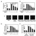
본 발명자들은 혈관내피세포에서 3가지 디케토피페라진이 HMGB1(High mobility group box 1)의 분비 억제뿐만 아니라 전염증성 시그날링(proinflammatory signaling)을 억제한다는 사실을 처음으로 규명하였으며, 실제로 전신염증질환 동물 모델에서 디케토피페라진이 예방 또는 치료효과가 있다는 사실을 확인하였다.The present inventors first recognized that three diketopiperazines inhibit the secretion of HMGB1 (high mobility group box 1) as well as proinflammatory signaling in vascular endothelial cells. In fact, , It was confirmed that diketopiperazin is effective for prevention or treatment.
이는 본 발명의 명세서 실시예에 잘 나타나 있다.This is well illustrated in the specification of the present invention.
본 발명의 실시예에서 혈관내피세포인 HUVEC 세포를 이용하여 in vitro 상에서 디케토피페라진의 기능을 평가하였다. 혈관내피세포에 LPS를 처리하여 HMGB1의 분비를 유도한 뒤 3가지 디케토피페라진을 각각 처리한 결과, 3가지 디케토피페라진이 LPS에 의해 매개된 HMGB1의 분비를 억제하였다. 또한 HMGB1의 수용체인 TLR2, TLR4, RAGE의 발현을 억제하였으며, HMGB1에 의한 세포골격 재분배를 억제하였다. 또한 디케토피페라진이 HMGB1에 매개된 내피세포에서 백혈구의 이동을 억제하고, VCAM-1, ICAM-1, E-selection의 발현을 억제하여, HMGB1의 신호 전달 경로를 억제하였다. 게다가 디케토피페라진은 염증성 시그널 분자인 nuclear factor-κB(NF-κB), extracellular regulated kinases 1/2(ERK1/2)의 활성화 및 tumor necrosis factor-α(TNF-α), interleukin-6(IL-6)의 생성을 억제하였다.In an embodiment of the present invention, the function of diketopiperazine was evaluated in vitro using HUVEC cells as vascular endothelial cells. After treatment of LPS with HMGB1 and treatment with three diketopiperazines, three diketopiperazines suppressed the secretion of HMGB1 mediated by LPS. We also inhibited the expression of HMGB1 receptors TLR2, TLR4, and RAGE, and inhibited HMGB1 - induced cytoskeletal redistribution. In addition, diketopiperazines inhibited leukocyte migration in endothelial cells mediated by HMGB1, suppressed the expression of VCAM-1, ICAM-1 and E-selection, and suppressed the signaling pathway of HMGB1. In addition, diketopiperazines are involved in the activation of nuclear factor-κB (NF-κB), extracellular regulated
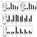
상기와 같은 효과를 in vivo 상에서 확인하기 위하여, CLP(cecal ligation and puncture)를 실시하여 전신염증질환 마우스 모델을 제작한 뒤 디케토피페라진의 영향을 확인하였다. 그 결과 디케토피페라진이 CLP 수술에 의한 HMGB1의 분비, 백혈구의 이동을 억제하고, 생존율을 높이며, 폐 손상을 억제하여 현저한 치료효과를 나타내었다. In order to confirm the above effect in vivo, CLP (cecal ligation and puncture) was performed to produce a mouse model of systemic inflammatory disease, and the effect of diketopiperazine was confirmed. As a result, diketopiperazine suppressed HMGB1 secretion and leukocyte migration by CLP, increased survival rate, and inhibited lung injury.
상기와 같이 in vitro 및 in vivo 실험을 통하여, 3가지 디케토피페라진은 HMGB1의 분비를 억제하고, HMGB1에 의해 매개되는 염증성 반응을 억제하여, 전신염증질환을 치료하는 효과가 있다.Through in vitro and in vivo experiments as described above, three diketopiperazines inhibit the secretion of HMGB1, inhibit the inflammatory response mediated by HMGB1, and are effective in treating systemic inflammatory diseases.
본 발명의 ‘전신염증질환(systemic inflammatory disease)’은 염증에 의해 유발되는 발열이나 빈맥, 빈호흡, 백혈구 증가증 등을 동반하는 전신적인 염증반응이다. 이는 감염 이외에도 급성 췌장염, 화상, 외상, 수술 등으로 발생할 수 있는 것으로, 성인형 스틸씨병(adult onset Still’s disease, AOSD), 베체트병(Behcet disease), 크론씨병(Crohn’s disease), 연소형 특발성관절염(Juvenile idiopathic arthritis), 전신성 류마티스 질환, 피부발진, 포도막염 또는 전신 혈관염을 포함한다.The "systemic inflammatory disease" of the present invention is a systemic inflammatory reaction accompanied by inflammation-induced fever, tachycardia, brisk breathing, and leukocytosis. In addition to infection, it can also be caused by acute pancreatitis, burns, trauma, and surgery, including adult onset Still's disease (AOSD), Behcet disease, Crohn's disease, juvenile idiopathic arthritis), systemic rheumatic diseases, skin rash, uveitis or systemic vasculitis.
바람직하게는 HMGB1(High mobility group box 1)에 의해 매개되는 염증성 질환일 수 있고, 그 종류가 특별히 제한되지 않으나 예를 들어, 전신염증반응증후군(Systemic inflammatory response syndrome, SIRS), 패혈증(sepsis), 패혈성 쇼크(septic shock), 급성 폐손상(Acute lung injury), 급성 신장 손상(Acute kidney injury), 다발성 장기 기능부전 증후군(Multiple organ dysfunction syndrome)으로 이루어진 군에서 선택되는 것을 특징으로 한다.And may be an inflammatory disease mediated by HMGB1 (High mobility group box 1), and the kind thereof is not particularly limited. For example, systemic inflammatory response syndrome (SIRS), sepsis, Acute lung injury, acute kidney injury, multiple organ dysfunction syndrome, and the like. The present invention is also directed to a method for preventing or treating an autoimmune disease,
상기‘전신염증반응증후군(Systemic inflammatory response syndrome, SIRS)’은 다양한 중증 임상적 손상에 대한 전신성인 염증반응으로, ACCP/SCCM의 정의에 따라 24시간 이내에 다음의 조건 중 2개 이상의 증상에 의해 진단할 수 있다:Systemic inflammatory response syndrome (SIRS) is a systemic inflammatory response to a variety of severe clinical impairments. It is defined by ACCP / SCCM within 24 hours and is diagnosed by two or more of the following conditions can do:
- 체온 38℃(100.4℉) 초과 또는 36℃(96.8℉) 미만;- body temperature greater than 100.4 F or less than 36.8 F;
- 심박수(HR) 90회 박동/분 초과;- Heart rate (HR) 90 beats / minute exceeded;
- 호흡수(RR) 20회 호흡/분 초과, 또는 동맥혈이산화탄소분압(PCO2) 32 mmHg 미만, 또는 기계적 환기 필요; 및- respiratory rate (RR) 20 breaths / minute, or arterial carbon dioxide partial pressure (PCO2 ) less than 32 mmHg, or mechanical ventilation required; And
- 백혈구 계수(WBC) 12.0 x 10⁹/L 초과 또는 4.0 x 10⁹/L미만, 또는 10% 초과의 미성숙 대상형 포함.- Includes immature subjects with leukocyte counts (WBC) greater than 12.0 x 10⁹ / L or less than 4.0 x 10⁹ / L, or greater than 10%.
본 발명에서 ‘패혈증(sepsis)'은 감염에 의한 전신염증반응증후군으로, 저혈압, 저산소증, 핍뇨증. 대사성산증, 혈소판감소증, 의식장애 등이 나타날 수 있다. 패혈증은 tumor necrosis factor-α(TNF-α)와 같은 전염증성 cytokine 의 과다한 생성과 비정상적인 면역체계의 활성화에 의한 과염증반응(hyper-inflammatory response)의 결과이다. 이러한 비정상적인 과염증반응은 조직손상과 장기부전을 초래하게 된다. 단핵구와 대식세포는 다양한 감염성이나 비감염성 자극들에 반응하여 전염증반응(pro-inflammatory response)에 관여하는 다양한 염증 매개물질들을 분비한다. 정상적인 상태에서 염증은 인체를 방어하기 위한 조절 가능한 반응으로, 정상적인 상태에서 cytokine의 분비와 백혈구의 활성화는 염증을 유발하는 물질들을 제거할 정도로 적절하게 일어난다. 그러나 감염에 대한 부적절한 선천성 면역반응에 의해 유발된 과염증반응으로 인하여 패혈증이 발생하게 되고, 이에 따른 염증 매개물질들의 과도한 분비로 다발성 장기부전을 유발할 수 있다.In the present invention, 'sepsis' is a systemic inflammatory reaction syndrome caused by infection, which is characterized by hypotension, hypoxia, oliguria. Metabolic acidosis, thrombocytopenia, and consciousness disorder. Sepsis is the result of excessive production of proinflammatory cytokines such as tumor necrosis factor-α (TNF-α) and hyper-inflammatory response due to abnormal activation of the immune system. These abnormal hyperglycemia reactions lead to tissue damage and organ failure. Monocytes and macrophages secrete a variety of inflammatory mediators that are involved in the pro-inflammatory response in response to a variety of infectious and non-infectious stimuli. Under normal conditions, inflammation is an adjustable reaction to defend the body. Under normal conditions, the secretion of cytokines and the activation of leukocytes are adequate to remove inflammation-inducing substances. However, due to the hyperinflammatory response caused by the inadequate congenital immune response to the infection, sepsis may occur and excessive secretion of the inflammatory mediators may lead to multiple organ failure.
본 발명의 ‘급성 폐손상(Acute lung injury)’은 급성 호흡곤란 증후군(acute respiratory distress syndrome, ARDS)이라고도 불리며, 심장 외의 원인으로 인해 급성으로 시작된 폐 부종으로 고농도의 산소를 공급하여도 호흡곤란을 일으킨다. 이는 보통 원인 인자에 노출된 후 보통 수시간에서 이틀 정도에 나타나며, 다양한 원인으로 인해 발생한다. 가장 흔한 원인은 패혈증, 심한 외상이며, 세균이나 신체 손상에 의해 혈액으로 화학적 물질이 방출되어, 이것이 폐에 도달해 폐에서 심한 염증이 발생한다.The acute lung injury of the present invention is also called acute respiratory distress syndrome (ARDS). The acute lung injury, which is caused by an extra-cardiac cause, Cause. It usually occurs within a few hours to two days after exposure to the causative agent, and is caused by a variety of causes. The most common cause is septicemia, severe trauma, chemical substances released into the blood by bacterial or physical damage, which reaches the lungs and causes severe inflammation in the lungs.
본 발명의 ‘급성 신장 손상(Acute kidney injury)’은 급성 신부전(acute renal failure, ARF)이라고도 불리며, 신장 기능이 수시간에서 수일에 걸쳐 급격하게 저하되는 것을 말한다. 신장 기능 저하의 결과로 신체 내에 질소 노폐물이 축적되어 혈액 내에 고질소혈증이 일어나고, 체액 및 전해질 균형에 이상이 생긴다. 원인으로는 전반적인 신체기능 저하로 인하여 신장으로 가는 혈액이 부족한 경우(출혈, 구토, 설사, 저혈압, 심부전 등), 신장 자체의 이상(신장 혈관 이상, 고혈압, 급성 사구체 신염, 혈관염 등), 요도 및 방광 이상(전립선 비대증, 종양 등)이 있다. 증상은 의식 혼탁, 경련, 고혈압, 부정맥, 폐부종, 폐출혈, 식욕부진, 구토, 장 마비, 위장관 출혈, 허혈성 심장질환 등이 있다.Acute kidney injury of the present invention is also referred to as acute renal failure (ARF), which means that the renal function rapidly drops over a period of several hours to several days. Nitrogen waste accumulates in the body as a result of decreased renal function, resulting in hyperglycemia in the blood and abnormal balance of fluid and electrolyte. Causes include kidney failure (renal artery anomalies, hypertension, acute glomerulonephritis, vasculitis, etc.), urinary tract and / or urinary tract infections due to lack of blood to the kidney due to overall body dysfunction (bleeding, vomiting, diarrhea, hypotension, Bladder anomalies (benign prostatic hyperplasia, tumors, etc.). Symptoms include unconscious turbidity, convulsions, hypertension, arrhythmia, pulmonary edema, pulmonary hemorrhage, anorexia, vomiting, intestinal paralysis, gastrointestinal bleeding, and ischemic heart disease.
본 발명의 ‘다발성 장기 기능부전 증후군(Multiple organ dysfunction syndrome, MODS)’은 다발성 장기부전(multiple organ failure, MOF)이라고도 불리며, 외상, 수술 후 쇼크 또는 중증 감염증 등 치료경과중에 신체의 주요장기, 즉 심장, 폐, 간, 신장 등의 기능부전이 2개 이상 동시 또는 연속적으로 발생하는 상태를 말한다. 생명유지 활동이 극단적으로 어려워지기 때문에 치사율이 매우 높다. 2개 장기부전에서는 사망률 60-70%, 3개 장기 부전에서는 80-90%라고 한다. 장기부전 수가 늘어남에 따라 사망률은 상승하는데, 부전장기수를 증가시키는 기반에는 감염증이나 그에 따른 endotoxemia가 중요한 역할을 하고 있다. 다발성 장기 부전은 교통사고와 같은 외부의 충격으로 인하여 다발성 장기부전이 선행하고 이를 계기로 숙주가 면역부전에 빠져. 감염증이 합병해서 더 악화하는 타입인 primary MOF와, 중증감염증이나 패혈증이 선행해서 terminal infection에서 다발성 장기부전이 발생하는 타입인 Secondary MOF로 구분한다.The multiple organ dysfunction syndrome (MODS) of the present invention is also referred to as multiple organ failure (MOF), and is a major organ of the body during the course of trauma, post-operative shock, Refers to a state in which two or more functional impairments such as heart, lung, liver, kidney, etc. occur simultaneously or continuously. The mortality rate is very high because life-sustaining activities become extremely difficult. The mortality rate is 60-70% in 2 organ failure cases and 80-90% in 3 organ failure cases. The mortality rate rises as the number of organ dysfunction increases, and the infectious disease and its endotoxemia play an important role in increasing the number of insufficient long term. Multiple organ failure is caused by external shocks such as traffic accidents, leading to multiple organ failure, which causes the host to become immune deficient. Primary MOF, which is a type of worsening disease due to the merger of infectious diseases, and Secondary MOF, which is a type in which multiple organ failure occurs in terminal infection preceded by severe infectious disease or sepsis.
본 발명에 따른 약학적 조성물은 디케토피페라진을 단독으로 함유하거나 약학적으로 허용되는 담체와 함께 적합한 형태로 제형화 될 수 있으며, 부형제 또는 희석제를 추가로 함유할 수 있다. 상기에서 '약학적으로 허용되는'이란 생리학적으로 허용되고 인간에게 투여될 때, 통상적으로 위장 장애, 현기증 등과 같은 알레르기 반응 또는 이와 유사한 반응을 일으키지 않는 비독성의 조성물을 말한다.The pharmaceutical composition according to the present invention may be formulated into a suitable form together with a diketopiperazine alone or together with a pharmaceutically acceptable carrier, and may further contain an excipient or a diluent. &Quot; Pharmaceutically acceptable " as used herein refers to a nontoxic composition that is physiologically acceptable and does not normally cause an allergic reaction such as gastrointestinal disorder, dizziness, or the like when administered to humans.
약학적으로 허용되는 담체로는 예컨대, 경구 투여용 담체 또는 비경구 투여용 담체를 추가로 포함할 수 있다. 경구 투여용 담체는 락토스, 전분, 셀룰로스 유도체, 마그네슘 스테아레이트, 스테아르산 등을 포함할 수 있다. 아울러, 펩티드 제제에 대한 경구투여용으로 사용되는 다양한 약물전달물질을 포함할 수 있다. 또한, 비경구 투여용 담체는 물, 적합한 오일, 식염수, 수성 글루코오스 및 글리콜 등을 포함할 수 있으며, 안정화제 및 보존제를 추가로 포함할 수 있다. 적합한 안정화제로는 아황산수소나트륨, 아황산나트륨 또는 아스코르브산과 같은 항산화제가 있다. 적합한 보존제로는 벤즈알코늄 클로라이드, 메틸-또는 프로필-파라벤 및 클로로부탄올이 있다. 본 발명의 약학적 조성물은 상기 성분들 이외에 윤활제, 습윤제, 감미제, 향미제, 유화제, 현택제 등을 추가로 포함할 수 있다. 그 밖의 약학적으로 허용되는 담체 및 제제는 다음의 문헌에 기재되어 있는 것을 참고로 할 수 있다(Remington's Pharmaceutical Sciences, 19th ed., Mack Publishing Company, Easton, PA, 1995).The pharmaceutically acceptable carrier may further include, for example, a carrier for oral administration or a carrier for parenteral administration. Carriers for oral administration may include lactose, starch, cellulose derivatives, magnesium stearate, stearic acid, and the like. In addition, it may contain various drug delivery materials used for oral administration to peptide preparations. In addition, the carrier for parenteral administration may contain water, a suitable oil, a saline solution, an aqueous glucose and a glycol, and may further contain a stabilizer and a preservative. Suitable stabilizers include antioxidants such as sodium hydrogen sulfite, sodium sulfite or ascorbic acid. Suitable preservatives include benzalkonium chloride, methyl- or propyl-paraben and chlorobutanol. The pharmaceutical composition of the present invention may further contain a lubricant, a wetting agent, a sweetening agent, a flavoring agent, an emulsifying agent, a suspending agent, etc. in addition to the above components. Other pharmaceutically acceptable carriers and preparations can be found in Remington's Pharmaceutical Sciences, 19th ed., Mack Publishing Company, Easton, Pa., 1995).
본 발명의 조성물은 인간을 비롯한 포유동물에 어떠한 방법으로도 투여할 수 있다. 예를 들면, 경구 또는 비경구적으로 투여할 수 있다. 비경구적인 투여방법으로는 이에 한정되지는 않으나, 정맥내, 근육내, 동맥내, 골수내, 경막내, 심장내, 경피, 피하, 복강내, 비강내, 장관, 국소, 설하 또는 직장내 투여일 수 있다.The composition of the present invention can be administered to mammals including humans by any method. For example, it can be administered orally or parenterally. Parenteral administration methods include, but are not limited to, intravenous, intramuscular, intraarterial, intramedullary, intrathecal, intracardiac, transdermal, subcutaneous, intraperitoneal, intranasal, enteral, topical, sublingual or rectal administration Lt; / RTI >
본 발명의 약학적 조성물은 상술한 바와 같은 투여 경로에 따라 경구 투여용 또는 비경구 투여용 제제로 제형화 할 수 있다.The pharmaceutical composition of the present invention can be formulated into oral preparations or parenteral administration preparations according to the route of administration as described above.
경구 투여용 제제의 경우에 본 발명의 조성물은 분말, 과립, 정제, 환제, 당의정제, 캡슐제, 액제, 겔제, 시럽제, 슬러리제, 현탁액 등으로 당업계에 공지된 방법을 이용하여 제형화될 수 있다. 예를 들어, 경구용 제제는 활성성분을 고체 부형제와 배합한 다음 이를 분쇄하고 적합한 보조제를 첨가한 후 과립 혼합물로 가공함으로써 정제 또는 당의정제를 수득할 수 있다. 적합한 부형제의 예로는 락토즈, 덱스트로즈, 수크로즈, 솔비톨, 만니톨, 자일리톨, 에리스리톨 및 말티톨 등을 포함하는 당류와 옥수수 전분, 밀 전분, 쌀 전분 및 감자 전분 등을 포함하는 전분류, 셀룰로즈,메틸 셀룰로즈, 나트륨 카르복시메틸셀룰로오즈 및 하이드록시프로필메틸-셀룰로즈 등을 포함하는 셀룰로즈류, 젤라틴, 폴리비닐피롤리돈 등과 같은 충전제가 포함될 수 있다. 또한, 경우에 따라 가교결합 폴리비닐피롤리돈, 한천, 알긴산 또는 나트륨 알기네이트 등을 붕해제로 첨가할 수 있다. 나아가, 본 발명의 약학적 조성물은 항응집제, 윤활제, 습윤제, 향료, 유화제 및 방부제 등을 추가로 포함할 수 있다.In the case of a preparation for oral administration, the composition of the present invention may be formulated into a powder, a granule, a tablet, a pill, a sugar, a tablet, a liquid, a gel, a syrup, a slurry, . For example, an oral preparation can be obtained by combining the active ingredient with a solid excipient, then milling it, adding suitable auxiliaries, and then processing the mixture into a granular mixture. Examples of suitable excipients include, but are not limited to, sugars including lactose, dextrose, sucrose, sorbitol, mannitol, xylitol, erythritol and maltitol, and starches including corn starch, wheat starch, rice starch and potato starch, Cellulose such as methylcellulose, sodium carboxymethylcellulose and hydroxypropylmethyl-cellulose and the like, fillers such as gelatin, polyvinylpyrrolidone and the like. In addition, crosslinked polyvinylpyrrolidone, agar, alginic acid, or sodium alginate may optionally be added as a disintegrant. Further, the pharmaceutical composition of the present invention may further comprise an anti-coagulant, a lubricant, a wetting agent, a flavoring agent, an emulsifying agent and an antiseptic agent.
비경구 투여용 제제의 경우에는 주사제, 크림제, 로션제, 외용연고제, 오일제, 보습제, 겔제, 에어로졸 및 비강 흡입제의 형태로 당업계에 공지된 방법으로 제형화할 수 있다. 이들 제형은 모든 제약 화학에 일반적으로 공지된 처방서인 문헌(Remington's Pharmaceutical Science, 19th ed., Mack Publishing Company, Easton, PA,1995)에 기재되어 있다.In the case of a preparation for parenteral administration, it can be formulated by a method known in the art in the form of injection, cream, lotion, external ointment, oil, moisturizer, gel, aerosol and nasal aspirate. These formulations are described in Remington's Pharmaceutical Science, 19th ed., Mack Publishing Company, Easton, Pa., 1995, which is a commonly known formulary for all pharmaceutical chemistries.
본 발명의 조성물의 총 유효량은 단일 투여량(single dose)으로 환자에게 투여될 수 있으며, 다중 투여량(multiple dose)으로 장기간 투여되는 분할 치료 방법(fractionated treatment protocol)에 의해 투여될 수 있다. 본 발명의 약학적 조성물은 질환의 정도에 따라 유효성분의 함량을 달리할 수 있다. 바람직하게 본 발명의 약학적 조성물의 바람직한 전체 용량은 1일당 환자 체중 1㎏ 당 약 0.01㎍ 내지 10,000mg, 가장 바람직하게는 0.1㎍ 내지 500mg일 수 있다. 그러나 상기 약학적 조성물의 용량은 제제화 방법, 투여 경로 및 치료 횟수뿐만 아니라 환자의 연령, 체중, 건강 상태, 성별, 질환의 중증도, 식이 및 배설율등 다양한 요인들을 고려하여 환자에 대한 유효 투여량이 결정되는 것이므로, 이러한 점을 고려할 때 당 분야의 통상적인 지식을 가진 자라면 본 발명의 조성물의 적절한 유효 투여량을 결정할 수 있을 것이다. 본 발명에 따른 약학적 조성물은 본 발명의 효과를 보이는 한 그 제형, 투여 경로 및 투여 방법에 특별히 제한되지 아니한다. The total effective amount of the composition of the present invention may be administered to a patient in a single dose and may be administered by a fractionated treatment protocol administered over a prolonged period of time in multiple doses. In the pharmaceutical composition of the present invention, the content of the active ingredient may be varied depending on the degree of the disease. Preferably, the total preferred dose of the pharmaceutical composition of the present invention may be from about 0.01 μg to about 10,000 mg, and most preferably from about 0.1 μg to 500 mg, per kilogram of patient body weight per day. However, the dosage of the pharmaceutical composition may be determined depending on various factors such as the formulation method, administration route and frequency of treatment, as well as the patient's age, body weight, health condition, sex, severity of disease, diet and excretion rate, It will be possible to determine the appropriate effective dose of the composition of the present invention by those of ordinary skill in the art in view of this point. The pharmaceutical composition according to the present invention is not particularly limited to the formulation, administration route and administration method as long as the effect of the present invention is exhibited.
따라서, 본 발명은 디케토피페라진을 유효성분으로 포함하는 중증혈관질환의 예방 또는 치료용 약학적 조성물을 제공한다. 본 발명의 방법은 HMGB1(High mobility group box 1)의 분비를 억제하며 전염증성 시그널링을 조절하고 혈관 염증성 반응을 억제하여 전신염증질환의 예방 및 치료에 효과가 있다.Accordingly, the present invention provides a pharmaceutical composition for preventing or treating serious vascular diseases comprising diketopiperazine as an active ingredient. The method of the present invention suppresses the secretion of HMGB1 (High mobility group box 1), regulates proinflammatory signaling, inhibits vascular inflammatory response, and is effective in the prevention and treatment of systemic inflammatory diseases.
도 1은 3가지 디케토피페라진(diketopiperazine)의 화학식을 나타낸 것이다(A: 발린(Val)이 결합된 1번 화학식, B: 티로신(Tyr)이 결합된 2번 화학식, C: 류신(Leu)이 결합된 3번 화학식).
도 2는 3가지 디케토피페라진(diketopiperazine)이 HMGB1의 분비와 HMGB1 수용체의 발현에 미치는 영향을 나타낸 것으로, HUVEC 세포에 LPS(100 ng/mL)로 16시간 동안 자극한 후, 3가지 디케토피페라진 각각을 표시된 농도로 처리하였을 때 HMGB1의 분비량을 ELISA 방법으로 측정한 것(A), CLP를 수행한 수컷 C57BL/6 마우스(n=5)에 CLP로부터 12시간 후에 3가지 디케토피페라진을 각각 정맥주사하고, CLP로부터 24시간 후에 혈장 속 HMGB1 level을 ELISA로 측정한 것(B), Confluent HUVEC 세포에 HMGB1(1 ㎍/mL)을 16시간 처리하고, 1번 디케토피페라진을 6시간 동안 처리하였을 때, TLR-2(white bar), TLR-4(gray bar), RAGE(black bar) 수용체의 발현량을 cell-based ELISA로 측정한 것(C), MTT assay 방법으로 세포 생존성(cell viability)에 대한 3가지 디케토피페라진의 영향을 측정하여 5회의 독립적 실험의 mean ± SEM으로 나타낸 것이다(LPS: lipopolysaccharide, *p<0.05versus LPS alone(A), CLP alone(B), or HMGB1 alone(C)).
도 3은 HUVEC 세포를 LPS(A, 100 ng/mL, 4 h) 또는 HMGB1(B,1 ㎍/mL, 16 h)으로 자극시킨 후, 각각의 농도로 3가지 디케토피페라진을 각각 6시간 처리하였을 때, HUVEC 단일층을 가로지른 Evans blue-결합된 알부민의 유출정도 측정에 의해 모니터링하여 세포의 투과성을 평가한 결과, HMGB1(H로 표시, 2 ㎍/mouse, i.v.) 또는 CLP(C로 표시, CLP 후 24h)로 마우스에 혈관투과(vascular permeability)를 유도한 후, 디케토피페라진을 주입하여, 복강 세척액(peritoneal washes)에 포함된 Evans blue 염료의 양을 측정하여 투과성을 평가하는 것으로 이의 효과를 확인한 결과(C, expressed as ㎍/mouse, n = 5)를 나타낸 것이다. 또한, HUVEC 세포들을 HMGB1(1 ㎍/mL, 16 h)으로 활성화시키고 서로 다른 농도의 디케토피페라진을 6시간 처리한 후, HMGB1에 의해 매개된 phospho-p38 발현에 대한 3가지 디케토피페라진의 영향을 ELISA로 측정한 결과(D)를 나타낸 것이다. E는 glass coverslip 위에서 배양한 HUVEC 세포 단일층을 HMGB1(1 ㎍/mL)으로 1시간동안 자극하고 6시간 동안 각각 10씩 디케토피페라진을 6시간동안 처리한 후, 형광표지된 F-actin으로 형광염색한 모습을 나타낸 것이다. 화살표는 세포사이의 틈(intercellular gap)을 나타낸다(P: PBS vehicle control, Comp: diketopiperazine, LPS: lipopolysaccharide, 결과는 5회의 독립적 실험의 mean ± SEM으로서 나타냄, H/C는 CLP 또는 HMGB1 처리를 의미하는 것으로 'CLP/HMGB1'과 같은 의미). *p < 0.05 versus LPS alone(A) or HMGB1 alone(B, C, D).
도 4는 HUVEC 세포를 HMGB1(1 ㎍/mL)으로 16시간동안 자극하고 다양한 농도로 3가지 디케토피페라진을 6시간 동안 처리한 후, HMGB1-매개 VCAM-1, ICAM-1 및 E-selectin의 발현 정도를 나타낸 것(A), HUVEC monolayer에 대한 인간 호중구의 부착정도를 그래프(B)와 이미지(C)로 나타낸 것, HUVEC monolayer를 통한 인간 호중구의 이동(migration)을 분석한 결과를 나타낸 것(d)이다(P: PBS vehicle control, Comp: diketopiperazine, 결과는 5회의 독립적 실험의 mean ± SEM으로서 나타냄, *p< 0.05 versus HMGB1). 또한, C57BL/6 수컷 마우스를 HMGB1(H로 표시, 2㎍ per mouse, i.v.) 또는 CLP-수술(C로 표시, CLP 수술 후 24h)로 자극하고 디케토피페라진을 주입하여, 상기 HMGB1 또는 CLP에 의해 매개된 백혈구의 복막강으로의 이동성을 분석한 결과(E)를 나타낸 것이다(H/C는 CLP 또는 HMGB1 처리를 의미하는 것으로 'CLP/HMGB1'과 같은 의미.P: PBS vehicle control, Comp: diketopiperazine, 결과는 5회의 독립적 실험의 mean ± SEM으로서 나타냄, *p< 0.05 versus CLP/HMGB1).
도 5는 HUVEC 세포를 HMGB1(1 ㎍/mL)으로 16시간동안 자극하고 다양한 농도로 3가지 디케토피페라진을 6시간 동안 처리한 후, HMGB1-매개 TNF-α생성 정도를 측정한 결과(A), HMGB1-매개 IL-6 생성 정도를 측정한 결과(B), HMGB1-매개 NF-κB p65의 활성화 정도를 분석한 결과(C) 및 HMGB1-매개 ERK1/2의 활성화 정도를 분석한 결과(D)를 나타낸 것이다(P: PBS vehicle control, Comp: diketopiperazine, 결과는 5회의 독립적 실험의 mean ± SEM으로서 나타냄, *p< 0.05 versus HMGB1).
도 6은 수컷 C57BL/6 마우스들(n=20)에 CLP 후 12시간째 및 50시간째에 3가지 디케토피페라진을 각각 정맥투여하고, CLP 후 132시간까지 6시간마다 실험동물의 생존을 모니터링한 결과를 나타낸 것으로, 처리하지 않은 CLP mice(●로 표시) 및 sham-operated mice(○로 표시)들에는 살균한 식염수를 투여하였다.Kaplan-Meier survival analysis 방법으로 CLP군에 대비한(versus) 전체 생존률을 그래프(A)로 나타내었다(Comp: diketopiperazine, □: CLP 후 12시간 및 50시간째에 3,93씩 1번 디케토피페라진 투여군, ■: CLP 후 12시간 및 50시간째에 5.21씩 1번 디케토피페라진 투여군. △: CLP 후 12시간 및 50시간째에 4.20씩 3번 디케토피페라진 투여군). 또한 CLP 후 96시간째에 안락사시켜 관찰한 폐조직의 조직병리학적 스코어(Histopathological score)를 나타낸 그래프(B)와 CLP 후 96시간째에 안락사시켜 수득한 폐조직을 H&E staining하여 현미경(200x)으로 관찰한 사진(C)을 나타낸 것이다. 이미지는 세 번의 독립적 실험을 대표한다.Brief Description of the Drawings Figure 1 shows the formula of three diketopiperazines (A: valine-bound
FIG. 2 shows the effect of three diketopiperazines on HMGB1 secretion and HMGB1 receptor expression. HUVEC cells were stimulated with LPS (100 ng / mL) for 16 hours, (N = 5) of CLP-treated male mice were treated with three diketopiperazines (CLP) at 12 hours after CLP, and then the levels of HMGB1 were measured by ELISA (B), HMGB1 (1 ㎍ / mL) was treated with Confluent HUVEC cells for 16 hours, and 1-diketopiperazine was treated with 6 (C), cell viability (MT) assay method by measuring the expression level of TLR-2, TLR-4 and RAGE (black bar) receptors by cell-based ELISA The effect of three diketopiperazines on cell viability was measured and expressed as the mean ± SEM of 5 independent experiments (LPS: lipopolysaccharide, *p < 0.05versus LPS alone (A), CLP alone (B), or HMGB1 alone (C)).
Figure 3 shows the results of stimulation of HUVEC cells with LPS (A, 100 ng / mL, 4 h) or HMGB1 (B, 1 ug / mL, 16 h) and then 3 diketopiperazines at each concentration for 6 hours The permeability of the cells was monitored by measuring the extent of Evans blue-bound albumin crossed across the HUVEC monolayer. The permeability of HMGB1 (labeled with H, 2 ㎍ / mouse, iv) or CLP Labeling, 24 h after CLP) to induce vascular permeability in mice, and then injected with diketopiperazine to evaluate the permeability by measuring the amount of Evans blue dye contained in peritoneal washes (C, expressed as / / mouse, n = 5). In addition, HUVEC cells were activated with HMGB1 (1 ㎍ / mL, 16 h), treated with different concentrations of diketopiperazine for 6 hours, and then treated with 3 different diketopiperazines for phospho-p38 expression mediated by HMGB1 (D) by the ELISA. E was stimulated with HMGB1 (1 ㎍ / mL) for 1 hour on a glass coverslip, treated with diketopiperazine for 10 hours for 6 hours, and incubated with fluorescently labeled F-actin It shows fluorescent staining. The arrow indicates the intercellular gap (P: PBS vehicle control, Comp: diketopiperazine, LPS: lipopolysaccharide, the result is expressed as the mean ± SEM of 5 independent experiments, H / C means CLP or HMGB1 treatment Which means 'CLP / HMGB1'). * p < 0.05 versus LPS alone (A) or HMGB1 alone (B, C, D).
Figure 4 shows that HUVEC cells were stimulated with HMGB1 (1 [mu] g / mL) for 16 hours and treated with 3 different diketopiperazines at various concentrations for 6 hours and then stimulated with HMGB1-mediated VCAM-1, ICAM- (B) and (C), and the migration of human neutrophils through the HUVEC monolayer was analyzed. The results are shown in FIGS. (P: PBS vehicle control, Comp: diketopiperazine, results expressed as the mean ± SEM of 5 independent experiments, *p <0.05 versus HMGB1). In addition, C57BL / 6 male mice were stimulated with HMGB1 (indicated by H, 2 占 퐂 per mouse, iv) or CLP-surgery (labeled with C, 24h after CLP surgery) and injected with diketopiperazine, (H / C means CLP or HMGB1 treatment, meaning 'CLP / HMGB1'. P: PBS vehicle control, Comp. : diketopiperazine, results expressed as mean ± SEM of 5 independent experiments, *p <0.05 versus CLP / HMGB1).
FIG. 5 shows the results of measuring HMGB1-mediated TNF-α production after stimulation of HUVEC cells with HMGB1 (1 μg / mL) for 16 hours and treatment of three diketopiperazines at various concentrations for 6 hours ), The result of measuring the degree of HMGB1-mediated IL-6 production (B), the activity of HMGB1-mediated NF-κB p65 (C) and the activity of HMGB1-mediated ERK1 / 2 D) (P: PBS vehicle control, Comp: diketopiperazine, results expressed as the mean ± SEM of 5 independent experiments, *p <0.05 versus HMGB1).
FIG. 6 shows the results of intravenous administration of three diketopiperazines at 12 hours and 50 hours after CLP in male C57BL / 6 mice (n = 20), and survival of the experimental animals every 6 hours up to 132 hours after CLP The results of the monitoring showed that untreated CLP mice (marked with a circle) and sham-operated mice (labeled with a circle) were given sterile saline. Kaplan-Meier survival analysis (Comp: diketopiperazine, □: 1 dose of diketopiperazine at 3 hours after 12 hours and 50 hours after CLP, and 12 hours and 50 hours after CLP) Dicetofiprazine treated
이하 본 발명을 상세히 설명한다.Hereinafter, the present invention will be described in detail.
단, 하기 실시예는 본 발명을 예시하는 것일 뿐, 본 발명의 내용이 하기 실시예에 한정되는 것은 아니다.However, the following examples are illustrative of the present invention, and the present invention is not limited to the following examples.
<실험방법><Experimental Method>
1. 시약1. Reagents
박테리아 lipopolysaccharide(LPS; serotype: 0111:B4, L5293), Evans blue 염료, HPLC 분류 용제, 크리스탈 바이올렛(crystal violet) 염료 및 항생제(페니실리린 G 및 스트렙토마이신)는 Sigma(St. Louis, MO)로부터 구입하였다. 인간 재조합 HMGB1은 Abnova(Taipei City, Taiwan)로부터 구입하였으며, FBS(Fetal bovine serum) 및 Vybrant DiD는 Invitrogen(Carlsbad, CA)으로부터 구입하였다. 추출 및 분류를 위한 유기용제인 메탄올(MeOH), 에틸 아세테이트(EtOA), n-헥산(n-hexane)은 덕산 화학(Duksan Chemical, Korea)에서 구입하였다. 박층 크로마토그래피(Thin layer chromatography)는 실리카겔로 코팅된 플레이트(Kieselgel 60F254, Germany)를 사용하였고, 실리카겔 컬럼 크로마토그래피는 Kiegelgel 60(Art. No. 9385)을 사용하였다. UV 스펙트럼 및 HR-FABMS는 시마주 UV-1800(Shimadzu, Japan) 및 JMS 700(JEOL, Japan)을 각각 사용하였으며, NMR(nuclear magnetic resonance) 스펙트럼은 내부 표준에서 테트라메틸실란(tetramethylsilane, TMS)로 Bruker Avance Digital 500 NMR spectrometer(Karlsruhe, Germany)를 이용하여 측정하였다. TMS에 대한 화학 변화는 ppm(parts per million)으로 표시하였다. MPLC(Medium pressure liquid chromatography)는 Water 510 펌프(Waters, USA)와 연결된 Prep UV-10V(Yamazen, Japan)로 실시하였다. 모든 용매는 감압하에 40℃이하에서 증발시켰다.(LPS), Evans blue dyes, HPLC classifying solvents, crystal violet dyes and antibiotics (penicillin G and streptomycin) were purchased from Sigma (St. Louis, Mo.) Respectively. Human recombinant HMGB1 was purchased from Abnova (Taipei City, Taiwan), FBS (Fetal bovine serum) and Vybrant DiD were purchased from Invitrogen (Carlsbad, CA). Methanol (MeOH), ethyl acetate (EtOA) and n-hexane, organic solvents for extraction and classification, were purchased from Duksan Chemical, Korea. Thin layer chromatography was performed on a plate coated with silica gel (Kieselgel 60F254, Germany) and silica gel column chromatography using Kiegelgel 60 (Art. No. 9385). The UV spectrum and the HR-FABMS were respectively used in Shimadzu UV-1800 (Shimadzu, Japan) and JMS 700 (JEOL, Japan), and nuclear magnetic resonance (NMR) spectra were obtained from tetramethylsilane Bruker Avance Digital 500 NMR spectrometer (Karlsruhe, Germany). Chemical changes to TMS are expressed in parts per million (ppm). Medium pressure liquid chromatography (MPLC) was performed with Prep UV-10V (Yamazen, Japan) connected to a Water 510 pump (Waters, USA). All solvents were evaporated under reduced pressure at < RTI ID = 0.0 > 40 C. < / RTI >
2. 디케토피페라진의 획득2. Acquisition of diketopiperazines
제주도에서 수집한 침전물과 해면 동물(marine sponge)에서 두 종의 박테리아, HC001 및 12L081을 추출하여 동정하였다. 두 종을 각각 동정한 결과 HC001은Bacillus 속이며, 12L081은Picicoccus속인 것을 확인하였다.Two species of bacteria, HC001 and 12L081, were extracted from sediments and marine sponge collected from Jeju Island. As a result of identification of two species, it was confirmed that HC001 belongs toBacillus and 12L081 belongs toPicicoccus .
Bacillus 속 HC001 및Picicoccus 속 12L081 박테리아를 7일 동안 150rpm, 25℃의 조건으로 SYP SW 배지 35L에서 각각 배양한 뒤, 각 배양 배지를 EtOA 35L로 두 번 추출하였다.Bacillus 속 HC001 및Picicoccus 속 12L081의 추출물을 농축하여 제조하였다. HC001의 추출물(1.9 g)을 step-gradient elution 방법으로 실리카겔 VLC로 분획하였다. 디케토피페라진 1(2.5 mg)과 디케토피페라진 2(1.6 mg)를 획득하기 위하여, 분획물 E(CH2Cl2:MeOH=10:1로 추출, 195.5 mg)로 예비 RP-HPLC(Phenomenex Luna 10μ C18(2) 100, 250 × 21.20 mm, 10 μm, 6 mL/min, 20% CH3CNinH2O)를 실시하였다. 12L081의 추출물(2.7 g)은 VLC로 분획하였고, 디케토피페라진 3(6.6mg)을 획득하기 위하여 분획물 D(CH2Cl2:MeOH=20:1로 추출, 539.1 mg)를 예비 RP-HPLC(Phenomenex Luna 10μ C18(2) 100, 250 × 21.20 mm, 10 μm, 6 mL/min, 17% CH3CNinH2O)를 사용하여 추가로 정제하였다. 선행문헌(Fdhila et al., 2003; Jayatilake et al., 1996; Stierle et al., 1988)에 기재되어 있는 분광 데이터의 비교에 의해 디케토피페라진 1은cyclo(l-Pro-d-Val), 디케토피페라진 2는cyclo(l-Pro-l-Tyr), 디케토피페라진 3은cyclo(l-Pro-d-Leu)로 확인하였다.Bacillus sp . HC001 andPicococcus sp. 12L081 bacteria werecultured in 35 L of SYP SW medium at 150 rpm and 25 ° C for 7 days, and then each culture medium was extracted twice with 35 L of EtOA. The extracts ofBacillus sp . HC001 andPicococcus sp. 12L081 were prepared by concentration. The HC001 extract (1.9 g) was fractionated by silica gel VLC using a step-gradient elution method. HPLC (Phenomenex) was performed with fraction E (extraction with CH2 Cl2 : MeOH = 10: 1, 195.5 mg) to obtain diketopiperazine 1 (2.5 mg) and diketopiperazin2 a Luna 10μ C18 (2) 100, 250 × 21.20 mm, 10 μm, 6 mL / min, 20
3. 실험동물 및 사육3. Experimental animals and breeding
몸무게가 27 g인 6 내지 7주령 C57BL/6 수컷 마우스들을 Orient Bio Co.(Sungnam, KyungKiDo, Republic of Korea)로부터 구입하였고, 12일의 적응기간(acclimatization period) 후에 본 연구에 이용하였다. 실험동물들은 polycarbonate cage 당 5마리씩, 20-25°C의 온도, 40%-45%의 습도 및 12시간 주기의 낮/밤 순환 조건에서 사육되었다. 상기 적응기간 동안, 실험동물들은 일반 설치류 펠렛 사료(rodent pellet diet) 및 물을 자유식이(ad libitum)하였다. 모든 동물 실험은 한국 Institutional Animal Care and Use Committee of Kyungpook National University 의 승인 하에 수행되었다(IRB approval number, KNU2012-13).6-7 week old C57BL / 6 male mice weighing 27 g were purchased from Orient Bio Co. (Sungnam, KyungkiDo, Republic of Korea) and used for this study after an acclimatization period of 12 days. Experimental animals were housed in polycarbonate cages at a temperature of 20-25 ° C, humidity of 40% -45% and day / night circulation of 12 hours. During the adaptation period, the experimental animalsad libitum the rodent pellet diet and water. All animal experiments were conducted under the approval of the Korean Institutional Animal Care and Use Committee, Kyungpook National University (IRB approval number, KNU2012-13).
4. CLP(Cecal ligation and puncture)4. CLP (Cecal ligation and puncture)
심각한 혈관염증을 유발하기 위하여, 수컷 마우스들은 small rodent gas anesthesia machine(RC2, Vetequip, Pleasanton, CA)을 통하여 주입된 2% isoflurane(Forane, JW Pharmaceutical, South Korea)이 포함된 산소로 마취되었고, 최초에는 breathing chamber에서 마취하고 그 후에는 facemask를 사용하여 실험과정동안 자연스럽게 호흡할 수 있도록 하였다. CLP-유도 전신염증질환 모델은 Bae JS et al., 2014의 연구에서 묘사된 바와 같이 수행되었다. 간략하게, 2 cm의 정중절개(midline incision)를 수행하여 맹장 및 인접한 소장을 노출시켰다. 그 후 맹장 끝부분(cecal tip)으로부터 5.0 mm 떨어진 부분을 3.0-비단봉합사(silk suture)로 단단히 묶고, 높은 수준의 혈관염증을 유발하기 위하여 22-게이지의 바늘로 맹장에 구멍을 냈다(Rittirsch et al., 2009). 그 후, 상기 구멍 부위로부터 배설물을 압출시키기 위하여 맹장을 부드럽게 짜내었고(squeezed), 상기 맹장을 다시 복막강(peritoneal cavity)으로 복귀시켰다. 마지막으로 개복 부위(laparotomy site)를 4.0-silk로 봉합하였다. sham control 군의 실험동물은 맹장을 노출시킨 후 봉합 또는 구멍을 내는 과정 없이 복강으로 복귀시켰다. 상기 실험 프로토콜은 실험 전 the Animal Care Committee at Kyungpook National University(IRB No. KNU 2012-13)에 의해 승인 받았다.To induce severe vascular inflammation, male mice were anesthetized with oxygen containing 2% isoflurane (Forane, JW Pharmaceutical, South Korea) injected via a small rodent gas anesthesia machine (RC2, Vetequip, Pleasanton, CA) Were anesthetized in a breathing chamber and then allowed to breathe naturally during the course of the experiment using a facemask. A CLP-induced systemic inflammatory disease model was performed as described in Bae JS et al., 2014. Briefly, a 2 cm midline incision was performed to expose the appendix and adjacent small intestine. Thereafter, 5.0 mm from the cecal tip was tightly bound with a 3.0-silk suture and punctured into the cecum with a 22-gauge needle to induce a high level of vascular inflammation (Rittirsch et al., 2009). The cecum was then gently squeezed to extrude the excrement from the hole and the cecum was returned to the peritoneal cavity. Finally, the laparotomy site was closed with 4.0-silk. The experimental animals in the sham control group were exposed to the abdominal cavity without exposing the cecum and then suturing or puncturing. The experimental protocol was approved by the Animal Care Committee at Kyungpook National University (IRB No. KNU 2012-13) before the experiment.
5. 세포 배양5. Cell culture
인간 탯줄 정맥 내피세포(Primary human umbilical vein endothelial cells, HUVECs)는 Cambrex Bioscience(Charles City, IA)로부터 수득하였고, 이전 연구에 묘사된 바와 같이 유지시켰다(Bae et al., 2014; Ku and Bae, 2014; Ku et al., 2015a; Ku et al., 2015b; Ku et al., 2015c; Lee et al., 2015a; Lee et al., 2015b; Yoon et al., 2015). 간략하게, 세포는 성장보조제(Cambrex Bio Science)가 포함된 EBM-2(endothelial basal medium), 37℃ 및 5% CO2에서 포화상태로 배양하였다. HUVEC 세포는 Lee W et al., 2013 연구에서 묘사된 바와 같이 3-5계대(passage) 배양하고 사용하였다. 인간 호중구(neutrophil)는 다섯명의 건강한 지원자로부터 정맥천자(venipuncture)에 의해 수득된 15 mL의 전혈로부터 분리되었고, Hofbauer R et al., 1998의 연구에서 묘사된 바와 같이 유지되었다.Human umbilical vein endothelial cells (HUVECs) were obtained from Cambrex Bioscience (Charles City, IA) and maintained as described in previous studies (Bae et al., 2014; Ku and Bae, 2014 Lee et al., 2015b; Yoon et al., 2015). In addition, the present inventors have found that, Briefly, cells were incubated in EBM-2 (endothelial basal medium) with growth supplements (Cambrex Bio Science) at 37 ° C and 5% CO2 saturation. HUVEC cells were cultured in 3-5 passages as described in Lee W et al., 2013 study. Human neutrophil was isolated from 15 mL of whole blood obtained by venipuncture from five healthy volunteers and maintained as described in Hofbauer R et al., 1998.
6. ELISA(enzyme-linked immunosorbent assay)6. ELISA (enzyme-linked immunosorbent assay)
세포배양 배지 또는 마우스 혈장에 포함되어 있는 HMGB1의 농도를 조사하기 위하여, Lee W et al., 2012의 연구에서 묘사된 바와 같이 경쟁적 ELISA를 수행하였다. 구체적으로 in vitro 실험에서 HUVEC 단일층(HUVEC monolayer)은 LPS(100 ng/mL)로 16시간 동안 처리되었으며, 그 후 0 내지 10의 3가지 디케토피페라진으로 6시간동안 처리되었다. 그 다음에, HMGB1의 농도를 조사하기 위하여 세포배양액을 수집하였다. ELISA를 수행하기 위하여, 96-well flat plastic microtiter plates(Corning Inc., Corning, NY)를 HMGB1 단백질과 함께 4℃에서 오버나잇(overnight)하여 코팅하였으며, 이때 0.02% sodium azide가 포함된 20 mM carbonate-bicarbonate buffer(pH 9.6)를 사용하였다. 상기 plate를 0.05% Tween 20이 포함된 PBS 용액(PBS-T)으로 3회 세척한 뒤, 4℃에서 보관하였다. 상기 수집된 세포배양액은 동결건조(Lyophilization)시킨 후, 이를 anti-HMGB1 antibody(PBS-T에 1:1000로 희석됨, Abnova, Taipei City, Taiwan)와 함께 96-well round plastic microtiter plate에서 37 °C, 90분 동안 사전-배양(pre-incubate)하였고, 이를 상기 HMGB1으로 코팅된 플레이트에 옮긴 후, 30분 동안 실온에서 배양하였다. 그 후, 상기 플레이트를 PBS-T 용액으로 3회 세척하고, peroxidase-conjugated anti-rabbit IgG antibody(PBS-T에 1:2000로 희석됨, GE Healthcare Life Science, Pittsburgh)와 함께 실온에서 90분 동안 배양하였으며, 다시 PBS-T 용액으로 3회 세척하고, 200㎕의 기질용액(100 μg/mLo-phenylenediamine 및 0.003% H2O2)을 첨가하여 암실에서 상온으로 60분 동안 배양하였다. 반응은 8N H2SO4를 50 ㎕ 처리하여 종료하였고, 490nm에서 흡광도를 측정하였다.To investigate the concentration of HMGB1 contained in cell culture medium or mouse plasma, a competitive ELISA was performed as described in the study of Lee W et al., 2012. Specifically, in vitro experiments, HUVEC monolayers were treated with LPS (100 ng / mL) for 16 h and then treated with three diketopiperazines from 0 to 10 for 6 h. Then, cell culture medium was collected to examine the concentration of HMGB1. To perform the ELISA, 96-well flat plastic microtiter plates (Corning Inc., Corning, NY) were coated with HMGB1 protein overnight at 4 ° C, where 20 mM carbonate with 0.02% sodium azide -bicarbonate buffer (pH 9.6) was used. The plate was washed three times with PBS solution (PBS-T) containing 0.05
HUVEC 세포에서 VCAM-1, ICAM-1 및 E-selectin과 같은 CAM들의 농도를 조사하기 위하여, Che et al., 2002 및 Kim et al., 2012의 연구에서 기술된 바와 같이 전세포 ELISA(whole cell ELISA) 방법이 적용되었다. 간략하게, HUVEC 세포의 융합된 단층(confluent monolayer)에 HMGB1(1 ㎍/mL)을 16시간(VCAM-1 및 ICAM-1에 대한 실험) 또는 22시간(E-selectin에 대한 실험) 동안 처리하고, 그 후 0 내지 10의 3가지 디케토피페라진을 각각 6시간 동안 처리한 후, 배지를 제거하고 PBS로 세척한 뒤 1% paraformaldehyde(50㎕)로 상온에서 15분동안 고정시켰다. 3회 세척 후, mouse anti-human monoclonal antibody(VCAM-1, ICAM-1 및 E-selectin 각각에 대한 항체를 의미, PBS-T에 1:50으로 희석됨, Temecula, CA)를 100씩 첨가하고, 37°C, 5% CO2 조건에서 1시간동안 배양하였다. 상기 배양된 세포들을 세척한 후, peroxidase-conjugated anti-mouse IgG antibody(Sigma)를 1:2000으로 희석시켜 100를 1시간동안 처리하였으며, 3회 세척한 후 o-phenylenediamine substrate(Sigma)를 이용하여 상기한 바와 같이 실험을 전개하였다. 비색 분석은 490nm에서 흡광도를 측정하였으며, 3중으로 실시하였다.To investigate the concentration of CAMs such as VCAM-1, ICAM-1 and E-selectin in HUVEC cells, whole cell (ELISA) cells as described in the study of Che et al., 2002 and Kim et al. ELISA) method was applied. Briefly, HMGB1 (1 ug / mL) was treated for 16 hours (experiment for VCAM-1 and ICAM-1) or 22 hours (experiment for E-selectin) in the confluent monolayer of HUVEC cells , Then three diketopiperazines of 0-10 were each treated for 6 hours, then the medium was removed, washed with PBS and fixed with 1% paraformaldehyde (50 μl) for 15 minutes at room temperature. After three washes, 100 μl of mouse anti-human monoclonal antibody (antibody to each of VCAM-1, ICAM-1 and E-selectin, diluted 1:50 in PBS-T, Temecula, CA) was added , 37 ° C, 5% CO2 for 1 hour. The cultured cells were washed and peroxidase-conjugated anti-mouse IgG antibody (Sigma) was diluted 1: 2000 and treated with 100 for 1 hour. After washing three times with o-phenylenediamine substrate (Sigma) The experiment was developed as described above. The colorimetric analysis was performed at 490 nm and the absorbance was measured in triplicate.
상기와 같은 실험과정이 TLR2, TLR4 및 RAGE와 같은 HMGB1 수용체의 세포표면 발현을 모니터링하기 위해 이용되었으며, 이때 Santa Cruz Biotechnology Inc.(Santa Cruz, CA)로부터 수득한 상기 리셉터별 특수항체(각각 A-9, H-80 및 A-9)들을 사용하였다.The experimental procedure described above was used to monitor cell surface expression of HMGB1 receptors such as TLR2, TLR4 and RAGE, wherein the specific antibodies for each receptor (A- < RTI ID = 0.0 > 9, H-80 and A-9).
인산화된 p38 MAPK, nuclear factor(NF)-κB, TNF-α, ERK 1/2, IL-1β 및 IL-6의 농도를 조사하기 위하여, 상업용 ELISA kit를 제조사의 프로토콜에 따라 사용하였다; IL-6, 전체(total) 및 인산화된 ERK 1/2, TNF-α를 조사하는 데는 R&D Systems(Minneapolis, MN)로부터 구입한 ELISA kit를 사용하였고, 전체(total) 및 인산화된 p38, 전체(total) 및 인산화된 p65 NF-κB를 조사하는데는 Cell Signaling Technology(Danvers, MA, Catalog NO. #7170, #7252)로부터 구입한 ELISA kit를 사용하였다. ELISA plate reader(Tecan, Austria GmbH, Austria)를 이용하여 값을 측정하였다.To investigate the concentrations of phosphorylated p38 MAPK, nuclear factor (NF) -KB, TNF-α,
7. 세포 생존 에세이(Cell viability assay)7. Cell viability assay
세포 생존의 지표로서 MTT assay 방법을 이용하였다. 세포들은 96-well plates에서 5 × 10³ cells/well 의 밀도로 배양되었다. 24시간 후에, 세포들을 새 배지(fresh medium)로 세척하였으며, 3가지 디케토피페라진 1 내지 3을 각각 처리하였다. 48시간의 배양 후에, 세포들을 세척하고 100 ㎕의 MTT(1 mg/mL)를 첨가하여 4시간 배양하였다. 마지막으로 dimethyl sulfoxide(DMSO) 150㎕를 첨가하고, 생성된 formazan salt의 양을 microplate reader(Tecan Austria GmbH, Austria)를 사용하여 540 nm에서 OD(optical density)를 측정하였다.MTT assay was used as an indicator of cell survival. Cells were cultured at a density of 5 × 10³ cells / well in 96-well plates. After 24 hours, the cells were washed with fresh medium and treated with three diketopiperazines 1-3 respectively. After 48 hours of incubation, the cells were washed and 100 ㎕ of MTT (1 mg / mL) was added and incubated for 4 hours. Finally, 150 μl of dimethyl sulfoxide (DMSO) was added and the amount of formazan salt produced was measured at 540 nm using a microplate reader (Tecan Austria GmbH, Austria).
8.8.in vitroin vitro투과성 에세이(Permeability assay)Permeability assay
내피세포의 투과성(Endothelial cell permeability)은 Qureshi et al., 2008에 기술된 two-compartment chamber model을 이용하여 기능적 세포 단일층(functional cell monolayer)을 가로지른 Evans blue-결합된 알부민의 유출을 분광광도 측정(spectrophotometric measurement)하는 방법에 의해 정량하였다. 간략하게, HUVEC 세포들은 3 μm pore size, 12 mm 직경의 transwell 에 5 X10⁴cells/well 로 분주되었고, 3일 동안 배양되었다. 이렇게 생성된 HUVEC 융합 단층(Confluent monolayers of HUVEC)에 HMGB1(1㎍/mL, 16h)을 처리하고, 그 후 3가지 디케토피페라진을 6시간동안 처리하였다.The endothelial cell permeability was measured using a two-compartment chamber model described in Qureshi et al., 2008, by measuring the efflux of Evans blue-bound albumin across a functional cell monolayer using spectrophotometry And then quantified by spectrophotometric measurement. Briefly, HUVEC cells were plated at 5 × 10⁴ cells / well in 3 μm pore size, 12 mm diameter transwells and cultured for 3 days. Confluent monolayers of HUVEC were treated with HMGB1 (1 ㎍ / mL, 16h), and then treated with 3 diketopiperazines for 6 hours.
9. 면역형광염색9. Immunofluorescent staining
10% FBS를 포함하는 완전배지(complete media)에서 0.05% Poly-L-Lysine으로 코팅된 glass cover slip 위에 HUVEC 세포들을 융합(confluence) 상태까지 배양하여, 48시간동안 유지하였다. 세포들을 HMGB1(1 μg/mL)으로 1시간동안 자극시킨 후, 6시간동안 3가지 디케토피페라진을 처리 또는 비처리하였다. 세포골격 염색을 위하여, 세포들을 4% formaldehyde용액(in PBS, v/v)으로 실온에서 15분동안 고정하고, 0.05% Triton X-100 용액(in PBS)을 15분 처리하여 투과화(permeabilize) 시킨 후, 상기 투과화된 세포에 blocking buffer(5% BSA in PBS)를 4℃에서 오버나잇 처리하여 블로킹(blocking)하였다. 그 후, 세포들을 fluorescein phalloidin(F 432; Molecular Probes, Invitrogen)으로 표지된 F-actin과 함께 4℃에서 오버나잇 배양하였다. 핵은 4,6-diamidino-2-phenylindole dihydrochloride(DAPI)로 대비염색하였고, 630x magnification에서 confocal microscopy(TCS-Sp5, Leica microsystem, Germany)로 시각화하였다.(Fuseler et al., 2006에 기재된 바와 같치 p65 핵 전이의 정량을 계산하였다.HUVEC cells were cultured in confluence on a glass cover slip coated with 0.05% Poly-L-Lysine in complete media containing 10% FBS and kept for 48 hours. Cells were stimulated with HMGB1 (1 [mu] g / mL) for 1 hour and then treated or untreated with 3 diketopiperazines for 6 hours. For cytoskeletal staining, cells were fixed with 4% formaldehyde solution (in PBS, v / v) for 15 min at room temperature and permeabilized by treatment with 0.05% Triton X-100 solution (in PBS) The permeabilized cells were blocked with blocking buffer (5% BSA in PBS) at 4 ° C. Cells were then over-night incubated at 4 ° C with F-actin labeled with fluorescein phalloidin (F 432; Molecular Probes, Invitrogen). The nuclei were contrast dyed with 4,6-diamidino-2-phenylindole dihydrochloride (DAPI) and visualized with confocal microscopy (TCS-Sp5, Leica microsystem, Germany) at 630x magnification (Fuseler et al., 2006 The quantification of p65 nuclear transfer was calculated.
10.10.In vitroIn vitro이동성 에세이(migration assay)Migration assay
세포 이동성 에세이는 8 μm pore size filter를 포함하는 6.5 mm직경의 transwell plate에서 수행되었다. HUVEC 세포(6 X 10⁴cells/well)들을 3일동안 배양하여, 융합된 내피세포 단일층을 수득하였다. 상기 세포 단층을 HMGB1(1 ㎍/mL)으로 16시간 처리하고 3가지 디케토피페라진을 6시간동안 처리한 후, transwell plate의 상부(upper compartment)에 인간 호중구를 첨가하였다. 그 후 상기 Transwell plate 는 37°C, 5% CO2 조건에서 2시간동안 배양되었다. 그 후 upper chamber의 세포들은 흡인되었으며(aspirated), 필터의 윗부분에 위치한 비이동성 세포들은 면봉을 이용하여 제거되었다. 필터의 아랫부분에 위치한 인간 호중구 세포들은 8% glutaraldehyde로 고정된 후, 0.25% crystal violet을 포함하는 20% methanol(w/v)을 첨가하여 염색되었다. 각각의 실험은 duplicate well에서 두 번 반복되었으며, 무작위로 선택된 9개의 high power microscopic field(HPF; 200× )들에서 이동성 세포의 개수가 카운팅(counting)되었다. 이러한 결과는 이동 지표(migration index)로서 나타내어진다.The cell mobility assay was performed on a 6.5 mm diameter transwell plate containing an 8 μm pore size filter. HUVEC cells (6 x 10⁴ cells / well) were cultured for 3 days to obtain a single layer of fused endothelial cells. The cell monolayer was treated with HMGB1 (1 μg / mL) for 16 hours, treated with three diketopiperazines for 6 hours, and then added to the upper compartment of the transwell plate with human neutrophils. The Transwell plate was then incubated for 2 hours at 37 ° C and 5% CO2 . The cells in the upper chamber were then aspirated and non-migrating cells located at the top of the filter were removed using a cotton swab. Human neutrophils at the bottom of the filter were fixed with 8% glutaraldehyde and stained with 20% methanol (w / v) containing 0.25% crystal violet. Each experiment was repeated twice in duplicate wells and the number of mobile cells was counted in nine randomly selected high power microscopic fields (HPF; 200x). These results are expressed as migration index.
11.11.In vivoIn vivo투과성 및 백혈구 이동성 에세이Permeability and leukocyte mobility essay
in vivo실험을 위하여, 수컷 마우스들을 작은 설치류 가스 마취 기계를 통해 전달되는 산소 내에 2% 이소플루란(isoflulan, JW제약)으로 마취한 뒤, 안면 마스크로 호흡할 수 있도록 하였다. 마우스에 HMGB1(2㎍/mouse)을 정맥주사(i.v.)하여 16시간동안 처리한 뒤 6시간 동안 3가지 디케토피페라진을 처리하였다.For in vivo experiments, male mice were anesthetized with 2% isoflulan (JW Pharma) in oxygen delivered via a small rodent gas anesthesia machine and allowed to breathe with a facial mask. Mice were treated with HMGB1 (2 μg / mouse) intravenously (iv) for 16 h and then treated with 3 diketopiperazines for 6 h.
in vivo 투과성 분석을 위해, 1% Evans blue dye solution(in normal saline)을 각 마우스에 정맥주사하였다. 30분 후에, 각 마우스들을 희생시켰으며, 5mL의 일반 식염수로 복막강을 세척하여 복막삼출물(peritoneal exudate)들을 수집하고 10분 동안 200Xg으로 원심분리하였다. 상등액의 흡광도는 650nm에서 측정되었다. 혈관 투과성은 Bae JS et al., 2012의 연구에서 기술된 바와 같이, Evans blue dye의 표준 곡선에 따른 복막강(peritoneal cavity)으로 누설(leak)된 염료의 양(㎍ per mouse)으로서 나타내었다.For in vivo permeability analysis, 1% Evans blue dye solution (in normal saline) was intravenously injected into each mouse. After 30 minutes, each mouse was sacrificed and peritoneal exudates were collected by washing the bicomponent flasks with 5 mL of normal saline and centrifuged at 200 xg for 10 minutes. The absorbance of the supernatant was measured at 650 nm. Vascular permeability was expressed as the amount of dye (㎍ per mouse) leaked into the peritoneal cavity according to the standard curve of Evans blue dye, as described in the study of Bae JS et al., 2012.
백혈구 이동을 평가하기 위하여, CLP-받은 마우스 및 마우스에 HMGB1(2㎍/mouse, in normal saline)을 정맥주사(i.v.)하여 16시간동안 처리한 뒤 3가지 디케토피페라진을 각각 처리하였다. 6시간 후에, 상기 마우스들을 희생시키고 복막강을 5 mL의 일반 식염수로 세척하였다. 20 ㎕의 상기 복막액(peritoneal fluid)을 0.38 mL Turk’s solution(0.01% crystal violet in 3% acetic acid)과 혼합하고, 광학현미경을 이용하여 백혈구의 수를 카운팅(counting)하였다.To evaluate leukocyte migration, CLP-treated mice and mice were treated with HMGB1 (2 ug / mouse, in normal saline) intravenously (i.v.) for 16 hours and then treated with three diketopiperazines, respectively. After 6 hours, the mice were sacrificed and the mixed membrane was washed with 5 mL of normal saline. 20 μl of the peritoneal fluid was mixed with 0.38 mL of Turk's solution (0.01% crystal violet in 3% acetic acid), and the number of white blood cells was counted using an optical microscope.
12. 세포-세포 부착 어세이12. Cell-cell attachment assay
내피 세포의 단핵 세포 부착정도는 이전 연구에서 기술된 바와 같이 단핵 세포의 형광표지방법에 의해 평가되었다(Akeson and Woods, 1993; Bae and Bae, 2011; Kim et al., 2001). 간략하게, 호중구(1.5 × 10⁶cells/mL, 200㎕/well)세포들을 Vybrant DiD dye로 표지하고, 세척 및 자극된 HUVEC 세포들에 상기 호중구 세포들을 첨가하였다. 상기 자극된 HUVEC 세포는, HUVEC 세포 단층을 HMGB1(1 ㎍/mL)으로 16시간동안 처리하고 다시 6시간동안 3가지 디케토피페라진을(0 내지 10μM) 각각 처리하였다.Endothelial cell mononuclear cell attachment was assessed by monocyte fluorescent labeling as described in previous studies (Akeson and Woods, 1993; Bae and Bae, 2011; Kim et al., 2001). Briefly, neutrophils (1.5 × 10⁶ cells / mL, 200 μl / well) were labeled with Vybrant DiD dye and neutrophils were added to the washed and stimulated HUVEC cells. The stimulated HUVEC cells were treated with HMGB1 (1 [mu] g / mL) for 16 hours and treated again with 3 diketopiperazines (0 to 10 [mu] M each) for 6 hours.
13. 헤마톡실린 & 에오신 염색 및 병리조직학적 시험13. Hematoxylin & eosin staining and histopathological examination
5 마리의 수컷 C57BL/6 마우스들에 CLP 를 수행하고 각각 12시간 및 50시간 후에, 3가지 디케토피페라진을 정맥주사하였다. 마우스들은 CLP 후 96시간째에 안락사 시켰다. 상기 마우스들에서 폐조직의 표현형 변화를 분석하기 위하여 각 마우스로부터 폐 샘플들을 적출하였고, 잔류 혈액을 제거하기 위하여 PBS(pH 7.4)로 3회 세척한 후 4% formaldehyde solution(in PBS(pH 7.4), Junsei, Tokyo, Japan)으로 4℃에서 20시간동안 고정하였다. 상기 고정 후, 각 샘플들은 일련의 증가되는 농도의 에탄올 수용액(ethanol series)으로 탈수되었고, 파라핀 포매된 후, 4 μm두께로 절편화되어 슬라이드(slide)에 올려 놓았다. 상기 슬라이드들은 60℃ 오븐에서 탈파라핀화(deparaffinize)되었고, 재수화(rehydrate)된후, hematoxylin(Sigma)으로 염색되었다. 과도한 염색을 제거하기 위하여 각 슬라이드들을 0.3% acid alcohol에 3회 급속침지(quick-dip)시켰으며, eosin(Sigma)으로 대비염색하였다. 그 후, 커버슬립(coverslip)으로 상기 슬라이드를 마운팅(mounting)하기 전에, 일련의 증가되는 농도의 에탄올 수용액(ethanol series) 및 자일렌(xylene)을 이용하여 과도한 염색을 제거하였다. 폐 조직구조(pulmonary architecture), 조직 부종(tissue edema) 및 염증성 세포의 침윤(infiltration of inflammatory cell)을 평가하기 위한 폐 표본에 대한 광학현미경분석은, 전술한 바와 같이 블라인드 관찰자(blinded observation)에 의해 수행되었다(Ozdulger et al., 2003). 상기 관찰 결과는 하기의 4 등급으로 분류되었다: 등급 1은 정상 조직병리(normal histopathology)를 나타내고, 등급 2는 소량의 호중구 백혈구 침윤을 나타내며, 등급 3은 중간정도의 호중구 백혈구 침윤, 혈관주위 부종(perivascular edema) 형성 및 폐 조직구조의 부분적 파괴를 나타내고, 등급 4는 밀도 높은 호중구 백혈구 침윤, 농양(abscess) 형성 및 완전한 폐 조직구조 파괴를 포함하는 것을 의미한다.Five male C57BL / 6 mice were treated with CLP and injected with three diketopiperazines intravenously after 12 hours and 50 hours, respectively. Mice were euthanized at 96 hours after CLP. In order to analyze the phenotypic changes of lung tissue in the mice, lung samples were extracted from each mouse, washed with PBS (pH 7.4) three times to remove residual blood, and then resuspended in 4% formaldehyde solution (pH 7.4) , Junsei, Tokyo, Japan) at 4 ° C for 20 hours. After the fixation, each sample was dehydrated in a series of increasing concentrations of ethanol series, embedded in paraffin, cut into 4 μm thick slices and placed on a slide. The slides were deparaffinized in a 60 ° C oven, rehydrated and stained with hematoxylin (Sigma). To eliminate excessive staining, each slide was rapidly dipped into 0.3% acid alcohol three times, and contrasted with eosin (Sigma). Subsequently, prior to mounting the slide with a coverslip, excess dyeing was removed using a series of increasing concentrations of ethanol series and xylene. Optical microscopic analysis of lung specimens for evaluating pulmonary architecture, tissue edema and infiltration of inflammatory cells was performed by blinded observation as described above Was performed (Ozdulger et al., 2003). These observations were classified into the following four grades:
14. 통계적 분석14. Statistical analysis
각 실험은 적어도 3회씩 독립적으로 반복되었다. 모든 데이터들은 평균± 평균의 표준오차(mean± standard error of the mean(SEM))로 나타내었다. 생존 분석은 KaplanpMeier method를 이용하여 수행되었다. 통계적 분석은 SPSS for Windows, version 16.0(SPSS, Chicago, IL)을 이용하여 수행되었다. 0.05 미만의 P-값은 통계학적으로 유의미한 것으로 생각되었다.Each experiment was repeated at least three times independently. All data were expressed as mean ± standard error of the mean (SEM). Survival analysis was performed using the Kaplan-Meier method. Statistical analysis was performed using SPSS for Windows, version 16.0 (SPSS, Chicago, IL). P-values less than 0.05 were considered statistically significant.
<실시예 1>≪ Example 1 >
LPS- 및 CLP-매개(mediated) HMGB1 분비에 있어서 디케토피페라진의 영향Effect of diketopiperazine on LPS- and CLP-mediated HMGB1 secretion
HMGB1은 LPS에 의해 인간 내피세포 및 해양 대식세포에서 분비되는 것을 밝혔다(Bae and Rezaie, 2011; El Gazzar, 2007; Mullins et al., 2004). Chen et al., 2004의 연구결과와 일치하게, LPS(100 ng/mL)는 HUVEC 세포로부터 HMGB1의 분비를 자극하였다. LPS-매개 HMGB1 분비에 대한 디케토피페라진의 영향을 조사하기 위하여, 내피세포들을 100ng/mL LPS로 16시간동안 처리하여 자극하고, 0 내지 10μM농도의 도 1의 3가지 디케토피페라진을 6시간동안 처리하였다. 또한 in vivo상에서의 효과를 확인하기 위하여, CLP를 실시한 마우스모델을 사용하였다.HMGB1 is secreted by human endothelial cells and marine macrophages by LPS (Bae and Rezaie, 2011; El Gazzar, 2007; Mullins et al., 2004). Consistent with the results of Chen et al., 2004, LPS (100 ng / mL) stimulated the secretion of HMGB1 from HUVEC cells. To investigate the effect of diketopiperazine on the secretion of LPS-mediated HMGB1, endothelial cells were stimulated by treatment with 100 ng / mL LPS for 16 h and the three diketopiperazines of FIG. 1 at a concentration of 0 to 10 [ Lt; / RTI > In order to confirm the effect in vivo, a mouse model with CLP was used.
그 결과 도 2에서 보이는 바와 같이, LPS를 처리한 HUVEC세포에 3가지 디케토피페라진을 처리하였을 때 LPS에 의해 유도된 HMGB1의 분비량이 농도에 따라 감소하는 것으로 나타났으며, 디케토피페라진을 단독으로 처리한 경우에는 HMGB1 분비에 영향을 미치지 않는 것을 확인할 수 있었다(도 2A 참조). 또한, CLP에 의해 전신염증질환을 유발시킨 마우스에 디케토피페라진을 처리하였을 때, HMGB1의 분비량이 농도에 따라 감소하는 것으로 나타났으며, 정상 마우스에 디케토피페라진을 처리한 경우는 HMGB1의 분비에 영향을 미치지 않는 것을 확인할 수 있었다(도 2B 참조).As a result, as shown in FIG. 2, when three diketopiperazines were treated with LPS-treated HUVEC cells, the secretion amount of HMGB1 induced by LPS was decreased with concentration, and diketopiperazine It was confirmed that HMGB1 secretion was not affected when treated alone (see Fig. 2A). In addition, when the mice treated with diketopiperazine were treated with CLP to induce systemic inflammatory diseases, the secretion amount of HMGB1 decreased according to the concentration. When the mice were treated with diketopiperazine, HMGB1 (Fig. 2B).
한편, HUVEC 세포에 디케토피페라진을 처리하였을 때 HMGB1 수용체인 TLR2, TLR4, RAGE의 발현량을 확인하였다. HMGB1을 처리한 결과 TLR2, TLR4, RAGE의 발현량이 증가하였으나, 디케토피페라진(화합식 1)을 처리하였을 때, 농도에 따라 TLR2, TLR4, RAGE의 발현량이 감소하는 것을 확인하였다(도 2C 참조).On the other hand, the expression level of HMGB1 receptor TLR2, TLR4, and RAGE was confirmed by treatment with diketopiperazine in HUVEC cells. HMGB1 treatment increased the expression levels of TLR2, TLR4 and RAGE. However, when the diketopiperazine (compounding formula 1) was treated, the expression levels of TLR2, TLR4 and RAGE decreased depending on the concentration (see FIG. 2C) ).
디케토피페라진이 세포 생존성에 미치는 영향을 확인하기 위하여, 24시간 동안 디케토피페라진을 처리한 HUVEC세포에 대한 MTT asssay를 실시하였다. 그 결과, 디케토피페라진은 세포 생존에 영향을 미치지 않는 것을 확인할 수 있었다(도 2D 참조).In order to confirm the effect of diketopiperazine on cell viability, MTT assay was performed on HUVEC cells treated with diketopiperazine for 24 hours. As a result, it was confirmed that diketopiperazine did not affect cell survival (see FIG. 2D).
이에 염증성 질환의 환자에서 HMGB1의 높은 혈장 농도는 나쁜 예후와 높은 사망률과 관계된 것으로 알려져 있다. 게다가 HMGB1의 약학적 억제는 엔도톡신(endotoxin)에 대한 반응으로 급성 염증 동물 모델에서 생존율을 증가시키는 것으로 알려져 있다(Sama et al., 2004). 그러므로 CLP-유도된 HMGB1의 분비가 디케토피페라진에 의해 억제되는 것은 혈관 염증질환의 치료에 사용될 수 있다는 것을 나타낸다.High plasma levels of HMGB1 in patients with inflammatory diseases are known to be associated with poor prognosis and high mortality. In addition, pharmacological inhibition of HMGB1 is known to increase survival in acute inflammatory animal models in response to endotoxin (Sama et al., 2004). Thus, inhibition of CLP-induced secretion of HMGB1 by diketopiperazine indicates that it can be used in the treatment of vascular inflammatory diseases.
<실시예 2>≪ Example 2 >
LPS-, HMGB1- 또는 CLP-매개 세포장벽(barrier) 붕괴에 있어 디케토피페라진의 효과Effect of diketopiperazine on LPS-, HMGB1- or CLP-mediated cell barrier disruption
HUVEC세포의 장벽 온전성(barrier integrity, 장벽 통합성)에 대한 디케토피페라진의 효과를 조사하기 위하여 투과 에세이(permeability assay)가 수행되었다. 3가지 디케토피페라진(10 μM)의 단독처리는 장벽 온전성(barrier integrity)을 변화시키지 못했다(도 3A 참조). 대조적으로, LPS는 내피세포의 세포막장벽(endothelial membrane barriers)의 분열 및 붕괴를 유도하는 것으로 알려져 있다(Lee et al., 2014; Wolfson et al., 2011; Yang et al., 2005). HUVEC 세포들은 LPS(100 ng/mL) 또는 HMGB1(1 ㎍/mL)을 처리한 후, 다양한 농도의 3가지 디케토피페라진을 6시간동안 처리하였다. 그 결과 도3에서 보이는 바와 같이, 3가지 디케토피페라진은 LPS-매개 세포막 붕괴를 농도 의존적으로 억제하는 것으로 나타냈다(도 3A 참조). 또한 HMGB1은 장벽 온전성(barrier integrity)의 분열 및 붕괴를 유도하는 것으로 알려져 있다. 3가지 디케토피페라진은 HMGB1-매개 세포막 붕괴를 농도-의존적으로 감소시키는 결과를 나타냈다(도 3B 참조).A permeability assay was performed to investigate the effect of diketopiperazine on the barrier integrity of HUVEC cells. The sole treatment of three diketopiperazines (10 [mu] M) did not change barrier integrity (see Figure 3A). In contrast, LPS is known to induce cleavage and disruption of endothelial membrane barriers (Lee et al., 2014; Wolfson et al., 2011; Yang et al., 2005). HUVEC cells were treated with LPS (100 ng / mL) or HMGB1 (1 μg / mL) and then treated with various concentrations of diketopiperazine for 6 hours. As a result, as shown in FIG. 3, three diketopiperazines were shown to inhibit LPS-mediated cell membrane disruption in a concentration-dependent manner (see FIG. 3A). HMGB1 is also known to induce cleavage and disruption of barrier integrity. Three diketopiperazines showed a concentration-dependent reduction in HMGB1-mediated cell membrane disruption (see FIG. 3B).
in vivo상에서 상기 효과를 확인하기 위하여, 마우스에서 HMGB1- 또는 CLP-에 의해 유도된 혈관 투과성(vascular permeability)을 평가한 결과, 디케토피페라진은 HMGB1- 또는 CLP-에 의해 유도되는 evans blue 염료의 복막 누설(peritoneal leakage)을 현저하게 억제하는 것을 확인할 수 있었다(도 3C 참조).In order to confirm the effectin vivo , the vascular permeability induced by HMGB1- or CLP- in the mouse was evaluated. As a result, diketopiperazine was found to be an HMGB1- or CLP- induced evans blue dye It was confirmed that peritoneal leakage was remarkably suppressed (see Fig. 3C).
HMGB1은 p38 MAPK의 인산화를 촉진하는 것에 의해 전염증성(proinflammatory) 반응을 유도하는 것으로 알려져 있다(Qin et al., 2009; Sun et al., 2009). HUVEC세포에서 HMGB1에 의해 유도된 p38 MAPK의 활성화를 3가지 디케토피페라진이 억제하는지를 알아보기 위하여, 상기 세포들은 HMGB1으로 활성화시킨 후, 6시간 동안 디케토피페라진을 처리한 뒤, 인산화된 p38 MAPK의 수준(level)을 산정하였다. 그 결과, HMGB1은 인산화된 p38의 발현을 증가시켰으며, 이는 디케토피페라진을 처리하였을 때 현저히 억제되는 것으로 나타났다(도 3D 참조). 이러한 결과는 디케토피페라진의 처리가 마우스에서 HMGB1-매개 내피세포 붕괴를 억제하고, 인간 내피세포의 장벽 온전성(barrier integrity)을 유지한다는 것을 증명한다.HMGB1 is known to induce proinflammatory responses by promoting phosphorylation of p38 MAPK (Qin et al., 2009; Sun et al., 2009). To investigate whether three diketopiperazines inhibit HMGB1-induced activation of p38 MAPK in HUVEC cells, the cells were activated with HMGB1, treated with diketopiperazine for 6 hours, and incubated with phosphorylated p38 MAPK And the level of the test was calculated. As a result, HMGB1 increased the expression of phosphorylated p38, which was significantly inhibited when treated with diketopiperazine (see FIG. 3D). These results demonstrate that treatment with diketopiperazine inhibits HMGB1-mediated endothelial cell disruption in mice and maintains barrier integrity of human endothelial cells.
세포골격 단백질(Cytoskeletal protein)은 세포의 온전성(integrity) 및 모양을 유지하기 위해 중요하다(Schnittler et al., 2001). 게다가, 사이토카인 자극(cytokine stimulation)에 기인하는 액틴(actin) 세포골격의 재분배, 세포의 분리 및 세포-세포 접촉의 상실(loss of cell-cell contact)은 내피세포 단층 투과성(endothelial monolayer permeability) 증가와 관계되어있다. 그러므로, HUVEC세포 단층(HUVEC monolayer)을 F-actin-labeled fluorescein phalloidin으로 면역형광 염색하는 방법에 의해 HUVEC 세포에서 액틴 세포골격 배치(actin cytoskeletal arrangement)에 대한 3가지 디케토피페라진의 영향을 조사하였다. Control군의 HUVEC세포들은 몇몇 액틴 필라멘트 다발들(actin filament bundles)이 세포 경계에 위치함과 함께, 세포 전체적으로 F-actin의 무작위 분포를 나타냈다. HUVEC 세포에서 HMGB1(1 μg/mL) 처리에 의하여 유도된 장벽의 붕괴(Barrier disruption)는 세포 사이의 틈(paracellular gap) 생성을 동반하였다(도 3E에서 화살표로 표시). 디케토피페라진(10 μM)의 처리는 밀집한(dense) F-actin ring들의 생성과 함께 HMGB1에 의해 유도된 세포사이의 틈 생성을 억제하였다(도 3E 참조).Cytoskeletal proteins are important for maintaining cell integrity and shape (Schnittler et al., 2001). In addition, redistribution of actin cytoskeleton, cell detachment and loss of cell-cell contact due to cytokine stimulation leads to increased endothelial monolayer permeability . Therefore, we examined the effect of three diketopiperazines on the actin cytoskeletal arrangement in HUVEC cells by immunofluorescent staining of HUVEC monolayer with F-actin-labeled fluorescein phalloidin. HUVEC cells in the control group showed a random distribution of F-actin throughout the cell, with several actin filament bundles located at the cell boundary. Barrier disruption induced by HMGB1 (1 μg / mL) treatment in HUVEC cells was accompanied by paracellular gap formation (indicated by arrows in FIG. 3E). Treatment of diketopiperazine (10 [mu] M) inhibited the formation of intercellular gaps induced by HMGB1 with the generation of dense F-actin rings (see Figure 3E).
이는 디케토피페라진이 내피세포에서 F-actin 재분배와 관련되어 HMGB1-매개 형태학적 변화 및 틈(gap) 형성을 억제함을 제시하며, 그렇게 함으로서 혈관 장벽의 온전성(vascular barrier integrity)을 증가시키는 것을 확인할 수 있었다.This suggests that diketopiperazine inhibits HMGB1-mediated morphological changes and gap formation associated with F-actin redistribution in endothelial cells, thereby increasing vascular barrier integrity of vascular barriers I could confirm.
<실시예3>≪ Example 3 >
HMGB1-매개된 CAM(cell-adhesion molecule)들의 발현, 호중구 접착 및 이주에 대한 디케토피페라진의 효과Expression of HMGB1-mediated CAM (cell-adhesion molecules), effect of diketopiperazine on neutrophil adhesion and migration
HMGB1이 내피세포의 표면에서 ICAM-1, VCAM-1 및 E-selectin과 같은 CAM들의 세포표면 발현을 증가시킴으로서 염증성 반응을 매개하고, 그 때문에 내피(endothelium)를 가로질러 염증 부위에 백혈구의 이동 및 부착을 촉진한다(Andersson et al., 2000; Fiuza et al., 2003; Park et al., 2003; Treutiger et al., 2003).HMGB1 mediates the inflammatory response by increasing the cell surface expression of CAMs such as ICAM-1, VCAM-1 and E-selectin on the surface of endothelial cells, thereby causing migration of white blood cells to the inflamed site across the endothelium (2003), and the results of the study are summarized as follows.
HMGB1으로 자극된 내피세포에서 CAM들의 발현에 대한 디케토피페라진의 영향을 조사하기위하여, HMGB1으로 자극된 후 디케토피페라진으로 처리된 HUVEC 세포에서 VCAM-1, ICAM-1 및 E-selectin의 발현을 ELISA 방법으로 모니터링하였다. 또한, 내피세포에 백혈구의 부착 및 백혈구의 내피세포 투과 이동(transendothelial migration, TEM)은 전염증성 반응에 있어서 중요한 단계이다. 그러므로, CAM들의 발현이 향상된 백혈구 부착과 부합하는지와 디케토피페라진이 HMGB1으로 자극된 HUVEC세포에 대하여 단핵구(monocyte)의 부착을 억제할 수 있는지를 조사하였다.In order to investigate the effect of diketopiperazine on the expression of CAMs in HMGB1-stimulated endothelial cells, expression of VCAM-1, ICAM-1 and E-selectin in HUVEC cells treated with HMGB1 and then treated with diketopiperazine Were monitored by ELISA. In addition, adhesion of leukocytes to the endothelial cells and transendothelial migration (TEM) of leukocytes is an important step in the proinflammatory response. Therefore, we examined whether the expression of CAMs was consistent with improved leukocyte adhesion and whether diketopiperazine could inhibit monocyte adhesion to HMGB1-stimulated HUVEC cells.
그 결과 도 4에 나타난 바와 같이, HMGB1은 VCAM-1, ICAM-1, E-selection의 표면 발현량을 증가시키고, 디케토피페라진은 VCAM-1, ICAM-1, E-selection의 발현을 억제하는 것으로 나타났다(도 4A 참조). 이는 디케토피페라진의 CAM들의 발현 억제효과는 HMGB1의 신호 전달 경로를 억제하는 것을 통해 매개되는 것을 의미한다. 또한, 디케토피페라진은 HMGB1으로 자극된 HUVEC세포에 대한 인간 호중구의 부착을 효과적으로 억제하는 것으로 나타났으며(도 4B, C 참조), 추가 연구에서 HUVEC 세포에 호중구의 부착과 차후의(subsequent) 호중구 TEM 사이에 관계가 있음을 밝혔고, 디케토피페라진은 이러한 단계를 효과적으로 억제하는 것으로 나타났다(도 4D 참조).As a result, HMGB1 increased the surface expression level of VCAM-1, ICAM-1 and E-selection, and diketopiperazine suppressed the expression of VCAM-1, ICAM-1 and E-selection (See FIG. 4A). This means that the inhibitory effect of diketopiperazine CAMs is mediated through the inhibition of the signaling pathway of HMGB1. In addition, diketopiperazines have been shown to effectively inhibit the attachment of human neutrophils to HMGB1-stimulated HUVEC cells (see FIG. 4B, C), and further studies have shown that neutrophil attachment to HUVEC cells, Neutrophil TEM, and diketopiperazine effectively inhibited this step (see FIG. 4D).
in vivo상에서 상기 효과를 확인하기 위하여, 마우스에서 디케토피페라진의 처리 하에 HMGB1- 또는 CLP-에 의해 유도된 백혈구 이동을 조사하였다. 그 결과, HMGB1 및 CLP는 마우스의 복막강(peritoneal cavity)으로 백혈구의 이동을 상당히 촉진하였으나, 디케토피페라진은 농도에 따라 백혈구의 이동을 현저히 억제하는 것으로 나타났다(도 4E 참조).To confirm the effectin vivo , leukocyte migration induced by HMGB1- or CLP- under the treatment of diketopiperazine in mice was investigated. As a result, HMGB1 and CLP significantly promoted leukocyte migration into the peritoneal cavity of mice, but diketopiperazine significantly inhibited leukocyte migration depending on the concentration (see FIG. 4E).
이러한 결과는 디케토피페라진이 내피세포로부터 내독소-매개(endotoxin-mediated) HMGB1의 분비를 억제하는 것뿐만 아니라 분비된 HMGB1의 전염증성 시그날링 효과(proinflammatory signaling effect)를 억제하며, 그 때문에 HMGB1에 의해 유도된 CAM들의 증가, 백혈구 부착 및 이동과 같은 염증성 경로(inflammatory pathway)에서 증폭과정을 억제한다는 것을 나타낸다.These results indicate that diketopiperazines inhibit the proinflammatory signaling effect of secreted HMGB1 as well as inhibit the secretion of endotoxin-mediated HMGB1 from endothelial cells, Induced CAMs, and inhibit the amplification process in inflammatory pathways such as leukocyte adhesion and migration.
<실시예 4><Example 4>
HMGB1에 의해 자극된 NF-κB 및 ERK의 활성화 및 HMGB1에 의해 자극된 TNF-α 및 IL-6의 생성에 대한 디케토피페라진의 효과The effect of diketopiperazine on the activation of HMGB1-stimulated NF-κB and ERK and the production of HMGB1-stimulated TNF-α and IL-6
염증성 사이토카인은 감염에 대한 효과적인 염증 과정의 개시에 필요하나, 과잉 생산은 여러 시스템의 장기 기능 장애 및 사망률과 관련되어 있다(Blackwell and Christman, 1996; Pinsky et al., 1993). HMGB1 및 LPS는 모두 인간 내피세포에서 NF-κB의 핵 전좌(nuclear translocation), p38 MAPK의 인산화가 상당히 증가되도록 유도한다. NF-κB 및 ERK1/2의 활성화는 전염증성 반응을 위해 필요하다. 이전의 연구들은 혈관 염증성 반응에 있어서 HMGB1에 의한 NF-κB 및 ERK 1/2의 활성화를 보고하였다. 따라서 HMGB1에 의해 활성화된 HUVEC 세포에서 염증성 시그널 분자들의 활성화 및 TNF-α및 IL-6의 생산에 대한 디케토피페라진의 잠재적 영향을 조사하기 위하여, 세포들에 HMGB1을 16시간 처리하여 활성화시키고, 6시간동안 디케토피페라진을 처리하여 인큐베이션하였다.Inflammatory cytokines are necessary for the initiation of an effective inflammatory process for infection, but overproduction is associated with organ dysfunction and mortality in various systems (Blackwell and Christman, 1996; Pinsky et al., 1993). Both HMGB1 and LPS induce NF-κB nuclear translocation and phosphorylation of p38 MAPK to be significantly increased in human endothelial cells. Activation of NF-kB and ERK1 / 2 is required for proinflammatory responses. Previous studies have reported activation of NF-κB and
그 결과 도 5에서 보이는 바와 같이, TNF-α(도 5A 참조), IL-6(도 5B 참조)가 HMGB1에 의해 생성이 증가되었으나, 디케토피페라진의 처리 농도에 따라 감소하는 것으로 나타났다. 또한, NF-kB(도 5C 참조), ERK1/2(도 5D 참조)는 HMGB1에 의해 활성화가 증가하는 것으로 나타났으나 디케토피페라진의 처리 농도에 따라 활성화가 감소하는 것으로 나타났다. 또한 디케토피페라진은 IL-1α 및 IL-1β와 같은 염증성 마커들의 생성도 억제한다(데이터 미도시). 이러한 결과는 디케토피페라진이 인간 내피세포에서 전염증성 반응의 유도와 관련된 중요한 신호에 영향을 미친다는 것을 나타낸다.As a result, as shown in FIG. 5, the production of TNF-α (see FIG. 5A) and IL-6 (see FIG. 5B) was increased by HMGB1, but decreased with the treatment concentration of diketopiperazine. In addition, NF-kB (see FIG. 5C) and ERK1 / 2 (see FIG. 5D) were found to be activated by HMGB1, but activation was decreased by treatment with diketopiperazine. In addition, diketopiperazine suppresses the production of inflammatory markers such as IL-1 [alpha] and IL-1 [beta] (data not shown). These results indicate that diketopiperazine affects important signals related to the induction of proinflammatory responses in human endothelial cells.
<실시예 5>≪ Example 5 >
CLP-유도 패혈증의 사망률에 있어 디케토피페라진의 보호 효과The protective effect of diketopiperazine on the mortality of CLP-induced sepsis
디케토피페라진이 CLP-유도 패혈증의 치사(lethality)로부터 마우스를 보호할 수 있는지 조사하기 위하여, 마우스들을 대상으로 CLP 수술 후 3가지 디케토피페라진을 각각 주입하였다. CLP 수술 24시간 후에 실험동물들은 떨림(shivering), 거친 털(bristled hair) 및 쇄약(weakness) 등과 같은 패혈증의 증상을 나타냈다. CLP 수술 12시간 후 3가지 디케토피페라진(1(3.93 mg/mouse), 2(5.21 mg/mouse), 및 3(4.20 mg/mouse))을 한번 주입하는 것은 CLP에 의해 유도된 죽음을 막지 못하였다(데이터 미도시). 따라서 CLP 후 12시간 및 50시간 후에 두 번 디케토피페라진을 주입하였으며, Kaplan-Meier survival analysis 에 따라 생존율이 증가하는 것을 확인할 수 있었다(p < 0.0001, 도 6A 참조).To investigate whether diketopiperazines can protect mice from lethality of CLP-induced septicemia, mice were injected with three diketopiperazines after CLP surgery, respectively. After 24 hours of CLP surgery, the experimental animals exhibited symptoms of sepsis such as shivering, bristled hair, and weakness. One injection of three diketopiperazines (1 (3.93 mg / mouse), 2 (5.21 mg / mouse), and 3 (4.20 mg / mouse)) after 12 hours of CLP surgery prevented CLP- (Data not shown). Therefore, diketopiperazine was injected twice after 12 hours and 50 hours after CLP, and the survival rate was increased by Kaplan-Meier survival analysis (p <0.0001, see Fig. 6A).
이는 3가지 디케토피페라진이 HMGB1 분비 및 HMGB1 매개 염증성 반응을 억제하는 것은 패혈증 및 패혈성 쇼크의 관리를 위한 치료적 전략을 제시한다.This suggests that three diketopiperazines inhibiting HMGB1 and HMGB1 mediated inflammatory responses provide a therapeutic strategy for the management of sepsis and septic shock.
<실시예 6>≪ Example 6 >
CLP-유도 폐 손상에 대한 디케토피페라진의 보호 효과The protective effect of diketopiperazine on CLP-induced lung injury
CLP-유도성 폐 손상에 대한 디케토피페라진의 보호효과를 확인하기 위하여, CLP-유도 폐 손상에 대한 디케토피페라진의 효과를 조사하였다. CLP 받은 수컷 C57BL/6 마우스(n=5)들에 CLP 후 12시간 및 50시간째에 디케토피페라진(1(3.93 mg/mouse), 2(5.21 mg/mouse), and 3(4.20 mg/mouse)을 정맥투여하고, CLP 후 96시간째에 안락사시켜 폐조직을 관찰하였다.In order to confirm the protective effect of diketopiperazine on CLP-induced lung injury, the effect of diketopiperazine on CLP-induced lung injury was investigated. (3.93 mg / mouse), 2 (5.21 mg / mouse), and 3 (4.20 mg / mouse) at 12 and 50 hours after CLP in male C57BL / 6 mice (n = mouse), and lung tissue was observed by euthanasia at 96 hours after CLP.
그 결과 도 6에서 보이는 바와 같이, 광학현미경을 통한 관찰에서 sham군의 폐 사이에는 유의적인 차이가 없었다(데이터 미도시). CLP 군에서, 간질조직(interstitium) 및 허파꽈리(alveolar spaces)로 염증성 세포들의 광범위한 침윤(massive infiltration)과 함께 간질성 부종(interstitial edema)이 관찰되었으며, 폐 구조가 심각하게 손상되었다(도 6B, C 참조). CLP 수술 후 디케토피페라진(Comp)을 처리한 군에서는 CLP군과 비교하여, 형태학적인 변화가 적게 나타났으며 폐구조가 보존되고 폐손상 스코어(lung injury score)가 감소하는 것을 확인할 수 있었다(도 6B, C 참조).As a result, there was no significant difference between the lungs of the sham group in the observation through the optical microscope (data not shown), as shown in Fig. In the CLP group, interstitial edema was observed with massive infiltration of inflammatory cells into the interstitium and alveolar spaces, and lung structures were severely impaired (Fig. 6B, C). In the group treated with diketopiperazine (Comp) after CLP, the morphological changes were less, the lung structure was preserved, and the lung injury score was reduced compared with the CLP group 6B, C).
이상 살펴본 바와 같이 본 발명은 디케토피페라진을 유효성분으로 포함하는 중증혈관질환의 예방 또는 치료용 약학적 조성물을 제공한다. 본 발명의 방법은 HMGB1(High mobility group box 1)의 분비를 억제하며 전염증성 시그널링을 조절하고 혈관 염증성 반응을 억제하여 전신염증질환의 예방 및 치료하는 데에 유용하게 이용될 수 있다.As described above, the present invention provides a pharmaceutical composition for preventing or treating serious vascular diseases comprising diketopiperazine as an active ingredient. The method of the present invention can be used to prevent secretion of HMGB1 (high mobility group box 1), to control proinflammatory signaling, and to inhibit vascular inflammatory response, thereby preventing and treating systemic inflammatory diseases.
Claims (3)
Translated fromKorean<화학식 1>

<화학식 2>

<화학식 3>

A pharmaceutical composition for the prophylaxis or treatment of systemic inflammatory diseases mediated by HMGB1 (High mobility group box 1) comprising diketopiperazine represented by any one of the following Chemical Formulas 1 to 3 as an active ingredient Gt;
≪ Formula 1 >
(2)
(3)
Priority Applications (2)
| Application Number | Priority Date | Filing Date | Title |
|---|---|---|---|
| KR1020160030680AKR101733564B1 (en) | 2016-03-15 | 2016-03-15 | Pharmaceutical composition for preventing or treating of systemic inflammatory disease comprising diketopiperazine as an active ingredient |
| PCT/KR2017/002796WO2017160079A2 (en) | 2016-03-15 | 2017-03-15 | Pharmaceutical composition comprising diketopiperazine as effective ingredient for treatment of systemic inflammatory disease |
Applications Claiming Priority (1)
| Application Number | Priority Date | Filing Date | Title |
|---|---|---|---|
| KR1020160030680AKR101733564B1 (en) | 2016-03-15 | 2016-03-15 | Pharmaceutical composition for preventing or treating of systemic inflammatory disease comprising diketopiperazine as an active ingredient |
Publications (1)
| Publication Number | Publication Date |
|---|---|
| KR101733564B1true KR101733564B1 (en) | 2017-05-10 |
Family
ID=58743858
Family Applications (1)
| Application Number | Title | Priority Date | Filing Date |
|---|---|---|---|
| KR1020160030680AActiveKR101733564B1 (en) | 2016-03-15 | 2016-03-15 | Pharmaceutical composition for preventing or treating of systemic inflammatory disease comprising diketopiperazine as an active ingredient |
Country Status (2)
| Country | Link |
|---|---|
| KR (1) | KR101733564B1 (en) |
| WO (1) | WO2017160079A2 (en) |
Family Cites Families (4)
| Publication number | Priority date | Publication date | Assignee | Title |
|---|---|---|---|---|
| JP2000327575A (en)* | 1999-05-26 | 2000-11-28 | Teika Seiyaku Kk | Remedy for inflammatiory disease containing diketopiperazine derivative and new diketopiperazine derivative |
| DK1311269T3 (en)* | 2000-08-04 | 2012-03-26 | Dmi Biosciences Inc | PROCEDURE FOR USING DIKETOPIPERAZINES AND COMPOSITION CONTAINING THEM |
| US20070208087A1 (en)* | 2001-11-02 | 2007-09-06 | Sanders Virginia J | Compounds, compositions and methods for the treatment of inflammatory diseases |
| CA2469370C (en)* | 2001-12-07 | 2014-07-08 | Macropore Biosurgery, Inc. | Adipose-derived cell processing unit |
- 2016
- 2016-03-15KRKR1020160030680Apatent/KR101733564B1/enactiveActive
- 2017
- 2017-03-15WOPCT/KR2017/002796patent/WO2017160079A2/ennot_activeCeased
Also Published As
| Publication number | Publication date |
|---|---|
| WO2017160079A2 (en) | 2017-09-21 |
| WO2017160079A3 (en) | 2018-09-07 |
Similar Documents
| Publication | Publication Date | Title |
|---|---|---|
| KR101740893B1 (en) | COMPOSITION COMPRISING EXTRACELLULAR VESICLES DERIVED FROM Akkermansia muciniphila AS AN ACTIVE INGREDIENT FOR TREATING OR PREVENTING METABOLIC DISEASE | |
| Zhou et al. | Oroxylin A inhibits colitis by inactivating NLRP3 inflammasome | |
| US20140364358A1 (en) | Method for Preventing and Treating Hyperpermeability | |
| Feng et al. | Seselin ameliorates inflammation via targeting Jak2 to suppress the proinflammatory phenotype of macrophages | |
| US6051613A (en) | Nitrogen monoxide production suppressor | |
| CN108721601A (en) | A kind of composition prevented and/or treat injury of kidney and kidney failure | |
| Yi et al. | Adrenomedullin improves intestinal epithelial barrier function by downregulating myosin light chain phosphorylation in ulcerative colitis rats | |
| CN117017989A (en) | Application of indoloquinolinone compounds in preparation of medicines for preventing or/and treating pulmonary fibrosis and pulmonary injury diseases | |
| KR20100014267A (en) | Acat inhibitors and their use in the prevention or treatment of fibrosis | |
| CN111356468B (en) | Composition for the prevention or treatment of fibrotic diseases comprising as an active ingredient | |
| Li-Li et al. | 5, 7, 2’, 4’, 5’-Pentamethoxyflavanone regulates M1/M2 macrophage phenotype and protects the septic mice | |
| CN112839708A (en) | Compositions and methods for reducing or treating fibrosis | |
| WO2019152627A1 (en) | Method for preventing or treating alzheimer's disease | |
| KR101733564B1 (en) | Pharmaceutical composition for preventing or treating of systemic inflammatory disease comprising diketopiperazine as an active ingredient | |
| TWI813387B (en) | Use of plant composition, traditional chinese medicine composition in preparing medicine for treating lung injury | |
| CA2590804C (en) | Use of bombesin/gastrin-releasing peptide antagonist for the treatment of sepsis, acute lung injury, septic shock or rheumatoid arthritis | |
| KR101656726B1 (en) | Pharmaceutical composition for preventing or treating of severe vascular inflammatory disease comprising lysozyme as an active ingredient | |
| Qian et al. | Acute toxicity of βγ-CAT, a naturally existing non-lens βγ-crystallin and trefoil factor complex from frog Bombina maxima skin secretions | |
| KR101696657B1 (en) | Composition for prevention or treatment of sepsis comprising flavone-type compound | |
| WO2019071395A1 (en) | Anti-candida albicans diaryl chalcogenide and preparation and use thereof | |
| Xiping et al. | Effect of salvia miltiorrhizae on the expressions of TLR4 protein in the liver of rats with SAP or OJ | |
| Liu et al. | The antimicrobial peptide Abaecin alleviates colitis in mice by regulating inflammatory signaling pathways and intestinal microbial composition | |
| KR101616367B1 (en) | Pharmaceutical composition for sepsis and septic shock comprising methythiouracil | |
| KR102120244B1 (en) | Composition for prevention or treatment of sepsis or septic shock comprising Collismycin C | |
| KR0156623B1 (en) | Epithelial cell damage treatment composition |
Legal Events
| Date | Code | Title | Description |
|---|---|---|---|
| PA0109 | Patent application | Patent event code:PA01091R01D Comment text:Patent Application Patent event date:20160315 | |
| PA0201 | Request for examination | ||
| PE0902 | Notice of grounds for rejection | Comment text:Notification of reason for refusal Patent event date:20161220 Patent event code:PE09021S01D | |
| E701 | Decision to grant or registration of patent right | ||
| PE0701 | Decision of registration | Patent event code:PE07011S01D Comment text:Decision to Grant Registration Patent event date:20170419 | |
| GRNT | Written decision to grant | ||
| PR0701 | Registration of establishment | Comment text:Registration of Establishment Patent event date:20170428 Patent event code:PR07011E01D | |
| PR1002 | Payment of registration fee | Payment date:20170428 End annual number:3 Start annual number:1 | |
| PG1601 | Publication of registration | ||
| PR1001 | Payment of annual fee | Payment date:20200420 Start annual number:4 End annual number:4 | |
| PR1001 | Payment of annual fee | Payment date:20210421 Start annual number:5 End annual number:5 | |
| PR1001 | Payment of annual fee | Payment date:20220407 Start annual number:6 End annual number:6 | |
| PR1001 | Payment of annual fee | Payment date:20230425 Start annual number:7 End annual number:7 | |
| PR1001 | Payment of annual fee | Payment date:20240424 Start annual number:8 End annual number:8 | |
| PR1001 | Payment of annual fee | Payment date:20250422 Start annual number:9 End annual number:9 |