KR101601678B1 - Position indicator device for surgery - Google Patents
Position indicator device for surgeryDownload PDFInfo
- Publication number
- KR101601678B1 KR101601678B1KR1020140090997AKR20140090997AKR101601678B1KR 101601678 B1KR101601678 B1KR 101601678B1KR 1020140090997 AKR1020140090997 AKR 1020140090997AKR 20140090997 AKR20140090997 AKR 20140090997AKR 101601678 B1KR101601678 B1KR 101601678B1
- Authority
- KR
- South Korea
- Prior art keywords
- human body
- endoscope
- position display
- main body
- fixing
- Prior art date
- Legal status (The legal status is an assumption and is not a legal conclusion. Google has not performed a legal analysis and makes no representation as to the accuracy of the status listed.)
- Active
Links
- 238000001356surgical procedureMethods0.000titleclaimsdescription20
- 238000000034methodMethods0.000claimsdescription23
- 238000005260corrosionMethods0.000claimsdescription11
- 230000007797corrosionEffects0.000claimsdescription11
- 239000000463materialSubstances0.000claimsdescription10
- 239000011248coating agentSubstances0.000claimsdescription4
- 238000000576coating methodMethods0.000claimsdescription4
- 229910000531Co alloyInorganic materials0.000claimsdescription3
- 229910001069Ti alloyInorganic materials0.000claimsdescription3
- RTAQQCXQSZGOHL-UHFFFAOYSA-NTitaniumChemical compound[Ti]RTAQQCXQSZGOHL-UHFFFAOYSA-N0.000claimsdescription3
- 238000004519manufacturing processMethods0.000claimsdescription3
- 229920002635polyurethanePolymers0.000claimsdescription3
- 239000004814polyurethaneSubstances0.000claimsdescription3
- 229910001220stainless steelInorganic materials0.000claimsdescription3
- 239000010935stainless steelSubstances0.000claimsdescription3
- 229910001256stainless steel alloyInorganic materials0.000claimsdescription3
- 239000010936titaniumSubstances0.000claimsdescription3
- 229910052719titaniumInorganic materials0.000claimsdescription3
- 229920002529medical grade siliconePolymers0.000claimsdescription2
- 229920000728polyesterPolymers0.000claimsdescription2
- 206010028980NeoplasmDiseases0.000description16
- 208000037062PolypsDiseases0.000description8
- 238000010586diagramMethods0.000description6
- 239000000853adhesiveSubstances0.000description5
- 230000001070adhesive effectEffects0.000description5
- 229920001971elastomerPolymers0.000description3
- 238000002357laparoscopic surgeryMethods0.000description3
- 210000002429large intestineAnatomy0.000description3
- 210000002784stomachAnatomy0.000description3
- 239000000227bioadhesiveSubstances0.000description2
- 230000004956cell adhesive effectEffects0.000description2
- 239000007769metal materialSubstances0.000description2
- -1polyethylenePolymers0.000description2
- 244000043261Hevea brasiliensisSpecies0.000description1
- 241000167880HirundinidaeSpecies0.000description1
- 239000004698PolyethyleneSubstances0.000description1
- 239000004743PolypropyleneSubstances0.000description1
- 239000002253acidSubstances0.000description1
- 230000002411adverseEffects0.000description1
- 238000013459approachMethods0.000description1
- 230000004397blinkingEffects0.000description1
- 239000003990capacitorSubstances0.000description1
- 210000001072colonAnatomy0.000description1
- 238000002350laparotomyMethods0.000description1
- 229920003052natural elastomerPolymers0.000description1
- 229920001194natural rubberPolymers0.000description1
- 230000002093peripheral effectEffects0.000description1
- 229920000573polyethylenePolymers0.000description1
- 239000002861polymer materialSubstances0.000description1
- 229920001155polypropylenePolymers0.000description1
- 229920001296polysiloxanePolymers0.000description1
- 229920000915polyvinyl chloridePolymers0.000description1
- 239000004800polyvinyl chlorideSubstances0.000description1
- 239000004065semiconductorSubstances0.000description1
- 239000000126substanceSubstances0.000description1
- 230000009747swallowingEffects0.000description1
Images


Classifications
- A—HUMAN NECESSITIES
- A61—MEDICAL OR VETERINARY SCIENCE; HYGIENE
- A61L—METHODS OR APPARATUS FOR STERILISING MATERIALS OR OBJECTS IN GENERAL; DISINFECTION, STERILISATION OR DEODORISATION OF AIR; CHEMICAL ASPECTS OF BANDAGES, DRESSINGS, ABSORBENT PADS OR SURGICAL ARTICLES; MATERIALS FOR BANDAGES, DRESSINGS, ABSORBENT PADS OR SURGICAL ARTICLES
- A61L31/00—Materials for other surgical articles, e.g. stents, stent-grafts, shunts, surgical drapes, guide wires, materials for adhesion prevention, occluding devices, surgical gloves, tissue fixation devices
- A61L31/02—Inorganic materials
Landscapes
- Health & Medical Sciences (AREA)
- Epidemiology (AREA)
- Inorganic Chemistry (AREA)
- Heart & Thoracic Surgery (AREA)
- Surgery (AREA)
- Vascular Medicine (AREA)
- Chemical & Material Sciences (AREA)
- Life Sciences & Earth Sciences (AREA)
- Animal Behavior & Ethology (AREA)
- General Health & Medical Sciences (AREA)
- Public Health (AREA)
- Veterinary Medicine (AREA)
- Endoscopes (AREA)
Abstract
Translated fromKoreanDescription
Translated fromKorean본 발명은 인체 수술용 위치 표시 장치에 관한 것으로서, 보다 상세하게는 인체 내부 조직에 부착되어 빛을 발함으로써 위치를 표시하는 인체 수술용 위치 표시 장치에 관한 것이다.BACKGROUND OF THE
내시경 등의 검사를 통하여 위나 대장 등에서 종양을 발견한 경우에, 개복수술을 하거나 복강경 수술을 수행하여 이를 제거해왔다.Endoscopic examination of the tumor in the stomach or colon through the examination, such as laparoscopic surgery or laparoscopic surgery has been removed.
그러나, 의사가 내시경으로부터 전송되는 화면을 육안으로 관찰하여 종양의 위치를 파악해야 했기 때문에, 의사가 개복을 한 후 수술을 위한 정확한 부위를 찾지 못하는 경우가 발생하였다.However, since the physician had to visually monitor the screen transmitted from the endoscope to determine the location of the tumor, the surgeon sometimes did not find the exact site for surgery after the laparotomy.
또한 복강경 수술을 위해서는 카메라를 환자의 내부에 삽입하고 의사가 이를 관찰하며 종양을 제거하였는데, 위장 또는 대장 내부에서 자라나는 종양의 위치를 정확히 찾아내지 못하여 다시 내시경을 수행하고 수술을 진행하는 경우가 발생할 가능성이 있다는 문제가 있었다.In addition, for laparoscopic surgery, the camera was inserted into the patient's body, and the doctor observed the tumor and removed the tumor. The location of the tumor growing in the stomach or large intestine could not be accurately detected, There was a problem.
따라서, 내시경으로 인체 내부 조직을 검사할 때 수술해야 할 부위를 표시하는 방법이 필요하다.Therefore, there is a need for a method of indicating the site to be operated when the internal tissue of the human body is examined with an endoscope.
본 발명이 해결하고자 하는 기술적 과제는 빛을 이용하여 환자의 수술 부위를 명확히 표시하여 의사에게 수술상의 편의를 제공하는 데에 있다.SUMMARY OF THE INVENTION The present invention has been made in view of the above problems, and it is an object of the present invention to provide surgical convenience to a doctor by clearly indicating a surgical site of a patient using light.
상기 기술적 과제를 해결하기 위한 본 발명의 일 실시예에 따라, 인체 내부 조직에 부착되어 위치를 표시하는 인체 수술용 위치 표시 장치로서, 내시경과 분리되어 인체 내에 삽입되는 본체; 상기 수술용 위치 표시 장치가 상기 인체 내부 조직에 고정된 위치를 빛을 발함으로써 표시하는 포인터부; 상기 본체를 상기 인체 내부 조직에 고정하는 고정부; 및 상기 포인터부에 전력을 공급하는 전력공급부를 포함한다.According to an aspect of the present invention, there is provided a position display device for a human body surgery, the position display device being attached to an internal tissue of a human body, the body position being inserted into a human body separately from an endoscope. A pointer unit for displaying a position where the surgical position display device is fixed to the internal tissue of the human body by emitting light; A fixing part fixing the body to the internal tissue of the human body; And a power supply unit for supplying power to the pointer unit.
상기 포인터부는 레이저 포인터 모듈로 구성될 수 있다.The pointer unit may be composed of a laser pointer module.
상기 본체는 직경이 6.5 밀리미터[mm]이상 15 밀리미터[mm]이하이며, 길이가 15 밀리미터[mm]이하로 구성될 수 있다.The body may have a diameter of 6.5 mm or more and 15 mm or less and a length of 15 mm or less.
상기 전력공급부는, 상기 본체에 포함되며, 상기 포인터부에 전력을 공급하되 버튼 셀 배터리를 이용하고 인체 내에 삽입된 후 상기 포인터 모듈에 적어도 24시간 이상 전력을 공급할 수 있다.The power supply unit is included in the main body and can supply power to the pointer unit for at least 24 hours after being inserted into the human body using a button cell battery.
상기 포인터부 및 고정부는 내부식성 재질로 제조되거나 코팅될 수 있다.The pointer portion and the fixing portion may be made of or coated with a corrosion-resistant material.
상기 내부식성 재질을 이용한 제조는, 상기 본체 및 고정부를 스테인리스강, 스테인리스강 합금, 코발트 합금, 타이타늄 또는 타이타늄 합금; 중 어느 하나를 이용하여 제조될 수 있다.The manufacturing method using the corrosion-resistant material may include a step of forming the main body and the fixing portion in a stainless steel, a stainless steel alloy, a cobalt alloy, a titanium or a titanium alloy; Or the like.
상기 내부식성 재질을 이용한 코팅은, 상기 본체 및 고정부를 의료용 실리콘, 폴리우레탄 또는 폴리에스터; 중 어느 하나를 사용하여 수행될 수 있다.The coating using the corrosion-resistant material may be applied to the main body and the fixing part in a medical silicone, polyurethane or polyester; Or < / RTI >
본 발명에 의한 경우, 의사가 수술 부위를 찾지 못하여 내시경을 다시 수행하는 경우를 예방할 수 있기 때문에 신속하고 효율적인 수술을 가능하게 하는 효과가 있다.According to the present invention, it is possible to prevent a case where the surgeon does not find the surgical site and perform the endoscope again, thereby enabling quick and efficient surgery.
또한, 본 발명에 의한 경우, 인체 내부에서 생성되는 산에 의한 부식에 잘 견디도록 제조된 장치를 제공하기 때문에 장치의 부식으로 인한 인체의 유해물질 누출을 방지하는 효과가 있다.In addition, according to the present invention, there is provided an apparatus manufactured to withstand corrosion caused by acid generated in the human body, thereby preventing the leakage of harmful substances to the human body due to corrosion of the apparatus.
또한, 본 발명에 의한 고정부를 통하여 장치의 스위치 온/오프를 수행할 수 있기 때문에, 의사의 필요에 따라 장치의 사용 시간을 조절하는 효과가 있다.In addition, since the device can be switched on and off through the fixing unit according to the present invention, the use time of the device can be adjusted according to the need of the doctor.
도 1은 본 발명의 일 실시예에 따른 인체 수술용 위치 표시 장치에 관한 도면이다.
도 2는 도 1의 인체 수술용 위치 표시 장치를 상단에서 바라본 도면이다.
도 3 및 도 4는 본 발명의 일 실시예에 따른 인체 수술용 위치 표시 장치가 인체의 특정 부위에 고정되는 과정을 도시한 도면이다.
도 5는 본 발명의 일 실시예에 따른 발광표시부와 관련된 회로도를 간략히 표시한 도면이다.
도 6은 본 발명의 일 실시예에 따른 발광표시부를 구성하는 회로도의 예시 도면이다.
도 7은 본 발명의 일 실시예에 따른 인체 수술용 위치 표시 장치의 구성에 관한 블록도이다.1 is a view illustrating a position display apparatus for a human body surgery according to an embodiment of the present invention.
FIG. 2 is a top view of the position indicator for human body surgery of FIG. 1;
3 and 4 are views illustrating a process of fixing a position display device for a human body surgery to a specific region of a human body according to an embodiment of the present invention.
5 is a schematic view illustrating a circuit diagram related to a light emitting display unit according to an embodiment of the present invention.
6 is an exemplary view showing a circuit diagram of a light emitting display unit according to an embodiment of the present invention.
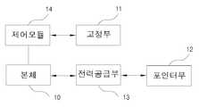
FIG. 7 is a block diagram of a position display apparatus for a human body surgery according to an embodiment of the present invention. Referring to FIG.
첨부된 도면을 참조하여 본 발명의 바람직한 실시예를 상세히 설명하기로 한다.DETAILED DESCRIPTION OF THE PREFERRED EMBODIMENTS Hereinafter, preferred embodiments of the present invention will be described in detail with reference to the accompanying drawings.
도 1은 본 발명의 일 실시예에 따른 인체 수술용 위치 표시 장치에 관한 도면이다. 도 2는 도 1의 인체 수술용 위치 표시 장치를 상단에서 바라본 도면이다.1 is a view illustrating a position display apparatus for a human body surgery according to an embodiment of the present invention. FIG. 2 is a top view of the position indicator for human body surgery of FIG. 1;
도 1 및 도 2를 참조하면, 본 발명의 일 실시예에 의한 인체 수술용 위치 표시 장치(1)는 인체 내부 조직에 부착되어 특정 위치 즉, 의사가 수술할 부위로 지정하는 위치를 표시한다. 도 3 및 도 4는 본 발명의 일 실시예에 따른 인체 수술용 위치 표시 장치가 인체의 특정 부위에 고정되는 과정을 도시한 도면이다.Referring to FIGS. 1 and 2, a
인체 수술용 위치 표시 장치(1, 이하 '위치 표시 장치'라 한다)는 본체(10), 고정부(11), 포인터부(12), 전력공급부(13) 및 제어 모듈(14)를 포함한다.A
본 발명에서 본체(10)의 형상은 원통형으로 형성되고, 크기는 고정부(11), 포인터부(12), 전력공급부(13) 및 제어 모듈(14)을 포함하여 직경이 6.5 밀리미터[mm]이상 15 밀리미터[mm]이하이며, 길이가 15 밀리미터[mm] 이하가 바람직하다. 이는 일 실시예일뿐, 크기에 제한이 있는 것은 아니다. 이하에서 설명할 바와 같이 특정 위치를 표시할 수 있을 정도의 빛을 발할 수 있고 전력공급부(13)의 전력용량이 포인터부(12)를 적어도 24시간 이상 작동하도록 제조될 수 있다면, 원통형 외에 다른 형상으로 형성될 수 있으며 환자에게 좀 더 적은 이질감을 주기 위해서는 그 크기가 작게 제조될수록 좋다. 또한, 포인터부(12)는 시중에서 유통되는, 즉 공지의 레이저포인터 모듈로 대체 가능하도록 형성될 수 있다.In the present invention, the shape of the
위치 표시 장치(1)는 환자가 삼키는 방식으로 인체 내에 삽입되는데, 본 발명에서는 내시경(20)을 이용하여 위치 표시 장치(1)의 위치를 배치하므로, 위치 표시 장치(1)를 삼킨 후 내시경(20)을 삽입한다.The
여기서 인체에 삽입되는 내시경(20)은 적어도 하나의 케이블(22)을 포함할 수 있으며, 예컨대 케이블에는 인체 내부 촬영으로 의사에게 환자의 인체 내부 정보 즉, 위장 또는 대장 등의 내부 정보를 제공하는 카메라(21)가 연결될 수 있다. 또는, 케이블(22)에는 인체 내부에서 촬영된 정보를 기반으로 종양, 용종 등이 발견되었을 때 이를 제거하기 위한 보조기구를 장착할 수 있다. 케이블(22)에 구비된 보조기구 중 인체에 삽입되는 부분은 1)끝단이 무딘 가위와 유사한 형상일 수 있으며, 2)고주파 발생부 또는 3)레이저 발생부 등을 포함할 수도 있다.Here, the
의사는 환자 신체의 외부에서 내시경의 케이블(22)에 연결된 보조기구를 조작 또는 제어함으로써 종양이나 용종을 제거할 수 있다. 즉, 1)의 경우 종양이나 용종 부근에 가위와 유사한 형상부를 위치시키고, 외부에서 동작 신호를 가하여 이를 구동시켜 잘라내도록 함으로써 종양이나 용종을 제거할 수 있다. 2) 또는 3)의 경우, 종양이나 용종 부위에 고주파 발생부 또는 레이저 발생부를 위치시키고, 외부에서 신호를 가하여 고주파 또는 레이저를 발생시킴으로써 종양이나 용종을 제거할 수 있다.The doctor may remove the tumor or polyp by manipulating or controlling the ancillary device connected to the cable 22 of the endoscope outside the patient's body. In the case of 1), a tumor or polyp can be removed by locating a scissor-like feature in the vicinity of the tumor or polyp and driving it by exciting it from outside. 2) or 3), a tumor or polyp can be removed by locating a high frequency generator or a laser generator on a tumor or polyp site and generating a high frequency or laser by applying a signal from the outside.
전술한 바와 같이 이용되는 내시경 보조기구가 구비되는 부분을 위치 표시 장치(1)로 대체하고, 그 후에 후술할 고정부(11)를 제어함으로써, 인체 내부 조직에 위치 표시 장치(1) 고정할 수 있다.The
의사는 환자의 인체 내로 삽입된 내시경(20)의 카메라(21)를 통하여 환자의 위장 또는 대장 내부에 존재하는 종양, 용종 등을 발견하게 된다. 이를 제거하기 위해서는 정밀 검사, 환자의 동의 등의 부수적인 과정이 필요하므로 발견된 종양을 바로 제거하는 시술을 수행할 수는 없다. 따라서, 추후에 종양이나 용종을 수술하여 제거하거나 치료하기 위해 정확한 위치를 표시할 필요가 있다. 이를 위해 위치 표시 장치(1)를 사용할 필요가 있으며, 위치 표시 장치(1)는 환자가 삼킴으로써 환자의 신체 내부로 투입되고, 그 뒤를 따라 내시경(20)이 삽입되어 신체 내부로 투입된다.The surgeon finds a tumor, a polyp, or the like present in the stomach or large intestine of the patient through the
내시경과 분리되어 환자의 신체 내부로 삽입된 위치 표시 장치(1)는 내시경(20)의 조작에 의하여 본체(10)를 인체 내부 조직에 고정하는 고정부(11)에 의해 종양과 같은 특정 부위(30)에 고정된다. 일 예로, 내시경의 조작에 의해 도 1에서와 같은 고정부(11)에 집게 등의 일측을 고정하며, 내시경의 조작에 의해 집게 등의 타측을 종양과 같은 특정 부위(30)에 고정할 수 있다. 이때 고정부(11)는 후술할 포인터부(12)와 인접하여 배치될 수 있는데, 이에 한정되지 아니하고 본체(10)의 옆면, 저면 등 다양한 위치에 마련될 수 있다.The
본체(10)와 인체 내부 조직을 고정하는 방법은 다양하게 고려될 수 있으며, 예컨대 인체 내 일부 조직이 돌출된 부위(30)를 집게로 집는 것과 유사한 방식, 즉 인체 내 돌출부위(30)를 폐성(閉性)하도록 조이는 방식이 있다. 또한, 인체 내 일부 조직이 돌출된 부위(30)를 본체에 포함된 고무줄 등을 이용하여 엮는 방식, 즉 인체 내 돌출부위(30)를 탄성체로 엮는 방식으로 고정부(11)와 인체 내 돌출부위(30)를 고정할 수 있다.The method of fixing the
또, 인체 내의 부드러운 부분에 바늘을 꽂거나 밀어넣는 것과 유사한 방식, 즉 인체 내부 조직의 연성부위를 찌르는 방식으로 고정부(11)와 인체 내 특정부위(30)를 고정할 수 있다. 또한, 인체 내 의사가 지정한 특정 부위에 세포접착제, 생체접착제 등의 인체에 무해한 접착제로 붙이는 방식, 즉 인체 내 특정부위에 인체에 무해한 접착제로 부착하는 방식으로 고정부(11)와 인체 내 특정부위(30)를 고정할 수 있다. 여기서 탄성체로 엮는 방식, 조직의 연성부위를 찌르는 방식 및 인체에 무해한 접착제로 붙이는 방식은 외부에서 조작함으로써 수행될 수 있다. 즉, 내시경의 동작에 의해 집게, 고무줄, 탄성체, 바늘, 접착제 등의 고정 부재를 고정부(11)에 고정하며, 내시경의 동작에 의해 고정부(11)에 고정된 고정 부재를 인체 내부 조직에 고정함으로써 위치 표시 장치(1)가 인체 내부 조직에 고정되도록 한다.In addition, it is possible to fix the
포인터부(12)는 위치 표시 장치(1)에 포함되어 본체(10)가 배치될 위치를 표시한다. 포인터부(12)는 빛을 발하는 부품 또는 소재로 형성될 수 있는데, 본 발명에서는 레이저포인터(laser pointer)를 이용한다. 레이저포인터는 반도체 레이저를 포함하므로, 매우 얇은 가시광선을 발산하며 대체로 5mW이하 저전력이기 때문에 본 발명에 적용될 수 있다.The
도 5는 본 발명의 일 실시예에 따른 발광표시부와 관련된 회로도를 간략히 표시한 도면이다. 도 6은 본 발명의 일 실시예에 따른 발광표시부를 구성하는 회로도의 예시 도면이다.5 is a schematic view illustrating a circuit diagram related to a light emitting display unit according to an embodiment of the present invention. 6 is an exemplary view showing a circuit diagram of a light emitting display unit according to an embodiment of the present invention.
도 5 및 도 6을 참조하면, 본 발명에 따른 위치 표시 장치 중 발광표시부의 작동 원리를 알 수 있다. 도 5에서 저항(51)의 저항값 수치를 변경하면, 발광표시부인 LED(52)의 휘도를 가변적으로 조절할 수 있다. 이때, 휘도는 일정한 범위를 가진 광원의 광도를 그 광원의 면적으로 나눈 양을 의미하며, 그 자체가 발광하고 있는 광원뿐만 아니라, 조명되어 빛나는 2차적인 광원에 대해서도 밝기를 나타내는 양이 된다. 도 6에 의하면 LED(61)는 일정한 시간 간격으로 깜빡이게 된다. 이때, 사용되는 부품에 따라 깜빡이는 시간 간격, 휘도 등을 조절할 수 있다. 또한, 도 5에 의한 구성 외에도 전압, 저항, 다이오드, 커패시터, 트랜지스터 등의 배치를 변경함으로써 다양한 형태의 회로 구성이 가능하다.5 and 6, the operation principle of the light emitting display unit of the position display apparatus according to the present invention can be understood. 5, when the resistance value of the
전력공급부(13)는 포인터부(12)에 전력을 공급한다. 포인터부(12)는 본체에 포함되며, 포인터부(12)가 적어도 24시간 이상 빛을 발할 수 있도록 작동할 수 있는 전력용량을 구비한다. 전력공급부(13)는 도면에 도시하지는 않았지만, 스위치와 연결되어 있다. 스위치가 온(ON)되는 경우, 전력공급부(13)에 신호가 전달되고, 그 후, 전력공급부(13)는 포인터부(12)에 전력을 공급한다. 스위치는 푸쉬버튼형 스위치와 같은, 외적인 압력에 의하여 온(ON)되는 스위치일 수도 있고, 단순히 회로적으로 내부에 구현될 수도 있다. 푸쉬버튼형 스위치가 사용된다면, 고정부(11) 근처에 위치하여 부착되는 것이 바람직하다.The
다른 실시예로, 푸쉬버튼형 스위치가 사용되는 경우, 위치 표시 장치(1)가 인체에 삽입되기 전에 사용자에 의하여 스위치를 온(ON)시켜 포인터부(12)를 구동하고, 그 후에 위치 표시 장치(1)를 삽입할 수도 있다.In another embodiment, when a push-button switch is used, the
또는, 고정부(11)를 고정하기 전 또는 후에 내시경(20) 또는 그 보조기구가 위치 표시 장치(1)에 접근하여 소정의 조작을 통하여 스위치를 온(ON)시킬 수도 있다.Alternatively, the
또 다른 실시예로, 스위치가 회로적으로 내부에 구현된 경우에는, 적절한 시간에 내시경(20)의 조작자가 소정의 신호를 무선을 통하여 스위치에 전송하여 스위치를 온(ON)시켜 포인터부(12)를 구동할 수도 있다.In another embodiment, when the switch is implemented in a circuit, the operator of the
또 다른 실시예로, 리모컨 등을 통하여 스위치를 작동시킬 수도 있다. 물론, 위에서 설명한 고정부(11)의 동작도 리모컨을 통하여 조작을 할 수도 있다.In another embodiment, the switch may be operated through a remote controller or the like. Of course, the operation of the fixing
고정부(11)가 고정되기 위한 소정의 동작이 이루어지는 경우, 전력공급부(13)에 스위치에 의한 ON 신호가 송신되고, 그 후, 전력공급부(13)는 포인터부(12)에 전력을 공급하게 된다.When the predetermined operation for fixing the fixing
다른 실시예로는, 인체 내 일부 조직이 돌출된 부위를 집게로 집는 것과 유사한 방식으로, 즉 외부에서 신호를 가하여 고정부(11)가 확개(擴開)되었다가 닫히도록 조절함으로써 인체 내 돌출부위가 폐성(閉性)하도록 조여질 수 있다. 이때, 본체(10)에 고정부(11)가 폐성(閉性)될 때의 압력이나 동작을 감지하는 센서를 부가하여, 일정 압력 이상이 감지되거나 또는 폐성(閉性)되는 동작이 감지되는 경우 스위치가 온(ON) 되도록 버튼을 누를 수 있는 설정을 할 수 있다. 또한, 막대 끝에 고무줄을 부착하고 고무줄을 이용하여 타 물체와 엮이도록 하는 것과 유사한 방법으로 인체 내 돌출부위와 고정부(11)를 탄성체를 이용하여 고정할 수 있다. 이때, 본체(10)에 탄성체가 타 물체와 엮여 외부에서 당겨지는 등의 힘에 의해 탄성력이 가해질 때 이를 감지할 수 있는 센서를 부가하여 일정 이상의 탄성력이 가해지면 스위치가 온(ON) 되도록 버튼을 누를 수 있는 설정을 할 수 있다. 또, 인체 내의 연성부분에 바늘을 꽂거나 밀어넣는 것과 유사한 방식으로 인체 내 특정부위에 고정부(11)를 고정할 수 있다. 이때, 본체(10)에 연성부위를 찌를 때의 압력 또는 인체 조직으로부터 발생하는 반발력을 감지할 수 있는 센서를 구비하여 인체 내부 조직의 연성부위가 찔릴 때 스위치가 온(ON) 되도록 버튼을 누를 수 있는 설정을 할 수 있다. 또는, 세포접착제, 생체접착제 등의 인체에 무해한 접착제로 붙이는 방식으로 인체 내 특정부위에 고정부(11)를 고정할 수 있다. 이때, 본체(10)에 인체 내 특정부위에 고정부(11)를 밀어서 밀착함으로써 고정할 때 미는 힘을 감지하는 센서를 구비하여, 인체 내부 조직에 고정부(11)가 부착될 때 스위치가 온(ON) 되도록 버튼을 누를 수 있는 설정을 할 수 있다.In another embodiment, the protruding portion of the human body is projected in a manner similar to that in which the protruding portion is gripped by the gripper, that is, by externally applying a signal to adjust the fixing
본 발명에서, 위치 표시 장치(1)는 인체 내부에 삽입되므로, 본체(10)의 외주면 및 고정부(11)는 인체에 무해한 재질이어야 한다. 이를 위해 본체(10) 및 고정부(11)를 내부식성 재질로 제조하거나 또는 본체(10) 및 고정부(11)에 내부식성 재질로 엷은 막을 입히는 코팅(coating)작업을 수행한다. 여기서 기계 또는 장치의 내부식성을 보장하기 위해 사용되는 스테인리스강 및 스테인리스 합금, 코발트 합금, 타이타늄 및 타이타늄 합금 등의 금속재료를 이용하여 본체(10) 또는 고정부(11)를 제조할 수 있다. 이때, 본체(10) 또는 고정부(11)를 제조하기 위해 사용되는 금속재료는 성형성이 좋고 다양한 형상으로 가공할 수 있으며 내부식성을 가진다는 특성이 있다. 다만, 고정부(11)와 인체 내부 조직을 고정하는 방식이 인체 내 돌출부위를 폐성(閉性)하도록 조이는 방식이거나 또는 인체 내부 조직의 연성부위를 찌르는 방식인 경우, 인체 내부로 위치 표시 장치(1)를 삽입하는 과정에서 고정부(11)가 인체 내를 긁거나 찌를 수 있으므로, 이를 최소화할 방법이 필요하다.In the present invention, since the
이에, 본체(10)의 모서리 부분 또는 고정부(11)의 날카로운 부분으로부터 인체가 상처나는 것을 보호하기 위해 코팅을 수행한다. 이를 위해 의료용 고분자 재료인 실리콘, 천연고무, 폴리염화비닐, 폴리에틸렌, 폴리프로필렌, 폴리우레탄 등을 이용할 수 있다. 코팅을 위한 의료용 고분자 재료들은 일반적으로 내부식성을 포함하며 생체적합성이 우수하다. 여기서 생체적합성이란 장기간에 걸쳐 생체에 악영향을 나타내지 않고 본래의 기능을 다하면서 생체와 공존할 수 있는 재료의 속성을 의미하며, 환자의 인체 내에 삽입되는 장치를 구성하거나 또는 그러한 장치에 코팅을 수행하기 위한 재료이므로 생체적합성은 반드시 필요한 성질이다.Thus, the coating is performed to protect the human body from being scratched from the edge portion of the
도 7은 본 발명의 일 실시예에 따른 인체 수술용 위치 표시 장치의 구성에 관한 블록도이다.FIG. 7 is a block diagram of a position display apparatus for a human body surgery according to an embodiment of the present invention. Referring to FIG.
도 7을 참조하면, 본 발명에 따른 위치 표시 장치(1)는 본체(10), 고정부(11), 포인터부(12), 전력공급부(13) 및 제어모듈(14)을 구비할 수 있다. 이때, 이때, 제어모듈(15)은 외부 또는 내시경으로부터 제공되는 신호 또는 내부 동작에 의하여 위치 표시 장치(1)의 구성요소의 동작을 전반적으로 제어한다. 그 외 각 구성요소에 대한 부분의 설명은 도 1 내지 도 6과 동일하여 생략하기로 한다.7, the
이상에서 본 발명에 대하여 바람직한 실시예들을 중심으로 살펴보았다. 본 발명이 속하는 기술분야에서 통상의 지식을 가진 자는 본 발명이 본 발명의 본질적인 특성에서 벗어나지 않는 범위에서 변형된 형태로 구현될 수 있음을 이해할 수 있을 것이다. 그러므로 개선된 실시예들은 한정적인 관점이 아니라 설명적인 관점에서 고려되어야 한다. 본 발명의 범위는 전술한 설명이 아니라 특허청구범위에 나타나 있으며, 그와 동등한 범위 내에 있는 모든 차이점은 본 발명에 포함된 것으로 해석되어야 할 것이다.The preferred embodiments of the present invention have been described above. It will be understood by those skilled in the art that various changes in form and details may be made therein without departing from the spirit and scope of the invention as defined by the appended claims. Therefore, the improved embodiments should be considered in an illustrative rather than a restrictive sense. The scope of the present invention is defined by the appended claims rather than by the foregoing description, and all differences within the scope of equivalents thereof should be construed as being included in the present invention.
1: 인체 수술용 위치 표시 장치
10: 본체11: 고정부
12: 포인터부13: 전력공급부
14: 제어모듈
20: 내시경21: 카메라
22: 케이블
30: 종양, 인체 내 돌출부위
51: 저항52, 61: LED1: Position indicator for human body surgery
10: main body 11:
12: Pointer unit 13: Power supply unit
14: Control module
20: endoscope 21: camera
22: Cable
30: tumor, protruding part in human body
51:
Claims (7)
Translated fromKorean내시경과 분리되어 인체 내에 삽입되는 본체;
상기 수술용 위치 표시 장치가 상기 인체 내부 조직에 고정된 위치를 빛을 발함으로써 표시하는 레이저 포인터부;
상기 본체에 형성되며, 상기 내시경의 동작에 의해 일측이 상기 인체 내부 조직에 고정되는 고정 부재의 타측에 상기 내시경의 동작에 의해 고정되는 고정부; 및
버튼 셀 배터리를 포함하며 상기 레이저 포인터부에 전력을 공급하는 전력공급부를 포함하는 것을 특징으로 하는 인체 수술용 위치 표시 장치.1. A position display apparatus for a human body surgery, the position display apparatus being attached to an internal tissue of a human body,
A body which is inserted into the human body separately from the endoscope;
A laser pointer unit for displaying a position where the surgical position display device is fixed to the internal tissue of the human body by emitting light;
A fixing part formed on the main body and fixed to the other side of a fixing member whose one side is fixed to the internal tissue of the human body by the operation of the endoscope by an operation of the endoscope; And
And a power supply unit including a button cell battery and supplying power to the laser pointer unit.
상기 포인터부는 레이저 포인터 모듈로 구성되는 것을 특징으로 하는 인체 수술용 위치 표시 장치.The method according to claim 1,
Wherein the pointer unit comprises a laser pointer module.
상기 본체는 직경이 6.5밀리미터[mm]이상 15밀리미터[mm]이하이며, 길이가 15밀리미터[mm]이하로 구성되는 것을 특징으로 하는 인체 수술용 위치 표시 장치.The method according to claim 1,
Wherein the main body has a diameter of 6.5 mm or more and 15 mm or less and a length of 15 mm or less.
상기 포인터부 및 고정부는 내부식성 재질로 제조되거나 코팅되는 것을 특징으로 하는 인체 수술용 위치 표시 장치.The method according to claim 1,
Wherein the pointer portion and the fixing portion are made of or coated with a corrosion-resistant material.
상기 내부식성 재질을 이용한 제조는,
상기 본체 및 고정부를 스테인리스강, 스테인리스강 합금, 코발트 합금, 타이타늄 또는 타이타늄 합금; 중 어느 하나를 이용하여 제조하는 것을 특징으로 하는 인체 수술용 위치 표시 장치.6. The method of claim 5,
The manufacturing using the corrosion-
The main body and the fixing part are made of stainless steel, a stainless steel alloy, a cobalt alloy, a titanium or a titanium alloy; Wherein the position measuring apparatus is manufactured by using any one of the following methods.
상기 내부식성 재질을 이용한 코팅은,
상기 본체 및 고정부를 의료용 실리콘, 폴리우레탄 또는 폴리에스터; 중 어느 하나를 사용하여 수행되는 것을 특징으로 하는 인체 수술용 위치 표시 장치.6. The method of claim 5,
The coating using the corrosion-
The body and the fixing part are made of medical silicone, polyurethane or polyester; Wherein the positional information is obtained by using any one of the following methods.
Priority Applications (1)
| Application Number | Priority Date | Filing Date | Title |
|---|---|---|---|
| KR1020140090997AKR101601678B1 (en) | 2014-07-18 | 2014-07-18 | Position indicator device for surgery |
Applications Claiming Priority (1)
| Application Number | Priority Date | Filing Date | Title |
|---|---|---|---|
| KR1020140090997AKR101601678B1 (en) | 2014-07-18 | 2014-07-18 | Position indicator device for surgery |
Publications (2)
| Publication Number | Publication Date |
|---|---|
| KR20160010107A KR20160010107A (en) | 2016-01-27 |
| KR101601678B1true KR101601678B1 (en) | 2016-03-09 |
Family
ID=55309426
Family Applications (1)
| Application Number | Title | Priority Date | Filing Date |
|---|---|---|---|
| KR1020140090997AActiveKR101601678B1 (en) | 2014-07-18 | 2014-07-18 | Position indicator device for surgery |
Country Status (1)
| Country | Link |
|---|---|
| KR (1) | KR101601678B1 (en) |
Citations (2)
| Publication number | Priority date | Publication date | Assignee | Title |
|---|---|---|---|---|
| JP2005218680A (en)* | 2004-02-06 | 2005-08-18 | Olympus Corp | Surgical lesion identification system |
| JP2009542329A (en) | 2006-07-05 | 2009-12-03 | ウィルソン−クック・メディカル・インコーポレーテッド | Suction clip |
Family Cites Families (2)
| Publication number | Priority date | Publication date | Assignee | Title |
|---|---|---|---|---|
| WO2004082488A1 (en) | 2003-03-17 | 2004-09-30 | Sumitomo Bakelite Company Limited | Clip and clipping instrument for biological tissues |
| KR101014971B1 (en)* | 2008-10-24 | 2011-02-16 | 오영준 | Human Body Capsule Type Laser Light Therapy |
- 2014
- 2014-07-18KRKR1020140090997Apatent/KR101601678B1/enactiveActive
Patent Citations (2)
| Publication number | Priority date | Publication date | Assignee | Title |
|---|---|---|---|---|
| JP2005218680A (en)* | 2004-02-06 | 2005-08-18 | Olympus Corp | Surgical lesion identification system |
| JP2009542329A (en) | 2006-07-05 | 2009-12-03 | ウィルソン−クック・メディカル・インコーポレーテッド | Suction clip |
Also Published As
| Publication number | Publication date |
|---|---|
| KR20160010107A (en) | 2016-01-27 |
Similar Documents
| Publication | Publication Date | Title |
|---|---|---|
| US20220322928A1 (en) | Surgical retractors | |
| US8267854B2 (en) | Endoscope system | |
| EP3313310B1 (en) | Devices for assisting in open surgeries | |
| US20200054193A1 (en) | Wireless viewing device and method of use thereof | |
| JP5784308B2 (en) | Surgical lighting system | |
| US20160353973A1 (en) | Wireless viewing device | |
| CN104470459B (en) | Medical system and method of operation | |
| WO2007078003A1 (en) | Trans-natural opening based or transcutaneous medical system | |
| JP2015223435A (en) | Tool and system for spinal cord and nerve operation | |
| US20160270641A1 (en) | Video assisted surgical device | |
| US20140257089A1 (en) | Means and methods for the prevention of retained surgical items and gossypiboma | |
| US11779428B2 (en) | Disposable medical clamp light | |
| KR101601678B1 (en) | Position indicator device for surgery | |
| KR101601682B1 (en) | Position indicator device for surgery | |
| US12201399B2 (en) | Disposable thumb light | |
| KR101180078B1 (en) | Lesion indicating apparatus and fitting device thereof | |
| KR101977581B1 (en) | System for detecting lesion location including a clip for marking lesion location | |
| JP4763397B2 (en) | Air supply device and surgical system having the air supply device | |
| KR101163630B1 (en) | Surgical Position Check Luminescent Capsule | |
| KR102045383B1 (en) | Overtube | |
| Melzer et al. | Instrumentation and allied technology for endoscopic surgery | |
| JP3213655U (en) | Spinal and neurosurgical instruments and systems | |
| JP5059121B2 (en) | Surgical instruments | |
| WO2020068105A1 (en) | Wireless viewing device and method of use thereof | |
| KR101171025B1 (en) | Method and Apparatus for Natural Orifice Translumenal Endoscopic Surgery |
Legal Events
| Date | Code | Title | Description |
|---|---|---|---|
| A201 | Request for examination | ||
| PA0109 | Patent application | Patent event code:PA01091R01D Comment text:Patent Application Patent event date:20140718 | |
| PA0201 | Request for examination | ||
| E902 | Notification of reason for refusal | ||
| PE0902 | Notice of grounds for rejection | Comment text:Notification of reason for refusal Patent event date:20150618 Patent event code:PE09021S01D | |
| E701 | Decision to grant or registration of patent right | ||
| PE0701 | Decision of registration | Patent event code:PE07011S01D Comment text:Decision to Grant Registration Patent event date:20151228 | |
| PG1501 | Laying open of application | ||
| GRNT | Written decision to grant | ||
| PR0701 | Registration of establishment | Comment text:Registration of Establishment Patent event date:20160303 Patent event code:PR07011E01D | |
| PR1002 | Payment of registration fee | Payment date:20160303 End annual number:3 Start annual number:1 | |
| PG1601 | Publication of registration | ||
| FPAY | Annual fee payment | Payment date:20200203 Year of fee payment:5 | |
| PR1001 | Payment of annual fee | Payment date:20200203 Start annual number:5 End annual number:5 | |
| PR1001 | Payment of annual fee | Payment date:20210408 Start annual number:6 End annual number:6 | |
| PR1001 | Payment of annual fee | Payment date:20220127 Start annual number:7 End annual number:7 | |
| PR1001 | Payment of annual fee | Payment date:20230131 Start annual number:8 End annual number:8 | |
| PR1001 | Payment of annual fee | Payment date:20240130 Start annual number:9 End annual number:9 |