KR101587802B1 - Artificial blood scaffold and the method for manufacturing the same - Google Patents
Artificial blood scaffold and the method for manufacturing the sameDownload PDFInfo
- Publication number
- KR101587802B1 KR101587802B1KR1020130135588AKR20130135588AKR101587802B1KR 101587802 B1KR101587802 B1KR 101587802B1KR 1020130135588 AKR1020130135588 AKR 1020130135588AKR 20130135588 AKR20130135588 AKR 20130135588AKR 101587802 B1KR101587802 B1KR 101587802B1
- Authority
- KR
- South Korea
- Prior art keywords
- fibrous structure
- rti
- biodegradable elastomer
- group
- copolymers
- Prior art date
- Legal status (The legal status is an assumption and is not a legal conclusion. Google has not performed a legal analysis and makes no representation as to the accuracy of the status listed.)
- Expired - Fee Related
Links
Images


Classifications
- A—HUMAN NECESSITIES
- A61—MEDICAL OR VETERINARY SCIENCE; HYGIENE
- A61L—METHODS OR APPARATUS FOR STERILISING MATERIALS OR OBJECTS IN GENERAL; DISINFECTION, STERILISATION OR DEODORISATION OF AIR; CHEMICAL ASPECTS OF BANDAGES, DRESSINGS, ABSORBENT PADS OR SURGICAL ARTICLES; MATERIALS FOR BANDAGES, DRESSINGS, ABSORBENT PADS OR SURGICAL ARTICLES
- A61L27/00—Materials for grafts or prostheses or for coating grafts or prostheses
- A61L27/14—Macromolecular materials
- A—HUMAN NECESSITIES
- A61—MEDICAL OR VETERINARY SCIENCE; HYGIENE
- A61F—FILTERS IMPLANTABLE INTO BLOOD VESSELS; PROSTHESES; DEVICES PROVIDING PATENCY TO, OR PREVENTING COLLAPSING OF, TUBULAR STRUCTURES OF THE BODY, e.g. STENTS; ORTHOPAEDIC, NURSING OR CONTRACEPTIVE DEVICES; FOMENTATION; TREATMENT OR PROTECTION OF EYES OR EARS; BANDAGES, DRESSINGS OR ABSORBENT PADS; FIRST-AID KITS
- A61F2/00—Filters implantable into blood vessels; Prostheses, i.e. artificial substitutes or replacements for parts of the body; Appliances for connecting them with the body; Devices providing patency to, or preventing collapsing of, tubular structures of the body, e.g. stents
- A61F2/02—Prostheses implantable into the body
- A61F2/04—Hollow or tubular parts of organs, e.g. bladders, tracheae, bronchi or bile ducts
- A61F2/06—Blood vessels
- A—HUMAN NECESSITIES
- A61—MEDICAL OR VETERINARY SCIENCE; HYGIENE
- A61L—METHODS OR APPARATUS FOR STERILISING MATERIALS OR OBJECTS IN GENERAL; DISINFECTION, STERILISATION OR DEODORISATION OF AIR; CHEMICAL ASPECTS OF BANDAGES, DRESSINGS, ABSORBENT PADS OR SURGICAL ARTICLES; MATERIALS FOR BANDAGES, DRESSINGS, ABSORBENT PADS OR SURGICAL ARTICLES
- A61L27/00—Materials for grafts or prostheses or for coating grafts or prostheses
- A61L27/14—Macromolecular materials
- A61L27/26—Mixtures of macromolecular compounds
- A—HUMAN NECESSITIES
- A61—MEDICAL OR VETERINARY SCIENCE; HYGIENE
- A61L—METHODS OR APPARATUS FOR STERILISING MATERIALS OR OBJECTS IN GENERAL; DISINFECTION, STERILISATION OR DEODORISATION OF AIR; CHEMICAL ASPECTS OF BANDAGES, DRESSINGS, ABSORBENT PADS OR SURGICAL ARTICLES; MATERIALS FOR BANDAGES, DRESSINGS, ABSORBENT PADS OR SURGICAL ARTICLES
- A61L2420/00—Materials or methods for coatings medical devices
Landscapes
- Health & Medical Sciences (AREA)
- Life Sciences & Earth Sciences (AREA)
- General Health & Medical Sciences (AREA)
- Oral & Maxillofacial Surgery (AREA)
- Transplantation (AREA)
- Veterinary Medicine (AREA)
- Animal Behavior & Ethology (AREA)
- Public Health (AREA)
- Epidemiology (AREA)
- Medicinal Chemistry (AREA)
- Dermatology (AREA)
- Chemical & Material Sciences (AREA)
- Engineering & Computer Science (AREA)
- Pulmonology (AREA)
- Gastroenterology & Hepatology (AREA)
- Heart & Thoracic Surgery (AREA)
- Biomedical Technology (AREA)
- Vascular Medicine (AREA)
- Cardiology (AREA)
- Materials For Medical Uses (AREA)
- Prostheses (AREA)
Abstract
Translated fromKoreanDescription
Translated fromKorean본 발명은 인공혈관 이식시 성공률을 향상시키는 J형태의 응력-변형률 곡선을 나타내는 인공혈관용 구조물 및 이것의 제조방법에 관한 것이다.The present invention relates to an artificial blood vessel structure showing a J-shaped stress-strain curve that improves the success rate in artificial blood vessel grafting, and a method for manufacturing the same.
심장, 심장판막, 혈관 등과 같은 순환기 계통에서 발생하는 질병은 전 세계적으로 성인의 사망원인 중에서 1위를 기록하고 있는데 이중 동맥경화, 협심증, 심근경색, 뇌졸중 등이 속하는 혈관과 관련된 질병이 가장 많은 부분을 차지하고 있다.Diseases occurring in the circulatory system such as heart, heart valves, and blood vessels are among the leading causes of death among adults worldwide. The most common diseases related to blood vessels belonging to atherosclerosis, angina pectoris, myocardial infarction, Respectively.
세계보건기구(WHO)의 통계에 따르면 심혈관 질환으로 사망하는 사람은 1년에 약 1,200만 명으로 추산되고 있으며, 미국의 경우 약 5,700만 명의 인구가 한 가지 이상의 심혈관 질환으로 치료받고 있는 실정이다. 이에 따른 비용만 해도 연간 2,600억 달러에 달하는 것으로 보고되고 있다.According to statistics from the World Health Organization (WHO), about 12 million people die from cardiovascular disease per year. In the United States, about 57 million people are treated with more than one cardiovascular disease. The costs are reported to reach $ 260 billion annually.
심혈관 질환의 1차적인 치료방법으로는 자가혈관 이식술이나 스텐트 등의 혈관 개통을 위한 보조 장치를 이용한 방법이 있는데, 자가 혈관 이식술은 대부분 동맥경화에 의한 폐쇄성 질환에 이용되며 유전적 섬유질 형성장애, 대동맥류, 대동맥 박리증, 동맥염, 외부손상 등에 이용되는 경우도 있다. 자가 혈관 이식술로 사용될 수 있는 이식편은 내유동맥, 대위망동맥, 요동맥, 척골동맥 등으로 극히 제한적이고 정맥 이식편도 사용 가능한 범위가 제한적이기 때문에 반복수술을 요하는 경우 자가 혈관의 이식이 어렵거나 불가능한 경우가 발생될 수 있다. 그러므로 환자의 생명이나 삶의 질의 향상을 위해 동종혈관이나 인조혈관 등의 자가 혈관을 대체할 수 있는 장치가 요구되고 있다.The primary treatment methods for cardiovascular disease are autologous vascular grafts and assistive devices for vascular access such as stents. Autologous vascular grafting is mostly used for obstructive diseases caused by atherosclerosis, Aneurysm, aortic dissection, arteritis, external injury, etc. Grafts that can be used as autologous grafts are extremely limited to the internal mammary artery, the superior mesenteric artery, the umbilical artery, the ulnar artery, etc., and the range of available venous grafts is limited. Therefore, A case may arise. Therefore, devices for replacing autologous blood vessels such as allografts or artificial blood vessels are required to improve the quality of life or quality of life of a patient.
최근 적용되고 있는 이식형 인공 혈관으로는 폴리에틸렌테레프탈레이트(poly(ethyleneterephalate))소재 또는 PTFE(Poly tetra fluoro ethylene)소재의 인공 혈관이 이용되고 있는데, 상기 소재의 인공 혈관은 내경 6 mm 초과의 대구경에만 적용이 가능하다. 그 이유는 혈관의 내부 표면에 생긴 혈전에 의한 막힘 현상이 문제점으로 지적되고 있다.Recently, artificial blood vessels of polyethylene terephthalate (PTFE) or poly tetra fluoro ethylene (PTFE) have been used as implantable artificial blood vessels. It is applicable. The reason is that clogging due to thrombosis on the inner surface of the blood vessel is pointed out as a problem.
6 mm 이하의 소구경 혈관의 경우 작은 혈전이라 해도 혈관을 막아 혈관 터짐등 치명적인 결과를 초래할 수 있다. 이 문제의 해결을 위한 방법은 두 가지로 접근할 수 있다. 우선 인공혈관의 강도를 올리는 방법으로 기존 일반적인 인공혈관은 적당한 탄성체를 이용해 왔으며, 이들 대부분의 지지체들은 가해지는 힘에 따라 일정량 늘어나거나 줄어드는 성질을 가지고 있다. 따라서 혈전 등에 의해 일정 이상의 힘이 가해지면 혈관 터짐 현상이 빈발해 왔던 이유라 할 수 있다. 더구나 강도를 올리면 지지체의 전반적인 물성이 단단해지므로 혈관 특유의 수축과 팽창인 연속적 기계 운동에 문제가 생기게 된다. 두 번째 접근방법으로는 근본적으로 혈관 내측 부분에 혈전의 생성을 막아야 하므로 분해성 혈관 모양 지지체에 일반 조직세포나 줄기세포 등을 접종하고 접종된 세포를 조직으로 성장시키는 원리인 조직공학적인 방법, 혈전 생성을 억제해주는 혈관내피세포만 분해성 또는 비분해성 지지체의 내부 표면에 결합시켜주는 방법 또는 세포를 사용하지 않는 방법으로 헤파린, 와파린, 음이온을 가진 작용기 등을 내부 표면에 붙여주는 방법들이 있다. 그러나 혈관내피세포를 이용한 방법은 혈관내피세포의 취급이 쉽지 않으며, 헤파린 등을 이용한 방법은 초기에 다소 효과가 있지만 일정 시간이 지나면 다시 혈전이 생기는 것으로 알려져 있다. 따라서 새로운 실용적인 혈관 지지체의 필요성이 날로 증대되고 있다.Small-diameter blood vessels smaller than 6 mm may cause fatal outcomes such as blood vessel clogging by blocking blood vessels even if they are small blood vessels. There are two approaches to solving this problem. As a method of increasing the strength of artificial blood vessels, conventional artificial blood vessels have used suitable elastic bodies, and most of these supports have a property of increasing or decreasing by a certain amount depending on the applied force. Therefore, if a force exceeding a certain level is applied due to thrombosis or the like, it may be the reason that the blood vessel rupture phenomenon has frequently occurred. Furthermore, when the strength is increased, the overall physical properties of the support become harder, resulting in a problem in the continuous mechanical movement, which is the shrinkage and swelling peculiar to the blood vessel. The second approach is to fundamentally prevent the formation of thrombus in the inner blood vessels. Therefore, the tissue engineering method, which is a principle of inoculating normal tissue cells or stem cells into the degradable vascular support and growing the inoculated cells into tissue, There is a method in which only vascular endothelial cells that inhibit vascular endothelial cells are bound to the inner surface of a decomposable or non-degradable support, or a method in which heparin, warfarin, and an anion having a functional group are attached to the inner surface by a method using no cells. However, vascular endothelial cells can not be easily handled by vascular endothelial cells, and heparin and other methods are effective at early stage but after a certain time, blood clots are formed again. Thus, the need for new practical vascular supports is increasing day by day.
또한, 스텐트의 경우에는 스테인리스나 티타늄 합금 재질을 많이 사용하고 있는데 이 물질이 혈관벽과 만나 상처가 나게 되면 상처 부위로부터 평활근 세포가 자라나와 혈관을 막히게 하거나 상기 물질의 표면에 혈소판 등이 붙으면서 혈전의 생성에 의해 혈관이 막히는 일이 많다. 따라서 스텐트 표면에 음이온 작용기를 부여하는 방법이나 사이클로스포린 A (cyclosporin A)등과 같은 약물을 도포하여 방출케 함으로써 불필요한 조직에 의한 캡슐화 또는 혈전을 방지하고자 하였다. 그러나 이러한 방법들은 일정 기간이 지나면 다시 혈관이 막히는 단점이 있다.In addition, in the case of stents, stainless steel or titanium alloy material is often used. When this material meets with the blood vessel wall, the smooth muscle cells grow from the injured area and clog blood vessels or platelets adhere to the surface of the material. There are many cases where blood vessels are clogged by generation. Therefore, a method of applying an anionic functional group to the surface of the stent or a drug such as cyclosporin A or the like is applied and released to prevent encapsulation or thrombosis by unnecessary tissue. However, these methods have the disadvantage that the blood vessels are blocked again after a certain period of time.
종래 대한민국 등록특허 제0661396호에는 스텐트에 다층으로 코팅층을 형성하되 상층에는 소수성 해파린 고분자가 분포되어 혈전이 생성되는 것을 억제시킬 수 있는 스텐트가 제시되어 있다. 상기 기술의 스텐트는 대구경의 혈관에는 사용할 수 있지만 소구경의 혈관에는 사용할 수 없을 뿐만 아니라 약한 소수성을 띄므로 단백질, 혈소판 등과의 소수성 상호작용(hydrophobic interaction)에 의해 이들이 스텐트 표면에 더 잘 붙을 수도 있다. 더구나 이들 스텐트는 한번 부풀어 오르면 그 자체의 강인함 때문에 혈관의 수축과 팽창의 기계적 운동에 스트레스를 가할 수 있기 때문에 혈전에 의한 위급 상황시를 제외하고는 잘 사용하지 않는 단점도 있다.Korean Patent Registration No. 0661396 discloses a stent capable of forming a coating layer on the stent in a multilayer structure, wherein hydrophobic heparin polymer is distributed in the upper layer to inhibit thrombus formation. The stent of the above-described technique can be used for large-diameter blood vessels, but it can not be used for small-diameter blood vessels, and has weak hydrophobicity. Therefore, hydrophobic interactions with proteins, platelets, etc. may adhere them more stably to the surface of the stent . In addition, these stents, once inflated, can stress the mechanical movement of the contraction and expansion of the blood vessels due to their own strength, which is a disadvantage that they are not used except for emergency situations caused by thrombosis.
따라서, 실제 혈관과 유사한 물리적 특성을 가짐으로써 다소의 혈전이 생성되더라도 혈관의 터짐 현상을 최대한 지연시킬 수 있으면서, 또한, 소구경에서 대구경의 혈관에 사용할 수 있는 인공 혈관용 구조물에 대한 요구가 증대되고 있다.Therefore, even if some blood clots are generated by having physical characteristics similar to actual blood vessels, it is possible to delay the deterioration of blood vessels as much as possible, and the demand for artificial blood vessel structures that can be used for large-diameter blood vessels in a small diameter is increased have.
이에, 본 발명은 J형태의 응력-변형률 곡선을 나타냄으로써 인공혈관 이식시 실제 혈관과 유사한 물리적 특성을 제공할 수 있는 인공혈관용 구조물을 제공하고자 한다. 또한, 이러한 인공혈관 구조물의 제조방법을 제공하고자 한다.Accordingly, the present invention provides J-shaped stress-strain curves to provide artificial blood vessel structures that can provide physical characteristics similar to actual blood vessels in artificial blood vessel grafting. The present invention also provides a method for manufacturing such an artificial blood vessel structure.
구체적으로, 본 발명은 인공혈관을 체내에 이식 시 혈관으로써 심장으로부터 받는 스트레스에 대한 수축과 팽창이 자연스럽게 이루어지며, 혈관 내벽에 다소 생성될 수 있는 혈전이나 조직의 침범 등에 의해 혈관의 내압이 비정상적으로 상승하게 될 경우 그에 따른 지지체의 팽창에 대한 저항을 극대화시킴으로써 지지체의 비정상적인 팽창에 의한 인공 혈관의 파열 및 찢어짐 현상을 최대한 억제할 수 있을 뿐 만 아니라, 고분자 표면으로부터 내면에 이르기까지 미세 다공성을 부여함으로써 영양분이나 세포의 이동을 용이하게 하여 혈관 내피세포와 평활근 세포가 주위로부터 잘 자라 들어올 수 있게 하는 기능이 부여된 J형태의 응력-변형률 곡선을 나타내는 인공혈관용 구조물 및 제조방법을 사용자에게 제공하는데 그 목적이 있다.More particularly, the present invention relates to a method and a device for the prevention and / or treatment of grafting of an artificial blood vessel to a blood vessel by grafting the artificial blood vessel into the blood vessel through a blood vessel or tissue, By maximizing the resistance to the expansion of the supporting body when it is increased, the rupture and tearing of the artificial blood vessel due to the abnormal expansion of the support can be suppressed to the utmost, and besides, by giving the micropores from the surface of the polymer to the inner surface A J-shaped stress-strain curve having a function of facilitating the movement of nutrients or cells and allowing vascular endothelial cells and smooth muscle cells to smoothly come in from around, and a manufacturing method thereof, There is a purpose.
본 발명은 체내 혈관이 J형태의 응력-변형률 곡선을 나타내는 고유한 특성을 가짐을 확인하고 인공혈관 이식 시 성공률의 향상을 도모하고자 J형태의 응력-변형률 곡선을 나타내는 인공혈관용 구조물 및 이것의 제조방법을 제공한다.The present invention relates to an artificial blood vessel structure exhibiting a J-shaped stress-strain curve in order to confirm that a blood vessel in the body has unique characteristics showing a J-shaped stress-strain curve and to improve the success rate in artificial blood vessel grafting, ≪ / RTI >
본 발명의 일 양태로서, 생체 적합성 소재를 포함하는 특정 형태를 갖는 섬유 구조물 및 상기 섬유 구조물에 코팅된 다공성 구조의 생분해성 탄성체를 포함하는 인공혈관용 구조물을 제공한다.As one aspect of the present invention, there is provided an artificial vascular structure comprising a fibrous structure having a specific shape including a biocompatible material and a biodegradable elastomer having a porous structure coated on the fibrous structure.
상기 생체 적합성 소재는 락티드(lactide), 카프로락톤(caprolactone), 글리코라이드(glycolide), 디옥사논(dioxanone), 프로필렌(Propylene), 에틸렌(Ethylene), 염화비닐(vinylchloride), 부타디엔(butadiene), 메틸메타아크릴레이트(methly methacrylate), 아크릴산, 2-히드록시에틸메타크릴에이트(2-hydroxyethlymethacrylate), 카보네이트(carbonate), 폴리에틸렌 테레프탈레이트(polyethylene terephalate) 및 이들의 공중합체로 이루어진 군에서 선택된 하나 또는 그 이상일 수 있으며, 바람직하게는 락티드, 카프로락톤, 글리코라이드 및 이들의 공중합체일 수 있다.The biocompatible material may be selected from the group consisting of lactide, caprolactone, glycolide, dioxanone, propylene, ethylene, vinylchloride, butadiene, One selected from the group consisting of methyl methacrylate, acrylic acid, 2-hydroxyethlymethacrylate, carbonate, polyethylene terephthalate, and copolymers thereof; or Or more, and preferably lactide, caprolactone, glycolides, and copolymers thereof.
상기 섬유 구조물의 특정 형태로는 0.1 um 내지 5,000 um 굵기의 단섬유 또는 이것의 섬유 다발에 의하여 제조되는 니트형태, 메쉬형태, 우븐형태, 직조형태, 부직포형태 및 단위세포가 삼각형으로부터 팔각형까지의 연결된 구조 중에서 선택되는 하나의 형태 또는 이들의 혼합 형태일 수 있다. 가장 바람직하게는 500 um 내지 2,000 um 굵기 섬유로써 니트형태로 짜여진 구조이다.Specific forms of the fibrous structure include knitted, mesh, woven, woven, non-woven, and unit cells made of short fibers of 0.1 um to 5,000 um in thickness or their fiber bundles, Structure, or a mixed form thereof. Most preferably 500 to 2,000 um thick fibers knitted into a knitted structure.
상기 생분해성 탄성체는 락티드(lactide), 카프로락톤(caprolactone), 글리코라이드(glycolide), 디옥사논(dioxanone), 프로필렌(Propylene), 에틸렌(Ethylene), 염화비닐(vinylchloride), 부타디엔(butadiene), 메틸메타크릴레이트(methly methacrylate), 아크릴산, 2-히드록시에틸메타크릴에이트(2-hydroxyethlymethacrylate), 카보네이트(carbonate), 폴리에틸렌 테레프탈레이트(polyethylene terephalate) 및 이들의 공중합체로 이루어진 군에서 선택되는 하나 또는 그 이상일 수 있다. 바람직하게는 락티드, 카프로락톤, 글리코라이드 및 이들의 공중합체일 수 있다.The biodegradable elastomer may be selected from the group consisting of lactide, caprolactone, glycolide, dioxanone, propylene, ethylene, vinylchloride, butadiene, One selected from the group consisting of methyl methacrylate, acrylic acid, 2-hydroxyethlymethacrylate, carbonate, polyethylene terephthalate, and copolymers thereof. Or more. Preferably lactide, caprolactone, glycolides, and copolymers thereof.
또한, 본 발명의 다른 양태로서, 생체 적합성 소재를 포함하는 특정 형태를 갖는 섬유 구조물 및 상기 섬유 구조물에 코팅된 다공성 구조의 생분해성 탄성체를 포함하는 인공혈관용 구조물을 제조하는 방법을 제공한다.According to another aspect of the present invention, there is provided a method for manufacturing an artificial vascular structure including a fibrous structure having a specific shape including a biocompatible material and a biodegradable elastomer having a porous structure coated on the fibrous structure.
상기 방법에서 생분해성 탄성체에 기공을 형성하는 방법은 염용출법, 기체발포법 또는 상분리법에 의할 수 있다.The method of forming pores in the biodegradable elastomer in the above method may be a salt elution method, a gas foaming method, or a phase separation method.
상기 본 발명에 따른 인공혈관용 구조물을 제조하는 방법의 일 구체예로서, 염용출법을 이용한 방법은 다음의 단계를 포함한다:As one embodiment of the method for producing an artificial vascular structure according to the present invention, a method using a salt elution method includes the following steps:
(a) 생체 적합성 소재를 이용하여 특정 형태의 섬유 구조물을 제조하는 단계;(a) preparing a specific type of fibrous structure using a biocompatible material;
(b) 생분해성 탄성체를 유기용매에 용해시키고, 미세한 크기의 염과 혼합하는 단계;(b) dissolving the biodegradable elastomer in an organic solvent and mixing with a finely-divided salt;
(c) 상기 (a)의 섬유 구조물을 (b)의 혼합액으로 코팅하는 단계; 및(c) coating the fibrous structure of (a) with the mixed solution of (b); And
(d) 상기 코팅된 섬유 구조물을 건조 후 증류수로 세척하는 단계.(d) washing the coated fiber structure with distilled water after drying.
또는, 상기 본 발명에 따른 인공혈관용 구조물을 제조하는 방법의 일 구체예로서, 기체발포법을 이용한 방법은 다음의 단계를 포함한다:Alternatively, as one embodiment of the method for producing an artificial vascular structure according to the present invention, a method using a gas foaming method includes the following steps:
(a) 생체적합성 소재를 이용하여 특정 형태의 섬유 구조물을 제조하는 단계;(a) preparing a specific type of fibrous structure using a biocompatible material;
(b) 생분해성 탄성체를 유기용매에 용해시키고, 미세한 크기의 기체방출용 미세입자와 혼합하는 단계;(b) dissolving the biodegradable elastomer in an organic solvent, and mixing the fine biodegradable elastomer with fine particles for gas discharge;
(c) 상기 (a)의 섬유 구조물을 (b)의 혼합액으로 코팅하는 단계; 및(c) coating the fibrous structure of (a) with the mixed solution of (b); And
(d) 상기 코팅된 섬유 구조물을 건조 후 60 내지 80℃의 증류수로 세척하는 단계.(d) washing the coated fibrous structure with distilled water at 60-80 < 0 > C after drying.
또는, 상기 본 발명에 따른 인공혈관용 구조물을 제조하는 방법의 다른 구체예로서, 상분리법을 이용한 방법은 다음의 단계를 포함한다:Alternatively, as another embodiment of the method for producing an artificial vascular structure according to the present invention, the method using the phase separation method includes the following steps:
(a) 생체적합성 소재를 이용하여 특정 형태의 섬유 구조물을 제조하는 단계;(a) preparing a specific type of fibrous structure using a biocompatible material;
(b) 생분해성 탄성체를 유기용매에 용해시키는 단계;(b) dissolving the biodegradable elastomer in an organic solvent;
(c) 상기 (a)의 섬유 구조물을 (b)의 혼합액으로 코팅하는 단계; 및(c) coating the fibrous structure of (a) with the mixed solution of (b); And
(d) 상기 코팅된 섬유 구조물을 -4 내지 -200℃ 사이에서 냉동시킨 후 동결건조시키는 단계.
(d) freezing the coated fibrous structure between -4 and -200 < 0 > C, followed by lyophilization.
상기 (a)단계에서 생체 적합성 소재는 락티드(lactide), 카프로락톤(caprolactone), 글리코라이드(glycolide), 디옥사논(dioxanone), 프로필렌(Propylene), 에틸렌(Ethylene), 염화비닐(vinylchloride), 부타디엔(butadiene), 메틸메타크릴레이트(methly methacrylate), 아크릴산, 2-히드록시에틸메타크릴레이트(2-hydroxyethlymethacrylate), 카보네이트(carbonate), 폴리에틸렌 테레프탈레이트(polyethylene terephalate) 및 이들의 공중합체로 이루어진 군에서 선택된 하나 또는 그 이상일 수 있으며, 바람직하게는 락티드, 카프로락톤, 글리코라이드 및 이들의 공중합체일 수 있다.In the step (a), the biocompatible material may be selected from the group consisting of lactide, caprolactone, glycolide, dioxanone, propylene, ethylene, vinylchloride, Butadiene, methly methacrylate, acrylic acid, 2-hydroxyethlymethacrylate, carbonate, polyethylene terephthalate, and copolymers thereof. And may be lactide, caprolactone, glycolides, and copolymers thereof.
상기 섬유 구조물의 특정 형태로는 0.1 um 내지 5,000 um 굵기의 단섬유 또는 이것의 섬유 다발에 의하여 제조되는 니트형태, 메쉬형태, 우븐형태, 직조형태, 부직포형태 및 단위세포가 삼각형으로부터 팔각형까지의 연결된 구조 중에서 선택되는 하나의 형태 또는 이들의 혼합 형태일 수 있다. 가장 바람직하게는 500 um 내지 2,000 um 굵기 섬유로써 니트형태로 짜여진 구조이다.Specific forms of the fibrous structure include knitted, mesh, woven, woven, non-woven, and unit cells made of short fibers of 0.1 um to 5,000 um in thickness or their fiber bundles, Structure, or a mixed form thereof. Most preferably 500 to 2,000 um thick fibers knitted into a knitted structure.
상기 (b)단계에서 생분해성 탄성체 소재는 락티드(lactide), 카프로락톤(caprolactone), 글리코라이드(glycolide), 디옥사논(dioxanone), 프로필렌(Propylene), 에틸렌(Ethylene), 염화비닐(vinylchloride), 부타디엔(butadiene), 메틸메타아크릴레이트(methly methacrylate), 아크릴산, 히드록시에틸메타아크릴에이트(2-hydroxyethlymethacrylate), 카보네이트(carbonate), 폴리에틸렌 텔레프탈레이트(polyethylene terephalate) 및 이들의 공중합체로 이루어진 군에서 선택되는 하나 또는 그 이상일 수 있다. 바람직하게는 락티드, 카프로락톤, 글리코라이드 및 이들의 공중합체일 수 있다.In the step (b), the biodegradable elastomer material may be selected from the group consisting of lactide, caprolactone, glycolide, dioxanone, propylene, ethylene, vinylchloride Butadiene, methly methacrylate, acrylic acid, 2-hydroxyethlymethacrylate, carbonate, polyethylene terephthalate, and copolymers thereof. Lt; RTI ID = 0.0 > and / or < / RTI > Preferably lactide, caprolactone, glycolides, and copolymers thereof.
상기 (b) 단계에서 혼합되는 염은 일반적으로 NaCl, KCl, CH3COONa 등의 나트륨과 염소 및 칼륨, 칼슘으로 구성된 무기질 이온성 입자일 수 있다. 상기 (b) 단계의 염 혼합은 (i) 냉동에 의한 상분리법(phase separation)이나 (ii) 기체방출(gas forming) 방법으로 대체될 수 있다. 상기 (b) 단계는 5 ~ 500 um 크기의 NaCl을 코팅용 고분자의 무게비 5 ~ 80%를 가하여 수행하는 것이 바람직하다.The salt to be mixed in step (b) may be an inorganic ionic particle composed of sodium, chlorine, potassium and calcium such as NaCl, KCl and CH3 COONa. The salt mixing in the step (b) may be replaced with (i) phase separation by freezing or (ii) gas forming. The step (b) may be performed by adding 5 to 80% of NaCl to the coating polymer in a weight ratio of 5 to 500 μm.
상기 (c)단계는 (a) 섬유 구조물을 (b)용액에 넣었다 빼는 코팅단계로써, 여기서 (b)용액의 고분자 농도는 코팅용 고분자의 무게비 2% ~ 40% 범위가 적당하다. 상기 코팅단계는 1회 내지 25회까지 반복 코팅을 할 수 있으며, 상기 코팅 때 마다 30초 내지 30분 동안 건조 시간을 둘 수 있다.The step (c) is a coating step in which (a) the fibrous structure is put in a solution and the solution is taken out of the solution, wherein the concentration of the polymer in the solution (b) ranges from 2% to 40% by weight of the polymer for coating. The coating step may be repeated once to 25 times, and the coating time may be 30 seconds to 30 minutes.
상기 (d) 단계는 건조된 혈관용 구조물을 증류수 속에 담그고 약 3일 동안 염을 제거 용출시키는 단계를 의미한다.Step (d) refers to a step of immersing the dried vascular structure in distilled water and removing and eluting the salt for about 3 days.
본 발명의 인공혈관용 구조물은 심장으로부터 정상적인 스트레스 하에서는 유연하며 탄성인 폴리에스테르계 고분자에 의해 자연스러운 수축과 팽창이 이루어지며, 혈관 내벽에 다소 생성될 수 있는 혈전이나 조직의 성장 등에 의해 혈관의 내압이 비정상적으로 상승하게 될 경우 망사구조의 구조물에 의해 지지체의 팽창에 대한 저항을 극대화시킴으로써 지지체의 비정상적인 팽창에 의한 인공 혈관의 파열 및 찢어짐 현상을 최대한 억제할 수 있는 J형태의 응력-변형률 곡선을 나타내는 인공혈관용 구조물 및 제조방법을 제공할 수 있다.The artificial vascular structure of the present invention has natural contraction and swelling due to a polyester polymer which is flexible and elastic under normal stress from the heart, and the intrinsic pressure of blood vessels due to a thrombus or tissue growth, The J-shaped stress-strain curve that can suppress the rupture and tear of the artificial blood vessel due to the abnormal expansion of the support by maximizing the resistance to the expansion of the support by the structure of the mesh structure when it is abnormally raised, A vascular structure and a manufacturing method thereof.
도 1은 본 발명에 적용된 원통형 망사구조의 일 구체예를 나타낸 도면이다.
도 2는 본 발명에 의해 제조된 인공혈관용 구조체를 나타내었다.
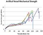
도 4는 본 발명에 의해 제조된 인공혈관용 구조체의 J형태의 응력-변형률 곡선 그래프이다.
도 3은 실제 체내 혈관의 J형태의 응력-변형률 곡선을 나타낸 것이다.1 is a view showing an embodiment of a cylindrical mesh structure applied to the present invention.
2 shows the artificial vascular structure manufactured by the present invention.
FIG. 4 is a graph showing a stress-strain curve of J-shape of the artificial vascular structure manufactured by the present invention.
3 shows the J-shaped stress-strain curves of actual blood vessels in the body.
본 발명은 J형태의 응력-변형률 곡선을 나타내는 인공혈관용 구조물 및 이것의 제조방법에 관한 것이다.The present invention relates to an artificial blood vessel structure showing a J-shaped stress-strain curve and a method of manufacturing the same.
구체적으로, 본 발명의 인공혈관용 구조물은 정상적인 수축과 팽창의 스트레스에 대해 유연성 있게 작용하다가 비정상적으로 높은 압력이 발생 시 혈관의 파열을 막기 위해 섬유 구조물에 의해 일정 길이로 팽창시 강도를 향상시켜줌으로써 혈관의 부분적 막힘에 의한 혈관의 파열을 최대한 방지할 수 있는 효과를 나타낼 수 있다.Specifically, the artificial vascular structure of the present invention flexibly works against the stresses of normal contraction and expansion, and when the abnormally high pressure is generated, the strength of the vascular structure is increased by the fiber structure to a certain length in order to prevent rupture of the blood vessel It is possible to prevent the rupture of the blood vessel due to the partial clogging of the blood vessel to the utmost.
또한, 본 발명의 인공혈관용 구조물은 종래 내경이 6 mm이상과 이하인 소구경에서 대구경까지 다양한 내경의 혈관에 사용될 수 있다.In addition, the artificial vascular structure of the present invention can be used for blood vessels of various internal diameters ranging from a small diameter to a large diameter with an inner diameter of 6 mm or more.
또한, 본 발명의 인공혈관용 구조물은 고분자 코팅시 다공성 형태를 부여할 수 있어 약물 방출이나 혈관 세포의 성장과 안착을 유도할 수 있다.In addition, the artificial vascular structure of the present invention can impart a porous form upon polymer coating, and can induce drug release or vascular cell growth and settlement.
본 발명의 구체적 일 양태는 인공혈관용 구조물에 관한 것으로서, 생체 적합성 소재를 포함하는 특정 형태를 갖는 섬유 구조물 및 상기 섬유 구조물에 코팅된 생분해성 탄성체를 포함한다.A specific embodiment of the present invention relates to an artificial blood vessel structure, which includes a fibrous structure having a specific shape including a biocompatible material and a biodegradable elastomer coated on the fibrous structure.
상기 코팅된 생분해성 탄성체는 다공성 구조인 것을 특징으로 한다.The coated biodegradable elastomer is characterized by being a porous structure.
상기 생체적합성 소재는 혈관 등에 이용 시 생체에 부작용이 발생되지 않는 물질을 의미하는 것으로서, 구체적으로 락티드(lactide), 카프로락톤(caprolactone), 글리코라이드(glycolide), 디옥사논(dioxanone), 프로필렌(Propylene), 에틸렌(Ethylene), 염화비닐(vinylchloride), 부타디엔(butadiene), 메틸메타크릴레이트(methly methacrylate), 아크릴산, 2-히드록시에틸메타크릴레이트(2-hydroxyethlymethacrylate), 카보네이트(carbonate), 폴리에틸렌 테레프탈레이트(polyethylene terephalate) 및 이들의 공중합체로 이루어진 군에서 선택된 하나 또는 그 이상일 수 있으며, 바람직하게는 락티드, 카프로락톤, 글리코라이드 및 이들의 공중합체일 수 있다.The biocompatible material means a material that does not cause side effects to the living body when used in blood vessels and the like. Specifically, lactide, caprolactone, glycolide, dioxanone, propylene Propylene, ethylene, vinylchloride, butadiene, methly methacrylate, acrylic acid, 2-hydroxyethlymethacrylate, carbonate, Polyethylene terephthalate and copolymers thereof, and may be one or more selected from the group consisting of lactide, caprolactone, glycolides and copolymers thereof.
상기 섬유 구조물의 특정 형태로는 0.1 um 내지 5,000 um 굵기 범위의 단섬유 또는 섬유다발 형태인 섬유로써 니트형태, 메쉬형태, 우븐형태, 직조형태, 부직포형태, 단위세포가 삼각형으로부터 팔각형까지의 연결된 구조로써 이들의 혼합 내지는 단일구조로 짜여진 형태일 수 있다. 가장 바람직하게는 500 um 내지 2,000 um 굵기 섬유로써 니트형태로 짜여진 구조이다.As a specific form of the fiber structure, fibers of a short fiber type or a fiber bundle shape having a thickness ranging from 0.1 to 5,000 um may be used as a knit form, a mesh form, a woven form, a woven form, a nonwoven form, Or may be a blend of these or a woven fabric of a single structure. Most preferably 500 to 2,000 um thick fibers knitted into a knitted structure.
상기 생분해성 탄성체는 평상시 혈관으로써 수축과 팽창의 기계적 움직임에 있어 유연성과 탄성이 있는 소재를 의미하는 것으로서, 구체적으로 락티드(lactide), 카프로락톤(caprolactone), 글리코라이드(glycolide), 디옥사논(dioxanone), 프로필렌(Propylene), 에틸렌(Ethylene), 염화비닐(vinylchloride), 부타디엔(butadiene), 메틸메타크릴레이트(methly methacrylate), 아크릴산, 2-히드록시에틸메타크릴레이트(2-hydroxyethlymethacrylate), 카보네이트(carbonate), 폴리에틸렌 테레프탈레이트(polyethylene terephalate) 및 이들의 공중합체로 이루어진 군에서 선택되는 하나 또는 그 이상일 수 있다. 바람직하게는 락티드, 카프로락톤, 글리코라이드 및 이들의 공중합체일 수 있다.The biodegradable elastomer is a blood vessel which is normally flexible and elastic in mechanical motion of shrinkage and expansion. Specifically, lactose, caprolactone, glycolide, dioxanone propylene, ethylene, vinylchloride, butadiene, methly methacrylate, acrylic acid, 2-hydroxyethlymethacrylate, and the like. But may be one or more selected from the group consisting of carbonate, polyethylene terephthalate and copolymers thereof. Preferably lactide, caprolactone, glycolides, and copolymers thereof.
또한, 본 발명의 다른 양태는 생체 적합성 소재를 포함하는 특정 형태의 섬유 구조물 및 상기 섬유 구조물에 코팅된 다공성 구조의 생분해성 탄성체를 포함하는 인공혈관용 구조물을 제조하는 방법에 관한 것이다.Still another aspect of the present invention relates to a method for producing an artificial vascular structure comprising a specific type of fibrous structure including a biocompatible material and a biodegradable elastomer of a porous structure coated on the fibrous structure.
상기 방법에서 코팅된 생분해성 탄성체에 기공을 형성하는 방법은 염용출법, 기체발포법 또는 상분리법에 의할 수 있다. 기체방출법은 수용액에서 온도를 올려주면 이산화탄소 같은 기체가 발생하는 입자를 넣어주는 방법이고, 상분리법은 염 등을 사용하지 않고 고분자 코팅액만 코팅한 후 온도를 떨어뜨려 기공을 만들어 주는 방법입니다.The method of forming pores in the coated biodegradable elastomer in the above method may be a salt elution method, a gas foaming method, or a phase separation method. Gas release is a method of adding gas generating particles such as carbon dioxide by increasing the temperature in an aqueous solution. The phase separation method is a method of coating the polymer coating solution only without using a salt and then dropping the temperature to make pores.
상기 본 발명에 따른 인공혈관용 구조물을 제조하는 방법의 일 구체예로서, 염용출법을 이용한 방법은 다음의 단계를 포함한다:As one embodiment of the method for producing an artificial vascular structure according to the present invention, a method using a salt elution method includes the following steps:
(a) 생체 적합성 소재를 이용하여 특정 형태의 섬유 구조물을 제조하는 단계;(a) preparing a specific type of fibrous structure using a biocompatible material;
(b) 생분해성 탄성체를 유기용매에 용해시키고, 미세한 크기의 염과 혼합하는 단계;(b) dissolving the biodegradable elastomer in an organic solvent and mixing with a finely-divided salt;
(c) 상기 (a)의 섬유 구조물을 (b)의 혼합액으로 코팅하는 단계; 및(c) coating the fibrous structure of (a) with the mixed solution of (b); And
(d) 상기 코팅된 섬유 구조물을 건조 후 증류수로 세척하는 단계.(d) washing the coated fiber structure with distilled water after drying.
또는, 상기 본 발명에 따른 인공혈관용 구조물을 제조하는 방법의 일 구체예로서, 기체발포법을 이용한 방법은 다음의 단계를 포함한다:Alternatively, as one embodiment of the method for producing an artificial vascular structure according to the present invention, a method using a gas foaming method includes the following steps:
(a) 생체 적합성 소재를 이용하여 특정 형태의 섬유 구조물을 제조하는 단계;(a) preparing a specific type of fibrous structure using a biocompatible material;
(b) 생분해성 탄성체를 유기용매에 용해시키고, 미세한 크기의 기체방출용 미세입자와 혼합하는 단계;(b) dissolving the biodegradable elastomer in an organic solvent, and mixing the fine biodegradable elastomer with fine particles for gas discharge;
(c) 상기 (a)의 섬유 구조물을 (b)의 혼합액으로 코팅하는 단계; 및(c) coating the fibrous structure of (a) with the mixed solution of (b); And
(d) 상기 코팅된 섬유 구조물을 건조 후 60 내지 80℃의 증류수로 세척하는 단계.(d) washing the coated fibrous structure with distilled water at 60-80 < 0 > C after drying.
또는, 상기 본 발명에 따른 인공혈관용 구조물을 제조하는 방법의 다른 구체예로서, 상분리법을 이용한 다음의 단계를 포함한다:Alternatively, another embodiment of the method for producing an artificial vascular structure according to the present invention includes the following steps using a phase separation method:
(a) 생체적합성 소재를 이용하여 특정 형태의 섬유 구조물을 제조하는 단계;(a) preparing a specific type of fibrous structure using a biocompatible material;
(b) 생분해성 탄성체를 유기용매에 용해시키는 단계;(b) dissolving the biodegradable elastomer in an organic solvent;
(c) 상기 (a)의 섬유 구조물을 (b)의 혼합액으로 코팅하는 단계; 및(c) coating the fibrous structure of (a) with the mixed solution of (b); And
(d) 상기 코팅된 섬유 구조물을 -4 내지 -200℃ 사이에서 냉동시킨 후 동결건조시키는 단계.(d) freezing the coated fibrous structure between -4 and -200 < 0 > C, followed by lyophilization.
상기 (a)단계에서 생체 적합성 소재는 혈관 등에 이용 시 생체에 부작용이 발생되지 않는 물질을 의미하는 것으로서, 구체적으로 락티드(lactide), 카프로락톤(caprolactone), 글리코라이드(glycolide), 디옥사논(dioxanone), 프로필렌(Propylene), 에틸렌(Ethylene), 염화비닐(vinylchloride), 부타디엔(butadiene), 메틸메타크릴레이트(methly methacrylate), 아크릴산, 2-히드록시에틸메타크릴레이트(2-hydroxyethlymethacrylate), 카보네이트(carbonate), 폴리에틸렌 테레프탈레이트(polyethylene terephalate) 및 이들의 공중합체로 이루어진 군에서 선택된 하나 또는 그 이상일 수 있으며, 바람직하게는 락티드, 카프로락톤, 글리코라이드 및 이들의 공중합체일 수 있다.In the step (a), the biocompatible material means a material that does not cause side effects to the living body when used in blood vessels and the like. Specifically, lactide, caprolactone, glycolide, dioxanone propylene, ethylene, vinylchloride, butadiene, methly methacrylate, acrylic acid, 2-hydroxyethlymethacrylate, and the like. And may be one or more selected from the group consisting of carbonate, polyethylene terephthalate and copolymers thereof, preferably lactide, caprolactone, glycolid and copolymers thereof.
상기 섬유 구조물의 특정 형태로는 0.1 um 내지 5,000 um 굵기 범위의 단섬유 또는 섬유다발 형태인 섬유로써 니트형태, 메쉬형태, 우븐형태, 직조형태, 부직포형태, 단위세포가 삼각형으로부터 팔각형까지의 연결된 구조로써 이들의 혼합 내지는 단일구조로 짜여진 형태일 수 있다. 가장 바람직하게는 500 um 내지 2,000 um 굵기 섬유로써 니트형태로 짜여진 구조이다.As a specific form of the fiber structure, fibers of a short fiber type or a fiber bundle shape having a thickness ranging from 0.1 to 5,000 um may be used as a knit form, a mesh form, a woven form, a woven form, a nonwoven form, Or may be a blend of these or a woven fabric of a single structure. Most preferably 500 to 2,000 um thick fibers knitted into a knitted structure.
상기 (b)단계에서 생분해성 탄성체 소재로는 평상시 혈관으로써 수축과 팽창의 기계적 움직임에 있어 유연성과 탄성이 있는 소재를 의미하는 것으로서, 구체적으로 락티드(lactide), 카프로락톤(caprolactone), 글리코라이드(glycolide), 디옥사논(dioxanone), 프로필렌(Propylene), 에틸렌(Ethylene), 염화비닐(vinylchloride), 부타디엔(butadiene), 메틸메타크릴레이트(methly methacrylate), 아크릴산, 2-히드록시에틸메타크릴레이트(2-hydroxyethlymethacrylate), 카보네이트(carbonate), 폴리에틸렌 텔레프탈레이트(polyethylene terephalate) 및 이들의 공중합체로 이루어진 군에서 선택되는 하나 또는 그 이상일 수 있다. 바람직하게는 락티드, 카프로락톤, 글리코라이드 및 이들의 공중합체일 수 있다.The biodegradable elastomer material in the step (b) means a material having flexibility and elasticity in the mechanical motion of contraction and expansion as blood vessels at normal times. Specifically, lactide, caprolactone, but are not limited to, glycolide, dioxanone, propylene, ethylene, vinylchloride, butadiene, methly methacrylate, acrylic acid, 2-hydroxyethylmethacrylate But may be one or more selected from the group consisting of 2-hydroxyethlymethacrylate, carbonate, polyethylene terephthalate and copolymers thereof. Preferably lactide, caprolactone, glycolides, and copolymers thereof.
상기 (b) 단계에서 혼합되는 염은 일반적으로 NaCl, KCl, CH3COONa 등의 나트륨과 염소 및 칼륨, 칼슘으로 구성된 무기질 이온성 입자일 수 있다. 상기 (b) 단계의 염 혼합은 (i) 냉동에 의한 상분리법(phase separation)이나 (ii) 기체방출(gas forming) 방법으로 대체될 수 있다. 상기 (b) 단계는 5 내지 500 um 크기의 NaCl을 코팅용 고분자의 무게비 5 ~ 80%를 가하여 수행하는 것이 바람직하다.The salt to be mixed in step (b) may be an inorganic ionic particle composed of sodium, chlorine, potassium and calcium such as NaCl, KCl and CH3 COONa. The salt mixing in the step (b) may be replaced with (i) phase separation by freezing or (ii) gas forming. Preferably, the step (b) is performed by adding 5 to 80% NaCl in a weight ratio of 5 to 500 .mu.m to the coating polymer.
상기 (c)단계는 (a) 섬유 구조물을 (b)용액에 넣었다 빼는 코팅단계로써, 여기서 (b)용액의 농도는 용매에 대한 코팅용 고분자가 무게비 2% 내지 40% 범위가 적당하다. 상기 코팅단계는 1회 내지 25회까지 반복 코팅을 할 수 있으며, 상기 코팅 때 마다 30초 내지 30분 동안 건조 시간을 둘 수 있다.The step (c) is a coating step in which (a) the fibrous structure is put in a solution, and the concentration of the solution (b) is in the range of 2 to 40% by weight. The coating step may be repeated once to 25 times, and the coating time may be 30 seconds to 30 minutes.
상기 (d) 단계는 건조된 혈관용 구조물을 증류수 속에 담그고 약 3일 동안 염을 제거 용출시키는 단계를 의미한다. 또는, 기체방출법과 같은 경우 증류수의 온도를 상온으로부터 80℃까지 올릴 수 있는 방법을 포함한다. 또는, 염을 가하지 않고 상분리법으로 진행할 경우 (c)단계의 지지체를 -4 내지 -200 ℃ 사이에서 냉동시킨 후 동결건조를 실시하는 것을 포함시킬 수 있다.Step (d) refers to a step of immersing the dried vascular structure in distilled water and removing and eluting the salt for about 3 days. Or a method of raising the temperature of the distilled water from room temperature to 80 DEG C in the case of the gas discharge method. Or freeze drying after freezing the supporter of step (c) between -4 and -200 ° C when proceeding with a phase separation method without adding a salt.
이하, 본 발명의 이해를 돕기 위하여 바람직한 실시예를 제시하나, 하기 실시예는 본 발명을 예시하는 것일 뿐 본 발명의 범주 및 기술사상 범위 내에서 다양한 변경 및 수정이 가능함은 당업자에게 있어서 명백한 것이며, 이러한 변형 및 수정이 첨부된 특허청구범위에 속하는 것도 당연한 것이다.It will be apparent to those skilled in the art that various modifications and variations can be made in the present invention without departing from the spirit or scope of the present invention. Such variations and modifications are intended to be within the scope of the appended claims.
굵기가 100 um인 폴리디옥사논(Polydioxanone, PDO) 섬유 mono-filament를 사용하여 니트구조를 가진 직경 6 mm인 원통형 섬유구조물을 제조하였다 [도 1]. Poly(L-lactide-co-ε-caprolactone) (PLCL)을 클로로포름에 9% (w/w)로 용해한 후 20 um 이하의 NaCl입자를 PLCL과 무게비 20%가 되도록 혼합하였다. 이 혼합용액 내에 상기 제조한 원통형 PDO 섬유 구조물을 1분간 담그고 빼서 15분간 건조하는 코팅작업을 연속으로 6회 실시한 후 상온에서 건조 하였다. 건조된 구조물을 증류수에 넣고 3일간 염 용출 작업을 진행하고 염이 제거된 구조물을 -78 ℃에서 20시간 동안 건조시켜 지지체를 제조하였다 [도 2]. 제조된 지지체의 물리적 특성이 실제 혈관과 유사한지를 알아보기 위하여 응력-변형률 측정을 진행 하였다 [도 4].A cylindrical fiber structure having a knitted structure and having a diameter of 6 mm was fabricated using polydioxanone (PDO) fiber mono-filament having a thickness of 100 μm (FIG. 1). Poly (L-lactide-co-ε-caprolactone) (PLCL) was dissolved in chloroform at 9% (w / w) and NaCl particles of 20 μm or less were mixed with PLCL at a weight ratio of 20%. The cylindrical PDO fiber structure was immersed in the mixed solution for 1 minute, and the coating solution was removed and dried for 15 minutes. The coating operation was carried out six times in succession and then dried at room temperature. The dried structure was put into distilled water and salt elution was performed for 3 days. The salt-free structure was dried at -78 ° C for 20 hours to prepare a support (FIG. 2). Stress-strain measurements were performed to see if the physical properties of the prepared support were similar to actual blood vessels (Fig. 4).
굵기가 100 um인 락티드 (Polylactide, PLA) 섬유를 이용하여 니트구조를 가진 직경 6 mm인 원통형 섬유구조물을 제조하였다. Poly(L-lactide-co-ε-caprolactone) (PLCL)을 Tetrahydrofuran (THF)에 9% (w/w)로 용해한 후 20 um 이하의 NaCl입자를 PLCL과 무게비 20%가 되도록 혼합하고, 이 후 과정은 상기 실시예 1과 동일하게 진행 하였다.A cylindrical fiber structure with a knitted structure and a diameter of 6 mm was fabricated using a polylactide (PLA) fiber having a thickness of 100 μm. After dissolving poly (L-lactide-co-ε-caprolactone) (PLCL) in tetrahydrofuran (THF) at 9% (w / w) NaCl particles of 20 μm or less were mixed with PLCL at a weight ratio of 20% The procedure was the same as in Example 1 above.
상기 실시예 1과 동일하게 진행하되 염을 사용하지 않고 코팅작업을 연속으로 6회 실시한 후 상온 건조 절차 없이 -80 ℃에서 동결 하였다. 그 후 78 ℃에서 20시간 동안 건조시켜 지지체를 제조하였다.The procedure of Example 1 was followed except that the coating was continuously carried out six times without using a salt and then frozen at -80 ° C without drying at room temperature. And then dried at 78 DEG C for 20 hours to prepare a support.
상기 실시예 1과 동일하게 진행하되 염을 사용하지 않고 기체방출법을 이용하여 진행하였다. 염 대신 sodium bicarbonate를 동일한 20%로 혼합해 준 후 코팅작업을 연속으로 6회 실시하고 상온 건조 후 60 ℃의 증류수에서 5시간동안 기체발포를 시켜주었다. 깨끗한 증류수로 세척 후 그 후 78 ℃에서 20시간 동안 건조시켜 지지체를 제조하였다.The procedure was carried out in the same manner as in Example 1, but was carried out using a gas discharge method without using a salt. The sodium bicarbonate was mixed with the same 20% of the salt, and then the coating operation was carried out six times in succession. After drying at room temperature, gas bubbling was performed in distilled water at 60 ° C for 5 hours. Washed with clean distilled water and then dried at 78 ° C for 20 hours to prepare a support.
[비교예 1][Comparative Example 1]
상기 실시예 1과 비교하기 위해 개의 복강 내 동맥을 적출하였고, 이 혈관의 응력-변형률측정을 진행 하였다 (표 1, 도 4).To compare with Example 1 above, intraperitoneal arteries were excised and the stress-strain measurements of the blood vessels were performed (Table 1, Fig. 4).
[비교예 2][Comparative Example 2]
상기 실시예 1에서 PDO섬유구조물은 제외하고 PLCL과 염 용액을 이용하여 동일하게 원통형 혈관 지지체를 만들었다. 이 지지체의 응력-변형률 측정을 진행하였다 (표 1).In Example 1, except for the PDO fiber structure, a cylindrical vascular support was similarly prepared using PLCL and a salt solution. Stress-strain measurements of this support were carried out (Table 1).
[시험예 1][Test Example 1]
응력-변형률 측정Stress-strain measurement
실시예 및 비교예에서 제조된 지지체와 실제 혈관의 응력-변형률을 측정하였으며, 하기 표 1과 도 3, 4에 나타냈었다. Universal testing machine (Instron model 4467, Canton, MA, USA)을 사용하였으며, crosshead 속도는 1 mm/min로 진행하였다.Stress-strain of the support and actual blood vessels prepared in Examples and Comparative Examples were measured and shown in Table 1 and Figures 3 and 4 below. A universal testing machine (Instron model 4467, Canton, MA, USA) was used and the crosshead speed was 1 mm / min.
위 표 1에 나타낸 바와 같이, 본 발명의 실시예 1에 따라 제조된 지지체는 비교예 1 즉, 실제 혈관보다 강도는 다소 높았으나 전반적으로 매우 유사한 경향을 보임으로써 물리적 성질이 비슷함을 확인하였다. 반면, 비교예 2의 경우 늘어나는 성질은 우수하지만 강도 등이 약한것으로 평가되었다. 도 4와 도 3에는 실시예 1과 비교예 2에 대한 그래프를 각각 나타내었다. 도 3의 경우 실제 혈관 특유의 J 곡선이 나타나고 있는 것을 확인 하였고, 도 4에서 본 발명에 의해 제조된 인공혈관도 명확하게 J 곡선이 만들어지는 것을 확인하였다.
As shown in Table 1, the support prepared according to Example 1 of the present invention showed comparatively high physical strength compared to that of Comparative Example 1, but the overall tendency was similar. On the other hand, in Comparative Example 2, it was evaluated that the elongation property was excellent but the strength was weak. 4 and FIG. 3 show graphs for Example 1 and Comparative Example 2, respectively. In FIG. 3, it is confirmed that the J curve specific to the actual blood vessel appears. In FIG. 4, it is confirmed that the artificial blood vessel produced by the present invention also clearly has a J curve.
Claims (10)
Translated fromKorean상기 섬유 구조물에 코팅된 다공성 구조의 생분해성 탄성체
를 포함하는 인공혈관용 구조물로서;
상기 특정 형태는 단섬유를 이용하여 형성된 니트형태인 것을 특징으로 하는, J 형태의 응력-변형률 곡선을 나타내는 인공혈관용 구조물.Certain types of fibrous structures including biocompatible materials; And
A biodegradable elastomer of a porous structure coated on the fiber structure
An artificial vascular structure;
Wherein said specific form is a knitted form formed by using short fibers.
(b) 생분해성 탄성체를 유기용매에 용해시키고, 미세한 크기의 염과 혼합하는 단계;
(c) 상기 (a)의 섬유 구조물을 (b)의 혼합액으로 코팅하는 단계; 및
(d) 상기 코팅된 섬유 구조물을 건조 후 증류수로 세척하는 단계
를 포함하고;
상기 특정 형태는 단섬유를 이용하여 형성된 니트형태인 것을 특징으로 하는, J 형태의 응력-변형률 곡선을 나타내는 인공혈관용 구조물을 제조하는 방법.(a) preparing a specific type of fibrous structure using a biocompatible material;
(b) dissolving the biodegradable elastomer in an organic solvent and mixing with a finely-divided salt;
(c) coating the fibrous structure of (a) with the mixed solution of (b); And
(d) washing the coated fibrous structure with distilled water after drying
;
Wherein said specific form is a knitted form formed using short fibers. ≪ RTI ID = 0.0 > 21. < / RTI >
(b) 생분해성 탄성체를 유기용매에 용해시키고, 미세한 크기의 기체방출용 미세입자와 혼합하는 단계;
(c) 상기 (a)의 섬유 구조물을 (b)의 혼합액으로 코팅하는 단계; 및
(d) 상기 코팅된 섬유 구조물을 건조 후 60℃ 내지 80℃의 증류수로 세척하는 단계
를 포함하고;
상기 특정 형태는 단섬유를 이용하여 형성된 니트형태인 것을 특징으로 하는, J 형태의 응력-변형률 곡선을 나타내는 인공혈관용 구조물을 제조하는 방법.(a) preparing a specific type of fibrous structure using a biocompatible material;
(b) dissolving the biodegradable elastomer in an organic solvent, and mixing the fine biodegradable elastomer with fine particles for gas discharge;
(c) coating the fibrous structure of (a) with the mixed solution of (b); And
(d) washing the coated fibrous structure with distilled water at 60 DEG C to 80 DEG C after drying
;
Wherein said specific form is a knitted form formed using short fibers. ≪ RTI ID = 0.0 > 21. < / RTI >
(b) 생분해성 탄성체를 유기용매에 용해시키는 단계;
(c) 상기 (a)의 섬유 구조물을 (b)의 용액으로 코팅하는 단계; 및
(d) 상기 코팅된 섬유 구조물을 -4℃ 내지 -200℃ 사이에서 냉동시킨 후 동결건조시키는 단계
를 포함하고;
상기 특정 형태는 단섬유를 이용하여 형성된 니트형태인 것을 특징으로 하는, J 형태의 응력-변형률 곡선을 나타내는 인공혈관용 구조물을 제조하는 방법.(a) preparing a specific type of fibrous structure using a biocompatible material;
(b) dissolving the biodegradable elastomer in an organic solvent;
(c) coating the fibrous structure of (a) with a solution of (b); And
(d) freezing the coated fibrous structure between -4 ° C and -200 ° C, followed by lyophilization
;
Wherein said specific form is a knitted form formed using short fibers. ≪ RTI ID = 0.0 > 21. < / RTI >
Priority Applications (1)
| Application Number | Priority Date | Filing Date | Title |
|---|---|---|---|
| KR1020130135588AKR101587802B1 (en) | 2013-11-08 | 2013-11-08 | Artificial blood scaffold and the method for manufacturing the same |
Applications Claiming Priority (1)
| Application Number | Priority Date | Filing Date | Title |
|---|---|---|---|
| KR1020130135588AKR101587802B1 (en) | 2013-11-08 | 2013-11-08 | Artificial blood scaffold and the method for manufacturing the same |
Publications (2)
| Publication Number | Publication Date |
|---|---|
| KR20150053528A KR20150053528A (en) | 2015-05-18 |
| KR101587802B1true KR101587802B1 (en) | 2016-02-12 |
Family
ID=53390086
Family Applications (1)
| Application Number | Title | Priority Date | Filing Date |
|---|---|---|---|
| KR1020130135588AExpired - Fee RelatedKR101587802B1 (en) | 2013-11-08 | 2013-11-08 | Artificial blood scaffold and the method for manufacturing the same |
Country Status (1)
| Country | Link |
|---|---|
| KR (1) | KR101587802B1 (en) |
Cited By (1)
| Publication number | Priority date | Publication date | Assignee | Title |
|---|---|---|---|---|
| KR20220055286A (en) | 2020-10-26 | 2022-05-03 | 전남대학교병원 | Graft material for blood vessel regeneration and method for manufacturing thereof |
Families Citing this family (5)
| Publication number | Priority date | Publication date | Assignee | Title |
|---|---|---|---|---|
| KR102129059B1 (en)* | 2017-11-21 | 2020-07-02 | 주식회사 글로원 | Artificial Blood Vessel by Biocompatibility Materials and the Method for Manufacturing the Same |
| WO2019103227A1 (en)* | 2017-11-21 | 2019-05-31 | 주식회사 글로원 | Artificial blood vessel utilizing biocompatible material and method for preparing same |
| KR102283339B1 (en)* | 2019-06-27 | 2021-08-02 | 사회복지법인 삼성생명공익재단 | Implantable artificial vessel coated with bioactive substance and its coating method of the same |
| US20250084212A1 (en)* | 2021-11-23 | 2025-03-13 | Tmd Lab Co. Ltd | Shape-memory polymer comprising 6-arm structure compound, and use thereof |
| KR102610524B1 (en)* | 2023-03-27 | 2023-12-06 | 주식회사 티엠디랩 | Shape memory polymers comprising 8-arm structure compound and use thereof |
Citations (2)
| Publication number | Priority date | Publication date | Assignee | Title |
|---|---|---|---|---|
| KR100496354B1 (en)* | 2002-03-27 | 2005-06-20 | 서울산업대학교 산학협력단 | Hybrid Grafts Including Biodegradable Polymer Supporting Layer And Manufacturing Process Thereof |
| JP2012187398A (en) | 2011-02-25 | 2012-10-04 | Shinji Uchida | Vascular prosthesis |
Family Cites Families (3)
| Publication number | Priority date | Publication date | Assignee | Title |
|---|---|---|---|---|
| JPH05161708A (en)* | 1991-12-18 | 1993-06-29 | Terumo Corp | Artificial blood vessel |
| CN1237889A (en) | 1996-12-06 | 1999-12-08 | 清水庆彦 | Artificial blood vessel |
| KR100932688B1 (en)* | 2007-07-06 | 2009-12-21 | 한국과학기술연구원 | Tubular porous scaffold with double membrane structure for artificial blood vessel and its manufacturing method |
- 2013
- 2013-11-08KRKR1020130135588Apatent/KR101587802B1/ennot_activeExpired - Fee Related
Patent Citations (2)
| Publication number | Priority date | Publication date | Assignee | Title |
|---|---|---|---|---|
| KR100496354B1 (en)* | 2002-03-27 | 2005-06-20 | 서울산업대학교 산학협력단 | Hybrid Grafts Including Biodegradable Polymer Supporting Layer And Manufacturing Process Thereof |
| JP2012187398A (en) | 2011-02-25 | 2012-10-04 | Shinji Uchida | Vascular prosthesis |
Cited By (1)
| Publication number | Priority date | Publication date | Assignee | Title |
|---|---|---|---|---|
| KR20220055286A (en) | 2020-10-26 | 2022-05-03 | 전남대학교병원 | Graft material for blood vessel regeneration and method for manufacturing thereof |
Also Published As
| Publication number | Publication date |
|---|---|
| KR20150053528A (en) | 2015-05-18 |
Similar Documents
| Publication | Publication Date | Title |
|---|---|---|
| KR101587802B1 (en) | Artificial blood scaffold and the method for manufacturing the same | |
| JP6996861B2 (en) | Absorbable vessel filter | |
| Park et al. | Fabrication of strong, bioactive vascular grafts with PCL/collagen and PCL/silica bilayers for small-diameter vascular applications | |
| JP4981665B2 (en) | Composite vascular graft having an antimicrobial agent, a biodegradable matrix, and an outer fabric layer | |
| EP3139972B1 (en) | Composite lumen with reinforcing textile and matrix | |
| Williams et al. | Poly-4-hydroxybutyrate (P4HB): a new generation of resorbable medical devices for tissue repair and regeneration | |
| JP5313142B2 (en) | Prosthetic material for living organs | |
| KR20240007727A (en) | Warp-knitted fabric and medical material | |
| Li et al. | Preventing collapsing of vascular scaffolds: The mechanical behavior of PLA/PCL composite structure prostheses during in vitro degradation | |
| Bolbasov et al. | Comparative study of the physical, topographical and biological properties of electrospinning PCL, PLLA, their blend and copolymer scaffolds | |
| KR102129059B1 (en) | Artificial Blood Vessel by Biocompatibility Materials and the Method for Manufacturing the Same | |
| Sell et al. | Creating small diameter bioresorbable vascular grafts through electrospinning | |
| CN110731832B (en) | Inferior vena cava filter | |
| WO2016178251A2 (en) | Drug eluting bioresorbable polymer mesh covered embolic protection implantable device | |
| US12233634B2 (en) | Porous body and material for medical use | |
| Lin et al. | Polymer blends and composites for biomedical applications | |
| Yang et al. | Characterization and preparation of bioinspired resorbable conduits for vascular reconstruction | |
| Grant et al. | Progress in synthetic prosthetic mesh for ventral hernia repair | |
| TW202031211A (en) | Cylindrical body for implant | |
| JP7456104B2 (en) | medical prosthetics | |
| US12127928B2 (en) | Composite implant | |
| JP5139749B2 (en) | Method for producing substrate for cardiovascular tissue culture | |
| Singh et al. | 12 Nano-Biotechnology | |
| Singh et al. | Nano-Biotechnology in Vascular Graft Implant and Heart Valve for Biotextile | |
| CN108785756B (en) | A degradable vascular stent loaded with drugs |
Legal Events
| Date | Code | Title | Description |
|---|---|---|---|
| A201 | Request for examination | ||
| PA0109 | Patent application | St.27 status event code:A-0-1-A10-A12-nap-PA0109 | |
| PA0201 | Request for examination | St.27 status event code:A-1-2-D10-D11-exm-PA0201 | |
| PN2301 | Change of applicant | St.27 status event code:A-3-3-R10-R13-asn-PN2301 St.27 status event code:A-3-3-R10-R11-asn-PN2301 | |
| D13-X000 | Search requested | St.27 status event code:A-1-2-D10-D13-srh-X000 | |
| D14-X000 | Search report completed | St.27 status event code:A-1-2-D10-D14-srh-X000 | |
| E902 | Notification of reason for refusal | ||
| PE0902 | Notice of grounds for rejection | St.27 status event code:A-1-2-D10-D21-exm-PE0902 | |
| AMND | Amendment | ||
| E13-X000 | Pre-grant limitation requested | St.27 status event code:A-2-3-E10-E13-lim-X000 | |
| P11-X000 | Amendment of application requested | St.27 status event code:A-2-2-P10-P11-nap-X000 | |
| P13-X000 | Application amended | St.27 status event code:A-2-2-P10-P13-nap-X000 | |
| PG1501 | Laying open of application | St.27 status event code:A-1-1-Q10-Q12-nap-PG1501 | |
| E601 | Decision to refuse application | ||
| PE0601 | Decision on rejection of patent | St.27 status event code:N-2-6-B10-B15-exm-PE0601 | |
| X091 | Application refused [patent] | ||
| T11-X000 | Administrative time limit extension requested | St.27 status event code:U-3-3-T10-T11-oth-X000 | |
| AMND | Amendment | ||
| P11-X000 | Amendment of application requested | St.27 status event code:A-2-2-P10-P11-nap-X000 | |
| P13-X000 | Application amended | St.27 status event code:A-2-2-P10-P13-nap-X000 | |
| PX0901 | Re-examination | St.27 status event code:A-2-3-E10-E12-rex-PX0901 | |
| E902 | Notification of reason for refusal | ||
| PE0902 | Notice of grounds for rejection | St.27 status event code:A-1-2-D10-D21-exm-PE0902 | |
| AMND | Amendment | ||
| P11-X000 | Amendment of application requested | St.27 status event code:A-2-2-P10-P11-nap-X000 | |
| P13-X000 | Application amended | St.27 status event code:A-2-2-P10-P13-nap-X000 | |
| PX0701 | Decision of registration after re-examination | St.27 status event code:A-3-4-F10-F13-rex-PX0701 | |
| X701 | Decision to grant (after re-examination) | ||
| GRNT | Written decision to grant | ||
| PR0701 | Registration of establishment | St.27 status event code:A-2-4-F10-F11-exm-PR0701 | |
| PR1002 | Payment of registration fee | St.27 status event code:A-2-2-U10-U11-oth-PR1002 Fee payment year number:1 | |
| PG1601 | Publication of registration | St.27 status event code:A-4-4-Q10-Q13-nap-PG1601 | |
| FPAY | Annual fee payment | Payment date:20190107 Year of fee payment:4 | |
| PR1001 | Payment of annual fee | St.27 status event code:A-4-4-U10-U11-oth-PR1001 Fee payment year number:4 | |
| FPAY | Annual fee payment | Payment date:20200102 Year of fee payment:5 | |
| PR1001 | Payment of annual fee | St.27 status event code:A-4-4-U10-U11-oth-PR1001 Fee payment year number:5 | |
| PR1001 | Payment of annual fee | St.27 status event code:A-4-4-U10-U11-oth-PR1001 Fee payment year number:6 | |
| PR1001 | Payment of annual fee | St.27 status event code:A-4-4-U10-U11-oth-PR1001 Fee payment year number:7 | |
| PR1001 | Payment of annual fee | St.27 status event code:A-4-4-U10-U11-oth-PR1001 Fee payment year number:8 | |
| PC1903 | Unpaid annual fee | St.27 status event code:A-4-4-U10-U13-oth-PC1903 Not in force date:20240119 Payment event data comment text:Termination Category : DEFAULT_OF_REGISTRATION_FEE | |
| PN2301 | Change of applicant | St.27 status event code:A-5-5-R10-R13-asn-PN2301 St.27 status event code:A-5-5-R10-R11-asn-PN2301 | |
| PC1903 | Unpaid annual fee | St.27 status event code:N-4-6-H10-H13-oth-PC1903 Ip right cessation event data comment text:Termination Category : DEFAULT_OF_REGISTRATION_FEE Not in force date:20240119 |