KR101503341B1 - Methods for isolation and proliferation of autologous cancer antigen-specific CD8+ T cells - Google Patents
Methods for isolation and proliferation of autologous cancer antigen-specific CD8+ T cellsDownload PDFInfo
- Publication number
- KR101503341B1 KR101503341B1KR20140029198AKR20140029198AKR101503341B1KR 101503341 B1KR101503341 B1KR 101503341B1KR 20140029198 AKR20140029198 AKR 20140029198AKR 20140029198 AKR20140029198 AKR 20140029198AKR 101503341 B1KR101503341 B1KR 101503341B1
- Authority
- KR
- South Korea
- Prior art keywords
- cells
- cancer
- epitope
- autologous
- hla
- Prior art date
- Legal status (The legal status is an assumption and is not a legal conclusion. Google has not performed a legal analysis and makes no representation as to the accuracy of the status listed.)
- Expired - Fee Related
Links
Images


Classifications
- C—CHEMISTRY; METALLURGY
- C12—BIOCHEMISTRY; BEER; SPIRITS; WINE; VINEGAR; MICROBIOLOGY; ENZYMOLOGY; MUTATION OR GENETIC ENGINEERING
- C12N—MICROORGANISMS OR ENZYMES; COMPOSITIONS THEREOF; PROPAGATING, PRESERVING, OR MAINTAINING MICROORGANISMS; MUTATION OR GENETIC ENGINEERING; CULTURE MEDIA
- C12N5/00—Undifferentiated human, animal or plant cells, e.g. cell lines; Tissues; Cultivation or maintenance thereof; Culture media therefor
- C12N5/06—Animal cells or tissues; Human cells or tissues
- C12N5/0602—Vertebrate cells
- C12N5/0634—Cells from the blood or the immune system
- C12N5/0636—T lymphocytes
- A—HUMAN NECESSITIES
- A61—MEDICAL OR VETERINARY SCIENCE; HYGIENE
- A61K—PREPARATIONS FOR MEDICAL, DENTAL OR TOILETRY PURPOSES
- A61K40/00—Cellular immunotherapy
- A61K40/10—Cellular immunotherapy characterised by the cell type used
- A61K40/11—T-cells, e.g. tumour infiltrating lymphocytes [TIL] or regulatory T [Treg] cells; Lymphokine-activated killer [LAK] cells
- A—HUMAN NECESSITIES
- A61—MEDICAL OR VETERINARY SCIENCE; HYGIENE
- A61K—PREPARATIONS FOR MEDICAL, DENTAL OR TOILETRY PURPOSES
- A61K40/00—Cellular immunotherapy
- A61K40/40—Cellular immunotherapy characterised by antigens that are targeted or presented by cells of the immune system
- A61K40/41—Vertebrate antigens
- A61K40/42—Cancer antigens
- A61K40/4242—Transcription factors, e.g. SOX or c-MYC
- A61K40/4243—Wilms tumor 1 [WT1]
- A—HUMAN NECESSITIES
- A61—MEDICAL OR VETERINARY SCIENCE; HYGIENE
- A61K—PREPARATIONS FOR MEDICAL, DENTAL OR TOILETRY PURPOSES
- A61K40/00—Cellular immunotherapy
- A61K40/40—Cellular immunotherapy characterised by antigens that are targeted or presented by cells of the immune system
- A61K40/41—Vertebrate antigens
- A61K40/42—Cancer antigens
- A61K40/4244—Enzymes
- A61K40/4246—Telomerase or [telomerase reverse transcriptase [TERT]
- A—HUMAN NECESSITIES
- A61—MEDICAL OR VETERINARY SCIENCE; HYGIENE
- A61K—PREPARATIONS FOR MEDICAL, DENTAL OR TOILETRY PURPOSES
- A61K40/00—Cellular immunotherapy
- A61K40/40—Cellular immunotherapy characterised by antigens that are targeted or presented by cells of the immune system
- A61K40/41—Vertebrate antigens
- A61K40/42—Cancer antigens
- A61K40/4267—Cancer testis antigens, e.g. SSX, BAGE, GAGE or SAGE
- A61K40/4268—MAGE
- A—HUMAN NECESSITIES
- A61—MEDICAL OR VETERINARY SCIENCE; HYGIENE
- A61K—PREPARATIONS FOR MEDICAL, DENTAL OR TOILETRY PURPOSES
- A61K40/00—Cellular immunotherapy
- A61K40/40—Cellular immunotherapy characterised by antigens that are targeted or presented by cells of the immune system
- A61K40/41—Vertebrate antigens
- A61K40/42—Cancer antigens
- A61K40/4267—Cancer testis antigens, e.g. SSX, BAGE, GAGE or SAGE
- A61K40/4269—NY-ESO
- C—CHEMISTRY; METALLURGY
- C12—BIOCHEMISTRY; BEER; SPIRITS; WINE; VINEGAR; MICROBIOLOGY; ENZYMOLOGY; MUTATION OR GENETIC ENGINEERING
- C12N—MICROORGANISMS OR ENZYMES; COMPOSITIONS THEREOF; PROPAGATING, PRESERVING, OR MAINTAINING MICROORGANISMS; MUTATION OR GENETIC ENGINEERING; CULTURE MEDIA
- C12N5/00—Undifferentiated human, animal or plant cells, e.g. cell lines; Tissues; Cultivation or maintenance thereof; Culture media therefor
- C12N5/06—Animal cells or tissues; Human cells or tissues
- C12N5/0602—Vertebrate cells
- C12N5/0634—Cells from the blood or the immune system
- C12N5/0647—Haematopoietic stem cells; Uncommitted or multipotent progenitors
- C—CHEMISTRY; METALLURGY
- C12—BIOCHEMISTRY; BEER; SPIRITS; WINE; VINEGAR; MICROBIOLOGY; ENZYMOLOGY; MUTATION OR GENETIC ENGINEERING
- C12N—MICROORGANISMS OR ENZYMES; COMPOSITIONS THEREOF; PROPAGATING, PRESERVING, OR MAINTAINING MICROORGANISMS; MUTATION OR GENETIC ENGINEERING; CULTURE MEDIA
- C12N2501/00—Active agents used in cell culture processes, e.g. differentation
- C12N2501/20—Cytokines; Chemokines
- C12N2501/23—Interleukins [IL]
- C12N2501/2302—Interleukin-2 (IL-2)
- C—CHEMISTRY; METALLURGY
- C12—BIOCHEMISTRY; BEER; SPIRITS; WINE; VINEGAR; MICROBIOLOGY; ENZYMOLOGY; MUTATION OR GENETIC ENGINEERING
- C12N—MICROORGANISMS OR ENZYMES; COMPOSITIONS THEREOF; PROPAGATING, PRESERVING, OR MAINTAINING MICROORGANISMS; MUTATION OR GENETIC ENGINEERING; CULTURE MEDIA
- C12N2501/00—Active agents used in cell culture processes, e.g. differentation
- C12N2501/50—Cell markers; Cell surface determinants
- C12N2501/52—CD40, CD40-ligand (CD154)
- C—CHEMISTRY; METALLURGY
- C12—BIOCHEMISTRY; BEER; SPIRITS; WINE; VINEGAR; MICROBIOLOGY; ENZYMOLOGY; MUTATION OR GENETIC ENGINEERING
- C12N—MICROORGANISMS OR ENZYMES; COMPOSITIONS THEREOF; PROPAGATING, PRESERVING, OR MAINTAINING MICROORGANISMS; MUTATION OR GENETIC ENGINEERING; CULTURE MEDIA
- C12N2501/00—Active agents used in cell culture processes, e.g. differentation
- C12N2501/998—Proteins not provided for elsewhere
Landscapes
- Health & Medical Sciences (AREA)
- Life Sciences & Earth Sciences (AREA)
- Engineering & Computer Science (AREA)
- General Health & Medical Sciences (AREA)
- Biomedical Technology (AREA)
- Veterinary Medicine (AREA)
- Public Health (AREA)
- Animal Behavior & Ethology (AREA)
- Epidemiology (AREA)
- Wood Science & Technology (AREA)
- Organic Chemistry (AREA)
- Biotechnology (AREA)
- Bioinformatics & Cheminformatics (AREA)
- Zoology (AREA)
- Chemical & Material Sciences (AREA)
- Genetics & Genomics (AREA)
- Immunology (AREA)
- Hematology (AREA)
- General Engineering & Computer Science (AREA)
- Cell Biology (AREA)
- Biochemistry (AREA)
- Microbiology (AREA)
- Developmental Biology & Embryology (AREA)
- Medicines Containing Material From Animals Or Micro-Organisms (AREA)
- Micro-Organisms Or Cultivation Processes Thereof (AREA)
- Measuring Or Testing Involving Enzymes Or Micro-Organisms (AREA)
Abstract
Description
Translated fromKorean본 발명은 자가암항원 특이적 CD8+ T 세포의 분리 및 증식방법에 관한 것으로, 상세하게는 암 환자 개개인의 혈액 내에 존재하는 자가암항원으로부터 CD8+ T 세포에 의해 인식되는 에피토프를 선별하고 선별된 에피토프의 펩티드를 이용하여 자가암항원에 특이적인 CD8+ T 세포를 분리하는 방법, 및 이를 이용한 CD8+ T 세포의 대량증식방법에 관한 것이다.
The present invention relates to a method for isolating and proliferating autologous cancer antigen-specific CD8+ T cells, and more particularly, to an epitope recognized by CD8+ T cells from autologous cancer antigen present in the blood of individual cancer patients, The present invention relates to a method for isolating CD8+ T cells specific for autosomal antigen using an epitope peptide, and a method for mass proliferation of CD8+ T cells using the peptide.
CD8+ T 세포는 수지상세포, CD4+ T, NK 세포와 같은 다른 세포들에 비해 비교적 단순한 기능을 가지고 있기 때문에, 항암 면역치료시 기대하지 않았던 부작용이 나타날 가능성이 적다. 일반적으로 MHC class I/peptide multimer를 이용하여 항원 특이적인 CD8+ T 세포를 분리하지만, 이 방법의 경우 세포 분리 후 세포자살에 의한 사멸율이 높아 충분한 양의 항원 특이적 CD8+ T 세포를 생산하기 위해 장기간 배양을 해야 하는 단점이 있었다. 따라서 TCR(T cell receptor)을 자극하는 MHC multimer를 대체하여 항원 특이적 CD8+ T 세포를 분리할 수 있는 서로게이트 마커(surrogate marker)가 필요하고, 이에 본 발명자는 오랫동안 면역조절 단백질인 4-1BB (CD137)과 관련된 연구를 진행해왔다.Since CD8+ T cells have relatively simple functions compared to other cells such as dendritic cells, CD4+ T, and NK cells, unexpected side effects of anticancer immunotherapy are less likely to occur. Generally, MHC class I / peptide multimer is used to isolate antigen-specific CD8+ T cells. However, in this method, a high enough amount of antigen-specific CD8+ T cells And there is a disadvantage that long-term culture must be performed. Therefore, a surrogate marker capable of separating antigen-specific CD8+ T cells is required in place of the MHC multimer that stimulates TCR (T cell receptor). Thus, the present inventors have found that 4-1BB (CD137) have been studied.
4-1BB는 유도성 공동자극 분자로 활성화된 T 세포에서 발현되며, 특히 CD8+ T 세포의 활성을 증진시킬 뿐 아니라, Bcl-2, Bcl-XL, Bfl-1 등과 같은 항-세포사멸 분자(anti-apoptotic molecules)의 발현을 증가시켜, 활성 후 세포사멸(AICD; activation-induced cell death)을 억제하는 기능을 나타내는 것으로 잘 알려져 있다. 이러한 4-1BB 자극의 특성들은 암 치료에 적합한 특성들이며, 이를 기반으로 항-4-1BB mAb를 이용한 암 치료 효과에 대해 동물모델을 이용하여 검증하였다. 이에 본 발명자는 이전 연구에서 항원 특이적으로 활성화된 CD8+ T 세포의 4-1BB의 발현을 이용하여 항-4-1BB 항체를 이용한 항원 특이적 CD8+ T 세포의 분리 및 증식 방법을 확립하였으나 (대한민국 등록특허 제10-0882445호), 항체의 경우invitro 및invivo 반감기가 길고, Fc 수용체를 통한 신호전달 결과와 항체가 인식하는 표적 단백질을 통한 신호전달의 효과가 통합되어 전체 결과가 나타나게 된다. 또한 동일한 항원에 대해 다양한 항체가 존재하는 경우가 많으며, 이들이 서로 조금씩 다른 효과가 나타나게 된다. 이러한 한계점을 극복하기 위하여 오중합체인 COMP-4-1BBL 단백질을 이용하여 성공적으로 항원 특이적 CD8+ T 세포를 분리 및 증식하는 방법을 개발한 바 있다 (대한민국 등록특허 제10-1103603호).4-1BB is expressed in T cells activated with inducible co-stimulatory molecules, and not only promotes the activity of CD8+ T cells, but also induces anti-apoptotic molecules such as Bcl-2, Bcl-XL, Bfl- anti-apoptotic molecules, and inhibit activation-induced cell death (AICD). The characteristics of 4-1BB stimulation are suitable for cancer treatment and based on this, the effect of anti - 4-1BB mAb on cancer treatment was verified using animal model. Thus, the present inventors have previously established the method of isolating and propagating antigen-specific CD8+ T cells using anti-4-1BB antibody using 4-1BB expression of antigen-specifically activated CD8+ T cells ( Korean Patent No. 10-0882445);in the case of an antibody,invitro andinThe vivo half-life is long and the effect of signal transduction through the Fc receptor and signaling through the target protein recognized by the antibody are integrated, resulting in a total result. In addition, many antibodies to the same antigen are frequently present, and they have slightly different effects. In order to overcome these limitations, a method of successfully isolating and propagating antigen-specific CD8+ T cells using COMP-4-1BBL protein, which is a pentapeptide, has been developed (Korean Patent No. 10-1103603).
이 두 가지 특허는 외래항원인 바이러스항원 (EBV/LMP2A, CMV/pp65) 특이적 CD8 T 세포를 분리/대량 배양하는 기술로, 체내 이들 세포의 비율이 높아 비교적 쉽게 구현이 가능하다. 그러나 대부분의 암세포는 우리 몸을 구성하는 세포로부터 형성되므로, 우리 몸을 구성하는 단백질이지만, 정상세포에서는 낮은 비율로 존재하며 암세포에서는 과발현되어 있는 자가암항원(self tumor Ag)을 인식하는 CD8 T 세포의 선택적 분리 및 대량배양이 필요하다.
These two patents are relatively easy to implement because of the high proportion of these cells in the body, which is a technique for isolating / mass-culturing the foreign antigen antigen (EBV / LMP2A, CMV / pp65) specific CD8 T cells. However, since most of the cancer cells are formed from the cells constituting our body, they are proteins that constitute our body, but they are present in a low ratio in the normal cells, and CD8 T cells recognizing the self tumor antigen And large-scale cultivation is required.
따라서 본 발명의 목적은 체내에 극히 낮은 비율로 존재하는 자가암항원에 특이적인 CD8+ T 세포를 31일내 선택적으로 분리하고 대량 배양할 수 있는, 자가암항원 특이적 CD8+ T 세포의 분리 및 증식방법을 제공하는데 있다.
Therefore, an object of the present invention is to provide a method for isolating and propagating autoantigen-specific CD8 + T cells capable of selectively isolating and mass-culturing CD8+ T cells specific for autologous cancer antigen present in the body at an extremely low rate within 31 days .
상기 목적을 달성하기 위하여, 본 발명은 a) 암 환자 혈액내에 존재하는 자가암항원 CD8+ T 세포 에피토프를 선별하는 단계; b) 암 환자의 혈액으로부터 분리된 PBMC(peripheral blood mononuclear cell)를 상기 에피토프의 펩티드 및 IL-2와 함께 배지에서 배양하는 단계; c) 상기 배양된 세포를 b) 단계와 동일한 펩티드를 첨가하여 4-1BB 발현을 유도하는 단계; 및 d) 4-1BB 발현이 유도된 세포를 항-4-1BB 항체가 코팅된 배양 플레이트에서 배양한 후 부착하지 않은 세포를 제거하는 단계하기 단계를 포함하는 자가암항원 특이적 CD8+ T 세포의 분리방법을 제공한다.In order to accomplish the above object, the present invention provides a method for detecting cancer, comprising: a) screening for an autoantibody CD8+ T cell epitope present in the blood of a cancer patient; b) culturing a peripheral blood mononuclear cell (PBMC) isolated from the blood of a cancer patient in a medium with a peptide of said epitope and IL-2; c) adding the same peptide as in step b) to the cultured cells to induce 4-1BB expression; And d) 4-1BB expression is induced by the cell culture in which the anti-antibody -4-1BB coated culture plates and then self comprising the steps of removing the non-adherent cells of the cancer antigen-specific CD8+ T cells Separation method.
본 발명의 분리방법에 있어서, 상기 a) 단계의 자가암항원은 hTERT, WT1, NY-ESO1 및 MAGE-A3로 구성된 군에서 선택될 수 있다.In the separation method of the present invention, the autologous cancer antigen of step a) may be selected from the group consisting of hTERT, WT1, NY-ESO1 and MAGE-A3.
본 발명의 분리방법에 있어서, 상기 b) 단계에서 에피토프는 서열번호 1 내지 15로 구성된 군에서 선택된 아미노산 서열로 이루어진 펩티드일 수 있다.In the separation method of the present invention, the epitope in step b) may be a peptide consisting of an amino acid sequence selected from the group consisting of SEQ ID NOS: 1 to 15.
본 발명의 분리방법에 있어서, 상기 c) 단계의 발현 유도는 12 내지 36시간 동안 배양하는 것을 특징으로 한다.In the separation method of the present invention, induction of expression in step c) is characterized by culturing for 12 to 36 hours.
본 발명의 분리방법에 있어서, 상기 d) 단계의 배양은 1 내지 20분간 수행될 수 있다.In the separation method of the present invention, the culturing in step d) may be performed for 1 to 20 minutes.
또한, 본 발명은 상기 방법으로 분리된 자가암항원 특이적 CD8+ T 세포와 방사선 조사된 동종이계(allogeneic) PBMC를 IL-2, 항-CD3 항체 및 자가혈장이 포함된 배지로 현탁한 후 배양용 백에 주입하고, 상기 배지를 추가로 주입하여 배양하는 것을 포함하는 자가암항원 특이적 CD8+ T 세포의 대량배양방법을 제공한다.In addition, the present invention relates to a method of suspending autologous cancer antigen-specific CD8+ T cells and irradiated allogeneic PBMC isolated by the above method in a medium containing IL-2, anti-CD3 antibody and autologous plasma, And culturing the medium by further injecting the medium. The present invention also provides a method for mass-culturing a CD8+ T cell.
본 발명의 대량배양방법에 있어서, 상기 PBMC는 정상 공여자로부터 분리된 것일 수 있다.In the mass culture method of the present invention, the PBMC may be isolated from a normal donor.
본 발명의 대량배양방법에 있어서, 상기 배양은 4 내지 15일간 수행될 수 있다.
In the mass culture method of the present invention, the culturing may be carried out for 4 to 15 days.
본 발명에 따르면, 외래항원이 아닌 암 환자의 개개인의 혈액에 존재하는 자가암항원 CD8 T 세포 에피토프의 펩티드를 이용하여 자가암항원에 특이적인 CD8+ T 세포를 분리할 수 있다. 따라서 본 발명의 방법으로 분리된, 정상인에서 극히 낮은 비율로 존재하는 자가암항원을 인식하는 T 세포를 이용하면 암 환자 자신의 세포로부터 유래된 암세포를 효과적으로 선택하여 제거할 수 있다.
According to the present invention, it is possible to isolate CD8+ T cells specific for autologous cancer antigen by using a peptide of an autologous cancer antigen CD8 T cell epitope present in the blood of an individual who is not a foreign antigen. Therefore, by using T cells that are separated by the method of the present invention and recognizing autologous cancer antigen present in a very low ratio in a normal person, cancer cells derived from the cancer patient's own cells can be effectively selected and removed.
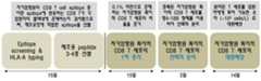
도 1은 본 발명에 따른 자가암항원 특이적 CD8+ T 세포의 선택적 분리 및 대량배양 과정을 설명하는 도면이다.
도 2는 본 발명에 따른 에피토프 스크리닝 과정의 공정 흐름도를 나타낸 도면이다.
도 3은 정상인(healthy doner)으로부터 얻은 PBMC을 이용한 hTERT 에피토프 스크리닝 결과를 보여주는 도면이다.
도 4는 정상인으로부터 얻은 PBMC를 이용한 WT1 에피토프 스크리닝 결과를 보여주는 도면이다.
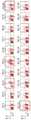
도 5 내지 7은 각각 위암(gastric cancer), 폐암(lung cancer) 및 췌장암(pancreatic cancer) 환자로부터 얻은 PBMC을 이용한 hTERT 에피토프 스크리닝 결과를 보여주는 도면이다.
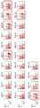
도 8 및 9는 각각 뇌종양(glioblastoma) 및 폐암 환자로부터 얻은 PBMC을 이용한 WT1 에피토프 스크리닝 결과를 보여주는 도면이다.
도 10 및 11은 각각 난소암(ovarian cancer) 및 육종(sarcoma) 환자로부터 얻은 PBMC을 이용한 NY-ESO1 에피토프 스크리닝 결과를 보여주는 도면이다.
도 12 및 13은 각각 육종 및 폐암 환자로부터 얻은 PBMC을 이용한 MAGE-A3 에피토프 스크리닝 결과를 보여주는 도면이다.
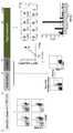
도 14는 hTERT T 세포 치료제의 파일럿 생산 과정을 설명하는 도면이다.
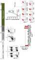
도 15는 WT1 T 세포 치료제의 파일럿 생산 과정을 설명하는 도면이다.
도 16은 NY-ES01 T 세포 치료제의 파일럿 생산 과정을 설명하는 도면이다.
도 17은 MAGE-A3 T 세포 치료제의 파일럿 생산 과정을 설명하는 도면이다.Brief Description of the Drawings Fig. 1 is a diagram for explaining the selective isolation and mass culture of autologous cancer antigen-specific CD8 + T cells according to the present invention.
FIG. 2 is a flowchart showing a process of an epitope screening process according to the present invention.
Figure 3 shows the results of hTERT epitope screening using PBMC from a healthy donor.
Fig. 4 is a graph showing the result of WT1 epitope screening using PBMC obtained from a normal human.
Figures 5 to 7 show the results of hTERT epitope screening using PBMC from gastric cancer, lung cancer and pancreatic cancer patients, respectively.
Figures 8 and 9 are plots showing WT1 epitope screening results using PBMC from glioblastoma and lung cancer patients, respectively.
Figures 10 and 11 show the results of NY-ESO1 epitope screening using PBMC from ovarian cancer and sarcoma patients, respectively.
Figures 12 and 13 show the results of MAGE-A3 epitope screening using PBMC from sarcoma and lung cancer patients, respectively.
Fig. 14 is a diagram illustrating a pilot production process of an hTERT T cell therapeutic agent.
FIG. 15 is a diagram illustrating a pilot production process of a WT1 T cell therapeutic agent. FIG.
16 is a view for explaining the pilot production process of the NY-ES01 T cell treatment agent.
FIG. 17 is a diagram illustrating a pilot production process of a MAGE-A3 T cell therapeutic agent.
암세포는 우리 몸을 구성하는 세포로부터 유래되므로 암세포를 선택적으로 제거하기 위해서는 암세포에서 과발현되는 자가암항원(self tumor Ag) 특이적 CD8+ T 세포의 선택적 분리 및 대량배양이 필요하다. 그러나 자가암항원을 인식하는 T 세포는 정상인의 경우 비율이 극히 낮게 존재할 뿐 아니라, 면역관용(immune tolerance)에 의해 활성이 억제된 상태로 존재한다. 따라서 암환자의 혈액으로부터 자가암항원 특이적 CD8+ T 세포를 선택적으로 분리하여 대량배양 하는 규격화된 공정은 아직 개발되지 못하고 있다. 이에 본 발명자들은 항4-1BB 항체를 이용하여 체내에 극히 낮은 비율로 존재하는 hTERT, WT1, NY-ESO1, 및 MAGE-A3과 같은 자가암항원 특이적 CD8+ T 세포를 31일내 선택적으로 분리 및 대량배양 할 수 있는 규격화된 공정기술을 개발하였다.Since cancer cells are derived from cells constituting our body, selective isolation and mass culture of self tumor antigen-specific CD8+ T cells overexpressed in cancer cells are necessary to selectively remove cancer cells. However, T cells that recognize autologous cancer antigen are present in a state in which the activity is suppressed by immune tolerance as well as in an extremely low ratio in normal persons. Therefore, a standardized process for selectively isolating and mass-culturing autologous cancer-specific CD8+ T cells from the blood of cancer patients has not been developed yet. Accordingly, the present inventors have found that by using the anti-4-1BB antibody, autologous antigen-specific CD8+ T cells such as hTERT, WT1, NY-ESO1, and MAGE-A3, We developed a standardized process technology capable of mass culture.
따라서 본 발명은 자가암항원 특이적 CD8+ T 세포의 분리방법을 제공한다. 구체적으로, 본 발명의 자가암항원 특이적 CD8+ T 세포의 분리방법은, a) 암 환자 혈액내에 존재하는 자가암항원 CD8+ T 세포 에피토프를 선별하는 단계; b) 암 환자의 혈액으로부터 분리된 PBMC(peripheral blood mononuclear cell)를 상기 에피토프의 펩티드 및 IL-2와 함께 배지에서 배양하는 단계; c) 상기 배양된 세포를 b) 단계와 동일한 펩티드를 첨가하여 4-1BB 발현을 유도하는 단계; 및 d) 4-1BB 발현이 유도된 세포를 항-4-1BB 항체가 코팅된 배양 플레이트에서 배양한 후 부착하지 않은 세포를 제거하는 단계를 포함한다.Accordingly, the present invention provides a method for separating autoantigen-specific CD8+ T cells. Specifically, the method for isolating autoantigen-specific CD8+ T cells of the present invention comprises the steps of: a) screening for an autoantibody CD8+ T cell epitope present in the blood of a cancer patient; b) culturing a peripheral blood mononuclear cell (PBMC) isolated from the blood of a cancer patient in a medium with a peptide of said epitope and IL-2; c) adding the same peptide as in step b) to the cultured cells to induce 4-1BB expression; And d) culturing the cells induced with 4-1BB expression on a culture plate coated with anti-4-1BB antibody, and then removing unattached cells.
본 발명의 분리방법에서, 상기 a) 단계의 자가암항원은 암 환자 자신의 체내에 존재하는 어떠한 암 항원일 수 있으며, 암종에 따라 적합한 자가암항원을 선택하여 사용할 수 있다. 바람직하게는 항암면역치료에 이용되고 있는 대표적인 자가암항원으로서 hTERT (GenBank: BAC11010.1), WT1 (GenBank: AAO61088.1), NY-ESO1 (GenBank: CAA05908.1), MAGE-A3 (NCBI Reference Sequence: NP_005353.1) 등을 사용할 수 있다. 상기 hTERT는 염색체 말단에서 텔로미어 DNA(telomeric DNA)를 합성하는 효소로서 암 세포는 이 효소를 과도하게 활성화시켜 텔로미어 의존적 세포 사멸을 회피할 수 있도록 기능하고 폐암, 위암, 췌장암을 포함하는 다양한 고형암의 타겟 항원으로 알려져 있고 (Kim NW, et al.Science. 1994;266:2011-2015), 상기 WT1은 Wilms tumor와 관련된 유전자로서 zinc finger 전사인자를 암호화하여 세포의 증식과 분화, 자멸사, 기관의 발생에 관여를 하는 단백질로서 뇌척수암, 폐암 등의 타겟 항원으로 알려져 있다 (Call KM, et al.,Cell. 1990. 60:509-520; Nakahara Y, et al.,Brain TumorPathol. 2004. 21:113-6). 또한 상기 NY-ESO1은 cancer testis antigen (CTA)에 속하는 단백질 중 하나로 주로 생식세포 (germ cell)와 육종(sarcoma), 유방암을 포함한 다양한 암세포에 발현하는 것으로 잘 알려져 있지만, 이들 세포에서 어떤 기능을 하는지에 대해서는 잘 알려져 있지 않다 (Gnjatic S, et al.,AdvCancerRes. 2006;95:1-30). 상기 MAGE-A3은 melanoma-associated antigen family에 속하는 단백질로 정상세포에서 어떤 기능을 수행하는지에 대해서는 알려진 것이 없지만, 폐암, 육종 및 흑색종을 포함한 다양한 암세포에 과발현하는 것으로 알려져 있어 암의 면역치료에 적합한 표적 항원으로 평가되고 있다 (Decoster L, et al.,AnnOncol. 2012 Jun;23(6):1387-93).In the separation method of the present invention, the autologous cancer antigen in step a) may be any cancer antigen present in the cancer patient's own body, and may be selected and used as a suitable cancer antigen according to the carcinoma. (GenBank: BAC11010.1), WT1 (GenBank: AAO61088.1), NY-ESO1 (GenBank: CAA05908.1), and MAGE-A3 (NCBI Reference Sequence: NP_005353.1) can be used. The hTERT is an enzyme that synthesizes telomeric DNA at the chromosome terminal. Cancer cells function to overcome the telomer-dependent cell death by activating the enzyme excessively, and can be used as a target of various solid tumors including lung cancer, stomach cancer and pancreatic cancer known as antigens and(Science Kim NW, et al 1994 ; 266:.. 2011-2015), the WT1 gene is a tumor Wilms related to the generation of proliferation of encrypting the zinc finger transcription factor and cell differentiation, apoptosis, organizations (KC, et al.,Cell . 1990. 60: 509-520; Nakahara Y, et al.,BrainTumorPathol 2004. 21: 113 -6). In addition, NY-ESO1 is one of the proteins belonging to the cancer testis antigen (CTA), and is known to express mainly in various cancer cells including germ cell, sarcoma, and breast cancer. (Gnjatic S, et al.,Adv .CancerRes . 2006; 95: 1-30). Although MAGE-A3 is a protein belonging to the melanoma-associated antigen family, it has no known function in normal cells. However, MAGE-A3 is known to be overexpressed in various cancer cells including lung cancer, sarcoma and melanoma, (Decoster L, et al., ≪ RTI ID = 0.0 >AnnOncol . 2012 Jun; 23 (6): 1387-93).
본 발명의 분리방법에서, 상기 b) 단계에서 에피토프는 서열번호 1 내지 15로 구성된 군에서 선택된 아미노산 서열로 이루어진 펩티드인 것을 특징으로 한다. In the separation method of the present invention, the epitope in step (b) is characterized by being a peptide consisting of an amino acid sequence selected from the group consisting of SEQ ID NOS: 1 to 15.
본 발명의 분리방법에서, 상기 b) 단계의 배지는 자가혈장이 포함된 배지인 것을 특징으로 하며, 또한 상기 b) 단계의 배양은 12 내지 16일 동안 수행되는 것을 특징으로 한다.In the separation method of the present invention, the medium of step b) is a medium containing autologous plasma, and the step b) is performed for 12-16 days.
본 발명의 분리방법에서, 상기 c) 단계의 발현 유도는 12 내지 36시간 동안 배양하는 것을 특징으로 하며, 상기 d) 단계의 배양은 1 내지 20분간 수행되는 것을 특징으로 한다.
In the separation method of the present invention, induction of expression in step c) is performed for 12 to 36 hours, and the step d) is performed for 1 to 20 minutes.
또한, 본 발명은 상술한 분리방법으로 분리된 자가암항원 특이적 CD8+ T 세포와 방사선 조사된 동종이계(allogeneic) PBMC를 IL-2, 항-CD3 항체 및 자가혈장이 포함된 배지로 현탁한 후 배양용 백에 주입하고, 상기 배지를 추가로 주입하여 배양하는 것을 포함하는 자가암항원 특이적 CD8+ T 세포의 대량배양방법을 제공한다.In addition, the present invention relates to a method of suspending autologous cancer antigen-specific CD8+ T cells and irradiated allogeneic PBMC isolated by the above-described separation method in a medium containing IL-2, anti-CD3 antibody and autologous plasma And culturing the medium by further injecting the medium. The present invention also provides a method for mass-culturing a CD8+ T cell.
본 발명의 대량배양방법에서, 상기 PBMC는 정상 공여자로부터 분리된 것을 특징으로 하며, 상기 배양은 4 내지 15일간 수행되는 것을 특징으로 한다. 특히 상기 배양을 수행하는 동안 배양 4일, 7일, 9일, 11일 및 14일째에 배지를 추가 주입할 수 있다.
In the mass culture method of the present invention, the PBMC is characterized in that it is isolated from a normal donor, and the culturing is performed for 4 to 15 days. In particular, the medium can be further infused at the 4th, 7th, 9th, 11th and 14th days of culturing during the culturing.
이하, 본 발명의 자가암항원 특이적 CD8+ T 세포의 분리 및 증식방법을 단계별로 설명한다.Hereinafter, the method for isolating and propagating the autoantigen-specific CD8+ T cells of the present invention will be described step by step.
(1) Epitope screening (사전선별검사)(1) Epitope screening (pre-screening)
본 발명은 펩티드를 이용하여 자가유래 암항원 특이적 CD8 T 세포를 선택적으로 증식 및 분리한다. CD8 T 세포에 의해 인식되는 자가암항원의 에피토프(epitope)들은 환자 개개인의 HLA-A type 및 상태에 따라 서로 다르므로 epitope screening을 통해 암환자 개개인의 혈액내에 존재하는 자가암항원 CD8 T cell epitope을 선별하여 T 세포치료제 제조용 펩티드 3-4종류를 선별한다.The present invention selectively amplifies and separates autologous cancer antigen-specific CD8 T cells using peptides. Since the epitopes of self-cancer antigen recognized by CD8 T cells are different according to the individual HLA-A type and status of the patient, epitope screening can be used to detect the autoantibody CD8 T cell epitope present in the blood of individual cancer patients 3-4 kinds of peptides for the preparation of T cell therapeutic agent are selected.
(2) 자가암항원 특이적 CD8 T 세포의 증식(2) proliferation of autologous cancer-specific CD8 T cells
자가암항원 특이적 CD8 T 세포는 혈액내 0.1% 이하로 존재하므로, 혈액으로부터 분리된 PBMC에 제조용 펩티드 3-4종 및 IL-2를 첨가하여 14일간 배양함으로써 자가암항원으로부터 유래된 펩티드에 특이적인 CD8 T 세포의 증식을 유도한다. 배양 14일째 모든 세포를 수거하여 동일한 펩티드들로 24시간 재활성화함으로써 펩티드-특이적 CD8 T 세포들이 동시에 4-1BB를 발현하도록 유도한다.Since the autologous cancer antigen-specific CD8 T cells are present in less than 0.1% in the blood, PBMCs isolated from the blood are cultured for 14 days by adding 3-4 production peptides and IL-2 to the peptides derived from autologous cancer antigen Induced CD8 T cell proliferation. On
(3) 자가암항원 특이적 CD8 T 세포의 선택적 분리(3) Selective isolation of autologous cancer-specific CD8 T cells
항4-1BB 항체가 코팅된 배양 플레이트에 펩티드로 재활성화시킨 세포를 분주하여 10분간 배양함으로써 4-1BB를 발현하는 CD8 T 세포들이 부착되도록 하며, 부탁되지 않은 세포들은 모두 세척하여 제거한다. 이후 IL-2가 포함된 배지를 첨가하여 이틀간 배양함으로써 분리된 T 세포들이 증식과 함께 배양 플레이트로 떨어지도록 한다.Anti-4-1BB antibody-coated culture plates are reactivated with peptide and incubated for 10 minutes to allow attachment of CD8 T cells expressing 4-1BB. All undesired cells are washed away. Then, culture medium containing IL-2 is added and cultured for two days so that the separated T cells fall into the culture plate with the proliferation.
(4) 자가암항원 특이적 CD8 T 세포의 대량배양(4) mass culture of autologous cancer antigen-specific CD8 T cells
1L 배양용 백(culture bag)에 분리된 CD8 T 세포 5×10⁵ 세포, 방사선 조사된(irradiated allogeneic) PBMCs 1×10⁸ 세포, 1000U/ml IL-2, 40ng 항-CD3 mAb를 혼합하여 14일간 정기적으로 배지를 첨가하여 ~109 세포/L 수준으로 세포를 대량 배양하여, 암환자에게 투여 가능한 수준으로 증식시킨다.
5 × 10⁵ CD8 T cells, 1 × 10⁸ irradiated allogeneic PBMCs, 1000 U / ml IL-2 and 40 ng anti-CD3 mAb were mixed in a 1 L culture bag to obtain 14 The cells are cultured at a level of ~ 10 <9 > cells / L by adding medium on a regular basis for a day, and the cells are allowed to proliferate to a level that can be administered to cancer patients.
이하, 본 발명을 실시예에 의해 상세히 설명하기로 한다. 그러나 이들 실시예는 본 발명을 보다 구체적으로 설명하기 위한 것으로서, 본 발명의 범위가 이들 실시예에 한정되는 것은 아니다.
Hereinafter, the present invention will be described in detail with reference to examples. However, these examples are intended to further illustrate the present invention, and the scope of the present invention is not limited to these examples.
실험예Experimental Example..에피토프Epitope 스크리닝 공정 Screening process
알고리즘을 통해 자가암항원의 CD8 T 세포 에피토프(epitope)들을 선별하였다. 암 환자의 혈액 내 존재하는 T 세포들이 어떤 CD8 T 세포의 에피토프에 반응하는지 확인하기 위해, 암 환자의 혈액으로부터 PBMC(peripheral blood mononuclear cell)를 분리하여 세척한 후, CTL 배지(RPMI1640 배지 + 4 mM L-글루타민 + 12.5 mM HEPES + 50 μM 2-메르캅토에탄올 + 3% 자가혈장)에 1×10⁶ 세포/ml로 현탁하고, 14 ml 튜브(round tube)에 1 ml씩 분주하였다. 알고리즘을 통해 분석하여 선별된 각 에피토프의 펩티드들(peptides)을 각 튜브에 1 μg/ml 농도로 첨가하여 CO2 인큐베이터에서 배양을 시작하였다. 배양 이틀째, 50 U/ml IL-2가 포함된 CTL 배지를 각 튜브에 1 ml씩 첨가하였다. 배양 7, 9, 11, 및 13일째 1 ml 배지를 제거한 후, 50 U/ml IL-2가 포함된 CTL 배지를 첨가하였다. 배양 14일째, 각 튜브에 RPMI1640 배지를 첨가하여 1400 rpm에서 5분간 원심분리 하여 세포를 3회 세척하였다. 세척된 세포에 1 ml CTL 배지를 현탁한 후, 동일한 펩티드를 5 μg/ml 농도로 첨가하여 배양하였다. 24시간 후, 각 튜브의 세포를 수거하여 항-CD8-PE-Cy5 및 항-4-1BB-PE 항체로 염색하여 유세포 분석하였으며, 4-1BB를 발현하는 CD8 T 세포의 비율을 분석함으로써, 어떤 펩티드에 반응하여 CD8 T 세포가 활성화 되었는지를 분석하였다. 도 2는 본 발명에 따른 에피토프 스크리닝 과정의 공정 흐름도를 나타낸 도면이다.The CD8 T cell epitopes of autologous cancer antigen were selected through an algorithm. PBMCs (peripheral blood mononuclear cells) were separated from the blood of cancer patients and washed in CTL medium (RPMI1640 medium + 4 mM L-glutamine + 12.5 mM HEPES + 50 μM 2-mercaptoethanol + 3% autologous plasma) at 1 × 10⁶ cells / ml and dispensed in 1 ml each in a 14 ml round tube. The peptides of each epitope selected by the analysis were added to each tube at a concentration of 1 μg / ml and the culture was started in a CO2 incubator. On the second day of culture, 1 ml of CTL medium containing 50 U / ml IL-2 was added to each tube. On
실험에 사용된 항-CD8-PE-Cy5, 항-4-1BB-PE는 e바이오사이언스(eBioscience, San Diego, CA)에서 구매하였다. RPMI1640, L-글루타민, HEPES, 2-메르캅토에탄올은 인비트로겐 사(Invitrogen, San Diego, CA)로부터 구매하였다.
Anti-CD8-PE-Cy5, anti-4-1BB-PE used in the experiments were purchased from eBioscience (San Diego, Calif.). RPMI 1640, L-glutamine, HEPES, and 2-mercaptoethanol were purchased from Invitrogen (San Diego, Calif.).
실시예Example 1. One.자가암항원Autosomal antigen 선별 및 Screening andCD8CD8 T 세포 T cell에피토프Epitope 선별 Selection
각 암종별로 어떤 암항원이 암의 면역치료에 적합한지에 대해 평가한 논문들(Scanlan MJ, et a.,ImmunolRev. 2002 Oct. 188:22-32; Ramakrishnan S, et al.,Cancerresearch. 1998. 58:622-625; Nakahara Y, et al.,BrainTumorPathol. 2004. 21(3):113-6)을 기반으로 한국인 호발암 및 난치성암(위암, 폐암, 췌장암 등)의 면역치료에 적합한 자가암항원을 선별하였다. hTERT (GenBank: BAC11010.1), WT1 (GenBank: AAO61088.1), NY-ESO1 (GenBank: CAA05908.1), MAGE-A3 (NCBI Reference Sequence: NP_005353.1)은 다양한 방식으로 항암면역치료에 이용되고 있는 대표적인 자가암항원으로 이들 네 가지 암 항원의 적용이 가능한 암종들을 선별하여 하기 표 1에 정리하였다.
(Canclan etal .,ImmunolRev. 2002 Oct. 188: 22-32; Ramakrishnan S, et al.,Cancerresearch . 1998. 58: 622-625; Nakahara Y, et al.,BrainTumorPathol . 2004, 21 (3): 113-6), which are suitable for the immunotherapy of Korean carcinoma and intractable cancer (gastric cancer, lung cancer, pancreatic cancer, etc.). HTERT (GenBank: BAC11010.1), WT1 (GenBank: AAO61088.1), NY-ESO1 (GenBank: CAA05908.1) and MAGE-A3 (NCBI Reference Sequence: NP_005353.1) The cancer types that can be applied to these four cancer antigens are selected and summarized in Table 1 below.

선별된 자가암항원의 아미노산 서열을 알고리즘을 통해 분석하여 CD8 T 세포 에피토프로 추정되는 아미노산 서열을 결정하였으며 (CTLPred: http://www.imtech.res.in/raghava/ctlpred/, NetCTL: http://www.cbs.dtu.dk/services/NetCTL/, SYFPEITHI: http://www.syfpeithi.de/), 선별된 에피토프를 대상으로 펩티드를 화학합성 (Peptron Inc; www.peptron.com)하여 에피토프 스크리닝(epitope screening)에 사용하였다. 각 자가암항원으로부터 선별된 CD8 T 세포 에피토프는 하기 표 2 내지 표 5와 같다.
The amino acid sequence of the selected autologous cancer antigen was analyzed through an algorithm to determine the amino acid sequence deduced as a CD8 T cell epitope (CTLPred: http://www.imtech.res.in/raghava/ctlpred/, NetCTL: http: (Peptron Inc, www.peptron.com) to select peptides from selected epitopes And used for epitope screening. The CD8 T cell epitope selected from each autologous cancer antigen is shown in Tables 2 to 5 below.
실시예Example 2. 임상 암 환자를 대상으로 한 2. Clinical Cancer Patients에피토프Epitope 스크리닝 Screening
실시예 1에서 선별된 hTERT, WT1, NY-ESO1, MAGE-A3 자가암항원의 CD8 T 세포 에피토프들이 실제 임상 암 환자의 혈액에 존재하는 CD8 T 세포 증식을 유도할 수 있는지 검증하기 위해, 도 2에 모식된 에피토프 스크리닝을 수행하였다. hTERT 에피토프 스크리닝은 위암, 폐암, 췌장암을 주 대상으로 수행하였으며, WT1 에피토프 스크리닝은 뇌척수암 및 폐암, NY-ESO1 에피토프 스크리닝은 난소암 및 육종, 그리고 MAGE-A3 에피토프 스크리닝은 육종 및 폐암을 주 대상으로 수행하였다.
In order to verify that the CD8 T cell epitopes of the hTERT, WT1, NY-ESO1, and MAGE-A3 autologous cancer antigens selected in Example 1 can induce CD8 T cell proliferation present in the blood of patients with clinical cancer, Lt; RTI ID = 0.0 > epitope < / RTI > screening. HTERT epitope screening was performed mainly for gastric cancer, lung cancer and pancreatic cancer. WT1 epitope screening was performed for cerebrospinal cancer and lung cancer, NY-ESO1 epitope screening was performed for ovarian cancer and sarcoma, and MAGE-A3 epitope screening was for sarcoma and lung cancer Respectively.
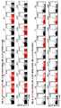
도 3은 정상인(healthy doner)으로부터 얻은 PBMC을 이용한 hTERT 에피토프 스크리닝 결과를 보여주는 도면이다.Figure 3 shows the results of hTERT epitope screening using PBMC from a healthy donor.
도 4는 정상인으로부터 얻은 PBMC를 이용한 WT1 에피토프 스크리닝 결과를 보여주는 도면이다.Fig. 4 is a graph showing the result of WT1 epitope screening using PBMC obtained from a normal human.
도 3과 4에서 볼 수 있듯이, hTERT 및 WT1의 CD8 T 세포 에피토프들은 정상인의 혈액으로부터 분리된 PBMC로부터 T 세포 반응을 유도하지 못하였다. 따라서 본 발명의 선별된 에피토프들은 정상인의 T 세포가 인식하지 못하는 것으로 확인되었다.
As can be seen in Figures 3 and 4, the CD8 T cell epitopes of hTERT and WT1 failed to induce T cell responses from PBMCs isolated from normal human blood. Thus, the selected epitopes of the present invention were found to be unrecognized by normal T cells.
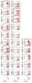
도 5 내지 7은 각각 위암(gastric cancer), 폐암(lung cancer) 및 췌장암(pancreatic cancer) 환자로부터 얻은 PBMC을 이용한 hTERT 에피토프 스크리닝 결과를 보여주는 도면이다.Figures 5 to 7 show the results of hTERT epitope screening using PBMC from gastric cancer, lung cancer and pancreatic cancer patients, respectively.
도 8 및 9는 각각 뇌종양(glioblastoma) 및 폐암 환자로부터 얻은 PBMC을 이용한 WT1 에피토프 스크리닝 결과를 보여주는 도면이다.Figures 8 and 9 are plots showing WT1 epitope screening results using PBMC from glioblastoma and lung cancer patients, respectively.
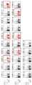
도 10 및 11은 각각 난소암(ovarian cancer) 및 육종(sarcoma) 환자로부터 얻은 PBMC을 이용한 NY-ESO1 에피토프 스크리닝 결과를 보여주는 도면이다.Figures 10 and 11 show the results of NY-ESO1 epitope screening using PBMC from ovarian cancer and sarcoma patients, respectively.
도 12 및 13은 각각 육종 및 폐암 환자로부터 얻은 PBMC을 이용한 MAGE-A3 에피토프 스크리닝 결과를 보여주는 도면이다.
Figures 12 and 13 show the results of MAGE-A3 epitope screening using PBMC from sarcoma and lung cancer patients, respectively.
도 5 내지 13에서 볼 수 있듯이, 임상 암 환자의 혈액으로 분리한 PBMC를 대상으로 에피토프 스크리닝을 수행하여 hTERT, WT1, NY-ESO1, 및 MAGE-A3에 대한 CD8 T 세포 반응성을 조사한 결과, 정상인과는 달리 선택된 자가암항원들에 대한 T 세포 반응이 높게 나타나는 것으로 확인되었다. 따라서 위암, 폐암, 췌장암, 육종, 난소암 등을 대상으로 각각의 자가암항원에 대한 반응성을 에피토프 스크리닝을 반복적으로 수행하여 확인하였다.
As shown in FIGS. 5 to 13, PBMCs isolated from blood of clinical cancer patients were subjected to epitope screening to examine the reactivity of CD8 T cells to hTERT, WT1, NY-ESO1, and MAGE-A3. Showed that the T cell response to cancer antigens was high in the other selected patients. Therefore, the reactivity of each cancer antigen to gastric cancer, lung cancer, pancreatic cancer, sarcoma, ovarian cancer, etc. was confirmed by repeated epitope screening.
또한, 상기 에피토프 스크리닝 결과를 객관적으로 분석하기 위해, 스코어링 시스템(scoring system)을 하기 표 6과 같이 만들었다.
In order to objectively analyze the results of the epitope screening, a scoring system was prepared as shown in Table 6 below.

상기 표 6의 기준에 따라 에피토프 스크리닝 결과를 분석하였으며, 그 결과는 하기 표 7 내지 15에 나타내었다.
The epitope screening results were analyzed according to the criteria of Table 6, and the results are shown in Tables 7 to 15 below.
하기 표 7은 위암 환자로부터 얻은 PBMC을 이용한 hTERT 에피토프 스크리닝을 분석한 결과이다.
Table 7 below shows the results of analysis of hTERT epitope screening using PBMC from gastric cancer patients.

하기 표 8은 폐암 환자로부터 얻은 PBMC을 이용한 hTERT 에피토프 스크리닝을 분석한 결과이다.
Table 8 below shows the results of analysis of hTERT epitope screening using PBMC from lung cancer patients.

하기 표 9는 췌장암 환자로부터 얻은 PBMC을 이용한 hTERT 에피토프 스크리닝을 분석한 결과이다.
Table 9 below shows the results of analysis of hTERT epitope screening using PBMC from pancreatic cancer patients.

하기 표 10은 뇌종양 환자로부터 얻은 PBMC을 이용한 WT1 에피토프 스크리닝을 분석한 결과이다.
Table 10 below shows the results of analysis of WT1 epitope screening using PBMC from brain tumor patients.

하기 표 11은 폐암 환자로부터 얻은 PBMC을 이용한 WT1 에피토프 스크리닝을 분석한 결과이다.
Table 11 below shows the results of analysis of WT1 epitope screening using PBMC from lung cancer patients.

하기 표 12는 난소암 환자로부터 얻은 PBMC을 이용한 NY-ESO1 에피토프 스크리닝을 분석한 결과이다.
Table 12 below shows the results of an analysis of NY-ESO1 epitope screening using PBMC from ovarian cancer patients.

하기 표 13은 육종 환자로부터 얻은 PBMC을 이용한 WT1 에피토프 스크리닝을 분석한 결과이다.
Table 13 below shows the results of analysis of WT1 epitope screening using PBMC from sarcoma patients.

하기 표 14는 육종 환자로부터 얻은 PBMC을 이용한 MAGE-A3 에피토프 스크리닝을 분석한 결과이다.
Table 14 below shows the results of analysis of MAGE-A3 epitope screening using PBMC obtained from sarcoma patients.

하기 표 15는 폐암 환자로부터 얻은 PBMC을 이용한 MAGE-A3 에피토프 스크리닝을 분석한 결과이다.
Table 15 below shows the results of analysis of MAGE-A3 epitope screening using PBMC from lung cancer patients.

상기 표 7 내지 13에서와 같이, 스코어 3 이상으로 CD8 T 세포가 펩티드 자극에 의해 4-1BB를 발현할 경우, 항4-1BB 항체를 이용하여 효과적으로 이들 세포를 분리할 수 있다. 각 암종에서 스코어 3 이상으로 자가암항원에 T 세포가 반응하는 비율 40-50% 수준이었으므로, 선별된 자가암항원의 에피토프들을 이용하여 T 세포치료제를 제조하는 것이 가능할 것으로 판단하였다. 선별된 펩티드들은, hTERT 펩티드: CLKELVARV (서열번호 1), PLFLELL (서열번호 2), AAVTPAA (서열번호 3); WT1 펩티드: SLGEQQVSV (서열번호 4), RMFPNAPVL (서열번호 5), CMTWNQMNL (서열번호 6), VLDFAPPGA (서열번호 7); NY-ESO1 펩티드: SISSCLQQL (서열번호 8), RLLEFYLAM (서열번호 9), GVLLKEFTV (서열번호 10), ILTIRLTAA (서열번호 11); 그리고 MAGE-A3 펩티드: LLIIVLAII (서열번호 12), KIWEELSVL (서열번호 13), LVFGIELMEV (서열번호 14), SLPTTMNYPL (서열번호 15)이다.
As shown in Tables 7 to 13, when CD8 T cells express 4-1BB by peptide stimulation with a score of 3 or more, these cells can be effectively isolated using anti-4-1BB antibody. Since the ratio of T cells to autologous cancer was 40-50% in each carcinoma with a score of 3 or more, it was considered that it would be possible to produce a therapeutic agent for T cell using the epitope of the selected autologous cancer antigen. Selected peptides were hTERT peptides: CLKELVARV (SEQ ID NO: 1), PLFLELL (SEQ ID NO: 2), AAVTPAA (SEQ ID NO: 3); WT1 peptides: SLGEQQVSV (SEQ ID NO: 4), RMFPNAPVL (SEQ ID NO: 5), CMTWNQMNL (SEQ ID NO: 6), VLDFAPPGA (SEQ ID NO: 7); NY-ESO1 peptide: SISSCLQQL (SEQ ID NO: 8), RLLEFYLAM (SEQ ID NO: 9), GVLLKEFTV (SEQ ID NO: 10), ILTIRLTAA (SEQ ID NO: 11); And MAGE-A3 peptides: LLIIVLAII (SEQ ID NO: 12), KIWEELSVL (SEQ ID NO: 13), LVFGIELMEV (SEQ ID NO: 14), SLPTTMNYPL (SEQ ID NO: 15).
실시예Example 3. 3.자가암항원Autosomal antigen 특이적 T 세포치료제의 Specific T cell therapy시험제조Test manufacture
에피토프 스크리닝을 통해 T 세포치료제 제조에 적합한 각 자가암항원에 대한 3-4종의 에피토프의 펩티드를 선별한 후, 이들 펩티드를 이용하여 hTERT, WT1, NY-ESO1, 및 MAGE-A3 특이적 T 세포치료제의 시험생산을 수행하였다. T 세포치료제의 생산은 자가유래 항암 T 세포의 1차 증식, 분리, 대량배양 세 단계로 구성되어 있다.
Epitope screening was carried out to select 3-4 epitope peptides for each of the autologous cancer antigens suitable for the preparation of T cell therapy, and then these peptides were used to express hTERT, WT1, NY-ESO1, and MAGE-A3 specific T cells Test production of the therapeutic agent was carried out. The production of T cell therapy consists of three stages of primary proliferation, isolation, and mass culture of autologous cancer T cells.
(1)(One)자가암항원Autosomal antigen 특이적 SpecificCD8CD8 T 세포의 증식 T cell proliferation
에피토프 스크리닝을 통해 스코어 3 이상의 에피토프가 하나 이상 존재하는 것을 확인한 암 환자로부터 50 ml 혈액을 분리하였다.50 ml blood was isolated from cancer patients who were confirmed to have one or more epitope of
1) 환자의 혈액으로부터 PBMC 분리: 7 ml 피콜-하이파크(Ficoll-hypaque)가 채워진 15 ml 튜브(cornical tube)에 7 ml 혈액을 천천히 흘려보내 피콜 용액 상층에 오버레이(overlay)하였다. 튜브를 상온 2000 rpm에서 20분간 원심분리 하였으며, 피콜과 혈장 사이에 위치한 흰색의 세포층만을 수거하여 세척한 후 PBMC로 사용하였다.1) PBMC separation from the patient's blood: 7 ml blood was slowly poured into a 15 ml cornical tube filled with 7 ml Ficoll-hypaque to overlay the upper layer of the picol solution. The tubes were centrifuged at 2000 rpm for 20 minutes at room temperature, and only the white cell layer located between the blood and the plasma was collected, washed and used as PBMC.
2) 분리된 PBMC를 CTL 배지(RPMI1640 medium + 4mM L-glutamine + 12.5 mM HEPES + 50 μM 2-mercaptoethanol + 3% autoplasma)에 1×10⁶ 세포/ml로 현탁하고, 에피토프 스크리닝을 통해 본 발명에서 선별된 3-4종의 펩티드들이 각각 1 μg/ml 농도가 되도록 첨가하였다. 이들 세포 현탁액을 14 ml 튜브(round tube)에 1 ml씩 분주하여 CO2 인큐베이터에서 배양하였다.2) The separated PBMCs were suspended in CTL medium (RPMI1640 medium + 4 mM L-glutamine + 12.5 mM HEPES + 50 μM 2-mercaptoethanol + 3% autoplasma) at 1 × 10⁶ cells / ml and analyzed by epitope screening The selected 3-4 kinds of peptides were added so as to have a concentration of 1 μg / ml, respectively. These cell suspensions were dispensed in 1 ml each in a 14 ml round tube and cultured in a CO2 incubator.
3) 배양 이틀째 50 U/ml IL-2(Proleukin, Novatis)가 포함된 CTL 배지를 각 튜브에 1 ml씩 첨가하였다.3) In the second day of culture, 1 ml of CTL medium containing 50 U / ml IL-2 (Proleukin, Novatis) was added to each tube.
4) 배양 7, 9, 11, 및 13일째 1 ml 상층배지를 제거한 후, 50 U/ml IL-2가 포함된 CTL 배지를 첨가하였다.4) Culture On
5) 배양 14일째 각 튜브의 세포를 50 ml 코티컬 튜브(cornical tube)에 수거한 후, RPMI1640 배지를 첨가하여 1400rpm에서 5분간 원심분리하여 세포를 세척하였다. 이 과정을 두 번 더 반복하였다.5) After 14 days of culture, the cells of each tube were collected in a 50 ml cornical tube, and the cells were washed by adding RPMI 1640 medium and centrifuging at 1400 rpm for 5 minutes. This process was repeated two more times.
6) 세척된 세포를 2×10⁶ 세포/ml의 농도로 CTL 배지로 현탁한 후, 동일한 펩티드 3-4종 각각을 5 μg/ml 농도로 첨가하여 배양하였다.
6) The washed cells were suspended in CTL medium at a concentration of 2 × 10⁶ cells / ml, and then 3-4 kinds of the same peptides were added at a concentration of 5 μg / ml.
(2)(2)자가암항원Autosomal antigen 특이적 SpecificCD8CD8 T 세포의 선별 Screening of T cells
1) 배양 24시간째, 하루 동안 재활성화시킨 PBMC를 수거하여 RPMI1640 배지로 두 번 세척한 후, 5×10⁶ 세포/ml의 농도로 CTL 배지를 현탁하고 50 U/ml IL-2를 첨가하였다.1) After 24 hours of culture, the reactivated PBMCs were collected and washed twice with RPMI 1640 medium, and the CTL medium was suspended at a concentration of 5 × 10⁶ cells / ml and 50 U / ml IL-2 was added .
2) 항4-1BB 항체를 50 μg/ml의 농도로 하루 동안 코팅한 6웰 또는 12웰 플레이트(culture plate)에, 이들 세포를 1 ml 씩 첨가하여 10분간 CO2 인큐베이터에서 배양하였다.2) Anti-4-1BB antibody was coated on a 6-well plate or a 12-well culture plate coated at a concentration of 50 μg / ml for 1 day. These cells were added in an amount of 1 ml and incubated in a CO2 incubator for 10 minutes.
3) 배양 10분 후, 플레이트에 부착되지 않은 세포를 모두 세척하여 제거한 후, 1000 U/ml IL-2가 포함된 CTL 배지 2-4 ml을 각 웰에 첨가하여 CO2 인큐베이터에서 이틀간 배양하였다.
3) After 10 minutes of incubation, all cells not attached to the plate were washed away and 2-4 ml of CTL medium containing 1000 U / ml IL-2 was added to each well and cultured in a CO2 incubator for two days.
(3)(3)자가암항원Autosomal antigen 특이적 SpecificCD8CD8 T 세포의 대량배양 Mass culture of T cells
1) 항4-1BB 항체로 분리하여 이틀간 배양한 세포를 모두 수거하여 RPMI1640 배지로 두 번 세척하여 계수하였다.1) The cells were separated with anti-4-1BB antibody, and all the cells cultured for two days were collected, washed twice with RPMI1640 medium and counted.
2) 정상 공여자로부터 PBMC를 분리하여 1×10⁸ 세포/ml로 현탁한 후, 3000 rad로 방사선 조사하여 세포의 사멸을 유도한 후, T 세포의 증식 유도에 필요한 공동자극(costimulation)을 제공할 수 있는 배양 첨가물로 사용하였다.2) PBMC were isolated from normal donors, suspended at 1 × 10⁸ cells / ml, irradiated at 3000 rad to induce cell death, and then provided costimulation necessary for induction of proliferation of T cells As a possible culture additive.
3) 50 ml 코니컬 튜브에 분리된 CD8 T 세포 5×10⁵ 세포와 irradiated allogeneic PBMCs 1×10⁸ 세포를 첨가한 후, 1,000 U/ml IL-2, 40 ng/ml 항-CD3 mAb(BD Bioscience) 및 3% 자가혈장(autoplasma)이 포함된 ALyS505N 배지(CELL SCIENCE & TECHNOLOGY INST., INC. (CSTI))를 50 ml까지 첨가하였다.3) 5 × 10⁵ CD8 T cells and 1 × 10⁸ irradiated allogeneic PBMCs were added to a 50 ml conical tube and incubated with 1,000 U / ml IL-2, 40 ng / ml anti-CD3 mAb (BD (CELL SCIENCE & TECHNOLOGY INST., INC. (CSTI)) supplemented with 10% Bioscience and 3% autoplasma.
4) 50 ml 세포현탁액을 1L 배양용 백(culture bag)에 주입한 후, CO2 인큐베이터에서 배양하였다.4) A 50-ml cell suspension was injected into a 1-L culture bag and then cultured in a CO2 incubator.
5) 배양 4일째, 1,000 U/ml IL-2, 3% 자가혈장이 포함된 ALyS505N 배지 50 ml을 1L 배양용 백에 추가 주입하였다.5) On the fourth day of incubation, 50 ml of ALyS505N medium containing 1,000 U / ml IL-2 and 3% autologous plasma was added to the 1 L culture bag.
6) 배양 7일째, 1,000 U/ml IL-2, 3% 자가혈장이 포함된 ALyS505N 배지 100 ml을 1L 배양용 백에 추가 주입하였다.6) On the 7th day of incubation, 100 ml of ALyS505N medium containing 1,000 U / ml IL-2, 3% autologous plasma was added to the 1 L culture bag.
7) 배양 9일째, 1,000 U/ml IL-2, 3% 자가혈장이 포함된 ALyS505N 배지 300 ml을 1L 배양용 백에 추가 주입하였다.7) On the 9th day of culture, 300 ml of ALyS505N medium containing 1,000 U / ml of IL-2 and 3% of autologous plasma was further injected into a 1 L culture bag.
8) 배양 11일째, 1,000 U/ml IL-2, 3% 자가혈장이 포함된 ALyS505N 배지 500 ml을 1L 배양용 백에 추가 주입하였다.8) On the 11th day of culture, 500 ml of ALyS505N medium containing 1,000 U / ml IL-2 and 3% autologous plasma was added to the 1 L culture bag.
9) 배양 14일째, 1L 배양용 백의 모든 세포를 수거하여 주사용 생리식염수로 세 번 세척하며, 5% 알부민이 포함된 주사용 생리식염수에 세포를 현탁하여 완제품 T 세포치료제를 충전하였다.
9) On the 14th day of culture, all the cells of the 1L culture bag were collected, washed three times with physiological saline for injection, and the cells were suspended in physiological saline containing 5% albumin and filled with the final product T cell treatment agent.
상기와 같이, 임상 암 환자로부터 본 발명의 에피토프 스크리닝을 통해 T 세포 반응을 유도할 수 있는 스코어 3 이상의 펩티드 3-4종을 선별한 후, 50 cc 혈액을 이용하여 hTERT, WT, NY-ESO1, MAGE-A3 T 세포치료제의 시험생산을 수행하였으며, 그 결과를 도 6 내지 9에 정리하였다.
As described above, 3-4 kinds of peptides with a score of 3 or more capable of inducing T cell response through epitope screening of the present invention were screened from patients with clinical cancer, and 50 cc blood was used to express hTERT, WT, NY-ESO1, MAGE-A3 T cell treatment agent, and the results are summarized in Figs. 6 to 9. Fig.
도 14는 hTERT T 세포 치료제의 파일럿 생산 과정을 설명하는 도면이다.Fig. 14 is a diagram illustrating a pilot production process of an hTERT T cell therapeutic agent.
도 14는 HLA-A*24 allele를 가진 위암환자의 50 cc 혈액으로부터 PBMC를 분리하여 세 종류의 hTERT peptide [CLKELVARV (서열번호 1), PLFLELL (서열번호 2), AAVTPAA (서열번호 3)]를 각각 1 μg/ml 농도로 첨가한 후, 상기 실시예 3의 “(1) 자가암항원 특이적 CD8 T 세포의 증식”에 기술된 과정에 따라 배양하였다. 배양 14일째 모든 세포를 수거하여 “(2) 자가암항원 특이적 CD8 T 세포의 선별” 과정에 따라 hTERT peptide에 반응하여 증식한 T 세포를 분리/증식하였다. 분리된 T 세포는 “(3) 자가암항원 특이적 CD8 T 세포의 대량배양” 과정을 통해 암환자에게 투여 가능한 수준으로 대량배양하였으며, 최종 배양된 세포는 낮은 노화도와 작용기능 가지고 있는 특정 TCRVb type의 T 세포인 것으로 유세포 분석되었다.
Figure 14 shows that PBMC were isolated from 50 cc blood of gastric cancer patients with HLA-
도 15는 WT1 T 세포 치료제의 파일럿 생산 과정을 설명하는 도면이다.FIG. 15 is a diagram illustrating a pilot production process of a WT1 T cell therapeutic agent. FIG.
도 15는 HLA-A*02 allele를 가진 악성신경교종환자의 50 cc 혈액으로부터 PBMC를 분리하여 네 종류의 WT1 peptide [SLGEQQVSV (서열번호 4), RMFPNAPVL (서열번호 5), CMTWNQMNL (서열번호 6), VLDFAPPGA (서열번호 7)]를 각각 1 μg/ml 농도로 첨가한 후, 상기 실시예3의 “(1) 자가암항원 특이적 CD8 T 세포의 증식”에 기술된 과정에 따라 배양하였다. 배양 14일째 모든 세포를 수거하여 “(2) 자가암항원 특이적 CD8 T 세포의 선별” 과정에 따라 WT1 peptide에 반응하여 증식한 T 세포를 분리/증식하였다. 분리된 T 세포는 “(3) 자가암항원 특이적 CD8 T 세포의 대량배양” 과정을 통해 암환자에게 투여 가능한 수준으로 대량배양하였으며, 최종 배양된 세포는 낮은 노화도와 작용기능 가지고 있는 특정 TCRVb type의 T 세포인 것으로 유세포 분석되었다.
Figure 15 shows that PBMCs were isolated from 50 cc blood of patients with malignant glioma with HLA-A * 02 allele and four types of WT1 peptide [SLGEQQVSV (SEQ ID NO: 4), RMFPNAPVL (SEQ ID NO: 5), CMTWNQMNL , VLDFAPPGA (SEQ ID NO: 7)] were respectively added at a concentration of 1 μg / ml and then cultured according to the procedure described in "(1) Proliferation of autologous cancer antigen-specific CD8 T cells" On the 14th day after culture, all cells were collected and T cells proliferated / proliferated in response to the WT1 peptide according to "(2) selection of autologous cancer-specific CD8 T cells". The isolated T cells were cultured in large quantities to be able to be administered to cancer patients through the process of "(3) mass culture of autologous cancer antigen-specific CD8 T cells." The final cultured cells had a specific TCRVb type T cells were analyzed by flow cytometry.
도 16은 NY-ES01 T 세포 치료제의 파일럿 생산 과정을 설명하는 도면이다.16 is a view for explaining the pilot production process of the NY-ES01 T cell treatment agent.
도 16은 HLA-A*02 allele를 가진 난소암환자의 50 cc 혈액으로부터 PBMC를 분리하여 네 종류의 NY-ESO1 peptide [SISSCLQQL (서열번호 8), RLLEFYLAM (서열번호 9), GVLLKEFTV (서열번호 10), ILTIRLTAA (서열번호 11)]를 각각 1μg/ml 농도로 첨가한 후, 상기 실시예 3의 “(1) 자가암항원 특이적 CD8 T 세포의 증식”에 기술된 과정에 따라 배양하였다. 배양 14일째 모든 세포를 수거하여 “(2) 자가암항원 특이적 CD8 T 세포의 선별” 과정에 따라 NY-ESO-1 peptide에 반응하여 증식한 T 세포를 분리/증식하였다. 분리된 T 세포는 “(3) 자가암항원 특이적 CD8 T 세포의 대량배양” 과정을 통해 암환자에게 투여 가능한 수준으로 대량배양하였으며, 최종 배양된 세포는 낮은 노화도와 작용기능 가지고 있는 특정 TCRVb type의 T 세포인 것으로 유세포 분석되었다.
Figure 16 shows that PBMCs were isolated from 50 cc blood of ovarian cancer patients with the HLA-A * 02 allele and identified four NY-ESO1 peptides (SISSCLQQL (SEQ ID NO: 8), RLLEFYLAM (SEQ ID NO: 9), GVLLKEFTV ) And ILTIRLTAA (SEQ ID NO: 11)] at a concentration of 1 μg / ml, respectively, and cultured according to the procedure described in "(1) Proliferation of autologous cancer antigen-specific CD8 T cells" On the 14th day of culture, all the cells were collected and the proliferated T cells were isolated / propagated in response to the "(2) selection of autologous cancer antigen-specific CD8 T cells" in response to the NY-ESO-1 peptide. The isolated T cells were cultured in large quantities to be able to be administered to cancer patients through the process of "(3) mass culture of autologous cancer antigen-specific CD8 T cells." The final cultured cells had a specific TCRVb type T cells were analyzed by flow cytometry.
도 17은 MAGE-A3 T 세포 치료제의 파일럿 생산 과정을 설명하는 도면이다.FIG. 17 is a view illustrating a pilot production process of a MAGE-A3 T cell therapeutic agent.
도 17은 HLA-A*02 allele를 가진 육종환자의 50cc 혈액으로부터 PBMC를 분리하여 네 종류의 MAGE-A3 peptide [LLIIVLAII (서열번호 12), KIWEELSVL (서열번호 13), LVFGIELMEV (서열번호 14), SLPTTMNYPL (서열번호 15)]를 각각 1 μg/ml 농도로 첨가한 후, 상기 실시예 3의 “(1) 자가암항원 특이적 CD8 T 세포의 증식”에 기술된 과정에 따라 배양하였다. 배양 14일째 모든 세포를 수거하여 “(2) 자가암항원 특이적 CD8 T 세포의 선별” 과정에 따라 MAGE-A3 peptide에 반응하여 증식한 T 세포를 분리/증식하였다. 분리된 T 세포는 “(3) 자가암항원 특이적 CD8 T 세포의 대량배양” 과정을 통해 암환자에게 투여 가능한 수준으로 대량배양하였으며, 최종 배양된 세포는 낮은 노화도와 작용기능 가지고 있는 특정 TCRVb type의 T 세포인 것으로 유세포 분석되었다.
17 shows the results of immunohistochemical staining of PBMCs from 50 cc blood of a sarcoma patient with HLA-A * 02 allele to identify four types of MAGE-A3 peptide [LLIIVLAII (SEQ ID NO: 12), KIWEELSVL (SEQ ID NO: 13), LVFGIELMEV SLPTTMNYPL (SEQ ID NO: 15)] was added at a concentration of 1 μg / ml, respectively, and cultured according to the procedure described in "(1) Proliferation of autologous cancer antigen-specific CD8 T cells" On the 14th day of culture, all the cells were collected and T cell proliferated in response to MAGE-A3 peptide was isolated / propagated according to "(2) selection of autologous cancer-specific CD8 T cells". The isolated T cells were cultured in large quantities to be able to be administered to cancer patients through the process of "(3) mass culture of autologous cancer antigen-specific CD8 T cells." The final cultured cells had a specific TCRVb type T cells were analyzed by flow cytometry.
이제까지 본 발명에 대하여 그 바람직한 실시예들을 중심으로 살펴보았다. 본 발명이 속하는 기술 분야에서 통상의 지식을 가진 자는 본 발명이 본 발명의 본질적인 특성에서 벗어나지 않는 범위에서 변형된 형태로 구현될 수 있음을 이해할 수 있을 것이다. 그러므로 개시된 실시예들은 한정적인 관점이 아니라 설명적인 관점에서 고려되어야 한다. 본 발명의 범위는 전술한 설명이 아니라 특허청구범위에 나타나 있으며, 그와 동등한 범위 내에 있는 모든 차이점은 본 발명에 포함된 것으로 해석되어야 할 것이다.The present invention has been described with reference to the preferred embodiments. It will be understood by those skilled in the art that various changes in form and details may be made therein without departing from the spirit and scope of the invention as defined by the appended claims. Therefore, the disclosed embodiments should be considered in an illustrative rather than a restrictive sense. The scope of the present invention is defined by the appended claims rather than by the foregoing description, and all differences within the scope of equivalents thereof should be construed as being included in the present invention.
<110> NATIONAL CANCER CENTER<120> Methods for isolation and proliferation of autologous cancer antigen-specific CD8+ T cells<130> NP14-1064<160> 15<170> KopatentIn 2.0<210> 1<211> 9<212> PRT<213> Artificial Sequence<220><223> epitope of hTERT<400> 1Cys Leu Lys Glu Leu Val Ala Arg Val 1 5 <210> 2<211> 7<212> PRT<213> Artificial Sequence<220><223> epitope of hTERT<400> 2Pro Leu Phe Leu Glu Leu Leu 1 5 <210> 3<211> 7<212> PRT<213> Artificial Sequence<220><223> epitope of hTERT<400> 3Ala Ala Val Thr Pro Ala Ala 1 5 <210> 4<211> 9<212> PRT<213> Artificial Sequence<220><223> epitope of WT1<400> 4Ser Leu Gly Glu Gln Gln Val Ser Val 1 5 <210> 5<211> 9<212> PRT<213> Artificial Sequence<220><223> epitope of WT1<400> 5Arg Met Phe Pro Asn Ala Pro Val Leu 1 5 <210> 6<211> 9<212> PRT<213> Artificial Sequence<220><223> epitope of WT1<400> 6Cys Met Thr Trp Asn Gln Met Asn Leu 1 5 <210> 7<211> 9<212> PRT<213> Artificial Sequence<220><223> epitope of WT1<400> 7Val Leu Asp Phe Ala Pro Pro Gly Ala 1 5 <210> 8<211> 9<212> PRT<213> Artificial Sequence<220><223> epitope of NY ESO1<400> 8Ser Ile Ser Ser Cys Leu Gln Gln Leu 1 5 <210> 9<211> 9<212> PRT<213> Artificial Sequence<220><223> epitope of NY ESO1<400> 9Arg Leu Leu Glu Phe Tyr Leu Ala Met 1 5 <210> 10<211> 9<212> PRT<213> Artificial Sequence<220><223> epitope of NY ESO1<400> 10Gly Val Leu Leu Lys Glu Phe Thr Val 1 5 <210> 11<211> 9<212> PRT<213> Artificial Sequence<220><223> epitope of NY ESO1<400> 11Ile Leu Thr Ile Arg Leu Thr Ala Ala 1 5 <210> 12<211> 9<212> PRT<213> Artificial Sequence<220><223> epitope of MAGE A3<400> 12Leu Leu Ile Ile Val Leu Ala Ile Ile 1 5 <210> 13<211> 9<212> PRT<213> Artificial Sequence<220><223> epitope of MAGE A3<400> 13Lys Ile Trp Glu Glu Leu Ser Val Leu 1 5 <210> 14<211> 10<212> PRT<213> Artificial Sequence<220><223> epitope of MAGE A3<400> 14Leu Val Phe Gly Ile Glu Leu Met Glu Val 1 5 10<210> 15<211> 10<212> PRT<213> Artificial Sequence<220><223> epitope of MAGE A3<400> 15Ser Leu Pro Thr Thr Met Asn Tyr Pro Leu 1 5 10<110> NATIONAL CANCER CENTER<120> Methods for isolation and proliferation of autologous cancer antigen-specific CD8 + T cells<130> NP14-1064<160> 15<170> Kopatentin 2.0<210> 1<211> 9<212> PRT<213> Artificial Sequence<220><223> epitope of hTERT<400> 1Cys Leu Lys Glu Leu Val Ala Arg Val 1 5<210> 2<211> 7<212> PRT<213> Artificial Sequence<220><223> epitope of hTERT<400> 2Pro Leu Phe Leu Glu Leu Leu 1 5<210> 3<211> 7<212> PRT<213> Artificial Sequence<220><223> epitope of hTERT<400> 3Ala Ala Val Thr Pro Ala Ala 1 5<210> 4<211> 9<212> PRT<213> Artificial Sequence<220><223> epitope of WT1<400> 4Ser Leu Gly Glu Gln Gln Val Ser Val 1 5<210> 5<211> 9<212> PRT<213> Artificial Sequence<220><223> epitope of WT1<400> 5Arg Met Phe Pro Asn Ala Pro Val Leu 1 5<210> 6<211> 9<212> PRT<213> Artificial Sequence<220><223> epitope of WT1<400> 6Cys Met Thr Trp Asn Gln Met Asn Leu 1 5<210> 7<211> 9<212> PRT<213> Artificial Sequence<220><223> epitope of WT1<400> 7Val Leu Asp Phe Ala Pro Pro Gly Ala 1 5<210> 8<211> 9<212> PRT<213> Artificial Sequence<220><223> epitope of NY ESO1<400> 8Ser Ile Ser Ser Cys Leu Gln Gln Leu 1 5<210> 9<211> 9<212> PRT<213> Artificial Sequence<220><223> epitope of NY ESO1<400> 9Arg Leu Leu Glu Phe Tyr Leu Ala Met 1 5<210> 10<211> 9<212> PRT<213> Artificial Sequence<220><223> epitope of NY ESO1<400> 10Gly Val Leu Leu Lys Glu Phe Thr Val 1 5<210> 11<211> 9<212> PRT<213> Artificial Sequence<220><223> epitope of NY ESO1<400> 11Ile Leu Thr Ile Arg Leu Thr Ala Ala 1 5<210> 12<211> 9<212> PRT<213> Artificial Sequence<220><223> epitope of MAGE A3<400> 12Leu Leu Ile Ile Val Leu 1 5<210> 13<211> 9<212> PRT<213> Artificial Sequence<220><223> epitope of MAGE A3<400> 13Lys Ile Trp Glu Glu Leu Ser Val Leu 1 5<210> 14<211> 10<212> PRT<213> Artificial Sequence<220><223> epitope of MAGE A3<400> 14Leu Val Phe Gly Ile Glu Leu Met Glu Val 1 5 10<210> 15<211> 10<212> PRT<213> Artificial Sequence<220><223> epitope of MAGE A3<400> 15Ser Leu Pro Thr Thr Met Asn Tyr Pro Leu 1 5 10
Claims (11)
Translated fromKoreana) 암 환자 혈액내에 존재하는 자가암항원 CD8+ T 세포 에피토프를 선별하는 단계;
b) 암 환자의 혈액으로부터 분리된 PBMC(peripheral blood mononuclear cell)를 상기 에피토프의 펩티드 및 IL-2와 함께 배지에서 배양하는 단계;
c) 상기 배양된 세포를 b) 단계와 동일한 펩티드를 첨가하여 4-1BB 발현을 유도하는 단계; 및
d) 4-1BB 발현이 유도된 세포를 항-4-1BB 항체가 코팅된 배양 플레이트에서 배양한 후 부착하지 않은 세포를 제거하는 단계.
A method for isolating autoantigen-specific CD8+ T cells comprising the steps of:
a) screening for an autologous cancer antigen CD8+ T cell epitope present in the blood of a cancer patient;
b) culturing a peripheral blood mononuclear cell (PBMC) isolated from the blood of a cancer patient in a medium with a peptide of said epitope and IL-2;
c) adding the same peptide as in step b) to the cultured cells to induce 4-1BB expression; And
d) culturing the cells induced with 4-1BB expression on a culture plate coated with anti-4-1BB antibody, and then removing unattached cells.
2. The method according to claim 1, wherein the autologous cancer antigen in step a) is selected from the group consisting of hTERT (GenBank: BAC11010.1), WT1 (GenBank: AAO61088.1), NY- ESO1 (GenBank: CAA05908.1), and MAGE- Reference Sequence: NP_005353.1).
The method according to claim 1, wherein the epitope in step b) is a peptide consisting of an amino acid sequence selected from the group consisting of SEQ ID NOS: 1-15.
The method according to claim 1, wherein the medium of step b) is a medium containing autologous plasma.
The method according to claim 1, wherein the cultivation in step b) is performed for 12 to 16 days.
The method according to claim 1, wherein induction of expression of step c) is performed for 12 to 36 hours.
The method according to claim 1, wherein the culturing in step d) is performed for 1 to 20 minutes.
The autologous cancer antigen-specific CD8+ T cells isolated by the method according to claim 1 and the irradiated allogeneic PBMC are suspended in a medium containing IL-2, anti-CD3 antibody and autologous plasma, And culturing the medium by further injecting the medium. The method of mass-culturing a CD8+ T cell according to claim 1,
9. The method of claim 8, wherein the PBMC is isolated from a normal donor.
The method according to claim 8, wherein the culturing is performed for 4 to 15 days.
Priority Applications (16)
| Application Number | Priority Date | Filing Date | Title |
|---|---|---|---|
| KR20140029198AKR101503341B1 (en) | 2014-03-12 | 2014-03-12 | Methods for isolation and proliferation of autologous cancer antigen-specific CD8+ T cells |
| CN201810653908.0ACN108865994A (en) | 2014-03-12 | 2015-03-11 | Method for the isolation and proliferation of autoantigen-specific CD8+ T cells |
| CA2942557ACA2942557C (en) | 2014-03-12 | 2015-03-11 | Methods for isolating and proliferating autologous cancer antigen-specific cd8+ t cells |
| EP15761276.3AEP3118322A4 (en) | 2014-03-12 | 2015-03-11 | Method for isolating and proliferating self-tumor antigen-specific cd8+ t cells |
| PCT/KR2015/002356WO2015137724A1 (en) | 2014-03-12 | 2015-03-11 | Method for isolating and proliferating self-tumor antigen-specific cd8+ t cells |
| CN201580001522.6ACN105473731B (en) | 2014-03-12 | 2015-03-11 | Method for the isolation and proliferation of autoantigen-specific CD8+ T cells |
| HK16109673.7AHK1224340B (en) | 2014-03-12 | 2015-03-11 | Method for isolating and proliferating self-tumor antigen-specific cd8+ t cells |
| AU2015230611AAU2015230611B2 (en) | 2014-03-12 | 2015-03-11 | Method for isolating and proliferating self-tumor antigen-specific CD8+ T cells |
| JP2016575276AJP6522671B2 (en) | 2014-03-12 | 2015-03-11 | Method for isolation and expansion of autologous cancer antigen-specific CD8 + T cells |
| US14/656,355US20150259646A1 (en) | 2014-03-12 | 2015-03-12 | Methods for Isolating and Proliferating Autologous Cancer AntiGen-Specific CD8+ T Cells |
| US15/691,179US10801011B2 (en) | 2014-03-12 | 2017-08-30 | Methods for isolating and proliferating autologous cancer antigen-specific CD8+ T cells |
| US15/936,209US10570371B2 (en) | 2014-03-12 | 2018-03-26 | Methods for isolating and proliferating autologous cancer antigen-specific CD8+T cells |
| AU2018204924AAU2018204924B2 (en) | 2014-03-12 | 2018-07-06 | Method for isolating and proliferating self-tumor antigen-specific cd8+ t cells |
| JP2018225159AJP2019050826A (en) | 2014-03-12 | 2018-11-30 | Method for isolating and proliferating self cancer antigen-specific CD8 + T cells |
| JP2020079674AJP7122336B2 (en) | 2014-03-12 | 2020-04-28 | Method for isolating and expanding autologous cancer antigen-specific CD8+ T cells |
| AU2020202832AAU2020202832B2 (en) | 2014-03-12 | 2020-04-29 | Method for isolating and proliferating self-tumor antigen specific cd8+ t cells |
Applications Claiming Priority (1)
| Application Number | Priority Date | Filing Date | Title |
|---|---|---|---|
| KR20140029198AKR101503341B1 (en) | 2014-03-12 | 2014-03-12 | Methods for isolation and proliferation of autologous cancer antigen-specific CD8+ T cells |
Publications (1)
| Publication Number | Publication Date |
|---|---|
| KR101503341B1true KR101503341B1 (en) | 2015-03-18 |
Family
ID=53027809
Family Applications (1)
| Application Number | Title | Priority Date | Filing Date |
|---|---|---|---|
| KR20140029198AExpired - Fee RelatedKR101503341B1 (en) | 2014-03-12 | 2014-03-12 | Methods for isolation and proliferation of autologous cancer antigen-specific CD8+ T cells |
Country Status (8)
| Country | Link |
|---|---|
| US (3) | US20150259646A1 (en) |
| EP (1) | EP3118322A4 (en) |
| JP (3) | JP6522671B2 (en) |
| KR (1) | KR101503341B1 (en) |
| CN (2) | CN105473731B (en) |
| AU (3) | AU2015230611B2 (en) |
| CA (1) | CA2942557C (en) |
| WO (1) | WO2015137724A1 (en) |
Cited By (7)
| Publication number | Priority date | Publication date | Assignee | Title |
|---|---|---|---|---|
| WO2018127787A1 (en) | 2017-01-06 | 2018-07-12 | Eutilex Co., Ltd. | Anti-human 4-1 bb antibodies and use thereof |
| WO2019203497A1 (en) | 2018-04-17 | 2019-10-24 | 국립암센터 | Anticancer t cell therapy product-assisting composition comprising depleting anti-cd4 monoclonal antibody and use thereof |
| WO2020032782A1 (en) | 2018-08-10 | 2020-02-13 | 주식회사 유틸렉스 | Method for preparing and cryopreserving cancer antigen-specific cd8+ t cells |
| WO2020032780A1 (en) | 2018-08-10 | 2020-02-13 | 주식회사 유틸렉스 | Cancer antigen-specific cytotoxic t cells |
| US10570371B2 (en) | 2014-03-12 | 2020-02-25 | Eutilex Co., Ltd. | Methods for isolating and proliferating autologous cancer antigen-specific CD8+T cells |
| KR20200120342A (en)* | 2019-04-12 | 2020-10-21 | 국립암센터 | Method for promoting proliferation or mass culture of antigen-specific CD8+ T cell using IL-21 |
| WO2021157993A1 (en) | 2020-02-03 | 2021-08-12 | 삼다바이오텍 주식회사 | Method for isolating autologous tumor antigen-reactive cd8 t cell by using cd71, and application thereof |
Families Citing this family (10)
| Publication number | Priority date | Publication date | Assignee | Title |
|---|---|---|---|---|
| JP2018042481A (en)* | 2016-09-13 | 2018-03-22 | テラ株式会社 | Compositions of antigen-specific t cells and production methods thereof |
| CN106589133A (en)* | 2016-10-21 | 2017-04-26 | 闾军 | Preparation and applications of liver-cancer-specific CTL cells induced by new enhanced antigen combined polypeptide |
| IL273030B2 (en)* | 2017-09-05 | 2024-03-01 | Gritstone Bio Inc | Neoantigen identification for T-CELL therapy |
| JP2021503897A (en) | 2017-11-22 | 2021-02-15 | グリットストーン オンコロジー インコーポレイテッド | Reduced junction epitope presentation for nascent antigens |
| BR112021017464A2 (en)* | 2019-03-04 | 2021-11-16 | Univ Health Network | T cell receptors and methods of using them |
| IL292934A (en)* | 2019-11-20 | 2022-07-01 | Gi Cell Inc | Composition of medium for t-cell culture and method for culture of t-cells using the same composition of medium |
| WO2021137231A1 (en) | 2019-12-31 | 2021-07-08 | Kahr Medical Ltd. | Methods of culturing t cells with a 4-1bbl fusion polypeptide and uses of same |
| US20230048361A1 (en) | 2019-12-31 | 2023-02-16 | Kahr Medical Ltd. | Methods of culturing t cells and uses of same |
| CN115960253A (en)* | 2022-08-30 | 2023-04-14 | 暨南大学 | Tumor T cell epitope peptide, pMHC, preparation and application thereof |
| CN117986350B (en)* | 2023-02-23 | 2024-08-16 | 暨南大学 | A T cell receptor for recognizing MAGE-A3 antigen short peptide and its application |
Citations (4)
| Publication number | Priority date | Publication date | Assignee | Title |
|---|---|---|---|---|
| US20040224402A1 (en) | 2003-05-08 | 2004-11-11 | Xcyte Therapies, Inc. | Generation and isolation of antigen-specific T cells |
| US6905685B2 (en) | 1988-11-07 | 2005-06-14 | Byoung S. Kwon | Methods of using antibodies to human receptor protein 4-1BB |
| KR100882445B1 (en) | 2007-03-16 | 2009-02-09 | 울산대학교 산학협력단 | Isolation and Proliferation Method of Antigen-Specific Autologous CDV + T Cells Using Anti-4-1-antibody |
| KR20100043130A (en)* | 2008-10-18 | 2010-04-28 | 울산대학교 산학협력단 | Composition for activation and proliferation of cd8 t-cell |
Family Cites Families (32)
| Publication number | Priority date | Publication date | Assignee | Title |
|---|---|---|---|---|
| US5827642A (en) | 1994-08-31 | 1998-10-27 | Fred Hutchinson Cancer Research Center | Rapid expansion method ("REM") for in vitro propagation of T lymphocytes |
| US7030211B1 (en) | 1998-07-08 | 2006-04-18 | Gemvax As | Antigenic peptides derived from telomerase |
| KR20000034847A (en) | 1998-11-17 | 2000-06-26 | 성재갑 | Humanized Antibody Specific for Human 4-1BB Molecule and Pharmaceutical Composition Comprising Same |
| WO2001036452A2 (en) | 1999-11-18 | 2001-05-25 | Epimmune Inc. | Heteroclitic analogs of class i epitodes |
| EP1235841A4 (en)* | 1999-12-10 | 2006-04-12 | Epimmune Inc | INDUCTION OF CELLULAR IMMUNE RESPONSE TO MAGE2 / 3 WITH THE HELP OF PEPTIDE AND NUCLEIC ACID PREPARATIONS |
| WO2001042270A1 (en) | 1999-12-10 | 2001-06-14 | Epimmune Inc. | Inducing cellular immune responses to carcinoembryonic antigen using peptide and nucleic acid compositions |
| MXPA03001606A (en) | 2000-08-21 | 2004-11-01 | Apitope Technology Bristol Ltd | Peptide selection method. |
| KR100468321B1 (en) | 2001-02-08 | 2005-01-27 | 학교법인 울산공업학원 | Polypeptides for treatment of cancers and AIDS |
| EP1390064A4 (en)* | 2001-03-09 | 2006-10-18 | Baylor Res Inst | METHOD FOR THE TREATMENT OF SACRED ILLNESSES THROUGH EXPOSURE TO BLOOD IMMUNE RESPONSES |
| WO2003008537A2 (en)* | 2001-04-06 | 2003-01-30 | Mannkind Corporation | Epitope sequences |
| ES2328025T3 (en)* | 2001-10-09 | 2009-11-06 | Mayo Foundation For Medical Education And Research | IMPROVEMENT OF IMMUNE RESPONSES BY AGONIST ANTIBODIES 4-1 BB. |
| AU2003243151A1 (en) | 2002-08-16 | 2004-03-03 | Agensys, Inc. | Nucleic acid and corresponding protein entitled 251p5g2 useful in treatment and detection of cancer |
| KR100500283B1 (en) | 2003-03-25 | 2005-07-11 | 이뮤노믹스 주식회사 | Humanized Monoclonal Polypeptide Specific for Human 4-1BB Molecule and Pharmaceutical Composition Comprising Same |
| AR046094A1 (en) | 2003-10-10 | 2005-11-23 | Bristol Myers Squibb Co | COMPLETELY HUMAN ANTIBODIES AGAINST HUMAN 4-1BB |
| US7288638B2 (en) | 2003-10-10 | 2007-10-30 | Bristol-Myers Squibb Company | Fully human antibodies against human 4-1BB |
| KR100694508B1 (en) | 2005-05-24 | 2007-03-13 | 울산대학교 산학협력단 | Pharmaceutical composition for the treatment of cancer diseases comprising H4 antibody and immunotherapy method of cancer using the same |
| EP2712623A1 (en)* | 2006-05-05 | 2014-04-02 | Opexa Therapeutics | T-cell vaccine |
| KR101103603B1 (en)* | 2008-07-25 | 2012-01-09 | 국립암센터 | Isolation and Proliferation of Antigen-Specific CDV + T Cells Using 4-1kL Pentamer Protein |
| PT2172211E (en) | 2008-10-01 | 2015-03-09 | Immatics Biotechnologies Gmbh | COMPOSITION OF TUMOR-RELATED PEPTIDES AND RELATED ANTI-CANCER VACCINE FOR THE TREATMENT OF GLIOBLASTOM (GBM) AND OTHER TYPES OF CANCER |
| EP2337795A2 (en)* | 2008-10-01 | 2011-06-29 | Dako Denmark A/S | Mhc multimers in cancer vaccines and immune monitoring |
| KR100882455B1 (en)* | 2008-10-15 | 2009-02-06 | 방봉문 | Flooring method for attaching veneer as a finishing material on the base of ocher |
| PE20131465A1 (en) | 2010-09-09 | 2014-01-04 | Pfizer | UNION MOLECULES A 4-1 BB |
| GB201214007D0 (en)* | 2012-08-07 | 2012-09-19 | Scancell Ltd | Anti-tumour immune responses to modified self-epitopes |
| EP3102604B1 (en) | 2014-02-04 | 2020-01-15 | Pfizer Inc | Combination of a pd-1 antagonist and a 4-1bb agonist for treating cancer |
| KR101503341B1 (en) | 2014-03-12 | 2015-03-18 | 국립암센터 | Methods for isolation and proliferation of autologous cancer antigen-specific CD8+ T cells |
| BR112016026993A2 (en) | 2014-05-21 | 2017-10-31 | Kyowa Hakko Kirin Co Ltd | combination of an anti-ccr4 antibody and a 4-1bb agonist to treat cancer |
| US20170247455A1 (en) | 2014-08-22 | 2017-08-31 | Bristol-Myers Squibb Company | Treatment of cancer using a combination of an anti-pd-1 antibody and an anti-cd137 antibody |
| DE102015000928B3 (en) | 2015-01-28 | 2016-07-21 | Thyssenkrupp Ag | Device for introducing an auxiliary torque in a steering shaft of an electromechanical power steering system |
| MX2017010793A (en) | 2015-02-22 | 2018-07-06 | Sorrento Therapeutics Inc | Antibody therapeutics that bind cd137. |
| AU2016280003B2 (en) | 2015-06-16 | 2021-09-16 | Merck Patent Gmbh | PD-L1 antagonist combination treatments |
| EP3464362B1 (en) | 2016-05-27 | 2020-12-09 | AbbVie Biotherapeutics Inc. | Anti-4-1bb antibodies and their uses |
| AU2018206015B2 (en) | 2017-01-06 | 2019-05-30 | Eutilex Co., Ltd. | Anti-human 4-1 BB antibodies and use thereof |
- 2014
- 2014-03-12KRKR20140029198Apatent/KR101503341B1/ennot_activeExpired - Fee Related
- 2015
- 2015-03-11CNCN201580001522.6Apatent/CN105473731B/enactiveActive
- 2015-03-11CACA2942557Apatent/CA2942557C/enactiveActive
- 2015-03-11JPJP2016575276Apatent/JP6522671B2/enactiveActive
- 2015-03-11EPEP15761276.3Apatent/EP3118322A4/enactivePending
- 2015-03-11WOPCT/KR2015/002356patent/WO2015137724A1/enactiveApplication Filing
- 2015-03-11CNCN201810653908.0Apatent/CN108865994A/enactivePending
- 2015-03-11AUAU2015230611Apatent/AU2015230611B2/enactiveActive
- 2015-03-12USUS14/656,355patent/US20150259646A1/ennot_activeAbandoned
- 2017
- 2017-08-30USUS15/691,179patent/US10801011B2/enactiveActive
- 2018
- 2018-03-26USUS15/936,209patent/US10570371B2/enactiveActive
- 2018-07-06AUAU2018204924Apatent/AU2018204924B2/enactiveActive
- 2018-11-30JPJP2018225159Apatent/JP2019050826A/enactivePending
- 2020
- 2020-04-28JPJP2020079674Apatent/JP7122336B2/enactiveActive
- 2020-04-29AUAU2020202832Apatent/AU2020202832B2/enactiveActive
Patent Citations (4)
| Publication number | Priority date | Publication date | Assignee | Title |
|---|---|---|---|---|
| US6905685B2 (en) | 1988-11-07 | 2005-06-14 | Byoung S. Kwon | Methods of using antibodies to human receptor protein 4-1BB |
| US20040224402A1 (en) | 2003-05-08 | 2004-11-11 | Xcyte Therapies, Inc. | Generation and isolation of antigen-specific T cells |
| KR100882445B1 (en) | 2007-03-16 | 2009-02-09 | 울산대학교 산학협력단 | Isolation and Proliferation Method of Antigen-Specific Autologous CDV + T Cells Using Anti-4-1-antibody |
| KR20100043130A (en)* | 2008-10-18 | 2010-04-28 | 울산대학교 산학협력단 | Composition for activation and proliferation of cd8 t-cell |
Cited By (16)
| Publication number | Priority date | Publication date | Assignee | Title |
|---|---|---|---|---|
| US10570371B2 (en) | 2014-03-12 | 2020-02-25 | Eutilex Co., Ltd. | Methods for isolating and proliferating autologous cancer antigen-specific CD8+T cells |
| US10801011B2 (en) | 2014-03-12 | 2020-10-13 | National Cancer Center | Methods for isolating and proliferating autologous cancer antigen-specific CD8+ T cells |
| US10919972B2 (en) | 2017-01-06 | 2021-02-16 | Eutilex Co., Ltd. | Anti-human 4-1BB antibodies and uses thereof |
| US10174122B2 (en) | 2017-01-06 | 2019-01-08 | Eutilex Co., Ltd. | Anti-human 4-1BB antibodies and uses thereof |
| US11859004B2 (en) | 2017-01-06 | 2024-01-02 | Eutilex Co., Ltd. | Anti-human 4-1BB antibodies and uses thereof |
| WO2018127787A1 (en) | 2017-01-06 | 2018-07-12 | Eutilex Co., Ltd. | Anti-human 4-1 bb antibodies and use thereof |
| EP4032911A1 (en) | 2017-01-06 | 2022-07-27 | Eutilex Co., Ltd. | Anti-human 4-1bb antibodies and use thereof |
| WO2019203497A1 (en) | 2018-04-17 | 2019-10-24 | 국립암센터 | Anticancer t cell therapy product-assisting composition comprising depleting anti-cd4 monoclonal antibody and use thereof |
| WO2020032780A1 (en) | 2018-08-10 | 2020-02-13 | 주식회사 유틸렉스 | Cancer antigen-specific cytotoxic t cells |
| EP3835416A4 (en)* | 2018-08-10 | 2022-06-08 | Eutilex Co., Ltd. | METHODS FOR THE PRODUCTION AND CRYOPRESERVATION OF CANCER ANTIGEN-SPECIFIC CD8+ T CELLS |
| US11395836B2 (en) | 2018-08-10 | 2022-07-26 | Eutilex Co., Ltd. | Cancer antigen specific cytotoxic T cell |
| WO2020032782A1 (en) | 2018-08-10 | 2020-02-13 | 주식회사 유틸렉스 | Method for preparing and cryopreserving cancer antigen-specific cd8+ t cells |
| KR102352715B1 (en) | 2019-04-12 | 2022-01-19 | 국립암센터 | Method for promoting proliferation or mass culture of antigen-specific CD8+ T cell using IL-21 |
| KR20200120342A (en)* | 2019-04-12 | 2020-10-21 | 국립암센터 | Method for promoting proliferation or mass culture of antigen-specific CD8+ T cell using IL-21 |
| WO2021157993A1 (en) | 2020-02-03 | 2021-08-12 | 삼다바이오텍 주식회사 | Method for isolating autologous tumor antigen-reactive cd8 t cell by using cd71, and application thereof |
| JP2023512547A (en)* | 2020-02-03 | 2023-03-27 | サムダ バイオテック カンパニー リミテッド | Method for separating autologous cancer antigen-reactive CD8 T cells using CD71 and its application |
Also Published As
| Publication number | Publication date |
|---|---|
| WO2015137724A1 (en) | 2015-09-17 |
| AU2018204924A1 (en) | 2018-07-26 |
| CN105473731A (en) | 2016-04-06 |
| AU2020202832A1 (en) | 2020-05-21 |
| AU2015230611B2 (en) | 2018-04-19 |
| CN108865994A (en) | 2018-11-23 |
| AU2020202832B2 (en) | 2023-02-02 |
| CA2942557C (en) | 2023-04-04 |
| JP2017508480A (en) | 2017-03-30 |
| CA2942557A1 (en) | 2015-09-17 |
| AU2018204924B2 (en) | 2020-01-30 |
| EP3118322A1 (en) | 2017-01-18 |
| JP7122336B2 (en) | 2022-08-19 |
| JP2019050826A (en) | 2019-04-04 |
| AU2015230611A1 (en) | 2016-09-29 |
| US20180216066A1 (en) | 2018-08-02 |
| JP2020124214A (en) | 2020-08-20 |
| US20180057793A1 (en) | 2018-03-01 |
| EP3118322A4 (en) | 2017-08-16 |
| CN105473731B (en) | 2018-07-20 |
| US10570371B2 (en) | 2020-02-25 |
| US20150259646A1 (en) | 2015-09-17 |
| JP6522671B2 (en) | 2019-05-29 |
| HK1224340A1 (en) | 2017-08-18 |
| US10801011B2 (en) | 2020-10-13 |
Similar Documents
| Publication | Publication Date | Title |
|---|---|---|
| KR101503341B1 (en) | Methods for isolation and proliferation of autologous cancer antigen-specific CD8+ T cells | |
| US20220127574A1 (en) | Universal Killer T-Cell | |
| Devi et al. | The origin of DCs and capacity for immunologic tolerance in central and peripheral tissues | |
| JP2021121198A (en) | Immunocytes expressing immune function regulators | |
| KR20140052991A (en) | Method for proliferation of antigen-specific t cells | |
| CA2623735A1 (en) | Method for production of t cell population | |
| Zhou et al. | Interleukin-15 and chemokine ligand 19 enhance cytotoxic effects of chimeric antigen receptor T cells using zebrafish xenograft model of gastric cancer | |
| CN102112491A (en) | Anti-CD8 antibodies block priming of cytotoxic effectors and lead to generation of regulatory CD8+t cells | |
| KR20210057750A (en) | MR1 restricted T cell receptor for cancer immunotherapy | |
| US20110086367A1 (en) | Pharmaceuticals for influencing the reaction of the human immune system | |
| RU2727900C2 (en) | Pharmaceutical composition for preventing or treating diseases mediated by regulatory t-cells | |
| Abdelaal et al. | Induction of arginase-1 in MDSC requires exposure to CD3/CD28 activated T cells | |
| CN104761636B (en) | A kind of HLA-A33 restricted eEF2 epitope polypeptide and its application | |
| HK1261749A1 (en) | Method for isolating and proliferating self-tumor antigen-specific cd8+ t cells | |
| Yang et al. | Antigen-presenting cells containing multiple costimulatory molecules promote activation and expansion of human antigen-specific memory CD8+ T cells | |
| CN119331826A (en) | Immune cells expressing non-secretory IL-10 for enhancing cellular immunotherapy, preparation method and use thereof | |
| Barnkob | Role of neuropilin-1 in CD8+ T cells | |
| Taylor | Balancing Pro-and Anti-Inflammatory Signals for Effective Immunotherapy in the Post-Hematopoietic Stem Cell Transplant and Solid Tumor Settings | |
| HK1224340B (en) | Method for isolating and proliferating self-tumor antigen-specific cd8+ t cells | |
| Cuturi et al. | Mechanism and Localization of CD8 |
Legal Events
| Date | Code | Title | Description |
|---|---|---|---|
| PA0109 | Patent application | St.27 status event code:A-0-1-A10-A12-nap-PA0109 | |
| PA0201 | Request for examination | St.27 status event code:A-1-2-D10-D11-exm-PA0201 | |
| PA0302 | Request for accelerated examination | St.27 status event code:A-1-2-D10-D17-exm-PA0302 St.27 status event code:A-1-2-D10-D16-exm-PA0302 | |
| D13-X000 | Search requested | St.27 status event code:A-1-2-D10-D13-srh-X000 | |
| D14-X000 | Search report completed | St.27 status event code:A-1-2-D10-D14-srh-X000 | |
| R17-X000 | Change to representative recorded | St.27 status event code:A-3-3-R10-R17-oth-X000 | |
| E701 | Decision to grant or registration of patent right | ||
| PE0701 | Decision of registration | St.27 status event code:A-1-2-D10-D22-exm-PE0701 | |
| GRNT | Written decision to grant | ||
| PR0701 | Registration of establishment | St.27 status event code:A-2-4-F10-F11-exm-PR0701 | |
| PR1002 | Payment of registration fee | St.27 status event code:A-2-2-U10-U11-oth-PR1002 Fee payment year number:1 | |
| PG1601 | Publication of registration | St.27 status event code:A-4-4-Q10-Q13-nap-PG1601 | |
| S14-X000 | Exclusive voluntary license recorded | St.27 status event code:A-4-4-S10-S14-lic-X000 | |
| S15-X000 | Recordation of exclusive voluntary license amended | St.27 status event code:A-4-4-S10-S15-lic-X000 | |
| R18-X000 | Changes to party contact information recorded | St.27 status event code:A-5-5-R10-R18-oth-X000 | |
| FPAY | Annual fee payment | Payment date:20180223 Year of fee payment:4 | |
| PR1001 | Payment of annual fee | St.27 status event code:A-4-4-U10-U11-oth-PR1001 Fee payment year number:4 | |
| PR1001 | Payment of annual fee | St.27 status event code:A-4-4-U10-U11-oth-PR1001 Fee payment year number:5 | |
| PR1001 | Payment of annual fee | St.27 status event code:A-4-4-U10-U11-oth-PR1001 Fee payment year number:6 | |
| R18-X000 | Changes to party contact information recorded | St.27 status event code:A-5-5-R10-R18-oth-X000 | |
| PC1903 | Unpaid annual fee | St.27 status event code:A-4-4-U10-U13-oth-PC1903 Not in force date:20210312 Payment event data comment text:Termination Category : DEFAULT_OF_REGISTRATION_FEE | |
| PC1903 | Unpaid annual fee | St.27 status event code:N-4-6-H10-H13-oth-PC1903 Ip right cessation event data comment text:Termination Category : DEFAULT_OF_REGISTRATION_FEE Not in force date:20210312 | |
| P22-X000 | Classification modified | St.27 status event code:A-4-4-P10-P22-nap-X000 |